EP2628443B1 - Opto-acoustic imaging device - Google Patents
Opto-acoustic imaging device Download PDFInfo
- Publication number
- EP2628443B1 EP2628443B1 EP13158821.2A EP13158821A EP2628443B1 EP 2628443 B1 EP2628443 B1 EP 2628443B1 EP 13158821 A EP13158821 A EP 13158821A EP 2628443 B1 EP2628443 B1 EP 2628443B1
- Authority
- EP
- European Patent Office
- Prior art keywords
- probe
- image processing
- sensor
- electrical coil
- optical
- Prior art date
- Legal status (The legal status is an assumption and is not a legal conclusion. Google has not performed a legal analysis and makes no representation as to the accuracy of the status listed.)
- Active
Links
Images




Classifications
-
- A—HUMAN NECESSITIES
- A61—MEDICAL OR VETERINARY SCIENCE; HYGIENE
- A61B—DIAGNOSIS; SURGERY; IDENTIFICATION
- A61B5/00—Measuring for diagnostic purposes; Identification of persons
- A61B5/0059—Measuring for diagnostic purposes; Identification of persons using light, e.g. diagnosis by transillumination, diascopy, fluorescence
- A61B5/0062—Arrangements for scanning
- A61B5/0066—Optical coherence imaging
-
- A—HUMAN NECESSITIES
- A61—MEDICAL OR VETERINARY SCIENCE; HYGIENE
- A61B—DIAGNOSIS; SURGERY; IDENTIFICATION
- A61B5/00—Measuring for diagnostic purposes; Identification of persons
- A61B5/0093—Detecting, measuring or recording by applying one single type of energy and measuring its conversion into another type of energy
- A61B5/0095—Detecting, measuring or recording by applying one single type of energy and measuring its conversion into another type of energy by applying light and detecting acoustic waves, i.e. photoacoustic measurements
-
- A—HUMAN NECESSITIES
- A61—MEDICAL OR VETERINARY SCIENCE; HYGIENE
- A61B—DIAGNOSIS; SURGERY; IDENTIFICATION
- A61B5/00—Measuring for diagnostic purposes; Identification of persons
- A61B5/68—Arrangements of detecting, measuring or recording means, e.g. sensors, in relation to patient
- A61B5/6846—Arrangements of detecting, measuring or recording means, e.g. sensors, in relation to patient specially adapted to be brought in contact with an internal body part, i.e. invasive
- A61B5/6847—Arrangements of detecting, measuring or recording means, e.g. sensors, in relation to patient specially adapted to be brought in contact with an internal body part, i.e. invasive mounted on an invasive device
- A61B5/6852—Catheters
-
- A—HUMAN NECESSITIES
- A61—MEDICAL OR VETERINARY SCIENCE; HYGIENE
- A61B—DIAGNOSIS; SURGERY; IDENTIFICATION
- A61B8/00—Diagnosis using ultrasonic, sonic or infrasonic waves
- A61B8/12—Diagnosis using ultrasonic, sonic or infrasonic waves in body cavities or body tracts, e.g. by using catheters
-
- A—HUMAN NECESSITIES
- A61—MEDICAL OR VETERINARY SCIENCE; HYGIENE
- A61B—DIAGNOSIS; SURGERY; IDENTIFICATION
- A61B8/00—Diagnosis using ultrasonic, sonic or infrasonic waves
- A61B8/44—Constructional features of the ultrasonic, sonic or infrasonic diagnostic device
- A61B8/4444—Constructional features of the ultrasonic, sonic or infrasonic diagnostic device related to the probe
- A61B8/445—Details of catheter construction
-
- A—HUMAN NECESSITIES
- A61—MEDICAL OR VETERINARY SCIENCE; HYGIENE
- A61B—DIAGNOSIS; SURGERY; IDENTIFICATION
- A61B8/00—Diagnosis using ultrasonic, sonic or infrasonic waves
- A61B8/44—Constructional features of the ultrasonic, sonic or infrasonic diagnostic device
- A61B8/4444—Constructional features of the ultrasonic, sonic or infrasonic diagnostic device related to the probe
- A61B8/4461—Features of the scanning mechanism, e.g. for moving the transducer within the housing of the probe
Definitions
- This invention relates to the field of optical imaging and more specifically to the design of fiber-optic probes for optical coherence tomography (OCT) and other optical imaging technologies, such as ultrasound.
- OCT optical coherence tomography
- AMI acute myocardial infarctions
- US 2005/0288582 discloses a micro medical-ultrasonic endoscopic OCT probe including a micro ultrasonic motor arranged to rotate both an ultrasonic transducer and an optical prism.
- US 5,588,434 discloses an endoscope with a rotatable ultrasonic transducer.
- US 2005/0101859 relates to a system for medical examination or treatment having a first sensor for optical coherence tomography and a second sensor for intravascular ultrasound.
- the invention relates to apparatus for imaging biological tissues and other materials using optical and acoustic imaging techniques.
- a combination of Optical Coherent Tomography (OCT), an interferometric imaging technology, and Intravascular Ultrasound (IVUS) is ideally suited to subsurface visualization of biological tissue, such as the artery wall, via small-diameter probes.
- the disclosed apparatus are based on a combination of IVUS (Intravascular ultrasound) and OCT (Optical Coherence Tomography) techniques that advantageously overcomes the weakness of each individual technique.
- IVUS Intravascular Ultrasound
- OCT Optical Coherence Tomography
- IVUS is a medium-resolution ( ⁇ 100 um), medium-penetration ( ⁇ 2 cm) imaging technique.
- OCT is a high-resolution (5-20 um), shallow-penetration ( ⁇ 1 mm) technique.
- Neither technique individually can detect the state of the arterial wall.
- the cap thickness in a potentially hazardous TCFA can range from ⁇ 25um to ⁇ 100 um. This range is within the measurement resolution of OCT, but beyond the measurement resolution of IVUS..
- deep lipid pools beneath a thin cap greatly increases the risk of an AMI. OCT cannot be used to readily penetrate such deep lipid pools, but IVUS can readily be used to visualize such pools.
- Embodiments provide for an - aligned nature of the OCT and ultrasound sensors such that co-registration of the cross-sectional images obtained by the two sensors can be obtained with high precision. Previous descriptions of such combined catheters did not provide the co-registration levels needed. Co-registration is important because coronary morphology changes rapidly, often in less than a millimeter of longitudinal distance.
- CMUT capacitive micro-machined ultrasonic transducers
- the invention provides a probe for medical examination as set out in claim 1.
- embodiments of the invention relate to a probe which includes a sheath, a flexible, bi-directionally rotatable, optical subsystem positioned within the sheath, the optical subsystem comprises a transmission fiber, the optical subsystem capable of transmitting and collecting light of a predetermined range of wavelengths along a first beam having a predetermined beam size.
- the probe also includes an ultrasound subsystem, the ultrasound subsystem positioned within the sheath and adapted to propagate energy of a predetermined range of frequencies along a second beam having a second predetermined beam size.
- a portion of the first and second beams scan the same region at different points in time.
- the first beam can be directed to scan a first band of a region that is substantially adjacent to a second band of the region, wherein the second beam scans the second band.
- inventions of the invention relate to a system for medical examination.
- the system includes a first image processing device and a second image processing device.
- the system also includes a probe, in electrical communication with the first and second image processing devices.
- the probe includes a first sensor of an imaging system for optical coherence tomography having an optical fiber for directing and emitting light into an area adjacent to a-catheter tip introduced into an examination area and for directing reflected light from the illuminated examination area to the first image processing device; and a second sensor of an intravascular ultrasound imaging system for transmitting and receiving acoustic signals to a second image processing device as electrical signals.
- the system also includes a display device for outputting of images processed by the first and the second image processing devices.
- embodiments of the invention relate to an imaging probe adapted for insertion in a lumen.
- the probe includes a sheath having a core and an endface, an optical subsystem having an optical focus, the optical subsystem positioned within the core; and an array of ultrasound transducers having an acoustic focus, the array disposed on a portion of the endface.
- the probe includes a sheath, a first ultrasound subsystem, the first ultrasound subsystem positioned within the sheath and adapted to propagate energy along a first vector, and a second ultrasound subsystem, the second ultrasound subsystem positioned within the sheath and adapted to propagate energy along a second vector, wherein the first and second vectors are substantially parallel and opposite in direction.
- aspects of the disclosure relate to imaging a tissue region, including steps of inserting a combination ultrasound and OCT imaging probe in a lumen, and performing ultrasound imaging, and then performing optical coherence tomography imaging.
- a flush solution is applied during the optical coherence tomography imaging.
- the ultrasound imaging is performed simultaneously with the optical coherence tomography imaging.
- aspects of the disclosure relate to imaging a tissue region, comprising inserting a combination ultrasound and OCT imaging probe in a lumen, performing ultrasound imaging simultaneously with optical coherence tomography imaging whereby a flush solution is applied during the imaging.
- aspects of the disclosure include methods of fabricating probes that include sensor arrays, wherein each sensor includes an ultrasound transducer and a driver.
- Figure 1A illustrates a portion of an imaging probe 10a, using a conventional IVUS ultrasonic transducer 12, an optical transducer 14 which includes an angled-tip optical lens assembly 16 attached to a single mode fiber 18, a standard miniature RF cable 20 delivering power to the IVUS ultrasonic transducer, and a torque cable 22 providing a stable revolution rate to the assembly.
- Torque cables are generally preferred in this dual probe catheter as the optical fiber is known to have a very low torsional (rotational) stiffness.
- torsional rotational
- a 1 cm length of standard telecomm fiber 125 ⁇ m in diameter with approximately 1 millionth of a N-m of applied torque will twist one degree. Therefore, it is unrealistic to expect the fiber to be sufficiently torsionally rigid to drive the complete assembly.
- both the optical transducer 14 and the IVUS ultrasonic transducer 12 are angled to minimize unwanted parasitic reflections from reaching the respective transducers, and to create an aligned cross-sectional "cut" through the tissue.
- the acoustic beam (ab) emanating from the transducer is parallel to optical beam (ob) emanating from the fiber.
- the direction of these two parallel beams is rotated by an angle ⁇ relative to the longitudinal axis of the probe. As shown in the figure, a small amount of longitudinal displacement is acceptable.
- this allowable displacement is the approximate maximum beam width of the combined probe 10a. In most cases, this will be the width of the ultrasound beam, which typically has a width of ⁇ 100 to 300 um (the OCT beam width is typically 25 um). Keeping the longitudinal displacement below this longitudinal displacement limit ensures the beams remain overlapped. Furthermore, having the two beams at 180 degrees opposite to each other ensures easier real-time or post-processing alignment of the two images for an overlay display.
- Figure 1B depicts a probe 10b for imaging whereby the overall diameter is reduced.
- a metal coated fiber 24 is shown inside an insulated tube 26.
- These two cylindrical surfaces (tube and coating), the dielectric constant of the insulation, and the insulation thickness can be configured to form a simple coaxial transmission line for the RF signals.
- Such RF signals may vary from 10 to 60 MHz depending on the IVUS ultrasonic transducer design.
- Figure 1C illustrates another probe embodiment 10c with a different conduction mechanism.
- the inner 28 and outer 30 coils of a torque cable 22 form a coaxial transmission line 32.
- An insulated spacer 34 is inserted between the inner and outer coils to prevent a short circuit condition.
- the embodiment shown in Figure 1C allows RF power to be transmitted using an integral torque wire.
- the transducer is coated with epoxy.
- both the ultrasound transducer and the optical fiber rotate together, being driven by the same torque wire.
- the distal tip epoxy encases the optical fiber, the ultrasound transducer and its associated supply wires.
- the epoxy is selected for suitable optical and acoustic properties, as well as the required electrical insulation.
- Various epoxies and silicon compounds can be purchased and/or specifically tailored that meet these requirements.
- Figure ID illustrates a cross-section of the embodiment of Figure 1C .
- the two wires connected to the transducer shown in Figures 1C and 1D are rigid and rotate with the transducer.
- Figure IE illustrates another optical probe embodiment wherein two IVUS ultrasonic transducers T 1 , T 2 operating at different frequencies are integrated in the device.
- the lower frequency transducer T 1 allows for ultrasound with s deeper scanning range, but lower resolution.
- the higher frequency T 2 transducer allows for ultrasound with increased resolution but less depth penetration.
- one transducer operates at about 5 MHz and the other transducer operates at about 60 MHz.
- an optical probe gains the advantages of both transducers, and mitigates disadvantages of each transducer, respectively.
- This dual transducer probe achieves the same overall goals as the combined OCT/IVUS catheter in the case where very high resolution ( ⁇ 10 um, OCT) is not needed in favor of very high penetration ( ⁇ 3-5 cm) offered by a lower frequency ultrasound transducer.
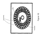
- FIG. 2 depicts a probe embodiment 40 that incorporates a mechanism for transmitting both RF energy and optical energy to the rotating assembly.
- a transformer scheme is used wherein a first coil 42 is attached to the rotating assembly 44, and a second coil 46 is integrated with the connector 48 of the optical probe.
- This configuration has the advantage that both coils move with the assembly during a 'pull-back' (longitudinal) scan operation. Such pullbacks are used in both OCT and IVUS scans.
- a spiral scan pattern is created inside the arterial lumen.
- this approach results in an increased cost for a one-time-use catheter.
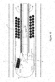
- Figure 3 illustrates an alternative coupling scheme wherein the fixed coil 42 is part of the drive unit 50 (motorized assembly providing rotational and longitudinal motions).
- the fixed coil is permanent, and must be long enough to efficiently couple the RF energy into the rotating catheter coil over the entire pullback length.
- slip-ring technology is widely used in the field of optical imaging.
- slip-ring technology can be used in IVUS probes described herein.
- the slip-ring is more difficult to manufacture than in the IVUS-only case.
- FIG 4 illustrates an embodiment that includes capacitive micro-machined ultrasonic transducers (CMUT) 52 integrated in a coronary imaging probe 54.
- CMUT capacitive micro-machined ultrasonic transducers
- the advantage of the CMUT is the small size of the transducer, which is fabricated via conventional electronic CMOS processes.
- the small size and photolithographic fabrication allows customized arrays of transducers to be built with the drive electronics on the same substrate.
- an array is formed in an annular region around the optical transducer.
- a co-focused, aligned and combined beam can be formed, which eliminates the need for software registration and removes a potential source of error.
- this probe tip may be larger than the embodiment shown in Figure 1 .
- Figure 5A illustrates a fused OCT-IVUS image 56, wherein the demarcation line 58 is chosen near the OCT penetration limit.
- the demarcation line 58 is chosen near the OCT penetration limit.
- Figure 5B illustrates a fused OCT/IVUS image wherein the OCT portion appears in the image center and the IVUS portion appears in the periphery.
- the outer boundary indicates approximately the boundary where the two regions intersect.
- the guide catheter is a larger bore catheter used to introduce the smaller imaging catheter into the main arterial trunk. From the guide catheter, a flush solution can be expelled to create a clear, blood-free imaging region when OCT imaging is performed.
- Alternative embodiments may include a flush lumen within the imaging catheter whereby the flush solution is ejected at the imaging tip rather than from the guide catheter.
- the aspects and embodiments of the invention can incorporate various components of varying dimension and materials as is known to one of ordinary skill.
- Various specific dimensions and materials are described herein, however, these exemplary materials are not meant to be limiting, but only to evidence additional more specific embodiments.
- the dimension given also includes a range of greater than about 10-20% of the dimension given and less than about 10%-20% of the dimension given.
- the dimension given also includes a range of greater than about 20-50% of the dimension given and less than about 20%-50% of the dimension given.
- the dimension given also includes a range of greater than about 50-100% of the dimension given and less than about 50%-100% of the dimension given.
- the viewing window used is a transparent epoxy-based window.
- the transducers used have a first dimension of about 0.1 mm and a second dimension of about 0.5 mm.
- the forward viewing angle is about 10 degrees in one embodiment of the probe.
- the end-cap used in one probe embodiment includes a metal.
- the probe can include a hollow core that is substantially filled with an epoxy material in some embodiments.
- the width of the shield RF cable is about 0.18 mm.
Landscapes
- Health & Medical Sciences (AREA)
- Life Sciences & Earth Sciences (AREA)
- Physics & Mathematics (AREA)
- Surgery (AREA)
- General Health & Medical Sciences (AREA)
- Engineering & Computer Science (AREA)
- Biomedical Technology (AREA)
- Heart & Thoracic Surgery (AREA)
- Medical Informatics (AREA)
- Molecular Biology (AREA)
- Biophysics (AREA)
- Animal Behavior & Ethology (AREA)
- Pathology (AREA)
- Public Health (AREA)
- Veterinary Medicine (AREA)
- Nuclear Medicine, Radiotherapy & Molecular Imaging (AREA)
- Radiology & Medical Imaging (AREA)
- Acoustics & Sound (AREA)
- Ultra Sonic Daignosis Equipment (AREA)
- Endoscopes (AREA)
Applications Claiming Priority (3)
| Application Number | Priority Date | Filing Date | Title |
|---|---|---|---|
| US85757306P | 2006-11-08 | 2006-11-08 | |
| EP07861810.5A EP2081486B1 (en) | 2006-11-08 | 2007-11-08 | Opto-acoustic imaging device |
| PCT/US2007/023493 WO2008057573A2 (en) | 2006-11-08 | 2007-11-08 | Opto-acoustic imaging devices and methods |
Related Parent Applications (3)
| Application Number | Title | Priority Date | Filing Date |
|---|---|---|---|
| EP07861810.5A Division-Into EP2081486B1 (en) | 2006-11-08 | 2007-11-08 | Opto-acoustic imaging device |
| EP07861810.5A Division EP2081486B1 (en) | 2006-11-08 | 2007-11-08 | Opto-acoustic imaging device |
| EP07861810.5 Division | 2007-11-08 |
Publications (2)
| Publication Number | Publication Date |
|---|---|
| EP2628443A1 EP2628443A1 (en) | 2013-08-21 |
| EP2628443B1 true EP2628443B1 (en) | 2021-05-12 |
Family
ID=39247184
Family Applications (2)
| Application Number | Title | Priority Date | Filing Date |
|---|---|---|---|
| EP13158821.2A Active EP2628443B1 (en) | 2006-11-08 | 2007-11-08 | Opto-acoustic imaging device |
| EP07861810.5A Active EP2081486B1 (en) | 2006-11-08 | 2007-11-08 | Opto-acoustic imaging device |
Family Applications After (1)
| Application Number | Title | Priority Date | Filing Date |
|---|---|---|---|
| EP07861810.5A Active EP2081486B1 (en) | 2006-11-08 | 2007-11-08 | Opto-acoustic imaging device |
Country Status (5)
| Country | Link |
|---|---|
| US (4) | US7935060B2 (ja) |
| EP (2) | EP2628443B1 (ja) |
| JP (5) | JP2010508973A (ja) |
| CN (1) | CN101594819B (ja) |
| WO (1) | WO2008057573A2 (ja) |
Families Citing this family (203)
| Publication number | Priority date | Publication date | Assignee | Title |
|---|---|---|---|---|
| US7241286B2 (en) * | 2003-04-25 | 2007-07-10 | Lightlab Imaging, Llc | Flush catheter with flow directing sheath |
| US9867530B2 (en) | 2006-08-14 | 2018-01-16 | Volcano Corporation | Telescopic side port catheter device with imaging system and method for accessing side branch occlusions |
| JP2010508973A (ja) | 2006-11-08 | 2010-03-25 | ライトラブ イメージング, インコーポレイテッド | 光−音響イメージングデバイスおよび方法 |
| JP5543212B2 (ja) * | 2006-11-21 | 2014-07-09 | コーニンクレッカ フィリップス エヌ ヴェ | 前立腺癌のイメージング用システム、前立腺癌をイメージングするシステムの作動方法及びコンピュータ読取可能媒体 |
| ES3007239T3 (en) | 2007-01-10 | 2025-03-19 | Lightlab Imaging Inc | Methods and apparatus for swept-source optical coherence tomography |
| CN103222846B (zh) * | 2007-01-19 | 2017-04-26 | 桑尼布鲁克健康科学中心 | 用于成像探头的扫描机构 |
| WO2008100386A2 (en) * | 2007-02-09 | 2008-08-21 | Board Of Regents, The University Of Texas System | Intravascular photoacoustic and ultrasound echo imaging |
| WO2009009799A1 (en) | 2007-07-12 | 2009-01-15 | Volcano Corporation | Catheter for in vivo imaging |
| US10219780B2 (en) | 2007-07-12 | 2019-03-05 | Volcano Corporation | OCT-IVUS catheter for concurrent luminal imaging |
| US9596993B2 (en) | 2007-07-12 | 2017-03-21 | Volcano Corporation | Automatic calibration systems and methods of use |
| US8582934B2 (en) | 2007-11-12 | 2013-11-12 | Lightlab Imaging, Inc. | Miniature optical elements for fiber-optic beam shaping |
| US7813609B2 (en) | 2007-11-12 | 2010-10-12 | Lightlab Imaging, Inc. | Imaging catheter with integrated reference reflector |
| US10531859B2 (en) | 2008-01-02 | 2020-01-14 | Arcscan, Inc. | Components for a precision ultrasonic scanning apparatus for body parts |
| EP2231021A4 (en) | 2008-01-02 | 2011-06-29 | Arcscan Inc | COMPONENTS FOR ULTRASONIC ARC SCANNING APPARATUS |
| US9713448B2 (en) | 2008-04-03 | 2017-07-25 | Infraredx, Inc. | System and method for intravascular structural analysis compensation of chemical analysis modality |
| JP2011519689A (ja) | 2008-05-07 | 2011-07-14 | インフラレデックス, インコーポレイテッド | 脈管内分析のためのマルチモーダルカテーテルシステム |
| ES2517915T3 (es) * | 2008-06-02 | 2014-11-04 | Lightlab Imaging, Inc. | Métodos cuantitativos para obtener características de un tejido a partir de imágenes de tomografía por coherencia óptica |
| US8197413B2 (en) * | 2008-06-06 | 2012-06-12 | Boston Scientific Scimed, Inc. | Transducers, devices and systems containing the transducers, and methods of manufacture |
| DE102008045634A1 (de) | 2008-09-03 | 2010-03-04 | Ludwig-Maximilians-Universität München | Wellenlängenabstimmbare Lichtquelle |
| EP3725212A1 (en) * | 2008-10-14 | 2020-10-21 | Lightlab Imaging, Inc. | Stent strut detection and related measurement and display using optical coherence tomography |
| US8465686B2 (en) | 2008-12-19 | 2013-06-18 | Volcano Corporation | Method of manufacturing a rotational intravascular ultrasound probe |
| US20100179434A1 (en) * | 2009-01-09 | 2010-07-15 | Boston Scientific Scimed, Inc. | Systems and methods for making and using intravascular ultrasound systems with photo-acoustic imaging capabilities |
| US20100179432A1 (en) * | 2009-01-09 | 2010-07-15 | Boston Scientific Scimed, Inc. | Systems and methods for making and using intravascular ultrasound systems with photo-acoustic imaging capabilities |
| US8403856B2 (en) | 2009-03-11 | 2013-03-26 | Volcano Corporation | Rotational intravascular ultrasound probe with an active spinning element |
| US8670129B2 (en) | 2009-09-03 | 2014-03-11 | Axsun Technologies, Inc. | Filtered ASE swept source for OCT medical imaging |
| US8526472B2 (en) | 2009-09-03 | 2013-09-03 | Axsun Technologies, Inc. | ASE swept source with self-tracking filter for OCT medical imaging |
| WO2011038048A1 (en) * | 2009-09-23 | 2011-03-31 | Lightlab Imaging, Inc. | Apparatus, systems, and methods of in-vivo blood clearing in a lumen |
| ES2660570T3 (es) | 2009-09-23 | 2018-03-23 | Lightlab Imaging, Inc. | Sistemas, aparatos y métodos de recopilación de datos de medición de resistencia vascular y morfología luminal |
| US12426789B2 (en) | 2009-09-23 | 2025-09-30 | Lightlab Imaging, Inc. | Blood vessel lumen morphology and minimum lumen area measurements data collection by intravascular imaging systems for stenosis or stent planning |
| JP5563582B2 (ja) * | 2009-09-30 | 2014-07-30 | テルモ株式会社 | 画像診断装置 |
| US8206377B2 (en) * | 2009-12-22 | 2012-06-26 | Lightlab Imaging, Inc. | Torque limiter for an OCT catheter |
| US8926590B2 (en) | 2009-12-22 | 2015-01-06 | Lightlab Imaging, Inc. | Torque limiter for an OCT catheter |
| EP2519158A1 (en) * | 2009-12-29 | 2012-11-07 | Boston Scientific Scimed, Inc. | Systems and methods for multi-frequency imaging of patient tissue using intravascular ultrasound imaging systems |
| US8478384B2 (en) * | 2010-01-19 | 2013-07-02 | Lightlab Imaging, Inc. | Intravascular optical coherence tomography system with pressure monitoring interface and accessories |
| US8356517B2 (en) * | 2010-02-24 | 2013-01-22 | Avago Technologies Wireless Ip (Singapore) Pte. Ltd. | Integrated optical and acoustic transducer device |
| AU2011227178B2 (en) * | 2010-03-17 | 2013-11-07 | Lightlab Imaging, Inc. | Intensity noise reduction methods and apparatus for interferometric sensing and imaging systems |
| CN101828902B (zh) * | 2010-04-01 | 2011-09-21 | 江西科技师范学院 | 一种用于乳腺或颅脑三维医学诊断的光声传感器 |
| JP5773578B2 (ja) | 2010-04-08 | 2015-09-02 | キヤノン株式会社 | 被検体情報取得装置、被検体情報取得装置の制御方法およびプログラム |
| RU2013121803A (ru) * | 2010-10-14 | 2014-11-20 | Конинклейке Филипс Электроникс Н.В. | Определяющее свойства устройство для определения свойств объекта |
| JP5653882B2 (ja) * | 2010-10-27 | 2015-01-14 | 富士フイルム株式会社 | 光音響撮像装置およびその作動方法 |
| CA3085777C (en) | 2010-11-08 | 2022-03-15 | Conavi Medical Inc. | Systems and methods for improved visualization during minimally invasive procedures |
| RU2553930C2 (ru) * | 2010-11-11 | 2015-06-20 | Ковидиен Лп | Гибкие фрагментирующие катетеры с визуализацией и способы их использования и изготовления |
| EP2642913B1 (en) | 2010-11-27 | 2024-02-21 | Boston Scientific Scimed, Inc. | Ablation and temperature measurement devices |
| US11141063B2 (en) | 2010-12-23 | 2021-10-12 | Philips Image Guided Therapy Corporation | Integrated system architectures and methods of use |
| US11040140B2 (en) | 2010-12-31 | 2021-06-22 | Philips Image Guided Therapy Corporation | Deep vein thrombosis therapeutic methods |
| US8582619B2 (en) | 2011-03-15 | 2013-11-12 | Lightlab Imaging, Inc. | Methods, systems, and devices for timing control in electromagnetic radiation sources |
| US9164240B2 (en) | 2011-03-31 | 2015-10-20 | Lightlab Imaging, Inc. | Optical buffering methods, apparatus, and systems for increasing the repetition rate of tunable light sources |
| JP6141264B2 (ja) | 2011-05-27 | 2017-06-07 | ライトラボ・イメージング・インコーポレーテッド | 光コヒーレンス断層撮影法及び圧力に基づくシステム及び方法 |
| CA2836790C (en) | 2011-05-31 | 2019-04-23 | Desmond Adler | Multimodal imaging system, apparatus, and methods |
| FR2976173B1 (fr) * | 2011-06-07 | 2013-07-12 | Commissariat Energie Atomique | Coque demontable a moyens optiques embarques pour sonde de diagnostic bimodale a imageries optique et ultrasonore |
| US8582109B1 (en) | 2011-08-01 | 2013-11-12 | Lightlab Imaging, Inc. | Swept mode-hopping laser system, methods, and devices for frequency-domain optical coherence tomography |
| WO2013019840A1 (en) | 2011-08-03 | 2013-02-07 | Lightlab Imaging, Inc. | Systems, methods and apparatus for determining a fractional flow reserve |
| WO2013033592A1 (en) | 2011-08-31 | 2013-03-07 | Volcano Corporation | Optical-electrical rotary joint and methods of use |
| WO2013055896A1 (en) | 2011-10-14 | 2013-04-18 | Acist Medical Systems, Inc. | Device and methods for measuring and treating an anatomical structure |
| US8953911B1 (en) | 2011-10-28 | 2015-02-10 | Lightlab Imaging, Inc. | Spectroscopic imaging probes, devices, and methods |
| US8831321B1 (en) | 2011-11-07 | 2014-09-09 | Lightlab Imaging, Inc. | Side branch detection methods, systems and devices |
| TW201323851A (zh) * | 2011-12-05 | 2013-06-16 | Ind Tech Res Inst | 光聲影像裝置、光聲感測結構及擷取光聲影像的方法 |
| JP6038957B2 (ja) * | 2012-02-14 | 2016-12-07 | セント・ジュード・メディカル・エイトリアル・フィブリレーション・ディヴィジョン・インコーポレーテッド | 光音響法を用いた心臓組織のアブレーション治療の効果を評価するためのシステム |
| US20130253266A1 (en) | 2012-03-22 | 2013-09-26 | Codman & Shurtleff, Inc. | Fluid management catheter and methods of using same |
| WO2013145711A1 (ja) * | 2012-03-26 | 2013-10-03 | テルモ株式会社 | 画像診断装置及びその制御方法 |
| WO2013145635A1 (ja) | 2012-03-26 | 2013-10-03 | テルモ株式会社 | プローブ及び画像診断装置 |
| WO2013145637A1 (ja) | 2012-03-28 | 2013-10-03 | テルモ株式会社 | プローブ |
| WO2013145689A1 (ja) * | 2012-03-28 | 2013-10-03 | テルモ株式会社 | プローブ及び画像診断装置 |
| US9549679B2 (en) | 2012-05-14 | 2017-01-24 | Acist Medical Systems, Inc. | Multiple transducer delivery device and method |
| US9597059B2 (en) | 2012-05-17 | 2017-03-21 | Arcscan, Inc. | Correcting for unintended motion for ultrasonic eye scans |
| CA2911446C (en) | 2012-05-25 | 2020-10-13 | Vascular Imaging Corporation | Optical fiber pressure sensor |
| US9492140B2 (en) * | 2012-06-12 | 2016-11-15 | Volcano Corporation | Devices, systems, and methods for forward looking imaging |
| WO2014005000A1 (en) * | 2012-06-28 | 2014-01-03 | Volcano Corporation | Side-loading connectors for use with intravascular devices and associated systems and methods |
| US9320427B2 (en) | 2012-07-09 | 2016-04-26 | Arcscan, Inc. | Combination optical and ultrasonic imaging of an eye |
| US9289185B2 (en) * | 2012-07-23 | 2016-03-22 | ClariTrac, Inc. | Ultrasound device for needle procedures |
| CN105050485B (zh) * | 2012-08-14 | 2017-08-18 | 皇家飞利浦有限公司 | 用于光声成像的紧凑激光器和有效脉冲递送 |
| US8687201B2 (en) | 2012-08-31 | 2014-04-01 | Lightlab Imaging, Inc. | Optical coherence tomography control systems and methods |
| JP5981557B2 (ja) * | 2012-09-11 | 2016-08-31 | テルモ株式会社 | 画像診断装置 |
| WO2014049644A1 (ja) * | 2012-09-26 | 2014-04-03 | テルモ株式会社 | 校正冶具、画像診断装置及び画像診断装置の校正方法 |
| US11272845B2 (en) | 2012-10-05 | 2022-03-15 | Philips Image Guided Therapy Corporation | System and method for instant and automatic border detection |
| US20140100454A1 (en) | 2012-10-05 | 2014-04-10 | Volcano Corporation | Methods and systems for establishing parameters for three-dimensional imaging |
| US9858668B2 (en) | 2012-10-05 | 2018-01-02 | Volcano Corporation | Guidewire artifact removal in images |
| US9367965B2 (en) | 2012-10-05 | 2016-06-14 | Volcano Corporation | Systems and methods for generating images of tissue |
| US10070827B2 (en) | 2012-10-05 | 2018-09-11 | Volcano Corporation | Automatic image playback |
| US9292918B2 (en) | 2012-10-05 | 2016-03-22 | Volcano Corporation | Methods and systems for transforming luminal images |
| US9286673B2 (en) | 2012-10-05 | 2016-03-15 | Volcano Corporation | Systems for correcting distortions in a medical image and methods of use thereof |
| US10568586B2 (en) | 2012-10-05 | 2020-02-25 | Volcano Corporation | Systems for indicating parameters in an imaging data set and methods of use |
| US9324141B2 (en) | 2012-10-05 | 2016-04-26 | Volcano Corporation | Removal of A-scan streaking artifact |
| US9307926B2 (en) | 2012-10-05 | 2016-04-12 | Volcano Corporation | Automatic stent detection |
| WO2014055880A2 (en) | 2012-10-05 | 2014-04-10 | David Welford | Systems and methods for amplifying light |
| US9840734B2 (en) | 2012-10-22 | 2017-12-12 | Raindance Technologies, Inc. | Methods for analyzing DNA |
| ES2713198T3 (es) * | 2012-10-22 | 2019-05-20 | Massachusetts Gen Hospital | Sistema de catéter híbrido |
| JP6352287B2 (ja) * | 2012-11-19 | 2018-07-04 | ライトラボ・イメージング・インコーポレーテッド | マルチモーダル・イメージングシステム、プローブ及び方法 |
| CN102944521B (zh) * | 2012-11-29 | 2015-01-21 | 华南师范大学 | 非接触式光声和光学相干断层双成像装置及其检测方法 |
| US9677869B2 (en) | 2012-12-05 | 2017-06-13 | Perimeter Medical Imaging, Inc. | System and method for generating a wide-field OCT image of a portion of a sample |
| JP6336471B2 (ja) | 2012-12-12 | 2018-06-06 | ライトラボ・イメージング・インコーポレーテッド | 血管の内腔輪郭の自動化された決定のための方法および装置 |
| CA2894403A1 (en) | 2012-12-13 | 2014-06-19 | Volcano Corporation | Devices, systems, and methods for targeted cannulation |
| US10942022B2 (en) | 2012-12-20 | 2021-03-09 | Philips Image Guided Therapy Corporation | Manual calibration of imaging system |
| JP2016504589A (ja) | 2012-12-20 | 2016-02-12 | ナサニエル ジェイ. ケンプ, | 異なる撮像モード間で再構成可能な光コヒーレンストモグラフィシステム |
| US11406498B2 (en) | 2012-12-20 | 2022-08-09 | Philips Image Guided Therapy Corporation | Implant delivery system and implants |
| CA2895770A1 (en) | 2012-12-20 | 2014-07-24 | Jeremy Stigall | Locating intravascular images |
| US10939826B2 (en) | 2012-12-20 | 2021-03-09 | Philips Image Guided Therapy Corporation | Aspirating and removing biological material |
| WO2014100110A2 (en) * | 2012-12-20 | 2014-06-26 | Dale Dorando | Intravascular blood pressure and velocity wire |
| US10595820B2 (en) | 2012-12-20 | 2020-03-24 | Philips Image Guided Therapy Corporation | Smooth transition catheters |
| US10045757B2 (en) * | 2012-12-21 | 2018-08-14 | Volcano Corporation | Guarded imaging devices and methods |
| CA2895769A1 (en) | 2012-12-21 | 2014-06-26 | Douglas Meyer | Rotational ultrasound imaging catheter with extended catheter body telescope |
| US10413317B2 (en) | 2012-12-21 | 2019-09-17 | Volcano Corporation | System and method for catheter steering and operation |
| US9486143B2 (en) | 2012-12-21 | 2016-11-08 | Volcano Corporation | Intravascular forward imaging device |
| EP2934323A4 (en) | 2012-12-21 | 2016-08-17 | Andrew Hancock | SYSTEM AND METHOD FOR MULTI-PASS PROCESSING OF IMAGE SIGNALS |
| JP2016508757A (ja) | 2012-12-21 | 2016-03-24 | ジェイソン スペンサー, | 医療データのグラフィカル処理のためのシステムおよび方法 |
| EP2936626A4 (en) | 2012-12-21 | 2016-08-17 | David Welford | SYSTEMS AND METHOD FOR REDUCING A WAVELENGTH LIGHT EMISSION |
| US10058284B2 (en) | 2012-12-21 | 2018-08-28 | Volcano Corporation | Simultaneous imaging, monitoring, and therapy |
| US10166003B2 (en) | 2012-12-21 | 2019-01-01 | Volcano Corporation | Ultrasound imaging with variable line density |
| US10191220B2 (en) | 2012-12-21 | 2019-01-29 | Volcano Corporation | Power-efficient optical circuit |
| US9612105B2 (en) | 2012-12-21 | 2017-04-04 | Volcano Corporation | Polarization sensitive optical coherence tomography system |
| JP6243453B2 (ja) | 2013-03-07 | 2017-12-06 | ボルケーノ コーポレイション | 血管内画像におけるマルチモーダルセグメンテーション |
| US10226597B2 (en) | 2013-03-07 | 2019-03-12 | Volcano Corporation | Guidewire with centering mechanism |
| US9173591B2 (en) | 2013-03-08 | 2015-11-03 | Lightlab Imaging, Inc. | Stent visualization and malapposition detection systems, devices, and methods |
| WO2014163601A1 (en) | 2013-03-11 | 2014-10-09 | Lightlab Imaging, Inc. | Friction torque limiter for an imaging catheter |
| US9069396B2 (en) | 2013-03-12 | 2015-06-30 | Lightlab Imaging, Inc. | Controller and user interface device, systems, and methods |
| EP3895604A1 (en) | 2013-03-12 | 2021-10-20 | Collins, Donna | Systems and methods for diagnosing coronary microvascular disease |
| US11154313B2 (en) | 2013-03-12 | 2021-10-26 | The Volcano Corporation | Vibrating guidewire torquer and methods of use |
| US9351698B2 (en) | 2013-03-12 | 2016-05-31 | Lightlab Imaging, Inc. | Vascular data processing and image registration systems, methods, and apparatuses |
| US9301687B2 (en) | 2013-03-13 | 2016-04-05 | Volcano Corporation | System and method for OCT depth calibration |
| US11026591B2 (en) | 2013-03-13 | 2021-06-08 | Philips Image Guided Therapy Corporation | Intravascular pressure sensor calibration |
| EP2967488B1 (en) | 2013-03-13 | 2021-06-16 | Jinhyoung Park | System for producing an image from a rotational intravascular ultrasound device |
| US10292677B2 (en) | 2013-03-14 | 2019-05-21 | Volcano Corporation | Endoluminal filter having enhanced echogenic properties |
| WO2014152365A2 (en) | 2013-03-14 | 2014-09-25 | Volcano Corporation | Filters with echogenic characteristics |
| US10219887B2 (en) | 2013-03-14 | 2019-03-05 | Volcano Corporation | Filters with echogenic characteristics |
| US12343198B2 (en) | 2013-03-14 | 2025-07-01 | Philips Image Guided Therapy Corporation | Delivery catheter having imaging capabilities |
| US9439570B2 (en) | 2013-03-15 | 2016-09-13 | Lx Medical Corporation | Tissue imaging and image guidance in luminal anatomic structures and body cavities |
| US9833221B2 (en) | 2013-03-15 | 2017-12-05 | Lightlab Imaging, Inc. | Apparatus and method of image registration |
| US9702762B2 (en) | 2013-03-15 | 2017-07-11 | Lightlab Imaging, Inc. | Calibration and image processing devices, methods, and systems |
| US9364167B2 (en) | 2013-03-15 | 2016-06-14 | Lx Medical Corporation | Tissue imaging and image guidance in luminal anatomic structures and body cavities |
| WO2014162367A1 (ja) * | 2013-04-05 | 2014-10-09 | テルモ株式会社 | 画像診断装置及びプログラム |
| JP6001765B2 (ja) * | 2013-04-05 | 2016-10-05 | テルモ株式会社 | 画像診断装置及びプログラム |
| WO2014179681A1 (en) * | 2013-05-03 | 2014-11-06 | Sunnybrook Health Sciences Centre | Systems and methods for super-resolution ultrasound imaging |
| CN103385758B (zh) * | 2013-07-22 | 2015-12-09 | 深圳先进技术研究院 | 一种血管内光声超声双模成像系统及其成像方法 |
| WO2015051003A1 (en) | 2013-10-04 | 2015-04-09 | Vascular Imaging Corporation | Imaging techniques using an imaging guidewire |
| CN103637808B (zh) * | 2013-11-18 | 2015-08-19 | 深圳先进技术研究院 | 光声成像装置 |
| US10537255B2 (en) | 2013-11-21 | 2020-01-21 | Phyzhon Health Inc. | Optical fiber pressure sensor |
| EP3089479B1 (en) * | 2014-01-29 | 2019-03-13 | Sogang University Research Foundation | Intravascular ultrasonic transducer structure |
| US10736605B2 (en) | 2014-02-24 | 2020-08-11 | Arcscan, Inc. | Disposable eyepiece system for an ultrasonic eye scanning apparatus |
| EP3861928B1 (en) | 2014-04-04 | 2024-03-27 | St. Jude Medical Systems AB | Intravascular pressure and flow data diagnostic system |
| CN103892871B (zh) * | 2014-04-17 | 2015-11-25 | 深圳大学 | 一种机械旋转式血管内超声探头 |
| WO2015166302A1 (en) * | 2014-04-29 | 2015-11-05 | B-K Medical Aps | Ultrasound imaging probe |
| US9668645B2 (en) * | 2014-05-09 | 2017-06-06 | Novartis Ag | Imaging probes and associated devices, systems, and methods utilizing electrostatic actuators |
| JP5983676B2 (ja) * | 2014-05-16 | 2016-09-06 | 住友電気工業株式会社 | 光プローブ |
| CN103976703B (zh) * | 2014-05-27 | 2016-01-20 | 江西科技师范大学 | 一种光声超声双模态内窥镜成像系统 |
| EP3174457A4 (en) * | 2014-06-04 | 2018-10-10 | Securus Medical Group, Inc. | Temperature measurement systems, method and devices |
| JP6378787B2 (ja) * | 2014-06-17 | 2018-08-22 | コーニンクレッカ フィリップス エヌ ヴェKoninklijke Philips N.V. | 血管内カテーテルのための設計及び方法 |
| ES2744900T3 (es) | 2014-07-24 | 2020-02-26 | Lightlab Imaging Inc | Visualización de endoprótesis vasculares y vasos sanguíneos y procedimientos de diagnóstico |
| JP6495296B2 (ja) * | 2014-08-12 | 2019-04-03 | プレキシオン株式会社 | 光音響画像化装置 |
| EP4574026A3 (en) | 2014-08-27 | 2025-08-27 | St. Jude Medical Systems AB | System and method for evaluating a cardiac system by determining minimum ratio pd/pa (distal pressure/arterial pressure) |
| US11311200B1 (en) | 2014-08-27 | 2022-04-26 | Lightlab Imaging, Inc. | Systems and methods to measure physiological flow in coronary arteries |
| WO2016034977A1 (en) * | 2014-09-04 | 2016-03-10 | Koninklijke Philips N.V. | Intravascular ultrasound imaging system with slip ring interface and associated devices, systems, and methods |
| US10499813B2 (en) | 2014-09-12 | 2019-12-10 | Lightlab Imaging, Inc. | Methods, systems and apparatus for temporal calibration of an intravascular imaging system |
| JP6315816B2 (ja) * | 2014-09-25 | 2018-04-25 | テルモ株式会社 | 画像処理装置、画像処理装置の作動方法、プログラムおよび記憶媒体 |
| EP3199109B1 (en) * | 2014-09-26 | 2020-01-08 | Terumo Kabushiki Kaisha | Image diagnostic probe |
| US10258240B1 (en) | 2014-11-24 | 2019-04-16 | Vascular Imaging Corporation | Optical fiber pressure sensor |
| EP3031399B1 (en) * | 2014-12-09 | 2021-02-17 | Nidek Co., Ltd. | Equipment and method for ultrasound imaging of an eye |
| ES2864714T3 (es) | 2014-12-12 | 2021-10-14 | Lightlab Imaging Inc | Método para detectar y representar visualmente características endovasculares |
| GB2539368A (en) * | 2015-02-09 | 2016-12-21 | Univ Erasmus Med Ct Rotterdam | Intravascular photoacoustic imaging |
| JP6533078B2 (ja) | 2015-03-20 | 2019-06-19 | テルモ株式会社 | 画像診断装置、その制御方法、プログラム及びコンピュータ可読記憶媒体 |
| WO2016152624A1 (ja) | 2015-03-24 | 2016-09-29 | テルモ株式会社 | 画像診断装置及びその制御方法、プログラム及びコンピュータ可読記憶媒体 |
| WO2016168605A1 (en) | 2015-04-16 | 2016-10-20 | Gentuity, Llc | Micro-optic probes for neurology |
| US10646198B2 (en) | 2015-05-17 | 2020-05-12 | Lightlab Imaging, Inc. | Intravascular imaging and guide catheter detection methods and systems |
| US9996921B2 (en) | 2015-05-17 | 2018-06-12 | LIGHTLAB IMAGING, lNC. | Detection of metal stent struts |
| US10222956B2 (en) | 2015-05-17 | 2019-03-05 | Lightlab Imaging, Inc. | Intravascular imaging user interface systems and methods |
| US10140712B2 (en) | 2015-05-17 | 2018-11-27 | Lightlab Imaging, Inc. | Detection of stent struts relative to side branches |
| US10109058B2 (en) | 2015-05-17 | 2018-10-23 | Lightlab Imaging, Inc. | Intravascular imaging system interfaces and stent detection methods |
| EP3324829B1 (en) | 2015-07-25 | 2021-05-12 | Lightlab Imaging, Inc. | Guidewire detection systems, methods, and apparatuses |
| AU2016297930B2 (en) | 2015-07-25 | 2021-02-18 | Lightlab Imaging, Inc. | Intravascular data visualization method |
| EP4675259A3 (en) | 2015-08-31 | 2026-04-08 | Spryte Medical, Inc. | Imaging system includes imaging probe and delivery devices |
| JP6779890B2 (ja) * | 2015-09-18 | 2020-11-04 | テルモ株式会社 | 画像診断用カテーテル |
| WO2017066460A1 (en) | 2015-10-13 | 2017-04-20 | Arcscan, Inc | Ultrasonic scanning apparatus |
| US11426611B2 (en) | 2015-10-13 | 2022-08-30 | Arcscan, Inc. | Ultrasound therapeutic and scanning apparatus |
| CN105902255A (zh) * | 2015-10-26 | 2016-08-31 | 首都医科大学附属北京同仁医院 | 微型光学导管 |
| EP3979189A1 (en) | 2015-11-18 | 2022-04-06 | Lightlab Imaging, Inc. | X-ray image feature detection and registration systems and methods |
| CA3005242A1 (en) | 2015-11-18 | 2017-05-26 | Lightlab Imaging, Inc. | Detection of stent struts relative to side branches |
| JP6898927B2 (ja) | 2015-11-23 | 2021-07-07 | ライトラボ・イメージング・インコーポレーテッド | 血管内画像における影の検出および検証 |
| JP6908325B2 (ja) * | 2015-11-25 | 2021-07-21 | フジフィルム ソノサイト インコーポレイテッド | 高周波超音波トランスデューサアレイを含む医療機器 |
| US10593037B2 (en) | 2016-04-14 | 2020-03-17 | Lightlab Imaging, Inc. | Method, apparatus, and system to identify branches of a blood vessel |
| WO2017201026A1 (en) | 2016-05-16 | 2017-11-23 | Lightlab Imaging, Inc. | Intravascular absorbable stent detection and diagnostic methods and systems |
| CN117398183A (zh) | 2016-09-28 | 2024-01-16 | 光学实验室成像公司 | 利用血管表象的支架规划系统及方法 |
| KR101852560B1 (ko) | 2016-12-01 | 2018-04-26 | 포항공과대학교 산학협력단 | 방수형 pdms 스캐너를 이용한 광음향 수술중 조직검사 장치 및 그를 이용한 광음향 영상 획득 방법 |
| CN106361295A (zh) * | 2016-12-06 | 2017-02-01 | 全景恒升(北京)科学技术有限公司 | 一种光学和声学混合成像导管 |
| CN106691405A (zh) * | 2016-12-29 | 2017-05-24 | 天津恒宇医疗科技有限公司 | 一种超细oct成像导管 |
| JP6829124B2 (ja) * | 2017-03-22 | 2021-02-10 | テルモ株式会社 | 画像診断装置 |
| EP3646795B1 (en) | 2017-06-29 | 2023-08-09 | Terumo Kabushiki Kaisha | Image diagnostic catheter |
| EP3655748B1 (en) | 2017-07-18 | 2023-08-09 | Perimeter Medical Imaging, Inc. | Sample container for stabilizing and aligning excised biological tissue samples for ex vivo analysis |
| WO2019070702A1 (en) | 2017-10-02 | 2019-04-11 | Lightlab Imaging, Inc. | INTRAVASCULAR DATA COLLECTION PROBES AND ASSOCIATED ASSEMBLIES |
| JP7160935B2 (ja) | 2017-11-28 | 2022-10-25 | ジェンテュイティ・リミテッド・ライアビリティ・カンパニー | 撮像システム |
| JP7179943B2 (ja) * | 2018-03-30 | 2022-11-29 | テルモ株式会社 | 画像診断用カテーテル |
| JP6962850B2 (ja) * | 2018-03-30 | 2021-11-05 | テルモ株式会社 | 画像診断用カテーテル |
| CN108852296B (zh) * | 2018-05-17 | 2020-05-19 | 华中科技大学 | 一种可调光声换能器装置及其制备方法 |
| US11344373B2 (en) | 2018-05-29 | 2022-05-31 | Lightlab Imaging, Inc. | Stent expansion display, systems, and methods |
| CN108784739B (zh) * | 2018-06-20 | 2024-02-20 | 深圳英美达医疗技术有限公司 | 一种结合超声波成像和光学相干断层成像的双模探头 |
| CN109091167A (zh) * | 2018-06-29 | 2018-12-28 | 东南大学 | 冠状动脉粥样硬化斑块增长的预测方法 |
| JP2022500174A (ja) | 2018-09-17 | 2022-01-04 | ジェンテュイティ・リミテッド・ライアビリティ・カンパニーGentuity, LLC | 光学経路を有する撮像システム |
| WO2020223433A1 (en) | 2019-04-30 | 2020-11-05 | Gentuity, Llc | Imaging probe with fluid pressurization element |
| US12239412B2 (en) | 2019-05-21 | 2025-03-04 | Spryte Medical, Inc. | Systems and methods for OCT-guided treatment of a patient |
| JP2022006490A (ja) | 2020-06-24 | 2022-01-13 | 朝日インテック株式会社 | 医療システム、および、画像生成方法 |
| CN112493997B (zh) * | 2020-11-30 | 2023-03-31 | 中国科学院深圳先进技术研究院 | 一种光声内窥成像装置及基于该装置的光声内窥成像方法 |
| CN113080814B (zh) * | 2021-04-12 | 2022-01-28 | 中南大学 | 一种透射共轴式的光声内窥探头及其成像方法 |
| US20230098586A1 (en) * | 2021-09-30 | 2023-03-30 | Seno Medical Instruments, Inc. | Optoacoustic probe for prostrate imaging |
| USD1083949S1 (en) | 2021-10-15 | 2025-07-15 | Lightlab Imaging, Inc. | Display screen or portion thereof with graphical user interface |
| WO2024085590A1 (ko) * | 2022-10-17 | 2024-04-25 | 울산과학기술원 | 광음향-초음파 미니 프로브용 스캐닝 팁 |
Family Cites Families (119)
| Publication number | Priority date | Publication date | Assignee | Title |
|---|---|---|---|---|
| US5000185A (en) * | 1986-02-28 | 1991-03-19 | Cardiovascular Imaging Systems, Inc. | Method for intravascular two-dimensional ultrasonography and recanalization |
| US5582178A (en) * | 1986-02-28 | 1996-12-10 | Cardiovascular Imaging Systems, Inc. | Method and apparatus for intravascular ultrasonography |
| US5076279A (en) | 1990-07-17 | 1991-12-31 | Acuson Corporation | Needle guide for assembly upon an ultrasound imaging transducer |
| US5465147A (en) * | 1991-04-29 | 1995-11-07 | Massachusetts Institute Of Technology | Method and apparatus for acquiring images using a ccd detector array and no transverse scanner |
| US5748598A (en) * | 1995-12-22 | 1998-05-05 | Massachusetts Institute Of Technology | Apparatus and methods for reading multilayer storage media using short coherence length sources |
| US6501551B1 (en) * | 1991-04-29 | 2002-12-31 | Massachusetts Institute Of Technology | Fiber optic imaging endoscope interferometer with at least one faraday rotator |
| US6134003A (en) * | 1991-04-29 | 2000-10-17 | Massachusetts Institute Of Technology | Method and apparatus for performing optical measurements using a fiber optic imaging guidewire, catheter or endoscope |
| US6485413B1 (en) * | 1991-04-29 | 2002-11-26 | The General Hospital Corporation | Methods and apparatus for forward-directed optical scanning instruments |
| US5956355A (en) * | 1991-04-29 | 1999-09-21 | Massachusetts Institute Of Technology | Method and apparatus for performing optical measurements using a rapidly frequency-tuned laser |
| US6111645A (en) * | 1991-04-29 | 2000-08-29 | Massachusetts Institute Of Technology | Grating based phase control optical delay line |
| DE69227902T3 (de) * | 1991-04-29 | 2010-04-22 | Massachusetts Institute Of Technology, Cambridge | Vorrichtung für optische abbildung und messung |
| US6564087B1 (en) * | 1991-04-29 | 2003-05-13 | Massachusetts Institute Of Technology | Fiber optic needle probes for optical coherence tomography imaging |
| JP3095817B2 (ja) * | 1991-08-09 | 2000-10-10 | オリンパス光学工業株式会社 | 超音波診断装置 |
| DE4128744C1 (ja) | 1991-08-29 | 1993-04-22 | Siemens Ag, 8000 Muenchen, De | |
| US5274551A (en) * | 1991-11-29 | 1993-12-28 | General Electric Company | Method and apparatus for real-time navigation assist in interventional radiological procedures |
| JP3631257B2 (ja) | 1992-08-28 | 2005-03-23 | オリンパス株式会社 | 電子内視鏡装置 |
| US5350377A (en) | 1992-10-26 | 1994-09-27 | Ultrasonic Sensing & Monitoring Systems, Inc. | Medical catheter using optical fibers that transmit both laser energy and ultrasonic imaging signals |
| US5509093A (en) | 1993-10-13 | 1996-04-16 | Micron Optics, Inc. | Temperature compensated fiber fabry-perot filters |
| JP2848586B2 (ja) * | 1994-10-03 | 1999-01-20 | オリンパス光学工業株式会社 | 超音波診断装置 |
| US5619368A (en) | 1995-05-16 | 1997-04-08 | Massachusetts Inst. Of Technology | Optical frequency shifter |
| WO1997001167A1 (en) * | 1995-06-21 | 1997-01-09 | Massachusetts Institute Of Technology | Apparatus and method for accessing data on multilayered optical media |
| JPH09168538A (ja) * | 1995-12-19 | 1997-06-30 | Toshiba Corp | 超音波内視鏡装置 |
| US5830145A (en) | 1996-09-20 | 1998-11-03 | Cardiovascular Imaging Systems, Inc. | Enhanced accuracy of three-dimensional intraluminal ultrasound (ILUS) image reconstruction |
| JP3772002B2 (ja) * | 1997-08-28 | 2006-05-10 | オリンパス株式会社 | 被検体内断層イメージング装置 |
| GB2329708B (en) * | 1997-09-24 | 2002-05-08 | Roke Manor Research | Catheter localisation system |
| US6217527B1 (en) | 1998-09-30 | 2001-04-17 | Lumend, Inc. | Methods and apparatus for crossing vascular occlusions |
| US6129667A (en) * | 1998-02-02 | 2000-10-10 | General Electric Company | Luminal diagnostics employing spectral analysis |
| US20020161351A1 (en) | 1998-09-01 | 2002-10-31 | Samson Wilfred J. | Method and apparatus for treating acute myocardial infarction with selective hypothermic perfusion |
| US6191862B1 (en) * | 1999-01-20 | 2001-02-20 | Lightlab Imaging, Llc | Methods and apparatus for high speed longitudinal scanning in imaging systems |
| US6911026B1 (en) | 1999-07-12 | 2005-06-28 | Stereotaxis, Inc. | Magnetically guided atherectomy |
| US7386339B2 (en) | 1999-05-18 | 2008-06-10 | Mediguide Ltd. | Medical imaging and navigation system |
| US7343195B2 (en) | 1999-05-18 | 2008-03-11 | Mediguide Ltd. | Method and apparatus for real time quantitative three-dimensional image reconstruction of a moving organ and intra-body navigation |
| US6423002B1 (en) * | 1999-06-24 | 2002-07-23 | Acuson Corporation | Intra-operative diagnostic ultrasound multiple-array transducer probe and optional surgical tool |
| US6445939B1 (en) * | 1999-08-09 | 2002-09-03 | Lightlab Imaging, Llc | Ultra-small optical probes, imaging optics, and methods for using same |
| US6299622B1 (en) | 1999-08-19 | 2001-10-09 | Fox Hollow Technologies, Inc. | Atherectomy catheter with aligned imager |
| US6265792B1 (en) * | 1999-09-08 | 2001-07-24 | Endosonics Corporation | Medical device having precision interconnect |
| US7366562B2 (en) * | 2003-10-17 | 2008-04-29 | Medtronic Navigation, Inc. | Method and apparatus for surgical navigation |
| US8644907B2 (en) * | 1999-10-28 | 2014-02-04 | Medtronic Navigaton, Inc. | Method and apparatus for surgical navigation |
| US8239001B2 (en) * | 2003-10-17 | 2012-08-07 | Medtronic Navigation, Inc. | Method and apparatus for surgical navigation |
| EP1434522B1 (en) | 2000-10-30 | 2010-01-13 | The General Hospital Corporation | Optical systems for tissue analysis |
| JP2002153472A (ja) * | 2000-11-22 | 2002-05-28 | Fuji Photo Film Co Ltd | 画像診断装置 |
| US6768756B2 (en) | 2001-03-12 | 2004-07-27 | Axsun Technologies, Inc. | MEMS membrane with integral mirror/lens |
| US6570659B2 (en) * | 2001-03-16 | 2003-05-27 | Lightlab Imaging, Llc | Broadband light source system and method and light source combiner |
| US6552796B2 (en) * | 2001-04-06 | 2003-04-22 | Lightlab Imaging, Llc | Apparatus and method for selective data collection and signal to noise ratio enhancement using optical coherence tomography |
| US6585660B2 (en) * | 2001-05-18 | 2003-07-01 | Jomed Inc. | Signal conditioning device for interfacing intravascular sensors having varying operational characteristics to a physiology monitor |
| US6706004B2 (en) | 2001-05-31 | 2004-03-16 | Infraredx, Inc. | Balloon catheter |
| US6879851B2 (en) * | 2001-06-07 | 2005-04-12 | Lightlab Imaging, Llc | Fiber optic endoscopic gastrointestinal probe |
| US6847454B2 (en) * | 2001-07-16 | 2005-01-25 | Scimed Life Systems, Inc. | Systems and methods for processing signals from an interferometer by an ultrasound console |
| US6947787B2 (en) * | 2001-12-21 | 2005-09-20 | Advanced Cardiovascular Systems, Inc. | System and methods for imaging within a body lumen |
| DE10203651B4 (de) * | 2002-01-30 | 2004-04-01 | Aloys Wobben | Übertrager |
| US7134994B2 (en) * | 2002-05-20 | 2006-11-14 | Volcano Corporation | Multipurpose host system for invasive cardiovascular diagnostic measurement acquisition and display |
| US7477763B2 (en) * | 2002-06-18 | 2009-01-13 | Boston Scientific Scimed, Inc. | Computer generated representation of the imaging pattern of an imaging device |
| WO2004000112A2 (en) * | 2002-06-25 | 2003-12-31 | Glucon Inc. | Method and apparatus for determining tissue viability |
| US6891984B2 (en) * | 2002-07-25 | 2005-05-10 | Lightlab Imaging, Llc | Scanning miniature optical probes with optical distortion correction and rotational control |
| JP4010908B2 (ja) * | 2002-08-15 | 2007-11-21 | ペンタックス株式会社 | 内視鏡 |
| US7359554B2 (en) * | 2002-08-26 | 2008-04-15 | Cleveland Clinic Foundation | System and method for identifying a vascular border |
| JP2004290548A (ja) * | 2003-03-28 | 2004-10-21 | Toshiba Corp | 画像診断装置、診断・治療装置及び診断・治療方法 |
| US20050149002A1 (en) * | 2003-04-08 | 2005-07-07 | Xingwu Wang | Markers for visualizing interventional medical devices |
| US20050025797A1 (en) * | 2003-04-08 | 2005-02-03 | Xingwu Wang | Medical device with low magnetic susceptibility |
| US20100076320A1 (en) | 2003-04-25 | 2010-03-25 | Lightlab Imaging, Llc | Flush catheter with flow directing sheath |
| US7241286B2 (en) * | 2003-04-25 | 2007-07-10 | Lightlab Imaging, Llc | Flush catheter with flow directing sheath |
| JP4370120B2 (ja) * | 2003-05-26 | 2009-11-25 | オリンパス株式会社 | 超音波内視鏡および超音波内視鏡装置 |
| DE10343808B4 (de) * | 2003-09-22 | 2017-06-01 | Siemens Healthcare Gmbh | Medizinisches Untersuchungs- und/oder Behandlungssystem |
| US7840253B2 (en) * | 2003-10-17 | 2010-11-23 | Medtronic Navigation, Inc. | Method and apparatus for surgical navigation |
| DE10354496B4 (de) * | 2003-11-21 | 2011-03-31 | Siemens Ag | Medizinisches Untersuchungs- und/oder Behandlungssystem |
| JP2005224399A (ja) * | 2004-02-13 | 2005-08-25 | Clinical Supply:Kk | 光超音波断層画像測定方法及び装置 |
| CN1322839C (zh) * | 2004-06-28 | 2007-06-27 | 天津大学 | 医用经内窥镜微型超声-oct探头 |
| JP4494127B2 (ja) * | 2004-08-18 | 2010-06-30 | 富士フイルム株式会社 | 断層画像観察装置、内視鏡装置、及び、それらに用いるプローブ |
| KR20070058523A (ko) * | 2004-08-24 | 2007-06-08 | 더 제너럴 하스피탈 코포레이션 | 혈관절편 영상화 방법 및 장치 |
| WO2006037001A1 (en) * | 2004-09-24 | 2006-04-06 | Lightlab Imaging, Inc. | Fluid occluding devices and methods |
| DE102005045071A1 (de) | 2005-09-21 | 2007-04-12 | Siemens Ag | Kathetervorrichtung mit einem Positionssensorsystem zur Behandlung eines teilweisen und/oder vollständigen Gefäßverschlusses unter Bildüberwachung |
| US20060116577A1 (en) | 2004-11-30 | 2006-06-01 | Siemens Medical Solutions Usa, Inc. | Direct image measurement editing mode for ultrasound reports |
| DE102004058008B4 (de) | 2004-12-01 | 2007-08-23 | Siemens Ag | Führungsdraht für Gefäßkatheter mit verbesserter Ortungs- und Navigiermöglichkeit |
| US7621874B2 (en) * | 2004-12-14 | 2009-11-24 | Scimed Life Systems, Inc. | Systems and methods for improved three-dimensional imaging of a body lumen |
| WO2006076409A2 (en) * | 2005-01-11 | 2006-07-20 | Volcano Corporation | Vascular image co-registration |
| JP2006192059A (ja) * | 2005-01-13 | 2006-07-27 | Fujinon Corp | 断層測定装置 |
| US8315282B2 (en) | 2005-01-20 | 2012-11-20 | Massachusetts Institute Of Technology | Fourier domain mode locking: method and apparatus for control and improved performance |
| EP1839375B1 (en) | 2005-01-20 | 2014-06-04 | Massachusetts Institute of Technology | Mode locking methods and apparatus |
| JP2006204430A (ja) * | 2005-01-26 | 2006-08-10 | Fuji Photo Film Co Ltd | 断層画像取得装置 |
| WO2006086700A2 (en) * | 2005-02-10 | 2006-08-17 | Lightlab Imaging, Inc. | Optical coherence tomography apparatus and methods |
| US7415049B2 (en) | 2005-03-28 | 2008-08-19 | Axsun Technologies, Inc. | Laser with tilted multi spatial mode resonator tuning element |
| JP4577504B2 (ja) * | 2005-03-31 | 2010-11-10 | 富士フイルム株式会社 | 画像診断装置 |
| DE102005019369B3 (de) * | 2005-04-26 | 2006-11-16 | Siemens Ag | Vorrichtung zur Aufnahme von Querschnittsbildern |
| US9468500B2 (en) | 2005-04-26 | 2016-10-18 | Tea Time Partners, L.P. | Image-guided laser catheter |
| US8162834B2 (en) * | 2006-10-18 | 2012-04-24 | Board Of Regents, The University Of Texas System | Hemoglobin contrast in ultrasound and optical coherence tomography for diagnosing diseased tissue, cancers, and the like |
| EP1903944B1 (en) * | 2005-06-24 | 2017-04-19 | Volcano Corporation | Co-registration of graphical image data representing three-dimensional vascular features |
| DE102005032755B4 (de) | 2005-07-13 | 2014-09-04 | Siemens Aktiengesellschaft | System zur Durchführung und Überwachung minimal-invasiver Eingriffe |
| DE102005048892B4 (de) | 2005-09-22 | 2009-01-15 | Siemens Ag | Vorrichtung zur Durchführung von Rotablation sowie medizinische Behandlungseinrichtung |
| JP6046325B2 (ja) | 2005-09-29 | 2016-12-14 | ザ ジェネラル ホスピタル コーポレイション | 漸次的に解像度を増加させて1以上の生物学的サンプルの観察及び分析のための方法及びその方法のための装置 |
| US20070232933A1 (en) * | 2005-10-13 | 2007-10-04 | Volcano Corporation | Component-based catheter lab intravascular ultrasound system |
| US20070243137A1 (en) * | 2006-04-18 | 2007-10-18 | Nanoprobes, Inc. | Cell and sub-cell methods for imaging and therapy |
| KR20090009233A (ko) * | 2006-05-03 | 2009-01-22 | 사버테크 엘.엘.씨. | 배출물 감소 장치 및 배출물 감소 방법 |
| US8029447B2 (en) * | 2006-10-10 | 2011-10-04 | Volcano Corporation | Multipurpose host system for invasive cardiovascular diagnostic measurement acquisition including an enhanced dynamically configured graphical display |
| JP2010508973A (ja) | 2006-11-08 | 2010-03-25 | ライトラブ イメージング, インコーポレイテッド | 光−音響イメージングデバイスおよび方法 |
| ES3007239T3 (en) | 2007-01-10 | 2025-03-19 | Lightlab Imaging Inc | Methods and apparatus for swept-source optical coherence tomography |
| US8460195B2 (en) | 2007-01-19 | 2013-06-11 | Sunnybrook Health Sciences Centre | Scanning mechanisms for imaging probe |
| CN103222846B (zh) | 2007-01-19 | 2017-04-26 | 桑尼布鲁克健康科学中心 | 用于成像探头的扫描机构 |
| WO2008091961A2 (en) * | 2007-01-23 | 2008-07-31 | Volcano Corporation | Optical coherence tomography implementation |
| US10219780B2 (en) * | 2007-07-12 | 2019-03-05 | Volcano Corporation | OCT-IVUS catheter for concurrent luminal imaging |
| WO2009009799A1 (en) * | 2007-07-12 | 2009-01-15 | Volcano Corporation | Catheter for in vivo imaging |
| WO2009021179A1 (en) * | 2007-08-09 | 2009-02-12 | Volcano Corporation | Controller user interface for a catheter lab intravascular ultrasound system |
| US7813609B2 (en) | 2007-11-12 | 2010-10-12 | Lightlab Imaging, Inc. | Imaging catheter with integrated reference reflector |
| US8582934B2 (en) | 2007-11-12 | 2013-11-12 | Lightlab Imaging, Inc. | Miniature optical elements for fiber-optic beam shaping |
| US20110190586A1 (en) | 2008-03-28 | 2011-08-04 | Volcano Corporation | Methods and systems for intravascular imaging and flushing |
| JP2011519689A (ja) | 2008-05-07 | 2011-07-14 | インフラレデックス, インコーポレイテッド | 脈管内分析のためのマルチモーダルカテーテルシステム |
| ES2517915T3 (es) | 2008-06-02 | 2014-11-04 | Lightlab Imaging, Inc. | Métodos cuantitativos para obtener características de un tejido a partir de imágenes de tomografía por coherencia óptica |
| DE102008045634A1 (de) | 2008-09-03 | 2010-03-04 | Ludwig-Maximilians-Universität München | Wellenlängenabstimmbare Lichtquelle |
| EP3725212A1 (en) | 2008-10-14 | 2020-10-21 | Lightlab Imaging, Inc. | Stent strut detection and related measurement and display using optical coherence tomography |
| WO2011038048A1 (en) | 2009-09-23 | 2011-03-31 | Lightlab Imaging, Inc. | Apparatus, systems, and methods of in-vivo blood clearing in a lumen |
| ES2660570T3 (es) | 2009-09-23 | 2018-03-23 | Lightlab Imaging, Inc. | Sistemas, aparatos y métodos de recopilación de datos de medición de resistencia vascular y morfología luminal |
| US8926590B2 (en) | 2009-12-22 | 2015-01-06 | Lightlab Imaging, Inc. | Torque limiter for an OCT catheter |
| US8206377B2 (en) | 2009-12-22 | 2012-06-26 | Lightlab Imaging, Inc. | Torque limiter for an OCT catheter |
| US8478384B2 (en) | 2010-01-19 | 2013-07-02 | Lightlab Imaging, Inc. | Intravascular optical coherence tomography system with pressure monitoring interface and accessories |
| AU2011227178B2 (en) | 2010-03-17 | 2013-11-07 | Lightlab Imaging, Inc. | Intensity noise reduction methods and apparatus for interferometric sensing and imaging systems |
| US8582619B2 (en) | 2011-03-15 | 2013-11-12 | Lightlab Imaging, Inc. | Methods, systems, and devices for timing control in electromagnetic radiation sources |
| US9164240B2 (en) | 2011-03-31 | 2015-10-20 | Lightlab Imaging, Inc. | Optical buffering methods, apparatus, and systems for increasing the repetition rate of tunable light sources |
| CA2836790C (en) | 2011-05-31 | 2019-04-23 | Desmond Adler | Multimodal imaging system, apparatus, and methods |
| US20130051728A1 (en) | 2011-08-31 | 2013-02-28 | Lightlab Imaging, Inc. | Optical Imaging Probes and Related Methods |
| JP2013131070A (ja) * | 2011-12-21 | 2013-07-04 | Renesas Electronics Corp | 回路設計支援装置、回路設計支援方法、及び回路設計支援プログラム |
-
2007
- 2007-11-08 JP JP2009536291A patent/JP2010508973A/ja active Pending
- 2007-11-08 CN CN2007800457994A patent/CN101594819B/zh active Active
- 2007-11-08 EP EP13158821.2A patent/EP2628443B1/en active Active
- 2007-11-08 US US11/983,417 patent/US7935060B2/en active Active
- 2007-11-08 WO PCT/US2007/023493 patent/WO2008057573A2/en not_active Ceased
- 2007-11-08 EP EP07861810.5A patent/EP2081486B1/en active Active
-
2011
- 2011-03-18 US US13/051,567 patent/US8449468B2/en active Active
-
2012
- 2012-09-14 US US13/618,520 patent/US8753281B2/en active Active
-
2013
- 2013-03-15 US US13/841,399 patent/US20140114182A1/en not_active Abandoned
- 2013-06-21 JP JP2013131070A patent/JP2013223757A/ja active Pending
-
2015
- 2015-06-24 JP JP2015126833A patent/JP6216351B2/ja active Active
-
2017
- 2017-09-22 JP JP2017181989A patent/JP6584467B2/ja not_active Expired - Fee Related
-
2019
- 2019-09-03 JP JP2019160285A patent/JP2019217348A/ja active Pending
Non-Patent Citations (1)
| Title |
|---|
| None * |
Also Published As
| Publication number | Publication date |
|---|---|
| EP2081486B1 (en) | 2014-04-09 |
| EP2628443A1 (en) | 2013-08-21 |
| US8449468B2 (en) | 2013-05-28 |
| JP2017217536A (ja) | 2017-12-14 |
| WO2008057573A3 (en) | 2008-08-07 |
| US20140114182A1 (en) | 2014-04-24 |
| US20080161696A1 (en) | 2008-07-03 |
| CN101594819B (zh) | 2012-05-30 |
| CN101594819A (zh) | 2009-12-02 |
| JP6216351B2 (ja) | 2017-10-18 |
| JP2013223757A (ja) | 2013-10-31 |
| JP2010508973A (ja) | 2010-03-25 |
| JP6584467B2 (ja) | 2019-10-02 |
| WO2008057573A2 (en) | 2008-05-15 |
| JP2019217348A (ja) | 2019-12-26 |
| US20130012811A1 (en) | 2013-01-10 |
| US7935060B2 (en) | 2011-05-03 |
| JP2015164660A (ja) | 2015-09-17 |
| EP2081486A2 (en) | 2009-07-29 |
| US8753281B2 (en) | 2014-06-17 |
| US20110172511A1 (en) | 2011-07-14 |
Similar Documents
| Publication | Publication Date | Title |
|---|---|---|
| EP2081486B1 (en) | Opto-acoustic imaging device | |
| US12127882B2 (en) | Multimodal imaging systems probes and methods | |
| US9039626B2 (en) | Medical device with means to improve transmission of torque along a rotational drive shaft | |
| US8764666B2 (en) | Ultrasound guided optical coherence tomography, photoacoustic probe for biomedical imaging | |
| EP2934653B1 (en) | Rotational ultrasound imaging catheter with extended catheter body telescope | |
| EP3157437B1 (en) | Design and method for intravascular catheter | |
| EP4304485B1 (en) | Catheter with rotatable image array | |
| US20110098573A1 (en) | Systems and methods for coupling a transducer to a control module of an intravascular ultrasound imaging system | |
| JP5171355B2 (ja) | 生体内挿入用プローブ装置 | |
| JP6805009B2 (ja) | 画像診断用カテーテル | |
| JP2023144742A (ja) | 画像診断用カテーテル | |
| WO2024176636A1 (ja) | 画像診断用カテーテルの製造方法、及び、画像診断用カテーテル |
Legal Events
| Date | Code | Title | Description |
|---|---|---|---|
| PUAI | Public reference made under article 153(3) epc to a published international application that has entered the european phase |
Free format text: ORIGINAL CODE: 0009012 |
|
| AC | Divisional application: reference to earlier application |
Ref document number: 2081486 Country of ref document: EP Kind code of ref document: P |
|
| AK | Designated contracting states |
Kind code of ref document: A1 Designated state(s): AT BE BG CH CY CZ DE DK EE ES FI FR GB GR HU IE IS IT LI LT LU LV MC MT NL PL PT RO SE SI SK TR |
|
| AX | Request for extension of the european patent |
Extension state: AL BA HR MK RS |
|
| 17P | Request for examination filed |
Effective date: 20140220 |
|
| RBV | Designated contracting states (corrected) |
Designated state(s): AT BE BG CH CY CZ DE DK EE ES FI FR GB GR HU IE IS IT LI LT LU LV MC MT NL PL PT RO SE SI SK TR |
|
| STAA | Information on the status of an ep patent application or granted ep patent |
Free format text: STATUS: EXAMINATION IS IN PROGRESS |
|
| 17Q | First examination report despatched |
Effective date: 20190913 |
|
| GRAP | Despatch of communication of intention to grant a patent |
Free format text: ORIGINAL CODE: EPIDOSNIGR1 |
|
| STAA | Information on the status of an ep patent application or granted ep patent |
Free format text: STATUS: GRANT OF PATENT IS INTENDED |
|
| INTG | Intention to grant announced |
Effective date: 20200706 |
|
| GRAJ | Information related to disapproval of communication of intention to grant by the applicant or resumption of examination proceedings by the epo deleted |
Free format text: ORIGINAL CODE: EPIDOSDIGR1 |
|
| STAA | Information on the status of an ep patent application or granted ep patent |
Free format text: STATUS: EXAMINATION IS IN PROGRESS |
|
| GRAP | Despatch of communication of intention to grant a patent |
Free format text: ORIGINAL CODE: EPIDOSNIGR1 |
|
| STAA | Information on the status of an ep patent application or granted ep patent |
Free format text: STATUS: GRANT OF PATENT IS INTENDED |
|
| INTC | Intention to grant announced (deleted) | ||
| INTG | Intention to grant announced |
Effective date: 20201204 |
|
| GRAS | Grant fee paid |
Free format text: ORIGINAL CODE: EPIDOSNIGR3 |
|
| GRAA | (expected) grant |
Free format text: ORIGINAL CODE: 0009210 |
|
| STAA | Information on the status of an ep patent application or granted ep patent |
Free format text: STATUS: THE PATENT HAS BEEN GRANTED |
|
| AC | Divisional application: reference to earlier application |
Ref document number: 2081486 Country of ref document: EP Kind code of ref document: P |
|
| AK | Designated contracting states |
Kind code of ref document: B1 Designated state(s): AT BE BG CH CY CZ DE DK EE ES FI FR GB GR HU IE IS IT LI LT LU LV MC MT NL PL PT RO SE SI SK TR |
|
| REG | Reference to a national code |
Ref country code: GB Ref legal event code: FG4D |
|
| REG | Reference to a national code |
Ref country code: CH Ref legal event code: EP |
|
| REG | Reference to a national code |
Ref country code: DE Ref legal event code: R096 Ref document number: 602007061123 Country of ref document: DE |
|
| REG | Reference to a national code |
Ref country code: IE Ref legal event code: FG4D |
|
| REG | Reference to a national code |
Ref country code: AT Ref legal event code: REF Ref document number: 1391609 Country of ref document: AT Kind code of ref document: T Effective date: 20210615 |
|
| REG | Reference to a national code |
Ref country code: LT Ref legal event code: MG9D |
|
| REG | Reference to a national code |
Ref country code: AT Ref legal event code: MK05 Ref document number: 1391609 Country of ref document: AT Kind code of ref document: T Effective date: 20210512 |
|
| REG | Reference to a national code |
Ref country code: NL Ref legal event code: MP Effective date: 20210512 |
|
| PG25 | Lapsed in a contracting state [announced via postgrant information from national office to epo] |
Ref country code: BG Free format text: LAPSE BECAUSE OF FAILURE TO SUBMIT A TRANSLATION OF THE DESCRIPTION OR TO PAY THE FEE WITHIN THE PRESCRIBED TIME-LIMIT Effective date: 20210812 Ref country code: AT Free format text: LAPSE BECAUSE OF FAILURE TO SUBMIT A TRANSLATION OF THE DESCRIPTION OR TO PAY THE FEE WITHIN THE PRESCRIBED TIME-LIMIT Effective date: 20210512 Ref country code: LT Free format text: LAPSE BECAUSE OF FAILURE TO SUBMIT A TRANSLATION OF THE DESCRIPTION OR TO PAY THE FEE WITHIN THE PRESCRIBED TIME-LIMIT Effective date: 20210512 Ref country code: FI Free format text: LAPSE BECAUSE OF FAILURE TO SUBMIT A TRANSLATION OF THE DESCRIPTION OR TO PAY THE FEE WITHIN THE PRESCRIBED TIME-LIMIT Effective date: 20210512 |
|
| PG25 | Lapsed in a contracting state [announced via postgrant information from national office to epo] |
Ref country code: PL Free format text: LAPSE BECAUSE OF FAILURE TO SUBMIT A TRANSLATION OF THE DESCRIPTION OR TO PAY THE FEE WITHIN THE PRESCRIBED TIME-LIMIT Effective date: 20210512 Ref country code: PT Free format text: LAPSE BECAUSE OF FAILURE TO SUBMIT A TRANSLATION OF THE DESCRIPTION OR TO PAY THE FEE WITHIN THE PRESCRIBED TIME-LIMIT Effective date: 20210913 Ref country code: ES Free format text: LAPSE BECAUSE OF FAILURE TO SUBMIT A TRANSLATION OF THE DESCRIPTION OR TO PAY THE FEE WITHIN THE PRESCRIBED TIME-LIMIT Effective date: 20210512 Ref country code: SE Free format text: LAPSE BECAUSE OF FAILURE TO SUBMIT A TRANSLATION OF THE DESCRIPTION OR TO PAY THE FEE WITHIN THE PRESCRIBED TIME-LIMIT Effective date: 20210512 Ref country code: LV Free format text: LAPSE BECAUSE OF FAILURE TO SUBMIT A TRANSLATION OF THE DESCRIPTION OR TO PAY THE FEE WITHIN THE PRESCRIBED TIME-LIMIT Effective date: 20210512 Ref country code: GR Free format text: LAPSE BECAUSE OF FAILURE TO SUBMIT A TRANSLATION OF THE DESCRIPTION OR TO PAY THE FEE WITHIN THE PRESCRIBED TIME-LIMIT Effective date: 20210813 Ref country code: IS Free format text: LAPSE BECAUSE OF FAILURE TO SUBMIT A TRANSLATION OF THE DESCRIPTION OR TO PAY THE FEE WITHIN THE PRESCRIBED TIME-LIMIT Effective date: 20210912 |
|
| PG25 | Lapsed in a contracting state [announced via postgrant information from national office to epo] |
Ref country code: NL Free format text: LAPSE BECAUSE OF FAILURE TO SUBMIT A TRANSLATION OF THE DESCRIPTION OR TO PAY THE FEE WITHIN THE PRESCRIBED TIME-LIMIT Effective date: 20210512 |
|
| PG25 | Lapsed in a contracting state [announced via postgrant information from national office to epo] |
Ref country code: RO Free format text: LAPSE BECAUSE OF FAILURE TO SUBMIT A TRANSLATION OF THE DESCRIPTION OR TO PAY THE FEE WITHIN THE PRESCRIBED TIME-LIMIT Effective date: 20210512 Ref country code: SK Free format text: LAPSE BECAUSE OF FAILURE TO SUBMIT A TRANSLATION OF THE DESCRIPTION OR TO PAY THE FEE WITHIN THE PRESCRIBED TIME-LIMIT Effective date: 20210512 Ref country code: EE Free format text: LAPSE BECAUSE OF FAILURE TO SUBMIT A TRANSLATION OF THE DESCRIPTION OR TO PAY THE FEE WITHIN THE PRESCRIBED TIME-LIMIT Effective date: 20210512 Ref country code: CZ Free format text: LAPSE BECAUSE OF FAILURE TO SUBMIT A TRANSLATION OF THE DESCRIPTION OR TO PAY THE FEE WITHIN THE PRESCRIBED TIME-LIMIT Effective date: 20210512 Ref country code: DK Free format text: LAPSE BECAUSE OF FAILURE TO SUBMIT A TRANSLATION OF THE DESCRIPTION OR TO PAY THE FEE WITHIN THE PRESCRIBED TIME-LIMIT Effective date: 20210512 |
|
| REG | Reference to a national code |
Ref country code: DE Ref legal event code: R097 Ref document number: 602007061123 Country of ref document: DE |
|
| PLBE | No opposition filed within time limit |
Free format text: ORIGINAL CODE: 0009261 |
|
| STAA | Information on the status of an ep patent application or granted ep patent |
Free format text: STATUS: NO OPPOSITION FILED WITHIN TIME LIMIT |
|
| 26N | No opposition filed |
Effective date: 20220215 |
|
| PG25 | Lapsed in a contracting state [announced via postgrant information from national office to epo] |
Ref country code: IS Free format text: LAPSE BECAUSE OF FAILURE TO SUBMIT A TRANSLATION OF THE DESCRIPTION OR TO PAY THE FEE WITHIN THE PRESCRIBED TIME-LIMIT Effective date: 20210912 |
|
| PG25 | Lapsed in a contracting state [announced via postgrant information from national office to epo] |
Ref country code: MC Free format text: LAPSE BECAUSE OF FAILURE TO SUBMIT A TRANSLATION OF THE DESCRIPTION OR TO PAY THE FEE WITHIN THE PRESCRIBED TIME-LIMIT Effective date: 20210512 |
|
| REG | Reference to a national code |
Ref country code: CH Ref legal event code: PL |
|
| PG25 | Lapsed in a contracting state [announced via postgrant information from national office to epo] |
Ref country code: LU Free format text: LAPSE BECAUSE OF NON-PAYMENT OF DUE FEES Effective date: 20211108 Ref country code: IT Free format text: LAPSE BECAUSE OF FAILURE TO SUBMIT A TRANSLATION OF THE DESCRIPTION OR TO PAY THE FEE WITHIN THE PRESCRIBED TIME-LIMIT Effective date: 20210512 Ref country code: BE Free format text: LAPSE BECAUSE OF NON-PAYMENT OF DUE FEES Effective date: 20211130 |
|
| REG | Reference to a national code |
Ref country code: BE Ref legal event code: MM Effective date: 20211130 |
|
| PG25 | Lapsed in a contracting state [announced via postgrant information from national office to epo] |
Ref country code: LI Free format text: LAPSE BECAUSE OF NON-PAYMENT OF DUE FEES Effective date: 20211130 Ref country code: CH Free format text: LAPSE BECAUSE OF NON-PAYMENT OF DUE FEES Effective date: 20211130 |
|
| PG25 | Lapsed in a contracting state [announced via postgrant information from national office to epo] |
Ref country code: IE Free format text: LAPSE BECAUSE OF NON-PAYMENT OF DUE FEES Effective date: 20211108 |
|
| PG25 | Lapsed in a contracting state [announced via postgrant information from national office to epo] |
Ref country code: HU Free format text: LAPSE BECAUSE OF FAILURE TO SUBMIT A TRANSLATION OF THE DESCRIPTION OR TO PAY THE FEE WITHIN THE PRESCRIBED TIME-LIMIT; INVALID AB INITIO Effective date: 20071108 Ref country code: CY Free format text: LAPSE BECAUSE OF FAILURE TO SUBMIT A TRANSLATION OF THE DESCRIPTION OR TO PAY THE FEE WITHIN THE PRESCRIBED TIME-LIMIT Effective date: 20210512 |
|
| P01 | Opt-out of the competence of the unified patent court (upc) registered |
Effective date: 20230616 |
|
| PG25 | Lapsed in a contracting state [announced via postgrant information from national office to epo] |
Ref country code: TR Free format text: LAPSE BECAUSE OF FAILURE TO SUBMIT A TRANSLATION OF THE DESCRIPTION OR TO PAY THE FEE WITHIN THE PRESCRIBED TIME-LIMIT Effective date: 20210512 |
|
| PG25 | Lapsed in a contracting state [announced via postgrant information from national office to epo] |
Ref country code: MT Free format text: LAPSE BECAUSE OF FAILURE TO SUBMIT A TRANSLATION OF THE DESCRIPTION OR TO PAY THE FEE WITHIN THE PRESCRIBED TIME-LIMIT Effective date: 20210512 |
|
| PGFP | Annual fee paid to national office [announced via postgrant information from national office to epo] |
Ref country code: DE Payment date: 20251013 Year of fee payment: 19 |
|
| PGFP | Annual fee paid to national office [announced via postgrant information from national office to epo] |
Ref country code: GB Payment date: 20251009 Year of fee payment: 19 |
|
| PGFP | Annual fee paid to national office [announced via postgrant information from national office to epo] |
Ref country code: FR Payment date: 20251013 Year of fee payment: 19 |