EP2500439B2 - Procédés de détection d'acides nucléiques dans des cellules individuelles et d'identification de cellules rares à partir de grandes populations cellulaires hétérogènes - Google Patents
Procédés de détection d'acides nucléiques dans des cellules individuelles et d'identification de cellules rares à partir de grandes populations cellulaires hétérogènes Download PDFInfo
- Publication number
- EP2500439B2 EP2500439B2 EP12161252.7A EP12161252A EP2500439B2 EP 2500439 B2 EP2500439 B2 EP 2500439B2 EP 12161252 A EP12161252 A EP 12161252A EP 2500439 B2 EP2500439 B2 EP 2500439B2
- Authority
- EP
- European Patent Office
- Prior art keywords
- nucleic acid
- label
- cell
- target
- probe
- Prior art date
- Legal status (The legal status is an assumption and is not a legal conclusion. Google has not performed a legal analysis and makes no representation as to the accuracy of the status listed.)
- Active
Links
- 150000007523 nucleic acids Chemical class 0.000 title claims description 630
- 102000039446 nucleic acids Human genes 0.000 title claims description 621
- 108020004707 nucleic acids Proteins 0.000 title claims description 621
- 239000000523 sample Substances 0.000 claims description 868
- 210000004027 cell Anatomy 0.000 claims description 739
- 239000000203 mixture Substances 0.000 claims description 73
- 208000005443 Circulating Neoplastic Cells Diseases 0.000 claims description 55
- 230000000295 complement effect Effects 0.000 claims description 50
- 210000004369 blood Anatomy 0.000 claims description 36
- 239000008280 blood Substances 0.000 claims description 36
- 210000004881 tumor cell Anatomy 0.000 claims description 35
- 239000003153 chemical reaction reagent Substances 0.000 claims description 28
- 108091028043 Nucleic acid sequence Proteins 0.000 claims description 20
- 210000001124 body fluid Anatomy 0.000 claims description 17
- 238000000034 method Methods 0.000 description 181
- 238000001514 detection method Methods 0.000 description 177
- 239000002157 polynucleotide Substances 0.000 description 139
- 102000040430 polynucleotide Human genes 0.000 description 139
- 108091033319 polynucleotide Proteins 0.000 description 139
- 108020004999 messenger RNA Proteins 0.000 description 85
- 230000014509 gene expression Effects 0.000 description 78
- 238000009396 hybridization Methods 0.000 description 70
- 125000003729 nucleotide group Chemical group 0.000 description 70
- 239000002773 nucleotide Substances 0.000 description 68
- 108090000623 proteins and genes Proteins 0.000 description 57
- 230000003321 amplification Effects 0.000 description 56
- 238000003199 nucleic acid amplification method Methods 0.000 description 56
- 206010028980 Neoplasm Diseases 0.000 description 48
- 238000003556 assay Methods 0.000 description 44
- 238000005516 engineering process Methods 0.000 description 40
- 239000000725 suspension Substances 0.000 description 37
- 238000002372 labelling Methods 0.000 description 33
- 239000002245 particle Substances 0.000 description 32
- 238000005406 washing Methods 0.000 description 32
- 108020004414 DNA Proteins 0.000 description 29
- 238000013459 approach Methods 0.000 description 29
- 239000013611 chromosomal DNA Substances 0.000 description 29
- 239000002699 waste material Substances 0.000 description 25
- 239000000758 substrate Substances 0.000 description 23
- 201000011510 cancer Diseases 0.000 description 22
- 210000001519 tissue Anatomy 0.000 description 22
- 206010009944 Colon cancer Diseases 0.000 description 20
- 230000000903 blocking effect Effects 0.000 description 20
- 230000000875 corresponding effect Effects 0.000 description 20
- 239000012530 fluid Substances 0.000 description 20
- 230000027455 binding Effects 0.000 description 19
- 108091032973 (ribonucleotides)n+m Proteins 0.000 description 18
- 230000008901 benefit Effects 0.000 description 18
- 238000013461 design Methods 0.000 description 18
- 239000000243 solution Substances 0.000 description 18
- 238000007901 in situ hybridization Methods 0.000 description 17
- 238000005096 rolling process Methods 0.000 description 17
- 238000000684 flow cytometry Methods 0.000 description 16
- 230000001580 bacterial effect Effects 0.000 description 15
- 238000011282 treatment Methods 0.000 description 15
- 238000010195 expression analysis Methods 0.000 description 14
- 230000035945 sensitivity Effects 0.000 description 14
- 208000029742 colonic neoplasm Diseases 0.000 description 13
- 208000037265 diseases, disorders, signs and symptoms Diseases 0.000 description 13
- 108700039887 Essential Genes Proteins 0.000 description 12
- 239000003550 marker Substances 0.000 description 12
- 239000012528 membrane Substances 0.000 description 12
- 239000011534 wash buffer Substances 0.000 description 12
- 101100314454 Caenorhabditis elegans tra-1 gene Proteins 0.000 description 11
- 239000000872 buffer Substances 0.000 description 11
- 230000002596 correlated effect Effects 0.000 description 11
- 201000010099 disease Diseases 0.000 description 11
- 230000035772 mutation Effects 0.000 description 11
- -1 nucleoside triphosphates Chemical class 0.000 description 11
- WSFSSNUMVMOOMR-UHFFFAOYSA-N Formaldehyde Chemical compound O=C WSFSSNUMVMOOMR-UHFFFAOYSA-N 0.000 description 10
- 210000000601 blood cell Anatomy 0.000 description 10
- 239000010839 body fluid Substances 0.000 description 10
- 210000000349 chromosome Anatomy 0.000 description 10
- 238000012217 deletion Methods 0.000 description 10
- 230000037430 deletion Effects 0.000 description 10
- 210000002889 endothelial cell Anatomy 0.000 description 10
- 230000001965 increasing effect Effects 0.000 description 10
- 239000000463 material Substances 0.000 description 10
- 238000005259 measurement Methods 0.000 description 10
- 230000003612 virological effect Effects 0.000 description 10
- 238000004925 denaturation Methods 0.000 description 9
- 230000036425 denaturation Effects 0.000 description 9
- 229940079593 drug Drugs 0.000 description 9
- 239000003814 drug Substances 0.000 description 9
- 238000007667 floating Methods 0.000 description 9
- 230000008823 permeabilization Effects 0.000 description 9
- 206010006187 Breast cancer Diseases 0.000 description 8
- 108020005187 Oligonucleotide Probes Proteins 0.000 description 8
- 238000000386 microscopy Methods 0.000 description 8
- 238000007837 multiplex assay Methods 0.000 description 8
- 239000002751 oligonucleotide probe Substances 0.000 description 8
- 239000007787 solid Substances 0.000 description 8
- 208000001333 Colorectal Neoplasms Diseases 0.000 description 7
- 230000004544 DNA amplification Effects 0.000 description 7
- 239000007850 fluorescent dye Substances 0.000 description 7
- 238000002955 isolation Methods 0.000 description 7
- 230000003902 lesion Effects 0.000 description 7
- 244000005700 microbiome Species 0.000 description 7
- 230000001717 pathogenic effect Effects 0.000 description 7
- 210000005259 peripheral blood Anatomy 0.000 description 7
- 239000011886 peripheral blood Substances 0.000 description 7
- 102000004169 proteins and genes Human genes 0.000 description 7
- 208000026310 Breast neoplasm Diseases 0.000 description 6
- OKKJLVBELUTLKV-UHFFFAOYSA-N Methanol Chemical compound OC OKKJLVBELUTLKV-UHFFFAOYSA-N 0.000 description 6
- 108091034117 Oligonucleotide Proteins 0.000 description 6
- NKANXQFJJICGDU-QPLCGJKRSA-N Tamoxifen Chemical compound C=1C=CC=CC=1C(/CC)=C(C=1C=CC(OCCN(C)C)=CC=1)/C1=CC=CC=C1 NKANXQFJJICGDU-QPLCGJKRSA-N 0.000 description 6
- 239000012472 biological sample Substances 0.000 description 6
- 230000001413 cellular effect Effects 0.000 description 6
- 230000001605 fetal effect Effects 0.000 description 6
- 239000000834 fixative Substances 0.000 description 6
- 230000002068 genetic effect Effects 0.000 description 6
- 238000011065 in-situ storage Methods 0.000 description 6
- 208000015181 infectious disease Diseases 0.000 description 6
- 230000008774 maternal effect Effects 0.000 description 6
- 238000011002 quantification Methods 0.000 description 6
- 238000003757 reverse transcription PCR Methods 0.000 description 6
- 238000003786 synthesis reaction Methods 0.000 description 6
- 108020005497 Nuclear hormone receptor Proteins 0.000 description 5
- 108020004711 Nucleic Acid Probes Proteins 0.000 description 5
- 238000004458 analytical method Methods 0.000 description 5
- 239000000427 antigen Substances 0.000 description 5
- 108091007433 antigens Proteins 0.000 description 5
- 102000036639 antigens Human genes 0.000 description 5
- 230000008859 change Effects 0.000 description 5
- 230000002107 myocardial effect Effects 0.000 description 5
- 102000006255 nuclear receptors Human genes 0.000 description 5
- 108020004017 nuclear receptors Proteins 0.000 description 5
- 239000002853 nucleic acid probe Substances 0.000 description 5
- 244000052769 pathogen Species 0.000 description 5
- 239000002243 precursor Substances 0.000 description 5
- 108020004418 ribosomal RNA Proteins 0.000 description 5
- 230000005945 translocation Effects 0.000 description 5
- 206010008342 Cervix carcinoma Diseases 0.000 description 4
- 102000053602 DNA Human genes 0.000 description 4
- KCXVZYZYPLLWCC-UHFFFAOYSA-N EDTA Chemical compound OC(=O)CN(CC(O)=O)CCN(CC(O)=O)CC(O)=O KCXVZYZYPLLWCC-UHFFFAOYSA-N 0.000 description 4
- 241000196324 Embryophyta Species 0.000 description 4
- LFQSCWFLJHTTHZ-UHFFFAOYSA-N Ethanol Chemical compound CCO LFQSCWFLJHTTHZ-UHFFFAOYSA-N 0.000 description 4
- 108700011259 MicroRNAs Proteins 0.000 description 4
- 208000003019 Neurofibromatosis 1 Diseases 0.000 description 4
- 208000024834 Neurofibromatosis type 1 Diseases 0.000 description 4
- 108700020796 Oncogene Proteins 0.000 description 4
- 102000043276 Oncogene Human genes 0.000 description 4
- 206010036790 Productive cough Diseases 0.000 description 4
- 108010072866 Prostate-Specific Antigen Proteins 0.000 description 4
- 102100038358 Prostate-specific antigen Human genes 0.000 description 4
- FAPWRFPIFSIZLT-UHFFFAOYSA-M Sodium chloride Chemical compound [Na+].[Cl-] FAPWRFPIFSIZLT-UHFFFAOYSA-M 0.000 description 4
- 208000006105 Uterine Cervical Neoplasms Diseases 0.000 description 4
- 238000013019 agitation Methods 0.000 description 4
- 239000011324 bead Substances 0.000 description 4
- 201000010881 cervical cancer Diseases 0.000 description 4
- 238000003745 diagnosis Methods 0.000 description 4
- 239000008241 heterogeneous mixture Substances 0.000 description 4
- 239000000543 intermediate Substances 0.000 description 4
- 210000000265 leukocyte Anatomy 0.000 description 4
- 238000002844 melting Methods 0.000 description 4
- 230000008018 melting Effects 0.000 description 4
- 239000002679 microRNA Substances 0.000 description 4
- 239000003068 molecular probe Substances 0.000 description 4
- 230000009871 nonspecific binding Effects 0.000 description 4
- 230000001575 pathological effect Effects 0.000 description 4
- 239000013612 plasmid Substances 0.000 description 4
- 229920000642 polymer Polymers 0.000 description 4
- 239000002096 quantum dot Substances 0.000 description 4
- 238000012552 review Methods 0.000 description 4
- 210000003802 sputum Anatomy 0.000 description 4
- 208000024794 sputum Diseases 0.000 description 4
- 230000004083 survival effect Effects 0.000 description 4
- 238000012360 testing method Methods 0.000 description 4
- 239000005495 thyroid hormone Substances 0.000 description 4
- 229940036555 thyroid hormone Drugs 0.000 description 4
- QTBSBXVTEAMEQO-UHFFFAOYSA-N Acetic acid Chemical compound CC(O)=O QTBSBXVTEAMEQO-UHFFFAOYSA-N 0.000 description 3
- 206010069754 Acquired gene mutation Diseases 0.000 description 3
- 241000894006 Bacteria Species 0.000 description 3
- 102100033420 Keratin, type I cytoskeletal 19 Human genes 0.000 description 3
- 206010060862 Prostate cancer Diseases 0.000 description 3
- 208000000236 Prostatic Neoplasms Diseases 0.000 description 3
- 108020004518 RNA Probes Proteins 0.000 description 3
- 239000003391 RNA probe Substances 0.000 description 3
- 108020004682 Single-Stranded DNA Proteins 0.000 description 3
- 241000700605 Viruses Species 0.000 description 3
- JLCPHMBAVCMARE-UHFFFAOYSA-N [3-[[3-[[3-[[3-[[3-[[3-[[3-[[3-[[3-[[3-[[3-[[5-(2-amino-6-oxo-1H-purin-9-yl)-3-[[3-[[3-[[3-[[3-[[3-[[5-(2-amino-6-oxo-1H-purin-9-yl)-3-[[5-(2-amino-6-oxo-1H-purin-9-yl)-3-hydroxyoxolan-2-yl]methoxy-hydroxyphosphoryl]oxyoxolan-2-yl]methoxy-hydroxyphosphoryl]oxy-5-(5-methyl-2,4-dioxopyrimidin-1-yl)oxolan-2-yl]methoxy-hydroxyphosphoryl]oxy-5-(6-aminopurin-9-yl)oxolan-2-yl]methoxy-hydroxyphosphoryl]oxy-5-(6-aminopurin-9-yl)oxolan-2-yl]methoxy-hydroxyphosphoryl]oxy-5-(6-aminopurin-9-yl)oxolan-2-yl]methoxy-hydroxyphosphoryl]oxy-5-(6-aminopurin-9-yl)oxolan-2-yl]methoxy-hydroxyphosphoryl]oxyoxolan-2-yl]methoxy-hydroxyphosphoryl]oxy-5-(5-methyl-2,4-dioxopyrimidin-1-yl)oxolan-2-yl]methoxy-hydroxyphosphoryl]oxy-5-(4-amino-2-oxopyrimidin-1-yl)oxolan-2-yl]methoxy-hydroxyphosphoryl]oxy-5-(5-methyl-2,4-dioxopyrimidin-1-yl)oxolan-2-yl]methoxy-hydroxyphosphoryl]oxy-5-(5-methyl-2,4-dioxopyrimidin-1-yl)oxolan-2-yl]methoxy-hydroxyphosphoryl]oxy-5-(6-aminopurin-9-yl)oxolan-2-yl]methoxy-hydroxyphosphoryl]oxy-5-(6-aminopurin-9-yl)oxolan-2-yl]methoxy-hydroxyphosphoryl]oxy-5-(4-amino-2-oxopyrimidin-1-yl)oxolan-2-yl]methoxy-hydroxyphosphoryl]oxy-5-(4-amino-2-oxopyrimidin-1-yl)oxolan-2-yl]methoxy-hydroxyphosphoryl]oxy-5-(4-amino-2-oxopyrimidin-1-yl)oxolan-2-yl]methoxy-hydroxyphosphoryl]oxy-5-(6-aminopurin-9-yl)oxolan-2-yl]methoxy-hydroxyphosphoryl]oxy-5-(4-amino-2-oxopyrimidin-1-yl)oxolan-2-yl]methyl [5-(6-aminopurin-9-yl)-2-(hydroxymethyl)oxolan-3-yl] hydrogen phosphate Chemical class Cc1cn(C2CC(OP(O)(=O)OCC3OC(CC3OP(O)(=O)OCC3OC(CC3O)n3cnc4c3nc(N)[nH]c4=O)n3cnc4c3nc(N)[nH]c4=O)C(COP(O)(=O)OC3CC(OC3COP(O)(=O)OC3CC(OC3COP(O)(=O)OC3CC(OC3COP(O)(=O)OC3CC(OC3COP(O)(=O)OC3CC(OC3COP(O)(=O)OC3CC(OC3COP(O)(=O)OC3CC(OC3COP(O)(=O)OC3CC(OC3COP(O)(=O)OC3CC(OC3COP(O)(=O)OC3CC(OC3COP(O)(=O)OC3CC(OC3COP(O)(=O)OC3CC(OC3COP(O)(=O)OC3CC(OC3COP(O)(=O)OC3CC(OC3COP(O)(=O)OC3CC(OC3COP(O)(=O)OC3CC(OC3COP(O)(=O)OC3CC(OC3CO)n3cnc4c(N)ncnc34)n3ccc(N)nc3=O)n3cnc4c(N)ncnc34)n3ccc(N)nc3=O)n3ccc(N)nc3=O)n3ccc(N)nc3=O)n3cnc4c(N)ncnc34)n3cnc4c(N)ncnc34)n3cc(C)c(=O)[nH]c3=O)n3cc(C)c(=O)[nH]c3=O)n3ccc(N)nc3=O)n3cc(C)c(=O)[nH]c3=O)n3cnc4c3nc(N)[nH]c4=O)n3cnc4c(N)ncnc34)n3cnc4c(N)ncnc34)n3cnc4c(N)ncnc34)n3cnc4c(N)ncnc34)O2)c(=O)[nH]c1=O JLCPHMBAVCMARE-UHFFFAOYSA-N 0.000 description 3
- 230000009471 action Effects 0.000 description 3
- 238000000149 argon plasma sintering Methods 0.000 description 3
- 230000015572 biosynthetic process Effects 0.000 description 3
- 210000001185 bone marrow Anatomy 0.000 description 3
- 230000002759 chromosomal effect Effects 0.000 description 3
- KRKNYBCHXYNGOX-UHFFFAOYSA-N citric acid Chemical compound OC(=O)CC(O)(C(O)=O)CC(O)=O KRKNYBCHXYNGOX-UHFFFAOYSA-N 0.000 description 3
- 238000004132 cross linking Methods 0.000 description 3
- 230000009089 cytolysis Effects 0.000 description 3
- 238000004163 cytometry Methods 0.000 description 3
- 241001493065 dsRNA viruses Species 0.000 description 3
- 239000000975 dye Substances 0.000 description 3
- 230000002255 enzymatic effect Effects 0.000 description 3
- 210000002919 epithelial cell Anatomy 0.000 description 3
- 210000003743 erythrocyte Anatomy 0.000 description 3
- 230000005284 excitation Effects 0.000 description 3
- 238000002474 experimental method Methods 0.000 description 3
- 238000012224 gene deletion Methods 0.000 description 3
- PCHJSUWPFVWCPO-UHFFFAOYSA-N gold Chemical compound [Au] PCHJSUWPFVWCPO-UHFFFAOYSA-N 0.000 description 3
- 238000000338 in vitro Methods 0.000 description 3
- 210000001165 lymph node Anatomy 0.000 description 3
- 230000007246 mechanism Effects 0.000 description 3
- 238000002156 mixing Methods 0.000 description 3
- 239000002105 nanoparticle Substances 0.000 description 3
- 230000002246 oncogenic effect Effects 0.000 description 3
- 230000003287 optical effect Effects 0.000 description 3
- 239000008188 pellet Substances 0.000 description 3
- 210000003819 peripheral blood mononuclear cell Anatomy 0.000 description 3
- 102000005962 receptors Human genes 0.000 description 3
- 108020003175 receptors Proteins 0.000 description 3
- 230000004044 response Effects 0.000 description 3
- 238000000926 separation method Methods 0.000 description 3
- 230000037439 somatic mutation Effects 0.000 description 3
- 229960001603 tamoxifen Drugs 0.000 description 3
- YBJHBAHKTGYVGT-ZKWXMUAHSA-N (+)-Biotin Chemical compound N1C(=O)N[C@@H]2[C@H](CCCCC(=O)O)SC[C@@H]21 YBJHBAHKTGYVGT-ZKWXMUAHSA-N 0.000 description 2
- 206010055113 Breast cancer metastatic Diseases 0.000 description 2
- 241000195493 Cryptophyta Species 0.000 description 2
- 238000000116 DAPI staining Methods 0.000 description 2
- 239000003298 DNA probe Substances 0.000 description 2
- 206010061818 Disease progression Diseases 0.000 description 2
- 108010067770 Endopeptidase K Proteins 0.000 description 2
- 102000004190 Enzymes Human genes 0.000 description 2
- 108090000790 Enzymes Proteins 0.000 description 2
- 102000018651 Epithelial Cell Adhesion Molecule Human genes 0.000 description 2
- 108010066687 Epithelial Cell Adhesion Molecule Proteins 0.000 description 2
- ZHNUHDYFZUAESO-UHFFFAOYSA-N Formamide Chemical compound NC=O ZHNUHDYFZUAESO-UHFFFAOYSA-N 0.000 description 2
- 241000233866 Fungi Species 0.000 description 2
- SXRSQZLOMIGNAQ-UHFFFAOYSA-N Glutaraldehyde Chemical compound O=CCCCC=O SXRSQZLOMIGNAQ-UHFFFAOYSA-N 0.000 description 2
- 108010043121 Green Fluorescent Proteins Proteins 0.000 description 2
- 102000004144 Green Fluorescent Proteins Human genes 0.000 description 2
- 101000756632 Homo sapiens Actin, cytoplasmic 1 Proteins 0.000 description 2
- 101000611202 Homo sapiens Peptidyl-prolyl cis-trans isomerase B Proteins 0.000 description 2
- 102100035014 Interleukin-17 receptor B Human genes 0.000 description 2
- 108010066302 Keratin-19 Proteins 0.000 description 2
- 102000003960 Ligases Human genes 0.000 description 2
- 108090000364 Ligases Proteins 0.000 description 2
- 102000018697 Membrane Proteins Human genes 0.000 description 2
- 108010052285 Membrane Proteins Proteins 0.000 description 2
- 206010027476 Metastases Diseases 0.000 description 2
- 241001465754 Metazoa Species 0.000 description 2
- 206010061309 Neoplasm progression Diseases 0.000 description 2
- 102000007530 Neurofibromin 1 Human genes 0.000 description 2
- 108091093105 Nuclear DNA Proteins 0.000 description 2
- 229930040373 Paraformaldehyde Natural products 0.000 description 2
- 108091093037 Peptide nucleic acid Proteins 0.000 description 2
- 102100040283 Peptidyl-prolyl cis-trans isomerase B Human genes 0.000 description 2
- 102000003728 Peroxisome Proliferator-Activated Receptors Human genes 0.000 description 2
- 108090000029 Peroxisome Proliferator-Activated Receptors Proteins 0.000 description 2
- 108010004729 Phycoerythrin Proteins 0.000 description 2
- 108091081062 Repeated sequence (DNA) Proteins 0.000 description 2
- 208000007660 Residual Neoplasm Diseases 0.000 description 2
- 201000000582 Retinoblastoma Diseases 0.000 description 2
- 102000006382 Ribonucleases Human genes 0.000 description 2
- 108010083644 Ribonucleases Proteins 0.000 description 2
- 108091028664 Ribonucleotide Proteins 0.000 description 2
- 210000001744 T-lymphocyte Anatomy 0.000 description 2
- AUYYCJSJGJYCDS-LBPRGKRZSA-N Thyrolar Chemical class IC1=CC(C[C@H](N)C(O)=O)=CC(I)=C1OC1=CC=C(O)C(I)=C1 AUYYCJSJGJYCDS-LBPRGKRZSA-N 0.000 description 2
- 229920004890 Triton X-100 Polymers 0.000 description 2
- 239000013504 Triton X-100 Substances 0.000 description 2
- 208000036142 Viral infection Diseases 0.000 description 2
- 238000011226 adjuvant chemotherapy Methods 0.000 description 2
- 230000002776 aggregation Effects 0.000 description 2
- 238000004220 aggregation Methods 0.000 description 2
- 239000003098 androgen Substances 0.000 description 2
- 229940030486 androgens Drugs 0.000 description 2
- 230000006907 apoptotic process Effects 0.000 description 2
- 238000001574 biopsy Methods 0.000 description 2
- 239000008366 buffered solution Substances 0.000 description 2
- 210000003850 cellular structure Anatomy 0.000 description 2
- 238000005119 centrifugation Methods 0.000 description 2
- 210000003756 cervix mucus Anatomy 0.000 description 2
- 238000006243 chemical reaction Methods 0.000 description 2
- 230000000052 comparative effect Effects 0.000 description 2
- 230000001276 controlling effect Effects 0.000 description 2
- 210000004748 cultured cell Anatomy 0.000 description 2
- 230000002380 cytological effect Effects 0.000 description 2
- 239000005547 deoxyribonucleotide Substances 0.000 description 2
- 125000002637 deoxyribonucleotide group Chemical group 0.000 description 2
- 239000003599 detergent Substances 0.000 description 2
- 239000003085 diluting agent Substances 0.000 description 2
- 230000005750 disease progression Effects 0.000 description 2
- 208000035475 disorder Diseases 0.000 description 2
- VHJLVAABSRFDPM-QWWZWVQMSA-N dithiothreitol Chemical compound SC[C@@H](O)[C@H](O)CS VHJLVAABSRFDPM-QWWZWVQMSA-N 0.000 description 2
- 230000000694 effects Effects 0.000 description 2
- 230000008995 epigenetic change Effects 0.000 description 2
- 229940011871 estrogen Drugs 0.000 description 2
- 239000000262 estrogen Substances 0.000 description 2
- 210000003722 extracellular fluid Anatomy 0.000 description 2
- GNBHRKFJIUUOQI-UHFFFAOYSA-N fluorescein Chemical compound O1C(=O)C2=CC=CC=C2C21C1=CC=C(O)C=C1OC1=CC(O)=CC=C21 GNBHRKFJIUUOQI-UHFFFAOYSA-N 0.000 description 2
- 238000012632 fluorescent imaging Methods 0.000 description 2
- 238000002509 fluorescent in situ hybridization Methods 0.000 description 2
- 102000034356 gene-regulatory proteins Human genes 0.000 description 2
- 108091006104 gene-regulatory proteins Proteins 0.000 description 2
- 239000003862 glucocorticoid Substances 0.000 description 2
- 108020004445 glyceraldehyde-3-phosphate dehydrogenase Proteins 0.000 description 2
- 102000006602 glyceraldehyde-3-phosphate dehydrogenase Human genes 0.000 description 2
- 239000005090 green fluorescent protein Substances 0.000 description 2
- 239000003102 growth factor Substances 0.000 description 2
- 238000003384 imaging method Methods 0.000 description 2
- 230000002458 infectious effect Effects 0.000 description 2
- 230000003993 interaction Effects 0.000 description 2
- 238000011005 laboratory method Methods 0.000 description 2
- 239000007788 liquid Substances 0.000 description 2
- KWGKDLIKAYFUFQ-UHFFFAOYSA-M lithium chloride Chemical compound [Li+].[Cl-] KWGKDLIKAYFUFQ-UHFFFAOYSA-M 0.000 description 2
- 210000004880 lymph fluid Anatomy 0.000 description 2
- 230000009401 metastasis Effects 0.000 description 2
- 230000001394 metastastic effect Effects 0.000 description 2
- 206010061289 metastatic neoplasm Diseases 0.000 description 2
- 239000002395 mineralocorticoid Substances 0.000 description 2
- 238000007479 molecular analysis Methods 0.000 description 2
- 238000010369 molecular cloning Methods 0.000 description 2
- 238000012544 monitoring process Methods 0.000 description 2
- 239000002777 nucleoside Substances 0.000 description 2
- 231100000590 oncogenic Toxicity 0.000 description 2
- 101150098847 pRB gene Proteins 0.000 description 2
- 238000009595 pap smear Methods 0.000 description 2
- 239000012188 paraffin wax Substances 0.000 description 2
- 229920002866 paraformaldehyde Polymers 0.000 description 2
- 230000035699 permeability Effects 0.000 description 2
- 150000008300 phosphoramidites Chemical class 0.000 description 2
- 229940037129 plain mineralocorticoids for systemic use Drugs 0.000 description 2
- 239000011148 porous material Substances 0.000 description 2
- 230000002980 postoperative effect Effects 0.000 description 2
- 230000008569 process Effects 0.000 description 2
- 239000000583 progesterone congener Substances 0.000 description 2
- 238000004393 prognosis Methods 0.000 description 2
- 230000002062 proliferating effect Effects 0.000 description 2
- 238000004445 quantitative analysis Methods 0.000 description 2
- 238000003753 real-time PCR Methods 0.000 description 2
- 102000027426 receptor tyrosine kinases Human genes 0.000 description 2
- 108091008598 receptor tyrosine kinases Proteins 0.000 description 2
- 230000001105 regulatory effect Effects 0.000 description 2
- 102000003702 retinoic acid receptors Human genes 0.000 description 2
- 108090000064 retinoic acid receptors Proteins 0.000 description 2
- 239000002336 ribonucleotide Substances 0.000 description 2
- 125000002652 ribonucleotide group Chemical group 0.000 description 2
- 210000003296 saliva Anatomy 0.000 description 2
- 210000000582 semen Anatomy 0.000 description 2
- 238000011896 sensitive detection Methods 0.000 description 2
- 230000019491 signal transduction Effects 0.000 description 2
- 239000011780 sodium chloride Substances 0.000 description 2
- 241000894007 species Species 0.000 description 2
- 210000000130 stem cell Anatomy 0.000 description 2
- 239000000126 substance Substances 0.000 description 2
- 238000001356 surgical procedure Methods 0.000 description 2
- 229940037128 systemic glucocorticoids Drugs 0.000 description 2
- 108091035539 telomere Proteins 0.000 description 2
- 102000055501 telomere Human genes 0.000 description 2
- 210000003411 telomere Anatomy 0.000 description 2
- 230000001225 therapeutic effect Effects 0.000 description 2
- 239000001226 triphosphate Substances 0.000 description 2
- 235000011178 triphosphate Nutrition 0.000 description 2
- 230000005751 tumor progression Effects 0.000 description 2
- 210000002700 urine Anatomy 0.000 description 2
- 238000010200 validation analysis Methods 0.000 description 2
- 230000009385 viral infection Effects 0.000 description 2
- 230000000007 visual effect Effects 0.000 description 2
- 102000040650 (ribonucleotides)n+m Human genes 0.000 description 1
- 108020004463 18S ribosomal RNA Proteins 0.000 description 1
- WEVYNIUIFUYDGI-UHFFFAOYSA-N 3-[6-[4-(trifluoromethoxy)anilino]-4-pyrimidinyl]benzamide Chemical compound NC(=O)C1=CC=CC(C=2N=CN=C(NC=3C=CC(OC(F)(F)F)=CC=3)C=2)=C1 WEVYNIUIFUYDGI-UHFFFAOYSA-N 0.000 description 1
- LELMRLNNAOPAPI-UFLZEWODSA-N 5-[(3as,4s,6ar)-2-oxo-1,3,3a,4,6,6a-hexahydrothieno[3,4-d]imidazol-4-yl]pentanoic acid;aminophosphonous acid Chemical compound NP(O)O.N1C(=O)N[C@@H]2[C@H](CCCCC(=O)O)SC[C@@H]21 LELMRLNNAOPAPI-UFLZEWODSA-N 0.000 description 1
- 239000012103 Alexa Fluor 488 Substances 0.000 description 1
- 102000002260 Alkaline Phosphatase Human genes 0.000 description 1
- 108020004774 Alkaline Phosphatase Proteins 0.000 description 1
- 108091093088 Amplicon Proteins 0.000 description 1
- 102100021569 Apoptosis regulator Bcl-2 Human genes 0.000 description 1
- 241000712891 Arenavirus Species 0.000 description 1
- 241000228212 Aspergillus Species 0.000 description 1
- 208000032791 BCR-ABL1 positive chronic myelogenous leukemia Diseases 0.000 description 1
- 208000019838 Blood disease Diseases 0.000 description 1
- 241000222120 Candida <Saccharomycetales> Species 0.000 description 1
- 102000016289 Cell Adhesion Molecules Human genes 0.000 description 1
- 108010067225 Cell Adhesion Molecules Proteins 0.000 description 1
- 208000010833 Chronic myeloid leukaemia Diseases 0.000 description 1
- 241000711573 Coronaviridae Species 0.000 description 1
- 102000003910 Cyclin D Human genes 0.000 description 1
- 108090000259 Cyclin D Proteins 0.000 description 1
- 102000003909 Cyclin E Human genes 0.000 description 1
- 108090000257 Cyclin E Proteins 0.000 description 1
- 102000015792 Cyclin-Dependent Kinase 2 Human genes 0.000 description 1
- 108010024986 Cyclin-Dependent Kinase 2 Proteins 0.000 description 1
- 102100034157 DNA mismatch repair protein Msh2 Human genes 0.000 description 1
- 241000450599 DNA viruses Species 0.000 description 1
- SHIBSTMRCDJXLN-UHFFFAOYSA-N Digoxigenin Natural products C1CC(C2C(C3(C)CCC(O)CC3CC2)CC2O)(O)C2(C)C1C1=CC(=O)OC1 SHIBSTMRCDJXLN-UHFFFAOYSA-N 0.000 description 1
- 206010061819 Disease recurrence Diseases 0.000 description 1
- 108700003861 Dominant Genes Proteins 0.000 description 1
- 101100129310 Drosophila melanogaster mam gene Proteins 0.000 description 1
- 108050002772 E3 ubiquitin-protein ligase Mdm2 Proteins 0.000 description 1
- 102000012199 E3 ubiquitin-protein ligase Mdm2 Human genes 0.000 description 1
- 241000224431 Entamoeba Species 0.000 description 1
- 241000588724 Escherichia coli Species 0.000 description 1
- 201000006107 Familial adenomatous polyposis Diseases 0.000 description 1
- 102000016359 Fibronectins Human genes 0.000 description 1
- 108010067306 Fibronectins Proteins 0.000 description 1
- 241000710831 Flavivirus Species 0.000 description 1
- 108091006027 G proteins Proteins 0.000 description 1
- 102000013446 GTP Phosphohydrolases Human genes 0.000 description 1
- 102000030782 GTP binding Human genes 0.000 description 1
- 108091000058 GTP-Binding Proteins 0.000 description 1
- 108091006109 GTPases Proteins 0.000 description 1
- 241000224466 Giardia Species 0.000 description 1
- 102100041003 Glutamate carboxypeptidase 2 Human genes 0.000 description 1
- 108010009202 Growth Factor Receptors Proteins 0.000 description 1
- 102000009465 Growth Factor Receptors Human genes 0.000 description 1
- 102100021088 Homeobox protein Hox-B13 Human genes 0.000 description 1
- 101000971171 Homo sapiens Apoptosis regulator Bcl-2 Proteins 0.000 description 1
- 101001134036 Homo sapiens DNA mismatch repair protein Msh2 Proteins 0.000 description 1
- 101000892862 Homo sapiens Glutamate carboxypeptidase 2 Proteins 0.000 description 1
- 101001041145 Homo sapiens Homeobox protein Hox-B13 Proteins 0.000 description 1
- 101001019600 Homo sapiens Interleukin-17 receptor B Proteins 0.000 description 1
- 101000998011 Homo sapiens Keratin, type I cytoskeletal 19 Proteins 0.000 description 1
- 101001128138 Homo sapiens NACHT, LRR and PYD domains-containing protein 2 Proteins 0.000 description 1
- 101000981336 Homo sapiens Nibrin Proteins 0.000 description 1
- 101001012157 Homo sapiens Receptor tyrosine-protein kinase erbB-2 Proteins 0.000 description 1
- 101000738771 Homo sapiens Receptor-type tyrosine-protein phosphatase C Proteins 0.000 description 1
- 101000872580 Homo sapiens Serine protease hepsin Proteins 0.000 description 1
- 108010001336 Horseradish Peroxidase Proteins 0.000 description 1
- 241000598436 Human T-cell lymphotropic virus Species 0.000 description 1
- 241000725303 Human immunodeficiency virus Species 0.000 description 1
- 241000713772 Human immunodeficiency virus 1 Species 0.000 description 1
- 241000701806 Human papillomavirus Species 0.000 description 1
- 101710186071 Interleukin-17 receptor B Proteins 0.000 description 1
- 241000222722 Leishmania <genus> Species 0.000 description 1
- 206010025323 Lymphomas Diseases 0.000 description 1
- 101100346932 Mus musculus Muc1 gene Proteins 0.000 description 1
- 241000204031 Mycoplasma Species 0.000 description 1
- 208000033761 Myelogenous Chronic BCR-ABL Positive Leukemia Diseases 0.000 description 1
- 101150083321 Nf1 gene Proteins 0.000 description 1
- 102100024403 Nibrin Human genes 0.000 description 1
- 102000007399 Nuclear hormone receptor Human genes 0.000 description 1
- 241000713112 Orthobunyavirus Species 0.000 description 1
- 241000702244 Orthoreovirus Species 0.000 description 1
- 238000012408 PCR amplification Methods 0.000 description 1
- 102000014160 PTEN Phosphohydrolase Human genes 0.000 description 1
- 108010011536 PTEN Phosphohydrolase Proteins 0.000 description 1
- 108091005804 Peptidases Proteins 0.000 description 1
- 102000035195 Peptidases Human genes 0.000 description 1
- 241000709664 Picornaviridae Species 0.000 description 1
- 208000000474 Poliomyelitis Diseases 0.000 description 1
- 239000004365 Protease Substances 0.000 description 1
- 238000010240 RT-PCR analysis Methods 0.000 description 1
- 102100030086 Receptor tyrosine-protein kinase erbB-2 Human genes 0.000 description 1
- 102100037422 Receptor-type tyrosine-protein phosphatase C Human genes 0.000 description 1
- 108020004511 Recombinant DNA Proteins 0.000 description 1
- 108700025701 Retinoblastoma Genes Proteins 0.000 description 1
- 102000034527 Retinoid X Receptors Human genes 0.000 description 1
- 108010038912 Retinoid X Receptors Proteins 0.000 description 1
- 240000004808 Saccharomyces cerevisiae Species 0.000 description 1
- 102100034801 Serine protease hepsin Human genes 0.000 description 1
- 241000295644 Staphylococcaceae Species 0.000 description 1
- 102000007451 Steroid Receptors Human genes 0.000 description 1
- 108010085012 Steroid Receptors Proteins 0.000 description 1
- 241000379147 Strawberry green petal phytoplasma Species 0.000 description 1
- 108010090804 Streptavidin Proteins 0.000 description 1
- 108700025695 Suppressor Genes Proteins 0.000 description 1
- 108091023040 Transcription factor Proteins 0.000 description 1
- 102000040945 Transcription factor Human genes 0.000 description 1
- 241000224526 Trichomonas Species 0.000 description 1
- GLNADSQYFUSGOU-GPTZEZBUSA-J Trypan blue Chemical compound [Na+].[Na+].[Na+].[Na+].C1=C(S([O-])(=O)=O)C=C2C=C(S([O-])(=O)=O)C(/N=N/C3=CC=C(C=C3C)C=3C=C(C(=CC=3)\N=N\C=3C(=CC4=CC(=CC(N)=C4C=3O)S([O-])(=O)=O)S([O-])(=O)=O)C)=C(O)C2=C1N GLNADSQYFUSGOU-GPTZEZBUSA-J 0.000 description 1
- 241000223104 Trypanosoma Species 0.000 description 1
- 108700025716 Tumor Suppressor Genes Proteins 0.000 description 1
- 102000044209 Tumor Suppressor Genes Human genes 0.000 description 1
- 206010046865 Vaccinia virus infection Diseases 0.000 description 1
- 241000726445 Viroids Species 0.000 description 1
- 229930003316 Vitamin D Natural products 0.000 description 1
- QYSXJUFSXHHAJI-XFEUOLMDSA-N Vitamin D3 Natural products C1(/[C@@H]2CC[C@@H]([C@]2(CCC1)C)[C@H](C)CCCC(C)C)=C/C=C1\C[C@@H](O)CCC1=C QYSXJUFSXHHAJI-XFEUOLMDSA-N 0.000 description 1
- 230000002159 abnormal effect Effects 0.000 description 1
- 238000010521 absorption reaction Methods 0.000 description 1
- 238000009825 accumulation Methods 0.000 description 1
- 239000002253 acid Substances 0.000 description 1
- 150000007513 acids Chemical class 0.000 description 1
- 239000002671 adjuvant Substances 0.000 description 1
- 239000000556 agonist Substances 0.000 description 1
- 230000002491 angiogenic effect Effects 0.000 description 1
- 210000004102 animal cell Anatomy 0.000 description 1
- 239000005557 antagonist Substances 0.000 description 1
- 244000052616 bacterial pathogen Species 0.000 description 1
- 238000010256 biochemical assay Methods 0.000 description 1
- 239000003124 biologic agent Substances 0.000 description 1
- 230000008827 biological function Effects 0.000 description 1
- 230000033228 biological regulation Effects 0.000 description 1
- 229960002685 biotin Drugs 0.000 description 1
- 235000020958 biotin Nutrition 0.000 description 1
- 239000011616 biotin Substances 0.000 description 1
- 239000007853 buffer solution Substances 0.000 description 1
- 150000001768 cations Chemical class 0.000 description 1
- 230000022131 cell cycle Effects 0.000 description 1
- 239000013592 cell lysate Substances 0.000 description 1
- 210000000170 cell membrane Anatomy 0.000 description 1
- 230000009391 cell specific gene expression Effects 0.000 description 1
- 239000006285 cell suspension Substances 0.000 description 1
- 108091092328 cellular RNA Proteins 0.000 description 1
- 210000003679 cervix uteri Anatomy 0.000 description 1
- 238000007385 chemical modification Methods 0.000 description 1
- 239000003795 chemical substances by application Substances 0.000 description 1
- 238000002512 chemotherapy Methods 0.000 description 1
- 208000029664 classic familial adenomatous polyposis Diseases 0.000 description 1
- 210000001072 colon Anatomy 0.000 description 1
- 239000002299 complementary DNA Substances 0.000 description 1
- 150000001875 compounds Chemical class 0.000 description 1
- 238000012790 confirmation Methods 0.000 description 1
- 239000000470 constituent Substances 0.000 description 1
- 238000011109 contamination Methods 0.000 description 1
- 238000007796 conventional method Methods 0.000 description 1
- 108010082025 cyan fluorescent protein Proteins 0.000 description 1
- 230000003247 decreasing effect Effects 0.000 description 1
- 238000011161 development Methods 0.000 description 1
- 229960000633 dextran sulfate Drugs 0.000 description 1
- 230000009274 differential gene expression Effects 0.000 description 1
- 230000029087 digestion Effects 0.000 description 1
- QONQRTHLHBTMGP-UHFFFAOYSA-N digitoxigenin Natural products CC12CCC(C3(CCC(O)CC3CC3)C)C3C11OC1CC2C1=CC(=O)OC1 QONQRTHLHBTMGP-UHFFFAOYSA-N 0.000 description 1
- SHIBSTMRCDJXLN-KCZCNTNESA-N digoxigenin Chemical compound C1([C@@H]2[C@@]3([C@@](CC2)(O)[C@H]2[C@@H]([C@@]4(C)CC[C@H](O)C[C@H]4CC2)C[C@H]3O)C)=CC(=O)OC1 SHIBSTMRCDJXLN-KCZCNTNESA-N 0.000 description 1
- 238000007877 drug screening Methods 0.000 description 1
- 241001492478 dsDNA viruses, no RNA stage Species 0.000 description 1
- 150000002066 eicosanoids Chemical class 0.000 description 1
- 238000001962 electrophoresis Methods 0.000 description 1
- 238000000295 emission spectrum Methods 0.000 description 1
- 239000003623 enhancer Substances 0.000 description 1
- 238000001976 enzyme digestion Methods 0.000 description 1
- 108700020302 erbB-2 Genes Proteins 0.000 description 1
- 238000011156 evaluation Methods 0.000 description 1
- 230000007717 exclusion Effects 0.000 description 1
- 238000001914 filtration Methods 0.000 description 1
- MHMNJMPURVTYEJ-UHFFFAOYSA-N fluorescein-5-isothiocyanate Chemical compound O1C(=O)C2=CC(N=C=S)=CC=C2C21C1=CC=C(O)C=C1OC1=CC(O)=CC=C21 MHMNJMPURVTYEJ-UHFFFAOYSA-N 0.000 description 1
- 230000006870 function Effects 0.000 description 1
- 210000001035 gastrointestinal tract Anatomy 0.000 description 1
- 238000011223 gene expression profiling Methods 0.000 description 1
- 238000003205 genotyping method Methods 0.000 description 1
- 210000004602 germ cell Anatomy 0.000 description 1
- 239000011521 glass Substances 0.000 description 1
- 101150036072 glmE gene Proteins 0.000 description 1
- 239000010931 gold Substances 0.000 description 1
- 229910052737 gold Inorganic materials 0.000 description 1
- 230000005484 gravity Effects 0.000 description 1
- 230000012010 growth Effects 0.000 description 1
- 210000004524 haematopoietic cell Anatomy 0.000 description 1
- 208000014951 hematologic disease Diseases 0.000 description 1
- 208000018706 hematopoietic system disease Diseases 0.000 description 1
- 208000002672 hepatitis B Diseases 0.000 description 1
- 229910052739 hydrogen Inorganic materials 0.000 description 1
- 239000001257 hydrogen Substances 0.000 description 1
- 230000002209 hydrophobic effect Effects 0.000 description 1
- 210000002865 immune cell Anatomy 0.000 description 1
- 238000003018 immunoassay Methods 0.000 description 1
- 238000007850 in situ PCR Methods 0.000 description 1
- 230000002779 inactivation Effects 0.000 description 1
- 230000008595 infiltration Effects 0.000 description 1
- 238000001764 infiltration Methods 0.000 description 1
- 206010022000 influenza Diseases 0.000 description 1
- 239000003112 inhibitor Substances 0.000 description 1
- 238000003780 insertion Methods 0.000 description 1
- 230000037431 insertion Effects 0.000 description 1
- 102000006495 integrins Human genes 0.000 description 1
- 108010044426 integrins Proteins 0.000 description 1
- 208000032839 leukemia Diseases 0.000 description 1
- 150000002632 lipids Chemical class 0.000 description 1
- 210000002751 lymph Anatomy 0.000 description 1
- 210000000207 lymphocyte subset Anatomy 0.000 description 1
- 238000010841 mRNA extraction Methods 0.000 description 1
- 239000006249 magnetic particle Substances 0.000 description 1
- 239000006148 magnetic separator Substances 0.000 description 1
- 101150063457 mamB gene Proteins 0.000 description 1
- 201000001441 melanoma Diseases 0.000 description 1
- 208000010658 metastatic prostate carcinoma Diseases 0.000 description 1
- YACKEPLHDIMKIO-UHFFFAOYSA-N methylphosphonic acid Chemical compound CP(O)(O)=O YACKEPLHDIMKIO-UHFFFAOYSA-N 0.000 description 1
- 238000002493 microarray Methods 0.000 description 1
- 230000002906 microbiologic effect Effects 0.000 description 1
- 230000033607 mismatch repair Effects 0.000 description 1
- 239000011259 mixed solution Substances 0.000 description 1
- 239000000178 monomer Substances 0.000 description 1
- 201000011682 nervous system cancer Diseases 0.000 description 1
- 102000037979 non-receptor tyrosine kinases Human genes 0.000 description 1
- 108091008046 non-receptor tyrosine kinases Proteins 0.000 description 1
- 238000010606 normalization Methods 0.000 description 1
- 238000007826 nucleic acid assay Methods 0.000 description 1
- 238000001668 nucleic acid synthesis Methods 0.000 description 1
- 238000005457 optimization Methods 0.000 description 1
- 210000000056 organ Anatomy 0.000 description 1
- 239000003960 organic solvent Substances 0.000 description 1
- 102000004164 orphan nuclear receptors Human genes 0.000 description 1
- 108090000629 orphan nuclear receptors Proteins 0.000 description 1
- 230000002018 overexpression Effects 0.000 description 1
- 210000000496 pancreas Anatomy 0.000 description 1
- 238000003068 pathway analysis Methods 0.000 description 1
- 239000012466 permeate Substances 0.000 description 1
- 230000002085 persistent effect Effects 0.000 description 1
- 239000008363 phosphate buffer Substances 0.000 description 1
- 229920001481 poly(stearyl methacrylate) Polymers 0.000 description 1
- 229920001184 polypeptide Polymers 0.000 description 1
- 238000002360 preparation method Methods 0.000 description 1
- 108090000765 processed proteins & peptides Proteins 0.000 description 1
- 102000004196 processed proteins & peptides Human genes 0.000 description 1
- 230000002250 progressing effect Effects 0.000 description 1
- 235000019419 proteases Nutrition 0.000 description 1
- 238000000746 purification Methods 0.000 description 1
- 238000010791 quenching Methods 0.000 description 1
- 230000000171 quenching effect Effects 0.000 description 1
- 238000011472 radical prostatectomy Methods 0.000 description 1
- 230000002285 radioactive effect Effects 0.000 description 1
- 238000001959 radiotherapy Methods 0.000 description 1
- 108700042226 ras Genes Proteins 0.000 description 1
- 230000009467 reduction Effects 0.000 description 1
- 238000001403 relative X-ray reflectometry Methods 0.000 description 1
- 108091008146 restriction endonucleases Proteins 0.000 description 1
- 238000010839 reverse transcription Methods 0.000 description 1
- PYWVYCXTNDRMGF-UHFFFAOYSA-N rhodamine B Chemical compound [Cl-].C=12C=CC(=[N+](CC)CC)C=C2OC2=CC(N(CC)CC)=CC=C2C=1C1=CC=CC=C1C(O)=O PYWVYCXTNDRMGF-UHFFFAOYSA-N 0.000 description 1
- 239000003161 ribonuclease inhibitor Substances 0.000 description 1
- 229920002477 rna polymer Polymers 0.000 description 1
- 201000005404 rubella Diseases 0.000 description 1
- 239000012488 sample solution Substances 0.000 description 1
- 238000005070 sampling Methods 0.000 description 1
- 238000012216 screening Methods 0.000 description 1
- 210000003491 skin Anatomy 0.000 description 1
- 229910000033 sodium borohydride Inorganic materials 0.000 description 1
- 239000012279 sodium borohydride Substances 0.000 description 1
- 239000001509 sodium citrate Substances 0.000 description 1
- NLJMYIDDQXHKNR-UHFFFAOYSA-K sodium citrate Chemical compound O.O.[Na+].[Na+].[Na+].[O-]C(=O)CC(O)(CC([O-])=O)C([O-])=O NLJMYIDDQXHKNR-UHFFFAOYSA-K 0.000 description 1
- 239000002904 solvent Substances 0.000 description 1
- 238000011895 specific detection Methods 0.000 description 1
- 230000007480 spreading Effects 0.000 description 1
- 238000003892 spreading Methods 0.000 description 1
- 230000000087 stabilizing effect Effects 0.000 description 1
- 238000010186 staining Methods 0.000 description 1
- 238000010561 standard procedure Methods 0.000 description 1
- 150000003431 steroids Chemical class 0.000 description 1
- 238000003756 stirring Methods 0.000 description 1
- 210000002784 stomach Anatomy 0.000 description 1
- 210000002536 stromal cell Anatomy 0.000 description 1
- 239000013589 supplement Substances 0.000 description 1
- 208000011580 syndromic disease Diseases 0.000 description 1
- 238000009121 systemic therapy Methods 0.000 description 1
- 230000008685 targeting Effects 0.000 description 1
- 101150065190 term gene Proteins 0.000 description 1
- 210000001550 testis Anatomy 0.000 description 1
- RYYWUUFWQRZTIU-UHFFFAOYSA-K thiophosphate Chemical compound [O-]P([O-])([O-])=S RYYWUUFWQRZTIU-UHFFFAOYSA-K 0.000 description 1
- 102000004217 thyroid hormone receptors Human genes 0.000 description 1
- 108090000721 thyroid hormone receptors Proteins 0.000 description 1
- 231100000419 toxicity Toxicity 0.000 description 1
- 230000001988 toxicity Effects 0.000 description 1
- 238000013518 transcription Methods 0.000 description 1
- 230000035897 transcription Effects 0.000 description 1
- 108091006106 transcriptional activators Proteins 0.000 description 1
- 238000012546 transfer Methods 0.000 description 1
- 230000007704 transition Effects 0.000 description 1
- 230000004614 tumor growth Effects 0.000 description 1
- 239000000439 tumor marker Substances 0.000 description 1
- 238000002604 ultrasonography Methods 0.000 description 1
- 241001430294 unidentified retrovirus Species 0.000 description 1
- 210000003932 urinary bladder Anatomy 0.000 description 1
- 210000004291 uterus Anatomy 0.000 description 1
- 208000007089 vaccinia Diseases 0.000 description 1
- 239000013598 vector Substances 0.000 description 1
- 235000019166 vitamin D Nutrition 0.000 description 1
- 239000011710 vitamin D Substances 0.000 description 1
- 150000003710 vitamin D derivatives Chemical class 0.000 description 1
- 102000009310 vitamin D receptors Human genes 0.000 description 1
- 108050000156 vitamin D receptors Proteins 0.000 description 1
- 229940046008 vitamin d Drugs 0.000 description 1
- 239000002569 water oil cream Substances 0.000 description 1
- 108091005957 yellow fluorescent proteins Proteins 0.000 description 1
Images


Classifications
-
- C—CHEMISTRY; METALLURGY
- C12—BIOCHEMISTRY; BEER; SPIRITS; WINE; VINEGAR; MICROBIOLOGY; ENZYMOLOGY; MUTATION OR GENETIC ENGINEERING
- C12Q—MEASURING OR TESTING PROCESSES INVOLVING ENZYMES, NUCLEIC ACIDS OR MICROORGANISMS; COMPOSITIONS OR TEST PAPERS THEREFOR; PROCESSES OF PREPARING SUCH COMPOSITIONS; CONDITION-RESPONSIVE CONTROL IN MICROBIOLOGICAL OR ENZYMOLOGICAL PROCESSES
- C12Q1/00—Measuring or testing processes involving enzymes, nucleic acids or microorganisms; Compositions therefor; Processes of preparing such compositions
- C12Q1/68—Measuring or testing processes involving enzymes, nucleic acids or microorganisms; Compositions therefor; Processes of preparing such compositions involving nucleic acids
- C12Q1/6813—Hybridisation assays
- C12Q1/6834—Enzymatic or biochemical coupling of nucleic acids to a solid phase
- C12Q1/6837—Enzymatic or biochemical coupling of nucleic acids to a solid phase using probe arrays or probe chips
-
- C—CHEMISTRY; METALLURGY
- C12—BIOCHEMISTRY; BEER; SPIRITS; WINE; VINEGAR; MICROBIOLOGY; ENZYMOLOGY; MUTATION OR GENETIC ENGINEERING
- C12Q—MEASURING OR TESTING PROCESSES INVOLVING ENZYMES, NUCLEIC ACIDS OR MICROORGANISMS; COMPOSITIONS OR TEST PAPERS THEREFOR; PROCESSES OF PREPARING SUCH COMPOSITIONS; CONDITION-RESPONSIVE CONTROL IN MICROBIOLOGICAL OR ENZYMOLOGICAL PROCESSES
- C12Q1/00—Measuring or testing processes involving enzymes, nucleic acids or microorganisms; Compositions therefor; Processes of preparing such compositions
- C12Q1/68—Measuring or testing processes involving enzymes, nucleic acids or microorganisms; Compositions therefor; Processes of preparing such compositions involving nucleic acids
- C12Q1/6876—Nucleic acid products used in the analysis of nucleic acids, e.g. primers or probes
-
- C—CHEMISTRY; METALLURGY
- C12—BIOCHEMISTRY; BEER; SPIRITS; WINE; VINEGAR; MICROBIOLOGY; ENZYMOLOGY; MUTATION OR GENETIC ENGINEERING
- C12Q—MEASURING OR TESTING PROCESSES INVOLVING ENZYMES, NUCLEIC ACIDS OR MICROORGANISMS; COMPOSITIONS OR TEST PAPERS THEREFOR; PROCESSES OF PREPARING SUCH COMPOSITIONS; CONDITION-RESPONSIVE CONTROL IN MICROBIOLOGICAL OR ENZYMOLOGICAL PROCESSES
- C12Q1/00—Measuring or testing processes involving enzymes, nucleic acids or microorganisms; Compositions therefor; Processes of preparing such compositions
- C12Q1/68—Measuring or testing processes involving enzymes, nucleic acids or microorganisms; Compositions therefor; Processes of preparing such compositions involving nucleic acids
- C12Q1/6813—Hybridisation assays
- C12Q1/6816—Hybridisation assays characterised by the detection means
- C12Q1/682—Signal amplification
-
- C—CHEMISTRY; METALLURGY
- C12—BIOCHEMISTRY; BEER; SPIRITS; WINE; VINEGAR; MICROBIOLOGY; ENZYMOLOGY; MUTATION OR GENETIC ENGINEERING
- C12Q—MEASURING OR TESTING PROCESSES INVOLVING ENZYMES, NUCLEIC ACIDS OR MICROORGANISMS; COMPOSITIONS OR TEST PAPERS THEREFOR; PROCESSES OF PREPARING SUCH COMPOSITIONS; CONDITION-RESPONSIVE CONTROL IN MICROBIOLOGICAL OR ENZYMOLOGICAL PROCESSES
- C12Q1/00—Measuring or testing processes involving enzymes, nucleic acids or microorganisms; Compositions therefor; Processes of preparing such compositions
- C12Q1/68—Measuring or testing processes involving enzymes, nucleic acids or microorganisms; Compositions therefor; Processes of preparing such compositions involving nucleic acids
- C12Q1/6813—Hybridisation assays
- C12Q1/6841—In situ hybridisation
-
- C—CHEMISTRY; METALLURGY
- C12—BIOCHEMISTRY; BEER; SPIRITS; WINE; VINEGAR; MICROBIOLOGY; ENZYMOLOGY; MUTATION OR GENETIC ENGINEERING
- C12Q—MEASURING OR TESTING PROCESSES INVOLVING ENZYMES, NUCLEIC ACIDS OR MICROORGANISMS; COMPOSITIONS OR TEST PAPERS THEREFOR; PROCESSES OF PREPARING SUCH COMPOSITIONS; CONDITION-RESPONSIVE CONTROL IN MICROBIOLOGICAL OR ENZYMOLOGICAL PROCESSES
- C12Q1/00—Measuring or testing processes involving enzymes, nucleic acids or microorganisms; Compositions therefor; Processes of preparing such compositions
- C12Q1/68—Measuring or testing processes involving enzymes, nucleic acids or microorganisms; Compositions therefor; Processes of preparing such compositions involving nucleic acids
- C12Q1/6876—Nucleic acid products used in the analysis of nucleic acids, e.g. primers or probes
- C12Q1/6883—Nucleic acid products used in the analysis of nucleic acids, e.g. primers or probes for diseases caused by alterations of genetic material
- C12Q1/6886—Nucleic acid products used in the analysis of nucleic acids, e.g. primers or probes for diseases caused by alterations of genetic material for cancer
-
- C—CHEMISTRY; METALLURGY
- C12—BIOCHEMISTRY; BEER; SPIRITS; WINE; VINEGAR; MICROBIOLOGY; ENZYMOLOGY; MUTATION OR GENETIC ENGINEERING
- C12Q—MEASURING OR TESTING PROCESSES INVOLVING ENZYMES, NUCLEIC ACIDS OR MICROORGANISMS; COMPOSITIONS OR TEST PAPERS THEREFOR; PROCESSES OF PREPARING SUCH COMPOSITIONS; CONDITION-RESPONSIVE CONTROL IN MICROBIOLOGICAL OR ENZYMOLOGICAL PROCESSES
- C12Q2600/00—Oligonucleotides characterized by their use
- C12Q2600/158—Expression markers
Definitions
- the invention relates generally to nucleic acid chemistry and biochemical assays. More particularly, the invention relates to kits for in situ detection of nucleic acid analytes in single cells. The invention also relates to detection and identification of single cells, particularly rare cells.
- CTC circulating tumor cells
- CTC detection As a prognostic indicator has not been progressing as fast as expected, in large part due to lack of suitable detection technologies.
- One key difficulty in detecting CTC in peripheral blood or other body fluids is that CTC are present in the circulation in extremely low concentrations, estimated to be in the range of one tumor cell among 10 6 -10 7 normal white blood cells.
- any detection technology for this application has to exhibit exceptional sensitivity and specificity in order to limit both false negative and false positive rate to an acceptable level.
- US 5,635,352 relates to an in vitro solution-based hybridization (branched-DNA, or bDNA) assay for identifying analytes such as nucleic acids, which requires that targeted individual nucleic acids are released from the cellular environment and attached to a solid support via an amplification multimer and that excessive non-target nucleic acids are removed through washing prior to labeling and identification.
- branched-DNA or bDNA
- ISH In situ hybridization
- ISH technology faces a number of technical challenges that limit its wide use. First of all, cells immobilized on solid surface exhibit poor hybridization kinetics. Secondly, assay optimization is generally required for a target mRNA in probe selection, labeling, and detection, for each tissue section in fixation and permeabilization, and in hybridization and washing. In addition, various experiments need to be performed to control for the specificity of the probe, for tissue mRNA quality, and for the hybridization efficacy of the experimental procedure. In addition to technical issues, current ISH technology has relatively low performance standards in term of its detection sensitivity and reproducibility. The false positive rate is still high unless the relevant cells are re-examined manually using their morphology, which is time and labor-intensive.
- ISH technology also does not have the capability to quantitatively determine the mRNA expression level or to simultaneously measure the expression of multiple target mRNA within cells, which may provide clinical valuable information such as increased detection sensitivity and specificity, and the identification of primary tumor type, source and stage..
- RNA probes There are four main types of probes that are typically used in performing in situ hybridization within cells: oligonucleotide probes (usually 20-40 bases in length), single-stranded DNA probes (200-500 bases in length), double stranded DNA probes, or RNA probes (200-5000 bases in length).
- RNA probes are currently the most widely used probes for in situ hybridization as they have the advantage that RNA-RNA hybrids are very thermostable and are resistant to digestion by RNases.
- RNA probe is a direct labeling method that suffers a number of difficulties. First, separate labeled probes have to be prepared for detecting each mRNA of interest. Second, it is technically difficult to detect the expression of multiple mRNA of interest in situ at the same time.
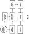
- Branched DNA (bDNA) in situ hybridization is an indirect labeling method for detecting mRNA in single cells (Player et al, 2001). This method uses a series of oligonucleotide probes that have one portion hybridizing to the specific mRNA of interest and another portion hybridizing to the bDNA for signal amplification and detection.
- bDNA ISH has the advantage of using unlabeled oligonucleotide probes for detecting every mRNA of interest and the signal amplification and detection are generic components in the assay.
- the gene specific probes in the bDNA ISH need to be theoretically screened against possible non-specific hybridization interactions with other mRNA sequences in the cells.
- the nonspecific hybridization of the oligonucleotide probes in bDNA ISH can become a serious problem when multiple of those probes have to be used for the detection of low abundance mRNAs.
- use of bDNA ISH to detect or quantitate multiple mRNAs is desirable, such nonspecific hybridization of the oligonucleotide probes is a potential problem.
- the present invention overcomes the above noted difficulties and provides kits for detecting nucleic acids in and for identifying individual cells. A complete understanding of the invention will be obtained upon review of the following.
- the invention provides a kit for detecting a nucleic acid in an individual cell comprising, packaged in one or more containers:
- the nucleic acid target comprises a plurality of different nucleic acid targets; and each nucleic acid target has its associating label probe, capture probe, preamplifier and/or amplifier; and each label probe comprises a label, wherein the signal from each said label is distinguishable from each other.
- the invention further provides a sample of fixed and permeabilized cells, comprising:
- said cells are suspended in solution or are immobilized on a slide.
- said cells are derived from a bodily fluid or from blood.
- said cells comprise a mixture of cell types, wherein said mixture comprises at least one cell of a specified type.
- the ratio of cells of a specified type to cells of all other type(s) in the mixture is less than 1:1x10 4 or less than 1:1x10 5 .
- said cells of a specified type are tumor cells, or specifically, circulating tumor cell.
- Methods of detecting nucleic acid targets in single cells including methods of detecting multiple targets in a single cell, are also described.
- Methods of detecting individual cells, particularly rare cells from large heterogeneous cell populations, through detection of nucleic acids are described.
- Related compositions, and systems are also described.
- a sample comprising the cell is described.
- the cell comprises, or is suspected of comprising, a first nucleic acid target and a second nucleic acid target.
- a first label probe comprising a first label and a second label probe comprising a second label, wherein a first signal from the first label is distinguishable from a second signal from the second label, are described.
- At least a first capture probe and at least a second capture probe are also described.
- the first capture probe is hybridized, in the cell, to the first nucleic acid target (when the first nucleic acid target is present in the cell), and the second capture probe is hybridized, in the cell, to the second nucleic acid target (when the second nucleic acid target is present in the cell).
- the first label probe is captured to the first capture probe and the second label probe is captured to the second capture probe, thereby capturing the first label probe to the first nucleic acid target and the second label probe to the second nucleic acid target.
- the first signal from the first label and the second signal from the second label are then detected.
- first and second labels are associated with their respective nucleic acid targets through the capture probes
- presence of the label(s) in the cell indicates the presence of the corresponding nucleic acid target(s) in the cell.
- the methods are optionally quantitative.
- an intensity of the first signal and an intensity of the second signal can be measured, and the intensity of the first signal can be correlated with a quantity of the first nucleic acid target in the cell while the intensity of the second signal is correlated with a quantity of the second nucleic acid target in the cell.
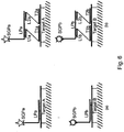
- the label probes bind directly to the capture probes.
- a single first capture probe and a single second capture probe are described, the first label probe is hybridized to the first capture probe, and the second label probe is hybridized to the second capture probe.
- two or more first capture probes and two or more second capture probes are described, as are a plurality of the first label probes (e.g., two or more identical first label probes) and a plurality of the second label probes (e.g., two or more identical second label probes).
- the two or more first capture probes are hybridized to the first nucleic acid target, and the two or more second capture probes are hybridized to the second nucleic acid target.
- a single first label probe is hybridized to each of the first capture probes, and a single second label probe is hybridized to each of the second capture probes.
- the label probes are captured to the capture probes indirectly, for example, through binding of preamplifiers and/or amplifiers.
- amplifiers are employed, a single first capture probe, a single second capture probe, a plurality of the first label probes, and a plurality of the second label probes are provided.
- a first amplifier is hybridized to the first capture probe and to the plurality of first label probes
- a second amplifier is hybridized to the second capture probe and to the plurality of second label probes.
- two or more first capture probes, two or more second capture probes, a plurality of the first label probes, and a plurality of the second label probes are provided.
- the two or more first capture probes are hybridized to the first nucleic acid target, and the two or more second capture probes are hybridized to the second nucleic acid target.
- a first amplifier is hybridized to each of the first capture probes, and the plurality of first label probes is hybridized to the first amplifiers.
- a second amplifier is hybridized to each of the second capture probes, and the plurality of second label probes is hybridized to the second amplifiers.
- preamplifiers are employed, a single first capture probe, a single second capture probe, a plurality of the first label probes, and a plurality of the second label probes are described.
- a first preamplifier is hybridized to the first capture probe, a plurality of first amplifiers is hybridized to the first preamplifier, and the plurality of first label probes is hybridized to the first amplifiers.
- a second preamplifier is hybridized to the second capture probe, a plurality of second amplifiers is hybridized to the second preamplifier, and the plurality of second label probes is hybridized to the second amplifiers.
- first capture probes two or more first capture probes, two or more second capture probes, a plurality of the first label probes, and a plurality of the second label probes are described.
- the two or more first capture probes are hybridized to the first nucleic acid target, and the two or more second capture probes are hybridized to the second nucleic acid target.
- a first preamplifier is hybridized to each of the first capture probes, a plurality of first amplifiers is hybridized to each of the first preamplifiers, and the plurality of first label probes is hybridized to the first amplifiers.
- a second preamplifier is hybridized to each of the second capture probes, a plurality of second amplifiers is hybridized to each of the second preamplifiers, and the plurality of second label probes is hybridized to the second amplifiers.
- the capture probes preferably hybridize to nonoverlapping polynucleotide sequences in their respective nucleic acid target.
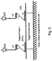
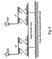
- a first amplified polynucleotide is produced by rolling circle amplification of a first circular polynucleotide hybridized to the first capture probe.
- the first circular polynucleotide comprises at least one copy of a polynucleotide sequence identical to a polynucleotide sequence in the first label probe, and the first amplified polynucleotide thus comprises a plurality of copies of a polynucleotide sequence complementary to the polynucleotide sequence in the first label probe.
- the plurality of first label probes is then hybridized to the first amplified polynucleotide.
- a second amplified polynucleotide is produced by rolling circle amplification of a second circular polynucleotide hybridized to the second capture probe.
- the second circular polynucleotide comprises at least one copy of a polynucleotide sequence identical to a polynucleotide sequence in the second label probe, and the second amplified polynucleotide thus comprises a plurality of copies of a polynucleotide sequence complementary to the polynucleotide sequence in the second label probe.
- the plurality of second label probes is then hybridized to the second amplified polynucleotide.
- the amplified polynucleotides remain associated with the capture probe(s), and the label probes are thus captured to the nucleic acid targets.
- the cell optionally comprises or is suspected of comprising a third nucleic acid target
- the methods optionally include: providing a third label probe comprising a third label, wherein a third signal from the third label is distinguishable from the first and second signals, providing at least a third capture probe, hybridizing in the cell the third capture probe to the third nucleic acid target (when present in the cell), capturing the third label probe to the third capture probe, and detecting the third signal from the third label.
- Fourth, fifth, sixth, etc. nucleic acid targets are similarly simultaneously detected in the cell if desired.
- Each hybridization or capture step is preferably accomplished for all of the nucleic acid targets at the same time.
- a nucleic acid target can be essentially any nucleic acid that is desirably detected in the cell.
- a nucleic acid target can be a DNA, a chromosomal DNA, an RNA, an mRNA, a microRNA, a ribosomal RNA, or the like.
- the nucleic acid target can be a nucleic acid endogenous to the cell.
- the target can be a nucleic acid introduced to or expressed in the cell by infection of the cell with a pathogen, for example, a viral or bacterial genomic RNA or DNA, a plasmid, a viral or bacterial mRNA, or the like.
- the first and second (and/or optional third, fourth, etc.) nucleic acid targets can be part of a single nucleic acid molecule, or they can be separate molecules.
- the first nucleic acid target is a first mRNA and the second nucleic acid target is a second mRNA.
- the first nucleic acid target comprises a first region of an mRNA and the second nucleic acid target comprises a second region of the same mRNA.
- the first nucleic acid target comprises a first chromosomal DNA polynucleotide sequence and the second nucleic acid target comprises a second chromosomal DNA polynucleotide sequence.
- the first and second chromosomal DNA polynucleotide sequences are optionally located on the same chromosome, e.g., within the same gene, or on different chromosomes.
- the signal(s) from nucleic acid target(s) are normalized.
- the second nucleic acid target comprises a reference nucleic acid, and the method includes normalizing the first signal to the second signal.
- the label (first, second, third, etc.) can be essentially any convenient label that directly or indirectly provides a detectable signal.
- the first label is a first fluorescent label and the second label is a second fluorescent label.
- the methods can be used to detect the presence of the nucleic acid targets in cells from essentially any type of sample.
- the sample can be derived from a bodily fluid such as blood.
- the methods for detecting nucleic acid targets in cells can be used to identify the cells.
- a cell can be identified as being of a desired type based on which nucleic acids, and in what levels, it contains.
- the methods include identifying the cell as a desired target cell based on detection of the first and second signals (and optional third, fourth, etc. signals) from within the cell.
- the cell can be a circulating tumor cell, a virally infected cell, a fetal cell in maternal blood, a bacterial cell or other microorganism in a biological sample, or an endothelial cell, precursor endothelial cell, or myocardial cell in blood.
- the cell is typically fixed and permeabilized before hybridization of the capture probes, to retain the nucleic acid targets in the cell and to permit the capture probes, label probes, etc. to enter the cell.
- the cell is optionally washed to remove materials not captured to one of the nucleic acid targets.
- the cell can be washed after any of various steps, for example, after hybridization of the capture probes to the nucleic acid targets to remove unbound capture probes, after hybridization of the preamplifiers, amplifiers, and/or label probes to the capture probes, and/or the like. It will be evident that double-stranded nucleic acid target(s) are preferably denatured, e.g., by heat, prior to hybridization of the corresponding capture probe(s) to the target(s).
- the cell is in suspension for all or most of the steps of the method.
- the cell is in suspension in the sample comprising the cell, and/or the cell is in suspension during the hybridizing, capturing, and/or detecting steps.
- the cell is in suspension in the sample comprising the cell, and the cell is fixed on a substrate during the hybridizing, capturing, and/or detecting steps.
- the cell can be in suspension during the hybridization, capturing, and optional washing steps and immobilized on a substrate during the detection step.
- the first and second (and optional third, etc.) signals can be conveniently detected by flow cytometry. Signals from the labels are typically detected in a single operation.
- a sample comprising the cell is described.
- the cell comprises or is suspected of comprising a first, target nucleic acid, and it comprises a second, reference nucleic acid.
- a first label probe comprising a first label and a second label probe comprising a second label, wherein a first signal from the first label is distinguishable from a second signal from the second label, are also described.
- the first label probe is captured to the first, target nucleic acid (when present in the cell) and the second label probe is captured to the second, reference nucleic acid.
- the first signal from the first label and the second signal from the second label are then detected in the individual cell, and the intensity of each signal is measured.
- the intensity of the first signal is normalized to the intensity of the second (reference) signal.
- the level of the first, target nucleic acid relative to the level of the second, reference nucleic acid in the cell is thereby assayed, since the first and second labels are associated with their respective nucleic acids.
- the methods are optionally quantitative, permitting measurement of the amount of the first, target nucleic acid relative to the amount of the second, reference nucleic acid in the cell.
- the intensity of the first signal normalized to that of the second signal can be correlated with a quantity of the first, target nucleic acid present in the cell.
- the label probes can bind directly to the nucleic acids.
- the first label probe can hybridize to the first, target nucleic acid and/or the second label probe can hybridize to the second, reference nucleic acid.
- the label probes can be bound indirectly to the nucleic acids, e.g., via capture probes.
- at least a first capture probe and at least a second capture probe are described. In the cell, the first capture probe is hybridized to the first, target nucleic acid and the second capture probe is hybridized to the second, reference nucleic acid.
- the first label probe is captured to the first capture probe and the second label probe is captured to the second capture probe, thereby capturing the first label probe to the first, target nucleic acid and the second label probe to the second, reference nucleic acid.
- the features described for the methods above apply as well, with respect to configuration and number of the label and capture probes, optional use of preamplifiers and/or amplifiers, rolling circle amplification of circular polynucleotides, and the like.
- the methods can be used for multiplex detection of nucleic acids, including simultaneous detection of two or more target nucleic acids.
- the cell optionally comprises or is suspected of comprising a third, target nucleic acid
- the methods optionally include: providing a third label probe comprising a third label, wherein a third signal from the third label is distinguishable from the first and second signals; capturing, in the cell, the third label probe to the third, target nucleic acid (when present in the cell); detecting the third signal from the third label, which detecting comprises measuring an intensity of the third signal; and normalizing the intensity of the third signal to the intensity of the second signal.
- Fourth, fifth, sixth, etc. nucleic acids are similarly simultaneously detected in the cell if desired.
- the methods for assaying relative levels of target nucleic acids in cells can be used to identify the cells.
- a cell can be identified as being of a desired type based on which nucleic acids, and in what levels, it contains.
- the methods include identifying the cell as a desired target cell based on the normalized first signal (and optional normalized third, fourth, etc. signals).
- a first mixed cell population comprising one or more cells of a specified type is provided.
- An expression level of one or more target nucleic acids relative to a reference nucleic acid is measured in the cells of the specified type of the first population, to provide a first expression profile.
- a second mixed cell population comprising one or more cells of the specified type is also provided, and an expression level of the one or more target nucleic acids relative to the reference nucleic acid is measured in the cells of the specified type of the second population, to provide a second expression profile.
- the first and second expression profiles are then compared.
- a sample comprising the cell is provided.
- the cell comprises or is suspected of comprising a first nucleic acid target and a second nucleic acid target.
- a first label is captured to the first nucleic acid target (when present in the cell) and a second label is captured to the second nucleic acid target (when present in the cell).
- a first signal from the first label is distinguishable from a second signal from the second label.
- the labels are captured at high density.
- an average of at least one copy of the first label per nucleotide of the first nucleic acid target is captured to the first nucleic acid target over a region that spans at least 20 contiguous nucleotides of the first nucleic acid target
- an average of at least one copy of the second label per nucleotide of the second nucleic acid target is captured to the second nucleic acid target over a region that spans at least 20 contiguous nucleotides of the second nucleic acid target.
- an average of at least four, eight, or twelve copies of the first label per nucleotide of the first nucleic acid target are captured to the first nucleic acid target over a region that spans at least 20 contiguous nucleotides of the first nucleic acid target
- an average of at least four, eight, or twelve copies of the second label per nucleotide of the second nucleic acid target are captured to the second nucleic acid target over a region that spans at least 20 contiguous nucleotides of the second nucleic acid target.
- an average of at least sixteen copies of the first label per nucleotide of the first nucleic acid target are captured to the first nucleic acid target over a region that spans at least 20 contiguous nucleotides of the first nucleic acid target
- an average of at least sixteen copies of the second label per nucleotide of the second nucleic acid target are captured to the second nucleic acid target over a region that spans at least 20 contiguous nucleotides of the second nucleic acid target.
- a sample comprising a mixture of cell types including at least one cell of the specified type is provided.
- a first label probe comprising a first label and a second label probe comprising a second label, wherein a first signal from the first label is distinguishable from a second signal from the second label, are provided.
- the first label probe is captured to a first nucleic acid target (when the first nucleic acid target is present in the cell) and the second label probe is captured to a second nucleic acid target (when the second nucleic acid target is present in the cell).
- the first signal from the first label and the second signal from the second label are detected and correlated with the presence, absence, or amount of the corresponding, first and second nucleic acid targets in the cell.
- the cell is identified as being of the specified type based on detection of the presence, absence, or amount of both the first and second nucleic acid targets within the cell, where the specified type of cell is distinguishable from the other cell type(s) in the mixture on the basis of either the presence, absence, or amount of the first nucleic acid target or the presence, absence, or amount of the second nucleic acid target in the cell (that is, the nucleic acid targets are redundant markers for the specified cell type).
- the cell comprises a first nucleic acid target and a second nucleic acid target, and the cell is identified as being of the specified type based on detection of the presence or amount of both the first and second nucleic acid targets within the cell, where the specified type of cell is distinguishable from the other cell type(s) in the mixture on the basis of either the presence or amount of the first nucleic acid target or the presence or amount of the second nucleic acid target in the cell.
- the label probes can bind directly to the nucleic acid targets.
- the first label probe can hybridize to the first nucleic acid target and/or the second label probe can hybridize to the second nucleic acid target.
- the label probes are optionally captured to the nucleic acid targets via capture probes.
- at least a first capture probe and at least a second capture probe are provided. In the cell, the first capture probe is hybridized to the first nucleic acid target and the second capture probe is hybridized to the second nucleic acid target.
- the first label probe is captured to the first capture probe and the second label probe is captured to the second capture probe, thereby capturing the first label probe to the first nucleic acid target and the second label probe to the second nucleic acid target.
- Third, fourth, fifth, etc. nucleic acid targets are optionally detected in the cell.
- the method optionally includes: providing a third label probe comprising a third label, wherein a third signal from the third label is distinguishable from the first and second signals, capturing in the cell the third label probe to a third nucleic acid target (when the third target is present in the cell), and detecting the third signal from the third label.
- the third, fourth, fifth, etc. label probes are optionally hybridized directly to their corresponding nucleic acid, or they can be captured indirectly via capture probes as described for the first and second label probes.
- the first and/or second signal can be normalized to the third signal.
- the cell comprises the third nucleic acid target, and the methods include identifying the cell as being of the specified type based on the normalized first and/or second signal, e.g., if the target cell type is distinguishable from the other cell type(s) in the mixture based on the copy number of the first and/or second nucleic acid targets, rather than purely on their presence in the target cell type and not in the other cell type(s).
- the third nucleic acid target can serve as a third redundant marker for the target cell type, e.g., to improve specificity of the assay for the desired cell type.
- the methods include correlating the third signal detected from the cell with the presence, absence, or amount of the third nucleic acid target in the cell, and identifying the cell as being of the specified type based on detection of the presence, absence, or amount of the first, second, and third nucleic acid targets within the cell, wherein the specified type of cell is distinguishable from the other cell type(s) in the mixture on the basis of either presence, absence, or amount of the first nucleic acid target, presence, absence, or amount of the second nucleic acid target, or presence, absence, or amount of the third nucleic acid target in the cell.
- the methods can be applied to detection and identification of even rare cell types.
- the ratio of cells of the specified type to cells of all other type(s) in the mixture is optionally less than 1:1x10 4 , less than 1:1x10 5 , less than 1:1x10 6 , less than 1:1x10 7 , less than 1:1x10 8 , or even less than 1:1x10 9 .
- compositions useful in practicing or produced by the methods include a fixed and permeabilized cell, which cell comprises or is suspected of comprising a first nucleic acid target and a second nucleic acid target, at least a first capture probe capable of hybridizing to the first nucleic acid target, at least a second capture probe capable of hybridizing to the second nucleic acid target, a first label probe comprising a first label, and a second label probe comprising a second label.
- a first signal from the first label is distinguishable from a second signal from the second label.
- the cell optionally comprises the first and second capture probes and label probes.
- the first and second capture probes are optionally hybridized to their respective nucleic acid targets in the cell.
- the composition comprises a plurality of the first label probes, a plurality of the second label probes, a first amplified polynucleotide produced by rolling circle amplification of a first circular polynucleotide hybridized to the first capture probe, and a second amplified polynucleotide produced by rolling circle amplification of a second circular polynucleotide hybridized to the second capture probe.
- the first circular polynucleotide comprises at least one copy of a polynucleotide sequence identical to a polynucleotide sequence in the first label probe, and the first amplified polynucleotide comprises a plurality of copies of a polynucleotide sequence complementary to the polynucleotide sequence in the first label probe.
- the second circular polynucleotide comprises at least one copy of a polynucleotide sequence identical to a polynucleotide sequence in the second label probe, and the second amplified polynucleotide comprises a plurality of copies of a polynucleotide sequence complementary to the polynucleotide sequence in the second label probe.
- the composition can also include reagents necessary for producing the amplified polynucleotides, for example, an exogenously supplied nucleic acid polymerase, an exogenously supplied nucleic acid ligase, and/or exogenously supplied nucleoside triphosphates (e.g., dNTPs).
- reagents necessary for producing the amplified polynucleotides for example, an exogenously supplied nucleic acid polymerase, an exogenously supplied nucleic acid ligase, and/or exogenously supplied nucleoside triphosphates (e.g., dNTPs).
- the cell optionally includes additional nucleic acid targets, and the composition (and cell) can include reagents for detecting these targets.
- the cell can comprise or be suspected of comprising a third nucleic acid target, and the composition can include at least a third capture probe capable of hybridizing to the third nucleic acid target and a third label probe comprising a third label.
- a third signal from the third label is distinguishable from the first and second signals.
- the cell optionally includes fourth, fifth, sixth, etc. nucleic acid targets, and the composition optionally includes fourth, fifth, sixth, etc. label probes and capture probes.
- the cell can be present in a mixture of cells, for example, a complex heterogeneous mixture.
- the cell is of a specified type, and the composition comprises one or more other types of cells. These other cells can be present in excess, even large excess, of the cell.
- the ratio of cells of the specified type to cells of all other type(s) in the composition is optionally less than 1:1x10 4 , less than 1:1x10 5 , less than 1:1x10 6 , less than 1:1x10 7 , less than 1:1x10 8 , or even less than 1:1x10 9 .
- composition comprising a cell, which cell includes a first nucleic acid target, a second nucleic acid target, a first label wnose presence in the cell is indicative of the presence of the first nucleic acid target in the cell, and a second label whose presence in the cell is indicative of the presence of the second nucleic acid target in the cell, wherein a first signal from the first label is distinguishable from a second signal from the second label.
- An average of at least one copy of the first label is present in the cell per nucleotide of the first nucleic acid target over a region that spans at least 20 contiguous nucleotides of the first nucleic acid target
- an average of at least one copy of the second label is present in the cell per nucleotide of the second nucleic acid target over a region that spans at least 20 contiguous nucleotides of the second nucleic acid target.
- the copies of the first label are physically associated with the first nucleic acid target, and the copies of the second label are physically associated with the second nucleic acid target.
- the first label can be part of a first label probe and the second label part of a second label probe, where the label probes are captured to the target nucleic acids.
- kits useful for practicing the methods include at least one reagent for fixing and/or permeabilizing the cell, at least a first capture probe capable of hybridizing to the first nucleic acid target, at least a second capture probe capable of hybridizing to the second nucleic acid target, a first label probe comprising a first label, and a second label probe comprising a second label, wherein a first signal from the first label is distinguishable from a second signal from the second label, packaged in one or more containers.
- kits for detecting an individual cell of a specified type from a mixture of cell types by detecting a first nucleic acid target and a second nucleic acid target includes at least one reagent for fixing and/or permeabilizing the cell, a first label probe comprising a first label, and a second label probe comprising a second label, wherein a first signal from the first label is distinguishable from a second signal from the second label, packaged in one or more containers.
- the specified type of cell is distinguishable from the other cell type(s) in the mixture by presence, absence, or amount of the first nucleic acid target in the cell or by presence, absence, or amount of the second nucleic acid target in the cell.
- polynucleotide encompasses any physical string of monomer units that can be corresponded to a string of nucleotides, including a polymer of nucleotides (e.g., a typical DNA or RNA polymer), peptide nucleic acids (PNAs), modified oligonucleotides (e.g., oligonucleotides comprising nucleotides that are not typical to biological RNA or DNA, such as 2'-O-methylated oligonucleotides), and the like.
- PNAs peptide nucleic acids
- modified oligonucleotides e.g., oligonucleotides comprising nucleotides that are not typical to biological RNA or DNA, such as 2'-O-methylated oligonucleotides
- the nucleotides of the polynucleotide can be deoxyribonucleotides, ribonucleotides or nucleotide analogs, can be natural or non-natural, and can be unsubstituted, unmodified, substituted or modified.
- the nucleotides can be linked by phosphodiester bonds, or by phosphorothioate linkages, methylphosphonate linkages, boranophosphate linkages, or the like.
- the polynucleotide can additionally comprise non-nucleotide elements such as labels, quenchers, blocking groups, or the like.
- the polynucleotide can be, e.g., single-stranded or double-stranded.
- nucleic acid target or “target nucleic acid” refers to a nucleic acid, or optionally a region thereof, that is to be detected.
- a "polynucleotide sequence” or “nucleotide sequence” is a polymer of nucleotides (an oligonucleotide, a DNA, a nucleic acid, etc.) or a character string representing a nucleotide polymer, depending on context. From any specified polynucleotide sequence, either the given nucleic acid or the complementary polynucleotide sequence (e.g., the complementary nucleic acid) can be determined.
- Gene is used broadly to refer to any nucleic acid associated with a biological function. Genes typically include coding sequences and/or the regulatory sequences required for expression of such coding sequences. The term gene can apply to a specific genomic sequence, as well as to a cDNA or an mRNA encoded by that genomic sequence. Genes also include non-expressed nucleic acid segments that, for example, form recognition sequences for other proteins. Non-expressed regulatory sequences include promoters and enhancers, to which regulatory proteins such as transcription factors bind, resulting in transcription of adjacent or nearby sequences.
- Two polynucleotides "hybridize” when they associate to form a stable duplex, e.g., under relevant assay conditions.
- Nucleic acids hybridize due to a variety of well characterized physico-chemical forces, such as hydrogen bonding, solvent exclusion, base stacking and the like.
- An extensive guide to the hybridization of nucleic acids is found in Tijssen (1993) Laboratory Techniques in Biochemistry and Molecular Biology-Hybridization with Nucleic Acid Probes, part I chapter 2, “Overview of principles of hybridization and the strategy of nucleic acid probe assays" (Elsevier, New York), as well as in Ausubel, infra .
- a first polynucleotide "capable of hybridizing" to a second polynucleotide contains a first polynucleotide sequence that is complementary to a second polynucleotide sequence in the second polynucleotide.
- the first and second polynucleotides are able to form a stable duplex, e.g., under relevant assay conditions.
- the "T m " (melting temperature) of a nucleic acid duplex under specified conditions is the temperature at which half of the base pairs in a population of the duplex are disassociated and half are associated.
- the T m for a particular duplex can be calculated and/or measured, e.g., by obtaining a thermal denaturation curve for the duplex (where the T m is the temperature corresponding to the midpoint in the observed transition from double-stranded to single-stranded form).
- complementary refers to a polynucleotide that forms a stable duplex with its "complement,” e.g., under relevant assay conditions.
- two polynucleotide sequences that are complementary to each other have mismatches at less than about 20% of the bases, at less than about 10% of the bases, preferably at less than about 5% of the bases, and more preferably have no mismatches.
- label is a moiety that facilitates detection of a molecule.
- Common labels in the context of the present invention include fluorescent, luminescent, light-scattering, and/or colorimetric labels. Suitable labels include enzymes and fluorescent moieties, as well as radionuclides, substrates, cofactors, inhibitors, chemiluminescent moieties, magnetic particles, and the like. Patents teaching the use of such labels include U.S. Pat. Nos. 3,817,837 ; 3,850,752 ; 3,939,350 ; 3,996,345 ; 4,277,437 ; 4,275,149 ; and 4,366,241 . Many labels are commercially available and can be used in the context of the invention.
- label probe refers to an entity that binds to a target molecule, directly or indirectly, and enables the target to be detected, e.g., by a readout instrument.
- a label probe (or "LP") is typically a single-stranded polynucleotide that comprises at least one label which directly or indirectly provides a detectable signal.
- the label can be covalently attached to the polynucleotide, or the polynucleotide can be configured to bind to the label (e.g., a biotinylated polynucleotide can bind a streptavidin-associated label).
- the label probe can, for example, hybridize directly to a target nucleic acid, or it can hybridize to a nucleic acid that is in turn hybridized to the target nucleic acid or to one or more other nucleic acids that are hybridized to the nucleic acid.
- the label probe can comprise a polynucleotide sequence that is complementary to a polynucleotide sequence of the target nucleic acid, or it can comprise at least one polynucleotide sequence that is complementary to a polynucleotide sequence in a capture probe, amplifier, or the like.
- a “capture probe” is a polynucleotide that is capable of hybridizing to a target nucleic acid and capturing a label probe to that target nucleic acid.
- the capture probe can hybridize directly to the label probe, or it can hybridize to one or more nucleic acids that in turn hybridize to the label probe; for example, the capture probe can hybridize to an amplifier or a preamplifier.
- the capture probe thus includes a first polynucleotide sequence that is complementary to a polynucleotide sequence of the target nucleic acid and a second polynucleotide sequence that is complementary to a polynucleotide sequence of the label probe, amplifier, preamplifier, or the like.
- the capture probe is preferably single-stranded.
- An "amplifier” is a molecule, typically a polynucleotide, that is capable of hybridizing to multiple label probes. Typically, the amplifier hybridizes to multiple identical label probes. The amplifier also hybridizes to at least one capture probe or nucleic acid bound to a capture probe. For example, the amplifier can hybridize to at least one capture probe and to a plurality of label probes, or to a preamplifier and a plurality of label probes. The amplifier can be, e.g., a linear, forked, comb-like, or branched nucleic acid.
- the amplifier can include modified nucleotides and/or nonstandard intemucleotide linkages as well as standard deoxyribonucleotides, ribonucleotides, and/or phosphodiester bonds. Suitable amplifiers are described, for example, in USPN 5,635,352, USPN 5,124,246, USPN 5,710,264, and USPN 5,849,481.
- a "preamplifier” is a molecule, typically a polynucleotide, that serves as an intermediate between one or more capture probes and amplifiers. Typically, the preamplifier hybridizes simultaneously to one or more capture probes and to a plurality of amplifiers. Exemplary preamplifiers are described, for example, in USPN 5,635,352 and USPN 5,681,697.
- a "pathogen” is a biological agent, typically a microorganism, that causes disease or illness to its host.
- a "microorganism” is an organism of microscopic or submicroscopic size. Examples include bacteria, fungi, yeast, protozoans, microscopic algae (e.g., unicellular algae), viruses (which are typically included in this category although they are incapable of growth and reproduction outside of host cells), subviral agents, viroids, and mycoplasma.
- the invention provides a kit for detecting a nucleic acid in an individual cell comprising, packaged in one or more containers:
- the nucleic acid target comprises a plurality of different nucleic acid targets; and each nucleic acid target has its associating label probe, capture probe, preamplifier and/or amplifier; and each label probe comprises a label, wherein the signal from each said label is distinguishable from each other.
- the invention further provides a sample of fixed and permeabilized cells, comprising:
- said cells are suspended in solution or are immobilized on a slide.
- said cells are derived from a bodily fluid or from blood.
- said cells comprise a mixture of cell types, wherein said mixture comprises at least one cell of a specified type.
- the ratio of cells of a specified type to cells of all other type(s) in the mixture is less than 1:1x10 4 or less than 1:1x10 5 .
- said cells of a specified type are tumor cells, or specifically, circulating tumor cell.
- multiplex assays that can be used for simultaneous detection, and optionally quantitation, of two or more nucleic acid targets in a single cell.
- Described herein are methods for detecting the level of one or more target nucleic acids relative to that of a reference nucleic acid in an individual cell.
- a label probe is captured to each target nucleic acid.
- the label probe can be captured to the target through direct binding of the label probe to the target.
- the label probe is captured indirectly through binding to capture probes, amplifiers, and/or preamplifiers that bind to the target.
- Use of the optional amplifiers and preamplifiers facilitates capture of multiple copies of the label probe to the target, thus amplifying signal from the target without requiring enzymatic amplification of the target itself.
- Binding of the capture probes is optionally cooperative, reducing background caused by undesired cross hybridization of capture probes to non-target nucleic acids (a greater problem in multiplex assays than singleplex assays since more probes must be used in multiplex assays, increasing the likelihood of cross hybridization).
- One aspect of the invention relates to detection of single cells, including detection of rare cells from a heterogeneous mixture of cells. Individual cells are detected through detection of nucleic acids whose presence, absence, copy number, or the like are characteristic of the cell.
- compositions are also described.
- a sample comprising the cell is described.
- the cell comprises, or is suspected of comprising, a first nucleic acid target and a second nucleic acid target.
- a first label probe comprising a first label and a second label probe comprising a second label, wherein a first signal from the first label is distinguishable from a second signal from the second label, are described.
- At least a first capture probe and at least a second capture probe are also described.
- the first capture probe is hybridized, in the cell, to the first nucleic acid target (when the first nucleic acid target is present in the cell), and the second capture probe is hybridized, in the cell, to the second nucleic acid target (when the second nucleic acid target is present in the cell).
- the first label probe is captured to the first capture probe and the second label probe is captured to the second capture probe, thereby capturing the first label probe to the first nucleic acid target and the second label probe to the second nucleic acid target.
- the first signal from the first label and the second signal from the second label are then detected.
- first and second labels are associated with their respective nucleic acid targets through the capture probes
- presence of the label(s) in the cell indicates the presence of the corresponding nucleic acid target(s) in the cell.
- the methods are optionally quantitative.
- an intensity of the first signal and an intensity of the second signal can be measured, and the intensity of the first signal can be correlated with a quantity of the first nucleic acid target in the cell while the intensity of the second signal is correlated with a quantity of the second nucleic acid target in the cell.
- the label probes bind directly to the capture probes.
- a single first capture probe and a single second capture probe are provided, the first label probe is hybridized to the first capture probe, and the second label probe is hybridized to the second capture probe.
- two or more first capture probes and two or more second capture probes are provided, as are a plurality of the first label probes (e.g., two or more identical first label probes) and a plurality of the second label probes (e.g., two or more identical second label probes).
- the two or more first capture probes are hybridized to the first nucleic acid target, and the two or more second capture probes are hybridized to the second nucleic acid target.
- a single first label probe is hybridized to each of the first capture probes, and a single second label probe is hybridized to each of the second capture probes.
- the label probes are captured to the capture probes indirectly, for example, through binding of preamplifiers and/or amplifiers.
- Use of amplifiers and preamplifiers can be advantageous in increasing signal strength, since they can facilitate binding of large numbers of label probes to each nucleic acid target.
- a first amplifier is hybridized to the first capture probe and to the plurality of first label probes
- a second amplifier is hybridized to the second capture probe and to the plurality of second label probes.
- two or more first capture probes, two or more second capture probes, a plurality of the first label probes, and a plurality of the second label probes are provided. The two or more first capture probes are hybridized to the first nucleic acid target, and the two or more second capture probes are hybridized to the second nucleic acid target.
- a first amplifier is hybridized to each of the first capture probes, and the plurality of first label probes is hybridized to the first amplifiers.
- a second amplifier is hybridized to each of the second capture probes, and the plurality of second label probes is hybridized to the second amplifiers.
- preamplifiers are employed, a single first capture probe, a single second capture probe, a plurality of the first label probes, and a plurality of the second label probes are described.
- a first preamplifier is hybridized to the first capture probe, a plurality of first amplifiers is hybridized to the first preamplifier, and the plurality of first label probes is hybridized to the first amplifiers.
- a second preamplifier is hybridized to the second capture probe, a plurality of second amplifiers is hybridized to the second preamplifier, and the plurality of second label probes is hybridized to the second amplifiers.
- two or more first capture probes, two or more second capture probes, a plurality of the first label probes, and a plurality of the second label probes are provided.
- the two or more first capture probes are hybridized to the first nucleic acid target, and the two or more second capture probes are hybridized to the second nucleic acid target.
- a first preamplifier is hybridized to each of the first capture probes, a plurality of first amplifiers is hybridized to each of the first preamplifiers, and the plurality of first label probes is hybridized to the first amplifiers.
- a second preamplifier is hybridized to each of the second capture probes, a plurality of second amplifiers is hybridized to each of the second preamplifiers, and the plurality of second label probes is hybridized to the second amplifiers.
- additional preamplifiers can be used as intermediates between a preamplifier hybridized to the capture probe(s) and the amplifiers.
- one capture probe hybridizes to each label probe, amplifier, or preamplifier.
- two or more capture probes hybridize to the label probe, amplifier, or preamplifier. See, e.g., the section below entitled “Implementation, applications, and advantages.”
- the capture probes preferably hybridize to nonoverlapping polynucleotide sequences in their respective nucleic acid target.
- the capture probes can, but need not, cover a contiguous region of the nucleic acid target.
- Blocking probes, polynucleotides which hybridize to regions of the nucleic acid target not occupied by capture probes, are optionally provided and hybridized to the target.
- the corresponding capture probes and blocking probes are preferably complementary to physically distinct, nonoverlapping sequences in the nucleic acid target, which nonoverlapping sequences are preferably, but not necessarily, contiguous. Having the capture probes and optional blocking probes be contiguous with each other can in some embodiments enhance hybridization strength, remove secondary structure, and ensure more consistent and reproducible signal.
- enzymatic manipulation is not required to capture the label probes to the capture probes.
- enzymatic manipulation particularly amplification of nucleic acids intermediate between the capture probes and the label probes, facilitates detection of the nucleic acid targets.
- a plurality of the first label probes and a plurality of the second label probes are provided.
- a first amplified polynucleotide is produced by rolling circle amplification of a first circular polynucleotide hybridized to the first capture probe.
- the first circular polynucleotide comprises at least one copy of a polynucleotide sequence identical to a polynucleotide sequence in the first label probe, and the first amplified polynucleotide thus comprises a plurality of copies of a polynucleotide sequence complementary to the polynucleotide sequence in the first label probe.
- the plurality of first label probes is then hybridized to the first amplified polynucleotide.
- a second amplified polynucleotide is produced by rolling circle amplification of a second circular polynucleotide hybridized to the second capture probe (preferably, at the same time the first amplified polynucleotide is produced).
- the second circular polynucleotide comprises at least one copy of a polynucleotide sequence identical to a polynucleotide sequence in the second label probe, and the second amplified polynucleotide thus comprises a plurality of copies of a polynucleotide sequence complementary to the polynucleotide sequence in the second label probe.
- the plurality of second label probes is then hybridized to the second amplified polynucleotide.
- the amplified polynucleotides remain associated (e.g., covalently) with the capture probe(s), and the label probes are thus captured to the nucleic acid targets.
- a circular polynucleotide can be provided and hybridized to the capture probe, or a linear polynucleotide that is circularized by ligation after it binds to the capture probe (e.g., a padlock probe) can be employed.
- a padlock probe e.g., a linear polynucleotide that is circularized by ligation after it binds to the capture probe.
- Techniques for rolling circle amplification, including use of padlock probes are well known in the art. See, e.g., Larsson et al. (2004) "In situ genotyping individual DNA molecules by target-primed rolling-circle amplification of padlock probes" Nat Methods. 1(3):227-32 , Nilsson et al. (1994) Science 265:2085-2088 , and Antson et al. (2000) "PCR-generated padlock probes detect single nucleotide variation in genomic DNA” Nucl Acids Res 28(12):E58 .
- Potential capture probe sequences are optionally examined for possible interactions with non-corresponding nucleic acid targets, the preamplifiers, the amplifiers, the label probes, and/or any relevant genomic sequences, for example. Sequences expected to cross-hybridize with undesired nucleic acids are typically not selected for use in the capture probes. Examination can be, e.g., visual (e.g., visual examination for complementarity), computational (e.g., a BLAST search of the relevant genomic database, or computation and comparison of binding free energies), and/or experimental (e.g., cross-hybridization experiments). Label probe sequences are preferably similarly examined, to help minimize potential undesirable cross-hybridization.
- a capture probe, preamplifier, amplifier, and/or label probe optionally comprises at least one non-natural nucleotide.
- a capture probe and a preamplifier (or amplifier or label probe) that hybridizes to it optionally comprise, at complementary positions, at least one pair of non-natural nucleotides that base pair with each other but that do not Watson-Crick base pair with the bases typical to biological DNA or RNA (i.e., A, C, G, T, or U).
- nonnatural nucleotides include, Locked NucleicAcidTM nucleotides (available from Exiqon A/S, (www.) exiqon.com; see, e.g., SantaLucia Jr.
- the methods are useful for multiplex detection of nucleic acids, including simultaneous detection of more than two nucleic acid targets.
- the cell optionally comprises or is suspected of comprising a third nucleic acid target, and the methods optionally include: providing a third label probe comprising a third label, wherein a third signal from the third label is distinguishable from the first and second signals, providing at least a third capture probe, hybridizing in the cell the third capture probe to the third nucleic acid target (when the third target is present in the cell), capturing the third label probe to the third capture probe, and detecting the third signal from the third label.
- Fourth, fifth, sixth, etc. nucleic acid targets are similarly simultaneously detected in the cell if desired.
- a nucleic acid target can be essentially any nucleic acid that is desirably detected in the cell.
- a nucleic acid target can be a DNA, a chromosomal DNA, an RNA, an mRNA, a microRNA, a ribosomal RNA, or the like.
- the nucleic acid target can be a nucleic acid endogenous to the cell.
- the target can be a nucleic acid introduced to or expressed in the cell by infection of the cell with a pathogen, for example, a viral or bacterial genomic RNA or DNA, a plasmid, a viral or bacterial mRNA, or the like.
- the first and second (and/or optional third, fourth, etc.) nucleic acid targets can be part of a single nucleic acid molecule, or they can be separate molecules. Various advantages and applications of both approaches are discussed in greater detail below and in the section entitled "Implementation, applications, and advantages.”
- the first nucleic acid target is a first mRNA and the second nucleic acid target is a second mRNA.
- the first nucleic acid target comprises a first region of an mRNA and the second nucleic acid target comprises a second region of the same mRNA; this approach can increase specificity of detection of the mRNA.
- the first nucleic acid target comprises a first chromosomal DNA polynucleotide sequence and the second nucleic acid target comprises a second chromosomal DNA polynucleotide sequence.
- the first and second chromosomal DNA polynucleotide sequences are optionally located on the same chromosome, e.g., within the same gene, or on different chromosomes.
- the signal(s) from nucleic acid target(s) are normalized.
- the second nucleic acid target comprises a reference nucleic acid
- the method includes normalizing the first signal to the second signal.
- the reference nucleic acid is a nucleic acid selected as a standard of comparison. It will be evident that choice of the reference nucleic acid can depend on the desired application. For example, for gene expression analysis, where the first and optional third, fourth, etc. nucleic acid targets are mRNAs whose expression levels are to be determined, the reference nucleic acid can be an mRNA transcribed from a housekeeping gene.
- the first nucleic acid target can be an mRNA whose expression is altered in a pathological state, e.g., an mRNA expressed in a tumor cell and not a normal cell or expressed at a higher level in a tumor cell than in a normal cell, while the second nucleic acid target is an mRNA expressed from a housekeeping gene or similar gene whose expression is not altered in the pathological state.
- the first nucleic acid target can be a chromosomal DNA sequence that is amplified or deleted in a tumor cell, while the second nucleic acid target is another chromosomal DNA sequence that is maintained at its normal copy number in the tumor cell. Exemplary reference nucleic acids are described herein, and many more are well known in the art.
- results from the cell are compared with results from a reference cell. That is, the first and second targets are also detected in a reference cell, for example, a non-tumor, uninfected, or other healthy normal cell, chosen as a standard of comparison depending on the desired application.
- the signals can be normalized to a reference nucleic acid as noted above.
- the first nucleic acid target can be the Her-2 gene, with the goal of measuring Her-2 gene amplification.
- Signal from Her-2 can be normalized to that from a reference gene, whose copy number is stably maintained in the genomic DNA.
- the normalized signal for the Her-2 gene from a target cell can be compared to the normalized signal from a reference cell (e.g., a normal cell), to determine copy number in the cancer cell in comparison to normal cells.
- a target cell e.g., a tumor cell or suspected tumor cell
- a reference cell e.g., a normal cell
- the label can be essentially any convenient label that directly or indirectly provides a detectable signal.
- the first label is a first fluorescent label and the second label is a second fluorescent label.
- Detecting the signal from the labels thus comprises detecting fluorescent signals from the labels.
- a variety of fluorescent labels whose signals can be distinguished from each other are known, including, e.g., fluorophores and quantum dots.
- the label can be a luminescent label, a light-scattering label (e.g., colloidal gold particles), or an enzyme (e.g., alkaline phosphatase or horseradish peroxidase).
- the methods can be used to detect the presence of the nucleic acid targets in cells from essentially any type of sample.
- the sample can be derived from a bodily fluid, a bodily waste, blood, bone marrow, sputum, urine, lymph node, stool, vaginal secretions, cervical pap smear, oral swab or other swab or smear, spinal fluid, saliva, sputum, ejaculatory fluid, semen, lymph fluid, an intercellular fluid, a tissue (e.g., a tissue homogenate), a biopsy, and/or a tumor.
- the sample and/or the cell can be derived from one or more of a human, an animal, a plant, and a cultured cell. Samples derived from even relatively large volumes of materials such as bodily fluid or bodily waste can be screened in the methods of the invention, and removal of such materials is relatively non-invasive. Samples are optionally taken from a patient, following standard laboratory methods after informed consent.
- the methods for detecting nucleic acid targets in cells can be used to identify the cells.
- a cell can be identified as being of a desired type based on which nucleic acids, and in what levels, it contains.
- the methods include identifying the cell as a desired target cell based on detection of the first and second signals (and optional third, fourth, etc. signals) from within the cell.
- the cell can be identified on the basis of the presence or absence of one or more of the nucleic acid targets.
- the cell can be identified on the basis of the relative signal strength from or expression level of one or more of the nucleic acid targets. Signals are optionally normalized as noted above and/or compared to those from a reference cell.
- the sample including the cell can be a mixture of desired target cells and other, nontarget cells, which can be present in excess of the target cells.
- the ratio of target cells to cells of all other type(s) in the sample is optionally less than 1:1x10 4 , less than 1:1x10 5 , less than 1:1x10 6 , less than 1:1x10 7 , less than 1:1x10 8 , or even less than 1:1x10 9 .
- the cell can be a circulating tumor cell, a virally infected cell, a fetal cell in maternal blood, a bacterial cell or other microorganism in a biological sample (e.g., blood or other body fluid), an endothelial cell, precursor endothelial cell, or myocardial cell in blood, a stem cell, or a T-cell.
- a biological sample e.g., blood or other body fluid
- an endothelial cell e.g., precursor endothelial cell, or myocardial cell in blood, a stem cell, or a T-cell.
- Rare cell types can be enriched prior to performing the methods, if necessary, by methods known in the art (e.g., lysis of red blood cells, isolation of peripheral blood mononuclear cells, further enrichment of rare target cells through magnetic-activated cell separation (MACS), etc.).
- the methods are optionally combined with other techniques, such as DAPI staining for nuclear DNA. It will be evident that a variety of different types of nucleic acid markers are optionally detected simultaneously by the methods and used to identify the cell.
- a cell can be identified based on the presence or relative expression level of one nucleic acid target in the cell and the absence of another nucleic acid target from the cell; e.g., a circulating tumor cell can be identified by the presence or level of one or more markers found in the tumor cell and not found (or found at different levels) in blood cells, and its identity can be confirmed by the absence of one or more markers present in blood cells and not circulating tumor cells.
- the principle may be extended to using any other type of markers such as protein based markers in single cells.
- the cell is typically fixed and permeabilized before hybridization of the capture probes, to retain the nucleic acid targets in the cell and to permit the capture probes, label probes, etc. to enter the cell.
- the cell is optionally washed to remove materials not captured to one of the nucleic acid targets.
- the cell can be washed after any of various steps, for example, after hybridization of the capture probes to the nucleic acid targets to remove unbound capture probes, after hybridization of the preamplifiers, amplifiers, and/or label probes to the capture probes, and/or the like.
- the various capture and hybridization steps can be performed simultaneously or sequentially, in essentially any convenient order.
- a given hybridization step is accomplished for all of the nucleic acid targets at the same time.
- all the capture probes first, second, etc.
- amplifiers first, second, etc.
- the label probes first, second, etc.
- the capture probes can be hybridized to the targets, the cell can be washed, amplifiers and label probes can be added together and hybridized, and the cell can then be washed prior to detection. It will be evident that double-stranded nucleic acid target(s) are preferably denatured, e.g., by heat, prior to hybridization of the corresponding capture probe(s) to the target(s).
- the cell is in suspension for all or most of the steps of the method, for ease of handling.
- the methods are also applicable to cells in solid tissue samples (e.g., tissue sections) and/or cells immobilized on a substrate (e.g., a slide or other surface).
- the cell is in suspension in the sample comprising the cell, and/or the cell is in suspension during the hybridizing, capturing, and/or detecting steps.
- the cell can be in suspension in the sample and during the hybridization, capture, optional washing, and detection steps.
- the cell is in suspension in the sample comprising the cell, and the cell is fixed on a substrate during the hybridizing, capturing, and/or detecting steps.
- the cell can be in suspension during the hybridization, capture, and optional washing steps and immobilized on a substrate during the detection step.
- Signals from the labels can be detected, and their intensities optionally measured, by any of a variety of techniques well known in the art. For example, if the cell is in suspension, the first and second (and optional third, etc.) signals can be conveniently detected by flow cytometry. If cells are immobilized on a substrate, the first and second (and optional third etc.) signals can be detected, for example, by laser scanner or microscope, e.g., a fluorescent or automated scanning microscope. As noted, detection is at the level of individual, single cells. Signals from the labels are typically detected in a single operation (e.g., a single flow cytometry run or a single microscopy or scanning session), rather than sequentially in separate operations for each label.
- a single operation e.g., a single flow cytometry run or a single microscopy or scanning session
- Such a single detection operation can, for example, involve changing optical filters between detection of the different labels, but it does not involve detection of the first label followed by capture of the second label and then detection of the second label.
- the first and second (and optional third etc.) labels are captured to their respective targets simultaneously but are detected in separate detection steps or operations.
- a label probe can include more than one label, identical or distinct.
- Signal strength is optionally adjusted between targets depending on their expected copy numbers, if desired; for example, the signal for an mRNA expressed at low levels can be amplified to a greater degree (e.g., by use of more labels per label probe and/or use of preamplifiers and amplifiers to capture more label probes per copy of the target) than the signal for a highly expressed mRNA.
- two or more nucleic acids are detected by PCR amplification of the nucleic acids in situ in individual cells.
- a water-oil emulsion can be made as mentioned in Li et al. (2006) "BEAMing up for detection and quantification of rare sequence variants” Nature Methods 3(2):95-7 that separates single cells into different compartments.
- the signal detected for a nucleic acid of interest can be normalized to that of a standard, reference nucleic acid.
- a sample comprising the cell is described.
- the cell comprises or is suspected of comprising a first, target nucleic acid, and it comprises a second, reference nucleic acid.
- a first label probe comprising a first label and a second label probe comprising a second label, wherein a first signal from the first label is distinguishable from a second signal from the second label, are also described.
- the first label probe is captured to the first, target nucleic acid (when the first, target nucleic acid is present in the cell) and the second label probe is captured to the second, reference nucleic acid.
- the first signal from the first label and the second signal from the second label are then detected in the individual cell, and the intensity of each signal is measured.
- the intensity of the first signal is normalized to the intensity of the second (reference) signal.
- the level of the first, target nucleic acid relative to the level of the second, reference nucleic acid in the cell is thereby assayed, since the first and second labels are associated with their respective nucleic acids.
- the methods are optionally quantitative, permitting measurement of the amount of the first, target nucleic acid relative to the amount of the second, reference nucleic acid in the cell.
- the intensity of the first signal normalized to that of the second signal can be correlated with a quantity of the first, target nucleic acid present in the cell.
- the label probes can bind directly to the nucleic acids.
- the first label probe can hybridize to the first, target nucleic acid and/or the second label probe can hybridize to the second, reference nucleic acid.
- some or all of the label probes can be indirectly bound to their corresponding nucleic acids, e.g., through capture probes.
- the first and second label probes can bind directly to the nucleic acids, or one can bind directly while the other binds indirectly, or both can bind indirectly.
- the label probes are optionally captured to the nucleic acids via capture probes.
- at least a first capture probe and at least a second capture probe are provided.
- the first capture probe is hybridized to the first, target nucleic acid and the second capture probe is hybridized to the second, reference nucleic acid.
- the first label probe is captured to the first capture probe and the second label probe is captured to the second capture probe, thereby capturing the first label probe to the first, target nucleic acid and the second label probe to the second, reference nucleic acid.
- the methods can be used for multiplex detection of nucleic acids, including simultaneous detection of two or more target nucleic acids.
- the cell optionally comprises or is suspected of comprising a third, target nucleic acid
- the methods optionally include: providing a third label probe comprising a third label, wherein a third signal from the third label is distinguishable from the first and second signals; capturing, in the cell, the third label probe to the third, target nucleic acid (when present in the cell); detecting the third signal from the third label, which detecting comprises measuring an intensity of the third signal; and normalizing the intensity of the third signal to the intensity of the second signal.
- the third signal can be normalized to that from a different reference nucleic acid.
- Fourth, fifth, sixth, etc. nucleic acids are similarly simultaneously detected in the cell if desired.
- the third, fourth, fifth, etc. label probes are optionally hybridized directly to their corresponding nucleic acid, or they can be captured indirectly via capture probes as described for the first and second label probes.
- a target nucleic acid can be essentially any nucleic acid that is desirably detected in the cell.
- a target nucleic acid can be a DNA, a chromosomal DNA, an RNA, an mRNA, a microRNA, a ribosomal RNA, or the like.
- the target nucleic acid can be a nucleic acid endogenous to the cell, or as another example, the target can be a nucleic acid introduced to or expressed in the cell by infection of the cell with a pathogen, for example, a viral or bacterial genomic RNA or DNA, a plasmid, a viral or bacterial mRNA, or the like.
- the reference nucleic acid can similarly be a DNA, an mRNA, a chromosomal DNA, an mRNA, an RNA endogenous to the cell, or the like.
- the reference nucleic acid can depend on the desired application.
- the reference nucleic acid can be an mRNA transcribed from a housekeeping gene.
- the first, target nucleic acid can be an mRNA whose expression is altered in a pathological state, e.g., an mRNA expressed in a tumor cell and not a normal cell or expressed at a higher level in a tumor cell than in a normal cell, while the reference nucleic acid is an mRNA expressed from a housekeeping gene or similar gene whose expression is not altered in the pathological state.
- the target nucleic acid can be a viral or bacterial nucleic acid while the reference nucleic acid is endogenous to the cell.
- the first, target nucleic acid can be a chromosomal DNA sequence that is amplified or deleted in a tumor cell, while the reference nucleic acid is another chromosomal DNA sequence that is maintained at its normal copy number in the tumor cell. Exemplary reference nucleic acids are described herein, and many more are well known in the art.
- the first, target nucleic acid is a first mRNA and the second, reference nucleic acid is a second mRNA.
- the first, target nucleic acid comprises a first chromosomal DNA polynucleotide sequence and the second, reference nucleic acid comprises a second chromosomal DNA polynucleotide sequence.
- the first and second chromosomal DNA polynucleotide sequences are optionally located on the same chromosome or on different chromosomes.
- normalized results from the cell are compared with normalized results from a reference cell. That is, the target and reference nucleic acids are also detected in a reference cell, for example, a non-tumor, uninfected, or other healthy normal cell, chosen as a standard of comparison depending on the desired application.
- a reference cell for example, a non-tumor, uninfected, or other healthy normal cell, chosen as a standard of comparison depending on the desired application.
- the first, target nucleic acid can be the Her-2 gene, with the goal of measuring Her-2 gene amplification.
- Signal from Her-2 can be normalized to that from a reference gene whose copy number is stably maintained in the genomic DNA.
- the normalized signal for the Her-2 gene from a target cell can be compared to the normalized signal from a reference cell (e.g., a normal cell), to determine copy number in the cancer cell in comparison to normal cells.
- a target cell e.g., a tumor cell or suspected tumor cell
- a reference cell e.g., a normal cell
- Signal strength is optionally adjusted between the target and reference nucleic acids depending on their expected copy numbers, if desired.
- the signal for a target mRNA expressed at low levels can be amplified to a greater degree (e.g., by use of more labels per label probe and/or use of capture probes, preamplifiers and amplifiers to capture more label probes per copy of the target) than the signal for a highly expressed mRNA (which can, e.g., be detected by direct binding of the label probe to the reference nucleic acid, by use of capture probes and amplifier without a preamplifier, or the like).
- the methods for assaying relative levels of target nucleic acids in cells can be used to identify the cells.
- a cell can be identified as being of a desired type based on which nucleic acids, and in what levels, it contains.
- the methods include identifying the cell as a desired target cell based on the normalized first signal (and optional normalized third, fourth, etc. signals).
- the cell can be identified on the basis of the presence or absence of one or more of the target nucleic acids.
- the cell can be identified on the basis of the relative signal strength from or expression level of one or more target nucleic acids. Signals are optionally compared to those from a reference cell.
- the sample including the cell can be a mixture of desired target cells and other, nontarget cells, which can be present in excess of the target cells.
- the ratio of target cells to cells of all other type(s) in the sample is optionally less than 1:1x10 4 , less than 1:1x10 5 , less than 1:1x10 6 , less than 1:1x10 7 , less than 1:1x10 8 , or even less than 1:1x10 9 .
- the cell can be a circulating tumor cell, a virally infected cell, a fetal cell in maternal blood, a bacterial cell or other microorganism in a biological sample (e.g., blood or other body fluid), or an endothelial cell, precursor endothelial cell, or myocardial cell in blood.
- Rare cell types can be enriched prior to performing the methods, if necessary, by methods known in the art (e.g., lysis of red blood cells, isolation of peripheral blood mononuclear cells, etc.).
- the methods are optionally combined with other techniques, such as DAPI staining for nuclear DNA. It will be evident that a variety of different types of nucleic acid markers are optionally detected simultaneously by the methods and used to identify the cell.
- a cell can be identified based on the presence or relative expression level of one target nucleic acid in the cell and the absence of another target nucleic acid from the cell; e.g., a circulating tumor cell can be identified by the presence or level of one or more markers found in the tumor cell and not found (or found at different levels) in blood cells, and by the absence of one or more markers present in blood cells and not circulating tumor cells.
- the principle may be extended to using any other type of markers such as protein based markers in single cells.
- the methods of the invention can be used for gene expression analysis in single cells.
- gene expression analysis deals with heterogeneous cell populations such as blood or tumor specimens.
- Blood contains various subtypes of leukocytes, and when changes in gene expression of whole blood or RNA isolated from blood are measured, it is not known what subtype of blood cells actually changed their gene expression. It is possible that gene expression of only a certain subtype of blood cells is affected in a disease state or by drug treatment, for example.
- Technology that can measure gene expression in single cells, so changes of gene expression in single cells can be examined, is thus desirable.
- a tumor specimen contains a heterogeneous cell population including tumor cells, normal cells, stromal cells, immune cells, etc.
- Single cell gene expression analysis can be accomplished by measuring expression of a target gene and normalizing against the expression of a housekeeping gene, as described above.
- the normalized expression in a disease state can be compared to that in the normal state, or the expression in a drug treated state can be compared to that in the normal state.
- the change of expression level in single cells may have biological significance indicating disease progression, drug therapeutic efficacy and/or toxicity, tumor staging and classification, etc.
- a first mixed cell population comprising one or more cells of a specified type is provided.
- a second mixed cell population comprising one or more cells of the specified type is also provided.
- An expression level of one or more target nucleic acids relative to a reference nucleic acid is measured in the cells of the specified type of the first population, to provide a first expression profile.
- An expression level of the one or more target nucleic acids relative to the reference nucleic acid is measured in the cells of the specified type of the second population, to provide a second expression profile.
- the first and second expression profiles are compared.
- the one or more target nucleic acids are one or more mRNAs, e.g., two or more, three or more, four or more, etc. mRNAs.
- the expression level of each mRNA can be determined relative to that of a housekeeping gene whose mRNA serves as the reference nucleic acid.
- the first and/or second mixed cell population contains at least one other type of cell in addition to the specified type, more typically at least two or more other types of cells, and optionally several to many other types of cells (e.g., as is found in whole blood, a tumor, or other complex biological sample).
- the ratio of cells of the specified type to cells of all other type(s) in the first or second mixed cell population is optionally less than 1:1x10 4 , less than 1:1x10 5 , less than 1:1x10 6 , less than 1:1x10 7 , less than 1:1x10 8 , or even less than 1:1x10 9 .
- the first mixed cell population can be from a patient who has been diagnosed or who is to be diagnosed with a particular disease or disorder, while the second mixed population is from a healthy individual.
- the first and second mixed populations can be from a single individual but taken at different time points, for example, to follow disease progression or to assess response to drug treatment.
- the first mixed cell population can be taken from an individual (e.g., a human) before treatment is initiated with a drug or other compound, while the second population is taken at a specified time after treatment is initiated.
- the first mixed population can be from a treated individual while the second mixed population is from an untreated individual.
- the methods can be used to compare copy number in single cells from a first population (e.g., tumor cells) with copy number in single cells from a second population (e.g., normal cells used as a reference).
- the nucleic acid target(s) can be transcripts or genomic DNA, where, for example, the degree of amplification or deletion of genes such as her-2 can correlate with tumor progression.
- the methods can be applied to gene expression analysis in single cells in even a single population, including, for example, cells of the same type but at different stages of the cell cycle.
- the methods described herein permit far more labels to be captured to small regions of target nucleic acids than do currently existing techniques.
- standard FISH techniques typically use probes that cover 20 kb or more, and a probe typically has fluorophores chemically conjugated at a density of approximately one fluorescent molecule per seven nucleotides of the probe.
- molecular beacon target detection When molecular beacon target detection is employed, one label pair is captured to the target in the region covered by the beacon, typically about 40 nucleotides.
- U.S. patent application publications 2004/0091880 and 2005/0181463 , USPN 6,645,731 See, e.g., U.S. patent application publications 2004/0091880 and 2005/0181463 , USPN 6,645,731 , and international patent application publications WO 95/09245 and 03/019141 .
- Methods described herein in comparison, readily permit capture of hundreds of labels (e.g., 400 or more) to the region of the target covered by a single capture probe, e.g., 20-25 nucleotides or more.
- the theoretical degree of amplification achieved from a single capture probe is readily calculated for any given configuration of capture probes, amplifiers, etc; for example, the theoretical degree of amplification achieved from a single capture probe, and thus the number of labels per length in nucleotides of the capture probe, can be equal to the number of preamplifiers bound to the capture probe times the number of amplifiers that bind each preamplifier times the number of label probes that bind each preamplifier times the number of labels per label probe.
- a sample comprising the cell is provided.
- the cell comprises or is suspected of comprising a first nucleic acid target and a second nucleic acid target.
- a first label is captured to the first nucleic acid target (when present in the cell) and a second label is captured to the second nucleic acid target (when present in the cell).
- a first signal from the first label is distinguishable from a second signal from the second label.
- the labels are captured at high density.
- an average of at least one copy of the first label per nucleotide of the first nucleic acid target is captured to the first nucleic acid target over a region that spans at least 20 contiguous nucleotides of the first nucleic acid target
- an average of at least one copy of the second label per nucleotide of the second nucleic acid target is captured to the second nucleic acid target over a region that spans at least 20 contiguous nucleotides of the second nucleic acid target.
- an average of at least four, eight, or twelve copies of the first label per nucleotide of the first nucleic acid target are captured to the first nucleic acid target over a region that spans at least 20 contiguous nucleotides of the first nucleic acid target
- an average of at least four, eight, or twelve copies of the second label per nucleotide of the second nucleic acid target are captured to the second nucleic acid target over a region that spans at least 20 contiguous nucleotides of the second nucleic acid target.
- an average of at least sixteen copies of the first label per nucleotide of the first nucleic acid target are captured to the first nucleic acid target over a region that spans at least 20 contiguous nucleotides of the first nucleic acid target
- an average of at least sixteen copies of the second label per nucleotide of the second nucleic acid target are captured to the second nucleic acid target over a region that spans at least 20 contiguous nucleotides of the second nucleic acid target.
- the regions of the first and second nucleic acid targets optionally span at least 25, 50,100, 200, or more contiguous nucleotides and/or at most 2000, 1000, 500, 200, 100, 50, or fewer nucleotides.
- a like density of third, fourth, fifth, sixth, etc. labels is optionally present for (e.g., captured to) third, fourth, fifth, sixth, etc. nucleic acid targets.
- cells can be detected and identified by detecting their constituent nucleic acids.
- detection of rare cells from large heterogeneous mixtures of cells detection of multiple, redundant nucleic acid markers in order to detect the rare cell is advantageous.
- the following hypothetical example illustrates one advantage of detecting redundant markers.
- CTC circulating tumor cells
- a sample comprising a mixture of cell types including at least one cell of the specified type is provided.
- a first label probe comprising a first label and a second label probe comprising a second label, wherein a first signal from the first label is distinguishable from a second signal from the second label, are provided.
- the first label probe is captured to a first nucleic acid target (when the first nucleic acid target is present in the cell) and the second label probe is captured to a second nucleic acid target (when the second nucleic acid target is present in the cell).
- the first signal from the first label and the second signal from the second label are detected and correlated with the presence, absence, or amount of the corresponding, first and second nucleic acid targets in the cell.
- the cell is identified as being of the specified type based on detection of the presence, absence, or amount (e.g., a non-zero amount) of both the first and second nucleic acid targets within the cell, where the specified type of cell is distinguishable from the other cell type(s) in the mixture on the basis of either the presence, absence, or amount of the first nucleic acid target or the presence, absence, or amount of the second nucleic acid target in the cell (that is, the nucleic acid targets are redundant markers for the specified cell type).
- An intensity of the first signal and an intensity of the second signal are optionally measured and correlated with a quantity of the corresponding nucleic acid present in the cell.
- Each nucleic acid target that serves as a marker for the specified cell type can distinguish the cell type by its presence in the cell, by its amount (copy number, e.g., its genomic copy number or its transcript expression level), or by its absence from the cell (a negative marker).
- a set of nucleic acid targets can include different types of such markers; that is, one nucleic acid target can serve as a positive marker, distinguishing the cell by its presence or non-zero amount in the cell, while another serves as a negative marker, distinguishing the cell by its absence from the cell.
- the cell comprises a first nucleic acid target and a second nucleic acid target, and the cell is identified as being of the specified type based on detection of the presence or amount of both the first and second nucleic acid targets within the cell, where the specified type of cell is distinguishable from the other cell type(s) in the mixture on the basis of either the presence or amount of the first nucleic acid target or the presence or amount of the second nucleic acid target in the cell.
- the label probes can bind directly to the nucleic acid targets.
- the first label probe can hybridize to the first nucleic acid target and/or the second label probe can hybridize to the second nucleic acid target.
- some or all of the label probes can be indirectly bound to their corresponding nucleic acid targets, e.g., through capture probes.
- the first and second label probes can bind directly to the nucleic acid targets, or one can bind directly while the other binds indirectly, or both can bind indirectly.
- the label probes are optionally captured to the nucleic acid targets via capture probes. Specifically, at least a first capture probe and at least a second capture probe are provided. In the cell, the first capture probe is hybridized to the first nucleic acid target and the second capture probe is hybridized to the second nucleic acid target. The first label probe is captured to the first capture probe and the second label probe is captured to the second capture probe, thereby capturing the first label probe to the first nucleic acid target and the second label probe to the second nucleic acid target.
- the features described for the methods above apply to these embodiments as well, with respect to configuration and number of the label and capture probes, optional use of preamplifiers and/or amplifiers, rolling circle amplification of circular polynucleotides, and the like.
- Third, fourth, fifth, etc. nucleic acid targets are optionally detected in the cell.
- the method optionally includes: providing a third label probe comprising a third label, wherein a third signal from the third label is distinguishable from the first and second signals, capturing in the cell the third label probe to a third nucleic acid target (when present in the cell), and detecting the third signal from the third label.
- the third, fourth, fifth, etc. label probes are optionally hybridized directly to their corresponding nucleic acid, or they can be captured indirectly via capture probes as described for the first and second label probes.
- the cell can comprise the third nucleic acid target, and the first and/or second signal can be normalized to the third signal.
- the methods can include identifying the cell as being of the specified type based on the normalized first and/or second signal, e.g., if the target cell type is distinguishable from the other cell type(s) in the mixture based on the copy number of the first and/or second nucleic acid targets, rather than purely on their presence in the target cell type and not in the other cell type(s).
- Examples include cells detectable based on a pattern of differential gene expression, CTC or other tumor cells detectable by overexpression of one or more specific mRNAs, and CTC or other tumor cells detectable by amplification or deletion of one or more specific chromosomal regions.
- the third nucleic acid target can serve as a third redundant marker for the target cell type, e.g., to improve specificity of the assay for the desired cell type.
- the methods include correlating the third signal detected from the cell with the presence, absence, or amount of the third nucleic acid target in the cell, and identifying the cell as being of the specified type based on detection of the presence, absence, or amount of the first, second, and third nucleic acid targets within the cell, wherein the specified type of cell is distinguishable from the other cell type(s) in the mixture on the basis of either presence, absence, or amount of the first nucleic acid target, presence, absence, or amount of the second nucleic acid target, or presence, absence, or amount of the third nucleic acid target in the cell.
- the additional markers can assist in identifying the cell type.
- the presence, absence, or amount of the first and third markers may suffice to identify the cell type, as could the presence, absence, or amount of the second and fourth markers; all four markers could be detected to provide two redundant sets of markers and therefore increased specificity of detection.
- one or more additional markers can be used in negative selection against undesired cell types; for example, identity of a cell as a CTC can be further verified by the absence from the cell of one or more markers present in blood cells and not circulating tumor cells.
- additional nucleic acid targets can also provide further information useful in diagnosis, outcome prediction or the like, regardless of whether the targets serve as markers for the particular cell type.
- additional nucleic acid targets can include markers for proliferating potential, apoptosis, or other metastatic, genetic, or epigenetic changes.
- Signals from the additional targets are optionally normalized to a reference nucleic acid as described above. Signal strength is optionally adjusted between targets depending on their expected copy numbers, if desired. Signals from the target nucleic acids in the cell are optionally compared to those from a reference cell, as noted above.
- a nucleic acid target can be essentially any nucleic acid that is desirably detected in the cell.
- a nucleic acid target can be a DNA, a chromosomal DNA, an RNA, an mRNA, a microRNA, a ribosomal RNA, or the like.
- the nucleic acid target can be a nucleic acid endogenous to the cell.
- the target can be a nucleic acid introduced to or expressed in the cell by infection of the cell with a pathogen, for example, a viral or bacterial genomic RNA or DNA, a plasmid, a viral or bacterial mRNA, or the like.
- the first and second (and/or optional third, fourth, etc.) nucleic acid targets can be part of a single nucleic acid molecule, or they can be separate molecules. Various advantages and applications of both approaches are discussed in greater detail below, e.g., in the section entitled "Implementation, applications, and advantages.”
- the first nucleic acid target is a first mRNA and the second nucleic acid target is a second mRNA.
- the first nucleic acid target comprises a first region of an mRNA and the second nucleic acid target comprises a second region of the same mRNA.
- the first nucleic acid target comprises a first chromosomal DNA polynucleotide sequence and the second nucleic acid target comprises a second chromosomal DNA polynucleotide sequence.
- the first and second chromosomal DNA polynucleotide sequences are optionally located on the same chromosome, e.g., within the same gene, or on different chromosomes.
- the methods can be applied to detection and identification of even rare cell types.
- the ratio of cells of the specified type to cells of all other type(s) in the mixture is optionally less than 1:1x10 4 , less than 1:1x10 5 , less than 1:1x10 6 , less than 1:1x10 7 , less than 1:1x10 8 , or even less than 1:1x10 9 .
- the cell can be a circulating tumor cell, a virally infected cell, a fetal cell in maternal blood, a bacterial cell or other microorganism in a biological sample (e.g., blood or other body fluid), an endothelial cell, precursor endothelial cell, or myocardial cell in blood, stem cell, or T-cell.
- a biological sample e.g., blood or other body fluid
- an endothelial cell e.g., precursor endothelial cell, or myocardial cell in blood, stem cell, or T-cell.
- Rare cell types can be enriched prior to performing the methods, if necessary, by methods known in the art (e.g., lysis of red blood cells, isolation of peripheral blood mononuclear cells, etc.).
- nucleic acid targets can be essentially any desired nucleic acids, including, for example, redundant and/or non-redundant markers for the cell type.
- compositions useful in practicing or produced by the methods include a fixed and permeabilized cell, which cell comprises or is suspected of comprising a first nucleic acid target and a second nucleic acid target, at least a first capture probe capable of hybridizing to the first nucleic acid target, at least a second capture probe capable of hybridizing to the second nucleic acid target, a first label probe comprising a first label, and a second label probe comprising a second label.
- a first signal from the first label is distinguishable from a second signal from the second label.
- the cell optionally comprises the first and second capture probes and label probes.
- the first and second capture probes are optionally hybridized to their respective nucleic acid targets in the cell.
- the label probes can hybridize to the capture probes.
- the composition includes a single first capture probe and a single second capture probe, where the first label probe is capable of hybridizing to the first capture probe and the second label probe is capable of hybridizing to the second capture probe.
- the composition includes two or more first capture probes, two or more second capture probes, a plurality of the first label probes, and a plurality of the second label probes.
- a single first label probe is capable of hybridizing to each of the first capture probes
- a single second label probe is capable of hybridizing to each of the second capture probes.
- the composition includes a single first capture probe, a single second capture probe, a plurality of the first label probes, a plurality of the second label probes, a first amplifier, and a second amplifier.
- the first amplifier is capable of hybridizing to the first capture probe and to the plurality of first label probes
- the second amplifier is capable of hybridizing to the second capture probe and to the plurality of second label probes.
- the composition includes two or more first capture probes, two or more second capture probes, a multiplicity of the first label probes, a multiplicity of the second label probes, a first amplifier, and a second amplifier.
- the first amplifier is capable of hybridizing to one of the first capture probes and to a plurality of first label probes
- the second amplifier is capable of hybridizing to one of the second capture probes and to a plurality of second label probes.
- the composition includes a single first capture probe, a single second capture probe, a multiplicity of the first label probes, a multiplicity of the second label probes, a plurality of first amplifiers, a plurality of second amplifiers, a first preamplifier, and a second preamplifier.
- the first preamplifier is capable of hybridizing to the first capture probe and to the plurality of first amplifiers
- the second preamplifier is capable of hybridizing to the second capture probe and to the plurality of second amplifiers.
- the first amplifier is capable of hybridizing to the first preamplifier and to a plurality of first label probes
- the second amplifier is capable of hybridizing to the second preamplifier and to a plurality of second label probes.
- the composition includes two or more first capture probes, two or more second capture probes, a multiplicity of the first label probes, a multiplicity of the second label probes, a multiplicity of first amplifiers, a multiplicity of second amplifiers, a plurality of first preamplifiers, and a plurality of second preamplifiers.
- the first preamplifier is capable of hybridizing to one of the first capture probes and to a plurality of first amplifiers
- the second preamplifier is capable of hybridizing to one of the second capture probes and to a plurality of second amplifiers
- the first amplifier is capable of hybridizing to the first preamplifier and to a plurality of first label probes
- the second amplifier is capable of hybridizing to the second preamplifier and to a plurality of second label probes.
- additional preamplifiers can be used as intermediates between a preamplifier hybridized to the capture probe(s) and the amplifiers.
- one capture probe hybridizes to each label probe, amplifier, or preamplifier.
- two or more capture probes hybridize to the label probe, amplifier, or preamplifier.
- the composition comprises a plurality of the first label probes, a plurality of the second label probes, a first amplified polynucleotide produced by rolling circle amplification of a first circular polynucleotide hybridized to the first capture probe, and a second amplified polynucleotide produced by rolling circle amplification of a second circular polynucleotide hybridized to the second capture probe.
- the first circular polynucleotide comprises at least one copy of a polynucleotide sequence identical to a polynucleotide sequence in the first label probe, and the first amplified polynucleotide comprises a plurality of copies of a polynucleotide sequence complementary to the polynucleotide sequence in the first label probe (and can thus hybridize to a plurality of the label probes).
- the second circular polynucleotide comprises at least one copy of a polynucleotide sequence identical to a polynucleotide sequence in the second label probe, and the second amplified polynucleotide comprises a plurality of copies of a polynucleotide sequence complementary to the polynucleotide sequence in the second label probe.
- the composition can also include reagents necessary for producing the amplified polynucleotides, for example, an exogenously supplied nucleic acid polymerase, an exogenously supplied nucleic acid ligase, and/or exogenously supplied nucleoside triphosphates (e.g., dNTPs).
- the cell optionally includes additional nucleic acid targets, and the composition (and cell) can include reagents for detecting these targets.
- the cell can comprise or be suspected of comprising a third nucleic acid target, and the composition can include at least a third capture probe capable of hybridizing to the third nucleic acid target and a third label probe comprising a third label.
- a third signal from the third label is distinguishable from the first and second signals.
- the cell optionally includes fourth, fifth, sixth, etc. nucleic acid targets, and the composition optionally includes fourth, fifth, sixth, etc. label probes and capture probes.
- the second nucleic acid target optionally comprises a reference nucleic acid.
- the first and second nucleic acid targets serve as markers for a specified cell type, e.g., redundant markers.
- the cell can be essentially any type of cell from any source, particularly a cell that can be differentiated based on its nucleic acid content (presence, absence, or copy number of one or more nucleic acids).
- the cell can be a circulating tumor cell, a virally infected cell, a fetal cell in maternal blood, a bacterial cell or other microorganism in a biological sample (e.g., blood or other body fluid), or an endothelial cell, precursor endothelial cell, or myocardial cell in blood.
- the cell can be derived from a bodily fluid, blood, bone marrow, sputum, urine, lymph node, stool, cervical pap smear, oral swab or other swab or smear, spinal fluid, saliva, sputum, semen, lymph fluid, an intercellular fluid, a tissue (e.g., a tissue homogenate), a biopsy, and/or a tumor.
- the cell can be derived from one or more of a human, an animal, a plant, and a cultured cell.
- the cell can be present in a mixture of cells, for example, a complex heterogeneous mixture.
- the cell is of a specified type, and the composition comprises one or more other types of cells. These other cells can be present in excess, even large excess, of the cell.
- the ratio of cells of the specified type to cells of all other type(s) in the composition is optionally less than 1:1x10 4 , less than 1:1x10 5 , less than 1:1x10 6 , less than 1:1x10 7 , less than 1:1x10 8 , or even less than 1:1x10 9 .
- the cell is optionally immobilized on a substrate, present in a tissue section, or the like. Preferably, however, the cell is in suspension in the composition.
- the composition can be contained in a flow cytometer or similar instrument. Additional features described herein, e.g., in the section entitled “Implementation, applications, and advantages,” can be applied to the compositions, as relevant.
- compositions in which a large number of labels are correlated with each target nucleic acid.
- One general class of embodiments thus provides a composition comprising a cell, which cell includes a first nucleic acid target, a second nucleic acid target, a first label whose presence in the cell is indicative of the presence of the first nucleic acid target in the cell, and a second label whose presence in the cell is indicative of the presence of the second nucleic acid target in the cell, wherein a first signal from the first label is distinguishable from a second signal from the second label.
- An average of at least one copy of the first label is present in the cell per nucleotide of the first nucleic acid target over a region that spans at least 20 contiguous nucleotides of the first nucleic acid target
- an average of at least one copy of the second label is present in the cell per nucleotide of the second nucleic acid target over a region that spans at least 20 contiguous nucleotides of the second nucleic acid target.
- the copies of the first label are physically associated with the first nucleic acid target
- the copies of the second label are physically associated with the second nucleic acid target.
- the first label can be part of a first label probe and the second label part of a second label probe, where the label probes are captured to the target nucleic acids.
- an average of at least four, eight, or twelve copies of the first label are present in the cell per nucleotide of the first nucleic acid target over a region that spans at least 20 contiguous nucleotides of the first nucleic acid target
- an average of at least four, eight, or twelve copies of the second label are present in the cell per nucleotide of the second nucleic acid target over a region that spans at least 20 contiguous nucleotides of the second nucleic acid target.
- an average of at least sixteen copies of the first label are present in the cell per nucleotide of the first nucleic acid target over a region that spans at least 20 contiguous nucleotides of the first nucleic acid target
- an average of at least sixteen copies of the second label are present in the cell per nucleotide of the second nucleic acid target over a region that spans at least 20 contiguous nucleotides of the second nucleic acid target.
- the regions of the first and second nucleic acid targets are typically regions covered by a probe, primer, or similar polynucleotide employed to detect the respective target.
- the regions of the first and second nucleic acid targets optionally span at least 25, 50, 100, 200, or more contiguous nucleotides and/or at most 2000, 1000, 500, 200, 100, 50, or fewer nucleotides.
- a like density of labels is optionally captured to third, fourth, fifth, sixth, etc. nucleic acid targets.
- the composition optionally includes PCR primers, a thermostable polymerase, and/or the like, in embodiments in which the targets are detected by multiplex in situ PCR.
- kits of the invention useful for practicing the methods described herein.
- One general class of kits detects a first nucleic acid target and a second nucleic acid target in an individual cell.
- a kit includes at least one reagent for fixing and/or permeabilizing the cell, at least a first capture probe capable of hybridizing to the first nucleic acid target, at least a second capture probe capable of hybridizing to the second nucleic acid target, a first label probe comprising a first label, and a second label probe comprising a second label, wherein a first signal from the first label is distinguishable from a second signal from the second label, packaged in one or more containers.
- compositions described above apply to the kits as well, as relevant; for example, with respect to number of nucleic acid targets, configuration and number of the label and capture probes, inclusion of preamplifiers and/or amplifiers, inclusion of blocking probes, inclusion of amplification reagents, type of nucleic acid target, location of various targets on a single molecule or on different molecules, type of labels, inclusion of optional blocking probes, and/or the like.
- the kit optionally also includes instructions for detecting the nucleic acid targets in the cell and/or identifying the cell as being of a specified type, one or more buffered solutions (e.g., diluent, hybridization buffer, and/or wash buffer), reference cell(s) comprising one or more of the nucleic acid targets, and/or the like.
- buffered solutions e.g., diluent, hybridization buffer, and/or wash buffer
- reference cell(s) comprising one or more of the nucleic acid targets, and/or the like.
- kits detects an individual cell of a specified type from a mixture of cell types by detecting a first nucleic acid target and a second nucleic acid target.
- a kit includes at least one reagent for fixing and/or permeabilizing the cell, a first label probe comprising a first label (for detection of the first nucleic acid target), and a second label probe comprising a second label (for detection of the second nucleic acid target), wherein a first signal from the first label is distinguishable from a second signal from the second label, packaged in one or more containers.
- the specified type of cell is distinguishable from the other cell type(s) in the mixture by presence, absence, or amount of the first nucleic acid target in the cell or by presence, absence, or amount of the second nucleic acid target in the cell (that is, the two targets are redundant markers for the specified cell type).
- compositions described above apply to the kits as well, as relevant; for example, with respect to number of nucleic acid targets, inclusion of capture probes, configuration and number of the label and/or capture probes, inclusion of preamplifiers and/or amplifiers, inclusion of blocking probes, inclusion of amplification reagents, type of nucleic acid target, location of various targets on a single molecule or on different molecules, type of labels, inclusion of optional blocking probes, and/or the like.
- the kit optionally also includes instructions for identifying the cell as being of the specified type, one or more buffered solutions (e.g., diluent, hybridization buffer, and/or wash buffer), reference cell(s) comprising one or more of the nucleic acid targets, and/or the like.
- buffered solutions e.g., diluent, hybridization buffer, and/or wash buffer
- reference cell(s) comprising one or more of the nucleic acid targets, and/or the like.
- the new technology (methods, compositions, systems, and kits), QMAGEX (Quantitative Multiplex Analysis of Gene Expression in Single Cell), disclosed herein is capable of detection and quantification of multiple nucleic acids within individual cells.
- the technology is significantly different from existing ISH technology in several aspects, although they both can measure mRNA expression in individual cells.
- cells preferably remain in suspension status during all or at least most of the assay steps in the assays of the present invention, which greatly improves assay hybridization kinetics, resulting in better reproducibility and shorter assay time.
- the instant technology has the capability for analyzing the expression of multiple mRNA transcripts within cells simultaneously and quantitatively.
- the instant technology enables the use of a flow cytometer as the base for detection, which, compared with microscope-based detection instruments, offers higher throughput.
- the flow cytometer is capable of sorting out cells, e.g., tumor cells, for further study.
- the instant technology has vastly improved detection sensitivity and reproducibility, and is capable of single copy gene detection and quantification.
- the instant technology uses a standard, generic set of probe labeling and detection technology (e.g., the same set of preamplifiers, amplifiers, and label probes can be used to detect multiple different sets of nucleic acid targets, requiring only synthesis of a new set of capture probes for each new set of nucleic acid targets), and optionally uses standardized procedures for cell fixation and permeation and for hybridization and washing.
- the technology can include built-in internal controls for assay specificity and efficiency.
- the instant technology can be used not only for the detection and enumeration of rare CTC in blood samples or other body fluids, but also for any type of rare cell identification and enumeration events.
- Applications include: detection of minimal residual disease in leukemia and lymphoma; recurrence monitoring after chemotherapy treatment (Hess et al.); detection of other pre-cancerous cells, such as the detection of HPV-containing cervical cells in body fluids; detection of viral or bacterial nucleic acid in an infected cell; detection of fetal cells in maternal blood; detection of micro-tumor lesions during early stage of tumor growth; or detection of residual tumor cells after surgery for margin management.
- target cell specific gene expression is likely to be buried in the background of large numbers of heterogeneous cell populations.
- microarray or RT-PCR based expression analysis which require the isolation of mRNA from a large population of cells, will have difficulty detecting the presence of those rare cell events accurately or reliably, whereas the invented technology can readily be applied.
- single cell detection and quantification of multiple mRNA transcripts is illustrated here as the main application, such technology is equally applicable to detection of other rare cell events that include changes in chromosomal DNA or cellular nucleic acid content. Examples include detection of her-2/neu gene amplification, detection of Rb gene deletion, detection of somatic mutations, detection of chromosome translocation such as in chronic myelogenous leukemia (BCR-ABL), or detection of HPV insertion to chromosomal DNA of cervical cancer cells.
- BCR-ABL chronic myelogenous leukemia
- probe design, multiplexing and amplification aspects of the instant technology can be applied in quantitative, multiplex gene expression analysis and in measuring chromosomal DNA changes at a single cell level in solid tissue sections, such as formalin-fixed, paraffin embedded (FFPE) tissue samples.
- FFPE formalin-fixed, paraffin embedded
- the QMAGEX technology comprises an assay and optional associated apparatus to implement the assay in an automated fashion.
- Figure 1 illustrates major elements of the QMAGEX assay work flow, which, if amplifiers are employed, include:
- the term label probe refers to an entity that binds to the target molecule, directly or indirectly, and enables the target to be detected by a readout instrument.
- the label probe in general, comprises a nucleic acid or modified nucleic acid molecule that binds to the target, directly or indirectly, and one or more "signal generating particle" (i.e., label) that produces the signal recognizable by the readout instrument.
- the label probe can either be attached to the target molecule through binding to a capture probe directly or through binding to an amplifier that is in turn linked to a capture probe.
- Exemplary signal-generating particles include fluorescent molecules, nano-particles, radioactive isotopes, chemiluminescent molecules (e.g., digoxigenin, dinitrophenyl). Fluorescent molecules include, fluorescein (FITC), cy3, cy5, alexa dyes, phycoerythrin, etc. Nano-particles include fluorescent quantum dots, scattering particles, etc.
- the term capture probe refers to a nucleic acid or a modified nucleic acid that links the target to a specific type of label probe, directly or indirectly.
- capture probe set refers to multiple nucleic acids or modified nucleic acids that link a target to a specific type of label probe, directly or indirectly, for increased assay sensitivity.
- amplifier refers to a large scaffold molecule(s) that binds to one or more capture probes or to a preamplifier on one side and to multiple label probes on another side.
- the nucleic acids are immobilized within cells by cross-linking them within the cellular structure.
- Fixative reagents include formalin (formaldehyde), paraformaldehyde, gluteraldehyde, ethanol, methanol, etc.
- formalin formaldehyde
- paraformaldehyde gluteraldehyde
- gluteraldehyde gluteraldehyde
- ethanol methanol
- tissue sections includes 0.25% gluteraldehyde and 4% paraformaldehyde in phosphate buffer.
- Another common fixative solution for tissue sections includes 50% ethanol, 10% formalin (containing 37% formaldehyde), and 5% acetic acid.
- fixative reagents at various concentrations are optionally tested to find the optimal composition for fixing cells in suspension, using techniques well known in the art. Duration of the fixing treatment can also be optimized.
- RNase inhibitors can be included in the fixative solution, such as RNAlater (Ambion), citric acid or LiCl, etc.
- Fixation results in cross-linking of the target nucleic acids with proteins or other cellular components within cells, which may hinder or prevent infiltration of the capture probes into the cells and mask the target molecules for hybridization.
- the assays of the invention thus typically include a follow-on permeation step to enable in-cell hybridization.
- One technique involves the application of heat for varying lengths of time to break the cross-linking. This has been demonstrated to increase the accessibility of the mRNA in the cells for hybridization.
- Detergents e.g., Triton X-100 or SDS
- Proteinase K can also be used to increase the permeability of the fixed cells.
- Detergent treatment is frequently used to permeate the membranes by extracting the lipids.
- Proteinase K is a nonspecific protease that is active over a wide pH range and is not easily inactivated. It is used to digest proteins that surround the target mRNA. Again, optimal concentrations and duration of treatment can be experimentally determined as is well known in the art.
- a cell washing step can follow, to remove the dissolved materials produced in the permeation step.
- RNAlater RNAlater
- Sodium borohydride and high heat have also been shown to preserve the integrity of RNA and to reduce autofluorescence, facilitating the detection of genes expressed at a low level ( Capodieci et al.
- the capture probe or capture probe set binds to the intended target molecule by hybridization.
- One indicator for a successful target hybridization is specificity, i.e. the capture probes or probe sets should substantially only link the label probes to the specific target molecule of interest, not to any other molecules. Probe selection and design are important in achieving specific hybridization.
- the assays of the invention employ two types of approaches in probe design to link the target nucleic acids in cells to signal generating particles: "direct labeling” and "indirect labeling".
- direct labeling the target molecule hybridizes to or captures one or more label probes (LP) directly.
- the LPs contain the signal-generating particles (SGP), as shown in Figure 2 .
- SGP signal-generating particles
- a different LP needs to be used to attach additional SGP at different positions on the target molecule.
- the label probe is preferably stringently selected to ensure that it does not cross- hybridize with nonspecific nucleic acid sequences.
- an additional capture probe (CP) is employed.
- An example is shown in Figure 3 .
- the target molecule captures the label probe through the capture probe.
- each capture probe there is at least one section, T, complementary to a section on the target molecule, and another section, L, complementary to a section on the label probe.
- the T and L sections are connected by a section C.
- the sequence of L is carefully selected to ensure that it does not cross-hybridize substantially with any sequences in the nucleic acids in cells.
- the L portion of the capture probe and the label probe contain chemically modified or nonnatural nucleotides that do not hybridize with natural nucleotides in cells.
- L and the label probe (or a portion thereof) are not even nucleic acid sequences.
- L can be a weak affinity binding antibody that recognizes the signal-generating probe, which in this case is or includes an antigen; L can be covalently conjugated to an oligonucleotide that comprises the T section of the capture probe.
- the T sections hybridize to the target and two of the low affinity binding antibody binds to the antigen on the label probe at the same time, which results in strong affinity binding of the antigen.
- the capture and label probes are specific for a target gene of interest.
- Multiple capture probes probe set
- the probe set for the same target gene can share the same label probe.
- the indirect capture approach is preferred because it enables the label probe to be target independent and further disclosure will show that it can offer better specificity and sensitivity.
- two adjacent capture probes are incorporated in a probe set targeting a gene of interest.
- T 1 and T 2 are designed to be complementary to two unique and adjacent sections on the target nucleic acid.
- L 1 and L 2 which can be different or the same, are complementary to two adjacent sections on the label probe.
- Their binding sections, T, L or both, are designed so that the linkage between the label probe and the target is unstable and tends to fall off at hybridization temperature when only one of the capture probes is in place.
- T, L or both are designed so that the linkage between the label probe and the target is unstable and tends to fall off at hybridization temperature when only one of the capture probes is in place.
- Such a design should enable exceptional specificity because the signal-generating label probe can only be attached to the target gene of interest when two independent capture probes both recognize the target and bind to the adjacent sequences or in very close proximity of the target gene.
- the melting temperature, T m of the T sections of the two capture probes are designed to be significantly above the hybridization temperature while the T m of the L sections is below the hybridization temperature.
- T sections bind to the target molecule strongly and stably during hybridization
- L sections bind to the label probe weakly and unstably if only one of the capture probes is present.
- T m of the T sections is below hybridization temperature while T m of the L sections is substantially above. In the same way, the linkage between the label probe and the target can only survive the hybridization when both capture probes are hybridized to the target in a cooperative fashion.
- three or more of the target nucleic acid specific, neighboring capture probes are used for the stable capture of one label probe within cells ( Figure 5 ).
- the basic design of the probes is the same as discussed above, but the capture of one signal-generating probe should have even higher specificity than when two neighboring probes are used since now three independent probes have to bind to the same target molecule of interest in neighboring positions in order to generate signal.
- each target gene has to be specifically bound by different capture and label probes.
- the signal generating particle (the label) attached to the label probe should provide distinctively different signals for each target that can be read by the detection instrument.
- suitable label probes with minimal cross-hybridization can be harder to find because each label probe has to be able to bind to the target strongly but not cross-hybridize to any other nucleic acid molecules in the system.
- the target binding portion of the label probe should be judiciously designed so that it does not substantially cross-hybridize with nonspecific sequences.
- the indirect labeling approach e.g., Figure 6 Panel B
- the capture probe design illustrated in Figure 4 and Figure 5 is typically preferred in some multiplex assay applications.
- the signal-generating particles attached to different target genes are different fluorescent molecules with distinctive emission spectra.
- the capacity of the instant technology to measure more than one parameter simultaneously can enable detection of rare cells in a large heterogeneous cell population.
- the concentration of CTC is estimated to be in the range of one tumor cell among every 10 6 -10 7 normal blood cells.
- random dye aggregation in cells may produce one false positive cell count in every ten thousand cells.
- Such an assay can thus not be used for CTC detection due to the unacceptably high false positive rates.
- This problem can be solved elegantly using the instant technology. Specifically, expression of more than one tumor genes are used as the targets for multiplex detection. Only cells that express all the target genes are counted as tumor cells. In this way, the false positive rate of the CTC detection can be dramatically reduced.
- the false positive rate of a single color detection is 10 -4
- the false positive rate for two color or three color detection can be as low as 10 -8 or 10 -12 , respectively.
- these relative levels can be measured using the multiplex detection methods disclosed herein and the information can be used to further reduce the false positive rate of the detection.
- FIG. 7 Panel A schematically illustrated in Figure 7 Panel A, more than one signal-generating particles are linked to the same target nucleic acid. These particles generate distinct signals in the detection instrument.
- the relative strengths of these signals can be pre-determined by designing the number of each type of particles attached to the target.
- the number of signal-generating particles on a target can be controlled in probe design by changing the number of probe sets or employing different signal amplification methods, e.g., as described in the following section.
- the rare cells are identified only when the relative signal strengths of these particles measured by the detection instrument equal the pre-determined values. This example is useful when there are not enough suitable markers or when their expression levels are unknown in a particular type of rare cells.
- the same set of signal-generating particles are attached to more than one target.
- each target molecule has a set of signal generating particles attached to it, but the particle sets are distinctively different from target to target.
- a reference/housekeeping gene is defined as a gene that is generally always present or expressed in cells.
- the expression of the reference/housekeeping gene is generally constitutive and tends not to change under different biological conditions. 18S, 28S, GAPD, ACTB, PPIB etc. have generally been considered as reference or housekeeping genes, and they have been used in normalizing gene expression data generated from different samples and/or under varying assay conditions.
- a special label probe set can be designed that does not bind to any capture probe or target specifically.
- the signal associated to this label probe can be used to establish the background of hybridization signal in individual cells.
- the abundance of a particular target molecule can be quantitatively determined by first subtracting the background hybridization signal, then normalizing against the background subtracted reference/housekeeping gene hybridization signal.
- two or more chromosomal DNA sequences of interest can be detected simultaneously in cells.
- the label probes for the DNA sequences are distinct from each other and they do not cross-hybridize with each other.
- cooperative indirect capture because of the design scheme, even when one probe binds to a nonspecific DNA sequence, it will not result in the capture of the signal-generating probe to the nonspecific DNA sequences.
- the detection of multiple target chromosomal DNA sequences of interest enables quantitative analysis of gene amplification, gene deletion, or gene translocations in single cells. This is accomplished by normalizing the signal of a gene of interest to that of a reference gene.
- the signal ratio of the gene of interest to the reference gene for a particular cell of interest is compared with the ratio in reference cells.
- a reference gene is defined as a gene that stably maintains its copy numbers in the genomic DNA.
- a reference cell is defined as a cell that contains the normal copy number of the gene of interest and the reference gene. If the signal ratio is higher in the cells of interest in comparison to the reference cells, gene amplification is detected. If the ratio is lower in the cells of interest in comparison to the reference cells, then gene deletion is detected.
- the "indirect labeling" approach not only can improve specificity as described above but also can be used to improve the detection sensitivity.
- the label probe is hybridized or connected to an amplifier molecule, which provides many more attachment locations for label probes.
- the structure and attachment method of the amplifier can take many forms.
- Figure 8 Panels A-D show a number of amplification schemes as illustrative examples.
- Panel A multiple singly-labeled label probes bind to the amplifier.
- Panel B multiple multiply-labeled label probes bind to the amplifier.
- Panel C multiple singly-labeled label probes bind to the amplifier, and multiple copies of the amplifier are bound to a preamplifier.
- the amplifier is one or multiple branched DNA molecules (Panel D ).
- the sequence of the label probe is preferably selected carefully so that it does not substantially cross-hybridize with any endogenous nucleic acids in the cell.
- the label probe does not have to be a natural polynucleotide molecule. Chemical modification of the molecule, for example, inclusion of nonnatural nucleotides, can ensure that the label probe only hybridizes to the amplifier and not to nucleic acid molecules naturally occurring in the cells. In multiplex assays, distinct amplifiers and label probes will be designed and used for the different targets.
- a circular polynucleotide molecule is captured by the capture probe set.
- the capture probe set there can be one sequence or more than one repeat of the same sequence that binds to label probe ( Figure 9 Panel A) .
- a rolling circle amplification procedure (Larsson et al, 2004) is carried out.
- Figure 9 Panel B a rolling circle amplification procedure
- Figure 9 Panel C There are many repeating sequences along the chain, on which label probes can be attached by hybridization.
- distinct capture probes, rolling circles, and label probes will be designed and used.
- a portion of the signal-generating probe can be PCR-amplified.
- each portion of multiple signal-generating probes can be PCR-amplified simultaneously.
- the composition of the hybridization solution can affect efficiency of the hybridization process.
- Hybridization typically depends on the ability of the oligonucleotide to anneal to a complementary mRNA strand below its melting point (T m ).
- T m melting point
- the value of the T m is the temperature at which half of the oligonucleotide duplex is present in a single stranded form.
- the factors that influence the hybridization of the oligonucleotide probes to the target nucleic acids can include temperature, pH, monovalent cation concentration, presence of organic solvents, etc.
- a typical hybridization solution can contain some or all of the following reagents, e.g., dextran sulfate, formamide, DTT (dithiothreitol), SSC (NaCl plus sodium citrate), EDTA, etc.
- Other components can also be added to decrease the chance of nonspecific binding of the oligonucleotide probes, including, e.g., single-stranded DNA, tRNA acting as a carrier RNA, polyA, Denhardt's solution, etc.
- Exemplary hybridization conditions can be found in the art and/or determined empirically as well known in the art. See, e.g., U.S. patent application publication 2002/0172950 , Player et al. (2001) J. Histochem. Cytochem. 49:603-611 , and Kenny et al. (2002) J. Histochem. Cytochem. 50:1219-1227 , which also describe fixation, permeabilization, and washing.
- Prehybridization involves incubating the fixed tissue or cells with a solution that is composed of all the elements of the hybridization solution, minus the probe.
- the cells are preferably washed to remove unbound probes or probes which have loosely bound to imperfectly matched sequences. Washing is generally started with a low stringency wash buffer such as 2 X SSC + 1 mM EDTA (1 X SSC is 0.15M NaCl, 0.015M Na-citrate), then followed by washing with higher stringency wash buffer such as 0.2 X SSC + 1 mM EDTA or 0.1 X SSC + 1 mM EDTA.
- a low stringency wash buffer such as 2 X SSC + 1 mM EDTA (1 X SSC is 0.15M NaCl, 0.015M Na-citrate)
- higher stringency wash buffer such as 0.2 X SSC + 1 mM EDTA or 0.1 X SSC + 1 mM EDTA.
- Washing is important in reducing background noise, improving signal to noise ratio of and quantification with the assay.
- Established washing procedures can be found, e.g., in Bauman and Bentvelzen (1988) "Flow cytometric detection of ribosomal RNA in suspended cells by fluorescent in situ hybridization” Cytometry 9(6):517-24 and Yu et al. (1992) "Sensitive detection of RNAs in single cells by flow cytometry" Nucleic Acids Res. 20(1):83-8 .
- Washing can be accomplished by executing a suitable number of washing cycles, i.e., one or more.
- Each cycle in general includes the following steps: mixing the cells with a suitable buffer solution, detaching non-specifically bound materials from the cells, and removing the buffer together with the waste. Each step is described in more detail below.
- the cells are immobilized on the surface of a substrate before being washed.
- the washing buffer is mixed together with the substrate surface.
- the cells to be washed are free-floating.
- the washing buffer is added to cell pellets or to the solution in which the cells are floating.
- Detach non-specifically bound materials from cells Any of a number of techniques can be employed here to reduce nonspecific binding after cell permeability treatment and probe hybridization to encourage non-specifically bound probes to detach from the cells and dissolve into the wash buffer. These include raising the temperature to somewhere just below the melting temperature of the specifically bound probes and employing agitation using a magnetic or mechanical stirrer or perturbation with sonic or ultrasonic waves. Agitation of the mixture can also be achieved by shaking the container with a rocking or vortex motion.
- Remove buffer together with waste Any convenient method can be employed to separate and remove the washing buffer and waste from the target cells in the sample. For example, the floating cells or substrates that the cells bound to are separated from the buffer and waste through centrifugation. After the spin, the cells or substrates form a pellet at the bottom of the container. The buffer and waste are decanted from the top.
- the mixture is optionally transferred to (or formed in) a container the bottom of which is made of a porous membrane.
- the pore size of the membrane is chosen to be smaller than the target cells or the substrates that the cells are bound to but large enough to allow for debris and other waste materials to pass through.
- the air or liquid pressure is optionally adjusted such that the pressure is higher inside the container than outside, thus driving the buffer and waste out of the container while the membrane retains the target cells inside.
- the waste can also be removed, e.g.” by filtering the buffer and waste through the membrane driven by the force of gravity or by centrifugal force.
- the cells can be immobilized on the surface of a large substrate, for example, a slide or the bottom of a container, through cell fixing or affinity attachment utilizing surface proteins.
- the buffer and waste can be removed directly by either using a vacuum to decant from the top or by turning the container upside down.
- the cells are optionally immobilized on magnetic beads, e.g., by either chemical fixing or surface protein affinity attachment.
- the beads can then be immobilized on the container by attaching a magnetic field on the container.
- the buffer and waste can then be removed directly without the loss of cells the same way as described in the previous example.
- the nonspecifically bound probes within cells are induced to migrate out of the cells by electrophoretic methods while the specifically bound probes remain.
- washing cycle is completed by conducting each of the three steps above, and the washing procedure is accomplished by executing one or more (e.g., several) such washing cycles.
- Different washing buffers, detachment, or waste removal techniques may be used in different washing cycles.
- the target cells that have signal-generating particles (labels) specifically hybridized to nucleic acid targets in them can be identified out of a large heterogeneous population after non-specifically bound probes and other wastes are removed through washing.
- any convenient method for the detection and identification can be employed.
- the suspension cells are immobilized onto a solid substrate after the labeling or washing step described above.
- the detection can be achieved using microscope based instruments. Specifically, in cases where the signal generated by the probes is chemiluminescent light, an imaging microscope with a CCD camera or a scanning microscope can be used to convert the light signal into digital information. In cases where the probe carries a label emitting a fluorescent signal, a fluorescent imaging or scanning microscope based instrument can be used for detection.
- the target cells are, in general, rare among a large cell population, automatic event finding algorithms can be used to automatically identify and count the number of target cells in the population. Cells in suspension can be immobilized onto solid surfaces by any of a number of techniques.
- a container with large flat bottom surface is used to hold the solution with the suspended cells.
- the container is then centrifuged to force the floating cells to settle on the bottom. If the surface is sufficiently large in comparison to the concentration of cells in the solution, cells are not likely to overlap on the bottom surface. In most cases, even if the cells overlap, the target cells will not because they are relatively rare in a large population.
- suspended cells are cytospun onto a flat surface. After removal of fluids, the cells are immobilized on the surface by surface tension.
- cells are floating (in suspension) or are immobilized on floating substrates, such as beads, so that pre-detection procedures, such as hybridization and washing, can be carried out efficiently in solution.
- pre-detection procedures such as hybridization and washing
- the preferred method is to use a detection system based on the concept of flow cytometry, where the floating cells or substrates are streamlined and pass in front of excitation and detection optics one by one.
- the target cells are identified through the optical signal emitted by the probes specifically bound to the nucleic acid targets in the cells.
- the optical signal can, e.g., be luminescent light or fluorescent light of a specific wavelength.
- the instant QMAGEX technology has a number of unique elements that enable multiplex nucleic acid detection in single cells and detection of target cells. These elements include the following.
- Nucleic acid molecules immobilized inside cells are used as markers for the identification of CTC (or other cell types). Compared with protein based markers, nucleic acids are more stable, widely available, and provide better signal to noise ratio in detection. In addition, the detection technique can be readily applied to a wide range of tumors or even other applications related to cell identification or classification. As another advantage, nucleic acid molecules are quantifiably measured at an individual cell level, instead of in a mixed cell population. This feature ensures that the cell as a key functional unit in the biological system is preserved for study. In many applications involving a mixed population of cells, this feature can be very useful in extracting real, useful information out of the assay.
- a CTC can be identified based on detection of the presence or expression level(s) of a set of nucleic acid marker(s) in the cell; the presence or copy number of additional nucleic acids in the cell can then provide additional information useful in diagnosis, predicting outcome, or the like.
- Cells optionally remain in suspension or in pellets that can be re-suspended in all steps of the assay before final detection. This feature significantly improves assay kinetics, simplifies the process, enhances the reproducibility, and keeps the cell in its most functional relevant status.
- significant aspects of the invention including probe selection and design, multiplexing, amplification and labeling, can be applied directly to in situ hybridization technique for the detection and enumeration of rare cells in tissue samples.
- a unique indirect capture probe design approach is optionally employed to achieve exceptional target hybridization specificity, which results in better signal to noise ratio in detection.
- the assays enable the detection of multiple target genes or multiple parameters on the same gene simultaneously. This feature benefits the detection of rare cells such as CTC in a number of ways. First, it can reduce the false positive rate, which is essential in cancer diagnostics. Second, it can provide additional, clinically important information related to the detected tumor cell, which may include the progression stage and/or original type and source of the primary tumor.
- the invented technology incorporates a signal amplification scheme, which boosts the detection sensitivity and enables the detection of rare cells among a large number of normal cells with high confidence.
- Detection can be implemented on FACS or flow cytometer based instruments or on microscope based platforms.
- the former can be fully automated and provides fast detection and the additional benefit of sorting out identified cells for further study, if desired.
- the latter platform is more widely available and has the benefit of allowing final manual identification through morphology.
- the apparatus or system comprises one or more (and preferably all) of at least the following elements.
- the apparatus optionally includes a subsystem that can add reagents, and if required by the assay, decant fluids from the sample container (e.g., a removable or fixed, disposable or reusable container, for example a sample tube, multiwell plate, or the like).
- the subsystem can be based on a pipette style fluid transfer system where different fluids are handled by one pump head with disposable tips.
- each reagent may have its own dedicated fluid channel.
- the apparatus optionally includes a device to mix different reagents in the sample solution and encourage any non-specifically bound material to detach from the cells.
- the device may have a mechanism to introduce a vortex or rocking motion to the holder of the sample container or to couple sound or ultrasound to the container.
- a magnetic stirrer can be put into the sample container and be driven by rotating magnetic field produced by an element installed in a holder for the container.
- Temperature control The temperature of the sample can be controlled to a level above the room temperature by installing a heater and a temperature probe to the chamber that holds the sample container.
- a peltier device can be used to control the temperature to a level above or below ambient. Temperature control is important, e.g., for performance of the hybridization and washing procedures in the assays.
- the apparatus optionally includes a device that can remove waste fluid from the sample mixture while retaining cells for further analysis.
- the device may comprise a sample container that has a porous membrane as its bottom. The pore size of the membrane is smaller than the cells but larger than the waste material in the mixed solution.
- the space below the membrane can be sealed and connected to a vacuum pump.
- the space above the membrane can be sealed and connected to a positive pressure source.
- the device can comprise a centrifuge.
- the container with the membrane bottom is loaded into the centrifuge, which spins to force the waste solution to filter out through the membrane.
- the sample container has a solid bottom. Cells deposit at the bottom after centrifugation, and the waste solution is decanted from the top by the fluid handling subsystem described above.
- This device can also perform a function that prepares the sample for final readout. If the readout is by microscopy, the cells are typically deposited and attached to a flat surface. A centrifuge in the device can achieve this if the bottom of the container is flat. In another approach, a flat plate can spin within its plane, and the system can employ the fluid handling device to drop the solution containing the cells at the center of the spin. The cells will be evenly spun on the plate surface.
- the detection element of the invented apparatus can be integrated with the rest of the system, or alternatively it can be separate from the rest of the subsystems described above.
- the readout device is based on a microscope, which may be an imaging or scanning microscope.
- the device is based on a fluorescent imaging or scanning microscope with multiple excitation and readout wavelengths for different probes.
- the readout device is based on flow cytometry. The cytometry approach is preferred because it can read floating cells directly out of fluid at multiple wavelengths thus greatly improving the efficiency of the assay.
- FIG. 10 illustrates one particular exemplary embodiment of the instrument configuration.
- the sample is held in a container (sample test tube) with a membrane bottom.
- Reagents are added from the top of the tube using a pump through a multiport valve. Waste is removed from bottom by vacuum.
- the holder for the sample container is fixed on an agitation table and the space around the sample is temperature controlled (temp controlled zone) by the temperature controller.
- the fluid handling element can introduce reagents (fixation and permeation reagents, hybridization buffer, probes sets, and wash buffer) into the sample tube, remove waste into a waste container, and feed cells to a flow cytometer for detection.
- a system comprising a holder configured to accept a sample container; a temperature controller configured to maintain the sample container at a selected temperature (e.g., a temperature selected by a user of the system or a preset temperature, different temperatures are optionally selected for different steps in an assay procedure); a fluid handling element fluidly connected to the sample container and configured to add fluid to and/or remove fluid from the sample container; a mixing element configured to mix (e.g., stir or agitate) contents of the sample container; and a detector for detecting one or more signals from within individual cells, wherein the detector is optionally fluidly connected to the sample container.
- One of more fluid reservoirs e.g., for fixation or permeabilization reagents, wash buffer, probe sets, and/or waste) are optionally fluidly connected to the sample container.
- a system can include a computer.
- the computer can include appropriate software for receiving user instructions, either in the form of user input into a set of parameter fields, e.g., in a GUI, or in the form of preprogrammed instructions, e.g., preprogrammed for a variety of different specific operations.
- the software optionally converts these instructions to appropriate language for controlling the operation of components of the system (e.g., for controlling a fluid handling element and/or laser).
- the computer can also receive data from other components of the system, e.g., from a detector, and can interpret the data, provide it to a user in a human readable format, or use that data to initiate further operations, in accordance with any programming by the user.
- a nucleic acid target can be essentially any nucleic acid that is desirably detected in a cell.
- Choice of targets will obviously depend on the desired application, e.g., expression analysis, disease diagnosis, staging, or prognosis, target identification or validation, pathway analysis, drug screening, drug efficacy studies, or any of many other applications. Large numbers of suitable targets have been described in the art, and many more can be identified using standard techniques.
- a multiplex panel of markers for CTC detection could include one or more of the following markers: epithelial cell-specific (e.g. CK19, Muc1, EpCAM), blood cell-specific as negative selection (e.g. CD45), tumor origin-specific (e.g. PSA, PSMA, HPN for prostate cancer and mam, mamB, her-2 for breast cancer), proliferating potential-specific (e.g. Ki-67, CEA, CA15-3), apoptosis markers (e.g. BCL-2, BCL-XL), and other markers for metastatic, genetic and epigenetic changes.
- epithelial cell-specific e.g. CK19, Muc1, EpCAM
- blood cell-specific as negative selection e.g. CD45
- tumor origin-specific e.g. PSA, PSMA, HPN for prostate cancer and mam, mamB, her-2 for breast cancer
- proliferating potential-specific e.g. Ki-67, CEA, CA15-3
- targets can include HOXB13 and IL17BR mRNAs, whose ratio in primary tumor has been shown to predict clinical outcome of breast cancer patients treated with tamoxifen ( Ma et al. (2004) "A two-gene expression ratio predicts clinical outcome in breast cancer patients treated with tamoxifen” Cancer Cell 5(6):607-16 and Goetz et al. (2006) "A Two-Gene Expression Ratio of Homeobox 13 and Interleukin-17B Receptor for Prediction of Recurrence and Survival in Women Receiving Adjuvant Tamoxifen” Clin Cancer Res 12:2080-2087 ). See also, e.g., Gewanter, R. M., A. E. Katz, et al.
- nucleic acid targets to be detected in the methods herein are those involved in cancer.
- Any nucleic acid that is associated with cancer can be detected in the methods described herein, e.g., those that encode over expressed or mutated polypeptide growth factors (e.g., sis), overexpressed or mutated growth factor receptors (e.g., erb-B1), over expressed or mutated signal transduction proteins such as G-proteins (e.g., Ras), or non-receptor tyrosine kinases (e.g., abl), or over expressed or mutated regulatory proteins (e.g., myc, myb, jun, fos, etc.) and/or the like.
- cancer can often be linked to signal transduction molecules and corresponding oncogene products, e.g., nucleic acids encoding Mos, Ras, Raf, and Met; and transcriptional activators and suppressors, e.g., p53, Tat, Fos, Myc, Jun, Myb, Rel, and/or nuclear receptors.
- p53 colloquially referred to as the "molecular policeman" of the cell, is of particular relevance, as about 50% of all known cancers can be traced to one or more genetic lesion in p53.
- nuclear receptors include those for glucocorticoids (GRs), androgens (ARs), mineralocorticoids (MRs), progestins (PRs), estrogens (ERs), thyroid hormones (TRs), vitamin D (VDRs), retinoids (RARs and RXRs), and the peroxisome proliferator activated receptors (PPARs) that bind eicosanoids.
- GRs glucocorticoids
- ARs mineralocorticoids
- PRs progestins
- PRs progestins
- ERs estrogens
- TRs thyroid hormones
- VDRs vitamin D
- RARs and RXRs retinoids
- PPARs peroxisome proliferator activated receptors
- target nucleic acids are those that are diagnostic of colon cancer, e.g., in samples derived from stool.
- Colon cancer is a common disease that can be sporadic or inherited.
- the molecular basis of various patterns of colon cancer is known in some detail.
- germline mutations are the basis of inherited colon cancer syndromes, while an accumulation of somatic mutations is the basis of sporadic colon cancer.
- Ashkenazi Jews a mutation that was previously thought to be a polymorphism may cause familial colon cancer. Mutations of at least three different classes of genes have been described in colon cancer etiology: oncogenes, suppressor genes, and mismatch repair genes.
- nucleic acid encodes DCC (deleted in colon cancer), a cell adhesion molecule with homology to fibronectin.
- An additional form of colon cancer is an autosomal dominant gene, hMSH2, that comprises a lesion. Familial adenomatous polyposis is another form of colon cancer with a lesion in the MCC locus on chromosome number 5.
- hMSH2 autosomal dominant gene
- Familial adenomatous polyposis is another form of colon cancer with a lesion in the MCC locus on chromosome number 5.
- Cervical cancer is another exemplary target for detection, e.g., by detection of nucleic acids that are diagnostic of such cancer in samples obtained from vaginal secretions.
- Cervical cancer can be caused by the papova virus (e.g., human papilloma virus) and has two oncogenes, E6 and E7.
- E6 binds to and removes p53
- E7 binds to and removes PRB.
- the loss of p53 and uncontrolled action of E2F/DP growth factors without the regulation of pRB is one mechanism that leads to cervical cancer.
- Retinoblastoma is a tumor of the eyes which results from inactivation of the pRB gene. It has been found to transmit heritably when a parent has a mutated pRB gene (and, of course, somatic mutation can cause non-heritable forms of the cancer).
- Neurofibromatosis Type 1 can be detected in the methods described herein.
- the NF1 gene is inactivated, which activates the GTPase activity of the ras oncogene. If NF1 is missing, ras is overactive and causes neural tumors.
- the methods described herein can be used to detect Neurofibromatosis Type 1 in CSF or via tissue sampling.
- cancers that can be detected by detecting appropriate lesions include cancers of the lymph, blood, stomach, gut, colon, testicles, pancreas, bladder, cervix, uterus, skin, and essentially all others for which a known genetic lesion exists.
- detecting appropriate lesions include cancers of the lymph, blood, stomach, gut, colon, testicles, pancreas, bladder, cervix, uterus, skin, and essentially all others for which a known genetic lesion exists.
- nucleic acids from pathogenic or infectious organisms can be detected by the methods of the invention, e.g., for infectious fungi, e.g., Aspergillus , or Candida species; bacteria, particularly E. coli , which serves a model for pathogenic bacteria (and, of course certain strains of which are pathogenic), as well as medically important bacteria such as Staphylococci (e.g., aureus ), or Streptococci (e.g., pneumoniae ); protozoa such as sporozoa (e.g., Plasmodia), rhizopods (e.g., Entamoeba ) and flagellates ( Trypanosoma, Leishmania, Trichomonas, Giardia, etc.); viruses such as ( + ) RNA viruses (examples include Poxviruses e.g., vaccinia; Picornaviruses, e.g.
- RNA viruses e.g., Rhabdoviruses, e.g., VSV; Paramyxovimses, e.g., RSV; Orthomyxovimses, e.g., influenza; Bunyaviruses; and Arenaviruses
- dsDNA viruses Reoviruses, for example
- RNA to DNA viruses i.e., Retroviruses, e.g., HIV and HTLV
- retroviruses e.g., HIV and HTLV
- certain DNA to RNA viruses such as Hepatitis B.
- gene amplification or deletion events can be detected at a chromosomal level using the methods described herein, as can altered or abnormal expression levels.
- One preferred class of nucleic acid targets to be detected in the methods herein include oncogenes or tumor suppressor genes subject to such amplification or deletion.
- nucleic acid targets include, integrin (e.g., deletion), receptor tyrosine kinases (RTKs; e.g., amplification, point mutation, translocation, or increased expression), NF1 (e.g., deletion or point mutation), Akt (e.g., amplification, point mutation, or increased expression), PTEN (e.g., deletion or point mutation), MDM2 (e.g., amplification), SOX (e.g., amplification), RAR (e.g., amplification), CDK2 (e.g., amplification or increased expression), Cyclin D (e.g., amplification or translocation), Cyclin E (e.g., amplification), Aurora A (e.g., amplification or increased expression), P53 (e.g., deletion or point mutation), NBS1 (e.g., deletion or point mutation), Gli (e.g., amplification or translocation), Myc (e.g., amplification or point mutation
- nucleic acid target is used as a reference
- suitable reference nucleic acids have similarly been described in the art or can be determined.
- a variety of genes whose copy number is stably maintained in various tumor cells is known in the art.
- Housekeeping genes whose transcripts can serve as references in gene expression analyses include, for example, 18S rRNA, 28S rRNA, GAPD, ACTB, and PPIB.
- luminescent labels and light-scattering labels e.g., colloidal gold particles
- luminescent labels and light-scattering labels have been described. See, e.g., Csaki et al. (2002) “Gold nanoparticles as novel label for DNA diagnostics” Expert Rev Mol Diagn 2:187-93 .
- fluorescent labels are well known in the art, including, hydrophobic fluorophores (e.g., phycoerythrin, rhodamine, Alexa Fluor 488 and fluorescein), green fluorescent protein (GFP) and variants thereof (e.g., cyan fluorescent protein and yellow fluorescent protein), and quantum dots.
- hydrophobic fluorophores e.g., phycoerythrin, rhodamine, Alexa Fluor 488 and fluorescein
- GFP green fluorescent protein
- variants thereof e.g., cyan fluorescent protein and yellow fluorescent protein
- quantum dots e.g., The Handbook: A Guide to Fluorescent Probes and Labeling Technologies, Tenth Edition or Web Edition (2006) from Invitrogen (available on the world wide web at probes.invitrogen.com/handbook ), for descriptions of fluorophores emitting at various different wavelengths (including tandem conjugates of fluorophores that can facilitate simultaneous excitation and detection of multiple labeled species).
- quantum dots for use of
- Labels can be introduced to molecules, e.g. polynucleotides, during synthesis or by postsynthetic reactions by techniques established in the art.
- molecules e.g. polynucleotides
- kits for fluorescently labeling polynucleotides with various fluorophores are available from Molecular Probes, Inc. ((www.) molecularprobes.com), and fluorophore-containing phosphoramidites for use in nucleic acid synthesis are commercially available.
- signals from the labels e.g., absorption by and/or fluorescent emission from a fluorescent label
- multicolor detection and the like are well known in the art.
- Instruments for detection of labels are likewise well known and widely available, e.g., scanners, microscopes, flow cytometers, etc.
- flow cytometers are widely available, e.g., from Becton-Dickinson ((www.) bd.com) and Beckman Coulter ((www.) beckman.com).
- nucleic acids e.g., by in vitro amplification, purification from cells, or chemical synthesis
- methods for manipulating nucleic acids e.g., by restriction enzyme digestion, ligation, etc.
- various vectors, cell lines and the like useful in manipulating and making nucleic acids
- methods of making branched polynucleotides e.g., amplification multimers
- USPN 5;635,352, USPN 5,124,246, USPN 5,710,264, and USPN 5,849,481 are described in USPN 5;635,352, USPN 5,124,246, USPN 5,710,264, and USPN 5,849,481, as well as in other references mentioned above.
- any polynucleotide can be custom or standard ordered from any of a variety of commercial sources, such as The Midland Certified Reagent Company ((www.) mcrc.com), The Great American Gene Company ((www.) genco.com), ExpressGen Inc. ((www.) expressgen.com), Qiagen (oligos.qiagen.com) and many others.
- a label, biotin, or other moiety can optionally be introduced to a polynucleotide, either during or after synthesis.
- a biotin phosphoramidite can be incorporated during chemical synthesis of a polynucleotide.
- any nucleic acid can be biotinylated using techniques known in the art; suitable reagents are commercially available, e.g., from Pierce Biotechnology ((www.) piercenet.com).
- any nucleic acid can be fluorescently labeled, for example, by using commercially available kits such as those from Molecular Probes, Inc. ((www.) molecularprobes.com) or Pierce Biotechnology ((www.) piercenet.com) or by incorporating a fluorescently labeled phosphoramidite during chemical synthesis of a polynucleotide.
Landscapes
- Chemical & Material Sciences (AREA)
- Life Sciences & Earth Sciences (AREA)
- Organic Chemistry (AREA)
- Health & Medical Sciences (AREA)
- Proteomics, Peptides & Aminoacids (AREA)
- Engineering & Computer Science (AREA)
- Zoology (AREA)
- Wood Science & Technology (AREA)
- Analytical Chemistry (AREA)
- Immunology (AREA)
- Genetics & Genomics (AREA)
- Physics & Mathematics (AREA)
- Biophysics (AREA)
- Biotechnology (AREA)
- Microbiology (AREA)
- Molecular Biology (AREA)
- Biochemistry (AREA)
- Bioinformatics & Cheminformatics (AREA)
- General Engineering & Computer Science (AREA)
- General Health & Medical Sciences (AREA)
- Pathology (AREA)
- Hospice & Palliative Care (AREA)
- Oncology (AREA)
- Measuring Or Testing Involving Enzymes Or Micro-Organisms (AREA)
Claims (8)
- Kit pour détection d'un acide nucléique dans une cellule individuelle comprenant, empaqueter dans un ou plusieurs récipients :au moins un réactif pour perméabiliser des cellules ;au moins un ensemble de sondes de capture comprenant deux sondes de capture ou plus pouvant s'hybrider à une séquence d'acide nucléique cible ; et(i) une sonde de marquage comprenant une étiquette, la sonde de marquage pouvant s'hybrider audit ensemble de deux sondes de capture ou plus ; ou(ii) une sonde de marquage comprenant une étiquette et un amplificateur hybridé à la sonde de marquage et pouvant s'hybrider audit ensemble de deux sondes de capture ou plus ; ou(iii) une sonde de marquage comprenant une étiquette, un amplificateur hybridé à la sonde de marquage et un préamplificateur hybridé à l'amplificateur et pouvant s'hybrider audit ensemble de deux sondes de capture ou plus,chaque dite sonde de capture comprenant une section T qui est complémentaire à une zone de ladite séquence d'acide nucléique cible et une section L qui est complémentaire à une zone desdits (i) sonde de marquage, (ii) amplificateur, ou (iii) préamplificateur ; les sections T des deux sondes de capture ou plus dans l'ensemble de sondes de capture étant complémentaires aux zones non chevauchantes de la séquence d'acide nucléique cible et les sections L de deux sondes de capture ou plus dans l'ensemble de sondes de capture étant complémentaires aux régions non chevauchantes desdits (i) sonde de marquage, (ii) amplificateur, ou (iii) préamplificateur.
- Kit selon la revendication 1, la cible d'acide nucléique comprenant une pluralité de séquences cibles d'acide nucléique différentes et chaque cible d'acide nucléique présentant sa sonde de marquage associée, ses sondes de capture associées, son préamplificateur et/ou amplificateur associé ; et chaque sonde de marquage comprenant une étiquette, le signal de chaque dite étiquette étant distinguable des autres.
- Echantillon de cellules fixées et perméabilisées, comprenant :a. au moins une cellule fixée et perméabilisée contenant un acide nucléique cible,b. au moins un ensemble de sondes de capture comprenant deux sondes de capture ou plus hybridées audit acide nucléique cible, etc.chaque dite sonde de capture comprenant une section T qui est complémentaire à une zone dudit acide nucléique cible et une section L qui est complémentaire à une zone desdits (i) sonde de marquage, (ii) amplificateur, ou (iii) préamplificateur ; les sections T des deux sondes de capture ou plus dans l'ensemble de sondes de capture étant complémentaires aux zones non chevauchantes de l'acide nucléique cible et les sections L de deux sondes de capture ou plus dans l'ensemble de sondes de capture étant complémentaires aux régions non chevauchantes desdits (i) sonde de marquage, (ii) amplificateur, ou (iii) préamplificateur.(i) une sonde de marquage hybridée audit ensemble de deux sondes de capture ou plus, ou(ii) une sonde de marquage et un amplificateur hybridé à la sonde de marquage et pouvant s'hybrider audit ensemble de deux sondes de capture ou plus, ou(iii) une sonde de marquage, un amplificateur hybridé à la sonde de marquage et un préamplificateur hybridé à l'amplificateur et pouvant s'hybrider audit ensemble de deux sondes de capture ou plus,
- Echantillon selon la revendication 3, lesdites cellules étant en suspension dans une solution ou immobilisées sur une lame.
- Echantillon selon la revendication 3 ou 4, lesdites cellules étant dérivées d'un fluide corporel ou de sang.
- Echantillon selon l'une quelconque des revendications 3-5, lesdites cellules comprenant un mélange de types de cellules, ledit mélange comprenant au moins une cellule d'un type spécifié.
- Echantillon selon la revendication 6, le rapport de cellules d'un type spécifié aux cellules de l'autre type/tous les autres types dans le mélange étant inférieur à 1:1x10⁴ ou inférieur à 1:1x10⁵.
- Echantillon selon l'une quelconque des revendications 3-7, lesdites cellules d'un type spécifié étant des cellules tumorales ou, plus particulièrement, des cellules tumorales en circulation.
Applications Claiming Priority (2)
| Application Number | Priority Date | Filing Date | Title |
|---|---|---|---|
| US69183405P | 2005-06-20 | 2005-06-20 | |
| EP06785111.3A EP1910572B1 (fr) | 2005-06-20 | 2006-06-19 | Méthodes de détection des acides nucléiques dans des cellules individuelles et d'identification de cellules rares à partir de grandes populations cellulaires hétérogènes |
Related Parent Applications (2)
| Application Number | Title | Priority Date | Filing Date |
|---|---|---|---|
| EP06785111.3A Division-Into EP1910572B1 (fr) | 2005-06-20 | 2006-06-19 | Méthodes de détection des acides nucléiques dans des cellules individuelles et d'identification de cellules rares à partir de grandes populations cellulaires hétérogènes |
| EP06785111.3A Division EP1910572B1 (fr) | 2005-06-20 | 2006-06-19 | Méthodes de détection des acides nucléiques dans des cellules individuelles et d'identification de cellules rares à partir de grandes populations cellulaires hétérogènes |
Related Child Applications (1)
| Application Number | Title | Priority Date | Filing Date |
|---|---|---|---|
| EP15194063.2A Previously-Filed-Application EP3042963A1 (fr) | 2005-06-20 | 2006-06-19 | Procédés de détection d'acides nucléiques dans des cellules individuelles et d'identification de cellules rares dans de grandes populations cellulaires hétérogènes |
Publications (3)
| Publication Number | Publication Date |
|---|---|
| EP2500439A1 EP2500439A1 (fr) | 2012-09-19 |
| EP2500439B1 EP2500439B1 (fr) | 2014-08-13 |
| EP2500439B2 true EP2500439B2 (fr) | 2017-08-16 |
Family
ID=37595717
Family Applications (6)
| Application Number | Title | Priority Date | Filing Date |
|---|---|---|---|
| EP12161252.7A Active EP2500439B2 (fr) | 2005-06-20 | 2006-06-19 | Procédés de détection d'acides nucléiques dans des cellules individuelles et d'identification de cellules rares à partir de grandes populations cellulaires hétérogènes |
| EP06785111.3A Active EP1910572B1 (fr) | 2005-06-20 | 2006-06-19 | Méthodes de détection des acides nucléiques dans des cellules individuelles et d'identification de cellules rares à partir de grandes populations cellulaires hétérogènes |
| EP06773564A Withdrawn EP1913158A4 (fr) | 2005-06-20 | 2006-06-19 | Detection multiplex d'acides nucleiques |
| EP13180677.0A Active EP2711434B1 (fr) | 2005-06-20 | 2006-06-19 | Détection multiplex d'acides nucléiques |
| EP15194063.2A Withdrawn EP3042963A1 (fr) | 2005-06-20 | 2006-06-19 | Procédés de détection d'acides nucléiques dans des cellules individuelles et d'identification de cellules rares dans de grandes populations cellulaires hétérogènes |
| EP11188830.1A Active EP2460893B1 (fr) | 2005-06-20 | 2006-06-19 | Détection multiplex d'acides nucléiques |
Family Applications After (5)
| Application Number | Title | Priority Date | Filing Date |
|---|---|---|---|
| EP06785111.3A Active EP1910572B1 (fr) | 2005-06-20 | 2006-06-19 | Méthodes de détection des acides nucléiques dans des cellules individuelles et d'identification de cellules rares à partir de grandes populations cellulaires hétérogènes |
| EP06773564A Withdrawn EP1913158A4 (fr) | 2005-06-20 | 2006-06-19 | Detection multiplex d'acides nucleiques |
| EP13180677.0A Active EP2711434B1 (fr) | 2005-06-20 | 2006-06-19 | Détection multiplex d'acides nucléiques |
| EP15194063.2A Withdrawn EP3042963A1 (fr) | 2005-06-20 | 2006-06-19 | Procédés de détection d'acides nucléiques dans des cellules individuelles et d'identification de cellules rares dans de grandes populations cellulaires hétérogènes |
| EP11188830.1A Active EP2460893B1 (fr) | 2005-06-20 | 2006-06-19 | Détection multiplex d'acides nucléiques |
Country Status (7)
| Country | Link |
|---|---|
| US (9) | US7709198B2 (fr) |
| EP (6) | EP2500439B2 (fr) |
| CN (3) | CN104673905B (fr) |
| CA (1) | CA2612577C (fr) |
| DK (2) | DK1910572T3 (fr) |
| ES (3) | ES2434915T3 (fr) |
| WO (2) | WO2007001986A2 (fr) |
Cited By (2)
| Publication number | Priority date | Publication date | Assignee | Title |
|---|---|---|---|---|
| US10450599B2 (en) | 2016-07-05 | 2019-10-22 | California Institute Of Technology | Fractional initiator hybridization chain reaction |
| US11873485B2 (en) | 2021-01-26 | 2024-01-16 | California Institute Of Technology | Allosteric conditional guide RNAs for cell-selective regulation of CRISPR/Cas |
Families Citing this family (218)
| Publication number | Priority date | Publication date | Assignee | Title |
|---|---|---|---|---|
| US8632970B2 (en) | 2005-05-09 | 2014-01-21 | Affymetrix, Inc. | Multiplex capture of nucleic acids |
| WO2006122002A2 (fr) * | 2005-05-09 | 2006-11-16 | Panomics, Inc. | Capture multiplex d'acides nucleiques |
| DE602006020715D1 (de) * | 2005-05-12 | 2011-04-28 | Affymetrix Inc | Multiplex-assays für verzweigtkettige dna |
| WO2007001986A2 (fr) | 2005-06-20 | 2007-01-04 | Yuling Luo | Methodes de detection des acides nucleiques dans des cellules individuelles et d'identification de cellules rares a partir de grandes populations cellulaires heterogenes |
| US20090081688A1 (en) * | 2005-06-20 | 2009-03-26 | Advanced Cell Diagnostics | Methods of detecting nucleic acids in individual cells and of identifying rare cells from large heterogeneous cell populations |
| US7927798B2 (en) * | 2005-10-05 | 2011-04-19 | Panomics, Inc. | Detection of nucleic acids from whole blood |
| EP1951905B1 (fr) | 2005-11-10 | 2012-04-11 | Affymetrix, Inc. | Détection d'acides nucléiques par l'amplification d'acides nucléiques de substitut |
| EP2099928B1 (fr) | 2006-12-01 | 2016-01-27 | Affymetrix, Inc. | Amplification d'acide nucleique en deux etapes utilisant un oligomere d'amplification |
| EP2203749B1 (fr) * | 2007-10-05 | 2012-08-29 | Affymetrix, Inc. | Dosages à base de particules hautement multiplexées |
| US20090298709A1 (en) * | 2008-05-28 | 2009-12-03 | Affymetrix, Inc. | Assays for determining telomere length and repeated sequence copy number |
| ES2624562T3 (es) * | 2008-09-10 | 2017-07-14 | Rutgers, The State University Of New Jersey | Obtención de imágenes de moléculas de ARNm individuales, usando múltiples sondas marcadas con un solo marcador |
| US8309306B2 (en) * | 2008-11-12 | 2012-11-13 | Nodality, Inc. | Detection composition |
| WO2010056577A1 (fr) * | 2008-11-17 | 2010-05-20 | Magic Technologies, Inc. | Procédés et compositions de détection à base de particules de molécules cibles utilisant des molécules de liaison |
| EP2358898B1 (fr) * | 2008-11-17 | 2016-08-31 | Headway Technologies, Inc. | Procédés et compositions de détection de molécules cibles à base de particules utilisant des paires réactives formant une liaison covalente |
| CA2747479C (fr) * | 2008-12-18 | 2017-11-21 | Siemens Healthcare Diagnostics Inc. | Procedes et reactifs pour raccourcir les temps d'incubation dans des dosages par hybridation |
| US8404444B2 (en) * | 2009-02-25 | 2013-03-26 | Diacarta Llc | Method for predicting the level of damage to cells by measuring free circulating Alu nucleic acid |
| US20130023433A1 (en) * | 2009-09-28 | 2013-01-24 | Yuling Luo | Methods of detecting nucleic acid sequences with high specificity |
| GB0921665D0 (en) * | 2009-12-10 | 2010-01-27 | Trillion Genomics Ltd | Phobes |
| WO2011084168A1 (fr) * | 2010-01-08 | 2011-07-14 | The Regents Of The University Of California | Bioessais à base de sondes de séquence polymère |
| US20130171621A1 (en) * | 2010-01-29 | 2013-07-04 | Advanced Cell Diagnostics Inc. | Methods of in situ detection of nucleic acids |
| ES2606012T3 (es) | 2010-02-26 | 2017-03-17 | Ventana Medical Systems, Inc. | Hibridación in situ con sondas PolyTag |
| AU2011237729B2 (en) | 2010-04-05 | 2014-04-03 | Prognosys Biosciences, Inc. | Spatially encoded biological assays |
| US20190300945A1 (en) | 2010-04-05 | 2019-10-03 | Prognosys Biosciences, Inc. | Spatially Encoded Biological Assays |
| US10787701B2 (en) | 2010-04-05 | 2020-09-29 | Prognosys Biosciences, Inc. | Spatially encoded biological assays |
| CN102939525A (zh) * | 2010-04-12 | 2013-02-20 | 金沃特公司 | 用于组织保存的产品和方法 |
| US9512471B2 (en) * | 2010-06-30 | 2016-12-06 | Diacarta Inc | Methods and kits for detecting human papillomavirus |
| US20120052498A1 (en) * | 2010-07-01 | 2012-03-01 | Affymetrix, Inc. | Detection of Nucleic Acids |
| AU2011274789B2 (en) | 2010-07-07 | 2014-04-10 | The Regents Of The University Of Michigan | Diagnosis and treatment of breast cancer |
| EP2619321B1 (fr) | 2010-09-20 | 2018-08-01 | Advanced Cell Diagnostics, Inc. | Marqueurs biologiques permettant de différencier un mélanome d'un nævus bénin sur la peau |
| CN102445964A (zh) * | 2010-10-06 | 2012-05-09 | 鸿富锦精密工业(深圳)有限公司 | 电脑机箱及其硬盘模组 |
| CN103517990A (zh) | 2010-10-07 | 2014-01-15 | 通用医疗公司 | 癌症生物标志物 |
| EP3034625B1 (fr) * | 2010-10-21 | 2017-10-04 | Advanced Cell Diagnostics, Inc. | Procédé ultrasensible de détection in situ d'acides nucléiques |
| WO2012103414A2 (fr) | 2011-01-28 | 2012-08-02 | Advanced Cell Diagnostics, Inc. | Dosage pvh rnascope® pour la détermination du statut pvh dans les cancers de la tête et du cou et les lésions cervicales |
| CN103476951B (zh) | 2011-02-16 | 2017-07-28 | 海德威技术公司 | 用于可检测的标记的靶标定位锚定的方法和组合物 |
| GB201106254D0 (en) | 2011-04-13 | 2011-05-25 | Frisen Jonas | Method and product |
| WO2012178046A2 (fr) * | 2011-06-24 | 2012-12-27 | Nanostring Technologies, Inc. | Dosages diagnostiques multivariés et procédés d'utilisation de ceux-ci |
| JP2014531908A (ja) | 2011-10-14 | 2014-12-04 | プレジデント アンド フェローズ オブ ハーバード カレッジ | 構造アッセンブリによる配列決定 |
| EP4108782B1 (fr) | 2011-12-22 | 2023-06-07 | President and Fellows of Harvard College | Compositions et procédés de détection d'analyte |
| US11021737B2 (en) | 2011-12-22 | 2021-06-01 | President And Fellows Of Harvard College | Compositions and methods for analyte detection |
| US20140178869A1 (en) | 2012-04-05 | 2014-06-26 | Advanced Cell Diagnostics, Inc. | Detection of immunoglobulin light chain restriction by rna in situ hybridization |
| US9803237B2 (en) | 2012-04-24 | 2017-10-31 | California Institute Of Technology | Slip-induced compartmentalization |
| US9914967B2 (en) | 2012-06-05 | 2018-03-13 | President And Fellows Of Harvard College | Spatial sequencing of nucleic acids using DNA origami probes |
| CA2877933A1 (fr) | 2012-06-27 | 2014-01-03 | Yuri Bushkin | Dosages rapides pour l'activation de lymphocytes t par des mesures d'arn a l'aide de la cytometrie de flux |
| US9347106B2 (en) | 2012-07-18 | 2016-05-24 | The Regents Of The University Of California | Multiplex assay for the detection of citrus pathogens |
| US9783841B2 (en) | 2012-10-04 | 2017-10-10 | The Board Of Trustees Of The Leland Stanford Junior University | Detection of target nucleic acids in a cellular sample |
| EP3901280A1 (fr) | 2012-10-17 | 2021-10-27 | Spatial Transcriptomics AB | Procédés et produits pour optimiser la detection localisée ou spatiale de l'expression génique dans un échantillon de tissu |
| US10138509B2 (en) | 2013-03-12 | 2018-11-27 | President And Fellows Of Harvard College | Method for generating a three-dimensional nucleic acid containing matrix |
| US9273349B2 (en) | 2013-03-14 | 2016-03-01 | Affymetrix, Inc. | Detection of nucleic acids |
| US20160296945A1 (en) | 2013-03-15 | 2016-10-13 | Ancera, Inc. | Systems and methods for active particle separation |
| WO2014145765A1 (fr) | 2013-03-15 | 2014-09-18 | Ancera, Inc. | Systèmes et procédés d'essais à base de billes dans des ferrofluides |
| DK2992115T3 (da) | 2013-04-30 | 2020-06-02 | California Inst Of Techn | Multipleksmærkning af molekyler ved stregkodning med sekventiel hybridisering |
| KR102512979B1 (ko) | 2013-06-04 | 2023-03-22 | 프레지던트 앤드 펠로우즈 오브 하바드 칼리지 | Rna-가이드된 전사 조절 |
| AU2014278152A1 (en) * | 2013-06-14 | 2015-12-24 | Nanostring Technologies, Inc. | Multiplexable tag-based reporter system |
| EP4234716A3 (fr) | 2013-06-25 | 2023-12-06 | Prognosys Biosciences, Inc. | Procédés pour déterminer des motifs spatiales de cibles biologiques dans un échantillon |
| EP3087394A2 (fr) | 2013-12-27 | 2016-11-02 | Merrimack Pharmaceuticals, Inc. | Profils de biomarqueur pour prédire les résultats d'une thérapie cancéreuse utilisant des inhibiteurs d'erbb3 et/ou des chimiothérapies |
| WO2015120273A1 (fr) | 2014-02-07 | 2015-08-13 | The General Hospital Corporation | Diagnostic différentiel de néoplasmes hépatiques |
| WO2015123565A1 (fr) | 2014-02-14 | 2015-08-20 | The General Hospital Corporation | Méthodes de diagnostic d'une maladie liée à l'igg4 |
| US20150247205A1 (en) | 2014-02-28 | 2015-09-03 | The General Hospital Corporation | Diagnosis of multiple myeloma and lymphoma |
| ES2870096T3 (es) | 2014-06-19 | 2021-10-26 | Memorial Sloan Kettering Cancer Center | Biomarcadores de respuesta a los inhibidores de EZH2 |
| EP3760208B1 (fr) | 2014-06-25 | 2024-05-29 | The General Hospital Corporation | Ciblage de hsatii (human satellite ii) |
| WO2016018963A1 (fr) | 2014-07-30 | 2016-02-04 | President And Fellows Of Harvard College | Construction d'une bibliothèque de sondes |
| WO2016015349A1 (fr) * | 2014-08-01 | 2016-02-04 | 深圳华大基因科技有限公司 | Procédé permettant de déterminer si des acides nucléiques présents dans un récipient sont issus d'une unique cellule et appareil associé et son utilisation |
| WO2016089588A1 (fr) | 2014-12-06 | 2016-06-09 | Children's Medical Center Corporation | Séquences de liaison à base d'acides nucléiques utilisables en vue de la détection et de la mesure d'interactions |
| US20160177396A1 (en) * | 2014-12-22 | 2016-06-23 | Enzo Biochem, Inc. | Comprehensive and comparative flow cytometry-based methods for identifying the state of a biological system |
| CA2982146A1 (fr) | 2015-04-10 | 2016-10-13 | Spatial Transcriptomics Ab | Analyse de plusieurs acides nucleiques spatialement differencies de specimens biologiques |
| JP2018513155A (ja) | 2015-04-17 | 2018-05-24 | メリマック ファーマシューティカルズ インコーポレーティッド | セリバンツマブを用いた併用療法 |
| US9890418B1 (en) * | 2015-04-24 | 2018-02-13 | Acgt Corporation | Method of detecting nucleic acids with enhanced signal using dual-functional capture particles |
| US11396650B2 (en) | 2015-06-02 | 2022-07-26 | Children's Medical Center Corporation | Nucleic acid complexes for screening barcoded compounds |
| GB2539675B (en) | 2015-06-23 | 2017-11-22 | Cs Genetics Ltd | Libraries of multimeric barcoding reagents and kits thereof for labelling nucleic acids for sequencing |
| US11285490B2 (en) | 2015-06-26 | 2022-03-29 | Ancera, Llc | Background defocusing and clearing in ferrofluid-based capture assays |
| US12077807B2 (en) | 2015-06-27 | 2024-09-03 | The Research Foundation For The State University Of New York | Compositions and methods for analyte detection using nanoswitches |
| US10302634B2 (en) * | 2015-07-01 | 2019-05-28 | Ancera, Llc | Tunable affinity system and method for ferrofluid-based capture assays |
| EP3325650B1 (fr) | 2015-07-17 | 2024-01-03 | Nanostring Technologies, Inc. | Quantification simultanée de l'expression génique dans une région définie par l'utilisateur d'un tissu en coupe transversale |
| EP4368992A3 (fr) | 2015-07-17 | 2024-08-07 | Bruker Spatial Biology, Inc. | Quantification simultanée d'une pluralité de protéines dans une région définie par l'utilisateur d'un tissu en coupe transversale |
| CN108368543B (zh) | 2015-10-12 | 2023-06-13 | 领先细胞医疗诊断有限公司 | 高噪声样品中原位检测核苷酸变体及相关的组合物和方法 |
| MX2018005611A (es) | 2015-11-03 | 2018-11-09 | Harvard College | Metodo y aparato para la formacion de imagenes volumetricas de una matriz tridimensional que contiene acido nucleico. |
| CN106868098A (zh) * | 2015-12-10 | 2017-06-20 | 益善生物技术股份有限公司 | 循环肿瘤细胞分型鉴定试剂盒 |
| US11713483B2 (en) | 2016-02-09 | 2023-08-01 | Children's Medical Center Corporation | Method for detection of analytes via polymer complexes |
| CN116200465A (zh) | 2016-04-25 | 2023-06-02 | 哈佛学院董事及会员团体 | 用于原位分子检测的杂交链反应方法 |
| KR102475710B1 (ko) * | 2016-07-22 | 2022-12-08 | 오레곤 헬스 앤드 사이언스 유니버시티 | 단일 세포 전체 게놈 라이브러리 및 이의 제조를 위한 조합 인덱싱 방법 |
| US11118212B2 (en) | 2016-07-29 | 2021-09-14 | University Of Utah Research Foundation | Methods for detecting rapidly processed introns to evaluate allelic expression |
| CN109983125B (zh) | 2016-08-31 | 2024-06-04 | 哈佛学院董事及会员团体 | 生成用于通过荧光原位测序检测的核酸序列文库的方法 |
| CN109923216B (zh) | 2016-08-31 | 2024-08-02 | 哈佛学院董事及会员团体 | 将生物分子的检测组合到使用荧光原位测序的单个试验的方法 |
| KR102476709B1 (ko) | 2016-11-21 | 2022-12-09 | 나노스트링 테크놀로지스, 인크. | 화학적 조성물 및 이것을 사용하는 방법 |
| WO2018112422A1 (fr) | 2016-12-16 | 2018-06-21 | Aratome, LLC | Détection moléculaire par amplification de ligature |
| US11788123B2 (en) | 2017-05-26 | 2023-10-17 | President And Fellows Of Harvard College | Systems and methods for high-throughput image-based screening |
| CN107227350A (zh) * | 2017-06-09 | 2017-10-03 | 苏州达麦迪生物医学科技有限公司 | 一种快速检测alk基因断裂的探针组、试剂盒及方法 |
| WO2019093423A1 (fr) * | 2017-11-09 | 2019-05-16 | 国立大学法人東京大学 | Procédé pour la stabilisation d'arnm |
| WO2019100080A1 (fr) | 2017-11-20 | 2019-05-23 | Children's Medical Center Corporation | Dosages de nanocommutateur commandé par une force pour détection de molécule unique dans des liquides biologiques complexes |
| CA3090652A1 (fr) | 2018-02-06 | 2019-08-15 | The General Hospital Corporation | Arn a motifs repetes utilises comme biomarqueurs de reponse immunitaire tumorale |
| KR20200121329A (ko) | 2018-02-12 | 2020-10-23 | 나노스트링 테크놀로지스, 인크. | 유전자 및 단백질 발현을 검출하는 생체 분자 프로브 및 방법 |
| EP4063519B1 (fr) | 2018-04-09 | 2023-09-13 | Advanced Cell Diagnostics, Inc. | Procédés d'amélioration supplémentaire de l'amplification de signal pour la détection in situ d'acides nucléiques |
| AU2019271028A1 (en) | 2018-05-14 | 2020-12-03 | Bruker Spatial Biology, Inc. | Chemical compositions and methods of using same |
| SG11202101934SA (en) | 2018-07-30 | 2021-03-30 | Readcoor Llc | Methods and systems for sample processing or analysis |
| CN113767175A (zh) | 2018-08-28 | 2021-12-07 | 10X基因组学股份有限公司 | 增加空间阵列分辨率 |
| US11519033B2 (en) | 2018-08-28 | 2022-12-06 | 10X Genomics, Inc. | Method for transposase-mediated spatial tagging and analyzing genomic DNA in a biological sample |
| WO2020047005A1 (fr) | 2018-08-28 | 2020-03-05 | 10X Genomics, Inc. | Résolution de réseaux spatiaux |
| US20220049294A1 (en) | 2018-12-10 | 2022-02-17 | 10X Genomics, Inc. | Imaging system hardware |
| US11926867B2 (en) | 2019-01-06 | 2024-03-12 | 10X Genomics, Inc. | Generating capture probes for spatial analysis |
| US11649485B2 (en) | 2019-01-06 | 2023-05-16 | 10X Genomics, Inc. | Generating capture probes for spatial analysis |
| EP3924501A4 (fr) * | 2019-02-15 | 2022-11-23 | Advanced Cell Diagnostics, Inc. | Procédés de détection multiplexe d'acides nucléiques par hybridation in situ |
| WO2020176788A1 (fr) | 2019-02-28 | 2020-09-03 | 10X Genomics, Inc. | Profilage d'analytes biologiques avec des réseaux d'oligonucléotides à codes-barres spatiaux |
| WO2020185846A1 (fr) * | 2019-03-12 | 2020-09-17 | Bio-Techne Corporation | Détection d'acides nucléiques bicaténaires in situ et procédés associés |
| WO2020190509A1 (fr) | 2019-03-15 | 2020-09-24 | 10X Genomics, Inc. | Méthodes d'utilisation de matrices spatiales pour le séquençage d'organismes unicellulaires |
| EP3887542A1 (fr) | 2019-03-22 | 2021-10-06 | 10X Genomics, Inc. | Analyse spatiale tridimensionnelle |
| EP3976820A1 (fr) | 2019-05-30 | 2022-04-06 | 10X Genomics, Inc. | Procédés de détection de l'hétérogénéité spatiale d'un échantillon biologique |
| US12053569B2 (en) | 2019-09-04 | 2024-08-06 | The Regents Of The University Of Michigan | Indwelling intravascular aphaeretic system for in vivo enrichment of circulating tumor cells |
| WO2021067514A1 (fr) | 2019-10-01 | 2021-04-08 | 10X Genomics, Inc. | Systèmes et procédés d'identification de motifs morphologiques dans des échantillons de tissu |
| US20210130881A1 (en) | 2019-11-06 | 2021-05-06 | 10X Genomics, Inc. | Imaging system hardware |
| WO2021092433A2 (fr) | 2019-11-08 | 2021-05-14 | 10X Genomics, Inc. | Amélioration de la spécificité de la liaison d'un analyte |
| WO2021091611A1 (fr) | 2019-11-08 | 2021-05-14 | 10X Genomics, Inc. | Agents de capture d'analytes marqués spatialement pour le multiplexage d'analytes |
| WO2021097255A1 (fr) | 2019-11-13 | 2021-05-20 | 10X Genomics, Inc. | Génération de sondes de capture pour analyse spatiale |
| EP4062316A1 (fr) | 2019-11-18 | 2022-09-28 | 10X Genomics, Inc. | Systèmes et procédés de classification de tissu |
| CN114945681A (zh) * | 2019-11-20 | 2022-08-26 | 领先细胞医疗诊断有限公司 | 用于连续核酸检测的方法 |
| CA3158888A1 (fr) | 2019-11-21 | 2021-05-27 | Yifeng YIN | Analyse spatiale d'analytes |
| EP4062372B1 (fr) | 2019-11-22 | 2024-05-08 | 10X Genomics, Inc. | Systèmes et procédés d'analyse spatiale d'analytes à l'aide d'un alignement de repères |
| ES2946357T3 (es) | 2019-12-23 | 2023-07-17 | 10X Genomics Inc | Métodos para el análisis espacial usando ligación con molde de ARN |
| WO2021142233A1 (fr) | 2020-01-10 | 2021-07-15 | 10X Genomics, Inc. | Procédés de détermination d'un emplacement d'un acide nucléique cible dans un échantillon biologique |
| US11702693B2 (en) | 2020-01-21 | 2023-07-18 | 10X Genomics, Inc. | Methods for printing cells and generating arrays of barcoded cells |
| US11732299B2 (en) | 2020-01-21 | 2023-08-22 | 10X Genomics, Inc. | Spatial assays with perturbed cells |
| US11821035B1 (en) | 2020-01-29 | 2023-11-21 | 10X Genomics, Inc. | Compositions and methods of making gene expression libraries |
| US12076701B2 (en) | 2020-01-31 | 2024-09-03 | 10X Genomics, Inc. | Capturing oligonucleotides in spatial transcriptomics |
| US11898205B2 (en) | 2020-02-03 | 2024-02-13 | 10X Genomics, Inc. | Increasing capture efficiency of spatial assays |
| US11732300B2 (en) | 2020-02-05 | 2023-08-22 | 10X Genomics, Inc. | Increasing efficiency of spatial analysis in a biological sample |
| US20230047782A1 (en) | 2020-02-07 | 2023-02-16 | 10X Genomics, Inc. | Quantitative and automated permeabilization performance evaluation for spatial transcriptomics |
| US11835462B2 (en) | 2020-02-11 | 2023-12-05 | 10X Genomics, Inc. | Methods and compositions for partitioning a biological sample |
| WO2021168278A1 (fr) | 2020-02-20 | 2021-08-26 | 10X Genomics, Inc. | Procédés pour combiner les synthèses d'adnc des premier et de deuxième brins pour l'analyse spatiale |
| US20210262018A1 (en) | 2020-02-21 | 2021-08-26 | 10X Genomics, Inc. | Methods and compositions for integrated in situ spatial assay |
| CN116157533A (zh) | 2020-02-21 | 2023-05-23 | 10X基因组学有限公司 | 使用杂交方法捕获遗传靶标 |
| US11891654B2 (en) | 2020-02-24 | 2024-02-06 | 10X Genomics, Inc. | Methods of making gene expression libraries |
| US11768175B1 (en) | 2020-03-04 | 2023-09-26 | 10X Genomics, Inc. | Electrophoretic methods for spatial analysis |
| CN115768780A (zh) * | 2020-04-07 | 2023-03-07 | 领先细胞医疗诊断有限公司 | 通过原位杂交检测靶核酸的方法及其试剂盒 |
| CN115916999A (zh) | 2020-04-22 | 2023-04-04 | 10X基因组学有限公司 | 用于使用靶向rna耗竭进行空间分析的方法 |
| US20230265491A1 (en) | 2020-05-04 | 2023-08-24 | 10X Genomics, Inc. | Spatial transcriptomic transfer modes |
| EP4153984A1 (fr) | 2020-05-19 | 2023-03-29 | 10X Genomics, Inc. | Cassettes et instrumentation d'électrophorèse |
| WO2021237087A1 (fr) | 2020-05-22 | 2021-11-25 | 10X Genomics, Inc. | Analyse spatiale pour détecter des variants de séquence |
| EP4414459A3 (fr) | 2020-05-22 | 2024-09-18 | 10X Genomics, Inc. | Mesure spatio-temporelle simultanée de l'expression génique et de l'activité cellulaire |
| WO2021237056A1 (fr) | 2020-05-22 | 2021-11-25 | 10X Genomics, Inc. | Analyse d'intégrité d'arn dans un échantillon biologique |
| WO2021242834A1 (fr) | 2020-05-26 | 2021-12-02 | 10X Genomics, Inc. | Procédé de réinitialisation d'un réseau |
| AU2021283174A1 (en) | 2020-06-02 | 2023-01-05 | 10X Genomics, Inc. | Nucleic acid library methods |
| AU2021283184A1 (en) | 2020-06-02 | 2023-01-05 | 10X Genomics, Inc. | Spatial transcriptomics for antigen-receptors |
| US12031177B1 (en) | 2020-06-04 | 2024-07-09 | 10X Genomics, Inc. | Methods of enhancing spatial resolution of transcripts |
| EP4162074B1 (fr) | 2020-06-08 | 2024-04-24 | 10X Genomics, Inc. | Méthodes de détermination de marge chirurgicale et méthodes d'utilisation associées |
| WO2021252576A1 (fr) | 2020-06-10 | 2021-12-16 | 10X Genomics, Inc. | Procédés d'analyse spatiale utilisant des oligonucléotides de blocage |
| EP4165207B1 (fr) | 2020-06-10 | 2024-09-25 | 10X Genomics, Inc. | Methode pour determiner la position d'un analyte dans un echantillon biologique |
| US11459567B2 (en) | 2020-06-24 | 2022-10-04 | Patricia Virginia Elizalde | Specific siRNA molecules, composition and use thereof for the treatment of triple negative breast cancer |
| EP4172362B1 (fr) | 2020-06-25 | 2024-09-18 | 10X Genomics, Inc. | Analyse spatiale de la méthylation de l'adn |
| US11761038B1 (en) | 2020-07-06 | 2023-09-19 | 10X Genomics, Inc. | Methods for identifying a location of an RNA in a biological sample |
| US11981960B1 (en) | 2020-07-06 | 2024-05-14 | 10X Genomics, Inc. | Spatial analysis utilizing degradable hydrogels |
| CN116323968A (zh) | 2020-07-31 | 2023-06-23 | 10X基因组学有限公司 | 用于空间分析的去交联化合物和使用方法 |
| US11981958B1 (en) | 2020-08-20 | 2024-05-14 | 10X Genomics, Inc. | Methods for spatial analysis using DNA capture |
| CN112014187A (zh) * | 2020-09-07 | 2020-12-01 | 四川大学华西医院 | 一种非脱钙骨软骨组织的切片制备方法及其细胞示踪和RNAscope联合测试方法 |
| EP4200441A1 (fr) | 2020-09-15 | 2023-06-28 | 10X Genomics, Inc. | Procédés de libération d'une sonde de capture étendue à partir d'un substrat et leurs utilisations |
| CN116507739A (zh) | 2020-09-16 | 2023-07-28 | 10X基因组学有限公司 | 使用多个孔确定生物样品中分析物位置的方法 |
| US20230351619A1 (en) | 2020-09-18 | 2023-11-02 | 10X Genomics, Inc. | Sample handling apparatus and image registration methods |
| US20240033743A1 (en) | 2020-09-18 | 2024-02-01 | 10X Genomics, Inc. | Sample handling apparatus and fluid delivery methods |
| US11926822B1 (en) | 2020-09-23 | 2024-03-12 | 10X Genomics, Inc. | Three-dimensional spatial analysis |
| WO2022081643A2 (fr) | 2020-10-13 | 2022-04-21 | 10X Genomics, Inc. | Compositions et procédés pour générer des molécules de liaison à un antigène recombinant à partir de cellules uniques |
| AU2021366701A1 (en) | 2020-10-22 | 2023-05-04 | 10X Genomics, Inc. | Methods for spatial analysis using rolling circle amplification |
| AU2021376399A1 (en) | 2020-11-06 | 2023-06-15 | 10X Genomics, Inc. | Compositions and methods for binding an analyte to a capture probe |
| EP4247978A1 (fr) | 2020-11-18 | 2023-09-27 | 10X Genomics, Inc. | Méthodes et compositions pour analyser une infiltration immunitaire dans un stroma cancéreux pour prédire un résultat clinique |
| US11827935B1 (en) | 2020-11-19 | 2023-11-28 | 10X Genomics, Inc. | Methods for spatial analysis using rolling circle amplification and detection probes |
| EP4012046A1 (fr) | 2020-12-11 | 2022-06-15 | 10X Genomics, Inc. | Procédés et compositions pour l'analyse multimodale in situ |
| EP4121555A1 (fr) | 2020-12-21 | 2023-01-25 | 10X Genomics, Inc. | Procédés, compositions et systèmes pour capturer des sondes et/ou des codes à barres |
| WO2022147296A1 (fr) | 2020-12-30 | 2022-07-07 | 10X Genomics, Inc. | Clivage de sondes de capture pour analyse spatiale |
| WO2022147005A1 (fr) | 2020-12-30 | 2022-07-07 | 10X Genomics, Inc. | Procédés de détermination de capture d'analytes |
| AU2022212231A1 (en) | 2021-01-29 | 2023-08-03 | 10X Genomics, Inc. | Method for transposase mediated spatial tagging and analyzing genomic dna in a biological sample |
| EP4301870A1 (fr) | 2021-03-18 | 2024-01-10 | 10X Genomics, Inc. | Capture multiplex de gène et expression de protéines à partir d'un échantillon biologique |
| AU2022256031A1 (en) | 2021-04-05 | 2023-10-12 | 10X Genomics, Inc. | Recombinant ligase composition and uses thereof |
| EP4305196A1 (fr) | 2021-04-14 | 2024-01-17 | 10X Genomics, Inc. | Procédés de mesure d'une mauvaise localisation d'un analyte |
| US20240218432A1 (en) | 2021-04-20 | 2024-07-04 | 10X Genomics, Inc. | Methods for assessing sample quality prior to spatial analysis using templated ligation |
| WO2022236054A1 (fr) | 2021-05-06 | 2022-11-10 | 10X Genomics, Inc. | Procédés pour augmenter la résolution d'une analyse spatiale |
| WO2022256503A1 (fr) | 2021-06-03 | 2022-12-08 | 10X Genomics, Inc. | Procédés, compositions, kits et systèmes pour améliorer la capture d'analytes pour une analyse spatiale |
| US20240279716A1 (en) * | 2021-06-17 | 2024-08-22 | Seegene, Inc. | Detection of multiple target nucleic acid using multiple detection temperatures |
| WO2022271820A1 (fr) | 2021-06-22 | 2022-12-29 | 10X Genomics, Inc. | Détection spatiale de sars-cov-2 à l'aide d'une ligature à matrice |
| EP4370896A1 (fr) | 2021-07-13 | 2024-05-22 | 10X Genomics, Inc. | Procédés de préparation d'une matrice polymérisée à épaisseur contrôlable |
| WO2023287765A1 (fr) | 2021-07-13 | 2023-01-19 | 10X Genomics, Inc. | Procédés d'analyse spatiale utilisant un silençage de sonde ciblée |
| WO2023018799A1 (fr) | 2021-08-12 | 2023-02-16 | 10X Genomics, Inc. | Procédés, compositions et systèmes pour identifier des molécules de liaison à l'antigène |
| WO2023034489A1 (fr) | 2021-09-01 | 2023-03-09 | 10X Genomics, Inc. | Procédés, compositions et kits pour bloquer une sonde de capture sur un réseau spatial |
| WO2023044071A1 (fr) | 2021-09-17 | 2023-03-23 | 10X Genomics, Inc. | Systèmes et procédés de recalage ou d'alignement d'images |
| WO2023076345A1 (fr) | 2021-10-26 | 2023-05-04 | 10X Genomics, Inc. | Procédés d'analyse spatiale utilisant la capture ciblée d'arn |
| EP4419707A1 (fr) | 2021-11-10 | 2024-08-28 | 10X Genomics, Inc. | Procédés, compositions et kits pour déterminer l'emplacement d'un analyte dans un échantillon biologique |
| WO2023086824A1 (fr) | 2021-11-10 | 2023-05-19 | 10X Genomics, Inc. | Procédés d'identification de molécules de liaison à l'antigène |
| US20230167495A1 (en) | 2021-11-30 | 2023-06-01 | 10X Genomics, Inc. | Systems and methods for identifying regions of aneuploidy in a tissue |
| WO2023102118A2 (fr) | 2021-12-01 | 2023-06-08 | 10X Genomics, Inc. | Procédés, compositions et systèmes pour la détection améliorée d'analytes in situ et analyse spatiale |
| WO2023122033A1 (fr) | 2021-12-20 | 2023-06-29 | 10X Genomics, Inc. | Auto-test pour dispositif d'imagerie |
| WO2023141588A1 (fr) | 2022-01-21 | 2023-07-27 | 10X Genomics, Inc. | Signaux de lecture multiples pour analyser un échantillon |
| WO2023150163A1 (fr) | 2022-02-01 | 2023-08-10 | 10X Genomics, Inc. | Procédés, compositions et systèmes de capture d'analytes à partir de tissu lymphatique |
| WO2023150098A1 (fr) | 2022-02-01 | 2023-08-10 | 10X Genomics, Inc. | Procédés, kits, compositions et systèmes d'analyse spatiale |
| WO2023150171A1 (fr) | 2022-02-01 | 2023-08-10 | 10X Genomics, Inc. | Procédés, compositions et systèmes de capture d'analytes à partir d'échantillons de glioblastome |
| WO2023159028A1 (fr) | 2022-02-15 | 2023-08-24 | 10X Genomics, Inc. | Systèmes et procédés d'analyse spatiale d'analytes à l'aide d'un alignement de repères |
| WO2023172670A2 (fr) | 2022-03-11 | 2023-09-14 | 10X Genomics, Inc. | Appareil de manipulation d'échantillon et procédés système de distribution de fluide |
| AU2023243205A1 (en) | 2022-04-01 | 2024-09-19 | 10X Genomics, Inc. | Compositions and methods for targeted masking of autofluorescence |
| US20230323427A1 (en) | 2022-04-06 | 2023-10-12 | 10X Genomics, Inc. | Methods and compositions for multiplex cell analysis |
| WO2023201235A2 (fr) | 2022-04-12 | 2023-10-19 | 10X Genomics, Inc. | Compositions et procédés de génération et de caractérisation de molécules de liaison à un antigène recombinant |
| WO2023215552A1 (fr) | 2022-05-06 | 2023-11-09 | 10X Genomics, Inc. | Lecteurs de codes-barres moléculaires pour détection d'analytes |
| WO2023215612A1 (fr) | 2022-05-06 | 2023-11-09 | 10X Genomics, Inc. | Analyse d'un antigène et des interactions antigène-récepteur |
| WO2023225519A1 (fr) | 2022-05-17 | 2023-11-23 | 10X Genomics, Inc. | Transposons modifiés, compositions et utilisations de ceux-ci |
| WO2023229988A1 (fr) | 2022-05-23 | 2023-11-30 | 10X Genomics, Inc. | Moule pour échantillon de tissu |
| WO2023229982A2 (fr) | 2022-05-24 | 2023-11-30 | 10X Genomics, Inc. | Confinement de structure poreuse pour suppression de convection |
| US20240035071A1 (en) | 2022-06-17 | 2024-02-01 | 10X Genomics, Inc. | Catalytic de-crosslinking of samples for in situ analysis |
| WO2023250077A1 (fr) | 2022-06-22 | 2023-12-28 | 10X Genomics, Inc. | Procédés, compositions et systèmes pour la capture de sondes et/ou de code-barres |
| WO2024015862A1 (fr) | 2022-07-13 | 2024-01-18 | 10X Genomics, Inc. | Procédés de caractérisation de molécules de liaison à l'antigène à partir d'échantillons biologiques |
| WO2024015578A1 (fr) | 2022-07-15 | 2024-01-18 | 10X Genomics, Inc. | Procédés pour déterminer l'emplacement d'un acide nucléique cible dans un échantillon biologique |
| WO2024031068A1 (fr) | 2022-08-05 | 2024-02-08 | 10X Genomics, Inc. | Systèmes et procédés de quantification d'immunofluorescence |
| WO2024036191A1 (fr) | 2022-08-10 | 2024-02-15 | 10X Genomics, Inc. | Systèmes et procédés de colocalisation |
| WO2024035844A1 (fr) | 2022-08-12 | 2024-02-15 | 10X Genomics, Inc. | Procédés de réduction de la capture d'analytes |
| WO2024044703A1 (fr) | 2022-08-24 | 2024-02-29 | 10X Genomics, Inc. | Compositions et procédés de cartographie d'épitopes antigéniques dans des échantillons biologiques |
| WO2024081212A1 (fr) | 2022-10-10 | 2024-04-18 | 10X Genomics, Inc. | Transcription in vitro d'acides nucléiques capturés spatialement |
| WO2024081869A1 (fr) | 2022-10-14 | 2024-04-18 | 10X Genomics, Inc. | Procédés d'analyse d'échantillons biologiques |
| WO2024086167A2 (fr) | 2022-10-17 | 2024-04-25 | 10X Genomics, Inc. | Procédés, compositions et kits pour déterminer l'emplacement d'un analyte dans un échantillon biologique |
| WO2024102809A1 (fr) | 2022-11-09 | 2024-05-16 | 10X Genomics, Inc. | Procédés, compositions et kits pour déterminer l'emplacement de plusieurs analytes dans un échantillon biologique |
| WO2024124041A1 (fr) | 2022-12-07 | 2024-06-13 | Advanced Cell Diagnostics, Inc. | Détection multiplexée de cibles d'acides nucléiques |
| WO2024137826A1 (fr) | 2022-12-21 | 2024-06-27 | 10X Genomics, Inc. | Analyse d'analytes et d'expression génique spatiale |
| WO2024145224A1 (fr) | 2022-12-29 | 2024-07-04 | 10X Genomics, Inc. | Compositions, procédés et systèmes d'analyse spatiale à haute résolution |
| WO2024145441A1 (fr) | 2022-12-29 | 2024-07-04 | 10X Genomics, Inc. | Procédés, compositions et kits pour déterminer un emplacement d'un acide nucléique cible dans un échantillon biologique fixe |
| WO2024145491A1 (fr) | 2022-12-30 | 2024-07-04 | 10X Genomics, Inc. | Procédés, compositions et kits pour le codage à barres multiple et/ou le codage à barres spatial à haute densité |
| WO2024145445A1 (fr) | 2022-12-30 | 2024-07-04 | 10X Genomics, Inc. | Procédés de capture d'analytes cibles |
Citations (1)
| Publication number | Priority date | Publication date | Assignee | Title |
|---|---|---|---|---|
| US5635352A (en) † | 1993-12-08 | 1997-06-03 | Chiron Corporation | Solution phase nucleic acid sandwich assays having reduced background noise |
Family Cites Families (125)
| Publication number | Priority date | Publication date | Assignee | Title |
|---|---|---|---|---|
| NL154598B (nl) | 1970-11-10 | 1977-09-15 | Organon Nv | Werkwijze voor het aantonen en bepalen van laagmoleculire verbindingen en van eiwitten die deze verbindingen specifiek kunnen binden, alsmede testverpakking. |
| US3817837A (en) | 1971-05-14 | 1974-06-18 | Syva Corp | Enzyme amplification assay |
| US3939350A (en) | 1974-04-29 | 1976-02-17 | Board Of Trustees Of The Leland Stanford Junior University | Fluorescent immunoassay employing total reflection for activation |
| US3996345A (en) | 1974-08-12 | 1976-12-07 | Syva Company | Fluorescence quenching with immunological pairs in immunoassays |
| US4275149A (en) | 1978-11-24 | 1981-06-23 | Syva Company | Macromolecular environment control in specific receptor assays |
| US4277437A (en) | 1978-04-05 | 1981-07-07 | Syva Company | Kit for carrying out chemically induced fluorescence immunoassay |
| US4366241A (en) | 1980-08-07 | 1982-12-28 | Syva Company | Concentrating zone method in heterogeneous immunoassays |
| US4994373A (en) * | 1983-01-27 | 1991-02-19 | Enzo Biochem, Inc. | Method and structures employing chemically-labelled polynucleotide probes |
| US4888278A (en) * | 1985-10-22 | 1989-12-19 | University Of Massachusetts Medical Center | In-situ hybridization to detect nucleic acid sequences in morphologically intact cells |
| US5985549A (en) * | 1985-10-22 | 1999-11-16 | University Of Massachusetts | Non-isotopic in-situ hybridization method for detection of nucleic acids |
| US4910300A (en) | 1985-12-11 | 1990-03-20 | Chiron Corporation | Method for making nucleic acid probes |
| US4868105A (en) | 1985-12-11 | 1989-09-19 | Chiron Corporation | Solution phase nucleic acid sandwich assay |
| US5093232A (en) * | 1985-12-11 | 1992-03-03 | Chiron Corporation | Nucleic acid probes |
| US5122599A (en) * | 1986-08-13 | 1992-06-16 | Molecular Diagnostics, Inc. | CDNAS coding for members of the carcinoembryonic antigen family |
| US5124246A (en) * | 1987-10-15 | 1992-06-23 | Chiron Corporation | Nucleic acid multimers and amplified nucleic acid hybridization assays using same |
| US5359100A (en) | 1987-10-15 | 1994-10-25 | Chiron Corporation | Bifunctional blocked phosphoramidites useful in making nucleic acid mutimers |
| SE8801537D0 (sv) * | 1988-04-26 | 1988-04-26 | Ellco Food Ab | Cellodlingsmedium samt forfarande for dess framstellning |
| US5374524A (en) * | 1988-05-10 | 1994-12-20 | E. I. Du Pont De Nemours And Company | Solution sandwich hybridization, capture and detection of amplified nucleic acids |
| US6242184B1 (en) * | 1988-10-18 | 2001-06-05 | University Of Massachusetts | In-situ hybridization of single-copy and multiple-copy nucleic acid sequences |
| CA1339729C (fr) * | 1988-10-26 | 1998-03-17 | Wayne D. Lancaster | Sequences d'and pour virus des papillomes humains de type 52 methode d'utilisation |
| US5334499A (en) * | 1989-04-17 | 1994-08-02 | Eastman Kodak Company | Methods of extracting, amplifying and detecting a nucleic acid from whole blood or PBMC fraction |
| US5231015A (en) * | 1989-10-18 | 1993-07-27 | Eastman Kodak Company | Methods of extracting nucleic acids and pcr amplification without using a proteolytic enzyme |
| US5185244A (en) * | 1989-12-08 | 1993-02-09 | Emory University | Genetic test for hereditary neuromuscular disease |
| US5130423A (en) * | 1990-07-13 | 1992-07-14 | Microprobe Corporation | Non-corrosive compositions and methods useful for the extraction of nucleic acids |
| US5849481A (en) | 1990-07-27 | 1998-12-15 | Chiron Corporation | Nucleic acid hybridization assays employing large comb-type branched polynucleotides |
| US6140496A (en) | 1990-10-09 | 2000-10-31 | Benner; Steven Albert | Precursors for deoxyribonucleotides containing non-standard nucleosides |
| US5432272A (en) | 1990-10-09 | 1995-07-11 | Benner; Steven A. | Method for incorporating into a DNA or RNA oligonucleotide using nucleotides bearing heterocyclic bases |
| US6037120A (en) | 1995-10-12 | 2000-03-14 | Benner; Steven Albert | Recognition of oligonucleotides containing non-standard base pairs |
| US5747244A (en) | 1991-12-23 | 1998-05-05 | Chiron Corporation | Nucleic acid probes immobilized on polystyrene surfaces |
| WO1993013225A1 (fr) | 1991-12-23 | 1993-07-08 | Chiron Corporation | Sondes d'htlv-1 utilises dans des methodes d'hybridation en sandwich en phase solution |
| WO1994000598A1 (fr) * | 1992-06-19 | 1994-01-06 | Northwestern University | Procede de detection de sequences d'acide nucleique amplifiees de cellules par cytometrie en flux |
| SE9201929D0 (sv) | 1992-06-23 | 1992-06-23 | Pharmacia Lkb Biotech | Method and system for molecular-biological diagnostics |
| US5633134A (en) * | 1992-10-06 | 1997-05-27 | Ig Laboratories, Inc. | Method for simultaneously detecting multiple mutations in a DNA sample |
| US5342790A (en) * | 1992-10-30 | 1994-08-30 | Becton Dickinson And Company | Apparatus for indirect fluorescent assay of blood samples |
| US5386024A (en) * | 1993-02-10 | 1995-01-31 | Gen-Probe Incorporated | Method to prepare nucleic acids from a biological sample using low pH and acid protease |
| EP0712444A1 (fr) | 1993-07-20 | 1996-05-22 | University Of Massachusetts Medical Center | Procede d'hybridation in vivo d'acide nucleique |
| AU7921194A (en) | 1993-09-27 | 1995-04-18 | Oncor, Inc. | Methods for detecting and analyzing individual rare cells in a population |
| US5523204A (en) * | 1993-12-10 | 1996-06-04 | Becton Dickinson And Company | Detection of nucleic acids in cells by strand displacement amplification |
| US5962332A (en) * | 1994-03-17 | 1999-10-05 | University Of Massachusetts | Detection of trinucleotide repeats by in situ hybridization |
| US5681702A (en) | 1994-08-30 | 1997-10-28 | Chiron Corporation | Reduction of nonspecific hybridization by using novel base-pairing schemes |
| US5641675A (en) * | 1994-10-07 | 1997-06-24 | University Of Massachusetts Medical Center | Cis-acting sequences for intracellular localization of RNA |
| US5780227A (en) * | 1995-06-07 | 1998-07-14 | Sheridan; Patrick J. | Oligonucleotide probe conjugated to a purified hydrophilic alkaline phosphatase and uses thereof |
| US5945515A (en) * | 1995-07-31 | 1999-08-31 | Chomczynski; Piotr | Product and process for isolating DNA, RNA and proteins |
| US5804684A (en) * | 1995-08-24 | 1998-09-08 | The Theobald Smith Research Institute, Inc. | Method for isolating nucleic acids |
| US5981180A (en) | 1995-10-11 | 1999-11-09 | Luminex Corporation | Multiplexed analysis of clinical specimens apparatus and methods |
| US5866331A (en) * | 1995-10-20 | 1999-02-02 | University Of Massachusetts | Single molecule detection by in situ hybridization |
| US6418382B2 (en) * | 1995-10-24 | 2002-07-09 | Curagen Corporation | Method and apparatus for identifying, classifying, or quantifying DNA sequences in a sample without sequencing |
| US6007994A (en) * | 1995-12-22 | 1999-12-28 | Yale University | Multiparametric fluorescence in situ hybridization |
| EP0795610A1 (fr) | 1996-03-13 | 1997-09-17 | Becton, Dickinson and Company | Amplification de signal d'hybrisation "in situ" par dépÔt de tyramine biotinilée |
| US20020172953A1 (en) * | 1996-07-29 | 2002-11-21 | Mirkin Chad A. | Movement of biomolecule-coated nanoparticles in an electric field |
| US6750016B2 (en) * | 1996-07-29 | 2004-06-15 | Nanosphere, Inc. | Nanoparticles having oligonucleotides attached thereto and uses therefor |
| US5827660A (en) * | 1996-08-09 | 1998-10-27 | University Of Massachusetts | Caged fluorochrome-labeled probes and subtraction of autoflourescence background |
| US6352827B1 (en) * | 1996-08-28 | 2002-03-05 | President And Fellows Of Harvard College | Detection of multiple nucleic acid sequences in a fluid sample |
| JP2001501825A (ja) * | 1996-10-04 | 2001-02-13 | ダコ アクティーゼルスカブ | マイコバクテリア(Mycobacteria)の検出のための新規プローブ |
| US6449562B1 (en) | 1996-10-10 | 2002-09-10 | Luminex Corporation | Multiplexed analysis of clinical specimens apparatus and method |
| US6027945A (en) * | 1997-01-21 | 2000-02-22 | Promega Corporation | Methods of isolating biological target materials using silica magnetic particles |
| US5888778A (en) * | 1997-06-16 | 1999-03-30 | Exact Laboratories, Inc. | High-throughput screening method for identification of genetic mutations or disease-causing microorganisms using segmented primers |
| JP2001510057A (ja) * | 1997-07-17 | 2001-07-31 | ティーエム テクノロジーズ, インク. | 核酸の融解温度を調節する方法及び組成 |
| US20050037397A1 (en) | 2001-03-28 | 2005-02-17 | Nanosphere, Inc. | Bio-barcode based detection of target analytes |
| US7435723B2 (en) * | 1997-11-21 | 2008-10-14 | Mirus Bio Corporation | Process for delivery of polynucleotides to the prostate |
| US6238869B1 (en) * | 1997-12-19 | 2001-05-29 | High Throughput Genomics, Inc. | High throughput assay system |
| US6673914B1 (en) * | 1998-01-22 | 2004-01-06 | John Wayne Cancer Institute | Human tumor-associated gene |
| WO1999041613A1 (fr) | 1998-02-12 | 1999-08-19 | Immunivest | Methodes et reactifs pour l'isolation rapide et efficace de cellules cancereuses circulantes |
| US6664045B1 (en) * | 1998-06-18 | 2003-12-16 | Boston Probes, Inc. | PNA probes, probe sets, methods and kits pertaining to the detection of microorganisms |
| US20020187470A1 (en) * | 1998-06-24 | 2002-12-12 | Casey Warren Michael | Detection of single nucleotide polymorphisms |
| US6673612B2 (en) * | 1999-07-16 | 2004-01-06 | Mirus Corporation | Micellar systems |
| US6306643B1 (en) * | 1998-08-24 | 2001-10-23 | Affymetrix, Inc. | Methods of using an array of pooled probes in genetic analysis |
| WO2000015793A2 (fr) * | 1998-09-17 | 2000-03-23 | Incyte Pharmaceuticals, Inc. | Proteines gpcr humaines |
| US6203986B1 (en) * | 1998-10-22 | 2001-03-20 | Robert H. Singer | Visualization of RNA in living cells |
| US6268147B1 (en) * | 1998-11-02 | 2001-07-31 | Kenneth Loren Beattie | Nucleic acid analysis using sequence-targeted tandem hybridization |
| US6670464B1 (en) * | 1998-11-17 | 2003-12-30 | Curagen Corporation | Nucleic acids containing single nucleotide polymorphisms and methods of use thereof |
| US6534266B1 (en) * | 1999-04-22 | 2003-03-18 | Albert Einstein College Of Medicine Of Yeshiva University | Assay of transcription sites by multi-fluor fish |
| DE19935766A1 (de) * | 1999-07-29 | 2001-02-01 | Friedrich Schiller Uni Jena Bu | Verfahren zur optischen Anregung von Fluorophor-markierter DNA und RNA |
| US6428957B1 (en) * | 1999-11-08 | 2002-08-06 | Agilent Technologies, Inc. | Systems tools and methods of assaying biological materials using spatially-addressable arrays |
| US6727356B1 (en) * | 1999-12-08 | 2004-04-27 | Epoch Pharmaceuticals, Inc. | Fluorescent quenching detection reagents and methods |
| US7955794B2 (en) * | 2000-09-21 | 2011-06-07 | Illumina, Inc. | Multiplex nucleic acid reactions |
| WO2001083814A2 (fr) * | 2000-05-04 | 2001-11-08 | Syngenta Participations Ag | Nouvelles methodes d'analyse d'acides nucleiques |
| WO2001094632A2 (fr) | 2000-06-02 | 2001-12-13 | Bayer Corporation | Detection et localisation tres sensibles de gene, a l'aide d'une hybridation in situ d'adn ramifie |
| EP1172445A1 (fr) | 2000-07-14 | 2002-01-16 | Praenadia GmbH | Procede d'analyse genetique directe de cellules cible utilisant sondes fluorescentes |
| AU2002226053A1 (en) * | 2000-12-12 | 2002-06-24 | Invitrogen Corporation | Compositions and methods for the release of nucleic acid molecules from solid matrices |
| US20020106644A1 (en) * | 2001-02-05 | 2002-08-08 | Carsten Rosenow | Methods for transcription detection and analysis |
| AU2002252370A1 (en) * | 2001-03-12 | 2002-09-24 | Irm, Llc. | Genomics-driven high speed cellular assays, development thereof, and collections of cellular reporters |
| US6640022B2 (en) * | 2001-05-21 | 2003-10-28 | Copley Networks, Inc. | Low angular alignment sensitivity optical switch |
| US20040161741A1 (en) * | 2001-06-30 | 2004-08-19 | Elazar Rabani | Novel compositions and processes for analyte detection, quantification and amplification |
| US6728527B2 (en) * | 2001-07-10 | 2004-04-27 | Asulab S.A. | Double up-conversion modulator |
| KR20040030984A (ko) | 2001-08-23 | 2004-04-09 | 이뮤니베스트 코포레이션 | 순환성 종양 세포, 단편 및 잔해의 분석 |
| US7863012B2 (en) * | 2004-02-17 | 2011-01-04 | Veridex, Llc | Analysis of circulating tumor cells, fragments, and debris |
| ES2329986T3 (es) * | 2001-09-06 | 2009-12-03 | Rapid Micro Biosystems Inc | Deteccion rapida de celulas en replicacion. |
| EP2196544A1 (fr) * | 2001-11-21 | 2010-06-16 | Applied Biosystems, LLC | Réactions de détection de ligature utilisant des marqueurs codés |
| US20040023248A1 (en) * | 2001-12-07 | 2004-02-05 | Whitehead Institiute For Biomedical Research | Methods and reagents for improving nucleic acid detection |
| US7553623B2 (en) | 2002-01-07 | 2009-06-30 | Norchip A/S | Method for detecting human papillomavirus mRNA |
| US20040115686A1 (en) * | 2002-05-17 | 2004-06-17 | Douglas Dolginow | Materials and methods to detect alternative splicing of mrna |
| WO2004020654A2 (fr) | 2002-08-30 | 2004-03-11 | Bayer Healthcare Llc | Dosages d'acide nucleique en rapport avec une phase solide, combinant une haute affinite avec une haute specificite |
| US8394944B2 (en) | 2002-09-20 | 2013-03-12 | Siemens Healthcare Diagnostics Inc. | Dual-purpose primers and probes for providing enhanced hybridization assays by disruption of secondary structure formation |
| US7122384B2 (en) | 2002-11-06 | 2006-10-17 | E. I. Du Pont De Nemours And Company | Resonant light scattering microparticle methods |
| US6995020B2 (en) * | 2003-07-21 | 2006-02-07 | Aureon Laboratories, Inc. | Methods and compositions for the preparation and use of fixed-treated cell-lines and tissue in fluorescence in situ hybridization |
| US8062897B2 (en) * | 2003-07-21 | 2011-11-22 | Aureon Laboratories, Inc. | Diagnostic histopathology using multiplex gene expression FISH |
| EP1709198B1 (fr) * | 2003-11-26 | 2013-08-14 | AdvanDx, Inc. | Sondes d'acides nucleiques peptidiques pour l'analyse de certaines especes staphylococcus |
| US7462475B2 (en) * | 2004-05-20 | 2008-12-09 | Dna Poleymerase Technology, Inc. | Use of whole blood in PCR reactions |
| EP1812590B1 (fr) | 2004-06-25 | 2009-04-15 | Veridex, LLC | Procedes et reactifs pour la detection de melanome |
| US8063196B2 (en) * | 2005-02-01 | 2011-11-22 | Siemens Healthcare Diagnostics Inc. | Highly orthogonal universal sequences for use in nucleic acid assays |
| US7524631B2 (en) * | 2005-02-02 | 2009-04-28 | Patterson Bruce K | HPV E6, E7 mRNA assay and methods of use thereof |
| WO2006122002A2 (fr) * | 2005-05-09 | 2006-11-16 | Panomics, Inc. | Capture multiplex d'acides nucleiques |
| US8632970B2 (en) * | 2005-05-09 | 2014-01-21 | Affymetrix, Inc. | Multiplex capture of nucleic acids |
| DE602006020715D1 (de) * | 2005-05-12 | 2011-04-28 | Affymetrix Inc | Multiplex-assays für verzweigtkettige dna |
| US20090081688A1 (en) * | 2005-06-20 | 2009-03-26 | Advanced Cell Diagnostics | Methods of detecting nucleic acids in individual cells and of identifying rare cells from large heterogeneous cell populations |
| WO2007001986A2 (fr) | 2005-06-20 | 2007-01-04 | Yuling Luo | Methodes de detection des acides nucleiques dans des cellules individuelles et d'identification de cellules rares a partir de grandes populations cellulaires heterogenes |
| US7927798B2 (en) * | 2005-10-05 | 2011-04-19 | Panomics, Inc. | Detection of nucleic acids from whole blood |
| EP1951905B1 (fr) * | 2005-11-10 | 2012-04-11 | Affymetrix, Inc. | Détection d'acides nucléiques par l'amplification d'acides nucléiques de substitut |
| JP2008043332A (ja) * | 2006-08-17 | 2008-02-28 | Panomics Inc | 組織スライドからの核酸定量 |
| EP2099928B1 (fr) * | 2006-12-01 | 2016-01-27 | Affymetrix, Inc. | Amplification d'acide nucleique en deux etapes utilisant un oligomere d'amplification |
| US20100081131A1 (en) * | 2008-10-01 | 2010-04-01 | Ach Robert A | Identification of microbes using oligonucleotide based in situ hybridization |
| WO2010060103A1 (fr) | 2008-11-24 | 2010-05-27 | Loma Linda University | Biomarqueurs pour la détection de tumeurs de la tête et du cou |
| WO2010129941A1 (fr) | 2009-05-08 | 2010-11-11 | Becton, Dickinson And Company | Corrélation de l'expression des protéines e6 et e7 de pvh avec la progression de la maladie du col de l'utérus |
| US20130023433A1 (en) | 2009-09-28 | 2013-01-24 | Yuling Luo | Methods of detecting nucleic acid sequences with high specificity |
| US20130171621A1 (en) | 2010-01-29 | 2013-07-04 | Advanced Cell Diagnostics Inc. | Methods of in situ detection of nucleic acids |
| US8334050B2 (en) * | 2010-02-04 | 2012-12-18 | The Procter & Gamble Company | Fibrous structures |
| US20120052498A1 (en) * | 2010-07-01 | 2012-03-01 | Affymetrix, Inc. | Detection of Nucleic Acids |
| US20120003648A1 (en) * | 2010-07-01 | 2012-01-05 | Affymetrix, Inc. | Signal Multiplexing and Signal Amplification |
| US20120004132A1 (en) * | 2010-07-02 | 2012-01-05 | Affymetrix, Inc. | Detection of Nucleic Acids and Proteins |
| EP2619321B1 (fr) | 2010-09-20 | 2018-08-01 | Advanced Cell Diagnostics, Inc. | Marqueurs biologiques permettant de différencier un mélanome d'un nævus bénin sur la peau |
| EP3034625B1 (fr) | 2010-10-21 | 2017-10-04 | Advanced Cell Diagnostics, Inc. | Procédé ultrasensible de détection in situ d'acides nucléiques |
| US20120172246A1 (en) * | 2010-12-31 | 2012-07-05 | Affymetrix, Inc. | Detection of Nucleic Acids |
| US20120178081A1 (en) * | 2010-12-31 | 2012-07-12 | Affymetrix. Inc. | Methods of Labeling Cells, Labeled Cells, and uses Thereof |
| WO2012103414A2 (fr) | 2011-01-28 | 2012-08-02 | Advanced Cell Diagnostics, Inc. | Dosage pvh rnascope® pour la détermination du statut pvh dans les cancers de la tête et du cou et les lésions cervicales |
-
2006
- 2006-06-19 WO PCT/US2006/023816 patent/WO2007001986A2/fr active Application Filing
- 2006-06-19 WO PCT/US2006/023859 patent/WO2007002006A2/fr active Application Filing
- 2006-06-19 CA CA2612577A patent/CA2612577C/fr active Active
- 2006-06-19 US US11/471,025 patent/US7709198B2/en active Active
- 2006-06-19 EP EP12161252.7A patent/EP2500439B2/fr active Active
- 2006-06-19 EP EP06785111.3A patent/EP1910572B1/fr active Active
- 2006-06-19 DK DK06785111.3T patent/DK1910572T3/en active
- 2006-06-19 EP EP06773564A patent/EP1913158A4/fr not_active Withdrawn
- 2006-06-19 CN CN201510072064.7A patent/CN104673905B/zh active Active
- 2006-06-19 DK DK12161252.7T patent/DK2500439T4/da active
- 2006-06-19 EP EP13180677.0A patent/EP2711434B1/fr active Active
- 2006-06-19 ES ES11188830T patent/ES2434915T3/es active Active
- 2006-06-19 CN CN201510070726.7A patent/CN104673903B/zh active Active
- 2006-06-19 EP EP15194063.2A patent/EP3042963A1/fr not_active Withdrawn
- 2006-06-19 EP EP11188830.1A patent/EP2460893B1/fr active Active
- 2006-06-19 CN CN200680021808.1A patent/CN101495650B/zh not_active Expired - Fee Related
- 2006-06-19 US US11/471,278 patent/US20080038725A1/en not_active Abandoned
- 2006-06-19 ES ES12161252.7T patent/ES2511218T5/es active Active
- 2006-06-19 ES ES06785111.3T patent/ES2565670T3/es active Active
-
2010
- 2010-02-26 US US12/660,516 patent/US8604182B2/en active Active
- 2010-02-26 US US12/660,524 patent/US8951726B2/en active Active
-
2015
- 2015-02-06 US US14/616,605 patent/US20160186245A1/en not_active Abandoned
-
2019
- 2019-03-06 US US16/294,864 patent/US20190203276A1/en not_active Abandoned
-
2020
- 2020-09-04 US US17/012,394 patent/US20200407782A1/en active Pending
-
2022
- 2022-10-14 US US18/046,770 patent/US20230151413A1/en active Pending
- 2022-10-14 US US18/046,775 patent/US20230143970A1/en active Pending
Patent Citations (1)
| Publication number | Priority date | Publication date | Assignee | Title |
|---|---|---|---|---|
| US5635352A (en) † | 1993-12-08 | 1997-06-03 | Chiron Corporation | Solution phase nucleic acid sandwich assays having reduced background noise |
Non-Patent Citations (1)
| Title |
|---|
| COLLINS, M.L. ET AL: "A branched DNA signal amplification assay for quantification of nucleic acid targets below 100 molecules/ml", NUCLEIC ACIDS RESEARCH, vol. 25, no. 15, 1997, pages 2979 - 2984 † |
Cited By (3)
| Publication number | Priority date | Publication date | Assignee | Title |
|---|---|---|---|---|
| US10450599B2 (en) | 2016-07-05 | 2019-10-22 | California Institute Of Technology | Fractional initiator hybridization chain reaction |
| US11214825B2 (en) | 2016-07-05 | 2022-01-04 | California Institute Of Technology | Fractional initiator hybridization chain reaction |
| US11873485B2 (en) | 2021-01-26 | 2024-01-16 | California Institute Of Technology | Allosteric conditional guide RNAs for cell-selective regulation of CRISPR/Cas |
Also Published As
Similar Documents
| Publication | Publication Date | Title |
|---|---|---|
| US20230143970A1 (en) | Apparatus and system for nucleic acid detection | |
| US20210032689A1 (en) | Methods of detecting nucleic acids in individual cells and of identifying rare cells from large heterogeneous cell populations | |
| US20200399689A1 (en) | Methods of detecting nucleic acid sequences with high specificity | |
| EP2529030B1 (fr) | Methode de detection d'acides nucleiques in situ | |
| US9315854B2 (en) | Ultra sensitive method for in situ detection of nucleic acids | |
| JP5777269B2 (ja) | 元素分析によって実施される遺伝子発現アッセイ | |
| US7927798B2 (en) | Detection of nucleic acids from whole blood | |
| JP2003519471A (ja) | インシトゥ増幅のための方法および試薬 | |
| Schröck et al. | Spectral karyotyping in clinical and tumor cytogenetics |
Legal Events
| Date | Code | Title | Description |
|---|---|---|---|
| PUAI | Public reference made under article 153(3) epc to a published international application that has entered the european phase |
Free format text: ORIGINAL CODE: 0009012 |
|
| AC | Divisional application: reference to earlier application |
Ref document number: 1910572 Country of ref document: EP Kind code of ref document: P |
|
| AK | Designated contracting states |
Kind code of ref document: A1 Designated state(s): AT BE BG CH CY CZ DE DK EE ES FI FR GB GR HU IE IS IT LI LT LU LV MC NL PL PT RO SE SI SK TR |
|
| 17P | Request for examination filed |
Effective date: 20130319 |
|
| 17Q | First examination report despatched |
Effective date: 20130418 |
|
| GRAP | Despatch of communication of intention to grant a patent |
Free format text: ORIGINAL CODE: EPIDOSNIGR1 |
|
| INTG | Intention to grant announced |
Effective date: 20140423 |
|
| GRAS | Grant fee paid |
Free format text: ORIGINAL CODE: EPIDOSNIGR3 |
|
| GRAA | (expected) grant |
Free format text: ORIGINAL CODE: 0009210 |
|
| AC | Divisional application: reference to earlier application |
Ref document number: 1910572 Country of ref document: EP Kind code of ref document: P |
|
| AK | Designated contracting states |
Kind code of ref document: B1 Designated state(s): AT BE BG CH CY CZ DE DK EE ES FI FR GB GR HU IE IS IT LI LT LU LV MC NL PL PT RO SE SI SK TR |
|
| REG | Reference to a national code |
Ref country code: GB Ref legal event code: FG4D |
|
| REG | Reference to a national code |
Ref country code: CH Ref legal event code: EP Ref country code: AT Ref legal event code: REF Ref document number: 682316 Country of ref document: AT Kind code of ref document: T Effective date: 20140815 |
|
| REG | Reference to a national code |
Ref country code: IE Ref legal event code: FG4D |
|
| REG | Reference to a national code |
Ref country code: DE Ref legal event code: R096 Ref document number: 602006042739 Country of ref document: DE Effective date: 20140925 |
|
| REG | Reference to a national code |
Ref country code: ES Ref legal event code: FG2A Ref document number: 2511218 Country of ref document: ES Kind code of ref document: T3 Effective date: 20141022 |
|
| REG | Reference to a national code |
Ref country code: NL Ref legal event code: T3 |
|
| REG | Reference to a national code |
Ref country code: DK Ref legal event code: T3 Effective date: 20141103 |
|
| REG | Reference to a national code |
Ref country code: CH Ref legal event code: NV Representative=s name: KIRKER AND CIE S.A., CH |
|
| REG | Reference to a national code |
Ref country code: AT Ref legal event code: MK05 Ref document number: 682316 Country of ref document: AT Kind code of ref document: T Effective date: 20140813 |
|
| REG | Reference to a national code |
Ref country code: LT Ref legal event code: MG4D |
|
| PG25 | Lapsed in a contracting state [announced via postgrant information from national office to epo] |
Ref country code: BG Free format text: LAPSE BECAUSE OF FAILURE TO SUBMIT A TRANSLATION OF THE DESCRIPTION OR TO PAY THE FEE WITHIN THE PRESCRIBED TIME-LIMIT Effective date: 20141113 Ref country code: LT Free format text: LAPSE BECAUSE OF FAILURE TO SUBMIT A TRANSLATION OF THE DESCRIPTION OR TO PAY THE FEE WITHIN THE PRESCRIBED TIME-LIMIT Effective date: 20140813 Ref country code: SE Free format text: LAPSE BECAUSE OF FAILURE TO SUBMIT A TRANSLATION OF THE DESCRIPTION OR TO PAY THE FEE WITHIN THE PRESCRIBED TIME-LIMIT Effective date: 20140813 Ref country code: FI Free format text: LAPSE BECAUSE OF FAILURE TO SUBMIT A TRANSLATION OF THE DESCRIPTION OR TO PAY THE FEE WITHIN THE PRESCRIBED TIME-LIMIT Effective date: 20140813 Ref country code: GR Free format text: LAPSE BECAUSE OF FAILURE TO SUBMIT A TRANSLATION OF THE DESCRIPTION OR TO PAY THE FEE WITHIN THE PRESCRIBED TIME-LIMIT Effective date: 20141114 Ref country code: PT Free format text: LAPSE BECAUSE OF FAILURE TO SUBMIT A TRANSLATION OF THE DESCRIPTION OR TO PAY THE FEE WITHIN THE PRESCRIBED TIME-LIMIT Effective date: 20141215 |
|
| PG25 | Lapsed in a contracting state [announced via postgrant information from national office to epo] |
Ref country code: AT Free format text: LAPSE BECAUSE OF FAILURE TO SUBMIT A TRANSLATION OF THE DESCRIPTION OR TO PAY THE FEE WITHIN THE PRESCRIBED TIME-LIMIT Effective date: 20140813 Ref country code: IS Free format text: LAPSE BECAUSE OF FAILURE TO SUBMIT A TRANSLATION OF THE DESCRIPTION OR TO PAY THE FEE WITHIN THE PRESCRIBED TIME-LIMIT Effective date: 20141213 Ref country code: CY Free format text: LAPSE BECAUSE OF FAILURE TO SUBMIT A TRANSLATION OF THE DESCRIPTION OR TO PAY THE FEE WITHIN THE PRESCRIBED TIME-LIMIT Effective date: 20140813 Ref country code: LV Free format text: LAPSE BECAUSE OF FAILURE TO SUBMIT A TRANSLATION OF THE DESCRIPTION OR TO PAY THE FEE WITHIN THE PRESCRIBED TIME-LIMIT Effective date: 20140813 |
|
| REG | Reference to a national code |
Ref country code: DE Ref legal event code: R026 Ref document number: 602006042739 Country of ref document: DE |
|
| PG25 | Lapsed in a contracting state [announced via postgrant information from national office to epo] |
Ref country code: CZ Free format text: LAPSE BECAUSE OF FAILURE TO SUBMIT A TRANSLATION OF THE DESCRIPTION OR TO PAY THE FEE WITHIN THE PRESCRIBED TIME-LIMIT Effective date: 20140813 Ref country code: EE Free format text: LAPSE BECAUSE OF FAILURE TO SUBMIT A TRANSLATION OF THE DESCRIPTION OR TO PAY THE FEE WITHIN THE PRESCRIBED TIME-LIMIT Effective date: 20140813 Ref country code: RO Free format text: LAPSE BECAUSE OF FAILURE TO SUBMIT A TRANSLATION OF THE DESCRIPTION OR TO PAY THE FEE WITHIN THE PRESCRIBED TIME-LIMIT Effective date: 20140813 Ref country code: SK Free format text: LAPSE BECAUSE OF FAILURE TO SUBMIT A TRANSLATION OF THE DESCRIPTION OR TO PAY THE FEE WITHIN THE PRESCRIBED TIME-LIMIT Effective date: 20140813 |
|
| PLBI | Opposition filed |
Free format text: ORIGINAL CODE: 0009260 |
|
| PG25 | Lapsed in a contracting state [announced via postgrant information from national office to epo] |
Ref country code: PL Free format text: LAPSE BECAUSE OF FAILURE TO SUBMIT A TRANSLATION OF THE DESCRIPTION OR TO PAY THE FEE WITHIN THE PRESCRIBED TIME-LIMIT Effective date: 20140813 |
|
| 26 | Opposition filed |
Opponent name: AFFYMETRIX, INC. Effective date: 20150427 |
|
| PLAX | Notice of opposition and request to file observation + time limit sent |
Free format text: ORIGINAL CODE: EPIDOSNOBS2 |
|
| PLAF | Information modified related to communication of a notice of opposition and request to file observations + time limit |
Free format text: ORIGINAL CODE: EPIDOSCOBS2 |
|
| PG25 | Lapsed in a contracting state [announced via postgrant information from national office to epo] |
Ref country code: SI Free format text: LAPSE BECAUSE OF FAILURE TO SUBMIT A TRANSLATION OF THE DESCRIPTION OR TO PAY THE FEE WITHIN THE PRESCRIBED TIME-LIMIT Effective date: 20140813 |
|
| PLBB | Reply of patent proprietor to notice(s) of opposition received |
Free format text: ORIGINAL CODE: EPIDOSNOBS3 |
|
| PG25 | Lapsed in a contracting state [announced via postgrant information from national office to epo] |
Ref country code: MC Free format text: LAPSE BECAUSE OF FAILURE TO SUBMIT A TRANSLATION OF THE DESCRIPTION OR TO PAY THE FEE WITHIN THE PRESCRIBED TIME-LIMIT Effective date: 20140813 |
|
| PG25 | Lapsed in a contracting state [announced via postgrant information from national office to epo] |
Ref country code: LU Free format text: LAPSE BECAUSE OF FAILURE TO SUBMIT A TRANSLATION OF THE DESCRIPTION OR TO PAY THE FEE WITHIN THE PRESCRIBED TIME-LIMIT Effective date: 20150619 |
|
| REG | Reference to a national code |
Ref country code: IE Ref legal event code: MM4A |
|
| PG25 | Lapsed in a contracting state [announced via postgrant information from national office to epo] |
Ref country code: IE Free format text: LAPSE BECAUSE OF NON-PAYMENT OF DUE FEES Effective date: 20150619 |
|
| REG | Reference to a national code |
Ref country code: FR Ref legal event code: PLFP Year of fee payment: 11 |
|
| PG25 | Lapsed in a contracting state [announced via postgrant information from national office to epo] |
Ref country code: BE Free format text: LAPSE BECAUSE OF FAILURE TO SUBMIT A TRANSLATION OF THE DESCRIPTION OR TO PAY THE FEE WITHIN THE PRESCRIBED TIME-LIMIT Effective date: 20140813 |
|
| REG | Reference to a national code |
Ref country code: DE Ref legal event code: R082 Ref document number: 602006042739 Country of ref document: DE Representative=s name: GLAWE DELFS MOLL PARTNERSCHAFT MBB VON PATENT-, DE Ref country code: DE Ref legal event code: R081 Ref document number: 602006042739 Country of ref document: DE Owner name: ADVANCED CELL DIAGNOSTICS, INC., NEWARK, US Free format text: FORMER OWNER: ADVANCED CELL DIAGNOSTICS, INC., HAYWARD, CALIF., US Ref country code: DE Ref legal event code: R082 Ref document number: 602006042739 Country of ref document: DE Representative=s name: JONES DAY RECHTSANWAELTE PATENTANWAELTE, DE |
|
| REG | Reference to a national code |
Ref country code: CH Ref legal event code: PCOW Free format text: NEW ADDRESS: 7707 GATEWAY BLVD., NEWARK, CA 94560 (US) |
|
| RAP2 | Party data changed (patent owner data changed or rights of a patent transferred) |
Owner name: ADVANCED CELL DIAGNOSTICS, INC. |
|
| PG25 | Lapsed in a contracting state [announced via postgrant information from national office to epo] |
Ref country code: HU Free format text: LAPSE BECAUSE OF FAILURE TO SUBMIT A TRANSLATION OF THE DESCRIPTION OR TO PAY THE FEE WITHIN THE PRESCRIBED TIME-LIMIT; INVALID AB INITIO Effective date: 20060619 |
|
| REG | Reference to a national code |
Ref country code: FR Ref legal event code: PLFP Year of fee payment: 12 |
|
| PUAH | Patent maintained in amended form |
Free format text: ORIGINAL CODE: 0009272 |
|
| STAA | Information on the status of an ep patent application or granted ep patent |
Free format text: STATUS: PATENT MAINTAINED AS AMENDED |
|
| 27A | Patent maintained in amended form |
Effective date: 20170816 |
|
| AK | Designated contracting states |
Kind code of ref document: B2 Designated state(s): AT BE BG CH CY CZ DE DK EE ES FI FR GB GR HU IE IS IT LI LT LU LV MC NL PL PT RO SE SI SK TR |
|
| REG | Reference to a national code |
Ref country code: DE Ref legal event code: R102 Ref document number: 602006042739 Country of ref document: DE |
|
| PG25 | Lapsed in a contracting state [announced via postgrant information from national office to epo] |
Ref country code: TR Free format text: LAPSE BECAUSE OF FAILURE TO SUBMIT A TRANSLATION OF THE DESCRIPTION OR TO PAY THE FEE WITHIN THE PRESCRIBED TIME-LIMIT Effective date: 20140813 |
|
| REG | Reference to a national code |
Ref country code: CH Ref legal event code: AELC |
|
| REG | Reference to a national code |
Ref country code: DK Ref legal event code: T4 Effective date: 20171107 |
|
| REG | Reference to a national code |
Ref country code: NL Ref legal event code: FP |
|
| REG | Reference to a national code |
Ref country code: ES Ref legal event code: DC2A Ref document number: 2511218 Country of ref document: ES Kind code of ref document: T5 Effective date: 20171207 |
|
| REG | Reference to a national code |
Ref country code: FR Ref legal event code: PLFP Year of fee payment: 13 |
|
| REG | Reference to a national code |
Ref country code: DE Ref legal event code: R082 Ref document number: 602006042739 Country of ref document: DE Representative=s name: GLAWE DELFS MOLL PARTNERSCHAFT MBB VON PATENT-, DE |
|
| REG | Reference to a national code |
Ref country code: GB Ref legal event code: 732E Free format text: REGISTERED BETWEEN 20220908 AND 20220914 |
|
| REG | Reference to a national code |
Ref country code: GB Ref legal event code: S72Z Free format text: COUNTERCLAIM LODGED; COUNTERCLAIM FOR REVOCATION LODGED AT THE PATENTS COURT ON 21 MARCH 2023 (HP-2022-000026) |
|
| P01 | Opt-out of the competence of the unified patent court (upc) registered |
Effective date: 20230516 |
|
| PGFP | Annual fee paid to national office [announced via postgrant information from national office to epo] |
Ref country code: IT Payment date: 20230620 Year of fee payment: 18 Ref country code: GB Payment date: 20230627 Year of fee payment: 18 Ref country code: ES Payment date: 20230703 Year of fee payment: 18 Ref country code: CH Payment date: 20230702 Year of fee payment: 18 |
|
| REG | Reference to a national code |
Ref country code: GB Ref legal event code: 732E Free format text: REGISTERED BETWEEN 20240208 AND 20240214 |
|
| P04 | Withdrawal of opt-out of the competence of the unified patent court (upc) registered |
Effective date: 20240418 |
|
| REG | Reference to a national code |
Ref country code: GB Ref legal event code: S72Z Free format text: CLAIMANT DID NOT SEEK PERMISSION TO APPEAL THE ORDER. |
|
| PG25 | Lapsed in a contracting state [announced via postgrant information from national office to epo] |
Ref country code: GB Free format text: THE PATENT HAS BEEN ANNULLED BY A DECISION OF A NATIONAL AUTHORITY Effective date: 20240520 |
|
| PGFP | Annual fee paid to national office [announced via postgrant information from national office to epo] |
Ref country code: DE Payment date: 20240627 Year of fee payment: 19 |
|
| PGFP | Annual fee paid to national office [announced via postgrant information from national office to epo] |
Ref country code: DK Payment date: 20240625 Year of fee payment: 19 |
|
| PGFP | Annual fee paid to national office [announced via postgrant information from national office to epo] |
Ref country code: NL Payment date: 20240626 Year of fee payment: 19 |
|
| PG25 | Lapsed in a contracting state [announced via postgrant information from national office to epo] |
Ref country code: GB Free format text: THE PATENT HAS BEEN ANNULLED BY A DECISION OF A NATIONAL AUTHORITY Effective date: 20240520 |
|
| PGFP | Annual fee paid to national office [announced via postgrant information from national office to epo] |
Ref country code: FR Payment date: 20240625 Year of fee payment: 19 |