EP2370118B1 - System zur bereitstellung eines flüssigkeitsflusses in nervengewebe - Google Patents
System zur bereitstellung eines flüssigkeitsflusses in nervengewebe Download PDFInfo
- Publication number
- EP2370118B1 EP2370118B1 EP09837119.8A EP09837119A EP2370118B1 EP 2370118 B1 EP2370118 B1 EP 2370118B1 EP 09837119 A EP09837119 A EP 09837119A EP 2370118 B1 EP2370118 B1 EP 2370118B1
- Authority
- EP
- European Patent Office
- Prior art keywords
- nerve
- manifold
- end wall
- tissue
- reduced pressure
- Prior art date
- Legal status (The legal status is an assumption and is not a legal conclusion. Google has not performed a legal analysis and makes no representation as to the accuracy of the status listed.)
- Not-in-force
Links
Images


Classifications
-
- A—HUMAN NECESSITIES
- A61—MEDICAL OR VETERINARY SCIENCE; HYGIENE
- A61M—DEVICES FOR INTRODUCING MEDIA INTO, OR ONTO, THE BODY; DEVICES FOR TRANSDUCING BODY MEDIA OR FOR TAKING MEDIA FROM THE BODY; DEVICES FOR PRODUCING OR ENDING SLEEP OR STUPOR
- A61M1/00—Suction or pumping devices for medical purposes; Devices for carrying-off, for treatment of, or for carrying-over, body-liquids; Drainage systems
-
- A—HUMAN NECESSITIES
- A61—MEDICAL OR VETERINARY SCIENCE; HYGIENE
- A61B—DIAGNOSIS; SURGERY; IDENTIFICATION
- A61B17/00—Surgical instruments, devices or methods
- A61B17/11—Surgical instruments, devices or methods for performing anastomosis; Buttons for anastomosis
-
- A—HUMAN NECESSITIES
- A61—MEDICAL OR VETERINARY SCIENCE; HYGIENE
- A61B—DIAGNOSIS; SURGERY; IDENTIFICATION
- A61B17/00—Surgical instruments, devices or methods
- A61B17/11—Surgical instruments, devices or methods for performing anastomosis; Buttons for anastomosis
- A61B17/1128—Surgical instruments, devices or methods for performing anastomosis; Buttons for anastomosis of nerves
-
- A—HUMAN NECESSITIES
- A61—MEDICAL OR VETERINARY SCIENCE; HYGIENE
- A61F—FILTERS IMPLANTABLE INTO BLOOD VESSELS; PROSTHESES; DEVICES PROVIDING PATENCY TO, OR PREVENTING COLLAPSING OF, TUBULAR STRUCTURES OF THE BODY, e.g. STENTS; ORTHOPAEDIC, NURSING OR CONTRACEPTIVE DEVICES; FOMENTATION; TREATMENT OR PROTECTION OF EYES OR EARS; BANDAGES, DRESSINGS OR ABSORBENT PADS; FIRST-AID KITS
- A61F13/00—Bandages or dressings; Absorbent pads
- A61F13/00051—Accessories for dressings
- A61F13/00063—Accessories for dressings comprising medicaments or additives, e.g. odor control, PH control, debriding, antimicrobic
-
- A—HUMAN NECESSITIES
- A61—MEDICAL OR VETERINARY SCIENCE; HYGIENE
- A61F—FILTERS IMPLANTABLE INTO BLOOD VESSELS; PROSTHESES; DEVICES PROVIDING PATENCY TO, OR PREVENTING COLLAPSING OF, TUBULAR STRUCTURES OF THE BODY, e.g. STENTS; ORTHOPAEDIC, NURSING OR CONTRACEPTIVE DEVICES; FOMENTATION; TREATMENT OR PROTECTION OF EYES OR EARS; BANDAGES, DRESSINGS OR ABSORBENT PADS; FIRST-AID KITS
- A61F13/00—Bandages or dressings; Absorbent pads
- A61F13/05—Bandages or dressings; Absorbent pads specially adapted for use with sub-pressure or over-pressure therapy, wound drainage or wound irrigation, e.g. for use with negative-pressure wound therapy [NPWT]
-
- A—HUMAN NECESSITIES
- A61—MEDICAL OR VETERINARY SCIENCE; HYGIENE
- A61L—METHODS OR APPARATUS FOR STERILISING MATERIALS OR OBJECTS IN GENERAL; DISINFECTION, STERILISATION OR DEODORISATION OF AIR; CHEMICAL ASPECTS OF BANDAGES, DRESSINGS, ABSORBENT PADS OR SURGICAL ARTICLES; MATERIALS FOR BANDAGES, DRESSINGS, ABSORBENT PADS OR SURGICAL ARTICLES
- A61L27/00—Materials for grafts or prostheses or for coating grafts or prostheses
- A61L27/14—Macromolecular materials
- A61L27/22—Polypeptides or derivatives thereof, e.g. degradation products
- A61L27/24—Collagen
-
- A—HUMAN NECESSITIES
- A61—MEDICAL OR VETERINARY SCIENCE; HYGIENE
- A61L—METHODS OR APPARATUS FOR STERILISING MATERIALS OR OBJECTS IN GENERAL; DISINFECTION, STERILISATION OR DEODORISATION OF AIR; CHEMICAL ASPECTS OF BANDAGES, DRESSINGS, ABSORBENT PADS OR SURGICAL ARTICLES; MATERIALS FOR BANDAGES, DRESSINGS, ABSORBENT PADS OR SURGICAL ARTICLES
- A61L27/00—Materials for grafts or prostheses or for coating grafts or prostheses
- A61L27/50—Materials characterised by their function or physical properties, e.g. injectable or lubricating compositions, shape-memory materials, surface modified materials
- A61L27/56—Porous materials, e.g. foams or sponges
-
- A—HUMAN NECESSITIES
- A61—MEDICAL OR VETERINARY SCIENCE; HYGIENE
- A61M—DEVICES FOR INTRODUCING MEDIA INTO, OR ONTO, THE BODY; DEVICES FOR TRANSDUCING BODY MEDIA OR FOR TAKING MEDIA FROM THE BODY; DEVICES FOR PRODUCING OR ENDING SLEEP OR STUPOR
- A61M1/00—Suction or pumping devices for medical purposes; Devices for carrying-off, for treatment of, or for carrying-over, body-liquids; Drainage systems
- A61M1/90—Negative pressure wound therapy devices, i.e. devices for applying suction to a wound to promote healing, e.g. including a vacuum dressing
- A61M1/91—Suction aspects of the dressing
- A61M1/915—Constructional details of the pressure distribution manifold
-
- A—HUMAN NECESSITIES
- A61—MEDICAL OR VETERINARY SCIENCE; HYGIENE
- A61M—DEVICES FOR INTRODUCING MEDIA INTO, OR ONTO, THE BODY; DEVICES FOR TRANSDUCING BODY MEDIA OR FOR TAKING MEDIA FROM THE BODY; DEVICES FOR PRODUCING OR ENDING SLEEP OR STUPOR
- A61M1/00—Suction or pumping devices for medical purposes; Devices for carrying-off, for treatment of, or for carrying-over, body-liquids; Drainage systems
- A61M1/90—Negative pressure wound therapy devices, i.e. devices for applying suction to a wound to promote healing, e.g. including a vacuum dressing
- A61M1/92—Negative pressure wound therapy devices, i.e. devices for applying suction to a wound to promote healing, e.g. including a vacuum dressing with liquid supply means
-
- A—HUMAN NECESSITIES
- A61—MEDICAL OR VETERINARY SCIENCE; HYGIENE
- A61M—DEVICES FOR INTRODUCING MEDIA INTO, OR ONTO, THE BODY; DEVICES FOR TRANSDUCING BODY MEDIA OR FOR TAKING MEDIA FROM THE BODY; DEVICES FOR PRODUCING OR ENDING SLEEP OR STUPOR
- A61M27/00—Drainage appliance for wounds or the like, i.e. wound drains, implanted drains
-
- A—HUMAN NECESSITIES
- A61—MEDICAL OR VETERINARY SCIENCE; HYGIENE
- A61P—SPECIFIC THERAPEUTIC ACTIVITY OF CHEMICAL COMPOUNDS OR MEDICINAL PREPARATIONS
- A61P17/00—Drugs for dermatological disorders
- A61P17/02—Drugs for dermatological disorders for treating wounds, ulcers, burns, scars, keloids, or the like
-
- A—HUMAN NECESSITIES
- A61—MEDICAL OR VETERINARY SCIENCE; HYGIENE
- A61P—SPECIFIC THERAPEUTIC ACTIVITY OF CHEMICAL COMPOUNDS OR MEDICINAL PREPARATIONS
- A61P25/00—Drugs for disorders of the nervous system
-
- A—HUMAN NECESSITIES
- A61—MEDICAL OR VETERINARY SCIENCE; HYGIENE
- A61M—DEVICES FOR INTRODUCING MEDIA INTO, OR ONTO, THE BODY; DEVICES FOR TRANSDUCING BODY MEDIA OR FOR TAKING MEDIA FROM THE BODY; DEVICES FOR PRODUCING OR ENDING SLEEP OR STUPOR
- A61M1/00—Suction or pumping devices for medical purposes; Devices for carrying-off, for treatment of, or for carrying-over, body-liquids; Drainage systems
- A61M1/90—Negative pressure wound therapy devices, i.e. devices for applying suction to a wound to promote healing, e.g. including a vacuum dressing
- A61M1/94—Negative pressure wound therapy devices, i.e. devices for applying suction to a wound to promote healing, e.g. including a vacuum dressing with gas supply means
Definitions
- the present application relates generally to tissue engineering and in particular to apparatuses and systems suitable for use in treatment of damaged nerve tissue.
- Scaffolds for reduced pressure treatment are described in, e.g., WO08/091521 , WO07/092397 , WO07/196590 , WO07/106594 .
- the adequacy of current scaffolds for reduced pressure treatment can be evaluated in light of current knowledge of wound healing.
- Injury to body tissues results in a wound healing response with sequential stages of healing that include hemostasis (seconds to hours), inflammation (hours to days), repair (days to weeks), and remodeling (weeks to months).
- a high level of homology exists across most tissue types with regards to the early phases of the wound healing process.
- the stages of healing for various tissues begin to diverge as time passes, with the involvement of different types of growth factors, cytokines, and cells.
- the later stages of the wound healing response are dependent upon the previous stages, with increasing complexity in the temporal patterning of and interrelationships between each component of the response.
- Synthetic and biologic scaffolds have been utilized to provide three-dimensional frameworks for augmenting endogenous cell attachment, migration, and colonization. To date nearly all scaffolds have been designed with the idea that they can be made to work with the biology. Traditional scaffolding technologies, however, rely on the passive influx of endogenous proteins, cytokines, growth factors, and cells into the interstitium of the porous scaffold. As such, the colonization of endogenous cells into the scaffold is limited by the distance away from vascular elements, which provide nutrient support within a diffusion limit of the scaffold, regardless of tissue type. In addition, the scaffolds can also elicit an immunogenic or foreign body response that leads to an elongated repair process and formation of a fibrous capsule around the implant. Taken together, these complications can all lead to less than functional tissue regeneration at the injury site.
- the present invention provides such systems.
- an apparatus for providing reduced pressure therapy and facilitating growth of nerve tissue in a patient includes a nerve conduit, a manifold and manifold support structure adaptable for implantation at a damaged nerve site, wherein the manifold provides and distributes a reduced pressure at the site of damaged nerve tissue.
- a manifold according to the invention may also serve as or be coupled to a scaffold which further distributes reduced pressure and provides a structural matrix for growth of the tissue.
- an apparatus comprises a nerve conduit, a manifold and at least a first support structure.
- a nerve conduit in certain aspects, comprises a generally tubular shape having walls including an exterior wall and a luminal wall surrounding the tissue site to contain fluids within a luminal space between the tissue site and the luminal wall.
- a manifold according to the invention comprises a generally cylindrical body having surfaces including a side wall surface and two end wall surfaces, a first end wall surface of the two end wall surfaces for receiving reduced pressure, a fluid contact surface including a first portion of the surfaces of the cylindrical body other than the first end wall surface for fluid communication with the luminal space, and a support surface including a second portion of the surface of the cylindrical body other than the first end wall surface and the fluid contact surface.
- a support structure according to the invention may comprise a generally tubular shape for enclosing the support surface, a first end portion for coupling the first end wall surface to the reduced-pressure source, and a second end portion for coupling the manifold to the nerve conduit in a generally radial direction with respect to the luminal wall.
- a system for providing reduced pressure therapy and facilitating growth of nerve tissue in a patient comprises a source of reduced pressure for supplying reduced pressure and an apparatus including a nerve conduit, manifold and support structure adaptable for implantation at the tissue site, where the manifold is in fluid communication with the source of reduced pressure.
- a system may further comprise a canister for fluid capture and/or a valve for control of reduced pressure in fluid communication with, and positioned between, the manifold and the reduced pressure source.
- a system according to the invention further comprises a fluid source in fluid communication with the manifold and the damaged nerve tissue.
- a method of providing reduced pressure therapy and facilitating growth of nerve tissue at site of nerve tissue damage in a patient includes implanting a nerve conduit, manifold and support structure at the tissue site, where the manifold provides a reduced pressure to the damaged nerve tissue.
- the manifold may also serve as or be coupled to a scaffold material, wherein the scaffold material provides a structural matrix for the growth of the nerve tissue.
- the method further comprises providing a manifold in fluid communication with a fluid source, wherein the fluid source may be used to deliver a fluid to the manifold and the damaged nerve tissue.
- the fluid source may comprise a fluid comprising one or more bioactive compounds including, but not limited to, an antibiotic, an antiviral, a cytokine, a chemokine, an antibody and a growth factor.
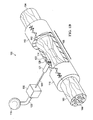
- FIG. 1A-B is a schematic, perspective view of a reduced pressure treatment system for repairing a severed ( FIG. 1A ) or a pinched nerve ( FIG. 1B ) including a nerve conduit and a first embodiment of a manifold with a section of the nerve conduit removed to show the manifold;
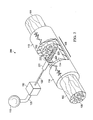
- FIG. 2 is a schematic of a reduced pressure treatment system for repairing a severed or partially severed nerve including a nerve conduit and a second embodiment of a manifold with a section of the nerve conduit removed to show the manifold;
- FIG. 3 is a schematic of a reduced pressure treatment system for repairing a severed or partially severed nerve including a nerve conduit and a third embodiment of a manifold with a section of the nerve conduit removed to show the manifold;
- FIG. 4 is a schematic, perspective view of the system shown in FIGS. 1-3 showing the nerve conduit enclosing the damaged nerve;
- FIG. 5 is a schematic view of a fluid control system for the system shown in FIGS. 1-3 .
- a reduced pressure therapy system 100 for applying reduced pressure at a tissue site in the body of a patient to repair a defect such as, for example, a damaged nerve.
- the damaged nerve may have been pinched, partially disconnected or severed, or partially degenerated as a result of disease.
- the damaged nerve in FIG. 1A is a severed nerve 102 having a proximal segment 104 and a distal segment 106 relative to the central nervous system (CNS) of the patient.
- FIG. 1B illustrates a reduced pressure therapy system and this case the damaged nerve is a pinched nerve 103 that has been damaged at a nerve damage site 108.
- the nerve has been pinched, partially severed or partially degenerated, but has not been completely severed.
- the nerve may be branched or unbranched at the nerve damage site 108.
- the term "nerve damage site” as used herein refers to a wound or defect located on or within any nerve tissue, including, but not limited to, a disconnected or partially disconnected nerve, a degenerated or partially degenerated nerve, and a compressed or pinched nerve.
- reduced pressure tissue treatment may be used to enhance repair or regrowth of existing nerve tissue or to facilitate growth or grafted or transplanted nerve tissue and/or cells.
- the reduced pressure therapy system 100 comprises a nerve conduit 110 that surrounds the pinched nerve 103 at the nerve damage site 108 with a section of the nerve conduit 110 removed to show the nerve damage site 108.
- the nerve conduit 110 is substantially tubular in shape and closes the nerve damage site 108 and a portion of the proximal segment 104 and the distal segment 106 that has not been damaged.
- the nerve conduit 110 has an exterior surface 113 and an inner surface 112 that forms a nerve gap 114 with the surface of the nerve damage site 108, i . e ., a luminal space between the inner surface 112 of the nerve conduit 110 and the surface of the nerve damage site 108.
- the reduced pressure therapy system 100 also comprises a reduced pressure source 115 for providing a reduced pressure and a manifold 120 fluidly coupled to the pressure source 115 via a first conduit 125.
- the manifold 120 is contained within a manifold chamber 121 having a flange 122 extending from one end of the manifold chamber 121 for securing the manifold chamber 121 to the nerve conduit 110.
- the other end of the manifold chamber 121 is connected to the first conduit 125 so that the manifold 120 is held in fluid communication with the first conduit 125.
- the manifold chamber 121 may be constructed of any biocompatible material that is substantially impermeable to preserve the manifold's 120 fluid communication between the nerve gap 114 and the first conduit 125.
- the manifold chamber 121 is secured to the nerve conduit 110 by the flange 122 such that the manifold 120 is in fluid communication with the nerve gap 114 surrounding the surface of the nerve damage site 108, but positioned outside of the nerve gap 114.
- the flange 122 is secured to the nerve conduit 110 with an adhesive.
- the flange 122 is detachably secured to the nerve conduit 110 such that the flange 122 and manifold chamber 121 can be removed from the nerve conduit 110 after reduced pressure therapy is complete.
- the manifold 120 extends through the wall of the nerve conduit 110 in direct fluid contact with the nerve gap 114.
- the flange 122 is secured to the exterior surface 113 of the nerve conduit 110 so that the manifold 120 is positioned adjacent to the exterior surface 113 to be in fluid communication with the nerve gap 114 via the porous wall of the nerve conduit 110.
- the manifold 120 may have a variety of shapes depending on the type of nerve damage, and depending on how the manifold is positioned in fluid contact with the nerve gap 114 around the nerve damage site 108.
- the manifold may also be coupled with a flange 122 that is in turn coupled to the exterior surface of the nerve conduit 113.
- the flange 122 forms a seal between the exterior surface of the nerve conduit 113 and the manifold 120 to prevent reduced pressure from being applied outside of the nerve conduit 110.
- the lumen of the nerve conduit and the nerve gap 114 may also contain a scaffold material (not shown) that provides a structure for tissue growth and repair.
- the reduced pressure therapy system 100 further comprises a canister 130 fluidly coupled between the reduced pressure source 115 and the manifold 120 via the conduit 125 to collect bodily fluids such as blood or exudate that are drawn from the nerve gap 114.
- the reduced pressure source 115 and the canister 130 are integrated into a single housing structure.
- Coupled includes direct coupling or indirect coupling via a separate object.
- the term “coupled” also encompasses two or more components that are continuous with one another by virtue of each of the components formed from the same piece of material.
- the term “coupled” may include chemical, mechanical, thermal, or electrical coupling.
- Fluid coupling means that fluid is in communication with designated parts or locations.
- reduced pressure generally refers to a pressure that is less than the ambient pressure at a tissue site that is subjected to treatment. In most cases, this reduced pressure will be less than the atmospheric pressure of the location at which the patient is located.
- vacuum and “negative pressure” may be used to describe the pressure applied to the tissue site, the actual pressure applied to the tissue site may be significantly greater than the pressure normally associated with a complete vacuum. Consistent with this nomenclature, an increase in reduced pressure or vacuum pressure refers to a relative reduction of absolute pressure, while a decrease in reduced pressure or vacuum pressure refers to a relative increase of absolute pressure.
- - ⁇ p means change in reduced pressure. As used herein, a greater ( i .
- Reduced pressure treatment typically applies reduced pressure at -5 mm Hg to -500 mm Hg, more usually -5 to -300 mm Hg, including but not limited to -50, -125 or -175 mm Hg.
- Reduced pressure may be constant at a particular pressure level or may be varied over time. For example, reduced pressure may be applied and stopped periodically or ramped-up or - down over time.
- a nerve damage site may be a wound or defect located on or within any nerve tissue including, for example, a completely severed nerve 102 having a proximal segment 104 and a distal segment 106 relative to the CNS of the patient as shown in FIG. 2 .
- the severed nerve 102 has been damaged at a nerve damage site 108 that has been completely severed.
- Another reduced pressure therapy system 200 for applying reduced pressure at the nerve damaged site 108 comprises similar components as the reduced pressure therapy system 100 as indicated by the same reference numerals.
- the reduced pressure therapy 200 comprises the nerve conduit 110 that surrounds the nerve damage site 108 and the severed ends of the severed nerve 102.
- the inner surface 112 of the nerve conduit 110 forms a nerve gap 114 between the severed ends of the severed nerve 102 within the nerve damage site 108, i . e ., a luminal space between the inner surface 112 of the nerve conduit 110 and the surface of the nerve damage site 108.
- the reduced pressure therapy system 200 also comprises a manifold 220 fluidly coupled to the pressure source 115 via the first conduit 125 and to the nerve gap 114.
- the manifold 220 is also partially contained with-in a manifold chamber 221 having a flange 222 for securing the manifold chamber 221 to the nerve conduit 110 and otherwise the same as the manifold chamber 121.
- the manifold 220 comprises a manifold protrusion 225 that extends into the nerve gap 114.
- the manifold 220 and manifold protrusion 225 may have a variety of shapes depending on the type of nerve damage, and depending on how the manifold is positioned in fluid contact with the nerve gap 114 around the nerve damage site 108.
- the flange 222 forms a seal between the exterior surface of the nerve conduit 113 and the manifold chamber 221 to prevent reduced pressure from being applied outside of the nerve conduit 110. In certain aspects, the flange 222 is secured to the nerve conduit 110 with an adhesive.
- the flange 222 is detachably secured to the nerve conduit 110 such that the flange 222 and manifold chamber 221 can be removed from the nerve conduit 110 after reduced pressure therapy is complete.
- the manifold protrusion 225 may be formed of a material so that the manifold protrusion also functions as a scaffold that provides a structure for tissue growth and repair.
- the reduced pressure therapy system 200 further comprises a canister 130 fluidly coupled between the reduced pressure source 115 and the manifold 220 via the conduit 125 to collect bodily fluids such as blood or exudate that are drawn from the nerve gap 114.
- the reduced pressure source 115 and the canister 130 are integrated into a single housing structure.
- FIG. 3 Still another reduced pressure therapy system 300 for applying reduced pressure at the nerve damaged site 108 is illustrated in FIG. 3 .
- a completely severed nerve 102 is shown having a proximal segment 104 and a distal segment 106 relative to the CNS.
- the severed nerve 102 has been damaged at a nerve damage site 108 that has been completely severed.
- the reduced pressure therapy 300 comprises the nerve conduit 110 that surrounds the nerve damage site 108 and the severed ends of the severed nerve 102.
- the inner surface 112 of the nerve conduit 110 forms a nerve gap 114 between the severed ends of the severed nerve 102 within the nerve damage site 108, i . e ., a luminal space between the inner surface 112 of the nerve conduit 110 and the surface of the nerve damage site 108.
- the reduced pressure therapy system 300 also comprises a manifold 320 fluidly coupled to the pressure source 115 via the first conduit 125 and to the nerve gap 114.
- the manifold 320 is also partially contained with-in manifold chambers 321 each having a flange 322 for securing the manifold chambers 321 to the nerve conduit 110 and otherwise the same as the manifold chamber 121 and 221.
- the manifold 320 extends into and through the nerve gap 114 and is secured on both sides of the nerve conduit 110 by a manifold chamber 321 and flange 322.
- the manifold 320 may have a variety of shapes depending on the type of nerve damage, and depending on how the manifold is positioned in fluid contact with the nerve gap 114 around the nerve damage site 108.
- the flanges 322 form a seal between the exterior surface of the nerve conduit 113 and the manifold chambers 321 to prevent reduced pressure from being applied outside of the nerve conduit 110.
- the flanges 322 are secured to the nerve conduit 110 with adhesive.
- one or both flanges 322 are detachably secured to the nerve conduit 110 such that the flange(s) 322 and manifold chamber(s) 321 can be removed from the nerve conduit 110 after reduced pressure therapy is complete.
- the manifold 320 may be formed of material so that the manifold also functions as a scaffold that provides a structure for tissue growth and repair.
- the reduced pressure therapy system 300 further comprises a canister 130 fluidly coupled between the reduced pressure source 115 and the manifold 320 via the conduit 125 to collect bodily fluids such as blood or exudate that are drawn from the nerve gap 114.
- the reduced pressure source 115 and the canister 130 are integrated into a single housing structure.
- the reduced pressure therapy system 300 further comprises a fluid source 150 in fluid communication with the manifold 320 via a second conduit 325. Accordingly, in a certain aspect fluid flows from the fluid source 150 into the manifold 320 and the nerve gap 114 and ultimately is captured by the canister 130. Fluid flow through the manifold transversing the site of nerve damage 108 may help to prevent clogging of the manifold.
- the nerve conduit 110 is shown in FIGS. 1-3 with a section removed, but shown as completely surrounding the nerve damage sites 108 as a closed nerve conduit 410 in FIG. 4 .
- the nerve conduit 110 may be sealed by utilizing one or more stitches 415 or any other fastening device known in the art.
- the nerve conduit 110 may be composed of a bioabsorbable or a bioinert material.
- Materials that may be used for nerve conduits include, without limitation, polylactic acid (PLA), polyglycolic acid (PGA), polylactide-co-glycolide (PLGA), polyvinylpyrrolidone, polycaprolactone, polycarbonates, polyfumarates, caprolactones, polyamides, polysaccharides (including alginates ( e .
- biological (e.g., purified or recombinant) materials may be used form nerve conduits including, but not limited to, fibrin, fibronectin or collagen (e.g., DURAMATRIX
- a nerve conduit 110 may be an unbroken substantially tubular structure fitted across a gap between a proximal and distal nerve stump such as depicted in FIG. 3 .
- substantially tubular nerve conduits also referred to as nerve guides, include without limitation NEURAGEN ® and NEUROFLEX TM collagen conduits.
- a nerve conduit may also be formed from a wrap that is positioned around a disconnected or damaged (e.g., pinched) nerve and sealed with a closure, such as a suture.
- wrap-type nerve conduits include, without limitation, NEUROMEND TM and NEURAWRAP TM collagen conduits.
- the nerve conduit is made of a material that encloses the damaged nerve, so as to exclude infiltration of non-nerve cells, such as glia.
- the nerve conduit material is permeable, thereby allowing fluid and protein factors to diffuse through the conduit.
- the pores in a nerve conduit may be sufficiently small so as to exclude the entry of cells into the conduit lumen (e.g., pores having an interior diameter or average interior diameter of between about 5 ⁇ m and 50 ⁇ m, 10 ⁇ m and 30 ⁇ m or 10 ⁇ m and 20 ⁇ m).
- the conduits comprise an internal diameter of less than about 6.0 mm, such as about 5, 4, 3, 2.5 or 2 mm.
- the reduced pressure therapy system 100, 200 or 300 may further comprise a pressure sensor 140 operably connected to the first conduit 125 to measure the reduced pressure being applied to the manifolds 120, 220, 320.
- the system further includes a control unit 145 electrically connected to the pressure sensor 140 and the reduced pressure source 115.
- the pressure sensor 140 measures the reduced pressure within the nerve gap 114, and also may indicate whether the first conduit 125 is occluded with blood or other bodily fluids.
- the pressure sensor 140 also provides feedback to control unit 145 which regulates the reduced pressure therapy being applied by the reduced pressure source 115 through the first conduit 125 to the manifolds 120, 220, 320.
- the reduced pressure therapy system 100, 200 or 300 may also comprise a fluid supply 150 fluidly coupled to the first conduit 125 via a second conduit 152 and operatively connected to the control unit 145.
- the fluid supply 150 may be used to deliver growth and/or healing agents to the nerve damage site 108 including, without limitation, an antibacterial agent, an antiviral agent, antibody, a cell-growth promotion agent, an irrigation fluid, or other chemically active agents.
- the system 100, 200, 300 further comprises a first valve 154 positioned in the second conduit 152 to control the flow of fluid therethrough, and a second valve 156 positioned in the first conduit 125 between the reduced pressure supply 115 and the juncture between the first conduit 125 and the second conduit 152 to control the flow of reduced pressure.
- the fluid supply 150 is directly coupled to the manifold 320 at the nerve tissue damage site 108 via the second conduit 325 as represented by the dashed lines.
- the control unit 145 is operatively connected to the first and second valves 154, 156 to control the delivery of reduced pressure and/or fluid from the fluid supply 150, respectively, to the manifolds 120, 220, 320 as required by the particular therapy being administered to the patient.
- the fluid supply 150 may deliver the liquids as indicated above, but may also deliver air to the manifolds 120, 220, 320 to promote healing and facilitate drainage at the site of the nerve damage site 108.
- manifold refers to a substance or structure that is provided to assist in directing reduced pressure to, delivering fluids to, or removing fluids from a tissue site.
- a manifold can include a plurality of flow channels or pathways that are interconnected to improve distribution of fluids provided to and removed from the area of tissue around the manifold.
- manifolds may include without limitation devices that have structural elements arranged to form flow channels, cellular foams such as open-cell foam, porous tissue collections, and liquids, gels and foams that include or cure to include flow channels.
- scaffold refers to a substance or structure applied to or in a wound or defect that provides a structural matrix for the growth of cells and/or the formation of tissue.
- a scaffold is often a three dimensional porous structure.
- the scaffold can be infused with, coated with, or comprised of cells, growth factors, extracellular matrix components, nutrients, integrins, or other substances to promote cell growth.
- a scaffold can take on characteristics of a manifold by directing flow through the matrix.
- a manifold can transmit flow to the scaffold and tissue; in the context of reduced pressure treatment, the manifold can be in fluid communication with the scaffold.
- the invention disclosed here discloses methods and apparatuses for controlling cellular-level based patterns of fluid flow that allow for control of patterned protein organization at a microscopic, nanoscopic, or mesoseopic scale amenable to provide a structured manifold and, optionally, a scaffold material for cellular migration, differentiation, and like behavior necessary for functional regeneration of tissues.
- the methods, scaffolds, manifolds, flow sources and systems disclosed herein provide an active mechanism by which to promote the endogenous deposition of proteins and organization of the provisional matrix with biochemical and physical cues to direct cellular colonization of a scaffold or tissue space.
- the present invention thus enhances current technology by exploiting the active force of directed fluid flow, providing a framework upon which to design manifolds and scaffolds based upon the need of the biology under the influence of fluid flow.
- Flow vectors and pathways are utilized to enhance protein deposition and cellular colonization.
- the systems provided herein are designed to promote establishment of a provisional matrix network with a seamless transition from the healthy tissue edges through a scaffold or tissue site to promote a functional tissue continuum.
- the apparatuses, methods and systems disclosed herein provide a means for active guidance of tissue regeneration through an implanted scaffold or within a tissue site to promote functional recovery.
- This active guidance occurs through mechanisms of controlled fluid flow, which can be used to initiate or augment the early stages of the body's own natural healing process; a manifold can provide the active guidance necessary to create a controlled fluid flow.
- the controlled flow vectors that the manifolds provide can be used to facilitate the directed influx of cells and proteins into a scaffold. Creation of specific flow pathways within a tissue site or scaffold can lead to patterned deposition of proteins, such as collagen and fibrin within the manifold, scaffold or tissue space.
- Biochemical cues from cytokines, growth factors, and cells bound within the provisional matrix can work in conjunction with the natural physical cues of the provisional matrix and extracellular matrix to guide the subsequent migration of endogenous cells during the repair stages of healing. These cues act as a form of track or gradient that emanates from surrounding healthy tissues and passes through the scaffolding or tissue space to facilitate a continuous guidance pathway for organized tissue regeneration.
- the invention concerns a new approach to wound healing, flow (or gradient) activated tissue engineering.
- this approach involves a source or generator of flow that forms a gradient for controlled movement of either endogenous or exogenous fluids into, out of, or through a tissue space for the organized deposition of proteins and/or spatial concentration of cytokines and growth factors, with subsequent formation of a directionally oriented provisional matrix.
- the tissue space being defined here includes, but is not limited to, the region surrounding a site of nerve tissue damage.
- Fluid flow into, through, or out of the nerve tissue space can be refined and directed through the inclusion of additional elements to the system including manifolds and/or scaffolds.
- the coordinated elements of the system are designed to create flow parameters, pathways, and patterns sufficiently detailed in scale as to be able to influence and direct the controlled adsorption of proteins, the organization of matrix, and organized colonization of specific cell types. Individual elements of the system are as follows.
- Source or generator of flow Flow is induced into, through, or out of the nerve tissue space by methods or apparatuses that introduce changes in mechanical, chemical, and/or electrical potentials. These generators of flow provide either a gradient or a change in potential from the site or reservoir of endogenous or exogenous fluids to the placement position of the flow generator or its extension element ( i . e ., manifold or scaffold).
- the source of flow comprises a source of reduced pressure.
- Systems and apparatuses according to the invention may also comprise valves or arrays of valves that control the application and amount of negative pressure applied to the manifold.
- nerve conduits and/or manifolds described herein comprise a pressure sensor.
- the amount of negative pressure applied by a source is regulated based on the amount of negative pressure that is sensed in the manifold or nerve conduit or at the site of tissue damage.
- Manifold The flow generators are the driving force for stimulating the flow of fluids.
- Manifolds are apparatuses for refining the pattern of flow between the source or generator of flow and the tissue space.
- the macroscale level of flow is refined by specialized manifolds utilized for directed localization to a single point or to a plurality of selectively positioned points for creating initiation sites for microscale flow pathways within the manifold/scaffold and, ultimately, the tissue space.
- the manifold may also serve as a conduit for the removal of fluids from and as an apparatus for the delivery of exogenous fluids to the tissue space.
- a manifold generally refers to a physical substance or structure that serves to assist in applying and translating a mechanical, chemical, electrical or similar alterations into changes in the flow of a fluid, herein defined as the movement of liquids, gases, and other deformable substances such as proteins, cells, and other like moieties.
- this physical device includes a single point or plurality of points for the egress or evacuation of pressure, fluids, and like substances capable of translating the movement of fluids in a scaffold, as defined above. This can include, but is not limited to, the introduction of exogenous factors such as cells and/or therapeutic moieties into the scaffold through the lumen or plurality of lumens present in the manifold.
- a manifold includes a single point or plurality of points for the ingress or introduction of fluid from the scaffold back towards the point source of flow.
- Flow distributed by the manifold can direct the movement of endogenous proteins, growth factors, cytokines, and cells from their resident locations within the host to the tissue space or scaffold in an organized manner.
- the establishment of flow along these pathways leads to the deposition of proteins and provisional matrix that creates an interfacial endogenous network connecting the host to the scaffold. Extensions of this matrix can be established within the scaffold through selective positioning of the manifold flow initiation sites with flow promoting scaffolding designs.
- the organized protein deposition and provisional matrix provide a biochemical and physical framework that stimulates the attachment and migration of cells along directed pathways throughout the scaffold and the tissue space.
- the resulting endogenous network of proteins, growth factors, and cells provides a foundation upon which subsequent phases of the body's own tissue repair and regeneration mechanisms can build.
- Flow generating sources include, but are not limited to generators of negative pressure; generators of positive pressure; and generators of osmotic flow.
- the flow gradient established in the manifold may be further refined through the scaffold, to deliver a flow gradient to the scaffold to optimize flow through the scaffold as needed for the particular defect.
- Many of the embodiments disclosed herein are manifolds capable of translating changes in pressure and the like into controlled movement of fluids, optionally through a physical scaffold, for the purposes of directed tissue regeneration. These embodiments are generally specified for a particular application in the regeneration of specific tissues, but are not limited to a particular tissue therein.
- alterations in the aforementioned mechanical, chemical, or electrical impetus must be translated from the singular gradient source toward a physical substrate or scaffold to elicit cellular-level changes in protein adsorption, matrix organization, cell migration, and other tissue regeneration-related behaviors.
- These alterations are multivariate in nature and can include mechanical changes that elicit a physical change in pressure applied to the scaffold as applied to the site of the wound or desired site of tissue regeneration, chemical changes that elicit a gradient in protein and/or ion concentrations, which result in the creation of osmotic gradients capable of inducing flow, or electrical changes that create a gradient of current/ion exchange allowing for propagation of electrical signals from the point source.
- the manifold comprises a physical structure in close apposition to or within the contents of a scaffold and serves to propagate an alteration in a physical parameter, whether it be mechanical, chemical, electrical, or something similar in nature, for the means of directing these changes from its point source to the scaffolding material.
- the placement of this manifold with respect to its location with regard to that of the scaffold may be of crucial importance for facilitating controlled and directed regeneration of specific tissue types. For example, in peripheral nerve where regeneration primarily occurs in a unidirectional manner from the proximal to distal nerve stumps, it may be important to place the manifold along the length of a nerve conduit more towards it distal end to help direct regeneration towards that end. However, it may also be important to not place the manifold at the most distal aspect of the scaffold/conduit as soluble factors derived from the distal stump have been shown to be important for directing nerve regeneration towards its source.
- Manifolds may be composed of a bioabsorbable or bioinert material. Examples include non-bioabsorbable materials such as medical grade silicone polymers, metals, polyvinylchloride (PVC), and polyurethane. Bioabsorbable polymers such as collagen, polylactic acid (PLA), polyglycolic acid (PGA), polylactide-co-glycolide (PLGA), a polysaccharide, a hydroxyapatite, or a polyethylene glycol, or combinations thereof, can also be used. Some manifolds are also a mix of non-bioresorbable and bioresorbable materials. In general material used for a scaffold may also be used to compose a manifold and such materials are further detailed below. In certain aspects, manifold materials are structured to include a high void fraction for improved bioabsorption properties.
- PVC polyvinylchloride
- Bioabsorbable polymers such as collagen, polylactic acid (PLA), polyglycolic acid (PGA), polyl
- Manifold support structures may be composed of any acceptable biocompatible material.
- a support structure will typically be impermeable and surround the manifold so as to maintain manifold pressure.
- a portion of the support couples the manifold and the nerve conduit.
- a flange is attached to the exterior surface of a nerve conduit with an adhesive such as a fibrin glue, cyanoacrylate, or other biologically derived adhesive.
- a support may also be connected to a nerve conduit via reversible mechanisms other than an adhesive, such as chemical, thermal, osmotic, mechanical (snap or other interference fit, threaded, etc), magnetic, and electrostatic mechanisms.
- the manifold may be used to deliver agents that reverse the action of the binding mechanism in order to detach the support from the nerve conduit (e.g ., upon completion of therapy).
- electrostatic binding may be released through introduction of salt solutions or biocompatible solvents may be used to release adhesives.
- a scaffold for use according to the invention is coupled to a manifold, provides physical guidance to direct the pathway of fluid flow in the tissue site, creating avenues for the movement and migration of adhesive proteins and cells, respectively, which are in tern integral to the establishment of a provisional matrix in predetermined patterns of organization within the tissue space.
- the methods and apparatuses described for fluid flow-induced and gradientinduced generation of tissues have direct implications in the design of the scaffolds.
- scaffolds serve to refine the pathways of fluid flow within the tissue space to cellular level patterns from the fluid source to the point(s) of flow initiation within the manifold.
- a scaffold may embody characteristics of a manifold or be combined in conjunction with a manifold for refinement of the flow pathways within the tissue site.
- a scaffold is a reticulated structure comprising high void fraction for improved bioabsorption properties.
- Nonlimiting examples of suitable scaffold materials include extracellular matrix proteins such as fibrin, collagen or fibronectin, and synthetic or naturally occurring polymers, including bioabsorbable or non-absorbable polymers, such as polylactic acid (PLA), polyglycolic acid (PGA), polylactide-co-glycolide (PLGA), polyvinylpyrrolidone, polycaprolactone, polycarbonates, polyfumarates, caprolactones, polyamides, polysaccharides (including alginates ( e.g ., calcium alginate) and chitosan), hyaluronic acid, polyhydroxybutyrate, polyhydroxyvalerate, polydioxanone, polyorthoesthers, polyethylene glycols, poloxamers, polyphosphazenes, polyanhydrides, polyamino acids, polyacetals, polycyanoacrylates, polyurethanes, polyacrylates, ethylene-vinyl acetate polymers and other acyl substituted
- the scaffold can also comprise ceramics such as hydroxyapatite, coralline apatite, calcium phosphate, calcium sulfate, calcium carbonate or other carbonates, bioglass, allografts, autografts, xenografts, decellularized tissues, or composites of any of the above.
- the scaffold comprises collagen, polylactic acid (PLA), polyglycolic acid (PGA), polylactide-co-glycolide (PLGA), a polyurethane, a polysaccharide, an hydroxyapatite, or a polytherylene glycol.
- the scaffold can comprise combinations of any two, three or more materials, either in separate or multiple areas of the scaffold, combined noncovalently, or covalently (e.g ., copolymers such as a polyethylene oxide-polypropylene glycol block copolymers, or terpolymers), or combinations thereof.
- Suitable matrix materials are discussed in, for example, Ma and Elisseeff, 2005, and Saltzman, 2004.
- bioactive agents may, in some cases, be incorporated directly onto a manifold or scaffold material ( i . e ., to generate a bioactive manifold and/or scaffold).
- a manifold or scaffold material i . e ., to generate a bioactive manifold and/or scaffold.
- agents that facilitate tissue growth such as collagen or fibrin may be directly incorporated onto or into a manifold or scaffold material.
- immune regulator agents such as rapamycin may be incorporated into manifold or scaffold structures.
- soluble bioactive agents may be introduced at a site of tissue damage by virtue of the flow through the tissue site.
- a manifold may be in fluid communication with a fluid source and a bioactive agent may be introduced into the fluid source and thereby into the manifold and damaged nerve tissue.
- bioactive growth factors for various applications are growth hormone (GH), a bone morphogenetic protein (BMP), transforming growth factor- ⁇ (TGF- ⁇ ), a TGF- ⁇ , a fibroblast growth factor (FGF), granulocyte-colony stimulating factor (G-CSF), granulocyte/macrophage-colony stimulating factor (GM-CSF), epidermal growth factor (EGF), platelet derived growth factor (PDGF), insulin-like growth factor (IGF), vascular endothelial growth factor (VEGF), hepatocyte growth factor/scatter factor (HGF/SF), an interleukin, tumor necrosis factor- ⁇ (TNF- ⁇ ) or nerve growth factor (NGF).
- GH growth hormone
- BMP bone morphogenetic protein
- TGF- ⁇ transforming growth factor- ⁇
- TGF- ⁇ TGF- ⁇
- FGF transforming growth factor- ⁇
- FGF transforming growth factor- ⁇
- TGF- ⁇ TGF- ⁇
- FGF transforming growth factor
- Nerve tissue repair and engineering The apparatuses and systems disclosed herein can be used for nerve tissue repair and engineering in various contexts including the following.
- a generator of flow may be combined with manifolds and/or scaffolds to direct the regeneration of lost tissue at a site of injury or compromised function.
- Tissues lost from traumatic injury, surgery, burns, or other causes e . g ., infection or autoimmune disease
- Functional nerve tissue is directed to regenerate.
- a generator of flow may be combined with manifolds and/or scaffolds to retard disease progression of an affected nerve tissue such as occurs, e . g ., in autoimmune disease.
- a generator of flow may be combined with manifolds and/or scaffolds to maintain the viability of explanted tissues either for in vitro study, ex vivo scaffold or implant preparation, or in vivo transplant.
- a generator of flow combined with a manifold may be used to provide nutrient flow to the tissue and to control waste removal from the tissue.
- a generator of flow may be combined with manifolds and/or scaffolds to promote the expansion of existing tissues.
- the methods, scaffolds, manifolds, flow sources and systems of the invention can be used to direct the growth of tissues where additional tissue quantity is needed or desired.
- a generator of flow may be combined with manifolds and/or scaffolds to accelerate the rate of tissue formation within a natural healing response.
- the methods, scaffolds, manifolds, flow sources, and systems of the invention may be used to accelerate tissue growth by augmenting formation of provisional matrices, facilitating its stable positioning, and aiding in recruitment of cells to the tissue space.
- a generator of flow may be combined with manifolds and/or scaffolds to stimulate the differentiation of stem cells or other pluripotent cells into specific lineages.
- Application of flow using the methods, scaffolds, manifolds, flow sources and systems of the invention may be used to direct pluripotent cells into specific cell lineages needed to foster growth in the tissue space.
- a generator of flow may be combined with manifolds and/or scaffolds to introduce exogenous growth factors, proteins, cells, or pharmaceutical agents into the tissue space to augment tissue repair, regeneration, and/or maintenance.
- a generator of flow may be combined with manifolds and/or scaffolds to facilitate formation of matrices in vitro that may subsequently be used for in vivo transplantation.
- a generator of flow may be combined with manifolds and/or scaffolds to promote integration of transplanted tissue into the host environment. This can be applied to autograft, allograft, or xenograft transplants.
- a flow generator may be combined with manifolds and/or scaffolds to guide the directed deposition and orientation of ECM expressed by cells and tissues.
- the directed orientation of ECM has an impact in organizing and directing the attachment and colonization of subsequent cell layers and tissues.
Landscapes
- Health & Medical Sciences (AREA)
- Life Sciences & Earth Sciences (AREA)
- Heart & Thoracic Surgery (AREA)
- Veterinary Medicine (AREA)
- Public Health (AREA)
- General Health & Medical Sciences (AREA)
- Animal Behavior & Ethology (AREA)
- Engineering & Computer Science (AREA)
- Biomedical Technology (AREA)
- Vascular Medicine (AREA)
- Surgery (AREA)
- Hematology (AREA)
- Anesthesiology (AREA)
- Chemical & Material Sciences (AREA)
- Nuclear Medicine, Radiotherapy & Molecular Imaging (AREA)
- Medicinal Chemistry (AREA)
- Medical Informatics (AREA)
- Molecular Biology (AREA)
- Neurology (AREA)
- Dermatology (AREA)
- Transplantation (AREA)
- Organic Chemistry (AREA)
- General Chemical & Material Sciences (AREA)
- Chemical Kinetics & Catalysis (AREA)
- Pharmacology & Pharmacy (AREA)
- Bioinformatics & Cheminformatics (AREA)
- Epidemiology (AREA)
- Oral & Maxillofacial Surgery (AREA)
- Neurosurgery (AREA)
- Biophysics (AREA)
- Dispersion Chemistry (AREA)
- Otolaryngology (AREA)
- Materials For Medical Uses (AREA)
- Prostheses (AREA)
- Media Introduction/Drainage Providing Device (AREA)
- External Artificial Organs (AREA)
- Treatment Of Fiber Materials (AREA)
Claims (15)
- Vorrichtung zum Versorgen eines Defektes an einer Gewebestelle eines Nervs mit verringertem Druck von einer Quelle für verringerten Druck, wobei die Vorrichtung Folgendes umfasst:ein Nervenrohr mit einer Röhrenform, das Wände aufweist, beinhaltend eine Außenwand und eine die Gewebestelle umgebende luminale Wand zum Aufnehmen von Fluiden in einem luminalen Raum zwischen der Gewebestelle und der luminalen Wand,einen Verteiler mit einem zylindrischen Hauptteil, der Flächen aufweist, beinhaltend eine Seitenwandfläche und zwei Endwandflächen, wobei eine erste Endwandfläche der beiden Endwandflächen zum Empfangen verringerten Drucks bestimmt ist, eine Fluidkontaktfläche, beinhaltend einen ersten Abschnitt der Flächen des zylindrischen Hauptteils, bei dem es sich nicht um die erste Endwandfläche handelt, zur Fluidverbindung mit dem luminalen Raum, und eine Stützfläche, beinhaltend einen zweiten Abschnitt der Fläche des zylindrischen Hauptteils, bei dem es sich nicht um die erste Endwandfläche und die Fluidkontaktfläche handelt, undeine erste Stützstruktur mit einer Röhrenform zum Umschließen der Stützfläche, einem ersten Endabschnitt zur Kopplung der ersten Endwandfläche mit der Quelle für verringerten Druck und einem zweiten Endabschnitt zur Kopplung des Verteilers mit dem Nervenrohr in einer radialen Richtung in Bezug auf die luminale Wand.
- Vorrichtung nach Anspruch 1, wobei die Fluidkontaktfläche eine zweite Endwandfläche der beiden Endwandflächen umfasst, die angrenzend an die luminale Wand angeordnet ist, um mit dem luminalen Raum in Fluidverbindung zu stehen.
- Vorrichtung nach Anspruch 1, wobei das Nervenrohr porös ist und die Fluidkontaktfläche eine zweite Endwandfläche der beiden Endwandflächen umfasst, die angrenzend an die Außenfläche des Nervenrohrs angeordnet ist, um durch die Wände des Nervenrohrs hindurch mit dem luminalen Raum in Fluidverbindung zu stehen.
- Vorrichtung nach Anspruch 2 oder 3, wobei das Nervenrohr ein Material umfasst, das im Allgemeinen undurchlässig für Fluide aus Gewebe ist, welches das Nervenrohr umgibt.
- Vorrichtung nach Anspruch 1, wobei die Fluidkontaktfläche eine zweite Endwandfläche der beiden Endwandflächen und einen Abschnitt der Seitenwandfläche umfasst, der an die zweite Endwandfläche angrenzt und sich von der luminalen Wand in den luminalen Raum erstreckt.
- Vorrichtung nach Anspruch 1, wobei die Fluidkontaktfläche einen Abschnitt der Seitenwandfläche, der sich von einer ersten Seite der luminalen Wand durch den luminalen Raum hindurch zu einer zweiten Seite der luminalen Wand erstreckt, und eine zweite Endwandfläche der beiden Endwandflächen umfasst, um ein Fluid von einer Fluidquelle zu empfangen, wobei die Vorrichtung ferner Folgendes umfasst:eine zweite Stützstruktur mit einer Röhrenform zum Umschließen der Stützfläche angrenzend an die zweite Endwandfläche, einem ersten Endabschnitt zur Kopplung der zweiten Endwandfläche mit der Fluidquelle und einem zweiten Endabschnitt zur Kopplung des Verteilers mit dem Nervenrohr.
- Vorrichtung nach Anspruch 1, wobei der Verteiler eine poröse Struktur umfasst, wobei die Porengröße ausreichend klein ist, um auszuschließen, dass Zellen in den Verteiler eindringen.
- Vorrichtung nach Anspruch 1, wobei der Verteiler als Gerüst dient, das das Gewebewachstum oder -neuwachstum unterstützt.
- Vorrichtung nach Anspruch 1, wobei das Nervenrohr Poren umfasst, die ausreichend klein sind, um auszuschließen, dass Zellen in den luminalen Raum eindringen.
- Vorrichtung nach Anspruch 1, wobei der Verteiler in Bezug auf die Gewebestelle an der distalen Seite des Nervenrohrs angeordnet ist.
- Vorrichtung nach Anspruch 1, wobei der luminale Raum des Nervenrohrs ein Gerüst beinhaltet, das das Gewebewachstum oder -neuwachstum unterstützt.
- Vorrichtung nach Anspruch 11, wobei das Gerüst aus biologischem Material besteht, das aus Fibrin oder Kollagen ausgewählt ist.
- Vorrichtung nach Anspruch 12, wobei das Gerüstmaterial ein bioaktives Mittel umfasst.
- Vorrichtung zum Versorgen eines Defektes an einer Gewebestelle eines Nervs mit verringertem Druck, wobei die Vorrichtung Folgendes umfasst:eine Vorrichtung nach einem der vorhergehenden Ansprüche und eine Quelle für verringerten Druck, wobei die erste Endwandfläche der beiden Endwandflächen in Fluidverbindung mit der Quelle für verringerten Druck steht.
- Vorrichtung nach Anspruch 14, ferner eine Fluidquelle in Fluidverbindung mit dem Verteiler der Vorrichtung zum Versorgen des luminalen Raumes mit einem Fluid umfassend.
Applications Claiming Priority (5)
| Application Number | Priority Date | Filing Date | Title |
|---|---|---|---|
| US14205308P | 2008-12-31 | 2008-12-31 | |
| US14206508P | 2008-12-31 | 2008-12-31 | |
| US23469209P | 2009-08-18 | 2009-08-18 | |
| US23877009P | 2009-09-01 | 2009-09-01 | |
| PCT/US2009/069713 WO2010078345A2 (en) | 2008-12-31 | 2009-12-29 | System for providing fluid flow to nerve tissues |
Publications (3)
| Publication Number | Publication Date |
|---|---|
| EP2370118A2 EP2370118A2 (de) | 2011-10-05 |
| EP2370118A4 EP2370118A4 (de) | 2012-09-05 |
| EP2370118B1 true EP2370118B1 (de) | 2013-09-04 |
Family
ID=42285800
Family Applications (5)
| Application Number | Title | Priority Date | Filing Date |
|---|---|---|---|
| EP09837120.6A Not-in-force EP2370142B1 (de) | 2008-12-31 | 2009-12-29 | System zur bereitstellung eines flüssigkeitsflusses in nervengewebe |
| EP09837117.2A Not-in-force EP2370117B1 (de) | 2008-12-31 | 2009-12-29 | System zur bereitstellung eines flüssigkeitsflusses in nervengewebe |
| EP14197314.9A Not-in-force EP2848268B1 (de) | 2008-12-31 | 2009-12-29 | Papierrollengerüste |
| EP09837119.8A Not-in-force EP2370118B1 (de) | 2008-12-31 | 2009-12-29 | System zur bereitstellung eines flüssigkeitsflusses in nervengewebe |
| EP09837122.2A Not-in-force EP2370119B1 (de) | 2008-12-31 | 2009-12-29 | Papierrollengerüste |
Family Applications Before (3)
| Application Number | Title | Priority Date | Filing Date |
|---|---|---|---|
| EP09837120.6A Not-in-force EP2370142B1 (de) | 2008-12-31 | 2009-12-29 | System zur bereitstellung eines flüssigkeitsflusses in nervengewebe |
| EP09837117.2A Not-in-force EP2370117B1 (de) | 2008-12-31 | 2009-12-29 | System zur bereitstellung eines flüssigkeitsflusses in nervengewebe |
| EP14197314.9A Not-in-force EP2848268B1 (de) | 2008-12-31 | 2009-12-29 | Papierrollengerüste |
Family Applications After (1)
| Application Number | Title | Priority Date | Filing Date |
|---|---|---|---|
| EP09837122.2A Not-in-force EP2370119B1 (de) | 2008-12-31 | 2009-12-29 | Papierrollengerüste |
Country Status (12)
| Country | Link |
|---|---|
| US (11) | US9351882B2 (de) |
| EP (5) | EP2370142B1 (de) |
| JP (4) | JP5611979B2 (de) |
| KR (4) | KR20110102924A (de) |
| CN (4) | CN102264405B (de) |
| AU (4) | AU2009335117B2 (de) |
| CA (4) | CA2745676A1 (de) |
| MX (4) | MX2011006992A (de) |
| RU (4) | RU2011122547A (de) |
| SG (4) | SG172025A1 (de) |
| TW (4) | TW201028184A (de) |
| WO (4) | WO2010078345A2 (de) |
Families Citing this family (128)
| Publication number | Priority date | Publication date | Assignee | Title |
|---|---|---|---|---|
| EP1656070B1 (de) * | 2003-08-11 | 2009-09-23 | Wilson-Cook Medical Inc. | Chirurgisches implantat |
| WO2011106454A2 (en) * | 2010-02-23 | 2011-09-01 | L-Vad Technology, Inc. | Vacuum assisted percutaneous appliance |
| US9820888B2 (en) | 2006-09-26 | 2017-11-21 | Smith & Nephew, Inc. | Wound dressing |
| WO2008140413A1 (en) * | 2007-05-15 | 2008-11-20 | NEUROGEN MEDICAL INNOVATION I UMEå | Fibrin-based nerve repair conduit and method of producing the same |
| GB0804654D0 (en) * | 2008-03-13 | 2008-04-16 | Smith & Nephew | Vacuum closure device |
| RU2011122547A (ru) * | 2008-12-31 | 2013-02-10 | КейСиАй ЛАЙСЕНЗИНГ, ИНК. | Рулонные каркасы для ткани |
| GB0902368D0 (en) | 2009-02-13 | 2009-04-01 | Smith & Nephew | Wound packing |
| DE102009057962B4 (de) * | 2009-12-11 | 2012-09-20 | Karlsruher Institut für Technologie | Nervenprothese und Verfahren zur Herstellung einer Nervenprothese |
| KR101679426B1 (ko) * | 2010-04-30 | 2016-11-25 | 에스케이이노베이션 주식회사 | 미전환유를 이용한 고급 윤활기유의 제조 방법 |
| US8623047B2 (en) * | 2010-04-30 | 2014-01-07 | Kci Licensing, Inc. | System and method for sealing an incisional wound |
| US8758374B2 (en) | 2010-09-15 | 2014-06-24 | University Of Utah Research Foundation | Method for connecting nerves via a side-to-side epineurial window using artificial conduits |
| US9408956B2 (en) * | 2010-09-24 | 2016-08-09 | Kci Licensing, Inc. | Cellular control and tissue regeneration systems and methods |
| US20160346444A1 (en) * | 2010-09-24 | 2016-12-01 | Kci Licensing, Inc. | Cellular control and tissue regeneration systems and methods |
| US9884909B2 (en) | 2010-12-01 | 2018-02-06 | Alderbio Holdings Llc | Anti-NGF compositions and use thereof |
| US9067988B2 (en) | 2010-12-01 | 2015-06-30 | Alderbio Holdings Llc | Methods of preventing or treating pain using anti-NGF antibodies |
| US9539324B2 (en) | 2010-12-01 | 2017-01-10 | Alderbio Holdings, Llc | Methods of preventing inflammation and treating pain using anti-NGF compositions |
| US11214610B2 (en) | 2010-12-01 | 2022-01-04 | H. Lundbeck A/S | High-purity production of multi-subunit proteins such as antibodies in transformed microbes such as Pichia pastoris |
| KR20190112175A (ko) | 2010-12-01 | 2019-10-02 | 앨더바이오 홀딩스 엘엘씨 | 항―ngf 조성물 및 그의 용도 |
| US9078878B2 (en) | 2010-12-01 | 2015-07-14 | Alderbio Holdings Llc | Anti-NGF antibodies that selectively inhibit the association of NGF with TrkA, without affecting the association of NGF with p75 |
| US9421132B2 (en) | 2011-02-04 | 2016-08-23 | University Of Massachusetts | Negative pressure wound closure device |
| EP2670312B1 (de) | 2011-02-04 | 2020-11-18 | University of Massachusetts | Unterdruck-wundverschlussvorrichtung |
| US9302034B2 (en) | 2011-04-04 | 2016-04-05 | Smith & Nephew, Inc. | Negative pressure wound therapy dressing |
| WO2012142473A1 (en) | 2011-04-15 | 2012-10-18 | University Of Massachusetts | Surgical cavity drainage and closure system |
| AU2012262311B2 (en) | 2011-05-31 | 2016-04-28 | Lifecell Corporation | Adipose tissue matrices |
| US10842494B2 (en) | 2011-10-17 | 2020-11-24 | University Of Utah Research Foundation | Methods and devices for connecting nerves |
| WO2013066619A1 (en) * | 2011-10-17 | 2013-05-10 | University Of Utah Research Foundation | Methods and devices for connecting nerves |
| JP6202686B2 (ja) * | 2011-11-18 | 2017-09-27 | ケーシーアイ ライセンシング インコーポレイテッド | 収縮領域と膨張領域を持つ多孔質基材を有する組織治療システムおよび方法 |
| US10220125B2 (en) | 2012-02-03 | 2019-03-05 | Smith & Nephew Plc | Apparatuses and methods for wound therapy |
| CA3260090A1 (en) | 2012-03-12 | 2025-05-16 | Smith & Nephew Plc | Reduced pressure apparatus and methods |
| MX2014014266A (es) | 2012-05-22 | 2015-06-23 | Smith & Nephew | Aparatos y metodos para terapia de heridas. |
| AU2013291693B2 (en) | 2012-05-22 | 2018-03-01 | Smith & Nephew Plc | Wound closure device |
| CN104736110B (zh) | 2012-05-24 | 2019-05-31 | 史密夫和内修有限公司 | 用于利用负压来处理和封闭伤口的装置和方法 |
| US11090338B2 (en) | 2012-07-13 | 2021-08-17 | Lifecell Corporation | Methods for improved treatment of adipose tissue |
| RU2015104581A (ru) | 2012-07-16 | 2016-09-10 | Смит Энд Нефью, Инк. | Устройство для закрытия раны с использованием отрицательного давления |
| CA2885327A1 (en) | 2012-09-26 | 2014-04-03 | Lifecell Corporation | Processed adipose tissue |
| TWI507181B (zh) * | 2012-10-30 | 2015-11-11 | Univ Nat Cheng Kung | 腹腔內力學刺激製造高強度組織工程材料的系統 |
| WO2014075102A1 (en) | 2012-11-12 | 2014-05-15 | Kci Licensing, Inc. | Externally-applied wound dressings and closures |
| DE102012025124A1 (de) | 2012-12-21 | 2014-06-26 | Paul Hartmann Ag | Saugkörper zur therapeutischen Behandlung einer Wunde mittels Unterdruck |
| EP3659633A1 (de) * | 2013-02-06 | 2020-06-03 | LifeCell Corporation | Verfahren für lokalisierte modifizierung von gewebeprodukten |
| WO2014137824A1 (en) * | 2013-03-07 | 2014-09-12 | Life Sciences Llc | Apparatus & method for wound infection prevention |
| US10124098B2 (en) | 2013-03-13 | 2018-11-13 | Smith & Nephew, Inc. | Negative pressure wound closure device and systems and methods of use in treating wounds with negative pressure |
| BR112015021123A2 (pt) | 2013-03-14 | 2017-07-18 | Smith & Nephew | enchimentos compressíveis de ferimentos e sistemas e métodos para o uso no tratamento de ferimentos com pressão negativa |
| US9408756B2 (en) | 2013-03-15 | 2016-08-09 | Acclarent, Inc. | Nasal fluid management device |
| US20140276654A1 (en) * | 2013-03-15 | 2014-09-18 | Acclarent, Inc. | Nasal Suction Device |
| US9408955B2 (en) | 2013-03-15 | 2016-08-09 | Acclarent, Inc. | Nasal fluid management device |
| US9604041B2 (en) | 2013-03-15 | 2017-03-28 | Acclarent, Inc. | Nasal fluid management device |
| US10687983B2 (en) * | 2013-04-17 | 2020-06-23 | Mölnlycke Health Care Ab | Wound pad |
| CA2926470C (en) | 2013-10-21 | 2023-03-14 | Smith & Nephew, Inc. | Negative pressure wound closure device |
| RU2016133734A (ru) | 2014-01-21 | 2018-03-02 | СМИТ ЭНД НЕФЬЮ ПиЭлСи | Устройства для лечения ран |
| CN110974539A (zh) | 2014-01-21 | 2020-04-10 | 史密夫及内修公开有限公司 | 用于负压伤口治疗的可塌缩敷料 |
| CN105213076A (zh) * | 2014-06-12 | 2016-01-06 | 微创心脉医疗科技(上海)有限公司 | 一种人造瘤颈及其制备方法 |
| CN104127215B (zh) * | 2014-07-18 | 2016-12-07 | 韦有芳 | 一种双层的硅胶神经导管 |
| US10898388B2 (en) | 2015-04-27 | 2021-01-26 | Smith & Nephew Plc | Reduced pressure apparatuses and methods |
| US9855317B2 (en) | 2015-04-27 | 2018-01-02 | Reflex Medical, Inc. | Systems and methods for sympathetic cardiopulmonary neuromodulation |
| WO2016176513A1 (en) | 2015-04-29 | 2016-11-03 | Smith & Nephew Inc. | Negative pressure wound closure device |
| US20170028111A1 (en) | 2015-07-29 | 2017-02-02 | Innovative Therapies, Inc. | Wound therapy device pressure monitoring and control system |
| WO2017049284A1 (en) * | 2015-09-17 | 2017-03-23 | Hollister Incorporated | Scaffold-based wound care delivery system and method |
| US10814049B2 (en) | 2015-12-15 | 2020-10-27 | University Of Massachusetts | Negative pressure wound closure devices and methods |
| US10575991B2 (en) | 2015-12-15 | 2020-03-03 | University Of Massachusetts | Negative pressure wound closure devices and methods |
| WO2017139487A1 (en) | 2016-02-09 | 2017-08-17 | Northwind Medical, Inc. | Methods, agents, and devices for local neuromodulation of autonomic nerves |
| CA3014549C (en) * | 2016-02-26 | 2024-09-17 | Neuronano Ab | METHOD FOR IMPLANTING CELL AGGREGATES AND TISSUE FRAGMENTS |
| JP6911043B2 (ja) | 2016-03-07 | 2021-07-28 | スミス アンド ネフュー ピーエルシーSmith & Nephew Public Limited Company | 陰圧源が創傷被覆材内に一体化された創傷治療装置及び方法 |
| US11285047B2 (en) | 2016-04-26 | 2022-03-29 | Smith & Nephew Plc | Wound dressings and methods of use with integrated negative pressure source having a fluid ingress inhibition component |
| EP3452129B1 (de) | 2016-05-03 | 2022-03-23 | Smith & Nephew plc | Aktivierung und steuerung einer unterdruckwundtherapievorrichtung |
| US11305047B2 (en) | 2016-05-03 | 2022-04-19 | Smith & Nephew Plc | Systems and methods for driving negative pressure sources in negative pressure therapy systems |
| JP6975170B2 (ja) | 2016-05-03 | 2021-12-01 | スミス アンド ネフュー ピーエルシーSmith & Nephew Public Limited Company | 陰圧療法システムにおける陰圧源への電力伝送の最適化 |
| WO2017210109A1 (en) | 2016-06-03 | 2017-12-07 | Lifecell Corporation | Methods for localized modification of tissue products |
| WO2018005848A1 (en) | 2016-06-29 | 2018-01-04 | Bright, Corinne | Treatment of sepsis and related inflammatory conditions by local neuromodulation of the autonomic nervous system |
| US10639398B2 (en) | 2016-07-05 | 2020-05-05 | Lifecell Corporation | Tissue matrices incorporating multiple tissue types |
| AU2017315129B2 (en) | 2016-08-25 | 2022-10-27 | Smith & Nephew Plc | Absorbent negative pressure wound therapy dressing |
| US11135351B2 (en) | 2016-08-30 | 2021-10-05 | Smith & Nephew Plc | Systems and methods for applying reduced pressure therapy |
| US11096832B2 (en) | 2016-09-27 | 2021-08-24 | Smith & Nephew Plc | Wound closure devices with dissolvable portions |
| EP3519001B1 (de) | 2016-09-30 | 2025-05-21 | Smith & Nephew plc | Vorrichtungen und verfahren zur unterdruckwundbehandlung mit integrierter elektronik |
| US12447260B2 (en) | 2016-09-30 | 2025-10-21 | Smith & Nephew Plc | Negative pressure wound treatment apparatuses and methods with integrated electronics |
| JP2019532769A (ja) | 2016-10-28 | 2019-11-14 | ザ ジェネラル ホスピタル コーポレイション | 組織移植片を組み立てるためのシステムおよび方法 |
| EP3534856A1 (de) | 2016-11-02 | 2019-09-11 | Smith & Nephew, Inc | Wundverschlussvorrichtungen |
| EP3551244A1 (de) | 2016-12-12 | 2019-10-16 | Smith & Nephew PLC | Statusanzeige einer druckwundtherapie über eine externe vorrichtung |
| US11045583B2 (en) | 2016-12-22 | 2021-06-29 | Lifecell Corporation | Devices and methods for tissue cryomilling |
| CN108452377A (zh) * | 2017-02-21 | 2018-08-28 | 北京中科再康生物技术有限公司 | 用于修复神经损伤的生长因子复合胶原支架材料及功能胶原支架材料 |
| AU2018229808B2 (en) | 2017-03-08 | 2024-04-11 | Smith & Nephew Plc | Negative pressure wound therapy device control in presence of fault condition |
| CN107126290B (zh) * | 2017-04-26 | 2021-07-09 | 吴恩德 | 一种实验动物用人工视神经套管 |
| AU2018265052B2 (en) | 2017-05-09 | 2023-08-31 | Smith & Nephew Plc | Redundant controls for negative pressure wound therapy systems |
| EP3638168B1 (de) | 2017-06-13 | 2024-10-23 | Smith & Nephew PLC | Wundverschlussvorrichtungen und verwendungsverfahren |
| US11324876B2 (en) | 2017-06-13 | 2022-05-10 | Smith & Nephew Plc | Collapsible structure and method of use |
| CA3065420A1 (en) | 2017-06-14 | 2018-12-20 | Smith & Nephew Plc | Collapsible sheet for wound closure and method of use |
| US11583623B2 (en) | 2017-06-14 | 2023-02-21 | Smith & Nephew Plc | Collapsible structure for wound closure and method of use |
| JP2020523052A (ja) | 2017-06-14 | 2020-08-06 | スミス アンド ネフュー インコーポレイテッド | 創傷治療における創傷閉鎖の流体除去管理および制御 |
| US11395873B2 (en) | 2017-06-14 | 2022-07-26 | Smith & Nephew, Inc. | Control of wound closure and fluid removal management in wound therapy |
| CN111629691B (zh) | 2017-07-26 | 2025-08-15 | 纽拉普缇夫治疗公司 | 创建用于向神经施用治疗剂的流体容纳场的装置和方法 |
| US11607344B2 (en) | 2017-07-27 | 2023-03-21 | Smith & Nephew Plc | Customizable wound closure device and method of use |
| EP3664756B1 (de) | 2017-08-07 | 2024-01-24 | Smith & Nephew plc | Wundverschlussvorrichtung mit schutzschicht |
| EP3675925B1 (de) | 2017-08-29 | 2025-12-03 | Smith & Nephew PLC | System zur überwachung des wundverschlusses |
| CN111065424A (zh) | 2017-09-13 | 2020-04-24 | 史密夫及内修公开有限公司 | 具有集成电子器件的负压伤口治疗设备及方法 |
| GB201718070D0 (en) | 2017-11-01 | 2017-12-13 | Smith & Nephew | Negative pressure wound treatment apparatuses and methods with integrated electronics |
| JPWO2019054407A1 (ja) * | 2017-09-15 | 2020-10-29 | 東洋紡株式会社 | 神経保護材 |
| US10821205B2 (en) | 2017-10-18 | 2020-11-03 | Lifecell Corporation | Adipose tissue products and methods of production |
| US11123375B2 (en) | 2017-10-18 | 2021-09-21 | Lifecell Corporation | Methods of treating tissue voids following removal of implantable infusion ports using adipose tissue products |
| US11246994B2 (en) | 2017-10-19 | 2022-02-15 | Lifecell Corporation | Methods for introduction of flowable acellular tissue matrix products into a hand |
| WO2019079672A1 (en) | 2017-10-19 | 2019-04-25 | Lifecell Corporation | ACELLULAR TISSUE MATRIX PRODUCTS FLUIDS AND METHODS OF PRODUCTION |
| EP3703632B1 (de) | 2017-11-01 | 2024-04-03 | Smith & Nephew plc | Vorrichtungen und verfahren zur unterdruckwundbehandlung mit integrierter elektronik |
| GB201718072D0 (en) | 2017-11-01 | 2017-12-13 | Smith & Nephew | Negative pressure wound treatment apparatuses and methods with integrated electronics |
| GB201718054D0 (en) | 2017-11-01 | 2017-12-13 | Smith & Nephew | Sterilization of integrated negative pressure wound treatment apparatuses and sterilization methods |
| KR102023574B1 (ko) * | 2018-04-13 | 2019-09-24 | 한국과학기술연구원 | 말초 신경 고정 장치 |
| CN117599244A (zh) * | 2018-07-02 | 2024-02-27 | 图拉维治疗股份有限公司 | 原位形成神经帽的方法和装置 |
| US20210315587A1 (en) | 2018-07-02 | 2021-10-14 | Tulavi Therapeutics, Inc. | Methods and devices for in situ formed nerve cap with rapid release |
| USD898925S1 (en) | 2018-09-13 | 2020-10-13 | Smith & Nephew Plc | Medical dressing |
| WO2020061387A1 (en) * | 2018-09-21 | 2020-03-26 | Kidong Park | Mesh rolled scaffold and advanced bioreactor |
| US10292709B1 (en) * | 2018-11-13 | 2019-05-21 | King Saud University | Device for sutureless repair of an injured nerve |
| EP3893825A1 (de) | 2018-12-13 | 2021-10-20 | University of Massachusetts | Vorrichtungen und verfahren für unterdruckwundverschluss |
| WO2020170961A1 (ja) * | 2019-02-22 | 2020-08-27 | 東レ株式会社 | 神経再生誘導チューブ |
| GB201903774D0 (en) | 2019-03-20 | 2019-05-01 | Smith & Nephew | Negative pressure wound treatment apparatuses and methods with integrated electronics |
| AU2020283895A1 (en) | 2019-05-30 | 2022-01-06 | Lifecell Corporation | Biologic breast implant |
| GB201907716D0 (en) | 2019-05-31 | 2019-07-17 | Smith & Nephew | Systems and methods for extending operational time of negative pressure wound treatment apparatuses |
| CN110227184B (zh) * | 2019-07-16 | 2020-04-24 | 南通大学 | 差异性组织工程化神经及应用 |
| CN110403727B (zh) * | 2019-08-01 | 2021-05-14 | 吉林大学 | 一种坐骨神经慢性压迫模型的构建装置 |
| CN115361912A (zh) * | 2020-01-13 | 2022-11-18 | 图拉维治疗股份有限公司 | 原位形成具有快速释放的神经帽的方法和装置 |
| EP3919005A1 (de) * | 2020-06-05 | 2021-12-08 | Université de Strasbourg | Vorrichtung zur nervenreparatur |
| EP4196064A1 (de) * | 2020-08-14 | 2023-06-21 | KCI Manufacturing Unlimited Company | Dekompressionshülle |
| WO2022081856A1 (en) * | 2020-10-16 | 2022-04-21 | The Johns Hopkins University | Biodegradable nanofiber conical conduits for nerve repair |
| CN112535550B (zh) * | 2020-12-25 | 2024-07-23 | 中国科学院重庆绿色智能技术研究院 | 一种用于修复周围神经的纳米结构神经支架 |
| SE544490C2 (en) * | 2021-03-02 | 2022-06-21 | Carponovum Ab | A system for anastomosis comprising a pump providing negative pressure in proportion to a patient heart rate |
| US20240180737A1 (en) * | 2021-04-09 | 2024-06-06 | Purewick Corporation | Conduits including at least one conduit porous material |
| IT202100011297A1 (it) * | 2021-05-04 | 2022-11-04 | Scuola Superiore Di Studi Univ E Di Perfezionamento Santanna | Canale guida per dispositivo di interfaccia nervo rigenerativa |
| WO2023004814A1 (zh) * | 2021-07-30 | 2023-02-02 | 中国人民解放军总医院第一医学中心 | 一种用于神经牵引的体内微型机器人 |
| US20240341765A1 (en) * | 2021-08-02 | 2024-10-17 | Mary Hitchcock Memorial Hospital, For Itself And On Behalf Of Dartmouth-Hitchcock Clinic | Nerve coupler and method for use of the same |
| CN114732960B (zh) * | 2022-03-30 | 2023-01-13 | 上海市第六人民医院 | 一种负载脂肪干细胞的神经导管及其制备方法 |
| CN115645610B (zh) * | 2022-11-10 | 2023-09-15 | 深圳先进技术研究院 | 神经导管、制备方法及其应用 |
| WO2025250441A1 (en) * | 2024-05-28 | 2025-12-04 | Epineurial Coaptation Technology, Llc | Conduits for a nerve repair procedure |
Family Cites Families (215)
| Publication number | Priority date | Publication date | Assignee | Title |
|---|---|---|---|---|
| US275409A (en) * | 1883-04-10 | Heating device | ||
| US1355846A (en) * | 1920-02-06 | 1920-10-19 | David A Rannells | Medical appliance |
| US2547758A (en) * | 1949-01-05 | 1951-04-03 | Wilmer B Keeling | Instrument for treating the male urethra |
| US2632443A (en) * | 1949-04-18 | 1953-03-24 | Eleanor P Lesher | Surgical dressing |
| GB692578A (en) | 1949-09-13 | 1953-06-10 | Minnesota Mining & Mfg | Improvements in or relating to drape sheets for surgical use |
| US2682873A (en) * | 1952-07-30 | 1954-07-06 | Johnson & Johnson | General purpose protective dressing |
| NL189176B (nl) | 1956-07-13 | 1900-01-01 | Hisamitsu Pharmaceutical Co | Pleister op basis van een synthetische rubber. |
| US2969057A (en) * | 1957-11-04 | 1961-01-24 | Brady Co W H | Nematodic swab |
| US3066672A (en) | 1960-09-27 | 1962-12-04 | Jr William H Crosby | Method and apparatus for serial sampling of intestinal juice |
| US3367332A (en) * | 1965-08-27 | 1968-02-06 | Gen Electric | Product and process for establishing a sterile area of skin |
| GB1168845A (en) * | 1966-12-23 | 1969-10-29 | Allen & Hanburys Ltd | Novel Fused-Ring Thiazine Compounds |
| US3520300A (en) * | 1967-03-15 | 1970-07-14 | Amp Inc | Surgical sponge and suction device |
| US3568675A (en) | 1968-08-30 | 1971-03-09 | Clyde B Harvey | Fistula and penetrating wound dressing |
| US3682180A (en) | 1970-06-08 | 1972-08-08 | Coilform Co Inc | Drain clip for surgical drain |
| BE789293Q (fr) * | 1970-12-07 | 1973-01-15 | Parke Davis & Co | Pansement medico-chirugical pour brulures et lesions analogues |
| US3786817A (en) * | 1972-06-01 | 1974-01-22 | Palma J | Method and apparatus for aiding severed nerves to join |
| US3826254A (en) * | 1973-02-26 | 1974-07-30 | Verco Ind | Needle or catheter retaining appliance |
| US3833002A (en) * | 1973-09-10 | 1974-09-03 | J Palma | Apparatus for aiding severed nerves to join |
| DE2527706A1 (de) * | 1975-06-21 | 1976-12-30 | Hanfried Dr Med Weigand | Einrichtung zum einleiten von kontrastmittel in einen kuenstlichen darmausgang |
| DE2640413C3 (de) | 1976-09-08 | 1980-03-27 | Richard Wolf Gmbh, 7134 Knittlingen | Katheter-Überwachungsgerät |
| NL7710909A (nl) * | 1976-10-08 | 1978-04-11 | Smith & Nephew | Samengestelde hechtstrook. |
| GB1562244A (en) | 1976-11-11 | 1980-03-05 | Lock P M | Wound dressing materials |
| US4080970A (en) * | 1976-11-17 | 1978-03-28 | Miller Thomas J | Post-operative combination dressing and internal drain tube with external shield and tube connector |
| US4139004A (en) * | 1977-02-17 | 1979-02-13 | Gonzalez Jr Harry | Bandage apparatus for treating burns |
| US4184510A (en) * | 1977-03-15 | 1980-01-22 | Fibra-Sonics, Inc. | Valued device for controlling vacuum in surgery |
| US4165748A (en) * | 1977-11-07 | 1979-08-28 | Johnson Melissa C | Catheter tube holder |
| US4245637A (en) * | 1978-07-10 | 1981-01-20 | Nichols Robert L | Shutoff valve sleeve |
| SE414994B (sv) | 1978-11-28 | 1980-09-01 | Landstingens Inkopscentral | Venkateterforband |
| BR7908937A (pt) | 1978-12-06 | 1981-06-30 | Svedman Paul | Dispositivo para tratamento de tecidos,por exemplo,pele |
| US4266545A (en) | 1979-04-06 | 1981-05-12 | Moss James P | Portable suction device for collecting fluids from a closed wound |
| US4284079A (en) * | 1979-06-28 | 1981-08-18 | Adair Edwin Lloyd | Method for applying a male incontinence device |
| US4261363A (en) | 1979-11-09 | 1981-04-14 | C. R. Bard, Inc. | Retention clips for body fluid drains |
| US4569348A (en) * | 1980-02-22 | 1986-02-11 | Velcro Usa Inc. | Catheter tube holder strap |
| ATE14835T1 (de) | 1980-03-11 | 1985-08-15 | Schmid Eduard | Hauttransplantations-druckverband. |
| US4297995A (en) * | 1980-06-03 | 1981-11-03 | Key Pharmaceuticals, Inc. | Bandage containing attachment post |
| US4333468A (en) | 1980-08-18 | 1982-06-08 | Geist Robert W | Mesentery tube holder apparatus |
| US4465485A (en) | 1981-03-06 | 1984-08-14 | Becton, Dickinson And Company | Suction canister with unitary shut-off valve and filter features |
| US4392853A (en) * | 1981-03-16 | 1983-07-12 | Rudolph Muto | Sterile assembly for protecting and fastening an indwelling device |
| US4373519A (en) * | 1981-06-26 | 1983-02-15 | Minnesota Mining And Manufacturing Company | Composite wound dressing |
| US4392858A (en) * | 1981-07-16 | 1983-07-12 | Sherwood Medical Company | Wound drainage device |
| US4419097A (en) | 1981-07-31 | 1983-12-06 | Rexar Industries, Inc. | Attachment for catheter tube |
| AU550575B2 (en) | 1981-08-07 | 1986-03-27 | Richard Christian Wright | Wound drainage device |
| SE429197B (sv) * | 1981-10-14 | 1983-08-22 | Frese Nielsen | Anordning for behandling av sar |
| DE3146266A1 (de) | 1981-11-21 | 1983-06-01 | B. Braun Melsungen Ag, 3508 Melsungen | Kombinierte vorrichtung fuer eine medizinische saugdrainage |
| US4551139A (en) | 1982-02-08 | 1985-11-05 | Marion Laboratories, Inc. | Method and apparatus for burn wound treatment |
| US4475909A (en) | 1982-05-06 | 1984-10-09 | Eisenberg Melvin I | Male urinary device and method for applying the device |
| EP0100148B1 (de) | 1982-07-06 | 1986-01-08 | Dow Corning Limited | Medizinischer chirurgischer Wundverband und Verfahren zu seiner Herstellung |
| NZ206837A (en) | 1983-01-27 | 1986-08-08 | Johnson & Johnson Prod Inc | Thin film adhesive dressing:backing material in three sections |
| US4548202A (en) * | 1983-06-20 | 1985-10-22 | Ethicon, Inc. | Mesh tissue fasteners |
| US4540412A (en) * | 1983-07-14 | 1985-09-10 | The Kendall Company | Device for moist heat therapy |
| US4543100A (en) * | 1983-11-01 | 1985-09-24 | Brodsky Stuart A | Catheter and drain tube retainer |
| US4525374A (en) * | 1984-02-27 | 1985-06-25 | Manresa, Inc. | Treating hydrophobic filters to render them hydrophilic |
| US4778467A (en) * | 1984-04-25 | 1988-10-18 | The University Of Utah | Prostheses and methods for promoting nerve regeneration and for inhibiting the formation of neuromas |
| CA1286177C (en) | 1984-05-03 | 1991-07-16 | Smith And Nephew Associated Companies Plc | Adhesive wound dressing |
| US4897081A (en) * | 1984-05-25 | 1990-01-30 | Thermedics Inc. | Percutaneous access device |
| US5215522A (en) * | 1984-07-23 | 1993-06-01 | Ballard Medical Products | Single use medical aspirating device and method |
| GB8419745D0 (en) * | 1984-08-02 | 1984-09-05 | Smith & Nephew Ass | Wound dressing |
| US4872450A (en) | 1984-08-17 | 1989-10-10 | Austad Eric D | Wound dressing and method of forming same |
| US4655754A (en) | 1984-11-09 | 1987-04-07 | Stryker Corporation | Vacuum wound drainage system and lipids baffle therefor |
| US4826494A (en) | 1984-11-09 | 1989-05-02 | Stryker Corporation | Vacuum wound drainage system |
| US4605399A (en) | 1984-12-04 | 1986-08-12 | Complex, Inc. | Transdermal infusion device |
| US4664862A (en) * | 1985-04-01 | 1987-05-12 | General Motors Corporation | Method of producing glass fiber mat reinforced plastic panels without the fiber readout defect |
| US5037397A (en) * | 1985-05-03 | 1991-08-06 | Medical Distributors, Inc. | Universal clamp |
| DE3677072D1 (de) * | 1985-06-25 | 1991-02-28 | Sanden Corp | Kaffeeroester. |
| US4640688A (en) | 1985-08-23 | 1987-02-03 | Mentor Corporation | Urine collection catheter |
| US4710165A (en) | 1985-09-16 | 1987-12-01 | Mcneil Charles B | Wearable, variable rate suction/collection device |
| US4758220A (en) * | 1985-09-26 | 1988-07-19 | Alcon Laboratories, Inc. | Surgical cassette proximity sensing and latching apparatus |
| US4733659A (en) * | 1986-01-17 | 1988-03-29 | Seton Company | Foam bandage |
| WO1987004626A1 (en) | 1986-01-31 | 1987-08-13 | Osmond, Roger, L., W. | Suction system for wound and gastro-intestinal drainage |
| US4838883A (en) * | 1986-03-07 | 1989-06-13 | Nissho Corporation | Urine-collecting device |
| JPS62281965A (ja) * | 1986-05-29 | 1987-12-07 | テルモ株式会社 | カテ−テルおよびカテ−テル用固定部材 |
| GB8621884D0 (en) | 1986-09-11 | 1986-10-15 | Bard Ltd | Catheter applicator |
| GB2195255B (en) | 1986-09-30 | 1991-05-01 | Vacutec Uk Limited | Apparatus for vacuum treatment of an epidermal surface |
| US4743232A (en) * | 1986-10-06 | 1988-05-10 | The Clinipad Corporation | Package assembly for plastic film bandage |
| DE3634569A1 (de) | 1986-10-10 | 1988-04-21 | Sachse Hans E | Kondomkatheter, ein harnroehrenkatheter zur verhinderung von aufsteigenden infektionen |
| JPS63135179A (ja) * | 1986-11-26 | 1988-06-07 | 立花 俊郎 | 薬物の経皮投与具 |
| GB8628564D0 (en) | 1986-11-28 | 1987-01-07 | Smiths Industries Plc | Anti-foaming agent suction apparatus |
| US4787906A (en) | 1987-03-02 | 1988-11-29 | Haris Andras G | Controlled tissue growth and graft containment |
| GB8706116D0 (en) * | 1987-03-14 | 1987-04-15 | Smith & Nephew Ass | Adhesive dressings |
| US4787888A (en) | 1987-06-01 | 1988-11-29 | University Of Connecticut | Disposable piezoelectric polymer bandage for percutaneous delivery of drugs and method for such percutaneous delivery (a) |
| US4863449A (en) * | 1987-07-06 | 1989-09-05 | Hollister Incorporated | Adhesive-lined elastic condom cathether |
| US5176663A (en) * | 1987-12-02 | 1993-01-05 | Pal Svedman | Dressing having pad with compressibility limiting elements |
| US4906240A (en) * | 1988-02-01 | 1990-03-06 | Matrix Medica, Inc. | Adhesive-faced porous absorbent sheet and method of making same |
| US4985019A (en) * | 1988-03-11 | 1991-01-15 | Michelson Gary K | X-ray marker |
| GB8812803D0 (en) | 1988-05-28 | 1988-06-29 | Smiths Industries Plc | Medico-surgical containers |
| US4919654A (en) | 1988-08-03 | 1990-04-24 | Kalt Medical Corporation | IV clamp with membrane |
| US5000741A (en) | 1988-08-22 | 1991-03-19 | Kalt Medical Corporation | Transparent tracheostomy tube dressing |
| JPH02270874A (ja) * | 1989-01-16 | 1990-11-05 | Roussel Uclaf | アザビシクロ化合物及びそれらの塩、それらの製造方法、それらを含有する製薬組成物並びにそれらの薬剤としての使用 |
| GB8906100D0 (en) | 1989-03-16 | 1989-04-26 | Smith & Nephew | Laminates |
| US5261893A (en) | 1989-04-03 | 1993-11-16 | Zamierowski David S | Fastening system and method |
| US5100396A (en) * | 1989-04-03 | 1992-03-31 | Zamierowski David S | Fluidic connection system and method |
| US4969880A (en) | 1989-04-03 | 1990-11-13 | Zamierowski David S | Wound dressing and treatment method |
| US5527293A (en) * | 1989-04-03 | 1996-06-18 | Kinetic Concepts, Inc. | Fastening system and method |
| US5358494A (en) * | 1989-07-11 | 1994-10-25 | Svedman Paul | Irrigation dressing |
| JP2719671B2 (ja) | 1989-07-11 | 1998-02-25 | 日本ゼオン株式会社 | 創傷被覆材 |
| US5232453A (en) | 1989-07-14 | 1993-08-03 | E. R. Squibb & Sons, Inc. | Catheter holder |
| GB2235877A (en) | 1989-09-18 | 1991-03-20 | Antonio Talluri | Closed wound suction apparatus |
| US5134994A (en) * | 1990-02-12 | 1992-08-04 | Say Sam L | Field aspirator in a soft pack with externally mounted container |
| US5092858A (en) * | 1990-03-20 | 1992-03-03 | Becton, Dickinson And Company | Liquid gelling agent distributor device |
| US6102921A (en) * | 1990-12-21 | 2000-08-15 | University Of New Mexico | Nerve anastomosis sling and method |
| US5149331A (en) * | 1991-05-03 | 1992-09-22 | Ariel Ferdman | Method and device for wound closure |
| US5278100A (en) * | 1991-11-08 | 1994-01-11 | Micron Technology, Inc. | Chemical vapor deposition technique for depositing titanium silicide on semiconductor wafers |
| US5636643A (en) | 1991-11-14 | 1997-06-10 | Wake Forest University | Wound treatment employing reduced pressure |
| US5645081A (en) | 1991-11-14 | 1997-07-08 | Wake Forest University | Method of treating tissue damage and apparatus for same |
| US5279550A (en) | 1991-12-19 | 1994-01-18 | Gish Biomedical, Inc. | Orthopedic autotransfusion system |
| US5167613A (en) | 1992-03-23 | 1992-12-01 | The Kendall Company | Composite vented wound dressing |
| FR2690617B1 (fr) | 1992-04-29 | 1994-06-24 | Cbh Textile | Pansement adhesif transparent. |
| DE4306478A1 (de) * | 1993-03-02 | 1994-09-08 | Wolfgang Dr Wagner | Drainagevorrichtung, insbesondere Pleuradrainagevorrichtung, und Drainageverfahren |
| US5342376A (en) * | 1993-05-03 | 1994-08-30 | Dermagraphics, Inc. | Inserting device for a barbed tissue connector |
| US6241747B1 (en) * | 1993-05-03 | 2001-06-05 | Quill Medical, Inc. | Barbed Bodily tissue connector |
| US5344415A (en) | 1993-06-15 | 1994-09-06 | Deroyal Industries, Inc. | Sterile system for dressing vascular access site |
| US5437651A (en) * | 1993-09-01 | 1995-08-01 | Research Medical, Inc. | Medical suction apparatus |
| US5549584A (en) * | 1994-02-14 | 1996-08-27 | The Kendall Company | Apparatus for removing fluid from a wound |
| US5441482A (en) * | 1994-05-11 | 1995-08-15 | The Regents Of The University Of Minnesota | Jet driven surgical suction device and method of using |
| US5607388A (en) * | 1994-06-16 | 1997-03-04 | Hercules Incorporated | Multi-purpose wound dressing |
| US5556375A (en) * | 1994-06-16 | 1996-09-17 | Hercules Incorporated | Wound dressing having a fenestrated base layer |
| US5664270A (en) | 1994-07-19 | 1997-09-09 | Kinetic Concepts, Inc. | Patient interface system |
| PT853950E (pt) | 1994-08-22 | 2003-03-31 | Kinetic Concepts Inc | Caixa de drenagem de feridas |
| DE29504378U1 (de) | 1995-03-15 | 1995-09-14 | MTG Medizinisch, technische Gerätebau GmbH, 66299 Friedrichsthal | Elektronisch geregelte Niedervakuumpumpe für die Thorax- und Wunddrainage |
| GB9506796D0 (en) * | 1995-04-01 | 1995-05-24 | Giltech Ltd | Device |
| GB9523253D0 (en) | 1995-11-14 | 1996-01-17 | Mediscus Prod Ltd | Portable wound treatment apparatus |
| US7346945B2 (en) | 1996-11-18 | 2008-03-25 | Kci Licensing, Inc. | Bariatric treatment system and related methods |
| US5868764A (en) * | 1996-12-12 | 1999-02-09 | Cornell Research Foundation, Inc. | Perfusion and occlusion device and method |
| DE19722075C1 (de) | 1997-05-27 | 1998-10-01 | Wilhelm Dr Med Fleischmann | Vorrichtung zur Applikation von Wirkstoffen an einer Wundoberfläche |
| US7214202B1 (en) | 1997-07-28 | 2007-05-08 | Kci Licensing, Inc. | Therapeutic apparatus for treating ulcers |
| US6135116A (en) * | 1997-07-28 | 2000-10-24 | Kci Licensing, Inc. | Therapeutic method for treating ulcers |
| GB9719520D0 (en) | 1997-09-12 | 1997-11-19 | Kci Medical Ltd | Surgical drape and suction heads for wound treatment |
| AU755496B2 (en) | 1997-09-12 | 2002-12-12 | Kci Licensing, Inc. | Surgical drape and suction head for wound treatment |
| US6071267A (en) * | 1998-02-06 | 2000-06-06 | Kinetic Concepts, Inc. | Medical patient fluid management interface system and method |
| US6488643B1 (en) | 1998-10-08 | 2002-12-03 | Kci Licensing, Inc. | Wound healing foot wrap |
| US6767334B1 (en) | 1998-12-23 | 2004-07-27 | Kci Licensing, Inc. | Method and apparatus for wound treatment |
| AU2713800A (en) | 1998-12-23 | 2000-07-31 | Kci Licensing, Inc. | Method and apparatus for wound treatment |
| US6287316B1 (en) * | 1999-03-26 | 2001-09-11 | Ethicon, Inc. | Knitted surgical mesh |
| US20070021697A1 (en) | 2004-07-26 | 2007-01-25 | Kci Licensing, Inc. | System and method for use of agent in combination with subatmospheric tissue treatment |
| US6994702B1 (en) | 1999-04-06 | 2006-02-07 | Kci Licensing, Inc. | Vacuum assisted closure pad with adaptation for phototherapy |
| US6856821B2 (en) | 2000-05-26 | 2005-02-15 | Kci Licensing, Inc. | System for combined transcutaneous blood gas monitoring and vacuum assisted wound closure |
| US7799004B2 (en) * | 2001-03-05 | 2010-09-21 | Kci Licensing, Inc. | Negative pressure wound treatment apparatus and infection identification system and method |
| WO2000061206A1 (en) | 1999-04-09 | 2000-10-19 | Kci Licensing, Inc. | Wound therapy device |
| US6695823B1 (en) | 1999-04-09 | 2004-02-24 | Kci Licensing, Inc. | Wound therapy device |
| US6103255A (en) | 1999-04-16 | 2000-08-15 | Rutgers, The State University | Porous polymer scaffolds for tissue engineering |
| GB9909301D0 (en) | 1999-04-22 | 1999-06-16 | Kci Medical Ltd | Wound treatment apparatus employing reduced pressure |
| US6365146B1 (en) | 1999-04-23 | 2002-04-02 | Rutgers, The State University Of New Jersey | Polymer encapsulation of hydrophobic materials |
| US6991643B2 (en) | 2000-12-20 | 2006-01-31 | Usgi Medical Inc. | Multi-barbed device for retaining tissue in apposition and methods of use |
| EP1244476B1 (de) | 1999-12-30 | 2006-05-31 | Acrymed | Methode und zusammensetzungen für verbesserte abgabevorrichtungen |
| ATE266443T1 (de) | 2000-02-24 | 2004-05-15 | Venetec Int Inc | Universelles katheterbefestigungssystem |
| WO2002014480A2 (en) | 2000-08-16 | 2002-02-21 | Duke University | Decellularized tissue engineered constructs and tissues |
| US6685681B2 (en) * | 2000-11-29 | 2004-02-03 | Hill-Rom Services, Inc. | Vacuum therapy and cleansing dressing for wounds |
| US6696074B2 (en) * | 2000-12-04 | 2004-02-24 | Tei Biosciences, Inc. | Processing fetal or neo-natal tissue to produce a scaffold for tissue engineering |
| US7700819B2 (en) * | 2001-02-16 | 2010-04-20 | Kci Licensing, Inc. | Biocompatible wound dressing |
| US7070584B2 (en) | 2001-02-20 | 2006-07-04 | Kci Licensing, Inc. | Biocompatible wound dressing |
| US6540705B2 (en) | 2001-02-22 | 2003-04-01 | Core Products International, Inc. | Ankle brace providing upper and lower ankle adjustment |
| US6696575B2 (en) | 2001-03-27 | 2004-02-24 | Board Of Regents, The University Of Texas System | Biodegradable, electrically conducting polymer for tissue engineering applications |
| US7108683B2 (en) | 2001-04-30 | 2006-09-19 | Kci Licensing, Inc | Wound therapy and tissue management system and method with fluid differentiation |
| US7645269B2 (en) | 2001-04-30 | 2010-01-12 | Kci Licensing, Inc. | Gradient wound treatment system and method |
| US6846294B2 (en) * | 2001-05-10 | 2005-01-25 | Ppt Llc | External counterpulsation cardiac assist device |
| JP4740482B2 (ja) * | 2001-07-09 | 2011-08-03 | グンゼ株式会社 | 神経再生チューブ |
| US7004915B2 (en) | 2001-08-24 | 2006-02-28 | Kci Licensing, Inc. | Negative pressure assisted tissue treatment system |
| US20030225347A1 (en) * | 2002-06-03 | 2003-12-04 | Argenta Louis C. | Directed tissue growth employing reduced pressure |
| US7465298B2 (en) * | 2002-06-28 | 2008-12-16 | Mercator Medsystems, Inc. | Methods and systems for delivering liquid substances to tissues surrounding body lumens |
| CA2532324A1 (en) | 2002-07-11 | 2004-01-22 | Targeson, Llc | Microbubble compositions, and methods for preparing and using same |
| US20070100358A2 (en) * | 2002-08-01 | 2007-05-03 | Texas Scottish Rite Hospital For Children | A Biomimetic Synthetic Nerve Implant |
| WO2004018020A1 (en) * | 2002-08-21 | 2004-03-04 | Hill-Rom Services, Inc. | Wound packing for preventing wound closure |
| US7351250B2 (en) | 2002-08-21 | 2008-04-01 | Kci Licensing, Inc. | Circumferential medical closure device and method |
| US7846141B2 (en) | 2002-09-03 | 2010-12-07 | Bluesky Medical Group Incorporated | Reduced pressure treatment system |
| US7666134B2 (en) | 2002-09-28 | 2010-02-23 | Kci Licensing, Inc. | System and method for transplantation of dermal tissue |
| GB0224986D0 (en) | 2002-10-28 | 2002-12-04 | Smith & Nephew | Apparatus |
| EP1578477B2 (de) | 2002-12-31 | 2020-05-06 | KCI Licensing, Inc. | Verbandsystem für eine geschlossene Wunde oder Schnittwunde |
| US7976519B2 (en) | 2002-12-31 | 2011-07-12 | Kci Licensing, Inc. | Externally-applied patient interface system and method |
| US6951553B2 (en) | 2002-12-31 | 2005-10-04 | Kci Licensing, Inc | Tissue closure treatment system and method with externally-applied patient interface |
| US20040175690A1 (en) | 2003-03-03 | 2004-09-09 | Kci Licensing, Inc. | Tissue harvesting device and method |
| US20040186488A1 (en) * | 2003-03-17 | 2004-09-23 | Droese Karl William | Method of peripheral nerve reconstruction using a micro suction connector |
| US7169151B1 (en) * | 2003-04-10 | 2007-01-30 | Kci Licensing, Inc. | Bone regeneration device for long bones, and method of use |
| WO2005010172A2 (en) | 2003-07-16 | 2005-02-03 | Boston Scientific Limited | Aligned scaffolds for improved myocardial regeneration |
| EP2253353B2 (de) | 2003-07-22 | 2024-06-26 | KCI Licensing, Inc. | Negativdruck-Wundbehandlungskompresse |
| GB0325120D0 (en) | 2003-10-28 | 2003-12-03 | Smith & Nephew | Apparatus with actives |
| GB0325126D0 (en) | 2003-10-28 | 2003-12-03 | Smith & Nephew | Apparatus with heat |
| US20050171467A1 (en) * | 2004-01-30 | 2005-08-04 | Jaime Landman | Multiple function surgical device |
| WO2005077113A2 (en) * | 2004-02-10 | 2005-08-25 | Quantum Orthopedics | System and method for protecting neurovascular structures |
| US7909805B2 (en) | 2004-04-05 | 2011-03-22 | Bluesky Medical Group Incorporated | Flexible reduced pressure treatment appliance |
| US8529548B2 (en) | 2004-04-27 | 2013-09-10 | Smith & Nephew Plc | Wound treatment apparatus and method |
| US20080274185A1 (en) | 2004-06-30 | 2008-11-06 | Jeremy Mao | Shape and Dimension Maintenance of Soft Tissue Grafts by Stem Cells |
| US7749204B2 (en) * | 2004-10-20 | 2010-07-06 | Ethicon, Inc. | Reinforced absorbable multilayered fabric for use in tissue repair and regeneration |
| CA2599946A1 (en) | 2005-03-07 | 2006-09-14 | Georgia Tech Research Corporation | Nanofilament scaffold for tissue regeneration |
| WO2006127853A2 (en) | 2005-05-24 | 2006-11-30 | Wanda M. Weder & William F. Straeter Not Individually But Solely As Trustees Of The Family Trust U/T/A Dated 12/8/1995 | Preformed decorative wrapper and methods of use thereof |
| JP4864348B2 (ja) * | 2005-05-27 | 2012-02-01 | 川澄化学工業株式会社 | 神経再生チューブ |
| US7438705B2 (en) * | 2005-07-14 | 2008-10-21 | Boehringer Technologies, L.P. | System for treating a wound with suction and method detecting loss of suction |
| US20070027414A1 (en) * | 2005-07-28 | 2007-02-01 | Integra Lifesciences Corporation | Laminar construction negative pressure wound dressing including bioabsorbable material |
| US7655584B2 (en) * | 2005-07-29 | 2010-02-02 | Gore Enterprise Holdings, Inc. | Highly porous self-cohered web materials |
| DE102005054941A1 (de) * | 2005-11-17 | 2007-05-31 | Gelita Ag | Nervenleitschiene |
| WO2007067685A2 (en) | 2005-12-06 | 2007-06-14 | Kci Licensing Inc | Wound exudate removal and isolation system |
| US8038595B2 (en) | 2006-01-25 | 2011-10-18 | Beth Israel Deaconess Medical Center | Devices and methods for tissue transplant and regeneration |
| BRPI0706893B8 (pt) | 2006-02-06 | 2021-06-22 | Kci Licensing Inc | adaptador de pressão reduzida rotativo para conectar uma fonte de pressão reduzida a uma almofada porosa. |
| US20080033324A1 (en) * | 2006-03-14 | 2008-02-07 | Cornet Douglas A | System for administering reduced pressure treatment having a manifold with a primary flow passage and a blockage prevention member |
| KR101164293B1 (ko) | 2006-03-14 | 2012-07-09 | 케이씨아이 라이센싱 인코포레이티드 | 감압 시술의 시행 과정에서 감압 장치를 정화하기 위한 시스템 및 방법 |
| US9050402B2 (en) | 2006-03-14 | 2015-06-09 | Kci Licensing, Inc. | Method for percutaneously administering reduced pressure treatment using balloon dissection |
| US20070276309A1 (en) | 2006-05-12 | 2007-11-29 | Kci Licensing, Inc. | Systems and methods for wound area management |
| US20070276195A1 (en) | 2006-05-12 | 2007-11-29 | Kci Licensing, Inc. | Systems and methods for wound area management |
| US8079991B2 (en) | 2006-06-02 | 2011-12-20 | Kci Licensing Inc. | Wound suction peg apparatus |
| US20070292397A1 (en) | 2006-06-19 | 2007-12-20 | Mcnulty Amy K | Method for the detection and neutralization of bacteria |
| MX2009002205A (es) * | 2006-08-30 | 2009-07-02 | Southeastern Medical Technolog | Metodos, composiciones y aparatos para tratar heridas con presiones alteradas respecto a la presion atmosferica. |
| CA2663715C (en) | 2006-09-19 | 2011-12-20 | Kci Licensing, Inc. | Reduced pressure treatment system having blockage clearing and dual-zone pressure protection capabilities |
| US7876546B2 (en) | 2006-09-19 | 2011-01-25 | Kci Licensing Inc. | Component module for a reduced pressure treatment system |
| DE602007009915D1 (de) * | 2006-11-20 | 2010-12-02 | Septrx Inc | Vorrichtung zur Verhinderung des nicht erwünschten Durchflusses der Embolis von den Venen in die Arterien |
| ES2353827T3 (es) * | 2006-11-20 | 2011-03-07 | Septrx, Inc. | Dispositivo para la prevención del paso no deseado de émbolos desde un depósito sanguíneo venoso a un depósito sanguíneo arterial. |
| US8030534B2 (en) * | 2006-11-28 | 2011-10-04 | Boehringer Technologies, L.P. | Tunnel dressing for use with negative pressure wound therapy system |
| CA2674025C (en) | 2007-02-09 | 2016-06-21 | Kci Licensing, Inc. | Apparatus and method for administering reduced pressure treatment to a tissue site |
| US8057446B2 (en) | 2007-05-01 | 2011-11-15 | The Brigham And Women's Hospital, Inc. | Wound healing device |
| US9782300B2 (en) * | 2008-02-01 | 2017-10-10 | Kci Licensing, Inc. | Fiber-microsphere bioresorbable composite scaffold for wound healing |
| US8021347B2 (en) | 2008-07-21 | 2011-09-20 | Tyco Healthcare Group Lp | Thin film wound dressing |
| US20090234306A1 (en) * | 2008-03-13 | 2009-09-17 | Tyco Healthcare Group Lp | Vacuum wound therapy wound dressing with variable performance zones |
| US8007481B2 (en) | 2008-07-17 | 2011-08-30 | Tyco Healthcare Group Lp | Subatmospheric pressure mechanism for wound therapy system |
| MX2010014303A (es) * | 2008-06-25 | 2011-02-15 | Kci Licensing Inc | Distribuidores y sistemas absorbibles de presion reducida. |
| US8251979B2 (en) | 2009-05-11 | 2012-08-28 | Tyco Healthcare Group Lp | Orientation independent canister for a negative pressure wound therapy device |
| US8216198B2 (en) | 2009-01-09 | 2012-07-10 | Tyco Healthcare Group Lp | Canister for receiving wound exudate in a negative pressure therapy system |
| RU2011122547A (ru) * | 2008-12-31 | 2013-02-10 | КейСиАй ЛАЙСЕНЗИНГ, ИНК. | Рулонные каркасы для ткани |
-
2009
- 2009-12-29 RU RU2011122547/14A patent/RU2011122547A/ru unknown
- 2009-12-29 CN CN2009801527211A patent/CN102264405B/zh not_active Expired - Fee Related
- 2009-12-29 MX MX2011006992A patent/MX2011006992A/es not_active Application Discontinuation
- 2009-12-29 AU AU2009335117A patent/AU2009335117B2/en not_active Ceased
- 2009-12-29 MX MX2011007081A patent/MX2011007081A/es not_active Application Discontinuation
- 2009-12-29 CN CN2009801527616A patent/CN102264431B/zh not_active Expired - Fee Related
- 2009-12-29 WO PCT/US2009/069713 patent/WO2010078345A2/en not_active Ceased
- 2009-12-29 CN CN200980152944.8A patent/CN102264407B/zh not_active Expired - Fee Related
- 2009-12-29 CA CA2745676A patent/CA2745676A1/en not_active Abandoned
- 2009-12-29 AU AU2009335121A patent/AU2009335121B2/en not_active Ceased
- 2009-12-29 JP JP2011543712A patent/JP5611979B2/ja not_active Expired - Fee Related
- 2009-12-29 CN CN2009801529452A patent/CN102264408B/zh not_active Expired - Fee Related
- 2009-12-29 KR KR1020117017774A patent/KR20110102924A/ko not_active Ceased
- 2009-12-29 AU AU2009335114A patent/AU2009335114A1/en not_active Abandoned
- 2009-12-29 EP EP09837120.6A patent/EP2370142B1/de not_active Not-in-force
- 2009-12-29 WO PCT/US2009/069710 patent/WO2010078342A2/en not_active Ceased
- 2009-12-29 EP EP09837117.2A patent/EP2370117B1/de not_active Not-in-force
- 2009-12-29 SG SG2011041720A patent/SG172025A1/en unknown
- 2009-12-29 WO PCT/US2009/069715 patent/WO2010078347A2/en not_active Ceased
- 2009-12-29 KR KR1020117017850A patent/KR20110102929A/ko not_active Withdrawn
- 2009-12-29 EP EP14197314.9A patent/EP2848268B1/de not_active Not-in-force
- 2009-12-29 MX MX2011006991A patent/MX2011006991A/es not_active Application Discontinuation
- 2009-12-29 CA CA2745462A patent/CA2745462C/en not_active Expired - Fee Related
- 2009-12-29 SG SG2011041670A patent/SG172021A1/en unknown
- 2009-12-29 KR KR1020117017762A patent/KR20110102921A/ko not_active Ceased
- 2009-12-29 WO PCT/US2009/069718 patent/WO2010078349A2/en not_active Ceased
- 2009-12-29 US US12/648,463 patent/US9351882B2/en active Active
- 2009-12-29 MX MX2011007079A patent/MX2011007079A/es not_active Application Discontinuation
- 2009-12-29 SG SG2011041571A patent/SG172013A1/en unknown
- 2009-12-29 EP EP09837119.8A patent/EP2370118B1/de not_active Not-in-force
- 2009-12-29 RU RU2011122549/14A patent/RU2011122549A/ru unknown
- 2009-12-29 US US12/648,475 patent/US9125766B2/en not_active Expired - Fee Related
- 2009-12-29 US US12/648,458 patent/US8257372B2/en active Active
- 2009-12-29 RU RU2011122956/14A patent/RU2011122956A/ru unknown
- 2009-12-29 RU RU2011122544/14A patent/RU2011122544A/ru unknown
- 2009-12-29 JP JP2011543716A patent/JP5472758B2/ja not_active Expired - Fee Related
- 2009-12-29 JP JP2011543718A patent/JP5611980B2/ja not_active Expired - Fee Related
- 2009-12-29 CA CA2745903A patent/CA2745903C/en active Active
- 2009-12-29 AU AU2009335119A patent/AU2009335119B9/en not_active Ceased
- 2009-12-29 US US12/648,642 patent/US8734474B2/en active Active
- 2009-12-29 KR KR1020117017858A patent/KR20110102930A/ko not_active Ceased
- 2009-12-29 EP EP09837122.2A patent/EP2370119B1/de not_active Not-in-force
- 2009-12-29 JP JP2011543715A patent/JP5553417B2/ja not_active Expired - Fee Related
- 2009-12-29 CA CA2745697A patent/CA2745697C/en not_active Expired - Fee Related
- 2009-12-29 SG SG2011041639A patent/SG172019A1/en unknown
- 2009-12-30 TW TW098146130A patent/TW201028184A/zh unknown
- 2009-12-30 TW TW098146133A patent/TW201029685A/zh unknown
- 2009-12-30 TW TW098146137A patent/TW201029686A/zh unknown
- 2009-12-30 TW TW098146146A patent/TW201029687A/zh unknown
-
2012
- 2012-07-20 US US13/554,724 patent/US8449562B2/en not_active Expired - Fee Related
-
2013
- 2013-04-25 US US13/870,059 patent/US8992552B2/en active Active
-
2014
- 2014-04-14 US US14/252,664 patent/US8998934B2/en active Active
-
2015
- 2015-07-28 US US14/811,355 patent/US10155074B2/en active Active
-
2016
- 2016-04-20 US US15/134,161 patent/US10493186B2/en active Active
-
2018
- 2018-11-09 US US16/186,097 patent/US20190091387A1/en not_active Abandoned
-
2019
- 2019-11-01 US US16/671,460 patent/US11357904B2/en active Active
Also Published As
Similar Documents
| Publication | Publication Date | Title |
|---|---|---|
| US11357904B2 (en) | System for providing fluid flow to nerve tissues | |
| AU2015230820B2 (en) | System for providing fluid flow to nerve tissues |
Legal Events
| Date | Code | Title | Description |
|---|---|---|---|
| PUAI | Public reference made under article 153(3) epc to a published international application that has entered the european phase |
Free format text: ORIGINAL CODE: 0009012 |
|
| 17P | Request for examination filed |
Effective date: 20110412 |
|
| AK | Designated contracting states |
Kind code of ref document: A2 Designated state(s): AT BE BG CH CY CZ DE DK EE ES FI FR GB GR HR HU IE IS IT LI LT LU LV MC MK MT NL NO PL PT RO SE SI SK SM TR |
|
| DAX | Request for extension of the european patent (deleted) | ||
| A4 | Supplementary search report drawn up and despatched |
Effective date: 20120803 |
|
| RIC1 | Information provided on ipc code assigned before grant |
Ipc: A61F 13/00 20060101ALI20120730BHEP Ipc: A61B 17/08 20060101ALI20120730BHEP Ipc: A61M 1/00 20060101AFI20120730BHEP Ipc: A61M 27/00 20060101ALI20120730BHEP |
|
| GRAP | Despatch of communication of intention to grant a patent |
Free format text: ORIGINAL CODE: EPIDOSNIGR1 |
|
| INTG | Intention to grant announced |
Effective date: 20130418 |
|
| GRAS | Grant fee paid |
Free format text: ORIGINAL CODE: EPIDOSNIGR3 |
|
| GRAA | (expected) grant |
Free format text: ORIGINAL CODE: 0009210 |
|
| AK | Designated contracting states |
Kind code of ref document: B1 Designated state(s): AT BE BG CH CY CZ DE DK EE ES FI FR GB GR HR HU IE IS IT LI LT LU LV MC MK MT NL NO PL PT RO SE SI SK SM TR |
|
| REG | Reference to a national code |
Ref country code: GB Ref legal event code: FG4D |
|
| REG | Reference to a national code |
Ref country code: CH Ref legal event code: EP |
|
| REG | Reference to a national code |
Ref country code: AT Ref legal event code: REF Ref document number: 630122 Country of ref document: AT Kind code of ref document: T Effective date: 20130915 |
|
| REG | Reference to a national code |
Ref country code: IE Ref legal event code: FG4D |
|
| REG | Reference to a national code |
Ref country code: DE Ref legal event code: R096 Ref document number: 602009018634 Country of ref document: DE Effective date: 20131031 |
|
| REG | Reference to a national code |
Ref country code: AT Ref legal event code: MK05 Ref document number: 630122 Country of ref document: AT Kind code of ref document: T Effective date: 20130904 |
|
| REG | Reference to a national code |
Ref country code: NL Ref legal event code: VDEP Effective date: 20130904 |
|
| PG25 | Lapsed in a contracting state [announced via postgrant information from national office to epo] |
Ref country code: CY Free format text: LAPSE BECAUSE OF FAILURE TO SUBMIT A TRANSLATION OF THE DESCRIPTION OR TO PAY THE FEE WITHIN THE PRESCRIBED TIME-LIMIT Effective date: 20130619 Ref country code: SE Free format text: LAPSE BECAUSE OF FAILURE TO SUBMIT A TRANSLATION OF THE DESCRIPTION OR TO PAY THE FEE WITHIN THE PRESCRIBED TIME-LIMIT Effective date: 20130904 Ref country code: HR Free format text: LAPSE BECAUSE OF FAILURE TO SUBMIT A TRANSLATION OF THE DESCRIPTION OR TO PAY THE FEE WITHIN THE PRESCRIBED TIME-LIMIT Effective date: 20130904 Ref country code: LT Free format text: LAPSE BECAUSE OF FAILURE TO SUBMIT A TRANSLATION OF THE DESCRIPTION OR TO PAY THE FEE WITHIN THE PRESCRIBED TIME-LIMIT Effective date: 20130904 Ref country code: AT Free format text: LAPSE BECAUSE OF FAILURE TO SUBMIT A TRANSLATION OF THE DESCRIPTION OR TO PAY THE FEE WITHIN THE PRESCRIBED TIME-LIMIT Effective date: 20130904 Ref country code: NO Free format text: LAPSE BECAUSE OF FAILURE TO SUBMIT A TRANSLATION OF THE DESCRIPTION OR TO PAY THE FEE WITHIN THE PRESCRIBED TIME-LIMIT Effective date: 20131204 |
|
| REG | Reference to a national code |
Ref country code: NL Ref legal event code: VDEP Effective date: 20130904 |
|
| REG | Reference to a national code |
Ref country code: LT Ref legal event code: MG4D |
|
| PG25 | Lapsed in a contracting state [announced via postgrant information from national office to epo] |
Ref country code: LV Free format text: LAPSE BECAUSE OF FAILURE TO SUBMIT A TRANSLATION OF THE DESCRIPTION OR TO PAY THE FEE WITHIN THE PRESCRIBED TIME-LIMIT Effective date: 20130904 Ref country code: SI Free format text: LAPSE BECAUSE OF FAILURE TO SUBMIT A TRANSLATION OF THE DESCRIPTION OR TO PAY THE FEE WITHIN THE PRESCRIBED TIME-LIMIT Effective date: 20130904 Ref country code: PL Free format text: LAPSE BECAUSE OF FAILURE TO SUBMIT A TRANSLATION OF THE DESCRIPTION OR TO PAY THE FEE WITHIN THE PRESCRIBED TIME-LIMIT Effective date: 20130904 Ref country code: GR Free format text: LAPSE BECAUSE OF FAILURE TO SUBMIT A TRANSLATION OF THE DESCRIPTION OR TO PAY THE FEE WITHIN THE PRESCRIBED TIME-LIMIT Effective date: 20131205 Ref country code: FI Free format text: LAPSE BECAUSE OF FAILURE TO SUBMIT A TRANSLATION OF THE DESCRIPTION OR TO PAY THE FEE WITHIN THE PRESCRIBED TIME-LIMIT Effective date: 20130904 |
|
| PG25 | Lapsed in a contracting state [announced via postgrant information from national office to epo] |
Ref country code: BE Free format text: LAPSE BECAUSE OF FAILURE TO SUBMIT A TRANSLATION OF THE DESCRIPTION OR TO PAY THE FEE WITHIN THE PRESCRIBED TIME-LIMIT Effective date: 20130904 Ref country code: CY Free format text: LAPSE BECAUSE OF FAILURE TO SUBMIT A TRANSLATION OF THE DESCRIPTION OR TO PAY THE FEE WITHIN THE PRESCRIBED TIME-LIMIT Effective date: 20130904 |
|
| PG25 | Lapsed in a contracting state [announced via postgrant information from national office to epo] |
Ref country code: CZ Free format text: LAPSE BECAUSE OF FAILURE TO SUBMIT A TRANSLATION OF THE DESCRIPTION OR TO PAY THE FEE WITHIN THE PRESCRIBED TIME-LIMIT Effective date: 20130904 Ref country code: IS Free format text: LAPSE BECAUSE OF FAILURE TO SUBMIT A TRANSLATION OF THE DESCRIPTION OR TO PAY THE FEE WITHIN THE PRESCRIBED TIME-LIMIT Effective date: 20140104 Ref country code: RO Free format text: LAPSE BECAUSE OF FAILURE TO SUBMIT A TRANSLATION OF THE DESCRIPTION OR TO PAY THE FEE WITHIN THE PRESCRIBED TIME-LIMIT Effective date: 20130904 Ref country code: NL Free format text: LAPSE BECAUSE OF FAILURE TO SUBMIT A TRANSLATION OF THE DESCRIPTION OR TO PAY THE FEE WITHIN THE PRESCRIBED TIME-LIMIT Effective date: 20130904 Ref country code: EE Free format text: LAPSE BECAUSE OF FAILURE TO SUBMIT A TRANSLATION OF THE DESCRIPTION OR TO PAY THE FEE WITHIN THE PRESCRIBED TIME-LIMIT Effective date: 20130904 Ref country code: SK Free format text: LAPSE BECAUSE OF FAILURE TO SUBMIT A TRANSLATION OF THE DESCRIPTION OR TO PAY THE FEE WITHIN THE PRESCRIBED TIME-LIMIT Effective date: 20130904 |
|
| PG25 | Lapsed in a contracting state [announced via postgrant information from national office to epo] |
Ref country code: ES Free format text: LAPSE BECAUSE OF FAILURE TO SUBMIT A TRANSLATION OF THE DESCRIPTION OR TO PAY THE FEE WITHIN THE PRESCRIBED TIME-LIMIT Effective date: 20130904 |
|
| REG | Reference to a national code |
Ref country code: DE Ref legal event code: R097 Ref document number: 602009018634 Country of ref document: DE |
|
| PG25 | Lapsed in a contracting state [announced via postgrant information from national office to epo] |
Ref country code: PT Free format text: LAPSE BECAUSE OF FAILURE TO SUBMIT A TRANSLATION OF THE DESCRIPTION OR TO PAY THE FEE WITHIN THE PRESCRIBED TIME-LIMIT Effective date: 20140106 |
|
| PLBE | No opposition filed within time limit |
Free format text: ORIGINAL CODE: 0009261 |
|
| STAA | Information on the status of an ep patent application or granted ep patent |
Free format text: STATUS: NO OPPOSITION FILED WITHIN TIME LIMIT |
|
| REG | Reference to a national code |
Ref country code: CH Ref legal event code: PL |
|
| 26N | No opposition filed |
Effective date: 20140605 |
|
| PG25 | Lapsed in a contracting state [announced via postgrant information from national office to epo] |
Ref country code: LU Free format text: LAPSE BECAUSE OF FAILURE TO SUBMIT A TRANSLATION OF THE DESCRIPTION OR TO PAY THE FEE WITHIN THE PRESCRIBED TIME-LIMIT Effective date: 20131229 |
|
| REG | Reference to a national code |
Ref country code: DE Ref legal event code: R097 Ref document number: 602009018634 Country of ref document: DE Effective date: 20140605 |
|
| REG | Reference to a national code |
Ref country code: IE Ref legal event code: MM4A |
|
| PG25 | Lapsed in a contracting state [announced via postgrant information from national office to epo] |
Ref country code: DK Free format text: LAPSE BECAUSE OF FAILURE TO SUBMIT A TRANSLATION OF THE DESCRIPTION OR TO PAY THE FEE WITHIN THE PRESCRIBED TIME-LIMIT Effective date: 20130904 |
|
| PG25 | Lapsed in a contracting state [announced via postgrant information from national office to epo] |
Ref country code: CH Free format text: LAPSE BECAUSE OF NON-PAYMENT OF DUE FEES Effective date: 20131231 Ref country code: IE Free format text: LAPSE BECAUSE OF NON-PAYMENT OF DUE FEES Effective date: 20131229 Ref country code: LI Free format text: LAPSE BECAUSE OF NON-PAYMENT OF DUE FEES Effective date: 20131231 |
|
| PG25 | Lapsed in a contracting state [announced via postgrant information from national office to epo] |
Ref country code: MC Free format text: LAPSE BECAUSE OF FAILURE TO SUBMIT A TRANSLATION OF THE DESCRIPTION OR TO PAY THE FEE WITHIN THE PRESCRIBED TIME-LIMIT Effective date: 20130904 |
|
| PG25 | Lapsed in a contracting state [announced via postgrant information from national office to epo] |
Ref country code: SM Free format text: LAPSE BECAUSE OF FAILURE TO SUBMIT A TRANSLATION OF THE DESCRIPTION OR TO PAY THE FEE WITHIN THE PRESCRIBED TIME-LIMIT Effective date: 20130904 |
|
| PG25 | Lapsed in a contracting state [announced via postgrant information from national office to epo] |
Ref country code: TR Free format text: LAPSE BECAUSE OF FAILURE TO SUBMIT A TRANSLATION OF THE DESCRIPTION OR TO PAY THE FEE WITHIN THE PRESCRIBED TIME-LIMIT Effective date: 20130904 |
|
| PG25 | Lapsed in a contracting state [announced via postgrant information from national office to epo] |
Ref country code: MK Free format text: LAPSE BECAUSE OF FAILURE TO SUBMIT A TRANSLATION OF THE DESCRIPTION OR TO PAY THE FEE WITHIN THE PRESCRIBED TIME-LIMIT Effective date: 20130904 Ref country code: BG Free format text: LAPSE BECAUSE OF FAILURE TO SUBMIT A TRANSLATION OF THE DESCRIPTION OR TO PAY THE FEE WITHIN THE PRESCRIBED TIME-LIMIT Effective date: 20130904 Ref country code: HU Free format text: LAPSE BECAUSE OF FAILURE TO SUBMIT A TRANSLATION OF THE DESCRIPTION OR TO PAY THE FEE WITHIN THE PRESCRIBED TIME-LIMIT; INVALID AB INITIO Effective date: 20091229 |
|
| PG25 | Lapsed in a contracting state [announced via postgrant information from national office to epo] |
Ref country code: MT Free format text: LAPSE BECAUSE OF FAILURE TO SUBMIT A TRANSLATION OF THE DESCRIPTION OR TO PAY THE FEE WITHIN THE PRESCRIBED TIME-LIMIT Effective date: 20130904 |
|
| REG | Reference to a national code |
Ref country code: FR Ref legal event code: PLFP Year of fee payment: 7 |
|
| PGFP | Annual fee paid to national office [announced via postgrant information from national office to epo] |
Ref country code: IT Payment date: 20151221 Year of fee payment: 7 |
|
| REG | Reference to a national code |
Ref country code: FR Ref legal event code: PLFP Year of fee payment: 8 |
|
| PG25 | Lapsed in a contracting state [announced via postgrant information from national office to epo] |
Ref country code: IT Free format text: LAPSE BECAUSE OF NON-PAYMENT OF DUE FEES Effective date: 20161229 |
|
| REG | Reference to a national code |
Ref country code: FR Ref legal event code: PLFP Year of fee payment: 9 |
|
| REG | Reference to a national code |
Ref country code: DE Ref legal event code: R082 Ref document number: 602009018634 Country of ref document: DE Representative=s name: SIMMONS & SIMMONS LLP, DE |
|
| PGFP | Annual fee paid to national office [announced via postgrant information from national office to epo] |
Ref country code: FR Payment date: 20191120 Year of fee payment: 11 |
|
| REG | Reference to a national code |
Ref country code: GB Ref legal event code: 732E Free format text: REGISTERED BETWEEN 20201203 AND 20201209 |
|
| REG | Reference to a national code |
Ref country code: DE Ref legal event code: R081 Ref document number: 602009018634 Country of ref document: DE Owner name: SOLVENTUM INTELLECTUAL PROPERTIES CO. (N.D.GES, US Free format text: FORMER OWNER: KCI LICENSING, INC., SAN ANTONIO, TEX., US Ref country code: DE Ref legal event code: R082 Ref document number: 602009018634 Country of ref document: DE Representative=s name: SIMMONS & SIMMONS LLP, DE Ref country code: DE Ref legal event code: R081 Ref document number: 602009018634 Country of ref document: DE Owner name: 3M INNOVATIVE PROPERTIES COMPANY, ST. PAUL, US Free format text: FORMER OWNER: KCI LICENSING, INC., SAN ANTONIO, TEX., US |
|
| PG25 | Lapsed in a contracting state [announced via postgrant information from national office to epo] |
Ref country code: FR Free format text: LAPSE BECAUSE OF NON-PAYMENT OF DUE FEES Effective date: 20201231 |
|
| PGFP | Annual fee paid to national office [announced via postgrant information from national office to epo] |
Ref country code: GB Payment date: 20231121 Year of fee payment: 15 |
|
| PGFP | Annual fee paid to national office [announced via postgrant information from national office to epo] |
Ref country code: DE Payment date: 20231121 Year of fee payment: 15 |
|
| REG | Reference to a national code |
Ref country code: GB Ref legal event code: 732E Free format text: REGISTERED BETWEEN 20240208 AND 20240214 |
|
| REG | Reference to a national code |
Ref country code: DE Ref legal event code: R081 Ref document number: 602009018634 Country of ref document: DE Owner name: SOLVENTUM INTELLECTUAL PROPERTIES CO. (N.D.GES, US Free format text: FORMER OWNER: 3M INNOVATIVE PROPERTIES COMPANY, ST. PAUL, MN, US |
|
| REG | Reference to a national code |
Ref country code: DE Ref legal event code: R119 Ref document number: 602009018634 Country of ref document: DE |
|
| GBPC | Gb: european patent ceased through non-payment of renewal fee |
Effective date: 20241229 |
|
| PG25 | Lapsed in a contracting state [announced via postgrant information from national office to epo] |
Ref country code: DE Free format text: LAPSE BECAUSE OF NON-PAYMENT OF DUE FEES Effective date: 20250701 |
|
| PG25 | Lapsed in a contracting state [announced via postgrant information from national office to epo] |
Ref country code: GB Free format text: LAPSE BECAUSE OF NON-PAYMENT OF DUE FEES Effective date: 20241229 |