EP2342228B1 - Pd-1 specific antibodies and uses thereof - Google Patents
Pd-1 specific antibodies and uses thereof Download PDFInfo
- Publication number
- EP2342228B1 EP2342228B1 EP09786276.7A EP09786276A EP2342228B1 EP 2342228 B1 EP2342228 B1 EP 2342228B1 EP 09786276 A EP09786276 A EP 09786276A EP 2342228 B1 EP2342228 B1 EP 2342228B1
- Authority
- EP
- European Patent Office
- Prior art keywords
- seq
- antibody
- clone
- antibodies
- heavy chain
- Prior art date
- Legal status (The legal status is an assumption and is not a legal conclusion. Google has not performed a legal analysis and makes no representation as to the accuracy of the status listed.)
- Active
Links
Images






Classifications
-
- C—CHEMISTRY; METALLURGY
- C07—ORGANIC CHEMISTRY
- C07K—PEPTIDES
- C07K16/00—Immunoglobulins [IG], e.g. monoclonal or polyclonal antibodies
- C07K16/18—Immunoglobulins [IG], e.g. monoclonal or polyclonal antibodies against material from animals or humans
- C07K16/28—Immunoglobulins [IG], e.g. monoclonal or polyclonal antibodies against material from animals or humans against receptors, cell surface antigens or cell surface determinants
- C07K16/2803—Immunoglobulins [IG], e.g. monoclonal or polyclonal antibodies against material from animals or humans against receptors, cell surface antigens or cell surface determinants against the immunoglobulin superfamily
- C07K16/2818—Immunoglobulins [IG], e.g. monoclonal or polyclonal antibodies against material from animals or humans against receptors, cell surface antigens or cell surface determinants against the immunoglobulin superfamily against CD28 or CD152
-
- A—HUMAN NECESSITIES
- A61—MEDICAL OR VETERINARY SCIENCE; HYGIENE
- A61P—SPECIFIC THERAPEUTIC ACTIVITY OF CHEMICAL COMPOUNDS OR MEDICINAL PREPARATIONS
- A61P1/00—Drugs for disorders of the alimentary tract or the digestive system
-
- A—HUMAN NECESSITIES
- A61—MEDICAL OR VETERINARY SCIENCE; HYGIENE
- A61P—SPECIFIC THERAPEUTIC ACTIVITY OF CHEMICAL COMPOUNDS OR MEDICINAL PREPARATIONS
- A61P1/00—Drugs for disorders of the alimentary tract or the digestive system
- A61P1/04—Drugs for disorders of the alimentary tract or the digestive system for ulcers, gastritis or reflux esophagitis, e.g. antacids, inhibitors of acid secretion, mucosal protectants
-
- A—HUMAN NECESSITIES
- A61—MEDICAL OR VETERINARY SCIENCE; HYGIENE
- A61P—SPECIFIC THERAPEUTIC ACTIVITY OF CHEMICAL COMPOUNDS OR MEDICINAL PREPARATIONS
- A61P19/00—Drugs for skeletal disorders
- A61P19/02—Drugs for skeletal disorders for joint disorders, e.g. arthritis, arthrosis
-
- A—HUMAN NECESSITIES
- A61—MEDICAL OR VETERINARY SCIENCE; HYGIENE
- A61P—SPECIFIC THERAPEUTIC ACTIVITY OF CHEMICAL COMPOUNDS OR MEDICINAL PREPARATIONS
- A61P25/00—Drugs for disorders of the nervous system
-
- A—HUMAN NECESSITIES
- A61—MEDICAL OR VETERINARY SCIENCE; HYGIENE
- A61P—SPECIFIC THERAPEUTIC ACTIVITY OF CHEMICAL COMPOUNDS OR MEDICINAL PREPARATIONS
- A61P29/00—Non-central analgesic, antipyretic or antiinflammatory agents, e.g. antirheumatic agents; Non-steroidal antiinflammatory drugs [NSAID]
-
- A—HUMAN NECESSITIES
- A61—MEDICAL OR VETERINARY SCIENCE; HYGIENE
- A61P—SPECIFIC THERAPEUTIC ACTIVITY OF CHEMICAL COMPOUNDS OR MEDICINAL PREPARATIONS
- A61P3/00—Drugs for disorders of the metabolism
- A61P3/08—Drugs for disorders of the metabolism for glucose homeostasis
- A61P3/10—Drugs for disorders of the metabolism for glucose homeostasis for hyperglycaemia, e.g. antidiabetics
-
- A—HUMAN NECESSITIES
- A61—MEDICAL OR VETERINARY SCIENCE; HYGIENE
- A61P—SPECIFIC THERAPEUTIC ACTIVITY OF CHEMICAL COMPOUNDS OR MEDICINAL PREPARATIONS
- A61P37/00—Drugs for immunological or allergic disorders
-
- A—HUMAN NECESSITIES
- A61—MEDICAL OR VETERINARY SCIENCE; HYGIENE
- A61P—SPECIFIC THERAPEUTIC ACTIVITY OF CHEMICAL COMPOUNDS OR MEDICINAL PREPARATIONS
- A61P37/00—Drugs for immunological or allergic disorders
- A61P37/02—Immunomodulators
- A61P37/06—Immunosuppressants, e.g. drugs for graft rejection
-
- A—HUMAN NECESSITIES
- A61—MEDICAL OR VETERINARY SCIENCE; HYGIENE
- A61P—SPECIFIC THERAPEUTIC ACTIVITY OF CHEMICAL COMPOUNDS OR MEDICINAL PREPARATIONS
- A61P37/00—Drugs for immunological or allergic disorders
- A61P37/08—Antiallergic agents
-
- A—HUMAN NECESSITIES
- A61—MEDICAL OR VETERINARY SCIENCE; HYGIENE
- A61K—PREPARATIONS FOR MEDICAL, DENTAL OR TOILETRY PURPOSES
- A61K39/00—Medicinal preparations containing antigens or antibodies
- A61K2039/505—Medicinal preparations containing antigens or antibodies comprising antibodies
-
- C—CHEMISTRY; METALLURGY
- C07—ORGANIC CHEMISTRY
- C07K—PEPTIDES
- C07K2317/00—Immunoglobulins specific features
- C07K2317/30—Immunoglobulins specific features characterized by aspects of specificity or valency
- C07K2317/34—Identification of a linear epitope shorter than 20 amino acid residues or of a conformational epitope defined by amino acid residues
-
- C—CHEMISTRY; METALLURGY
- C07—ORGANIC CHEMISTRY
- C07K—PEPTIDES
- C07K2317/00—Immunoglobulins specific features
- C07K2317/50—Immunoglobulins specific features characterized by immunoglobulin fragments
- C07K2317/56—Immunoglobulins specific features characterized by immunoglobulin fragments variable (Fv) region, i.e. VH and/or VL
-
- C—CHEMISTRY; METALLURGY
- C07—ORGANIC CHEMISTRY
- C07K—PEPTIDES
- C07K2317/00—Immunoglobulins specific features
- C07K2317/70—Immunoglobulins specific features characterized by effect upon binding to a cell or to an antigen
- C07K2317/73—Inducing cell death, e.g. apoptosis, necrosis or inhibition of cell proliferation
-
- C—CHEMISTRY; METALLURGY
- C07—ORGANIC CHEMISTRY
- C07K—PEPTIDES
- C07K2319/00—Fusion polypeptide
- C07K2319/30—Non-immunoglobulin-derived peptide or protein having an immunoglobulin constant or Fc region, or a fragment thereof, attached thereto
Definitions
- WO 2004/056875 discloses antibodies that can act as agonists and/or antagonists of PD-1 and their uses for modulating immune responses.
- US 2007/202100 relates to the use of PD-1 modulating agents in order to modulate a costimulatory or inhibitory signal in an immune cell, thereby modulating immune response.
- One aspect of the present disclosure provides antibodies that can act as agonists of PD-1, thereby modulating immune responses regulated by PD-1.
- the anti-PD-1 antibodies can be novel antigen-binding fragments.
- Anti-PD-1 antibodies disclosed herein are able to bind to human PD-1 and agonize the activity of PD-1, thereby inhibiting the function of immune cells expressing PD-1.
- Exemplary antibodies for use in the context of this disclosure include, but are not limited to monoclonal antibody produced by clone 19.
- compositions comprising PD-1 specific antibodies and their use in methods of down regulating the immune response. These methods can be practiced on any subject, including humans or animals.
- anti-PD-1 antibodies are used to treat or prevent immune disorders by reducing the T cell response.
- immune disorders that can be treated via the administration of PD-1 specific antibodies to a subject include, but are not limited to, rheumatoid arthritis, multiple sclerosis, inflammatory bowel disease, Crohn's disease, systemic lupus erythematosus, type I diabetes, transplant rejection, graft-versus-host disease, hyperproliferative immune disorders, cancer, and infectious diseases.
- Some embodiments of this aspect of the disclosure may use two PD-1 specific antibodies that bind to distinct, non-overlapping epitopes.
- Anti-PD-1 antibodies disclosed herein may be used, in another aspect of the disclosure to detect PD-1 or its fragments in a biological sample.
- the amount of PD-1 detected may be correlated with the expression level of PD-1, and associated with the activation status of immune cells (e.g., activated T cells, B cells, and/or monocytes) in the subject.
- immune cells e.g., activated T cells, B cells, and/or monocytes
- an antibody refers to an immunoglobulin or a fragment or a derivative thereof, and encompasses any polypeptide comprising an antigen-binding site, regardless of whether it is produced in vitro or in vivo.
- an antibody includes, but is not limited to, polyclonal, monoclonal, monospecific, polyspecific, bispecific, humanized, single-chain, chimeric, synthetic, recombinant, hybrid, mutated, and grafted antibodies.
- antibody fragment or "an antigen binding fragment” includes antibody fragments such as Fab, F(ab') 2 , Fv, scFv, Ed, dab, and other antibody fragments that retain antigen-binding function, i.e., the ability to bind PD-1 specifically and/or that are produced from a monoclonal antibody disclosed herein. These fragments comprise an antigen-binding domain and can also, in some aspects, agonize the function of PD-1.
- Antibodies disclosed herein, and fragments thereof include those antibodies having altered glycosylation patterns when compared to the parent antibody ( e.g ., the antibody produced by clone 10 and/or clone 19).
- the PD-1 antibodies disclosed herein are able to antagonize the activity and/or proliferation of lymphocytes by agonizing PD-1.
- the term “antagonize the activity” relates to a decrease (or reduction) in lymphocyte proliferation or activity that is at least about 10%, 20%, 30%, 40%, 50%, 60%, 70%, 80%, 90%, or more.
- the term “antagonize” may be used interchangeably with the terms “inhibitory” and "inhibit”.
- PD-1-mediated activity can be determined quantitatively using T cell proliferation assays as described herein.
- terapéuticaally effective refers to a dosage or amount of the disclosed antibodies that is sufficient to agonize the activity of PD-1 and provide for the amelioration of symptoms in a subject or to achieve a desired biological response, e.g., decreased T cell activity, etc.
- isolated refers to a molecule that is substantially free of its natural environment.
- an isolated antibody is substantially free of cellular material or other proteins from the cell (e.g., hybridoma) or other source from which it is derived.
- isolated also refers to preparations where the isolated protein is sufficiently pure to be administered as a pharmaceutical composition, or at least 70-80% (w/w) pure, at least 80-90% (w/w) pure, 90-95% pure; or at least 95%, 96%,97%, 98%, 99%, or 100% (w/w) pure.
- the anti-PD-1 antibodies can be novel antigen-binding fragments.
- Anti-PD-1 antibodies disclosed herein are able to bind to including human PD-1 and agonize PD-1, thereby inhibiting the function of immune cells expressing PD-1.
- the immune cells are activated lymphocytes, such as T-cells, B-cells and/or monocytes expressing PD-1.
- Exemplary antibodies for use in the context of this disclosure include, but are not limited to monoclonal antibodies produced by clone 19. Some aspects of this aspect of the disclosure may use two PD-1 specific antibodies that bind to distinct, non-overlapping epitopes.
- Another aspect of the disclosure provides anti-PD-1 specific monoclonal antibodies having modified binding affinity.
- One aspect provides for modifying the binding affinity such that the antibody has a low affinity for PD-1 (e.g., the antibody has a dissociation rate of between 0.1 sec -1 and 0.5 sec -1 or less than 0.90 sec -1 ).
- binding affinity of the antibodies can be increased or decreased via various methods known in the art.
- binding characteristics can be modified by direct mutation, methods of affinity maturation, phage display, or chain shuffling within the nucleic acids encoding the antibody molecules. Individual residues or combinations of residues can be randomized so that in a population of otherwise identical antigen binding sites, all twenty amino acids are found at particular positions and binding characteristics/affinities can also be modified by methods of affinity maturation. (See, e.g., Yang et al. (1995) J. Mol. Biol. 254, 392-403 ; Hawkins et al. (1992) J. Mol. Bio.
- WO 9523813 teaches in vitro methods of altering antibody affinities utilizing alanine-scanning mutagenesis.
- Alanine-scanning mutagenesis can also be used, for example, to map the antigen binding residues of an antibody ( Kelley et al. Biochemistry 32, 6828-6835 (1993 ); Vajdos et al. J. Mol. Biol. 320, 415-428 (2002 )). Sequence-based methods of affinity maturation (see, U.S.
- Pat. Application No. 2003/022240 A1 and U.S. Pat. No. 2002/177170A1 may also be used to increase or decrease the binding affinities of antibodies.
- the binding affinities of antibodies in which the binding affinity has been altered can be determined using methods as disclosed herein (for example, dissociation rates for modified antibodies can be determined by surface plasmon resonance-based analysis as described for Figure 12 ).
- Anti-PD1 antibodies described herein can be linked to another molecule/moiety.
- Non-limiting examples include another peptide or protein (albumin, another antibody, etc.), toxins, radioisotopes, cytotoxic agents or cytostatic agents.
- the term "link” or “linked” relates to the chemical cross-linking or covalent attachment of another molecule/moiety by recombinant methods.
- Antibodies disclosed herein may also be linked to one or more nonproteinaceous polymers, e.g ., polyethylene glycol, polypropylene glycol, or polyoxyalkylenes (see, for example, U. S. Patent Nos. 4,791,192 ; 4,766,106 ; 4,670,417 ; 4,640,835 ; 4,609,546 ; 4,496,689 ; 4,495,285 ; 4,301,144 ; and 4,179,337 ).
- the antibodies may also be tagged with a detectable, or functional, label.
- Detectable labels include radiolabels such as 99 Tc, which may also be attached to antibodies using conventional chemistry. Detectable labels also include enzyme labels such as horseradish peroxidase or alkaline phosphatase. Other types of detectable labels include chemical moieties such as biotin, which may be detected via binding to a specific cognate detectable moiety, e.g ., labeled avidin.
- Another aspect of the disclosure provides for the use of antibodies disclosed herein for isolating PD-1 or PD-1-expressing cells. Yet another aspect of the disclosure provides methods of inducing tolerance to a specific antigen. For example, tolerance can be induced by co-administration of antigen and an anti-PD-1 antibody disclosed herein. Still other aspects of the disclosure relate to reducing immune responses mediated by activated lymphocytes in a subject comprising the administration of anti-PD-1 antibodies disclosed herein. Another aspect of the disclosure provides for the use of the disclosed anti-PD-1 antibodies for agonizing PD-1 and down regulating immune responses (or in some cases inhibiting or reducing the proliferation of activated lymphocytes). In particular aspects, the immune response is TcR/CD28-mediated.
- graft-versus-host disease can be treated via the administration of anti-PD-1 antibodies.
- Some embodiments of this aspect of the disclosure may use two PD-1 specific antibodies that bind to distinct, non-overlapping epitopes.
- compositions comprising PD-1 specific antibodies and their use in methods of down regulating the immune response (or reducing the proliferation of activated T-cells, B-cells or mononuclear cells). These methods can be practiced on any subject, including humans or animals.
- anti-PD-1 antibodies are used to treat or prevent immune disorders by reducing the T cell response.
- Non-limiting examples of immune disorders that can be treated via the administration of PD-1 specific antibodies to a subject include, but are not limited to, rheumatoid arthritis, multiple sclerosis, inflammatory bowel disease, Crohn's disease, systemic lupus erythematosus, type I diabetes, transplant rejection, graft-versus-host disease, hyperproliferative immune disorders, cancer, and infectious diseases.
- Yet other aspects of the disclosure provide for inhibiting or reducing lymphocyte (T-cell, B-cell and/or monocyte) activity in inflammatory lesions.
- Some embodiments of this aspect of the disclosure may use two PD-1 specific antibodies that bind to distinct, non-overlapping epitopes (such antibodies can be affinity matched to provide a desired activity in vivo (e.g., Clone 19 and Clone 2)).
- Anti-PD-1 antibodies disclosed herein may be used, in another aspect of the disclosure to detect PD-1 or its fragments in a biological sample.
- the amount of PD-1 detected may be correlated with the expression level of PD-1, and associated with the activation status of immune cells (e.g ., activated T cells, B cells, and/or monocytes) in the subject.
- immune cells e.g ., activated T cells, B cells, and/or monocytes
- T-cells can be activated by any T-cell activating compound.
- one such T-cell-activating compound is an anti-CD3 antibody, which binds TcR.
- Activating anti-CD3 antibodies are known in the art (see, for example, U. S. Patent Nos. 6,405,696 and 5,316,763 ).
- the ratio between the activating TcR signal and negative PD-1 signal is determined experimentally using conventional procedures known in the art or as described in the Examples.
- Some embodiments of this aspect of the disclosure may use two PD-1 specific antibodies that bind to distinct, non-overlapping epitopes.
- the antibodies or antibody compositions of the present disclosure are administered in therapeutically effective amounts.
- a therapeutically effective amount may vary with the subject's age, condition, and sex, as well as the severity of the medical condition of the subject.
- a therapeutically effective amount of antibody ranges from about 0.001 to about 25 mg/kg body weight, preferably from about 0.01 to about 25 mg/kg body weight, from about 0.1 to about 20 mg/kg body weight, or from about 1 to about 10 mg/kg.
- the dosage may be adjusted, as necessary, to suit observed effects of the treatment. The appropriate dose is chosen based on clinical indications by a treating physician.
- the antibodies of the disclosure can be used as a targeting agent for delivery of another therapeutic or a cytotoxic agent (e.g., a toxin) to a cell expressing PD-1.
- a cytotoxic agent e.g., a toxin
- the method includes administering an anti-PD-1 antibody coupled to a therapeutic or a cytotoxic agent or under conditions that allow binding of the antibody to PD-1 expressed on the cell surface.
- Still other aspects of the disclosure provide for the use of the disclosed antibodies for detecting the presence of PD-1 in biological samples.
- the amount of PD-1 detected may be correlated with the expression level of PD-1, which, in turn, is correlated with the activation status of immune cells (e.g ., activated T cells, B cells, and monocytes) in the subject.
- immune cells e.g ., activated T cells, B cells, and monocytes
- the subject disclosure also provides methods of binding an antibody to a PD-1 polypeptide comprising contacting a sample that may contain PD-1 or cells expressing PD-1 with an antibody under conditions that allow for the formation of an antibody-antigen complex. These methods can further comprise the step of detecting the formation of said antibody-antigen complex.
- the complex can be detected using any means known in the art (e.g ., fluorescence activated cell sorting, radioimmunoassays, or chromogenic assays).
- compositions comprising anti-PD-1 antibodies.
- These compositions can be formulated according to known methods for preparing pharmaceutically useful compositions.
- Formulations are described in a number of sources which are well known and readily available to those skilled in the art. For example, Remington's Pharmaceutical Science (Martin E.W., Easton Pennsylvania, Mack Publishing Company, 19th ed., 1995 ) describes formulations which can be used in connection with the subject disclosure.
- Formulations suitable for administration include, for example, aqueous sterile injection solutions, which may contain antioxidants, buffers, bacteriostats, and solutes which render the formulation isotonic with the blood of the intended recipient; and aqueous and nonaqueous sterile suspensions which may include suspending agents and thickening agents.
- the formulations may be presented in unit-dose or multi-dose containers, for example sealed ampoules and vials, and may be stored in a freeze dried (lyophilized) condition requiring only the condition of the sterile liquid carrier, for example, water for injections, prior to use.
- sterile liquid carrier for example, water for injections, prior to use.
- Extemporaneous injection solutions and suspensions may be prepared from sterile powder, granules, tablets, etc. It should be understood that in addition to the ingredients particularly mentioned above, the formulations of the subject disclosure can include other agents conventional in the art having regard to the type of formulation in question.
- nucleic acids encoding PD-1 specific antibodies disclosed herein are provided.
- the nucleic acids encoding the antibody secreted by clone 19 can be isolated according to methods known to those skilled in the art.
- Yet another aspect of the disclosure provides vectors and transformed host cells comprising a nucleic acid encoding a PD-1 specific antibody as secreted by clone 19.
- constant regions of the murine antibodies disclosed herein can be substituted with human constant regions to form chimeric antibodies comprising murine variable regions and human constant regions.
- Some aspects provide for the substitution of heavy chain constant regions on the disclosed antibodies that provide for higher Fc receptor binding by the antibodies (e.g ., human IgG1, IgG3, and murine IgG2a isotypes, all of which bind Fc receptors strongly, can be grafted onto variable regions of the disclosed antibodies without affecting binding specificity).
- CDRs from the murine antibodies disclosed herein can be isolated and grafted into human framework regions to form humanized antibodies.
- methods of producing the disclosed PD-1 specific antibodies are also provided by the subject disclosure.
- hybridomas disclosed herein were deposited on September 9, 2008 with European Collection of Cell Cultures (ECACC), Centre For Emergency Preparedness and Response, The Health Protection Agency, Porton Down, Salisbury, Wiltshire, SP4 0JG United Kingdom.
- ECACC European Collection of Cell Cultures
- accession numbers for the hybridomas are as follows:
- antibodies disclosed herein can be a full-length murine, human, humanized, or chimeric antibody; or a fragment or derivative thereof.
- the antibody binds the same, or substantially the same, epitope as clone 19 or by a monoclonal antibody comprising a VH sequence of SEQ ID NO: 14 and a Vk sequence of SEQ ID NO: 12.
- the antibody, including a fragment or derivative thereof comprises the same or substantially identical VH and/or Vk regions as clone 19 (SEQ ID NOs: 14 and 12).
- the antibody including a fragment or derivative thereof, comprises the same or substantially identical CDR1, CDR2 and CDR3 regions as those found in the Vk and VH sequences of clone 19 (SEQ ID NOs: 27-32).
- the antibody comprises a VH sequence of SEQ ID NO: 14, a Vk sequence of SEQ ID NO: 12, as well as the sequence for murine IgG1 constant heavy chain region (GenBank accession No. D78344) and the sequence for murine IgG1 constant light chain region (GenBank accession No. V00807).
- Other aspects of the disclosure provide nucleotide sequences encoding the disclosed antibodies, expression vectors comprising such sequences, host cells comprising such vectors, and methods of producing such antibodies from such host cells.
- Immunoreactive fragments comprise a portion of the intact antibody, generally the antigen binding site or variable region.
- antibody fragments include Fab, Fab', Fab'-SH, F(ab') 2 , and Fv fragments; diabodies; any antibody fragment that is a polypeptide having a primary structure consisting of one uninterrupted sequence of contiguous amino acid residues (referred to herein as a "single-chain antibody fragment” or “single chain polypeptide”), including without limitation (1) single-chain Fv (scFv) molecules (2) single chain polypeptides containing only one light chain variable domain, or a fragment thereof that contains the three CDRs of the light chain variable domain, without an associated heavy chain moiety and (3) single chain polypeptides containing only one heavy chain variable region, or a fragment thereof containing the three CDRs of the heavy chain variable region, without an associated light chain moiety; and multispecific antibodies formed from
- Fab or F(ab') 2 fragments may be produced by protease digestion of the isolated antibodies, according to conventional techniques.
- the DNA of a hybridoma producing an antibody of this disclosure may be modified so as to encode for a fragment of this disclosure. The modified DNA is then inserted into an expression vector and used to transform or transfect an appropriate cell, which then expresses the desired fragment.
- the DNA of a hybridoma producing an antibody of this disclosure can be modified prior to insertion into an expression vector, for example, by substituting the coding sequence for human heavy- and light-chain constant domains in place of the homologous non-human sequences (e.g ., Morrison et al., Proc. Natl. Acad. Sci. U.S.A., 81, pp. 6851 (1984 )), or by covalently joining to the immunoglobulin coding sequence all or part of the coding sequence for a non-immunoglobulin polypeptide. In that manner, "chimeric" or "hybrid" antibodies are prepared that have the binding specificity of the original antibody.
- the antibodies of the present disclosure may also be made into "chimeric" antibodies (immunoglobulins) in which a portion of the heavy and/or light chain is identical with or homologous to corresponding sequences in the original antibody, while the remainder of the chain(s) is identical with or homologous to corresponding sequences in antibodies derived from another species or belonging to another antibody class or subclass, as well as fragments of such antibodies, so long as they exhibit the desired biological activity (Cabilly et al., supra ; Morrison et al., Proc. Natl. Acad. Sci. U.S.A., 81, pp. 6851 (1984 )).
- a chimeric recombinant mAb from clone 19 VH and Vk sequences, or a derivative or variant thereof is produced.
- Nucleic acid sequences encoding the clone 19 VH and Vk sequences are cloned into a commercially available or otherwise known eukaryotic expression vector containing the light and heavy chain constant regions for a human or non-human antibody, using standard techniques.
- a commercially available vector is pASK84, available from the ATCC (American Type Culture Collection, catalog number 87094).
- CHO cells, or other mammalian cell lines are then transfected with the vectors by standard methods, as described for example in " Molecular Cloning", Sambrook et al.
- the result is transfected cell lines that stably express and secrete the antibody molecule of interest, such as a chimeric version of clone 19 comprising its original VH and Vk regions and the constant regions from a human mAb.
- Human IgG1 constant heavy chain region GenBank accession #: J00228
- Human IgG2 constant heavy chain region GenBank accession #: J00230
- Human IgG3 constant heavy chain region GenBank accession #: X04646
- Human IgG4 constant heavy chain region GenBank accession #: K01316
- Human kappa light chain constant region GenBank accession #: J00241.
- VH and Vk regions of clone 19, or mutants or derivatives thereof can be cloned into vectors encoding truncated constant regions in order to express antibody fragments (e.g ., Fab fragments).
- Isotype-switching of antibody can be made according to similar principles. For example, an antibody with the exact same specificity as clone 19 but of a different isotype can be obtained by sub-cloning the cDNA encoding Vk and VH sequences into plasmids containing cDNA encoding human kappa light chain constant regions and a human heavy constant chain region selected from IgG1 or IgG2 or IgG3 or IgG4 constant heavy chain regions.
- an antibody as generated can possess any isotype and the antibody can then be isotype switched using conventional techniques in the art.
- Such techniques include the use of direct recombinant techniques (see, e.g., US Patent 4,816,397 ), cell-cell fusion techniques (see e.g., US Patent 5,916,771 ), and other suitable techniques known in the art.
- the effector function of antibodies provided by the disclosure may be "changed" with respect to the isotype of a parent antibody by isotype switching to, e.g., an IgG1, IgG2, IgG3, IgG4, IgD, IgA, IgE, or IgM antibody for various therapeutic or other uses.
- the antibody of this disclosure is humanized.
- “Humanized” forms of antibodies according to this disclosure are specific chimeric immunoglobulins, immunoglobulin chains or fragments thereof (such as Fv, Fab, Fab', F(ab') 2, or other antigen-binding subsequences of antibodies) which contain minimal sequence derived from the murine immunoglobulin.
- humanized antibodies are human immunoglobulins (recipient antibody) in which residues from a complementary-determining region (CDR) of the recipient are replaced by residues from a CDR of the original antibody (donor antibody) while maintaining the desired specificity, affinity, and capacity of the original antibody.
- CDR complementary-determining region
- humanized antibodies can comprise residues that are not found in either the recipient antibody or in the imported CDR or framework sequences. These modifications are made to further refine and optimize antibody performance.
- the humanized antibody will comprise substantially all of at least one, and typically two, variable domains, in which all or substantially all of the CDR regions correspond to those of the original antibody and all or substantially all of the FR regions are those of a human immunoglobulin consensus sequence.
- the humanized antibody optimally also will comprise at least a portion of an immunoglobulin constant region (Fc), typically that of a human immunoglobulin.
- Fc immunoglobulin constant region
- humanized versions of clone 19 comprising the VH and Vk CDR regions of clone 19 and constant and framework regions from a human mAb can be made, using known constant and framework human mAb sequences and established techniques in the art, as described herein.
- the domain can contain SEQ ID NO: 30 or amino acids 6-10 of SEQ ID NO: 30.
- a humanized antibody according to the present disclosure has one or more amino acid residues introduced into it from the original antibody. These murine or other non-human amino acid residues are often referred to as "import" residues, which are typically taken from an "import” variable domain. Humanization can be essentially performed following the method of Winter and co-workers ( Jones et al., Nature, 321, pp. 522 (1986 ); Riechmann et al., Nature, 332, pp. 323 (1988 ); Verhoeyen et al., Science, 239, pp. 1534 (1988 )).
- humanized antibodies are chimeric antibodies (Cabilly et al., U.S. Pat. No. 4,816,567 ), wherein substantially less than an intact human variable domain has been substituted by the corresponding sequence from the original antibody.
- humanized antibodies according to this disclosure are typically human antibodies in which some CDR residues and possibly some FR residues are substituted by residues from analogous sites in the original antibody.
- the myeloma cell line SP2/0-Ag14 from the German Collection of Microorganisms and Cell Cultures (DSMZ GmbH, Braunschweig) was used.
- This cell line is a hybrid between BALB/c spleen cells and the myeloma cell line P3x63Ag8.
- the cells have been described as not synthesizing or secreting immunoglobulin chains, being resistant to 8-azaguanine at 20 ⁇ g/ml, and not growing in HAT (Hypoxanthine, Aminopterin, Thymidine) medium.
- HAT Hypoxanthine, Aminopterin, Thymidine
- the SP2/0 cells are routinely maintained in tissue culture flasks in standard growth medium (with 10% FCS). A new aliquot of frozen SP2/0 cells was used after a period of 2 weeks in order to avoid the implementation of HGPRT-positive revertants.
- the myeloma cells were shown to be negative in all myco
- the recombinant protein PD-1Fc was prepared using the methods described for the production of CD28Fc ( Evans et al. Nat Immunol. 6, 271-9 (2005 )) and concentrated to 5.1 mg/ml in 0.01 M HEPES, 150 mM NaCl, pH 7.4. SDS-PAGE analysis of the antigen run under reducing and non-reducing conditions established the purity of the protein to be >95%.
- mice Five mice (about 8 weeks old) were immunized via the intraperitoneal cavity using an immunization protocol over 60 days. For immunization an alum precipitate of the immunogen was prepared. The alum precipitate was freshly prepared for each boost. The mice were immunized with 50 ⁇ g protein and boosted with 25 ⁇ g protein. Three mice were used for fusion.
- CGM complete growth medium
- the cell suspension (140 Cl/well) of each fusion was plated out into eight 96-well tissue culture flat-bottom plates (Corning-Costar) containing 140 Cl/well peritoneal exudate cells as feeder cells in CGM with 20% FCS. The plates were incubated for 10 days. During this period cells were fed two times with HAT medium. An aliquot of the spleen cell preparation (about 8x10 6 spleen cells) was cultivated 10 days in a well of a 24-well plate and the cell culture supernatant served as positive control in ELISA.
- ELISA was used for screening of IgG in cell culture supernatants.
- 96 well flat-bottom polystyrene microtiter plates (Greiner, Cat. No 655061) were coated with 50 ⁇ l/well PD-1Fc antigen (5 ⁇ g/ml) in 0.5 M carbonate/bicarbonate buffer, pH 9.6.
- the plates were incubated on a shaker for 1 h at RT, followed by several washes.
- For detection of bound antibodies plates were incubated with 50 ⁇ l/well goat anti-mouse IgG (Fab specific) conjugated to alkaline phosphatase (1:5000) in blocking buffer for 1 h at RT on a shaker, followed by several washes and addition of 150 ⁇ l/well substrate buffer (2 mM 4-nitrophenyl phosphate in 5% diethanolamine + 0.5 mM MgCl 2 , pH 9.8).
- the optical density (OD) was estimated in a 12-channel Dynex Opsys MR microplate reader at 405 nm. Wells with OD405nm 2-fold higher than the OD405nm of the average plate value were selected as positive.
- Cells from positive IgG producing cultures were transferred into wells of a 48-well plate and cultivated for several days (depending on the growth characteristics of the cells).
- An ELISA on PD-1Fc and without precoated antigen in order to select the specific binders was carried out.
- the cells from ELISA-positive wells were frozen in freezing medium (90 % FCS, 10 % DMSO). An aliquot of the cells was further cultivated for production of cell culture supernatants for further characterization.
- hybridoma cells were cloned to reduce the risk of overgrowth by non-producing cells (first cloning). To ensure that the antibodies are truly monoclonal the hybridomas were cloned again (second cloning). The method of limiting dilution was used for both cloning procedures. IgG producing cells were distributed into one 96 well plate containing feeder cells at a density of 1-3 cells per well. After 8-10 days (depending on growth characteristics) all plates were visually inspected under the microscope for detection of monoclonal growth. Culture supernatants from such wells were screened for specific immunoglobulin content using the above-described screening assay.
- the following directly labelled antibodies were used: donkey anti-mouse IgG Alexa647 conjugate (Molecular Probes), anti-human CD4 Alexa647 conjugate (Serotec Ltd) and anti-human CD4 FITC conjugate (Serotec Ltd).
- OX7 migG 1 culture supernatant; in-house
- MOPC21 mIgG 1 ; Sigma-Aldrich Ltd
- Isotype-specific PE-labelled goat anti-mouse IgG 1 and IgG 2a antibodies (STAR81PE and STAR82PE respectively) were obtained from Serotec Ltd and exhibited ⁇ 1% cross reactivity with other murine Ig subclasses.
- Propidium iodide and rabbit IgG were from Sigma Ltd.
- Clone19 anti-PD1 antibody produced as described above was conjugated to Alexa647 using a kit following the manufacturer's instructions (Molecular Probes).
- IL-2 levels in cell culture supernatants were quantified using the DuoSet Human IL-2 ELISA Kit (R&D Systems Ltd).
- Hybridoma supernatant was prepared and diluted into sterile, azide-free PBS.
- Purified stocks of monoclonal antibodies were isotyped at 1 ⁇ g/ml in PBS using the IsoStrip Mouse Monoclonal Antibody Isotyping Kit (Santa Cruz; sc-24958).
- the isotypes of Clone 19, Clone 10 and Clone 2 were IgG 1 ⁇ .
- Cells were harvested at 18-24 hours and stained with anti-PD1 antibodies or isotype controls at 10 ⁇ g/ml in PBS-azide for 1h at 4°C. Cells were washed with PBS-Azide, pelleted at 1500rpm/5min and primary antibodies were labelled with Alexa647-conjugated donkey anti-mouse IgG (5 ⁇ g/ml) in PBS-Azide for 30 min at 4°C. Cells were washed as above and resuspended in 200 ⁇ l PBS-Azide before being analysed at the flow cytometer. Propidium iodide (5 ⁇ g/ml) was added immediately prior to analysis to identify dead cells.
- GFP-positive (transfected) viable cells were gated and analysed for binding of anti-PD1 antibodies. Mutants were defined as 'knock-out' (reducing the percentage of cells bound by the anti-PD1 antibody) or 'knock-down' (reducing the intensity of antibody staining relative to other PD-1 antibodies).
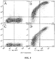
- FIG. 1 An example of the binding analysis is shown in Figure 1 .
- Transfection efficiencies ranged from 15-50% (GFP + ). Isotype controls were negative on all transfectants.
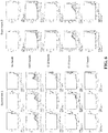
- Analysis of the percentage of GFP + cells that are also positive for Alexa647 (anti-PD-1 antibody binding) shows that the L16R and R118D mutations completely eliminate Clone 19 binding ( Fig. 2 ). All R118D expressing cells bind Clone 10, indicating functional expression of PD-1, but have the lowest intensity of all mutants ( Fig. 2 ), suggesting a low level of expression.
- V18R partially eliminates Clone 19 binding.
- Clone 10 binds all the mutants but for mutants N41K and L103E the binding intensity for this antibody versus the other PD-1 antibodies is significantly decreased ( Fig. 2 ).
- the binding analyses thus define two distinct epitopes each defined in turn by at least two residues: anti-PD-1 antibody Clone 10 binds to a membrane-distal epitope that overlaps with the ligand-binding region ( Zhang et al. Immunity 20, 337-47 (2004 )); Clone 19 binds to a membrane-proximal epitope.
- the binding-disrupting residues are mapped onto the murine PD-1 crystal structure in Figure 3 .
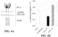
- a dimeric form of PD-1 was generated that consisted of the extracellular (antibody-binding) region of human PD-1 spliced to the transmembrane region of CD3 ⁇ (to produce dimers) and the cytoplasmic region of CD28 (in order to have an "active" readout consisting of IL-2 secretion; Fig. 4A ).
- oligonucleotide 1 (left arrow; sequence 5'-TAGTAGAGATCTCTCAAGCAGGCCACCATGCAAATCCCACAGGCGCCGTGG-3', SEQ ID NO: 33), which encodes a Bg1II restriction site and the rat ribosome binding site followed by the initiating codon and the first 21 bases of the signal peptide-encoding sequence of human PD-1, was used in a polymerase chain reaction (PCR1) with the complement of oligonucleotide 2 (5'-TCAGCCGGATCCTTCCAAACCCTGGTGCTCT GCTACTTGCTAGATGG-3', SEQ ID NO: 34).
- PCR1 polymerase chain reaction
- Oligonucleotide 2 encodes the last nine residues of the human PD-1 extracellular domain (up to residue 170 of the mature polypeptide) inserting a Bam H1 site, followed by 20 bases encoding the NH 2 -terminal end of the mouse CD3 ⁇ transmembrane domain. PCR reactions were carried out under standard conditions.
- oligonucleotide 2 was used in a PCR reaction (PCR2) with the complement of oligonucleotide 3 (5'-ATCACAGCCCTGTACCTGAATAGTAGAAG GAATAGACTC-3', SEQ ID NO: 35) which encodes the COOH-terminal end of the transmembrane region of mouse CD3 ⁇ , followed by the first 21 bases encoding the NH 2 -terminal end of the mouse CD28 cytoplasmic domain.
- step 3 the PCR1 and PCR2 reaction products were purified, annealed, extended and then amplified in the presence of oligonucleotide 1 and the complement of oligonucleotide 3, to generate a cDNA encoding the extracellular region of PD-1 fused with the transmembrane region of CD3 ⁇ .
- oligonucleotide 3 was used in a PCR reaction (PCR3) with oligonucleotide 4 (5'-CTCGAGCTACTAGGGGCGGTACGCTGCAAA- 3', SEQ ID NO: 36), which encodes the COOH-terminal end of the cytoplasmic domain of mouse CD28 followed by a stop codon and a Xho I restriction site.
- step 5 the purified PCR3 product was fused with the purified PCR product from step 3 by annealing the two products, extending the annealed hybrid, and then amplifying it with oligonucleotides 1 and 4.
- Human PD-1 and mouse CD28 cDNA was amplified using pENTRhPD-1/mCD28 as template, which was originally constructed from IMAGE clones obtained from Geneservices Ltd (Cambridge UK).
- Mouse CD3 ⁇ was amplified from DO11.10 mouse T cell hybridoma cDNA.
- the fusion PCR products were cloned into pCR4®-TOPO® (Invitrogen) and the final products sequenced by the dideoxy method.
- the constructs were cut with Bg1II and XhoI and inserted into the lentiviral vector pHR-SIN-BX-IRES-Em.
- HEK 293T cells were transfected with pHR-SIN-BX-IRES-Em encoding hPD-1/mCD3 ⁇ WT/mCD28, and the supernatant used to infect DO11.10 T-cell hybridomas.
- Infected DO11.10 cells were propagated and FACS sorted for mouse PD-1 and EGFP expression, and then tested for agonistic signaling by the anti-PD-1 antibodies using IL-2 release as a stimulation assay readout.
- IL-2 secretion results indicate that both antibodies are capable of inducing signaling via the hPD-1/mCD3 ⁇ WT/mCD28 chimera; however Clone 19, which binds PD-1 closest to the membrane induces the largest amount of IL-2 release (representative data is shown in Fig. 4b ). This supports the notion that the topology of the complex formed by the antibodies is what determines the relative levels of signaling induced by agonists. The data also suggest that the degree of agonistic signaling can be varied with choice of antibody.
- the antibodies were tested for their ability to inhibit TCR-derived activating signals by covalently coupling the antibodies, along with anti-CD3 antibodies, to tosyl-activated DYNALBEADS.
- the beads were then added to cultures of PBL labelled with carboxyfluorescein succinimidyl ester (CFSE). Proliferation levels were indicated by the fraction of cells with diluted CFSE determined by flow cytometric analysis.
- CFSE carboxyfluorescein succinimidyl ester
- Tosyl-activated 4.5 ⁇ m DYNALBEADS (M450; Invitrogen) were washed in 0.1M sterile phosphate buffer (pH 8) and loaded with 2.5 ⁇ g total antibody per 3x10 7 beads at 37°C for 18-24 h with continuous inversion mixing.
- Rabbit IgG (Sigma) was used to equalise the amount of total antibody per bead-loading reaction. Beads were blocked for at least 30 min in RPMI with 10% FCS at room temperature and washed three times in serum-free RPMI.
- bead-bound antibody was quantified in duplicate with saturating amounts of isotype-specific PE-labelled goat antibodies and compared with QuantibriteTM prelabelled quantification kit (BD Biosciences Ltd.).
- the geometric mean fluorescence PE intensities of bead singlets (minus background of unloaded beads as a control) were used to calculate the absolute amount of antibody loaded per bead from the standard curve.
- An example of such a titration is given in Fig. 5 .
- Loaded beads were stored at 4°C.
- the amounts of anti-CD3 antibody added were varied so that, at the time of the experiments, the effects of matched sets of beads with near-equivalent levels of anti-CD3 antibody could be compared.
- the level of stimulation provided by anti-CD3 loaded beads was defined as low (resulting in 15% proliferation of bulk lymphocytes at day 5), medium-low (30% proliferation), medium-high (60% proliferation) and high (80% proliferation).
- Fresh heparinized blood was diluted 1:1 with PBS and the lymphocytes isolated by density gradient separation (Ficoll Hypaque).
- accessory cells were depleted by plastic adherence for 2 h at 37°C or with specific antibody-labelled DYNALBEADS (against CD14/19/8/56).
- Cells were washed in PBS and RPMI and resuspended at 10 7 cells/ml in serum-free RPMI. Cells were labeled with 25 ⁇ M CFSE in PBS for 10 min in the dark at RT. CFSE was quenched with an equal volume of FCS at RT for 5 min.
- Cells were washed 3-5 times with RPMI and resuspended at 10 6 cells/ml in RPMI +10% FCS + PSG + 2-ME (final concentration 5x10 -5 M).
- Antibodies (beads), mitogen or media was added to relevant wells in 96-well round-bottomed plates and 10 5 cells/well were distributed and incubated at 37°C for 3-5 days.
- cells were stained with directly-labelled cell-surface antibodies for 1 h at 4°C.
- Cells were washed with PBS-Azide, pelleted at 1500rpm/5min and resuspended in 200 ⁇ l PBS-Azide. Cells were analysed for CFSE and antibody labelling at the flow cytometer using FlowJo Flow Cytometry Analysis Software.
- Clone 10 antibodies were further tested for their ability to inhibit TCR-derived activating signals by covalently coupling the antibodies, along with anti-CD3 antibodies, to tosyl-activated DYNALBEADS.
- the beads were then added to cultures of human CD4 + T cells and proliferation measured by 3 H-thymidine incorporation.
- Tosyl-activated 4.5 ⁇ m DYNALBEADS (M450; Invitrogen) were washed in 0.1M sterile phosphate buffer (pH 7.5) and loaded with 2 ⁇ g of anti-human CD3 (clone OKT3) per 1x10 7 beads at 37°C for 8h with continuous inversion mixing. Beads were then washed to remove un-conjugated anti-CD3. Aliquots of the anti-CD3 conjugated beads were then secondarily coated with 3 ⁇ g of anti-PD-1 antibody or control per 1x10 7 beads at 37°C for 19h with continuous inversion mixing.
- Beads were washed and then incubated in 0.2M Tris / 0.1% BSA (pH 8.5) for 3 hours to inactivate free tosyl groups, followed by washing and resuspension of beads in PBS / 0.1% BSA / 2mM EDTA (pH 7.4). Equal anti-CD3 and antibody coating of the bead sets was confirmed by staining the beads with fluorochrome-labelled isotype-specific antibodies and analysing by flow cytometry.
- CD4 + T cells were purified from the whole PBLs by negative selection using MACS (CD4 + T cell isolation Kit II; Miltenyi Biotec). 1x10 5 human CD4 + T cells / well were cultured at a 1:1 ratio with the coated beads in 96-well round-bottomed plates and incubated at 37°C for 6 days. Proliferation was measured at day 6 by addition of 0.5 ⁇ Ci/well 3 H-thymidine for the last 6 hours of culture. Cells were harvested onto glass-fibre filters and incorporated 3 H-thymidine was measured by ⁇ -scintillation counting.
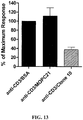
- the results in Figure 13 show the day 6 proliferative response by human CD4 + T cells measured in the presence of anti-CD3 plus Clone 10 antibody or control coated beads.
- the data are expressed as percentage of the maximal response (anti-CD3 plus BSA control) and are the mean of 4 different donor responses.
- CD4 + T cell proliferation was inhibited in the presence of Clone 10, so that the average proliferation observed was only 37.7 % of the maximum.
- Clone 19 generally induces stronger signaling by the hPD-1/mCD3 ⁇ WT/mCD28 chimera than Clone 10 ( Fig. 4B ) but in some experiments it gives weaker inhibitory signaling by native PD-1 (see, e.g. Fig. 8 ). It is possible that this is because, in some experiments, Clone 19 but not Clone 10 ligation results in the phosphorylation of both the ITIM (inhibitory, blue) and the ITSM (activating, red) tyrosine-based signaling motifs of PD-1 (see Fig. 9 ).
- a monomeric, monovalent form of PD-1, hPD-1/mCD28 that consisted of the extracellular (antibody-binding) and transmembrane regions of human PD-1 spliced to the cytoplasmic region of CD28 (in order to have an "active" readout consisting of IL-2 secretion; Fig. 10a ), was generated.
- oligonucleotide 1 (left arrow; sequence 5'-TAGTAGAGATCTCTCAAGCAGGCCACCA TGCAAATCCCACAGGCGCCGTGG-3', SEQ ID NO: 33), which encodes a BglII restriction site and the rat ribosome binding site followed by the initiating codon and the first 24 bases of the signal peptide-encoding sequence of human PD-1, was used in a polymerase chain reaction (PCR1) with the complement of oligonucleotide 2 (5'-GCCCAGCCGGCCAG TTCCAAACCTTTTGGGTGCTGGTGGTGGTTGGT-3', SEQ ID NO: 37).
- PCR1 polymerase chain reaction
- Oligonucleotide 2 encodes the last 23 bases of the human PD-1 extracellular domain (up to residue 149 of the mature polypeptide), followed by 24 bases encoding the NH 2 -terminal sequence of the mouse CD28 transmembrane region. PCR reactions were carried out under standard conditions.
- oligonucleotide 2 was used in a PCR reaction (PCR2) with the complement of oligonucleotide 3 (5'-TTTGCAGCGTACCGCCCCACGCGTTAGTAGCTCGAG-3', SEQ ID NO: 38) which encodes the COOH-terminal end of the cytoplasmic domain of mouse CD28, a Mlu I restriction site followed by a stop codon and a Xho I restriction site.
- the purified PCR2 product was fused with the purified PCR1 product from step 1 by annealing the two products, extending the annealed hybrid, and then amplifying it with oligonucleotides 1 and 3.
- Mouse CD28 sequence was amplified using pCR4®-TOPO®rCD28/mCD28 as template, which was originally amplified from DO11.10 mouse T cell hybridoma cDNA.
- the human extracellular PD-1 was amplified from pE14hPD-1Long, a gift from Dr Chao Yu of the MRC Human Immunology Unit, Oxford.
- the fusion PCR products were cloned into pCR4®-TOPO®(Invitrogen) and the final products sequenced by the dideoxy method.
- the constructs were cut with BglII and XhoI and inserted into the lentiviral vector pHR-SIN-BX-IRES-Em for infection of DO11.10 cells.
- Activation of the DO11.10 cells expressing the hPD-1/mCD28 chimera by anti-PD-1 antibodies was examined using IL-2 secretion as a read-out.
- hPD-1/mCD28 Lack of signaling by hPD-1/mCD28 suggests that agonistic signaling may be enhanced by cross-linking a monomeric receptor with two antibodies that bind to non-overlapping epitopes
- Clone 10 and Clone 19 were not agonistic for a chimeric form of human PD-1, i.e. hPD-1/mCD28, consisting of the monomeric extracellular region of PD-1 attached to the transmembrane and intracellular signaling domains of CD28 ( Fig. 10 ), in contrast to the equivalent CD28 construct (containing the homodimeric extracellular domain of rat CD28).
- hPD-1/mCD28 a chimeric form of human PD-1
- CD28 containing the homodimeric extracellular domain of rat CD28.
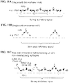
- the likeliest explanation for this is that, because PD-1 is monomeric and CD28 is a homodimer, the attachment of bivalent antibody leads to the assembly of a multimeric array of "cross-linked" CD28 molecules and a very high density of signaling domains ( Fig.
- Agonistic signalling is enhanced by cross-linking a monomeric receptor with two antibodies that bind to non-overlapping epitopes
- DO11.10 cells expressing the hPD-1/mCD28 chimeric protein were used in a Clone 10/Clone 19 antibody stimulation assay as follows.
- 96-well flat-bottomed plates (Costar EIA/RIA plates) were coated overnight at 4°C with 500 ⁇ g/ml donkey anti-mouse IgG (Jackson Immunoresearch) in coating buffer (15mM Na2CO3, 35mM NaHCO3, pH 9.6. Prior to the addition of cells, the plates were washed three times with 200 ⁇ l chilled PBS.
- the heavy chain CDR1s for clones 2, 10 and 19 have been identified according to both the combined Kabat/Chothia numbering system and the Kabat numbering system. All other CDRs have been identified according to the Kabat numbering system ( Kabat et al., 1987, "In sequences of proteins of immunological interest ", U.S. Dept. Health and Human Services, NIH USA. Heavy chain CDR1s for clones 2, 10 and 19, as identified by the Kabat numbering system, are identified (underlined amino acids) in Table 1.
Landscapes
- Health & Medical Sciences (AREA)
- Chemical & Material Sciences (AREA)
- Organic Chemistry (AREA)
- Immunology (AREA)
- Medicinal Chemistry (AREA)
- General Health & Medical Sciences (AREA)
- Life Sciences & Earth Sciences (AREA)
- Pharmacology & Pharmacy (AREA)
- Public Health (AREA)
- General Chemical & Material Sciences (AREA)
- Chemical Kinetics & Catalysis (AREA)
- Bioinformatics & Cheminformatics (AREA)
- Animal Behavior & Ethology (AREA)
- Engineering & Computer Science (AREA)
- Nuclear Medicine, Radiotherapy & Molecular Imaging (AREA)
- Veterinary Medicine (AREA)
- Diabetes (AREA)
- Proteomics, Peptides & Aminoacids (AREA)
- Molecular Biology (AREA)
- Genetics & Genomics (AREA)
- Biophysics (AREA)
- Biochemistry (AREA)
- Rheumatology (AREA)
- Biomedical Technology (AREA)
- Neurology (AREA)
- Neurosurgery (AREA)
- Pain & Pain Management (AREA)
- Pulmonology (AREA)
- Orthopedic Medicine & Surgery (AREA)
- Physical Education & Sports Medicine (AREA)
- Obesity (AREA)
- Hematology (AREA)
- Endocrinology (AREA)
- Emergency Medicine (AREA)
- Transplantation (AREA)
- Peptides Or Proteins (AREA)
- Medicines Containing Antibodies Or Antigens For Use As Internal Diagnostic Agents (AREA)
- Preparation Of Compounds By Using Micro-Organisms (AREA)
- Micro-Organisms Or Cultivation Processes Thereof (AREA)
Applications Claiming Priority (2)
| Application Number | Priority Date | Filing Date | Title |
|---|---|---|---|
| US9644708P | 2008-09-12 | 2008-09-12 | |
| PCT/IB2009/006940 WO2010029434A1 (en) | 2008-09-12 | 2009-09-14 | Pd-1 specific antibodies and uses thereof |
Publications (2)
| Publication Number | Publication Date |
|---|---|
| EP2342228A1 EP2342228A1 (en) | 2011-07-13 |
| EP2342228B1 true EP2342228B1 (en) | 2017-09-06 |
Family
ID=41335564
Family Applications (1)
| Application Number | Title | Priority Date | Filing Date |
|---|---|---|---|
| EP09786276.7A Active EP2342228B1 (en) | 2008-09-12 | 2009-09-14 | Pd-1 specific antibodies and uses thereof |
Country Status (6)
| Country | Link |
|---|---|
| US (1) | US9181342B2 (https=) |
| EP (1) | EP2342228B1 (https=) |
| JP (1) | JP5794917B2 (https=) |
| AU (1) | AU2009290543B2 (https=) |
| CA (1) | CA2736816C (https=) |
| WO (1) | WO2010029434A1 (https=) |
Cited By (1)
| Publication number | Priority date | Publication date | Assignee | Title |
|---|---|---|---|---|
| WO2020128447A1 (en) | 2018-12-17 | 2020-06-25 | Oxford University Innovation Limited | Modified antibodies |
Families Citing this family (155)
| Publication number | Priority date | Publication date | Assignee | Title |
|---|---|---|---|---|
| EP2445932B1 (en) | 2009-06-26 | 2018-02-28 | Soricimed Biopharma Inc. | Soricidin derived peptides and methods for the detection of trpv-6 cancers and drug delivery |
| TW201134488A (en) * | 2010-03-11 | 2011-10-16 | Ucb Pharma Sa | PD-1 antibodies |
| EP2545078A1 (en) * | 2010-03-11 | 2013-01-16 | UCB Pharma, S.A. | Pd-1 antibody |
| JP6072771B2 (ja) * | 2011-04-20 | 2017-02-01 | メディミューン,エルエルシー | B7−h1およびpd−1に結合する抗体およびその他の分子 |
| MX2014001222A (es) * | 2011-07-29 | 2014-09-15 | Univ Pennsylvania | Receptores coestimuladores de cambio. |
| US12466897B2 (en) | 2011-10-10 | 2025-11-11 | Xencor, Inc. | Heterodimeric human IgG1 polypeptides with isoelectric point modifications |
| US10858417B2 (en) | 2013-03-15 | 2020-12-08 | Xencor, Inc. | Heterodimeric proteins |
| SMT202100065T1 (it) | 2013-05-02 | 2021-03-15 | Anaptysbio Inc | Anticorpi diretti contro la proteina della morte programmata (pd-1) |
| RS59756B1 (sr) | 2013-08-08 | 2020-02-28 | Cytune Pharma | Kombinovana farmaceutska kompozicija |
| PT3444271T (pt) | 2013-08-08 | 2022-01-05 | Inst Nat Sante Rech Med | Modulocinas baseadas em il-15 e no domínio sushi de il-15ralfa |
| CN112552401B (zh) | 2013-09-13 | 2023-08-25 | 广州百济神州生物制药有限公司 | 抗pd1抗体及其作为治疗剂与诊断剂的用途 |
| WO2015048312A1 (en) | 2013-09-26 | 2015-04-02 | Costim Pharmaceuticals Inc. | Methods for treating hematologic cancers |
| SG10201804945WA (en) | 2013-12-12 | 2018-07-30 | Shanghai hengrui pharmaceutical co ltd | Pd-1 antibody, antigen-binding fragment thereof, and medical application thereof |
| TWI680138B (zh) | 2014-01-23 | 2019-12-21 | 美商再生元醫藥公司 | 抗pd-l1之人類抗體 |
| TWI681969B (zh) | 2014-01-23 | 2020-01-11 | 美商再生元醫藥公司 | 針對pd-1的人類抗體 |
| PE20170255A1 (es) | 2014-01-24 | 2017-03-22 | Dana Farber Cancer Inst Inc | Moleculas de anticuerpo que se unen a pd-1 y usos de las mismas |
| HUE045065T2 (hu) | 2014-01-31 | 2019-12-30 | Novartis Ag | TIM-3 antitest molekulák és felhasználásaik |
| KR102442436B1 (ko) | 2014-03-14 | 2022-09-15 | 노파르티스 아게 | Lag-3에 대한 항체 분자 및 그의 용도 |
| CN106604742B (zh) | 2014-07-03 | 2019-01-11 | 百济神州有限公司 | 抗pd-l1抗体及其作为治疗剂及诊断剂的用途 |
| BR112017001385B1 (pt) | 2014-07-22 | 2023-12-05 | Cb Therapeutics, Inc. | Anticorpo isolado ou fragmento do mesmo que liga a pd-1, uso deste, composição, polinucleotídeo isolado e vetor de expressão |
| US9982052B2 (en) * | 2014-08-05 | 2018-05-29 | MabQuest, SA | Immunological reagents |
| KR102357893B1 (ko) * | 2014-08-05 | 2022-02-04 | 맵퀘스트 에스아 | Pd-1 에 결합하는 면역학적 시약 |
| KR102476226B1 (ko) | 2014-08-05 | 2022-12-12 | 아폴로믹스 인코포레이티드 | 항-pd-l1 항체 |
| KR20170060042A (ko) | 2014-09-13 | 2017-05-31 | 노파르티스 아게 | Alk 억제제의 조합 요법 |
| JP6991857B2 (ja) | 2014-10-10 | 2022-01-13 | イデラ ファーマシューティカルズ インコーポレイテッド | Tlr9アゴニストをチェックポイント阻害剤と共に用いるがんの治療 |
| JP6821560B2 (ja) | 2014-10-21 | 2021-01-27 | サイクロン ファーマシューティカルズ インターナショナル エルティーディー.Sciclone Pharmaceuticals International Ltd. | 免疫刺激剤による癌治療 |
| PE20171324A1 (es) | 2014-11-26 | 2017-09-11 | Xencor Inc | Anticuerpos heterodimericos que se unen a cd3 y a antigenos tumorales |
| PL3298033T5 (pl) | 2015-05-18 | 2023-10-30 | TCR2 Therapeutics Inc. | Kompozycje i zastosowania medyczne do reprogramowania TCR z zastosowaniem białek fuzyjnych |
| US10874724B2 (en) * | 2015-05-22 | 2020-12-29 | University Of Houston System | Enzymatic immunomodulation of solid tumors and uses thereof |
| DK3909972T5 (da) | 2015-06-19 | 2024-08-05 | Sebastian Kobold | Pd1-cd28-fusionsproteiner og disses anvendelse i medicin |
| MX383464B (es) | 2015-07-13 | 2025-03-14 | Cytomx Therapeutics Inc | Anticuerpos anti-pd-1, anticuerpos anti-pd-1 activables, y métodos de uso de los mismos. |
| CA2991628C (en) | 2015-07-16 | 2020-04-07 | Bioxcel Therapeutics, Inc. | A novel approach for treatment of cancer using immunomodulation |
| HRP20211058T8 (hr) | 2015-07-29 | 2021-11-26 | Novartis Ag | Kombinirane terapije koje sadrže molekule antitijela protiv lag-3 |
| BR112018002436A2 (pt) | 2015-08-04 | 2018-09-18 | Glaxosmithkline Ip Dev Ltd | tratamento de combinação de usos de métodos destes |
| US20180222989A1 (en) | 2015-08-04 | 2018-08-09 | Glaxosmithkline Intellectual Property Development Limited | Combination treatments and uses and methods thereof |
| US20180230431A1 (en) | 2015-08-07 | 2018-08-16 | Glaxosmithkline Intellectual Property Development Limited | Combination Therapy |
| WO2017024465A1 (en) | 2015-08-10 | 2017-02-16 | Innovent Biologics (Suzhou) Co., Ltd. | Pd-1 antibodies |
| AU2016317915B2 (en) | 2015-09-01 | 2021-02-18 | Agenus Inc. | Anti-PD-1 antibodies and methods of use thereof |
| BR112018006237A2 (pt) | 2015-09-29 | 2018-10-09 | Celgene Corp | proteínas de ligação a pd-1 e métodos de uso das mesmas |
| WO2017055404A1 (en) | 2015-10-02 | 2017-04-06 | F. Hoffmann-La Roche Ag | Bispecific antibodies specific for pd1 and tim3 |
| US12030942B2 (en) | 2015-10-02 | 2024-07-09 | Les Laboratoires Servier | Anti-PD-1 antibodies and compositions |
| ES2895034T3 (es) | 2015-10-02 | 2022-02-17 | Hoffmann La Roche | Anticuerpos anti-PD1 y procedimientos de uso |
| ES2924402T3 (es) * | 2015-10-02 | 2022-10-06 | Symphogen As | Anticuerpos anti-PD-1 y composiciones |
| PE20181326A1 (es) | 2015-11-03 | 2018-08-20 | Janssen Biotech Inc | Anticuerpos que se unen especificamente a pd-1 y sus usos |
| LT3394103T (lt) | 2015-12-22 | 2023-09-11 | Regeneron Pharmaceuticals, Inc. | Antikūnų prieš pd-1 ir bispecifinių antikūnų prieš cd20/cd3 derinys, skirtas vėžiui gydyti |
| ES3034233T3 (en) | 2016-01-22 | 2025-08-14 | MabQuest SA | Non-blocking pd1 specific antibodies |
| JP7137474B2 (ja) | 2016-03-15 | 2022-09-14 | メルサナ セラピューティクス,インコーポレイティド | NaPi2b標的化抗体-薬物コンジュゲート及びその使用方法 |
| JP7069032B2 (ja) | 2016-03-24 | 2022-05-17 | ミレニアム ファーマシューティカルズ, インコーポレイテッド | がん免疫治療における胃腸の免疫関連有害事象の治療方法 |
| WO2017165742A1 (en) | 2016-03-24 | 2017-09-28 | Millennium Pharmaceuticals, Inc. | Methods of treating gastrointestinal immune-related adverse events in anti-ctla4 anti-pd-1 combination treatments |
| TWI910495B (zh) | 2016-05-13 | 2026-01-01 | 美商再生元醫藥公司 | 藉由投予pd-1抑制劑治療皮膚癌之方法 |
| EP3243832A1 (en) | 2016-05-13 | 2017-11-15 | F. Hoffmann-La Roche AG | Antigen binding molecules comprising a tnf family ligand trimer and pd1 binding moiety |
| CN106008714B (zh) * | 2016-05-24 | 2019-03-15 | 瑞阳(苏州)生物科技有限公司 | 抗人pd-1人源化单克隆抗体及其应用 |
| JP7010854B2 (ja) * | 2016-06-14 | 2022-01-26 | ゼンコア インコーポレイテッド | 二重特異性チェックポイント阻害剤抗体 |
| RU2769282C2 (ru) | 2016-06-20 | 2022-03-30 | Кимаб Лимитед | Анти-PD-L1 и IL-2 цитокины |
| US10864203B2 (en) | 2016-07-05 | 2020-12-15 | Beigene, Ltd. | Combination of a PD-1 antagonist and a RAF inhibitor for treating cancer |
| EP3494140A1 (en) | 2016-08-04 | 2019-06-12 | GlaxoSmithKline Intellectual Property Development Ltd | Anti-icos and anti-pd-1 antibody combination therapy |
| WO2018033135A1 (en) | 2016-08-19 | 2018-02-22 | Beigene, Ltd. | Use of a combination comprising a btk inhibitor for treating cancers |
| JP2019534859A (ja) | 2016-09-19 | 2019-12-05 | セルジーン コーポレイション | Pd−1結合タンパク質を使用して白斑を治療する方法 |
| MX2019002867A (es) | 2016-09-19 | 2019-11-12 | Celgene Corp | Metodos de tratamiento de trastornos inmunologicos usando proteinas de union a pd-1. |
| CN114456269A (zh) * | 2016-09-21 | 2022-05-10 | 基石药业(苏州)有限公司 | 一种新的pd-1单克隆抗体 |
| AU2017342176A1 (en) | 2016-10-10 | 2019-05-02 | The National Institute for Biotechnology in the Negev Ltd. | Non-cytotoxic modified cells and use thereof |
| JP2019533458A (ja) | 2016-11-01 | 2019-11-21 | アナプティスバイオ インコーポレイティッド | プログラム死1(pd−1)に対する抗体 |
| WO2018083087A2 (en) | 2016-11-02 | 2018-05-11 | Glaxosmithkline Intellectual Property (No.2) Limited | Binding proteins |
| DK3541841T3 (da) | 2016-11-18 | 2024-10-21 | Servier Lab | Anti-PD-1-antistoffer og sammensætninger |
| JP7291396B2 (ja) | 2016-11-22 | 2023-06-15 | ティーシーアール2 セラピューティクス インク. | 融合タンパク質を用いたtcrの再プログラミングのための組成物及び方法 |
| US11135307B2 (en) | 2016-11-23 | 2021-10-05 | Mersana Therapeutics, Inc. | Peptide-containing linkers for antibody-drug conjugates |
| KR20190090823A (ko) | 2016-12-01 | 2019-08-02 | 글락소스미스클라인 인털렉츄얼 프로퍼티 디벨로프먼트 리미티드 | 조합 요법 |
| US20190343803A1 (en) | 2016-12-01 | 2019-11-14 | Glaxosmithkline Intellectual Property Development Limited | Combination therapy |
| CN110300599B (zh) | 2016-12-07 | 2024-07-02 | 艾吉纳斯公司 | 抗体和其使用方法 |
| AU2018206481B2 (en) | 2017-01-09 | 2025-02-27 | Tesaro, Inc. | Methods of treating cancer with anti-PD-1 antibodies |
| US11555038B2 (en) | 2017-01-25 | 2023-01-17 | Beigene, Ltd. | Crystalline forms of (S)-7-(1-(but-2-ynoyl)piperidin-4-yl)-2-(4-phenoxyphenyl)-4,5,6,7-tetrahydropyrazolo[1,5-a]pyrimidine-3-carboxamide, preparation, and uses thereof |
| US20190375847A1 (en) | 2017-02-15 | 2019-12-12 | Glaxosmithkline Intellectual Property Development Limited | Combination treatment for cancer |
| US20180271996A1 (en) | 2017-02-28 | 2018-09-27 | Mersana Therapeutics, Inc. | Combination therapies of her2-targeted antibody-drug conjugates |
| EP3596108A4 (en) | 2017-03-15 | 2020-12-23 | Pandion Operations, Inc. | TARGETED IMMUNOTOLERANCE |
| MY201482A (en) | 2017-04-03 | 2024-02-26 | Hoffmann La Roche | Immunoconjugates of an anti-pd-1 antibody with a mutant il-2 or with il-15 |
| EP4516809A3 (en) | 2017-04-05 | 2025-09-03 | F. Hoffmann-La Roche AG | Bispecific antibodies specifically binding to pd1 and lag3 |
| UA129904C2 (uk) | 2017-04-05 | 2025-09-10 | Ле Лаборатуар Сервьє | Комбінація антитіла до tim-3 й антитіла до pd-1 для лікування раку |
| US11603407B2 (en) | 2017-04-06 | 2023-03-14 | Regeneron Pharmaceuticals, Inc. | Stable antibody formulation |
| US10676516B2 (en) | 2017-05-24 | 2020-06-09 | Pandion Therapeutics, Inc. | Targeted immunotolerance |
| CR20190550A (es) | 2017-06-05 | 2020-04-05 | Janssen Biotech Inc | Anticuerpos que se unen específicamente a pd-1 y métodos de uso |
| JP2020522555A (ja) | 2017-06-09 | 2020-07-30 | グラクソスミスクライン、インテレクチュアル、プロパティー、ディベロップメント、リミテッドGlaxosmithkline Intellectual Property Development Limited | 組み合わせ療法 |
| EP3645569A4 (en) | 2017-06-26 | 2021-03-24 | BeiGene, Ltd. | IMMUNOTHERAPY FOR LIVER CELL CARCINOMA |
| CN118027197A (zh) | 2017-07-06 | 2024-05-14 | 美勒斯公司 | 调节由细胞表达的生物活性的抗体 |
| JP2021502100A (ja) | 2017-11-08 | 2021-01-28 | ゼンコア インコーポレイテッド | 新規抗pd−1配列を用いた二重特異性および単一特異性抗体 |
| WO2019104289A1 (en) | 2017-11-27 | 2019-05-31 | Mersana Therapeutics, Inc. | Pyrrolobenzodiazepine antibody conjugates |
| WO2019108795A1 (en) | 2017-11-29 | 2019-06-06 | Beigene Switzerland Gmbh | Treatment of indolent or aggressive b-cell lymphomas using a combination comprising btk inhibitors |
| US10946068B2 (en) | 2017-12-06 | 2021-03-16 | Pandion Operations, Inc. | IL-2 muteins and uses thereof |
| US10174092B1 (en) | 2017-12-06 | 2019-01-08 | Pandion Therapeutics, Inc. | IL-2 muteins |
| USRE50550E1 (en) | 2017-12-06 | 2025-08-26 | Pandion Operations, Inc. | IL-2 muteins and uses thereof |
| CN111757757A (zh) | 2017-12-21 | 2020-10-09 | 梅尔莎纳医疗公司 | 吡咯并苯并二氮呯抗体共轭物 |
| US12398209B2 (en) | 2018-01-22 | 2025-08-26 | Janssen Biotech, Inc. | Methods of treating cancers with antagonistic anti-PD-1 antibodies |
| WO2019149716A1 (en) | 2018-01-31 | 2019-08-08 | F. Hoffmann-La Roche Ag | Bispecific antibodies comprising an antigen-binding site binding to lag3 |
| AR114127A1 (es) | 2018-03-02 | 2020-07-22 | Lilly Co Eli | Anticuerpos agonistas contra pd-1 y usos de estos |
| AU2019247511B2 (en) | 2018-04-06 | 2025-10-16 | Atyr Pharma, Inc. | Compositions and methods comprising anti-NRP2 antibodies |
| KR20210010896A (ko) | 2018-05-14 | 2021-01-28 | 이뮤노코어 리미티드 | 이기능성 결합 폴리펩타이드 |
| KR20210035805A (ko) | 2018-06-15 | 2021-04-01 | 플래그쉽 파이어니어링 이노베이션스 브이, 인크. | 세포후 신호전달 인자의 조절을 통한 면역 활성의 증가 |
| JP7490925B2 (ja) | 2018-07-26 | 2024-05-28 | エータイアー ファーマ, インコーポレイテッド | Nrp2関連疾患を治療するための組成物および方法 |
| WO2020030571A1 (en) | 2018-08-06 | 2020-02-13 | Glaxosmithkline Intellectual Property Development Limited | Combinations of a pd-1 antibody and a tlr4 modulator and uses thereof |
| BR112021007517A2 (pt) | 2018-10-22 | 2021-10-26 | Glaxosmithkline Intellectual Property Development Limited | Dosagem |
| CN113365664A (zh) | 2018-10-29 | 2021-09-07 | 梅尔莎纳医疗公司 | 具有含肽接头的半胱氨酸工程化的抗体-药物缀合物 |
| KR20210119380A (ko) | 2018-11-09 | 2021-10-05 | 피어리언 바이오사이언스즈, 엘엘씨 | 종양 미세환경의 조성을 결정하기 위한 방법 및 조성물 |
| EP3924521A4 (en) | 2019-02-15 | 2023-03-29 | IncellDx, Inc. | BLADDER-ASSOCIATED SAMPLES TESTING, IDENTIFICATION AND TREATMENT OF BLADDER-ASSOCIATED NEOPLASIA, AND KITS FOR USE THEREON |
| US12404331B2 (en) | 2019-04-19 | 2025-09-02 | Tcrcure Biopharma Corp. | Anti-PD-1 antibodies and uses thereof |
| WO2020227159A2 (en) | 2019-05-03 | 2020-11-12 | Flagship Pioneering Innovations V, Inc. | Methods of modulating immune activity |
| BR112021023345A2 (pt) | 2019-05-20 | 2022-02-01 | Pandion Operations Inc | Imunotolerância com alvo em madcam |
| KR20250126805A (ko) * | 2019-06-05 | 2025-08-25 | 아납티스바이오, 아이엔씨. | Pd-1 작용제 및 이의 사용 방법 |
| US20210038684A1 (en) | 2019-06-11 | 2021-02-11 | Alkermes Pharma Ireland Limited | Compositions and Methods for Cancer Immunotherapy |
| KR20220041080A (ko) | 2019-06-18 | 2022-03-31 | 얀센 사이언시즈 아일랜드 언리미티드 컴퍼니 | B형 간염 바이러스(hbv) 백신 및 항-pd-1 또는 항-pc-l1 항체의 조합 |
| EP3986460A2 (en) | 2019-06-18 | 2022-04-27 | Janssen Sciences Ireland Unlimited Company | Combination of hepatitis b virus (hbv) vaccines and anti-pd-1 antibody |
| JP6881658B2 (ja) | 2019-07-05 | 2021-06-02 | 小野薬品工業株式会社 | Pd−1/cd3二重特異性タンパク質による血液がん治療 |
| US20220332825A1 (en) | 2019-08-08 | 2022-10-20 | Ono Pharmaceutical Co., Ltd. | Bispecific protein |
| KR20220100859A (ko) | 2019-09-17 | 2022-07-18 | 비알 - 알&디 인베스트먼츠, 에스.에이. | 산성 세라미다아제 저해제로서의 치환된 n-헤테로시클릭 카르복사미드, 및 약제로서의 이의 용도 |
| JP2022549227A (ja) | 2019-09-17 | 2022-11-24 | バイアル-アールアンドディー インベストメンツ ソシエダッド アノニマ | 医学的障害の治療における使用のための置換された飽和および不飽和n-複素環式カルボキサミドおよび関連化合物 |
| BR112022004791A2 (pt) | 2019-09-17 | 2022-06-21 | Bial R&D Invest S A | Carboxamidas de imidazol substituídas e seu uso no tratamento de distúrbios médicos |
| US20230114107A1 (en) | 2019-12-17 | 2023-04-13 | Flagship Pioneering Innovations V, Inc. | Combination anti-cancer therapies with inducers of iron-dependent cellular disassembly |
| EP4107187A4 (en) | 2020-02-21 | 2024-07-03 | Pandion Operations, Inc. | TISSUE-TARGETED IMMUNOTOLERANCE WITH A CD39 EFFECTOR |
| CN115484978A (zh) | 2020-03-05 | 2022-12-16 | 尼奥克斯医疗有限公司 | 使用免疫细胞治疗癌症的方法和组合物 |
| JP2023521227A (ja) | 2020-04-14 | 2023-05-23 | グラクソスミスクライン、インテレクチュアル、プロパティー、ディベロップメント、リミテッド | 癌の併用療法 |
| AU2021256652A1 (en) | 2020-04-14 | 2022-11-03 | Glaxosmithkline Intellectual Property Development Limited | Combination treatment for cancer involving anti-ICOS and anti-PD1 antibodies, optionally further involving anti-tim3 antibodies |
| WO2021231976A1 (en) | 2020-05-14 | 2021-11-18 | Xencor, Inc. | Heterodimeric antibodies that bind prostate specific membrane antigen (psma) and cd3 |
| CR20220596A (es) * | 2020-05-26 | 2023-01-23 | Boehringer Ingelheim Int | Anticuerpos anti-pd-1 |
| US20230355804A1 (en) | 2020-06-29 | 2023-11-09 | Flagship Pioneering Innovations V, Inc. | Viruses engineered to promote thanotransmission and their use in treating cancer |
| US12516307B2 (en) | 2020-08-18 | 2026-01-06 | Onchilles Pharma, Inc. | Modified porcine pancreatic elastase proteins |
| PH12023500013A1 (en) | 2020-12-04 | 2024-03-11 | Tidal Therapeutics Inc | Ionizable cationic lipids and lipi nanoparticles, and methods of synthesis and use thereof |
| US11739144B2 (en) | 2021-03-09 | 2023-08-29 | Xencor, Inc. | Heterodimeric antibodies that bind CD3 and CLDN6 |
| EP4313109A1 (en) | 2021-03-31 | 2024-02-07 | Flagship Pioneering Innovations V, Inc. | Thanotransmission polypeptides and their use in treating cancer |
| KR20230163503A (ko) | 2021-03-31 | 2023-11-30 | 메뤼스 엔.페. | 신규한 pd-1 결합 도메인 |
| GB202107994D0 (en) | 2021-06-04 | 2021-07-21 | Kymab Ltd | Treatment of cancer |
| WO2022258011A1 (zh) * | 2021-06-11 | 2022-12-15 | 广东菲鹏制药股份有限公司 | 抗pd-1人源化抗体或其抗原结合片段及其应用 |
| AU2022303363A1 (en) | 2021-06-29 | 2024-01-18 | Flagship Pioneering Innovations V, Inc. | Immune cells engineered to promote thanotransmission and uses thereof |
| CN117794953A (zh) | 2021-08-03 | 2024-03-29 | 豪夫迈·罗氏有限公司 | 双特异性抗体及使用方法 |
| CA3229448A1 (en) | 2021-08-23 | 2023-03-02 | Immunitas Therapeutics, Inc. | Anti-cd161 antibodies and uses thereof |
| CN118284624A (zh) * | 2021-11-19 | 2024-07-02 | 米罗生物有限公司 | Pd-1抗体及其用途 |
| WO2023240156A1 (en) | 2022-06-08 | 2023-12-14 | Tidal Therapeutics, Inc. | Ionizable cationic lipids and lipid nanoparticles, and methods of synthesis and use thereof |
| CN120077068A (zh) | 2022-08-18 | 2025-05-30 | 英美偌科有限公司 | 多结构域结合分子 |
| EP4573125A1 (en) | 2022-08-18 | 2025-06-25 | Immunocore Limited | Multi-domain binding molecules |
| US20240174732A1 (en) | 2022-10-05 | 2024-05-30 | Flagship Pioneering Innovations V, Inc. | Nucleic acid molecules encoding trif and additional polypeptides and their use in treating cancer |
| EP4605422A2 (en) | 2022-10-20 | 2025-08-27 | Repertoire Immune Medicines, Inc. | Cd8 t cell targeted il2 |
| KR20250122541A (ko) | 2022-10-25 | 2025-08-13 | 사이즈믹 테라퓨틱, 인코포레이티드. | 변이체 IgG FC 폴리펩티드 및 그 용도 |
| KR20250133728A (ko) | 2023-01-06 | 2025-09-08 | 라센 테라퓨틱스, 인코포레이티드 | 항-il-18bp 항체 |
| TW202430560A (zh) | 2023-01-06 | 2024-08-01 | 美商拉森醫療公司 | 抗il-18bp抗體 |
| WO2024151687A1 (en) | 2023-01-09 | 2024-07-18 | Flagship Pioneering Innovations V, Inc. | Genetic switches and their use in treating cancer |
| CN120813375A (zh) | 2023-01-30 | 2025-10-17 | 凯玛布有限公司 | 抗体 |
| GB202306345D0 (en) | 2023-04-28 | 2023-06-14 | Immunocore Ltd | Binding molecules |
| EP4709756A1 (en) | 2023-05-10 | 2026-03-18 | Regeneron Pharmaceuticals, Inc. | Cd20-pd1 binding molecules and methods of use thereof |
| WO2024243189A2 (en) * | 2023-05-22 | 2024-11-28 | Board Of Regents, The University Of Texas System | Antigen binding proteins targeting pd-1 |
| GB202315181D0 (en) | 2023-10-03 | 2023-11-15 | Immunocore Ltd | Peptide-HLA binding molecules |
| GB202320012D0 (en) | 2023-12-22 | 2024-02-07 | Immunocore Ltd | Bispecific molecules |
| WO2025146662A1 (en) | 2024-01-05 | 2025-07-10 | Immunocore Limited | Cd1a-pd-1 bispecific agonist for the treatment of atopic dermatitis |
| WO2025174825A2 (en) | 2024-02-12 | 2025-08-21 | Aera Therapeutics, Inc. | Delivery compositions |
| TW202602455A (zh) | 2024-03-26 | 2026-01-16 | 美商安進公司 | 使用mta協作的prmt5抑制劑的癌症治療 |
| WO2025213154A1 (en) | 2024-04-05 | 2025-10-09 | Amgen Inc. | Gastrointestinal cancer treatments using mta-cooperative prmt5 inhibitors |
| WO2025224084A1 (en) | 2024-04-22 | 2025-10-30 | Engimmune Therapeutics Ag | Proteins comprising t cell receptor constant domains |
| WO2026003224A2 (en) | 2024-06-26 | 2026-01-02 | Iomx Therapeutics Ag | Bispecific antigen binding proteins (abp) targeting immune checkpoint molecules and both leukocyte immunoglobulin-like receptor subfamily b1 (lilrb1) and lilrb2; combinations and uses thereof |
| WO2026072557A2 (en) | 2024-09-24 | 2026-04-02 | Genzyme Corporation | Ionizable cationic lipids and lipid nanoparticles, and methods of synthesis and use thereof |
Citations (1)
| Publication number | Priority date | Publication date | Assignee | Title |
|---|---|---|---|---|
| JP2007202443A (ja) * | 2006-01-31 | 2007-08-16 | Japan Science & Technology Agency | ケモカインレセプターccr5のn末端領域に対する抗体酵素 |
Family Cites Families (9)
| Publication number | Priority date | Publication date | Assignee | Title |
|---|---|---|---|---|
| US4816567A (en) * | 1983-04-08 | 1989-03-28 | Genentech, Inc. | Recombinant immunoglobin preparations |
| US5173399A (en) * | 1988-06-10 | 1992-12-22 | Abbott Laboratories | Mouse monoclonal antibodies to hiv-1p24 and their use in diagnostic tests |
| AU669379B2 (en) * | 1992-09-07 | 1996-06-06 | Kyowa Hakko Kirin Co., Ltd. | Humanized antibodies |
| PL354286A1 (en) * | 1999-08-23 | 2003-12-29 | Dana-Farber Cancer Institutedana-Farber Cancer Institute | Pd-1, a receptor for b7-4, and uses therefor |
| CA2466279A1 (en) | 2001-11-13 | 2003-05-22 | Dana-Farber Cancer Institute, Inc. | Agents that modulate immune cell activation and methods of use thereof |
| CN101899114A (zh) * | 2002-12-23 | 2010-12-01 | 惠氏公司 | 抗pd-1抗体及其用途 |
| DK2439273T3 (da) | 2005-05-09 | 2019-06-03 | Ono Pharmaceutical Co | Humane monoklonale antistoffer til programmeret død-1(pd-1) og fremgangsmåder til behandling af cancer ved anvendelse af anti-pd-1- antistoffer alene eller i kombination med andre immunterapeutika |
| CA2611861C (en) | 2005-06-08 | 2017-11-28 | The Brigham And Women's Hospital, Inc. | Methods and compositions for the treatment of persistent infections |
| KR101523391B1 (ko) | 2006-12-27 | 2015-05-27 | 에모리 유니버시티 | 감염 및 종양 치료를 위한 조성물 및 방법 |
-
2009
- 2009-09-14 US US13/062,552 patent/US9181342B2/en active Active
- 2009-09-14 WO PCT/IB2009/006940 patent/WO2010029434A1/en not_active Ceased
- 2009-09-14 JP JP2011526591A patent/JP5794917B2/ja active Active
- 2009-09-14 CA CA2736816A patent/CA2736816C/en active Active
- 2009-09-14 AU AU2009290543A patent/AU2009290543B2/en active Active
- 2009-09-14 EP EP09786276.7A patent/EP2342228B1/en active Active
Patent Citations (1)
| Publication number | Priority date | Publication date | Assignee | Title |
|---|---|---|---|---|
| JP2007202443A (ja) * | 2006-01-31 | 2007-08-16 | Japan Science & Technology Agency | ケモカインレセプターccr5のn末端領域に対する抗体酵素 |
Cited By (3)
| Publication number | Priority date | Publication date | Assignee | Title |
|---|---|---|---|---|
| WO2020128447A1 (en) | 2018-12-17 | 2020-06-25 | Oxford University Innovation Limited | Modified antibodies |
| CN113454118A (zh) * | 2018-12-17 | 2021-09-28 | 牛津大学科技创新有限公司 | 修饰的抗体 |
| US12509517B2 (en) | 2018-12-17 | 2025-12-30 | Oxford University Innovation Limited | Modified antibodies |
Also Published As
| Publication number | Publication date |
|---|---|
| CA2736816A1 (en) | 2010-03-18 |
| US20110171220A1 (en) | 2011-07-14 |
| AU2009290543A2 (en) | 2011-04-28 |
| JP2012501669A (ja) | 2012-01-26 |
| EP2342228A1 (en) | 2011-07-13 |
| WO2010029434A1 (en) | 2010-03-18 |
| AU2009290543A1 (en) | 2010-03-18 |
| US9181342B2 (en) | 2015-11-10 |
| JP5794917B2 (ja) | 2015-10-14 |
| CA2736816C (en) | 2018-05-22 |
| AU2009290543B2 (en) | 2015-09-03 |
Similar Documents
| Publication | Publication Date | Title |
|---|---|---|
| EP2342228B1 (en) | Pd-1 specific antibodies and uses thereof | |
| US9683043B2 (en) | PD-1 specific antibodies and uses thereof | |
| US20230279142A1 (en) | Humanized anti-muc1* antibodies | |
| US20210238309A1 (en) | Antibodies and chimeric antigen receptors specific for ror1 | |
| JP6703520B2 (ja) | Cd3イプシロンおよびbcmaに対する二特異性抗体 | |
| CN107406505A (zh) | 用于治疗疾病的针对CD3ε和BCMA的双特异性抗体 | |
| JP2025524923A (ja) | クローディン-6結合部分及びその使用 | |
| US12258400B2 (en) | Antibodies having specificity for BTN2 and uses thereof | |
| CN117209608A (zh) | 一种靶向cd47的抗体及其应用 | |
| KR20250075590A (ko) | Gcc를 표적으로 하는 항체 및 키메라 항원 수용체 및 이의 사용 방법 | |
| WO2023134766A1 (zh) | 靶向cd25的抗体及其制备方法和应用 | |
| KR20220154686A (ko) | Lilrb3 항체 분자 및 이의 용도 | |
| RU2855103C1 (ru) | Молекулы антитела к LILRB3 и их применение | |
| EP4265639A2 (en) | Anti-hvem antibody, and composition and method associated with same | |
| KR20260047587A (ko) | 항-ox40l 항체 및 이의 용도 | |
| WO2025113640A1 (zh) | 结合lilrb1/2或pd1-lilrb1/2的抗体及其用途 | |
| CN121241070A (zh) | 抗garp抗体和使用方法 | |
| CN115368456A (zh) | 抗pd-1多肽及其用途 | |
| EA050681B1 (ru) | Анти-vista антитела и фрагменты |
Legal Events
| Date | Code | Title | Description |
|---|---|---|---|
| PUAI | Public reference made under article 153(3) epc to a published international application that has entered the european phase |
Free format text: ORIGINAL CODE: 0009012 |
|
| 17P | Request for examination filed |
Effective date: 20110406 |
|
| AK | Designated contracting states |
Kind code of ref document: A1 Designated state(s): AT BE BG CH CY CZ DE DK EE ES FI FR GB GR HR HU IE IS IT LI LT LU LV MC MK MT NL NO PL PT RO SE SI SK SM TR |
|
| AX | Request for extension of the european patent |
Extension state: AL BA RS |
|
| 17Q | First examination report despatched |
Effective date: 20120130 |
|
| TPAC | Observations filed by third parties |
Free format text: ORIGINAL CODE: EPIDOSNTIPA |
|
| RAP1 | Party data changed (applicant data changed or rights of an application transferred) |
Owner name: OXFORD UNIVERSITY INNOVATION LIMITED |
|
| RAP1 | Party data changed (applicant data changed or rights of an application transferred) |
Owner name: OXFORD UNIVERSITY INNOVATION LIMITED |
|
| GRAP | Despatch of communication of intention to grant a patent |
Free format text: ORIGINAL CODE: EPIDOSNIGR1 |
|
| STAA | Information on the status of an ep patent application or granted ep patent |
Free format text: STATUS: GRANT OF PATENT IS INTENDED |
|
| INTG | Intention to grant announced |
Effective date: 20170329 |
|
| GRAS | Grant fee paid |
Free format text: ORIGINAL CODE: EPIDOSNIGR3 |
|
| GRAA | (expected) grant |
Free format text: ORIGINAL CODE: 0009210 |
|
| STAA | Information on the status of an ep patent application or granted ep patent |
Free format text: STATUS: THE PATENT HAS BEEN GRANTED |
|
| AK | Designated contracting states |
Kind code of ref document: B1 Designated state(s): AT BE BG CH CY CZ DE DK EE ES FI FR GB GR HR HU IE IS IT LI LT LU LV MC MK MT NL NO PL PT RO SE SI SK SM TR |
|
| AX | Request for extension of the european patent |
Extension state: AL BA RS |
|
| REG | Reference to a national code |
Ref country code: GB Ref legal event code: FG4D |
|
| REG | Reference to a national code |
Ref country code: AT Ref legal event code: REF Ref document number: 925812 Country of ref document: AT Kind code of ref document: T Effective date: 20170915 Ref country code: CH Ref legal event code: EP |
|
| REG | Reference to a national code |
Ref country code: IE Ref legal event code: FG4D |
|
| REG | Reference to a national code |
Ref country code: DE Ref legal event code: R096 Ref document number: 602009048209 Country of ref document: DE |
|
| REG | Reference to a national code |
Ref country code: NL Ref legal event code: MP Effective date: 20170906 |
|
| REG | Reference to a national code |
Ref country code: LT Ref legal event code: MG4D |
|
| REG | Reference to a national code |
Ref country code: FR Ref legal event code: PLFP Year of fee payment: 9 |
|
| PG25 | Lapsed in a contracting state [announced via postgrant information from national office to epo] |
Ref country code: NO Free format text: LAPSE BECAUSE OF FAILURE TO SUBMIT A TRANSLATION OF THE DESCRIPTION OR TO PAY THE FEE WITHIN THE PRESCRIBED TIME-LIMIT Effective date: 20171206 Ref country code: FI Free format text: LAPSE BECAUSE OF FAILURE TO SUBMIT A TRANSLATION OF THE DESCRIPTION OR TO PAY THE FEE WITHIN THE PRESCRIBED TIME-LIMIT Effective date: 20170906 Ref country code: HR Free format text: LAPSE BECAUSE OF FAILURE TO SUBMIT A TRANSLATION OF THE DESCRIPTION OR TO PAY THE FEE WITHIN THE PRESCRIBED TIME-LIMIT Effective date: 20170906 Ref country code: LT Free format text: LAPSE BECAUSE OF FAILURE TO SUBMIT A TRANSLATION OF THE DESCRIPTION OR TO PAY THE FEE WITHIN THE PRESCRIBED TIME-LIMIT Effective date: 20170906 Ref country code: SE Free format text: LAPSE BECAUSE OF FAILURE TO SUBMIT A TRANSLATION OF THE DESCRIPTION OR TO PAY THE FEE WITHIN THE PRESCRIBED TIME-LIMIT Effective date: 20170906 |
|
| REG | Reference to a national code |
Ref country code: AT Ref legal event code: MK05 Ref document number: 925812 Country of ref document: AT Kind code of ref document: T Effective date: 20170906 |
|
| PG25 | Lapsed in a contracting state [announced via postgrant information from national office to epo] |
Ref country code: BG Free format text: LAPSE BECAUSE OF FAILURE TO SUBMIT A TRANSLATION OF THE DESCRIPTION OR TO PAY THE FEE WITHIN THE PRESCRIBED TIME-LIMIT Effective date: 20171206 Ref country code: GR Free format text: LAPSE BECAUSE OF FAILURE TO SUBMIT A TRANSLATION OF THE DESCRIPTION OR TO PAY THE FEE WITHIN THE PRESCRIBED TIME-LIMIT Effective date: 20171207 Ref country code: ES Free format text: LAPSE BECAUSE OF FAILURE TO SUBMIT A TRANSLATION OF THE DESCRIPTION OR TO PAY THE FEE WITHIN THE PRESCRIBED TIME-LIMIT Effective date: 20170906 Ref country code: LV Free format text: LAPSE BECAUSE OF FAILURE TO SUBMIT A TRANSLATION OF THE DESCRIPTION OR TO PAY THE FEE WITHIN THE PRESCRIBED TIME-LIMIT Effective date: 20170906 |
|
| PG25 | Lapsed in a contracting state [announced via postgrant information from national office to epo] |
Ref country code: NL Free format text: LAPSE BECAUSE OF FAILURE TO SUBMIT A TRANSLATION OF THE DESCRIPTION OR TO PAY THE FEE WITHIN THE PRESCRIBED TIME-LIMIT Effective date: 20170906 |
|
| PG25 | Lapsed in a contracting state [announced via postgrant information from national office to epo] |
Ref country code: PL Free format text: LAPSE BECAUSE OF FAILURE TO SUBMIT A TRANSLATION OF THE DESCRIPTION OR TO PAY THE FEE WITHIN THE PRESCRIBED TIME-LIMIT Effective date: 20170906 Ref country code: CZ Free format text: LAPSE BECAUSE OF FAILURE TO SUBMIT A TRANSLATION OF THE DESCRIPTION OR TO PAY THE FEE WITHIN THE PRESCRIBED TIME-LIMIT Effective date: 20170906 Ref country code: RO Free format text: LAPSE BECAUSE OF FAILURE TO SUBMIT A TRANSLATION OF THE DESCRIPTION OR TO PAY THE FEE WITHIN THE PRESCRIBED TIME-LIMIT Effective date: 20170906 |
|
| PG25 | Lapsed in a contracting state [announced via postgrant information from national office to epo] |
Ref country code: SK Free format text: LAPSE BECAUSE OF FAILURE TO SUBMIT A TRANSLATION OF THE DESCRIPTION OR TO PAY THE FEE WITHIN THE PRESCRIBED TIME-LIMIT Effective date: 20170906 Ref country code: IS Free format text: LAPSE BECAUSE OF FAILURE TO SUBMIT A TRANSLATION OF THE DESCRIPTION OR TO PAY THE FEE WITHIN THE PRESCRIBED TIME-LIMIT Effective date: 20180106 Ref country code: SM Free format text: LAPSE BECAUSE OF FAILURE TO SUBMIT A TRANSLATION OF THE DESCRIPTION OR TO PAY THE FEE WITHIN THE PRESCRIBED TIME-LIMIT Effective date: 20170906 Ref country code: IT Free format text: LAPSE BECAUSE OF FAILURE TO SUBMIT A TRANSLATION OF THE DESCRIPTION OR TO PAY THE FEE WITHIN THE PRESCRIBED TIME-LIMIT Effective date: 20170906 Ref country code: EE Free format text: LAPSE BECAUSE OF FAILURE TO SUBMIT A TRANSLATION OF THE DESCRIPTION OR TO PAY THE FEE WITHIN THE PRESCRIBED TIME-LIMIT Effective date: 20170906 Ref country code: AT Free format text: LAPSE BECAUSE OF FAILURE TO SUBMIT A TRANSLATION OF THE DESCRIPTION OR TO PAY THE FEE WITHIN THE PRESCRIBED TIME-LIMIT Effective date: 20170906 |
|
| REG | Reference to a national code |
Ref country code: DE Ref legal event code: R097 Ref document number: 602009048209 Country of ref document: DE |
|
| REG | Reference to a national code |
Ref country code: BE Ref legal event code: MM Effective date: 20170930 |
|
| PG25 | Lapsed in a contracting state [announced via postgrant information from national office to epo] |
Ref country code: LU Free format text: LAPSE BECAUSE OF NON-PAYMENT OF DUE FEES Effective date: 20170914 Ref country code: MC Free format text: LAPSE BECAUSE OF FAILURE TO SUBMIT A TRANSLATION OF THE DESCRIPTION OR TO PAY THE FEE WITHIN THE PRESCRIBED TIME-LIMIT Effective date: 20170906 |
|
| PLBE | No opposition filed within time limit |
Free format text: ORIGINAL CODE: 0009261 |
|
| STAA | Information on the status of an ep patent application or granted ep patent |
Free format text: STATUS: NO OPPOSITION FILED WITHIN TIME LIMIT |
|
| PG25 | Lapsed in a contracting state [announced via postgrant information from national office to epo] |
Ref country code: DK Free format text: LAPSE BECAUSE OF FAILURE TO SUBMIT A TRANSLATION OF THE DESCRIPTION OR TO PAY THE FEE WITHIN THE PRESCRIBED TIME-LIMIT Effective date: 20170906 |
|
| 26N | No opposition filed |
Effective date: 20180607 |
|
| PG25 | Lapsed in a contracting state [announced via postgrant information from national office to epo] |
Ref country code: SI Free format text: LAPSE BECAUSE OF FAILURE TO SUBMIT A TRANSLATION OF THE DESCRIPTION OR TO PAY THE FEE WITHIN THE PRESCRIBED TIME-LIMIT Effective date: 20170906 Ref country code: BE Free format text: LAPSE BECAUSE OF NON-PAYMENT OF DUE FEES Effective date: 20170930 |
|
| PG25 | Lapsed in a contracting state [announced via postgrant information from national office to epo] |
Ref country code: MT Free format text: LAPSE BECAUSE OF NON-PAYMENT OF DUE FEES Effective date: 20170914 |
|
| PG25 | Lapsed in a contracting state [announced via postgrant information from national office to epo] |
Ref country code: HU Free format text: LAPSE BECAUSE OF FAILURE TO SUBMIT A TRANSLATION OF THE DESCRIPTION OR TO PAY THE FEE WITHIN THE PRESCRIBED TIME-LIMIT; INVALID AB INITIO Effective date: 20090914 |
|
| PG25 | Lapsed in a contracting state [announced via postgrant information from national office to epo] |
Ref country code: CY Free format text: LAPSE BECAUSE OF NON-PAYMENT OF DUE FEES Effective date: 20170906 |
|
| PG25 | Lapsed in a contracting state [announced via postgrant information from national office to epo] |
Ref country code: MK Free format text: LAPSE BECAUSE OF FAILURE TO SUBMIT A TRANSLATION OF THE DESCRIPTION OR TO PAY THE FEE WITHIN THE PRESCRIBED TIME-LIMIT Effective date: 20170906 |
|
| PG25 | Lapsed in a contracting state [announced via postgrant information from national office to epo] |
Ref country code: TR Free format text: LAPSE BECAUSE OF FAILURE TO SUBMIT A TRANSLATION OF THE DESCRIPTION OR TO PAY THE FEE WITHIN THE PRESCRIBED TIME-LIMIT Effective date: 20170906 |
|
| PG25 | Lapsed in a contracting state [announced via postgrant information from national office to epo] |
Ref country code: PT Free format text: LAPSE BECAUSE OF FAILURE TO SUBMIT A TRANSLATION OF THE DESCRIPTION OR TO PAY THE FEE WITHIN THE PRESCRIBED TIME-LIMIT Effective date: 20170906 |
|
| P01 | Opt-out of the competence of the unified patent court (upc) registered |
Effective date: 20230526 |
|
| REG | Reference to a national code |
Ref country code: CH Ref legal event code: U11 Free format text: ST27 STATUS EVENT CODE: U-0-0-U10-U11 (AS PROVIDED BY THE NATIONAL OFFICE) Effective date: 20251001 |
|
| PGFP | Annual fee paid to national office [announced via postgrant information from national office to epo] |
Ref country code: DE Payment date: 20250702 Year of fee payment: 17 |
|
| PGFP | Annual fee paid to national office [announced via postgrant information from national office to epo] |
Ref country code: GB Payment date: 20250703 Year of fee payment: 17 |
|
| PGFP | Annual fee paid to national office [announced via postgrant information from national office to epo] |
Ref country code: FR Payment date: 20250708 Year of fee payment: 17 |
|
| PGFP | Annual fee paid to national office [announced via postgrant information from national office to epo] |
Ref country code: IE Payment date: 20250702 Year of fee payment: 17 |
|
| PGFP | Annual fee paid to national office [announced via postgrant information from national office to epo] |
Ref country code: CH Payment date: 20251001 Year of fee payment: 17 |