EP2220121B1 - Axl antibodies - Google Patents
Axl antibodies Download PDFInfo
- Publication number
- EP2220121B1 EP2220121B1 EP08850452.7A EP08850452A EP2220121B1 EP 2220121 B1 EP2220121 B1 EP 2220121B1 EP 08850452 A EP08850452 A EP 08850452A EP 2220121 B1 EP2220121 B1 EP 2220121B1
- Authority
- EP
- European Patent Office
- Prior art keywords
- axl
- antibody
- cells
- antibodies
- seq
- Prior art date
- Legal status (The legal status is an assumption and is not a legal conclusion. Google has not performed a legal analysis and makes no representation as to the accuracy of the status listed.)
- Active
Links
- 102100037236 Tyrosine-protein kinase receptor UFO Human genes 0.000 claims description 172
- 101000807561 Homo sapiens Tyrosine-protein kinase receptor UFO Proteins 0.000 claims description 171
- 241000282414 Homo sapiens Species 0.000 claims description 74
- 206010028980 Neoplasm Diseases 0.000 claims description 70
- 238000000034 method Methods 0.000 claims description 56
- 230000001404 mediated effect Effects 0.000 claims description 54
- 239000002246 antineoplastic agent Substances 0.000 claims description 50
- 208000037265 diseases, disorders, signs and symptoms Diseases 0.000 claims description 50
- 230000026731 phosphorylation Effects 0.000 claims description 50
- 238000006366 phosphorylation reaction Methods 0.000 claims description 50
- 239000013598 vector Substances 0.000 claims description 45
- 230000014509 gene expression Effects 0.000 claims description 42
- 201000010099 disease Diseases 0.000 claims description 39
- 125000003275 alpha amino acid group Chemical group 0.000 claims description 37
- 150000007523 nucleic acids Chemical class 0.000 claims description 37
- 230000000694 effects Effects 0.000 claims description 35
- 229940034982 antineoplastic agent Drugs 0.000 claims description 34
- 238000011282 treatment Methods 0.000 claims description 34
- 206010027476 Metastases Diseases 0.000 claims description 31
- 230000003463 hyperproliferative effect Effects 0.000 claims description 29
- 108020004707 nucleic acids Proteins 0.000 claims description 27
- 102000039446 nucleic acids Human genes 0.000 claims description 27
- 238000000338 in vitro Methods 0.000 claims description 22
- 239000008194 pharmaceutical composition Substances 0.000 claims description 22
- 206010058467 Lung neoplasm malignant Diseases 0.000 claims description 21
- 201000005202 lung cancer Diseases 0.000 claims description 21
- 208000020816 lung neoplasm Diseases 0.000 claims description 21
- 230000009401 metastasis Effects 0.000 claims description 21
- 108091028043 Nucleic acid sequence Proteins 0.000 claims description 19
- 230000004663 cell proliferation Effects 0.000 claims description 19
- 230000012292 cell migration Effects 0.000 claims description 18
- 206010006187 Breast cancer Diseases 0.000 claims description 17
- 239000003814 drug Substances 0.000 claims description 17
- 230000002018 overexpression Effects 0.000 claims description 16
- 208000026310 Breast neoplasm Diseases 0.000 claims description 15
- 241001465754 Metazoa Species 0.000 claims description 15
- 238000004519 manufacturing process Methods 0.000 claims description 14
- 230000006909 anti-apoptosis Effects 0.000 claims description 13
- 230000015572 biosynthetic process Effects 0.000 claims description 12
- 230000008569 process Effects 0.000 claims description 12
- 229940079593 drug Drugs 0.000 claims description 11
- 208000013403 hyperactivity Diseases 0.000 claims description 11
- 230000033115 angiogenesis Effects 0.000 claims description 9
- 230000019491 signal transduction Effects 0.000 claims description 9
- 230000000295 complement effect Effects 0.000 claims description 8
- 230000002265 prevention Effects 0.000 claims description 8
- 238000011260 co-administration Methods 0.000 claims description 7
- 238000003745 diagnosis Methods 0.000 claims description 7
- 230000011664 signaling Effects 0.000 claims description 7
- 241000894006 Bacteria Species 0.000 claims description 5
- 230000002538 fungal effect Effects 0.000 claims description 5
- 208000016691 refractory malignant neoplasm Diseases 0.000 claims description 5
- 230000009261 transgenic effect Effects 0.000 claims description 5
- 108091007960 PI3Ks Proteins 0.000 claims description 3
- 102000010400 1-phosphatidylinositol-3-kinase activity proteins Human genes 0.000 claims 1
- 210000004027 cell Anatomy 0.000 description 282
- 241000700159 Rattus Species 0.000 description 159
- 239000002953 phosphate buffered saline Substances 0.000 description 67
- 230000003042 antagnostic effect Effects 0.000 description 65
- 108090000623 proteins and genes Proteins 0.000 description 55
- 239000000243 solution Substances 0.000 description 43
- 210000002950 fibroblast Anatomy 0.000 description 40
- 230000004913 activation Effects 0.000 description 39
- 101150022345 GAS6 gene Proteins 0.000 description 38
- 239000003446 ligand Substances 0.000 description 35
- AOJJSUZBOXZQNB-TZSSRYMLSA-N Doxorubicin Chemical compound O([C@H]1C[C@@](O)(CC=2C(O)=C3C(=O)C=4C=CC=C(C=4C(=O)C3=C(O)C=21)OC)C(=O)CO)[C@H]1C[C@H](N)[C@H](O)[C@H](C)O1 AOJJSUZBOXZQNB-TZSSRYMLSA-N 0.000 description 34
- 238000002474 experimental method Methods 0.000 description 34
- 239000012634 fragment Substances 0.000 description 34
- WSFSSNUMVMOOMR-UHFFFAOYSA-N Formaldehyde Chemical compound O=C WSFSSNUMVMOOMR-UHFFFAOYSA-N 0.000 description 33
- 230000006907 apoptotic process Effects 0.000 description 33
- 201000011510 cancer Diseases 0.000 description 32
- 210000001519 tissue Anatomy 0.000 description 32
- 230000027455 binding Effects 0.000 description 30
- 210000004881 tumor cell Anatomy 0.000 description 30
- 239000011534 wash buffer Substances 0.000 description 30
- 239000000872 buffer Substances 0.000 description 29
- 239000002609 medium Substances 0.000 description 27
- 102000004169 proteins and genes Human genes 0.000 description 26
- 235000018102 proteins Nutrition 0.000 description 25
- 208000014197 Fetal anticonvulsant syndrome Diseases 0.000 description 24
- 230000001270 agonistic effect Effects 0.000 description 24
- 238000001943 fluorescence-activated cell sorting Methods 0.000 description 24
- XLYOFNOQVPJJNP-UHFFFAOYSA-N water Substances O XLYOFNOQVPJJNP-UHFFFAOYSA-N 0.000 description 23
- GOJUJUVQIVIZAV-UHFFFAOYSA-N 2-amino-4,6-dichloropyrimidine-5-carbaldehyde Chemical group NC1=NC(Cl)=C(C=O)C(Cl)=N1 GOJUJUVQIVIZAV-UHFFFAOYSA-N 0.000 description 21
- 230000002829 reductive effect Effects 0.000 description 21
- 238000002965 ELISA Methods 0.000 description 20
- 238000003752 polymerase chain reaction Methods 0.000 description 20
- 102000043136 MAP kinase family Human genes 0.000 description 19
- 108091054455 MAP kinase family Proteins 0.000 description 19
- 108010023337 axl receptor tyrosine kinase Proteins 0.000 description 19
- 230000001965 increasing effect Effects 0.000 description 19
- 206010060862 Prostate cancer Diseases 0.000 description 18
- DQLATGHUWYMOKM-UHFFFAOYSA-L cisplatin Chemical compound N[Pt](N)(Cl)Cl DQLATGHUWYMOKM-UHFFFAOYSA-L 0.000 description 18
- 229960004316 cisplatin Drugs 0.000 description 18
- 206010008342 Cervix carcinoma Diseases 0.000 description 17
- 241000699666 Mus <mouse, genus> Species 0.000 description 17
- 241000699670 Mus sp. Species 0.000 description 17
- 102100039127 Tyrosine-protein kinase receptor TYRO3 Human genes 0.000 description 17
- 208000006105 Uterine Cervical Neoplasms Diseases 0.000 description 17
- 201000010881 cervical cancer Diseases 0.000 description 17
- 229960004679 doxorubicin Drugs 0.000 description 17
- 238000011534 incubation Methods 0.000 description 17
- 201000001441 melanoma Diseases 0.000 description 17
- 101000606129 Homo sapiens Tyrosine-protein kinase receptor TYRO3 Proteins 0.000 description 16
- 206010061535 Ovarian neoplasm Diseases 0.000 description 16
- 239000001963 growth medium Substances 0.000 description 16
- 206010059866 Drug resistance Diseases 0.000 description 15
- 206010033128 Ovarian cancer Diseases 0.000 description 15
- 239000013604 expression vector Substances 0.000 description 15
- 230000012010 growth Effects 0.000 description 15
- 230000002401 inhibitory effect Effects 0.000 description 15
- 239000000203 mixture Substances 0.000 description 15
- 239000006144 Dulbecco’s modified Eagle's medium Substances 0.000 description 14
- KCXVZYZYPLLWCC-UHFFFAOYSA-N EDTA Chemical compound OC(=O)CN(CC(O)=O)CCN(CC(O)=O)CC(O)=O KCXVZYZYPLLWCC-UHFFFAOYSA-N 0.000 description 14
- 241000282567 Macaca fascicularis Species 0.000 description 14
- 230000000875 corresponding effect Effects 0.000 description 14
- 229940127089 cytotoxic agent Drugs 0.000 description 14
- 230000001939 inductive effect Effects 0.000 description 14
- 229920001184 polypeptide Polymers 0.000 description 14
- 102000004196 processed proteins & peptides Human genes 0.000 description 14
- 108090000765 processed proteins & peptides Proteins 0.000 description 14
- 108020004414 DNA Proteins 0.000 description 13
- 101000973901 Homo sapiens Tyrosine-protein kinase Mer Proteins 0.000 description 13
- 108010001336 Horseradish Peroxidase Proteins 0.000 description 13
- 235000001014 amino acid Nutrition 0.000 description 13
- 150000001413 amino acids Chemical class 0.000 description 13
- 102000045684 human MERTK Human genes 0.000 description 13
- 239000006228 supernatant Substances 0.000 description 13
- 108010021625 Immunoglobulin Fragments Proteins 0.000 description 12
- 102000008394 Immunoglobulin Fragments Human genes 0.000 description 12
- 238000003556 assay Methods 0.000 description 12
- 238000000746 purification Methods 0.000 description 12
- 210000002966 serum Anatomy 0.000 description 12
- GHASVSINZRGABV-UHFFFAOYSA-N Fluorouracil Chemical compound FC1=CNC(=O)NC1=O GHASVSINZRGABV-UHFFFAOYSA-N 0.000 description 11
- ZDZOTLJHXYCWBA-VCVYQWHSSA-N N-debenzoyl-N-(tert-butoxycarbonyl)-10-deacetyltaxol Chemical compound O([C@H]1[C@H]2[C@@](C([C@H](O)C3=C(C)[C@@H](OC(=O)[C@H](O)[C@@H](NC(=O)OC(C)(C)C)C=4C=CC=CC=4)C[C@]1(O)C3(C)C)=O)(C)[C@@H](O)C[C@H]1OC[C@]12OC(=O)C)C(=O)C1=CC=CC=C1 ZDZOTLJHXYCWBA-VCVYQWHSSA-N 0.000 description 11
- FAPWRFPIFSIZLT-UHFFFAOYSA-M Sodium chloride Chemical compound [Na+].[Cl-] FAPWRFPIFSIZLT-UHFFFAOYSA-M 0.000 description 11
- 239000002299 complementary DNA Substances 0.000 description 11
- 229960002949 fluorouracil Drugs 0.000 description 11
- 238000010186 staining Methods 0.000 description 11
- 230000004083 survival effect Effects 0.000 description 11
- 108060003951 Immunoglobulin Proteins 0.000 description 10
- 229920001213 Polysorbate 20 Polymers 0.000 description 10
- 208000000236 Prostatic Neoplasms Diseases 0.000 description 10
- PXIPVTKHYLBLMZ-UHFFFAOYSA-N Sodium azide Chemical compound [Na+].[N-]=[N+]=[N-] PXIPVTKHYLBLMZ-UHFFFAOYSA-N 0.000 description 10
- 208000005718 Stomach Neoplasms Diseases 0.000 description 10
- 238000002835 absorbance Methods 0.000 description 10
- 230000000903 blocking effect Effects 0.000 description 10
- 230000001413 cellular effect Effects 0.000 description 10
- 230000003247 decreasing effect Effects 0.000 description 10
- 208000035475 disorder Diseases 0.000 description 10
- 206010017758 gastric cancer Diseases 0.000 description 10
- 102000018358 immunoglobulin Human genes 0.000 description 10
- 230000005012 migration Effects 0.000 description 10
- 238000013508 migration Methods 0.000 description 10
- 239000000256 polyoxyethylene sorbitan monolaurate Substances 0.000 description 10
- 235000010486 polyoxyethylene sorbitan monolaurate Nutrition 0.000 description 10
- 201000011549 stomach cancer Diseases 0.000 description 10
- 230000002195 synergetic effect Effects 0.000 description 10
- 230000001225 therapeutic effect Effects 0.000 description 10
- 229920001817 Agar Polymers 0.000 description 9
- 239000002147 L01XE04 - Sunitinib Substances 0.000 description 9
- OKKJLVBELUTLKV-UHFFFAOYSA-N Methanol Chemical compound OC OKKJLVBELUTLKV-UHFFFAOYSA-N 0.000 description 9
- 206010061902 Pancreatic neoplasm Diseases 0.000 description 9
- 239000004698 Polyethylene Substances 0.000 description 9
- 239000008272 agar Substances 0.000 description 9
- 238000010367 cloning Methods 0.000 description 9
- 210000005260 human cell Anatomy 0.000 description 9
- 239000013612 plasmid Substances 0.000 description 9
- 230000007480 spreading Effects 0.000 description 9
- 238000003892 spreading Methods 0.000 description 9
- 239000000758 substrate Substances 0.000 description 9
- 230000005747 tumor angiogenesis Effects 0.000 description 9
- 230000004565 tumor cell growth Effects 0.000 description 9
- 238000005406 washing Methods 0.000 description 9
- 108091032973 (ribonucleotides)n+m Proteins 0.000 description 8
- YRNWIFYIFSBPAU-UHFFFAOYSA-N 4-[4-(dimethylamino)phenyl]-n,n-dimethylaniline Chemical compound C1=CC(N(C)C)=CC=C1C1=CC=C(N(C)C)C=C1 YRNWIFYIFSBPAU-UHFFFAOYSA-N 0.000 description 8
- 108091026890 Coding region Proteins 0.000 description 8
- 238000012413 Fluorescence activated cell sorting analysis Methods 0.000 description 8
- 229930012538 Paclitaxel Natural products 0.000 description 8
- 108010090804 Streptavidin Proteins 0.000 description 8
- 238000007792 addition Methods 0.000 description 8
- 230000003197 catalytic effect Effects 0.000 description 8
- 239000006285 cell suspension Substances 0.000 description 8
- 208000015486 malignant pancreatic neoplasm Diseases 0.000 description 8
- LBWFXVZLPYTWQI-IPOVEDGCSA-N n-[2-(diethylamino)ethyl]-5-[(z)-(5-fluoro-2-oxo-1h-indol-3-ylidene)methyl]-2,4-dimethyl-1h-pyrrole-3-carboxamide;(2s)-2-hydroxybutanedioic acid Chemical compound OC(=O)[C@@H](O)CC(O)=O.CCN(CC)CCNC(=O)C1=C(C)NC(\C=C/2C3=CC(F)=CC=C3NC\2=O)=C1C LBWFXVZLPYTWQI-IPOVEDGCSA-N 0.000 description 8
- 239000002773 nucleotide Substances 0.000 description 8
- 125000003729 nucleotide group Chemical group 0.000 description 8
- 229960001592 paclitaxel Drugs 0.000 description 8
- 208000008443 pancreatic carcinoma Diseases 0.000 description 8
- 201000001514 prostate carcinoma Diseases 0.000 description 8
- 239000000523 sample Substances 0.000 description 8
- 229940034785 sutent Drugs 0.000 description 8
- RCINICONZNJXQF-MZXODVADSA-N taxol Chemical compound O([C@@H]1[C@@]2(C[C@@H](C(C)=C(C2(C)C)[C@H](C([C@]2(C)[C@@H](O)C[C@H]3OC[C@]3([C@H]21)OC(C)=O)=O)OC(=O)C)OC(=O)[C@H](O)[C@@H](NC(=O)C=1C=CC=CC=1)C=1C=CC=CC=1)O)C(=O)C1=CC=CC=C1 RCINICONZNJXQF-MZXODVADSA-N 0.000 description 8
- 238000012360 testing method Methods 0.000 description 8
- 102000004190 Enzymes Human genes 0.000 description 7
- 108090000790 Enzymes Proteins 0.000 description 7
- 241000699660 Mus musculus Species 0.000 description 7
- 230000003321 amplification Effects 0.000 description 7
- 238000004458 analytical method Methods 0.000 description 7
- 229960003668 docetaxel Drugs 0.000 description 7
- 229940088598 enzyme Drugs 0.000 description 7
- 210000004602 germ cell Anatomy 0.000 description 7
- 238000003384 imaging method Methods 0.000 description 7
- 230000035772 mutation Effects 0.000 description 7
- 238000003199 nucleic acid amplification method Methods 0.000 description 7
- 238000011580 nude mouse model Methods 0.000 description 7
- 201000002528 pancreatic cancer Diseases 0.000 description 7
- 210000003819 peripheral blood mononuclear cell Anatomy 0.000 description 7
- 239000000047 product Substances 0.000 description 7
- 230000002441 reversible effect Effects 0.000 description 7
- 241000283074 Equus asinus Species 0.000 description 6
- FBOZXECLQNJBKD-ZDUSSCGKSA-N L-methotrexate Chemical compound C=1N=C2N=C(N)N=C(N)C2=NC=1CN(C)C1=CC=C(C(=O)N[C@@H](CCC(O)=O)C(O)=O)C=C1 FBOZXECLQNJBKD-ZDUSSCGKSA-N 0.000 description 6
- 241000283973 Oryctolagus cuniculus Species 0.000 description 6
- 108010073929 Vascular Endothelial Growth Factor A Proteins 0.000 description 6
- 102100039037 Vascular endothelial growth factor A Human genes 0.000 description 6
- 241000700605 Viruses Species 0.000 description 6
- BTKMJKKKZATLBU-UHFFFAOYSA-N [2-(1,3-benzothiazol-2-yl)-1,3-benzothiazol-6-yl] dihydrogen phosphate Chemical compound C1=CC=C2SC(C3=NC4=CC=C(C=C4S3)OP(O)(=O)O)=NC2=C1 BTKMJKKKZATLBU-UHFFFAOYSA-N 0.000 description 6
- 229960000723 ampicillin Drugs 0.000 description 6
- AVKUERGKIZMTKX-NJBDSQKTSA-N ampicillin Chemical compound C1([C@@H](N)C(=O)N[C@H]2[C@H]3SC([C@@H](N3C2=O)C(O)=O)(C)C)=CC=CC=C1 AVKUERGKIZMTKX-NJBDSQKTSA-N 0.000 description 6
- 239000000427 antigen Substances 0.000 description 6
- 108091007433 antigens Proteins 0.000 description 6
- 102000036639 antigens Human genes 0.000 description 6
- 238000013459 approach Methods 0.000 description 6
- 230000010261 cell growth Effects 0.000 description 6
- 238000006243 chemical reaction Methods 0.000 description 6
- 230000009260 cross reactivity Effects 0.000 description 6
- 208000015181 infectious disease Diseases 0.000 description 6
- 238000007912 intraperitoneal administration Methods 0.000 description 6
- 238000002372 labelling Methods 0.000 description 6
- 210000004072 lung Anatomy 0.000 description 6
- 239000006166 lysate Substances 0.000 description 6
- 229960000485 methotrexate Drugs 0.000 description 6
- 230000035755 proliferation Effects 0.000 description 6
- 108091008598 receptor tyrosine kinases Proteins 0.000 description 6
- 102000005962 receptors Human genes 0.000 description 6
- 108020003175 receptors Proteins 0.000 description 6
- 230000001105 regulatory effect Effects 0.000 description 6
- 239000012679 serum free medium Substances 0.000 description 6
- 239000011780 sodium chloride Substances 0.000 description 6
- 238000013518 transcription Methods 0.000 description 6
- 230000035897 transcription Effects 0.000 description 6
- 238000013042 tunel staining Methods 0.000 description 6
- 206010005003 Bladder cancer Diseases 0.000 description 5
- 101710167800 Capsid assembly scaffolding protein Proteins 0.000 description 5
- 102100022887 GTP-binding nuclear protein Ran Human genes 0.000 description 5
- 101000774835 Heteractis crispa PI-stichotoxin-Hcr2o Proteins 0.000 description 5
- 101000620756 Homo sapiens GTP-binding nuclear protein Ran Proteins 0.000 description 5
- 208000008839 Kidney Neoplasms Diseases 0.000 description 5
- 206010030155 Oesophageal carcinoma Diseases 0.000 description 5
- 108700020796 Oncogene Proteins 0.000 description 5
- 101710130420 Probable capsid assembly scaffolding protein Proteins 0.000 description 5
- 206010038389 Renal cancer Diseases 0.000 description 5
- 101710204410 Scaffold protein Proteins 0.000 description 5
- 239000007983 Tris buffer Substances 0.000 description 5
- 208000007097 Urinary Bladder Neoplasms Diseases 0.000 description 5
- 206010052428 Wound Diseases 0.000 description 5
- 208000027418 Wounds and injury Diseases 0.000 description 5
- 210000004102 animal cell Anatomy 0.000 description 5
- 230000001580 bacterial effect Effects 0.000 description 5
- 230000029918 bioluminescence Effects 0.000 description 5
- 238000005415 bioluminescence Methods 0.000 description 5
- 208000029742 colonic neoplasm Diseases 0.000 description 5
- 150000001875 compounds Chemical class 0.000 description 5
- 230000007423 decrease Effects 0.000 description 5
- 238000010790 dilution Methods 0.000 description 5
- 239000012895 dilution Substances 0.000 description 5
- 201000003444 follicular lymphoma Diseases 0.000 description 5
- 239000012737 fresh medium Substances 0.000 description 5
- 230000006870 function Effects 0.000 description 5
- 208000005017 glioblastoma Diseases 0.000 description 5
- 208000014829 head and neck neoplasm Diseases 0.000 description 5
- 230000005764 inhibitory process Effects 0.000 description 5
- 201000010982 kidney cancer Diseases 0.000 description 5
- 208000014018 liver neoplasm Diseases 0.000 description 5
- 230000003211 malignant effect Effects 0.000 description 5
- 210000000056 organ Anatomy 0.000 description 5
- 108091033319 polynucleotide Proteins 0.000 description 5
- 239000002157 polynucleotide Substances 0.000 description 5
- 102000040430 polynucleotide Human genes 0.000 description 5
- 238000002360 preparation method Methods 0.000 description 5
- 102000027426 receptor tyrosine kinases Human genes 0.000 description 5
- 210000000952 spleen Anatomy 0.000 description 5
- 201000005112 urinary bladder cancer Diseases 0.000 description 5
- YBJHBAHKTGYVGT-ZKWXMUAHSA-N (+)-Biotin Chemical compound N1C(=O)N[C@@H]2[C@H](CCCCC(=O)O)SC[C@@H]21 YBJHBAHKTGYVGT-ZKWXMUAHSA-N 0.000 description 4
- 208000023514 Barrett esophagus Diseases 0.000 description 4
- 208000023665 Barrett oesophagus Diseases 0.000 description 4
- 206010009944 Colon cancer Diseases 0.000 description 4
- 241000588724 Escherichia coli Species 0.000 description 4
- 208000000461 Esophageal Neoplasms Diseases 0.000 description 4
- WQZGKKKJIJFFOK-GASJEMHNSA-N Glucose Natural products OC[C@H]1OC(O)[C@H](O)[C@@H](O)[C@@H]1O WQZGKKKJIJFFOK-GASJEMHNSA-N 0.000 description 4
- PEDCQBHIVMGVHV-UHFFFAOYSA-N Glycerine Chemical compound OCC(O)CO PEDCQBHIVMGVHV-UHFFFAOYSA-N 0.000 description 4
- DHMQDGOQFOQNFH-UHFFFAOYSA-N Glycine Chemical compound NCC(O)=O DHMQDGOQFOQNFH-UHFFFAOYSA-N 0.000 description 4
- 229920000209 Hexadimethrine bromide Polymers 0.000 description 4
- NWIBSHFKIJFRCO-WUDYKRTCSA-N Mytomycin Chemical compound C1N2C(C(C(C)=C(N)C3=O)=O)=C3[C@@H](COC(N)=O)[C@@]2(OC)[C@@H]2[C@H]1N2 NWIBSHFKIJFRCO-WUDYKRTCSA-N 0.000 description 4
- 238000011887 Necropsy Methods 0.000 description 4
- 239000006146 Roswell Park Memorial Institute medium Substances 0.000 description 4
- 208000000453 Skin Neoplasms Diseases 0.000 description 4
- 239000006180 TBST buffer Substances 0.000 description 4
- 208000007536 Thrombosis Diseases 0.000 description 4
- 208000024770 Thyroid neoplasm Diseases 0.000 description 4
- RJURFGZVJUQBHK-UHFFFAOYSA-N actinomycin D Natural products CC1OC(=O)C(C(C)C)N(C)C(=O)CN(C)C(=O)C2CCCN2C(=O)C(C(C)C)NC(=O)C1NC(=O)C1=C(N)C(=O)C(C)=C2OC(C(C)=CC=C3C(=O)NC4C(=O)NC(C(N5CCCC5C(=O)N(C)CC(=O)N(C)C(C(C)C)C(=O)OC4C)=O)C(C)C)=C3N=C21 RJURFGZVJUQBHK-UHFFFAOYSA-N 0.000 description 4
- 125000000539 amino acid group Chemical group 0.000 description 4
- 239000001506 calcium phosphate Substances 0.000 description 4
- 229910000389 calcium phosphate Inorganic materials 0.000 description 4
- 235000011010 calcium phosphates Nutrition 0.000 description 4
- 230000005754 cellular signaling Effects 0.000 description 4
- 239000003795 chemical substances by application Substances 0.000 description 4
- 208000030381 cutaneous melanoma Diseases 0.000 description 4
- 230000001419 dependent effect Effects 0.000 description 4
- 238000001514 detection method Methods 0.000 description 4
- 239000012153 distilled water Substances 0.000 description 4
- 231100000673 dose–response relationship Toxicity 0.000 description 4
- 239000003937 drug carrier Substances 0.000 description 4
- 239000012636 effector Substances 0.000 description 4
- 238000010828 elution Methods 0.000 description 4
- 239000000839 emulsion Substances 0.000 description 4
- 201000004101 esophageal cancer Diseases 0.000 description 4
- 229960005420 etoposide Drugs 0.000 description 4
- VJJPUSNTGOMMGY-MRVIYFEKSA-N etoposide Chemical compound COC1=C(O)C(OC)=CC([C@@H]2C3=CC=4OCOC=4C=C3[C@@H](O[C@H]3[C@@H]([C@@H](O)[C@@H]4O[C@H](C)OC[C@H]4O3)O)[C@@H]3[C@@H]2C(OC3)=O)=C1 VJJPUSNTGOMMGY-MRVIYFEKSA-N 0.000 description 4
- 230000005284 excitation Effects 0.000 description 4
- 239000008098 formaldehyde solution Substances 0.000 description 4
- 230000004927 fusion Effects 0.000 description 4
- 229960005277 gemcitabine Drugs 0.000 description 4
- SDUQYLNIPVEERB-QPPQHZFASA-N gemcitabine Chemical compound O=C1N=C(N)C=CN1[C@H]1C(F)(F)[C@H](O)[C@@H](CO)O1 SDUQYLNIPVEERB-QPPQHZFASA-N 0.000 description 4
- 239000003102 growth factor Substances 0.000 description 4
- 201000010536 head and neck cancer Diseases 0.000 description 4
- 210000004408 hybridoma Anatomy 0.000 description 4
- 206010020718 hyperplasia Diseases 0.000 description 4
- 230000002390 hyperplastic effect Effects 0.000 description 4
- HOMGKSMUEGBAAB-UHFFFAOYSA-N ifosfamide Chemical compound ClCCNP1(=O)OCCCN1CCCl HOMGKSMUEGBAAB-UHFFFAOYSA-N 0.000 description 4
- 229960001101 ifosfamide Drugs 0.000 description 4
- 230000001900 immune effect Effects 0.000 description 4
- 238000001727 in vivo Methods 0.000 description 4
- 230000006882 induction of apoptosis Effects 0.000 description 4
- 239000003112 inhibitor Substances 0.000 description 4
- 230000000977 initiatory effect Effects 0.000 description 4
- 238000003780 insertion Methods 0.000 description 4
- 230000037431 insertion Effects 0.000 description 4
- 230000003993 interaction Effects 0.000 description 4
- 239000010410 layer Substances 0.000 description 4
- 201000007270 liver cancer Diseases 0.000 description 4
- 239000012528 membrane Substances 0.000 description 4
- 208000025113 myeloid leukemia Diseases 0.000 description 4
- 230000001613 neoplastic effect Effects 0.000 description 4
- YBYRMVIVWMBXKQ-UHFFFAOYSA-N phenylmethanesulfonyl fluoride Chemical compound FS(=O)(=O)CC1=CC=CC=C1 YBYRMVIVWMBXKQ-UHFFFAOYSA-N 0.000 description 4
- 210000002307 prostate Anatomy 0.000 description 4
- 238000010791 quenching Methods 0.000 description 4
- 230000000171 quenching effect Effects 0.000 description 4
- 230000010076 replication Effects 0.000 description 4
- 230000001177 retroviral effect Effects 0.000 description 4
- 201000000849 skin cancer Diseases 0.000 description 4
- 201000003708 skin melanoma Diseases 0.000 description 4
- 229940063683 taxotere Drugs 0.000 description 4
- 201000002510 thyroid cancer Diseases 0.000 description 4
- 210000001685 thyroid gland Anatomy 0.000 description 4
- 238000003146 transient transfection Methods 0.000 description 4
- QORWJWZARLRLPR-UHFFFAOYSA-H tricalcium bis(phosphate) Chemical compound [Ca+2].[Ca+2].[Ca+2].[O-]P([O-])([O-])=O.[O-]P([O-])([O-])=O QORWJWZARLRLPR-UHFFFAOYSA-H 0.000 description 4
- LENZDBCJOHFCAS-UHFFFAOYSA-N tris Chemical compound OCC(N)(CO)CO LENZDBCJOHFCAS-UHFFFAOYSA-N 0.000 description 4
- 230000004614 tumor growth Effects 0.000 description 4
- NFGXHKASABOEEW-UHFFFAOYSA-N 1-methylethyl 11-methoxy-3,7,11-trimethyl-2,4-dodecadienoate Chemical compound COC(C)(C)CCCC(C)CC=CC(C)=CC(=O)OC(C)C NFGXHKASABOEEW-UHFFFAOYSA-N 0.000 description 3
- AOJJSUZBOXZQNB-VTZDEGQISA-N 4'-epidoxorubicin Chemical compound O([C@H]1C[C@@](O)(CC=2C(O)=C3C(=O)C=4C=CC=C(C=4C(=O)C3=C(O)C=21)OC)C(=O)CO)[C@H]1C[C@H](N)[C@@H](O)[C@H](C)O1 AOJJSUZBOXZQNB-VTZDEGQISA-N 0.000 description 3
- QTBSBXVTEAMEQO-UHFFFAOYSA-N Acetic acid Chemical compound CC(O)=O QTBSBXVTEAMEQO-UHFFFAOYSA-N 0.000 description 3
- CSCPPACGZOOCGX-UHFFFAOYSA-N Acetone Chemical compound CC(C)=O CSCPPACGZOOCGX-UHFFFAOYSA-N 0.000 description 3
- UXVMQQNJUSDDNG-UHFFFAOYSA-L Calcium chloride Chemical compound [Cl-].[Cl-].[Ca+2] UXVMQQNJUSDDNG-UHFFFAOYSA-L 0.000 description 3
- GAGWJHPBXLXJQN-UORFTKCHSA-N Capecitabine Chemical compound C1=C(F)C(NC(=O)OCCCCC)=NC(=O)N1[C@H]1[C@H](O)[C@H](O)[C@@H](C)O1 GAGWJHPBXLXJQN-UORFTKCHSA-N 0.000 description 3
- GAGWJHPBXLXJQN-UHFFFAOYSA-N Capecitabine Natural products C1=C(F)C(NC(=O)OCCCCC)=NC(=O)N1C1C(O)C(O)C(C)O1 GAGWJHPBXLXJQN-UHFFFAOYSA-N 0.000 description 3
- 241000283707 Capra Species 0.000 description 3
- 208000024172 Cardiovascular disease Diseases 0.000 description 3
- 102000047934 Caspase-3/7 Human genes 0.000 description 3
- 108700037887 Caspase-3/7 Proteins 0.000 description 3
- 102000008186 Collagen Human genes 0.000 description 3
- 108010035532 Collagen Proteins 0.000 description 3
- 108091035707 Consensus sequence Proteins 0.000 description 3
- CMSMOCZEIVJLDB-UHFFFAOYSA-N Cyclophosphamide Chemical compound ClCCN(CCCl)P1(=O)NCCCO1 CMSMOCZEIVJLDB-UHFFFAOYSA-N 0.000 description 3
- UHDGCWIWMRVCDJ-CCXZUQQUSA-N Cytarabine Chemical compound O=C1N=C(N)C=CN1[C@H]1[C@@H](O)[C@H](O)[C@@H](CO)O1 UHDGCWIWMRVCDJ-CCXZUQQUSA-N 0.000 description 3
- 208000007342 Diabetic Nephropathies Diseases 0.000 description 3
- 241000196324 Embryophyta Species 0.000 description 3
- HTIJFSOGRVMCQR-UHFFFAOYSA-N Epirubicin Natural products COc1cccc2C(=O)c3c(O)c4CC(O)(CC(OC5CC(N)C(=O)C(C)O5)c4c(O)c3C(=O)c12)C(=O)CO HTIJFSOGRVMCQR-UHFFFAOYSA-N 0.000 description 3
- LFQSCWFLJHTTHZ-UHFFFAOYSA-N Ethanol Chemical compound CCO LFQSCWFLJHTTHZ-UHFFFAOYSA-N 0.000 description 3
- 241000238631 Hexapoda Species 0.000 description 3
- 206010020880 Hypertrophy Diseases 0.000 description 3
- OUYCCCASQSFEME-QMMMGPOBSA-N L-tyrosine Chemical compound OC(=O)[C@@H](N)CC1=CC=C(O)C=C1 OUYCCCASQSFEME-QMMMGPOBSA-N 0.000 description 3
- DNIAPMSPPWPWGF-UHFFFAOYSA-N Propylene glycol Chemical compound CC(O)CO DNIAPMSPPWPWGF-UHFFFAOYSA-N 0.000 description 3
- 229920002684 Sepharose Polymers 0.000 description 3
- 108010006785 Taq Polymerase Proteins 0.000 description 3
- 238000012452 Xenomouse strains Methods 0.000 description 3
- 230000002159 abnormal effect Effects 0.000 description 3
- 230000002001 anti-metastasis Effects 0.000 description 3
- 150000001540 azides Chemical class 0.000 description 3
- 210000004369 blood Anatomy 0.000 description 3
- 239000008280 blood Substances 0.000 description 3
- 210000004204 blood vessel Anatomy 0.000 description 3
- 239000001110 calcium chloride Substances 0.000 description 3
- 229910001628 calcium chloride Inorganic materials 0.000 description 3
- 229960004117 capecitabine Drugs 0.000 description 3
- 210000000349 chromosome Anatomy 0.000 description 3
- 229920001436 collagen Polymers 0.000 description 3
- 230000001186 cumulative effect Effects 0.000 description 3
- 229960004397 cyclophosphamide Drugs 0.000 description 3
- 238000012217 deletion Methods 0.000 description 3
- 230000037430 deletion Effects 0.000 description 3
- 239000008121 dextrose Substances 0.000 description 3
- 206010012601 diabetes mellitus Diseases 0.000 description 3
- 208000033679 diabetic kidney disease Diseases 0.000 description 3
- 230000003828 downregulation Effects 0.000 description 3
- 210000002889 endothelial cell Anatomy 0.000 description 3
- 238000005516 engineering process Methods 0.000 description 3
- 239000003623 enhancer Substances 0.000 description 3
- 229960001904 epirubicin Drugs 0.000 description 3
- 210000003527 eukaryotic cell Anatomy 0.000 description 3
- 238000000684 flow cytometry Methods 0.000 description 3
- 230000002496 gastric effect Effects 0.000 description 3
- 239000000499 gel Substances 0.000 description 3
- 230000001434 glomerular Effects 0.000 description 3
- 108010004351 growth arrest-specific protein 6 Proteins 0.000 description 3
- 210000002216 heart Anatomy 0.000 description 3
- 238000003364 immunohistochemistry Methods 0.000 description 3
- 238000002347 injection Methods 0.000 description 3
- 239000007924 injection Substances 0.000 description 3
- 210000004185 liver Anatomy 0.000 description 3
- 210000004705 lumbosacral region Anatomy 0.000 description 3
- 230000004048 modification Effects 0.000 description 3
- 238000012986 modification Methods 0.000 description 3
- 238000010369 molecular cloning Methods 0.000 description 3
- 230000007935 neutral effect Effects 0.000 description 3
- -1 of human AXL Chemical compound 0.000 description 3
- 239000002244 precipitate Substances 0.000 description 3
- 108091008146 restriction endonucleases Proteins 0.000 description 3
- 238000000926 separation method Methods 0.000 description 3
- 241000894007 species Species 0.000 description 3
- 230000006641 stabilisation Effects 0.000 description 3
- 238000011105 stabilization Methods 0.000 description 3
- 210000002784 stomach Anatomy 0.000 description 3
- 238000006467 substitution reaction Methods 0.000 description 3
- 238000003786 synthesis reaction Methods 0.000 description 3
- 230000008685 targeting Effects 0.000 description 3
- 229960000303 topotecan Drugs 0.000 description 3
- UCFGDBYHRUNTLO-QHCPKHFHSA-N topotecan Chemical compound C1=C(O)C(CN(C)C)=C2C=C(CN3C4=CC5=C(C3=O)COC(=O)[C@]5(O)CC)C4=NC2=C1 UCFGDBYHRUNTLO-QHCPKHFHSA-N 0.000 description 3
- 238000001890 transfection Methods 0.000 description 3
- OUYCCCASQSFEME-UHFFFAOYSA-N tyrosine Natural products OC(=O)C(N)CC1=CC=C(O)C=C1 OUYCCCASQSFEME-UHFFFAOYSA-N 0.000 description 3
- 210000000689 upper leg Anatomy 0.000 description 3
- FDKXTQMXEQVLRF-ZHACJKMWSA-N (E)-dacarbazine Chemical compound CN(C)\N=N\c1[nH]cnc1C(N)=O FDKXTQMXEQVLRF-ZHACJKMWSA-N 0.000 description 2
- IAKHMKGGTNLKSZ-INIZCTEOSA-N (S)-colchicine Chemical compound C1([C@@H](NC(C)=O)CC2)=CC(=O)C(OC)=CC=C1C1=C2C=C(OC)C(OC)=C1OC IAKHMKGGTNLKSZ-INIZCTEOSA-N 0.000 description 2
- JKMHFZQWWAIEOD-UHFFFAOYSA-N 2-[4-(2-hydroxyethyl)piperazin-1-yl]ethanesulfonic acid Chemical compound OCC[NH+]1CCN(CCS([O-])(=O)=O)CC1 JKMHFZQWWAIEOD-UHFFFAOYSA-N 0.000 description 2
- STQGQHZAVUOBTE-UHFFFAOYSA-N 7-Cyan-hept-2t-en-4,6-diinsaeure Natural products C1=2C(O)=C3C(=O)C=4C(OC)=CC=CC=4C(=O)C3=C(O)C=2CC(O)(C(C)=O)CC1OC1CC(N)C(O)C(C)O1 STQGQHZAVUOBTE-UHFFFAOYSA-N 0.000 description 2
- 108010039627 Aprotinin Proteins 0.000 description 2
- IJGRMHOSHXDMSA-UHFFFAOYSA-N Atomic nitrogen Chemical compound N#N IJGRMHOSHXDMSA-UHFFFAOYSA-N 0.000 description 2
- 108090001008 Avidin Proteins 0.000 description 2
- 241000282693 Cercopithecidae Species 0.000 description 2
- 241000699800 Cricetinae Species 0.000 description 2
- IGXWBGJHJZYPQS-SSDOTTSWSA-N D-Luciferin Chemical compound OC(=O)[C@H]1CSC(C=2SC3=CC=C(O)C=C3N=2)=N1 IGXWBGJHJZYPQS-SSDOTTSWSA-N 0.000 description 2
- 108010092160 Dactinomycin Proteins 0.000 description 2
- CYCGRDQQIOGCKX-UHFFFAOYSA-N Dehydro-luciferin Natural products OC(=O)C1=CSC(C=2SC3=CC(O)=CC=C3N=2)=N1 CYCGRDQQIOGCKX-UHFFFAOYSA-N 0.000 description 2
- 101100075747 Drosophila melanogaster Lztr1 gene Proteins 0.000 description 2
- BJGNCJDXODQBOB-UHFFFAOYSA-N Fivefly Luciferin Natural products OC(=O)C1CSC(C=2SC3=CC(O)=CC=C3N=2)=N1 BJGNCJDXODQBOB-UHFFFAOYSA-N 0.000 description 2
- 206010053759 Growth retardation Diseases 0.000 description 2
- 239000007995 HEPES buffer Substances 0.000 description 2
- WZUVPPKBWHMQCE-UHFFFAOYSA-N Haematoxylin Chemical compound C12=CC(O)=C(O)C=C2CC2(O)C1C1=CC=C(O)C(O)=C1OC2 WZUVPPKBWHMQCE-UHFFFAOYSA-N 0.000 description 2
- DDWFXDSYGUXRAY-UHFFFAOYSA-N Luciferin Natural products CCc1c(C)c(CC2NC(=O)C(=C2C=C)C)[nH]c1Cc3[nH]c4C(=C5/NC(CC(=O)O)C(C)C5CC(=O)O)CC(=O)c4c3C DDWFXDSYGUXRAY-UHFFFAOYSA-N 0.000 description 2
- TWRXJAOTZQYOKJ-UHFFFAOYSA-L Magnesium chloride Chemical compound [Mg+2].[Cl-].[Cl-] TWRXJAOTZQYOKJ-UHFFFAOYSA-L 0.000 description 2
- 241000124008 Mammalia Species 0.000 description 2
- 102000044589 Mitogen-Activated Protein Kinase 1 Human genes 0.000 description 2
- 108700015928 Mitogen-activated protein kinase 13 Proteins 0.000 description 2
- 241001529936 Murinae Species 0.000 description 2
- CTQNGGLPUBDAKN-UHFFFAOYSA-N O-Xylene Chemical compound CC1=CC=CC=C1C CTQNGGLPUBDAKN-UHFFFAOYSA-N 0.000 description 2
- 102000038030 PI3Ks Human genes 0.000 description 2
- 229910019142 PO4 Inorganic materials 0.000 description 2
- 102000004160 Phosphoric Monoester Hydrolases Human genes 0.000 description 2
- 108090000608 Phosphoric Monoester Hydrolases Proteins 0.000 description 2
- 208000003252 Signet Ring Cell Carcinoma Diseases 0.000 description 2
- NKANXQFJJICGDU-QPLCGJKRSA-N Tamoxifen Chemical compound C=1C=CC=CC=1C(/CC)=C(C=1C=CC(OCCN(C)C)=CC=1)/C1=CC=CC=C1 NKANXQFJJICGDU-QPLCGJKRSA-N 0.000 description 2
- BPEGJWRSRHCHSN-UHFFFAOYSA-N Temozolomide Chemical compound O=C1N(C)N=NC2=C(C(N)=O)N=CN21 BPEGJWRSRHCHSN-UHFFFAOYSA-N 0.000 description 2
- MUMGGOZAMZWBJJ-DYKIIFRCSA-N Testostosterone Chemical compound O=C1CC[C@]2(C)[C@H]3CC[C@](C)([C@H](CC4)O)[C@@H]4[C@@H]3CCC2=C1 MUMGGOZAMZWBJJ-DYKIIFRCSA-N 0.000 description 2
- 101710120037 Toxin CcdB Proteins 0.000 description 2
- 239000013504 Triton X-100 Substances 0.000 description 2
- 229920004890 Triton X-100 Polymers 0.000 description 2
- JXLYSJRDGCGARV-WWYNWVTFSA-N Vinblastine Natural products O=C(O[C@H]1[C@](O)(C(=O)OC)[C@@H]2N(C)c3c(cc(c(OC)c3)[C@]3(C(=O)OC)c4[nH]c5c(c4CCN4C[C@](O)(CC)C[C@H](C3)C4)cccc5)[C@@]32[C@H]2[C@@]1(CC)C=CCN2CC3)C JXLYSJRDGCGARV-WWYNWVTFSA-N 0.000 description 2
- RJURFGZVJUQBHK-IIXSONLDSA-N actinomycin D Chemical compound C[C@H]1OC(=O)[C@H](C(C)C)N(C)C(=O)CN(C)C(=O)[C@@H]2CCCN2C(=O)[C@@H](C(C)C)NC(=O)[C@H]1NC(=O)C1=C(N)C(=O)C(C)=C2OC(C(C)=CC=C3C(=O)N[C@@H]4C(=O)N[C@@H](C(N5CCC[C@H]5C(=O)N(C)CC(=O)N(C)[C@@H](C(C)C)C(=O)O[C@@H]4C)=O)C(C)C)=C3N=C21 RJURFGZVJUQBHK-IIXSONLDSA-N 0.000 description 2
- 239000013543 active substance Substances 0.000 description 2
- 239000000654 additive Substances 0.000 description 2
- 239000011543 agarose gel Substances 0.000 description 2
- 229960000473 altretamine Drugs 0.000 description 2
- 239000002870 angiogenesis inducing agent Substances 0.000 description 2
- 238000011091 antibody purification Methods 0.000 description 2
- 230000010056 antibody-dependent cellular cytotoxicity Effects 0.000 description 2
- 229960004405 aprotinin Drugs 0.000 description 2
- 239000007864 aqueous solution Substances 0.000 description 2
- VSRXQHXAPYXROS-UHFFFAOYSA-N azanide;cyclobutane-1,1-dicarboxylic acid;platinum(2+) Chemical compound [NH2-].[NH2-].[Pt+2].OC(=O)C1(C(O)=O)CCC1 VSRXQHXAPYXROS-UHFFFAOYSA-N 0.000 description 2
- 230000009674 basal proliferation Effects 0.000 description 2
- WQZGKKKJIJFFOK-VFUOTHLCSA-N beta-D-glucose Chemical compound OC[C@H]1O[C@@H](O)[C@H](O)[C@@H](O)[C@@H]1O WQZGKKKJIJFFOK-VFUOTHLCSA-N 0.000 description 2
- 230000004071 biological effect Effects 0.000 description 2
- 230000008827 biological function Effects 0.000 description 2
- 229960002685 biotin Drugs 0.000 description 2
- 235000020958 biotin Nutrition 0.000 description 2
- 239000011616 biotin Substances 0.000 description 2
- 125000004057 biotinyl group Chemical group [H]N1C(=O)N([H])[C@]2([H])[C@@]([H])(SC([H])([H])[C@]12[H])C([H])([H])C([H])([H])C([H])([H])C([H])([H])C(*)=O 0.000 description 2
- 230000037396 body weight Effects 0.000 description 2
- 210000004556 brain Anatomy 0.000 description 2
- 210000000481 breast Anatomy 0.000 description 2
- BQRGNLJZBFXNCZ-UHFFFAOYSA-N calcein am Chemical compound O1C(=O)C2=CC=CC=C2C21C1=CC(CN(CC(=O)OCOC(C)=O)CC(=O)OCOC(C)=O)=C(OC(C)=O)C=C1OC1=C2C=C(CN(CC(=O)OCOC(C)=O)CC(=O)OCOC(=O)C)C(OC(C)=O)=C1 BQRGNLJZBFXNCZ-UHFFFAOYSA-N 0.000 description 2
- 229960004562 carboplatin Drugs 0.000 description 2
- 239000013553 cell monolayer Substances 0.000 description 2
- 238000013375 chromatographic separation Methods 0.000 description 2
- 239000003593 chromogenic compound Substances 0.000 description 2
- 239000007979 citrate buffer Substances 0.000 description 2
- 238000011278 co-treatment Methods 0.000 description 2
- 210000001072 colon Anatomy 0.000 description 2
- 230000007748 combinatorial effect Effects 0.000 description 2
- 230000004540 complement-dependent cytotoxicity Effects 0.000 description 2
- 238000010276 construction Methods 0.000 description 2
- 238000007796 conventional method Methods 0.000 description 2
- 230000008878 coupling Effects 0.000 description 2
- 238000010168 coupling process Methods 0.000 description 2
- 238000005859 coupling reaction Methods 0.000 description 2
- 229960000684 cytarabine Drugs 0.000 description 2
- 229960003901 dacarbazine Drugs 0.000 description 2
- 229960000640 dactinomycin Drugs 0.000 description 2
- STQGQHZAVUOBTE-VGBVRHCVSA-N daunorubicin Chemical compound O([C@H]1C[C@@](O)(CC=2C(O)=C3C(=O)C=4C=CC=C(C=4C(=O)C3=C(O)C=21)OC)C(C)=O)[C@H]1C[C@H](N)[C@H](O)[C@H](C)O1 STQGQHZAVUOBTE-VGBVRHCVSA-N 0.000 description 2
- 239000003085 diluting agent Substances 0.000 description 2
- 229940042399 direct acting antivirals protease inhibitors Drugs 0.000 description 2
- 230000004064 dysfunction Effects 0.000 description 2
- 229940082789 erbitux Drugs 0.000 description 2
- ODKNJVUHOIMIIZ-RRKCRQDMSA-N floxuridine Chemical compound C1[C@H](O)[C@@H](CO)O[C@H]1N1C(=O)NC(=O)C(F)=C1 ODKNJVUHOIMIIZ-RRKCRQDMSA-N 0.000 description 2
- 229960000961 floxuridine Drugs 0.000 description 2
- 229960004783 fotemustine Drugs 0.000 description 2
- YAKWPXVTIGTRJH-UHFFFAOYSA-N fotemustine Chemical compound CCOP(=O)(OCC)C(C)NC(=O)N(CCCl)N=O YAKWPXVTIGTRJH-UHFFFAOYSA-N 0.000 description 2
- 238000010363 gene targeting Methods 0.000 description 2
- 239000011521 glass Substances 0.000 description 2
- ZDXPYRJPNDTMRX-UHFFFAOYSA-N glutamine Natural products OC(=O)C(N)CCC(N)=O ZDXPYRJPNDTMRX-UHFFFAOYSA-N 0.000 description 2
- 235000011187 glycerol Nutrition 0.000 description 2
- 230000009036 growth inhibition Effects 0.000 description 2
- 231100000001 growth retardation Toxicity 0.000 description 2
- 206010073071 hepatocellular carcinoma Diseases 0.000 description 2
- 231100000844 hepatocellular carcinoma Toxicity 0.000 description 2
- UUVWYPNAQBNQJQ-UHFFFAOYSA-N hexamethylmelamine Chemical compound CN(C)C1=NC(N(C)C)=NC(N(C)C)=N1 UUVWYPNAQBNQJQ-UHFFFAOYSA-N 0.000 description 2
- 210000000987 immune system Anatomy 0.000 description 2
- 230000008676 import Effects 0.000 description 2
- ZPNFWUPYTFPOJU-LPYSRVMUSA-N iniprol Chemical compound C([C@H]1C(=O)NCC(=O)NCC(=O)N[C@H]2CSSC[C@H]3C(=O)N[C@@H](CCCCN)C(=O)N[C@@H](C)C(=O)N[C@@H](CCCNC(N)=N)C(=O)N[C@H](C(N[C@H](C(=O)N[C@@H](CCCNC(N)=N)C(=O)N[C@@H](CC=4C=CC(O)=CC=4)C(=O)N[C@@H](CC=4C=CC=CC=4)C(=O)N[C@@H](CC=4C=CC(O)=CC=4)C(=O)N[C@@H](CC(N)=O)C(=O)N[C@@H](C)C(=O)N[C@@H](CCCCN)C(=O)N[C@@H](C)C(=O)NCC(=O)N[C@@H](CC(C)C)C(=O)N[C@@H](CSSC[C@H](NC(=O)[C@H](CC(O)=O)NC(=O)[C@H](CCC(O)=O)NC(=O)[C@H](C)NC(=O)[C@H](CO)NC(=O)[C@H](CCCCN)NC(=O)[C@H](CC=4C=CC=CC=4)NC(=O)[C@H](CC(N)=O)NC(=O)[C@H](CC(N)=O)NC(=O)[C@H](CCCNC(N)=N)NC(=O)[C@H](CCCCN)NC(=O)[C@H](C)NC(=O)[C@H](CCCNC(N)=N)NC2=O)C(=O)N[C@@H](CCSC)C(=O)N[C@@H](CCCNC(N)=N)C(=O)N[C@@H]([C@@H](C)O)C(=O)N[C@@H](CSSC[C@H](NC(=O)[C@H](CC=2C=CC=CC=2)NC(=O)[C@H](CC(O)=O)NC(=O)[C@H]2N(CCC2)C(=O)[C@@H](N)CCCNC(N)=N)C(=O)N[C@@H](CC(C)C)C(=O)N[C@@H](CCC(O)=O)C(=O)N2[C@@H](CCC2)C(=O)N2[C@@H](CCC2)C(=O)N[C@@H](CC=2C=CC(O)=CC=2)C(=O)N[C@@H]([C@@H](C)O)C(=O)NCC(=O)N2[C@@H](CCC2)C(=O)N3)C(=O)NCC(=O)NCC(=O)N[C@@H](C)C(O)=O)C(=O)N[C@@H](CCC(N)=O)C(=O)N[C@H](C(=O)N[C@@H](CC=2C=CC=CC=2)C(=O)N[C@H](C(=O)N1)C(C)C)[C@@H](C)O)[C@@H](C)CC)=O)[C@@H](C)CC)C1=CC=C(O)C=C1 ZPNFWUPYTFPOJU-LPYSRVMUSA-N 0.000 description 2
- 238000001990 intravenous administration Methods 0.000 description 2
- 230000009545 invasion Effects 0.000 description 2
- 229960004768 irinotecan Drugs 0.000 description 2
- UWKQSNNFCGGAFS-XIFFEERXSA-N irinotecan Chemical compound C1=C2C(CC)=C3CN(C(C4=C([C@@](C(=O)OC4)(O)CC)C=4)=O)C=4C3=NC2=CC=C1OC(=O)N(CC1)CCC1N1CCCCC1 UWKQSNNFCGGAFS-XIFFEERXSA-N 0.000 description 2
- 238000002955 isolation Methods 0.000 description 2
- 210000003734 kidney Anatomy 0.000 description 2
- 210000003292 kidney cell Anatomy 0.000 description 2
- 239000012139 lysis buffer Substances 0.000 description 2
- 230000036210 malignancy Effects 0.000 description 2
- 210000004962 mammalian cell Anatomy 0.000 description 2
- 239000011159 matrix material Substances 0.000 description 2
- 238000005259 measurement Methods 0.000 description 2
- SGDBTWWWUNNDEQ-LBPRGKRZSA-N melphalan Chemical compound OC(=O)[C@@H](N)CC1=CC=C(N(CCCl)CCCl)C=C1 SGDBTWWWUNNDEQ-LBPRGKRZSA-N 0.000 description 2
- 229960001924 melphalan Drugs 0.000 description 2
- GLVAUDGFNGKCSF-UHFFFAOYSA-N mercaptopurine Chemical compound S=C1NC=NC2=C1NC=N2 GLVAUDGFNGKCSF-UHFFFAOYSA-N 0.000 description 2
- 108020004999 messenger RNA Proteins 0.000 description 2
- 229960004857 mitomycin Drugs 0.000 description 2
- 229960001156 mitoxantrone Drugs 0.000 description 2
- KKZJGLLVHKMTCM-UHFFFAOYSA-N mitoxantrone Chemical compound O=C1C2=C(O)C=CC(O)=C2C(=O)C2=C1C(NCCNCCO)=CC=C2NCCNCCO KKZJGLLVHKMTCM-UHFFFAOYSA-N 0.000 description 2
- 210000001616 monocyte Anatomy 0.000 description 2
- 235000015097 nutrients Nutrition 0.000 description 2
- 230000002611 ovarian Effects 0.000 description 2
- 239000012188 paraffin wax Substances 0.000 description 2
- 230000037361 pathway Effects 0.000 description 2
- 239000000137 peptide hydrolase inhibitor Substances 0.000 description 2
- 102000013415 peroxidase activity proteins Human genes 0.000 description 2
- 108040007629 peroxidase activity proteins Proteins 0.000 description 2
- 238000002264 polyacrylamide gel electrophoresis Methods 0.000 description 2
- 238000006116 polymerization reaction Methods 0.000 description 2
- 210000001236 prokaryotic cell Anatomy 0.000 description 2
- 230000000644 propagated effect Effects 0.000 description 2
- 208000023958 prostate neoplasm Diseases 0.000 description 2
- 230000005855 radiation Effects 0.000 description 2
- 230000002285 radioactive effect Effects 0.000 description 2
- 238000003127 radioimmunoassay Methods 0.000 description 2
- 238000001959 radiotherapy Methods 0.000 description 2
- 238000003259 recombinant expression Methods 0.000 description 2
- 238000003757 reverse transcription PCR Methods 0.000 description 2
- FGDZQCVHDSGLHJ-UHFFFAOYSA-M rubidium chloride Chemical compound [Cl-].[Rb+] FGDZQCVHDSGLHJ-UHFFFAOYSA-M 0.000 description 2
- 150000003839 salts Chemical class 0.000 description 2
- 230000028327 secretion Effects 0.000 description 2
- 201000008123 signet ring cell adenocarcinoma Diseases 0.000 description 2
- 150000003384 small molecules Chemical class 0.000 description 2
- FQENQNTWSFEDLI-UHFFFAOYSA-J sodium diphosphate Chemical compound [Na+].[Na+].[Na+].[Na+].[O-]P([O-])(=O)OP([O-])([O-])=O FQENQNTWSFEDLI-UHFFFAOYSA-J 0.000 description 2
- 230000009870 specific binding Effects 0.000 description 2
- 238000010561 standard procedure Methods 0.000 description 2
- UCSJYZPVAKXKNQ-HZYVHMACSA-N streptomycin Chemical compound CN[C@H]1[C@H](O)[C@@H](O)[C@H](CO)O[C@H]1O[C@@H]1[C@](C=O)(O)[C@H](C)O[C@H]1O[C@@H]1[C@@H](NC(N)=N)[C@H](O)[C@@H](NC(N)=N)[C@H](O)[C@H]1O UCSJYZPVAKXKNQ-HZYVHMACSA-N 0.000 description 2
- 238000007920 subcutaneous administration Methods 0.000 description 2
- 239000000725 suspension Substances 0.000 description 2
- 229960004964 temozolomide Drugs 0.000 description 2
- 238000002560 therapeutic procedure Methods 0.000 description 2
- WYWHKKSPHMUBEB-UHFFFAOYSA-N tioguanine Chemical compound N1C(N)=NC(=S)C2=C1N=CN2 WYWHKKSPHMUBEB-UHFFFAOYSA-N 0.000 description 2
- 231100000331 toxic Toxicity 0.000 description 2
- 230000002588 toxic effect Effects 0.000 description 2
- 238000012546 transfer Methods 0.000 description 2
- 230000010474 transient expression Effects 0.000 description 2
- 229960000575 trastuzumab Drugs 0.000 description 2
- 241001430294 unidentified retrovirus Species 0.000 description 2
- LSGOVYNHVSXFFJ-UHFFFAOYSA-N vanadate(3-) Chemical compound [O-][V]([O-])([O-])=O LSGOVYNHVSXFFJ-UHFFFAOYSA-N 0.000 description 2
- 239000003981 vehicle Substances 0.000 description 2
- 229960003048 vinblastine Drugs 0.000 description 2
- JXLYSJRDGCGARV-XQKSVPLYSA-N vincaleukoblastine Chemical compound C([C@@H](C[C@]1(C(=O)OC)C=2C(=CC3=C([C@]45[C@H]([C@@]([C@H](OC(C)=O)[C@]6(CC)C=CCN([C@H]56)CC4)(O)C(=O)OC)N3C)C=2)OC)C[C@@](C2)(O)CC)N2CCC2=C1NC1=CC=CC=C21 JXLYSJRDGCGARV-XQKSVPLYSA-N 0.000 description 2
- 229960004528 vincristine Drugs 0.000 description 2
- OGWKCGZFUXNPDA-XQKSVPLYSA-N vincristine Chemical compound C([N@]1C[C@@H](C[C@]2(C(=O)OC)C=3C(=CC4=C([C@]56[C@H]([C@@]([C@H](OC(C)=O)[C@]7(CC)C=CCN([C@H]67)CC5)(O)C(=O)OC)N4C=O)C=3)OC)C[C@@](C1)(O)CC)CC1=C2NC2=CC=CC=C12 OGWKCGZFUXNPDA-XQKSVPLYSA-N 0.000 description 2
- OGWKCGZFUXNPDA-UHFFFAOYSA-N vincristine Natural products C1C(CC)(O)CC(CC2(C(=O)OC)C=3C(=CC4=C(C56C(C(C(OC(C)=O)C7(CC)C=CCN(C67)CC5)(O)C(=O)OC)N4C=O)C=3)OC)CN1CCC1=C2NC2=CC=CC=C12 OGWKCGZFUXNPDA-UHFFFAOYSA-N 0.000 description 2
- 229960002066 vinorelbine Drugs 0.000 description 2
- GBABOYUKABKIAF-GHYRFKGUSA-N vinorelbine Chemical compound C1N(CC=2C3=CC=CC=C3NC=22)CC(CC)=C[C@H]1C[C@]2(C(=O)OC)C1=CC([C@]23[C@H]([C@]([C@H](OC(C)=O)[C@]4(CC)C=CCN([C@H]34)CC2)(O)C(=O)OC)N2C)=C2C=C1OC GBABOYUKABKIAF-GHYRFKGUSA-N 0.000 description 2
- 230000003612 virological effect Effects 0.000 description 2
- 230000029663 wound healing Effects 0.000 description 2
- 239000008096 xylene Substances 0.000 description 2
- WOWDZACBATWTAU-FEFUEGSOSA-N (2s)-2-[[(2s)-2-(dimethylamino)-3-methylbutanoyl]amino]-n-[(3r,4s,5s)-1-[(2s)-2-[(1r,2r)-3-[[(1s,2r)-1-hydroxy-1-phenylpropan-2-yl]amino]-1-methoxy-2-methyl-3-oxopropyl]pyrrolidin-1-yl]-3-methoxy-5-methyl-1-oxoheptan-4-yl]-n,3-dimethylbutanamide Chemical compound CC(C)[C@H](N(C)C)C(=O)N[C@@H](C(C)C)C(=O)N(C)[C@@H]([C@@H](C)CC)[C@H](OC)CC(=O)N1CCC[C@H]1[C@H](OC)[C@@H](C)C(=O)N[C@H](C)[C@@H](O)C1=CC=CC=C1 WOWDZACBATWTAU-FEFUEGSOSA-N 0.000 description 1
- VSNHCAURESNICA-NJFSPNSNSA-N 1-oxidanylurea Chemical compound N[14C](=O)NO VSNHCAURESNICA-NJFSPNSNSA-N 0.000 description 1
- DBPWSSGDRRHUNT-CEGNMAFCSA-N 17α-hydroxyprogesterone Chemical compound C1CC2=CC(=O)CC[C@]2(C)[C@@H]2[C@@H]1[C@@H]1CC[C@@](C(=O)C)(O)[C@@]1(C)CC2 DBPWSSGDRRHUNT-CEGNMAFCSA-N 0.000 description 1
- RNAMYOYQYRYFQY-UHFFFAOYSA-N 2-(4,4-difluoropiperidin-1-yl)-6-methoxy-n-(1-propan-2-ylpiperidin-4-yl)-7-(3-pyrrolidin-1-ylpropoxy)quinazolin-4-amine Chemical compound N1=C(N2CCC(F)(F)CC2)N=C2C=C(OCCCN3CCCC3)C(OC)=CC2=C1NC1CCN(C(C)C)CC1 RNAMYOYQYRYFQY-UHFFFAOYSA-N 0.000 description 1
- PBUUPFTVAPUWDE-UGZDLDLSSA-N 2-[[(2S,4S)-2-[bis(2-chloroethyl)amino]-2-oxo-1,3,2lambda5-oxazaphosphinan-4-yl]sulfanyl]ethanesulfonic acid Chemical compound OS(=O)(=O)CCS[C@H]1CCO[P@](=O)(N(CCCl)CCCl)N1 PBUUPFTVAPUWDE-UGZDLDLSSA-N 0.000 description 1
- XIFVTSIIYVGRHJ-UHFFFAOYSA-N 2-n,2-n,4-n,4-n,6-n-pentamethyl-1,3,5-triazine-2,4,6-triamine Chemical compound CNC1=NC(N(C)C)=NC(N(C)C)=N1 XIFVTSIIYVGRHJ-UHFFFAOYSA-N 0.000 description 1
- UZOVYGYOLBIAJR-UHFFFAOYSA-N 4-isocyanato-4'-methyldiphenylmethane Chemical compound C1=CC(C)=CC=C1CC1=CC=C(N=C=O)C=C1 UZOVYGYOLBIAJR-UHFFFAOYSA-N 0.000 description 1
- NMUSYJAQQFHJEW-UHFFFAOYSA-N 5-Azacytidine Natural products O=C1N=C(N)N=CN1C1C(O)C(O)C(CO)O1 NMUSYJAQQFHJEW-UHFFFAOYSA-N 0.000 description 1
- NMUSYJAQQFHJEW-KVTDHHQDSA-N 5-azacytidine Chemical compound O=C1N=C(N)N=CN1[C@H]1[C@H](O)[C@H](O)[C@@H](CO)O1 NMUSYJAQQFHJEW-KVTDHHQDSA-N 0.000 description 1
- VVIAGPKUTFNRDU-UHFFFAOYSA-N 6S-folinic acid Natural products C1NC=2NC(N)=NC(=O)C=2N(C=O)C1CNC1=CC=C(C(=O)NC(CCC(O)=O)C(O)=O)C=C1 VVIAGPKUTFNRDU-UHFFFAOYSA-N 0.000 description 1
- 101710191936 70 kDa protein Proteins 0.000 description 1
- 229920000936 Agarose Polymers 0.000 description 1
- 102100027211 Albumin Human genes 0.000 description 1
- 108010088751 Albumins Proteins 0.000 description 1
- 102000002260 Alkaline Phosphatase Human genes 0.000 description 1
- 108020004774 Alkaline Phosphatase Proteins 0.000 description 1
- 238000009020 BCA Protein Assay Kit Methods 0.000 description 1
- 244000063299 Bacillus subtilis Species 0.000 description 1
- 235000014469 Bacillus subtilis Nutrition 0.000 description 1
- 102000015735 Beta-catenin Human genes 0.000 description 1
- 108060000903 Beta-catenin Proteins 0.000 description 1
- 102100026189 Beta-galactosidase Human genes 0.000 description 1
- 101710189812 Bilin-binding protein Proteins 0.000 description 1
- 108010006654 Bleomycin Proteins 0.000 description 1
- 241000283690 Bos taurus Species 0.000 description 1
- 241000701822 Bovine papillomavirus Species 0.000 description 1
- 238000007809 Boyden Chamber assay Methods 0.000 description 1
- COVZYZSDYWQREU-UHFFFAOYSA-N Busulfan Chemical compound CS(=O)(=O)OCCCCOS(C)(=O)=O COVZYZSDYWQREU-UHFFFAOYSA-N 0.000 description 1
- OKTJSMMVPCPJKN-UHFFFAOYSA-N Carbon Chemical compound [C] OKTJSMMVPCPJKN-UHFFFAOYSA-N 0.000 description 1
- 208000017897 Carcinoma of esophagus Diseases 0.000 description 1
- 102000011727 Caspases Human genes 0.000 description 1
- 108010076667 Caspases Proteins 0.000 description 1
- 229940123587 Cell cycle inhibitor Drugs 0.000 description 1
- 231100000023 Cell-mediated cytotoxicity Toxicity 0.000 description 1
- 206010057250 Cell-mediated cytotoxicity Diseases 0.000 description 1
- 108020004705 Codon Proteins 0.000 description 1
- 102000012422 Collagen Type I Human genes 0.000 description 1
- 108010022452 Collagen Type I Proteins 0.000 description 1
- 108010047041 Complementarity Determining Regions Proteins 0.000 description 1
- 108020004635 Complementary DNA Proteins 0.000 description 1
- 241000699802 Cricetulus griseus Species 0.000 description 1
- 102000053602 DNA Human genes 0.000 description 1
- 102000012410 DNA Ligases Human genes 0.000 description 1
- 108010061982 DNA Ligases Proteins 0.000 description 1
- 238000001712 DNA sequencing Methods 0.000 description 1
- WEAHRLBPCANXCN-UHFFFAOYSA-N Daunomycin Natural products CCC1(O)CC(OC2CC(N)C(O)C(C)O2)c3cc4C(=O)c5c(OC)cccc5C(=O)c4c(O)c3C1 WEAHRLBPCANXCN-UHFFFAOYSA-N 0.000 description 1
- 241000702421 Dependoparvovirus Species 0.000 description 1
- 238000012286 ELISA Assay Methods 0.000 description 1
- LVGKNOAMLMIIKO-UHFFFAOYSA-N Elaidinsaeure-aethylester Natural products CCCCCCCCC=CCCCCCCCC(=O)OCC LVGKNOAMLMIIKO-UHFFFAOYSA-N 0.000 description 1
- 108010067770 Endopeptidase K Proteins 0.000 description 1
- 241000283086 Equidae Species 0.000 description 1
- 102000009109 Fc receptors Human genes 0.000 description 1
- 108010087819 Fc receptors Proteins 0.000 description 1
- 201000008808 Fibrosarcoma Diseases 0.000 description 1
- 241000192125 Firmicutes Species 0.000 description 1
- 101150094690 GAL1 gene Proteins 0.000 description 1
- 102100028501 Galanin peptides Human genes 0.000 description 1
- JRZJKWGQFNTSRN-UHFFFAOYSA-N Geldanamycin Natural products C1C(C)CC(OC)C(O)C(C)C=C(C)C(OC(N)=O)C(OC)CCC=C(C)C(=O)NC2=CC(=O)C(OC)=C1C2=O JRZJKWGQFNTSRN-UHFFFAOYSA-N 0.000 description 1
- 108090000288 Glycoproteins Proteins 0.000 description 1
- 102000003886 Glycoproteins Human genes 0.000 description 1
- HTTJABKRGRZYRN-UHFFFAOYSA-N Heparin Chemical compound OC1C(NC(=O)C)C(O)OC(COS(O)(=O)=O)C1OC1C(OS(O)(=O)=O)C(O)C(OC2C(C(OS(O)(=O)=O)C(OC3C(C(O)C(O)C(O3)C(O)=O)OS(O)(=O)=O)C(CO)O2)NS(O)(=O)=O)C(C(O)=O)O1 HTTJABKRGRZYRN-UHFFFAOYSA-N 0.000 description 1
- 206010019695 Hepatic neoplasm Diseases 0.000 description 1
- 101100274557 Heterodera glycines CLE1 gene Proteins 0.000 description 1
- 101001027128 Homo sapiens Fibronectin Proteins 0.000 description 1
- 101100121078 Homo sapiens GAL gene Proteins 0.000 description 1
- 206010021143 Hypoxia Diseases 0.000 description 1
- XDXDZDZNSLXDNA-TZNDIEGXSA-N Idarubicin Chemical compound C1[C@H](N)[C@H](O)[C@H](C)O[C@H]1O[C@@H]1C2=C(O)C(C(=O)C3=CC=CC=C3C3=O)=C3C(O)=C2C[C@@](O)(C(C)=O)C1 XDXDZDZNSLXDNA-TZNDIEGXSA-N 0.000 description 1
- XDXDZDZNSLXDNA-UHFFFAOYSA-N Idarubicin Natural products C1C(N)C(O)C(C)OC1OC1C2=C(O)C(C(=O)C3=CC=CC=C3C3=O)=C3C(O)=C2CC(O)(C(C)=O)C1 XDXDZDZNSLXDNA-UHFFFAOYSA-N 0.000 description 1
- 206010061598 Immunodeficiency Diseases 0.000 description 1
- 108010054477 Immunoglobulin Fab Fragments Proteins 0.000 description 1
- 102000001706 Immunoglobulin Fab Fragments Human genes 0.000 description 1
- 102000012745 Immunoglobulin Subunits Human genes 0.000 description 1
- 108010079585 Immunoglobulin Subunits Proteins 0.000 description 1
- 108010067060 Immunoglobulin Variable Region Proteins 0.000 description 1
- 102000017727 Immunoglobulin Variable Region Human genes 0.000 description 1
- 238000012404 In vitro experiment Methods 0.000 description 1
- 102100034343 Integrase Human genes 0.000 description 1
- PIWKPBJCKXDKJR-UHFFFAOYSA-N Isoflurane Chemical compound FC(F)OC(Cl)C(F)(F)F PIWKPBJCKXDKJR-UHFFFAOYSA-N 0.000 description 1
- ROHFNLRQFUQHCH-YFKPBYRVSA-N L-leucine Chemical compound CC(C)C[C@H](N)C(O)=O ROHFNLRQFUQHCH-YFKPBYRVSA-N 0.000 description 1
- 239000005517 L01XE01 - Imatinib Substances 0.000 description 1
- 239000005411 L01XE02 - Gefitinib Substances 0.000 description 1
- 239000005551 L01XE03 - Erlotinib Substances 0.000 description 1
- 239000002136 L01XE07 - Lapatinib Substances 0.000 description 1
- ROHFNLRQFUQHCH-UHFFFAOYSA-N Leucine Natural products CC(C)CC(N)C(O)=O ROHFNLRQFUQHCH-UHFFFAOYSA-N 0.000 description 1
- 102000003960 Ligases Human genes 0.000 description 1
- 108090000364 Ligases Proteins 0.000 description 1
- 102000019298 Lipocalin Human genes 0.000 description 1
- 108050006654 Lipocalin Proteins 0.000 description 1
- GQYIWUVLTXOXAJ-UHFFFAOYSA-N Lomustine Chemical compound ClCCN(N=O)C(=O)NC1CCCCC1 GQYIWUVLTXOXAJ-UHFFFAOYSA-N 0.000 description 1
- 108060001084 Luciferase Proteins 0.000 description 1
- 239000005089 Luciferase Substances 0.000 description 1
- 229930126263 Maytansine Natural products 0.000 description 1
- 208000002030 Merkel cell carcinoma Diseases 0.000 description 1
- 102000014171 Milk Proteins Human genes 0.000 description 1
- 108010011756 Milk Proteins Proteins 0.000 description 1
- 241000204031 Mycoplasma Species 0.000 description 1
- 108091034117 Oligonucleotide Proteins 0.000 description 1
- 102000016979 Other receptors Human genes 0.000 description 1
- 238000012408 PCR amplification Methods 0.000 description 1
- 229930040373 Paraformaldehyde Natural products 0.000 description 1
- 229930182555 Penicillin Natural products 0.000 description 1
- JGSARLDLIJGVTE-MBNYWOFBSA-N Penicillin G Chemical compound N([C@H]1[C@H]2SC([C@@H](N2C1=O)C(O)=O)(C)C)C(=O)CC1=CC=CC=C1 JGSARLDLIJGVTE-MBNYWOFBSA-N 0.000 description 1
- 108090000284 Pepsin A Proteins 0.000 description 1
- 102000057297 Pepsin A Human genes 0.000 description 1
- 108091005804 Peptidases Proteins 0.000 description 1
- 102000035195 Peptidases Human genes 0.000 description 1
- 102000003993 Phosphatidylinositol 3-kinases Human genes 0.000 description 1
- 108090000430 Phosphatidylinositol 3-kinases Proteins 0.000 description 1
- 108091000080 Phosphotransferase Proteins 0.000 description 1
- 241000255969 Pieris brassicae Species 0.000 description 1
- 206010035226 Plasma cell myeloma Diseases 0.000 description 1
- 239000002202 Polyethylene glycol Substances 0.000 description 1
- 241000288906 Primates Species 0.000 description 1
- 108010059712 Pronase Proteins 0.000 description 1
- 239000004365 Protease Substances 0.000 description 1
- 102000001253 Protein Kinase Human genes 0.000 description 1
- 229940096437 Protein S Drugs 0.000 description 1
- 108010066124 Protein S Proteins 0.000 description 1
- 102000029301 Protein S Human genes 0.000 description 1
- LCTONWCANYUPML-UHFFFAOYSA-M Pyruvate Chemical compound CC(=O)C([O-])=O LCTONWCANYUPML-UHFFFAOYSA-M 0.000 description 1
- 238000010802 RNA extraction kit Methods 0.000 description 1
- 108010092799 RNA-directed DNA polymerase Proteins 0.000 description 1
- 102000004278 Receptor Protein-Tyrosine Kinases Human genes 0.000 description 1
- 108090000873 Receptor Protein-Tyrosine Kinases Proteins 0.000 description 1
- 102000006382 Ribonucleases Human genes 0.000 description 1
- 108010083644 Ribonucleases Proteins 0.000 description 1
- 241000714474 Rous sarcoma virus Species 0.000 description 1
- 241000235070 Saccharomyces Species 0.000 description 1
- 240000004808 Saccharomyces cerevisiae Species 0.000 description 1
- 101100076570 Saccharomyces cerevisiae (strain ATCC 204508 / S288c) MER1 gene Proteins 0.000 description 1
- 101100033336 Saccharomyces cerevisiae (strain ATCC 204508 / S288c) REC107 gene Proteins 0.000 description 1
- 241000293869 Salmonella enterica subsp. enterica serovar Typhimurium Species 0.000 description 1
- 241000607715 Serratia marcescens Species 0.000 description 1
- BQCADISMDOOEFD-UHFFFAOYSA-N Silver Chemical compound [Ag] BQCADISMDOOEFD-UHFFFAOYSA-N 0.000 description 1
- 108020004459 Small interfering RNA Proteins 0.000 description 1
- 208000000102 Squamous Cell Carcinoma of Head and Neck Diseases 0.000 description 1
- 241000191967 Staphylococcus aureus Species 0.000 description 1
- 101000677856 Stenotrophomonas maltophilia (strain K279a) Actin-binding protein Smlt3054 Proteins 0.000 description 1
- 241000282887 Suidae Species 0.000 description 1
- 239000012505 Superdex™ Substances 0.000 description 1
- 108700019146 Transgenes Proteins 0.000 description 1
- 108090000631 Trypsin Proteins 0.000 description 1
- 102000004142 Trypsin Human genes 0.000 description 1
- 101150117115 V gene Proteins 0.000 description 1
- 241000700618 Vaccinia virus Species 0.000 description 1
- JLCPHMBAVCMARE-UHFFFAOYSA-N [3-[[3-[[3-[[3-[[3-[[3-[[3-[[3-[[3-[[3-[[3-[[5-(2-amino-6-oxo-1H-purin-9-yl)-3-[[3-[[3-[[3-[[3-[[3-[[5-(2-amino-6-oxo-1H-purin-9-yl)-3-[[5-(2-amino-6-oxo-1H-purin-9-yl)-3-hydroxyoxolan-2-yl]methoxy-hydroxyphosphoryl]oxyoxolan-2-yl]methoxy-hydroxyphosphoryl]oxy-5-(5-methyl-2,4-dioxopyrimidin-1-yl)oxolan-2-yl]methoxy-hydroxyphosphoryl]oxy-5-(6-aminopurin-9-yl)oxolan-2-yl]methoxy-hydroxyphosphoryl]oxy-5-(6-aminopurin-9-yl)oxolan-2-yl]methoxy-hydroxyphosphoryl]oxy-5-(6-aminopurin-9-yl)oxolan-2-yl]methoxy-hydroxyphosphoryl]oxy-5-(6-aminopurin-9-yl)oxolan-2-yl]methoxy-hydroxyphosphoryl]oxyoxolan-2-yl]methoxy-hydroxyphosphoryl]oxy-5-(5-methyl-2,4-dioxopyrimidin-1-yl)oxolan-2-yl]methoxy-hydroxyphosphoryl]oxy-5-(4-amino-2-oxopyrimidin-1-yl)oxolan-2-yl]methoxy-hydroxyphosphoryl]oxy-5-(5-methyl-2,4-dioxopyrimidin-1-yl)oxolan-2-yl]methoxy-hydroxyphosphoryl]oxy-5-(5-methyl-2,4-dioxopyrimidin-1-yl)oxolan-2-yl]methoxy-hydroxyphosphoryl]oxy-5-(6-aminopurin-9-yl)oxolan-2-yl]methoxy-hydroxyphosphoryl]oxy-5-(6-aminopurin-9-yl)oxolan-2-yl]methoxy-hydroxyphosphoryl]oxy-5-(4-amino-2-oxopyrimidin-1-yl)oxolan-2-yl]methoxy-hydroxyphosphoryl]oxy-5-(4-amino-2-oxopyrimidin-1-yl)oxolan-2-yl]methoxy-hydroxyphosphoryl]oxy-5-(4-amino-2-oxopyrimidin-1-yl)oxolan-2-yl]methoxy-hydroxyphosphoryl]oxy-5-(6-aminopurin-9-yl)oxolan-2-yl]methoxy-hydroxyphosphoryl]oxy-5-(4-amino-2-oxopyrimidin-1-yl)oxolan-2-yl]methyl [5-(6-aminopurin-9-yl)-2-(hydroxymethyl)oxolan-3-yl] hydrogen phosphate Polymers Cc1cn(C2CC(OP(O)(=O)OCC3OC(CC3OP(O)(=O)OCC3OC(CC3O)n3cnc4c3nc(N)[nH]c4=O)n3cnc4c3nc(N)[nH]c4=O)C(COP(O)(=O)OC3CC(OC3COP(O)(=O)OC3CC(OC3COP(O)(=O)OC3CC(OC3COP(O)(=O)OC3CC(OC3COP(O)(=O)OC3CC(OC3COP(O)(=O)OC3CC(OC3COP(O)(=O)OC3CC(OC3COP(O)(=O)OC3CC(OC3COP(O)(=O)OC3CC(OC3COP(O)(=O)OC3CC(OC3COP(O)(=O)OC3CC(OC3COP(O)(=O)OC3CC(OC3COP(O)(=O)OC3CC(OC3COP(O)(=O)OC3CC(OC3COP(O)(=O)OC3CC(OC3COP(O)(=O)OC3CC(OC3COP(O)(=O)OC3CC(OC3CO)n3cnc4c(N)ncnc34)n3ccc(N)nc3=O)n3cnc4c(N)ncnc34)n3ccc(N)nc3=O)n3ccc(N)nc3=O)n3ccc(N)nc3=O)n3cnc4c(N)ncnc34)n3cnc4c(N)ncnc34)n3cc(C)c(=O)[nH]c3=O)n3cc(C)c(=O)[nH]c3=O)n3ccc(N)nc3=O)n3cc(C)c(=O)[nH]c3=O)n3cnc4c3nc(N)[nH]c4=O)n3cnc4c(N)ncnc34)n3cnc4c(N)ncnc34)n3cnc4c(N)ncnc34)n3cnc4c(N)ncnc34)O2)c(=O)[nH]c1=O JLCPHMBAVCMARE-UHFFFAOYSA-N 0.000 description 1
- IEDXPSOJFSVCKU-HOKPPMCLSA-N [4-[[(2S)-5-(carbamoylamino)-2-[[(2S)-2-[6-(2,5-dioxopyrrolidin-1-yl)hexanoylamino]-3-methylbutanoyl]amino]pentanoyl]amino]phenyl]methyl N-[(2S)-1-[[(2S)-1-[[(3R,4S,5S)-1-[(2S)-2-[(1R,2R)-3-[[(1S,2R)-1-hydroxy-1-phenylpropan-2-yl]amino]-1-methoxy-2-methyl-3-oxopropyl]pyrrolidin-1-yl]-3-methoxy-5-methyl-1-oxoheptan-4-yl]-methylamino]-3-methyl-1-oxobutan-2-yl]amino]-3-methyl-1-oxobutan-2-yl]-N-methylcarbamate Chemical compound CC[C@H](C)[C@@H]([C@@H](CC(=O)N1CCC[C@H]1[C@H](OC)[C@@H](C)C(=O)N[C@H](C)[C@@H](O)c1ccccc1)OC)N(C)C(=O)[C@@H](NC(=O)[C@H](C(C)C)N(C)C(=O)OCc1ccc(NC(=O)[C@H](CCCNC(N)=O)NC(=O)[C@@H](NC(=O)CCCCCN2C(=O)CCC2=O)C(C)C)cc1)C(C)C IEDXPSOJFSVCKU-HOKPPMCLSA-N 0.000 description 1
- 230000002378 acidificating effect Effects 0.000 description 1
- 230000000996 additive effect Effects 0.000 description 1
- 208000009956 adenocarcinoma Diseases 0.000 description 1
- 239000002671 adjuvant Substances 0.000 description 1
- 230000002776 aggregation Effects 0.000 description 1
- 238000004220 aggregation Methods 0.000 description 1
- 230000001476 alcoholic effect Effects 0.000 description 1
- 229960000548 alemtuzumab Drugs 0.000 description 1
- 229940100198 alkylating agent Drugs 0.000 description 1
- 239000002168 alkylating agent Substances 0.000 description 1
- 238000012870 ammonium sulfate precipitation Methods 0.000 description 1
- 229960001220 amsacrine Drugs 0.000 description 1
- XCPGHVQEEXUHNC-UHFFFAOYSA-N amsacrine Chemical compound COC1=CC(NS(C)(=O)=O)=CC=C1NC1=C(C=CC=C2)C2=NC2=CC=CC=C12 XCPGHVQEEXUHNC-UHFFFAOYSA-N 0.000 description 1
- 239000012491 analyte Substances 0.000 description 1
- 239000004037 angiogenesis inhibitor Substances 0.000 description 1
- 230000002491 angiogenic effect Effects 0.000 description 1
- 230000002280 anti-androgenic effect Effects 0.000 description 1
- 230000001093 anti-cancer Effects 0.000 description 1
- 230000002942 anti-growth Effects 0.000 description 1
- 230000003388 anti-hormonal effect Effects 0.000 description 1
- 230000000340 anti-metabolite Effects 0.000 description 1
- 230000000259 anti-tumor effect Effects 0.000 description 1
- 239000000051 antiandrogen Substances 0.000 description 1
- 229940030495 antiandrogen sex hormone and modulator of the genital system Drugs 0.000 description 1
- 230000010100 anticoagulation Effects 0.000 description 1
- 230000000890 antigenic effect Effects 0.000 description 1
- 229940100197 antimetabolite Drugs 0.000 description 1
- 239000002256 antimetabolite Substances 0.000 description 1
- 239000004599 antimicrobial Substances 0.000 description 1
- 239000003963 antioxidant agent Substances 0.000 description 1
- 235000006708 antioxidants Nutrition 0.000 description 1
- 238000003782 apoptosis assay Methods 0.000 description 1
- 239000008365 aqueous carrier Substances 0.000 description 1
- 239000003125 aqueous solvent Substances 0.000 description 1
- QVGXLLKOCUKJST-UHFFFAOYSA-N atomic oxygen Chemical compound [O] QVGXLLKOCUKJST-UHFFFAOYSA-N 0.000 description 1
- 229960002756 azacitidine Drugs 0.000 description 1
- 230000006399 behavior Effects 0.000 description 1
- 230000008901 benefit Effects 0.000 description 1
- 108010005774 beta-Galactosidase Proteins 0.000 description 1
- 239000011230 binding agent Substances 0.000 description 1
- 229960001561 bleomycin Drugs 0.000 description 1
- OYVAGSVQBOHSSS-UAPAGMARSA-O bleomycin A2 Chemical compound N([C@H](C(=O)N[C@H](C)[C@@H](O)[C@H](C)C(=O)N[C@@H]([C@H](O)C)C(=O)NCCC=1SC=C(N=1)C=1SC=C(N=1)C(=O)NCCC[S+](C)C)[C@@H](O[C@H]1[C@H]([C@@H](O)[C@H](O)[C@H](CO)O1)O[C@@H]1[C@H]([C@@H](OC(N)=O)[C@H](O)[C@@H](CO)O1)O)C=1N=CNC=1)C(=O)C1=NC([C@H](CC(N)=O)NC[C@H](N)C(N)=O)=NC(N)=C1C OYVAGSVQBOHSSS-UAPAGMARSA-O 0.000 description 1
- 239000002981 blocking agent Substances 0.000 description 1
- 210000001124 body fluid Anatomy 0.000 description 1
- 239000010839 body fluid Substances 0.000 description 1
- 229960001467 bortezomib Drugs 0.000 description 1
- GXJABQQUPOEUTA-RDJZCZTQSA-N bortezomib Chemical compound C([C@@H](C(=O)N[C@@H](CC(C)C)B(O)O)NC(=O)C=1N=CC=NC=1)C1=CC=CC=C1 GXJABQQUPOEUTA-RDJZCZTQSA-N 0.000 description 1
- 210000005013 brain tissue Anatomy 0.000 description 1
- 239000007853 buffer solution Substances 0.000 description 1
- 229960002092 busulfan Drugs 0.000 description 1
- 238000010804 cDNA synthesis Methods 0.000 description 1
- BPKIGYQJPYCAOW-FFJTTWKXSA-I calcium;potassium;disodium;(2s)-2-hydroxypropanoate;dichloride;dihydroxide;hydrate Chemical compound O.[OH-].[OH-].[Na+].[Na+].[Cl-].[Cl-].[K+].[Ca+2].C[C@H](O)C([O-])=O BPKIGYQJPYCAOW-FFJTTWKXSA-I 0.000 description 1
- 238000004364 calculation method Methods 0.000 description 1
- 239000002775 capsule Substances 0.000 description 1
- 229910052799 carbon Inorganic materials 0.000 description 1
- 239000000969 carrier Substances 0.000 description 1
- 230000021164 cell adhesion Effects 0.000 description 1
- 230000034196 cell chemotaxis Effects 0.000 description 1
- 238000004113 cell culture Methods 0.000 description 1
- 230000030833 cell death Effects 0.000 description 1
- 230000004709 cell invasion Effects 0.000 description 1
- 230000005890 cell-mediated cytotoxicity Effects 0.000 description 1
- 108091092356 cellular DNA Proteins 0.000 description 1
- 210000003679 cervix uteri Anatomy 0.000 description 1
- 229960005395 cetuximab Drugs 0.000 description 1
- 239000002738 chelating agent Substances 0.000 description 1
- 239000003153 chemical reaction reagent Substances 0.000 description 1
- 238000002512 chemotherapy Methods 0.000 description 1
- 229940044683 chemotherapy drug Drugs 0.000 description 1
- 229960004630 chlorambucil Drugs 0.000 description 1
- JCKYGMPEJWAADB-UHFFFAOYSA-N chlorambucil Chemical compound OC(=O)CCCC1=CC=C(N(CCCl)CCCl)C=C1 JCKYGMPEJWAADB-UHFFFAOYSA-N 0.000 description 1
- 238000003776 cleavage reaction Methods 0.000 description 1
- 238000010372 cloning stem cell Methods 0.000 description 1
- 229960001338 colchicine Drugs 0.000 description 1
- 239000000512 collagen gel Substances 0.000 description 1
- 201000011024 colonic benign neoplasm Diseases 0.000 description 1
- 238000010293 colony formation assay Methods 0.000 description 1
- 238000004440 column chromatography Methods 0.000 description 1
- 230000001609 comparable effect Effects 0.000 description 1
- 230000004154 complement system Effects 0.000 description 1
- 230000021615 conjugation Effects 0.000 description 1
- 210000002808 connective tissue Anatomy 0.000 description 1
- 230000002596 correlated effect Effects 0.000 description 1
- 238000009402 cross-breeding Methods 0.000 description 1
- 239000013078 crystal Substances 0.000 description 1
- 238000012258 culturing Methods 0.000 description 1
- 208000017763 cutaneous neuroendocrine carcinoma Diseases 0.000 description 1
- 230000021953 cytokinesis Effects 0.000 description 1
- 231100000433 cytotoxic Toxicity 0.000 description 1
- 230000001472 cytotoxic effect Effects 0.000 description 1
- 230000006378 damage Effects 0.000 description 1
- 229960000975 daunorubicin Drugs 0.000 description 1
- 230000002950 deficient Effects 0.000 description 1
- 238000006731 degradation reaction Methods 0.000 description 1
- CFCUWKMKBJTWLW-UHFFFAOYSA-N deoliosyl-3C-alpha-L-digitoxosyl-MTM Natural products CC=1C(O)=C2C(O)=C3C(=O)C(OC4OC(C)C(O)C(OC5OC(C)C(O)C(OC6OC(C)C(O)C(C)(O)C6)C5)C4)C(C(OC)C(=O)C(O)C(C)O)CC3=CC2=CC=1OC(OC(C)C1O)CC1OC1CC(O)C(O)C(C)O1 CFCUWKMKBJTWLW-UHFFFAOYSA-N 0.000 description 1
- 230000030609 dephosphorylation Effects 0.000 description 1
- 238000011161 development Methods 0.000 description 1
- RGLYKWWBQGJZGM-ISLYRVAYSA-N diethylstilbestrol Chemical compound C=1C=C(O)C=CC=1C(/CC)=C(\CC)C1=CC=C(O)C=C1 RGLYKWWBQGJZGM-ISLYRVAYSA-N 0.000 description 1
- 230000029087 digestion Effects 0.000 description 1
- 239000000539 dimer Substances 0.000 description 1
- LOKCTEFSRHRXRJ-UHFFFAOYSA-I dipotassium trisodium dihydrogen phosphate hydrogen phosphate dichloride Chemical compound P(=O)(O)(O)[O-].[K+].P(=O)(O)([O-])[O-].[Na+].[Na+].[Cl-].[K+].[Cl-].[Na+] LOKCTEFSRHRXRJ-UHFFFAOYSA-I 0.000 description 1
- 239000003534 dna topoisomerase inhibitor Substances 0.000 description 1
- 239000012154 double-distilled water Substances 0.000 description 1
- 239000003792 electrolyte Substances 0.000 description 1
- 230000008030 elimination Effects 0.000 description 1
- 238000003379 elimination reaction Methods 0.000 description 1
- 230000012202 endocytosis Effects 0.000 description 1
- 230000002357 endometrial effect Effects 0.000 description 1
- 230000010595 endothelial cell migration Effects 0.000 description 1
- 230000009764 endothelial cell sprouting Effects 0.000 description 1
- 239000002158 endotoxin Substances 0.000 description 1
- 238000003366 endpoint assay Methods 0.000 description 1
- 230000006862 enzymatic digestion Effects 0.000 description 1
- 230000002255 enzymatic effect Effects 0.000 description 1
- 238000001976 enzyme digestion Methods 0.000 description 1
- 229960001433 erlotinib Drugs 0.000 description 1
- AAKJLRGGTJKAMG-UHFFFAOYSA-N erlotinib Chemical compound C=12C=C(OCCOC)C(OCCOC)=CC2=NC=NC=1NC1=CC=CC(C#C)=C1 AAKJLRGGTJKAMG-UHFFFAOYSA-N 0.000 description 1
- 201000005619 esophageal carcinoma Diseases 0.000 description 1
- 210000003238 esophagus Anatomy 0.000 description 1
- 229950002017 esorubicin Drugs 0.000 description 1
- ITSGNOIFAJAQHJ-BMFNZSJVSA-N esorubicin Chemical compound O([C@H]1C[C@@](O)(CC=2C(O)=C3C(=O)C=4C=CC=C(C=4C(=O)C3=C(O)C=21)OC)C(=O)CO)[C@H]1C[C@H](N)C[C@H](C)O1 ITSGNOIFAJAQHJ-BMFNZSJVSA-N 0.000 description 1
- LVGKNOAMLMIIKO-QXMHVHEDSA-N ethyl oleate Chemical compound CCCCCCCC\C=C/CCCCCCCC(=O)OCC LVGKNOAMLMIIKO-QXMHVHEDSA-N 0.000 description 1
- 229940093471 ethyl oleate Drugs 0.000 description 1
- 101150092673 etk gene Proteins 0.000 description 1
- 230000007717 exclusion Effects 0.000 description 1
- 238000000605 extraction Methods 0.000 description 1
- 239000012530 fluid Substances 0.000 description 1
- MHMNJMPURVTYEJ-UHFFFAOYSA-N fluorescein-5-isothiocyanate Chemical compound O1C(=O)C2=CC(N=C=S)=CC=C2C21C1=CC=C(O)C=C1OC1=CC(O)=CC=C21 MHMNJMPURVTYEJ-UHFFFAOYSA-N 0.000 description 1
- 238000000799 fluorescence microscopy Methods 0.000 description 1
- VVIAGPKUTFNRDU-ABLWVSNPSA-N folinic acid Chemical compound C1NC=2NC(N)=NC(=O)C=2N(C=O)C1CNC1=CC=C(C(=O)N[C@@H](CCC(O)=O)C(O)=O)C=C1 VVIAGPKUTFNRDU-ABLWVSNPSA-N 0.000 description 1
- 235000008191 folinic acid Nutrition 0.000 description 1
- 239000011672 folinic acid Substances 0.000 description 1
- 108020001507 fusion proteins Proteins 0.000 description 1
- 102000037865 fusion proteins Human genes 0.000 description 1
- XGALLCVXEZPNRQ-UHFFFAOYSA-N gefitinib Chemical compound C=12C=C(OCCCN3CCOCC3)C(OC)=CC2=NC=NC=1NC1=CC=C(F)C(Cl)=C1 XGALLCVXEZPNRQ-UHFFFAOYSA-N 0.000 description 1
- 229960002584 gefitinib Drugs 0.000 description 1
- 238000001502 gel electrophoresis Methods 0.000 description 1
- QTQAWLPCGQOSGP-GBTDJJJQSA-N geldanamycin Chemical compound N1C(=O)\C(C)=C/C=C\[C@@H](OC)[C@H](OC(N)=O)\C(C)=C/[C@@H](C)[C@@H](O)[C@H](OC)C[C@@H](C)CC2=C(OC)C(=O)C=C1C2=O QTQAWLPCGQOSGP-GBTDJJJQSA-N 0.000 description 1
- 230000004077 genetic alteration Effects 0.000 description 1
- 231100000118 genetic alteration Toxicity 0.000 description 1
- 230000002068 genetic effect Effects 0.000 description 1
- 102000018146 globin Human genes 0.000 description 1
- 108060003196 globin Proteins 0.000 description 1
- 239000008103 glucose Substances 0.000 description 1
- 239000006481 glucose medium Substances 0.000 description 1
- 238000003306 harvesting Methods 0.000 description 1
- 201000000459 head and neck squamous cell carcinoma Diseases 0.000 description 1
- 230000035876 healing Effects 0.000 description 1
- 230000036541 health Effects 0.000 description 1
- 229960002897 heparin Drugs 0.000 description 1
- 229920000669 heparin Polymers 0.000 description 1
- 229940121372 histone deacetylase inhibitor Drugs 0.000 description 1
- 239000003276 histone deacetylase inhibitor Substances 0.000 description 1
- 238000002744 homologous recombination Methods 0.000 description 1
- 230000006801 homologous recombination Effects 0.000 description 1
- 229920001519 homopolymer Polymers 0.000 description 1
- 229960002899 hydroxyprogesterone Drugs 0.000 description 1
- 230000007954 hypoxia Effects 0.000 description 1
- 229960000908 idarubicin Drugs 0.000 description 1
- 238000010191 image analysis Methods 0.000 description 1
- 229960002411 imatinib Drugs 0.000 description 1
- KTUFNOKKBVMGRW-UHFFFAOYSA-N imatinib Chemical compound C1CN(C)CCN1CC1=CC=C(C(=O)NC=2C=C(NC=3N=C(C=CN=3)C=3C=NC=CC=3)C(C)=CC=2)C=C1 KTUFNOKKBVMGRW-UHFFFAOYSA-N 0.000 description 1
- 230000002519 immonomodulatory effect Effects 0.000 description 1
- 230000003053 immunization Effects 0.000 description 1
- 238000003018 immunoassay Methods 0.000 description 1
- 229940127121 immunoconjugate Drugs 0.000 description 1
- 230000002163 immunogen Effects 0.000 description 1
- 230000005847 immunogenicity Effects 0.000 description 1
- 229940072221 immunoglobulins Drugs 0.000 description 1
- 239000000367 immunologic factor Substances 0.000 description 1
- 230000001771 impaired effect Effects 0.000 description 1
- 230000001976 improved effect Effects 0.000 description 1
- 238000000099 in vitro assay Methods 0.000 description 1
- 230000002779 inactivation Effects 0.000 description 1
- 239000011261 inert gas Substances 0.000 description 1
- 238000001802 infusion Methods 0.000 description 1
- 102000006495 integrins Human genes 0.000 description 1
- 108010044426 integrins Proteins 0.000 description 1
- 239000012444 intercalating antibiotic Substances 0.000 description 1
- 238000007918 intramuscular administration Methods 0.000 description 1
- 238000004255 ion exchange chromatography Methods 0.000 description 1
- 229960002725 isoflurane Drugs 0.000 description 1
- 229940043355 kinase inhibitor Drugs 0.000 description 1
- 229910052747 lanthanoid Inorganic materials 0.000 description 1
- 150000002602 lanthanoids Chemical class 0.000 description 1
- 229960004891 lapatinib Drugs 0.000 description 1
- BCFGMOOMADDAQU-UHFFFAOYSA-N lapatinib Chemical compound O1C(CNCCS(=O)(=O)C)=CC=C1C1=CC=C(N=CN=C2NC=3C=C(Cl)C(OCC=4C=C(F)C=CC=4)=CC=3)C2=C1 BCFGMOOMADDAQU-UHFFFAOYSA-N 0.000 description 1
- 229960001691 leucovorin Drugs 0.000 description 1
- 208000032839 leukemia Diseases 0.000 description 1
- 230000000670 limiting effect Effects 0.000 description 1
- 150000002632 lipids Chemical class 0.000 description 1
- 239000002502 liposome Substances 0.000 description 1
- 238000011068 loading method Methods 0.000 description 1
- 229960002247 lomustine Drugs 0.000 description 1
- 238000011670 long-evans rat Methods 0.000 description 1
- 230000001926 lymphatic effect Effects 0.000 description 1
- 210000001365 lymphatic vessel Anatomy 0.000 description 1
- 210000004698 lymphocyte Anatomy 0.000 description 1
- 229950000547 mafosfamide Drugs 0.000 description 1
- 229910001629 magnesium chloride Inorganic materials 0.000 description 1
- 239000003550 marker Substances 0.000 description 1
- WKPWGQKGSOKKOO-RSFHAFMBSA-N maytansine Chemical compound CO[C@@H]([C@@]1(O)C[C@](OC(=O)N1)([C@H]([C@@H]1O[C@@]1(C)[C@@H](OC(=O)[C@H](C)N(C)C(C)=O)CC(=O)N1C)C)[H])\C=C\C=C(C)\CC2=CC(OC)=C(Cl)C1=C2 WKPWGQKGSOKKOO-RSFHAFMBSA-N 0.000 description 1
- 229960001428 mercaptopurine Drugs 0.000 description 1
- 229910052751 metal Inorganic materials 0.000 description 1
- 239000002184 metal Substances 0.000 description 1
- 230000001394 metastastic effect Effects 0.000 description 1
- 206010061289 metastatic neoplasm Diseases 0.000 description 1
- 238000002493 microarray Methods 0.000 description 1
- 238000010232 migration assay Methods 0.000 description 1
- 235000013336 milk Nutrition 0.000 description 1
- 239000008267 milk Substances 0.000 description 1
- 210000004080 milk Anatomy 0.000 description 1
- 235000021239 milk protein Nutrition 0.000 description 1
- CFCUWKMKBJTWLW-BKHRDMLASA-N mithramycin Chemical compound O([C@@H]1C[C@@H](O[C@H](C)[C@H]1O)OC=1C=C2C=C3C[C@H]([C@@H](C(=O)C3=C(O)C2=C(O)C=1C)O[C@@H]1O[C@H](C)[C@@H](O)[C@H](O[C@@H]2O[C@H](C)[C@H](O)[C@H](O[C@@H]3O[C@H](C)[C@@H](O)[C@@](C)(O)C3)C2)C1)[C@H](OC)C(=O)[C@@H](O)[C@@H](C)O)[C@H]1C[C@@H](O)[C@H](O)[C@@H](C)O1 CFCUWKMKBJTWLW-BKHRDMLASA-N 0.000 description 1
- 230000000394 mitotic effect Effects 0.000 description 1
- 239000003068 molecular probe Substances 0.000 description 1
- 108010093470 monomethyl auristatin E Proteins 0.000 description 1
- 201000000050 myeloid neoplasm Diseases 0.000 description 1
- 229960004927 neomycin Drugs 0.000 description 1
- 210000005170 neoplastic cell Anatomy 0.000 description 1
- 230000009826 neoplastic cell growth Effects 0.000 description 1
- 210000002569 neuron Anatomy 0.000 description 1
- 229910052757 nitrogen Inorganic materials 0.000 description 1
- 239000012457 nonaqueous media Substances 0.000 description 1
- 239000000346 nonvolatile oil Substances 0.000 description 1
- 238000001668 nucleic acid synthesis Methods 0.000 description 1
- 239000003921 oil Substances 0.000 description 1
- 235000019198 oils Nutrition 0.000 description 1
- 239000004006 olive oil Substances 0.000 description 1
- 235000008390 olive oil Nutrition 0.000 description 1
- 231100000590 oncogenic Toxicity 0.000 description 1
- 230000002246 oncogenic effect Effects 0.000 description 1
- 238000005457 optimization Methods 0.000 description 1
- 150000002895 organic esters Chemical class 0.000 description 1
- 201000008968 osteosarcoma Diseases 0.000 description 1
- 210000001672 ovary Anatomy 0.000 description 1
- 239000001301 oxygen Substances 0.000 description 1
- 229910052760 oxygen Inorganic materials 0.000 description 1
- 238000004806 packaging method and process Methods 0.000 description 1
- 238000004091 panning Methods 0.000 description 1
- 229920002866 paraformaldehyde Polymers 0.000 description 1
- 238000007911 parenteral administration Methods 0.000 description 1
- 239000008188 pellet Substances 0.000 description 1
- 229960005079 pemetrexed Drugs 0.000 description 1
- QOFFJEBXNKRSPX-ZDUSSCGKSA-N pemetrexed Chemical compound C1=N[C]2NC(N)=NC(=O)C2=C1CCC1=CC=C(C(=O)N[C@@H](CCC(O)=O)C(O)=O)C=C1 QOFFJEBXNKRSPX-ZDUSSCGKSA-N 0.000 description 1
- 229940049954 penicillin Drugs 0.000 description 1
- 229960002340 pentostatin Drugs 0.000 description 1
- FPVKHBSQESCIEP-JQCXWYLXSA-N pentostatin Chemical compound C1[C@H](O)[C@@H](CO)O[C@H]1N1C(N=CNC[C@H]2O)=C2N=C1 FPVKHBSQESCIEP-JQCXWYLXSA-N 0.000 description 1
- 229940111202 pepsin Drugs 0.000 description 1
- 230000010412 perfusion Effects 0.000 description 1
- 210000003668 pericyte Anatomy 0.000 description 1
- 230000000737 periodic effect Effects 0.000 description 1
- 230000002093 peripheral effect Effects 0.000 description 1
- 238000002823 phage display Methods 0.000 description 1
- 239000008024 pharmaceutical diluent Substances 0.000 description 1
- 239000000546 pharmaceutical excipient Substances 0.000 description 1
- 102000020233 phosphotransferase Human genes 0.000 description 1
- 239000003757 phosphotransferase inhibitor Substances 0.000 description 1
- 239000013600 plasmid vector Substances 0.000 description 1
- 239000004033 plastic Substances 0.000 description 1
- 229920003023 plastic Polymers 0.000 description 1
- 229960003171 plicamycin Drugs 0.000 description 1
- 229920001223 polyethylene glycol Polymers 0.000 description 1
- 239000011148 porous material Substances 0.000 description 1
- XOFYZVNMUHMLCC-ZPOLXVRWSA-N prednisone Chemical compound O=C1C=C[C@]2(C)[C@H]3C(=O)C[C@](C)([C@@](CC4)(O)C(=O)CO)[C@@H]4[C@@H]3CCC2=C1 XOFYZVNMUHMLCC-ZPOLXVRWSA-N 0.000 description 1
- 229960004618 prednisone Drugs 0.000 description 1
- 239000003755 preservative agent Substances 0.000 description 1
- 238000002203 pretreatment Methods 0.000 description 1
- CPTBDICYNRMXFX-UHFFFAOYSA-N procarbazine Chemical compound CNNCC1=CC=C(C(=O)NC(C)C)C=C1 CPTBDICYNRMXFX-UHFFFAOYSA-N 0.000 description 1
- 229960000624 procarbazine Drugs 0.000 description 1
- 230000005522 programmed cell death Effects 0.000 description 1
- 238000002818 protein evolution Methods 0.000 description 1
- 108060006633 protein kinase Proteins 0.000 description 1
- 238000001742 protein purification Methods 0.000 description 1
- 230000017854 proteolysis Effects 0.000 description 1
- 230000002685 pulmonary effect Effects 0.000 description 1
- 238000003908 quality control method Methods 0.000 description 1
- 239000001397 quillaja saponaria molina bark Substances 0.000 description 1
- 238000011363 radioimmunotherapy Methods 0.000 description 1
- 238000010188 recombinant method Methods 0.000 description 1
- 230000006798 recombination Effects 0.000 description 1
- 238000005215 recombination Methods 0.000 description 1
- 230000026267 regulation of growth Effects 0.000 description 1
- 238000009877 rendering Methods 0.000 description 1
- 238000011160 research Methods 0.000 description 1
- PYWVYCXTNDRMGF-UHFFFAOYSA-N rhodamine B Chemical compound [Cl-].C=12C=CC(=[N+](CC)CC)C=C2OC2=CC(N(CC)CC)=CC=C2C=1C1=CC=CC=C1C(O)=O PYWVYCXTNDRMGF-UHFFFAOYSA-N 0.000 description 1
- 239000003161 ribonuclease inhibitor Substances 0.000 description 1
- 238000002702 ribosome display Methods 0.000 description 1
- 229960004641 rituximab Drugs 0.000 description 1
- 229940102127 rubidium chloride Drugs 0.000 description 1
- 229930182490 saponin Natural products 0.000 description 1
- 150000007949 saponins Chemical class 0.000 description 1
- 230000007017 scission Effects 0.000 description 1
- 238000006748 scratching Methods 0.000 description 1
- 230000002393 scratching effect Effects 0.000 description 1
- 230000035945 sensitivity Effects 0.000 description 1
- 238000012163 sequencing technique Methods 0.000 description 1
- 229910052709 silver Inorganic materials 0.000 description 1
- 239000004332 silver Substances 0.000 description 1
- 238000002741 site-directed mutagenesis Methods 0.000 description 1
- 238000001542 size-exclusion chromatography Methods 0.000 description 1
- 210000002027 skeletal muscle Anatomy 0.000 description 1
- 101150111450 sky1 gene Proteins 0.000 description 1
- 238000002415 sodium dodecyl sulfate polyacrylamide gel electrophoresis Methods 0.000 description 1
- 230000000392 somatic effect Effects 0.000 description 1
- 238000001179 sorption measurement Methods 0.000 description 1
- 125000006850 spacer group Chemical group 0.000 description 1
- 238000009987 spinning Methods 0.000 description 1
- 210000004989 spleen cell Anatomy 0.000 description 1
- 238000000528 statistical test Methods 0.000 description 1
- 239000008174 sterile solution Substances 0.000 description 1
- 229960005322 streptomycin Drugs 0.000 description 1
- 210000002536 stromal cell Anatomy 0.000 description 1
- WINHZLLDWRZWRT-ATVHPVEESA-N sunitinib Chemical compound CCN(CC)CCNC(=O)C1=C(C)NC(\C=C/2C3=CC(F)=CC=C3NC\2=O)=C1C WINHZLLDWRZWRT-ATVHPVEESA-N 0.000 description 1
- 229960001796 sunitinib Drugs 0.000 description 1
- 230000009469 supplementation Effects 0.000 description 1
- 238000002198 surface plasmon resonance spectroscopy Methods 0.000 description 1
- 230000002459 sustained effect Effects 0.000 description 1
- 229960001603 tamoxifen Drugs 0.000 description 1
- NRUKOCRGYNPUPR-QBPJDGROSA-N teniposide Chemical compound COC1=C(O)C(OC)=CC([C@@H]2C3=CC=4OCOC=4C=C3[C@@H](O[C@H]3[C@@H]([C@@H](O)[C@@H]4O[C@@H](OC[C@H]4O3)C=3SC=CC=3)O)[C@@H]3[C@@H]2C(OC3)=O)=C1 NRUKOCRGYNPUPR-QBPJDGROSA-N 0.000 description 1
- 229960001278 teniposide Drugs 0.000 description 1
- 229960003604 testosterone Drugs 0.000 description 1
- 229960003087 tioguanine Drugs 0.000 description 1
- 230000000699 topical effect Effects 0.000 description 1
- 229940044693 topoisomerase inhibitor Drugs 0.000 description 1
- 230000005030 transcription termination Effects 0.000 description 1
- 230000002103 transcriptional effect Effects 0.000 description 1
- 238000003151 transfection method Methods 0.000 description 1
- 230000001960 triggered effect Effects 0.000 description 1
- 229960001099 trimetrexate Drugs 0.000 description 1
- NOYPYLRCIDNJJB-UHFFFAOYSA-N trimetrexate Chemical compound COC1=C(OC)C(OC)=CC(NCC=2C(=C3C(N)=NC(N)=NC3=CC=2)C)=C1 NOYPYLRCIDNJJB-UHFFFAOYSA-N 0.000 description 1
- 239000012588 trypsin Substances 0.000 description 1
- 125000001493 tyrosinyl group Chemical group [H]OC1=C([H])C([H])=C(C([H])=C1[H])C([H])([H])C([H])(N([H])[H])C(*)=O 0.000 description 1
- 210000003606 umbilical vein Anatomy 0.000 description 1
- 241001529453 unidentified herpesvirus Species 0.000 description 1
- 210000003932 urinary bladder Anatomy 0.000 description 1
- 210000002700 urine Anatomy 0.000 description 1
- 210000003556 vascular endothelial cell Anatomy 0.000 description 1
- 210000004509 vascular smooth muscle cell Anatomy 0.000 description 1
- 230000006426 vascular sprouting Effects 0.000 description 1
- 235000015112 vegetable and seed oil Nutrition 0.000 description 1
- 239000008158 vegetable oil Substances 0.000 description 1
- 238000012795 verification Methods 0.000 description 1
- 108700026220 vif Genes Proteins 0.000 description 1
- 239000013603 viral vector Substances 0.000 description 1
- 230000003442 weekly effect Effects 0.000 description 1
- 239000000080 wetting agent Substances 0.000 description 1
Images
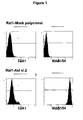


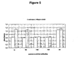


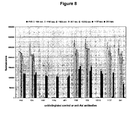

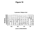


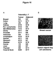


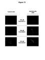
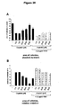
Classifications
-
- C—CHEMISTRY; METALLURGY
- C07—ORGANIC CHEMISTRY
- C07K—PEPTIDES
- C07K16/00—Immunoglobulins [IGs], e.g. monoclonal or polyclonal antibodies
- C07K16/18—Immunoglobulins [IGs], e.g. monoclonal or polyclonal antibodies against material from animals or humans
- C07K16/28—Immunoglobulins [IGs], e.g. monoclonal or polyclonal antibodies against material from animals or humans against receptors, cell surface antigens or cell surface determinants
- C07K16/2863—Immunoglobulins [IGs], e.g. monoclonal or polyclonal antibodies against material from animals or humans against receptors, cell surface antigens or cell surface determinants against receptors for growth factors, growth regulators
-
- A—HUMAN NECESSITIES
- A61—MEDICAL OR VETERINARY SCIENCE; HYGIENE
- A61K—PREPARATIONS FOR MEDICAL, DENTAL OR TOILETRY PURPOSES
- A61K31/00—Medicinal preparations containing organic active ingredients
- A61K31/28—Compounds containing heavy metals
- A61K31/282—Platinum compounds
-
- A—HUMAN NECESSITIES
- A61—MEDICAL OR VETERINARY SCIENCE; HYGIENE
- A61K—PREPARATIONS FOR MEDICAL, DENTAL OR TOILETRY PURPOSES
- A61K31/00—Medicinal preparations containing organic active ingredients
- A61K31/70—Carbohydrates; Sugars; Derivatives thereof
- A61K31/7028—Compounds having saccharide radicals attached to non-saccharide compounds by glycosidic linkages
- A61K31/7034—Compounds having saccharide radicals attached to non-saccharide compounds by glycosidic linkages attached to a carbocyclic compound, e.g. phloridzin
- A61K31/704—Compounds having saccharide radicals attached to non-saccharide compounds by glycosidic linkages attached to a carbocyclic compound, e.g. phloridzin attached to a condensed carbocyclic ring system, e.g. sennosides, thiocolchicosides, escin, daunorubicin
-
- A—HUMAN NECESSITIES
- A61—MEDICAL OR VETERINARY SCIENCE; HYGIENE
- A61K—PREPARATIONS FOR MEDICAL, DENTAL OR TOILETRY PURPOSES
- A61K39/00—Medicinal preparations containing antigens or antibodies
- A61K39/395—Antibodies; Immunoglobulins; Immune serum, e.g. antilymphocytic serum
- A61K39/39533—Antibodies; Immunoglobulins; Immune serum, e.g. antilymphocytic serum against materials from animals
- A61K39/3955—Antibodies; Immunoglobulins; Immune serum, e.g. antilymphocytic serum against materials from animals against proteinaceous materials, e.g. enzymes, hormones, lymphokines
-
- A—HUMAN NECESSITIES
- A61—MEDICAL OR VETERINARY SCIENCE; HYGIENE
- A61K—PREPARATIONS FOR MEDICAL, DENTAL OR TOILETRY PURPOSES
- A61K45/00—Medicinal preparations containing active ingredients not provided for in groups A61K31/00 - A61K41/00
- A61K45/06—Mixtures of active ingredients without chemical characterisation, e.g. antiphlogistics and cardiaca
-
- A—HUMAN NECESSITIES
- A61—MEDICAL OR VETERINARY SCIENCE; HYGIENE
- A61P—SPECIFIC THERAPEUTIC ACTIVITY OF CHEMICAL COMPOUNDS OR MEDICINAL PREPARATIONS
- A61P11/00—Drugs for disorders of the respiratory system
-
- A—HUMAN NECESSITIES
- A61—MEDICAL OR VETERINARY SCIENCE; HYGIENE
- A61P—SPECIFIC THERAPEUTIC ACTIVITY OF CHEMICAL COMPOUNDS OR MEDICINAL PREPARATIONS
- A61P15/00—Drugs for genital or sexual disorders; Contraceptives
-
- A—HUMAN NECESSITIES
- A61—MEDICAL OR VETERINARY SCIENCE; HYGIENE
- A61P—SPECIFIC THERAPEUTIC ACTIVITY OF CHEMICAL COMPOUNDS OR MEDICINAL PREPARATIONS
- A61P35/00—Antineoplastic agents
-
- A—HUMAN NECESSITIES
- A61—MEDICAL OR VETERINARY SCIENCE; HYGIENE
- A61P—SPECIFIC THERAPEUTIC ACTIVITY OF CHEMICAL COMPOUNDS OR MEDICINAL PREPARATIONS
- A61P35/00—Antineoplastic agents
- A61P35/04—Antineoplastic agents specific for metastasis
-
- A—HUMAN NECESSITIES
- A61—MEDICAL OR VETERINARY SCIENCE; HYGIENE
- A61P—SPECIFIC THERAPEUTIC ACTIVITY OF CHEMICAL COMPOUNDS OR MEDICINAL PREPARATIONS
- A61P43/00—Drugs for specific purposes, not provided for in groups A61P1/00-A61P41/00
-
- A—HUMAN NECESSITIES
- A61—MEDICAL OR VETERINARY SCIENCE; HYGIENE
- A61K—PREPARATIONS FOR MEDICAL, DENTAL OR TOILETRY PURPOSES
- A61K39/00—Medicinal preparations containing antigens or antibodies
- A61K2039/505—Medicinal preparations containing antigens or antibodies comprising antibodies
-
- C—CHEMISTRY; METALLURGY
- C07—ORGANIC CHEMISTRY
- C07K—PEPTIDES
- C07K2317/00—Immunoglobulins specific features
- C07K2317/20—Immunoglobulins specific features characterized by taxonomic origin
- C07K2317/24—Immunoglobulins specific features characterized by taxonomic origin containing regions, domains or residues from different species, e.g. chimeric, humanized or veneered
-
- C—CHEMISTRY; METALLURGY
- C07—ORGANIC CHEMISTRY
- C07K—PEPTIDES
- C07K2317/00—Immunoglobulins specific features
- C07K2317/50—Immunoglobulins specific features characterized by immunoglobulin fragments
- C07K2317/56—Immunoglobulins specific features characterized by immunoglobulin fragments variable (Fv) region, i.e. VH and/or VL
- C07K2317/565—Complementarity determining region [CDR]
-
- C—CHEMISTRY; METALLURGY
- C07—ORGANIC CHEMISTRY
- C07K—PEPTIDES
- C07K2317/00—Immunoglobulins specific features
- C07K2317/70—Immunoglobulins specific features characterized by effect upon binding to a cell or to an antigen
- C07K2317/73—Inducing cell death, e.g. apoptosis, necrosis or inhibition of cell proliferation
-
- C—CHEMISTRY; METALLURGY
- C07—ORGANIC CHEMISTRY
- C07K—PEPTIDES
- C07K2317/00—Immunoglobulins specific features
- C07K2317/70—Immunoglobulins specific features characterized by effect upon binding to a cell or to an antigen
- C07K2317/75—Agonist effect on antigen
-
- C—CHEMISTRY; METALLURGY
- C07—ORGANIC CHEMISTRY
- C07K—PEPTIDES
- C07K2317/00—Immunoglobulins specific features
- C07K2317/70—Immunoglobulins specific features characterized by effect upon binding to a cell or to an antigen
- C07K2317/76—Antagonist effect on antigen, e.g. neutralization or inhibition of binding
-
- C—CHEMISTRY; METALLURGY
- C07—ORGANIC CHEMISTRY
- C07K—PEPTIDES
- C07K2317/00—Immunoglobulins specific features
- C07K2317/90—Immunoglobulins specific features characterized by (pharmaco)kinetic aspects or by stability of the immunoglobulin
- C07K2317/92—Affinity (KD), association rate (Ka), dissociation rate (Kd) or EC50 value
Definitions
- the present invention refers to antibodies, particularly to monoclonal antibodies, which bind to the extracellular domain of the AXL receptor tyrosine kinase and which at least partially inhibit AXL activity.
- the AXL (Ark, UFO, Tyro-7) receptor tyrosine kinase is a member of the Tyro-3 family of kinases with the other members being Mer (Eyk, Nyk, Tyro-12) and Sky (Rse, Tyro-3, Dtk, Etk, Brt, Tif). It is activated by binding of the heterophilic ligand Gas6, a 70-kDa protein homologous to the anticoagulation factor protein S. In contrast to other receptor tyrosine kinases, AXL tyrosine phosphorylation can also be induced by homophilic binding.
- AXL activation leads to signalling through PI-3-kinase/Akt ( Franke et al., Oncogene 22: 8983-8998, 2003 ) and other major pathways like Ras/Erk and ⁇ -catenin/TCF ( Goruppi et al., Mol. Cell Biol. 21: 902-915, 2001 ).
- AXL is weakly expressed in a range of normal tissues, including brain, heart, skeletal muscle, the organ capsules and connective tissues of several other organs, and in monocytes, but not lymphocytes.
- Akt phosphorylation induced by AXL has been described in survival of fibroblasts ( Goruppi et al., Mol Cell Biol 17: 4442-4453 1997 ), endothelial cells ( Hasanbasic et al., Am J Physiol Heart Circ Physiol, 2004 ), vascular smooth muscle cells ( Melaragno et al., J. Mol. Cell Cardiol. 37: 881-887, 2004 ) and neurons ( Allen et al., Mol. Endocrinol. 13: 191-201 1999 ). Furthermore, AXL plays a role in cell-adhesion and chemotaxis. AXL knockouts display impaired platelet aggregate stabilization and thrombus formation as a result of reduced activation of the platelet integrin Ilb3.
- AXL overexpression has been demonstrated in various cancer types, e.g. breast ( Meric et al., Clin. Cancer Res. 8: 361-367, 2002 ; Berclaz et al., Ann. Oncol. 12: 819-824, 2001 ), colon ( Chen et al., Int. J. Cancer 83: 579-584, 1999 ; Craven et al., Int. J. Cancer 60: 791-797, 1995 ), prostate ( Jacob et al., Cancer Detect. Prev.
- AXL has been identified as a metastasis-associated gene that is upregulated in aggressive breast cancer cell lines compared to non-invasive cells. In vitro, AXL activity was found to be required for migration and invasion, and this activity could be inhibited by antibody treatment ( WO04008147 ).
- EP 1 382969 discloses a method of determining the invasivity of malignant disorders. US 2007/065444 relates to a method of evaluating a compound for use in treatment. Similarly, abrogation of AXL activity in vivo, either via expression of a dominant negative version of AXL ( Vajkoczy, P., et al., Proc. Natl. Acad. Science U.S.A. 103: 5799-5804. 2005 ) or by siRNA mediated downregulation of AXL ( Holland et al., Cancer Res. 65: 9294-9303, 2005 ) prevented subcutaneous and orthotopic cell growth in murine xenograft experiments.
- AXL (Z49M): Sc-73715 is a monoclonal antibody raised against the extracellular domain of AXL of human origin.
- One antibody is capable of reducing AXL mediated cell invasion ( WO04008147 ) whereas the other antibody has been reported to reduce AXL/Ligand interaction.
- both antibodies are polyclonal rendering them unsuitable for therapeutic administration.
- a first aspect of the present invention relates to a monoclonal antibody as defined in the claims that binds to the extracellular domain of AXL, particularly of human AXL, and at least partially inhibits AXL activity.
- the antibody of the present invention further possesses at least one or more of the following properties: the ability to reduce or block AXL-mediated signal transduction, the ability to reduce or block AXL phosphorylation, the ability to reduce or block cell proliferation, the ability to reduce or block angiogenesis, the ability to reduce or block cell migration, the ability to reduce or block tumor metastasis, the ability to reduce or block AXL mediated PI3K signaling .and the ability to reduce or block AXL mediated anti-apoptosis, thereby increasing for example the sensitivity of a cell against treatment with an antineoplastic agent.
- the antibodies of the present invention may exhibit high specificity for AXL, particularly human AXL and do not significantly recognize other Tyro-3 family members, e.g. MER and/or SKY and/or mammalian non-primate AXL, such as murine AXL. Antibody specificity may be determined by measurements of cross-reactivity as described in the Examples.
- the term "activity" refers to the biological function of AXL, which influences the phenotype of a cell, in particular but not limited to cancer phenotypes such as evasion of apoptosis, self sufficiency in growth signals, cell proliferation, tissue invasion and/or metastasis, insensitivity to anti-growth signals (anti-apoptosis) and/or sustained angiogenesis.
- AXL mediated signal transduction means the activation of second messenger pathways triggered by direct or indirect interaction of AXL with second messenger molecules.
- AXL phosphorylation refers to the phosphorylation of amino acid residues, preferably tyrosine residues, either by a second AXL protein (transphosphorylation) or by another protein having protein kinase activity.
- cell proliferation refers to all AXL-involving processes underlying the reproduction of human cells, in particular but not limited to human cancer cells. They contribute to or result in the replication of cellular DNA, separation of the duplicated DNA into two equally sized groups of chromosomes, and the physical division (called cytokinesis) of entire cells, and shall be stimulated or mediated by non-catalytic or catalytic activities of AXL, preferably including AXL phosphorylation and/or AXL-mediated signal transduction.
- angiogenesis refers to all AXL-involving processes that contribute to the growth of new blood vessels from pre-existing vessels, in particular but not limited to new tumor supplying blood vessels. These processes include multiple cellular events such as proliferation, survival, migration and sprouting of vascular endothelial cells, attraction and migration of pericytes as well as basal membrane formation for vessel stabilization, vessel perfusion, or secretion of angiogenic factors by stromal or neoplastic cells, and shall be stimulated or mediated by non-catalytic or catalytic activities of AXL, preferably including AXL phosphorylation and/or AXL-mediated signal transduction.
- AXL preferably including AXL phosphorylation and/or AXL-mediated signal transduction.
- metastasis refers to all AXL-involving processes that support cancer cells to disperse from a. primary tumor, penetrate into lymphatic and/or blood vessels, circulate through the bloodstrearii, and grow in a distant focus (metastasis) in normal tissues elsewhere in the body.
- tumor cells such as proliferation, migration, anchorage independence, evasion of apoptosis, or secretion of angiogenic factors, that underly metastasis and are stimulated or mediated by non-catalytic or catalytic activities of AXL, preferably including AXL phosphorylation and/or AXL-mediated signal transduction.
- AXL mediated anti-apoptosis refers to all AXL-involving processes that prevent human cells, preferably but not limited to human cancer cells from programmed cell death (apoptosis). In particular, it refers to processes that prevent human cells, preferably but not limited to human cancer cells from induction of apoptosis through growth factor withdrawal, hypoxia, exposure to chemotherapeutic agents or radiation, or initiation of the Fas/Apo-1 receptor-mediated signaling, and are stimulated or mediated by non-catalytic or catalytic activities of AXL, preferably including AXL phosphorylation and/or AXL-mediated signal transduction.
- the activity can be determined using surface plasmon resonance with Biacore, and/or by ELISA (enzyme-linked immunosorbent assays), EIA (enzyme immunoassays), RIA (radioimmunoassays), or fluorescent antibody techniques, e.g. FACS.
- the antibody may have at least one antigen binding site, e.g. one or two antigen binding sites. Further, the antibody comprises at least one heavy immunoglobulin chain and at least one light immunoglobulin chain.
- An immunoglobulin chain comprises a variable domain and optionally a constant domain.
- a variable domain comprises complementary determining regions (CDRs), a CDR1, CDR2 and CDR3 region, and framework regions.
- CDR complementary determining region
- CDR complementary determining region
- An antibody of the invention that binds to the extracellular domain of AXL comprises at least one heavy chain amino acid sequence comprising the CDRs selected
- the present invention refers to an antibody comprising a heavy chain amino acid sequence selected from the group consisting of SEQ ID NO: 8 or at least the variable domain thereof and a light chain amino acid sequence selected from the group consisting of SEQ. ID NO: 7 or at least the variable domain thereof.
- sequence identity between two polypeptide sequences, indicates the percentage of amino acids that are identical between the sequences.
- Preferred polypeptide sequences of the invention have a sequence identity of at least 90%.
- the antibody is 11 B7.
- the antibody may be any antibody of natural and/or synthetic origin, e.g. an antibody of mammalian origin.
- the constant domain -if present- is a human constant domain.
- the variable domain is preferably a mammalian variable domain, e.g. a humanized or a human variable domain. More preferably, the antibody is a chimeric, humanized or human antibody.
- the antibody of the invention may be of the IgA-, IgD-, IgE, IgG-or IgM-type, preferably of the IgG-or IgM-type including, but not limited to, the IgG1-, IgG2-, IgG3-, IgG4-, IgM1-and IgM2-type.
- the antibody is of the human IgG1-, IgG2-or IgG4-type.
- antibodies that are generated need not initially possess such an isotype but, rather the antibody as generated can possess any isotype and that the antibody can be isotype-switched by using the molecularly cloned V region genes or cloned constant region genes or cDNAs in appropriate expression vectors using conventional molecular biological techniques that are well known in the art and then expressing the antibodies in host cells using techniques known in the art.
- scaffold protein means a polypeptide or protein with exposed surface areas in which amino acid insertions, substitutions or deletions are highly tolerable.
- scaffold proteins that can be used in accordance with the present invention are protein A from Staphylococcus aureus, the bilin binding protein from Pieris brassicae or other lipocalins, ankyrin repeat proteins, wand human fibronectin (reviewed in Binz and Pluckthun, Curr Opin Biotechnol,16: 459-69, 2005 ).
- Engineering of a scaffold protein can be regarded as grafting or integrating an affinity function onto or into the structural framework of a stably folded protein.
- Affinity function means a protein binding affinity according to the present invention.
- a scaffold can be structurally separable from the amino acid sequences conferring binding specificity.
- proteins appearing suitable for the development of such artificial affinity reagents may be obtained by rational, or most commonly, combinatorial protein engineering techniques such as panning against AXL, either purified protein or protein displayed on the cell surface, for binding agents in an artificial scaffold library displayed in vitro, skills which are known in the art ( Skerra, J. Mol. Recog., Biochim Biophys Acta, 1482: 337-350, 2000 ; Binz and Plückthun, Curr Opin Biotechnol, 16: 459-69, 2005 ).
- a scaffold protein having an antibody like binding activity can be derived from an acceptor polypeptide containing the scaffold domain, which can be grafted with binding domains of a donor polypeptide to confer the binding specificity of the donor polypeptide onto the scaffold domain containing the acceptor polypeptide.
- the inserted binding domains may include, for example, at least one CDR of an anti-AXL antibody, preferably at least one selected from the group of SEQ ID NOs: 13-30. Insertion can be accomplished by various methods known to those skilled in the art including, for example, polypeptide synthesis, nucleic acid synthesis of an encoding amino acid as well by various forms of recombinant methods well known to those skilled in the art.
- variable domain the heavy chain VH and light chain VL
- the variable domain of an antibody comprises three complementary determining regions sometimes called hypervariable regions, flanked by four relatively conserved framework regions or "FRs".
- FRs relatively conserved framework regions
- the specificity of an antibody is determined or largely determined by a CDR, such as a CDR of the VH chain or a plurality of CDRs.
- CDR such as a CDR of the VH chain or a plurality of CDRs.
- the antibodies of the present invention or their corresponding immunoglobulin chain(s) can be further modified using conventional techniques known in the art, for example, by using amino acid deletion(s), insertion(s), substitution(s), addition(s), and/or recombination(s) and/or any other modification(s) known in the art either alone or in combination.
- Methods for introducing such modifications in the DNA sequence underlying the amino acid sequence of an immunoglobulin chain are well known to the person skilled in the art; see, e.g., Sambrook, Molecular Cloning A Laboratory Manual, Cold Spring Harbor Laboratory (1989) N.Y .
- the antibodies are optionally deimmunized for therapeutic purposes.
- a deimmunized antibody is a protein devoid of or reduced for epitopes that can be recognized by T helper lymphozytes. An example how to identify said epitopes is shown in Tangri et al., (J Immunol. 174: 3187-96, 2005 ).
- the manufacture of deimmunized antibody fragments or derivative thereof may be carried out as described in U.S. Pat. Nos. 6,054,297 , 5,886,152 and 5,877,293 .
- the antibodies herein specifically include "chimeric" antibodies (immunoglobulins) in which a portion of the heavy and/or light chain is identical with or homologous to corresponding sequences in antibodies derived from a particular species or belonging to a particular antibody class or subclass, while the remainder of the chain(s) is identical with or homologous to corresponding sequences in antibodies derived from another species or belonging to another antibody class or subclass, as well as fragments of such antibodies, so long as they exhibit the desired biological activity ( U.S. Pat. No. 4,816,567 ; Morrison et al., Proc. Natl. Acad. Sci. USA, 81:6851-6855 (1984 )).
- the production of chimeric antibodies is described, for example, in WO 89/09622 .
- the present invention refers to a chimerized antibody comprising a heavy chain amino acid sequence selected from the group consisting of SEQ ID NOs: 38, 39, 41, 42 or at least the variable domain thereof and a light chain amino acid sequence selected from the group consisting of SEQ. ID NO: 37 or at least the variable domain thereof.
- the antibodies of the present invention are humanized or fully human antibodies.
- Humanized forms of the antibodies may be generated according to the methods known in the art such as chimerization or CDR grafting. Alternative methods for the production of humanized antibodies are well known in the art and are described in, e.g., EP-A1 0 239 400 and WO 90/07861 .
- a humanized antibody has one or more amino acid residues introduced into it from a source which is non-human. These non-human amino acid residues are often referred to as "import" residues, which are typically taken from an "import” variable domain.
- Humanization can be for example performed following the method of Winter and co-workers ( Jones et al., Nature, 321:522-525 (1986 ); Riechmann et al., Nature, 332:323-327 (1988 ); Verhoeyen et al., Science, 239:1534-1536 (1988 )), by substituting CDRs or CDR sequences of non human origin for the corresponding sequences of a human antibody. Accordingly, such "humanized" antibodies are chimeric antibodies ( U.S. Pat. No. 4,816,567 ) wherein substantially less than an intact human variable domain has been substituted by the corresponding sequence from a non-human species. In practice, humanized antibodies are typically human antibodies in which some CDR residues and possibly some FR residues are substituted by residues from analogous sites in non human antibodies.
- the present invention refers to a humanized antibody comprising a heavy chain amino acid sequence selected from the group consisting of SEQ ID NO: 44 or at least the variable domain thereof and a light chain amino acid sequence selected from the group consisting of SEQ. ID NO: 43 or at least the variable domain thereof.
- XenoMouse® strains of mice that have been engineered to contain up to but less than 1000 kb sized germline configured fragments of the human heavy chain locus and kappa light chain locus. See, Mendez et al., , (Nature Genetics 15:146-156 1997 ), and Green and Jakobovits, ,(J. Exp. Med. 188:483-495, 1998 ).
- the XenoMouse® strains are available from AMGEN, Inc. (formerly ABGENIX, Fremont, CA).
- minilocus In an alternative approach, others, including GenPharm International, Inc., have utilized a "minilocus" approach. In the minilocus approach, an exogenous Ig locus is mimicked through the inclusion of pieces (individual genes) from the Ig locus. Thus, one or more VH genes, one or more DH genes, one or more JH genes, a mu constant region, and usually a second constant region (preferably a gamma constant region) are formed into a construct for insertion into an animal. This approach is described in U.S. Patent No. 5,545,807 to Surani et al. and U.S. Patent Nos.
- Kirin has also demonstrated the generation of human antibodies from mice in which, through microcell fusion, large pieces of chromosomes, or entire chromosomes, have been introduced. See, European Patent Application Nos. 773 288 and 843 961 . Additionally, KMTM mice, which are the result of cross-breeding of Kirin's Tc mice with Medarex's minilocus (Humab) mice have been generated. These mice possess the human IgH transchromosome of the Kirin mice and the kappa chain transgene of the Genpharm mice ( Ishida et al., , Cloning Stem Cells 4:91-102, 2002 ).
- Human antibodies can also be derived by in vitro methods. Suitable examples include but are not limited to phage display (CAT, Morphosys, Dyax, Biosite/Medarex, Xoma, Symphogen, Alexion (formerly Proliferon), Affimed) ribosome display (CAT), yeast display, and the like.
- the antibody may be conjugated with a therapeutic effector group, e.g. a radioactive group or a cytotoxic group.
- a therapeutic effector group e.g. a radioactive group or a cytotoxic group.
- the antibody may be labelled.
- Suitable labels include radioactive labels, fluorescent labels, or enzyme labels.
- xenogenic antibodies are so-called xenogenic antibodies.
- the general principle for the production of xenogenic antibodies such as human antibodies in mice is described in, e.g., WO9110741 , WO 9402602 , WO 9634096 and WO 9633735 .
- the antibody of the invention may exist in a variety of forms besides complete antibodies; including, for example, Fv, Fab' and F(ab') 2 as well as in single chains; see e.g. WO8809344 .
- the antibodies of the invention may be mutated in the Variable domains of the heavy and/or light chains to alter a binding property of the antibody.
- a mutation may be made in one or more of the CDR regions to increase or decrease the Kd of the antibody for AXL, or to alter the binding specificity of the antibody.
- Techniques in site directed mutagenesis are well-known in the art. See, e.g., Sambrook et al. and Ausubel et al., supra.
- mutations may be made at an amino acid residue that is known to be changed compared to germline in a variable region of an AXL antibody.
- mutations may be introduced into one or more of the framework regions.
- a mutation may be made in a framework region or constant domain to increase the half-life of the AXL antibody. See, e.g., WO0009560 .
- a mutation in a framework region or constant domain may also be made to alter the immunogenicity of the antibody, to provide a site for covalent or non-covalent binding to another molecule, or to alter such properties as complement fixation. Mutations may be made in each of the framework regions, the constant domain and the variable regions in a single mutated antibody. Alternatively, mutations may be made in only one of the framework regions, the variable regions or the constant domain in a single mutated antibody.
- the antibody may have a constant domain with effector functions, whereby AXL expressing cells which have bound the antibody, antibody fragment or derivative thereof on the cell surface may be attacked by immune system functions.
- the antibody may be capable of fixing complement and participating in complement-dependent cytotoxicity (CDC).
- the antibody may be capable of binding to Fc receptors on effector cells, such as monocytes and natural killer (NK) cells, and participate in antibody-dependent cellular cytotoxicity (ADCC).
- the antibodies of the invention are applicable for therapeutic treatment, preferably for treatment of hyperproliferative diseases, cardiovascular diseases, in particular artherosclerosis and thrombosis, diabetes related diseases, in particular glomerular hypertrophy or diabetic nephropathy, and particularly of disorders associated with, accompanied by or caused by AXL expression, overexpression or hyperactivity.
- the hyperproliferative diseases are preferably selected from disorders associated with, accompanied by or caused by AXL expression, overexpression or hyperactivity, such as cancer, e.g.
- breast cancer colon cancer
- lung cancer kidney cancer
- follicular lymphoma myeloid leukemia
- skin cancer/melanoma skin cancer/melanoma
- glioblastoma ovarian cancer
- prostate cancer pancreatic cancer
- Barrett's esophagus and esophageal cancer stomach cancer, bladder cancer, cervical cancer, liver cancer, thyroid cancer, and head and neck cancer, or hyperplastic and neoplastic diseases or other AXL expressing or overexpressing hyperproliferative diseases.
- antibodies of the present invention can be used for the co-administration with an antineoplastic agent for the treatment of one of the above mentioned disorders.
- Co-administration includes the administration of an antibody of the present invention with an antineoplastic agent, preferably an apoptosis inducing antineoplastic agent.
- the term co-administration further includes the administration of the antibody of the present invention and the antineoplastic agent, preferably an apoptosis inducing antineoplastic agent, in the form of a single composition or in the form of two or more distinct compositions.
- Co-administration includes the administration of an antibody of the present invention with an antineoplastic agent, preferably an apoptosis inducing antineoplastic agent simultaneously (i.e. at the same time) or sequentially, (i.e. at intervals).
- the invention further relates to a nucleic acid molecule encoding the antibody, antibody fragment or derivative thereof of the invention.
- the nucleic acid molecule of the invention encoding the above-described antibody, antibody fragment or derivative thereof may be, e.g. DNA, cDNA, RNA or synthetically produced DNA or RNA or recombinantly produced chimeric nucleic acid molecule comprising any of those nucleic acid molecules either alone or in combination.
- the nucleic acid molecule may also be genomic DNA corresponding to the entire gene or a substantial portion thereof or to fragments and derivatives thereof.
- the nucleotide sequence may correspond to the naturally occurring nucleotide sequence or may contain single or multiple nucleotide substitutions, deletions or additions.
- the nucleic acid molecule is a cDNA molecule.
- the invention relates to an isolated nucleic acid molecule selected from the group consisting of:
- the invention also relates to a vector comprising a nucleic acid molecule of the invention.
- Said vector may be, for example, a phage, plasmid, viral or retroviral vector.
- Retroviral vectors may be replication competent or replication defective. In the latter case, viral propagation generally will occur only in complementing host cells.
- the nucleic acid molecules of the invention may be joined to a vector containing selectable markers for propagation in a host.
- a plasmid vector is introduced in a precipitate such as a calcium phosphate precipitate or rubidium chloride precipitate, or in a complex with a charged lipid or in carbon-based clusters, such as fullerens.
- a virus it may be packaged in vitro using an appropriate packaging cell line prior to application to host cells.
- the vector of the invention is an expression vector wherein the nucleic acid molecule is operatively linked to one or more control sequences allowing the transcription and optionally expression in prokaryotic and/or eukaryotic host cells.
- Expression of said nucleic acid molecule comprises transcription of the nucleic acid molecule, preferably into a translatable mRNA.
- Regulatory elements ensuring expression in eukaryotic cells preferably mammalian cells, are well known to those skilled in the art. They usually comprise regulatory sequences ensuring initiation of transcription and optionally poly-A signals ensuring termination of transcription and stabilization of the transcript. Additional regulatory elements may include transcriptional as well as translational enhancers.
- Possible regulatory elements permitting expression in prokaryotic host cells comprise, e.g., the lac, trp or tac promoter in E. coli, and examples for regulatory elements permitting expression in eukaryotic host cells are the AOXI or GAL1 promoter in yeast or the CMV-, SV40-, RSV-promoter (Rous sarcoma virus), CMV-enhancer, SV40-enhancer or a globin intron in mammalian and other animal cells.
- Beside elements which are responsible for the initiation of transcription such regulatory elements may also comprise transcription termination signals, such as the SV40-poly-A site or the tk-poly-A site, downstream of the polynucleotide.
- suitable expression vectors are known in the art such as Okayama-Berg cDNA expression vector pcDV1 (Pharmacia), pCDMB, pRc/CMV, pcDNA1, pcDNA3 (Invitrogen) or pSPORTI (GIBCO BRL).
- said vector is an expression vector and/or a gene transfer or targeting vector.
- Expression vectors derived from viruses such as retroviruses, vaccinia virus, adeno-associated virus, herpes viruses, or bovine papilloma virus, may be used for delivery of the polynucleotides or vector of the invention into targeted cell population.
- nucleic acid molecules of the invention can be reconstituted into liposomes for delivery to target cells.
- the invention further relates to a host comprising the vector of the invention. Said host may be a prokaryotic or eukaryotic cell or a non-human transgenic animal.
- the polynucleotide or vector of the invention which is present in the host may either be integrated into the genome of the host or it may be maintained extrachromosomally.
- the nucleic acid molecule of the invention can be used for "gene targeting” and/or “gene replacement”, for restoring a mutant gene or for creating a mutant gene via homologous recombination; see for example Mouellic, Proc. Nat!. Acad. Sci. USA, 87 (1990), 4712-4716 ; Joyner, Gene Targeting, A Practical Approach, Oxford University Press .
- the host can be any prokaryotic or eukaryotic cell, such as a bacterial, insect, fungal, plant, animal, mammalian or, preferably, human cell.
- Preferred fungal cells are, for example, those of the genus Saccharomyces, in particular those of the species S. cerevisiae.
- the term "prokaryotic" is meant to include all bacteria which can be transformed or transfected with a polynucleotide for the expression of a variant polypeptide of the invention.
- Prokaryotic hosts may include gram negative as well as gram positive bacteria such as, for example, E. coli, S. typhimurium, Serratia marcescens and Bacillus subtilis.
- a polynucleotide coding for a mutant form of variant polypeptides of the invention can be used to transform or transfect the host using any of the techniques commonly known to those of ordinary skill in the art. Methods for preparing fused, operably linked genes and expressing them in bacteria or animal cells are well-known in the art ( Sambrook, Molecular Cloning A Laboratory Manual, Cold Spring Harbor Laboratory (2001, Third Editi on). The genetic constructs and methods described therein can be utilized for expression of variant antibodies, antibody fragments or derivatives thereof of the invention in, e.g., prokaryotic hosts. In general, expression vectors containing promoter sequences which facilitate the efficient transcription of the inserted nucleic acid molecule are used in connection with the host.
- the expression vector typically contains an origin of replication, a promoter, and a terminator, as well as specific genes which are capable of providing phenotypic selection of the transformed cells.
- the transformed prokaryotic hosts can be grown in fermentors and cultured according to techniques known in the art to achieve optimal cell growth.
- the antibodies, antibody fragments or derivatives thereof of the invention can then be isolated from the growth medium, cellular lysates, or cellular membrane fractions.
- the isolation and purification of the microbially or otherwise expressed antibodies, antibody fragments or derivatives thereof of the invention may be by any conventional means such as, for example, preparative chromatographic separations and immunological separations such as those involving the use of monoclonal or polyclonal antibodies.
- the host is a bacterium, fungal, plant, amphibian or animal cell.
- Preferred animal cells include but are not limited to Chinese hamster ovary (CHO) cells, baby hamster kidney (BHK) cells, monkey kidney cells (COS), 3T3 cells, NSO cells and a number of other cell lines including human cells, for example Per.C6.
- said animal cell is an insect cell.
- Preferred insect cells include but are not limited to cells of the SF9 cell lines
- said host is a human cell or human cell line.
- Said human cells include, but are not limited to Human embryonic kidney cells (HEK293, 293T, 293 freestyle).
- said human cell lines include, but are not limited to HeLa cells, human hepatocellular carcinoma cells (e. g., Hep G2), A549 cells.
- the invention also provides transgenic non-human animals comprising one or more nucleic acid molecules of the invention that may be used to produce antibodies of the invention.
- Antibodies can be produced in and recovered from tissue or body fluids, such as milk, blood or urine, of goats, cows, horses, pigs, rats, mice, rabbits, hamsters or other mammals. See, e. g., U.S. Patent Nos. 5,827,690 ; 5,756,687 ; 5,750,172 ; and 5,741,957 .
- non-human transgenic animals that comprise human immunoglobulin loci can be produced by immunizing with AXL or a portion thereof.
- the invention additionally relates to a method for the preparation of an antibody, comprising culturing the host of the invention under conditions that allow synthesis of said antibody and recovering said antibody from said culture.
- the transformed hosts can be grown in fermentors and cultured according to techniques known in the art to achieve optimal cell growth.
- the whole antibodies, their dimers, individual light and heavy chains, or other immunoglobulin forms of the present invention can be purified according to standard procedures of the art, including ammonium sulfate precipitation, affinity columns, column chromatography, gel electrophoresis and the like; see, Scopes, "Protein Purification", Springer-Verlag, N.Y. (1982 ).
- the antibody or its corresponding immunoglobulin chain(s) of the invention can then be isolated from the growth medium, cellular lysates, or cellular membrane fractions.
- the isolation and purification of the, e.g., microbially expressed antibodies or immunoglobulin chains of the invention may be by any conventional means such as, for example, preparative chromatographic separations and immunological separations such as those involving the use of monoclonal or polyclonal antibodies directed, e.g., against the constant region of the antibody of the invention.
- the antibodies of the invention can be further coupled to other moieties for, e.g., drug targeting and imaging applications.
- Such coupling may be conducted chemically after expression of the antibody or antigen to site of attachment or the coupling product may be engineered into the antibody or antigen of the invention at the DNA level.
- the DNAs are then expressed in a suitable host system, and the expressed proteins are collected and renatured, if necessary.
- the antibody is coupled to an effector, such as a radioisotope or a toxic chemotherapeutic agent.
- an effector such as a radioisotope or a toxic chemotherapeutic agent.
- these antibody conjugates are useful in targeting cells, e.g. cancer cells, expressing AXL, for elimination.
- the linking of antibodies/antibody fragments of the invention to radioisotopes e.g. provides advantages to tumor treatments. Unlike chemotherapy and other forms of cancer treatment, radioimmunotherapy or the administration of a radioisotope-antibody combination directly targets the cancer cells with minimal damage to surrounding normal, healthy tissue.
- Preferred radioisotopes include e.g. 3 H, 14 C, 15 N, 35 S, 90 Y, 99 Tc, 111 In, 125 I, 131 I.
- the antibodies of the invention can be used to treat cancer when being conjugated with toxic chemotherapeutic drugs such as geldanamycin ( Mandler et al., J. Natl. Cancer Inst., 92(19), 1549-51 (2000 )) and maytansin, for example, the maytansinoid drug, DM1 ( Liu et al., Proc. Natl. Acad. Sci. U.S.A. 93:8618-8623 (1996 ) and auristatin-E or monomethylauristatin-E ( Doronina et al., Nat. Biotechnol. 21:778-784 (2003 ) or calicheamicit);.
- toxic chemotherapeutic drugs such as geldanamycin ( Mandler et al., J. Natl. Cancer Inst., 92(19), 1549-51 (2000 )
- maytansin for example, the maytansinoid drug, DM1 ( Liu et
- the invention further relates to a pharmaceutical composition
- a pharmaceutical composition comprising the antibody, the nucleic acid molecule, the vector, the host of the invention or an antibody obtained by the method of the invention.
- composition as employed herein comprises at least one compound of the invention.
- a composition is a pharmaceutical or a diagnostic composition.
- said pharmaceutical composition comprises a pharmaceutically acceptable carrier and/or diluent.
- the herein disclosed pharmaceutical composition may be partially useful for the treatment of disorders as defined in the claims.
- the disorder is cancer or breast cancer, colon cancer, lung cancer, kidney cancer, follicular lymphoma, myeloid leukemia, skin cancer/melanoma, glioblastoma, ovarian cancer, prostate cancer, pancreatic cancer, Barrett's esophagus and esophageal cancer, stomach cancer, bladder cancer, cervical cancer, liver cancer, thyroid cancer, and head and neck cancer, or other hyperplastic or neoplastic diseases.
- hyperactivity refers to uncontrolled AXL signaling which may be caused by a lack and/or dysfunction of negative regulation.
- negative regulation comprises protein dephosphorylation, degradation and/or endocytosis.
- uncontrolled AXL signaling may be the result of genetic alterations, either somatic or germline, which result in changes of the AXL amino acid sequence.
- Suitable pharmaceutical carriers include phosphate buffered saline solutions, water, emulsions, such as oil/water emulsions, various types of wetting agents, sterile solutions etc.
- Compositions comprising such carriers can be formulated by well known conventional methods. These pharmaceutical compositions can be administered to the subject at a suitable dose. Administration of the suitable compositions may be effected by different ways, e.g., by intravenous, intraperitoneal, subcutaneous, intramuscular, topical, intradermal, intranasal or intrabronchial administration.
- compositions of the invention may also be administered directly to the target site, e.g., by biolistic delivery to an external or internal target site, like the brain.
- the dosage regimen will be determined by the attending physician and clinical factors. As is well known in the medical arts, dosages for any one patient depends upon many factors, including the patient's size, body surface area, age, the particular compound to be administered, sex, time and route of administration, general health, and other drugs being administered concurrently. Proteinaceous pharmaceutically active matter may be present in amounts between 1 ⁇ g and 100 mg/kg body weight per dose; however, doses below or above this exemplary range are envisioned, especially considering the aforementioned factors. If the regimen is a continuous infusion, it should also be in the range of 1 pg to 100 mg per kilogram of body weight per minute.
- compositions of the invention may be administered locally or systemically.
- Preparations for parenteral administration include sterile aqueous or non-aqueous solutions, suspensions, and emulsions.
- non-aqueous solvents are propylene glycol, polyethylene glycol, vegetable oils such as olive oil, and injectable organic esters such as ethyl oleate.
- Aqueous carriers include water, alcoholic/aqueous solutions, emulsions or suspensions, including saline and buffered media.
- Parenteral vehicles include sodium chloride solution, Ringer's dextrose, dextrose and sodium chloride, lactated Ringer's, or' fixed oils.
- Intravenous vehicles include fluid and nutrient replenishers, electrolyte replenishers (such as those based on Ringer's dextrose), and the like. Preservatives and other additives may also be present such as, for example, antimicrobials, anti-oxidants, chelating agents, and inert gases and the like.
- the pharmaceutical composition of the invention may comprise further agents depending on the intended use of the pharmaceutical composition. It is particularly preferred that the pharmaceutical composition comprises further active agents like, e.g. an additional antineoplastic agent, small molecule inhibitor, anti-tumor agent or chemotherapeutic agent.
- the invention also relates to a pharmaceutical composition
- a pharmaceutical composition comprising an anti-AXL-antibody of the invention in combination with at least one further antineoplastic agent. Said combination is effective, for example, in inhibiting abnormal cell growth.
- antineoplastic agents are presently known in the art. In general the term includes all agents that are capable of prevention, alleviation and/or treatment of hyperproliferative disorders.
- the antineoplastic agent is selected from the group of therapeutic proteins including but not limited to antibodies or immunomodulatory proteins.
- the antineoplastic agent is selected from the group of small molecule inhibitors or chemotherapeutic agents consisting of mitotic inhibitors, kinase inhibitors, alkylating agents, anti-metabolites, intercalating antibiotics, growth factor inhibitors, cell cycle inhibitors, enzymes, topoisomerase inhibitors, histone deacetylase inhibitors, anti-survival agents, biological response modifiers, anti-hormones, e. g. anti-androgens, and antiangiogenesis agents.
- antineoplastic agents which can be used in combination with the antibodies provided herein include, for example, gefitinib, lapatinib, sunitinib, pemetrexed, bevacisumab, cetuximab, imatinib, trastuzumab, alemtuzumab, rituximab, erlotinib, bortezomib and the like.
- antineoplastic agents to be used in the compositions as described and claimed herein include for example, chemotherapeutic agents such as capecitabine, daunorubicin, daunomycin, dactinomycin, doxorubicin, epirubicin, idarubicin, esorubicin, bleomycin, mafosfamide, ifosfamide, cytosine arabinoside, bis-chloroethylnitrosurea, busulfan, mitomycin C, actinomycin D, mithramycin, prednisone, hydroxyprogesterone, testosterone, tamoxifen, dacarbazine, procarbazine, hexamethylmelamine, pentamethylmelamine, mitoxantrone, I amsacrine, chlorambucil, methylcyclohexylnitrosurea, nitrogen mustards, melphalan, cyclophosphamide, 6-mercaptopurine, 6-thio
- such antineoplastic agents When used with the described AXL antibodies, such antineoplastic agents may be used individually (e.g., 5-FU and an antibody), sequentially (e.g., 5-FU and an antibody for a period of time followed by MTX and an antibody), or in combination with one or more other such antineoplastic agents (e.g., 5-FU, MTX and an antibody, or 5-FU, radiotherapy and an antibody).
- antineoplastic agents may be used individually (e.g., 5-FU and an antibody), sequentially (e.g., 5-FU and an antibody for a period of time followed by MTX and an antibody), or in combination with one or more other such antineoplastic agents (e.g., 5-FU, MTX and an antibody, or 5-FU, radiotherapy and an antibody).
- antineoplastic agent may also include therapeutic procedures, as for example irradiation or radiotherapy.
- the pharmaceutical composition of the invention can be used in human medicine and can be used also for veterinary purposes.
- the invention relates to the use of the antibody of the invention, the nucleic acid molecule, the vector, the host of the invention or an antibody obtained by the method of the invention for the preparation of a pharmaceutical composition for diagnosis, prevention or treatment of hyperproliferative diseases, cardiovascular diseases, in particular artherosclerosis and thrombosis, diabetes related diseases, in particular glomerular hypertrophy or diabetic nephropathy, namely of disorders associated with, accompanied by or caused by AXL expression, overexpression or hyperactivity.
- cardiovascular diseases in particular artherosclerosis and thrombosis
- diabetes related diseases in particular glomerular hypertrophy or diabetic nephropathy, namely of disorders associated with, accompanied by or caused by AXL expression, overexpression or hyperactivity.
- a hyperproliferative disease as mentioned above includes any neoplasia, i.e. any abnormal and/or uncontrolled new growth of tissue.
- the term "uncontrolled new growth of tissue” as used herein may depend upon a dysfunction and/or loss of growth regulation.
- a hyperproliferative disease includes tumor diseases and/or cancer, such as metastatic or invasive cancers.
- said hyperproliferative disease is in particular breast cancer, colon cancer, lung cancer, kidney cancer, follicular lymphoma, myeloid leukemia, skin cancer/melanoma, glioblastoma, ovarian cancer; prostate cancer, pancreatic cancer, Barrett's esophagus and esophageal cancer, stomach cancer, bladder cancer, cervical cancer, liver cancer, thyroid cancer, and head and neck cancer, or hyperplastic or neoplastic diseases or other AXL expressing or overexpressing hyperproliferative diseases.
- the present invention refers to the use of an anti-AXL-antibody of the present invention for the manufacture of a medicament for the co-administration with an antineoplastic agent for the treatment of one of the above mentioned disorders.
- the present invention is directed to the use of an anti-AXL antibody as defined in the claims for the manufacture of a pharmaceutical composition for the treatment of drug resistant cancer.
- the present invention relates to a diagnostic composition
- a diagnostic composition comprising the antibody of the invention, the nucleic acid molecule, the vector, the host of the invention or an antibody obtained by the method of the invention and optionally a pharmaceutically acceptable carrier.
- the diagnostic composition of the invention is useful in the detection of an undesired expression, overexpression or hyperactivity of the mammalian AXL in different cells, tissues or another suitable sample, comprising contacting a sample with an antibody of the invention, and detecting the presence of AXL in the sample. Accordingly, the diagnostic composition of the invention may be used for assessing the onset or the disease status of a hyperproliferative disease.
- malignant cells such as cancer cells expressing AXL
- the cells which have bound the antibody of the invention might thus be attacked by immune system functions such as the complement system or by cell-mediated cytotoxicity, thereby reducing the number of or eradicating cancer cells.
- the antibody of the invention is coupled to a labelling group.
- a labelling group refers to a detectable marker, e.g. a radiolabelled amino acid or biotinyl moieties that can be detected by marked avidin.
- suitable labelling groups include, but are not limited to, the following: radioisotopes or radionuclides (e.g. 3 H, 14 C, 15 N, 35 S, 90 Y, 99 Tc, 111 In, 125 I, 131 I), fluorescent groups (e.g.
- FITC FITC, rhodamine, lanthanide phosphors
- enzymatic groups e.g. horseradish peroxidase, ⁇ -galactosidase, luciferase, alkaline phosphatase
- chemiluminescent groups e.g. horseradish peroxidase, ⁇ -galactosidase, luciferase, alkaline phosphatase
- biotinyl groups e.g. leucine zipper pair sequences, binding sites for secondary antibodies, metal binding domains, epitope tags.
- the labelling groups are attached by spacer arms of various lengths to reduce potential steric hindrance.
- the present invention relates to a method of assessing for the presence of AXL expressing cells comprising contacting the antibody of the invention with cells or a tissue suspected of carrying AXL on their/its surface.
- Suitable methods for detection of AXL expression in a sample may be an Enzyme-Linked Immunosorbent Assay (ELISA) or Immunohistochemistry (IHC).
- An ELISA assay may be carried out in a microtiter plate format, wherein e.g. wells of a microtiter plate, are adsorbed with an AXL antibody.
- the wells are rinsed and treated with a blocking agent such as milk protein or albumin to prevent nonspecific adsorption of the analyte.
- the wells are treated with a test sample.
- the wells After rinsing away the test sample or standard, the wells are treated with a second AXL antibody that is labelled, e.g. by conjugation with biotin.
- the label is detected, e.g. with avidin-conjugated horseradish peroxidase (HRP) and a suitable chromogenic substrate.
- the concentration of the AXL antigen in the test samples is determined by comparison with a standard curve developed from standard samples.
- paraffin-embedded tissues may be used, wherein the tissues are, e.g. first deparaffinized in xylene and then dehydrated, e.g. with ethanol and rinsed in distilled water.
- Antigenic epitopes masked by formalin-fixation and paraffin-embedding may be exposed by epitope unmasking, enzymatic digestion or saponin.
- epitope unmasking paraffin sections may be heated in a steamer, water bath or microwave oven for 20-40 min in an epitope retrieval solution as for example 2N HCl solution (pH 1.0).
- tissue sections may be incubated at 37°C for 10-30 minutes in different enzyme solutions such as proteinase K, trypsin, pronase, pepsin etc.
- tissue sections are treated with a blocking buffer to prevent unspecific interactions.
- the primary AXL antibody is added at appropriate concentrations. Excess primary antibody is rinsed away and sections are incubated in peroxidase blocking solution for 10 min at room temperature. After another washing step, tissue sections are incubated with a secondary labelled antibody, e.g. labelled with a group that might serve as an anchor for an enzyme. Examples therefore are biotin labelled secondary antibodies that are recognized by streptavidin coupled horseradish peroxidase. Detection of the antibody/enzyme complex is achieved by incubating with a suitable chromogenic substrate.
- the present invention relates to a method of blocking AXL function comprising contacting the antibody of the invention with cells or a tissue suspected of carrying AXL on their/its surface under conditions, wherein the antibody is capable of blocking AXL function.
- the contacting may be in vitro or in vivo.
- the present document also describes a method of treating a hyperproliferative disease, cardiovascular diseases, in particular artherosclerosis and thrombosis, diabetes related diseases, in particular glomerular hypertrophy or diabetic nephropathy, comprising, administering to a patient in need thereof a suitable dose of the antibody of the present invention.
- the hyperproliferative disease is selected from disorders associated with, accompanied by or caused by AXL expression, overexpression or hyperactivity, such as cancer, e.g.
- breast cancer colon cancer
- lung cancer kidney cancer
- follicular lymphoma myeloid leukemia
- skin cancer/melanoma skin cancer/melanoma
- glioblastoma ovarian cancer
- prostate cancer pancreatic cancer
- Barrett's esophagus and esophageal cancer stomach cancer, bladder cancer, cervical cancer, liver cancer, thyroid cancer, and head and neck cancer, or hyperplastic and neoplastic diseases or other AXL expressing or overexpressing hyperproliferative diseases.
- the cancer to be treated is a drug resistant cancer.
- the document further relates to a method of treating a disease wherein the antibody of the invention is administered to a mammal and wherein said disease is correlated directly or indirectly with the abnormal level of expression or activity of AXL.
- the invention relates to a kit comprising an anti-AXL-antibody of the invention, the nucleic acid molecule encoding said components and/or the vector of the invention.
- Example 1 Generation of AXL overexpressing Rati fibroblasts as immunogen and AXL overexpressing NIH3T3 fibroblasts as experimental model system
- the full length coding sequence for the human receptor tyrosine kinase AXL transcript variant 1 according to the National Center for Biotechnology Information (NCBI) reference sequence (NM_021913) was subcloned into pLXSN via flanking recognition elements for the restriction endonucleases Eco RI and Bam HI, thereby resulting in the retroviral expression vector pLXS N-hAXL.
- NCBI National Center for Biotechnology Information
- Ratl fibroblasts stably overexpressing human AXL were generated by retroviral gene transfer.
- 3x10 5 Phoenix-E cells were seeded on 60 mm culture dishes and transfected with 2 ⁇ g/ml pLXSN vector or pLXSN-hAXL using the calcium phosphate method. After 24 h, medium was replaced by fresh medium in which Phoenix-E cells were incubated for 4 h.
- a panel of stable clones was propagated and quantified for membrane-localized human AXL expression by FACS analysis.
- 1x10 5 cells were harvested with 10 mM EDTA in PBS, washed once with FACS buffer (PBS, 3% FCS, 0.4% azide) and seeded on a 96 well round bottom plate. The cells were spun for 3 min at 1,000 rpm to remove supernatant and were resuspended with mouse anti-AXL primary antibody MAB154 (R&D Systems, 3 ⁇ g/ml).
- Figure 1 shows the FACS analysis of the polyclonal Ratl-Mock population stably infected with pLXSN empty vector and Ratl-AXL cl.2 stably infected with pLXSN-hAXL, and demonstrates AXL overexpression on the cell surface of this representative clone.
- NIH3T3 fibroblasts stably overexpressing AXL were generated in analogy to procedures described for Ratl.
- 3x10 5 Phoenix-E cells were seeded on 60 mm culture dishes and transfected with 2 ⁇ g/ml pLXSN vector or pLXSN-AXL cDNA using the calcium phosphate method. After 24 h, medium was replaced by fresh medium in which Phoenix-E cells were incubated for 4 h.
- the supernatants of Phoenix-E cells releasing pLXSN or pLXSN-hAXL ecotrophic virus were harvested and used for the incubation of subconfluent NIH3T3 cells (2x10 5 cells per 6 cm dish) for 3 h in the presence of Polybrene (4 mg/ml; Aldrich).
- Phoenix-E cells were re-incubated with fresh medium, which after another 3 h was used for a second infection of the NIH3T3 fibroblasts in the presence of Polybrene (4 mg/ml; Aldrich).
- a third infection cycle was performed. After changing the medium, selection of NIH3T3 cells with G418 was started. Usually, stable clones were picked after selection for 21 days.
- a panel of stable clones was propagated and quantified for membrane-localized AXL expression by FACS analysis.
- 1x10 5 cells were harvested with 10 mM EDTA in PBS, washed once with FACS buffer (PBS, 3% FCS, 0.4% azide) and seeded on a 96 well round bottom plate. The cells were spun for 3 min at 1000 rpm to remove supernatant and were resuspended with mouse anti-AXL primary antibody MAB154 (R&D Systems, 3 pg/ml).
- Figure 2 shows the FACS analysis of the polyclonal NIH3T3-Mock population stably infected with pLXSN empty vector and NIH3T3-AXL cl.7 stably infected with pLXSN-hAXL, and demonstrates AXL overexpression on the cell surface of this representative clone.
- Monoclonal rat anti-AXL antibodies were raised by injection of approximately 10x10 6 frozen cells of Ratl-AXL cl.2 both i.p. and subcutaneously into Lou/C or Long Evans rats. After an 8-week interval, a final boost was given i.p and subcutaneously 3 d before fusion. Fusion of the myeloma cell line P3X63-Ag8.653 with the rat immune spleen cells was performed according to standard procedures and yielded 105 hybridomas. After 2 weeks, first supernatants from hybridomas were collected and tested in a primary FACS screen for binding to NIH3T3-AXL cl.7 fibroblasts versus NIH3T3-Mock control cells.
- Clones positive for AXL binding were further cultivated. From 50 ml supernatant of these clones, antibodies were purified and re-analyzed for specific binding to AXL on NIH3T3-AXL cl.7 fibroblasts versus NIH3T3-Mock control cells. Purified antibodies specifically binding to NIH3T3-AXL cl. 7 fibroblasts but not NIH3T3-Mock control cells were furthermore tested in Akt-Kinase phosphorylation ELISAs, and ELISAs to determine the isotype were performed. For purification of rat antibodies, supernatants were spun for 20 minutes at 5,000g and subsequently sterile filtered.
- oligoclonal antibodies tested 91 specifically bound to NIH3T3-AXL cl. 7 fibroblasts but not NIH3T3-Mock control cells, 9 inhibited Gas6-induced Akt phosphorylation in the same cells, whereas 71 stimulated Akt phosphorylation.
- This example addressed cross-reactivity of rat anti-AXL antibodies of the invention with mouse and cynomolgus monkey AXL as well as with the other members of the human AXL family, human Mer and human Sky. Following subcloning of the mouse and monkey AXL coding sequence as well as human Mer and Sky into pcDNA3, each expression construct was transfected into HEK293T fibroblasts. The ability of rat anti-AXL antibodies of the invention to bind these proteins was tested by FACS analysis.
- mouse AXL expression construct pcDNA3-mAXL was generated.
- the full length coding sequence of mouse AXL was polymerase chain reaction (PCR) amplified using mouse heart cDNA (Ambion) as template and appropriate primers according to the National Center for Biotechnology Information (NCBI) reference sequence (NM_009465) of mouse AXL.
- NCBI National Center for Biotechnology Information
- the full length sequence coding for mouse AXL was thereby covered by two overlapping PCR fragments, a 5'-fragment and 3'-fragment.
- the primers for amplification of these fragments were as follows:
- the 5'-fragment was digested with Eco RI and Bst EII, the 3'-fragment was digested with Bst EII and Not I, and pcDNA3 was cleaved with Eco RI and Not I.
- a three factor ligation of the isolated and purified fragments was performed and transformed into DH5 ⁇ bacterial cells. A single colony was picked and grown in the presence of ampicillin.
- the mouse AXL expression vector pcDNA3-mAXL was purified, and sequence verified for subsequent transient transfection into HEK293T cells.
- the cynomolgus monkey AXL expression constructs pcDNA3-cyAXL was generated.
- the full length coding sequence of cynomolgus monkey AXL was PCR amplified using cDNA prepared from cynomolgus monkey brain tissue as template. Since the nucleotide sequence of cynomolgus monkey AXL was not available, respective primers were designed assuming significant homology to human AXL.
- the full length sequence coding for cynomolgus monkey AXL was thereby covered by two overlapping PCR fragments, a 5'-fragment and 3'-fragment. The primers for amplification of these fragments were as follows:
- the 5'-fragment was digested with Eco RI and Dra III, the 3'-fragment was digested with Dra III and Not I, and pcDNA3 was cleaved with Eco RI and Not I.
- a three factor ligation of the isolated and purified fragments was performed and transformed into DH5 ⁇ bacterial cells. A single colony was picked and grown in the presence of ampicillin.
- the cynomolgus monkey AXL expression vector pcDNA3-cyAXL was purified, and sequence verified for subsequent transient transfection into HEK293T cells.
- the nucleotide and amino acid sequences of cynomolgus monkey are as follows:
- the human Mer expression construct pcDNA3-hMer was generated.
- the full length coding sequence of human Mer was obtained through cleavage of the vector pCMV6-XL4-human Mer (Origene #TC116132) with Eco RI and and Xba I. After digestion of pcDNA3 with the same restriction endonucleases, both fragments were ligated to generate pcDNA3-hMer.
- the 5'-region of the human Mer coding sequence in pcDNA3-hMer was PCR amplified using appropriate primers according to the NCBI reference sequence (NM_006343) of human Mer. The primers for amplification of this fragment were as follows:
- telomere sequence was sequence verified for subsequent transient transfection into HEK293T cells.
- the human Sky expression construct pcDNA3-hSky was generated.
- the full length coding sequence of human Sky was PCR amplified using the vector pCMV6-XL4-human Sky (Origene #MG1044_A02) as template and appropriate primers according to the NCBI reference sequence (NM_006293) of human Sky.
- the primers for amplification were as follows:
- the PCR product and pcDNA3 were digested with Eco RI and Xho I and ligated to generate the pcDNA3-hSky expression vector. Transformed into DH5 ⁇ bacterial cells, a single colony was picked and grown in the presence of ampicillin. Using a commercially available plasmid purification kit (Qiagen), the pcDNA3-hSky expression vector was purified, and sequence verified for subsequent transient transfection into HEK293T cells.
- HEK293T cells were transiently transfected with either pcDNA3 empty vector, pcDNA3-hAXL, pcDNA3mAXL, pcDNA3-cyAXL, pcDNA3-hMer, or pcDNA3-hSky applying the calcium phosphate method.
- pcDNA3 empty vector pcDNA3-hAXL
- pcDNA3mAXL pcDNA3-cyAXL
- pcDNA3-hMer pcDNA3-hSky applying the calcium phosphate method.
- 3x10 6 HEK293T cells in 16 ml medium were seeded on a 15 cm cell tissue culture dish and grown at 7% CO 2 and 37°C for 30 h.
- Figure 3 shows representative results of this experiment. With exception of 12B7 which shows moderate cross-reactivity with mouse AXL as well as human Mer and Sky, non of the other anti-AXL antibodies of the invention cross-reacted with these molecules. In contrast, all of the tested rat anti-AXL antibodies of the invention cross-reacted with cynomolgus monkey AXL.
- Rat anti-AXL antibodies of the invention inhibit ligiand-induced AXL phosphorylation in vitro
- ELISA experiments were performed in order to investigate whether the rat anti-AXL antibodies of the invention are able to block ligand Gas6-mediated activation of AXL.
- Gas6-mediated AXL activation was detected by increased receptor tyrosine phosphorylation.
- 3x10 4 cells per well were seeded in normal growth medium in flat-bottom 96 well plates.
- growth medium was replaced by serum-free medium to starve cells over night for 24 h.
- black Maxi-Sorp 96 well plates (Nunc) were coated with mouse anti-phospho-tyrosine antibody 4G10 at 2 ⁇ g/ml PBS and 4°C.
- lysis buffer 50 mM HEPES, pH 7.5, 150 mM NaCl, 1 mM EDTA, 10% glycerine, and 1% Triton X-100
- phosphatase and protease inhibitors 10 mM Na 4 P 2 O 7 , 1 mM phenylmethylsulfonyl fluoride, 1 mM orthovanadate, 1 mM NaF, and 0,5% aprotinin
- Figure 4 shows representative results of this experiment for NIH3T3-AXL cl.7 fibroblasts (A) and NCl-H292 lung cancer cells (B).
- the rat anti-AXL antibodies 11 B7, 11 D5, 6E7, and 10D12 of the invention were able to block or reduce ligand-mediated AXL activation as indicated by decreased phosphorylation, and are thus considered antagonistic anti-AXL antibodies.
- the rat anti-AXL antibodies 2A1 and 11 D7 of the invention stimulate basal AXL activation as indicated by increased phosphorylation, do not significantly reduce ligand-mediated AXL activation, and are therefore considered agonistic anti-AXL antibodies.
- Rat anti-AXL antibodies of the invention inhibit ligand-induced p42/p44 MAP-Kinase phosphorylation in vitro
- cells were pre-incubated with 10 ⁇ g/ml of the isotypic control antibody 1 D5 as well as the rat anti-AXL antibodies 11 D5, 11 B7, and 2A1 for 1 hr at 37°C and then treated with or without 400 ng/ml Gas6 (R&D Systems) for 10 min at 37°C.
- Medium was flicked out and cells were fixed with 4% formaldehyde in PBS (pH 7.5) for 30 min at room temperature.
- Formaldehyde solution was removed and cells were washed twice with wash buffer (PBS, 0.1 % Tween 20). Cells were quenched with 1% H 2 O 2 , 0.1% NaN 3 in wash buffer and incubated for 20 min at room temperature.
- TMB Tetramethylbenzidine
- Figure 5 shows representative results of this experiment for the cervical cancer cell line CaSki.
- the rat anti-AXL antibodies 11 B7 and 11 D5 of the invention were able to reduce ligand-mediated p42/p44 MAP-Kinase activation as indicated by decreased phosphorylation, and are thus considered antagonistic anti-AXL antibodies.
- the rat anti-AXL antibody 2A1 of the invention stimulates basal p42/p44 MAP-Kinase activation as indicated by increased phosphorylation, does not reduce ligand-mediated p42/p44 MAP-Kinase activation, and is therefore considered an agonistic anti-AXL antibody.
- Similar effects with the same panel of antibodies were observed in the breast cancer cell line Hs578T and the lung cancer cell line NCI-H292.
- Rat anti-AXL antibodies of the invention inhibit ligand-induced Akt phosphorylation in vitro
- ELISA experiments were performed in order to investigate whether the rat anti-AXL antibodies of the invention are able to block ligand Gas6-mediated activation of Akt-Kinase.
- Gas6-mediated Akt-Kinase activation was detected by increased protein (Ser473) phosphorylation.
- Ser473 increased protein
- 2x10 4 cells per well were seeded in flat-bottom 96 well plates.
- normal growth medium was replaced by serum-reduced (DMEM, 0.5% FCS for NIH3T3-AXL cl.7 fibroblasts) or serum-free (for cancer cell lines) medium to starve cells for 36 h.
- cells were pre-incubated with 10 ⁇ g/ml of the isotypic control antibody 1 D5 as well as the rat anti-AXL antibodies 11 D5, 11 B7, and 2A1 for 1 h at 37°C and then treated with or withour 400 ng/ml Gas6 (R&D Systems) for 10 min at 37°C.
- Medium was flicked out and cells were fixed with 4% formaldehyde in PBS (pH 7.5) for 30 min at room temperature.
- Formaldehyde solution was removed and cells were washed twice with wash buffer (PBS, 0.1% Tween 20). Cells were quenched with 1% H 2 O 2 , 0.1% NaN 3 in wash buffer and incubated for 20 min at room temperature.
- TMB Tetramethylbenzidine
- Figure 6 shows representative results of this experiment for NIH3T3-AXL cl.7 fibroblasts (A) and CaLa-1 lung cancer cells (B).
- the rat anti-AXL antibodies 11 B7 and 11 D5 of the invention were able to block or reduce ligand-mediated Akt-Kinase activation as indicated by decreased phosphorylation, and are thus considered antagonistic anti-AXL antibodies.
- the rat anti-AXL antibody 2A1 of the invention stimulates basal Akt-Kinase activation as indicated by increased phosphorylation, does not reduce ligand-mediated Akt-Kinase activation, and is therefore considered an agonistic anti-AXL antibody.
- Chimeric derivatives of the rat anti-AXL antibodies 11B7 and 11D5 were generated as part of this invention (see below).
- ELISA experiments were performed. Antibody-mediated Akt-Kinase inhibition was detected by decreased protein (Ser473) phosphorylation.
- 2x10 4 cells per well were seeded in flat-bottom 96 well plates.
- DMEM serum-reduced medium
- DMEM serum-reduced medium
- cells were pre-incubated with 50 ng/ml, 100 ng/ml, 300 ng/ml, 500 ng/ml, and 1 ⁇ g/ml of rat anti-AXL antibody 11 B7 or chimeric anti-AXL antibody ch11B7, as well as 50 ng/ml, 100 ng/ml, 300 ng/ml, 500 ng/ml, 1 ⁇ g/ml, 5 ⁇ g/ml, and 10 ⁇ g/ml of rat anti-AXL antibody 11 D5 or chimeric anti-AXL antibody ch11 D5 for 1 h at 37°C and then treated with or without 400 ng/ml Gas6 (R&D Systems) for 10 min at 37°C.
- DMEM serum-reduced medium
- Anti-phospho-Akt (Ser473) primary antibody (polyclonal rabbit; Cell Signaling #9271) diluted 1:500 in PBS, 0.5% BSA, 5 mM EDTA was added over night at 4°C. On day 4, the antibody solution was removed and the plate was washed 3x with wash buffer. HRP-conjugated anti-rabbit secondary antibody (Dianova #111-036-045) diluted 1:2,500 in PBS, 0.5% BSA was then added to each well and incubated for 1.5 h at room temperature. The plate was washed 3x with wash buffer and twice with PBS for 5 min each. Tetramethylbenzidine (TMB, Calbiochem) was added and monitored at 620 nm. The reaction was stopped by addition of 100 ⁇ l of 250 nM HCl and the absorbance was read at 450 nm with a reference wavelength of 620 nm using a Vmax plate reader (Thermo Lab Systems).
- Figure 7 demonstrated that rat anti-AXL antibody 11 B7 and chimeric anti-AXL antibody ch11B7 of the invention as well as rat anti-AXL antibody 11 D5 and chimeric anti-AXL antibody ch11 D5 of the invention were able to inhibit ligand-mediated Akt-Kinase activation to similar extent as indicated by decreased phosphorylation.
- the chimeric anti-AXL antibodies ch11 B7 and ch11D5 maintained activity.
- Example 8 Antagonistic rat-anti AXL antibodies of the invention compete with each other for the same or structurally related epitopes and do not share binding sites with agonistic rat-anti AXL antibodies of the invention
- Anti-AXL antibodies of the invention were examined whether they compete with each other for similar binding epitopes on the AXL-ECD domain. Therefore binding of biotinylated anti-AXL antibodies to AXL-ECD domain-coated plates preincubated with anti-AXL antibodies was determined in a competition ELISA.
- 30 ⁇ g of isotypic control antibody 1 D5 as well as rat anti-AXL antibodies 11B7, 11D5, 6E7, 10D12, 11D7, and 2A1 were biotinylated with Sulfo-NHS-Biotin (Pierce #21217) according to the manufacturers' instructions and purified utilizing Micro-BioSpin P6 columns SSC (BIO-RAD #732-6200).
- Antibody solutions were flicked out without washing and 100 ⁇ l/well PBS or biotinylated isotypic control antibody 1 D5 as well as biotinylated rat anti-AXL antibodies 11B7, 11D5, 6E7, 10D12, 11D7, or 2A1 at 0.5 ⁇ g/ml in blocking buffer were added and incubated for 15 min at room temperature. After washing 6x with wash buffer (PBS, 0,1% TWEEN-20), 80 ⁇ l/well AP-conjugated Streptavidin (Chemicon #SA110) diluted 1:4,000 in blocking buffer were added, incubated for 20 min at room temperature washed again 6x with wash buffer and finally washed once with PBS.
- wash buffer PBS, 0,1% TWEEN-20
- 80 ⁇ l/well AP-conjugated Streptavidin Chemicon #SA110
- Attophos substrate solution 100 ⁇ l/ well Attophos substrate solution (Roche # 11681982) were added. Using a Victor plate reader (Perkin Elmer), the fluorescence of each well was collected at an excitation wavelength of 430 nm an emission wavelength of 580 nm.
- Figure 8 shows representative results of this analysis.
- the antagonistic anti-AXL antibodies 11 B7, 11 D5, 6E7, and 10D12 of the invention compete with each other for the same or structurally adjacent epitopes.
- the two agonistic antibodies 11 D7 and 2A1 of the invention recognize individually different epitopes and therefore are not mutually exclusive.
- 11 D7 and 2A1 do not compete with the antagonistic antibodies for binding to the AXL-ECD.
- the control antibody 1 D5 did not bind to AXL-ECD.
- Rat and chimeric anti-AXL antibodies of the invention inhibit lung cancer cell migration and proliferation in vitro
- Cells were gently rinsed with PBS and incubated with low serum medium (RPMI, 0.5% FCS) containing no additive, 10 ⁇ g/ml of the isotypic control antibody 1 D5, the antagonistic rat anti-AXL antibodies 11 D5, 11B7, 6E7, or 10D12, the chimeric anti-AXL antibodies chn11D5 IgG2 and chn11B7 IgG2, the agonistic rat anti-AXL antibodies 2A1 and 11 D7 as well as 10 ⁇ g/ml of Erbitux or 5 ⁇ M Sutent for comparison. Cells were permitted to migrate into the area of clearing for 24 h, washed once with PBS and fixed with ice cold Methanol (100%) at -20°C. After cells were stained with crystal violet (0.5% in 20% Methanol), rinsed with water and dried over night, photos of the wounds were taken.
- RPMI 0.5% FCS
- Figure 9 shows representative results of this experiment for NCI-H292 lung caner cells.
- the antagonistic rat anti-AXL antibodies 11D5, 11B7, 6E7, and 10D12 of the invention as well as the chimeric anti-AXL antibodies chn11 D5 IgG2 and chn11 B7 IgG2 of the invention reduced the re-population of the cleared area, whereas the agonistic rat anti-AXL antibodies 2A1 and 11 D7 of the invention led to a complete closure of the wound.
- Similar results with the same panel of antibodies were observed with the ovarian cancer cell line SkOv-3 or the gastric cancer cell line MKN-1.
- Rat anti-AXL antibodies of the invention inhibit ligand-induced migration of NIH3T3-AXL cl.7 fibroblasts in vitro
- NIH3T3-AXL cl.7 cells were seeded on 15 cm dished in normal growth medium, which in the evening was replaced by serum-reduced medium (DMEM, 0.5% FCS) in order to starve cells for 36 h.
- DMEM serum-reduced medium
- FCS serum-reduced medium
- DMEM serum-reduced medium
- DMEM serum-free medium
- Cells were harvested with 10 mM EDTA in PBS and pre-incubated with rat anti-AXL antibodies 4A6, 11 B7 or 2A1 at a cell density of 4x10 5 cells/ml and an antibody concentration of 10 ⁇ g/ml for 45 min.
- Figure 10 shows that the antagonistic anti-AXL antibody 11 B7 of the invention reduced both basal and Gas6-induced migration of NIH3T3-AXL cl. 7 fibroblasts, whereas the agonistic rat anti-AXL antibody 2A1 of the invention increased ligand-induced and, in particular, basal migration of NIH3T3-AXL cl.7 cells.
- the antibody 4A6 did not affect cell migration.
- Rat anti-AXL antibodies of the invention inhibit ligand-induced proliferation of NIH3T3-AXL cl.7 fibroblasts in vitro
- AlamarBlue TM (BIOSOURCE #DAL1100) was added and incubated at 37°C in the dark. Absorbance was measured at 590 nm every 30 min. The data were taken 4 h after addition of AlamarBlueTM.
- Figure 11 shows representative results of this experiment.
- the antagonistic anti-AXL antibodies 11 D5 amd 11 B7 of the invention blocked Gas6-induced proliferation of NIH3T3-AXL cl.7 fibroblasts, whereas the agonistic rat anti-AXL antibody 2A1 of the invention increased ligand-induced and, in particular, basal proliferation of NIH3T3-AXL cl.7 cells.
- Rat anti-AXL antibodies of the invention inhibit ligand-mediated anti-apoptosis of serum-starved NIH3T3 -AXL cl.7 fibroblasts in vitro
- Induction of apoptosis and activation of caspases can result from a variety of stimuli including growth factor withdrawal, exposure to chemotherapeutic agents or radiation, or initiation of the Fas/Apo-1 receptor-mediated cell death process.
- Gas6-AXL interaction has been shown to be implicated in the protection of a range of cell types from apoptosis, including serum-starved NIH3T3 fibroblasts ( Goruppi et al., 1996, Oncogene 12, 471-480 ) or pulmonary endothelial cells ( Healy et al., 2001, Am. J. Physiol., 280, 1273-1281 ).
- rat anti-AXL antibodies of the invention interfere with Gas6-mediated anti-apoptosis of serum-starved NIH3T3-AXL cl.7 fibroblasts, and thus induce apoptosis.
- Apoptosis rates were thereby determined by measurement of the cellular caspase-3/7 activity.
- NIH3T3-AXL cl.7 cells were seeded at a density of 1.5x10 3 cells per well in black clear-bottom 96 well plates (100 ⁇ l/well). The day after, normal growth medium was replaced by serum-reduced medium (DMEM, 0.5% FCS) to starve cells over night for 24 h.
- DMEM serum-reduced medium
- antibody solutions of the isotypic control antibody 1D5, the antagonistic rat anti-AXL antibodies 11B7 and 11 D5, as well as the agonistic rat anti-AXL antibodies 11 D7 and 2A1 at 80 ⁇ g/ml in DMEM, 0% FCS, 0.01 % BSA were prepared.
- the cells were washed with PBS, covered with 60 ⁇ l of DMEM, 0% FCS, 0.01% BSA, and 10 ⁇ l of the respective antibody solution were added.
- Figure 12 shows representative results of this experiment.
- the antagonistic rat anti-AXL antibodies 11 B7 and 11D5 of the invention reduced Gas6-mediated anti-apoptosis of serum-starved NIH3T3-AXL cl.7 fibroblasts, and thus induced apoptosis.
- the agonistic rat anti-AXL antibodies 2A1 and 11 D7 of the invention strongly induced anti-apoptosis of serum-starved NIH3T3-AXL cl.7 cells regardless of the absence or presence of Gas6, and therefore inhibited apoptosis.
- Rat anti-AXL antibodies of the invention inhibit spheroid-based cellular angiogenesis in vitro
- AXL is a key regulator of multiple angiogenic behaviors including endothelial cell migration, proliferation, and tube formation in vitro ( Holland et al., Cancer Res: 65, 9294-9303, 2005 ). Therefore, the rat anti-AXL monoclonal antibodies 11B7 and 11D5 of the invention were tested for inhibitory effects on VEGF-A-induced vessel sprouting of HUVEC-spheroids. The experiments were pursued in modification of the originally published protocol ( Korff and Augustin: J Cell Sci 112: 3249-58, 1999 ).
- spheroids were prepared as described ( Korff and Augustin: J Cell Biol 143: 1341-52, 1998 ) by pipetting 500 human umbilical vein endothelial cells (HUVEC) in a hanging drop on plastic dishes to allow over night spheroid aggregation. 50 HUVEC spheroids were then seeded in 0.9 ml of a collagen solution (2 mg/ml) and pipetted into individual wells of a 24 well plate to allow polymerization.
- HUVEC human umbilical vein endothelial cells
- Sprouting intensity of HUVEC spheroids was quantified by an image analysis system that determines the cumulative sprout length per spheroid using an inverted microscope and the digital imaging software Analysis 3.2 (Soft imaging system, Weg, Germany). The mean of the cumulative sprout length of 10 randomly selected spheroids was analyzed as an individual data point.
- Figure 13 shows the results of this experiment.
- the antagonistic rat anti-AXL antibodies 11 B7 (A) and 11 D5 (B) of the invention inhibited VEGF-A-stimulated HUVEC sprouting in the spheroid-based angiogenesis assay in a dose-dependent manner. Whereas treatment with the highest concentration of 11 B7 reduced HUVEC sprouting to basal levels, inhibition with the highest concentration of 11 D5 was not as effective (left panel). HUVEC sprouting was inhibited with IC 50 values of 9.8 x 10 -8 M and 7.0 x 10 -7 M for 11B7 and 11 D5, respectively (right panel).
- the anti-tumor efficacy of therapeutic antibodies is often evaluated in human xenograft tumor studies.
- human tumors grow as xenografts in immunocompromised mice and therapeutic efficacy is measured by the degree of tumor growth inhibition.
- the aim of this study was to evaluate whether the antagonistic rat anti-AXL antibody 11 B7 of the invention interferes with tumor growth of human prostate cancer cells in nude mice.
- PC-3-LN cells are derived from the PC-3 prostate carcinoma cell line which was infected with a retrovirus coding for a luciferase-neomycin fusion protein. The onset of tumor growth and tumor growth progression was therefore measurable via in vivo bioluminescence imaging.
- luciferin was injected intraperitoneally (i.p.) into the mice and light emission was measured 10 min post injection using a NightOWL LB 981 bioluminescence imaging system (Berthold Technologies, Germany).
- mice Prior to first treatment, mice were randomized and statistical tests performed to assure uniformity in starting tumor volumes (mean, median and standard deviation) across the treatment groups of 10 animals each. On day 8, all treatments started and were continued until day 34, followed by necropsy on day 35. 25 mg/kg of the isotypic control antibody 1 D5 and the antagonistic rat anti-AXL antibody 11B7 were intraperitoneally (i.p.) administered 3x a week (Mo, Wed, Fr) into animals of group 1 and 2, respectively.
- Group Treatment Application Animal Number Route Scheme 1 1D5 25 mg/kg i.p. 3 times per week (Mo, Wed, Fr) starting one day after randomization 2) 10 2 11B7 25 mg/kg i.p. 3 times per week (Mo, Wed, Fr) starting one day after randomization 2) 10 5 Sutent 40 mg/kg p.o. daily 10 starting one day after randomization 2) 6 Taxotere 12.5 mg/kg i.v. 3 doses 4 days apart 10 starting one day after randomization
- Figure 14 shows the results of this experiment.
- the antagonistic rat anti-AXL antibody 11 B7 of the invention reduced the overall growth of PC-3-LN prostate tumors in nude mice.
- Example 15 Rat anti-AXL antibodies of the invention inhibit metastasis of human prostate carcinoma
- Figure 15 shows the results of this experiment for the analysis of spleens.
- the antagonistic rat anti-AXL antibody 11 B7 of the invention reduced the occurrence of spleen metastases.
- the anti-metastatic effect of 11 B7 in this experiment was stronger than that of Sutent. Similar observations were obtained for liver, lung, femur, and lumbar spine metastasis.
- Example 16 AXL is predominantly expressed in tumor rather than adjacent normal tissue
- tissue multiarray format For each tumor type, pairs of tumor tissue and matching non-malignant tissue were examined. In brief, tissue was fixed for 16 to 20 h in 4% neutral buffered formalin and embedded in paraffin. For construction of a 60-core tissue microarray (TMA), one punch of healthy tissue and one punch of corresponding tumor tissue of each case was chosen by a pathologist. A 96-core TMA with normal control tissue punches (three of each tissue type) was generated regarding to FDA guidelines. Each punch was 1.5 mm in diameter.
- a microtome 2-4 ⁇ m sections of selected tissue blocks were cut, mounted on silanized glass slides (Sigma) and dried at 60°C for 30 min and 38°C over night. Sections were deparaffinized by incubation in a xylene bath for 5 min twice, in acetone for 5 min twice and finally in distilled water for 5 min. Heat pre-treatment of the sections was performed in 10 mM citrate buffer, pH 6.0 in a steamer for 30 min, followed by washing in distilled water. Endogenous peroxidase was blocked by incubation with a freshly prepared solution of 0.3% H 2 O 2 in methanol for 20 min at room temperature, followed by washing with distilled water and PBS for 5 min each.
- Sections were incubated with polyclonal goat anti-human AXL antibody (Santa Cruz SC-1096) for 60 min (1:20 dilution in TBST) at room temperature. After three washes in TBST, the sections were incubated with biotinylated rabbit anti-goat secondary antibody (Dianova, 1:200 dilution in TBST) for 45 min at room temperature. After washing as before, the sections were incubated with Streptavidin/HRP (DAKO, 1:300 dilution in TBST) for 30 min at room temperature, followed by washing as before. Staining was achieved with DAB solution (DAKO; 1:50 dilution in substrate buffer) for 10 min at room temperature. Finally, the slides were rinsed with water, counterstained with Harris' hematoxylin, and covered with a glass slide. Control sections were incubated with goat IgG control antibody (R&D) instead of anti-AXL primary antibody.
- R&D goat IgG control antibody
- Figure 16 summarizes the results of this analysis on AXL expression in 17 different human solid tumors and corresponding non-malignant tissue (A).
- AXL expression was detected in follicular lymphoma, prostate cancer (except for single cells), and in kidney cancer.
- Melanoma and Merkel cell tumors showed very low expression of AXL.
- a weak expression was observed in a few tumors of the lung, mainly adenocarcinomas.
- Esophagus and Barrett tumors, ovarian, colon and pancreatic tumors as well as liver tumors (hepatocellular carcinoma) showed weak staining in about 30% of the cases. Head and neck tumors showed weak to moderate staining in about 40% of the tumors.
- Rat anti-AXL antibody variable domains were cloned from hybridoma cells.
- RNA was prepared utilizing the RNA-Extraction kit RNeasy (RNeasy midi-kit, Qiagen).
- cDNA encoding for the antibody genes was prepared using the 5 'RACE kit (Invitrogen) according to manufacturer's instructions.
- first strand cDNA was synthesized from total or RNA using the gene-specific GSP1-primers and SuperScriptTM II Reverse Transcriptase. After first strand cDNA synthesis, the original mRNA template is removed by treatment with the RNase Mix. A homopolymeric tail is then added to the 3'-end of the cDNA. PCR amplification is accomplished using Taq DNA polymerase, a nested, gene-specific primer (GSP2) that anneals to a site located within the cDNA molecule and an anchor primer provided with the kit. Following amplification 5' RACE products were cloned into the pLXSN-ESK vector for sequencing. To facilitate cloning the Anchor Primer (AP) included a recognition sequence for Sal I, GSP2 primers contained a Xhol site.
- GSP2 primers included a recognition sequence for Sal I
- GSP2 primers contained a Xhol site.
- Rat antibody variable domain sequences were translated from sequenced genes cloned into the pLXSN-ESK vectors. The given amino acid sequences start at position one of the variable domain.
- the complementarity determining regions (CDRs) required for the specific binding of the antibody to its target are defined according to Kabat ( Kabat et al. Sequences of Proteins of Immunological Interest, Fifth Edition. NIH Publication No. 91-3242, 1991 ). The Kabat definition is based on the sequence variability within the variable domains.
- Anti-AXL specific CDR regions of the antibodies are listed in SEQ ID NO: 13-30.
- the individual CDRs include the following positions: CDR-L1: 24-34 CDR-L2: 50-56 CDR-L3: 89-97 CDR-H1: 31-35b CDR-H2: 50-65 CDR-H3: 95-102
- Hybridomas were cultured in Celline CL 1000 bioreactors (Integra Biosciences) at 37°C, 5-7% CO 2 using DMEM including 4.5g/L glucose; 1% Glutamine, 1% Pyruvate 1% Pen/Strep.
- FCS supplementation is 1 % FCS for the nutrient compartment and 5 % low IgG FCS for the cell compartment.
- Harvest and media exchange is performed twice a week. Cell splitting 1/1 ->1/3 depending on cell growth. Productivity is tested once a week via SDS-PAGE analysis. Supernatants are stored at -20°C until purification. Mycoplasma test of running cultures is done once a week.
- Antibodies are purified using Protein A or G Sepharose FF (GE-Healthcare) via an ⁇ kta Explorer 100 System (GE-Healthcare). Columns are individually packed for each purification. The column size is adjusted to the expected productivity and size of each batch (usually 50 - 500 mg). Protein containing solutions are kept on ice or at 4°C wherever possible. Sterile buffers and double destilled water are used for the entire process.
- Buffer system requirements are adjusted according to the individual properties of each antibody.
- rat IgG2a antibody 11 D5 was bound to ProteinG 4 FF matrix (GE-Healthcare) and washed under high salt conditions (2M NaCl).
- Rat antibody IgG1 11 B7 was purified via rProteinA (GE-Healthcare) under high salt conditions according to 11 D5.
- Antibody elution was performed at pH 5.5. Flow rate for rat antibody purification has to be kept low for increased binding efficiency.
- ion exchange chromatography under individual, suitable conditions
- preparative size exclusion chromatography PBS, pH 7.4
- the standard protocol for quality control of the purified antibodies includes:
- Purified antibodies are stored in PBS, pH 7.4 under steril conditions at 4°C or -20°C depending on their stability.
- Human AXL overexpression NIH3T3 cells were harvested by incubation with 10 mM EDTA in PBS and resuspended at 6 million cells per ml in FACS buffer (PBS pH 7.4, 3% FCS, 0.1% NaN 3 ).
- FACS buffer PBS pH 7.4, 3% FCS, 0.1% NaN 3 .
- 100 ⁇ l of cell suspension were added to 100 ⁇ l of antibody solution containing antibodies 11 B7, 11 D5, ch11B7-IgG2 or ch11D5-IgG2 at concentrations between 40 and 0.002 ⁇ g/ml (266 and 0.01 nM) in FACS buffer.
- Antibody binding was allowed to proceed for 2 hours on ice.
- PBMC peripheral blood mononuclear cells
- RNA was prepared from PBMCs using RNeasy kit from Qiagen (# 75142) according to manufacturer's instructions. Purified RNA (30 ⁇ g) was stored in aliquots at -80°C.
- cDNA for antibody IgG gamma 1 and 2 as well as kappa chains were prepared from isolated RNA by RT-PCR using Superskript III Reverse Transkriptase (invitrogen # 18080 - 93) according to manufacturers instructions using the following primers: 1) RT-gamma: GCG TGT AGT GGT TGT GCA GAG 2) RT-gamma2: GGG CTT GCC GGC CGT G 3) RT-kappa: TGG AAC TGA GGA GCA GGT GG 4) 5'Blp: 5) 3'Bam(GAG): 6) 5'Bsi: 7) 3'Bam(CTT):
- 2 ⁇ l of first strand reaction were used for second step PCR using Taq polymerase (Eurochrom # EME010001) to yield double stranded DNA of antibody constant domains.
- the primer 5'Blp and 3'Bam(GAG) were used to amplify ⁇ -chain, and 5'Bsi and 3'Bam(CTT) were used to amplify ⁇ -chain constant regions using the following PCR settings:.
- the PCR products were analysed on a TAE buffered 2% agarose gel. A single band of ⁇ 350 bp for kappa light chain and a single band of ⁇ 1000 bp for the heavy chains ⁇ 1 and ⁇ 2 were found.
- the PCR products were purified by Qiagen gel extraction kit, (QIAGEN, #28784) according to the manufacturer's instructions. To clone the PCR fragments into the multiple cloning site of the pcDNA3 vector (Invitrogen), pcDNA3 vector and PCR fragments were digested with HindIII (5') and BamHI (3') restriction endonucleases. Restriction sites were encoded within the PCR primers.
- Digested fragments were purified using the Qiagen PCR purification kit (QIAGEN, 28104), and DNA encoding the ⁇ 1, ⁇ 2 and ⁇ chains were ligated into the pcDNA3 vector facilitating T4 DNA ligase at 16°C overnight. Ligase was inactivated for 10 min. at 65°C. Ligated DNA plasmids were directly transformed into CaCl 2 competent E.coli using standard protocol and plated onto Ampicillin containing LB-plates.
- Rat variable domains were amplified by PCR from pLXSN-ESK vector and cloned into g1/g2 and k pcDNA3 vectors to yield the chimeric full length antibodies.
- Variable VL domains were amplified with the following primers, containing a HindIII and Bsml site at the 5'end and a BsiWI site at the 3'end: VL-11B7-5': VL-11B7-3': VL-11 D5-5': VL-11D5-3': AGA TCG TAC GTT TCA GCT TGG TCC CAG
- VH-11B7/11D5-5' VH-11B7/11D5-3': AGA TGC TGA GCT GAC AGT GAC CAT GAC TCC TTG GCC
- BsiWI for the light chain and the Blpl for the heavy chain are single sites at the 5'end of the constant regions to enable the direct fusion with the 3 'end of the variable domain genes.
- HEK 293 cells seeded on 20x20 cm plates were co-transfected with 1 ⁇ g/ml of each plasmid coding for light and heavy chain genes using standard CaPO 4 transfection method for transient expression.
- Culture conditions were 37°C, 5% CO 2 in DMEM/F12 high glucose medium containing 5% low IgG FCS, 1% pyrovate, 1% glutamine, 1% penicillin/streptomycin. 24h after transfection medium was exchanged by fresh medium. Supernatants were collected every 2-3 days for approximately 3 weeks.
- the rat variable regions of the chimeric antibodies were compared to human antibody germline sequences at the protein level via a BLAST search for immunoglobulin domains
- the closest human counterpart within the V-genes, which in addition had identical CDR loop lengths was identified.
- the associated D and J segments were selected from the V-BASE database ( http://vbase.mrc-cpe.cam.ac.uk/ ) according to their homology to the rat sequences in an analogous approach.
- variable domains of the 11 B7 and 11D5 antibodies the following bestfitting human germline sequences (V, D and J segments) were found and defined as human framework:
- the protein sequences of the humanized anti-AXL antibodies hum11 B7 and hum11D5 are as follows:
- Example 20 Rat and chimeric anti-Axl antibodies of the invention inhibit ligand-induced Axl phosphorylation in vitro to similar extent
- Chimeric derivatives of the rat anti-Axl antibodies 11 B7 and 11 D5 were generated as part of this invention (see below).
- ELISA experiments on CaSki cervical cancer cells were performed. Gas6-mediated Axl activation was thereby detected by increased receptor tyrosine phosphorylation.
- 3x10 4 cells per well were seeded in normal growth medium in flat-bottom 96 well plates. The next day, growth medium was replaced by serum-free medium to starve cells over night for 24 h.
- cells were pre-incubated with 50 ng/ml, 100 ng/ml, 300 ng/ml, 750 ng/ml, 1 ⁇ g/ml, and 10 ⁇ g/ml of rat anti-Axl antibody 11 B7 or chimeric anti-Axl antibody ch11 B7 for 1 h at 37°C and subsequently treated with or without 400 ng/ml Gas6 (R&D Systems) for 10 min at 37°C.
- lysis buffer 50 mM HEPES, pH 7.5, 150 mM NaCl, 1 mM EDTA, 10% glycerine, and 1% Triton X-100
- phosphatase and protease inhibitors 10 mM Na 4 P 2 O 7 , 1 mM phenylmethylsulfonyl fluoride, 1 mM orthovanadate, 1 mM NaF, and 0,5% aprotinin
- Figure 17 shows representative results of this experiment for the cervical cancer cell line CaSki.
- the rat anti-Axl antibody 11 B7 (A) and the chimeric anti-Axl antibody ch11B7 (B) of the invention were able to block ligand-induced activation of the receptor tyrosine kinase Axl to similar extent. Comparable effects applying the same experimental settings were observed with the melanoma cell line C-8161.
- Example 21 Rat and chimeric anti-Axl antibodies of the invention inhibit ligand-induced p42/p44 MAP-Kinase phosphorylation in vitro to similar extent
- rat anti-Axl antibodies of the invention and the corresponding chimeric anti-Axl antibodies of the invention were also able to inhibit Gas6-induced activation of p42/p44 MAP-Kinase in CaSki cervical cancer cells to similar extent.
- Gas6-induced p42/p44 MAP-Kinase activation was detected by increased protein (Thr202/Tyr204) phosphorylation.
- 2x10 4 cells per well were seeded in flat-bottom 96 well plates. The next day, normal growth medium was replaced by serum-free medium to starve cells for 24 h.
- cells were pre-incubated with 50 ng/ml, 100 ng/ml, 300 ng/ml, 750 ng/ml, 1 ⁇ g/ml, and 10 ⁇ g/ml of rat anti-Axl antibody 11 B7 or chimeric anti-Axl antibody ch11 B7 for 1 h at 37°C and then treated with or without 400 ng/ml Gas6 (R&D Systems) for 10 min at 37°C. Medium was flicked out and cells were fixed with 4% formaldehyde in PBS (pH 7.5) for 30 min at room temperature. Formaldehyde solution was removed and cells were washed twice with wash buffer (PBS, 0.1% Tween 20).
- PBS wash buffer
- HRP-conjugated anti-rabbit secondary antibody (Dianova #111-036-045) diluted 1:2,500 in PBS, 0.5% BSA, 0.05% Tween 20, 5 mM EDTA was then added to each well and incubated for 1.5 h at room temperature. The plate was washed 3x with wash buffer for 5 min each. Tetramethylbenzidine (TMB, Calbiochem) was added and monitored at 620 nm. The reaction was stopped by addition of 100 ⁇ l of 250 nM HCl and the absorbance was read at 450 nm with a reference wavelength of 620 nm using a Vmax plate reader (Thermo Lab Systems).
- Figure 18 shows representative results of this experiment.
- the rat anti-Axl antibody 11 B7 (A) and the chimeric anti-Axl antibody ch11 B7 (B) of the invention were able to block Gas6-induced activation of p42/p44 MAP-Kinase in CaSki cervical cancer cells to similar extent as indicated by concentration-dependent decrease of the relative p42/p44 MAP-Kinase phosphorylation.
- Example 22 Rat anti-Axl antibodies of the invention synergize with chemotherapeutic agents to overcome drug resistance in vitro
- rat anti-Axl antibodies of the invention turned out to interfere with Gas6-mediated anti-apoptosis of serum-starved NIH3T3-Axl cl.7 fibroblasts, the question arose, whether antagonistic anti-Axl antibodies would synergize with chemotherapeutic agents in inducing apoptosis, thereby contributing to overcome drug resistance.
- NCI/ADR-RES originally named MCF-7/AdrR cells - a ovarian cancer cell line ( Liscovitch and Ravid, 2007, Cancer Letters, 245, 350-352 ) displaying a high level of resistance to several agents including doxorubicin ( Fairchild et al., 1987, Cancer Research, 47, 5141-5148 ; Xu et al., 2002, The Journal of Pharmacology and Experimental Therapeutics, 302, 963-971 ) - were incubated with the antagonistic anti-Axl antibody 11 B7 and/or doxorubicin, and apoptosis rates were determined by TUNEL staining.
- doxorubicin Fairchild et al., 1987, Cancer Research, 47, 5141-5148 ; Xu et al., 2002, The Journal of Pharmacology and Experimental Therapeutics, 302, 963-971
- NCI/ADR-RES cells in normal growth medium were seeded per well of an 8-chamber culture slide (BD Falcon, cat# 354118) which were pre-incubated with the same medium for 1 h at 37°C.
- normal growth medium was removed and cells were washed with and cultured in serum-reduced (0.5% FCS) medium.
- isotypic control antibody 1 D5 or the antagonistic anti-Axl antibody 11 B7 were added at final concentrations of 10 ⁇ g/ml each.
- doxorubicin at final concentrations of 100 ⁇ M, 150 ⁇ M, or 200 ⁇ M was added, and cells were incubated at 37°C.
- Figure 19 shows representative results of this experiment. No TUNEL staining, and hence no apoptosis, was observed with NCI/ADR-RES ovarian cancer cells that were treated with 100 ⁇ M of doxorubicin, regardless of whether cells have been co-incubated with control antibody or the antagonistic anti-Axl antibody 11 B7 (top). However, at a concentration of 150 ⁇ M of doxorubicin, only very week apoptosis could be detected in cells co-treated with control antibody, whereas co-incubation with the antagonistic anti-Axl antibody 11B7 resulted in a substantial induction of apoptosis (middle).
- Example 23 Rat anti-Axl antibodies of the invention synergize with chemotherapeutic agents in reducing anchorage-independent colony growth in vitro
- Soft agar assays were conducted in order to investigate the ability of anti-Axl antibodies of the invention to inhibit anchorage-independent cell growth either alone or in combination with chemotherapeutic agents.
- the soft agar colony formation assay is a standard in vitro assay to test for transformed cells, as only transformed cells are able to grow in soft agar.
- 750 C-8161 melanoma cells either remained untreated or were pre-incubated with the antagonistic rat anti-Axl antibody 11B7 at 15 ⁇ g/ml in IMDM medium (Gibco) for 30 min at 37°C. Subsequently, cells were combined with Difco noble agar solution resulting in 50 ⁇ l of top agar cell suspension at concentrations of Agar, FCS, and 11 B7 of 0.35%, 0.2%, and 7.5 ⁇ g/ml, respectively.
- This cell suspension was plated on top of 50 ⁇ l of a 0.7% agarose bottom layer containing 20% FCS, and was finally covered with another 50 ⁇ l of a feeding layer solution that contains 0.2% FCS as well as cisplatin in according concentrations.
- the final concentrations of 11 B7 and cisplatin were 2.5 ⁇ g/ml and 1.5 ⁇ M, 1.0 ⁇ M, 0.75 ⁇ M, 0.5 ⁇ M, or 0.25 ⁇ M, respectively.
- Colonies were allowed to form for 5 days and were then stained with 50 ⁇ l MTT (Sigma, 1 mg/ml in PBS) for 3 hours at 37°C.
- Figure 20 shows representative results of this experiment.
- the presented data refer to the overall area of colonies and reflect both the absolute numbers being measured (A) and the relative growth inhibition (B) exerted by cisplatin and/or the antagonistic rat anti-Axl antibodies 11 B7.
- A absolute numbers being measured
- B relative growth inhibition
- incubation with cisplatin led to colony growth retardation in a dose-dependent manner.
- combination with the antagonistic anti-Axl antibody 11 B7 resulted in a significantly potentiated inhibitory effect of cisplatin on soft agar growth of C-8161 melanoma cells, particularly at lower concentrations of cisplatin.
- Example 24 Rat anti-Axl antibodies of the invention synergize with anti-neoplastic agents in reducing tumor-related phenomena
- synergistic effects in inducing apoptosis in and/or overcoming drug resistance of tumor cells, suppressing tumor cell survival, inhibiting tumor cell growth and/or proliferation, reducing tumor cell migration, spreading and metastasis, or impairing tumor angiogenesis are to be expected when cancer cells or patients suffering from cancer diseases are treated with antagonistic anti-Axl antibodies in combination with irradiation and/or one or more further anti-neoplastic agent.
- synergistic effects in inducing apoptosis in and/or overcoming drug resistance of tumor cells, suppressing tumor cell survival, inhibiting tumor cell growth and/or proliferation, reducing tumor cell migration, spreading and metastasis, or impairing tumor angiogenesis are to be expected when melanoma cells or patients suffering from melanoma are treated with antagonistic anti-Axl antibodies in combination with irradiation and/or any further anti-neoplastic agent which is preferably but not limited to cisplatin, dacarbazine, temozolomide/temodal, muphoran/fotemustine, paclitaxel, or docetaxel.
- synergistic effects in inducing apoptosis in and/or overcoming drug resistance of tumor cells, suppressing tumor cell survival, inhibiting tumor cell growth and/or proliferation, reducing tumor cell migration, spreading and metastasis, or impairing tumor angiogenesis are to be expected when ovarian cancer cells or patients suffering from ovarian cancer are treated with antagonistic anti-Axl antibodies in combination with irradiation and/or any further anti-neoplastic agent which is preferably but not limited to doxorubicin, cisplatin, carboplatin, paclitaxel, docetaxel, melphalan, altretamine, topotecan, ifosfamide, etoposide, or 5-fluorouracil.
- synergistic effects in inducing apoptosis in and/or overcoming drug resistance of tumor cells, suppressing tumor cell survival, inhibiting tumor cell growth and/or proliferation, reducing tumor cell migration, spreading and metastasis, or impairing tumor angiogenesis are to be expected when prostate cancer cells or patients suffering from prostate cancer are treated with antagonistic anti-Axl antibodies in combination with irradiation and/or any further anti-neoplastic agent which is preferably but not limited to mitozantrone, doxorubicin, paclitaxel, docetaxel, or vinblastine.
- synergistic effects in inducing apoptosis in and/or overcoming drug resistance of tumor cells, suppressing tumor cell survival, inhibiting tumor cell growth and/or proliferation, reducing tumor cell migration, spreading and metastasis, or impairing tumor angiogenesis are to be expected when gastric/stomach cancer cells or patients suffering from gastric/stomach cancer are treated with antagonistic anti-Axl antibodies in combination with irradiation and/or any further anti-neoplastic agent which is preferably but not limited to 5-fluorouracil, mitomycin C, cisplatin, doxorubicin, methotrexate, etoposide, leucovorin, epirubicin, paclitaxel, docetaxel, or irinotecan.
- synergistic effects in inducing apoptosis in and/or overcoming drug resistance of tumor cells, suppressing tumor cell survival, inhibiting tumor cell growth and/or proliferation, reducing tumor cell migration, spreading and metastasis, or impairing tumor angiogenesis are to be expected when breast cancer cells or patients suffering from breast cancer are treated with antagonistic anti-Axl antibodies in combination with irradiation and/or any further anti-neoplastic agent which is preferably but not limited to doxorubicin, epirubicin, paclitaxel, docetaxel, cyclophosphamide, 5-fluorouracil, gemcitabine, capecitabine, vinorelbine, or trastuzumab.
- synergistic effects in inducing apoptosis in and/or overcoming drug resistance of tumor cells, suppressing tumor cell survival, inhibiting tumor cell growth and/or proliferation, reducing tumor cell migration, spreading and metastasis, or impairing tumor angiogenesis are to be expected when cervical cancer cells or patients suffering from cervical cancer are treated with antagonistic anti-Axl antibodies in combination with irradiation and/or any further anti-neoplastic agent which is preferably but not limited to cisplatin, ifosfamide, irinotecan, 5-fluorouracil, paclitaxel, docetaxel, gemcitabine, or topotecan.
- synergistic effects in inducing apoptosis in and/or overcoming drug resistance of tumor cells, suppressing tumor cell survival, inhibiting tumor cell growth and/or proliferation, reducing tumor cell migration, spreading and metastasis, or impairing tumor angiogenesis are to be expected when pancreatic cancer cells or patients suffering from pancreatic cancer are treated with antagonistic anti-Axl antibodies in combination with irradiation and/or any further anti-neoplastic agent which is preferably but not limited to gemcitabine, capecitabine, or 5-fluorouracil.
- synergistic effects in inducing apoptosis in and/or overcoming drug resistance of tumor cells, suppressing tumor cell survival, inhibiting tumor cell growth and/or proliferation, reducing tumor cell migration, spreading and metastasis, or impairing tumor angiogenesis are to be expected when lung cancer cells or patients suffering from lung cancer are treated with antagonistic anti-Axl antibodies in combination with irradiation and/or any further anti-neoplastic agent which is preferably but not limited to cisplatin, carboplatin, doxorubicin, paclitaxel, docetaxel, etoposide, vinorelbine, vincristine, ifosfamide, gemcitabine, methotrexate, cyclophosphamide, lomustine, or topotecan.
- Seq # 81 1 ATGGGTGACAATGACATCCACTTTGCCTTTCTCTCCACAGG TGTGCATTCC 52
Landscapes
- Health & Medical Sciences (AREA)
- Chemical & Material Sciences (AREA)
- Life Sciences & Earth Sciences (AREA)
- Medicinal Chemistry (AREA)
- General Health & Medical Sciences (AREA)
- Veterinary Medicine (AREA)
- Public Health (AREA)
- Animal Behavior & Ethology (AREA)
- Pharmacology & Pharmacy (AREA)
- Organic Chemistry (AREA)
- Epidemiology (AREA)
- Immunology (AREA)
- Bioinformatics & Cheminformatics (AREA)
- Engineering & Computer Science (AREA)
- Molecular Biology (AREA)
- Nuclear Medicine, Radiotherapy & Molecular Imaging (AREA)
- General Chemical & Material Sciences (AREA)
- Chemical Kinetics & Catalysis (AREA)
- Endocrinology (AREA)
- Biochemistry (AREA)
- Proteomics, Peptides & Aminoacids (AREA)
- Microbiology (AREA)
- Biophysics (AREA)
- Genetics & Genomics (AREA)
- Mycology (AREA)
- Reproductive Health (AREA)
- Oncology (AREA)
- Pulmonology (AREA)
- Peptides Or Proteins (AREA)
- Medicines Containing Antibodies Or Antigens For Use As Internal Diagnostic Agents (AREA)
- Preparation Of Compounds By Using Micro-Organisms (AREA)
- Micro-Organisms Or Cultivation Processes Thereof (AREA)
- Medicines That Contain Protein Lipid Enzymes And Other Medicines (AREA)
Priority Applications (2)
| Application Number | Priority Date | Filing Date | Title |
|---|---|---|---|
| EP15162381.6A EP2918605A1 (en) | 2007-11-12 | 2008-11-12 | Axl antibodies |
| EP08850452.7A EP2220121B1 (en) | 2007-11-12 | 2008-11-12 | Axl antibodies |
Applications Claiming Priority (3)
| Application Number | Priority Date | Filing Date | Title |
|---|---|---|---|
| EP07021931 | 2007-11-12 | ||
| PCT/EP2008/009548 WO2009062690A1 (en) | 2007-11-12 | 2008-11-12 | Axl antibodies |
| EP08850452.7A EP2220121B1 (en) | 2007-11-12 | 2008-11-12 | Axl antibodies |
Related Child Applications (2)
| Application Number | Title | Priority Date | Filing Date |
|---|---|---|---|
| EP15162381.6A Division EP2918605A1 (en) | 2007-11-12 | 2008-11-12 | Axl antibodies |
| EP15162381.6A Division-Into EP2918605A1 (en) | 2007-11-12 | 2008-11-12 | Axl antibodies |
Publications (2)
| Publication Number | Publication Date |
|---|---|
| EP2220121A1 EP2220121A1 (en) | 2010-08-25 |
| EP2220121B1 true EP2220121B1 (en) | 2015-08-19 |
Family
ID=40280763
Family Applications (2)
| Application Number | Title | Priority Date | Filing Date |
|---|---|---|---|
| EP15162381.6A Withdrawn EP2918605A1 (en) | 2007-11-12 | 2008-11-12 | Axl antibodies |
| EP08850452.7A Active EP2220121B1 (en) | 2007-11-12 | 2008-11-12 | Axl antibodies |
Family Applications Before (1)
| Application Number | Title | Priority Date | Filing Date |
|---|---|---|---|
| EP15162381.6A Withdrawn EP2918605A1 (en) | 2007-11-12 | 2008-11-12 | Axl antibodies |
Country Status (20)
| Country | Link |
|---|---|
| US (1) | US8933202B2 (ko) |
| EP (2) | EP2918605A1 (ko) |
| JP (2) | JP5769969B2 (ko) |
| KR (1) | KR20100097684A (ko) |
| CN (2) | CN101939336B (ko) |
| AU (1) | AU2008323206B2 (ko) |
| BR (1) | BRPI0820073A2 (ko) |
| CA (1) | CA2705164A1 (ko) |
| CO (1) | CO6280499A2 (ko) |
| DK (1) | DK2220121T3 (ko) |
| ES (1) | ES2556214T3 (ko) |
| HK (2) | HK1147275A1 (ko) |
| IL (1) | IL205607A0 (ko) |
| MX (1) | MX2010005031A (ko) |
| MY (1) | MY155621A (ko) |
| NZ (2) | NZ599628A (ko) |
| RU (1) | RU2506276C2 (ko) |
| SG (2) | SG186000A1 (ko) |
| WO (1) | WO2009062690A1 (ko) |
| ZA (1) | ZA201003397B (ko) |
Families Citing this family (49)
| Publication number | Priority date | Publication date | Assignee | Title |
|---|---|---|---|---|
| EP2918605A1 (en) | 2007-11-12 | 2015-09-16 | U3 Pharma GmbH | Axl antibodies |
| CN101918452A (zh) | 2007-11-15 | 2010-12-15 | 中外制药株式会社 | 与Anexelekto结合的单克隆抗体及其利用 |
| ES2932874T3 (es) | 2009-03-25 | 2023-01-27 | Genentech Inc | Anticuerpos anti-FGFR3 y métodos que utilizan los mismos |
| BRPI1013428A2 (pt) | 2009-05-11 | 2019-07-02 | U3 Pharma Gmbh | anticorpos humanizados para axl |
| EP2270053A1 (en) * | 2009-05-11 | 2011-01-05 | U3 Pharma GmbH | Humanized AXL antibodies |
| WO2010131733A1 (ja) | 2009-05-15 | 2010-11-18 | 中外製薬株式会社 | 抗axl抗体 |
| AR077595A1 (es) | 2009-07-27 | 2011-09-07 | Genentech Inc | Tratamientos de combinacion |
| US9005926B2 (en) | 2009-10-02 | 2015-04-14 | Biogen Idec Ma Inc. | Methods of preventing and removing trisulfide bonds |
| RU2556822C2 (ru) * | 2010-01-22 | 2015-07-20 | Те Борд Оф Трастиз Оф Те Лилэнд Стэнфорд Джуниор Юниверсити | Ингибирование axl сигнализации в антиметастатической терапии |
| CN101858917B (zh) * | 2010-06-02 | 2013-03-27 | 成都创宜生物科技有限公司 | Axl单克隆抗体的应用及以Axl为检测指标的胎膜早破检测试剂盒和制备方法 |
| MX336001B (es) * | 2010-06-18 | 2016-01-07 | Genentech Inc | Anticuerpos anti-axl y metodos de uso. |
| WO2012106281A2 (en) | 2011-01-31 | 2012-08-09 | The General Hospital Corporation | Multimodal trail molecules and uses in cellular therapies |
| EP3388443A1 (en) * | 2011-05-13 | 2018-10-17 | Biogen MA Inc. | Methods of preventing and removing trisulfide bonds |
| RU2014101707A (ru) * | 2011-06-22 | 2015-07-27 | Инсерм (Энститю Насьональ Де Ля Сантэ Э Де Ля Решерш Медикаль) | АНТИ-Axl АНТИТЕЛА И ИХ ПРИМЕНЕНИЕ |
| EP2723376B1 (en) * | 2011-06-22 | 2018-12-05 | INSERM (Institut National de la Santé et de la Recherche Médicale) | Anti-axl antibodies and uses thereof |
| MX2014002289A (es) | 2011-08-26 | 2015-03-20 | Merrimack Pharmaceuticals Inc | Anticuerpos fc especificos en tandem. |
| EP2589609A1 (en) | 2011-11-03 | 2013-05-08 | Pierre Fabre Medicament | Antigen binding protein and its use as addressing product for the treatment of cancer |
| WO2014018113A1 (en) | 2012-07-24 | 2014-01-30 | The General Hospital Corporation | Oncolytic virus therapy for resistant tumors |
| EP2890720B1 (en) * | 2012-08-30 | 2019-07-17 | The General Hospital Corporation | Compositions and methods for treating cancer |
| US10669324B2 (en) | 2012-10-30 | 2020-06-02 | Suppremol Gmbh | Vector encoding an Fc gamma receptor IIB protein and composition of the encoded protein |
| JP6195716B2 (ja) * | 2013-02-15 | 2017-09-13 | 富士レビオ株式会社 | 抗癌剤耐性診断マーカー |
| KR20150143458A (ko) | 2013-03-06 | 2015-12-23 | 메리맥 파마슈티컬즈, 인크. | 항-C-MET 탠덤 Fc 이중특이적 항체 |
| EP2837637A1 (en) | 2013-08-16 | 2015-02-18 | SuppreMol GmbH | Novel anti-FcyRIIB IgG-type antibody |
| GB201410825D0 (en) | 2014-06-18 | 2014-07-30 | Bergenbio As | Anti-axl antibodies |
| GB201410826D0 (en) | 2014-06-18 | 2014-07-30 | Bergenbio As | Anti-axl antibodies |
| RU2764074C2 (ru) | 2014-08-28 | 2022-01-13 | Байоатла, Ллк | Условно активные химерные антигенные рецепторы для модифицированных т-клеток |
| EP3229836B1 (en) | 2014-12-09 | 2019-11-13 | Institut National de la Sante et de la Recherche Medicale (INSERM) | Human monoclonal antibodies against axl |
| AU2015366213B2 (en) | 2014-12-18 | 2021-10-07 | Bergenbio Asa | Anti-Axl antagonistic antibodies |
| GB201506411D0 (en) | 2015-04-15 | 2015-05-27 | Bergenbio As | Humanized anti-axl antibodies |
| JP6913030B2 (ja) * | 2015-05-18 | 2021-08-04 | アジェンシス,インコーポレイテッド | Axlタンパク質に結合する抗体 |
| WO2016187354A1 (en) | 2015-05-18 | 2016-11-24 | Agensys, Inc. | Antibodies that bind to axl proteins |
| GB201509338D0 (en) | 2015-05-29 | 2015-07-15 | Bergenbio As | Combination therapy |
| CA2991805A1 (en) * | 2015-07-10 | 2017-01-19 | Genmab A/S | Axl-specific antibody-drug conjugates for cancer treatment |
| AU2017248644B2 (en) | 2016-04-15 | 2019-10-31 | Bioatla, Llc | Anti-Axl antibodies, antibody fragments and their immunoconjugates and uses thereof |
| SI3455261T1 (sl) | 2016-05-13 | 2023-01-31 | Bioatla, Inc. | Protitelesa proti ROR2, fragmenti protitelesa, njihovi imunokonjugati in uporabe le-teh |
| GB201610902D0 (en) | 2016-06-22 | 2016-08-03 | Bergen Teknologioverforing As And Bergenbio As | Anti-Axl Antagonistic Antibodies |
| MX2019008503A (es) | 2017-01-18 | 2019-09-13 | F1 Oncology Inc | Receptores de antigenos quimericos contra axl o ror2 y metodos de uso de los mismos. |
| CN110536703A (zh) | 2017-04-20 | 2019-12-03 | Adc治疗有限公司 | 使用抗axl抗体-药物缀合物的组合疗法 |
| CN110540592B (zh) * | 2018-05-29 | 2022-08-09 | 杭州尚健生物技术有限公司 | 结合axl蛋白的抗体及其应用 |
| CN109160941B (zh) * | 2018-09-29 | 2020-09-04 | 未名生物医药有限公司 | 蛋白序列17e11及其用途 |
| GB201912059D0 (en) | 2019-08-22 | 2019-10-09 | Bergenbio As | Combaination therapy of a patient subgroup |
| US20230090552A1 (en) | 2020-01-08 | 2023-03-23 | Synthis Therapeutics, Inc. | Alk5 inhibitor conjugates and uses thereof |
| GB202004189D0 (en) | 2020-03-23 | 2020-05-06 | Bergenbio As | Combination therapy |
| KR20230013241A (ko) | 2020-04-08 | 2023-01-26 | 베르겐바이오 에이에스에이 | 항바이러스 요법을 위한 axl 억제제 |
| GB202006072D0 (en) | 2020-04-24 | 2020-06-10 | Bergenbio Asa | Method of selecting patients for treatment with cmbination therapy |
| GB202104037D0 (en) | 2021-03-23 | 2021-05-05 | Bergenbio Asa | Combination therapy |
| AU2021451307A1 (en) * | 2021-06-16 | 2024-02-01 | Shanghai Sinobay Biotechnology Co., Ltd. | Antibody targeting axl protein and antigen binding fragment thereof, preparation method therefor and use thereof |
| CN116348497B (zh) * | 2023-01-05 | 2023-11-17 | 浙江时迈药业有限公司 | 针对axl的抗体及其用途 |
| CN116715774B (zh) * | 2023-05-12 | 2023-11-24 | 武汉爱博泰克生物科技有限公司 | 针对人axl的兔单克隆抗体及其制备方法和应用 |
Family Cites Families (70)
| Publication number | Priority date | Publication date | Assignee | Title |
|---|---|---|---|---|
| US3180193A (en) | 1963-02-25 | 1965-04-27 | Benedict David | Machines for cutting lengths of strip material |
| US4816567A (en) | 1983-04-08 | 1989-03-28 | Genentech, Inc. | Recombinant immunoglobin preparations |
| US4931385A (en) | 1985-06-24 | 1990-06-05 | Hygeia Sciences, Incorporated | Enzyme immunoassays and immunologic reagents |
| GB8607679D0 (en) | 1986-03-27 | 1986-04-30 | Winter G P | Recombinant dna product |
| JPH02500329A (ja) | 1987-05-21 | 1990-02-08 | クリエイテイブ・バイオマリキユールズ・インコーポレーテツド | ターゲット化多機能蛋白質 |
| US5750172A (en) | 1987-06-23 | 1998-05-12 | Pharming B.V. | Transgenic non human mammal milk |
| WO1989009622A1 (en) | 1988-04-15 | 1989-10-19 | Protein Design Labs, Inc. | Il-2 receptor-specific chimeric antibodies |
| GB8823869D0 (en) | 1988-10-12 | 1988-11-16 | Medical Res Council | Production of antibodies |
| US5175384A (en) | 1988-12-05 | 1992-12-29 | Genpharm International | Transgenic mice depleted in mature t-cells and methods for making transgenic mice |
| IL162181A (en) | 1988-12-28 | 2006-04-10 | Pdl Biopharma Inc | A method of producing humanized immunoglubulin, and polynucleotides encoding the same |
| DE3900534A1 (de) | 1989-01-10 | 1990-07-12 | Boehringer Mannheim Gmbh | Diagnostischer nachweis unter verwendung von chimaeren antikoerpern |
| US5633076A (en) | 1989-12-01 | 1997-05-27 | Pharming Bv | Method of producing a transgenic bovine or transgenic bovine embryo |
| US6673986B1 (en) | 1990-01-12 | 2004-01-06 | Abgenix, Inc. | Generation of xenogeneic antibodies |
| US6150584A (en) | 1990-01-12 | 2000-11-21 | Abgenix, Inc. | Human antibodies derived from immunized xenomice |
| US6075181A (en) | 1990-01-12 | 2000-06-13 | Abgenix, Inc. | Human antibodies derived from immunized xenomice |
| WO1991010741A1 (en) | 1990-01-12 | 1991-07-25 | Cell Genesys, Inc. | Generation of xenogeneic antibodies |
| FR2664073A1 (fr) | 1990-06-29 | 1992-01-03 | Thomson Csf | Moyens de marquage d'objets, procede de realisation et dispositif de lecture. |
| GB9014932D0 (en) | 1990-07-05 | 1990-08-22 | Celltech Ltd | Recombinant dna product and method |
| US5633425A (en) | 1990-08-29 | 1997-05-27 | Genpharm International, Inc. | Transgenic non-human animals capable of producing heterologous antibodies |
| DE69133557D1 (de) | 1990-08-29 | 2007-03-15 | Pharming Intellectual Pty Bv | Homologe rekombination in säugetier-zellen |
| US5877397A (en) | 1990-08-29 | 1999-03-02 | Genpharm International Inc. | Transgenic non-human animals capable of producing heterologous antibodies of various isotypes |
| US5814318A (en) | 1990-08-29 | 1998-09-29 | Genpharm International Inc. | Transgenic non-human animals for producing heterologous antibodies |
| ES2108048T3 (es) | 1990-08-29 | 1997-12-16 | Genpharm Int | Produccion y utilizacion de animales inferiores transgenicos capaces de producir anticuerpos heterologos. |
| US6300129B1 (en) | 1990-08-29 | 2001-10-09 | Genpharm International | Transgenic non-human animals for producing heterologous antibodies |
| US5625126A (en) | 1990-08-29 | 1997-04-29 | Genpharm International, Inc. | Transgenic non-human animals for producing heterologous antibodies |
| US5661016A (en) | 1990-08-29 | 1997-08-26 | Genpharm International Inc. | Transgenic non-human animals capable of producing heterologous antibodies of various isotypes |
| US5770429A (en) | 1990-08-29 | 1998-06-23 | Genpharm International, Inc. | Transgenic non-human animals capable of producing heterologous antibodies |
| US5874299A (en) | 1990-08-29 | 1999-02-23 | Genpharm International, Inc. | Transgenic non-human animals capable of producing heterologous antibodies |
| US5789650A (en) | 1990-08-29 | 1998-08-04 | Genpharm International, Inc. | Transgenic non-human animals for producing heterologous antibodies |
| US6255458B1 (en) | 1990-08-29 | 2001-07-03 | Genpharm International | High affinity human antibodies and human antibodies against digoxin |
| US5545806A (en) | 1990-08-29 | 1996-08-13 | Genpharm International, Inc. | Ransgenic non-human animals for producing heterologous antibodies |
| WO1992022670A1 (en) | 1991-06-12 | 1992-12-23 | Genpharm International, Inc. | Early detection of transgenic embryos |
| AU2235992A (en) | 1991-06-14 | 1993-01-12 | Genpharm International, Inc. | Transgenic immunodeficient non-human animals |
| WO1994004679A1 (en) | 1991-06-14 | 1994-03-03 | Genentech, Inc. | Method for making humanized antibodies |
| US5468634A (en) * | 1991-06-24 | 1995-11-21 | The University Of North Carolina At Chapel Hill | Axl oncogene |
| WO1993004169A1 (en) | 1991-08-20 | 1993-03-04 | Genpharm International, Inc. | Gene targeting in animal cells using isogenic dna constructs |
| JPH05244982A (ja) | 1991-12-06 | 1993-09-24 | Sumitomo Chem Co Ltd | 擬人化b−b10 |
| CA2124967C (en) | 1991-12-17 | 2008-04-08 | Nils Lonberg | Transgenic non-human animals capable of producing heterologous antibodies |
| NZ253943A (en) | 1992-06-18 | 1997-01-29 | Genpharm Int | Transfering polynucleotides into eukaryotic cells using co-lipofection complexes of a cationic lipid and the polynucleotide |
| CA2140638C (en) | 1992-07-24 | 2010-05-04 | Raju Kucherlapati | Generation of xenogeneic antibodies |
| US5981175A (en) | 1993-01-07 | 1999-11-09 | Genpharm Internation, Inc. | Methods for producing recombinant mammalian cells harboring a yeast artificial chromosome |
| EP0804070B1 (en) | 1993-03-09 | 2000-05-24 | Genzyme Corporation | Process of isolation of proteins from milk |
| CA2161351C (en) | 1993-04-26 | 2010-12-21 | Nils Lonberg | Transgenic non-human animals capable of producing heterologous antibodies |
| US5625825A (en) | 1993-10-21 | 1997-04-29 | Lsi Logic Corporation | Random number generating apparatus for an interface unit of a carrier sense with multiple access and collision detect (CSMA/CD) ethernet data network |
| US5827690A (en) | 1993-12-20 | 1998-10-27 | Genzyme Transgenics Corporatiion | Transgenic production of antibodies in milk |
| US5643763A (en) | 1994-11-04 | 1997-07-01 | Genpharm International, Inc. | Method for making recombinant yeast artificial chromosomes by minimizing diploid doubling during mating |
| CA2219361C (en) | 1995-04-27 | 2012-02-28 | Abgenix, Inc. | Human antibodies derived from immunized xenomice |
| EP0823941A4 (en) | 1995-04-28 | 2001-09-19 | Abgenix Inc | HUMAN ANTIBODIES DERIVED FROM IMMUNIZED XENO MOUSES |
| DK0843961T3 (da) | 1995-08-29 | 2007-05-21 | Kirin Brewery | Kimærisk mus og fremgangsmåde til at producere samme |
| ATE303161T1 (de) * | 1996-03-20 | 2005-09-15 | Immunomedics Inc | Humanisierung von anti-carcinoembryonalen antigen anti-idiotypischen antikörper und dessen verwendung als tumorvakzin und zur markierung |
| JPH1084958A (ja) * | 1996-09-17 | 1998-04-07 | Pola Chem Ind Inc | 抗繊維芽細胞増殖因子6モノクローナル抗体 |
| EP1500329B1 (en) | 1996-12-03 | 2012-03-21 | Amgen Fremont Inc. | Human antibodies that specifically bind human TNF alpha |
| CA2341029A1 (en) | 1998-08-17 | 2000-02-24 | Abgenix, Inc. | Generation of modified molecules with increased serum half-lives |
| WO2000075333A1 (en) | 1999-06-07 | 2000-12-14 | Neorx Corporation | Streptavidin expressed gene fusions and methods of use thereof |
| US6833268B1 (en) | 1999-06-10 | 2004-12-21 | Abgenix, Inc. | Transgenic animals for producing specific isotypes of human antibodies via non-cognate switch regions |
| EP1382969A1 (en) * | 2002-07-17 | 2004-01-21 | Max-Planck-Gesellschaft zur Förderung der Wissenschaften e.V. | Diagnosis and prevention of cancer cell invasion |
| HN2004000285A (es) * | 2003-08-04 | 2006-04-27 | Pfizer Prod Inc | ANTICUERPOS DIRIGIDOS A c-MET |
| EP1697415A1 (en) * | 2003-11-12 | 2006-09-06 | Biogen Idec MA Inc. | NEONATAL Fc RECEPTOR (FcRn)-BINDING POLYPEPTIDE VARIANTS, DIMERIC Fc BINDING PROTEINS AND METHODS RELATED THERETO |
| US7910100B2 (en) | 2004-10-01 | 2011-03-22 | Max-Planck-Gesellschaft zur Forderung der Wissen | Antibodies directed to the mammalian EAG1 ion channel protein |
| CN101151277B (zh) * | 2005-01-27 | 2013-11-13 | 诺维莫尼公司 | 抗干扰素γ抗体及其使用方法 |
| WO2006096490A2 (en) * | 2005-03-08 | 2006-09-14 | Pharmacia & Upjohn Company Llc | ANTI-MAdCAM ANTIBODY COMPOSITIONS |
| WO2006104989A2 (en) * | 2005-03-29 | 2006-10-05 | Verenium Corporation | Altered antibody fc regions and uses thereof |
| NZ563580A (en) | 2005-06-07 | 2010-09-30 | Esbatech An Alcon Biomedical R | Stable and soluble antibodies inhibiting TNFaplha |
| PL2960253T3 (pl) * | 2005-09-07 | 2018-11-30 | Amgen Fremont Inc. | Ludzkie przeciwciała monoklonalne przeciwko kinazie podobnej do receptora aktywiny-1 |
| WO2007032634A1 (en) * | 2005-09-12 | 2007-03-22 | Industry Foundation Of Chonnam National University | A method for production of mature natural killer cell |
| EP2170395A1 (en) * | 2007-07-02 | 2010-04-07 | Wyeth LLC | Modulators of axl for use in treating bone disorders |
| WO2009062112A2 (en) | 2007-11-09 | 2009-05-14 | The Salk Institute For Biological Studies | Use of tam receptor inhibitors as antimicrobials |
| EP2918605A1 (en) | 2007-11-12 | 2015-09-16 | U3 Pharma GmbH | Axl antibodies |
| JP6071725B2 (ja) | 2013-04-23 | 2017-02-01 | カルソニックカンセイ株式会社 | 電気自動車の駆動力制御装置 |
| US11284893B2 (en) | 2019-04-02 | 2022-03-29 | Covidien Lp | Stapling device with articulating tool assembly |
-
2008
- 2008-11-12 EP EP15162381.6A patent/EP2918605A1/en not_active Withdrawn
- 2008-11-12 US US12/742,546 patent/US8933202B2/en not_active Expired - Fee Related
- 2008-11-12 EP EP08850452.7A patent/EP2220121B1/en active Active
- 2008-11-12 KR KR1020107012747A patent/KR20100097684A/ko active IP Right Grant
- 2008-11-12 MX MX2010005031A patent/MX2010005031A/es not_active Application Discontinuation
- 2008-11-12 CN CN200880123225.9A patent/CN101939336B/zh not_active Expired - Fee Related
- 2008-11-12 RU RU2010123888/10A patent/RU2506276C2/ru not_active IP Right Cessation
- 2008-11-12 AU AU2008323206A patent/AU2008323206B2/en not_active Ceased
- 2008-11-12 SG SG2012084026A patent/SG186000A1/en unknown
- 2008-11-12 DK DK08850452.7T patent/DK2220121T3/en active
- 2008-11-12 SG SG10201405835TA patent/SG10201405835TA/en unknown
- 2008-11-12 ES ES08850452.7T patent/ES2556214T3/es active Active
- 2008-11-12 CA CA2705164A patent/CA2705164A1/en not_active Abandoned
- 2008-11-12 NZ NZ599628A patent/NZ599628A/xx unknown
- 2008-11-12 CN CN201310242420.6A patent/CN103275227B/zh not_active Expired - Fee Related
- 2008-11-12 JP JP2010532510A patent/JP5769969B2/ja not_active Expired - Fee Related
- 2008-11-12 NZ NZ585465A patent/NZ585465A/xx unknown
- 2008-11-12 WO PCT/EP2008/009548 patent/WO2009062690A1/en active Application Filing
- 2008-11-12 BR BRPI0820073-4A patent/BRPI0820073A2/pt not_active IP Right Cessation
- 2008-11-12 MY MYPI2010002190A patent/MY155621A/en unknown
-
2010
- 2010-05-06 IL IL205607A patent/IL205607A0/en unknown
- 2010-05-13 ZA ZA2010/03397A patent/ZA201003397B/en unknown
- 2010-06-04 CO CO10068105A patent/CO6280499A2/es active IP Right Grant
-
2011
- 2011-02-16 HK HK11101502.6A patent/HK1147275A1/xx not_active IP Right Cessation
-
2013
- 2013-11-19 HK HK13112909.0A patent/HK1185622A1/xx not_active IP Right Cessation
-
2015
- 2015-06-24 JP JP2015126473A patent/JP2015221804A/ja not_active Ceased
Also Published As
| Publication number | Publication date |
|---|---|
| NZ599628A (en) | 2013-11-29 |
| SG10201405835TA (en) | 2014-10-30 |
| IL205607A0 (en) | 2010-11-30 |
| DK2220121T3 (en) | 2015-12-07 |
| CO6280499A2 (es) | 2011-05-20 |
| HK1147275A1 (en) | 2011-08-05 |
| NZ585465A (en) | 2012-12-21 |
| RU2506276C2 (ru) | 2014-02-10 |
| WO2009062690A1 (en) | 2009-05-22 |
| AU2008323206A1 (en) | 2009-05-22 |
| AU2008323206B2 (en) | 2014-08-14 |
| JP2011505120A (ja) | 2011-02-24 |
| MX2010005031A (es) | 2010-06-25 |
| CN103275227B (zh) | 2015-02-11 |
| CN101939336A (zh) | 2011-01-05 |
| HK1185622A1 (en) | 2014-02-21 |
| US8933202B2 (en) | 2015-01-13 |
| CN103275227A (zh) | 2013-09-04 |
| ES2556214T3 (es) | 2016-01-14 |
| ZA201003397B (en) | 2011-04-28 |
| CN101939336B (zh) | 2014-05-14 |
| RU2010123888A (ru) | 2011-12-20 |
| MY155621A (en) | 2015-11-13 |
| SG186000A1 (en) | 2012-12-28 |
| CA2705164A1 (en) | 2009-05-22 |
| EP2918605A1 (en) | 2015-09-16 |
| JP2015221804A (ja) | 2015-12-10 |
| KR20100097684A (ko) | 2010-09-03 |
| EP2220121A1 (en) | 2010-08-25 |
| US20100330095A1 (en) | 2010-12-30 |
| BRPI0820073A2 (pt) | 2015-06-23 |
| JP5769969B2 (ja) | 2015-08-26 |
Similar Documents
| Publication | Publication Date | Title |
|---|---|---|
| EP2220121B1 (en) | Axl antibodies | |
| RU2571224C2 (ru) | Гуманизированные антитела против axl | |
| CA2798778C (en) | Anti-human trop-2 antibody having anti-tumor activity in vivo | |
| JP2011505120A5 (ko) | ||
| JP2020521481A (ja) | Flt3に特異的な抗体およびその使用 | |
| EP3292151A1 (en) | Anti-itga3 antibodies, activatable anti-itga3 antibodies, and methods of use thereof | |
| JP4960859B2 (ja) | 血管形成及び腫瘍成長を阻害するためのポリペプチド化合物 | |
| CA3027417A1 (en) | Anti-human trop-2 antibody having an antitumor activity in vivo | |
| KR20170099918A (ko) | 항-axl 길항성 항체 | |
| AU2013203396B2 (en) | Anti-EMP2 therapy reduces cancer stem cells | |
| EP2270053A1 (en) | Humanized AXL antibodies | |
| US20150152193A1 (en) | Axl antibodies | |
| JP2011168614A (ja) | 血管形成及び腫瘍成長を阻害するためのポリペプチド化合物 | |
| RU2801093C2 (ru) | Антитела, специфические в отношении cd70, и пути их применения | |
| WO2022033978A2 (en) | Cancer treatment methods using anti-cd73 antibodies | |
| CN116059341A (zh) | 药物组合物及用途 | |
| WO2013139956A1 (en) | Antibodies abrogating cell binding to lactadherin |
Legal Events
| Date | Code | Title | Description |
|---|---|---|---|
| PUAI | Public reference made under article 153(3) epc to a published international application that has entered the european phase |
Free format text: ORIGINAL CODE: 0009012 |
|
| 17P | Request for examination filed |
Effective date: 20100610 |
|
| AK | Designated contracting states |
Kind code of ref document: A1 Designated state(s): AT BE BG CH CY CZ DE DK EE ES FI FR GB GR HR HU IE IS IT LI LT LU LV MC MT NL NO PL PT RO SE SI SK TR |
|
| AX | Request for extension of the european patent |
Extension state: AL BA MK RS |
|
| RIN1 | Information on inventor provided before grant (corrected) |
Inventor name: HETTMANN, THORE Inventor name: ROTHE, MIKE Inventor name: ZWICK-WALLASCH, ESTHER Inventor name: SELLE, KERSTIN Inventor name: WIRTZ, PETER Inventor name: RUHE, JENS Inventor name: NIEWOEHNER, JENS |
|
| 17Q | First examination report despatched |
Effective date: 20120822 |
|
| GRAP | Despatch of communication of intention to grant a patent |
Free format text: ORIGINAL CODE: EPIDOSNIGR1 |
|
| INTG | Intention to grant announced |
Effective date: 20150311 |
|
| GRAS | Grant fee paid |
Free format text: ORIGINAL CODE: EPIDOSNIGR3 |
|
| GRAA | (expected) grant |
Free format text: ORIGINAL CODE: 0009210 |
|
| AK | Designated contracting states |
Kind code of ref document: B1 Designated state(s): AT BE BG CH CY CZ DE DK EE ES FI FR GB GR HR HU IE IS IT LI LT LU LV MC MT NL NO PL PT RO SE SI SK TR |
|
| AX | Request for extension of the european patent |
Extension state: AL BA MK RS |
|
| REG | Reference to a national code |
Ref country code: GB Ref legal event code: FG4D |
|
| REG | Reference to a national code |
Ref country code: CH Ref legal event code: EP |
|
| REG | Reference to a national code |
Ref country code: IE Ref legal event code: FG4D |
|
| REG | Reference to a national code |
Ref country code: AT Ref legal event code: REF Ref document number: 743769 Country of ref document: AT Kind code of ref document: T Effective date: 20150915 |
|
| REG | Reference to a national code |
Ref country code: DE Ref legal event code: R096 Ref document number: 602008039703 Country of ref document: DE |
|
| REG | Reference to a national code |
Ref country code: FR Ref legal event code: PLFP Year of fee payment: 8 |
|
| REG | Reference to a national code |
Ref country code: DK Ref legal event code: T3 Effective date: 20151130 |
|
| REG | Reference to a national code |
Ref country code: SE Ref legal event code: TRGR |
|
| REG | Reference to a national code |
Ref country code: ES Ref legal event code: FG2A Ref document number: 2556214 Country of ref document: ES Kind code of ref document: T3 Effective date: 20160114 |
|
| REG | Reference to a national code |
Ref country code: AT Ref legal event code: MK05 Ref document number: 743769 Country of ref document: AT Kind code of ref document: T Effective date: 20150819 |
|
| REG | Reference to a national code |
Ref country code: NO Ref legal event code: T2 Effective date: 20150819 |
|
| REG | Reference to a national code |
Ref country code: LT Ref legal event code: MG4D |
|
| REG | Reference to a national code |
Ref country code: NL Ref legal event code: FP |
|
| PG25 | Lapsed in a contracting state [announced via postgrant information from national office to epo] |
Ref country code: LV Free format text: LAPSE BECAUSE OF FAILURE TO SUBMIT A TRANSLATION OF THE DESCRIPTION OR TO PAY THE FEE WITHIN THE PRESCRIBED TIME-LIMIT Effective date: 20150819 Ref country code: LT Free format text: LAPSE BECAUSE OF FAILURE TO SUBMIT A TRANSLATION OF THE DESCRIPTION OR TO PAY THE FEE WITHIN THE PRESCRIBED TIME-LIMIT Effective date: 20150819 Ref country code: GR Free format text: LAPSE BECAUSE OF FAILURE TO SUBMIT A TRANSLATION OF THE DESCRIPTION OR TO PAY THE FEE WITHIN THE PRESCRIBED TIME-LIMIT Effective date: 20151120 |
|
| PGFP | Annual fee paid to national office [announced via postgrant information from national office to epo] |
Ref country code: FI Payment date: 20151124 Year of fee payment: 8 Ref country code: DK Payment date: 20151127 Year of fee payment: 8 Ref country code: DE Payment date: 20151130 Year of fee payment: 8 Ref country code: GB Payment date: 20151127 Year of fee payment: 8 Ref country code: NO Payment date: 20151201 Year of fee payment: 8 |
|
| PG25 | Lapsed in a contracting state [announced via postgrant information from national office to epo] |
Ref country code: IS Free format text: LAPSE BECAUSE OF FAILURE TO SUBMIT A TRANSLATION OF THE DESCRIPTION OR TO PAY THE FEE WITHIN THE PRESCRIBED TIME-LIMIT Effective date: 20151219 Ref country code: AT Free format text: LAPSE BECAUSE OF FAILURE TO SUBMIT A TRANSLATION OF THE DESCRIPTION OR TO PAY THE FEE WITHIN THE PRESCRIBED TIME-LIMIT Effective date: 20150819 Ref country code: PT Free format text: LAPSE BECAUSE OF FAILURE TO SUBMIT A TRANSLATION OF THE DESCRIPTION OR TO PAY THE FEE WITHIN THE PRESCRIBED TIME-LIMIT Effective date: 20151221 Ref country code: PL Free format text: LAPSE BECAUSE OF FAILURE TO SUBMIT A TRANSLATION OF THE DESCRIPTION OR TO PAY THE FEE WITHIN THE PRESCRIBED TIME-LIMIT Effective date: 20150819 |
|
| PGFP | Annual fee paid to national office [announced via postgrant information from national office to epo] |
Ref country code: ES Payment date: 20151214 Year of fee payment: 8 Ref country code: NL Payment date: 20151127 Year of fee payment: 8 Ref country code: FR Payment date: 20151127 Year of fee payment: 8 Ref country code: SE Payment date: 20151127 Year of fee payment: 8 |
|
| PG25 | Lapsed in a contracting state [announced via postgrant information from national office to epo] |
Ref country code: CZ Free format text: LAPSE BECAUSE OF FAILURE TO SUBMIT A TRANSLATION OF THE DESCRIPTION OR TO PAY THE FEE WITHIN THE PRESCRIBED TIME-LIMIT Effective date: 20150819 Ref country code: EE Free format text: LAPSE BECAUSE OF FAILURE TO SUBMIT A TRANSLATION OF THE DESCRIPTION OR TO PAY THE FEE WITHIN THE PRESCRIBED TIME-LIMIT Effective date: 20150819 Ref country code: SK Free format text: LAPSE BECAUSE OF FAILURE TO SUBMIT A TRANSLATION OF THE DESCRIPTION OR TO PAY THE FEE WITHIN THE PRESCRIBED TIME-LIMIT Effective date: 20150819 |
|
| PGFP | Annual fee paid to national office [announced via postgrant information from national office to epo] |
Ref country code: IT Payment date: 20151214 Year of fee payment: 8 |
|
| REG | Reference to a national code |
Ref country code: DE Ref legal event code: R097 Ref document number: 602008039703 Country of ref document: DE |
|
| PG25 | Lapsed in a contracting state [announced via postgrant information from national office to epo] |
Ref country code: RO Free format text: LAPSE BECAUSE OF FAILURE TO SUBMIT A TRANSLATION OF THE DESCRIPTION OR TO PAY THE FEE WITHIN THE PRESCRIBED TIME-LIMIT Effective date: 20150819 |
|
| PLBE | No opposition filed within time limit |
Free format text: ORIGINAL CODE: 0009261 |
|
| STAA | Information on the status of an ep patent application or granted ep patent |
Free format text: STATUS: NO OPPOSITION FILED WITHIN TIME LIMIT |
|
| PG25 | Lapsed in a contracting state [announced via postgrant information from national office to epo] |
Ref country code: MC Free format text: LAPSE BECAUSE OF FAILURE TO SUBMIT A TRANSLATION OF THE DESCRIPTION OR TO PAY THE FEE WITHIN THE PRESCRIBED TIME-LIMIT Effective date: 20150819 Ref country code: LU Free format text: LAPSE BECAUSE OF FAILURE TO SUBMIT A TRANSLATION OF THE DESCRIPTION OR TO PAY THE FEE WITHIN THE PRESCRIBED TIME-LIMIT Effective date: 20151112 |
|
| REG | Reference to a national code |
Ref country code: CH Ref legal event code: PL |
|
| 26N | No opposition filed |
Effective date: 20160520 |
|
| PG25 | Lapsed in a contracting state [announced via postgrant information from national office to epo] |
Ref country code: LI Free format text: LAPSE BECAUSE OF NON-PAYMENT OF DUE FEES Effective date: 20151130 Ref country code: CH Free format text: LAPSE BECAUSE OF NON-PAYMENT OF DUE FEES Effective date: 20151130 |
|
| REG | Reference to a national code |
Ref country code: IE Ref legal event code: MM4A |
|
| PG25 | Lapsed in a contracting state [announced via postgrant information from national office to epo] |
Ref country code: SI Free format text: LAPSE BECAUSE OF FAILURE TO SUBMIT A TRANSLATION OF THE DESCRIPTION OR TO PAY THE FEE WITHIN THE PRESCRIBED TIME-LIMIT Effective date: 20150819 |
|
| PG25 | Lapsed in a contracting state [announced via postgrant information from national office to epo] |
Ref country code: IE Free format text: LAPSE BECAUSE OF NON-PAYMENT OF DUE FEES Effective date: 20151112 |
|
| PG25 | Lapsed in a contracting state [announced via postgrant information from national office to epo] |
Ref country code: BE Free format text: LAPSE BECAUSE OF FAILURE TO SUBMIT A TRANSLATION OF THE DESCRIPTION OR TO PAY THE FEE WITHIN THE PRESCRIBED TIME-LIMIT Effective date: 20150819 |
|
| PG25 | Lapsed in a contracting state [announced via postgrant information from national office to epo] |
Ref country code: HU Free format text: LAPSE BECAUSE OF FAILURE TO SUBMIT A TRANSLATION OF THE DESCRIPTION OR TO PAY THE FEE WITHIN THE PRESCRIBED TIME-LIMIT; INVALID AB INITIO Effective date: 20081112 Ref country code: BG Free format text: LAPSE BECAUSE OF FAILURE TO SUBMIT A TRANSLATION OF THE DESCRIPTION OR TO PAY THE FEE WITHIN THE PRESCRIBED TIME-LIMIT Effective date: 20150819 |
|
| REG | Reference to a national code |
Ref country code: DE Ref legal event code: R119 Ref document number: 602008039703 Country of ref document: DE |
|
| REG | Reference to a national code |
Ref country code: DK Ref legal event code: EBP Effective date: 20161130 Ref country code: NO Ref legal event code: MMEP |
|
| PG25 | Lapsed in a contracting state [announced via postgrant information from national office to epo] |
Ref country code: CY Free format text: LAPSE BECAUSE OF FAILURE TO SUBMIT A TRANSLATION OF THE DESCRIPTION OR TO PAY THE FEE WITHIN THE PRESCRIBED TIME-LIMIT Effective date: 20150819 |
|
| REG | Reference to a national code |
Ref country code: SE Ref legal event code: EUG |
|
| REG | Reference to a national code |
Ref country code: NL Ref legal event code: MM Effective date: 20161201 |
|
| GBPC | Gb: european patent ceased through non-payment of renewal fee |
Effective date: 20161112 |
|
| PG25 | Lapsed in a contracting state [announced via postgrant information from national office to epo] |
Ref country code: HR Free format text: LAPSE BECAUSE OF FAILURE TO SUBMIT A TRANSLATION OF THE DESCRIPTION OR TO PAY THE FEE WITHIN THE PRESCRIBED TIME-LIMIT Effective date: 20150819 Ref country code: NO Free format text: LAPSE BECAUSE OF NON-PAYMENT OF DUE FEES Effective date: 20161130 Ref country code: FI Free format text: LAPSE BECAUSE OF NON-PAYMENT OF DUE FEES Effective date: 20161112 |
|
| REG | Reference to a national code |
Ref country code: FR Ref legal event code: ST Effective date: 20170731 |
|
| PG25 | Lapsed in a contracting state [announced via postgrant information from national office to epo] |
Ref country code: MT Free format text: LAPSE BECAUSE OF FAILURE TO SUBMIT A TRANSLATION OF THE DESCRIPTION OR TO PAY THE FEE WITHIN THE PRESCRIBED TIME-LIMIT Effective date: 20150819 Ref country code: SE Free format text: LAPSE BECAUSE OF NON-PAYMENT OF DUE FEES Effective date: 20161113 Ref country code: TR Free format text: LAPSE BECAUSE OF FAILURE TO SUBMIT A TRANSLATION OF THE DESCRIPTION OR TO PAY THE FEE WITHIN THE PRESCRIBED TIME-LIMIT Effective date: 20150819 |
|
| PG25 | Lapsed in a contracting state [announced via postgrant information from national office to epo] |
Ref country code: NL Free format text: LAPSE BECAUSE OF NON-PAYMENT OF DUE FEES Effective date: 20161201 |
|
| PG25 | Lapsed in a contracting state [announced via postgrant information from national office to epo] |
Ref country code: IT Free format text: LAPSE BECAUSE OF NON-PAYMENT OF DUE FEES Effective date: 20161112 Ref country code: FR Free format text: LAPSE BECAUSE OF NON-PAYMENT OF DUE FEES Effective date: 20161130 |
|
| PG25 | Lapsed in a contracting state [announced via postgrant information from national office to epo] |
Ref country code: GB Free format text: LAPSE BECAUSE OF NON-PAYMENT OF DUE FEES Effective date: 20161112 Ref country code: DK Free format text: LAPSE BECAUSE OF NON-PAYMENT OF DUE FEES Effective date: 20161130 Ref country code: DE Free format text: LAPSE BECAUSE OF NON-PAYMENT OF DUE FEES Effective date: 20170601 |
|
| PG25 | Lapsed in a contracting state [announced via postgrant information from national office to epo] |
Ref country code: ES Free format text: LAPSE BECAUSE OF NON-PAYMENT OF DUE FEES Effective date: 20161113 |
|
| REG | Reference to a national code |
Ref country code: ES Ref legal event code: FD2A Effective date: 20181121 |