EP2161336A1 - Human monoclonal antibodies to programmed death 1(PD-1) and methods for treating cancer using anti-PD-1 antibodies alone or in combination with other immunotherapeutics - Google Patents
Human monoclonal antibodies to programmed death 1(PD-1) and methods for treating cancer using anti-PD-1 antibodies alone or in combination with other immunotherapeutics Download PDFInfo
- Publication number
- EP2161336A1 EP2161336A1 EP09013687A EP09013687A EP2161336A1 EP 2161336 A1 EP2161336 A1 EP 2161336A1 EP 09013687 A EP09013687 A EP 09013687A EP 09013687 A EP09013687 A EP 09013687A EP 2161336 A1 EP2161336 A1 EP 2161336A1
- Authority
- EP
- European Patent Office
- Prior art keywords
- antibody
- human
- antibodies
- seq
- cancer
- Prior art date
- Legal status (The legal status is an assumption and is not a legal conclusion. Google has not performed a legal analysis and makes no representation as to the accuracy of the status listed.)
- Granted
Links
- XAZKFISIRYLAEE-UHFFFAOYSA-N CC1CC(C)CC1 Chemical compound CC1CC(C)CC1 XAZKFISIRYLAEE-UHFFFAOYSA-N 0.000 description 1
Images



























Classifications
-
- A—HUMAN NECESSITIES
- A61—MEDICAL OR VETERINARY SCIENCE; HYGIENE
- A61K—PREPARATIONS FOR MEDICAL, DENTAL OR TOILETRY PURPOSES
- A61K39/00—Medicinal preparations containing antigens or antibodies
-
- A—HUMAN NECESSITIES
- A61—MEDICAL OR VETERINARY SCIENCE; HYGIENE
- A61K—PREPARATIONS FOR MEDICAL, DENTAL OR TOILETRY PURPOSES
- A61K39/00—Medicinal preparations containing antigens or antibodies
- A61K39/395—Antibodies; Immunoglobulins; Immune serum, e.g. antilymphocytic serum
- A61K39/39533—Antibodies; Immunoglobulins; Immune serum, e.g. antilymphocytic serum against materials from animals
-
- A—HUMAN NECESSITIES
- A61—MEDICAL OR VETERINARY SCIENCE; HYGIENE
- A61K—PREPARATIONS FOR MEDICAL, DENTAL OR TOILETRY PURPOSES
- A61K39/00—Medicinal preparations containing antigens or antibodies
- A61K39/395—Antibodies; Immunoglobulins; Immune serum, e.g. antilymphocytic serum
- A61K39/39533—Antibodies; Immunoglobulins; Immune serum, e.g. antilymphocytic serum against materials from animals
- A61K39/3955—Antibodies; Immunoglobulins; Immune serum, e.g. antilymphocytic serum against materials from animals against proteinaceous materials, e.g. enzymes, hormones, lymphokines
-
- A—HUMAN NECESSITIES
- A61—MEDICAL OR VETERINARY SCIENCE; HYGIENE
- A61K—PREPARATIONS FOR MEDICAL, DENTAL OR TOILETRY PURPOSES
- A61K39/00—Medicinal preparations containing antigens or antibodies
- A61K39/395—Antibodies; Immunoglobulins; Immune serum, e.g. antilymphocytic serum
- A61K39/39533—Antibodies; Immunoglobulins; Immune serum, e.g. antilymphocytic serum against materials from animals
- A61K39/39558—Antibodies; Immunoglobulins; Immune serum, e.g. antilymphocytic serum against materials from animals against tumor tissues, cells, antigens
-
- A—HUMAN NECESSITIES
- A61—MEDICAL OR VETERINARY SCIENCE; HYGIENE
- A61K—PREPARATIONS FOR MEDICAL, DENTAL OR TOILETRY PURPOSES
- A61K47/00—Medicinal preparations characterised by the non-active ingredients used, e.g. carriers or inert additives; Targeting or modifying agents chemically bound to the active ingredient
- A61K47/50—Medicinal preparations characterised by the non-active ingredients used, e.g. carriers or inert additives; Targeting or modifying agents chemically bound to the active ingredient the non-active ingredient being chemically bound to the active ingredient, e.g. polymer-drug conjugates
- A61K47/51—Medicinal preparations characterised by the non-active ingredients used, e.g. carriers or inert additives; Targeting or modifying agents chemically bound to the active ingredient the non-active ingredient being chemically bound to the active ingredient, e.g. polymer-drug conjugates the non-active ingredient being a modifying agent
- A61K47/68—Medicinal preparations characterised by the non-active ingredients used, e.g. carriers or inert additives; Targeting or modifying agents chemically bound to the active ingredient the non-active ingredient being chemically bound to the active ingredient, e.g. polymer-drug conjugates the non-active ingredient being a modifying agent the modifying agent being an antibody, an immunoglobulin or a fragment thereof, e.g. an Fc-fragment
- A61K47/6835—Medicinal preparations characterised by the non-active ingredients used, e.g. carriers or inert additives; Targeting or modifying agents chemically bound to the active ingredient the non-active ingredient being chemically bound to the active ingredient, e.g. polymer-drug conjugates the non-active ingredient being a modifying agent the modifying agent being an antibody, an immunoglobulin or a fragment thereof, e.g. an Fc-fragment the modifying agent being an antibody or an immunoglobulin bearing at least one antigen-binding site
- A61K47/6849—Medicinal preparations characterised by the non-active ingredients used, e.g. carriers or inert additives; Targeting or modifying agents chemically bound to the active ingredient the non-active ingredient being chemically bound to the active ingredient, e.g. polymer-drug conjugates the non-active ingredient being a modifying agent the modifying agent being an antibody, an immunoglobulin or a fragment thereof, e.g. an Fc-fragment the modifying agent being an antibody or an immunoglobulin bearing at least one antigen-binding site the antibody targeting a receptor, a cell surface antigen or a cell surface determinant
-
- A—HUMAN NECESSITIES
- A61—MEDICAL OR VETERINARY SCIENCE; HYGIENE
- A61K—PREPARATIONS FOR MEDICAL, DENTAL OR TOILETRY PURPOSES
- A61K51/00—Preparations containing radioactive substances for use in therapy or testing in vivo
- A61K51/02—Preparations containing radioactive substances for use in therapy or testing in vivo characterised by the carrier, i.e. characterised by the agent or material covalently linked or complexing the radioactive nucleus
- A61K51/04—Organic compounds
- A61K51/08—Peptides, e.g. proteins, carriers being peptides, polyamino acids, proteins
- A61K51/10—Antibodies or immunoglobulins; Fragments thereof, the carrier being an antibody, an immunoglobulin or a fragment thereof, e.g. a camelised human single domain antibody or the Fc fragment of an antibody
-
- A—HUMAN NECESSITIES
- A61—MEDICAL OR VETERINARY SCIENCE; HYGIENE
- A61P—SPECIFIC THERAPEUTIC ACTIVITY OF CHEMICAL COMPOUNDS OR MEDICINAL PREPARATIONS
- A61P35/00—Antineoplastic agents
-
- A—HUMAN NECESSITIES
- A61—MEDICAL OR VETERINARY SCIENCE; HYGIENE
- A61P—SPECIFIC THERAPEUTIC ACTIVITY OF CHEMICAL COMPOUNDS OR MEDICINAL PREPARATIONS
- A61P7/00—Drugs for disorders of the blood or the extracellular fluid
- A61P7/06—Antianaemics
-
- C—CHEMISTRY; METALLURGY
- C07—ORGANIC CHEMISTRY
- C07K—PEPTIDES
- C07K16/00—Immunoglobulins [IG], e.g. monoclonal or polyclonal antibodies
- C07K16/18—Immunoglobulins [IG], e.g. monoclonal or polyclonal antibodies against material from animals or humans
-
- C—CHEMISTRY; METALLURGY
- C07—ORGANIC CHEMISTRY
- C07K—PEPTIDES
- C07K16/00—Immunoglobulins [IG], e.g. monoclonal or polyclonal antibodies
- C07K16/18—Immunoglobulins [IG], e.g. monoclonal or polyclonal antibodies against material from animals or humans
- C07K16/28—Immunoglobulins [IG], e.g. monoclonal or polyclonal antibodies against material from animals or humans against receptors, cell surface antigens or cell surface determinants
-
- C—CHEMISTRY; METALLURGY
- C07—ORGANIC CHEMISTRY
- C07K—PEPTIDES
- C07K16/00—Immunoglobulins [IG], e.g. monoclonal or polyclonal antibodies
- C07K16/18—Immunoglobulins [IG], e.g. monoclonal or polyclonal antibodies against material from animals or humans
- C07K16/28—Immunoglobulins [IG], e.g. monoclonal or polyclonal antibodies against material from animals or humans against receptors, cell surface antigens or cell surface determinants
- C07K16/2803—Immunoglobulins [IG], e.g. monoclonal or polyclonal antibodies against material from animals or humans against receptors, cell surface antigens or cell surface determinants against the immunoglobulin superfamily
-
- C—CHEMISTRY; METALLURGY
- C07—ORGANIC CHEMISTRY
- C07K—PEPTIDES
- C07K16/00—Immunoglobulins [IG], e.g. monoclonal or polyclonal antibodies
- C07K16/18—Immunoglobulins [IG], e.g. monoclonal or polyclonal antibodies against material from animals or humans
- C07K16/28—Immunoglobulins [IG], e.g. monoclonal or polyclonal antibodies against material from animals or humans against receptors, cell surface antigens or cell surface determinants
- C07K16/2803—Immunoglobulins [IG], e.g. monoclonal or polyclonal antibodies against material from animals or humans against receptors, cell surface antigens or cell surface determinants against the immunoglobulin superfamily
- C07K16/2818—Immunoglobulins [IG], e.g. monoclonal or polyclonal antibodies against material from animals or humans against receptors, cell surface antigens or cell surface determinants against the immunoglobulin superfamily against CD28 or CD152
-
- C—CHEMISTRY; METALLURGY
- C07—ORGANIC CHEMISTRY
- C07K—PEPTIDES
- C07K16/00—Immunoglobulins [IG], e.g. monoclonal or polyclonal antibodies
- C07K16/46—Hybrid immunoglobulins
- C07K16/468—Immunoglobulins having two or more different antigen binding sites, e.g. multifunctional antibodies
-
- A—HUMAN NECESSITIES
- A61—MEDICAL OR VETERINARY SCIENCE; HYGIENE
- A61K—PREPARATIONS FOR MEDICAL, DENTAL OR TOILETRY PURPOSES
- A61K39/00—Medicinal preparations containing antigens or antibodies
- A61K2039/505—Medicinal preparations containing antigens or antibodies comprising antibodies
-
- A—HUMAN NECESSITIES
- A61—MEDICAL OR VETERINARY SCIENCE; HYGIENE
- A61K—PREPARATIONS FOR MEDICAL, DENTAL OR TOILETRY PURPOSES
- A61K39/00—Medicinal preparations containing antigens or antibodies
- A61K2039/505—Medicinal preparations containing antigens or antibodies comprising antibodies
- A61K2039/507—Comprising a combination of two or more separate antibodies
-
- C—CHEMISTRY; METALLURGY
- C07—ORGANIC CHEMISTRY
- C07K—PEPTIDES
- C07K2317/00—Immunoglobulins specific features
- C07K2317/20—Immunoglobulins specific features characterized by taxonomic origin
- C07K2317/21—Immunoglobulins specific features characterized by taxonomic origin from primates, e.g. man
-
- C—CHEMISTRY; METALLURGY
- C07—ORGANIC CHEMISTRY
- C07K—PEPTIDES
- C07K2317/00—Immunoglobulins specific features
- C07K2317/20—Immunoglobulins specific features characterized by taxonomic origin
- C07K2317/24—Immunoglobulins specific features characterized by taxonomic origin containing regions, domains or residues from different species, e.g. chimeric, humanized or veneered
-
- C—CHEMISTRY; METALLURGY
- C07—ORGANIC CHEMISTRY
- C07K—PEPTIDES
- C07K2317/00—Immunoglobulins specific features
- C07K2317/30—Immunoglobulins specific features characterized by aspects of specificity or valency
- C07K2317/33—Crossreactivity, e.g. for species or epitope, or lack of said crossreactivity
-
- C—CHEMISTRY; METALLURGY
- C07—ORGANIC CHEMISTRY
- C07K—PEPTIDES
- C07K2317/00—Immunoglobulins specific features
- C07K2317/50—Immunoglobulins specific features characterized by immunoglobulin fragments
- C07K2317/52—Constant or Fc region; Isotype
-
- C—CHEMISTRY; METALLURGY
- C07—ORGANIC CHEMISTRY
- C07K—PEPTIDES
- C07K2317/00—Immunoglobulins specific features
- C07K2317/50—Immunoglobulins specific features characterized by immunoglobulin fragments
- C07K2317/56—Immunoglobulins specific features characterized by immunoglobulin fragments variable (Fv) region, i.e. VH and/or VL
-
- C—CHEMISTRY; METALLURGY
- C07—ORGANIC CHEMISTRY
- C07K—PEPTIDES
- C07K2317/00—Immunoglobulins specific features
- C07K2317/50—Immunoglobulins specific features characterized by immunoglobulin fragments
- C07K2317/56—Immunoglobulins specific features characterized by immunoglobulin fragments variable (Fv) region, i.e. VH and/or VL
- C07K2317/565—Complementarity determining region [CDR]
-
- C—CHEMISTRY; METALLURGY
- C07—ORGANIC CHEMISTRY
- C07K—PEPTIDES
- C07K2317/00—Immunoglobulins specific features
- C07K2317/70—Immunoglobulins specific features characterized by effect upon binding to a cell or to an antigen
- C07K2317/73—Inducing cell death, e.g. apoptosis, necrosis or inhibition of cell proliferation
-
- C—CHEMISTRY; METALLURGY
- C07—ORGANIC CHEMISTRY
- C07K—PEPTIDES
- C07K2317/00—Immunoglobulins specific features
- C07K2317/70—Immunoglobulins specific features characterized by effect upon binding to a cell or to an antigen
- C07K2317/73—Inducing cell death, e.g. apoptosis, necrosis or inhibition of cell proliferation
- C07K2317/732—Antibody-dependent cellular cytotoxicity [ADCC]
-
- C—CHEMISTRY; METALLURGY
- C07—ORGANIC CHEMISTRY
- C07K—PEPTIDES
- C07K2317/00—Immunoglobulins specific features
- C07K2317/70—Immunoglobulins specific features characterized by effect upon binding to a cell or to an antigen
- C07K2317/74—Inducing cell proliferation
-
- C—CHEMISTRY; METALLURGY
- C07—ORGANIC CHEMISTRY
- C07K—PEPTIDES
- C07K2317/00—Immunoglobulins specific features
- C07K2317/70—Immunoglobulins specific features characterized by effect upon binding to a cell or to an antigen
- C07K2317/75—Agonist effect on antigen
-
- C—CHEMISTRY; METALLURGY
- C07—ORGANIC CHEMISTRY
- C07K—PEPTIDES
- C07K2317/00—Immunoglobulins specific features
- C07K2317/70—Immunoglobulins specific features characterized by effect upon binding to a cell or to an antigen
- C07K2317/76—Antagonist effect on antigen, e.g. neutralization or inhibition of binding
-
- C—CHEMISTRY; METALLURGY
- C07—ORGANIC CHEMISTRY
- C07K—PEPTIDES
- C07K2317/00—Immunoglobulins specific features
- C07K2317/90—Immunoglobulins specific features characterized by (pharmaco)kinetic aspects or by stability of the immunoglobulin
- C07K2317/92—Affinity (KD), association rate (Ka), dissociation rate (Kd) or EC50 value
-
- C—CHEMISTRY; METALLURGY
- C07—ORGANIC CHEMISTRY
- C07K—PEPTIDES
- C07K2317/00—Immunoglobulins specific features
- C07K2317/90—Immunoglobulins specific features characterized by (pharmaco)kinetic aspects or by stability of the immunoglobulin
- C07K2317/94—Stability, e.g. half-life, pH, temperature or enzyme-resistance
-
- Y—GENERAL TAGGING OF NEW TECHNOLOGICAL DEVELOPMENTS; GENERAL TAGGING OF CROSS-SECTIONAL TECHNOLOGIES SPANNING OVER SEVERAL SECTIONS OF THE IPC; TECHNICAL SUBJECTS COVERED BY FORMER USPC CROSS-REFERENCE ART COLLECTIONS [XRACs] AND DIGESTS
- Y02—TECHNOLOGIES OR APPLICATIONS FOR MITIGATION OR ADAPTATION AGAINST CLIMATE CHANGE
- Y02A—TECHNOLOGIES FOR ADAPTATION TO CLIMATE CHANGE
- Y02A50/00—TECHNOLOGIES FOR ADAPTATION TO CLIMATE CHANGE in human health protection, e.g. against extreme weather
- Y02A50/30—Against vector-borne diseases, e.g. mosquito-borne, fly-borne, tick-borne or waterborne diseases whose impact is exacerbated by climate change
Definitions
- the present invention relates generally to immunotherapy in the treatment of human disease and reduction of adverse events related thereto. More specifically, the present invention relates to the use of anti-PD-1 antibodies and the use of combination immunotherapy, including the combination of anti-CTLA-4 and anti-PD-1 antibodies, to treat cancer and/or to decrease the incidence or magnitude of adverse events related to treatment with such antibodies individually.
- the protein Programmed Death 1 is an inhibitory member of the CD28 family of receptors, that also includes CD28, CTLA-4, ICOS and BTLA.
- PD-1 is expressed on activated B cells, T cells, and myeloid cells (Agata et al., supra; Okazaki et al. (2002) Curr. Opin. Immunol. 14: 391779-82 ; Bennett et al. (2003) J Immunol 170:711-8 ).
- the initial members of the family, CD28 and ICOS, were discovered by functional effects on augmenting T cell proliferation following the addition of monoclonal antibodies ( Hutloff et al. (1999) Nature 397:263-266 ; Hansen et al.
- PD-1 was discovered through screening for differential expression in apototic cells ( Ishida et al. (1992) EMBO J 11:3887-95 ).
- the other members of the family, CTLA-4, and BTLA were discovered through screening for differential expression in cytotoxic T lymphocytes and TH1 cells, respectively.
- CD28, ICOS and CTLA-4 all have an unpaired cysteine residue allowing for homodimerization.
- PD-1 is suggested to exist as a monomer, lacking the unpaired cysteine residue characteristic in other CD28 family members.
- the PD-1 gene is a 55 kDa type I transmembrane protein that is part of the Ig gene superfamily ( Agata et al. (1996) Int Immunol 8:765-72 ).
- PD-1 contains a membrane proximal immunoreceptor tyrosine inhibitory motif (ITIM) and a membrane distal tyrosine-based switch motif (ITSM) ( Thomas, M.L. (1995) J Exp Med 181:1953-6 ; Vivier, E and Daeron, M (1997) Immunol Today 18:286-91 ).
- ITIM immunoreceptor tyrosine inhibitory motif
- ITSM membrane distal tyrosine-based switch motif
- PD-L1 and PD-L2 Two ligands for PD-1 have been identified, PD-L1 and PD-L2, that have been shown to downregulate T cell activation upon binding to PD-1 ( Freeman et al. (2000) J Exp Med 192:1027-34 ; Latchman et al. (2001) Nat Immunol 2:261-8 ; Carter et al. (2002) Eur J Immunol 32:634-43 ). Both PD-L1 and PD-L2 are B7 homologs that bind to PD-1, but do not bind to other CD28 family members.
- One ligand for PD-1, PD-L1 is abundant in a variety of human cancers ( Dong et al. (2002) Nat. Med 8:787-9 ).
- PD-1 is an inhibitory member of the CD28 family expressed on activated B cells, T cells, and myeloid cells (Agata et al., supra ; Okazaki et al. (2002) Curr Opin Immunol 14: 391779-82 ; Bennett et al. (2003) J Immunol 170:711-8 ).
- PD-1 deficient animals develop various autoimmune phenotypes, including autoimmune cardiomyopathy and a lupus-like syndrome with arthritis and nephritis ( Nishimura et al. (1999) Immunity 11:141-51 ; Nshimura et al. (2001) Science 291:319-22 ).
- PD-1 has been found to play a role in autoimmune encephalomyelitis, systemic lupus erythematosus, graft-versus-host disease (GVHD), type I diabetes, and rheumatoid arthritis ( Salama et al. (2003) J Exp Med 198:71-78 ; Prokunina and Alarcon-Riquelme (2004) Hum Mol Genet 13:R143 ; Nielsen et al. (2004) Lupus 13:510 ).
- the ITSM of PD-1 was shown to be essential to block BCR-mediated Ca 2+ -flux and tyrosine phosphorylation of downstream effector molecules ( Okazaki et al. (2001) PNAS 98:13866-71 ).
- agents that recognize PD-1 and methods of using such agents, are desired.
- the present invention provides isolated monoclonal antibodies, in particular human monoclonal antibodies, that bind to PD-1 and that exhibit numerous desirable properties. These properties include, for example, high affinity binding to human PD-1, but lacking substantial cross-reactivity with either human CD28, CTLA-4 or ICOS. Still further, antibodies of the invention have been shown to modulate immune responses. Accordingly, another aspect of the invention pertains to methods of modulating immune responses using anti-PD-1 antibodies. In particular, the invention provides a method of inhibiting growth of tumor cells in vivo using anti-PD-1 antibodies.
- the invention pertains to an isolated monoclonal antibody, or an antigen-binding portion thereof, wherein the antibody exhibits at least one of the following properties:
- the antibody is a human antibody, although in alternative embodiments the antibody can be, for example, a murine antibody, a chimeric antibody or humanized antibody.
- the antibody binds to human PD-1 with a K D of 5 x 10 -8 M or less, binds to human PD-1 with a K D of 1 x 10 -8 M or less, binds to human PD-1 with a K D of 5 x 10 -9 M or less, or binds to human PD-1 with a K D of between 1 x10 -8 M and 1 x 10 -10 M.
- the invention provides an isolated monoclonal antibody, or antigen-binding portion thereof, wherein the antibody cross-competes for binding to PD-1 with a reference antibody comprising:
- the reference antibody comprises:
- the invention pertains to an isolated monoclonal antibody, or an antigen-binding portion thereof, comprising a heavy chain variable region that is the product of or derived from a human V H 3-33 gene, wherein the antibody specifically binds PD-1.
- the invention further provides an isolated monoclonal antibody, or an antigen-binding portion thereof, comprising a heavy chain variable region that is the product of or derived from a human V H 4-39 gene, wherein the antibody specifically binds PD-1.
- the invention further provides an isolated monoclonal antibody, or an antigen-binding portion thereof, comprising a light chain variable region that is the product of or derived from a human V K L6 gene, wherein the antibody specifically binds PD-1.
- the invention further provides an isolated monoclonal antibody, or an antigen-binding portion thereof, comprising a light chain variable region that is the product of or derived from a human V K L15 gene, wherein the antibody specifically binds PD-1.
- the invention provides an isolated monoclonal antibody, or an antigen-binding portion thereof, comprising:
- the invention provides an isolated monoclonal antibody, or an antigen-binding portion thereof, comprising:
- the invention provides an isolated monoclonal antibody, or antigen-binding portion thereof, comprising:
- the heavy chain variable region CDR2 sequence comprises an amino acid sequence selected from the group consisting of amino acid sequences of SEQ ID NOs: 22, 23, 24, 25, 26, 27 and 28, and conservative modifications thereof; and the light chain variable region CDR2 sequence comprises an amino acid sequence selected from the group consisting of amino acid sequences of SEQ ID NOs: 43, 44, 45, 46, 47, 48 and 49, and conservative modifications thereof.
- the heavy chain variable region CDR1 sequence comprises an amino acid sequence selected from the group consisting of amino acid sequences of SEQ ID NOs: 15, 16, 17, 18, 19, 20 and 21, and conservative modifications thereof; and the light chain variable region CDR1 sequence comprises an amino acid sequence selected from the group consisting of amino acid sequences of SEQ ID NOs: 36, 37, 38, 39, 40, 41 and 42, and conservative modifications thereof.
- the invention provides an isolated monoclonal antibody, or antigen-binding portion thereof, comprising a heavy chain variable region and a light chain variable region, wherein:
- the antibodies additionally comprise at least one of the following properties:
- the antibody may comprise one or more of the other features listed above.
- the invention provides an isolated monoclonal antibody, or antigen-binding portion thereof, comprising:
- a preferred combination comprises:
- Another preferred combination comprises:
- Another preferred combination comprises:
- Another preferred combination comprises:
- Another preferred combination comprises:
- Another preferred combination comprises:
- Another preferred combination comprises:
- a preferred combination comprises:
- Another preferred combination comprises:
- Another preferred combination comprises:
- Another preferred combination comprises:
- Another preferred combination comprises:
- Another preferred combination comprises:
- Another preferred combination comprises:
- the antibodies of the invention can be, for example, full-length antibodies, for example of an IgG1 or IgG4 isotype.
- the antibodies can be antibody fragments, such as Fab or Fab'2 fragments, or single chain antibodies.
- the invention also provides an immunoconjugate comprising an antibody of the invention, or antigen-binding portion thereof, linked to a therapeutic agent, such as a cytotoxin or a radioactive isotope.
- a therapeutic agent such as a cytotoxin or a radioactive isotope.
- the invention also provides a bispecific molecule comprising an antibody, or antigen-binding portion thereof, of the invention, linked to a second functional moiety having a different binding specificity than said antibody, or antigen-binding portion thereof
- compositions comprising an antibody, or antigen-binding portion thereof, or immunoconjugate or bispecific molecule of the invention, and a pharmaceutically acceptable carrier, are also provided.
- Nucleic acid molecules encoding the antibodies, or antigen-binding portions thereof, of the invention are also encompassed by the invention, as well as expression vectors comprising such nucleic acids and host cells comprising such expression vectors.
- the invention provides a transgenic mouse comprising human immunoglobulin heavy and light chain transgenes, wherein the mouse expresses an antibody of the invention, as well as hybridomas prepared from such a mouse, wherein the hybridoma produces the antibody of the invention.
- the invention provides a method of modulating an immune response in a subject comprising administering to the subject the antibody, or antigen-binding portion thereof, of the invention such that the immune response in the subject is modulated.
- the antibody of the invention enhances, stimulates or increases the immune response in the subject.
- the invention provides a method of inhibiting growth of tumor cells in a subject, comprising administering to a subject a therapeutically effective amount of an anti-PD-1 antibody, or antigen-binding portion thereof.
- the antibodies of the invention are preferred for use in the method although other anti-PD-1 antibodies can be used instead (or in combination with an anti-PD-1 antibody of the invention).
- a chimeric, humanized or fully human anti-PD-1 antibody can be used in the method of inhibiting tumor growth.
- the invention provides a method of treating an infectious disease in a subject, comprising administering to a subject a therapeutically effective amount of an anti-PD-1 antibody, or antigen-binding portion thereof.
- the antibodies of the invention are preferred for use in the method although other anti-PD-1 antibodies can be used instead (or in combination with an anti-PD-1 antibody of the invention).
- a chimeric, humanized or fully human anti-PD-1 antibody can be used in the method of treating an infectious disease.
- the invention provides a method of enhancing an immune response to an antigen in a subject, comprising administering to the subject: (i) the antigen; and (ii) an anti-PD-1 antibody, or antigen-binding portion thereof, such that an immune response to the antigen in the subject is enhanced.
- the antigen can be, for example, a tumor antigen, a viral antigen, a bacterial antigen or an antigen from a pathogen.
- the antibodies of the invention are preferred for use in the method although other anti-PD-1 antibodies can be used instead (or in combination with an anti-PD-1 antibody of the invention).
- a chimeric, humanized or fully human anti-PD-1 antibody can be used in the method of enhancing an immune response to an antigen in a subject.
- the invention also provides methods for making "second generation" anti-PD-1 antibodies based on the sequences of the anti-PD-1 antibodies provided herein.
- the invention provides a method for preparing an anti-PD-1 antibody comprising:
- the present invention relates to isolated monoclonal antibodies, particularly human monoclonal antibodies, that bind specifically to PD-1.
- the antibodies of the invention exhibit one or more desirable functional properties, such as high affinity binding to PD-1, lack of cross-reactivity to other CD28 family members, the ability to stimulate T cell proliferation, IFN- ⁇ and/or IL-2 secretion in mixed lymphocyte reactions, the ability to inhibit binding of one or more PD-1 ligands (e.g., PD-L1 and/or PD-L2), the ability to cross-react with cynomolgus monkey PD-1, the ability to stimulate antigen-specific memory responses, the ability to stimulate antibody responses and/or the ability to inhibit growth of tumor cells in vivo.
- PD-1 ligands e.g., PD-L1 and/or PD-L2
- the ability to cross-react with cynomolgus monkey PD-1 the ability to stimulate antigen-specific memory responses
- the antibodies of the invention are derived from particular heavy and light chain germline sequences and/or comprise particular structural features such as CDR regions comprising particular amino acid sequences.
- the invention relates to the combined use of monoclonal antibodies that bind specifically to PD-1 and monoclonal antibodies that bind specifically to CTLA-4.
- the invention provides, for example, isolated antibodies, methods of making such antibodies, immunoconjugates and bispecific molecules comprising such antibodies and pharmaceutical compositions containing the antibodies, immunconjugates or bispecific molecules of the invention.
- the invention in another aspect, pertains to methods of inhibiting growth of tumor cells in.a subject using anti-PD-1 antibodies.
- anti-PD-1 antibodies are capable of inhibiting tumor cell growth in vivo.
- the invention also relates to methods of using the antibodies to modify an immune response, as well as to treat diseases such as cancer or infectious disease, or to stimulate a protective autoimmune response or to stimulate antigen-specific immune responses (e.g., by coadministration of anti-PD-1 with an antigen of interest).
- cytotoxic T lymphocyte-associated antigen-4 "CTLA-4,” “CTLA4,” “CTLA-4 antigen” and “CD152” (see, e.g., Murata, Am. J. Pathol. (1999) 155:453-460 ) are used interchangeably, and include variants, isoforms, species homologs of human CTLA-4, and analogs having at least one common epitope with CTLA-4 ( see, e.g., Balzano (1992) Int. J. Cancer Suppl. 7:28-32 ).
- the complete CTLA-4 nucleic acid sequence can be found under GenBank Accession No. L15006.
- immune response refers to the action of, for example, lymphocytes, antigen presenting cells, phagocytic cells, granulocytes, and soluble macromolecules produced by the above cells or the liver (including antibodies, cytokines, and complement) that results in selective damage to, destruction of, or elimination from the human body of invading pathogens, cells or tissues infected with pathogens, cancerous cells, or, in cases of autoimmunity or pathological inflammation, normal human cells or tissues.
- a “signal transduction pathway” refers to the biochemical relationship between a variety of signal transduction molecules that play a role in the transmission of a signal from one portion of a cell to another portion of a cell.
- the phrase "cell surface receptor” includes, for example, molecules and complexes of molecules capable of receiving a signal and the transmission of such a signal across the plasma membrane of a cell.
- An example of a “cell surface receptor” of the present invention is the PD-1 receptor.
- antibody as referred to herein includes whole antibodies and any antigen-binding fragment (i.e., "antigen-binding portion") or single chains thereof
- An “antibody” refers to a glycoprotein comprising at least two heavy (H) chains and two light (L) chains inter-connected by disulfide bonds, or an antigen-binding portion thereof.
- Each heavy chain is comprised of a heavy chain variable region (abbreviated herein as V H ) and a heavy chain constant region.
- the heavy chain constant region is comprised of three domains, C H1 , C H2 and C H3 .
- Each light chain is comprised of a light chain variable region (abbreviated herein as V L ) and a light chain constant region.
- the light chain constant region is comprised of one domain, C L .
- the V H and V L regions can be further subdivided into regions of hypervariability, termed complementarity determining regions (CDR), interspersed with regions that are more conserved, termed framework regions (FR).
- CDR complementarity determining regions
- FR framework regions
- Each V H and V L is composed of three CDRs and four FRs, arranged from amino-terminus to carboxy-terminus in the following order: FR1, CDR1, FR2, CDR2, FR3, CDR3, FR4.
- the variable regions of the heavy and light chains contain a binding domain that interacts with an antigen.
- the constant regions of the antibodies may mediate the binding of the immunoglobulin to host tissues or factors, including various cells of the immune system (e.g., effector cells) and the first component (Clq) of the classical complement system.
- antibody portion refers to one or more fragments of an antibody that retain the ability to specifically bind to an antigen (e.g., PD-1). It has been shown that the antigen-binding function of an antibody can be performed by fragments of a full-length antibody.
- binding fragments encompassed within the term "antigen-binding portion" of an antibody include (i) a Fab fragment, a monovalent fragment consisting of the V L , V H , C L and C H1 domains; (ii) a F(ab') 2 fragment, a bivalent fragment comprising two Fab fragments linked by a disulfide bridge at the hinge region; (iii) a Fd fragment consisting of the V H and C H1 domains; (iv) a Fv fragment consisting of the V L and V H domains of a single arm of an antibody, (v) a dAb fragment ( Ward et al., (1989) Nature 341:544-546 ), which consists of a V H domain; and (vi) an isolated complementarity determining region (CDR).
- a Fab fragment a monovalent fragment consisting of the V L , V H , C L and C H1 domains
- F(ab') 2 fragment a bivalent fragment comprising two
- the two domains of the Fv fragment, V L and V H are coded for by separate genes, they can be joined, using recombinant methods, by a synthetic linker that enables them to be made as a single protein chain in which the V L and V H regions pair to form monovalent molecules (known as single chain Fv (scFv); see e.g., Bird et al. (1988) Science 242:423-426 ; and Huston et al. (1988) Proc. Natl. Acad Sci. USA 85:5879-5883 ).
- single chain Fv single chain Fv
- Such single chain antibodies are also intended to be encompassed within the term "antigen-binding portion" of an antibody.
- an "isolated antibody”, as used herein, is intended to refer to an antibody that is substantially free of other antibodies having different antigenic specificities (e.g., an isolated antibody that specifically binds PD-1 is substantially free of antibodies that specifically bind antigens other than PD-1).
- An isolated antibody that specifically binds PD-1 may, however, have cross-reactivity to other antigens, such as PD-1 molecules from other species.
- an isolated antibody may be substantially free of other cellular material and/or chemicals.
- monoclonal antibody or “monoclonal antibody composition” as used herein refer to a preparation of antibody molecules of single molecular composition.
- a monoclonal antibody composition displays a single binding specificity and affinity for a particular epitope.
- human antibody is intended to include antibodies having variable regions in which both the framework and CDR regions are derived from human germline immunoglobulin sequences. Furthermore, if the antibody contains a constant region, the constant region also is derived from human germline immunoglobulin sequences.
- the human antibodies of the invention may include amino acid residues not encoded by human germline immunoglobulin sequences ( e.g., mutations introduced by random or site-specific mutagenesis in vitro or by somatic mutation in vivo ).
- the term "human antibody”, as used herein is not intended to include antibodies in which CDR sequences derived from the germline of another mammalian species, such as a mouse, have been grafted onto human framework sequences.
- human monoclonal antibody refers to antibodies displaying a single binding specificity which have variable regions in which both the framework and CDR regions are derived from human germline immunoglobulin sequences.
- the human monoclonal antibodies are produced by a hybridoma which includes a B cell obtained from a transgenic nonhuman animal, e.g., a transgenic mouse, having a genome comprising a human heavy chain transgene and a light chain transgene fused to an immortalized cell.
- recombinant human antibody includes all human antibodies that are prepared, expressed, created or isolated by recombinant means, such as (a) antibodies isolated from an animal (e.g., a mouse) that is transgenic or transchromosomal for human immunoglobulin genes or a hybridoma prepared therefrom (described further below), (b) antibodies isolated from a host cell transformed to express the human antibody, e.g. , from a transfectoma, (c) antibodies isolated from a recombinant, combinatorial human antibody library, and (d) antibodies prepared, expressed, created or isolated by any other means that involve splicing of human immunoglobulin gene sequences to other DNA sequences.
- Such recombinant human antibodies have variable regions in which the framework and CDR regions are derived from human germline immunoglobulin sequences.
- such recombinant human antibodies can be subjected to in vitro mutagenesis (or, when an animal transgenic for human Ig sequences is used, in vivo somatic mutagenesis) and thus the amino acid sequences of the V H and V L regions of the recombinant antibodies are sequences that, while derived from and related to human germline V H and V L sequences, may not naturally exist within the human antibody germline repertoire in vivo.
- isotype refers to the antibody class (e.g., IgM: or IgG1) that is encoded by the heavy chain constant region genes.
- an antibody recognizing an antigen and "an antibody specific for an antigen” are used interchangeably herein with the term “an antibody which binds specifically to an antigen.”
- human antibody derivatives refers to any modified form of the human antibody, e.g., a conjugate of the antibody and another agent or antibody.
- humanized antibody is intended to refer to antibodies in which CDR sequences derived from the germline of another mammalian species, such as a mouse, have been grafted onto human framework sequences. Additional framework region modifications may be made within the human framework sequences.
- chimeric antibody is intended to refer to antibodies in which the variable region sequences are derived from one species and the constant region sequences are derived from another species, such as an antibody in which the variable region sequences are derived from a mouse antibody and the constant region sequences are derived from a human antibody.
- an antibody that "specifically binds to human PD-1" is intended to refer to an antibody that binds to human PD-1 with a K D of 1 x 10 -7 M or less, more preferably 5 x 10 -8 M or less, more preferably 1 x 10 -8 M or less, more preferably 5 x 10 -9 M or less.
- K assoc or "K a ", as used herein, is intended to refer to the association rate of a particular antibody-antigen interaction
- K dis or "K d ,” as used herein, is intended to refer to the dissociation rate of a particular antibody-antigen interaction
- K D is intended to refer to the dissociation constant, which is obtained from the ratio of K d to K a (i.e,. K d /K a ) and is expressed as a molar concentration (M).
- K D values for antibodies can be determined using methods well established in the art. A preferred method for determining the K D of an antibody is by using surface plasmon resonance, preferably using a biosensor system such as a Biacore ® system.
- high affinity for an IgG antibody refers to an antibody having a K D of 10 -8 M or less, more preferably 10 -9 M or less and even more preferably 10 -10 M or less for a target antigen.
- high affinity binding can vary for other antibody isotypes.
- “high affinity” binding for an IgM isotype refers to an antibody having a K D of 10 -7 M or less, more preferably 10 -8 M or less, even more preferably 10 -9 M or less.
- treatment refers to administering an active agent with the purpose to cure, heal, alleviate, relieve, alter, remedy, ameliorate, improve, or affect a condition (e.g., a disease), the symptoms of the condition, or to prevent or delay the onset of the symptoms, complications, biochemical indicia of a disease, or otherwise arrest or inhibit further development of the disease, condition, or disorder in a statistically significant manner.
- a condition e.g., a disease
- an "adverse event” as used herein is any unfavorable and generally unintended, even undesirable, sign (including an abnormal laboratory finding), symptom, or disease associated with the use of a medical treatment.
- an adverse event may be associated with activation of the immune system or expansion of immune system cells (e.g., T cells) in response to a treatment.
- a medical treatment may have one or more associated AEs and each AE may have the same or different level of severity.
- Reference to methods capable of "altering adverse events” means a treatment regime that decreases the incidence and/or severity of one or more AEs associated with the use of a different treatment regime.
- hyperproliferative disease refers to conditions wherein cell growth is increased over normal levels.
- hyperproliferative diseases or disorders include malignant diseases (e.g., esophageal cancer, colon cancer, biliary cancer) and non-malignant diseases (e.g., atherosclerosis, benign hyperplasia, benign prostatic hypertrophy).
- subtherapeutic dose means a dose of a therapeutic compound (e.g., an antibody) that is lower than the usual or typical dose of the therapeutic compound when administered alone for the treatment of a hyperproliferative disease (e.g., cancer).
- a subtherapeutic dose of CTLA-4 antibody is a single dose of the antibody at less than about 3 mg/kg, i.e., the known dose of anti-CTLA-4 antibody.
- “about” or “comprising essentially of” mean within an acceptable error range for the particular value as determined by one of ordinary skill in the art, which will depend in part on how the value is measured or determined, i.e., the limitations of the measurement system. For example, “about” or “comprising essentially of” can mean within 1 or more than 1 standard deviation per the practice in the art. Alternatively, “about” or “comprising essentially of” can mean a range of up to 20%. Furthermore, particularly with respect to biological systems or processes, the terms can mean up to an order of magnitude or up to 5-fold of a value. When particular values are provided in the application and claims, unless otherwise stated, the meaning of "about” or “comprising essentially of” should be assumed to be within an acceptable error range for that particular value.
- any concentration range, percentage range, ratio range or integer range is to be understood to include the value of any integer within the recited range and, when appropriate, fractions thereof (such as one tenth and one hundredth of an integer), unless otherwise indicated.
- the term “subject” includes any human or nonhuman animal.
- nonhuman animal includes all vertebrates, e.g., mammals and non-mammals, such as nonhuman primates, sheep, dogs, cats, horses, cows chickens, amphibians, reptiles, etc. Except when noted, the terms “patient” or “subject” are used interchangeably.
- the antibodies of the invention are characterized by particular functional features or properties of the antibodies.
- the antibodies bind specifically to PD-1 (e.g., bind to human PD-1 and may cross-react with PD-1 from other species, such as cynomolgus monkey).
- an antibody of the invention binds to PD-1 with high affinity, for example with a K D of 1 x 10 -7 M or less.
- the anti-PD-1 antibodies of the invention preferably exhibit one or more of the following characteristics:
- the antibody binds to human PD-1 with a K D of 5 x 10 -8 M or less, binds to human PD-1 with a K D of 1 x 10 -8 M or less, binds to human PD-1 with a K D of 5 x 10 -9 M or less, or binds to human PD-1 with a K D of between 1 x 10 -8 M and 1 x 10 -10 M or less.
- An antibody of the invention may exhibit any combination of the above-listed features, such as two, three, four, five or more of the above-listed features.
- Standard assays to evaluate the binding ability of the antibodies toward PD-1 are known in the art, including for example, ELISAs, Western blots and RIAs.
- the binding kinetics (e.g., binding affinity) of the antibodies also can be assessed by standard assays known in the art, such as by Biacore analysis. Suitable assays for evaluating any of the above-described characteristics are described in detail in the Examples.
- Preferred antibodies of the invention are the human monoclonal antibodies 17D8, 2D3, 4H1, 5C4, 4A11, 7D3 and 5F4isolated and structurally characterized as described in Examples 1 and 2.
- the V H amino acid sequences of 17D8, 2D3, 4H1, 5C4, 4A11, 7D3 and 5F4 are shown in SEQ ID NOs: 1, 2, 3, 4, 5, 6 and 7, respectively.
- the V L amino acid sequences of 17D8, 2D3, 4H1, 5C4, 4A11, 7D3 and 5F4 are shown in SEQ ID NOs: 8, 9, 10, 11, 12, 13 and 14, respectively.
- V H and V L sequences can be "mixed and matched" to create other anti-PD-1 binding molecules of the invention.
- PD-1 binding of such "mixed and matched" antibodies can be tested using the binding assays described above and in the Examples (e.g. , ELISAs).
- a V H sequence from a particular V H /V L pairing is replaced with a structurally similar V H sequence.
- a V L sequence from a particular V H /V L pairing is replaced with a structurally similar V L sequence.
- the invention provides an isolated monoclonal antibody, or antigen-binding portion thereof comprising:
- Preferred heavy and light chain combinations include:
- the invention provides antibodies that comprise the heavy chain and light chain CDR1s CDR2s and CDR3s of 17D8, 2D3, 4H1, 5C4, 4A11, 7D3 and 5F4, or combinations thereof.
- the amino acid sequences of the V H CDR1s of 17D8, 2D3, 4H1, 5C4, 4A11, 7D3 and 5F4 are shown in SEQ ID NOs: 15, 16, 17, 18, 19, 20 and 21, respectively.
- the amino acid sequences of the V H CDR2s of 17D8, 2D3, 4H1, 5C4, 4A11, 7D3 and 5F4 are shown in SEQ ID NOs: 22, 23, 24, 25, 26, 27 and 28, respectively.
- the amino acid sequences of the V H CDR3s of 17D8, 2D3, 4H1, 5C4, 4A11, 7D3 and 5F4 are shown in SEQ ID NOs: 29, 30, 31, 32, 33, 34 and 35, respectively.
- the amino acid sequences of the V k CDR1s of 17D8, 2D3, 4H1, 5C4, 4A11, 7D3 and 5F4 are shown in SEQ ID NOs: 36, 37, 38, 39, 40, 41 and 42, respectively.
- the amino acid sequences of the V k CDR2s of 17D8, 2D3, 4H1, 5C4, 4A11, 7D3 and 5F4 are shown in SEQ ID NOs: 43, 44, 45, 46, 47, 48 and 49, respectively.
- the amino acid sequences of the V k CDR3s of 17D8, 2D3, 4H1, 5C4, 4A11, 7D3 and 5F4 are shown in SEQ ID NOs: 50, 51, 52, 53, 54, 55 and 56, respectively.
- the CDR regions are delineated using the Kabat system ( Kabat, E. A., et al. (1991) Sequences of Proteins of Immunological Interest, Fifth Edition, U.S. Department of Health and Human Services, NIH Publication No. 91-3242 ).
- V H CDR1, CDR2, and CDR3 sequences and V k CDR1, CDR2, and CDR3 sequences can be "mixed and matched" (i . e ., CDRs from different antibodies can be mixed and match, although each antibody must contain a V H CDR1, CDR2, and CDR3 and a V k CDR1, CDR2, and CDR3) to create other anti-PD-1 binding molecules of the invention.
- PD-1 binding of such "mixed and matched" antibodies can be tested using the binding assays described above and in the Examples (e.g ., ELISAs, Biacore analysis).
- the CDR1, CDR2 and/or CDR3 sequence from a particular V H sequence is replaced with a structurally similar CDR sequence(s).
- V k CDR sequences are mixed and matched, the CDR1, CDR2 and/or CDR3 sequence from a particular V k sequence preferably is replaced with a structurally similar CDR sequence(s).
- V H and V L sequences can be created by substituting one or more V H and/or V L CDR region sequences with structurally similar sequences from the CDR sequences disclosed herein for monoclonal antibodies antibodies 17D8, 2D3, 4H1, 5C4, 4A11, 7D3 and 5F4.
- the invention provides an isolated monoclonal antibody, or antigen-binding portion thereof comprising:
- an antibody of the invention comprises a heavy chain variable region from a particular germline heavy chain immunoglobulin gene and/or a light chain variable region from a particular germline light chain immunoglobulin gene.
- the invention provides an isolated monoclonal antibody, or an antigen-binding portion thereof, comprising a heavy chain variable region that is the product of or derived from a human V H 3-33 gene, wherein the antibody specifically binds PD-1, preferably human PD-1.
- the invention provides an isolated monoclonal antibody, or an antigen-binding portion thereof, comprising a heavy chain variable region that is the product of or derived from a human V H 4-39 gene, wherein the antibody specifically binds PD-1, preferably human PD-1.
- the invention provides an isolated monoclonal antibody, or an antigen-binding portion thereof, comprising a light chain variable region that is the product of or derived from a human V K L6 gene, wherein the antibody specifically binds PD-1, preferably human PD-1.
- the invention provides an isolated monoclonal antibody, or an antigen-binding portion thereof, comprising a light chain variable region that is the product of or derived from a human V K L15 gene, wherein the antibody specifically binds PD-1, preferably human PD-1.
- the invention provides an isolated monoclonal antibody, or antigen-binding portion thereof, wherein the antibody:
- Examples of antibodies having V H and V K of V H 3-33 and V K L6, respectively, are 17D8, 2D3, 4H1, 5C4, and 7D3.
- Examples of antibodies having V H and V K of V H 4-39 and V K L15, respectively are 4A11 and 5F4.
- a human antibody comprises heavy or light chain variable regions that is "the product of” or “derived from” a particular germline sequence if the variable regions of the antibody are obtained from a system that uses human germline immunoglobulin genes.
- Such systems include immunizing a transgenic mouse carrying human immunoglobulin genes with the antigen of interest or screening a human immunoglobulin gene library displayed on phage with the antigen of interest.
- a human antibody that is "the product of” or “derived from” a human germline immunoglobulin sequence can be identified as such by comparing the amino acid sequence of the human antibody to the amino acid sequences of human germline immunoglobulins and selecting the human germline immunoglobulin sequence that is closest in sequence (i.e., greatest % identity) to the sequence of the human antibody.
- a human antibody that is "the product of” or “derived from” a particular human germline immunoglobulin sequence may contain amino acid differences as compared to the germline sequence, due to, for example, naturally-occurring somatic mutations or intentional introduction of site-directed mutation.
- a selected human antibody typically is at least 90% identical in amino acids sequence to an amino acid sequence encoded by a human germline immunoglobulin gene and contains amino acid residues that identify the human antibody as being human when compared to the germline immunoglobulin amino acid sequences of other species (e . g. , murine germline sequences).
- a human antibody may be at least 95%, or even at least 96%, 97%, 98%, or 99% identical in amino acid sequence to the amino acid sequence encoded by the germline immunoglobulin gene.
- a human antibody derived from a particular human germline sequence will display no more than 10 amino acid differences from the amino acid sequence encoded by the human germline immunoglobulin gene.
- the human antibody may display no more than 5, or even no more than 4, 3, 2, or 1 amino acid difference from the amino acid sequence encoded by the germline immunoglobulin gene.
- an antibody of the invention comprises heavy and light chain variable regions comprising amino acid sequences that are homologous to the amino acid sequences of the preferred antibodies described herein, and wherein the antibodies retain the desired functional properties of the anti-PD-1 antibodies of the invention.
- the invention provides an isolated monoclonal antibody, or antigen-binding portion thereof, comprising a heavy chain variable region and a light chain variable region, wherein:
- the V H and/or V L amino acid sequences may be 85%, 90%, 95%, 96%, 97%, 98% or 99% homologous to the sequences set forth above.
- An antibody having V H and V L regions having high ( i . e ., 80% or greater) homology to the V H and V L regions of the sequences set forth above, can be obtained by mutagenesis (e.g.
- the percent homology between two amino acid sequences is equivalent to the percent identity between the two sequences.
- the comparison of sequences and determination of percent identity between two sequences can be accomplished using a mathematical algorithm, as described in the non-limiting examples below.
- the percent identity between two amino acid sequences can be determined using the algorithm ofE. Meyers and W. Miller (Comput. Appl. Biosci., 4:11-17 (1988 )) which has been incorporated into the ALIGN program (version 2.0), using a PAM120 weight residue table, a gap length penalty of 12 and a gap penalty of 4.
- the percent identity between two amino acid sequences can be determined using the Needleman and Wunsch (J. Mol. Biol.
- the protein sequences of the present invention can further be used as a "query sequence" to perform a search against public databases to, for example, identify related sequences.
- Such searches can be performed using the XBLAST program (version 2.0) of Altschul, et al. (1990) J. Mol. Biol. 215:403-10 .
- Gapped BLAST can be utilized as described in Altschul et al., (1997) Nucleic Acids Res. 25(17):3389-3402 .
- an antibody of the invention comprises a heavy chain variable region comprising CDR1, CDR2 and CDR3 sequences and a light chain variable region comprising CDR1, CDR2 and CDR3 sequences, wherein one or more of these CDR sequences comprise specified amino acid sequences based on the preferred antibodies described herein ( e.g. , 17D8, 2D3, 4H1, 5C4, 4A11, 7D3 or 5F4), or conservative modifications thereof, and wherein the antibodies retain the desired functional properties of the anti-PD-1 antibodies of the invention.
- the invention provides an isolated monoclonal antibody, or antigen-binding portion thereof, comprising a heavy chain variable region comprising CDR1, CDR2, and CDR3 sequences and a light chain variable region comprising CDR1, CDR2, and CDR3 sequences, wherein:
- the heavy chain variable region CDR2 sequence comprises an amino acid sequence selected from the group consisting of amino acid sequences of SEQ ID NOs: 22, 23, 24, 25, 26, 27 and 28, and conservative modifications thereof; and the light chain variable region CDR2 sequence comprises an amino acid sequence selected from the group consisting of amino acid sequences of SEQ ID NOs: 43, 44, 45, 46, 47, 48 and 49, and conservative modifications thereof.
- the heavy chain variable region CDR1 sequence comprises an amino acid sequence selected from the group consisting of amino acid sequences of SEQ ID NOs: 15, 16, 17, 18, 19, 20 and 21, and conservative modifications thereof; and the light chain variable region CDR1 sequence comprises an amino acid sequence selected from the group consisting of amino acid sequences of SEQ ID NOs: 36, 37, 38, 39, 40, 41 and 42, and conservative modifications thereof.
- conservative sequence modifications is intended to refer to amino acid modifications that do not significantly affect or alter the binding characteristics of the antibody containing the amino acid sequence. Such conservative modifications include amino acid substitutions, additions and deletions. Modifications can be introduced into an antibody of the invention by standard techniques known in the art, such as site-directed mutagenesis and PCR-mediated mutagenesis. Conservative amino acid substitutions are ones in which the amino acid residue is replaced with an amino acid residue having a similar side chain. Families of amino acid residues having similar side chains have been defined in the art. These families include amino acids with basic side chains (e.g. , lysine, arginine, histidine), acidic side chains (e.g.
- aspartic acid, glutamic acid uncharged polar side chains (e . g ., glycine, asparagine, glutamine, serine, threonine, tyrosine, cysteine, tryptophan), nonpolar side chains (e.g. , alanine, valine, leucine, isoleucine, proline, phenylalanine, methionine), beta-branched side chains ( e.g. , threonine, valine, isoleucine) and aromatic side chains ( e.g. , tyrosine, phenylalanine, tryptophan, histidine).
- uncharged polar side chains e. g ., glycine, asparagine, glutamine, serine, threonine, tyrosine, cysteine, tryptophan
- nonpolar side chains e.g. , alanine, valine, leucine, isoleucine, pro
- one or more amino acid residues within the CDR regions of an antibody of the invention can be replaced with other amino acid residues from the same side chain family and the altered antibody can be tested for retained function ( i . e ., the functions set forth in (c) through (1) above) using the functional assays described herein.
- the invention provides antibodies that bind to the same epitope on human PD-1 as any of the PD-1 monoclonal antibodies of the invention (i.e., antibodies that have the ability to cross-compete for binding to PD-1 with any of the monoclonal antibodies of the invention).
- the reference antibody for cross-competition studies can be the monoclonal antibody 17D8 (having V H and V L sequences as shown in SEQ ID NOs: 1 and 8, respectively), or the monoclonal antibody 2D3 (having V H and V L sequences as shown in SEQ ID NOs: 2 and 9, respectively), or the monoclonal antibody 4H1 (having V H and V L sequences as shown in SEQ ID NOs: 3 and 10, respectively), or the monoclonal antibody 5C4 (having V H and V L sequences as shown in SEQ ID NOs: 4 and 11, respectively), or the monoclonal antibody 4A11 (having V H and V L sequences as shown in SEQ ID NOs: 5 and 12, or the monoclonal antibody 7D3 (having V H and V L sequences as shown in SEQ ID NOs: 6 and 13, or the monoclonal antibody 5F4 (having V H and V L sequences as shown in SEQ ID NOs: 7 and 14, respectively).
- cross-competing antibodies can be identified based on their ability to cross-compete with 17D8, 2D3, 4H1, 5C4, 4A11, 7D3 or 5F4 in standard PD-1 binding assays.
- BIAcore analysis, ELISA assays or flow cytometry may be used to demonstrate cross-competition with the antibodies of the current invention.
- test antibody to inhibit the binding of, for example, 17D8, 2D3, 4H1, 5C4, 4A11, 7D3 or 5F4, to human PD-1 demonstrates that the test antibody can compete with 17D8, 2D3, 4H1, 5C4, 4A11, 7D3 or 5F4 for binding to human PD-1 and thus binds to the same epitope on human PD-1 as 17D8, 2D3, 4H1, 5C4, or 4A11.
- the antibody that binds to the same epitope on human PD-1 as 17D8, 2D3, 4H1, 5C4, 4A11, 7D3 or 5F4 is a human monoclonal antibody.
- Such human monoclonal antibodies can be prepared and isolated as described in the Examples.
- An antibody of the invention further can be prepared using an antibody having one or more of the V H and/or V L sequences disclosed herein as starting material to engineer a modified antibody, which modified antibody may have altered properties from the starting antibody.
- An antibody can be engineered by modifying one or more residues within one or both variable regions (i.e., V H and/or V L ), for example within one or more CDR regions and/or within one or more framework regions. Additionally or alternatively, an antibody can be engineered by modifying residues within the constant region(s), for example to alter the effector function(s) of the antibody.
- CDR grafting One type of variable region engineering that can be performed is CDR grafting.
- Antibodies interact with target antigens predominantly through amino acid residues that are located in the six heavy and light chain complementarity determining regions (CDRs). For this reason, the amino acid sequences within CDRs are more diverse between individual antibodies than sequences outside of CDRs. Because CDR sequences are responsible for most antibody-antigen interactions, it is possible to express recombinant antibodies that mimic the properties of specific naturally occurring antibodies by constructing expression vectors that include CDR sequences from the specific naturally occurring antibody grafted onto framework sequences from a different antibody with different properties (see, e.g. , Riechmann, L. et al. (1998) Nature 332:323-327 ; Jones, P. et al.
- another embodiment of the invention pertains to an isolated monoclonal antibody, or antigen-binding portion thereof, comprising a heavy chain variable region comprising CDR1, CDR2, and CDR3 sequences comprising an amino acid sequence selected from the group consisting of SEQ ID NOs: 15, 16, 17, 18, 19, 20 and 21, SEQ ID NOs: 22, 23, 24, 25, 26, 27 and 28, and SEQ ID NOs: 29, 30, 31, 32, 33, 34 and 35, respectively, and a light chain variable region comprising CDR1, CDR2, and CDR3 sequences comprising an amino acid sequence selected from the group consisting of SEQ ID NOs: 36, 37, 38, 39, 40, 41 and 42, SEQ ID NOs: 43, 44, 45, 46, 47, 48 and 49, and SEQ ID NOs: 50, 51, 52, 53, 54, 55 and 56, respectively.
- such antibodies contain the V H and V L CDR sequences of monoclonal antibodies 17D8, 2D3, 4H1, 5C4, 4A11, 7D3 or 5F4 yet may contain
- Such framework sequences can be obtained from public DNA databases or published references that include germline antibody gene sequences.
- germline DNA sequences for human heavy and light chain variable region genes can be found in the "VBase" human germline sequence database (available on the Internet at www.mrc-cne.cam.ac.uk/vbase), as well as in Kabat, E. A, et al. (1991) Sequences of Proteins of Immunological Interest, Fifth Edition, U.S. Department of Health and Human Services, NIH Publication No. 91-3242 ; Tomlinson, I. M., et al.
- the following heavy chain germline sequences found in the HCo12 HuMAb mouse are available in the accompanying GenBank accession numbers: 1-69 (NG_0010109, NT_024637 and BC070333), 5-51 (NG_0010109 and NT_024637), 4-34 (NG_0010109 and N_024637), 3-30.3 (AJ556644) and 3-23 (AJ406678).
- Preferred framework sequences for use in the antibodies of the invention are those that are structurally similar to the framework sequences used by selected antibodies of the invention, e.g. , similar to the V H 3-33 framework sequences (SEQ ID NO: 71) and/or the V H 4-39 framework sequences (SEQ ID NO: 73) and/or the V K L6 framework sequences (SEQ ID NO: 72) and/or the V K L15 framework sequences (SEQ ID NO: 74) used by preferred monoclonal antibodies of the invention.
- V H CDR1, CDR2, and CDR3 sequences can be grafted onto framework regions that have the identical sequence as that found in the germline immunoglobulin gene from which the framework sequence derive, or the CDR sequences can be grafted onto framework regions that contain one or more mutations as compared to the germline sequences.
- variable region modification is to mutate amino acid residues within the V H and/or V K CDR1, CDR2 and/or CDR3 regions to thereby improve one or more binding properties (e.g. , affinity) of the antibody of interest.
- Site-directed mutagenesis or PCR-mediated mutagenesis can be performed to introduce the mutation(s) and the effect on antibody binding, or other functional property of interest, can be evaluated in in vitro or in vivo assays as described herein and provided in the Examples.
- Preferably conservative modifications are introduced.
- the mutations may be amino acid substitutions, additions or deletions, but are preferably substitutions.
- typically no more than one, two, three, four or five residues within a CDR region are altered.
- the invention provides isolated anti-PD-1 monoclonal antibodies, or antigen-binding portions thereof, comprising a heavy chain variable region comprising: (a) a V H CDR1 region comprising an amino acid sequence selected from the group consisting of SEQ ID NOs: 15, 16, 17, 18, 19, 20 and 21, or an amino acid sequence having one, two, three, four or five amino acid substitutions, deletions or additions as compared to SEQ ID NOs: 15, 16, 17, 18, 19, 20 and 21; (b) a V H CDR2 region comprising an amino acid sequence selected from the group consisting of SEQ ID NOs: 22, 23, 24, 25, 26, 27 and 28, or an amino acid sequence having one, two, three, four or five amino acid substitutions, deletions or additions as compared to SEQ ID NOs: 22, 23, 24, 25, 26, 27 and 28; (c) a V H CDR3 region comprising an amino acid sequence selected from the group consisting of SEQ ID NOs: 29, 30, 31, 32, 33, 34 and 35, or an amino acid sequence having one, two
- Engineered antibodies of the invention include those in which modifications have been made to framework residues within V H and/or V K , e.g. to improve the properties of the antibody. Typically such framework modifications are made to decrease the immunogenicity of the antibody. For example, one approach is to "backmutate" one or more framework residues to the corresponding germline sequence. More specifically, an antibody that has undergone somatic mutation may contain framework residues that differ from the germline sequence from which the antibody is derived. Such residues can be identified by comparing the antibody framework sequences to the germline sequences from which the antibody is derived.
- Table 1 shows a number of amino acid changes in the framework regions of the anti-PD-1 antibodies 17D8, 2D3, 4H1, 5C4, 4A11, 7D3 and 5F4 that differ from the heavy chain parent germline sequence.
- somatic mutations can be "backmutated" to the germline sequence by, for example, site-directed mutagenesis or PCR-mediated mutagenesis.
- Amino acid changes may occur in the framework regions of anti-PD-1 antibodies that differ from the light chain parent germline sequence.
- amino acid residue #47 (within FR2) of V K is an isoleucine whereas this residue in the corresponding V K L6 germline sequence is a leucine.
- the somatic mutations can be "backmutated" to the germline sequence by, for example, site-directed mutagenesis or PCR-mediated mutagenesis (e.g. , residue #47 (residue #13 of FR2) of the V K of 17D8 can be "backmutated” from isoleucine to leucine).
- amino acid residue #20 (within FR1) of V K is a serine whereas this residue in the corresponding V K L15 germline sequence is a threonine.
- residue #20 of the V K of 4A11 can be "backmutated” from serine to threonine.
- Such "backmutated” antibodies are also intended to be encompassed by the invention.
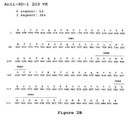
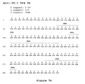
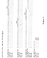
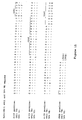
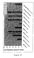
- V H regions for 17D8, 2D3, 4H1, 5C4 and 7D3, against the parent germline V H 3-33 sequence is shown in Figure 8 .
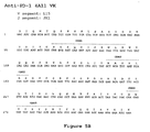
- the alignment of V H regions for 4A11 and 5F4 against the parent germline V H 4-39 sequence is shown in Figure 11 .
- Table 1 Modifications to antibodies 17D8, 2D3, 4H1, 5C4, 4A11, 7D3 and 5F4 from the heavy chain germline configuration.
- Anti-PD-1Ab Amino acid position Amino acid of antibody Original amino acid of germline configuration 17D8 10 D G 16 G R 27 V F 28 A T 78 M T 93 M V 2D3 10 D G 27 L F 30 T S 85 N S 98 T R 4H1 3 Y Q 84 T N 88 V A 98 S R 5C4 21 D S 23 K A 27 I F 80 F Y 98 T R 4A11 29 L I 79 Q H 98 V A 7D3 23 T A 24 T A 27 I F 70 L I 74 D N 97 V A 98 T R 5F4 23 S T 29 L I 51 A G 77 R K
- Another type of framework modification involves mutating one or more residues within the framework region, or even within one or more CDR regions, to remove T cell epitopes to thereby reduce the potential immunogenicity of the antibody. This approach is also referred to as "deimmunization" and is described in further detail in U.S. Patent Publication No. 20030153043 by Carr et al.
- antibodies of the invention may be engineered to include modifications within the Fc region, typically to alter one or more functional properties of the antibody, such as serum half-life, complement fixation, Fc receptor binding, and/or antigen-dependent cellular cytotoxicity.
- an antibody of the invention may be chemically modified (e.g. , one or more chemical moieties can be attached to the antibody) or be modified to alter its glycosylation, again to alter one or more functional properties of the antibody.
- the hinge region of CH1 is modified such that the number of cysteine residues in the hinge region is altered, e.g. , increased or decreased.
- This approach is described further in U.S. Patent No. 5,677,425 by Bodmer et al.
- the number of cysteine residues in the hinge region of CH1 is altered to, for example, facilitate assembly of the light and heavy chains or to increase or decrease the stability of the antibody.
- the Fc hinge region of an antibody is mutated to decrease the biological half life of the antibody. More specifically, one or more amino acid mutations are introduced into the CH2-CH3 domain interface region of the Fc-hinge fragment such that the antibody has impaired Staphylococcyl protein A (SpA) binding relative to native Fc-hinge domain SpA binding.
- SpA Staphylococcyl protein A
- the antibody is modified to increase its biological half life.
- Various approaches are possible. For example, one or more of the following mutations can be introduced: T252L, T254S, T256F, as described in U. S. Patent No. 6,277,375 to Ward .
- the antibody can be altered within the CH1 or CL region to contain a salvage receptor binding epitope taken from two loops of a CH2 domain of an Fc region of an IgG, as described in U.S. Patent Nos. 5,869,046 and 6,121,022 by Presta et al.
- the Fc region is altered by replacing at least one amino acid residue with a different amino acid residue to alter the effector function(s) of the antibody.
- one or more amino acids selected from amino acid residues 234, 235, 236, 237, 297, 318, 320 and 322 can be replaced with a different amino acid residue such that the antibody has an altered affinity for an effector ligand but retains the antigen-binding ability of the parent antibody.
- the effector ligand to which affinity is altered can be, for example, an Fc receptor or the C1 component of complement. This approach is described in further detail in U.S. Patent Nos. 5,624,821 and 5,648,260, both by Winter et al.
- one or more amino acids selected from amino acid residues 329, 331 and 322 can be replaced with a different amino acid residue such that the antibody has altered C1q binding and/or reduced or abolished complement dependent cytotoxicity (CDC).
- CDC complement dependent cytotoxicity
- one or more amino acid residues within amino acid positions 231 and 239 are altered to thereby alter the ability of the antibody to fix complement. This approach is described further in PCT Publication WO 94/29351 by Bodmer et a/.
- the Fc region is modified to increase the ability of the antibody to mediate antibody dependent cellular cytotoxicity (ADCC) and/or to increase the affinity of the antibody for an Fc ⁇ receptor by modifying one or more amino acids at the following positions: 238, 239, 248, 249, 252, 254, 255, 256, 258, 265, 267, 268, 269, 270, 272, 276, 278, 280, 283, 285, 286, 289, 290, 292, 293, 294, 295, 296, 298, 301, 303, 305, 307, 309, 312, 315, 320, 322, 324, 326, 327, 329, 330, 331, 333, 334, 335, 337, 338, 340, 360, 373, 376, 378, 382, 388, 389, 398, 414, 416, 419, 430, 434, 435, 437, 438 or 439.
- ADCC antibody dependent cellular cytotoxicity
- the glycosylation of an antibody is modified.
- an aglycoslated antibody can be made ( i . e ., the antibody lacks glycosylation).
- Glycosylation can be altered to, for example, increase the affinity of the antibody for antigen.
- Such carbohydrate modifications can be accomplished by, for example, altering one or more sites of glycosylation within the antibody sequence.
- one or more amino acid substitutions can be made that result in elimination of one or more variable region framework glycosylation sites to thereby eliminate glycosylation at that site.
- Such aglycosylation may increase the affinity of the antibody for antigen.
- Such an approach is described in further detail in U.S. Patent Nos. 5,714,350 and 6,350,861 by Co et al.
- an antibody can be made that has an altered type of glycosylation, such as a hypofucosylated antibody having reduced amounts of fucosyl residues or an antibody having increased bisecting GlcNac structures.
- altered glycosylation patterns have been demonstrated to increase the ADCC ability of antibodies.
- carbohydrate modifications can be accomplished by, for example, expressing the antibody in a host cell with altered glycosylation machinery. Cells with altered glycosylation machinery have been described in the art and can be used as host cells in which to express recombinant antibodies of the invention to thereby produce an antibody with altered glycosylation.
- the cell lines Ms704, Ms705, and Ms709 lack the fucosyltransferase gene, FUT8 (alpha (1,6) fucosyltransferase), such that antibodies expressed in the Ms704, Ms705, and Ms709 cell lines lack fucose on their carbohydrates.
- the Ms704, Ms705, and Ms709 FUT8 -/- cell lines were created by the targeted disruption of the FUT8 gene in CHO/DG44 cells using two replacement vectors (see U.S. Patent Publication No. 20040110704 by Yamane et al. and Yamane-Ohnuki et al. (2004) Biotechnol Bioeng 87:614-22 ).
- EP 1,176,195 by Hanai et al. describes a cell line with a functionally disrupted FUT8 gene, which encodes a fucosyl transferase, such that antibodies expressed in such a cell line exhibit hypofucosylation by reducing or eliminating the alpha 1,6 bond-related enzyme.
- Hanai et al. also describe cell lines which have a low enzyme activity for adding fucose to the N-acetylglucosamine that binds to the Fc region of the antibody or does not have the enzyme activity, for example the rat myeloma cell line YB2/0 (ATCC CRL 1662).
- PCT Publication WO 03/035835 by Presta describes a variant CHO cell line, Lec13 cells, with reduced ability to attach fucose to Asn(297)-linked carbohydrates, also resulting in hypofucosylation of antibodies expressed in that host cell (see also Shields, R.L. et al. (2002) J. Biol. Chem. 277:26733-26740 ).
- PCT Publication WO 99/54342 by Umana et al. describes cell lines engineered to express glycoprotein-modifying glycosyl transferases (e.g.
- beta(1,4)-N-acetylglucosaminyltransferase III (GnTIII)) such that antibodies expressed in the engineered cell lines exhibit increased bisecting GlcNac structures which results in increased ADCC activity of the antibodies (see also Umana et al. (1999) Nat. Biotech. 17:176-180 ).
- the fucose residues of the antibody may be cleaved off using a fucosidase enzyme.
- the fucosidase alpha-L-fucosidase removes fucosyl residues from antibodies ( Tarentino, A.L. et al. (1975) Biochem. 14:5516-23 ).
- An antibody can be pegylated to, for example, increase the biological (e.g. , serum) half life of the antibody.
- the antibody, or fragment thereof typically is reacted with polyethylene glycol (PEG), such as a reactive ester or aldehyde derivative of PEG, under conditions in which one or more PEG groups become attached to the antibody or antibody fragment.
- PEG polyethylene glycol
- the pegylation is carried out via an acylation reaction or an alkylation reaction with a reactive PEG molecule (or an analogous reactive water-soluble polymer).
- polyethylene glycol is intended to encompass any of the forms of PEG that have been used to derivatize other proteins, such as mono (C1-C10) alkoxy- or aryloxy-polyethylene glycol or polyethylene glycol-maleimide.
- the antibody to be pegylated is an aglycosylated antibody. Methods for pegylating proteins are known in the art and can be applied to the antibodies of the invention. See for example, EP 0 154 316 by Nishimura et al. and EP 0 401 384 by Ishikawa et al.
- the anti-PD-1 antibodies having V H and V K sequences disclosed herein can be used to create new anti-PD-1 antibodies by modifying the VH and/or V K sequences, or the constant region(s) attached thereto.
- the structural features of an anti-PD-1 antibody of the invention e.g. 17D8, 2D3, 4H1, 5C4, 4A11, 7D3 or 5F4, are used to create structurally related anti-PD-1 antibodies that retain at least one functional property of the antibodies of the invention, such as binding to human PD-1.
- one or more CDR regions of 17D8, 2D3, 4H1, 5C4, 4A11, 7D3 or 5F4, or mutations thereof can be combined recombinantly with known framework regions and/or other CDRs to create additional, recombinantly-engineered, anti-PD-1 antibodies of the invention, as discussed above.
- the starting material for the engineering method is one or more of the V H and/or V K sequences provided herein, or one or more CDR regions thereof.
- To create the engineered antibody it is not necessary to actually prepare ( i.e ., express as a protein) an antibody having one or more of the V H and/or V K sequences provided herein, or one or more CDR regions thereof. Rather, the information contained in the sequence(s) is used as the starting material to create a "second generation" sequence(s) derived from the original sequeilce(s) and then the "second generation" sequence(s) is prepared and expressed as a protein.
- the invention provides a method for preparing an anti-PD-1 antibody comprising:
- Standard molecular biology techniques can be used to prepare and express the altered antibody sequence.
- the antibody encoded by the altered antibody sequence(s) is one that retains one, some or all of the functional properties of the anti-PD-1 antibodies described herein, which functional properties include, but are not limited to:
- the functional properties of the altered antibodies can be assessed using standard assays available in the art and/or described herein, such as those set forth in the Examples (e.g. , flow cytometry, binding assays).
- mutations can be introduced randomly or selectively along all or part of an anti-PD-1 antibody coding sequence and the resulting modified anti-PD-1 antibodies can be screened for binding activity and/or other functional properties as described herein.
- Mutational methods have been described in the art.
- PCT Publication WO 02/092780 by Short describes methods for creating and screening antibody mutations using saturation mutagenesis, synthetic ligation assembly, or a combination thereof.
- PCT Publication WO 03/074679 by Lazar et al. describes methods of using computational screening methods to optimize physiochemical properties of antibodies.
- nucleic acid molecules that encode the antibodies of the invention.
- the nucleic acids may be present in whole cells, in a cell lysate, or in a partially purified or substantially pure form.
- a nucleic acid is "isolated” or “rendered substantially pure” when purified away from other cellular components or other contaminants, e.g. , other cellular nucleic acids or proteins, by standard techniques, including alkaline/SDS treatment, CsCl banding, column chromatography, agarose gel electrophoresis and others well known in the art. See, F. Ausubel, et al., ed.
- a nucleic acid of the invention can be, for example, DNA or RNA and may or may not contain intronic sequences.
- the nucleic acid is a cDNA molecule.
- Nucleic acids of the invention can be obtained using standard molecular biology techniques.
- hybridomas e.g. , hybridomas prepared from transgenic mice carrying human immunoglobulin genes as described further below
- cDNAs encoding the light and heavy chains of the antibody made by the hybridoma can be obtained by standard PCR amplification or cDNA cloning techniques.
- nucleic acid encoding the antibody can be recovered from the library.
- Preferred nucleic acids molecules of the invention are those encoding the VH and VL sequences of the 17D8, 2D3, 4H1, 5C4, 4A11, 7D3 or 5F4 monoclonal antibodies.
- DNA sequences encoding the VH sequences of 17D8, 2D3, 4H1, 5C4, 4A11, 7D3 and 5F4 are shown in SEQ ID NOs: 57, 58, 59, 60, 61, 62 and 63, respectively.
- DNA sequences encoding the VL sequences of 17D8, 2D3, 4H1, 5C4, 4A11, 7D3 and 5F4 are shown in SEQ ID NOs: 64, 65, 66, 67, 68, 69 and 70, respectively.
- VH and VL segments are obtained, these DNA fragments can be further manipulated by standard recombinant DNA techniques, for example to convert the variable region genes to full-length antibody chain genes, to Fab fragment genes or to a scFv gene.
- a VL- or VH-encoding DNA fragment is operatively linked to another DNA fragment encoding another protein, such as an antibody constant region or a flexible linker.
- the term "operatively linked”, as used in this context, is intended to mean that the two DNA fragments are joined such that the amino acid sequences encoded by the two DNA fragments remain in-frame.
- the isolated DNA encoding the VH region can be converted to a full-length heavy chain gene by operatively linking the VH-encoding DNA to another DNA molecule encoding heavy chain constant regions (CH1, CH2 and CH3).
- heavy chain constant regions CH1, CH2 and CH3
- the sequences of human heavy chain constant region genes are known in the art (see e.g. , Kabat, E. A., el al. (1991) Sequences of Proteins of Immunological Interest, Fifth Edition, U.S. Department of Health and Human Services, NIH Publication No. 91-3242 ) and DNA fragments encompassing these regions can be obtained by standard PCR amplification.
- the heavy chain constant region can be an IgG1, IgG2, IgG3, IgG4, IgA, IgE, IgM or IgD constant region, but most preferably is an IgG1 or IgG4 constant region.
- the VH-encoding DNA can be operatively linked to another DNA molecule encoding only the heavy chain CH1 constant region.
- the isolated DNA encoding the VL region can be converted to a full-length light chain gene (as well as a Fab light chain gene) by operatively linking the VL-encoding DNA to another DNA molecule encoding the light chain constant region, CL.
- the sequences of human light chain constant region genes are known in the art (see e.g. , Kabat, E. A., et al. (1991) Sequences of Proteins of Immunological Interest, Fifth Edition, U.S. Department of Health and Human Services, NIH Publication No. 91-3242 ) and DNA fragments encompassing these regions can be obtained by standard PCR amplification.
- the light chain constant region can be a kappa or lambda constant region, but most preferably is a kappa constant region.
- the VH- and VL-encoding DNA fragments are operatively linked to another fragment encoding a flexible linker, e.g. , encoding the amino acid sequence (Gly 4 -Ser) 3 , such that the VH and VL sequences can be expressed as a contiguous single-chain protein, with the VL and VH regions joined by the flexible linker (see e.g. , Bird et al. (1988) Science 242:423-426 ; Huston et al. (1988) Proc. Natl. Acad. Sci. USA 85:5879-5883 ; McCafferty et al., (1990) Nature 348:552-554 ).
- a flexible linker e.g. , encoding the amino acid sequence (Gly 4 -Ser) 3 , such that the VH and VL sequences can be expressed as a contiguous single-chain protein, with the VL and VH regions joined by the flexible linker (see e.
- Monoclonal antibodies (mAbs) of the present invention can be produced by a variety of techniques, including conventional monoclonal antibody methodology e.g. , the standard somatic cell hybridization technique of Kohler and Milstein (1975) Nature 256: 495 . Although somatic cell hybridization procedures are preferred, in principle, other techniques for producing monoclonal antibody can be employed e.g. , viral or oncogenic transformation of B lymphocytes.
- the preferred animal system for preparing hybridomas is the murine system.
- Hybridoma production in the mouse is a very well-established procedure. Immunization protocols and techniques for isolation of immunized splenocytes for fusion are known in the art. Fusion partners (e.g. , murine myeloma cells) and fusion procedures are also known.
- Chimeric or humanized antibodies of the present invention can be prepared based on the sequence of a murine monoclonal antibody prepared as described above.
- DNA encoding the heavy and light chain immunoglobulins can be obtained from the murine hybridoma of interest and engineered to contain non-murine ( e.g. , human) immunoglobulin sequences using standard molecular biology techniques.
- the murine variable regions can be linked to human constant regions using methods known in the art (see e.g., U.S. Patent No. 4,816,567 to Cabilly et al .).
- the murine CDR regions can be inserted into a human framework using methods known in the art (see e.g. , U.S. Patent No. 5,225,539 to Winter , and U.S. Patent Nos. 5,530,101 ; 5,585,089 ; 5,693,762 and 6,180,370 to Queen et al .).
- the antibodies of the invention are human monoclonal antibodies.
- Such human monoclonal antibodies directed against PD-1 can be generated using transgenic or transchromosomic mice carrying parts of the human immune system rather than the mouse system.
- transgenic and transchromosomic mice include mice referred to herein as HuMAb mice and KM mice TM , respectively, and are collectively referred to herein as "human Ig mice.”
- the HuMAb mouse ® (Medarex, Inc.) contains human immunoglobulin gene miniloci that encode unrearranged human heavy ( ⁇ and y) and ⁇ light chain immunoglobulin sequences, together with targeted mutations that inactivate the endogenous ⁇ and ⁇ chain loci (see e . g ., Lonberg, et al. (1994) Nature 368(6474): 856-859 ). Accordingly, the mice exhibit reduced expression of mouse IgM or ⁇ , and in response to immunization, the introduced human heavy and light chain transgenes undergo class switching and somatic mutation to generate high affinity human IgG ⁇ monoclonal (Lonberg, N. et al. (1994), supra ; reviewed in Lonberg, N.
- human antibodies of the invention can be raised using a mouse that carries human immunoglobulin sequences on transgenes and transchomosomes, such as a mouse that carries a human heavy chain transgene and a human light chain transchromosome.
- KM mice TM Such mice, referred to herein as "KM mice TM ", are described in detail in PCT Publication WO 02/43478 to Ishida et al.
- transgenic animal systems expressing human immunoglobulin genes are available in the art and can be used to raise anti-PD-1 antibodies of the invention.
- an alternative transgenic system referred to as the Xenomouse (Abgenix, Inc.) can be used; such mice are described in, for example, U.S. Patent Nos. 5,939,598 ; 6,075,181 ; 6,114,598 ; 6, 150,584 and 6,162,963 to Kucherlapati et al .
- mice carrying both a human heavy chain transchromosome and a human light chain transchromosome referred to as "TC mice” can be used; such mice are described in Tomizuka et al. (2000) Proc. Natl. Acad Sci. USA 97:722-727 .
- cows carrying human heavy and light chain transchromosomes have been described in the art ( Kuroiwa et al. (2002) Nature Biotechnology 20:889-894 ) and can be used to raise anti-PD-1 antibodies of the invention.
- Human monoclonal antibodies of the invention can also be prepared using phage display methods for screening libraries of human immunoglobulin genes. Such phage display methods for isolating human antibodies are established in the art. See for example: U.S. Patent Nos. 5,223,409 ; 5,403,484 ; and 5,571,698 to Ladner et al .; U.S. Patent Nos. 5,427,908 and 5,580,717 to Dower et al .; U.S. Patent Nos. 5,969,108 and 6,172,197 to McCafferty et al .; and U.S. Patent Nos. 5,885,793 ; 6,521,404 ; 6,544,731 ; 6,555,313 ; 6,582,915 and 6,593,081 to Griffiths et al .
- Human monoclonal antibodies of the invention can also be prepared using SCID mice into which human immune cells have been reconstituted such that a human antibody response can be generated upon immunization.
- SCID mice into which human immune cells have been reconstituted such that a human antibody response can be generated upon immunization.
- Such mice are described in, for example, U.S. Patent Nos. 5,476,996 and 5,698,767 to Wilson et al .
- mice When human Ig mice are used to raise human antibodies of the invention, such mice can be immunized with a purified or enriched preparation of PD-1 antigen and/or recombinant PD-1, or an PD-1 fusion protein, as described by Lonberg, N. et al. (1994) Nature 368(6474): 856-859 ; Fishwild, D. et al. (1996) Nature Biotechnology 14: 845-851 ; and PCT Publication WO 98/24884 and WO 01/14424 .
- the mice will be 6-16 weeks of age upon the first infusion.
- a purified or recombinant preparation (5-50 ⁇ g) of PD-1 antigen can be used to immunize the human Ig mice intraperitoneally.
- Example 1 Detailed procedures to generate fully human monoclonal antibodies to PD-1 are described in Example 1 below. Cumulative experience with various antigens has shown that the transgenic mice respond when initially immunized intraperitoneally (IP) with antigen in complete Freund's adjuvant, followed by every other week IP immunizations (up to a total of 6) with antigen in incomplete Freund's adjuvant. However, adjuvants other than Freund's are also found to be effective. In addition, whole cells in the absence of adjuvant are found to be highly immunogenic. The immune response can be monitored over the course of the immunization protocol with plasma samples being obtained by retroorbital bleeds.
- mice with sufficient titers of anti-PD-1 human immunoglobulin can be used for fusions.
- Mice can be boosted intravenously with antigen 3 days before sacrifice and removal of the spleen. It is expected that 2-3 fusions for each immunization may need to be performed. Between 6 and 24 mice are typically immunized for each antigen.
- HCo7 and HCo12 strains are used.
- both HCo7 and HCo12 transgene can be bred together into a single mouse having two different human heavy chain transgenes (HCo7/HCo12).
- the KM mouse TM strain can be used, as described in Example 1.
- splenocytes and/or lymph node cells from immunized mice can be isolated and fused to an appropriate immortalized cell line, such as a mouse myeloma cell line.
- an appropriate immortalized cell line such as a mouse myeloma cell line.
- the resulting hybridomas can be screened for the production of antigen-specific antibodies.
- single cell suspensions of splenic lymphocytes from immunized mice can be fused to one-sixth the number of P3X63-Ag8.653 nonsecreting mouse myeloma cells (ATCC, CRL 1580) with 50% PEG.
- the single cell suspensions of splenic lymphocytes from immunized mice can be fused using an electric field based electrofusion method, using a Cyto Pulse large chamber cell fusion electroporator (Cyto Pulse Sciences, Inc., Glen Burnie, MD).
- Cells are plated at approximately 2 x 10 5 in flat bottom microtiter plate, followed by a two week incubation in selective medium containing 20% fetal Clone Serum, 18% "653" conditioned media, 5% origen (IGEN), 4 mM L-glutamine, 1 mM sodium pyruvate, 5mM HEPES, 0.055 mM 2-mercaptoethanol, 50 units/ml penicillin, 50 mg/ml streptomycin, 50 mg/ml gentamycin and 1X HAT (Sigma; the HAT is added 24 hours after the fusion). After approximately two weeks; cells can be cultured in medium in which the HAT is replaced with HT.
- selective medium containing 20% fetal Clone Serum, 18% "653" conditioned media, 5% origen (IGEN), 4 mM L-glutamine, 1 mM sodium pyruvate, 5mM HEPES, 0.055 mM 2-mercaptoethanol, 50 units/ml penicillin,
- selected hybridomas can be grown in two-liter spinner-flasks for monoclonal antibody purification.
- Supernatants can be filtered and concentrated before affinity chromatography with protein A-sepharose (Pharmacia, Piscataway, N.J.).
- Eluted IgG can be checked by gel electrophoresis and high performance liquid chromatography to ensure purity.
- the buffer solution can be exchanged into PBS, and the concentration can be determined by OD 280 using 1.43 extinction coefficient.
- the monoclonal antibodies can be aliquoted and stored at -80 °C.
- Antibodies of the invention also can be produced in a host cell transfectoma using, for example, a combination of recombinant DNA techniques and gene transfection methods as is well known in the art (e.g ., Morrison, S. (1985) Science 229:1202 ).
- DNAs encoding partial or full-length light and heavy chains can be obtained by standard molecular biology techniques (e.g. , PCR amplification or cDNA cloning using a hybridoma that expresses the antibody of interest) and the DNAs can be inserted into expression vectors such that the genes are operatively linked to transcriptional and translational control sequences.
- operatively linked is intended to mean that an antibody gene is ligated into a vector such that transcriptional and translational control sequences within the vector serve their intended function of regulating the transcription and translation of the antibody gene.
- the expression vector and expression control sequences are chosen to be compatible with the expression host cell used.
- the antibody light chain gene and the antibody heavy chain gene can be inserted into separate vector or, more typically, both genes are inserted into the same expression vector.
- the antibody genes are inserted into the expression vector by standard methods (e.g. , ligation of complementary restriction sites on the antibody gene fragment and vector, or blunt end ligation if no restriction sites are present).
- the light and heavy chain variable regions of the antibodies described herein can be used to create full-length antibody genes of any antibody isotype by inserting them into expression vectors already encoding heavy chain constant and light chain constant regions of the desired isotype such that the V H segment is operatively linked to the C H segment(s) within the vector and the V K segment is operatively linked to the C L segment within the vector.
- the recombinant expression vector can encode a signal peptide that facilitates secretion of the antibody chain from a host cell.
- the antibody chain gene can be cloned into the vector such that the signal peptide is linked in-frame to the amino terminus of the antibody chain gene.
- the signal peptide can be an immunoglobulin signal peptide or a heterologous signal peptide ( i.e., a signal peptide from a non-immunoglobulin protein).
- the recombinant expression vectors of the invention carry regulatory sequences that control the expression of the antibody chain genes in a host cell.
- the term "regulatory sequence” is intended to include promoters, enhancers and other expression control elements (e.g ., polyadenylation signals) that control the transcription or translation of the antibody chain genes.
- Such regulatory sequences are described, for example, in Goeddel (Gene Expression Technology. Methods in Enzymology 185, Academic Press, San Diego, CA (1990 )). It will be appreciated by those skilled in the art that the design of the expression vector, including the selection of regulatory sequences, may depend on such factors as the choice of the host cell to be transformed, the level of expression of protein desired, etc.
- Preferred regulatory sequences for mammalian host cell expression include viral elements that direct high levels of protein expression in mammalian cells, such as promoters and/or enhancers derived from cytomegalovirus (CMV), Simian Virus 40 (SV40), adenovirus, ( e.g., the adenovirus major late promoter (AdMLP) and polyoma.
- CMV cytomegalovirus
- SV40 Simian Virus 40
- AdMLP adenovirus major late promoter
- nonviral regulatory sequences may be used, such as the ubiquitin promoter or ⁇ -globin promoter.
- regulatory elements composed of sequences from different sources such as the SR ⁇ promoter system, which contains sequences from the SV40 early promoter and the long terminal repeat of human T cell leukemia virus type 1 ( Takebe, Y. et al. (1988) Mol. Cell. Biol. 8:466-472 ).
- the recombinant expression vectors of the invention may carry additional sequences, such as sequences that regulate replication of the vector in host cells (e.g. , origins of replication) and selectable marker genes.
- the selectable marker gene facilitates selection of host cells into which the vector has been introduced (see, e.g. , U. S. Pat. Nos. 4,399,216 , 4,634,665 and 5,179,017 , all by Axel et al. ) .
- the selectable marker gene confers resistance to drugs, such as G418, hygromycin or methotrexate, on a host cell into which the vector has been introduced.
- Preferred selectable marker genes include the dihydrofolate reductase (DHFR) gene (for use in dhfr- host cells with methotrexate selection/amplification) and the neo gene (for G418 selection).
- DHFR dihydrofolate reductase
- the expression vector(s) encoding the heavy and light chains is transfected into a host cell by standard techniques.
- the various forms of the term "transfection" are intended to encompass a wide variety of techniques commonly used for the introduction of exogenous DNA into a prokaryotic or eukaryotic host cell, e.g. , electroporation, calcium-phosphate precipitation, DEAE-dextran transfection and the like.
- Preferred mammalian host cells for expressing the recombinant antibodies of the invention include Chinese Hamster Ovary (CHO cells) (including dhfr- CHO cells, described in Urlaub and Chasin, (1980) Proc. Natl. Acad. Sci. USA 77:4216-4220 , used with a DHFR selectable marker, e.g, as described in R. J. Kaufman and P. A. Sharp (1982) Mol. Biol. 159:601-621 ), NSO myeloma cells, COS cells and SP2 cells.
- Chinese Hamster Ovary CHO cells
- dhfr- CHO cells described in Urlaub and Chasin, (1980) Proc. Natl. Acad. Sci. USA 77:4216-4220 , used with a DHFR selectable marker, e.g, as described in R. J. Kaufman and P. A. Sharp (1982) Mol. Biol. 159:601-621
- another preferred expression system is the GS gene expression system disclosed in WO 87/04462 , WO 89/01036 and EP 338,841 .
- the antibodies are produced by culturing the host cells for a period of time sufficient to allow for expression of the antibody in the host cells or, more preferably, secretion of the antibody into the culture medium in which the host cells are grown.
- Antibodies can be recovered from the culture medium using standard protein purification methods.
- Antibodies of the invention can be tested for binding to PD-1 by, for example, standard ELISA. Briefly, microtiter plates are coated with purified PD-1 at 0.25 ⁇ g/ml in FBS, and then blocked with 5% bovine serum albumin in PBS. Dilutions of antibody (e.g. , dilutions of plasma from PD-1-immunized mice) are added to each well and incubated for 1-2 hours at 37°C. The plates are washed with PBS/Tween and then incubated with secondary reagent (e.g.
- mice which develop the highest titers will be used for fusions.
- An ELISA assay as described above can also be used to screen for hybridomas that show positive reactivity with PD-1 immunogen.
- Hybridomas that bind with high avidity to PD-1 are subcloned and further characterized.
- One clone from each hybridoma; which retains the reactivity of the parent cells (by ELISA) can be chosen for making a 5-10 vial cell bank stored at -140°C, and for antibody purification.
- selected hybridomas can be grown in two-liter spinner-flasks for monoclonal antibody purification.
- Supernatants can be filtered and concentrated before affinity chromatography with protein A-sepharose (Pharmacia, Piscataway, NJ).
- Eluted IgG can be checked by gel electrophoresis and high performance liquid chromatography to ensure purity.
- the buffer solution can be exchanged into PBS, and the concentration can be determined by OD 280 using 1.43 extinction coefficient.
- the monoclonal antibodies can be aliquoted and stored at -80°C.
- each antibody can be biotinylated using commercially available reagents (Pierce, Rockford, IL). Competition studies using unlabeled monoclonal antibodies and biotinylated monoclonal antibodies can be performed using PD-1 coated-ELISA plates as described above. Biotinylated mAb binding can be detected with a strep-avidin-alkaline phosphatase probe.
- isotype ELISAs can be performed using reagents specific for antibodies of a particular isotype. For example, to determine the isotype of a human monoclonal antibody, wells of microtiter plates can be coated with 1 ⁇ g/ml of anti-human immunoglobulin overnight at 4 °C. After blocking with 1% BSA, the plates are reacted with 1 ⁇ g /ml or less of test monoclonal antibodies or purified isotype controls, at ambient temperature for one to two hours. The wells can then be reacted with either human IgGl or human IgM-specific alkaline phosphatase-conjugated probes. Plates are developed and analyzed as described above.
- Anti-PD-1 human IgGs can be further tested for reactivity with PD-1 antigen by Western blotting. Briefly, PD-1 can be prepared and subjected to sodium dodecyl sulfate polyacrylamide gel electrophoresis. After electrophoresis, the separated antigens are transferred to nitrocellulose membranes, blocked with 10% fetal calf serum, and probed with the monoclonal antibodies to be tested. Human IgG binding can be detected using anti-human IgG alkaline phosphatase and developed with BCIP/NBT substrate tablets (Sigma Chem. Co., St. Louis, Mo.).
- the present invention features an anti-PD-1 antibody, or a fragment thereof, conjugated to a therapeutic moiety, such as a cytotoxin, a drug (e.g. , an immunosuppressant) or a radiotoxin.
- a therapeutic moiety such as a cytotoxin, a drug (e.g. , an immunosuppressant) or a radiotoxin.
- Therapeutic agents include taxol, cytochalasin B, gramicidin D, ethidium bromide, emetine, mitomycin, etoposide, tenoposide, vincristine, vinblastine, colchicin, doxorubicin, daunorubicin, dibydroxy anthracin dione, mitoxantrone, mithramycin, actinomycin D, 1-dehydrotestosterone, glucocorticoids, procaine, tetracaine, lidocaine, propranolol, and puromycin and analogs or homologs thereof
- Therapeutic agents also include, for example, antimetabolites (e.g.
- alkylating agents e.g. , mechlorethamine, thioepa chlorambucil, melphalan, carmustine (BSNU) and lomustine (CCNU), cyclothosphamide, busulfan, dibromomannitol, streptozotocin, mitomycin C, and cis-dichlorodiamine platinum (II) (DDP) cisplatin
- anthracyclines e.g.
- daunorubicin (formerly daunomycin) and doxorubicin
- antibiotics e.g. , dactinomycin (formerly actinomycin), bleomycin, mithramycin, and anthramycin (AMC)
- anti-mitotic agents e.g. , vincristine and vinblastine.
- An example of a calicheamicin antibody conjugate is commercially available (Mylotarg TM ; Wyeth-Ayerst).
- Cytoxins can be conjugated to antibodies of the invention using linker technology available in the art.
- linker types that have been used to conjugate a cytotoxin to an antibody include, but are not limited to, hydrazones, thioethers, esters, disulfides and peptide-containing linkers.
- a linker can be chosen that is, for example, susceptible to cleavage by low pH within the lysosomal compartment or susceptible to cleavage by proteases, such as proteases preferentially expressed in tumor tissue such as cathepsins ( e.g. , cathepsins B, C, D).
- Antibodies of the present invention also can be conjugated to a radioactive isotope to generate cytotoxic radiopharmaceuticals, also referred to as radioimmunoconjugates.
- radioactive isotopes that can be conjugated to antibodies for use diagnostically or therapeutically include, but are not limited to, iodine 131 , indium 111 , yttrium 90 and lutetium 177 .
- Method for preparing radioimmunconjugates are established in the art. Examples of radioimmunoconjugates are commercially available, including Zevalin TM (IDEC Pharmaceuticals) and Bexxar TM (Corixa Pharmaceuticals), and similar methods can be used to prepare radioimmunoconjugates using the antibodies of the invention.
- the antibody conjugates of the invention can be used to modify a given biological response, and the drug moiety is not to be construed as limited to classical chemical therapeutic agents.
- the drug moiety may be a protein or polypeptide possessing a desired biological activity.
- proteins may include, for example, an enzymatically active toxin, or active fragment thereof, such as abrin, ricin A, pseudomonas exotoxin, or diphtheria toxin; a protein such as tumor necrosis factor or interferon- ⁇ ; or, biological response modifiers such as, for example, lymphokines, interleukin-1 ("IL-1"), interleukin-2 (“IL-2”), interleukin-6 (“IL-6”), granulocyte macrophage colony stimulating factor (“GM-CSF”), granulocyte colony stimulating factor (“G-CSF”), or other growth factors.
- IL-1 interleukin-1
- IL-2 interleukin-2
- IL-6 interleukin-6
- the present invention features bispecific molecules comprising an anti-PD-1 antibody, or a fragment thereof, of the invention.
- An antibody of the invention, or antigen-binding portions thereof can be derivatized or linked to another functional molecule, e.g. , another peptide or protein (e.g. , another antibody or ligand for a receptor) to generate a bispecific molecule that binds to at least two different binding sites or target molecules.
- the antibody of the invention may in fact be derivatized or linkd to more than one other functional molecule to generate multispecific molecules that bind to more than two different binding sites and/or target molecules; such multispecific molecules are also intended to be encompassed by the term "bispecific molecule" as used herein.
- an antibody of the invention can be functionally linked ( e.g. , by chemical coupling, genetic fusion, noncovalent association or otherwise) to one or more other binding molecules, such as another antibody, antibody fragment, peptide or binding mimetic, such that a bispecific molecule results.
- the present invention includes bispecific molecules comprising at least one first binding specificity for PD-1 and a second binding specificity for a second target epitope.
- the second target epitope is an Fc receptor, e.g. , human Fc ⁇ RI (CD64) or a human Fc ⁇ receptor (CD89). Therefore, the .invention includes bispecific molecules capable of binding both to Fc ⁇ R or Fe ⁇ R expressing effector cells (e.g. , monocytes, macrophages or polymorphonuclear cells (PMNs)), and to target cells expressing PD-1.
- bispecific molecules target PD-1 expressing cells to effector cell and trigger Fc receptor-mediated effector cell activities, such as phagocytosis of an PD-1 expressing cells, antibody dependent cell-mediated cytotoxicity (ADCC), cytokine release, or generation of superoxide anion.
- ADCC antibody dependent cell-mediated cytotoxicity
- the molecule can further include a third binding specificity, in addition to an anti-Fc binding specificity and an anti-PD-1 binding specificity.
- the third binding specificity is an anti-enhancement factor (EF) portion, e.g. , a molecule which binds to a surface protein involved in cytotoxic activity and thereby increases the immune response against the target cell.
- EF anti-enhancement factor
- the "anti-enhancement factor portion” can be an antibody, functional antibody fragment or a ligand that binds to a given molecule, e.g. , an antigen or a receptor, and thereby results in an enhancement of the effect of the binding determinants for the F c receptor or target cell antigen.
- the "anti-enhancement factor portion” can bind an F c receptor or a target cell antigen.
- the anti-enhancement factor portion can bind to an entity that is different from the entity to which the first and second binding specificities bind.
- the anti-enhancement factor portion can bind a cytotoxic T-cell (e.g. via CD2, CD3, CD8, CD28, CD4, CD40, ICAM-1 or other immune cell that results in an increased immune response against the target cell).
- the bispecific molecules of the invention comprise as a binding specificity at least one antibody, or an antibody fragment thereof, including, e.g. , an Fab, Fab', F(ab') 2 , Fv, or a single chain Fv.
- the antibody may also be a light chain or heavy chain dimer, or any minimal fragment thereof such as a Fv or a single chain construct as described in Ladner et al. U. S. Patent No. 4,946,778 , the contents of which is expressly incorporated by reference.
- the binding specificity for an Fc ⁇ receptor is provided by a monoclonal antibody, the binding of which is not blocked by human immunoglobulin G (IgG).
- IgG receptor refers to any of the eight ⁇ -chain genes located on chromosome 1. These genes encode a total of twelve transmembrane or soluble receptor isoforms which are grouped into three Fc ⁇ receptor classes: Fc ⁇ RI (CD64), Fc ⁇ RII(CD32), and Fc ⁇ RIII (CD16).
- the Fc ⁇ receptor a human high affinity Fc ⁇ RI.
- the human Fc ⁇ RI is a 72 kDa molecule, which shows high affinity for monomeric IgG (10 8 - 10 9 M -1 ).
- the hybridoma producing mAb 32 is available from the American Type Culture Collection, ATCC Accession No. HB9469.
- the anti-Fc ⁇ receptor antibody is a humanized form of monoclonal antibody 22 (H22).
- H22 monoclonal antibody 22
- the production and characterization of the H22 antibody is described in Graziano, R.F. et al. (1995) J. Immunol 155 (10): 4996-5002 and PCT Publication WO 94/10332 .
- the H22 antibody producing cell line was deposited at the American Type Culture Collection under the designation HA022CL1 and has the accession no. CRL 11177.
- the binding specificity for an Fc receptor is provided by an antibody that binds to a human IgA receptor, e.g. , an Fc-alpha receptor (Fc ⁇ RI (CD89)), the binding of which is preferably not blocked by human immunoglobulin A (IgA).
- IgA receptor is intended to include the gene product of one ⁇ -gene (Fc ⁇ RI) located on chromosome 19. This gene is known to encode several alternatively spliced transmembrane isoforms of 55 to 110 kDa.
- Fc ⁇ RI (CD89) is constitutively expressed on monocytes/macrophages, eosinophilic and neutrophilic granulocytes, but not on non-effector cell populations.
- Fc ⁇ RI has medium affinity ( ⁇ 5 x 10 7 M -1 ) for both IgAl and IgA2, which is increased upon exposure to cytokines such as G-CSF or GM-CSF ( Morton, H.C. et al. (1996) Critical Reviews in Immunology 16:423-440 ).
- cytokines such as G-CSF or GM-CSF
- Fc ⁇ RI and Fc ⁇ RI are preferred trigger receptors for use in the bispecific molecules of the invention because they are (1) expressed primarily on immune effector cells, e.g. , monocytes, PMNs, macrophages and dendritic cells; (2) expressed at high levels ( e.g. , 5,000-100,000 per cell); (3) mediators of cytotoxic activities (e.g. , ADCC, phagocytosis); (4) mediate enhanced antigen presentation of antigens, including self-antigens, targeted to them.
- immune effector cells e.g. , monocytes, PMNs, macrophages and dendritic cells
- mediators of cytotoxic activities e.g. , ADCC, phagocytosis
- human monoclonal antibodies are preferred, other antibodies which can be employed in the bispecific molecules of the invention are murine, chimeric and humanized monoclonal antibodies.
- the bispecific molecules of the present invention can be prepared by conjugating the constituent binding specificities, e.g. , the anti-FcR and anti-PD-1 binding specificities, using methods known in the art. For example, each binding specificity of the bispecific molecule can be generated separately and then conjugated to one another. When the binding specificities are proteins or peptides, a variety of coupling or cross-linking agents can be used for covalent conjugation.
- cross-linking agents examples include protein A, carbodiimide, N-succinimidyl-S-acetyl-thioacetate (SATA), 5, 5'-dithiobis(2-nitrobenzoic acid) (DTNB), o-phenylenedimaleimide (oPDM), N-succinimidyl-3-(2-pyridyldithio)propionate (SPDP), and sulfosuccinimidyl 4-(N-maleimidomethyl) cyclohaxane-1-carboxylate (sulfo-SMCC) (see e.g., Karpovsky et al. (1984) J. Exp. Med.
- Preferred conjugating agents are SATA and sulfo-SMCC, both available from Pierce Chemical Co. (Rockford, IL).
- the binding specificities are antibodies, they can be conjugated via sulfliydryl bonding of the C-terminus hinge regions of the two heavy chains.
- the hinge region is modified to contain an odd number of sulfhydryl residues, preferably one, prior to conjugation.
- both binding specificities can be encoded in the same vector and expressed and assembled in the same host cell.
- This method is particularly useful where the bispecific molecule is a mAb x mAb, mAb x Fab, Fab x F(ab') 2 or ligand x Fab fusion protein.
- a bispecific molecule of the invention can be a single chain molecule comprising one single chain antibody and a binding determinant, or a single chain bispecific molecule comprising two binding determinants. Bispecific molecules may comprise at least two single chain molecules. Methods for preparing bispecific molecules are described for example in U.S. Patent Number 5,260,203 ; U.S. Patent Number 5,455,030 ; U.S.
- Patent Number 4,881,175 U.S. Patent Number 5,132,405 ; U.S. Patent Number 5,091,513 ; U.S. Patent Number 5,476,786 ; U.S. Patent Number 5,013,653 ; U.S. Patent Number 5,258,498 ; and U.S. Patent Number 5,482,858 .
- Binding of the bispecific molecules to their specific targets can be confirmed by, for example, enzyme-linked immunosorbent assay (ELISA), radioimmunoassay (RIA), FACS analysis, bioassay (e.g. , growth inhibition), or Western Blot assay.
- ELISA enzyme-linked immunosorbent assay
- RIA radioimmunoassay
- FACS analysis bioassay (e.g. , growth inhibition)
- bioassay e.g. , growth inhibition
- Western Blot assay Western Blot assay.
- Each of these assays generally detects the presence of protein-antibody complexes of particular interest by employing a labeled reagent (e.g. , an antibody) specific for the complex of interest.
- the FcR-antibody complexes can be detected using e.g. , an enzyme-linked antibody or antibody fragment which recognizes and specifically binds to the antibody-FcR complexes.
- the complexes can be detected using any of a variety of other immunoassays.
- the antibody can be radioactively labeled and used in a radioimmunoassay (RIA) (see, for example, Weintraub, B., Principles ofRadioimmunoassays, Seventh Training Course on Radioligand Assay Techniques, The Endocrine Society, March, 1986 , which is incorporated by reference herein).
- RIA radioimmunoassay
- the radioactive isotope can be detected by such means as the use of a ⁇ counter or a scintillation counter or by autoradiography.
- the present invention provides a composition, e.g. , a pharmaceutical composition, containing one or a combination of monoclonal antibodies, or antigen-binding portion(s) thereof, of the present invention, formulated together with a pharmaceutically acceptable carrier.
- a pharmaceutical composition of the invention can comprise a combination of antibodies (or immunoconjugates or bispecifics) that bind to different epitopes on the target antigen or that have complementary activities.
- compositions of the invention also can be administered in combination therapy, i . e ., combined with other agents.
- the combination therapy can include an anti-PD-1 antibody of the present invention combined with at least one other antiinflammatory or immunosuppressant agent. Examples of therapeutic agents that can be used in combination therapy are described in greater detail below in the section on uses of the antibodies of the invention.
- pharmaceutically acceptable carrier includes any and all solvents, dispersion media, coatings, antibacterial and antifungal agents, isotonic and absorption delaying agents, and the like that are physiologically compatible.
- the carrier is suitable for intravenous, intramuscular, subcutaneous, parenteral, spinal or epidermal administration ( e.g. , by injection or infusion).
- the active compound i.e ., antibody, immunoconjuage, or bispecific molecule
- the pharmaceutical compounds of the invention may include one or more pharmaceutically acceptable salts.
- a "pharmaceutically acceptable salt” refers to a salt that retains the desired biological activity of the parent compound and does not impart any undesired toxicological effects (see e.g., Berge, S.M., et al. (1977) J. Pharm. Sci. 66:1-19 ). Examples of such salts include acid addition salts and base addition salts.
- Acid addition salts include those derived from nontoxic inorganic acids, such as hydrochloric, nitric, phosphoric, sulfuric, hydrobromic, hydroiodic, phosphorous and the like, as well as from nontoxic organic acids such as aliphatic mono- and dicarboxylic acids, phenyl-substituted alkanoic acids, hydroxy alkanoic acids, aromatic acids, aliphatic and aromatic sulfonic acids and the like.
- nontoxic inorganic acids such as hydrochloric, nitric, phosphoric, sulfuric, hydrobromic, hydroiodic, phosphorous and the like
- nontoxic organic acids such as aliphatic mono- and dicarboxylic acids, phenyl-substituted alkanoic acids, hydroxy alkanoic acids, aromatic acids, aliphatic and aromatic sulfonic acids and the like.
- Base addition salts include those derived from alkaline earth metals, such as sodium, potassium, magnesium, calcium and the like, as well as from nontoxic organic amines, such as N, N-dibenzylethylenediamine, N-methylglucamine, chloroprocaine, choline, diethanolamine, ethylenediamine, procaine and the like.
- a pharmaceutical composition of the invention also may include a pharmaceutically acceptable anti-oxidant.
- pharmaceutically acceptable antioxidants include: (1) water soluble antioxidants, such as ascorbic acid, cysteine hydrochloride, sodium bisulfate, sodium metabisulfite, sodium sulfite and the like; (2) oil-soluble antioxidants, such as ascorbyl palmitate, butylated hydroxyanisole (BHA), butylated hydroxytoluene (BHT), lecithin, propyl gallate, alpha-tocopherol, and the like; and (3) metal chelating agents, such as citric acid, ethylenediamine tetraacetic acid (EDTA), sorbitol, tartaric acid, phosphoric acid, and the like.
- water soluble antioxidants such as ascorbic acid, cysteine hydrochloride, sodium bisulfate, sodium metabisulfite, sodium sulfite and the like
- oil-soluble antioxidants such as ascorbyl palmitate, butylated
- aqueous and nonaqueous carriers examples include water, ethanol, polyols (such as glycerol, propylene glycol, polyethylene glycol, and the like), and suitable mixtures thereof, vegetable oils, such as olive oil, and injectable organic esters, such as ethyl oleate.
- polyols such as glycerol, propylene glycol, polyethylene glycol, and the like
- vegetable oils such as olive oil
- injectable organic esters such as ethyl oleate.
- Proper fluidity can be maintained, for example, by the use of coating materials, such as lecithin, by the maintenance of the required particle size in the case of dispersions, and by the use of surfactants.
- compositions may also contain adjuvants such as preservatives, wetting agents, emulsifying agents and dispersing agents. Prevention of presence of microorganisms may be ensured both by sterilization procedures, supra, and by the inclusion of various antibacterial and antifungal agents, for example, paraben, chlorobutanol, phenol sorbic acid, and the like. It may also be desirable to include isotonic agents, such as sugars, sodium chloride, and the like into the compositions. In addition, prolonged absorption of the injectable pharmaceutical form may be brought about by the inclusion of agents which delay absorption such as aluminum monostearate and gelatin.
- Pharmaceutically acceptable carriers include sterile aqueous solutions or dispersions and sterile powders for the extemporaneous preparation of sterile injectable solutions or dispersion.
- sterile aqueous solutions or dispersions and sterile powders for the extemporaneous preparation of sterile injectable solutions or dispersion.
- the use of such media and agents for pharmaceutically active substances is known in the art. Except insofar as any conventional media or agent is incompatible with the active compound, use thereof in the pharmaceutical compositions of the invention is contemplated. Supplementary active compounds can also be incorporated into the compositions.
- compositions typically must be sterile and stable under the conditions of manufacture and storage.
- the composition can be formulated as a solution, microemulsion, liposome, or other ordered structure suitable to high drug concentration.
- the carrier can be a solvent or dispersion medium containing, for example, water, ethanol, polyol (for example, glycerol, propylene glycol, and liquid polyethylene glycol, and the like), and suitable mixtures thereof
- the proper fluidity can be maintained, for example, by the use of a coating such as lecithin, by the maintenance of the required particle size in the case of dispersion and by the use of surfactants.
- isotonic agents for example, sugars, polyalcohols such as mannitol, sorbitol, or sodium chloride in the composition.
- Prolonged absorption of the injectable compositions can be brought about by including in the composition an agent that delays absorption, for example, monostearate salts and gelatin.
- Sterile injectable solutions can be prepared by incorporating the active compound in the required amount in an appropriate solvent with one or a combination of ingredients enumerated above, as required, followed by sterilization microfiltration.
- dispersions are prepared by incorporating the active compound into a sterile vehicle that contains a basic dispersion medium and the required other ingredients from those enumerated above.
- the preferred methods of preparation are vacuum drying and freeze-drying (lyophilization) that yield a powder of the active ingredient plus any additional desired ingredient from a previously sterile-filtered solution thereof.
- the amount of active ingredient which can be combined with a carrier material to produce a single dosage form will vary depending upon the subject being treated, and the particular mode of administration.
- the amount of active ingredient which can be combined with a carrier material to produce a single dosage form will generally be that amount of the composition which produces a therapeutic effect. Generally, out of one hundred per cent, this amount will range from about 0.01 per cent to about ninety-nine percent of active ingredient, preferably from about 0.1 per cent to about 70 per cent, most preferably from about 1 per cent to about 30 per cent of active ingredient in combination with a pharmaceutically acceptable carrier.
- Dosage regimens are adjusted to provide the optimum desired response (e.g. , a therapeutic response). For example, a single bolus may be administered, several divided doses may be administered over time or the dose may be proportionally reduced or increased as indicated by the exigencies of the therapeutic situation. It is especially advantageous to formulate parenteral compositions in dosage unit form for ease of administration and uniformity of dosage.
- Dosage unit form as used herein refers to physically discrete units suited as unitary dosages for the subjects to be treated; each unit contains a predetermined quantity of active compound calculated to produce the desired therapeutic effect in association with the required pharmaceutical carrier.
- the specification for the dosage unit forms of the invention are dictated by and directly dependent on (a) the unique characteristics of the active compound and the particular therapeutic effect to be achieved, and (b) the limitations inherent in the art of compounding such an active compound for the treatment of sensitivity in individuals.
- the dosage ranges from about 0.0001 to 100 mg/kg, and more usually 0.01 to 5 mg/kg, of the host body weight.
- dosages can be 0.3 mg/kg body weight, 1 mg/kg body weight, 3 mg/kg body weight, 5 mg/kg body weight or 10 mg/kg body weight or within the range of 1-10 mg/kg.
- An exemplary treatment regime entails administration once per week, once every two weeks, once every three weeks, once every four weeks, once a month, once every 3 months or once every three to 6 months.
- Preferred dosage regimens for an anti-PD-1 antibody of the invention include 1 mg/kg body weight or 3 mg/kg body weight via intravenous administration, with the antibody being given using one of the following dosing schedules: (i) every four weeks for six dosages, then every three months; (ii) every three weeks; (iii) 3 mg/kg body weight once followed by 1 mg/kg body weight every three weeks.
- two or more monoclonal antibodies with different binding specificities are administered simultaneously, in which case the dosage of each antibody administered falls within the ranges indicated.
- Antibody is usually administered on multiple occasions. Intervals between single dosages can be, for example, weekly, monthly, every three months or yearly. Intervals can also be irregular as indicated by measuring blood levels of antibody to the target antigen in the patient.
- dosage is adjusted to achieve a plasma antibody concentration of about 1-1000 ⁇ g /ml and in some methods about 25-300 ⁇ g/ml.
- antibody can be administered as a sustained release formulation, in which case less frequent administration is required. Dosage and frequency vary depending on the half-life of the antibody in the patient. In general, human antibodies show the longest half life, followed by humanized antibodies, chimeric antibodies, and nonhuman antibodies. The dosage and frequency of administration can vary depending on whether the treatment is prophylactic or therapeutic. In prophylactic applications, a relatively low dosage is administered at relatively infrequent intervals over a long period of time. Some patients continue to receive treatment for the rest of their lives. In therapeutic applications, a relatively high dosage at relatively short intervals is sometimes required until progression of the disease is reduced or terminated, and preferably until the patient shows partial or complete amelioration of symptoms of disease. Thereafter, the patient can be administered a prophylactic regime.
- Actual dosage levels of the active ingredients in the pharmaceutical compositions of the present invention may be varied so as to obtain an amount of the active ingredient which is effective to achieve the desired therapeutic response for a particular patient, composition, and mode of administration, without being toxic to the patient.
- the selected dosage level will depend upon a variety of pharmacokinetic factors including the activity of the particular compositions of the present invention employed, or the ester, salt or amide thereof, the route of administration, the time of administration, the rate of excretion of the particular compound being employed, the duration of the treatment, other drugs, compounds and/or materials used in combination with the particular compositions employed, the age, sex, weight, condition, general health and prior medical history of the patient being treated, and like factors well known in the medical arts.
- a “therapeutically effective dosage” of an anti-PD-1 antibody of the invention preferably results in a decrease in severity of disease symptoms, an increase in frequency and duration of disease symptom-free periods, or a prevention of impairment or disability due to the disease affliction.
- a "therapeutically effective dosage” preferably inhibits cell growth or tumor growth by at least about 20%, more preferably by at least about 40%, even more preferably by at least about 60%, and still more preferably by at least about 80% relative to untreated subjects.
- the ability of a compound to inhibit tumor growth can be evaluated in an animal model system predictive of efficacy in human tumors.
- this property of a composition can be evaluated by examining the ability of the compound to inhibit, such inhibition in vitro by assays known to the skilled practitioner.
- a therapeutically effective amount of a therapeutic compound can decrease tumor size, or otherwise ameliorate symptoms in a subject.
- One of ordinary skill in the art would be able to determine such amounts based on such factors as the subject's size, the severity of the subject's symptoms, and the particular composition or route of administration selected.
- the instant disclosure provides a pharmaceutical kit of parts comprising an anti-PD-1 antibody and an anti-CTLA-4 antibody, as described herein.
- the kit may also further comprise instructions for use in the treatment of a hyperproliferative disease (such as cancer as described herein).
- the anti-PD-1 and anti-CTLA-4 antibodies may be co-packaged in unit dosage form.
- two or more monoclonal antibodies with different binding specificities are administered simultaneously, in which case the dosage of each antibody administered falls within the ranges indicated.
- Antibody can be administered as a single dose or more commonly can be administered on multiple occasions. Intervals between single dosages can be, for example, weekly, monthly, every three months or yearly. Intervals can also be irregular as indicated by measuring blood levels of antibody to the target antigen in the patient. In some methods, dosage is adjusted to achieve a plasma antibody concentration of about 1-1000 ⁇ g/ml and in some methods about 25-300 ⁇ g/ml.
- a composition of the present invention can be administered via one or more routes of administration using one or more of a variety of methods known in the art.
- routes and/or mode of administration will vary depending upon the desired results.
- Preferred routes of administration for antibodies of the invention include intravenous, intramuscular, intradermal, intraperitoneal, subcutaneous, spinal or other parenteral routes of administration, for example by injection or infusion.
- parenteral administration means modes of administration other than enteral and topical administration, usually by injection, and includes, without limitation, intravenous, intramuscular, intraarterial, intrathecal, intracapsular, intraorbital, intracardiac, intradermal, intraperitoneal, transtracheal, subcutaneous, subcuticular, intraarticular, subcapsular, subarachnoid, intraspinal, epidural and intrasternal injection and infusion.
- an antibody of the invention can be administered via a non-parenteral route, such as a topical, epidermal or mucosal route of administration, for example, intranasally, orally, vaginally, rectally, sublingually or topically.
- a non-parenteral route such as a topical, epidermal or mucosal route of administration, for example, intranasally, orally, vaginally, rectally, sublingually or topically.
- the active compounds can be prepared with carriers that will protect the compound against rapid release, such as a controlled release formulation, including implants, transdermal patches, and microencapsulated delivery systems.
- a controlled release formulation including implants, transdermal patches, and microencapsulated delivery systems.
- Biodegradable, biocompatible polymers can be used, such as ethylene vinyl acetate, polyanhydrides, polyglycolic acid, collagen, polyorthoesters, and polylactic acid. Many methods for the preparation of such formulations are patented or generally known to those skilled in the art. See, e.g., Sustained and Controlled Release Drug Delivery Systems, J.R Robinson, ed., Marcel Dekker, Inc., New York, 1978 .
- compositions can be administered with medical devices known in the art.
- a therapeutic composition of the invention can be administered with a needleless hypodermic injection device, such as the devices disclosed in U.S. Patent Nos. 5,399,163 ; 5,383,851 ; 5,312,335 ; 5,064,413 ; 4,941,880 ; 4,790,824 ; or 4,596,556 .
- a needleless hypodermic injection device such as the devices disclosed in U.S. Patent Nos. 5,399,163 ; 5,383,851 ; 5,312,335 ; 5,064,413 ; 4,941,880 ; 4,790,824 ; or 4,596,556 .
- Examples of well-known implants and modules useful in the present invention include: U.S. Patent No. 4,487,603 , which discloses an implantable micro-infusion pump for dispensing medication at a controlled rate; U.S. Patent No.
- the human monoclonal antibodies of the invention can be formulated to ensure proper distribution in vivo.
- the blood-brain barrier excludes many highly hydrophilic compounds.
- the therapeutic compounds of the invention cross the BBB (if desired)
- they can be formulated, for example, in liposomes.
- liposomes For methods of manufacturing liposomes, see, e.g. , U.S. Patents 4,522,811 ; 5,374,548 ; and 5,399,331 .
- the liposomes may comprise one or more moieties which are selectively transported into specific cells or organs, thus enhance targeted drug delivery ( see, e.g., V.V. Ranade (1989) J. Clin. Pharmacol. 29:685 ).
- Exemplary targeting moieties include folate or biotin (see, e.g., U. S. Patent 5,416,016 to Low et al. ) ; mannosides ( Umezawa et al., (1988) Biochem. Biophys. Res. Commun. 153:1038 ); antibodies ( P.G. Bloeman et. al. (1995) FEBS Lett. 357:140 ; M. Owais et al. (1995) Antimicrob. Agents Chemother. 39:180 ); surfactant protein A receptor ( Briscoe et al. (1995) Am. J. Physiol. 1233:134 ); p120 ( Schreier et al. (1994) J. Biol. Chem.
- the antibodies, antibody compositions and methods of the present invention have numerous in vitro and in vivo utilities involving, for example, detection of PD-1 or enhancement of immune response by blockade of PD-1.
- the antibodies of the present invention are human antibodies.
- these molecules can be administered to cells in culture, in vitro or ex vivo, or to human subjects, e.g., in vivo, to enhance immunity in a variety of situations.
- the invention provides a method of modifying an immune response in a subject comprising administering to the subject the antibody, or antigen-binding portion thereof of the invention such that the immune response in the subject is modified.
- the response is enhanced, stimulated or up-regulated.
- the term "subject" is intended to include human and non-human animals.
- Non-human animals includes all vertebrates, e.g. , mammals and non-mammals, such as non-human primates, sheep, dogs, cats, cows, horses, chickens, amphibians, and reptiles, although mammals are preferred, such as non-human primates, sheep, dogs, cats, cows and horses.
- Preferred subjects include human patients in need of enhancement of an immune response.
- the methods are particularly suitable for treating human patients having a disorder that can be treated by augmenting the T-cell mediated immune response.
- the methods are particularly suitable for treatment of cancer cells in vivo.
- the anti-PD-1 antibodies can be administered together with an antigen of interest. When antibodies to PD-1 are administered together with another agent, the two can be administered in either order or simultaneously.
- the invention further provides methods for detecting the presence of human PD-1 antigen in a sample, or measuring the amount of human PD-1 antigen, comprising contacting the sample, and a control sample, with a human monoclonal antibody, or an antigen-binding portion thereof, which specifically binds to human PD-1, under conditions that allow for formation of a complex between the antibody or portion thereof and human PD-1. The formation of a complex is then detected, wherein a difference complex formation between the sample compared to the control sample is indicative the presence of human PD-1 antigen in the sample.
- the antibodies of the invention can be used to specifically detect PD-1 expression on the surface of cells and, moreover, can be used to purify PD-1 via immunoaffinity purification.
- Blockade of PD-1 by antibodies can enhance the immune response to cancerous cells in the patient.
- the ligand for PD-1, PD-L1 is not expressed in normal human cells, but is abundant in a variety of human cancers ( Dong et al. (2002) Nat Med 8:787-9 ).
- the interaction between PD-1 and PD-L1 results in a decrease in tumor infiltrating lymphocytes, a decrease in T-cell receptor mediated proliferation, and immune evasion by the cancerous cells ( Dong et al. (2003) JMol Med 81:281-7 ; Blank et. al. (2005) Cancer Immunol. Immunother. 54:3 07-314 ; Konishi et al. (2004) Clin. Cancer Res. 10:5094-100 ).
- the present invention relates to treatment of a subject in vivo using an anti-PD-1 antibody such that growth of cancerous tumors is inhibited.
- An anti-PD-1 antibody may be used alone to inhibit the growth of cancerous tumors.
- an anti-PD-1 antibody may be used in conjunction with other immunogenic agents, standard cancer treatments, or other antibodies, as described below.
- the invention provides a method of inhibiting growth of tumor cells in a subject, comprising administering to the subject a therapeutically effective amount of an anti-PD-1 antibody, or antigen-binding portion thereof.
- the antibody is a human anti-PD-1 antibody (such as any of the human anti-human PD-1 antibodies described herein). Additionally or alternatively, the antibody may be a chimeric or humanized anti-PD-1 antibody.
- Preferred cancers whose growth may be inhibited using the antibodies of the invention include cancers typically responsive to immunotherapy.
- preferred cancers for treatment include melanoma (e.g. , metastatic malignant melanoma), renal cancer (e.g. clear cell carcinoma), prostate cancer (e.g. hormone refractory prostate adenocarcinoma), breast cancer, colon cancer and lung cancer (e.g. non-small cell lung cancer).
- the invention includes refractory or recurrent malignancies whose growth may be inhibited using the antibodies of the invention.
- cancers examples include bone cancer, pancreatic cancer, skin cancer, cancer of the head or neck, cutaneous or intraocular malignant melanoma, uterine cancer, ovarian cancer, rectal cancer, cancer of the anal region, stomach cancer, testicular cancer, uterine cancer, carcinoma of the fallopian tubes, carcinoma of the endometrium, carcinoma of the cervix, carcinoma of the vagina, carcinoma of the vulva, Hodgkin's Disease, non-Hodgkin's lymphoma, cancer of the esophagus, cancer of the small intestine, cancer of the endocrine system, cancer of the thyroid gland, cancer of the parathyroid gland, cancer of the adrenal gland, sarcoma of soft tissue, cancer of the urethra, cancer of the penis, chronic or acute leukemias including acute myeloid leukemia, chronic myeloid leukemia, acute lymphoblastic leukemia, chronic lymphocytic leukemia, solid
- antibodies to PD-1 can be combined with an immunogenic agent, such as cancerous cells, purified tumor antigens (including recombinant proteins, peptides, and carbohydrate molecules), cells, and cells transfected with genes encoding immune stimulating cytokines ( He et al (2004) J. Immunol. 173:4919-28 ).
- an immunogenic agent such as cancerous cells, purified tumor antigens (including recombinant proteins, peptides, and carbohydrate molecules), cells, and cells transfected with genes encoding immune stimulating cytokines ( He et al (2004) J. Immunol. 173:4919-28 ).
- tumor vaccines include peptides of melanoma antigens, such as peptides of gp100, MAGE antigens, Trp-2, MART1 and/or tyrosinase, or tumor cells transfected to express the cytokine GM-CSF (discussed further below).
- tumors have been shown to be immunogenic such as melanomas. It is anticipated that by raising the threshold of T cell activation by PD-1 blockade, we may expect to activate tumor responses in the host.
- PD-1 blockade is likely to be most effective when combined with a vaccination protocol.
- Many experimental strategies for vaccination against tumors have been devised (see Rosenberg, S., 2000, Development of Cancer Vaccines, ASCO Educational Book Spring: 60-62 ; Logothetis, C., 2000, ASCO Educational Book Spring: 300-302 ; Khayat, D. 2000, ASCO Educational Book Spring: 414-428 ; Foon, K. 2000, ASCO Educational Book Spring: 730-738 ; see also Restifo, N. and Sznol, M., Cancer Vaccines, Ch. 61, pp. 3023-3043 in DeVita, V. et al. (eds.), 1997, Cancer: Principles and Practice of Oncology.
- a vaccine is prepared using autologous or allogeneic tumor cells. These cellular vaccines have been shown to be most effective when the tumor cells are transduced to express GM-CSF. GM-CSF has been shown to be a potent activator of antigen presentation for tumor vaccination ( Dranoff et al. (1993) Proc. Natl. Acad Sci U.S.A. 90: 3539-43 ).
- tumor specific antigens are differentiation antigens expressed in the tumors and in the cell from which the tumor arose, for example melanocyte antigens gp100, MAGE antigens, and Trp-2. More importantly, many of these antigens can be shown to be the targets of tumor specific T cells found in the host.
- PD-1 blockade may be used in conjunction with a collection of recombinant proteins and/or peptides expressed in a tumor in order to generate an immune response to these proteins.
- the tumor antigen may also include the protein telomerase, which is required for the synthesis of telomeres of chromosomes and which is expressed in more than 85% of human cancers and in only a limited number of somatic tissues ( Kim, N et al. (1994) Science 266: 2011-2013 ). (These somatic tissues may be protected from immune attack by various means).
- Tumor antigen may also be "neo-antigens" expressed in cancer cells because of somatic mutations that alter protein sequence or create fusion proteins between two unrelated sequences (ie. bcr-abl in the Philadelphia chromosome), or idiotype from B cell tumors.
- tumor vaccines may include the proteins from viruses implicated in human cancers such a Human Papilloma Viruses (HPV), Hepatitis Viruses (HBV and HCV) and Kaposi's Herpes Sarcoma Virus (KHSV).
- HPV Human Papilloma Viruses
- HBV Hepatitis Viruses
- KHSV Kaposi's Herpes Sarcoma Virus
- Another form of tumor specific antigen which may be used in conjunction with PD-1 blockade is purified heat shock proteins (HSP) isolated from the tumor tissue itself. These heat shock proteins contain fragments of proteins from the tumor cells and these HSPs are highly efficient at delivery to antigen presenting cells for eliciting tumor immunity ( Suot, R & Srivastava, P (1995) Science 269:1585-1588 ; Tamura, Y. et al. (1997) Science 278:117-120 ).
- HSP heat shock proteins
- DC Dendritic cells
- DC's can be produced ex vivo and loaded with various protein and peptide antigens as well as tumor cell extracts ( Nestle, F. et al. (1998) Nature Medicine 4: 328-332 ). DCs may also be transduced by genetic means to express these tumor antigens as well. DCs have also been fused directly to tumor cells for the purposes of immunization ( Kugler, A. et al. (2000) Nature Medicine 6:332-336 ). As a method of vaccination, DC immunization may be effectively combined with PD-1 blockade to activate more potent anti-tumor responses.
- PD-1 blockade may also be combined with standard cancer treatments. PD-1 blockade may be effectively combined with chemotherapeutic regimes. In these instances, it may be possible to reduce the dose of chemotherapeutic reagent administered ( Mokyr, M. et al. (1998) Cancer Research 58: 5301-5304 ).
- An example of such a combination is an anti-PD-1 antibody in combination with decarbazine for the treatment of melanoma.
- Another example of such a combination is an anti-PD-1 antibody in combination with interleukin-2 (IL-2) for the treatment of melanoma.
- IL-2 interleukin-2
- PD-1 blockade The scientific rationale behind the combined use of PD-1 blockade and chemotherapy is that cell death, that is a consequence of the cytotoxic action of most chemotherapeutic compounds, should result in increased levels of tumor antigen in the antigen presentation pathway.
- Other combination therapies that may result in synergy with PD-1 blockade through cell death are radiation, surgery, and hormone deprivation. Each of these protocols creates a source of tumor antigen in the host.
- Angiogenesis inhibitors may also be combined with PD-1 blockade. Inhibition of angiogenesis leads to tumor cell dearth which may feed tumor antigen into host antigen presentation pathways.
- PD-1 blocking antibodies can also be used in combination with bispecific antibodies that target Fc alpha or Fc gamma receptor-expressing effectors cells to tumor cells (see, e.g. , U.S. Pat. Nos. 5,922,845 and 5,837,243 ).
- Bispecific antibodies can be used to target two separate antigens.
- anti-Fc receptor/anti tumor antigen e.g. , Her-2/neu
- bispecific antibodies have been used to target macrophages to sites of tumor. This targeting may more effectively activate tumor specific responses.
- the T cell arm of these responses would by augmented by the use of PD-1 blockade.
- antigen may be delivered directly to DCs by the use of bispecific antibodies which bind to tumor antigen and a dendritic cell specific cell surface marker.
- Tumors evade host immune surveillance by a large variety of mechanisms. Many of these mechanisms may be overcome by the inactivation of proteins which are expressed by the tumors and which are immunosuppressive. These include among others TGF-beta ( Kehrl, J. et al. (1986) J. Exp. Med. 163: 1037-1050 ), IL-10 ( Howard, M. & O'Garra, A. (1992) Immunology Today 13: 198-200 ), and Fas ligand ( Hahne, M. et al. (1996) Science 274: 1363-1365 ). Antibodies to each of these entities may be used in combination with anti-PD-1 to counteract the effects of the immunosuppressive agent and favor tumor immune responses by the host.
- Anti-CD40 antibodies are able to substitute effectively for T cell helper activity ( Ridge, J. et al. (1998) Nature 393: 474-478 ) and can be used in conjuction with PD-1 antibodies ( Ito, N. et al. (2000) Immunobiology 201 (5) 527-40 ).
- Activating antibodies to T cell costimulatory molecules such as CTLA-4 ( e.g. , US Patent No. 5,811,097 ), OX-40 ( Weinberg, A. et al.
- Bone marrow transplantation is currently being used to treat a variety of tumors of hematopoietic origin. While graft versus host disease is a consequence of this treatment, therapeutic benefit may be obtained from graft vs. tumor responses.
- PD-1 blockade can be used to increase the effectiveness of the donor engrafted tumor specific T cells.
- Another aspect of the invention provides a method of treating an infectious disease in a subject comprising administering to the subject an anti-PD-1 antibody, or antigen-binding portion thereof, such that the subject is treated for the infectious disease.
- the antibody is a human anti-human PD-1 antibody (such as any of the human anti-PD-1 antibodies described herein). Additionally or alternatively, the antibody can be a chimeric or humanized antibody.
- antibody mediated PD-1 blockade can be used alone, or as an adjuvant, in combination with vaccines, to stimulate the immune response to pathogens, toxins, and self-antigens.
- pathogens for which this therapeutic approach may be particularly useful include pathogens for which there is currently no effective vaccine, or pathogens for which conventional vaccines are less than completely effective. These include, but are not limited to HIV, Hepatitis (A, B, & C), Influenza, Herpes, Giardia, Malaria, Leishmania, Staphylococcus aureus, Pseudomonas Aeruginosa.
- PD-1 blockade is particularly useful against established infections by agents such as HIV that present altered antigens over the course of the infections. These novel epitopes are recognized as foreign at the time of anti-human PD-1 administration, thus provoking a strong T cell response that is not dampened by negative signals through PD-1.
- pathogenic viruses causing infections treatable by methods of the invention include HIV, hepatitis (A, B, or C), herpes virus (e.g. , VZV, HSV-1, HAV-6, HSV-II, and CMV, Epstein Barr virus), adenovirus, influenza virus, flaviviruses, echovirus, rhinovirus, coxsackie virus, comovirus, respiratory syncytial virus, mumps virus, rotavirus, measles virus, rubella virus, parvovirus, vaccinia virus, HTLV virus, dengue virus, papillomavirus, molluscum virus, poliovirus, rabies virus, JC virus and arboviral encephalitis virus.
- herpes virus e.g. , VZV, HSV-1, HAV-6, HSV-II, and CMV, Epstein Barr virus
- adenovirus e.g. , VZV, HSV-1, HAV-6, HSV-I
- pathogenic bacteria causing infections treatable by methods of the invention include chlamydia, rickettsial bacteria, mycobacteria, staphylococci, streptococci, pneumonococci, meningococci and conococci, klebsiella, proteus, serratia, pseudomonas, legionella, diphtheria, salmonella, bacilli, cholera, tetanus, botulism, anthrax, plague, leptospirosis, and Lymes disease bacteria.
- pathogenic fungi causing infections treatable by methods of the invention include Candida (albicans, krusei, glabrata, tropicalis, etc.), Cryptococcus neoformans, Aspergillus (fumigatus, niger, etc.), Genus Mucorales (mucor, absidia, rhizophus), Sporothrix schenkii, Blastomyces dermatitidis, Paracoccidioides brasiliensis, Coccidioides immitis and Histoplasma capsulatum.
- pathogenic parasites causing infections treatable by methods of the invention include Entamoeba histolytica, Balantidium coli, Naegleriafowleri, Acanthamoeba sp., Giardia lambia, Cryptosporidium sp., Pneumocystis carinii, Plasmodium vivax, Babesia microti, Trypanosoma brucei, Trypanosoma cruzi, Leishmania donovani, Toxoplasma gondi, and Nippostrongylus brasiliensis.
- PD-1 blockade can be combined with other forms of immunotherapy such as cytokine treatment (e.g. , interferons, GM-CSF, G-CSF, IL-2), or bispecific antibody therapy, which provides for enhanced presentation of tumor antigens (see, e.g., Holliger (1993) Proc. Natl. Acad Sci. USA 90:6444-6448 ; Poljak (1994) Stricture 2:1121-1123 ).
- cytokine treatment e.g. , interferons, GM-CSF, G-CSF, IL-2
- bispecific antibody therapy which provides for enhanced presentation of tumor antigens
- Anti-PD-1 antibodies may provoke and amplify autoimmune responses. Indeed, induction of anti-tumor responses using tumor cell and peptide vaccines reveals that many anti-tumor responses involve anti-self reactivities (depigmentation observed in anti-CTLA-4 + GM-CSF-modified B 16 melanoma in van Elsas et al. supra; depigmentation in Trp-2 vaccinated mice ( Overwijk, W. et al. (1999) Proc. Natl. Acad Sci. U.S.A. 96: 2982-2987 ); autoimmune prostatitis evoked by TRAMP tumor cell vaccines (Hurwitz, A. (2000) supra ), melanoma peptide antigen vaccination and vitilago observed in human clinical trials ( Rosenberg, SA and White, DE (1996) J. Immunother Emphasis Tumor Immunol 19 (1): 81-4 ).
- anti-PD-1 blockade in conjunction with various self proteins in order to devise vaccination protocols to efficiently generate immune responses against these self proteins for disease treatment.
- Alzheimers disease involves inappropriate accumulation of A ⁇ peptide in amyloid deposits in the brain; antibody responses against amyloid are able to clear these amyloid deposits ( Schenk et al., (1999) Nature 400: 173-177 ).
- Analogous methods as described above for the use of anti-PD-1 antibody can be used for induction of therapeutic autoimmune responses to treat patients having an inappropriate accumulation of other self-antigens, such as amyloid deposits, including A ⁇ in Alzheimer's disease, cytokines such as TNF ⁇ , and IgE.
- Anti-PD-1 antibodies may be used to stimulate antigen-specific immune responses by coadministration of an anti-PD-1 antibody with an antigen of interest (e.g. , a vaccine). Accordingly, in another aspect the invention provides a method of enhancing an immune response to an antigen in a subject, comprising administering to the subject: (i) the antigen; and (ii) an anti-PD-1 antibody, or antigen-binding portion thereof, such that an immune response to the antigen in the subject is enhanced.
- the antibody is a human anti-human PD-1 antibody (such as any of the human anti-PD-1 antibodies described herein). Additionally or alternatively, the antibody can be a chimeric or humanized antibody.
- the antigen can be, for example, a tumor antigen, a viral antigen, a bacterial antigen or an antigen from a pathogen.
- antigens include those discussed in the sections above, such as the tumor antigens (or tumor vaccines) discussed above, or antigens from the viruses, bacteria or other pathogens described above.
- Suitable routes of administering the antibody compositions e.g. , human monoclonal antibodies, multispecific and bispecific molecules and immunoconjugates
- the antibody compositions can be administered by injection (e.g. , intravenous or subcutaneous).
- Suitable dosages of the molecules used will depend on the age and weight of the subject and the concentration and/or formulation of the antibody composition.
- human anti-PD-1 antibodies of the invention can be co-administered with one or other more therapeutic agents, e.g. , a cytotoxic agent, a radiotoxic agent or an immunosuppressive agent.
- the antibody can be linked to the agent (as an immunocomplex) or can be administered separate from the agent. In the latter case (separate administration), the antibody can be administered before, after or concurrently with the agent or can be co-administered with other known therapies, e.g., an anti-cancer therapy, e.g. , radiation.
- Such therapeutic agents include, among others, anti-neoplastic agents such as doxorubicin (adriamycin), cisplatin bleomycin sulfate, carmustine, chlorambucil, decarbazine and cyclophosphamide hydroxyurea which, by themselves, are only effective at levels which are toxic or subtoxic to a patient.
- anti-neoplastic agents such as doxorubicin (adriamycin), cisplatin bleomycin sulfate, carmustine, chlorambucil, decarbazine and cyclophosphamide hydroxyurea which, by themselves, are only effective at levels which are toxic or subtoxic to a patient.
- Cisplatin is intravenously administered as a 100 mg/dose once every four weeks and adriamycin is intravenously administered as a 60-75 mg/ml dose once every 21 days.
- Co-administration of the human anti-PD-1 antibodies, or antigen binding fragments thereof, of the present invention with chemotherapeutic agents provides two anti-cancer agents which operate via different mechanisms which yield a cytotoxic effect to human tumor cells. Such co-administration can solve problems due to development of resistance to drugs or a change in the antigenicity of the tumor cells which would render them unreactive with the antibody.
- kits comprising the antibody compositions of the invention (e.g ., human antibodies, bispecific or multispecific molecules, or immunoconjugates) and instructions for use.
- the kit can further contain a least one additional reagent, or one or more additional human antibodies of the invention (e.g. , a human antibody having a complementary activity which binds to an epitope in PD-1 antigen distinct from the first human antibody).
- Kits typically include a label indicating the intended use of the contents of the kit.
- the term label includes any writing, or recorded material supplied on or with the kit, or which otherwise accompanies the kit.
- the present invention is based, in part, on the following experimental data.
- Mouse tumor models (MC38 colon cancer and SA1/N fibrosarcoma) were used to examine the in vivo effect of treating a tumor by combining immunostimulatory therapeutic antibodies - anti-CTLA-4 and anti-PD-1.
- the immunotherapeutic combination was provided either simultaneous with the implant of tumor cells (Examples 14 and 17) or after the tumor cells were implanted for a time sufficient to become an established tumor (Examples 15, 16 and 18).
- anti-CTLA-4 antibody treatment alone and anti-PD-1 antibody chimeric antibody in which a rat anti-mouse PD-1 was modified with a mouse immunoglobulin Fc region, see Example 1
- treatment alone had a modest effect on reducing tumor growth in the MC38 tumor model ( see, e.g., Figures 21 , 24 and 27 ).
- the anti-CTLA-4 antibody alone was quite effective in the SA1/N tumor model ( see Figure 30D ), which required a lower anti-CTLA-4 antibody dose for the combination studies in this model.
- the combination treatment of anti-CTLA-4 antibody and anti-PD-1 antibody showed an unexpected, significantly greater effect on reducing tumor growth as compared to treatment with either antibody alone ( see, e.g., Figures 21D , 24D , 30F and 33H-J ).
- the results of Examples 14, 16 and 18 show that the combination treatment of anti-CTLA-4 antibody and anti-PD-1 antibody had a significant (synergistic) effect on tumor growth even at sub-optimal therapeutic doses as compared to treatment with either antibody alone (i.e., the combination therapy was surprisingly more effective at subtherapeutic doses than either monotherapy).
- the present invention provides a method for treating a hyperproliferative disease, comprising administering a PD-1 antibody and a CTLA-4 antibody to a subject.
- the anti-PD-1 antibody is administered at a subtherapeutic dose
- the anti-CTLA-4 antibody is administered at a subtherapeutic dose
- the present invention provides a method for altering an adverse event associated with treatment of a hyperproliferative disease with an immunostimulatory agent, comprising administering an anti-PD-1 antibody and a subtherapeutic dose of anti-CTLA-4 antibody to a subject.
- the subject is human.
- the anti-CTLA-4 antibody is human sequence monoclonal antibody 10D1 and the anti-PD-1 antibody is human sequence monoclonal antibody, such as 17D8, 2D3, 4H1, 5C4 and 4A11.
- Human sequence monoclonal antibodies 17D8, 2D3, 4H1, 5C4 and 4A11 have been isolated and structurally characterized, as described in U.S. Provisional Patent No. 60/679,466 .
- the anti-CTLA-4 antibody and anti-PD-1 monoclonal antibodies (mAbs) and the human sequence antibodies of the invention can be produced by a variety of techniques, including conventional monoclonal antibody methodology, e.g. , the standard somatic cell hybridization technique of Kohler and Milstein (1975) Nature 256:495 . Any technique for producing monoclonal antibody can be employed, e.g. , viral or oncogenic transformation ofB lymphocytes.
- One animal system for preparing hybridomas is the murine system. Hybridoma production in the mouse is a very well-established procedure. Immunization protocols and techniques for isolation of immunized splenocytes for fusion are known in the art. Fusion partners ( e.g. , murine myeloma cells) and fusion procedures are also known ( see, e.g. , Harlow and Lane (1988) Antibodies, A Laboratory Manual, Cold Spring Harbor Laboratory Press, Cold Spring Harbor New York ).
- Anti-CTLA-4 antibodies of the instant invention can bind to an epitope on human CTLA-4 so as to inhibit CTLA-4 from interacting with a human B7 counterreceptor. Because interaction of human CTLA-4 with human B7 transduces a signal leading to inactivation of T-cells bearing the human CTLA-4 receptor, antagonism of the interaction effectively induces, augments or prolongs the activation of T cells bearing the human CTLA-4 receptor, thereby prolonging or augmenting an immune response.
- Anti-CTLA-4 antibodies are described in U.S. Patent Nos. 5,811,097 ; 5,855,887 ; 6,051,227 ; in PCT Application Publication Nos. WO 01/14424 and WO 00/37504 ; and in U.S.
- Patent Publication No. 2002/0039581 Each of these references is specifically incorporated herein by reference for purposes of description of anti-CTLA-4 antibodies.
- An exemplary clinical anti-CTLA-4 antibody is human monoclonal antibody 10D1 as disclosed in WO 01/14424 and U.S. Patent Application No. 09/644,668 .
- Antibody 10D1 has been administered in single and multiple doses, alone or in combination with a vaccine, chemotherapy, or interleukin-2 to more than 500 patients diagnosed with metastatic melanoma, prostate cancer, lymphoma, renal cell cancer, breast cancer, ovarian cancer, and HIV.
- anti-CTLA-4 antibodies encompassed by the methods of the present invention include, for example, those disclosed in: WO 98/42752 ; WO 00/37504 ; U.S. Patent No. 6,207,156 ; Hurwitz et al. (1998) Proc. Natl. Acad. Sci. USA 95(17):10067-10071 ; Camacho et al. (2004) J. Clin. Oncology 22(145): Abstract No. 2505 (antibody CP-675206 ); and Mokyr et al. (1998) Cancer Res. 58:5301-5304 .
- the methods of the instant invention comprise use of an anti-CTLA-4 antibody that is a human sequence antibody, preferably a monoclonal antibody and in another embodiment is monoclonal antibody 10D1.
- the anti-CTLA-4 antibody binds to human CTLA-4 with a K D of 5 x 10 -8 M or less, binds to human CTLA-4 with a K D of 1 x 10 -8 M or less, binds to human CTLA-4 with a K D of 5 x 10 -9 M or less, or binds to human CTLA-4 with a K D of between 1 x 10 -8 M and 1 x 10 -10 M or less.
- the combination of antibodies is useful for enhancement of an immune response against a hyperproliferative disease by blockade of PD-1 and CTLA-4.
- the antibodies of the present invention are human antibodies.
- these molecules can be administered to cells in culture, in vitro or ex vivo, or to human subjects, e.g., in vivo, to enhance immunity in a variety of situations.
- the invention provides a method of modifying an immune response in a subject comprising administering to the subject an antibody combination, or a combination of antigen-binding portions thereof, of the invention such that the immune response in the subject is modified.
- the response is enhanced, stimulated or up-regulated.
- the instant disclosure provides a method of altering adverse events associated with treatment of a hyperproliferative disease with an immunostimulatory therapeutic agent, comprising administering an anti-PD-1 antibody and a subtherapeutic dose of anti-CTLA-4 antibody to a subject.
- Blockade of PD-1 and CTLA-4 by antibodies can enhance the immune response to cancerous cells in the patient.
- Cancers whose growth may be inhibited using the antibodies of the instant disclosure include cancers typically responsive to immunotherapy.
- Representative examples of cancers for treatment with the combination therapy of the instant disclosure include melanoma (e.g. , metastatic malignant melanoma), renal cancer, prostate cancer, breast cancer, colon cancer and lung cancer.
- cancers examples include bone cancer, pancreatic cancer, skin cancer, cancer of the head or neck, cutaneous or intraocular malignant melanoma, uterine cancer, ovarian cancer, rectal cancer, cancer of the anal region, stomach cancer, testicular cancer, uterine cancer, carcinoma of the fallopian tubes, carcinoma of the endometrium, carcinoma of the cervix, carcinoma of the vagina, carcinoma of the vulva, Hodgkin's Disease, non-Hodgkin's lymphoma, cancer of the esophagus, cancer of the small intestine, cancer of the endocrine system, cancer of the thyroid gland, cancer of the parathyroid gland, cancer of the adrenal gland, sarcoma of soft tissue, cancer of the urethra, cancer of the penis, chronic or acute leukemias including acute myeloid leukemia, chronic myeloid leukemia, acute lymphoblastic leukemia, chronic lymphocytic leukemia,
- the combination of therapeutic antibodies discussed herein may be administered concurrently as a single composition in a pharmaceutically acceptable carrier, or concurrently as separate compositions with each antibody in a pharmaceutically acceptable carrier.
- the combination of therapeutic antibodies can be administered sequentially.
- an anti-CTLA-4 antibody and an anti-PD-1 antibody can be administered sequentially, such as anti-CTLA-4 being administered first and anti-PD-1 second, or anti-PD-1 being administered first and anti-CTLA-4 second.
- the order of the sequential administration can be reversed or kept in the same order at each time point of administration, sequential administrations may be combined with concurrent administrations, or any combination thereof.
- the first administration of a combination anti-CTLA-4 antibody and anti-PD-1 antibody may be concurrent
- the second administration may be sequential with anti-CTLA-4 first and anti-PD-1 second
- the third administration may be sequential with anti-PD-1 first and anti-CTLA-4 second
- Another representative dosing scheme may involve a first administration that is sequential with anti-PD-1 first and anti-CTLA-4 second, and subsequent administrations may be concurrent.
- the combination of anti-PD-1 and anti-CTLA-4 antibodies can be further combined with an immunogenic agent, such as cancerous cells, purified tumor antigens (including recombinant proteins, peptides, and carbohydrate molecules), cells, and cells transfected with genes encoding immune stimulating cytokines ( He et al. (2004) J. Immunol. 173:4919-28 ).
- an immunogenic agent such as cancerous cells, purified tumor antigens (including recombinant proteins, peptides, and carbohydrate molecules), cells, and cells transfected with genes encoding immune stimulating cytokines ( He et al. (2004) J. Immunol. 173:4919-28 ).
- tumor vaccines include peptides of melanoma antigens, such as peptides of gp100, MAGE antigens, Trp-2, MART1 and/or tyrosinase, or tumor cells transfected to express the cytokine GM-CSF (discus
- a combined PD-1 and CTLA-4 blockade can be further combined with a vaccination protocol.
- Many experimental strategies for vaccination against tumors have been devised (see Rosenberg, S. (2000) Development of Cancer Vaccines, ASCO Educational Book Spring: 60-62 ; Logothetis, C., 2000, ASCO Educational Book Spring: 300-302 ; Khayat, D. (2000) ASCO Educational Book Spring: 414-428 ; Foon, K. (2000) ASCO Educational Book Spring: 730-738 ; see also Restifo and Sznol, Cancer Vaccines, Ch. 61, pp. 3023-3043 in DeVita et al. (eds.), 1997, Cancer: Principles and Practice of Oncology. Fifth Editi on).
- a vaccine is prepared using autologous or allogeneic tumor cells. These cellular vaccines have been shown to be most effective when the tumor cells are transduced to express GM-CSF. GM-CSF has been shown to be a potent activator of antigen presentation for tumor vaccination ( Dranoff et al. (1993) Proc. Natl. Acad Sci U.S.A. 90: 3539-43 ).
- tumor specific antigens are differentiation antigens expressed in the tumors and in the cell from which the tumor arose, for example melanocyte antigens gp100, MAGE antigens, and Trp-2. More importantly, many of these antigens can be shown to be the targets of tumor specific T cells found in the host.
- a combined PD-1 and CTLA-4 blockade using the antibody compositions described herein may be used in conjunction with a collection of recombinant proteins and/or peptides expressed in a tumor in order to generate an immune response to these proteins.
- the tumor antigen may also include the protein telomerase, which is required for the synthesis of telomeres of chromosomes and which is expressed in more than 85% of human cancers and in only a limited number of somatic tissues ( Kim et al. (1994) Science 266: 2011-2013 ). (These somatic tissues may be protected from immune attack by various means).
- Tumor antigen may also be "neo-antigens" expressed in cancer cells because of somatic mutations that alter protein sequence or create fusion proteins between two unrelated sequences ( i.e ., bcr-abl in the Philadelphia chromosome), or idiotype from B cell tumors.
- tumor vaccines may include the proteins from viruses implicated in human cancers such a Human Papilloma Viruses (HPV), Hepatitis Viruses (HBV and HCV) and Kaposi's Herpes Sarcoma Virus (KHSV).
- HPV Human Papilloma Viruses
- HBV Hepatitis Viruses
- KHSV Kaposi's Herpes Sarcoma Virus
- Another form of tumor specific antigen which may be used in conjunction with PD-1 blockade is purified heat shock proteins (HSP) isolated from the tumor tissue itself. These heat shock proteins contain fragments of proteins from the tumor cells and these HSPs are highly efficient at delivery to antigen presenting cells for eliciting tumor immunity ( Suot & Srivastava (1995) Science 269:1585-1588 ; Tamura et al. (1997) Science 278:117-120 ).
- DC Dendritic cells
- DC's can be produced ex vivo and loaded with various protein and peptide antigens as well as tumor cell extracts ( Nestle et al. (1998) Nature Medicine 4: 328-332 ). DCs may also be transduced by genetic means to express these tumor antigens as well. DCs have also been fused directly to tumor cells for the purposes of immunization ( Kugler et al. (2000) Nature Medicine 6:332-336 ). As a method of vaccination, DC immunization may be effectively further combined with a combined PD-1 and CTLA-4 blockade to activate more potent anti-tumor responses.
- a combined PD-1 and CTLA-4 blockade may also be further combined with standard cancer treatments.
- a combined PD-1 and CTLA-4 blockade may be effectively combined with chemotherapeutic regimes.
- it may be possible to reduce the dose of other chemotherapeutic reagent administered with the combination of the instant disclosure Mokyr et al. (1998) Cancer Research 58: 5301-5304 ).
- An example of such a combination is a combination of anti-PD-1 and anti-CTLA-4 antibodies further in combination with decarbazine for the treatment of melanoma.
- Another example is a combination of anti-PD-1 and anti-CTLA-4 antibodies further in combination with interleukin-2 (IL-2) for the treatment of melanoma.
- IL-2 interleukin-2
- the scientific rationale behind the combined use of PD-1 and CTLA-4 blockade with chemotherapy is that cell death, which is a consequence of the cytotoxic action of most chemotherapeutic compounds, should result in increased levels of tumor antigen in the antigen presentation pathway.
- Other combination therapies that may result in synergy with a combined PD-1 and CTLA-4 blockade through cell death include radiation, surgery, or hormone deprivation. Each of these protocols creates a source of tumor antigen in the host.
- Angiogenesis inhibitors may also be combined with a combined PD-1 and CTLA-4 blockade. Inhibition of angiogenesis leads to tumor cell death, which may also be a source of tumor antigen to be fed into host antigen presentation pathways.
- a combination of PD-1 and CTLA-4 blocking antibodies can also be used in combination with bispecific antibodies that target Fc ⁇ or Fc ⁇ receptor-expressing effector cells to tumor cells (see, e.g. , U.S. Pat. Nos. 5,922,845 and 5,837,243 ).
- Bispecific antibodies can be used to target two separate antigens.
- anti-Fc receptor/anti tumor antigen e . g ., Her-2/neu
- bispecific antibodies have been used to target macrophages to sites of tumor. This targeting may more effectively activate tumor specific responses.
- the T cell arm of these responses would by augmented by the use of a combined PD-1 and CTLA-4 blockade.
- antigen may be delivered directly to DCs by the use of bispecific antibodies which bind to tumor antigen and a dendritic cell specific cell surface marker.
- a combination of anti-PD-1 and anti-CTLA-4 antibodies can be used in conjunction with anti-neoplastic antibodies, such as Rituxan ® (rituximab), Herceptin ® (trastuzumab), Bexxar ® (tositumomab), Zevalin ® (ibritumomab), Campath ® (alemtuzumab), Lymphocide ® (eprtuzumab), Avastin ® (bevacizumab), and Tarceva ® (erlotinib), and the like.
- anti-neoplastic antibodies such as Rituxan ® (rituximab), Herceptin ® (trastuzumab), Bexxar ® (tositumomab), Zevalin ® (ibritumomab), Campath ® (alemtuzumab), Lymphocide ® (eprtuzumab), Avastin ® (bevac
- a treatment of a hyperproliferative disease may include an anti-cancer antibody in combination with anti-PD-1 and anti-CTLA-4 antibodies, concurrently or sequentially or any combination thereof which may potentiate an anti-tumor immune responses by the host.
- Tumors evade host immune surveillance by a large variety of mechanisms. Many of these mechanisms may be overcome by the inactivation of proteins, which are expressed by the tumors and which are immunosuppressive. These include, among others, TGF- ⁇ ( Kehrl, J.et al. (1986) J. Exp. Med. 163: 1037-1050 ), IL-10 ( Howard, M. & O'Garra, A. (1992) Immunology Today 13: 198-200 ), and Fas ligand ( Hahne, M. et al. (1996) Science 274: 1363-1365 ). In another example, antibodies to each of these entities may be further combined with an anti-PD-1 and anti-CTLA-4 combination to counteract the effects of immunosuppressive agents and favor anti-tumor immune responses by the host.
- TGF- ⁇ Kehrl, J.et al. (1986) J. Exp. Med. 163: 1037-1050
- IL-10 Howard, M. & O'Garra, A. (1992) Immunology
- Anti-CD40 antibodies are able to substitute effectively for T cell helper activity ( Ridge, J. et al. (1998) Nature 393: 474-478 ) and can be used in conjunction with an anti-PD-1 and anti-CTLA-4 combination ( Ito, N. et al. (2000) Immunobiology 201 (5) 527-40 ).
- Activating antibodies to T cell costimulatory molecules such as OX-40 ( Weinberg, A. et al. (2000) Immunol 164: 2160-2169 ), 4-1BB ( Melero, I. et al. (1997) Nature Medicine 3: 682-685 (1997 ), and ICOS ( Hutloff, A. et. al. (1999) Mature 397: 262-266 ) may also provide for increased levels of T cell activation.
- Bone marrow transplantation is currently being used to treat a variety of tumors of hematopoietic origin. While graft versus host disease is a consequence of this treatment, therapeutic benefit may be obtained from graft vs. tumor responses.
- a combined PD-1 and CTLA-4 blockade can be used to increase the effectiveness of the donor engrafted tumor specific T cells.
- organs can exhibit immune-related adverse events following immunostimulatory therapeutic antibody therapy, such as the GI tract (diarrhea and colitis) and the skin (rash and pruritis) after treatment with anti-CTLA-4 antibody.
- immunostimulatory therapeutic antibody therapy such as the GI tract (diarrhea and colitis) and the skin (rash and pruritis) after treatment with anti-CTLA-4 antibody.
- non-colonic gastrointestinal immune-related adverse events have also been observed in the esophagus (esophagitis), duodenum (duodenitis), and ileum (ileitis) after anti-CTLA-4 antibody treatment.
- the present invention provides a method for altering an adverse event associated with treatment of a hyperproliferative disease with an immunostimulatory agent, comprising administering a anti-PD-1 antibody and a subtherapeutic dose of anti-CTLA-4 antibody to a subject.
- the methods of the present invention provide for a method of reducing the incidence of immunostimulatory therapeutic antibody-induced colitis or diarrhea by administering a non-absorbable steroid to the patient. Because any patient who will receive an immunostimulatory therapeutic antibody is at risk for developing colitis or diarrhea induced by such an antibody, this entire patient population is suitable for therapy according to the methods of the present invention.
- steroids have been administered to treat inflammatory bowel disease (IBD) and prevent exacerbations of IBD, they have not been used to prevent (decrease the incidence of) IBD in patients who have not been diagnosed with IBD.
- IBD inflammatory bowel disease
- a combination PD-1 and CTLA-4 blockade i.e. , immunostimulatory therapeutic antibodies anti-PD-1 and anti-CTLA-4) can be further combined with the use of any non-absorbable steroid.
- a "non-absorbable steroid” is a glucocorticoid that exhibits extensive first pass metabolism such that, following metabolism in the liver, the bioavailability of the steroid is low, i.e., less than about 20%.
- the non-absorbable steroid is budesonide.
- Budesonide is a locally-acting glucocorticosteroid, which is extensively metabolized, primarily by the liver, following oral administration.
- ENTOCORT EC ® (Astra-Zeneca) is a pH- and time-dependent oral formulation of budesonide developed to optimize drug delivery to the ileum and throughout the colon.
- ENTOCORT EC ® is approved in the U. S. for the treatment of mild to moderate.Crohn's disease involving the ileum and/or ascending colon.
- the usual oral dosage of ENTOCORT EC ® for the treatment of Crohn's disease is 6 to 9 mg/day.
- ENTOCORT EC ® is released in the intestines before being absorbed and retained in the gut mucosa. Once it passes through the gut mucosa target tissue, ENTOCORT EC ® is extensively metabolized by the cytochrome P450 system in the liver to metabolites with negligible glucocorticoid activity. Therefore, the bioavailability is low (about 10%).
- the low bioavailability of budesonide results in an improved therapeutic ratio compared to other glucocorticoids with less extensive first-pass metabolism.
- Budesonide results in fewer adverse effects, including less hypothalamic-pituitary suppression, than systemically-acting corticosteroids.
- chronic administration of ENTOCORT EC ® can result in systemic glucocorticoid effects such as hypercorticism and adrenal suppression. See PDR 58th ed. 2004; 608-610 .
- a combination PD-1 and CTLA-4 blockade i . e ., immunostimulatory therapeutic antibodies anti-PD-1 and anti-CTLA-4 in conjunction with a non-absorbable steroid can be further combined with a salicylate.
- Salicylates include 5-ASA agents such as, for example: sulfasalazine (AZULFIDINE ® , Pharmacia & UpJohn); olsalazine (DIPENTUM ® , Pharmacia & UpJohn); balsalazide (COLAZAL ® , Salix Pharmaceuticals, Inc.); and mesalamine (ASACOL ® , Procter & Gamble Pharmaceuticals; PENTASA ® , Shire US; CANASA ® , Axcan Scandipharm, Inc.; ROWASA ® , Solvay).
- 5-ASA agents such as, for example: sulfasalazine (AZULFIDINE ® , Pharmacia & UpJohn); olsalazine (DIPENTUM ® , Pharmacia & UpJohn); balsalazide (COLAZAL ® , Salix Pharmaceuticals, Inc.); and mesalamine (ASACOL ® , Procter & Gamble Pharmaceuticals
- a salicylate administered in combination with anti-PD-1 and anti-CTLA-4 antibodies and a non-absorbable steroid can includes any overlapping or sequential administration of the salicylate and the non-absorbable steroid for the purpose of decreasing the incidence of colitis induced by the immunostimulatory antibodies.
- methods for reducing the incidence of colitis induced by the immunostimulatory antibodies according to the present invention encompass administering a salicylate and a non-absorbable concurrently or sequentially (e.g., a salicylate is administered 6 hours after a non-absorbable steroid), or any combination thereof.
- a salicylate and a non-absorbable steroid can be administered by the same route (e.g., both are administered orally) or by different routes (e.g., a salicylate is administered orally and a non-absorbable steroid is administered rectally), which may differ from the route(s) used to administer the anti-PD-1 and anti-CTLA-4 antibodies.
- Immunization protocols utilized as antigen both (i) a recombinant fusion protein comprising the extracellular portion of PD-1 and (ii) membrane bound full-length PD-1. Both antigens were generated by recombinant transfection methods in a CHO cell line.
- Transgenic HuMab and KM mice TM were generated by recombinant transfection methods in a CHO cell line.
- Fully human monoclonal antibodies to PD-1 were prepared using the HCo7 strain of HuMab transgenic mice and the KM strain of transgenic transchromosomic mice, each of which express human antibody genes.
- the endogenous mouse kappa light chain gene has been homozygously disrupted as described in Chen et al. (1993) EMSO J. 12:811-820 and the endogenous mouse heavy chain gene has been homozygously disrupted as described in Example 1 of PCT Publication WO 01/09187 .
- Each of these mouse strains carries a human kappa light chain transgene, KCo5, as described in Fishwild et al. (1996) Nature Biotechnology 14:845-851 .
- the HCo7 strain carries the HCo7 human heavy chain transgene as described in U.S. Patent Nos. 5,545,806 ; 5,625,825 ; and 5,545,807 .
- the KM strain contains the SC20 transchromosome as described in PCT Publication WO 02/43478 .
- HuMab mice and KM mice TM were immunized with purified recombinant PD-1 fusion protein and PD-1-transfected CHO cells as antigen.
- General immunization schemes for HuMab mice are described in Lonberg, N. et al (1994) Nature 368(6474): 856-859 ; Fishwild, D. et al. (1996) Nature Biotechnology 14: 845-851 and PCT Publication WO 98/24884 .
- the mice were 6-16 weeks of age upon the first infusion of antigen.
- a purified recombinant preparation (5-50 ⁇ g) of PD-1 fusion protein antigen and 5 -10x10 6 cells were used to immunize the HuMab mice and KM mice TM intraperitonealy, subcutaneously (Sc) or via footpad injection.
- mice were immunized twice with antigen in complete Freund's adjuvant or Ribi adjuvant IP, followed by 3-21 days IP (up to a total of 11 immunizations) with the antigen in incomplete Freund's or Ribi adjuvant.
- the immune response was monitored by retroorbital bleeds.
- the plasma was screened by ELISA (as described below), and mice with sufficient titers of anti-PD-1 human immunogolobulin were used for fusions.
- Mice were boosted intravenously with antigen 3 days before sacrifice and removal of the spleen. Typically, 10-35 fusions for each antigen were performed. Several dozen mice were immunized for each antigen.
- the plates were washed with PBS/Tween and then incubated with a goat-anti-human IgG polyclonal antibody conjugated with horseradish peroxidase (HRP) for 1 hour at room temperature. After washing, the plates were developed with ABTS substrate (Sigma, A-1888, 0.22 mg/ml) and analyzed by spectrophotometer at OD 415-495. Mice that developed the highest titers of anti-PD-1 antibodies were used for fusions. Fusions were performed as described below and hybridoma supernatants were tested for anti-PD-1 activity by ELISA.
- HRP horseradish peroxidase
- mice The mouse splenocytes, isolated from the HuMab or KM mice, were fused to a mouse myeloma cell line either using PEG based upon standard protocols or electric field based electrofusion using a Cyto Pulse large chamber cell fusion electroporator (Cyto Pulse Sciences, Inc., Glen Burnie, MD). The resulting hybridomas were then screened for the production of antigen-specific antibodies. Single cell suspensions of splenocytes from immunized mice were fused to one-fourth the number of SP2/0 nonsecreting mouse myeloma cells (ATCC, CRL 1581) with 50% PEG (Sigma).
- Cells were plated at approximately 1x10 5 /well in flat bottom microtiter plate, followed by about two week incubation in selective medium containing 10% fetal bovine serum, 10% P388D1 (ATCC, CRL TIB-63) conditioned medium, 3-5% origen (IGEN) in DMEM (Mediatech, CRL 10013, with high glucose, L-glutamine and sodium pyruvate) plus 5 mM HEPES, 0.055 mM 2-mercaptoethanol, 50 mg/ml gentamycin and 1x HAT (Sigma, CRL P-7185). After 1-2 weeks, cells were cultured in medium in which the HAT was replaced with HT.
- selective medium containing 10% fetal bovine serum, 10% P388D1 (ATCC, CRL TIB-63) conditioned medium, 3-5% origen (IGEN) in DMEM (Mediatech, CRL 10013, with high glucose, L-glutamine and sodium pyruvate) plus 5 mM HEPES, 0.05
- Hybridoma clones 17D8, 2D3, 4H1, 5C4, 4A11, 7D3 and 5F4 were selected for further analysis.
- the cDNA sequences encoding the heavy and light chain variable regions of the 17D8, 2D3, 4H1, 5C4, 4A11, 7D3 and 5F4 monoclonal antibodies were obtained from the 17D8, 2D3, 4H1, 5C4, 4A11, 7D3 and 5F4 hybridomas, respectively, using standard PCR techniques and were sequenced using standard DNA sequencing techniques.
- nucleotide and amino acid sequences of the heavy chain variable region of 17D8 are shown in Figure 1A and in SEQ ID NO: 57 and 1, respectively.
- nucleotide and amino acid sequences of the light chain variable region of 17D8 are shown in Figure 1B and in SEQ ID NO: 64 and 8, respectively.
- nucleotide and amino acid sequences of the heavy chain variable region of 2D3 are shown in Figure 2A and in SEQ ID NO: 58 and 2, respectively.
- nucleotide and amino acid sequences of the light chain variable region of 2D3 are shown in Figure 2B and in SEQ ID NO: 65 and 9, respectively.
- nucleotide and amino acid sequences of the heavy chain variable region of 4H1 are shown in Figure 3A and in SEQ ID NO: 59 and 3, respectively.
- nucleotide and amino acid sequences of the light chain variable region of 4H1 are shown in Figure 3B and in SEQ ID NO: 66 and 10, respectively.
- nucleotide and amino acid sequences of the heavy chain variable region of 5C4 are shown in Figure 4A and in SEQ ID NO: 60 and 4, respectively.
- nucleotide and amino acid sequences of the light chain variable region of 5C4 are shown in Figure 4B and in SEQ ID NO: 67 and 11, respectively.
- nucleotide and amino acid sequences of the heavy chain variable region of 4A11 are shown in Figure 5A and in SEQ ID NO: 61 and 5, respectively.
- nucleotide and amino acid sequences of the light chain variable region of 4A11 are shown in Figure 5B and in SEQ ID NO: 68 and 12, respectively.
- nucleotide and amino acid sequences of the heavy chain variable region of 7D3 are shown in Figure 7A and in SEQ ID NO: 62 and 6, respectively.
- nucleotide and amino acid sequences of the light chain variable region of 7D3 are shown in Figure 7B and in SEQ ID NO: 69 and 13, respectively.
- nucleotide and amino acid sequences of the heavy chain variable region of 5F4 are shown in Figure 7A and in SEQ ID NO: 63 and 7, respectively.
- nucleotide and amino acid sequences of the light chain variable region of 5F4 are shown in Figure 7B and in SEQ ID NO: 70 and 14, respectively.
- binding affinity and binding kinetics of anti-PD-1 antibodies were examined by Biacore analysis. Binding specificity, and cross-competition were examined by flow cytometry.
- Anti-PD-1 antibodies were characterized for affinities and binding kinetics by Biacore analysis (Biacore AB, Uppsala, Sweden). Purified recombinant human PD-1 fusion protein was covalently linked to a CM5 chip (carboxy methyl dextran coated chip) via primary amines, using standard amine coupling chemistry and kit provided by Biacore. Binding was measured by flowing the antibodies in HBS EP buffer (provided by Biacore AB) at a concentration of 267 nM at a flow rate of 50 ⁇ l/min. The antigen-antibody association kinetics was followed for 3 minutes and the dissociation kinetics was followed for 7 minutes.
- Biacore AB Biacore AB
- association and dissociation curves were fit to a 1:1 Langmuir binding model using BIAevaluation software (Biacore AB). To minimize the effects of avidity in the estimation of the binding constants, only the initial segment of data corresponding to association and dissociation phases were used for fitting. The K D , k on and k off values that were determined are shown in Table 2. Table 2. Biacore binding data for PD-1 human monoclonal antibodies.
- CHO cell lines that express recombinant human PD-1 at the cell surface were developed and used to determine the specificity of PD-1 human monoclonal antibodies by flow cytometry.
- CHO cells were transfected with expression plasmids containing full length cDNA encoding transmembrane forms of PD-1. Binding of the 5C4 and 4H1 anti-PD-1 human monoclonal antibodies was assessed by incubating the transfected cells with the anti-PD-1 human monoclonal antibodies at a concentration of 20 ⁇ g/ml. The cells were washed and binding was detected with a FITC-labeled anti-human IgG Ab.
- Anti-PD-1 antibodies were tested for binding to cells expressing PD-1 on their cell surface by flow cytometry.
- Activated human T-cells, monkey peripheral blood mononuclear cells (PBMC), and CHO cells transfected with PD-1 were each tested for antibody binding.
- Human T cells and cynomolgous PBMC were activated by anti-CD3 antibody to induce PD-1 expression on T cells prior to binding with a human anti-PD-1 monoclonal antibody.
- Binding of the 5C4 and 4H1 anti-PD-1 human monoclonal antibodies was assessed by incubating the transfected cells with either IgG1 or IgG4 forms of the anti-PD-1 human monoclonal antibodies at different concentrations. The cells were washed and binding was detected with a FITC-labeled anti-human IgG Ab.
- a mixed lymphocyte reaction was employed to demonstrate the effect of blocking the PD-1 pathway to lymphocyte effector cells.
- T cells in the assay were tested for proliferation, IFN-gamma secretion and IL-2 secretion in the presence or absence of an anti-PD-1 HuMAb antibody.
- Human T-cells were purified from PBMC using a human CD4+ T cell enrichment column (R&D systems). Each culture contained 10 5 purified T-cells and 10 4 allogeneic dendritic cells in a total volume of 200 ⁇ l. Anti-PD-1 monoclonal antibody 5C4, 4H1, 17D8, 2D3 or a Fab fragment portion of 5C4 was added to each culture at different antibody concentrations. Either no antibody or an isotype control antibody was used as a negative control. The cells were cultured for 5 days at 37°C. After day 5, 100 ⁇ l of medium was taken from each culture for cytokine measurement.
- the levels of IFN-gamma and other cytokines were measured using OptEIA ELISA kits (BD Biosciences). The cells were labeled with 3 H-thymidine, cultured for another 18 hours, and analyzed for cell proliferation. The results are shown in Figures 16A (T cell proliferation), 16B (IFN- ⁇ secretion) and 16C (IL-2 secretion).
- the anti-PD-1 human monoclonal antibodies promoted T-cell proliferation, IFN-gamma secretion and IL-2 secretion in a concentration dependent manner.
- the 5C4-Fab fragment also promoted T-cell proliferation, IFN-gamma secretion and IL-2 secretion in a concentration dependent manner. In contrast, cultures containing the isotype control antibody did not show an increase in T cell proliferation, IFN-gamma or IL-2 secretion.
- Example 6 Blocking of ligand binding to PD-1 by human anti-PD-1 antibodies
- Anti-PD-1 HuMAbs were tested for the ability to block binding of the ligands PD-L1 and PD-L2 to PD-1 expressed on transfected CHO cells by using a flow cytometry assay.
- PD-1 expressing CHO cells were suspended in FACS buffer (PBS with 4% fetal calf serum).
- FACS buffer PBS with 4% fetal calf serum.
- Various concentrations of the anti-PD-1 HuMAbs 5C4 and 4H1 were added to the cell suspension and incubated at 4°C for 30 minutes. Unbound antibody was washed off and either FITC-labeled PD-L1 fusion protein or FITC-labeled PD-L2 fusion protein was added into the tubes and incubated at 4°C for 30 minutes.
- Flow cytometric analyses were performed using a FACScan flowcytometer (Becton Dickinson, San Jose, CA). The results are depicted in Figures 17A (blocking of PD-L1) and 17B (blocking of PD-L2).
- the anti-PD-1 monoclonal antibodies 5C4 and 4H1 blocked binding of PD-L1 and PD-L2 to CHO cells transfected with human PD-1, as measured by the mean fluorescent intensity (MFT) of staining.
- MFT mean fluorescent intensity
- the anti-PD-1 HuMAbs were mixed with fresh human whole blood in order to determine whether the anti-PD-1 HuMAbs alone stimulated the release of certain cytokines from human blood cells.
- the cells were spun down and the plasma was collected for measurement of the cytokines IFN-gamma, TNF-alpha, IL-2, IL-4, IL-6, IL-10 and IL-12 using a cytokine cytometric bead array assay (BD Biosciences).
- the concentration of each cytokine ( ⁇ g/ml) is shown in Tables 3a, with a 6 hour incubation, and 3b, with a 24 hour incubation, below.
- the results show that treatment with the human anti-PD-1 antibodies 5C4 and 4H1 alone did not stimulate human blood cells to release any of the cytokines IFN-gamma, TNF-alpha, IL-2, IL-4, IL-6, IL-10 and IL-12.
- Table 3a The results show that treatment with the human anti-PD-1 antibodies 5C4 and 4H1 alone did not stimulate human blood cells to release any of the cytokines IFN-gamma, TNF-alpha, IL-2, IL-4,
- the effect of anti-PD-1 antibodies on the induction of apoptosis of T-cells was measured using an annexin V staining test.
- T cells were cultured in a mixed lymphocyte reaction, as described above in Example 5.
- the anti-PD-1 antibody 5C4 was added to the tube at a concentration of 25 ⁇ g/ml.
- a nonspecific antibody was used as a control.
- Annexin V and propidium iodide were added according to standard protocol (BD Biosciences). The mixture was incubated for 15 minutes in the dark at room temperature and then analyzed using a FACScan flowcytometer (Becton Dickinson, San Jose, CA). The results are shown in Figure 18 .
- the anti-PD-1 antibody 5C4 did not have an effect on T-cell apoptosis.
- peripheral blood mononuclear cells from a donor positive for CMV were isolated and exposed to a CMV lysate in the presence or absence of anti-PD-1 antibodies to examine the effect of the antibodies on cytokine secretion simulated by antigen.
- 2x10 5 human PMBCs from a CMV positive donor were cultured in a total volume of 200 ⁇ l and added into each well along with a lysate of CMV-infected cells.
- the anti-PD-1 HuMAb 5C4 was added to each well in various concentrations for 4 days. After day 4, 100 ⁇ l of medium was taken from each culture for cytokine measurement. The level of IFN-gamma was measured using OptEIA ELISA kits (BD Biosciences).
- the cells were labeled with 3 H-thymidine, cultured for another 18 hours, and analyzed for cell proliferation.
- the cell proliferation was analyzed using the Cell Titer-Glo reagent (Promega). The results are shown in Figure 19 .
- the anti-PD-1 HuMab 5C4 increased IFN gamma secretion in a concentration dependent manner. These results shows that anti-PD-1 HuMAbs can stimulate IFN-gamma release in a memory T cell response from PBMC cells previously stimulated against an antigen.
- mice were immunized and rechallenged with a TI -antigen (DNP-Ficoll) and also treated with a rat anti-mouse-PD-1 antibody, or a control antibody to examine the effect of the anti-PD-1 antibody on antibody titers.
- DNP-Ficoll TI -antigen
- a rat anti-mouse-PD-1 antibody or a control antibody to examine the effect of the anti-PD-1 antibody on antibody titers.
- mice Female C57BL6 mice were divided into two groups, with 6 mice/group. One group was treated with a control rat IgG and the other with a rat anti-mouse PD-1 antibody. The mice were immunized with 5 ⁇ g of DNP-Ficoll (a T1-antigen) in 50 ⁇ l CFA by i.p. at day 0. Either the control rat IgG antibody or the rat-mPD-1 antibody (200 ⁇ g/mouse) was given by i.p. at days -1, 0 and 2. Four weeks later, mice were rechallenged with 5 ⁇ g of DNP-Ficoll in 50 ⁇ l IFA by i.p. at day 0.
- DNP-Ficoll a T1-antigen
- Rat anti-mPD-1 antibody or control antibody (200 ⁇ g/mouse) was given by i.p. at days 0 and 1. Antibody titers were measured by standard ELISA assay at day 7 following the boost. The results are shown in Table 4 below. In the mice treated with the anti-mPD-1 antibody, both IgM and IgG3 isotypes showed the greatest increase in titer following challenge with the T1-antigen, as compared to mice treated with a control antibody. These results demonstrate that anti-PD-1 treatment can increase antibody titers in response to T1-antigen. Table 4.
- Example 11 Treatment of in vivo tumor model using anti-PD-1 antibodies
- mice implanted with a cancerous tumor were treated in vivo with anti-PD-1 antibodies to examine the in vivo effect of the antibodies on tumor growth.
- anti-PD-1 antibodies As a positive control, an anti-CTLA-4 antibody was used, since such antibodies have been shown to inhibit tumor growth in vivo.
- the anti-PD-1 antibody used was a chimeric rat anti-mouse-PD-1 antibody generated using well known laboratory techniques.
- rats were immunized with mouse cells transfected to express a recombinant mouse PD-1 fusion protein (R&D Systems Catalog No. 1021-PD) and monoclonal antibodies were screened for binding to mouse PD-1 antigen by ELISA assay.
- the rat anti-PD-1 antibody V regions were then recombinantly linked to a murine IgG1 constant region using standard molecular biology techniques and rescreened for binding to mouse PD-1 by ELISA and FACS.
- the chimeric rat anti-mouse-PD-1 antibody used herein is referred to as 4H2.
- mice between 6-8 weeks of age were randomized by weight into 6 groups.
- the mice were implanted subcutaneously in the right flank with 2 x 10 6 SA1/N fibrosarcoma cells dissolved in 200 ⁇ l of DMEM media on day 0.
- the mice were treated with PBS vehicle, or antibodies at 10 mg/kg.
- the animals were dosed by intraperitoneal injection with approximately 200 ⁇ l of PBS containing antibody or vehicle on days 1, 4, 8 and 11.
- Each group contained 10 animals and the groups consisted of: (i) a vehicle group, (ii) control mouse IgG, (iii) control hamster IgG, (iv) hamster anti-mouse CTLA-4 antibody and (v) the chimeric anti-PD-1 antibody 4H2.
- the mice were monitored twice weekly for tumor growth for approximately 6 weeks. Using an electronic caliper, the tumors were measured three dimensionally (height x width X length) and tumor volume was calculated. Mice were euthanized when the tumors reached tumor end point (1500 mm 3 ) or show greater than 15% weight loss. The results are shown in Figure 20 .
- the anti-PD-1 antibody extended the mean time to reaching the tumor end point volume (1500 mm 3 ) from -25 days in the control groups to ⁇ 40 days. Thus, treatment with an anti-PD-1 antibody has a direct in vivo inhibitory effect on tumor growth.
- Rat monoclonal antibody against mouse PD-1 antibodies were generated from rats immunized with mPD-1-hFc fusion protein using standard hybridoma production methods (see Kohler and Milstein (1975) Mature 256:495 ; and Harlow and Lane (1988) Antibodies, A Laboratory Manual, Cold Spring Harbor Laboratory Press, Cold Spring Harbor New York ). Eight hybridomas were subcloned, and antibodies were isolated and screened for their ability to block mouse PD-L2 (mPD-L2) binding to mPD-1.
- mPD-L2 mouse PD-L2
- anti-mPD-1 antibodies capable of blocking mPD-L2 binding to mPD-1 were identified (see, e.g., activity of 4H2, Figure 41 ) and the binding affinity of several of these antibodies to mPD-1-Fc fusion protein was determined by ELISA ( Figure 42 ).
- Antibody 4H2.B3 was further characterized, which is referred to interchangeably herein as "4H2.”
- CHO cells expressing mouse PD-1 were constructed and incubated with 4H2 anti-mPD-1 antibody at a concentration ranging from 200 ⁇ g/ml to 0.012 ⁇ g/ml to determine the binding affinity of 4H2 to PD-1.
- Binding of anti-mPD-1 antibody to the PD-1 expressing CHO cells was detected by incubating with donkey-anti-rat IgG, FITC conjugated and measured by FACS.
- the anti-mPD-1 antibody had an EC 50 (50% effective concentration) of about 0.38 ⁇ g ( Figure 43 ) and a K D of 4.7 x 10 -9 M.
- the same assay was performed except that the cells were also incubated with 0.16 ⁇ g mPD-L1-hFc fusion protein, then binding of PD-L1 to the PD-1 expressing CHO cells was detected by incubating with goat-anti-human IgG (Fc specific), FITC conjugated and measuring binding signal by FACS (MFI, mean fluorescence intensity).
- the anti-mPD-1 antibody had an EC 50 of about 0.72 ⁇ g ( Figure 44 ).
- the 4H2 rat anti-mPD-1 needed to be modified so the mouse immune system would not neutralize the immunotherapeutic antibody ( i . e ., so the antibody would have better pharmacokinetics) and to avoid antibody-dependent cellular cytotoxicity (ADCC) by reducing Fc receptor interactions (i . e ., so blockade by anti-PD-1 could be evaluated with being compromised by ADCC effects).
- the original rat anti-mPD-1 antibody, 4H2 was determined to be a rat IgG2a isotype. Hence, the Fc-portion of the 4H2 antibody was replaced with an Fc-portion from a mouse IgG1 isotype.
- the binding affinity of the rat-mouse chimeric 4H2 to mPD-1 was found to be comparable to the rat 4H2.B3 anti-mPD-1 antibody ( Figure 45 ).
- inhibition ofPD-L1 binding to PD-1 was comparable for both antibodies ( Figure 46 ).
- the rat-mouse chimeric 4H2 anti-mPD-1 antibody was used to examine the therapeutic efficacy of anti-PD-1 in combination with anti-CTLA-4.
- Example 13 In Vivo Efficacy of Combination Therapy (anti-CTLA-4 and anti-PD-1 Antibodies) on Tumor Establishment and Growth
- MC38 colorectal cancer cells (PD-L1 - ) (available from Dr. N. Restifo, National Cancer Institute, Bethesda, MD; or Jeffrey Schlom, National Institutes of Health, Bethesda, MD) were implanted in C57BL/6 mice (2 x 10 6 cells/mouse).
- PD-L1 - MC38 colorectal cancer cells
- IP intraperitoneally
- anti-PD-1 monoclonal antibody 4H2 chimeric antibody in which a rat anti-mouse PD-1 was modified with a mouse Fc region, as described in Example 6
- anti-CTLA-4 antibody 9D9 and anti-PD-1 antibody 4H2 Antibody injections were then further administered on days 3, 6 and 10.
- the single antibody treatments were dosed at 10 mg/kg, and the combination of anti-CTLA-4 antibody and anti-PD-1 antibody was dosed at 5 mg/kg of each antibody ( i . e ., 10 mg/kg of total antibody).
- the tumors were measured three dimensionally (height x width x length) and tumor volume was calculated.
- mice were euthanized when the tumors reached a designated tumor end-point. The results are shown in Table 5 and Figure 21 . Table 5. Percentage of Tumor-Free Mice Following Anti-PD-1 and/or Anti-CTLA-4 Treatment Treatment Total mice studied Tumor-free mice (%) mIgG1 10 0 anti-CTLA-4 10 1 (10) anti-PD-1 10 3 (30) anti-CTLA-4 + anti-PD-1 10 6 (60)
- mice in the IgG group reached the tumor end-point by about day 30 and two mice (86066 and 87260) in the IgG group had ulcerated tumors ( Figure 21 A) .
- the anti-CTLA-4 antibody alone group seven mice reached the tumor end-point by about day 60, one mouse had an ulcerated tumor (84952), one mouse had a tumor with a volume of less than 1500 mm 3 (85246), and one mouse was tumor-free (86057) ( Figure 21B ).
- the anti-PD-1 antibody alone group six mice reached the tumor end-point by about day 60, one mouse had an ulcerated tumor (86055), and three mice were tumor-free (84955, 85239 and 86750) ( Figure 21C ).
- mice reached the tumor end-point by about Day 40, and six mice were tumor-free (84596, 85240, 86056, 86071, 86082 and 86761) ( Figure 21D ).
- Figure 22 shows that the mean tumor volume measured at day 21 was about 2955 mm 3 for the IgG control group; about 655 mm 3 for the CTLA-4 antibody alone group, about 510 mm 3 for the PD-1 antibody alone group, and about 280 mm 3 for the anti-CTLA-4 antibody and anti-PD-1 antibody combination group.
- Figure 23 shows that the median tumor volume measured at day 21 was about 2715 mm 3 for the IgG group; about 625 mm 3 for the CTLA-4 antibody alone group; about 525 mm 3 for the PD-1 antibody alone group; and about 10 mm 3 for the CTLA-4 antibody and PD-1 antibody combination group (and down to 0 mm 3 by day 32).
- Example 14 In Vivo Efficacy of Combination Therapy (anti-CTLA-4 and anti-PD-1 Antibodies) on Established Tumor Growth
- MC38 colorectal cancer cells (PD-L1 - ) were implanted in C57BL/6 mice (2 x 10 6 cells/mouse) for a time sufficient (about 6 to 7 days) to permit the formation of tumors. On day 6 post-implantation (day -1), tumor measurements were taken and mice were randomized based on mean tumor volume (about 250 mm 3 ) into 11 groups for subsequent antibody therapy.
- mice were injected IP with (1) mouse IgG (control), (2) anti-CTLA-4 monoclonal antibody 9D9, (3) anti-PD-1 monoclonal antibody 4H2, or (4) anti-CTLA-4 monoclonal antibody 9D9 and anti-PD-1 antibody monoclonal antibody 4H2, at a concentration of 10 mg/kg per mouse.
- Antibody injections were also administered on days 3, 6 and 10.
- the monoclonal antibody compositions used had low levels of endotoxin and did not significantly aggregate. Using an electronic caliper, the tumors were measured three dimensionally (height x width x length) and tumor volume was calculated.
- Tumor measurements were taken on day 0 (tumors at the beginning of treatment had a volume of about 125 mm 3 ), and on days 3, 6, 10, 13, 17 and 20 post-antibody injection. Mice were euthanized when the tumors reached a designated tumor end-point (a particular tumor volume such as 1500 mm 3 and/or when the mice showed greater than about 15% weight loss).
- mice in the IgG group reached the tumor end-point by about day 17 ( Figure 24A ).
- the anti-CTLA-4 antibody alone group seven of eleven mice reached the tumor end-point by about day 12 ( Figure 24B ).
- the anti-PD-1 antibody alone group four mice reached the tumor end-point by about day 13 and two mice were tumor-free ( Figure 24C ).
- the anti-CTLA-4 antibody and anti-PD-1 antibody combination group one mouse reached the tumor end-point by about day 17, one mouse reached the tumor end-point by about day 45 and nine mice were tumor-free on day 45 ( Figure 24D ).
- Figure 25 shows that the mean tumor volume measured at day 10 was about 1485 mm 3 for the IgG control group; about 1010 mm 3 for the CTLA-4 antibody alone group; about 695 mm 3 for the PD-1 antibody alone group; and about 80 mm 3 for the anti-CTLA-4 antibody and anti-PD-1 antibody combination group.
- Figure 26 shows that the median tumor volume measured at day 10 was about 1365 mm 3 for the IgG group; about 1060 mm 3 for the anti-CTLA-4 antibody alone group; about 480 mm 3 for the anti-PD-1 antibody alone group; and about 15 mm 3 for the anti-CTLA-4 antibody and anti-PD-1 antibody combination group (which was down to 0 mm 3 by day 17).
- Example 15 Dose Titration of Combination Therapy (anti-CTLA-4 and anti-PD-1 Antibodies) on Established Tumor Growth
- mice MC38 colorectal cancer cells (PD-L1 - ) were implanted in C57BL/6 mice (2 x 10 6 cells/mouse) for a time sufficient (about 6 to 7 days) to permit the formation of tumors as described in Example 3.
- Groups of 10 mice were injected IP at days 0, 3, 6 and 10 as follows: Group (A) mouse IgG (control, 20 mg/kg), Group (B) anti-PD-1 monoclonal antibody 4H2 (10 mg/kg) and mouse IgG (10 mg/kg), Group (C) anti-CTLA-4 monoclonal antibody 9D9 (10 mg/kg) and mouse IgG (10 mg/kg), Group (D) anti-CTLA-4 monoclonal antibody 9D9 (10 mg/kg) and anti-PD-1 antibody monoclonal antibody 4H2 (10 mg/kg), Group (E) anti-CTLA-4 monoclonal antibody 9D9 (3 mg/kg) and anti-PD-1 antibody monoclonal antibody 4H2 (3 mg/kg), or Group (F) anti-CT
- tumors were measured three dimensionally (height x width x length) and tumor volume was calculated. Tumor measurements were taken at the beginning of treatment ( i . e ., on day 0 tumors had an average volume of about 90 mm 3 ), and on days 3, 6, 10, 13, 17 and 20 post-antibody treatment. Mice were euthanized when the tumors reached a designated tumor end-point (a particular tumor volume such as 1500 mm 3 and/or when the mice showed greater than about 15% weight loss).
- a designated tumor end-point a particular tumor volume such as 1500 mm 3 and/or when the mice showed greater than about 15% weight loss.
- Figure 27A shows that all 10 control mice had reached a tumor end-point.
- Figure 27B shows that the group treated with 10 mg/kg anti-PD-1 antibody (Group B) had 6 mice that reached the tumor end-point and 4 mice with tumors having a volume of about 750 mm 3 or less.
- Figure 27C shows that the group treated with 10 mg/kg anti-CTLA-4 antibody (Group C) had 3 mice that reached the tumor end-point and 7 mice with tumors having a volume of about 1000 mm 3 or less.
- Figure 27D shows that the group treated with a combination of 10 mg/kg anti-PD-1 antibody with 10 mg/kg anti-CTLA-4 antibody (Group D) had 2 mice with tumors having a volume of about 1000 mm 3 or less, and 8 mice that were tumor free.
- Figure 27E shows that the group treated with a combination of 3 mg/kg anti-PD-1 antibody with 3 mg/kg anti-CTLA-4 antibody (Group E) had one mouse that had reached the tumor end-point, 7 mice with tumors having a volume of about 500 mm 3 or less, and 2 mice that were tumor free.
- Figure 27F shows that the group treated with a combination of 1 mg/kg anti-PD-1 antibody with 1 mg/kg anti-CTLA-4 antibody (Group F) had 4 mice that had reached the tumor end-point, 5 mice with tumors having a volume of about 1100 mm 3 or less, and one mouse that was tumor free.
- Figures 27G and 27H show the tumor volumes in mice treated sequentially with anti-PD-1 antibody first and anti-CTLA-4 antibody second, and vice versa.
- the mice of Figure 27G first received 10 mg/kg anti-CTLA-4 on each of days 0 and 3, and then received 10 mg/kg anti-PD-1 antibody on each of days 6 and 10.
- the mice of Figure 27H first received 10 mg/kg anti-PD-1 antibody on each of days 0 and 3, and then received 10 mg/kg anti-CTLA-4 antibody on each of days 6 and 10.
- group G at day 27 8 mice reached the tumor end-point, one mouse had a very small tumor (which, after a significant delay, eventually grew out) and one mouse was tumor free.
- Figure 28 shows that the mean tumor volume measured at day 10 was about 1250 mm 3 for the IgG control group; about 470 mm 3 for the PD-1 antibody with the IgG control; about 290 mm 3 for the CTLA-4 antibody with the IgG control (measured at day 6); about 40 mm 3 for the anti-CTLA-4 antibody (10 mg/kg) and anti-PD-1 antibody (10 mg/kg) combination group; about 165 mm 3 for the anti-CTLA-4 antibody (3 mg/kg) and anti-PD-1 antibody (3 mg/kg) combination group; and about 400 mm 3 for the anti-CTLA-4 antibody (1 mg/kg) and anti-PD-1 antibody (1 mg/kg) combination group.
- Figure 29 shows that the median tumor volume measured at day 13 was about 1680 mm 3 for the IgG control group; about 400 mm 3 for the PD-1 antibody with the IgG control; about 660 mm 3 for the CTLA-4 antibody with the IgG control; 0 mm 3 for the anti-CTLA-4 antibody (10 mg/kg) and anti-PD-1 antibody (10 mg/kg) combination group; about 90 mm 3 for the anti-CTLA-4 antibody (3 mg/kg) and anti-PD-1 antibody (3 mg/kg) combination group; and about 650 mm 3 for the anti-CTLA-4 antibody (1 mg/kg) and anti-PD-1 antibody (1 mg/kg) combination group.
- mice per group that were tumor free at day 27 of the study was 8/10 (10 mg/kg), 2/10 (3 mg/kg) and 1/10 (1 mg/kg) (data not shown).
- Example 16 In Vivo Efficacy of Combination Therapy (anti-CTLA-4 and anti-PD-1 Antibodies) on Fibrosarcoma Establishment and Growth
- SAl/N fibrosarcoma cells (PD-L1 - ) ( Leach et al. (1996) Science 271:1734-1736 ) were implanted subcutaneously in A/J mice (2 x 10 6 cells/mouse) on day 0.
- mice were injected IP as follows: Group (A) PBS alone (referred to as the "vehicle”); Group(B) mouse IgG (control, 10 mg/kg per mouse), Group (C) anti-PD-1 monoclonal antibody 4H2 (10 mg/kg per mouse), Group (D) anti-CTLA-4 monoclonal antibody 9D9 (10 mg/kg or 0.2 mg/kg per mouse), and Group (E) anti-PD-1 monoclonal antibody 4H2 (10 mg/kg per mouse) in combination with anti-CTLA-4 monoclonal antibody 9D9 (0.2 mg/kg per mouse).
- the study lasted 41 days and tumor measurements were taken on various days throughout the course of the study (see Figure 29 ).
- Tumor volume was calculated by measuring tumors in three dimensions (height x width x length) using an electronic caliper. Mice were euthanized when the tumors reached a designated tumor end-point-a volume of 1500 mm 3 and/or an ulcerated tumor.
- Figures 30A and 30B show that 19 out of the 20 control (9/10 in Group A and 10/10 in Group B) mice had either reached a tumor end-point or had developed ulcerated tumors.
- Figure 30C shows that the group treated with 10 mg/kg anti-PD-1 antibody (Group C) had 6 mice that reached a tumor end-point (2 with a volume greater than 1500 mm 3 and 4 with an ulcerated tumor) and 4 mice that were tumor free.
- Figure 30D shows that the group treated with 10 mg/kg anti-CTLA-4 antibody (Group D) had 5 mice that reached a tumor end-point (2 with a volume greater than 1500 mm 3 and 3 with an ulcerated tumor), one mouse with a small tumor (volume of about 70 mm 3 ) and 4 mice that were tumor free.
- Figure 30E shows that the group treated with 0.2 mg/kg anti-CTLA-4 antibody (Group E) had 10 mice that reached a tumor end-point (6 with a volume greater than 1500 mm 3 and 4 with an ulcerated tumor).
- Figure 30F shows that the group treated with a combination of 10 mg/kg anti-PD-1 antibody with 0.2 mg/kg anti-CTLA-4 antibody (Group F) had 2 mice that reached a tumor end-point (one with a volume greater than 1500 mm 3 and one with an ulcerated tumor) and 8 mice that were tumor free.
- Figures 31 and 32 show the mean and median tumor volume, respectively, that developed in treated and untreated mice over the course of this study.
- Table 6. Tumor Growth Inhibition and Tumor Free Mice Following Anti-PD-1 and/or Anti-CTLA-4 Treatment Group ⁇ Median Tumor Volume - mm 3 (Day 15) TGI* (%) (Day 15) Median Tumor Volume - mm 3 (Day 19) TGI (%) (Day 19) No.
- TGI tumor growth inhibition; the median could be calculated only when fewer than 50% of the mice reached the tumor end point.
- Example 17 In Vivo Efficacy and Dose Titration of Combination Therapy (anti-CTLA-4 and anti-PD-1 Antibodies) on PD-L1 - Fibrosarcoma Growth
- SAl/N fibrosarcoma cells (PD-L1 - ) were implanted subcutaneously in A/J mice (2 x 10 6 cells/mouse) on day 0 for a time sufficient (about 7 days) to permit the establishment of a tumor.
- mice having an average tumor volume of 110 mm 3 were injected IP as follows: Group (A) PBS alone (referred to as the "vehicle"); Group (B) mouse IgG (control, 10 mg/kg per mouse); Group (C) anti-CTLA-4 monoclonal antibody 9D9 (0.25 mg/kg); Group (D) anti-CTLA-4 monoclonal antibody 9D9 (0.5 mg/kg per mouse); Group (E) anti-CTLA-4 monoclonal antibody 9D9 (5 mg/kg); Group (F) anti-PD-1 monoclonal antibody 4H2 (3 mg/kg per mouse); Group (G) anti-PD-1 monoclonal antibody 4H2 (10 mg/kg per mouse); Group (H) anti-PD-1 monoclonal antibody 4H2 (10 mg/kg per mouse) in combination with anti-CTLA-4 monoclonal antibody 9D9 (0.25 mg/kg per mouse); Group (I) anti-PD-1 monoclonal antibody 4H2 (10 mg/
- mice On days 10, 13, 16 and 19 post-implantation, two groups of 6 mice having an average tumor volume of 255 mm 3 were injected IP as follows: Group (K) mouse IgG (control, 10 mg/kg per mouse); and Group (L) anti-PD-1 monoclonal antibody 4H2 (10 mg/kg per mouse) in combination with anti-CTLA-4 monoclonal antibody 9D9 (1 mg/kg per mouse).
- the study lasted 51 days and tumor measurements were taken on various days throughout the course of the study ( see Figures 33-38 ). Tumor volume was calculated by measuring tumors in three dimensions (height x width x length) using an electronic caliper. Mice were euthanized when the tumors reached a designated tumor end-point-a volume of 1500 mm 3 and/or an ulcerated tumor.
- Figure 33 shows the response to immunostimulatory antibody treatment in mice with tumors having an initial volume of about 110 mm 3 ( i.e., at the time of the first antibody treatment.
- Figures 33A and 33B show that all 16 control mice (Groups A and B) reached a tumor end-point (15 with a tumor volume greater than 1500 mm 3 and 1 with an ulcerated tumor).
- Figures 33C-33E show that tumor bearing mice respond to treatment with anti-CTLA-4 antibody in a dose-dependent manner (e.g. , Group C receiving 0.25 mg/kg had 7/8 mice reach the tumor end-point and one mouse with a tumor volume less than 200 mm 3 , whereas Group E receiving 5 mg/kg had 6/8 mice reach the tumor end-point and two mice were tumor free).
- Figures 33F and 33G show that mice responded about the same regardless of the anti-PD-1 antibody dose (Group F received 3 mg/kg and Group G received 10 mg/kg). In contrast, the mice receiving a combination treatment of 10 or 3 mg/kg anti-PD-1 antibody with 0.25 or 0.5 mg/kg anti-CTLA-4 antibody (Groups H, I and J) showed a significant reduction in tumor growth.
- Figure 33J shows that the group treated with a combination of 3 mg/kg anti-PD-1 antibody with 0.5 mg/kg anti-CTLA-4 antibody (Group J) had 2 mice that had ulcerated tumors, 2 mice with a tumor volume less than 500 mm 3 , and 4 mice that were tumor free.
- Figure 36 shows the response to immunostimulatory antibody treatment in mice with larger tumors, those having an initial volume of about 250 mm 3 ( i.e., at the time of the first antibody treatment).
- Figure 36A shows that all 6 control mice (Group K) reached a tumor end-point (4 with a tumor volume greater than 1500 mm 3 and 2 with an ulcerated tumor).
- Figure 36B shows that the group treated with a combination of 10 mg/kg anti-PD-1 antibody with 1 mg/kg anti-CTLA-4 antibody (Group L) had one mouse with an ulcerated tumor, 4 mice with a tumor volume greater than 1500 mm 3 , and one mouse that was tumor free.
- the mean and median tumor volumes are shown in Figures 37 and 38 .
- mice treated with these antibodies are summarized in Table 7 and Figure 39 .
- Table 7 Tumor Growth Inhibition Following Anti-PD-1 and/or Anti-CTLA-4 Treatment Group Mean Tumor Volume - mm 3 (Day 23) TGI* (Mean) Median Tumor Volume - mm 3 (Day 23) TGI (Median) Tumor Free Mice (Day 51) No.
- the combination therapy comprising anti-PD-1 and anti-CTLA-4 antibodies is substantially more effective than treatment with either antibody alone.
- the dose of each antibody can be reduced without affecting the synergistic efficacy of this combination of immunostimulatory therapeutic antibodies.
- the combination therapy still seems to be effective even when the tumor mass is more mature ( i.e. , larger).
- Example 18 Tumor Immunity in Mice Following anti-PD-1 Antibody Treatment and Re-Challenge with PD-Ll - Fibrosarcoma Cells
- mice that survived tumor-free from a challenge with tumor cells and treatment with anti-PD-1 antibody were then re-challenged with tumor cells to investigate immunity to tumor formation after such a treatment.
- SA1/N fibrosarcoma cells (PD-L1 - ) were implanted subcutaneously in A/J mice (1 x 10 6 cells/mouse) on day 0.
- groups of mice were injected IP with either mouse IgG (control, 10 mg/kg per mouse) or with one of various doses of anti-PD-1 monoclonal antibody 4H2 (30, 10, 3, 1 and 0.3 mg/kg per mouse).
- mice Tumor formation and volume was monitored with a precision electronic caliper twice a week until the study was complete.
- a group of 8 mice were tumor-free after the anti-PD1 antibody treatment (4 that were treated with 30 mg/kg, 2 with 3 mg/kg, one with 1 mg/kg, and one with 0.3 mg/kg).
- mice The eight treated, tumor-free A/J mice were re-challenged by subcutaneously implanting 1 x 10 6 SA1/N fibrosarcoma cells/mouse.
- As a control nine na ⁇ ve mice were subcutaneously implanted with 1 x 10 6 SA1/N fibrosarcoma cells/mouse. Tumor formation and volume was monitored with a precision electronic caliper twice a week until day 62 post-implantation. All nine naive (control) mice reached the tumor end-point by day 22 post-implantation of the fibrosarcoma cells. In contrast, the eight tumor-free mice re-challenged with fibrosarcoma cells did not develop tumors up to 62 days post-implantation.
- Figure 47 shows the mean tumor volume for the na ⁇ ve and re-challenged mice.
- Example 19 Tumor Immunity in Mice Following Single Antibody Therapy (anti-PD-1) or Combination Antibody Therapy (anti-CTLA-4 and anti-PD-1 Re-Challenged with PD-LL - Colorectal Cancer Cells
- mice that survived tumor-free from a challenge with tumor cells and treatment with either anti-PD-1 antibody alone or anti-PD-1 antibody combined with anti-CTLA-4 antibody were then re-challenged with tumor cells to investigate immunity to tumor formation after such treatments.
- MC38 colorectal cancer cells (PD-L1 - ) were implanted in C57BL/6 mice (2 x 10 6 cells/mouse) on day 0.
- mice On days 0, 3, 6 and 10 post-implantation, groups of mice were injected IP with one of the following treatments: (1) mouse IgG (control, 10 mg/kg per mouse), anti-PD-1 monoclonal antibody 4H2, or (3) anti-PD-1 monoclonal antibody 4H2 in combination with anti-CTLA-4 monoclonal antibody 9D9. Tumor growth was monitored with a precision electronic caliper as described in Example 15. A group of 11 mice were tumor-free after the anti-PD1 antibody treatment (2 total) or the combination anti-PD-1/anti-CTLA-4 antibody treatment (9 total).
- the 11 treated, tumor-free C57BL/6 mice were re-challenged by implantation of 2 x 10 7 MC38 colorectal cancer cells/mouse (i. e., a dose of cells 10 x greater than the initial challenge).
- 2 x 10 7 MC38 colorectal cancer cells/mouse i. e., a dose of cells 10 x greater than the initial challenge.
- seven naive mice were implanted with 2 x 10 7 MC38 colorectal cancer cells/mouse. Tumor formation and volume was monitored with a precision electronic caliper for the duration of the re-challenge experiment (at least 20 days).
- Figure 48 shows that all seven naive (control) mice developed a tumor and reached the tumor end-point by day 18 post-implantation of the colorectal cancer cells.
- Example 20 In Vivo Efficacy of Combination Therapy (anti-CTLA-4 and anti-PD-1 Antibodies) on Established Tumor Growth
- CT26 colorectal cancer cells were implanted in BALB/Cmice (2 x 10 6 cells/mouse) for a time sufficient (about 10 days) to permit the formation of tumors. On day 10 post-implantation, tumor measurements were taken and mice were randomized based on mean tumor volume (about 250 mm 3 ) into 5 groups for subsequent antibody therapy.
- mice were injected IP with (1) mouse IgG (control), (2) anti-CTLA-4 monoclonal antibody 9D9, (3) anti-PD-1 monoclonal antibody 4H2, or (4) anti-CTLA-4 monoclonal antibody 9D9 and anti-PD-1 antibody monoclonal antibody 4H2, at a concentration of 10 mg/kg per mouse.
- Antibody injections were also administered on days 3, 6 and 10.
- the monoclonal antibody compositions used had low levels of endotoxin and did not significantly aggregate. Using an electronic caliper, the tumors were measured three dimensionally (height x width x length) and tumor volume was calculated.
- Tumor measurements were taken on day 0 (tumors at the beginning of treatment had a volume of about 125 mm 3 ), and on days 3, 6, 10, 13, 17 and 20 post-antibody injection. Mice were euthanized when the tumors reached a designated tumor end-point (a particular tumor volume such as 1500 mm 3 and/or when the mice showed greater than about 15% weight loss). The results are shown in Figure 50 . This study indicates that, in a murine tumor model, treatment with the combination of CTLA-4 antibody and PD-1 antibody has a significantly greater effect on tumor growth than either antibody alone, even when a tumor is already well established.
- T regulatory cells are lymphocytes that suppress the immune response.
- T regulatory cells were tested for its inhibitory function on proliferation and IFN-gamma secretion of CD4+CD25- T cells in the presence or absence of an anti-PD-1 human monoclonal antibody.
- T regulatory cells were purified from PBMC using a CD4+CD25+ regulatory T cell isolation kit (Miltenyi Biotec). T regulatory cells were added into a mixed lymphocyte reaction (see above) containing purified CD4+CD25- T cells and allogeneic dendritic cells in a 2:1 ratio of CD4+CD25- to T regulatory cells.
- Anti-PD-1 monoclonal antibody 5C4 was added at a concentration of 10 ⁇ g/ml. Either no antibody or an isotype control antibody was used as a negative control. Culture supernatants were harvested on Day 5 for cytokine measurement using a Beadlyte cytokine detection system (Upstate).
- the cells were labeled with 3 H-thymidine, cultured for another 18 hours, and analyzed for cell proliferation.
- the results are shown in Figures 51A (T cell proliferation) and 51B (IFN-gamma secretion).
- the addition of anti-PD-1 human monoclonal antibody 5C4 partially released inhibition imposed by Treg cells on proliferation and IFN-gamma secretion of CD4+CD25- T cells, indicating that anti-PD-1 antibodies have an effect on T regulatory cells.
- an antibody-dependent cellular cytotoxicity (ADCC) assay was performed to evaluate whether anti-PD-1 antibody could induce ADCC to target cells.
- the Delfia Cell Cytotoxicity Kit from Perkin Elmer was used for the assay. Briefly, purified human CD4 T cells (Dynal CD4 T cell purification kit) were activated by plate-bound anti-CD3 antibody (BD) to induce PD-1 expression. Target activated CD4 T cells were then labeled with BATDA reagent.
- complement dependant cytotoxicity (CDC) of anti-PD-1 antibody was examined.
- Two versions of 5C4 one with Fc region of human IgG1 (5C4-IgG1) and the other with Fc region of human IgG4 (5C4-IgG4), were tested in the assay.
- purified human CD4 T cells (Dynal CD4 T cell purification kit) were activated by plate-bound anti-CD3 antibody (BD) to induce PD-1 expression.
- BD plate-bound anti-CD3 antibody
- Serial dilutions of anti-PD-1 antibody (5C4) and control antibodies from 50 ⁇ g/mL to 640 pg/mL were tested for CDC in the presence of human complement (Quidel-A113). Alamar blue (Biosource International) was used to measure cytotoxicity.
- Example 25 Assessment of PD-1 expression on human T cells
- PBMCs from different donors were examined for PD-1 expression on various cell subsets by FACS.
- Biotinylated anti-PD-1 antibody which has displayed a much higher sensitivity than commercially available anti-PD-1 antibody on detection of PD-1 molecules on cell surface, was used in the assay. Bound antibody was detected using an PE-conjugated streptavidin.
- Flow cytometric analyses were performed using a FACScan flow cytometry (Becton Dickinson) and Flowjo software (Tree Star). PD-1 expression was detected on some peripheral human T cells, but not on B cells or monocytes. Further examination of T cell subsets indicates that PD-1 is expressed on CD4 and CD8 memory and effector T cells, but absent on naive CD4 or CD8 T cells.
Landscapes
- Health & Medical Sciences (AREA)
- Chemical & Material Sciences (AREA)
- Life Sciences & Earth Sciences (AREA)
- Immunology (AREA)
- Medicinal Chemistry (AREA)
- General Health & Medical Sciences (AREA)
- Organic Chemistry (AREA)
- Proteomics, Peptides & Aminoacids (AREA)
- Pharmacology & Pharmacy (AREA)
- Veterinary Medicine (AREA)
- Public Health (AREA)
- Animal Behavior & Ethology (AREA)
- Epidemiology (AREA)
- Molecular Biology (AREA)
- Genetics & Genomics (AREA)
- Biophysics (AREA)
- Biochemistry (AREA)
- Engineering & Computer Science (AREA)
- Microbiology (AREA)
- Mycology (AREA)
- Bioinformatics & Cheminformatics (AREA)
- Nuclear Medicine, Radiotherapy & Molecular Imaging (AREA)
- Chemical Kinetics & Catalysis (AREA)
- General Chemical & Material Sciences (AREA)
- Biomedical Technology (AREA)
- Oncology (AREA)
- Optics & Photonics (AREA)
- Physics & Mathematics (AREA)
- Cell Biology (AREA)
- Hematology (AREA)
- Diabetes (AREA)
- Endocrinology (AREA)
- Peptides Or Proteins (AREA)
- Medicines Containing Antibodies Or Antigens For Use As Internal Diagnostic Agents (AREA)
- Medicines That Contain Protein Lipid Enzymes And Other Medicines (AREA)
- Preparation Of Compounds By Using Micro-Organisms (AREA)
- Medicines Containing Material From Animals Or Micro-Organisms (AREA)
- Micro-Organisms Or Cultivation Processes Thereof (AREA)
Abstract
Description
- The present invention relates generally to immunotherapy in the treatment of human disease and reduction of adverse events related thereto. More specifically, the present invention relates to the use of anti-PD-1 antibodies and the use of combination immunotherapy, including the combination of anti-CTLA-4 and anti-PD-1 antibodies, to treat cancer and/or to decrease the incidence or magnitude of adverse events related to treatment with such antibodies individually.
- The protein Programmed Death 1 (PD-1) is an inhibitory member of the CD28 family of receptors, that also includes CD28, CTLA-4, ICOS and BTLA. PD-1 is expressed on activated B cells, T cells, and myeloid cells (Agata et al., supra; Okazaki et al. (2002) Curr. Opin. Immunol. 14: 391779-82; Bennett et al. (2003) J Immunol 170:711-8). The initial members of the family, CD28 and ICOS, were discovered by functional effects on augmenting T cell proliferation following the addition of monoclonal antibodies (Hutloff et al. (1999) Nature 397:263-266 ; Hansen et al. (1980) Immunogenics 10:247-260). PD-1 was discovered through screening for differential expression in apototic cells (Ishida et al. (1992) EMBO J 11:3887-95). The other members of the family, CTLA-4, and BTLA were discovered through screening for differential expression in cytotoxic T lymphocytes and TH1 cells, respectively. CD28, ICOS and CTLA-4 all have an unpaired cysteine residue allowing for homodimerization. In contrast, PD-1 is suggested to exist as a monomer, lacking the unpaired cysteine residue characteristic in other CD28 family members.
- The PD-1 gene is a 55 kDa type I transmembrane protein that is part of the Ig gene superfamily (Agata et al. (1996) Int Immunol 8:765-72). PD-1 contains a membrane proximal immunoreceptor tyrosine inhibitory motif (ITIM) and a membrane distal tyrosine-based switch motif (ITSM) (Thomas, M.L. (1995) J Exp Med 181:1953-6; Vivier, E and Daeron, M (1997) Immunol Today 18:286-91). Although structurally similar to CTLA-4, PD-1 lacks the MYPPPY motif that is critical for B7-1 and B7-2 binding. Two ligands for PD-1 have been identified, PD-L1 and PD-L2, that have been shown to downregulate T cell activation upon binding to PD-1 (Freeman et al. (2000) J Exp Med 192:1027-34; Latchman et al. (2001) Nat Immunol 2:261-8; Carter et al. (2002) Eur J Immunol 32:634-43). Both PD-L1 and PD-L2 are B7 homologs that bind to PD-1, but do not bind to other CD28 family members. One ligand for PD-1, PD-L1 is abundant in a variety of human cancers (Dong et al. (2002) Nat. Med 8:787-9). The interaction between PD-1 and PD-L1 results in a decrease in tumor infiltrating lymphocytes, a decrease in T-cell receptor mediated proliferation, and immune evasion by the cancerous cells (Dong et al. (2003) J. Mol. Med. 81:281-7; Blank et al. (2005) Cancer Immunol. Immunother. 54:307-314; Konishi et at. (2004) Clin. Cancer Res. 10:5094-100). Immune suppression can be reversed by inhibiting the local interaction of PD-1 with PD-L1, and the effect is additive when the interaction of PD-1 with PD-L2 is blocked as well (Iwai et al. (2002) Proc. Nat'l. Acad Sci. USA 99:12293-7; Brown et al. (2003) J. Immunol. 170:1257-66).
- PD-1 is an inhibitory member of the CD28 family expressed on activated B cells, T cells, and myeloid cells (Agata et al., supra; Okazaki et al. (2002) Curr Opin Immunol 14: 391779-82; Bennett et al. (2003) J Immunol 170:711-8). PD-1 deficient animals develop various autoimmune phenotypes, including autoimmune cardiomyopathy and a lupus-like syndrome with arthritis and nephritis (Nishimura et al. (1999) Immunity 11:141-51; Nshimura et al. (2001) Science 291:319-22). Additionally, PD-1 has been found to play a role in autoimmune encephalomyelitis, systemic lupus erythematosus, graft-versus-host disease (GVHD), type I diabetes, and rheumatoid arthritis (Salama et al. (2003) J Exp Med 198:71-78; Prokunina and Alarcon-Riquelme (2004) Hum Mol Genet 13:R143; Nielsen et al. (2004) Lupus 13:510). In a murine B cell tumor line, the ITSM of PD-1 was shown to be essential to block BCR-mediated Ca2+-flux and tyrosine phosphorylation of downstream effector molecules (Okazaki et al. (2001) PNAS 98:13866-71 ).
- Accordingly, agents that recognize PD-1, and methods of using such agents, are desired.
- The present invention provides isolated monoclonal antibodies, in particular human monoclonal antibodies, that bind to PD-1 and that exhibit numerous desirable properties. These properties include, for example, high affinity binding to human PD-1, but lacking substantial cross-reactivity with either human CD28, CTLA-4 or ICOS. Still further, antibodies of the invention have been shown to modulate immune responses. Accordingly, another aspect of the invention pertains to methods of modulating immune responses using anti-PD-1 antibodies. In particular, the invention provides a method of inhibiting growth of tumor cells in vivo using anti-PD-1 antibodies.
- In one aspect, the invention pertains to an isolated monoclonal antibody, or an antigen-binding portion thereof, wherein the antibody exhibits at least one of the following properties:
- (a) binds to human PD-1 with a KD of 1 x 10⁻⁷ M or less;
- (b) does not substantially bind to human CD28, CTLA-4 or ICOS;
- (c) increases T-cell proliferation in an Mixed.Lymphocyte Reaction (MLR) assay;
- (d) increases interferon-gamma production in an MLR assay;
- (e) increases IL-2 secretion in an MLR assay;
- (f) binds to human PD-1 and cynomolgus monkey PD-1;
- (g) inhibits the binding of PD-L1 and/or PD-L2 to PD-1;
- (h) stimulates antigen-specific memory responses;
- (i) stimulates antibody responses;
- (j) inhibits tumor cell growth in vivo.
- Preferably the antibody is a human antibody, although in alternative embodiments the antibody can be, for example, a murine antibody, a chimeric antibody or humanized antibody.
- In more preferred embodiments, the antibody binds to human PD-1 with a KD of 5 x 10⁻⁸ M or less, binds to human PD-1 with a KD of 1 x 10⁻⁸ M or less, binds to human PD-1 with a KD of 5 x 10⁻⁹ M or less, or binds to human PD-1 with a KD of between 1 x10⁻⁸ M and 1 x 10⁻¹⁰M.
- In another embodiment, the invention provides an isolated monoclonal antibody, or antigen-binding portion thereof, wherein the antibody cross-competes for binding to PD-1 with a reference antibody comprising:
- (a) a human heavy chain variable region comprises an amino acid sequence selecte from the group consisting of SEQ ID NOs: 1, 2, 3, 4, 5, 6 and 7; and
- (b) a human light chain variable region comprises an amino acid sequence selected from the group consisting of SEQ ID NOs: 8, 9, 10, 11, 12, 13 and 14.
- In various embodiments, the reference antibody comprises:
- (a) a heavy chain variable region comprising the amino acid sequence of SEQ ID NO. 1; and
- (b) a light chain variable region comprising the amino acid sequence of SEQ ID NO: 8; or the reference antibody comprises:
- (a) a heavy chain variable region comprising the amino acid sequence of SEQ ID NO: 2; and
- (b) a light chain variable region comprising the amino acid sequence of SEQ ID NO: 9; or the reference antibody comprises:
- (a) a heavy chain variable region comprising the amino acid sequence of SEQ ID NO: 3; and
- (b) a light chain variable region comprising the amino acid sequence of SEQ ID NO: 10; or the reference antibody comprises:
- (a) a heavy chain variable region comprising the amino acid sequence of SEQ ID NO: 4; and
- (b) a light chain variable region comprising the amino acid sequence of SEQ ID NO: 11; or the reference antibody comprises:
- (a) a heavy chain variable region comprising the amino acid sequence of SEQ ID NO: 5; and
- (b) a light chain variable region comprising the amino acid sequence of SEQ ID NO: 12; or the reference antibody comprises:
- (a) a heavy chain variable region comprising the amino acid sequence of SEQ ID NO: 6; and
- (b) a light chain variable region comprising the amino acid sequence of SEQ ID NO: 13; or the reference antibody comprises:
- (a) a heavy chain variable region comprising the amino acid sequence of SEQ ID NO: 7; and
- (b) a light chain variable region comprising the amino acid sequence of SEQ ID NO: 14.
- In another aspect, the invention pertains to an isolated monoclonal antibody, or an antigen-binding portion thereof, comprising a heavy chain variable region that is the product of or derived from a human VH 3-33 gene, wherein the antibody specifically binds PD-1. The invention further provides an isolated monoclonal antibody, or an antigen-binding portion thereof, comprising a heavy chain variable region that is the product of or derived from a human VH 4-39 gene, wherein the antibody specifically binds PD-1. The invention further provides an isolated monoclonal antibody, or an antigen-binding portion thereof, comprising a light chain variable region that is the product of or derived from a human VK L6 gene, wherein the antibody specifically binds PD-1. The invention further provides an isolated monoclonal antibody, or an antigen-binding portion thereof, comprising a light chain variable region that is the product of or derived from a human VK L15 gene, wherein the antibody specifically binds PD-1.
- In a preferred embodiment, the invention provides an isolated monoclonal antibody, or an antigen-binding portion thereof, comprising:
- (a) a heavy chain variable region of a human VH 3-33 gene; and
- (b) a light chain variable region of a human VK L6 gene;
- In another preferred embodiment, the invention provides an isolated monoclonal antibody, or an antigen-binding portion thereof, comprising:
- (a) a heavy chain variable region of a human VH 4-39 gene; and
- (b) a light chain variable region of a human VK L15 gene;
- In another aspect, the invention provides an isolated monoclonal antibody, or antigen-binding portion thereof, comprising:
- a heavy chain variable region that comprises CDR1, CDR2, and CDR3 sequences; and
- a light chain variable region that comprises CDR1, CDR2, and CDR3 sequences,
- (a) the heavy chain variable region CDR3 sequence comprises an amino acid sequence selected from the group consisting of SEQ ID NOs: 29, 30, 31, 32, 33, 34 and 35, and conservative modifications thereof;
- (b) the light chain variable region CDR3 sequence comprises an amino acid sequence selected from the group consisting of SEQ ID NOs: 50, 51, 52, 53, 54, 55 and 56, and conservative modifications thereof; and
- (c) the antibody specifically binds to human PD-1.
- Preferably, the heavy chain variable region CDR2 sequence comprises an amino acid sequence selected from the group consisting of amino acid sequences of SEQ ID NOs: 22, 23, 24, 25, 26, 27 and 28, and conservative modifications thereof; and the light chain variable region CDR2 sequence comprises an amino acid sequence selected from the group consisting of amino acid sequences of SEQ ID NOs: 43, 44, 45, 46, 47, 48 and 49, and conservative modifications thereof. Preferably, the heavy chain variable region CDR1 sequence comprises an amino acid sequence selected from the group consisting of amino acid sequences of SEQ ID NOs: 15, 16, 17, 18, 19, 20 and 21, and conservative modifications thereof; and the light chain variable region CDR1 sequence comprises an amino acid sequence selected from the group consisting of amino acid sequences of SEQ ID NOs: 36, 37, 38, 39, 40, 41 and 42, and conservative modifications thereof.
- In yet another aspect, the invention provides an isolated monoclonal antibody, or antigen-binding portion thereof, comprising a heavy chain variable region and a light chain variable region, wherein:
- (a) the heavy chain variable region comprises an amino acid sequence that is at least 80% homologous to an amino acid sequence selected from the group consisting of SEQ ID NOs: 1, 2, 3, 4, 5, 6 and 7;
- (b) the light chain variable region comprises an amino acid sequence that is at least 80% homologous to an amino acid sequence selected from the group consisting of SEQ ID NOs: 8, 9, 10, 11, 12, 13 and 14;
- (c) the antibody binds to human PD-1 with a KD of 1 x 10⁻⁷ M or less; and
- (d) the antibody does not substantially bind to human CD28, CTLA-4 or ICOS.
- In a preferred embodiment, the antibodies additionally comprise at least one of the following properties:
- (a) the antibody increases T-cell proliferation in an MLR assay;
- (b) the antibody increases interferon-gamma production in an MLR assay; or
- (c) the antibody increases IL-2 secretion in an MLR assay.
- Additionally or alternatively, the antibody may comprise one or more of the other features listed above.
- In preferred embodiments, the invention provides an isolated monoclonal antibody, or antigen-binding portion thereof, comprising:
- (a) a heavy chain variable region CDR1 comprising an amino acid sequence selected from the group consisting of SEQ ID NOs: 15, 16, 17, 18, 19, 20 and 21;
- (b) a heavy chain variable region CDR2 comprising an amino acid sequence selected from the group consisting of SEQ ID NOs: 22, 23, 24, 25, 26, 27 and 28;
- (c) a heavy chain variable region CDR3 comprising an amino acid sequence selected from the group consisting of SEQ ID NOs: 29, 30, 31, 32, 33, 34 and 35;
- (d) a light chain variable region CDR1 comprising an amino acid sequence selected from the group consisting of SEQ ID NOs: 36, 37, 38, 39, 40, 41 and 42;
- (e) a light chain variable region CDR2 comprising an amino acid sequence selected from the group consisting of SEQ ID NOs: 43, 44, 45, 46, 47, 48 and 49; and
- (f) a light chain variable region CDR3 comprising an amino acid sequence selected from the group consisting of SEQ ID NOs: 50, 51, 52, 53, 54, 55 and 56;
- A preferred combination comprises:
- (a) a heavy chain variable region CDR1 comprising SEQ ID NO: 15;
- (b) a heavy chain variable region CDR2 comprising SEQ ID NO: 22;
- (c) a heavy chain variable region CDR3 comprising SEQ ID NO: 29;
- (d) a light chain variable region CDR1 comprising SEQ ID NO: 36;
- (e) a light chain variable region CDR2 comprising SEQ ID NO: 43; and
- (f) a light chain variable region CDR3 comprising SEQ ID NO: 50.
- Another preferred combination comprises:
- (a) a heavy chain variable region CDR1 comprising SEQ ID NO: 16;
- (b) a heavy chain variable region CDR2 comprising SEQ ID NO: 23;
- (c) a heavy chain variable region CDR3 comprising SEQ ID NO: 30;
- (d) a light chain variable region CDR1 comprising SEQ ID NO: 37;
- (e) a light chain variable region CDR2 comprising SEQ ID NO: 44; and
- (f) a light chain variable region CDR3 comprising SEQ ID NO: 51.
- Another preferred combination comprises:
- (a) a heavy chain variable region CDR1 comprising SEQ ID NO: 17;
- (b) a heavy chain variable region CDR2 comprising SEQ ID NO: 24;
- (c) a heavy chain variable region CDR3 comprising SEQ ID NO: 31;
- (d) a light chain variable region CDR1 comprising SEQ ID NO: 38;
- (e) a light chain variable region CDR2 comprising SEQ ID NO: 45; and
- (f) a light chain variable region CDR3 comprising SEQ ID NO: 52.
- Another preferred combination comprises:
- (a) a heavy chain variable region CDR1 comprising SEQ ID NO: 18;
- (b) a heavy chain variable region CDR2 comprising SEQ ID NO: 25;
- (c) a heavy chain variable region CDR3 comprising SEQ ID NO: 32;
- (d) a light chain variable region CDR1 comprising SEQ ID NO: 39;
- (e) a light chain variable region CDR2 comprising SEQ ID NO: 46; and
- (f) a light chain variable region CDR3 comprising SEQ ID NO: 53.
- Another preferred combination comprises:
- (a) a heavy chain variable region CDR1 comprising SEQ ID NO: 19;
- (b) a heavy chain variable region CDR2 comprising SEQ ID NO: 26;
- (c) a heavy chain variable region CDR3 comprising SEQ ID NO: 33;
- (d) a light chain variable region CDR1 comprising SEQ ID NO: 40;
- (e) a light chain variable region CDR2 comprising SEQ ID NO: 47; and
- (f) a light chain variable region CDR3 comprising SEQ ID NO: 54.
- Another preferred combination comprises:
- (a) a heavy chain variable region CDR1 comprising SEQ ID NO: 20;
- (b) a heavy chain variable region CDR2 comprising SEQ ID NO: 27;
- (c) a heavy chain variable region CDR3 comprising SEQ ID NO: 34;
- (d) a light chain variable region CDR1 comprising SEQ ID NO: 41;
- (e) a light chain variable region CDR2 comprising SEQ ID NO: 48; and
- (f) a light chain variable region CDR3 comprising SEQ ID NO: 55.
- Another preferred combination comprises:
- (a) a heavy chain variable region CDR1 comprising SEQ ID NO: 21;
- (b) a heavy chain variable region CDR2 comprising SEQ ID NO: 28;
- (c) a heavy chain variable region CDR3 comprising SEQ ID NO: 35;
- (d) a light chain variable region CDR1 comprising SEQ ID NO: 42;
- (e) a light chain variable region CDR2 comprising SEQ ID NO: 49; and
- (f) a light chain variable region CDR3 comprising SEQ ID NO: 56.
- Other preferred antibodies of the invention, or antigen-binding portions thereof, comprise:
- (a) a heavy chain variable region comprising an amino acid sequence selected from the group consisting of SEQ ID NOs: 1, 2, 3, 4, 5, 6 and 7; and
- (b) a light chain variable region comprising an amino acid sequence selected from the group consisting of SEQ ID NOs: 8, 9, 10, 11, 12, 13 and 14;
- A preferred combination comprises:
- (a) a heavy chain variable region comprising the amino acid sequence of SEQ ID NO: 1; and
- (b) a light chain variable region comprising the amino acid sequence of SEQ ID NO: 8.
- Another preferred combination comprises:
- (a) a heavy chain variable region comprising the amino acid sequence of SEQ ID NO: 2; and
- (b) a light chain variable region comprising the amino acid sequence of SEQ ID NO: 9.
- Another preferred combination comprises:
- (a) a heavy chain variable region comprising the amino acid sequence of SEQ ID NO: 3; and
- (b) a light chain variable region comprising the amino acid sequence of SEQ ID NO: 10.
- Another preferred combination comprises:
- (a) a heavy chain variable region comprising the amino acid sequence of SEQ ID NO: 4; and
- (b) a light chain variable region comprising the amino acid sequence of SEQ ID NO: 11.
- Another preferred combination comprises:
- (a) a heavy chain variable region comprising the amino acid sequence of SEQ ID NO: 5; and
- (b) a light chain variable region comprising the amino acid sequence of SEQ ID NO: 12.
- Another preferred combination comprises:
- (a) a heavy chain variable region comprising the amino acid sequence of SEQ ID NO: 6; and
- (b) a light chain variable region comprising the amino acid sequence of SEQ ID NO: 13.
- Another preferred combination comprises:
- (a) a heavy chain variable region comprising the amino acid sequence of SEQ ID NO: 7; and
- (b) a light chain variable region comprising the amino acid sequence of SEQ ID NO: 14.
- The antibodies of the invention can be, for example, full-length antibodies, for example of an IgG1 or IgG4 isotype. Alternatively, the antibodies can be antibody fragments, such as Fab or Fab'2 fragments, or single chain antibodies.
- The invention also provides an immunoconjugate comprising an antibody of the invention, or antigen-binding portion thereof, linked to a therapeutic agent, such as a cytotoxin or a radioactive isotope. The invention also provides a bispecific molecule comprising an antibody, or antigen-binding portion thereof, of the invention, linked to a second functional moiety having a different binding specificity than said antibody, or antigen-binding portion thereof
- Compositions comprising an antibody, or antigen-binding portion thereof, or immunoconjugate or bispecific molecule of the invention, and a pharmaceutically acceptable carrier, are also provided.
- Nucleic acid molecules encoding the antibodies, or antigen-binding portions thereof, of the invention are also encompassed by the invention, as well as expression vectors comprising such nucleic acids and host cells comprising such expression vectors. Moreover, the invention provides a transgenic mouse comprising human immunoglobulin heavy and light chain transgenes, wherein the mouse expresses an antibody of the invention, as well as hybridomas prepared from such a mouse, wherein the hybridoma produces the antibody of the invention.
- In yet another aspect, the invention provides a method of modulating an immune response in a subject comprising administering to the subject the antibody, or antigen-binding portion thereof, of the invention such that the immune response in the subject is modulated. Preferably, the antibody of the invention enhances, stimulates or increases the immune response in the subject.
- In a further aspect, the invention provides a method of inhibiting growth of tumor cells in a subject, comprising administering to a subject a therapeutically effective amount of an anti-PD-1 antibody, or antigen-binding portion thereof. The antibodies of the invention are preferred for use in the method although other anti-PD-1 antibodies can be used instead (or in combination with an anti-PD-1 antibody of the invention). For example, a chimeric, humanized or fully human anti-PD-1 antibody can be used in the method of inhibiting tumor growth.
- In a further aspect, the invention provides a method of treating an infectious disease in a subject, comprising administering to a subject a therapeutically effective amount of an anti-PD-1 antibody, or antigen-binding portion thereof. The antibodies of the invention are preferred for use in the method although other anti-PD-1 antibodies can be used instead (or in combination with an anti-PD-1 antibody of the invention). For example, a chimeric, humanized or fully human anti-PD-1 antibody can be used in the method of treating an infectious disease.
- Still further, the invention provides a method of enhancing an immune response to an antigen in a subject, comprising administering to the subject: (i) the antigen; and (ii) an anti-PD-1 antibody, or antigen-binding portion thereof, such that an immune response to the antigen in the subject is enhanced. The antigen can be, for example, a tumor antigen, a viral antigen, a bacterial antigen or an antigen from a pathogen. The antibodies of the invention are preferred for use in the method although other anti-PD-1 antibodies can be used instead (or in combination with an anti-PD-1 antibody of the invention). For example, a chimeric, humanized or fully human anti-PD-1 antibody can be used in the method of enhancing an immune response to an antigen in a subject.
- The invention also provides methods for making "second generation" anti-PD-1 antibodies based on the sequences of the anti-PD-1 antibodies provided herein. For example, the invention provides a method for preparing an anti-PD-1 antibody comprising:
- (a) providing: (i) a heavy chain variable region antibody sequence comprising a CDR1 sequence that is selected from the group consisting of SEQ ID NOs: 15, 16, 17, 18, 19, 20 and 21, and/or a CDR2 sequence that is selected from the group consisting of SEQ ID NOs: 22, 23, 24, 25, 26, 27 and 28; and/or a CDR3 sequence that is selected from the group consisting of SEQ ID NOs: 29, 30, 31, 32, 33, 34 and 35; or (ii) a light chain variable region antibody sequence comprising a CDR1 sequence that is selected from the group consisting of SEQ ID NOs: 36, 37, 38, 39, 40, 41 and 42, and/or a CDR2 sequence that is selected from the group consisting of SEQ ID NOs: 43, 44, 45, 46, 47, 48 and 49, and/or a CDR3 sequence that is selected from the group consisting of SEQ ID NOs: 50, 51, 52, 53, 54, 55 and 56;
- (b) altering at least one amino acid residue within at least one variable region antibody sequence, said sequence being selected from the heavy chain variable region antibody sequence and the light chain variable region antibody sequence, to create at least one altered antibody sequence; and
- (c) expressing the altered antibody sequence as a protein.
- Other features and advantages of the instant invention will be apparent from the following detailed description and examples which should not be construed as limiting. The contents of all references, GenBank entries, patents and published patent applications cited throughout this application are expressly incorporated herein by reference.
-
-
Figure 1A shows the nucleotide sequence (SEQ ID NO: 57) and amino acid sequence (SEQ ID NO: 1) of the heavy chain variable region of the 17D8 human monoclonal antibody. The CDR1 (SEQ ID NO: 15), CDR2 (SEQ ID NO: 22) and CDR3 (SEQ ID NO: 29) regions are delineated and the V, D and J germline derivations are indicated. -
Figure 1B shows the nucleotide sequence (SEQ ID NO: 64) and amino acid sequence (SEQ ID NO: 8) of the light chain variable region of the 17D8 human monoclonal antibody. The CDR1 (SEQ ID NO: 36), CDR2 (SEQ ID NO: 43) and CDR3 (SEQ ID NO: 50) regions are delineated and the V and J germline derivations are indicated. -
Figure 2A shows the nucleotide sequence (SEQ ID NO: 58) and amino acid sequence (SEQ ID NO: 2) of the heavy chain variable region of the 2D3 human monoclonal antibody. The CDR1 (SEQ ID NO: 16), CDR2 (SEQ ID NO: 23) and CDR3 (SEQ ID NO: 30) regions are delineated and the V and J germline derivations are indicated. -
Figure 2B shows the nucleotide sequence (SEQ ID NO: 65) and amino acid sequence (SEQ ID NO: 9) of the light chain variable region of the 2D3 human monoclonal antibody. The CDR1 (SEQ ID NO: 37), CDR2 (SEQ ID NO: 44) and CDR3 (SEQ ID NO: 51) regions are delineated and the V and J germline derivations are indicated. -
Figure 3A shows the nucleotide sequence (SEQ ID NO: 59) and amino acid sequence (SEQ ID NO: 3) of the heavy chain variable region of the 4H1 human monoclonal antibody. The CDR1 (SEQ ID NO: 17), CDR2 (SEQ ID NO: 24) and CDR3 (SEQ ID NO: 31) regions are delineated and the V and J germline derivations are indicated. -
Figure 3B shows the nucleotide sequence (SEQ ID NO: 66) and amino acid sequence (SEQ ID NO: 10) of the light chain variable region of the 4H1 human monoclonal antibody. The CDR1 (SEQ ID NO: 38), CDR2 (SEQ ID NO: 45) and CDR3 (SEQ ID NO: 52) regions are delineated and the V and J germline derivations are indicated. -
Figure 4A shows the nucleotide sequence (SEQ ID NO: 60) and amino acid sequence (SEQ ID NO: 4) of the heavy chain variable region of the 5C4 human monoclonal antibody. The CDR1 (SEQ ID NO: 18), CDR2 (SEQ ID NO: 25) and CDR3 (SEQ ID NO: 32) regions are delineated and the V and J germline derivations are indicated. -
Figure 4B shows the nucleotide sequence (SEQ ID NO: 67) and amino acid sequence (SEQ ID NO: 11) of the light chain variable region of the 5C4 human monoclonal antibody. The CDR1 (SEQ ID NO: 39), CDR2 (SEQ ID NO: 46) and CDR3 (SEQ ID NO: 53) regions are delineated and the V and J germline derivations are indicated. -
Figure 5A shows the nucleotide sequence (SEQ ID NO: 61) and amino acid sequence (SEQ ID NO: 5) of the heavy chain variable region of the 4A11 human monoclonal antibody. The CDR1 (SEQ ID NO: 19), CDR2 (SEQ ID NO: 26) and CDR3 (SEQ ID NO: 33) regions are delineated and the V and J germline derivations are indicated. -
Figure 5B shows the nucleotide sequence (SEQ ID NO: 68) and amino acid sequence (SEQ ID NO: 12) of the light chain variable region of the 4A11 human monoclonal antibody. The CDR1 (SEQ ID NO: 40), CDR2 (SEQ ID NO: 47) and CDR3 (SEQ ID NO: 54) regions are delineated and the V and J germline derivations are indicated. -
Figure 6A shows the nucleotide sequence (SEQ ID NO: 62) and amino acid sequence (SEQ ID NO: 6) of the heavy chain variable region of the 7D3 human monoclonal antibody. The CDR1 (SEQ ID NO: 20), CDR2 (SEQ ID NO: 27) and CDR3 (SEQ ID NO: 34) regions are delineated and the V and J germline derivations are indicated. -
Figure 6B shows the nucleotide sequence (SEQ ID NO: 69) and amino acid sequence (SEQ ID NO: 13) of the light chain variable region of the 7D3 human monoclonal antibody. The CDR1 (SEQ ID NO: 41), CDR2 (SEQ ID NO: 48) and CDR3 (SEQ ID NO: 55) regions are delineated and the V and J germline derivations are indicated. -
Figure 7A shows the nucleotide sequence (SEQ ID NO: 63) and amino acid sequence (SEQ ID NO: 7) of the heavy chain variable region of the 5F4 human monoclonal antibody. The CDR1 (SEQ ID NO: 21), CDR2 (SEQ ID NO: 28) and CDR3 (SEQ ID NO: 35) regions are delineated and the V and J germline derivations are indicated. -
Figure 7B shows the nucleotide sequence (SEQ ID NO: 70) and amino acid sequence (SEQ ID NO: 14) of the light chain variable region of the 5F4 human monoclonal antibody. The CDR1 (SEQ ID NO: 42), CDR2 (SEQ ID NO: 49) and CDR3 (SEQ ID NO: 56) regions are delineated and the V and J germline derivations are indicated. -
Figure 8 shows the alignment of the amino acid sequence of the heavy chain variable region of 17D8, 2D3, 4H1, 5C4 and 7D3 with the human germline VH 3-33 amino acid sequence (SEQ ID NO: 71). -
Figure 9 shows the alignment of the amino acid sequence of the light chain variable region of 17D8, 2D3 and 7D3 with the human germline Vk L6 amino acid sequence (SEQ ID NO: 73). -
Figure 10 shows the alignment of the amino acid sequence of the light chain variable region of 4H1 and 5C4 with the human germline Vk L6 amino acid sequence (SEQ ID NO: 73). -
Figure 11 shows the alignment of the amino acid sequence of the heavy chain variable region of 4A11 and 5F4 with the human germline VH 4-39 amino acid sequence (SEQ ID NO: 72). -
Figure 12 shows the alignment of the amino acid sequence of the light chain variable region of 4A11 and 5F4 with the human germline Vk L15 amino acid sequence (SEQ ID NO: 74). -
Figures 13A-13B show the results of flow cytometry experiments demonstrating that the human monoclonal antibodies 5C4 and 4H1, directed against human PD-1, binds the cell surface of CHO cells transfected with full-length human PD-1.Figure 13A shows to the flow cytometry plot for 5C4.Figure 13B shows the flow cytometry plot for 4H1. Thin line represents the binding to CHO cells and solid line represents the binding to CHO hPD-1 cells. -
Figure 14 shows a graph demonstrating that the human monoclonal antibodies 17D8, 2D3, 4H1, 5C4, and 4A11, directed against human PD-1, bind specifically to PD-1, and not to other members of the CD28 family. -
Figures 15A-15C show the results of flow cytometry experiments demonstrating that the human monoclonal antibodies 4H1 and 5C4, directed against human PD-1, binds to PD-1 on the cell surface.Figure 15A shows binding to activated human T-cells.Figure 15B shows the binding to cynomolgous monkey T-cells.Figure 15C shows the binding to CHO transfected cells expressing PD-1. -
Figures 16A-16C show the results of experiments demonstrating that human monoclonal antibodies against human PD-1 promote T-cell proliferation, IFN-gamma secretion and IL-2 secretion in a mixed lymphocyte reaction assay.Figure 16A is a bar graph showing concentration dependent T-cell proliferation;Figure 16B is a bar graph showing concentration dependent IFN-gamma secretion;Figure 16C is a bar graph showing concentration dependent IL-2 secretion. -
Figures 17A-17B show the results of flow cytometry experiments demonstrating that human monoclonal antibodies against human PD-1 block the binding of PD-L1 and PD-L2 to CHO transfected cells expressing PD-1.Figure 17A is a graph showing inhibition of binding of PD-L1;Figure 17B is a graph showing inhibition of binding of PD-L2. -
Figure 18 shows the results of flow cytometry experiments demonstrating that human monoclonal antibodies against human PD-1 do not promote T-cell apoptosis. -
Figure 19 shows the results of experiments demonstrating that anti-PD-1 HuMabs have a concentration dependent effect on IFN gamma secretion by PBMCs from CMV-positive donors when PBMCs were stimulated with a CMV lysate and anti-PD-1. -
Figure 20 shows the results of tumor growth experiments in a mouse model system demonstrating that treatment in vivo of mouse tumors with anti-PD-1 antibodies inhibits the growth of tumors. -
Figures 21A to 21D show the tumor volume over time in individual mice that were implanted with MC38 colon tumor cells (PD-L1-) and on the same day treated with one of the following therapies: (A) mouse IgG (control), (B) anti-CTLA-4 antibody, (C) anti-PD-1 antibody, and (D) anti-CTLA-4 antibody and anti-PD-1 antibody. The mice received subsequent antibody treatments ondays -
Figure 22 shows the mean tumor volume of the mice shown inFigure 21 . -
Figure 23 shows the median tumor volume of the mice shown inFigure 21 . -
Figures 24A to 24D show the tumor volume over time in individual mice that were implanted with MC38 colon tumor cells (PD-L1-) and one week later treated with one of the following therapies: (A) mouse IgG (control), (B) anti-CTLA-4 antibody, (C) anti-PD-1 antibody, and (D) anti-CTLA-4 antibody and anti-PD-1 antibody. The tumor volume on the first day of treatment was about 315 mm3. The mice received subsequent antibody treatments ondays -
Figure 25 shows the mean tumor volume of the mice shown inFigure 24 . -
Figure 26 shows the median tumor volume of the mice shown inFigure 24 . -
Figure 27 shows the mean tumor volume over time in individual mice that were implanted with MC38 colon tumor cells (PD-L1-) (day -7) and then treated ondays -
Figure 28 shows the mean tumor volume of the mice shown inFigure 27 . -
Figure 29 shows the median tumor volume of the mice shown inFigure 27 . -
Figures 30A to 30F show the tumor volume over time in individual mice that were implanted with SA1/N fibrosarcoma cells (PD-L1-) and one day later treated with one of the following therapies: (A) PBS (vehicle control), (B) mouse IgG (antibody control, 10 mg/kg), (C) anti-PD-1 antibody (10 mg/kg), (D) anti-CTLA-4 antibody (10 mg/kg), (E) anti-CTLA-4 antibody (0.2 mg/kg), and (F) anti-PD-1 antibody (10 mg/kg) and anti-CTLA-4 antibody (0.2 mg/kg). The mice received subsequent antibody treatments ondays -
Figure 31 shows the mean tumor volume of the mice shown inFigure 29 . -
Figure 32 shows the median tumor volume of the mice shown inFigure 29 . -
Figures 33A to 33J show the tumor volume over time in individual mice that were implanted with SA1/N fibrosarcoma cells (PD-L1-) and then treated ondays -
Figure 34 shows the mean tumor volume of the mice shown inFigure 33 . -
Figure 35 shows the median tumor volume of the mice shown inFigure 33 . -
Figures 36A and 36B show the tumor volume over time in individual mice that were implanted with SA1/N fibrosarcoma cells (PD-L1-) and then treated ondays -
Figure 37 shows the mean tumor volume of the mice shown inFigure 36 . -
Figure 38 shows the median tumor volume of the mice shown inFigure 36 . -
Figure 39 shows the mean and median percent tumor inhibition calculated from the tumor volumes shown inFigures 33 and36 . -
Figures 40A to 40D show the tumor volume in BALB/c mice that were implanted subcutaneously with RENCA renal adenocarcinoma cells (PD-L1+) (Murphy and Hrushesky (1973) J. Nat'l. Cancer Res. 50:1013-1025) (day -12) and then treated intraperitoneally ondays -
Figure 41 shows binding of mouse PD-L2-Fc fusion protein to mouse PD-1 (mPD-1) is blocked by anti-mPD-1 antibody 4H2 in a dose dependent manner. The binding is detected by measuring fluorescence of FITC-labeled donkey-anti-rat IgG by ELISA. The greater the MFI (mean fluorescence intensity) the greater the binding. -
Figure 42 shows binding curves of anti-mPD-1 antibodies to immobilized mPD-1-Fc fusion protein by ELISA. -
Figure 43 shows the binding curve of rat anti-mPD-1 antibody 4H2.B3 to mPD-1-expressing CHO cells. Binding was detected with donkey-anti-rat IgG, FITC conjugated and measured by FACS (MFI). -
Figure 44 shows the binding curve of mPD-L1-hFc fusion protein to mPD-1-expressing CHO cells in the presence of increasing concentrations of anti-mPD-1 antibody 4H2.B3. Binding was detected with goat-anti-human IgG, FITC conjugated and measured by FACS (MFI). -
Figure 45 shows the binding curves of rat anti-mPD-1 antibody 4H2.B3 to mPD-1-expressing CHO cells as compared to chimeric rat:mouse anti-mPD-1 antibody 4H2. -
Figure 46 shows the binding curves of mPD-L1-hFc fusion protein to mPD-1-expressing CHO cells in the presence of increasing concentrations of either rat anti-mPD-1 antibody 4H2.B3 or chimeric rat:mouse anti-mPD-1 antibody 4H2. -
Figure 47 shows the mean tumor volume of tumor-free mice previously treated with anti-PD1 antibody and re-challenged with SA1/N fibrosarcoma cells (PD-L1-). Also shown is the mean tumor volume of naïve mice (control, not previously challenged or treated) implanted with SA1/N fibrosarcoma cells. -
Figure 48 shows the tumor volume over time in individual mice, which survived tumor-free following implantation ofMC3 8 colon tumor cells (PD-L1-) and treatment with anti-PD1 antibody or a combination of anti-PD1 antibody with anti-CTLA-4 antibody), re-challenged with 10x more MC38 colon tumor cells than the initial treatment. Also shown is the mean tumor volume of naive mice (control, not previously challenged or treated) implanted with MC38 colon tumor cells. -
Figure 49 shows the mean tumor volume of the mice shown inFigure 48 . -
Figure 50 shows the mean tumor volume over time in individual mice that were implanted with CT26 colon tumor cells. -
Figures 51A-B shows the results of experiments demonstrating that human monoclonal antibodies against human PD-1 promote T-cell proliferation and IFN-gamma secretion in cultures containing T regulatory cells.Figure 50A is a bar graph showing concentration dependent T-cell proliferation using HuMAb 5C4;Figure 50B is a bar graph showing concentration dependent IFN-gamma secretion using HuMAb 5C4. -
Figures 52A-B shows the results of experiments demonstrating that human monoclonal antibodies against human PD-1 promote T-cell proliferation and IFN-gamma secretion in cultures containing activated T cells.Figure 51A is a bar graph showing concentration dependent T-cell proliferation using HuMAb 5C4;Figure 51B is a bar graph showing concentration dependent IFN-gamma secretion using HuMAb 5C4. -
Figure 53 shows the results of an antibody dependent cellular cytotoxicity (ADCC) assay demonstrating that human monoclonal anti-PD-1 antibodies kill human activated T cells in an ADCC concentration-dependent manner in relation to the Fc region of the anti-PD-1 antibody. -
Figure 54 shows the results of a complement dependent cytotoxicity (CDC) assay demonstrating that human monoclonal anti-PD-1 antibodies do not kill human activated T cells in a CDC concentration-dependent manner. - In one aspect, the present invention relates to isolated monoclonal antibodies, particularly human monoclonal antibodies, that bind specifically to PD-1. In certain embodiments, the antibodies of the invention exhibit one or more desirable functional properties, such as high affinity binding to PD-1, lack of cross-reactivity to other CD28 family members, the ability to stimulate T cell proliferation, IFN-γ and/or IL-2 secretion in mixed lymphocyte reactions, the ability to inhibit binding of one or more PD-1 ligands (e.g., PD-L1 and/or PD-L2), the ability to cross-react with cynomolgus monkey PD-1, the ability to stimulate antigen-specific memory responses, the ability to stimulate antibody responses and/or the ability to inhibit growth of tumor cells in vivo. Additionally or alternatively, the antibodies of the invention are derived from particular heavy and light chain germline sequences and/or comprise particular structural features such as CDR regions comprising particular amino acid sequences. In another aspect, the invention relates to the combined use of monoclonal antibodies that bind specifically to PD-1 and monoclonal antibodies that bind specifically to CTLA-4.
- The invention provides, for example, isolated antibodies, methods of making such antibodies, immunoconjugates and bispecific molecules comprising such antibodies and pharmaceutical compositions containing the antibodies, immunconjugates or bispecific molecules of the invention.
- In another aspect, the invention pertains to methods of inhibiting growth of tumor cells in.a subject using anti-PD-1 antibodies. As demonstrated herein, anti-PD-1 antibodies are capable of inhibiting tumor cell growth in vivo. The invention also relates to methods of using the antibodies to modify an immune response, as well as to treat diseases such as cancer or infectious disease, or to stimulate a protective autoimmune response or to stimulate antigen-specific immune responses (e.g., by coadministration of anti-PD-1 with an antigen of interest).
- In order that the present invention may be more readily understood, certain terms are first defined. Additional definitions are set forth throughout the detailed description.
- The terms "Programmed
Death 1," "ProgrammedCell Death 1," "Protein PD-1," "PD-1," PD1," "PDCD1," "hPD-1" and "hPD-I" are used interchangeably, and include variants, isoforms, species homologs of human PD-1, and analogs having at least one common epitope with PD-1. The complete PD-1 sequence can be found under GenBank Accession No. U64863. - The terms "cytotoxic T lymphocyte-associated antigen-4," "CTLA-4," "CTLA4," "CTLA-4 antigen" and "CD152" (see, e.g., Murata, Am. J. Pathol. (1999) 155:453-460) are used interchangeably, and include variants, isoforms, species homologs of human CTLA-4, and analogs having at least one common epitope with CTLA-4 (see, e.g., Balzano (1992) Int. J. Cancer Suppl. 7:28-32). The complete CTLA-4 nucleic acid sequence can be found under GenBank Accession No. L15006.
- The term "immune response" refers to the action of, for example, lymphocytes, antigen presenting cells, phagocytic cells, granulocytes, and soluble macromolecules produced by the above cells or the liver (including antibodies, cytokines, and complement) that results in selective damage to, destruction of, or elimination from the human body of invading pathogens, cells or tissues infected with pathogens, cancerous cells, or, in cases of autoimmunity or pathological inflammation, normal human cells or tissues.
- A "signal transduction pathway" refers to the biochemical relationship between a variety of signal transduction molecules that play a role in the transmission of a signal from one portion of a cell to another portion of a cell. As used herein, the phrase "cell surface receptor" includes, for example, molecules and complexes of molecules capable of receiving a signal and the transmission of such a signal across the plasma membrane of a cell. An example of a "cell surface receptor" of the present invention is the PD-1 receptor.
- The term "antibody" as referred to herein includes whole antibodies and any antigen-binding fragment (i.e., "antigen-binding portion") or single chains thereof An "antibody" refers to a glycoprotein comprising at least two heavy (H) chains and two light (L) chains inter-connected by disulfide bonds, or an antigen-binding portion thereof. Each heavy chain is comprised of a heavy chain variable region (abbreviated herein as VH) and a heavy chain constant region. The heavy chain constant region is comprised of three domains, CH1, CH2 and CH3. Each light chain is comprised of a light chain variable region (abbreviated herein as VL) and a light chain constant region. The light chain constant region is comprised of one domain, CL. The VH and VL regions can be further subdivided into regions of hypervariability, termed complementarity determining regions (CDR), interspersed with regions that are more conserved, termed framework regions (FR). Each VH and VL is composed of three CDRs and four FRs, arranged from amino-terminus to carboxy-terminus in the following order: FR1, CDR1, FR2, CDR2, FR3, CDR3, FR4. The variable regions of the heavy and light chains contain a binding domain that interacts with an antigen. The constant regions of the antibodies may mediate the binding of the immunoglobulin to host tissues or factors, including various cells of the immune system (e.g., effector cells) and the first component (Clq) of the classical complement system.
- The term "antigen-binding portion" of an antibody (or simply "antibody portion"), as used herein, refers to one or more fragments of an antibody that retain the ability to specifically bind to an antigen (e.g., PD-1). It has been shown that the antigen-binding function of an antibody can be performed by fragments of a full-length antibody. Examples of binding fragments encompassed within the term "antigen-binding portion" of an antibody include (i) a Fab fragment, a monovalent fragment consisting of the VL, VH, CL and CH1 domains; (ii) a F(ab')2 fragment, a bivalent fragment comprising two Fab fragments linked by a disulfide bridge at the hinge region; (iii) a Fd fragment consisting of the VH and CH1 domains; (iv) a Fv fragment consisting of the VL and VH domains of a single arm of an antibody, (v) a dAb fragment (Ward et al., (1989) Nature 341:544-546), which consists of a VH domain; and (vi) an isolated complementarity determining region (CDR). Furthermore, although the two domains of the Fv fragment, VL and VH, are coded for by separate genes, they can be joined, using recombinant methods, by a synthetic linker that enables them to be made as a single protein chain in which the VL and VH regions pair to form monovalent molecules (known as single chain Fv (scFv); see e.g., Bird et al. (1988) Science 242:423-426 ; and Huston et al. (1988) Proc. Natl. Acad Sci. USA 85:5879-5883). Such single chain antibodies are also intended to be encompassed within the term "antigen-binding portion" of an antibody. These antibody fragments are obtained using conventional techniques known to those with skill in the art, and the fragments are screened for utility in the same manner as are intact antibodies.
- An "isolated antibody", as used herein, is intended to refer to an antibody that is substantially free of other antibodies having different antigenic specificities (e.g., an isolated antibody that specifically binds PD-1 is substantially free of antibodies that specifically bind antigens other than PD-1). An isolated antibody that specifically binds PD-1 may, however, have cross-reactivity to other antigens, such as PD-1 molecules from other species. Moreover, an isolated antibody may be substantially free of other cellular material and/or chemicals.
- The terms "monoclonal antibody" or "monoclonal antibody composition" as used herein refer to a preparation of antibody molecules of single molecular composition. A monoclonal antibody composition displays a single binding specificity and affinity for a particular epitope.
- The term "human antibody", as used herein, is intended to include antibodies having variable regions in which both the framework and CDR regions are derived from human germline immunoglobulin sequences. Furthermore, if the antibody contains a constant region, the constant region also is derived from human germline immunoglobulin sequences. The human antibodies of the invention may include amino acid residues not encoded by human germline immunoglobulin sequences (e.g., mutations introduced by random or site-specific mutagenesis in vitro or by somatic mutation in vivo). However, the term "human antibody", as used herein, is not intended to include antibodies in which CDR sequences derived from the germline of another mammalian species, such as a mouse, have been grafted onto human framework sequences.
- The term "human monoclonal antibody" refers to antibodies displaying a single binding specificity which have variable regions in which both the framework and CDR regions are derived from human germline immunoglobulin sequences. In one embodiment, the human monoclonal antibodies are produced by a hybridoma which includes a B cell obtained from a transgenic nonhuman animal, e.g., a transgenic mouse, having a genome comprising a human heavy chain transgene and a light chain transgene fused to an immortalized cell.
- The term "recombinant human antibody", as used herein, includes all human antibodies that are prepared, expressed, created or isolated by recombinant means, such as (a) antibodies isolated from an animal (e.g., a mouse) that is transgenic or transchromosomal for human immunoglobulin genes or a hybridoma prepared therefrom (described further below), (b) antibodies isolated from a host cell transformed to express the human antibody, e.g., from a transfectoma, (c) antibodies isolated from a recombinant, combinatorial human antibody library, and (d) antibodies prepared, expressed, created or isolated by any other means that involve splicing of human immunoglobulin gene sequences to other DNA sequences. Such recombinant human antibodies have variable regions in which the framework and CDR regions are derived from human germline immunoglobulin sequences. In certain embodiments, however, such recombinant human antibodies can be subjected to in vitro mutagenesis (or, when an animal transgenic for human Ig sequences is used, in vivo somatic mutagenesis) and thus the amino acid sequences of the VH and VL regions of the recombinant antibodies are sequences that, while derived from and related to human germline VH and VL sequences, may not naturally exist within the human antibody germline repertoire in vivo.
- As used herein, "isotype" refers to the antibody class (e.g., IgM: or IgG1) that is encoded by the heavy chain constant region genes.
- The phrases "an antibody recognizing an antigen" and "an antibody specific for an antigen" are used interchangeably herein with the term "an antibody which binds specifically to an antigen."
- The term "human antibody derivatives" refers to any modified form of the human antibody, e.g., a conjugate of the antibody and another agent or antibody.
- The term "humanized antibody" is intended to refer to antibodies in which CDR sequences derived from the germline of another mammalian species, such as a mouse, have been grafted onto human framework sequences. Additional framework region modifications may be made within the human framework sequences.
- The term "chimeric antibody" is intended to refer to antibodies in which the variable region sequences are derived from one species and the constant region sequences are derived from another species, such as an antibody in which the variable region sequences are derived from a mouse antibody and the constant region sequences are derived from a human antibody.
- As used herein, an antibody that "specifically binds to human PD-1" is intended to refer to an antibody that binds to human PD-1 with a KD of 1 x 10⁻⁷ M or less, more preferably 5 x 10⁻⁸ M or less, more preferably 1 x 10⁻⁸ M or less, more preferably 5 x 10⁻⁹ M or less.
- The term "Kassoc" or "Ka", as used herein, is intended to refer to the association rate of a particular antibody-antigen interaction, whereas the term "Kdis" or "Kd," as used herein, is intended to refer to the dissociation rate of a particular antibody-antigen interaction. The term "KD", as used herein, is intended to refer to the dissociation constant, which is obtained from the ratio of Kd to Ka (i.e,. Kd/Ka) and is expressed as a molar concentration (M). KD values for antibodies can be determined using methods well established in the art. A preferred method for determining the KD of an antibody is by using surface plasmon resonance, preferably using a biosensor system such as a Biacore® system.
- As used herein, the term "high affinity" for an IgG antibody refers to an antibody having a KD of 10-8 M or less, more preferably 10-9 M or less and even more preferably 10-10 M or less for a target antigen. However, "high affinity" binding can vary for other antibody isotypes. For example, "high affinity" binding for an IgM isotype refers to an antibody having a KD of 10-7 M or less, more preferably 10-8 M or less, even more preferably 10-9 M or less.
- The term "treatment" or "therapy" refers to administering an active agent with the purpose to cure, heal, alleviate, relieve, alter, remedy, ameliorate, improve, or affect a condition (e.g., a disease), the symptoms of the condition, or to prevent or delay the onset of the symptoms, complications, biochemical indicia of a disease, or otherwise arrest or inhibit further development of the disease, condition, or disorder in a statistically significant manner.
- An "adverse event" (AE) as used herein is any unfavorable and generally unintended, even undesirable, sign (including an abnormal laboratory finding), symptom, or disease associated with the use of a medical treatment. For example, an adverse event may be associated with activation of the immune system or expansion of immune system cells (e.g., T cells) in response to a treatment. A medical treatment may have one or more associated AEs and each AE may have the same or different level of severity. Reference to methods capable of "altering adverse events" means a treatment regime that decreases the incidence and/or severity of one or more AEs associated with the use of a different treatment regime.
- As used herein, "hyperproliferative disease" refers to conditions wherein cell growth is increased over normal levels. For example, hyperproliferative diseases or disorders include malignant diseases (e.g., esophageal cancer, colon cancer, biliary cancer) and non-malignant diseases (e.g., atherosclerosis, benign hyperplasia, benign prostatic hypertrophy).
- As used herein, "subtherapeutic dose" means a dose of a therapeutic compound (e.g., an antibody) that is lower than the usual or typical dose of the therapeutic compound when administered alone for the treatment of a hyperproliferative disease (e.g., cancer). For example, a subtherapeutic dose of CTLA-4 antibody is a single dose of the antibody at less than about 3 mg/kg, i.e., the known dose of anti-CTLA-4 antibody.
- The use of the alternative (e.g., "or") should be understood to mean either one, both, or any combination thereof of the alternatives. As used herein, the indefinite articles "a" or "an" should be understood to refer to "one or more" of any recited or enumerated component.
- As used herein, "about" or "comprising essentially of" mean within an acceptable error range for the particular value as determined by one of ordinary skill in the art, which will depend in part on how the value is measured or determined, i.e., the limitations of the measurement system. For example, "about" or "comprising essentially of" can mean within 1 or more than 1 standard deviation per the practice in the art. Alternatively, "about" or "comprising essentially of" can mean a range of up to 20%. Furthermore, particularly with respect to biological systems or processes, the terms can mean up to an order of magnitude or up to 5-fold of a value. When particular values are provided in the application and claims, unless otherwise stated, the meaning of "about" or "comprising essentially of" should be assumed to be within an acceptable error range for that particular value.
- As described herein, any concentration range, percentage range, ratio range or integer range is to be understood to include the value of any integer within the recited range and, when appropriate, fractions thereof (such as one tenth and one hundredth of an integer), unless otherwise indicated.
- As used herein, the term "subject" includes any human or nonhuman animal. The term "nonhuman animal" includes all vertebrates, e.g., mammals and non-mammals, such as nonhuman primates, sheep, dogs, cats, horses, cows chickens, amphibians, reptiles, etc. Except when noted, the terms "patient" or "subject" are used interchangeably.
- Various aspects of the invention are described in further detail in the following subsections.
- The antibodies of the invention are characterized by particular functional features or properties of the antibodies. For example, the antibodies bind specifically to PD-1 (e.g., bind to human PD-1 and may cross-react with PD-1 from other species, such as cynomolgus monkey). Preferably, an antibody of the invention binds to PD-1 with high affinity, for example with a KD of 1 x 10⁻⁷ M or less. The anti-PD-1 antibodies of the invention preferably exhibit one or more of the following characteristics:
- (a) binds to human PD-1 with a KD of 1 x 10⁻⁷ M or less;
- (b) does not substantially bind to human CD28, CTLA-4 or ICOS;
- (c) increases T-cell proliferation in an Mixed Lymphocyte Reaction (MLR) assay;
- (d) increases interferon-gamma production in an MLR assay;
- (e) increases IL-2 secretion in an MLR assay;
- (f) binds to human PD-1 and cynomolgus monkey PD-1;
- (g) inhibits the binding of PD-L1 and/or PD-L2 to PD-1;
- (h) stimulates antigen-specific memory responses;
- (i) stimulates antibody responses;
- (j) inhibits tumor cell growth in vivo.
- Preferably, the antibody binds to human PD-1 with a KD of 5 x 10⁻⁸ M or less, binds to human PD-1 with a KD of 1 x 10⁻⁸ M or less, binds to human PD-1 with a KD of 5 x 10⁻⁹ M or less, or binds to human PD-1 with a KD of between 1 x 10⁻⁸ M and 1 x 10⁻¹⁰ M or less.
- An antibody of the invention may exhibit any combination of the above-listed features, such as two, three, four, five or more of the above-listed features.
- Standard assays to evaluate the binding ability of the antibodies toward PD-1 are known in the art, including for example, ELISAs, Western blots and RIAs. The binding kinetics (e.g., binding affinity) of the antibodies also can be assessed by standard assays known in the art, such as by Biacore analysis. Suitable assays for evaluating any of the above-described characteristics are described in detail in the Examples.
- Preferred antibodies of the invention are the human monoclonal antibodies 17D8, 2D3, 4H1, 5C4, 4A11, 7D3 and 5F4isolated and structurally characterized as described in Examples 1 and 2. The VH amino acid sequences of 17D8, 2D3, 4H1, 5C4, 4A11, 7D3 and 5F4 are shown in SEQ ID NOs: 1, 2, 3, 4, 5, 6 and 7, respectively. The VL amino acid sequences of 17D8, 2D3, 4H1, 5C4, 4A11, 7D3 and 5F4 are shown in SEQ ID NOs: 8, 9, 10, 11, 12, 13 and 14, respectively.
- Given that each of these antibodies can bind to PD-1, the VH and VL sequences can be "mixed and matched" to create other anti-PD-1 binding molecules of the invention. PD-1 binding of such "mixed and matched" antibodies can be tested using the binding assays described above and in the Examples (e.g., ELISAs). Preferably, when VH and VL chains are mixed and matched, a VH sequence from a particular VH/VL pairing is replaced with a structurally similar VH sequence. Likewise, preferably a VL sequence from a particular VH/VL pairing is replaced with a structurally similar VL sequence.
- Accordingly, in one aspect, the invention provides an isolated monoclonal antibody, or antigen-binding portion thereof comprising:
- (a) a heavy chain variable region comprising an amino acid sequence selected from the group consisting of SEQ ID NOs: 1, 2, 3, 4, 5, 6 and 7; and
- (b) a light chain variable region comprising an amino acid sequence selected from the group consisting of SEQ ID NOs: 8, 9, 10, 11, 12, 13 and 14;
- Preferred heavy and light chain combinations include:
- (a) a heavy chain variable region comprising the amino acid sequence of SEQ ID NO: 1; and (b) a light chain variable region comprising the amino acid sequence of SEQ ID NO: 8; or
- (a) a heavy chain variable region comprising the amino acid sequence of SEQ ID NO: 2; and (b) a light chain variable region comprising the amino acid sequence of SEQ ID NO: 9; or
- (a) a heavy chain variable region comprising the amino acid sequence of SEQ ID NO: 3; and (b) a light chain variable region comprising the amino acid sequence of SEQ ID NO: 10; or
- (a) a heavy chain variable region comprising the amino acid sequence of SEQ ID NO: 4; and (b) a light chain variable region comprising the amino acid sequence of SEQ ID NO: 11; or
- (a) a heavy chain variable region comprising the amino acid sequence of SEQ ID NO: 5; and (b) a light chain variable region comprising the amino acid sequence of SEQ ID NO: 12; or
- (a) a heavy chain variable region comprising the amino acid sequence of SEQ ID NO: 6; and (b) a light chain variable region comprising the amino acid sequence of SEQ ID NO: 13; or
- (a) a heavy chain variable region comprising the amino acid sequence of SEQ ID NO: 7; and (b) a light chain variable region comprising the amino acid sequence of SEQ ID NO: 14.
- In another aspect, the invention provides antibodies that comprise the heavy chain and light chain CDR1s CDR2s and CDR3s of 17D8, 2D3, 4H1, 5C4, 4A11, 7D3 and 5F4, or combinations thereof. The amino acid sequences of the VH CDR1s of 17D8, 2D3, 4H1, 5C4, 4A11, 7D3 and 5F4 are shown in SEQ ID NOs: 15, 16, 17, 18, 19, 20 and 21, respectively. The amino acid sequences of the VH CDR2s of 17D8, 2D3, 4H1, 5C4, 4A11, 7D3 and 5F4 are shown in SEQ ID NOs: 22, 23, 24, 25, 26, 27 and 28, respectively. The amino acid sequences of the VH CDR3s of 17D8, 2D3, 4H1, 5C4, 4A11, 7D3 and 5F4 are shown in SEQ ID NOs: 29, 30, 31, 32, 33, 34 and 35, respectively. The amino acid sequences of the Vk CDR1s of 17D8, 2D3, 4H1, 5C4, 4A11, 7D3 and 5F4 are shown in SEQ ID NOs: 36, 37, 38, 39, 40, 41 and 42, respectively. The amino acid sequences of the Vk CDR2s of 17D8, 2D3, 4H1, 5C4, 4A11, 7D3 and 5F4 are shown in SEQ ID NOs: 43, 44, 45, 46, 47, 48 and 49, respectively. The amino acid sequences of the Vk CDR3s of 17D8, 2D3, 4H1, 5C4, 4A11, 7D3 and 5F4 are shown in SEQ ID NOs: 50, 51, 52, 53, 54, 55 and 56, respectively. The CDR regions are delineated using the Kabat system (Kabat, E. A., et al. (1991) Sequences of Proteins of Immunological Interest, Fifth Edition, U.S. Department of Health and Human Services, NIH Publication No. 91-3242).
- Given that each of these antibodies can bind to PD-1 and that antigen-binding specificity is provided primarily by the CDR1, CDR2, and CDR3 regions, the VH CDR1, CDR2, and CDR3 sequences and Vk CDR1, CDR2, and CDR3 sequences can be "mixed and matched" (i.e., CDRs from different antibodies can be mixed and match, although each antibody must contain a VH CDR1, CDR2, and CDR3 and a Vk CDR1, CDR2, and CDR3) to create other anti-PD-1 binding molecules of the invention. PD-1 binding of such "mixed and matched" antibodies can be tested using the binding assays described above and in the Examples (e.g., ELISAs, Biacore analysis). Preferably, when VH CDR sequences are mixed and matched, the CDR1, CDR2 and/or CDR3 sequence from a particular VH sequence is replaced with a structurally similar CDR sequence(s). Likewise, when Vk CDR sequences are mixed and matched, the CDR1, CDR2 and/or CDR3 sequence from a particular Vk sequence preferably is replaced with a structurally similar CDR sequence(s). It will be readily apparent to the ordinarily skilled artisan that novel VH and VL sequences can be created by substituting one or more VH and/or VL CDR region sequences with structurally similar sequences from the CDR sequences disclosed herein for monoclonal antibodies antibodies 17D8, 2D3, 4H1, 5C4, 4A11, 7D3 and 5F4.
- Accordingly, in another aspect, the invention provides an isolated monoclonal antibody, or antigen-binding portion thereof comprising:
- (a) a heavy chain variable region CDR1 comprising an amino acid sequence selected from the group consisting of SEQ ID NOs: 15, 16, 17, 18, 19, 20 and 21;
- (b) a heavy chain variable region CDR2 comprising an amino acid sequence selected from the group consisting of SEQ ID NOs: 22, 23, 24, 25, 26, 27 and 28;
- (c) a heavy chain variable region CDR3 comprising an amino acid sequence selected from the group consisting of SEQ ID NOs: 29, 30, 31, 32, 33, 34 and 35;
- (d) a light chain variable region CDR1 comprising an amino acid sequence selected from the group consisting of SEQ ID NOs: 36, 37, 38, 39, 40, 41 and 42;
- (e) a light chain variable region CDR2 comprising an amino acid sequence selected from the group consisting of SEQ ID NOs: 43, 44, 45, 46, 47, 48 and 49; and
- (f) a light chain variable region CDR3 comprising an amino acid sequence selected from the group consisting of SEQ ID NOs: 50, 51, 52, 53, 54, 55 and 56;
- (a) a heavy chain variable region CDR1 comprising SEQ ID NO: 15;
- (b) a heavy chain variable region CDR2 comprising SEQ ID NO: 22;
- (c) a heavy chain variable region CDR3 comprising SEQ ID NO: 29;
- (d) a light chain variable region CDR1 comprising SEQ ID NO: 36;
- (e) a light chain variable region CDR2 comprising SEQ ID NO: 43; and
- (f) a light chain variable region CDR3 comprising SEQ ID NO: 50. In another preferred embodiment, the antibody comprises:
- (a) a heavy chain variable region CDR1 comprising SEQ NO: 16;
- (b) a heavy chain variable region CDR2 comprising SEQ ID NO: 23;
- (c) a heavy chain variable region CDR3 comprising SEQ ID NO: 30;
- (d) a light chain variable region CDR1 comprising SEQ ID NO: 37;
- (e) a light chain variable region CDR2 comprising SEQ ID NO: 44; and
- (f) a light chain variable region CDR3 comprising SEQ ID NO: 51. In another preferred embodiment, the antibody comprises:
- (a) a heavy chain variable region CDR1 comprising SEQ ID NO: 17;
- (b) a heavy chain variable region CDR2 comprising SEQ ID NO: 24;
- (c) a heavy chain variable region CDR3 comprising SEQ ID NO: 31;
- (d) a light chain variable region CDR1 comprising SEQ ID NO: 38;
- (e) a light chain variable region CDR2 comprising SEQ ID NO: 45; and
- (f) a light chain variable region CDR3 comprising SEQ ID NO: 52. In another preferred embodiment, the antibody comprises:
- (a) a heavy chain variable region CDR1 comprising SEQ ID NO: 18;
- (b) a heavy chain variable region CDR2 comprising SEQ ID NO: 25;
- (c) a heavy chain variable region CDR3 comprising SEQ ID NO: 32;
- (d) a light chain variable region CDR1 comprising SEQ ID NO: 39;
- (e) a light chain variable region CDR2 comprising SEQ ID NO: 46; and
- (f) a light chain variable region CDR3 comprising SEQ ID NO: 53. In another preferred embodiment, the antibody comprises:
- (a) a heavy chain variable region CDR1 comprising SEQ ID NO: 19;
- (b) a heavy chain variable region CDR2 comprising SEQ ID NO: 26;
- (c) a heavy chain variable region CDR3 comprising SEQ ID NO: 33;
- (d) a light chain variable region CDR1 comprising SEQ ID NO: 40;
- (e) a light chain variable region CDR2 comprising SEQ ID NO: 47; and
- (f) a light chain variable region CDR3 comprising SEQ ID NO: 54.
- In certain embodiments, an antibody of the invention comprises a heavy chain variable region from a particular germline heavy chain immunoglobulin gene and/or a light chain variable region from a particular germline light chain immunoglobulin gene.
- For example, in a preferred embodiment, the invention provides an isolated monoclonal antibody, or an antigen-binding portion thereof, comprising a heavy chain variable region that is the product of or derived from a human VH 3-33 gene, wherein the antibody specifically binds PD-1, preferably human PD-1. In another preferred embodiment, the invention provides an isolated monoclonal antibody, or an antigen-binding portion thereof, comprising a heavy chain variable region that is the product of or derived from a human VH 4-39 gene, wherein the antibody specifically binds PD-1, preferably human PD-1. In yet another preferred embodiment, the invention provides an isolated monoclonal antibody, or an antigen-binding portion thereof, comprising a light chain variable region that is the product of or derived from a human VK L6 gene, wherein the antibody specifically binds PD-1, preferably human PD-1. In yet another preferred embodiment, the invention provides an isolated monoclonal antibody, or an antigen-binding portion thereof, comprising a light chain variable region that is the product of or derived from a human VK L15 gene, wherein the antibody specifically binds PD-1, preferably human PD-1. In yet another preferred embodiment, the invention provides an isolated monoclonal antibody, or antigen-binding portion thereof, wherein the antibody:
- (a) comprises a heavy chain variable region that is the product of or derived from a human VH 3-33 or 4-39 gene (which gene encodes the amino acid sequence set forth in SEQ ID NO: 71 or 73, respectively);
- (b) comprises a light chain variable region that is the product of or derived from a human VK L6 or L15 gene (which gene encodes the amino acid sequence set forth in SEQ ID NO: 72 or 74, respectively); and
- (c) specifically binds to PD-1.
- Examples of antibodies having VH and VK of VH 3-33 and VK L6, respectively, are 17D8, 2D3, 4H1, 5C4, and 7D3. Examples of antibodies having VH and VK of VH 4-39 and VK L15, respectively are 4A11 and 5F4.
- As used herein, a human antibody comprises heavy or light chain variable regions that is "the product of" or "derived from" a particular germline sequence if the variable regions of the antibody are obtained from a system that uses human germline immunoglobulin genes. Such systems include immunizing a transgenic mouse carrying human immunoglobulin genes with the antigen of interest or screening a human immunoglobulin gene library displayed on phage with the antigen of interest. A human antibody that is "the product of" or "derived from" a human germline immunoglobulin sequence can be identified as such by comparing the amino acid sequence of the human antibody to the amino acid sequences of human germline immunoglobulins and selecting the human germline immunoglobulin sequence that is closest in sequence (i.e., greatest % identity) to the sequence of the human antibody. A human antibody that is "the product of" or "derived from" a particular human germline immunoglobulin sequence may contain amino acid differences as compared to the germline sequence, due to, for example, naturally-occurring somatic mutations or intentional introduction of site-directed mutation. However, a selected human antibody typically is at least 90% identical in amino acids sequence to an amino acid sequence encoded by a human germline immunoglobulin gene and contains amino acid residues that identify the human antibody as being human when compared to the germline immunoglobulin amino acid sequences of other species (e.g., murine germline sequences). In certain cases, a human antibody may be at least 95%, or even at least 96%, 97%, 98%, or 99% identical in amino acid sequence to the amino acid sequence encoded by the germline immunoglobulin gene. Typically, a human antibody derived from a particular human germline sequence will display no more than 10 amino acid differences from the amino acid sequence encoded by the human germline immunoglobulin gene. In certain cases, the human antibody may display no more than 5, or even no more than 4, 3, 2, or 1 amino acid difference from the amino acid sequence encoded by the germline immunoglobulin gene.
- In yet another embodiment, an antibody of the invention comprises heavy and light chain variable regions comprising amino acid sequences that are homologous to the amino acid sequences of the preferred antibodies described herein, and wherein the antibodies retain the desired functional properties of the anti-PD-1 antibodies of the invention.
- For example, the invention provides an isolated monoclonal antibody, or antigen-binding portion thereof, comprising a heavy chain variable region and a light chain variable region, wherein:
- (a) the heavy chain variable region comprises an amino acid sequence that is at least 80% homologous to an amino acid sequence selected from the group consisting of SEQ ID NOs: 1, 2, 3, 4, 5, 6 and 7;
- (b) the light chain variable region comprises an amino acid sequence that is at least 80% homologous to an amino acid sequence selected from the group consisting of SEQ ID NOs: 8, 9, 10, 11, 12, 13 and 14; and
the antibody exhibits one or more of the following properties: - (c) the antibody binds to human PD-1 with a KD of 1 x 10⁻⁷ M or less;
- (d) the antibody does not substantially bind to human CD28, CTLA-4 or ICOS;
- (e) the antibody increases T-cell proliferation in an MLR assay;
- (f) the antibody increases interferon-gamma production in an MLR assay;
- (g) the antibody increases Il-2 secretion in an MLR assay;
- (h) the antibody binds to human PD-1 and cynomolgus monkey PD-1;
- (i) the antibody inhibits the binding of PD-L1 and/or PD-L2 to PD-1;
- (j) the antibody stimulates antigen-specific memory responses;
- (k) the antibody stimulates antibody responses;
- (l) the antibody inhibits tumor cell growth in vivo.
- In other embodiments, the VH and/or VL amino acid sequences may be 85%, 90%, 95%, 96%, 97%, 98% or 99% homologous to the sequences set forth above. An antibody having VH and VL regions having high (i.e., 80% or greater) homology to the VH and VL regions of the sequences set forth above, can be obtained by mutagenesis (e.g., site-directed or PCR-mediated mutagenesis) of nucleic acid molecules encoding SEQ ID NOs: 57, 58, 59, 60, 61, 62, 63, 64, 65, 66, 67 , 68, 69 and 70, followed by testing of the encoded altered antibody for retained function (i.e., the functions set forth in (c) through (1) above) using the functional assays described herein.
- As used herein, the percent homology between two amino acid sequences is equivalent to the percent identity between the two sequences. The percent identity between the two sequences is a function of the number of identical positions shared by the sequences (i.e., % homology = # of identical positions/total # of positions x 100), taking into account the number of gaps, and the length of each gap, which need to be introduced for optimal alignment of the two sequences. The comparison of sequences and determination of percent identity between two sequences can be accomplished using a mathematical algorithm, as described in the non-limiting examples below.
- The percent identity between two amino acid sequences can be determined using the algorithm ofE. Meyers and W. Miller (Comput. Appl. Biosci., 4:11-17 (1988)) which has been incorporated into the ALIGN program (version 2.0), using a PAM120 weight residue table, a gap length penalty of 12 and a gap penalty of 4. In addition, the percent identity between two amino acid sequences can be determined using the Needleman and Wunsch (J. Mol. Biol. 48:444-453 (1970)) algorithm which has been incorporated into the GAP program in the GCG software package (available at www.geg.com), using either a Blossum 62 matrix or a PAM250 matrix, and a gap weight of 16, 14, 12, 10, 8, 6, or 4 and a length weight of 1, 2, 3, 4, 5, or 6.
- Additionally or alternatively, the protein sequences of the present invention can further be used as a "query sequence" to perform a search against public databases to, for example, identify related sequences. Such searches can be performed using the XBLAST program (version 2.0) of Altschul, et al. (1990) J. Mol. Biol. 215:403-10. BLAST protein searches can be performed with the XBLAST program, score = 50, wordlength = 3 to obtain amino acid sequences homologous to the antibody molecules of the invention. To obtain gapped alignments for comparison purposes, Gapped BLAST can be utilized as described in Altschul et al., (1997) Nucleic Acids Res. 25(17):3389-3402. When utilizing BLAST and Gapped BLAST programs, the default parameters of the respective programs (e.g., XBLAST and NBLAST) can be used. (See www.ncbi.nlm.nih.gov).
- In certain embodiments, an antibody of the invention comprises a heavy chain variable region comprising CDR1, CDR2 and CDR3 sequences and a light chain variable region comprising CDR1, CDR2 and CDR3 sequences, wherein one or more of these CDR sequences comprise specified amino acid sequences based on the preferred antibodies described herein (e.g., 17D8, 2D3, 4H1, 5C4, 4A11, 7D3 or 5F4), or conservative modifications thereof, and
wherein the antibodies retain the desired functional properties of the anti-PD-1 antibodies of the invention. Accordingly, the invention provides an isolated monoclonal antibody, or antigen-binding portion thereof, comprising a heavy chain variable region comprising CDR1, CDR2, and CDR3 sequences and a light chain variable region comprising CDR1, CDR2, and CDR3 sequences, wherein: - (a) the heavy chain variable region CDR3 sequence comprises an amino acid sequence selected from the group consisting of amino acid sequences of SEQ ID NOs: 29, 30, 31, 32, 33, 34 and 35, and conservative modifications thereof;
- (b) the light chain variable region CDR3 sequence comprises an amino acid sequence selected from the group consisting of amino acid sequence of SEQ ID NOs: 50, 51, 52, 53, 54, 55 and 56, and conservative modifications thereof; and
the antibody exhibits one or more of the following properties: - (c) the antibody binds to human PD-1 with a KD of 1 x 10⁻⁷ M or less;
- (d) the antibody does not substantially bind to human CD28, CTLA-4 or ICOS;
- (e) the antibody increases T-cell proliferation in an MLR assay;
- (f) the antibody increases interferon-gamma production in an MLR assay;
- (g) the antibody increases Il-2 secretion in an MLR assay;
- (h) the antibody binds to human PD-1 and cynomolgus monkey PD-1;
- (i) the antibody inhibits the binding of PD-L1 and/or PD-L2 to PD-1;
- (j) the antibody stimulates antigen-specific memory responses;
- (k) the antibody stimulates antibody responses;
- (l) the antibody inhibits tumor cell growth in vivo.
- In a preferred embodiment, the heavy chain variable region CDR2 sequence comprises an amino acid sequence selected from the group consisting of amino acid sequences of SEQ ID NOs: 22, 23, 24, 25, 26, 27 and 28, and conservative modifications thereof; and the light chain variable region CDR2 sequence comprises an amino acid sequence selected from the group consisting of amino acid sequences of SEQ ID NOs: 43, 44, 45, 46, 47, 48 and 49, and conservative modifications thereof. In another preferred embodiment, the heavy chain variable region CDR1 sequence comprises an amino acid sequence selected from the group consisting of amino acid sequences of SEQ ID NOs: 15, 16, 17, 18, 19, 20 and 21, and conservative modifications thereof; and the light chain variable region CDR1 sequence comprises an amino acid sequence selected from the group consisting of amino acid sequences of SEQ ID NOs: 36, 37, 38, 39, 40, 41 and 42, and conservative modifications thereof.
- As used herein, the term "conservative sequence modifications" is intended to refer to amino acid modifications that do not significantly affect or alter the binding characteristics of the antibody containing the amino acid sequence. Such conservative modifications include amino acid substitutions, additions and deletions. Modifications can be introduced into an antibody of the invention by standard techniques known in the art, such as site-directed mutagenesis and PCR-mediated mutagenesis. Conservative amino acid substitutions are ones in which the amino acid residue is replaced with an amino acid residue having a similar side chain. Families of amino acid residues having similar side chains have been defined in the art. These families include amino acids with basic side chains (e.g., lysine, arginine, histidine), acidic side chains (e.g., aspartic acid, glutamic acid), uncharged polar side chains (e.g., glycine, asparagine, glutamine, serine, threonine, tyrosine, cysteine, tryptophan), nonpolar side chains (e.g., alanine, valine, leucine, isoleucine, proline, phenylalanine, methionine), beta-branched side chains (e.g., threonine, valine, isoleucine) and aromatic side chains (e.g., tyrosine, phenylalanine, tryptophan, histidine). Thus, one or more amino acid residues within the CDR regions of an antibody of the invention can be replaced with other amino acid residues from the same side chain family and the altered antibody can be tested for retained function (i.e., the functions set forth in (c) through (1) above) using the functional assays described herein.
- In another embodiment, the invention provides antibodies that bind to the same epitope on human PD-1 as any of the PD-1 monoclonal antibodies of the invention (i.e., antibodies that have the ability to cross-compete for binding to PD-1 with any of the monoclonal antibodies of the invention). In preferred embodiments, the reference antibody for cross-competition studies can be the monoclonal antibody 17D8 (having VH and VL sequences as shown in SEQ ID NOs: 1 and 8, respectively), or the monoclonal antibody 2D3 (having VH and VL sequences as shown in SEQ ID NOs: 2 and 9, respectively), or the monoclonal antibody 4H1 (having VH and VL sequences as shown in SEQ ID NOs: 3 and 10, respectively), or the monoclonal antibody 5C4 (having VH and VL sequences as shown in SEQ ID NOs: 4 and 11, respectively), or the monoclonal antibody 4A11 (having VH and VL sequences as shown in SEQ ID NOs: 5 and 12, or the monoclonal antibody 7D3 (having VH and VL sequences as shown in SEQ ID NOs: 6 and 13, or the monoclonal antibody 5F4 (having VH and VL sequences as shown in SEQ ID NOs: 7 and 14, respectively). Such cross-competing antibodies can be identified based on their ability to cross-compete with 17D8, 2D3, 4H1, 5C4, 4A11, 7D3 or 5F4 in standard PD-1 binding assays. For example, BIAcore analysis, ELISA assays or flow cytometry may be used to demonstrate cross-competition with the antibodies of the current invention. The ability of a test antibody to inhibit the binding of, for example, 17D8, 2D3, 4H1, 5C4, 4A11, 7D3 or 5F4, to human PD-1 demonstrates that the test antibody can compete with 17D8, 2D3, 4H1, 5C4, 4A11, 7D3 or 5F4 for binding to human PD-1 and thus binds to the same epitope on human PD-1 as 17D8, 2D3, 4H1, 5C4, or 4A11. In a preferred embodiment, the antibody that binds to the same epitope on human PD-1 as 17D8, 2D3, 4H1, 5C4, 4A11, 7D3 or 5F4 is a human monoclonal antibody. Such human monoclonal antibodies can be prepared and isolated as described in the Examples.
- An antibody of the invention further can be prepared using an antibody having one or more of the VH and/or VL sequences disclosed herein as starting material to engineer a modified antibody, which modified antibody may have altered properties from the starting antibody. An antibody can be engineered by modifying one or more residues within one or both variable regions (i.e., VH and/or VL), for example within one or more CDR regions and/or within one or more framework regions. Additionally or alternatively, an antibody can be engineered by modifying residues within the constant region(s), for example to alter the effector function(s) of the antibody.
- One type of variable region engineering that can be performed is CDR grafting. Antibodies interact with target antigens predominantly through amino acid residues that are located in the six heavy and light chain complementarity determining regions (CDRs). For this reason, the amino acid sequences within CDRs are more diverse between individual antibodies than sequences outside of CDRs. Because CDR sequences are responsible for most antibody-antigen interactions, it is possible to express recombinant antibodies that mimic the properties of specific naturally occurring antibodies by constructing expression vectors that include CDR sequences from the specific naturally occurring antibody grafted onto framework sequences from a different antibody with different properties (see, e.g., Riechmann, L. et al. (1998) Nature 332:323-327; Jones, P. et al. (1986) Nature 321:522-525; Queen, C. et al. (1989) Proc. Natl. Acad See. U.S.A. 86:10029-10033;
U.S. Patent No. 5,225,539 to Winter , andU.S. Patent Nos. 5,530,101 ;5,585,089 ;5,693,762 and6,180,370 to Queen et al .) - Accordingly, another embodiment of the invention pertains to an isolated monoclonal antibody, or antigen-binding portion thereof, comprising a heavy chain variable region comprising CDR1, CDR2, and CDR3 sequences comprising an amino acid sequence selected from the group consisting of SEQ ID NOs: 15, 16, 17, 18, 19, 20 and 21, SEQ ID NOs: 22, 23, 24, 25, 26, 27 and 28, and SEQ ID NOs: 29, 30, 31, 32, 33, 34 and 35, respectively, and a light chain variable region comprising CDR1, CDR2, and CDR3 sequences comprising an amino acid sequence selected from the group consisting of SEQ ID NOs: 36, 37, 38, 39, 40, 41 and 42, SEQ ID NOs: 43, 44, 45, 46, 47, 48 and 49, and SEQ ID NOs: 50, 51, 52, 53, 54, 55 and 56, respectively. Thus, such antibodies contain the VH and VL CDR sequences of monoclonal antibodies 17D8, 2D3, 4H1, 5C4, 4A11, 7D3 or 5F4 yet may contain different framework sequences from these antibodies.
- Such framework sequences can be obtained from public DNA databases or published references that include germline antibody gene sequences. For example, germline DNA sequences for human heavy and light chain variable region genes can be found in the "VBase" human germline sequence database (available on the Internet at www.mrc-cne.cam.ac.uk/vbase), as well as in Kabat, E. A, et al. (1991) Sequences of Proteins of Immunological Interest, Fifth Edition, U.S. Department of Health and Human Services, NIH Publication No. 91-3242; Tomlinson, I. M., et al. (1992) "The Repertoire of Human Germline VH Sequences Reveals about Fifty Groups of VH Segments with Different Hypervariable Loops" J. Mol. Biol. 227:776-798; and Cox, J. P. L et al. (1994) "A Directory of Human Germ-line VH Segments Reveals a Strong Bias in their Usage" Eur. J. Immunol. 24:827-836; the contents of each of which are expressly incorporated herein by reference. As another example, the germline DNA sequences for human heavy and light chain variable region genes can be found in the GenBank database. For example, the following heavy chain germline sequences found in the HCo7 HuMAb mouse are available in the accompanying GenBank accession numbers: 1-69 (NG_0010109, NT_024637 and BC070333), 3-33 (NG_0010109 and NT_024637) and 3-7 (NG_0010109 and NT_024637). As another example, the following heavy chain germline sequences found in the HCo12 HuMAb mouse are available in the accompanying GenBank accession numbers: 1-69 (NG_0010109, NT_024637 and BC070333), 5-51 (NG_0010109 and NT_024637), 4-34 (NG_0010109 and N_024637), 3-30.3 (AJ556644) and 3-23 (AJ406678).
- Preferred framework sequences for use in the antibodies of the invention are those that are structurally similar to the framework sequences used by selected antibodies of the invention, e.g., similar to the VH 3-33 framework sequences (SEQ ID NO: 71) and/or the VH 4-39 framework sequences (SEQ ID NO: 73) and/or the VK L6 framework sequences (SEQ ID NO: 72) and/or the VK L15 framework sequences (SEQ ID NO: 74) used by preferred monoclonal antibodies of the invention. The VH CDR1, CDR2, and CDR3 sequences, and the VK CDR1, CDR2, and CDR3 sequences, can be grafted onto framework regions that have the identical sequence as that found in the germline immunoglobulin gene from which the framework sequence derive, or the CDR sequences can be grafted onto framework regions that contain one or more mutations as compared to the germline sequences. For example, it has been found that in certain instances it is beneficial to mutate residues within the framework regions to maintain or enhance the antigen binding ability of the antibody (see e.g.,
U.S. Patent Nos. 5,530,101 ;5,585,089 ;5,693,762 and6,180,370 to Queen et al ). - Another type of variable region modification is to mutate amino acid residues within the VH and/or VK CDR1, CDR2 and/or CDR3 regions to thereby improve one or more binding properties (e.g., affinity) of the antibody of interest. Site-directed mutagenesis or PCR-mediated mutagenesis can be performed to introduce the mutation(s) and the effect on antibody binding, or other functional property of interest, can be evaluated in in vitro or in vivo assays as described herein and provided in the Examples. Preferably conservative modifications (as discussed above) are introduced. The mutations may be amino acid substitutions, additions or deletions, but are preferably substitutions. Moreover, typically no more than one, two, three, four or five residues within a CDR region are altered.
- Accordingly, in another embodiment, the invention provides isolated anti-PD-1 monoclonal antibodies, or antigen-binding portions thereof, comprising a heavy chain variable region comprising: (a) a VH CDR1 region comprising an amino acid sequence selected from the group consisting of SEQ ID NOs: 15, 16, 17, 18, 19, 20 and 21, or an amino acid sequence having one, two, three, four or five amino acid substitutions, deletions or additions as compared to SEQ ID NOs: 15, 16, 17, 18, 19, 20 and 21; (b) a VH CDR2 region comprising an amino acid sequence selected from the group consisting of SEQ ID NOs: 22, 23, 24, 25, 26, 27 and 28, or an amino acid sequence having one, two, three, four or five amino acid substitutions, deletions or additions as compared to SEQ ID NOs: 22, 23, 24, 25, 26, 27 and 28; (c) a VH CDR3 region comprising an amino acid sequence selected from the group consisting of SEQ ID NOs: 29, 30, 31, 32, 33, 34 and 35, or an amino acid sequence having one, two, three, four or five amino acid substitutions, deletions or additions as compared to SEQ ID NOs: 29, 30, 31, 32, 33, 34 and 35; (d) a VK CDR1 region comprising an amino acid sequence selected from the group consisting of SEQ ID NOs: 36, 37, 38, 39, 40, 41 and 42, or an amino acid sequence having one, two, three, four or five amino acid substitutions, deletions or additions as compared to SEQ ID NOs: 36, 37, 38, 39, 40, 41 and 42; (e) a VK CDR2 region comprising an amino acid sequence selected from the group consisting of SEQ ID NOs: 43, 44, 45, 46, 47, 48 and 49, or an amino acid sequence having one, two, three, four or five amino acid substitutions, deletions or additions as compared to SEQ ID NOs: 43, 44, 45, 46, 47, 48 and 49; and (f) a VK CDR3 region comprising an amino acid sequence selected from the group consisting of SEQ ID NOs: 50, 51, 52, 53, 54, 55 and 56, or an amino acid sequence having one, two, three, four or five amino acid substitutions, deletions or additions as compared to SEQ ID NOs: 50, 51, 52, 53, 54, 55 and 56.
- Engineered antibodies of the invention include those in which modifications have been made to framework residues within VH and/or VK, e.g. to improve the properties of the antibody. Typically such framework modifications are made to decrease the immunogenicity of the antibody. For example, one approach is to "backmutate" one or more framework residues to the corresponding germline sequence. More specifically, an antibody that has undergone somatic mutation may contain framework residues that differ from the germline sequence from which the antibody is derived. Such residues can be identified by comparing the antibody framework sequences to the germline sequences from which the antibody is derived.
- For example, Table 1 below shows a number of amino acid changes in the framework regions of the anti-PD-1 antibodies 17D8, 2D3, 4H1, 5C4, 4A11, 7D3 and 5F4 that differ from the heavy chain parent germline sequence. To return one or more of the amino acid residues in the framework region sequences to their germline configuration, the somatic mutations can be "backmutated" to the germline sequence by, for example, site-directed mutagenesis or PCR-mediated mutagenesis.
- Amino acid changes may occur in the framework regions of anti-PD-1 antibodies that differ from the light chain parent germline sequence. For example, for 17D8, amino acid residue #47 (within FR2) of VK is an isoleucine whereas this residue in the corresponding VK L6 germline sequence is a leucine. To return the framework region sequences to their germline configuration, the somatic mutations can be "backmutated" to the germline sequence by, for example, site-directed mutagenesis or PCR-mediated mutagenesis (e.g., residue #47 (
residue # 13 of FR2) of the VK of 17D8 can be "backmutated" from isoleucine to leucine). - As another example, for 4A11, amino acid residue #20 (within FR1) of VK is a serine whereas this residue in the corresponding VK L15 germline sequence is a threonine. To return the framework region sequences to their germline configuration, for example,
residue # 20 of the VK of 4A11 can be "backmutated" from serine to threonine. Such "backmutated" antibodies are also intended to be encompassed by the invention. - The alignment of VH regions for 17D8, 2D3, 4H1, 5C4 and 7D3, against the parent germline VH 3-33 sequence is shown in
Figure 8 . The alignment of VH regions for 4A11 and 5F4 against the parent germline VH 4-39 sequence is shown inFigure 11 .Table 1. Modifications to antibodies 17D8, 2D3, 4H1, 5C4, 4A11, 7D3 and 5F4 from the heavy chain germline configuration. Anti-PD-1Ab Amino acid position Amino acid of antibody Original amino acid of germline configuration 17D8 10 D G 16 G R 27 V F 28 A T 78 M T 93 M V 2D3 10 D G 27 L F 30 T S 85 N S 98 T R 4H1 3 Y Q 84 T N 88 V A 98 S R 5C4 21 D S 23 K A 27 I F 80 F Y 98 T R 4A11 29 L I 79 Q H 98 V A 7D3 23 T A 24 T A 27 I F 70 L I 74 D N 97 V A 98 T R 5F4 23 S T 29 L I 51 A G 77 R K - Another type of framework modification involves mutating one or more residues within the framework region, or even within one or more CDR regions, to remove T cell epitopes to thereby reduce the potential immunogenicity of the antibody. This approach is also referred to as "deimmunization" and is described in further detail in
U.S. Patent Publication No. 20030153043 by Carr et al. - In addition or alternative to modifications made within the framework or CDR regions, antibodies of the invention may be engineered to include modifications within the Fc region, typically to alter one or more functional properties of the antibody, such as serum half-life, complement fixation, Fc receptor binding, and/or antigen-dependent cellular cytotoxicity. Furthermore, an antibody of the invention may be chemically modified (e.g., one or more chemical moieties can be attached to the antibody) or be modified to alter its glycosylation, again to alter one or more functional properties of the antibody. Each of these embodiments is described in further detail below. The numbering of residues in the Fc region is that of the EU index of Kabat.
- In one embodiment, the hinge region of CH1 is modified such that the number of cysteine residues in the hinge region is altered, e.g., increased or decreased. This approach is described further in
U.S. Patent No. 5,677,425 by Bodmer et al. The number of cysteine residues in the hinge region of CH1 is altered to, for example, facilitate assembly of the light and heavy chains or to increase or decrease the stability of the antibody. - In another embodiment, the Fc hinge region of an antibody is mutated to decrease the biological half life of the antibody. More specifically, one or more amino acid mutations are introduced into the CH2-CH3 domain interface region of the Fc-hinge fragment such that the antibody has impaired Staphylococcyl protein A (SpA) binding relative to native Fc-hinge domain SpA binding. This approach is described in further detail in
U.S. Patent No. 6,165,745 by Ward et al. - In another embodiment, the antibody is modified to increase its biological half life. Various approaches are possible. For example, one or more of the following mutations can be introduced: T252L, T254S, T256F, as described in
U. S. Patent No. 6,277,375 to Ward . Alternatively, to increase the biological half life, the antibody can be altered within the CH1 or CL region to contain a salvage receptor binding epitope taken from two loops of a CH2 domain of an Fc region of an IgG, as described inU.S. Patent Nos. 5,869,046 and6,121,022 by Presta et al. - In yet other embodiments, the Fc region is altered by replacing at least one amino acid residue with a different amino acid residue to alter the effector function(s) of the antibody. For example, one or more amino acids selected from amino acid residues 234, 235, 236, 237, 297, 318, 320 and 322 can be replaced with a different amino acid residue such that the antibody has an altered affinity for an effector ligand but retains the antigen-binding ability of the parent antibody. The effector ligand to which affinity is altered can be, for example, an Fc receptor or the C1 component of complement. This approach is described in further detail in
U.S. Patent Nos. 5,624,821 and5,648,260, both by Winter et al. - In another example, one or more amino acids selected from amino acid residues 329, 331 and 322 can be replaced with a different amino acid residue such that the antibody has altered C1q binding and/or reduced or abolished complement dependent cytotoxicity (CDC). This approach is described in further detail in
U.S. Patent Nos. 6,194,551 by Idusogie et al. - In another example, one or more amino acid residues within amino acid positions 231 and 239 are altered to thereby alter the ability of the antibody to fix complement. This approach is described further in
PCT Publication WO 94/29351 by Bodmer - In yet another example, the Fc region is modified to increase the ability of the antibody to mediate antibody dependent cellular cytotoxicity (ADCC) and/or to increase the affinity of the antibody for an Fcγ receptor by modifying one or more amino acids at the following positions: 238, 239, 248, 249, 252, 254, 255, 256, 258, 265, 267, 268, 269, 270, 272, 276, 278, 280, 283, 285, 286, 289, 290, 292, 293, 294, 295, 296, 298, 301, 303, 305, 307, 309, 312, 315, 320, 322, 324, 326, 327, 329, 330, 331, 333, 334, 335, 337, 338, 340, 360, 373, 376, 378, 382, 388, 389, 398, 414, 416, 419, 430, 434, 435, 437, 438 or 439. This approach is described further in
PCT Publication WO 00/42072 by Presta - In still another embodiment, the glycosylation of an antibody is modified. For example, an aglycoslated antibody can be made (i.e., the antibody lacks glycosylation). Glycosylation can be altered to, for example, increase the affinity of the antibody for antigen. Such carbohydrate modifications can be accomplished by, for example, altering one or more sites of glycosylation within the antibody sequence. For example, one or more amino acid substitutions can be made that result in elimination of one or more variable region framework glycosylation sites to thereby eliminate glycosylation at that site. Such aglycosylation may increase the affinity of the antibody for antigen. Such an approach is described in further detail in
U.S. Patent Nos. 5,714,350 and6,350,861 by Co et al. - Additionally or alternatively, an antibody can be made that has an altered type of glycosylation, such as a hypofucosylated antibody having reduced amounts of fucosyl residues or an antibody having increased bisecting GlcNac structures. Such altered glycosylation patterns have been demonstrated to increase the ADCC ability of antibodies. Such carbohydrate modifications can be accomplished by, for example, expressing the antibody in a host cell with altered glycosylation machinery. Cells with altered glycosylation machinery have been described in the art and can be used as host cells in which to express recombinant antibodies of the invention to thereby produce an antibody with altered glycosylation. For example, the cell lines Ms704, Ms705, and Ms709 lack the fucosyltransferase gene, FUT8 (alpha (1,6) fucosyltransferase), such that antibodies expressed in the Ms704, Ms705, and Ms709 cell lines lack fucose on their carbohydrates. The Ms704, Ms705, and Ms709 FUT8-/- cell lines were created by the targeted disruption of the FUT8 gene in CHO/DG44 cells using two replacement vectors (see
U.S. Patent Publication No. 20040110704 by Yamane et al. and Yamane-Ohnuki et al. (2004) Biotechnol Bioeng 87:614-22). As another example,EP 1,176,195 by Hanai et al. describes a cell line with a functionally disrupted FUT8 gene, which encodes a fucosyl transferase, such that antibodies expressed in such a cell line exhibit hypofucosylation by reducing or eliminating thealpha PCT Publication WO 03/035835 by Presta PCT Publication WO 99/54342 by Umana et al. - Another modification of the antibodies herein that is contemplated by the invention is pegylation. An antibody can be pegylated to, for example, increase the biological (e.g., serum) half life of the antibody. To pegylate an antibody, the antibody, or fragment thereof, typically is reacted with polyethylene glycol (PEG), such as a reactive ester or aldehyde derivative of PEG, under conditions in which one or more PEG groups become attached to the antibody or antibody fragment. Preferably, the pegylation is carried out via an acylation reaction or an alkylation reaction with a reactive PEG molecule (or an analogous reactive water-soluble polymer). As used herein, the term "polyethylene glycol" is intended to encompass any of the forms of PEG that have been used to derivatize other proteins, such as mono (C1-C10) alkoxy- or aryloxy-polyethylene glycol or polyethylene glycol-maleimide. In certain embodiments, the antibody to be pegylated is an aglycosylated antibody. Methods for pegylating proteins are known in the art and can be applied to the antibodies of the invention. See for example,
EP 0 154 316 by Nishimura et al.EP 0 401 384 by Ishikawa et al. - As discussed above, the anti-PD-1 antibodies having VH and VK sequences disclosed herein can be used to create new anti-PD-1 antibodies by modifying the VH and/or VK sequences, or the constant region(s) attached thereto. Thus, in another aspect of the invention, the structural features of an anti-PD-1 antibody of the invention, e.g. 17D8, 2D3, 4H1, 5C4, 4A11, 7D3 or 5F4, are used to create structurally related anti-PD-1 antibodies that retain at least one functional property of the antibodies of the invention, such as binding to human PD-1. For example, one or more CDR regions of 17D8, 2D3, 4H1, 5C4, 4A11, 7D3 or 5F4, or mutations thereof, can be combined recombinantly with known framework regions and/or other CDRs to create additional, recombinantly-engineered, anti-PD-1 antibodies of the invention, as discussed above. Other types of modifications include those described in the previous section. The starting material for the engineering method is one or more of the VH and/or VK sequences provided herein, or one or more CDR regions thereof. To create the engineered antibody, it is not necessary to actually prepare (i.e., express as a protein) an antibody having one or more of the VH and/or VK sequences provided herein, or one or more CDR regions thereof. Rather, the information contained in the sequence(s) is used as the starting material to create a "second generation" sequence(s) derived from the original sequeilce(s) and then the "second generation" sequence(s) is prepared and expressed as a protein.
- Accordingly, in another embodiment, the invention provides a method for preparing an anti-PD-1 antibody comprising:
- (a) providing: (i) a heavy chain variable region antibody sequence comprising a CDR1 sequence selected from the group consisting of SEQ ID NOs: 15, 16, 17, 18, 19, 20 and 21, a CDR2 sequence selected from the group consisting of SEQ ID NOs: 22, 23, 24, 25, 26, 27 and 28, and/or a CDR3 sequence selected from the group consisting of SEQ ID NOs: 29, 30, 31, 32, 33, 34 and 35; and/or (ii) a light chain variable region antibody sequence comprising a CDR1 sequence selected from the group consisting of SEQ ID NOs: 36, 37, 38, 39, 40, 41 and 42, a CDR2 sequence selected from the group consisting of SEQ ID NOs: 43, 44, 45, 46, 47, 48 and 49, and/or a CDR3 sequence selected from the group consisting of SEQ ID NOs: 50, 51, 52, 53, 54, 55 and 56;
- (b) altering at least one amino acid residue within the heavy chain variable region antibody sequence and/or the light chain variable region antibody sequence to create at least one altered antibody sequence; and
- (c) expressing the altered antibody sequence as a protein.
- Standard molecular biology techniques can be used to prepare and express the altered antibody sequence.
- Preferably, the antibody encoded by the altered antibody sequence(s) is one that retains one, some or all of the functional properties of the anti-PD-1 antibodies described herein, which functional properties include, but are not limited to:
- (a) the antibody binds to human PD-1 with a KD of 1 x 10⁻⁷ M or less;
- (b) the antibody does not substantially bind to human CD28, CTLA-4 or ICOS;
- (c) the antibody increases T-cell proliferation in an MLR assay;
- (d) the antibody increases interferon-gamma production in an MLR assay;
- (e) the antibody increases Il-2 secretion in an MLR assay;
- (f) the antibody binds to human PD-1 and cynomolgus monkey PD-1;
- (g) the antibody inhibits the binding of PD-L1 and/or PD-L2 to PD-1;
- (h) the antibody stimulates antigen-specific memory responses;
- (i) the antibody stimulates antibody responses;
- (j) the antibody inhibits tumor cell growth in vivo.
- The functional properties of the altered antibodies can be assessed using standard assays available in the art and/or described herein, such as those set forth in the Examples (e.g., flow cytometry, binding assays).
- In certain embodiments of the methods of engineering antibodies of the invention, mutations can be introduced randomly or selectively along all or part of an anti-PD-1 antibody coding sequence and the resulting modified anti-PD-1 antibodies can be screened for binding activity and/or other functional properties as described herein. Mutational methods have been described in the art. For example,
PCT Publication WO 02/092780 by Short PCT Publication WO 03/074679 by Lazar et al. - Another aspect of the invention pertains to nucleic acid molecules that encode the antibodies of the invention. The nucleic acids may be present in whole cells, in a cell lysate, or in a partially purified or substantially pure form. A nucleic acid is "isolated" or "rendered substantially pure" when purified away from other cellular components or other contaminants, e.g., other cellular nucleic acids or proteins, by standard techniques, including alkaline/SDS treatment, CsCl banding, column chromatography, agarose gel electrophoresis and others well known in the art. See, F. Ausubel, et al., ed. (1987) Current Protocols in Molecular Biology, Greene Publishing and Wiley Interscience, New York A nucleic acid of the invention can be, for example, DNA or RNA and may or may not contain intronic sequences. In a preferred embodiment, the nucleic acid is a cDNA molecule.
- Nucleic acids of the invention can be obtained using standard molecular biology techniques. For antibodies expressed by hybridomas (e.g., hybridomas prepared from transgenic mice carrying human immunoglobulin genes as described further below), cDNAs encoding the light and heavy chains of the antibody made by the hybridoma can be obtained by standard PCR amplification or cDNA cloning techniques. For antibodies obtained from an immunoglobulin gene library (e.g., using phage display techniques), nucleic acid encoding the antibody can be recovered from the library.
- Preferred nucleic acids molecules of the invention are those encoding the VH and VL sequences of the 17D8, 2D3, 4H1, 5C4, 4A11, 7D3 or 5F4 monoclonal antibodies. DNA sequences encoding the VH sequences of 17D8, 2D3, 4H1, 5C4, 4A11, 7D3 and 5F4 are shown in SEQ ID NOs: 57, 58, 59, 60, 61, 62 and 63, respectively. DNA sequences encoding the VL sequences of 17D8, 2D3, 4H1, 5C4, 4A11, 7D3 and 5F4 are shown in SEQ ID NOs: 64, 65, 66, 67, 68, 69 and 70, respectively.
- Once DNA fragments encoding VH and VL segments are obtained, these DNA fragments can be further manipulated by standard recombinant DNA techniques, for example to convert the variable region genes to full-length antibody chain genes, to Fab fragment genes or to a scFv gene. In these manipulations, a VL- or VH-encoding DNA fragment is operatively linked to another DNA fragment encoding another protein, such as an antibody constant region or a flexible linker. The term "operatively linked", as used in this context, is intended to mean that the two DNA fragments are joined such that the amino acid sequences encoded by the two DNA fragments remain in-frame.
- The isolated DNA encoding the VH region can be converted to a full-length heavy chain gene by operatively linking the VH-encoding DNA to another DNA molecule encoding heavy chain constant regions (CH1, CH2 and CH3). The sequences of human heavy chain constant region genes are known in the art (see e.g., Kabat, E. A., el al. (1991) Sequences of Proteins of Immunological Interest, Fifth Edition, U.S. Department of Health and Human Services, NIH Publication No. 91-3242) and DNA fragments encompassing these regions can be obtained by standard PCR amplification. The heavy chain constant region can be an IgG1, IgG2, IgG3, IgG4, IgA, IgE, IgM or IgD constant region, but most preferably is an IgG1 or IgG4 constant region. For a Fab fragment heavy chain gene, the VH-encoding DNA can be operatively linked to another DNA molecule encoding only the heavy chain CH1 constant region.
- The isolated DNA encoding the VL region can be converted to a full-length light chain gene (as well as a Fab light chain gene) by operatively linking the VL-encoding DNA to another DNA molecule encoding the light chain constant region, CL. The sequences of human light chain constant region genes are known in the art (see e.g., Kabat, E. A., et al. (1991) Sequences of Proteins of Immunological Interest, Fifth Edition, U.S. Department of Health and Human Services, NIH Publication No. 91-3242) and DNA fragments encompassing these regions can be obtained by standard PCR amplification. The light chain constant region can be a kappa or lambda constant region, but most preferably is a kappa constant region.
- To create a scFv gene, the VH- and VL-encoding DNA fragments are operatively linked to another fragment encoding a flexible linker, e.g., encoding the amino acid sequence (Gly4 -Ser)3, such that the VH and VL sequences can be expressed as a contiguous single-chain protein, with the VL and VH regions joined by the flexible linker (see e.g., Bird et al. (1988) Science 242:423-426 ; Huston et al. (1988) Proc. Natl. Acad. Sci. USA 85:5879-5883; McCafferty et al., (1990) Nature 348:552-554 ).
- Monoclonal antibodies (mAbs) of the present invention can be produced by a variety of techniques, including conventional monoclonal antibody methodology e.g., the standard somatic cell hybridization technique of Kohler and Milstein (1975) Nature 256: 495. Although somatic cell hybridization procedures are preferred, in principle, other techniques for producing monoclonal antibody can be employed e.g., viral or oncogenic transformation of B lymphocytes.
- The preferred animal system for preparing hybridomas is the murine system. Hybridoma production in the mouse is a very well-established procedure. Immunization protocols and techniques for isolation of immunized splenocytes for fusion are known in the art. Fusion partners (e.g., murine myeloma cells) and fusion procedures are also known.
- Chimeric or humanized antibodies of the present invention can be prepared based on the sequence of a murine monoclonal antibody prepared as described above. DNA encoding the heavy and light chain immunoglobulins can be obtained from the murine hybridoma of interest and engineered to contain non-murine (e.g., human) immunoglobulin sequences using standard molecular biology techniques. For example, to create a chimeric antibody, the murine variable regions can be linked to human constant regions using methods known in the art (see e.g.,
U.S. Patent No. 4,816,567 to Cabilly et al .). To create a humanized antibody, the murine CDR regions can be inserted into a human framework using methods known in the art (see e.g.,U.S. Patent No. 5,225,539 to Winter , andU.S. Patent Nos. 5,530,101 ;5,585,089 ;5,693,762 and6,180,370 to Queen et al .). - In a preferred embodiment, the antibodies of the invention are human monoclonal antibodies. Such human monoclonal antibodies directed against PD-1 can be generated using transgenic or transchromosomic mice carrying parts of the human immune system rather than the mouse system. These transgenic and transchromosomic mice include mice referred to herein as HuMAb mice and KM mice™, respectively, and are collectively referred to herein as "human Ig mice."
- The HuMAb mouse® (Medarex, Inc.) contains human immunoglobulin gene miniloci that encode unrearranged human heavy (µ and y) and κ light chain immunoglobulin sequences, together with targeted mutations that inactivate the endogenous µ and κ chain loci (see e.g., Lonberg, et al. (1994) Nature 368(6474): 856-859). Accordingly, the mice exhibit reduced expression of mouse IgM or κ, and in response to immunization, the introduced human heavy and light chain transgenes undergo class switching and somatic mutation to generate high affinity human IgGκ monoclonal (Lonberg, N. et al. (1994), supra; reviewed in Lonberg, N. (1994) Handbook of Experimental Pharmacology 113:49-101; Lonberg, N. and Huszar, D. (1995) Intern. Rev. Immunol. 13: 65-93, and Harding, F. and Lonberg, N. (1995) Ann. N.Y. Acad Sci. 764:536-546). The preparation and use of HuMab mice, and the genomic modifications carried by such mice, is further described in Taylor, L. et al. (1992) Nucleic Acids Research 20:6287-6295; Chen, J. et al. (1993) International Immunology 5: 647-656; Tuaillon et al. (1993) Proc. Natl. Acad. Sci. USA 90:3720-3724; Choi et al. (1993) Nature Genetics 4:117-123; Chen, J. et al. (1993) EMBO J. 12: 821-830; Tuaillon et al. (1994) J. Immunol. 152:2912-2920; Taylor, L. et al. (1994) International Immunology 6: 579-591; and Fishwild, D. et al. (1996) Nature Biotechnology 14: 845-851, the contents of all of which are hereby specifically incorporated by reference in their entirety. See further,
U.S. Patent Nos. 5,545,806 ;5,569,825 ;5,625,126 ;5,633,425 ;5,789,650 ;5,877,397 ;5,661,016 ;5,814,318 ;5,874,299 ; and5,770,429 ; all to Lonberg and Kay;U.S. Patent No. 5,545,807 to Surani et al .;PCT Publication Nos. WO 92/03918 WO 93/12227 WO 94/25585 WO 97/13852 WO 98/24884 WO 99/45962 PCT Publication No. WO 01/14424 to Korman et al. - In another embodiment, human antibodies of the invention can be raised using a mouse that carries human immunoglobulin sequences on transgenes and transchomosomes, such as a mouse that carries a human heavy chain transgene and a human light chain transchromosome. Such mice, referred to herein as "KM mice™", are described in detail in
PCT Publication WO 02/43478 to Ishida et al. - Still further, alternative transgenic animal systems expressing human immunoglobulin genes are available in the art and can be used to raise anti-PD-1 antibodies of the invention. For example, an alternative transgenic system referred to as the Xenomouse (Abgenix, Inc.) can be used; such mice are described in, for example,
U.S. Patent Nos. 5,939,598 ;6,075,181 ;6,114,598 ;6, 150,584 and6,162,963 to Kucherlapati et al . - Moreover, alternative transchromosomic animal systems expressing human immunoglobulin genes are available in the art and can be used to raise anti-PD-1 antibodies of the invention. For example, mice carrying both a human heavy chain transchromosome and a human light chain transchromosome, referred to as "TC mice" can be used; such mice are described in Tomizuka et al. (2000) Proc. Natl. Acad Sci. USA 97:722-727. Furthermore, cows carrying human heavy and light chain transchromosomes have been described in the art (Kuroiwa et al. (2002) Nature Biotechnology 20:889-894) and can be used to raise anti-PD-1 antibodies of the invention.
- Human monoclonal antibodies of the invention can also be prepared using phage display methods for screening libraries of human immunoglobulin genes. Such phage display methods for isolating human antibodies are established in the art. See for example:
U.S. Patent Nos. 5,223,409 ;5,403,484 ; and5,571,698 to Ladner et al .;U.S. Patent Nos. 5,427,908 and5,580,717 to Dower et al .;U.S. Patent Nos. 5,969,108 and6,172,197 to McCafferty et al .; andU.S. Patent Nos. 5,885,793 ;6,521,404 ;6,544,731 ;6,555,313 ;6,582,915 and6,593,081 to Griffiths et al . - Human monoclonal antibodies of the invention can also be prepared using SCID mice into which human immune cells have been reconstituted such that a human antibody response can be generated upon immunization. Such mice are described in, for example,
U.S. Patent Nos. 5,476,996 and5,698,767 to Wilson et al . - When human Ig mice are used to raise human antibodies of the invention, such mice can be immunized with a purified or enriched preparation of PD-1 antigen and/or recombinant PD-1, or an PD-1 fusion protein, as described by Lonberg, N. et al. (1994) Nature 368(6474): 856-859; Fishwild, D. et al. (1996) Nature Biotechnology 14: 845-851; and
PCT Publication WO 98/24884 WO 01/14424 - Detailed procedures to generate fully human monoclonal antibodies to PD-1 are described in Example 1 below. Cumulative experience with various antigens has shown that the transgenic mice respond when initially immunized intraperitoneally (IP) with antigen in complete Freund's adjuvant, followed by every other week IP immunizations (up to a total of 6) with antigen in incomplete Freund's adjuvant. However, adjuvants other than Freund's are also found to be effective. In addition, whole cells in the absence of adjuvant are found to be highly immunogenic. The immune response can be monitored over the course of the immunization protocol with plasma samples being obtained by retroorbital bleeds. The plasma can be screened by ELISA (as described below), and mice with sufficient titers of anti-PD-1 human immunoglobulin can be used for fusions. Mice can be boosted intravenously with
antigen 3 days before sacrifice and removal of the spleen. It is expected that 2-3 fusions for each immunization may need to be performed. Between 6 and 24 mice are typically immunized for each antigen. Usually both HCo7 and HCo12 strains are used. In addition, both HCo7 and HCo12 transgene can be bred together into a single mouse having two different human heavy chain transgenes (HCo7/HCo12). Alternatively or additionally, the KM mouse™ strain can be used, as described in Example 1. - To generate hybridomas producing human monoclonal antibodies of the invention, splenocytes and/or lymph node cells from immunized mice can be isolated and fused to an appropriate immortalized cell line, such as a mouse myeloma cell line. The resulting hybridomas can be screened for the production of antigen-specific antibodies. For example, single cell suspensions of splenic lymphocytes from immunized mice can be fused to one-sixth the number of P3X63-Ag8.653 nonsecreting mouse myeloma cells (ATCC, CRL 1580) with 50% PEG. Alternatively, the single cell suspensions of splenic lymphocytes from immunized mice can be fused using an electric field based electrofusion method, using a Cyto Pulse large chamber cell fusion electroporator (Cyto Pulse Sciences, Inc., Glen Burnie, MD). Cells are plated at approximately 2 x 10⁵ in flat bottom microtiter plate, followed by a two week incubation in selective medium containing 20% fetal Clone Serum, 18% "653" conditioned media, 5% origen (IGEN), 4 mM L-glutamine, 1 mM sodium pyruvate, 5mM HEPES, 0.055 mM 2-mercaptoethanol, 50 units/ml penicillin, 50 mg/ml streptomycin, 50 mg/ml gentamycin and 1X HAT (Sigma; the HAT is added 24 hours after the fusion). After approximately two weeks; cells can be cultured in medium in which the HAT is replaced with HT. Individual wells can then be screened by ELISA for human monoclonal IgM and IgG antibodies. Once extensive hybridoma growth occurs, medium can be observed usually after 10-14 days. The antibody secreting hybridomas can be replated, screened again, and if still positive for human IgG, the monoclonal antibodies can be subcloned at least twice by limiting dilution. The stable subclones can then be cultured in vitro to generate small amounts of antibody in tissue culture medium for characterization.
- To purify human monoclonal antibodies, selected hybridomas can be grown in two-liter spinner-flasks for monoclonal antibody purification. Supernatants can be filtered and concentrated before affinity chromatography with protein A-sepharose (Pharmacia, Piscataway, N.J.). Eluted IgG can be checked by gel electrophoresis and high performance liquid chromatography to ensure purity. The buffer solution can be exchanged into PBS, and the concentration can be determined by OD280 using 1.43 extinction coefficient. The monoclonal antibodies can be aliquoted and stored at -80 °C.
- Antibodies of the invention also can be produced in a host cell transfectoma using, for example, a combination of recombinant DNA techniques and gene transfection methods as is well known in the art (e.g., Morrison, S. (1985) Science 229:1202 ).
- For example, to express the antibodies, or antibody fragments thereof, DNAs encoding partial or full-length light and heavy chains, can be obtained by standard molecular biology techniques (e.g., PCR amplification or cDNA cloning using a hybridoma that expresses the antibody of interest) and the DNAs can be inserted into expression vectors such that the genes are operatively linked to transcriptional and translational control sequences. In this context, the term "operatively linked" is intended to mean that an antibody gene is ligated into a vector such that transcriptional and translational control sequences within the vector serve their intended function of regulating the transcription and translation of the antibody gene. The expression vector and expression control sequences are chosen to be compatible with the expression host cell used. The antibody light chain gene and the antibody heavy chain gene can be inserted into separate vector or, more typically, both genes are inserted into the same expression vector. The antibody genes are inserted into the expression vector by standard methods (e.g., ligation of complementary restriction sites on the antibody gene fragment and vector, or blunt end ligation if no restriction sites are present). The light and heavy chain variable regions of the antibodies described herein can be used to create full-length antibody genes of any antibody isotype by inserting them into expression vectors already encoding heavy chain constant and light chain constant regions of the desired isotype such that the VH segment is operatively linked to the CH segment(s) within the vector and the VK segment is operatively linked to the CL segment within the vector. Additionally or alternatively, the recombinant expression vector can encode a signal peptide that facilitates secretion of the antibody chain from a host cell. The antibody chain gene can be cloned into the vector such that the signal peptide is linked in-frame to the amino terminus of the antibody chain gene. The signal peptide can be an immunoglobulin signal peptide or a heterologous signal peptide (i.e., a signal peptide from a non-immunoglobulin protein).
- In addition to the antibody chain genes, the recombinant expression vectors of the invention carry regulatory sequences that control the expression of the antibody chain genes in a host cell. The term "regulatory sequence" is intended to include promoters, enhancers and other expression control elements (e.g., polyadenylation signals) that control the transcription or translation of the antibody chain genes. Such regulatory sequences are described, for example, in Goeddel (Gene Expression Technology. Methods in Enzymology 185, Academic Press, San Diego, CA (1990)). It will be appreciated by those skilled in the art that the design of the expression vector, including the selection of regulatory sequences, may depend on such factors as the choice of the host cell to be transformed, the level of expression of protein desired, etc. Preferred regulatory sequences for mammalian host cell expression include viral elements that direct high levels of protein expression in mammalian cells, such as promoters and/or enhancers derived from cytomegalovirus (CMV), Simian Virus 40 (SV40), adenovirus, (e.g., the adenovirus major late promoter (AdMLP) and polyoma. Alternatively, nonviral regulatory sequences may be used, such as the ubiquitin promoter or β-globin promoter. Still further, regulatory elements composed of sequences from different sources, such as the SRα promoter system, which contains sequences from the SV40 early promoter and the long terminal repeat of human T cell leukemia virus type 1 (Takebe, Y. et al. (1988) Mol. Cell. Biol. 8:466-472).
- In addition to the antibody chain genes and regulatory sequences, the recombinant expression vectors of the invention may carry additional sequences, such as sequences that regulate replication of the vector in host cells (e.g., origins of replication) and selectable marker genes. The selectable marker gene facilitates selection of host cells into which the vector has been introduced (see, e.g.,
U. S. Pat. Nos. 4,399,216 ,4,634,665 and5,179,017 , all by Axel et al.). For example, typically the selectable marker gene confers resistance to drugs, such as G418, hygromycin or methotrexate, on a host cell into which the vector has been introduced. Preferred selectable marker genes include the dihydrofolate reductase (DHFR) gene (for use in dhfr- host cells with methotrexate selection/amplification) and the neo gene (for G418 selection). - For expression of the light and heavy chains, the expression vector(s) encoding the heavy and light chains is transfected into a host cell by standard techniques. The various forms of the term "transfection" are intended to encompass a wide variety of techniques commonly used for the introduction of exogenous DNA into a prokaryotic or eukaryotic host cell, e.g., electroporation, calcium-phosphate precipitation, DEAE-dextran transfection and the like. Although it is theoretically possible to express the antibodies of the invention in either prokaryotic or eukaryotic host cells, expression of antibodies in eukaryotic cells, and most preferably mammalian host cells, is the most preferred because such eukaryotic cells, and in particular mammalian cells, are more likely than prokaryotic cells to assemble and secrete a properly folded and immunologically active antibody. Prokaryotic expression of antibody genes has been reported to be ineffective for production of high yields of active antibody (Boss, M. A. and Wood, C. R (1985) Immunology Today 6:12-13).
- Preferred mammalian host cells for expressing the recombinant antibodies of the invention include Chinese Hamster Ovary (CHO cells) (including dhfr- CHO cells, described in Urlaub and Chasin, (1980) Proc. Natl. Acad. Sci. USA 77:4216-4220, used with a DHFR selectable marker, e.g, as described in R. J. Kaufman and P. A. Sharp (1982) Mol. Biol. 159:601-621), NSO myeloma cells, COS cells and SP2 cells. In particular, for use with NSO myeloma cells, another preferred expression system is the GS gene expression system disclosed in
WO 87/04462 WO 89/01036 EP 338,841 - Antibodies of the invention can be tested for binding to PD-1 by, for example, standard ELISA. Briefly, microtiter plates are coated with purified PD-1 at 0.25 µg/ml in FBS, and then blocked with 5% bovine serum albumin in PBS. Dilutions of antibody (e.g., dilutions of plasma from PD-1-immunized mice) are added to each well and incubated for 1-2 hours at 37°C. The plates are washed with PBS/Tween and then incubated with secondary reagent (e.g., for human antibodies, a goat-anti-human IgG Fc-specific polyclonal reagent) conjugated to alkaline phosphatase for 1 hour at 37°C. After washing, the plates are developed with pNPP substrate (1 mg/ml), and analyzed at OD of 405-650. Preferably, mice which develop the highest titers will be used for fusions.
- An ELISA assay as described above can also be used to screen for hybridomas that show positive reactivity with PD-1 immunogen. Hybridomas that bind with high avidity to PD-1 are subcloned and further characterized. One clone from each hybridoma; which retains the reactivity of the parent cells (by ELISA), can be chosen for making a 5-10 vial cell bank stored at -140°C, and for antibody purification.
- To purify anti-PD-1 antibodies, selected hybridomas can be grown in two-liter spinner-flasks for monoclonal antibody purification. Supernatants can be filtered and concentrated before affinity chromatography with protein A-sepharose (Pharmacia, Piscataway, NJ). Eluted IgG can be checked by gel electrophoresis and high performance liquid chromatography to ensure purity. The buffer solution can be exchanged into PBS, and the concentration can be determined by OD280 using 1.43 extinction coefficient. The monoclonal antibodies can be aliquoted and stored at -80°C.
- To determine if the selected anti-PD-1 monoclonal antibodies bind to unique epitopes, each antibody can be biotinylated using commercially available reagents (Pierce, Rockford, IL). Competition studies using unlabeled monoclonal antibodies and biotinylated monoclonal antibodies can be performed using PD-1 coated-ELISA plates as described above. Biotinylated mAb binding can be detected with a strep-avidin-alkaline phosphatase probe.
- To determine the isotype of purified antibodies, isotype ELISAs can be performed using reagents specific for antibodies of a particular isotype. For example, to determine the isotype of a human monoclonal antibody, wells of microtiter plates can be coated with 1 µg/ml of anti-human immunoglobulin overnight at 4 °C. After blocking with 1% BSA, the plates are reacted with 1 µg /ml or less of test monoclonal antibodies or purified isotype controls, at ambient temperature for one to two hours. The wells can then be reacted with either human IgGl or human IgM-specific alkaline phosphatase-conjugated probes. Plates are developed and analyzed as described above. Anti-PD-1 human IgGs can be further tested for reactivity with PD-1 antigen by Western blotting. Briefly, PD-1 can be prepared and subjected to sodium dodecyl sulfate polyacrylamide gel electrophoresis. After electrophoresis, the separated antigens are transferred to nitrocellulose membranes, blocked with 10% fetal calf serum, and probed with the monoclonal antibodies to be tested. Human IgG binding can be detected using anti-human IgG alkaline phosphatase and developed with BCIP/NBT substrate tablets (Sigma Chem. Co., St. Louis, Mo.).
- In another aspect, the present invention features an anti-PD-1 antibody, or a fragment thereof, conjugated to a therapeutic moiety, such as a cytotoxin, a drug (e.g., an immunosuppressant) or a radiotoxin. Such conjugates are referred to herein as "immunoconjugates". Immunoconjugates that include one or more cytotoxins are referred to as "immunotoxins." A cytotoxin or cytotoxic agent includes any agent that is detrimental to (e.g., kills) cells. Examples include taxol, cytochalasin B, gramicidin D, ethidium bromide, emetine, mitomycin, etoposide, tenoposide, vincristine, vinblastine, colchicin, doxorubicin, daunorubicin, dibydroxy anthracin dione, mitoxantrone, mithramycin, actinomycin D, 1-dehydrotestosterone, glucocorticoids, procaine, tetracaine, lidocaine, propranolol, and puromycin and analogs or homologs thereof Therapeutic agents also include, for example, antimetabolites (e.g., methotrexate, 6-mercaptopurine, 6-thioguanine, cytarabine, 5-fluorouracil decarbazine), alkylating agents (e.g., mechlorethamine, thioepa chlorambucil, melphalan, carmustine (BSNU) and lomustine (CCNU), cyclothosphamide, busulfan, dibromomannitol, streptozotocin, mitomycin C, and cis-dichlorodiamine platinum (II) (DDP) cisplatin), anthracyclines (e.g., daunorubicin (formerly daunomycin) and doxorubicin), antibiotics (e.g., dactinomycin (formerly actinomycin), bleomycin, mithramycin, and anthramycin (AMC)), and anti-mitotic agents (e.g., vincristine and vinblastine).
- Other preferred examples of therapeutic cytotoxins that can be conjugated to an antibody of the invention include duocarmycins, calicheamicins, maytansines and auristatins, and derivatives thereof. An example of a calicheamicin antibody conjugate is commercially available (Mylotarg™; Wyeth-Ayerst).
- Cytoxins can be conjugated to antibodies of the invention using linker technology available in the art. Examples of linker types that have been used to conjugate a cytotoxin to an antibody include, but are not limited to, hydrazones, thioethers, esters, disulfides and peptide-containing linkers. A linker can be chosen that is, for example, susceptible to cleavage by low pH within the lysosomal compartment or susceptible to cleavage by proteases, such as proteases preferentially expressed in tumor tissue such as cathepsins (e.g., cathepsins B, C, D).
- For further discussion of types of cytotoxins, linkers and methods for conjugating therapeutic agents to antibodies, see also Saito, G. et al. (2003) Adv. Drug Deliv. Rev. 55: 199-215; Trail, P.A et al. (2003) Cancer lmmunol. Immunother. 52:328-337; Payne, G. (2003) Cancer Cell 3:207-212; Allen, T.M. (2002) Nat. Rev. Cancer 2:750-763; Pastan, I. and Kreitman, R. J. (2002) Curr. Opin. Investig. Drugs 3:1089-1091; Senter, P.D. and Springer, C.J. (2001) Adv. Drug Deliv. Rev. 53:247-264.
- Antibodies of the present invention also can be conjugated to a radioactive isotope to generate cytotoxic radiopharmaceuticals, also referred to as radioimmunoconjugates. Examples of radioactive isotopes that can be conjugated to antibodies for use diagnostically or therapeutically include, but are not limited to, iodine131, indium111, yttrium90 and lutetium177. Method for preparing radioimmunconjugates are established in the art. Examples of radioimmunoconjugates are commercially available, including Zevalin™ (IDEC Pharmaceuticals) and Bexxar™ (Corixa Pharmaceuticals), and similar methods can be used to prepare radioimmunoconjugates using the antibodies of the invention.
- The antibody conjugates of the invention can be used to modify a given biological response, and the drug moiety is not to be construed as limited to classical chemical therapeutic agents. For example, the drug moiety may be a protein or polypeptide possessing a desired biological activity. Such proteins may include, for example, an enzymatically active toxin, or active fragment thereof, such as abrin, ricin A, pseudomonas exotoxin, or diphtheria toxin; a protein such as tumor necrosis factor or interferon-γ; or, biological response modifiers such as, for example, lymphokines, interleukin-1 ("IL-1"), interleukin-2 ("IL-2"), interleukin-6 ("IL-6"), granulocyte macrophage colony stimulating factor ("GM-CSF"), granulocyte colony stimulating factor ("G-CSF"), or other growth factors.
- Techniques for conjugating such therapeutic moiety to antibodies are well known, see, e.g., Arnon et al., "Monoclonal Antibodies For Immunotargeting Of Drugs In Cancer Therapy", in Monoclonal Antibodies And Cancer Therapy, Reisfeld et al. (eds.), pp. 243-56 (Alan R. Liss, Inc. 1985); Hellstrom et al., "Antibodies For Drug Delivery", in Controlled Drug Delivery (2nd Ed.), Robinson et al. (eds.), pp. 623-53 (Marcel Dekker, Inc. 1987); Thorpe, "Antibody Carriers Of Cytotoxic Agents In Cancer Therapy: A Review", in Monoclonal Antibodies '84: Biological And Clinical Applications, Pinchera et al. (eds.), pp. 475-506 (1985); "Analysis, Results, And Future Prospective Of The Therapeutic Use Of Radiolabeled Antibody In Cancer Therapy", in Monoclonal Antibodies For Cancer Detection And Therapy, Baldwin et al. (eds.), pp. 303-16 (Academic Press 1985), and Thorpe et al., "The Preparation And Cytotoxic Properties Of Antibody-Toxin Conjugates", Immunol. Rev., 62:119-58 (1982).
- In another aspect, the present invention features bispecific molecules comprising an anti-PD-1 antibody, or a fragment thereof, of the invention. An antibody of the invention, or antigen-binding portions thereof, can be derivatized or linked to another functional molecule, e.g., another peptide or protein (e.g., another antibody or ligand for a receptor) to generate a bispecific molecule that binds to at least two different binding sites or target molecules. The antibody of the invention may in fact be derivatized or linkd to more than one other functional molecule to generate multispecific molecules that bind to more than two different binding sites and/or target molecules; such multispecific molecules are also intended to be encompassed by the term "bispecific molecule" as used herein. To create a bispecific molecule of the invention, an antibody of the invention can be functionally linked (e.g., by chemical coupling, genetic fusion, noncovalent association or otherwise) to one or more other binding molecules, such as another antibody, antibody fragment, peptide or binding mimetic, such that a bispecific molecule results.
- Accordingly, the present invention includes bispecific molecules comprising at least one first binding specificity for PD-1 and a second binding specificity for a second target epitope. In a particular embodiment of the invention, the second target epitope is an Fc receptor, e.g., human FcγRI (CD64) or a human Fcα receptor (CD89). Therefore, the .invention includes bispecific molecules capable of binding both to FcγR or FeαR expressing effector cells (e.g., monocytes, macrophages or polymorphonuclear cells (PMNs)), and to target cells expressing PD-1. These bispecific molecules target PD-1 expressing cells to effector cell and trigger Fc receptor-mediated effector cell activities, such as phagocytosis of an PD-1 expressing cells, antibody dependent cell-mediated cytotoxicity (ADCC), cytokine release, or generation of superoxide anion.
- In an embodiment of the invention in which the bispecific molecule is multispecific, the molecule can further include a third binding specificity, in addition to an anti-Fc binding specificity and an anti-PD-1 binding specificity. In one embodiment, the third binding specificity is an anti-enhancement factor (EF) portion, e.g., a molecule which binds to a surface protein involved in cytotoxic activity and thereby increases the immune response against the target cell. The "anti-enhancement factor portion" can be an antibody, functional antibody fragment or a ligand that binds to a given molecule, e.g., an antigen or a receptor, and thereby results in an enhancement of the effect of the binding determinants for the Fc receptor or target cell antigen. The "anti-enhancement factor portion" can bind an Fc receptor or a target cell antigen. Alternatively, the anti-enhancement factor portion can bind to an entity that is different from the entity to which the first and second binding specificities bind. For example, the anti-enhancement factor portion can bind a cytotoxic T-cell (e.g. via CD2, CD3, CD8, CD28, CD4, CD40, ICAM-1 or other immune cell that results in an increased immune response against the target cell).
- In one embodiment, the bispecific molecules of the invention comprise as a binding specificity at least one antibody, or an antibody fragment thereof, including, e.g., an Fab, Fab', F(ab')2, Fv, or a single chain Fv. The antibody may also be a light chain or heavy chain dimer, or any minimal fragment thereof such as a Fv or a single chain construct as described in
Ladner et al. U. S. Patent No. 4,946,778 , the contents of which is expressly incorporated by reference. - In one embodiment, the binding specificity for an Fcγ receptor is provided by a monoclonal antibody, the binding of which is not blocked by human immunoglobulin G (IgG). As used herein, the term "IgG receptor" refers to any of the eight γ-chain genes located on
chromosome 1. These genes encode a total of twelve transmembrane or soluble receptor isoforms which are grouped into three Fcγ receptor classes: FcγRI (CD64), FcγRII(CD32), and FcγRIII (CD16). In one preferred embodiment, the Fcγ receptor a human high affinity FcγRI. The human FcγRI is a 72 kDa molecule, which shows high affinity for monomeric IgG (108 - 109M-1). - The production and characterization of certain preferred anti-Fcy monoclonal antibodies are described by
Fanger et al. in PCT Publication WO 88/00052 U.S. Patent No. 4,954,617 , the teachings of which are fully incorporated by reference herein. These antibodies bind to an epitope of FcγRI, FcγRII or FcγRIII at a site which is distinct from the Fc y binding site of the receptor and, thus, their binding is not blocked substantially by physiological levels of IgG. Specific anti-FcγRI antibodies useful in this invention aremAb 22,mAb 32, mAb 44,mAb 62 and mAb 197. Thehybridoma producing mAb 32 is available from the American Type Culture Collection, ATCC Accession No. HB9469. In other embodiments, the anti-Fcγ receptor antibody is a humanized form of monoclonal antibody 22 (H22). The production and characterization of the H22 antibody is described in Graziano, R.F. et al. (1995) J. Immunol 155 (10): 4996-5002 andPCT Publication WO 94/10332 - In still other preferred embodiments, the binding specificity for an Fc receptor is provided by an antibody that binds to a human IgA receptor, e.g., an Fc-alpha receptor (FcαRI (CD89)), the binding of which is preferably not blocked by human immunoglobulin A (IgA). The term "IgA receptor" is intended to include the gene product of one α-gene (FcαRI) located on
chromosome 19. This gene is known to encode several alternatively spliced transmembrane isoforms of 55 to 110 kDa. FcαRI (CD89) is constitutively expressed on monocytes/macrophages, eosinophilic and neutrophilic granulocytes, but not on non-effector cell populations. FcαRI has medium affinity (≈ 5 x 10⁷ M-1) for both IgAl and IgA2, which is increased upon exposure to cytokines such as G-CSF or GM-CSF (Morton, H.C. et al. (1996) Critical Reviews in Immunology 16:423-440 ). Four FcαRI-specific monoclonal antibodies, identified as A3, A59, A62 and A77, which bind FcαRI outside the IgA ligand binding domain, have been described (Monteiro, R.C. et al. (1992) J. Immunol. 148:1764). - FcαRI and FcγRI are preferred trigger receptors for use in the bispecific molecules of the invention because they are (1) expressed primarily on immune effector cells, e.g., monocytes, PMNs, macrophages and dendritic cells; (2) expressed at high levels (e.g., 5,000-100,000 per cell); (3) mediators of cytotoxic activities (e.g., ADCC, phagocytosis); (4) mediate enhanced antigen presentation of antigens, including self-antigens, targeted to them.
- While human monoclonal antibodies are preferred, other antibodies which can be employed in the bispecific molecules of the invention are murine, chimeric and humanized monoclonal antibodies.
- The bispecific molecules of the present invention can be prepared by conjugating the constituent binding specificities, e.g., the anti-FcR and anti-PD-1 binding specificities, using methods known in the art. For example, each binding specificity of the bispecific molecule can be generated separately and then conjugated to one another. When the binding specificities are proteins or peptides, a variety of coupling or cross-linking agents can be used for covalent conjugation. Examples of cross-linking agents include protein A, carbodiimide, N-succinimidyl-S-acetyl-thioacetate (SATA), 5, 5'-dithiobis(2-nitrobenzoic acid) (DTNB), o-phenylenedimaleimide (oPDM), N-succinimidyl-3-(2-pyridyldithio)propionate (SPDP), and sulfosuccinimidyl 4-(N-maleimidomethyl) cyclohaxane-1-carboxylate (sulfo-SMCC) (see e.g., Karpovsky et al. (1984) J. Exp. Med. 160:1686; Liu, MA et al. (1985) Proc. Natl. Acad. Sci. USA 82:8648). Other methods include those described in Paulus (1985) Behring Ins. Mitt. No. 78, 118-132; Brennan et al. (1985) Science 229:81-83), and Glennie et al. (1987) J. Immunol. 139: 2367-2375). Preferred conjugating agents are SATA and sulfo-SMCC, both available from Pierce Chemical Co. (Rockford, IL).
- When the binding specificities are antibodies, they can be conjugated via sulfliydryl bonding of the C-terminus hinge regions of the two heavy chains. In a particularly preferred embodiment, the hinge region is modified to contain an odd number of sulfhydryl residues, preferably one, prior to conjugation.
- Alternatively, both binding specificities can be encoded in the same vector and expressed and assembled in the same host cell. This method is particularly useful where the bispecific molecule is a mAb x mAb, mAb x Fab, Fab x F(ab')2 or ligand x Fab fusion protein. A bispecific molecule of the invention can be a single chain molecule comprising one single chain antibody and a binding determinant, or a single chain bispecific molecule comprising two binding determinants. Bispecific molecules may comprise at least two single chain molecules. Methods for preparing bispecific molecules are described for example in
U.S. Patent Number 5,260,203 ;U.S. Patent Number 5,455,030 ;U.S. Patent Number 4,881,175 ;U.S. Patent Number 5,132,405 ;U.S. Patent Number 5,091,513 ;U.S. Patent Number 5,476,786 ;U.S. Patent Number 5,013,653 ;U.S. Patent Number 5,258,498 ; andU.S. Patent Number 5,482,858 . - Binding of the bispecific molecules to their specific targets can be confirmed by, for example, enzyme-linked immunosorbent assay (ELISA), radioimmunoassay (RIA), FACS analysis, bioassay (e.g., growth inhibition), or Western Blot assay. Each of these assays generally detects the presence of protein-antibody complexes of particular interest by employing a labeled reagent (e.g., an antibody) specific for the complex of interest. For example, the FcR-antibody complexes can be detected using e.g., an enzyme-linked antibody or antibody fragment which recognizes and specifically binds to the antibody-FcR complexes. Alternatively, the complexes can be detected using any of a variety of other immunoassays. For example, the antibody can be radioactively labeled and used in a radioimmunoassay (RIA) (see, for example, Weintraub, B., Principles ofRadioimmunoassays, Seventh Training Course on Radioligand Assay Techniques, The Endocrine Society, March, 1986, which is incorporated by reference herein). The radioactive isotope can be detected by such means as the use of a γ counter or a scintillation counter or by autoradiography.
- In another aspect, the present invention provides a composition, e.g., a pharmaceutical composition, containing one or a combination of monoclonal antibodies, or antigen-binding portion(s) thereof, of the present invention, formulated together with a pharmaceutically acceptable carrier. Such compositions may include one or a combination of (e.g., two or more different) antibodies, or immunoconjugates or bispecific molecules of the invention. For example, a pharmaceutical composition of the invention can comprise a combination of antibodies (or immunoconjugates or bispecifics) that bind to different epitopes on the target antigen or that have complementary activities.
- Pharmaceutical compositions of the invention also can be administered in combination therapy, i.e., combined with other agents. For example, the combination therapy can include an anti-PD-1 antibody of the present invention combined with at least one other antiinflammatory or immunosuppressant agent. Examples of therapeutic agents that can be used in combination therapy are described in greater detail below in the section on uses of the antibodies of the invention.
- As used herein, "pharmaceutically acceptable carrier" includes any and all solvents, dispersion media, coatings, antibacterial and antifungal agents, isotonic and absorption delaying agents, and the like that are physiologically compatible. Preferably, the carrier is suitable for intravenous, intramuscular, subcutaneous, parenteral, spinal or epidermal administration (e.g., by injection or infusion). Depending on the route of administration, the active compound, i.e., antibody, immunoconjuage, or bispecific molecule, may be coated in a material to protect the compound from the action of acids and other natural conditions that may inactivate the compound.
- The pharmaceutical compounds of the invention may include one or more pharmaceutically acceptable salts. A "pharmaceutically acceptable salt" refers to a salt that retains the desired biological activity of the parent compound and does not impart any undesired toxicological effects (see e.g., Berge, S.M., et al. (1977) J. Pharm. Sci. 66:1-19). Examples of such salts include acid addition salts and base addition salts. Acid addition salts include those derived from nontoxic inorganic acids, such as hydrochloric, nitric, phosphoric, sulfuric, hydrobromic, hydroiodic, phosphorous and the like, as well as from nontoxic organic acids such as aliphatic mono- and dicarboxylic acids, phenyl-substituted alkanoic acids, hydroxy alkanoic acids, aromatic acids, aliphatic and aromatic sulfonic acids and the like. Base addition salts include those derived from alkaline earth metals, such as sodium, potassium, magnesium, calcium and the like, as well as from nontoxic organic amines, such as N, N-dibenzylethylenediamine, N-methylglucamine, chloroprocaine, choline, diethanolamine, ethylenediamine, procaine and the like.
- A pharmaceutical composition of the invention also may include a pharmaceutically acceptable anti-oxidant. Examples of pharmaceutically acceptable antioxidants include: (1) water soluble antioxidants, such as ascorbic acid, cysteine hydrochloride, sodium bisulfate, sodium metabisulfite, sodium sulfite and the like; (2) oil-soluble antioxidants, such as ascorbyl palmitate, butylated hydroxyanisole (BHA), butylated hydroxytoluene (BHT), lecithin, propyl gallate, alpha-tocopherol, and the like; and (3) metal chelating agents, such as citric acid, ethylenediamine tetraacetic acid (EDTA), sorbitol, tartaric acid, phosphoric acid, and the like.
- Examples of suitable aqueous and nonaqueous carriers that may be employed in the pharmaceutical compositions of the invention include water, ethanol, polyols (such as glycerol, propylene glycol, polyethylene glycol, and the like), and suitable mixtures thereof, vegetable oils, such as olive oil, and injectable organic esters, such as ethyl oleate. Proper fluidity can be maintained, for example, by the use of coating materials, such as lecithin, by the maintenance of the required particle size in the case of dispersions, and by the use of surfactants.
- These compositions may also contain adjuvants such as preservatives, wetting agents, emulsifying agents and dispersing agents. Prevention of presence of microorganisms may be ensured both by sterilization procedures, supra, and by the inclusion of various antibacterial and antifungal agents, for example, paraben, chlorobutanol, phenol sorbic acid, and the like. It may also be desirable to include isotonic agents, such as sugars, sodium chloride, and the like into the compositions. In addition, prolonged absorption of the injectable pharmaceutical form may be brought about by the inclusion of agents which delay absorption such as aluminum monostearate and gelatin.
- Pharmaceutically acceptable carriers include sterile aqueous solutions or dispersions and sterile powders for the extemporaneous preparation of sterile injectable solutions or dispersion. The use of such media and agents for pharmaceutically active substances is known in the art. Except insofar as any conventional media or agent is incompatible with the active compound, use thereof in the pharmaceutical compositions of the invention is contemplated. Supplementary active compounds can also be incorporated into the compositions.
- Therapeutic compositions typically must be sterile and stable under the conditions of manufacture and storage. The composition can be formulated as a solution, microemulsion, liposome, or other ordered structure suitable to high drug concentration. The carrier can be a solvent or dispersion medium containing, for example, water, ethanol, polyol (for example, glycerol, propylene glycol, and liquid polyethylene glycol, and the like), and suitable mixtures thereof The proper fluidity can be maintained, for example, by the use of a coating such as lecithin, by the maintenance of the required particle size in the case of dispersion and by the use of surfactants. In many cases, it will be preferable to include isotonic agents, for example, sugars, polyalcohols such as mannitol, sorbitol, or sodium chloride in the composition. Prolonged absorption of the injectable compositions can be brought about by including in the composition an agent that delays absorption, for example, monostearate salts and gelatin.
- Sterile injectable solutions can be prepared by incorporating the active compound in the required amount in an appropriate solvent with one or a combination of ingredients enumerated above, as required, followed by sterilization microfiltration. Generally, dispersions are prepared by incorporating the active compound into a sterile vehicle that contains a basic dispersion medium and the required other ingredients from those enumerated above. In the case of sterile powders for the preparation of sterile injectable solutions, the preferred methods of preparation are vacuum drying and freeze-drying (lyophilization) that yield a powder of the active ingredient plus any additional desired ingredient from a previously sterile-filtered solution thereof.
- The amount of active ingredient which can be combined with a carrier material to produce a single dosage form will vary depending upon the subject being treated, and the particular mode of administration. The amount of active ingredient which can be combined with a carrier material to produce a single dosage form will generally be that amount of the composition which produces a therapeutic effect. Generally, out of one hundred per cent, this amount will range from about 0.01 per cent to about ninety-nine percent of active ingredient, preferably from about 0.1 per cent to about 70 per cent, most preferably from about 1 per cent to about 30 per cent of active ingredient in combination with a pharmaceutically acceptable carrier.
- Dosage regimens are adjusted to provide the optimum desired response (e.g., a therapeutic response). For example, a single bolus may be administered, several divided doses may be administered over time or the dose may be proportionally reduced or increased as indicated by the exigencies of the therapeutic situation. It is especially advantageous to formulate parenteral compositions in dosage unit form for ease of administration and uniformity of dosage. Dosage unit form as used herein refers to physically discrete units suited as unitary dosages for the subjects to be treated; each unit contains a predetermined quantity of active compound calculated to produce the desired therapeutic effect in association with the required pharmaceutical carrier. The specification for the dosage unit forms of the invention are dictated by and directly dependent on (a) the unique characteristics of the active compound and the particular therapeutic effect to be achieved, and (b) the limitations inherent in the art of compounding such an active compound for the treatment of sensitivity in individuals.
- For administration of the antibody, the dosage ranges from about 0.0001 to 100 mg/kg, and more usually 0.01 to 5 mg/kg, of the host body weight. For example dosages can be 0.3 mg/kg body weight, 1 mg/kg body weight, 3 mg/kg body weight, 5 mg/kg body weight or 10 mg/kg body weight or within the range of 1-10 mg/kg. An exemplary treatment regime entails administration once per week, once every two weeks, once every three weeks, once every four weeks, once a month, once every 3 months or once every three to 6 months. Preferred dosage regimens for an anti-PD-1 antibody of the invention include 1 mg/kg body weight or 3 mg/kg body weight via intravenous administration, with the antibody being given using one of the following dosing schedules: (i) every four weeks for six dosages, then every three months; (ii) every three weeks; (iii) 3 mg/kg body weight once followed by 1 mg/kg body weight every three weeks.
- In some methods, two or more monoclonal antibodies with different binding specificities are administered simultaneously, in which case the dosage of each antibody administered falls within the ranges indicated. Antibody is usually administered on multiple occasions. Intervals between single dosages can be, for example, weekly, monthly, every three months or yearly. Intervals can also be irregular as indicated by measuring blood levels of antibody to the target antigen in the patient. In some methods, dosage is adjusted to achieve a plasma antibody concentration of about 1-1000 µg /ml and in some methods about 25-300 µg/ml.
- Alternatively, antibody can be administered as a sustained release formulation, in which case less frequent administration is required. Dosage and frequency vary depending on the half-life of the antibody in the patient. In general, human antibodies show the longest half life, followed by humanized antibodies, chimeric antibodies, and nonhuman antibodies. The dosage and frequency of administration can vary depending on whether the treatment is prophylactic or therapeutic. In prophylactic applications, a relatively low dosage is administered at relatively infrequent intervals over a long period of time. Some patients continue to receive treatment for the rest of their lives. In therapeutic applications, a relatively high dosage at relatively short intervals is sometimes required until progression of the disease is reduced or terminated, and preferably until the patient shows partial or complete amelioration of symptoms of disease. Thereafter, the patient can be administered a prophylactic regime.
- Actual dosage levels of the active ingredients in the pharmaceutical compositions of the present invention may be varied so as to obtain an amount of the active ingredient which is effective to achieve the desired therapeutic response for a particular patient, composition, and mode of administration, without being toxic to the patient. The selected dosage level will depend upon a variety of pharmacokinetic factors including the activity of the particular compositions of the present invention employed, or the ester, salt or amide thereof, the route of administration, the time of administration, the rate of excretion of the particular compound being employed, the duration of the treatment, other drugs, compounds and/or materials used in combination with the particular compositions employed, the age, sex, weight, condition, general health and prior medical history of the patient being treated, and like factors well known in the medical arts.
- A "therapeutically effective dosage" of an anti-PD-1 antibody of the invention preferably results in a decrease in severity of disease symptoms, an increase in frequency and duration of disease symptom-free periods, or a prevention of impairment or disability due to the disease affliction. For example, for the treatment of tumors, a "therapeutically effective dosage" preferably inhibits cell growth or tumor growth by at least about 20%, more preferably by at least about 40%, even more preferably by at least about 60%, and still more preferably by at least about 80% relative to untreated subjects. The ability of a compound to inhibit tumor growth can be evaluated in an animal model system predictive of efficacy in human tumors. Alternatively, this property of a composition can be evaluated by examining the ability of the compound to inhibit, such inhibition in vitro by assays known to the skilled practitioner. A therapeutically effective amount of a therapeutic compound can decrease tumor size, or otherwise ameliorate symptoms in a subject. One of ordinary skill in the art would be able to determine such amounts based on such factors as the subject's size, the severity of the subject's symptoms, and the particular composition or route of administration selected.
- In another aspect, the instant disclosure provides a pharmaceutical kit of parts comprising an anti-PD-1 antibody and an anti-CTLA-4 antibody, as described herein. The kit may also further comprise instructions for use in the treatment of a hyperproliferative disease (such as cancer as described herein). In another embodiment, the anti-PD-1 and anti-CTLA-4 antibodies may be co-packaged in unit dosage form.
- In certain embodiments, two or more monoclonal antibodies with different binding specificities (e.g., anti-PD-1 and anti-CTLA-4) are administered simultaneously, in which case the dosage of each antibody administered falls within the ranges indicated. Antibody can be administered as a single dose or more commonly can be administered on multiple occasions. Intervals between single dosages can be, for example, weekly, monthly, every three months or yearly. Intervals can also be irregular as indicated by measuring blood levels of antibody to the target antigen in the patient. In some methods, dosage is adjusted to achieve a plasma antibody concentration of about 1-1000 µg/ml and in some methods about 25-300 µg/ml.
- A composition of the present invention can be administered via one or more routes of administration using one or more of a variety of methods known in the art. As will be appreciated by the skilled artisan, the route and/or mode of administration will vary depending upon the desired results. Preferred routes of administration for antibodies of the invention include intravenous, intramuscular, intradermal, intraperitoneal, subcutaneous, spinal or other parenteral routes of administration, for example by injection or infusion. The phrase "parenteral administration" as used herein means modes of administration other than enteral and topical administration, usually by injection, and includes, without limitation, intravenous, intramuscular, intraarterial, intrathecal, intracapsular, intraorbital, intracardiac, intradermal, intraperitoneal, transtracheal, subcutaneous, subcuticular, intraarticular, subcapsular, subarachnoid, intraspinal, epidural and intrasternal injection and infusion.
- Alternatively, an antibody of the invention can be administered via a non-parenteral route, such as a topical, epidermal or mucosal route of administration, for example, intranasally, orally, vaginally, rectally, sublingually or topically.
- The active compounds can be prepared with carriers that will protect the compound against rapid release, such as a controlled release formulation, including implants, transdermal patches, and microencapsulated delivery systems. Biodegradable, biocompatible polymers can be used, such as ethylene vinyl acetate, polyanhydrides, polyglycolic acid, collagen, polyorthoesters, and polylactic acid. Many methods for the preparation of such formulations are patented or generally known to those skilled in the art. See, e.g., Sustained and Controlled Release Drug Delivery Systems, J.R Robinson, ed., Marcel Dekker, Inc., New York, 1978.
- Therapeutic compositions can be administered with medical devices known in the art. For example, in a preferred embodiment, a therapeutic composition of the invention can be administered with a needleless hypodermic injection device, such as the devices disclosed in
U.S. Patent Nos. 5,399,163 ;5,383,851 ;5,312,335 ;5,064,413 ;4,941,880 ;4,790,824 ; or4,596,556 . Examples of well-known implants and modules useful in the present invention include:U.S. Patent No. 4,487,603 , which discloses an implantable micro-infusion pump for dispensing medication at a controlled rate;U.S. Patent No. 4,486,194 , which discloses a therapeutic device for administering medicants through the skin;U.S. Patent No. 4,447,233 , which discloses a medication infusion pump for delivering medication at a precise infusion rate;U.S. Patent No. 4,447,224 , which discloses a variable flow implantable infusion apparatus for continuous drug delivery;U.S. Patent No. 4,439,196 , which discloses an osmotic drug delivery system having multi-chamber compartments; andU.S. Patent No. 4,475,196 , which discloses an osmotic drug delivery system. These patents are incorporated herein by reference. Many other such implants, delivery systems, and modules are known to those skilled in the art. - In certain embodiments, the human monoclonal antibodies of the invention can be formulated to ensure proper distribution in vivo. For example, the blood-brain barrier (BBB) excludes many highly hydrophilic compounds. To ensure that the therapeutic compounds of the invention cross the BBB (if desired), they can be formulated, for example, in liposomes. For methods of manufacturing liposomes, see, e.g.,
U.S. Patents 4,522,811 ;5,374,548 ; and5,399,331 . The liposomes may comprise one or more moieties which are selectively transported into specific cells or organs, thus enhance targeted drug delivery (see, e.g., V.V. Ranade (1989) J. Clin. Pharmacol. 29:685). Exemplary targeting moieties include folate or biotin (see, e.g.,U. S. Patent 5,416,016 to Low et al. ); mannosides (Umezawa et al., (1988) Biochem. Biophys. Res. Commun. 153:1038); antibodies (P.G. Bloeman et. al. (1995) FEBS Lett. 357:140; M. Owais et al. (1995) Antimicrob. Agents Chemother. 39:180); surfactant protein A receptor (Briscoe et al. (1995) Am. J. Physiol. 1233:134); p120 (Schreier et al. (1994) J. Biol. Chem. 269:9090); see also K. Keinanen; M.L. Laukkanen (1994) FEBS Lett. 346:123; J.J. Killion; I.J. Fidler (1994) Immunomethods 4:273. - The antibodies, antibody compositions and methods of the present invention have numerous in vitro and in vivo utilities involving, for example, detection of PD-1 or enhancement of immune response by blockade of PD-1. In a preferred embodiment, the antibodies of the present invention are human antibodies. For example, these molecules can be administered to cells in culture, in vitro or ex vivo, or to human subjects, e.g., in vivo, to enhance immunity in a variety of situations. Accordingly, in one aspect, the invention provides a method of modifying an immune response in a subject comprising administering to the subject the antibody, or antigen-binding portion thereof of the invention such that the immune response in the subject is modified. Preferably, the response is enhanced, stimulated or up-regulated.
- As used herein, the term "subject" is intended to include human and non-human animals. Non-human animals includes all vertebrates, e.g., mammals and non-mammals, such as non-human primates, sheep, dogs, cats, cows, horses, chickens, amphibians, and reptiles, although mammals are preferred, such as non-human primates, sheep, dogs, cats, cows and horses. Preferred subjects include human patients in need of enhancement of an immune response. The methods are particularly suitable for treating human patients having a disorder that can be treated by augmenting the T-cell mediated immune response. In a particular embodiment, the methods are particularly suitable for treatment of cancer cells in vivo. To achieve antigen-specific enhancement of immunity, the anti-PD-1 antibodies can be administered together with an antigen of interest. When antibodies to PD-1 are administered together with another agent, the two can be administered in either order or simultaneously.
- The invention further provides methods for detecting the presence of human PD-1 antigen in a sample, or measuring the amount of human PD-1 antigen, comprising contacting the sample, and a control sample, with a human monoclonal antibody, or an antigen-binding portion thereof, which specifically binds to human PD-1, under conditions that allow for formation of a complex between the antibody or portion thereof and human PD-1. The formation of a complex is then detected, wherein a difference complex formation between the sample compared to the control sample is indicative the presence of human PD-1 antigen in the sample.
- Given the specific binding of the antibodies of the invention for PD-1, compared to CD28, ICOS and CTLA-4, the antibodies of the invention can be used to specifically detect PD-1 expression on the surface of cells and, moreover, can be used to purify PD-1 via immunoaffinity purification.
- Blockade of PD-1 by antibodies can enhance the immune response to cancerous cells in the patient. The ligand for PD-1, PD-L1, is not expressed in normal human cells, but is abundant in a variety of human cancers (Dong et al. (2002) Nat Med 8:787-9). The interaction between PD-1 and PD-L1 results in a decrease in tumor infiltrating lymphocytes, a decrease in T-cell receptor mediated proliferation, and immune evasion by the cancerous cells (Dong et al. (2003) JMol Med 81:281-7; Blank et. al. (2005) Cancer Immunol. Immunother. 54:3 07-314; Konishi et al. (2004) Clin. Cancer Res. 10:5094-100). Immune suppression can be reversed by inhibiting the local interaction of PD-1 to PD-L1 and the effect is additive when the interaction of PD-1 to PD-L2 is blocked as well (Iwai et al. (2002) PNAS 99:12293-7; Brown et al. (2003) J. Immunol. 170:1257-66). While previous studies have shown that T-cell proliferation can be restored by inhibiting the interaction of PD-1 to PD-L1, there have been no reports of a direct effect on cancer tumor growth in vivo by blocking the PD-1/PD-L1 interaction. In one aspect, the present invention relates to treatment of a subject in vivo using an anti-PD-1 antibody such that growth of cancerous tumors is inhibited. An anti-PD-1 antibody may be used alone to inhibit the growth of cancerous tumors. Alternatively, an anti-PD-1 antibody may be used in conjunction with other immunogenic agents, standard cancer treatments, or other antibodies, as described below.
- Accordingly, in one embodiment, the invention provides a method of inhibiting growth of tumor cells in a subject, comprising administering to the subject a therapeutically effective amount of an anti-PD-1 antibody, or antigen-binding portion thereof. Preferably, the antibody is a human anti-PD-1 antibody (such as any of the human anti-human PD-1 antibodies described herein). Additionally or alternatively, the antibody may be a chimeric or humanized anti-PD-1 antibody.
- Preferred cancers whose growth may be inhibited using the antibodies of the invention include cancers typically responsive to immunotherapy. Non-limiting examples of preferred cancers for treatment include melanoma (e.g., metastatic malignant melanoma), renal cancer (e.g. clear cell carcinoma), prostate cancer (e.g. hormone refractory prostate adenocarcinoma), breast cancer, colon cancer and lung cancer (e.g. non-small cell lung cancer). Additionally, the invention includes refractory or recurrent malignancies whose growth may be inhibited using the antibodies of the invention.
- Examples of other cancers that may be treated using the methods of the invention include bone cancer, pancreatic cancer, skin cancer, cancer of the head or neck, cutaneous or intraocular malignant melanoma, uterine cancer, ovarian cancer, rectal cancer, cancer of the anal region, stomach cancer, testicular cancer, uterine cancer, carcinoma of the fallopian tubes, carcinoma of the endometrium, carcinoma of the cervix, carcinoma of the vagina, carcinoma of the vulva, Hodgkin's Disease, non-Hodgkin's lymphoma, cancer of the esophagus, cancer of the small intestine, cancer of the endocrine system, cancer of the thyroid gland, cancer of the parathyroid gland, cancer of the adrenal gland, sarcoma of soft tissue, cancer of the urethra, cancer of the penis, chronic or acute leukemias including acute myeloid leukemia, chronic myeloid leukemia, acute lymphoblastic leukemia, chronic lymphocytic leukemia, solid tumors of childhood, lymphocytic lymphoma, cancer of the bladder, cancer of the kidney or ureter, carcinoma of the renal pelvis, neoplasm of the central nervous system (CNS), primary CNS lymphoma, tumor angiogenesis, spinal axis tumor, brain stem glioma, pituitary adenoma, Kaposi's sarcoma, epidermoid cancer, squamous cell cancer, T-cell lymphoma, environmentally induced cancers including those induced by asbestos, and combinations of said cancers. The present invention is also useful for treatment of metastatic cancers, especially metastatic cancers that express PD-L1 (Iwai et al. (2005) Int. Immunol. 17:133-144).
- Optionally, antibodies to PD-1 can be combined with an immunogenic agent, such as cancerous cells, purified tumor antigens (including recombinant proteins, peptides, and carbohydrate molecules), cells, and cells transfected with genes encoding immune stimulating cytokines (He et al (2004) J. Immunol. 173:4919-28). Non-limiting examples of tumor vaccines that can be used include peptides of melanoma antigens, such as peptides of gp100, MAGE antigens, Trp-2, MART1 and/or tyrosinase, or tumor cells transfected to express the cytokine GM-CSF (discussed further below).
- In humans, some tumors have been shown to be immunogenic such as melanomas. It is anticipated that by raising the threshold of T cell activation by PD-1 blockade, we may expect to activate tumor responses in the host.
- PD-1 blockade is likely to be most effective when combined with a vaccination protocol. Many experimental strategies for vaccination against tumors have been devised (see Rosenberg, S., 2000, Development of Cancer Vaccines, ASCO Educational Book Spring: 60-62; Logothetis, C., 2000, ASCO Educational Book Spring: 300-302; Khayat, D. 2000, ASCO Educational Book Spring: 414-428; Foon, K. 2000, ASCO Educational Book Spring: 730-738; see also Restifo, N. and Sznol, M., Cancer Vaccines, Ch. 61, pp. 3023-3043 in DeVita, V. et al. (eds.), 1997, Cancer: Principles and Practice of Oncology. Fifth Edition). In one of these strategies, a vaccine is prepared using autologous or allogeneic tumor cells. These cellular vaccines have been shown to be most effective when the tumor cells are transduced to express GM-CSF. GM-CSF has been shown to be a potent activator of antigen presentation for tumor vaccination (Dranoff et al. (1993) Proc. Natl. Acad Sci U.S.A. 90: 3539-43).
- The study of gene expression and large scale gene expression patterns in various tumors has led to the definition of so called tumor specific antigens (Rosenberg, SA (1999) Immunity 10: 281-7). In many cases, these tumor specific antigens are differentiation antigens expressed in the tumors and in the cell from which the tumor arose, for example melanocyte antigens gp100, MAGE antigens, and Trp-2. More importantly, many of these antigens can be shown to be the targets of tumor specific T cells found in the host. PD-1 blockade may be used in conjunction with a collection of recombinant proteins and/or peptides expressed in a tumor in order to generate an immune response to these proteins. These proteins are normally viewed by the immune system as self antigens and are therefore tolerant to them. The tumor antigen may also include the protein telomerase, which is required for the synthesis of telomeres of chromosomes and which is expressed in more than 85% of human cancers and in only a limited number of somatic tissues (Kim, N et al. (1994) Science 266: 2011-2013). (These somatic tissues may be protected from immune attack by various means). Tumor antigen may also be "neo-antigens" expressed in cancer cells because of somatic mutations that alter protein sequence or create fusion proteins between two unrelated sequences (ie. bcr-abl in the Philadelphia chromosome), or idiotype from B cell tumors.
- Other tumor vaccines may include the proteins from viruses implicated in human cancers such a Human Papilloma Viruses (HPV), Hepatitis Viruses (HBV and HCV) and Kaposi's Herpes Sarcoma Virus (KHSV). Another form of tumor specific antigen which may be used in conjunction with PD-1 blockade is purified heat shock proteins (HSP) isolated from the tumor tissue itself. These heat shock proteins contain fragments of proteins from the tumor cells and these HSPs are highly efficient at delivery to antigen presenting cells for eliciting tumor immunity (Suot, R & Srivastava, P (1995) Science 269:1585-1588; Tamura, Y. et al. (1997) Science 278:117-120 ).
- Dendritic cells (DC) are potent antigen presenting cells that can be used to prime antigen-specific responses. DC's can be produced ex vivo and loaded with various protein and peptide antigens as well as tumor cell extracts (Nestle, F. et al. (1998) Nature Medicine 4: 328-332). DCs may also be transduced by genetic means to express these tumor antigens as well. DCs have also been fused directly to tumor cells for the purposes of immunization (Kugler, A. et al. (2000) Nature Medicine 6:332-336). As a method of vaccination, DC immunization may be effectively combined with PD-1 blockade to activate more potent anti-tumor responses.
- PD-1 blockade may also be combined with standard cancer treatments. PD-1 blockade may be effectively combined with chemotherapeutic regimes. In these instances, it may be possible to reduce the dose of chemotherapeutic reagent administered (Mokyr, M. et al. (1998) Cancer Research 58: 5301-5304). An example of such a combination is an anti-PD-1 antibody in combination with decarbazine for the treatment of melanoma. Another example of such a combination is an anti-PD-1 antibody in combination with interleukin-2 (IL-2) for the treatment of melanoma. The scientific rationale behind the combined use of PD-1 blockade and chemotherapy is that cell death, that is a consequence of the cytotoxic action of most chemotherapeutic compounds, should result in increased levels of tumor antigen in the antigen presentation pathway. Other combination therapies that may result in synergy with PD-1 blockade through cell death are radiation, surgery, and hormone deprivation. Each of these protocols creates a source of tumor antigen in the host. Angiogenesis inhibitors may also be combined with PD-1 blockade. Inhibition of angiogenesis leads to tumor cell dearth which may feed tumor antigen into host antigen presentation pathways.
- PD-1 blocking antibodies can also be used in combination with bispecific antibodies that target Fc alpha or Fc gamma receptor-expressing effectors cells to tumor cells (see, e.g.,
U.S. Pat. Nos. 5,922,845 and5,837,243 ). Bispecific antibodies can be used to target two separate antigens. For example anti-Fc receptor/anti tumor antigen (e.g., Her-2/neu) bispecific antibodies have been used to target macrophages to sites of tumor. This targeting may more effectively activate tumor specific responses. The T cell arm of these responses would by augmented by the use of PD-1 blockade. Alternatively, antigen may be delivered directly to DCs by the use of bispecific antibodies which bind to tumor antigen and a dendritic cell specific cell surface marker. - Tumors evade host immune surveillance by a large variety of mechanisms. Many of these mechanisms may be overcome by the inactivation of proteins which are expressed by the tumors and which are immunosuppressive. These include among others TGF-beta (Kehrl, J. et al. (1986) J. Exp. Med. 163: 1037-1050), IL-10 (Howard, M. & O'Garra, A. (1992) Immunology Today 13: 198-200), and Fas ligand (Hahne, M. et al. (1996) Science 274: 1363-1365). Antibodies to each of these entities may be used in combination with anti-PD-1 to counteract the effects of the immunosuppressive agent and favor tumor immune responses by the host.
- Other antibodies which may be used to activate host immune responsiveness can be used in combination with anti-PD-1. These include molecules on the surface of dendritic cells which activate DC function and antigen presentation. Anti-CD40 antibodies are able to substitute effectively for T cell helper activity (Ridge, J. et al. (1998) Nature 393: 474-478) and can be used in conjuction with PD-1 antibodies (Ito, N. et al. (2000) Immunobiology 201 (5) 527-40). Activating antibodies to T cell costimulatory molecules such as CTLA-4 (e.g.,
US Patent No. 5,811,097 ), OX-40 (Weinberg, A. et al. (2000) Immunol 164: 2160-2169), 4-1BB (Melero, I. et al. (1997) Nature Medicine 3: 682-685 (1997), and ICOS (Hutloff, A. et al. (1999) Nature 397: 262-266) may also provide for increased levels of T cell activation. - Bone marrow transplantation is currently being used to treat a variety of tumors of hematopoietic origin. While graft versus host disease is a consequence of this treatment, therapeutic benefit may be obtained from graft vs. tumor responses. PD-1 blockade can be used to increase the effectiveness of the donor engrafted tumor specific T cells.
- There are also several experimental treatment protocols that involve ex vivo activation and expansion of antigen specific T cells and adoptive transfer of these cells into recipients in order to antigen-specific T cells against tumor (Greenberg, R. & Riddell, S. (1999) Science 285: 546-51). These methods may also be used to activate T cell responses to infectious agents such as CMV. Ex vivo activation in the presence of anti-PD-1 antibodies may be expected to increase the frequency and activity of the adoptively transferred T cells.
- Other methods of the invention are used to treat patients that have been exposed to particular toxins or pathogens. Accordingly, another aspect of the invention provides a method of treating an infectious disease in a subject comprising administering to the subject an anti-PD-1 antibody, or antigen-binding portion thereof, such that the subject is treated for the infectious disease. Preferably, the antibody is a human anti-human PD-1 antibody (such as any of the human anti-PD-1 antibodies described herein). Additionally or alternatively, the antibody can be a chimeric or humanized antibody.
- Similar to its application to tumors as discussed above, antibody mediated PD-1 blockade can be used alone, or as an adjuvant, in combination with vaccines, to stimulate the immune response to pathogens, toxins, and self-antigens. Examples of pathogens for which this therapeutic approach may be particularly useful, include pathogens for which there is currently no effective vaccine, or pathogens for which conventional vaccines are less than completely effective. These include, but are not limited to HIV, Hepatitis (A, B, & C), Influenza, Herpes, Giardia, Malaria, Leishmania, Staphylococcus aureus, Pseudomonas Aeruginosa. PD-1 blockade is particularly useful against established infections by agents such as HIV that present altered antigens over the course of the infections. These novel epitopes are recognized as foreign at the time of anti-human PD-1 administration, thus provoking a strong T cell response that is not dampened by negative signals through PD-1.
- Some examples of pathogenic viruses causing infections treatable by methods of the invention include HIV, hepatitis (A, B, or C), herpes virus (e.g., VZV, HSV-1, HAV-6, HSV-II, and CMV, Epstein Barr virus), adenovirus, influenza virus, flaviviruses, echovirus, rhinovirus, coxsackie virus, comovirus, respiratory syncytial virus, mumps virus, rotavirus, measles virus, rubella virus, parvovirus, vaccinia virus, HTLV virus, dengue virus, papillomavirus, molluscum virus, poliovirus, rabies virus, JC virus and arboviral encephalitis virus.
- Some examples of pathogenic bacteria causing infections treatable by methods of the invention include chlamydia, rickettsial bacteria, mycobacteria, staphylococci, streptococci, pneumonococci, meningococci and conococci, klebsiella, proteus, serratia, pseudomonas, legionella, diphtheria, salmonella, bacilli, cholera, tetanus, botulism, anthrax, plague, leptospirosis, and Lymes disease bacteria.
- Some examples of pathogenic fungi causing infections treatable by methods of the invention include Candida (albicans, krusei, glabrata, tropicalis, etc.), Cryptococcus neoformans, Aspergillus (fumigatus, niger, etc.), Genus Mucorales (mucor, absidia, rhizophus), Sporothrix schenkii, Blastomyces dermatitidis, Paracoccidioides brasiliensis, Coccidioides immitis and Histoplasma capsulatum.
- Some examples of pathogenic parasites causing infections treatable by methods of the invention include Entamoeba histolytica, Balantidium coli, Naegleriafowleri, Acanthamoeba sp., Giardia lambia, Cryptosporidium sp., Pneumocystis carinii, Plasmodium vivax, Babesia microti, Trypanosoma brucei, Trypanosoma cruzi, Leishmania donovani, Toxoplasma gondi, and Nippostrongylus brasiliensis.
- In all of the above methods, PD-1 blockade can be combined with other forms of immunotherapy such as cytokine treatment (e.g., interferons, GM-CSF, G-CSF, IL-2), or bispecific antibody therapy, which provides for enhanced presentation of tumor antigens (see, e.g., Holliger (1993) Proc. Natl. Acad Sci. USA 90:6444-6448; Poljak (1994) Stricture 2:1121-1123).
- Anti-PD-1 antibodies may provoke and amplify autoimmune responses. Indeed, induction of anti-tumor responses using tumor cell and peptide vaccines reveals that many anti-tumor responses involve anti-self reactivities (depigmentation observed in anti-CTLA-4 + GM-CSF-modified
B 16 melanoma in van Elsas et al. supra; depigmentation in Trp-2 vaccinated mice (Overwijk, W. et al. (1999) Proc. Natl. Acad Sci. U.S.A. 96: 2982-2987); autoimmune prostatitis evoked by TRAMP tumor cell vaccines (Hurwitz, A. (2000) supra ), melanoma peptide antigen vaccination and vitilago observed in human clinical trials (Rosenberg, SA and White, DE (1996) J. Immunother Emphasis Tumor Immunol 19 (1): 81-4). - Therefore, it is possible to consider using anti-PD-1 blockade in conjunction with various self proteins in order to devise vaccination protocols to efficiently generate immune responses against these self proteins for disease treatment. For example, Alzheimers disease involves inappropriate accumulation of Aβ peptide in amyloid deposits in the brain; antibody responses against amyloid are able to clear these amyloid deposits (Schenk et al., (1999) Nature 400: 173-177).
- Other self proteins may also be used as targets such as IgE for the treatment of allergy and asthma, and TNFα for rhematoid arthritis. Finally, antibody responses to various hormones may be induced by the use of anti-PD-1 antibody. Neutralizing antibody responses to reproductive hormones may be used for contraception. Neutralizing antibody response to hormones and other soluble factors that are required for the growth of particular tumors may also be considered as possible vaccination targets.
- Analogous methods as described above for the use of anti-PD-1 antibody can be used for induction of therapeutic autoimmune responses to treat patients having an inappropriate accumulation of other self-antigens, such as amyloid deposits, including Aβ in Alzheimer's disease, cytokines such as TNFα, and IgE.
- Anti-PD-1 antibodies may be used to stimulate antigen-specific immune responses by coadministration of an anti-PD-1 antibody with an antigen of interest (e.g., a vaccine). Accordingly, in another aspect the invention provides a method of enhancing an immune response to an antigen in a subject, comprising administering to the subject: (i) the antigen; and (ii) an anti-PD-1 antibody, or antigen-binding portion thereof, such that an immune response to the antigen in the subject is enhanced. Preferably, the antibody is a human anti-human PD-1 antibody (such as any of the human anti-PD-1 antibodies described herein). Additionally or alternatively, the antibody can be a chimeric or humanized antibody. The antigen can be, for example, a tumor antigen, a viral antigen, a bacterial antigen or an antigen from a pathogen. Non-limiting examples of such antigens include those discussed in the sections above, such as the tumor antigens (or tumor vaccines) discussed above, or antigens from the viruses, bacteria or other pathogens described above.
- Suitable routes of administering the antibody compositions (e.g., human monoclonal antibodies, multispecific and bispecific molecules and immunoconjugates) of the invention in vivo and in vitro are well known in the art and can be selected by those of ordinary skill. For example, the antibody compositions can be administered by injection (e.g., intravenous or subcutaneous). Suitable dosages of the molecules used will depend on the age and weight of the subject and the concentration and/or formulation of the antibody composition.
- As previously described, human anti-PD-1 antibodies of the invention can be co-administered with one or other more therapeutic agents, e.g., a cytotoxic agent, a radiotoxic agent or an immunosuppressive agent. The antibody can be linked to the agent (as an immunocomplex) or can be administered separate from the agent. In the latter case (separate administration), the antibody can be administered before, after or concurrently with the agent or can be co-administered with other known therapies, e.g., an anti-cancer therapy, e.g., radiation. Such therapeutic agents include, among others, anti-neoplastic agents such as doxorubicin (adriamycin), cisplatin bleomycin sulfate, carmustine, chlorambucil, decarbazine and cyclophosphamide hydroxyurea which, by themselves, are only effective at levels which are toxic or subtoxic to a patient. Cisplatin is intravenously administered as a 100 mg/dose once every four weeks and adriamycin is intravenously administered as a 60-75 mg/ml dose once every 21 days. Co-administration of the human anti-PD-1 antibodies, or antigen binding fragments thereof, of the present invention with chemotherapeutic agents provides two anti-cancer agents which operate via different mechanisms which yield a cytotoxic effect to human tumor cells. Such co-administration can solve problems due to development of resistance to drugs or a change in the antigenicity of the tumor cells which would render them unreactive with the antibody.
- Also within the scope of the present invention are kits comprising the antibody compositions of the invention (e.g., human antibodies, bispecific or multispecific molecules, or immunoconjugates) and instructions for use. The kit can further contain a least one additional reagent, or one or more additional human antibodies of the invention (e.g., a human antibody having a complementary activity which binds to an epitope in PD-1 antigen distinct from the first human antibody). Kits typically include a label indicating the intended use of the contents of the kit. The term label includes any writing, or recorded material supplied on or with the kit, or which otherwise accompanies the kit.
- The present invention is based, in part, on the following experimental data. Mouse tumor models (MC38 colon cancer and SA1/N fibrosarcoma) were used to examine the in vivo effect of treating a tumor by combining immunostimulatory therapeutic antibodies - anti-CTLA-4 and anti-PD-1. The immunotherapeutic combination was provided either simultaneous with the implant of tumor cells (Examples 14 and 17) or after the tumor cells were implanted for a time sufficient to become an established tumor (Examples 15, 16 and 18). Regardless of the timing of antibody treatment, it was found that anti-CTLA-4 antibody treatment alone and anti-PD-1 antibody (chimeric antibody in which a rat anti-mouse PD-1 was modified with a mouse immunoglobulin Fc region, see Example 1) treatment alone had a modest effect on reducing tumor growth in the MC38 tumor model (see, e.g.,
Figures 21 ,24 and27 ). The anti-CTLA-4 antibody alone was quite effective in the SA1/N tumor model (seeFigure 30D ), which required a lower anti-CTLA-4 antibody dose for the combination studies in this model. Nonetheless, the combination treatment of anti-CTLA-4 antibody and anti-PD-1 antibody showed an unexpected, significantly greater effect on reducing tumor growth as compared to treatment with either antibody alone (see, e.g.,Figures 21D ,24D ,30F and33H-J ). In addition, the results of Examples 14, 16 and 18 show that the combination treatment of anti-CTLA-4 antibody and anti-PD-1 antibody had a significant (synergistic) effect on tumor growth even at sub-optimal therapeutic doses as compared to treatment with either antibody alone (i.e., the combination therapy was surprisingly more effective at subtherapeutic doses than either monotherapy). Without wishing to be bound by theory, it is possible that by raising the threshold of T cell activation by PD-1 and CTLA-4 blockade, anti-tumor responses may be activated in a host. - In one embodiment, the present invention provides a method for treating a hyperproliferative disease, comprising administering a PD-1 antibody and a CTLA-4 antibody to a subject. In further embodiments, the anti-PD-1 antibody is administered at a subtherapeutic dose, the anti-CTLA-4 antibody is administered at a subtherapeutic dose, or both are administered at a subtherapeutic dose. In another embodiment, the present invention provides a method for altering an adverse event associated with treatment of a hyperproliferative disease with an immunostimulatory agent, comprising administering an anti-PD-1 antibody and a subtherapeutic dose of anti-CTLA-4 antibody to a subject. In certain embodiments, the subject is human. In certain embodiments, the anti-CTLA-4 antibody is human sequence monoclonal antibody 10D1 and the anti-PD-1 antibody is human sequence monoclonal antibody, such as 17D8, 2D3, 4H1, 5C4 and 4A11. Human sequence monoclonal antibodies 17D8, 2D3, 4H1, 5C4 and 4A11 have been isolated and structurally characterized, as described in
U.S. Provisional Patent No. 60/679,466 - The anti-CTLA-4 antibody and anti-PD-1 monoclonal antibodies (mAbs) and the human sequence antibodies of the invention can be produced by a variety of techniques, including conventional monoclonal antibody methodology, e.g., the standard somatic cell hybridization technique of Kohler and Milstein (1975) Nature 256:495. Any technique for producing monoclonal antibody can be employed, e.g., viral or oncogenic transformation ofB lymphocytes. One animal system for preparing hybridomas is the murine system. Hybridoma production in the mouse is a very well-established procedure. Immunization protocols and techniques for isolation of immunized splenocytes for fusion are known in the art. Fusion partners (e.g., murine myeloma cells) and fusion procedures are also known (see, e.g., Harlow and Lane (1988) Antibodies, A Laboratory Manual, Cold Spring Harbor Laboratory Press, Cold Spring Harbor New York).
- Anti-CTLA-4 antibodies of the instant invention can bind to an epitope on human CTLA-4 so as to inhibit CTLA-4 from interacting with a human B7 counterreceptor. Because interaction of human CTLA-4 with human B7 transduces a signal leading to inactivation of T-cells bearing the human CTLA-4 receptor, antagonism of the interaction effectively induces, augments or prolongs the activation of T cells bearing the human CTLA-4 receptor, thereby prolonging or augmenting an immune response. Anti-CTLA-4 antibodies are described in
U.S. Patent Nos. 5,811,097 ;5,855,887 ;6,051,227 ; inPCT Application Publication Nos. WO 01/14424 WO 00/37504 U.S. Patent Publication No. 2002/0039581 . Each of these references is specifically incorporated herein by reference for purposes of description of anti-CTLA-4 antibodies. An exemplary clinical anti-CTLA-4 antibody is human monoclonal antibody 10D1 as disclosed inWO 01/14424 U.S. Patent Application No. 09/644,668 . Antibody 10D1 has been administered in single and multiple doses, alone or in combination with a vaccine, chemotherapy, or interleukin-2 to more than 500 patients diagnosed with metastatic melanoma, prostate cancer, lymphoma, renal cell cancer, breast cancer, ovarian cancer, and HIV. Other anti-CTLA-4 antibodies encompassed by the methods of the present invention include, for example, those disclosed in:WO 98/42752 WO 00/37504 U.S. Patent No. 6,207,156 ; Hurwitz et al. (1998) Proc. Natl. Acad. Sci. USA 95(17):10067-10071; Camacho et al. (2004) J. Clin. Oncology 22(145): Abstract No. 2505 (antibody CP-675206); and Mokyr et al. (1998) Cancer Res. 58:5301-5304. In certain embodiments, the methods of the instant invention comprise use of an anti-CTLA-4 antibody that is a human sequence antibody, preferably a monoclonal antibody and in another embodiment is monoclonal antibody 10D1. - In certain embodiments, the anti-CTLA-4 antibody binds to human CTLA-4 with a KD of 5 x 10⁻⁸ M or less, binds to human CTLA-4 with a KD of 1 x 10⁻⁸ M or less, binds to human CTLA-4 with a KD of 5 x 10⁻⁹ M or less, or binds to human CTLA-4 with a KD of between 1 x 10⁻⁸ M and 1 x 10⁻¹⁰ M or less.
- The combination of antibodies is useful for enhancement of an immune response against a hyperproliferative disease by blockade of PD-1 and CTLA-4. In a preferred embodiment, the antibodies of the present invention are human antibodies. For example, these molecules can be administered to cells in culture, in vitro or ex vivo, or to human subjects, e.g., in vivo, to enhance immunity in a variety of situations. Accordingly, in one aspect, the invention provides a method of modifying an immune response in a subject comprising administering to the subject an antibody combination, or a combination of antigen-binding portions thereof, of the invention such that the immune response in the subject is modified. Preferably, the response is enhanced, stimulated or up-regulated. In another embodiment, the instant disclosure provides a method of altering adverse events associated with treatment of a hyperproliferative disease with an immunostimulatory therapeutic agent, comprising administering an anti-PD-1 antibody and a subtherapeutic dose of anti-CTLA-4 antibody to a subject.
- Blockade of PD-1 and CTLA-4 by antibodies can enhance the immune response to cancerous cells in the patient. Cancers whose growth may be inhibited using the antibodies of the instant disclosure include cancers typically responsive to immunotherapy. Representative examples of cancers for treatment with the combination therapy of the instant disclosure include melanoma (e.g., metastatic malignant melanoma), renal cancer, prostate cancer, breast cancer, colon cancer and lung cancer. Examples of other cancers that may be treated using the methods of the instant disclosure include bone cancer, pancreatic cancer, skin cancer, cancer of the head or neck, cutaneous or intraocular malignant melanoma, uterine cancer, ovarian cancer, rectal cancer, cancer of the anal region, stomach cancer, testicular cancer, uterine cancer, carcinoma of the fallopian tubes, carcinoma of the endometrium, carcinoma of the cervix, carcinoma of the vagina, carcinoma of the vulva, Hodgkin's Disease, non-Hodgkin's lymphoma, cancer of the esophagus, cancer of the small intestine, cancer of the endocrine system, cancer of the thyroid gland, cancer of the parathyroid gland, cancer of the adrenal gland, sarcoma of soft tissue, cancer of the urethra, cancer of the penis, chronic or acute leukemias including acute myeloid leukemia, chronic myeloid leukemia, acute lymphoblastic leukemia, chronic lymphocytic leukemia, solid tumors of childhood, lymphocytic lymphoma, cancer of the bladder, cancer of the kidney or ureter, carcinoma of the renal pelvis, neoplasm of the central nervous system (CNS), primary CNS lymphoma, tumor angiogenesis, spinal axis tumor, brain stem glioma, pituitary adenoma, Kaposi's sarcoma, epidermoid cancer, squamous cell cancer, T-cell lymphoma, environmentally induced cancers including those induced by asbestos, and combinations of said cancers. The present invention is also useful for treatment of metastatic cancers.
- In certain embodiments, the combination of therapeutic antibodies discussed herein may be administered concurrently as a single composition in a pharmaceutically acceptable carrier, or concurrently as separate compositions with each antibody in a pharmaceutically acceptable carrier. In another embodiment, the combination of therapeutic antibodies can be administered sequentially. For example, an anti-CTLA-4 antibody and an anti-PD-1 antibody can be administered sequentially, such as anti-CTLA-4 being administered first and anti-PD-1 second, or anti-PD-1 being administered first and anti-CTLA-4 second. Furthermore, if more than one dose of the combination therapy is administered sequentially, the order of the sequential administration can be reversed or kept in the same order at each time point of administration, sequential administrations may be combined with concurrent administrations, or any combination thereof. For example, the first administration of a combination anti-CTLA-4 antibody and anti-PD-1 antibody may be concurrent, the second administration may be sequential with anti-CTLA-4 first and anti-PD-1 second, and the third administration may be sequential with anti-PD-1 first and anti-CTLA-4 second, etc. Another representative dosing scheme may involve a first administration that is sequential with anti-PD-1 first and anti-CTLA-4 second, and subsequent administrations may be concurrent.
- Optionally, the combination of anti-PD-1 and anti-CTLA-4 antibodies can be further combined with an immunogenic agent, such as cancerous cells, purified tumor antigens (including recombinant proteins, peptides, and carbohydrate molecules), cells, and cells transfected with genes encoding immune stimulating cytokines (He et al. (2004) J. Immunol. 173:4919-28). Non-limiting examples of tumor vaccines that can be used include peptides of melanoma antigens, such as peptides of gp100, MAGE antigens, Trp-2, MART1 and/or tyrosinase, or tumor cells transfected to express the cytokine GM-CSF (discussed further below).
- A combined PD-1 and CTLA-4 blockade can be further combined with a vaccination protocol. Many experimental strategies for vaccination against tumors have been devised (see Rosenberg, S. (2000) Development of Cancer Vaccines, ASCO Educational Book Spring: 60-62; Logothetis, C., 2000, ASCO Educational Book Spring: 300-302; Khayat, D. (2000) ASCO Educational Book Spring: 414-428; Foon, K. (2000) ASCO Educational Book Spring: 730-738; see also Restifo and Sznol, Cancer Vaccines, Ch. 61, pp. 3023-3043 in DeVita et al. (eds.), 1997, Cancer: Principles and Practice of Oncology. Fifth Edition). In one of these strategies, a vaccine is prepared using autologous or allogeneic tumor cells. These cellular vaccines have been shown to be most effective when the tumor cells are transduced to express GM-CSF. GM-CSF has been shown to be a potent activator of antigen presentation for tumor vaccination (Dranoff et al. (1993) Proc. Natl. Acad Sci U.S.A. 90: 3539-43).
- The study of gene expression and large scale gene expression patterns in various tumors has led to the definition of so called tumor specific antigens (Rosenberg (1999) Immunity 10:281-7). In many cases, these tumor specific antigens are differentiation antigens expressed in the tumors and in the cell from which the tumor arose, for example melanocyte antigens gp100, MAGE antigens, and Trp-2. More importantly, many of these antigens can be shown to be the targets of tumor specific T cells found in the host. In certain embodiments, a combined PD-1 and CTLA-4 blockade using the antibody compositions described herein may be used in conjunction with a collection of recombinant proteins and/or peptides expressed in a tumor in order to generate an immune response to these proteins. These proteins are normally viewed by the immune system as self-antigens and are, therefore, tolerant to them. The tumor antigen may also include the protein telomerase, which is required for the synthesis of telomeres of chromosomes and which is expressed in more than 85% of human cancers and in only a limited number of somatic tissues (Kim et al. (1994) Science 266: 2011-2013). (These somatic tissues may be protected from immune attack by various means). Tumor antigen may also be "neo-antigens" expressed in cancer cells because of somatic mutations that alter protein sequence or create fusion proteins between two unrelated sequences (i.e., bcr-abl in the Philadelphia chromosome), or idiotype from B cell tumors.
- Other tumor vaccines may include the proteins from viruses implicated in human cancers such a Human Papilloma Viruses (HPV), Hepatitis Viruses (HBV and HCV) and Kaposi's Herpes Sarcoma Virus (KHSV). Another form of tumor specific antigen which may be used in conjunction with PD-1 blockade is purified heat shock proteins (HSP) isolated from the tumor tissue itself. These heat shock proteins contain fragments of proteins from the tumor cells and these HSPs are highly efficient at delivery to antigen presenting cells for eliciting tumor immunity (Suot & Srivastava (1995) Science 269:1585-1588; Tamura et al. (1997) Science 278:117-120).
- Dendritic cells (DC) are potent antigen presenting cells that can be used to prime antigen-specific responses. DC's can be produced ex vivo and loaded with various protein and peptide antigens as well as tumor cell extracts (Nestle et al. (1998) Nature Medicine 4: 328-332). DCs may also be transduced by genetic means to express these tumor antigens as well. DCs have also been fused directly to tumor cells for the purposes of immunization (Kugler et al. (2000) Nature Medicine 6:332-336). As a method of vaccination, DC immunization may be effectively further combined with a combined PD-1 and CTLA-4 blockade to activate more potent anti-tumor responses.
- A combined PD-1 and CTLA-4 blockade may also be further combined with standard cancer treatments. For example, a combined PD-1 and CTLA-4 blockade may be effectively combined with chemotherapeutic regimes. In these instances, as is observed with the combination of anti-PD-1 and anti-CTLA-4 antibodies, it may be possible to reduce the dose of other chemotherapeutic reagent administered with the combination of the instant disclosure (Mokyr et al. (1998) Cancer Research 58: 5301-5304). An example of such a combination is a combination of anti-PD-1 and anti-CTLA-4 antibodies further in combination with decarbazine for the treatment of melanoma. Another example is a combination of anti-PD-1 and anti-CTLA-4 antibodies further in combination with interleukin-2 (IL-2) for the treatment of melanoma. The scientific rationale behind the combined use of PD-1 and CTLA-4 blockade with chemotherapy is that cell death, which is a consequence of the cytotoxic action of most chemotherapeutic compounds, should result in increased levels of tumor antigen in the antigen presentation pathway. Other combination therapies that may result in synergy with a combined PD-1 and CTLA-4 blockade through cell death include radiation, surgery, or hormone deprivation. Each of these protocols creates a source of tumor antigen in the host. Angiogenesis inhibitors may also be combined with a combined PD-1 and CTLA-4 blockade. Inhibition of angiogenesis leads to tumor cell death, which may also be a source of tumor antigen to be fed into host antigen presentation pathways.
- A combination of PD-1 and CTLA-4 blocking antibodies can also be used in combination with bispecific antibodies that target Fcα or Fcγ receptor-expressing effector cells to tumor cells (see, e.g.,
U.S. Pat. Nos. 5,922,845 and5,837,243 ). Bispecific antibodies can be used to target two separate antigens. For example anti-Fc receptor/anti tumor antigen (e.g., Her-2/neu) bispecific antibodies have been used to target macrophages to sites of tumor. This targeting may more effectively activate tumor specific responses. The T cell arm of these responses would by augmented by the use of a combined PD-1 and CTLA-4 blockade. Alternatively, antigen may be delivered directly to DCs by the use of bispecific antibodies which bind to tumor antigen and a dendritic cell specific cell surface marker. - In another example, a combination of anti-PD-1 and anti-CTLA-4 antibodies can be used in conjunction with anti-neoplastic antibodies, such as Rituxan® (rituximab), Herceptin® (trastuzumab), Bexxar® (tositumomab), Zevalin® (ibritumomab), Campath® (alemtuzumab), Lymphocide® (eprtuzumab), Avastin® (bevacizumab), and Tarceva® (erlotinib), and the like. By way of example and not wishing to be bound by theory, treatment with an anti-cancer antibody or an anti-cancer antibody conjugated to a toxin can lead to cancer cell death (e.g., tumor cells) which would potentiate an immune response mediated by CTLA-4 or PD-1. In an exemplary embodiment, a treatment of a hyperproliferative disease (e.g., a cancer tumor) may include an anti-cancer antibody in combination with anti-PD-1 and anti-CTLA-4 antibodies, concurrently or sequentially or any combination thereof which may potentiate an anti-tumor immune responses by the host.
- Tumors evade host immune surveillance by a large variety of mechanisms. Many of these mechanisms may be overcome by the inactivation of proteins, which are expressed by the tumors and which are immunosuppressive. These include, among others, TGF-β (Kehrl, J.et al. (1986) J. Exp. Med. 163: 1037-1050), IL-10 (Howard, M. & O'Garra, A. (1992) Immunology Today 13: 198-200), and Fas ligand (Hahne, M. et al. (1996) Science 274: 1363-1365). In another example, antibodies to each of these entities may be further combined with an anti-PD-1 and anti-CTLA-4 combination to counteract the effects of immunosuppressive agents and favor anti-tumor immune responses by the host.
- Other antibodies that may be used to activate host immune responsiveness can be further used in combination with an anti-PD-1 and anti-CTLA-4 combination. These include molecules on the surface of dendritic cells that activate DC function and antigen presentation. Anti-CD40 antibodies are able to substitute effectively for T cell helper activity (Ridge, J. et al. (1998) Nature 393: 474-478) and can be used in conjunction with an anti-PD-1 and anti-CTLA-4 combination (Ito, N. et al. (2000) Immunobiology 201 (5) 527-40). Activating antibodies to T cell costimulatory molecules, such as OX-40 (Weinberg, A. et al. (2000) Immunol 164: 2160-2169), 4-1BB (Melero, I. et al. (1997) Nature Medicine 3: 682-685 (1997), and ICOS (Hutloff, A. et. al. (1999) Mature 397: 262-266) may also provide for increased levels of T cell activation.
- Bone marrow transplantation is currently being used to treat a variety of tumors of hematopoietic origin. While graft versus host disease is a consequence of this treatment, therapeutic benefit may be obtained from graft vs. tumor responses. A combined PD-1 and CTLA-4 blockade can be used to increase the effectiveness of the donor engrafted tumor specific T cells.
- There are also several experimental treatment protocols that involve ex vivo activation and expansion of antigen specific T cells and adoptive transfer of these cells into recipients in order to antigen-specific T cells against tumor (Greenberg, R. & Riddell, S. (1999) Science 285: 546-51). These methods may also be used to activate T cell responses to infectious agents such as CMV. Ex vivo activation in the presence of anti-PD-1 and anti-CTLA-4 antibodies may be expected to increase the frequency and activity of the adoptively transferred T cells.
- As set forth herein, organs can exhibit immune-related adverse events following immunostimulatory therapeutic antibody therapy, such as the GI tract (diarrhea and colitis) and the skin (rash and pruritis) after treatment with anti-CTLA-4 antibody. For example, non-colonic gastrointestinal immune-related adverse events have also been observed in the esophagus (esophagitis), duodenum (duodenitis), and ileum (ileitis) after anti-CTLA-4 antibody treatment.
- In certain embodiments, the present invention provides a method for altering an adverse event associated with treatment of a hyperproliferative disease with an immunostimulatory agent, comprising administering a anti-PD-1 antibody and a subtherapeutic dose of anti-CTLA-4 antibody to a subject. For example, the methods of the present invention provide for a method of reducing the incidence of immunostimulatory therapeutic antibody-induced colitis or diarrhea by administering a non-absorbable steroid to the patient. Because any patient who will receive an immunostimulatory therapeutic antibody is at risk for developing colitis or diarrhea induced by such an antibody, this entire patient population is suitable for therapy according to the methods of the present invention. Although steroids have been administered to treat inflammatory bowel disease (IBD) and prevent exacerbations of IBD, they have not been used to prevent (decrease the incidence of) IBD in patients who have not been diagnosed with IBD. The significant side effects associated with steroids, even non-absorbable steroids, have discouraged prophylactic use.
- In further embodiments, a combination PD-1 and CTLA-4 blockade (i.e., immunostimulatory therapeutic antibodies anti-PD-1 and anti-CTLA-4) can be further combined with the use of any non-absorbable steroid. As used herein, a "non-absorbable steroid" is a glucocorticoid that exhibits extensive first pass metabolism such that, following metabolism in the liver, the bioavailability of the steroid is low, i.e., less than about 20%. In one embodiment of the invention, the non-absorbable steroid is budesonide. Budesonide is a locally-acting glucocorticosteroid, which is extensively metabolized, primarily by the liver, following oral administration. ENTOCORT EC® (Astra-Zeneca) is a pH- and time-dependent oral formulation of budesonide developed to optimize drug delivery to the ileum and throughout the colon. ENTOCORT EC® is approved in the U. S. for the treatment of mild to moderate.Crohn's disease involving the ileum and/or ascending colon. The usual oral dosage of ENTOCORT EC® for the treatment of Crohn's disease is 6 to 9 mg/day. ENTOCORT EC® is released in the intestines before being absorbed and retained in the gut mucosa. Once it passes through the gut mucosa target tissue, ENTOCORT EC® is extensively metabolized by the cytochrome P450 system in the liver to metabolites with negligible glucocorticoid activity. Therefore, the bioavailability is low (about 10%). The low bioavailability of budesonide results in an improved therapeutic ratio compared to other glucocorticoids with less extensive first-pass metabolism. Budesonide results in fewer adverse effects, including less hypothalamic-pituitary suppression, than systemically-acting corticosteroids. However, chronic administration of ENTOCORT EC® can result in systemic glucocorticoid effects such as hypercorticism and adrenal suppression. See PDR 58th ed. 2004; 608-610.
- In still further embodiments, a combination PD-1 and CTLA-4 blockade (i.e., immunostimulatory therapeutic antibodies anti-PD-1 and anti-CTLA-4) in conjunction with a non-absorbable steroid can be further combined with a salicylate. Salicylates include 5-ASA agents such as, for example: sulfasalazine (AZULFIDINE®, Pharmacia & UpJohn); olsalazine (DIPENTUM®, Pharmacia & UpJohn); balsalazide (COLAZAL®, Salix Pharmaceuticals, Inc.); and mesalamine (ASACOL®, Procter & Gamble Pharmaceuticals; PENTASA®, Shire US; CANASA®, Axcan Scandipharm, Inc.; ROWASA®, Solvay).
- In accordance with the methods of the present invention, a salicylate administered in combination with anti-PD-1 and anti-CTLA-4 antibodies and a non-absorbable steroid can includes any overlapping or sequential administration of the salicylate and the non-absorbable steroid for the purpose of decreasing the incidence of colitis induced by the immunostimulatory antibodies. Thus, for example, methods for reducing the incidence of colitis induced by the immunostimulatory antibodies according to the present invention encompass administering a salicylate and a non-absorbable concurrently or sequentially (e.g., a salicylate is administered 6 hours after a non-absorbable steroid), or any combination thereof. Further, according to the present invention, a salicylate and a non-absorbable steroid can be administered by the same route (e.g., both are administered orally) or by different routes (e.g., a salicylate is administered orally and a non-absorbable steroid is administered rectally), which may differ from the route(s) used to administer the anti-PD-1 and anti-CTLA-4 antibodies.
- The present invention is further illustrated by the following examples which should not be construed as further limiting. The contents of all figures and all references, patents and published patent applications cited throughout this application are expressly incorporated herein by reference.
- Immunization protocols utilized as antigen both (i) a recombinant fusion protein comprising the extracellular portion of PD-1 and (ii) membrane bound full-length PD-1. Both antigens were generated by recombinant transfection methods in a CHO cell line. Transgenic HuMab and KM mice™
- Fully human monoclonal antibodies to PD-1 were prepared using the HCo7 strain of HuMab transgenic mice and the KM strain of transgenic transchromosomic mice, each of which express human antibody genes. In each of these mouse strains, the endogenous mouse kappa light chain gene has been homozygously disrupted as described in Chen et al. (1993) EMSO J. 12:811-820 and the endogenous mouse heavy chain gene has been homozygously disrupted as described in Example 1 of
PCT Publication WO 01/09187 U.S. Patent Nos. 5,545,806 ;5,625,825 ; and5,545,807 . The KM strain contains the SC20 transchromosome as described inPCT Publication WO 02/43478 - To generate fully human monoclonal antibodies to PD-1, HuMab mice and KM mice™ were immunized with purified recombinant PD-1 fusion protein and PD-1-transfected CHO cells as antigen. General immunization schemes for HuMab mice are described in Lonberg, N. et al (1994) Nature 368(6474): 856-859; Fishwild, D. et al. (1996) Nature Biotechnology 14: 845-851 and
PCT Publication WO 98/24884 - Transgenic mice were immunized twice with antigen in complete Freund's adjuvant or Ribi adjuvant IP, followed by 3-21 days IP (up to a total of 11 immunizations) with the antigen in incomplete Freund's or Ribi adjuvant. The immune response was monitored by retroorbital bleeds. The plasma was screened by ELISA (as described below), and mice with sufficient titers of anti-PD-1 human immunogolobulin were used for fusions. Mice were boosted intravenously with
antigen 3 days before sacrifice and removal of the spleen. Typically, 10-35 fusions for each antigen were performed. Several dozen mice were immunized for each antigen. - To select HuMab or KM mice™ producing antibodies that bound PD-1, sera from immunized mice were tested by ELISA as described by Fishwild, D. et al. (1996). Briefly, microtiter plates were coated with purified recombinant PD-1 fusion protein from transfected CHO cells at 1-2 µg /ml in PBS, 100 µl/wells incubated 4 °C overnight then blocked with 200 µl/well of 5% fetal bovine serum in PBS/Tween (0.05%). Dilutions of sera from PD-1-immunized mice were added to each well and incubated for 1-2 hours at ambient temperature. The plates were washed with PBS/Tween and then incubated with a goat-anti-human IgG polyclonal antibody conjugated with horseradish peroxidase (HRP) for 1 hour at room temperature. After washing, the plates were developed with ABTS substrate (Sigma, A-1888, 0.22 mg/ml) and analyzed by spectrophotometer at OD 415-495. Mice that developed the highest titers of anti-PD-1 antibodies were used for fusions. Fusions were performed as described below and hybridoma supernatants were tested for anti-PD-1 activity by ELISA.
- The mouse splenocytes, isolated from the HuMab or KM mice, were fused to a mouse myeloma cell line either using PEG based upon standard protocols or electric field based electrofusion using a Cyto Pulse large chamber cell fusion electroporator (Cyto Pulse Sciences, Inc., Glen Burnie, MD). The resulting hybridomas were then screened for the production of antigen-specific antibodies. Single cell suspensions of splenocytes from immunized mice were fused to one-fourth the number of SP2/0 nonsecreting mouse myeloma cells (ATCC, CRL 1581) with 50% PEG (Sigma). Cells were plated at approximately 1x10 5/well in flat bottom microtiter plate, followed by about two week incubation in selective medium containing 10% fetal bovine serum, 10% P388D1 (ATCC, CRL TIB-63) conditioned medium, 3-5% origen (IGEN) in DMEM (Mediatech, CRL 10013, with high glucose, L-glutamine and sodium pyruvate) plus 5 mM HEPES, 0.055 mM 2-mercaptoethanol, 50 mg/ml gentamycin and 1x HAT (Sigma, CRL P-7185). After 1-2 weeks, cells were cultured in medium in which the HAT was replaced with HT. Individual wells were then screened by ELISA (described above) for human anti-PD-1 monoclonal IgG antibodies. Once extensive hybridoma growth occurred, medium was monitored usually after 10-14 days. The antibody-secreting hybridomas were replated, screened again and, if still positive for human IgG, anti-PD-1 monoclonal antibodies were subcloned at least twice by limiting dilution. The stable subclones were then cultured in vitro to generate small amounts of antibody in tissue culture medium for further characterization.
- Hybridoma clones 17D8, 2D3, 4H1, 5C4, 4A11, 7D3 and 5F4 were selected for further analysis.
- The cDNA sequences encoding the heavy and light chain variable regions of the 17D8, 2D3, 4H1, 5C4, 4A11, 7D3 and 5F4 monoclonal antibodies were obtained from the 17D8, 2D3, 4H1, 5C4, 4A11, 7D3 and 5F4 hybridomas, respectively, using standard PCR techniques and were sequenced using standard DNA sequencing techniques.
- The nucleotide and amino acid sequences of the heavy chain variable region of 17D8 are shown in
Figure 1A and in SEQ ID NO: 57 and 1, respectively. - The nucleotide and amino acid sequences of the light chain variable region of 17D8 are shown in
Figure 1B and in SEQ ID NO: 64 and 8, respectively. - Comparison of the 17D8 heavy chain immunoglobulin sequence to the known human germline immunoglobulin heavy chain sequences demonstrated that the 17D8 heavy chain utilizes a VH segment from human germline VH 3-33, an undetermined D segment, and a JH segment from human germline JH 4b. The alignment of the 17D8 VH sequence to the germline VH 3-33 sequence is shown in
Figure 8 . Further analysis of the 17D8 VH sequence using the Kabat system of CDR region determination led to the delineation of the heavy chain CDR1, CDR2 and CD3 regions as shown inFigures 1A and8 , and in SEQ ID NOs: 15, 22 and 29, respectively. - Comparison of the 17D8 light chain immunoglobulin sequence to the known human germline immunoglobulin light chain sequences demonstrated that the 17D8 light chain utilizes a VL segment from human germline VK L6 and a JK segment from
human germline JK 4. The alignment of the 17D8 VL sequence to the germline VK L6 sequence is shown inFigure 9 . Further analysis of the 17D8 VL sequence using the Kabat system of CDR region determination led to the delineation of the light chain CDR1, CDR2 and CD3 regions as shown inFigures 1B and9 , and in SEQ ID NOs: 36, 43 and 50, respectively. - The nucleotide and amino acid sequences of the heavy chain variable region of 2D3 are shown in
Figure 2A and in SEQ ID NO: 58 and 2, respectively. - The nucleotide and amino acid sequences of the light chain variable region of 2D3 are shown in
Figure 2B and in SEQ ID NO: 65 and 9, respectively. - Comparison of the 2D3 heavy chain immunoglobulin sequence to the known human germline immunoglobulin heavy chain sequences demonstrated that the 2D3 heavy chain utilizes a VH segment from human germline VH 3-33, a D segment from human germline 7-27, and a JH segment from human germline JH 4b. The alignment of the 2D3 VH sequence to the germline VH 3-33 sequence is shown in
Figure 8 . Further analysis of the 2D3 VH sequence using the Kabat system of CDR region determination led to the delineation of the heavy chain CDR1, CDR2 and CD3 regions as shown inFigures 2A and8 , and in SEQ ID NOs: 16, 23 and 30, respectively. - Comparison of the 2D3 light chain immunoglobulin sequence to the known human germline immunoglobulin light chain sequences demonstrated that the 2D3 light chain utilizes a VL segment from human germline VK L6 and a JK segment from
human germline JK 4. The alignment of the 2D3 VL sequence to the germline VK L6 sequence is shown inFigure 9 . Further analysis of the 2D3 VL sequence using the Kabat system of CDR region determination led to the delineation of the light chain CDR1, CDR2 and CD3 regions as shown inFigures 2B and9 , and in SEQ ID NOs: 37, 44 and 51, respectively. - The nucleotide and amino acid sequences of the heavy chain variable region of 4H1 are shown in
Figure 3A and in SEQ ID NO: 59 and 3, respectively. - The nucleotide and amino acid sequences of the light chain variable region of 4H1 are shown in
Figure 3B and in SEQ ID NO: 66 and 10, respectively. - Comparison of the 4H1 heavy chain immunoglobulin sequence to the known human germline immunoglobulin heavy chain sequences demonstrated that the 4H1 heavy chain utilizes a VH segment from human germline VH 3-33, an undetermined D segment, and a JH segment from human germline JH 4b. The alignment of the 4H1 VH sequence to the germline VH 3-33 sequence is shown in
Figure 8 . Further analysis of the 4H1 VH sequence using the Kabat system of CDR region determination led to the delineation of the heavy chain CDR1, CDR2 and CD3 regions as shown inFigures 3A and8 , and in SEQ ID NOs: 17, 24 and 31, respectively. - Comparison of the 4H1 light chain immunoglobulin sequence to the known human germline immunoglobulin light chain sequences demonstrated that the 4H1 light chain utilizes a VL segment from human germline VK L6 and a JK segment from
human germline JK 1. The alignment of the 4H1 VL sequence to the germline VK L6 sequence is shown inFigure 10 . Further analysis of the 4H1 VL sequence using the Kabat system of CDR region determination led to the delineation of the light chain CDR1, CDR2 and CD3 regions as shown inFigures 3B and10 , and in SEQ ID NOs: 38, 45 and 52, respectively. - The nucleotide and amino acid sequences of the heavy chain variable region of 5C4 are shown in
Figure 4A and in SEQ ID NO: 60 and 4, respectively. - The nucleotide and amino acid sequences of the light chain variable region of 5C4 are shown in
Figure 4B and in SEQ ID NO: 67 and 11, respectively. - Comparison of the 5C4 heavy chain immunoglobulin sequence to the known human germline immunoglobulin heavy chain sequences demonstrated that the 5C4 heavy chain utilizes a VH segment from human germline VH 3-33, an undetermined D segment, and a JH segment from human germline JH 4b. The alignment of the 5C4 VH sequence to the germline VH 3-33 sequence is shown in
Figure 8 . Further analysis of the 5C4 VH sequence using the Kabat system of CDR region determination led to the delineation of the heavy chain CDR1, CDR2 and CD3 regions as shown inFigures 4A and8 , and in SEQ ID NOs: 18, 25 and 32, respectively. - Comparison of the 5C4 light chain immunoglobulin sequence to the known human germline immunoglobulin light chain sequences demonstrated that the 5C4 light chain utilizes a VL segment from human germline VK L6 and a JK segment from
human germline JK 1. The alignment of the 5C4 VL sequence to the germline VK L6 sequence is shown inFigure 10 . Further analysis of the 5C4 VL sequence using the Kabat system of CDR region determination led to the delineation of the light chain CDR1, CDR2 and CD3 regions as shown inFigures 4B and10 , and in SEQ ID NOs: 39, 46 and 53, respectively. - The nucleotide and amino acid sequences of the heavy chain variable region of 4A11 are shown in
Figure 5A and in SEQ ID NO: 61 and 5, respectively. - The nucleotide and amino acid sequences of the light chain variable region of 4A11 are shown in
Figure 5B and in SEQ ID NO: 68 and 12, respectively. - Comparison of the 4A11 heavy chain immunoglobulin sequence to the known human germline immunoglobulin heavy chain sequences demonstrated that the 4A11 heavy chain utilizes a VH segment from human germline VH 4-39, a D segment from human germline 3-9, and a JH segment from human germline JH 4b. The alignment of the 4A11 VH sequence to the germline VH 4-39 sequence is shown in
Figure 11 . Further analysis of the 4A11 VH sequence using the Kabat system of CDR region determination led to the delineation of the heavy chain CDR1, CDR2 and CD3 regions as shown inFigures 5A and11 , and in SEQ ID NOs: 19, 26 and 33, respectively. - Comparison of the 4A11 light chain immunoglobulin sequence to the known human germline immunoglobulin light chain sequences demonstrated that the 4A11 light chain utilizes a VL segment from human germline VK L15 and a JK segment from
human germline JK 1. The alignment of the 4A11 VL sequence to the germline VK L6 sequence is shown inFigure 12 . Further analysis of the 4A11 VL sequence using the Kabat system of CDR region determination led to the delineation of the light chain CDR1, CDR2 and CD3 regions as shown inFigures 5B and12 , and in SEQ ID NOs: 40, 47 and 54, respectively. - The nucleotide and amino acid sequences of the heavy chain variable region of 7D3 are shown in
Figure 7A and in SEQ ID NO: 62 and 6, respectively. - The nucleotide and amino acid sequences of the light chain variable region of 7D3 are shown in
Figure 7B and in SEQ ID NO: 69 and 13, respectively. - Comparison of the 7D3 heavy chain immunoglobulin sequence to the known human germline immunoglobulin heavy chain sequences demonstrated that the 7D3 heavy chain utilizes a VH segment from human germline VH 3-33, a human germline 7-27 D segment, and a JH segment from human germline JH 4b. The alignment of the 7D3 VH sequence to the germline VH 3-33 sequence is shown in
Figure 8 . Further analysis of the 7D3 VH sequence using the Kabat system of CDR region determination led to the delineation of the heavy chain CDR1, CDR2 and CD3 regions as shown inFigures 6A and8 , and in SEQ ID NOs: 20, 27 and 34, respectively. - Comparison of the 7D3 light chain immunoglobulin sequence to the known human germline immunoglobulin light chain sequences demonstrated that the 7D3 light chain utilizes a VL segment from human germline VK L6 and a JK segment from
human germline JK 4. The alignment of the 7D3 VL sequence to the germline VK L6 sequence is shown inFigure 9 . Further analysis of the 7D3 VL sequence using the Kabat system of CDR region determination led to the delineation of the light chain CDR1, CDR2 and CD3 regions as shown inFigures 6B and9 , and in SEQ ID NOs: 41, 48 and 55, respectively. - The nucleotide and amino acid sequences of the heavy chain variable region of 5F4 are shown in
Figure 7A and in SEQ ID NO: 63 and 7, respectively. - The nucleotide and amino acid sequences of the light chain variable region of 5F4 are shown in
Figure 7B and in SEQ ID NO: 70 and 14, respectively. - Comparison of the 5F4 heavy chain immunoglobulin sequence to the known human germline immunoglobulin heavy chain sequences demonstrated that the 5F4 heavy chain utilizes a VH segment from human germline VH 4-39, a D segment from human germline 3-9, and a JH segment from human germline JH 4b. The alignment of the 5F4 VH sequence to the germline VH 4-39 sequence is shown in
Figure 11 . Further analysis of the 5F4 VH sequence using the Kabat system of CDR region determination led to the delineation of the heavy chain CDR1, CDR2 and CD3 regions as shown inFigures 7A and11 , and in SEQ ID NOs: 21, 28 and 35, respectively. - Comparison of the 5F4 light chain immunoglobulin sequence to the known human germline immunoglobulin light chain sequences demonstrated that the 5F4 light chain utilizes a VL segment from human germline VK L15 and a JK segment from
human germline JK 1. The alignment of the 5F4 VL sequence to the germline VK L6 sequence is shown inFigure 12 . Further analysis of the 5F4 VL sequence using the Kabat system of CDR region determination led to the delineation of the light chain CDR1, CDR2 and CD3 regions as shown inFigures 7B and12 , and in SEQ ID NOs: 42, 49 and 56, respectively. - In this example, binding affinity and binding kinetics of anti-PD-1 antibodies were examined by Biacore analysis. Binding specificity, and cross-competition were examined by flow cytometry.
- Anti-PD-1 antibodies were characterized for affinities and binding kinetics by Biacore analysis (Biacore AB, Uppsala, Sweden). Purified recombinant human PD-1 fusion protein was covalently linked to a CM5 chip (carboxy methyl dextran coated chip) via primary amines, using standard amine coupling chemistry and kit provided by Biacore. Binding was measured by flowing the antibodies in HBS EP buffer (provided by Biacore AB) at a concentration of 267 nM at a flow rate of 50 µl/min. The antigen-antibody association kinetics was followed for 3 minutes and the dissociation kinetics was followed for 7 minutes. The association and dissociation curves were fit to a 1:1 Langmuir binding model using BIAevaluation software (Biacore AB). To minimize the effects of avidity in the estimation of the binding constants, only the initial segment of data corresponding to association and dissociation phases were used for fitting. The KD, kon and koff values that were determined are shown in Table 2.
Table 2. Biacore binding data for PD-1 human monoclonal antibodies. Sample # Sample ID Affinity KD x 10-9 (M) On rate x kon 105 (l/Ms) Off rate koff x 10-4 l/ s 1 17D8 0.16 2.56 0.45 2 2D3 1.20 3.77 4.52 3 4H1 5.46 3.15 1.72 4 5C4 0.73 4.32 3.15 5 4A11 0.13 0.76 0.099 6 7D3 2.49 18.2 4.54 7 5F4 2.91 8.74 2.54 - Chinese hamster ovary (CHO) cell lines that express recombinant human PD-1 at the cell surface were developed and used to determine the specificity of PD-1 human monoclonal antibodies by flow cytometry. CHO cells were transfected with expression plasmids containing full length cDNA encoding transmembrane forms of PD-1. Binding of the 5C4 and 4H1 anti-PD-1 human monoclonal antibodies was assessed by incubating the transfected cells with the anti-PD-1 human monoclonal antibodies at a concentration of 20 µg/ml. The cells were washed and binding was detected with a FITC-labeled anti-human IgG Ab. Flow cytometric analyses were performed using a FACScan flow cytometry (Becton Dickinson, San Jose, CA). The results are depicted in
Figures 13A (5C4) and 13B (4H1). The anti-PD-1 human monoclonal antibodies bound to the CHO cells transfected with PD-1 but not to CHO cells that were not transfected with human PD-1. These data demonstrate the specificity of anti-PD-1 human monoclonal antibodies for PD-1. - A comparison of the binding of anti-PD-1 antibodies to CD28 family members was performed by standard ELISA using four different CD28 family members to examine the specificity of binding for PD-1.
- Fusion proteins of CD28 family members, ICOS, CTLA-4 and CD28 (R&D Biosystems) were tested for binding against the anti-PD-1 human monoclonal antibodies 17D8, 2D3, 4H1, 5C4, and 4A11. Standard ELISA procedures were performed. The anti-PD-1 human monoclonal antibodies were added at a concentration of 20 µg/ml. Goat-anti-human IgG (kappa chain-specific) polyclonal antibody conjugated with horseradish peroxidase (HRP) was used as secondary antibody. The results are shown in
Figure 14 . Each of the anti-PD-1 human monoclonal antibodies 17D8, 2D3, 4H1, 5C4, 4A11, 7D3 and 5F4 bound with high specificity to PD-1, but not to the other CD28 family members. - Anti-PD-1 antibodies were tested for binding to cells expressing PD-1 on their cell surface by flow cytometry.
- Activated human T-cells, monkey peripheral blood mononuclear cells (PBMC), and CHO cells transfected with PD-1 were each tested for antibody binding. Human T cells and cynomolgous PBMC were activated by anti-CD3 antibody to induce PD-1 expression on T cells prior to binding with a human anti-PD-1 monoclonal antibody. Binding of the 5C4 and 4H1 anti-PD-1 human monoclonal antibodies was assessed by incubating the transfected cells with either IgG1 or IgG4 forms of the anti-PD-1 human monoclonal antibodies at different concentrations. The cells were washed and binding was detected with a FITC-labeled anti-human IgG Ab. Flow cytometric analyses were performed using a FACScan flow cytometry (Becton Dickinson, San Jose, CA). The results are shown in
Figures 15A (activated human T cells), 15B (cynomolgous monkey PBMC) and 15C (PD-1-transfected CHO cells). The anti-PD-1 monoclonal antibodies 5C4 and 4H1 bound to activated human T cells, activated monkey PBMCs, and CHO cells transfected with human PD-1, as measured by the mean fluorescent intensity (MFI) of staining. These data demonstrate that the anti-PD-1 HuMAbs bind to both human and cynomolgous monkey cell surface PD-1. - A mixed lymphocyte reaction was employed to demonstrate the effect of blocking the PD-1 pathway to lymphocyte effector cells. T cells in the assay were tested for proliferation, IFN-gamma secretion and IL-2 secretion in the presence or absence of an anti-PD-1 HuMAb antibody.
- Human T-cells were purified from PBMC using a human CD4+ T cell enrichment column (R&D systems). Each culture contained 105 purified T-cells and 104 allogeneic dendritic cells in a total volume of 200 µl. Anti-PD-1 monoclonal antibody 5C4, 4H1, 17D8, 2D3 or a Fab fragment portion of 5C4 was added to each culture at different antibody concentrations. Either no antibody or an isotype control antibody was used as a negative control. The cells were cultured for 5 days at 37°C. After
day Figures 16A (T cell proliferation), 16B (IFN-γ secretion) and 16C (IL-2 secretion). The anti-PD-1 human monoclonal antibodies promoted T-cell proliferation, IFN-gamma secretion and IL-2 secretion in a concentration dependent manner. The 5C4-Fab fragment also promoted T-cell proliferation, IFN-gamma secretion and IL-2 secretion in a concentration dependent manner. In contrast, cultures containing the isotype control antibody did not show an increase in T cell proliferation, IFN-gamma or IL-2 secretion. - Anti-PD-1 HuMAbs were tested for the ability to block binding of the ligands PD-L1 and PD-L2 to PD-1 expressed on transfected CHO cells by using a flow cytometry assay.
- PD-1 expressing CHO cells were suspended in FACS buffer (PBS with 4% fetal calf serum). Various concentrations of the anti-PD-1 HuMAbs 5C4 and 4H1 were added to the cell suspension and incubated at 4°C for 30 minutes. Unbound antibody was washed off and either FITC-labeled PD-L1 fusion protein or FITC-labeled PD-L2 fusion protein was added into the tubes and incubated at 4°C for 30 minutes. Flow cytometric analyses were performed using a FACScan flowcytometer (Becton Dickinson, San Jose, CA). The results are depicted in
Figures 17A (blocking of PD-L1) and 17B (blocking of PD-L2). The anti-PD-1 monoclonal antibodies 5C4 and 4H1 blocked binding of PD-L1 and PD-L2 to CHO cells transfected with human PD-1, as measured by the mean fluorescent intensity (MFT) of staining. These data demonstrate that the anti-PD-1 HuMAbs block binding of ligand (both PD-L1 and PD-L2) to cell surface PD-1. - The anti-PD-1 HuMAbs were mixed with fresh human whole blood in order to determine whether the anti-PD-1 HuMAbs alone stimulated the release of certain cytokines from human blood cells.
- 500 µl of heparinized-fresh human whole blood, was added into each well. Either 10 µg or 100 µg of an anti-PD-1 HuMAb (4H1 or 5C4,the latter either as an IgG1 or IgG4 isotype) was added to each well. Some wells were incubated with anti-CD3 antibody as a positive control, or a human IgG1 or human IgG4 antibody as isotype-matched negative controls. The cells were incubated at 37 °C for either 6 or 24 hours. The cells were spun down and the plasma was collected for measurement of the cytokines IFN-gamma, TNF-alpha, IL-2, IL-4, IL-6, IL-10 and IL-12 using a cytokine cytometric bead array assay (BD Biosciences). The concentration of each cytokine (µg/ml) is shown in Tables 3a, with a 6 hour incubation, and 3b, with a 24 hour incubation, below. The results show that treatment with the human anti-PD-1 antibodies 5C4 and 4H1 alone did not stimulate human blood cells to release any of the cytokines IFN-gamma, TNF-alpha, IL-2, IL-4, IL-6, IL-10 and IL-12.
Table 3a. Cytokine production following 6 hour incubation Ab IFN-gamma (pg/ml) TNF-alpha (pg/ml) IL-10 (pg/ml) IL-6 (pg/ml) IL-4 (pg/ml) IL-2 (pg/ml) No Ab 12.3 2 3 5 3.6 1.9 10 mg/ ml anti-CD3 5000 530 82.6 510.4 37.2 467.9 100 mg/ ml anti-CD3 5000 571 91.3 530 43.9 551.5 10 mg/ ml hIgG1 7 1.8 2.8 4.4 2.6 1.5 100 mg/ ml hIgG1 0 2.2 2.7 6 2.6 1.4 10 mg/ml hIgG4 5.4 1.4 2.5 4.5 2.1 1.3 100 mg/ml hIgG4 6.4 2.3 3 32.6 2.9 1.4 10 mg/ml 4H1 6.2 1.8 2.4 4.1 2.8 1.6 100 mg/ml 4H1 11.8 2 2.6 3.5 2.6 1.7 10 mg/ml 5C4 IgG1 4.2 1.6 2.3 3.9 2.5 1.3 100 mg/ ml 5C4 IgG1 0 1.4 2.2 3.6 2.1 1.2 10 mg/ml 5C4 IgG4 8.3 2.5 1.9 4.8 1.6 1.5 100 mg/ml 5C4 IgG4 3.6 1.7 2.4 3.9 2.3 1.5 Table 3b. Cytokine production following 24 hour incubation Ab IFN-gamma (pg/ml) TNF-alpha (pg/ml) IL-10 (pg/ml IL-6 (pg/ml) IL-4 (pg/ml) IL-2 (pg/ml) No Ab 11.2 2 6.1 5.9 2.6 1.7 10 mg/ml anti-CD3 5000 565.9 432 5000 64.5 1265.3 100 mg/ ml anti-CD3 5000 535 461 5000 73.8 1334.9 10 mg/ ml hIgG1 0 0 0 0 0 0 100 mg/ml hIgG1 11.5 1.7 7.9 60.8 2.9 1.5 10 mg/ml hIgG4 24.6 3.1 8.3 63.4 3.1 2.3 100 mg/ml hIgG4 11.2 1.8 8 27.7 3.1 2.4 10 mg/ml 4H1 27.3 2.9 8 13.9 5.3 2.6 100 mg/ml 4H1 17.5 2.5 4.4 7 4 2.1 10 mg/ml 5C4 IgG1 9.1 2 7.6 68.5 3.5 1.8 100 mg/ml 5C4 IgG1 12.9 1.9 6.1 25.3 2.9 1.7 10 mg/ ml 5C4 IgG4 14 1.9 4.4 3.3 2.6 1.9 100 mg/ ml 5C4 IgG4 0 0 0 0 0 0 - The effect of anti-PD-1 antibodies on the induction of apoptosis of T-cells was measured using an annexin V staining test.
- T cells were cultured in a mixed lymphocyte reaction, as described above in Example 5. The anti-PD-1 antibody 5C4 was added to the tube at a concentration of 25 µg/ml. A nonspecific antibody was used as a control. Annexin V and propidium iodide were added according to standard protocol (BD Biosciences). The mixture was incubated for 15 minutes in the dark at room temperature and then analyzed using a FACScan flowcytometer (Becton Dickinson, San Jose, CA). The results are shown in
Figure 18 . The anti-PD-1 antibody 5C4 did not have an effect on T-cell apoptosis. - In this example, peripheral blood mononuclear cells (PBMC) from a donor positive for CMV were isolated and exposed to a CMV lysate in the presence or absence of anti-PD-1 antibodies to examine the effect of the antibodies on cytokine secretion simulated by antigen.
- 2x10⁵ human PMBCs from a CMV positive donor were cultured in a total volume of 200 µl and added into each well along with a lysate of CMV-infected cells. The anti-PD-1 HuMAb 5C4 was added to each well in various concentrations for 4 days. After
day Figure 19 . The anti-PD-1 HuMab 5C4 increased IFN gamma secretion in a concentration dependent manner. These results shows that anti-PD-1 HuMAbs can stimulate IFN-gamma release in a memory T cell response from PBMC cells previously stimulated against an antigen. - Mice were immunized and rechallenged with a TI -antigen (DNP-Ficoll) and also treated with a rat anti-mouse-PD-1 antibody, or a control antibody to examine the effect of the anti-PD-1 antibody on antibody titers.
- Female C57BL6 mice were divided into two groups, with 6 mice/group. One group was treated with a control rat IgG and the other with a rat anti-mouse PD-1 antibody. The mice were immunized with 5µg of DNP-Ficoll (a T1-antigen) in 50µl CFA by i.p. at
day 0. Either the control rat IgG antibody or the rat-mPD-1 antibody (200µg/mouse) was given by i.p. at days -1, 0 and 2. Four weeks later, mice were rechallenged with 5µg of DNP-Ficoll in 50µl IFA by i.p. atday 0. Rat anti-mPD-1 antibody or control antibody (200µg/mouse) was given by i.p. atdays day 7 following the boost. The results are shown in Table 4 below. In the mice treated with the anti-mPD-1 antibody, both IgM and IgG3 isotypes showed the greatest increase in titer following challenge with the T1-antigen, as compared to mice treated with a control antibody. These results demonstrate that anti-PD-1 treatment can increase antibody titers in response to T1-antigen.Table 4. Murine secondary response following treatment with anti-PD-1 antibody Antibody Isotype Control group Rat anti-mouse PD-1 antibody P value IgM 606 1200 0.026 IgG 9 15.55 0.18 IgG1 1.2 1.1 0.83 IgG2b 5.05 9.26 0.18 IgG3 21.9 81.2 0.03 * Results shown are average concentration of antibody isotype (µg/ml) - Mice implanted with a cancerous tumor were treated in vivo with anti-PD-1 antibodies to examine the in vivo effect of the antibodies on tumor growth. As a positive control, an anti-CTLA-4 antibody was used, since such antibodies have been shown to inhibit tumor growth in vivo.
- In this experiment, the anti-PD-1 antibody used was a chimeric rat anti-mouse-PD-1 antibody generated using well known laboratory techniques. To generate the rat anti-mouse PD-1 antibody, rats were immunized with mouse cells transfected to express a recombinant mouse PD-1 fusion protein (R&D Systems Catalog No. 1021-PD) and monoclonal antibodies were screened for binding to mouse PD-1 antigen by ELISA assay. The rat anti-PD-1 antibody V regions were then recombinantly linked to a murine IgG1 constant region using standard molecular biology techniques and rescreened for binding to mouse PD-1 by ELISA and FACS. The chimeric rat anti-mouse-PD-1 antibody used herein is referred to as 4H2.
- For the tumor studies, female AJ mice between 6-8 weeks of age (Harlan Laboratories) were randomized by weight into 6 groups. The mice were implanted subcutaneously in the right flank with 2 x 10⁶ SA1/N fibrosarcoma cells dissolved in 200 µl of DMEM media on
day 0. The mice were treated with PBS vehicle, or antibodies at 10 mg/kg. The animals were dosed by intraperitoneal injection with approximately 200 µl of PBS containing antibody or vehicle ondays Figure 20 . The anti-PD-1 antibody extended the mean time to reaching the tumor end point volume (1500 mm3) from -25 days in the control groups to ∼40 days. Thus, treatment with an anti-PD-1 antibody has a direct in vivo inhibitory effect on tumor growth. - Rat monoclonal antibody against mouse PD-1 antibodies (rat anti-mPD-1) were generated from rats immunized with mPD-1-hFc fusion protein using standard hybridoma production methods (see Kohler and Milstein (1975) Mature 256:495; and Harlow and Lane (1988) Antibodies, A Laboratory Manual, Cold Spring Harbor Laboratory Press, Cold Spring Harbor New York). Eight hybridomas were subcloned, and antibodies were isolated and screened for their ability to block mouse PD-L2 (mPD-L2) binding to mPD-1. Several anti-mPD-1 antibodies capable of blocking mPD-L2 binding to mPD-1 were identified (see, e.g., activity of 4H2,
Figure 41 ) and the binding affinity of several of these antibodies to mPD-1-Fc fusion protein was determined by ELISA (Figure 42 ). - Antibody 4H2.B3 was further characterized, which is referred to interchangeably herein as "4H2." CHO cells expressing mouse PD-1 were constructed and incubated with 4H2 anti-mPD-1 antibody at a concentration ranging from 200 µg/ml to 0.012 µg/ml to determine the binding affinity of 4H2 to PD-1. Binding of anti-mPD-1 antibody to the PD-1 expressing CHO cells was detected by incubating with donkey-anti-rat IgG, FITC conjugated and measured by FACS. The anti-mPD-1 antibody had an EC50 (50% effective concentration) of about 0.38 µg (
Figure 43 ) and a KD of 4.7 x 10⁻⁹ M. To examine the inhibition of PD-L1 binding to PD-1, the same assay was performed except that the cells were also incubated with 0.16 µg mPD-L1-hFc fusion protein, then binding of PD-L1 to the PD-1 expressing CHO cells was detected by incubating with goat-anti-human IgG (Fc specific), FITC conjugated and measuring binding signal by FACS (MFI, mean fluorescence intensity). The anti-mPD-1 antibody had an EC50 of about 0.72 µg (Figure 44 ). - For use in the mouse tumor models, the 4H2 rat anti-mPD-1 needed to be modified so the mouse immune system would not neutralize the immunotherapeutic antibody (i.e., so the antibody would have better pharmacokinetics) and to avoid antibody-dependent cellular cytotoxicity (ADCC) by reducing Fc receptor interactions (i.e., so blockade by anti-PD-1 could be evaluated with being compromised by ADCC effects). The original rat anti-mPD-1 antibody, 4H2, was determined to be a rat IgG2a isotype. Hence, the Fc-portion of the 4H2 antibody was replaced with an Fc-portion from a mouse IgG1 isotype. Using the assay described above,the binding affinity of the rat-mouse chimeric 4H2 to mPD-1 was found to be comparable to the rat 4H2.B3 anti-mPD-1 antibody (
Figure 45 ). Similarly, inhibition ofPD-L1 binding to PD-1 was comparable for both antibodies (Figure 46 ). Thus, the rat-mouse chimeric 4H2 anti-mPD-1 antibody was used to examine the therapeutic efficacy of anti-PD-1 in combination with anti-CTLA-4. - MC38 colorectal cancer cells (PD-L1-) (available from Dr. N. Restifo, National Cancer Institute, Bethesda, MD; or Jeffrey Schlom, National Institutes of Health, Bethesda, MD) were implanted in C57BL/6 mice (2 x 10⁶ cells/mouse). On day 0 (i.e., the day the MC38 cells were implanted in the mice), each of four groups of 10 mice each was injected intraperitoneally (IP) with one of the following: (1) mouse IgG (control), (2) anti-CTLA-4 monoclonal antibody 9D9 (mouse anti-mouse CTLA-4, obtained from J. Allison, Memorial Sloan-Kettering Cancer Center, New York, NY), (3) anti-PD-1 monoclonal antibody 4H2 (chimeric antibody in which a rat anti-mouse PD-1 was modified with a mouse Fc region, as described in Example 6), or (4) anti-CTLA-4 antibody 9D9 and anti-PD-1 antibody 4H2. Antibody injections were then further administered on
days Figure 21 .Table 5. Percentage of Tumor-Free Mice Following Anti-PD-1 and/or Anti-CTLA-4 Treatment Treatment Total mice studied Tumor-free mice (%) mIgG1 10 0 anti-CTLA-4 10 1 (10) anti-PD-1 10 3 (30) anti-CTLA-4 + anti-PD-1 10 6 (60) - Eight mice in the IgG group reached the tumor end-point by about
day 30 and two mice (86066 and 87260) in the IgG group had ulcerated tumors (Figure 21 A) . In the anti-CTLA-4 antibody alone group, seven mice reached the tumor end-point by aboutday 60, one mouse had an ulcerated tumor (84952), one mouse had a tumor with a volume of less than 1500 mm3 (85246), and one mouse was tumor-free (86057) (Figure 21B ). In the anti-PD-1 antibody alone group, six mice reached the tumor end-point by aboutday 60, one mouse had an ulcerated tumor (86055), and three mice were tumor-free (84955, 85239 and 86750) (Figure 21C ). In the anti-CTLA-4 antibody and anti-PD-1 antibody combination group, four mice reached the tumor end-point by aboutDay 40, and six mice were tumor-free (84596, 85240, 86056, 86071, 86082 and 86761) (Figure 21D ). -
Figure 22 shows that the mean tumor volume measured atday 21 was about 2955 mm3 for the IgG control group; about 655 mm3 for the CTLA-4 antibody alone group, about 510 mm3 for the PD-1 antibody alone group, and about 280 mm3 for the anti-CTLA-4 antibody and anti-PD-1 antibody combination group.Figure 23 shows that the median tumor volume measured atday 21 was about 2715 mm3 for the IgG group; about 625 mm3 for the CTLA-4 antibody alone group; about 525 mm3 for the PD-1 antibody alone group; and about 10 mm3 for the CTLA-4 antibody and PD-1 antibody combination group (and down to 0 mm3 by day 32). - This study indicates that, in a murine tumor model, CTLA-4 antibody treatment alone and PD-1 antibody treatment alone have a modest effect on tumor growth, and that the combination treatment of CTLA-4 antibody and PD-1 antibody has a significantly greater effect on tumor growth. It is interesting to note that the combination treatment with CTLA-4 antibody and PD-1 antibody had a more significant effect on tumor growth at a dose of 5 mg/kg of each antibody as compared to the effect of either antibody alone when each is administered at a higher dose of 10 mg/kg.
- MC38 colorectal cancer cells (PD-L1-) were implanted in C57BL/6 mice (2 x 10⁶ cells/mouse) for a time sufficient (about 6 to 7 days) to permit the formation of tumors. On
day 6 post-implantation (day -1), tumor measurements were taken and mice were randomized based on mean tumor volume (about 250 mm3) into 11 groups for subsequent antibody therapy. At day 0 (i.e., one week after the MC38 cells were implanted), mice were injected IP with (1) mouse IgG (control), (2) anti-CTLA-4 monoclonal antibody 9D9, (3) anti-PD-1 monoclonal antibody 4H2, or (4) anti-CTLA-4 monoclonal antibody 9D9 and anti-PD-1 antibody monoclonal antibody 4H2, at a concentration of 10 mg/kg per mouse. Antibody injections were also administered ondays days - All eleven mice in the IgG group reached the tumor end-point by about day 17 (
Figure 24A ). In the anti-CTLA-4 antibody alone group, seven of eleven mice reached the tumor end-point by about day 12 (Figure 24B ). In the anti-PD-1 antibody alone group, four mice reached the tumor end-point by aboutday 13 and two mice were tumor-free (Figure 24C ). In the anti-CTLA-4 antibody and anti-PD-1 antibody combination group, one mouse reached the tumor end-point by aboutday 17, one mouse reached the tumor end-point by aboutday 45 and nine mice were tumor-free on day 45 (Figure 24D ). -
Figure 25 shows that the mean tumor volume measured atday 10 was about 1485 mm3 for the IgG control group; about 1010 mm3 for the CTLA-4 antibody alone group; about 695 mm3 for the PD-1 antibody alone group; and about 80 mm3 for the anti-CTLA-4 antibody and anti-PD-1 antibody combination group.Figure 26 shows that the median tumor volume measured atday 10 was about 1365 mm3 for the IgG group; about 1060 mm3 for the anti-CTLA-4 antibody alone group; about 480 mm3 for the anti-PD-1 antibody alone group; and about 15 mm3 for the anti-CTLA-4 antibody and anti-PD-1 antibody combination group (which was down to 0 mm3 by day 17). - This study indicates that, in a murine tumor model, treatment with the combination of CTLA-4 antibody and PD-1 antibody has a significantly greater effect on tumor growth than either antibody alone, even when a tumor is already well established.
- MC38 colorectal cancer cells (PD-L1-) were implanted in C57BL/6 mice (2 x 10⁶ cells/mouse) for a time sufficient (about 6 to 7 days) to permit the formation of tumors as described in Example 3. Groups of 10 mice were injected IP at
days day 0 tumors had an average volume of about 90 mm3), and ondays -
Figure 27A shows that all 10 control mice had reached a tumor end-point.Figure 27B shows that the group treated with 10 mg/kg anti-PD-1 antibody (Group B) had 6 mice that reached the tumor end-point and 4 mice with tumors having a volume of about 750 mm3 or less.Figure 27C shows that the group treated with 10 mg/kg anti-CTLA-4 antibody (Group C) had 3 mice that reached the tumor end-point and 7 mice with tumors having a volume of about 1000 mm3 or less.Figure 27D shows that the group treated with a combination of 10 mg/kg anti-PD-1 antibody with 10 mg/kg anti-CTLA-4 antibody (Group D) had 2 mice with tumors having a volume of about 1000 mm3 or less, and 8 mice that were tumor free.Figure 27E shows that the group treated with a combination of 3 mg/kg anti-PD-1 antibody with 3 mg/kg anti-CTLA-4 antibody (Group E) had one mouse that had reached the tumor end-point, 7 mice with tumors having a volume of about 500 mm3 or less, and 2 mice that were tumor free.Figure 27F shows that the group treated with a combination of 1 mg/kg anti-PD-1 antibody with 1 mg/kg anti-CTLA-4 antibody (Group F) had 4 mice that had reached the tumor end-point, 5 mice with tumors having a volume of about 1100 mm3 or less, and one mouse that was tumor free. -
Figures 27G and 27H show the tumor volumes in mice treated sequentially with anti-PD-1 antibody first and anti-CTLA-4 antibody second, and vice versa. The mice ofFigure 27G first received 10 mg/kg anti-CTLA-4 on each ofdays days Figure 27H first received 10 mg/kg anti-PD-1 antibody on each ofdays days day day -
Figure 28 shows that the mean tumor volume measured atday 10 was about 1250 mm3 for the IgG control group; about 470 mm3 for the PD-1 antibody with the IgG control; about 290 mm3 for the CTLA-4 antibody with the IgG control (measured at day 6); about 40 mm3 for the anti-CTLA-4 antibody (10 mg/kg) and anti-PD-1 antibody (10 mg/kg) combination group; about 165 mm3 for the anti-CTLA-4 antibody (3 mg/kg) and anti-PD-1 antibody (3 mg/kg) combination group; and about 400 mm3 for the anti-CTLA-4 antibody (1 mg/kg) and anti-PD-1 antibody (1 mg/kg) combination group.Figure 29 shows that the median tumor volume measured atday 13 was about 1680 mm3 for the IgG control group; about 400 mm3 for the PD-1 antibody with the IgG control; about 660 mm3 for the CTLA-4 antibody with the IgG control; 0 mm3 for the anti-CTLA-4 antibody (10 mg/kg) and anti-PD-1 antibody (10 mg/kg) combination group; about 90 mm3 for the anti-CTLA-4 antibody (3 mg/kg) and anti-PD-1 antibody (3 mg/kg) combination group; and about 650 mm3 for the anti-CTLA-4 antibody (1 mg/kg) and anti-PD-1 antibody (1 mg/kg) combination group. For the combination treatment of the anti-PD-1 antibody with the anti-CTLA-4 antibody, the number of mice per group that were tumor free atday 27 of the study was 8/10 (10 mg/kg), 2/10 (3 mg/kg) and 1/10 (1 mg/kg) (data not shown). - This study indicates that, in a murine tumor model, treatment with the combination of CTLA-4 antibody and PD-1 antibody functions in a dose dependent manner and has a significantly greater effect on tumor growth than both antibodies alone, even at a lower dose and even when a tumor is already well established. Moreover, the antibodies may be administered sequentially (anti-CTLA-4 antibody first and anti-PD-1 antibody second, or vice versa) and the combination is still superior to the antibody monotherapies.
- SAl/N fibrosarcoma cells (PD-L1-) (Leach et al. (1996) Science 271:1734-1736) were implanted subcutaneously in A/J mice (2 x 10⁶ cells/mouse) on
day 0. Ondays Figure 29 ). Tumor volume was calculated by measuring tumors in three dimensions (height x width x length) using an electronic caliper. Mice were euthanized when the tumors reached a designated tumor end-point-a volume of 1500 mm3 and/or an ulcerated tumor. -
Figures 30A and 30B show that 19 out of the 20 control (9/10 in Group A and 10/10 in Group B) mice had either reached a tumor end-point or had developed ulcerated tumors.Figure 30C shows that the group treated with 10 mg/kg anti-PD-1 antibody (Group C) had 6 mice that reached a tumor end-point (2 with a volume greater than 1500 mm3 and 4 with an ulcerated tumor) and 4 mice that were tumor free.Figure 30D shows that the group treated with 10 mg/kg anti-CTLA-4 antibody (Group D) had 5 mice that reached a tumor end-point (2 with a volume greater than 1500 mm3 and 3 with an ulcerated tumor), one mouse with a small tumor (volume of about 70 mm3) and 4 mice that were tumor free.Figure 30E shows that the group treated with 0.2 mg/kg anti-CTLA-4 antibody (Group E) had 10 mice that reached a tumor end-point (6 with a volume greater than 1500 mm3 and 4 with an ulcerated tumor).Figure 30F shows that the group treated with a combination of 10 mg/kg anti-PD-1 antibody with 0.2 mg/kg anti-CTLA-4 antibody (Group F) had 2 mice that reached a tumor end-point (one with a volume greater than 1500 mm3 and one with an ulcerated tumor) and 8 mice that were tumor free. -
Figures 31 and 32 show the mean and median tumor volume, respectively, that developed in treated and untreated mice over the course of this study. The tumor growth inhibition in mice treated with these antibodies, as compared to mice treated with the control antibody mouse IgG, is summarized in Table 6.Table 6. Tumor Growth Inhibition and Tumor Free Mice Following Anti-PD-1 and/or Anti-CTLA-4 Treatment Group† Median Tumor Volume - mm3 (Day 15) TGI* (%) (Day 15) Median Tumor Volume - mm3 (Day 19) TGI (%) (Day 19) No. of Tumor Free Mice (Day 41) A 985 - 1140 - 0/10 B 635 - 1060 - 0/10 C 465 27 310 71 4/10 D 235 63 90 91 4/10 E 600 6 805 24 0/10 F 330 48 90 92 8/10 * TGI = tumor growth inhibition; the median could be calculated only when fewer than 50% of the mice reached the tumor end point.
† Groups are as defined inFigure 30 . A = vehicle (PBS); B = mouse IgG; C = anti-PD-1, 10 mg/kg; D = anti-CTLA-4, 10 mg/kg; E = anti-CTLA-4, 0.2 mg/kg; and F = anti-PD-1, 10 mg/kg with anti-CTLA-4, 0.2 mg/kg. - These data-further indicate that the combination therapy comprising anti-PD-1 and anti-CTLA-4 antibodies is substantially more effective than treatment with either antibody alone. Indeed, the combination is still more effective than single antibody treatments even when the combination therapy contains a subtherapeutic dose of anti-CTLA-4 antibody. These data also indicate that surprisingly the presence or absence of PD-L1 on the tumor may have no effect on the efficacy of treatment with this antibody combination, although the presence of PD-L1 may influence the effect of the antibody monotherapies in that expression of PD-L1 on the tumor may also lead to inhibition of anti-tumor T cell responses (see
Figure 40 ). - SAl/N fibrosarcoma cells (PD-L1-) were implanted subcutaneously in A/J mice (2 x 10⁶ cells/mouse) on
day 0 for a time sufficient (about 7 days) to permit the establishment of a tumor. On days 7, 10, 13 and 16 post-implantation, ten groups of 8 mice having an average tumor volume of 110 mm3 were injected IP as follows: Group (A) PBS alone (referred to as the "vehicle"); Group (B) mouse IgG (control, 10 mg/kg per mouse); Group (C) anti-CTLA-4 monoclonal antibody 9D9 (0.25 mg/kg); Group (D) anti-CTLA-4 monoclonal antibody 9D9 (0.5 mg/kg per mouse); Group (E) anti-CTLA-4 monoclonal antibody 9D9 (5 mg/kg); Group (F) anti-PD-1 monoclonal antibody 4H2 (3 mg/kg per mouse); Group (G) anti-PD-1 monoclonal antibody 4H2 (10 mg/kg per mouse); Group (H) anti-PD-1 monoclonal antibody 4H2 (10 mg/kg per mouse) in combination with anti-CTLA-4 monoclonal antibody 9D9 (0.25 mg/kg per mouse); Group (I) anti-PD-1 monoclonal antibody 4H2 (10 mg/kg per mouse) in combination with anti-CTLA-4 monoclonal antibody 9D9 (0.5 mg/kg per mouse); and Group (J) anti-PD-1 monoclonal antibody 4H2 (3 mg/kg per mouse) in combination with anti-CTLA-4 monoclonal antibody 9D9 (0.5 mg/kg per mouse). - On
days Figures 33-38 ). Tumor volume was calculated by measuring tumors in three dimensions (height x width x length) using an electronic caliper. Mice were euthanized when the tumors reached a designated tumor end-point-a volume of 1500 mm3 and/or an ulcerated tumor. -
Figure 33 shows the response to immunostimulatory antibody treatment in mice with tumors having an initial volume of about 110 mm3 (i.e., at the time of the first antibody treatment.Figures 33A and 33B show that all 16 control mice (Groups A and B) reached a tumor end-point (15 with a tumor volume greater than 1500 mm3 and 1 with an ulcerated tumor).Figures 33C-33E show that tumor bearing mice respond to treatment with anti-CTLA-4 antibody in a dose-dependent manner (e.g., Group C receiving 0.25 mg/kg had 7/8 mice reach the tumor end-point and one mouse with a tumor volume less than 200 mm3, whereas Group E receiving 5 mg/kg had 6/8 mice reach the tumor end-point and two mice were tumor free).Figures 33F and33G show that mice responded about the same regardless of the anti-PD-1 antibody dose (Group F received 3 mg/kg and Group G received 10 mg/kg). In contrast, the mice receiving a combination treatment of 10 or 3 mg/kg anti-PD-1 antibody with 0.25 or 0.5 mg/kg anti-CTLA-4 antibody (Groups H, I and J) showed a significant reduction in tumor growth. For example,Figure 33J shows that the group treated with a combination of 3 mg/kg anti-PD-1 antibody with 0.5 mg/kg anti-CTLA-4 antibody (Group J) had 2 mice that had ulcerated tumors, 2 mice with a tumor volume less than 500 mm3, and 4 mice that were tumor free. The unexpected synergistic effect of an anti-PD-1 antibody combined with an anti-CTLA-4 antibody, along with the surprising effectiveness of subtherapeutic levels of anti-CTLA-4 antibody in the combination, are shown inFigures 34 (mean tumor volume) and 35 (median tumor volume). -
Figure 36 shows the response to immunostimulatory antibody treatment in mice with larger tumors, those having an initial volume of about 250 mm3 (i.e., at the time of the first antibody treatment).Figure 36A shows that all 6 control mice (Group K) reached a tumor end-point (4 with a tumor volume greater than 1500 mm3 and 2 with an ulcerated tumor).Figure 36B shows that the group treated with a combination of 10 mg/kg anti-PD-1 antibody with 1 mg/kg anti-CTLA-4 antibody (Group L) had one mouse with an ulcerated tumor, 4 mice with a tumor volume greater than 1500 mm3, and one mouse that was tumor free. The mean and median tumor volumes are shown inFigures 37 and 38 . - The tumor growth inhibition in mice treated with these antibodies, as compared to mice treated with the control antibody mouse IgG, is summarized in Table 7 and
Figure 39 .Table 7. Tumor Growth Inhibition Following Anti-PD-1 and/or Anti-CTLA-4 Treatment Group Mean Tumor Volume - mm3 (Day 23) TGI* (Mean) Median Tumor Volume - mm3 (Day 23) TGI (Median) Tumor Free Mice (Day 51) No. Mice at Tumor End Point A 700 - 1,380 - - - B 1,710 - 1,360 - - - C 1,050 39% 925 32 - - D 770 55% 505 63 - - E 155 91% 100 93 2/8 6/8 F 1,050 39% 675 50 - 7/8 G 1,070 37% 1,145 16 - 6/8 H 85 95% 25 98 4/8 3/8 I 75 96% 60 95 4/8 1/8 J 80 95% 5 99 4/8 0/8 K 1,900 - 2,125 - - - L 1,115 41 1,090 49 1/6 - * TGI = tumor growth inhibition; the median could only be calculated when fewer than 50% of the mice reached the tumor end point.
† Groups are as defined inFigures 33 and36 . For smaller initial tumor: A = vehicle (PBS);
B = mouse IgG, 10 mg/kg; C = anti-CTLA-4, 0.25 mg/kg; D = anti-CTLA-4, 0.5 mg/kg; E = anti-CTLA-4, 5 mg/kg; F = anti-PD-1, 3 mg/kg; G = anti-PD-1, 10 mg/kg; H = anti-PD-1, 10 mg/kg with anti-CTLA-4, 0.25 mg/kg; I = anti-PD-1, 10 mg/kg with anti-CTLA-4, 0.5 mg/kg; and J = anti-PD-1, 3 mg/kg with anti-CTLA-4, 0.5 mg/kg. For larger initial tumor: K = mouse IgG, 10 mg/kg; and L = anti-PD-1, 10 mg/kg with anti-CTLA-4, 0.25 mg/kg. - Together these data indicate that the combination therapy comprising anti-PD-1 and anti-CTLA-4 antibodies is substantially more effective than treatment with either antibody alone. In addition, surprisingly the dose of each antibody can be reduced without affecting the synergistic efficacy of this combination of immunostimulatory therapeutic antibodies. The combination therapy still seems to be effective even when the tumor mass is more mature (i.e., larger).
- Mice that survived tumor-free from a challenge with tumor cells and treatment with anti-PD-1 antibody (i. e., treatment similar to the efficacy studies described in Examples 5 and 6) were then re-challenged with tumor cells to investigate immunity to tumor formation after such a treatment. Briefly, in the initial challenge, SA1/N fibrosarcoma cells (PD-L1-) were implanted subcutaneously in A/J mice (1 x 10⁶ cells/mouse) on
day 0. Ondays - The eight treated, tumor-free A/J mice were re-challenged by subcutaneously implanting 1 x 10⁶ SA1/N fibrosarcoma cells/mouse. As a control, nine naïve mice were subcutaneously implanted with 1 x 10⁶ SA1/N fibrosarcoma cells/mouse. Tumor formation and volume was monitored with a precision electronic caliper twice a week until
day 62 post-implantation. All nine naive (control) mice reached the tumor end-point byday 22 post-implantation of the fibrosarcoma cells. In contrast, the eight tumor-free mice re-challenged with fibrosarcoma cells did not develop tumors up to 62 days post-implantation.Figure 47 shows the mean tumor volume for the naïve and re-challenged mice. These results demonstrate that treatment with an immunostimulatory antibody, such as anti-PD-1, provides the treated subject with immunity to further tumor formation, even in the presence of cells capable of forming a tumor. - Mice that survived tumor-free from a challenge with tumor cells and treatment with either anti-PD-1 antibody alone or anti-PD-1 antibody combined with anti-CTLA-4 antibody (i.e., treatment similar to the efficacy studies described in Examples 2-4) were then re-challenged with tumor cells to investigate immunity to tumor formation after such treatments. Briefly, in the initial challenge, MC38 colorectal cancer cells (PD-L1-) were implanted in C57BL/6 mice (2 x 10⁶ cells/mouse) on
day 0. Ondays - The 11 treated, tumor-free C57BL/6 mice were re-challenged by implantation of 2 x 10⁷ MC38 colorectal cancer cells/mouse (i. e., a dose of cells 10 x greater than the initial challenge). As a control, seven naive mice were implanted with 2 x 10⁷ MC38 colorectal cancer cells/mouse. Tumor formation and volume was monitored with a precision electronic caliper for the duration of the re-challenge experiment (at least 20 days).
Figure 48 shows that all seven naive (control) mice developed a tumor and reached the tumor end-point byday 18 post-implantation of the colorectal cancer cells. In contrast, all 11 tumor-free mice re-challenged with colorectal cancer cells did not develop tumors up to 18 days post-implantation.Figure 49 shows the mean tumor volume for the naive and re-challenged mice. These data indicate that, similar to the antibody monotherapy, the combination antibody therapy resulting in PD-1 and CTLA-4 blockade produces a persistent immunity to tumor relapse. - CT26 colorectal cancer cells were implanted in BALB/Cmice (2 x 10⁶ cells/mouse) for a time sufficient (about 10 days) to permit the formation of tumors. On
day 10 post-implantation, tumor measurements were taken and mice were randomized based on mean tumor volume (about 250 mm3) into 5 groups for subsequent antibody therapy. At day 0 (i.e., 10 days after the CT26 cells were implanted), mice were injected IP with (1) mouse IgG (control), (2) anti-CTLA-4 monoclonal antibody 9D9, (3) anti-PD-1 monoclonal antibody 4H2, or (4) anti-CTLA-4 monoclonal antibody 9D9 and anti-PD-1 antibody monoclonal antibody 4H2, at a concentration of 10 mg/kg per mouse. Antibody injections were also administered ondays days Figure 50 . This study indicates that, in a murine tumor model, treatment with the combination of CTLA-4 antibody and PD-1 antibody has a significantly greater effect on tumor growth than either antibody alone, even when a tumor is already well established. - T regulatory cells are lymphocytes that suppress the immune response. In this example, T regulatory cells were tested for its inhibitory function on proliferation and IFN-gamma secretion of CD4+CD25- T cells in the presence or absence of an anti-PD-1 human monoclonal antibody.
- T regulatory cells were purified from PBMC using a CD4+CD25+ regulatory T cell isolation kit (Miltenyi Biotec). T regulatory cells were added into a mixed lymphocyte reaction (see above) containing purified CD4+CD25- T cells and allogeneic dendritic cells in a 2:1 ratio of CD4+CD25- to T regulatory cells. Anti-PD-1 monoclonal antibody 5C4 was added at a concentration of 10 µg/ml. Either no antibody or an isotype control antibody was used as a negative control. Culture supernatants were harvested on
Day 5 for cytokine measurement using a Beadlyte cytokine detection system (Upstate). The cells were labeled with 3H-thymidine, cultured for another 18 hours, and analyzed for cell proliferation. The results are shown inFigures 51A (T cell proliferation) and 51B (IFN-gamma secretion). The addition of anti-PD-1 human monoclonal antibody 5C4 partially released inhibition imposed by Treg cells on proliferation and IFN-gamma secretion of CD4+CD25- T cells, indicating that anti-PD-1 antibodies have an effect on T regulatory cells. - In this example, effect of blockade of PD-1 pathway by anti-PD-1 antibody 5C4 on T cell activation was examined. Purified human CD4+ T cells (Dynal CD4 T cell purification kit) were activated with 1µg/ml soluble anti-CD3 antibody (BD) in the presence of autologous monocytes or monocyte-derived dendritic cells (DCs). Monocytes were purified using Miltenyl CD14 monocyte purification kit, and DCs was generated in vitro after culture of monocytes with GM-CSF and IL-4 (PeproTech) for 7 days. After three days of activation in the presence or absence of titrated anti-PD-1 antibody or irrelevant isotype control mAb, culture supernatants were harvested for ELISA analysis of IFNγ secretion while tritiated thymidine was added during the final 18 hours of the assay in order to measure T cell proliferation. The results shown in
Figures 52A and 52B demonstrate that PD-1 blockade by anti-PD-1 antibody resulted in enhanced T cell proliferation and IFN-γ secretion. Synergic effect by anti-PD-1 antibody and anti-CTLA-4 antibody on T cell activation (specifically on IFN-γ secretion) in the presence of monocytes was also observed. - In this example, an antibody-dependent cellular cytotoxicity (ADCC) assay was performed to evaluate whether anti-PD-1 antibody could induce ADCC to target cells. Two versions of 5C4, one with an Fc region of human IgG1 (5C4-IgG1) and the other with an Fc region of human IgG4 (5C4-IgG4), were tested in the assay. The Delfia Cell Cytotoxicity Kit from Perkin Elmer was used for the assay. Briefly, purified human CD4 T cells (Dynal CD4 T cell purification kit) were activated by plate-bound anti-CD3 antibody (BD) to induce PD-1 expression. Target activated CD4 T cells were then labeled with BATDA reagent. Labeled CD4 T cells were added to a V-bottom 96-well plate, followed by the addition of human PBMC (an effector to target (E/T) cell ratio of 50:1) and designed antibody. After incubation for 1 hour at 37°C, the plate was spun down. Supernatant was transferred into a flat bottom 96-well plate and the plate was read using a RubyStar plate reader. Results showed that 5C4-IgG4 did not mediate ADCC on activated CD4 T cells, while 5C4-IgG1 did mediate ADCC on activated CD4 T cells (
Figure 53 ), indicating that ADCC activity is related to its Fc region of the anti-PD-1 antibody. - In this example, complement dependant cytotoxicity (CDC) of anti-PD-1 antibody was examined. Two versions of 5C4, one with Fc region of human IgG1 (5C4-IgG1) and the other with Fc region of human IgG4 (5C4-IgG4), were tested in the assay. Briefly, purified human CD4 T cells (Dynal CD4 T cell purification kit) were activated by plate-bound anti-CD3 antibody (BD) to induce PD-1 expression. Serial dilutions of anti-PD-1 antibody (5C4) and control antibodies from 50 µg/mL to 640 pg/mL were tested for CDC in the presence of human complement (Quidel-A113). Alamar blue (Biosource International) was used to measure cytotoxicity. The plate was read on a fluorescent plate reader (EX530 EM590). Viable cell counts are proportional to fluorescence units. Results showed that neither 5C4-IgG1 or 5C4-IgG4 mediated CDC on activated CD4 T cells, while the positive control antibody (anti-HLA-ABC antibody) did (
Figure 54 ). - In this example, human PBMCs from different donors were examined for PD-1 expression on various cell subsets by FACS. Biotinylated anti-PD-1 antibody, which has displayed a much higher sensitivity than commercially available anti-PD-1 antibody on detection of PD-1 molecules on cell surface, was used in the assay. Bound antibody was detected using an PE-conjugated streptavidin. Flow cytometric analyses were performed using a FACScan flow cytometry (Becton Dickinson) and Flowjo software (Tree Star). PD-1 expression was detected on some peripheral human T cells, but not on B cells or monocytes. Further examination of T cell subsets indicates that PD-1 is expressed on CD4 and CD8 memory and effector T cells, but absent on naive CD4 or CD8 T cells.
- The present invention is not to be limited in scope by the specific embodiments described herein. Indeed, various modifications of the invention, in addition to those described herein, will become apparent to those skilled in the art from the foregoing description and the accompanying figures. Such modifications are intended to fall within the scope of the appended claims. The invention is, therefore, to be limited only by the terms of the appended claims along with the full scope of equivalents to which the claims are entitled.
- Additional embodiments of the invention are described below:
-
Embodiment 1. An isolated human monoclonal antibody, or an antigen-binding portion thereof, wherein the antibody binds to PD-1 and wherein the antibody exhibits at least one of the following properties:- a) binds to human PD-1 with a KD of 1 x 10⁻⁷ M or less;
- b) does not substantially bind to human CD28, CTLA-4 or ICOS;
- c) increases T-cell proliferation in an Mixed Lymphocyte Reaction (MLR) assay;
- d) increases interferon-gamma production in an MLR assay; or
- e) increases interleukin-2 (IL-2) secretion in an MLR assay.
-
Embodiment 2. The antibody ofembodiment 1 which is full-length antibody of an IgG1 or IgG4 isotype. -
Embodiment 3. The antibody ofembodiment 1 which is an antibody fragment or a single chain antibody. -
Embodiment 4. The antibody ofembodiment 1, wherein the antibody binds to human PD-1 with a KD of 5 x 10⁻⁸ M or less. -
Embodiment 5. The antibody ofembodiment 1, wherein the antibody binds to human PD-1 with a KD of 1 X 10⁻⁸ M or less. -
Embodiment 6. The antibody ofembodiment 1, wherein the antibody binds to human PD-1 with a KD of 5 x 10⁻⁹ M or less. -
Embodiment 7. The antibody ofembodiment 1, wherein the antibody binds to human PD-1 with a KD of between 1 x 10⁻⁸ M and 1 x 10⁻¹⁰ M. -
Embodiment 8. An isolated human monoclonal antibody, or an antigen-binding portion thereof, wherein the antibody cross-competes for binding to PD-1 with a reference antibody comprising:- a) a human heavy chain variable region comprises an amino acid sequence selected from the group consisting of SEQ ID NOs: 1, 2, 3, 4, 5, 6 and 7; and
- b) a human light chain variable region comprises an amino acid sequence selected from the group consisting of SEQ ID NOs: 8, 9, 10, 11, 12, 13 and 14.
-
Embodiment 9. The antibody ofembodiment 8, wherein the human heavy chain variable region comprises the amino acid sequence of SEQ ID NO: 1 and the human light chain variable region comprises the amino acid sequence of SEQ ID NO: 8. -
Embodiment 10. The antibody ofembodiment 8, wherein the human heavy chain variable region comprises the amino acid sequence of SEQ ID NO: 2 and the human light chain variable region comprises the amino acid sequence of SEQ ID NO: 9 -
Embodiment 11. The antibody ofembodiment 8, wherein the human heavy chain variable region comprises the amino acid sequence of SEQ ID NO: 3 and the human light chain variable region comprises the amino acid sequence of SEQ ID NO: 10. -
Embodiment 12. The antibody ofembodiment 8, wherein the human heavy chain variable region comprises the amino acid sequence of SEQ ID NO: 4 and the human light chain variable region comprises the amino acid sequence of SEQ ID NO: 11. -
Embodiment 13. The antibody ofembodiment 8, wherein the human heavy chain variable region comprises the amino acid sequence of SEQ ID NO: 5 and the human light chain variable region comprises the amino acid sequence of SEQ ID NO: 12. -
Embodiment 14. The antibody ofembodiment 8, wherein the human heavy chain variable region comprises the amino acid sequence of SEQ ID NO: 6 and the human light chain variable region comprises the amino acid sequence of SEQ ID NO: 13. -
Embodiment 15. The antibody ofembodiment 8, wherein the human heavy chain variable region comprises the amino acid sequence of SEQ ID NO: 7 and the human light chain variable region comprises the amino acid sequence of SEQ ID NO: 14. -
Embodiment 16. An isolated monoclonal antibody, or an antigen-binding portion thereof, comprising a heavy chain variable region that is the product of or derived from a human VH 3-33 gene, wherein the antibody specifically binds to PD-1. -
Embodiment 17. An isolated monoclonal antibody, or an antigen-binding portion thereof, comprising a heavy chain variable region that is the product of or derived from a human VH 4-39 gene, wherein the antibody specifically binds to PD-1. -
Embodiment 18. An isolated monoclonal antibody, or an antigen-binding portion thereof, comprising a light chain variable region that is the product of or derived from a human VK L6 gene, wherein the antibody specifically binds to PD-1. -
Embodiment 19. An isolated monoclonal antibody, or an antigen-binding portion thereof, comprising a light chain variable region that is the product of or derived from a human VK L15 gene, wherein the antibody specifically binds to PD-1. -
Embodiment 20. An isolated monoclonal antibody, or an antigen-binding portion thereof, comprising:- a) a heavy chain variable region of a human VH 3-33 gene; and
- b) a light chain variable region of a human VK L6 gene;
wherein the antibody specifically binds to PD-1.
-
Embodiment 21. An isolated monoclonal antibody, or an antigen-binding portion thereof, comprising:- a) a heavy chain variable region of a human VH 4-39 gene; and
- b) a light chain variable region of a
human Vk L11 5 gene;
wherein the antibody specifically binds to PD-1.
-
Embodiment 22. An isolated monoclonal antibody, or antigen-binding portion thereof, comprising a heavy chain variable region that comprises CDR1, CDR2, and CDR3 sequences; and a light chain variable region that comprises CDR1, CDR2, and CDR3 sequences, wherein:- a) the heavy chain variable region CDR3 sequence comprises an amino acid sequence selected from the group consisting of SEQ ID NOs: 29, 30, 31, 32, 33, 34 and 35, and conservative modifications thereof;
- b) the light chain variable region CDR3 sequence comprises an amino acid sequence selected from the group consisting of SEQ ID NOs: 50, 51, 52, 53, 54, 55 and 56, and conservative modifications thereof; and
- c) the antibody specifically binds to PD-1.
-
Embodiment 23. The antibody ofembodiment 22, wherein the heavy chain variable region CDR2 sequence comprises an amino acid sequence selected from the group consisting of amino acid sequences of SEQ ID NOs: 22, 23, 24, 25, 26, 27 and 28, and conservative modifications thereof; and the light chain variable region CDR2 sequence comprises an amino acid sequence selected from the group consisting of amino acid sequences of SEQ ID NOs: 43, 44, 45, 46, 47, 48 and 49, and conservative modifications thereof. -
Embodiment 24. The antibody ofembodiment 23, wherein the heavy chain variable region CDR1 sequence comprises an amino acid sequence selected from the group consisting of amino acid sequences of SEQ ID NOs: 15, 16, 17, 18, 19, 20 and 21, and conservative modifications thereof; and the light chain variable region CDR1 sequence comprises an amino acid sequence selected from the group consisting of amino acid sequences of SEQ ID NOs: 36, 37, 38, 39, 40, 41 and 42, and conservative modifications thereof. -
Embodiment 25. An isolated monoclonal antibody, or antigen-binding portion thereof, comprising a heavy chain variable region and a light chain variable region,
wherein:- a) the heavy chain variable region comprises an amino acid sequence that is at least 80% homologous to an amino acid sequence selected from the group consisting of SEQ ID NOs: 1, 2, 3, 4, 5, 6 and 7;
- b) the light chain variable region comprises an amino acid sequence that is at least 80% homologous to an amino acid sequence selected from the group consisting of SEQ ID NOs: 8, 9, 10, 11, 12, 13 and 14;
- c) the antibody binds to human PD-1 with a KD of 1 x 10⁻⁷ M or less; and
- d) the antibody does not substantially bind to human CD28, CTLA-4 or ICOS.
-
Embodiment 26. The antibody ofembodiment 25, wherein the antibody further comprises at least one of the following properties:- a) the antibody increases T-cell proliferation in an Mixed Lymphocyte Reaction (MLR) assay;
- b) the antibody increases interferon-gamma production in an MLR assay; or
- c) the antibody increases interleukin-2 (IL-2) secretion in an MLR assay.
-
Embodiment 27. An isolated monoclonal antibody, or antigen-binding portion thereof, comprising:- a) a heavy chain variable region CDR1 comprising an amino acid sequence selected from the group consisting of SEQ ID NOs: 15, 16, 17, 18, 19, 20 and 21;
- b) a heavy chain variable region CDR2 comprising an amino acid sequence selected from the group consisting of SEQ ID NOs: 22, 23, 24, 25, 26, 27 and 28;
- c) a heavy chain variable region CDR3 comprising an amino acid sequence selected from the group consisting of SEQ ID NOs: 29, 30, 31, 32, 33, 34 and 35;
- d) a light chain variable region CDR1 comprising an amino acid sequence selected from the group consisting of SEQ ID NOs: 36, 37, 38, 39, 40, 41 and 42;
- e) a light chain variable region CDR2 comprising an amino acid sequence selected from the group consisting of SEQ ID NOs: 43, 44, 45, 46, 47, 48 and 49; and
- f) a light chain variable region CDR3 comprising an amino acid sequence selected from the group consisting of SEQ ID NOs: 50, 51, 52, 53, 54, 55 and 56; wherein the antibody specifically binds PD-1.
-
Embodiment 28. The antibody ofembodiment 27, which comprises:- a) a heavy chain variable region CDR1 comprising SEQ ID NO: 15;
- b) a heavy chain variable region CDR2 comprising SEQ ID NO: 22;
- c) a heavy chain variable region CDR3 comprising SEQ ID NO: 29;
- d) a light chain variable region CDR1 comprising SEQ ID NO: 36;
- e) a light chain variable region CDR2 comprising SEQ ID NO: 43; and
- f) a light chain variable region CDR3 comprising SEQ ID NO: 50.
- Embodiment 29. The antibody of
embodiment 27, which comprises:- a) a heavy chain variable region CDR1 comprising SEQ ID NO: 16;
- b) a heavy chain variable region CDR2 comprising SEQ ID NO: 23;
- c) a heavy chain variable region CDR3 comprising SEQ ID NO:30;
- d) a light chain variable region CDR1 comprising SEQ ID NO: 37;
- e) a light chain variable region CDR2 comprising SEQ ID NO: 44; and
- f) a light chain variable region CDR3 comprising SEQ ID NO: 51.
-
Embodiment 30. The antibody ofembodiment 27, which comprises:- a) a heavy chain variable region CDR1 comprising SEQ ID NO: 17;
- b) a heavy chain variable region CDR2 comprising SEQ ID NO: 24;
- c) a heavy chain variable region CDR3 comprising SEQ ID NO: 31;
- d) a light chain variable region CDR1 comprising SEQ ID NO: 38;
- e) a light chain variable region CDR2 comprising SEQ ID NO: 45; and
- f) a light chain variable region CDR3 comprising SEQ ID NO: 52.
-
Embodiment 31. The antibody ofembodiment 27, which comprises:- a) a heavy chain variable region CDR1 comprising SEQ ID NO: 18;
- b) a heavy chain variable region CDR2 comprising SEQ ID NO: 25;
- c) a heavy chain variable region CDR3 comprising SEQ ID NO: 32;
- d) a light chain variable region CDR1 comprising SEQ ID NO: 39;
- e) a light chain variable region CDR2 comprising SEQ ID NO: 46; and
- f) a light chain variable region CDR3 comprising SEQ ID NO: 53.
-
Embodiment 32. The antibody ofembodiment 27, which comprises:- a) a heavy chain variable region CDR1 comprising SEQ ID NO: 19;
- b) a heavy chain variable region CDR2 comprising SEQ ID NO: 26;
- c) a heavy chain variable region CDR3 comprising SEQ ID NO: 33;
- d) a light chain variable region CDR1 comprising SEQ ID NO: 40;
- e) a light chain variable region CDR2 comprising SEQ ID NO: 47; and
- f) a light chain variable region CDR3 comprising SEQ ID NO: 54.
-
Embodiment 33. The antibody ofembodiment 27, which comprises:- a) a heavy chain variable region CDR1 comprising SEQ ID NO: 20;
- b) a heavy chain variable region CDR2 comprising SEQ ID NO: 27;
- c) a heavy chain variable region CDR3 comprising SEQ ID NO: 34;
- d) a light chain variable region CDR1 comprising SEQ ID NO: 41;
- e) a light chain variable region CDR2 comprising SEQ ID NO: 48; and
- f) a light chain variable region CDR3 comprising SEQ NO: 55.
-
Embodiment 34. The antibody ofembodiment 27, which comprises:- a) a heavy chain variable region CDR1 comprising SEQ ID NO: 21;
- b) a heavy chain variable region CDR2 comprising SEQ ID NO: 28;
- c) a heavy chain variable region CDR3 comprising SEQ ID NO: 35;
- d) a light chain variable region CDR1 comprising SEQ ID NO: 42;
- e) a light chain variable region CDR2 comprising SEQ ID NO: 49; and
- f) a light chain variable region CDR3 comprising SEQ ID NO: 56.
-
Embodiment 35. An isolated monoclonal antibody, or antigen-binding portion thereof comprising:- a) a heavy chain variable region comprising an amino acid sequence selected from the group consisting of SEQ ID NOs: 1, 2, 3, 4, 5, 6 and 7; and
- b) a light chain variable region comprising an amino acid sequence selected from the group consisting of SEQ ID NOs: 8, 9, 10, 11, 12, 13 and 14; wherein the antibody specifically binds PD-1.
-
Embodiment 36. The antibody ofembodiment 35, which comprises:- a) a heavy chain variable region comprising the amino acid sequence of SEQ ID NO: 1; and
- b) a light chain variable region comprising the amino acid sequence of SEQ ID NO: 8.
-
Embodiment 37. The antibody ofembodiment 35, which comprises:- a) a heavy chain variable region comprising the amino acid sequence of SEQ ID NO: 2; and
- b) a light chain variable region comprising the amino acid sequence of SEQ ID NO: 9.
- Embodiment 38. The antibody of
embodiment 35, which comprises:- a) a heavy chain variable region comprising the amino acid sequence of SEQ ID NO: 3; and
- b) a light chain variable region comprising the amino acid sequence of SEQ ID NO: 10.
-
Embodiment 39. The antibody ofembodiment 35, which comprises:- a) a heavy chain variable region comprising the amino acid sequence of SEQ ID NO: 4; and
- b) a light chain variable region comprising the amino acid sequence of SEQ ID NO: 11.
-
Embodiment 40. The antibody ofembodiment 35, which comprises:- a) a heavy chain variable region comprising the amino acid sequence of SEQ ID NO: 5; and
- b) a light chain variable region comprising the amino acid sequence of SEQ DD NO: 12.
- Embodiment 41. The antibody of
embodiment 35, which comprises:- a) a heavy chain variable region comprising the amino acid sequence of SEQ ID NO: 6; and
- b) a light chain variable region comprising the amino acid sequence of SEQ ID NO: 13.
-
Embodiment 42. The antibody ofembodiment 35, which comprises:- a) a heavy chain variable region comprising the amino acid sequence of SEQ ID NO: 7; and
- b) a light chain variable region comprising the amino acid sequence of SEQ ID NO: 14.
- Embodiment 43. A composition comprising the antibody, or antigen-binding portion thereof, of any one of embodiments 1-36, and a pharmaceutically acceptable carrier.
- Embodiment 44. An immunoconjugate comprising the antibody, or antigen-binding portion thereof, of any one of embodiments 1-36, linked to a therapeutic agent.
-
Embodiment 45. A composition comprising the immunoconjugate of embodiment 44 and a pharmaceutically acceptable carrier. - Embodiment 46. The immunoconjugate of embodiment 44, wherein the therapeutic agent is a cytotoxin.
-
Embodiment 47. A composition comprising the immunoconjugate of embodiment 46 and a pharmaceutically acceptable carrier. -
Embodiment 48. The immunoconjugate of embodiment 44, wherein the therapeutic agent is a radioactive isotope. - Embodiment 49. A composition comprising the immunoconjugate of
embodiment 48 and a pharmaceutically acceptable carrier. -
Embodiment 50. A bispecific molecule comprising the antibody, or antigen-binding portion thereof, of any one of embodiments 1-36, linked to a second functional moiety having a different binding specificity than said antibody, or antigen-binding portion thereof. -
Embodiment 51. A composition comprising the bispecific molecule ofembodiment 50, and a pharmaceutically acceptable carrier. -
Embodiment 52. An isolated nucleic acid molecule encoding the antibody, or antigen-binding portion thereof, of any one of embodiments 1-36. - Embodiment 53. An expression vector comprising the nucleic acid molecule of
embodiment 52. - Embodiment 54. A host cell comprising the expression vector of embodiment 53.
-
Embodiment 55. A transgenic mouse comprising human immunoglobulin heavy and light chain transgenes, wherein the mouse expresses the antibody of any one of embodiments 1-36. - Embodiment 56. A hybridoma prepared from the mouse of
embodiment 55, wherein the hybridoma produces said antibody. -
Embodiment 57. A method of modulating an immune response in a subject comprising administering to the subject the antibody, or antigen-binding portion thereof, of any one of embodiments 1-36 such that the immune response in the subject is modulated. - Embodiment 58. A method of inhibiting growth of tumor cells in a subject, comprising administering to the subject a therapeutically effective amount of an anti-PD-1 antibody, or antigen- binding portion thereof.
- Embodiment 59. The method of embodiment 58, wherein the antibody is a chimeric antibody.
-
Embodiment 60. The method of embodiment 58, wherein the antibody is a humanized antibody. - Embodiment 61. The method of embodiment 58, wherein the antibody is a fully human antibody.
-
Embodiment 62. The method of embodiment 58, wherein the tumor cells are of a cancer selected from the group consisting of melanoma, renal cancer, prostate cancer, breast cancer, colon cancer and lung cancer. - Embodiment 63. The method of embodiment 58, wherein the tumor cells are of a cancer selected from the list consisting of bone cancer, pancreatic cancer, skin cancer, cancer of the head or neck, cutaneous or intraocular malignant melanoma, uterine cancer, ovarian cancer, rectal cancer, cancer of the anal region, stomach cancer, testicular cancer, uterine cancer, carcinoma of the fallopian tubes, carcinoma of the endometrium, carcinoma of the cervix, carcinoma of the vagina, carcinoma of the vulva, Hodgkin's Disease, non-Hodgkin's lymphoma, cancer of the esophagus, cancer of the small intestine, cancer of the endocrine system, cancer of the thyroid gland, cancer of the parathyroid gland, cancer of the adrenal gland, sarcoma of soft tissue, cancer of the urethra, cancer of the penis, chronic or acute leukemias including acute myeloid leukemia, chronic myeloid leukemia, acute lymphoblastic leukemia, chronic lymphocytic leukemia, solid tumors of childhood, lymphocytic lymphoma, cancer of the bladder, cancer of the kidney or ureter, carcinoma of the renal pelvis, neoplasm of the central nervous system (CNS), primary CNS lymphoma, tumor angiogenesis, spinal axis tumor, brain stem glioma, pituitary adenoma, Kaposi's sarcoma, epidermoid cancer, squamous cell cancer, T-cell lymphoma, environmentally induced cancers including those induced by asbestos, and combinations of said cancers.
- Embodiment 64. A method of inhibiting growth of tumor cells in a subject, comprising administering to a subject the antibody, or antigen-binding portion thereof, of any one of embodiments 1-36 in an amount effective to inhibit growth of the tumor cells.
- Embodiment 65. A method of treating an infectious disease in a subject comprising administering to the subject the antibody, or antigen-binding portion thereof, of any one of embodiments 1-36 such that the subject is treated for the infectious disease.
- Embodiment 66. The method of embodiment 65, wherein the infectious disease is selected from the list consisting of: HIV, Influenza, Herpes, G[upsilon]ardia, Malaria, Leishmania, the pathogenic infection by the virus Hepatitis (A, B, & C), herpes virus (e.g., VZV, HSV-I, HAV-6, HSV-II, and CMV, Epstein Barr virus), adenovirus, influenza virus, flaviviruses, echovirus, rhinovirus, coxsackie virus, cornovirus, respiratory syncytial virus, mumps virus, rotavirus, measles virus, rubella virus, parvovirus, vaccinia virus, HTLV virus, dengue virus, papillomavirus, molluscum virus, poliovirus, rabies virus, JC virus and arboviral encephalitis virus, pathogenic infection by the bacteria chlamydia, rickettsial bacteria, mycobacteria, staphylococci, streptococci, pneumonococci, meningococci and conococci, klebsiella, proteus, serratia, pseudomonas, legionella, diphtheria, salmonella, bacilli, cholera, tetanus, botulism, anthrax, plague, leptospirosis, and Lymes disease bacteria, pathogenic infection by the fungi Candida (albicans, krusei, glabrata, tropicalis, etc.), Cryptococcus neoformans, Aspergillus (fumigatus, niger, etc.), Genus Mucorales (mucor, absidia, rhizophus), Sporothrix schenkii, Blastomyces dermatitidis, Paracoccidioides brasiliensis, Coccidioides immitis and Histoplasma capsulatum, and pathogenic infection by the parasites Entamoeba histolytica, Balantidium coli, Naegleriafowleri, Acanthamoeba sp., Giardia lambia, Cryptosporidium sp., Pneumocystis carinii, Plasmodium vivax, Babesia microti, Trypanosoma brucei, Trypanosoma cruzi, Leishmania donovani, Toxoplasma gondi, and Nippostrongylus brasiliensis.
- Embodiment 67. A method of enhancing an immune response to an antigen in a subject, comprising administering to the subject: (i) the antigen; and (ii) the antibody, or antigen-binding portion thereof, of any one of embodiments 1-36 such that an immune response to the antigen in the subject is enhanced.
- Embodiment 68. The method of embodiment 67, wherein the antigen is a tumor antigen, a viral antigen, a bacterial antigen or an antigen from a pathogen.
- Embodiment 69. A method for treating a hyperproliferative disease, which method comprises:
- (a) administering an anti-PD-1 antibody to a subject, and
- (b) administering an anti-CTLA-4 antibody to the subject.
-
Embodiment 70. The method of embodiment 69, wherein the anti-PD-1 antibody is administered at a subtherapeutic dose. - Embodiment 71. The method of embodiment 69, wherein the anti-CTLA-4 antibody is administered at a subtherapeutic dose.
- Embodiment 72. The method of embodiment 69, wherein the anti-PD-1 antibody and the anti-CTLA-4 antibody are each administered at a subtherapeutic dose.
- Embodiment 73. The method according to embodiment 69 wherein the anti-PD-1 antibody and the anti-CTLA-4 antibody are administered sequentially.
- Embodiment 74. The method according to embodiment 73 wherein the anti-PD-1 antibody is administered before the anti-CTLA-4 antibody.
-
Embodiment 75. The method according to embodiment 73 wherein the anti-CTLA-4 antibody is administered before the anti-PD-1 antibody. - Embodiment 76. The method according to embodiment 73 wherein the anti-PD-1 antibody and the anti-CTLA-4 antibody are administered concurrently.
- Embodiment 77. The method according to embodiment 73 wherein the anti-PD-1 antibody and the anti-CTLA-4 antibody are admixed as a single composition and administered concurrently.
- Embodiment 78. The method according to embodiment 73 comprising administering to a subject (a) a composition comprising the anti-PD-1 antibody and a pharmaceutically acceptable carrier and (b) a composition comprising the anti-CTLA-4 antibody and a pharmaceutically acceptable carrier.
- Embodiment 79. The method of any one of embodiments 69 to 78 wherein the hyperproliferative disease is cancer.
-
Embodiment 80. The method of embodiment 79 wherein the cancer is colon cancer. - Embodiment 81. The method of any one of embodiments 68 to 79 wherein the subject is a human.
- Embodiment 82. The method of any one of embodiments 68 to 79 wherein the anti-PD-1 antibody is a human sequence antibody.
- Embodiment 83. The method of embodiment 82 wherein the anti-PD-1 human sequence antibody is a monoclonal antibody.
-
Embodiment 84. The method of embodiment 83 wherein the anti-PD-1 human sequence monoclonal antibody is 17D8, 2D3, 4H1 5C4, 4A11, 7D3 or 5F4. - Embodiment 85. The method of any one of embodiments 68 to 79 wherein the anti-CTLA-4 antibody is a human sequence antibody.
- Embodiment 86. The method of embodiment 85 wherein the anti-CTLA-4 human sequence antibody is a monoclonal antibody.
- Embodiment 87. The method of embodiment 86 wherein the anti-CTLA-4 human sequence monoclonal antibody is 10D1 (MDX-010).
- Embodiment 88. A method for altering an adverse event associated with treatment of a hyperproliferative disease with an immunostimulatory agent, which method comprises:
- (a) administering an anti-PD-1 antibody to a subject, and
- (b) administering a subtherapeutic dose of anti-CTLA-4 antibody to the subject.
- Embodiment 89. The method of embodiment 86 wherein the anti-PD-1 antibody is administered in a subtherapeutic dose.
- Embodiment 90. The method according to embodiment 88 wherein the anti-PD-1 antibody and the anti-CTLA-4 antibody are administered sequentially.
- Embodiment 91. The method according to embodiment 90 wherein the anti-PD-1 antibody is administered before the anti-CTLA-4 antibody.
- Embodiment 92. The method according to embodiment 90 wherein the anti-CTLA-4 antibody is administered before the anti-PD-1 antibody.
- Embodiment 93. The method according to embodiment 88 wherein the anti-PD-1 antibody and the anti-CTLA-4 antibody are administered concurrently.
- Embodiment 94. The method according to embodiment 88 wherein the anti-PD-1 antibody and the anti-CTLA-4 antibody are admixed as a single composition and administered concurrently.
- Embodiment 95. The method according to embodiment 88 comprising administering to a subject (a) a composition comprising the anti-PD-1 antibody and a pharmaceutically acceptable carrier and (b) a composition comprising the anti-CTLA-4 antibody and a pharmaceutically acceptable carrier.
- Embodiment 96. The method of any one of embodiments 88 to 95 wherein the hyperproliferative disease is cancer.
- Embodiment 97. The method of embodiment 96 wherein the cancer is colon cancer.
- Embodiment 98. The method of any one of embodiments 88 to 95 wherein the subject is a human.
- Embodiment 99. The method of any one of embodiments 88 to 95 wherein the anti-PD-1 antibody is a human sequence antibody.
-
Embodiment 100. The method of embodiment 99 wherein the anti-PD-1 human sequence antibody is a monoclonal antibody. -
Embodiment 101. The method ofembodiment 100 wherein the anti-PD-1 human sequence monoclonal antibody is 17D8, 2D3, 4H1, 5C4, 4A11, 7D3 or 5F4. -
Embodiment 102. The method of any one of embodiments 88 to 95 wherein the anti-CTLA-4 antibody is a human sequence antibody. -
Embodiment 103. The method ofembodiment 102 wherein the anti-CTLA-4 human sequence antibody is a monoclonal antibody. -
Embodiment 104. The method ofembodiment 103 wherein the anti-CTLA-4 human sequence monoclonal antibody is 10D1 (MDX-010). - Embodiment 105. A method for preparing an anti-PD-1 antibody comprising:
- (a) providing: (i) a heavy chain variable region antibody sequence comprising a CDR1 sequence that is selected from the group consisting of SEQ ID NOs: 15, 16, 17, 18, 19, 20 and 21, a CDR2 sequence that is selected from the group consisting of SEQ ID NOs: 22, 23, 24, 25, 26, 27 and 28; and a CDR3 sequence that is selected from the group consisting of SEQ ID NOs: 29, 30, 31, 32, 33, 34 and 35; or (ii) a light chain variable region antibody sequence comprising a CDR1 sequence that is selected from the group consisting of SEQ ID NOs: 36, 37, 38, 39, 40, 41 and 42, a CDR2 sequence that is selected from the group consisting of SEQ ID NOs: 43, 44, 45, 46, 47, 48 and 49, and a CDR3 sequence that is selected from the group consisting of SEQ ID NOs: 50, 51, 52, 53, 54, 55 and 56;
- (b) altering at least one amino acid residue within at least one variable region antibody sequence, said sequence being selected from the heavy chain variable region antibody sequence and the light chain variable region antibody sequence, to create at least one altered antibody sequence; and
- (c) expressing the altered antibody sequence as a protein.

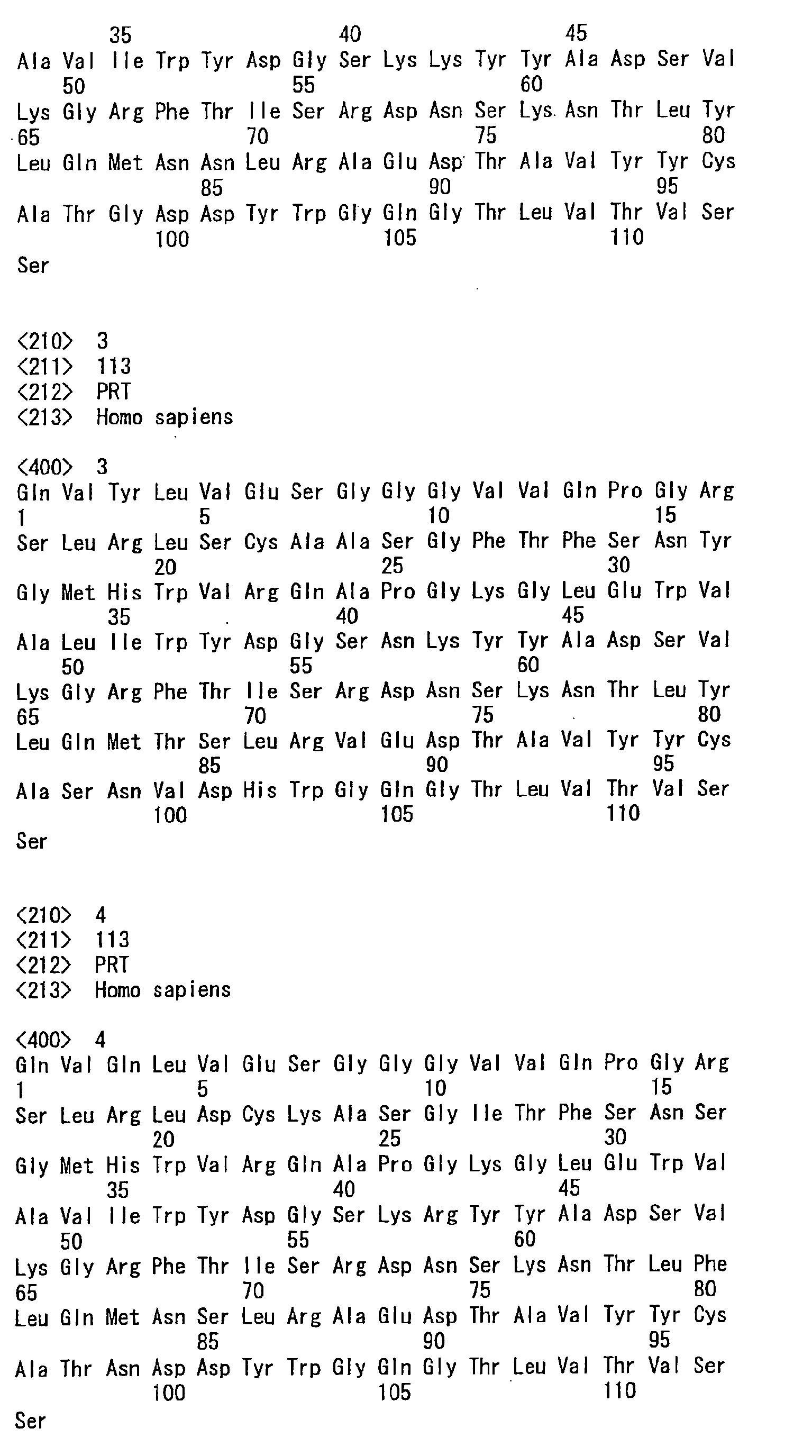


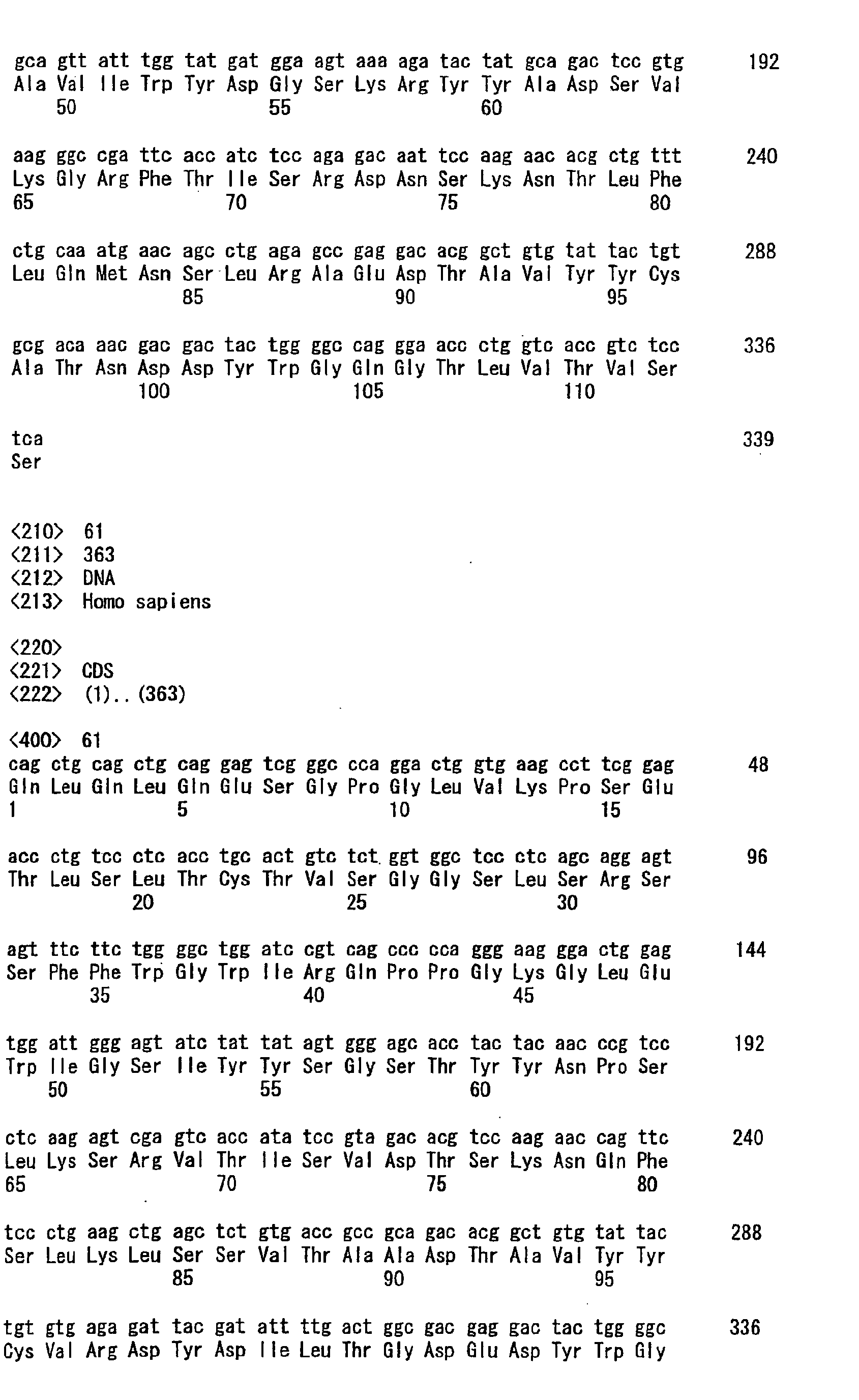


Claims (16)
- An isolated monoclonal antibody, or antigen-binding portion thereof, comprising:a) a heavy chain variable region CDR1 comprising SEQ ID NO: 18;b) a heavy chain variable region CDR2 comprising SEQ ID NO: 25;c) a heavy chain variable region CDR3 comprising SEQ ID NO: 32;d) a light chain variable region CDR1 comprising SEQ ID NO: 39;e) a light chain variable region CDR2 comprising SEQ ID NO: 46; andf) a light chain variable region CDR3 comprising SEQ ID NO: 53,wherein the antibody specifically binds PD-1.
- The antibody of claim 1, comprising:a) a heavy chain variable region comprising an amino acid sequence of SEQ ID NO: 4; andb) a light chain variable region comprising an amino acid sequence of SEQ ID NO: 11,wherein the antibody specifically binds human PD-1.
- An isolated monoclonal antibody, or antigen binding portion thereof, wherein the antibody cross-competes for binding to PD-1 with the antibody of claim 1 or 2.
- The antibody of any one of claims 1 to 3, wherein the antibodya) binds to human PD-1 with a KD of 5x10⁻⁹ or less; andb) binds to neither human ICOS, human CTLA-4 nor human CD28.
- The antibody of any one of claims 1 to 4, wherein the antibody inhibits the suppression of CD4+ CD25- T cells by regulatory T cells.
- The antibody of any one of claims 1 to 4, wherein the antibody enhances the antigen specific-memory response to a tumor or a pathogen.
- The antibody of any one of claims 1 to 4, wherein the antibody confers a persistent immunity to tumor relapse.
- The antibody of any one of claims 1 to 7, wherein the antibody is an IgG4 isotype.
- The antibody of claim 8, wherein the antibody has neither ADCC activity nor CDC activity.
- A composition comprising the antibody, or an antigen-binding portion thereof, of any one of claims 1 to 9, and a pharmaceutically acceptable carrier.
- Use of an antibody, or an antigen-binding portion thereof, of any one of claims 1 to 9 for preparing a pharmaceutical composition for modulating an immune response.
- Use of an antibody, or an antigen-binding portion thereof, of any one of claims 1 to 9 for preparing a pharmaceutical composition for inhibiting the growth of tumor cells.
- The use of claim 12, wherein the tumor cells are caused by a cancer selected from the group consisting of melanoma, renal cancer, prostate cancer, breast cancer, colon cancer and lung cancer.
- The use of claim 12, wherein the tumor cells are caused by a cancer selected from the group consisting of bone cancer, pancreatic cancer, skin cancer, cancer of the head or neck, cutaneous or intraocular malignant melanoma, uterine cancer, ovarian cancer, rectal cancer, cancer of the anal region, stomach cancer, testicular cancer, uterine cancer, carcinoma of the fallopian tubes, carcinoma of the endometrium, carcinoma of the cervix, carcinoma of the vagina, carcinoma of the vulva, Hodgkin's Disease, non-Hodgkin's lymphoma, cancer of the esophagus, cancer of the small intestine, cancer of the endocrine system, cancer of the thyroid gland, cancer of the parathyroid gland, cancer of the adrenal gland, sarcoma of soft tissue, cancer of the urethra, cancer of the penis, chronic or acute leukemia, acute myeloid leukemia, chronic myeloid leukemia, acute lymphoblastic leukemia, chronic lymphocytic leukemia, solid tumors of childhood, lymphocytic lymphoma, cancer of the bladder, cancer of the kidney or ureter, carcinoma of the renal pelvis, neoplasm of the central nervous system (CNS), primary CNS lymphoma, tumor angiogenesis, spinal axis tumor, brain stem glioma, pituitary adenoma, Kaposi's sarcoma, epidermoid cancer, squamous cell cancer, T-cell lymphoma, environmentally induced cancers including those induced by asbestos, and combinations of said cancers.
- Use of an antibody, or an antigen-binding portion thereof, of any one of claims 1 to 9 for preparing a pharmaceutical composition for treating an infectious disease.
- The use of claim 15, wherein the infectious disease is caused by a virus selected from the group consisting of HIV, Influenza, Herpes, Giardia, Malaria, Leishmania, Hepatitis A, B, or C, a herpes virus, VZV, HSV-1, HAV-6, HSV-II, CMV, Epstein Barr virus, adenovirus, influenza virus, flaviviruses, echovirus, rhinovirus, coxsackie virus, comovirus, respiratory syncytial virus, mumps virus, rotavirus, measles virus, rubella virus, parvovirus, vaccinia virus, HTLV virus, dengue virus, papillomavirus, molluscum virus, poliovirus, rabies virus, JC virus, arboviral encephalitis virus, chlamydia, rickettsial bacteria, mycobacteria, staphylococci, streptococci, pneumonococci, meningococci, conococci, klebsiella, proteus, serratia, pseudomonas, legionella, diphtheria, salmonella, bacilli, cholera, tetanus, botulism, anthrax, plague, leptospirosis, Lymes disease bacteria, Candida albicans, krusei, glabrata, or tropicalis, Cryptococcus neoformans, Aspergillus fumigatus or niger, Mucorales mucor, absidia or rhizophus, Sporothrix schenkii, Blastomyces dermatitidis, Paracoccidioides brasiliensis, Coccidioides immitis, Histoplasma capsulatum, Entamoeba histolytica, Balantidium coli, Naegleriafowleri, Acanthamoeba sp., Giardialamblia, Cryptosporidium, Pneumocystis carinii, Plasmodium vivax, Babesia microti, Trypanosoma brucei, Trypanosoma cruzi, Leishmania donovani, Toxoplasma gondi, and Nippostrongylus brasiliensis.
Priority Applications (12)
| Application Number | Priority Date | Filing Date | Title |
|---|---|---|---|
| SI200631652T SI2161336T1 (en) | 2005-05-09 | 2006-05-02 | Human monoclonal antibodies to programmed death 1(PD-1) and methods for treating cancer using anti-PD-1 antibodies alone or in combination with other immunotherapeutics |
| EP19151041.1A EP3530736A3 (en) | 2005-05-09 | 2006-05-02 | Human monoclonal antibodies to programmed death 1 (pd-1) and methods for treating cancer using anti-pd-1 antibodies alone or in combination with other immunotherapeutics |
| EP11178188.6A EP2439272A3 (en) | 2005-05-09 | 2006-05-02 | Human monoclonal antibodies to programmed death 1(PD-1) and methods for treating cancer using anti-PD-1 antibodies alone or in combination with other immunotherapeutics |
| PL11178191T PL2439273T3 (en) | 2005-05-09 | 2006-05-02 | Human monoclonal antibodies to programmed death 1(PD-1) and methods for treating cancer using anti-PD-1 antibodies alone or in combination with other immunotherapeutics |
| PL09013687T PL2161336T5 (en) | 2005-05-09 | 2006-05-02 | Human monoclonal antibodies to programmed death 1(PD-1) and methods for treating cancer using anti-PD-1 antibodies alone or in combination with other immunotherapeutics |
| EP11178191.0A EP2439273B1 (en) | 2005-05-09 | 2006-05-02 | Human monoclonal antibodies to programmed death 1(PD-1) and methods for treating cancer using anti-PD-1 antibodies alone or in combination with other immunotherapeutics |
| DK11178191.0T DK2439273T3 (en) | 2005-05-09 | 2006-05-02 | HUMAN MONOCLONAL ANTIBODIES FOR PROGRAMMED DEATH-1 (PD-1) AND PROCEDURES FOR TREATMENT OF CANCER USING ANTI-PD-1 ANTIBODIES ALONE OR IN COMBINATION WITH OTHER IMMUNTER APPLICATIONS |
| EP11178187A EP2418278A3 (en) | 2005-05-09 | 2006-05-02 | Human monoclonal antibodies to programmed death 1(PD-1) and methods for treating cancer using anti-PD-1 antibodies alone or in combination with other immunotherapeutics |
| SI200631652A SI2161336T2 (en) | 2005-05-09 | 2006-05-02 | Human monoclonal antibodies to programmed death 1(PD-1) and methods for treating cancer using anti-PD-1 antibodies alone or in combination with other immunotherapeutics |
| CY20131100915T CY1114506T1 (en) | 2005-05-09 | 2013-10-17 | MONOCLONAL ANTIBODIES AGAINST HUMAN PROTEIN programmed death (PD-1) AND METHODS OF TREATMENT AGAINST CANCER USING ANTI-PD-1 INDIVIDUALLY OR IN COMBINATION WITH OTHER AGENTS ANOSOTHERAPEFTIKOUS |
| HUS1500067C HUS1500067I1 (en) | 2005-05-09 | 2015-12-15 | Human monoclonal antibodies to programmed death 1(PD-1) and methods for treating cancer using anti-PD-1 antibodies alone or in combination with other immunotherapeutics |
| CY20191100551T CY1121648T1 (en) | 2005-05-09 | 2019-05-24 | HUMAN MONOCLONAL ANTIBODIES TO PROGRAMMED DEATH l(PD-1) AND METHODS OF TREATING CANCER USING ANTI-PD-1 ANTIBODIES ALONE OR IN COMBINATION WITH OTHER IMMUNOTHERAPEUTICS |
Applications Claiming Priority (4)
| Application Number | Priority Date | Filing Date | Title |
|---|---|---|---|
| US67946605P | 2005-05-09 | 2005-05-09 | |
| US73843405P | 2005-11-21 | 2005-11-21 | |
| US74891905P | 2005-12-08 | 2005-12-08 | |
| EP06746353A EP1896582A4 (en) | 2005-05-09 | 2006-05-02 | HUMAN MONOCLONAL ANTIBODIES FOR PROGRAMMED DEATH 1 (MP-1) AND METHODS FOR TREATING CANCER USING ANTI-MP-1 ANTIBODIES ALONE OR ASSOCIATED WITH OTHER IMMUNOTHERAPIES |
Related Parent Applications (2)
| Application Number | Title | Priority Date | Filing Date |
|---|---|---|---|
| EP06746353A Division EP1896582A4 (en) | 2005-05-09 | 2006-05-02 | HUMAN MONOCLONAL ANTIBODIES FOR PROGRAMMED DEATH 1 (MP-1) AND METHODS FOR TREATING CANCER USING ANTI-MP-1 ANTIBODIES ALONE OR ASSOCIATED WITH OTHER IMMUNOTHERAPIES |
| EP06746353.9 Division | 2006-05-02 |
Related Child Applications (8)
| Application Number | Title | Priority Date | Filing Date |
|---|---|---|---|
| EP11178187A Division-Into EP2418278A3 (en) | 2005-05-09 | 2006-05-02 | Human monoclonal antibodies to programmed death 1(PD-1) and methods for treating cancer using anti-PD-1 antibodies alone or in combination with other immunotherapeutics |
| EP19151041.1A Division EP3530736A3 (en) | 2005-05-09 | 2006-05-02 | Human monoclonal antibodies to programmed death 1 (pd-1) and methods for treating cancer using anti-pd-1 antibodies alone or in combination with other immunotherapeutics |
| EP11178191.0A Division-Into EP2439273B1 (en) | 2005-05-09 | 2006-05-02 | Human monoclonal antibodies to programmed death 1(PD-1) and methods for treating cancer using anti-PD-1 antibodies alone or in combination with other immunotherapeutics |
| EP11178191.0A Division EP2439273B1 (en) | 2005-05-09 | 2006-05-02 | Human monoclonal antibodies to programmed death 1(PD-1) and methods for treating cancer using anti-PD-1 antibodies alone or in combination with other immunotherapeutics |
| EP11178188.6A Division-Into EP2439272A3 (en) | 2005-05-09 | 2006-05-02 | Human monoclonal antibodies to programmed death 1(PD-1) and methods for treating cancer using anti-PD-1 antibodies alone or in combination with other immunotherapeutics |
| EP11178187.8 Division-Into | 2011-08-19 | ||
| EP11178191.0 Division-Into | 2011-08-19 | ||
| EP11178188.6 Division-Into | 2011-08-19 |
Publications (3)
| Publication Number | Publication Date |
|---|---|
| EP2161336A1 true EP2161336A1 (en) | 2010-03-10 |
| EP2161336B1 EP2161336B1 (en) | 2013-07-31 |
| EP2161336B2 EP2161336B2 (en) | 2017-03-29 |
Family
ID=37396674
Family Applications (6)
| Application Number | Title | Priority Date | Filing Date |
|---|---|---|---|
| EP19151041.1A Pending EP3530736A3 (en) | 2005-05-09 | 2006-05-02 | Human monoclonal antibodies to programmed death 1 (pd-1) and methods for treating cancer using anti-pd-1 antibodies alone or in combination with other immunotherapeutics |
| EP11178187A Withdrawn EP2418278A3 (en) | 2005-05-09 | 2006-05-02 | Human monoclonal antibodies to programmed death 1(PD-1) and methods for treating cancer using anti-PD-1 antibodies alone or in combination with other immunotherapeutics |
| EP09013687.0A Expired - Lifetime EP2161336B2 (en) | 2005-05-09 | 2006-05-02 | Human monoclonal antibodies to programmed death 1(PD-1) and methods for treating cancer using anti-PD-1 antibodies alone or in combination with other immunotherapeutics |
| EP11178188.6A Withdrawn EP2439272A3 (en) | 2005-05-09 | 2006-05-02 | Human monoclonal antibodies to programmed death 1(PD-1) and methods for treating cancer using anti-PD-1 antibodies alone or in combination with other immunotherapeutics |
| EP11178191.0A Revoked EP2439273B1 (en) | 2005-05-09 | 2006-05-02 | Human monoclonal antibodies to programmed death 1(PD-1) and methods for treating cancer using anti-PD-1 antibodies alone or in combination with other immunotherapeutics |
| EP06746353A Withdrawn EP1896582A4 (en) | 2005-05-09 | 2006-05-02 | HUMAN MONOCLONAL ANTIBODIES FOR PROGRAMMED DEATH 1 (MP-1) AND METHODS FOR TREATING CANCER USING ANTI-MP-1 ANTIBODIES ALONE OR ASSOCIATED WITH OTHER IMMUNOTHERAPIES |
Family Applications Before (2)
| Application Number | Title | Priority Date | Filing Date |
|---|---|---|---|
| EP19151041.1A Pending EP3530736A3 (en) | 2005-05-09 | 2006-05-02 | Human monoclonal antibodies to programmed death 1 (pd-1) and methods for treating cancer using anti-pd-1 antibodies alone or in combination with other immunotherapeutics |
| EP11178187A Withdrawn EP2418278A3 (en) | 2005-05-09 | 2006-05-02 | Human monoclonal antibodies to programmed death 1(PD-1) and methods for treating cancer using anti-PD-1 antibodies alone or in combination with other immunotherapeutics |
Family Applications After (3)
| Application Number | Title | Priority Date | Filing Date |
|---|---|---|---|
| EP11178188.6A Withdrawn EP2439272A3 (en) | 2005-05-09 | 2006-05-02 | Human monoclonal antibodies to programmed death 1(PD-1) and methods for treating cancer using anti-PD-1 antibodies alone or in combination with other immunotherapeutics |
| EP11178191.0A Revoked EP2439273B1 (en) | 2005-05-09 | 2006-05-02 | Human monoclonal antibodies to programmed death 1(PD-1) and methods for treating cancer using anti-PD-1 antibodies alone or in combination with other immunotherapeutics |
| EP06746353A Withdrawn EP1896582A4 (en) | 2005-05-09 | 2006-05-02 | HUMAN MONOCLONAL ANTIBODIES FOR PROGRAMMED DEATH 1 (MP-1) AND METHODS FOR TREATING CANCER USING ANTI-MP-1 ANTIBODIES ALONE OR ASSOCIATED WITH OTHER IMMUNOTHERAPIES |
Country Status (28)
| Country | Link |
|---|---|
| US (10) | US8008449B2 (en) |
| EP (6) | EP3530736A3 (en) |
| JP (9) | JP4361545B2 (en) |
| KR (3) | KR101339628B1 (en) |
| CN (5) | CN103059138B (en) |
| AU (1) | AU2006244885B2 (en) |
| BE (1) | BE2015C074I2 (en) |
| BR (1) | BRPI0610235B8 (en) |
| CA (3) | CA3151350A1 (en) |
| CY (2) | CY2015057I1 (en) |
| DK (2) | DK2439273T3 (en) |
| ES (2) | ES2427646T5 (en) |
| FI (1) | FI2161336T9 (en) |
| FR (1) | FR15C0087I2 (en) |
| HU (2) | HUE044719T2 (en) |
| IL (2) | IL187108A (en) |
| LT (2) | LT2439273T (en) |
| LU (1) | LU92904I2 (en) |
| MX (1) | MX2007013978A (en) |
| NL (1) | NL300782I2 (en) |
| NO (6) | NO341219B1 (en) |
| NZ (1) | NZ563193A (en) |
| PL (2) | PL2161336T5 (en) |
| PT (2) | PT2439273T (en) |
| RU (4) | RU2406760C3 (en) |
| SI (2) | SI2161336T1 (en) |
| TW (1) | TWI379898B (en) |
| WO (1) | WO2006121168A1 (en) |
Cited By (61)
| Publication number | Priority date | Publication date | Assignee | Title |
|---|---|---|---|---|
| WO2015066413A1 (en) | 2013-11-01 | 2015-05-07 | Novartis Ag | Oxazolidinone hydroxamic acid compounds for the treatment of bacterial infections |
| WO2015138920A1 (en) | 2014-03-14 | 2015-09-17 | Novartis Ag | Antibody molecules to lag-3 and uses thereof |
| WO2015148379A1 (en) | 2014-03-24 | 2015-10-01 | Novartis Ag | Monobactam organic compounds for the treatment of bacterial infections |
| CN105238762A (en) * | 2015-10-26 | 2016-01-13 | 无锡傲锐东源生物科技有限公司 | Anti-PD-1 protein monoclonal antibody hybridomas cell, anti- PD-1 monoclonal antibody generated by same and application |
| WO2016020836A1 (en) | 2014-08-06 | 2016-02-11 | Novartis Ag | Quinolone derivatives as antibacterials |
| WO2016040880A1 (en) | 2014-09-13 | 2016-03-17 | Novartis Ag | Combination therapies of alk inhibitors |
| WO2016059602A3 (en) * | 2014-10-16 | 2016-06-09 | Glaxo Group Limited | Combination therapy with anti-her3 antibodies to treat cancer |
| WO2016097995A1 (en) | 2014-12-16 | 2016-06-23 | Novartis Ag | Isoxazole hydroxamic acid compounds as lpxc inhibitors |
| WO2016100882A1 (en) | 2014-12-19 | 2016-06-23 | Novartis Ag | Combination therapies |
| US9683048B2 (en) | 2014-01-24 | 2017-06-20 | Novartis Ag | Antibody molecules to PD-1 and uses thereof |
| WO2017106630A1 (en) | 2015-12-18 | 2017-06-22 | The General Hospital Corporation | Polyacetal polymers, conjugates, particles and uses thereof |
| WO2017140821A1 (en) | 2016-02-19 | 2017-08-24 | Novartis Ag | Tetracyclic pyridone compounds as antivirals |
| WO2017163186A1 (en) | 2016-03-24 | 2017-09-28 | Novartis Ag | Alkynyl nucleoside analogs as inhibitors of human rhinovirus |
| WO2017216705A1 (en) | 2016-06-14 | 2017-12-21 | Novartis Ag | Crystalline form of (r)-4-(5-(cyclopropylethynyl)isoxazol-3-yl)-n-hydroxy-2-methyl-2-(methylsulfonyl)butanamide as an antibacterial agent |
| WO2017216685A1 (en) | 2016-06-16 | 2017-12-21 | Novartis Ag | Pentacyclic pyridone compounds as antivirals |
| WO2017216686A1 (en) | 2016-06-16 | 2017-12-21 | Novartis Ag | 8,9-fused 2-oxo-6,7-dihydropyrido-isoquinoline compounds as antivirals |
| WO2018047109A1 (en) | 2016-09-09 | 2018-03-15 | Novartis Ag | Polycyclic pyridone compounds as antivirals |
| WO2018060926A1 (en) | 2016-09-28 | 2018-04-05 | Novartis Ag | Beta-lactamase inhibitors |
| US9938345B2 (en) | 2014-01-23 | 2018-04-10 | Regeneron Pharmaceuticals, Inc. | Human antibodies to PD-L1 |
| WO2018073753A1 (en) | 2016-10-18 | 2018-04-26 | Novartis Ag | Fused tetracyclic pyridone compounds as antivirals |
| US9987500B2 (en) | 2014-01-23 | 2018-06-05 | Regeneron Pharmaceuticals, Inc. | Human antibodies to PD-1 |
| WO2018154529A1 (en) | 2017-02-27 | 2018-08-30 | Novartis Ag | Dosing schedule for a combination of ceritinib and an anti-pd-1 antibody molecule |
| WO2018198079A1 (en) | 2017-04-27 | 2018-11-01 | Novartis Ag | Fused indazole pyridone compounds as antivirals |
| WO2018203302A1 (en) | 2017-05-05 | 2018-11-08 | Novartis Ag | Tricyclic 2-quinolinones as antibacterials |
| WO2019089753A2 (en) | 2017-10-31 | 2019-05-09 | Compass Therapeutics Llc | Cd137 antibodies and pd-1 antagonists and uses thereof |
| WO2019097479A1 (en) | 2017-11-17 | 2019-05-23 | Novartis Ag | Novel dihydroisoxazole compounds and their use for the treatment of hepatitis b |
| WO2019105468A1 (en) * | 2017-12-01 | 2019-06-06 | Adagene Inc. | Methods for using cd137 ligand as a biomarker for treatment with anti-cd137 antibody |
| WO2019123285A1 (en) | 2017-12-20 | 2019-06-27 | Novartis Ag | Fused tricyclic pyrazolo-dihydropyrazinyl-pyridone compounds as antivirals |
| WO2019166951A1 (en) | 2018-02-28 | 2019-09-06 | Novartis Ag | Indole-2-carbonyl compounds and their use for the treatment of hepatitis b |
| WO2019178269A2 (en) | 2018-03-14 | 2019-09-19 | Surface Oncology, Inc. | Antibodies that bind cd39 and uses thereof |
| US10441655B2 (en) | 2005-05-09 | 2019-10-15 | Ono Pharmaceutical Co., Ltd. | Monoclonal antibodies to programmed death 1 (PD-1) |
| US10449251B2 (en) | 2014-08-01 | 2019-10-22 | Akeso Biopharma, Inc. | Anti-CTLA4 monoclonal antibody or its antigen binding fragments, pharmaceutical compositions and uses |
| WO2019204743A1 (en) | 2018-04-19 | 2019-10-24 | Checkmate Pharmaceuticals, Inc. | Synthetic rig-i-like receptor agonists |
| US10457725B2 (en) | 2016-05-13 | 2019-10-29 | Regeneron Pharmaceuticals, Inc. | Methods of treating skin cancer by administering a PD-1 inhibitor |
| US10472419B2 (en) | 2014-01-31 | 2019-11-12 | Novartis Ag | Antibody molecules to TIM-3 and uses thereof |
| US10570204B2 (en) | 2013-09-26 | 2020-02-25 | The Medical College Of Wisconsin, Inc. | Methods for treating hematologic cancers |
| WO2020053654A1 (en) | 2018-09-12 | 2020-03-19 | Novartis Ag | Antiviral pyridopyrazinedione compounds |
| WO2021062244A1 (en) | 2019-09-25 | 2021-04-01 | Surface Oncology, Inc. | Anti-il-27 antibodies and uses thereof |
| WO2021081353A1 (en) | 2019-10-23 | 2021-04-29 | Checkmate Pharmaceuticals, Inc. | Synthetic rig-i-like receptor agonists |
| CN113767175A (en) * | 2018-08-28 | 2021-12-07 | 10X基因组学股份有限公司 | Increase Spatial Array Resolution |
| US11242395B2 (en) | 2017-08-21 | 2022-02-08 | Adagene Inc. | Anti-CD137 molecules and use thereof |
| WO2022184937A1 (en) | 2021-03-05 | 2022-09-09 | Leadartis, S.L. | Trimeric polypeptides and uses thereof in the treatment of cancer |
| WO2022212400A1 (en) | 2021-03-29 | 2022-10-06 | Juno Therapeutics, Inc. | Methods for dosing and treatment with a combination of a checkpoint inhibitor therapy and a car t cell therapy |
| US11479608B2 (en) | 2016-08-23 | 2022-10-25 | Akeso Biopharma, Inc. | Anti-CTLA4 antibodies |
| WO2022236134A1 (en) | 2021-05-07 | 2022-11-10 | Surface Oncology, Inc. | Anti-il-27 antibodies and uses thereof |
| US11603407B2 (en) | 2017-04-06 | 2023-03-14 | Regeneron Pharmaceuticals, Inc. | Stable antibody formulation |
| WO2023039089A1 (en) | 2021-09-08 | 2023-03-16 | Twentyeight-Seven, Inc. | Papd5 and/or papd7 inhibiting 4-oxo-1,4-dihydroquinoline-3-carboxylic acid derivatives |
| WO2023154905A1 (en) | 2022-02-14 | 2023-08-17 | Gilead Sciences, Inc. | Antiviral pyrazolopyridinone compounds |
| US11952681B2 (en) | 2018-02-02 | 2024-04-09 | Adagene Inc. | Masked activatable CD137 antibodies |
| USRE50065E1 (en) | 2012-10-17 | 2024-07-30 | 10X Genomics Sweden Ab | Methods and product for optimising localised or spatial detection of gene expression in a tissue sample |
| US12054557B2 (en) | 2015-12-22 | 2024-08-06 | Regeneron Pharmaceuticals, Inc. | Combination of anti-PD-1 antibodies and bispecific anti-CD20/anti-CD3 antibodies to treat cancer |
| WO2024229461A2 (en) | 2023-05-04 | 2024-11-07 | Novasenta, Inc. | Anti-cd161 antibodies and methods of use thereof |
| US12234505B2 (en) | 2010-04-05 | 2025-02-25 | Prognosys Biosciences, Inc. | Spatially encoded biological assays |
| EP4549467A2 (en) | 2016-08-01 | 2025-05-07 | ImmunoGenesis, Inc. | Administration of hypoxia activated prodrugs in combination with immune modulatory agents for treating cancer |
| US12338220B2 (en) | 2019-09-26 | 2025-06-24 | Gilead Sciences, Inc. | Antiviral pyrazolopiridinone compounds |
| US12358978B2 (en) | 2018-12-13 | 2025-07-15 | Surface Oncology, LLC | Anti-IL-27 antibodies and uses thereof |
| EP4585268A2 (en) | 2015-09-14 | 2025-07-16 | Twelve Therapeutics, Inc. | Solid forms of isoquinolinone derivatives, process of making, compositions comprising, and methods of using the same |
| WO2026055167A1 (en) | 2024-09-05 | 2026-03-12 | Surface Oncology, LLC | Anti-il-27 antibodies and use of biomarkers in uses thereof |
| WO2026055168A1 (en) | 2024-09-06 | 2026-03-12 | Surface Oncology, LLC | Anti-il-27 antibodies and uses and doses thereof |
| US12600777B2 (en) | 2015-07-29 | 2026-04-14 | Novartis Ag | Combination therapies comprising antibody molecules to LAG-3 |
| US12630633B2 (en) | 2015-12-17 | 2026-05-19 | Novartis Ag | Antibody molecules to PD-1 and uses thereof |
Families Citing this family (2257)
| Publication number | Priority date | Publication date | Assignee | Title |
|---|---|---|---|---|
| US7605238B2 (en) | 1999-08-24 | 2009-10-20 | Medarex, Inc. | Human CTLA-4 antibodies and their uses |
| US7030219B2 (en) | 2000-04-28 | 2006-04-18 | Johns Hopkins University | B7-DC, Dendritic cell co-stimulatory molecules |
| NZ536420A (en) * | 2002-04-12 | 2008-04-30 | Medarex Inc | Methods of treatment using CTLA-4 antibodies |
| DE60334303D1 (en) | 2002-07-03 | 2010-11-04 | Tasuku Honjo | IMMUNO-POTENTIAL COMPOSITIONS |
| EP2277532A1 (en) | 2002-09-11 | 2011-01-26 | Genentech, Inc. | Novel composition and methods for the treatment of immune related diseases |
| DE10347710B4 (en) | 2003-10-14 | 2006-03-30 | Johannes-Gutenberg-Universität Mainz | Recombinant vaccines and their use |
| DK1879573T3 (en) | 2005-05-10 | 2013-02-25 | Incyte Corp | MODULATORS OF INDOLAMINE 2,3-DIOXYGENASE AND METHODS FOR USING IT |
| DK1907000T4 (en) | 2005-06-08 | 2020-03-30 | The President And Fellows Of Harvard College | Methods and compositions for treating persistent HIV infections by inhibiting the pathway of programmed cell death 1 (PD-1). |
| KR101704734B1 (en) | 2005-07-01 | 2017-02-09 | 이. 알. 스퀴부 앤드 선즈, 엘.엘.씨. | Human monoclonal antibodies to programmed death ligand 1(pd-l1) |
| DE102005046490A1 (en) | 2005-09-28 | 2007-03-29 | Johannes-Gutenberg-Universität Mainz | New nucleic acid molecule comprising promoter, a transcriptable nucleic acid sequence, a first and second nucleic acid sequence for producing modified RNA with transcriptional stability and translational efficiency |
| US7700567B2 (en) | 2005-09-29 | 2010-04-20 | Supergen, Inc. | Oligonucleotide analogues incorporating 5-aza-cytosine therein |
| NZ568016A (en) | 2005-12-07 | 2011-12-22 | Medarex Inc | CTLA-4 antibody dosage escalation regimens |
| EP2889309B1 (en) | 2006-03-03 | 2017-12-27 | Ono Pharmaceutical Co., Ltd. | Multimer of extracellular domain of pd-1 or pd-l1 |
| JP2010504356A (en) * | 2006-09-20 | 2010-02-12 | ザ ジョンズ ホプキンス ユニバーシティー | Combination therapy of cancer and infectious diseases using anti-B7-H1 antibody |
| CA2673870A1 (en) | 2006-12-27 | 2008-07-10 | Emory University | Compositions and methods for the treatment of infections and tumors |
| AU2013200388B2 (en) * | 2006-12-27 | 2014-10-23 | Dana-Farber Cancer Institute, Inc. | Compositions and methods for the treatment of infections and tumors |
| US20100055111A1 (en) * | 2007-02-14 | 2010-03-04 | Med. College Of Georgia Research Institute, Inc. | Indoleamine 2,3-dioxygenase, pd-1/pd-l pathways, and ctla4 pathways in the activation of regulatory t cells |
| SI2185589T1 (en) | 2007-06-01 | 2016-04-29 | University Of Maryland Baltimore | Immunoglobulin constant region fc receptor binding agents |
| PT2170959E (en) * | 2007-06-18 | 2014-01-07 | Merck Sharp & Dohme | ANTIBODIES FOR THE PD-1 PROGRAMMED DEATH HUMAN RECEPTOR |
| AU2014201367B2 (en) * | 2007-06-18 | 2016-01-28 | Merck Sharp & Dohme B.V. | Antibodies to human programmed death receptor pd-1 |
| US20090028857A1 (en) | 2007-07-23 | 2009-01-29 | Cell Genesys, Inc. | Pd-1 antibodies in combination with a cytokine-secreting cell and methods of use thereof |
| EP2195347A1 (en) | 2007-08-17 | 2010-06-16 | INSERM (Institut National de la Santé et de la Recherche Médicale) | Method for treating and diagnosing hematologic malignancies |
| EP2225397A4 (en) | 2007-11-28 | 2011-08-17 | Univ Montreal | MODULATION OF PD-1 AND ITS USES |
| EP3153526B1 (en) | 2008-01-31 | 2020-09-23 | INSERM - Institut National de la Santé et de la Recherche Médicale | Antibodies against human cd39 and use thereof for inhibiting t regulatory cells activity |
| US8168757B2 (en) | 2008-03-12 | 2012-05-01 | Merck Sharp & Dohme Corp. | PD-1 binding proteins |
| WO2009126688A2 (en) | 2008-04-09 | 2009-10-15 | Genentech, Inc. | Novel compositions and methods for the treatment of immune related diseases |
| US9650639B2 (en) | 2008-05-19 | 2017-05-16 | Advaxis, Inc. | Dual delivery system for heterologous antigens |
| JP5757863B2 (en) | 2008-05-19 | 2015-08-05 | アドバクシス インコーポレイテッド | Dual delivery system for xenoantigens |
| US9017660B2 (en) | 2009-11-11 | 2015-04-28 | Advaxis, Inc. | Compositions and methods for prevention of escape mutation in the treatment of Her2/neu over-expressing tumors |
| US8460886B2 (en) | 2008-07-04 | 2013-06-11 | Ono Pharmaceutical Co., Ltd. | Use of an efficacy marker for optimizing therapeutic efficacy of an anti-human PD-1 antibody on cancers |
| CA3012229C (en) * | 2008-07-08 | 2020-11-10 | Incyte Holdings Corporation | 1,2,5-oxadiazoles as inhibitors of indoleamine 2,3-dioxygenase |
| WO2010014784A2 (en) | 2008-08-01 | 2010-02-04 | Bristol-Myers Squibb Company | Combination of anti-ctla4 antibody with diverse therapeutic regimens for the synergistic treatment of proliferative diseases |
| AR072999A1 (en) | 2008-08-11 | 2010-10-06 | Medarex Inc | HUMAN ANTIBODIES THAT JOIN GEN 3 OF LYMPHOCYTARY ACTIVATION (LAG-3) AND THE USES OF THESE |
| EA201500417A1 (en) | 2008-08-25 | 2015-11-30 | Эмплиммьюн, Инк. | ANTAGONIST COMPOSITIONS PD-1 AND APPLICATIONS |
| WO2010098788A2 (en) * | 2008-08-25 | 2010-09-02 | Amplimmune, Inc. | Pd-i antagonists and methods for treating infectious disease |
| AU2009290543B2 (en) | 2008-09-12 | 2015-09-03 | Oxford University Innovation Limited | PD-1 specific antibodies and uses thereof |
| AU2009290544B2 (en) * | 2008-09-12 | 2015-07-16 | Oxford University Innovation Limited | PD-1 specific antibodies and uses thereof |
| WO2010036959A2 (en) | 2008-09-26 | 2010-04-01 | Dana-Farber Cancer Institute | Human anti-pd-1, pd-l1, and pd-l2 antibodies and uses therefor |
| JP2012508563A (en) * | 2008-11-12 | 2012-04-12 | シェーリング コーポレイション | ΒG1-IGG intron for enhanced anti-IGF1R expression |
| US11542328B2 (en) * | 2008-11-14 | 2023-01-03 | The Brigham And Women's Hospital, Inc. | Therapeutic and diagnostic methods relating to cancer stem cells |
| ES2796111T3 (en) * | 2008-11-14 | 2020-11-25 | Brigham & Womens Hospital Inc | Therapeutic methods related to cancer stem cells |
| MX2011005691A (en) | 2008-11-28 | 2011-07-20 | Univ Emory | Methods for the treatment of infections and tumors. |
| MX356367B (en) | 2008-12-09 | 2018-05-25 | Genentech Inc | Anti-pd-l1 antibodies and their use to enhance t-cell function. |
| WO2010089411A2 (en) * | 2009-02-09 | 2010-08-12 | Universite De La Mediterranee | Pd-1 antibodies and pd-l1 antibodies and uses thereof |
| PE20120923A1 (en) | 2009-03-30 | 2012-08-27 | Eisai Randd Man Co Ltd | PHARMACEUTICAL COMPOSITIONS INCLUDING LIPOSOMES CONTAINING ERIBULIN OR A SALT OF IT |
| JP5836940B2 (en) | 2009-06-26 | 2015-12-24 | ソリシメド・バイオファーマ・インコーポレーテッド | Soricidin derived peptides and methods for detection and drug delivery of TRPV-6 cancer |
| US8394922B2 (en) | 2009-08-03 | 2013-03-12 | Medarex, Inc. | Antiproliferative compounds, conjugates thereof, methods therefor, and uses thereof |
| CA2771403C (en) | 2009-08-19 | 2015-02-24 | Eisai R&D Management Co. Ltd. | Quinoline derivative-containing pharmaceutical composition |
| WO2011041613A2 (en) | 2009-09-30 | 2011-04-07 | Memorial Sloan-Kettering Cancer Center | Combination immunotherapy for the treatment of cancer |
| EP3485908B1 (en) | 2009-10-16 | 2021-08-18 | Mereo BioPharma 5, Inc. | Therapeutic combination and use of dll4 antagonist antibodies and anti-hypertensive agents |
| US10016617B2 (en) | 2009-11-11 | 2018-07-10 | The Trustees Of The University Of Pennsylvania | Combination immuno therapy and radiotherapy for the treatment of Her-2-positive cancers |
| TW201134488A (en) * | 2010-03-11 | 2011-10-16 | Ucb Pharma Sa | PD-1 antibodies |
| CN102892786B (en) | 2010-03-11 | 2016-03-16 | Ucb医药有限公司 | Pd-1 antibody |
| US20110288545A1 (en) * | 2010-04-22 | 2011-11-24 | Old Dominion University Research Foundation | Method and Device for Ablation of Cancer and Resistance to New Cancer Growth |
| US9115402B2 (en) | 2010-05-14 | 2015-08-25 | Dana-Farber Cancer Institute, Inc. | Compositions and methods of identifying tumor specific neoantigens |
| US20130064831A1 (en) * | 2010-05-17 | 2013-03-14 | Bristol-Myers Squibb Company | Immunotherapeutic dosing regimens and combinations thereof |
| US10420665B2 (en) | 2010-06-13 | 2019-09-24 | W. L. Gore & Associates, Inc. | Intragastric device for treating obesity |
| US10010439B2 (en) | 2010-06-13 | 2018-07-03 | Synerz Medical, Inc. | Intragastric device for treating obesity |
| US8628554B2 (en) | 2010-06-13 | 2014-01-14 | Virender K. Sharma | Intragastric device for treating obesity |
| US9526648B2 (en) | 2010-06-13 | 2016-12-27 | Synerz Medical, Inc. | Intragastric device for treating obesity |
| US9163087B2 (en) * | 2010-06-18 | 2015-10-20 | The Brigham And Women's Hospital, Inc. | Bi-specific antibodies against TIM-3 and PD-1 for immunotherapy in chronic immune conditions |
| WO2012016073A2 (en) | 2010-07-28 | 2012-02-02 | Gliknik Inc. | Fusion proteins of natural human protein fragments to create orderly multimerized immunoglobulin fc compositions |
| CA2807552A1 (en) | 2010-08-06 | 2012-02-09 | Moderna Therapeutics, Inc. | Engineered nucleic acids and methods of use thereof |
| EP2857499A1 (en) | 2010-10-01 | 2015-04-08 | Moderna Therapeutics, Inc. | Engineered nucleic acids and methods of use thereof |
| CN107412756A (en) | 2010-10-01 | 2017-12-01 | 宾夕法尼亚大学理事会 | Listeria vaccine carrier is used for the purposes that immunological unresponsiveness is reversed in the individual of parasitic infection |
| WO2012048113A2 (en) | 2010-10-07 | 2012-04-12 | The General Hospital Corporation | Biomarkers of cancer |
| WO2012088446A1 (en) | 2010-12-22 | 2012-06-28 | Board Of Trustees Of The Leland Stanford Junior University | Superagonists and antagonists of interleukin-2 |
| CA2829960A1 (en) | 2011-03-11 | 2012-09-20 | John Rothman | Listeria-based adjuvants |
| CA2831613A1 (en) | 2011-03-31 | 2012-10-04 | Moderna Therapeutics, Inc. | Delivery and formulation of engineered nucleic acids |
| WO2012135408A1 (en) * | 2011-03-31 | 2012-10-04 | Merck Sharp & Dohme Corp. | Stable formulations of antibodies to human programmed death receptor pd-1 and related treatments |
| CA2833636A1 (en) * | 2011-04-20 | 2012-10-26 | Amplimmune, Inc. | Antibodies and other molecules that bind b7-h1 and pd-1 |
| LT2714071T (en) | 2011-05-24 | 2019-10-10 | Biontech Rna Pharmaceuticals Gmbh | Individualized vaccines for cancer |
| US8852599B2 (en) | 2011-05-26 | 2014-10-07 | Bristol-Myers Squibb Company | Immunoconjugates, compositions for making them, and methods of making and use |
| JP6038128B2 (en) | 2011-06-03 | 2016-12-07 | エーザイ・アール・アンド・ディー・マネジメント株式会社 | A biomarker for predicting and evaluating the reactivity of thyroid and renal cancer subjects to lenvatinib compounds |
| CA2840170A1 (en) * | 2011-06-21 | 2012-12-27 | The Johns Hopkins University | Focused radiation for augmenting immune-based therapies against neoplasms |
| DK2734205T3 (en) | 2011-07-21 | 2018-06-14 | Tolero Pharmaceuticals Inc | Heterocyclic Protein Kinase Inhibitors |
| BR112014002353B1 (en) * | 2011-08-01 | 2022-09-27 | Genentech, Inc | USES OF PD-1 AXIS BINDING ANTAGONISTS AND MEK INHIBITORS, PHARMACEUTICAL COMPOSITIONS, AND KIT |
| WO2013022091A1 (en) | 2011-08-11 | 2013-02-14 | 小野薬品工業株式会社 | Therapeutic agent for autoimmune diseases comprising pd-1 agonist |
| HUE042327T2 (en) | 2011-08-30 | 2019-06-28 | Astex Pharmaceuticals Inc | Decitabine derivative products |
| US9464124B2 (en) | 2011-09-12 | 2016-10-11 | Moderna Therapeutics, Inc. | Engineered nucleic acids and methods of use thereof |
| TWI583699B (en) | 2011-09-23 | 2017-05-21 | 安可美德藥物股份有限公司 | VEGF/DLL4 binding agents and their use |
| PT3682905T (en) | 2011-10-03 | 2022-04-07 | Modernatx Inc | Modified nucleosides, nucleotides, and nucleic acids, and uses thereof |
| GB201117313D0 (en) | 2011-10-07 | 2011-11-16 | Gt Biolog Ltd | Bacterium for use in medicine |
| US11951157B2 (en) | 2011-10-11 | 2024-04-09 | Universitat Zurich | Methods of treating malignant tumour with IL-12 and anti-PD-1 antibody |
| PL3351261T3 (en) | 2011-10-11 | 2021-12-06 | Universität Zürich | A combination drug containing IL-12 and an agent for blocking T cell inhibitory molecules in cancer therapy |
| WO2013059740A1 (en) | 2011-10-21 | 2013-04-25 | Foundation Medicine, Inc. | Novel alk and ntrk1 fusion molecules and uses thereof |
| BR112014012727B1 (en) | 2011-11-29 | 2022-10-25 | Ono Pharmaceutical Co., Ltd | 6-AMINO-9-[(3R)-1-(2-BUTINOYL)-3-PYRROLIDINYL]-7-(4-PHENOXYPHENYL)-7,9-DIHYDRO-8H-PURI-8-ONE E PHARMACEUTICAL COMPOSITION |
| ES2923757T3 (en) | 2011-12-16 | 2022-09-30 | Modernatx Inc | Modified mRNA compositions |
| CN104245717B (en) | 2011-12-28 | 2017-10-24 | 卡莱克汀医疗有限公司 | Compositions of novel carbohydrate drugs for the treatment of human diseases |
| US20140370012A1 (en) * | 2012-01-27 | 2014-12-18 | Gliknik Inc. | Fusion proteins comprising igg2 hinge domains |
| EP2809345A4 (en) * | 2012-02-03 | 2015-11-25 | Univ Emory | IMMUNOSTIMULATORY COMPOSITIONS, PARTICLES AND ASSOCIATED APPLICATIONS |
| PL2814829T3 (en) | 2012-02-13 | 2017-06-30 | Bristol-Myers Squibb Company | Enediyne compounds, conjugates thereof, and uses and methods therefor |
| KR102069397B1 (en) | 2012-02-15 | 2020-01-22 | 에프. 호프만-라 로슈 아게 | Fc-receptor based affinity chromatography |
| WO2013151664A1 (en) | 2012-04-02 | 2013-10-10 | modeRNA Therapeutics | Modified polynucleotides for the production of proteins |
| BR112014022662A2 (en) | 2012-03-12 | 2017-10-03 | Advaxis Inc | INHIBITION OF SUPPRESSOR CELL FUNCTION FOLLOWING LISTERIA VACCINE TREATMENT |
| WO2013143555A1 (en) | 2012-03-26 | 2013-10-03 | Biontech Ag | Rna formulation for immunotherapy |
| US9572897B2 (en) | 2012-04-02 | 2017-02-21 | Modernatx, Inc. | Modified polynucleotides for the production of cytoplasmic and cytoskeletal proteins |
| US9283287B2 (en) | 2012-04-02 | 2016-03-15 | Moderna Therapeutics, Inc. | Modified polynucleotides for the production of nuclear proteins |
| US10501512B2 (en) | 2012-04-02 | 2019-12-10 | Modernatx, Inc. | Modified polynucleotides |
| US9610250B2 (en) | 2012-04-12 | 2017-04-04 | Yale University | Nanolipogel vehicles for controlled delivery of different pharmaceutical agents |
| WO2013169693A1 (en) | 2012-05-09 | 2013-11-14 | Bristol-Myers Squibb Company | Methods of treating cancer using an il-21 polypeptide and an anti-pd-1 antibody |
| US9856320B2 (en) | 2012-05-15 | 2018-01-02 | Bristol-Myers Squibb Company | Cancer immunotherapy by disrupting PD-1/PD-L1 signaling |
| ES2742379T3 (en) | 2012-05-31 | 2020-02-14 | Hoffmann La Roche | Cancer treatment procedures using PD-1 axis binding antagonists and VEGF antagonists |
| CA2876689C (en) | 2012-06-13 | 2022-04-26 | Incyte Corporation | Substituted tricyclic compounds as fgfr inhibitors |
| AR091649A1 (en) | 2012-07-02 | 2015-02-18 | Bristol Myers Squibb Co | OPTIMIZATION OF ANTIBODIES THAT FIX THE LYMPHOCYTE ACTIVATION GEN 3 (LAG-3) AND ITS USES |
| JP6923291B2 (en) * | 2012-07-12 | 2021-08-18 | アンスティチュ ナショナル ドゥ ラ サンテ エ ドゥ ラ ルシェルシュ メディカル | A method for predicting survival and treatment responsiveness of patients with solid tumors using the signatures of at least 7 genes |
| CN112587671A (en) | 2012-07-18 | 2021-04-02 | 博笛生物科技有限公司 | Targeted immunotherapy for cancer |
| CN111499755A (en) * | 2012-08-03 | 2020-08-07 | 丹娜法伯癌症研究院 | Anti-PD-L1 and PD-L2 double binding antibody single reagent and method of use |
| US10131712B2 (en) * | 2012-08-14 | 2018-11-20 | Ibc Pharmaceuticals, Inc. | Combination therapy with T-cell redirecting bispecific antibodies and checkpoint inhibitors |
| US9682143B2 (en) | 2012-08-14 | 2017-06-20 | Ibc Pharmaceuticals, Inc. | Combination therapy for inducing immune response to disease |
| US20150231241A1 (en) * | 2012-08-14 | 2015-08-20 | Ibc Pharmaceuticals, Inc. | Combination therapy for inducing immune response to disease |
| AU2013302696B9 (en) * | 2012-08-14 | 2018-08-09 | Ibc Pharmaceuticals, Inc. | T-cell redirecting bispecific antibodies for treatment of disease |
| KR20150058236A (en) | 2012-08-20 | 2015-05-28 | 글리크닉 인코포레이티드 | Molecules with antigen binding and polyvalent fc gamma receptor binding activity |
| EA201590451A1 (en) | 2012-08-30 | 2016-05-31 | Эмджен Инк. | METHOD OF TREATING MELANOMA WITH THE USE OF VIRUS HERPES VIRUS AND INHIBITOR IMMUNE CONTROL POINT |
| JP6415437B2 (en) * | 2012-09-17 | 2018-10-31 | ガレクティン・セラピューティクス・インコーポレイテッドGalectin Therapeutics, Inc. | Methods to improve specific immunotherapy in the treatment of cancer |
| WO2014047085A2 (en) * | 2012-09-20 | 2014-03-27 | Rongfu Wang | Prostate-specific tumor antigen and uses thereof |
| BR112015007184A2 (en) * | 2012-10-02 | 2017-08-08 | Bristol Myers Squibb Co | combination of anti-kir antibodies and anti-pd-1 antibodies to treat cancer |
| WO2014059251A1 (en) * | 2012-10-12 | 2014-04-17 | The Brigham And Women's Hospital, Inc. | Enhancement of the immune response |
| US10047164B2 (en) * | 2012-10-19 | 2018-08-14 | Opsona Therapeutics Limited | Methods and compositions for the treatment of pancreatic cancer |
| WO2014066167A1 (en) | 2012-10-22 | 2014-05-01 | Fountain Biopharma Inc. | Antibodies to interleukin-6 and uses thereof |
| EP4223770A3 (en) | 2012-11-05 | 2023-10-18 | Foundation Medicine, Inc. | Novel fusion molecules and uses thereof |
| PT2922554T (en) | 2012-11-26 | 2022-06-28 | Modernatx Inc | Terminally modified rna |
| AU2013351542B2 (en) | 2012-11-28 | 2018-08-09 | BioNTech SE | Individualized vaccines for cancer |
| SMT201900242T1 (en) | 2012-12-03 | 2019-05-10 | Bristol Myers Squibb Co | Enhancing anti-cancer activity of immunomodulatory fc fusion proteins |
| UA117466C2 (en) | 2012-12-13 | 2018-08-10 | Мерк Шарп Енд Доме Корп. | STABLE COMPOSITION IN THE VIEW OF AN ANTIBODY ANTIBODY TO IL-23p19 |
| CA2898326C (en) | 2013-01-18 | 2022-05-17 | Foundation Medicine, Inc. | Methods of treating cholangiocarcinoma |
| CN103965363B (en) * | 2013-02-06 | 2021-01-15 | 上海白泽生物科技有限公司 | Fusion protein efficiently combined with PD-1 and VEGF, coding sequence and application thereof |
| EP2954327A1 (en) | 2013-02-07 | 2015-12-16 | INSERM (Institut National de la Santé et de la Recherche Médicale) | Methods for predicting the survival time of patients suffering from diffuse large b-cell lymphomas |
| DK2956173T3 (en) | 2013-02-14 | 2017-07-17 | Bristol Myers Squibb Co | TUBULYSIS RELATIONSHIPS, METHODS OF PRODUCING AND USING THEREOF |
| SG11201505896YA (en) | 2013-02-20 | 2015-09-29 | Novartis Ag | Treatment of cancer using humanized anti-egfrviii chimeric antigen receptor |
| WO2014130635A1 (en) | 2013-02-20 | 2014-08-28 | Novartis Ag | Effective targeting of primary human leukemia using anti-cd123 chimeric antigen receptor engineered t cells |
| SG11201506052PA (en) | 2013-02-22 | 2015-09-29 | Curevac Gmbh | Combination of vaccination and inhibition of the pd-1 pathway |
| EP3578200A1 (en) | 2013-02-22 | 2019-12-11 | CureVac AG | Combination of vaccination and inhibition of the pd-1 pathway |
| IL298125A (en) | 2013-02-26 | 2023-01-01 | Memorial Sloan Kettering Cancer Center | Compositions and methods for immunotherapy |
| CN105025884B (en) | 2013-03-01 | 2019-08-20 | 阿斯泰克斯制药公司 | drug combination |
| LT2964638T (en) | 2013-03-06 | 2017-12-27 | Astrazeneca Ab | Quinazoline inhibitors of activating mutant forms of epidermal growth factor receptor |
| ES2812208T3 (en) | 2013-03-14 | 2021-03-16 | Bristol Myers Squibb Co | Combination of a DR5 agonist and an anti-PD-1 antagonist and methods of use |
| US9308236B2 (en) | 2013-03-15 | 2016-04-12 | Bristol-Myers Squibb Company | Macrocyclic inhibitors of the PD-1/PD-L1 and CD80(B7-1)/PD-L1 protein/protein interactions |
| KR20150131269A (en) | 2013-03-15 | 2015-11-24 | 제넨테크, 인크. | Biomarkers and methods of treating pd-1 and pd-l1 related conditions |
| US8980864B2 (en) | 2013-03-15 | 2015-03-17 | Moderna Therapeutics, Inc. | Compositions and methods of altering cholesterol levels |
| UY35468A (en) | 2013-03-16 | 2014-10-31 | Novartis Ag | CANCER TREATMENT USING AN ANTI-CD19 CHEMERIC ANTIGEN RECEIVER |
| WO2014165422A1 (en) | 2013-04-02 | 2014-10-09 | Merck Sharp & Dohme Corp. | Immunohistochemical assay for detecting expression of programmed death ligand 1 (pd-l1) in tumor tissue |
| CN105073128A (en) * | 2013-04-03 | 2015-11-18 | Ibc药品公司 | Combination therapy for inducing an immune response to disease |
| US20160030384A1 (en) | 2013-04-09 | 2016-02-04 | Boston Biomedical, Inc. | 2-acetylnaphtho[2,3-b]furan -4,9-dione for use on treating cancer |
| KR20160004299A (en) | 2013-04-09 | 2016-01-12 | 릭스트 바이오테크놀로지, 인코포레이티드 | Formulations of oxabicycloheptanes and oxabicycloheptenes |
| GB201306536D0 (en) | 2013-04-10 | 2013-05-22 | Gt Biolog Ltd | Polypeptide and immune modulation |
| PH12015502383B1 (en) | 2013-04-19 | 2023-02-03 | Incyte Holdings Corp | Bicyclic heterocycles as fgfr inhibitors |
| RS61400B1 (en) * | 2013-05-02 | 2021-02-26 | Anaptysbio Inc | Antibodies directed against programmed death-1 (pd-1) |
| WO2014180490A1 (en) | 2013-05-10 | 2014-11-13 | Biontech Ag | Predicting immunogenicity of t cell epitopes |
| CN103242448B (en) * | 2013-05-27 | 2015-01-14 | 郑州大学 | Full-humanized anti-PD-1 monoclonal antibody and preparation method and application thereof |
| WO2014194293A1 (en) | 2013-05-30 | 2014-12-04 | Amplimmune, Inc. | Improved methods for the selection of patients for pd-1 or b7-h4 targeted therapies, and combination therapies thereof |
| CA3175360C (en) * | 2013-05-31 | 2024-05-28 | Sorrento Therapeutics, Inc. | Antigen binding proteins that bind pd-1 |
| WO2014193898A1 (en) * | 2013-05-31 | 2014-12-04 | Merck Sharp & Dohme Corp. | Combination therapies for cancer |
| WO2014209804A1 (en) * | 2013-06-24 | 2014-12-31 | Biomed Valley Discoveries, Inc. | Bispecific antibodies |
| CN104250302B (en) * | 2013-06-26 | 2017-11-14 | 上海君实生物医药科技股份有限公司 | The anti-antibody of PD 1 and its application |
| TWI888366B (en) | 2013-07-16 | 2025-07-01 | 美商建南德克公司 | Methods of treating cancer using pd-1 axis binding antagonists and tigit inhibitors |
| CN105682683A (en) | 2013-08-02 | 2016-06-15 | 阿杜罗生物科技控股有限公司 | Combining cd27 agonists and immune checkpoint inhibition for immune stimulation |
| CA2920539C (en) | 2013-08-08 | 2024-01-02 | Cytune Pharma | Combined pharmaceutical composition |
| SI3030575T1 (en) | 2013-08-08 | 2018-11-30 | Cytune Pharma | Il-15 and il-15r-alpha sushi domain based modulokines |
| JP6474404B2 (en) | 2013-08-14 | 2019-02-27 | ウィリアム マーシュ ライス ユニバーシティWilliam Marsh Rice University | Unsialamycin derivatives, synthetic methods and their use as antitumor agents |
| AR097306A1 (en) | 2013-08-20 | 2016-03-02 | Merck Sharp & Dohme | MODULATION OF TUMOR IMMUNITY |
| AU2014309199B2 (en) | 2013-08-20 | 2018-04-19 | Merck Sharp & Dohme Llc | Treating cancer with a combination of a PD-1 antagonist and dinaciclib |
| MX2016002970A (en) | 2013-09-06 | 2016-10-07 | Aurigene Discovery Tech Ltd | Cyclic peptidomimetic compounds as immunomodulators. |
| TR201811077T4 (en) | 2013-09-06 | 2018-08-27 | Aurigene Discovery Tech Ltd | 1,3,4-oxadiazole and 1,3,4-thiadiazole derivatives as immunomodulators. |
| RS57508B1 (en) | 2013-09-06 | 2018-10-31 | Aurigene Discovery Tech Ltd | 1,2,4-oxadiazole derivatives as immunomodulators |
| US10077305B2 (en) | 2013-09-10 | 2018-09-18 | Medimmune Limited | Antibodies against PD-1 and uses thereof |
| SMT202300039T1 (en) * | 2013-09-13 | 2023-03-17 | Beigene Switzerland Gmbh | Anti-pd1 antibodies and their use as therapeutics and diagnostics |
| RS64268B1 (en) * | 2013-09-20 | 2023-07-31 | Bristol Myers Squibb Co | COMBINATION OF ANTI-LAG-3 ANTIBODIES AND ANTI-PD-1 ANTIBODIES FOR THE TREATMENT OF TUMORS |
| CN105722860A (en) | 2013-09-24 | 2016-06-29 | 梅迪塞纳医疗股份有限公司 | Interleukin-2 fusion proteins and uses thereof |
| US10023626B2 (en) | 2013-09-30 | 2018-07-17 | Modernatx, Inc. | Polynucleotides encoding immune modulating polypeptides |
| KR20160067219A (en) | 2013-10-03 | 2016-06-13 | 모더나 세라퓨틱스, 인코포레이티드 | Polynucleotides encoding low density lipoprotein receptor |
| CN104558177B (en) * | 2013-10-25 | 2020-02-18 | 苏州思坦维生物技术股份有限公司 | Monoclonal antibody for antagonizing and inhibiting programmed death receptor PD-1and ligand combination thereof, and coding sequence and application thereof |
| CN104560884A (en) * | 2013-10-25 | 2015-04-29 | 苏州思坦维生物技术有限责任公司 | Monoclonal antibodies for antagonizing and inhibiting combination of programmed death-1 (PD-1) and ligands of PD-1, hybridoma cell line secreting monoclonal antibodies and application of monoclonal antibodies |
| KR20190126452A (en) | 2013-11-01 | 2019-11-11 | 예일 유니버시티 | Modular particles for immunotherapy |
| EA201690912A1 (en) * | 2013-11-05 | 2016-10-31 | Когнейт Биосервисис, Инк. | COMBINATION INHIBITORS OF CONTROL POINTS AND THERAPEUTIC REMEDIES FOR THE TREATMENT OF CANCER |
| CA2926893C (en) | 2013-11-07 | 2022-04-12 | F. Hoffmann-La Roche Ag | Combination therapy of an anti cd20 antibody with a btk inhibitor |
| KR20160079114A (en) * | 2013-11-11 | 2016-07-05 | 아르모 바이오사이언시스 인코포레이티드 | Methods of using interleukin-10 for treating diseases and disorders |
| EA201690987A1 (en) | 2013-11-13 | 2016-10-31 | Новартис Аг | LOW-DOSE IMMUNE MULTIBIDATION MTOR INHIBITORS AND THEIR APPLICATIONS |
| CN114317461A (en) | 2013-11-22 | 2022-04-12 | 德那翠丝有限公司 | Adenoviruses expressing immune cell stimulating receptor agonists |
| WO2015077717A1 (en) | 2013-11-25 | 2015-05-28 | The Broad Institute Inc. | Compositions and methods for diagnosing, evaluating and treating cancer by means of the dna methylation status |
| ES2980417T3 (en) * | 2013-11-25 | 2024-10-01 | Famewave Ltd | Compositions comprising anti-CEACAM1 and anti-PD antibodies for cancer therapy |
| WO2015085147A1 (en) | 2013-12-05 | 2015-06-11 | The Broad Institute Inc. | Polymorphic gene typing and somatic change detection using sequencing data |
| FI3077041T3 (en) | 2013-12-05 | 2024-10-16 | Rfemb Holdings Llc | Cancer immunotherapy by radiofrequency electrical membrane breakdown (rf-emb) |
| US10241115B2 (en) | 2013-12-10 | 2019-03-26 | Merck Sharp & Dohme Corp. | Immunohistochemical proximity assay for PD-1 positive cells and PD-ligand positive cells in tumor tissue |
| CR20160319A (en) * | 2013-12-12 | 2016-11-08 | Jiangsu Hengrui Medicine Co | ANTIBODY PD-1, FRAGMENT OF UNION TO THE ANTIGEN OF THIS AND MEDICAL USE OF THIS |
| EP3084003A4 (en) | 2013-12-17 | 2017-07-19 | Merck Sharp & Dohme Corp. | Ifn-gamma gene signature biomarkers of tumor response to pd-1 antagonists |
| MX2016007965A (en) | 2013-12-17 | 2016-10-28 | Genentech Inc | Combination therapy comprising ox40 binding agonists and pd-1 axis binding antagonists. |
| US9067998B1 (en) * | 2014-07-15 | 2015-06-30 | Kymab Limited | Targeting PD-1 variants for treatment of cancer |
| ES2763429T5 (en) | 2013-12-17 | 2023-06-21 | Hoffmann La Roche | Methods of treating cancers using PD-1 axis binding antagonists and taxanes |
| US20150210772A1 (en) | 2013-12-17 | 2015-07-30 | Genentech, Inc. | Methods of treating cancer using pd-1 axis binding antagonists and an anti-cd20 antibody |
| ES2918501T3 (en) | 2013-12-19 | 2022-07-18 | Novartis Ag | Human mesothelin chimeric antigen receptors and uses thereof |
| JP7060324B2 (en) | 2013-12-20 | 2022-04-26 | ザ・ブロード・インスティテュート・インコーポレイテッド | Combination therapy with neoantigen vaccine |
| AU2014368453B2 (en) * | 2013-12-20 | 2020-04-16 | Intervet International B.V. | Caninized murine anti-canine PD-1 antibodies |
| WO2015103602A1 (en) * | 2014-01-06 | 2015-07-09 | The Trustees Of The University Of Pennsylvania | Pd1 and pdl1 antibodies and vaccine combinations and use of same for immunotherapy |
| EP3092256B1 (en) | 2014-01-10 | 2022-05-18 | Birdie Biopharmaceuticals Inc. | Compounds and compositions for immunotherapy |
| JO3517B1 (en) | 2014-01-17 | 2020-07-05 | Novartis Ag | N-azaspirocycloalkane substituted n-heteroaryl compounds and compositions for inhibiting the activity of shp2 |
| WO2015112534A2 (en) * | 2014-01-21 | 2015-07-30 | Medimmune, Llc | Compositions and methods for modulating and redirecting immune responses |
| PH12020550847A1 (en) | 2014-02-04 | 2022-05-11 | Incyte Corp | Combination of a pd-1 antagonist and an ido1 inhibitor for treating cancer |
| US10899840B2 (en) | 2014-02-04 | 2021-01-26 | Pfizer Inc. | Combination of a PD-1 antagonist and a 4-1BB agonist for treating cancer |
| US10570202B2 (en) | 2014-02-04 | 2020-02-25 | Pfizer Inc. | Combination of a PD-1 antagonist and a VEGFR inhibitor for treating cancer |
| US20170165364A1 (en) * | 2014-02-21 | 2017-06-15 | Idac Theranostics, Inc. | Therapeutic agent for solid cancer |
| PT3134123T (en) * | 2014-02-21 | 2021-03-22 | Nektar Therapeutics India Pvt Ltd | Il-2rbeta-selective agonists in combination with an anti-ctla-4 antibody or an an anti-pd-1 antibody |
| US9732154B2 (en) | 2014-02-28 | 2017-08-15 | Janssen Biotech, Inc. | Anti-CD38 antibodies for treatment of acute lymphoblastic leukemia |
| US9603927B2 (en) | 2014-02-28 | 2017-03-28 | Janssen Biotech, Inc. | Combination therapies with anti-CD38 antibodies |
| GB201403775D0 (en) | 2014-03-04 | 2014-04-16 | Kymab Ltd | Antibodies, uses & methods |
| EP3114144A1 (en) | 2014-03-05 | 2017-01-11 | Bristol-Myers Squibb Company | Treatment of renal cancer using a combination of an anti-pd-1 antibody and another anti-cancer agent |
| US10618963B2 (en) | 2014-03-12 | 2020-04-14 | Yeda Research And Development Co. Ltd | Reducing systemic regulatory T cell levels or activity for treatment of disease and injury of the CNS |
| US9394365B1 (en) | 2014-03-12 | 2016-07-19 | Yeda Research And Development Co., Ltd | Reducing systemic regulatory T cell levels or activity for treatment of alzheimer's disease |
| CA2935878C (en) | 2014-03-12 | 2023-05-02 | Curevac Ag | Combination of vaccination and ox40 agonists |
| KR20200029627A (en) | 2014-03-12 | 2020-03-18 | 예다 리서치 앤드 디벨럽먼트 캄파니 리미티드 | Reducing systemic regulatory t cell levels or activity for treatment of disease and injury of the cns |
| US10519237B2 (en) | 2014-03-12 | 2019-12-31 | Yeda Research And Development Co. Ltd | Reducing systemic regulatory T cell levels or activity for treatment of disease and injury of the CNS |
| CN122399016A (en) | 2014-03-15 | 2026-07-17 | 诺华股份有限公司 | Treatment of cancer using chimeric antigen receptor |
| KR20160146747A (en) | 2014-03-31 | 2016-12-21 | 제넨테크, 인크. | Combination therapy comprising anti-angiogenesis agents and ox40 binding agonists |
| CA2944456C (en) * | 2014-03-31 | 2023-10-31 | The Johns Hopkins University | Use of bacteria, bacterial products, and other immunoregulatory entities in combination with anti-ctla-4 and/or anti-pd-1 antibodies to treat solid tumor malignancies |
| SG11201607969XA (en) | 2014-03-31 | 2016-10-28 | Genentech Inc | Anti-ox40 antibodies and methods of use |
| CN104974253A (en) * | 2014-04-01 | 2015-10-14 | 上海中信国健药业股份有限公司 | Anti-CTLA-4/PD-1 bispecific antibody as well as preparation method and application thereof |
| WO2015157162A1 (en) | 2014-04-06 | 2015-10-15 | H. Lee Moffitt Cancer Center And Research Institute, Inc. | Histone deacetylase as a modulator of pdl1 expression and activity |
| SG10202109752XA (en) | 2014-04-07 | 2021-10-28 | Novartis Ag | Treatment of cancer using anti-cd19 chimeric antigen receptor |
| ES2980788T3 (en) | 2014-04-10 | 2024-10-03 | H Lee Moffitt Cancer Ct & Res | Enhanced expansion of tumor-infiltrating lymphocytes for adoptive cell therapy |
| JP6592505B2 (en) | 2014-04-24 | 2019-10-16 | ザ ボード オブ トラスティーズ オブ ザ レランド スタンフォード ジュニア ユニバーシティー | Interleukin-2 super antagonist, partial agonist and antagonist |
| CN105031630A (en) * | 2014-04-28 | 2015-11-11 | 四川大学 | Tumor cell vaccine simultaneously secreting PD-1 neutralizing antibody and GM-CSF factor and preparation method thereof |
| DK3138555T3 (en) | 2014-04-30 | 2020-12-14 | Fujifilm Corp | LIPOSOME COMPOSITION AND METHOD OF PREPARATION |
| DK3142690T3 (en) * | 2014-05-13 | 2022-05-09 | Bavarian Nordic As | COMBINATION THERAPY FOR TREATING CANCER WITH A RECOMBINANT POX VIRUS EXPRESSING A TUMOR ANTIGEN AND AN IMMUNE CHECKPOINT MOLECULATE ANTAGONIST OR AGONIST |
| ES2851451T3 (en) * | 2014-05-13 | 2021-09-07 | Bavarian Nordic As | Combination therapy to treat cancer with a poxvirus expressing a tumor antigen and a monoclonal antibody against TIM-3 |
| JP2017515859A (en) * | 2014-05-15 | 2017-06-15 | ブリストル−マイヤーズ スクイブ カンパニーBristol−Myers Squibb Company | Treatment of lung cancer using a combination of anti-PD-1 antibodies and other anticancer agents |
| SG11201609770TA (en) | 2014-05-23 | 2016-12-29 | Eisai R&D Man Co Ltd | Combination therapies for the treatment of cancer |
| LT3148579T (en) | 2014-05-28 | 2021-05-25 | Agenus Inc. | ANTI-GITR ANTIBODIES AND THEIR USES |
| AU2015265607A1 (en) | 2014-05-28 | 2016-11-17 | Idenix Pharmaceuticals Llc | Nucleoside derivatives for the treatment of cancer |
| PL3151921T3 (en) | 2014-06-06 | 2020-02-28 | Bristol-Myers Squibb Company | Antibodies against glucocorticoid-induced tumor necrosis factor receptor (gitr) and uses thereof |
| WO2015196051A1 (en) | 2014-06-19 | 2015-12-23 | Regeneron Pharmaceuticals, Inc. | Non-human animals having a humanized programmed cell death 1 gene |
| EP3161159B1 (en) | 2014-06-25 | 2020-08-05 | The General Hospital Corporation | Targeting human satellite ii (hsatii) |
| TWI693232B (en) * | 2014-06-26 | 2020-05-11 | 美商宏觀基因股份有限公司 | Covalently bonded diabodies having immunoreactivity with pd-1 and lag-3, and methods of use thereof |
| TWI726608B (en) | 2014-07-03 | 2021-05-01 | 英屬開曼群島商百濟神州有限公司 | Anti-pd-l1 antibodies and their use as therapeutics and diagnostics |
| CA2953149A1 (en) * | 2014-07-03 | 2016-01-07 | Yale University | Dickkopf2 (dkk2) inhibition suppresses tumor formation |
| DK4001311T3 (en) | 2014-07-09 | 2026-02-02 | Birdie Biopharmaceuticals Inc | ANTI-PD-L1/PD-1 COMBINATIONS FOR THE TREATMENT OF TUMORS |
| JP6893594B2 (en) | 2014-07-10 | 2021-06-23 | ハイバーセル,インク. | Β-Glucan in combination with antineoplastic agents that affect the tumor microenvironment |
| CN106604933B (en) | 2014-07-11 | 2021-08-10 | 文塔纳医疗系统公司 | Anti-PD-L1 antibody and its diagnostic use |
| BR112017000672A2 (en) | 2014-07-15 | 2017-11-14 | Genentech Inc | cancer treatment compositions using pd-1 axis binding antagonists and mek inhibitors |
| JP6626085B2 (en) | 2014-07-15 | 2019-12-25 | ザ・ジョンズ・ホプキンス・ユニバーシティー | Methods for suppressing bone marrow-derived suppressor cells and inhibiting immune checkpoints |
| DK3552615T3 (en) | 2014-07-16 | 2022-02-14 | Transgene | ONCOLYTIC VIRUS FOR EXPRESSION OF IMMUNE CHECKPOINT MODULATORS |
| ES2833425T3 (en) | 2014-07-16 | 2021-06-15 | Roussy Inst Gustave | Combination of oncolytic virus with immune checkpoint modulators |
| WO2016011357A1 (en) | 2014-07-18 | 2016-01-21 | Advaxis, Inc. | Combination of a pd-1 antagonist and a listeria-based vaccine for treating prostate cancer |
| JP6831777B2 (en) | 2014-07-21 | 2021-02-17 | ノバルティス アーゲー | Treatment of cancer using the CD33 chimeric antigen receptor |
| JP2017528433A (en) | 2014-07-21 | 2017-09-28 | ノバルティス アーゲー | Low immunoenhancing dose of mTOR inhibitor and CAR combination |
| WO2016014553A1 (en) | 2014-07-21 | 2016-01-28 | Novartis Ag | Sortase synthesized chimeric antigen receptors |
| KR102524920B1 (en) | 2014-07-22 | 2023-04-25 | 아폴로믹스 인코포레이티드 | Anti-pd-1 antibodies |
| CN105330740B (en) * | 2014-07-30 | 2018-08-17 | 珠海市丽珠单抗生物技术有限公司 | Anti- PD-1 antibody and its application |
| WO2016019300A1 (en) | 2014-07-31 | 2016-02-04 | Novartis Ag | Subset-optimized chimeric antigen receptor-containing t-cells |
| US9982052B2 (en) | 2014-08-05 | 2018-05-29 | MabQuest, SA | Immunological reagents |
| SG10201901057UA (en) | 2014-08-05 | 2019-03-28 | Cb Therapeutics Inc | Anti-pd-l1 antibodies |
| PT3177644T (en) | 2014-08-05 | 2021-01-13 | MabQuest SA | Immunological reagents binding to pd-1 |
| RS63364B1 (en) | 2014-08-11 | 2022-07-29 | Acerta Pharma Bv | THERAPEUTIC COMBINATIONS OF BTK INHIBITORS, PD-1 INHIBITORS AND/OR PD-L1 INHIBITORS |
| EP3180018B1 (en) | 2014-08-12 | 2019-07-24 | Massachusetts Institute Of Technology | Synergistic tumor treatment with il-2 and integrin-binding-fc-fusion protein |
| EP3180087B1 (en) | 2014-08-12 | 2019-03-13 | Alligator Bioscience AB | Combination therapies with anti cd40 antibodies |
| WO2016025647A1 (en) | 2014-08-12 | 2016-02-18 | Massachusetts Institute Of Technology | Synergistic tumor treatment with il-2, a therapeutic antibody, and a cancer vaccine |
| EP3180359A1 (en) | 2014-08-14 | 2017-06-21 | Novartis AG | Treatment of cancer using gfr alpha-4 chimeric antigen receptor |
| SG10202005315SA (en) | 2014-08-19 | 2020-07-29 | Nat Univ Corporation Okayama Univ | Agent for treating and/or preventing diseases associated with immune abnormalities by combining biguanide antidiabetic drug with immunosuppressive factor blocking agent or costimulatory receptor agonist |
| EP3712171A1 (en) | 2014-08-19 | 2020-09-23 | Novartis AG | Treatment of cancer using a cd123 chimeric antigen receptor |
| US10695426B2 (en) | 2014-08-25 | 2020-06-30 | Pfizer Inc. | Combination of a PD-1 antagonist and an ALK inhibitor for treating cancer |
| WO2016030455A1 (en) * | 2014-08-28 | 2016-03-03 | Medimmune Limited | Anti-b7-h1 and anti-ctla-4 antibodies for treating non-small lung cancer |
| EP3186281B1 (en) | 2014-08-28 | 2019-04-10 | Halozyme, Inc. | Combination therapy with a hyaluronan-degrading enzyme and an immune checkpoint inhibitor |
| EP3187491A4 (en) | 2014-08-28 | 2018-08-01 | Eisai R&D Management Co., Ltd. | High-purity quinoline derivative and method for manufacturing same |
| CN112546238A (en) | 2014-09-01 | 2021-03-26 | 博笛生物科技有限公司 | anti-PD-L1 conjugates for the treatment of tumors |
| CN107073079A (en) * | 2014-09-08 | 2017-08-18 | 达纳-法伯癌症研究所公司 | Including the method for the treating cancer for giving PPAR gamma agonists |
| US9535074B2 (en) | 2014-09-08 | 2017-01-03 | Merck Sharp & Dohme Corp. | Immunoassay for soluble PD-L1 |
| KR20250007689A (en) * | 2014-09-16 | 2025-01-14 | 이나뜨 파르마 에스.에이. | Neutralization of inhibitory pathways in lymphocytes |
| US10577417B2 (en) | 2014-09-17 | 2020-03-03 | Novartis Ag | Targeting cytotoxic cells with chimeric receptors for adoptive immunotherapy |
| CA2979671C (en) | 2014-09-23 | 2020-03-10 | Genentech, Inc. | Methods of using anti-cd79b immunoconjugates |
| WO2016045732A1 (en) | 2014-09-25 | 2016-03-31 | Biontech Rna Pharmaceuticals Gmbh | Stable formulations of lipids and liposomes |
| ES2853823T3 (en) | 2014-09-30 | 2021-09-17 | Intervet Int Bv | PD-L1 antibodies that bind to canine PD-L1 |
| KR20170066546A (en) | 2014-10-03 | 2017-06-14 | 노파르티스 아게 | Combination therapies |
| AU2015330898B2 (en) | 2014-10-08 | 2022-03-10 | Novartis Ag | Biomarkers predictive of therapeutic responsiveness to chimeric antigen receptor therapy and uses thereof |
| MA41044A (en) | 2014-10-08 | 2017-08-15 | Novartis Ag | COMPOSITIONS AND METHODS OF USE FOR INCREASED IMMUNE RESPONSE AND CANCER TREATMENT |
| US9732119B2 (en) | 2014-10-10 | 2017-08-15 | Bristol-Myers Squibb Company | Immunomodulators |
| CA2964155A1 (en) | 2014-10-10 | 2016-04-14 | Idera Pharmaceuticals, Inc. | Treatment of cancer using tlr9 agonist with checkpoint inhibitors |
| WO2016055609A1 (en) | 2014-10-10 | 2016-04-14 | Innate Pharma | Cd73 blockade |
| SG11201702401RA (en) | 2014-10-14 | 2017-04-27 | Novartis Ag | Antibody molecules to pd-l1 and uses thereof |
| AU2015332533B2 (en) | 2014-10-14 | 2018-08-30 | Halozyme, Inc. | Compositions of adenosine deaminase-2 (ADA2), variants thereof and methods of using same |
| CN106794177A (en) | 2014-10-16 | 2017-05-31 | Epizyme股份有限公司 | ways to treat cancer |
| CN107148279B (en) * | 2014-10-21 | 2021-09-03 | 赛生药品股份国际有限公司 | Treatment of cancer with immunostimulants |
| GB201419084D0 (en) | 2014-10-27 | 2014-12-10 | Agency Science Tech & Res | Anti-PD-1 antibodies |
| HUE054012T2 (en) | 2014-10-29 | 2021-08-30 | Five Prime Therapeutics Inc | Combination therapy for cancer |
| EP3212233B1 (en) * | 2014-10-31 | 2020-06-24 | Oncomed Pharmaceuticals, Inc. | Combination therapy for treatment of disease |
| CA2966507A1 (en) | 2014-11-03 | 2016-05-12 | Genentech, Inc. | Methods and biomarkers for predicting efficacy and evaluation of an ox40 agonist treatment |
| KR20170086540A (en) | 2014-11-03 | 2017-07-26 | 제넨테크, 인크. | Assays for detecting t cell immune subsets and methods of use thereof |
| US11236139B2 (en) | 2014-11-05 | 2022-02-01 | The Regents Of The University Of California | Combination immunotherapy |
| PT3215528T (en) | 2014-11-06 | 2019-10-11 | Hoffmann La Roche | Fc-region variants with modified fcrn-binding and methods of use |
| JP6887378B2 (en) * | 2014-11-06 | 2021-06-16 | バイオセラ,インク. | Beta-glucan methods and compositions that affect the intratumoral microenvironment |
| US10077287B2 (en) | 2014-11-10 | 2018-09-18 | Bristol-Myers Squibb Company | Tubulysin analogs and methods of making and use |
| EP3218409A2 (en) * | 2014-11-11 | 2017-09-20 | Sutro Biopharma, Inc. | Anti-pd-1 antibodies, compositions comprising anti-pd-1 antibodies and methods of using anti-pd-1 antibodies |
| KR20170080697A (en) | 2014-11-13 | 2017-07-10 | 더 존스 홉킨스 유니버시티 | Checkpoint blockade and microsatellite instability |
| US9856292B2 (en) | 2014-11-14 | 2018-01-02 | Bristol-Myers Squibb Company | Immunomodulators |
| UY36401A (en) | 2014-11-14 | 2016-06-30 | Novartis Ag | ANTIBODY-DRUG CONJUGATES |
| CN106999583A (en) | 2014-11-17 | 2017-08-01 | 豪夫迈·罗氏有限公司 | Combination treatment comprising OX40 combinations activator and the axle binding antagonists of PD 1 |
| SG11201704056XA (en) | 2014-11-20 | 2017-06-29 | Hoffmann La Roche | Combination therapy of t cell activating bispecific antigen binding molecules cd3 abd folate receptor 1 (folr1) and pd-1 axis binding antagonists |
| CN107106608B (en) | 2014-11-20 | 2022-01-21 | 普洛麦格公司 | Systems and methods for evaluating immune checkpoint modulators |
| ES2826566T3 (en) | 2014-11-21 | 2021-05-18 | Bristol Myers Squibb Co | Antibodies comprising modified heavy constant regions |
| JP6805142B2 (en) | 2014-11-21 | 2020-12-23 | ブリストル−マイヤーズ スクイブ カンパニーBristol−Myers Squibb Company | Antibodies to CD73 and their use |
| WO2016086036A2 (en) | 2014-11-25 | 2016-06-02 | Bristol-Myers Squibb Company | Methods and compositions for 18f-radiolabeling of biologics |
| JP6622814B2 (en) | 2014-11-25 | 2019-12-18 | ブリストル−マイヤーズ スクイブ カンパニーBristol−Myers Squibb Company | Novel PD-L1 binding polypeptides for imaging |
| EP3632915A1 (en) | 2014-11-27 | 2020-04-08 | Genentech, Inc. | 4,5,6,7-tetrahydro-1 h-pyrazolo[4,3-c]pyridin-3-amine compounds as cbp and/or ep300 inhibitors |
| US20180334490A1 (en) | 2014-12-03 | 2018-11-22 | Qilong H. Wu | Methods for b cell preconditioning in car therapy |
| PE20171094A1 (en) | 2014-12-04 | 2017-08-07 | Janssen Biotech Inc | ANTI-CD38 ANTIBODIES FOR THE TREATMENT OF ACUTE LYMPHOBLASTIC LEUKEMIA |
| AU2015358462A1 (en) | 2014-12-04 | 2017-07-27 | Bristol-Myers Squibb Company | Combination of anti-CS1 and anti-PD1 antibodies to treat cancer (myeloma) |
| US10442819B2 (en) | 2014-12-05 | 2019-10-15 | Merck Sharp & Dohme Corp. | Tricyclic compounds as inhibitors of mutant IDH enzymes |
| US10086000B2 (en) | 2014-12-05 | 2018-10-02 | Merck Sharp & Dohme Corp. | Tricyclic compounds as inhibitors of mutant IDH enzymes |
| WO2016089830A1 (en) | 2014-12-05 | 2016-06-09 | Merck Sharp & Dohme Corp. | Novel tricyclic compounds as inhibitors of mutant idh enzymes |
| EP3227337A1 (en) | 2014-12-05 | 2017-10-11 | F. Hoffmann-La Roche AG | Methods and compositions for treating cancer using pd-1 axis antagonists and hpk1 antagonists |
| US20170058043A1 (en) | 2014-12-06 | 2017-03-02 | H. Lee Moffitt Cancer Center And Research Institute, Inc. | Bispecific antibody for cancer immunotherapy |
| AU2015360903B2 (en) * | 2014-12-08 | 2021-03-25 | Dana-Farber Cancer Institute, Inc. | Methods for upregulating immune responses using combinations of anti-RGMB and anti-PD-1 agents |
| MX2017007535A (en) | 2014-12-09 | 2017-08-10 | Merck Sharp & Dohme | System and methods for deriving gene signature biomarkers of response to pd-1 antagonists. |
| TWI595006B (en) | 2014-12-09 | 2017-08-11 | 禮納特神經系統科學公司 | Anti-PD-1 antibodies and methods of using same |
| US9861680B2 (en) | 2014-12-18 | 2018-01-09 | Bristol-Myers Squibb Company | Immunomodulators |
| US20170360857A1 (en) | 2014-12-18 | 2017-12-21 | Amgen Inc. | Stable Frozen Virus Formulation |
| EP3234130B1 (en) | 2014-12-19 | 2020-11-25 | The Broad Institute, Inc. | Methods for profiling the t-cell- receptor repertoire |
| US9944678B2 (en) | 2014-12-19 | 2018-04-17 | Bristol-Myers Squibb Company | Immunomodulators |
| US10975442B2 (en) | 2014-12-19 | 2021-04-13 | Massachusetts Institute Of Technology | Molecular biomarkers for cancer immunotherapy |
| US20170363614A1 (en) * | 2014-12-22 | 2017-12-21 | Enumeral Biomedical Holdings, Inc. | Methods For Screening Therapeutic Compounds |
| CA2971734C (en) * | 2014-12-22 | 2025-11-18 | Pd-1 Acquisition Group, Llc | ANTI-PD-1 ANTIBODIES |
| US11639385B2 (en) | 2014-12-22 | 2023-05-02 | Pd-1 Acquisition Group, Llc | Anti-PD-1 antibodies |
| DK3193901T3 (en) | 2014-12-23 | 2018-05-28 | 4D Pharma Res Ltd | PIRIN POLYPEPTIDE AND IMMUNMODULATION |
| AR103268A1 (en) | 2014-12-23 | 2017-04-26 | Bristol Myers Squibb Co | ANTIBODIES AGAINST TIGIT |
| CN104479020B (en) * | 2014-12-26 | 2019-08-02 | 上海复宏汉霖生物技术股份有限公司 | An anti-PD-1 human antibody |
| GB201500319D0 (en) | 2015-01-09 | 2015-02-25 | Agency Science Tech & Res | Anti-PD-L1 antibodies |
| CN107231804B (en) | 2015-01-14 | 2019-11-26 | 百时美施贵宝公司 | Inferior heteroaryl bridging benzodiazepine * dimer, its conjugate and preparation and application |
| JP2018502123A (en) * | 2015-01-20 | 2018-01-25 | イミューンエクサイト, インコーポレイテッド | Compositions and methods for cancer immunotherapy |
| MA41414A (en) | 2015-01-28 | 2017-12-05 | Centre Nat Rech Scient | ICOS AGONIST BINDING PROTEINS |
| WO2016123573A1 (en) | 2015-01-30 | 2016-08-04 | President And Fellows Of Harvard College | Peritumoral and intratumoral materials for cancer therapy |
| CA2975123A1 (en) | 2015-01-30 | 2016-08-04 | Rfemb Holdings, Llc | Radio frequency electrical membrane breakdown for the treatment of high risk and recurrent prostate cancer, unresectable pancreatic cancer, tumors of the breast, melanoma or other skin malignancies, sarcoma, soft tissue tumors, ductal carcinoma, neoplasia, and intra and extra luminal abnormal tissue |
| WO2016126608A1 (en) | 2015-02-02 | 2016-08-11 | Novartis Ag | Car-expressing cells against multiple tumor antigens and uses thereof |
| US20160222060A1 (en) | 2015-02-04 | 2016-08-04 | Bristol-Myers Squibb Company | Immunomodulators |
| WO2016127052A1 (en) | 2015-02-05 | 2016-08-11 | Bristol-Myers Squibb Company | Cxcl11 and smica as predictive biomarkers for efficacy of anti-ctla4 immunotherapy |
| JP2018506280A (en) * | 2015-02-06 | 2018-03-08 | カドモン コーポレイション,リミティド ライアビリティ カンパニー | Immunomodulator |
| WO2016128912A1 (en) | 2015-02-12 | 2016-08-18 | Acerta Pharma B.V. | Therapeutic combinations of a btk inhibitor, a pi3k inhibitor, a jak-2 inhibitor, a pd-1 inhibitor, and/or a pd-l1 inhibitor |
| WO2016128060A1 (en) | 2015-02-12 | 2016-08-18 | Biontech Ag | Predicting t cell epitopes useful for vaccination |
| MX394865B (en) * | 2015-02-12 | 2025-03-24 | Beyondspring Pharmaceuticals Inc | USE OF PLINABULIN IN COMBINATION WITH IMMUNE CHECKPOINT INHIBITORS |
| CA2975432A1 (en) | 2015-02-13 | 2016-08-18 | Transgene Sa | Immunotherapeutic vaccine and antibody combination therapy |
| CA2976790C (en) | 2015-02-20 | 2024-02-27 | Incyte Corporation | Bicyclic heterocycles as fgfr inhibitors |
| MA41551A (en) | 2015-02-20 | 2017-12-26 | Incyte Corp | BICYCLIC HETEROCYCLES USED AS FGFR4 INHIBITORS |
| US20180028662A1 (en) | 2015-02-25 | 2018-02-01 | Eisai R&D Management Co., Ltd. | Method for Suppressing Bitterness of Quinoline Derivative |
| KR20170132171A (en) | 2015-02-26 | 2017-12-01 | 메르크 파텐트 게엠베하 | Pd-1 / pd-l1 inhibitors for the treatment of cancer |
| AR103726A1 (en) * | 2015-02-27 | 2017-05-31 | Merck Sharp & Dohme | HUMAN ANTI-PD-1 MONOCLONAL ANTIBODY CRYSTALS |
| US9139653B1 (en) | 2015-04-30 | 2015-09-22 | Kymab Limited | Anti-human OX40L antibodies and methods of treatment |
| KR20240064733A (en) | 2015-03-04 | 2024-05-13 | 머크 샤프 앤드 돔 코포레이션 | Combination of a pd-1 antagonist and a vegfr/fgfr/ret tyrosine kinase inhibitor for treating cancer |
| MX386547B (en) * | 2015-03-04 | 2025-03-19 | Merck Sharp & Dohme | COMBINATION OF A PROGRAMMED DEATH 1 (PD-1) ANTAGONIST AND ERIBULIN FOR THE TREATMENT OF CANCER. |
| WO2016144636A1 (en) | 2015-03-06 | 2016-09-15 | Beyondspring Pharmaceuticals, Inc. | Method of treating a brain tumor |
| HK1249052A1 (en) | 2015-03-06 | 2018-10-26 | BeyondSpring Pharmaceuticals Inc. | Method of treating cancer associated with a ras mutation |
| KR102894264B1 (en) | 2015-03-10 | 2025-12-02 | 오리진 온콜로지 리미티드 | 1,2,4-oxadiazole and thiadiazole compounds as immunomodulators |
| TWI706958B (en) | 2015-03-10 | 2020-10-11 | 美商艾杜諾生物科技公司 | Compositions and methods for activating "stimulator of interferon gene"-dependent signalling |
| EP3067062A1 (en) | 2015-03-13 | 2016-09-14 | Ipsen Pharma S.A.S. | Combination of tasquinimod or a pharmaceutically acceptable salt thereof and a pd1 and/or pdl1 inhibitor, for use as a medicament |
| MA42971A (en) | 2015-03-13 | 2018-08-15 | Cytomx Therapeutics Inc | ANTI-PDL1 ANTIBODIES, ACTIVABLE ANTI-PLD1 ANTIBODIES, AND METHODS OF USE THEREOF |
| US9809625B2 (en) | 2015-03-18 | 2017-11-07 | Bristol-Myers Squibb Company | Immunomodulators |
| JP6815329B2 (en) | 2015-03-23 | 2021-01-20 | バイエル ファーマ アクチエンゲゼルシャフト | Anti-CEACAM6 antibody and its use |
| US10426847B2 (en) | 2015-03-26 | 2019-10-01 | Oncosec Medical Incorporated | Method for the treatment of malignancies |
| US11933786B2 (en) | 2015-03-30 | 2024-03-19 | Stcube, Inc. | Antibodies specific to glycosylated PD-L1 and methods of use thereof |
| EP3277320A4 (en) | 2015-03-30 | 2018-08-01 | Stcube, Inc. | Antibodies specific to glycosylated pd-l1 and methods of use thereof |
| MA41866A (en) * | 2015-03-31 | 2018-02-06 | Massachusetts Gen Hospital | SELF-ASSEMBLING MOLECULES FOR TARGETED DRUG DELIVERY |
| PL3277321T3 (en) | 2015-04-01 | 2024-11-18 | Anaptysbio, Inc. | Antibodies directed against t cell immunoglobulin and mucin protein 3 (tim-3) |
| US11071775B2 (en) | 2015-04-03 | 2021-07-27 | H. Lee Moffitt Cancer Center And Research Institute, Inc. | Combination immunotherapy for treating cancer |
| US10478494B2 (en) | 2015-04-03 | 2019-11-19 | Astex Therapeutics Ltd | FGFR/PD-1 combination therapy for the treatment of cancer |
| ES2820768T3 (en) | 2015-04-03 | 2021-04-22 | Xoma Technology Ltd | Cancer treatment using TGF-beta and PD-1 inhibitors |
| US20180140602A1 (en) | 2015-04-07 | 2018-05-24 | Novartis Ag | Combination of chimeric antigen receptor therapy and amino pyrimidine derivatives |
| AU2016244570B2 (en) | 2015-04-07 | 2020-08-27 | Nec Corporation | Medicine |
| MX2017012805A (en) | 2015-04-07 | 2018-04-11 | Genentech Inc | Antigen binding complex having agonistic activity and methods of use. |
| PL3280729T3 (en) | 2015-04-08 | 2022-08-22 | Novartis Ag | Cd20 therapies, cd22 therapies, and combination therapies with a cd19 chimeric antigen receptor (car) - expressing cell |
| CA2980460A1 (en) | 2015-04-13 | 2016-10-20 | Five Prime Therapeutics, Inc. | Combination therapy for cancer |
| WO2016168133A1 (en) | 2015-04-17 | 2016-10-20 | Merck Sharp & Dohme Corp. | Blood-based biomarkers of tumor sensitivity to pd-1 antagonists |
| HRP20201334T1 (en) | 2015-04-17 | 2020-11-27 | Bristol-Myers Squibb Company | Compositions comprising a combination of an anti-pd-1 antibody and another antibody |
| WO2016168595A1 (en) | 2015-04-17 | 2016-10-20 | Barrett David Maxwell | Methods for improving the efficacy and expansion of chimeric antigen receptor-expressing cells |
| WO2016172583A1 (en) | 2015-04-23 | 2016-10-27 | Novartis Ag | Treatment of cancer using chimeric antigen receptor and protein kinase a blocker |
| WO2016176503A1 (en) | 2015-04-28 | 2016-11-03 | Bristol-Myers Squibb Company | Treatment of pd-l1-negative melanoma using an anti-pd-1 antibody and an anti-ctla-4 antibody |
| EP3288980B1 (en) | 2015-04-28 | 2021-03-10 | Bristol-Myers Squibb Company | Treatment of pd-l1-positive melanoma using an anti-pd-1 antibody |
| ES2767730T3 (en) | 2015-04-30 | 2020-06-18 | Univ Kyoto | Method to predict the therapeutic effect of PD-1 / PD-L1 inhibitor using PD-L1 (CD274) abnormality as index |
| WO2016177682A1 (en) | 2015-05-06 | 2016-11-10 | Snipr Technologies Limited | Altering microbial populations & modifying microbiota |
| DK3294770T4 (en) | 2015-05-12 | 2024-05-21 | Hoffmann La Roche | Therapeutic and diagnostic approaches to cancer |
| CN104987421A (en) * | 2015-05-13 | 2015-10-21 | 北京比洋生物技术有限公司 | Anti-CTLA-4 and PD-1 dual variable domain immunoglobulin |
| HUE045585T2 (en) | 2015-05-18 | 2020-01-28 | Tolero Pharmaceuticals Inc | Alvocidib prodrugs having increased bioavailability |
| JP6913032B2 (en) | 2015-05-20 | 2021-08-04 | ザ・ブロード・インスティテュート・インコーポレイテッド | Common neoantigen |
| CR20170526A (en) | 2015-05-20 | 2018-04-03 | Janssen Biotech Inc | ANTI-CD38 ANTIBODIES FOR THE TREATMENT OF LIGHT CHAIN AMYLOIDOSIS and OTHER POSITIVE HEMATOLOGICAL MALIGNAL DISEASES FOR CD38 |
| EP3297672B1 (en) | 2015-05-21 | 2021-09-01 | Harpoon Therapeutics, Inc. | Trispecific binding proteins and methods of use |
| CN104931690A (en) * | 2015-05-22 | 2015-09-23 | 华中科技大学同济医学院附属协和医院 | PD-1 antibody detection kit and application thereof |
| CN107921126A (en) * | 2015-05-22 | 2018-04-17 | 转化药物开发有限责任公司 | Compositions of benzamides and active compounds and methods of use thereof |
| EP3303361A1 (en) | 2015-05-27 | 2018-04-11 | Idenix Pharmaceuticals LLC | Nucleotides for the treatment of cancer |
| WO2016191751A1 (en) | 2015-05-28 | 2016-12-01 | Bristol-Myers Squibb Company | Treatment of pd-l1 positive lung cancer using an anti-pd-1 antibody |
| US20160347836A1 (en) * | 2015-05-28 | 2016-12-01 | Bristol-Myers Squibb Company | Treatment of hodgkin's lymphoma using an anti-pd-1 antibody |
| DK3303394T3 (en) | 2015-05-29 | 2020-07-06 | Agenus Inc | ANTI-CTLA-4 ANTIBODIES AND PROCEDURES FOR USE THEREOF |
| WO2016196389A1 (en) | 2015-05-29 | 2016-12-08 | Bristol-Myers Squibb Company | Treatment of renal cell carcinoma |
| AU2016271111B2 (en) | 2015-05-29 | 2022-04-28 | Bristol-Myers Squibb Company | Antibodies against OX40 and uses thereof |
| EP3303632B2 (en) | 2015-05-29 | 2023-05-10 | F. Hoffmann-La Roche AG | Therapeutic and diagnostic methods for cancer |
| EP3302501B1 (en) | 2015-05-29 | 2021-09-22 | Merck Sharp & Dohme Corp. | Combination of a pd-1 antagonist and cpg-c type oligonucleotide for treating cancer |
| US20180153884A1 (en) | 2015-05-31 | 2018-06-07 | Curegenix Corporation | Combination compositions for immunotherapy |
| HK1253064A1 (en) * | 2015-06-01 | 2019-06-06 | The University Of Chicago | Treatment of cancer by manipulation of commensal microflora |
| WO2016196935A1 (en) | 2015-06-03 | 2016-12-08 | Boston Biomedical, Inc. | Compositions comprising a cancer stemness inhibitor and an immunotherapeutic agent for use in treating cancer |
| US10995140B2 (en) | 2015-06-05 | 2021-05-04 | H. Lee Moffitt Cancer Center And Research Institute, Inc. | GM-CSF/CD40L vaccine and checkpoint inhibitor combination therapy |
| TWI773646B (en) | 2015-06-08 | 2022-08-11 | 美商宏觀基因股份有限公司 | Lag-3-binding molecules and methods of use thereof |
| AU2016274585A1 (en) | 2015-06-08 | 2017-12-14 | Genentech, Inc. | Methods of treating cancer using anti-OX40 antibodies |
| CN105061597B (en) * | 2015-06-09 | 2016-04-27 | 北京东方百泰生物科技有限公司 | The monoclonal antibody of a kind of anti-PD-1 and preparation method thereof |
| RU2757489C2 (en) | 2015-06-12 | 2021-10-18 | Маккэй Медикл Фаундэйшн Зе Пресбайтериэн Чёч Ин Тайвэнь Маккэй Мемориал Хоспитэл | Methods and antibodies for immune response modulation |
| US20180179282A1 (en) | 2015-06-12 | 2018-06-28 | Bristol-Myers Squibb Company | Treatment of cancer by combined blockade of the pd-1 and cxcr4 signaling pathways |
| TWI870335B (en) | 2015-06-12 | 2025-01-21 | 美商宏觀基因股份有限公司 | Variant chimeric 4d5 antibodies and uses thereof in combination with anti-pd-1 antibodies for the treatment of cancer |
| PE20180243A1 (en) | 2015-06-15 | 2018-01-31 | 4D Pharma Res Ltd | COMPOSITIONS INCLUDING BACTERIAL STRAINS |
| MA41010B1 (en) | 2015-06-15 | 2020-01-31 | 4D Pharma Res Ltd | Compositions comprising bacterial strains |
| EP3662917A1 (en) | 2015-06-15 | 2020-06-10 | 4D Pharma Research Limited | Compositions comprising bacterial strains |
| LT3360559T (en) | 2015-06-15 | 2019-12-27 | 4D Pharma Research Limited | Compositions comprising bacterial strains |
| RU2766890C2 (en) | 2015-06-16 | 2022-03-16 | Мерк Патент Гмбх | Combined methods for treating with pd-l1 antagonists |
| CA2988707C (en) | 2015-06-16 | 2023-10-10 | Eisai R&D Management Co., Ltd. | Combination of cbp/catenin inhibitor and immune checkpoint inhibitor for treating cancer |
| US20190194315A1 (en) | 2015-06-17 | 2019-06-27 | Novartis Ag | Antibody drug conjugates |
| HRP20260689T1 (en) | 2015-06-17 | 2026-07-17 | F. Hoffmann-La Roche Ag | Methods of treating locally advanced or metastatic breast cancers using pd-1 axis binding antagonists and taxanes |
| JP6974311B2 (en) | 2015-06-23 | 2021-12-01 | メモリアル スローン ケタリング キャンサー センター | New PD-1 immunomodulator |
| EP3313441B1 (en) | 2015-06-24 | 2024-02-21 | Janssen Biotech, Inc. | Immune modulation and treatment of solid tumors with antibodies that specifically bind cd38 |
| MX2017016502A (en) | 2015-06-29 | 2018-03-12 | Univ Rockefeller | Antibodies to cd40 with enhanced agonist activity. |
| DK3313528T3 (en) | 2015-06-29 | 2021-09-20 | Bristol Myers Squibb Co | IMMUNTERAPEUTIC DOSAGE SCHEMES INCLUDING POMALIDOMID AND AN ANTI-CS1 ANTIBODY FOR THE CANCER OF CANCER |
| EP3316874A4 (en) | 2015-06-30 | 2019-03-06 | The Trustees Of The University Of Pennsylvania | TOPICAL AND INJECTABLE REYQUIMOD COMPOSITIONS FOR THE TREATMENT OF NEOPLASTIC SKIN DISEASES |
| KR20180023968A (en) * | 2015-07-02 | 2018-03-07 | 셀진 코포레이션 | Combination therapy for the treatment of blood cancer and solid tumors |
| WO2017004538A1 (en) | 2015-07-02 | 2017-01-05 | Otsuka Pharmaceutical Co., Ltd. | Lyophilized pharmaceutical compositions |
| GB201511790D0 (en) | 2015-07-06 | 2015-08-19 | Iomet Pharma Ltd | Pharmaceutical compound |
| EP3334726B1 (en) | 2015-07-13 | 2022-03-16 | Beyondspring Pharmaceuticals, Inc. | Plinabulin compositions |
| MX383464B (en) | 2015-07-13 | 2025-03-14 | Cytomx Therapeutics Inc | ANTI-PD-1 ANTIBODIES, ACTIVATABLE ANTI-PD-1 ANTIBODIES, AND METHODS OF USING THE SAME. |
| DK3858859T3 (en) * | 2015-07-14 | 2025-12-15 | Bristol Myers Squibb Co | METHOD OF TREATING CANCER USING AN IMMUNE CHECKPOINT INHIBITOR; ANTIBODY THAT BINDS TO PROGRAMMED DEATH-1 RECEPTOR (PD-1) OR PROGRAMMED DEATH LIGAND 1 (PD-L1) |
| MX2017016844A (en) | 2015-07-16 | 2018-08-15 | Biokine Therapeutics Ltd | Compositions and methods for treating cancer. |
| CA2991628C (en) | 2015-07-16 | 2020-04-07 | Bioxcel Therapeutics, Inc. | A novel approach for treatment of cancer using immunomodulation |
| US10829735B2 (en) | 2015-07-21 | 2020-11-10 | The Trustees Of The University Of Pennsylvania | Methods for improving the efficacy and expansion of immune cells |
| IL257065B (en) * | 2015-07-22 | 2022-07-01 | Inatherys | Anti-tfr antibodies and their use in treating proliferative and inflammatory disorders |
| IL309260B2 (en) | 2015-07-24 | 2025-01-01 | Gliknik Inc | Fusion proteins of human protein fragments to create orderly multimerized immunoglobulin fc compositions with enhanced complement binding |
| CN106699888B (en) | 2015-07-28 | 2020-11-06 | 上海昀怡健康科技发展有限公司 | PD-1 antibody and preparation method and application thereof |
| TN2017000554A1 (en) | 2015-07-29 | 2019-04-12 | Novartis Ag | Novel combination for use in the treatment of cancer |
| US20180207273A1 (en) | 2015-07-29 | 2018-07-26 | Novartis Ag | Combination therapies comprising antibody molecules to tim-3 |
| EP3328418A1 (en) | 2015-07-29 | 2018-06-06 | Novartis AG | Combination therapies comprising antibody molecules to pd-1 |
| RU2018105846A (en) | 2015-07-29 | 2019-08-28 | Новартис Аг | COMBINATION OF PD-1 ANTAGONIST WITH EGFR INHIBITOR |
| PE20231958A1 (en) * | 2015-07-30 | 2023-12-06 | Macrogenics Inc | BINDING MOLECULES TO PD-1 AND METHODS OF USE THEREOF |
| EP3331917A1 (en) | 2015-08-04 | 2018-06-13 | GlaxoSmithKline Intellectual Property Development Limited | Combination treatments and uses and methods thereof |
| WO2017021911A1 (en) | 2015-08-04 | 2017-02-09 | Glaxosmithkline Intellectual Property Development Limited | Combination treatments and uses and methods thereof |
| AU2016303550B2 (en) | 2015-08-04 | 2019-06-13 | Glaxosmithkline Intellectual Property Development Limited | Combination treatments and uses and methods thereof |
| EP3331919A1 (en) | 2015-08-07 | 2018-06-13 | GlaxoSmithKline Intellectual Property Development Limited | Combination therapy comprising anti ctla-4 antibodies |
| BR112018000366A2 (en) | 2015-08-07 | 2018-09-11 | Pieris Pharmaceuticals Gmbh | fusion polypeptide, lipocalin mutein, nucleic acid molecule, host cell, methods of producing a fusion polypeptide, to simultaneously inhibit immune control points, to increase cellular activity of antitumor lymphocytes and to interfere with lag binding -3 human and fusion polypeptide use |
| WO2017024465A1 (en) * | 2015-08-10 | 2017-02-16 | Innovent Biologics (Suzhou) Co., Ltd. | Pd-1 antibodies |
| MX392711B (en) | 2015-08-11 | 2025-03-24 | Wuxi Biologics Ireland Ltd | Novel anti-pd-1 antibodies |
| WO2017024515A1 (en) * | 2015-08-11 | 2017-02-16 | Wuxi Biologics (Cayman) Inc. | Novel anti-pd-1 antibodies |
| US11453697B1 (en) | 2015-08-13 | 2022-09-27 | Merck Sharp & Dohme Llc | Cyclic di-nucleotide compounds as sting agonists |
| CN108137641B (en) | 2015-08-13 | 2022-04-01 | 默沙东公司 | Cyclic dinucleotide compounds as sting agonists |
| CN108135894B (en) | 2015-08-20 | 2021-02-19 | 卫材R&D管理有限公司 | Therapeutic agent for tumor |
| US11385231B2 (en) | 2015-08-27 | 2022-07-12 | Inserm (Institut National De La Sante Et De La Recherche Scientifique) | Methods for predicting the survival time of patients suffering from a lung cancer |
| JP6869987B2 (en) * | 2015-09-01 | 2021-05-12 | アジェナス インコーポレイテッド | Anti-PD-1 antibody and how to use it |
| WO2017040930A2 (en) | 2015-09-03 | 2017-03-09 | The Trustees Of The University Of Pennsylvania | Biomarkers predictive of cytokine release syndrome |
| AU2016315878A1 (en) | 2015-09-03 | 2018-03-29 | Aileron Therapeutics, Inc. | Peptidomimetic macrocycles and uses thereof |
| SG11201801699VA (en) | 2015-09-03 | 2018-04-27 | Ono Pharmaceutical Co | Immunity enhancing agent for cancer by allergin-1 antagonist |
| EP3747472B1 (en) | 2015-09-15 | 2024-04-03 | Acerta Pharma B.V. | Therapeutic combinations of a cd19 inhibitor and a btk inhibitor |
| US11124569B2 (en) * | 2015-09-18 | 2021-09-21 | Dana-Farber Cancer Institute, Inc. | Methods of reducing liver PD-1-expressing CD8+ T cells using PD-1 Fc fusion proteins that bind Fc receptors |
| US11339213B2 (en) | 2015-09-23 | 2022-05-24 | Mereo Biopharma 5, Inc. | Methods and compositions for treatment of cancer |
| SG10202007764TA (en) | 2015-09-25 | 2020-09-29 | Genentech Inc | Anti-tigit antibodies and methods of use |
| US10954300B2 (en) * | 2015-09-28 | 2021-03-23 | The Trustees Of Columbia University In The City Of New York | Use of pentoxifylline with immune checkpoint-blockade therapies for the treatment of melanoma |
| EP3357508A4 (en) * | 2015-09-28 | 2019-04-24 | Suzhou Suncadia Biopharmaceuticals Co., Ltd. | PHARMACEUTICAL PREPARATION OF STABLE ANTI-PD-1 ANTIBODIES AND APPLICATION THEREOF IN A MEDICAMENT |
| RU2722451C1 (en) * | 2015-09-29 | 2020-06-01 | Шанхай Чжанцзян Биотекнолоджи Ко., Лтд. | Pd-1 antibodies and use thereof |
| WO2017055326A1 (en) | 2015-09-29 | 2017-04-06 | INSERM (Institut National de la Santé et de la Recherche Médicale) | Methods for quantifying the population of myeloid dendritic cells in a tissue sample |
| WO2017055324A1 (en) | 2015-09-29 | 2017-04-06 | INSERM (Institut National de la Santé et de la Recherche Médicale) | Methods for quantifying the population of cells of monocytic origin in a tissue sample |
| WO2017055319A1 (en) | 2015-09-29 | 2017-04-06 | INSERM (Institut National de la Santé et de la Recherche Médicale) | Methods for quantifying the population of b cells in a tissue sample |
| WO2017055320A1 (en) | 2015-09-29 | 2017-04-06 | INSERM (Institut National de la Santé et de la Recherche Médicale) | Methods for quantifying the population of cytotoxic lymphocytes in a tissue sample |
| WO2017055325A1 (en) | 2015-09-29 | 2017-04-06 | INSERM (Institut National de la Santé et de la Recherche Médicale) | Methods for quantifying the population of nk cells in a tissue sample |
| BR112018006237A2 (en) | 2015-09-29 | 2018-10-09 | Celgene Corp | pd-1 binding proteins and methods of using them |
| ES2839212T3 (en) | 2015-09-29 | 2021-07-05 | Inst Nat Sante Rech Med | Methods to determine the metabolic status of B lymphomas |
| WO2017055321A1 (en) | 2015-09-29 | 2017-04-06 | INSERM (Institut National de la Santé et de la Recherche Médicale) | Methods for quantifying the population of fibroblasts in a tissue sample |
| WO2017055322A1 (en) | 2015-09-29 | 2017-04-06 | INSERM (Institut National de la Santé et de la Recherche Médicale) | Methods for quantifying the population of neutrophils in a tissue sample |
| WO2017055327A1 (en) | 2015-09-29 | 2017-04-06 | INSERM (Institut National de la Santé et de la Recherche Médicale) | Methods for quantifying the population of endothelial cells in a tissue sample |
| JP2018529719A (en) | 2015-09-30 | 2018-10-11 | メルク パテント ゲゼルシャフト ミット ベシュレンクテル ハフツングMerck Patent Gesellschaft mit beschraenkter Haftung | Combination of PD-1 system binding antagonist and ALK inhibitor for treating ALK negative cancer |
| PE20181367A1 (en) | 2015-09-30 | 2018-08-27 | Janssen Biotech Inc | AGONIST ANTIBODIES THAT SPECIFICALLY BIND HUMAN CD40 AND METHODS OF USE |
| ES2900482T3 (en) | 2015-10-01 | 2022-03-17 | Gilead Sciences Inc | Combination of a Btk inhibitor and a checkpoint inhibitor for cancer treatment |
| AR106188A1 (en) | 2015-10-01 | 2017-12-20 | Hoffmann La Roche | ANTI-CD19 HUMANIZED HUMAN ANTIBODIES AND METHODS OF USE |
| US12030942B2 (en) | 2015-10-02 | 2024-07-09 | Les Laboratoires Servier | Anti-PD-1 antibodies and compositions |
| PL3368572T5 (en) | 2015-10-02 | 2026-07-20 | Les Laboratoires Servier | Anti-pd-1 antibodies and compositions |
| MA43028B1 (en) | 2015-10-02 | 2021-09-30 | Hoffmann La Roche | Bispecific antibodies for pd1 and tim3 |
| SI3356404T1 (en) * | 2015-10-02 | 2021-11-30 | F. Hoffmann-La Roche Ag | Anti-pd1 antibodies and methods of use |
| WO2017059902A1 (en) | 2015-10-07 | 2017-04-13 | Biontech Rna Pharmaceuticals Gmbh | 3' utr sequences for stabilization of rna |
| UA128469C2 (en) | 2015-10-08 | 2024-07-24 | Макродженікс, Інк. | Combination therapy for the treatment of cancer |
| WO2017060397A1 (en) | 2015-10-09 | 2017-04-13 | INSERM (Institut National de la Santé et de la Recherche Médicale) | Methods for predicting the survival time of subjects suffering from melanoma metastases |
| US20180280435A1 (en) * | 2015-10-09 | 2018-10-04 | Virginia Commonwealth University | T cell delivery of mda-7/il-24 to improve therapeutic eradication of cancer and generate protective antitumor immunity |
| WO2017064043A1 (en) | 2015-10-12 | 2017-04-20 | Innate Pharma | Cd73 blocking agents |
| WO2017066414A1 (en) * | 2015-10-14 | 2017-04-20 | Endocyte, Inc. | Drug delivery conjugates for use in combination therapy |
| WO2017066561A2 (en) | 2015-10-16 | 2017-04-20 | President And Fellows Of Harvard College | Regulatory t cell pd-1 modulation for regulating t cell effector immune responses |
| JP6954648B2 (en) | 2015-10-19 | 2021-10-27 | シージー オンコロジー, インコーポレイテッド | Treatment of solid tumors or lymphoid tumors with combination therapy |
| US10149887B2 (en) * | 2015-10-23 | 2018-12-11 | Canbas Co., Ltd. | Peptides and peptidomimetics in combination with t cell activating and/or checkpoint inhibiting agents for cancer treatment |
| CR20180229A (en) | 2015-10-29 | 2018-08-24 | Novartis Ag | ANTIBODY CONJUGATES UNDERSTANDING A TOLL TYPE RECEIVER AGONIST |
| CN106632674B (en) * | 2015-10-30 | 2018-11-16 | 泽达生物医药有限公司 | A kind of anti-PD-1 monoclonal antibody, its medical composition and its use |
| SI3370733T1 (en) | 2015-11-02 | 2021-11-30 | Board Of Regents The University Of Texas System | Procedure for CD40 activation and immune checkpoint blockade |
| CA3001131A1 (en) | 2015-11-02 | 2017-05-11 | Five Prime Therapeutics, Inc. | Cd80 extracellular domain polypeptides and their use in cancer treatment |
| JP7027321B2 (en) | 2015-11-03 | 2022-03-01 | ヤンセン バイオテツク,インコーポレーテツド | Subcutaneous preparation of anti-CD38 antibody and its use |
| PT3370768T (en) * | 2015-11-03 | 2022-04-21 | Janssen Biotech Inc | ANTIBODIES THAT SPECIFICALLY BIND TO PD-1 AND THEIR USES |
| WO2017079642A1 (en) * | 2015-11-04 | 2017-05-11 | Emory University | Immune cells with dnmt3a gene modifications and methods related thereto |
| EP3371210B1 (en) * | 2015-11-04 | 2022-05-25 | Taipei Veterans General Hospital | Combination therapy for malignant diseases |
| EP3371311B1 (en) | 2015-11-06 | 2021-07-21 | Orionis Biosciences BV | Bi-functional chimeric proteins and uses thereof |
| US20190038713A1 (en) | 2015-11-07 | 2019-02-07 | Multivir Inc. | Compositions comprising tumor suppressor gene therapy and immune checkpoint blockade for the treatment of cancer |
| EP3373959B1 (en) | 2015-11-12 | 2022-06-29 | Hookipa Biotech GmbH | Arenavirus particles as cancer vaccines |
| US11072657B2 (en) | 2015-11-18 | 2021-07-27 | Bristol-Myers Squibb Company | Treatment of lung cancer using a combination of an anti-PD-1 antibody and an anti-CTLA-4 antibody |
| CN106699889A (en) | 2015-11-18 | 2017-05-24 | 礼进生物医药科技(上海)有限公司 | PD-1 resisting antibody and treatment application thereof |
| WO2017087851A1 (en) | 2015-11-19 | 2017-05-26 | Genentech, Inc. | Methods of treating cancer using b-raf inhibitors and immune checkpoint inhibitors |
| MX2018006072A (en) | 2015-11-19 | 2018-08-01 | Squibb Bristol Myers Co | Antibodies against glucocorticoid-induced tumor necrosis factor receptor (gitr) and uses thereof. |
| MA41013A (en) | 2015-11-20 | 2017-08-30 | 4D Pharma Res Ltd | COMPOSITIONS CONTAINING BACTERIAL STRAINS |
| SG10201913538VA (en) | 2015-11-23 | 2020-02-27 | Five Prime Therapeutics Inc | Fgfr2 inhibitors alone or in combination with immune stimulating agents in cancer treatment |
| ES2913208T3 (en) | 2015-12-01 | 2022-06-01 | Glaxosmithkline Ip Dev Ltd | Combination treatments and uses and methods thereof |
| WO2017096179A1 (en) | 2015-12-02 | 2017-06-08 | Agenus Inc. | Antibodies and methods of use thereof |
| CN109152798B (en) * | 2015-12-02 | 2022-10-21 | 斯特库比股份有限公司 | Antibodies specific for glycosylated PD-1 and methods of use thereof |
| JP6411676B2 (en) | 2015-12-03 | 2018-10-24 | グラクソスミスクライン、インテレクチュアル、プロパティー、ディベロップメント、リミテッドGlaxosmithkline Intellectual Property Development Limited | Cyclic purine dinucleotides as regulators of STING |
| WO2017098421A1 (en) | 2015-12-08 | 2017-06-15 | Glaxosmithkline Intellectual Property Development Limited | Benzothiadiazine compounds |
| US10590169B2 (en) * | 2015-12-09 | 2020-03-17 | Virogin Biotech Canada Ltd | Compositions and methods for inhibiting CD279 interactions |
| EP3178848A1 (en) | 2015-12-09 | 2017-06-14 | F. Hoffmann-La Roche AG | Type ii anti-cd20 antibody for reducing formation of anti-drug antibodies |
| IL313608A (en) | 2015-12-09 | 2024-08-01 | Hoffmann La Roche | Type ii anti-cd20 antibody for reducing formation of anti-drug antibodies |
| CN108367069B (en) | 2015-12-14 | 2022-08-23 | 宏观基因有限公司 | Bispecific molecules immunoreactive for PD-1 and CTLA-4 and methods of use thereof |
| AU2016370648B2 (en) * | 2015-12-15 | 2023-03-09 | OncoC4, Inc. | Chimeric and humanized anti-human CTLA4 monoclonal antibodies and uses thereof |
| EP3389783B1 (en) | 2015-12-15 | 2024-07-03 | Merck Sharp & Dohme LLC | Novel compounds as indoleamine 2,3-dioxygenase inhibitors |
| ES2916874T3 (en) | 2015-12-17 | 2022-07-06 | Incyte Corp | N-phenyl-pyridine-2-carboxamide derivatives and their use as modulators of the PD-1/PD-L1 protein/protein interaction |
| US10392442B2 (en) | 2015-12-17 | 2019-08-27 | Bristol-Myers Squibb Company | Use of anti-PD-1 antibody in combination with anti-CD27 antibody in cancer treatment |
| GB201522309D0 (en) | 2015-12-17 | 2016-02-03 | Photocure Asa | Use |
| ES3014160T3 (en) | 2015-12-18 | 2025-04-21 | Intervet Int Bv | Caninized human antibodies to human and canine il-4r alpha |
| US11091556B2 (en) | 2015-12-18 | 2021-08-17 | Intervet Inc. | Caninized human antibodies to human IL-4R alpha |
| TW201731872A (en) | 2015-12-18 | 2017-09-16 | 諾華公司 | Antibodies targeting CD32b and methods of use thereof |
| EP3394096A1 (en) | 2015-12-21 | 2018-10-31 | Bristol-Myers Squibb Company | Variant antibodies for site-specific conjugation |
| CA3009474A1 (en) | 2015-12-22 | 2017-06-29 | Incyte Corporation | Heterocyclic compounds as immunomodulators |
| PT3393504T (en) | 2015-12-22 | 2025-12-30 | Novartis Ag | Mesothelin chimeric antigen receptor (CAR) and anti-PD-L1 inhibitor antibody for combined use in anticancer therapy. |
| CN105669864B (en) * | 2015-12-23 | 2018-10-16 | 杭州尚健生物技术有限公司 | Anti-human 1 antibody of programmed death receptor and its preparation method and application |
| SMT202200310T1 (en) | 2015-12-23 | 2022-09-14 | Modernatx Inc | Methods of using ox40 ligand encoding polynucleotides |
| CA3048202A1 (en) * | 2015-12-23 | 2017-06-29 | Moonshot Pharma Llc | Methods for inducing an immune response by inhibition of nonsense mediated decay |
| WO2017118634A1 (en) | 2016-01-04 | 2017-07-13 | INSERM (Institut National de la Santé et de la Recherche Médicale) | Use of pd-1 and tim-3 as a measure for cd8+ cells in predicting and treating renal cell carcinoma |
| CN106943597A (en) | 2016-01-07 | 2017-07-14 | 博笛生物科技(北京)有限公司 | Anti-EGFR Combinations for the Treatment of Tumors |
| CN106943596A (en) | 2016-01-07 | 2017-07-14 | 博笛生物科技(北京)有限公司 | Anti-CD20 Combinations for Treating Tumors |
| CN106943598A (en) | 2016-01-07 | 2017-07-14 | 博笛生物科技(北京)有限公司 | Anti-HER2 Combinations for the Treatment of Tumors |
| BR112018011029A2 (en) | 2016-01-08 | 2018-11-21 | Hoffmann La Roche | methods for treating or delaying cancer progression and improving immune function in a cancer individual, uses of a binding antagonist and a bispecific antibody, compositions and kits |
| TWI782395B (en) | 2016-01-08 | 2022-11-01 | 美商西建公司 | Antiproliferative compounds, and their pharmaceutical compositions and uses |
| US10052315B2 (en) | 2016-01-08 | 2018-08-21 | Celgene Corporation | Formulations of 2-(4-chlorophenyl)-N-((2-(2,6-dioxopiperidin-3-yl)-1-oxoisoindolin-5-yl)methyl)-2,2-difluoroacetamide |
| CA3010794A1 (en) | 2016-01-08 | 2017-07-13 | Celgene Corporation | Solid forms of 2-(4-chlorophenyl)-n-((2-(2,6-dioxopiperidin-3-yl)-1-oxoisoindolin-5-yl)methyl)-2,2-difluoroacetamide, and their pharmaceutical compositions and uses |
| JP6993699B2 (en) | 2016-01-11 | 2022-02-03 | ウニヴェルズィテート・ツューリヒ | Immunostimulatory humanized monoclonal antibody against human interleukin-2 and its fusion protein |
| EP3402503B1 (en) | 2016-01-13 | 2020-10-21 | Acerta Pharma B.V. | Therapeutic combinations of an antifolate and a btk inhibitor |
| JP2019509759A (en) | 2016-01-14 | 2019-04-11 | ビーピーエス バイオサイエンス、インク.Bps Bioscience, Inc. | Anti-PD-1 antibody and use thereof |
| CA3011460A1 (en) | 2016-01-15 | 2017-07-20 | Rfemb Holdings, Llc | Immunologic treatment of cancer |
| SMT202100280T1 (en) | 2016-01-21 | 2021-07-12 | Innate Pharma | Neutralization of inhibitory pathways in lymphocytes |
| US10053515B2 (en) | 2016-01-22 | 2018-08-21 | Merck Sharp & Dohme Corp. | Anti-coagulation factor XI antibodies |
| PL3964529T3 (en) | 2016-01-22 | 2025-10-27 | MabQuest SA | Non-blocking pd1 specific antibodies |
| US11214617B2 (en) | 2016-01-22 | 2022-01-04 | MabQuest SA | Immunological reagents |
| EP3408296A1 (en) * | 2016-01-27 | 2018-12-05 | Bristol-Myers Squibb Company | Treatment of lung cancer using a combination of an anti-pd-1 antibody and another anti-cancer agent |
| JP6970099B2 (en) | 2016-01-28 | 2021-11-24 | アンスティチュ ナショナル ドゥ ラ サンテ エ ドゥ ラ ルシェルシュ メディカル | Methods and pharmaceutical compositions for the treatment of cancer |
| JP6902040B2 (en) | 2016-01-28 | 2021-07-14 | アンスティチュ ナショナル ドゥ ラ サンテ エ ドゥ ラ ルシェルシュ メディカル | How to Increase the Efficacy of Immune Checkpoint Inhibitors |
| WO2017129763A1 (en) | 2016-01-28 | 2017-08-03 | INSERM (Institut National de la Santé et de la Recherche Médicale) | Methods and pharmaceutical compositions for the treatment of signet ring cell gastric cancer |
| CN109071632B (en) | 2016-02-05 | 2022-12-30 | 奥里尼斯生物科学私人有限公司 | Targeted therapeutic agents and uses thereof |
| US10912748B2 (en) | 2016-02-08 | 2021-02-09 | Beyondspring Pharmaceuticals, Inc. | Compositions containing tucaresol or its analogs |
| RU2018131123A (en) | 2016-02-17 | 2020-03-17 | Новартис Аг | ANTIBODIES TO TGF-BETA2 |
| US11219635B2 (en) | 2016-02-19 | 2022-01-11 | City Of Hope | Bi-specific aptamer |
| CA3013333A1 (en) | 2016-02-26 | 2017-08-31 | Inserm (Institut National De La Sante Et De La Recherche Medicale) | Antibodies having specificity for btla and uses thereof |
| US11725247B2 (en) | 2016-02-29 | 2023-08-15 | Foundation Medicine, Inc. | Methods of treating cancer |
| WO2017151502A1 (en) | 2016-02-29 | 2017-09-08 | Genentech, Inc. | Therapeutic and diagnostic methods for cancer |
| US10143746B2 (en) | 2016-03-04 | 2018-12-04 | Bristol-Myers Squibb Company | Immunomodulators |
| SG11201807489PA (en) | 2016-03-04 | 2018-09-27 | Novartis Ag | Cells expressing multiple chimeric antigen receptor (car) molecules and uses therefore |
| GB201612191D0 (en) | 2016-07-13 | 2016-08-24 | 4D Pharma Plc | Compositions comprising bacterial strains |
| SG10201601719RA (en) | 2016-03-04 | 2017-10-30 | Agency Science Tech & Res | Anti-LAG-3 Antibodies |
| AU2017228470A1 (en) | 2016-03-04 | 2018-08-30 | Bristol-Myers Squibb Company | Combination therapy with anti-CD73 antibodies |
| MX373290B (en) | 2016-03-04 | 2020-06-18 | 4D Pharma Plc | COMPOSITIONS INCLUDING BACTERIAL STRAINS. |
| WO2017155981A1 (en) | 2016-03-07 | 2017-09-14 | Massachusetts Institute Of Technology | Protein-chaperoned t-cell vaccines |
| CN108699158B (en) | 2016-03-08 | 2022-06-03 | 依奈特制药公司 | Siglec neutralizing antibodies |
| US11497781B2 (en) | 2016-03-10 | 2022-11-15 | Cg Oncology, Inc. | Methods of treating bladder cancer by combination therapy comprising the oncolytic adenovirus CG0070 and an immune checkpoint inhibitor |
| WO2017153952A1 (en) | 2016-03-10 | 2017-09-14 | Glaxosmithkline Intellectual Property Development Limited | 5-sulfamoyl-2-hydroxybenzamide derivatives |
| US20190343935A1 (en) * | 2016-03-11 | 2019-11-14 | University Of Louisville Research Foundation, Inc. | Methods and compositions for treating tumors |
| WO2017160599A1 (en) | 2016-03-14 | 2017-09-21 | The United States Of America, As Represented By The Secretary, Department Of Health And Human Services | Use of cd300b antagonists to treat sepsis and septic shock |
| BR112018068461A2 (en) | 2016-03-15 | 2019-01-22 | Mersana Therapeutics Inc | conjugate, pharmaceutical composition, methods for preparing a conjugate and for alleviating a symptom of a cancer. |
| CN109153719B (en) | 2016-03-15 | 2022-12-30 | 中外制药株式会社 | Methods of treating cancer using PD-1 axis binding antagonists and anti-GPC 3 antibodies |
| JP2019512271A (en) | 2016-03-21 | 2019-05-16 | デイナ ファーバー キャンサー インスティチュート,インコーポレイテッド | T cell exhaustion state specific gene expression regulator and use thereof |
| TW201735949A (en) | 2016-03-24 | 2017-10-16 | 千禧製藥公司 | Methods of treating gastrointestinal immune-related adverse events in anti-CTLA4 anti-PD-1 combination treatments |
| CN109069638B (en) * | 2016-03-24 | 2022-03-29 | 璟尚生物制药公司 | Trispecific inhibitors for cancer therapy |
| MA44483A (en) | 2016-03-24 | 2019-01-30 | Millennium Pharm Inc | METHODS FOR TREATING GASTROINTESTINAL ADVERSE EVENTS OF IMMUNE ORIGIN IN ONCOLOGICAL IMMUNE TREATMENTS |
| US20190112380A1 (en) | 2016-03-29 | 2019-04-18 | University Of Southern California | Chimeric antigen receptors targeting cancer |
| CN109195991B (en) | 2016-03-29 | 2023-10-31 | 斯特库比股份有限公司 | Dual-functional antibodies specific for glycosylated PD-L1 and methods of using them |
| EP3436475A1 (en) | 2016-03-29 | 2019-02-06 | STCube & Co., Inc. | Methods for selecting antibodies that specifically bind glycosylated immune checkpoint proteins |
| WO2017173091A1 (en) | 2016-03-30 | 2017-10-05 | Musc Foundation For Research Development | Methods for treatment and diagnosis of cancer by targeting glycoprotein a repetitions predominant (garp) and for providing effective immunotherapy alone or in combination |
| EP3436829A1 (en) | 2016-03-30 | 2019-02-06 | Centre Léon-Bérard | Lymphocytes expressing cd73 in cancerous patient dictates therapy |
| MX2018011864A (en) | 2016-03-31 | 2019-05-20 | Asieris Pharmaceutical Tech Co Ltd | Combinational uses of nitroxoline and its analogues with chemotherapeis and immunotherapies in the treatment of cancers. |
| EP3225253A1 (en) | 2016-04-01 | 2017-10-04 | Deutsches Krebsforschungszentrum Stiftung des Öffentlichen Rechts | Cancer therapy with an oncolytic virus combined with a checkpoint inhibitor |
| CN107286242B (en) * | 2016-04-01 | 2019-03-22 | 中山康方生物医药有限公司 | Anti-PD-1 monoclonal antibody |
| US11209441B2 (en) | 2016-04-05 | 2021-12-28 | Bristol-Myers Squibb Company | Cytokine profiling analysis |
| US10358463B2 (en) | 2016-04-05 | 2019-07-23 | Bristol-Myers Squibb Company | Immunomodulators |
| SG10201900628RA (en) | 2016-04-07 | 2019-02-27 | Glaxosmithkline Ip Dev Ltd | Heterocyclic amides useful as protein modulators |
| WO2017175156A1 (en) | 2016-04-07 | 2017-10-12 | Glaxosmithkline Intellectual Property Development Limited | Heterocyclic amides useful as protein modulators |
| JP2019513737A (en) | 2016-04-08 | 2019-05-30 | ギリアード サイエンシーズ, インコーポレイテッド | Compositions and methods for treating cancer, inflammatory diseases and autoimmune diseases |
| ES3056995T3 (en) | 2016-04-13 | 2026-02-25 | Vivia Biotech Sl | Ex vivo bite-activated t cells |
| CN109641037B (en) | 2016-04-13 | 2022-09-06 | 西安奥瑞单抗生物技术有限公司 | anti-PSMA antibodies and uses thereof |
| PL3443350T3 (en) * | 2016-04-15 | 2021-05-31 | F. Hoffmann-La Roche Ag | Methods of monitoring and treating cancer |
| WO2017181079A2 (en) | 2016-04-15 | 2017-10-19 | Genentech, Inc. | Methods for monitoring and treating cancer |
| BR112018071307A2 (en) | 2016-04-18 | 2019-02-26 | Celldex Therapeutics, Inc. | human cd40 binding agonist antibodies and uses thereof |
| KR102323872B1 (en) | 2016-04-22 | 2021-11-08 | 임비라 컴퍼니 리미티드 | CONSTRUCTION OF ONCOLYTIC HERPES SIMPLEX VIRUSES (oHSV) OBLIGATE VECTOR AND CONSTRUCTS FOR CANCER THERAPY |
| US10779980B2 (en) | 2016-04-27 | 2020-09-22 | Synerz Medical, Inc. | Intragastric device for treating obesity |
| WO2017188350A1 (en) | 2016-04-28 | 2017-11-02 | エーザイ・アール・アンド・ディー・マネジメント株式会社 | Method for inhibiting tumor growth |
| CA3022377A1 (en) | 2016-04-29 | 2017-11-02 | Board Of Regents, The University Of Texas System | Targeted measure of transcriptional activity related to hormone receptors |
| WO2017190145A1 (en) | 2016-04-29 | 2017-11-02 | Icahn School Of Medicine At Mount Sinai | Targeting the innate immune system to induce long-term tolerance and to resolve macrophage accumulation in atherosclerosis |
| WO2017192874A1 (en) | 2016-05-04 | 2017-11-09 | The United States Of America, As Represented By The Secretary, Department Of Health And Human Services | Albumin-binding immunomodulatory compositions and methods of use thereof |
| WO2017191545A1 (en) | 2016-05-05 | 2017-11-09 | Glaxosmithkline Intellectual Property (No.2) Limited | Enhancer of zeste homolog 2 inhibitors |
| SG10202011027QA (en) * | 2016-05-05 | 2020-12-30 | Univ Pennsylvania | Dna monoclonal antibodies targeting checkpoint molecules |
| IL321475A (en) * | 2016-05-06 | 2025-08-01 | Medimmune Llc | Bispecific binding proteins and uses thereof |
| JP2019518013A (en) | 2016-05-10 | 2019-06-27 | ブリストル−マイヤーズ スクイブ カンパニーBristol−Myers Squibb Company | Antibody drug conjugates of tubulysin analogues with improved stability |
| SG10201603721TA (en) | 2016-05-10 | 2017-12-28 | Agency Science Tech & Res | Anti-CTLA-4 Antibodies |
| TWI808055B (en) * | 2016-05-11 | 2023-07-11 | 美商滬亞生物國際有限公司 | Combination therapies of hdac inhibitors and pd-1 inhibitors |
| TWI794171B (en) | 2016-05-11 | 2023-03-01 | 美商滬亞生物國際有限公司 | Combination therapies of hdac inhibitors and pd-l1 inhibitors |
| WO2017194783A1 (en) | 2016-05-13 | 2017-11-16 | Orionis Biosciences Nv | Targeted mutant interferon-beta and uses thereof |
| EP3455245A2 (en) | 2016-05-13 | 2019-03-20 | Orionis Biosciences NV | Therapeutic targeting of non-cellular structures |
| EP3243832A1 (en) | 2016-05-13 | 2017-11-15 | F. Hoffmann-La Roche AG | Antigen binding molecules comprising a tnf family ligand trimer and pd1 binding moiety |
| CA3024509A1 (en) | 2016-05-18 | 2017-11-23 | Modernatx, Inc. | Mrna combination therapy for the treatment of cancer |
| RS63625B1 (en) | 2016-05-18 | 2022-10-31 | Modernatx Inc | IRNA COMBINATIONS ENCODING IMMUNOMODULATING POLYPEPTIDES AND THEIR USE |
| PL3458083T3 (en) | 2016-05-18 | 2023-03-13 | Modernatx, Inc. | Polynucleotides encoding interleukin-12 (il12) and uses thereof |
| US12202856B2 (en) | 2016-05-19 | 2025-01-21 | The General Hospital Corporation | Tethered interleukin-2 to its receptor IL-2RBETA, a platform to enhance natural killer and regulatory T cell activity |
| HRP20220237T1 (en) | 2016-05-20 | 2022-04-29 | Biohaven Therapeutics Ltd. | Use of riluzole, riluzole prodrugs or riluzole analogs with immunotherapies to treat cancers |
| EP3458091B1 (en) | 2016-05-20 | 2021-11-17 | Eli Lilly and Company | Combination therapy with notch and pd-1 or pd-l1 inhibitors |
| KR102530742B1 (en) | 2016-05-20 | 2023-05-09 | 하푼 테라퓨틱스, 인크. | single domain serum albumin binding protein |
| CN105968200B (en) | 2016-05-20 | 2019-03-15 | 瑞阳(苏州)生物科技有限公司 | Anti-human PD-L1 humanized monoclonal antibody and its application |
| US11623958B2 (en) | 2016-05-20 | 2023-04-11 | Harpoon Therapeutics, Inc. | Single chain variable fragment CD3 binding proteins |
| CN106008714B (en) | 2016-05-24 | 2019-03-15 | 瑞阳(苏州)生物科技有限公司 | Anti-human PD-1 Humanized monoclonal antibodies and its application |
| WO2017202962A1 (en) | 2016-05-24 | 2017-11-30 | INSERM (Institut National de la Santé et de la Recherche Médicale) | Methods and pharmaceutical compositions for the treatment of non small cell lung cancer (nsclc) that coexists with chronic obstructive pulmonary disease (copd) |
| WO2017205536A2 (en) | 2016-05-24 | 2017-11-30 | Genentech, Inc. | Therapeutic compounds and uses thereof |
| MA45146A (en) | 2016-05-24 | 2021-03-24 | Constellation Pharmaceuticals Inc | PYRAZOLOPYRIDINE DERIVATIVES FOR THE TREATMENT OF CANCER |
| RU2741786C2 (en) | 2016-05-25 | 2021-01-28 | Энсэрм (Энститю Насьональ Де Ла Санте Э Де Ла Решерш Медикаль) | Methods and compositions for treating tumors |
| KR102543118B1 (en) | 2016-05-27 | 2023-06-14 | 아게누스 인코포레이티드 | Anti-tim-3 antibodies and methods of use thereof |
| CN109562195A (en) | 2016-06-01 | 2019-04-02 | 百时美施贵宝公司 | PET imaging is carried out with PD-L1 combination polypeptide |
| WO2017210335A1 (en) | 2016-06-01 | 2017-12-07 | Bristol-Myers Squibb Company | Imaging methods using 18f-radiolabeled biologics |
| MX390955B (en) | 2016-06-02 | 2025-03-19 | Seagen Inc | USE OF AN ANTI-PD-1 ANTIBODY IN COMBINATION WITH AN ANTI-CD30 ANTIBODY FOR THE TREATMENT OF LYMPHOMA. |
| HUE062398T2 (en) | 2016-06-02 | 2023-10-28 | Bristol Myers Squibb Co | Pd-1 blockade with nivolumab in refractory hodgkin's lymphoma |
| PT3463436T (en) | 2016-06-02 | 2023-12-28 | Ultimovacs As | A vaccine in combination with an immune checkpoint inhibitor for use in treating cancer |
| KR20250107295A (en) | 2016-06-03 | 2025-07-11 | 브리스톨-마이어스 스큅 컴퍼니 | Anti-pd-1 antibody for use in a method of treating a tumor |
| WO2017210631A1 (en) | 2016-06-03 | 2017-12-07 | Bristol-Myers Squibb Company | Anti-pd-1 antibody for use in a method of treatment of recurrent small cell lung cancer |
| KR102976378B1 (en) | 2016-06-03 | 2026-06-11 | 브리스톨-마이어스 스큅 컴퍼니 | Use of anti-pd-1 antibody in the treatment of patients with colorectal cancer |
| GB201609811D0 (en) | 2016-06-05 | 2016-07-20 | Snipr Technologies Ltd | Methods, cells, systems, arrays, RNA and kits |
| CN109475524A (en) | 2016-06-06 | 2019-03-15 | 万春药业公司 | Compositions and methods for reducing neutropenia |
| WO2017214182A1 (en) * | 2016-06-07 | 2017-12-14 | The United States Of America. As Represented By The Secretary, Department Of Health & Human Services | Fully human antibody targeting pdi for cancer immunotherapy |
| CA3026420A1 (en) | 2016-06-07 | 2017-12-14 | Gliknik Inc. | Cysteine-optimized stradomers |
| CA3026983A1 (en) | 2016-06-08 | 2017-12-14 | Glaxosmithkline Intellectual Property Development Limited | Chemical compounds |
| RU2018145734A (en) | 2016-06-08 | 2020-07-14 | Глэксосмитклайн Интеллекчуал Проперти Дивелопмент Лимитед | CHEMICAL COMPOUNDS AS ATF4 PATH INHIBITORS |
| CN109414500B (en) * | 2016-06-13 | 2022-02-25 | 奥美药业有限公司 | PD-L1-specific monoclonal antibodies for therapy and diagnosis |
| EA201892716A1 (en) | 2016-06-14 | 2019-05-31 | Мерк Шарп И Доум Корп. | Antibody to Coagulation Factor XI |
| CA3027942A1 (en) * | 2016-06-16 | 2017-12-21 | The Board Of Trustees Of The Leland Stanford Junior University | Humanized and chimeric monoclonal antibodies to cd81 |
| SG11201811414TA (en) | 2016-06-20 | 2019-01-30 | Incyte Corp | Heterocyclic compounds as immunomodulators |
| KR102379464B1 (en) | 2016-06-20 | 2022-03-29 | 키맵 리미티드 | anti-PD-L1 antibody |
| EP3474856B1 (en) | 2016-06-24 | 2022-09-14 | Infinity Pharmaceuticals, Inc. | Combination therapies |
| WO2018002181A1 (en) | 2016-06-28 | 2018-01-04 | Umc Utrecht Holding B.V. | TREATMENT OF IgE-MEDIATED DISEASES WITH ANTIBODIES THAT SPECIFICALLY BIND CD38 |
| EP3507367A4 (en) | 2016-07-05 | 2020-03-25 | Aduro BioTech, Inc. | Locked nucleic acid cyclic dinucleotide compounds and uses thereof |
| WO2018007885A1 (en) | 2016-07-05 | 2018-01-11 | Beigene, Ltd. | COMBINATION OF A PD-l ANTAGONIST AND A RAF INHIBITOR FOR TREATING CANCER |
| EP3481866B1 (en) | 2016-07-06 | 2024-07-24 | Bristol-Myers Squibb Company | Combination of tim-4 antagonist and pd-1 antagonist and methods of use |
| US11141434B2 (en) | 2016-07-07 | 2021-10-12 | Iovance Biotherapeutics, Inc. | Programmed death 1 ligand 1 (PD-L1) binding proteins and methods of use thereof |
| WO2018011166A2 (en) | 2016-07-12 | 2018-01-18 | INSERM (Institut National de la Santé et de la Recherche Médicale) | Methods for quantifying the population of myeloid dendritic cells in a tissue sample |
| TWI802545B (en) | 2016-07-13 | 2023-05-21 | 英商4D製藥有限公司 | Compositions comprising bacterial strains |
| RU2656181C1 (en) * | 2016-07-13 | 2018-05-31 | Закрытое Акционерное Общество "Биокад" | Anti-pd-1 antibodies, method for their production, and method of application |
| US20190233534A1 (en) | 2016-07-14 | 2019-08-01 | Fred Hutchinson Cancer Research Center | Multiple bi-specific binding domain constructs with different epitope binding to treat cancer |
| US20190248893A1 (en) | 2016-07-14 | 2019-08-15 | Bristol-Myers Squibb Company | Antibodies against tim3 and uses thereof |
| EP4650005A3 (en) | 2016-07-15 | 2026-01-28 | Viracta Subsidiary, Inc. | Histone deacetylase inhibitors for use in immunotherapy |
| US11746152B2 (en) | 2016-07-20 | 2023-09-05 | Stcube, Inc. | Methods of cancer treatment and therapy using a combination of antibodies that bind glycosylated PD-L1 |
| CN109789135A (en) | 2016-07-20 | 2019-05-21 | 葛兰素史密斯克莱知识产权发展有限公司 | Isoquinilone derivatives as PERK inhibitor |
| US11365252B2 (en) | 2016-07-20 | 2022-06-21 | University Of Utah Research Foundation | CD229 CAR T cells and methods of use thereof |
| EP3490676A1 (en) | 2016-07-29 | 2019-06-05 | Eli Lilly and Company | Combination therapy with merestinib and anti-pd-l1 or anti-pd-1 inhibitors for use in the treatment of cancer |
| NL2017267B1 (en) | 2016-07-29 | 2018-02-01 | Aduro Biotech Holdings Europe B V | Anti-pd-1 antibodies |
| CA3032826A1 (en) | 2016-08-03 | 2018-02-08 | Nextcure, Inc. | Compositions and methods for modulating lair signal transduction |
| EP3494140A1 (en) | 2016-08-04 | 2019-06-12 | GlaxoSmithKline Intellectual Property Development Ltd | Anti-icos and anti-pd-1 antibody combination therapy |
| WO2018026248A1 (en) * | 2016-08-05 | 2018-02-08 | 주식회사 와이바이오로직스 | Novel antibody against programmed cell death protein (pd-1), and use thereof |
| EP3494139B1 (en) | 2016-08-05 | 2022-01-12 | F. Hoffmann-La Roche AG | Multivalent and multiepitopic anitibodies having agonistic activity and methods of use |
| KR102048476B1 (en) * | 2016-08-05 | 2019-11-26 | 주식회사 와이바이오로직스 | Antibodies Against Programmed Cell Death 1(PD-1) Protein and Use Thereof |
| WO2018029124A1 (en) | 2016-08-08 | 2018-02-15 | F. Hoffmann-La Roche Ag | Therapeutic and diagnostic methods for cancer |
| WO2018027524A1 (en) | 2016-08-09 | 2018-02-15 | Innovent Biologics (Suzhou) Co., Ltd. | Pd-1 antibody formulation |
| RU2764548C2 (en) * | 2016-08-09 | 2022-01-18 | Кимаб Лимитед | Anti-icos antibodies |
| CN109689102A (en) | 2016-08-12 | 2019-04-26 | 基因泰克公司 | Combination therapy with MEK inhibitor, PD-1 axis inhibitor, and VEGF inhibitor |
| WO2018029336A1 (en) | 2016-08-12 | 2018-02-15 | INSERM (Institut National de la Santé et de la Recherche Médicale) | Methods for determining whether a subject was administered with an activator of the ppar beta/delta pathway. |
| AU2017313495B2 (en) * | 2016-08-15 | 2023-09-07 | Fuso Pharmaceutical Industries, Ltd. | Anti-PD-1 antibody |
| WO2018035391A1 (en) | 2016-08-19 | 2018-02-22 | Bristol-Myers Squibb Company | Seco-cyclopropapyrroloindole compounds, antibody-drug conjugates thereof, and methods of making and use |
| CN110087680B (en) | 2016-08-19 | 2024-03-19 | 百济神州有限公司 | Treating cancer with combination products containing BTK inhibitors |
| WO2018039131A1 (en) | 2016-08-22 | 2018-03-01 | Protiva Biotherapeutics, Inc. | Anti-pd-1 antibodies, or fragments thereof, for treating hepatitis b |
| CN106977602B (en) | 2016-08-23 | 2018-09-25 | 中山康方生物医药有限公司 | A kind of anti-PD1 monoclonal antibodies, its medical composition and its use |
| CN110121352B (en) | 2016-09-01 | 2020-12-11 | 嵌合体生物工程公司 | GOLD-optimized CAR T-cells |
| WO2018049027A1 (en) | 2016-09-07 | 2018-03-15 | Trustees Of Tufts College | Combination therapies using immuno-dash inhibitors and pge2 antagonists |
| US20190247399A1 (en) | 2016-09-09 | 2019-08-15 | Tg Therapeutics, Inc. | Combination of an anti-cd20 antibody, pi3 kinase-delta inhibitor, and anti-pd-1 or anti-pd-l1 antibody for treating hematological cancers |
| US20190218294A1 (en) | 2016-09-09 | 2019-07-18 | Bristol-Myers Squibb Company | Use of an anti-pd-1 antibody in combination with an anti-mesothelin antibody in cancer treatment |
| WO2018046736A1 (en) | 2016-09-12 | 2018-03-15 | INSERM (Institut National de la Santé et de la Recherche Médicale) | Methods for predicting the survival time of patients suffering from cancer |
| WO2018046738A1 (en) | 2016-09-12 | 2018-03-15 | INSERM (Institut National de la Santé et de la Recherche Médicale) | Methods for predicting the survival time of patients suffering from cancer |
| KR102557336B1 (en) * | 2016-09-13 | 2023-07-18 | 노쓰 캐롤라이나 스테이트 유니버시티 | Platelet Compositions and Methods of Delivery of Therapeutic Agents |
| MX2019002946A (en) * | 2016-09-14 | 2019-09-26 | Abbvie Biotherapeutics Inc | Anti-pd-1(cd279) antibodies. |
| JP2019533645A (en) | 2016-09-16 | 2019-11-21 | ザ・ジョンズ・ホプキンス・ユニバーシティー | Increased mucosal penetration protein nanocages for targeted tissue and intracellular delivery |
| SI4339615T1 (en) | 2016-09-16 | 2026-01-30 | Shanghai Henlius Biotech, Inc. | Anti-pd-1 antibodies for use in the treatment of cancer |
| WO2018053463A1 (en) | 2016-09-19 | 2018-03-22 | H. Lee Moffitt Cancer Center And Research Institute, Inc. | Artificial antigen presenting cells for genetic engineering of immune cells |
| EP3515943A4 (en) | 2016-09-19 | 2020-05-06 | Celgene Corporation | Methods of treating vitiligo using pd-1 binding proteins |
| EP3515944A4 (en) | 2016-09-19 | 2020-05-06 | Celgene Corporation | METHODS OF TREATING IMMUNE DISORDERS WITH PD-1 BINDING PROTEINS |
| KR102644544B1 (en) | 2016-09-21 | 2024-03-11 | 넥스트큐어 인코포레이티드 | Antibodies for SIGLEC-15 and methods of using the same |
| JP7101169B2 (en) | 2016-09-21 | 2022-07-14 | シーストーン ファーマシューティカルズ | A novel monoclonal antibody against Programmed Death 1 (PD-1) |
| WO2018057585A1 (en) | 2016-09-21 | 2018-03-29 | The United States Of America, As Represented By The Secretary, Department Of Health And Human Services | Chimeric antigen receptor (car) that targets chemokine receptor ccr4 and its use |
| EP4360714A3 (en) | 2016-09-21 | 2024-07-24 | Nextcure, Inc. | Antibodies for siglec-15 and methods of use thereof |
| CN107840887B (en) * | 2016-09-21 | 2022-03-25 | 基石药业(苏州)有限公司 | A new PD-1 monoclonal antibody |
| EP3515453A1 (en) | 2016-09-22 | 2019-07-31 | INSERM (Institut National de la Santé et de la Recherche Médicale) | Methods and pharmaceutical compositions for reprograming immune environment in a subject in need thereof |
| CA3036564A1 (en) | 2016-09-23 | 2018-03-29 | Elstar Therapeutics, Inc. | Multispecific antibody molecules comprising lambda and kappa light chains |
| CN109844536B (en) | 2016-09-26 | 2023-04-14 | 豪夫迈·罗氏有限公司 | Predicting Response to PD-1 Axis Inhibitors |
| JP2019537619A (en) | 2016-09-27 | 2019-12-26 | オンコロジー、インコーポレイテッド | Method for treating cancer with bavituximab based on β2-glycoprotein 1 levels, and assays therefor |
| EP3518946A4 (en) | 2016-09-27 | 2020-09-09 | Board of Regents, The University of Texas System | PROCESS FOR IMPROVING IMMUNE CHECKPOINT BLOCKAD THERAPY BY MODULATION OF THE MICROBIOME |
| WO2018064299A1 (en) | 2016-09-29 | 2018-04-05 | Genentech, Inc. | Combination therapy with a mek inhibitor, a pd-1 axis inhibitor, and a taxane |
| JP6636673B2 (en) | 2016-10-04 | 2020-01-29 | メルク・シャープ・アンド・ドーム・コーポレーションMerck Sharp & Dohme Corp. | Benzo [b] thiophene compounds as STING agonists |
| BR112019006504A2 (en) | 2016-10-06 | 2019-06-25 | Pfizer Inc. | Avelumab Dosage Regimen For Cancer Treatment |
| KR102804118B1 (en) | 2016-10-06 | 2025-05-09 | 제넨테크, 인크. | Treatment and Diagnosis of Cancer |
| JP7467117B2 (en) | 2016-10-07 | 2024-04-15 | ノバルティス アーゲー | Chimeric antigen receptors for the treatment of cancer - Patents.com |
| TWI764943B (en) * | 2016-10-10 | 2022-05-21 | 大陸商蘇州盛迪亞生物醫藥有限公司 | Combination use of anti-pd-1 antibody and vegfr inhibitor in the preparation of a medicament for the treatment of cancer |
| WO2018069927A1 (en) | 2016-10-10 | 2018-04-19 | The National Institute for Biotechnology in the Negev Ltd. | Non-cytotoxic modified cells and use thereof |
| US11291718B2 (en) | 2016-10-11 | 2022-04-05 | Cytlimic Inc. | Method for treating cancer by administering a toll-like receptor agonist and LAG-3 IgG fusion protein |
| KR20230133934A (en) | 2016-10-11 | 2023-09-19 | 아게누스 인코포레이티드 | Anti-lag-3 antibodies and methods of use thereof |
| WO2018071668A1 (en) | 2016-10-12 | 2018-04-19 | Board Of Regents, The University Of Texas System | Methods and compositions for tusc2 immunotherapy |
| WO2018071576A1 (en) | 2016-10-14 | 2018-04-19 | The United States Of America, As Represented By The Secretary, Department Of Health And Human Services | Treatment of tumors by inhibition of cd300f |
| SG11201902974PA (en) | 2016-10-14 | 2019-05-30 | Merck Sharp & Dohme | Combination of a pd-1 antagonist and eribulin for treating urothelial cancer |
| WO2018075447A1 (en) | 2016-10-19 | 2018-04-26 | The Trustees Of Columbia University In The City Of New York | Combination of braf inhibitor, talimogene laherparepvec, and immune checkpoint inhibitor for use in the treatment cancer (melanoma) |
| US10398783B2 (en) | 2016-10-20 | 2019-09-03 | Bristol-Myers Squibb Company | Antiproliferative compounds and conjugates made therefrom |
| CN110114368B (en) | 2016-10-24 | 2024-08-02 | 奥睿尼斯生物科学私人有限公司 | Targeted mutant interferon-gamma and uses thereof |
| CN110099998A (en) | 2016-10-26 | 2019-08-06 | 艾欧凡斯生物治疗公司 | The tumor infiltrating lymphocyte of freezen protective stimulates again |
| KR102634093B1 (en) | 2016-10-28 | 2024-02-07 | 브리스톨-마이어스 스큅 컴퍼니 | Methods for treating urothelial carcinoma using anti-PD-1 antibodies |
| EP3532091A2 (en) | 2016-10-29 | 2019-09-04 | H. Hoffnabb-La Roche Ag | Anti-mic antibidies and methods of use |
| JP7011657B2 (en) * | 2016-10-30 | 2022-02-10 | シャンハイ・ヘンリウス・バイオテック・インコーポレイテッド | Anti-PD-L1 antibody and mutant |
| TWI788307B (en) | 2016-10-31 | 2023-01-01 | 美商艾歐凡斯生物治療公司 | Engineered artificial antigen presenting cells for tumor infiltrating lymphocyte expansion |
| CN110049777A (en) | 2016-11-01 | 2019-07-23 | 安奈普泰斯生物有限公司 | For the antibody of programmed death-1 (PD-1) |
| MA50677A (en) | 2016-11-01 | 2021-07-14 | Anaptysbio Inc | ANTIBODIES DIRECTED AGAINST T-LYMPHOCYTE IMMUNOGLOBULIN PROTEIN AND MUCIN 3 (TIM-3) |
| LT3535298T (en) | 2016-11-02 | 2021-12-27 | Jounce Therapeutics, Inc. | Antibodies to pd-1 and uses thereof |
| WO2018083087A2 (en) | 2016-11-02 | 2018-05-11 | Glaxosmithkline Intellectual Property (No.2) Limited | Binding proteins |
| US11124577B2 (en) | 2016-11-02 | 2021-09-21 | Engmab Sàrl | Bispecific antibody against BCMA and CD3 and an immunological drug for combined use in treating multiple myeloma |
| CN110072890B (en) | 2016-11-03 | 2022-11-29 | 百时美施贵宝公司 | Activatable anti-CTLA-4 antibodies and uses thereof |
| WO2018083248A1 (en) | 2016-11-03 | 2018-05-11 | Kymab Limited | Antibodies, combinations comprising antibodies, biomarkers, uses & methods |
| US10342785B2 (en) | 2016-11-04 | 2019-07-09 | Askat Inc. | Use of EP4 receptor antagonists for the treatment of NASH-associated liver cancer |
| KR102526034B1 (en) | 2016-11-07 | 2023-04-25 | 브리스톨-마이어스 스큅 컴퍼니 | immunomodulator |
| JP2019534044A (en) | 2016-11-08 | 2019-11-28 | クイル ピュージェット サウンド バイオセラピューティクス コーポレーション | Anti-PD1 and anti-CTLA4 antibodies |
| WO2018089688A1 (en) | 2016-11-09 | 2018-05-17 | Jinjun Shi | Restoration of tumor suppression using mrna-based delivery system |
| US11883430B2 (en) | 2016-11-09 | 2024-01-30 | Musc Foundation For Research Development | CD38-NAD+ regulated metabolic axis in anti-tumor immunotherapy |
| EP3538140A1 (en) | 2016-11-14 | 2019-09-18 | Institut National de la Sante et de la Recherche Medicale (INSERM) | Methods and pharmaceutical compositions for modulating stem cells proliferation or differentiation |
| EP3541843A1 (en) | 2016-11-15 | 2019-09-25 | Genentech, Inc. | Dosing for treatment with anti-cd20/anti-cd3 bispecific antibodies |
| TWI826360B (en) | 2016-11-17 | 2023-12-21 | 美商艾歐凡斯生物治療公司 | Remnant tumor infiltrating lymphocytes and methods of preparing and using the same |
| BR112019010020A2 (en) | 2016-11-17 | 2019-08-20 | Univ Texas | compounds with anti-tumor activity against cancer cells containing egfr or her2 exon mutations |
| TWI780083B (en) * | 2016-11-18 | 2022-10-11 | 丹麥商賽門弗鎮公司 | Anti-pd-1 antibodies and compositions |
| EP3541423A4 (en) * | 2016-11-18 | 2020-10-07 | The Regents of The University of California | MANIPULATED ANTIBODIES AND USES THEREOF |
| WO2018094275A1 (en) | 2016-11-18 | 2018-05-24 | Tolero Pharmaceuticals, Inc. | Alvocidib prodrugs and their use as protein kinase inhibitors |
| US20190365788A1 (en) | 2016-11-21 | 2019-12-05 | Idenix Pharmaceuticals Llc | Cyclic phosphate substituted nucleoside derivatives for the treatment of liver diseases |
| WO2018098352A2 (en) | 2016-11-22 | 2018-05-31 | Jun Oishi | Targeting kras induced immune checkpoint expression |
| US11135307B2 (en) | 2016-11-23 | 2021-10-05 | Mersana Therapeutics, Inc. | Peptide-containing linkers for antibody-drug conjugates |
| AU2017364818C1 (en) | 2016-11-28 | 2025-05-15 | Chugai Seiyaku Kabushiki Kaisha | Ligand-binding molecule having adjustable ligand binding activity |
| CA3045306A1 (en) | 2016-11-29 | 2018-06-07 | Boston Biomedical, Inc. | Naphthofuran derivatives, preparation, and methods of use thereof |
| WO2018101448A1 (en) | 2016-11-30 | 2018-06-07 | Kyowa Hakko Kirin Co., Ltd. | Method of treating cancer using anti-ccr4 antibody and anti-pd-1 antibody |
| MX2019006072A (en) | 2016-11-30 | 2019-08-14 | Oncomed Pharm Inc | Methods for treatment of cancer comprising tigit-binding agents. |
| RS67792B1 (en) * | 2016-12-01 | 2026-03-31 | Regeneron Pharma | Radiolabeled anti-pd-l1 antibodies for immuno-pet imaging |
| WO2018100535A1 (en) | 2016-12-01 | 2018-06-07 | Glaxosmithkline Intellectual Property Development Limited | Combination therapy |
| EP3548069A1 (en) | 2016-12-01 | 2019-10-09 | GlaxoSmithKline Intellectual Property Development Limited | Combination therapy |
| JP2019536460A (en) | 2016-12-03 | 2019-12-19 | ジュノー セラピューティクス インコーポレイテッド | Regulation of CAR-T cells |
| WO2018106738A1 (en) | 2016-12-05 | 2018-06-14 | Massachusetts Institute Of Technology | Brush-arm star polymers, conjugates and particles, and uses thereof |
| CN110300599B (en) | 2016-12-07 | 2024-07-02 | 艾吉纳斯公司 | Antibodies and methods of use thereof |
| IL307242B1 (en) | 2016-12-07 | 2026-07-01 | Agenus Inc | Anti-ctla-4 antibodies and methods of use thereof |
| AU2017370731B2 (en) | 2016-12-08 | 2022-06-02 | Lixte Biotechnology, Inc. | Oxabicycloheptanes for modulation of immune response |
| BR112019009495A2 (en) | 2016-12-09 | 2019-08-06 | Gliknik Inc | methods of treating inflammatory disorders with multivalent fc compounds |
| BR112019009484A2 (en) | 2016-12-09 | 2019-07-30 | Gliknik Inc | manufacturing optimization of gl-2045, a multimerizer stradomer |
| JPWO2018110515A1 (en) | 2016-12-12 | 2019-10-24 | 第一三共株式会社 | Combination of antibody-drug conjugate and immune checkpoint inhibitor |
| WO2018111902A1 (en) | 2016-12-12 | 2018-06-21 | Multivir Inc. | Methods and compositions comprising viral gene therapy and an immune checkpoint inhibitor for treatment and prevention of cancer and infectious diseases |
| WO2018111890A1 (en) | 2016-12-12 | 2018-06-21 | Genentech, Inc. | Methods of treating cancer using anti-pd-l1 antibodies and antiandrogens |
| US20200095301A1 (en) | 2016-12-14 | 2020-03-26 | The Board Of Trustees Of The Leland Stanford Junior University | Il-13 superkine: immune cell targeting constructs and methods of use thereof |
| WO2018112360A1 (en) | 2016-12-16 | 2018-06-21 | Evelo Biosciences, Inc. | Combination therapies for treating cancer |
| WO2018112364A1 (en) | 2016-12-16 | 2018-06-21 | Evelo Biosciences, Inc. | Combination therapies for treating melanoma |
| HRP20250482T1 (en) * | 2016-12-21 | 2025-06-20 | Cephalon Llc | ANTIBODIES THAT SPECIFICALLY BINDE TO HUMAN IL-15 AND THEIR USES |
| US10308644B2 (en) | 2016-12-22 | 2019-06-04 | Incyte Corporation | Heterocyclic compounds as immunomodulators |
| WO2018119286A1 (en) | 2016-12-22 | 2018-06-28 | Incyte Corporation | Bicyclic heteroaromatic compounds as immunomodulators |
| CR20190338A (en) | 2016-12-22 | 2019-09-09 | Amgen Inc | KRAS G12C INHIBITORS AND METHODS FOR THEIR USE |
| CN106519034B (en) * | 2016-12-22 | 2020-09-18 | 鲁南制药集团股份有限公司 | anti-PD-1 antibodies and uses thereof |
| CN110366564B (en) * | 2016-12-23 | 2023-07-07 | 瑞美德生物医药科技有限公司 | Immunotherapy using antibodies that bind to programmed death 1 (PD-1) |
| BR112019013125B1 (en) | 2016-12-23 | 2022-02-08 | Keio University | PHARMACEUTICAL COMPOSITIONS COMPRISING A PURIFIED BACTERIAL MIXTURE |
| JP2020501589A (en) | 2016-12-23 | 2020-01-23 | ウイルツ・バイオロジクス・リミテッド | Cancer treatment |
| WO2018122249A1 (en) | 2016-12-28 | 2018-07-05 | INSERM (Institut National de la Santé et de la Recherche Médicale) | Methods for predicting the survival time of patients suffering from a microsatellite stable colorectal cancer |
| WO2018122245A1 (en) | 2016-12-28 | 2018-07-05 | INSERM (Institut National de la Santé et de la Recherche Médicale) | Methods of predicting the survival time of patients suffering from cms3 colorectal cancer |
| EP3565837B1 (en) | 2017-01-05 | 2024-04-10 | Netris Pharma | Combined treatment with netrin-1 interfering drug and immune checkpoint inhibitors drugs |
| WO2018129381A1 (en) | 2017-01-06 | 2018-07-12 | Beyondspring Pharmaceuticals, Inc. | Tubulin binding compounds and therapeutic use thereof |
| ES3055314T3 (en) | 2017-01-06 | 2026-02-11 | Iovance Biotherapeutics Inc | Expansion of tumor infiltrating lymphocytes with potassium channel agonists and therapeutic uses thereof |
| WO2018129497A1 (en) | 2017-01-09 | 2018-07-12 | Bioxcel Therapeutics, Inc. | Predictive and diagnostic methods for prostate cancer |
| US11034667B2 (en) | 2017-01-09 | 2021-06-15 | Shuttle Pharmaceuticals, Inc. | Selective histone deacetylase inhibitors for the treatment of human disease |
| US11584733B2 (en) | 2017-01-09 | 2023-02-21 | Shuttle Pharmaceuticals, Inc. | Selective histone deacetylase inhibitors for the treatment of human disease |
| PT3565844T (en) | 2017-01-09 | 2023-05-02 | Tesaro Inc | Methods of treating cancer with anti-pd-1 antibodies |
| AU2018205401B2 (en) | 2017-01-09 | 2025-01-02 | Tesaro, Inc. | Methods of treating cancer with anti-TIM-3 antibodies |
| WO2018132427A1 (en) | 2017-01-10 | 2018-07-19 | The General Hospital Corporation | Targeted t cells with cytotoxicity toward immunosuppressive cells |
| AU2018207172B2 (en) | 2017-01-13 | 2023-10-12 | Mink Therapeutics, Inc. | T cell receptors that bind to NY-ESO-1 and methods of use thereof |
| WO2018134279A1 (en) | 2017-01-18 | 2018-07-26 | Pieris Pharmaceuticals Gmbh | Novel fusion polypeptides specific for lag-3 and pd-1 |
| US20200237874A1 (en) | 2017-01-20 | 2020-07-30 | Novartis Ag | Combination therapy for the treatment of cancer |
| EP3574116A1 (en) | 2017-01-24 | 2019-12-04 | The Broad Institute, Inc. | Compositions and methods for detecting a mutant variant of a polynucleotide |
| CN108341871A (en) * | 2017-01-24 | 2018-07-31 | 三生国健药业(上海)股份有限公司 | Anti- PD-1 monoclonal antibodies and its preparation method and application |
| EP3573989A4 (en) | 2017-01-25 | 2020-11-18 | Beigene, Ltd. | CRYSTALLINE FORMS OF (S) -7- (1- (BUT-2-YNOYL) -PIPERIDINE-4-YL) -2- (4-PHENOXYPHENYL) -4,5,6,7-TETRAHYDROPYRAZOLO [1,5-A ] PYRIMIDINE-3-CARBOXAMIDE, MANUFACTURING AND USES THEREOF |
| JP7062010B2 (en) | 2017-01-27 | 2022-05-02 | セルジーン コーポレイション | 3- (1-oxo-4-((4-((3-oxomorpholino) methyl) benzyl) oxy) isoindoline-2-yl) piperidine-2,6-dione and its isotopolog |
| KR20190109479A (en) | 2017-02-01 | 2019-09-25 | 비욘드스프링 파마수티컬스, 인코포레이티드. | How to reduce neutropenia |
| JOP20190187A1 (en) | 2017-02-03 | 2019-08-01 | Novartis Ag | Anti-ccr7 antibody drug conjugates |
| EP3576765B1 (en) | 2017-02-06 | 2025-07-16 | Orionis Biosciences, Inc. | Targeted engineered interferon and uses thereof |
| WO2018141959A1 (en) | 2017-02-06 | 2018-08-09 | Innate Pharma | Immunomodulatory antibody drug conjugates binding to a human mica polypeptide |
| KR102642385B1 (en) | 2017-02-06 | 2024-03-04 | 오리오니스 바이오사이언시스 엔브이 | Targeted chimeric proteins and uses thereof |
| WO2018146128A1 (en) | 2017-02-07 | 2018-08-16 | INSERM (Institut National de la Santé et de la Recherche Médicale) | Detection of kit polymorphism for predicting the response to checkpoint blockade cancer immunotherapy |
| WO2018146148A1 (en) | 2017-02-07 | 2018-08-16 | INSERM (Institut National de la Santé et de la Recherche Médicale) | A method for predicting the response to checkpoint blockade cancer immunotherapy |
| CN110072528B (en) | 2017-02-08 | 2022-04-26 | 卫材R&D管理有限公司 | Pharmaceutical composition for treating tumor |
| CN110290808B (en) | 2017-02-10 | 2023-07-11 | 诺华股份有限公司 | 1- (4-amino-5-bromo-6- (1H-pyrazol-1-yl) pyrimidin-2-yl) -1H-pyrazol-4-ol and use thereof in the treatment of cancer |
| EP3582855A1 (en) | 2017-02-15 | 2019-12-25 | GlaxoSmithKline Intellectual Property Development Limited | Combination treatment for cancer |
| WO2018151820A1 (en) | 2017-02-16 | 2018-08-23 | Elstar Therapeutics, Inc. | Multifunctional molecules comprising a trimeric ligand and uses thereof |
| CN117442719A (en) | 2017-02-21 | 2024-01-26 | 瑞泽恩制药公司 | Anti-PD-1 antibodies for lung cancer treatment |
| EP3585403A4 (en) | 2017-02-22 | 2020-12-09 | H. Lee Moffitt Cancer Center And Research Institute, Inc. | TIM3 BINDING CHIMERA ANTIGEN RECEPTORS |
| US12077590B2 (en) | 2017-02-22 | 2024-09-03 | H. Lee Moffitt Cancer Center And Research Institute, Inc. | Bispecific antibody for cancer immunotherapy |
| BR112019017628A2 (en) | 2017-02-24 | 2020-07-07 | Macrogenics, Inc. | cd137 x ta binding molecule, pharmaceutical compositions, use of cd137 x ta binding molecule, cd137 binding molecule, use of cd137 binding molecule, her2 / neu binding molecule, use of her2 binding molecule / neu, and use of a composition |
| CA3054249A1 (en) | 2017-02-24 | 2018-08-30 | Bayer Pharma Aktiengesellschaft | Combinations of copanlisib with anti-pd-1 antibody |
| WO2018156888A1 (en) | 2017-02-24 | 2018-08-30 | Biothera Pharmaceuticals, Inc. | Beta glucan immunopharmacodynamics |
| CN110612447B (en) | 2017-02-24 | 2024-02-06 | 德克萨斯州立大学董事会 | Assays for detecting early pancreatic cancer |
| EP3585782A1 (en) | 2017-02-27 | 2020-01-01 | GlaxoSmithKline Intellectual Property Development Limited | Heterocyclic amides as kinase inhibitors |
| TW201834697A (en) | 2017-02-28 | 2018-10-01 | 美商梅爾莎納醫療公司 | Combination therapies of her2-targeted antibody-drug conjugates |
| KR20240144261A (en) | 2017-02-28 | 2024-10-02 | 사노피 | Therapeutic rna |
| SG11201907867TA (en) | 2017-02-28 | 2019-09-27 | Bristol Myers Squibb Co | Use of anti-ctla-4 antibodies with enhanced adcc to enhance immune response to a vaccine |
| EP3366703B1 (en) | 2017-02-28 | 2019-04-03 | Ralf Kleef | Immune checkpoint therapy with hyperthermia |
| MX2019010295A (en) | 2017-03-01 | 2019-11-21 | Genentech Inc | DIAGNOSTIC AND THERAPEUTIC METHODS FOR CANCER. |
| WO2018162944A1 (en) * | 2017-03-04 | 2018-09-13 | Shenzhen Runshin Bioscience | Recombinant antibodies to programmed death 1 (pd-1) and uses therefor |
| WO2018163051A1 (en) | 2017-03-06 | 2018-09-13 | Novartis Ag | Methods of treatment of cancer with reduced ubb expression |
| WO2018167780A1 (en) | 2017-03-12 | 2018-09-20 | Yeda Research And Development Co. Ltd. | Methods of prognosing and treating cancer |
| WO2018167778A1 (en) | 2017-03-12 | 2018-09-20 | Yeda Research And Development Co. Ltd. | Methods of diagnosing and prognosing cancer |
| BR112019018915A2 (en) | 2017-03-15 | 2020-04-14 | Pandion Therapeutics Inc | targeted immunotolerance |
| WO2018170133A1 (en) | 2017-03-15 | 2018-09-20 | Amgen Inc. | Use of oncolytic viruses, alone or in combination with a checkpoint inhibitor, for the treatment of cancer |
| KR20240005989A (en) | 2017-03-15 | 2024-01-12 | 큐 바이오파마, 인크. | Methods for modulating an immune response |
| AU2018235092B2 (en) | 2017-03-16 | 2024-09-19 | Innate Pharma | Compositions and methods for treating cancer |
| CA3056433A1 (en) | 2017-03-22 | 2018-09-27 | Pharmain Corporation | Npra agonists, compositions, and uses thereof |
| WO2018172508A1 (en) | 2017-03-24 | 2018-09-27 | INSERM (Institut National de la Santé et de la Recherche Médicale) | Methods and compositions for treating melanoma |
| CN108623686A (en) | 2017-03-25 | 2018-10-09 | 信达生物制药(苏州)有限公司 | Anti- OX40 antibody and application thereof |
| CN110636855B (en) | 2017-03-28 | 2024-08-09 | 俄亥俄州创新基金会 | Human PD1 peptide vaccine and its use |
| SG11201907948TA (en) * | 2017-03-29 | 2019-09-27 | Celgene Corp | Formulations comprising pd-1 binding proteins and methods of making thereof |
| US11254913B1 (en) | 2017-03-29 | 2022-02-22 | Iovance Biotherapeutics, Inc. | Processes for production of tumor infiltrating lymphocytes and uses of same in immunotherapy |
| JOP20190224A1 (en) | 2017-03-29 | 2019-09-26 | Iovance Biotherapeutics Inc | Operations for the production of tumor-infiltrating lymphocytes and their use in immunotherapy |
| EP3601355A1 (en) | 2017-03-31 | 2020-02-05 | Bristol-Myers Squibb Company | Methods of treating tumor |
| CN110475567A (en) | 2017-03-31 | 2019-11-19 | 勃林格殷格翰国际有限公司 | Anticancer Combination Therapy |
| WO2018181963A1 (en) | 2017-03-31 | 2018-10-04 | 富士フイルム株式会社 | Liposome composition and pharmaceutical composition |
| EP3601353A1 (en) | 2017-03-31 | 2020-02-05 | Five Prime Therapeutics, Inc. | Combination therapy for cancer using anti-gitr antibodies |
| KR102461885B1 (en) | 2017-04-03 | 2022-11-03 | 에프. 호프만-라 로슈 아게 | Immunoconjugates of an anti-pd-1 antibody with a mutant il-2 or with il-15 |
| WO2018185618A1 (en) | 2017-04-03 | 2018-10-11 | Novartis Ag | Anti-cdh6 antibody drug conjugates and anti-gitr antibody combinations and methods of treatment |
| KR20200020662A (en) | 2017-04-03 | 2020-02-26 | 온콜로지, 인크. | How to Treat Cancer Using PS-Targeted Antibodies with Immuno-Oncology Agent |
| EP3606550A2 (en) | 2017-04-03 | 2020-02-12 | Neon Therapeutics, Inc. | Protein antigens and uses thereof |
| MX2019011945A (en) | 2017-04-05 | 2019-11-28 | Boehringer Ingelheim Int | Anticancer combination therapy. |
| CA3052532A1 (en) | 2017-04-05 | 2018-10-11 | F. Hoffmann-La Roche Ag | Bispecific antibodies specifically binding to pd1 and lag3 |
| AU2018247916B2 (en) | 2017-04-05 | 2025-04-24 | Les Laboratoires Servier | Combination therapies targeting PD-1, TIM-3, and LAG-3 |
| TWI788340B (en) | 2017-04-07 | 2023-01-01 | 美商必治妥美雅史谷比公司 | Anti-icos agonist antibodies and uses thereof |
| BR112019017241A2 (en) | 2017-04-13 | 2020-04-14 | Agenus Inc. | anti-cd137 antibodies and methods of using them |
| KR20190136076A (en) | 2017-04-13 | 2019-12-09 | 에프. 호프만-라 로슈 아게 | Interleukin-2 immunoconjugates, CD40 agonists and optional PD-1 axis binding antagonists for use in cancer treatment methods |
| CN110621787A (en) | 2017-04-14 | 2019-12-27 | 豪夫迈·罗氏有限公司 | Methods for diagnosis and treatment of cancer |
| US12460208B2 (en) | 2017-04-18 | 2025-11-04 | Parr Biotechnology Co., Ltd. | Immunomodulatory polynucleotides and uses thereof |
| WO2018195123A1 (en) | 2017-04-18 | 2018-10-25 | Tempest Therapeutics, Inc. | Bicyclic compounds and their use in the treatment of cancer |
| CN108728444A (en) | 2017-04-18 | 2018-11-02 | 长春华普生物技术股份有限公司 | Immunoregulation polynucleotide and its application |
| EP3612563A1 (en) | 2017-04-19 | 2020-02-26 | Elstar Therapeutics, Inc. | Multispecific molecules and uses thereof |
| JP2020517259A (en) | 2017-04-19 | 2020-06-18 | ボード・オブ・リージエンツ,ザ・ユニバーシテイ・オブ・テキサス・システム | Immune cells expressing engineered antigen receptors |
| CN106939049B (en) | 2017-04-20 | 2019-10-01 | 苏州思坦维生物技术股份有限公司 | The monoclonal antibody and the preparation method and application thereof of antagonism inhibition people PD-1 antigen and its ligand binding |
| BR102018007822A2 (en) | 2017-04-20 | 2018-11-06 | Gilead Sciences, Inc. | compound, methods for inhibiting pd-1, pd-11 and / or interaction of pd-1 / pd-11 and for cancer treatment, pharmaceutical composition, and kit for treating or preventing cancer or a disease or condition |
| CN118892540A (en) | 2017-04-21 | 2024-11-05 | 新罗杰股份有限公司 | Oncolytic vaccinia virus combined with immune checkpoint inhibitors |
| JP2020517699A (en) | 2017-04-26 | 2020-06-18 | ブリストル−マイヤーズ スクイブ カンパニーBristol−Myers Squibb Company | Antibody production method that minimizes disulfide bond reduction |
| CN108794467A (en) | 2017-04-27 | 2018-11-13 | 博笛生物科技有限公司 | 2-amino-quinoline derivatives |
| UY37695A (en) | 2017-04-28 | 2018-11-30 | Novartis Ag | BIS 2’-5’-RR- (3’F-A) (3’F-A) CYCLE DINUCLEOTIDE COMPOUND AND USES OF THE SAME |
| EP3615068A1 (en) | 2017-04-28 | 2020-03-04 | Novartis AG | Bcma-targeting agent, and combination therapy with a gamma secretase inhibitor |
| AR111651A1 (en) | 2017-04-28 | 2019-08-07 | Novartis Ag | CONJUGATES OF ANTIBODIES THAT INCLUDE TOLL TYPE RECEIVER AGONISTS AND COMBINATION THERAPIES |
| EP4328241A3 (en) | 2017-04-28 | 2024-06-05 | Marengo Therapeutics, Inc. | Multispecific molecules comprising a non-immunoglobulin heterodimerization domain and uses thereof |
| EP3615055A1 (en) | 2017-04-28 | 2020-03-04 | Novartis AG | Cells expressing a bcma-targeting chimeric antigen receptor, and combination therapy with a gamma secretase inhibitor |
| MX2019012741A (en) | 2017-04-28 | 2020-01-21 | Merck Sharp & Dohme | BIOMARKERS FOR THERAPEUTIC AGENTS AGAINST CANCER. |
| CN110546509A (en) | 2017-04-28 | 2019-12-06 | 戊瑞治疗有限公司 | Methods of treatment with CD80 extracellular domain polypeptides |
| CN110809582B (en) * | 2017-05-01 | 2023-12-22 | 儿童医疗中心有限公司 | Methods and compositions involving anti-PD1 antibody reagents |
| LT3618863T (en) | 2017-05-01 | 2023-10-10 | Agenus Inc. | Anti-tigit antibodies and methods of use thereof |
| JOP20190260A1 (en) | 2017-05-02 | 2019-10-31 | Merck Sharp & Dohme | Stable formulations of programmed death receptor 1 (pd-1) antibodies and methods of use thereof |
| CA3060695A1 (en) * | 2017-05-02 | 2018-11-08 | Merck Sharp & Dohme Corp. | Stable formulations of anti-ctla4 antibodies alone and in combination with programmed death receptor 1 (pd-1) antibodies and methods of use thereof |
| CN110913906A (en) | 2017-05-02 | 2020-03-24 | 默沙东公司 | Preparation of anti-LAG3 antibody and co-formulation of anti-LAG3 antibody and anti-PD-1 antibody |
| WO2018209270A1 (en) | 2017-05-11 | 2018-11-15 | Northwestern University | Adoptive cell therapy using spherical nucleic acids (snas) |
| SG10202107880XA (en) | 2017-05-12 | 2021-09-29 | Harpoon Therapeutics Inc | Mesothelin binding proteins |
| US11466047B2 (en) | 2017-05-12 | 2022-10-11 | Merck Sharp & Dohme Llc | Cyclic di-nucleotide compounds as sting agonists |
| WO2018213297A1 (en) | 2017-05-16 | 2018-11-22 | Bristol-Myers Squibb Company | Treatment of cancer with anti-gitr agonist antibodies |
| WO2018212202A1 (en) | 2017-05-16 | 2018-11-22 | Eisai R&D Management Co., Ltd. | Treatment of hepatocellular carcinoma |
| JP2020520923A (en) | 2017-05-17 | 2020-07-16 | ボストン バイオメディカル, インコーポレイテッド | Methods for treating cancer |
| AU2018270111B2 (en) | 2017-05-18 | 2022-07-14 | Modernatx, Inc. | Polynucleotides encoding tethered interleukin-12 (IL12) polypeptides and uses thereof |
| AR111760A1 (en) | 2017-05-19 | 2019-08-14 | Novartis Ag | COMPOUNDS AND COMPOSITIONS FOR THE TREATMENT OF SOLID TUMORS THROUGH INTRATUMORAL ADMINISTRATION |
| IL270743B2 (en) | 2017-05-19 | 2024-09-01 | Wuxi Biologics Shanghai Co Ltd | Monoclonal antibodies to lymphocyte-associated protein - Tcytotoxic 4 (CTLA-4), preparations containing them and their uses |
| CN108948194B (en) * | 2017-05-19 | 2023-02-17 | 上海药明生物技术有限公司 | A new CTLA-4 monoclonal antibody |
| UA127968C2 (en) | 2017-05-22 | 2024-02-28 | Емджен Інк. | Kras g12c inhibitors and methods of using the same |
| MA48940A (en) | 2017-05-22 | 2020-04-08 | 4D Pharma Res Ltd | COMPOSITIONS CONTAINING BACTERIAL STRAINS |
| CN108948206B (en) * | 2017-05-23 | 2022-08-23 | 赵磊 | anti-EGFR/PD-1 double-targeting antibody, preparation method and application thereof |
| KR102920712B1 (en) | 2017-05-24 | 2026-01-30 | 노파르티스 아게 | Antibody-cytokine engrafted proteins and their use in cancer therapy |
| AU2018273914A1 (en) | 2017-05-24 | 2019-11-14 | Pandion Operations, Inc. | Targeted immunotolerance |
| WO2018215937A1 (en) | 2017-05-24 | 2018-11-29 | Novartis Ag | Interleukin-7 antibody cytokine engrafted proteins and methods of use in the treatment of cancer |
| US20200362058A1 (en) | 2017-05-24 | 2020-11-19 | Novartis Ag | Antibody-cytokine engrafted proteins and methods of use |
| MA41708A (en) | 2017-05-24 | 2020-04-08 | 4D Pharma Res Ltd | COMPOSITIONS CONTAINING BACTERIAL STRAINS |
| MX2019013132A (en) | 2017-05-25 | 2020-01-27 | Bristol Myers Squibb Co | ANTIBODIES INCLUDING MODIFIED HEAVY CONSTANT REGIONS. |
| AR111960A1 (en) | 2017-05-26 | 2019-09-04 | Incyte Corp | CRYSTALLINE FORMS OF A FGFR INHIBITOR AND PROCESSES FOR ITS PREPARATION |
| CA3064333A1 (en) | 2017-05-29 | 2018-12-06 | Gamamabs Pharma | Cancer-associated immunosuppression inhibitor |
| WO2018222718A1 (en) | 2017-05-30 | 2018-12-06 | Bristol-Myers Squibb Company | Treatment of lag-3 positive tumors |
| JP2020522495A (en) | 2017-05-30 | 2020-07-30 | ブリストル−マイヤーズ スクイブ カンパニーBristol−Myers Squibb Company | Composition comprising a combination of anti-LAG-3 antibody, PD-1 pathway inhibitor and immunotherapeutic agent |
| JP7301002B2 (en) | 2017-05-30 | 2023-06-30 | ブリストル-マイヤーズ スクイブ カンパニー | Compositions comprising anti-LAG-3 antibodies or anti-LAG-3 antibodies and anti-PD-1 or anti-PD-L1 antibodies |
| CN111051346A (en) | 2017-05-31 | 2020-04-21 | 斯特库伯株式会社 | Methods of treating cancer using antibodies and molecules that immunospecifically bind BTN1A1 |
| US10933064B2 (en) | 2017-05-31 | 2021-03-02 | Novartis Ag | Crystalline forms of 5-bromo-2,6-di(1H-pyrazol-1-yl)pyrimidin-4-amine and new salts |
| EP3630836A1 (en) | 2017-05-31 | 2020-04-08 | Elstar Therapeutics, Inc. | Multispecific molecules that bind to myeloproliferative leukemia (mpl) protein and uses thereof |
| EP3630839A1 (en) | 2017-06-01 | 2020-04-08 | Xencor, Inc. | Bispecific antibodies that bind cd 123 cd3 |
| WO2018223004A1 (en) | 2017-06-01 | 2018-12-06 | Xencor, Inc. | Bispecific antibodies that bind cd20 and cd3 |
| CN110914302A (en) | 2017-06-01 | 2020-03-24 | 赛托姆克斯治疗学股份有限公司 | Activatable anti-PDL1 antibodies and methods of using the same |
| EP3630840A1 (en) | 2017-06-01 | 2020-04-08 | Bristol-Myers Squibb Company | Methods of treating a tumor using an anti-pd-1 antibody |
| US11559504B2 (en) | 2017-06-02 | 2023-01-24 | The Penn State Research Foundation | Ceramide nanoliposomes, compositions and methods of using for immunotherapy |
| BR112019022074A2 (en) | 2017-06-02 | 2020-05-12 | Boehringer Ingelheim International Gmbh | ANTICANCER COMBINATION THERAPY |
| IL327498A (en) | 2017-06-02 | 2026-05-01 | Juno Therapeutics Inc | Articles of manufacture and methods of treatment using adoptive cell therapy |
| TW201919662A (en) | 2017-06-05 | 2019-06-01 | 美商艾歐凡斯生物治療公司 | Methods of using tumor infiltrating lymphocytes in double-refractory melanoma |
| JP2020522562A (en) | 2017-06-06 | 2020-07-30 | ストキューブ アンド シーオー., インコーポレイテッド | Methods of treating cancer with antibodies and molecules that bind to BTN1A1 or BTN1A1 ligand |
| CN110730777A (en) | 2017-06-07 | 2020-01-24 | 葛兰素史密斯克莱知识产权发展有限公司 | Chemical compounds as inhibitors of the ATF4 pathway |
| JP2020522555A (en) | 2017-06-09 | 2020-07-30 | グラクソスミスクライン、インテレクチュアル、プロパティー、ディベロップメント、リミテッドGlaxosmithkline Intellectual Property Development Limited | Combination therapy |
| ES3000510T3 (en) | 2017-06-09 | 2025-02-28 | Providence Health & Services Oregon | Tumor-infiltrating t-cells for use in the treatment of cancer |
| WO2018224166A1 (en) | 2017-06-09 | 2018-12-13 | Biontech Rna Pharmaceuticals Gmbh | Methods for predicting the usefulness of disease specific amino acid modifications for immunotherapy |
| PL3638271T3 (en) | 2017-06-14 | 2021-03-22 | 4D Pharma Research Limited | Compositions comprising bacterial strains |
| TWI767013B (en) | 2017-06-14 | 2022-06-11 | 英商4D製藥研究有限公司 | Compositions comprising bacterial strains |
| JP2020523326A (en) | 2017-06-14 | 2020-08-06 | フォーディー ファーマ リサーチ リミテッド4D Pharma Research Limited | Composition comprising a bacterial strain |
| WO2018229715A1 (en) | 2017-06-16 | 2018-12-20 | Novartis Ag | Compositions comprising anti-cd32b antibodies and methods of use thereof |
| AU2018287317B2 (en) | 2017-06-19 | 2024-06-20 | Medicenna Therapeutics Inc. | Uses and methods for IL-2 superagonists, agonists, and fusions thereof |
| EP3641739A1 (en) | 2017-06-20 | 2020-04-29 | Institut Curie | Inhibitor of suv39h1 histone methyltransferase for use in cancer combination therapy |
| JP2020525434A (en) | 2017-06-22 | 2020-08-27 | ムーンショット ファーマ エルエルシー | Method of treating cancer with a composition comprising amlexanox and an immunomodulator |
| IL271491B2 (en) | 2017-06-22 | 2023-09-01 | Celgene Corp | Treatment of hepatocellular carcinoma characterized by hepatitis b virus infection |
| MX2019015885A (en) | 2017-06-22 | 2020-09-10 | Novartis Ag | Antibody molecules to cd73 and uses thereof. |
| US20200172628A1 (en) | 2017-06-22 | 2020-06-04 | Novartis Ag | Antibody molecules to cd73 and uses thereof |
| CN110997698B (en) | 2017-06-23 | 2023-12-26 | 百时美施贵宝公司 | Immunomodulators that act as PD-1 antagonists |
| FI3641771T3 (en) | 2017-06-23 | 2026-08-11 | Birdie Biopharmaceuticals Inc | Compositions comprising crystalline resiquimod monosulfate anhydrate salt or other resiquimod crystalline salts or other crystalline salts of 1h-imidazo-[4,5-c]-quinoline derivatives for the treatment of cancer |
| JP7383605B2 (en) * | 2017-06-25 | 2023-11-20 | システィミューン, インク. | Anti-PD-L1 antibody and its production and use method |
| CN110799537B (en) * | 2017-06-25 | 2023-07-28 | 西雅图免疫公司 | Anti-PD-1 antibody and method for its preparation and use |
| EP3645569A4 (en) | 2017-06-26 | 2021-03-24 | BeiGene, Ltd. | IMMUNOTHERAPY FOR HEPATOCELLULAR CARCINOMA |
| CN111050791A (en) | 2017-06-27 | 2020-04-21 | 诺华股份有限公司 | Dosing regimens for anti-TIM-3 antibodies and uses thereof |
| JP7012384B2 (en) | 2017-06-27 | 2022-02-14 | ニューラクル サイエンス カンパニー リミテッド | Uses of anti-FAM19A5 antibody for cancer treatment |
| CN111050545A (en) | 2017-06-29 | 2020-04-21 | 朱诺治疗学股份有限公司 | A mouse model for assessing immunotherapy-related toxicity |
| TWI828626B (en) * | 2017-06-30 | 2024-01-11 | 日商小野藥品工業股份有限公司 | Therapy in combination with preparation containing hemolytic streptococcus bacterial cell |
| EP4201399A3 (en) | 2017-06-30 | 2023-08-09 | Celgene Corporation | Compositions and methods of use of 2-(4-chlorophenyl)-n-((2-(2,6-dioxopiperidin-3-yl)-1-oxoisoindolin-5-yl) methyl) -2,2-difluoroacetamide |
| EP3649108A1 (en) | 2017-07-03 | 2020-05-13 | GlaxoSmithKline Intellectual Property Development Limited | 2-(4-chlorophenoxy)-n-((1 -(2-(4-chlorophenoxy)ethynazetidin-3-yl)methyl)acetamide derivatives and related compounds as atf4 inhibitors for treating cancer and other diseases |
| WO2019008506A1 (en) | 2017-07-03 | 2019-01-10 | Glaxosmithkline Intellectual Property Development Limited | N-(3-(2-(4-chlorophenoxy)acetamido)bicyclo[1.1.1]pentan-1-yl)-2-cyclobutane-1-carboxamide derivatives and related compounds as atf4 inhibitors for treating cancer and other diseases |
| KR20200037250A (en) * | 2017-07-06 | 2020-04-08 | 메뤼스 엔.페. | Antibodies that regulate biological activity expressed by cells |
| TWI917306B (en) * | 2017-07-06 | 2026-03-11 | 荷蘭商米樂斯股份有限公司 | Binding molecules that modulate a biological activity expressed by a cell |
| TWI887193B (en) | 2017-07-06 | 2025-06-21 | 荷蘭商米樂斯股份有限公司 | Antibodies that modulate a biological activity expressed by a cell |
| CN111212663A (en) | 2017-07-07 | 2020-05-29 | H·李·莫菲特癌症中心研究所公司 | Chimeric antigen receptors with mutated CD28 costimulatory domains |
| GB201710973D0 (en) | 2017-07-07 | 2017-08-23 | Avacta Life Sciences Ltd | Scaffold proteins |
| CN111263769B (en) | 2017-07-10 | 2024-01-02 | 先天制药公司 | SIGLEC-9 neutralizing antibodies |
| JP7258009B2 (en) | 2017-07-10 | 2023-04-14 | セルジーン コーポレイション | Antiproliferative compounds and methods of use thereof |
| US11293066B2 (en) | 2017-07-18 | 2022-04-05 | Institut Gustave Roussy | Method for assessing the response to PD-1/PDL-1 targeting drugs |
| WO2019018730A1 (en) | 2017-07-20 | 2019-01-24 | Novartis Ag | Dosage regimens of anti-lag-3 antibodies and uses thereof |
| AU2018304458B2 (en) | 2017-07-21 | 2021-12-09 | Foundation Medicine, Inc. | Therapeutic and diagnostic methods for cancer |
| US11926664B2 (en) | 2017-07-25 | 2024-03-12 | INSERM (Institut National de la Santé et de la Recherche Médicale) | Methods and pharmaceutical compositions for modulating monocytopoiesis |
| CN111201030B (en) | 2017-07-25 | 2024-11-01 | 真和制药有限公司 | Treating cancer by blocking the interaction between TIM-3 and its ligands |
| WO2019021208A1 (en) | 2017-07-27 | 2019-01-31 | Glaxosmithkline Intellectual Property Development Limited | Indazole derivatives useful as perk inhibitors |
| US11899017B2 (en) | 2017-07-28 | 2024-02-13 | Bristol-Myers Squibb Company | Predictive peripheral blood biomarker for checkpoint inhibitors |
| CN118126157A (en) | 2017-08-03 | 2024-06-04 | 美国安进公司 | Interleukin-21 mutant protein and treatment methods |
| BR112020002280A2 (en) | 2017-08-03 | 2020-07-28 | Otsuka Pharmaceutical Co., Ltd. | pharmacological compound and purification methods |
| AU2018311965A1 (en) | 2017-08-04 | 2020-02-13 | Merck Sharp & Dohme Llc | Combinations of PD-1 antagonists and benzo[b]thiophene sting antagonists for cancer treatment |
| KR20250020679A (en) | 2017-08-04 | 2025-02-11 | 젠맵 에이/에스 | Binding agents binding to pd-l1 and cd137 and use thereof |
| MA49772A (en) | 2017-08-04 | 2021-04-21 | Merck Sharp & Dohme | STING THIOPHENE BENZO [B] AGONISTS FOR CANCER TREATMENT |
| US10487084B2 (en) | 2017-08-16 | 2019-11-26 | Bristol-Myers Squibb Company | Toll-like receptor 7 (TLR7) agonists having a heterobiaryl moiety, conjugates thereof, and methods and uses therefor |
| US10457681B2 (en) | 2017-08-16 | 2019-10-29 | Bristol_Myers Squibb Company | Toll-like receptor 7 (TLR7) agonists having a tricyclic moiety, conjugates thereof, and methods and uses therefor |
| US10472361B2 (en) | 2017-08-16 | 2019-11-12 | Bristol-Myers Squibb Company | Toll-like receptor 7 (TLR7) agonists having a benzotriazole moiety, conjugates thereof, and methods and uses therefor |
| US10508115B2 (en) | 2017-08-16 | 2019-12-17 | Bristol-Myers Squibb Company | Toll-like receptor 7 (TLR7) agonists having heteroatom-linked aromatic moieties, conjugates thereof, and methods and uses therefor |
| US10494370B2 (en) | 2017-08-16 | 2019-12-03 | Bristol-Myers Squibb Company | Toll-like receptor 7 (TLR7) agonists having a pyridine or pyrazine moiety, conjugates thereof, and methods and uses therefor |
| ES2977589T3 (en) | 2017-08-18 | 2024-08-27 | Cothera Bioscience Inc | Polymorphic form of TG02 |
| CN107383174B (en) * | 2017-08-21 | 2019-01-18 | 生工生物工程(上海)股份有限公司 | A kind of tumor suppression peptide and application thereof that can be specifically bound with PD-1 |
| AU2018323462A1 (en) | 2017-08-28 | 2020-03-26 | Bristol-Myers Squibb Company | TIM-3 antagonists for the treatment and diagnosis of cancers |
| WO2019046856A1 (en) | 2017-09-04 | 2019-03-07 | Agenus Inc. | T cell receptors that bind to mixed lineage leukemia (mll)-specific phosphopeptides and methods of use thereof |
| JP7795860B2 (en) | 2017-09-07 | 2026-01-08 | オーガスタ ユニバーシティ リサーチ インスティテュート,インコーポレーテッド | Antibody against programmed cell death protein 1 |
| UY37866A (en) | 2017-09-07 | 2019-03-29 | Glaxosmithkline Ip Dev Ltd | NEW SUBSTITUTED BENZOIMIDAZOL COMPOUNDS THAT REDUCE MYC PROTEIN (C-MYC) IN THE CELLS AND INHIBIT THE HISTONE ACETYLTRANSPHERASE OF P300 / CBP. |
| US10640504B2 (en) | 2017-09-08 | 2020-05-05 | Amgen Inc. | Inhibitors of KRAS G12C and methods of using the same |
| WO2019053617A1 (en) | 2017-09-12 | 2019-03-21 | Glaxosmithkline Intellectual Property Development Limited | Chemical compounds |
| JP7196160B2 (en) | 2017-09-12 | 2022-12-26 | スミトモ ファーマ オンコロジー, インコーポレイテッド | Treatment Regimens for Cancers Insensitive to BCL-2 Inhibitors Using the MCL-1 Inhibitor Albocidib |
| JP2020535119A (en) | 2017-09-13 | 2020-12-03 | ファイヴ プライム セラピューティクス インク | Combination therapy of anti-CSF1R and anti-PD-1 antibody for pancreatic cancer |
| WO2019057744A1 (en) | 2017-09-19 | 2019-03-28 | Institut Curie | Agonist of aryl hydrocarbon receptor for use in cancer combination therapy |
| JP7382922B2 (en) | 2017-09-20 | 2023-11-17 | 中外製薬株式会社 | Dosing regimen for combination therapy using PD-1 system binding antagonists and GPC3 targeting agents |
| US11351163B2 (en) | 2017-09-29 | 2022-06-07 | Bristol-Myers Squibb Company | Compositions and methods of treating cancer |
| WO2019061324A1 (en) | 2017-09-29 | 2019-04-04 | Curis Inc. | Crystal forms of immunomodulators |
| EP3687573A1 (en) | 2017-09-29 | 2020-08-05 | Boehringer Ingelheim International GmbH | Anti igf, anti pd-1 anti-cancer combination therapy |
| EP3692053A1 (en) | 2017-10-03 | 2020-08-12 | Bristol-Myers Squibb Company | Immunomodulators |
| US11377440B2 (en) | 2017-10-05 | 2022-07-05 | Glaxosmithkline Intellectual Property Development Limited | Modulators of stimulator of interferon genes (STING) |
| AU2018344902B2 (en) | 2017-10-05 | 2021-06-03 | Glaxosmithkline Intellectual Property Development Limited | Modulators of stimulator of interferon genes (STING) useful in treating HIV |
| AU2018346447B2 (en) | 2017-10-06 | 2025-06-19 | Innate Pharma | Restoration of T cell activity via the CD39/CD73 axis |
| WO2019072566A1 (en) | 2017-10-10 | 2019-04-18 | Biotest Ag | Combination of anti-il10 and anti-pd1 antibodies in cancer treatment |
| AU2018348350B2 (en) | 2017-10-11 | 2024-05-02 | Aurigene Oncology Limited | Crystalline forms of 3-substituted 1,2,4-oxadiazole |
| WO2019075385A1 (en) | 2017-10-12 | 2019-04-18 | Board Of Regents, The University Of Texas System | T cell receptors for immunotherapy |
| CA3078969A1 (en) | 2017-10-13 | 2019-04-18 | Harpoon Therapeutics, Inc. | Trispecific proteins and methods of use |
| IL315737A (en) | 2017-10-13 | 2024-11-01 | Harpoon Therapeutics Inc | B cell maturation antigen binding proteins |
| EP3694884A1 (en) | 2017-10-15 | 2020-08-19 | Bristol-Myers Squibb Company | Methods of treating tumor |
| WO2019077062A1 (en) | 2017-10-18 | 2019-04-25 | Vivia Biotech, S.L. | Bite-activated car-t cells |
| US11685782B2 (en) | 2017-10-23 | 2023-06-27 | Children's Medical Center Corporation | Methods of treating cancer using LSD1 inhibitors in combination with immunotherapy |
| WO2019081983A1 (en) | 2017-10-25 | 2019-05-02 | Novartis Ag | Antibodies targeting cd32b and methods of use thereof |
| US11618787B2 (en) | 2017-10-31 | 2023-04-04 | Janssen Biotech, Inc. | Methods of treating high risk multiple myeloma |
| US11066475B2 (en) | 2017-11-01 | 2021-07-20 | Juno Therapeutics, Inc. | Chimeric antigen receptors specific for B-cell maturation antigen and encoding polynucleotides |
| WO2019089858A2 (en) | 2017-11-01 | 2019-05-09 | Juno Therapeutics, Inc. | Methods of assessing or monitoring a response to a cell therapy |
| US12275729B2 (en) | 2017-11-01 | 2025-04-15 | Merck Sharp & Dohme Llc | Substituted tetrahydroquinolin compounds as indoleamine 2,3-dioxygenase (IDO) inhibitors |
| MA49911A (en) | 2017-11-01 | 2020-06-24 | Juno Therapeutics Inc | ANTIBODIES AND CHEMERICAL ANTIGENIC RECEPTORS SPECIFIC TO THE B-LYMPHOCYTE MATURATION ANTIGEN |
| JP7159007B2 (en) | 2017-11-01 | 2022-10-24 | 小野薬品工業株式会社 | Medicine for the treatment of brain tumors |
| US11603410B2 (en) | 2017-11-01 | 2023-03-14 | Bristol-Myers Squibb Company | Immunostimulatory agonistic antibodies for use in treating cancer |
| MX2020004531A (en) | 2017-11-03 | 2020-08-03 | Aurigene Discovery Tech Ltd | Dual inhibitors of tim-3 and pd-1 pathways. |
| EP3706798A1 (en) | 2017-11-06 | 2020-09-16 | Aurigene Discovery Technologies Limited | Conjoint therapies for immunomodulation |
| KR20200084880A (en) | 2017-11-06 | 2020-07-13 | 브리스톨-마이어스 스큅 컴퍼니 | How to treat a tumor |
| WO2019090263A1 (en) | 2017-11-06 | 2019-05-09 | Genentech, Inc. | Diagnostic and therapeutic methods for cancer |
| WO2019094360A1 (en) | 2017-11-07 | 2019-05-16 | The Board Of Regents Of The University Of Texas System | Targeting lilrb4 with car-t or car-nk cells in the treatment of cancer |
| US12564632B2 (en) | 2017-11-08 | 2026-03-03 | Yafei Shanghai Biolog Medicine Science & Technology Co., Ltd. | Conjugates of biomolecule and use thereof |
| CN111315398A (en) | 2017-11-10 | 2020-06-19 | 阿尔莫生物科技股份有限公司 | Compositions and Methods of Use of Combinations of Interleukin-10 and Immune Checkpoint Pathway Inhibitors |
| CN109467603B (en) * | 2017-11-14 | 2020-02-21 | 拜西欧斯(北京)生物技术有限公司 | Anti-PD-1 antibody and its preparation method and application |
| AU2018368332B2 (en) | 2017-11-14 | 2022-05-12 | Merck Sharp & Dohme Llc | Novel substituted biaryl compounds as indoleamine 2,3-dioxygenase (IDO) inhibitors |
| US11529344B2 (en) | 2017-11-14 | 2022-12-20 | Pfizer Inc. | EZH2 inhibitor combination therapies |
| WO2019099294A1 (en) | 2017-11-14 | 2019-05-23 | Merck Sharp & Dohme Corp. | Novel substituted biaryl compounds as indoleamine 2,3-dioxygenase (ido) inhibitors |
| CA3082436A1 (en) | 2017-11-16 | 2019-05-23 | Board Of Regents, The University Of Texas System | Methods for production of msc-derived exosomes |
| MX2020004756A (en) | 2017-11-16 | 2020-08-20 | Novartis Ag | Combination therapies. |
| JP7557367B2 (en) | 2017-11-17 | 2024-09-27 | アイオバンス バイオセラピューティクス,インコーポレイテッド | TIL expansion from fine needle aspirates and small biopsies |
| JP7194481B2 (en) | 2017-11-17 | 2022-12-22 | メルク・シャープ・アンド・ドーム・エルエルシー | Antibodies specific for immunoglobulin-like transcript 3 (ILT3) and uses thereof |
| US12668635B1 (en) | 2017-11-21 | 2026-06-30 | Arcus Biosciences, Inc. | Methods of treating solid tumors using an anti-PD-1 antibody or a fragment thereof |
| IL314506A (en) | 2017-11-24 | 2024-09-01 | Inst Nat Sante Rech Med | Methods and compositions for treating cancers |
| US11638760B2 (en) | 2017-11-27 | 2023-05-02 | Mersana Therapeutics, Inc. | Pyrrolobenzodiazepine antibody conjugates |
| KR20250134208A (en) | 2017-11-28 | 2025-09-09 | 추가이 세이야쿠 가부시키가이샤 | Polypeptide including antigen-binding domain and carrying section |
| KR20200089312A (en) | 2017-11-28 | 2020-07-24 | 추가이 세이야쿠 가부시키가이샤 | Ligand binding molecule capable of modulating ligand binding activity |
| US11786529B2 (en) | 2017-11-29 | 2023-10-17 | Beigene Switzerland Gmbh | Treatment of indolent or aggressive B-cell lymphomas using a combination comprising BTK inhibitors |
| MX2020005651A (en) | 2017-11-30 | 2020-10-28 | Novartis Ag | Bcma-targeting chimeric antigen receptor, and uses thereof. |
| US10174091B1 (en) | 2017-12-06 | 2019-01-08 | Pandion Therapeutics, Inc. | IL-2 muteins |
| US10946068B2 (en) | 2017-12-06 | 2021-03-16 | Pandion Operations, Inc. | IL-2 muteins and uses thereof |
| JP7348899B2 (en) | 2017-12-08 | 2023-09-21 | マレンゴ・セラピューティクス,インコーポレーテッド | Multispecific molecules and their uses |
| US11946094B2 (en) | 2017-12-10 | 2024-04-02 | Augusta University Research Institute, Inc. | Combination therapies and methods of use thereof |
| CA3084821A1 (en) | 2017-12-15 | 2019-06-20 | Board Of Regents, The University Of Texas System | Methods and compositions for treating cancer using exosomes-associated gene editing |
| EP3724225A1 (en) | 2017-12-15 | 2020-10-21 | Juno Therapeutics, Inc. | Anti-cct5 binding molecules and methods of use thereof |
| TW201930340A (en) | 2017-12-18 | 2019-08-01 | 美商尼恩醫療公司 | Neoantigens and uses thereof |
| EA202091521A1 (en) | 2017-12-19 | 2020-10-22 | Дзе Рокфеллер Юниверсити | HUMAN Fc IgG DOMAIN OPTIONS WITH IMPROVED EFFECTIVE FUNCTION |
| WO2019125974A1 (en) | 2017-12-20 | 2019-06-27 | Merck Sharp & Dohme Corp. | Cyclic di-nucleotide compounds as sting agonists |
| CN111757757A (en) | 2017-12-21 | 2020-10-09 | 梅尔莎纳医疗公司 | Pyrrolobenzodiazepine antibody conjugates |
| CN109970857B (en) * | 2017-12-27 | 2022-09-30 | 信达生物制药(苏州)有限公司 | anti-PD-L1 antibodies and uses thereof |
| CN109970856B (en) | 2017-12-27 | 2022-08-23 | 信达生物制药(苏州)有限公司 | anti-LAG-3 antibodies and uses thereof |
| CN111788227B (en) | 2017-12-27 | 2025-02-25 | 百时美施贵宝公司 | Anti-CD40 antibodies and uses thereof |
| WO2019129137A1 (en) | 2017-12-27 | 2019-07-04 | 信达生物制药(苏州)有限公司 | Anti-lag-3 antibody and uses thereof |
| KR20200105678A (en) | 2017-12-28 | 2020-09-08 | 더 제너럴 하스피탈 코포레이션 | CBM signalosome complex targeting induces inflammation in the tumor microenvironment of regulatory T cells |
| WO2019133847A1 (en) | 2017-12-29 | 2019-07-04 | Oncorus, Inc. | Oncolytic viral delivery of therapeutic polypeptides |
| WO2019134946A1 (en) | 2018-01-04 | 2019-07-11 | INSERM (Institut National de la Santé et de la Recherche Médicale) | Methods and compositions for treating melanoma resistant |
| US11324774B2 (en) | 2018-01-05 | 2022-05-10 | Augusta University Research Institute, Inc. | Compositions of oral alkaline salts and metabolic acid inducers and uses thereof |
| US11713446B2 (en) | 2018-01-08 | 2023-08-01 | Iovance Biotherapeutics, Inc. | Processes for generating TIL products enriched for tumor antigen-specific T-cells |
| WO2019136459A1 (en) | 2018-01-08 | 2019-07-11 | Iovance Biotherapeutics, Inc. | Processes for generating til products enriched for tumor antigen-specific t-cells |
| EP3737743A1 (en) | 2018-01-08 | 2020-11-18 | Iovance Biotherapeutics, Inc. | Processes for generating til products enriched for tumor antigen-specific t-cells |
| WO2019136432A1 (en) | 2018-01-08 | 2019-07-11 | Novartis Ag | Immune-enhancing rnas for combination with chimeric antigen receptor therapy |
| US12036273B2 (en) | 2018-01-08 | 2024-07-16 | H. Lee Moffitt Cancer Center And Research Institute, Inc. | Compositions and methods for targeting CD99-expressing cancers |
| WO2019139921A1 (en) | 2018-01-09 | 2019-07-18 | Shuttle Pharmaceuticals, Inc. | Selective histone deacetylase inhibitors for the treatment of human disease |
| US11951129B2 (en) | 2018-01-09 | 2024-04-09 | H. Lee Moffitt Cancer Center And Research Institute, Inc. | Compositions and methods for targeting CLEC12A-expressing cancers |
| US12247060B2 (en) | 2018-01-09 | 2025-03-11 | Marengo Therapeutics, Inc. | Calreticulin binding constructs and engineered T cells for the treatment of diseases |
| JP7358361B2 (en) | 2018-01-12 | 2023-10-10 | ブリストル-マイヤーズ スクイブ カンパニー | Antibodies against TIM3 and their uses |
| MX2020007291A (en) * | 2018-01-12 | 2020-09-10 | Amgen Inc | Anti-pd-1 antibodies and methods of treatment. |
| WO2019140150A1 (en) * | 2018-01-12 | 2019-07-18 | Bristol-Myers Squibb Company | Combination therapy with anti-il-8 antibodies and anti-pd-1 antibodies for treating cancer |
| KR20200109339A (en) | 2018-01-16 | 2020-09-22 | 브리스톨-마이어스 스큅 컴퍼니 | How to treat cancer using antibodies to TIM3 |
| AU2019210332A1 (en) | 2018-01-22 | 2020-09-10 | Pascal Biosciences Inc. | Cannabinoids and derivatives for promoting immunogenicity of tumor and infected cells |
| US12398209B2 (en) | 2018-01-22 | 2025-08-26 | Janssen Biotech, Inc. | Methods of treating cancers with antagonistic anti-PD-1 antibodies |
| SG11202006823XA (en) | 2018-01-22 | 2020-08-28 | Bristol Myers Squibb Co | Compositions and methods of treating cancer |
| CN112566934B (en) | 2018-01-23 | 2024-09-17 | 奈斯科尔公司 | B7-H4 antibodies and methods of use thereof |
| WO2019147615A1 (en) | 2018-01-24 | 2019-08-01 | Beyondspring Pharmaceuticals, Inc. | Composition and method for reducing thrombocytopenia via the administration of plinabulin |
| AU2019212800B2 (en) | 2018-01-26 | 2024-05-23 | Exelixis, Inc. | Compounds for the treatment of kinase-dependent disorders |
| US12655186B2 (en) | 2018-01-26 | 2026-06-16 | Orionis Biosciences, Inc. | Chimeric protein comprising anti-XCR1 antibody and modified interferon alpha 2 |
| SG11202006945PA (en) | 2018-01-26 | 2020-08-28 | Exelixis Inc | Compounds for the treatment of kinase-dependent disorders |
| HRP20251184T1 (en) | 2018-01-26 | 2025-12-05 | Exelixis, Inc. | COMPOUNDS FOR TREATING KINASE-DEPENDENT DISORDERS |
| CN111971059A (en) | 2018-01-31 | 2020-11-20 | 细胞基因公司 | Combination therapy using adoptive cell therapy and checkpoint inhibitors |
| AU2019215031C1 (en) | 2018-01-31 | 2026-02-26 | Novartis Ag | Combination therapy using a chimeric antigen receptor |
| CN108314734B (en) * | 2018-01-31 | 2021-11-05 | 中国药科大学 | Anti-PD-1 monoclonal antibody and its application |
| JP2021511793A (en) | 2018-01-31 | 2021-05-13 | エフ・ホフマン−ラ・ロシュ・アクチェンゲゼルシャフト | Bispecific antibody containing an antigen binding site that binds to LAG3 |
| CA3090406A1 (en) | 2018-02-05 | 2019-08-08 | Orionis Biosciences, Inc. | Fibroblast binding agents and use thereof |
| US20200352906A1 (en) | 2018-02-05 | 2020-11-12 | Shenzhen Ionova Life Science Co., Ltd. | Heterobicyclic Carboxylic Acids for Treating Cancer or Inflammatory Diseases |
| EP4317972A3 (en) | 2018-02-06 | 2024-03-13 | The General Hospital Corporation | Repeat rna as biomarkers of tumor immune response |
| WO2019157124A1 (en) | 2018-02-08 | 2019-08-15 | Bristol-Myers Squibb Company | Combination of a tetanus toxoid, anti-ox40 antibody and/or anti-pd-1 antibody to treat tumors |
| TWI849895B (en) * | 2018-02-09 | 2024-07-21 | 日商小野藥品工業股份有限公司 | Bispecific antibody |
| JP2021512638A (en) | 2018-02-09 | 2021-05-20 | 学校法人慶應義塾 | Compositions and Methods for Inducing CD8 + T Cells |
| NL2020422B1 (en) | 2018-02-12 | 2019-08-19 | Stichting Het Nederlands Kanker Inst Antoni Van Leeuwenhoek Ziekenhuis | Methods for Predicting Treatment Outcome and/or for Selecting a Subject Suitable for Immune Checkpoint Therapy. |
| WO2019160882A1 (en) | 2018-02-13 | 2019-08-22 | Gilead Sciences, Inc. | Pd-1/pd-l1 inhibitors |
| SG11202007590TA (en) | 2018-02-13 | 2020-09-29 | Merck Sharp & Dohme | Methods for treating cancer with anti-pd-1 antibodies |
| CN119792515A (en) * | 2018-02-13 | 2025-04-11 | 默沙东有限责任公司 | Method for treating cancer using anti-PD-1 antibody and anti-CTLA-4 antibody |
| WO2019160956A1 (en) | 2018-02-13 | 2019-08-22 | Novartis Ag | Chimeric antigen receptor therapy in combination with il-15r and il15 |
| CA3091646A1 (en) | 2018-02-14 | 2019-08-22 | Abba Therapeutics Ag | Anti-human pd-l2 antibodies |
| WO2019162325A1 (en) | 2018-02-21 | 2019-08-29 | INSERM (Institut National de la Santé et de la Recherche Médicale) | Use of sk1 as biomarker for predicting response to immunecheckpoint inhibitors |
| US12090142B2 (en) | 2018-02-22 | 2024-09-17 | Board Of Regents, The University Of Texas System | Combination therapy for the treatment of cancer |
| AU2019227607C1 (en) | 2018-02-27 | 2023-07-20 | Incyte Corporation | Imidazopyrimidines and triazolopyrimidines as A2A / A2B inhibitors |
| US20210002373A1 (en) | 2018-03-01 | 2021-01-07 | Nextcure, Inc. | KLRG1 Binding Compositions and Methods of Use Thereof |
| TWI708787B (en) | 2018-03-02 | 2020-11-01 | 美商美國禮來大藥廠 | Pd-1 agonist antibodies and uses thereof |
| WO2019170727A1 (en) | 2018-03-06 | 2019-09-12 | Institut Curie | Inhibitor of setdb1 histone methyltransferase for use in cancer combination therapy |
| IL277095B2 (en) | 2018-03-07 | 2025-10-01 | Pfizer | Anti-pd-1 antibody compositions |
| GB201803745D0 (en) | 2018-03-08 | 2018-04-25 | Ultrahuman Eight Ltd | PD1 binding agents |
| GB201803746D0 (en) * | 2018-03-08 | 2018-04-25 | Ultrahuman Eight Ltd | PD1 binding agents |
| AU2019236402A1 (en) | 2018-03-12 | 2020-10-01 | Assistance Publique-Hôpitaux De Paris (Aphp) | Use of caloric restriction mimetics for potentiating chemo-immunotherapy for the treatment of cancers |
| US12215116B2 (en) | 2018-03-13 | 2025-02-04 | Merck Sharp & Dohme Llc | Arginase inhibitors and methods of use |
| CN108434452A (en) * | 2018-03-13 | 2018-08-24 | 安徽瀚海博兴生物技术有限公司 | It is a kind of that PD-1 antibody and JMJD6 are combined to the application for being used to prepare anticancer drug |
| US20210113594A1 (en) * | 2018-03-13 | 2021-04-22 | Osaka University | Tumor immunomodulator |
| EP3765516A2 (en) | 2018-03-14 | 2021-01-20 | Elstar Therapeutics, Inc. | Multifunctional molecules and uses thereof |
| CN110272490B (en) * | 2018-03-14 | 2021-05-14 | 上海开拓者生物医药有限公司 | Targeted CTLA-4 antibody, preparation method and application thereof |
| EP3765517A1 (en) | 2018-03-14 | 2021-01-20 | Elstar Therapeutics, Inc. | Multifunctional molecules that bind to calreticulin and uses thereof |
| US20210017284A1 (en) * | 2018-03-19 | 2021-01-21 | Abeome Corporation | High affinity neutralizing monoclonal antibodies to programmed death ligand 1 (pd-l1) and uses thereof |
| CA3094329A1 (en) | 2018-03-19 | 2020-02-20 | Multivir Inc. | Methods and compositions comprising tumor suppressor gene therapy and cd122/cd132 agonists for the treatment of cancer |
| CN112236454B (en) * | 2018-03-20 | 2022-09-13 | 上海药明生物技术有限公司 | Novel anti-PD-1 antibodies |
| TWI841554B (en) | 2018-03-21 | 2024-05-11 | 丹麥商珍美寶股份有限公司 | Methods of treating cancer with a combination of a platinum-based agent and an anti-tissue factor antibody-drug conjugate |
| SG11202008593PA (en) | 2018-03-21 | 2020-10-29 | Five Prime Therapeutics Inc | ANTIBODIES BINDING TO VISTA AT ACIDIC pH |
| CN120463807A (en) | 2018-03-22 | 2025-08-12 | 表面肿瘤学有限责任公司 | Anti-IL-27 antibodies and their uses |
| JP7351845B2 (en) | 2018-03-23 | 2023-09-27 | ブリストル-マイヤーズ スクイブ カンパニー | Antibodies against MICA and/or MICB and their uses |
| US10760075B2 (en) | 2018-04-30 | 2020-09-01 | Snipr Biome Aps | Treating and preventing microbial infections |
| CA3094458A1 (en) | 2018-03-25 | 2019-10-31 | Snipr Biome Aps. | Treating & preventing microbial infections |
| JP7386174B2 (en) | 2018-03-27 | 2023-11-24 | ボード オブ リージェンツ,ザ ユニバーシティ オブ テキサス システム | Compound with antitumor activity against cancer cells with HER2 exon 19 mutation |
| BR112020019827A2 (en) * | 2018-03-29 | 2021-01-05 | I-Mab Biopharma Us Limited | ANTIBODY OR FRAGMENT OF THE SAME, BIESPECIFIC ANTIBODY, COMPOSITION, NUCLEOTIDE POLYNUCLEOTIDE, ISOLATED CELL, AND, METHODS FOR TREATING CANCER OR INFECTION AND DETECTING THE EXPRESSION OF PD-L1 IN A SAMPLE |
| WO2019185792A1 (en) | 2018-03-29 | 2019-10-03 | Philogen S.P.A | Cancer treatment using immunoconjugates and immune check-point inhibitors |
| EA202092319A1 (en) | 2018-03-29 | 2021-03-04 | Айовэнс Байотерапьютикс, Инк. | METHODS FOR OBTAINING TUMOR-INFILTING LYMPHOCYTES AND USING THEM IN IMMUNOTHERAPY |
| CN108588030B (en) * | 2018-03-30 | 2020-07-14 | 四川迈克生物新材料技术有限公司 | Anti-human IgM monoclonal antibody, hybridoma cell strain and application thereof |
| SMT202300065T1 (en) | 2018-03-30 | 2023-05-12 | Incyte Corp | Heterocyclic compounds as immunomodulators |
| EP3774911A1 (en) | 2018-03-30 | 2021-02-17 | Bristol-Myers Squibb Company | Methods of treating tumor |
| US10793557B2 (en) | 2018-04-03 | 2020-10-06 | Merck Sharp & Dohme Corp. | Sting agonist compounds |
| US11702430B2 (en) | 2018-04-03 | 2023-07-18 | Merck Sharp & Dohme Llc | Aza-benzothiophene compounds as STING agonists |
| EP3774903A1 (en) | 2018-04-04 | 2021-02-17 | Bristol-Myers Squibb Company | Anti-cd27 antibodies and uses thereof |
| WO2019195770A1 (en) | 2018-04-06 | 2019-10-10 | Atyr Pharma, Inc. | Compositions and methods comprising anti-nrp2 antibodies |
| WO2019193541A1 (en) | 2018-04-06 | 2019-10-10 | Glaxosmithkline Intellectual Property Development Limited | Bicyclic aromatic ring derivatives of formula (i) as atf4 inhibitors |
| WO2019193540A1 (en) | 2018-04-06 | 2019-10-10 | Glaxosmithkline Intellectual Property Development Limited | Heteroaryl derivatives of formula (i) as atf4 inhibitors |
| CA3096674A1 (en) | 2018-04-12 | 2019-10-17 | Bristol-Myers Squibb Company | Anticancer combination therapy with cd73 antagonist antibody and pd-1/pd-l1 axis antagonist antibody |
| US20210147547A1 (en) | 2018-04-13 | 2021-05-20 | Novartis Ag | Dosage Regimens For Anti-Pd-L1 Antibodies And Uses Thereof |
| KR102558418B1 (en) | 2018-04-15 | 2023-07-24 | 임비라 컴퍼니 리미티드 | Antibodies that bind to PD-1 and uses thereof |
| JP2021520850A (en) * | 2018-04-15 | 2021-08-26 | サルブリス (チョンドゥ) バイオテック カンパニー リミテッド | PD-1 binding antibody and its use |
| CN113975264A (en) | 2018-04-16 | 2022-01-28 | 上海岸阔医药科技有限公司 | Method for preventing or treating side effects of tumor therapy |
| CN112313208B (en) | 2018-04-17 | 2024-04-19 | 泰普斯特医疗公司 | Bicyclic carboxamides and methods of use thereof |
| CA3097625A1 (en) | 2018-04-18 | 2019-10-24 | Xencor, Inc. | Il-15/il-15ra heterodimeric fc fusion proteins and uses thereof |
| CA3097593A1 (en) | 2018-04-18 | 2019-10-24 | Xencor, Inc. | Pd-1 targeted heterodimeric fusion proteins containing il-15/il-15ra fc-fusion proteins and pd-1 antigen binding domains and uses thereof |
| KR102591947B1 (en) | 2018-04-19 | 2023-10-25 | 길리애드 사이언시즈, 인코포레이티드 | PD-1/PD-L1 inhibitors |
| US11542505B1 (en) | 2018-04-20 | 2023-01-03 | Merck Sharp & Dohme Llc | Substituted RIG-I agonists: compositions and methods thereof |
| WO2019209811A1 (en) | 2018-04-24 | 2019-10-31 | Bristol-Myers Squibb Company | Macrocyclic toll-like receptor 7 (tlr7) agonists |
| WO2019207030A1 (en) | 2018-04-26 | 2019-10-31 | INSERM (Institut National de la Santé et de la Recherche Médicale) | Methods for predicting a response with an immune checkpoint inhibitor in a patient suffering from a lung cancer |
| EP3784688A2 (en) | 2018-04-26 | 2021-03-03 | Agenus Inc. | Heat shock protein-binding peptide compositions and methods of use thereof |
| JP6630026B1 (en) * | 2018-04-27 | 2020-01-15 | 隆代 大田 | Biomarkers to assess the efficacy of cancer treatment with immune checkpoint inhibitors |
| US20210047405A1 (en) | 2018-04-27 | 2021-02-18 | Novartis Ag | Car t cell therapies with enhanced efficacy |
| WO2019210131A1 (en) | 2018-04-27 | 2019-10-31 | Iovance Biotherapeutics, Inc. | Closed process for expansion and gene editing of tumor infiltrating lymphocytes and uses of same in immunotherapy |
| US12048745B2 (en) | 2018-05-01 | 2024-07-30 | Augusta University Research Institute, Inc. | Methods for detecting and reversing immune therapy resistance |
| US20210396739A1 (en) | 2018-05-01 | 2021-12-23 | Novartis Ag | Biomarkers for evaluating car-t cells to predict clinical outcome |
| CN112074541A (en) | 2018-05-03 | 2020-12-11 | 上海岸迈生物科技有限公司 | High affinity antibodies to PD-1 and LAG-3 and bispecific binding proteins prepared therefrom |
| KR102956194B1 (en) | 2018-05-04 | 2026-04-23 | 인사이트 코포레이션 | Salt of FGFR inhibitors |
| BR112020022145A2 (en) | 2018-05-04 | 2021-01-26 | Merck Patent Gmbh | combined inhibition of pd-1 / pd-l1, tgfbeta and dna-pk for the treatment of cancer |
| RS66310B1 (en) | 2018-05-04 | 2025-01-31 | Incyte Corp | Solid forms of an fgfr inhibitor and processes for preparing the same |
| MX2020010836A (en) | 2018-05-04 | 2021-01-08 | Amgen Inc | Kras g12c inhibitors and methods of using the same. |
| EP3788053B1 (en) | 2018-05-04 | 2024-07-10 | Amgen Inc. | Kras g12c inhibitors and methods of using the same |
| WO2019211492A1 (en) | 2018-05-04 | 2019-11-07 | Tollys | Tlr3 ligands that activate both epithelial and myeloid cells |
| JP7460608B2 (en) | 2018-05-07 | 2024-04-02 | ジェンマブ エー/エス | Methods for treating cancer using a combination of anti-PD-1 antibody and anti-tissue factor antibody-drug conjugate |
| EA202092136A1 (en) * | 2018-05-07 | 2021-03-22 | Генмаб А/С | METHODS FOR TREATMENT OF CANCER USING A COMBINATION OF ANTIBODY TO PD-1 AND ANTIBODY TO TISSUE FACTOR AND A DRUG |
| WO2019217691A1 (en) | 2018-05-10 | 2019-11-14 | Amgen Inc. | Kras g12c inhibitors for the treatment of cancer |
| WO2019217753A1 (en) | 2018-05-10 | 2019-11-14 | Iovance Biotherapeutics, Inc. | Processes for production of tumor infiltrating lymphocytes and uses of same in immunotherapy |
| CN112752756B (en) | 2018-05-11 | 2024-06-25 | 因赛特公司 | Tetrahydro-imidazo[4,5-c]pyridine derivatives as PD-L1 immunomodulators |
| JP6899975B2 (en) | 2018-05-14 | 2021-07-07 | ギリアード サイエンシーズ, インコーポレイテッド | MCL-1 inhibitor |
| CN112384624A (en) | 2018-05-15 | 2021-02-19 | 杜克大学 | Systems and methods for genetic manipulation of Ackermansia |
| WO2019219658A1 (en) | 2018-05-15 | 2019-11-21 | Medimmune Limited | Treatment of cancer |
| GB201807924D0 (en) | 2018-05-16 | 2018-06-27 | Ctxt Pty Ltd | Compounds |
| WO2019219064A1 (en) * | 2018-05-17 | 2019-11-21 | Nanjing Leads Biolabs Co.Ltd | Antibody binding pd-1 and use thereof |
| EP3810610A1 (en) | 2018-05-18 | 2021-04-28 | Incyte Corporation | Fused pyrimidine derivatives as a2a / a2b inhibitors |
| EP3569618A1 (en) | 2018-05-19 | 2019-11-20 | Boehringer Ingelheim International GmbH | Antagonizing cd73 antibody |
| IL278854B2 (en) | 2018-05-23 | 2026-01-01 | Celgene Corp | Treating multiple myeloma and the use of biomarkers for 4-(4-(4-(((2-(2,6-dioxopiperidin-3-yl)-1-oxoisoindolin-4-yl)oxy)methyl)benzyl)piperazin-1-yl)-3-fluorobenzonitrile |
| PL3796912T3 (en) | 2018-05-23 | 2023-09-11 | Celgene Corporation | ANTI-PROLIFERATIVE COMPOUNDS AND BISPECIFIC ANTIBODY AGAINST BCMA AND CD3 FOR COMBINATION USE |
| JP7458997B2 (en) | 2018-05-29 | 2024-04-01 | ブリストル-マイヤーズ スクイブ カンパニー | Modified self-cleaving moieties for use in prodrugs and conjugates and methods of use and preparation thereof |
| UY38247A (en) | 2018-05-30 | 2019-12-31 | Novartis Ag | ANTIBODIES AGAINST ENTPD2, COMBINATION THERAPIES AND METHODS OF USE OF ANTIBODIES AND COMBINATION THERAPIES |
| TW202017569A (en) | 2018-05-31 | 2020-05-16 | 美商佩樂敦治療公司 | Compositions and methods for inhibiting cd73 |
| WO2019231870A1 (en) | 2018-05-31 | 2019-12-05 | Merck Sharp & Dohme Corp. | Novel substituted [1.1.1] bicyclo compounds as indoleamine 2,3-dioxygenase inhibitors |
| WO2019232244A2 (en) | 2018-05-31 | 2019-12-05 | Novartis Ag | Antibody molecules to cd73 and uses thereof |
| CN119490583A (en) | 2018-05-31 | 2025-02-21 | 诺华股份有限公司 | Hepatitis B Antibody |
| WO2019231871A1 (en) | 2018-06-01 | 2019-12-05 | Merck Sharp & Dohme Corp. | Novel [3.3.1] bicyclo compounds as indoleamine 2,3-dioxygenase inhibitors |
| WO2019232419A1 (en) | 2018-06-01 | 2019-12-05 | Amgen Inc. | Kras g12c inhibitors and methods of using the same |
| WO2019232523A1 (en) | 2018-06-01 | 2019-12-05 | The Board Of Trustees Of The Leland Stanford Junior University | Il-13/il-4 superkines: immune cell targeting constructs and methods of use thereof |
| WO2019227490A1 (en) * | 2018-06-01 | 2019-12-05 | Tayu Huaxia Biotech Medical Group Co., Ltd. | Compositions and methods for imaging |
| CN112512578A (en) | 2018-06-01 | 2021-03-16 | 诺华股份有限公司 | Administration of bispecific antibodies that bind to CD123 and CD3 |
| JP2021525806A (en) | 2018-06-01 | 2021-09-27 | タユー ファシャ バイオテック メディカル グループ カンパニー, リミテッド | Compositions for treating diseases or conditions and their use |
| IL278740B2 (en) | 2018-06-01 | 2024-01-01 | Eisai R&D Man Co Ltd | Methods of using splicing modulators |
| PE20210320A1 (en) | 2018-06-01 | 2021-02-16 | Novartis Ag | BINDING MOLECULES AGAINST BCMA AND THE USES OF THEM |
| CN112566938B (en) | 2018-06-03 | 2025-04-01 | 拉姆卡普生物测试有限公司 | Bispecific antibodies targeting CEACAM5 and CD47 |
| AU2019284472B2 (en) | 2018-06-11 | 2024-05-30 | Amgen Inc. | KRAS G12C inhibitors for treating cancer |
| JP7369719B2 (en) | 2018-06-12 | 2023-10-26 | アムジエン・インコーポレーテツド | KRas G12C inhibitors and methods of using the same |
| CA3103610A1 (en) | 2018-06-12 | 2019-12-19 | The Regents Of The University Of California | Single-chain bispecific chimeric antigen receptors for the treatment of cancer |
| IL279367B2 (en) | 2018-06-13 | 2026-05-01 | Novartis Ag | Bcma chimeric antigen receptors and uses thereof |
| US20210238312A1 (en) | 2018-06-13 | 2021-08-05 | The Children's Medical Center Corporation | Methods and compositions relating to high-throughput models for antibody discovery and/or optimization |
| WO2019241730A2 (en) | 2018-06-15 | 2019-12-19 | Flagship Pioneering Innovations V, Inc. | Increasing immune activity through modulation of postcellular signaling factors |
| TWI870353B (en) | 2018-06-18 | 2025-01-21 | 法商天賜製藥公司 | Compositions and methods for treating cancer |
| JP7097465B2 (en) | 2018-06-19 | 2022-07-07 | アルモ・バイオサイエンシーズ・インコーポレイテッド | Composition of IL-10 agent to be used in combination with chimeric antigen receptor cell therapy and its usage |
| SG11202012653XA (en) | 2018-06-19 | 2021-01-28 | Biontech Us Inc | Neoantigens and uses thereof |
| KR20210030366A (en) | 2018-06-20 | 2021-03-17 | 인사이트 코포레이션 | Anti-PD-1 antibodies and uses thereof |
| PL3811931T3 (en) | 2018-06-20 | 2024-11-18 | Fujifilm Corporation | Combination medication containing liposome composition encapsulating drug and immune checkpoint inhibitor |
| FI3811949T3 (en) | 2018-06-20 | 2024-09-13 | Fujifilm Corp | Combined medicine comprising gemcitabine-encapsulated liposome composition and immune checkpoint blockade |
| EP3810615B1 (en) | 2018-06-20 | 2026-01-28 | Merck Sharp & Dohme LLC | Arginase inhibitors and methods of use |
| TW202504917A (en) | 2018-06-21 | 2025-02-01 | 美商再生元醫藥公司 | Methods for treating cancer with bispecific anti-cd3xmuc16 antibodies and anti-pd-1 antibodies |
| TWI890148B (en) | 2018-06-23 | 2025-07-11 | 美商建南德克公司 | Methods of treating lung cancer with a pd-1 axis binding antagonist, a platinum agent, and a topoisomerase ii inhibitor |
| WO2020005068A2 (en) | 2018-06-29 | 2020-01-02 | Stichting Het Nederlands Kanker Instituut-Antoni van Leeuwenhoek Ziekenhuis | Gene signatures and method for predicting response to pd-1 antagonists and ctla-4 antagonists, and combination thereof |
| WO2020010250A2 (en) | 2018-07-03 | 2020-01-09 | Elstar Therapeutics, Inc. | Anti-tcr antibody molecules and uses thereof |
| CN117304191A (en) | 2018-07-05 | 2023-12-29 | 因赛特公司 | Fused pyrazine derivatives as A2A/A2B inhibitors |
| MY201995A (en) | 2018-07-09 | 2024-03-28 | Five Prime Therapeutics Inc | Antibodies binding to ilt4 |
| CN112672753B (en) * | 2018-07-09 | 2025-07-08 | 普瑞赛格恩公司 | Fusion constructs and methods of use thereof |
| JP2021529814A (en) | 2018-07-09 | 2021-11-04 | グラクソスミスクライン、インテレクチュアル、プロパティー、ディベロップメント、リミテッドGlaxosmithkline Intellectual Property Development Limited | Chemical compound |
| AR116109A1 (en) | 2018-07-10 | 2021-03-31 | Novartis Ag | DERIVATIVES OF 3- (5-AMINO-1-OXOISOINDOLIN-2-IL) PIPERIDINE-2,6-DIONA AND USES OF THE SAME |
| BR122022012697B1 (en) | 2018-07-10 | 2023-04-04 | Novartis Ag | USES OF 3-(5-HYDROXY-1-OXOISOINDOLIN-2-IL)PIPERIDINE-2,6- DIONE DERIVATIVES, AND KIT |
| AU2019301120A1 (en) | 2018-07-11 | 2021-02-25 | Bristol-Myers Squibb Company | Antibodies binding to VISTA at acidic pH |
| CA3176812A1 (en) | 2018-07-11 | 2020-01-16 | Actym Therapeutics, Inc. | Engineered immunostimulatory bacterial strains and uses thereof |
| JP2021530218A (en) | 2018-07-11 | 2021-11-11 | ザ ブリガム アンド ウィメンズ ホスピタル インコーポレイテッドThe Brigham and Women’s Hospital, Inc. | Methods and compositions for the delivery of drugs across the blood-brain barrier |
| GB201811410D0 (en) | 2018-07-12 | 2018-08-29 | F Star Beta Ltd | OX40 Binding molecules |
| AU2019301206B2 (en) | 2018-07-12 | 2025-10-30 | Invox Pharma Limited | Antibody molecules that bind CD137 and OX40 |
| PE20210642A1 (en) | 2018-07-13 | 2021-03-23 | Gilead Sciences Inc | PD-1 / PD-L1 INHIBITORS |
| WO2020014583A1 (en) | 2018-07-13 | 2020-01-16 | Bristol-Myers Squibb Company | Ox-40 agonist, pd-1 pathway inhibitor and ctla-4 inhibitor combination for use in a mehtod of treating a cancer or a solid tumor |
| EP3823611A1 (en) | 2018-07-18 | 2021-05-26 | Genentech, Inc. | Methods of treating lung cancer with a pd-1 axis binding antagonist, an antimetabolite, and a platinum agent |
| CN110734493B (en) * | 2018-07-20 | 2021-12-17 | 厦门大学 | anti-PD-1 antibodies and uses thereof |
| WO2020023361A1 (en) | 2018-07-23 | 2020-01-30 | H. Lee Moffitt Cancer Center And Research Institute Inc. | Enhancing anti-tumor response in melanoma cells with defective sting signaling |
| WO2020023268A1 (en) | 2018-07-24 | 2020-01-30 | Amgen Inc. | Combination of lilrb1/2 pathway inhibitors and pd-1 pathway inhibitors |
| WO2020023702A1 (en) | 2018-07-25 | 2020-01-30 | AskGene Pharma, Inc. | Novel il-21 prodrugs and methods of use thereof |
| WO2020021061A1 (en) | 2018-07-26 | 2020-01-30 | Pieris Pharmaceuticals Gmbh | Humanized anti-pd-1 antibodies and uses thereof |
| AU2019312366B2 (en) | 2018-07-26 | 2025-08-14 | Atyr Pharma, Inc. | Compositions and methods for treating NRP2-associated diseases |
| AU2019309849A1 (en) | 2018-07-26 | 2021-03-18 | Bristol-Myers Squibb Company | LAG-3 combination therapy for the treatment of cancer |
| US11554120B2 (en) | 2018-08-03 | 2023-01-17 | Bristol-Myers Squibb Company | 1H-pyrazolo[4,3-d]pyrimidine compounds as toll-like receptor 7 (TLR7) agonists and methods and uses therefor |
| US12599662B2 (en) | 2018-08-06 | 2026-04-14 | INSERM (Institut National de la Santé et de la Recherche Médicale) | Methods and compositions for treating cancers |
| WO2020030571A1 (en) | 2018-08-06 | 2020-02-13 | Glaxosmithkline Intellectual Property Development Limited | Combinations of a pd-1 antibody and a tlr4 modulator and uses thereof |
| WO2020031107A1 (en) | 2018-08-08 | 2020-02-13 | Glaxosmithkline Intellectual Property Development Limited | Chemical compounds |
| WO2020033791A1 (en) | 2018-08-09 | 2020-02-13 | Verseau Therapeutics, Inc. | Oligonucleotide compositions for targeting ccr2 and csf1r and uses thereof |
| CN113307872B (en) * | 2018-08-11 | 2022-12-06 | 广东天科雅生物医药科技有限公司 | Engineered nucleic acid, T cell, application and production method thereof |
| US12173070B2 (en) * | 2018-08-21 | 2024-12-24 | Abl Bio Inc. | Anti-PD-L1/anti-LAG3 bispecific antibodies and uses thereof |
| AU2019328632A1 (en) | 2018-08-27 | 2021-03-25 | Pieris Pharmaceuticals Gmbh | Combination therapies comprising CD137/HER2 bispecific agents and PD-1 axis inhibitors and uses thereof |
| WO2020044206A1 (en) | 2018-08-29 | 2020-03-05 | Glaxosmithkline Intellectual Property Development Limited | Heterocyclic amides as kinase inhibitors for use in the treatment cancer |
| TW202031273A (en) | 2018-08-31 | 2020-09-01 | 美商艾歐凡斯生物治療公司 | Treatment of nsclc patients refractory for anti-pd-1 antibody |
| WO2020044252A1 (en) | 2018-08-31 | 2020-03-05 | Novartis Ag | Dosage regimes for anti-m-csf antibodies and uses thereof |
| US20210340279A1 (en) | 2018-08-31 | 2021-11-04 | Yale University | Compositions and methods of using cell-penetrating antibodies in combination with immune checkpoint modulators |
| TW202024023A (en) | 2018-09-03 | 2020-07-01 | 瑞士商赫孚孟拉羅股份公司 | Therapeutic compounds and methods of use |
| WO2020048942A1 (en) | 2018-09-04 | 2020-03-12 | INSERM (Institut National de la Santé et de la Recherche Médicale) | Methods and pharmaceutical compositions for enhancing cytotoxic t lymphocyte-dependent immune responses |
| CN112823167B (en) | 2018-09-07 | 2025-07-15 | 辉瑞大药厂 | Anti-αvβ8 antibodies and compositions and uses thereof |
| WO2020049534A1 (en) | 2018-09-07 | 2020-03-12 | Novartis Ag | Sting agonist and combination therapy thereof for the treatment of cancer |
| WO2020053742A2 (en) | 2018-09-10 | 2020-03-19 | Novartis Ag | Anti-hla-hbv peptide antibodies |
| EP3849554B1 (en) | 2018-09-11 | 2025-01-29 | Curis, Inc. | Combination therapy with a phosphoinositide 3-kinase inhibitor with a zinc binding moiety |
| AU2019337547A1 (en) | 2018-09-13 | 2021-03-18 | Merck Sharp & Dohme Llc | Combination of PD-1 antagonist and LAG3 antagonist for treating non-microsatellite instablity-high/proficient mismatch repair colorectal cancer |
| WO2020058372A1 (en) | 2018-09-19 | 2020-03-26 | INSERM (Institut National de la Santé et de la Recherche Médicale) | Methods and pharmaceutical composition for the treatment of cancers resistant to immune checkpoint therapy |
| CA3111401A1 (en) | 2018-09-19 | 2020-03-26 | Genentech, Inc. | Therapeutic and diagnostic methods for bladder cancer |
| CN113544144A (en) | 2018-09-19 | 2021-10-22 | 高山免疫科学股份有限公司 | Methods and uses of variant CD80 fusion proteins and related constructs |
| SG11202101787XA (en) | 2018-09-20 | 2021-04-29 | Iovance Biotherapeutics Inc | Expansion of tils from cryopreserved tumor samples |
| AU2019342133B8 (en) | 2018-09-21 | 2025-08-07 | Genentech, Inc. | Diagnostic methods for triple-negative breast cancer |
| US12195544B2 (en) | 2018-09-21 | 2025-01-14 | Harpoon Therapeutics, Inc. | EGFR binding proteins and methods of use |
| CN119306844A (en) | 2018-09-25 | 2025-01-14 | 哈普恩治疗公司 | DLL3 binding proteins and methods of use |
| JP7465272B2 (en) | 2018-09-27 | 2024-04-10 | マレンゴ・セラピューティクス,インコーポレーテッド | CSF1R/CCR2 multispecific antibodies |
| US20210347851A1 (en) | 2018-09-28 | 2021-11-11 | Novartis Ag | Cd19 chimeric antigen receptor (car) and cd22 car combination therapies |
| WO2020069405A1 (en) | 2018-09-28 | 2020-04-02 | Novartis Ag | Cd22 chimeric antigen receptor (car) therapies |
| SG11202100935TA (en) | 2018-09-28 | 2021-02-25 | Massachusetts Inst Technology | Collagen-localized immunomodulatory molecules and methods thereof |
| EP3856345A1 (en) | 2018-09-29 | 2021-08-04 | Novartis AG | Process of manufacture of a compound for inhibiting the activity of shp2 |
| WO2020071349A1 (en) | 2018-10-01 | 2020-04-09 | 富士フイルム株式会社 | Combination medicine comprising drug-encapsulating liposome composition and platinum preparation |
| US20220040183A1 (en) | 2018-10-01 | 2022-02-10 | INSERM (Institut National de la Santé et de la Recherche Médicale) | Use of inhibitors of stress granule formation for targeting the regulation of immune responses |
| KR102884523B1 (en) | 2018-10-03 | 2025-11-10 | 젠코어 인코포레이티드 | IL-12 heterodimeric Fc-fusion protein |
| US20210395392A1 (en) | 2018-10-09 | 2021-12-23 | Bristol-Myers Squibb Company | Anti-mertk antibodies for treating cancer |
| US11066404B2 (en) | 2018-10-11 | 2021-07-20 | Incyte Corporation | Dihydropyrido[2,3-d]pyrimidinone compounds as CDK2 inhibitors |
| TW202446772A (en) | 2018-10-11 | 2024-12-01 | 日商小野藥品工業股份有限公司 | Sting agonist compound |
| CA3116188A1 (en) | 2018-10-12 | 2020-04-16 | Xencor, Inc. | Pd-1 targeted il-15/il-15ralpha fc fusion proteins and uses in combination therapies thereof |
| US12590164B2 (en) | 2018-10-15 | 2026-03-31 | Yonsei University Biohealth Technology Holdings, Inc. | Productivity-enhanced antibody and method for producing same |
| CN112867803A (en) | 2018-10-16 | 2021-05-28 | 诺华股份有限公司 | Tumor mutational burden alone or in combination with immune markers as biomarkers for predicting response to targeted therapy |
| EP3867232B1 (en) | 2018-10-17 | 2024-12-18 | Merck Sharp & Dohme LLC | Novel arylalkyl pyrazole compounds as indoleamine 2,3-dioxygenase inhibitors |
| WO2020079692A1 (en) | 2018-10-17 | 2020-04-23 | Biolinerx Ltd. | Treatment of metastatic pancreatic adenocarcinoma |
| EP3867646A1 (en) | 2018-10-18 | 2021-08-25 | F. Hoffmann-La Roche AG | Diagnostic and therapeutic methods for sarcomatoid kidney cancer |
| CN112955462B (en) | 2018-10-18 | 2024-05-07 | 国家医疗保健研究所 | Combination of βIG-H3 antagonist and immune checkpoint inhibitor for the treatment of solid tumors |
| MX2021004356A (en) | 2018-10-18 | 2021-05-31 | Merck Sharp & Dohme Llc | Formulations of anti-rsv antibodies and methods of use thereof. |
| WO2020081928A1 (en) | 2018-10-19 | 2020-04-23 | Bristol-Myers Squibb Company | Combination therapy for melanoma |
| BR112021007517A2 (en) | 2018-10-22 | 2021-10-26 | Glaxosmithkline Intellectual Property Development Limited | DOSAGE |
| JP2022505647A (en) | 2018-10-23 | 2022-01-14 | ブリストル-マイヤーズ スクイブ カンパニー | How to treat a tumor |
| US11236085B2 (en) | 2018-10-24 | 2022-02-01 | Gilead Sciences, Inc. | PD-1/PD-L1 inhibitors |
| CA3117050A1 (en) | 2018-10-29 | 2020-05-07 | Mersana Therapeutics, Inc. | Cysteine engineered antibody-drug conjugates with peptide-containing linkers |
| WO2020092304A1 (en) | 2018-10-29 | 2020-05-07 | Wisconsin Alumni Research Foundation | Dendritic polymers complexed with immune checkpoint inhibitors for enhanced cancer immunotherapy |
| US11564995B2 (en) | 2018-10-29 | 2023-01-31 | Wisconsin Alumni Research Foundation | Peptide-nanoparticle conjugates |
| TWI844571B (en) | 2018-10-30 | 2024-06-11 | 丹麥商珍美寶股份有限公司 | Methods of treating cancer with a combination of an anti-vegf antibody and an anti-tissue factor antibody-drug conjugate |
| US12264189B2 (en) | 2018-10-31 | 2025-04-01 | Mayo Foundation For Medical Education And Research | Methods and materials for treating cancer |
| EP3873532A1 (en) | 2018-10-31 | 2021-09-08 | Novartis AG | Dc-sign antibody drug conjugates |
| US12624086B2 (en) | 2018-10-31 | 2026-05-12 | Mayo Foundation For Medical Education And Research | Materials and methods for treating cancer |
| US12257286B2 (en) | 2018-10-31 | 2025-03-25 | Mayo Foundation For Medical Education And Research | Methods and materials for treating cancer |
| MA54089A (en) | 2018-10-31 | 2022-02-09 | Merck Sharp & Dohme | HUMAN ANTI-PD-1 ANTIBODIES AND METHODS OF USE THEREOF |
| US12091405B2 (en) | 2018-11-01 | 2024-09-17 | Merck Sharp & Dohme Llc | Substituted pyrazole compounds as indoleamine 2,3-dioxygenase inhibitors |
| US12473345B2 (en) | 2018-11-01 | 2025-11-18 | Juno Therapeutics, Inc. | Methods for treatment using chimeric antigen receptors specific for B-cell maturation antigen |
| CN119569895A (en) | 2018-11-01 | 2025-03-07 | 朱诺治疗学股份有限公司 | G protein coupled receptor class C group 5 member D (GPRC 5D) specific chimeric antigen receptor |
| IL282775B1 (en) | 2018-11-05 | 2026-05-01 | Iovance Biotherapeutics Inc | Selection of improved tumor reactive t-cells |
| US12453697B2 (en) | 2018-11-05 | 2025-10-28 | Iovance Biotherapeutics, Inc. | Processes for production of tumor infiltrating lymphocytes and uses of the same in immunotherapy |
| BR112021008549A2 (en) | 2018-11-05 | 2022-01-04 | Iovance Biotherapeutics Inc | Method of treating non-small cell lung cancer with a population of tumor-infiltrating lymphocytes |
| WO2020096871A1 (en) | 2018-11-06 | 2020-05-14 | Merck Sharp & Dohme Corp. | Novel substituted tricyclic compounds as indoleamine 2,3-dioxygenase inhibitors |
| JP7503056B6 (en) | 2018-11-07 | 2024-07-16 | メルク・シャープ・アンド・ドーム・エルエルシー | Co-formulation of anti-LAG3 and anti-PD-1 antibodies |
| EP3890773A4 (en) | 2018-11-08 | 2022-11-09 | Orionis Biosciences, Inc. | MODULATION OF DENDRITIC CELL LINES |
| US20220001026A1 (en) | 2018-11-08 | 2022-01-06 | Modernatx, Inc. | Use of mrna encoding ox40l to treat cancer in human patients |
| CN113330310A (en) | 2018-11-09 | 2021-08-31 | 皮埃里亚生物科学有限责任公司 | Methods and compositions for determining tumor microenvironment composition |
| TW202028222A (en) | 2018-11-14 | 2020-08-01 | 美商Ionis製藥公司 | Modulators of foxp3 expression |
| CA3119563A1 (en) | 2018-11-14 | 2020-05-22 | Bayer Aktiengesellschaft | Pharmaceutical combination of anti-ceacam6 and either anti-pd-1 or anti-pd-l1 antibodies for the treatment of cancer |
| DK3880186T3 (en) | 2018-11-14 | 2024-06-03 | Regeneron Pharma | INTRALESIONAL ADMINISTRATION OF PD-1 INHIBITORS FOR THE TREATMENT OF SKIN CANCER |
| MX2021005708A (en) | 2018-11-16 | 2021-09-21 | Bristol Myers Squibb Co | Anti-nkg2a antibodies and uses thereof. |
| EP3880202A2 (en) | 2018-11-16 | 2021-09-22 | ArQule, Inc. | Pharmaceutical combination for treatment of cancer |
| BR112021009420A2 (en) | 2018-11-16 | 2021-11-23 | Juno Therapeutics Inc | Engineered t-cell dosing methods for the treatment of b-cell malignancies |
| JP7516029B2 (en) | 2018-11-16 | 2024-07-16 | アムジエン・インコーポレーテツド | Improved synthesis of key intermediates for KRAS G12C inhibitor compounds |
| CA3119341A1 (en) | 2018-11-16 | 2020-05-22 | Neoimmunetech, Inc. | Method of treating a tumor with a combination of il-7 protein and an immune checkpoint inhibitor |
| JP7377679B2 (en) | 2018-11-19 | 2023-11-10 | アムジエン・インコーポレーテツド | Combination therapy comprising a KRASG12C inhibitor and one or more additional pharmaceutically active agents for the treatment of cancer |
| US20220033848A1 (en) | 2018-11-19 | 2022-02-03 | Board Of Regents, The University Of Texas System | A modular, polycistronic vector for car and tcr transduction |
| JP7454572B2 (en) | 2018-11-19 | 2024-03-22 | アムジエン・インコーポレーテツド | KRAS G12C inhibitor and its use |
| JP2022507734A (en) | 2018-11-20 | 2022-01-18 | メルク・シャープ・アンド・ドーム・コーポレーション | Substituted aminotriazolopyrimidines and aminotriazolopyrazine adenosine receptor antagonists, pharmaceutical compositions and their use |
| KR20210116437A (en) | 2018-11-20 | 2021-09-27 | 코넬 유니버시티 | Macrocyclic complexes of radionuclides and their use in radiation therapy of cancer |
| US12414952B2 (en) | 2018-11-20 | 2025-09-16 | Merck Sharp & Dohme Llc | Substituted amino triazolopyrimidine and amino triazolopyrazine adenosine receptor antagonists, pharmaceutical compositions and their use |
| US20230183379A1 (en) | 2018-11-20 | 2023-06-15 | INSERM (Institut National de la Santé et de la Recherche Médicale) | Bispecific antibody targeting transferrin receptor 1 and soluble antigen |
| WO2020104479A1 (en) | 2018-11-20 | 2020-05-28 | INSERM (Institut National de la Santé et de la Recherche Médicale) | Methods and compositions for treating cancers and resistant cancers with anti transferrin receptor 1 antibodies |
| AU2019384816A1 (en) | 2018-11-21 | 2021-06-24 | Board Of Regents, The University Of Texas System | Methods and compositions for treating cancer |
| JP2022511437A (en) | 2018-11-26 | 2022-01-31 | デバイオファーム インターナショナル エス.エー. | Combined treatment of HIV infection |
| KR102943054B1 (en) | 2018-11-27 | 2026-03-23 | 오노 야꾸힝 고교 가부시키가이샤 | Cancer treatment in combination with immune checkpoint inhibitors and FOLFIRINOX therapy |
| JP2022513653A (en) | 2018-11-28 | 2022-02-09 | ブリストル-マイヤーズ スクイブ カンパニー | Antibodies containing modified heavy chain constant regions |
| AU2019386140B2 (en) | 2018-11-28 | 2026-05-28 | Board Of Regents, The University Of Texas System | Multiplex genome editing of immune cells to enhance functionality and resistance to suppressive environment |
| WO2020109355A1 (en) | 2018-11-28 | 2020-06-04 | INSERM (Institut National de la Santé et de la Recherche Médicale) | Methods and kit for assaying lytic potential of immune effector cells |
| EP3886845B1 (en) | 2018-11-28 | 2024-09-04 | Merck Sharp & Dohme LLC | Novel substituted piperazine amide compounds as indoleamine 2, 3-dioxygenase (ido) inhibitors |
| EP3886874B1 (en) | 2018-11-29 | 2024-12-11 | Board of Regents, The University of Texas System | Methods for ex vivo expansion of natural killer cells and use thereof |
| AR117164A1 (en) | 2018-11-30 | 2021-07-14 | Merck Sharp & Dohme | DERIVATIVES OF AMINO-TRIAZOLOQUINAZOLINE 9-SUBSTITUTED AS ANTAGONISTS OF THE ADENOSINE RECEPTOR, PHARMACEUTICAL COMPOSITIONS AND THEIR USE FOR THE TREATMENT OF VARIOUS TYPES OF CANCER |
| DK3886914T3 (en) | 2018-11-30 | 2023-07-03 | Bristol Myers Squibb Co | ANTIBODY COMPRISING A GLUTAMINE-CONTAINING LIGHT CHAIN C-TERMINAL EXTENSION, CONJUGATES THEREOF AND METHODS AND USES |
| CN113613724B (en) | 2018-11-30 | 2024-11-08 | 葛兰素史克知识产权开发有限公司 | Compounds that could be used for HIV therapy |
| WO2020109570A1 (en) | 2018-11-30 | 2020-06-04 | Gbg Forschungs Gmbh | Method for predicting the response to cancer immunotherapy in cancer patients |
| EP4427810A3 (en) | 2018-11-30 | 2024-12-04 | Juno Therapeutics, Inc. | Methods for treatment using adoptive cell therapy |
| EA202191201A1 (en) | 2018-12-03 | 2022-01-26 | Эдженсис, Инк. | PHARMACEUTICAL COMPOSITIONS INCLUDING ANTI-191P4D12 ANTIBODY-DRUG CONJUGATES AND METHODS OF THEIR APPLICATION |
| US11034710B2 (en) | 2018-12-04 | 2021-06-15 | Sumitomo Dainippon Pharma Oncology, Inc. | CDK9 inhibitors and polymorphs thereof for use as agents for treatment of cancer |
| MX2021006578A (en) | 2018-12-05 | 2021-07-07 | Genentech Inc | Diagnostic methods and compositions for cancer immunotherapy. |
| WO2020115262A1 (en) | 2018-12-07 | 2020-06-11 | INSERM (Institut National de la Santé et de la Recherche Médicale) | Use of cd26 and cd39 as new phenotypic markers for assessing maturation of foxp3+ t cells and uses thereof for diagnostic purposes |
| WO2020115261A1 (en) | 2018-12-07 | 2020-06-11 | INSERM (Institut National de la Santé et de la Recherche Médicale) | Methods and compositions for treating melanoma |
| CA3117710A1 (en) | 2018-12-11 | 2020-06-18 | Theravance Biopharma R&D Ip, Llc | Alk5 inhibitors |
| WO2020120592A1 (en) | 2018-12-12 | 2020-06-18 | INSERM (Institut National de la Santé et de la Recherche Médicale) | Methods and compositions for predicting and treating melanoma |
| WO2020123425A2 (en) | 2018-12-12 | 2020-06-18 | Bristol-Myers Squibb Company | Antibodies modified for transglutaminase conjugation, conjugates thereof, and methods and uses |
| US20220047556A1 (en) | 2018-12-17 | 2022-02-17 | INSERM (Institut National de la Santé et de la Recherche Médicale) | Use of sulconazole as a furin inhibitor |
| GB201820547D0 (en) | 2018-12-17 | 2019-01-30 | Oxford Univ Innovation | Modified antibodies |
| WO2020131885A1 (en) * | 2018-12-17 | 2020-06-25 | Eamonn Hobbs | In situ therapeutic cancer vaccine creation system and method |
| EP3897622B1 (en) | 2018-12-18 | 2026-01-14 | Merck Sharp & Dohme LLC | Arginase inhibitors and methods of use |
| WO2020127411A1 (en) | 2018-12-19 | 2020-06-25 | INSERM (Institut National de la Santé et de la Recherche Médicale) | Methods and compositions for treating cancers by immuno-modulation using antibodies against cathespin-d |
| JP7521163B2 (en) | 2018-12-19 | 2024-07-24 | ドイチェス クレープスフォルシュングスツェントルム シュティフトゥング デス エッフェントリッヒェン レヒツ | Pharmaceutical combination of anti-CEACAM6 and TIM3 antibodies |
| IL283639B2 (en) | 2018-12-20 | 2024-06-01 | Amgen Inc | KIF18A inhibitors |
| MA54550A (en) | 2018-12-20 | 2022-03-30 | Amgen Inc | KIF18A INHIBITORS |
| AU2019401495B2 (en) | 2018-12-20 | 2025-06-26 | Amgen Inc. | Heteroaryl amides useful as KIF18A inhibitors |
| US11618776B2 (en) | 2018-12-20 | 2023-04-04 | Xencor, Inc. | Targeted heterodimeric Fc fusion proteins containing IL-15/IL-15RA and NKG2D antigen binding domains |
| AU2019402189B2 (en) | 2018-12-20 | 2023-04-13 | Novartis Ag | Dosing regimen and pharmaceutical combination comprising 3-(1-oxoisoindolin-2-yl)piperidine-2,6-dione derivatives |
| AU2019404576B2 (en) | 2018-12-20 | 2025-07-17 | Amgen Inc. | Heteroaryl amides useful as KIF18A inhibitors |
| WO2020127377A1 (en) | 2018-12-21 | 2020-06-25 | Ose Immunotherapeutics | Bifunctional anti-pd-1/il-7 molecule |
| AU2019409132B2 (en) | 2018-12-21 | 2026-08-06 | Novartis Ag | Antibodies to PMEL17 and conjugates thereof |
| BR112021012066A2 (en) | 2018-12-21 | 2021-11-03 | Onxeo | New conjugated nucleic acid molecules and their uses |
| JP7737143B2 (en) | 2018-12-21 | 2025-09-10 | エイム・イムノテック・インコーポレイテッド | Compositions and methods for treating cancer |
| US20220056135A1 (en) | 2018-12-21 | 2022-02-24 | Ose Immunotherapeutics | Bifunctional anti-pd-1/sirpa molecule |
| CN111349162A (en) * | 2018-12-21 | 2020-06-30 | 神州细胞工程有限公司 | Humanized anti-PD-1 antibodies and uses thereof |
| WO2020127885A1 (en) | 2018-12-21 | 2020-06-25 | INSERM (Institut National de la Santé et de la Recherche Médicale) | Compositions for treating cancers and resistant cancers |
| CN113330033B (en) | 2018-12-26 | 2024-11-29 | 先天制药公司 | Leukocyte immunoglobulin-like receptor 2 neutralizing antibodies |
| BR112021012631A2 (en) | 2018-12-26 | 2021-12-14 | Xilio Dev Inc | Anti-ctla4 antibodies and methods of using them |
| AU2019414968A1 (en) * | 2018-12-27 | 2021-08-12 | Gigagen, Inc. | Anti-PD-1 binding proteins and methods of use thereof |
| MX2021007639A (en) | 2018-12-27 | 2021-08-11 | Amgen Inc | Lyophilized virus formulations. |
| MX2021008121A (en) | 2019-01-03 | 2021-12-10 | Inst Nat Sante Rech Med | METHODS AND PHARMACEUTICAL COMPOSITIONS TO IMPROVE IMMUNE RESPONSES DEPENDENT ON CD8+ T CELLS IN SUBJECTS SUFFERING FROM CANCER. |
| DK3908281T3 (en) | 2019-01-09 | 2024-10-21 | Celgene Corp | ANTI-PROLIFERATIVE COMPOUNDS AND SECONDARY ACTIVE AGENTS FOR USE IN THE TREATMENT OF MULTIPLE MYELOMA |
| AU2020206692B2 (en) | 2019-01-09 | 2025-03-27 | Celgene Corporation | Pharmaceutical compositions comprising (S)-4-(4-(4-(((2-(2,6-dioxopiperidin-3-yl)-1-oxoisoindolin-4-yl)oxy)methyl) benzyl)piperazin-1-yl)-3-fluorobenzonitrile and methods of using the same |
| EP3908582B1 (en) | 2019-01-09 | 2025-11-12 | Celgene Corporation | Solid forms comprising (s)-4-(4-(4-(((2-(2,6-dioxopiperidin-3-yl)-1-oxoisoindolin-4-yl)oxy)methyl) benzyl)piperazin-1-yl)-3-fluorobenzonitrile and salts thereof, and compositions comprising and methods of using the same |
| CN111423510B (en) | 2019-01-10 | 2024-02-06 | 迈威(上海)生物科技股份有限公司 | Recombinant anti-human PD-1 antibody and application thereof |
| CN115120716A (en) | 2019-01-14 | 2022-09-30 | 健泰科生物技术公司 | Methods of treating cancer with PD-1 axis binding antagonists and RNA vaccines |
| CN113508129B (en) | 2019-01-15 | 2025-02-18 | 法国国家健康和医学研究院 | Mutated interleukin-34 (IL-34) polypeptides and their use in therapy |
| EP3911302B1 (en) | 2019-01-17 | 2025-09-10 | Georgia Tech Research Corporation | Drug delivery systems containing oxidized cholesterols |
| US20220117938A1 (en) | 2019-01-18 | 2022-04-21 | Dracen Pharmaceuticals, Inc. | Combinaton therapy with a don prodrug and an immune checkpoint inhibitor |
| AU2020210614A1 (en) | 2019-01-21 | 2021-08-26 | Sanofi | Therapeutic RNA and anti-PD1 antibodies for advanced stage solid tumor cancers |
| WO2020154032A1 (en) | 2019-01-23 | 2020-07-30 | Massachusetts Institute Of Technology | Combination immunotherapy dosing regimen for immune checkpoint blockade |
| CN113795511B (en) | 2019-01-23 | 2024-07-23 | 大有华夏生物医药集团有限公司 | Anti-PD-L1 double antibody and its use |
| TW202600150A (en) | 2019-01-25 | 2026-01-01 | 德商百靈佳殷格翰國際股份有限公司 | Recombinant rhabdovirus encoding for ccl21 |
| TWI829857B (en) | 2019-01-29 | 2024-01-21 | 美商英塞特公司 | Pyrazolopyridines and triazolopyridines as a2a / a2b inhibitors |
| EA202192103A1 (en) | 2019-01-29 | 2021-12-15 | Джуно Терапьютикс, Инк. | ANTIBODIES AND CHIMERIC ANTIGEN RECEPTORS SPECIFIC TO ORPHAN RECEPTOR TYPE OF RECEPTOR TYROSINE KINASE 1 (ROR1) |
| KR20210124308A (en) | 2019-01-30 | 2021-10-14 | 트루바인딩 아이엔씨. | Anti-GAL3 antibodies and uses thereof |
| EP3918332B1 (en) | 2019-01-30 | 2025-03-19 | INSERM (Institut National de la Santé et de la Recherche Médicale) | Methods and compositions for identifying whether a subject suffering from a cancer will achieve a response with an immune-checkpoint inhibitor |
| CN113795512A (en) | 2019-02-01 | 2021-12-14 | 葛兰素史克知识产权开发有限公司 | Cancer combination therapy comprising belantimumab mofotine and an anti-OX4 antibody, uses and methods thereof |
| MX2021009041A (en) | 2019-02-03 | 2021-08-19 | Jiangsu Hengrui Medicine Co | Anti-pd-1 antibody, antigen-binding fragment thereof and pharmaceutical use thereof. |
| US20220117911A1 (en) | 2019-02-04 | 2022-04-21 | INSERM (Institut National de la Santé et de la Recherche Médicale) | Methods and compositions for modulating blood-brain barrier |
| CN113396230A (en) | 2019-02-08 | 2021-09-14 | 豪夫迈·罗氏有限公司 | Methods of diagnosis and treatment of cancer |
| HUE072069T2 (en) | 2019-02-12 | 2025-10-28 | Sumitomo Pharma America Inc | Crystalline form of the hydrochloride salt of 2-((1r,4r)-4-((3-(3-(trifluoromethyl)phenyl) imidazo[1,2-b]pyridazin-6-yl)amino)cyclohexyl)propan-2-ol |
| EP3923940B1 (en) | 2019-02-12 | 2025-12-03 | Novartis AG | Pharmaceutical combination comprising tno155 and a pd-1 inhibitor |
| EP3924520A1 (en) | 2019-02-13 | 2021-12-22 | INSERM (Institut National de la Santé et de la Recherche Médicale) | Methods and compositions for selecting a cancer treatment in a subject suffering from cancer |
| WO2020165374A1 (en) | 2019-02-14 | 2020-08-20 | Ose Immunotherapeutics | Bifunctional molecule comprising il-15ra |
| US20220107320A1 (en) | 2019-02-15 | 2022-04-07 | Incelldx, Inc. | Assaying Bladder-Associated Samples, Identifying and Treating Bladder-Associated Neoplasia, and Kits for Use Therein |
| EP3924054B1 (en) | 2019-02-15 | 2025-04-02 | Novartis AG | 3-(1-oxo-5-(piperidin-4-yl)isoindolin-2-yl)piperidine-2,6-dione derivatives and uses thereof |
| WO2020168197A1 (en) | 2019-02-15 | 2020-08-20 | Incyte Corporation | Pyrrolo[2,3-d]pyrimidinone compounds as cdk2 inhibitors |
| CA3123519A1 (en) | 2019-02-15 | 2020-08-20 | Novartis Ag | Substituted 3-(1-oxoisoindolin-2-yl)piperidine-2,6-dione derivatives and uses thereof |
| EA202192260A1 (en) | 2019-02-15 | 2021-12-17 | Инсайт Корпорейшн | CYCLIN-DEPENDENT KINASE 2 BIOMARKERS AND THEIR APPLICATIONS |
| WO2020169472A2 (en) | 2019-02-18 | 2020-08-27 | INSERM (Institut National de la Santé et de la Recherche Médicale) | Methods of inducing phenotypic changes in macrophages |
| CN113710270A (en) | 2019-02-19 | 2021-11-26 | 迈斯特治疗公司 | Methods of generating autologous T cells useful for treating cancer and compositions thereof |
| JP7579795B2 (en) | 2019-02-21 | 2024-11-08 | マレンゴ・セラピューティクス,インコーポレーテッド | Antibody molecules that bind to NKp30 and uses thereof |
| AU2020226893B2 (en) | 2019-02-21 | 2025-02-27 | Marengo Therapeutics, Inc. | Multifunctional molecules that bind to T cell related cancer cells and uses thereof |
| WO2020176771A1 (en) | 2019-02-27 | 2020-09-03 | Ionis Pharmaceuticals, Inc. | Modulators of malat1 expression |
| SG11202108089SA (en) | 2019-02-28 | 2021-08-30 | Regeneron Pharma | Administration of pd-1 inhibitors for treating skin cancer |
| US20230096028A1 (en) | 2019-03-01 | 2023-03-30 | Revolution Medicines, Inc. | Bicyclic heterocyclyl compounds and uses thereof |
| WO2020180768A1 (en) | 2019-03-01 | 2020-09-10 | Revolution Medicines, Inc. | Bicyclic heteroaryl compounds and uses thereof |
| TW202100520A (en) | 2019-03-05 | 2021-01-01 | 美商英塞特公司 | Pyrazolyl pyrimidinylamine compounds as cdk2 inhibitors |
| WO2020180864A1 (en) | 2019-03-05 | 2020-09-10 | Amgen Inc. | Use of oncolytic viruses for the treatment of cancer |
| MA55204A (en) | 2019-03-06 | 2022-01-12 | Regeneron Pharma | IL-4/IL-13 PATHWAY INHIBITORS FOR IMPROVED EFFECTIVENESS IN THE TREATMENT OF CANCER |
| WO2020185532A1 (en) | 2019-03-08 | 2020-09-17 | Incyte Corporation | Methods of treating cancer with an fgfr inhibitor |
| MA55316A (en) | 2019-03-13 | 2022-01-19 | Merck Sharp & Dohme | CANCER MULTI-THERAPIES INCLUDING CTLA-4 AND PD-1 BLOCKING AGENTS |
| WO2020183011A1 (en) | 2019-03-14 | 2020-09-17 | Institut Curie | Htr1d inhibitors and uses thereof in the treatment of cancer |
| MA55296A (en) | 2019-03-14 | 2022-03-23 | Hoffmann La Roche | CANCER TREATMENT WITH BISPECIFIC ANTIBODIES TO HER2XCD3 IN COMBINATION WITH AN ANTI-HER2 MAB |
| WO2020191084A1 (en) | 2019-03-18 | 2020-09-24 | The Regents Of The University Of California | Augmentation of t-cell activation by oscillatory forces and engineered antigen-presenting cells |
| CA3133155A1 (en) | 2019-03-19 | 2020-09-24 | Fundacio Privada Institut D'investigacio Oncologica De Vall Hebron | Combination therapy for the treatment for cancer |
| JP2022525149A (en) | 2019-03-20 | 2022-05-11 | スミトモ ダイニッポン ファーマ オンコロジー, インコーポレイテッド | Treatment of Acute Myeloid Leukemia (AML) with Venetoclax Failure |
| AU2020245437A1 (en) | 2019-03-22 | 2021-09-30 | Sumitomo Pharma Oncology, Inc. | Compositions comprising PKM2 modulators and methods of treatment using the same |
| KR20220009941A (en) | 2019-03-26 | 2022-01-25 | 더 리젠츠 오브 더 유니버시티 오브 미시간 | Small molecule degrading agent of STAT3 |
| WO2020198665A1 (en) | 2019-03-28 | 2020-10-01 | Orionis Biosciences, Inc. | Fibroblast activation protein binding agents and use thereof |
| KR20210146348A (en) | 2019-03-28 | 2021-12-03 | 브리스톨-마이어스 스큅 컴퍼니 | how to treat a tumor |
| WO2020198676A1 (en) | 2019-03-28 | 2020-10-01 | Bristol-Myers Squibb Company | Methods of treating tumor |
| CN114269370A (en) | 2019-03-29 | 2022-04-01 | 迈斯特治疗公司 | Methods of ex vivo production of T cell therapeutics and related compositions and methods |
| TW202102543A (en) | 2019-03-29 | 2021-01-16 | 美商安進公司 | Use of oncolytic viruses in the neoadjuvant therapy of cancer |
| WO2020205467A1 (en) | 2019-03-29 | 2020-10-08 | The Regents Of The University Of Michigan | Stat3 protein degraders |
| JP2022527192A (en) | 2019-03-29 | 2022-05-31 | ジェネンテック, インコーポレイテッド | Modulators of cell surface protein interactions and related methods and compositions |
| US11919904B2 (en) | 2019-03-29 | 2024-03-05 | Incyte Corporation | Sulfonylamide compounds as CDK2 inhibitors |
| CN119350468A (en) | 2019-03-29 | 2025-01-24 | 居里研究所 | Interleukin-2 variants with modified biological activity |
| WO2020201362A2 (en) | 2019-04-02 | 2020-10-08 | INSERM (Institut National de la Santé et de la Recherche Médicale) | Methods of predicting and preventing cancer in patients having premalignant lesions |
| US20200318200A1 (en) | 2019-04-02 | 2020-10-08 | The Brigham And Women's Hospital, Inc. | Methods for Identifying Progression of a Primary Melanoma |
| SG11202109274TA (en) | 2019-04-03 | 2021-10-28 | Targimmune Therapeutics Ag | Immunotherapy for the treatment of cancer |
| US12281109B2 (en) | 2019-04-04 | 2025-04-22 | Merck Sharp & Dohme Llc | Inhibitors of histone deacetylase-3 useful for the treatment of cancer, inflammation, neurodegeneration diseases and diabetes |
| US20220160692A1 (en) | 2019-04-09 | 2022-05-26 | INSERM (Institut National de la Santé et de la Recherche Médicale) | Use of sk2 inhibitors in combination with immune checkpoint blockade therapy for the treatment of cancer |
| CA3136510A1 (en) | 2019-04-11 | 2020-10-15 | Bayer Aktiengesellschaft | Combinations of anti-ildr2 antibodies and pd-1 antagonists |
| CN110095612B (en) * | 2019-04-12 | 2022-05-10 | 河北仁博科技有限公司 | A method for rapid screening of monoclonal antibodies based on SPR |
| AU2020271998A1 (en) | 2019-04-12 | 2021-09-30 | Vascular Biogenics Ltd. | Methods of anti-tumor therapy |
| WO2020212484A1 (en) | 2019-04-17 | 2020-10-22 | INSERM (Institut National de la Santé et de la Recherche Médicale) | Methods and compositions for treatment of nlrp3 inflammasome mediated il-1beta dependent disorders |
| WO2020215037A1 (en) | 2019-04-18 | 2020-10-22 | The Regents Of The University Of Michigan | Combination with checkpoint inhibitors to treat cancer |
| KR20220002967A (en) | 2019-04-19 | 2022-01-07 | 제넨테크, 인크. | Anti-MERTK antibodies and methods of use thereof |
| EP3725370A1 (en) | 2019-04-19 | 2020-10-21 | ImmunoBrain Checkpoint, Inc. | Modified anti-pd-l1 antibodies and methods and uses for treating a neurodegenerative disease |
| CN113811329B (en) | 2019-04-19 | 2025-10-14 | 天科雅生物科技有限公司 | Anti-PD-1 antibodies and their uses |
| KR20220002959A (en) | 2019-04-23 | 2022-01-07 | 이나뜨 파르마 에스.에이. | CD73 blocking antibody |
| WO2020223233A1 (en) | 2019-04-30 | 2020-11-05 | Genentech, Inc. | Prognostic and therapeutic methods for colorectal cancer |
| US20220220565A1 (en) | 2019-04-30 | 2022-07-14 | INSERM (Institut National de la Santé et de la Recherche Médicale) | Methods and compositions for treating melanoma |
| CN110402892A (en) * | 2019-04-30 | 2019-11-05 | 梁廷波 | Selectivity knocks out the method for building up of the spontaneous cancer of pancreas mouse model of 1 molecule of pancreatic epithelial cells programmed death ligand |
| WO2020223469A1 (en) | 2019-05-01 | 2020-11-05 | Incyte Corporation | N-(1-(methylsulfonyl)piperidin-4-yl)-4,5-di hydro-1h-imidazo[4,5-h]quinazolin-8-amine derivatives and related compounds as cyclin-dependent kinase 2 (cdk2) inhibitors for treating cancer |
| WO2020223558A1 (en) | 2019-05-01 | 2020-11-05 | Incyte Corporation | Tricyclic amine compounds as cdk2 inhibitors |
| WO2020223639A1 (en) | 2019-05-01 | 2020-11-05 | Sensei Biotherapeutics, Inc. | Combination therapies for cancer |
| MA55805A (en) | 2019-05-03 | 2022-03-09 | Flagship Pioneering Innovations V Inc | METHODS OF MODULATING IMMUNE ACTIVITY |
| WO2020225552A1 (en) | 2019-05-06 | 2020-11-12 | Medimmune Limited | Combination of monalizumab, durvalumab, chemotherapy and bevacizumab or cetuximab for the treatment of colorectal cancer |
| IL287801A (en) | 2019-05-07 | 2022-07-01 | Immunicom Inc | Augmentation of responses to checkpoint inhibitors using in vitro apheresis |
| EP3966315A1 (en) | 2019-05-09 | 2022-03-16 | FUJIFILM Cellular Dynamics, Inc. | Methods for the production of hepatocytes |
| CA3136568A1 (en) | 2019-05-13 | 2020-11-19 | Regeneron Pharmaceuticals, Inc. | Combination of pd-1 inhibitors and lag-3 inhibitors for enhanced efficacy in treating cancer |
| EP3738593A1 (en) | 2019-05-14 | 2020-11-18 | Amgen, Inc | Dosing of kras inhibitor for treatment of cancers |
| JP2022533390A (en) | 2019-05-16 | 2022-07-22 | スティングセラ インコーポレイテッド | Oxoacridinyl acetic acid derivative and method of use |
| US20220251079A1 (en) | 2019-05-16 | 2022-08-11 | Stingthera, Inc. | Benzo[b][1,8]naphthyridine acetic acid derivatives and methods of use |
| TWI917322B (en) | 2019-05-17 | 2026-03-11 | 美商癌症預防製藥股份有限公司 | Methods for treating familial adenomatous polyposis |
| IL266728B (en) | 2019-05-19 | 2020-11-30 | Yeda Res & Dev | Identification of recurrent mutated neopeptides |
| AU2020279240A1 (en) | 2019-05-20 | 2021-12-23 | Pandion Operations, Inc. | MAdCAM targeted immunotolerance |
| EP3972973A1 (en) | 2019-05-21 | 2022-03-30 | Amgen Inc. | Solid state forms |
| WO2020243021A2 (en) | 2019-05-24 | 2020-12-03 | Foundation Medicine, Inc. | Ntrk fusion molecules and uses thereof |
| US20220241412A1 (en) | 2019-05-24 | 2022-08-04 | Pfizer Inc. | Combination therapies using cdk inhibitors |
| WO2020243329A1 (en) | 2019-05-28 | 2020-12-03 | The Regents Of The University Of California | Methods for treating small cell neuroendocrine and related cancers |
| US20220363760A1 (en) | 2019-05-30 | 2022-11-17 | Bristol-Myers Squibb Company | Multi-tumor gene signature for suitability to immuno-oncology therapy |
| WO2020243570A1 (en) | 2019-05-30 | 2020-12-03 | Bristol-Myers Squibb Company | Cell localization signature and combination therapy |
| JP2022534982A (en) | 2019-05-30 | 2022-08-04 | ブリストル-マイヤーズ スクイブ カンパニー | Cellular localization signatures and their uses |
| CN114206355A (en) | 2019-06-03 | 2022-03-18 | 芝加哥大学 | Methods and compositions for treating cancer with cancer-targeting adjuvants |
| CN114173822B (en) | 2019-06-03 | 2025-01-10 | 芝加哥大学 | Methods and compositions for treating cancer using collagen-bound drug carriers |
| EP3981428A4 (en) | 2019-06-05 | 2023-07-12 | Chugai Seiyaku Kabushiki Kaisha | PROTEAS SUBSTRATE AND POLYPEPTIDE WITH PROTEAS CLEAVAGE SEQUENCE |
| US20220235132A1 (en) * | 2019-06-05 | 2022-07-28 | Anaptysbio, Inc. | Pd-1 agonist and method of using same |
| US11246906B2 (en) | 2019-06-11 | 2022-02-15 | Alkermes Pharma Ireland Limited | Compositions and methods for subcutaneous administration of cancer immunotherapy |
| CN114341189A (en) | 2019-06-12 | 2022-04-12 | 奥美药业有限公司 | Novel IL-15 prodrug and application thereof |
| US20220304984A1 (en) | 2019-06-12 | 2022-09-29 | Vanderbilt University | Amino acid transport inhibitors and the uses thereof |
| WO2020252336A1 (en) | 2019-06-12 | 2020-12-17 | Vanderbilt University | Dibenzylamines as amino acid transport inhibitors |
| KR20220024495A (en) | 2019-06-14 | 2022-03-03 | 틸트 바이오세러퓨틱스 오이 | Oncolytic adenovirus and checkpoint inhibitor combination therapy |
| CA3143634A1 (en) | 2019-06-18 | 2020-12-24 | Janssen Sciences Ireland Unlimited Company | Combination of hepatitis b virus (hbv) vaccines and anti-pd-1 antibody |
| CN114630675A (en) | 2019-06-18 | 2022-06-14 | 爱尔兰詹森科学公司 | Combination of Hepatitis B Virus (HBV) vaccine and anti-PD-1 or anti-PD-L1 antibody |
| JP2022538974A (en) | 2019-06-26 | 2022-09-07 | マサチューセッツ インスチテュート オブ テクノロジー | Immunomodulatory fusion protein-metal hydroxide complexes and methods thereof |
| CA3143211A1 (en) | 2019-06-26 | 2020-12-30 | Glaxosmithkline Intellectual Property Development Limited | Il1rap binding proteins |
| CN115443329A (en) | 2019-06-27 | 2022-12-06 | 乔治华盛顿大学国会特许非营利公司 | HDAC6-activated macrophages, compositions and uses thereof |
| WO2020260547A1 (en) | 2019-06-27 | 2020-12-30 | Rigontec Gmbh | Design method for optimized rig-i ligands |
| CN117447596A (en) * | 2019-06-30 | 2024-01-26 | 福州创方医药科技有限公司 | A monoclonal antibody targeting PD-1 and its application |
| BR112021026832A2 (en) | 2019-07-02 | 2022-05-10 | Hutchinson Fred Cancer Res | Recombinant ad35 vectors and related gene therapy enhancements |
| AU2020300619A1 (en) | 2019-07-03 | 2022-01-27 | Sumitomo Pharma Oncology, Inc. | Tyrosine kinase non-receptor 1 (TNK1) inhibitors and uses thereof |
| JP6881658B2 (en) | 2019-07-05 | 2021-06-02 | 小野薬品工業株式会社 | Blood cancer treatment with PD-1 / CD3 bispecific protein |
| WO2021007269A1 (en) | 2019-07-09 | 2021-01-14 | Incyte Corporation | Bicyclic heterocycles as fgfr inhibitors |
| WO2021005546A1 (en) | 2019-07-09 | 2021-01-14 | Cadila Healthcare Limited | Antibodies to human programmed death receptor pd-1 |
| JP7359864B2 (en) * | 2019-07-10 | 2023-10-11 | 中外製薬株式会社 | Claudin-6 binding molecules and their uses |
| AU2020315401B2 (en) | 2019-07-16 | 2025-05-22 | The Regents Of The University Of Michigan | Imidazopyrimidines as EED inhibitors and the use thereof |
| GB201910305D0 (en) | 2019-07-18 | 2019-09-04 | Ctxt Pty Ltd | Compounds |
| GB201910304D0 (en) | 2019-07-18 | 2019-09-04 | Ctxt Pty Ltd | Compounds |
| US11083705B2 (en) | 2019-07-26 | 2021-08-10 | Eisai R&D Management Co., Ltd. | Pharmaceutical composition for treating tumor |
| ES3075649T3 (en) | 2019-07-26 | 2026-08-06 | Eisai R&D Man Co Ltd | Pharmaceutical composition for treating tumor |
| EP4004548B1 (en) | 2019-07-29 | 2026-01-14 | Yeda Research and Development Co. Ltd | Methods of treating and diagnosing lung cancer |
| KR20220056176A (en) | 2019-08-02 | 2022-05-04 | 메르사나 테라퓨틱스, 인코포레이티드 | Bis-[N-((5-carbamoyl)-1H-benzo[D]imidazol-2-yl)-pyrazole-5-carboxamide] as STING (stimulator of interferon gene) agonist for cancer treatment Derivatives and related compounds |
| AU2020325115B2 (en) | 2019-08-02 | 2026-03-12 | Amgen Inc. | Pyridine derivatives as KIF18A inhibitors |
| US20220289724A1 (en) | 2019-08-02 | 2022-09-15 | Amgen Inc. | Kif18a inhibitors |
| JP7699100B2 (en) | 2019-08-02 | 2025-06-26 | アムジエン・インコーポレーテツド | KIF18A inhibitor |
| MX428368B (en) | 2019-08-02 | 2025-11-13 | Amgen Inc | KIF18A INHIBITORS |
| JP2022542437A (en) | 2019-08-02 | 2022-10-03 | ランティオペプ ベスローテン ヴェンノーツハップ | Angiotensin type 2 (AT2) receptor agonists for treating cancer |
| BR112022002028A2 (en) * | 2019-08-02 | 2022-04-12 | Cttq Akeso Shanghai Biomed Tech Co Ltd | Anti-pd-1 antibody and pharmaceutical use thereof |
| CN114206386A (en) | 2019-08-05 | 2022-03-18 | 小野药品工业株式会社 | Determining biomarkers for effectiveness of immune checkpoint inhibitors |
| WO2021024020A1 (en) | 2019-08-06 | 2021-02-11 | Astellas Pharma Inc. | Combination therapy involving antibodies against claudin 18.2 and immune checkpoint inhibitors for treatment of cancer |
| WO2021025140A1 (en) | 2019-08-08 | 2021-02-11 | 小野薬品工業株式会社 | Dual-specific protein |
| CN114127121B (en) | 2019-08-12 | 2025-04-11 | 北京恩瑞尼生物科技股份有限公司 | Methods and compositions for promoting and enhancing T cell-mediated immune responses through ADCC targeting of CD39 expressing cells |
| WO2021030537A1 (en) | 2019-08-14 | 2021-02-18 | Incyte Corporation | Imidazolyl pyrimidinylamine compounds as cdk2 inhibitors |
| GB201912107D0 (en) | 2019-08-22 | 2019-10-09 | Amazentis Sa | Combination |
| AU2020336381A1 (en) | 2019-08-27 | 2022-03-03 | The Regents Of The University Of Michigan | Cereblon E3 ligase inhibitors |
| CN114585644A (en) | 2019-08-30 | 2022-06-03 | 艾吉纳斯公司 | Anti-CD96 antibodies and methods of use |
| EP4021581A4 (en) | 2019-08-30 | 2023-08-30 | Foundation Medicine, Inc. | KMT2A-MAML2 FUSION MOLECULES AND THEIR USES |
| WO2021043961A1 (en) | 2019-09-06 | 2021-03-11 | Glaxosmithkline Intellectual Property Development Limited | Dosing regimen for the treatment of cancer with an anti icos agonistic antibody and chemotherapy |
| WO2021048292A1 (en) | 2019-09-11 | 2021-03-18 | INSERM (Institut National de la Santé et de la Recherche Médicale) | Methods and compositions for treating melanoma |
| US12121565B2 (en) | 2019-09-13 | 2024-10-22 | Duke University | Methods of treatment of specific cancers with NLRP3 inhibitors and anti-PD1/PD-L1 antibodies |
| KR20220062500A (en) | 2019-09-16 | 2022-05-17 | 서피스 온콜로지, 인크. | Anti-CD39 Antibody Compositions and Methods |
| US20240377413A1 (en) | 2019-09-16 | 2024-11-14 | Bristol-Myers Squibb Company | Dual capture method for analysis of antibody-drug conjugates |
| CN114901652A (en) | 2019-09-17 | 2022-08-12 | 比亚尔R&D投资股份公司 | Substituted saturated and unsaturated N-heterocyclic carboxamides and related compounds for the treatment of medical disorders |
| CA3151022A1 (en) | 2019-09-17 | 2021-03-25 | Bial - R&D Investments, S.A. | Substituted n-heterocyclic carboxamides as acid ceramidase inhibitors and their use as medicaments |
| CA3150700A1 (en) | 2019-09-17 | 2021-03-25 | Renato T. Skerlj | Substituted imidazole carboxamides and their use in the treatment of medical disorders |
| AU2020350205A1 (en) | 2019-09-18 | 2022-05-12 | Lamkap Bio Alpha AG | Bispecific antibodies against CEACAM5 and CD3 |
| US20220348632A1 (en) | 2019-09-18 | 2022-11-03 | Novartis Ag | Nkg2d fusion proteins and uses thereof |
| JP2022548881A (en) | 2019-09-18 | 2022-11-22 | ノバルティス アーゲー | ENTPD2 Antibodies, Combination Therapy and Methods of Using Antibodies and Combination Therapy |
| TW202124446A (en) | 2019-09-18 | 2021-07-01 | 瑞士商諾華公司 | Combination therapies with entpd2 antibodies |
| CN110467675B (en) * | 2019-09-19 | 2020-08-14 | 合源生物科技(天津)有限公司 | A kind of CTLA-4 monoclonal antibody 6F1 and its use in anti-tumor |
| AU2020348849A1 (en) | 2019-09-19 | 2022-04-07 | The Regents Of The University Of Michigan | Spirocyclic androgen receptor protein degraders |
| CA3149719A1 (en) | 2019-09-19 | 2021-03-25 | Bristol-Myers Squibb Company | Antibodies binding to vista at acidic ph |
| WO2021055994A1 (en) | 2019-09-22 | 2021-03-25 | Bristol-Myers Squibb Company | Quantitative spatial profiling for lag-3 antagonist therapy |
| CN114761012B (en) | 2019-09-24 | 2025-03-21 | 米拉蒂治疗股份有限公司 | Combination therapy |
| KR20220069964A (en) | 2019-09-25 | 2022-05-27 | 씨젠 인크. | Anti-CD30 ADC, Anti-PD-1 and Chemotherapeutic Agent Combination for Treatment of Hematopoietic Cancer |
| CN114728070A (en) | 2019-09-25 | 2022-07-08 | 百时美施贵宝公司 | Composite biomarkers for cancer therapy |
| AU2020355614B2 (en) | 2019-09-27 | 2024-12-05 | Glaxosmithkline Intellectual Property Development Limited | Antigen binding proteins |
| WO2021062406A1 (en) | 2019-09-28 | 2021-04-01 | AskGene Pharma, Inc. | Cytokine prodrugs and dual-prodrugs |
| JP7559059B2 (en) | 2019-09-30 | 2024-10-01 | インサイト・コーポレイション | Pyrido[3,2-D]pyrimidine compounds as immunomodulators - Patents.com |
| WO2021067644A1 (en) | 2019-10-01 | 2021-04-08 | Silverback Therapeutics, Inc. | Combination therapy with immune stimulatory conjugates |
| EP3800201A1 (en) | 2019-10-01 | 2021-04-07 | INSERM (Institut National de la Santé et de la Recherche Médicale) | Cd28h stimulation enhances nk cell killing activities |
| US12122767B2 (en) | 2019-10-01 | 2024-10-22 | Incyte Corporation | Bicyclic heterocycles as FGFR inhibitors |
| EP4037714A1 (en) | 2019-10-03 | 2022-08-10 | INSERM (Institut National de la Santé et de la Recherche Médicale) | Methods and compositions for modulating macrophages polarization |
| EP4037700A2 (en) | 2019-10-03 | 2022-08-10 | Xencor, Inc. | Targeted il-12 heterodimeric fc-fusion proteins |
| AU2020358854B2 (en) | 2019-10-03 | 2026-08-13 | Atyr Pharma, Inc. | Compositions and methods comprising anti-NRP2 antibodies |
| US20220363776A1 (en) | 2019-10-04 | 2022-11-17 | INSERM (Institut National de la Santé et de la Recherche Médicale) | Methods and pharmaceutical composition for the treatment of ovarian cancer, breast cancer or pancreatic cancer |
| CR20220170A (en) | 2019-10-11 | 2022-10-10 | Incyte Corp | BICYCLIC AMINES AS INHIBITORS OF CDK2 |
| TW202128757A (en) | 2019-10-11 | 2021-08-01 | 美商建南德克公司 | Pd-1 targeted il-15/il-15ralpha fc fusion proteins with improved properties |
| GB201914747D0 (en) | 2019-10-11 | 2019-11-27 | Ultrahuman Eight Ltd | PD1 and vegfr2 dual-binding agents |
| US12380963B2 (en) | 2019-10-14 | 2025-08-05 | The Medical College Of Wisconsin, Inc. | Gene expression signature of hyperprogressive disease (HPD) in patients after anti-PD-1 immunotherapy |
| GEP20247679B (en) | 2019-10-14 | 2024-10-10 | Incyte Corp | Bicyclic heterocycles as fgfr inhibitors |
| US20240139193A1 (en) | 2019-10-15 | 2024-05-02 | Amgen Inc. | Combination therapy of kras inhibitor and shp2 inhibitor for treatment of cancers |
| US11566028B2 (en) | 2019-10-16 | 2023-01-31 | Incyte Corporation | Bicyclic heterocycles as FGFR inhibitors |
| WO2021074683A1 (en) | 2019-10-16 | 2021-04-22 | Avacta Life Sciences Limited | Bispecific anti-pd-l1 and anti-fcrn polypeptides |
| EP4045686A1 (en) | 2019-10-17 | 2022-08-24 | Institut National de la Santé et de la Recherche Médicale (INSERM) | Methods for diagnosing nasal intestinal type adenocarcinomas |
| CN114786679A (en) | 2019-10-21 | 2022-07-22 | 诺华股份有限公司 | Combination therapy with Vernetork and TIM-3 inhibitors |
| WO2021079195A1 (en) | 2019-10-21 | 2021-04-29 | Novartis Ag | Tim-3 inhibitors and uses thereof |
| US20220401539A1 (en) | 2019-10-22 | 2022-12-22 | Institut Curie | Immunotherapy Targeting Tumor Neoantigenic Peptides |
| US11459389B2 (en) | 2019-10-24 | 2022-10-04 | Massachusetts Institute Of Technology | Monoclonal antibodies that bind human CD161 |
| AU2020369569B2 (en) | 2019-10-24 | 2026-06-18 | Amgen Inc. | Pyridopyrimidine derivatives useful as KRAS G12C and KRAS G12D inhibitors in the treatment of cancer |
| EP4048295A1 (en) | 2019-10-25 | 2022-08-31 | Iovance Biotherapeutics, Inc. | Gene editing of tumor infiltrating lymphocytes and uses of same in immunotherapy |
| CN114630679A (en) | 2019-10-25 | 2022-06-14 | 第一三共株式会社 | Combination of anti-GARP antibodies and immunomodulators |
| NL2024108B1 (en) | 2019-10-26 | 2021-07-19 | Vitroscan B V | Methods and apparatus for measuring immune-cell mediated anti-tumoroid responses |
| EP4053124A1 (en) | 2019-10-28 | 2022-09-07 | Shanghai Institute of Materia Medica, Chinese Academy of Sciences | Five-membered heterocyclic oxocarboxylic acid compound and medical use thereof |
| EP4051286A1 (en) | 2019-10-29 | 2022-09-07 | Institut National de la Santé et de la Recherche Médicale (INSERM) | Methods and compositions for treating uveal melanoma |
| WO2021086909A1 (en) | 2019-10-29 | 2021-05-06 | Eisai R&D Managment Co., Ltd. | Combination of a pd-1 antagonist, a vegfr/fgfr/ret tyrosine kinase inhibitor and a cbp/beta-catenin inhibitor for treating cancer |
| US12514927B2 (en) | 2019-10-30 | 2026-01-06 | Duke University | Immunotherapy with combination therapy comprising an immunotoxin |
| WO2021087458A2 (en) | 2019-11-02 | 2021-05-06 | Board Of Regents, The University Of Texas System | Targeting nonsense-mediated decay to activate p53 pathway for the treatment of cancer |
| MX2022005359A (en) | 2019-11-04 | 2022-06-02 | Revolution Medicines Inc | Ras inhibitors. |
| WO2021091956A1 (en) | 2019-11-04 | 2021-05-14 | Revolution Medicines, Inc. | Ras inhibitors |
| US20220387529A1 (en) | 2019-11-04 | 2022-12-08 | Duke University | Treatment for primary and metastatic cancer |
| WO2021090146A1 (en) | 2019-11-04 | 2021-05-14 | Astrazeneca Ab | Combination therapy for treating cancer |
| CN121471132A (en) | 2019-11-04 | 2026-02-06 | 锐新医药公司 | RAS inhibitors |
| CA3157196A1 (en) | 2019-11-05 | 2021-05-14 | Celgene Corporation | Combination therapy with 2-(4-chlorophenyl)-n-((2-(2,6-dioxopiperidin-3-yl)-1-oxoisoindolin-5-yl) methyl)-2,2-difluoroacetamide |
| WO2021092044A1 (en) | 2019-11-05 | 2021-05-14 | Bristol-Myers Squibb Company | M-protein assays and uses thereof |
| WO2021092221A1 (en) | 2019-11-06 | 2021-05-14 | Bristol-Myers Squibb Company | Methods of identifying a subject with a tumor suitable for a checkpoint inhibitor therapy |
| WO2021092220A1 (en) | 2019-11-06 | 2021-05-14 | Bristol-Myers Squibb Company | Methods of identifying a subject with a tumor suitable for a checkpoint inhibitor therapy |
| IL292458A (en) | 2019-11-06 | 2022-06-01 | Genentech Inc | Diagnostic and therapeutic methods for treatment of hematologic cancers |
| WO2021092071A1 (en) | 2019-11-07 | 2021-05-14 | Oncxerna Therapeutics, Inc. | Classification of tumor microenvironments |
| EP4054723A1 (en) | 2019-11-08 | 2022-09-14 | Bristol-Myers Squibb Company | Lag-3 antagonist therapy for melanoma |
| PH12022551102A1 (en) | 2019-11-08 | 2023-11-20 | Revolution Medicines Inc | Bicyclic heteroaryl compounds and uses thereof |
| EP4058461A1 (en) | 2019-11-11 | 2022-09-21 | Incyte Corporation | Salts and crystalline forms of a pd-1/pd-l1 inhibitor |
| US20220396839A1 (en) | 2019-11-12 | 2022-12-15 | Foundation Medicine, Inc. | Methods of detecting a fusion gene encoding a neoantigen |
| EP4058435A1 (en) | 2019-11-13 | 2022-09-21 | Genentech, Inc. | Therapeutic compounds and methods of use |
| WO2021097256A1 (en) | 2019-11-14 | 2021-05-20 | Cohbar, Inc. | Cxcr4 antagonist peptides |
| EP4700030A3 (en) | 2019-11-14 | 2026-03-18 | Amgen, Inc. | Improved synthesis of kras g12c inhibitor compound |
| CA3161156A1 (en) | 2019-11-14 | 2021-05-20 | Amgen Inc. | Improved synthesis of kras g12c inhibitor compound |
| KR20220103709A (en) * | 2019-11-21 | 2022-07-22 | 베이진 엘티디 | Methods of treating cancer using an anti-OX40 antibody in combination with an anti-TIGIT antibody |
| EP3824954A1 (en) | 2019-11-22 | 2021-05-26 | Centre National de la Recherche Scientifique | Device, apparatus and method for minibeam radiation therapy |
| KR20220104208A (en) | 2019-11-22 | 2022-07-26 | 세라밴스 바이오파마 알앤디 아이피, 엘엘씨 | Substituted 1,5-naphthyridine or quinoline as an ALK5 inhibitor |
| WO2021102343A1 (en) | 2019-11-22 | 2021-05-27 | Sumitomo Dainippon Pharma Oncology, Inc. | Solid dose pharmaceutical composition |
| WO2021102624A1 (en) * | 2019-11-25 | 2021-06-03 | Hangzhou Branch Of Technical Institute Of Physics And Chemistry, Chinese Academy Of Sciences | Covalent protein drugs developed via proximity-enabled reactive therapeutics (perx) |
| US20210154281A1 (en) | 2019-11-26 | 2021-05-27 | Massachusetts Institute Of Technology | Cell-based cancer vaccines and cancer therapies |
| JP7805928B2 (en) | 2019-11-26 | 2026-01-26 | ノバルティス アーゲー | CD19 and CD22 chimeric antigen receptors and uses thereof |
| IL293350A (en) | 2019-11-27 | 2022-07-01 | Myst Therapeutics Llc | A method for producing tumor-reactive t cells using modulatory substances |
| TW202128155A (en) | 2019-11-27 | 2021-08-01 | 日商賽多利克公司 | Pharmaceutical composition |
| CN114980976A (en) | 2019-11-27 | 2022-08-30 | 锐新医药公司 | Covalent RAS inhibitors and uses thereof |
| CN110927389B (en) * | 2019-11-29 | 2021-07-16 | 中国科学院苏州生物医学工程技术研究所 | A cancer biomarker, use |
| EP3831849A1 (en) | 2019-12-02 | 2021-06-09 | LamKap Bio beta AG | Bispecific antibodies against ceacam5 and cd47 |
| MX2022006691A (en) | 2019-12-04 | 2022-09-19 | Incyte Corp | DERIVATIVES OF A FIBROBLAST GROWTH FACTOR RECEPTOR (FGFR) INHIBITOR. |
| MX2022006854A (en) | 2019-12-04 | 2022-11-30 | Orna Therapeutics Inc | COMPOSITIONS AND METHODS OF CIRCULAR RNA. |
| WO2021113479A1 (en) | 2019-12-04 | 2021-06-10 | Incyte Corporation | Tricyclic heterocycles as fgfr inhibitors |
| WO2021113644A1 (en) | 2019-12-05 | 2021-06-10 | Multivir Inc. | Combinations comprising a cd8+ t cell enhancer, an immune checkpoint inhibitor and radiotherapy for targeted and abscopal effects for the treatment of cancer |
| US11897950B2 (en) | 2019-12-06 | 2024-02-13 | Augusta University Research Institute, Inc. | Osteopontin monoclonal antibodies |
| US20230074558A1 (en) | 2019-12-06 | 2023-03-09 | Mersana Therapeutics, Inc. | Dimeric compounds as sting agonists |
| MX2022006891A (en) | 2019-12-09 | 2022-09-09 | Seagen Inc | Combination therapy with liv1-adc and pd-1 antagonist. |
| GB201918230D0 (en) | 2019-12-11 | 2020-01-22 | Prec Therapeutics Ltd | Antibodies and their uses |
| US12516291B2 (en) | 2019-12-11 | 2026-01-06 | Iovance Biotherapeutics, Inc. | Processes for the production of tumor infiltrating lymphocytes (TILs) and methods of using the same |
| CN115916818A (en) | 2019-12-11 | 2023-04-04 | 库利南肿瘤股份有限公司 | Anti-CD19 antibodies and multispecific binding proteins |
| US20230028414A1 (en) | 2019-12-16 | 2023-01-26 | Amgen Inc. | Dosing regimen of kras g12c inhibitor |
| BR112022007837A2 (en) | 2019-12-16 | 2022-07-05 | Bayer Ag | COMBINATION OF AN AHR INHIBITOR AND A PD1 INHIBITOR ANTIBODY AND THEIR USE IN THE TREATMENT OF CANCER |
| EP4076443B1 (en) | 2019-12-17 | 2025-09-10 | Merck Sharp & Dohme LLC | Substituted 1,3,8-triazaspiro[4,5]decane-2,4-dione compound as indoleamine 2,3-dioxygenase (ido) and/or tryptophan 2,3-dioxygenase (tdo) inhibitors |
| TW202136287A (en) | 2019-12-17 | 2021-10-01 | 法商Ose免疫治療公司 | Bifunctional molecules comprising an il-7 variant |
| JP2023509359A (en) | 2019-12-17 | 2023-03-08 | フラグシップ パイオニアリング イノベーションズ ブイ,インコーポレーテッド | Combination anticancer therapy with inducers of iron-dependent cell degradation |
| WO2021119753A1 (en) | 2019-12-18 | 2021-06-24 | Ctxt Pty Limited | Compounds |
| US20230346901A1 (en) | 2019-12-19 | 2023-11-02 | INSERM (Institut National de la Santé et de la Recherche Médicale) | Methods and vaccine compositions to treat cancers |
| WO2021127554A1 (en) | 2019-12-19 | 2021-06-24 | Bristol-Myers Squibb Company | Combinations of dgk inhibitors and checkpoint antagonists |
| US20230057071A1 (en) | 2019-12-20 | 2023-02-23 | Novartis Ag | Combination of anti tim-3 antibody mbg453 and anti tgf-beta antibody nis793, with or without decitabine or the anti pd-1 antibody spartalizumab, for treating myelofibrosis and myelodysplastic syndrome |
| EP4079763A4 (en) * | 2019-12-20 | 2023-10-11 | Guangdong Feipeng Pharmaceutical Co., Ltd | Anti-human programmed death -1 (pd-1) monoclonal antibody |
| CN113024670A (en) * | 2019-12-25 | 2021-06-25 | 百奥泰生物制药股份有限公司 | CTLA-4 antibody and preparation method thereof |
| JP7702954B2 (en) | 2019-12-27 | 2025-07-04 | ハイファイバイオ, インコーポレイテッド | Anti-OX40 Antibodies and Uses Thereof |
| WO2021138474A2 (en) | 2020-01-03 | 2021-07-08 | Marengo Therapeutics, Inc. | Anti-tcr antibody molecules and uses thereof |
| EP4084821A4 (en) | 2020-01-03 | 2024-04-24 | Marengo Therapeutics, Inc. | CD33-BINDING MULTIFUNCTIONAL MOLECULES AND THEIR USES |
| CN115279766B (en) | 2020-01-03 | 2025-05-02 | 因赛特公司 | Combination therapy containing A2A/A2B and PD-1/PD-L1 inhibitors |
| US20230140132A1 (en) | 2020-01-07 | 2023-05-04 | Merck Sharp & Dohme Llc | Arginase inhibitors and methods of use |
| CA3163703A1 (en) | 2020-01-07 | 2021-07-15 | Steve Kelsey | Shp2 inhibitor dosing and methods of treating cancer |
| MX2022008412A (en) | 2020-01-07 | 2022-08-08 | Univ Texas | Improved human methylthioadenosine/adenosine depleting enzyme variants for cancer therapy. |
| WO2021142237A1 (en) | 2020-01-10 | 2021-07-15 | Clovis Oncology, Inc. | Methods for administering lucitanib and combinations thereof |
| CA3167290A1 (en) | 2020-01-10 | 2021-07-15 | The Brigham And Women's Hospital, Inc. | Methods and compositions for delivery of immunotherapy agents across the blood-brain barrier to treat brain cancer |
| AU2021205981A1 (en) | 2020-01-11 | 2022-07-14 | AskGene Pharma, Inc. | Novel masked cytokines and methods of use thereof |
| WO2021146749A1 (en) | 2020-01-13 | 2021-07-22 | The University Of Chicago | Toll-like receptor agonist formulations and methods of use |
| CN115362168A (en) | 2020-01-14 | 2022-11-18 | 辛德凯因股份有限公司 | Biased IL2 mutein methods and compositions |
| US12012409B2 (en) | 2020-01-15 | 2024-06-18 | Incyte Corporation | Bicyclic heterocycles as FGFR inhibitors |
| EP4090770A1 (en) | 2020-01-17 | 2022-11-23 | INSERM (Institut National de la Santé et de la Recherche Médicale) | Methods and compositions for treating melanoma |
| EP4090335A1 (en) | 2020-01-17 | 2022-11-23 | Novartis AG | Combination comprising a tim-3 inhibitor and a hypomethylating agent for use in treating myelodysplastic syndrome or chronic myelomonocytic leukemia |
| CN115943210A (en) | 2020-01-20 | 2023-04-07 | 中外制药株式会社 | ligand-binding fusion protein |
| AU2021213767A1 (en) | 2020-01-28 | 2022-07-28 | Genentech, Inc. | IL15/IL15R alpha heterodimeric Fc-fusion proteins for the treatment of cancer |
| EP4096718A1 (en) | 2020-01-28 | 2022-12-07 | GlaxoSmithKline Intellectual Property Development Limited | Combination treatments and uses and methods thereof |
| US20230090446A1 (en) | 2020-01-28 | 2023-03-23 | Universite De Strasbourg | Antisense oligonucleotide targeting linc00518 for treating melanoma |
| KR20220133238A (en) | 2020-01-29 | 2022-10-04 | 더 보드 오브 리젠츠 오브 더 유니버시티 오브 텍사스 시스템 | Use of posiotinib for the treatment of cancers with NRG1 fusions |
| AU2021214938B2 (en) | 2020-01-29 | 2024-02-08 | Corcept Therapeutics Incorporated | Treatment of adrenocortical carcinoma with selective glucocorticoid receptor modulators (SGRMs) and antibody checkpoint inhibitors |
| KR20220132555A (en) | 2020-01-29 | 2022-09-30 | 더 보드 오브 리젠츠 오브 더 유니버시티 오브 텍사스 시스템 | Use of EGFR/HER2 tyrosine kinase inhibitors and/or HER2/HER3 antibodies for the treatment of cancer with NRG1 fusions |
| AU2021213969A1 (en) | 2020-01-30 | 2022-09-01 | ONA Therapeutics S.L. | Combination therapy for treatment of cancer and cancer metastasis |
| US20230074301A1 (en) | 2020-01-30 | 2023-03-09 | Adeyemi Adesokan | Compositions comprising pig stomach mucins and uses thereof |
| BR112022015077A2 (en) | 2020-01-31 | 2022-10-04 | Genentech Inc | METHODS TO INDUCE NEOEPITOPE-SPECIFIC CD8+ T CELLS IN AN INDIVIDUAL WITH A TUMOR AND TO INDUCE CD8+ T CELL TRAFFICKING, RNA VACCINES, RNA VACCINE FOR USE AND BINDING ANTAGONISTS |
| EP4100126A1 (en) | 2020-02-05 | 2022-12-14 | Institut National de la Santé et de la Recherche Médicale (INSERM) | Methods for discontinuing a treatment with a tyrosine kinase inhibitor (tki) |
| WO2021158938A1 (en) | 2020-02-06 | 2021-08-12 | Bristol-Myers Squibb Company | Il-10 and uses thereof |
| CN113244385A (en) * | 2020-02-07 | 2021-08-13 | 上海君实生物医药科技股份有限公司 | Use of anti-PD-1 antibodies in the treatment of malignant tumors |
| CA3170416A1 (en) * | 2020-02-07 | 2021-08-12 | Quanxuan ZHANG | Polypeptide affinity ligands and methods of using |
| WO2021167908A1 (en) | 2020-02-17 | 2021-08-26 | Board Of Regents, The University Of Texas System | Methods for expansion of tumor infiltrating lymphocytes and use thereof |
| US11981715B2 (en) | 2020-02-21 | 2024-05-14 | Pandion Operations, Inc. | Tissue targeted immunotolerance with a CD39 effector |
| WO2021173844A1 (en) | 2020-02-26 | 2021-09-02 | Biograph 55, Inc. | C19 c38 bispecific antibodies |
| EP4110799B1 (en) | 2020-02-27 | 2024-11-06 | Turnstone Biologics Corp. | Methods for ex vivo enrichment and expansion of tumor reactive t cells and related compositions thereof |
| EP4110319B1 (en) | 2020-02-27 | 2026-01-07 | H. Lee Moffitt Cancer Center & Research Institute, Inc. | Tumor-infiltrating lymphocytes with enhanced tumor reactivity |
| WO2021171264A1 (en) | 2020-02-28 | 2021-09-02 | Novartis Ag | Dosing of a bispecific antibody that binds cd123 and cd3 |
| IL295626A (en) | 2020-02-28 | 2022-10-01 | Novartis Ag | A triple drug combination that includes dabrafenib, an erk inhibitor and a raf inhibitor |
| WO2021170777A1 (en) | 2020-02-28 | 2021-09-02 | INSERM (Institut National de la Santé et de la Recherche Médicale) | Methods for diagnosing, prognosing and managing treatment of breast cancer |
| JP2023517011A (en) | 2020-03-05 | 2023-04-21 | ネオティーエックス セラピューティクス リミテッド | Methods and compositions for treating cancer using immune cells |
| WO2021177980A1 (en) | 2020-03-06 | 2021-09-10 | Genentech, Inc. | Combination therapy for cancer comprising pd-1 axis binding antagonist and il6 antagonist |
| BR112022017637A2 (en) | 2020-03-06 | 2022-11-08 | Celgene Quanticel Res Inc | METHODS OF TREATMENT OF SMALL CELL LUNG CANCER AND/OR NON-SMALL CELL LUNG CANCER |
| WO2021176424A1 (en) | 2020-03-06 | 2021-09-10 | Ona Therapeutics, S.L. | Anti-cd36 antibodies and their use to treat cancer |
| WO2021177822A1 (en) | 2020-03-06 | 2021-09-10 | Stichting Het Nederlands Kanker Instituut-Antoni van Leeuwenhoek Ziekenhuis | Modulating anti-tumor immunity |
| PH12022552347A1 (en) | 2020-03-06 | 2024-01-29 | Incyte Corp | Combination therapy comprising axl/mer and pd-1/pd-l1 inhibitors |
| EP3878446A1 (en) | 2020-03-09 | 2021-09-15 | Universite De Geneve | Hsd11b1 inhibitors for use in immunotherapy and uses thereof |
| US20230140384A1 (en) | 2020-03-09 | 2023-05-04 | Bristol-Myers Squibb Company | Antibodies to cd40 with enhanced agonist activity |
| US20230093147A1 (en) | 2020-03-09 | 2023-03-23 | President And Fellows Of Harvard College | Methods and compositions relating to improved combination therapies |
| US12404332B2 (en) * | 2020-03-13 | 2025-09-02 | Research Development Foundation | Methods for diagnosing and treating cancers |
| US20240245805A1 (en) | 2020-03-20 | 2024-07-25 | Orna Therapeutics, Inc. | Circular rna compositions and methods |
| AU2021244200A1 (en) | 2020-03-23 | 2022-11-24 | Bristol-Myers Squibb Company | Anti-CCR8 antibodies for treating cancer |
| US12522623B2 (en) | 2020-03-26 | 2026-01-13 | Regents Of The University Of Michigan | Small molecule STAT protein degraders |
| CA3176606A1 (en) * | 2020-03-26 | 2021-09-30 | Cureimmune Therapeutics Inc. | Anti-pd-1 antibodies and methods of use |
| US12091681B2 (en) | 2020-03-27 | 2024-09-17 | Mendus B.V. | Ex vivo use of modified cells of leukemic origin for enhancing the efficacy of adoptive cell therapy |
| TW202204339A (en) | 2020-03-31 | 2022-02-01 | 美商施萬生物製藥研發 Ip有限責任公司 | Substituted pyrimidines and methods of use |
| CN116196435B (en) | 2020-04-02 | 2025-12-02 | 梅尔莎纳医疗公司 | Antibody drug conjugates containing STING agonists |
| CN115698717A (en) | 2020-04-03 | 2023-02-03 | 基因泰克公司 | Cancer treatment and diagnosis methods |
| EP4133107A1 (en) | 2020-04-06 | 2023-02-15 | Yeda Research and Development Co. Ltd | Methods of diagnosing cancer and predicting responsiveness to therapy |
| US20230151024A1 (en) | 2020-04-10 | 2023-05-18 | Ono Pharmaceutical Co., Ltd. | Sting agonistic compound |
| IL297147B1 (en) | 2020-04-10 | 2026-05-01 | Juno Therapeutics Inc | Methods and uses related to cell therapy engineered with a chimeric antigen receptor targeting b-cell maturation antigen |
| US20230141284A1 (en) | 2020-04-10 | 2023-05-11 | Ono Pharmaceutical Co., Ltd. | Cancer therapeutic method |
| WO2021209356A1 (en) | 2020-04-14 | 2021-10-21 | Glaxosmithkline Intellectual Property Development Limited | Combination treatment for cancer |
| JP2023521228A (en) | 2020-04-14 | 2023-05-23 | グラクソスミスクライン、インテレクチュアル、プロパティー、ディベロップメント、リミテッド | Cancer combination therapy |
| CN121270550A (en) | 2020-04-16 | 2026-01-06 | 因赛特公司 | Fused tricyclic KRAS inhibitors |
| US20230149560A1 (en) | 2020-04-20 | 2023-05-18 | Massachusetts Institute Of Technology | Lipid compositions for delivery of sting agonist compounds and uses thereof |
| MX2022013031A (en) | 2020-04-21 | 2023-01-04 | Novartis Ag | Dosing regimen for treating a disease modulated by csf-1r. |
| US11834485B2 (en) | 2020-04-21 | 2023-12-05 | Regeneron Pharmaceuticals, Inc. | IL-2 variants with reduced binding to IL-2 receptor alpha and uses thereof |
| KR102986821B1 (en) | 2020-04-22 | 2026-07-06 | 머크 샤프 앤드 돔 엘엘씨 | Human interleukin-2 conjugate biased toward the interleukin-2 receptor beta-gamma-c dimer and conjugated to a non-peptide water-soluble polymer |
| EP4139440B1 (en) | 2020-04-22 | 2026-08-19 | Iovance Biotherapeutics, Inc. | Systems and methods for coordinating manufacturing of cells for patient-specific immunotherapy |
| EP4143219A2 (en) | 2020-04-27 | 2023-03-08 | Vanderbilt University | Lymphocytes lacking perforin function |
| TW202206100A (en) | 2020-04-27 | 2022-02-16 | 美商西健公司 | Treatment for cancer |
| EP4143345A1 (en) | 2020-04-28 | 2023-03-08 | Genentech, Inc. | Methods and compositions for non-small cell lung cancer immunotherapy |
| US20230181756A1 (en) | 2020-04-30 | 2023-06-15 | Novartis Ag | Ccr7 antibody drug conjugates for treating cancer |
| KR20230006568A (en) * | 2020-05-04 | 2023-01-10 | 비욘드스프링 파마수티컬스, 인코포레이티드. | Triple combination therapy to enhance cancer cell death in cancers with low immunogenicity |
| WO2021224186A1 (en) | 2020-05-04 | 2021-11-11 | Institut Curie | New pyridine derivatives as radiosensitizers |
| EP4146793A1 (en) | 2020-05-04 | 2023-03-15 | Iovance Biotherapeutics, Inc. | Selection of improved tumor reactive t-cells |
| WO2021226063A1 (en) | 2020-05-05 | 2021-11-11 | Regeneron Pharmaceuticals, Inc. | Car comprising cd28 zeta and cd3 zeta |
| WO2021224215A1 (en) | 2020-05-05 | 2021-11-11 | F. Hoffmann-La Roche Ag | Predicting response to pd-1 axis inhibitors |
| KR20230041654A (en) | 2020-05-05 | 2023-03-24 | 테온 테라퓨틱스, 인크. | Cannabinoid receptor type 2 (CB2) modulators and uses thereof |
| CR20220565A (en) | 2020-05-06 | 2023-01-13 | Merck Sharp & Dohme Llc | Il4i1 inhibitors and methods of use |
| EP4147053B1 (en) | 2020-05-07 | 2026-01-28 | Institut Curie | Antxr1 as a biomarker of immunosuppressive fibroblast populations and its use for predicting response to immunotherapy |
| EP4149512A4 (en) | 2020-05-12 | 2024-06-05 | Board of Regents, The University of Texas System | METHODS OF TREATMENT OF GLIOBLASTOMA |
| WO2021231526A1 (en) | 2020-05-13 | 2021-11-18 | Incyte Corporation | Fused pyrimidine compounds as kras inhibitors |
| IL298019A (en) | 2020-05-13 | 2023-01-01 | Massachusetts Inst Technology | Compositions of polymeric microdevices and their use in cancer immunotherapy |
| US20230192867A1 (en) | 2020-05-15 | 2023-06-22 | Bristol-Myers Squibb Company | Antibodies to garp |
| JP2023504675A (en) | 2020-05-19 | 2023-02-06 | ベーリンガー インゲルハイム インターナショナル ゲゼルシャフト ミット ベシュレンクテル ハフツング | Binding molecules for cancer treatment |
| AU2021275489A1 (en) | 2020-05-20 | 2022-12-01 | Centre National De La Recherche Scientifique - Cnrs - | Single domain antibodies and their use in cancer therapies |
| CN115803084A (en) | 2020-05-21 | 2023-03-14 | 得克萨斯大学体系董事会 | T cell receptor with VGLL1 specificity and uses thereof |
| CA3181776A1 (en) | 2020-05-26 | 2021-12-02 | Boehringer Ingelheim International Gmbh | Anti-pd-1 antibodies |
| CA3184802A1 (en) | 2020-05-26 | 2021-12-02 | Inserm (Institut National De La Sante Et De La Recherche Medicale) | Severe acute respiratory syndrome coronavirus 2 (sars-cov-2) polypeptides and uses thereof for vaccine purposes |
| WO2021242728A1 (en) | 2020-05-26 | 2021-12-02 | Regeneron Pharmaceuticals, Inc. | Methods of treating cervical cancer by administering the pd-1 inhibitor antibody cemiplimab |
| MX2022014786A (en) | 2020-05-26 | 2023-01-16 | Truebinding Inc | Methods of treating inflammatory diseases by blocking galectin-3. |
| EP4157319A1 (en) | 2020-05-28 | 2023-04-05 | Modernatx, Inc. | Use of mrnas encoding ox40l, il-23 and il-36gamma for treating cancer |
| CA3180060A1 (en) | 2020-05-29 | 2021-12-02 | Zongmin ZHAO | Living cells engineered with polyphenol-functionalized biologically active nanocomplexes |
| BR112022023753A2 (en) | 2020-06-03 | 2022-12-20 | Boehringer Ingelheim Int | RECOMBINANT RHABDOVIRUS THAT CODES FOR AN EXTRACELLULAR DOMAIN FC FUSION PROTEIN CD80 |
| WO2021247836A1 (en) | 2020-06-03 | 2021-12-09 | Board Of Regents, The University Of Texas System | Methods for targeting shp-2 to overcome resistance |
| US11767353B2 (en) | 2020-06-05 | 2023-09-26 | Theraly Fibrosis, Inc. | Trail compositions with reduced immunogenicity |
| CN116323607A (en) | 2020-06-10 | 2023-06-23 | 施万生物制药研发Ip有限责任公司 | Naphthyridine derivatives useful as ALK5 inhibitors |
| US20230332104A1 (en) | 2020-06-11 | 2023-10-19 | Novartis Ag | Zbtb32 inhibitors and uses thereof |
| JP2023529206A (en) | 2020-06-12 | 2023-07-07 | ジェネンテック, インコーポレイテッド | Methods and compositions for cancer immunotherapy |
| WO2021257503A1 (en) | 2020-06-16 | 2021-12-23 | Genentech, Inc. | Methods and compositions for treating triple-negative breast cancer |
| CN115916194A (en) | 2020-06-18 | 2023-04-04 | 锐新医药公司 | Methods for delaying, preventing and treating acquired resistance to RAS inhibitors |
| AR122644A1 (en) | 2020-06-19 | 2022-09-28 | Onxeo | NEW CONJUGATED NUCLEIC ACID MOLECULES AND THEIR USES |
| CR20220637A (en) | 2020-06-19 | 2023-01-31 | Hoffmann La Roche | ANTIBODIES THAT BIND CD3 AND CD19 |
| EP4168007A1 (en) | 2020-06-23 | 2023-04-26 | Novartis AG | Dosing regimen comprising 3-(1-oxoisoindolin-2-yl)piperidine-2,6-dione derivatives |
| US20230235077A1 (en) | 2020-06-24 | 2023-07-27 | The General Hospital Corporation | Materials and methods of treating cancer |
| WO2021260443A1 (en) | 2020-06-24 | 2021-12-30 | Bayer Aktiengesellschaft | Combinations of 2,3-dihydroimidazo[1,2-c]quinazolines |
| US20230293530A1 (en) | 2020-06-24 | 2023-09-21 | Yeda Research And Development Co. Ltd. | Agents for sensitizing solid tumors to treatment |
| CN115916191B (en) | 2020-06-25 | 2026-06-23 | 新基公司 | Methods of treating cancer with combination therapies |
| CN116209677A (en) * | 2020-06-26 | 2023-06-02 | 索伦托药业有限公司 | anti-PD 1 antibodies and uses thereof |
| IL330191A (en) | 2020-06-26 | 2026-08-01 | Amgen Inc | Il-10 muteins and fusion proteins thereof |
| CN116096906A (en) | 2020-06-29 | 2023-05-09 | 旗舰创业创新五公司 | Virus engineered to promote saenox delivery and use thereof in treating cancer |
| EP4172621A1 (en) | 2020-06-30 | 2023-05-03 | INSERM (Institut National de la Santé et de la Recherche Médicale) | Methods for predicting the risk of recurrence and/or death of patients suffering from a solid cancer after preoperative adjuvant therapies |
| JP7741831B2 (en) | 2020-06-30 | 2025-09-18 | アンスティチュ ナショナル ドゥ ラ サンテ エ ドゥ ラ ルシェルシュ メディカル | Method for predicting the risk of recurrence and/or death in patients with solid tumors after neoadjuvant therapy and curative surgery - Patent Application 20070122997 |
| WO2022003568A1 (en) | 2020-06-30 | 2022-01-06 | Dcprime B.V. | Use of leukemia-derived cells in ovarian cancer vaccines |
| ES2988714T3 (en) | 2020-07-07 | 2024-11-21 | Celgene Corp | Pharmaceutical compositions comprising (S)-4-(4-(4-((2-(2,6-dioxopiperidin-3-yl)-1-oxoisoindolin-4-yl)oxy)methyl)benzyl)piperazin-1-yl)-3-fluorobenzonitrile and methods of using the same |
| CN116234568A (en) | 2020-07-07 | 2023-06-06 | 生物技术公司 | Therapeutic RNA for HPV-positive cancers |
| WO2022011205A1 (en) | 2020-07-10 | 2022-01-13 | The Regents Of The University Of Michigan | Androgen receptor protein degraders |
| WO2022011204A1 (en) | 2020-07-10 | 2022-01-13 | The Regents Of The University Of Michigan | Small molecule androgen receptor protein degraders |
| WO2022009157A1 (en) | 2020-07-10 | 2022-01-13 | Novartis Ag | Lhc165 and spartalizumab combinations for treating solid tumors |
| US20240294641A1 (en) * | 2020-07-13 | 2024-09-05 | The Children's Medical Center Corporation | Novel anti-pd1 antibodies for inhibiting t-cell activity |
| TW202216778A (en) | 2020-07-15 | 2022-05-01 | 美商安進公司 | Tigit and cd112r blockade |
| US11787775B2 (en) | 2020-07-24 | 2023-10-17 | Genentech, Inc. | Therapeutic compounds and methods of use |
| KR20230042038A (en) * | 2020-07-27 | 2023-03-27 | 마크로제닉스, 인크. | Methods of Using PD-1 x CTLA-4 Bispecific Molecules |
| EP4189395A1 (en) | 2020-07-28 | 2023-06-07 | Institut National de la Santé et de la Recherche Médicale (INSERM) | Methods and compositions for preventing and treating a cancer |
| US20230227555A1 (en) * | 2020-07-30 | 2023-07-20 | Tiziana Life Sciences Plc | Cd-3 antibodies for the treatment of coronavirus |
| TW202221031A (en) | 2020-07-30 | 2022-06-01 | 英商阿法克塔生命科學有限公司 | Serum half-life extended pd-l1 inhibitory polypeptides |
| EP4188549A1 (en) | 2020-08-03 | 2023-06-07 | Novartis AG | Heteroaryl substituted 3-(1-oxoisoindolin-2-yl)piperidine-2,6-dione derivatives and uses thereof |
| MX2023001491A (en) | 2020-08-05 | 2023-03-08 | Synthekine Inc | Gp130 binding molecules and methods of use. |
| US12668777B2 (en) | 2020-08-05 | 2026-06-30 | Augusta University Research Institute, Inc. | Chimeric antigen receptor T cells and methods of use thereof |
| CA3190415A1 (en) | 2020-08-05 | 2022-02-10 | Synthekine, Inc. | Il2rb/il2rg synthetic cytokines |
| WO2022031885A2 (en) | 2020-08-05 | 2022-02-10 | Synthekine, Inc. | Il10ra binding molecules and methods of use |
| WO2022032005A2 (en) | 2020-08-05 | 2022-02-10 | Synthekine, Inc. | Il10rb binding molecules and methods of use |
| JP7743500B2 (en) | 2020-08-05 | 2025-09-24 | シンセカイン インコーポレイテッド | IL10 receptor binding molecules and methods of use |
| EP4196612A1 (en) | 2020-08-12 | 2023-06-21 | Genentech, Inc. | Diagnostic and therapeutic methods for cancer |
| US20230416364A1 (en) * | 2020-08-13 | 2023-12-28 | Bristol-Myers Squibb Company | Methods of redirecting of il-2 to target cells of interest |
| EP4199947A4 (en) | 2020-08-18 | 2024-10-16 | Onchilles Pharma, Inc. | MODIFIED PORCINE PANCREATIC ELASTASE PROTEINS |
| CN111944052B (en) * | 2020-08-26 | 2022-02-11 | 中国药科大学 | anti-TNF-alpha/PD-1 bispecific antibody and application thereof |
| CA3190573A1 (en) | 2020-08-26 | 2022-03-03 | Andreas Loew | Methods of detecting trbc1 or trbc2 |
| CN116134155A (en) | 2020-08-26 | 2023-05-16 | 瑞泽恩制药公司 | Methods of treating cancer by administering a PD-1 inhibitor |
| MX2023002332A (en) | 2020-08-28 | 2023-03-21 | Bristol Myers Squibb Co | Lag-3 antagonist therapy for hepatocellular carcinoma. |
| WO2022047093A1 (en) | 2020-08-28 | 2022-03-03 | Incyte Corporation | Vinyl imidazole compounds as inhibitors of kras |
| CN111808196B (en) * | 2020-08-31 | 2020-12-29 | 北京百奥赛图基因生物技术有限公司 | Anti-PD-1 antibody and its use |
| CA3191749A1 (en) * | 2020-08-31 | 2022-03-03 | Biosion Inc. | Pd-1 binding antibodies and uses thereof |
| IL300916A (en) | 2020-08-31 | 2023-04-01 | Bristol Myers Squibb Co | Cell localization signature and immunotherapy |
| KR20230087451A (en) | 2020-09-02 | 2023-06-16 | 주식회사 파멥신 | Combination therapy of a PD-1 antagonist and an antagonist to VEGFR-2 for treating cancer patients |
| KR20230061499A (en) | 2020-09-03 | 2023-05-08 | 리제너론 파마슈티칼스 인코포레이티드 | Method for treating cancer pain by administering a PD-1 inhibitor |
| EP4208261A1 (en) | 2020-09-03 | 2023-07-12 | Revolution Medicines, Inc. | Use of sos1 inhibitors to treat malignancies with shp2 mutations |
| KR20230069956A (en) | 2020-09-14 | 2023-05-19 | 베링거 인겔하임 인터내셔날 게엠베하 | Heterogeneous Prime Boost Vaccine |
| IL301298A (en) | 2020-09-15 | 2023-05-01 | Revolution Medicines Inc | Indole derivatives as ras inhibitors in the treatment of cancer |
| WO2022072820A1 (en) | 2020-10-02 | 2022-04-07 | Dracen Pharmaceuticals, Inc. | Lyophilized composition comprising (s)-isopropyl 2-((s)-2- acetamido-3-(1h-indol-3-yl)propanamido)-6-diazo-5- oxohexanoate for subcutaneous administration and the use thereof |
| WO2022072783A1 (en) | 2020-10-02 | 2022-04-07 | Incyte Corporation | Bicyclic dione compounds as inhibitors of kras |
| WO2022072762A1 (en) | 2020-10-02 | 2022-04-07 | Regeneron Pharmaceuticals, Inc. | Combination of antibodies for treating cancer with reduced cytokine release syndrome |
| JP2023544410A (en) | 2020-10-05 | 2023-10-23 | ブリストル-マイヤーズ スクイブ カンパニー | Methods for concentrating proteins |
| WO2022076606A1 (en) | 2020-10-06 | 2022-04-14 | Iovance Biotherapeutics, Inc. | Treatment of nsclc patients with tumor infiltrating lymphocyte therapies |
| WO2022076596A1 (en) | 2020-10-06 | 2022-04-14 | Codiak Biosciences, Inc. | Extracellular vesicle-aso constructs targeting stat6 |
| CA3195019A1 (en) | 2020-10-06 | 2022-04-14 | Maria Fardis | Treatment of nsclc patients with tumor infiltrating lymphocyte therapies |
| CN116348142A (en) | 2020-10-08 | 2023-06-27 | 塔尔格免疫治疗有限公司 | Immunotherapy for the treatment of cancer |
| US12555649B2 (en) | 2020-10-09 | 2026-02-17 | Worldwide Innovative Network | Prediction method and gene signatures for the treatment of cancer |
| CN116437926B (en) * | 2020-10-11 | 2026-01-20 | 百奥泰生物制药股份有限公司 | Application of anti-PD-1 antibodies in combination therapy |
| JP2023548746A (en) | 2020-10-13 | 2023-11-21 | クリヤ セラピューティクス インコーポレイテッド | Viral vector constructs for delivering nucleic acids encoding cytokines and their use for treating cancer |
| WO2022079270A1 (en) | 2020-10-16 | 2022-04-21 | Université D'aix-Marseille | Anti-gpc4 single domain antibodies |
| WO2022086957A1 (en) | 2020-10-20 | 2022-04-28 | Genentech, Inc. | Peg-conjugated anti-mertk antibodies and methods of use |
| AU2021363536A1 (en) | 2020-10-20 | 2023-02-23 | F. Hoffmann-La Roche Ag | Combination therapy of PD-1 axis binding antagonists and LRRK2 inhitibors |
| EP4232453A1 (en) | 2020-10-20 | 2023-08-30 | Institut Curie | Metallic trans-(n-heterocyclic carbene)-amine-platinum complexes and uses thereof for treating cancer |
| WO2022084531A1 (en) | 2020-10-23 | 2022-04-28 | INSERM (Institut National de la Santé et de la Recherche Médicale) | Methods and compositions for treating glioma |
| CA3196496A1 (en) | 2020-10-23 | 2022-04-28 | Laurence David TOMS | Lag-3 antagonist therapy for lung cancer |
| WO2022093981A1 (en) | 2020-10-28 | 2022-05-05 | Genentech, Inc. | Combination therapy comprising ptpn22 inhibitors and pd-l1 binding antagonists |
| WO2022092085A1 (en) | 2020-10-28 | 2022-05-05 | エーザイ・アール・アンド・ディー・マネジメント株式会社 | Pharmaceutical composition for treating tumors |
| CA3196535A1 (en) | 2020-10-28 | 2022-05-05 | Alfredo C. Castro | Combination of an ahr inhibitor with a pdx inhibitor or doxorubicine |
| EP4240493A2 (en) | 2020-11-04 | 2023-09-13 | Genentech, Inc. | Dosing for treatment with anti-cd20/anti-cd3 bispecific antibodies and anti-cd79b antibody drug conjugates |
| CN116744969A (en) | 2020-11-04 | 2023-09-12 | 海德堡医药研究有限责任公司 | Compositions for use in cancer therapy comprising combinations of immune checkpoint inhibitors and antibody-amanitin conjugates |
| WO2022098638A2 (en) | 2020-11-04 | 2022-05-12 | Genentech, Inc. | Dosing for treatment with anti-cd20/anti-cd3 bispecific antibodies |
| JP7716473B2 (en) | 2020-11-04 | 2025-07-31 | ジェネンテック, インコーポレイテッド | Subcutaneous administration of anti-CD20/anti-CD3 bispecific antibodies |
| CA3200661A1 (en) | 2020-11-05 | 2022-05-12 | Board Of Regents, The University Of Texas System | Engineered t cell receptors targeting egfr antigens and methods of use |
| AU2021373366A1 (en) | 2020-11-06 | 2023-06-01 | Novartis Ag | Cd19 binding molecules and uses thereof |
| US11760756B2 (en) | 2020-11-06 | 2023-09-19 | Incyte Corporation | Crystalline form of a PD-1/PD-L1 inhibitor |
| IL302590A (en) | 2020-11-06 | 2023-07-01 | Incyte Corp | Process for making a pd-1/pd-l1 inhibitor and salts and crystalline forms thereof |
| US11780836B2 (en) | 2020-11-06 | 2023-10-10 | Incyte Corporation | Process of preparing a PD-1/PD-L1 inhibitor |
| TW202233248A (en) | 2020-11-08 | 2022-09-01 | 美商西健公司 | Combination therapy |
| JP2023554587A (en) | 2020-11-12 | 2023-12-28 | アンセルム(アンスティチュート・ナシオナル・ドゥ・ラ・サンテ・エ・ドゥ・ラ・ルシェルシュ・メディカル) | Antibodies conjugated or fused to the receptor binding domain of the SARS-COV-2 spike protein and their use for vaccine purposes |
| CA3201499A1 (en) | 2020-11-13 | 2022-05-19 | Catamaran Bio, Inc. | Genetically modified natural killer cells and methods of use thereof |
| JP7816164B2 (en) | 2020-11-13 | 2026-02-18 | 小野薬品工業株式会社 | Cancer treatment using a combination of EP4 antagonist and immune checkpoint inhibitor |
| EP4244392A1 (en) | 2020-11-16 | 2023-09-20 | Inserm (Institut National De La Sante Et De La Recherche Medicale) | Methods and compositions for predicting and treating uveal melanoma |
| WO2022101463A1 (en) | 2020-11-16 | 2022-05-19 | INSERM (Institut National de la Santé et de la Recherche Médicale) | Use of the last c-terminal residues m31/41 of zikv m ectodomain for triggering apoptotic cell death |
| EP4244391A1 (en) | 2020-11-16 | 2023-09-20 | INSERM (Institut National de la Santé et de la Recherche Médicale) | Methods and compositions for predicting and treating uveal melanoma |
| MX2023005528A (en) | 2020-11-17 | 2023-06-23 | Seagen Inc | Methods of treating cancer with a combination of tucatinib and an anti-pd-1/anti-pd-l1 antibody. |
| EP4247352A1 (en) | 2020-11-18 | 2023-09-27 | Institut Curie | Dimers of biguanidines and their therapeutic uses |
| WO2022112198A1 (en) | 2020-11-24 | 2022-06-02 | Worldwide Innovative Network | Method to select the optimal immune checkpoint therapies |
| EP4251645A1 (en) | 2020-11-25 | 2023-10-04 | Catamaran Bio, Inc. | Cellular therapeutics engineered with signal modulators and methods of use thereof |
| TWI877433B (en) | 2020-11-30 | 2025-03-21 | 大陸商杭州阿諾生物醫藥科技有限公司 | Combination therapy for the treatment of pik3ca mutant cancer |
| AU2021392630A1 (en) | 2020-12-02 | 2023-06-22 | Genentech, Inc. | Methods and compositions for neoadjuvant and adjuvant urothelial carcinoma therapy |
| WO2022120179A1 (en) | 2020-12-03 | 2022-06-09 | Bristol-Myers Squibb Company | Multi-tumor gene signatures and uses thereof |
| PH12023500013A1 (en) | 2020-12-04 | 2024-03-11 | Tidal Therapeutics Inc | Ionizable cationic lipids and lipi nanoparticles, and methods of synthesis and use thereof |
| CA3204091A1 (en) | 2020-12-08 | 2022-06-16 | Infinity Pharmaceuticals, Inc. | Eganelisib for use in the treatment of pd-l1 negative cancer |
| TW202237119A (en) | 2020-12-10 | 2022-10-01 | 美商住友製藥腫瘤公司 | Alk-5 inhibitors and uses thereof |
| WO2022130206A1 (en) | 2020-12-16 | 2022-06-23 | Pfizer Inc. | TGFβr1 INHIBITOR COMBINATION THERAPIES |
| JP2023554395A (en) | 2020-12-17 | 2023-12-27 | アイオバンス バイオセラピューティクス,インコーポレイテッド | Treatment with tumor-infiltrating lymphocyte therapy in combination with CTLA-4 and PD-1 inhibitors |
| AU2021402065A1 (en) | 2020-12-17 | 2023-06-29 | Boehringer Ingelheim International Gmbh | Bifunctional anti-pd1/il-7 molecules |
| KR20230121772A (en) | 2020-12-18 | 2023-08-21 | 람카프 바이오 베타 엘티디. | Bispecific antibodies to CEACAM5 and CD47 |
| WO2022135666A1 (en) | 2020-12-21 | 2022-06-30 | BioNTech SE | Treatment schedule for cytokine proteins |
| WO2022135667A1 (en) | 2020-12-21 | 2022-06-30 | BioNTech SE | Therapeutic rna for treating cancer |
| TW202245808A (en) | 2020-12-21 | 2022-12-01 | 德商拜恩迪克公司 | Therapeutic rna for treating cancer |
| AU2021409816A1 (en) | 2020-12-22 | 2023-07-06 | Qilu Regor Therapeutics Inc. | Sos1 inhibitors and uses thereof |
| AU2021416156A1 (en) | 2020-12-28 | 2023-06-22 | Bristol-Myers Squibb Company | Methods of treating tumors |
| PT4267105T (en) | 2020-12-28 | 2025-04-30 | Bristol Myers Squibb Co | Antibody compositions and methods of use thereof |
| JP2024502005A (en) | 2020-12-29 | 2024-01-17 | インサイト・コーポレイション | Combination therapy including A2A/A2B inhibitors, PD-1/PD-L1 inhibitors, and anti-CD73 antibodies |
| JP2024501845A (en) | 2020-12-31 | 2024-01-16 | アイオバンス バイオセラピューティクス,インコーポレイテッド | Devices and processes for automated production of tumor-infiltrating lymphocytes |
| WO2022148781A1 (en) | 2021-01-05 | 2022-07-14 | Institut Curie | Combination of mcoln activators and immune checkpoint inhibitors |
| CN116829598A (en) | 2021-01-06 | 2023-09-29 | 豪夫迈·罗氏有限公司 | Combination therapy using PD1-LAG3 bispecific antibody and CD20 T cell bispecific antibody |
| US20240050564A1 (en) | 2021-01-08 | 2024-02-15 | Bristol-Myers Squibb Company | Combination therapy using an anti-fucosyl-gm1 antibody |
| US20250163162A1 (en) | 2021-01-11 | 2025-05-22 | Synthekine, Inc. | Compositions and methods related to receptor pairing |
| EP4277934A1 (en) | 2021-01-14 | 2023-11-22 | Institut Curie | Her2 single domain antibodies variants and cars thereof |
| US20240076355A1 (en) | 2021-01-14 | 2024-03-07 | AskGene Pharma, Inc. | Interferon Prodrugs and Methods of Making and Using the Same |
| JP2024503480A (en) | 2021-01-19 | 2024-01-25 | ウィリアム マーシュ ライス ユニバーシティ | Bone-specific delivery method for polypeptides |
| KR20230135075A (en) | 2021-01-22 | 2023-09-22 | 멘두스 비.브이. | Tumor Vaccination Method |
| EP4284950A4 (en) | 2021-01-29 | 2024-12-25 | Board of Regents, The University of Texas System | METHODS OF TREATING CANCER WITH KINASE INHIBITORS |
| EP4284510A1 (en) | 2021-01-29 | 2023-12-06 | Novartis AG | Dosage regimes for anti-cd73 and anti-entpd2 antibodies and uses thereof |
| US20240423972A1 (en) | 2021-02-01 | 2024-12-26 | Yale University | Chemotherapeutic bioadhesive particles with immunostimulatory molecules for cancer treatment |
| AU2022218137A1 (en) | 2021-02-03 | 2023-08-24 | Mozart Therapeutics, Inc. | Binding agents and methods of using the same |
| CN117222413A (en) | 2021-02-10 | 2023-12-12 | 同润生物医药(上海)有限公司 | Methods and combinations for treating tumors |
| TW202302148A (en) | 2021-02-12 | 2023-01-16 | 美商欣爍克斯公司 | Lung cancer combination therapy with il-2 conjugates and an anti-pd-1 antibody or antigen-binding fragment thereof |
| US20220267446A1 (en) * | 2021-02-18 | 2022-08-25 | Qilu Puget Sound Biotherapeutics Corporation | Combinations of anti-pd1 and anti-ctla4 antibodies |
| BR112023016706A2 (en) | 2021-02-19 | 2023-10-31 | Seoul Nat Univ R&Db Foundation | Antibody or an antigen-binding fragment thereof, nucleic acid molecule, methods for producing an antibody or an antigen-binding fragment thereof and for detecting cluster of differentiation 47 or determining an amount of cluster of differentiation 47 in a sample, and, use of the antibody or an antigen-binding fragment thereof |
| CN116917322A (en) | 2021-02-19 | 2023-10-20 | 沙裴隆有限公司 | Bispecific single domain antibodies targeting PD-L1 and CD47 and their uses |
| CA3212345A1 (en) | 2021-03-02 | 2022-09-09 | Glaxosmithkline Intellectual Property Development Limited | Substituted pyridines as dnmt1 inhibitors |
| WO2022187423A1 (en) | 2021-03-03 | 2022-09-09 | The Regents Of The University Of Michigan | Cereblon ligands |
| WO2022187419A1 (en) | 2021-03-03 | 2022-09-09 | The Regents Of The University Of Michigan | Small molecule degraders of androgen receptor |
| JP2024509184A (en) | 2021-03-05 | 2024-02-29 | アイオバンス バイオセラピューティクス,インコーポレイテッド | Tumor preservation and cell culture composition |
| WO2022190058A1 (en) | 2021-03-12 | 2022-09-15 | Dcprime B.V. | Methods of vaccination and use of cd47 blockade |
| CN117015394A (en) * | 2021-03-12 | 2023-11-07 | 博际生物医药科技(杭州)有限公司 | Bifunctional molecules combining human TGFβ and PD-L1 and their applications |
| WO2022189618A1 (en) | 2021-03-12 | 2022-09-15 | Institut Curie | Nitrogen-containing heterocycles as radiosensitizers |
| US20240165094A1 (en) | 2021-03-17 | 2024-05-23 | Institut National de la Santé et de la Recherche Médicale | Methods and compositions for treating melanoma |
| EP4308935A1 (en) | 2021-03-18 | 2024-01-24 | Novartis AG | Biomarkers for cancer and methods of use thereof |
| CN118459539B (en) | 2021-03-19 | 2026-01-06 | 生物治疗探索股份有限公司 | Compounds for modulating trained immunity and their methods of use |
| MX2023011031A (en) | 2021-03-19 | 2023-12-14 | Heidelberg Pharma Res Gmbh | CONJUGATES OF AMATOXIN AND SPECIFIC ANTIBODY OF B LYMPHOCYTES. |
| JP2024511106A (en) | 2021-03-23 | 2024-03-12 | リジェネロン・ファーマシューティカルズ・インコーポレイテッド | Method of treating cancer in immunosuppressed or immunocompromised patients by administering PD-1 inhibitors |
| TW202304506A (en) | 2021-03-25 | 2023-02-01 | 日商安斯泰來製藥公司 | Combination therapy involving antibodies against claudin 18.2 for treatment of cancer |
| EP4314253A2 (en) | 2021-03-25 | 2024-02-07 | Iovance Biotherapeutics, Inc. | Methods and compositions for t-cell coculture potency assays and use with cell therapy products |
| US20240344138A1 (en) | 2021-03-25 | 2024-10-17 | Feng Biosciences, Inc | Targeted therapies in cancer |
| EP4314060A1 (en) | 2021-03-31 | 2024-02-07 | GlaxoSmithKline Intellectual Property Development Limited | Antigen binding proteins and combinations thereof |
| EP4313109A1 (en) | 2021-03-31 | 2024-02-07 | Flagship Pioneering Innovations V, Inc. | Thanotransmission polypeptides and their use in treating cancer |
| PE20241732A1 (en) * | 2021-03-31 | 2024-08-19 | Merus Nv | NOVEL PD-1 BINDING DOMAINS |
| JP2024514530A (en) | 2021-04-02 | 2024-04-02 | ザ リージェンツ オブ ザ ユニバーシティ オブ カリフォルニア | Antibodies against truncated CDCP1 and uses thereof |
| TW202304979A (en) | 2021-04-07 | 2023-02-01 | 瑞士商諾華公司 | USES OF ANTI-TGFβ ANTIBODIES AND OTHER THERAPEUTIC AGENTS FOR THE TREATMENT OF PROLIFERATIVE DISEASES |
| CA3214757A1 (en) | 2021-04-08 | 2022-10-13 | Andreas Loew | Multifuntional molecules binding to tcr and uses thereof |
| EP4319755A4 (en) | 2021-04-08 | 2025-03-26 | Board of Regents, The University of Texas System | Compounds and methods for theranostic targeting of PARP activity |
| TW202305009A (en) | 2021-04-08 | 2023-02-01 | 美商默沙東有限責任公司 | Methods for treating cancer with subcutaneous administration of anti-pd1 antibodies |
| US12016860B2 (en) | 2021-04-08 | 2024-06-25 | Nurix Therapeutics, Inc. | Combination therapies with Cbl-b inhibitor compounds |
| CN117202897A (en) | 2021-04-09 | 2023-12-08 | 基因泰克公司 | Combination therapy using RAF inhibitors and PD-1 axis inhibitors |
| JP2024514816A (en) | 2021-04-09 | 2024-04-03 | シージェン インコーポレイテッド | Cancer treatment method using anti-TIGIT antibody |
| EP4319751A4 (en) | 2021-04-09 | 2025-02-26 | Beyondspring Pharmaceuticals, Inc. | THERAPEUTIC COMPOSITIONS AND METHODS OF TREATING TUMORS |
| EP4320156A1 (en) | 2021-04-09 | 2024-02-14 | Ose Immunotherapeutics | Scaffold for bifunctioanl molecules comprising pd-1 or cd28 and sirp binding domains |
| CN117545773A (en) | 2021-04-09 | 2024-02-09 | 塞德斯医疗公司 | anti-ILT 4 antibodies, bispecific anti-ILT 4/PD-L1 antibodies and uses thereof |
| MX2023011964A (en) | 2021-04-09 | 2024-01-08 | Ose Immunotherapeutics | New scaffold for bifunctional molecules with improved properties. |
| EP4323405A1 (en) | 2021-04-12 | 2024-02-21 | Incyte Corporation | Combination therapy comprising an fgfr inhibitor and a nectin-4 targeting agent |
| TW202309022A (en) | 2021-04-13 | 2023-03-01 | 美商努法倫特公司 | Amino-substituted heterocycles for treating cancers with egfr mutations |
| EP4322938A1 (en) | 2021-04-14 | 2024-02-21 | Institut National de la Santé et de la Recherche Médicale (INSERM) | New method to improve nk cells cytotoxicity |
| AU2022258829A1 (en) | 2021-04-16 | 2023-10-26 | Novartis Ag | Antibody drug conjugates and methods for making thereof |
| CA3212610A1 (en) | 2021-04-20 | 2022-10-27 | Philip MOQUIST | Modulation of antibody-dependent cellular cytotoxicity |
| GB2629999B (en) * | 2021-04-21 | 2025-03-26 | Act Therapeutics Ltd | Enhancement of treatment with immunotherapeutic agents |
| EP4326903A1 (en) | 2021-04-23 | 2024-02-28 | Inserm (Institut National De La Sante Et De La Recherche Medicale) | Methods and compositions for treating cell senescence accumulation related disease |
| EP4329734A4 (en) | 2021-04-26 | 2025-04-02 | Celanese EVA Performance Polymers LLC | IMPLANTABLE DEVICE FOR THE SUSTAINED RELEASE OF A MACROMOLECULAR DRUG COMPOUND |
| WO2022229966A1 (en) | 2021-04-29 | 2022-11-03 | Yeda Research And Development Co. Ltd. | T cell receptors directed against ras-derived recurrent neoantigens and methods of identifying same |
| CA3213632A1 (en) | 2021-04-30 | 2022-11-03 | F. Hoffmann-La Roche Ag | Dosing for combination treatment with anti-cd20/anti-cd3 bispecific antibody and anti-cd79b antibody drug conjugate |
| WO2022227015A1 (en) | 2021-04-30 | 2022-11-03 | Merck Sharp & Dohme Corp. | Il4i1 inhibitors and methods of use |
| JP2024516230A (en) | 2021-04-30 | 2024-04-12 | ジェネンテック, インコーポレイテッド | Therapeutic and diagnostic methods and compositions for cancer |
| PE20240088A1 (en) | 2021-05-05 | 2024-01-16 | Revolution Medicines Inc | RAS INHIBITORS |
| WO2022235866A1 (en) | 2021-05-05 | 2022-11-10 | Revolution Medicines, Inc. | Covalent ras inhibitors and uses thereof |
| IL308195A (en) | 2021-05-05 | 2024-01-01 | Revolution Medicines Inc | Ras inhibitors for the treatment of cancer |
| US20240269282A1 (en) | 2021-05-10 | 2024-08-15 | Institut Curie | Methods for the Treatment of Cancer, Inflammatory Diseases and Autoimmune Diseases |
| US20240269180A1 (en) | 2021-05-17 | 2024-08-15 | Iovance Biotherapeutics, Inc. | Pd-1 gene-edited tumor infiltrating lymphocytes and uses of same in immunotherapy |
| WO2022245820A1 (en) | 2021-05-17 | 2022-11-24 | Emory University | Vasoactive intestinal peptide (vip) receptor antagonists |
| AR125874A1 (en) | 2021-05-18 | 2023-08-23 | Novartis Ag | COMBINATION THERAPIES |
| US20250282871A1 (en) | 2021-05-21 | 2025-09-11 | Tianjin Lipogen Technology Co., Ltd | Pharmaceutical combination and use thereof |
| CN113030475B (en) * | 2021-05-25 | 2021-08-10 | 泛肽生物科技(浙江)有限公司 | T cell PD-1 detection method based on cell mitochondrial quality evaluation |
| BR112023024775A2 (en) | 2021-05-26 | 2024-02-15 | Ct Inmunologia Molecular | USE OF A THERAPEUTIC COMPOSITION, AND PATIENT STRATIFICATION METHOD |
| WO2022251359A1 (en) | 2021-05-26 | 2022-12-01 | Theravance Biopharma R&D Ip, Llc | Bicyclic inhibitors of alk5 and methods of use |
| AU2022280511A1 (en) | 2021-05-28 | 2023-12-14 | Nippon Kayaku Kabushiki Kaisha | Combined use of ubenimex and immune checkpoint inhibitor |
| CA3217029A1 (en) | 2021-05-30 | 2022-12-08 | Biolojic Design Ltd. | Engineered dual binding antibodies and uses thereof |
| WO2022254337A1 (en) | 2021-06-01 | 2022-12-08 | Novartis Ag | Cd19 and cd22 chimeric antigen receptors and uses thereof |
| WO2022256534A1 (en) | 2021-06-03 | 2022-12-08 | Synthorx, Inc. | Head and neck cancer combination therapy comprising an il-2 conjugate and pembrolizumab |
| GB202107994D0 (en) | 2021-06-04 | 2021-07-21 | Kymab Ltd | Treatment of cancer |
| US20240277842A1 (en) | 2021-06-07 | 2024-08-22 | Providence Health & Services - Oregon | Cxcr5, pd-1, and icos expressing tumor reactive cd4 t cells and their use |
| TW202313610A (en) | 2021-06-09 | 2023-04-01 | 美商英塞特公司 | Tricyclic heterocycles as fgfr inhibitors |
| CA3220274A1 (en) | 2021-06-09 | 2022-12-15 | Incyte Corporation | Tricyclic heterocycles as fgfr inhibitors |
| EP4353265A4 (en) | 2021-06-10 | 2024-10-02 | ONO Pharmaceutical Co., Ltd. | METHOD FOR TREATING CANCER BY COMBINATION OF CD47 INHIBITOR, IMMUNE CHECKPOINT INHIBITOR AND STANDARD THERAPY |
| TW202317623A (en) | 2021-06-14 | 2023-05-01 | 美商再生元醫藥公司 | Il2-based therapeutics and methods of use thereof |
| JP2024522234A (en) | 2021-06-18 | 2024-06-11 | アリゲーター・バイオサイエンス・アーベー | Novel combination therapy and uses thereof |
| US11981671B2 (en) | 2021-06-21 | 2024-05-14 | Incyte Corporation | Bicyclic pyrazolyl amines as CDK2 inhibitors |
| US20240316104A1 (en) | 2021-06-29 | 2024-09-26 | Flagship Pioneering Innovations V, Inc. | Immune cells engineered to promote thanotransmission and uses thereof |
| KR20240028452A (en) | 2021-07-02 | 2024-03-05 | 제넨테크, 인크. | Methods and compositions for treating cancer |
| US20240316005A1 (en) | 2021-07-05 | 2024-09-26 | Institut National de la Santé et de la Recherche Médicale | Gene signatures for predicting survival time in patients suffering from renal cell carcinoma |
| WO2023283213A1 (en) | 2021-07-07 | 2023-01-12 | Incyte Corporation | Tricyclic compounds as inhibitors of kras |
| WO2023285552A1 (en) | 2021-07-13 | 2023-01-19 | BioNTech SE | Multispecific binding agents against cd40 and cd137 in combination therapy for cancer |
| CA3224841A1 (en) | 2021-07-14 | 2023-01-19 | Zhenwu Li | Tricyclic compounds as inhibitors of kras |
| EP4370139A4 (en) | 2021-07-14 | 2025-10-15 | Synthekine Inc | Methods and compositions for use in cell therapy of neoplastic diseases |
| EP4373505A1 (en) | 2021-07-19 | 2024-05-29 | Regeneron Pharmaceuticals, Inc. | Combination of checkpoint inhibitors and an oncolytic virus for treating cancer |
| IL309120A (en) | 2021-07-28 | 2024-02-01 | Hoffmann La Roche | Methods and compositions for treating cancer |
| CN118871463A (en) | 2021-07-28 | 2024-10-29 | 基因泰克公司 | Methods and compositions for treating cancer |
| WO2023010080A1 (en) | 2021-07-30 | 2023-02-02 | Seagen Inc. | Treatment for cancer |
| CA3226281A1 (en) | 2021-07-30 | 2023-02-02 | ONA Therapeutics S.L. | Anti-cd36 antibodies and their use to treat cancer |
| JP2024528217A (en) | 2021-08-03 | 2024-07-26 | エフ・ホフマン-ラ・ロシュ・アクチェンゲゼルシャフト | Bispecific antibodies and methods of use |
| WO2023015198A1 (en) | 2021-08-04 | 2023-02-09 | Genentech, Inc. | Il15/il15r alpha heterodimeric fc-fusion proteins for the expansion of nk cells in the treatment of solid tumours |
| JP2024531910A (en) | 2021-08-04 | 2024-09-03 | ザ リージェンツ オブ ザ ユニバーシティ オブ コロラド,ア ボディー コーポレイト | LAT-activated chimeric antigen receptor T cells and methods of use thereof |
| EP4380682A1 (en) | 2021-08-05 | 2024-06-12 | Institut Curie | Scanning dynamic device for minibeams production |
| MX2024001877A (en) | 2021-08-10 | 2024-02-28 | Kymab Ltd | Treatment of atopic dermatitis. |
| JP2024534265A (en) | 2021-08-23 | 2024-09-18 | イミュニタス セラピューティクス,インコーポレーテッド | Anti-CD161 antibodies and uses thereof |
| CA3229855A1 (en) | 2021-08-31 | 2023-03-09 | Incyte Corporation | Naphthyridine compounds as inhibitors of kras |
| WO2023034530A1 (en) | 2021-09-02 | 2023-03-09 | Teon Therapeutics, Inc. | Methods of improving growth and function of immune cells |
| KR20240051162A (en) | 2021-09-02 | 2024-04-19 | 도이체스크레브스포르슝스젠트룸스티프퉁데스외펜트리헨레크츠 | Anti-CECAM6 antibody with reduced side effects |
| US12410252B2 (en) * | 2021-09-10 | 2025-09-09 | Trustees Of Tufts College | Anti-PD-1 immunoglobulin polypeptides and uses thereof |
| JP2024535811A (en) | 2021-09-13 | 2024-10-02 | プランティバディーズ | Genetically modified organisms for recombinant protein production |
| US20240316061A1 (en) | 2021-09-17 | 2024-09-26 | Institut Curie | Bet inhibitors for treating pab1 deficient cancer |
| US12030883B2 (en) | 2021-09-21 | 2024-07-09 | Incyte Corporation | Hetero-tricyclic compounds as inhibitors of KRAS |
| US20250084155A1 (en) | 2021-09-29 | 2025-03-13 | Board Of Regents, The University Of Texas System | Anti-hsp70 antibodies and therapeutic uses thereof |
| WO2023051926A1 (en) | 2021-09-30 | 2023-04-06 | BioNTech SE | Treatment involving non-immunogenic rna for antigen vaccination and pd-1 axis binding antagonists |
| WO2023056403A1 (en) | 2021-09-30 | 2023-04-06 | Genentech, Inc. | Methods for treatment of hematologic cancers using anti-tigit antibodies, anti-cd38 antibodies, and pd-1 axis binding antagonists |
| US12030884B2 (en) | 2021-10-01 | 2024-07-09 | Incyte Corporation | Pyrazoloquinoline KRAS inhibitors |
| AU2022361488A1 (en) | 2021-10-05 | 2024-05-02 | Cytovia Therapeutics, Llc | Natural killer cells and methods of use thereof |
| TW202327595A (en) | 2021-10-05 | 2023-07-16 | 美商輝瑞大藥廠 | Combinations of azalactam compounds for the treatment of cancer |
| WO2023057534A1 (en) | 2021-10-06 | 2023-04-13 | Genmab A/S | Multispecific binding agents against pd-l1 and cd137 in combination |
| AR127308A1 (en) | 2021-10-08 | 2024-01-10 | Revolution Medicines Inc | RAS INHIBITORS |
| TW202333802A (en) | 2021-10-11 | 2023-09-01 | 德商拜恩迪克公司 | Therapeutic rna for lung cancer |
| KR20240101561A (en) | 2021-10-14 | 2024-07-02 | 인사이트 코포레이션 | Quinoline compounds as inhibitors of KRAS |
| AU2022372894A1 (en) | 2021-10-20 | 2024-04-18 | Takeda Pharmaceutical Company Limited | Compositions targeting bcma and methods of use thereof |
| IL312221A (en) | 2021-10-20 | 2024-06-01 | Synthekine Inc | FC heterodimeric cytokines and their uses |
| US20240409934A1 (en) | 2021-10-25 | 2024-12-12 | Board Of Regents, The University Of Texas System | Foxo1-targeted therapy for the treatment of cancer |
| EP4423249A1 (en) | 2021-10-28 | 2024-09-04 | Lyell Immunopharma, Inc. | Methods for culturing immune cells |
| WO2023077090A1 (en) | 2021-10-29 | 2023-05-04 | Bristol-Myers Squibb Company | Lag-3 antagonist therapy for hematological cancer |
| EP4427044A1 (en) | 2021-11-03 | 2024-09-11 | Institut National de la Santé et de la Recherche Médicale (INSERM) | Methods and compositions for treating triple negative breast cancer (tnbc) |
| WO2023081730A1 (en) | 2021-11-03 | 2023-05-11 | Teon Therapeutics, Inc. | 4-hydroxy-2-oxo-1,2-dihydro-1,8-naphthyridine-3-carboxamide derivatives as cannabinoid cb2 receptor modulators for the treatment of cancer |
| WO2023079428A1 (en) | 2021-11-03 | 2023-05-11 | Pfizer Inc. | Combination therapies using tlr7/8 agonist |
| WO2023080900A1 (en) | 2021-11-05 | 2023-05-11 | Genentech, Inc. | Methods and compositions for classifying and treating kidney cancer |
| WO2023083439A1 (en) | 2021-11-09 | 2023-05-19 | BioNTech SE | Tlr7 agonist and combinations for cancer treatment |
| CA3237437A1 (en) | 2021-11-09 | 2023-05-19 | Robert Pierce | Anti-vista antibodies and uses thereof |
| CA3240096A1 (en) | 2021-11-12 | 2023-05-19 | Novartis Ag | Combination therapy for treating lung cancer |
| EP4435011A4 (en) | 2021-11-15 | 2025-07-16 | Inst Biophysics Cas | Fusion protein construct with interleukin 15 as active ingredient and use thereof |
| JP2024540505A (en) | 2021-11-17 | 2024-10-31 | アンスティチュート、ナシオナル、ドゥ、ラ、サンテ、エ、ドゥ、ラ、ルシェルシュ、メディカル | Universal Sarbecovirus Vaccine |
| WO2023089032A1 (en) | 2021-11-19 | 2023-05-25 | Institut Curie | Methods for the treatment of hrd cancer and brca-associated cancer |
| MX2024006113A (en) | 2021-11-22 | 2024-07-29 | Incyte Corp | Combination therapy comprising an fgfr inhibitor and a kras inhibitor. |
| WO2023097211A1 (en) | 2021-11-24 | 2023-06-01 | The University Of Southern California | Methods for enhancing immune checkpoint inhibitor therapy |
| US12275745B2 (en) | 2021-11-24 | 2025-04-15 | Genentech, Inc. | Therapeutic compounds and methods of use |
| US12110276B2 (en) | 2021-11-24 | 2024-10-08 | Genentech, Inc. | Pyrazolo compounds and methods of use thereof |
| WO2023099763A1 (en) | 2021-12-03 | 2023-06-08 | Institut Curie | Sirt6 inhibitors for use in treating resistant hrd cancer |
| TW202329937A (en) | 2021-12-03 | 2023-08-01 | 美商英塞特公司 | Bicyclic amine cdk12 inhibitors |
| TW202328438A (en) | 2021-12-08 | 2023-07-16 | 新加坡商泰莎治療有限公司 | Treatment of lymphoma |
| US12084453B2 (en) | 2021-12-10 | 2024-09-10 | Incyte Corporation | Bicyclic amines as CDK12 inhibitors |
| US11976073B2 (en) | 2021-12-10 | 2024-05-07 | Incyte Corporation | Bicyclic amines as CDK2 inhibitors |
| CA3240558A1 (en) | 2021-12-16 | 2023-06-22 | Valerio Therapeutics | New conjugated nucleic acid molecules and their uses |
| TW202340214A (en) | 2021-12-17 | 2023-10-16 | 美商健臻公司 | Pyrazolopyrazine compounds as shp2 inhibitors |
| EP4452327A1 (en) | 2021-12-20 | 2024-10-30 | Synthorx, Inc. | Head and neck cancer combination therapy comprising an il-2 conjugate and pembrolizumab |
| WO2023118165A1 (en) | 2021-12-21 | 2023-06-29 | INSERM (Institut National de la Santé et de la Recherche Médicale) | Methods and compositions for treating melanoma |
| CR20240297A (en) | 2021-12-22 | 2024-09-18 | Incyte Corp | Salts and solid forms of an fgfr inhibitor and processes of preparing thereof |
| WO2023129438A1 (en) | 2021-12-28 | 2023-07-06 | Wisconsin Alumni Research Foundation | Hydrogel compositions for use for depletion of tumor associated macrophages |
| CN119836296A (en) | 2021-12-30 | 2025-04-15 | 新免疫技术有限公司 | Methods of treating tumors with combinations of IL-7 proteins and VEGF antagonists |
| MX2024008055A (en) | 2022-01-07 | 2024-09-06 | Regeneron Pharma | METHODS FOR TREATING RECURRENT OVARIAN CANCER WITH ANTI-MUC16 X ANTI-CD3 BISPECIFIC ANTIBODIES ALONE OR IN COMBINATION WITH ANTI-PD-1 ANTIBODIES. |
| AU2023213937A1 (en) | 2022-01-26 | 2024-07-18 | Bristol-Myers Squibb Company | Combination therapy for hepatocellular carcinoma |
| EP4470565A1 (en) | 2022-01-28 | 2024-12-04 | OnQuality Pharmaceuticals China Ltd. | Method for preventing or treating disease or disorder associated with antineoplastic agent |
| WO2023147470A2 (en) | 2022-01-28 | 2023-08-03 | Georgiamune Inc. | Antibodies to programmed cell death protein 1 that are pd-1 agonists |
| EP4227307A1 (en) | 2022-02-11 | 2023-08-16 | Genzyme Corporation | Pyrazolopyrazine compounds as shp2 inhibitors |
| WO2023154799A1 (en) | 2022-02-14 | 2023-08-17 | The United States Of America, As Represented By The Secretary, Department Of Health And Human Services | Combination immunotherapy for treating cancer |
| US12052261B2 (en) | 2022-02-15 | 2024-07-30 | Bank Of America Corporation | System and method for authenticating the receiving end of data transmission via LiFi and holochain network |
| US12074641B2 (en) | 2022-02-15 | 2024-08-27 | Bank Of America Corporation | System and method for secured data transmission using LiFi and holochain network |
| US20250152643A1 (en) | 2022-02-17 | 2025-05-15 | Regeneron Pharmaceuticals, Inc. | Combinations of checkpoint inhibitors and oncolytic virus for treating cancer |
| WO2023155905A1 (en) | 2022-02-21 | 2023-08-24 | 上海岸阔医药科技有限公司 | Compound and use thereof |
| US20230277669A1 (en) | 2022-02-24 | 2023-09-07 | Amazentis Sa | Uses of urolithins |
| KR20240153583A (en) | 2022-02-25 | 2024-10-23 | 브리스톨-마이어스 스큅 컴퍼니 | Combination therapy for colorectal cancer |
| WO2023168404A1 (en) | 2022-03-04 | 2023-09-07 | Bristol-Myers Squibb Company | Methods of treating a tumor |
| KR20240156373A (en) | 2022-03-07 | 2024-10-29 | 암젠 인크 | Method for preparing 4-methyl-2-propan-2-yl-pyridine-3-carbonitrile |
| MX2024009994A (en) | 2022-03-07 | 2024-08-22 | Mabxience Res S L | Stable formulations for antibodies. |
| JP2025512710A (en) | 2022-03-07 | 2025-04-22 | インサイト・コーポレイション | Solid forms, salts and preparation processes of CDK2 inhibitors |
| CN119156403A (en) | 2022-03-08 | 2024-12-17 | 阿伦蒂斯治疗股份公司 | Use of anti-claudin-1 antibodies to increase T cell availability |
| WO2023172940A1 (en) | 2022-03-08 | 2023-09-14 | Revolution Medicines, Inc. | Methods for treating immune refractory lung cancer |
| KR20240163103A (en) | 2022-03-10 | 2024-11-18 | 주식회사 제넥신 | Triple combination drug therapy for head and neck cancer |
| WO2023176881A1 (en) | 2022-03-16 | 2023-09-21 | 第一三共株式会社 | Combination of multi-specific molecule and immune checkpoint inhibitor |
| IL315405A (en) | 2022-03-17 | 2024-11-01 | Regeneron Pharma | Methods of treating recurrent epithelioid sarcoma with bispecific anti-muc16 x anti-cd3 antibodies alone or in combination with anti-pd-1 antibodies |
| US20250206775A1 (en) | 2022-03-18 | 2025-06-26 | Bristol-Myers Squibb Company | Methods of isolating polypeptides |
| CN119816320A (en) | 2022-03-24 | 2025-04-11 | 居里研究所 | Immunotherapy targeting neoantigenic peptides derived from tumor transposable elements in glioblastoma |
| EP4499677A1 (en) | 2022-03-31 | 2025-02-05 | Institut Curie | Modified rela protein for inducing interferon expression and engineered immune cells with improved interferon expression |
| CN114835810B (en) * | 2022-03-31 | 2024-01-05 | 浙江特瑞思药业股份有限公司 | anti-PD-1 nano antibody and application thereof |
| WO2023191816A1 (en) | 2022-04-01 | 2023-10-05 | Genentech, Inc. | Dosing for treatment with anti-fcrh5/anti-cd3 bispecific antibodies |
| WO2023192478A1 (en) | 2022-04-01 | 2023-10-05 | Bristol-Myers Squibb Company | Combination therapy with anti-il-8 antibodies and anti-pd-1 antibodies for treating cancer |
| JP2025512313A (en) | 2022-04-06 | 2025-04-17 | アイオバンス バイオセラピューティクス,インコーポレイテッド | Treatment of NSCLC patients with tumor-infiltrating lymphocyte therapy |
| US20250222106A1 (en) | 2022-04-07 | 2025-07-10 | Institut Curie | Myeloid cells modified by chimeric antigen receptor with cd40 and uses thereof for anti-cancer therapy |
| EP4504245A1 (en) | 2022-04-07 | 2025-02-12 | Institut Curie | Myeloid cells modified by chimeric antigen receptor and uses thereof for anti-cancer therapy |
| WO2023196988A1 (en) | 2022-04-07 | 2023-10-12 | Modernatx, Inc. | Methods of use of mrnas encoding il-12 |
| WO2023196987A1 (en) | 2022-04-07 | 2023-10-12 | Bristol-Myers Squibb Company | Methods of treating tumor |
| JP7851417B2 (en) | 2022-04-08 | 2026-04-24 | ブリストル-マイヤーズ スクイブ カンパニー | Machine learning-based identification, classification, and quantification of tertiary lymphoid tissue-like structures. |
| AU2023254206A1 (en) | 2022-04-13 | 2024-10-17 | Genentech, Inc. | Pharmaceutical compositions of mosunetuzumab and methods of use |
| TW202545564A (en) | 2022-04-13 | 2025-12-01 | 美商建南德克公司 | Pharmaceutical compositions of therapeutic proteins and methods of use |
| EP4507704A1 (en) | 2022-04-15 | 2025-02-19 | Iovance Biotherapeutics, Inc. | Til expansion processes using specific cytokine combinations and/or akti treatment |
| WO2023211972A1 (en) | 2022-04-28 | 2023-11-02 | Medical University Of South Carolina | Chimeric antigen receptor modified regulatory t cells for treating cancer |
| WO2023213764A1 (en) | 2022-05-02 | 2023-11-09 | Transgene | Fusion polypeptide comprising an anti-pd-l1 sdab and a member of the tnfsf |
| CA3251059A1 (en) | 2022-05-02 | 2023-11-09 | The Board Of Trustees Of The Leland Stanford Junior University | Compositions, systems, and methods for modulating a target gene |
| WO2023213763A1 (en) | 2022-05-02 | 2023-11-09 | Transgene | Poxvirus encoding a binding agent comprising an anti- pd-l1 sdab |
| WO2023214325A1 (en) | 2022-05-05 | 2023-11-09 | Novartis Ag | Pyrazolopyrimidine derivatives and uses thereof as tet2 inhibitors |
| JP2025517650A (en) | 2022-05-11 | 2025-06-10 | ジェネンテック, インコーポレイテッド | Administration for Treatment with Anti-FcRH5/Anti-CD3 Bispecific Antibody |
| CA3253038A1 (en) | 2022-05-12 | 2023-11-16 | Genmab A/S | Binding agents capable of binding to cd27 in combination therapy |
| WO2023224912A1 (en) | 2022-05-16 | 2023-11-23 | Regeneron Pharmaceuticals, Inc. | Methods of treating metastatic castration-resistant prostate cancer with bispecific anti-psma x anti-cd3 antibodies alone or in combination with anti-pd-1 antibodies |
| EP4527853A1 (en) | 2022-05-19 | 2025-03-26 | Shaperon Inc. | Bispecific humanized single domain antibody to pd-l1 and cd47, and use thereof |
| CN119255825A (en) | 2022-05-24 | 2025-01-03 | 第一三共株式会社 | Dosing regimens for anti-CDH6 antibody-drug conjugates |
| WO2023230554A1 (en) | 2022-05-25 | 2023-11-30 | Pfizer Inc. | Combination of a braf inhibitor, an egfr inhibitor, and a pd-1 antagonist for the treatment of braf v600e-mutant, msi-h/dmmr colorectal cancer |
| IL317203A (en) | 2022-05-27 | 2025-01-01 | Takeda Pharmaceuticals Co | Dosing of cd38-binding fusion protein |
| AR129423A1 (en) | 2022-05-27 | 2024-08-21 | Viiv Healthcare Co | USEFUL COMPOUNDS IN HIV THERAPY |
| AU2023281061A1 (en) | 2022-06-02 | 2024-12-05 | Bristol-Myers Squibb Company | Antibody compositions and methods of use thereof |
| JP2025523387A (en) | 2022-06-07 | 2025-07-23 | ジェネンテック, インコーポレイテッド | Methods for determining efficacy of lung cancer treatments including anti-PD-L1 antagonist and anti-TIGIT antagonist antibodies |
| CN119998295A (en) | 2022-06-08 | 2025-05-13 | 因赛特公司 | Tricyclic triazolo compounds as DGK inhibitors |
| AR129560A1 (en) | 2022-06-08 | 2024-09-04 | Tidal Therapeutics Inc | IONIZABLE CATIONIC LIPIDS AND LIPID NANOPARTICLES, AND METHODS OF THEIR SYNTHESIS AND USE |
| EP4536364A1 (en) | 2022-06-10 | 2025-04-16 | Revolution Medicines, Inc. | Macrocyclic ras inhibitors |
| IL317511A (en) | 2022-06-16 | 2025-02-01 | Cephalon Llc | Anti-pd-1 antibody-attenuated il-2 immunoconjugates and uses thereof |
| CN119998319A (en) | 2022-06-16 | 2025-05-13 | 拉姆卡普生物测试有限公司 | Combination therapy of a bispecific antibody against CEACAM5 and CD47 and a bispecific antibody against CEACAM5 and CD3 |
| TW202413359A (en) | 2022-06-22 | 2024-04-01 | 美商英塞特公司 | Bicyclic amine cdk12 inhibitors |
| JP2025521543A (en) | 2022-06-22 | 2025-07-10 | ジュノー セラピューティクス インコーポレイテッド | Treatment methods for second line therapy of CD19-targeted CAR T cells |
| GB202209518D0 (en) | 2022-06-29 | 2022-08-10 | Snipr Biome Aps | Treating & preventing E coli infections |
| WO2024003360A1 (en) | 2022-07-01 | 2024-01-04 | Institut Curie | Biomarkers and uses thereof for the treatment of neuroblastoma |
| CN119654339A (en) | 2022-07-01 | 2025-03-18 | 特兰斯吉恩股份有限公司 | Fusion protein comprising surfactant protein-D and TNFSF member |
| WO2024011114A1 (en) | 2022-07-06 | 2024-01-11 | Iovance Biotherapeutics, Inc. | Devices and processes for automated production of tumor infiltrating lymphocytes |
| US20240101557A1 (en) | 2022-07-11 | 2024-03-28 | Incyte Corporation | Fused tricyclic compounds as inhibitors of kras g12v mutants |
| EP4554586A1 (en) | 2022-07-12 | 2025-05-21 | Hotspot Therapeutics, Inc. | Cbl-b inhibitors and anti-pd1/anti-pd-l1 for use in the treatment of cancer |
| EP4554943A1 (en) | 2022-07-14 | 2025-05-21 | Teon Therapeutics, Inc. | Adenosine receptor antagonists and uses thereof |
| KR20250049568A (en) | 2022-07-15 | 2025-04-11 | 페온 테라퓨틱스 리미티드 | Antibody drug conjugate binding to CDCP1 and uses thereof |
| US12600723B2 (en) | 2022-07-18 | 2026-04-14 | Incyte Corporation | Tetracyclic compounds as DGK inhibitors |
| US12600722B2 (en) | 2022-07-18 | 2026-04-14 | Incyte Corporation | Tetracyclic compounds as DGK inhibitors |
| KR20250040020A (en) | 2022-07-19 | 2025-03-21 | 제넨테크, 인크. | Dosage regimen for treatment with anti-FCRH5/anti-CD3 bispecific antibodies |
| EP4310197A1 (en) | 2022-07-21 | 2024-01-24 | Fundación para la Investigación Biomédica del Hospital Universitario Puerta de Hierro Majadahonda | Method for identifying lung cancer patients for a combination treatment of immuno- and chemotherapy |
| AU2023313118A1 (en) | 2022-07-27 | 2025-03-06 | Astrazeneca Ab | Combinations of recombinant virus expressing interleukin-12 with pd-1/pd-l1 inhibitors |
| JP2025524992A (en) | 2022-07-28 | 2025-08-01 | アストラゼネカ ユーケー リミテッド | Combining antibody-drug conjugates with bispecific checkpoint inhibitors |
| WO2024026449A2 (en) * | 2022-07-28 | 2024-02-01 | Proviva Therapeutics (Hong Kong) Limited | Il-2 procytokine antibody fusion proteins |
| WO2024028794A1 (en) | 2022-08-02 | 2024-02-08 | Temple Therapeutics BV | Methods for treating endometrial and ovarian hyperproliferative disorders |
| EP4565610A1 (en) | 2022-08-02 | 2025-06-11 | Ose Immunotherapeutics | Multifunctional molecule directed against cd28 |
| US20240043560A1 (en) | 2022-08-02 | 2024-02-08 | Regeneron Pharmaceuticals, Inc. | Methods of Treating Metastatic Castration-Resistant Prostate Cancer with Bispecific Anti-PSMA x Anti-CD28 Antibodies in Combination with Anti-PD-1 Antibodies |
| EP4565262A2 (en) | 2022-08-05 | 2025-06-11 | Juno Therapeutics, Inc. | Chimeric antigen receptors specific for gprc5d and bcma |
| WO2024033400A1 (en) | 2022-08-10 | 2024-02-15 | Institut National de la Santé et de la Recherche Médicale | Sk2 inhibitor for the treatment of pancreatic cancer |
| US20260083725A1 (en) | 2022-08-10 | 2026-03-26 | Institut National de la Santé et de la Recherche Médicale | Sigmari ligand for the treatment of pancreatic cancer |
| WO2024040175A1 (en) | 2022-08-18 | 2024-02-22 | Pulmatrix Operating Company, Inc. | Methods for treating cancer using inhaled angiogenesis inhibitor |
| WO2024040264A1 (en) | 2022-08-19 | 2024-02-22 | Massachusetts Institute Of Technology | Compositions and methods for targeting dendritic cell lectins |
| WO2024043227A1 (en) | 2022-08-23 | 2024-02-29 | 小野薬品工業株式会社 | Bispecific antibody |
| TW202416953A (en) | 2022-08-26 | 2024-05-01 | 日商衛材R&D企管股份有限公司 | Antibody-drug complex |
| EP4581366A1 (en) | 2022-09-01 | 2025-07-09 | Genentech, Inc. | Therapeutic and diagnostic methods for bladder cancer |
| WO2024052356A1 (en) | 2022-09-06 | 2024-03-14 | Institut National de la Santé et de la Recherche Médicale | Inhibitors of the ceramide metabolic pathway for overcoming immunotherapy resistance in cancer |
| US20260061031A1 (en) | 2022-09-14 | 2026-03-05 | Institut National de la Santé et de la Recherche Médicale | Methods and pharmaceutical compositions for the treatment of dilated cardiomyopathy |
| EP4593868A1 (en) | 2022-09-26 | 2025-08-06 | Institut Curie | Myeloid cells expressing il-2 and uses thereof for quick anticancer therapy |
| CN120379691A (en) | 2022-09-30 | 2025-07-25 | 爱兰蒂斯疗法股份公司 | Treatment of drug resistant hepatocellular carcinoma |
| CN120344265A (en) | 2022-10-03 | 2025-07-18 | 里珍纳龙药品有限公司 | Methods for treating cancer using bispecific EGFR x CD28 antibodies alone or in combination with anti-PD-1 antibodies |
| WO2024077191A1 (en) | 2022-10-05 | 2024-04-11 | Flagship Pioneering Innovations V, Inc. | Nucleic acid molecules encoding trif and additionalpolypeptides and their use in treating cancer |
| WO2024077166A1 (en) | 2022-10-05 | 2024-04-11 | Genentech, Inc. | Methods and compositions for classifying and treating lung cancer |
| EP4599089A1 (en) | 2022-10-05 | 2025-08-13 | Genentech, Inc. | Methods and compositions for classifying and treating bladder cancer |
| JP2025536260A (en) | 2022-10-11 | 2025-11-05 | イェール ユニバーシティー | Cell-permeable antibody compositions and methods of use |
| IL320217A (en) | 2022-10-14 | 2025-06-01 | Black Diamond Therapeutics Inc | Methods of treating cancers using isoquinoline or 6-aza-quinoline derivatives |
| CN119997975A (en) | 2022-10-19 | 2025-05-13 | 安斯泰来制药株式会社 | Application of anti-CLDN4-anti-CD137 bispecific antibodies in cancer treatment by combination with PD-1 signaling inhibitors |
| US20260234258A1 (en) | 2022-10-20 | 2026-08-13 | Repertoire Immune Medicines, Inc. | Cd8 t cell targeted il2 |
| WO2024086739A1 (en) | 2022-10-20 | 2024-04-25 | Synthekine, Inc. | Methods and compositions of il12 muteins and il2 muteins |
| US20260151376A1 (en) | 2022-10-20 | 2026-06-04 | Institut National de la Santé et de la Recherche Médicale | Combination therapy for the treatment of cancer |
| EP4605000A1 (en) | 2022-10-21 | 2025-08-27 | Institut National de la Santé et de la Recherche Médicale | Methods and pharmaceutical compositions for the treatment of osteoarthritis |
| WO2024089417A1 (en) | 2022-10-24 | 2024-05-02 | Memorial Sloan-Kettering Cancer Center | Tumour stratification for responsiveness to an immune checkpoint inhibitor |
| JP2025535474A (en) | 2022-10-24 | 2025-10-24 | キャンサー・リサーチ・テクノロジー・リミテッド | Sensitizing tumors to checkpoint inhibitors with redox state modifiers |
| JP2026500084A (en) | 2022-10-25 | 2026-01-06 | ジェネンテック, インコーポレイテッド | Methods for treating and diagnosing multiple myeloma |
| AU2023374571A1 (en) | 2022-11-01 | 2025-04-03 | Heidelberg Pharma Research Gmbh | Anti-gucy2c antibody and uses thereof |
| WO2024097328A1 (en) * | 2022-11-03 | 2024-05-10 | Incyte Corporation | Combination therapies comprising an anti-gitr antibody for treating cancers |
| JP2026504619A (en) | 2022-11-07 | 2026-02-06 | ネオイミューンテック, インコーポレイテッド | Methods for treating tumors containing unmethylated MGMT promoter |
| WO2024105180A1 (en) | 2022-11-16 | 2024-05-23 | Boehringer Ingelheim International Gmbh | Predictive efficacy biomarkers for anti-sirpa antibodies |
| TW202430637A (en) | 2022-11-16 | 2024-08-01 | 美商戴瑟納製藥股份有限公司 | Stat3 targeting oligonucleotides and uses thereof |
| WO2024107477A1 (en) * | 2022-11-18 | 2024-05-23 | Massachusetts Institute Of Technology | Compositions and methods for antibody mediated delivery of antigen to b cell follicles |
| US20240217989A1 (en) | 2022-11-18 | 2024-07-04 | Incyte Corporation | Heteroaryl Fluoroalkenes As DGK Inhibitors |
| WO2024112571A2 (en) | 2022-11-21 | 2024-05-30 | Iovance Biotherapeutics, Inc. | Two-dimensional processes for the expansion of tumor infiltrating lymphocytes and therapies therefrom |
| WO2024112867A1 (en) | 2022-11-23 | 2024-05-30 | University Of Georgia Research Foundation, Inc. | Compositions and methods of use thereof for increasing immune responses |
| TW202434306A (en) | 2022-11-24 | 2024-09-01 | 瑞士商百濟神州瑞士有限責任公司 | Anti-cea antibody drug conjugates and methods of use |
| WO2024118836A1 (en) | 2022-11-30 | 2024-06-06 | Iovance Biotherapeutics, Inc. | Processes for production of tumor infiltrating lymphocytes with shortened rep step |
| US20260191954A1 (en) | 2022-12-01 | 2026-07-09 | Innate Pharma | Compositions and methods for neoadjuvant treatment in cancer |
| AU2023403103A1 (en) | 2022-12-01 | 2025-07-10 | Medimmune Limited | Combination therapy for treatment of cancer comprising anti-pd-l1 and anti-cd73 antibodies |
| CN120302979A (en) | 2022-12-01 | 2025-07-11 | 生物技术公司 | Combination therapy of multispecific antibodies targeting CD40 and CD137 with anti-PD1 Ab and chemotherapy |
| WO2024119193A2 (en) | 2022-12-02 | 2024-06-06 | AskGene Pharma, Inc. | Mutant il-2 polypeptides and il-2 prodrugs |
| US20260207667A1 (en) | 2022-12-13 | 2026-07-23 | Juno Therapeutics, Inc. | Chimeric antigen receptors specific for baff-r and cd19 and methods and uses thereof |
| JP2026501506A (en) | 2022-12-14 | 2026-01-16 | アステラス・ファーマ・ヨーロッパ・ベスローデン・フェンノートシャップ | Combination therapy comprising a bispecific binding agent that binds to CLDN18.2 and CD3 and an immune checkpoint inhibitor |
| US20260207509A1 (en) * | 2022-12-15 | 2026-07-23 | Merck Sharp & Dohme Llc | Lyospheres containing programmed death receptor 1 (pd-1) antibodies and methods of use thereof |
| JP2026501282A (en) | 2022-12-20 | 2026-01-14 | ジェネンテック, インコーポレイテッド | Methods of treating pancreatic cancer with pd-1 axis binding antagonists and rna vaccines |
| KR20250123912A (en) | 2022-12-21 | 2025-08-18 | 브리스톨-마이어스 스큅 컴퍼니 | Combination therapy for lung cancer |
| TW202430560A (en) | 2023-01-06 | 2024-08-01 | 美商拉森醫療公司 | Anti-il-18bp antibodies |
| EP4646433A1 (en) | 2023-01-06 | 2025-11-12 | Lassen Therapeutics, Inc. | Anti-il-18bp antibodies |
| WO2024151687A1 (en) | 2023-01-09 | 2024-07-18 | Flagship Pioneering Innovations V, Inc. | Genetic switches and their use in treating cancer |
| WO2024150177A1 (en) | 2023-01-11 | 2024-07-18 | Advesya | Treatment methods for solid tumors |
| TW202428575A (en) | 2023-01-12 | 2024-07-16 | 美商英塞特公司 | Heteroaryl fluoroalkenes as dgk inhibitors |
| WO2024150017A1 (en) | 2023-01-13 | 2024-07-18 | Akrivia Biomedics Limited | Method of profiling diseases |
| WO2024151885A1 (en) | 2023-01-13 | 2024-07-18 | Iovance Biotherapeutics, Inc. | Use of til as maintenance therapy for nsclc patients who achieved pr/cr after prior therapy |
| CN120603850A (en) | 2023-01-19 | 2025-09-05 | 广州百济神州生物制药有限公司 | Anti-CMET antibodies and methods of use |
| WO2024153768A1 (en) | 2023-01-20 | 2024-07-25 | Boehringer Ingelheim International Gmbh | Il-12 fc fusion proteins |
| US20240294651A1 (en) | 2023-01-30 | 2024-09-05 | Kymab Limited | Antibodies |
| US20260217828A1 (en) | 2023-01-31 | 2026-07-30 | University Of Rochester | Immune checkpoint blockade therapy for treating staphylococcus aureus infections |
| TW202436345A (en) | 2023-03-06 | 2024-09-16 | 瑞士商百濟神州瑞士有限責任公司 | Anti-cd3 multispecific antibodies and methods of use |
| WO2024184812A1 (en) | 2023-03-06 | 2024-09-12 | Beigene Switzerland Gmbh | Anti-cldn6 antibodies and methods of use |
| WO2024184810A1 (en) | 2023-03-06 | 2024-09-12 | Beigene Switzerland Gmbh | Anti-cldn6 and anti-cd3 multispecific antibodies and methods of use |
| CN116218786B (en) * | 2023-03-09 | 2024-01-23 | 山东大学齐鲁医院 | A multi-gene-edited universal macrophage and its application in the preparation of anti-tumor drugs |
| WO2024189048A1 (en) | 2023-03-13 | 2024-09-19 | Heidelberg Pharma Research Gmbh | Subcutaneously administered antibody-drug conjugates for use in cancer treatment |
| EP4680269A1 (en) | 2023-03-13 | 2026-01-21 | H. Lee Moffitt Cancer Center And Research Institute, Inc. | Composition of selected tumor infiltrating lymphocytes and related methods of producing and using the same |
| CN121127264A (en) | 2023-03-13 | 2025-12-12 | 瑞泽恩制药公司 | Combination of PD-1 inhibitors and LAG-3 inhibitors to enhance efficacy in the treatment of melanoma |
| CN120835901A (en) | 2023-03-13 | 2025-10-24 | 豪夫迈·罗氏有限公司 | Combination therapy using PD1-LAG3 bispecific antibodies and HLA-G T cell bispecific antibodies |
| WO2024196952A1 (en) | 2023-03-20 | 2024-09-26 | Bristol-Myers Squibb Company | Tumor subtype assessment for cancer therapy |
| TW202440636A (en) | 2023-03-21 | 2024-10-16 | 美商傳記55有限公司 | Cd19/cd38 multispecific antibodies |
| EP4683630A1 (en) | 2023-03-21 | 2026-01-28 | Institut Curie | Vps4b inhibitor for use in methods for the treatment of hrd cancer |
| WO2024194673A1 (en) | 2023-03-21 | 2024-09-26 | Institut Curie | Methods for the treatment of dedifferentiated liposarcoma |
| WO2024194402A1 (en) | 2023-03-21 | 2024-09-26 | Institut Curie | Farnesyl transferase inhibitor for use in methods for the treatment of hrd cancer |
| WO2024206155A1 (en) | 2023-03-24 | 2024-10-03 | Cornell University | Utilizing t cells derived from tumor draining lymph nodes for chimeric antigen receptor (car) t cell therapy for the treatment of cancer |
| WO2024200571A1 (en) | 2023-03-28 | 2024-10-03 | Institut National de la Santé et de la Recherche Médicale | Method for discriminating mono-immunotherapy from combined immunotherapy in cancers |
| AR132248A1 (en) | 2023-03-29 | 2025-06-11 | Merck Sharp & Dohme Llc | IL4I1 INHIBITORS AND METHODS OF THEIR USE |
| KR20250164708A (en) | 2023-03-29 | 2025-11-25 | 다이이찌 산쿄 가부시키가이샤 | Anti-CD25 antibodies and anti-CD25 antibody-drug conjugates |
| JP2026512425A (en) | 2023-03-30 | 2026-04-16 | オーエスイー・イミュノセラピューティクス | Lipid-based nanoparticles targeted to activated immune cells for the expression of immune cell-enhancing molecules and their use |
| CN121487738A (en) | 2023-03-30 | 2026-02-06 | 锐新医药公司 | Compositions for inducing RAS GTP hydrolysis and their uses |
| WO2024200820A1 (en) | 2023-03-30 | 2024-10-03 | Ose Immunotherapeutics | Method of synthesis of targeted lipid nanoparticle and uses thereof |
| WO2024211551A1 (en) | 2023-04-06 | 2024-10-10 | Glaxosmithkline Intellectual Property (No.4) Limited | Methods for treating and monitoring cancer |
| EP4688160A1 (en) | 2023-04-06 | 2026-02-11 | Genmab A/S | Multispecific binding agents against pd-l1 and cd137 for treating cancer |
| WO2024211712A1 (en) | 2023-04-07 | 2024-10-10 | Revolution Medicines, Inc. | Condensed macrocyclic compounds as ras inhibitors |
| CN121487951A (en) | 2023-04-07 | 2026-02-06 | 锐新医药公司 | Macrocyclic RAS inhibitors |
| EP4694894A1 (en) | 2023-04-12 | 2026-02-18 | Agenus Inc. | Methods of treating cancer using an anti-ctla4 antibody and an enpp1 inhibitor |
| JP2026515781A (en) | 2023-04-13 | 2026-05-19 | アリゲーター・バイオサイエンス・アーベー | Combination therapy |
| EP4695290A1 (en) | 2023-04-13 | 2026-02-18 | Institut Curie | Methods for the treatment of t-cell acute lymphoblastic leukemia |
| EP4695261A1 (en) | 2023-04-14 | 2026-02-18 | Revolution Medicines, Inc. | Crystalline forms of ras inhibitors, compositions containing the same, and methods of use thereof |
| AU2024251341A1 (en) | 2023-04-14 | 2025-10-16 | Revolution Medicines, Inc. | Crystalline forms of a ras inhibitor |
| WO2024213767A1 (en) | 2023-04-14 | 2024-10-17 | Institut National de la Santé et de la Recherche Médicale | Engraftment of mesenchymal stromal cells engineered to stimulate immune infiltration in tumors |
| US20240390340A1 (en) | 2023-04-18 | 2024-11-28 | Incyte Corporation | Pyrrolidine kras inhibitors |
| EP4701650A2 (en) | 2023-04-26 | 2026-03-04 | Isabella Pharma B.V. | Methods of treating cancer by administering immunogenic compositions and a pd-1 inhibitor |
| JP2026517843A (en) | 2023-05-03 | 2026-06-02 | アイオーエックス セラピューティクス リミテッド | INKT cell regulator liposome composition and method of use |
| KR20260019479A (en) | 2023-05-04 | 2026-02-10 | 레볼루션 메디슨즈, 인크. | Combination therapy for RAS-related diseases or disorders |
| WO2024233341A1 (en) | 2023-05-05 | 2024-11-14 | Genentech, Inc. | Dosing for treatment with anti-fcrh5/anti-cd3 bispecific antibodies |
| WO2024231384A1 (en) | 2023-05-10 | 2024-11-14 | Institut National de la Santé et de la Recherche Médicale | Compositions for treating senescence related disease |
| AU2024268933A1 (en) | 2023-05-10 | 2025-11-20 | Chugai Seiyaku Kabushiki Kaisha | Methods and compositions for treating cancer |
| IL324457A (en) | 2023-05-12 | 2026-01-01 | Genmab As | Antibodies capable of binding to ox40, variants thereof and uses thereof |
| WO2024245951A1 (en) | 2023-05-26 | 2024-12-05 | Institut National de la Santé et de la Recherche Médicale | Combination of slc8a1 inhibitor and mitochondria-targeted antioxidant for treating melanoma |
| EP4470542A1 (en) | 2023-05-31 | 2024-12-04 | Fundación Miguel Servet | Oleuropein in the management of cancer |
| WO2024254245A1 (en) | 2023-06-09 | 2024-12-12 | Incyte Corporation | Bicyclic amines as cdk2 inhibitors |
| WO2024256635A1 (en) | 2023-06-15 | 2024-12-19 | Institut National de la Santé et de la Recherche Médicale | Dpm1 inhibitor for treating cancer |
| WO2024261302A1 (en) | 2023-06-22 | 2024-12-26 | Institut National de la Santé et de la Recherche Médicale | Nlrp3 inhibitors, pak1/2 inhibitors and/or caspase 1 inhibitors for use in the treatment of rac2 monogenic disorders |
| WO2024263195A1 (en) | 2023-06-23 | 2024-12-26 | Genentech, Inc. | Methods for treatment of liver cancer |
| WO2024261239A1 (en) | 2023-06-23 | 2024-12-26 | Imcheck Therapeutics | Bispecific antibodies targeting btn3a and the pd-1/pd-l1 inhibitory axis |
| CN121729432A (en) | 2023-06-23 | 2026-03-24 | 派克西斯肿瘤学公司 | Anti-KLRG 1 antibodies and uses thereof |
| WO2024263904A1 (en) | 2023-06-23 | 2024-12-26 | Genentech, Inc. | Methods for treatment of liver cancer |
| WO2025003193A1 (en) | 2023-06-26 | 2025-01-02 | Institut National de la Santé et de la Recherche Médicale | Sertraline and indatraline for disrupting intracellular cholesterol trafficking and subsequently inducing lysosomal damage and anti-tumor immunity |
| WO2025006811A1 (en) | 2023-06-27 | 2025-01-02 | Lyell Immunopharma, Inc. | Methods for culturing immune cells |
| WO2025002264A1 (en) | 2023-06-30 | 2025-01-02 | 默沙东有限责任公司 | Treatment method and use of pharmaceutical combination containing conjugate |
| KR20260042361A (en) | 2023-06-30 | 2026-03-31 | 머크 샤프 앤드 돔 엘엘씨 | Therapeutic combinations, and their uses and treatment methods |
| JP2026523881A (en) | 2023-07-07 | 2026-07-15 | メスタッグ セラピューティクス エルティーディー | Joint construct |
| WO2025012417A1 (en) | 2023-07-13 | 2025-01-16 | Institut National de la Santé et de la Recherche Médicale | Anti-neurotensin long fragment and anti-neuromedin n long fragment antibodies and uses thereof |
| WO2025024257A1 (en) | 2023-07-21 | 2025-01-30 | Genentech, Inc. | Diagnostic and therapeutic methods for cancer |
| IL304887A (en) | 2023-07-31 | 2025-02-01 | Yeda Res & Dev | T cell receptor directed against a ras neoantigen |
| KR20260046131A (en) | 2023-08-02 | 2026-04-06 | 리제너론 파아마슈티컬스, 인크. | Treatment method for metastatic castration-resistant prostate cancer using bispecific anti-PSMA x anti-CD28 antibodies |
| WO2025030044A1 (en) | 2023-08-02 | 2025-02-06 | Regeneron Pharmaceuticals, Inc. | Methods of treating clear cell renal cell carcinoma with bispecific anti-psma x anti-cd28 antibodies |
| CN121889155A (en) | 2023-08-07 | 2026-04-17 | 锐新医药公司 | RMC-6291 for the treatment of RAS protein-related diseases or disorders |
| WO2025034883A1 (en) | 2023-08-08 | 2025-02-13 | Quanta Therapeutics, Inc. | Combination therapies with kras modulators |
| CN121712571A (en) | 2023-08-15 | 2026-03-20 | 百时美施贵宝公司 | Ceramic hydroxyapatite chromatographic flow-through method |
| US20250099605A1 (en) | 2023-08-15 | 2025-03-27 | Gilead Sciences, Inc. | Treatment of non-small cell lung cancer using sacituzumab govitecan and an anti-pd-1 antibody or antigen binding fragment thereof |
| KR20260057078A (en) | 2023-08-18 | 2026-04-27 | 브리스톨-마이어스 스큅 컴퍼니 | Composition and treatment method comprising antibodies binding to BCMA and CD3 |
| US20250066363A1 (en) | 2023-08-24 | 2025-02-27 | Incyte Corporation | Bicyclic DGK Inhibitors |
| TW202515614A (en) | 2023-08-25 | 2025-04-16 | 美商建南德克公司 | Methods and compositions for treating non-small cell lung cancer |
| EP4770666A2 (en) | 2023-09-01 | 2026-07-08 | Children's Hospital Medical Center | Identification of targets for immunotherapy in melanoma using splicing-derived neoantigens |
| WO2025049858A1 (en) | 2023-09-01 | 2025-03-06 | Amgen Inc. | Molecules for treatment of cancer |
| WO2025051895A1 (en) | 2023-09-06 | 2025-03-13 | Novimmune Sa | Combination therapy with a cea x cd28 bispecific antibody and blocking anti-pd-1 antibodies for enhanced in vivo anti-tumor activity |
| WO2025056180A1 (en) | 2023-09-15 | 2025-03-20 | BioNTech SE | Methods of treatment using agents binding to epcam and cd137 in combination with pd-1 axis binding antagonists |
| WO2025068393A1 (en) | 2023-09-27 | 2025-04-03 | Institut Curie | Methods for the treatment of fibrotic related diseases |
| WO2025068461A1 (en) | 2023-09-29 | 2025-04-03 | Negio Therapeutics | Guanfacine derivatives and their use in treating cancer |
| WO2025068452A1 (en) | 2023-09-29 | 2025-04-03 | Negio Therapeutics | Guanfacine derivatives and their use in treating cancer |
| EP4788839A1 (en) | 2023-10-03 | 2026-08-12 | Institut National de la Santé et de la Recherche Médicale | Methods of prognosis and treatment of patients suffering from melanoma |
| EP4791504A1 (en) | 2023-10-09 | 2026-08-19 | Regeneron Pharmaceuticals, Inc. | Methods of treating cancer with a combination of a pd1 inhibitor and a targeted immunocytokine |
| US20250114346A1 (en) | 2023-10-09 | 2025-04-10 | Incyte Corporation | Combination therapy using a kras g12d inhibitor and pd-1 inhibitor or pd-l1 inhibitor |
| WO2025080865A1 (en) | 2023-10-11 | 2025-04-17 | Turnstone Biologics Corp. | Combination of tumor infiltrating lymphocytes (til) and low dose radiation |
| WO2025078632A1 (en) | 2023-10-12 | 2025-04-17 | Institut National de la Santé et de la Recherche Médicale | Methods of prognosis and treatment of patients suffering from cancer |
| AU2024360465A1 (en) | 2023-10-12 | 2026-04-09 | Revolution Medicines, Inc. | Macrocyclic ras inhibitors |
| TW202515903A (en) | 2023-10-12 | 2025-04-16 | 瑞士商百濟神州瑞士有限責任公司 | Anti-pd-1-based treatment before and after surgery |
| EP4791430A1 (en) | 2023-10-13 | 2026-08-19 | Gilead Sciences, Inc. | Combination therapies for treating cancers |
| WO2025085404A1 (en) | 2023-10-16 | 2025-04-24 | Genentech, Inc. | Diagnostic and therapeutic methods for treating lung cancer |
| TW202532440A (en) | 2023-10-19 | 2025-08-16 | 美商建南德克公司 | Combinations of il15/il15r alpha heterodimeric fc-fusion proteins and her2xcd3 bispecific antibodies for the treatment of her2-positive cancers |
| WO2025096638A2 (en) | 2023-10-30 | 2025-05-08 | Turnstone Biologics Corp. | Genetically modified tumor infilitrating lymphocytes and methods of producing and using the same |
| US20250163079A1 (en) | 2023-11-01 | 2025-05-22 | Incyte Corporation | Kras inhibitors |
| CN122121886A (en) | 2023-11-02 | 2026-05-29 | 蒂尔坦生物制药有限公司 | Oncolytic adenoviruses, immune checkpoint inhibitors and chemotherapeutic agents combination therapies for cancer |
| WO2025096843A1 (en) | 2023-11-03 | 2025-05-08 | Amgen Inc. | Bispecific molecules |
| IL328125A (en) | 2023-11-06 | 2026-06-01 | Iovance Biotherapeutics Inc | Treatment of endometrial cancer using tumor-infiltrating lymphocytes |
| CN122180519A (en) | 2023-11-15 | 2026-06-09 | 瑞泽恩制药公司 | Combination of PD-1 inhibitors and LAG-3 inhibitors for enhancing efficacy in the treatment of lung cancer |
| WO2025106905A1 (en) | 2023-11-17 | 2025-05-22 | Quanta Therapeutics, Inc. | Combination therapies with a kras modulator and an immunomodulator inhibitor |
| WO2025109597A1 (en) | 2023-11-22 | 2025-05-30 | Yeda Research And Development Co. Ltd. | T cell receptors directed against androgen receptor mutation |
| CN117756915B (en) * | 2023-11-24 | 2025-05-30 | 河南省肿瘤医院 | A group of PD-1 molecules that block the binding of anti-PD-1 antibodies to cell surface PD-1 molecules and their mutants and uses |
| TW202539732A (en) | 2023-11-29 | 2025-10-16 | 美商再生元醫藥公司 | Methods of treating recurrent ovarian cancer and endometrial cancer with bispecific anti-muc16 x anti-cd28 antibodies in combination with anti-pd-1 antibodies or bispecific anti-muc16 x anti-cd3 antibodies |
| CN122249464A (en) | 2023-11-30 | 2026-06-19 | 金麦安博股份有限公司 | Antibodies that can bind to OX40 in combination therapy |
| TW202523667A (en) | 2023-12-05 | 2025-06-16 | 美商英塞特公司 | Tricyclic triazolo compounds as dgk inhibitors |
| US20250186450A1 (en) | 2023-12-06 | 2025-06-12 | Incyte Corporation | COMBINATION THERAPY COMPRISING DGK INHIBITORS and PD-1/PD-L1 INHIBITORS |
| WO2025120867A1 (en) | 2023-12-08 | 2025-06-12 | Astellas Pharma Inc. | Combination therapy involving bispecific binding agents binding to cldn18.2 and cd3 and anti-vegfr2 antibodies |
| WO2025120866A1 (en) | 2023-12-08 | 2025-06-12 | Astellas Pharma Inc. | Combination therapy involving bispecific binding agents binding to cldn18.2 and cd3 and agents stabilizing or increasing expression of cldn18.2 |
| AR134560A1 (en) | 2023-12-08 | 2026-01-28 | Astellas Pharma Inc | COMBINATION THERAPY INVOLVING BISPECIFIC BINDING AGENTS THAT BIND TO CLDN18.2 AND CD3 AND AGENTS THAT STABILIZE OR INCREASE CLDN18.2 EXPRESSION |
| WO2026033885A1 (en) | 2024-08-08 | 2026-02-12 | Astellas Pharma Inc. | Combination therapy involving bispecific binding agents binding to cldn18.2 and cd3 and agents stabilizing or increasing expression of cldn18.2 |
| TW202542187A (en) | 2023-12-12 | 2025-11-01 | 美商再生元醫藥公司 | Methods of treating endometrial cancer with bispecific anti-muc16 x anti-cd3 antibodies alone or in combination with anti-pd-1 antibodies |
| WO2025129002A1 (en) | 2023-12-13 | 2025-06-19 | Incyte Corporation | Bicyclooctane kras inhibitors |
| WO2025132479A1 (en) | 2023-12-18 | 2025-06-26 | Institut National de la Santé et de la Recherche Médicale | Flt3 inhibitor for modulating macrophages polarization |
| AR134752A1 (en) | 2023-12-19 | 2026-04-01 | Boehringer Ingelheim Int | RECOMBINANT RHABDOVIRUS ENCODING A GASDERMIN |
| WO2025132831A1 (en) | 2023-12-19 | 2025-06-26 | Universite D'aix-Marseille | N-heteroaryl derivatives and uses thereof for treating cancer |
| IL328822A (en) | 2023-12-19 | 2026-08-01 | Cephalon Llc | Uses for attenuated il-2 immunoconjugates |
| AU2024405515A1 (en) | 2023-12-21 | 2026-07-02 | Egle Therapeutics | Immunocytokine for cancer treatment |
| WO2025133115A1 (en) | 2023-12-21 | 2025-06-26 | Ose Immunotherapeutics | Lipid-based nanoparticles comprising il-35 |
| EP4574165A1 (en) | 2023-12-21 | 2025-06-25 | Egle Therapeutics | Immunocytokine for cancer treatment |
| AU2024404879A1 (en) | 2023-12-22 | 2026-06-25 | Regor Pharmaceuticals, Inc. | Sos1 inhibitors and uses thereof |
| WO2025132770A1 (en) | 2023-12-22 | 2025-06-26 | Institut National de la Santé et de la Recherche Médicale | Affitins for the treatment of cancer |
| CN122459678A (en) | 2023-12-29 | 2026-07-24 | 百时美施贵宝公司 | Combination therapy of KRAS inhibitors and Treg depletion agents |
| WO2025146131A1 (en) | 2024-01-05 | 2025-07-10 | Beigene, Ltd. | ANTI-FGFR2b ANTIBODIES, CONJUGATES AND METHODS OF USE |
| WO2025151487A2 (en) | 2024-01-08 | 2025-07-17 | Regents Of The University Of Michigan | Small-molecule inhibitors of adar1 |
| WO2025149667A1 (en) | 2024-01-12 | 2025-07-17 | Pheon Therapeutics Ltd | Antibody drug conjugates and uses thereof |
| TW202539700A (en) | 2024-01-16 | 2025-10-16 | 美商建南德克公司 | Methods of treating urothelial carcinoma with a pd-1 axis binding antagonist and an rna vaccine |
| WO2025153834A1 (en) | 2024-01-19 | 2025-07-24 | Institut National de la Santé et de la Recherche Médicale | Methods of predicting the risk of recurrence and/or death of patients suffering from a hepatocellular carcinoma (hcc) |
| AU2025213523A1 (en) | 2024-01-26 | 2026-07-16 | Centre National De La Recherche Scientifique | Lipid degraders to trigger ferroptosis in cancer |
| TW202545568A (en) | 2024-01-29 | 2025-12-01 | 日商第一三共股份有限公司 | Combination of anti-her2 antibody-drug conjugate and other agents |
| TW202545567A (en) | 2024-01-30 | 2025-12-01 | 美商思進公司 | Anti-pd-l1 antibodies and antibody-drug conjugates and their use in the treatment of cancer |
| TW202543581A (en) | 2024-02-12 | 2025-11-16 | 美商艾伊拉醫療公司 | Delivery compositions |
| WO2025174933A1 (en) | 2024-02-14 | 2025-08-21 | Genentech, Inc. | Methods for treatment of pancreatic cancer with anti-pd-l1 ab, anti-tigit ab, gemcitabine and nab-placlitaxel |
| US20250361320A1 (en) | 2024-02-27 | 2025-11-27 | Bristol-Myers Squibb Company | Anti-ceacam5 antibodies and uses thereof |
| US12521446B2 (en) | 2024-02-27 | 2026-01-13 | Bristol-Myers Squibb Company | Anti-CEACAM5 antibody drug conjugates |
| US20250282776A1 (en) | 2024-03-05 | 2025-09-11 | Bristol-Myers Squibb Company | Bicyclic TLR7 Agonists and Uses Thereof |
| WO2025188694A1 (en) | 2024-03-05 | 2025-09-12 | Bristol-Myers Squibb Company | Tricyclic tlr7 agonists and uses thereof |
| WO2025191529A1 (en) | 2024-03-14 | 2025-09-18 | Biohaven Therapeutics Ltd. | Next generation riluzole prodrugs |
| WO2025202213A1 (en) | 2024-03-26 | 2025-10-02 | Institut National de la Santé et de la Recherche Médicale | Lipid nanoparticle loaded with antitumoral agent and functionnalized to target immosuppressive cells |
| WO2025207705A1 (en) | 2024-03-26 | 2025-10-02 | Amgen Inc. | Cancer treatments using mta-cooperative prmt5 inhibitors |
| WO2025202450A1 (en) | 2024-03-28 | 2025-10-02 | Institut Curie | Myeloid cells modified by cytokine chimeric receptor and uses thereof |
| EP4624494A1 (en) | 2024-03-29 | 2025-10-01 | Institut Curie | Her2 single domain antibody and uses thereof |
| WO2025210123A1 (en) | 2024-04-03 | 2025-10-09 | Institut National de la Santé et de la Recherche Médicale | Methods and pharmaceutical composition for treating cancers |
| WO2025210175A1 (en) | 2024-04-04 | 2025-10-09 | Centre National De La Recherche Scientifique | Mutant csf-1r extracellular domain fusion molecules and therapeutic uses thereof |
| TW202606706A (en) | 2024-04-05 | 2026-02-16 | 美商安進公司 | Gastrointestinal cancer treatments using mta-cooperative prmt5 inhibitors |
| WO2025210252A1 (en) | 2024-04-05 | 2025-10-09 | Institut Curie | Modulators of fam118b protein for use in therapy |
| WO2025216894A1 (en) | 2024-04-09 | 2025-10-16 | Sensei Biotherapeutics, Inc. | Bispecific anti-vista and anti-cd28 binding proteins and uses thereof |
| WO2025219330A1 (en) | 2024-04-15 | 2025-10-23 | Institut National de la Santé et de la Recherche Médicale | Detection of ppix for use in methods for melanoma ferroptosis sensitivity and targeted therapy resistance prediction |
| WO2025219595A1 (en) | 2024-04-19 | 2025-10-23 | Biper Therapeutics | Method for combination treatments using alkynylbenzenesulphonamides for cancer therapy |
| WO2025223372A1 (en) | 2024-04-22 | 2025-10-30 | Fujian Haixi Pharmaceuticals Co., Ltd. | Combination comprising a pd-1 inhibitor and a pyridine compound for use in treating solid tumors |
| WO2025228998A1 (en) | 2024-04-30 | 2025-11-06 | Institut National de la Santé et de la Recherche Médicale | Use of hdac4 inhibitors for the treatment of melanoma |
| WO2025232879A1 (en) | 2024-05-10 | 2025-11-13 | Cytocares (Shanghai) Inc. | Anti-lilrb2 monospecific and bispecific antibody constructs and uses thereof |
| WO2025240670A2 (en) | 2024-05-15 | 2025-11-20 | Abalytics Oncology, Inc. | Anti-pd-1 antibodies and related binding molecules and methods and uses thereof |
| WO2025240847A1 (en) | 2024-05-17 | 2025-11-20 | Revolution Medicines, Inc. | Ras inhibitors |
| WO2025242835A1 (en) | 2024-05-22 | 2025-11-27 | Ose Immunotherapeutics | Molecules comprising masking linkers and uses thereof for the treatment of cancer |
| WO2025242836A1 (en) | 2024-05-22 | 2025-11-27 | Ose Immunotherapeutics | Molecules comprising masking linkers and uses thereof for the treatment of auto-immune or inflammatory diseases and disorders |
| WO2025245489A1 (en) | 2024-05-24 | 2025-11-27 | Bristol-Myers Squibb Company | Treatment of tumors in subjects having fgl-1 positive samples |
| WO2025247829A1 (en) | 2024-05-27 | 2025-12-04 | Institut National de la Santé et de la Recherche Médicale | Methods and pharmaceutical composition for treating prostate cancer |
| NL2037811B1 (en) | 2024-05-29 | 2025-12-12 | Univ Oslo | Treatment for Cancer |
| WO2025248505A1 (en) | 2024-05-31 | 2025-12-04 | Wayne State University | Methods for treating endometrial and ovarian hyperproliferative disorders |
| WO2025252855A1 (en) | 2024-06-05 | 2025-12-11 | Institut National de la Santé et de la Recherche Médicale | IL-15 MUTEINS WITH PH-DEPENDENT BINDING FOR IL-15Rbeta |
| WO2025252857A1 (en) | 2024-06-05 | 2025-12-11 | Institut National de la Santé et de la Recherche Médicale | Il-15 muteins with ph-dependent binding for il-15ralpha |
| US20250375445A1 (en) | 2024-06-07 | 2025-12-11 | Revolution Medicines, Inc. | Methods of treating a ras protein-related disease or disorder |
| WO2025257588A1 (en) | 2024-06-10 | 2025-12-18 | Affimed Gmbh | Cd16a/tumor antigen polyspecific binder for use in the treatment of immune checkpoint inhibitor resistance |
| TW202613174A (en) | 2024-06-10 | 2026-04-01 | 德商馬克專利公司 | Muc-1 conditional cd40 agonists |
| GB202408360D0 (en) | 2024-06-11 | 2024-07-24 | Cancer Research Tech Ltd | Tumour sensitisation |
| WO2025259515A2 (en) | 2024-06-11 | 2025-12-18 | Amgen Inc. | Combination treatment |
| WO2025262641A1 (en) | 2024-06-19 | 2025-12-26 | Pheon Therapeutics Ltd | Antibody drug conjugates that bind cdcp1 and uses thereof |
| WO2025262250A1 (en) | 2024-06-20 | 2025-12-26 | Negio Therapeutics | Guanfacine derivatives and their uses |
| WO2025265060A1 (en) | 2024-06-21 | 2025-12-26 | Revolution Medicines, Inc. | Therapeutic compositions and methods for managing treatment-related effects |
| WO2026006604A1 (en) | 2024-06-26 | 2026-01-02 | Lyell Immunopharma, Inc. | Feeder cell replacement |
| WO2026003224A2 (en) | 2024-06-26 | 2026-01-02 | Iomx Therapeutics Ag | Bispecific antigen binding proteins (abp) targeting immune checkpoint molecules and both leukocyte immunoglobulin-like receptor subfamily b1 (lilrb1) and lilrb2; combinations and uses thereof |
| WO2026006747A1 (en) | 2024-06-28 | 2026-01-02 | Revolution Medicines, Inc. | Ras inhibitors |
| WO2026012976A1 (en) | 2024-07-08 | 2026-01-15 | Institut National de la Santé et de la Recherche Médicale | Use of inhibitor of gasdermind for treatment of rac2 monogenic disorders |
| TW202617776A (en) | 2024-07-10 | 2026-05-01 | 美商再生元醫藥公司 | Methods of treating smarcb1-deficient cancers |
| WO2026015801A1 (en) | 2024-07-12 | 2026-01-15 | Revolution Medicines, Inc. | Methods of treating a ras related disease or disorder |
| WO2026015790A1 (en) | 2024-07-12 | 2026-01-15 | Revolution Medicines, Inc. | Methods of treating a ras related disease or disorder |
| WO2026015825A1 (en) | 2024-07-12 | 2026-01-15 | Revolution Medicines, Inc. | Use of ras inhibitor for treating pancreatic cancer |
| WO2026015796A1 (en) | 2024-07-12 | 2026-01-15 | Revolution Medicines, Inc. | Methods of treating a ras related disease or disorder |
| WO2026017820A1 (en) | 2024-07-18 | 2026-01-22 | Egle Therapeutics | Fusion protein for cancer treatment |
| EP4681780A1 (en) | 2024-07-18 | 2026-01-21 | Egle Therapeutics | Immunocytokine for cancer treatment |
| WO2026030717A1 (en) * | 2024-08-01 | 2026-02-05 | Profluent Bio Inc. | Systems and methods for protein design |
| WO2026035860A1 (en) | 2024-08-07 | 2026-02-12 | Teon Therapeutics, Inc. | Formulations of adenosine receptor antagonist |
| WO2026035866A1 (en) | 2024-08-07 | 2026-02-12 | Iovance Biotherapeutics, Inc. | Treatment of cancer patients with tumor infiltrating lymphocyte therapies in combination with a lag-3 inhibitor and a pd-1 inhibitor |
| WO2026037841A1 (en) | 2024-08-12 | 2026-02-19 | ONA Therapeutics S.L. | Anti-fgfr4 molecules and uses thereof |
| WO2026050572A2 (en) | 2024-08-29 | 2026-03-05 | Marengo Therapeutics, Inc. | Multifunctional molecules binding to tcr and uses thereof |
| WO2026050446A1 (en) | 2024-08-29 | 2026-03-05 | Revolution Medicines, Inc. | Ras inhibitors |
| EP4707295A1 (en) | 2024-09-05 | 2026-03-11 | Egle Therapeutics | Interleukin-2 variants with modified biological activity |
| EP4707296A1 (en) | 2024-09-05 | 2026-03-11 | Egle Therapeutics | Interleukin-2 variants with modified biological activity |
| WO2026052851A2 (en) | 2024-09-09 | 2026-03-12 | Institut National de la Santé et de la Recherche Médicale | Inhibitor of ciliogenesis for use in a method of preventing therapeutic resistance in cancer |
| WO2026059920A1 (en) | 2024-09-10 | 2026-03-19 | Regeneron Pharmaceuticals, Inc. | Methods of treating multiple myeloma with bcma inhibitors in combination with pd1/pd-l1 inhibitors |
| WO2026060143A1 (en) | 2024-09-11 | 2026-03-19 | Incyte Corporation | Kras inhibitors |
| WO2026057739A1 (en) | 2024-09-12 | 2026-03-19 | BioNTech SE | Combination therapy comprising anti-ctla4 antibodies and anti-pd-1 antibodies for platinum-resistant ovarian cancer treatment |
| WO2026060384A1 (en) | 2024-09-16 | 2026-03-19 | Quanta Therapeutics, Inc. | Combination therapies with kras modulators |
| WO2026072557A2 (en) | 2024-09-24 | 2026-04-02 | Genzyme Corporation | Ionizable cationic lipids and lipid nanoparticles, and methods of synthesis and use thereof |
| WO2026068705A1 (en) | 2024-09-26 | 2026-04-02 | Ose Immunotherapeutics | Lipid-based nanoparticles comprising non-glycosylated fc domains and uses thereof |
| WO2026072904A2 (en) | 2024-09-26 | 2026-04-02 | Revolution Medicines, Inc. | Compositions and methods for treating lung cancer |
| WO2026076207A1 (en) | 2024-10-04 | 2026-04-09 | Incyte Corporation | Tricyclic heteroaryl compounds as inhibitors of tyk2 and/or jak1 |
| WO2026078443A1 (en) | 2024-10-11 | 2026-04-16 | Pheon Therapeutics Ltd. | Antibody drug conjugates that bind cdcp1 and uses thereof |
| WO2026090116A2 (en) | 2024-10-21 | 2026-04-30 | Revolution Medicines, Inc. | Ras inhibitors |
| US20260108528A1 (en) | 2024-10-22 | 2026-04-23 | Revolution Medicines, Inc. | Methods of treating a ras protein-related disease or disorder |
| WO2026090245A1 (en) | 2024-10-22 | 2026-04-30 | Revolution Medicines, Inc. | Use of ras inhibitors for treating cancer |
| WO2026093414A1 (en) | 2024-10-30 | 2026-05-07 | Ose Immunotherapeutics | Lipid-based nanoparticles comprising membrane il-35 |
| WO2026093526A1 (en) | 2024-10-31 | 2026-05-07 | Institut National de la Santé et de la Recherche Médicale | Methods of predicting cancer in patients having premalignant lesions |
| WO2026096695A1 (en) | 2024-10-31 | 2026-05-07 | Bristol-Myers Squibb Company | Anti-fucosyl-gm1 antibody drug conjugates |
| WO2026093520A1 (en) | 2024-10-31 | 2026-05-07 | Institut Curie | Short-csf1-associated extracellular vesicles and uses thereof |
| WO2026093435A1 (en) | 2024-10-31 | 2026-05-07 | Institut National de la Santé et de la Recherche Médicale | Use of jak inhibitors for enhancing the potency of immunotherapy in the treament of cancer |
| US20260125391A1 (en) | 2024-11-04 | 2026-05-07 | Incyte Corporation | Pyrimidine indoles as cdk4 inhibitors |
| WO2026106912A1 (en) | 2024-11-12 | 2026-05-21 | Revolution Medicines, Inc. | Ras inhibitors |
| WO2026107031A1 (en) | 2024-11-13 | 2026-05-21 | Incyte Corporation | Kras inhibitors |
| WO2026107143A1 (en) | 2024-11-14 | 2026-05-21 | Bristol-Myers Squibb Company | Fixed dose combination of anti-fucosyl-gm1 antibody and anti-pd-1 antibody and uses thereof |
| WO2026105061A1 (en) | 2024-11-15 | 2026-05-21 | Pheon Therapeutics Ltd | Antibody-drug conjugates that bind cdcp1 and uses thereof |
| WO2026112260A1 (en) | 2024-11-21 | 2026-05-28 | Revolution Medicines, Inc. | Ras inhibitors |
| US20260144884A1 (en) | 2024-11-22 | 2026-05-28 | Bristol-Myers Squibb Company | Anti-lrrc15 antibody drug conjugates |
| US20260144885A1 (en) | 2024-11-22 | 2026-05-28 | Bristol-Myers Squibb Company | Anti-lrrc15 antibodies and uses thereof |
| WO2026112559A1 (en) | 2024-11-25 | 2026-05-28 | Genzyme Corporation | Ionizable cationic lipids and lipid nanoparticles, and methods of synthesis and use thereof |
| GB202417285D0 (en) | 2024-11-25 | 2025-01-08 | Univ Oxford Innovation Ltd | Expanded t cell population |
| EP4751777A1 (en) | 2024-11-29 | 2026-06-03 | Affimed GmbH | Combination of a bispecific antibody construct, an immune checkpoint inhibitor and a vegfr-2 inhibitor |
| EP4751731A1 (en) | 2024-11-29 | 2026-06-03 | Affimed GmbH | Combination of a bispecific antibody construct, an immune checkpoint inhibitor and a taxane |
| EP4751734A1 (en) | 2024-11-29 | 2026-06-03 | Affimed GmbH | Bispecific antibody construct dosage regimen for treating nsclc |
| WO2026122764A2 (en) | 2024-12-06 | 2026-06-11 | Revolution Medicines, Inc. | Ras inhibitors |
| WO2026128688A2 (en) | 2024-12-13 | 2026-06-18 | Revolution Medicines, Inc. | Ras inhibitors |
| GB202418460D0 (en) | 2024-12-16 | 2025-01-29 | Univ Oxford Innovation Ltd | Multivalent binding proteins |
| EP4763209A1 (en) | 2024-12-17 | 2026-06-24 | Institut National de la Santé et de la Recherche Médicale | Use of (alpha)s-setmar agonist for preventing or treating glioblastoma |
| WO2026133192A1 (en) | 2024-12-20 | 2026-06-25 | Pfizer Inc. | Combination therapies using cdk4 inhibitors with pd-1 axis binding antagonists |
| WO2026136792A1 (en) | 2024-12-20 | 2026-06-25 | Nuvalent, Inc. | Heteroaryl compounds as modulators of akt activity |
| WO2026154125A2 (en) | 2025-01-17 | 2026-07-23 | Institut Curie | A tumor-specific hla-bound neoantigenic peptide encoded by ewsr1::fli1-induced neogenes |
| US20260209246A1 (en) | 2025-01-23 | 2026-07-23 | Incyte Corporation | Furanyl or thienyl pyrimidines as cdk4 inhibitors |
| WO2026161839A1 (en) | 2025-01-27 | 2026-07-30 | Revolution Medicines, Inc. | Ras inhibitors |
| WO2026162712A1 (en) | 2025-01-31 | 2026-08-06 | Institut National de la Santé et de la Recherche Médicale | Methods for treating solid cancers combining low thermal-induced therapy and immunotherapy |
Citations (91)
| Publication number | Priority date | Publication date | Assignee | Title |
|---|---|---|---|---|
| US4399216A (en) | 1980-02-25 | 1983-08-16 | The Trustees Of Columbia University | Processes for inserting DNA into eucaryotic cells and for producing proteinaceous materials |
| US4439196A (en) | 1982-03-18 | 1984-03-27 | Merck & Co., Inc. | Osmotic drug delivery system |
| US4447224A (en) | 1982-09-20 | 1984-05-08 | Infusaid Corporation | Variable flow implantable infusion apparatus |
| US4447233A (en) | 1981-04-10 | 1984-05-08 | Parker-Hannifin Corporation | Medication infusion pump |
| US4475196A (en) | 1981-03-06 | 1984-10-02 | Zor Clair G | Instrument for locating faults in aircraft passenger reading light and attendant call control system |
| US4486194A (en) | 1983-06-08 | 1984-12-04 | James Ferrara | Therapeutic device for administering medicaments through the skin |
| US4487603A (en) | 1982-11-26 | 1984-12-11 | Cordis Corporation | Implantable microinfusion pump system |
| US4522811A (en) | 1982-07-08 | 1985-06-11 | Syntex (U.S.A.) Inc. | Serial injection of muramyldipeptides and liposomes enhances the anti-infective activity of muramyldipeptides |
| EP0154316A2 (en) | 1984-03-06 | 1985-09-11 | Takeda Chemical Industries, Ltd. | Chemically modified lymphokine and production thereof |
| US4596556A (en) | 1985-03-25 | 1986-06-24 | Bioject, Inc. | Hypodermic injection apparatus |
| US4634665A (en) | 1980-02-25 | 1987-01-06 | The Trustees Of Columbia University In The City Of New York | Processes for inserting DNA into eucaryotic cells and for producing proteinaceous materials |
| WO1987004462A1 (en) | 1986-01-23 | 1987-07-30 | Celltech Limited | Recombinant dna sequences, vectors containing them and method for the use thereof |
| WO1988000052A1 (en) | 1986-07-07 | 1988-01-14 | Trustees Of Dartmouth College | Monoclonal antibodies to fc receptor |
| US4790824A (en) | 1987-06-19 | 1988-12-13 | Bioject, Inc. | Non-invasive hypodermic injection device |
| WO1989001036A1 (en) | 1987-07-23 | 1989-02-09 | Celltech Limited | Recombinant dna expression vectors |
| US4816567A (en) | 1983-04-08 | 1989-03-28 | Genentech, Inc. | Recombinant immunoglobin preparations |
| EP0338841A1 (en) | 1988-04-18 | 1989-10-25 | Celltech Limited | Recombinant DNA methods, vectors and host cells |
| US4881175A (en) | 1986-09-02 | 1989-11-14 | Genex Corporation | Computer based system and method for determining and displaying possible chemical structures for converting double- or multiple-chain polypeptides to single-chain polypeptides |
| US4941880A (en) | 1987-06-19 | 1990-07-17 | Bioject, Inc. | Pre-filled ampule and non-invasive hypodermic injection device assembly |
| US4946778A (en) | 1987-09-21 | 1990-08-07 | Genex Corporation | Single polypeptide chain binding molecules |
| EP0401384A1 (en) | 1988-12-22 | 1990-12-12 | Kirin-Amgen, Inc. | Chemically modified granulocyte colony stimulating factor |
| US5013653A (en) | 1987-03-20 | 1991-05-07 | Creative Biomolecules, Inc. | Product and process for introduction of a hinge region into a fusion protein to facilitate cleavage |
| US5064413A (en) | 1989-11-09 | 1991-11-12 | Bioject, Inc. | Needleless hypodermic injection device |
| US5091513A (en) | 1987-05-21 | 1992-02-25 | Creative Biomolecules, Inc. | Biosynthetic antibody binding sites |
| WO1992003918A1 (en) | 1990-08-29 | 1992-03-19 | Genpharm International, Inc. | Transgenic non-human animals capable of producing heterologous antibodies |
| US5132405A (en) | 1987-05-21 | 1992-07-21 | Creative Biomolecules, Inc. | Biosynthetic antibody binding sites |
| US5179017A (en) | 1980-02-25 | 1993-01-12 | The Trustees Of Columbia University In The City Of New York | Processes for inserting DNA into eucaryotic cells and for producing proteinaceous materials |
| WO1993012227A1 (en) | 1991-12-17 | 1993-06-24 | Genpharm International, Inc. | Transgenic non-human animals capable of producing heterologous antibodies |
| US5223409A (en) | 1988-09-02 | 1993-06-29 | Protein Engineering Corp. | Directed evolution of novel binding proteins |
| US5225539A (en) | 1986-03-27 | 1993-07-06 | Medical Research Council | Recombinant altered antibodies and methods of making altered antibodies |
| US5258498A (en) | 1987-05-21 | 1993-11-02 | Creative Biomolecules, Inc. | Polypeptide linkers for production of biosynthetic proteins |
| US5260203A (en) | 1986-09-02 | 1993-11-09 | Enzon, Inc. | Single polypeptide chain binding molecules |
| WO1994010332A1 (en) | 1992-11-04 | 1994-05-11 | Medarex, Inc. | HUMANIZED ANTIBODIES TO Fc RECEPTORS FOR IMMUNOGLOBULIN G ON HUMAN MONONUCLEAR PHAGOCYTES |
| US5312335A (en) | 1989-11-09 | 1994-05-17 | Bioject Inc. | Needleless hypodermic injection device |
| WO1994025585A1 (en) | 1993-04-26 | 1994-11-10 | Genpharm International, Inc. | Transgenic non-human animals capable of producing heterologous antibodies |
| US5374548A (en) | 1986-05-02 | 1994-12-20 | Genentech, Inc. | Methods and compositions for the attachment of proteins to liposomes using a glycophospholipid anchor |
| WO1994029351A2 (en) | 1993-06-16 | 1994-12-22 | Celltech Limited | Antibodies |
| US5383851A (en) | 1992-07-24 | 1995-01-24 | Bioject Inc. | Needleless hypodermic injection device |
| US5399331A (en) | 1985-06-26 | 1995-03-21 | The Liposome Company, Inc. | Method for protein-liposome coupling |
| US5416016A (en) | 1989-04-03 | 1995-05-16 | Purdue Research Foundation | Method for enhancing transmembrane transport of exogenous molecules |
| US5427908A (en) | 1990-05-01 | 1995-06-27 | Affymax Technologies N.V. | Recombinant library screening methods |
| US5476786A (en) | 1987-05-21 | 1995-12-19 | Creative Biomolecules, Inc. | Biosynthetic antibody binding sites |
| US5476996A (en) | 1988-06-14 | 1995-12-19 | Lidak Pharmaceuticals | Human immune system in non-human animal |
| US5530101A (en) | 1988-12-28 | 1996-06-25 | Protein Design Labs, Inc. | Humanized immunoglobulins |
| US5545806A (en) | 1990-08-29 | 1996-08-13 | Genpharm International, Inc. | Ransgenic non-human animals for producing heterologous antibodies |
| US5545807A (en) | 1988-10-12 | 1996-08-13 | The Babraham Institute | Production of antibodies from transgenic animals |
| WO1997013852A1 (en) | 1995-10-10 | 1997-04-17 | Genpharm International, Inc. | Transgenic non-human animals capable of producing heterologous antibodies |
| US5624821A (en) | 1987-03-18 | 1997-04-29 | Scotgen Biopharmaceuticals Incorporated | Antibodies with altered effector functions |
| US5625126A (en) | 1990-08-29 | 1997-04-29 | Genpharm International, Inc. | Transgenic non-human animals for producing heterologous antibodies |
| US5625825A (en) | 1993-10-21 | 1997-04-29 | Lsi Logic Corporation | Random number generating apparatus for an interface unit of a carrier sense with multiple access and collision detect (CSMA/CD) ethernet data network |
| US5633425A (en) | 1990-08-29 | 1997-05-27 | Genpharm International, Inc. | Transgenic non-human animals capable of producing heterologous antibodies |
| US5661016A (en) | 1990-08-29 | 1997-08-26 | Genpharm International Inc. | Transgenic non-human animals capable of producing heterologous antibodies of various isotypes |
| US5677425A (en) | 1987-09-04 | 1997-10-14 | Celltech Therapeutics Limited | Recombinant antibody |
| US5714350A (en) | 1992-03-09 | 1998-02-03 | Protein Design Labs, Inc. | Increasing antibody affinity by altering glycosylation in the immunoglobulin variable region |
| WO1998024884A1 (en) | 1996-12-02 | 1998-06-11 | Genpharm International | Transgenic non-human animals capable of producing heterologous antibodies |
| US5789650A (en) | 1990-08-29 | 1998-08-04 | Genpharm International, Inc. | Transgenic non-human animals for producing heterologous antibodies |
| US5811097A (en) | 1995-07-25 | 1998-09-22 | The Regents Of The University Of California | Blockade of T lymphocyte down-regulation associated with CTLA-4 signaling |
| US5814318A (en) | 1990-08-29 | 1998-09-29 | Genpharm International Inc. | Transgenic non-human animals for producing heterologous antibodies |
| WO1998042752A1 (en) | 1997-03-21 | 1998-10-01 | Brigham And Women's Hospital Inc. | Immunotherapeutic ctla-4 binding peptides |
| US5837243A (en) | 1995-06-07 | 1998-11-17 | Medarex, Inc. | Therapeutic compounds comprised of anti-Fc receptor antibodies |
| US5855887A (en) | 1995-07-25 | 1999-01-05 | The Regents Of The University Of California | Blockade of lymphocyte down-regulation associated with CTLA-4 signaling |
| US5869046A (en) | 1995-04-14 | 1999-02-09 | Genentech, Inc. | Altered polypeptides with increased half-life |
| US5874299A (en) | 1990-08-29 | 1999-02-23 | Genpharm International, Inc. | Transgenic non-human animals capable of producing heterologous antibodies |
| US5877397A (en) | 1990-08-29 | 1999-03-02 | Genpharm International Inc. | Transgenic non-human animals capable of producing heterologous antibodies of various isotypes |
| US5885793A (en) | 1991-12-02 | 1999-03-23 | Medical Research Council | Production of anti-self antibodies from antibody segment repertoires and displayed on phage |
| US5922845A (en) | 1996-07-11 | 1999-07-13 | Medarex, Inc. | Therapeutic multispecific compounds comprised of anti-Fcα receptor antibodies |
| US5939598A (en) | 1990-01-12 | 1999-08-17 | Abgenix, Inc. | Method of making transgenic mice lacking endogenous heavy chains |
| WO1999045962A1 (en) | 1998-03-13 | 1999-09-16 | Genpharm International, Inc. | Transgenic non-human animals capable of producing heterologous antibodies |
| US5969108A (en) | 1990-07-10 | 1999-10-19 | Medical Research Council | Methods for producing members of specific binding pairs |
| WO1999054342A1 (en) | 1998-04-20 | 1999-10-28 | Pablo Umana | Glycosylation engineering of antibodies for improving antibody-dependent cellular cytotoxicity |
| US6051227A (en) | 1995-07-25 | 2000-04-18 | The Regents Of The University Of California, Office Of Technology Transfer | Blockade of T lymphocyte down-regulation associated with CTLA-4 signaling |
| US6075181A (en) | 1990-01-12 | 2000-06-13 | Abgenix, Inc. | Human antibodies derived from immunized xenomice |
| WO2000037504A2 (en) | 1998-12-23 | 2000-06-29 | Pfizer Inc. | Human monoclonal antibodies to ctla-4 |
| WO2000042072A2 (en) | 1999-01-15 | 2000-07-20 | Genentech, Inc. | Polypeptide variants with altered effector function |
| US6121022A (en) | 1995-04-14 | 2000-09-19 | Genentech, Inc. | Altered polypeptides with increased half-life |
| US6150584A (en) | 1990-01-12 | 2000-11-21 | Abgenix, Inc. | Human antibodies derived from immunized xenomice |
| US6162963A (en) | 1990-01-12 | 2000-12-19 | Abgenix, Inc. | Generation of Xenogenetic antibodies |
| US6165745A (en) | 1992-04-24 | 2000-12-26 | Board Of Regents, The University Of Texas System | Recombinant production of immunoglobulin-like domains in prokaryotic cells |
| US6172197B1 (en) | 1991-07-10 | 2001-01-09 | Medical Research Council | Methods for producing members of specific binding pairs |
| WO2001009187A2 (en) | 1999-07-29 | 2001-02-08 | Medarex, Inc. | Human monoclonal antibodies to her2/neu |
| US6194551B1 (en) | 1998-04-02 | 2001-02-27 | Genentech, Inc. | Polypeptide variants |
| WO2001014424A2 (en) | 1999-08-24 | 2001-03-01 | Medarex, Inc. | Human ctla-4 antibodies and their uses |
| US6277375B1 (en) | 1997-03-03 | 2001-08-21 | Board Of Regents, The University Of Texas System | Immunoglobulin-like domains with increased half-lives |
| EP1176195A1 (en) | 1999-04-09 | 2002-01-30 | Kyowa Hakko Kogyo Co., Ltd. | Method for controlling the activity of immunologically functional molecule |
| US20020039581A1 (en) | 2000-01-27 | 2002-04-04 | Carreno Beatriz M. | Antibodies against CTLA4 and uses therefor |
| WO2002043478A2 (en) | 2000-11-30 | 2002-06-06 | Medarex, Inc. | Transgenic transchromosomal rodents for making human antibodies |
| WO2002092780A2 (en) | 2001-05-17 | 2002-11-21 | Diversa Corporation | Novel antigen binding molecules for therapeutic, diagnostic, prophylactic, enzymatic, industrial, and agricultural applications, and methods for generating and screening thereof |
| WO2003035835A2 (en) | 2001-10-25 | 2003-05-01 | Genentech, Inc. | Glycoprotein compositions |
| US20030153043A1 (en) | 1997-05-21 | 2003-08-14 | Biovation Limited | Method for the production of non-immunogenic proteins |
| WO2003074679A2 (en) | 2002-03-01 | 2003-09-12 | Xencor | Antibody optimization |
| US20040110704A1 (en) | 2002-04-09 | 2004-06-10 | Kyowa Hakko Kogyo Co., Ltd. | Cells of which genome is modified |
Family Cites Families (67)
| Publication number | Priority date | Publication date | Assignee | Title |
|---|---|---|---|---|
| US5859205A (en) * | 1989-12-21 | 1999-01-12 | Celltech Limited | Humanised antibodies |
| US5260074A (en) | 1992-06-22 | 1993-11-09 | Digestive Care Inc. | Compositions of digestive enzymes and salts of bile acids and process for preparation thereof |
| IL108501A (en) | 1994-01-31 | 1998-10-30 | Mor Research Applic Ltd | Antibodies and pharmaceutical compositions containing them |
| CA2143491C (en) * | 1994-03-01 | 2011-02-22 | Yasumasa Ishida | A novel peptide related to human programmed cell death and dna encoding it |
| US6632976B1 (en) | 1995-08-29 | 2003-10-14 | Kirin Beer Kabushiki Kaisha | Chimeric mice that are produced by microcell mediated chromosome transfer and that retain a human antibody gene |
| EP0773288A3 (en) | 1995-08-29 | 1997-07-09 | Kirin Brewery | Chimeric animal and method for producing the same |
| BR9713498A (en) * | 1996-11-08 | 2000-02-29 | Idec Pharma Corp | Identification of unique binding interactions between some antibodies and human co-stimulatory antigens b7.1 and b7.2 |
| JPH10291996A (en) | 1997-04-22 | 1998-11-04 | Mitsubishi Chem Corp | Preparation method of rhodium complex solution |
| WO2004087163A2 (en) * | 1998-12-02 | 2004-10-14 | Masato Kusunoki | Pharmacokinetics modifying chemotherapy |
| ATE397457T1 (en) | 1998-12-03 | 2008-06-15 | Univ California | STIMULATION OF T-CELLS AGAINST SELF-ANTIGENS USING CTLA-4 INHIBITING AGENTS |
| US7041474B2 (en) | 1998-12-30 | 2006-05-09 | Millennium Pharmaceuticals, Inc. | Nucleic acid encoding human tango 509 |
| US6316462B1 (en) * | 1999-04-09 | 2001-11-13 | Schering Corporation | Methods of inducing cancer cell death and tumor regression |
| FR2794025A1 (en) | 1999-05-25 | 2000-12-01 | Transgene Sa | COMPOSITION FOR IMPLEMENTING ANTI-TUMOR OR ANTIVIRAL TREATMENT IN A MAMMAL |
| WO2001007082A1 (en) | 1999-07-23 | 2001-02-01 | Glaxo Group Limited | Combination of an anti-ep-cam antibody with a chemotherapeutic agent |
| ES2539411T3 (en) | 1999-08-23 | 2015-06-30 | Dana-Farber Cancer Institute, Inc. | PD-1, receiver for B7-4 and its use |
| MXPA02001878A (en) | 1999-08-23 | 2003-08-20 | Dana Farber Cancer Inst Inc | Novel b74 molecules and uses therefor. |
| CA3016482A1 (en) | 1999-11-30 | 2001-06-07 | Mayo Foundation For Medical Education And Research | B7-h1, a novel immunoregulatory molecule |
| US6803192B1 (en) | 1999-11-30 | 2004-10-12 | Mayo Foundation For Medical Education And Research | B7-H1, a novel immunoregulatory molecule |
| JP2003527441A (en) | 2000-03-22 | 2003-09-16 | グラクソ グループ リミテッド | Drugs that block cell cycle and drugs that contain antibodies |
| US7030219B2 (en) | 2000-04-28 | 2006-04-18 | Johns Hopkins University | B7-DC, Dendritic cell co-stimulatory molecules |
| WO2002000692A2 (en) | 2000-06-28 | 2002-01-03 | Genetics Institute, Llc. | Pd-l2 molecules: pd-1 ligands and uses therefor |
| EP1321145A4 (en) * | 2000-07-31 | 2006-03-22 | Nisshin Oillio Ltd | Antitumor agents |
| US7132109B1 (en) | 2000-10-20 | 2006-11-07 | University Of Connecticut Health Center | Using heat shock proteins to increase immune response |
| JP2004511503A (en) | 2000-10-20 | 2004-04-15 | 大野 典也 | Fusion cells and cytokine compositions for treating disease |
| JP2002194491A (en) | 2000-12-27 | 2002-07-10 | Daido Steel Co Ltd | Spring steel |
| AR036993A1 (en) | 2001-04-02 | 2004-10-20 | Wyeth Corp | USE OF AGENTS THAT MODULATE THE INTERACTION BETWEEN PD-1 AND ITS LINKS IN THE SUBMODULATION OF IMMUNOLOGICAL ANSWERS |
| EP2388590A1 (en) | 2001-04-02 | 2011-11-23 | Dana Farber Cancer Institute | PD-1, a receptor for B7-4, and uses thereof |
| US7794710B2 (en) | 2001-04-20 | 2010-09-14 | Mayo Foundation For Medical Education And Research | Methods of enhancing T cell responsiveness |
| US6727072B2 (en) | 2001-05-01 | 2004-04-27 | Dako Corporation | EGF-r detection kit |
| IL149701A0 (en) * | 2001-05-23 | 2002-11-10 | Pfizer Prod Inc | Use of anti-ctla-4 antibodies |
| US6592849B2 (en) | 2001-06-21 | 2003-07-15 | Colgate Palmolive Company | Chewing gum to control malodorous breath |
| JP2003029846A (en) | 2001-07-11 | 2003-01-31 | Sanyo Electric Co Ltd | Flow regulator and beverage supply device with flow regulator |
| WO2003006636A1 (en) | 2001-07-12 | 2003-01-23 | Genethor Gmbh | Reduction of the stimulatory capacity of antigen-presenting cells |
| ATE524495T1 (en) * | 2001-07-31 | 2011-09-15 | Ono Pharmaceutical Co | PD-1 SPECIFIC SUBSTANCE |
| IL145926A0 (en) | 2001-10-15 | 2002-07-25 | Mor Research Applic Ltd | Peptide epitopes of mimotopes useful in immunomodulation |
| EP1456652A4 (en) | 2001-11-13 | 2005-11-02 | Dana Farber Cancer Inst Inc | AGENTS MODULATING IMMUNE CELL ACTIVITY AND METHODS OF USE THEREOF |
| EP1519747A4 (en) * | 2002-01-28 | 2006-03-29 | Medarex Inc | Human monoclonal antibodies to prostate specific membrane antigen (psma) |
| IL149820A0 (en) | 2002-05-23 | 2002-11-10 | Curetech Ltd | Humanized immunomodulatory monoclonal antibodies for the treatment of neoplastic disease or immunodeficiency |
| EP1539218A4 (en) * | 2002-06-20 | 2007-08-22 | Univ California | COMPOSITIONS AND METHODS FOR MODULATING LYMPHOCYTE ACTIVITY |
| DE60334303D1 (en) | 2002-07-03 | 2010-11-04 | Tasuku Honjo | IMMUNO-POTENTIAL COMPOSITIONS |
| AR040778A1 (en) | 2002-08-06 | 2005-04-20 | Glaxo Group Ltd | ALTERED ANTIBODIES OR FUNCTIONAL FRAGMENTS THAT BIND MAG (GLICOPROTEIN ASSOCIATED WITH HONEY). |
| ES2367430T3 (en) * | 2002-12-23 | 2011-11-03 | Wyeth Llc | ANTIBODIES AGAINST PD-1 AND ITS USES. |
| US7563869B2 (en) | 2003-01-23 | 2009-07-21 | Ono Pharmaceutical Co., Ltd. | Substance specific to human PD-1 |
| US7465446B2 (en) * | 2003-05-30 | 2008-12-16 | Medarex, Inc. | Surrogate therapeutic endpoint for anti-CTLA4-based immunotherapy of disease |
| CN1972698B (en) * | 2003-09-01 | 2010-06-02 | 有限会社爱泽世 | Beta-hydroxy short-medium chain fatty acid polymers |
| US7524634B2 (en) * | 2003-10-16 | 2009-04-28 | Institute Of Virology, Slovak Academy Of Sciences | MN/CA IX and cancer prognosis |
| WO2006021955A2 (en) | 2004-08-23 | 2006-03-02 | Mor Research Applications Ltd. | Use of bat monoclonal antibody for immunotherapy |
| PL1810026T3 (en) | 2004-10-06 | 2018-08-31 | Mayo Foundation For Medical Education And Research | B7-H1 and PD-1 in the treatment of renal cell carcinoma |
| US7423128B2 (en) | 2004-11-03 | 2008-09-09 | Amgen Fremont Inc. | Anti-properdin antibodies, and methods for making and using same |
| JP4005080B2 (en) * | 2004-11-25 | 2007-11-07 | オリンパス株式会社 | Endoscope device |
| EP3530736A3 (en) * | 2005-05-09 | 2019-11-06 | ONO Pharmaceutical Co., Ltd. | Human monoclonal antibodies to programmed death 1 (pd-1) and methods for treating cancer using anti-pd-1 antibodies alone or in combination with other immunotherapeutics |
| WO2006124269A2 (en) * | 2005-05-16 | 2006-11-23 | Amgen Fremont Inc. | Human monoclonal antibodies that bind to very late antigen-1 for the treatment of inflammation and other disorders |
| KR101704734B1 (en) | 2005-07-01 | 2017-02-09 | 이. 알. 스퀴부 앤드 선즈, 엘.엘.씨. | Human monoclonal antibodies to programmed death ligand 1(pd-l1) |
| EA201500417A1 (en) * | 2008-08-25 | 2015-11-30 | Эмплиммьюн, Инк. | ANTAGONIST COMPOSITIONS PD-1 AND APPLICATIONS |
| US9856320B2 (en) | 2012-05-15 | 2018-01-02 | Bristol-Myers Squibb Company | Cancer immunotherapy by disrupting PD-1/PD-L1 signaling |
| AR091649A1 (en) * | 2012-07-02 | 2015-02-18 | Bristol Myers Squibb Co | OPTIMIZATION OF ANTIBODIES THAT FIX THE LYMPHOCYTE ACTIVATION GEN 3 (LAG-3) AND ITS USES |
| SG10202005315SA (en) * | 2014-08-19 | 2020-07-29 | Nat Univ Corporation Okayama Univ | Agent for treating and/or preventing diseases associated with immune abnormalities by combining biguanide antidiabetic drug with immunosuppressive factor blocking agent or costimulatory receptor agonist |
| SG11201801699VA (en) * | 2015-09-03 | 2018-04-27 | Ono Pharmaceutical Co | Immunity enhancing agent for cancer by allergin-1 antagonist |
| ES2900482T3 (en) * | 2015-10-01 | 2022-03-17 | Gilead Sciences Inc | Combination of a Btk inhibitor and a checkpoint inhibitor for cancer treatment |
| JP7159007B2 (en) * | 2017-11-01 | 2022-10-24 | 小野薬品工業株式会社 | Medicine for the treatment of brain tumors |
| KR20200109339A (en) * | 2018-01-16 | 2020-09-22 | 브리스톨-마이어스 스큅 컴퍼니 | How to treat cancer using antibodies to TIM3 |
| CN111919119A (en) * | 2018-03-27 | 2020-11-10 | 国立大学法人京都大学 | Method, kit, device and computer program for assisting in the determination of the efficacy of an immune checkpoint inhibitor |
| EP3774903A1 (en) * | 2018-04-04 | 2021-02-17 | Bristol-Myers Squibb Company | Anti-cd27 antibodies and uses thereof |
| MX2020012939A (en) * | 2018-05-31 | 2021-02-15 | Ono Pharmaceutical Co | Biomarker for judging efficacy of immune checkpoint inhibitor. |
| MX2021005708A (en) * | 2018-11-16 | 2021-09-21 | Bristol Myers Squibb Co | Anti-nkg2a antibodies and uses thereof. |
| KR102943054B1 (en) * | 2018-11-27 | 2026-03-23 | 오노 야꾸힝 고교 가부시키가이샤 | Cancer treatment in combination with immune checkpoint inhibitors and FOLFIRINOX therapy |
| ES3075649T3 (en) * | 2019-07-26 | 2026-08-06 | Eisai R&D Man Co Ltd | Pharmaceutical composition for treating tumor |
-
2006
- 2006-05-02 EP EP19151041.1A patent/EP3530736A3/en active Pending
- 2006-05-02 JP JP2006128058A patent/JP4361545B2/en not_active Expired - Lifetime
- 2006-05-02 CN CN201210554886.5A patent/CN103059138B/en not_active Expired - Lifetime
- 2006-05-02 PT PT11178191T patent/PT2439273T/en unknown
- 2006-05-02 HU HUE11178191 patent/HUE044719T2/en unknown
- 2006-05-02 WO PCT/JP2006/309606 patent/WO2006121168A1/en not_active Ceased
- 2006-05-02 BR BRPI0610235A patent/BRPI0610235B8/en active IP Right Grant
- 2006-05-02 NZ NZ563193A patent/NZ563193A/en unknown
- 2006-05-02 ES ES09013687.0T patent/ES2427646T5/en not_active Expired - Lifetime
- 2006-05-02 RU RU2007145419A patent/RU2406760C3/en active
- 2006-05-02 EP EP11178187A patent/EP2418278A3/en not_active Withdrawn
- 2006-05-02 CA CA3151350A patent/CA3151350A1/en active Pending
- 2006-05-02 CN CN202311341026.8A patent/CN117534755A/en not_active Withdrawn
- 2006-05-02 RU RU2010135087/10A patent/RU2494107C2/en active
- 2006-05-02 KR KR1020137020114A patent/KR101339628B1/en not_active Expired - Lifetime
- 2006-05-02 PT PT90136870T patent/PT2161336E/en unknown
- 2006-05-02 ES ES11178191T patent/ES2720160T3/en not_active Expired - Lifetime
- 2006-05-02 MX MX2007013978A patent/MX2007013978A/en active IP Right Grant
- 2006-05-02 SI SI200631652T patent/SI2161336T1/en unknown
- 2006-05-02 CN CN201811213609.1A patent/CN109485727A/en active Pending
- 2006-05-02 EP EP09013687.0A patent/EP2161336B2/en not_active Expired - Lifetime
- 2006-05-02 AU AU2006244885A patent/AU2006244885B2/en active Active
- 2006-05-02 DK DK11178191.0T patent/DK2439273T3/en active
- 2006-05-02 FI FIEP09013687.0T patent/FI2161336T9/en unknown
- 2006-05-02 KR KR1020137004055A patent/KR101498834B1/en not_active Expired - Lifetime
- 2006-05-02 CN CN2006800238600A patent/CN101213297B/en not_active Expired - Lifetime
- 2006-05-02 EP EP11178188.6A patent/EP2439272A3/en not_active Withdrawn
- 2006-05-02 US US11/913,217 patent/US8008449B2/en active Active
- 2006-05-02 PL PL09013687T patent/PL2161336T5/en unknown
- 2006-05-02 CA CA2970873A patent/CA2970873C/en not_active Expired - Lifetime
- 2006-05-02 LT LTEP11178191.0T patent/LT2439273T/en unknown
- 2006-05-02 DK DK09013687.0T patent/DK2161336T5/en active
- 2006-05-02 CA CA2607147A patent/CA2607147C/en not_active Expired - Lifetime
- 2006-05-02 CN CN201510568080.5A patent/CN105315373B/en not_active Expired - Lifetime
- 2006-05-02 PL PL11178191T patent/PL2439273T3/en unknown
- 2006-05-02 SI SI200632322T patent/SI2439273T1/en unknown
- 2006-05-02 EP EP11178191.0A patent/EP2439273B1/en not_active Revoked
- 2006-05-02 KR KR1020077028376A patent/KR101318469B1/en not_active Expired - Lifetime
- 2006-05-02 EP EP06746353A patent/EP1896582A4/en not_active Withdrawn
- 2006-05-08 TW TW095116202A patent/TWI379898B/en active
-
2007
- 2007-11-01 IL IL187108A patent/IL187108A/en active Protection Beyond IP Right Term
- 2007-11-07 NO NO20075697A patent/NO341219B1/en active Protection Beyond IP Right Term
-
2009
- 2009-03-31 JP JP2009086400A patent/JP5028700B2/en not_active Expired - Lifetime
-
2010
- 2010-10-12 IL IL208642A patent/IL208642A/en active Protection Beyond IP Right Term
-
2011
- 2011-08-15 US US13/210,137 patent/US8779105B2/en active Active
-
2012
- 2012-05-08 JP JP2012106662A patent/JP5872377B2/en not_active Expired - Lifetime
-
2013
- 2013-07-18 RU RU2013133714A patent/RU2599417C3/en active
-
2014
- 2014-01-23 JP JP2014010140A patent/JP2014077015A/en active Pending
- 2014-03-27 US US14/227,733 patent/US9358289B2/en active Active
- 2014-04-03 US US14/244,405 patent/US9387247B2/en active Active
- 2014-04-09 US US14/248,462 patent/US9492539B2/en active Active
- 2014-05-06 US US14/270,750 patent/US9492540B2/en active Active
- 2014-11-18 US US14/547,026 patent/US9084776B2/en not_active Expired - Lifetime
-
2015
- 2015-10-30 JP JP2015213586A patent/JP6185971B2/en not_active Expired - Lifetime
- 2015-12-10 LU LU92904C patent/LU92904I2/en unknown
- 2015-12-10 NL NL300782C patent/NL300782I2/nl unknown
- 2015-12-14 BE BE2015C074C patent/BE2015C074I2/fr unknown
- 2015-12-15 HU HUS1500067C patent/HUS1500067I1/en unknown
- 2015-12-15 FR FR15C0087C patent/FR15C0087I2/en active Active
- 2015-12-16 LT LTPA2015052C patent/LTC2161336I2/en unknown
- 2015-12-18 CY CY2015057C patent/CY2015057I1/en unknown
-
2016
- 2016-08-18 RU RU2016133899A patent/RU2732924C2/en active
- 2016-10-07 US US15/288,545 patent/US10441655B2/en active Active
- 2016-11-11 JP JP2016220640A patent/JP2017052784A/en active Pending
-
2017
- 2017-01-30 NO NO20170138A patent/NO344818B1/en unknown
-
2018
- 2018-02-14 NO NO2018008C patent/NO2018008I1/en unknown
-
2019
- 2019-02-05 JP JP2019018345A patent/JP6975733B2/en not_active Expired - Lifetime
- 2019-05-24 CY CY20191100551T patent/CY1121648T1/en unknown
- 2019-10-11 US US16/600,272 patent/US20200138945A1/en not_active Abandoned
-
2020
- 2020-04-17 NO NO20200470A patent/NO20200470A1/en unknown
-
2021
- 2021-09-14 JP JP2021149019A patent/JP7443302B2/en not_active Expired - Lifetime
-
2023
- 2023-02-09 US US18/167,012 patent/US20230272079A1/en not_active Abandoned
- 2023-08-23 NO NO2023031C patent/NO2023031I1/en unknown
- 2023-11-01 NO NO20231166A patent/NO20231166A1/en unknown
- 2023-12-05 JP JP2023205124A patent/JP2024023539A/en active Pending
Patent Citations (113)
| Publication number | Priority date | Publication date | Assignee | Title |
|---|---|---|---|---|
| US5179017A (en) | 1980-02-25 | 1993-01-12 | The Trustees Of Columbia University In The City Of New York | Processes for inserting DNA into eucaryotic cells and for producing proteinaceous materials |
| US4634665A (en) | 1980-02-25 | 1987-01-06 | The Trustees Of Columbia University In The City Of New York | Processes for inserting DNA into eucaryotic cells and for producing proteinaceous materials |
| US4399216A (en) | 1980-02-25 | 1983-08-16 | The Trustees Of Columbia University | Processes for inserting DNA into eucaryotic cells and for producing proteinaceous materials |
| US4475196A (en) | 1981-03-06 | 1984-10-02 | Zor Clair G | Instrument for locating faults in aircraft passenger reading light and attendant call control system |
| US4447233A (en) | 1981-04-10 | 1984-05-08 | Parker-Hannifin Corporation | Medication infusion pump |
| US4439196A (en) | 1982-03-18 | 1984-03-27 | Merck & Co., Inc. | Osmotic drug delivery system |
| US4522811A (en) | 1982-07-08 | 1985-06-11 | Syntex (U.S.A.) Inc. | Serial injection of muramyldipeptides and liposomes enhances the anti-infective activity of muramyldipeptides |
| US4447224A (en) | 1982-09-20 | 1984-05-08 | Infusaid Corporation | Variable flow implantable infusion apparatus |
| US4487603A (en) | 1982-11-26 | 1984-12-11 | Cordis Corporation | Implantable microinfusion pump system |
| US4816567A (en) | 1983-04-08 | 1989-03-28 | Genentech, Inc. | Recombinant immunoglobin preparations |
| US4486194A (en) | 1983-06-08 | 1984-12-04 | James Ferrara | Therapeutic device for administering medicaments through the skin |
| EP0154316A2 (en) | 1984-03-06 | 1985-09-11 | Takeda Chemical Industries, Ltd. | Chemically modified lymphokine and production thereof |
| US4596556A (en) | 1985-03-25 | 1986-06-24 | Bioject, Inc. | Hypodermic injection apparatus |
| US5399331A (en) | 1985-06-26 | 1995-03-21 | The Liposome Company, Inc. | Method for protein-liposome coupling |
| WO1987004462A1 (en) | 1986-01-23 | 1987-07-30 | Celltech Limited | Recombinant dna sequences, vectors containing them and method for the use thereof |
| US5225539A (en) | 1986-03-27 | 1993-07-06 | Medical Research Council | Recombinant altered antibodies and methods of making altered antibodies |
| US5374548A (en) | 1986-05-02 | 1994-12-20 | Genentech, Inc. | Methods and compositions for the attachment of proteins to liposomes using a glycophospholipid anchor |
| WO1988000052A1 (en) | 1986-07-07 | 1988-01-14 | Trustees Of Dartmouth College | Monoclonal antibodies to fc receptor |
| US4954617A (en) | 1986-07-07 | 1990-09-04 | Trustees Of Dartmouth College | Monoclonal antibodies to FC receptors for immunoglobulin G on human mononuclear phagocytes |
| US5260203A (en) | 1986-09-02 | 1993-11-09 | Enzon, Inc. | Single polypeptide chain binding molecules |
| US5455030A (en) | 1986-09-02 | 1995-10-03 | Enzon Labs, Inc. | Immunotheraphy using single chain polypeptide binding molecules |
| US4881175A (en) | 1986-09-02 | 1989-11-14 | Genex Corporation | Computer based system and method for determining and displaying possible chemical structures for converting double- or multiple-chain polypeptides to single-chain polypeptides |
| US5624821A (en) | 1987-03-18 | 1997-04-29 | Scotgen Biopharmaceuticals Incorporated | Antibodies with altered effector functions |
| US5648260A (en) | 1987-03-18 | 1997-07-15 | Scotgen Biopharmaceuticals Incorporated | DNA encoding antibodies with altered effector functions |
| US5013653A (en) | 1987-03-20 | 1991-05-07 | Creative Biomolecules, Inc. | Product and process for introduction of a hinge region into a fusion protein to facilitate cleavage |
| US5476786A (en) | 1987-05-21 | 1995-12-19 | Creative Biomolecules, Inc. | Biosynthetic antibody binding sites |
| US5132405A (en) | 1987-05-21 | 1992-07-21 | Creative Biomolecules, Inc. | Biosynthetic antibody binding sites |
| US5482858A (en) | 1987-05-21 | 1996-01-09 | Creative Biomolecules, Inc. | Polypeptide linkers for production of biosynthetic proteins |
| US5091513A (en) | 1987-05-21 | 1992-02-25 | Creative Biomolecules, Inc. | Biosynthetic antibody binding sites |
| US5258498A (en) | 1987-05-21 | 1993-11-02 | Creative Biomolecules, Inc. | Polypeptide linkers for production of biosynthetic proteins |
| US4790824A (en) | 1987-06-19 | 1988-12-13 | Bioject, Inc. | Non-invasive hypodermic injection device |
| US4941880A (en) | 1987-06-19 | 1990-07-17 | Bioject, Inc. | Pre-filled ampule and non-invasive hypodermic injection device assembly |
| WO1989001036A1 (en) | 1987-07-23 | 1989-02-09 | Celltech Limited | Recombinant dna expression vectors |
| US5677425A (en) | 1987-09-04 | 1997-10-14 | Celltech Therapeutics Limited | Recombinant antibody |
| US4946778A (en) | 1987-09-21 | 1990-08-07 | Genex Corporation | Single polypeptide chain binding molecules |
| EP0338841A1 (en) | 1988-04-18 | 1989-10-25 | Celltech Limited | Recombinant DNA methods, vectors and host cells |
| US5476996A (en) | 1988-06-14 | 1995-12-19 | Lidak Pharmaceuticals | Human immune system in non-human animal |
| US5698767A (en) | 1988-06-14 | 1997-12-16 | Lidak Pharmaceuticals | Human immune system in non-human animal |
| US5223409A (en) | 1988-09-02 | 1993-06-29 | Protein Engineering Corp. | Directed evolution of novel binding proteins |
| US5403484A (en) | 1988-09-02 | 1995-04-04 | Protein Engineering Corporation | Viruses expressing chimeric binding proteins |
| US5571698A (en) | 1988-09-02 | 1996-11-05 | Protein Engineering Corporation | Directed evolution of novel binding proteins |
| US5545807A (en) | 1988-10-12 | 1996-08-13 | The Babraham Institute | Production of antibodies from transgenic animals |
| EP0401384A1 (en) | 1988-12-22 | 1990-12-12 | Kirin-Amgen, Inc. | Chemically modified granulocyte colony stimulating factor |
| US6180370B1 (en) | 1988-12-28 | 2001-01-30 | Protein Design Labs, Inc. | Humanized immunoglobulins and methods of making the same |
| US5585089A (en) | 1988-12-28 | 1996-12-17 | Protein Design Labs, Inc. | Humanized immunoglobulins |
| US5530101A (en) | 1988-12-28 | 1996-06-25 | Protein Design Labs, Inc. | Humanized immunoglobulins |
| US5693762A (en) | 1988-12-28 | 1997-12-02 | Protein Design Labs, Inc. | Humanized immunoglobulins |
| US5416016A (en) | 1989-04-03 | 1995-05-16 | Purdue Research Foundation | Method for enhancing transmembrane transport of exogenous molecules |
| US5064413A (en) | 1989-11-09 | 1991-11-12 | Bioject, Inc. | Needleless hypodermic injection device |
| US5312335A (en) | 1989-11-09 | 1994-05-17 | Bioject Inc. | Needleless hypodermic injection device |
| US6150584A (en) | 1990-01-12 | 2000-11-21 | Abgenix, Inc. | Human antibodies derived from immunized xenomice |
| US6162963A (en) | 1990-01-12 | 2000-12-19 | Abgenix, Inc. | Generation of Xenogenetic antibodies |
| US5939598A (en) | 1990-01-12 | 1999-08-17 | Abgenix, Inc. | Method of making transgenic mice lacking endogenous heavy chains |
| US6114598A (en) | 1990-01-12 | 2000-09-05 | Abgenix, Inc. | Generation of xenogeneic antibodies |
| US6075181A (en) | 1990-01-12 | 2000-06-13 | Abgenix, Inc. | Human antibodies derived from immunized xenomice |
| US5580717A (en) | 1990-05-01 | 1996-12-03 | Affymax Technologies N.V. | Recombinant library screening methods |
| US5427908A (en) | 1990-05-01 | 1995-06-27 | Affymax Technologies N.V. | Recombinant library screening methods |
| US5969108A (en) | 1990-07-10 | 1999-10-19 | Medical Research Council | Methods for producing members of specific binding pairs |
| US5625126A (en) | 1990-08-29 | 1997-04-29 | Genpharm International, Inc. | Transgenic non-human animals for producing heterologous antibodies |
| US5814318A (en) | 1990-08-29 | 1998-09-29 | Genpharm International Inc. | Transgenic non-human animals for producing heterologous antibodies |
| WO1992003918A1 (en) | 1990-08-29 | 1992-03-19 | Genpharm International, Inc. | Transgenic non-human animals capable of producing heterologous antibodies |
| US5661016A (en) | 1990-08-29 | 1997-08-26 | Genpharm International Inc. | Transgenic non-human animals capable of producing heterologous antibodies of various isotypes |
| US5569825A (en) | 1990-08-29 | 1996-10-29 | Genpharm International | Transgenic non-human animals capable of producing heterologous antibodies of various isotypes |
| US5545806A (en) | 1990-08-29 | 1996-08-13 | Genpharm International, Inc. | Ransgenic non-human animals for producing heterologous antibodies |
| US5633425A (en) | 1990-08-29 | 1997-05-27 | Genpharm International, Inc. | Transgenic non-human animals capable of producing heterologous antibodies |
| US5877397A (en) | 1990-08-29 | 1999-03-02 | Genpharm International Inc. | Transgenic non-human animals capable of producing heterologous antibodies of various isotypes |
| US5770429A (en) | 1990-08-29 | 1998-06-23 | Genpharm International, Inc. | Transgenic non-human animals capable of producing heterologous antibodies |
| US5789650A (en) | 1990-08-29 | 1998-08-04 | Genpharm International, Inc. | Transgenic non-human animals for producing heterologous antibodies |
| US5874299A (en) | 1990-08-29 | 1999-02-23 | Genpharm International, Inc. | Transgenic non-human animals capable of producing heterologous antibodies |
| US6172197B1 (en) | 1991-07-10 | 2001-01-09 | Medical Research Council | Methods for producing members of specific binding pairs |
| US5885793A (en) | 1991-12-02 | 1999-03-23 | Medical Research Council | Production of anti-self antibodies from antibody segment repertoires and displayed on phage |
| US6555313B1 (en) | 1991-12-02 | 2003-04-29 | Medical Research Council | Production of anti-self antibodies from antibody segment repertoires and displayed on phage |
| US6593081B1 (en) | 1991-12-02 | 2003-07-15 | Medical Research Council | Production of anti-self antibodies from antibody segment repertoires and displayed on phage |
| US6521404B1 (en) | 1991-12-02 | 2003-02-18 | Medical Research Council | Production of anti-self antibodies from antibody segment repertoires and displayed on phage |
| US6544731B1 (en) | 1991-12-02 | 2003-04-08 | Medical Research Council | Production of anti-self antibodies from antibody segment repertories and displayed on phage |
| US6582915B1 (en) | 1991-12-02 | 2003-06-24 | Medical Research Council | Production of anti-self bodies from antibody segment repertories and displayed on phage |
| WO1993012227A1 (en) | 1991-12-17 | 1993-06-24 | Genpharm International, Inc. | Transgenic non-human animals capable of producing heterologous antibodies |
| US6350861B1 (en) | 1992-03-09 | 2002-02-26 | Protein Design Labs, Inc. | Antibodies with increased binding affinity |
| US5714350A (en) | 1992-03-09 | 1998-02-03 | Protein Design Labs, Inc. | Increasing antibody affinity by altering glycosylation in the immunoglobulin variable region |
| US6165745A (en) | 1992-04-24 | 2000-12-26 | Board Of Regents, The University Of Texas System | Recombinant production of immunoglobulin-like domains in prokaryotic cells |
| US5399163A (en) | 1992-07-24 | 1995-03-21 | Bioject Inc. | Needleless hypodermic injection methods and device |
| US5383851A (en) | 1992-07-24 | 1995-01-24 | Bioject Inc. | Needleless hypodermic injection device |
| WO1994010332A1 (en) | 1992-11-04 | 1994-05-11 | Medarex, Inc. | HUMANIZED ANTIBODIES TO Fc RECEPTORS FOR IMMUNOGLOBULIN G ON HUMAN MONONUCLEAR PHAGOCYTES |
| WO1994025585A1 (en) | 1993-04-26 | 1994-11-10 | Genpharm International, Inc. | Transgenic non-human animals capable of producing heterologous antibodies |
| WO1994029351A2 (en) | 1993-06-16 | 1994-12-22 | Celltech Limited | Antibodies |
| US5625825A (en) | 1993-10-21 | 1997-04-29 | Lsi Logic Corporation | Random number generating apparatus for an interface unit of a carrier sense with multiple access and collision detect (CSMA/CD) ethernet data network |
| US6121022A (en) | 1995-04-14 | 2000-09-19 | Genentech, Inc. | Altered polypeptides with increased half-life |
| US5869046A (en) | 1995-04-14 | 1999-02-09 | Genentech, Inc. | Altered polypeptides with increased half-life |
| US5837243A (en) | 1995-06-07 | 1998-11-17 | Medarex, Inc. | Therapeutic compounds comprised of anti-Fc receptor antibodies |
| US6051227A (en) | 1995-07-25 | 2000-04-18 | The Regents Of The University Of California, Office Of Technology Transfer | Blockade of T lymphocyte down-regulation associated with CTLA-4 signaling |
| US5811097A (en) | 1995-07-25 | 1998-09-22 | The Regents Of The University Of California | Blockade of T lymphocyte down-regulation associated with CTLA-4 signaling |
| US5855887A (en) | 1995-07-25 | 1999-01-05 | The Regents Of The University Of California | Blockade of lymphocyte down-regulation associated with CTLA-4 signaling |
| WO1997013852A1 (en) | 1995-10-10 | 1997-04-17 | Genpharm International, Inc. | Transgenic non-human animals capable of producing heterologous antibodies |
| US5922845A (en) | 1996-07-11 | 1999-07-13 | Medarex, Inc. | Therapeutic multispecific compounds comprised of anti-Fcα receptor antibodies |
| WO1998024884A1 (en) | 1996-12-02 | 1998-06-11 | Genpharm International | Transgenic non-human animals capable of producing heterologous antibodies |
| US6277375B1 (en) | 1997-03-03 | 2001-08-21 | Board Of Regents, The University Of Texas System | Immunoglobulin-like domains with increased half-lives |
| US6207156B1 (en) | 1997-03-21 | 2001-03-27 | Brigham And Women's Hospital, Inc. | Specific antibodies and antibody fragments |
| WO1998042752A1 (en) | 1997-03-21 | 1998-10-01 | Brigham And Women's Hospital Inc. | Immunotherapeutic ctla-4 binding peptides |
| US20030153043A1 (en) | 1997-05-21 | 2003-08-14 | Biovation Limited | Method for the production of non-immunogenic proteins |
| WO1999045962A1 (en) | 1998-03-13 | 1999-09-16 | Genpharm International, Inc. | Transgenic non-human animals capable of producing heterologous antibodies |
| US6194551B1 (en) | 1998-04-02 | 2001-02-27 | Genentech, Inc. | Polypeptide variants |
| WO1999054342A1 (en) | 1998-04-20 | 1999-10-28 | Pablo Umana | Glycosylation engineering of antibodies for improving antibody-dependent cellular cytotoxicity |
| WO2000037504A2 (en) | 1998-12-23 | 2000-06-29 | Pfizer Inc. | Human monoclonal antibodies to ctla-4 |
| WO2000042072A2 (en) | 1999-01-15 | 2000-07-20 | Genentech, Inc. | Polypeptide variants with altered effector function |
| EP1176195A1 (en) | 1999-04-09 | 2002-01-30 | Kyowa Hakko Kogyo Co., Ltd. | Method for controlling the activity of immunologically functional molecule |
| WO2001009187A2 (en) | 1999-07-29 | 2001-02-08 | Medarex, Inc. | Human monoclonal antibodies to her2/neu |
| WO2001014424A2 (en) | 1999-08-24 | 2001-03-01 | Medarex, Inc. | Human ctla-4 antibodies and their uses |
| US20020039581A1 (en) | 2000-01-27 | 2002-04-04 | Carreno Beatriz M. | Antibodies against CTLA4 and uses therefor |
| WO2002043478A2 (en) | 2000-11-30 | 2002-06-06 | Medarex, Inc. | Transgenic transchromosomal rodents for making human antibodies |
| WO2002092780A2 (en) | 2001-05-17 | 2002-11-21 | Diversa Corporation | Novel antigen binding molecules for therapeutic, diagnostic, prophylactic, enzymatic, industrial, and agricultural applications, and methods for generating and screening thereof |
| WO2003035835A2 (en) | 2001-10-25 | 2003-05-01 | Genentech, Inc. | Glycoprotein compositions |
| WO2003074679A2 (en) | 2002-03-01 | 2003-09-12 | Xencor | Antibody optimization |
| US20040110704A1 (en) | 2002-04-09 | 2004-06-10 | Kyowa Hakko Kogyo Co., Ltd. | Cells of which genome is modified |
Non-Patent Citations (162)
| Title |
|---|
| "Cancer: Principles and Practice of Oncology", 1997 |
| "Cancer: Principles and Practice of Oncology.", 1997 |
| "Current Protocols in Molecular Biology", 1987, GREENE PUBLISHING AND WILEY INTERSCIENCE |
| "Monoclonal Antibodies For Cancer Detection And Therapy", 1985, ACADEMIC PRESS, article "Analysis, Results, And Future Prospective Of The Therapeutic Use Of Radiolabeled Antibody In Cancer Therapy", pages: 303 - 16 |
| "Sustained and Controlled Release Drug Delivery Systems", 1978, MARCEL DEKKER, INC. |
| AGATA ET AL., INT IMMUNOL, vol. 8, 1996, pages 765 - 72 |
| ALLEN, T.M., NAT. REV. CANCER, vol. 2, 2002, pages 750 - 763 |
| ALTSCHUL ET AL., J. MOL. BIOL., vol. 215, 1990, pages 403 - 10 |
| ALTSCHUL ET AL., NUCLEIC ACIDS RES., vol. 25, no. 1 7, 1997, pages 3 3 89 - 3402 |
| ARNON ET AL.: "Monoclonal Antibodies And Cancer Therapy", 1985, ALAN R. LISS, INC., article "Monoclonal Antibodies For Immunotargeting Of Drugs In Cancer Therapy", pages: 243 - 56 |
| BALZANO, INT. J. CANCER SUPPL., vol. 7, 1992, pages 28 - 32 |
| BENNETT ET AL., J IMMUNOL, vol. 170, 2003, pages 711 - 8 |
| BERGE, S.M. ET AL., J. PHARNZ. SCI., vol. 66, 1977, pages 1 - 19 |
| BIRD ET AL., SCIENCE, vol. 242, 1988, pages 423 - 426 |
| BLANK ET AL., CANCER LMMUNOL. ININIUNOTHER., vol. 54, 2005, pages 307 - 314 |
| BLANK, CANCER IMMUNOL. IMMUNOTHER., vol. 54, 2005, pages 307 - 314 |
| BOSS, M. A.; WOOD, C. R, IMMUNOLOGY TODAY, vol. 6, 1985, pages 12 - 13 |
| BRENNAN ET AL., SCIENCE, vol. 229, 1985, pages 81 - 83 |
| BRISCOE ET AL., ANZ. J. PHYSIOL., vol. 1233, 1995, pages 134 |
| BROWN ET AL., J. IMMUNOL., vol. 170, 2003, pages 1257 - 66 |
| BROWN ET AL., J. IRNMUNOL., vol. 170, 2003, pages 1257 - 66 |
| BROWN JULIA A ET AL: "Blockade of programmed death-1 ligands on dendritic cells enhances T cell activation and cytokine production", JOURNAL OF IMMUNOLOGY, THE WILLIAMS AND WILKINS CO. BALTIMORE, vol. 170, no. 3, 1 February 2003 (2003-02-01), pages 1257 - 1266, XP002973918, ISSN: 0022-1767 * |
| CAMACHO ET AL., J. CLIN. ONCOLOGY, vol. 22, no. 145, 2004, pages 2505 |
| CARTER ET AL., EUR J IMMUNOL, vol. 32, 2002, pages 634 - 43 |
| CHEN ET AL., EMBO J., vol. 12, 1993, pages 811 - 820 |
| CHEN, J. ET AL., EMBO J., vol. 12, 1993, pages 821 - 830 |
| CHEN, J. ET AL., INTERNATIONALLMMUNOLOGY, vol. 5, 1993, pages 647 - 656 |
| CHOI ET AL., NATURE GENETICS, vol. 4, 1993, pages 117 - 123 |
| COMPUT. APPL. BIOSCI., vol. 4, 1988, pages 11 - 17 |
| COX, J. P. L ET AL.: "A Directory of Human Germ-line VH Segments Reveals a Strong Bias in their Usage", EUR. J. IMMUNOL., vol. 24, 1994, pages 827 - 836, XP003004702, DOI: doi:10.1002/eji.1830240409 |
| DONG ET AL., J MOL MED, vol. 81, 2003, pages 281 - 7 |
| DONG ET AL., J. MOI. MED., vol. 8, no. 1, 2003, pages 281 - 7 |
| DONG ET AL., NAT. MED, vol. 8, 2002, pages 787 - 9 |
| DONG ET AL., NATMED, vol. 8, 2002, pages 787 - 9 |
| DRANOFF ET AL., PROC. NATL. ACAD SCI U.S.A., vol. 90, 1993, pages 3539 - 43 |
| DRANOFFET, PROC. NATL. ACAD SCI U S.A., vol. 90, 1993, pages 3539 - 43 |
| FISHWILD ET AL., NATURE BIOTECHNOLOGY, vol. 14, 1996, pages 845 - 851 |
| FISHWILD, D. ET AL., NATURE BIOTECHNOLOGY, vol. 14, 1996, pages 845 - 851 |
| FOON, K., ASCO EDUCATIONAL BOOK SPRING, 2000, pages 730 - 738 |
| FREEMAN ET AL., JEXP MED, vol. 192, 2000, pages 1027 - 34 |
| GLENNIE ET AL., J. IMMUNOL., vol. 139, 1987, pages 2367 - 2375 |
| GOEDDEL: "Gene Expression Technology. Methods in Enzymology", vol. 185, 1990, ACADEMIC PRESS |
| GRAZIANO, RF. ET AL., J. IMMUNOL, vol. 155, no. 10, 1995, pages 4996 - 5002 |
| GREENBERG, R.; RIDDELL, S., SCIENCE, vol. 285, 1999, pages 546 - 51 |
| GREENBERG, R; RIDDELL, S., SCIENCE, vol. 285, 1999, pages 546 - 51 |
| HAHNE, M. ET AL., SCIENCE, vol. 274, 1996, pages 1363 - 1365 |
| HANSEN ET AL., IMMUNOGENICS, vol. 10, 1980, pages 247 - 260 |
| HARDING, F.; LONBERG, N., ANN. N.Y. ACAD SCI., vol. 764, 1995, pages 536 - 546 |
| HARLOW; LANE: "Antibodies, A Laboratory Manual", 1988, COLD SPRING HARBOR LABORATORY PRESS |
| HE ET AL., J. IMMUNOL., vol. 173, 2004, pages 4919 - 28 |
| HELLSTROM ET AL.: "Controlled Drug Delivery", 1987, MARCEL DEKKER, INC., article "Antibodies For Drug Delivery", pages: 623 - 53 |
| HOLLIGER, PROC. NATL. ACAD SCI. USA, vol. 90, 1993, pages 6444 - 6448 |
| HOWARD, M.; O'GARRA, A., IMMUNOLOGY TODAY, vol. 13, 1992, pages 198 - 200 |
| HURWITZ ET AL., PROC. NATL. ACAD SCI. USA, vol. 95, no. 17, 1998, pages 10067 - 10071 |
| HUSTON ET AL., PROC. NATL. ACAD SCI. USA, vol. 85, 1988, pages 5879 - 5883 |
| HUSTON, PROC. NATL. ACAD SCI. USA, vol. 85, 1988, pages 5879 - 5883 |
| HUTLOFF ET AL., NATURE, vol. 397, 1999, pages 263 - 266 |
| HUTLOFF, A ET AL., NATURE, vol. 397, 1999, pages 262 - 266 |
| HUTLOFF, A. ET AL., NATURE, vol. 397, 1999, pages 262 - 266 |
| ISHIDA ET AL., EMBO J, vol. 11, 1992, pages 3887 - 95 |
| ITO, N. ET AL., IMMUNOBIOLOGY, vol. 201, no. 5, 2000, pages 527 - 40 |
| IWAI ET AL., INT. IMMUNOL., vol. 17, 2005, pages 133 - 144 |
| IWAI ET AL., PNAS, vol. 99, 2002, pages 12293 - 7 |
| IWAI ET AL., PROC. NAT'L. ACAD SCI. USA, vol. 99, 2002, pages 12293 - 7 |
| J.J. KILLION; I.J. FIDLER, IMMUNOMETHODS, vol. 4, 1994, pages 273 |
| JONES, P. ET AL., NATURE, vol. 321, 1986, pages 522 - 525 |
| K. KEINANEN; M.L. LAUKKANEN, FEBS LEFT., vol. 346, 1994, pages 123 |
| KABAT, E. A ET AL.: "Sequences of Proteins of Immunological Interest", 1991, U.S. DEPARTMENT OF HEALTH AND HUMAN SERVICES, pages: 91 - 3242 |
| KABAT, E. A. ET AL.: "Sequences of Proteins of Immunological Interest", 1991, U. S. DEPARTMENT OF HEALTH AND HUMAN SERVICES, pages: 91 - 3242 |
| KABAT, E. A.: "Sequences of Proteins of Immunological Interest", 1991, U.S. DEPARTMENT OF HEALTH AND HUMAN SERVICES, pages: 91 - 3242 |
| KARPOVSKY ET AL., J. EXP. MED., vol. 160, 1984, pages 1686 |
| KEHRL, J. ET AL., J: EXP. MED., vol. 163, 1986, pages 1037 - 1050 |
| KEHRL, J., J. EXP. MED., vol. 163, 1986, pages 1037 - 1050 |
| KHAYAT, D., ASCO EDUCATIONAL BOOK SPRING, 2000, pages 414 - 428 |
| KIM ET AL., SCIENCE, vol. 266, 1994, pages 2011 - 2013 |
| KIM, N ET AL., SCIENCE, vol. 266, 1994, pages 2011 - 2013 |
| KOHLER; MILSTEIN, NATURE, vol. 256, 1975, pages 495 |
| KONISHI ET AL., CANCER RES., vol. 10, 2004, pages 5094 - 100 |
| KONISHI ET AL., CLIN. CANCER RES., vol. 10, 2004, pages 5094 - 100 |
| KUGLER ET AL., NATURE MEDICINE, vol. 6, 2000, pages 332 - 336 |
| KUGLER, A. ET AL., NATURE MEDICINE, vol. 6, 2000, pages 332 - 336 |
| KUROIWA ET AL., NATURE BIOTECHNOLOGY, vol. 20, 2002, pages 889 - 894 |
| LATCHMAN ET AL., NAT IMMUNOL, vol. 2, 2001, pages 261 - 8 |
| LEACH ET AL., SCIENCE, vol. 271, 1996, pages 1734 - 1736 |
| LIU, MA ET AL., PROC. NATL. ACAD SCI. USA, vol. 82, 1985, pages 8648 |
| LOGOTHETIS, C., ASCO EDUCATIONAL BOOK SPRING, 2000, pages 300 - 302 |
| LONBERG ET AL., NATURE, vol. 368, no. 6474, 1994, pages 856 - 859 |
| LONBERG N ET AL: "ANTIGEN-SPECIFIC HUMAN ANTIBODIES FROM MICE COMPRISING FOUR DISTINCT GENETIC MODIFICATIONS", NATURE, NATURE PUBLISHING GROUP, LONDON, UK, vol. 368, no. 6474, 28 April 1994 (1994-04-28), pages 856 - 859, XP000941636, ISSN: 0028-0836 * |
| LONBERG, N ET AL., NATURE, vol. 368, no. 6474, 1994, pages 856 - 859 |
| LONBERG, N. ET AL., NATURE, vol. 368, no. 6474, 1994, pages 856 - 859 |
| LONBERG, N., HANDBOOK OF EXPERIMENTAL PHARMACOLOGY, vol. 113, 1994, pages 49 - 101 |
| LONBERG, N.; HUSZAR, D., INTERN. REV. IMMUNOL., vol. 13, 1995, pages 65 - 93 |
| M. OWAIS ET AL., ANTIMICROB. AGENTS CHEMOTHER., vol. 39, 1995, pages 180 |
| MCCAFFERTY ET AL., NATURE, vol. 348, 1990, pages 552 - 554 |
| MELERO, I. ET AL., NATURE MEDICINE, vol. 3, 1997, pages 682 - 685 |
| MOKYR ET AL., CANCER RES., vol. 58, 1998, pages 5301 - 5304 |
| MOKYR ET AL., CANCER RESEARCH, vol. 58, 1998, pages 5301 - 5304 |
| MOKYR, M. ET AL., CANCER RESEARCH, vol. 58, 1998, pages 5301 - 5304 |
| MONTEIRO, RC. ET AL., J IMMUNOL, vol. 148, 1992, pages 1764 |
| MORRISON, S., SCIENCE, vol. 229, 1985, pages 1202 |
| MORTON, H.C. ET AL., CRITICAL REVIEWS IN INIMUNOLOGY, vol. 16, 1996, pages 423 - 440 |
| MURATA, AM, I PATHOL., vol. 155, 1999, pages 453 - 460 |
| MURPHY; HRUSHESKY, J. NAT'L. CANCER RES., vol. 50, 1973, pages 1013 - 1025 |
| NEEDLEMAN; WUNSCH, J. MOL. BIOL., vol. 48, 1970, pages 444 - 453 |
| NESTLE ET AL., NATURE MEDICINE, vol. 4, 1998, pages 328 - 332 |
| NESTLE, F. ET AL., NATURE MEDICINE, vol. 4, 1998, pages 328 - 332 |
| NIELSEN ET AL., LUPUS, vol. 13, 2004, pages 510 |
| NISHIMURA ET AL., IMMUNITY, vol. 11, 1999, pages 141 - 51 |
| NISHIMURA ET AL., SCIENCE, vol. 291, 2001, pages 319 - 22 |
| OKAZAKI ET AL., CURR OPIN IMMUNOL, vol. 14, 2002, pages 391779 - 82 |
| OKAZAKI ET AL., CURR. OPIN. IMMUNOL, vol. 14, 2002, pages 391779 - 82 |
| OKAZAKI ET AL., PNAS, vol. 98, 2001, pages 13866 - 71 |
| OVERWIJK, W. ET AL., PROC. NATL. ACAD SCI. U.S.A., vol. 96, 1999, pages 2982 - 2987 |
| P.G. BLOEMAN ET AL., FEBS LETT., vol. 357, 1995, pages 140 |
| PASTAN, I.; KREITMAN, R. J., CUM. OPIN. INVESTIG. DRUGS, vol. 3, 2002, pages 1089 - 1091 |
| PAULUS, BEHRING INS. MITT., vol. 78, 1985, pages 118 - 132 |
| PAYNE, G., CANCER CELL, vol. 3, 2003, pages 207 - 212 |
| POLJAK, STRUCTURE, vol. 2, 1994, pages 1121 - 1123 |
| PROKUNINA; ALARCON-RIQUELME, HUM MOL GENET, vol. 13, 2004, pages R143 |
| QUEEN, C. ET AL., PROC. NATL. ACAD SEE. U.S.A., vol. 86, 1989, pages 10029 - 10033 |
| R. J. KAUFMAN; P. A. SHARP, MOL. BIOL, vol. 159, 1982, pages 601 - 621 |
| RANADE, J. CLIN. PHARMACOL., vol. 29, 1989, pages 685 |
| RESTIFO, N.; SZNOL, M., CANCER VACCINES, CH., vol. 61, pages 3023 - 3043 |
| RESTIFO; SZNOL: "Cancer Vaccines", pages: 3023 - 3043 |
| RIDGE, J. ET AL., NATURE, vol. 393, 1998, pages 474 - 478 |
| RIECHMANN, L. ET AL., NATURE, vol. 332, 1998, pages 323 - 327 |
| ROSENBERG, IMMUNITY, vol. 10, 1999, pages 281 - 7 |
| ROSENBERG, S.: "Development of Cancer Vaccines", ASCO EDUCATIONAL BOOK SPRING, 2000, pages 60 - 62 |
| ROSENBERG, SA, IMMUNITY, vol. 10, 1999, pages 281 - 7 |
| ROSENBERG, SA; WHITE, DE, J. IMMUNOTHER EMPHASIS TUMOR IMMUNOL, vol. 19, no. 1, 1996, pages 81 - 4 |
| SAITO, G. ET AL., ADV DRUG DELIV. REV., vol. 55, 2003, pages 199 - 215 |
| SALAMA ET AL., JEXP MED, vol. 198, 2003, pages 71 - 78 |
| SCHENK ET AL., NATURE, vol. 400, 1999, pages 173 - 177 |
| SCHREIER ET AL., J. BIOL.. CHENI., vol. 269, 1994, pages 9090 |
| SENTER, P.D.; SPRINGER, C.J., ADV DRUG DELIV. REV., vol. 53, 2001, pages 247 - 264 |
| SHIELDS, R.L. ET AL., J. BIOL CHEM., vol. 276, 2001, pages 6591 - 6604 |
| SHIELDS, R.L. ET AL., J. BIOL. CHEM., vol. 277, 2002, pages 26733 - 26740 |
| SUOT, R; SRIVASTAVA, P, SCIENCE, vol. 269, 1995, pages 1585 - 1588 |
| SUOT; SRIVASTAVA, SCIENCE, vol. 269, 1995, pages 1 5 8 5 - 1 5 8 8 |
| TAKEBE, Y. ET AL., MOL. CELL. BIOL., vol. 8, 1988, pages 466 - 472 |
| TAMURA ET AL., SCIENCE, vol. 278, 1997, pages 117 - 120 |
| TAMURA, Y. ET AL., SCIENCE, vol. 278, 1997, pages 117 - 120 |
| TARENTINO, A.L. ET AL., BIOCHEM., vol. 14, 1975, pages 5516 - 23 |
| TAYLOR, L. ET AL., INTERNATIONALLMMUNOLOGY, vol. 6, 1994, pages 579 - 591 |
| TAYLOR, L. ET AL., NUCLEIC ACIDS RESEARCH, vol. 20, 1992, pages 6287 - 6295 |
| THOMAS, M.L., JEXP MED, vol. 181, 1995, pages 1953 - 6 |
| THORPE ET AL.: "Monoclonal Antibodies '84: Biological And Clinical Applications", 1985, article "Antibody Carriers Of Cytotoxic Agents In Cancer Therapy: A Review", pages: 475 - 506 |
| THORPE ET AL.: "The Preparation And Cytotoxic Properties Of Antibody-Toxin Conjugates", IMMUNOL. REV., vol. 62, 1982, pages 119 - 58, XP001179872, DOI: doi:10.1111/j.1600-065X.1982.tb00392.x |
| TOMIZUKA ET AL., PROC. NATL. ACAD SCI. USA, vol. 97, 2000, pages 722 - 727 |
| TOMLINSON, I. M. ET AL.: "The Repertoire of Human Germline VH Sequences Reveals about Fifty Groups of VH Segments with Different Hypervariable Loops", J. MOL. BIOL., vol. 227, 1992, pages 776 - 798, XP024020607, DOI: doi:10.1016/0022-2836(92)90223-7 |
| TRAIL, P.A. ET AL., CANCER IMMUNOL. IMMUNOTHER., vol. 52, 2003, pages 328 - 337 |
| TUAILLON ET AL., J. IMMUNOL., vol. 152, 1994, pages 2912 - 2920 |
| TUAILLON ET AL., PROC. NATL. ACAD SCI. USA, vol. 90, 1993, pages 3720 - 3724 |
| UMANA ET AL., NAT. BIOTECH., vol. 17, 1999, pages 176 - 180 |
| UMEZAWA ET AL., BIOCHEM. BIOPHYS. RES. COMMUN., vol. 153, 1988, pages 1038 |
| URLAUB; CHASIN, PROC. NATL. ACAD SCI. USA, vol. 77, 1980, pages 4216 - 4220 |
| VIVIER, E; DAERON, M, IMMUNOL TODAY, vol. 18, 1997, pages 286 - 91 |
| WARD ET AL., NATURE, vol. 341, 1989, pages 544 - 546 |
| WEINBERG, A ET AL., IMMUNOL, vol. 164, 2000, pages 2160 - 2169 |
| WEINBERG, A. ET AL., IMMUNOL, vol. 164, 2000, pages 2160 - 2169 |
| WEINTRAUB, B.: "Principles of Radioimmunoassays, Seventh Training Course on Radioligand Assay Techniques", THE ENDOCRINE SOCIETY, March 1986 (1986-03-01) |
| YAMANE-OHNUKI ET AL., BIOTECHNOL BIOENG, vol. 87, 2004, pages 614 - 22 |
Cited By (90)
| Publication number | Priority date | Publication date | Assignee | Title |
|---|---|---|---|---|
| US10441655B2 (en) | 2005-05-09 | 2019-10-15 | Ono Pharmaceutical Co., Ltd. | Monoclonal antibodies to programmed death 1 (PD-1) |
| US12391980B2 (en) | 2010-04-05 | 2025-08-19 | Prognosys Biosciences, Inc. | Spatially encoded biological assays |
| US12391979B2 (en) | 2010-04-05 | 2025-08-19 | Prognosys Biosciences, Inc. | Spatially encoded biological assays |
| US12297488B2 (en) | 2010-04-05 | 2025-05-13 | Prognosys Biosciences, Inc. | Spatially encoded biological assays |
| US12297487B2 (en) | 2010-04-05 | 2025-05-13 | Prognosys Biosciences, Inc. | Spatially encoded biological assays |
| US12234505B2 (en) | 2010-04-05 | 2025-02-25 | Prognosys Biosciences, Inc. | Spatially encoded biological assays |
| USRE50065E1 (en) | 2012-10-17 | 2024-07-30 | 10X Genomics Sweden Ab | Methods and product for optimising localised or spatial detection of gene expression in a tissue sample |
| US10570204B2 (en) | 2013-09-26 | 2020-02-25 | The Medical College Of Wisconsin, Inc. | Methods for treating hematologic cancers |
| US11708412B2 (en) | 2013-09-26 | 2023-07-25 | Novartis Ag | Methods for treating hematologic cancers |
| WO2015066413A1 (en) | 2013-11-01 | 2015-05-07 | Novartis Ag | Oxazolidinone hydroxamic acid compounds for the treatment of bacterial infections |
| US10737113B2 (en) | 2014-01-23 | 2020-08-11 | Regeneron Pharmaceuticals, Inc. | Human antibodies to PD-1 |
| US9938345B2 (en) | 2014-01-23 | 2018-04-10 | Regeneron Pharmaceuticals, Inc. | Human antibodies to PD-L1 |
| US9987500B2 (en) | 2014-01-23 | 2018-06-05 | Regeneron Pharmaceuticals, Inc. | Human antibodies to PD-1 |
| US11117970B2 (en) | 2014-01-23 | 2021-09-14 | Regeneron Pharmaceuticals, Inc. | Human antibodies to PD-L1 |
| US9815898B2 (en) | 2014-01-24 | 2017-11-14 | Novartis Ag | Antibody molecules to PD-1 and uses thereof |
| US11827704B2 (en) | 2014-01-24 | 2023-11-28 | Novartis Ag | Antibody molecules to PD-1 and uses thereof |
| US10752687B2 (en) | 2014-01-24 | 2020-08-25 | Novartis Ag | Antibody molecules to PD-1 and uses thereof |
| US9683048B2 (en) | 2014-01-24 | 2017-06-20 | Novartis Ag | Antibody molecules to PD-1 and uses thereof |
| US10981990B2 (en) | 2014-01-31 | 2021-04-20 | Novartis Ag | Antibody molecules to TIM-3 and uses thereof |
| US10472419B2 (en) | 2014-01-31 | 2019-11-12 | Novartis Ag | Antibody molecules to TIM-3 and uses thereof |
| US11155620B2 (en) | 2014-01-31 | 2021-10-26 | Novartis Ag | Method of detecting TIM-3 using antibody molecules to TIM-3 |
| EP3660050A1 (en) | 2014-03-14 | 2020-06-03 | Novartis AG | Antibody molecules to lag-3 and uses thereof |
| WO2015138920A1 (en) | 2014-03-14 | 2015-09-17 | Novartis Ag | Antibody molecules to lag-3 and uses thereof |
| US12252535B2 (en) | 2014-03-14 | 2025-03-18 | Novartis Ag | Antibody molecules to LAG-3 and uses thereof |
| WO2015148379A1 (en) | 2014-03-24 | 2015-10-01 | Novartis Ag | Monobactam organic compounds for the treatment of bacterial infections |
| EP3511328A1 (en) | 2014-03-24 | 2019-07-17 | Novartis AG | Monobactam organic compounds for the treatment of bacterial infections |
| US10449251B2 (en) | 2014-08-01 | 2019-10-22 | Akeso Biopharma, Inc. | Anti-CTLA4 monoclonal antibody or its antigen binding fragments, pharmaceutical compositions and uses |
| US12447208B2 (en) | 2014-08-01 | 2025-10-21 | Akeso Biopharma, Inc. | Anti-CTLA4 monoclonal antibody or its antigen binding fragments, pharmaceutical compositions and uses |
| US11291720B2 (en) | 2014-08-01 | 2022-04-05 | Akeso Biopharma, Inc. | Anti-CTLA4 monoclonal antibody or its antigen binding fragments, pharmaceutical compositions and uses |
| WO2016020836A1 (en) | 2014-08-06 | 2016-02-11 | Novartis Ag | Quinolone derivatives as antibacterials |
| US11344620B2 (en) | 2014-09-13 | 2022-05-31 | Novartis Ag | Combination therapies |
| WO2016040880A1 (en) | 2014-09-13 | 2016-03-17 | Novartis Ag | Combination therapies of alk inhibitors |
| WO2016059602A3 (en) * | 2014-10-16 | 2016-06-09 | Glaxo Group Limited | Combination therapy with anti-her3 antibodies to treat cancer |
| WO2016097995A1 (en) | 2014-12-16 | 2016-06-23 | Novartis Ag | Isoxazole hydroxamic acid compounds as lpxc inhibitors |
| WO2016100882A1 (en) | 2014-12-19 | 2016-06-23 | Novartis Ag | Combination therapies |
| US12600777B2 (en) | 2015-07-29 | 2026-04-14 | Novartis Ag | Combination therapies comprising antibody molecules to LAG-3 |
| EP4585268A2 (en) | 2015-09-14 | 2025-07-16 | Twelve Therapeutics, Inc. | Solid forms of isoquinolinone derivatives, process of making, compositions comprising, and methods of using the same |
| CN105238762A (en) * | 2015-10-26 | 2016-01-13 | 无锡傲锐东源生物科技有限公司 | Anti-PD-1 protein monoclonal antibody hybridomas cell, anti- PD-1 monoclonal antibody generated by same and application |
| US12630633B2 (en) | 2015-12-17 | 2026-05-19 | Novartis Ag | Antibody molecules to PD-1 and uses thereof |
| WO2017106630A1 (en) | 2015-12-18 | 2017-06-22 | The General Hospital Corporation | Polyacetal polymers, conjugates, particles and uses thereof |
| US12054557B2 (en) | 2015-12-22 | 2024-08-06 | Regeneron Pharmaceuticals, Inc. | Combination of anti-PD-1 antibodies and bispecific anti-CD20/anti-CD3 antibodies to treat cancer |
| WO2017140821A1 (en) | 2016-02-19 | 2017-08-24 | Novartis Ag | Tetracyclic pyridone compounds as antivirals |
| EP4292658A2 (en) | 2016-03-24 | 2023-12-20 | Novartis AG | Alkynyl nucleoside analogs as inhibitors of human rhinovirus |
| WO2017163186A1 (en) | 2016-03-24 | 2017-09-28 | Novartis Ag | Alkynyl nucleoside analogs as inhibitors of human rhinovirus |
| US11505600B2 (en) | 2016-05-13 | 2022-11-22 | Regeneron Pharmaceuticals, Inc. | Methods of treating skin cancer by administering a PD-1 inhibitor |
| US10457725B2 (en) | 2016-05-13 | 2019-10-29 | Regeneron Pharmaceuticals, Inc. | Methods of treating skin cancer by administering a PD-1 inhibitor |
| WO2017216705A1 (en) | 2016-06-14 | 2017-12-21 | Novartis Ag | Crystalline form of (r)-4-(5-(cyclopropylethynyl)isoxazol-3-yl)-n-hydroxy-2-methyl-2-(methylsulfonyl)butanamide as an antibacterial agent |
| WO2017216686A1 (en) | 2016-06-16 | 2017-12-21 | Novartis Ag | 8,9-fused 2-oxo-6,7-dihydropyrido-isoquinoline compounds as antivirals |
| WO2017216685A1 (en) | 2016-06-16 | 2017-12-21 | Novartis Ag | Pentacyclic pyridone compounds as antivirals |
| EP4549467A2 (en) | 2016-08-01 | 2025-05-07 | ImmunoGenesis, Inc. | Administration of hypoxia activated prodrugs in combination with immune modulatory agents for treating cancer |
| US11479608B2 (en) | 2016-08-23 | 2022-10-25 | Akeso Biopharma, Inc. | Anti-CTLA4 antibodies |
| WO2018047109A1 (en) | 2016-09-09 | 2018-03-15 | Novartis Ag | Polycyclic pyridone compounds as antivirals |
| EP3698796A1 (en) | 2016-09-28 | 2020-08-26 | Novartis AG | Pharmaceutical combination of a tricyclic beta-lactamase inhibitor with specific beta-lactam antibiotics |
| WO2018060926A1 (en) | 2016-09-28 | 2018-04-05 | Novartis Ag | Beta-lactamase inhibitors |
| WO2018073753A1 (en) | 2016-10-18 | 2018-04-26 | Novartis Ag | Fused tetracyclic pyridone compounds as antivirals |
| WO2018154529A1 (en) | 2017-02-27 | 2018-08-30 | Novartis Ag | Dosing schedule for a combination of ceritinib and an anti-pd-1 antibody molecule |
| US11603407B2 (en) | 2017-04-06 | 2023-03-14 | Regeneron Pharmaceuticals, Inc. | Stable antibody formulation |
| WO2018198079A1 (en) | 2017-04-27 | 2018-11-01 | Novartis Ag | Fused indazole pyridone compounds as antivirals |
| EP3998269A1 (en) | 2017-04-27 | 2022-05-18 | Novartis AG | Fused indazole pyridone compounds as antivirals |
| WO2018203302A1 (en) | 2017-05-05 | 2018-11-08 | Novartis Ag | Tricyclic 2-quinolinones as antibacterials |
| US11859003B2 (en) | 2017-08-21 | 2024-01-02 | Adagene Inc. | Method for treating cancer using anti-CD137 antibody |
| US11242395B2 (en) | 2017-08-21 | 2022-02-08 | Adagene Inc. | Anti-CD137 molecules and use thereof |
| US12378319B2 (en) | 2017-08-21 | 2025-08-05 | Adagene Inc. | Anti-CD137 molecules and use thereof |
| WO2019089753A2 (en) | 2017-10-31 | 2019-05-09 | Compass Therapeutics Llc | Cd137 antibodies and pd-1 antagonists and uses thereof |
| WO2019097479A1 (en) | 2017-11-17 | 2019-05-23 | Novartis Ag | Novel dihydroisoxazole compounds and their use for the treatment of hepatitis b |
| WO2019105468A1 (en) * | 2017-12-01 | 2019-06-06 | Adagene Inc. | Methods for using cd137 ligand as a biomarker for treatment with anti-cd137 antibody |
| CN111344014A (en) * | 2017-12-01 | 2020-06-26 | 天演药业公司 | Methods of using CD137 ligands as biomarkers for anti-CD 137 antibody therapy |
| WO2019123285A1 (en) | 2017-12-20 | 2019-06-27 | Novartis Ag | Fused tricyclic pyrazolo-dihydropyrazinyl-pyridone compounds as antivirals |
| US11952681B2 (en) | 2018-02-02 | 2024-04-09 | Adagene Inc. | Masked activatable CD137 antibodies |
| WO2019166951A1 (en) | 2018-02-28 | 2019-09-06 | Novartis Ag | Indole-2-carbonyl compounds and their use for the treatment of hepatitis b |
| EP4043496A1 (en) | 2018-03-14 | 2022-08-17 | Surface Oncology, Inc. | Antibodies that bind cd39 and uses thereof |
| WO2019178269A2 (en) | 2018-03-14 | 2019-09-19 | Surface Oncology, Inc. | Antibodies that bind cd39 and uses thereof |
| US12123003B2 (en) | 2018-04-19 | 2024-10-22 | Checkmate Pharmaceuticals, Inc. | Synthetic RIG-I-like receptor agonists |
| WO2019204743A1 (en) | 2018-04-19 | 2019-10-24 | Checkmate Pharmaceuticals, Inc. | Synthetic rig-i-like receptor agonists |
| CN113767175A (en) * | 2018-08-28 | 2021-12-07 | 10X基因组学股份有限公司 | Increase Spatial Array Resolution |
| US11072610B2 (en) | 2018-09-12 | 2021-07-27 | Novartis Ag | Antiviral pyridopyrazinedione compounds |
| WO2020053654A1 (en) | 2018-09-12 | 2020-03-19 | Novartis Ag | Antiviral pyridopyrazinedione compounds |
| US12358978B2 (en) | 2018-12-13 | 2025-07-15 | Surface Oncology, LLC | Anti-IL-27 antibodies and uses thereof |
| WO2021062244A1 (en) | 2019-09-25 | 2021-04-01 | Surface Oncology, Inc. | Anti-il-27 antibodies and uses thereof |
| US12338220B2 (en) | 2019-09-26 | 2025-06-24 | Gilead Sciences, Inc. | Antiviral pyrazolopiridinone compounds |
| WO2021081353A1 (en) | 2019-10-23 | 2021-04-29 | Checkmate Pharmaceuticals, Inc. | Synthetic rig-i-like receptor agonists |
| US12691133B2 (en) | 2019-10-23 | 2026-07-28 | Checkmate Pharmaceuticals, Inc. | Synthetic RIG-I-like receptor agonists |
| WO2022184937A1 (en) | 2021-03-05 | 2022-09-09 | Leadartis, S.L. | Trimeric polypeptides and uses thereof in the treatment of cancer |
| WO2022212400A1 (en) | 2021-03-29 | 2022-10-06 | Juno Therapeutics, Inc. | Methods for dosing and treatment with a combination of a checkpoint inhibitor therapy and a car t cell therapy |
| WO2022236134A1 (en) | 2021-05-07 | 2022-11-10 | Surface Oncology, Inc. | Anti-il-27 antibodies and uses thereof |
| WO2023039089A1 (en) | 2021-09-08 | 2023-03-16 | Twentyeight-Seven, Inc. | Papd5 and/or papd7 inhibiting 4-oxo-1,4-dihydroquinoline-3-carboxylic acid derivatives |
| WO2023154905A1 (en) | 2022-02-14 | 2023-08-17 | Gilead Sciences, Inc. | Antiviral pyrazolopyridinone compounds |
| WO2024229461A2 (en) | 2023-05-04 | 2024-11-07 | Novasenta, Inc. | Anti-cd161 antibodies and methods of use thereof |
| WO2026055167A1 (en) | 2024-09-05 | 2026-03-12 | Surface Oncology, LLC | Anti-il-27 antibodies and use of biomarkers in uses thereof |
| WO2026055168A1 (en) | 2024-09-06 | 2026-03-12 | Surface Oncology, LLC | Anti-il-27 antibodies and uses and doses thereof |
Also Published As
Similar Documents
| Publication | Publication Date | Title |
|---|---|---|
| US20230272079A1 (en) | Monoclonal Antibodies to Programmed Death 1 (PD-1) | |
| AU2011203119B2 (en) | Human monoclonal antibodies to programmed death 1(PD-1) and methods for treating cancer using anti-PD-1 antibodies alone or in combination with other immunotherapeutics | |
| HK1140793B (en) | Human monoclonal antibodies to programmed death 1(pd-1) and methods for treating cancer using anti-pd-1 antibodies alone or in combination with other immunotherapeutics |
Legal Events
| Date | Code | Title | Description |
|---|---|---|---|
| PUAI | Public reference made under article 153(3) epc to a published international application that has entered the european phase |
Free format text: ORIGINAL CODE: 0009012 |
|
| 17P | Request for examination filed |
Effective date: 20091030 |
|
| AC | Divisional application: reference to earlier application |
Ref document number: 1896582 Country of ref document: EP Kind code of ref document: P |
|
| AK | Designated contracting states |
Kind code of ref document: A1 Designated state(s): AT BE BG CH CY CZ DE DK EE ES FI FR GB GR HU IE IS IT LI LT LU LV MC NL PL PT RO SE SI SK TR |
|
| RIN1 | Information on inventor provided before grant (corrected) |
Inventor name: CARDARELLI, JOSEPHINE M. Inventor name: CHEN, BING Inventor name: SELBY, MARK. J. Inventor name: WANG, CHANGYU Inventor name: SRINIVASAN, MOHAN Inventor name: KORMAN, ALAN, J. |
|
| RIN1 | Information on inventor provided before grant (corrected) |
Inventor name: CARDARELLI, JOSEPHINE M. Inventor name: CHEN, BING Inventor name: SELBY, MARK. J. Inventor name: WANG, CHANGYU Inventor name: SRINIVASAN, MOHAN Inventor name: KORMAN, ALAN, J. |
|
| REG | Reference to a national code |
Ref country code: HK Ref legal event code: DE Ref document number: 1140793 Country of ref document: HK |
|
| 17Q | First examination report despatched |
Effective date: 20110408 |
|
| GRAP | Despatch of communication of intention to grant a patent |
Free format text: ORIGINAL CODE: EPIDOSNIGR1 |
|
| RAP1 | Party data changed (applicant data changed or rights of an application transferred) |
Owner name: ONO PHARMACEUTICAL CO., LTD. Owner name: MEDAREX, INC. |
|
| RIN1 | Information on inventor provided before grant (corrected) |
Inventor name: CHEN, BING Inventor name: CARDARELLI, JOSEPHINE M. Inventor name: HUANG, HAICHUN Inventor name: SELBY, MARK. J. Inventor name: SRINIVASAN, MOHAN Inventor name: KORMAN, ALAN, J. Inventor name: WANG, CHANGYU |
|
| GRAS | Grant fee paid |
Free format text: ORIGINAL CODE: EPIDOSNIGR3 |
|
| GRAA | (expected) grant |
Free format text: ORIGINAL CODE: 0009210 |
|
| AC | Divisional application: reference to earlier application |
Ref document number: 1896582 Country of ref document: EP Kind code of ref document: P |
|
| AK | Designated contracting states |
Kind code of ref document: B1 Designated state(s): AT BE BG CH CY CZ DE DK EE ES FI FR GB GR HU IE IS IT LI LT LU LV MC NL PL PT RO SE SI SK TR |
|
| REG | Reference to a national code |
Ref country code: GB Ref legal event code: FG4D Ref country code: CH Ref legal event code: EP |
|
| REG | Reference to a national code |
Ref country code: AT Ref legal event code: REF Ref document number: 624686 Country of ref document: AT Kind code of ref document: T Effective date: 20130815 |
|
| REG | Reference to a national code |
Ref country code: IE Ref legal event code: FG4D |
|
| REG | Reference to a national code |
Ref country code: CH Ref legal event code: NV Representative=s name: BOVARD AG, CH |
|
| REG | Reference to a national code |
Ref country code: DE Ref legal event code: R096 Ref document number: 602006037693 Country of ref document: DE Effective date: 20130926 |
|
| REG | Reference to a national code |
Ref country code: PT Ref legal event code: SC4A Free format text: AVAILABILITY OF NATIONAL TRANSLATION Effective date: 20130926 |
|
| REG | Reference to a national code |
Ref country code: RO Ref legal event code: EPE |
|
| REG | Reference to a national code |
Ref country code: NL Ref legal event code: T3 |
|
| REG | Reference to a national code |
Ref country code: DK Ref legal event code: T3 Ref country code: DK Ref legal event code: T3 Effective date: 20131024 |
|
| REG | Reference to a national code |
Ref country code: ES Ref legal event code: FG2A Ref document number: 2427646 Country of ref document: ES Kind code of ref document: T3 Effective date: 20131031 |
|
| REG | Reference to a national code |
Ref country code: SE Ref legal event code: TRGR |
|
| REG | Reference to a national code |
Ref country code: EE Ref legal event code: FG4A Ref document number: E008554 Country of ref document: EE Effective date: 20131024 |
|
| REG | Reference to a national code |
Ref country code: PT Ref legal event code: PC4A Owner name: MEDAREX, L.L.C., US Effective date: 20131223 Ref country code: PT Ref legal event code: PC4A Owner name: ONO PHARMACEUTICAL CO., LTD., JP Effective date: 20131223 |
|
| REG | Reference to a national code |
Ref country code: GR Ref legal event code: EP Ref document number: 20130402090 Country of ref document: GR Effective date: 20131118 |
|
| REG | Reference to a national code |
Ref country code: PL Ref legal event code: T3 Ref country code: CH Ref legal event code: PFUS Owner name: ONO PHARMACEUTICAL CO., LTD., US Free format text: FORMER OWNER: ONO PHARMACEUTICAL CO., LTD., US |
|
| REG | Reference to a national code |
Ref country code: NL Ref legal event code: SD Effective date: 20140218 |
|
| REG | Reference to a national code |
Ref country code: SI Ref legal event code: SP73 Owner name: ONO PHARMACEUTICAL CO., LTD.; US Effective date: 20140114 |
|
| REG | Reference to a national code |
Ref country code: SK Ref legal event code: T3 Ref document number: E 15261 Country of ref document: SK |
|
| REG | Reference to a national code |
Ref country code: ES Ref legal event code: PC2A Owner name: MEDAREX, L.L.C. Effective date: 20140303 |
|
| REG | Reference to a national code |
Ref country code: AT Ref legal event code: PC Ref document number: 624686 Country of ref document: AT Kind code of ref document: T Owner name: MEDAREX, L.L.C., US Effective date: 20140128 Ref country code: AT Ref legal event code: PC Ref document number: 624686 Country of ref document: AT Kind code of ref document: T Owner name: ONO PHARMACEUTICAL CO., LTD., JP Effective date: 20140128 |
|
| REG | Reference to a national code |
Ref country code: FR Ref legal event code: TQ Owner name: ONO PHARMACEUTICAL CO., LTD., JP Effective date: 20140226 Ref country code: FR Ref legal event code: TQ Owner name: MEDAREX, LLC, US Effective date: 20140226 |
|
| REG | Reference to a national code |
Ref country code: HU Ref legal event code: AG4A Ref document number: E019088 Country of ref document: HU |
|
| REG | Reference to a national code |
Ref country code: GB Ref legal event code: 732E Free format text: REGISTERED BETWEEN 20140403 AND 20140409 |
|
| PLBI | Opposition filed |
Free format text: ORIGINAL CODE: 0009260 |
|
| REG | Reference to a national code |
Ref country code: DE Ref legal event code: R081 Ref document number: 602006037693 Country of ref document: DE Owner name: E. R. SQUIBB & SONS, L.L.C. (N.D.GES.D. STAATE, US Free format text: FORMER OWNERS: MEDAREX, INC., PRINCETON, N.J., US; ONO PHARMACEUTICAL CO., LTD., OSAKA, JP Effective date: 20140401 Ref country code: DE Ref legal event code: R081 Ref document number: 602006037693 Country of ref document: DE Owner name: ONO PHARMACEUTICAL CO., LTD., JP Free format text: FORMER OWNERS: MEDAREX, INC., PRINCETON, N.J., US; ONO PHARMACEUTICAL CO., LTD., OSAKA, JP Effective date: 20140401 Ref country code: DE Ref legal event code: R081 Ref document number: 602006037693 Country of ref document: DE Owner name: E. R. SQUIBB & SONS, L.L.C. (N.D.GES.D. STAATE, US Free format text: FORMER OWNER: MEDAREX, INC., ONO PHARMACEUTICAL CO., LTD., , JP Effective date: 20140401 Ref country code: DE Ref legal event code: R081 Ref document number: 602006037693 Country of ref document: DE Owner name: MEDAREX, L.L.C., PRINCETON, US Free format text: FORMER OWNER: MEDAREX, INC., ONO PHARMACEUTICAL CO., LTD., , JP Effective date: 20140401 Ref country code: DE Ref legal event code: R081 Ref document number: 602006037693 Country of ref document: DE Owner name: ONO PHARMACEUTICAL CO., LTD., JP Free format text: FORMER OWNER: MEDAREX, INC., ONO PHARMACEUTICAL CO., LTD., , JP Effective date: 20140401 Ref country code: DE Ref legal event code: R081 Ref document number: 602006037693 Country of ref document: DE Owner name: MEDAREX, L.L.C., US Free format text: FORMER OWNER: MEDAREX, INC., ONO PHARMACEUTICAL CO., LTD., , JP Effective date: 20140401 |
|
| PLBI | Opposition filed |
Free format text: ORIGINAL CODE: 0009260 |
|
| 26 | Opposition filed |
Opponent name: NOVARTIS AG Effective date: 20140425 |
|
| PLAX | Notice of opposition and request to file observation + time limit sent |
Free format text: ORIGINAL CODE: EPIDOSNOBS2 |
|
| REG | Reference to a national code |
Ref country code: EE Ref legal event code: GB1A Ref document number: E008554 Country of ref document: EE |
|
| 26 | Opposition filed |
Opponent name: MERCK & CO., INC. Effective date: 20140430 Opponent name: 4-ANTIBODY AG Effective date: 20140429 Opponent name: JANSSEN BIOTECH, INC. Effective date: 20140430 |
|
| REG | Reference to a national code |
Ref country code: HK Ref legal event code: GR Ref document number: 1140793 Country of ref document: HK |
|
| REG | Reference to a national code |
Ref country code: SK Ref legal event code: PC4A Ref document number: E 15261 Country of ref document: SK Owner name: MEDAREX, L.L.C., PRINCETON, NEW JERSEY, US Free format text: FORMER OWNER: ONO PHARMACEUTICAL CO., LTD., OSAKA-SHI, OSAKA, JP; MEDAREX, INC. , PRINCETON, NJ, US Effective date: 20140605 Ref country code: SK Ref legal event code: PC4A Ref document number: E 15261 Country of ref document: SK Owner name: ONO PHARMACEUTICAL CO., LTD., OSAKA-SHI, OSAKA, JP Free format text: FORMER OWNER: ONO PHARMACEUTICAL CO., LTD., OSAKA-SHI, OSAKA, JP; MEDAREX, INC. , PRINCETON, NJ, US Effective date: 20140605 |
|
| REG | Reference to a national code |
Ref country code: DE Ref legal event code: R026 Ref document number: 602006037693 Country of ref document: DE Effective date: 20140425 |
|
| PLAF | Information modified related to communication of a notice of opposition and request to file observations + time limit |
Free format text: ORIGINAL CODE: EPIDOSCOBS2 |
|
| REG | Reference to a national code |
Ref country code: GB Ref legal event code: S72Z Free format text: CLAIM LODGED; CLAIM FOR REVOCATION LODGED AT THE PATENTS COURT ON 22 MAY 2014 (COURT ACTION NO: HP14 E02148) |
|
| PLAF | Information modified related to communication of a notice of opposition and request to file observations + time limit |
Free format text: ORIGINAL CODE: EPIDOSCOBS2 |
|
| REG | Reference to a national code |
Ref country code: GB Ref legal event code: 732E Free format text: REGISTERED BETWEEN 20141204 AND 20141211 |
|
| REG | Reference to a national code |
Ref country code: CH Ref legal event code: PFUS Owner name: ONO PHARMACEUTICAL CO., LTD., US Free format text: FORMER OWNER: ONO PHARMACEUTICAL CO., LTD., US |
|
| PLBB | Reply of patent proprietor to notice(s) of opposition received |
Free format text: ORIGINAL CODE: EPIDOSNOBS3 |
|
| REG | Reference to a national code |
Ref country code: PT Ref legal event code: PC4A Owner name: E. R. SQUIBB & SONS, L.L.C., US Effective date: 20150202 |
|
| REG | Reference to a national code |
Ref country code: NL Ref legal event code: VD3 Effective date: 20150310 |
|
| REG | Reference to a national code |
Ref country code: DE Ref legal event code: R081 Ref document number: 602006037693 Country of ref document: DE Owner name: ONO PHARMACEUTICAL CO., LTD., JP Free format text: FORMER OWNERS: MEDAREX, L.L.C., PRINCETON, N.J., US; ONO PHARMACEUTICAL CO., LTD., OSAKA, JP Effective date: 20150205 Ref country code: DE Ref legal event code: R081 Ref document number: 602006037693 Country of ref document: DE Owner name: E. R. SQUIBB & SONS, L.L.C. (N.D.GES.D. STAATE, US Free format text: FORMER OWNERS: MEDAREX, L.L.C., PRINCETON, N.J., US; ONO PHARMACEUTICAL CO., LTD., OSAKA, JP Effective date: 20150205 Ref country code: DE Ref legal event code: R081 Ref document number: 602006037693 Country of ref document: DE Owner name: E. R. SQUIBB & SONS, L.L.C. (N.D.GES.D. STAATE, US Free format text: FORMER OWNER: MEDAREX, L.L.C., ONO PHARMACEUTICAL CO., LTD., , JP Effective date: 20150205 Ref country code: DE Ref legal event code: R081 Ref document number: 602006037693 Country of ref document: DE Owner name: ONO PHARMACEUTICAL CO., LTD., JP Free format text: FORMER OWNER: MEDAREX, L.L.C., ONO PHARMACEUTICAL CO., LTD., , JP Effective date: 20150205 |
|
| REG | Reference to a national code |
Ref country code: HU Ref legal event code: GB9C Owner name: E.R. SQUIBB & SONS, L.L.C., US Free format text: FORMER OWNER(S): MEDAREX, INC., US; MEDAREX, L.L.C., US; MEDAREX, L.L.C., US Ref country code: HU Ref legal event code: GB9C Owner name: ONO PHARMACEUTICAL CO., LTD., JP Free format text: FORMER OWNER(S): MEDAREX, INC., US; MEDAREX, L.L.C., US; MEDAREX, L.L.C., US Ref country code: HU Ref legal event code: FH1C Free format text: FORMER REPRESENTATIVE(S): SBGK SZABADALMI UEGYVIVOEI IRODA, HU Representative=s name: SBGK SZABADALMI UEGYVIVOEI IRODA, HU |
|
| REG | Reference to a national code |
Ref country code: SI Ref legal event code: SP73 Owner name: ONO PHARMACEUTICAL CO., LTD.; US Effective date: 20150213 |
|
| REG | Reference to a national code |
Ref country code: NL Ref legal event code: SD Effective date: 20150408 |
|
| RAP2 | Party data changed (patent owner data changed or rights of a patent transferred) |
Owner name: ONO PHARMACEUTICAL CO., LTD. Owner name: E. R. SQUIBB & SONS, L.L.C. |
|
| REG | Reference to a national code |
Ref country code: NL Ref legal event code: SD Effective date: 20150408 |
|
| REG | Reference to a national code |
Ref country code: FR Ref legal event code: PLFP Year of fee payment: 10 |
|
| PLAB | Opposition data, opponent's data or that of the opponent's representative modified |
Free format text: ORIGINAL CODE: 0009299OPPO |
|
| R26 | Opposition filed (corrected) |
Opponent name: 4-ANTIBODY AG Effective date: 20140429 |
|
| REG | Reference to a national code |
Ref country code: GB Ref legal event code: S75Z Free format text: PATENTS COURT DATED 27 APRIL 2015 IT IS ORDERED BY CONSENT THAT THE PATENT BE AMENDED IN ACCORDANCE WITH THE DEFENDANTS' STATEMENT OF GROUNDS FOR AMENDMENT. (HP-2014-000038 FORMERLY HP14 E02148) |
|
| REG | Reference to a national code |
Ref country code: FR Ref legal event code: TQ Owner name: ONO PHARMACEUTICAL CO., LTD., JP Effective date: 20150625 Ref country code: FR Ref legal event code: TQ Owner name: E. R. SQUIBB & SONS, L.L.C., US Effective date: 20150625 Ref country code: FR Ref legal event code: TQ Owner name: E.R. SQUIBB & SONS, LLC, US Effective date: 20150625 |
|
| REG | Reference to a national code |
Ref country code: SK Ref legal event code: PC4A Ref document number: E 15261 Country of ref document: SK Owner name: ONO PHARMACEUTICAL CO., LTD., OSAKA-SHI, OSAKA, JP Free format text: FORMER OWNER: ONO PHARMACEUTICAL CO., LTD., OSAKA-SHI, OSAKA, JP; MEDAREX, L.L.C., PRINCETON, NEW JERSEY, US Effective date: 20150715 Ref country code: SK Ref legal event code: PC4A Ref document number: E 15261 Country of ref document: SK Owner name: E.R. SQUIBB & SONS, L.L.C., PRINCETON, NJ, US Free format text: FORMER OWNER: ONO PHARMACEUTICAL CO., LTD., OSAKA-SHI, OSAKA, JP; MEDAREX, L.L.C., PRINCETON, NEW JERSEY, US Effective date: 20150715 |
|
| REG | Reference to a national code |
Ref country code: FR Ref legal event code: RM Effective date: 20151021 |
|
| REG | Reference to a national code |
Ref country code: FR Ref legal event code: CP Free format text: PRODUCT NAME: NIVOLUMAB; REGISTRATION NO/DATE: EU/1/15/1014 20150624 Spc suppl protection certif: 15C0087 Filing date: 20151215 Ref country code: FR Ref legal event code: CP Free format text: PRODUCT NAME: NIVOLUMAB; REGISTRATION NO/DATE: EU/1/15/1014 20150619 Spc suppl protection certif: 15C0087 Filing date: 20151215 |
|
| REG | Reference to a national code |
Ref country code: FI Ref legal event code: SPCF Spc suppl protection certif: C20150066 |
|
| REG | Reference to a national code |
Ref country code: NL Ref legal event code: SPCF Free format text: PRODUCT NAME: NIVOLUMAB; REGISTRATION NO/DATE: EU/1/15/1014 20150619 Spc suppl protection certif: 300782 Filing date: 20151210 Expiry date: 20260501 Extension date: 20300618 Ref country code: NL Ref legal event code: LD Free format text: AFSTAND, GEDEELTELIJK - VERZAKING, INDIENING GEDEELTELIJKE AFSTAND OP 30 SEPTEMBER 2015 DOOR V.O. TE DEN HAAG. OP VERZOEK VAN ONO PHARMACEUTICAL CO., LTD. TE OSAKA-SHI, OSAKA 541-8526 EN E.R. SQUIBB & SONS, L.L.C. TE PRINCETON, NEW JERSEY INGEDIEND ZIJN NIEUWE CONCLUSIES. SCHRIFTELIJKE TOESTEMMING MSD AANWEZIG. ZIE VERDER DE AKTE. Effective date: 20150930 |
|
| REG | Reference to a national code |
Ref country code: CH Ref legal event code: SPCF Free format text: PRODUCT NAME: NIVOLUMAB; REGISTRATION NO/DATE: SWISSMEDIC 65660 30.11.2015 Filing date: 20151217 |
|
| REG | Reference to a national code |
Ref country code: GB Ref legal event code: CTFF Free format text: PRODUCT NAME: NIVOLUMAB; REGISTERED: UK EU/1/15/1014 20150624 Spc suppl protection certif: SPC/GB15/080 Filing date: 20151211 |
|
| REG | Reference to a national code |
Ref country code: DK Ref legal event code: CTFF Free format text: PRODUCT NAME: NIVOLUMAB; REG. NO/DATE: EU/1/15/1014 20150624 Spc suppl protection certif: CA 2015 00070 Filing date: 20151214 Expiry date: 20260502 Extension date: 20300624 |
|
| REG | Reference to a national code |
Ref country code: SE Ref legal event code: SPCF Free format text: PRODUCT NAME: NIVOLUMAB; REG. NO/DATE: EU/1/15/1014 20150624 Spc suppl protection certif: 1590072-3 |
|
| REG | Reference to a national code |
Ref country code: IE Ref legal event code: SPCF Free format text: PRODUCT NAME: NIVOLUMAB, A MEDICINAL PRODUCT FOR HUMAN USE; REGISTRATION NO/DATE: EU/1/15/1014 20150619 Spc suppl protection certif: 2015/068 Filing date: 20151211 |
|
| REG | Reference to a national code |
Ref country code: EE Ref legal event code: AA1Y Ref document number: E008554 Country of ref document: EE Free format text: PRODUCT NAME: NIVOLUMAB;REG NO/DATE: EU/1/15/1014 24.06.2015 Spc suppl protection certif: C20150053 Filing date: 20151216 |
|
| REG | Reference to a national code |
Ref country code: DE Ref legal event code: R065 Ref document number: 602006037693 Country of ref document: DE Free format text: PRODUCT NAME: NIVOLUMAB; REGISTRATION NO/DATE: EU/1/15/1014 20150619 Spc suppl protection certif: 122015000109 Filing date: 20151216 Expiry date: 20260503 Extension date: 20300624 Ref country code: DE Ref legal event code: R065 Ref document number: 602006037693 Country of ref document: DE Free format text: PRODUCT NAME: NIVOLUMAB; AUTHORISATION NO/DATE: EU/1/15/1014 20150619 Spc suppl protection certif: 122015000109 |
|
| REG | Reference to a national code |
Ref country code: LT Ref legal event code: SPCF Free format text: PRODUCT NAME: NIVOLUMABAS; REGISTRATION NO/DATE: EU/1/15/1014 20150624 Spc suppl protection certif: PA2015052 Filing date: 20151216 Expiry date: 20260502 |
|
| REG | Reference to a national code |
Ref country code: SI Ref legal event code: SPCF Ref document number: 2161336 Country of ref document: SI Kind code of ref document: T1 Free format text: PRODUCT NAME: NIVOLUMAB; NATIONAL AUTHORISATION NUMBER: EU/1/15/1014/001-002; DATE OF NATIONAL AUTHORISATION: 20150619; AUTHORITY FOR NATIONAL AUTHORISATION: EU Spc suppl protection certif: 201540056 Filing date: 20151216 |
|
| REG | Reference to a national code |
Ref country code: LU Ref legal event code: CCP Free format text: PRODUCT NAME: NIVOLUMAB; FIRST REGISTRATION: 20151210 Spc suppl protection certif: 92904 Filing date: 20151210 Extension date: 20300624 |
|
| REG | Reference to a national code |
Ref country code: AT Ref legal event code: SPCF Ref document number: 624686 Country of ref document: AT Kind code of ref document: T Free format text: PRODUCT NAME: NIVOLUMAB; REGISTRATION NO/DATE: (DATUM DER MITTEILUNG 24.06.2015) EU/1/15/1004 20150619 Spc suppl protection certif: 74/2015 Filing date: 20151216 Effective date: 20160215 |
|
| REG | Reference to a national code |
Ref country code: HU Ref legal event code: AA1S Ref document number: E019088 Country of ref document: HU Free format text: PRODUCT NAME: NIVOLUMAB; REGISTRATION NO/DATE: EU/1/15/1014 (C(2015)4299) 20150624 Spc suppl protection certif: S1500067 Filing date: 20151215 |
|
| REG | Reference to a national code |
Ref country code: FR Ref legal event code: PLFP Year of fee payment: 11 |
|
| REG | Reference to a national code |
Ref country code: EE Ref legal event code: FG1Y Ref document number: E008554 Country of ref document: EE Free format text: PRODUCT NAME: NIVOLUMAB;REG NO/DATE: EU/1/15/1014 24.06.2015 Spc suppl protection certif: C20150053 00176 Filing date: 20151216 Extension date: 20300624 |
|
| REG | Reference to a national code |
Ref country code: NL Ref legal event code: SPCG Free format text: PRODUCT NAME: NIVOLUMAB; REGISTRATION NO/DATE: EU/1/15/1014 20150624 Spc suppl protection certif: 300782 Filing date: 20151210 Expiry date: 20260501 Extension date: 20300623 Effective date: 20160518 |
|
| REG | Reference to a national code |
Ref country code: SI Ref legal event code: SPCG Ref document number: 2161336 Country of ref document: SI Kind code of ref document: T1 Free format text: PRODUCT NAME: NIVOLUMAB; NATIONAL AUTHORISATION NUMBER: EU/1/15/1014/001-002; DATE OF NATIONAL AUTHORISATION: 20150619; AUTHORITY FOR NATIONAL AUTHORISATION: EU Spc suppl protection certif: 201540056 Filing date: 20151216 Expiry date: 20260502 Extension date: 20300624 |
|
| REG | Reference to a national code |
Ref country code: AT Ref legal event code: PC Ref document number: 624686 Country of ref document: AT Kind code of ref document: T Owner name: ONO PHARMACEUTICAL CO., LTD., JP Effective date: 20160419 Ref country code: AT Ref legal event code: PC Ref document number: 624686 Country of ref document: AT Kind code of ref document: T Owner name: E. R. SQUIBB & SONS, L.L.C., US Effective date: 20160419 |
|
| REG | Reference to a national code |
Ref country code: SK Ref legal event code: SPCF Free format text: PRODUCT NAME: NIVOLUMAB; REGISTRATION NO/DATE: EU/1/15/1014 20150624 Spc suppl protection certif: 5027-2015 Filing date: 20151217 |
|
| REG | Reference to a national code |
Ref country code: AT Ref legal event code: SPCG Ref document number: 624686 Country of ref document: AT Kind code of ref document: T Free format text: PRODUCT NAME: NIVOLUMAB; REGISTRATION NO/DATE: EU/1/15/1014 (MITTEILUNG) 20150624 Spc suppl protection certif: 74/2015 Filing date: 20151216 Expiry date: 20260502 Extension date: 20300624 Effective date: 20161115 |
|
| REG | Reference to a national code |
Ref country code: FR Ref legal event code: CT Free format text: PRODUCT NAME: NIVOLUMAB; REGISTRATION NO/DATE: EU/1/15/1014 20150624 Spc suppl protection certif: 15C0087 Filing date: 20151215 Extension date: 20301224 Ref country code: FR Ref legal event code: CT Free format text: PRODUCT NAME: NIVOLUMAB; REGISTRATION NO/DATE: EU/1/15/1014 20150624 Spc suppl protection certif: 15C0087 Filing date: 20151215 Extension date: 20300624 |
|
| REG | Reference to a national code |
Ref country code: FR Ref legal event code: AV Effective date: 20170104 |
|
| PUAH | Patent maintained in amended form |
Free format text: ORIGINAL CODE: 0009272 |
|
| STAA | Information on the status of an ep patent application or granted ep patent |
Free format text: STATUS: PATENT MAINTAINED AS AMENDED |
|
| REG | Reference to a national code |
Ref country code: SE Ref legal event code: SPCG Free format text: PRODUCT NAME: NIVOLUMAB; REG. NO/DATE: EU/1/15/1014 20150624 Spc suppl protection certif: 1590072-3 Expiry date: 20260503 Extension date: 20300623 |
|
| 27A | Patent maintained in amended form |
Effective date: 20170329 |
|
| AK | Designated contracting states |
Kind code of ref document: B2 Designated state(s): AT BE BG CH CY CZ DE DK EE ES FI FR GB GR HU IE IS IT LI LT LU LV MC NL PL PT RO SE SI SK TR |
|
| REG | Reference to a national code |
Ref country code: DE Ref legal event code: R102 Ref document number: 602006037693 Country of ref document: DE |
|
| REG | Reference to a national code |
Ref country code: CH Ref legal event code: AELC |
|
| REG | Reference to a national code |
Ref country code: FR Ref legal event code: PLFP Year of fee payment: 12 |
|
| REG | Reference to a national code |
Ref country code: NL Ref legal event code: FP |
|
| REG | Reference to a national code |
Ref country code: DK Ref legal event code: T4 Effective date: 20170419 |
|
| REG | Reference to a national code |
Ref country code: EE Ref legal event code: LD4A Ref document number: E008554 Country of ref document: EE Ref country code: DK Ref legal event code: CTFG Free format text: PRODUCT NAME: NIVOLUMAB; REG. NO/DATE: EU/1/15/1014 20150624 Spc suppl protection certif: CR 2015 00070 Filing date: 20151214 Expiry date: 20260502 Extension date: 20300624 |
|
| REG | Reference to a national code |
Ref country code: LT Ref legal event code: SPCG Free format text: PRODUCT NAME: NIVOLUMABAS; REGISTRATION NO/DATE: EU/1/15/1014 20150624 Spc suppl protection certif: PA2015052,C2161336 Filing date: 20151216 Expiry date: 20260502 Extension date: 20300624 |
|
| REG | Reference to a national code |
Ref country code: SE Ref legal event code: RPEO |
|
| REG | Reference to a national code |
Ref country code: HK Ref legal event code: AM43 Ref document number: 1140793 Country of ref document: HK |
|
| REG | Reference to a national code |
Ref country code: ES Ref legal event code: DC2A Ref document number: 2427646 Country of ref document: ES Kind code of ref document: T5 Effective date: 20170822 |
|
| REG | Reference to a national code |
Ref country code: GR Ref legal event code: EP Ref document number: 20170401453 Country of ref document: GR Effective date: 20171023 |
|
| REG | Reference to a national code |
Ref country code: SK Ref legal event code: T5 Ref document number: E 15261 Country of ref document: SK |
|
| REG | Reference to a national code |
Ref country code: FR Ref legal event code: PLFP Year of fee payment: 13 |
|
| REG | Reference to a national code |
Ref country code: GB Ref legal event code: CTFG Free format text: PRODUCT NAME: NIVOLUMAB; REGISTERED: UK EU/1/15/1014 20150624 Spc suppl protection certif: SPC/GB15/080 Filing date: 20151211 Extension date: 20300623 Effective date: 20180625 |
|
| REG | Reference to a national code |
Ref country code: IE Ref legal event code: SPCG Free format text: PRODUCT NAME: NIVOLUMAB; REGISTRATION NO/DATE: EU/1/15/1014 20150619 Spc suppl protection certif: 2015/068 Extension date: 20300623 Effective date: 20180628 |
|
| REG | Reference to a national code |
Ref country code: DE Ref legal event code: R067 Ref document number: 602006037693 Country of ref document: DE Free format text: PRODUCT NAME: NIVOLUMAB; AUTHORISATION NO/DATE: EU/1/15/1014 20150619 Spc suppl protection certif: 122015000109 Ref country code: DE Ref legal event code: R067 Ref document number: 602006037693 Country of ref document: DE Free format text: PRODUCT NAME: NIVOLUMAB; REGISTRATION NO/DATE: EU/1/15/1014 20150619 Spc suppl protection certif: 122015000109 Filing date: 20151216 Expiry date: 20260503 Extension date: 20300624 |
|
| REG | Reference to a national code |
Ref country code: DE Ref legal event code: R069 Ref document number: 602006037693 Country of ref document: DE Free format text: PRODUCT NAME: NIVOLUMAB; AUTHORISATION NO/DATE: EU/1/15/1014 20150619 Spc suppl protection certif: 122015000109 Ref country code: DE Ref legal event code: R069 Ref document number: 602006037693 Country of ref document: DE Free format text: PRODUCT NAME: NIVOLUMAB; REGISTRATION NO/DATE: EU/1/15/1014 20150619 Spc suppl protection certif: 122015000109 Filing date: 20151216 Expiry date: 20260503 Extension date: 20300624 |
|
| REG | Reference to a national code |
Ref country code: SK Ref legal event code: SPCG Free format text: PRODUCT NAME: NIVOLUMAB; REGISTRATION NO/DATE: EU/1/15/1014 20150624 Spc suppl protection certif: 281 5027-2015 Filing date: 20151217 Extension date: 20300624 |
|
| REG | Reference to a national code |
Ref country code: HU Ref legal event code: FG4S Ref document number: E019088 Country of ref document: HU Spc suppl protection certif: S1500067 Filing date: 20151215 |
|
| REG | Reference to a national code |
Ref country code: FI Ref legal event code: SPCP Ref document number: C20150066 Country of ref document: FI Spc suppl protection certif: 556 Filing date: 20151217 Expiry date: 20260502 Ref country code: FI Ref legal event code: SPCE Ref document number: C20150066 Country of ref document: FI Spc suppl protection certif: 556 Filing date: 20151217 Expiry date: 20260502 Ref country code: FI Ref legal event code: SPCG Spc suppl protection certif: 556 Extension date: 20300624 |
|
| REG | Reference to a national code |
Ref country code: AT Ref legal event code: UEP Ref document number: 624686 Country of ref document: AT Kind code of ref document: T Effective date: 20170329 |
|
| REG | Reference to a national code |
Ref country code: CH Ref legal event code: SPCG Free format text: PRODUCT NAME: NIVOLUMAB; REGISTRATION NO/DATE: AUTORISATION SWISSMEDIC 65660 30.11.2015 Spc suppl protection certif: C02161336/01 Filing date: 20151217 Extension date: 20301129 |
|
| REG | Reference to a national code |
Ref country code: RO Ref legal event code: SPCG Free format text: PRODUCT NAME: NIVOLUMAB; NATIONAL AUTHORISATION NUMBER: EU/1/15/1014; DATE OF NATIONAL AUTHORISATION: 20150619; NUMBER OF FIRST AUTHORISATION IN EUROPEAN ECONOMIC AREA (EEA): EU/1/15/1014; DATE OF FIRST AUTHORISATION IN EEA: 20150619 Spc suppl protection certif: C 2015 054 Extension date: 20300624 Effective date: 20220930 |
|
| REG | Reference to a national code |
Ref country code: FR Ref legal event code: PLFP Year of fee payment: 18 |
|
| P01 | Opt-out of the competence of the unified patent court (upc) registered |
Effective date: 20230505 |
|
| REG | Reference to a national code |
Ref country code: LU Ref legal event code: SPCE Free format text: PRODUCT NAME: NIVOLUMAB; AUTHORISATION NUMBER AND DATE: EU/1/15/1014 - NIVOLUMAB Spc suppl protection certif: 92904 Filing date: 20230629 Ref country code: FR Ref legal event code: SPCY Free format text: PRODUCT NAME: NIVOLUMAB; REGISTRATION NO/DATE: EU/1/15/1014 20150624 Spc suppl protection certif: 15C0087 Filing date: 20151215 Extension date: 20300624 Ref country code: FR Ref legal event code: SPCY Free format text: PRODUCT NAME: NIVOLUMAB; REGISTRATION NO/DATE: EU/1/15/1014 20150624 Spc suppl protection certif: 15C0087 Filing date: 20151215 Extension date: 20301224 |
|
| REG | Reference to a national code |
Ref country code: NL Ref legal event code: SPCE Free format text: PRODUCT NAME: NIVOLUMAB; REGISTRATION NO/DATE: EU/1/15/1014 20150624 Spc suppl protection certif: 300782 Filing date: 20151210 Expiry date: 20260501 Effective date: 20230630 |
|
| REG | Reference to a national code |
Ref country code: EE Ref legal event code: HC1A Ref document number: E008554 Country of ref document: EE |
|
| REG | Reference to a national code |
Ref country code: SE Ref legal event code: SPCE Free format text: PRODUCT NAME: NIVOLUMAB; FIRST MARKETING AUTHORIZATION SE: EU/1/15/1014, 2015-06-24 (93) 2023-06-30 Spc suppl protection certif: 1591072-2 |
|
| REG | Reference to a national code |
Ref country code: NL Ref legal event code: SPCZ Free format text: PRODUCT NAME: NIVOLUMAB; REGISTRATION NO/DATE: EU/1/15/1014 20150624 Spc suppl protection certif: 300782 Filing date: 20151210 Expiry date: 20260501 Effective date: 20230727 Ref country code: GB Ref legal event code: SPCY Free format text: PRODUCT NAME: NIVOLUMAB; REGISTERED: UK EU/1/15/1014 20150624 Spc suppl protection certif: SPC/GB15/080 Filing date: 20230705 Ref country code: IE Ref legal event code: SPCE Free format text: PRODUCT NAME: NIVOLUMAB; REGISTRATION NO/DATE: EU/1/15/1014 20150619 Spc suppl protection certif: 2015/068 Effective date: 20230711 |
|
| REG | Reference to a national code |
Ref country code: FR Ref legal event code: SPCJ Free format text: PRODUCT NAME: NIVOLUMAB; REGISTRATION NO/DATE: EU/1/15/1014 20150624 Spc suppl protection certif: 15C0087 Filing date: 20151215 Extension date: 20300624 Ref country code: FR Ref legal event code: SPCJ Free format text: PRODUCT NAME: NIVOLUMAB; REGISTRATION NO/DATE: EU/1/15/1014 20150624 Spc suppl protection certif: 15C0087 Filing date: 20151215 Extension date: 20301224 |
|
| REG | Reference to a national code |
Ref country code: DE Ref legal event code: R420 Ref document number: 602006037693 Country of ref document: DE Free format text: PRODUCT NAME: NIVOLUMAB; REGISTRATION NO/DATE: EU/1/15/1014 20150619 Spc suppl protection certif: 122015000109 Filing date: 20151216 Expiry date: 20260503 Extension date: 20301224 Ref country code: DE Ref legal event code: R420 Ref document number: 602006037693 Country of ref document: DE Free format text: PRODUCT NAME: NIVOLUMAB; AUTHORISATION NO/DATE: EU/1/15/1014 20150619 Spc suppl protection certif: 122015000109 |
|
| REG | Reference to a national code |
Ref country code: AT Ref legal event code: SPCE Ref document number: 624686 Country of ref document: AT Kind code of ref document: T Free format text: PRODUCT NAME: NIVOLUMAB; REGISTRATION NO/DATE: EU/1/15/1014 (MITTEILUNG) 20150624 Spc suppl protection certif: 74/2015 Filing date: 20151216 Expiry date: 20260502 Extension date: 20300624 Effective date: 20230815 |
|
| REG | Reference to a national code |
Ref country code: BE Ref legal event code: SPCF Free format text: PRODUCT NAME: OPDIVO; AUTHORISATION NUMBER AND DATE: EU/1/15//1014 20150628 Spc suppl protection certif: 2015C/074 Filing date: 20151214 Expiry date: 20260502 |
|
| REG | Reference to a national code |
Ref country code: SI Ref legal event code: SPCE Ref document number: 2161336 Country of ref document: SI Kind code of ref document: T1 Free format text: PRODUCT NAME: NIVOLUMAB; NATIONAL AUTHORISATION NUMBER: EU/1/15/1014/001-002; DATE OF NATIONAL AUTHORISATION: 20150619; AUTHORITY FOR NATIONAL AUTHORISATION: EU Spc suppl protection certif: 201540056 Filing date: 20151216 |
|
| REG | Reference to a national code |
Ref country code: EE Ref legal event code: SPCE Ref document number: E008554 Country of ref document: EE Spc suppl protection certif: C20150053 |
|
| REG | Reference to a national code |
Ref country code: LT Ref legal event code: SPCE Free format text: PRODUCT NAME: NIVOLUMABAS; REGISTRATION NO/DATE: EU/1/15/1014 20150624 Spc suppl protection certif: PA2015052,C2161336 Filing date: 20151216 Expiry date: 20260502 Extension date: 20300624 Effective date: 20231006 |
|
| REG | Reference to a national code |
Ref country code: SI Ref legal event code: SPCZ Ref document number: 2161336 Country of ref document: SI Kind code of ref document: T1 Free format text: PRODUCT NAME: NIVOLUMAB; NATIONAL AUTHORISATION NUMBER: EU/1/15/1014/001-002; DATE OF NATIONAL AUTHORISATION: 20150619; AUTHORITY FOR NATIONAL AUTHORISATION: EU Spc suppl protection certif: 201540056 Filing date: 20151216 Expiry date: 20260502 Extension date: 20301224 |
|
| REG | Reference to a national code |
Ref country code: ES Ref legal event code: SPCA Free format text: PRODUCT NAME: NIVOLUMAB; NATIONAL AUTHORISATION NUMBER: EU/1/15/1014; DATE OF AUTHORISATION: 20150619; NUMBER OF FIRST AUTHORISATION IN EUROPEAN ECONOMIC AREA (EEA): EU/1/15/1014; DATE OF FIRST AUTHORISATION IN EEA: 20150619 Spc suppl protection certif: C201530075 Extension date: 20300624 Effective date: 20230731 |
|
| REG | Reference to a national code |
Ref country code: LT Ref legal event code: SPCZ Free format text: PRODUCT NAME: NIVOLUMABAS; REGISTRATION NO/DATE: EU/1/15/1014 20150624 Spc suppl protection certif: PA2015052,C2161336 Filing date: 20151216 Expiry date: 20260502 Extension date: 20300624 |
|
| REG | Reference to a national code |
Ref country code: AT Ref legal event code: SPCZ Ref document number: 624686 Country of ref document: AT Kind code of ref document: T Free format text: PRODUCT NAME: NIVOLUMAB; REGISTRATION NO/DATE: EU/1/15/1014 (MITTEILUNG) 20150624 Spc suppl protection certif: 74/2015 Filing date: 20151216 Expiry date: 20260502 Extension date: 20301224 Effective date: 20231115 |
|
| REG | Reference to a national code |
Ref country code: ES Ref legal event code: SPCZ Free format text: PRODUCT NAME: NIVOLUMAB; NATIONAL AUTHORISATION NUMBER: EU/1/15/1014; DATE OF AUTHORISATION: 20150619; NUMBER OF FIRST AUTHORISATION IN EUROPEAN ECONOMIC AREA (EEA): EU/1/15/1014; DATE OF FIRST AUTHORISATION IN EEA: 20150619 Spc suppl protection certif: C201530075 Extension date: 20300624 Effective date: 20231116 |
|
| REG | Reference to a national code |
Ref country code: GB Ref legal event code: SPCZ Free format text: PRODUCT NAME: NIVOLUMAB; REGISTERED: UK EU/1/15/1014 20150624 Spc suppl protection certif: SPC/GB15/080 Filing date: 20230705 Extension date: 20301223 |
|
| REG | Reference to a national code |
Ref country code: SE Ref legal event code: SPCZ Free format text: PRODUCT NAME:NIVOLUMAB; FIRST MARKETING AUTHORIZATION NUMBER SE: EU/1/15/1014, 2015-06-24 Spc suppl protection certif: 1591072-2 Extension date: 20301223 |
|
| REG | Reference to a national code |
Ref country code: FR Ref legal event code: SPCZ Free format text: PRODUCT NAME: NIVOLUMAB; REGISTRATION NO/DATE: EU/1/15/1014 20150624 Spc suppl protection certif: 15C0087 Filing date: 20151215 Extension date: 20301224 |
|
| REG | Reference to a national code |
Ref country code: DE Ref legal event code: R454 Ref document number: 602006037693 Country of ref document: DE Free format text: PRODUCT NAME: NIVOLUMAB; REGISTRATION NO/DATE: EU/1/15/1014 20150619 Spc suppl protection certif: 122015000109 Filing date: 20151216 Expiry date: 20260503 Extension date: 20301224 Ref country code: DE Ref legal event code: R454 Ref document number: 602006037693 Country of ref document: DE Free format text: PRODUCT NAME: NIVOLUMAB; AUTHORISATION NO/DATE: EU/1/15/1014 20150619 Spc suppl protection certif: 122015000109 |
|
| REG | Reference to a national code |
Ref country code: DE Ref legal event code: R422 Ref document number: 602006037693 Country of ref document: DE Free format text: PRODUCT NAME: NIVOLUMAB; REGISTRATION NO/DATE: EU/1/15/1014 20150619 Spc suppl protection certif: 122015000109 Filing date: 20151216 Expiry date: 20260503 Extension date: 20301224 Ref country code: DE Ref legal event code: R422 Ref document number: 602006037693 Country of ref document: DE Free format text: PRODUCT NAME: NIVOLUMAB; AUTHORISATION NO/DATE: EU/1/15/1014 20150619 Spc suppl protection certif: 122015000109 |
|
| REG | Reference to a national code |
Ref country code: SE Ref legal event code: SPCI Free format text: PRODUCT NAME: NIVOLUMAB; FIRST MARKETING AUTHORIZATION NUMBER SE: EG EU/1/15/1014, 2015-06-24; SKYDDSTIDEN FOER FOELJANDE TILLAEGGSSKYDD HAR RAETTATS OCH FOERLAENGTS MED EN DAG, GENOM BESLUT AV PRV DEN 3 APRIL 2025. 2290023-7 2090010-6 1790051-5 1790050-7 1690006-0 1690045-8 1590072-3; SKYDDSTIDEN AER UTRAEKNAD I ENLIGHET MED PATENT- OCH MARKNADSDOMSTOLENS BESLUT I PMAE 7804-24 OCH FRAMGAR AV SVENSK PATENTDATABAS. |
|
| PGFP | Annual fee paid to national office [announced via postgrant information from national office to epo] |
Ref country code: NL Payment date: 20250409 Year of fee payment: 20 |
|
| PGFP | Annual fee paid to national office [announced via postgrant information from national office to epo] |
Ref country code: LU Payment date: 20250429 Year of fee payment: 20 |
|
| PGFP | Annual fee paid to national office [announced via postgrant information from national office to epo] |
Ref country code: MC Payment date: 20250428 Year of fee payment: 20 |
|
| PGFP | Annual fee paid to national office [announced via postgrant information from national office to epo] |
Ref country code: FI Payment date: 20250513 Year of fee payment: 20 |
|
| PGFP | Annual fee paid to national office [announced via postgrant information from national office to epo] |
Ref country code: PL Payment date: 20250415 Year of fee payment: 20 Ref country code: DE Payment date: 20250402 Year of fee payment: 20 |
|
| PGFP | Annual fee paid to national office [announced via postgrant information from national office to epo] |
Ref country code: ES Payment date: 20250606 Year of fee payment: 20 Ref country code: DK Payment date: 20250516 Year of fee payment: 20 Ref country code: GB Payment date: 20250401 Year of fee payment: 20 |
|
| PGFP | Annual fee paid to national office [announced via postgrant information from national office to epo] |
Ref country code: LT Payment date: 20250402 Year of fee payment: 20 |
|
| PGFP | Annual fee paid to national office [announced via postgrant information from national office to epo] |
Ref country code: IS Payment date: 20250429 Year of fee payment: 20 Ref country code: HU Payment date: 20250429 Year of fee payment: 20 |
|
| PGFP | Annual fee paid to national office [announced via postgrant information from national office to epo] |
Ref country code: IT Payment date: 20250422 Year of fee payment: 20 Ref country code: BE Payment date: 20250410 Year of fee payment: 20 |
|
| PGFP | Annual fee paid to national office [announced via postgrant information from national office to epo] |
Ref country code: PT Payment date: 20250502 Year of fee payment: 20 Ref country code: LV Payment date: 20250403 Year of fee payment: 20 |
|
| PGFP | Annual fee paid to national office [announced via postgrant information from national office to epo] |
Ref country code: FR Payment date: 20250401 Year of fee payment: 20 Ref country code: EE Payment date: 20250410 Year of fee payment: 20 |
|
| PGFP | Annual fee paid to national office [announced via postgrant information from national office to epo] |
Ref country code: GR Payment date: 20250416 Year of fee payment: 20 Ref country code: BG Payment date: 20250415 Year of fee payment: 20 |
|
| PGFP | Annual fee paid to national office [announced via postgrant information from national office to epo] |
Ref country code: CH Payment date: 20250601 Year of fee payment: 20 |
|
| PGFP | Annual fee paid to national office [announced via postgrant information from national office to epo] |
Ref country code: RO Payment date: 20250415 Year of fee payment: 20 Ref country code: AT Payment date: 20250425 Year of fee payment: 20 |
|
| PGFP | Annual fee paid to national office [announced via postgrant information from national office to epo] |
Ref country code: TR Payment date: 20250424 Year of fee payment: 20 Ref country code: SK Payment date: 20250416 Year of fee payment: 20 |
|
| PGFP | Annual fee paid to national office [announced via postgrant information from national office to epo] |
Ref country code: CZ Payment date: 20250423 Year of fee payment: 20 |
|
| PGFP | Annual fee paid to national office [announced via postgrant information from national office to epo] |
Ref country code: IE Payment date: 20250402 Year of fee payment: 20 |
|
| PGFP | Annual fee paid to national office [announced via postgrant information from national office to epo] |
Ref country code: SI Payment date: 20250415 Year of fee payment: 20 Ref country code: SE Payment date: 20250411 Year of fee payment: 20 |
|
| REG | Reference to a national code |
Ref country code: CH Ref legal event code: SPCZ Spc suppl protection certif: C02161336/01 |
|
| REG | Reference to a national code |
Ref country code: BE Ref legal event code: SPCG Free format text: PRODUCT NAME: NIVOLUMAB; AUTHORISATION NUMBER AND DATE: EU/1/15/1014 20150624 Spc suppl protection certif: 2015C/074 Filing date: 20151214 Expiry date: 20260502 Extension date: 20300624 Effective date: 20251210 Ref country code: BE Ref legal event code: SPCZ Free format text: PRODUCT NAME: NIVOLUMAB; AUTHORISATION NUMBER AND DATE: EU/1/15/1014 20150624 Spc suppl protection certif: 2015C/074 Effective date: 20251210 |
|
| PGFP | Annual fee paid to national office [announced via postgrant information from national office to epo] |
Ref country code: CY Payment date: 20250404 Year of fee payment: 20 |
|
| REG | Reference to a national code |
Ref country code: FR Ref legal event code: SPAY Free format text: PRODUCT NAME: NIVOLUMAB; REGISTRATION NO/DATE: EU/1/15/1014 20150624 Spc suppl protection certif: 15C0087 Filing date: 20151215 Year of fee payment: 1 Extension date: 20301224 |
|
| REG | Reference to a national code |
Ref country code: DK Ref legal event code: T5 Effective date: 20260413 |
|
| REG | Reference to a national code |
Ref country code: CH Ref legal event code: H14 Free format text: ST27 STATUS EVENT CODE: U-0-0-H10-H14 (AS PROVIDED BY THE NATIONAL OFFICE) Effective date: 20260502 Ref country code: DE Ref legal event code: R071 Ref document number: 602006037693 Country of ref document: DE |
|
| REG | Reference to a national code |
Ref country code: NL Ref legal event code: MK Effective date: 20260501 |
|
| REG | Reference to a national code |
Ref country code: LT Ref legal event code: MM9A Effective date: 20260502 Ref country code: LT Ref legal event code: TK9D Effective date: 20260413 |
|
| REG | Reference to a national code |
Ref country code: EE Ref legal event code: LD4A Ref document number: E008554 Country of ref document: EE |
|
| REG | Reference to a national code |
Ref country code: SK Ref legal event code: MK4A Ref document number: E 15261 Country of ref document: SK Expiry date: 20260502 Ref country code: GB Ref legal event code: PE20 Expiry date: 20260501 |
|
| REG | Reference to a national code |
Ref country code: BE Ref legal event code: MK Effective date: 20260502 |
|
| REG | Reference to a national code |
Ref country code: ES Ref legal event code: FD2A Effective date: 20260529 |
|
| REG | Reference to a national code |
Ref country code: SK Ref legal event code: T4 Ref document number: E 15261 Country of ref document: SK |
|
| REG | Reference to a national code |
Ref country code: AT Ref legal event code: MK07 Ref document number: 624686 Country of ref document: AT Kind code of ref document: T Effective date: 20260502 |
|
| REG | Reference to a national code |
Ref country code: HU Ref legal event code: AG4B Ref document number: E019088 Country of ref document: HU |
|
| REG | Reference to a national code |
Ref country code: IE Ref legal event code: MK9A |
|
| PG25 | Lapsed in a contracting state [announced via postgrant information from national office to epo] |
Ref country code: ES Free format text: LAPSE BECAUSE OF EXPIRATION OF PROTECTION Effective date: 20260503 |
|
| PG25 | Lapsed in a contracting state [announced via postgrant information from national office to epo] |
Ref country code: GB Free format text: LAPSE BECAUSE OF EXPIRATION OF PROTECTION Effective date: 20260501 |
|
| PG25 | Lapsed in a contracting state [announced via postgrant information from national office to epo] |
Ref country code: IE Free format text: LAPSE BECAUSE OF EXPIRATION OF PROTECTION Effective date: 20260502 |
|
| PG25 | Lapsed in a contracting state [announced via postgrant information from national office to epo] |
Ref country code: PT Free format text: LAPSE BECAUSE OF EXPIRATION OF PROTECTION Effective date: 20260513 |
|
| PG25 | Lapsed in a contracting state [announced via postgrant information from national office to epo] |
Ref country code: CZ Free format text: LAPSE BECAUSE OF EXPIRATION OF PROTECTION Effective date: 20260502 |
|
| PG25 | Lapsed in a contracting state [announced via postgrant information from national office to epo] |
Ref country code: SK Free format text: LAPSE BECAUSE OF EXPIRATION OF PROTECTION Effective date: 20260502 |