JP7101169B2 - A novel monoclonal antibody against Programmed Death 1 (PD-1) - Google Patents
A novel monoclonal antibody against Programmed Death 1 (PD-1) Download PDFInfo
- Publication number
- JP7101169B2 JP7101169B2 JP2019515471A JP2019515471A JP7101169B2 JP 7101169 B2 JP7101169 B2 JP 7101169B2 JP 2019515471 A JP2019515471 A JP 2019515471A JP 2019515471 A JP2019515471 A JP 2019515471A JP 7101169 B2 JP7101169 B2 JP 7101169B2
- Authority
- JP
- Japan
- Prior art keywords
- antibody
- seq
- variable region
- group
- amino acid
- Prior art date
- Legal status (The legal status is an assumption and is not a legal conclusion. Google has not performed a legal analysis and makes no representation as to the accuracy of the status listed.)
- Active
Links
Images


Classifications
-
- A—HUMAN NECESSITIES
- A61—MEDICAL OR VETERINARY SCIENCE; HYGIENE
- A61K—PREPARATIONS FOR MEDICAL, DENTAL OR TOILETRY PURPOSES
- A61K39/00—Medicinal preparations containing antigens or antibodies
- A61K39/395—Antibodies; Immunoglobulins; Immune serum, e.g. antilymphocytic serum
- A61K39/39533—Antibodies; Immunoglobulins; Immune serum, e.g. antilymphocytic serum against materials from animals
- A61K39/3955—Antibodies; Immunoglobulins; Immune serum, e.g. antilymphocytic serum against materials from animals against proteinaceous materials, e.g. enzymes, hormones, lymphokines
-
- C—CHEMISTRY; METALLURGY
- C07—ORGANIC CHEMISTRY
- C07K—PEPTIDES
- C07K16/00—Immunoglobulins [IGs], e.g. monoclonal or polyclonal antibodies
- C07K16/18—Immunoglobulins [IGs], e.g. monoclonal or polyclonal antibodies against material from animals or humans
- C07K16/28—Immunoglobulins [IGs], e.g. monoclonal or polyclonal antibodies against material from animals or humans against receptors, cell surface antigens or cell surface determinants
- C07K16/2803—Immunoglobulins [IGs], e.g. monoclonal or polyclonal antibodies against material from animals or humans against receptors, cell surface antigens or cell surface determinants against the immunoglobulin superfamily
- C07K16/2818—Immunoglobulins [IGs], e.g. monoclonal or polyclonal antibodies against material from animals or humans against receptors, cell surface antigens or cell surface determinants against the immunoglobulin superfamily against CD28 or CD152
-
- A—HUMAN NECESSITIES
- A61—MEDICAL OR VETERINARY SCIENCE; HYGIENE
- A61P—SPECIFIC THERAPEUTIC ACTIVITY OF CHEMICAL COMPOUNDS OR MEDICINAL PREPARATIONS
- A61P35/00—Antineoplastic agents
-
- A—HUMAN NECESSITIES
- A61—MEDICAL OR VETERINARY SCIENCE; HYGIENE
- A61K—PREPARATIONS FOR MEDICAL, DENTAL OR TOILETRY PURPOSES
- A61K39/00—Medicinal preparations containing antigens or antibodies
- A61K39/395—Antibodies; Immunoglobulins; Immune serum, e.g. antilymphocytic serum
- A61K39/39533—Antibodies; Immunoglobulins; Immune serum, e.g. antilymphocytic serum against materials from animals
- A61K39/39558—Antibodies; Immunoglobulins; Immune serum, e.g. antilymphocytic serum against materials from animals against tumor tissues, cells, antigens
-
- C—CHEMISTRY; METALLURGY
- C07—ORGANIC CHEMISTRY
- C07K—PEPTIDES
- C07K16/00—Immunoglobulins [IGs], e.g. monoclonal or polyclonal antibodies
- C07K16/18—Immunoglobulins [IGs], e.g. monoclonal or polyclonal antibodies against material from animals or humans
- C07K16/28—Immunoglobulins [IGs], e.g. monoclonal or polyclonal antibodies against material from animals or humans against receptors, cell surface antigens or cell surface determinants
- C07K16/2803—Immunoglobulins [IGs], e.g. monoclonal or polyclonal antibodies against material from animals or humans against receptors, cell surface antigens or cell surface determinants against the immunoglobulin superfamily
- C07K16/2806—Immunoglobulins [IGs], e.g. monoclonal or polyclonal antibodies against material from animals or humans against receptors, cell surface antigens or cell surface determinants against the immunoglobulin superfamily against CD2
-
- C—CHEMISTRY; METALLURGY
- C07—ORGANIC CHEMISTRY
- C07K—PEPTIDES
- C07K16/00—Immunoglobulins [IGs], e.g. monoclonal or polyclonal antibodies
- C07K16/18—Immunoglobulins [IGs], e.g. monoclonal or polyclonal antibodies against material from animals or humans
- C07K16/28—Immunoglobulins [IGs], e.g. monoclonal or polyclonal antibodies against material from animals or humans against receptors, cell surface antigens or cell surface determinants
- C07K16/2803—Immunoglobulins [IGs], e.g. monoclonal or polyclonal antibodies against material from animals or humans against receptors, cell surface antigens or cell surface determinants against the immunoglobulin superfamily
- C07K16/2809—Immunoglobulins [IGs], e.g. monoclonal or polyclonal antibodies against material from animals or humans against receptors, cell surface antigens or cell surface determinants against the immunoglobulin superfamily against the T-cell receptor (TcR)-CD3 complex
-
- C—CHEMISTRY; METALLURGY
- C12—BIOCHEMISTRY; BEER; SPIRITS; WINE; VINEGAR; MICROBIOLOGY; ENZYMOLOGY; MUTATION OR GENETIC ENGINEERING
- C12N—MICROORGANISMS OR ENZYMES; COMPOSITIONS THEREOF; PROPAGATING, PRESERVING, OR MAINTAINING MICROORGANISMS; MUTATION OR GENETIC ENGINEERING; CULTURE MEDIA
- C12N15/00—Mutation or genetic engineering; DNA or RNA concerning genetic engineering, vectors, e.g. plasmids, or their isolation, preparation or purification; Use of hosts therefor
- C12N15/09—Recombinant DNA-technology
- C12N15/63—Introduction of foreign genetic material using vectors; Vectors; Use of hosts therefor; Regulation of expression
-
- C—CHEMISTRY; METALLURGY
- C12—BIOCHEMISTRY; BEER; SPIRITS; WINE; VINEGAR; MICROBIOLOGY; ENZYMOLOGY; MUTATION OR GENETIC ENGINEERING
- C12N—MICROORGANISMS OR ENZYMES; COMPOSITIONS THEREOF; PROPAGATING, PRESERVING, OR MAINTAINING MICROORGANISMS; MUTATION OR GENETIC ENGINEERING; CULTURE MEDIA
- C12N5/00—Undifferentiated human, animal or plant cells, e.g. cell lines; Tissues; Cultivation or maintenance thereof; Culture media therefor
- C12N5/10—Cells modified by introduction of foreign genetic material
-
- A—HUMAN NECESSITIES
- A61—MEDICAL OR VETERINARY SCIENCE; HYGIENE
- A61K—PREPARATIONS FOR MEDICAL, DENTAL OR TOILETRY PURPOSES
- A61K39/00—Medicinal preparations containing antigens or antibodies
- A61K2039/505—Medicinal preparations containing antigens or antibodies comprising antibodies
-
- C—CHEMISTRY; METALLURGY
- C07—ORGANIC CHEMISTRY
- C07K—PEPTIDES
- C07K2317/00—Immunoglobulins specific features
- C07K2317/20—Immunoglobulins specific features characterized by taxonomic origin
- C07K2317/24—Immunoglobulins specific features characterized by taxonomic origin containing regions, domains or residues from different species, e.g. chimeric, humanized or veneered
-
- C—CHEMISTRY; METALLURGY
- C07—ORGANIC CHEMISTRY
- C07K—PEPTIDES
- C07K2317/00—Immunoglobulins specific features
- C07K2317/30—Immunoglobulins specific features characterized by aspects of specificity or valency
- C07K2317/33—Crossreactivity, e.g. for species or epitope, or lack of said crossreactivity
-
- C—CHEMISTRY; METALLURGY
- C07—ORGANIC CHEMISTRY
- C07K—PEPTIDES
- C07K2317/00—Immunoglobulins specific features
- C07K2317/30—Immunoglobulins specific features characterized by aspects of specificity or valency
- C07K2317/34—Identification of a linear epitope shorter than 20 amino acid residues or of a conformational epitope defined by amino acid residues
-
- C—CHEMISTRY; METALLURGY
- C07—ORGANIC CHEMISTRY
- C07K—PEPTIDES
- C07K2317/00—Immunoglobulins specific features
- C07K2317/50—Immunoglobulins specific features characterized by immunoglobulin fragments
- C07K2317/56—Immunoglobulins specific features characterized by immunoglobulin fragments variable (Fv) region, i.e. VH and/or VL
-
- C—CHEMISTRY; METALLURGY
- C07—ORGANIC CHEMISTRY
- C07K—PEPTIDES
- C07K2317/00—Immunoglobulins specific features
- C07K2317/70—Immunoglobulins specific features characterized by effect upon binding to a cell or to an antigen
- C07K2317/74—Inducing cell proliferation
-
- C—CHEMISTRY; METALLURGY
- C07—ORGANIC CHEMISTRY
- C07K—PEPTIDES
- C07K2317/00—Immunoglobulins specific features
- C07K2317/70—Immunoglobulins specific features characterized by effect upon binding to a cell or to an antigen
- C07K2317/76—Antagonist effect on antigen, e.g. neutralization or inhibition of binding
-
- C—CHEMISTRY; METALLURGY
- C07—ORGANIC CHEMISTRY
- C07K—PEPTIDES
- C07K2317/00—Immunoglobulins specific features
- C07K2317/90—Immunoglobulins specific features characterized by (pharmaco)kinetic aspects or by stability of the immunoglobulin
- C07K2317/92—Affinity (KD), association rate (Ka), dissociation rate (Kd) or EC50 value
Landscapes
- Health & Medical Sciences (AREA)
- Chemical & Material Sciences (AREA)
- Life Sciences & Earth Sciences (AREA)
- Organic Chemistry (AREA)
- Immunology (AREA)
- Genetics & Genomics (AREA)
- General Health & Medical Sciences (AREA)
- Medicinal Chemistry (AREA)
- Engineering & Computer Science (AREA)
- Biochemistry (AREA)
- Biophysics (AREA)
- Molecular Biology (AREA)
- Bioinformatics & Cheminformatics (AREA)
- Biomedical Technology (AREA)
- Proteomics, Peptides & Aminoacids (AREA)
- Veterinary Medicine (AREA)
- Public Health (AREA)
- Wood Science & Technology (AREA)
- Biotechnology (AREA)
- Animal Behavior & Ethology (AREA)
- Zoology (AREA)
- Pharmacology & Pharmacy (AREA)
- General Engineering & Computer Science (AREA)
- Microbiology (AREA)
- Chemical Kinetics & Catalysis (AREA)
- Nuclear Medicine, Radiotherapy & Molecular Imaging (AREA)
- General Chemical & Material Sciences (AREA)
- Plant Pathology (AREA)
- Physics & Mathematics (AREA)
- Mycology (AREA)
- Epidemiology (AREA)
- Cell Biology (AREA)
- Endocrinology (AREA)
- Oncology (AREA)
- Peptides Or Proteins (AREA)
- Medicines Containing Antibodies Or Antigens For Use As Internal Diagnostic Agents (AREA)
- Micro-Organisms Or Cultivation Processes Thereof (AREA)
- Preparation Of Compounds By Using Micro-Organisms (AREA)
Description
技術分野
本発明は、一般にPD-1に対する抗体およびその組成物ならびに抗PD-1抗体を使用するヒト疾患の処置における免疫療法に関する。
The present invention relates to immunotherapy in the treatment of human diseases, generally using antibodies against PD-1 and compositions thereof as well as anti-PD-1 antibodies.
発明の背景
前臨床および臨床結果から、免疫チェックポイントの標的化が癌を有する患者の処置のための、最も有望なアプローチとなりつつあることを示す証拠が増え続けている。CD28に対する相同性を有する免疫グロブリンスーパーファミリーの阻害性メンバーであるタンパク質プログラム死1(PD-1)は、活性化B細胞、T細胞および骨髄細胞で発現され(Agata et al, supra; Okazaki et al (2002) Curr. Opin. Immunol. 14: 391779-82; Bennett et al. (2003) J Immunol 170:711-8)、免疫系の刺激性および阻害性シグナルの制御に重要な役割を有する(Okazaki, Taku et al. 2007 International Immunology 19:813-824)。PD-1は、アポトーシス細胞における差次的発現のスクリーニング中に発見された(Ishida et al (1992) EMBO J 11:3887-95)。
Background of the Invention Preclinical and clinical results continue to show that targeting immune checkpoints is becoming the most promising approach for the treatment of patients with cancer. Protein Program Death 1 (PD-1), an inhibitory member of the immunoglobulin superfamily homologous to CD28, is expressed on activated B cells, T cells and bone marrow cells (Agata et al, supra; Okazaki et al). (2002) Curr. Opin. Immunol. 14: 391779-82; Bennett et al. (2003) J Immunol 170: 711-8), playing an important role in the regulation of stimulatory and inhibitory signals of the immune system (Okazaki) , Taku et al. 2007 International Immunology 19: 813-824). PD-1 was discovered during screening for differential expression in apoptotic cells (Ishida et al (1992) EMBO J 11: 3887-95).
PD-1は、Ig遺伝子スーパーファミリーの一部であるI型膜貫通タンパク質であり(Agata et al. (1996) bit Immunol 8:765-72)、PD-1の構造は、1個の免疫グロブリン可変様細胞外ドメインならびに免疫受容抑制性チロシンモチーフ(ITIM)および免疫受容スイッチチロシンモチーフ(ITSM)を含む細胞質ドメインからなる。CTLA-4と構造は類似するが、PD-1は、B7-1およびB7-2結合に重要なMYPPPYモチーフを欠く。PD-1は2個の既知リガンド、PD-L1(B7-H1、CD274)およびPD-L2(B7-DC、CD273)を有し、これらは、B7ファミリーの細胞表面発現メンバーである(Freeman et al (2000) J Exp Med 192: 1027-34; Latchman et al (2001) Nat Immunol 2:261-8; Carter et al (2002) Eur J Immunol 32:634-43)。PD-LlおよびPD-L2の両者は、PD-1に結合するが、CD28ファミリーの他のメンバーに結合しないB7ホモログである。 PD-1 is a type I transmembrane protein that is part of the Ig gene superfamily (Agata et al. (1996) bit Immunol 8: 765-72), and the structure of PD-1 is a single immunoglobulin. It consists of a variable-like extracellular domain and a cytoplasmic domain containing an immunoreceptor-suppressing tyrosine motif (ITIM) and an immunoreceptor switch tyrosine motif (ITSM). Although similar in structure to CTLA-4, PD-1 lacks the MYPPPY motif important for B7-1 and B7-2 binding. PD-1 has two known ligands, PD-L1 (B7-H1, CD274) and PD-L2 (B7-DC, CD273), which are cell surface expression members of the B7 family (Freeman et. al (2000) J Exp Med 192: 1027-34; Latchman et al (2001) Nat Immunol 2: 261-8; Carter et al (2002) Eur J Immunol 32: 634-43). Both PD-Ll and PD-L2 are B7 homologs that bind to PD-1 but not to other members of the CD28 family.
PD-1は、免疫チェックポイントタンパク質の一つとして、活性化B細胞、T細胞および骨髄細胞に発現されるCD28ファミリーの阻害性メンバーであり(Agata et al, supra; Okazaki et al. (2002) Curr Opin Immunol 14: 391779-82; Bennett et al. (2003) J Immunol 170:711-8)、腫瘍細胞が免疫監視を逃れる主要な免疫耐性機構を提供する、T細胞の活動の制限に大きな役割を有する。PD-1は、T細胞のアネルギーまたは不応性の状態を誘発し、細胞が最適レベルのエフェクターサイトカインを産生することができなくなるようにする。PD-1は、生存シグナルを阻害する能力により、T細胞のアポトーシスも誘発し得る。PD-1欠損動物は、自己免疫性心筋症ならびに関節炎および腎炎を伴う狼瘡様症候群を含む、種々の自己免疫表現型を発症する(Nishimura et al. (1999) Immunity 11:141-51; Nishimura et al. (2001) Science 291:319-22)。さらに、PD-1は自己免疫性脳脊髄炎、全身性エリテマトーデス、移植片対宿主病(GVHD)、I型糖尿病およびリウマチ性関節炎において役割を有することが判明している(Salama et al. (2003) J Exp Med 198:71-78: Prokunina and Alarcon-Riquelme (2004) Hum MoI Genet 13:R143; Nielsen et al. (2004) Lupus 11:510)。ネズミ科B細胞腫瘍株において、PD-1のITSMは、BCR介在Ca2+流および下流エフェクター分子のチロシンリン酸化のブロッキングに必須であることが示された(Okazaki et al. (2001) PNAS 98: 13866-71)。 PD-1 is an inhibitory member of the CD28 family expressed on activated B cells, T cells and bone marrow cells as one of the immune checkpoint proteins (Agata et al, supra; Okazaki et al. (2002)). Curr Opin Immunol 14: 391779-82; Bennett et al. (2003) J Immunol 170: 711-8), a major role in limiting T cell activity, providing a major immune resistance mechanism for tumor cells to escape immune surveillance. Have. PD-1 induces an anergy or refractory state of T cells, preventing cells from producing optimal levels of effector cytokines. PD-1 can also induce T cell apoptosis due to its ability to inhibit survival signals. PD-1 deficient animals develop a variety of autoimmune phenotypes, including autoimmune cardiomyopathy and lupus-like syndrome with arthritis and nephritis (Nishimura et al. (1999) Immunity 11: 141-51; Nishimura et. al. (2001) Science 291: 319-22). In addition, PD-1 has been shown to have a role in autoimmune encephalomyelitis, systemic lupus erythematosus, graft-versus-host disease (GVHD), type I diabetes and rheumatoid arthritis (Salama et al. (2003). ) J Exp Med 198: 71-78: Prokunina and Alarcon-Riquelme (2004) Hum MoI Genet 13: R143; Nielsen et al. (2004) Lupus 11: 510). In murine B cell tumor lines, PD-1 ITSM has been shown to be essential for blocking tyrosine phosphorylation of BCR-mediated Ca 2+ flow and downstream effector molecules (Okazaki et al. (2001) PNAS 98: 13866-71).
活性化T細胞に発現されるPD-1と腫瘍細胞に発現されるPD-L1の相互作用は、免疫応答を負に制御し、抗腫瘍免疫を減衰させる。PD-Llは、多種のヒト癌に豊富である(Dong et al (2002) Nat. Med 8:787-9)。腫瘍におけるPD-L1発現は、食道、膵臓および他のタイプの癌における生存率減少と相関し、この経路を腫瘍免疫療法の新しい有望な標的として際立たせる。数グループがPD-1-PD-L相互作用が疾患を増悪させ、腫瘍浸潤性リンパ球減少、T細胞受容体介在増殖減少および癌細胞による免疫逃避をもたらすことを示している(Dong et al. (2003) J. MoI. Med. 81:281-7; Blank et al. (2005) Cancer Immunol. Immunother. 54:307-314; Konishi et al. (2004) Clin. Cancer Res. 10:5094-100)。免疫抑制は、PD-1とPD-Llの局所相互作用阻害により逆転でき、PD-1とPD-L2の相互作用が同様にブロッキングされたとき、その効果は相加的である。 The interaction between PD-1 expressed on activated T cells and PD-L1 expressed on tumor cells negatively regulates the immune response and attenuates antitumor immunity. PD-Ll is abundant in various human cancers (Dong et al (2002) Nat. Med 8: 787-9). PD-L1 expression in tumors correlates with reduced survival in the esophagus, pancreas and other types of cancer, highlighting this pathway as a new promising target for tumor immunotherapy. Several groups have shown that PD-1-PD-L interactions exacerbate the disease, leading to tumor-infiltrating lymphocyte depletion, T-cell receptor-mediated proliferation reduction and cancer cell immune escape (Dong et al. (2003) J. MoI. Med. 81: 281-7; Blank et al. (2005) Cancer Immunol. Immunother. 54: 307-314; Konishi et al. (2004) Clin. Cancer Res. 10: 5094-100 ). Immunosuppression can be reversed by inhibiting the local interaction between PD-1 and PD-Ll, and the effect is additive when the interaction between PD-1 and PD-L2 is similarly blocked.
PD-1経路を標的とする複数の薬剤が、Bristol-Myers Squibb(BMS)、Merck、RocheおよびGlaxoSmithKline(GSK)などのいくつかの製薬会社により開発されている。臨床治験のデータは、種々のタイプの腫瘍を有する患者における持続性の臨床活性の初期の証拠および励みになる安全性プロファイルを示している。BMSにより開発された抗PD-1薬物であるニボルマブは、次世代分野の主役にされようとしている。現在、6つの後期治験で、肺癌患者(n=72)の18%、黒色腫患者(n=98)のほぼ1/3および腎臓癌を有する患者(n=33)の27%を含む、5癌群の3つで、処置が腫瘍縮小を加速させた。Merckにより開発されたペムブロリズマブは、皮膚癌から優れたフェーズIBデータが得られた後、FDAの新しいブレークスルーの指定を得たPD-1に対して作用するヒト化モノクローナルIgG4抗体である。フェーズIB治験の結果は、癌患者(n=85)の51%の客観的抗腫瘍応答および患者の9%の完全奏功を示している。Rocheの実験的MPDL3280A(アテゾリズマブ)は、種々のサイズの腫瘍を有する140名の進行癌患者中29名(21%)において腫瘍を縮小させる能力が示された。 Multiple drugs targeting the PD-1 pathway have been developed by several pharmaceutical companies such as Bristol-Myers Squibb (BMS), Merck, Roche and GlaxoSmithKline (GSK). Data from clinical trials provide early evidence and encouraging safety profile for persistent clinical activity in patients with various types of tumors. Nivolumab, an anti-PD-1 drug developed by BMS, is about to play a leading role in the next generation field. Currently, in six late-stage clinical trials, including 18% of lung cancer patients (n = 72), nearly one-third of melanoma patients (n = 98) and 27% of patients with kidney cancer (n = 33), 5 In three of the cancer groups, treatment accelerated tumor shrinkage. Pembrolizumab, developed by Merck, is a humanized monoclonal IgG4 antibody that acts against PD-1, which has been designated as a new breakthrough by the FDA, after excellent phase IB data have been obtained from skin cancer. The results of the Phase IB trial show an objective antitumor response in 51% of cancer patients (n = 85) and a complete response in 9% of patients. Roche's experimental MPDL3280A (atezolizumab) has been shown to be capable of shrinking tumors in 29 (21%) of 140 patients with advanced cancer with tumors of various sizes.
治療剤として、PD-1に対する抗体を改良する余地がある。臨床治験で現在試験されているPD-1に対するモノクローナル抗体の大部分は、ヒトPD-1に対してのみであり、これは前臨床インビボアッセイを制限し、マウス由来タンパク質配列の免疫原性により有効性が削減された。マウスPD-1と交差反応性を有するヒト化抗体はこの欠点を克服し、さらなる忍容性およびより高い効果をインビボで示した。従って、新規抗PD-1抗体に対する要望がなお存在する。 As a therapeutic agent, there is room for improving the antibody against PD-1. The majority of monoclonal antibodies against PD-1 currently being tested in clinical trials are only against human PD-1, which limits preclinical in vivo assays and is more effective due to the immunogenicity of mouse-derived protein sequences. Sex has been reduced. Humanized antibodies cross-reactive with mouse PD-1 overcame this shortcoming and showed further tolerability and higher efficacy in vivo. Therefore, there is still a need for novel anti-PD-1 antibodies.
発明の開示
本発明は、単離抗体、特にモノクローナル抗体またはヒトモノクローナル抗体を提供する。
Disclosure of the Invention The present invention provides isolated antibodies, in particular monoclonal or human monoclonal antibodies.
ある態様において、本発明は、配列番号24の128位、129位、130位、131位および132位のアミノ酸ならびに35位、64位、82位、83位のアミノ酸の少なくとも一つにアミノ酸を含むPD-1のエピトープに結合する抗体またはその抗原結合フラグメントを提供する。
In certain embodiments, the present invention comprises an amino acid in at least one of the amino acids at positions 128, 129, 130, 131 and 132 of SEQ ID NO: 24 and the amino acids at
本発明はまた、エピトープが配列番号24の128位、129位、130位、131位および132位のアミノ酸を含む、ヒトおよびネズミ科PD-1のエピトープに結合する抗体またはその抗原結合フラグメントも提供する。 The present invention also provides an antibody or antigen-binding fragment thereof that binds to an epitope of PD-1 of the human and murine family, wherein the epitope contains the amino acids at positions 128, 129, 130, 131 and 132 of SEQ ID NO: 24. do.
ネズミ科PD-1がマウスまたはラットPD-1である、前記抗体またはその抗原結合フラグメント。 The antibody or antigen-binding fragment thereof, wherein the murine PD-1 is a mouse or rat PD-1.
抗体が
a)ヒトPD-1に2.15E-10M以下のKDで結合する;および
b)マウスPD-1に1.67E-08M以下のKDで結合する、
前記抗体またはその抗原結合フラグメント。
The antibody a) binds to human PD-1 with a KD of 2.15E -10M or less; and b) binds to mouse PD-1 with a KD of 1.67E -08M or less.
The antibody or its antigen-binding fragment.
抗体が
a)ヒトPD-1に2.15E-10M以下のKDで結合し;かつ
b)マウスPD-1に1.67E-08M以下のKDで結合し;そして
抗体が次の性質の少なくとも一つを示す:
a)ヒトPD-1に4.32E-10M~2.15E-10MのKDでおよびマウスPD-1に5.39E-8M~1.67E-8MのKDで結合する;
b)ヒトCD28、CTLA-4に実質的に結合しない;
c)T細胞増殖を増加させる;
d)インターフェロン-ガンマ産生を増加させる;または
e)インターロイキン-2分泌を増加させる、
前記抗体。
The antibody a) binds to human PD-1 with a KD of 2.15E -10M or less; and b) binds to mouse PD-1 with a KD of 1.67E -08M or less; and the antibody has the following properties: Show at least one:
a) Binds to human PD-1 with KD of 4.32E-10M to 2.15E -10M and to mouse PD-1 with KD of 5.39E-8M to 1.67E -8M;
b) Substantially does not bind to human CD28, CTLA-4;
c) Increase T cell proliferation;
d) increase interferon-gamma production; or e) increase interleukin-2 secretion,
The antibody.
本発明は、配列番号1、2、3、4、5、6、7、8および9からなる群から選択される配列と少なくとも70%、80%、90%または95%相同であるアミノ酸配列を含む、抗体またはその抗原結合フラグメントを提供し、ここで、該抗体はPD-1に特異的に結合する。 The present invention comprises an amino acid sequence that is at least 70%, 80%, 90% or 95% homologous to the sequence selected from the group consisting of SEQ ID NOs: 1, 2, 3, 4, 5, 6, 7, 8 and 9. It provides an antibody or antigen-binding fragment thereof, which comprises, where the antibody specifically binds to PD-1.
本発明は、配列番号1、2、3、4、5、6、7、8および9からなる群から選択されるアミノ酸配列を含む抗体またはその抗原結合フラグメントを提供し、ここで、該抗体はPD-1に特異的に結合する。 The present invention provides an antibody or antigen-binding fragment thereof comprising an amino acid sequence selected from the group consisting of SEQ ID NOs: 1, 2, 3, 4, 5, 6, 7, 8 and 9, wherein the antibody is used. It specifically binds to PD-1.
本発明は、
a)配列番号1および2からなる群から選択される配列と少なくとも70%、80%、90%または95%相同であるアミノ酸配列を有する重鎖の可変領域;および
b)配列番号3、4、5、6、7、8および9からなる群から選択される配列と少なくとも70%、80%、90%または95%相同であるアミノ酸配列を有する軽鎖の可変領域
を含む抗体またはその抗原結合フラグメントを提供し、ここで、該抗体はPD-1に特異的に結合する。
The present invention
a) A variable region of a heavy chain having an amino acid sequence that is at least 70%, 80%, 90% or 95% homologous to a sequence selected from the group consisting of SEQ ID NOs: 1 and 2; and b) SEQ ID NOs: 3, 4, An antibody or antigen-binding fragment thereof comprising a variable region of a light chain having an amino acid sequence that is at least 70%, 80%, 90% or 95% homologous to a sequence selected from the group consisting of 5, 6, 7, 8 and 9. Where the antibody specifically binds to PD-1.
本発明は、
a)配列番号1および2からなる群から選択されるアミノ酸配列を有する重鎖の可変領域;および
b)配列番号3、4、5、6、7、8および9からなる群から選択されるアミノ酸配列を有する軽鎖の可変領域
を含む抗体またはその抗原結合フラグメントを提供し、ここで、該抗体はPD-1に特異的に結合する。
The present invention
a) Variable region of the heavy chain having an amino acid sequence selected from the group consisting of SEQ ID NOs: 1 and 2; and b) Amino acids selected from the group consisting of SEQ ID NOs: 3, 4, 5, 6, 7, 8 and 9. An antibody comprising a variable region of a light chain having a sequence or an antigen-binding fragment thereof is provided, wherein the antibody specifically binds to PD-1.
種々の実施態様において、抗体は
a)配列番号1からなる群から選択されるアミノ酸配列を有する重鎖の可変領域;および
b)配列番号3からなる群から選択されるアミノ酸配列を有する軽鎖の可変領域
を含み、ここで、該抗体はPD-1に特異的に結合する;
または抗体は
a)配列番号2からなる群から選択されるアミノ酸配列を有する重鎖の可変領域;および
b)配列番号3からなる群から選択されるアミノ酸配列を有する軽鎖の可変領域
を含み、ここで、該抗体はPD-1に特異的に結合する;
または抗体は
a)配列番号2からなる群から選択されるアミノ酸配列を有する重鎖の可変領域;および
b)配列番号4からなる群から選択されるアミノ酸配列を有する軽鎖の可変領域
を含み、ここで、該抗体はPD-1に特異的に結合する;
または抗体は
a)配列番号2からなる群から選択されるアミノ酸配列を有する重鎖の可変領域;および
b)配列番号5からなる群から選択されるアミノ酸配列を有する軽鎖の可変領域
を含み、ここで、該抗体はPD-1に特異的に結合する;
または抗体は
a)配列番号1からなる群から選択されるアミノ酸配列を有する重鎖の可変領域;および
b)配列番号6からなる群から選択されるアミノ酸配列を有する軽鎖の可変領域
を含み、ここで、該抗体はPD-1に特異的に結合する;
または抗体は
a)配列番号1からなる群から選択されるアミノ酸配列を有する重鎖の可変領域;および
b)配列番号5からなる群から選択されるアミノ酸配列を有する軽鎖の可変領域
を含み、ここで、該抗体はPD-1に特異的に結合する;
または抗体は
a)配列番号2からなる群から選択されるアミノ酸配列を有する重鎖の可変領域;および
b)配列番号6からなる群から選択されるアミノ酸配列を有する軽鎖の可変領域
を含み、ここで、該抗体はPD-1に特異的に結合する;
または抗体は
a)配列番号2からなる群から選択されるアミノ酸配列を有する重鎖の可変領域;および
b)配列番号7からなる群から選択されるアミノ酸配列を有する軽鎖の可変領域
を含み、ここで、該抗体はPD-1に特異的に結合する;
または抗体は
a)配列番号1からなる群から選択されるアミノ酸配列を有する重鎖の可変領域;および
b)配列番号8からなる群から選択されるアミノ酸配列を有する軽鎖の可変領域
を含み、ここで、該抗体はPD-1に特異的に結合する;
または抗体は
a)配列番号2からなる群から選択されるアミノ酸配列を有する重鎖の可変領域;および
b)配列番号9からなる群から選択されるアミノ酸配列を有する軽鎖の可変領域
を含み、ここで、該抗体はPD-1に特異的に結合する。
In various embodiments, the antibody is a variable region of a heavy chain having an amino acid sequence selected from the group consisting of SEQ ID NO: 1; and b) a light chain having an amino acid sequence selected from the group consisting of SEQ ID NO: 3. It contains a variable region, where the antibody specifically binds to PD-1;
Alternatively, the antibody comprises a) a variable region of a heavy chain having an amino acid sequence selected from the group consisting of SEQ ID NO: 2; and b) a variable region of a light chain having an amino acid sequence selected from the group consisting of SEQ ID NO: 3. Here, the antibody specifically binds to PD-1;
Alternatively, the antibody comprises a) a variable region of a heavy chain having an amino acid sequence selected from the group consisting of SEQ ID NO: 2; and b) a variable region of a light chain having an amino acid sequence selected from the group consisting of SEQ ID NO: 4. Here, the antibody specifically binds to PD-1;
Alternatively, the antibody comprises a) a variable region of a heavy chain having an amino acid sequence selected from the group consisting of SEQ ID NO: 2; and b) a variable region of a light chain having an amino acid sequence selected from the group consisting of SEQ ID NO: 5. Here, the antibody specifically binds to PD-1;
Alternatively, the antibody comprises a) a variable region of a heavy chain having an amino acid sequence selected from the group consisting of SEQ ID NO: 1; and b) a variable region of a light chain having an amino acid sequence selected from the group consisting of SEQ ID NO: 6. Here, the antibody specifically binds to PD-1;
Alternatively, the antibody comprises a) a variable region of a heavy chain having an amino acid sequence selected from the group consisting of SEQ ID NO: 1; and b) a variable region of a light chain having an amino acid sequence selected from the group consisting of SEQ ID NO: 5. Here, the antibody specifically binds to PD-1;
Alternatively, the antibody comprises a) a variable region of a heavy chain having an amino acid sequence selected from the group consisting of SEQ ID NO: 2; and b) a variable region of a light chain having an amino acid sequence selected from the group consisting of SEQ ID NO: 6. Here, the antibody specifically binds to PD-1;
Alternatively, the antibody comprises a) a variable region of a heavy chain having an amino acid sequence selected from the group consisting of SEQ ID NO: 2; and b) a variable region of a light chain having an amino acid sequence selected from the group consisting of SEQ ID NO: 7. Here, the antibody specifically binds to PD-1;
Alternatively, the antibody comprises a) a variable region of a heavy chain having an amino acid sequence selected from the group consisting of SEQ ID NO: 1; and b) a variable region of a light chain having an amino acid sequence selected from the group consisting of SEQ ID NO: 8. Here, the antibody specifically binds to PD-1;
Alternatively, the antibody comprises a) a variable region of a heavy chain having an amino acid sequence selected from the group consisting of SEQ ID NO: 2; and b) a variable region of a light chain having an amino acid sequence selected from the group consisting of SEQ ID NO: 9. Here, the antibody specifically binds to PD-1.
該抗体の配列は表1および配列表に示す。


他の態様において、本発明は、配列番号10~23からなる群から選択されるアミノ酸配列を有する相補性決定領域(CDR)を含む抗体またはその抗原結合フラグメントを提供し、ここで、該抗体はPD-1に特異的に結合する。 In another embodiment, the invention provides an antibody comprising complementarity determining regions (CDRs) having an amino acid sequence selected from the group consisting of SEQ ID NOs: 10-23, or an antigen binding fragment thereof, wherein the antibody. It specifically binds to PD-1.
他の態様において、本発明は、CDR1、CDR2およびCDR3配列を含む重鎖可変領域;およびCDR1、CDR2およびCDR3配列を含む軽鎖可変領域を含む抗体またはその抗原結合フラグメントを提供し、
ここで、重鎖可変領域CDR3配列は配列番号12および13ならびにその保存的修飾体からなる群から選択される配列を含み、ここで、該抗体はPD-1に特異的に結合する。
In another embodiment, the invention provides an antibody or antigen binding fragment thereof comprising a heavy chain variable region comprising CDR1, CDR2 and CDR3 sequences; and a light chain variable region comprising the CDR1, CDR2 and CDR3 sequences.
Here, the heavy chain variable region CDR3 sequence comprises a sequence selected from the group consisting of SEQ ID NOs: 12 and 13 and conservative modifications thereof, wherein the antibody specifically binds to PD-1.
好ましくは、ここで、前記抗体の軽鎖可変領域CDR3配列は、配列番号20、21、22および23ならびにその保存的修飾体からなる群から選択される、アミノ酸配列を含む。 Preferably, the light chain variable region CDR3 sequence of said antibody comprises an amino acid sequence selected from the group consisting of SEQ ID NOs: 20, 21, 22 and 23 and conservative modifications thereof.
好ましくは、ここで、前記抗体の重鎖可変領域CDR2配列は、配列番号11のアミノ酸配列およびその保存的修飾体からなる群から選択されるアミノ酸配列を含む。 Preferably, the heavy chain variable region CDR2 sequence of the antibody comprises an amino acid sequence selected from the group consisting of the amino acid sequence of SEQ ID NO: 11 and a conservative modification thereof.
好ましくは、ここで、前記抗体の軽鎖可変領域CDR2配列は、配列番号19のアミノ酸配列およびその保存的修飾体からなる群から選択されるアミノ酸配列を含む。 Preferably, the light chain variable region CDR2 sequence of the antibody comprises an amino acid sequence selected from the group consisting of the amino acid sequence of SEQ ID NO: 19 and a conservative modification thereof.
好ましくは、ここで、前記抗体の重鎖可変領域CDR1配列は、配列番号10のアミノ酸配列およびその保存的修飾体からなる群から選択されるアミノ酸配列を含む。 Preferably, the heavy chain variable region CDR1 sequence of the antibody comprises an amino acid sequence selected from the group consisting of the amino acid sequence of SEQ ID NO: 10 and a conservative modification thereof.
好ましくは、前記抗体の軽鎖可変領域CDR1配列は配列番号14、15、16、17および18のアミノ酸配列ならびにその保存的修飾体からなる群から選択されるアミノ酸配列を含む、本発明の抗体である。 Preferably, the light chain variable region CDR1 sequence of said antibody comprises an amino acid sequence selected from the group consisting of the amino acid sequences of SEQ ID NOs: 14, 15, 16, 17 and 18 and conservative modifications thereof, according to the antibody of the present invention. be.
より好ましい実施態様において、本発明は抗体またはその抗原結合フラグメントを提供し、ここで、該抗体はPD-1に特異的に結合し、CDR1、CDR2およびCDR3配列を含む重鎖可変領域;およびCDR1、CDR2およびCDR3配列を含む軽鎖可変領域を含み、ここで、
a)重鎖可変領域CDR1配列は配列番号10を含み、CDR2配列は配列番号11から選択されるアミノ酸配列を含み、CDR3配列は配列番号12~13のアミノ酸配列からなる群から選択されるアミノ酸配列を含み;かつ
b)軽鎖可変領域CDR1配列は配列番号14~18のアミノ酸配列からなる群から選択されるアミノ酸配列を含み、CDR2配列は配列番号19のアミノ酸配列からなる群から選択されるアミノ酸配列を含み、CDR3配列は配列番号20~23のアミノ酸配列からなる群から選択されるアミノ酸配列を含む。
In a more preferred embodiment, the invention provides an antibody or antigen-binding fragment thereof, wherein the antibody specifically binds to PD-1 and comprises a heavy chain variable region containing CDR1, CDR2 and CDR3 sequences; and CDR1. , Containing a light chain variable region comprising CDR2 and CDR3 sequences, where
a) The heavy chain variable region CDR1 sequence comprises SEQ ID NO: 10, the CDR2 sequence comprises an amino acid sequence selected from SEQ ID NO: 11, and the CDR3 sequence is an amino acid sequence selected from the group consisting of the amino acid sequences of SEQ ID NOs: 12-13. And b) the light chain variable region CDR1 sequence comprises an amino acid sequence selected from the group consisting of the amino acid sequences of SEQ ID NOs: 14-18, and the CDR2 sequence contains an amino acid selected from the group consisting of the amino acid sequence of SEQ ID NO: 19. Containing the sequence, the CDR3 sequence comprises an amino acid sequence selected from the group consisting of the amino acid sequences of SEQ ID NOs: 20-23.
好ましい抗体は
a)配列番号10を含む重鎖可変領域CDR1;
b)配列番号11を含む重鎖可変領域CDR2;
c)配列番号12を含む重鎖可変領域CDR3;
d)配列番号14を含む軽鎖可変領域CDR1;
e)配列番号19を含む軽鎖可変領域CDR2;
f)配列番号20を含む軽鎖可変領域CDR3;
を含み、ここで、該抗体はPD-1に特異的に結合する。
Preferred antibodies are a) heavy chain variable region CDR1 comprising SEQ ID NO: 10;
b) Heavy chain variable region CDR2 containing SEQ ID NO: 11;
c) Heavy chain variable region CDR3 comprising SEQ ID NO: 12;
d) Light chain variable region CDR1 comprising SEQ ID NO: 14;
e) Light chain variable region CDR2 comprising SEQ ID NO: 19;
f) Light chain variable region CDR3 comprising SEQ ID NO: 20;
Where the antibody specifically binds to PD-1.
他の好ましい抗体は、
a)配列番号10を含む重鎖可変領域CDR1;
b)配列番号11を含む重鎖可変領域CDR2;
c)配列番号13を含む重鎖可変領域CDR3;
d)配列番号14を含む軽鎖可変領域CDR1;
e)配列番号19を含む軽鎖可変領域CDR2;
f)配列番号21を含む軽鎖可変領域CDR3;
を含み、ここで、該抗体はPD-1に特異的に結合する。
Other preferred antibodies are
a) Heavy chain variable region CDR1 containing SEQ ID NO: 10;
b) Heavy chain variable region CDR2 containing SEQ ID NO: 11;
c) Heavy chain variable region CDR3 comprising SEQ ID NO: 13;
d) Light chain variable region CDR1 comprising SEQ ID NO: 14;
e) Light chain variable region CDR2 comprising SEQ ID NO: 19;
f) Light chain variable region CDR3 comprising SEQ ID NO: 21;
Where the antibody specifically binds to PD-1.
他の好ましい抗体は、
a)配列番号10を含む重鎖可変領域CDR1;
b)配列番号11を含む重鎖可変領域CDR2;
c)配列番号13を含む重鎖可変領域CDR3;
d)配列番号15を含む軽鎖可変領域CDR1;
e)配列番号19を含む軽鎖可変領域CDR2;
f)配列番号21を含む軽鎖可変領域CDR3;
を含み、ここで、該抗体はPD-1に特異的に結合する。
Other preferred antibodies are
a) Heavy chain variable region CDR1 containing SEQ ID NO: 10;
b) Heavy chain variable region CDR2 containing SEQ ID NO: 11;
c) Heavy chain variable region CDR3 comprising SEQ ID NO: 13;
d) Light chain variable region CDR1 comprising SEQ ID NO: 15;
e) Light chain variable region CDR2 comprising SEQ ID NO: 19;
f) Light chain variable region CDR3 comprising SEQ ID NO: 21;
Where the antibody specifically binds to PD-1.
他の好ましい抗体は、
a)配列番号10を含む重鎖可変領域CDR1;
b)配列番号11を含む重鎖可変領域CDR2;
c)配列番号13を含む重鎖可変領域CDR3;
d)配列番号16を含む軽鎖可変領域CDR1;
e)配列番号19を含む軽鎖可変領域CDR2;
f)配列番号21を含む軽鎖可変領域CDR3;
を含み、ここで、該抗体はPD-1に特異的に結合する。
Other preferred antibodies are
a) Heavy chain variable region CDR1 containing SEQ ID NO: 10;
b) Heavy chain variable region CDR2 containing SEQ ID NO: 11;
c) Heavy chain variable region CDR3 comprising SEQ ID NO: 13;
d) Light chain variable region CDR1 comprising SEQ ID NO: 16;
e) Light chain variable region CDR2 comprising SEQ ID NO: 19;
f) Light chain variable region CDR3 comprising SEQ ID NO: 21;
Where the antibody specifically binds to PD-1.
他の好ましい抗体は、
a)配列番号10を含む重鎖可変領域CDR1;
b)配列番号11を含む重鎖可変領域CDR2;
c)配列番号12を含む重鎖可変領域CDR3;
d)配列番号17を含む軽鎖可変領域CDR1;
e)配列番号19を含む軽鎖可変領域CDR2;
f)配列番号21を含む軽鎖可変領域CDR3;
を含み、ここで、該抗体はPD-1に特異的に結合する。
Other preferred antibodies are
a) Heavy chain variable region CDR1 containing SEQ ID NO: 10;
b) Heavy chain variable region CDR2 containing SEQ ID NO: 11;
c) Heavy chain variable region CDR3 comprising SEQ ID NO: 12;
d) Light chain variable region CDR1 comprising SEQ ID NO: 17;
e) Light chain variable region CDR2 comprising SEQ ID NO: 19;
f) Light chain variable region CDR3 comprising SEQ ID NO: 21;
Where the antibody specifically binds to PD-1.
他の好ましい抗体は、
a)配列番号10を含む重鎖可変領域CDR1;
b)配列番号11を含む重鎖可変領域CDR2;
c)配列番号12を含む重鎖可変領域CDR3;
d)配列番号16を含む軽鎖可変領域CDR1;
e)配列番号19を含む軽鎖可変領域CDR2;
f)配列番号21を含む軽鎖可変領域CDR3;
を含み、ここで、該抗体はPD-1に特異的に結合する。
Other preferred antibodies are
a) Heavy chain variable region CDR1 containing SEQ ID NO: 10;
b) Heavy chain variable region CDR2 containing SEQ ID NO: 11;
c) Heavy chain variable region CDR3 comprising SEQ ID NO: 12;
d) Light chain variable region CDR1 comprising SEQ ID NO: 16;
e) Light chain variable region CDR2 comprising SEQ ID NO: 19;
f) Light chain variable region CDR3 comprising SEQ ID NO: 21;
Where the antibody specifically binds to PD-1.
他の好ましい抗体は、
a)配列番号10を含む重鎖可変領域CDR1;
b)配列番号11を含む重鎖可変領域CDR2;
c)配列番号13を含む重鎖可変領域CDR3;
d)配列番号17を含む軽鎖可変領域CDR1;
e)配列番号19を含む軽鎖可変領域CDR2;
f)配列番号21を含む軽鎖可変領域CDR3;
を含み、ここで、該抗体はPD-1に特異的に結合する。
Other preferred antibodies are
a) Heavy chain variable region CDR1 containing SEQ ID NO: 10;
b) Heavy chain variable region CDR2 containing SEQ ID NO: 11;
c) Heavy chain variable region CDR3 comprising SEQ ID NO: 13;
d) Light chain variable region CDR1 comprising SEQ ID NO: 17;
e) Light chain variable region CDR2 comprising SEQ ID NO: 19;
f) Light chain variable region CDR3 comprising SEQ ID NO: 21;
Where the antibody specifically binds to PD-1.
他の好ましい抗体は、
a)配列番号10を含む重鎖可変領域CDR1;
b)配列番号11を含む重鎖可変領域CDR2;
c)配列番号13を含む重鎖可変領域CDR3;
d)配列番号17を含む軽鎖可変領域CDR1;
e)配列番号19を含む軽鎖可変領域CDR2;
f)配列番号22を含む軽鎖可変領域CDR3;
を含み、ここで、該抗体はPD-1に特異的に結合する。
Other preferred antibodies are
a) Heavy chain variable region CDR1 containing SEQ ID NO: 10;
b) Heavy chain variable region CDR2 containing SEQ ID NO: 11;
c) Heavy chain variable region CDR3 comprising SEQ ID NO: 13;
d) Light chain variable region CDR1 comprising SEQ ID NO: 17;
e) Light chain variable region CDR2 comprising SEQ ID NO: 19;
f) Light chain variable region CDR3 comprising SEQ ID NO: 22;
Where the antibody specifically binds to PD-1.
他の好ましい抗体は、
a)配列番号10を含む重鎖可変領域CDR1;
b)配列番号11を含む重鎖可変領域CDR2;
c)配列番号12を含む重鎖可変領域CDR3;
d)配列番号18を含む軽鎖可変領域CDR1;
e)配列番号19を含む軽鎖可変領域CDR2;
f)配列番号23を含む軽鎖可変領域CDR3;
を含み、ここで、該抗体はPD-1に特異的に結合する。
Other preferred antibodies are
a) Heavy chain variable region CDR1 containing SEQ ID NO: 10;
b) Heavy chain variable region CDR2 containing SEQ ID NO: 11;
c) Heavy chain variable region CDR3 comprising SEQ ID NO: 12;
d) Light chain variable region CDR1 comprising SEQ ID NO: 18;
e) Light chain variable region CDR2 comprising SEQ ID NO: 19;
f) Light chain variable region CDR3 comprising SEQ ID NO: 23;
Where the antibody specifically binds to PD-1.
他の好ましい抗体は、
a)配列番号10を含む重鎖可変領域CDR1;
b)配列番号11を含む重鎖可変領域CDR2;
c)配列番号12を含む重鎖可変領域CDR3;
d)配列番号18を含む軽鎖可変領域CDR1;
e)配列番号19を含む軽鎖可変領域CDR2;
f)配列番号20を含む軽鎖可変領域CDR3;
を含み、ここで、該抗体はPD-1に特異的に結合する。
Other preferred antibodies are
a) Heavy chain variable region CDR1 containing SEQ ID NO: 10;
b) Heavy chain variable region CDR2 containing SEQ ID NO: 11;
c) Heavy chain variable region CDR3 comprising SEQ ID NO: 12;
d) Light chain variable region CDR1 comprising SEQ ID NO: 18;
e) Light chain variable region CDR2 comprising SEQ ID NO: 19;
f) Light chain variable region CDR3 comprising SEQ ID NO: 20;
Where the antibody specifically binds to PD-1.
該抗体のCDR配列は表2および配列表に示す。

本発明の抗体はキメラまたはヒト化またはヒト抗体であり得る。 The antibodies of the invention can be chimeric or humanized or human antibodies.
本発明の抗体は、次の性質の少なくとも一つを示し得る:
a)ヒトPD-1に2.15E-10M以下のKDでおよびマウスマウスPD-1に1.67E-08M以下のKDで結合する;
b)ヒトCD28、CTLA-4に実質的に結合しない;
c)T細胞増殖を増加させる;
d)インターフェロン-ガンマ産生を増加させる;または
e)インターロイキン-2分泌を増加させる。
The antibodies of the invention may exhibit at least one of the following properties:
a) Binds to human PD-1 with KD of 2.15E -10M or less and to mouse mouse PD-1 with KD of 1.67E -08M or less;
b) Substantially does not bind to human CD28, CTLA-4;
c) Increase T cell proliferation;
d) increase interferon-gamma production; or e) increase interleukin-2 secretion.
さらなる態様において、本発明は、該抗体またはその抗原結合フラグメントをコードする核酸分子を提供する。 In a further aspect, the invention provides a nucleic acid molecule encoding the antibody or antigen-binding fragment thereof.
本発明は、該抗体またはその抗原結合フラグメントをコードする核酸分子を含むクローニングまたは発現ベクターを提供する。 The present invention provides a cloning or expression vector comprising a nucleic acid molecule encoding the antibody or antigen binding fragment thereof.
本発明はまた1以上のクローニングまたは発現ベクターを含む宿主細胞を提供する。 The invention also provides a host cell containing one or more cloning or expression vectors.
さらに他の態様において、本発明は、本発明の宿主細胞を培養し、抗体を単離することを含む、方法であって、該抗体がSDラットのヒトPD-1細胞外ドメインおよびマウスPD-1細胞外ドメインでの免疫化により調製される、方法を提供する。 In yet another embodiment, the invention comprises culturing the host cells of the invention and isolating the antibody, wherein the antibody is a human PD-1 extracellular domain of SD rats and a mouse PD-. 1 Provided is a method prepared by immunization in an extracellular domain.
本発明は、ヒト免疫グロブリン重鎖および軽鎖導入遺伝子を含むトランスジェニックマウスであって、本発明の抗体を発現するマウスを提供する。 The present invention provides a transgenic mouse containing a human immunoglobulin heavy chain and a light chain transgene, which expresses the antibody of the present invention.
本発明は、本発明のマウスから調製したハイブリドーマであって、該抗体を産生するハイブリドーマを提供する。 The present invention provides a hybridoma prepared from the mouse of the present invention, which produces the antibody.
さらなる態様において、本発明は、本発明の抗体または該抗体の抗原結合フラグメントおよび薬学的に許容される添加物、希釈剤または担体の1以上を含む、医薬組成物を提供する。 In a further aspect, the invention provides a pharmaceutical composition comprising an antibody of the invention or an antigen binding fragment of the antibody and one or more of a pharmaceutically acceptable additive, diluent or carrier.
本発明は、治療剤に連結された、本発明の抗体またはその抗原結合フラグメントを含む、免疫複合体を提供する。 The present invention provides an immune complex comprising an antibody of the invention or an antigen-binding fragment thereof linked to a therapeutic agent.
ここで、本発明は、該免疫複合体および薬学的に許容される添加物、希釈剤または担体を含む、医薬組成物を提供する。 Here, the present invention provides a pharmaceutical composition comprising the immune complex and a pharmaceutically acceptable additive, diluent or carrier.
本発明はまた抗PD-1抗体またはその抗原結合フラグメントを製造する方法であって、
(a)(i)配列番号10からなる群から選択されるCDR1配列、配列番号11からなる群から選択されるCDR2配列および配列番号12および13からなる群から選択されるCDR3配列を含む重鎖可変領域抗体配列;および/または(ii)配列番号14、15、16、17および18からなる群から選択されるCDR1配列、配列番号19からなる群から選択されるCDR2配列および配列番号20、21、22および23からなる群から選択されるCDR3配列を含む軽鎖可変領域抗体配列を準備し;そして
(b)改変抗体配列をタンパク質として発現させる
ことを含む、方法を提供する。
The present invention is also a method for producing an anti-PD-1 antibody or an antigen-binding fragment thereof.
(a) (i) A heavy chain containing a CDR1 sequence selected from the group consisting of SEQ ID NO: 10, a CDR2 sequence selected from the group consisting of SEQ ID NO: 11, and a CDR3 sequence selected from the group consisting of SEQ ID NOs: 12 and 13. Variable region antibody sequences; and / or (ii) CDR1 sequences selected from the group consisting of SEQ ID NOs: 14, 15, 16, 17 and 18, CDR2 sequences selected from the group consisting of SEQ ID NO: 19 and SEQ ID NOs: 20, 21. , 22 and 23, prepare a light chain variable region antibody sequence containing a CDR3 sequence selected from the group;
(B) Provided is a method comprising expressing a modified antibody sequence as a protein.
本発明はまた、対象に本発明の抗体または該抗体の一つの抗原結合フラグメントを投与することを含む、対象における免疫応答を調節する方法を提供する。 The invention also provides a method of regulating an immune response in a subject, comprising administering to the subject an antibody of the invention or an antigen-binding fragment of one of the antibodies.
本発明はまた免疫障害または癌の処置または予防用医薬の製造における、該抗体の使用を提供する。 The present invention also provides the use of the antibody in the treatment of immune disorders or cancer or the manufacture of pharmaceuticals for prevention.
本発明はまた、腫瘍細胞の増殖を阻害するために対象に治療有効量の該抗体または該抗原結合フラグメントを投与することを含む、対象における腫瘍細胞の増殖を阻害する方法を提供する。 The invention also provides a method of inhibiting tumor cell growth in a subject, comprising administering to the subject a therapeutically effective amount of the antibody or antigen-binding fragment to inhibit tumor cell growth.
ここで、本発明は、腫瘍細胞が黒色腫、腎臓癌、前立腺癌、乳癌、結腸癌、肺癌、骨癌、膵臓癌、皮膚癌、頭頸部癌、皮膚または眼内悪性黒色腫、子宮癌、卵巣癌および直腸癌からなる群から選択される癌のものである、該方法を提供する。 Here, in the present invention, the tumor cells are melanoma, kidney cancer, prostate cancer, breast cancer, colon cancer, lung cancer, bone cancer, pancreatic cancer, skin cancer, head and neck cancer, skin or intraocular malignant melanoma, uterine cancer, Provided is the method of cancer selected from the group consisting of ovarian cancer and rectal cancer.
ここで、本発明は、該抗体がキメラ抗体またはヒト化抗体である、該方法を提供する。 Here, the present invention provides the method, wherein the antibody is a chimeric antibody or a humanized antibody.
本発明の特長および利点
本発明者らは、独占的ハイブリドーマテクノロジーを利用して、ヒト化PD-1に対する抗体を産生した。本明細書に記載される抗体は、ファミリー間(cross-family)反応なく、ヒトおよびマウスPD-1タンパク質の両者に高結合親和性で、特異的に結合し、そしてT細胞増殖増強ならびにサイトカインIFN-γおよびインターロイキン-2産生増加を含む免疫応答を強力に調節する。
Features and Advantages of the Invention We have utilized exclusive hybridoma technology to produce antibodies against humanized PD-1. The antibodies described herein specifically bind to both human and mouse PD-1 proteins with high binding affinity without cross-family reactions, and enhance T cell proliferation and cytokine IFN. -Strongly regulates immune responses, including increased production of γ and interleukin-2.
マウスPD-1に結合する新規抗PD-1抗体は、免疫されたラットに由来し、これは、抗PD-1抗体が前臨床マウスモデルで使用できない不都合を解消し、ヒト化レベルは配列ヒト化後100%に近づき、人体で使用する薬物の有害作用を大きく低減させる。 The novel anti-PD-1 antibody that binds to mouse PD-1 is derived from immunized rats, which eliminates the inconvenience that anti-PD-1 antibody cannot be used in preclinical mouse models and humanization levels are sequenced humans. It approaches 100% after conversion and greatly reduces the harmful effects of drugs used in the human body.
詳細な記載
本発明がより容易に理解され得るために、まずいくつかの用語を定義する。さらなる定義は、詳細な記載をとおして示される。
Detailed Description In order for the present invention to be more easily understood, some terms are first defined. Further definitions are given through detailed descriptions.
用語“プログラム死1”、“プログラム細胞死1”、“タンパク質PD-1”、“PD-1”、“PD1”、“PDCD1”、“hPD-1”および“hPD-F”は相互交換可能に使用され、PD-1と少なくとも一つの共通エピトープを有するバリアント、アイソフォーム、ヒトPD-1の種ホモログおよびアナログを含む。
The terms "programmed
ここで使用する用語“抗体”は、抗体全体およびその任意の抗原結合フラグメント(すなわち、“抗原結合部分”)または一本鎖を含む。“抗体”は、ジスルフィド結合により相互接続された少なくとも2個の重(H)鎖および2個の軽(L)鎖を含むタンパク質またはその抗原結合部分をいう。各重鎖は重鎖可変領域(ここではVHと略す)および重鎖定常領域からなる。重鎖定常領域は、3ドメイン、CH1、CH2およびCH3からなる。各軽鎖は軽鎖可変領域(ここではVLと略す)および軽鎖定常領域からなる。軽鎖定常領域は、1ドメイン、CLからなる。VHおよびVL領域を、フレームワーク領域(FR)と称されるより保存された領域が散在する、相補性決定領域(CDR)と称される超可変性の領域にさらに細分できる。各VHおよびVLは、3個のCDRおよび4個のFRからなり、アミノ末端からカルボキシ末端で次の順位配置される:FR1、CDR1、FR2、CDR2、FR3、CDR3、FR4。重鎖および軽鎖の可変領域は、抗原と相互作用する結合ドメインを含む。 As used herein, the term "antibody" includes the entire antibody and any antigen-binding fragment thereof (ie, "antigen-binding portion") or single strand. "Antibody" refers to a protein or antigen-binding portion thereof comprising at least two heavy (H) chains and two light (L) chains interconnected by disulfide bonds. Each heavy chain consists of a heavy chain variable region (abbreviated here as VH) and a heavy chain constant region. The heavy chain constant region consists of 3 domains, CH1, CH2 and CH3. Each light chain consists of a light chain variable region (here abbreviated as VL) and a light chain constant region. The light chain constant region consists of one domain, CL. The VH and VL regions can be further subdivided into hypervariable regions called complementarity determining regions (CDRs), which are interspersed with more conserved regions called framework regions (FRs). Each VH and VL consists of 3 CDRs and 4 FRs and is ordered from the amino terminus to the carboxy terminus in the following order: FR1, CDR1, FR2, CDR2, FR3, CDR3, FR4. The variable regions of the heavy and light chains contain binding domains that interact with the antigen.
ここで使用する用語“抗体”は、免疫グロブリンまたはそのフラグメントもしくは誘導体をいい、インビトロで産生されたかまたはインビボで産生されたかにかかわらず、抗原結合部位を含むあらゆるポリペプチドを含む。本用語は、ポリクローナル、モノクローナル、単一特異的、多特異的、非特異的、ヒト化、一本鎖、キメラ、合成、組み換え、ハイブリッド、変異および移植抗体を含むが、これらに限定されない。用語“抗体”はまたFab、F(ab’)2、Fv、scFv、Fd、dAbおよび抗原結合機能、すなわち、PD-1に特異的に結合する能力を保持する他の抗体フラグメントなどの抗体フラグメントも含む。一般に、このようなフラグメントは抗原結合フラグメントを含む。 As used herein, the term "antibody" refers to an immunoglobulin or fragment or derivative thereof and includes any polypeptide containing an antigen binding site, whether produced in vitro or in vivo. The term includes, but is not limited to, polyclonal, monoclonal, unispecific, multispecific, non-specific, humanized, single-stranded, chimeric, synthetic, recombinant, hybrid, mutated and transplanted antibodies. The term "antibody" also refers to antibody fragments such as Fab, F (ab') 2 , Fv, scFv, Fd, dAb and other antibody fragments that retain antigen-binding function, i.e., the ability to specifically bind PD-1. Also includes. Generally, such fragments include antigen binding fragments.
用語“抗原結合フラグメント”、“抗原結合ドメイン”および“結合フラグメント”は、抗体と抗原の間の特異的結合を担うアミノ酸を含む抗体分子の一部をいう。抗原が大きい場合において、抗原結合フラグメントは該抗原の一部にしか結合しないかもしれない。抗原結合フラグメントとの特異的相互作用を担う抗体分子の部分は、“エピトープ”または“抗原決定基”と称される。 The terms "antigen binding fragment", "antigen binding domain" and "antigen fragment" refer to a portion of an antibody molecule containing an amino acid responsible for the specific binding between an antibody and an antigen. When the antigen is large, the antigen-binding fragment may bind only to a part of the antigen. The portion of the antibody molecule responsible for the specific interaction with the antigen binding fragment is referred to as the "epitope" or "antigen determinant".
抗原結合フラグメントは、一般に抗体軽鎖可変領域(VL)および抗体重鎖可変領域(VH)を含むが、必ずしも両者を含む必要はない。例えば、いわゆるFd抗体フラグメントはVHドメインしか含まないが、なおインタクト抗体の一部抗原結合機能を保持する。 The antigen-binding fragment generally contains an antibody light chain variable region (VL) and an antibody heavy chain variable region (VH), but does not necessarily include both. For example, the so-called Fd antibody fragment contains only the VH domain, but still retains some antigen-binding function of the intact antibody.
上記に添って、用語“エピトープ”は、上に規定した結合フラグメントにより特異的に結合/識別される、抗原決定基として定義される。結合フラグメントは、標的構造、例えばヒトおよびネズミ科PD-1に特有である立体構造または連続エピトープに特異的に結合される/相互作用する。配座的または不連続エピトープは、一次配列では離れているが、ポリペプチドが天然タンパク質/抗原に折り畳まれたとき分子表面で一体となる、2以上の分離したアミノ酸残基の存在によって、ポリペプチド抗原を特徴付ける。エピトープに貢献する2以上の分離したアミノ酸残基は、1以上のポリペプチド鎖の別のセクションに存在する。これらの残基は、ポリペプチド鎖がエピトープを構成するために三次元構造に折り畳まれたとき、分子表面で一体となる。対照的に、連続または線形エピトープは、ポリペプチド鎖の単一線形セグメントに存在する2以上の分離したアミノ酸残基からなる。 Along with the above, the term "epitope" is defined as an antigenic determinant that is specifically bound / identified by the binding fragment defined above. The binding fragment specifically binds / interacts with a target structure, such as a conformation or contiguous epitope that is unique to human and Muridae PD-1. Coordinated or discontinuous epitopes are separated in the primary sequence, but due to the presence of two or more separated amino acid residues that are united on the molecular surface when the polypeptide is folded into a natural protein / antigen. Characterize the antigen. Two or more separated amino acid residues that contribute to the epitope are present in another section of one or more polypeptide chains. These residues are united on the surface of the molecule when the polypeptide chain is folded into three-dimensional structure to form the epitope. In contrast, a continuous or linear epitope consists of two or more separated amino acid residues present in a single linear segment of the polypeptide chain.
用語“PD-1のエピトープに結合する”は、PD-1ポリペプチドの線形アミノ酸配列または三次、すなわち、三次元立体構造により規定され得る、PD-1の特定のエピトープに対して特異的結合を有する抗体をいう。結合は、PD-1の部分への抗体親和性が、他の関連ポリペプチドに対する親和性より実質的に大きいことを意味する。用語“実質的に大きな親和性”は、他の関連ポリペプチドに対する親和性と比較して、PD-1の部分に対する親和性の測定可能な増加があることを意味する。好ましくは、親和性は、他のタンパク質に対してより、PD-1の特定の部分に対して少なくとも1.5倍、2倍、5倍、10倍、100倍、103倍、104倍、105倍、106倍またはそれ以上大きい。好ましくは、結合親和性は、酵素結合免疫吸着測定法(ELISA)または蛍光励起細胞選別(FACS)分析または表面プラズモン共鳴法(SPR)により決定する。より好ましくは、結合特異性は蛍光励起細胞選別(FACS)分析により測定し得る。 The term "binding to an epitope of PD-1" refers to a specific binding to a particular epitope of PD-1, which may be defined by the linear amino acid sequence or tertiary of the PD-1 polypeptide, i.e., a three-dimensional structure. An antibody that has. Binding means that the antibody affinity for the portion of PD-1 is substantially greater than the affinity for other related polypeptides. The term "substantially large affinity" means that there is a measurable increase in affinity for a portion of PD-1 as compared to affinity for other related polypeptides. Preferably, the affinity is at least 1.5-fold, 2-fold, 5-fold, 10 - fold, 100 - fold, 103-fold, 104-fold for a particular portion of PD-1 than for other proteins. It is 105 times, 106 times or more larger. Preferably, the binding affinity is determined by enzyme-linked immunosorbent assay (ELISA) or fluorescence-excited cell selection (FACS) analysis or surface plasmon resonance (SPR). More preferably, the binding specificity can be measured by fluorescence excited cell selection (FACS) analysis.
用語“交差反応性”は、ここに記載する抗原フラグメントのヒトおよびネズミ科(マウスまたはラット)における同一標的分子への結合をいう。それ故に、“交差反応性”は、様々な種で発現される同一分子Xへの、しかし、X以外の分子にではない、種間反応性と理解される。例えばヒトPD-1を認識するモノクローナル抗体のネズミ科(マウスまたはラット)PD-1への異種間特異性は、例えば、FACS分析により決定され得る。 The term "cross-reactivity" refers to the binding of the antigenic fragments described herein to the same target molecule in humans and murines (mouse or rat). Therefore, "cross-reactivity" is understood to be interspecific reactivity to the same molecule X expressed in various species, but not to molecules other than X. For example, the interspecific specificity of a monoclonal antibody that recognizes human PD-1 to murine (mouse or rat) PD-1 can be determined, for example, by FACS analysis.
ここで使用する用語“対象”は、あらゆるヒトまたは非ヒト動物を含む。用語“非ヒト動物”は、全脊椎動物、例えば、非ヒト霊長類、ヒツジ、イヌ、ネコ、ウマ、ウシ、ニワトリ、両生類、爬虫類などの、哺乳動物および非哺乳動物を含む。特記された場合を除いて、用語“患者”または“対象”は相互交換可能に使用される。 The term "object" as used herein includes any human or non-human animal. The term "non-human animal" includes all vertebrates, eg mammals and non-mammals such as non-human primates, sheep, dogs, cats, horses, cows, chickens, amphibians, reptiles and the like. Unless otherwise noted, the terms "patient" or "subject" are used interchangeably.
用語“処置”および“治療”は、治療的処置および予防的/防止的対策の両者を意味する。処置を必要とするものは、既に特定の医学的障害を有する個体ならびに最終的に障害を獲得し得るものを含み得る。 The terms "treatment" and "treatment" mean both therapeutic treatment and prophylactic / preventive measures. Those in need of treatment may include individuals who already have a particular medical disorder as well as those who may eventually acquire the disorder.
実施例1:研究材料調製
1. 免疫原産生
細胞外ドメイン(ECD)または完全長のPD-1およびPD-L1をコードするDNAを合成し、発現ベクターpcDNA3.3に挿入した。プラスミドDNAをMax-prepし、挿入DNA配列を配列決定により確認した。ヒトFc、マウスFcおよびHisタグを含む種々のタグを含む融合タンパク質PD-1 ECDおよびPD-L1 ECDを、ヒトPD-1 ECD遺伝子のCHO-SまたはHEK293細胞へのトランスフェクションにより得た。5日後、上清を一過性にトランスフェクトした細胞の培養物から得た。免疫化およびスクリーニングの使用のために融合タンパク質を精製し、定量した。
Example 1 : Preparation of research materials
1. Immune origin production
DNA encoding extracellular domain (ECD) or full-length PD-1 and PD-L1 was synthesized and inserted into the expression vector pcDNA3.3. The plasmid DNA was Max-prepared and the inserted DNA sequence was confirmed by sequencing. Fusion proteins PD-1 ECD and PD-L1 ECD containing various tags including human Fc, mouse Fc and His tags were obtained by transfection of the human PD-1 ECD gene into CHO-S or HEK293 cells. After 5 days, the supernatant was obtained from a culture of transiently transfected cells. Fusion proteins were purified and quantified for use in immunization and screening.
2. 安定な細胞株確立
抗体スクリーニングおよび検証のためのツールを得るために、PD-1およびPD-L1トランスフェクト細胞株を得た。簡潔には、CHO-K1または293F細胞を、製造業者のプロトコールに従いLipofectamine 2000 Transfectionキットを使用して、完全長PD-1またはPD-L1を含むpcDNA3.3発現ベクターでトランスフェクトした。トランスフェクション48~72時間後、トランスフェクト細胞をブラストサイジンまたはG418を含む培地で培養して、ゲノムDNAに安定に取り込まれたPD-1またはPD-L1遺伝子を有する細胞を選択した。その間に、細胞を所望の遺伝子PD-1およびPD-L1発現について確認した。発現が確認できたら、目的の単一クローンを限界希釈法により選択し、大容積にスケールアップした。確立されたモノクローナル細胞株を、低用量の抗生物質ブラストサイジンまたはG418を含む培地で維持した。
2. Establishing Stable Cell Lines PD-1 and PD-L1 transfected cell lines were obtained to obtain tools for antibody screening and validation. Briefly, CHO-K1 or 293F cells were transfected with a pcDNA3.3 expression vector containing full-length PD-1 or PD-L1 using the
実施例2:抗体ハイブリドーマ産生
1. 免疫化
6~8週齢の雌SDラットを、刺激のために、足蹠注射によりTiterMax中10μg/動物のヒトPD-1 ECDタンパク質および10μg/動物のマウスPD-1 ECDタンパク質で免疫化し、アルミニウム中のヒトPD-1 ECDタンパク質またはマウスPD-1 ECDタンパク質で交互に週に2回ブーストした。血清抗体力価を、2週毎にELISAまたはFACSで測定した。
Example 2 : Antibody hybridoma production
1. Immunization
6-8 week old female SD rats were immunized with 10 μg / animal human PD-1 ECD protein and 10 μg / animal mouse PD-1 ECD protein in TiterMax by footpad injection for stimulation and in aluminum. Alternately boosted twice a week with human PD-1 ECD protein or mouse PD-1 ECD protein. Serum antibody titers were measured by ELISA or FACS every 2 weeks.
2. 細胞融合
血清抗体力価が十分に高くなったら、ラットを、アジュバントを伴わない等体積のD-PBS(ダルベッコのリン酸緩衝化食塩水)中のヒトおよびマウスPD-1 ECDタンパク質両方で、最終ブーストをした。細胞融合を次のとおり実施した。骨髄腫細胞SP2/0を調製し、骨髄腫細胞を融合1週間前に融解し、対数増殖で維持するために、融合前日まで毎日1:2で分割させた。免疫化SDラットのリンパ節から単離したBリンパ球を骨髄腫細胞と合わせた(1:1比で)。細胞をトリプシンで処理し、FBSで反応を停止させた。次いで細胞混合物を洗浄し、ECFに対して2×106細胞/mlでECF溶液に再懸濁させた。電子的細胞融合(BTX2000)後、融合チャンバーからの細胞懸濁液を、即時により多くの培地を含む無菌チューブに移し、少なくとも24時間、37℃インキュベーターでインキュベートした。次いで細胞懸濁液を混合し、96ウェルプレート(1×104細胞/ウェル)に移した。96ウェルプレートを37℃、5%CO2で培養し、定期的にモニターした。クローンが十分大きくなったら(7~14日後)、抗体スクリーニングのために100μLの上清を組織培養プレートから96ウェルアッセイプレートに移した。
2. Once the cell fusion serum antibody titer is sufficiently high, the rats are subjected to both human and mouse PD-1 ECD proteins in equal volume D-PBS (Dalbeco's Phosphate Buffered Saline) without adjuvant. , Made the final boost. Cell fusion was performed as follows. Myeloma cells SP2 / 0 were prepared, myeloma cells were thawed 1 week before fusion and split 1: 2 daily until the day before fusion to maintain logarithmic proliferation. B lymphocytes isolated from lymph nodes in immunized SD rats were combined with myeloma cells (in a 1: 1 ratio). The cells were treated with trypsin and the reaction was stopped with FBS. The cell mixture was then washed and resuspended in ECF solution at 2 × 10⁶ cells / ml against ECF. After electronic cell fusion (BTX2000), cell suspensions from the fusion chamber were immediately transferred to sterile tubes containing more medium and incubated in a 37 ° C. incubator for at least 24 hours. The cell suspension was then mixed and transferred to a 96-well plate (1 x 10⁴ cells / well). 96-well plates were cultured at 37 ° C. and 5% CO 2 and monitored regularly. When the clones were large enough (after 7-14 days), 100 μL of the supernatant was transferred from the tissue culture plate to the 96-well assay plate for antibody screening.
3. ハイブリドーマ上清の第一、第二および確認スクリーニング
ELISAアッセイを、ハイブリドーマ上清のヒトまたはマウスPD-1タンパク質への結合を試験する第一スクリーニング法として使用した。簡潔には、プレート(Nunc)を、1μg/mlのヒトまたはマウスPD-1 ECDで、一夜、4℃でコーティングした。ブロッキングおよび洗浄後、ハイブリドーマ上清を該コーティング済プレートに負荷し、室温で1時間インキュベートした。次いでプレートを洗浄し、その後二次抗体ヤギ抗ラットIgG Fc HRP(Bethyl)と1時間インキュベートした。洗浄後、TMB基質を加え、2M HClで反応を停止させた。450nmでの吸光度を、マイクロプレートリーダー(Molecular Device)を使用して読んだ。
3. Hybridoma supernatant first, second and confirmatory screening ELISA assays were used as the first screening method to test the binding of hybridoma supernatants to human or mouse PD-1 protein. Briefly, plates (Nunc) were coated with 1 μg / ml human or mouse PD-1 ECD overnight at 4 ° C. After blocking and washing, the hybridoma supernatant was loaded onto the coated plate and incubated for 1 hour at room temperature. The plates were then washed and then incubated with the secondary antibody goat anti-rat IgG Fc HRP (Bethyl) for 1 hour. After washing, TMB substrate was added and the reaction was stopped with 2M HCl. The absorbance at 450 nm was read using a microplate reader (Molecular Device).
細胞膜に発現される立体構造的PD-1分子への抗PD-1抗体の天然結合を確認するために、第二スクリーニングとしてPD-1トランスフェクト細胞株を使用するFACS分析を実施した。ヒトPD-1を発現するCHO-S細胞またはマウスPD-1を発現する293F細胞を、96ウェルU底プレート(Corning)に1×10⁵細胞/ウェルの密度で移した。次いでハイブリドーマ上清を加え、該細胞と1時間、4℃でインキュベートした。1×PBS/1%BSAで洗浄後、二次抗体ヤギ抗ラットFITC(Jackson ImmunoResearch Lab)を適用し、細胞と4℃で暗所で1時間インキュベートした。次いで細胞を洗浄し、1×PBS/1%BSAに再懸濁するかまたは4%パラホルムアルデヒドで固定し、フローサイトメトリー(BD)およびFlowJoソフトウェアにより分析した。親CHO-Sまたは293F細胞株に結合する抗体を、それぞれ陰性対照として使用した。 FACS analysis using a PD-1 transfected cell line was performed as a second screening to confirm the natural binding of the anti-PD-1 antibody to the conformational PD-1 molecule expressed on the cell membrane. CHO-S cells expressing human PD-1 or 293F cells expressing mouse PD-1 were transferred to a 96-well U-bottom plate (Corning) at a density of 1 × 10 5 cells / well. The hybridoma supernatant was then added and incubated with the cells for 1 hour at 4 ° C. After washing with 1 × PBS / 1% BSA, a secondary antibody goat anti-rat FITC (Jackson ImmunoResearch Lab) was applied, and the cells were incubated with cells at 4 ° C. for 1 hour in the dark. The cells were then washed, resuspended in 1 x PBS / 1% BSA or fixed with 4% paraformaldehyde and analyzed by flow cytometry (BD) and FlowJo software. Antibodies that bind to the parent CHO-S or 293F cell line were used as negative controls, respectively.
可能性のあるアンタゴニストのヒットを選択するために、選択抗体をFACS分析によりリガンドPD-L1のPD-1トランスフェクト細胞への結合をブロッキングする能力について、試験した。ヒトPD-1を発現するCHO-S細胞またはマウスPD-1を発現する293F細胞を、1×10⁵細胞/ウェル濃度で96ウェルU底プレート(BD)に移した。ハイブリドーマ上清を加え、該細胞を4℃で1時間インキュベートした。洗浄後、マウスFc融合ヒトPD-L1タンパク質またはマウスFc融合マウスPD-L1タンパク質を加え、4℃で1時間インキュベートした。二次抗体ヤギ抗マウスIgG Fc FITC抗体(ラットIgG Fcと交差反応せず、Jackson ImmunoResearch Lab)を細胞と4℃で暗所で1時間インキュベートした。次いで細胞を洗浄し、1×PBS/1%BSAに再懸濁するかまたは4%パラホルムアルデヒドで固定し、フローサイトメトリー(BD)およびFlowJoソフトウェアにより分析した。 To select potential antagonist hits, the selected antibody was tested for its ability to block binding of ligand PD-L1 to PD-1 transfected cells by FACS analysis. CHO-S cells expressing human PD-1 or 293F cells expressing mouse PD-1 were transferred to a 96-well U-bottom plate (BD) at a concentration of 1 × 10 5 cells / well. Hybridoma supernatant was added and the cells were incubated at 4 ° C. for 1 hour. After washing, mouse Fc-fused human PD-L1 protein or mouse Fc-fused mouse PD-L1 protein was added and incubated at 4 ° C. for 1 hour. Secondary antibody Goat anti-mouse IgG Fc FITC antibody (Jackson ImmunoResearch Lab, which did not cross-react with rat IgG Fc) was incubated with cells at 4 ° C. for 1 hour in the dark. The cells were then washed, resuspended in 1 x PBS / 1% BSA or fixed with 4% paraformaldehyde and analyzed by flow cytometry (BD) and FlowJo software.
図1は、細胞表面ヒトおよびマウスPD-1への16ハイブリドーマ抗体結合のグラフを示す。図1AはヒトPD-1への結合を示す。図1BはマウスPD-1への結合を示す。 FIG. 1 shows a graph of 16 hybridoma antibody binding to cell surface human and mouse PD-1. FIG. 1A shows binding to human PD-1. FIG. 1B shows binding to mouse PD-1.
4. ハイブリドーマサブクローニング
特異的結合およびブロッキング活性が第一および確認スクリーニングで確認されたら、陽性ハイブリドーマ細胞株をサブクローニングに使用した。簡潔には、各ハイブリドーマ細胞株について、細胞を計数し、200μLクローニング培地あたり5細胞、1細胞または0.5細胞となるよう希釈した。細胞懸濁液を、96ウェルプレートに200μL/ウェルで、1プレートは5細胞/ウェル、1プレートは1細胞/ウェルおよび4プレートは0.5細胞/ウェルで播種した。プレートを、37℃、5%CO2で、上記のとおり結合ELISAまたはFACSによりスクリーニングされる準備ができるまで、培養した。選択単一クローンのESNを回収し、該抗体をさらなる特徴付けのために精製した。
4. Once hybridoma subcloning -specific binding and blocking activity was confirmed in the primary and confirmatory screenings, positive hybridoma cell lines were used for subcloning. Briefly, for each hybridoma cell line, cells were counted and diluted to 5 cells, 1 cell or 0.5 cells per 200 μL cloning medium. Cell suspensions were seeded in 96-well plates at 200 μL / well, 1 plate at 5 cells / well, 1 plate at 1 cell / well and 4 plates at 0.5 cells / well. Plates were cultured at 37 ° C., 5% CO 2 until ready to be screened by bound ELISA or FACS as described above. Selected single clones of ESN were harvested and the antibody was purified for further characterization.
5. サブタイプ試験
ウェルあたり50μLのヤギ抗ラットIgG1、IgG2a、IgG2b、IgG2c、IgGまたはIgM抗体(1μg/mL)で、マイクロタイタープレート(Nunc)を一夜コーティングした。ブロッキング後、50μLのハイブリドーマ上清サンプルを各ウェルに加え、2時間、室温でインキュベートした。ヤギ抗ラットIgGカッパまたはHRP標識ラムダ軽鎖二次抗体(Bエチル)は検出抗体である。発色のためにTMB基質を使用し、2M HClで反応を停止させた。450nmの光を吸収する値を、マイクロプレートリーダー(Molecular Device)を使用して読む。
5. Microtiter plates (Nunc) were coated overnight with 50 μL of goat anti-rat IgG1, IgG2a, IgG2b, IgG2c, IgG or IgM antibody (1 μg / mL) per subtype test well. After blocking, 50 μL of hybridoma supernatant sample was added to each well and incubated for 2 hours at room temperature. Goat anti-rat IgG kappa or HRP-labeled lambda light chain secondary antibody (B ethyl) is a detection antibody. A TMB substrate was used for color development and the reaction was stopped with 2M HCl. Read the value of absorbing 450 nm light using a microplate reader (Molecular Device).
表3は、16ハイブリドーマ抗体のサブタイプ結果を示す。7抗体はポリクローナル抗体であり、9抗体はIgG2aカッパサブタイプである。抗PD-1抗体がインビボでADCCおよびCDCの役割を避ける必要性を考慮して、ヒト化抗体をヒトIgG4カッパサブタイプとして構築する。

実施例3:抗体ハイブリドーマ細胞配列およびヒト化抗体分子構築および親和性成熟
1. 抗体ハイブリドーマ細胞配列
RNAを、トリゾール試薬を使用してモノクローナルハイブリドーマ細胞から単離した。PD-1キメラ抗体のVHおよびVLを次のとおり増幅させた。RNAを、ここに記載する逆転写酵素を使用して、まずcDNAに逆転写させる。


1. Antibody hybridoma cell sequence
RNA was isolated from monoclonal hybridoma cells using Trizol reagent. The VH and VL of the PD-1 chimeric antibody were amplified as follows. RNA is first reverse transcribed into cDNA using the reverse transcriptase described herein.
得られたcDNAを、続く所望の遺伝子に特異的なプライマーを使用するPCR増幅で鋳型として使用した。PCR反応を次のとおり実施した。


得られたPCR産物(10μL)をpMD18-Tベクターとライゲートした。上位10コンピテント細胞を10μLのライゲーション産物で形質転換した。陽性クローンを、M13-48およびM13-47プライマーを使用するPCRと続く配列決定により確認した。 The obtained PCR product (10 μL) was ligated to the pMD18-T vector. The top 10 competent cells were transformed with 10 μL of ligation product. Positive clones were confirmed by PCR using M13-48 and M13-47 primers followed by sequencing.
2. ヒト化抗体分子構築
ハイブリドーマからのラット抗PD-1抗体を、PD-1への抗PD-1抗体結合の高い親和性および特異性により選択し、ヒト化して、ヒト抗体配列との相同性を改善した。該ヒト化法の利用はCDR移植技術と称される。FR領域およびCDR領域などの抗体の可変領域遺伝子をKABAT系およびIMGT系により分けた。抗体データベースにおいて、結合配列相同性のアラインメントおよび構造類似性に基づき、ネズミ科領域FR1-3の遺伝子をヒト化可変領域FR1-3に置き換え、ネズミ科遺伝子の領域FR4を最も類似する構造を有したJHおよびJK遺伝子由来のヒト化FR4領域で置き換えた。鋳型配列検証およびコドン最適化後、重鎖可変領域および軽鎖可変領域を合成し、発現ベクターにクローン化し、次いでヒト化抗体を発現させた。
2. Humanized antibody molecule construction Rat anti-PD-1 antibodies from hybridomas are selected for their high affinity and specificity for anti-PD-1 antibody binding to PD-1, humanized and homologous to human antibody sequences. Improved sex. The use of the humanization method is referred to as CDR transplantation technology. Variable region genes of antibodies such as FR regions and CDR regions were separated by KABAT system and IMGT system. In the antibody database, the gene of the murine family region FR1-3 was replaced with the humanized variable region FR1-3 based on the alignment and structural similarity of the binding sequence homology, and the region FR4 of the murine family gene had the most similar structure. It was replaced with a humanized FR4 region derived from the JH and JK genes. After template sequence validation and codon optimization, heavy and light chain variable regions were synthesized, cloned into expression vectors, and then humanized antibodies were expressed.
細胞表面ヒトおよびマウスPD-1への結合能により、W3052_r16.88.9およびW3052_r16.81.3をヒト化のために選択した。表2は、ヒト化スコアの分析を示す。クローンW3052-16.88-z9-IgG4(42720)を、良好な親和性およびヒト化スコアなどの全てのこれらの因子を考慮して、親和性成熟のために選択した(表4)。 W3052_r16.88.9 and W3052_r16.81.3 were selected for humanization due to their ability to bind to cell surface human and mouse PD-1. Table 2 shows an analysis of humanization scores. Clone W3052-16.88-z9-IgG4 (42720) was selected for affinity maturation, taking into account all these factors such as good affinity and humanization score (Table 4).


3. 親和性成熟
親クローンの3相補性決定領域(VH CDR3、VK CDR1およびVK CDR3)の各アミノ酸を、ハイブリダイゼーション突然変異誘発法を使用して他の20アミノ酸に個々に変異させた。20アミノ酸をコードするNNSコドンを含むDNAプライマーを、各標的CDR位置に変異を導入するために使用した。個々の縮重プライマーをハイブリダイゼーション突然変異誘発反応に使用した。簡潔には、各縮重プライマーをリン酸化し、次いで、ウリジニル化ssDNAと10:1比で使用した。混合物を85℃で5分加熱し、次いで1時間かけて55℃に冷却した。その後、T4リガーゼおよびT4 DNAポリメラーゼを加え、混合物を1.5時間、37℃でインキュベートした。VH CDRおよびVL CDRの合成産物をそれぞれプールした。一般に、200ngの該プールしたライブラリーDNAをBL21細菌叢でのプラーク形成またはscFvフラグメント産生のためにBL21に電気穿孔した。
3. Each amino acid in the tricomplementarity determining regions (VH CDR3, VK CDR1 and VK CDR3) of the affinity mature parent clone was individually mutated to the other 20 amino acids using hybridization mutagenesis. DNA primers containing the
一次スクリーニングは、96ウェルプレート(深ウェル)で増殖させた細菌のペリプラズム抽出物(PE)を使用して実施したシングル・ポイントELISA(SPE)アッセイからなった。簡潔には、この捕捉ELISAは、96ウェルMaxisorp Immunoplateの個々のウェルを、pH9.2のコーティング緩衝液(200mM Na2CO3/NaHCO3)中の抗c-myc抗体で一夜、4℃でコーティングすることを含んだ。翌日、プレートをカゼインで1時間、室温でブロッキングした。次いでscFv PEをプレートに加え、室温で1時間インキュベートした。洗浄後、ビオチニル化抗原タンパク質をウェルに加え、混合物を1時間、室温でインキュベートした。続いて、ストレプトアビジン-HRPコンジュゲートと1時間、室温でインキュベートした。HRP活性をTMB基質で検出し、2M HClで反応停止させた。プレートを450nmで読んだ。450nmで親クローンより大きな光学密度(OD)シグナルを示すクローンを取り、陽性結果を確認するためにデュプリケートでELISA(上記のとおり)により再アッセイした。繰り返し親抗体より大きなシグナルを示したクローンを配列決定した。次いでCDR変化を受けた各クローンのscFvタンパク質濃度を、既知濃度のscFvを参照として使用する定量的scFv ELISAにより決定した。scFvタンパク質濃度を、ELISAシグナルを参照scFvにより生じたシグナルと比較することにより、決定した。結合アッセイを、変異体scFvおよび親抗体の相対的結合親和性を決定するために、正規化scFv濃度下、全陽性バリアントについてもう1回繰り返した。 The primary screening consisted of a single point ELISA (SPE) assay performed using a periplasmic extract (PE) of bacteria grown in 96-well plates (deep wells). Briefly, this capture ELISA coats individual wells of a 96-well Maximorp Immunoplate with anti-c-myc antibody in a pH 9.2 coating buffer (200 mM Na 2 CO 3 / NaHCO 3 ) overnight at 4 ° C. Including doing. The next day, the plates were blocked with casein for 1 hour at room temperature. ScFv PE was then added to the plates and incubated for 1 hour at room temperature. After washing, the biotinylated antigen protein was added to the wells and the mixture was incubated for 1 hour at room temperature. Subsequently, it was incubated with streptavidin-HRP conjugate for 1 hour at room temperature. HRP activity was detected on the TMB substrate and the reaction was stopped with 2M HCl. The plate was read at 450 nm. Clone clones showing greater optical density (OD) signal than the parent clone at 450 nm were duplicated and reassayed by ELISA (as described above) to confirm positive results. Clones that repeatedly showed a greater signal than the parent antibody were sequenced. The scFv protein concentration of each clone that underwent a CDR change was then determined by quantitative scFv ELISA using a known concentration of scFv as a reference. The scFv protein concentration was determined by comparing the ELISA signal with the signal generated by the reference scFv. The binding assay was repeated once more for all positive variants under normalized scFv concentrations to determine the relative binding affinity of the mutant scFv and the parent antibody.
抗原への結合に有益であることが決定されたVHおよびVLにおける点変異を、さらに組み合わせて、さらなる結合相乗効果を獲得した。組み合わせ変異体をscFvとして発現させ、捕捉ELISAを使用してスクリーニングした。450nmで親クローンより大きなODシグナルを示すクローンを配列決定し、さらに上記のとおり結合ELISAで確認した。 Point mutations in VH and VL that were determined to be beneficial for binding to the antigen were further combined to obtain additional binding synergies. Combination variants were expressed as scFv and screened using capture ELISA. A clone showing a larger OD signal than the parent clone at 450 nm was sequenced and further confirmed by bound ELISA as described above.
親和性成熟後、計10個のヒト化抗体(2E5、2G4、1G10、2C2、2B1、8C10、1H6、5C4、A6WおよびL1I)を得た。図2は、1回目の変異導入ライブラリースクリーニングからの結果を示した。ヒト、カニクイザルサルおよびマウスにおける10ヒト化抗体の配列および親和性データを表5に示した。 After affinity maturation, a total of 10 humanized antibodies (2E5, 2G4, 1G10, 2C2, 2B1, 8C10, 1H6, 5C4, A6W and L1I) were obtained. FIG. 2 shows the results from the first mutagenesis library screening. The sequences and affinity data of 10 humanized antibodies in humans, cynomolgus monkeys and mice are shown in Table 5.
表5は、2回目の変異導入ライブラリースクリーニングからの結果を示した。クローン1H6、2E5、2G4および2C2を、さらなる分析のために選択した。

4. 抗体精製
親和性成熟ヒト化抗体を含むベクターを、抗体産生および発現のために293F細胞にトランスフェクトした。293F細胞の上清の抗体を、プロテインA親和性クロマトグラフィーを使用して精製した。
4. Antibody Purification A vector containing a mature humanized antibody was transfected into 293F cells for antibody production and expression. Antibodies in the supernatant of 293F cells were purified using protein A affinity chromatography.
実施例4:ヒト化抗体の特徴付け
1. ヒト、カニクイザルおよびマウスPD-1への交差反応性(異種間)
1.1 FACS
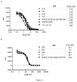
交差反応性をFACSおよびELISAにより測定した。FACSについて、抗PD-1抗体を、実施例2.3に記載のとおり細胞表面ヒト、マウスおよびカニクイザルPD-1への結合について試験した。
図3は、FACSによる異種間試験の結果を示した。図3Aは、ヒトPD-1トランスフェクトCHO-S細胞への結合を示した。抗体は、2.20~2.78nMのEC50でヒトPD-1に特異的に結合し得る。図3Bは、マウスPD-1トランスフェクト293F細胞への結合を示した。抗体は、11.8~15.1nMのEC50でマウスPD-1に特異的結合し得る。図3Cは、用量依存的方法での活性化カニクイザルPBMCの結合を示した。アイソタイプはヒトIgG4カッパであった。下も同様。
Example 4 : characterization of humanized antibodies
1. Cross-reactivity to human, cynomolgus monkey and mouse PD-1 (interspecies)
1.1 FACS
Cross-reactivity was measured by FACS and ELISA. For FACS, anti-PD-1 antibodies were tested for binding to cell surface humans, mice and cynomolgus monkey PD-1 as described in Example 2.3.
FIG. 3 shows the results of a cross-species test by FACS. FIG. 3A shows binding to human PD-1 transfected CHO-S cells. The antibody may specifically bind to human PD-1 at an EC50 of 2.20 to 2.78 nM . FIG. 3B shows binding to mouse PD-1 transfected 293F cells. The antibody may specifically bind to mouse PD-1 at an EC50 of 11.8 to 15.1 nM . FIG. 3C shows binding of activated cynomolgus monkey PBMC in a dose-dependent manner. The isotype was human IgG4 kappa. The same applies to the bottom.
1.2 ヒト、カニクイザルおよびマウスPD-1への交差反応性(異種間)
ELISAについて、プレート(Nunc)をヒト、1μg/mlのカニクイザルまたはマウスPD-1(Sino Biological)で、一夜、4℃でコーティングした。ブロッキングおよび洗浄後、抗体をブロッキング緩衝液で連続的に希釈し、プレートに加え、室温で1時間インキュベートした。次いでプレートを洗浄し、その後二次抗体ヤギ抗ヒトIgG HRP(Bethyl)と1時間インキュベートした。洗浄後、TMB基質を加え、2M HClで反応を停止させた。450nmでの吸光度を、マイクロプレートリーダー(Molecular Device)を使用して読んだ。
図4は、ELISAによる異種間試験の結果を示した。図4Aは、ヒトPD-1への結合を示した。図4Bは、マウスPD-1への結合を示した。図4Cは、カニクイザルPD-1への結合を示した。
1.2 Cross-reactivity to human, cynomolgus monkey and mouse PD-1 (interspecies)
For ELISA, plates (Nunc) were coated with human, 1 μg / ml cynomolgus monkey or mouse PD-1 (Sino Biological) overnight at 4 ° C. After blocking and washing, the antibody was serially diluted with blocking buffer, added to the plate and incubated for 1 hour at room temperature. The plates were then washed and then incubated with the secondary antibody goat anti-human IgG HRP (Bethyl) for 1 hour. After washing, TMB substrate was added and the reaction was stopped with 2M HCl. The absorbance at 450 nm was read using a microplate reader (Molecular Device).
FIG. 4 shows the results of a heterogeneous test by ELISA. FIG. 4A shows binding to human PD-1. FIG. 4B shows binding to mouse PD-1. FIG. 4C shows binding to cynomolgus monkey PD-1.
2. ヒトPD-1ファミリーメンバーCD28、CTLA4への交差反応性
ヒトPD-1、CD28、CTLA-4またはICOSをそれぞれ発現する構築細胞株を、2×10⁵細胞/ウェルの密度で96ウェルU底プレート(BD)に移した。試験抗体を洗浄緩衝液(1×PBS/1%BSA)で希釈し、細胞と4℃で1時間インキュベートした。洗浄後、二次抗体ヤギ抗ヒトIgG Fc FITC(Jackson ImmunoResearch Lab)を加え、4℃で暗所で1時間インキュベートした。次いで細胞を1回洗浄し、1×PBS/1%BSAに再懸濁し、フローサイトメトリー(BD)およびFlowJoソフトウェアにより分析した。
図5は、ファミリー間試験の結果を示した。抗PD-1抗体はヒトPD-1に特異的に結合できるが、CD28およびCTLA-4には結合しない。
2. Cross-reactivity to human PD-1 family members CD28, CTLA4 96-well U of constructed cell lines expressing human PD-1, CD28, CTLA-4 or ICOS, respectively, at a density of 2 × 10 5 cells / well. Transferred to bottom plate (BD). The test antibody was diluted with wash buffer (1 x PBS / 1% BSA) and incubated with cells at 4 ° C. for 1 hour. After washing, a secondary antibody goat anti-human IgG Fc FITC (Jackson ImmunoResearch Lab) was added, and the mixture was incubated at 4 ° C. in the dark for 1 hour. The cells were then washed once, resuspended in 1 x PBS / 1% BSA and analyzed by flow cytometry (BD) and FlowJo software.
FIG. 5 shows the results of the interfamily test. Anti-PD-1 antibody can specifically bind to human PD-1, but not CD28 and CTLA-4.
3. PD-1へのリガンド結合のブロッキング
3.1 抗PD-1抗体がPD-1へのPD-L1結合をブロッキングする能力を、実施例2.3に記載するFACSにより試験した。
3. Blocking ligand binding to PD-1 3.1 The ability of anti-PD-1 antibodies to block PD-L1 binding to PD-1 was tested by the FACS described in Example 2.3.
3.2 抗PD-1抗体がPD-1へのPD-L2結合をブロッキングする能力を、ELISAにより試験した。簡潔には、プレート(Nunc)を、1μg/mlのヒトPD-1で、一夜、4℃でコーティングした。抗体をブロッキング緩衝液で連続的に希釈し、hisタグ・コンジュゲートPD-L2と混合した。該コーティング済プレートのブロッキングおよび洗浄後、抗体/PD-L2混合物を該プレートに加え、次いで室温で1時間インキュベートした。次いでプレートを洗浄し、続いて二次抗体ヤギ抗his HRP(GenScript)と1時間インキュベートした。洗浄後、TMB基質を加え、2M HClで反応を停止させた。450nmでの吸光度を、マイクロプレートリーダー(Molecular Device)を使用して読んだ。
図6Aは、PD-1トランスフェクトCHO-S細胞へのヒトPD-L1結合をブロッキングする抗PD-1抗体の結果を示した。図6Bは、PD-1トランスフェクト293F細胞へのマウスPD-L1結合をブロッキングする抗PD-1抗体の結果を示す。図7は、抗PD-1抗体が用量依存的様式でPD-1へのヒトPD-L2結合をsy団できたことをしめした。
3.2 The ability of anti-PD-1 antibodies to block PD-L2 binding to PD-1 was tested by ELISA. Briefly, plates (Nunc) were coated with 1 μg / ml human PD-1 overnight at 4 ° C. The antibody was serially diluted with blocking buffer and mixed with his-tag-conjugated PD-L2. After blocking and washing the coated plate, the antibody / PD-L2 mixture was added to the plate and then incubated at room temperature for 1 hour. The plates were then washed and subsequently incubated with the secondary antibody goat anti-his HRP (GenScript) for 1 hour. After washing, TMB substrate was added and the reaction was stopped with 2M HCl. The absorbance at 450 nm was read using a microplate reader (Molecular Device).
FIG. 6A shows the results of an anti-PD-1 antibody that blocks human PD-L1 binding to PD-1 transfected CHO-S cells. FIG. 6B shows the results of an anti-PD-1 antibody that blocks mouse PD-L1 binding to PD-1 transfected 293F cells. FIG. 7 shows that the anti-PD-1 antibody was able to bind human PD-L2 to PD-1 in a dose-dependent manner.
4. 表面プラズモン共鳴法(SPR)により試験した完全動的結合親和性
抗体を、ProteOn XPR36(Bio-Rad)を使用するSPRアッセイにより、PD-1への親和性および結合速度論について特徴付けた。プロテインAタンパク質(Sigma)をGLMセンサーチップ(Bio-Rad)にアミンカップリングにより固定した。精製抗体をセンサーチップ上を流し、プロテインAで捕捉させた。チップを90°回転させ、ベースラインが安定するまでランニング緩衝液(1×PBS/0.01%Tween20、Bio-Rad)で洗浄した。7濃度のヒトPD-1およびランニング緩衝液を、30μL/分の流速で180秒の結合相についてセンサーチップを流し、300秒解離が続いた。再生後、7個の濃度のマウスPD-1およびランニング緩衝液を、30μL/分の流速で180秒の結合相についてセンサーチップを流し、300秒解離が続いた。各ランの後、チップをpH1.5 H3PO4で再生した。結合および解離曲線を、ProteOnソフトウェアを使用して、1:1ラングミュア結合モデルにより適合させた。
表6A~6Bは、SPRによるヒトおよびマウスPD-1への完全動的結合親和性の結果を示した。WBP305BMK1を、BMS特許US9084776B2からの5C4のクローンに従い、合成した。キイトルーダは、Merckからの抗PD-1薬物であった。下も同様。結果は、SPRアッセイによるヒトPD-1への親和性能力は、1.43E-8~5.64E-9mol/Lであったことを示した。WBP305BMK1およびキイトルーダと比較すると、抗体2E5、2G4または2C2のKD値は遙かに小さく、2E5、2G4または2C2がヒトPD-1に良好な結合能を有したことを示した。さらに、マウスPD-1に対する親和性能力は、9.37E-9~3.89E-9mol/Lであった。
4. Fully dynamic binding affinity antibodies tested by surface plasmon resonance (SPR) were characterized for PD-1 affinity and binding kinetics by SPR assay using ProteOn XPR36 (Bio-Rad). .. Protein A Protein (Sigma) was immobilized on a GLM sensor chip (Bio-Rad) by amine coupling. The purified antibody was flushed over the sensor chip and captured with Protein A. The chips were rotated 90 ° and washed with running buffer (1 x PBS / 0.01
Tables 6A-6B show the results of full dynamic binding affinity for human and mouse PD-1 by SPR. WBP305BMK1 was synthesized according to the clone of 5C4 from BMS patent US9084776B2. Keytruda was an anti-PD-1 drug from Merck. The same applies to the bottom. The results showed that the affinity capacity for human PD-1 by the SPR assay was 1.43E-8 to 5.64E-9 mol / L. Compared with WBP305BMK1 and Keytruda, the KD value of antibody 2E5 , 2G4 or 2C2 was much smaller, indicating that 2E5, 2G4 or 2C2 had good binding ability to human PD-1. Furthermore, the affinity ability for mouse PD-1 was 9.37E-9 to 3.89E-9 mol / L.


5. フローサイトメトリー(FACS)により試験した抗PD-1抗体の細胞表面PD-1分子への結合親和性
ヒトPD-1を発現するCHO-S細胞またはマウスPD-1を発現する293F細胞を、1×10⁵細胞/ウェルの密度で96ウェルU底プレート(BD)に移した。試験抗体を、洗浄緩衝液(1×PBS/1%BSA)で1:2連続希釈し、細胞と4℃で1時間インキュベートした。二次抗体ヤギ抗ヒトIgG Fc FITC(IgGモルあたり3.0モルFITC(Jackson ImmunoResearch Lab))を加え、4℃で暗所で1時間インキュベートした。次いで細胞を1回洗浄し、1×PBS/1%BSAに再懸濁し、フローサイトメトリー(BD)により分析した。蛍光強度を、定量的ビーズに基づき、結合分子/細胞に変換した(QuantumTM MESF Kits, Bangs Laboratories, Inc.)。KDをGraphpad Prism5を使用して計算した。
表7A~7Bは、フローサイトメトリーにより試験した細胞表面ヒトおよびマウスPD-1分子への抗PD-1抗体の結合親和性の結果を示す。結果は、FACSアッセイによるヒトPD-1への親和性能力が3.80E-10~2.15E-10mol/Lであったことを示した。さらに、マウスPD-1への親和性能力は5.39E-8~1.74E-8mol/Lであった。
5. Binding affinity of anti-PD-1 antibody to cell surface PD-1 molecule tested by flow cytometry (FACS) CHO-S cells expressing human PD-1 or 293F cells expressing mouse PD-1. Transferred to 96-well U-bottom plate (BD) at a density of 1 × 10 5 cells / well. The test antibody was serially diluted 1: 2 with wash buffer (1 x PBS / 1% BSA) and incubated with cells at 4 ° C. for 1 hour. Secondary antibody goat anti-human IgG Fc FITC (3.0 mol FITC per IgG mol (Jackson ImmunoResearch Lab)) was added and incubated at 4 ° C. for 1 hour in the dark. The cells were then washed once, resuspended in 1 x PBS / 1% BSA and analyzed by flow cytometry (BD). Fluorescence intensity was converted to bound molecules / cells based on quantitative beads (Quantum TM MESF Kits, Bangs Laboratories, Inc.). KD was calculated using Graphpad
Tables 7A-7B show the results of the binding affinity of the anti-PD-1 antibody for cell surface human and mouse PD-1 molecules tested by flow cytometry. The results showed that the affinity capacity for human PD-1 by FACS assay was 3.80E-10 to 2.15E-10 mol / L. Furthermore, the affinity ability for mouse PD-1 was 5.39E-8 to 1.74E-8 mol / L.


6. エピトープ・ビニング試験
抗PD-1抗体の結合エピトープを、FACSによりベンチマーク抗体AおよびBと比較した。細胞表面にヒトPD-1を発現するCHO-S細胞をビオチニル化ベンチマーク抗体AまたはB(1μg/ml)および試験抗体(洗浄緩衝液で連続的に希釈)の混合物と、4℃で1時間インキュベートした。細胞を洗浄し、第二抗体ストレプトアビジン-PEを加え、30分、4℃でインキュベートした。次いで細胞を1回洗浄し、1×PBS/1%BSAに再懸濁し、フローサイトメトリー(BD)により分析した。
6. Epitope binning test The binding epitope of the anti-PD-1 antibody was compared with benchmark antibodies A and B by FACS. CHO-S cells expressing human PD-1 on the cell surface are incubated with a mixture of biotinylated benchmark antibody A or B (1 μg / ml) and test antibody (continuously diluted with wash buffer) at 4 ° C. for 1 hour. did. The cells were washed, the second antibody streptavidin-PE was added, and the cells were incubated for 30 minutes at 4 ° C. The cells were then washed once, resuspended in 1 x PBS / 1% BSA and analyzed by flow cytometry (BD).
図8A~8Bは、抗PD-1抗体がベンチマーク抗体と同じまたは近いエピトープビンにあることを示唆する、エピトープビニングアッセイの結果を示した。図8Aは、WBP305BMK1(US9084776)に対するビニングを示した。図8Bは、キイトルーダ(US8168757)に対するビニングを示した。 8A-8B show the results of the epitope binning assay, suggesting that the anti-PD-1 antibody is in the same or close epitope bin as the benchmark antibody. FIG. 8A shows binning for WBP305BMK1 (US9084776). FIG. 8B shows binning for Keytruda (US8168757).
さらに、hPD-1でのアラニン走査実験を行い、その抗体結合への影響を評価した。hPD-1のアラニン残基をグリシンコドンに変異させ、他の全ての残基をアラニンコドンに変異させた。HPD-1細胞外ドメイン(ECD)の各残基について、点アミノ酸置換を、2つの連続的PCR工程を使用して行った。ヒトPD-1のECDおよびC末端HisタグをコードするpcDNA3.3-hPD-1_ECD.Hisプラスミドを鋳型とて使用し、一式の変異原性プライマーを、QuikChange電撃性多部位特異的突然変異誘発キット(Agilent technologies, Palo Alto, CA)を使用する、第一段階PCRに使用した。Dpn Iエンドヌクレアーゼを、変異鎖合成反応後親鋳型の消化に使用した。第二段階PCRにおいて、CMVプロモーター、PD-1の細胞外ドメイン(ECD)、Hisタグおよび単純ヘルペスウイルスチミジンキナーゼ(TK)ポリアデニル化からなる線形DNA発現カセットを増幅させ、HEK293F細胞(Life Technologies, Gaithersburg, MD)に一過性に発現させた。 Furthermore, an alanine scanning experiment with hPD-1 was performed to evaluate its effect on antibody binding. The alanine residue of hPD-1 was mutated to the glycine codon and all other residues were mutated to the alanine codon. For each residue of the HPD-1 extracellular domain (ECD), point amino acid substitutions were performed using two consecutive PCR steps. Using the pcDNA3.3-hPD-1_ECD.His plasmid encoding the ECD and C-terminal His tags of human PD-1 as a template, a set of mutagenic primers was used in the QuikChange electric shock multisite-specific mutagenesis kit. Used for first-stage PCR using (Agilent technologies, Palo Alto, CA). Dpn I endonuclease was used to digest the parent template after the mutant chain synthesis reaction. In the second stage PCR, a linear DNA expression cassette consisting of CMV promoter, extracellular domain (ECD) of PD-1, His tag and herpes simplex virus thymidine kinase (TK) polyadenylation was amplified and HEK293F cells (Life Technologies, Gaithersburg). , MD) transiently.
モノクローナル抗体W3052_r16.88.9およびキイトルーダを、ELISA結合アッセイのためにプレートコーティングさせた。定量化PD-1変異体またはヒト/マウスPD-1_ECD.Hisタンパク質(Sino Biological, China)を含む上清とインキュベート後、HRPコンジュゲート抗His抗体を検出抗体として加えた。吸光度を、対照変異体の平均に対して正規化した。結合倍率変化に対するさらなるカットオフ(<0.55)設定後、最終的に決定されたエピトープ残基が同定された。 Monoclonal antibody W3052_r16.88.9 and Keytruda were plate coated for ELISA binding assay. After incubation with a supernatant containing a quantified PD-1 mutant or human / mouse PD-1_ECD.His protein (Sino Biological, China), HRP-conjugated anti-His antibody was added as a detection antibody. Absorbance was normalized to the average of control variants. After further cutoff (<0.55) setting for binding magnification changes, the final determined epitope residue was identified.
抗体W3052_r16.88.9およびキイトルーダのヒトおよびネズミ科両者のPD-1への結合活性を行った(図9)。W3052_r16.88.9は、hPD-1およびmPD-1の両者への結合が見られたが、キイトルーダはヒトのものにしか結合しなかった(図9)。W3052_r16.88.9のこの特有の機能的交差反応性は、前臨床試験における薬物安全性評価の際のより多くの動物モデル選択肢の提供に役立つ。観察された結合挙動の原因を探索するために、両抗体のエピトープマッピングを実施した。 The binding activity of the antibody W3052_r16.88.9 and Keytruda to PD-1 in both humans and Muridae was performed (Fig. 9). W3052_r16.88.9 was found to bind to both hPD-1 and mPD-1, but Keytruda bound only to humans (Fig. 9). This unique functional cross-reactivity of W3052_r16.88.9 helps provide more animal model options for drug safety assessments in preclinical studies. Epitope mapping of both antibodies was performed to investigate the cause of the observed binding behavior.
抗体結合が有意に減少した上位30の点置換hPD-1変異体を表8に示した。hPD-1結晶構造のこれら全残基の位置の確認(PDBコード3RRQおよび4ZQK)により、一部アミノ酸(例えばVal144、Leu142、Val110、Met108、Cys123など)がタンパク質に完全に埋没しており、あらゆる抗体と直接接触しそうにないことが確認された。観察された結合減少は、アラニン置換後のhPD-1構造の不安定性または崩壊に由来することが最も可能性があった。抗原構造分析により、残基の一部、例えばV144およびL142は結合活性に関与しないが、hPD-1構造の安定性に応答することが期待される。両抗体に影響する変異体を、偽ホットスポットとして処理し、リストから除いた。結合倍率変化に対するさらなるカットオフ(<0.55)設定後、最終的に決定されたエピトープ残基を表9にリストした。W3052_r16.88.9について9位置およびキイトルーダについて5位置ある。 Table 8 shows the top 30 point-substituted hPD-1 mutants with significantly reduced antibody binding. By confirming the position of all these residues in the hPD-1 crystal structure (PDB code 3RQ and 4ZQK), some amino acids (eg Val144, Leu142, Val110, Met108, Cys123, etc.) are completely embedded in the protein and are all It was confirmed that it was unlikely to come into direct contact with the antibody. The observed loss of binding was most likely due to the instability or disruption of the hPD-1 structure after alanine substitution. Antigen structure analysis suggests that some of the residues, such as V144 and L142, are not involved in binding activity but respond to the stability of the hPD-1 structure. Mutants affecting both antibodies were treated as pseudohotspots and removed from the list. After further cutoff (<0.55) setting for binding magnification changes, the finally determined epitope residues are listed in Table 9. There are 9 positions for W3052_r16.88.9 and 5 positions for Keytruda.
表9におけるW3052_r16.88.9およびキイトルーダのエピトープ残基の比較は、2つの重複ホットスポット残基しか明らかにしなかった。残りはかなり違うように見え、2抗体がhPD-1結合およびhPD-L1ブロッキングの観点で、極めて異なる機構を採用しているはずであることを示す。表9における残基IDの読み出しは、機構の解釈に直接的ではない。それ故に、表9における全データおよびhPD-L1結合部位を、良好な可視化および比較を得るために、hPD-1の結晶構造上にマッピングした(図10)。 Comparison of W3052_r16.88.9 and Keytruda epitope residues in Table 9 revealed only two overlapping hotspot residues. The rest appear to be quite different, indicating that the two antibodies should employ very different mechanisms in terms of hPD-1 binding and hPD-L1 blocking. Reading the residue ID in Table 9 is not direct to the interpretation of the mechanism. Therefore, all data and hPD-L1 binding sites in Table 9 were mapped onto the crystal structure of hPD-1 for good visualization and comparison (FIG. 10).


* mPD-1で観察されたC’’鎖はhPD-1構造に存在しない。このβシートは、hPD-1の無構造ループに置き換えられる。我々は、単にmPD-1との比較を容易にする目的のみで、この領域の標識のためにC’’をなお使用する。
* The C'' chain observed in mPD-1 is not present in the hPD-1 structure. This β sheet is replaced with the unstructured loop of hPD-1. We still use C'' for labeling in this region solely for the purpose of facilitating comparison with mPD-1.
2つの調査抗体W3052_r16.88.9およびキイトルーダは、両者がhPD-1結合およびhPD-L1ブロッキングに機能的であるが、明らかに異なるエピトープを有する(図10B、10C)。キイトルーダのエピトープは、主にPD-L1結合部位と決して交わらないC’Dループ(PD-1上のC’’鎖に対応)の残基により寄与された。これは、キイトルーダのhPD-L1ブロッキング機能が、抗体サイズにより提供される立体障害効果により依存することを示唆した。対照的に、エピトープマッピング結果は、抗体W3052_r16.88.9のエピトープが複数部位にまたがり分散され、hPD-L1結合部位と直接重複するホットスポットからなったことを示す(図10A、10B)。W3052_r16.88.9は、hPD-L1を、共通結合部位への反応により、hPD-L1と競合する手段によりブロッキングした。その上、W3052_r16.88.9は、ヒトおよびネズミ科PD-1が大きな構造差異を示す、柔軟性C’Dループ(または対応するPD-1上のC’’鎖)と相互作用を有さなかった(図11)。その結合部位は、大部分FGループに位置した(Lin et al. (2008) PNAS 105: 3011-3016)。これは、なぜW3052_r16.88.9が両PD-1種に結合でき、一方キイトルーダがヒトのものにしか結合しないのかを説明する(図9)。この特有の機能的交差反応性により、W3052_r16.88.9の前臨床安全性評価がマウスモデルで実施でき、これは、開発を大きく単純化し、加速させる。全体的に見て、抗体W3052_r16.88.9は、キイトルーダより機能的であり、開発可能であることが予測される。 The two study antibodies W3052_r16.88.9 and Keytruda are both functional for hPD-1 binding and hPD-L1 blocking, but have distinctly different epitopes (FIGS. 10B and 10C). The keytruda epitope was primarily contributed by residues in the C'D loop (corresponding to the C ″ chain on PD-1) that never intersected the PD-L1 binding site. This suggested that the hPD-L1 blocking function of Keytruda was more dependent on the steric hindrance effect provided by antibody size. In contrast, epitope mapping results indicate that the epitope of antibody W3052_r16.88.9 was dispersed across multiple sites and consisted of hot spots that directly overlapped the hPD-L1 binding site (FIGS. 10A and 10B). W3052_r16.88.9 blocked hPD-L1 by means competing with hPD-L1 by reacting to the common binding site. Moreover, W3052_r16.88.9 did not interact with the flexible C'D loop (or the C'chain on the corresponding PD-1), where human and Murid PD-1 show significant structural differences. (Fig. 11). The binding site was located mostly in the FG loop (Lin et al. (2008) PNAS 105: 3011-3016). This explains why W3052_r16.88.9 can bind to both PD-1 species, while Keytruda binds only to humans (Fig. 9). This unique functional cross-reactivity allows preclinical safety assessments of W3052_r16.88.9 to be performed in mouse models, which greatly simplifies and accelerates development. Overall, antibody W3052_r16.88.9 is expected to be more functional and developable than Keytruda.
7. 細胞ベースのアッセイにより試験した抗PD-1抗体のインビトロ機能
7.1 混合リンパ球反応(MLR)を、抗PD-1抗体のTリンパ球機能への影響の試験に使用した
ヒトDC、CD4+ T、CD8+ Tおよび総T細胞単離:ヒトPBMCを、Ficoll-Paque PLUS(GE)勾配遠心分離を使用して健常ドナーから新たに単離した。単球を、製造業者の指示に従い、Human Monocyte Enrichment Kit(StemCell)を使用して単離した。細胞を、rhGM-CSFおよびrhIL-4を含む培地で5~7日培養し、樹状細胞を産生させた。MLR18~24時間前、1μg/mL LPSを培養に添加して、DCの成熟を誘発させた。ヒトCD4+ T細胞を、製造業者のプロトコールに従い、Human CD4+ T Cell Enrichment Kit(StemCell)を使用して単離した。マウスCD4+ T細胞を製造業者のプロトコールに従いMouse CD4+ T Cell Isolation Kit(StemCell)を使用して、Balb/cマウスの脾臓から得た。マウスDCを、rmGM-CSFおよびrmIL-4を含む培地中、C57BL/6マウスの骨髄細胞から、5~7日誘発させた。MLR18~24時間前、1μg/mL LPSを培養に加えて、DCの成熟を誘発した。
7. In vitro function of anti-PD-1 antibody tested by cell-based assay
7.1 Mixed lymphocyte reaction (MLR) was used to test the effect of anti-PD-1 antibody on T lymphocyte function. Human DC, CD4 + T, CD8 + T and total T cell isolation: human PBMC. , Ficoll-Paque PLUS (GE) freshly isolated from healthy donors using gradient centrifugation. Monocytes were isolated using the Human Monocyte Enrichment Kit (Stem Cell) according to the manufacturer's instructions. The cells were cultured in medium containing rhGM-CSF and rhIL-4 for 5-7 days to produce dendritic cells. 18-24 hours prior to MLR, 1 μg / mL LPS was added to the culture to induce DC maturation. Human CD4 + T cells were isolated using the Human CD4 + T Cell Enrichment Kit (Stem Cell) according to the manufacturer's protocol. Mouse CD4 + T cells were obtained from the spleen of Balb / c mice using the Mouse CD4 + T Cell Isolation Kit (StemCell) according to the manufacturer's protocol. Mouse DCs were induced from bone marrow cells of C57BL / 6 mice in medium containing rmGM-CSF and rmIL-4 for 5-7 days. 18-24 hours prior to MLR, 1 μg / mL LPS was added to the culture to induce DC maturation.
簡潔には、一次樹状細胞(DC)刺激MLRを96ウェル、U底組織培養プレートで、10%FCSおよび1%抗生物質を含む200μLのRPMI 1640中、実施した。DCを、1:10~1:200 DC:T細胞比で、試験抗体またはベンチマーク抗体(166.75nMから0.00667nMまで下がる、一般に全6濃度)存在下または非存在下、1×10⁵ CD4+ T細胞と混合した。T細胞機能に対する抗PD-1抗体の影響を決定するために、サイトカイン産生およびT細胞増殖を決定した。示す結果は、実施した最小5実験の代表である。 Briefly, primary dendritic cell (DC) stimulated MLRs were performed in 96-well, U-bottom tissue culture plates in 200 μL RPMI 1640 containing 10% FCS and 1% antibiotics. DCs in a 1 x 10 to 1: 200 DC: T cell ratio, with or without test antibody or benchmark antibody (down from 166.75 nM to 0.00667 nM, generally all 6 concentrations) 1 × 10 5 CD4 + Mixed with T cells. Cytokine production and T cell proliferation were determined to determine the effect of anti-PD-1 antibody on T cell function. The results shown are representative of the minimum of 5 experiments performed.
サイトカイン検出:ヒトIFN-γおよびIL-2を、適合抗体対を使用する酵素結合免疫吸着検定法(ELISA)により測定した。プレートを、それぞれヒトIFN-γ(cat# Pierce-M700A)またはIL-2(cat# R&D-MAB602)特異的な捕捉抗体でプレコートした。ビオチンコンジュゲ-ト抗IFN-γ抗体(cat# Pierce-M701B)または抗IL-2抗体(cat# R&D-BAF202)を検出抗体として使用した。
図12Aは、抗PD-1抗体が用量依存的様式でIL-2分泌を増加させたことを示した。図12Bは、抗PD-1抗体が用量依存的様式でIFN-γ分泌を増加させることを示す。
Cytokine detection : Human IFN-γ and IL-2 were measured by enzyme-linked immunosorbent assay (ELISA) using a compatible antibody pair. Plates were precoated with human IFN-γ (cat # Pierce-M700A) or IL-2 (cat # R & D-MAB602) specific capture antibodies, respectively. Biotin conjugate anti-IFN-γ antibody (cat # Pierce-M701B) or anti-IL-2 antibody (cat # R & D-BAF202) was used as the detection antibody.
FIG. 12A showed that the anti-PD-1 antibody increased IL-2 secretion in a dose-dependent manner. FIG. 12B shows that anti-PD-1 antibody increases IFN-γ secretion in a dose-dependent manner.
増殖アッセイ:3H-チミジン(cat# PerkinElmer- NET027001MC)を、0.9%NaCl溶液で1:20に希釈し、細胞培養プレートに0.5μCi/ウェルで加えた。プレートを、5%CO2で37℃で16~18時間培養し、増殖細胞への3H-チミジン取り込みを決定した。図12Cは、抗PD-1抗体が用量依存的様式でCD4+ T細胞増殖を増加させることを示す。
抗PD-1抗体のマウスT細胞機能に対する効果を決定するために、サイトカイン産生およびマウスT細胞増殖を同様に決定した。図13A~13Cは、抗PD-1抗体がマウスCD4+ T細胞機能を増強できることを示す、マウスアロMLRの結果を示した。図13Aは、抗PD-1抗体が用量依存的様式でIL-2分泌を増加させたことを示した。図13Bは、抗PD-1抗体が用量依存的様式でIFN-γ分泌を増加させたことを示した。図13Cは、抗PD-1抗体が用量依存的様式でCD4+ T細胞増殖を増加させたことを示した。
Growth assay : 3H -thymidine (cat # PerkinElmer-NET027001MC) was diluted 1:20 with 0.9% NaCl solution and added to cell culture plates at 0.5 μCi / well. Plates were cultured in 5% CO 2 at 37 ° C. for 16-18 hours to determine 3H -thymidine uptake into proliferating cells. FIG. 12C shows that anti-PD-1 antibody increases CD4 + T cell proliferation in a dose-dependent manner.
Cytokine production and mouse T cell proliferation were similarly determined to determine the effect of anti-PD-1 antibody on mouse T cell function. 13A-13C show the results of mouse allo MLR showing that anti-PD-1 antibody can enhance mouse CD4 + T cell function. FIG. 13A showed that the anti-PD-1 antibody increased IL-2 secretion in a dose-dependent manner. FIG. 13B showed that the anti-PD-1 antibody increased IFN-γ secretion in a dose-dependent manner. FIG. 13C showed that the anti-PD-1 antibody increased CD4 + T cell proliferation in a dose-dependent manner.
7.2 自己抗原特異的免疫応答による細胞増殖およびサイトカイン産生に対するヒト抗PD-1抗体の効果
このアッセイにおいて、CD4+ T細胞およびDCは、同じドナー由来であった。簡潔には、CD4+ T細胞をPBMCから精製し、CMV pp65ペプチドおよび低用量のIL-2(20U/mL)存在下培養し、その間にも、DCを同一ドナーのPBMCからの単球のGM-CSFおよびIL-4中の培養により産生した。5日後、CMV pp65ペプチド処理CD4+ T細胞を、ヒト抗PD-1抗体またはベンチマーク抗体(対照として)存在下または非存在下、CMV pp65ペプチドを瞬間適用したDCと共培養した。5日目、100μLの上清を、上記のとおりELISAによるIFN-γ測定のために、各培養から取った。CMV pp65特異的T細胞の増殖を、上記のとおり3H-チミジン取り込みにより評価した。
図14A~14Bは、抗PD-1抗体がヒトCD4+ T細胞の機能を増強できることを示す、ヒト自己MLRの結果を示した。図14Aは、抗PD-1抗体が用量依存的様式でIFN-γ分泌を増加させることを示した。図14Bは、抗PD-1抗体が用量依存的様式でCD4+ T細胞増殖を増加させることを示した。
7.2 Effect of human anti-PD-1 antibody on cell proliferation and cytokine production by self-antigen-specific immune response In this assay, CD4 + T cells and DC were from the same donor. Briefly, CD4 + T cells were purified from PBMCs and cultured in the presence of CMV pp65 peptide and low dose IL-2 (20 U / mL), while DCs were monocytic GM from PBMCs of the same donor. -Produced by culture in CSF and IL-4. After 5 days, CMV pp65 peptide treated CD4 + T cells were co-cultured with DCs to which CMV pp65 peptide was instantaneously applied in the presence or absence of human anti-PD-1 antibody or benchmark antibody (as a control). On
14A-14B show the results of human autologous MLR showing that anti-PD-1 antibody can enhance the function of human CD4 + T cells. FIG. 14A showed that anti-PD-1 antibody increased IFN-γ secretion in a dose-dependent manner. FIG. 14B showed that anti-PD-1 antibody increased CD4 + T cell proliferation in a dose-dependent manner.
7.3 制御性T細胞(Treg)抑制機能に対するヒト抗PD-1抗体の効果
T細胞の亜集団であるTregは、重要な免疫モジュレーターであり、自己耐容性維持に重要な役割を有する。CD4+CD25+ Tregの数の増加が、複数の癌の患者で見られ、より不良な予後と相関した。抗PD-1抗体がTregの免疫抑制的役割に影響するか否かを決定するために、Treg存在下、抗PD-1抗体処置を施したまたは施していないT細胞機能を比較した。CD4+CD25+およびCD4+CD25-T細胞を、製造業者の指示に従い特異的抗CD25マイクロビーズ(StemCell)を使用して分離した。2000成熟DC、1×10⁵ CD4+CD25- T細胞、1×10⁵ Treg細胞およびPD-1抗体を、96ウェルプレートでインキュベートした。プレートを37℃で5%CO2インキュベーターに5日間維持した。IFN-γ産生およびCD4+CD25細胞増殖を、上記のとおり試験した。
図15は、抗PD-1抗体がTregの抑制機能を逆転できることを示す。図15Aは、抗PD-1抗体がIFN-γ分泌を回復できることを示した。図15Bは、抗PD-1抗体がT細胞増殖を回復できることを示した。
7.3 Effect of human anti-PD-1 antibody on regulatory T cell (Treg) inhibitory function Treg , a subpopulation of T cells, is an important immune modulator and has an important role in maintaining self-tolerance. Increased numbers of CD4 + CD25 + Tregs were seen in patients with multiple cancers and correlated with a worse prognosis. To determine whether anti-PD-1 antibodies affect the immunosuppressive role of Tregs, T cell functions with and without anti-PD-1 antibody treatment were compared in the presence of Tregs. CD4 + CD25 + and CD4 + CD25 - T cells were isolated using specific anti-CD25 microbeads (StemCell) according to the manufacturer's instructions. 2000 mature DC, 1x10 5 CD4 + CD25 - T cells, 1x10 5 Treg cells and PD-1 antibody were incubated in 96-well plates. Plates were maintained in a 5% CO 2 incubator at 37 ° C. for 5 days. IFN-γ production and CD4 + CD25 cell proliferation were tested as described above.
FIG. 15 shows that anti-PD-1 antibody can reverse the Treg inhibitory function. FIG. 15A showed that anti-PD-1 antibody can restore IFN-γ secretion. FIG. 15B shows that anti-PD-1 antibody can restore T cell proliferation.
8. ADCCおよびCDC試験
PD-1は、多様な細胞型で発現される。健常PD-1陽性細胞への毒性の可能性を最小化するために、抗PD-1抗体を、抗体依存性細胞傷害(ADCC)および補体依存性細胞傷害(CDC)に介在する能力について評価した。
8. ADCC and CDC test PD-1 is expressed in a variety of cell types. To assess the ability of anti-PD-1 antibodies to mediate antibody-dependent cellular cytotoxicity (ADCC) and complement-dependent cellular cytotoxicity (CDC) to minimize the potential for toxicity to healthy PD-1 positive cells. did.
8.1 ADCC試験
ヒト活性化CD4+ T細胞および種々の濃度のPD-1抗体を96ウェルプレートで30分プレインキュベートし、次いでPBMCをエフェクター/標的比50:1で加えた。プレートを、37℃で5%CO2インキュベーターに6時間維持した。標的細胞溶解を、LDHベースの細胞毒性検出キット(cat# Roche-11644793001)により決定した。492nmでの吸光度を、マイクロプレートリーダー(Molecular Device)を使用して読んだ。ハーセプチン誘発SK-Br-3細胞溶解を陽性対照として使用した。
図16は、抗PD-1抗体が活性化CD4+ T細胞のADCC活性に介在しなかったことを示す、ADCC試験の結果を示した。
8.1 ADCC test Human activated CD4 + T cells and PD-1 antibodies of various concentrations were preincubated in 96-well plates for 30 minutes, then PBMC was added at an effector / target ratio of 50: 1. Plates were maintained in a 5% CO 2 incubator at 37 ° C. for 6 hours. Target cytolysis was determined by the LDH-based cytotoxicity detection kit (cat # Roche-11644793001). The absorbance at 492 nm was read using a microplate reader (Molecular Device). Herceptin-induced SK-Br-3 cytolysis was used as a positive control.
FIG. 16 shows the results of the ADCC test showing that the anti-PD-1 antibody did not intervene in the ADCC activity of activated CD4 + T cells.
8.2 CDC試験
ヒト活性化CD4+ T細胞および種々の濃度のPD-1抗体を96ウェルプレートで混合した。ヒト補体(Quidel-A112)を1:50の希釈比で加えた。プレートを、37℃で5%CO2インキュベーターに2時間維持した。標的細胞溶解を、CellTiter-Gloにより決定した。リツキサン(登録商標)誘発Raji細胞溶解を陽性対照として使用した。発光を、マイクロプレートリーダー(Molecular Device)を使用して読んだ。
図17は、抗PD-1抗体が活性化CD4+ T細胞のCDC活性に介在しなかったことを示す、CDC試験の結果を示した。
8.2 CDC test Human activated CD4 + T cells and PD-1 antibodies of various concentrations were mixed in 96-well plates. Human complement (Quidel-A112) was added at a dilution ratio of 1:50. Plates were maintained in a 5% CO 2 incubator at 37 ° C. for 2 hours. Target cytolysis was determined by CellTiter-Glo. Rituxan®-induced Raji cell lysis was used as a positive control. The luminescence was read using a microplate reader (Molecular Device).
FIG. 17 shows the results of a CDC test showing that the anti-PD-1 antibody did not intervene in the CDC activity of activated CD4 + T cells.
実施例5:ヒトPD-1に対するモノクローナル抗体を使用するインビボ腫瘍モデルの処置
1. 実験設計

1N:各群のマウス数
2用量-体積:マウスの体重により10μL/g。体重減少が15%を超えたら、投与レジメンをそれに応じて調節すべきである。
Example 5 : Treatment of an in vivo tumor model using a monoclonal antibody against human PD-1
1. Experimental design
1 N: Number of mice in each group
2 Dose-Volume: 10 μL / g depending on mouse body weight. If weight loss exceeds 15%, the dosing regimen should be adjusted accordingly.
2. 方法
2.1 細胞培養
マウス黒色腫細胞CloudmanS91細胞(ATCC-CCL-53.1)を、単層としてインビトロで培養し、培養条件は、2.5%FBSおよび15%ウマ血清、100U/mLペニシリンおよび100μg/mLストレプトマイシン添加F-12K培地であり、37℃および5%CO2でインキュベートした。細胞をトリプシン-EDTAを使用して消化させ、週に2回定期的に継代した。約80%~90%コンフルエントおよび必要な数になったとき、細胞を採取し、計数し、次いで接種した。
2. Method 2.1 Cell culture Mouse melanoma cells Cloudman S91 cells (ATCC-CCL-53.1) were cultured in vitro as a monolayer, and the culture conditions were 2.5% FBS and 15% horse serum, 100 U / mL penicillin. And 100 μg / mL streptomycin-added F-12K medium, incubated at 37 ° C. and 5% CO 2 . Cells were digested using trypsin-EDTA and were periodically passaged twice a week. When about 80% -90% confluent and the required number were reached, cells were harvested, counted and then inoculated.
2.2 腫瘍細胞の注入
0.1mL(5×10⁵細胞)CloudmanS91細胞を、各動物の右背部に皮下的に接種した。腫瘍体積平均が約64mm3に達したとき、投与を群で開始した。群分けおよび投与レジメンは表10に示した。
2.2 Injection of tumor cells 0.1 mL (5 × 10 5 cells) Cloudman S91 cells were subcutaneously inoculated into the right back of each animal. Administration was started in groups when the average tumor volume reached approximately 64 mm 3 . Grouping and dosing regimens are shown in Table 10.
2.3 腫瘍試験および指数
実験指数は、腫瘍増殖が阻止されたか、遅延されたかまたは治癒したかの試験である。腫瘍直径を、週3回ノギスで測定した。腫瘍体積をV=0.5a×b2(式中、aおよびbは、それぞれ腫瘍の長径および短径を表す)を使用して、計算する。
2.3 Tumor Tests and Indexes Experimental indices are tests of whether tumor growth is blocked, delayed, or cured. Tumor diameter was measured with a caliper three times a week. Tumor volume is calculated using V = 0.5a × b 2 (where a and b represent the major and minor diameters of the tumor, respectively).
抗体の抗腫瘍有効性を、腫瘍増殖阻害TGI(%)または相対的腫瘍増殖率T/C(%)により評価した。TGI(%)は腫瘍増殖阻害の率を反映した。TGI(%)を次のとおり計算した。TGI(%)=[(1-(処置群投与終了時の平均腫瘍体積-処置群投与開始時の平均腫瘍体積))/(溶媒対照群投与終了時の平均腫瘍体積-溶媒対照群投与開始時の平均腫瘍体積)]×100%。 The antitumor efficacy of the antibody was evaluated by tumor growth inhibitory TGI (%) or relative tumor growth rate T / C (%). TGI (%) reflected the rate of tumor growth inhibition. TGI (%) was calculated as follows. TGI (%) = [(1- (Average tumor volume at the end of treatment group administration-Average tumor volume at the start of treatment group administration)) / (Average tumor volume at the end of solvent control group administration-At the start of solvent control group administration Average tumor volume)] x 100%.
相対的腫瘍増殖率T/C(%)を次のとおり計算した。T/C%=TRTV/CRTV×100%(TRTV:処置群RTV;CRTV:陰性対照群RTV)。相対的腫瘍体積(RTV)を、RTV=Vt/V0(式中、V0は群分け時の平均腫瘍体積(すなわち、d0)であり、Vtはある測定点の平均腫瘍体積であり;TRTVおよびCRTVのデータを同じ日に取った)を使用して、腫瘍測定結果に従い計算した。 The relative tumor growth rate T / C (%) was calculated as follows. T / C% = T RTV / C RTV × 100% (T RTV : treatment group RTV; C RTV : negative control group RTV). Relative tumor volume (RTV) is RTV = V t / V 0 (in the formula, V 0 is the average tumor volume at the time of grouping (ie, d 0 ), and V t is the average tumor volume at a certain measurement point. Yes; TRTV and CRTV data were taken on the same day) and calculated according to tumor measurements.
T-C(日)は、腫瘍増殖遅延指数を表し、Tは処置群において腫瘍が予定した体積(例えば300mm3)に到達したときに経過した平均日数を表し、Cは対照群の腫瘍が同一体積に到達したときの平均日数を表した。 TC (day) represents the tumor growth retardation index, T represents the average number of days elapsed when the tumor reached the planned volume (eg 300 mm 3 ) in the treatment group, and C represents the same tumor in the control group. The average number of days when the volume was reached was shown.
生存曲線をプロットし;動物生存期間を、投与から動物が死亡するまでまたは腫瘍体積が2000mm3に到達するまでの時間として定義した。中央生存期間(日)を各群で計算した。延長された生存期間(ILS)を、処理群とモデル対照群の中央生存期間の比較により計算し、モデル対照群の生存期間を超えるパーセンテージとして計算した。 The survival curve was plotted; animal survival was defined as the time from dosing until the animal died or the tumor volume reached 2000 mm 3 . Central survival (days) was calculated for each group. Prolonged survival (ILS) was calculated by comparing the median survival of the treatment group with that of the model control and calculated as a percentage above the survival of the model control.
2.4 統計分析
各群の各時点の平均腫瘍体積および標準誤差(SEM)を含むデータを、統計的に分析した(特定のデータについては表11参照のこと)。実験は、投与後37日目に完了した;投与後13日目、動物を連続的に屠殺した;それ故、群間差異の統計分析および評価は、投与開始後13日目の腫瘍体積。2群間比較について、データをT検定を使用して分析した;3群以上の間の比較について、データを一元配置ANOVAを使用して分析した。F値について統計的有意差が見られたら、データをゲイムス・ハウエル検定を使用して分析した。F値について統計的有意差が見られなかったら、ダネット(両側)検定を分析に使用した。SPSS 17.0を全データ分析に使用した。p<0.05を有意差として見なした。生存期間をカプラン・マイヤー法とログ・ランク検定を使用して、分析した。
2.4 Statistical analysis Data including mean tumor volume and standard error (SEM) at each time point in each group were statistically analyzed (see Table 11 for specific data). The experiment was completed on day 37 post-dose; animals were sacrificed continuously on
3. 結果
3.1 死亡率、罹病率および体重変化
動物の体重は、薬物毒性の測定のための間接的基準である。2E5のCloudmanS91皮下同系異種移植雌DBA/2マウスモデルの体重に対する影響を図18および図19に示した。このモデルにおいて、全投与群は、有意な体重減少を示さなかった(図18)。それ故に、2E5は黒色腫CloudmanS91のマウスモデルに明白な毒性は示さない。
3. Results 3.1 Mortality, morbidity and body weight changes Animal body weight is an indirect measure of drug toxicity. The effects of 2E5 on body weight in the Cloudman S91 subcutaneous allogeneic xenograft female DBA / 2 mouse model are shown in FIGS. 18 and 19. In this model, all treatment groups showed no significant weight loss (Fig. 18). Therefore, 2E5 shows no apparent toxicity to the mouse model of melanoma Cloudman S91.
3.2 腫瘍体積
2E5処置後のCloudmanS91皮下同系異種移植雌DBA/2マウスモデルの腫瘍体積を表11に示した。

a. 平均±SEM;
b. 投与後日数。
3.2 Tumor Volume Table 11 shows the tumor volume of the Cloudman S91 subcutaneous allogeneic xenograft female DBA / 2 mouse model after 2E5 treatment.
a. Average ± SEM;
b. Days after administration.
3.3 腫瘍増殖曲線
腫瘍増殖曲線を図20に示した。
3.3 Tumor growth curve The tumor growth curve is shown in FIG.
3.4 抗腫瘍有効性評価

a. 平均±SEM;
b. 腫瘍増殖阻害を、T/CおよびTGI(TGI(%)=[1-(T13-T0)/(V13-V0)]×100)により計算した;
c. p値を腫瘍体積により計算した。
3.4 Anti-tumor efficacy evaluation
a. Average ± SEM;
b. Tumor growth inhibition was calculated by T / C and TGI (TGI (%) = [1- (T 13 -T 0 ) / (V 13 -V 0 )] x 100);
c. The p-value was calculated by tumor volume.
3.5 生存曲線s
各群の生存曲線を図21に示した。
3.5 Survivorship curve s
The survival curve of each group is shown in FIG.
3.6 生存期間

a. p値は、処置群と媒体対照群の間の比較を表した;
b. 実験終了時、3mg/kg群(2E5)において、生存率は66.7%であった。
3.6 Time to live
The p-value represented a comparison between the treatment group and the vehicle control group;
b. At the end of the experiment, the survival rate was 66.7% in the 3 mg / kg group (2E5).
4. 考察
本試験において、CloudmanS91同系腫瘍モデルにおける2E5のインビボ有効性を評価した。種々の時点での各群の腫瘍体積を表11、表12および図20に示し、生存期間を図21および表13に示した。投与後13日目、溶媒対照群における腫瘍担持マウスの腫瘍体積は1,626mm3に達した。対照群と比較して、1mg/kg 2E5群で弱い阻害効果が観察され、腫瘍体積は1,089mm3(T/C=68.1%、TGI=34.4%、p=0.367)であり、腫瘍増殖遅延は0日であった。溶媒対照群と比較して、有意な抗腫瘍効果が3mg/kg 2E5群で観察され、腫瘍体積は361mm3(T/C=22.9%、TGI=81.0%、p=0.008)であり、腫瘍増殖遅延は5日であった。溶媒対照群と比較して、有意な抗腫瘍効果が10mg/kg 2E5群でも観察され、腫瘍体積は614mm3(T/C=39.4%、TGI=64.7%、p=0.036)であり、腫瘍増殖遅延は5日であった。
4. Discussion In this study, the in vivo efficacy of 2E5 in the Cloudman S91 syngeneic tumor model was evaluated. Tumor volumes for each group at different time points are shown in Tables 11, 12 and 20, and survival times are shown in FIGS. 21 and 13. On the 13th day after administration, the tumor volume of the tumor-carrying mice in the solvent control group reached 1,626 mm 3 . A weak inhibitory effect was observed in the 1 mg / kg 2E5 group compared to the control group, and the tumor volume was 1,089 mm 3 (T / C = 68.1%, TGI = 34.4%, p = 0.367). The tumor growth delay was 0 days. A significant antitumor effect was observed in the 3 mg / kg 2E5 group compared to the solvent control group, with a tumor volume of 361 mm 3 (T / C = 22.9%, TGI = 81.0%, p = 0.008). ), And the tumor growth delay was 5 days. A significant antitumor effect was also observed in the 10 mg / kg 2E5 group compared to the solvent control group, with a tumor volume of 614 mm 3 (T / C = 39.4%, TGI = 64.7%, p = 0.036). ), And the tumor growth delay was 5 days.
本実験において、溶媒対照群における腫瘍担持マウスの中央生存期間は16日であった。媒体対照群と比較して、1mg/kg 2E5群における腫瘍担持マウスの中央生存期間は20日であり、生存は25%(p=0.077)延長され;3mg/kg 2E5群における腫瘍担持マウスの生存率は66.7%(p=0.001)であった。10mg/kg 2E5群における腫瘍担持マウスの中央生存期間は32日であり、生存は100%(p=0.022)延長された。 In this experiment, the median survival time of tumor-supported mice in the solvent control group was 16 days. The median survival of tumor-bearing mice in the 1 mg / kg 2E5 group was 20 days and survival was extended by 25% (p = 0.077) compared to the vehicle control group; tumor-bearing mice in the 3 mg / kg 2E5 group. The survival rate was 66.7% (p = 0.001). The median survival of tumor-supported mice in the 10 mg / kg 2E5 group was 32 days, with survival extended by 100% (p = 0.022).
ヌードマウスの体重変化を図19に示した。薬物2E5の良好な忍容性が全腫瘍担持マウスで見られ、全処置群で有意な体重減少は観察されなかった。要約すると、この実験において、有意な抗腫瘍効果が、CloudmanS91皮下同系腫瘍モデルについて、3mg/kg群および10mg/kg群の両方で示され、これは用量依存性ではなかった。3mg/kg群における抗腫瘍効果は10mg/kg群より良好であった。 The change in body weight of the nude mouse is shown in FIG. Good tolerability of drug 2E5 was seen in all tumor-carrying mice and no significant weight loss was observed in all treatment groups. In summary, in this experiment, significant antitumor effects were shown for the Cloudman S91 subcutaneous syngeneic tumor model in both the 3 mg / kg and 10 mg / kg groups, which were not dose-dependent. The antitumor effect in the 3 mg / kg group was better than that in the 10 mg / kg group.
本発明の説明が、上記の実施例によりなされている。しかしながら、本発明が実施例に限定されないことは、当業者により理解される。本発明は、その精神または本質的特徴から逸脱することなく、他の特定の形態に具現化され得る。それ故に、本発明の範囲は、上記の説明ではなく、添付する特許請求の範囲により示され、特許請求の範囲と均等の意味および範囲に入る全ての変化が本発明の範囲に包含されることが意図される。 The present invention has been described by the above examples. However, it will be understood by those skilled in the art that the present invention is not limited to the examples. The present invention may be embodied in other particular forms without departing from its spirit or essential characteristics. Therefore, the scope of the present invention is shown not by the above description but by the appended claims, and all changes that fall within the meaning and scope of the claims are included in the scope of the present invention. Is intended.
Claims (21)
ここで、重鎖可変領域CDR3配列が配列番号12および13からなる群から選択される配列を含み;重鎖可変領域CDR2配列が配列番号11のアミノ酸配列からなる群から選択されるアミノ酸配列を含み;重鎖可変領域CDR1配列が配列番号10のアミノ酸配列からなる群から選択されるアミノ酸配列を含み;および
軽鎖可変領域CDR3配列が配列番号20、21、22および23からなる群から選択されるアミノ酸配列を含み;軽鎖可変領域CDR2配列が配列番号19のアミノ酸配列からなる群から選択されるアミノ酸配列を含み;軽鎖可変領域CDR1配列が配列番号14、15、16、17および18のアミノ酸配列からなる群から選択されるアミノ酸配列を含む、
抗体またはその抗原結合フラグメント。 An isolated antibody or antigen-binding fragment thereof comprising a heavy chain variable region containing the CDR1, CDR2 and CDR3 sequences; and a light chain variable region containing the CDR1, CDR2 and CDR3 sequences .
Here, the heavy chain variable region CDR3 sequence comprises a sequence selected from the group consisting of SEQ ID NOs: 12 and 13; the heavy chain variable region CDR2 sequence comprises an amino acid sequence selected from the group consisting of the amino acid sequence of SEQ ID NO: 11. The heavy chain variable region CDR1 sequence comprises an amino acid sequence selected from the group consisting of the amino acid sequence of SEQ ID NO: 10; and
The light chain variable region CDR3 sequence comprises an amino acid sequence selected from the group consisting of SEQ ID NOs: 20, 21, 22 and 23; the light chain variable region CDR2 sequence contains an amino acid sequence selected from the group consisting of the amino acid sequence of SEQ ID NO: 19. The light chain variable region CDR1 sequence comprises an amino acid sequence selected from the group consisting of the amino acid sequences of SEQ ID NOs: 14, 15, 16, 17 and 18.
An antibody or its antigen-binding fragment .
a)ヒトPD-1に2.15E-10M以下のKDで結合する;および
b)マウスPD-1に1.67E-08M以下のKDで結合する、
請求項1に記載の抗体またはその抗原結合フラグメント。 The antibody or antigen-binding fragment thereof a) binds to human PD-1 with a KD of 2.15E -10M or less; and b) binds to mouse PD-1 with a KD of 1.67E -08M or less.
The antibody according to claim 1 or an antigen-binding fragment thereof.
a)ヒトPD-1に4.32E-10M~2.15E-10MのKDでおよびマウスPD-1に5.39E-08M~1.67E-08MのKDで結合する;
b)ヒトCD28、CTLA-4に実質的に結合しない;
c)T細胞増殖を増加させる;
d)インターフェロン-ガンマ産生を増加させる;または
e)インターロイキン-2分泌を増加させる。 The antibody according to claim 1 or 2, wherein the antibody exhibits at least one of the following properties:
a) Binds to human PD-1 with KD of 4.32E-10M to 2.15E -10M and to mouse PD-1 with KD of 5.39E-08M to 1.67E -08M;
b) Substantially does not bind to human CD28, CTLA-4;
c) Increase T cell proliferation;
d) increase interferon-gamma production; or e) increase interleukin-2 secretion.
b)配列番号3、4、5、6、7、8および9からなる群から選択される配列と少なくとも90%相同であるアミノ酸配列を有する軽鎖の可変領域
を含む抗体またはその抗原結合フラグメントであって、
ここで、該抗体がPD-1に特異的に結合する、請求項4に記載の抗体またはその抗原結合フラグメント。 a) Variable regions of the heavy chain having an amino acid sequence that is at least 90% homologous to the sequence selected from the group consisting of SEQ ID NOs: 1 and 2; and b) SEQ ID NOs: 3, 4, 5, 6, 7, 8 and 9 An antibody or antigen-binding fragment thereof comprising a variable region of a light chain having an amino acid sequence that is at least 90% homologous to a sequence selected from the group consisting of.
Here, the antibody according to claim 4 or an antigen-binding fragment thereof, wherein the antibody specifically binds to PD-1.
b)配列番号3、4、5、6、7、8および9からなる群から選択されるアミノ酸配列を有する軽鎖の可変領域
を含む抗体またはその抗原結合フラグメントであって、
ここで、該抗体がPD-1に特異的に結合する、請求項4に記載の抗体またはその抗原結合フラグメント。 a) Variable region of the heavy chain having an amino acid sequence selected from the group consisting of SEQ ID NOs: 1 and 2; and b) Amino acid selected from the group consisting of SEQ ID NOs: 3, 4, 5, 6, 7, 8 and 9. An antibody or antigen-binding fragment thereof comprising a variable region of a light chain having a sequence.
Here, the antibody according to claim 4 or an antigen-binding fragment thereof, wherein the antibody specifically binds to PD-1.
(a)
(i)配列番号10からなる群から選択されるCDR1配列、配列番号11からなる群から選択されるCDR2配列および配列番号12および13からなる群から選択されるCDR3配列を含む重鎖可変領域抗体配列;および
(ii)配列番号14、15、16、17および18からなる群から選択されるCDR1配列、配列番号19からなる群から選択されるCDR2配列および配列番号20、21、22および23からなる群から選択されるCDR3配列を含む軽鎖可変領域抗体配列
を準備し;そして
(b)重鎖可変領域抗体配列および軽鎖可変領域抗体配列を含む抗体配列をタンパク質として発現させる
ことを含む、方法。 A method for producing an isolated anti-PD-1 antibody or an antigen-binding fragment thereof.
(A)
(I) Heavy chain variable region antibody comprising a CDR1 sequence selected from the group consisting of SEQ ID NO: 10, a CDR2 sequence selected from the group consisting of SEQ ID NO: 11 and a CDR3 sequence selected from the group consisting of SEQ ID NOs: 12 and 13. Sequences; and (ii) from the CDR1 sequence selected from the group consisting of SEQ ID NOs: 14, 15, 16, 17 and 18, the CDR2 sequence selected from the group consisting of SEQ ID NO: 19 and SEQ ID NOs: 20, 21, 22 and 23. Prepare a light chain variable region antibody sequence containing a CDR3 sequence selected from the group; and (b) express the antibody sequence containing the heavy chain variable region antibody sequence and the light chain variable region antibody sequence as a protein. Method.
Priority Applications (1)
| Application Number | Priority Date | Filing Date | Title |
|---|---|---|---|
| JP2021071948A JP2021120378A (en) | 2016-09-21 | 2021-04-21 | Novel monoclonal antibodies to programmed death 1 (pd-1) |
Applications Claiming Priority (1)
| Application Number | Priority Date | Filing Date | Title |
|---|---|---|---|
| PCT/CN2016/099576 WO2018053709A1 (en) | 2016-09-21 | 2016-09-21 | The novel monoclonal antibodies to programmed death 1 (pd-1) |
Related Child Applications (1)
| Application Number | Title | Priority Date | Filing Date |
|---|---|---|---|
| JP2021071948A Division JP2021120378A (en) | 2016-09-21 | 2021-04-21 | Novel monoclonal antibodies to programmed death 1 (pd-1) |
Publications (2)
| Publication Number | Publication Date |
|---|---|
| JP2019536432A JP2019536432A (en) | 2019-12-19 |
| JP7101169B2 true JP7101169B2 (en) | 2022-07-14 |
Family
ID=61690082
Family Applications (2)
| Application Number | Title | Priority Date | Filing Date |
|---|---|---|---|
| JP2019515471A Active JP7101169B2 (en) | 2016-09-21 | 2016-09-21 | A novel monoclonal antibody against Programmed Death 1 (PD-1) |
| JP2021071948A Pending JP2021120378A (en) | 2016-09-21 | 2021-04-21 | Novel monoclonal antibodies to programmed death 1 (pd-1) |
Family Applications After (1)
| Application Number | Title | Priority Date | Filing Date |
|---|---|---|---|
| JP2021071948A Pending JP2021120378A (en) | 2016-09-21 | 2021-04-21 | Novel monoclonal antibodies to programmed death 1 (pd-1) |
Country Status (11)
| Country | Link |
|---|---|
| US (3) | US11414487B2 (en) |
| EP (1) | EP3515938A4 (en) |
| JP (2) | JP7101169B2 (en) |
| KR (2) | KR102527160B1 (en) |
| AU (1) | AU2016423559C1 (en) |
| BR (1) | BR112019005316A2 (en) |
| CA (1) | CA3037407C (en) |
| IL (1) | IL265525A (en) |
| MX (1) | MX2019003058A (en) |
| RU (1) | RU2757316C2 (en) |
| WO (1) | WO2018053709A1 (en) |
Families Citing this family (19)
| Publication number | Priority date | Publication date | Assignee | Title |
|---|---|---|---|---|
| KR102055396B1 (en) | 2015-08-11 | 2019-12-12 | 우시 바이올로직스 (케이만) 인코포레이티드 | Novel Anti-PD-1 Antibodies |
| US10894830B2 (en) | 2015-11-03 | 2021-01-19 | Janssen Biotech, Inc. | Antibodies specifically binding PD-1, TIM-3 or PD-1 and TIM-3 and their uses |
| CA3039992A1 (en) | 2016-11-02 | 2018-05-11 | Jounce Therapeutics, Inc. | Antibodies to pd-1 and uses thereof |
| BR122021015266B1 (en) | 2017-08-03 | 2023-01-24 | Amgen Inc. | CONJUGATE COMPRISING IL-21 MUTEIN AND ANTI-PD ANTIBODY, KIT AND PHARMACEUTICAL COMPOSITION |
| JP2019122373A (en) | 2018-01-12 | 2019-07-25 | アムジエン・インコーポレーテツド | Anti-pd-1 antibodies and methods of treatment |
| EP3768703A4 (en) * | 2018-04-15 | 2022-01-05 | Salubris (Chengdu) Biotech Co., Ltd. | Antibodies binding pd-1 and uses thereof |
| EP3781202A4 (en) * | 2018-04-15 | 2022-01-05 | Immvira Co., Limited | Antibodies binding pd-1 and uses thereof |
| CN109160949B (en) * | 2018-07-23 | 2021-05-14 | 中国医学科学院血液病医院(血液学研究所) | Mouse anti-human PD-1 monoclonal antibody and application |
| WO2020216348A1 (en) * | 2019-04-26 | 2020-10-29 | Wuxi Biologics (Shanghai) Co., Ltd. | Bispecific antibodies against pd-1 and lag-3 |
| JP2022534889A (en) | 2019-05-24 | 2022-08-04 | ファイザー・インコーポレイテッド | Combination therapy using CDK inhibitors |
| WO2021057836A1 (en) * | 2019-09-25 | 2021-04-01 | Wuxi Biologics (Shanghai) Co. Ltd. | Novel anti-pd-l1 antibodies |
| US20220387417A1 (en) * | 2019-11-11 | 2022-12-08 | Cstone Pharmaceuticals (Suzhou) Co., Ltd. | Pharmaceutical Combination and Use Thereof |
| WO2021110122A1 (en) * | 2019-12-05 | 2021-06-10 | 基石药业(苏州)有限公司 | Combination therapy of cdk4/6 inhibitor |
| CN114316022A (en) * | 2020-09-30 | 2022-04-12 | 正大天晴康方(上海)生物医药科技有限公司 | Peptides that bind to PD-1 antibodies and uses thereof |
| US20220168293A1 (en) | 2020-12-02 | 2022-06-02 | Pfizer Inc. | Time to resolution of axitinib-related adverse events |
| KR102661163B1 (en) | 2021-02-04 | 2024-04-26 | 주식회사 지뉴브 | Anti-PD-1 antibodies and uses thereof |
| WO2023057882A1 (en) | 2021-10-05 | 2023-04-13 | Pfizer Inc. | Combinations of azalactam compounds with a pd-1 axis binding antagonist for the treatment of cancer |
| WO2023079428A1 (en) | 2021-11-03 | 2023-05-11 | Pfizer Inc. | Combination therapies using tlr7/8 agonist |
| CN116036266A (en) * | 2022-12-28 | 2023-05-02 | 广州誉衡生物科技有限公司 | anti-PD-1 antibodies and their use in the preparation of a medicament for the treatment of colon cancer patients |
Family Cites Families (12)
| Publication number | Priority date | Publication date | Assignee | Title |
|---|---|---|---|---|
| US20090123413A1 (en) | 2004-08-23 | 2009-05-14 | Britta Hardy | Use of bat monoclonal antibody for immunotherapy |
| SI2439273T1 (en) | 2005-05-09 | 2019-05-31 | Ono Pharmaceutical Co., Ltd. | Human monoclonal antibodies to programmed death 1(PD-1) and methods for treating cancer using anti-PD-1 antibodies alone or in combination with other immunotherapeutics |
| EP2262837A4 (en) | 2008-03-12 | 2011-04-06 | Merck Sharp & Dohme | Pd-1 binding proteins |
| US9139656B2 (en) | 2012-03-30 | 2015-09-22 | Morphotek, Inc. | TEM-1 diagnostic antibodies |
| JOP20200094A1 (en) * | 2014-01-24 | 2017-06-16 | Dana Farber Cancer Inst Inc | Antibody molecules to pd-1 and uses thereof |
| CN105330740B (en) | 2014-07-30 | 2018-08-17 | 珠海市丽珠单抗生物技术有限公司 | Anti- PD-1 antibody and its application |
| WO2016077397A2 (en) | 2014-11-11 | 2016-05-19 | Sutro Biopharma, Inc. | Anti-pd-1 antibodies, compositions comprising anti-pd-1 antibodies and methods of using anti-pd-1 antibodies |
| TWI595006B (en) | 2014-12-09 | 2017-08-11 | 禮納特神經系統科學公司 | Anti-pd-1 antibodies and methods of use thereof |
| JP2018506280A (en) | 2015-02-06 | 2018-03-08 | カドモン コーポレイション,リミティド ライアビリティ カンパニー | Immunomodulator |
| KR102055396B1 (en) * | 2015-08-11 | 2019-12-12 | 우시 바이올로직스 (케이만) 인코포레이티드 | Novel Anti-PD-1 Antibodies |
| MX2018003936A (en) | 2015-10-02 | 2018-07-06 | Symphogen As | Anti-pd-1 antibodies and compositions. |
| CN109575139A (en) * | 2017-09-29 | 2019-04-05 | 上海药明生物技术有限公司 | For the bispecific antibody of EGF-R ELISA and programmed death receptor |
-
2016
- 2016-09-21 MX MX2019003058A patent/MX2019003058A/en unknown
- 2016-09-21 KR KR1020217020495A patent/KR102527160B1/en active IP Right Grant
- 2016-09-21 KR KR1020197010353A patent/KR102273634B1/en active IP Right Grant
- 2016-09-21 JP JP2019515471A patent/JP7101169B2/en active Active
- 2016-09-21 EP EP16916449.8A patent/EP3515938A4/en active Pending
- 2016-09-21 WO PCT/CN2016/099576 patent/WO2018053709A1/en active Application Filing
- 2016-09-21 BR BR112019005316A patent/BR112019005316A2/en active Search and Examination
- 2016-09-21 AU AU2016423559A patent/AU2016423559C1/en active Active
- 2016-09-21 RU RU2019111277A patent/RU2757316C2/en active
- 2016-09-21 CA CA3037407A patent/CA3037407C/en active Active
- 2016-09-21 US US16/333,993 patent/US11414487B2/en active Active
-
2019
- 2019-03-20 IL IL265525A patent/IL265525A/en unknown
-
2021
- 2021-04-21 JP JP2021071948A patent/JP2021120378A/en active Pending
-
2022
- 2022-06-29 US US17/853,606 patent/US20230014722A1/en not_active Abandoned
-
2023
- 2023-08-31 US US18/459,236 patent/US20240101676A1/en active Pending
Non-Patent Citations (1)
| Title |
|---|
| Cancer Immunol.Res.,2014,Vol.2,Issue 9,p.846-856 |
Also Published As
| Publication number | Publication date |
|---|---|
| JP2021120378A (en) | 2021-08-19 |
| RU2757316C2 (en) | 2021-10-13 |
| AU2016423559A1 (en) | 2019-04-04 |
| RU2019111277A (en) | 2020-10-23 |
| US20200002420A1 (en) | 2020-01-02 |
| CA3037407A1 (en) | 2018-03-29 |
| EP3515938A1 (en) | 2019-07-31 |
| KR102273634B1 (en) | 2021-07-07 |
| CA3037407C (en) | 2022-10-18 |
| MX2019003058A (en) | 2019-11-28 |
| AU2016423559C1 (en) | 2020-11-19 |
| WO2018053709A1 (en) | 2018-03-29 |
| RU2019111277A3 (en) | 2020-10-23 |
| US20240101676A1 (en) | 2024-03-28 |
| JP2019536432A (en) | 2019-12-19 |
| BR112019005316A2 (en) | 2019-09-03 |
| KR20210104749A (en) | 2021-08-25 |
| EP3515938A4 (en) | 2020-06-10 |
| US20230014722A1 (en) | 2023-01-19 |
| KR20190072524A (en) | 2019-06-25 |
| IL265525A (en) | 2019-05-30 |
| AU2016423559B2 (en) | 2020-07-30 |
| US11414487B2 (en) | 2022-08-16 |
| KR102527160B1 (en) | 2023-04-28 |
Similar Documents
| Publication | Publication Date | Title |
|---|---|---|
| JP7101169B2 (en) | A novel monoclonal antibody against Programmed Death 1 (PD-1) | |
| TWI723220B (en) | A new PD-1 monoclonal antibody | |
| US20230382994A1 (en) | Monoclonal antibodies to cytotoxic t-lymphocyte-associated protein 4 (ctla-4) | |
| JP2021527441A (en) | Antibodies targeting CLDN18.2, bispecific antibodies, ADCs and CARs and their use | |
| CN108948194B (en) | Novel CTLA-4 monoclonal antibody | |
| JP2023504016A (en) | Antibodies to CD3 and BCMA and bispecific binding proteins made therefrom | |
| US20230071422A1 (en) | ANTI-CD3 and ANTI-CD123 Bispecific Antibody and Use Thereof | |
| TWI833227B (en) | Specific binding protein targeting pd-l1 and cd73 and application thereof | |
| EA042725B1 (en) | NEW MONOCLONAL ANTIBODIES TO PROTEIN 4 ASSOCIATED WITH CYTOTOXIC T-LYMPHOCYTES (CTLA-4) | |
| NZ788333A (en) | Novel monoclonal antibodies to cytotoxic t-lymphocyte-associated protein 4 (ctla-4) |
Legal Events
| Date | Code | Title | Description |
|---|---|---|---|
| A621 | Written request for application examination |
Free format text: JAPANESE INTERMEDIATE CODE: A621 Effective date: 20190510 |
|
| A131 | Notification of reasons for refusal |
Free format text: JAPANESE INTERMEDIATE CODE: A131 Effective date: 20200602 |
|
| A521 | Request for written amendment filed |
Free format text: JAPANESE INTERMEDIATE CODE: A523 Effective date: 20200729 |
|
| A02 | Decision of refusal |
Free format text: JAPANESE INTERMEDIATE CODE: A02 Effective date: 20201222 |
|
| A521 | Request for written amendment filed |
Free format text: JAPANESE INTERMEDIATE CODE: A523 Effective date: 20210421 |
|
| C60 | Trial request (containing other claim documents, opposition documents) |
Free format text: JAPANESE INTERMEDIATE CODE: C60 Effective date: 20210421 |
|
| A521 | Request for written amendment filed |
Free format text: JAPANESE INTERMEDIATE CODE: A821 Effective date: 20210423 |
|
| C11 | Written invitation by the commissioner to file amendments |
Free format text: JAPANESE INTERMEDIATE CODE: C11 Effective date: 20210615 |
|
| A911 | Transfer to examiner for re-examination before appeal (zenchi) |
Free format text: JAPANESE INTERMEDIATE CODE: A911 Effective date: 20210709 |
|
| C21 | Notice of transfer of a case for reconsideration by examiners before appeal proceedings |
Free format text: JAPANESE INTERMEDIATE CODE: C21 Effective date: 20210713 |
|
| A912 | Re-examination (zenchi) completed and case transferred to appeal board |
Free format text: JAPANESE INTERMEDIATE CODE: A912 Effective date: 20210910 |
|
| C211 | Notice of termination of reconsideration by examiners before appeal proceedings |
Free format text: JAPANESE INTERMEDIATE CODE: C211 Effective date: 20210914 |
|
| C22 | Notice of designation (change) of administrative judge |
Free format text: JAPANESE INTERMEDIATE CODE: C22 Effective date: 20220105 |
|
| C22 | Notice of designation (change) of administrative judge |
Free format text: JAPANESE INTERMEDIATE CODE: C22 Effective date: 20220419 |
|
| C23 | Notice of termination of proceedings |
Free format text: JAPANESE INTERMEDIATE CODE: C23 Effective date: 20220510 |
|
| C03 | Trial/appeal decision taken |
Free format text: JAPANESE INTERMEDIATE CODE: C03 Effective date: 20220607 |
|
| C30A | Notification sent |
Free format text: JAPANESE INTERMEDIATE CODE: C3012 Effective date: 20220607 |
|
| A61 | First payment of annual fees (during grant procedure) |
Free format text: JAPANESE INTERMEDIATE CODE: A61 Effective date: 20220704 |
|
| R150 | Certificate of patent or registration of utility model |
Ref document number: 7101169 Country of ref document: JP Free format text: JAPANESE INTERMEDIATE CODE: R150 |