JP2019536460A - Regulation of CAR-T cells - Google Patents
Regulation of CAR-T cells Download PDFInfo
- Publication number
- JP2019536460A JP2019536460A JP2019529636A JP2019529636A JP2019536460A JP 2019536460 A JP2019536460 A JP 2019536460A JP 2019529636 A JP2019529636 A JP 2019529636A JP 2019529636 A JP2019529636 A JP 2019529636A JP 2019536460 A JP2019536460 A JP 2019536460A
- Authority
- JP
- Japan
- Prior art keywords
- cells
- antigen
- receptor
- cell
- subject
- Prior art date
- Legal status (The legal status is an assumption and is not a legal conclusion. Google has not performed a legal analysis and makes no representation as to the accuracy of the status listed.)
- Pending
Links
- 230000033228 biological regulation Effects 0.000 title description 2
- 210000004027 cell Anatomy 0.000 claims abstract description 586
- 230000003902 lesion Effects 0.000 claims abstract description 231
- 238000000034 method Methods 0.000 claims abstract description 193
- 102000016266 T-Cell Antigen Receptors Human genes 0.000 claims abstract description 177
- 108091008874 T cell receptors Proteins 0.000 claims abstract description 174
- 108010019670 Chimeric Antigen Receptors Proteins 0.000 claims abstract description 160
- 206010028980 Neoplasm Diseases 0.000 claims abstract description 142
- 102000005962 receptors Human genes 0.000 claims abstract description 122
- 108020003175 receptors Proteins 0.000 claims abstract description 122
- 238000001574 biopsy Methods 0.000 claims abstract description 35
- 230000004044 response Effects 0.000 claims abstract description 32
- 230000001965 increasing effect Effects 0.000 claims abstract description 26
- 238000001727 in vivo Methods 0.000 claims abstract description 18
- 239000000427 antigen Substances 0.000 claims description 211
- 108091007433 antigens Proteins 0.000 claims description 211
- 102000036639 antigens Human genes 0.000 claims description 211
- 210000001744 T-lymphocyte Anatomy 0.000 claims description 165
- 238000011282 treatment Methods 0.000 claims description 127
- 230000027455 binding Effects 0.000 claims description 118
- OIRDTQYFTABQOQ-KQYNXXCUSA-N adenosine Chemical compound C1=NC=2C(N)=NC=NC=2N1[C@@H]1O[C@H](CO)[C@@H](O)[C@H]1O OIRDTQYFTABQOQ-KQYNXXCUSA-N 0.000 claims description 96
- 239000003795 chemical substances by application Substances 0.000 claims description 68
- 230000011664 signaling Effects 0.000 claims description 63
- 208000037265 diseases, disorders, signs and symptoms Diseases 0.000 claims description 56
- 201000011510 cancer Diseases 0.000 claims description 55
- 239000002955 immunomodulating agent Substances 0.000 claims description 52
- 229940121354 immunomodulator Drugs 0.000 claims description 52
- 201000010099 disease Diseases 0.000 claims description 51
- 230000002584 immunomodulator Effects 0.000 claims description 49
- 239000002126 C01EB10 - Adenosine Substances 0.000 claims description 48
- 229960005305 adenosine Drugs 0.000 claims description 48
- 230000004068 intracellular signaling Effects 0.000 claims description 48
- 210000001519 tissue Anatomy 0.000 claims description 44
- 210000004369 blood Anatomy 0.000 claims description 41
- 239000008280 blood Substances 0.000 claims description 41
- 239000012634 fragment Substances 0.000 claims description 41
- 102100036011 T-cell surface glycoprotein CD4 Human genes 0.000 claims description 40
- 241000282414 Homo sapiens Species 0.000 claims description 38
- 230000002519 immonomodulatory effect Effects 0.000 claims description 38
- 102100034922 T-cell surface glycoprotein CD8 alpha chain Human genes 0.000 claims description 36
- 230000014509 gene expression Effects 0.000 claims description 35
- -1 CD44v7 / 8 Proteins 0.000 claims description 34
- 239000003446 ligand Substances 0.000 claims description 33
- 210000003819 peripheral blood mononuclear cell Anatomy 0.000 claims description 33
- 230000000139 costimulatory effect Effects 0.000 claims description 32
- 230000000694 effects Effects 0.000 claims description 31
- 230000005855 radiation Effects 0.000 claims description 29
- GOTYRUGSSMKFNF-UHFFFAOYSA-N lenalidomide Chemical compound C1C=2C(N)=CC=CC=2C(=O)N1C1CCC(=O)NC1=O GOTYRUGSSMKFNF-UHFFFAOYSA-N 0.000 claims description 27
- 101000914514 Homo sapiens T-cell-specific surface glycoprotein CD28 Proteins 0.000 claims description 25
- 102100027213 T-cell-specific surface glycoprotein CD28 Human genes 0.000 claims description 25
- 229960004942 lenalidomide Drugs 0.000 claims description 24
- 230000002401 inhibitory effect Effects 0.000 claims description 23
- 101710089372 Programmed cell death protein 1 Proteins 0.000 claims description 22
- 230000003834 intracellular effect Effects 0.000 claims description 21
- 102000037982 Immune checkpoint proteins Human genes 0.000 claims description 20
- 108091008036 Immune checkpoint proteins Proteins 0.000 claims description 20
- 239000000203 mixture Substances 0.000 claims description 19
- 210000003719 b-lymphocyte Anatomy 0.000 claims description 18
- 102000009346 Adenosine receptors Human genes 0.000 claims description 17
- 108050000203 Adenosine receptors Proteins 0.000 claims description 17
- 210000000056 organ Anatomy 0.000 claims description 17
- UEJJHQNACJXSKW-UHFFFAOYSA-N 2-(2,6-dioxopiperidin-3-yl)-1H-isoindole-1,3(2H)-dione Chemical compound O=C1C2=CC=CC=C2C(=O)N1C1CCC(=O)NC1=O UEJJHQNACJXSKW-UHFFFAOYSA-N 0.000 claims description 16
- 101000851370 Homo sapiens Tumor necrosis factor receptor superfamily member 9 Proteins 0.000 claims description 16
- 102100024216 Programmed cell death 1 ligand 1 Human genes 0.000 claims description 16
- 102100036856 Tumor necrosis factor receptor superfamily member 9 Human genes 0.000 claims description 16
- 230000006870 function Effects 0.000 claims description 16
- 230000037361 pathway Effects 0.000 claims description 14
- 229940126546 immune checkpoint molecule Drugs 0.000 claims description 12
- 108010074708 B7-H1 Antigen Proteins 0.000 claims description 11
- 210000001124 body fluid Anatomy 0.000 claims description 11
- 201000001441 melanoma Diseases 0.000 claims description 11
- 229960002621 pembrolizumab Drugs 0.000 claims description 11
- 230000001225 therapeutic effect Effects 0.000 claims description 11
- 102100024222 B-lymphocyte antigen CD19 Human genes 0.000 claims description 10
- 101000980825 Homo sapiens B-lymphocyte antigen CD19 Proteins 0.000 claims description 10
- 102100024213 Programmed cell death 1 ligand 2 Human genes 0.000 claims description 10
- 239000010839 body fluid Substances 0.000 claims description 10
- 229960003301 nivolumab Drugs 0.000 claims description 10
- 230000002688 persistence Effects 0.000 claims description 10
- 102100022464 5'-nucleotidase Human genes 0.000 claims description 9
- 102100024423 Carbonic anhydrase 9 Human genes 0.000 claims description 9
- 206010025323 Lymphomas Diseases 0.000 claims description 9
- 208000015914 Non-Hodgkin lymphomas Diseases 0.000 claims description 9
- 229950009791 durvalumab Drugs 0.000 claims description 9
- 230000001605 fetal effect Effects 0.000 claims description 9
- UVSMNLNDYGZFPF-UHFFFAOYSA-N pomalidomide Chemical compound O=C1C=2C(N)=CC=CC=2C(=O)N1C1CCC(=O)NC1=O UVSMNLNDYGZFPF-UHFFFAOYSA-N 0.000 claims description 9
- 230000002829 reductive effect Effects 0.000 claims description 9
- 150000003839 salts Chemical class 0.000 claims description 9
- 108010022366 Carcinoembryonic Antigen Proteins 0.000 claims description 8
- 102100025475 Carcinoembryonic antigen-related cell adhesion molecule 5 Human genes 0.000 claims description 8
- 102100029722 Ectonucleoside triphosphate diphosphohydrolase 1 Human genes 0.000 claims description 8
- 101000678236 Homo sapiens 5'-nucleotidase Proteins 0.000 claims description 8
- 101001012447 Homo sapiens Ectonucleoside triphosphate diphosphohydrolase 1 Proteins 0.000 claims description 8
- 239000012530 fluid Substances 0.000 claims description 8
- 229960005386 ipilimumab Drugs 0.000 claims description 8
- 210000000822 natural killer cell Anatomy 0.000 claims description 8
- 229960003433 thalidomide Drugs 0.000 claims description 8
- 101100407308 Mus musculus Pdcd1lg2 gene Proteins 0.000 claims description 7
- 102100024964 Neural cell adhesion molecule L1 Human genes 0.000 claims description 7
- 108700030875 Programmed Cell Death 1 Ligand 2 Proteins 0.000 claims description 7
- 208000008383 Wilms tumor Diseases 0.000 claims description 7
- 239000013078 crystal Substances 0.000 claims description 7
- 208000032839 leukemia Diseases 0.000 claims description 7
- 230000001613 neoplastic effect Effects 0.000 claims description 7
- 230000001717 pathogenic effect Effects 0.000 claims description 7
- 229960000688 pomalidomide Drugs 0.000 claims description 7
- 239000012453 solvate Substances 0.000 claims description 7
- 102100038080 B-cell receptor CD22 Human genes 0.000 claims description 6
- 229940045513 CTLA4 antagonist Drugs 0.000 claims description 6
- 102100025570 Cancer/testis antigen 1 Human genes 0.000 claims description 6
- 102100031507 Fc receptor-like protein 5 Human genes 0.000 claims description 6
- 101000884305 Homo sapiens B-cell receptor CD22 Proteins 0.000 claims description 6
- 101000856237 Homo sapiens Cancer/testis antigen 1 Proteins 0.000 claims description 6
- 108050008953 Melanoma-associated antigen Proteins 0.000 claims description 6
- 208000026448 Wilms tumor 1 Diseases 0.000 claims description 6
- 102100022748 Wilms tumor protein Human genes 0.000 claims description 6
- 101710127857 Wilms tumor protein Proteins 0.000 claims description 6
- 102100022005 B-lymphocyte antigen CD20 Human genes 0.000 claims description 5
- 108700012439 CA9 Proteins 0.000 claims description 5
- 102100024263 CD160 antigen Human genes 0.000 claims description 5
- 102100031940 Epithelial cell adhesion molecule Human genes 0.000 claims description 5
- 101000897405 Homo sapiens B-lymphocyte antigen CD20 Proteins 0.000 claims description 5
- 101000761938 Homo sapiens CD160 antigen Proteins 0.000 claims description 5
- 101000920667 Homo sapiens Epithelial cell adhesion molecule Proteins 0.000 claims description 5
- 101000934338 Homo sapiens Myeloid cell surface antigen CD33 Proteins 0.000 claims description 5
- 108010008707 Mucin-1 Proteins 0.000 claims description 5
- 102000007298 Mucin-1 Human genes 0.000 claims description 5
- 102100025243 Myeloid cell surface antigen CD33 Human genes 0.000 claims description 5
- 108010012255 Neural Cell Adhesion Molecule L1 Proteins 0.000 claims description 5
- 101001039269 Rattus norvegicus Glycine N-methyltransferase Proteins 0.000 claims description 5
- 210000001185 bone marrow Anatomy 0.000 claims description 5
- 244000052769 pathogen Species 0.000 claims description 5
- 102000006942 B-Cell Maturation Antigen Human genes 0.000 claims description 4
- 108010008014 B-Cell Maturation Antigen Proteins 0.000 claims description 4
- 108010009685 Cholinergic Receptors Proteins 0.000 claims description 4
- 101150029707 ERBB2 gene Proteins 0.000 claims description 4
- 101000846908 Homo sapiens Fc receptor-like protein 5 Proteins 0.000 claims description 4
- 101000851376 Homo sapiens Tumor necrosis factor receptor superfamily member 8 Proteins 0.000 claims description 4
- 101000666896 Homo sapiens V-type immunoglobulin domain-containing suppressor of T-cell activation Proteins 0.000 claims description 4
- 208000031422 Lymphocytic Chronic B-Cell Leukemia Diseases 0.000 claims description 4
- 102000003735 Mesothelin Human genes 0.000 claims description 4
- 108090000015 Mesothelin Proteins 0.000 claims description 4
- 101800001271 Surface protein Proteins 0.000 claims description 4
- 102100036857 Tumor necrosis factor receptor superfamily member 8 Human genes 0.000 claims description 4
- 102100038282 V-type immunoglobulin domain-containing suppressor of T-cell activation Human genes 0.000 claims description 4
- 102000034337 acetylcholine receptors Human genes 0.000 claims description 4
- 230000000735 allogeneic effect Effects 0.000 claims description 4
- 230000037396 body weight Effects 0.000 claims description 4
- 230000007423 decrease Effects 0.000 claims description 4
- 206010012818 diffuse large B-cell lymphoma Diseases 0.000 claims description 4
- IJJVMEJXYNJXOJ-UHFFFAOYSA-N fluquinconazole Chemical compound C=1C=C(Cl)C=C(Cl)C=1N1C(=O)C2=CC(F)=CC=C2N=C1N1C=NC=N1 IJJVMEJXYNJXOJ-UHFFFAOYSA-N 0.000 claims description 4
- 239000008194 pharmaceutical composition Substances 0.000 claims description 4
- 230000009467 reduction Effects 0.000 claims description 4
- 210000001550 testis Anatomy 0.000 claims description 4
- 229950007217 tremelimumab Drugs 0.000 claims description 4
- 102100031585 ADP-ribosyl cyclase/cyclic ADP-ribose hydrolase 1 Human genes 0.000 claims description 3
- 102100032912 CD44 antigen Human genes 0.000 claims description 3
- 102000016736 Cyclin Human genes 0.000 claims description 3
- 108050006400 Cyclin Proteins 0.000 claims description 3
- 108010044090 Ephrin-B2 Proteins 0.000 claims description 3
- 102100023721 Ephrin-B2 Human genes 0.000 claims description 3
- 102100034458 Hepatitis A virus cellular receptor 2 Human genes 0.000 claims description 3
- 101000777636 Homo sapiens ADP-ribosyl cyclase/cyclic ADP-ribose hydrolase 1 Proteins 0.000 claims description 3
- 101000868273 Homo sapiens CD44 antigen Proteins 0.000 claims description 3
- 101001068133 Homo sapiens Hepatitis A virus cellular receptor 2 Proteins 0.000 claims description 3
- 101000998120 Homo sapiens Interleukin-3 receptor subunit alpha Proteins 0.000 claims description 3
- 101000578784 Homo sapiens Melanoma antigen recognized by T-cells 1 Proteins 0.000 claims description 3
- 101000623901 Homo sapiens Mucin-16 Proteins 0.000 claims description 3
- 101000884271 Homo sapiens Signal transducer CD24 Proteins 0.000 claims description 3
- 102100033493 Interleukin-3 receptor subunit alpha Human genes 0.000 claims description 3
- 102000017578 LAG3 Human genes 0.000 claims description 3
- 102100022430 Melanocyte protein PMEL Human genes 0.000 claims description 3
- 102100028389 Melanoma antigen recognized by T-cells 1 Human genes 0.000 claims description 3
- 102100023123 Mucin-16 Human genes 0.000 claims description 3
- 108010069196 Neural Cell Adhesion Molecules Proteins 0.000 claims description 3
- 208000006664 Precursor Cell Lymphoblastic Leukemia-Lymphoma Diseases 0.000 claims description 3
- 102100038081 Signal transducer CD24 Human genes 0.000 claims description 3
- 108091008605 VEGF receptors Proteins 0.000 claims description 3
- 102000009484 Vascular Endothelial Growth Factor Receptors Human genes 0.000 claims description 3
- 229960003852 atezolizumab Drugs 0.000 claims description 3
- 229960000397 bevacizumab Drugs 0.000 claims description 3
- 230000003247 decreasing effect Effects 0.000 claims description 3
- 239000000539 dimer Substances 0.000 claims description 3
- 108010038795 estrogen receptors Proteins 0.000 claims description 3
- 102000006495 integrins Human genes 0.000 claims description 3
- 108010044426 integrins Proteins 0.000 claims description 3
- 230000000149 penetrating effect Effects 0.000 claims description 3
- 108090000468 progesterone receptors Proteins 0.000 claims description 3
- 208000011581 secondary neoplasm Diseases 0.000 claims description 3
- SSOORFWOBGFTHL-OTEJMHTDSA-N (4S)-5-[[(2S)-1-[[(2S)-1-[[(2S)-1-[[(2S)-1-[[(2S)-1-[[(2S)-1-[[(2S)-1-[[(2S)-6-amino-1-[[(2S)-1-[[(2S)-1-[[(2S)-1-[[(2S)-1-[[2-[(2S)-2-[[(2S)-1-[[(2S)-1-[[(2S)-1-[[(2S)-1-[[(2S)-1-[[(2S)-1-[[(2S)-6-amino-1-[[(2S)-1-[[(2S)-1-[[(2S,3S)-1-[[(2S)-1-[[(2S)-1-[[(2S)-6-amino-1-[[(2S)-1-[[(2S)-1-[[(2S)-1-[[(2S)-1-[[(2S)-1-[[(2S)-5-amino-1-[[(2S)-1-[[(2S)-1-[[(2S)-6-amino-1-[[(2S)-6-amino-1-[[(2S)-1-[[(2S)-1-[[(2S)-5-amino-1-[[(2S)-5-carbamimidamido-1-[[(2S)-5-carbamimidamido-1-[[(1S)-4-carbamimidamido-1-carboxybutyl]amino]-1-oxopentan-2-yl]amino]-1-oxopentan-2-yl]amino]-1,5-dioxopentan-2-yl]amino]-5-carbamimidamido-1-oxopentan-2-yl]amino]-5-carbamimidamido-1-oxopentan-2-yl]amino]-1-oxohexan-2-yl]amino]-1-oxohexan-2-yl]amino]-5-carbamimidamido-1-oxopentan-2-yl]amino]-4-methyl-1-oxopentan-2-yl]amino]-1,5-dioxopentan-2-yl]amino]-4-methyl-1-oxopentan-2-yl]amino]-3-hydroxy-1-oxopropan-2-yl]amino]-3-hydroxy-1-oxopropan-2-yl]amino]-3-hydroxy-1-oxopropan-2-yl]amino]-1-oxopropan-2-yl]amino]-1-oxohexan-2-yl]amino]-3-hydroxy-1-oxopropan-2-yl]amino]-1-oxo-3-phenylpropan-2-yl]amino]-3-methyl-1-oxopentan-2-yl]amino]-3-methyl-1-oxobutan-2-yl]amino]-5-carbamimidamido-1-oxopentan-2-yl]amino]-1-oxohexan-2-yl]amino]-3-methyl-1-oxobutan-2-yl]amino]-5-carbamimidamido-1-oxopentan-2-yl]amino]-3-methyl-1-oxobutan-2-yl]amino]-4-methyl-1-oxopentan-2-yl]amino]-1-oxopropan-2-yl]amino]-5-carbamimidamido-1-oxopentan-2-yl]carbamoyl]pyrrolidin-1-yl]-2-oxoethyl]amino]-3-(1H-indol-3-yl)-1-oxopropan-2-yl]amino]-4-methyl-1-oxopentan-2-yl]amino]-1-oxo-3-phenylpropan-2-yl]amino]-5-carbamimidamido-1-oxopentan-2-yl]amino]-1-oxohexan-2-yl]amino]-3-methyl-1-oxobutan-2-yl]amino]-5-carbamimidamido-1-oxopentan-2-yl]amino]-4-methyl-1-oxopentan-2-yl]amino]-1-oxo-3-phenylpropan-2-yl]amino]-3-(1H-imidazol-4-yl)-1-oxopropan-2-yl]amino]-3-methyl-1-oxobutan-2-yl]amino]-4-methyl-1-oxopentan-2-yl]amino]-4-[[(2S)-2-[[(2S)-2-[[(2S)-2,6-diaminohexanoyl]amino]-3-methylbutanoyl]amino]propanoyl]amino]-5-oxopentanoic acid Chemical compound CC[C@H](C)[C@H](NC(=O)[C@@H](NC(=O)[C@H](CCCNC(N)=N)NC(=O)[C@H](CCCCN)NC(=O)[C@@H](NC(=O)[C@H](CCCNC(N)=N)NC(=O)[C@@H](NC(=O)[C@H](CC(C)C)NC(=O)[C@H](C)NC(=O)[C@H](CCCNC(N)=N)NC(=O)[C@@H]1CCCN1C(=O)CNC(=O)[C@H](Cc1c[nH]c2ccccc12)NC(=O)[C@H](CC(C)C)NC(=O)[C@H](Cc1ccccc1)NC(=O)[C@H](CCCNC(N)=N)NC(=O)[C@H](CCCCN)NC(=O)[C@@H](NC(=O)[C@H](CCCNC(N)=N)NC(=O)[C@H](CC(C)C)NC(=O)[C@H](Cc1ccccc1)NC(=O)[C@H](Cc1c[nH]cn1)NC(=O)[C@@H](NC(=O)[C@H](CC(C)C)NC(=O)[C@H](CCC(O)=O)NC(=O)[C@H](C)NC(=O)[C@@H](NC(=O)[C@@H](N)CCCCN)C(C)C)C(C)C)C(C)C)C(C)C)C(C)C)C(C)C)C(=O)N[C@@H](Cc1ccccc1)C(=O)N[C@@H](CO)C(=O)N[C@@H](CCCCN)C(=O)N[C@@H](C)C(=O)N[C@@H](CO)C(=O)N[C@@H](CO)C(=O)N[C@@H](CO)C(=O)N[C@@H](CC(C)C)C(=O)N[C@@H](CCC(N)=O)C(=O)N[C@@H](CC(C)C)C(=O)N[C@@H](CCCNC(N)=N)C(=O)N[C@@H](CCCCN)C(=O)N[C@@H](CCCCN)C(=O)N[C@@H](CCCNC(N)=N)C(=O)N[C@@H](CCCNC(N)=N)C(=O)N[C@@H](CCC(N)=O)C(=O)N[C@@H](CCCNC(N)=N)C(=O)N[C@@H](CCCNC(N)=N)C(=O)N[C@@H](CCCNC(N)=N)C(O)=O SSOORFWOBGFTHL-OTEJMHTDSA-N 0.000 claims description 2
- 208000031261 Acute myeloid leukaemia Diseases 0.000 claims description 2
- 102100021663 Baculoviral IAP repeat-containing protein 5 Human genes 0.000 claims description 2
- 108010058905 CD44v6 antigen Proteins 0.000 claims description 2
- 108010060273 Cyclin A2 Proteins 0.000 claims description 2
- 102100025191 Cyclin-A2 Human genes 0.000 claims description 2
- 102100039498 Cytotoxic T-lymphocyte protein 4 Human genes 0.000 claims description 2
- 101150032879 Fcrl5 gene Proteins 0.000 claims description 2
- 102000010451 Folate receptor alpha Human genes 0.000 claims description 2
- 108050001931 Folate receptor alpha Proteins 0.000 claims description 2
- 102100039615 Inactive tyrosine-protein kinase transmembrane receptor ROR1 Human genes 0.000 claims description 2
- 102000007482 Interleukin-13 Receptor alpha2 Subunit Human genes 0.000 claims description 2
- 108010085418 Interleukin-13 Receptor alpha2 Subunit Proteins 0.000 claims description 2
- 102100029527 Natural cytotoxicity triggering receptor 3 ligand 1 Human genes 0.000 claims description 2
- 101710201161 Natural cytotoxicity triggering receptor 3 ligand 1 Proteins 0.000 claims description 2
- 108060006580 PRAME Proteins 0.000 claims description 2
- 102000036673 PRAME Human genes 0.000 claims description 2
- 108091000080 Phosphotransferase Proteins 0.000 claims description 2
- 108010002687 Survivin Proteins 0.000 claims description 2
- 102100035721 Syndecan-1 Human genes 0.000 claims description 2
- 229960004669 basiliximab Drugs 0.000 claims description 2
- 229960002806 daclizumab Drugs 0.000 claims description 2
- 229950004563 lucatumumab Drugs 0.000 claims description 2
- 208000003747 lymphoid leukemia Diseases 0.000 claims description 2
- 230000036210 malignancy Effects 0.000 claims description 2
- 102000020233 phosphotransferase Human genes 0.000 claims description 2
- 102000027426 receptor tyrosine kinases Human genes 0.000 claims description 2
- 108091008598 receptor tyrosine kinases Proteins 0.000 claims description 2
- 229950007213 spartalizumab Drugs 0.000 claims description 2
- 101001109501 Homo sapiens NKG2-D type II integral membrane protein Proteins 0.000 claims 2
- 102000000440 Melanoma-associated antigen Human genes 0.000 claims 2
- 102100022680 NKG2-D type II integral membrane protein Human genes 0.000 claims 2
- 102000052116 epidermal growth factor receptor activity proteins Human genes 0.000 claims 2
- 108700015053 epidermal growth factor receptor activity proteins Proteins 0.000 claims 2
- YOHYSYJDKVYCJI-UHFFFAOYSA-N n-[3-[[6-[3-(trifluoromethyl)anilino]pyrimidin-4-yl]amino]phenyl]cyclopropanecarboxamide Chemical compound FC(F)(F)C1=CC=CC(NC=2N=CN=C(NC=3C=C(NC(=O)C4CC4)C=CC=3)C=2)=C1 YOHYSYJDKVYCJI-UHFFFAOYSA-N 0.000 claims 2
- 102100023990 60S ribosomal protein L17 Human genes 0.000 claims 1
- 208000003950 B-cell lymphoma Diseases 0.000 claims 1
- 208000032791 BCR-ABL1 positive chronic myelogenous leukemia Diseases 0.000 claims 1
- 108010021064 CTLA-4 Antigen Proteins 0.000 claims 1
- 102100038595 Estrogen receptor Human genes 0.000 claims 1
- 101001103039 Homo sapiens Inactive tyrosine-protein kinase transmembrane receptor ROR1 Proteins 0.000 claims 1
- 101001103036 Homo sapiens Nuclear receptor ROR-alpha Proteins 0.000 claims 1
- 101001136592 Homo sapiens Prostate stem cell antigen Proteins 0.000 claims 1
- 108010073816 IgE Receptors Proteins 0.000 claims 1
- 102000009438 IgE Receptors Human genes 0.000 claims 1
- 101150030213 Lag3 gene Proteins 0.000 claims 1
- 102000001068 Neural Cell Adhesion Molecules Human genes 0.000 claims 1
- 102100025803 Progesterone receptor Human genes 0.000 claims 1
- 102100036735 Prostate stem cell antigen Human genes 0.000 claims 1
- 229950007409 dacetuzumab Drugs 0.000 claims 1
- 208000006454 hepatitis Diseases 0.000 claims 1
- 231100000283 hepatitis Toxicity 0.000 claims 1
- 238000003780 insertion Methods 0.000 claims 1
- 230000037431 insertion Effects 0.000 claims 1
- 238000012737 microarray-based gene expression Methods 0.000 claims 1
- 238000012243 multiplex automated genomic engineering Methods 0.000 claims 1
- 101150047061 tag-72 gene Proteins 0.000 claims 1
- 230000006378 damage Effects 0.000 abstract description 63
- 230000002459 sustained effect Effects 0.000 abstract description 6
- 108090000765 processed proteins & peptides Proteins 0.000 description 67
- 235000001014 amino acid Nutrition 0.000 description 46
- 229940024606 amino acid Drugs 0.000 description 45
- 150000001413 amino acids Chemical class 0.000 description 45
- 101000716102 Homo sapiens T-cell surface glycoprotein CD4 Proteins 0.000 description 39
- 108090000623 proteins and genes Proteins 0.000 description 38
- 102000039446 nucleic acids Human genes 0.000 description 37
- 108020004707 nucleic acids Proteins 0.000 description 37
- 150000007523 nucleic acids Chemical class 0.000 description 37
- 101000946843 Homo sapiens T-cell surface glycoprotein CD8 alpha chain Proteins 0.000 description 35
- 102000004196 processed proteins & peptides Human genes 0.000 description 34
- 125000006850 spacer group Chemical group 0.000 description 33
- 102000006306 Antigen Receptors Human genes 0.000 description 29
- 108010083359 Antigen Receptors Proteins 0.000 description 29
- 230000004913 activation Effects 0.000 description 29
- 102000004169 proteins and genes Human genes 0.000 description 28
- 235000018102 proteins Nutrition 0.000 description 26
- 238000000926 separation method Methods 0.000 description 26
- 108700018351 Major Histocompatibility Complex Proteins 0.000 description 24
- 239000003550 marker Substances 0.000 description 24
- 230000020382 suppression by virus of host antigen processing and presentation of peptide antigen via MHC class I Effects 0.000 description 24
- 102100040678 Programmed cell death protein 1 Human genes 0.000 description 22
- 229920001184 polypeptide Polymers 0.000 description 22
- 239000000523 sample Substances 0.000 description 22
- 239000013598 vector Substances 0.000 description 22
- 239000002245 particle Substances 0.000 description 21
- 108010021625 Immunoglobulin Fragments Proteins 0.000 description 20
- 102000008394 Immunoglobulin Fragments Human genes 0.000 description 20
- 239000003112 inhibitor Substances 0.000 description 19
- 230000001086 cytosolic effect Effects 0.000 description 18
- 238000002560 therapeutic procedure Methods 0.000 description 17
- 230000028993 immune response Effects 0.000 description 16
- 150000001875 compounds Chemical class 0.000 description 15
- 230000004936 stimulating effect Effects 0.000 description 15
- 102000004127 Cytokines Human genes 0.000 description 14
- 108090000695 Cytokines Proteins 0.000 description 14
- 238000002659 cell therapy Methods 0.000 description 14
- 210000000265 leukocyte Anatomy 0.000 description 14
- 206010035226 Plasma cell myeloma Diseases 0.000 description 13
- 230000015654 memory Effects 0.000 description 13
- 239000011886 peripheral blood Substances 0.000 description 13
- 210000005259 peripheral blood Anatomy 0.000 description 13
- 101001018097 Homo sapiens L-selectin Proteins 0.000 description 12
- 102100033467 L-selectin Human genes 0.000 description 12
- 108700010039 chimeric receptor Proteins 0.000 description 12
- 230000004083 survival effect Effects 0.000 description 12
- 102000004190 Enzymes Human genes 0.000 description 11
- 108090000790 Enzymes Proteins 0.000 description 11
- 125000003275 alpha amino acid group Chemical group 0.000 description 11
- 238000002725 brachytherapy Methods 0.000 description 11
- 108091008034 costimulatory receptors Proteins 0.000 description 11
- 239000012636 effector Substances 0.000 description 11
- 229940088598 enzyme Drugs 0.000 description 11
- 238000010353 genetic engineering Methods 0.000 description 11
- 210000002865 immune cell Anatomy 0.000 description 11
- 230000001506 immunosuppresive effect Effects 0.000 description 11
- 230000003993 interaction Effects 0.000 description 11
- 102100032783 Protein cereblon Human genes 0.000 description 10
- 239000005557 antagonist Substances 0.000 description 10
- 239000003814 drug Substances 0.000 description 10
- 230000035755 proliferation Effects 0.000 description 10
- 238000001959 radiotherapy Methods 0.000 description 10
- 230000001177 retroviral effect Effects 0.000 description 10
- 108010047041 Complementarity Determining Regions Proteins 0.000 description 9
- 101000599042 Homo sapiens Zinc finger protein Aiolos Proteins 0.000 description 9
- 206010021143 Hypoxia Diseases 0.000 description 9
- 241000699666 Mus <mouse, genus> Species 0.000 description 9
- 102100037798 Zinc finger protein Aiolos Human genes 0.000 description 9
- 238000011467 adoptive cell therapy Methods 0.000 description 9
- 239000002246 antineoplastic agent Substances 0.000 description 9
- 229940127089 cytotoxic agent Drugs 0.000 description 9
- 210000000987 immune system Anatomy 0.000 description 9
- 230000001939 inductive effect Effects 0.000 description 9
- 230000001404 mediated effect Effects 0.000 description 9
- 238000010361 transduction Methods 0.000 description 9
- 230000026683 transduction Effects 0.000 description 9
- 108060003951 Immunoglobulin Proteins 0.000 description 8
- 102000013691 Interleukin-17 Human genes 0.000 description 8
- 108050003558 Interleukin-17 Proteins 0.000 description 8
- 230000006044 T cell activation Effects 0.000 description 8
- 229940121359 adenosine receptor antagonist Drugs 0.000 description 8
- 239000011324 bead Substances 0.000 description 8
- 239000013604 expression vector Substances 0.000 description 8
- 102000018358 immunoglobulin Human genes 0.000 description 8
- 238000011534 incubation Methods 0.000 description 8
- 239000000296 purinergic P1 receptor antagonist Substances 0.000 description 8
- 238000005406 washing Methods 0.000 description 8
- 101710117290 Aldo-keto reductase family 1 member C4 Proteins 0.000 description 7
- 241000571697 Icarus Species 0.000 description 7
- 241000700605 Viruses Species 0.000 description 7
- 230000000875 corresponding effect Effects 0.000 description 7
- 229910052739 hydrogen Inorganic materials 0.000 description 7
- 210000004698 lymphocyte Anatomy 0.000 description 7
- 230000005291 magnetic effect Effects 0.000 description 7
- 239000006249 magnetic particle Substances 0.000 description 7
- 230000004048 modification Effects 0.000 description 7
- 238000012986 modification Methods 0.000 description 7
- 201000000050 myeloid neoplasm Diseases 0.000 description 7
- 230000000717 retained effect Effects 0.000 description 7
- 238000012552 review Methods 0.000 description 7
- 210000000130 stem cell Anatomy 0.000 description 7
- 101710169336 5'-deoxyadenosine deaminase Proteins 0.000 description 6
- 102000055025 Adenosine deaminases Human genes 0.000 description 6
- 208000023275 Autoimmune disease Diseases 0.000 description 6
- 208000010839 B-cell chronic lymphocytic leukemia Diseases 0.000 description 6
- IAZDPXIOMUYVGZ-UHFFFAOYSA-N Dimethylsulphoxide Chemical compound CS(C)=O IAZDPXIOMUYVGZ-UHFFFAOYSA-N 0.000 description 6
- DHMQDGOQFOQNFH-UHFFFAOYSA-N Glycine Chemical compound NCC(O)=O DHMQDGOQFOQNFH-UHFFFAOYSA-N 0.000 description 6
- 206010020843 Hyperthermia Diseases 0.000 description 6
- 229940076838 Immune checkpoint inhibitor Drugs 0.000 description 6
- 108091054437 MHC class I family Proteins 0.000 description 6
- 108091054438 MHC class II family Proteins 0.000 description 6
- 208000034578 Multiple myelomas Diseases 0.000 description 6
- 102100022153 Tumor necrosis factor receptor superfamily member 4 Human genes 0.000 description 6
- 230000015572 biosynthetic process Effects 0.000 description 6
- 230000015556 catabolic process Effects 0.000 description 6
- 125000000151 cysteine group Chemical group N[C@@H](CS)C(=O)* 0.000 description 6
- 238000006731 degradation reaction Methods 0.000 description 6
- 230000001419 dependent effect Effects 0.000 description 6
- 238000011347 external beam therapy Methods 0.000 description 6
- 210000002443 helper t lymphocyte Anatomy 0.000 description 6
- 239000001257 hydrogen Substances 0.000 description 6
- 230000036031 hyperthermia Effects 0.000 description 6
- 230000007954 hypoxia Effects 0.000 description 6
- 239000012274 immune-checkpoint protein inhibitor Substances 0.000 description 6
- 238000002955 isolation Methods 0.000 description 6
- 125000002496 methyl group Chemical group [H]C([H])([H])* 0.000 description 6
- 208000002154 non-small cell lung carcinoma Diseases 0.000 description 6
- 239000002831 pharmacologic agent Substances 0.000 description 6
- 230000028327 secretion Effects 0.000 description 6
- 230000019491 signal transduction Effects 0.000 description 6
- 210000004881 tumor cell Anatomy 0.000 description 6
- 230000034512 ubiquitination Effects 0.000 description 6
- 238000010798 ubiquitination Methods 0.000 description 6
- 241001430294 unidentified retrovirus Species 0.000 description 6
- 102000019034 Chemokines Human genes 0.000 description 5
- 108010012236 Chemokines Proteins 0.000 description 5
- 206010009944 Colon cancer Diseases 0.000 description 5
- 241000701022 Cytomegalovirus Species 0.000 description 5
- 108020004414 DNA Proteins 0.000 description 5
- 101000946889 Homo sapiens Monocyte differentiation antigen CD14 Proteins 0.000 description 5
- 101000941994 Homo sapiens Protein cereblon Proteins 0.000 description 5
- UFHFLCQGNIYNRP-UHFFFAOYSA-N Hydrogen Chemical compound [H][H] UFHFLCQGNIYNRP-UHFFFAOYSA-N 0.000 description 5
- 102000037984 Inhibitory immune checkpoint proteins Human genes 0.000 description 5
- 108091008026 Inhibitory immune checkpoint proteins Proteins 0.000 description 5
- 108010002350 Interleukin-2 Proteins 0.000 description 5
- 102000000588 Interleukin-2 Human genes 0.000 description 5
- OUYCCCASQSFEME-QMMMGPOBSA-N L-tyrosine Chemical compound OC(=O)[C@@H](N)CC1=CC=C(O)C=C1 OUYCCCASQSFEME-QMMMGPOBSA-N 0.000 description 5
- 102100035877 Monocyte differentiation antigen CD14 Human genes 0.000 description 5
- 108010003723 Single-Domain Antibodies Proteins 0.000 description 5
- 230000005867 T cell response Effects 0.000 description 5
- 230000003213 activating effect Effects 0.000 description 5
- 230000000692 anti-sense effect Effects 0.000 description 5
- 230000000259 anti-tumor effect Effects 0.000 description 5
- 210000000612 antigen-presenting cell Anatomy 0.000 description 5
- 230000003197 catalytic effect Effects 0.000 description 5
- 230000008859 change Effects 0.000 description 5
- 210000001151 cytotoxic T lymphocyte Anatomy 0.000 description 5
- 210000004443 dendritic cell Anatomy 0.000 description 5
- 208000035475 disorder Diseases 0.000 description 5
- 229940079593 drug Drugs 0.000 description 5
- 238000001943 fluorescence-activated cell sorting Methods 0.000 description 5
- 238000001415 gene therapy Methods 0.000 description 5
- 230000006662 intracellular pathway Effects 0.000 description 5
- 238000004519 manufacturing process Methods 0.000 description 5
- 239000002609 medium Substances 0.000 description 5
- 210000003071 memory t lymphocyte Anatomy 0.000 description 5
- 210000001616 monocyte Anatomy 0.000 description 5
- 210000000440 neutrophil Anatomy 0.000 description 5
- 238000002360 preparation method Methods 0.000 description 5
- 210000002307 prostate Anatomy 0.000 description 5
- 210000003289 regulatory T cell Anatomy 0.000 description 5
- 230000001105 regulatory effect Effects 0.000 description 5
- 210000002966 serum Anatomy 0.000 description 5
- 239000000243 solution Substances 0.000 description 5
- 238000006467 substitution reaction Methods 0.000 description 5
- 238000001890 transfection Methods 0.000 description 5
- OUYCCCASQSFEME-UHFFFAOYSA-N tyrosine Natural products OC(=O)C(N)CC1=CC=C(O)C=C1 OUYCCCASQSFEME-UHFFFAOYSA-N 0.000 description 5
- 230000003612 virological effect Effects 0.000 description 5
- 206010006187 Breast cancer Diseases 0.000 description 4
- 208000026310 Breast neoplasm Diseases 0.000 description 4
- 102100038078 CD276 antigen Human genes 0.000 description 4
- 208000001333 Colorectal Neoplasms Diseases 0.000 description 4
- 102000001301 EGF receptor Human genes 0.000 description 4
- 108060006698 EGF receptor Proteins 0.000 description 4
- 101000917858 Homo sapiens Low affinity immunoglobulin gamma Fc region receptor III-A Proteins 0.000 description 4
- 101000917839 Homo sapiens Low affinity immunoglobulin gamma Fc region receptor III-B Proteins 0.000 description 4
- 101000914484 Homo sapiens T-lymphocyte activation antigen CD80 Proteins 0.000 description 4
- 102100030703 Interleukin-22 Human genes 0.000 description 4
- 102100029185 Low affinity immunoglobulin gamma Fc region receptor III-B Human genes 0.000 description 4
- 102000043129 MHC class I family Human genes 0.000 description 4
- 101100508818 Mus musculus Inpp5k gene Proteins 0.000 description 4
- 206010061902 Pancreatic neoplasm Diseases 0.000 description 4
- 206010060862 Prostate cancer Diseases 0.000 description 4
- 208000000236 Prostatic Neoplasms Diseases 0.000 description 4
- 101100366438 Rattus norvegicus Sphkap gene Proteins 0.000 description 4
- MTCFGRXMJLQNBG-UHFFFAOYSA-N Serine Natural products OCC(N)C(O)=O MTCFGRXMJLQNBG-UHFFFAOYSA-N 0.000 description 4
- 208000005718 Stomach Neoplasms Diseases 0.000 description 4
- 102100027222 T-lymphocyte activation antigen CD80 Human genes 0.000 description 4
- 102000040945 Transcription factor Human genes 0.000 description 4
- 108091023040 Transcription factor Proteins 0.000 description 4
- 102100028785 Tumor necrosis factor receptor superfamily member 14 Human genes 0.000 description 4
- 102100038929 V-set domain-containing T-cell activation inhibitor 1 Human genes 0.000 description 4
- 208000027418 Wounds and injury Diseases 0.000 description 4
- 102100026497 Zinc finger protein 654 Human genes 0.000 description 4
- 150000003838 adenosines Chemical class 0.000 description 4
- 125000000217 alkyl group Chemical group 0.000 description 4
- 238000002617 apheresis Methods 0.000 description 4
- 239000011230 binding agent Substances 0.000 description 4
- 239000000872 buffer Substances 0.000 description 4
- 125000004432 carbon atom Chemical group C* 0.000 description 4
- 239000003153 chemical reaction reagent Substances 0.000 description 4
- 238000012258 culturing Methods 0.000 description 4
- 235000018417 cysteine Nutrition 0.000 description 4
- 238000006471 dimerization reaction Methods 0.000 description 4
- LOKCTEFSRHRXRJ-UHFFFAOYSA-I dipotassium trisodium dihydrogen phosphate hydrogen phosphate dichloride Chemical compound P(=O)(O)(O)[O-].[K+].P(=O)(O)([O-])[O-].[Na+].[Na+].[Cl-].[K+].[Cl-].[Na+] LOKCTEFSRHRXRJ-UHFFFAOYSA-I 0.000 description 4
- 210000003743 erythrocyte Anatomy 0.000 description 4
- 230000008014 freezing Effects 0.000 description 4
- 238000007710 freezing Methods 0.000 description 4
- 206010017758 gastric cancer Diseases 0.000 description 4
- 210000003714 granulocyte Anatomy 0.000 description 4
- 239000003102 growth factor Substances 0.000 description 4
- 230000036541 health Effects 0.000 description 4
- 230000002163 immunogen Effects 0.000 description 4
- 230000003308 immunostimulating effect Effects 0.000 description 4
- 238000011273 incision biopsy Methods 0.000 description 4
- 210000004263 induced pluripotent stem cell Anatomy 0.000 description 4
- 230000000977 initiatory effect Effects 0.000 description 4
- 238000002347 injection Methods 0.000 description 4
- 239000007924 injection Substances 0.000 description 4
- 208000014674 injury Diseases 0.000 description 4
- 108010074109 interleukin-22 Proteins 0.000 description 4
- 210000003734 kidney Anatomy 0.000 description 4
- 230000007774 longterm Effects 0.000 description 4
- 210000004072 lung Anatomy 0.000 description 4
- 210000002540 macrophage Anatomy 0.000 description 4
- 208000015486 malignant pancreatic neoplasm Diseases 0.000 description 4
- 239000000463 material Substances 0.000 description 4
- 239000011159 matrix material Substances 0.000 description 4
- 210000004379 membrane Anatomy 0.000 description 4
- 239000012528 membrane Substances 0.000 description 4
- QCAWEPFNJXQPAN-UHFFFAOYSA-N methoxyfenozide Chemical compound COC1=CC=CC(C(=O)NN(C(=O)C=2C=C(C)C=C(C)C=2)C(C)(C)C)=C1C QCAWEPFNJXQPAN-UHFFFAOYSA-N 0.000 description 4
- 201000002528 pancreatic cancer Diseases 0.000 description 4
- 208000008443 pancreatic carcinoma Diseases 0.000 description 4
- 239000002953 phosphate buffered saline Substances 0.000 description 4
- 210000002381 plasma Anatomy 0.000 description 4
- 238000003752 polymerase chain reaction Methods 0.000 description 4
- 238000012545 processing Methods 0.000 description 4
- 150000003384 small molecules Chemical class 0.000 description 4
- 241000894007 species Species 0.000 description 4
- 230000009870 specific binding Effects 0.000 description 4
- 201000011549 stomach cancer Diseases 0.000 description 4
- 230000008685 targeting Effects 0.000 description 4
- 229940124597 therapeutic agent Drugs 0.000 description 4
- 230000009466 transformation Effects 0.000 description 4
- 208000029729 tumor suppressor gene on chromosome 11 Diseases 0.000 description 4
- 210000003171 tumor-infiltrating lymphocyte Anatomy 0.000 description 4
- MTCFGRXMJLQNBG-REOHCLBHSA-N (2S)-2-Amino-3-hydroxypropansäure Chemical compound OC[C@H](N)C(O)=O MTCFGRXMJLQNBG-REOHCLBHSA-N 0.000 description 3
- 108091032973 (ribonucleotides)n+m Proteins 0.000 description 3
- PNDPGZBMCMUPRI-HVTJNCQCSA-N 10043-66-0 Chemical compound [131I][131I] PNDPGZBMCMUPRI-HVTJNCQCSA-N 0.000 description 3
- 208000024893 Acute lymphoblastic leukemia Diseases 0.000 description 3
- 102100032534 Adenosine kinase Human genes 0.000 description 3
- 108020000543 Adenylate kinase Proteins 0.000 description 3
- 206010005003 Bladder cancer Diseases 0.000 description 3
- 208000003174 Brain Neoplasms Diseases 0.000 description 3
- 102100036301 C-C chemokine receptor type 7 Human genes 0.000 description 3
- 101710185679 CD276 antigen Proteins 0.000 description 3
- 102100034357 Casein kinase I isoform alpha Human genes 0.000 description 3
- 206010008342 Cervix carcinoma Diseases 0.000 description 3
- CMSMOCZEIVJLDB-UHFFFAOYSA-N Cyclophosphamide Chemical compound ClCCN(CCCl)P1(=O)NCCCO1 CMSMOCZEIVJLDB-UHFFFAOYSA-N 0.000 description 3
- 102100037799 DNA-binding protein Ikaros Human genes 0.000 description 3
- 208000000461 Esophageal Neoplasms Diseases 0.000 description 3
- 102100031351 Galectin-9 Human genes 0.000 description 3
- 102100041003 Glutamate carboxypeptidase 2 Human genes 0.000 description 3
- 239000004471 Glycine Substances 0.000 description 3
- 102000006354 HLA-DR Antigens Human genes 0.000 description 3
- 108010058597 HLA-DR Antigens Proteins 0.000 description 3
- 102000018713 Histocompatibility Antigens Class II Human genes 0.000 description 3
- 241000282412 Homo Species 0.000 description 3
- 101000716065 Homo sapiens C-C chemokine receptor type 7 Proteins 0.000 description 3
- 101100166600 Homo sapiens CD28 gene Proteins 0.000 description 3
- 101000994700 Homo sapiens Casein kinase I isoform alpha Proteins 0.000 description 3
- 101000599038 Homo sapiens DNA-binding protein Ikaros Proteins 0.000 description 3
- 101000892862 Homo sapiens Glutamate carboxypeptidase 2 Proteins 0.000 description 3
- 101001117312 Homo sapiens Programmed cell death 1 ligand 2 Proteins 0.000 description 3
- 101000946860 Homo sapiens T-cell surface glycoprotein CD3 epsilon chain Proteins 0.000 description 3
- 101000801234 Homo sapiens Tumor necrosis factor receptor superfamily member 18 Proteins 0.000 description 3
- 101000679851 Homo sapiens Tumor necrosis factor receptor superfamily member 4 Proteins 0.000 description 3
- 102000008100 Human Serum Albumin Human genes 0.000 description 3
- 108091006905 Human Serum Albumin Proteins 0.000 description 3
- 206010061218 Inflammation Diseases 0.000 description 3
- 108010074328 Interferon-gamma Proteins 0.000 description 3
- 102100030704 Interleukin-21 Human genes 0.000 description 3
- 102000015696 Interleukins Human genes 0.000 description 3
- 108010063738 Interleukins Proteins 0.000 description 3
- 208000031671 Large B-Cell Diffuse Lymphoma Diseases 0.000 description 3
- 102000043131 MHC class II family Human genes 0.000 description 3
- 241000829100 Macaca mulatta polyomavirus 1 Species 0.000 description 3
- 102000018697 Membrane Proteins Human genes 0.000 description 3
- 108010052285 Membrane Proteins Proteins 0.000 description 3
- 241001465754 Metazoa Species 0.000 description 3
- 206010029350 Neurotoxicity Diseases 0.000 description 3
- 108091028043 Nucleic acid sequence Proteins 0.000 description 3
- 206010030155 Oesophageal carcinoma Diseases 0.000 description 3
- 206010041067 Small cell lung cancer Diseases 0.000 description 3
- 108010090804 Streptavidin Proteins 0.000 description 3
- 102100035794 T-cell surface glycoprotein CD3 epsilon chain Human genes 0.000 description 3
- 206010044221 Toxic encephalopathy Diseases 0.000 description 3
- 102100033728 Tumor necrosis factor receptor superfamily member 18 Human genes 0.000 description 3
- 101710165473 Tumor necrosis factor receptor superfamily member 4 Proteins 0.000 description 3
- 102000006275 Ubiquitin-Protein Ligases Human genes 0.000 description 3
- 108010083111 Ubiquitin-Protein Ligases Proteins 0.000 description 3
- 208000007097 Urinary Bladder Neoplasms Diseases 0.000 description 3
- 208000006105 Uterine Cervical Neoplasms Diseases 0.000 description 3
- 108010053099 Vascular Endothelial Growth Factor Receptor-2 Proteins 0.000 description 3
- 102100033177 Vascular endothelial growth factor receptor 2 Human genes 0.000 description 3
- 210000003815 abdominal wall Anatomy 0.000 description 3
- 230000005856 abnormality Effects 0.000 description 3
- 210000001789 adipocyte Anatomy 0.000 description 3
- 230000004075 alteration Effects 0.000 description 3
- 125000000539 amino acid group Chemical group 0.000 description 3
- 230000003321 amplification Effects 0.000 description 3
- 238000013459 approach Methods 0.000 description 3
- 239000012472 biological sample Substances 0.000 description 3
- 210000002798 bone marrow cell Anatomy 0.000 description 3
- 210000004556 brain Anatomy 0.000 description 3
- 208000035269 cancer or benign tumor Diseases 0.000 description 3
- 150000001720 carbohydrates Chemical class 0.000 description 3
- 235000014633 carbohydrates Nutrition 0.000 description 3
- 238000004113 cell culture Methods 0.000 description 3
- 210000000170 cell membrane Anatomy 0.000 description 3
- 238000005119 centrifugation Methods 0.000 description 3
- 201000010881 cervical cancer Diseases 0.000 description 3
- 210000001072 colon Anatomy 0.000 description 3
- 229960004397 cyclophosphamide Drugs 0.000 description 3
- 210000005220 cytoplasmic tail Anatomy 0.000 description 3
- 231100000433 cytotoxic Toxicity 0.000 description 3
- 230000001472 cytotoxic effect Effects 0.000 description 3
- 230000004069 differentiation Effects 0.000 description 3
- 238000004520 electroporation Methods 0.000 description 3
- 201000004101 esophageal cancer Diseases 0.000 description 3
- 230000001747 exhibiting effect Effects 0.000 description 3
- 210000002950 fibroblast Anatomy 0.000 description 3
- 230000012010 growth Effects 0.000 description 3
- 201000010536 head and neck cancer Diseases 0.000 description 3
- 208000014829 head and neck neoplasm Diseases 0.000 description 3
- 201000005787 hematologic cancer Diseases 0.000 description 3
- 238000009169 immunotherapy Methods 0.000 description 3
- 238000000338 in vitro Methods 0.000 description 3
- 229940055742 indium-111 Drugs 0.000 description 3
- APFVFJFRJDLVQX-AHCXROLUSA-N indium-111 Chemical compound [111In] APFVFJFRJDLVQX-AHCXROLUSA-N 0.000 description 3
- 230000002757 inflammatory effect Effects 0.000 description 3
- 230000004054 inflammatory process Effects 0.000 description 3
- 230000005764 inhibitory process Effects 0.000 description 3
- 108010074108 interleukin-21 Proteins 0.000 description 3
- 230000005865 ionizing radiation Effects 0.000 description 3
- 210000005073 lymphatic endothelial cell Anatomy 0.000 description 3
- 210000003563 lymphoid tissue Anatomy 0.000 description 3
- 238000002595 magnetic resonance imaging Methods 0.000 description 3
- 238000007885 magnetic separation Methods 0.000 description 3
- 238000009126 molecular therapy Methods 0.000 description 3
- 231100000228 neurotoxicity Toxicity 0.000 description 3
- 230000007135 neurotoxicity Effects 0.000 description 3
- 238000003199 nucleic acid amplification method Methods 0.000 description 3
- 125000003729 nucleotide group Chemical group 0.000 description 3
- 108010044644 pegfilgrastim Proteins 0.000 description 3
- 210000003668 pericyte Anatomy 0.000 description 3
- 238000002823 phage display Methods 0.000 description 3
- 230000008569 process Effects 0.000 description 3
- 230000000770 proinflammatory effect Effects 0.000 description 3
- 230000001737 promoting effect Effects 0.000 description 3
- 238000011362 radionuclide therapy Methods 0.000 description 3
- 238000002271 resection Methods 0.000 description 3
- 230000002441 reversible effect Effects 0.000 description 3
- 210000003705 ribosome Anatomy 0.000 description 3
- 210000003491 skin Anatomy 0.000 description 3
- 208000000587 small cell lung carcinoma Diseases 0.000 description 3
- 230000000638 stimulation Effects 0.000 description 3
- 238000003786 synthesis reaction Methods 0.000 description 3
- 238000012546 transfer Methods 0.000 description 3
- 210000004981 tumor-associated macrophage Anatomy 0.000 description 3
- 238000002604 ultrasonography Methods 0.000 description 3
- 229950005972 urelumab Drugs 0.000 description 3
- 210000003932 urinary bladder Anatomy 0.000 description 3
- 201000005112 urinary bladder cancer Diseases 0.000 description 3
- 210000003556 vascular endothelial cell Anatomy 0.000 description 3
- QUNWUDVFRNGTCO-UHFFFAOYSA-N 1,7-dimethylxanthine Chemical compound N1C(=O)N(C)C(=O)C2=C1N=CN2C QUNWUDVFRNGTCO-UHFFFAOYSA-N 0.000 description 2
- NFGXHKASABOEEW-UHFFFAOYSA-N 1-methylethyl 11-methoxy-3,7,11-trimethyl-2,4-dodecadienoate Chemical compound COC(C)(C)CCCC(C)CC=CC(C)=CC(=O)OC(C)C NFGXHKASABOEEW-UHFFFAOYSA-N 0.000 description 2
- MSYGAHOHLUJIKV-UHFFFAOYSA-N 3,5-dimethyl-1-(3-nitrophenyl)-1h-pyrazole-4-carboxylic acid ethyl ester Chemical compound CC1=C(C(=O)OCC)C(C)=NN1C1=CC=CC([N+]([O-])=O)=C1 MSYGAHOHLUJIKV-UHFFFAOYSA-N 0.000 description 2
- IORPOFJLSIHJOG-UHFFFAOYSA-N 3,7-dimethyl-1-prop-2-ynylpurine-2,6-dione Chemical compound CN1C(=O)N(CC#C)C(=O)C2=C1N=CN2C IORPOFJLSIHJOG-UHFFFAOYSA-N 0.000 description 2
- DDYUBCCTNHWSQM-UHFFFAOYSA-N 3-(3-cyclopentyloxy-4-methoxyphenyl)-3-(1,3-dioxoisoindol-2-yl)propanamide Chemical compound COC1=CC=C(C(CC(N)=O)N2C(C3=CC=CC=C3C2=O)=O)C=C1OC1CCCC1 DDYUBCCTNHWSQM-UHFFFAOYSA-N 0.000 description 2
- HQSBCDPYXDGTCL-UHFFFAOYSA-N 3-[(4-amino-3-methylphenyl)methyl]-7-(furan-2-yl)triazolo[4,5-d]pyrimidin-5-amine Chemical compound C1=C(N)C(C)=CC(CN2C3=NC(N)=NC(=C3N=N2)C=2OC=CC=2)=C1 HQSBCDPYXDGTCL-UHFFFAOYSA-N 0.000 description 2
- XTWYTFMLZFPYCI-KQYNXXCUSA-N 5'-adenylphosphoric acid Chemical compound C1=NC=2C(N)=NC=NC=2N1[C@@H]1O[C@H](COP(O)(=O)OP(O)(O)=O)[C@@H](O)[C@H]1O XTWYTFMLZFPYCI-KQYNXXCUSA-N 0.000 description 2
- AEOBEOJCBAYXBA-UHFFFAOYSA-N A2P5P Natural products C1=NC=2C(N)=NC=NC=2N1C1OC(COP(O)(O)=O)C(O)C1OP(O)(O)=O AEOBEOJCBAYXBA-UHFFFAOYSA-N 0.000 description 2
- 102100022416 Aminoacyl tRNA synthase complex-interacting multifunctional protein 1 Human genes 0.000 description 2
- IJGRMHOSHXDMSA-UHFFFAOYSA-N Atomic nitrogen Chemical compound N#N IJGRMHOSHXDMSA-UHFFFAOYSA-N 0.000 description 2
- 229940125565 BMS-986016 Drugs 0.000 description 2
- 241000894006 Bacteria Species 0.000 description 2
- 208000002055 Bankart Lesions Diseases 0.000 description 2
- 206010005949 Bone cancer Diseases 0.000 description 2
- 208000018084 Bone neoplasm Diseases 0.000 description 2
- 229940124292 CD20 monoclonal antibody Drugs 0.000 description 2
- 101150013553 CD40 gene Proteins 0.000 description 2
- CURLTUGMZLYLDI-UHFFFAOYSA-N Carbon dioxide Chemical compound O=C=O CURLTUGMZLYLDI-UHFFFAOYSA-N 0.000 description 2
- 101150015280 Cel gene Proteins 0.000 description 2
- 108010060267 Cyclin A1 Proteins 0.000 description 2
- 102100025176 Cyclin-A1 Human genes 0.000 description 2
- 241000702421 Dependoparvovirus Species 0.000 description 2
- BWGNESOTFCXPMA-UHFFFAOYSA-N Dihydrogen disulfide Chemical compound SS BWGNESOTFCXPMA-UHFFFAOYSA-N 0.000 description 2
- 241000196324 Embryophyta Species 0.000 description 2
- 206010014733 Endometrial cancer Diseases 0.000 description 2
- 206010014759 Endometrial neoplasm Diseases 0.000 description 2
- 101800003838 Epidermal growth factor Proteins 0.000 description 2
- 108010087819 Fc receptors Proteins 0.000 description 2
- 102000009109 Fc receptors Human genes 0.000 description 2
- 102000003688 G-Protein-Coupled Receptors Human genes 0.000 description 2
- 108090000045 G-Protein-Coupled Receptors Proteins 0.000 description 2
- 101100229077 Gallus gallus GAL9 gene Proteins 0.000 description 2
- 206010017993 Gastrointestinal neoplasms Diseases 0.000 description 2
- BCCRXDTUTZHDEU-VKHMYHEASA-N Gly-Ser Chemical compound NCC(=O)N[C@@H](CO)C(O)=O BCCRXDTUTZHDEU-VKHMYHEASA-N 0.000 description 2
- 102000003886 Glycoproteins Human genes 0.000 description 2
- 108090000288 Glycoproteins Proteins 0.000 description 2
- 108010088729 HLA-A*02:01 antigen Proteins 0.000 description 2
- 208000002250 Hematologic Neoplasms Diseases 0.000 description 2
- 108090000100 Hepatocyte Growth Factor Proteins 0.000 description 2
- 102100021866 Hepatocyte growth factor Human genes 0.000 description 2
- 102000008949 Histocompatibility Antigens Class I Human genes 0.000 description 2
- 101000599951 Homo sapiens Insulin-like growth factor I Proteins 0.000 description 2
- 101001076292 Homo sapiens Insulin-like growth factor II Proteins 0.000 description 2
- 101001057504 Homo sapiens Interferon-stimulated gene 20 kDa protein Proteins 0.000 description 2
- 101001019600 Homo sapiens Interleukin-17 receptor B Proteins 0.000 description 2
- 101001055144 Homo sapiens Interleukin-2 receptor subunit alpha Proteins 0.000 description 2
- 101000878605 Homo sapiens Low affinity immunoglobulin epsilon Fc receptor Proteins 0.000 description 2
- 101000738771 Homo sapiens Receptor-type tyrosine-protein phosphatase C Proteins 0.000 description 2
- 101000934341 Homo sapiens T-cell surface glycoprotein CD5 Proteins 0.000 description 2
- 101000801433 Homo sapiens Trophoblast glycoprotein Proteins 0.000 description 2
- 101000648507 Homo sapiens Tumor necrosis factor receptor superfamily member 14 Proteins 0.000 description 2
- 101000955999 Homo sapiens V-set domain-containing T-cell activation inhibitor 1 Proteins 0.000 description 2
- 108010013958 Ikaros Transcription Factor Proteins 0.000 description 2
- 102000017182 Ikaros Transcription Factor Human genes 0.000 description 2
- 102000009786 Immunoglobulin Constant Regions Human genes 0.000 description 2
- 108010009817 Immunoglobulin Constant Regions Proteins 0.000 description 2
- 108010067060 Immunoglobulin Variable Region Proteins 0.000 description 2
- 102000017727 Immunoglobulin Variable Region Human genes 0.000 description 2
- 102100037852 Insulin-like growth factor I Human genes 0.000 description 2
- 102100025947 Insulin-like growth factor II Human genes 0.000 description 2
- 102000008070 Interferon-gamma Human genes 0.000 description 2
- 102100027268 Interferon-stimulated gene 20 kDa protein Human genes 0.000 description 2
- 102000000589 Interleukin-1 Human genes 0.000 description 2
- 108010002352 Interleukin-1 Proteins 0.000 description 2
- 102000003814 Interleukin-10 Human genes 0.000 description 2
- 108090000174 Interleukin-10 Proteins 0.000 description 2
- 102100020793 Interleukin-13 receptor subunit alpha-2 Human genes 0.000 description 2
- 101710112634 Interleukin-13 receptor subunit alpha-2 Proteins 0.000 description 2
- 102100035014 Interleukin-17 receptor B Human genes 0.000 description 2
- 102000004388 Interleukin-4 Human genes 0.000 description 2
- 108090000978 Interleukin-4 Proteins 0.000 description 2
- 102100039897 Interleukin-5 Human genes 0.000 description 2
- 108010002616 Interleukin-5 Proteins 0.000 description 2
- 108010043610 KIR Receptors Proteins 0.000 description 2
- 208000007766 Kaposi sarcoma Diseases 0.000 description 2
- 208000008839 Kidney Neoplasms Diseases 0.000 description 2
- ROHFNLRQFUQHCH-YFKPBYRVSA-N L-leucine Chemical compound CC(C)C[C@H](N)C(O)=O ROHFNLRQFUQHCH-YFKPBYRVSA-N 0.000 description 2
- KZSNJWFQEVHDMF-BYPYZUCNSA-N L-valine Chemical compound CC(C)[C@H](N)C(O)=O KZSNJWFQEVHDMF-BYPYZUCNSA-N 0.000 description 2
- 241000713666 Lentivirus Species 0.000 description 2
- ROHFNLRQFUQHCH-UHFFFAOYSA-N Leucine Natural products CC(C)CC(N)C(O)=O ROHFNLRQFUQHCH-UHFFFAOYSA-N 0.000 description 2
- 102100038007 Low affinity immunoglobulin epsilon Fc receptor Human genes 0.000 description 2
- 208000025205 Mantle-Cell Lymphoma Diseases 0.000 description 2
- 206010027145 Melanocytic naevus Diseases 0.000 description 2
- 108010061593 Member 14 Tumor Necrosis Factor Receptors Proteins 0.000 description 2
- 241000713869 Moloney murine leukemia virus Species 0.000 description 2
- 241000699670 Mus sp. Species 0.000 description 2
- 241000713883 Myeloproliferative sarcoma virus Species 0.000 description 2
- NWIBSHFKIJFRCO-WUDYKRTCSA-N Mytomycin Chemical compound C1N2C(C(C(C)=C(N)C3=O)=O)=C3[C@@H](COC(N)=O)[C@@]2(OC)[C@@H]2[C@H]1N2 NWIBSHFKIJFRCO-WUDYKRTCSA-N 0.000 description 2
- 206010028851 Necrosis Diseases 0.000 description 2
- 102100023616 Neural cell adhesion molecule L1-like protein Human genes 0.000 description 2
- 206010033128 Ovarian cancer Diseases 0.000 description 2
- 206010061535 Ovarian neoplasm Diseases 0.000 description 2
- 201000011152 Pemphigus Diseases 0.000 description 2
- OAICVXFJPJFONN-OUBTZVSYSA-N Phosphorus-32 Chemical compound [32P] OAICVXFJPJFONN-OUBTZVSYSA-N 0.000 description 2
- 108010038512 Platelet-Derived Growth Factor Proteins 0.000 description 2
- 102000010780 Platelet-Derived Growth Factor Human genes 0.000 description 2
- ONIBWKKTOPOVIA-UHFFFAOYSA-N Proline Natural products OC(=O)C1CCCN1 ONIBWKKTOPOVIA-UHFFFAOYSA-N 0.000 description 2
- 108010072866 Prostate-Specific Antigen Proteins 0.000 description 2
- 102100038358 Prostate-specific antigen Human genes 0.000 description 2
- 241000700159 Rattus Species 0.000 description 2
- 102100037422 Receptor-type tyrosine-protein phosphatase C Human genes 0.000 description 2
- 206010038389 Renal cancer Diseases 0.000 description 2
- 208000006265 Renal cell carcinoma Diseases 0.000 description 2
- 206010039491 Sarcoma Diseases 0.000 description 2
- 208000000453 Skin Neoplasms Diseases 0.000 description 2
- 208000021712 Soft tissue sarcoma Diseases 0.000 description 2
- 241000713880 Spleen focus-forming virus Species 0.000 description 2
- 230000006052 T cell proliferation Effects 0.000 description 2
- 102100025244 T-cell surface glycoprotein CD5 Human genes 0.000 description 2
- 101001051488 Takifugu rubripes Neural cell adhesion molecule L1 Proteins 0.000 description 2
- NKANXQFJJICGDU-QPLCGJKRSA-N Tamoxifen Chemical compound C=1C=CC=CC=1C(/CC)=C(C=1C=CC(OCCN(C)C)=CC=1)/C1=CC=CC=C1 NKANXQFJJICGDU-QPLCGJKRSA-N 0.000 description 2
- 208000024770 Thyroid neoplasm Diseases 0.000 description 2
- 108090001012 Transforming Growth Factor beta Proteins 0.000 description 2
- 102000004887 Transforming Growth Factor beta Human genes 0.000 description 2
- 102100023935 Transmembrane glycoprotein NMB Human genes 0.000 description 2
- 102100033579 Trophoblast glycoprotein Human genes 0.000 description 2
- 102100040245 Tumor necrosis factor receptor superfamily member 5 Human genes 0.000 description 2
- 102000011017 Type 4 Cyclic Nucleotide Phosphodiesterases Human genes 0.000 description 2
- 108010037584 Type 4 Cyclic Nucleotide Phosphodiesterases Proteins 0.000 description 2
- 108010079206 V-Set Domain-Containing T-Cell Activation Inhibitor 1 Proteins 0.000 description 2
- KZSNJWFQEVHDMF-UHFFFAOYSA-N Valine Natural products CC(C)C(N)C(O)=O KZSNJWFQEVHDMF-UHFFFAOYSA-N 0.000 description 2
- 108010073929 Vascular Endothelial Growth Factor A Proteins 0.000 description 2
- 102000005789 Vascular Endothelial Growth Factors Human genes 0.000 description 2
- 108010019530 Vascular Endothelial Growth Factors Proteins 0.000 description 2
- 101710171981 Volume-regulated anion channel subunit LRRC8A Proteins 0.000 description 2
- 102100040985 Volume-regulated anion channel subunit LRRC8A Human genes 0.000 description 2
- VWQVUPCCIRVNHF-OUBTZVSYSA-N Yttrium-90 Chemical compound [90Y] VWQVUPCCIRVNHF-OUBTZVSYSA-N 0.000 description 2
- 230000002159 abnormal effect Effects 0.000 description 2
- 239000002253 acid Substances 0.000 description 2
- 150000007513 acids Chemical class 0.000 description 2
- RJURFGZVJUQBHK-UHFFFAOYSA-N actinomycin D Natural products CC1OC(=O)C(C(C)C)N(C)C(=O)CN(C)C(=O)C2CCCN2C(=O)C(C(C)C)NC(=O)C1NC(=O)C1=C(N)C(=O)C(C)=C2OC(C(C)=CC=C3C(=O)NC4C(=O)NC(C(N5CCCC5C(=O)N(C)CC(=O)N(C)C(C(C)C)C(=O)OC4C)=O)C(C)C)=C3N=C21 RJURFGZVJUQBHK-UHFFFAOYSA-N 0.000 description 2
- UDMBCSSLTHHNCD-KQYNXXCUSA-N adenosine 5'-monophosphate Chemical compound C1=NC=2C(N)=NC=NC=2N1[C@@H]1O[C@H](COP(O)(O)=O)[C@@H](O)[C@H]1O UDMBCSSLTHHNCD-KQYNXXCUSA-N 0.000 description 2
- 239000003242 anti bacterial agent Substances 0.000 description 2
- 229940088710 antibiotic agent Drugs 0.000 description 2
- 230000010056 antibody-dependent cellular cytotoxicity Effects 0.000 description 2
- 230000000890 antigenic effect Effects 0.000 description 2
- QVGXLLKOCUKJST-UHFFFAOYSA-N atomic oxygen Chemical compound [O] QVGXLLKOCUKJST-UHFFFAOYSA-N 0.000 description 2
- 230000000903 blocking effect Effects 0.000 description 2
- 210000000601 blood cell Anatomy 0.000 description 2
- 210000000988 bone and bone Anatomy 0.000 description 2
- 210000000481 breast Anatomy 0.000 description 2
- RYYVLZVUVIJVGH-UHFFFAOYSA-N caffeine Chemical compound CN1C(=O)N(C)C(=O)C2=C1N=CN2C RYYVLZVUVIJVGH-UHFFFAOYSA-N 0.000 description 2
- 239000011575 calcium Substances 0.000 description 2
- 239000001506 calcium phosphate Substances 0.000 description 2
- 229910000389 calcium phosphate Inorganic materials 0.000 description 2
- 235000011010 calcium phosphates Nutrition 0.000 description 2
- 108010046616 cdc25 Phosphatases Proteins 0.000 description 2
- 102000007588 cdc25 Phosphatases Human genes 0.000 description 2
- 230000030833 cell death Effects 0.000 description 2
- 230000003915 cell function Effects 0.000 description 2
- 230000004663 cell proliferation Effects 0.000 description 2
- 230000001413 cellular effect Effects 0.000 description 2
- 230000005754 cellular signaling Effects 0.000 description 2
- 210000003169 central nervous system Anatomy 0.000 description 2
- 201000007455 central nervous system cancer Diseases 0.000 description 2
- 208000032852 chronic lymphocytic leukemia Diseases 0.000 description 2
- 238000003776 cleavage reaction Methods 0.000 description 2
- 238000002591 computed tomography Methods 0.000 description 2
- 238000009295 crossflow filtration Methods 0.000 description 2
- 238000005520 cutting process Methods 0.000 description 2
- 230000016396 cytokine production Effects 0.000 description 2
- 230000000593 degrading effect Effects 0.000 description 2
- 238000001514 detection method Methods 0.000 description 2
- 230000008034 disappearance Effects 0.000 description 2
- 208000037765 diseases and disorders Diseases 0.000 description 2
- 210000003162 effector t lymphocyte Anatomy 0.000 description 2
- 229940056913 eftilagimod alfa Drugs 0.000 description 2
- 230000008030 elimination Effects 0.000 description 2
- 238000003379 elimination reaction Methods 0.000 description 2
- 238000010828 elution Methods 0.000 description 2
- 210000002889 endothelial cell Anatomy 0.000 description 2
- 230000002708 enhancing effect Effects 0.000 description 2
- 102000015694 estrogen receptors Human genes 0.000 description 2
- 238000002710 external beam radiation therapy Methods 0.000 description 2
- 238000000684 flow cytometry Methods 0.000 description 2
- 229960000390 fludarabine Drugs 0.000 description 2
- GIUYCYHIANZCFB-FJFJXFQQSA-N fludarabine phosphate Chemical compound C1=NC=2C(N)=NC(F)=NC=2N1[C@@H]1O[C@H](COP(O)(O)=O)[C@@H](O)[C@@H]1O GIUYCYHIANZCFB-FJFJXFQQSA-N 0.000 description 2
- 238000009472 formulation Methods 0.000 description 2
- 238000005194 fractionation Methods 0.000 description 2
- 230000004927 fusion Effects 0.000 description 2
- 125000005843 halogen group Chemical group 0.000 description 2
- 208000002672 hepatitis B Diseases 0.000 description 2
- 239000000833 heterodimer Substances 0.000 description 2
- 210000005260 human cell Anatomy 0.000 description 2
- 210000004408 hybridoma Anatomy 0.000 description 2
- 230000001900 immune effect Effects 0.000 description 2
- 230000006698 induction Effects 0.000 description 2
- 208000015181 infectious disease Diseases 0.000 description 2
- 108091008042 inhibitory receptors Proteins 0.000 description 2
- 229940068935 insulin-like growth factor 2 Drugs 0.000 description 2
- 229960003130 interferon gamma Drugs 0.000 description 2
- 229940076144 interleukin-10 Drugs 0.000 description 2
- 229940028885 interleukin-4 Drugs 0.000 description 2
- 229940100602 interleukin-5 Drugs 0.000 description 2
- 210000000936 intestine Anatomy 0.000 description 2
- 230000031146 intracellular signal transduction Effects 0.000 description 2
- GWVMLCQWXVFZCN-UHFFFAOYSA-N isoindoline Chemical compound C1=CC=C2CNCC2=C1 GWVMLCQWXVFZCN-UHFFFAOYSA-N 0.000 description 2
- IQVRBWUUXZMOPW-PKNBQFBNSA-N istradefylline Chemical compound CN1C=2C(=O)N(CC)C(=O)N(CC)C=2N=C1\C=C\C1=CC=C(OC)C(OC)=C1 IQVRBWUUXZMOPW-PKNBQFBNSA-N 0.000 description 2
- 229950009028 istradefylline Drugs 0.000 description 2
- 201000010982 kidney cancer Diseases 0.000 description 2
- 230000002147 killing effect Effects 0.000 description 2
- 210000004185 liver Anatomy 0.000 description 2
- 201000007270 liver cancer Diseases 0.000 description 2
- 208000014018 liver neoplasm Diseases 0.000 description 2
- 210000005210 lymphoid organ Anatomy 0.000 description 2
- 239000011777 magnesium Substances 0.000 description 2
- 230000003211 malignant effect Effects 0.000 description 2
- 210000004962 mammalian cell Anatomy 0.000 description 2
- 239000011859 microparticle Substances 0.000 description 2
- 230000003278 mimic effect Effects 0.000 description 2
- 210000003205 muscle Anatomy 0.000 description 2
- 238000002703 mutagenesis Methods 0.000 description 2
- 231100000350 mutagenesis Toxicity 0.000 description 2
- 210000000581 natural killer T-cell Anatomy 0.000 description 2
- 230000017074 necrotic cell death Effects 0.000 description 2
- 210000005036 nerve Anatomy 0.000 description 2
- 239000002773 nucleotide Substances 0.000 description 2
- 210000001672 ovary Anatomy 0.000 description 2
- 239000001301 oxygen Substances 0.000 description 2
- 229910052760 oxygen Inorganic materials 0.000 description 2
- 210000000496 pancreas Anatomy 0.000 description 2
- 230000005298 paramagnetic effect Effects 0.000 description 2
- HQQSBEDKMRHYME-UHFFFAOYSA-N pefloxacin mesylate Chemical compound [H+].CS([O-])(=O)=O.C1=C2N(CC)C=C(C(O)=O)C(=O)C2=CC(F)=C1N1CCN(C)CC1 HQQSBEDKMRHYME-UHFFFAOYSA-N 0.000 description 2
- 229960001373 pegfilgrastim Drugs 0.000 description 2
- 201000001976 pemphigus vulgaris Diseases 0.000 description 2
- 230000002572 peristaltic effect Effects 0.000 description 2
- 230000000144 pharmacologic effect Effects 0.000 description 2
- 229940097886 phosphorus 32 Drugs 0.000 description 2
- XKJCHHZQLQNZHY-UHFFFAOYSA-N phthalimide Chemical compound C1=CC=C2C(=O)NC(=O)C2=C1 XKJCHHZQLQNZHY-UHFFFAOYSA-N 0.000 description 2
- 239000013612 plasmid Substances 0.000 description 2
- BASFCYQUMIYNBI-UHFFFAOYSA-N platinum Chemical compound [Pt] BASFCYQUMIYNBI-UHFFFAOYSA-N 0.000 description 2
- 238000002203 pretreatment Methods 0.000 description 2
- 125000002924 primary amino group Chemical group [H]N([H])* 0.000 description 2
- 102000003998 progesterone receptors Human genes 0.000 description 2
- 230000002062 proliferating effect Effects 0.000 description 2
- 238000007388 punch biopsy Methods 0.000 description 2
- 230000002285 radioactive effect Effects 0.000 description 2
- 239000012857 radioactive material Substances 0.000 description 2
- 238000003753 real-time PCR Methods 0.000 description 2
- 238000003259 recombinant expression Methods 0.000 description 2
- 238000007634 remodeling Methods 0.000 description 2
- WUAPFZMCVAUBPE-IGMARMGPSA-N rhenium-186 Chemical compound [186Re] WUAPFZMCVAUBPE-IGMARMGPSA-N 0.000 description 2
- KZUNJOHGWZRPMI-AKLPVKDBSA-N samarium-153 Chemical compound [153Sm] KZUNJOHGWZRPMI-AKLPVKDBSA-N 0.000 description 2
- 230000007017 scission Effects 0.000 description 2
- 238000012216 screening Methods 0.000 description 2
- 201000000849 skin cancer Diseases 0.000 description 2
- 239000007787 solid Substances 0.000 description 2
- 230000000392 somatic effect Effects 0.000 description 2
- 210000000952 spleen Anatomy 0.000 description 2
- 210000002784 stomach Anatomy 0.000 description 2
- 239000000126 substance Substances 0.000 description 2
- 210000000225 synapse Anatomy 0.000 description 2
- 230000009885 systemic effect Effects 0.000 description 2
- ZRKFYGHZFMAOKI-QMGMOQQFSA-N tgfbeta Chemical compound C([C@H](NC(=O)[C@H](C(C)C)NC(=O)CNC(=O)[C@H](CCC(O)=O)NC(=O)[C@H](CCCNC(N)=N)NC(=O)[C@H](CC(N)=O)NC(=O)[C@H](CC(C)C)NC(=O)[C@H]([C@@H](C)O)NC(=O)[C@H](CCC(O)=O)NC(=O)[C@H]([C@@H](C)O)NC(=O)[C@H](CC(C)C)NC(=O)CNC(=O)[C@H](C)NC(=O)[C@H](CO)NC(=O)[C@H](CCC(N)=O)NC(=O)[C@@H](NC(=O)[C@H](C)NC(=O)[C@H](C)NC(=O)[C@@H](NC(=O)[C@H](CC(C)C)NC(=O)[C@@H](N)CCSC)C(C)C)[C@@H](C)CC)C(=O)N[C@@H]([C@@H](C)O)C(=O)N[C@@H](C(C)C)C(=O)N[C@@H](CC=1C=CC=CC=1)C(=O)N[C@@H](C)C(=O)N1[C@@H](CCC1)C(=O)N[C@@H]([C@@H](C)O)C(=O)N[C@@H](CC(N)=O)C(=O)N[C@@H](CCC(O)=O)C(=O)N[C@@H](C)C(=O)N[C@@H](CC=1C=CC=CC=1)C(=O)N[C@@H](CCCNC(N)=N)C(=O)N[C@@H](C)C(=O)N[C@@H](CC(C)C)C(=O)N1[C@@H](CCC1)C(=O)N1[C@@H](CCC1)C(=O)N[C@@H](CCCNC(N)=N)C(=O)N[C@@H](CCC(O)=O)C(=O)N[C@@H](CCCNC(N)=N)C(=O)N[C@@H](CO)C(=O)N[C@@H](CCCNC(N)=N)C(=O)N[C@@H](CC(C)C)C(=O)N[C@@H](CC(C)C)C(O)=O)C1=CC=C(O)C=C1 ZRKFYGHZFMAOKI-QMGMOQQFSA-N 0.000 description 2
- 238000011285 therapeutic regimen Methods 0.000 description 2
- 208000008732 thymoma Diseases 0.000 description 2
- 210000001541 thymus gland Anatomy 0.000 description 2
- 201000002510 thyroid cancer Diseases 0.000 description 2
- 210000001685 thyroid gland Anatomy 0.000 description 2
- 229960005267 tositumomab Drugs 0.000 description 2
- 230000009261 transgenic effect Effects 0.000 description 2
- 108091007466 transmembrane glycoproteins Proteins 0.000 description 2
- 238000011269 treatment regimen Methods 0.000 description 2
- QORWJWZARLRLPR-UHFFFAOYSA-H tricalcium bis(phosphate) Chemical compound [Ca+2].[Ca+2].[Ca+2].[O-]P([O-])([O-])=O.[O-]P([O-])([O-])=O QORWJWZARLRLPR-UHFFFAOYSA-H 0.000 description 2
- 239000000439 tumor marker Substances 0.000 description 2
- 210000000626 ureter Anatomy 0.000 description 2
- 208000037965 uterine sarcoma Diseases 0.000 description 2
- 239000004474 valine Substances 0.000 description 2
- 229940124676 vascular endothelial growth factor receptor Drugs 0.000 description 2
- 210000005166 vasculature Anatomy 0.000 description 2
- 239000013603 viral vector Substances 0.000 description 2
- MZOFCQQQCNRIBI-VMXHOPILSA-N (3s)-4-[[(2s)-1-[[(2s)-1-[[(1s)-1-carboxy-2-hydroxyethyl]amino]-4-methyl-1-oxopentan-2-yl]amino]-5-(diaminomethylideneamino)-1-oxopentan-2-yl]amino]-3-[[2-[[(2s)-2,6-diaminohexanoyl]amino]acetyl]amino]-4-oxobutanoic acid Chemical compound OC[C@@H](C(O)=O)NC(=O)[C@H](CC(C)C)NC(=O)[C@H](CCCN=C(N)N)NC(=O)[C@H](CC(O)=O)NC(=O)CNC(=O)[C@@H](N)CCCCN MZOFCQQQCNRIBI-VMXHOPILSA-N 0.000 description 1
- FPVKHBSQESCIEP-UHFFFAOYSA-N (8S)-3-(2-deoxy-beta-D-erythro-pentofuranosyl)-3,6,7,8-tetrahydroimidazo[4,5-d][1,3]diazepin-8-ol Natural products C1C(O)C(CO)OC1N1C(NC=NCC2O)=C2N=C1 FPVKHBSQESCIEP-UHFFFAOYSA-N 0.000 description 1
- FDKXTQMXEQVLRF-ZHACJKMWSA-N (E)-dacarbazine Chemical compound CN(C)\N=N\c1[nH]cnc1C(N)=O FDKXTQMXEQVLRF-ZHACJKMWSA-N 0.000 description 1
- TZCPCKNHXULUIY-RGULYWFUSA-N 1,2-distearoyl-sn-glycero-3-phosphoserine Chemical compound CCCCCCCCCCCCCCCCCC(=O)OC[C@H](COP(O)(=O)OC[C@H](N)C(O)=O)OC(=O)CCCCCCCCCCCCCCCCC TZCPCKNHXULUIY-RGULYWFUSA-N 0.000 description 1
- HJTAZXHBEBIQQX-UHFFFAOYSA-N 1,5-bis(chloromethyl)naphthalene Chemical compound C1=CC=C2C(CCl)=CC=CC2=C1CCl HJTAZXHBEBIQQX-UHFFFAOYSA-N 0.000 description 1
- OKKBUZAPZRRSSO-UHFFFAOYSA-N 1-benzyl-3-[[2-(2,6-dioxopiperidin-3-yl)-1,3-dioxoisoindol-4-yl]methyl]urea Chemical compound C=1C=CC=2C(=O)N(C3C(NC(=O)CC3)=O)C(=O)C=2C=1CNC(=O)NCC1=CC=CC=C1 OKKBUZAPZRRSSO-UHFFFAOYSA-N 0.000 description 1
- OWJDKMFZNZKFEZ-UHFFFAOYSA-N 1-butyl-3-[[2-(2,6-dioxopiperidin-3-yl)-1,3-dioxoisoindol-4-yl]methyl]urea Chemical compound O=C1C=2C(CNC(=O)NCCCC)=CC=CC=2C(=O)N1C1CCC(=O)NC1=O OWJDKMFZNZKFEZ-UHFFFAOYSA-N 0.000 description 1
- VSNHCAURESNICA-NJFSPNSNSA-N 1-oxidanylurea Chemical compound N[14C](=O)NO VSNHCAURESNICA-NJFSPNSNSA-N 0.000 description 1
- QXLQZLBNPTZMRK-UHFFFAOYSA-N 2-[(dimethylamino)methyl]-1-(2,4-dimethylphenyl)prop-2-en-1-one Chemical compound CN(C)CC(=C)C(=O)C1=CC=C(C)C=C1C QXLQZLBNPTZMRK-UHFFFAOYSA-N 0.000 description 1
- XTLAZPDLUBOBOC-UHFFFAOYSA-N 2-[4-(furan-2-ylmethylamino)-1,3-dioxoisoindol-2-yl]pentanedioic acid Chemical compound C=12C(=O)N(C(CCC(=O)O)C(O)=O)C(=O)C2=CC=CC=1NCC1=CC=CO1 XTLAZPDLUBOBOC-UHFFFAOYSA-N 0.000 description 1
- CYYQMAWUIRPCNW-UHFFFAOYSA-N 2-butyl-9-methyl-8-(triazol-2-yl)purin-6-amine Chemical compound CN1C2=NC(CCCC)=NC(N)=C2N=C1N1N=CC=N1 CYYQMAWUIRPCNW-UHFFFAOYSA-N 0.000 description 1
- ZCXUVYAZINUVJD-AHXZWLDOSA-N 2-deoxy-2-((18)F)fluoro-alpha-D-glucose Chemical compound OC[C@H]1O[C@H](O)[C@H]([18F])[C@@H](O)[C@@H]1O ZCXUVYAZINUVJD-AHXZWLDOSA-N 0.000 description 1
- ZXWQZGROTQMXME-KMPXOGLXSA-N 2-hydroxy-n-[(e)-3-hydroxy-1-[(3r,4s,5r,6r)-3,4,5-trihydroxy-6-(hydroxymethyl)oxan-2-yl]oxyoctadec-4-en-2-yl]tetracosanamide Chemical compound CCCCCCCCCCCCCCCCCCCCCCC(O)C(=O)NC(C(O)\C=C\CCCCCCCCCCCCC)COC1O[C@H](CO)[C@H](O)[C@H](O)[C@H]1O ZXWQZGROTQMXME-KMPXOGLXSA-N 0.000 description 1
- VHMICKWLTGFITH-UHFFFAOYSA-N 2H-isoindole Chemical compound C1=CC=CC2=CNC=C21 VHMICKWLTGFITH-UHFFFAOYSA-N 0.000 description 1
- FWLDDFYHEQMIGG-MDZDMXLPSA-N 3-(3-hydroxypropyl)-8-[(e)-2-(3-methoxyphenyl)ethenyl]-7-methyl-1-prop-2-ynylpurine-2,6-dione Chemical compound COC1=CC=CC(\C=C\C=2N(C=3C(=O)N(CC#C)C(=O)N(CCCO)C=3N=2)C)=C1 FWLDDFYHEQMIGG-MDZDMXLPSA-N 0.000 description 1
- RSNPAKAFCAAMBH-UHFFFAOYSA-N 3-(5-amino-2-methyl-4-oxoquinazolin-3-yl)piperidine-2,6-dione Chemical compound CC1=NC2=CC=CC(N)=C2C(=O)N1C1CCC(=O)NC1=O RSNPAKAFCAAMBH-UHFFFAOYSA-N 0.000 description 1
- AOJJSUZBOXZQNB-VTZDEGQISA-N 4'-epidoxorubicin Chemical compound O([C@H]1C[C@@](O)(CC=2C(O)=C3C(=O)C=4C=CC=C(C=4C(=O)C3=C(O)C=21)OC)C(=O)CO)[C@H]1C[C@H](N)[C@@H](O)[C@H](C)O1 AOJJSUZBOXZQNB-VTZDEGQISA-N 0.000 description 1
- TUMJPYMYNBEMDD-UHFFFAOYSA-N 4-(aminomethyl)-2-(2,6-dioxopiperidin-3-yl)isoindole-1,3-dione Chemical compound O=C1C=2C(CN)=CC=CC=2C(=O)N1C1CCC(=O)NC1=O TUMJPYMYNBEMDD-UHFFFAOYSA-N 0.000 description 1
- 108010082808 4-1BB Ligand Proteins 0.000 description 1
- XQVKLMRIZCRVPO-UHFFFAOYSA-N 4-[(2-arsonophenyl)diazenyl]-3-hydroxynaphthalene-2,7-disulfonic acid Chemical compound C12=CC=C(S(O)(=O)=O)C=C2C=C(S(O)(=O)=O)C(O)=C1N=NC1=CC=CC=C1[As](O)(O)=O XQVKLMRIZCRVPO-UHFFFAOYSA-N 0.000 description 1
- PWTBZOIUWZOPFT-UHFFFAOYSA-N 4-[2-[[7-amino-2-(2-furanyl)-[1,2,4]triazolo[1,5-a][1,3,5]triazin-5-yl]amino]ethyl]phenol Chemical compound N=1C2=NC(C=3OC=CC=3)=NN2C(N)=NC=1NCCC1=CC=C(O)C=C1 PWTBZOIUWZOPFT-UHFFFAOYSA-N 0.000 description 1
- FWMNVWWHGCHHJJ-SKKKGAJSSA-N 4-amino-1-[(2r)-6-amino-2-[[(2r)-2-[[(2r)-2-[[(2r)-2-amino-3-phenylpropanoyl]amino]-3-phenylpropanoyl]amino]-4-methylpentanoyl]amino]hexanoyl]piperidine-4-carboxylic acid Chemical compound C([C@H](C(=O)N[C@H](CC(C)C)C(=O)N[C@H](CCCCN)C(=O)N1CCC(N)(CC1)C(O)=O)NC(=O)[C@H](N)CC=1C=CC=CC=1)C1=CC=CC=C1 FWMNVWWHGCHHJJ-SKKKGAJSSA-N 0.000 description 1
- IDPUKCWIGUEADI-UHFFFAOYSA-N 5-[bis(2-chloroethyl)amino]uracil Chemical compound ClCCN(CCCl)C1=CNC(=O)NC1=O IDPUKCWIGUEADI-UHFFFAOYSA-N 0.000 description 1
- KSCACLRGQJXDAB-UHFFFAOYSA-N 5-amino-2-(7-amino-3-oxo-1h-isoindol-2-yl)-5-oxopentanoic acid Chemical group O=C1N(C(CCC(=O)N)C(O)=O)CC2=C1C=CC=C2N KSCACLRGQJXDAB-UHFFFAOYSA-N 0.000 description 1
- ROSRWBRPERVGFP-UHFFFAOYSA-N 5-amino-2-[4-(furan-2-ylmethylamino)-1,3-dioxoisoindol-2-yl]-5-oxopentanoic acid Chemical compound C=12C(=O)N(C(CCC(=O)N)C(O)=O)C(=O)C2=CC=CC=1NCC1=CC=CO1 ROSRWBRPERVGFP-UHFFFAOYSA-N 0.000 description 1
- USYWQLIFELNXTA-UHFFFAOYSA-N 5-amino-4-(7-amino-3-oxo-1h-isoindol-2-yl)-5-oxopentanoic acid Chemical compound O=C1N(C(CCC(O)=O)C(=O)N)CC2=C1C=CC=C2N USYWQLIFELNXTA-UHFFFAOYSA-N 0.000 description 1
- GPYZPGRGYJYDBQ-UHFFFAOYSA-N 5-anilino-2-[4-(furan-2-ylmethylamino)-1,3-dioxoisoindol-2-yl]-5-oxopentanoic acid Chemical compound O=C1C2=CC=CC(NCC=3OC=CC=3)=C2C(=O)N1C(C(=O)O)CCC(=O)NC1=CC=CC=C1 GPYZPGRGYJYDBQ-UHFFFAOYSA-N 0.000 description 1
- WYWHKKSPHMUBEB-UHFFFAOYSA-N 6-Mercaptoguanine Natural products N1C(N)=NC(=S)C2=C1N=CN2 WYWHKKSPHMUBEB-UHFFFAOYSA-N 0.000 description 1
- VVIAGPKUTFNRDU-UHFFFAOYSA-N 6S-folinic acid Natural products C1NC=2NC(N)=NC(=O)C=2N(C=O)C1CNC1=CC=C(C(=O)NC(CCC(O)=O)C(O)=O)C=C1 VVIAGPKUTFNRDU-UHFFFAOYSA-N 0.000 description 1
- STQGQHZAVUOBTE-UHFFFAOYSA-N 7-Cyan-hept-2t-en-4,6-diinsaeure Natural products C1=2C(O)=C3C(=O)C=4C(OC)=CC=CC=4C(=O)C3=C(O)C=2CC(O)(C(C)=O)CC1OC1CC(N)C(O)C(C)O1 STQGQHZAVUOBTE-UHFFFAOYSA-N 0.000 description 1
- FPNJYPLJTAYAMP-UHFFFAOYSA-N 7a-(2,6-dioxopiperidin-3-yl)-3ah-isoindole-1,3-dione Chemical class C1=CC=CC2C(=O)NC(=O)C21C1CCC(=O)NC1=O FPNJYPLJTAYAMP-UHFFFAOYSA-N 0.000 description 1
- WBWFIUAVMCNYPG-UHFFFAOYSA-N 8-[2-(3-chlorophenyl)ethenyl]-1,3,7-trimethylpurine-2,6-dione Chemical compound N=1C=2N(C)C(=O)N(C)C(=O)C=2N(C)C=1C=CC1=CC=CC(Cl)=C1 WBWFIUAVMCNYPG-UHFFFAOYSA-N 0.000 description 1
- HBAQYPYDRFILMT-UHFFFAOYSA-N 8-[3-(1-cyclopropylpyrazol-4-yl)-1H-pyrazolo[4,3-d]pyrimidin-5-yl]-3-methyl-3,8-diazabicyclo[3.2.1]octan-2-one Chemical class C1(CC1)N1N=CC(=C1)C1=NNC2=C1N=C(N=C2)N1C2C(N(CC1CC2)C)=O HBAQYPYDRFILMT-UHFFFAOYSA-N 0.000 description 1
- MSJODEOZODDVGW-UHFFFAOYSA-N 9-chloro-2-(2-furanyl)-[1,2,4]triazolo[1,5-c]quinazolin-5-amine Chemical compound N=1N2C(N)=NC3=CC=C(Cl)C=C3C2=NC=1C1=CC=CO1 MSJODEOZODDVGW-UHFFFAOYSA-N 0.000 description 1
- SHGAZHPCJJPHSC-ZVCIMWCZSA-N 9-cis-retinoic acid Chemical compound OC(=O)/C=C(\C)/C=C/C=C(/C)\C=C\C1=C(C)CCCC1(C)C SHGAZHPCJJPHSC-ZVCIMWCZSA-N 0.000 description 1
- 101150046889 ADORA3 gene Proteins 0.000 description 1
- 208000030507 AIDS Diseases 0.000 description 1
- 208000014697 Acute lymphocytic leukaemia Diseases 0.000 description 1
- 229940122614 Adenosine receptor agonist Drugs 0.000 description 1
- 101150051188 Adora2a gene Proteins 0.000 description 1
- 208000006468 Adrenal Cortex Neoplasms Diseases 0.000 description 1
- 108700028369 Alleles Proteins 0.000 description 1
- 206010061424 Anal cancer Diseases 0.000 description 1
- 208000007860 Anus Neoplasms Diseases 0.000 description 1
- 206010073360 Appendix cancer Diseases 0.000 description 1
- 101100339431 Arabidopsis thaliana HMGB2 gene Proteins 0.000 description 1
- BFYIZQONLCFLEV-DAELLWKTSA-N Aromasine Chemical compound O=C1C=C[C@]2(C)[C@H]3CC[C@](C)(C(CC4)=O)[C@@H]4[C@@H]3CC(=C)C2=C1 BFYIZQONLCFLEV-DAELLWKTSA-N 0.000 description 1
- 108010024976 Asparaginase Proteins 0.000 description 1
- 102000015790 Asparaginase Human genes 0.000 description 1
- 206010003571 Astrocytoma Diseases 0.000 description 1
- 241000271566 Aves Species 0.000 description 1
- 102100029822 B- and T-lymphocyte attenuator Human genes 0.000 description 1
- 108010072524 BKT140 Proteins 0.000 description 1
- 102100027314 Beta-2-microglobulin Human genes 0.000 description 1
- 206010004593 Bile duct cancer Diseases 0.000 description 1
- 108010006654 Bleomycin Proteins 0.000 description 1
- 206010061728 Bone lesion Diseases 0.000 description 1
- 206010006143 Brain stem glioma Diseases 0.000 description 1
- COVZYZSDYWQREU-UHFFFAOYSA-N Busulfan Chemical compound CS(=O)(=O)OCCCCOS(C)(=O)=O COVZYZSDYWQREU-UHFFFAOYSA-N 0.000 description 1
- 102100028989 C-X-C chemokine receptor type 2 Human genes 0.000 description 1
- 238000011357 CAR T-cell therapy Methods 0.000 description 1
- 102100027207 CD27 antigen Human genes 0.000 description 1
- 102100037904 CD9 antigen Human genes 0.000 description 1
- OYPRJOBELJOOCE-UHFFFAOYSA-N Calcium Chemical compound [Ca] OYPRJOBELJOOCE-UHFFFAOYSA-N 0.000 description 1
- KLWPJMFMVPTNCC-UHFFFAOYSA-N Camptothecin Natural products CCC1(O)C(=O)OCC2=C1C=C3C4Nc5ccccc5C=C4CN3C2=O KLWPJMFMVPTNCC-UHFFFAOYSA-N 0.000 description 1
- 101100495352 Candida albicans CDR4 gene Proteins 0.000 description 1
- 241001631457 Cannula Species 0.000 description 1
- GAGWJHPBXLXJQN-UORFTKCHSA-N Capecitabine Chemical compound C1=C(F)C(NC(=O)OCCCCC)=NC(=O)N1[C@H]1[C@H](O)[C@H](O)[C@@H](C)O1 GAGWJHPBXLXJQN-UORFTKCHSA-N 0.000 description 1
- GAGWJHPBXLXJQN-UHFFFAOYSA-N Capecitabine Natural products C1=C(F)C(NC(=O)OCCCCC)=NC(=O)N1C1C(O)C(O)C(C)O1 GAGWJHPBXLXJQN-UHFFFAOYSA-N 0.000 description 1
- 102100025466 Carcinoembryonic antigen-related cell adhesion molecule 3 Human genes 0.000 description 1
- 201000009030 Carcinoma Diseases 0.000 description 1
- DLGOEMSEDOSKAD-UHFFFAOYSA-N Carmustine Chemical compound ClCCNC(=O)N(N=O)CCCl DLGOEMSEDOSKAD-UHFFFAOYSA-N 0.000 description 1
- 102000005403 Casein Kinases Human genes 0.000 description 1
- 108010031425 Casein Kinases Proteins 0.000 description 1
- 102000053642 Catalytic RNA Human genes 0.000 description 1
- 108090000994 Catalytic RNA Proteins 0.000 description 1
- 102400001321 Cathepsin L Human genes 0.000 description 1
- 108090000624 Cathepsin L Proteins 0.000 description 1
- 102000005600 Cathepsins Human genes 0.000 description 1
- 108010084457 Cathepsins Proteins 0.000 description 1
- 102000000844 Cell Surface Receptors Human genes 0.000 description 1
- 108010001857 Cell Surface Receptors Proteins 0.000 description 1
- 208000037138 Central nervous system embryonal tumor Diseases 0.000 description 1
- PTOAARAWEBMLNO-KVQBGUIXSA-N Cladribine Chemical compound C1=NC=2C(N)=NC(Cl)=NC=2N1[C@H]1C[C@H](O)[C@@H](CO)O1 PTOAARAWEBMLNO-KVQBGUIXSA-N 0.000 description 1
- 108020004705 Codon Proteins 0.000 description 1
- 102100032768 Complement receptor type 2 Human genes 0.000 description 1
- 208000009798 Craniopharyngioma Diseases 0.000 description 1
- 102000008130 Cyclic AMP-Dependent Protein Kinases Human genes 0.000 description 1
- 108010049894 Cyclic AMP-Dependent Protein Kinases Proteins 0.000 description 1
- UHDGCWIWMRVCDJ-CCXZUQQUSA-N Cytarabine Chemical compound O=C1N=C(N)C=CN1[C@H]1[C@@H](O)[C@H](O)[C@@H](CO)O1 UHDGCWIWMRVCDJ-CCXZUQQUSA-N 0.000 description 1
- 229940124087 DNA topoisomerase II inhibitor Drugs 0.000 description 1
- 108010092160 Dactinomycin Proteins 0.000 description 1
- 108010019673 Darbepoetin alfa Proteins 0.000 description 1
- 206010011968 Decreased immune responsiveness Diseases 0.000 description 1
- 108010045579 Desmoglein 1 Proteins 0.000 description 1
- 102100034579 Desmoglein-1 Human genes 0.000 description 1
- 101001112318 Dictyostelium discoideum Nucleoside diphosphate kinase, cytosolic Proteins 0.000 description 1
- 101001112320 Dictyostelium discoideum Nucleoside diphosphate kinase, mitochondrial Proteins 0.000 description 1
- 206010061818 Disease progression Diseases 0.000 description 1
- 102400000792 Endothelial monocyte-activating polypeptide 2 Human genes 0.000 description 1
- 102400000686 Endothelin-1 Human genes 0.000 description 1
- 101800004490 Endothelin-1 Proteins 0.000 description 1
- 102000003965 Endothelin-2 Human genes 0.000 description 1
- 108090000387 Endothelin-2 Proteins 0.000 description 1
- 102100029109 Endothelin-3 Human genes 0.000 description 1
- 108010072844 Endothelin-3 Proteins 0.000 description 1
- 206010014967 Ependymoma Diseases 0.000 description 1
- 102000050554 Eph Family Receptors Human genes 0.000 description 1
- 108091008815 Eph receptors Proteins 0.000 description 1
- HTIJFSOGRVMCQR-UHFFFAOYSA-N Epirubicin Natural products COc1cccc2C(=O)c3c(O)c4CC(O)(CC(OC5CC(N)C(=O)C(C)O5)c4c(O)c3C(=O)c12)C(=O)CO HTIJFSOGRVMCQR-UHFFFAOYSA-N 0.000 description 1
- 108010074604 Epoetin Alfa Proteins 0.000 description 1
- 208000006168 Ewing Sarcoma Diseases 0.000 description 1
- 208000017259 Extragonadal germ cell tumor Diseases 0.000 description 1
- 229920001917 Ficoll Polymers 0.000 description 1
- 108010029961 Filgrastim Proteins 0.000 description 1
- GHASVSINZRGABV-UHFFFAOYSA-N Fluorouracil Chemical compound FC1=CNC(=O)NC1=O GHASVSINZRGABV-UHFFFAOYSA-N 0.000 description 1
- VWUXBMIQPBEWFH-WCCTWKNTSA-N Fulvestrant Chemical compound OC1=CC=C2[C@H]3CC[C@](C)([C@H](CC4)O)[C@@H]4[C@@H]3[C@H](CCCCCCCCCS(=O)CCCC(F)(F)C(F)(F)F)CC2=C1 VWUXBMIQPBEWFH-WCCTWKNTSA-N 0.000 description 1
- 241000233866 Fungi Species 0.000 description 1
- 230000037057 G1 phase arrest Effects 0.000 description 1
- 101710121810 Galectin-9 Proteins 0.000 description 1
- 208000022072 Gallbladder Neoplasms Diseases 0.000 description 1
- 208000021309 Germ cell tumor Diseases 0.000 description 1
- 108091006101 Gi proteins Proteins 0.000 description 1
- 102000034354 Gi proteins Human genes 0.000 description 1
- ZWZWYGMENQVNFU-UHFFFAOYSA-N Glycerophosphorylserin Natural products OC(=O)C(N)COP(O)(=O)OCC(O)CO ZWZWYGMENQVNFU-UHFFFAOYSA-N 0.000 description 1
- BLCLNMBMMGCOAS-URPVMXJPSA-N Goserelin Chemical compound C([C@@H](C(=O)N[C@H](COC(C)(C)C)C(=O)N[C@@H](CC(C)C)C(=O)N[C@@H](CCCN=C(N)N)C(=O)N1[C@@H](CCC1)C(=O)NNC(N)=O)NC(=O)[C@H](CO)NC(=O)[C@H](CC=1C2=CC=CC=C2NC=1)NC(=O)[C@H](CC=1NC=NC=1)NC(=O)[C@H]1NC(=O)CC1)C1=CC=C(O)C=C1 BLCLNMBMMGCOAS-URPVMXJPSA-N 0.000 description 1
- 108010069236 Goserelin Proteins 0.000 description 1
- 102100039619 Granulocyte colony-stimulating factor Human genes 0.000 description 1
- 102100039620 Granulocyte-macrophage colony-stimulating factor Human genes 0.000 description 1
- 102000001398 Granzyme Human genes 0.000 description 1
- 108060005986 Granzyme Proteins 0.000 description 1
- 108091006065 Gs proteins Proteins 0.000 description 1
- 108700010013 HMGB1 Proteins 0.000 description 1
- 101150021904 HMGB1 gene Proteins 0.000 description 1
- 102100029360 Hematopoietic cell signal transducer Human genes 0.000 description 1
- 102100031573 Hematopoietic progenitor cell antigen CD34 Human genes 0.000 description 1
- 102100026122 High affinity immunoglobulin gamma Fc receptor I Human genes 0.000 description 1
- 102100037907 High mobility group protein B1 Human genes 0.000 description 1
- 101000690301 Homo sapiens Aldo-keto reductase family 1 member C4 Proteins 0.000 description 1
- 101000755762 Homo sapiens Aminoacyl tRNA synthase complex-interacting multifunctional protein 1 Proteins 0.000 description 1
- 101000864344 Homo sapiens B- and T-lymphocyte attenuator Proteins 0.000 description 1
- 101000914511 Homo sapiens CD27 antigen Proteins 0.000 description 1
- 101000884279 Homo sapiens CD276 antigen Proteins 0.000 description 1
- 101000738354 Homo sapiens CD9 antigen Proteins 0.000 description 1
- 101000914337 Homo sapiens Carcinoembryonic antigen-related cell adhesion molecule 3 Proteins 0.000 description 1
- 101000941929 Homo sapiens Complement receptor type 2 Proteins 0.000 description 1
- 101001057942 Homo sapiens Echinoderm microtubule-associated protein-like 2 Proteins 0.000 description 1
- 101000935587 Homo sapiens Flavin reductase (NADPH) Proteins 0.000 description 1
- 101000990188 Homo sapiens Hematopoietic cell signal transducer Proteins 0.000 description 1
- 101000777663 Homo sapiens Hematopoietic progenitor cell antigen CD34 Proteins 0.000 description 1
- 101000913074 Homo sapiens High affinity immunoglobulin gamma Fc receptor I Proteins 0.000 description 1
- 101001046686 Homo sapiens Integrin alpha-M Proteins 0.000 description 1
- 101001011441 Homo sapiens Interferon regulatory factor 4 Proteins 0.000 description 1
- 101000777628 Homo sapiens Leukocyte antigen CD37 Proteins 0.000 description 1
- 101001138062 Homo sapiens Leukocyte-associated immunoglobulin-like receptor 1 Proteins 0.000 description 1
- 101001116548 Homo sapiens Protein CBFA2T1 Proteins 0.000 description 1
- 101001128731 Homo sapiens Putative nucleoside diphosphate kinase Proteins 0.000 description 1
- 101000617130 Homo sapiens Stromal cell-derived factor 1 Proteins 0.000 description 1
- 101000831007 Homo sapiens T-cell immunoreceptor with Ig and ITIM domains Proteins 0.000 description 1
- 101000891649 Homo sapiens Transcription elongation factor A protein-like 1 Proteins 0.000 description 1
- 101000611023 Homo sapiens Tumor necrosis factor receptor superfamily member 6 Proteins 0.000 description 1
- 241000701044 Human gammaherpesvirus 4 Species 0.000 description 1
- 206010020649 Hyperkeratosis Diseases 0.000 description 1
- 206010021042 Hypopharyngeal cancer Diseases 0.000 description 1
- 206010056305 Hypopharyngeal neoplasm Diseases 0.000 description 1
- 108010043766 IRX 2 Proteins 0.000 description 1
- XDXDZDZNSLXDNA-TZNDIEGXSA-N Idarubicin Chemical compound C1[C@H](N)[C@H](O)[C@H](C)O[C@H]1O[C@@H]1C2=C(O)C(C(=O)C3=CC=CC=C3C3=O)=C3C(O)=C2C[C@@](O)(C(C)=O)C1 XDXDZDZNSLXDNA-TZNDIEGXSA-N 0.000 description 1
- XDXDZDZNSLXDNA-UHFFFAOYSA-N Idarubicin Natural products C1C(N)C(O)C(C)OC1OC1C2=C(O)C(C(=O)C3=CC=CC=C3C3=O)=C3C(O)=C2CC(O)(C(C)=O)C1 XDXDZDZNSLXDNA-UHFFFAOYSA-N 0.000 description 1
- 102000037977 Immune checkpoint ligands Human genes 0.000 description 1
- 108091008029 Immune checkpoint ligands Proteins 0.000 description 1
- 108091008028 Immune checkpoint receptors Proteins 0.000 description 1
- 102000037978 Immune checkpoint receptors Human genes 0.000 description 1
- 102000016844 Immunoglobulin-like domains Human genes 0.000 description 1
- 108050006430 Immunoglobulin-like domains Proteins 0.000 description 1
- 206010062016 Immunosuppression Diseases 0.000 description 1
- 102100022338 Integrin alpha-M Human genes 0.000 description 1
- 102100040018 Interferon alpha-2 Human genes 0.000 description 1
- 102100037850 Interferon gamma Human genes 0.000 description 1
- 102100030126 Interferon regulatory factor 4 Human genes 0.000 description 1
- 108010079944 Interferon-alpha2b Proteins 0.000 description 1
- 108010050904 Interferons Proteins 0.000 description 1
- 102000014150 Interferons Human genes 0.000 description 1
- 102000013462 Interleukin-12 Human genes 0.000 description 1
- 108010065805 Interleukin-12 Proteins 0.000 description 1
- 108090000176 Interleukin-13 Proteins 0.000 description 1
- 102000003816 Interleukin-13 Human genes 0.000 description 1
- 108090001005 Interleukin-6 Proteins 0.000 description 1
- 102000004889 Interleukin-6 Human genes 0.000 description 1
- 108010002586 Interleukin-7 Proteins 0.000 description 1
- 108010018951 Interleukin-8B Receptors Proteins 0.000 description 1
- 206010061252 Intraocular melanoma Diseases 0.000 description 1
- 208000009164 Islet Cell Adenoma Diseases 0.000 description 1
- LPHGQDQBBGAPDZ-UHFFFAOYSA-N Isocaffeine Natural products CN1C(=O)N(C)C(=O)C2=C1N(C)C=N2 LPHGQDQBBGAPDZ-UHFFFAOYSA-N 0.000 description 1
- 102000002698 KIR Receptors Human genes 0.000 description 1
- 208000001126 Keratosis Diseases 0.000 description 1
- QNAYBMKLOCPYGJ-REOHCLBHSA-N L-alanine Chemical compound C[C@H](N)C(O)=O QNAYBMKLOCPYGJ-REOHCLBHSA-N 0.000 description 1
- FBOZXECLQNJBKD-ZDUSSCGKSA-N L-methotrexate Chemical compound C=1N=C2N=C(N)N=C(N)C2=NC=1CN(C)C1=CC=C(C(=O)N[C@@H](CCC(O)=O)C(O)=O)C=C1 FBOZXECLQNJBKD-ZDUSSCGKSA-N 0.000 description 1
- COLNVLDHVKWLRT-QMMMGPOBSA-N L-phenylalanine Chemical compound OC(=O)[C@@H](N)CC1=CC=CC=C1 COLNVLDHVKWLRT-QMMMGPOBSA-N 0.000 description 1
- QIVBCDIJIAJPQS-VIFPVBQESA-N L-tryptophane Chemical compound C1=CC=C2C(C[C@H](N)C(O)=O)=CNC2=C1 QIVBCDIJIAJPQS-VIFPVBQESA-N 0.000 description 1
- 239000005517 L01XE01 - Imatinib Substances 0.000 description 1
- 239000005411 L01XE02 - Gefitinib Substances 0.000 description 1
- AEULVFLPCJOBCE-UHFFFAOYSA-N LSM-3027 Chemical compound C1=CC(OC)=CC=C1CCCN1C(N=C(N)N2C3=NC(=N2)C=2OC=CC=2)=C3C=N1 AEULVFLPCJOBCE-UHFFFAOYSA-N 0.000 description 1
- UTLPKQYUXOEJIL-UHFFFAOYSA-N LSM-3822 Chemical compound N1=CC=2C3=NC(C=4OC=CC=4)=NN3C(N)=NC=2N1CCC1=CC=CC=C1 UTLPKQYUXOEJIL-UHFFFAOYSA-N 0.000 description 1
- 206010023825 Laryngeal cancer Diseases 0.000 description 1
- 102000004856 Lectins Human genes 0.000 description 1
- 108090001090 Lectins Proteins 0.000 description 1
- 102100031586 Leukocyte antigen CD37 Human genes 0.000 description 1
- 102100020943 Leukocyte-associated immunoglobulin-like receptor 1 Human genes 0.000 description 1
- HLFSDGLLUJUHTE-SNVBAGLBSA-N Levamisole Chemical compound C1([C@H]2CN3CCSC3=N2)=CC=CC=C1 HLFSDGLLUJUHTE-SNVBAGLBSA-N 0.000 description 1
- 206010065433 Ligament rupture Diseases 0.000 description 1
- 206010061523 Lip and/or oral cavity cancer Diseases 0.000 description 1
- 206010062038 Lip neoplasm Diseases 0.000 description 1
- GQYIWUVLTXOXAJ-UHFFFAOYSA-N Lomustine Chemical compound ClCCN(N=O)C(=O)NC1CCCCC1 GQYIWUVLTXOXAJ-UHFFFAOYSA-N 0.000 description 1
- 206010058467 Lung neoplasm malignant Diseases 0.000 description 1
- 108010010995 MART-1 Antigen Proteins 0.000 description 1
- 102000016200 MART-1 Antigen Human genes 0.000 description 1
- 108700013585 MSX2 Proteins 0.000 description 1
- FYYHWMGAXLPEAU-UHFFFAOYSA-N Magnesium Chemical compound [Mg] FYYHWMGAXLPEAU-UHFFFAOYSA-N 0.000 description 1
- 208000030070 Malignant epithelial tumor of ovary Diseases 0.000 description 1
- 206010025557 Malignant fibrous histiocytoma of bone Diseases 0.000 description 1
- 208000032271 Malignant tumor of penis Diseases 0.000 description 1
- 241000124008 Mammalia Species 0.000 description 1
- 208000000172 Medulloblastoma Diseases 0.000 description 1
- 108010037274 Member 9 Tumor Necrosis Factor Receptor Superfamily Proteins 0.000 description 1
- 102000011769 Member 9 Tumor Necrosis Factor Receptor Superfamily Human genes 0.000 description 1
- 102000012750 Membrane Glycoproteins Human genes 0.000 description 1
- 108010090054 Membrane Glycoproteins Proteins 0.000 description 1
- 208000002030 Merkel cell carcinoma Diseases 0.000 description 1
- XOGTZOOQQBDUSI-UHFFFAOYSA-M Mesna Chemical compound [Na+].[O-]S(=O)(=O)CCS XOGTZOOQQBDUSI-UHFFFAOYSA-M 0.000 description 1
- 108010006035 Metalloproteases Proteins 0.000 description 1
- 102000005741 Metalloproteases Human genes 0.000 description 1
- 206010027476 Metastases Diseases 0.000 description 1
- 206010027480 Metastatic malignant melanoma Diseases 0.000 description 1
- QXKHYNVANLEOEG-UHFFFAOYSA-N Methoxsalen Chemical compound C1=CC(=O)OC2=C1C=C1C=COC1=C2OC QXKHYNVANLEOEG-UHFFFAOYSA-N 0.000 description 1
- FQISKWAFAHGMGT-SGJOWKDISA-M Methylprednisolone sodium succinate Chemical compound [Na+].C([C@@]12C)=CC(=O)C=C1[C@@H](C)C[C@@H]1[C@@H]2[C@@H](O)C[C@]2(C)[C@@](O)(C(=O)COC(=O)CCC([O-])=O)CC[C@H]21 FQISKWAFAHGMGT-SGJOWKDISA-M 0.000 description 1
- 101000686985 Mouse mammary tumor virus (strain C3H) Protein PR73 Proteins 0.000 description 1
- 208000003445 Mouth Neoplasms Diseases 0.000 description 1
- 108010063954 Mucins Proteins 0.000 description 1
- 102000015728 Mucins Human genes 0.000 description 1
- 208000034486 Multi-organ failure Diseases 0.000 description 1
- 208000010718 Multiple Organ Failure Diseases 0.000 description 1
- 101100010421 Mus musculus Dsg1a gene Proteins 0.000 description 1
- 101100024551 Mus musculus Msx3 gene Proteins 0.000 description 1
- 102100038895 Myc proto-oncogene protein Human genes 0.000 description 1
- 101710135898 Myc proto-oncogene protein Proteins 0.000 description 1
- 201000003793 Myelodysplastic syndrome Diseases 0.000 description 1
- 108010057466 NF-kappa B Proteins 0.000 description 1
- 230000006051 NK cell activation Effects 0.000 description 1
- 208000001894 Nasopharyngeal Neoplasms Diseases 0.000 description 1
- 206010061306 Nasopharyngeal cancer Diseases 0.000 description 1
- 208000034176 Neoplasms, Germ Cell and Embryonal Diseases 0.000 description 1
- 108010032605 Nerve Growth Factor Receptors Proteins 0.000 description 1
- 206010029260 Neuroblastoma Diseases 0.000 description 1
- 208000009277 Neuroectodermal Tumors Diseases 0.000 description 1
- 206010029266 Neuroendocrine carcinoma of the skin Diseases 0.000 description 1
- 206010052399 Neuroendocrine tumour Diseases 0.000 description 1
- 208000007256 Nevus Diseases 0.000 description 1
- 102100023050 Nuclear factor NF-kappa-B p105 subunit Human genes 0.000 description 1
- 102000015636 Oligopeptides Human genes 0.000 description 1
- 108010038807 Oligopeptides Proteins 0.000 description 1
- 208000025157 Oral disease Diseases 0.000 description 1
- 206010031096 Oropharyngeal cancer Diseases 0.000 description 1
- 206010057444 Oropharyngeal neoplasm Diseases 0.000 description 1
- 201000011436 Ossifying Fibroma Diseases 0.000 description 1
- 101710160107 Outer membrane protein A Proteins 0.000 description 1
- 208000007571 Ovarian Epithelial Carcinoma Diseases 0.000 description 1
- 206010061328 Ovarian epithelial cancer Diseases 0.000 description 1
- 229930012538 Paclitaxel Natural products 0.000 description 1
- 208000000821 Parathyroid Neoplasms Diseases 0.000 description 1
- 208000002471 Penile Neoplasms Diseases 0.000 description 1
- 206010034299 Penile cancer Diseases 0.000 description 1
- 208000009565 Pharyngeal Neoplasms Diseases 0.000 description 1
- 206010034811 Pharyngeal cancer Diseases 0.000 description 1
- 241000709664 Picornaviridae Species 0.000 description 1
- 208000009077 Pigmented Nevus Diseases 0.000 description 1
- 206010050487 Pinealoblastoma Diseases 0.000 description 1
- 208000007641 Pinealoma Diseases 0.000 description 1
- 208000007913 Pituitary Neoplasms Diseases 0.000 description 1
- 239000002202 Polyethylene glycol Substances 0.000 description 1
- 108010029485 Protein Isoforms Proteins 0.000 description 1
- 102000001708 Protein Isoforms Human genes 0.000 description 1
- 102100032116 Putative nucleoside diphosphate kinase Human genes 0.000 description 1
- 102000004278 Receptor Protein-Tyrosine Kinases Human genes 0.000 description 1
- 108090000873 Receptor Protein-Tyrosine Kinases Proteins 0.000 description 1
- 108010006700 Receptor Tyrosine Kinase-like Orphan Receptors Proteins 0.000 description 1
- 108020004511 Recombinant DNA Proteins 0.000 description 1
- 108010008281 Recombinant Fusion Proteins Proteins 0.000 description 1
- 102000007056 Recombinant Fusion Proteins Human genes 0.000 description 1
- 208000015634 Rectal Neoplasms Diseases 0.000 description 1
- 208000004756 Respiratory Insufficiency Diseases 0.000 description 1
- 201000000582 Retinoblastoma Diseases 0.000 description 1
- 208000008938 Rhabdoid tumor Diseases 0.000 description 1
- 206010073334 Rhabdoid tumour Diseases 0.000 description 1
- 208000004337 Salivary Gland Neoplasms Diseases 0.000 description 1
- 206010061934 Salivary gland cancer Diseases 0.000 description 1
- 108091027967 Small hairpin RNA Proteins 0.000 description 1
- 102100021669 Stromal cell-derived factor 1 Human genes 0.000 description 1
- 241000282887 Suidae Species 0.000 description 1
- 229940126547 T-cell immunoglobulin mucin-3 Drugs 0.000 description 1
- 102100024834 T-cell immunoreceptor with Ig and ITIM domains Human genes 0.000 description 1
- 210000000662 T-lymphocyte subset Anatomy 0.000 description 1
- NAVMQTYZDKMPEU-UHFFFAOYSA-N Targretin Chemical compound CC1=CC(C(CCC2(C)C)(C)C)=C2C=C1C(=C)C1=CC=C(C(O)=O)C=C1 NAVMQTYZDKMPEU-UHFFFAOYSA-N 0.000 description 1
- 229940123237 Taxane Drugs 0.000 description 1
- 206010043276 Teratoma Diseases 0.000 description 1
- 208000024313 Testicular Neoplasms Diseases 0.000 description 1
- 206010057644 Testis cancer Diseases 0.000 description 1
- 210000000447 Th1 cell Anatomy 0.000 description 1
- FOCVUCIESVLUNU-UHFFFAOYSA-N Thiotepa Chemical compound C1CN1P(N1CC1)(=S)N1CC1 FOCVUCIESVLUNU-UHFFFAOYSA-N 0.000 description 1
- 241000679125 Thoron Species 0.000 description 1
- 206010043515 Throat cancer Diseases 0.000 description 1
- 201000009365 Thymic carcinoma Diseases 0.000 description 1
- 239000000317 Topoisomerase II Inhibitor Substances 0.000 description 1
- XNBRWUQWSKXMPW-UHFFFAOYSA-N Tozadenant Chemical compound C1=2SC(NC(=O)N3CCC(C)(O)CC3)=NC=2C(OC)=CC=C1N1CCOCC1 XNBRWUQWSKXMPW-UHFFFAOYSA-N 0.000 description 1
- 101710150448 Transcriptional regulator Myc Proteins 0.000 description 1
- 108010009583 Transforming Growth Factors Proteins 0.000 description 1
- 102000009618 Transforming Growth Factors Human genes 0.000 description 1
- 208000003721 Triple Negative Breast Neoplasms Diseases 0.000 description 1
- 206010067653 Tropical ulcer Diseases 0.000 description 1
- QIVBCDIJIAJPQS-UHFFFAOYSA-N Tryptophan Natural products C1=CC=C2C(CC(N)C(O)=O)=CNC2=C1 QIVBCDIJIAJPQS-UHFFFAOYSA-N 0.000 description 1
- 108060008682 Tumor Necrosis Factor Proteins 0.000 description 1
- 108060008683 Tumor Necrosis Factor Receptor Proteins 0.000 description 1
- 102100040247 Tumor necrosis factor Human genes 0.000 description 1
- 102100032101 Tumor necrosis factor ligand superfamily member 9 Human genes 0.000 description 1
- 102100033725 Tumor necrosis factor receptor superfamily member 16 Human genes 0.000 description 1
- 102100040403 Tumor necrosis factor receptor superfamily member 6 Human genes 0.000 description 1
- 206010046431 Urethral cancer Diseases 0.000 description 1
- 206010046458 Urethral neoplasms Diseases 0.000 description 1
- 208000002495 Uterine Neoplasms Diseases 0.000 description 1
- 201000005969 Uveal melanoma Diseases 0.000 description 1
- JXLYSJRDGCGARV-WWYNWVTFSA-N Vinblastine Natural products O=C(O[C@H]1[C@](O)(C(=O)OC)[C@@H]2N(C)c3c(cc(c(OC)c3)[C@]3(C(=O)OC)c4[nH]c5c(c4CCN4C[C@](O)(CC)C[C@H](C3)C4)cccc5)[C@@]32[C@H]2[C@@]1(CC)C=CCN2CC3)C JXLYSJRDGCGARV-WWYNWVTFSA-N 0.000 description 1
- 229940122803 Vinca alkaloid Drugs 0.000 description 1
- 206010047741 Vulval cancer Diseases 0.000 description 1
- 208000004354 Vulvar Neoplasms Diseases 0.000 description 1
- PCWZKQSKUXXDDJ-UHFFFAOYSA-N Xanthotoxin Natural products COCc1c2OC(=O)C=Cc2cc3ccoc13 PCWZKQSKUXXDDJ-UHFFFAOYSA-N 0.000 description 1
- PNDPGZBMCMUPRI-XXSWNUTMSA-N [125I][125I] Chemical compound [125I][125I] PNDPGZBMCMUPRI-XXSWNUTMSA-N 0.000 description 1
- 210000001015 abdomen Anatomy 0.000 description 1
- 230000003187 abdominal effect Effects 0.000 description 1
- 230000035508 accumulation Effects 0.000 description 1
- 238000009825 accumulation Methods 0.000 description 1
- RJURFGZVJUQBHK-IIXSONLDSA-N actinomycin D Chemical compound C[C@H]1OC(=O)[C@H](C(C)C)N(C)C(=O)CN(C)C(=O)[C@@H]2CCCN2C(=O)[C@@H](C(C)C)NC(=O)[C@H]1NC(=O)C1=C(N)C(=O)C(C)=C2OC(C(C)=CC=C3C(=O)N[C@@H]4C(=O)N[C@@H](C(N5CCC[C@H]5C(=O)N(C)CC(=O)N(C)[C@@H](C(C)C)C(=O)O[C@@H]4C)=O)C(C)C)=C3N=C21 RJURFGZVJUQBHK-IIXSONLDSA-N 0.000 description 1
- 230000009471 action Effects 0.000 description 1
- 208000038016 acute inflammation Diseases 0.000 description 1
- 230000006022 acute inflammation Effects 0.000 description 1
- 230000003044 adaptive effect Effects 0.000 description 1
- 230000004721 adaptive immunity Effects 0.000 description 1
- 239000000654 additive Substances 0.000 description 1
- 230000000996 additive effect Effects 0.000 description 1
- 201000002454 adrenal cortex cancer Diseases 0.000 description 1
- 210000004100 adrenal gland Anatomy 0.000 description 1
- 230000002411 adverse Effects 0.000 description 1
- 230000009824 affinity maturation Effects 0.000 description 1
- 235000004279 alanine Nutrition 0.000 description 1
- 108700025316 aldesleukin Proteins 0.000 description 1
- 229960005310 aldesleukin Drugs 0.000 description 1
- 229960000548 alemtuzumab Drugs 0.000 description 1
- 229960001445 alitretinoin Drugs 0.000 description 1
- 125000003545 alkoxy group Chemical group 0.000 description 1
- 229940100198 alkylating agent Drugs 0.000 description 1
- 239000002168 alkylating agent Substances 0.000 description 1
- SHGAZHPCJJPHSC-YCNIQYBTSA-N all-trans-retinoic acid Chemical compound OC(=O)\C=C(/C)\C=C\C=C(/C)\C=C\C1=C(C)CCCC1(C)C SHGAZHPCJJPHSC-YCNIQYBTSA-N 0.000 description 1
- 229960003459 allopurinol Drugs 0.000 description 1
- OFCNXPDARWKPPY-UHFFFAOYSA-N allopurinol Chemical compound OC1=NC=NC2=C1C=NN2 OFCNXPDARWKPPY-UHFFFAOYSA-N 0.000 description 1
- 229960000473 altretamine Drugs 0.000 description 1
- 229960001097 amifostine Drugs 0.000 description 1
- JKOQGQFVAUAYPM-UHFFFAOYSA-N amifostine Chemical compound NCCCNCCSP(O)(O)=O JKOQGQFVAUAYPM-UHFFFAOYSA-N 0.000 description 1
- 239000012491 analyte Substances 0.000 description 1
- 229960002932 anastrozole Drugs 0.000 description 1
- YBBLVLTVTVSKRW-UHFFFAOYSA-N anastrozole Chemical compound N#CC(C)(C)C1=CC(C(C)(C#N)C)=CC(CN2N=CN=C2)=C1 YBBLVLTVTVSKRW-UHFFFAOYSA-N 0.000 description 1
- 238000004873 anchoring Methods 0.000 description 1
- 230000003042 antagnostic effect Effects 0.000 description 1
- 229940045799 anthracyclines and related substance Drugs 0.000 description 1
- 230000003432 anti-folate effect Effects 0.000 description 1
- 230000000118 anti-neoplastic effect Effects 0.000 description 1
- 230000005809 anti-tumor immunity Effects 0.000 description 1
- 230000006023 anti-tumor response Effects 0.000 description 1
- 229940127074 antifolate Drugs 0.000 description 1
- 229940121375 antifungal agent Drugs 0.000 description 1
- 102000025171 antigen binding proteins Human genes 0.000 description 1
- 108091000831 antigen binding proteins Proteins 0.000 description 1
- 229940041181 antineoplastic drug Drugs 0.000 description 1
- 201000011165 anus cancer Diseases 0.000 description 1
- 208000021780 appendiceal neoplasm Diseases 0.000 description 1
- GOLCXWYRSKYTSP-UHFFFAOYSA-N arsenic trioxide Inorganic materials O1[As]2O[As]1O2 GOLCXWYRSKYTSP-UHFFFAOYSA-N 0.000 description 1
- 229960002594 arsenic trioxide Drugs 0.000 description 1
- 229960003272 asparaginase Drugs 0.000 description 1
- DCXYFEDJOCDNAF-UHFFFAOYSA-M asparaginate Chemical compound [O-]C(=O)C(N)CC(N)=O DCXYFEDJOCDNAF-UHFFFAOYSA-M 0.000 description 1
- 230000001363 autoimmune Effects 0.000 description 1
- 230000005784 autoimmunity Effects 0.000 description 1
- 208000036556 autosomal recessive T cell-negative B cell-negative NK cell-negative due to adenosine deaminase deficiency severe combined immunodeficiency Diseases 0.000 description 1
- 229940120638 avastin Drugs 0.000 description 1
- VSRXQHXAPYXROS-UHFFFAOYSA-N azanide;cyclobutane-1,1-dicarboxylic acid;platinum(2+) Chemical compound [NH2-].[NH2-].[Pt+2].OC(=O)C1(C(O)=O)CCC1 VSRXQHXAPYXROS-UHFFFAOYSA-N 0.000 description 1
- 230000001580 bacterial effect Effects 0.000 description 1
- 239000013602 bacteriophage vector Substances 0.000 description 1
- 210000003651 basophil Anatomy 0.000 description 1
- 230000006399 behavior Effects 0.000 description 1
- 230000008901 benefit Effects 0.000 description 1
- 125000005605 benzo group Chemical group 0.000 description 1
- 125000001797 benzyl group Chemical group [H]C1=C([H])C([H])=C(C([H])=C1[H])C([H])([H])* 0.000 description 1
- 108010081355 beta 2-Microglobulin Proteins 0.000 description 1
- CXQCLLQQYTUUKJ-ALWAHNIESA-N beta-D-GalpNAc-(1->4)-[alpha-Neup5Ac-(2->8)-alpha-Neup5Ac-(2->3)]-beta-D-Galp-(1->4)-beta-D-Glcp-(1<->1')-Cer(d18:1/18:0) Chemical compound O[C@@H]1[C@@H](O)[C@H](OC[C@H](NC(=O)CCCCCCCCCCCCCCCCC)[C@H](O)\C=C\CCCCCCCCCCCCC)O[C@H](CO)[C@H]1O[C@H]1[C@H](O)[C@@H](O[C@]2(O[C@H]([C@H](NC(C)=O)[C@@H](O)C2)[C@H](O)[C@@H](CO)O[C@]2(O[C@H]([C@H](NC(C)=O)[C@@H](O)C2)[C@H](O)[C@H](O)CO)C(O)=O)C(O)=O)[C@@H](O[C@H]2[C@@H]([C@@H](O)[C@@H](O)[C@@H](CO)O2)NC(C)=O)[C@@H](CO)O1 CXQCLLQQYTUUKJ-ALWAHNIESA-N 0.000 description 1
- 229960002938 bexarotene Drugs 0.000 description 1
- 208000026900 bile duct neoplasm Diseases 0.000 description 1
- 230000008238 biochemical pathway Effects 0.000 description 1
- 230000004071 biological effect Effects 0.000 description 1
- JCXGWMGPZLAOME-RNFDNDRNSA-N bismuth-213 Chemical compound [213Bi] JCXGWMGPZLAOME-RNFDNDRNSA-N 0.000 description 1
- 229960001561 bleomycin Drugs 0.000 description 1
- OYVAGSVQBOHSSS-UAPAGMARSA-O bleomycin A2 Chemical compound N([C@H](C(=O)N[C@H](C)[C@@H](O)[C@H](C)C(=O)N[C@@H]([C@H](O)C)C(=O)NCCC=1SC=C(N=1)C=1SC=C(N=1)C(=O)NCCC[S+](C)C)[C@@H](O[C@H]1[C@H]([C@@H](O)[C@H](O)[C@H](CO)O1)O[C@@H]1[C@H]([C@@H](OC(N)=O)[C@H](O)[C@@H](CO)O1)O)C=1N=CNC=1)C(=O)C1=NC([C@H](CC(N)=O)NC[C@H](N)C(N)=O)=NC(N)=C1C OYVAGSVQBOHSSS-UAPAGMARSA-O 0.000 description 1
- 230000036770 blood supply Effects 0.000 description 1
- GXJABQQUPOEUTA-RDJZCZTQSA-N bortezomib Chemical compound C([C@@H](C(=O)N[C@@H](CC(C)C)B(O)O)NC(=O)C=1N=CC=NC=1)C1=CC=CC=C1 GXJABQQUPOEUTA-RDJZCZTQSA-N 0.000 description 1
- 229960001467 bortezomib Drugs 0.000 description 1
- 210000000621 bronchi Anatomy 0.000 description 1
- 229960002092 busulfan Drugs 0.000 description 1
- 210000004899 c-terminal region Anatomy 0.000 description 1
- 229960001948 caffeine Drugs 0.000 description 1
- VJEONQKOZGKCAK-UHFFFAOYSA-N caffeine Natural products CN1C(=O)N(C)C(=O)C2=C1C=CN2C VJEONQKOZGKCAK-UHFFFAOYSA-N 0.000 description 1
- 229910052791 calcium Inorganic materials 0.000 description 1
- 229940127093 camptothecin Drugs 0.000 description 1
- VSJKWCGYPAHWDS-FQEVSTJZSA-N camptothecin Chemical compound C1=CC=C2C=C(CN3C4=CC5=C(C3=O)COC(=O)[C@]5(O)CC)C4=NC2=C1 VSJKWCGYPAHWDS-FQEVSTJZSA-N 0.000 description 1
- 229960004117 capecitabine Drugs 0.000 description 1
- 239000001569 carbon dioxide Substances 0.000 description 1
- 229910002092 carbon dioxide Inorganic materials 0.000 description 1
- 229960004562 carboplatin Drugs 0.000 description 1
- 150000003857 carboxamides Chemical class 0.000 description 1
- 229960005243 carmustine Drugs 0.000 description 1
- 210000000845 cartilage Anatomy 0.000 description 1
- 125000002091 cationic group Chemical group 0.000 description 1
- 150000001768 cations Chemical class 0.000 description 1
- 229960000590 celecoxib Drugs 0.000 description 1
- RZEKVGVHFLEQIL-UHFFFAOYSA-N celecoxib Chemical compound C1=CC(C)=CC=C1C1=CC(C(F)(F)F)=NN1C1=CC=C(S(N)(=O)=O)C=C1 RZEKVGVHFLEQIL-UHFFFAOYSA-N 0.000 description 1
- 230000020411 cell activation Effects 0.000 description 1
- 238000000423 cell based assay Methods 0.000 description 1
- 230000011712 cell development Effects 0.000 description 1
- 230000024245 cell differentiation Effects 0.000 description 1
- 230000032823 cell division Effects 0.000 description 1
- 230000022534 cell killing Effects 0.000 description 1
- 238000003320 cell separation method Methods 0.000 description 1
- 239000002458 cell surface marker Substances 0.000 description 1
- 230000036755 cellular response Effects 0.000 description 1
- 201000010353 central nervous system germ cell tumor Diseases 0.000 description 1
- 210000001175 cerebrospinal fluid Anatomy 0.000 description 1
- 210000003679 cervix uteri Anatomy 0.000 description 1
- 229960005395 cetuximab Drugs 0.000 description 1
- ZGOVYTPSWMLYOF-QEADGSHQSA-N chembl1790180 Chemical compound C([C@@H](C(=O)N[C@@H](CC(C)C)C(=O)N[C@@H](CC(O)=O)C(=O)N[C@@H]([C@@H](C)CC)C(=O)N[C@@H]([C@@H](C)CC)C(=O)N[C@@H](CC=1C2=CC=CC=C2NC=1)C(O)=O)NC(=O)[C@H]1NC(=O)[C@H](CC=2C=CC(O)=CC=2)NC(=O)[C@@H](CC=2C=CC(O)=CC=2)NC(=O)[C@H](C(C)C)NC(=O)[C@H]2CSSC[C@@H](C(N[C@H](CC=3C=CC=CC=3)C(=O)N[C@H](C(=O)N[C@H](CCC=3C=CC(O)=CC=3)C(=O)N[C@@H](CCCCN)C(=O)N[C@H](CC(O)=O)C(=O)N[C@@H](CCCCN)C(=O)N[C@@H](CCC(O)=O)C(=O)N2)[C@H](C)O)=O)NC(=O)[C@@H]([C@@H](C)O)NC(=O)[C@H](N)CSSC1)C1=CNC=N1 ZGOVYTPSWMLYOF-QEADGSHQSA-N 0.000 description 1
- 238000002512 chemotherapy Methods 0.000 description 1
- 208000013549 childhood kidney neoplasm Diseases 0.000 description 1
- 229960004630 chlorambucil Drugs 0.000 description 1
- JCKYGMPEJWAADB-UHFFFAOYSA-N chlorambucil Chemical compound OC(=O)CCCC1=CC=C(N(CCCl)CCCl)C=C1 JCKYGMPEJWAADB-UHFFFAOYSA-N 0.000 description 1
- 208000006990 cholangiocarcinoma Diseases 0.000 description 1
- 230000001684 chronic effect Effects 0.000 description 1
- 208000037976 chronic inflammation Diseases 0.000 description 1
- 230000006020 chronic inflammation Effects 0.000 description 1
- VDHAWDNDOKGFTD-MRXNPFEDSA-N cinacalcet Chemical compound N([C@H](C)C=1C2=CC=CC=C2C=CC=1)CCCC1=CC=CC(C(F)(F)F)=C1 VDHAWDNDOKGFTD-MRXNPFEDSA-N 0.000 description 1
- 229960003315 cinacalcet Drugs 0.000 description 1
- 229960004316 cisplatin Drugs 0.000 description 1
- DQLATGHUWYMOKM-UHFFFAOYSA-L cisplatin Chemical compound N[Pt](N)(Cl)Cl DQLATGHUWYMOKM-UHFFFAOYSA-L 0.000 description 1
- 229960002436 cladribine Drugs 0.000 description 1
- 238000010367 cloning Methods 0.000 description 1
- 238000000975 co-precipitation Methods 0.000 description 1
- 239000011248 coating agent Substances 0.000 description 1
- 238000000576 coating method Methods 0.000 description 1
- 229910017052 cobalt Inorganic materials 0.000 description 1
- 239000010941 cobalt Substances 0.000 description 1
- GUTLYIVDDKVIGB-UHFFFAOYSA-N cobalt atom Chemical compound [Co] GUTLYIVDDKVIGB-UHFFFAOYSA-N 0.000 description 1
- 238000009200 cobalt therapy Methods 0.000 description 1
- 208000029742 colonic neoplasm Diseases 0.000 description 1
- 238000002648 combination therapy Methods 0.000 description 1
- 230000000295 complement effect Effects 0.000 description 1
- 239000002299 complementary DNA Substances 0.000 description 1
- 238000004590 computer program Methods 0.000 description 1
- 239000012141 concentrate Substances 0.000 description 1
- 238000011109 contamination Methods 0.000 description 1
- 239000002872 contrast media Substances 0.000 description 1
- 230000001276 controlling effect Effects 0.000 description 1
- 230000008878 coupling Effects 0.000 description 1
- 238000010168 coupling process Methods 0.000 description 1
- 238000005859 coupling reaction Methods 0.000 description 1
- 210000003252 cranial fossa middle Anatomy 0.000 description 1
- 238000005138 cryopreservation Methods 0.000 description 1
- 208000030381 cutaneous melanoma Diseases 0.000 description 1
- 208000017763 cutaneous neuroendocrine carcinoma Diseases 0.000 description 1
- AIMMVWOEOZMVMS-UHFFFAOYSA-N cyclopropanecarboxamide Chemical compound NC(=O)C1CC1 AIMMVWOEOZMVMS-UHFFFAOYSA-N 0.000 description 1
- XUJNEKJLAYXESH-UHFFFAOYSA-N cysteine Natural products SCC(N)C(O)=O XUJNEKJLAYXESH-UHFFFAOYSA-N 0.000 description 1
- 229960000684 cytarabine Drugs 0.000 description 1
- 206010052015 cytokine release syndrome Diseases 0.000 description 1
- 230000001461 cytolytic effect Effects 0.000 description 1
- 229940127096 cytoskeletal disruptor Drugs 0.000 description 1
- 210000000172 cytosol Anatomy 0.000 description 1
- 239000002254 cytotoxic agent Substances 0.000 description 1
- 231100000599 cytotoxic agent Toxicity 0.000 description 1
- 230000007402 cytotoxic response Effects 0.000 description 1
- 230000003013 cytotoxicity Effects 0.000 description 1
- 231100000135 cytotoxicity Toxicity 0.000 description 1
- 229960003901 dacarbazine Drugs 0.000 description 1
- 229960000640 dactinomycin Drugs 0.000 description 1
- 229960005029 darbepoetin alfa Drugs 0.000 description 1
- 229960000975 daunorubicin Drugs 0.000 description 1
- STQGQHZAVUOBTE-VGBVRHCVSA-N daunorubicin Chemical compound O([C@H]1C[C@@](O)(CC=2C(O)=C3C(=O)C=4C=CC=C(C=4C(=O)C3=C(O)C=21)OC)C(C)=O)[C@H]1C[C@H](N)[C@H](O)[C@H](C)O1 STQGQHZAVUOBTE-VGBVRHCVSA-N 0.000 description 1
- 238000012217 deletion Methods 0.000 description 1
- 230000037430 deletion Effects 0.000 description 1
- 108010017271 denileukin diftitox Proteins 0.000 description 1
- 229960002923 denileukin diftitox Drugs 0.000 description 1
- 230000000779 depleting effect Effects 0.000 description 1
- 230000008021 deposition Effects 0.000 description 1
- 238000013461 design Methods 0.000 description 1
- 210000000188 diaphragm Anatomy 0.000 description 1
- 230000029087 digestion Effects 0.000 description 1
- 208000024558 digestive system cancer Diseases 0.000 description 1
- 230000003292 diminished effect Effects 0.000 description 1
- 239000001177 diphosphate Substances 0.000 description 1
- 235000011180 diphosphates Nutrition 0.000 description 1
- 230000005750 disease progression Effects 0.000 description 1
- ZYVZWCILYQDHNU-TTWKNDKESA-L disodium;3-[8-[(e)-2-(3-methoxyphenyl)ethenyl]-7-methyl-2,6-dioxo-1-prop-2-ynylpurin-3-yl]propyl phosphate Chemical compound [Na+].[Na+].COC1=CC=CC(\C=C\C=2N(C=3C(=O)N(CC#C)C(=O)N(CCCOP([O-])([O-])=O)C=3N=2)C)=C1 ZYVZWCILYQDHNU-TTWKNDKESA-L 0.000 description 1
- VSJKWCGYPAHWDS-UHFFFAOYSA-N dl-camptothecin Natural products C1=CC=C2C=C(CN3C4=CC5=C(C3=O)COC(=O)C5(O)CC)C4=NC2=C1 VSJKWCGYPAHWDS-UHFFFAOYSA-N 0.000 description 1
- 239000003534 dna topoisomerase inhibitor Substances 0.000 description 1
- KBQHZAAAGSGFKK-NJFSPNSNSA-N dysprosium-165 Chemical compound [165Dy] KBQHZAAAGSGFKK-NJFSPNSNSA-N 0.000 description 1
- 238000010894 electron beam technology Methods 0.000 description 1
- 210000001671 embryonic stem cell Anatomy 0.000 description 1
- 210000000750 endocrine system Anatomy 0.000 description 1
- 239000006274 endogenous ligand Substances 0.000 description 1
- 230000002357 endometrial effect Effects 0.000 description 1
- MLFJHYIHIKEBTQ-IYRKOGFYSA-N endothelin 2 Chemical compound C([C@@H](C(=O)N[C@@H](CC(C)C)C(=O)N[C@@H](CC(O)=O)C(=O)N[C@@H]([C@@H](C)CC)C(=O)N[C@@H]([C@@H](C)CC)C(=O)N[C@@H](CC=1C2=CC=CC=C2NC=1)C(O)=O)NC(=O)[C@H]1NC(=O)[C@H](CC=2C=CC=CC=2)NC(=O)[C@H](CC=2C=CC(O)=CC=2)NC(=O)[C@H](C(C)C)NC(=O)[C@@H]2CSSC[C@@H](C(N[C@@H](CO)C(=O)N[C@@H](CO)C(=O)N[C@@H](CC=3C4=CC=CC=C4NC=3)C(=O)N[C@@H](CC(C)C)C(=O)N[C@@H](CC(O)=O)C(=O)N[C@@H](CCCCN)C(=O)N[C@@H](CCC(O)=O)C(=O)N2)=O)NC(=O)[C@H](CO)NC(=O)[C@@H](N)CSSC1)C1=CNC=N1 MLFJHYIHIKEBTQ-IYRKOGFYSA-N 0.000 description 1
- 238000005516 engineering process Methods 0.000 description 1
- 230000006862 enzymatic digestion Effects 0.000 description 1
- 230000002255 enzymatic effect Effects 0.000 description 1
- 210000003979 eosinophil Anatomy 0.000 description 1
- 229960001904 epirubicin Drugs 0.000 description 1
- 210000000981 epithelium Anatomy 0.000 description 1
- 229960003388 epoetin alfa Drugs 0.000 description 1
- 229930013356 epothilone Natural products 0.000 description 1
- 150000003883 epothilone derivatives Chemical class 0.000 description 1
- 210000003238 esophagus Anatomy 0.000 description 1
- 229960005420 etoposide Drugs 0.000 description 1
- VJJPUSNTGOMMGY-MRVIYFEKSA-N etoposide Chemical compound COC1=C(O)C(OC)=CC([C@@H]2C3=CC=4OCOC=4C=C3[C@@H](O[C@H]3[C@@H]([C@@H](O)[C@@H]4O[C@H](C)OC[C@H]4O3)O)[C@@H]3[C@@H]2C(OC3)=O)=C1 VJJPUSNTGOMMGY-MRVIYFEKSA-N 0.000 description 1
- 210000003527 eukaryotic cell Anatomy 0.000 description 1
- 229960000255 exemestane Drugs 0.000 description 1
- 208000024519 eye neoplasm Diseases 0.000 description 1
- 230000005294 ferromagnetic effect Effects 0.000 description 1
- 239000000835 fiber Substances 0.000 description 1
- 229960004177 filgrastim Drugs 0.000 description 1
- 229960000961 floxuridine Drugs 0.000 description 1
- ODKNJVUHOIMIIZ-RRKCRQDMSA-N floxuridine Chemical compound C1[C@H](O)[C@@H](CO)O[C@H]1N1C(=O)NC(=O)C(F)=C1 ODKNJVUHOIMIIZ-RRKCRQDMSA-N 0.000 description 1
- 125000001153 fluoro group Chemical group F* 0.000 description 1
- 229960002949 fluorouracil Drugs 0.000 description 1
- 239000004052 folic acid antagonist Substances 0.000 description 1
- VVIAGPKUTFNRDU-ABLWVSNPSA-N folinic acid Chemical compound C1NC=2NC(N)=NC(=O)C=2N(C=O)C1CNC1=CC=C(C(=O)N[C@@H](CCC(O)=O)C(O)=O)C=C1 VVIAGPKUTFNRDU-ABLWVSNPSA-N 0.000 description 1
- 235000008191 folinic acid Nutrition 0.000 description 1
- 239000011672 folinic acid Substances 0.000 description 1
- 230000003325 follicular Effects 0.000 description 1
- 201000003444 follicular lymphoma Diseases 0.000 description 1
- 239000012595 freezing medium Substances 0.000 description 1
- 229960002258 fulvestrant Drugs 0.000 description 1
- 108020001507 fusion proteins Proteins 0.000 description 1
- 102000037865 fusion proteins Human genes 0.000 description 1
- 210000000232 gallbladder Anatomy 0.000 description 1
- 201000010175 gallbladder cancer Diseases 0.000 description 1
- 150000002270 gangliosides Chemical class 0.000 description 1
- 239000007789 gas Substances 0.000 description 1
- 230000002496 gastric effect Effects 0.000 description 1
- 201000011243 gastrointestinal stromal tumor Diseases 0.000 description 1
- 201000010231 gastrointestinal system cancer Diseases 0.000 description 1
- 231100001014 gastrointestinal tract lesion Toxicity 0.000 description 1
- 229960002584 gefitinib Drugs 0.000 description 1
- XGALLCVXEZPNRQ-UHFFFAOYSA-N gefitinib Chemical compound C=12C=C(OCCCN3CCOCC3)C(OC)=CC2=NC=NC=1NC1=CC=C(F)C(Cl)=C1 XGALLCVXEZPNRQ-UHFFFAOYSA-N 0.000 description 1
- 229960005277 gemcitabine Drugs 0.000 description 1
- SDUQYLNIPVEERB-QPPQHZFASA-N gemcitabine Chemical compound O=C1N=C(N)C=CN1[C@H]1C(F)(F)[C@H](O)[C@@H](CO)O1 SDUQYLNIPVEERB-QPPQHZFASA-N 0.000 description 1
- 229960003297 gemtuzumab ozogamicin Drugs 0.000 description 1
- 210000004602 germ cell Anatomy 0.000 description 1
- 201000008822 gestational choriocarcinoma Diseases 0.000 description 1
- 229960002913 goserelin Drugs 0.000 description 1
- 210000004837 gut-associated lymphoid tissue Anatomy 0.000 description 1
- 210000002216 heart Anatomy 0.000 description 1
- 208000024200 hematopoietic and lymphoid system neoplasm Diseases 0.000 description 1
- UUVWYPNAQBNQJQ-UHFFFAOYSA-N hexamethylmelamine Chemical compound CN(C)C1=NC(N(C)C)=NC(N(C)C)=N1 UUVWYPNAQBNQJQ-UHFFFAOYSA-N 0.000 description 1
- 210000003630 histaminocyte Anatomy 0.000 description 1
- 229940121372 histone deacetylase inhibitor Drugs 0.000 description 1
- 239000003276 histone deacetylase inhibitor Substances 0.000 description 1
- 102000054751 human RUNX1T1 Human genes 0.000 description 1
- 150000002431 hydrogen Chemical class 0.000 description 1
- 125000001165 hydrophobic group Chemical group 0.000 description 1
- 230000002727 hyperosmolar Effects 0.000 description 1
- 230000003463 hyperproliferative effect Effects 0.000 description 1
- 230000000147 hypnotic effect Effects 0.000 description 1
- 201000006866 hypopharynx cancer Diseases 0.000 description 1
- 210000003016 hypothalamus Anatomy 0.000 description 1
- 230000001146 hypoxic effect Effects 0.000 description 1
- 229960000908 idarubicin Drugs 0.000 description 1
- 229940121569 ieramilimab Drugs 0.000 description 1
- 229960001101 ifosfamide Drugs 0.000 description 1
- HOMGKSMUEGBAAB-UHFFFAOYSA-N ifosfamide Chemical compound ClCCNP1(=O)OCCCN1CCCl HOMGKSMUEGBAAB-UHFFFAOYSA-N 0.000 description 1
- 238000013275 image-guided biopsy Methods 0.000 description 1
- 238000003384 imaging method Methods 0.000 description 1
- 229960002411 imatinib Drugs 0.000 description 1
- KTUFNOKKBVMGRW-UHFFFAOYSA-N imatinib Chemical compound C1CN(C)CCN1CC1=CC=C(C(=O)NC=2C=C(NC=3N=C(C=CN=3)C=3C=NC=CC=3)C(C)=CC=2)C=C1 KTUFNOKKBVMGRW-UHFFFAOYSA-N 0.000 description 1
- 210000002861 immature t-cell Anatomy 0.000 description 1
- 230000005934 immune activation Effects 0.000 description 1
- 239000012642 immune effector Substances 0.000 description 1
- 230000003053 immunization Effects 0.000 description 1
- 229940127121 immunoconjugate Drugs 0.000 description 1
- 230000005847 immunogenicity Effects 0.000 description 1
- 229940072221 immunoglobulins Drugs 0.000 description 1
- 230000020287 immunological synapse formation Effects 0.000 description 1
- 230000001976 improved effect Effects 0.000 description 1
- 230000000415 inactivating effect Effects 0.000 description 1
- 238000010348 incorporation Methods 0.000 description 1
- 125000003387 indolinyl group Chemical group N1(CCC2=CC=CC=C12)* 0.000 description 1
- 230000002458 infectious effect Effects 0.000 description 1
- 238000011221 initial treatment Methods 0.000 description 1
- 230000015788 innate immune response Effects 0.000 description 1
- 229940079322 interferon Drugs 0.000 description 1
- 230000014828 interferon-gamma production Effects 0.000 description 1
- 108010027445 interleukin-22 receptor Proteins 0.000 description 1
- 230000010189 intracellular transport Effects 0.000 description 1
- 238000007917 intracranial administration Methods 0.000 description 1
- 208000020082 intraepithelial neoplasia Diseases 0.000 description 1
- 230000002601 intratumoral effect Effects 0.000 description 1
- 238000011835 investigation Methods 0.000 description 1
- PNDPGZBMCMUPRI-UHFFFAOYSA-N iodine Chemical compound II PNDPGZBMCMUPRI-UHFFFAOYSA-N 0.000 description 1
- 229940044173 iodine-125 Drugs 0.000 description 1
- 150000002500 ions Chemical class 0.000 description 1
- 229960004768 irinotecan Drugs 0.000 description 1
- UWKQSNNFCGGAFS-XIFFEERXSA-N irinotecan Chemical compound C1=C2C(CC)=C3CN(C(C4=C([C@@](C(=O)OC4)(O)CC)C=4)=O)C=4C3=NC2=CC=C1OC(=O)N(CC1)CCC1N1CCCCC1 UWKQSNNFCGGAFS-XIFFEERXSA-N 0.000 description 1
- 230000002427 irreversible effect Effects 0.000 description 1
- 150000002518 isoindoles Chemical class 0.000 description 1
- PXZQEOJJUGGUIB-UHFFFAOYSA-N isoindolin-1-one Chemical compound C1=CC=C2C(=O)NCC2=C1 PXZQEOJJUGGUIB-UHFFFAOYSA-N 0.000 description 1
- 229940043355 kinase inhibitor Drugs 0.000 description 1
- 238000002372 labelling Methods 0.000 description 1
- 150000002596 lactones Chemical class 0.000 description 1
- 238000002357 laparoscopic surgery Methods 0.000 description 1
- 206010023841 laryngeal neoplasm Diseases 0.000 description 1
- 210000000867 larynx Anatomy 0.000 description 1
- 239000002523 lectin Substances 0.000 description 1
- 229960003881 letrozole Drugs 0.000 description 1
- HPJKCIUCZWXJDR-UHFFFAOYSA-N letrozole Chemical compound C1=CC(C#N)=CC=C1C(N1N=CN=C1)C1=CC=C(C#N)C=C1 HPJKCIUCZWXJDR-UHFFFAOYSA-N 0.000 description 1
- 229960001691 leucovorin Drugs 0.000 description 1
- 229960001614 levamisole Drugs 0.000 description 1
- 210000003041 ligament Anatomy 0.000 description 1
- 108020001756 ligand binding domains Proteins 0.000 description 1
- 201000006721 lip cancer Diseases 0.000 description 1
- 239000002502 liposome Substances 0.000 description 1
- 239000007788 liquid Substances 0.000 description 1
- 238000011068 loading method Methods 0.000 description 1
- 230000004807 localization Effects 0.000 description 1
- 229960002247 lomustine Drugs 0.000 description 1
- 230000007787 long-term memory Effects 0.000 description 1
- 201000005202 lung cancer Diseases 0.000 description 1
- 208000020816 lung neoplasm Diseases 0.000 description 1
- OHSVLFRHMCKCQY-NJFSPNSNSA-N lutetium-177 Chemical compound [177Lu] OHSVLFRHMCKCQY-NJFSPNSNSA-N 0.000 description 1
- 210000002751 lymph Anatomy 0.000 description 1
- 210000001165 lymph node Anatomy 0.000 description 1
- 210000004324 lymphatic system Anatomy 0.000 description 1
- 230000033334 lymphocyte anergy Effects 0.000 description 1
- 125000003588 lysine group Chemical group [H]N([H])C([H])([H])C([H])([H])C([H])([H])C([H])([H])C([H])(N([H])[H])C(*)=O 0.000 description 1
- 230000002934 lysing effect Effects 0.000 description 1
- 229910052749 magnesium Inorganic materials 0.000 description 1
- 230000036244 malformation Effects 0.000 description 1
- 208000006178 malignant mesothelioma Diseases 0.000 description 1
- 208000025854 malignant tumor of adrenal cortex Diseases 0.000 description 1
- 208000026045 malignant tumor of parathyroid gland Diseases 0.000 description 1
- 238000005399 mechanical ventilation Methods 0.000 description 1
- 230000007246 mechanism Effects 0.000 description 1
- 230000010534 mechanism of action Effects 0.000 description 1
- HAWPXGHAZFHHAD-UHFFFAOYSA-N mechlorethamine Chemical compound ClCCN(C)CCCl HAWPXGHAZFHHAD-UHFFFAOYSA-N 0.000 description 1
- 229960004961 mechlorethamine Drugs 0.000 description 1
- 229960001786 megestrol Drugs 0.000 description 1
- RQZAXGRLVPAYTJ-GQFGMJRRSA-N megestrol acetate Chemical compound C1=C(C)C2=CC(=O)CC[C@]2(C)[C@@H]2[C@@H]1[C@@H]1CC[C@@](C(C)=O)(OC(=O)C)[C@@]1(C)CC2 RQZAXGRLVPAYTJ-GQFGMJRRSA-N 0.000 description 1
- 230000000684 melanotic effect Effects 0.000 description 1
- 229960001924 melphalan Drugs 0.000 description 1
- SGDBTWWWUNNDEQ-LBPRGKRZSA-N melphalan Chemical compound OC(=O)[C@@H](N)CC1=CC=C(N(CCCl)CCCl)C=C1 SGDBTWWWUNNDEQ-LBPRGKRZSA-N 0.000 description 1
- 108010000525 member 1 small inducible cytokine subfamily E Proteins 0.000 description 1
- 229960000901 mepacrine Drugs 0.000 description 1
- GLVAUDGFNGKCSF-UHFFFAOYSA-N mercaptopurine Chemical compound S=C1NC=NC2=C1NC=N2 GLVAUDGFNGKCSF-UHFFFAOYSA-N 0.000 description 1
- 229960001428 mercaptopurine Drugs 0.000 description 1
- 229960004635 mesna Drugs 0.000 description 1
- 108020004999 messenger RNA Proteins 0.000 description 1
- 230000009401 metastasis Effects 0.000 description 1
- 208000021039 metastatic melanoma Diseases 0.000 description 1
- 208000037970 metastatic squamous neck cancer Diseases 0.000 description 1
- MYWUZJCMWCOHBA-VIFPVBQESA-N methamphetamine Chemical compound CN[C@@H](C)CC1=CC=CC=C1 MYWUZJCMWCOHBA-VIFPVBQESA-N 0.000 description 1
- 229960000485 methotrexate Drugs 0.000 description 1
- 229960004469 methoxsalen Drugs 0.000 description 1
- 229960004584 methylprednisolone Drugs 0.000 description 1
- CFCUWKMKBJTWLW-BKHRDMLASA-N mithramycin Chemical compound O([C@@H]1C[C@@H](O[C@H](C)[C@H]1O)OC=1C=C2C=C3C[C@H]([C@@H](C(=O)C3=C(O)C2=C(O)C=1C)O[C@@H]1O[C@H](C)[C@@H](O)[C@H](O[C@@H]2O[C@H](C)[C@H](O)[C@H](O[C@@H]3O[C@H](C)[C@@H](O)[C@@](C)(O)C3)C2)C1)[C@H](OC)C(=O)[C@@H](O)[C@@H](C)O)[C@H]1C[C@@H](O)[C@H](O)[C@@H](C)O1 CFCUWKMKBJTWLW-BKHRDMLASA-N 0.000 description 1
- 229960004857 mitomycin Drugs 0.000 description 1
- 238000012544 monitoring process Methods 0.000 description 1
- JJVZSYKFCOBILL-MKMRYRNGSA-N motixafortide Chemical compound NCCCC[C@@H]1NC(=O)[C@H](CCCNC(N)=O)NC(=O)[C@H](Cc2ccc(O)cc2)NC(=O)[C@H](CSSC[C@H](NC(=O)[C@H](CCCNC(N)=O)NC(=O)[C@H](CCCNC(N)=N)NC(=O)[C@H](Cc2ccc(O)cc2)NC(=O)[C@@H]2CCCN2C(=O)[C@H](CCCCN)NC1=O)C(=O)N[C@@H](CCCNC(N)=N)C(N)=O)NC(=O)[C@H](Cc1ccc2ccccc2c1)NC(=O)[C@H](CCCNC(N)=N)NC(=O)[C@H](CCCNC(N)=N)NC(=O)c1ccc(F)cc1 JJVZSYKFCOBILL-MKMRYRNGSA-N 0.000 description 1
- 210000004877 mucosa Anatomy 0.000 description 1
- 208000029744 multiple organ dysfunction syndrome Diseases 0.000 description 1
- 201000006417 multiple sclerosis Diseases 0.000 description 1
- 210000001167 myeloblast Anatomy 0.000 description 1
- 208000025113 myeloid leukemia Diseases 0.000 description 1
- ZGYJSLXLVBIKGT-UHFFFAOYSA-N n-[2-(2,6-dioxopiperidin-3-yl)-1,3-dioxoisoindol-4-yl]thiophene-2-carboxamide Chemical compound C=1C=CSC=1C(=O)NC(C=1C2=O)=CC=CC=1C(=O)N2C1CCC(=O)NC1=O ZGYJSLXLVBIKGT-UHFFFAOYSA-N 0.000 description 1
- XYXZUBMCEWJOLA-UHFFFAOYSA-N n-[[2-(2,6-dioxopiperidin-3-yl)-1,3-dioxoisoindol-4-yl]methyl]acetamide Chemical compound O=C1C=2C(CNC(=O)C)=CC=CC=2C(=O)N1C1CCC(=O)NC1=O XYXZUBMCEWJOLA-UHFFFAOYSA-N 0.000 description 1
- TVOVTRQGKCWEAG-UHFFFAOYSA-N n-[[2-(2,6-dioxopiperidin-3-yl)-1,3-dioxoisoindol-4-yl]methyl]furan-2-carboxamide Chemical compound C=1C=COC=1C(=O)NCC(C=1C2=O)=CC=CC=1C(=O)N2C1CCC(=O)NC1=O TVOVTRQGKCWEAG-UHFFFAOYSA-N 0.000 description 1
- UDODWYJNTGKWBS-UHFFFAOYSA-N n-[[2-(2,6-dioxopiperidin-3-yl)-1,3-dioxoisoindol-4-yl]methyl]heptanamide Chemical compound O=C1C=2C(CNC(=O)CCCCCC)=CC=CC=2C(=O)N1C1CCC(=O)NC1=O UDODWYJNTGKWBS-UHFFFAOYSA-N 0.000 description 1
- FWELFDXVJGXJIY-UHFFFAOYSA-N n-[[2-(2,6-dioxopiperidin-3-yl)-1,3-dioxoisoindol-4-yl]methyl]propanamide Chemical compound O=C1C=2C(CNC(=O)CC)=CC=CC=2C(=O)N1C1CCC(=O)NC1=O FWELFDXVJGXJIY-UHFFFAOYSA-N 0.000 description 1
- 229960004719 nandrolone Drugs 0.000 description 1
- NPAGDVCDWIYMMC-IZPLOLCNSA-N nandrolone Chemical compound O=C1CC[C@@H]2[C@H]3CC[C@](C)([C@H](CC4)O)[C@@H]4[C@@H]3CCC2=C1 NPAGDVCDWIYMMC-IZPLOLCNSA-N 0.000 description 1
- 238000013188 needle biopsy Methods 0.000 description 1
- 201000008026 nephroblastoma Diseases 0.000 description 1
- 210000000944 nerve tissue Anatomy 0.000 description 1
- 229940071846 neulasta Drugs 0.000 description 1
- 201000002120 neuroendocrine carcinoma Diseases 0.000 description 1
- 208000016065 neuroendocrine neoplasm Diseases 0.000 description 1
- 201000011519 neuroendocrine tumor Diseases 0.000 description 1
- 230000003472 neutralizing effect Effects 0.000 description 1
- 238000009203 neutron therapy Methods 0.000 description 1
- 208000004235 neutropenia Diseases 0.000 description 1
- 229910052757 nitrogen Inorganic materials 0.000 description 1
- 235000015097 nutrients Nutrition 0.000 description 1
- 229960000435 oblimersen Drugs 0.000 description 1
- MIMNFCVQODTQDP-NDLVEFNKSA-N oblimersen Chemical compound O=C1NC(=O)C(C)=CN1[C@@H]1O[C@H](COP(S)(=O)O[C@@H]2[C@H](O[C@H](C2)N2C3=NC=NC(N)=C3N=C2)COP(O)(=S)O[C@@H]2[C@H](O[C@H](C2)N2C(N=C(N)C=C2)=O)COP(O)(=S)O[C@@H]2[C@H](O[C@H](C2)N2C(N=C(N)C=C2)=O)COP(O)(=S)O[C@@H]2[C@H](O[C@H](C2)N2C3=C(C(NC(N)=N3)=O)N=C2)COP(O)(=S)O[C@@H]2[C@H](O[C@H](C2)N2C(N=C(N)C=C2)=O)COP(O)(=S)O[C@@H]2[C@H](O[C@H](C2)N2C3=C(C(NC(N)=N3)=O)N=C2)COP(O)(=S)O[C@@H]2[C@H](O[C@H](C2)N2C(NC(=O)C(C)=C2)=O)COP(O)(=S)O[C@@H]2[C@H](O[C@H](C2)N2C3=C(C(NC(N)=N3)=O)N=C2)COP(O)(=S)O[C@@H]2[C@H](O[C@H](C2)N2C(N=C(N)C=C2)=O)COP(O)(=S)O[C@@H]2[C@H](O[C@H](C2)N2C3=C(C(NC(N)=N3)=O)N=C2)COP(O)(=S)O[C@@H]2[C@H](O[C@H](C2)N2C3=NC=NC(N)=C3N=C2)COP(O)(=S)O[C@@H]2[C@H](O[C@H](C2)N2C(N=C(N)C=C2)=O)COP(O)(=S)O[C@@H]2[C@H](O[C@H](C2)N2C(N=C(N)C=C2)=O)COP(O)(=S)O[C@@H]2[C@H](O[C@H](C2)N2C(N=C(N)C=C2)=O)COP(O)(=S)O[C@@H]2[C@H](O[C@H](C2)N2C(NC(=O)C(C)=C2)=O)COP(O)(=S)O[C@@H]2[C@H](O[C@H](C2)N2C(N=C(N)C=C2)=O)COP(O)(=S)O[C@@H]2[C@H](O[C@H](C2)N2C(NC(=O)C(C)=C2)=O)CO)[C@@H](O)C1 MIMNFCVQODTQDP-NDLVEFNKSA-N 0.000 description 1
- 201000008106 ocular cancer Diseases 0.000 description 1
- 201000002575 ocular melanoma Diseases 0.000 description 1
- 230000009437 off-target effect Effects 0.000 description 1
- 210000003463 organelle Anatomy 0.000 description 1
- 201000006958 oropharynx cancer Diseases 0.000 description 1
- 201000008968 osteosarcoma Diseases 0.000 description 1
- 230000002611 ovarian Effects 0.000 description 1
- 208000021284 ovarian germ cell tumor Diseases 0.000 description 1
- 229960001756 oxaliplatin Drugs 0.000 description 1
- DWAFYCQODLXJNR-BNTLRKBRSA-L oxaliplatin Chemical compound O1C(=O)C(=O)O[Pt]11N[C@@H]2CCCC[C@H]2N1 DWAFYCQODLXJNR-BNTLRKBRSA-L 0.000 description 1
- 230000003647 oxidation Effects 0.000 description 1
- 238000007254 oxidation reaction Methods 0.000 description 1
- 229960001592 paclitaxel Drugs 0.000 description 1
- 210000002741 palatine tonsil Anatomy 0.000 description 1
- 206010033675 panniculitis Diseases 0.000 description 1
- 244000045947 parasite Species 0.000 description 1
- 230000000849 parathyroid Effects 0.000 description 1
- 230000007170 pathology Effects 0.000 description 1
- 229960001218 pegademase Drugs 0.000 description 1
- 108010027841 pegademase bovine Proteins 0.000 description 1
- 229960001744 pegaspargase Drugs 0.000 description 1
- 108010001564 pegaspargase Proteins 0.000 description 1
- 239000008188 pellet Substances 0.000 description 1
- 229960005079 pemetrexed Drugs 0.000 description 1
- QOFFJEBXNKRSPX-ZDUSSCGKSA-N pemetrexed Chemical compound C1=N[C]2NC(N)=NC(=O)C2=C1CCC1=CC=C(C(=O)N[C@@H](CCC(O)=O)C(O)=O)C=C1 QOFFJEBXNKRSPX-ZDUSSCGKSA-N 0.000 description 1
- 230000035515 penetration Effects 0.000 description 1
- 229960002340 pentostatin Drugs 0.000 description 1
- FPVKHBSQESCIEP-JQCXWYLXSA-N pentostatin Chemical compound C1[C@H](O)[C@@H](CO)O[C@H]1N1C(N=CNC[C@H]2O)=C2N=C1 FPVKHBSQESCIEP-JQCXWYLXSA-N 0.000 description 1
- 239000000816 peptidomimetic Substances 0.000 description 1
- 210000005105 peripheral blood lymphocyte Anatomy 0.000 description 1
- 230000002093 peripheral effect Effects 0.000 description 1
- 210000003200 peritoneal cavity Anatomy 0.000 description 1
- 230000002085 persistent effect Effects 0.000 description 1
- 230000007479 persistent immune response Effects 0.000 description 1
- 238000011458 pharmacological treatment Methods 0.000 description 1
- 210000003800 pharynx Anatomy 0.000 description 1
- COLNVLDHVKWLRT-UHFFFAOYSA-N phenylalanine Natural products OC(=O)C(N)CC1=CC=CC=C1 COLNVLDHVKWLRT-UHFFFAOYSA-N 0.000 description 1
- 208000028591 pheochromocytoma Diseases 0.000 description 1
- 230000026731 phosphorylation Effects 0.000 description 1
- 238000006366 phosphorylation reaction Methods 0.000 description 1
- 239000003757 phosphotransferase inhibitor Substances 0.000 description 1
- 230000000649 photocoagulation Effects 0.000 description 1
- 230000006461 physiological response Effects 0.000 description 1
- 210000004560 pineal gland Anatomy 0.000 description 1
- 201000003113 pineoblastoma Diseases 0.000 description 1
- KNCYXPMJDCCGSJ-UHFFFAOYSA-N piperidine-2,6-dione Chemical compound O=C1CCCC(=O)N1 KNCYXPMJDCCGSJ-UHFFFAOYSA-N 0.000 description 1
- 229960000952 pipobroman Drugs 0.000 description 1
- NJBFOOCLYDNZJN-UHFFFAOYSA-N pipobroman Chemical compound BrCCC(=O)N1CCN(C(=O)CCBr)CC1 NJBFOOCLYDNZJN-UHFFFAOYSA-N 0.000 description 1
- 230000001817 pituitary effect Effects 0.000 description 1
- 208000010916 pituitary tumor Diseases 0.000 description 1
- 229910052697 platinum Inorganic materials 0.000 description 1
- 229960003171 plicamycin Drugs 0.000 description 1
- 210000001778 pluripotent stem cell Anatomy 0.000 description 1
- 229920001223 polyethylene glycol Polymers 0.000 description 1
- 102000040430 polynucleotide Human genes 0.000 description 1
- 108091033319 polynucleotide Proteins 0.000 description 1
- 239000002157 polynucleotide Substances 0.000 description 1
- 239000003910 polypeptide antibiotic agent Substances 0.000 description 1
- 229940008606 pomalyst Drugs 0.000 description 1
- 238000010837 poor prognosis Methods 0.000 description 1
- 238000002600 positron emission tomography Methods 0.000 description 1
- CJOWSBOERSTJAR-UHFFFAOYSA-M potassium;4-(2,6-dioxo-1-propyl-3,7-dihydropurin-8-yl)benzenesulfonate Chemical compound [K+].N1C=2C(=O)N(CCC)C(=O)NC=2N=C1C1=CC=C(S([O-])(=O)=O)C=C1 CJOWSBOERSTJAR-UHFFFAOYSA-M 0.000 description 1
- 230000003389 potentiating effect Effects 0.000 description 1
- 239000002243 precursor Substances 0.000 description 1
- DTYWJKSSUANMHD-UHFFFAOYSA-N preladenant Chemical compound C1=CC(OCCOC)=CC=C1N1CCN(CCN2C3=C(C4=NC(=NN4C(N)=N3)C=3OC=CC=3)C=N2)CC1 DTYWJKSSUANMHD-UHFFFAOYSA-N 0.000 description 1
- 229950008939 preladenant Drugs 0.000 description 1
- 208000029340 primitive neuroectodermal tumor Diseases 0.000 description 1
- 229960000624 procarbazine Drugs 0.000 description 1
- CPTBDICYNRMXFX-UHFFFAOYSA-N procarbazine Chemical compound CNNCC1=CC=C(C(=O)NC(C)C)C=C1 CPTBDICYNRMXFX-UHFFFAOYSA-N 0.000 description 1
- 230000000750 progressive effect Effects 0.000 description 1
- 230000002035 prolonged effect Effects 0.000 description 1
- 108010043671 prostatic acid phosphatase Proteins 0.000 description 1
- 238000002818 protein evolution Methods 0.000 description 1
- 230000002797 proteolythic effect Effects 0.000 description 1
- 210000001938 protoplast Anatomy 0.000 description 1
- 230000005180 public health Effects 0.000 description 1
- 239000003379 purinergic P1 receptor agonist Substances 0.000 description 1
- 230000001698 pyrogenic effect Effects 0.000 description 1
- GPKJTRJOBQGKQK-UHFFFAOYSA-N quinacrine Chemical compound C1=C(OC)C=C2C(NC(C)CCCN(CC)CC)=C(C=CC(Cl)=C3)C3=NC2=C1 GPKJTRJOBQGKQK-UHFFFAOYSA-N 0.000 description 1
- 239000000941 radioactive substance Substances 0.000 description 1
- 238000007674 radiofrequency ablation Methods 0.000 description 1
- 239000012217 radiopharmaceutical Substances 0.000 description 1
- 229940121896 radiopharmaceutical Drugs 0.000 description 1
- 230000002799 radiopharmaceutical effect Effects 0.000 description 1
- 229960000424 rasburicase Drugs 0.000 description 1
- 108010084837 rasburicase Proteins 0.000 description 1
- 206010037844 rash Diseases 0.000 description 1
- 230000007420 reactivation Effects 0.000 description 1
- 229940044551 receptor antagonist Drugs 0.000 description 1
- 239000002464 receptor antagonist Substances 0.000 description 1
- 206010038038 rectal cancer Diseases 0.000 description 1
- 210000000664 rectum Anatomy 0.000 description 1
- 201000001275 rectum cancer Diseases 0.000 description 1
- 210000002707 regulatory b cell Anatomy 0.000 description 1
- 208000015347 renal cell adenocarcinoma Diseases 0.000 description 1
- 208000010639 renal pelvis urothelial carcinoma Diseases 0.000 description 1
- 230000008672 reprogramming Effects 0.000 description 1
- 238000011160 research Methods 0.000 description 1
- 230000000241 respiratory effect Effects 0.000 description 1
- 201000004193 respiratory failure Diseases 0.000 description 1
- 230000020874 response to hypoxia Effects 0.000 description 1
- 230000004043 responsiveness Effects 0.000 description 1
- 238000003757 reverse transcription PCR Methods 0.000 description 1
- 201000009410 rhabdomyosarcoma Diseases 0.000 description 1
- 108091092562 ribozyme Proteins 0.000 description 1
- 238000007363 ring formation reaction Methods 0.000 description 1
- 229960004641 rituximab Drugs 0.000 description 1
- 210000003079 salivary gland Anatomy 0.000 description 1
- 229960002530 sargramostim Drugs 0.000 description 1
- 108010038379 sargramostim Proteins 0.000 description 1
- 230000035945 sensitivity Effects 0.000 description 1
- 230000001568 sexual effect Effects 0.000 description 1
- 208000037968 sinus cancer Diseases 0.000 description 1
- 208000017520 skin disease Diseases 0.000 description 1
- 206010040882 skin lesion Diseases 0.000 description 1
- 231100000444 skin lesion Toxicity 0.000 description 1
- 201000003708 skin melanoma Diseases 0.000 description 1
- 239000004055 small Interfering RNA Substances 0.000 description 1
- 201000002314 small intestine cancer Diseases 0.000 description 1
- 210000004872 soft tissue Anatomy 0.000 description 1
- 210000000278 spinal cord Anatomy 0.000 description 1
- 206010062261 spinal cord neoplasm Diseases 0.000 description 1
- 238000010561 standard procedure Methods 0.000 description 1
- 238000003860 storage Methods 0.000 description 1
- 229960001052 streptozocin Drugs 0.000 description 1
- ZSJLQEPLLKMAKR-GKHCUFPYSA-N streptozocin Chemical compound O=NN(C)C(=O)N[C@H]1[C@@H](O)O[C@H](CO)[C@@H](O)[C@@H]1O ZSJLQEPLLKMAKR-GKHCUFPYSA-N 0.000 description 1
- 210000002536 stromal cell Anatomy 0.000 description 1
- CIOAGBVUUVVLOB-OUBTZVSYSA-N strontium-89 Chemical compound [89Sr] CIOAGBVUUVVLOB-OUBTZVSYSA-N 0.000 description 1
- 229940006509 strontium-89 Drugs 0.000 description 1
- 210000004304 subcutaneous tissue Anatomy 0.000 description 1
- 239000000758 substrate Substances 0.000 description 1
- 231100000617 superantigen Toxicity 0.000 description 1
- 230000008093 supporting effect Effects 0.000 description 1
- 238000001356 surgical procedure Methods 0.000 description 1
- 239000000725 suspension Substances 0.000 description 1
- 210000004243 sweat Anatomy 0.000 description 1
- 230000003977 synaptic function Effects 0.000 description 1
- 230000002195 synergetic effect Effects 0.000 description 1
- 210000001179 synovial fluid Anatomy 0.000 description 1
- 239000000454 talc Substances 0.000 description 1
- 229910052623 talc Inorganic materials 0.000 description 1
- 229940033134 talc Drugs 0.000 description 1
- 229960001603 tamoxifen Drugs 0.000 description 1
- DKPFODGZWDEEBT-QFIAKTPHSA-N taxane Chemical class C([C@]1(C)CCC[C@@H](C)[C@H]1C1)C[C@H]2[C@H](C)CC[C@@H]1C2(C)C DKPFODGZWDEEBT-QFIAKTPHSA-N 0.000 description 1
- RCINICONZNJXQF-MZXODVADSA-N taxol Chemical compound O([C@@H]1[C@@]2(C[C@@H](C(C)=C(C2(C)C)[C@H](C([C@]2(C)[C@@H](O)C[C@H]3OC[C@]3([C@H]21)OC(C)=O)=O)OC(=O)C)OC(=O)[C@H](O)[C@@H](NC(=O)C=1C=CC=CC=1)C=1C=CC=CC=1)O)C(=O)C1=CC=CC=C1 RCINICONZNJXQF-MZXODVADSA-N 0.000 description 1
- 210000002435 tendon Anatomy 0.000 description 1
- NRUKOCRGYNPUPR-QBPJDGROSA-N teniposide Chemical compound COC1=C(O)C(OC)=CC([C@@H]2C3=CC=4OCOC=4C=C3[C@@H](O[C@H]3[C@@H]([C@@H](O)[C@@H]4O[C@@H](OC[C@H]4O3)C=3SC=CC=3)O)[C@@H]3[C@@H]2C(OC3)=O)=C1 NRUKOCRGYNPUPR-QBPJDGROSA-N 0.000 description 1
- 229960001278 teniposide Drugs 0.000 description 1
- UMLXMDSGUCNRJE-UHFFFAOYSA-N tert-butyl n-[[2-(2,6-dioxopiperidin-3-yl)-1,3-dioxoisoindol-4-yl]methyl]carbamate Chemical compound O=C1C=2C(CNC(=O)OC(C)(C)C)=CC=CC=2C(=O)N1C1CCC(=O)NC1=O UMLXMDSGUCNRJE-UHFFFAOYSA-N 0.000 description 1
- 201000003120 testicular cancer Diseases 0.000 description 1
- 238000012360 testing method Methods 0.000 description 1
- 229940034915 thalomid Drugs 0.000 description 1
- 238000010257 thawing Methods 0.000 description 1
- 238000011287 therapeutic dose Methods 0.000 description 1
- DLFVBJFMPXGRIB-UHFFFAOYSA-N thioacetamide Natural products CC(N)=O DLFVBJFMPXGRIB-UHFFFAOYSA-N 0.000 description 1
- 229960001196 thiotepa Drugs 0.000 description 1
- 210000000115 thoracic cavity Anatomy 0.000 description 1
- 229960003087 tioguanine Drugs 0.000 description 1
- MNRILEROXIRVNJ-UHFFFAOYSA-N tioguanine Chemical compound N1C(N)=NC(=S)C2=NC=N[C]21 MNRILEROXIRVNJ-UHFFFAOYSA-N 0.000 description 1
- 230000003868 tissue accumulation Effects 0.000 description 1
- 229940044693 topoisomerase inhibitor Drugs 0.000 description 1
- 229960000303 topotecan Drugs 0.000 description 1
- UCFGDBYHRUNTLO-QHCPKHFHSA-N topotecan Chemical compound C1=C(O)C(CN(C)C)=C2C=C(CN3C4=CC5=C(C3=O)COC(=O)[C@]5(O)CC)C4=NC2=C1 UCFGDBYHRUNTLO-QHCPKHFHSA-N 0.000 description 1
- 229960005026 toremifene Drugs 0.000 description 1
- XFCLJVABOIYOMF-QPLCGJKRSA-N toremifene Chemical compound C1=CC(OCCN(C)C)=CC=C1C(\C=1C=CC=CC=1)=C(\CCCl)C1=CC=CC=C1 XFCLJVABOIYOMF-QPLCGJKRSA-N 0.000 description 1
- 210000003437 trachea Anatomy 0.000 description 1
- 238000013518 transcription Methods 0.000 description 1
- 230000035897 transcription Effects 0.000 description 1
- 230000002103 transcriptional effect Effects 0.000 description 1
- 238000011830 transgenic mouse model Methods 0.000 description 1
- 102000035160 transmembrane proteins Human genes 0.000 description 1
- 108091005703 transmembrane proteins Proteins 0.000 description 1
- 230000032258 transport Effects 0.000 description 1
- 238000012384 transportation and delivery Methods 0.000 description 1
- 230000017105 transposition Effects 0.000 description 1
- 229960000575 trastuzumab Drugs 0.000 description 1
- 229960001727 tretinoin Drugs 0.000 description 1
- 239000001226 triphosphate Substances 0.000 description 1
- 235000011178 triphosphate Nutrition 0.000 description 1
- UNXRWKVEANCORM-UHFFFAOYSA-N triphosphoric acid Chemical compound OP(O)(=O)OP(O)(=O)OP(O)(O)=O UNXRWKVEANCORM-UHFFFAOYSA-N 0.000 description 1
- 208000022679 triple-negative breast carcinoma Diseases 0.000 description 1
- JOPDZQBPOWAEHC-UHFFFAOYSA-H tristrontium;diphosphate Chemical compound [Sr+2].[Sr+2].[Sr+2].[O-]P([O-])([O-])=O.[O-]P([O-])([O-])=O JOPDZQBPOWAEHC-UHFFFAOYSA-H 0.000 description 1
- 210000002993 trophoblast Anatomy 0.000 description 1
- 102000003298 tumor necrosis factor receptor Human genes 0.000 description 1
- 230000002476 tumorcidal effect Effects 0.000 description 1
- WFKWXMTUELFFGS-UHFFFAOYSA-N tungsten Chemical compound [W] WFKWXMTUELFFGS-UHFFFAOYSA-N 0.000 description 1
- 229910052721 tungsten Inorganic materials 0.000 description 1
- 239000010937 tungsten Substances 0.000 description 1
- 208000018417 undifferentiated high grade pleomorphic sarcoma of bone Diseases 0.000 description 1
- 241000701161 unidentified adenovirus Species 0.000 description 1
- 229960001055 uracil mustard Drugs 0.000 description 1
- 201000000334 ureter transitional cell carcinoma Diseases 0.000 description 1
- 210000003708 urethra Anatomy 0.000 description 1
- 210000002700 urine Anatomy 0.000 description 1
- 206010046766 uterine cancer Diseases 0.000 description 1
- 210000004291 uterus Anatomy 0.000 description 1
- 206010046885 vaginal cancer Diseases 0.000 description 1
- 208000013139 vaginal neoplasm Diseases 0.000 description 1
- ZOCKGBMQLCSHFP-KQRAQHLDSA-N valrubicin Chemical compound O([C@H]1C[C@](CC2=C(O)C=3C(=O)C4=CC=CC(OC)=C4C(=O)C=3C(O)=C21)(O)C(=O)COC(=O)CCCC)[C@H]1C[C@H](NC(=O)C(F)(F)F)[C@H](O)[C@H](C)O1 ZOCKGBMQLCSHFP-KQRAQHLDSA-N 0.000 description 1
- 229960000653 valrubicin Drugs 0.000 description 1
- 229910052720 vanadium Inorganic materials 0.000 description 1
- 229960003048 vinblastine Drugs 0.000 description 1
- JXLYSJRDGCGARV-XQKSVPLYSA-N vincaleukoblastine Chemical compound C([C@@H](C[C@]1(C(=O)OC)C=2C(=CC3=C([C@]45[C@H]([C@@]([C@H](OC(C)=O)[C@]6(CC)C=CCN([C@H]56)CC4)(O)C(=O)OC)N3C)C=2)OC)C[C@@](C2)(O)CC)N2CCC2=C1NC1=CC=CC=C21 JXLYSJRDGCGARV-XQKSVPLYSA-N 0.000 description 1
- 229960004528 vincristine Drugs 0.000 description 1
- OGWKCGZFUXNPDA-XQKSVPLYSA-N vincristine Chemical compound C([N@]1C[C@@H](C[C@]2(C(=O)OC)C=3C(=CC4=C([C@]56[C@H]([C@@]([C@H](OC(C)=O)[C@]7(CC)C=CCN([C@H]67)CC5)(O)C(=O)OC)N4C=O)C=3)OC)C[C@@](C1)(O)CC)CC1=C2NC2=CC=CC=C12 OGWKCGZFUXNPDA-XQKSVPLYSA-N 0.000 description 1
- OGWKCGZFUXNPDA-UHFFFAOYSA-N vincristine Natural products C1C(CC)(O)CC(CC2(C(=O)OC)C=3C(=CC4=C(C56C(C(C(OC(C)=O)C7(CC)C=CCN(C67)CC5)(O)C(=O)OC)N4C=O)C=3)OC)CN1CCC1=C2NC2=CC=CC=C12 OGWKCGZFUXNPDA-UHFFFAOYSA-N 0.000 description 1
- 229960002066 vinorelbine Drugs 0.000 description 1
- GBABOYUKABKIAF-GHYRFKGUSA-N vinorelbine Chemical compound C1N(CC=2C3=CC=CC=C3NC=22)CC(CC)=C[C@H]1C[C@]2(C(=O)OC)C1=CC([C@]23[C@H]([C@]([C@H](OC(C)=O)[C@]4(CC)C=CCN([C@H]34)CC2)(O)C(=O)OC)N2C)=C2C=C1OC GBABOYUKABKIAF-GHYRFKGUSA-N 0.000 description 1
- 229950003008 vipadenant Drugs 0.000 description 1
- 201000005102 vulva cancer Diseases 0.000 description 1
- XLYOFNOQVPJJNP-UHFFFAOYSA-N water Chemical compound O XLYOFNOQVPJJNP-UHFFFAOYSA-N 0.000 description 1
- 229940055760 yervoy Drugs 0.000 description 1
- XRASPMIURGNCCH-UHFFFAOYSA-N zoledronic acid Chemical compound OP(=O)(O)C(P(O)(O)=O)(O)CN1C=CN=C1 XRASPMIURGNCCH-UHFFFAOYSA-N 0.000 description 1
- 229960004276 zoledronic acid Drugs 0.000 description 1
Images


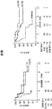


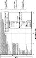
Classifications
-
- A—HUMAN NECESSITIES
- A61—MEDICAL OR VETERINARY SCIENCE; HYGIENE
- A61K—PREPARATIONS FOR MEDICAL, DENTAL OR TOILETRY PURPOSES
- A61K35/00—Medicinal preparations containing materials or reaction products thereof with undetermined constitution
- A61K35/12—Materials from mammals; Compositions comprising non-specified tissues or cells; Compositions comprising non-embryonic stem cells; Genetically modified cells
- A61K35/14—Blood; Artificial blood
- A61K35/17—Lymphocytes; B-cells; T-cells; Natural killer cells; Interferon-activated or cytokine-activated lymphocytes
-
- A—HUMAN NECESSITIES
- A61—MEDICAL OR VETERINARY SCIENCE; HYGIENE
- A61K—PREPARATIONS FOR MEDICAL, DENTAL OR TOILETRY PURPOSES
- A61K39/00—Medicinal preparations containing antigens or antibodies
- A61K39/395—Antibodies; Immunoglobulins; Immune serum, e.g. antilymphocytic serum
- A61K39/39533—Antibodies; Immunoglobulins; Immune serum, e.g. antilymphocytic serum against materials from animals
- A61K39/39558—Antibodies; Immunoglobulins; Immune serum, e.g. antilymphocytic serum against materials from animals against tumor tissues, cells, antigens
-
- A—HUMAN NECESSITIES
- A61—MEDICAL OR VETERINARY SCIENCE; HYGIENE
- A61B—DIAGNOSIS; SURGERY; IDENTIFICATION
- A61B10/00—Other methods or instruments for diagnosis, e.g. instruments for taking a cell sample, for biopsy, for vaccination diagnosis; Sex determination; Ovulation-period determination; Throat striking implements
- A61B10/02—Instruments for taking cell samples or for biopsy
- A61B10/0233—Pointed or sharp biopsy instruments
- A61B10/0266—Pointed or sharp biopsy instruments means for severing sample
-
- A—HUMAN NECESSITIES
- A61—MEDICAL OR VETERINARY SCIENCE; HYGIENE
- A61B—DIAGNOSIS; SURGERY; IDENTIFICATION
- A61B10/00—Other methods or instruments for diagnosis, e.g. instruments for taking a cell sample, for biopsy, for vaccination diagnosis; Sex determination; Ovulation-period determination; Throat striking implements
- A61B10/02—Instruments for taking cell samples or for biopsy
- A61B10/0233—Pointed or sharp biopsy instruments
- A61B10/0283—Pointed or sharp biopsy instruments with vacuum aspiration, e.g. caused by retractable plunger or by connected syringe
-
- A—HUMAN NECESSITIES
- A61—MEDICAL OR VETERINARY SCIENCE; HYGIENE
- A61B—DIAGNOSIS; SURGERY; IDENTIFICATION
- A61B18/00—Surgical instruments, devices or methods for transferring non-mechanical forms of energy to or from the body
- A61B18/02—Surgical instruments, devices or methods for transferring non-mechanical forms of energy to or from the body by cooling, e.g. cryogenic techniques
-
- A—HUMAN NECESSITIES
- A61—MEDICAL OR VETERINARY SCIENCE; HYGIENE
- A61K—PREPARATIONS FOR MEDICAL, DENTAL OR TOILETRY PURPOSES
- A61K31/00—Medicinal preparations containing organic active ingredients
- A61K31/33—Heterocyclic compounds
- A61K31/395—Heterocyclic compounds having nitrogen as a ring hetero atom, e.g. guanethidine or rifamycins
- A61K31/435—Heterocyclic compounds having nitrogen as a ring hetero atom, e.g. guanethidine or rifamycins having six-membered rings with one nitrogen as the only ring hetero atom
- A61K31/44—Non condensed pyridines; Hydrogenated derivatives thereof
- A61K31/445—Non condensed piperidines, e.g. piperocaine
- A61K31/4523—Non condensed piperidines, e.g. piperocaine containing further heterocyclic ring systems
- A61K31/454—Non condensed piperidines, e.g. piperocaine containing further heterocyclic ring systems containing a five-membered ring with nitrogen as a ring hetero atom, e.g. pimozide, domperidone
-
- A—HUMAN NECESSITIES
- A61—MEDICAL OR VETERINARY SCIENCE; HYGIENE
- A61K—PREPARATIONS FOR MEDICAL, DENTAL OR TOILETRY PURPOSES
- A61K39/00—Medicinal preparations containing antigens or antibodies
- A61K39/395—Antibodies; Immunoglobulins; Immune serum, e.g. antilymphocytic serum
- A61K39/44—Antibodies bound to carriers
-
- A—HUMAN NECESSITIES
- A61—MEDICAL OR VETERINARY SCIENCE; HYGIENE
- A61K—PREPARATIONS FOR MEDICAL, DENTAL OR TOILETRY PURPOSES
- A61K39/00—Medicinal preparations containing antigens or antibodies
- A61K39/46—Cellular immunotherapy
- A61K39/461—Cellular immunotherapy characterised by the cell type used
- A61K39/4611—T-cells, e.g. tumor infiltrating lymphocytes [TIL], lymphokine-activated killer cells [LAK] or regulatory T cells [Treg]
-
- A—HUMAN NECESSITIES
- A61—MEDICAL OR VETERINARY SCIENCE; HYGIENE
- A61K—PREPARATIONS FOR MEDICAL, DENTAL OR TOILETRY PURPOSES
- A61K39/00—Medicinal preparations containing antigens or antibodies
- A61K39/46—Cellular immunotherapy
- A61K39/463—Cellular immunotherapy characterised by recombinant expression
- A61K39/4631—Chimeric Antigen Receptors [CAR]
-
- A—HUMAN NECESSITIES
- A61—MEDICAL OR VETERINARY SCIENCE; HYGIENE
- A61K—PREPARATIONS FOR MEDICAL, DENTAL OR TOILETRY PURPOSES
- A61K39/00—Medicinal preparations containing antigens or antibodies
- A61K39/46—Cellular immunotherapy
- A61K39/464—Cellular immunotherapy characterised by the antigen targeted or presented
- A61K39/4643—Vertebrate antigens
- A61K39/4644—Cancer antigens
- A61K39/464402—Receptors, cell surface antigens or cell surface determinants
- A61K39/464411—Immunoglobulin superfamily
- A61K39/464412—CD19 or B4
-
- A—HUMAN NECESSITIES
- A61—MEDICAL OR VETERINARY SCIENCE; HYGIENE
- A61N—ELECTROTHERAPY; MAGNETOTHERAPY; RADIATION THERAPY; ULTRASOUND THERAPY
- A61N5/00—Radiation therapy
- A61N5/10—X-ray therapy; Gamma-ray therapy; Particle-irradiation therapy
-
- A—HUMAN NECESSITIES
- A61—MEDICAL OR VETERINARY SCIENCE; HYGIENE
- A61N—ELECTROTHERAPY; MAGNETOTHERAPY; RADIATION THERAPY; ULTRASOUND THERAPY
- A61N5/00—Radiation therapy
- A61N5/10—X-ray therapy; Gamma-ray therapy; Particle-irradiation therapy
- A61N5/1001—X-ray therapy; Gamma-ray therapy; Particle-irradiation therapy using radiation sources introduced into or applied onto the body; brachytherapy
- A61N5/1014—Intracavitary radiation therapy
-
- A—HUMAN NECESSITIES
- A61—MEDICAL OR VETERINARY SCIENCE; HYGIENE
- A61P—SPECIFIC THERAPEUTIC ACTIVITY OF CHEMICAL COMPOUNDS OR MEDICINAL PREPARATIONS
- A61P35/00—Antineoplastic agents
-
- A—HUMAN NECESSITIES
- A61—MEDICAL OR VETERINARY SCIENCE; HYGIENE
- A61P—SPECIFIC THERAPEUTIC ACTIVITY OF CHEMICAL COMPOUNDS OR MEDICINAL PREPARATIONS
- A61P35/00—Antineoplastic agents
- A61P35/02—Antineoplastic agents specific for leukemia
-
- A—HUMAN NECESSITIES
- A61—MEDICAL OR VETERINARY SCIENCE; HYGIENE
- A61P—SPECIFIC THERAPEUTIC ACTIVITY OF CHEMICAL COMPOUNDS OR MEDICINAL PREPARATIONS
- A61P43/00—Drugs for specific purposes, not provided for in groups A61P1/00-A61P41/00
-
- C—CHEMISTRY; METALLURGY
- C07—ORGANIC CHEMISTRY
- C07K—PEPTIDES
- C07K16/00—Immunoglobulins [IGs], e.g. monoclonal or polyclonal antibodies
- C07K16/18—Immunoglobulins [IGs], e.g. monoclonal or polyclonal antibodies against material from animals or humans
- C07K16/28—Immunoglobulins [IGs], e.g. monoclonal or polyclonal antibodies against material from animals or humans against receptors, cell surface antigens or cell surface determinants
- C07K16/2803—Immunoglobulins [IGs], e.g. monoclonal or polyclonal antibodies against material from animals or humans against receptors, cell surface antigens or cell surface determinants against the immunoglobulin superfamily
- C07K16/2827—Immunoglobulins [IGs], e.g. monoclonal or polyclonal antibodies against material from animals or humans against receptors, cell surface antigens or cell surface determinants against the immunoglobulin superfamily against B7 molecules, e.g. CD80, CD86
-
- A—HUMAN NECESSITIES
- A61—MEDICAL OR VETERINARY SCIENCE; HYGIENE
- A61B—DIAGNOSIS; SURGERY; IDENTIFICATION
- A61B18/00—Surgical instruments, devices or methods for transferring non-mechanical forms of energy to or from the body
- A61B2018/00571—Surgical instruments, devices or methods for transferring non-mechanical forms of energy to or from the body for achieving a particular surgical effect
- A61B2018/00577—Ablation
-
- A—HUMAN NECESSITIES
- A61—MEDICAL OR VETERINARY SCIENCE; HYGIENE
- A61K—PREPARATIONS FOR MEDICAL, DENTAL OR TOILETRY PURPOSES
- A61K39/00—Medicinal preparations containing antigens or antibodies
- A61K2039/505—Medicinal preparations containing antigens or antibodies comprising antibodies
-
- A—HUMAN NECESSITIES
- A61—MEDICAL OR VETERINARY SCIENCE; HYGIENE
- A61K—PREPARATIONS FOR MEDICAL, DENTAL OR TOILETRY PURPOSES
- A61K39/00—Medicinal preparations containing antigens or antibodies
- A61K2039/545—Medicinal preparations containing antigens or antibodies characterised by the dose, timing or administration schedule
-
- A—HUMAN NECESSITIES
- A61—MEDICAL OR VETERINARY SCIENCE; HYGIENE
- A61K—PREPARATIONS FOR MEDICAL, DENTAL OR TOILETRY PURPOSES
- A61K2239/00—Indexing codes associated with cellular immunotherapy of group A61K39/46
- A61K2239/31—Indexing codes associated with cellular immunotherapy of group A61K39/46 characterized by the route of administration
-
- A—HUMAN NECESSITIES
- A61—MEDICAL OR VETERINARY SCIENCE; HYGIENE
- A61K—PREPARATIONS FOR MEDICAL, DENTAL OR TOILETRY PURPOSES
- A61K2239/00—Indexing codes associated with cellular immunotherapy of group A61K39/46
- A61K2239/38—Indexing codes associated with cellular immunotherapy of group A61K39/46 characterised by the dose, timing or administration schedule
-
- A—HUMAN NECESSITIES
- A61—MEDICAL OR VETERINARY SCIENCE; HYGIENE
- A61K—PREPARATIONS FOR MEDICAL, DENTAL OR TOILETRY PURPOSES
- A61K2239/00—Indexing codes associated with cellular immunotherapy of group A61K39/46
- A61K2239/46—Indexing codes associated with cellular immunotherapy of group A61K39/46 characterised by the cancer treated
- A61K2239/48—Blood cells, e.g. leukemia or lymphoma
-
- A—HUMAN NECESSITIES
- A61—MEDICAL OR VETERINARY SCIENCE; HYGIENE
- A61N—ELECTROTHERAPY; MAGNETOTHERAPY; RADIATION THERAPY; ULTRASOUND THERAPY
- A61N5/00—Radiation therapy
- A61N5/10—X-ray therapy; Gamma-ray therapy; Particle-irradiation therapy
- A61N2005/1085—X-ray therapy; Gamma-ray therapy; Particle-irradiation therapy characterised by the type of particles applied to the patient
- A61N2005/1087—Ions; Protons
-
- A—HUMAN NECESSITIES
- A61—MEDICAL OR VETERINARY SCIENCE; HYGIENE
- A61N—ELECTROTHERAPY; MAGNETOTHERAPY; RADIATION THERAPY; ULTRASOUND THERAPY
- A61N5/00—Radiation therapy
- A61N5/10—X-ray therapy; Gamma-ray therapy; Particle-irradiation therapy
- A61N2005/1085—X-ray therapy; Gamma-ray therapy; Particle-irradiation therapy characterised by the type of particles applied to the patient
- A61N2005/1091—Kilovoltage or orthovoltage range photons
Abstract
T細胞受容体(TCR)またはキメラ抗原受容体(CAR)などの組換え受容体で操作された細胞をインビボで調節する方法が本明細書で提供される。いくつかの態様では、本方法は、前記細胞が存在するかもしくは存在する可能性があるまたは存在したかもしくは存在した可能性がある対象の区域、例えば腫瘍を含む病変を破壊することを含む。いくつかの態様では、破壊により、病変の環境、例えば腫瘍微小環境が変化する。いくつかの態様では、破壊は生検である。いくつかの局面では、提供される方法は、破壊を実施した後、操作された細胞の拡大の増加、およびいくつかの場合には、より堅固で持続的な応答をもたらす。Provided herein are methods of modulating cells engineered with a recombinant receptor, such as a T cell receptor (TCR) or a chimeric antigen receptor (CAR), in vivo. In some embodiments, the method comprises destroying an area of the subject in which the cells are or may be or have been or may be present, eg, a lesion including a tumor. In some embodiments, destruction changes the environment of the lesion, eg, the tumor microenvironment. In some embodiments, the disruption is a biopsy. In some aspects, the provided methods result in increased expansion of the engineered cells and, in some cases, a more robust and sustained response after performing the disruption.
Description
関連出願の相互参照
本出願は、その内容全体が参照により本明細書に組み入れられる、「METHODS FOR MODULATION OF GENETICALLY ENGINEERED CELLS」と題する、2016年12月3日に出願された米国仮特許出願第62/429,740号、「METHODS FOR MODULATION OF GENETICALLY ENGINEERED CELLS」と題する2017年1月10日に出願された米国仮特許出願第62/444,784号、「METHODS FOR MODULATION OF GENETICALLY ENGINEERED CELLS」と題する2017年5月1日に出願された米国仮特許出願第62/492,950号、「METHODS FOR MODULATION OF GENETICALLY ENGINEERED CELLS」と題する2017年6月2日に出願された米国仮特許出願第62/514,777号、「METHODS FOR MODULATION OF GENETICALLY ENGINEERED CELLS」と題する2017年6月5日に出願された米国仮特許出願第62/515,512号、「METHODS FOR MODULATION OF GENETICALLY ENGINEERED CELLS」と題する2017年8月23日に出願された米国仮特許出願第62/549,391号、「METHODS FOR MODULATION OF GENETICALLY ENGINEERED CELLS」と題する2017年11月1日に出願された米国仮特許出願第62/580,414号の優先権を主張する。
CROSS-REFERENCE TO RELATED APPLICATIONS This application, the entire contents of which are incorporated herein by reference, entitled "METHODS FOR MODULATION OF GENETICALLY ENGINEERED CELLS", 2016 filed US Provisional Patent filed December 3, Chapter 62 / 429,740, U.S. Provisional Patent Application No. 62 / 444,784, filed January 10, 2017 entitled `` METHODS FOR MODULATION OF GENETICALLY ENGINEERED CELLS, '' May 2017 entitled `` METHODS FOR MODULATION OF GENETICALLY ENGINEERED CELLS. '' U.S. Provisional Patent Application No. 62 / 492,950 filed on the 1st, U.S. Provisional Patent Application No. 62 / 514,777, filed on June 2, 2017, entitled `` METHODS FOR MODULATION OF GENETICALLY ENGINEERED CELLS, '' U.S. Provisional Patent Application No. 62 / 515,512, filed June 5, 2017 entitled `` MODULATION OF GENETICALLY ENGINEERED CELLS, '' U.S. application filed August 23, 2017, entitled `` METHODS FOR MODULATION OF GENETICALLY ENGINEERED CELLS. '' Provisional Patent Application No. 62 / 549,391, `` METH Claims priority of US Provisional Patent Application No. 62 / 580,414, filed November 1, 2017, entitled "ODS FOR MODULATION OF GENETICALLY ENGINEERED CELLS."
配列表の参照による組み入れ
本出願は、電子形式の配列表と共に提出される。配列表は、34,855キロバイトのサイズの、2017年11月29日に作成された735042009140SeqList.txtと題するファイルとして提供される。配列表の電子形式の情報は、その全体が参照により組み入れられる。
INCORPORATION BY REFERENCE OF SEQUENCE LISTING This application is filed with the sequence listing in electronic form. The sequence listing is provided as a file entitled 735042009140SeqList.txt created on November 29, 2017, with a size of 34,855 kilobytes. The information in electronic form of the sequence listing is incorporated by reference in its entirety.
分野
本開示は、いくつかの局面では、T細胞受容体(TCR)またはキメラ受容体(CAR)などの組換え受容体で操作された細胞をインビボで調節する方法に関する。いくつかの態様では、方法は、前記細胞が存在するかもしくは存在する可能性が高い対象の区域および/または腫瘍などの病変を破壊すること、ならびに/または病変もしくはその一部の物理的もしくは機械的操作、放射線または免疫調節剤の投与の1つまたは複数を含む治療を行うことを含む。いくつかの態様では、破壊および/または治療は、病変の環境、例えば腫瘍微小環境を変化させる。いくつかの態様では、破壊および/または治療は生検である。いくつかの局面では、提供される方法は、破壊および/または治療を実施した後、操作された細胞の拡大(expansion)の増加、およびいくつかの場合は、より堅固で持続的な応答をもたらす。
Field The present disclosure relates in some aspects to methods of modulating cells engineered with a recombinant receptor, such as a T cell receptor (TCR) or a chimeric receptor (CAR), in vivo. In some embodiments, the method comprises destroying an area of the subject in which the cells are or is likely to be present and / or a lesion, such as a tumor, and / or physical or mechanical of the lesion or a portion thereof. Performing a treatment that includes one or more of the following: manipulation, administration of radiation or an immunomodulatory agent. In some embodiments, destruction and / or treatment alters the environment of the lesion, eg, the tumor microenvironment. In some embodiments, the destruction and / or treatment is a biopsy. In some aspects, the provided methods result in increased expansion of engineered cells and, in some cases, more robust and sustained responses after performing disruption and / or treatment .
背景
免疫療法、例えばCARのような遺伝子操作された抗原受容体などのT細胞を投与することを含む養子細胞療法の方法のために、様々な戦略が利用可能である。いくつかの局面では、利用可能な方法は完全に満足のいくものではないことがある。養子細胞療法のためのさらなる戦略、例えば投与された細胞の持続性、活性および/もしくは増殖ならびに応答を増強するための戦略、ならびに細胞を調節するための戦略が必要とされている。そのような必要を満たす方法が提供される。
Background A variety of strategies are available for methods of immunotherapy, including adoptive cell therapy, including administering T cells such as genetically engineered antigen receptors such as CAR. In some aspects, the available methods may not be completely satisfactory. There is a need for additional strategies for adoptive cell therapy, for example, strategies for enhancing the persistence, activity and / or proliferation and response of administered cells, and for regulating cells. A method is provided that meets such needs.
概要
遺伝子操作された細胞が存在するか存在する可能性がある、もしくは存在したか存在した可能性がある対象の区域の破壊を行うこと、および/または病変もしくはその一部の物理的もしくは機械的操作、放射線または免疫調節剤の投与の1つまたは複数を含む治療を行うことを含み、前記対象は、疾患または状態を治療するために遺伝子操作された細胞の投与を以前に受けたことがある、遺伝子操作された細胞を拡大させるための方法が本明細書で提供され、この方法は、対象、区域、および/または対象の組織もしくは器官もしくは体液における操作された細胞の拡大、ならびに/または区域、組織もしくは器官もしくは体液中の操作された細胞の数の増加をもたらす。特定の態様では、区域は病変もしくはその一部であるかまたはそれを含む。いくつかの態様では、病変は腫瘍である。特定の態様では、腫瘍は原発性または続発性腫瘍である。特定の態様では、区域は骨髄組織であるかまたはそれを含む。特定の態様では、区域は病変もしくはその一部であるかまたはそれを含む。
Overview Performing destruction of an area of interest where genetically engineered cells are, or may be, or have been, or have been, present and / or physical or mechanical of a lesion or portion thereof. The subject has previously received administration of genetically engineered cells to treat a disease or condition, comprising administering a therapy comprising one or more of manipulation, radiation, or administration of an immunomodulatory agent. Provided herein is a method for expanding a genetically engineered cell, the method comprising expanding the engineered cell in a subject, area, and / or a tissue or organ or body fluid of the subject, and / or , Resulting in an increased number of engineered cells in a tissue or organ or body fluid. In certain embodiments, the area is or includes a lesion or a portion thereof. In some aspects, the lesion is a tumor. In certain aspects, the tumor is a primary or secondary tumor. In certain embodiments, the area is or includes bone marrow tissue. In certain embodiments, the area is or includes a lesion or a portion thereof.
そのような態様のいくつかでは、破壊および/または治療の時点でまたはその直前に、対象は、遺伝子操作された細胞の投与に応答した寛解後に再発した。そのような態様のいくつかでは、破壊および/または治療の時点でまたはその直前に:対象は寛解期にある;血液中で検出可能な操作された細胞の数は減少しているかまたは検出不能である;対象由来の体液もしくは組織もしくは試料、任意に血液中で検出可能な操作された細胞の数は、操作された細胞の投与後の先行する時点と比較して減少している;ならびに/または対象由来の体液もしくは組織もしくは試料、任意に血液中で検出可能な操作された細胞の数は、操作された細胞の投与の開始後の対象の血液中で検出可能なもしくは検出された操作された細胞のピークもしくは最大数と比較して、および/または細胞の投与後1、2、3、4、5、6、7、8、9、10、11、12、13、14もしくは28日以内の時点でのレベルと比較して、1.5倍、2.0倍、3.0倍、4.0倍、5.0倍、10倍、またはそれ以上を超えて減少している。 In some such embodiments, at or shortly before the destruction and / or treatment, the subject has relapsed after remission in response to administration of the genetically engineered cells. In some such embodiments, at or immediately prior to destruction and / or treatment: the subject is in remission; the number of engineered cells detectable in the blood is reduced or undetectable The number of engineered cells detectable in a body fluid or tissue or sample, optionally blood, from the subject is reduced compared to a previous time point after administration of the engineered cells; and / or The number of engineered cells detectable in a body fluid or tissue or sample, optionally blood, from the subject is the number of engineered cells detectable or detected in the subject's blood after the start of administration of the engineered cells. Compared to the peak or maximum number of cells and / or within 1, 2, 3, 4, 5, 6, 7, 8, 9, 10, 11, 12, 13, 14, or 28 days after administration of the cells 1.5 times, 2.0 times, 3.0 times, 4.0 times, Decreased by more than 5.0 times, 10 times or more.
そのような態様のいくつかでは、破壊および/または治療は、遺伝子操作された細胞の投与の開始後または遺伝子操作された細胞の最後の投与後2週間、1ヶ月、2ヶ月、3ヶ月、4ヶ月、5ヶ月、6ヶ月、1年またはそれ以上の時点、またはおよそこれらの時点、またはこれらを超える時点、またはおよそこれらを超える時点で行われる。そのような態様のいくつかでは、破壊および/または治療は、免疫調節剤の投与、放射線または区域もしくは病変の物理的もしくは機械的操作の1つまたは複数を含む。 In some such embodiments, the destruction and / or treatment is performed two weeks, one month, two months, three months, four weeks after the start of administration of the engineered cells or after the last administration of the engineered cells. It occurs at months, 5 months, 6 months, 1 year or more, or at or about these time points, or at or above these time points. In some of such embodiments, the destruction and / or treatment comprises one or more of administering an immunomodulator, radiation or physical or mechanical manipulation of an area or lesion.
そのような態様のいくつかでは、破壊および/または治療は、免疫調節剤の投与を含む。特定の態様では、免疫調節剤は、免疫阻害分子であるかもしくはそれを含む、免疫チェックポイント分子もしくは免疫チェックポイント経路のメンバーであるかもしくはそれを含む、および/または免疫チェックポイント分子もしくは経路の調節剤であるかもしくはそれを含む。特定の態様では、免疫チェックポイント分子または経路は、PD-1、PD-L1、PD-L2、CTLA-4、LAG-3、TIM3、VISTA、アデノシン受容体、CD73、CD39、アデノシン2A受容体(A2AR)、もしくはアデノシン、または前記のいずれかを含む経路であるかまたはそれを含む。特定の態様では、免疫調節剤はサリドマイドであるか、またはサリドマイドの誘導体もしくは類似体である。特定の態様では、免疫調節剤は、レナリドミド、ポマリドミド、アバドミド、またはレナリドミド、ポマリドミド、アバドミドの立体異性体、またはその薬学的に許容される塩、溶媒和物、水和物、共結晶、包接化合物、もしくは多形体である。特定の態様では、免疫調節剤は、レナリドミド、レナリドミドの立体異性体、またはその薬学的に許容される塩、溶媒和物、水和物、共結晶、包接化合物、もしくは多形体である。 In some such embodiments, the destruction and / or treatment comprises administration of an immunomodulatory agent. In certain embodiments, the immunomodulatory agent is or comprises an immune inhibitory molecule, is or comprises a member of an immune checkpoint molecule or pathway, and / or is a member of an immune checkpoint molecule or pathway. Is or comprises a modulator. In certain embodiments, the immune checkpoint molecule or pathway is PD-1, PD-L1, PD-L2, CTLA-4, LAG-3, TIM3, VISTA, adenosine receptor, CD73, CD39, adenosine 2A receptor ( A2AR), or adenosine, or a pathway comprising any of the foregoing. In certain embodiments, the immunomodulator is thalidomide, or a derivative or analog of thalidomide. In certain embodiments, the immunomodulator is lenalidomide, pomalidomide, abamide, or a stereoisomer of lenalidomide, pomalidomide, abamide, or a pharmaceutically acceptable salt, solvate, hydrate, co-crystal, clathrate thereof. Compound or polymorph. In certain aspects, the immunomodulator is lenalidomide, a stereoisomer of lenalidomide, or a pharmaceutically acceptable salt, solvate, hydrate, co-crystal, clathrate, or polymorph thereof.
そのような態様のいくつかでは、再発後ならびに破壊および/または治療の前に、対象は、疾患もしくは状態を治療するためまたは操作された細胞の活性を調節するための外因性または組換え作用物質を投与されたことがない。そのような態様のいくつかでは、破壊および/または治療は放射線を含む。そのような態様のいくつかでは、破壊および/または治療は、区域または病変の物理的または機械的操作を含み、任意で区域または病変を探査する、突き刺すまたは貫通することを含む。いくつかの態様では、物理的または機械的操作は生検を含む。特定の態様では、生検は針またはトロカールによって行われる。特定の態様では、生検は切開生検を含む。 In some of such embodiments, after recurrence and prior to destruction and / or treatment, the subject is treated with an exogenous or recombinant agent for treating a disease or condition or modulating the activity of an engineered cell. Have never been administered. In some such embodiments, the destruction and / or treatment comprises radiation. In some such embodiments, destruction and / or treatment includes physical or mechanical manipulation of the area or lesion, and optionally includes exploring, piercing, or penetrating the area or lesion. In some embodiments, the physical or mechanical manipulation includes a biopsy. In certain embodiments, the biopsy is performed with a needle or trocar. In certain embodiments, the biopsy comprises an open biopsy.
そのような態様のいくつかでは、本発明の方法は、破壊および/または治療の直前の時点と比較して、遺伝子操作された細胞の拡大または遺伝子操作された細胞の数の増加をもたらす。特定の態様では、細胞の拡大は、破壊および/または治療後24時間、48時間、96時間、7日間、14日間または28日間以内またはおよそこれらの期間以内に起こる。特定の態様では、拡大は、破壊および/または治療の直前と比較して、1.5倍、2.0倍、5.0倍、10倍、100倍、200倍、もしくはそれ以上を超えるか、または約1.5倍、2.0倍、5.0倍、10倍、100倍、200倍、もしくはそれ以上を超える、血液中で検出可能な操作された細胞をもたらすか、または拡大は、破壊および/または治療前の血液中の操作された細胞の事前のピークレベルと比較して、1.5倍、2.0倍、5.0倍、10倍、100倍、200倍、もしくはそれ以上を超えるか、または約1.5倍、2.0倍、5.0倍、10倍、100倍、200倍、もしくはそれ以上を超える、血液中で検出可能な操作された細胞をもたらす。特定の態様では、破壊および/または治療後の時点での血液中で検出可能な操作された細胞の数は、破壊および/または治療前の先行する時点での操作された細胞の数と比較して増加している(例えば1.5倍、2倍、3倍、4倍、5倍、10倍、またはそれ以上の増加);破壊および/または治療前の対象の血液中で検出可能な操作された細胞のピークまたは最大数よりも1.5倍、2倍、3倍、4倍、5倍、10倍、50倍、100倍、またはそれ以上を超える;操作された細胞の10%、9%、8%、7%、6%、5%、4%、3%、2%、1%、0.5%、0.2%、もしくは0.1%超または約10%、9%、8%、7%、6%、5%、4%、3%、2%、1%、0.5%、0.2%、もしくは0.1%超が、そのような細胞の最大レベルのピークが血液中で検出された後の時点で、血液中で検出可能である。 In some of such embodiments, the methods of the invention result in an expansion of the genetically engineered cells or an increase in the number of genetically engineered cells as compared to a point in time immediately before destruction and / or treatment. In certain embodiments, expansion of the cells occurs within 24 hours, 48 hours, 96 hours, 7 days, 14 days or 28 days or about within these periods after disruption and / or treatment. In certain embodiments, the expansion is greater than 1.5-fold, 2.0-fold, 5.0-fold, 10-fold, 100-fold, 200-fold or more, or about 1.5-fold, compared to immediately before destruction and / or treatment. 2.0 ×, 5.0 ×, 10 ×, 100 ×, 200 × or more, resulting in detectable engineered cells in the blood or expansion is the manipulation in the blood prior to destruction and / or treatment. 1.5-fold, 2.0-fold, 5.0-fold, 10-fold, 100-fold, 200-fold, or more, or about 1.5-fold, 2.0-fold, 5.0-fold, 10-fold, This results in engineered cells detectable in blood that are more than 1, fold, 100 fold, 200 fold, or more. In certain embodiments, the number of engineered cells detectable in blood at a time after disruption and / or treatment is compared to the number of engineered cells at a prior time prior to disruption and / or treatment. (Eg, 1.5-fold, 2-fold, 3-fold, 4-fold, 5-fold, 10-fold, or more); manipulated detectable in the blood of the subject before destruction and / or treatment 1.5 times, 2 times, 3 times, 4 times, 5 times, 10 times, 50 times, 100 times or more than the peak or maximum number of cells; 10%, 9%, 8 %, 7%, 6%, 5%, 4%, 3%, 2%, 1%, 0.5%, 0.2%, or more than 0.1% or about 10%, 9%, 8%, 7%, 6%, 5%, 4%, 3%, 2%, 1%, 0.5%, 0.2%, or more than 0.1% in the blood after the peak of the maximum level of such cells is detected in the blood Can be detected.
そのような態様のいくつかでは、操作された細胞は、疾患もしくは障害に関連するかまたは環境もしくは病変の細胞において発現される抗原に特異的に結合する組換え受容体を発現する。そのような態様のいくつかでは、疾患または状態は腫瘍または癌である。そのような態様のいくつかでは、疾患または状態は白血病またはリンパ腫である。そのような態様のいくつかでは、疾患または状態は、非ホジキンリンパ腫(NHL)、急性リンパ芽球性白血病(ALL)または慢性リンパ性白血病(CLL)である。そのような態様のいくつかでは、組換え受容体は、T細胞受容体または機能的非T細胞受容体である。そのような態様のいくつかでは、組換え受容体はキメラ抗原受容体(CAR)である。 In some of such embodiments, the engineered cells express a recombinant receptor that specifically binds to an antigen that is associated with a disease or disorder or that is expressed on cells of the environment or pathology. In some of such embodiments, the disease or condition is a tumor or cancer. In some of such embodiments, the disease or condition is leukemia or lymphoma. In some of such embodiments, the disease or condition is non-Hodgkin's lymphoma (NHL), acute lymphoblastic leukemia (ALL) or chronic lymphocytic leukemia (CLL). In some of such embodiments, the recombinant receptor is a T cell receptor or a functional non-T cell receptor. In some of such embodiments, the recombinant receptor is a chimeric antigen receptor (CAR).
いくつかの態様では、CARは、抗原に特異的に結合する細胞外抗原認識ドメインおよびITAMを含む細胞内シグナル伝達ドメインを含む。特定の態様では、抗原はCD19である。特定の態様では、細胞内シグナル伝達ドメインは、CD3-ゼータ(CD3ζ)鎖の細胞内ドメインを含む。いくつかの態様では、CARは共刺激シグナル伝達領域をさらに含む。特定の態様では、共刺激シグナル伝達ドメインは、CD28または4-1BBのシグナル伝達ドメインを含む。特定の態様では、操作された細胞はCD4+またはCD8+T細胞である。そのような態様のいくつかでは、操作された細胞は対象に対して自己由来である。 In some embodiments, the CAR comprises an extracellular antigen recognition domain that specifically binds to the antigen and an intracellular signaling domain that includes ITAM. In certain embodiments, the antigen is CD19. In certain embodiments, the intracellular signaling domain comprises the intracellular domain of the CD3-zeta (CD3ζ) chain. In some embodiments, the CAR further comprises a costimulatory signaling region. In certain embodiments, the costimulatory signaling domain comprises a CD28 or 4-1BB signaling domain. In certain embodiments, the engineered cells are CD4 + or CD8 + T cells. In some of such embodiments, the engineered cells are autologous to the subject.
そのような態様のいくつかでは、操作された細胞は対象に対して同種異系である。そのような態様のいくつかでは、投与される操作された細胞は、それぞれ両端の値を含む、約0.25×10⁶細胞/kg〜5×106細胞/kg対象体重、0.5×106細胞/kg〜3×106細胞/kg対象体重、約0.75×106細胞/kg〜2.5×106細胞/kg、または約1×106細胞/kg〜2×106細胞/kgである。そのような態様のいくつかでは、操作された細胞は、細胞を含む単一の薬学的組成物中で投与される。そのような態様のいくつかでは、投与される操作された細胞は分割用量であり、用量の細胞は、3日間以下の期間にわたって用量の細胞を集合的に含む複数の組成物で投与される。 In some of such embodiments, the engineered cells are allogeneic to the subject. In some such embodiments, cells engineered to be administered each inclusive, about 0.25 × 10 6 cells / kg~5 × 10 6 cells / kg body weight of the subject, 0.5 × 10 6 cells / kg~3 × 10 6 cells / kg body weight of the subject, about 0.75 × 10 6 cells /Kg~2.5×10 6 cells / kg or about 1 × 10 6 cells / kg~2 × 10 6 cells / kg,. In some such embodiments, the engineered cells are administered in a single pharmaceutical composition comprising the cells. In some of such embodiments, the engineered cells administered are in divided doses, and the dose of cells is administered in a plurality of compositions that collectively include a dose of cells over a period of three days or less.
治療レジメンを対象に投与することを含む治療の方法が本明細書で提供され、ここで、対象は、疾患または状態を治療するために遺伝子操作された細胞を以前に投与されたことがあり、この方法は、対象、区域、および/または対象の組織もしくは器官もしくは体液における操作された細胞の拡大、ならびに/または区域、組織もしくは器官もしくは体液中の操作された細胞の数の増加をもたらす。 Provided herein is a method of treatment comprising administering a treatment regimen to a subject, wherein the subject has previously been administered genetically engineered cells to treat a disease or condition; The method results in an expansion of the engineered cells in the subject, area, and / or tissue or organ or fluid of the subject, and / or an increase in the number of engineered cells in the area, tissue or organ or fluid.
特定の態様では、治療レジメンは、操作された細胞が存在する、存在するかもしくは存在したことが疑われる、または存在する可能性がある対象の区域の破壊を含み、および/または病変もしくはその一部の物理的もしくは機械的操作、放射線または免疫調節剤の投与の1つまたは複数を含む。特定の態様では、治療レジメンおよび/または方法は、遺伝子操作された細胞のまたは前記遺伝子操作された細胞のその後の投与を含まず、および/またはそのようなその後の投与なしに拡大が達成される。いくつかの態様では、治療レジメンは治療用量以下の用量で投与され、および/または遺伝子操作された細胞の拡大を介してその治療効果を引き出す。特定の態様では、対象は、遺伝子操作された細胞の以前の投与に対する応答の後に再発した、および/または投与に応答しなかった。いくつかの態様では、対象は、遺伝子操作された細胞に応答したことがあり、その後応答しなくなったおよび/または再発した。 In certain embodiments, the treatment regimen comprises destruction of an area of the subject in which the engineered cells are, is, or is suspected of, or may be present, and / or the lesion or one of the lesions. And / or administration of radiation or immunomodulatory agents. In certain embodiments, the therapeutic regimen and / or method does not include and / or does not involve subsequent administration of the engineered cells or the genetically engineered cells. . In some embodiments, the therapeutic regimen is administered at sub-therapeutic doses and / or elicits its therapeutic effects via expansion of the genetically engineered cells. In certain embodiments, the subject has relapsed after response to a previous administration of the genetically engineered cells and / or did not respond to the administration. In some embodiments, the subject has responded to the engineered cells and has subsequently stopped responding and / or has recurred.
発明の詳細な説明
I.養子細胞療法における遺伝子操作された細胞の調節
対象に投与された遺伝子操作細胞の拡大、増殖、および/または活性化を促進する、増強するまたは増大させることなどの、遺伝子操作された細胞をインビボで調節する方法が本明細書で提供される。いくつかの態様では、組換え受容体発現細胞(例えばCAR+T細胞)などの遺伝子操作された細胞を対象に投与した後、提供される方法は、遺伝子操作された細胞が存在するかもしくは存在する可能性が高い対象の組織、器官、腫瘤もしくは病変区域などの対象の区域またはその領域もしくは一部を破壊すること、例えば操作すること、および/または病変もしくはその一部の物理的もしくは機械的操作、放射線または免疫調節剤の投与の1つまたは複数を含む治療を行うことを含む。いくつかの態様では、区域(例えば対象の組織、器官、腫瘤もしくは病変区域、またはその領域もしくは一部)は、遺伝子操作された細胞によって認識される抗原発現細胞を含むことが既知であるかまたは疑われる。特に、対象の他の区域または領域と比べて、標的部区域は、その区域が対象の他の区域と比べてまたは比較して、より高いまたはより多い濃度または抗原の量または抗原特異的細胞の数を有することが既知であるかまたは疑われるものである。
DETAILED DESCRIPTION OF THE INVENTION
I. Regulation of Genetically Engineered Cells in Adoptive Cell Therapy Genetically engineered cells can be administered in vivo, such as to promote, enhance or increase the expansion, proliferation and / or activation of genetically engineered cells administered to a subject. Methods of adjusting are provided herein. In some embodiments, after administering a genetically engineered cell, such as a recombinant receptor expressing cell (eg, a CAR + T cell) to a subject, the provided method comprises the step of presenting or possibly presenting the genetically engineered cell. Destroying, e.g., manipulating, and / or physically or mechanically manipulating a lesion or a region thereof, such as a tissue, organ, mass or lesion area of the subject, It involves administering a treatment that includes one or more of the administration of radiation or an immunomodulator. In some embodiments, the area (eg, a tissue, organ, mass or lesion area of the subject, or an area or portion thereof) is known to contain antigen-expressing cells that are recognized by the engineered cells, or Suspected. In particular, compared to other areas or regions of interest, the target area may have a higher or higher concentration or amount of antigen or amount of antigen-specific cells in that area as compared to or compared to other areas of interest. It is known or suspected to have a number.
いくつかの態様では、治療および/または破壊は、病変の環境、例えば腫瘍微小環境を改変するなどの、区域の環境を改変する。いくつかの場合には、改変は、遺伝子操作されたT細胞の活性を直接的または間接的に調節するようなものである。いくつかの局面では、改変は、組換え受容体発現細胞(例えばCAR+T細胞)のような以前に投与された遺伝子操作細胞のインビボでの再活性化、拡大および/または増殖を促進するのに十分である。 In some embodiments, the treatment and / or destruction alters the environment of the area, such as altering the environment of the lesion, eg, the tumor microenvironment. In some cases, the modification is such as to directly or indirectly modulate the activity of the engineered T cell. In some aspects, the modification is sufficient to promote in vivo reactivation, expansion and / or proliferation of a previously administered genetically engineered cell, such as a recombinant receptor expressing cell (eg, a CAR + T cell). It is.
養子T細胞療法などのT細胞ベースの療法(キメラ抗原受容体(CAR)および/または他の組換え抗原受容体などの関心対象の疾患または障害に特異的なキメラ受容体を発現する細胞、ならびに他の養子免疫細胞および養子T細胞療法の投与を含むものを包含する)は、癌ならびに他の疾患および障害の治療において有効であり得る。ある状況では、養子細胞療法に対する利用可能なアプローチは、必ずしも完全に満足のいくものではないことがある。いくつかの状況では、最適な効果は、投与された細胞が、標的、例えば標的抗原を認識して結合し、対象、腫瘍、およびその環境内の適切な部位に輸送し、局在させ、成功裏に進入する能力に依存し得る。いくつかの状況では、最適な効果は、投与された細胞が活性化する、拡大する、細胞傷害性死滅およびサイトカインなどの様々な因子の分泌を含む様々なエフェクター機能を発揮する、長期を含めて持続する、特定の表現型状態(長期記憶、低分化状態、およびエフェクター状態など)に分化、移行、またはそれへのリプログラミングに関与する、疾患の局所微小環境における免疫抑制状態を回避または低減する、標的リガンドまたは抗原のクリアランスおよびそれへの再曝露後に有効で堅固なリコール応答を提供する、ならびに枯渇、アネルギー、末梢寛容、最終分化、および/または抑制状態への分化を回避または低減する能力に依存し得る。 T cell-based therapies, such as adoptive T cell therapy (cells that express a chimeric receptor specific for the disease or disorder of interest, such as chimeric antigen receptor (CAR) and / or other recombinant antigen receptors, and (Including those involving the administration of other adoptive immune cells and adoptive T-cell therapy) may be effective in the treatment of cancer and other diseases and disorders. In some situations, available approaches to adoptive cell therapy may not always be completely satisfactory. In some situations, the optimal effect is that the administered cells will recognize and bind to the target, eg, the target antigen, and transport and localize it to the appropriate site in the subject, tumor, and its environment, It may depend on the ability to enter the back. In some situations, the optimal effects include long-term administration of the administered cells to exert various effector functions, including activation, expansion, cytotoxic killing and secretion of various factors such as cytokines. Avoid or reduce the immunosuppressive state in the local microenvironment of the disease involved in differentiating, transitioning to, or reprogramming into, persistent, phenotypic states (such as long-term memory, poorly differentiated, and effector states) Provides an effective and robust recall response after clearance and re-exposure to the target ligand or antigen, and its ability to avoid or reduce depletion, anergy, peripheral tolerance, terminal differentiation, and / or differentiation to a suppressive state Can depend.
いくつかの局面では、免疫療法、例えばT細胞療法の効果は、疾患または障害の局所微小環境、例えばTMEにおける免疫抑制活性または存在する因子によって制限され得る。いくつかの局面では、TMEは、T細胞療法のために投与されたT細胞の活性、機能、増殖、生存および/または持続性を抑制することができる因子または状態を含むかまたはそれを生成する。 In some aspects, the effects of immunotherapy, eg, T cell therapy, can be limited by the immunosuppressive activity or factors present in the local microenvironment of the disease or disorder, eg, TME. In some aspects, the TME comprises or produces an agent or condition capable of suppressing the activity, function, proliferation, survival and / or persistence of T cells administered for T cell therapy .
いくつかの態様では、操作された細胞の曝露および持続性は、対象への投与後に減少または低下する。それにもかかわらず、所見は、いくつかの場合には、投与された組換え受容体を発現する細胞への対象の曝露の増加(例えば細胞数の増加または期間の延長)は、養子細胞療法における効果および治療転帰を改善し得る。複数の臨床試験において様々なCD19発現癌を有する対象に種々のCD19標的CAR発現T細胞を投与した後に行われた予備分析は、CAR発現細胞へのより高いおよび/またはより長い程度の曝露と治療転帰との間の相関関係を明らかにした。そのような転帰は、重度のまたは著しい腫瘍量を有する個人においてさえも、患者の生存および寛解を含んだ。いくつかの局面では、提供された方法によって観察された安全性プロフィールは、提供された治療用T細胞組成物および疾患または状態を治療するための別の療法、例えばチェックポイントアンタゴニストなどの免疫調節剤を含む併用療法の望ましくない安全性懸念のリスクを低減し得る。 In some embodiments, the exposure and persistence of the engineered cells is reduced or diminished after administration to the subject. Nevertheless, the finding is that in some cases, increased subject exposure to administered recombinant receptor-expressing cells (eg, increased cell number or extended duration) may be associated with adoptive cell therapy. May improve efficacy and treatment outcome. Preliminary analyzes performed after administering different CD19-targeted CAR-expressing T cells to subjects with different CD19-expressing cancers in multiple clinical trials revealed higher and / or longer-term exposure to CAR-expressing cells and treatment The correlation between outcomes was revealed. Such outcomes included patient survival and remission, even in individuals with severe or significant tumor burden. In some aspects, the safety profile observed by the provided methods is such that the therapeutic T cell composition provided and another therapy to treat the disease or condition, e.g., an immunomodulatory agent such as a checkpoint antagonist May reduce the risk of unwanted safety concerns of combination therapies that include
遺伝子操作されたT細胞を投与された対象において、病変を破壊すること、および/または病変もしくはその一部の物理的もしくは機械的操作、放射線または免疫調節剤の投与の1つまたは複数を含む治療を行うことは、対象が再発した後でも、対象における細胞の実質的な拡大をもたらし得ることが本明細書で見出される。細胞が存在するかまたは存在する可能性が高い対象の区域を破壊する、例えば操作する、および/または病変もしくはその一部の物理的もしくは機械的操作、放射線または免疫調節剤の投与の1つまたは複数を含む治療を行う方法が本明細書で提供される。いくつかの態様では、区域は、腫瘍または癌などの病変である。いくつかの態様では、破壊および/または治療は、病変部もしくはその付近、例えば腫瘍もしくはその付近、または病変に関連する微小環境、例えば腫瘍微小環境(TME)もしくはその付近における機械的または物理的改変によって行うことができる。いくつかの態様では、破壊および/または治療は、薬理学的作用物質または治療薬、例えば免疫調節剤、またはT細胞または病変もしくは病変の微小環境に関連する1つもしくは複数の細胞の活性を調節することができる他の作用物質の投与によって行うことができる。いくつかの場合には、薬理学的作用物質は、病変の部位を標的とする、または病変の細胞、例えば腫瘍微小環境の細胞に特異的に結合する治療薬である。いくつかの態様では、機械的破壊による、または免疫調節剤などの薬理学的作用物質の投与などによる破壊および/または治療は、組換え受容体発現T細胞、例えばCAR+T細胞の投与の開始の1週間、2ヶ月、1ヶ月、2ヶ月、3ヶ月、4ヶ月、5ヶ月、6ヶ月、1年、2年後もしくはそれ以上より後にまたはおよそこれらの期間より後に行われる。 In a subject receiving genetically engineered T cells, destroying the lesion and / or treatment comprising one or more of physical or mechanical manipulation of the lesion or a portion thereof, radiation or immunomodulatory agent It is found herein that performing can result in a substantial expansion of cells in the subject, even after the subject has relapsed. Destroying, e.g., manipulating, and / or physical or mechanical manipulation of the lesion or part thereof, the administration of radiation or immunomodulatory agents, or destroying the area of the subject where the cells are or are likely to be present Provided herein are methods of performing a therapy, including a plurality. In some aspects, the area is a lesion, such as a tumor or cancer. In some embodiments, the destruction and / or treatment comprises mechanical or physical modification at or near the lesion, such as at or near a tumor, or a microenvironment associated with the lesion, such as a tumor microenvironment (TME). Can be done by In some embodiments, the destruction and / or treatment modulates the activity of a pharmacological agent or therapeutic agent, such as an immunomodulator, or one or more T cells or one or more cells associated with the lesion or lesion microenvironment. This can be accomplished by the administration of other agents that can. In some cases, the pharmacological agent is a therapeutic agent that targets the site of the lesion or specifically binds to cells of the lesion, eg, cells of the tumor microenvironment. In some embodiments, destruction and / or treatment, such as by mechanical destruction or by the administration of a pharmacological agent such as an immunomodulatory agent, comprises the initiation of administration of the recombinant receptor-expressing T cells, eg, CAR + T cells, for one day. It takes place after weeks, two months, one month, two months, three months, four months, five months, six months, one year, two years or more or approximately after these periods.
いくつかの態様では、薬理学的作用物質または治療薬の投与などの破壊および/または治療の時点またはその直前に、対象は、治療後の寛解の後に再発した。 In some embodiments, the subject has relapsed after or after a remission after treatment, at or immediately prior to destruction and / or treatment, such as administration of a pharmacological agent or therapeutic agent.
A.病変
提供される方法の局面では、操作された細胞が存在するか存在する可能性がある、または存在したか存在した可能性がある対象の区域は、病変または病変の一部である。いくつかの局面では、病変は、投与される組換え受容体発現細胞によって認識される抗原発現細胞を含むことが既知であるかまたは疑われるものである。提供される方法は、病変を破壊するため、および/またはインビボで遺伝子操作された細胞を調節するように対象を治療するために行われる。
A. Lesions In aspects of the provided methods, the area of the subject where the engineered cells are or may be, or have, or have been, is a lesion or a portion of a lesion. In some aspects, the lesion is one that is known or suspected of containing an antigen-expressing cell that is recognized by the administered recombinant receptor-expressing cell. The provided methods are performed to destroy lesions and / or treat a subject to modulate genetically engineered cells in vivo.
いくつかの態様では、病変は、傷害または疾患による損傷を被った器官または組織の任意の領域を含む。特定の態様では、病変は、傷害または疾患による構造の異常な変化を受けたおよび/または受けている器官または組織の任意の領域である。いくつかの態様では、病変は限局性であり、明確に定義されている。いくつかの態様では、病変は非癌性である。特定の態様では、病変は非腫瘍性である。特定の態様では、病変は癌性であるか、または癌性であることが疑われる。特定の態様では、病変は腫瘍である。 In some embodiments, the lesion includes any region of an organ or tissue that has been damaged by the injury or disease. In certain embodiments, a lesion is any region of an organ or tissue that has undergone and / or has undergone an abnormal change in structure due to an injury or disease. In some aspects, the lesion is localized and well-defined. In some aspects, the lesion is non-cancerous. In certain aspects, the lesion is non-neoplastic. In certain embodiments, the lesion is or is suspected of being cancerous. In certain aspects, the lesion is a tumor.
いくつかの態様では、病変は、器官または組織に認められる病変である。特定の態様では、病変は、結合組織、筋肉組織、神経組織、または上皮組織に存在する。特定の態様では、病変は、心臓、血管系、唾液腺、食道、胃、肝臓、胆嚢、膵臓、腸、結腸、直腸、視床下部、下垂体、松果体、甲状腺、副甲状腺、副腎、腎臓、尿管、膀胱、尿道、リンパ系、皮膚、筋肉、脳、脊髄、神経、卵巣、子宮、精巣、前立腺、咽頭、喉頭、気管、気管支、肺、横隔膜、骨、軟骨、靭帯、または腱に存在する。 In some aspects, the lesion is a lesion found on an organ or tissue. In certain aspects, the lesion is in connective, muscle, nerve, or epithelial tissue. In certain embodiments, the lesion is a heart, vasculature, salivary gland, esophagus, stomach, liver, gallbladder, pancreas, intestine, colon, rectum, hypothalamus, pituitary, pineal gland, thyroid, parathyroid, adrenal gland, kidney, In the ureter, bladder, urethra, lymphatic system, skin, muscle, brain, spinal cord, nerve, ovary, uterus, testis, prostate, pharynx, larynx, trachea, bronchi, lung, diaphragm, bone, cartilage, ligament, or tendon I do.
特定の態様では、病変は以下から選択される:軟部組織における病変、例えばモレル-ラバリー病変、バンカート病変、ペルセス病変、ステナー病変、またはSLAP病変;骨病変、例えば骨化性線維腫、ALPSA病変、またはヒル-サックス病変;皮膚病変、例えば色素性母斑、スキップ病変、またはオスラー結節;膿漏性角皮症、黒色丘疹性皮膚病、白血病性皮疹、ジェーンウェイ病変、カポジ肉腫、扁平母斑、または慢性瘢痕角化症;消化管病変、例えばデュラフォイ病変またはキャメロン病変:内胚葉性病変、例えば色素性口腔病変、子宮内膜上皮内腫瘍;ゴーン病巣、良性リンパ上皮性病変、多発性硬化症病変、熱帯性潰瘍、またはヘルペス性ひょうそなどの別の病変。 In certain embodiments, the lesion is selected from: a lesion in soft tissue, such as a Morel-Lavary lesion, a Bankart lesion, a Peress lesion, a Stener lesion, or a SLAP lesion; a bone lesion, such as an ossifying fibroma, ALPSA lesion , Or Hill-Sachs lesions; skin lesions, such as pigmented nevus, skip lesions, or Osler nodules; purulent keratoderma, melanotic papular dermatoses, leukemic eruptions, Janeway lesions, Kaposi's sarcoma, flat nevus Gastrointestinal lesions, such as Durafoy or Cameron lesions: endodermal lesions, such as pigmented oral lesions, endometrial intraepithelial neoplasia; Gone lesions, benign lymphoepithelial lesions, multiple sclerosis Another lesion, such as a lesion, tropical ulcer, or herpetic hypnosis.
特定の態様では、病変は腫瘍または新生物である。特定の態様では、腫瘍は良性である。特定の態様では、腫瘍は、前癌性もしくは癌性であるか、または癌性もしくは前癌性であることが疑われる。特定の態様では、腫瘍は原発性腫瘍であり、すなわち、腫瘍は、病変が最初に発症または出現した解剖学的部位に見出される。いくつかの態様では、腫瘍は、続発性腫瘍、例えば身体の異なる部位内に位置する原発性腫瘍内の細胞に由来する癌性腫瘍である。 In certain aspects, the lesion is a tumor or neoplasm. In certain aspects, the tumor is benign. In certain aspects, the tumor is precancerous or cancerous, or is suspected of being cancerous or precancerous. In certain embodiments, the tumor is a primary tumor, ie, the tumor is found at the anatomical site where the lesion first developed or appeared. In some embodiments, the tumor is a secondary tumor, for example, a cancerous tumor derived from cells in a primary tumor located in different parts of the body.
いくつかの態様では、病変は、B細胞悪性腫瘍または血液悪性腫瘍である癌または増殖性疾患に関連するか、それによって引き起こされるか、またはそれに関連するもしくはそれによって引き起こされることが疑われる。いくつかの態様では、癌または増殖性疾患は、リンパ芽球性白血病(ALL)、非ホジキンリンパ腫(NHL)、または慢性リンパ性白血病(CLL)である。いくつかの態様では、癌はCLLである。いくつかの態様では、病変は、骨髄腫、リンパ腫または白血病に関連するか、それによって引き起こされるか、またはそれに関連するもしくはそれによって引き起こされることが疑われる。いくつかの態様では、病変は、非ホジキンリンパ腫(NHL)、急性リンパ芽球性白血病(ALL)、慢性リンパ性白血病(CLL)、びまん性大細胞型B細胞リンパ腫(DLBCL)、急性骨髄性白血病(AML)、または骨髄腫、例えば多発性骨髄腫(MM)に関連するか、それによって引き起こされるか、またはそれに関連するもしくはそれによって引き起こされることが疑われる。いくつかの態様では、病変は、MMまたはDBCBLに関連するか、それによって引き起こされるか、またはそれに関連するもしくはそれによって引き起こされることが疑われる。 In some embodiments, the lesion is associated with, caused by, or is suspected to be associated with or caused by a cancer or proliferative disease that is a B-cell or hematologic malignancy. In some embodiments, the cancer or proliferative disease is lymphoblastic leukemia (ALL), non-Hodgkin's lymphoma (NHL), or chronic lymphocytic leukemia (CLL). In some embodiments, the cancer is CLL. In some embodiments, the lesion is associated with, caused by, or suspected to be associated with or caused by myeloma, lymphoma, or leukemia. In some embodiments, the lesion is non-Hodgkin's lymphoma (NHL), acute lymphoblastic leukemia (ALL), chronic lymphocytic leukemia (CLL), diffuse large B-cell lymphoma (DLBCL), acute myeloid leukemia (AML), or is associated with or caused by, or is suspected to be associated with or caused by, myeloma, eg, multiple myeloma (MM). In some embodiments, the lesion is associated with, caused by, or suspected of being associated with or caused by MM or DBCBL.
特定の態様では、病変は、非血液癌に関連するか、それによって引き起こされるか、またはそれに関連するもしくはそれによって引き起こされることが疑われ、例えば病変は固形腫瘍である。いくつかの態様では、病変は、膀胱癌、肺癌、脳の癌、黒色腫(例えば小細胞肺癌、黒色腫)、乳癌、子宮頸癌、卵巣癌、結腸直腸癌、膵臓癌、子宮内膜癌、食道癌、腎臓癌、肝臓癌、前立腺癌、皮膚癌、甲状腺癌、または子宮癌に関連するか、それによって引き起こされるか、またはそれに関連するもしくはそれによって引き起こされることが疑われる。いくつかの態様では、病変は、膵臓癌、膀胱癌、結腸直腸癌、乳癌、前立腺癌、腎臓癌、肝細胞癌、肺癌、卵巣癌、子宮頸癌、膵臓癌、直腸癌、甲状腺癌、子宮癌、胃癌、食道癌、頭頸部癌、黒色腫、神経内分泌癌、CNS癌、脳腫瘍、骨癌、または軟部組織肉腫に関連するかまたはそれによって引き起こされる。 In certain embodiments, the lesion is associated with, caused by, or suspected of being associated with or caused by a non-hematologic cancer, eg, the lesion is a solid tumor. In some embodiments, the lesion is bladder cancer, lung cancer, brain cancer, melanoma (eg, small cell lung cancer, melanoma), breast cancer, cervical cancer, ovarian cancer, colorectal cancer, pancreatic cancer, endometrial cancer. , Esophageal cancer, kidney cancer, liver cancer, prostate cancer, skin cancer, thyroid cancer, or uterine cancer. In some embodiments, the lesion is pancreatic, bladder, colorectal, breast, prostate, kidney, hepatocellular, lung, ovarian, cervical, pancreatic, rectal, thyroid, uterine. Associated with or caused by cancer, gastric cancer, esophageal cancer, head and neck cancer, melanoma, neuroendocrine cancer, CNS cancer, brain tumor, bone cancer, or soft tissue sarcoma.
特定の態様では、病変は、少なくとも1つの癌細胞を含む、または含むことが疑われる腫瘍である。いくつかの態様では、病変は、AIDS関連癌、乳癌、消化管/胃腸管の癌、肛門癌、虫垂癌、胆管癌、結腸癌、結腸直腸癌、食道癌、胆嚢癌、膵島細胞腫瘍、膵神経内分泌腫瘍、肝臓癌、膵臓癌、直腸癌、小腸癌、胃癌、内分泌系癌、副腎皮質癌、副甲状腺癌、褐色細胞腫、下垂体腫瘍、甲状腺癌、眼の癌、眼内黒色腫、網膜芽細胞腫、膀胱癌、腎臓(腎細胞)癌、陰茎癌、前立腺癌、腎盂および尿管の移行上皮癌、精巣癌、尿道癌、ウィルムス腫瘍または他の小児腎臓腫瘍、胚細胞癌、中枢神経系癌、頭蓋外胚細胞腫瘍、性腺外胚細胞腫瘍、卵巣胚細胞腫瘍、婦人科癌、子宮頸癌、子宮内膜癌、妊娠性絨毛腫瘍、卵巣上皮癌、子宮肉腫、膣癌、外陰癌、頭頸部癌、下咽頭癌、喉頭癌、口唇口腔癌、転移性扁平上皮性頸部癌、鼻咽頭癌、口咽頭癌、副鼻腔癌、咽頭癌、唾液腺癌、咽喉癌、筋骨格癌、骨癌、ユーイング肉腫、消化管間質性腫瘍(GIST)、骨肉腫、骨の悪性線維性組織球腫、横紋筋肉腫、軟部組織肉腫、子宮肉腫、神経癌、脳腫瘍、星状細胞腫、脳幹神経膠腫、中枢神経系非定型奇形腫/ラブドイド腫瘍、中枢神経系胚性腫瘍、中枢神経系胚細胞腫瘍、頭蓋咽頭腫、上衣腫、髄芽腫、脊髄腫瘍、テント上原始神経外胚葉性腫瘍および松果体芽腫、神経芽腫、呼吸器癌、胸部癌、非小細胞肺癌、小細胞肺癌、悪性中皮腫、胸腺腫、胸腺癌、皮膚癌、カポジ肉腫、黒色腫、またはメルケル細胞癌に由来する癌細胞を含む、または含むことが疑われる腫瘍である。 In certain embodiments, the lesion is a tumor that contains or is suspected of containing at least one cancer cell. In some embodiments, the lesion is AIDS-related cancer, breast cancer, gastrointestinal / gastrointestinal tract cancer, anal cancer, appendix cancer, bile duct cancer, colon cancer, colorectal cancer, esophageal cancer, gallbladder cancer, pancreatic islet cell tumor, pancreatic cancer Neuroendocrine tumor, liver cancer, pancreatic cancer, rectal cancer, small intestine cancer, gastric cancer, endocrine system cancer, adrenal cortex cancer, parathyroid cancer, pheochromocytoma, pituitary tumor, thyroid cancer, eye cancer, intraocular melanoma, Retinoblastoma, bladder cancer, kidney (renal cell) cancer, penile cancer, prostate cancer, transitional cell carcinoma of the renal pelvis and ureter, testicular cancer, urethral cancer, Wilms tumor or other childhood kidney tumor, germ cell carcinoma, central Nervous system cancer, extracranial germ cell tumor, extragonadal germ cell tumor, ovarian germ cell tumor, gynecological cancer, cervical cancer, endometrial cancer, gestational choriocarcinoma, ovarian epithelial cancer, uterine sarcoma, vaginal cancer, vulvar cancer Cancer, head and neck cancer, hypopharyngeal cancer, laryngeal cancer, lip and mouth cancer, metastatic squamous neck cancer, nasopharyngeal cancer, oropharyngeal cancer Sinus cancer, pharyngeal cancer, salivary gland cancer, throat cancer, musculoskeletal cancer, bone cancer, Ewing sarcoma, gastrointestinal stromal tumor (GIST), osteosarcoma, malignant fibrous histiocytoma of bone, rhabdomyosarcoma, Soft tissue sarcoma, uterine sarcoma, nerve cancer, brain tumor, astrocytoma, brain stem glioma, central nervous system atypical teratomas / rhabdoid tumor, central nervous system embryonal tumor, central nervous system germ cell tumor, craniopharyngioma , Ependymoma, medulloblastoma, spinal cord tumor, supratentorial primordial neuroectodermal tumor and pineoblastoma, neuroblastoma, respiratory cancer, breast cancer, non-small cell lung cancer, small cell lung cancer, malignant mesothelioma , Thymoma, thymic carcinoma, skin cancer, Kaposi's sarcoma, melanoma, or a tumor containing or suspected of containing cancer cells derived from Merkel cell carcinoma.
B.病変の治療および/または破壊
インビボで遺伝子操作された細胞を調節するために、例えば対象に投与された遺伝子操作細胞の拡大を促進する、増強する、または増大させるために、病変、例えば腫瘍の治療および/または破壊を行う方法が本明細書で提供される。いくつかの態様では、治療および/または破壊には、機械的破壊、例えば生検、放射線、例えば体外照射療法による治療および/または破壊、および/または薬理学的破壊、例えば免疫調節剤による治療が含まれる。
B. Treatment and / or destruction of lesions Treatment of lesions, e.g., tumors, to modulate genetically engineered cells in vivo, e.g., to promote, enhance, or increase the expansion of genetically engineered cells administered to a subject Methods for effecting and / or disrupting are provided herein. In some embodiments, the treatment and / or destruction comprises mechanical destruction, such as biopsy, treatment and / or destruction with radiation, such as external beam radiation therapy, and / or pharmacological destruction, such as treatment with an immunomodulatory agent. included.
いくつかの態様では、病変の治療および/または破壊は、組換え受容体発現T細胞、例えばCAR+T細胞などの操作されたT細胞を、病変に関連する微小環境を改変することなどによって、直接的または間接的に改変する任意の操作、手順、または治療である。いくつかの態様では、操作、手順、または治療は、機械的破壊、例えば生検である。特定の態様では、操作、手順、または治療は、病変を有する対象への薬理学的作用物質の投与である。特定の態様では、操作、手順、または治療は、病変への放射線の照射である。いくつかの態様では、治療および/または破壊は、少なくとも最初にまたは直ちに、病変内の細胞数を減少させる。 In some embodiments, treatment and / or destruction of a lesion is accomplished by directing a recombinant receptor-expressing T cell, eg, an engineered T cell such as a CAR + T cell, by altering the microenvironment associated with the lesion. Or any operation, procedure, or treatment that indirectly modifies. In some aspects, the operation, procedure, or treatment is a mechanical disruption, such as a biopsy. In certain aspects, the manipulation, procedure, or treatment is the administration of a pharmacological agent to the subject having the lesion. In certain aspects, the operation, procedure, or treatment is irradiation of the lesion. In some embodiments, the treatment and / or destruction reduces the number of cells in the lesion, at least initially or immediately.
特定の態様では、病変の治療および/または破壊は、操作された細胞、例えばCARを発現する細胞が存在するかまたは存在する可能性が高い対象の区域の破壊を含む。いくつかの態様では、病変の破壊は、遺伝子操作された細胞がかつて存在した区域または遺伝子操作された細胞が存在した可能性がある区域を破壊することを含む。 In certain embodiments, treating and / or destroying the lesion comprises disrupting the area of the subject where engineered cells, eg, cells expressing CAR, are or are likely to be present. In some embodiments, disrupting the lesion comprises destroying the area where the genetically engineered cells were once or where the genetically engineered cells may have been.
特定の態様では、病変は、インビボで遺伝子操作された細胞を調節するために治療および/または破壊され、ここで治療および/または破壊は、病変または病変に関連する微小環境、例えば腫瘍微小環境(TME)の改変であるかまたはそれをもたらす。いくつかの態様では、改変は、微小環境の少なくとも1つの成分の修飾、変化、置換、または変換を含み得る。特定の態様では、改変は、治療および/または破壊が行われる前の病変または微小環境と比較した、病変または微小環境の修飾、変化、置換、または変換を指す。特定の態様では、改変とは、治療および/または破壊を受けなかった類似の病変、例えば同じ種類、例えば腫瘍型の病変、または同じ腫瘍型の微小環境と比較した、病変または微小環境の修飾、変化、置換、または変換を指す。特定の態様では、類似の病変は異なる対象に存在する。特定の態様では、類似の病変は、治療されたおよび/または破壊された病変と同じ対象に存在する。 In certain embodiments, the lesion is treated and / or destroyed in order to modulate the engineered cells in vivo, wherein the treatment and / or destruction comprises treating the lesion or a microenvironment associated with the lesion, such as a tumor microenvironment ( Or a modification of TME). In some embodiments, the alteration can include a modification, change, substitution, or transformation of at least one component of the microenvironment. In certain embodiments, alteration refers to a modification, change, substitution, or transformation of a lesion or microenvironment as compared to the lesion or microenvironment prior to treatment and / or destruction. In certain embodiments, the alteration is a modification of a lesion or microenvironment relative to a similar lesion that has not been treated and / or destroyed, e.g., a lesion of the same type, e.g., a tumor type, or a microenvironment of the same tumor type. Refers to a change, substitution, or transformation. In certain embodiments, similar lesions are in different subjects. In certain embodiments, the similar lesion is in the same subject as the treated and / or destroyed lesion.
いくつかの態様では、微小環境の成分は、病変内またはその周囲の細胞、例えば癌細胞、非病変細胞、ならびに微小環境内の細胞によって分泌、放出、および/または発現される分子、例えばシグナル伝達分子を含む。非病変細胞は、病変の細胞ではないが末梢または病変内に含まれる細胞を含み得る。非病変細胞は、免疫細胞、線維芽細胞、脂肪細胞、血管内皮細胞、周皮細胞、およびリンパ管内皮細胞を含み得るが、これらに限定されるわけではない。シグナル伝達分子は、サイトカイン、ケモカイン、増殖因子、ならびに炎症性およびマトリックスリモデリング酵素を含み得るが、これらに限定されるわけではない。 In some embodiments, components of the microenvironment are molecules that are secreted, released, and / or expressed by cells within or around the lesion, such as cancer cells, non-lesioned cells, and cells within the microenvironment, such as signaling Including molecules. Non-lesioned cells may include cells that are not cells of the lesion but are contained peripherally or within the lesion. Non-lesioned cells can include, but are not limited to, immune cells, fibroblasts, adipocytes, vascular endothelial cells, pericytes, and lymphatic endothelial cells. Signaling molecules can include, but are not limited to, cytokines, chemokines, growth factors, and inflammatory and matrix remodeling enzymes.
特定の態様では、治療および/または破壊は、少なくとも当初または一定期間にわたって、病変の微小環境内の細胞数、または少なくとも1つの細胞型の細胞数を減少させる操作、手順、または治療である。特定の態様では、病変は腫瘍であり、操作、手順、または治療は、病変内の腫瘍細胞の数を減少させる。特定の態様では、病変は癌性であり、操作、手順、または治療は、病変内の癌細胞の数を減少させる。いくつかの態様では、治療および/または破壊は、病変の微小環境内にある非病変細胞の数を減少させる操作、手順、または治療である。病変の微小環境内に見られる非病変細胞としては、微小環境内にある免疫細胞、線維芽細胞、脂肪細胞、血管内皮細胞、周皮細胞、および/またはリンパ管内皮細胞が挙げられるが、これらに限定されるわけではない。特定の態様では、治療および/または破壊は、腫瘍の微小環境内の免疫細胞、線維芽細胞、脂肪細胞、血管内皮細胞、周皮細胞、および/またはリンパ管内皮細胞の数の増加をもたらす操作、手順、または治療である。 In certain embodiments, the treatment and / or destruction is an operation, procedure, or treatment that reduces the number of cells in the microenvironment of the lesion, or at least one cell type, at least initially or over a period of time. In certain aspects, the lesion is a tumor, and the manipulation, procedure, or treatment reduces the number of tumor cells in the lesion. In certain aspects, the lesion is cancerous, and the manipulation, procedure, or treatment reduces the number of cancer cells in the lesion. In some embodiments, the treatment and / or destruction is an operation, procedure, or treatment that reduces the number of non-lesional cells within the microenvironment of the lesion. Non-lesioned cells found in the lesion microenvironment include immune cells, fibroblasts, adipocytes, vascular endothelial cells, pericytes, and / or lymphatic endothelial cells in the microenvironment. It is not limited to. In certain embodiments, the treatment and / or destruction results in an increase in the number of immune cells, fibroblasts, adipocytes, vascular endothelial cells, pericytes, and / or lymphatic endothelial cells within the tumor microenvironment. , Procedures, or treatment.
特定の態様では、治療および/または破壊は、少なくとも当初または一定期間にわたって、病変の細胞を除去するかまたは死滅させる操作、手順、または治療である。いくつかの態様では、病変は腫瘍であり、操作、手順、または治療は、少なくとも当初または一定期間にわたって、病変内の腫瘍細胞を死滅させるかまたは除去する。特定の態様では、病変は癌性であり、操作、手順、または治療は、病変内の癌細胞を死滅させるかまたは除去する。いくつかの態様では、治療および/または破壊は、病変の細胞の少なくとも約0.001%、少なくとも約0.01%、少なくとも約0.1%、少なくとも約1%、少なくとも約5%、少なくとも約10%、少なくとも約15%、少なくとも約20%、少なくとも約25%、少なくとも約30%、少なくとも約35%、少なくとも約40%、少なくとも約45%、少なくとも約50%、少なくとも約55%、少なくとも約60%、少なくとも約65%、少なくとも約70%、少なくとも約75%、少なくとも約80%、少なくとも約85%、少なくとも約90%、少なくとも約95%、少なくとも約98%、少なくとも約99%、または少なくとも約99.9%を死滅させるかまたは除去する。 In certain embodiments, treatment and / or destruction is an operation, procedure, or treatment that removes or kills diseased cells, at least initially or over a period of time. In some embodiments, the lesion is a tumor, and the manipulation, procedure, or treatment kills or removes tumor cells within the lesion, at least initially or over a period of time. In certain aspects, the lesion is cancerous, and the manipulation, procedure, or treatment kills or eliminates cancer cells within the lesion. In some embodiments, the treatment and / or destruction is at least about 0.001%, at least about 0.01%, at least about 0.1%, at least about 1%, at least about 5%, at least about 10%, at least about 15% of the cells of the lesion. %, At least about 20%, at least about 25%, at least about 30%, at least about 35%, at least about 40%, at least about 45%, at least about 50%, at least about 55%, at least about 60%, at least about 65% Kill at least about 70%, at least about 75%, at least about 80%, at least about 85%, at least about 90%, at least about 95%, at least about 98%, at least about 99%, or at least about 99.9% Or remove.
特定の態様では、治療および/または破壊は、病変の微小環境における1つまたは複数の種類の免疫細胞の数の変化をもたらす操作、手順、または治療である。微小環境中に見られる免疫細胞の種類は、Tリンパ球、Bリンパ球、ナチュラルキラー細胞(NK細胞)、ナチュラルキラーT細胞(NKT細胞)、マクロファージ、例えば腫瘍関連マクロファージ、骨髄由来サプレッサー細胞(MDSC)、樹状細胞、および好中球、例えば腫瘍関連好中球(TAN)を含み得るが、これらに限定されるわけではない。特定の態様では、治療および/または破壊は、病変の微小環境におけるCD8+細胞、細胞傷害性記憶CD8+T細胞(CD8+CD45RO+)、CD4+Tヘルパー1(TH1)細胞、CD4+Tヘルパー2(TH2)細胞、ナチュラルキラー(NK)細胞、ナチュラルキラーT(NKT)細胞、および/またはγδTリンパ球の数を増加させる操作、手順または治療である。特定の態様では、治療および/または破壊は、病変の微小環境におけるTH2細胞、CD4+Tヘルパー17(TH17)細胞、免疫抑制性制御性T細胞(Treg)、制御性B細胞(Breg)、B10細胞および/または腫瘍関連マクロファージ(TAM)の数を減少させる操作、手順、または治療である。 In certain embodiments, treatment and / or destruction is an operation, procedure, or treatment that results in a change in the number of one or more types of immune cells in the microenvironment of the lesion. The types of immune cells found in the microenvironment are T lymphocytes, B lymphocytes, natural killer cells (NK cells), natural killer T cells (NKT cells), macrophages such as tumor-associated macrophages, and bone marrow-derived suppressor cells (MDSCs). ), Dendritic cells, and neutrophils, such as, but not limited to, tumor-associated neutrophils (TAN). In certain embodiments, the treatment and / or destruction, CD8 + cells in the lesion microenvironment, cytotoxic memory CD8 + T cells (CD8 + CD45RO +), CD4 + T -helper 1 (T H 1) cells, CD4 + An operation, procedure or treatment that increases the number of T helper 2 (T H 2) cells, natural killer (NK) cells, natural killer T (NKT) cells, and / or γδ T lymphocytes. In certain embodiments, the treatment and / or destruction, TH2 cells in lesions microenvironment, CD4 + T helper 17 (T H 17) cells, immunosuppressive regulatory T cells (Treg), regulatory B cells (Breg) An operation, procedure, or treatment that reduces the number of B10 cells and / or tumor associated macrophages (TAM).
いくつかの態様では、治療および/または破壊は、病変および/または病変の微小環境に存在する1つまたは複数のシグナル伝達分子の数を増加させる操作、手順、または治療である。いくつかの態様では、シグナル伝達分子は、サイトカイン、ケモカイン、増殖因子、ならびに炎症性およびマトリックスリモデリング酵素を含み得るが、これらに限定されるわけではない。特定の態様では、操作、手順、または治療は、インターロイキン-2(IL-2)、インターロイキン-17A(IL-17A)、インターロイキン-17F(IL-17F)、インターロイキン-21(IL-21)、インターロイキン22(IL-22)、および/またはインターフェロンガンマ(IFN-γ)の量を増加させる。いくつかの態様では、治療および/または破壊は、病変および/または病変の微小環境に存在するシグナル伝達分子の量を少なくとも約1%、少なくとも約5%、少なくとも約10%、少なくとも約15%、少なくとも約20%、少なくとも約25%、少なくとも約30%、少なくとも約35%、少なくとも約40%、少なくとも約45%、少なくとも約50%、少なくとも約55%、少なくとも約60%、少なくとも約65%、少なくとも約70%、少なくとも約75%、少なくとも約80%、少なくとも約85%、少なくとも約90%、少なくとも約95%、少なくとも約1倍、少なくとも約1.5倍、少なくとも約2倍、少なくとも約2.5倍、少なくとも約3倍、少なくとも約4倍、少なくとも約5倍、少なくとも約6倍、少なくとも約7倍、少なくとも約8倍、少なくとも約9倍、少なくとも約10倍、少なくとも約20倍、少なくとも約30倍、少なくとも約40倍、少なくとも約50倍、少なくとも約100倍、少なくとも約200倍、少なくとも約500倍、または少なくとも約1,000倍増加させる操作、手順、または治療である。 In some embodiments, treatment and / or destruction is an operation, procedure, or treatment that increases the number of one or more signaling molecules present in the lesion and / or the microenvironment of the lesion. In some aspects, signaling molecules may include, but are not limited to, cytokines, chemokines, growth factors, and inflammatory and matrix remodeling enzymes. In certain embodiments, the manipulation, procedure, or treatment comprises interleukin-2 (IL-2), interleukin-17A (IL-17A), interleukin-17F (IL-17F), interleukin-21 (IL- 21) increase the amount of interleukin 22 (IL-22) and / or interferon gamma (IFN-γ). In some embodiments, the treating and / or destruction reduces the amount of signaling molecule present in the lesion and / or the microenvironment of the lesion by at least about 1%, at least about 5%, at least about 10%, at least about 15%, At least about 20%, at least about 25%, at least about 30%, at least about 35%, at least about 40%, at least about 45%, at least about 50%, at least about 55%, at least about 60%, at least about 65%, At least about 70%, at least about 75%, at least about 80%, at least about 85%, at least about 90%, at least about 95%, at least about 1 fold, at least about 1.5 fold, at least about 2 fold, at least about 2.5 fold, At least about 3 times, at least about 4 times, at least about 5 times, at least about 6 times, at least about 7 times, at least about 8 times, at least about 9 times, at least about 10 times, at least 20-fold, at least about 30 fold, at least about 40-fold, at least about 50-fold, at least about 100-fold, at least about 200-fold, at least about 500 fold, or increases at least about 1,000 times the operation is a procedure or treatment.
いくつかの態様では、治療および/または破壊は、病変および/または病変の微小環境に存在する1つまたは複数のシグナル伝達分子の数を減少させる操作、手順、または治療である。特定の態様では、操作、手順、または治療は、インターロイキン‐4(IL-4)、インターロイキン‐5(IL-5)、インターロイキン‐10(IL-10)、インターロイキン‐13(IL-13)、インターロイキン-17A(IL-17A)、インターロイキン-17F(IL-17F)、インターロイキン-21(IL-21)、インターロイキン22(IL-22)、トランスフォーミング増殖因子β(TGF-β)、血管内皮増殖因子(VEGF)、エンドセリン-1、エンドセリン-2、エンドセリン-3、内皮単球活性化ポリペプチドII(EMAP2、AIMP1としても知られる)、肝細胞増殖因子(HGF)、線維芽細胞増殖因子(FGF)、インスリン様成長因子1(IGF1)、インスリン様成長因子2(IGF2)、TGF-β、C-X-Cモチーフケモカイン12(CXCL12)、血小板由来増殖因子(PDGF)、マトリックスメタロプロテアーゼ(MMP)および/またはカテプシン、例えばカテプシンLの量を減少させる。いくつかの態様では、治療および/または破壊は、病変および/または病変の微小環境におけるシグナル伝達分子の量を少なくとも約1%、少なくとも約5%、少なくとも約10%、少なくとも約15%、少なくとも約20%、少なくとも約25%、少なくとも約30%、少なくとも約35%、少なくとも約40%、少なくとも約45%、少なくとも約50%、少なくとも約55%、少なくとも約60%、少なくとも約65%、少なくとも約70%、少なくとも約75%、少なくとも約80%、少なくとも約85%、少なくとも約90%、少なくとも約95%、少なくとも約98%、少なくとも約99%、少なくとも約99.9%、または約100%減少させる操作、手順、または治療である。 In some embodiments, treatment and / or destruction is an operation, procedure, or treatment that reduces the number of one or more signaling molecules present in the lesion and / or the microenvironment of the lesion. In certain embodiments, the manipulation, procedure, or treatment comprises interleukin-4 (IL-4), interleukin-5 (IL-5), interleukin-10 (IL-10), interleukin-13 (IL- 13), interleukin-17A (IL-17A), interleukin-17F (IL-17F), interleukin-21 (IL-21), interleukin 22 (IL-22), transforming growth factor β (TGF- β), vascular endothelial growth factor (VEGF), endothelin-1, endothelin-2, endothelin-3, endothelial monocyte activating polypeptide II (also known as EMAP2, AIMP1), hepatocyte growth factor (HGF), fiber Blast growth factor (FGF), insulin-like growth factor 1 (IGF1), insulin-like growth factor 2 (IGF2), TGF-β, CXC motif chemokine 12 (CXCL12), platelet-derived growth factor (PDGF), matrix metalloprotease ( MMP) and / or cathepsin, The example to reduce the amount of cathepsin L. In some embodiments, treatment and / or destruction reduces the amount of signaling molecule in the lesion and / or the microenvironment of the lesion by at least about 1%, at least about 5%, at least about 10%, at least about 15%, at least about 20%, at least about 25%, at least about 30%, at least about 35%, at least about 40%, at least about 45%, at least about 50%, at least about 55%, at least about 60%, at least about 65%, at least about Reducing 70%, at least about 75%, at least about 80%, at least about 85%, at least about 90%, at least about 95%, at least about 98%, at least about 99%, at least about 99.9%, or about 100% , Procedures, or treatment.
1.機械的破壊
いくつかの態様では、治療および/または破壊は、例えば病変を探査する、突き刺す、または貫通することによる、病変などの区域の物理的または機械的操作を含む。いくつかの態様では、治療および/または破壊は、病変(例えば腫瘍)の生検などの、区域の生検によって行われる。いくつかの態様では、生検は針を用いて行われる。いくつかの態様では、生検は切開生検である。
1. Mechanical Destruction In some embodiments, treatment and / or destruction includes physical or mechanical manipulation of an area such as a lesion, for example, by probing, piercing, or penetrating the lesion. In some embodiments, treatment and / or destruction is performed by biopsy of an area, such as a biopsy of a lesion (eg, a tumor). In some embodiments, the biopsy is performed using a needle. In some embodiments, the biopsy is an open biopsy.
いくつかの態様では、病変は、インビボで遺伝子操作された細胞を調節するために、例えば対象に投与された遺伝子操作細胞の拡大を促進する、増強する、または増大させるために、生検手順を用いて物理的および/または機械的に破壊される。いくつかの態様では、生検手順は細針吸引であり、それによって、病変に挿入することができる長くて細い針および注射器を用いて病変から細胞および/または体液を取り出す。特定の態様では、生検はコア針生検であり、それによって、切断端を有するより太い針を用いて病変から組織柱を取り出す。特定の態様では、生検は真空補助下生検であり、それによって、吸引装置を使用して針を通して抽出される体液および/または細胞の量を増やす。特定の態様では、生検は画像誘導下生検であり、それによって、医療提供者、例えば医師が病変を視覚化し、生検器具、例えば針を腫瘍に誘導することを可能にするX線、超音波、CTスキャン、またはMRIスキャンを含むがこれらに限定されるわけではない画像化技術を用いて病変が視覚化される。 In some embodiments, the lesion is a biopsy procedure to modulate the engineered cells in vivo, eg, to promote, enhance, or increase the expansion of genetically engineered cells administered to a subject. Destruction physically and / or mechanically when used. In some embodiments, the biopsy procedure is fine needle aspiration, whereby cells and / or bodily fluids are removed from the lesion using a long, thin needle and a syringe that can be inserted into the lesion. In certain embodiments, the biopsy is a core needle biopsy, whereby a thicker needle with a cut end is used to remove tissue columns from the lesion. In certain embodiments, the biopsy is a vacuum-assisted biopsy, thereby increasing the amount of fluid and / or cells extracted through the needle using a suction device. In certain embodiments, the biopsy is an image-guided biopsy, which allows a health care provider, such as a physician, to visualize the lesion and guide a biopsy instrument, such as a needle, to the tumor. The lesion is visualized using imaging techniques including, but not limited to, ultrasound, CT scan, or MRI scan.
特定の態様では、病変は、インビトロで遺伝子操作された細胞を調節するために1つまたは複数の生検器具(例えば針)で破壊される。いくつかの態様では、生検器具はコア針である。特定の態様では、生検器具は細針吸引に使用し得る針である。特定の態様では、生検器具はトロカールである。 In certain embodiments, the lesion is destroyed with one or more biopsy devices (eg, needles) to regulate the engineered cells in vitro. In some embodiments, the biopsy device is a core needle. In certain embodiments, the biopsy device is a needle that can be used for fine needle aspiration. In certain embodiments, the biopsy device is a trocar.
特定の態様では、生検器具はコア針である。いくつかの態様では、コア針は、10ゲージ、11ゲージ、12ゲージ、13ゲージ、14ゲージ、15ゲージ、16ゲージ、17ゲージ、18ゲージ、19ゲージ、20ゲージ、21ゲージ、22ゲージ、23ゲージ、24ゲージ、25ゲージ、または26ゲージである。特定の態様では、コア針は、10ゲージ〜30ゲージ、10ゲージ〜24ゲージ、または14ゲージ〜20ゲージである。特定の態様では、針は、約10cm、約11cm、約12cm、約13cm、約14cm、約15cm、約16cm、約17cm、約18cm、約19cm、約20cm、約21cm、約22cm、約23cm、約24cm、約25cm、約26cm、約27cm、約28cm、約29cm、約30cm、約31cm、または約32cmの長さである。いくつかの態様では、コア針は、約5cm〜約30cm、約10cm〜約25cm、または約10cm〜約20cmの長さである。特定の態様では、コア針は14ゲージ〜20ゲージであり、長さが約10cm〜20cmである。特定の態様では、コア針は使い捨てである。特定の態様では、コア針は再利用可能である。 In certain embodiments, the biopsy device is a core needle. In some embodiments, the core needle is 10 gauge, 11 gauge, 12 gauge, 13 gauge, 14 gauge, 15 gauge, 16 gauge, 17 gauge, 18 gauge, 19 gauge, 20 gauge, 21 gauge, 22 gauge, 23 gauge. Gauge, 24 gauge, 25 gauge, or 26 gauge. In certain embodiments, the core needle is 10 to 30 gauge, 10 to 24 gauge, or 14 to 20 gauge. In certain embodiments, the needle is about 10 cm, about 11 cm, about 12 cm, about 13 cm, about 14 cm, about 15 cm, about 16 cm, about 17 cm, about 18 cm, about 19 cm, about 20 cm, about 21 cm, about 22 cm, about 23 cm, It is about 24 cm, about 25 cm, about 26 cm, about 27 cm, about 28 cm, about 29 cm, about 30 cm, about 31 cm, or about 32 cm in length. In some embodiments, the core needle is about 5 cm to about 30 cm, about 10 cm to about 25 cm, or about 10 cm to about 20 cm in length. In certain embodiments, the core needle is between 14 gauge and 20 gauge and is about 10 cm to 20 cm in length. In certain embodiments, the core needle is disposable. In certain aspects, the core needle is reusable.
いくつかの態様では、病変は、細針吸引によって破壊される。特定の態様では、針は細針である。いくつかの態様では、細針吸引に使用し得る針は、20ゲージ、21ゲージ、22ゲージ、23ゲージ、24ゲージ、25ゲージ、26ゲージ、27ゲージ、28ゲージ、29ゲージ、30ゲージ、31ゲージ、または32ゲージである。特定の態様では、針は、20ゲージ〜30ゲージ、22ゲージ〜28ゲージ、20ゲージ〜26ゲージ、または約24ゲージ〜約28ゲージである。特定の態様では、針は細針吸引に使用することができ、約1cm、約2cm、約3cm、約4cm、約5cm、約6cm、約7cm、約8cm、約9cm、約10cm、約12cm、約14cm、約16cm、約18cm、または約20cmの長さである。いくつかの態様では、針は細針吸引に使用することができ、約1cm〜約10cm、約5cm〜約10cm、または約1cm〜約5cmである。特定の態様では、針は細針吸引に使用することができ、22ゲージ〜28ゲージおよび1cm〜10cmの長さである。 In some aspects, the lesion is destroyed by fine needle aspiration. In certain aspects, the needle is a fine needle. In some embodiments, the needles that can be used for fine needle aspiration are 20 gauge, 21 gauge, 22 gauge, 23 gauge, 24 gauge, 25 gauge, 26 gauge, 27 gauge, 28 gauge, 29 gauge, 30 gauge, 31 gauge. Gauge or 32 gauge. In certain embodiments, the needle is between 20 gauge and 30 gauge, 22 gauge and 28 gauge, 20 gauge and 26 gauge, or about 24 gauge and about 28 gauge. In certain embodiments, the needle can be used for fine needle aspiration, such as about 1 cm, about 2 cm, about 3 cm, about 4 cm, about 5 cm, about 6 cm, about 7 cm, about 8 cm, about 9 cm, about 10 cm, about 12 cm, It is about 14 cm, about 16 cm, about 18 cm, or about 20 cm long. In some embodiments, the needle can be used for fine needle aspiration, from about 1 cm to about 10 cm, from about 5 cm to about 10 cm, or from about 1 cm to about 5 cm. In certain embodiments, the needle can be used for fine needle aspiration and is 22 gauge to 28 gauge and 1 cm to 10 cm long.
いくつかの態様では、病変は、インビボで遺伝子操作された細胞を調節するためにトロカールでまたはトロカールを補助的に用いて破壊される。トロカールは、例えば腹腔鏡下手術技術のために、例えば体腔、例えば腹膜腔に進入し、アクセスを確保するために一般的に使用される。従来のトロカールは、例えばシール、鋭いトロカール、カニューレ、およびトロカールが腹壁を貫通した後に器官を保護するための安全シールドを含み得る。安全シールドは一般に、トロカール先端部がカニューレ内に挿入されたときにばね荷重されて作動する機械装置として設計される。トロカールの先端部は安全シールドによって保護されている。トロカールが腹壁の層を通過すると、安全シールドが引っ込められ、トロカールの鋭い先端部を露出させる。装置が最終的に腹部組織の最後の層を貫通したとき、および腹部の開放空間に入る直前に、安全シールドは再び前方に移動してトロカール先端部を覆う。 In some embodiments, the lesion is destroyed with or with the aid of a trocar to regulate the engineered cells in vivo. Trocars are commonly used, for example, for laparoscopic surgery techniques, for example, to enter and ensure access to a body cavity, for example, the peritoneal cavity. Conventional trocars may include, for example, seals, sharp trocars, cannulas, and safety shields to protect the organ after the trocar has penetrated the abdominal wall. Safety shields are generally designed as mechanical devices that operate spring-loaded when the trocar tip is inserted into the cannula. The tip of the trocar is protected by a safety shield. As the trocar passes through the layers of the abdominal wall, the safety shield is retracted, exposing the sharp tip of the trocar. When the device finally penetrates the last layer of abdominal tissue, and just before entering the open space of the abdomen, the safety shield again moves forward to cover the trocar tip.
トロカールの例には、ブレード付き先端部を含むブレード付きトロカール、および鈍いトロカールが含まれる。最も一般的に使用されてきたブレード付きトロカール先端部のタイプは三角錐形デザインであり、これは、組織、例えば腹壁の組織を突き通すことができる3つの鋭い縁によって組織への侵入を容易にする。ブレード付きトロカールには、ハイブリッド先端部を有するトロカールも含まれる。ハイブリッド先端部は、トロカールの鈍い構成要素によって拡張される切開部を作り出すための、より小さい先導線形ブレードを有する。鈍いトロカールは、ブレード付き先端部なしで腔に進入するように設計されている。鈍いトロカールには放射状に拡張するトロカールが含まれ、これらは、異なる器具、例えばメスで小さな切開部が作られると組織に進入するように設計されている。いくつかの態様では、病変は、ブレード付きトロカールで、または補助的にそれを用いて破壊される。特定の態様では、病変は、鈍いトロカールで、または補助的にそれを用いて破壊される。 Examples of trocars include bladed trocars, including bladed tips, and blunt trocars. The most commonly used type of bladed trocar tip is the triangular pyramid design, which facilitates penetration of tissue by three sharp edges that can penetrate tissue, for example, tissue of the abdominal wall . Trocars with blades also include trocars with hybrid tips. The hybrid tip has a smaller leading linear blade to create an incision that is extended by the blunt component of the trocar. Blunt trocars are designed to enter the cavity without a bladed tip. Blunt trocars include radially expanding trocars, which are designed to enter tissue when a small incision is made with a different instrument, eg, a scalpel. In some embodiments, the lesion is destroyed with or with the aid of a bladed trocar. In certain embodiments, the lesion is destroyed with a blunt trocar or with the aid of it.
いくつかの態様では、病変は、少なくともまたは約1mm、約2mm、約3mm、約4mm、約5mm、約6mm、約7mm、約8mm、約9mm、約10mm、約11mm、約12mm、約13mm、約14mm、約15mm、約16mm、約17mm、約18mm、約19mm、または約20mmの直径のトロカールで破壊される。特定の態様では、トロカールは、約1mm〜約20mm、約1mm〜約15mm、約5mm〜約15mm、約10mm〜約15mm、または約5mm〜約12mmの直径を有する。特定の態様では、トロカールは約5mm〜約12mmの直径を有する。 In some embodiments, the lesion is at least or about 1 mm, about 2 mm, about 3 mm, about 4 mm, about 5 mm, about 6 mm, about 7 mm, about 8 mm, about 9 mm, about 10 mm, about 11 mm, about 12 mm, about 13 mm, Destroyed with a trocar having a diameter of about 14 mm, about 15 mm, about 16 mm, about 17 mm, about 18 mm, about 19 mm, or about 20 mm. In certain embodiments, the trocar has a diameter of about 1 mm to about 20 mm, about 1 mm to about 15 mm, about 5 mm to about 15 mm, about 10 mm to about 15 mm, or about 5 mm to about 12 mm. In certain embodiments, the trocar has a diameter from about 5mm to about 12mm.
特定の態様では、病変は、インビボで操作された細胞を調節するためにパンチ生検で破壊される。いくつかの態様では、パンチ生検は、円筒状のコア組織試料、例えば皮膚試料を収集するために組織を通って下方へと回転することができる円形ブレードを用いて行われる。特定の態様では、パンチは、少なくともまたは約0.1mm、約0.5mm、約1mm、約2mm、約3mm、約4mm、約5mm、約6mm、約7mm、約8mm、約9mm、約10mm、約11mm、約12mm、約13mm、約14mm、約15mm、約16mm、約17mm、約18mm、約19mm、または約20mmの直径を有する。特定の態様では、パンチは、約1mm〜約8mmの直径を有する。 In certain embodiments, the lesion is destroyed in a punch biopsy to regulate the engineered cells in vivo. In some embodiments, a punch biopsy is performed using a circular blade that can be rotated down through tissue to collect a cylindrical core tissue sample, eg, a skin sample. In certain embodiments, the punch is at least or about 0.1 mm, about 0.5 mm, about 1 mm, about 2 mm, about 3 mm, about 4 mm, about 5 mm, about 6 mm, about 7 mm, about 8 mm, about 9 mm, about 10 mm, about 11 mm Has a diameter of about 12 mm, about 13 mm, about 14 mm, about 15 mm, about 16 mm, about 17 mm, about 18 mm, about 19 mm, or about 20 mm. In certain embodiments, the punch has a diameter from about 1 mm to about 8 mm.
いくつかの態様では、病変は切除生検で破壊される。特定の態様では、切除生検は病変の全部または大部分の除去を含む。特定の態様では、切除生検は、病変の少なくとも約90%、少なくとも約95%、少なくとも約98%、少なくとも約99%、少なくとも約99.5%、少なくとも約99.9%、または約100%の除去を含む。 In some embodiments, the lesion is destroyed by a resection biopsy. In certain embodiments, the resection biopsy includes removal of all or most of the lesion. In certain embodiments, the resection biopsy includes removal of at least about 90%, at least about 95%, at least about 98%, at least about 99%, at least about 99.5%, at least about 99.9%, or about 100% of the lesion. .
特定の態様では、病変は切開生検で破壊される。特定の態様では、切開生検は病変の少なくとも一部の除去を含む。特定の態様では、切開生検は、病変の少なくとも約0.01%、少なくとも約0.1%、少なくとも約1%、少なくとも約5%、少なくとも約10%、少なくとも約15%、少なくとも約20%、少なくとも約25%、少なくとも約30%、少なくとも約35%、少なくとも約40%、少なくとも約45%、少なくとも約50%、少なくとも約55%、少なくとも約60%、少なくとも約65%、少なくとも約70%、少なくとも約75%、少なくとも約80%、少なくとも約85%、少なくとも約90%、または少なくとも約95%の除去を含む。特定の態様では、切開生検は、病変の約50%未満、約40%未満、約30%未満、約20%未満、約10%未満、約5%未満、約1%未満、約0.5%未満、または約0.1%未満の除去を含む。 In certain embodiments, the lesion is destroyed by an open biopsy. In certain aspects, the incision biopsy includes removing at least a portion of the lesion. In certain embodiments, the incision biopsy is at least about 0.01%, at least about 0.1%, at least about 1%, at least about 5%, at least about 10%, at least about 15%, at least about 20%, at least about 25% of the lesion. %, At least about 30%, at least about 35%, at least about 40%, at least about 45%, at least about 50%, at least about 55%, at least about 60%, at least about 65%, at least about 70%, at least about 75% %, At least about 80%, at least about 85%, at least about 90%, or at least about 95%. In certain embodiments, the incision biopsy is less than about 50%, less than about 40%, less than about 30%, less than about 20%, less than about 10%, less than about 5%, less than about 1%, less than about 0.5% of the lesion. Less than or less than about 0.1% removal.
いくつかの態様では、病変は、外科用器具、例えば、トロカール、ナイフ、または針を用いて病変を突き刺す、突く、切断する、切り裂く、貫く、開放する、切り目を付ける、削り落とすおよび/または切片化することによって破壊される。特定の態様では、病変部の突き刺し、突き、切断、切り裂き、貫き、開放、切り目付け、削り落とし、および/または切片化は、病変部の損傷をもたらす。例えば、いくつかの態様では、損傷は、穿刺、穿孔、スライス、スリット、または裂け目を含む。いくつかの態様では、病変の破壊は、0.001mm未満、0.01mm未満、0.1mm未満、0.2mm未満、0.3mm未満、0.4mm未満、0.5mm未満、0.6mm未満、0.7mm未満、0.8mm未満、0.9mm未満、1mm未満、1.1mm未満、1.2mm未満、1.3mm未満、1.4mm未満、1.5mm未満、1.6mm未満、1.7mm未満、1.8mm未満、1.9mm未満、2mm未満、2.5mm未満、3mm未満、4mm未満、5mm未満、6mm未満、7mm未満、8mm未満、9mm未満、1cm未満、2cm未満、3cm未満、4cm未満、5cm未満、6cm未満、7cm未満、8cm未満、9cm未満、もしくは10cm未満、または約0.001mm、約0.01mm、約0.1mm、約0.2mm、約0.3mm、約0.4mm、約0.5mm、約0.6mm、約0.7mm、約0.8mm、約0.9mm、約1mm、約1.1mm、約1.2mm、約1.3mm、約1.4mm、約1.5mm、約1.6mm、約1.7mm、約1.8mm、約1.9mm、約2mm、約2.5mm、約3mm、約4mm、約5mm、約6mm、約7mm、約8mm、約9mm、約1cm、約2cm、約3cm、約4cm、約5cm、約6cm、約7cm、約8cm、約9cm、もしくは約10cmの穿刺、穿孔、スライス、スリット、または裂け目を含む、病変への損傷をもたらす。いくつかの態様では、穿刺、穿孔、切片、スリット、または裂け目は約10cmまたはそれ以上である。特定の態様では、病変は、外科用器具を用いて病変を突き刺す、突く、切断する、切り裂く、貫く、開放する、切り目を付ける、削り落とすおよび/または切片化することによって破壊され、その結果、約0.001mm〜約0.1mm、約0.1mm〜約1mm、約1mm〜約1cm、または約1cm〜約10cmの病変への穿刺、穿孔、スライス、スリット、または裂け目を生じさせる。 In some embodiments, the lesion is pierced, pierced, cut, slashed, penetrated, opened, scored, abraded and / or sectioned with a surgical instrument, such as a trocar, knife, or needle. Destroyed by In certain embodiments, piercing, piercing, cutting, severing, piercing, opening, scoring, shaving, and / or sectioning a lesion results in lesion damage. For example, in some aspects, the injury includes a puncture, puncture, slice, slit, or breach. In some embodiments, lesion destruction is less than 0.001 mm, less than 0.01 mm, less than 0.1 mm, less than 0.2 mm, less than 0.3 mm, less than 0.4 mm, less than 0.5 mm, less than 0.6 mm, less than 0.7 mm, less than 0.8 mm Less than 0.9mm, less than 1mm, less than 1.1mm, less than 1.2mm, less than 1.3mm, less than 1.4mm, less than 1.5mm, less than 1.6mm, less than 1.7mm, less than 1.8mm, less than 1.9mm, less than 2mm, less than 2.5mm Less than 3mm, less than 4mm, less than 5mm, less than 6mm, less than 7mm, less than 8mm, less than 9mm, less than 1cm, less than 2cm, less than 3cm, less than 4cm, less than 5cm, less than 6cm, less than 7cm, less than 8cm, less than 9cm, or Less than 10 cm, or about 0.001 mm, about 0.01 mm, about 0.1 mm, about 0.2 mm, about 0.3 mm, about 0.4 mm, about 0.5 mm, about 0.6 mm, about 0.7 mm, about 0.8 mm, about 0.9 mm, about 1 mm , About 1.1mm, about 1.2mm, about 1.3mm, about 1.4mm, about 1.5mm, about 1.6mm, about 1.7mm, about 1.8mm, about 1.9mm, about 2mm, about 2.5mm, about 3mm, about 4mm, About 5mm, about 6mm, about 7mm, about 8mm, about 9mm, about 1cm, about 2cm, about 3cm, about 4cm, about 5cm, about 6cm, about 7cm, about 8cm, about 9cm, if It results in damage to the lesion, including a puncture, puncture, slice, slit, or breach of about 10 cm. In some embodiments, the puncture, puncture, section, slit, or breach is about 10 cm or more. In certain embodiments, the lesion is destroyed by piercing, piercing, cutting, tearing, piercing, opening, scoring, shaving and / or sectioning the lesion with a surgical instrument, A puncture, puncture, slice, slit, or breach of a lesion of about 0.001 mm to about 0.1 mm, about 0.1 mm to about 1 mm, about 1 mm to about 1 cm, or about 1 cm to about 10 cm.
いくつかの態様では、機械的破壊は、病変の温度を変えること、例えば温熱療法によって行われる。特定の態様では、機械的破壊は凍結融解壊死療法である。特定の態様では、機械的破壊は温熱療法である。 In some embodiments, the mechanical disruption is performed by altering the temperature of the lesion, for example, by hyperthermia. In certain embodiments, the mechanical disruption is freeze-thaw necrosis therapy. In certain embodiments, the mechanical disruption is hyperthermia.
特定の態様では、病変は凍結融解壊死療法によって破壊される。凍結融解壊死療法は、新生物塊を凍結し、細胞内および細胞外の氷晶の沈着を導くこと、細胞膜、タンパク質および細胞小器官を破壊すること、ならびに高浸透圧環境を誘導し、それによって細胞死を引き起こすことを含む。凍結融解壊死療法に有用な方法および装置は、Murphy et al,Sent.Urol.Oncol.79:133-140(2001)ならびに米国特許第6,383,181号、同第6,383,180号、同第5,993,444号、同第5,654,279号、同第5,437,673号、および同第5,147,355号に記載されている。 In certain embodiments, the lesion is destroyed by cryoablation. Freeze-thaw necrosis therapy freezes the neoplastic mass, leading to the deposition of intracellular and extracellular ice crystals, destroying cell membranes, proteins and organelles, and inducing a hyperosmolar environment, Including causing cell death. Methods and devices useful for cryoablation therapy are described in Murphy et al, Sent. Urol. Oncol. 79: 133-140 (2001) and U.S. Patent Nos. 6,383,181, 6,383,180, 5,993,444, 5,654,279. No. 5,437,673 and No. 5,147,355.
特定の態様では、病変は温熱療法で破壊される。温熱療法は、典型的には新生物塊の温度を約42℃から約44℃の範囲に上昇させることを含む。病変の温度はこの範囲を超えてさらに上昇する場合もある;;しかしながら、そのような温度は、周囲の健康な組織への損傷を増加させ得る一方で、治療されるべき病変内の細胞死の増加を生じさせない。当業者に公知の任意の手段による温熱療法で、腫瘍を加熱してもよい。いくつかの態様では、マイクロ波、高密度集束超音波、強磁性サーモシード、局所電流場、赤外線、湿式または乾式高周波アブレーション、レーザー光凝固術、レーザー間質温熱療法、および電気焼灼法によって、病変を加熱してもよい。マイクロ波および電波は、導波管アプリケータ、ホーン、スパイラル、電流シート、および小型アプリケータによって発生させることができる。 In certain embodiments, the lesion is destroyed by hyperthermia. Hyperthermia typically involves increasing the temperature of the neoplastic mass to a range from about 42 ° C to about 44 ° C. The temperature of the lesion may rise further beyond this range; however, such a temperature may increase the damage to surrounding healthy tissue while reducing cell death within the lesion to be treated. Does not cause an increase. The tumor may be heated by hyperthermia by any means known to those skilled in the art. In some embodiments, lesions are induced by microwaves, high-intensity focused ultrasound, ferromagnetic thermoseed, local current fields, infrared, wet or dry radiofrequency ablation, laser photocoagulation, laser interstitial hyperthermia, and electrocautery. May be heated. Microwaves and radio waves can be generated by waveguide applicators, horns, spirals, current sheets, and miniature applicators.
病変、例えば腫瘍の温度を上昇させるための他の方法、装置、および組成物は、Wust et al, Lancet Oncol.3:487-97(2002)に総説されており、米国特許第6,470,217号、同第6,379,347号、同第6,165,440号、同第6,163,726号、同第6,099,554号、同第6,009,351号、同第5,776,175号、同第5,707,401号、同第5,658,234号、同第5,620,479号、同第5,549,639号、および同第5,523,058号に記載されている。 Other methods, devices, and compositions for increasing the temperature of a lesion, such as a tumor, are reviewed in Wust et al, Lancet Oncol. 3: 487-97 (2002), and are described in U.S. Patent No. 6,470,217, et al. No. 6,379,347, No. 6,165,440, No. 6,163,726, No. 6,099,554, No. 6,009,351, No. 5,776,175, No. 5,707,401, No. 5,658,234, No. 5,620,479, No. 5,549,639, and No. 5,523,058.
2.照射による治療および/または破壊
特定の態様では、病変は、インビボで遺伝子操作された細胞を調節するために、照射による治療、または放射線療法によって治療および/または破壊される。特定の態様では、放射線療法は、病変、例えば腫瘍を縮小し、病変内の細胞、例えば癌細胞を死滅させるために高エネルギー放射線を使用する。いくつかの態様では、病変は、電離放射線、すなわち電離(電子の獲得または損失)を生じさせるために、十分なエネルギーを有するかまたは核相互作用によって十分なエネルギーを発生することができる粒子または光子を含む放射線で治療および/または破壊される。いくつかの態様では、病変は、病変をX線、ガンマ線、荷電粒子、例えば電子、または癌治療に使用し得る他の任意の種類の放射線に曝露することによって治療および/または破壊される。
2. Treatment and / or Destruction by Irradiation In certain embodiments, the lesion is treated and / or destroyed by treatment with radiation, or by radiation therapy, to modulate the engineered cells in vivo. In certain embodiments, radiation therapy uses high energy radiation to shrink a lesion, eg, a tumor, and kill cells within the lesion, eg, cancer cells. In some embodiments, the lesion is ionizing radiation, a particle or photon that has sufficient energy or can generate sufficient energy by nuclear interaction to cause ionization (gain or loss of electrons). Is treated and / or destroyed by radiation containing. In some embodiments, the lesion is treated and / or destroyed by exposing the lesion to x-rays, gamma rays, charged particles, such as electrons, or any other type of radiation that can be used in cancer treatment.
放射線療法は、電離放射線療法、近接照射療法、密封線源放射線療法、全身放射性同位体療法、非密封線源放射線療法、放射性核種療法、体外照射療法、放射線手術、荷電粒子線療法、中性子線療法、X線療法、ガンマ線療法、およびコバルト療法を含むがこれらに限定されるわけではない、癌および/または関連疾患の治療に使用される治療用放射線の任意の線源を含み得る。 Radiation therapy includes ionizing radiation therapy, brachytherapy, sealed source radiation therapy, whole body radioisotope therapy, unsealed source radiation therapy, radionuclide therapy, external beam radiation therapy, radiation surgery, charged particle beam therapy, and neutron therapy May include any source of therapeutic radiation used to treat cancer and / or related disorders, including, but not limited to, x-ray therapy, gamma therapy, and cobalt therapy.
特定の態様では、病変は、電離放射線の外部線源が病変を含む対象の身体の領域で対象に適用される、外部ビーム療法(EBT)で治療および/または破壊される。いくつかの態様では、EBTは、皮膚上に存在する病変を治療および/または破壊するための常用電圧(すなわち表在)放射線ビームを含む。特定の態様では、EBTは、メガ電圧、例えば深部放射線ビームを含み、内部病変、例えば膀胱、腸、前立腺、肺、または脳の病変を治療するために使用される。特定の態様では、EBTで病変を治療および/または破壊することは、X線、ガンマ線、電子線、陽子線、またはイオン化核ビームを病変に送達することを含む。いくつかの態様では、病変は、線形加速器、コリメータ、コバルト装置、表在放射線療法(SRT)装置、常用電圧X線装置を用いて行われるEBTによって治療および/または破壊される。 In certain embodiments, the lesion is treated and / or destroyed with external beam therapy (EBT), wherein an external source of ionizing radiation is applied to the subject in an area of the subject's body containing the lesion. In some embodiments, the EBT comprises a working voltage (ie, superficial) radiation beam to treat and / or destroy lesions present on the skin. In certain embodiments, EBT comprises a megavoltage, eg, a deep radiation beam, and is used to treat internal lesions, eg, lesions of the bladder, bowel, prostate, lung, or brain. In certain embodiments, treating and / or destroying the lesion with EBT comprises delivering x-rays, gamma rays, electron beams, protons, or ionized nuclear beams to the lesion. In some embodiments, the lesion is treated and / or destroyed by EBT performed using a linear accelerator, a collimator, a cobalt device, a superficial radiation therapy (SRT) device, a working voltage x-ray device.
いくつかの態様では、病変は、内部放射線療法、すなわち近接照射療法で治療および/または破壊される。特定の態様では、近接照射療法は、病変の区域またはその近くに放射線源を適用することを含む。特定の態様では、近接照射療法は、放射線源が小さなペレット、シード、ワイヤ、チューブ、および/または容器中に含まれ、直接病変内にまたは病変に隣接して配置される間質放射線を含む。特定の態様では、近接照射療法は、放射性物質の容器が体腔、例えば胸腔または大腸に配置される、腔内放射線を含む。いくつかの態様では、放射線源の配置を補助するために超音波、X線、および/またはCTスキャンが使用される。 In some embodiments, the lesion is treated and / or destroyed with internal radiation therapy, ie, brachytherapy. In certain aspects, brachytherapy involves applying a radiation source at or near the lesion area. In certain embodiments, brachytherapy comprises interstitial radiation in which the radiation source is contained in small pellets, seeds, wires, tubes, and / or containers and is placed directly within or adjacent to the lesion. In certain embodiments, brachytherapy comprises intracavitary radiation where a container of radioactive material is placed in a body cavity, such as the thoracic cavity or colon. In some embodiments, ultrasound, x-ray, and / or CT scans are used to assist in positioning the radiation source.
いくつかの態様では、病変は、小さな容器、例えば米粒程度の大きさの容器を病変内に配置することを含む、永続的近接照射療法で治療および/または破壊される。いくつかの態様では、容器は数週間または数ヶ月間放射線を放出し、放射線が使い果たされた後、その場に残される。 In some embodiments, the lesion is treated and / or destroyed with permanent brachytherapy, including placing a small container within the lesion, such as a rice-sized container. In some embodiments, the container emits radiation for weeks or months and is left in place after the radiation has been depleted.
特定の態様では、病変は、治療される区域にシリンダー、中空針、チューブ(カテーテル)、および/または流体充填バルーンを配置し、治療後に回収することを含む、一時的近接照射療法で治療および/または破壊される。いくつかの態様では、放射性物質をこれらの容器に短時間入れ、その後取り出す。いくつかの態様では、一時的近接照射療法は高線量率(HDR)近接照射療法であり得、放射線源は病変部またはその近くに一度に数分間配置され、その後除去される。この工程は、最大1週間まで1日2回、または数週間にわたって1週間に1回反復され得る。いくつかの態様では、一時的近接照射療法は低線量率(LDR)近接照射療法であり、放射線源は、除去されるまで最大7日間その場にとどまる。 In certain embodiments, the lesion is treated and / or treated with temporary brachytherapy, including placing a cylinder, hollow needle, tube (catheter), and / or fluid-filled balloon in the area to be treated and retrieving after treatment. Or be destroyed. In some embodiments, the radioactive material is placed in these containers for a short time and then removed. In some embodiments, the temporary brachytherapy can be high dose rate (HDR) brachytherapy, where the radiation source is placed at or near the lesion for several minutes at a time, and then removed. This process can be repeated twice a day for up to one week, or once a week for several weeks. In some embodiments, temporary brachytherapy is low dose rate (LDR) brachytherapy, and the radiation source remains in place for up to 7 days before being removed.
いくつかの態様では、病変は全身放射線療法によって治療および/または破壊される。いくつかの態様では、全身放射線療法は、血液中を移動して病変の細胞を死滅させる、放射性ヨウ素などの放射性物質を投与することを含む。放射性核種療法で投与され得る代表的な放射性同位元素としては、リン32、イットリウム90、ジスプロシウム165、インジウム111、ストロンチウム89、サマリウム153、レニウム186、ヨウ素131、ヨウ素125、ルテチウム177、およびビスマス213が挙げられるが、これらに限定されるわけではない。これらの放射性同位元素のすべてが標的とする特異性を提供する生体分子に結合され得るが、ヨウ素131、インジウム111、リン32、サマリウム153、およびレニウム186は、そのような結合なしに全身投与され得る。当業者は、その新生物に存在する細胞表面分子に基づいて、放射性核種療法のために特定の新生物を標的とする際に使用するための特定の生体分子を選択し得る。いくつかの態様では、放射性粒子は、病変の細胞に結合するモノクローナル抗体、またはその活性断片もしくは変異体に連結される。放射性医薬品の例には、イットリウム90またはインジウム111のいずれかに結合した抗CD20モノクローナル抗体であるイブリツモマブチウキセタン、およびヨウ素131に結合した抗CD20モノクローナル抗体であるトシツモマブが含まれるが、これらに限定されるわけではない。
In some embodiments, the lesion is treated and / or destroyed by systemic radiation therapy. In some embodiments, systemic radiation therapy involves administering a radioactive substance, such as radioactive iodine, that travels through the blood and kills the diseased cells. Representative radioisotopes that can be administered in radionuclide therapy include phosphorus 32,
3.薬理学的治療および/または破壊
いくつかの態様では、病変は、対象に剤を投与することによって、インビボで遺伝子操作された細胞を調節するために、例えば対象に投与された遺伝子操作細胞の拡大を促進する、増強する、または増大させるために、治療および/または破壊される。特定の態様では、剤は医薬品である。いくつかの態様では、剤は治療薬である。いくつかの態様では、剤は免疫調節剤である。特定の態様では、剤は化学療法剤である。
3. Pharmacological treatment and / or destruction Is treated and / or destroyed to promote, enhance, or increase In certain aspects, the agent is a pharmaceutical. In some aspects, the agent is a therapeutic. In some aspects, the agent is an immunomodulator. In certain aspects, the agent is a chemotherapeutic agent.
いくつかの態様では、免疫調節剤などの剤は、分子の機能、または前記分子が関与するシグナル伝達経路を阻害または遮断することができる。いくつかの態様では、分子は、免疫細胞上に発現されるかまたは免疫シナプスの一部であり、例えばT細胞または抗原提示細胞または免疫応答に関連する他の細胞上に発現される。いくつかのそのような局面では、分子は免疫阻害分子であるか、または分子は免疫チェックポイント分子である。いくつかの態様では、免疫チェックポイント分子または経路は、PD-1、PD-L1、PD-L2、CTLA-4、LAG-3、TIM3、VISTA、アデノシン2A受容体(A2AR)、もしくはアデノシン、または前記のいずれかを含む経路である。 In some aspects, an agent, such as an immunomodulatory agent, can inhibit or block the function of a molecule, or a signaling pathway involving the molecule. In some embodiments, the molecule is expressed on immune cells or is part of an immune synapse, for example, on T cells or antigen presenting cells or other cells involved in an immune response. In some such aspects, the molecule is an immunosuppressive molecule or the molecule is an immune checkpoint molecule. In some embodiments, the immune checkpoint molecule or pathway is PD-1, PD-L1, PD-L2, CTLA-4, LAG-3, TIM3, VISTA, adenosine 2A receptor (A2AR), or adenosine, or This is a route including any of the above.
いくつかの態様では、化学療法剤は抗体であるかまたはそれを含み、抗体断片、一本鎖抗体、多重特異性抗体、または免疫複合体であり得る。いくつかの態様では、抗体は、免疫チェックポイント分子またはそのリガンドもしくは受容体に特異的に結合する。いくつかの態様では、抗体は、免疫チェックポイント分子とそのリガンドまたは受容体との間の相互作用を遮断するまたは損なうことができる。 In some embodiments, the chemotherapeutic agent is or comprises an antibody and can be an antibody fragment, a single-chain antibody, a multispecific antibody, or an immunoconjugate. In some embodiments, the antibody specifically binds to an immune checkpoint molecule or its ligand or receptor. In some embodiments, the antibody can block or impair the interaction between the immune checkpoint molecule and its ligand or receptor.
いくつかの態様では、病変は、対象に免疫調節剤を投与することによって、インビボで遺伝子操作された細胞を調節するために治療および/または破壊される。いくつかの態様では、免疫調節剤は、免疫チェックポイント経路の成分を遮断、阻害、または中和する。免疫系は、自己寛容を維持することに関与しかつ免疫応答を調節するための、複数の阻害経路を有する。腫瘍は、特に腫瘍抗原に特異的なT細胞、例えばCAR発現細胞などの操作された細胞に対する免疫耐性の主要な機構として特定の免疫チェックポイント経路を使用することができる(Pardoll(2012)Nature Reviews Cancer 12:252-264)。多くのそのような免疫チェックポイントはリガンド-受容体相互作用によって開始されるので、それらはリガンドおよび/またはそれらの受容体に対する抗体によって容易に遮断され得る。大部分の抗癌剤とは対照的に、チェックポイント阻害剤は必ずしも腫瘍細胞を直接標的とするのではなく、むしろ免疫系の内因性抗腫瘍活性を増強するためにリンパ球受容体またはそれらのリガンドを標的とする。 In some embodiments, the lesion is treated and / or destroyed to modulate the engineered cells in vivo by administering an immunomodulatory agent to the subject. In some aspects, the immunomodulatory agent blocks, inhibits, or neutralizes a component of the immune checkpoint pathway. The immune system has multiple pathways of inhibition involved in maintaining self-tolerance and for modulating the immune response. Tumors can use certain immune checkpoint pathways as a major mechanism of immune resistance, especially to engineered cells such as T cells specific for tumor antigens, eg, CAR-expressing cells (Pardoll (2012) Nature Reviews Cancer 12: 252-264). Since many such immune checkpoints are initiated by ligand-receptor interactions, they can be easily blocked by ligands and / or antibodies to their receptors. In contrast to most anticancer drugs, checkpoint inhibitors do not necessarily target tumor cells directly, but rather use lymphocyte receptors or their ligands to enhance the endogenous antitumor activity of the immune system. Target.
特定の態様では、病変は、免疫チェックポイント阻害剤を対象に投与することによって治療および/または破壊される。いくつかの態様では、免疫チェックポイント阻害剤は、1つまたは複数のチェックポイントタンパク質を完全にまたは部分的に低減する、阻害する、妨げる、または調節する分子である。いくつかの態様では、チェックポイントタンパク質は、T細胞の活性化もしくは機能を調節する、および/またはT細胞応答に関連する共刺激もしくは阻害相互作用に関与する任意のタンパク質である。免疫チェックポイントタンパク質は、自己寛容ならびに生理学的免疫応答の持続期間および大きさを調節し、維持する。免疫チェックポイント阻害剤には、免疫系の阻害経路の活性または機能を遮断、阻害、または低減する任意の剤が含まれる。そのような阻害剤は、小分子阻害剤を含み得るか、または免疫チェックポイント受容体、リガンドおよび/もしくは受容体-リガンド相互作用に結合してそれを遮断もしくは阻害する抗体もしくはその抗原結合断片を含み得る。いくつかの態様では、特定の受容体の調節、増強および/または刺激は、免疫チェックポイント経路の成分を克服することができる。遮断、阻害、調節、増強および/または刺激のために標的とし得る例示的な免疫チェックポイント分子としては、PD-1(CD279)、PD-L1(CD274、B7-H1)、PDL2(CD273、B7-DC)、CTLA-4、LAG-3(CD223)、TIM-3,4-1BB(CD137)、4-1BBL(CD137L)、GITR(TNFRSF18、AITR)、CD40、OX40(CD134、TNFRSF4)、CXCR2、腫瘍関連抗原(TAA)、B7-H3、B7-H4、BTLA、HVEM、GAL9、B7H3、B7H4、VISTA、KIR、2B4(CD2ファミリーの分子に属し、すべてのNK、γδ、および記憶CD8+(αβ)T細胞上に発現される)、CD160(BY55とも称される)、CGEN-15049、CEACAM(例えばCEACAM-1、CEACAM-3および/またはCEACAM-5)、TIGIT、LAIR1、CD160、2B4、CD80、CD86、B7-H3(CD276)、B7-H4(VTCN1)、HVEM(TNFRSF14またはCD270)、KIR、A2aR、MHCクラスI、MHCクラスII、GAL9、アデノシン、およびトランスフォーミング増殖因子受容体(TGFR;例えばTGFRβ)が挙げられるが、これらに限定されるわけではない。免疫チェックポイント阻害剤には、抗体、またはその抗原結合断片、または前記分子のいずれかの1つまたは複数に結合してそれを遮断もしくは阻害するおよび/またはその活性を増強もしくは刺激する他の結合タンパク質が含まれる。 In certain embodiments, the lesion is treated and / or destroyed by administering an immune checkpoint inhibitor to the subject. In some embodiments, an immune checkpoint inhibitor is a molecule that completely or partially reduces, inhibits, prevents, or regulates one or more checkpoint proteins. In some embodiments, the checkpoint protein is any protein that modulates T cell activation or function and / or participates in a costimulatory or inhibitory interaction associated with a T cell response. Immune checkpoint proteins regulate and maintain self-tolerance and the duration and magnitude of physiological immune responses. An immune checkpoint inhibitor includes any agent that blocks, inhibits, or reduces the activity or function of an inhibitory pathway of the immune system. Such inhibitors can include small molecule inhibitors, or antibodies or antigen-binding fragments thereof that bind to and block or inhibit immune checkpoint receptors, ligands and / or receptor-ligand interactions. May be included. In some embodiments, modulation, enhancement and / or stimulation of particular receptors can overcome components of the immune checkpoint pathway. Exemplary immune checkpoint molecules that can be targeted for blocking, inhibiting, modulating, enhancing and / or stimulating include PD-1 (CD279), PD-L1 (CD274, B7-H1), PDL2 (CD273, B7 -DC), CTLA-4, LAG-3 (CD223), TIM-3,4-1BB (CD137), 4-1BBL (CD137L), GITR (TNFRSF18, AITR), CD40, OX40 (CD134, TNFRSF4), CXCR2 , Tumor-associated antigen (TAA), B7-H3, B7-H4, BTLA, HVEM, GAL9, B7H3, B7H4, VISTA, KIR, 2B4 (belongs to the CD2 family of molecules, all NK, γδ, and memory CD8 + (αβ ) Is expressed on T cells), CD160 (also called BY55), CGEN-15049, CEACAM (eg, CEACAM-1, CEACAM-3 and / or CEACAM-5), TIGIT, LAIR1, CD160, 2B4, CD80. , CD86, B7-H3 (CD276), B7-H4 (VTCN1), HVEM (TNFRSF14 or CD270), KIR, A2aR, MHC class I, MHC class II, GAL9, adenosine, and transforming growth factor receptor (TGFR; For example, TGFRβ) But not limited to these. Immune checkpoint inhibitors include antibodies, or antigen-binding fragments thereof, or other binding agents that bind to and block or inhibit one or more of any of the foregoing molecules and / or enhance or stimulate its activity. Proteins.
例示的な免疫チェックポイント阻害剤としては、トレメリムマブ(チシリムマブ、CP-675,206としても知られる、CTLA-4遮断抗体)、抗OX40、PD-L1モノクローナル抗体(抗B7-H1;MEDI4736、デュルバルマブとも称される)、MK-3475(PD-1ブロッカー)、ニボルマブ(抗PD-1抗体)、CT-011(抗PD-1抗体)、BY55モノクローナル抗体、AMP224(抗PD-L1抗体)、BMS-936559(抗PD-L1抗体)、MPLDL3280A(抗PD-L1抗体)、MSB0010718C(抗PD-L1抗体)およびイピリムマブ(抗CTLA-4抗体、Yervoy(登録商標)、MDX-010およびMDX-101としても知られる)が挙げられる。免疫調節抗体の例には、ダクリズマブ(Zenapax)、ベバシズマブ(AVASTIN(登録商標))、バシリキシマブ、イピリムマブ、ニボルマブ、ペンブロリズマブ、MPDL3280A、ピジリズマブ(CT-011)、MK-3475、BMS-936559、MPDL3280A(アテゾリズマブ)、トレメリムマブ、IMP321、BMS-986016、LAG525、ウレルマブ、PF-05082566、TRX518、MK-4166、ダセツズマブ(SGN-40)、ルカツムマブ(HCD122)、SEA-CD40、CP-870、CP-893、MEDI6469、MEDI6383、MOXR0916、AMP-224、MSB0010718C(アベルマブ)、MEDI4736(デュルバルマブ)、PDR001、rHIgM12B7、ウロクプルマブ、BKT140、バルリルマブ(CDX-1127)、ARGX-110、MGA271、リリルマブ(BMS-986015、IPH2101)、IPH2201、ARGX-115、エマクツズマブ、CC-90002、およびMNRP1685A、またはその抗体結合断片が含まれるが、これらに限定されるわけではない。他の例示的な免疫調節剤としては、例えばアフツズマブ(ROCHE(登録商標)から入手可能)、ペグフィルグラスチム(NEULASTA(登録商標))、レナリドミド(CC-5013、REVELIMID(登録商標))、サリドマイド(THALOMID(登録商標))、アクチミド(CC4047)、およびIRX-2(インターロイキン1、インターロイキン2、およびインターフェロンγを含むヒトサイトカインの混合物、CAS951209-71-5、IRX Therapeuticsから入手可能)が挙げられる。
Exemplary immune checkpoint inhibitors include tremelimumab (ticilimumab, a CTLA-4 blocking antibody, also known as CP-675,206), anti-OX40, PD-L1 monoclonal antibody (anti-B7-H1; MEDI4736, also called durvalumab) ), MK-3475 (PD-1 blocker), nivolumab (anti-PD-1 antibody), CT-011 (anti-PD-1 antibody), BY55 monoclonal antibody, AMP224 (anti-PD-L1 antibody), BMS-936559 ( Anti-PD-L1 antibody), MPLDL3280A (anti-PD-L1 antibody), MSB0010718C (anti-PD-L1 antibody) and ipilimumab (anti-CTLA-4 antibody, Yervoy®, also known as MDX-010 and MDX-101 ). Examples of immunomodulatory antibodies include daclizumab (Zenapax), bevacizumab (AVASTIN®), basiliximab, ipilimumab, nivolumab, pembrolizumab, MPDL3280A, pidizlizumab (CT-011), MK-3475, BMS-936559, MPDL3280Azumazate (atezo) ), Tremelimumab, IMP321, BMS-986016, LAG525, urelumab, PF-05082566, TRX518, MK-4166, dasetuzumab (SGN-40), lukatumumab (HCD122), SEA-CD40, CP-870, CP-893, MEDI6469, MEDI6383, MOXR0916, AMP-224, MSB0010718C (Averumab), MEDI4736 (Durvalumab), PDR001, rHIgM12B7, Urokupurumab, BKT140, Valrilumab (CDX-1127), ARGX-110, MGA271, Rilirumab (BMS-986015, IPH2101) Includes, but is not limited to, ARGX-115, emaktuzumab, CC-90002, and MNRP1685A, or an antibody binding fragment thereof. Other exemplary immunomodulators include, for example, aftuzumab (available from ROCHE®), pegfilgrastim (NEULASTA®), lenalidomide (CC-5013, REVELIMID®), thalidomide (THALOMID®), actimide (CC4047), and IRX-2 (a mixture of human cytokines, including
いくつかの態様では、病変は、プログラム細胞死1(PD-1)に結合するおよび/またはそれを阻害する免疫調節剤を対象に投与することによって治療および/または破壊される。PD-1は、B細胞、NK細胞、およびT細胞において発現される免疫チェックポイントタンパク質である(Shinohara et al., 1995,Genomics 23:704-6;Blank et al., 2007,Cancer Immunol Immunother 56:739-45;Finger et al., 1997,Gene 197:177-87;Pardoll(2012)Nature Reviews Cancer 12:252-264)。PD-1の主な役割は、感染に応答して炎症中の周辺組織におけるT細胞の活性を制限すること、ならびに自己免疫を制限することである。PD-1発現は活性化T細胞において誘導され、PD-1のその内因性リガンドの1つへの結合は、刺激キナーゼを阻害することによってT細胞活性化を阻害するように働く。PD-1はまた、TCR「停止シグナル」を阻害するように作用する。PD-1はTreg細胞上に高度に発現され、リガンドの存在下でその増殖を増加し得る(Pardoll(2012)Nature Review Cancer 12:252-264)。抗PD 1抗体は、黒色腫、非小細胞肺癌、膀胱癌、前立腺癌、結腸直腸癌、頭頸部癌、トリプルネガティブ乳癌、白血病、リンパ腫および腎細胞癌の治療に使用されている(Topalian et al., 2012,N Engl J Med 366:2443-54;Lipson et al., 2013,Clin Cancer Res 19:462-8;Berger et al., 2008,Clin Cancer Res 14:3044-51;Gildener-Leapman et al., 2013,Oral Oncol 49:1089-96;Menzies & Long, 2013,Ther Adv Med Oncol 5:278-85)。いくつかの態様では、病変は、抗PD-1抗体またはその抗原結合断片を対象に投与することによって治療および/または破壊される。例示的な抗PD-1抗体としては、ニボルマブ(BMSによるOpdivo)、ペムブロリズマブ(MerckによるKeytruda)、ピジリズマブ(Cure TechによるCT-011)、ラムブロリズマブ(MerckによるMK-3475)、およびAMP-224(Merck)が挙げられ、ニボルマブ(Opdivo、BMS-936558またはMDX1106とも称される;Bristol-Myers Squibb)は、PD-1を特異的に遮断する完全ヒトIgG4モノクローナル抗体である。ニボルマブ(クローン5C4)およびPD-1に特異的に結合する他のヒトモノクローナル抗体は、米国特許第8,008,449号および国際公開公報第2006/121168号に記載されている。ピジリズマブ(CT-011;Cure Tech)は、PD-1に結合するヒト化IgG1kモノクローナル抗体である。ピジリズマブおよび他のヒト化抗PD-1モノクローナル抗体は、国際公開公報第2009/101611号に記載されている。ペムブロリズマブ(以前はラムブロリズマブとして知られており、Keytruda、MK03475とも称される;Merck)は、PD-1に結合するヒト化IgG4モノクローナル抗体である。ペムブロリズマブおよび他のヒト化抗PD-1抗体は、米国特許第8,354,509号および国際公開公報第2009/114335号に記載されている。他の抗PD-1抗体には、とりわけ、AMP 514(Amplimmune)、例えば米国特許第8,609,089号、米国特許出願第2010028330号、同第20120114649号および/または同第20150210769号に記載されている抗PD-1抗体が含まれる。AMP-224(B7-DCIg;Amplimmune;例えば、国際公開公報第2010/027827号および同第2011/066342号に記載されている)は、PD-1とB7-H1との間の相互作用を遮断するPD-L2 Fc融合可溶性受容体である。 In some embodiments, the lesion is treated and / or destroyed by administering to the subject an immunomodulatory agent that binds to and / or inhibits programmed cell death 1 (PD-1). PD-1 is an immune checkpoint protein expressed on B cells, NK cells, and T cells (Shinohara et al., 1995, Genomics 23: 704-6; Blank et al., 2007, Cancer Immunol Immunother 56 : 739-45; Finger et al., 1997, Gene 197: 177-87; Pardoll (2012) Nature Reviews Cancer 12: 252-264). The primary role of PD-1 is to limit the activity of T cells in surrounding tissues during inflammation in response to infection, as well as to limit autoimmunity. PD-1 expression is induced in activated T cells, and the binding of PD-1 to one of its endogenous ligands acts to inhibit T cell activation by inhibiting stimulating kinase. PD-1 also acts to inhibit the TCR “stop signal”. PD-1 is highly expressed on Treg cells and may increase its proliferation in the presence of ligand (Pardoll (2012) Nature Review Cancer 12: 252-264). Anti-PD1 antibodies have been used to treat melanoma, non-small cell lung cancer, bladder cancer, prostate cancer, colorectal cancer, head and neck cancer, triple negative breast cancer, leukemia, lymphoma, and renal cell carcinoma (Topalian et al ., 2012, N Engl J Med 366: 2443-54; Lipson et al., 2013, Clin Cancer Res 19: 462-8; Berger et al., 2008, Clin Cancer Res 14: 3044-51; Gildener-Leapman et. al., 2013, Oral Oncol 49: 1089-96; Menzies & Long, 2013, Ther Adv Med Oncol 5: 278-85). In some embodiments, the lesion is treated and / or destroyed by administering an anti-PD-1 antibody or antigen-binding fragment thereof to the subject. Exemplary anti-PD-1 antibodies include nivolumab (Opdivo by BMS), pembrolizumab (Keytruda by Merck), pizilizumab (CT-011 by Cure Tech), rambrolizumab (MK-3475 by Merck), and AMP-224 (Merck ), And nivolumab (also called Opdivo, BMS-936558 or MDX1106; Bristol-Myers Squibb) is a fully human IgG4 monoclonal antibody that specifically blocks PD-1. Nivolumab (clone 5C4) and other human monoclonal antibodies that specifically bind to PD-1 are described in US Pat. No. 8,008,449 and WO 2006/121168. Pizirizumab (CT-011; Cure Tech) is a humanized IgG1k monoclonal antibody that binds to PD-1. Pizirizumab and other humanized anti-PD-1 monoclonal antibodies are described in WO 2009/101611. Pembrolizumab (formerly known as rambrolizumab, also referred to as Keytruda, MK03475; Merck) is a humanized IgG4 monoclonal antibody that binds to PD-1. Pembrolizumab and other humanized anti-PD-1 antibodies are described in US Pat. No. 8,354,509 and WO 2009/114335. Other anti-PD-1 antibodies include, inter alia, AMP 514 (Amplimmune), such as the anti-PD described in US Patent No. 8,609,089, US Patent Application Nos. 2010028330, 20120114649 and / or 20150210769. -1 antibody. AMP-224 (B7-DCIg; Amplimmune; described, for example, in WO 2010/027827 and WO 2011/066342) blocks the interaction between PD-1 and B7-H1 PD-L2 Fc fusion soluble receptor.
特定の態様では、病変は、PD-L1(CD274およびB7-H1としても知られる)および/またはPD-L2(CD273およびB7-DCとしても知られる)に結合するおよび/または阻害する免疫調節剤を対象に投与することによって治療および/または破壊される。PD-L1およびPD-L2は、活性化T細胞、B細胞、骨髄細胞、マクロファージ、およびいくつかの種類の腫瘍細胞に見られる、PD-1のリガンドである。抗腫瘍療法は抗PD-L1抗体に焦点を合わせている。PD-1とPD-L1の複合体はCD8+T細胞の増殖を阻害し、免疫応答を低減させる(Topalian et al., 2012,N Engl J Med 366:2443-54;Brahmer et al., 2012,N Eng J Med 366:2455-65)。抗PD-L1抗体は、非小細胞肺癌、黒色腫、結腸直腸癌、腎細胞癌、膵臓癌、胃癌、卵巣癌、乳癌、および血液悪性腫瘍の治療に使用されている(Brahmer et al., 2012,N Eng J Med 366:2455-65;Ott et al., 2013,Clin Cancer Res 19:5300-9;Radvanyi et al., 2013,Clin Cancer Res 19:5541;Menzies & Long,2013,Ther Adv Med Oncol 5:278-85;Berger et al., 2008,Clin Cancer Res 14:13044-51)。特定の態様では、病変は、抗PD-L1抗体、またはその抗原結合断片を対象に投与することによって治療および/または破壊される。例示的な抗PD-L1抗体には、MDX-1105(Medarex)、MEDI4736(デュルバルマブ、Medimmune)、MPDL3280A(Genentech)、BMS-935559(Bristol-Myers Squibb)およびMSB0010718Cが含まれる。 In certain embodiments, the lesion is an immunomodulator that binds and / or inhibits PD-L1 (also known as CD274 and B7-H1) and / or PD-L2 (also known as CD273 and B7-DC) Is treated and / or destroyed by administering to a subject. PD-L1 and PD-L2 are ligands for PD-1 found on activated T cells, B cells, bone marrow cells, macrophages, and some types of tumor cells. Antitumor therapy focuses on anti-PD-L1 antibodies. The complex of PD-1 and PD-L1 inhibits the proliferation of CD8 + T cells and reduces the immune response (Topalian et al., 2012, N Engl J Med 366: 2443-54; Brahmer et al., 2012 , N Eng J Med 366: 2455-65). Anti-PD-L1 antibodies have been used to treat non-small cell lung cancer, melanoma, colorectal cancer, renal cell cancer, pancreatic cancer, gastric cancer, ovarian cancer, breast cancer, and hematological malignancies (Brahmer et al., 2012, N Eng J Med 366: 2455-65; Ott et al., 2013, Clin Cancer Res 19: 5300-9; Radvanyi et al., 2013, Clin Cancer Res 19: 5541; Menzies & Long, 2013, Ther Adv Med Oncol 5: 278-85; Berger et al., 2008, Clin Cancer Res 14: 13044-51). In certain embodiments, the lesion is treated and / or destroyed by administering to the subject an anti-PD-L1 antibody, or antigen-binding fragment thereof. Exemplary anti-PD-L1 antibodies include MDX-1105 (Medarex), MEDI4736 (Durvalumab, Medimmune), MPDL3280A (Genentech), BMS-935559 (Bristol-Myers Squibb) and MSB0010718C.
いくつかの態様では、免疫調節剤は抗PD-L1抗体である。抗PD-L1抗体の例は、PD-L1に結合して、PD-1とリガンドとの相互作用を阻害するヒトモノクローナル抗体である、MEDI4736(デュルバルマブ、Medimmune)である(米国特許第8,779,108号参照)。いくつかの態様では、免疫調節剤は、PD-L1に結合するヒトFc最適化IgG1モノクローナル抗体である、MDPL3280A(Genentech/Roche)である。MDPL3280AおよびPD-L1に対する他のヒトモノクローナル抗体は、米国特許第7,943,743号および米国特許出願公開第20120039906号に記載されている。他の抗PD-L1結合剤には、YW243.55.S70(国際公開公報第2010/077634号参照)、MDX-1105(BMS-936559とも称され、例えば国際公開公報第2007/005874号に記載されている抗PD-L1結合剤)、LY3300054(米国特許出願第2017/0058033号参照)、アテゾリズマブ(米国特許第8,217,149号参照)、およびアベルマブ(米国特許第9,624,298号)が含まれる。 In some embodiments, the immunomodulator is an anti-PD-L1 antibody. An example of an anti-PD-L1 antibody is MEDI4736 (Durvalumab, Medimmune), a human monoclonal antibody that binds to PD-L1 and inhibits the interaction of PD-1 with a ligand (see US Patent No. 8,779,108). ). In some embodiments, the immunomodulator is MDPL3280A (Genentech / Roche), a human Fc-optimized IgG1 monoclonal antibody that binds to PD-L1. Other human monoclonal antibodies to MDPL3280A and PD-L1 are described in U.S. Patent No. 7,943,743 and U.S. Patent Application Publication No. 20120039906. Other anti-PD-L1 binding agents include YW243.55.S70 (see WO 2010/077634), MDX-1105 (also referred to as BMS-936559, for example, described in WO 2007/005874). Anti-PD-L1 binding agents), LY3300054 (see US Patent Application No. 2017/0058033), atezolizumab (see US Patent No. 8,217,149), and averumab (US Patent No. 9,624,298).
特定の態様では、病変は、CD152としても知られる細胞傷害性Tリンパ球関連抗原(CTLA-4)の阻害剤を投与することによって治療および/または破壊される。CTLA-4は、T細胞活性化を調節するように機能する共阻害分子である。CTLA-4は、T細胞上に独占的に発現される免疫グロブリンスーパーファミリーのメンバーである。CTLA-4は、T細胞活性化を阻害するように作用し、ヘルパーT細胞活性を阻害して制御性T細胞免疫抑制活性を増強することが報告されている。CTLA-4の正確な作用機序は未だ調査中であるが、CD80およびCD86への結合においてCD28に打ち勝つこと、ならびに阻害剤シグナルをT細胞に活発に送達することによってT細胞活性化を阻害することが示唆されている(Pardoll(2012)Nature Reviews Cancer 12:252-264)。抗CTLA-4抗体は、黒色腫、前立腺癌、小細胞肺癌、非小細胞肺癌の治療のための臨床試験において使用されている(Robert & Ghiringhelli,2009,Oncologist 14:848-61;Ott et al., 2013,Clin Cancer Res 19:5300;Weber,2007,Oncologist 12:864-72;Wada et al., 2013,J Transl Med 11:89)。抗CTLA-4の重要な特徴は、抗腫瘍効果の反応速度論であり、生理学的応答に必要な初期治療後6ヶ月までの遅延期間を有する。いくつかの場合には、腫瘍は、治療開始後、縮小が見られる前に、実際にはサイズが大きくなることがある(Pardoll(2012)Nature Reviews Cancer 12:252-264)。特定の態様では、病変は、抗CTLA-4抗体、またはその抗原結合断片を対象に投与することによって治療および/または破壊される。例示的な抗CTLA-4抗体には、イピリムマブ(Bristol-Myers Squibb)およびトレメリムマブ(Pfizer)が含まれる。イピリムマブは最近、転移性黒色腫の治療についてFDAの承認を受けた(Wada et al., 2013,J Transl Med 11:89)。 In certain embodiments, the lesion is treated and / or destroyed by administering an inhibitor of a cytotoxic T lymphocyte associated antigen (CTLA-4), also known as CD152. CTLA-4 is a co-inhibitory molecule that functions to regulate T cell activation. CTLA-4 is a member of the immunoglobulin superfamily that is exclusively expressed on T cells. It has been reported that CTLA-4 acts to inhibit T cell activation, inhibits helper T cell activity, and enhances regulatory T cell immunosuppressive activity. The exact mechanism of action of CTLA-4 is still under investigation, but it inhibits T cell activation by overcoming CD28 in binding to CD80 and CD86 and actively delivering inhibitory signals to T cells (Pardoll (2012) Nature Reviews Cancer 12: 252-264). Anti-CTLA-4 antibodies have been used in clinical trials for the treatment of melanoma, prostate cancer, small cell lung cancer, and non-small cell lung cancer (Robert & Ghiringhelli, 2009, Oncologist 14: 848-61; Ott et al. ., 2013, Clin Cancer Res 19: 5300; Weber, 2007, Oncologist 12: 864-72; Wada et al., 2013, J Transl Med 11:89). An important feature of anti-CTLA-4 is the kinetics of the antitumor effect, with a delay period of up to 6 months after the initial treatment required for a physiological response. In some cases, the tumor may actually increase in size after treatment has begun and before shrinkage is seen (Pardoll (2012) Nature Reviews Cancer 12: 252-264). In certain embodiments, the lesion is treated and / or destroyed by administering an anti-CTLA-4 antibody, or antigen-binding fragment thereof, to the subject. Exemplary anti-CTLA-4 antibodies include ipilimumab (Bristol-Myers Squibb) and tremelimumab (Pfizer). Ipilimumab has recently received FDA approval for the treatment of metastatic melanoma (Wada et al., 2013, J Transl Med 11:89).
特定の態様では、病変は、CD223としても知られるリンパ球活性化遺伝子-3(LAG-3)に結合するおよび/またはそれを阻害する免疫調節剤を投与することによって治療および/または破壊される。LAG-3は、リンパ球活性の阻害およびいくつかの場合にはリンパ球アネルギーの誘導に関連する免疫チェックポイントタンパク質である。LAG-3は、B細胞、NK細胞、および樹状細胞を含む免疫系の様々な細胞上に発現される。LAG-3は、強力な免疫抑制活性を有するものを含む黒色腫浸潤T細胞上に実質的に発現される、MHCクラスII受容体に対する天然リガンドである。特定の態様では、病変は、抗LAG-3抗体、またはその抗原結合断片を対象に投与することによって治療および/または破壊される。例示的な抗LAG-3抗体には、LAG-3を標的とするモノクローナル抗体である、BMS-986016(Bristol-Myers Squib)が含まれる。IMP701(Immutep)はアンタゴニストLAG-3抗体であり、IMP731(ImmutepおよびGlaxoSmithKline)は枯渇性LAG-3抗体である。他のLAG-3阻害剤には、MHCクラスII分子に結合して抗原提示細胞(APC)を活性化するLAG-3の可溶性部分とIgとの組換え融合タンパク質である、IMP321(Immutep)が含まれる。他の抗体は、例えば国際公開公報第2010/019570号および米国特許出願第2015/0259420号に記載されている。 In certain embodiments, the lesion is treated and / or destroyed by administering an immunomodulatory agent that binds to and / or inhibits lymphocyte activating gene-3 (LAG-3), also known as CD223. . LAG-3 is an immune checkpoint protein that is involved in inhibiting lymphocyte activity and in some cases inducing lymphocyte anergy. LAG-3 is expressed on various cells of the immune system, including B cells, NK cells, and dendritic cells. LAG-3 is a natural ligand for the MHC class II receptor that is substantially expressed on melanoma infiltrating T cells, including those with potent immunosuppressive activity. In certain embodiments, the lesion is treated and / or destroyed by administering an anti-LAG-3 antibody, or antigen-binding fragment thereof, to the subject. Exemplary anti-LAG-3 antibodies include BMS-986016 (Bristol-Myers Squib), a monoclonal antibody targeting LAG-3. IMP701 (Immutep) is an antagonist LAG-3 antibody and IMP731 (Immutep and GlaxoSmithKline) is a depleting LAG-3 antibody. Other LAG-3 inhibitors include IMP321 (Immutep), a recombinant fusion protein of the soluble portion of LAG-3 and Ig that binds to MHC class II molecules and activates antigen presenting cells (APCs). included. Other antibodies are described, for example, in WO 2010/019570 and US Patent Application No. 2015/0259420.
いくつかの態様では、病変は、T細胞免疫グロブリンドメインおよびムチンドメイン-3(TIM-3)に結合するおよび/またはそれを阻害する免疫調節剤を投与することによって治療および/または破壊される。活性化Th1細胞上で最初に同定されたTIM-3は、免疫応答の負の調節因子であることが示されている。TIM-3の遮断は、T細胞媒介抗腫瘍免疫を促進し、一連のマウス腫瘍モデルにおいて抗腫瘍活性を有する。TIM-3遮断と、TSR-042、抗CD137抗体等のような他の免疫療法剤との組み合わせは、抗腫瘍効果を増大させる上で相加的または相乗的であり得る。TIM-3発現は、黒色腫、NSCLCおよび腎臓癌を含む多くの異なる腫瘍型と関連しており、さらに、腫瘍内TIM-3の発現は、NSCLC、子宮頸癌、および胃癌を含む一連の腫瘍型にわたって予後不良と相関することが示されている。TIM-3の遮断はまた、多くの慢性ウイルス性疾患に対する免疫増強を促進することにおいても興味深い。TIM-3は、ガレクチン-9、ホスファチジルセリン、およびHMGB1を含む多数のリガンドと相互作用することも示されているが、これらのうちのいずれが抗腫瘍応答の調節に関連するかは現在のところ明らかではない。いくつかの態様では、TIM-3を標的とする抗体、抗体断片、小分子、またはペプチド阻害剤は、TIM-3のIgVドメインに結合してそのリガンドとの相互作用を阻害することができる。いくつかの態様では、病変は、TIM-3に結合するおよび/またはそれを阻害する抗体、またはその抗原結合断片、またはペプチドを投与することによって治療および/または破壊される。TIM-3を阻害する例示的な抗体およびペプチドは、米国特許出願第2015/0218274号、国際公開公報第2013/006490号および米国特許出願第2010/0247521号に記載されている。他の抗TIM-3抗体には、ヒト化バージョンのRMT3-23(Ngiow et al., 2011,Cancer Res,71:3540-3551)、およびクローン8B.2C12(Monney et al., 2002, Nature,415:536-541)が含まれる。TIM-3およびPD-1を阻害する二重特異性抗体は、米国特許出願第2013/0156774号に記載されている。 In some embodiments, the lesion is treated and / or destroyed by administering an immunomodulatory agent that binds to and / or inhibits the T cell immunoglobulin domain and mucin domain-3 (TIM-3). TIM-3, first identified on activated Th1 cells, has been shown to be a negative regulator of the immune response. Blockade of TIM-3 promotes T cell-mediated anti-tumor immunity and has anti-tumor activity in a series of mouse tumor models. The combination of TIM-3 blockade with other immunotherapeutic agents, such as TSR-042, anti-CD137 antibodies, etc., may be additive or synergistic in increasing anti-tumor effects. TIM-3 expression is associated with many different tumor types including melanoma, NSCLC and kidney cancer, and furthermore, intratumoral TIM-3 expression is associated with a range of tumors including NSCLC, cervical cancer, and gastric cancer It has been shown to correlate with poor prognosis across types. TIM-3 blockade is also of interest in promoting immune enhancement against many chronic viral diseases. TIM-3 has also been shown to interact with a number of ligands, including galectin-9, phosphatidylserine, and HMGB1, but it is currently unclear which of these is involved in modulating antitumor responses. Not obvious. In some embodiments, antibodies, antibody fragments, small molecules, or peptide inhibitors that target TIM-3 can bind to the IgV domain of TIM-3 and inhibit its interaction with its ligand. In some embodiments, the lesion is treated and / or destroyed by administering an antibody that binds to and / or inhibits TIM-3, or an antigen-binding fragment or peptide thereof. Exemplary antibodies and peptides that inhibit TIM-3 are described in US Patent Application No. 2015/0218274, WO 2013/006490, and US Patent Application No. 2010/0247521. Other anti-TIM-3 antibodies include humanized versions of RMT3-23 (Ngiow et al., 2011, Cancer Res, 71: 3540-3551), and clone 8B.2C12 (Monney et al., 2002, Nature, 415: 536-541). Bispecific antibodies that inhibit TIM-3 and PD-1 are described in US Patent Application No. 2013/0156774.
いくつかの態様では、病変は、CEACAM阻害剤(例えばCEACAM-1、CEACAM-3、および/またはCEACAM-5阻害剤)を対象に投与することによって治療および/または破壊される。特定の態様では、CEACAMの阻害剤は、抗CEACAM抗体またはその抗原結合断片もしくは変異体である。例示的な抗CEACAM-1抗体は、国際公開公報第2010/125571号、同第2013/082366号、同第2014/059251号、および同第2014/022332号に記載されており、例えばモノクローナル抗体34B1、26H7、および5F4であるか、または米国特許出願第2004/0047858号、米国特許第7,132,255号および国際公開公報第99/052552号に記載されているその組換え形態である。いくつかの態様では、抗CEACAM抗体は、例えばZheng et al.PLoS One(2011)6(6):e21146に記載されているようにCEACAM-5に結合するか、または、例えば国際公開公報第2013/054331号および米国特許出願第2014/0271618号に記載されているようにCEACAM-1およびCEACAM-5と交差反応する。 In some embodiments, the lesion is treated and / or destroyed by administering a CEACAM inhibitor (eg, a CEACAM-1, CEACAM-3, and / or CEACAM-5 inhibitor) to the subject. In certain embodiments, the inhibitor of CEACAM is an anti-CEACAM antibody or an antigen-binding fragment or variant thereof. Exemplary anti-CEACAM-1 antibodies are described in WO 2010/125571, 2013/082366, 2014/059251, and 2014/022332, e.g., monoclonal antibody 34B1 , 26H7, and 5F4 or their recombinant forms as described in U.S. Patent Application No. 2004/0047858, U.S. Patent No. 7,132,255 and WO 99/052552. In some embodiments, the anti-CEACAM antibody binds CEACAM-5, as described, for example, in Zheng et al. PLoS One (2011) 6 (6): e21146, or, for example, Cross-reacts with CEACAM-1 and CEACAM-5 as described in US Pat.
特定の態様では、病変は、CD137としても知られる4-1BBに結合するおよび/またはそれを阻害する免疫調節剤を投与することによって治療および/または破壊される。4-1BBは、TNFRスーパーファミリーに属する膜貫通糖タンパク質である。4-1BB受容体は、活性化T細胞およびB細胞および単球上に存在する。いくつかの態様では、抗4-1BB抗体、またはその抗原結合断片は、病変を治療および/または破壊するために対象に投与される。例示的な抗4-1BB抗体はウレルマブ(BMS-663513)であり、これは潜在的な免疫刺激活性および抗新生物活性を有する。 In certain embodiments, the lesion is treated and / or destroyed by administering an immunomodulatory agent that binds to and / or inhibits 4-1BB, also known as CD137. 4-1BB is a transmembrane glycoprotein belonging to the TNFR superfamily. The 4-1BB receptor is present on activated T and B cells and monocytes. In some embodiments, an anti-4-1BB antibody, or antigen-binding fragment thereof, is administered to a subject to treat and / or destroy a lesion. An exemplary anti-4-1BB antibody is urelumab (BMS-663513), which has potential immunostimulatory and anti-neoplastic activities.
いくつかの態様では、病変は、サリドマイドの構造的もしくは機能的類似体もしくは誘導体である免疫調節剤および/またはE3ユビキチンリガーゼの阻害剤を投与することによって治療および/または破壊される。いくつかの態様では、免疫調節剤はセレブロン(CRBN)に結合する。いくつかの態様では、免疫調節剤はCRBN E3ユビキチン-リガーゼ複合体に結合する。いくつかの態様では、免疫調節剤はCRBNおよびCRBN E3ユビキチン-リガーゼ複合体に結合する。いくつかの態様では、免疫調節剤はCRBNのタンパク質または遺伝子発現を上方調節する。いくつかの局面では、CRBNはCRL4CRBN E3ユビキチンリガーゼの基質アダプタであり、この酵素の特異性を調節する。いくつかの態様では、CRBまたはCRBN E3ユビキチンリガーゼ複合体への結合は、E3ユビキチンリガーゼ活性を阻害する。いくつかの態様では、免疫調節剤は、KZF1(イカロス)およびIKZF3(アイオロス)のユビキチン化を誘導し、および/またはIKZF1(イカロス)およびIKZF3(アイオロス)の分解を誘導する。いくつかの態様では、免疫調節剤は、CRL4CRBN E3ユビキチンリガーゼによるカゼインキナーゼ1A1(CK1α)のユビキチン化を誘導する。いくつかの態様では、CK1αのユビキチン化はCK1αの分解をもたらす。 In some embodiments, the lesion is treated and / or destroyed by administering an immunomodulatory agent that is a structural or functional analog or derivative of thalidomide and / or an inhibitor of E3 ubiquitin ligase. In some embodiments, the immunomodulator binds to cereblon (CRBN). In some embodiments, the immunomodulator binds to the CRBN E3 ubiquitin-ligase complex. In some embodiments, the immunomodulatory agent binds CRBN and CRBN E3 ubiquitin-ligase complex. In some embodiments, the immunomodulator up-regulates CRBN protein or gene expression. In some aspects, CRBN is a substrate adapter for CRL4 CRBN E3 ubiquitin ligase and regulates the specificity of the enzyme. In some embodiments, binding to CRB or CRBN E3 ubiquitin ligase complex inhibits E3 ubiquitin ligase activity. In some embodiments, the immunomodulator induces ubiquitination of KZF1 (Icaros) and IKZF3 (Iolos) and / or induces degradation of IKZF1 (Icaros) and IKZF3 (Iolos). In some embodiments, the immunomodulator induces ubiquitination of casein kinase 1A1 (CK1α) by CRL4 CRBN E3 ubiquitin ligase. In some embodiments, ubiquitination of CK1α results in degradation of CK1α.
いくつかの態様では、免疫調節剤はイカロス(IKZF1)転写因子の阻害剤である。いくつかの態様では、免疫調節剤はイカロスのユビキチン化を増強する。いくつかの態様では、免疫調節剤はイカロスの分解を促進する。いくつかの態様では、免疫調節剤は、イカロスのタンパク質または遺伝子発現を下方調節する。いくつかの態様では、免疫調節剤の投与は、イカロスタンパク質レベルの低下を引き起こす。 In some embodiments, the immunomodulator is an inhibitor of the Icarus (IKZF1) transcription factor. In some embodiments, the immunomodulator enhances ubiquitination of Icarus. In some embodiments, the immunomodulator promotes the degradation of Icarus. In some embodiments, the immunomodulatory agent down regulates Icarus protein or gene expression. In some embodiments, administration of the immunomodulator causes a reduction in Ikaros protein levels.
いくつかの態様では、免疫調節剤はアイオロス(IKZF3)転写因子の阻害剤である。いくつかの態様では、免疫調節剤はアイオロスのユビキチン化を増強する。いくつかの態様では、免疫調節剤はアイオロスの分解を促進する。いくつかの態様では、免疫調節剤は、アイオロスのタンパク質または遺伝子発現を下方調節する。いくつかの態様では、免疫調節剤の投与は、アイオロスタンパク質レベルの低下を引き起こす。 In some embodiments, the immunomodulator is an inhibitor of the Aiolos (IKZF3) transcription factor. In some embodiments, the immunomodulator enhances iochitinylation of Aiolos. In some embodiments, the immunomodulator promotes the degradation of Aiolos. In some embodiments, the immunomodulatory agent down regulates protein or gene expression of Aiolos. In some embodiments, administration of an immunomodulator causes a reduction in aiolos protein levels.
いくつかの態様では、免疫調節剤は、イカロス(IKZF1)およびアイオロス(IKZF3)転写因子の両方の阻害剤である。いくつかの態様では、免疫調節剤は、イカロスとアイオロスの両方のユビキチン化を増強する。いくつかの態様では、免疫調節剤は、イカロスおよびアイオロスの両方の分解を促進する。いくつかの態様では、免疫調節剤は、イカロスおよびアイオロスの両方のユビキチン化および分解を促進する。いくつかの態様では、免疫調節剤の投与は、アイオロスタンパク質レベルおよびイカロスタンパク質レベルの両方を低下させる。 In some embodiments, the immunomodulator is an inhibitor of both the Icarus (IKZF1) and Aiolos (IKZF3) transcription factors. In some embodiments, the immunomodulator enhances ubiquitination of both Icaros and Aiolos. In some embodiments, the immunomodulator promotes the degradation of both Icarus and Aiolos. In some embodiments, the immunomodulator promotes ubiquitination and degradation of both Icarus and Aiolos. In some embodiments, administration of an immunomodulator reduces both Iolos and Ikaros protein levels.
いくつかの態様では、免疫調節剤は選択的サイトカイン阻害薬(SelCID)である。いくつかの態様では、免疫調節剤はホスホジエステラーゼ-4(PDE4)の活性を阻害する。いくつかの態様では、免疫調節剤はCDC25ホスファターゼの酵素活性を抑制する。いくつかの態様では、免疫調節剤はCDC25ホスファターゼの細胞内輸送を変化させる。 In some embodiments, the immunomodulator is a selective cytokine inhibitor (SelCID). In some embodiments, the immunomodulator inhibits the activity of phosphodiesterase-4 (PDE4). In some embodiments, the immunomodulator suppresses the enzymatic activity of CDC25 phosphatase. In some embodiments, the immunomodulator alters the intracellular transport of CDC25 phosphatase.
いくつかの態様では、免疫調節剤は、サリドマイド(2-(2,6-ジオキソピペリジン-3-イル)-1H-イソインドール-1,3(2H)-ジオン)またはサリドマイドの類似体もしくは誘導体である。特定の態様では、サリドマイド誘導体は、類似の生物学的活性を有するサリドマイドの構造的変異体を含む。例示的なサリドマイド誘導体としては、レナリドミド(REVLIMMUNOMODULATORY COMPOUND(商標);Celgene Corporation)、ポマリドミド(ACTIMMUNOMODULATORY COMPOUND(商標)またはPOMALYST(商標)としても知られる;Celgene Corporation)、CC-1088、CDC-501、およびCDC-801、ならびに米国特許第5,712,291号、同第7,320,991号および同第8,716,315号、米国特許出願第2016/0313300号、およびPCT国際公開公報第2002/068414号および同第2008/154252号に開示されている化合物が挙げられるが、これらに限定されるわけではない。 In some embodiments, the immunomodulator is thalidomide (2- (2,6-dioxopiperidin-3-yl) -1H-isoindole-1,3 (2H) -dione) or an analog or derivative of thalidomide It is. In certain embodiments, thalidomide derivatives include structural variants of thalidomide that have similar biological activities. Exemplary thalidomide derivatives include lenalidomide (REVLIMMUNOMODULATORY COMPOUND ™; Celgene Corporation), pomalidomide (also known as ACTIMMUNOMODULATORY COMPOUND ™ or POMALYST ™; Celgene Corporation), CC-1088, CDC-501, and CDC-801 and U.S. Patent Nos. 5,712,291, 7,320,991 and 8,716,315, U.S. Patent Application No. 2016/0313300, and PCT Publications WO 2002/068414 and WO 2008/154252. But not limited thereto.
いくつかの態様では、免疫調節剤は、参照により本明細書に組み入れられる米国特許第5,635,517号に記載されているようなベンゾ環においてアミノで置換された1-オキソ-および1,3ジオキソ-2-(2,6-ジオキソピペリジン-3-イル)イソインドリンである。 In some embodiments, the immunomodulatory agent is 1-oxo- and 1,3 dioxo-2 substituted with amino in the benzo ring as described in U.S. Pat.No. 5,635,517, which is incorporated herein by reference. -(2,6-dioxopiperidin-3-yl) isoindoline.
いくつかの態様では、免疫調節剤は、以下の式:

式中、XおよびYの一方は-C(O)-であり、XおよびYの他方は-C(O)-または-CH2-であり、かつR5は水素または低級アルキルである。いくつかの態様では、Xは-C(O)-であり、Yは-CH2-である。いくつかの態様では、XおよびYは両方とも-C(O)-である。いくつかの態様では、R5は水素である。他の態様では、R5はメチルである。
In some embodiments, the immunomodulator has the following formula:
Wherein one of X and Y is -C (O) -, the other of X and Y is -C (O) - or -CH 2 -, and R 5 is hydrogen or lower alkyl. In some embodiments, X is -C (O) -, Y is -CH 2 -. In some embodiments, X and Y are both -C (O)-. In some embodiments, R 5 is hydrogen. In another embodiment, R 5 is methyl.
いくつかの態様では、免疫調節化合物は、置換2-(2,6-ジオキソピペリジン-3-イル)フタルイミド免疫調節化合物および置換2-(2,6-ジオキソピペリジン-3-イル)-1-オキソイソインドールのクラスに属する化合物、例えば、各々が参照により本明細書に組み入れられる、米国特許第6,281,230号、同第6,316,471号、同第6,335,349号および同第6,476,052号、ならびに国際特許出願第PCT/US97/13375号(国際公開公報第98/03502号)に記載されているものである。 In some embodiments, the immunomodulatory compound is a substituted 2- (2,6-dioxopiperidin-3-yl) phthalimide immunomodulatory compound and a substituted 2- (2,6-dioxopiperidin-3-yl) -1. -Oxoisoindole compounds, such as U.S. Patent Nos. 6,281,230, 6,316,471, 6,335,349 and 6,476,052, and International Patent Application No. / US97 / 13375 (WO 98/03502).
いくつかの態様では、免疫調節剤は、以下の式:
式中、
XおよびYの一方は-C(O)-であり、XおよびYの他方は-C(O)-または-CH2-であり;
(1)R1、R2、R3、およびR4のそれぞれは、独立して、ハロ、1〜4個の炭素原子のアルキル、またはアルコキシまたは1〜4個の炭素原子であるか;または
(2)R1、R3、R4、およびR5のうちの1つは-NHRaであり、R1、R2、R3、およびR4の残りは水素であり、ここでRaは水素または1〜8個の炭素原子のアルキルであり;
R5は、水素または1〜8個の炭素原子のアルキル、ベンジル、またはハロであり;
ただし、XおよびYが-C(O)-であり、かつ(i)R1、R2、R3、およびR4のそれぞれがフルオロであるか、または(ii)R1、R2、R3、およびR4のうちの1つがアミノである場合、R5は水素以外である。
In some embodiments, the immunomodulator has the following formula:
Where
One is -C of X and Y (O) - and is, the other of X and Y is -C (O) - or -CH 2 -;
(1) each of R 1 , R 2 , R 3 , and R 4 is independently halo, alkyl of 1-4 carbon atoms, or alkoxy or 1-4 carbon atoms; or (2) one of R 1 , R 3 , R 4 , and R 5 is —NHR a , and the remainder of R 1 , R 2 , R 3 , and R 4 are hydrogen, where R a Is hydrogen or alkyl of 1 to 8 carbon atoms;
R 5 is hydrogen or alkyl, benzyl, or halo of 1 to 8 carbon atoms;
With the proviso that X and Y are —C (O) — and (i) each of R 1 , R 2 , R 3 and R 4 is fluoro, or (ii) R 1 , R 2 , R If one of 3 and R 4 is amino, then R 5 is other than hydrogen.
いくつかの態様では、免疫調節剤は、各々が参照により本明細書に組み入れられる、米国特許第7,091,353号、米国特許出願公開第2003/0045552号、および国際出願第PCT/USOI/50401号(国際公開公報第02/059106号)に開示されているイソインドール免疫調節化合物のクラスに属する化合物である。例えば、いくつかの態様では、免疫調節剤は[2-(2,6-ジオキソ-ピペリジン-3-イル)-1,3-ジオキソ-2,3-ジヒドロ-1H-イソインドール-4-イルメチル]-アミド;(2-(2,6-ジオキソ-ピペリジン-3-イル)-1,3-ジオキソ-2,3-ジヒドロ-1H-イソインドール-4-イルメチル)-カルバミン酸tert-ブチルエステル;4-(アミノメチル)-2-(2,6-ジオキソ(3-ピペリジル))-イソインドリン-1,3-ジオン;N-(2-(2,6-ジオキソ-ピペリジン-3-イル)-1,3-ジオキソ-2,3-ジヒドロ-1H-イソインドール-4-イルメチル)-アセトアミド;N-{(2-(2,6-ジオキソ(3-ピペリジル)-1,3-ジオキソイソインドリン-4-イル)メチル}シクロプロピル-カルボキサミド;2-クロロ-N-{(2-(2,6-ジオキソ(3-ピペリジル))-1,3-ジオキソイソインドリン-4-イル)メチル}アセトアミド;N-(2-(2,6-ジオキソ(3-ピペリジル))-1,3-ジオキソイソインドリン-4-イル)-3-ピリジルカルボキサミド;3-{1-オキソ-4-(ベンジルアミノ)イソインドリン-2-イル}ピペリジン-2,6-ジオン;2-(2,6-ジオキソ(3-ピペリジル))-4-(ベンジルアミノ)イソインドリン-1,3-ジオン;N-{(2-(2,6-ジオキソ(3-ピペリジル))-1,3-ジオキソイソインドリン-4-イル)メチル}プロパンアミド;N-{(2-(2,6-ジオキソ(3-ピペリジル))-1,3-ジオキソイソインドリン-4-イル)メチル}-3-ピリジルカルボキサミド;N-{(2-(2,6-ジオキソ(3-ピペリジル))-1,3-ジオキソイソインドリン-4-イル)メチル}ヘプタンアミド;N-{(2-(2,6-ジオキソ(3-ピペリジル))-1,3-ジオキソイソインドリン-4-イル)メチル}-2-フリルカルボキサミド;{N-(2-(2,6-ジオキソ(3-ピペリジル))-1,3-ジオキソイソインドリン-4-イル)カルバモイル}メチルアセテート;N-(2-(2,6-ジオキソ(3-ピペリジル))-1,3-ジオキソイソインドリン-4-イル)ペンタンアミド;N-(2-(2,6-ジオキソ(3-ピペリジル))-1,3-ジオキソイソインドリン-4-イル)-2-チエニルカルボキサミド;N-{[2-(2,6-ジオキソ(3-ピペリジル))-1,3-ジオキソイソインドリン-4-イル]メチル}(ブチルアミノ)カルボキサミド;N-{[2-(2,6-ジオキソ(3-ピペリジル))-1,3-ジオキソイソインドリン-4-イル]メチル}(オクチルアミノ)カルボキサミド;またはN-{[2-(2,6-ジオキソ(3-ピペリジル))-1,3-ジオキソイソインドリン-4-イル]メチル}(ベンジルアミノ)カルボキサミドである。 In some embodiments, the immunomodulatory agent is selected from U.S. Patent No. 7,091,353, U.S. Patent Application Publication No. 2003/0045552, and International Application No. PCT / USOI / 50401, each of which is incorporated herein by reference. Compounds belonging to the class of isoindole immunomodulatory compounds disclosed in Japanese Patent Publication No. 02/059106). For example, in some embodiments, the immunomodulator is [2- (2,6-dioxo-piperidin-3-yl) -1,3-dioxo-2,3-dihydro-1H-isoindol-4-ylmethyl]. -Amide; (2- (2,6-dioxo-piperidin-3-yl) -1,3-dioxo-2,3-dihydro-1H-isoindol-4-ylmethyl) -carbamic acid tert-butyl ester; 4 -(Aminomethyl) -2- (2,6-dioxo (3-piperidyl))-isoindoline-1,3-dione; N- (2- (2,6-dioxo-piperidin-3-yl) -1 , 3-Dioxo-2,3-dihydro-1H-isoindol-4-ylmethyl) -acetamide; N-{(2- (2,6-dioxo (3-piperidyl) -1,3-dioxoisoindoline- 4-yl) methyl {cyclopropyl-carboxamide; 2-chloro-N-{(2- (2,6-dioxo (3-piperidyl))-1,3-dioxoisoindoline-4-yl) methyl} acetamide ; N- (2- (2,6-dioxo (3-pi Lysyl))-1,3-dioxoisoindoline-4-yl) -3-pyridylcarboxamide; 3- {1-oxo-4- (benzylamino) isoindoline-2-yl} piperidine-2,6-dione 2- (2,6-dioxo (3-piperidyl))-4- (benzylamino) isoindoline-1,3-dione; N-{(2- (2,6-dioxo (3-piperidyl))- 1,3-dioxoisoindoline-4-yl) methyl} propanamide; N-{(2- (2,6-dioxo (3-piperidyl))-1,3-dioxoisoindoline-4-yl) Methyl {-3-pyridylcarboxamide; N-{(2- (2,6-dioxo (3-piperidyl))-1,3-dioxoisoindoline-4-yl) methyl} heptanamide; N-{(2 -(2,6-dioxo (3-piperidyl))-1,3-dioxoisoindoline-4-yl) methyl} -2-furylcarboxamide; {N- (2- (2,6-dioxo (3- Piperidyl))-1,3-dioxoiso N- (2- (2- (2,6-dioxo (3-piperidyl))-1,3-dioxoisoindoline-4-yl) pentanamide; N- (2- (2,6-dioxo (3-piperidyl))-1,3-dioxoisoindoline-4-yl) -2-thienylcarboxamide; N-{[2- (2,6-dioxo (3-piperidyl)) -1,3-dioxoisoindoline-4-yl] methyl} (butylamino) carboxamide; N-{[2- (2,6-dioxo (3-piperidyl))-1,3-dioxoisoindoline- 4-yl] methyl {(octylamino) carboxamide; or N-{[2- (2,6-dioxo (3-piperidyl))-1,3-dioxoisoindoline-4-yl] methyl} (benzylamino ) Carboxamide.
いくつかの態様では、免疫調節剤は、各々が参照により本明細書に組み入れられる、米国特許出願公開第2002/0045643号、国際公開公報第98/54170号、および米国特許第6,395,754号に開示されているイソインドール免疫調節化合物のクラスに属する化合物である。いくつかの態様では、免疫調節剤は、参照により本明細書に組み入れられる、米国特許第5,798,368号に記載されている四置換2-(2,6-ジオキソピペリジン-3-イル)-1-オキソイソインドリンである。いくつかの態様では、免疫調節剤は、参照により本明細書に組み入れられる、米国特許第6,403,613号に開示されている1-オキソおよび1,3-ジオキソ-2-(2,6-ジオキソピペリジン-3-イル)イソインドリンである。いくつかの態様では、免疫調節剤は、どちらも参照により本明細書に組み入れられる、米国特許第6,380,239号および同第7,244,759号に記載されているインドリン環の4位または5位が置換されている1-オキソまたは1,3-ジオキソイソインドリンである。
In some embodiments, immunomodulators are disclosed in U.S. Patent Application Publication No. 2002/0045643, WO 98/54170, and U.S. Patent No. 6,395,754, each of which is incorporated herein by reference. Isoindole immunomodulatory compounds. In some embodiments, the immunomodulator is a tetrasubstituted 2- (2,6-dioxopiperidin-3-yl) -1- as described in US Pat. No. 5,798,368, which is incorporated herein by reference. Oxoisoindoline. In some embodiments, the immunomodulator is a 1-oxo and 1,3-dioxo-2- (2,6-dioxopiperidine disclosed in US Pat. No. 6,403,613, which is incorporated herein by reference. -3-yl) isoindoline. In some embodiments, the immunomodulatory agent is substituted at
いくつかの態様では、免疫調節剤は、2-(4-アミノ-1-オキソ-1,3-ジヒドロ-イソインドール-2-イル)-4-カルバモイル-酪酸または4-(4-アミノ-1-オキソ-1,3-ジヒドロ-イソインドール-2-イル)-4-カルバモイル-酪酸である。いくつかの態様では、免疫調節化合物は、4-カルバモイル-4-{4-[(フラン-2-イル-メチル)-アミノ]-1,3-ジオキソ-1,3-ジヒドロ-イソインドール-2-イル}-酪酸、4-カルバモイル-2-{4-[(フラン-2-イル-メチル)-アミノ]-1,3-ジオキソ-1,3-ジヒドロ-イソインドール-2-イル}-酪酸、2-{4-[(フラン-2-イル-メチル)-アミノ]-1,3-ジオキソ-1,3-ジヒドロ-イソインドール-2-イル}-4-フェニルカルバモイル-酪酸、または2-{4-[(フラン-2-イル-メチル)-アミノ]-1,3-ジオキソ-1,3-ジヒドロ-イソインドール-2-イル}-ペンタン二酸である。 In some embodiments, the immunomodulator is 2- (4-amino-1-oxo-1,3-dihydro-isoindol-2-yl) -4-carbamoyl-butyric acid or 4- (4-amino-1 -Oxo-1,3-dihydro-isoindol-2-yl) -4-carbamoyl-butyric acid. In some embodiments, the immunomodulatory compound is 4-carbamoyl-4- {4-[(furan-2-yl-methyl) -amino] -1,3-dioxo-1,3-dihydro-isoindole-2. -Ill-butyric acid, 4-carbamoyl-2- {4-[(furan-2-yl-methyl) -amino] -1,3-dioxo-1,3-dihydro-isoindol-2-yl} -butyric acid , 2- {4-[(furan-2-yl-methyl) -amino] -1,3-dioxo-1,3-dihydro-isoindol-2-yl} -4-phenylcarbamoyl-butyric acid, or 2- {4-[(furan-2-yl-methyl) -amino] -1,3-dioxo-1,3-dihydro-isoindol-2-yl} -pentanedioic acid.
いくつかの態様では、免疫調節剤は、参照により本明細書に組み入れられる、米国特許第6,458,810号に記載されている2,6-ジオキソ-3-ヒドロキシピペリジン-5-イルで2位が置換されているイソインドリン-1-オンまたはイソインドリン-1,3-ジオンである。いくつかの態様では、免疫調節化合物は、3-(5-アミノ-2-メチル-4-オキソ-4H-キナゾリン-3-イル)-ピペリジン-2,6-ジオン、またはそのエナンチオマーもしくはエナンチオマーの混合物であるか、またはその薬学的に許容される塩、溶媒和物、水和物、共結晶、包接化合物、もしくは多形体である。いくつかの態様では、免疫調節化合物は、3-[4-(4-モルホリン-4-イルメチル-ベンジルオキシ)-1-オキソ-1,3-ジヒドロ-イソインドール-2-イル]-ピペリジン-2,6-ジオンである。 In some embodiments, the immunomodulator is substituted at the 2-position with 2,6-dioxo-3-hydroxypiperidin-5-yl described in U.S. Pat.No. 6,458,810, which is incorporated herein by reference. Isoindoline-1-one or isoindoline-1,3-dione. In some embodiments, the immunomodulatory compound is 3- (5-amino-2-methyl-4-oxo-4H-quinazolin-3-yl) -piperidine-2,6-dione, or an enantiomer or a mixture of enantiomers thereof. Or a pharmaceutically acceptable salt, solvate, hydrate, co-crystal, clathrate, or polymorph thereof. In some embodiments, the immunomodulatory compound is 3- [4- (4-morpholin-4-ylmethyl-benzyloxy) -1-oxo-1,3-dihydro-isoindol-2-yl] -piperidin-2. , 6-dione.
いくつかの態様では、免疫調節剤は、Oshima,K.et al., Nihon Rinsho.,72(6):1130-5 (2014);Millrine,D.et al., Trends Mol Med.,23(4):348-364(2017);およびCollins,et al., Biochem J.,474(7):1127-1147(2017)に記載されている通りである。 In some embodiments, the immunomodulator is Oshima, K. et al., Nihon Rinsho., 72 (6): 1130-5 (2014); Millrine, D. et al., Trends Mol Med., 23 ( 4): 348-364 (2017); and Collins, et al., Biochem J., 474 (7): 1127-1147 (2017).
いくつかの態様では、免疫調節剤は、レナリドミド、ポマリドミド、アバドミド、レナリドミド、ポマリドミド、アバドミドの立体異性体、またはその薬学的に許容される塩、溶媒和物、水和物、共結晶、包接化合物、もしくは多形体である。いくつかの態様では、免疫調節化合物は、レナリドミド、レナリドミドの立体異性体、またはその薬学的に許容される塩、溶媒和物、水和物、共結晶、包接化合物、もしくは多形体である。いくつかの態様では、免疫調節化合物は、レナリドミド、または((RS)-3-(4-アミノ-1-オキソ-1,3-ジヒドロ-2H-イソインドール-2-イル)ピペリジン-2,6-ジオン)である。 In some embodiments, the immunomodulator is lenalidomide, pomalidomide, abadamide, lenalidomide, pomalidomide, a stereoisomer of abadomide, or a pharmaceutically acceptable salt, solvate, hydrate, co-crystal, clathrate thereof. Compound or polymorph. In some embodiments, the immunomodulatory compound is lenalidomide, a stereoisomer of lenalidomide, or a pharmaceutically acceptable salt, solvate, hydrate, co-crystal, clathrate, or polymorph thereof. In some embodiments, the immunomodulatory compound is lenalidomide, or ((RS) -3- (4-amino-1-oxo-1,3-dihydro-2H-isoindol-2-yl) piperidine-2,6 -Zeon).
特定の態様では、病変は、サリドマイド誘導体レナリドミド、((RS)-3-(4-アミノ-1-オキソ-1,3-ジヒドロ-2H-イソインドール-2-イル)ピペリジン-2,6-ジオン)を対象に投与することによって治療および/または破壊される。レナリドミドは、多発性骨髄腫、欠失5qに関連する骨髄異形成症候群、および最近では再発/難治性のマントル細胞リンパ腫(MCL)の治療についてFDAに承認されている。レナリドミドは通常サリドマイドの合成誘導体であり、現在、T細胞と抗原提示細胞(APC)との間の免疫シナプス形成の強化を含む複数の免疫調節作用を有すると理解されている。例えば、いくつかの場合には、レナリドミドはT細胞応答を調節し、CD4+およびCD8+T細胞におけるインターロイキン(IL)-2産生の増加をもたらし、Tヘルパー(Th)応答のTh2からTh1へのシフトを誘導し、T細胞の制御性サブセット(Treg)の拡大を阻害し、ならびに濾胞性リンパ腫および慢性リンパ性白血病(CLL)における免疫学的シナプスの機能を改善する(Otahal et al., Oncoimmunology (2016)5(4): e1115940)。レナリドミドはまた、多発性骨髄腫(MM)の患者において直接的な殺腫瘍活性を有し、リンパ組織の微小環境に見られる栄養細胞様細胞などの支持細胞に影響を及ぼすことによって直接的および間接的にCLL腫瘍細胞の生存を調節する。レナリドミドはまた、CD3結合または樹状細胞媒介活性化を介したT細胞の活性化に応答してT細胞増殖およびインターフェロンγ産生を増強することができる。さらに、レナリドミドは、TNF-α、IL-1、IL-6、およびIL-12を含む炎症誘発性サイトカインの増殖を減少させ、NK細胞活性化の増加を介して抗体依存性細胞傷害(ADCC)を増強すると考えられている。レナリドミドはまた、悪性B細胞がCD80、CD86、HLA-DR、CD95、およびCD40などの免疫刺激分子のより高いレベルを発現するように誘導することもできる(Fecteau et al., Blood(2014)124(10):1637-1644)。E3ユビキチンリガーゼであるセレブロンは、サリドマイド誘発性奇形の主な標的として同定された(Ito et al., T.,(2010)Science 327:1345-1350)。レナリドミドもセレブロンを標的とし、これがc-MycおよびIRF4発現の減少をもたらし、同時にG1細胞周期停止をもたらすp21の発現も増加させることが示されている(Lopez-Girona et al., (2012)Leukemia 26:2326-2335)。 In certain embodiments, the lesion is a thalidomide derivative lenalidomide, ((RS) -3- (4-amino-1-oxo-1,3-dihydro-2H-isoindol-2-yl) piperidine-2,6-dione ) To a subject is treated and / or destroyed. Lenalidomide has been approved by the FDA for the treatment of multiple myeloma, myelodysplastic syndrome associated with deletion 5q, and recently relapsed / refractory mantle cell lymphoma (MCL). Lenalidomide is usually a synthetic derivative of thalidomide and is now understood to have multiple immunomodulatory effects, including enhanced immune synapse formation between T cells and antigen presenting cells (APCs). For example, in some cases, lenalidomide modulates T cell responses, results in increased interleukin (IL) -2 production in CD4 + and CD8 + T cells, and T helper (Th) response from Th2 to Th1 Induces a shift in T cells, inhibits the expansion of a regulatory subset of T cells (Treg), and improves immunological synaptic function in follicular lymphoma and chronic lymphocytic leukemia (CLL) (Otahal et al., Oncoimmunology (2016) 5 (4): e1115940). Lenalidomide also has direct tumoricidal activity in patients with multiple myeloma (MM), directly and indirectly by affecting supporting cells such as vegetative cell-like cells found in the lymphoid tissue microenvironment. Regulates the survival of CLL tumor cells. Lenalidomide can also enhance T cell proliferation and interferon gamma production in response to T cell activation via CD3 binding or dendritic cell-mediated activation. In addition, lenalidomide reduces proliferation of pro-inflammatory cytokines, including TNF-α, IL-1, IL-6, and IL-12, and increases antibody-dependent cellular cytotoxicity (ADCC) through increased NK cell activation Is believed to enhance. Lenalidomide can also induce malignant B cells to express higher levels of immunostimulatory molecules such as CD80, CD86, HLA-DR, CD95, and CD40 (Fecteau et al., Blood (2014) 124 (10): 1637-1644). Cerebron, an E3 ubiquitin ligase, has been identified as a major target of thalidomide-induced malformations (Ito et al., T., (2010) Science 327: 1345-1350). Lenalidomide also targets cerebrone, which has been shown to lead to a decrease in c-Myc and IRF4 expression, while at the same time increasing the expression of p21, which leads to G1 cell cycle arrest (Lopez-Girona et al., (2012) Leukemia 26: 2326-2335).
いくつかの態様では、病変は、アデノシンレベルを調節するおよび/またはアデノシン経路の成分の活性もしくは量を調節する剤を投与することによって治療および/または破壊される。アデノシンは体内で免疫調節剤として機能することができる。例えば、アデノシンおよびアデノシン受容体サブタイプを非選択的に活性化するいくつかのアデノシン類似体は、好中球の炎症性酸化産物産生を減少させる(Cronstein et al., Ann.N.Y.Acad.Sci.451:291,1985;Roberts et al., Biochem.J.,227: 669, 1985;Schrier et al., J. Immunol. 137:3284,1 986;Cronstein et al., Clinical Immunol. Immunopath. 42: 76, 1987)。いくつかの場合には、細胞外アデノシンまたはアデノシン類似体の濃度は、特定の環境、例えば腫瘍微小環境(TME)において増加し得る。いくつかの場合には、アデノシンまたはアデノシン類似体シグナル伝達は、低酸素または低酸素もしくはその調節に関与する因子、例えば低酸素誘導因子(HIF)に依存する。いくつかの態様では、アデノシンシグナル伝達の増加は、炎症誘発性サイトカイン産生の阻害をもたらす細胞内cAMPおよびcAMP依存性プロテインキナーゼを増加させ得、免疫抑制分子の合成およびTregの発生をもたらし得る(Sitkovsky et al., Cancer Immunol Res(2014)2(7):598-605)。いくつかの態様では、追加の剤は、アデノシン、アデノシン類似体、および/またはアデノシンシグナル伝達の免疫抑制作用を低減または逆転させることができる。いくつかの態様では、追加の剤は、低酸素駆動A2アデノシン作動性T細胞免疫抑制を低減または逆転させることができる。いくつかの態様では、追加の剤は、アデノシン受容体のアンタゴニスト、細胞外アデノシン分解剤、CD39/CD73細胞外酵素によるアデノシン生成の阻害剤、および低酸素-HIF-1αシグナル伝達の阻害剤の中から選択される。いくつかの態様では、追加の剤はアデノシン受容体アンタゴニストまたはアゴニストである。 In some embodiments, the lesion is treated and / or destroyed by administering an agent that modulates adenosine levels and / or modulates the activity or amount of a component of the adenosine pathway. Adenosine can function as an immunomodulator in the body. For example, some adenosine analogs that non-selectively activate adenosine and adenosine receptor subtypes reduce neutrophil inflammatory oxidation product production (Cronstein et al., Ann. NYAcad. Sci. 451: 291,1985; Roberts et al., Biochem. J., 227: 669, 1985; Schrier et al., J. Immunol. 137: 3284, 1986; Cronstein et al., Clinical Immunol. Immunopath. 42: 76, 1987). In some cases, the concentration of extracellular adenosine or adenosine analogs can be increased in certain circumstances, such as the tumor microenvironment (TME). In some cases, adenosine or adenosine analog signaling depends on hypoxia or factors involved in or controlling hypoxia, such as hypoxia-inducible factor (HIF). In some embodiments, increased adenosine signaling can increase intracellular cAMP and cAMP-dependent protein kinase, resulting in inhibition of pro-inflammatory cytokine production, resulting in synthesis of immunosuppressive molecules and generation of Tregs (Sitkovsky et al., Cancer Immunol Res (2014) 2 (7): 598-605). In some embodiments, the additional agent can reduce or reverse the immunosuppressive effects of adenosine, adenosine analogs, and / or adenosine signaling. In some embodiments, the additional agent can reduce or reverse hypoxia-driven A2 adenosinergic T cell immunosuppression. In some embodiments, the additional agent is an antagonist of an adenosine receptor, an extracellular adenosine degrading agent, an inhibitor of adenosine production by CD39 / CD73 extracellular enzymes, and an inhibitor of hypoxia-HIF-1α signaling. Is selected from In some embodiments, the additional agent is an adenosine receptor antagonist or agonist.
特定の態様では、病変を治療および/または破壊するために、細胞外アデノシンを阻害または低減する剤を対象に投与する。いくつかの態様では、病変を治療および/または破壊するために、アデノシン受容体の活性および/または量を阻害する剤を対象に投与する。特定の態様は、細胞外アデノシンの阻害剤(細胞外アデノシンの形成を防止する、細胞外アデノシンを分解する、不活性にする、および/もしくは減少させる剤など)および/またはアデノシン受容体阻害剤(アデノシン受容体アンタゴニストなど)による細胞外アデノシンまたはアデノシン受容体の阻害または低減は、マクロファージ、好中球、顆粒球、樹状細胞、T細胞および/またはB細胞を介した応答などの免疫応答を増強し得ると想定する。さらに、Gsタンパク質媒介cAMP依存性細胞内経路の阻害剤およびアデノシン受容体誘発Giタンパク質媒介細胞内経路の阻害剤も、急性および慢性炎症を増大させ得る。 In certain embodiments, an agent that inhibits or reduces extracellular adenosine is administered to a subject to treat and / or destroy a lesion. In some embodiments, an agent that inhibits the activity and / or amount of adenosine receptor is administered to a subject to treat and / or destroy a lesion. Particular embodiments include inhibitors of extracellular adenosine (such as agents that prevent the formation of, degrade, inactivate, and / or reduce extracellular adenosine) and / or adenosine receptor inhibitors ( Inhibition or reduction of extracellular adenosine or adenosine receptors by adenosine receptor antagonists (eg, adenosine receptor antagonists) enhances immune responses, including responses through macrophages, neutrophils, granulocytes, dendritic cells, T cells and / or B cells Suppose you can. In addition, inhibitors of the Gs protein-mediated cAMP-dependent intracellular pathway and inhibitors of the adenosine receptor-induced Gi protein-mediated intracellular pathway can also increase acute and chronic inflammation.
いくつかの態様では、病変を治療および/または破壊するために、アデノシン受容体アンタゴニストを対象に投与する。特定の態様では、病変を治療および/または破壊するために、アデノシン受容体アンタゴニストを対象に投与する。いくつかの態様では、アデノシン受容体アンタゴニストは、A2a、A2b、および/またはA3アンタゴニストである。A2a、A2b、およびA3受容体は免疫応答を抑制または低減することができ、したがって免疫抑制アデノシン受容体に拮抗することは、免疫応答を増大させる、促進する、または増強することができる。いくつかの態様では、細胞外アデノシンの産生を阻害するおよび/またはアデノシン受容体を介したアデノシン誘発シグナル伝達を阻害する剤を、病変を治療および/または破壊するために対象に投与する。いくつかの態様では、病変に対する免疫応答、病変の組織炎症、および病変の標的組織破壊は、アデノシンを産生する局所組織の低酸素を阻害もしくは低減することによって、蓄積した細胞外アデノシンを分解すること(または不活性にすること)によって、免疫細胞上のアデノシン受容体の発現を防止もしくは減少させることによって、および/またはアデノシン受容体を介したアデノシンリガンドによるシグナル伝達を阻害することによって増強することができる。 In some embodiments, an adenosine receptor antagonist is administered to a subject to treat and / or destroy a lesion. In certain embodiments, an adenosine receptor antagonist is administered to a subject to treat and / or destroy a lesion. In some embodiments, the adenosine receptor antagonist is an A2a, A2b, and / or A3 antagonist. A2a, A2b, and A3 receptors can suppress or reduce an immune response, and thus antagonizing an immunosuppressive adenosine receptor can increase, enhance, or enhance an immune response. In some embodiments, an agent that inhibits extracellular adenosine production and / or inhibits adenosine-induced signaling via adenosine receptor is administered to the subject to treat and / or destroy the lesion. In some embodiments, the immune response to the lesion, tissue inflammation of the lesion, and target tissue destruction of the lesion degrade accumulated extracellular adenosine by inhibiting or reducing hypoxia in local tissues that produce adenosine. (Or by inactivating) enhances by preventing or reducing the expression of adenosine receptors on immune cells and / or inhibiting signal transduction by adenosine ligands via adenosine receptors. it can.
特定の態様では、病変を治療および/または破壊するために、アデノシン受容体アンタゴニストを対象に投与する。いくつかの態様では、アンタゴニストは、A2a、A2b、またはA3受容体アンタゴニストなどの小分子アデノシン受容体アンタゴニストである。いくつかの態様では、アンタゴニストは、A2a、A3b、および/またはA3アデノシン受容体に結合するが、Giタンパク質依存性細胞内シグナル伝達経路を誘発しないペプチド、またはペプチドミメティックである。そのようなアンタゴニストの例は、米国特許第5,565,566号、同第5,545,627号、同第5,981,524号、同第5,861,405号、同第6,066,642号、同第6,326,390号、同第5,670,501号、同第6,117,998号、同第6,232,297号、同第5,786,360号、同第5,424,297号、同第6,313,131号、同第5,504,090号、および同第6,322,771号に記載されている。 In certain embodiments, an adenosine receptor antagonist is administered to a subject to treat and / or destroy a lesion. In some embodiments, the antagonist is a small molecule adenosine receptor antagonist, such as an A2a, A2b, or A3 receptor antagonist. In some embodiments, the antagonist, A2a, A3b, and / or binds to A3 adenosine receptor is a peptide does not induce the G i protein-dependent intracellular signaling pathways or peptidomimetics. Examples of such antagonists are U.S. Patent Nos. Nos. 6,232,297, 5,786,360, 5,424,297, 6,313,131, 5,504,090, and 6,322,771.
いくつかの態様では、病変を治療および/または破壊するために、A2受容体(A2R)アンタゴニストを対象に投与する。例示的なA2Rアンタゴニストとしては、KW6002(イストラデフィリン)、SCH58261、カフェイン、パラキサンチン、3,7-ジメチル-1-プロパルギルキサンチン(DMPX)、8-(m-クロロスチリル)カフェイン(CSC)、MSX-2、MSX-3、MSX-4、CGS-15943、ZM-241385、SCH-442416、プレラデナント、ビパデナント(BII014)、V2006、ST-1535、SYN-115、PSB-1115、ZM241365、FSPTP、およびA2R発現を標的とする阻害性核酸、例えばsiRNAもしくはshRNA、またはA2Rを標的とする任意の抗体もしくはその抗原結合断片が挙げられるが、これらに限定されるわけではない。いくつかの態様では、追加の剤は、例えばOhta et al., Proc Natl Acad Sci U S A(2006)103:13132-13137;Jin et al., Cancer Res.(2010)70(6):2245-2255;Leone et al., Computational and Structural Biotechnology Journal(2015)13:265-272;Beavis et al., Proc Natl Acad Sci U S A(2013)110:14711-14716;およびPinna, A., Expert Opin Investig Drugs(2009)18:1619-1631;Sitkovsky et al., Cancer Immunol Res(2014)2(7):598-605;米国特許第8,080,554号;同第8,716,301号;米国特許出願第20140056922号;国際公開公報第2008/147482号;米国特許第8,883,500号;米国特許出願第20140377240号;国際公開公報第02/055083号;米国特許第7,141,575号;同第7,405,219号;同第8,883,500号;同第8,450,329号および同第8,987,279号に記載されているA2Rアンタゴニストである。 In some embodiments, an A2 receptor (A2R) antagonist is administered to the subject to treat and / or destroy the lesion. Exemplary A2R antagonists include KW6002 (Istradefylline), SCH58261, caffeine, paraxanthine, 3,7-dimethyl-1-propargylxanthine (DMPX), 8- (m-chlorostyryl) caffeine (CSC) , MSX-2, MSX-3, MSX-4, CGS-15943, ZM-241385, SCH-442416, Preladenant, Vipadenant (BII014), V2006, ST-1535, SYN-115, PSB-1115, ZM241365, FSPTP, And inhibitory nucleic acids that target A2R expression, such as, but not limited to, siRNA or shRNA, or any antibody or antigen-binding fragment thereof that targets A2R. In some embodiments, the additional agent is, for example, Ohta et al., Proc Natl Acad Sci USA (2006) 103: 13132-13137; Jin et al., Cancer Res. (2010) 70 (6): 2245-2255. Leone et al., Computational and Structural Biotechnology Journal (2015) 13: 265-272; Beavis et al., Proc Natl Acad Sci USA (2013) 110: 14711-14716; and Pinna, A., Expert Opin Investig Drugs ( 2009) 18: 1619-1631; Sitkovsky et al., Cancer Immunol Res (2014) 2 (7): 598-605; US Patent Nos. 8,080,554; 8,716,301; US Patent Application No. 20140056922; US Patent No. 8,883,500; US Patent Application No. 20140377240; WO 02/055083; US Patent No. 7,141,575; US Patent No. 7,405,219; US Patent No. 8,883,500; US Patent No. 8,450,329 and US Patent No. No. 8,987,279.
特定の態様では、アデノシン受容体をコードするmRNAに特異的に結合するアンチセンス分子、阻害性核酸分子(例えば低分子阻害性RNA(siRNA))または触媒性核酸分子(例えばリボザイム)であるアデノシン受容体アンタゴニストを、病変を治療および/または破壊するために対象に投与する。いくつかの態様では、アンチセンス分子、阻害性核酸分子または触媒性核酸分子は、A2a、A2b、またはA3をコードする核酸に結合する。いくつかの態様では、アンチセンス分子、阻害性核酸分子または触媒性核酸は、アデノシン受容体の下流の生化学的経路を標的とする。例えば、アンチセンス分子または触媒性核酸は、Gsタンパク質またはGiタンパク質依存性細胞内経路に関与する酵素を阻害することができる。いくつかの態様では、追加の剤は、A2a、A2b、またはA3などのアデノシン受容体のドミナントネガティブ変異型を含む。 In certain embodiments, the adenosine receptor is an antisense molecule, an inhibitory nucleic acid molecule (eg, a small inhibitory RNA (siRNA)) or a catalytic nucleic acid molecule (eg, a ribozyme) that specifically binds to mRNA encoding the adenosine receptor. A body antagonist is administered to the subject to treat and / or destroy the lesion. In some embodiments, the antisense, inhibitory or catalytic nucleic acid molecules bind to nucleic acids encoding A2a, A2b, or A3. In some embodiments, the antisense molecule, inhibitory nucleic acid molecule or catalytic nucleic acid targets a biochemical pathway downstream of the adenosine receptor. For example, antisense molecules or catalytic nucleic acid can inhibit enzymes involved in G s protein or G i protein-dependent intracellular pathways. In some embodiments, the additional agent comprises a dominant negative variant of the adenosine receptor, such as A2a, A2b, or A3.
いくつかの態様では、病変は、細胞外アデノシンを阻害する剤を対象に投与することによって治療および/または破壊される。細胞外アデノシンを阻害する剤には、細胞外アデノシンを非機能性にする(例えば細胞外アデノシンをアデノシン受容体に結合するおよび/またはそれを活性化することができないようにする)剤、例えば細胞外アデノシンの構造を修飾する物質が含まれる。いくつかの態様では、追加の剤は、細胞外アデノシン生成酵素またはアデノシン分解酵素、その修飾型、またはそのモジュレータである。例えば、いくつかの態様では、追加の剤は、アデノシンに選択的に結合してそれを破壊する酵素(例えばアデノシンデアミナーゼ)または別の触媒分子であり、それによって内因的に形成されたアデノシンがアデノシン受容体を介してシグナル伝達し、炎症を終結させる能力を無効にするまたは低下させる。 In some embodiments, the lesion is treated and / or destroyed by administering to the subject an agent that inhibits extracellular adenosine. Agents that inhibit extracellular adenosine include those that render extracellular adenosine non-functional (eg, bind extracellular adenosine to and / or cannot activate it), such as cells Substances that modify the structure of outer adenosine are included. In some embodiments, the additional agent is an extracellular adenosine producing or adenosine degrading enzyme, a modified form thereof, or a modulator thereof. For example, in some embodiments, the additional agent is an enzyme that selectively binds to and destroys adenosine (eg, adenosine deaminase) or another catalytic molecule whereby the endogenously formed adenosine is adenosine. Signals through the receptor and abolishes or reduces the ability to end inflammation.
特定の態様では、病変は、アデノシンデアミナーゼ(ADA)またはその修飾型、例えば組換えADAおよび/またはポリエチレングリコール修飾ADA(ADA-PEG)を対象に投与することによって治療および/または破壊される。アデノシンデアミナーゼは、細胞外アデノシンの局所組織蓄積を阻害することができる。ADA-PEGは、ADA SCIDを有する患者の治療に使用されてきた(Hershfield(1995)Hum Mutat 5:107)。いくつかの態様では、細胞外アデノシンの形成を防止もしくは減少させ、および/または細胞外アデノシンの蓄積を防止もしくは減少させ、それによってアデノシンの免疫抑制作用を無効にするまたは実質的に減少させる剤を含む、細胞外アデノシンを阻害する剤を対象に投与する。いくつかの態様では、核転写因子のモジュレータを含む、炎症誘発性分子の合成および/または分泌の調節に関与する酵素およびタンパク質を特異的に阻害する剤を対象に投与する。アデノシン受容体発現、またはGsタンパク質もしくはGiタンパク質依存性細胞内経路の発現、またはcAMP依存性細胞内経路の発現の抑制は、免疫応答の増加/増強をもたらし得る。 In certain embodiments, the lesion is treated and / or destroyed by administering to the subject adenosine deaminase (ADA) or a modified form thereof, eg, recombinant ADA and / or polyethylene glycol modified ADA (ADA-PEG). Adenosine deaminase can inhibit local tissue accumulation of extracellular adenosine. ADA-PEG has been used to treat patients with ADA SCID (Hershfield (1995) Hum Mutat 5: 107). In some embodiments, agents that prevent or reduce the formation of extracellular adenosine and / or prevent or reduce the accumulation of extracellular adenosine, thereby nullifying or substantially reducing the immunosuppressive effects of adenosine. An agent that inhibits extracellular adenosine is administered to the subject. In some embodiments, an agent that specifically inhibits enzymes and proteins involved in regulating the synthesis and / or secretion of pro-inflammatory molecules, including modulators of nuclear transcription factors, is administered to the subject. Adenosine receptor expression, or expression of G s proteins or G i protein-dependent intracellular pathways, or inhibiting the expression of cAMP-dependent intracellular pathways may result in increased / enhanced immune response.
いくつかの態様では、細胞外アデノシンを生成または産生する細胞外酵素を標的とする剤を、病変を治療および/または破壊するために対象に投与する。いくつかの態様では、剤は、細胞外アデノシンを生成するために協力して機能する、CD39およびCD73細胞外酵素を標的とする。CD39(エクトヌクレオシド三リン酸ジホスホヒドロラーゼとも呼ばれる)は、細胞外ATP(またはADP)を5'AMPに変換する。その後、CD73(5'ヌクレオチダーゼとも呼ばれる)が5'AMPをアデノシンに変換する。CD39の活性は、NDPキナーゼおよびアデニル酸キナーゼの作用によって可逆的であるのに対し、CD73の活性は不可逆的である。CD39およびCD73は、内皮細胞およびTregを含む腫瘍間質細胞上に、ならびに多くの癌細胞上にも発現される。例えば、内皮細胞上のCD39およびCD73の発現は、腫瘍微小環境の低酸素条件下で増加する。腫瘍低酸素は不十分な血液供給および無秩序な腫瘍血管系から生じ、酸素の送達を損ない得る(Carroll and Ashcroft(2005),Expert.Rev.Mol.Med.7(6):1-16)。低酸素はまた、アデノシンをAMPに変換するアデニル酸キナーゼ(AK)も阻害し、非常に高い細胞外アデノシン濃度をもたらす。したがって、アデノシンは、固形腫瘍内またはその周囲の腫瘍微小環境(TME)で頻繁に起こる状態である低酸素に応答して高濃度で放出される。いくつかの態様では、追加の剤は、抗CD39抗体またはその抗原結合断片、抗CD73抗体またはその抗原結合断片、例えばMEDI9447またはTY/23、α-β-メチレン-アデノシン二リン酸(ADP)、ARL67156、POM-3、IPH52の1つまたは複数である(例えばAllard et al.Clin Cancer Res(2013)19(20):5626-5635;Hausler et al., Am J Transl Res(2014)6(2):129-139;Zhang,B.,Cancer Res.(2010)70(16):6407-6411参照)。 In some embodiments, an agent that targets an extracellular enzyme that produces or produces extracellular adenosine is administered to a subject to treat and / or destroy a lesion. In some embodiments, the agent targets CD39 and CD73 extracellular enzymes that work together to produce extracellular adenosine. CD39 (also called ectonucleoside triphosphate diphosphohydrolase) converts extracellular ATP (or ADP) to 5'AMP. Thereafter, CD73 (also called 5 'nucleotidase) converts 5' AMP to adenosine. The activity of CD39 is reversible by the action of NDP kinase and adenylate kinase, whereas the activity of CD73 is irreversible. CD39 and CD73 are also expressed on tumor stromal cells, including endothelial cells and Tregs, and on many cancer cells. For example, expression of CD39 and CD73 on endothelial cells increases under hypoxic conditions in the tumor microenvironment. Tumor hypoxia results from an inadequate blood supply and unregulated tumor vasculature and can impair oxygen delivery (Carroll and Ashcroft (2005), Expert. Rev. Mol. Med. 7 (6): 1-16). Hypoxia also inhibits adenylate kinase (AK), which converts adenosine to AMP, resulting in very high extracellular adenosine concentrations. Thus, adenosine is released at high concentrations in response to hypoxia, a condition that frequently occurs in the tumor microenvironment (TME) within or around solid tumors. In some embodiments, the additional agent is an anti-CD39 antibody or antigen-binding fragment thereof, an anti-CD73 antibody or antigen-binding fragment thereof, such as MEDI9447 or TY / 23, α-β-methylene-adenosine diphosphate (ADP), One or more of ARL67156, POM-3, IPH52 (eg, Allard et al. Clin Cancer Res (2013) 19 (20): 5626-5635; Hausler et al., Am J Transl Res (2014) 6 (2 ): 129-139; Zhang, B., Cancer Res. (2010) 70 (16): 6407-6411).
いくつかの態様では、病変を治療および/または破壊するために化学療法剤(時として細胞傷害剤と称される)を対象に投与する。特定の態様では、病変は腫瘍である。特定の態様では、病変は癌性である。特定の態様では、化学療法剤は、癌などの過剰増殖性障害の治療、予防、または改善に有効であることが当業者に公知の任意の剤である。化学療法剤としては、小分子、合成薬物、ペプチド、ポリペプチド、タンパク質、核酸(例えばアンチセンスヌクレオチド配列、三重らせん、および生物学的に活性なタンパク質、ポリペプチドまたはペプチドをコードするヌクレオチド配列を含むがこれらに限定されるわけではないDNAおよびRNAポリヌクレオチド)、抗体、合成または天然の無機分子、模倣剤、および合成または天然の有機分子が挙げられるが、これらに限定されるわけではない。特定の態様では、化学療法薬には、アルキル化剤、アントラサイクリン、細胞骨格破壊剤(タキサン)、エポチロン、ヒストンデアセチラーゼ阻害剤、トポイソメラーゼ阻害剤、トポイソメラーゼII阻害剤、キナーゼ阻害剤、ヌクレオチド類似体および前駆体類似体、ペプチド系抗生物質、白金系の剤、ならびにビンカアルカロイドおよび誘導体が含まれる。 In some embodiments, a chemotherapeutic agent (sometimes referred to as a cytotoxic agent) is administered to the subject to treat and / or destroy the lesion. In certain aspects, the lesion is a tumor. In certain aspects, the lesion is cancerous. In certain aspects, the chemotherapeutic agent is any agent known to those of skill in the art to be effective in treating, preventing, or ameliorating a hyperproliferative disorder, such as cancer. Chemotherapeutic agents include small molecules, synthetic drugs, peptides, polypeptides, proteins, nucleic acids (eg, antisense nucleotide sequences, triple helices, and nucleotide sequences encoding biologically active proteins, polypeptides or peptides). (Including, but not limited to, DNA and RNA polynucleotides), antibodies, synthetic or natural inorganic molecules, mimetics, and synthetic or natural organic molecules. In certain embodiments, the chemotherapeutic agent includes an alkylating agent, an anthracycline, a cytoskeletal disruptor (taxane), epothilone, a histone deacetylase inhibitor, a topoisomerase inhibitor, a topoisomerase II inhibitor, a kinase inhibitor, a nucleotide analog. Includes body and precursor analogs, peptide antibiotics, platinum-based agents, and vinca alkaloids and derivatives.
特定の態様では、病変は、インビボで遺伝子操作された細胞を調節するために化学療法剤を投与することによって治療および/または破壊される。化学療法剤には、アバレリックス、アルデスロイキン、アレムツズマブ、アリトレチノイン、アロプリノール、アルトレタミン、アミフォスチン、アナストロゾール、三酸化ヒ素、アスパラギナーゼ、BCG生菌、ベバシズマブ、ベキサロテン、ブレオマイシン、ボルテゾミブ、ブスルファン、カルステロン、カンプトテシン、カペシタビン、カルボプラチン、カルムスチン、セレコキシブ、セツキシマブ、クロラムブシル、シナカルセト、シスプラチン、クラドリビン、シクロホスファミド、シタラビン、ダカルバジン、ダクチノマイシン、ダルベポエチンアルファ、ダウノルビシン、デニロイキンジフチトクス、デクスラゾキサン、ドセタキセル、ドキソルビシン、ドロモスタノロン、エリオットB液、エピルビシン、エポエチンアルファ、エストラムスチン、エトポシド、エキセメスタン、フィルグラスチム、フロクスウリジン、フルダラビン、フルオロウラシル、フルベストラント、ゲムシタビン、ゲムツズマブオゾガマイシン、ゲフィチニブ、ゴセレリン、ヒドロキシ尿素、イブリツモマブチウキセタン、イダルビシン、イホスファミド、イマチニブ、インターフェロンα-2a、インターフェロンα-2b、イリノテカン、レトロゾール、ロイコボリン、レバミゾール、ロムスチン、メクロレタミン、メゲストロール、メルファラン、メルカプトプリン、メスナ、メトトレキサート、メトキサレン、メチルプレドニゾロン、マイトマイシンC、ミトタン、ミトキサントロン、ナンドロロン、ノフェツモマブ、オブリメルセン、オプレルベキン、オキサリプラチン、パクリタキセル、パミドロネート、ペガデマーゼ、ペガスパルガーゼ、ペグフィルグラスチム、ペメトレキセド、ペントスタチン、ピポブロマン、プリカマイシン、ポリフェプロサン、ポルフィマー、プロカルバジン、キナクリン、ラスブリカーゼ、リツキシマブ、サルグラモスチム、ストレプトゾシン、タルク、タモキシフェン、タルセバ、テモゾロミド、テニポシド、テストラクトン、チオグアニン、チオテパ、トポテカン、トレミフェン、トシツモマブ、トラスツズマブ、トレチノイン、ウラシルマスタード、バルルビシン、ビンブラスチン、ビンクリスチン、ビノレルビン、およびゾレドロネートが含まれ得るが、これらに限定されるわけではない。 In certain embodiments, the lesion is treated and / or destroyed by administering a chemotherapeutic agent to modulate the engineered cells in vivo. Chemotherapeutic agents include avalelix, aldesleukin, alemtuzumab, alitretinoin, allopurinol, altretamine, amifostine, anastrozole, arsenic trioxide, asparaginase, live BCG bacteria, bevacizumab, bexarotene, bleomycin, bortezomib, busulfan, calsterone, camptothecin , Capecitabine, carboplatin, carmustine, celecoxib, cetuximab, chlorambucil, cinacalcet, cisplatin, cladribine, cyclophosphamide, cytarabine, dacarbazine, dactinomycin, darbepoetin alfa, daunorubicin, denileukin diftitox, dexrazoxone, dexrazoxone , Elliott B solution, epirubicin, epoetin alfa, d Stramustine, etoposide, exemestane, filgrastim, floxuridine, fludarabine, fluorouracil, fulvestrant, gemcitabine, gemtuzumab ozogamicin, gefitinib, goserelin, hydroxyurea, ibritumomab tiuxetane, idarubicin, ifosfamide, imatinib , Interferon α-2a, interferon α-2b, irinotecan, letrozole, leucovorin, levamisole, lomustine, mechlorethamine, megestrol, melphalan, mercaptopurine, mesna, methotrexate, methoxsalen, methylprednisolone, mitomycin C, mitoxane, mitoxan Thoron, Nandrolone, Nofetumomab, Oblimersen, Oplerbekin, Oxaliplatin, Paclitaxel, Pami Lonate, pegademase, pegaspargase, pegfilgrastim, pemetrexed, pentostatin, pipobroman, plicamycin, polyfeprosan, porfimer, procarbazine, quinacrine, rasburicase, rituximab, sargramostim, streptozocin, talc, tamoxifen, talcemimide, talcemimide It may include, but is not limited to, teniposide, test lactone, thioguanine, thiotepa, topotecan, toremifene, tositumomab, trastuzumab, tretinoin, uracil mustard, valrubicin, vinblastine, vincristine, vinorelbine, and zoledronate.
C.遺伝子操作された細胞の再拡大
いくつかの態様では、提供される方法は、対象における操作された細胞の再拡大を促進し、これは、いくつかの場合には、治療および/または破壊前の拡大の初期ピークレベルをはるかに上回り得る。いくつかの態様では、提供される方法は、遺伝子操作された細胞のピークレベルが低下したかまたは検出不可能であるときに、遺伝子操作されたT細胞の拡大および/または持続性を調節する。いくつかの態様では、再拡大するように誘導された遺伝子操作細胞は、それが投与される対象においてより良好な効力を示す。
C. Re-expansion of engineered cells In some embodiments, the provided methods facilitate re-expansion of the engineered cells in a subject, which, in some cases, prior to treatment and / or destruction. It can be well above the initial peak level of magnification. In some embodiments, the provided methods modulate expansion and / or persistence of the engineered T cells when the peak level of the engineered cells is reduced or undetectable. In some embodiments, the genetically engineered cells that have been induced to re-expand show better efficacy in the subject to which they are administered.
CAR+T細胞をモニタリングまたは検出する方法は公知であり、例示的な方法は第IV.C章に記載されている。いくつかの態様では、投与された細胞の拡大の程度または範囲は、対象への投与後に検出または定量化することができる。例えば、いくつかの局面では、定量的PCR(qPCR)を用いて、対象の血液または血清または器官または組織(例えば疾患部位)中のキメラ受容体を発現する細胞(例えばCAR発現細胞)の量を評価する。いくつかの局面では、操作された細胞の数を含む拡大は、DNA 1マイクログラムの当たりの受容体、例えばCARをコードするDNAもしくはプラスミドのコピーとして、または例えば血液もしくは血清の試料1マイクロリットル当たりの、または試料1マイクロリットル当たりの末梢血単核細胞(PBMC)もしくは白血球もしくはT細胞の総数当たりの、受容体発現細胞、例えばCAR発現細胞の数として定量化される。いくつかの態様では、一般に受容体に特異的な抗体を用いて受容体を発現する細胞を検出するフローサイトメトリーアッセイも実施することができる。細胞ベースのアッセイも、疾患もしくは状態の細胞に結合するおよび/もしくはそれを中和するおよび/もしくはそれに対する応答、例えば細胞傷害性応答を誘導することができる、または受容体によって認識された抗原を発現することができる細胞などの、機能的細胞の数または割合を検出するために使用し得る。そのような態様のいずれにおいても、組換え受容体に関連する別のマーカー(例えばCAR発現細胞)の発現の程度またはレベルを用いて、投与された細胞を対象の内因性細胞から区別することができる。 Methods for monitoring or detecting CAR + T cells are known and exemplary methods are described in Section IV.C. In some embodiments, the degree or extent of expansion of the administered cells can be detected or quantified following administration to a subject. For example, in some aspects, quantitative PCR (qPCR) is used to determine the amount of cells (eg, CAR-expressing cells) that express a chimeric receptor in blood or serum or an organ or tissue (eg, a site of disease) in a subject. evaluate. In some aspects, the expansion, including the number of engineered cells, is performed as copies of DNA or plasmid encoding a receptor, e.g., a CAR, per microgram of DNA, or, e.g., per microliter of blood or serum sample. Quantified as the number of receptor-expressing cells, eg, CAR-expressing cells, or per total number of peripheral blood mononuclear cells (PBMCs) or leukocytes or T cells per microliter of sample. In some embodiments, a flow cytometry assay that detects cells expressing the receptor, generally using an antibody specific for the receptor, can also be performed. Cell-based assays are also capable of binding to and / or neutralizing a disease or condition cell and / or eliciting a response thereto, e.g., a cytotoxic response, or an antigen recognized by a receptor. It can be used to detect the number or percentage of functional cells, such as cells that can be expressed. In any of such embodiments, the degree or level of expression of another marker associated with the recombinant receptor (eg, a CAR-expressing cell) can be used to distinguish the administered cells from the subject's endogenous cells. it can.
いくつかの態様では、提供される方法に従って病変を破壊することは、細胞、例えばT細胞療法のために投与されたT細胞の拡大および/または長期的な持続性を促進することなどによって、それらの細胞の活性化、再拡大および/または細胞への対象の曝露の増加を促進する。いくつかの態様では、T細胞療法は、T細胞療法が病変の破壊なしで対象に投与される方法と比較して、対象において、またはそのように治療された複数の対象において平均して、拡大および/または持続性の増大または延長を示す。いくつかの態様では、インビボで病変を破壊することを含む提供される方法は、病変の破壊を伴わないT細胞単独の投与と比較して、対象において、またはそのように治療された複数の対象において平均して、細胞、例えばT細胞療法のために投与されたT細胞への曝露の最大値、総量、および/または持続時間を増加させることができる。そのような増加は、1.2倍、1.5倍、2.0倍、3.0倍、4.0倍、5.0倍、6.0倍、7.0倍、8.0倍、9.0倍、10.0倍、20.0倍、30.0倍、40.0倍、50.0倍、もしくはそれ以上、または約もしくは少なくとも約1.2倍、1.5倍、2.0倍、3.0倍、4.0倍、5.0倍、6.0倍、7.0倍、8.0倍、9.0倍、10.0倍、20.0倍、30.0倍、40.0倍、50.0倍、もしくはそれ以上であり得る。 In some embodiments, destroying the lesions according to the methods provided provides for the expansion of cells, e.g., T cells administered for T cell therapy, such as by promoting long-term persistence, etc. Promotes cell activation, re-expansion and / or increased subject exposure to the cells. In some embodiments, the T cell therapy is expanded in the subject or, on average, in a plurality of subjects so treated, compared to the manner in which the T cell therapy is administered to the subject without destruction of the lesion. And / or exhibit increased or prolonged persistence. In some embodiments, provided methods comprising destroying a lesion in vivo comprise administering to a plurality of subjects in or in a subject as compared to administration of T cells alone without lesion destruction. On average, the maximum, total, and / or duration of exposure to cells, eg, T cells administered for T cell therapy, can be increased. Such an increase is 1.2 times, 1.5 times, 2.0 times, 3.0 times, 4.0 times, 5.0 times, 6.0 times, 7.0 times, 8.0 times, 9.0 times, 10.0 times, 20.0 times, 30.0 times, 40.0 times, 50.0 times Or more, or about or at least about 1.2, 1.5, 2.0, 3.0, 4.0, 5.0, 6.0, 7.0, 8.0, 9.0, 10.0, 20.0, 30.0, 40.0 Times, 50.0 times, or more.
いくつかの態様では、提供される方法によって達成される投与された細胞への対象の曝露の増加(例えば細胞数の増加または期間の延長)は、免疫療法、例えばT細胞療法の効果および治療転帰を改善する。いくつかの局面では、この方法は、組換え受容体を発現する細胞、例えばCAR発現細胞へのより大きなおよび/またはより長い程度の曝露が、他の方法と比較して治療転帰を改善するという点で有利である。そのような転帰は、重度の腫瘍量を有する個体においてさえも、患者の生存および寛解を含み得る。いくつかの局面では、病変の治療および/または破壊後に達成される、対象における、またはそのように治療された複数の対象における平均での、細胞の投与の拡大および/または持続性の増加または延長は、対象における腫瘍関連転帰の恩恵に結びつく。いくつかの態様では、腫瘍関連転帰は、対象における腫瘍量の減少または骨髄芽球の減少を含む。いくつかの態様では、腫瘍量は、この方法の投与後に10、20、30、40、50、60、70、80、90、もしくは100%、または少なくとももしくは少なくとも約10、20、30、40、50、60、70、80、90、もしくは100%だけ減少する。いくつかの態様では、疾患負荷、腫瘍サイズ、腫瘍体積、腫瘍量、および/または腫瘍負荷もしくは嵩は、病変を破壊することを含まない方法で治療された対象、またはそのように治療された複数の対象の平均と比較して、細胞の投与後に少なくともまたは少なくとも約50%、60%、70%、80%、90%またはそれ以上減少する。 In some embodiments, increasing the subject's exposure to the administered cells (eg, increasing the number of cells or extending the duration) achieved by the provided methods is effect of immunotherapy, eg, T cell therapy, and therapeutic outcome. To improve. In some aspects, the method is such that a greater and / or longer degree of exposure to cells expressing the recombinant receptor, e.g., CAR-expressing cells, improves the therapeutic outcome as compared to other methods. This is advantageous. Such outcomes can include patient survival and remission, even in individuals with severe tumor burden. In some aspects, the expansion and / or increase or extension of administration and / or persistence of the cells in the subject or on average in a plurality of subjects so treated, achieved after treatment and / or destruction of the lesion. Leads to the benefit of tumor-related outcomes in subjects. In some embodiments, the tumor-related outcome comprises a decrease in tumor burden or a decrease in myeloblasts in the subject. In some embodiments, the tumor burden is 10, 20, 30, 40, 50, 60, 70, 80, 90, or 100%, or at least or at least about 10, 20, 30, 40, 40% after administration of the method. Reduce by 50, 60, 70, 80, 90 or 100%. In some embodiments, the disease burden, tumor size, tumor volume, tumor volume, and / or tumor burden or bulk is such that the subject treated in a manner that does not involve destroying the lesion, or a plurality treated as such. At least or at least about 50%, 60%, 70%, 80%, 90% or more after administration of the cells as compared to the average of the subjects.
いくつかの態様では、提供される方法は、対象が別の療法に対して抵抗性になった、および/または組換え受容体発現細胞、例えばCAR+T細胞などの操作された細胞の投与後に再発したにもかかわらず、対象を有効に治療する。いくつかの態様では、有効な治療について評価される基準は、全奏効率(ORR)、完全奏効(CR)、奏効期間(DOR)、無増悪生存期間(PFS)、および/または全生存期間(OS)を含む。いくつかの態様では、本発明の方法および使用は、病変を破壊することを含まない方法と比較して、対象において、またはそのように治療された複数の対象において平均して、より持続的な応答を提供または達成する。提供される方法のいずれかの特定の態様では、提供される方法に従って病変を破壊した後および/または薬理学的剤もしくは治療薬を投与した後、応答、例えばORRまたはCRは、3ヶ月超、6ヶ月超、12ヶ月超、18ヶ月超、24ヶ月超、30ヶ月超、36ヶ月超またはそれ以上持続する。 In some embodiments, the provided methods provide that the subject has become resistant to another therapy and / or has relapsed after administration of a recombinant receptor expressing cell, eg, an engineered cell such as a CAR + T cell. Nevertheless, the subject is effectively treated. In some embodiments, the criteria evaluated for effective treatment are overall response rate (ORR), complete response (CR), duration of response (DOR), progression free survival (PFS), and / or overall survival ( OS). In some embodiments, the methods and uses of the present invention provide a more sustained, on average, in a subject or in a plurality of subjects so treated, compared to a method that does not involve destroying a lesion. Provide or achieve a response. In certain embodiments of any of the provided methods, after destroying the lesion and / or administering a pharmacological or therapeutic agent according to the provided methods, the response, e.g., ORR or CR, is greater than 3 months, It lasts more than 6 months, more than 12 months, more than 18 months, more than 24 months, more than 30 months, more than 36 months or more.
II.細胞療法および操作された細胞
提供される治療方法は、組換え受容体を発現する細胞、およびその組成物を対象、例えば患者に投与することを含む。いくつかの態様では、細胞は、操作された受容体、例えばキメラ抗原受容体(CAR)などの操作された抗原受容体、またはT細胞受容体(TCR)を含むか、または含むように操作されている。細胞は、そのような細胞の集団、そのような細胞を含むおよび/またはそのような細胞が濃縮された組成物、例えばT細胞またはCD8+もしくはCD4+細胞などの特定の種類の細胞が濃縮または選択されている組成物を含む。組成物の中には、養子細胞療法のためなどの、投与のための薬学的組成物および製剤がある。
II. Cell Therapy and Engineered Cells The therapeutic methods provided include administering cells expressing the recombinant receptor, and compositions thereof, to a subject, eg, a patient. In some embodiments, the cell comprises or is engineered to contain an engineered receptor, eg, an engineered antigen receptor such as a chimeric antigen receptor (CAR), or a T cell receptor (TCR). ing. The cell may be a population of such cells, a composition comprising such cells and / or a composition enriched in such cells, e.g., a cell of a particular type, such as a T cell or a CD8 + or CD4 + cell. Contains the selected composition. Among the compositions are pharmaceutical compositions and formulations for administration, such as for adoptive cell therapy.
いくつかの態様では、細胞は、遺伝子操作によって導入された1つまたは複数の核酸を含み、それによってそのような核酸の組換え産物または遺伝子操作された産物を発現する。いくつかの態様では、遺伝子導入は、例えばサイトカインまたは活性化マーカーの発現によって測定されるように、細胞の増殖、生存、および/または活性化などの応答を誘導する刺激と組み合わせることなどによって、最初に細胞を刺激し、続いて活性化した細胞を形質導入し、培養下で臨床適用に十分な数まで増殖させることによって達成される。 In some embodiments, the cells contain one or more nucleic acids introduced by genetic engineering, thereby expressing a recombinant or genetically engineered product of such nucleic acids. In some embodiments, gene transfer is initially performed, such as by combining with a stimulus that induces a response such as cell proliferation, survival, and / or activation, as measured by expression of a cytokine or activation marker, for example. By stimulating the cells, followed by transduction of the activated cells and growing in culture to a number sufficient for clinical application.
遺伝子操作された成分、例えば抗原受容体、例えばCARを導入するための様々な方法は周知であり、提供される方法および組成物と共に使用し得る。例示的な方法としては、ウイルス、例えばレトロウイルスまたはレンチウイルス、形質導入、トランスポゾン、およびエレクトロポレーションを介することを含む、受容体をコードする核酸の導入のための方法が挙げられる。 Various methods for introducing genetically engineered components, such as antigen receptors, such as CARs, are well known and can be used with the provided methods and compositions. Exemplary methods include methods for the introduction of a nucleic acid encoding a receptor, including via a virus, eg, a retrovirus or lentivirus, transduction, transposons, and electroporation.
A.組換え受容体
細胞は概して、機能的非TCR抗原受容体を含む抗原受容体、例えばキメラ抗原受容体(CAR)、およびトランスジェニックT細胞受容体(TCR)のような他の抗原結合受容体などの、組換え受容体を発現する。受容体の中にはまた、キメラ自己抗体受容体(CAAR)などの他のキメラ受容体もある。
A. Recombinant receptors Cells are generally antigen receptors, including functional non-TCR antigen receptors, such as chimeric antigen receptors (CARs), and other antigen-binding receptors such as transgenic T cell receptors (TCRs). Express the recombinant receptor. Some of the receptors are also other chimeric receptors, such as the chimeric autoantibody receptor (CAAR).
1.キメラ抗原受容体(CAR)
いくつかの態様では、組換え受容体はキメラ抗原受容体(CAR)を含む。いくつかの態様では、CARは、特定の細胞型の表面に発現される抗原などの特定の抗原(またはマーカーもしくはリガンド)に特異的である。いくつかの態様では、抗原はポリペプチドである。いくつかの態様では、抗原は炭水化物または他の分子である。いくつかの態様では、抗原は、正常または非標的細胞または組織と比較して、疾患または状態の細胞、例えば腫瘍または病原性細胞上に選択的に発現されるかまたは過剰発現される。他の態様では、抗原は正常細胞上に発現され、および/または操作された細胞上に発現される。
1. Chimeric antigen receptor (CAR)
In some embodiments, the recombinant receptor comprises a chimeric antigen receptor (CAR). In some embodiments, the CAR is specific for a particular antigen (or marker or ligand), such as an antigen expressed on the surface of a particular cell type. In some aspects, the antigen is a polypeptide. In some aspects, the antigen is a carbohydrate or other molecule. In some embodiments, the antigen is selectively expressed or overexpressed on disease or condition cells, such as tumors or pathogenic cells, as compared to normal or non-target cells or tissues. In other embodiments, the antigen is expressed on normal cells and / or on engineered cells.
特定の態様では、キメラ受容体などの組換え受容体は、T細胞における一次活性化シグナルを誘導することができる細胞質(細胞内)領域などの細胞質シグナル伝達ドメイン(互換的に細胞内シグナル伝達ドメインとも呼ばれる)、例えばT細胞受容体(TCR)成分の細胞質シグナル伝達ドメイン(例えばCD3-ゼータ(CD3ζ)鎖のゼータ鎖の細胞質シグナル伝達ドメインまたはその機能的変異体もしくはシグナル伝達部分)を含む、および/または免疫受容体チロシン活性化モチーフ(ITAM)を含む、細胞内シグナル伝達領域を含有する。 In certain embodiments, a recombinant receptor, such as a chimeric receptor, has a cytoplasmic signaling domain, such as a cytoplasmic (intracellular) region capable of inducing a primary activation signal in T cells (interchangeably an intracellular signaling domain). For example, including the cytoplasmic signaling domain of the T cell receptor (TCR) component (eg, the cytoplasmic signaling domain of the zeta chain of the CD3-zeta (CD3ζ) chain or a functional variant or signaling portion thereof), and And / or contains an intracellular signaling region containing an immunoreceptor tyrosine activation motif (ITAM).
いくつかの態様では、キメラ受容体は、抗原(またはリガンド)に特異的に結合する細胞外結合ドメインをさらに含む。いくつかの態様では、キメラ受容体は、抗原に特異的に結合する細胞外抗原認識ドメインを含むCARである。いくつかの態様では、抗原(またはリガンド)は、細胞の表面上に発現されるタンパク質である。いくつかの態様では、CARはTCR様CARであり、抗原は、細胞内タンパク質のペプチド抗原などのプロセシングされたペプチド抗原であり、これは、TCRのように、主要組織適合遺伝子複合体(MHC)分子に関連して細胞表面上で認識される。 In some aspects, the chimeric receptor further comprises an extracellular binding domain that specifically binds to the antigen (or ligand). In some embodiments, the chimeric receptor is a CAR that includes an extracellular antigen recognition domain that specifically binds an antigen. In some aspects, the antigen (or ligand) is a protein that is expressed on the surface of a cell. In some embodiments, the CAR is a TCR-like CAR and the antigen is a processed peptide antigen, such as a peptide antigen of an intracellular protein, which, like the TCR, is a major histocompatibility complex (MHC) Recognized on the cell surface in relation to the molecule.
CARを含む例示的な抗原受容体、およびそのような受容体を操作して細胞に導入するための方法は、例えば国際公開公報第200014257号、同第2013126726号、同第2012/129514号、同第2014031687号、同第2013/166321号、同第2013/071154号、同第2013/123061号、米国特許出願公開第2002131960号、同第2013287748号、同第20130149337号、米国特許第6,451,995号、同第7,446,190号、同第8,252,592号、同第8,339,645号、同第8,398,282号、同第7,446,179号、同第6,410,319号、同第7,070,995号、同第7,265,209号、同第7,354,762号、同第7,446,191号、同第8,324,353号、および同第8,479,118号、および欧州特許出願公開第2537416号に記載されているもの、ならびに/またはSadelain et al., Cancer Discov.2013 April;3(4):388-398;Davila et al.(2013)PLoS ONE 8(4):e61338;Turtle et al., Curr.Opin.Immunol.,2012 October;24(5):633-39;Wu et al., Cancer,2012 March 18(2):160-75によって記載されているものを含む。いくつかの局面では、抗原受容体は、米国特許第7,446,190号に記載されているCAR、および国際公開公報第2014055668 A1号に記載されているものを含む。CARの例としては、国際公開公報第2014031687号、米国特許第8,339,645号、同第7,446,179号、米国特許出願公開第2013/0149337号、米国特許第7,446,190号、同第8,389,282号、Kochenderfer et al., 2013,Nature Reviews Clinical Oncology,10,267-276(2013);Wang et al.(2012)J. Immunother.35(9):689-701;およびBrentjens et al., Sci Transl Med.2013 5(177)などの前述の刊行物のいずれかに開示されているCARが挙げられる。国際公開公報第2014031687号、米国特許第8,339,645号、同第7,446,179号、米国特許出願公開第2013/0149337号、米国特許第7,446,190号、および米国特許第8,389,282号も参照のこと。CARなどのキメラ受容体は、通常、抗体分子の一部などの細胞外抗原結合ドメイン、通常は抗体の可変重(VH)鎖領域および/または可変軽(VL)鎖領域、例えばscFv抗体断片を含む。 Exemplary antigen receptors, including CARs, and methods for engineering such receptors into cells are described, for example, in WO 200014257, WO 2013126726, WO 2012/129514, No. 2014031687, No. 2013/166321, No. 2013/071154, No. 2013/123061, No. 2002131960, No. 2013287748, No. 20130149337, No. 6,451,995, No. No. 7,446,190, No. 8,252,592, No. 8,339,645, No. 8,398,282, No. 7,446,179, No. 6,410,319, No. 7,070,995, No. 7,265,209, No. 7, 354,762, No. 7,446,191, No. Nos. 8,324,353 and 8,479,118 and those described in EP-A-2537416, and / or Sadelain et al., Cancer Discov. 2013 April; 3 (4): 388-398; Davila et. al. (2013) PLoS ONE 8 (4): e61338; Turtle et al., Curr. Opin. Immunol., 2012 October; 24 (5): 633-39; Wu et al., Cancer, 2012 Mar. ch 18 (2): Includes those described by 160-75. In some aspects, antigen receptors include the CARs described in U.S. Patent No. 7,446,190, and those described in WO2014055668 A1. As examples of CAR, International Publication No.2014031687, U.S. Patent Nos. 8,339,645, 7,446,179, U.S. Patent Application Publication No. 2013/0149337, U.S. Patent No. 7,446,190, 8,389,282, Kochenderfer et al., 2013, Nature Reviews Clinical Oncology, 10, 267-276 (2013); Wang et al. (2012) J. Immunother. 35 (9): 689-701; and Brentjens et al., Sci Transl Med. 2013 5 (177) And the CARs disclosed in any of the aforementioned publications. See also WO2014031687, U.S. Patent Nos. 8,339,645, 7,446,179, U.S. Patent Application Publication 2013/0149337, U.S. Patent 7,446,190, and U.S. Patent 8,389,282. Chimeric receptors such as CARs typically bind an extracellular antigen-binding domain, such as a portion of an antibody molecule, usually the variable heavy (VH) and / or variable light (VL) chain regions of the antibody, eg, a scFv antibody fragment. Including.
いくつかの態様では、受容体によって標的とされる抗原はポリペプチドである。いくつかの態様では、抗原は炭水化物または他の分子である。いくつかの態様では、抗原は、正常または非標的細胞または組織と比較して、疾患または状態の細胞、例えば腫瘍または病原性細胞上に選択的に発現されるかまたは過剰発現される。他の態様では、抗原は正常細胞上に発現され、および/または操作された細胞上に発現される。 In some aspects, the antigen targeted by the receptor is a polypeptide. In some aspects, the antigen is a carbohydrate or other molecule. In some embodiments, the antigen is selectively expressed or overexpressed on disease or condition cells, such as tumors or pathogenic cells, as compared to normal or non-target cells or tissues. In other embodiments, the antigen is expressed on normal cells and / or on engineered cells.
いくつかの態様では、CARは、養子療法によって標的とされる特定の細胞型で発現される抗原、例えば癌マーカー、および/または正常細胞型もしくは非罹患細胞型で発現される抗原などの抑制応答を誘導することが意図されている抗原などの、特定の抗原(またはマーカーまたはリガンド)に対する特異性を備えて構築される。したがって、CARは、典型的には、その細胞外部分に、1つもしくは複数の抗原結合断片、ドメイン、もしくは部分、または1つもしくは複数の抗体可変ドメインなどの1つまたは複数の抗原結合分子、および/または抗体分子を含む。いくつかの態様では、CARは、モノクローナル抗体(mAb)の可変重鎖(VH)および可変軽鎖(VL)に由来する一本鎖抗体断片(scFv)などの抗体分子の1つまたは複数の抗原結合部分を含む。 In some embodiments, the CAR is an inhibitory response such as an antigen expressed on a particular cell type targeted by adoptive therapy, e.g., a cancer marker, and / or an antigen expressed on normal or unaffected cell types. Are constructed with specificity for a particular antigen (or marker or ligand), such as an antigen intended to induce. Accordingly, a CAR typically comprises, in its extracellular portion, one or more antigen-binding fragments, domains, or portions, or one or more antigen-binding molecules, such as one or more antibody variable domains, And / or antibody molecules. In some embodiments, the CAR comprises one or more antigens of an antibody molecule, such as a single chain antibody fragment (scFv) derived from the variable heavy (VH) and variable light (VL) chains of a monoclonal antibody (mAb). Includes a binding moiety.
いくつかの態様では、抗体またはその抗原結合部分は、抗原受容体などの組換え受容体の一部として細胞上に発現される。抗原受容体の中には、キメラ抗原受容体(CAR)などの機能的非TCR抗原受容体がある。通常ペプチド-MHC複合体に対してTCR様特異性を示す抗体または抗原結合断片を含むCARは、TCR様CARとも称され得る。いくつかの態様では、TCR様CARのMHC-ペプチド複合体に特異的な細胞外抗原結合ドメインは、いくつかの局面ではリンカーおよび/または膜貫通ドメインを介して、1つまたは複数の細胞内シグナル伝達成分に連結される。いくつかの態様では、そのような分子は、典型的には、TCRなどの天然の抗原受容体を介するシグナル、および任意で、共刺激受容体と組み合わせたそのような受容体を介するシグナルを模倣または近似することができる。 In some aspects, the antibody or antigen-binding portion thereof is expressed on a cell as part of a recombinant receptor, such as an antigen receptor. Among the antigen receptors are functional non-TCR antigen receptors such as the chimeric antigen receptor (CAR). A CAR containing an antibody or antigen-binding fragment that normally exhibits TCR-like specificity for a peptide-MHC complex can also be referred to as a TCR-like CAR. In some embodiments, the extracellular antigen-binding domain specific for the MHC-peptide complex of the TCR-like CAR comprises one or more intracellular signals, in some aspects, via a linker and / or a transmembrane domain. Linked to the transfer component. In some embodiments, such molecules typically mimic a signal through a natural antigen receptor such as the TCR, and optionally a signal through such a receptor in combination with a costimulatory receptor Or can be approximated.
いくつかの態様では、キメラ受容体(例えばCAR)などの組換え受容体は、抗原(またはリガンド)に結合する、例えば特異的に結合するリガンド結合ドメインを含む。キメラ受容体によって標的とされる抗原の中には、養子細胞療法によって標的とされる疾患、状態、または細胞型に関連して発現されるものがある。疾患および状態には、血液癌、B、Tおよび骨髄性白血病、リンパ腫および多発性骨髄腫のようなリンパ腫、白血病、および/または骨髄腫などの免疫系の癌を含む癌および腫瘍を含む、増殖性、新生物性、および悪性の疾患および障害がある。 In some embodiments, a recombinant receptor, such as a chimeric receptor (eg, a CAR), comprises a ligand binding domain that binds, eg, specifically binds to an antigen (or ligand). Some antigens targeted by the chimeric receptor are expressed in relation to the disease, condition, or cell type targeted by adoptive cell therapy. Diseases and conditions include cancers and tumors, including hematologic cancers, B, T and lymphomas such as myeloid leukemia, lymphomas and multiple myeloma, cancers of the immune system such as leukemia, and / or myeloma, growth. There are sexual, neoplastic, and malignant diseases and disorders.
いくつかの態様では、抗原(またはリガンド)はポリペプチドである。いくつかの態様では、抗原は炭水化物または他の分子である。いくつかの態様では、抗原(またはリガンド)は、正常または非標的細胞または組織と比較して、疾患または状態の細胞、例えば腫瘍または病原性細胞上に選択的に発現されるかまたは過剰発現される。 In some aspects, the antigen (or ligand) is a polypeptide. In some aspects, the antigen is a carbohydrate or other molecule. In some embodiments, the antigen (or ligand) is selectively expressed or overexpressed on disease or condition cells, such as tumors or pathogenic cells, as compared to normal or non-target cells or tissues. You.
いくつかの態様では、CARは、細胞の表面に発現される、無傷の抗原などの抗原を特異的に認識する抗体または抗原結合断片(例えばscFv)を含む。 In some embodiments, the CAR comprises an antibody or antigen-binding fragment (eg, scFv) that specifically expresses an antigen, such as an intact antigen, expressed on the surface of a cell.
いくつかの態様において受容体によって標的とされる抗原は、オーファンチロシンキナーゼ受容体ROR1、tEGFR、Her2、L1-CAM、CD19、CD20、CD22、メソテリン、CEA、およびB型肝炎表面抗原、抗葉酸受容体、CD23、CD24、CD30、CD33、CD38、CD44、EGFR、EGP-2、EGP-4、EPHa2、ErbB2、3、もしくは4、FBP、胎児アセチルコリン受容体、GD2、GD3、HMW-MAA、IL-22R-α、IL-13R-α2、kdr、κ軽鎖、ルイスY、L1細胞接着分子、MAGE-A1、メソテリン、MUC1、MUC16、PSCA、NKG2Dリガンド、NY-ESO-1、MART-1、gp100、胎児腫瘍性抗原、ROR1、TAG72、VEGF-R2、癌胎児性抗原(CEA)、前立腺特異抗原、PSMA、Her2/neu、エストロゲン受容体、プロゲステロン受容体、エフリンB2、CD123、c-Met、GD-2、およびMAGE A3、CE7、ウィルムス腫瘍1(WT-1)、サイクリンA1(CCNA1)などのサイクリン、Gタンパク質共役受容体5D(GPCR5D)、および/またはビオチン化分子、および/またはHIV、HCV、HBVもしくは他の病原体によって発現される分子であるかまたはそれを含む。 In some embodiments, the antigen targeted by the receptor is the orphan tyrosine kinase receptor ROR1, tEGFR, Her2, L1-CAM, CD19, CD20, CD22, mesothelin, CEA, and hepatitis B surface antigen, antifolate Receptor, CD23, CD24, CD30, CD33, CD38, CD44, EGFR, EGP-2, EGP-4, EPHa2, ErbB2, 3, or 4, FBP, fetal acetylcholine receptor, GD2, GD3, HMW-MAA, IL -22R-α, IL-13R-α2, kdr, κ light chain, Lewis Y, L1 cell adhesion molecule, MAGE-A1, mesothelin, MUC1, MUC16, PSCA, NKG2D ligand, NY-ESO-1, MART-1, gp100, fetal neoplastic antigen, ROR1, TAG72, VEGF-R2, carcinoembryonic antigen (CEA), prostate specific antigen, PSMA, Her2 / neu, estrogen receptor, progesterone receptor, ephrin B2, CD123, c-Met, GD-2 and cyclins such as MAGE A3, CE7, Wilms tumor 1 (WT-1), cyclin A1 (CCNA1), G protein-coupled receptor 5D (GPCR5D), and And / or biotinylated molecules and / or molecules expressed by HIV, HCV, HBV or other pathogens.
特定の態様では、操作された細胞は、抗原に結合する組換え受容体および/またはCARを発現する。特定の態様では、抗原は、αvβ6インテグリン(avb6インテグリン)、B細胞成熟抗原(BCMA)、B7-H3、B7-H6、炭酸アンヒドラーゼ9(CA9、CAIXまたはG250としても知られる)、癌精巣抗原、癌/精巣抗原1B(CTAG、NY-ESO-1およびLAGE-2としても知られる)、癌胎児性抗原(CEA)、サイクリン、サイクリンA2、C-Cモチーフケモカインリガンド1(CCL-1)、CD19、CD20、CD22、CD23、CD24、CD30、CD33、CD38、CD44、CD44v6、CD44v7/8、CD123、CD138、CD171、上皮成長因子タンパク質(EGFR)、切断型上皮成長因子タンパク質(tEGFR)、上皮成長因子受容体III型変異体(EGFR vIII)、上皮糖タンパク質2(EPG-2)、上皮糖タンパク質40(EPG-40)、エフリンB2、エフリン受容体A2(EPHa2)、エストロゲン受容体、Fc受容体様5(FCRL5;Fc受容体ホモログ5またはFCRH5としても知られる)、胎児アセチルコリン受容体(胎児AchR)、葉酸結合タンパク質(FBP)、葉酸受容体α、胎児アセチルコリン受容体、ガングリオシドGD2、O-アセチル化GD2(OGD2)、ガングリオシドGD3、糖タンパク質100(gp100)、Her2/neu(受容体チロシンキナーゼerbB2)、Her3(erb-B3)、Her4(erb-B4)、erbB二量体、ヒト高分子量黒色腫関連抗原(HMW-MAA)、B型肝炎表面抗原、ヒト白血球抗原A1(HLA-A1)、ヒト白血球抗原A2(HLA-A2)、IL-22受容体α(IL-22Rα)、IL-13受容体α2(IL-13Rα2)、キナーゼインサートドメイン受容体(kdr)、κ軽鎖、L1細胞接着分子(L1CAM)、L1-CAMのCE7エピトープ、ロイシンリッチリピート含有8ファミリーメンバーA(LRRC8A)、ルイスY、黒色腫関連抗原(MAGE)-A1、MAGE-A3、MAGE-A6、メソテリン、c-Met、マウスサイトメガロウイルス(CMV)、ムチン1(MUC1)、MUC16、ナチュラルキラーグループ2メンバーD(NKG2D)リガンド、メランA(MART-1)、神経細胞接着分子(NCAM)、胎児腫瘍性抗原、黒色腫の優先発現抗原(PRAME)、プロゲステロン受容体、前立腺特異抗原、前立腺幹細胞抗原(PSCA)、前立腺特異的膜抗原(PSMA)、受容体チロシンキナーゼ様オーファン受容体1(ROR1)、サバイビン、栄養膜糖タンパク質(TPBG、5T4としても知られる)、腫瘍関連糖タンパク質72(TAG72)、血管内皮増殖因子受容体(VEGFR)、血管内皮増殖因子受容体2(VEGFR2)、ウィルムス腫瘍1(WT-1)、Gタンパク質共役受容体5D(GPCR5D)、病原体特異的抗原、またはユニバーサルタグに関連する抗原、および/またはビオチン化分子、および/またはHIV、HCV、HBVもしくは他の病原体によって発現される分子である。いくつかの態様において受容体によって標的とされる抗原は、多くの公知のB細胞マーカーのいずれかなどの、B細胞悪性腫瘍に関連する抗原を含む。いくつかの態様では、抗原は、CD20、CD19、CD22、ROR1、CD45、CD21、CD5、CD33、Igκ、Igλ、CD79a、CD79bまたはCD30であるかまたはそれを含む。 In certain embodiments, the engineered cells express a recombinant receptor and / or CAR that binds an antigen. In certain embodiments, the antigen is αvβ6 integrin (avb6 integrin), B cell maturation antigen (BCMA), B7-H3, B7-H6, carbonic anhydrase 9 (also known as CA9, CAIX or G250), cancer testis antigen, Cancer / testis antigen 1B (also known as CTAG, NY-ESO-1 and LAGE-2), carcinoembryonic antigen (CEA), cyclin, cyclin A2, CC motif chemokine ligand 1 (CCL-1), CD19, CD20 , CD22, CD23, CD24, CD30, CD33, CD38, CD44, CD44v6, CD44v7 / 8, CD123, CD138, CD171, epidermal growth factor protein (EGFR), truncated epidermal growth factor protein (tEGFR), epidermal growth factor receptor Type III mutant (EGFR vIII), epithelial glycoprotein 2 (EPG-2), epithelial glycoprotein 40 (EPG-40), ephrin B2, ephrin receptor A2 (EPHa2), estrogen receptor, Fc receptor-like 5 ( FCRL5; also known as Fc receptor homolog 5 or FCRH5), fetal acetylcholine Receptor (fetal AchR), folate binding protein (FBP), folate receptor α, fetal acetylcholine receptor, ganglioside GD2, O-acetylated GD2 (OGD2), ganglioside GD3, glycoprotein 100 (gp100), Her2 / neu ( Receptor tyrosine kinase erbB2), Her3 (erb-B3), Her4 (erb-B4), erbB dimer, human high molecular weight melanoma-associated antigen (HMW-MAA), hepatitis B surface antigen, human leukocyte antigen A1 ( HLA-A1), human leukocyte antigen A2 (HLA-A2), IL-22 receptor α (IL-22Rα), IL-13 receptor α2 (IL-13Rα2), kinase insert domain receptor (kdr), κ light Chain, L1 cell adhesion molecule (L1CAM), CE7 epitope of L1-CAM, leucine-rich repeat-containing 8 family member A (LRRC8A), Lewis Y, melanoma-associated antigen (MAGE) -A1, MAGE-A3, MAGE-A6, Mesothelin, c-Met, mouse cytomegalovirus (CMV), mucin 1 (MUC1), MUC16, natural killer Group 2 member D (NKG2D) ligand, melan A (MART-1), neural cell adhesion molecule (NCAM), fetal neoplastic antigen, melanoma preferential expression antigen (PRAME), progesterone receptor, prostate specific antigen, prostate stem cells Antigen (PSCA), prostate-specific membrane antigen (PSMA), receptor tyrosine kinase-like orphan receptor 1 (ROR1), survivin, trophoblast membrane glycoprotein (TPBG, also known as 5T4), tumor-associated glycoprotein 72 ( TAG72), vascular endothelial growth factor receptor (VEGFR), vascular endothelial growth factor receptor 2 (VEGFR2), Wilms tumor 1 (WT-1), G protein-coupled receptor 5D (GPCR5D), pathogen-specific antigen, or universal Antigen associated with the tag, and / or a biotinylated molecule, and / or a molecule expressed by HIV, HCV, HBV or other pathogens. In some embodiments, the antigen targeted by the receptor comprises an antigen associated with a B cell malignancy, such as any of a number of known B cell markers. In some embodiments, the antigen is or comprises CD20, CD19, CD22, ROR1, CD45, CD21, CD5, CD33, Igκ, Igλ, CD79a, CD79b or CD30.
いくつかの態様では、CARは病原体特異的抗原または病原体発現抗原に結合する。いくつかの態様では、CARは、ウイルス抗原(例えばHIV、HCV、HBV等)、細菌抗原、および/または寄生虫抗原に特異的である。 In some embodiments, the CAR binds to a pathogen-specific or pathogen-expressed antigen. In some embodiments, the CAR is specific for a viral antigen (eg, HIV, HCV, HBV, etc.), a bacterial antigen, and / or a parasite antigen.
いくつかの態様では、CARは、細胞表面にMHC-ペプチド複合体として提示される腫瘍関連抗原のような細胞内抗原を特異的に認識する抗体または抗原結合断片(例えばscFv)などのTCR様抗体を含む。いくつかの態様では、MHC-ペプチド複合体を認識する抗体またはその抗原結合部分は、抗原受容体などの組換え受容体の一部として細胞上に発現され得る。抗原受容体の中には、キメラ抗原受容体(CAR)などの機能的非TCR抗原受容体がある。通常ペプチド-MHC複合体に対してTCR様の特異性を示す抗体または抗原結合断片を含むCARは、TCR様CARとも称され得る。 In some embodiments, the CAR is a TCR-like antibody such as an antibody or an antigen-binding fragment (eg, scFv) that specifically recognizes an intracellular antigen, such as a tumor-associated antigen, presented as an MHC-peptide complex on the cell surface. including. In some embodiments, the antibody or antigen-binding portion thereof that recognizes the MHC-peptide complex may be expressed on a cell as part of a recombinant receptor, such as an antigen receptor. Among the antigen receptors are functional non-TCR antigen receptors such as the chimeric antigen receptor (CAR). A CAR containing an antibody or antigen-binding fragment that normally exhibits TCR-like specificity for a peptide-MHC complex can also be referred to as a TCR-like CAR.
「主要組織適合遺伝子複合体」(MHC)への言及は、いくつかの場合には、細胞機構によって処理されたペプチド抗原を含むポリペプチドのペプチド抗原と複合体を形成することができる多型ペプチド結合部位または結合溝を含むタンパク質、通常糖タンパク質を指す。いくつかの場合には、MHC分子は、TCRまたはTCR様抗体などの、T細胞上の抗原受容体が認識可能な立体配座で抗原を提示するために、ペプチドとの複合体、すなわちMHC-ペプチド複合体としてを含めて、細胞表面上に表示または発現され得る。通常MHCクラスI分子は、いくつかの場合には3つのαドメインを備える膜貫通α鎖、および非共有結合したβ2ミクログロブリンを有するヘテロ二量体である。通常MHCクラスII分子は、2つの膜貫通糖タンパク質、αおよびβから構成され、それらは両方とも、典型的には膜を貫通している。MHC分子は、ペプチドに結合するための1つまたは複数の抗原結合部位および適切な抗原受容体による認識に必要な配列を含むMHCの有効部分を含み得る。いくつかの態様では、MHCクラスI分子は、サイトゾルに由来するペプチドを細胞表面に送達し、そこでMHC-ペプチド複合体は、通常CD8+T細胞であるが、いくつかの場合にはCD4+T細胞などのT細胞によって認識される。いくつかの態様では、MHCクラスII分子は、小胞系に由来するペプチドを細胞表面に送達し、そこでそれらは、典型的にはCD4+T細胞によって認識される。通常MHC分子は、マウスではH-2、ヒトではヒト白血球抗原(HLA)と総称される一群の連鎖する遺伝子座によってコードされる。したがって、典型的には、ヒトMHCはヒト白血球抗原(HLA)とも称され得る。 Reference to "major histocompatibility complex" (MHC) refers, in some cases, to a polymorphic peptide capable of forming a complex with a peptide antigen of a polypeptide including a peptide antigen that has been processed by cellular machinery Refers to a protein, usually a glycoprotein, that contains a binding site or binding groove. In some cases, the MHC molecule is complexed with a peptide, i.e., MHC-, to present the antigen in a conformation that is recognizable by an antigen receptor on the T cell, such as a TCR or TCR-like antibody. It can be displayed or expressed on the cell surface, including as a peptide complex. Usually MHC class I molecules are heterodimers with a transmembrane α-chain, in some cases having three α-domains, and non-covalently linked β2 microglobulin. Usually MHC class II molecules are composed of two transmembrane glycoproteins, α and β, both of which are typically transmembrane. An MHC molecule can include an effective portion of an MHC that includes one or more antigen binding sites for binding to the peptide and sequences necessary for recognition by a suitable antigen receptor. In some embodiments, MHC class I molecules deliver peptides derived from the cytosol to the cell surface, where the MHC-peptide complex is usually a CD8 + T cell, but in some cases a CD4 + T cell As recognized by T cells. In some embodiments, MHC class II molecules deliver peptides from the vesicle system to the cell surface, where they are typically recognized by CD4 + T cells. MHC molecules are usually encoded by a group of linked loci collectively referred to as H-2 in mice and human leukocyte antigen (HLA) in humans. Thus, typically, human MHC can also be referred to as human leukocyte antigen (HLA).
「MHC-ペプチド複合体」または「ペプチド-MHC複合体」という用語またはその変形は通常、MHC分子の結合溝または裂におけるペプチドの非共有相互作用などによる、ペプチド抗原とMHC分子との複合体または結合を指す。いくつかの態様では、MHC-ペプチド複合体は細胞の表面に存在するかまたは表示される。いくつかの態様では、MHC-ペプチド複合体は、TCR、TCR様CAR、またはその抗原結合部分などの抗原受容体によって特異的に認識され得る。 The term "MHC-peptide complex" or "peptide-MHC complex" or variants thereof is generally associated with a peptide antigen-MHC molecule complex or by a non-covalent interaction of the peptide in the binding groove or cleavage of the MHC molecule. Refers to a bond. In some embodiments, the MHC-peptide complex is present or displayed on the surface of a cell. In some embodiments, the MHC-peptide complex may be specifically recognized by an antigen receptor, such as a TCR, TCR-like CAR, or antigen-binding portion thereof.
いくつかの態様では、ペプチド抗原またはエピトープなどのポリペプチドのペプチドは、抗原受容体による認識などのために、MHC分子と結合することができる。通常ペプチドは、ポリペプチドまたはタンパク質などのより長い生物学的分子の断片に由来するかまたはそれに基づく。いくつかの態様では、ペプチドは、典型的には長さが約8〜約24アミノ酸である。いくつかの態様では、ペプチドは、MHCクラスII複合体における認識のために9〜22アミノ酸または約9〜22アミノ酸の長さを有する。いくつかの態様では、ペプチドは、MHCクラスI複合体における認識のために8〜13アミノ酸または約8〜13アミノ酸の長さを有する。いくつかの態様では、MHC-ペプチド複合体などのMHC分子の状況でペプチドを認識すると、TCRまたはTCR様CARなどの抗原受容体は、T細胞増殖、サイトカイン産生、細胞傷害性T細胞応答または他の応答などのT細胞応答を誘導するT細胞への活性化シグナルを生成または誘発する。 In some embodiments, a peptide of a polypeptide, such as a peptide antigen or epitope, can bind to an MHC molecule, such as for recognition by an antigen receptor. Usually peptides are derived from or based on fragments of longer biological molecules such as polypeptides or proteins. In some embodiments, the peptide is typically about 8 to about 24 amino acids in length. In some embodiments, the peptide has a length of 9-22 amino acids or about 9-22 amino acids for recognition in an MHC class II complex. In some embodiments, the peptide has a length of 8 to 13 amino acids or about 8 to 13 amino acids for recognition in an MHC class I complex. In some embodiments, upon recognizing the peptide in the context of an MHC molecule, such as an MHC-peptide complex, an antigen receptor, such as a TCR or TCR-like CAR, increases T cell proliferation, cytokine production, cytotoxic T cell response, or other Generates or elicits an activation signal to T cells that induces a T cell response, such as the response of
いくつかの態様では、TCR様抗体または抗原結合部分は、公知であるかまたは公知の方法によって生成することができる(例えば米国特許出願公開第2002/0150914号;同第2003/0223994号;同第2004/0191260号;同第2006/0034850号;同第2007/00992530号;同第20090226474号;同第20090304679号;および国際公開公報第03/068201号参照)。 In some embodiments, the TCR-like antibody or antigen-binding portion is known or can be generated by known methods (eg, US Patent Application Publication Nos. 2002/0150914; 2003/0223994; Nos. 2004/0191260; 2006/0034850; 2007/00992530; 20090226474; 20090304679; and WO 03/068201).
いくつかの態様では、MHC-ペプチド複合体に特異的に結合する抗体またはその抗原結合部分は、特定のMHC-ペプチド複合体を含む有効量の免疫原で宿主を免疫することによって生成することができる。いくつかの場合には、MHC-ペプチド複合体のペプチドは、MHCに結合することができる抗原、例えば腫瘍抗原、例えばユニバーサル腫瘍抗原、骨髄腫抗原、または以下に記載される他の抗原のエピトープである。いくつかの態様では、次いで、免疫応答を惹起するために有効量の免疫原を宿主に投与し、ここで免疫原は、MHC分子の結合溝内のペプチドの三次元提示に対する免疫応答を惹起するのに十分な期間、その三次元形態を保持する。次いで、宿主から収集した血清を、MHC分子の結合溝内のペプチドの三次元提示を認識する所望の抗体が生成されているかどうかを決定するために検定する。いくつかの態様では、生成された抗体を、抗体が、MHC分子単独、関心対象のペプチド単独、およびMHCと無関係なペプチドとの複合体からMHC-ペプチド複合体を識別できることを確認するために評価することができる。その後所望の抗体を単離することができる。 In some embodiments, an antibody or antigen-binding portion thereof that specifically binds to an MHC-peptide complex may be generated by immunizing a host with an effective amount of an immunogen comprising a particular MHC-peptide complex. it can. In some cases, the peptide of the MHC-peptide complex is an antigen capable of binding to the MHC, such as an epitope of a tumor antigen, such as a universal tumor antigen, myeloma antigen, or other antigens described below. is there. In some embodiments, an effective amount of an immunogen is then administered to a host to elicit an immune response, wherein the immunogen elicits an immune response to a three-dimensional presentation of a peptide in the binding groove of an MHC molecule. To retain its three-dimensional form for a period of time sufficient to The serum collected from the host is then assayed to determine whether the desired antibodies have been generated that recognize the three-dimensional presentation of the peptide in the binding groove of the MHC molecule. In some embodiments, the generated antibodies are evaluated to confirm that the antibodies are able to distinguish MHC-peptide complexes from MHC molecules alone, peptides of interest alone, and complexes with peptides unrelated to MHC. can do. Thereafter, the desired antibody can be isolated.
いくつかの態様では、MHC-ペプチド複合体に特異的に結合する抗体またはその抗原結合部分は、ファージ抗体ライブラリなどの抗体ライブラリディスプレイ法を使用することによって作製できる。いくつかの態様では、例えば、ライブラリのメンバーが1つまたは複数のCDRの1つまたは複数の残基において変異している、変異型Fab、scFvまたは他の抗体型のファージディスプレイライブラリを作製することができる。例えば米国特許出願公開第20020150914号、同第2014/0294841号、およびCohen CJ.et al.(2003)J Mol.Recogn.16:324-332参照。 In some embodiments, antibodies or antigen-binding portions thereof that specifically bind to an MHC-peptide complex can be made by using an antibody library display method, such as a phage antibody library. In some embodiments, for example, creating a phage display library of a mutated Fab, scFv or other antibody type, wherein library members are mutated at one or more residues of one or more CDRs. Can be. See, e.g., U.S. Patent Application Publication Nos. 20020150914, 2014/0294841, and Cohen CJ. Et al. (2003) J Mol. Recogn. 16: 324-332.
本明細書における「抗体」という用語は最も広い意味で使用され、無傷の抗体、ならびに断片抗原結合(Fab)断片、F(ab')2断片、Fab'断片、Fv断片、組換えIgG(rIgG)断片、抗原に特異的に結合することができる可変重鎖(VH)領域、一本鎖可変断片(scFv)を含む一本鎖抗体断片、および単一ドメイン抗体(例えばsdAb、sdFv、ナノボディ)断片を含む機能的(抗原結合)抗体断片を含む、ポリクローナルおよびモノクローナル抗体を含む。この用語は、遺伝子操作されたおよび/または他の方法で修飾された形態の免疫グロブリン、例えばイントラボディ、ペプチボディ、キメラ抗体、完全ヒト抗体、ヒト化抗体、およびヘテロコンジュゲート抗体、多重特異性、例えば二重特異性抗体、ダイアボディ、トリアボディ、およびテトラボディ、タンデムdi-scFv、タンデムtri-scFvを包含する。特に明記されない限り、「抗体」という用語はその機能的抗体断片を包含すると理解されるべきである。この用語はまた、IgGおよびそのサブクラス、IgM、IgE、IgA、およびIgDを含む任意のクラスまたはサブクラスの抗体を含む、無傷または完全長抗体を包含する。 The term "antibody" herein is used in the broadest sense and includes intact antibodies as well as fragment antigen binding (Fab) fragments, F (ab ') 2 fragments, Fab' fragments, Fv fragments, recombinant IgG (rIgG ) Fragments, variable heavy (V H ) regions capable of specifically binding to antigens, single chain antibody fragments including single chain variable fragments (scFv), and single domain antibodies (eg, sdAb, sdFv, Nanobodies) And polyclonal and monoclonal antibodies, including functional (antigen-binding) antibody fragments. The term refers to genetically engineered and / or otherwise modified forms of immunoglobulins, such as intrabodies, peptibodies, chimeric, fully human, humanized, and heteroconjugate antibodies, multispecific, For example, include bispecific antibodies, diabodies, triabodies, and tetrabodies, tandem di-scFv, tandem tri-scFv. Unless otherwise specified, the term "antibody" should be understood to encompass functional antibody fragments thereof. The term also includes intact or full-length antibodies, including antibodies of any class or subclass, including IgG and its subclasses, IgM, IgE, IgA, and IgD.
いくつかの態様では、抗原結合タンパク質、抗体、およびその抗原結合断片は、完全長抗体の抗原を特異的に認識する。いくつかの態様では、抗体の重鎖および軽鎖は完全長であり得るか、または抗原結合部分(Fab、F(ab')2、Fvまたは一本鎖Fv断片(scFv))であり得る。他の態様では、抗体重鎖定常領域は、例えばIgG1、IgG2、IgG3、IgG4、IgM、IgA1、IgA2、IgD、およびIgEから選択され、特に、例えばIgG1、IgG2、IgG3、およびIgG4から、より具体的にはIgG1(例えばヒトIgG1)から選択される。別の態様では、抗体軽鎖定常領域は、例えばκまたはλ、特にκから選択される。 In some aspects, the antigen binding proteins, antibodies, and antigen binding fragments thereof specifically recognize the antigen of a full length antibody. In some embodiments, the heavy and light chains of the antibody may be full length or may be an antigen binding portion (Fab, F (ab ') 2, Fv or single chain Fv fragment (scFv)). In other embodiments, the antibody heavy chain constant region is selected from, for example, IgG1, IgG2, IgG3, IgG4, IgM, IgA1, IgA2, IgD, and IgE, particularly, for example, from IgG1, IgG2, IgG3, and IgG4, more specifically. Specifically, it is selected from IgG1 (eg, human IgG1). In another aspect, the antibody light chain constant region is selected from, for example, κ or λ, especially κ.
提供される抗体の中には抗体断片がある。「抗体断片」とは、無傷の抗体が結合する抗原に結合する無傷の抗体の一部を含む、無傷の抗体以外の分子を指す。抗体断片の例には、Fv、Fab、Fab'、Fab'-SH、F(ab')2;ダイアボディ;直鎖抗体;可変重鎖(VH)領域、scFvおよび単一ドメインVH単一抗体などの一本鎖抗体分子;ならびに抗体断片から形成される多重特異性抗体が含まれるが、これらに限定されるわけではない。特定の態様では、抗体は、scFvなどの可変重鎖領域および/または可変軽鎖領域を含む一本鎖抗体断片である。 Among the antibodies provided are antibody fragments. An “antibody fragment” refers to a molecule other than an intact antibody, including a portion of the intact antibody that binds to an antigen to which the intact antibody binds. Examples of antibody fragments include Fv, Fab, Fab ′, Fab′-SH, F (ab ′) 2 ; diabodies; linear antibodies; variable heavy (V H ) regions, scFv and single domain V H single Single-chain antibody molecules, such as one antibody; as well as multispecific antibodies formed from antibody fragments. In certain embodiments, the antibody is a single-chain antibody fragment that comprises a variable heavy and / or light chain region, such as a scFv.
「可変領域」または「可変ドメイン」という用語は、抗体の抗原への結合に関与する抗体重鎖または軽鎖のドメインを指す。天然抗体の重鎖および軽鎖の可変ドメイン(それぞれVHおよびVL)は概ね類似の構造を有し、各ドメインは4つの保存されたフレームワーク領域(FR)および3つのCDRを含む。(例えばKindt et al.Kuby Immunology,6th ed.,W.H.Freeman and Co.,page 91(2007)参照。)単一のVHまたはVLドメインが、抗原結合特異性を付与するのに十分であり得る。さらに、特定の抗原に結合する抗体を、抗原に結合する抗体由来のVHまたはVLドメインを用いて単離して、それぞれ相補的VLまたはVHドメインのライブラリをスクリーニングしてもよい。例えばPortolano et al., J. Immunol.150:880-887(1993);Clarkson et al., Nature 352:624-628(1991)参照。 The terms "variable region" or "variable domain" refer to the domain of an antibody heavy or light chain that is involved in binding an antibody to an antigen. The variable domains of the heavy and light chains of the native antibody (V H and V L, respectively) have generally similar structures, each domain containing four conserved framework regions (FRs) and three CDRs. (See, eg, Kindt et al. Kuby Immunology, 6th ed., WH Freeman and Co., page 91 (2007).) A single VH or VL domain may be sufficient to confer antigen-binding specificity . In addition, antibodies that bind to a particular antigen may be isolated using VH or VL domains from the antibody that binds to the antigen, and screened for libraries of complementary VL or VH domains, respectively. See, for example, Portolano et al., J. Immunol. 150: 880-887 (1993); Clarkson et al., Nature 352: 624-628 (1991).
単一ドメイン抗体は、抗体の重鎖可変ドメインの全部もしくは一部、または軽鎖可変ドメインの全部もしくは一部を含む抗体断片である。特定の態様では、単一ドメイン抗体はヒト単一ドメイン抗体である。いくつかの態様では、CARは、抗原、例えば癌マーカーまたは標的とされる細胞もしくは疾患、例えば腫瘍細胞もしくは癌細胞の細胞表面抗原、例えば本明細書に記載されるかまたは公知の標的抗原のいずれかに特異的に結合する抗体重鎖ドメインを含む。 Single domain antibodies are antibody fragments that contain all or part of the heavy chain variable domain or all or part of the light chain variable domain of the antibody. In certain aspects, the single domain antibody is a human single domain antibody. In some embodiments, the CAR is an antigen, e.g., a cancer marker or cell surface antigen of a targeted cell or disease, e.g., a tumor cell or cancer cell, e.g., any of the target antigens described or known herein. Antibody heavy chain domain that specifically binds to
抗体断片は、無傷の抗体のタンパク質分解消化ならびに組換え宿主細胞による産生を含むがこれらに限定されるわけではない、様々な技術によって作製することができる。いくつかの態様では、抗体は、合成リンカー、例えばペプチドリンカーによって連結された2つもしくはそれ以上の抗体領域もしくは鎖を有するものなどの、天然には生じない配置を含む断片、および/または天然の無傷の抗体の酵素消化によっては生成され得ない断片などの、組換えによって生成された断片である。いくつかの態様では、抗体断片はscFvである。 Antibody fragments can be produced by various techniques, including but not limited to proteolytic digestion of intact antibodies as well as production by recombinant host cells. In some embodiments, the antibody is a fragment comprising a non-naturally occurring configuration, such as one having two or more antibody regions or chains linked by a synthetic linker, e.g., a peptide linker, and / or Recombinantly produced fragments, such as fragments that cannot be produced by enzymatic digestion of intact antibodies. In some embodiments, the antibody fragment is a scFv.
「ヒト化」抗体は、すべてまたは実質的にすべてのCDRアミノ酸残基が非ヒトCDRに由来し、すべてまたは実質的にすべてのFRアミノ酸残基がヒトFRに由来する抗体である。ヒト化抗体は、任意で、ヒト抗体に由来する抗体定常領域の少なくとも一部を含んでもよい。非ヒト抗体の「ヒト化型」は、親の非ヒト抗体の特異性および親和性を保持しながら、典型的にはヒトに対する免疫原性を低下させるために、ヒト化を受けた非ヒト抗体の変異体を指す。いくつかの態様では、ヒト化抗体中のいくつかのFR残基は、例えば抗体特異性または親和性を回復または改善するために、非ヒト抗体(例えばCDR残基が由来する抗体)からの対応する残基で置換される。 A "humanized" antibody is an antibody in which all or substantially all CDR amino acid residues are derived from non-human CDRs and all or substantially all FR amino acid residues are derived from human FRs. The humanized antibody optionally may include at least a portion of an antibody constant region derived from a human antibody. A "humanized" form of a non-human antibody is a humanized non-human antibody, typically to reduce the immunogenicity to humans while retaining the specificity and affinity of the parent non-human antibody Refers to a mutant. In some embodiments, some FR residues in a humanized antibody are counterpart from a non-human antibody (eg, an antibody from which CDR residues are derived), eg, to restore or improve antibody specificity or affinity. Are replaced with
したがって、いくつかの態様では、TCR様CARを含むキメラ抗原受容体は、抗体または抗体断片を含む細胞外部分を含有する。いくつかの態様では、抗体または断片はscFvを含む。いくつかの局面では、キメラ抗原受容体は、抗体または断片を含む細胞外部分と細胞内シグナル伝達領域とを含む。いくつかの態様では、細胞内シグナル伝達領域は細胞内シグナル伝達ドメインを含む。いくつかの態様では、細胞内シグナル伝達ドメインは、一次シグナル伝達ドメイン、T細胞において一次活性化シグナルを誘導することができるシグナル伝達ドメイン、T細胞受容体(TCR)成分のシグナル伝達ドメイン、および/または免疫受容体チロシン活性化モチーフ(ITAM)を含むシグナル伝達ドメインであるかまたはそれを含む。 Thus, in some embodiments, a chimeric antigen receptor comprising a TCR-like CAR contains an extracellular portion comprising an antibody or antibody fragment. In some embodiments, the antibody or fragment comprises a scFv. In some aspects, the chimeric antigen receptor comprises an extracellular portion containing an antibody or fragment and an intracellular signaling region. In some embodiments, the intracellular signaling region comprises an intracellular signaling domain. In some embodiments, the intracellular signaling domain is a primary signaling domain, a signaling domain capable of inducing a primary activation signal in T cells, a signaling domain of a T cell receptor (TCR) component, and / or Or a signal transduction domain comprising an immunoreceptor tyrosine activation motif (ITAM).
いくつかの態様では、組換え受容体、例えばCARの抗体部分を含む、CARなどの組換え受容体は、ヒンジ領域、例えばIgG4ヒンジ領域、ならびに/またはCH1/CLおよび/もしくはFc領域などの免疫グロブリン定常領域の少なくとも一部をさらに含む。いくつかの態様では、その抗体部分を含むCARなどの組換え受容体は、スペーサーをさらに含み、これは、ヒンジ領域、例えばIgG4ヒンジ領域、ならびに/またはCH1/CLおよび/もしくはFc領域などの、免疫グロブリン定常領域の少なくとも一部またはその変異体もしくは修飾型であり得るかまたはそれを含み得る。いくつかの態様では、組換え受容体はスペーサーおよび/またはヒンジ領域をさらに含む。いくつかの態様では、定常領域またはその部分は、IgG4またはIgG1などのヒトIgGのものである。いくつかの局面では、定常領域の一部は、抗原認識成分、例えばscFvと膜貫通ドメインとの間のスペーサー領域として機能する。スペーサーは、スペーサーが存在しない場合と比較して、抗原結合後の細胞の応答性の増大をもたらす長さのものであり得る。例示的なスペーサー、例えばヒンジ領域は、国際公開公報第2014031687号に記載されているものを含む。いくつかの例では、スペーサーは、12アミノ酸もしくは約12アミノ酸の長さであるか、または12アミノ酸未満の長さである。例示的なスペーサーとしては、少なくとも約10〜229個のアミノ酸、約10〜200個のアミノ酸、約10〜175個のアミノ酸、約10〜150個のアミノ酸、約10〜125個のアミノ酸、約10〜100個のアミノ酸、約10〜75個のアミノ酸、約10〜50個のアミノ酸、約10〜40個のアミノ酸、約10〜30個のアミノ酸、約10〜20個のアミノ酸、または約10〜15個のアミノ酸を有する、および列挙された範囲のいずれかの両端の値の間の任意の整数個のアミノ酸を含むものが挙げられる。いくつかの態様では、スペーサー領域は、約12個もしくはそれ未満のアミノ酸、約119個もしくはそれ未満のアミノ酸、または約229個もしくはそれ未満のアミノ酸を有する。例示的なスペーサーには、IgG4ヒンジ単独、CH2およびCH3ドメインに連結されたIgG4ヒンジ、またはCH3ドメインに連結されたIgG4ヒンジを含む。例示的なスペーサーとしては、Hudecek et al., (2013)Clin.Cancer Res.,19:3153、国際公開公報第2014031687号、米国特許第8,822,647号、または米国特許出願公開第2014/0271635号に記載されているものが挙げられるが、これらに限定されるわけではない。
In some embodiments, the recombinant receptor, such as a CAR, including the antibody portion of the CAR, comprises a hinge region, such as an IgG4 hinge region, and / or a
いくつかの態様では、定常領域またはその部分は、IgG4またはIgG1などのヒトIgGのものである。いくつかの態様では、スペーサーは、配列ESKYGPPCPPCP(SEQ ID NO:1に示す)を有し、SEQ ID NO:2に示す配列によってコードされる。いくつかの態様では、スペーサーは、SEQ ID NO:3に示す配列を有する。いくつかの態様では、スペーサーは、SEQ ID NO:4に示す配列を有する。いくつかの態様では、定常領域またはその部分はIgDのものである。いくつかの態様では、スペーサーは、SEQ ID NO:5に示す配列を有する。いくつかの態様では、スペーサーは、SEQ ID NO:1、3、4または5のいずれかと少なくともまたは少なくとも約85%、86%、87%、88%、89%、90%、91%、92%、93%、94%、95%、96%、97%、98%、99%またはそれ以上の配列同一性を示すアミノ酸の配列を有する。いくつかの態様では、スペーサーは、SEQ ID NO:26〜34に示す配列を有する。いくつかの態様では、スペーサーは、SEQ ID NO:26〜34のいずれかと少なくともまたは少なくとも約85%、86%、87%、88%、89%、90%、91%、92%、93%、94%、95%、96%、97%、98%、99%またはそれ以上の配列同一性を示すアミノ酸の配列を有する。 In some embodiments, the constant regions or portions thereof are of human IgG, such as IgG4 or IgG1. In some embodiments, the spacer has the sequence ESKYGPPCPPCP (shown in SEQ ID NO: 1) and is encoded by the sequence shown in SEQ ID NO: 2. In some embodiments, the spacer has the sequence shown in SEQ ID NO: 3. In some embodiments, the spacer has the sequence shown in SEQ ID NO: 4. In some embodiments, the constant region or a portion thereof is of an IgD. In some embodiments, the spacer has the sequence shown in SEQ ID NO: 5. In some embodiments, the spacer is at least or at least about 85%, 86%, 87%, 88%, 89%, 90%, 91%, 92% with any of SEQ ID NOs: 1, 3, 4, or 5. , 93%, 94%, 95%, 96%, 97%, 98%, 99% or more sequence identity. In some embodiments, the spacer has a sequence as set forth in SEQ ID NOs: 26-34. In some embodiments, the spacer is at least or at least about 85%, 86%, 87%, 88%, 89%, 90%, 91%, 92%, 93%, with any of SEQ ID NOs: 26-34. It has a sequence of amino acids exhibiting 94%, 95%, 96%, 97%, 98%, 99% or more sequence identity.
この抗原認識ドメインは通常、CARの場合には、TCR複合体などの抗原受容体複合体を介して活性化を模倣するシグナル伝達成分、および/または別の細胞表面受容体を介するシグナルなどの、1つまたは複数の細胞内シグナル伝達成分に連結されている。したがって、いくつかの態様では、抗原結合成分(例えば抗体)は、1つまたは複数の膜貫通ドメインおよび細胞内シグナル伝達ドメインまたは領域に連結されている。いくつかの態様では、膜貫通ドメインは細胞外ドメインに融合されている。一態様では、受容体、例えばCAR中のドメインの1つと天然に関連している膜貫通ドメインが使用される。いくつかの場合には、膜貫通ドメインは、同じまたは異なる表面膜タンパク質の膜貫通ドメインへのそのようなドメインの結合を回避して、受容体複合体の他の成員との相互作用を最小限に抑えるように選択されるかまたはアミノ酸置換によって改変される。 This antigen recognition domain is typically, in the case of CAR, a signaling component that mimics activation via an antigen receptor complex such as the TCR complex, and / or a signal via another cell surface receptor. It is linked to one or more intracellular signaling components. Thus, in some embodiments, an antigen binding component (eg, an antibody) is linked to one or more transmembrane domains and an intracellular signaling domain or region. In some embodiments, the transmembrane domain is fused to an extracellular domain. In one aspect, a transmembrane domain is used that is naturally associated with one of the domains in the receptor, eg, the CAR. In some cases, the transmembrane domain avoids binding of such a domain to the transmembrane domain of the same or a different surface membrane protein, minimizing interaction with other members of the receptor complex. Or modified by amino acid substitutions.
いくつかの態様における膜貫通ドメインは、天然の供給源または合成供給源のいずれかに由来する。供給源が天然である場合、いくつかの局面におけるドメインは、任意の膜結合または膜貫通タンパク質に由来する。膜貫通領域には、T細胞受容体、CD28、CD3ε、CD45、CD4、CD5、CD8、CD9、CD16、CD22、CD33、CD37、CD64、CD80、CD86、CD134、CD137、CD154のα鎖、β鎖、またはζ鎖に由来する(すなわち少なくともその膜貫通領域含む)ものが含まれる。あるいは、いくつかの態様における膜貫通ドメインは合成である。いくつかの局面では、合成膜貫通ドメインは、ロイシンおよびバリンなどの疎水性残基を主に含む。いくつかの局面では、フェニルアラニン、トリプトファンおよびバリンのトリプレットが合成膜貫通ドメインの各末端に見出される。いくつかの態様では、結合は、リンカー、スペーサー、および/または膜貫通ドメインによる。 In some embodiments, the transmembrane domain is derived from either natural or synthetic sources. If the source is natural, the domain in some aspects is from any membrane-bound or transmembrane protein. In the transmembrane region, T cell receptor, CD28, CD3ε, CD45, CD4, CD5, CD8, CD9, CD16, CD22, CD33, CD37, CD64, CD80, CD86, CD134, CD137, CD154 α chain, β chain Or those derived from the ζ chain (that is, including at least the transmembrane region thereof). Alternatively, the transmembrane domain in some embodiments is synthetic. In some aspects, the synthetic transmembrane domain mainly comprises hydrophobic residues such as leucine and valine. In some aspects, phenylalanine, tryptophan and valine triplets are found at each end of the synthetic transmembrane domain. In some embodiments, the attachment is via a linker, spacer, and / or transmembrane domain.
細胞内シグナル伝達ドメインまたは領域の中には、天然の抗原受容体を介するシグナル、共刺激受容体と合わせてそのような受容体を介するシグナル、および/または共刺激受容体のみを介するシグナルを模倣するまたはそれらに近似するものがある。いくつかの態様では、短いオリゴペプチドリンカーまたはポリペプチドリンカー、例えばグリシンおよびセリンを含むもの、例えばグリシン-セリンダブレットなどの2〜10アミノ酸長のリンカーが存在し、CARの膜貫通ドメインと細胞質シグナル伝達ドメインまたは領域との間に結合を形成する。 Some intracellular signaling domains or regions mimic signals through natural antigen receptors, signals through such receptors in conjunction with costimulatory receptors, and / or signals through only costimulatory receptors There are things that do or approximate them. In some embodiments, there are short oligopeptide or polypeptide linkers, such as those containing glycine and serine, such as glycine-serine doublets, that are 2-10 amino acids in length, and the transmembrane and cytoplasmic signaling domains of the CAR. Alternatively, a bond is formed with the region.
受容体、例えばCARは、通常少なくとも1つの細胞内シグナル伝達成分を含む。いくつかの態様では、受容体は、T細胞活性化および細胞毒性を媒介するTCR CD3鎖、例えばCD3ζ鎖などのTCR複合体の細胞内成分を含む。したがって、いくつかの局面では、抗原結合部分は1つまたは複数の細胞シグナル伝達モジュールに連結されている。いくつかの態様では、細胞シグナル伝達モジュールは、CD3膜貫通ドメイン、CD3細胞内シグナル伝達ドメイン、および/または他のCD膜貫通ドメインを含む。いくつかの態様では、受容体、例えばCARは、Fc受容体γ、CD8、CD4、CD25またはCD16などの1つまたは複数の追加の分子の一部をさらに含む。例えば、いくつかの局面では、CARまたは他のキメラ受容体は、CD3-ゼータ(CD3ζ)またはFc受容体γとCD8、CD4、CD25またはCD16との間のキメラ分子を含む。 Receptors, such as CARs, usually contain at least one intracellular signaling component. In some embodiments, the receptor comprises an intracellular component of the TCR complex, such as the TCR CD3 chain, eg, the CD3ζ chain, that mediates T cell activation and cytotoxicity. Thus, in some aspects, the antigen binding moiety is linked to one or more cell signaling modules. In some embodiments, the cell signaling module comprises a CD3 transmembrane domain, a CD3 intracellular signaling domain, and / or other CD transmembrane domains. In some embodiments, the receptor, eg, a CAR, further comprises a portion of one or more additional molecules, such as Fc receptor γ, CD8, CD4, CD25 or CD16. For example, in some aspects, the CAR or other chimeric receptor comprises a chimeric molecule between CD3-zeta (CD3ζ) or Fc receptor γ and CD8, CD4, CD25 or CD16.
いくつかの態様では、CARまたは他のキメラ受容体の連結時に、受容体の細胞質ドメインまたは細胞内シグナル伝達ドメインもしくは領域は、正常なエフェクター機能または免疫細胞、例えばCARを発現するように操作されたT細胞の応答の少なくとも1つを活性化する。例えば、いくつかの状況では、CARは、T細胞の機能、例えば細胞溶解活性またはTヘルパー活性、例えばサイトカインまたは他の因子の分泌を誘導する。いくつかの態様では、抗原受容体成分または共刺激分子の細胞内シグナル伝達ドメインまたは領域の末端切断された部分は、例えばそれがエフェクター機能シグナルを伝達する場合、無傷の免疫刺激鎖の代わりに使用される。いくつかの態様では、1つまたは複数の細胞内シグナル伝達ドメインまたは領域は、T細胞受容体(TCR)の細胞質配列、およびいくつかの局面では、天然の状況でそのような受容体と協調的に作用して、抗原受容体結合後にシグナル伝達を開始する共受容体のもの、および/またはそのような分子の任意の誘導体もしくは変異体、および/または同様の機能的能力を有する合成配列も含む。 In some embodiments, upon ligation of a CAR or other chimeric receptor, the cytoplasmic domain or intracellular signaling domain or region of the receptor has been engineered to express normal effector function or an immune cell, e.g., a CAR. Activate at least one of the T cell responses. For example, in some situations, CARs induce T cell function, eg, cytolytic or T helper activity, eg, secretion of cytokines or other factors. In some embodiments, truncated portions of the intracellular signaling domain or region of the antigen receptor component or costimulatory molecule are used in place of the intact immunostimulatory chain, for example, when it transmits effector function signals. Is done. In some embodiments, one or more intracellular signaling domains or regions are associated with the cytoplasmic sequence of a T cell receptor (TCR), and in some aspects, with such a receptor in its natural context Also include those of co-receptors that initiate signal transduction after antigen receptor binding, and / or any derivatives or variants of such molecules, and / or synthetic sequences with similar functional capabilities .
天然のTCRに関連して、完全な活性化は通常、TCRを介したシグナル伝達だけでなく、共刺激シグナルも必要とする。したがって、いくつかの態様では、完全な活性化を促進するために、二次シグナルまたは共刺激シグナルを生成するための成分もCARに含まれる。他の態様では、CARは共刺激シグナルを生成するための成分を含まない。いくつかの局面では、追加のCARが同じ細胞中で発現され、二次シグナルまたは共刺激シグナルを生成するための成分を提供する。 In the context of the native TCR, complete activation usually requires not only signaling through the TCR, but also costimulatory signals. Thus, in some embodiments, the CAR also includes a component for generating a secondary or costimulatory signal to promote complete activation. In other embodiments, the CAR does not include a component for generating a costimulatory signal. In some aspects, additional CARs are expressed in the same cell, providing a component for generating a secondary or costimulatory signal.
T細胞活性化は、いくつかの局面では、2つのクラスの細胞質シグナル伝達配列:TCRを介して抗原依存性の一次活性化を開始するもの(一次細胞質シグナル伝達配列)、および抗原非依存的に作用して二次または共刺激シグナルを提供するもの(二次細胞質シグナル伝達配列)によって媒介されると説明される。いくつかの局面では、CARはそのようなシグナル伝達成分の一方または両方を含む。 T cell activation is, in some aspects, initiated by two classes of cytoplasmic signaling sequences: those that initiate antigen-dependent primary activation via the TCR (primary cytoplasmic signaling sequences), and those that are antigen-independent. It is described as mediated by one that acts to provide a secondary or costimulatory signal (secondary cytoplasmic signaling sequence). In some aspects, the CAR contains one or both of such signaling components.
いくつかの局面では、CARは、TCR複合体の一次活性化を調節する一次細胞質シグナル伝達配列を含む。刺激性に作用する一次細胞質シグナル伝達配列は、免疫受容体チロシンベースの活性化モチーフまたはITAMとして公知のシグナル伝達モチーフを含み得る。一次細胞質シグナル伝達配列を含むITAMの例には、TCRζ、FcRγ、FcRβ、CD3γ、CD3δ、CD3ε、CD8、CD22、CD79a、CD79bおよびCD66dに由来するものが含まれる。いくつかの態様では、CAR中の細胞質シグナル伝達分子は、細胞質シグナル伝達ドメインもしくは領域、その一部、またはCD3ζに由来する配列を含む。 In some aspects, the CAR comprises a primary cytoplasmic signaling sequence that regulates primary activation of the TCR complex. The stimulatory primary cytoplasmic signaling sequence may include a signaling motif known as the immunoreceptor tyrosine-based activation motif or ITAM. Examples of ITAMs containing a primary cytoplasmic signaling sequence include those from TCRζ, FcRγ, FcRβ, CD3γ, CD3δ, CD3ε, CD8, CD22, CD79a, CD79b and CD66d. In some embodiments, the cytoplasmic signaling molecule in the CAR comprises a cytoplasmic signaling domain or region, a portion thereof, or a sequence derived from CD3ζ.
いくつかの態様では、CARは、CD28、4-1BB、OX40、DAP10、およびICOSなどの共刺激受容体のシグナル伝達ドメインもしくは領域および/または膜貫通部分を含む。いくつかの局面では、同じCARが活性化成分と共刺激成分の両方を含む。 In some embodiments, the CAR comprises a signaling domain or region and / or a transmembrane portion of a costimulatory receptor, such as CD28, 4-1BB, OX40, DAP10, and ICOS. In some aspects, the same CAR contains both an activating component and a costimulatory component.
いくつかの態様では、活性化ドメインは1つのCAR内に含まれ、一方共刺激成分は、別の抗原を認識する別のCARによって提供される。いくつかの態様では、CARは、活性化または刺激性CAR、共刺激性CARを含み、両方とも同じ細胞上で発現される(国際公開公報第2014/055668号参照)。いくつかの局面では、細胞は、1つまたは複数の刺激性もしくは活性化CARおよび/または共刺激性CARを含む。いくつかの態様では、細胞は、阻害性CAR(iCAR、Fedorov et al., Sci.Transl.Medicine,5(215)(2013年12月)参照)、例えば疾患または状態に関連するおよび/または特異的である抗原以外の抗原を認識するCARをさらに含み、それにより、疾患を標的とするCARを介して送達される活性化シグナルが、阻害性CARのそのリガンドへの結合によって低減または阻害され、例えばオフターゲット効果を減少させる。 In some embodiments, the activation domain is contained within one CAR, while the costimulatory component is provided by another CAR that recognizes another antigen. In some embodiments, the CAR comprises an activated or stimulatory CAR, a costimulatory CAR, both of which are expressed on the same cell (see WO 2014/055668). In some aspects, the cell comprises one or more stimulatory or activated CARs and / or costimulatory CARs. In some embodiments, the cell is an inhibitory CAR (iCAR, see Fedorov et al., Sci. Transl. Medicine, 5 (215) (December 2013)), eg, associated with and / or specific to a disease or condition. Further comprising a CAR that recognizes an antigen other than the antigen of interest, whereby the activation signal delivered via the disease-targeting CAR is reduced or inhibited by binding of the inhibitory CAR to its ligand; For example, reduce off-target effects.
特定の態様では、細胞内シグナル伝達ドメインは、CD3(例えばCD3ζ)細胞内ドメインに連結されたCD28膜貫通ドメインおよびシグナル伝達ドメインを含む。いくつかの態様では、細胞内シグナル伝達ドメインは、CD3ζ細胞内ドメインに連結された、キメラCD28およびCD137(4-1BB、TNFRSF9)共刺激ドメインを含む。 In certain embodiments, the intracellular signaling domain comprises a CD28 transmembrane domain and a signaling domain linked to a CD3 (eg, CD3ζ) intracellular domain. In some embodiments, the intracellular signaling domain comprises a chimeric CD28 and CD137 (4-1BB, TNFRSF9) costimulatory domain linked to the CD3ζ intracellular domain.
いくつかの態様では、CARは、細胞質部分に1つまたは複数、例えば2つまたはそれ以上の共刺激ドメインおよび活性化ドメイン、例えば一次活性化ドメインを包含する。例示的なCARは、CD3ζ、CD28、および4-1BBの細胞内成分を含む。 In some embodiments, the CAR includes one or more, eg, two or more, costimulatory domains and an activation domain, eg, a primary activation domain, in the cytoplasmic portion. Exemplary CARs include the intracellular components of CD3ζ, CD28, and 4-1BB.
いくつかの態様では、CARもしくは他の抗原受容体は、受容体を発現するための細胞の形質導入もしくは操作を確認するために使用し得る細胞表面マーカーなどのマーカー、例えば末端切断されたEGFR(tEGFR)などの短縮型の細胞表面受容体をさらに含む。いくつかの局面では、マーカーは、CD34、NGFR、または上皮増殖因子受容体(例えばtEGFR)の全部または一部(例えば短縮形態)を含む。いくつかの態様では、マーカーをコードする核酸は、切断可能なリンカー配列、例えばT2Aなどのリンカー配列をコードするポリヌクレオチドに機能的に連結されている。例えば、マーカー、および任意でリンカー配列は、国際特許出願公開番号2014031687号に開示されている任意のものであり得る。例えば、マーカーは、任意で、T2A切断可能リンカー配列などのリンカー配列に連結されている短縮型EGFR(tEGFR)であり得る。短縮型EGFR(例えばtEGFR)のための例示的なポリペプチドは、SEQ ID NO:7もしくは16に示すアミノ酸の配列、またはSEQ ID NO:7もしくは16と少なくとももしくは少なくとも約85%、86%、87%、88%、89%、90%、91%、92%、93%、94%、95%、96%、97%、98%、99%もしくはそれ以上の配列同一性を示すアミノ酸の配列を含む。例示的なT2Aリンカー配列は、SEQ ID NO:6もしくは17に示すアミノ酸の配列、またはSEQ ID NO:6もしくは17と少なくとももしくは少なくとも約85%、86%、87%、88%、89%、90%、91%、92%、93%、94%、95%、96%、97%、98%、99%もしくはそれ以上の配列同一性を示すアミノ酸の配列を含む。 In some embodiments, the CAR or other antigen receptor is a marker such as a cell surface marker that can be used to confirm transduction or manipulation of cells to express the receptor, such as a truncated EGFR ( (tEGFR). In some aspects, the marker comprises all or a portion (eg, a truncated form) of CD34, NGFR, or an epidermal growth factor receptor (eg, tEGFR). In some embodiments, the nucleic acid encoding the marker is operably linked to a cleavable linker sequence, eg, a polynucleotide encoding a linker sequence such as T2A. For example, the marker, and optionally the linker sequence, can be any of those disclosed in International Patent Application Publication No. 2014031687. For example, the marker can optionally be a truncated EGFR (tEGFR) linked to a linker sequence, such as a T2A cleavable linker sequence. An exemplary polypeptide for a truncated EGFR (eg, tEGFR) is a sequence of amino acids set forth in SEQ ID NO: 7 or 16, or at least or at least about 85%, 86%, 87% with SEQ ID NO: 7 or 16. %, 88%, 89%, 90%, 91%, 92%, 93%, 94%, 95%, 96%, 97%, 98%, 99% or more Including. An exemplary T2A linker sequence is a sequence of amino acids set forth in SEQ ID NO: 6 or 17, or at least or at least about 85%, 86%, 87%, 88%, 89%, 90% with SEQ ID NO: 6 or 17. %, 91%, 92%, 93%, 94%, 95%, 96%, 97%, 98%, 99% or more amino acid sequences.
いくつかの態様では、マーカーは、天然ではT細胞上に見出されない、または天然ではT細胞の表面上に見出されない分子、例えば細胞表面タンパク質、またはその一部である。いくつかの態様では、分子は非自己分子、例えば非自己タンパク質、すなわち細胞が養子移入される宿主の免疫系によって「自己」として認識されないものである。 In some embodiments, the marker is a molecule that is not naturally found on T cells or that is not naturally found on the surface of T cells, eg, a cell surface protein, or a portion thereof. In some embodiments, the molecule is a non-self molecule, eg, a non-self protein, ie, one that is not recognized as “self” by the immune system of the host into which the cells are adoptively transferred.
いくつかの態様では、マーカーは、治療機能を果たさない、および/または遺伝子操作のため、例えば成功裏に操作された細胞を選択するためのマーカーとして使用されること以外には全く作用をもたらさない。他の態様では、マーカーは、治療分子または何らかの所望の作用を及ぼす分子、例えば、養子移入時またはリガンドとの遭遇時に細胞の応答を増強するおよび/または減弱させる共刺激分子または免疫チェックポイント分子などの、インビボで遭遇される細胞に対するリガンドであり得る。 In some embodiments, the marker performs no therapeutic function and / or has no effect other than being used as a marker for genetic manipulation, e.g., to select successfully engineered cells. . In other embodiments, the marker is a therapeutic molecule or a molecule that exerts any desired effect, such as a costimulatory molecule or an immune checkpoint molecule that enhances and / or attenuates the response of the cell upon adoptive transfer or encounter with a ligand. May be a ligand for cells encountered in vivo.
いくつかの場合には、CARは、第一、第二、および/または第三世代のCARと称される。いくつかの局面では、第一世代のCARは、抗原結合の際にCD3鎖誘導シグナルのみを提供するものである;いくつかの局面では、第二世代のCARは、そのようなシグナルと、CD28またはCD137などの共刺激受容体由来の細胞内シグナル伝達ドメインを含むものなどの共刺激シグナルとを提供するものである;いくつかの局面では、第三世代のCARは、異なる共刺激受容体の複数の共刺激ドメインを含むものである。 In some cases, the CAR is referred to as a first, second, and / or third generation CAR. In some aspects, the first generation CAR provides only a CD3 chain induction signal upon antigen binding; in some aspects, the second generation CAR binds such a signal to CD28 Or a costimulatory signal, such as one that includes an intracellular signaling domain from a costimulatory receptor such as CD137; It contains multiple costimulatory domains.
いくつかの態様では、キメラ抗原受容体は、抗体または抗体断片を含む細胞外部分を含有する。いくつかの局面では、キメラ抗原受容体は、抗体または断片を含む細胞外部分と細胞内シグナル伝達ドメインとを含む。いくつかの態様では、抗体または断片はscFvを含み、細胞内ドメインはITAMを含む。いくつかの局面では、細胞内シグナル伝達ドメインは、CD3-ゼータ(CD3ζ)鎖のζ鎖のシグナル伝達ドメインを含む。いくつかの態様では、キメラ抗原受容体は、細胞外ドメインと細胞内シグナル伝達ドメインとを連結する膜貫通ドメインを含む。いくつかの局面では、膜貫通ドメインはCD28の膜貫通部分を含む。いくつかの態様では、キメラ抗原受容体はT細胞共刺激分子の細胞内ドメインを含む。細胞外ドメインと膜貫通ドメインは直接または間接的に連結され得る。いくつかの態様では、細胞外ドメインと膜貫通ドメインは、本明細書に記載されるものなどのスペーサーによって連結されている。いくつかの態様では、受容体は、膜貫通ドメインが由来する分子の細胞外部分、例えばCD28細胞外部分を含む。いくつかの態様では、キメラ抗原受容体は、T細胞共刺激分子またはその機能的変異体に由来する細胞内ドメインを、例えば膜貫通ドメインと細胞内シグナル伝達ドメインとの間に含む。いくつかの局面では、T細胞共刺激分子はCD28または41BBである。 In some aspects, the chimeric antigen receptor contains an extracellular portion that includes an antibody or antibody fragment. In some aspects, the chimeric antigen receptor comprises an extracellular portion containing an antibody or fragment and an intracellular signaling domain. In some embodiments, the antibody or fragment comprises a scFv and the intracellular domain comprises ITAM. In some aspects, the intracellular signaling domain comprises the signaling domain of the ζ chain of the CD3-zeta (CD3ζ) chain. In some aspects, the chimeric antigen receptor comprises a transmembrane domain that links the extracellular domain and the intracellular signaling domain. In some aspects, the transmembrane domain comprises the transmembrane portion of CD28. In some embodiments, the chimeric antigen receptor comprises the intracellular domain of a T cell costimulatory molecule. The extracellular domain and the transmembrane domain can be directly or indirectly linked. In some aspects, the extracellular domain and the transmembrane domain are linked by a spacer, such as those described herein. In some embodiments, the receptor comprises the extracellular portion of the molecule from which the transmembrane domain is derived, eg, the CD28 extracellular portion. In some embodiments, the chimeric antigen receptor comprises an intracellular domain derived from a T cell costimulatory molecule or a functional variant thereof, eg, between a transmembrane domain and an intracellular signaling domain. In some aspects, the T cell costimulatory molecule is CD28 or 41BB.
いくつかの態様では、scFvはFMC63に由来する。FMC63は、ヒト起源のCD19を発現するNalm-1およびNalm-16細胞に対して惹起されたマウスモノクローナルIgG1抗体である(Ling,N.R.,et al.(1987)Leucocyte typing III.302)。FMC63抗体は、それぞれSEQ ID NO:38および39に示すCDRH1およびH2、およびSEQ ID NO:40または54に示すCDRH3、およびSEQ ID NO:35に示すCDRL1、およびSEQ ID NO:36または55に示すCDRL2、およびSEQ ID NO:37または56に示すCDRL3を含む。FMC63抗体は、SEQ ID NO:41のアミノ酸配列を含む重鎖可変領域(VH)およびSEQ ID NO:42のアミノ酸配列を含む軽鎖可変領域(VL)を含む。いくつかの態様では、svFvは、SEQ ID NO:35に示すCDRL1、SEQ ID NO:36もしくは55に示すCDRL2、およびSEQ ID NO:37もしくは56に示すCDRL3を含む可変軽鎖、ならびに/またはSEQ ID NO:38に示すCDRH1、SEQ ID NO:39に示すCDRH2、およびSEQ ID NO:40もしくは54に示すCDRH3を含む可変重鎖を含む。いくつかの態様では、scFvは、SEQ ID NO:41に示すFMC63の可変重鎖領域およびSEQ ID NO:42に示すFMC63の可変軽鎖領域を含む。いくつかの態様では、可変重鎖と可変軽鎖はリンカーによって接続されている。いくつかの態様では、リンカーはSEQ ID NO:24に示されている。いくつかの態様では、scFvは、順に、VH、リンカー、およびVLを含む。いくつかの態様では、scFvは、順に、VL、リンカー、およびVHを含む。いくつかの態様では、svFcは、SEQ ID NO:25に示すヌクレオチドの配列、またはSEQ ID NO:25と少なくとももしくは少なくとも約85%、86%、87%、88%、89%、90%、91%、92%、93%、94%、95%、96%、97%、98%、もしくは99%の配列同一性を示す配列によってコードされる。いくつかの態様では、scFvは、SEQ ID NO:43に示すアミノ酸の配列、またはSEQ ID NO:43と少なくとももしくは少なくとも約85%、86%、87%、88%、89%、90%、91%、92%、93%、94%、95%、96%、97%、98%、もしくは99%の配列同一性を示す配列を含む。 In some embodiments, the scFv is from FMC63. FMC63 is a mouse monoclonal IgG1 antibody raised against Nalm-1 and Nalm-16 cells expressing CD19 of human origin (Ling, NR, et al. (1987) Leucocyte typing III.302). The FMC63 antibody was CDRH1 and H2 shown in SEQ ID NO: 38 and 39, and CDRH3 shown in SEQ ID NO: 40 or 54, and CDRL1 shown in SEQ ID NO: 35, and shown in SEQ ID NO: 36 or 55, respectively. CDRL2, and CDRL3 set forth in SEQ ID NO: 37 or 56. The FMC63 antibody comprises a heavy chain variable region (V H ) comprising the amino acid sequence of SEQ ID NO: 41 and a light chain variable region (V L ) comprising the amino acid sequence of SEQ ID NO: 42. In some embodiments, the svFv is a variable light chain comprising a CDRL1 set forth in SEQ ID NO: 35, a CDRL2 set forth in SEQ ID NO: 36 or 55, and a CDRL3 set forth in SEQ ID NO: 37 or 56, and / or It comprises a variable heavy chain comprising CDRH1 shown in ID NO: 38, CDRH2 shown in SEQ ID NO: 39, and CDRH3 shown in SEQ ID NO: 40 or 54. In some embodiments, the scFv comprises the variable heavy chain region of FMC63 set forth in SEQ ID NO: 41 and the variable light chain region of FMC63 set forth in SEQ ID NO: 42. In some embodiments, the variable heavy and light chains are connected by a linker. In some embodiments, the linker is shown in SEQ ID NO: 24. In some embodiments, the scFv comprises, in order, a VH, a linker, and a VL. In some embodiments, the scFv comprises, in order, a VL, a linker, and a VH. In some embodiments, the svFc is at least or at least about 85%, 86%, 87%, 88%, 89%, 90%, 91% of the sequence of nucleotides set forth in SEQ ID NO: 25, or SEQ ID NO: 25. %, 92%, 93%, 94%, 95%, 96%, 97%, 98%, or 99%. In some embodiments, the scFv is at least or at least about 85%, 86%, 87%, 88%, 89%, 90%, 91% of the sequence of amino acids set forth in SEQ ID NO: 43, or SEQ ID NO: 43. %, 92%, 93%, 94%, 95%, 96%, 97%, 98%, or 99% sequence identity.
いくつかの態様では、scFvはSJ25C1に由来する。SJ25C1は、ヒト起源のCD19を発現するNalm-1およびNalm-16細胞に対して惹起されたマウスモノクローナルIgG1抗体である(Ling,N.R.,et al.(1987)Leucocyte typing III.302)。SJ25C1抗体は、それぞれSEQ ID NO:47〜49に示すCDRH1、H2およびH3、ならびにそれぞれSEQ ID NO:44〜46に示すCDRL1、L2およびL3配列を含む。SJ25C1抗体は、SEQ ID NO:50のアミノ酸配列を含む重鎖可変領域(VH)およびSEQ ID NO:51のアミノ酸配列を含む軽鎖可変領域(VL)を含む。いくつかの態様では、svFvは、SEQ ID NO:44に示すCDRL1、SEQ ID NO:45に示すCDRL2、およびSEQ ID NO:46に示すCDRL3を含む可変軽鎖ならびに/またはSEQ ID NO:47に示すCDRH1、SEQ ID NO:48に示すCDRH2、およびSEQ ID NO:49に示すCDRH3を含む可変重鎖を含む。いくつかの態様では、scFvは、SEQ ID NO:50に示すSJ25C1の可変重鎖領域およびSEQ ID NO:51に示すSJ25C1の可変軽鎖領域を含む。いくつかの態様では、可変重鎖と可変軽鎖はリンカーによって接続されている。いくつかの態様では、リンカーはSEQ ID NO:52に示されている。いくつかの態様では、scFvは、順に、VH、リンカー、およびVLを含む。いくつかの態様では、scFvは、順に、VL、リンカー、およびVHを含む。いくつかの態様では、scFvは、SEQ ID NO:53に示すアミノ酸の配列、またはSEQ ID NO:53と少なくとももしくは少なくとも約85%、86%、87%、88%、89%、90%、91%、92%、93%、94%、95%、96%、97%、98%、もしくは99%の配列同一性を示す配列を含む。 In some embodiments, the scFv is from SJ25C1. SJ25C1 is a mouse monoclonal IgG1 antibody raised against Nalm-1 and Nalm-16 cells expressing CD19 of human origin (Ling, NR, et al. (1987) Leucocyte typing III.302). The SJ25C1 antibody comprises CDRH1, H2 and H3 shown in SEQ ID NOs: 47-49, respectively, and the CDRL1, L2 and L3 sequences shown in SEQ ID NOs: 44-46, respectively. The SJ25C1 antibody comprises a heavy chain variable region (V H ) comprising the amino acid sequence of SEQ ID NO: 50 and a light chain variable region (V L ) comprising the amino acid sequence of SEQ ID NO: 51. In some embodiments, the svFv is a variable light chain comprising CDRL1 set forth in SEQ ID NO: 44, CDRL2 set forth in SEQ ID NO: 45, and CDRL3 set forth in SEQ ID NO: 46 and / or SEQ ID NO: 47. And a variable heavy chain comprising CDRH1 as shown, CDRH2 as shown in SEQ ID NO: 48, and CDRH3 as shown in SEQ ID NO: 49. In some embodiments, the scFv comprises the variable heavy chain region of SJ25C1 set forth in SEQ ID NO: 50 and the variable light chain region of SJ25C1 set forth in SEQ ID NO: 51. In some embodiments, the variable heavy and light chains are connected by a linker. In some embodiments, the linker is shown in SEQ ID NO: 52. In some embodiments, the scFv comprises, in order, a VH, a linker, and a VL. In some embodiments, the scFv comprises, in order, a VL, a linker, and a VH. In some embodiments, the scFv has a sequence of amino acids set forth in SEQ ID NO: 53, or at least or at least about 85%, 86%, 87%, 88%, 89%, 90%, 91% with SEQ ID NO: 53. %, 92%, 93%, 94%, 95%, 96%, 97%, 98%, or 99% sequence identity.
例えば、いくつかの態様では、CARは、抗体、例えば抗体断片、CD28の膜貫通部分またはその機能的変異体であるかまたはそれを含む膜貫通ドメイン、ならびにCD28のシグナル伝達部分またはその機能的変異体およびCD3ζのシグナル伝達部分またはその機能的変異体を含む細胞内シグナル伝達ドメインを含む。いくつかの態様では、CARは、抗体、例えば抗体断片、CD28の膜貫通部分またはその機能的変異体であるかまたはそれを含む膜貫通ドメイン、ならびに4-1BBのシグナル伝達部分またはその機能的変異体およびCD3ζのシグナル伝達部分またはその機能的変異体を含む細胞内シグナル伝達ドメインを含む。いくつかのそのような態様では、受容体は、ヒトIg分子などのIg分子の一部、例えばIgヒンジ、例えばIgG4ヒンジを含むスペーサー、例えばヒンジのみのスペーサーをさらに含む。 For example, in some embodiments, the CAR is an antibody, e.g., an antibody fragment, a transmembrane domain of or comprising a transmembrane portion of CD28 or a functional variant thereof, and a signaling portion of CD28 or a functional variant thereof. And intracellular signaling domains that include the somatic and CD3ζ signaling portions or functional variants thereof. In some embodiments, the CAR is an antibody, eg, an antibody fragment, a transmembrane domain of or comprising a transmembrane portion of CD28 or a functional variant thereof, and a signaling portion of 4-1BB or a functional variant thereof. And intracellular signaling domains that include the somatic and CD3ζ signaling portions or functional variants thereof. In some such embodiments, the receptor further comprises a portion of an Ig molecule, such as a human Ig molecule, eg, a spacer comprising an Ig hinge, eg, an IgG4 hinge, eg, a hinge-only spacer.
いくつかの態様では、組換え受容体、例えばCARの膜貫通ドメインは、ヒトCD28の膜貫通ドメイン(例えばアクセッション番号P01747.1)またはその変異体、例えばSEQ ID NO:8に示すアミノ酸の配列、またはSEQ ID NO:8と少なくとももしくは少なくとも約85%、86%、87%、88%、89%、90%、91%、92%、93%、94%、95%、96%、97%、98%、99%もしくはそれ以上の配列同一性を示すアミノ酸の配列を含む膜貫通ドメインであるかまたはそれを含む;いくつかの態様では、組換え受容体の膜貫通ドメイン含有部分は、SEQ ID NO:9に示すアミノ酸の配列、またはSEQ ID NO:9と少なくとももしくは少なくとも約85%、86%、87%、88%、89%、90%、91%、92%、93%、94%、95%、96%、97%、98%、99%もしくはそれ以上の配列同一性を有するアミノ酸の配列、または例えばヒトCD28の27アミノ酸の膜貫通ドメインを含む。 In some embodiments, the transmembrane domain of a recombinant receptor, eg, a CAR, is the transmembrane domain of human CD28 (eg, accession number P01747.1) or a variant thereof, eg, the sequence of amino acids set forth in SEQ ID NO: 8. Or at least or at least about 85%, 86%, 87%, 88%, 89%, 90%, 91%, 92%, 93%, 94%, 95%, 96%, 97% with SEQ ID NO: 8 A transmembrane domain comprising or comprising a sequence of amino acids exhibiting 98%, 99% or more sequence identity; in some embodiments, the transmembrane domain-containing portion of the recombinant receptor comprises At least or at least about 85%, 86%, 87%, 88%, 89%, 90%, 91%, 92%, 93%, 94% of the amino acid sequence shown in ID NO: 9 or SEQ ID NO: 9 , 95%, 96%, 97%, 98%, 99% or more sequence of amino acids having sequence identity or more For example, it contains the transmembrane domain of 27 amino acids of human CD28.
いくつかの態様では、キメラ抗原受容体はT細胞共刺激分子の細胞内ドメインを含む。いくつかの局面では、T細胞共刺激分子はCD28または4-1BBである。 In some embodiments, the chimeric antigen receptor comprises the intracellular domain of a T cell costimulatory molecule. In some aspects, the T cell costimulatory molecule is CD28 or 4-1BB.
いくつかの態様では、組換え受容体、例えばCARの細胞内シグナル伝達ドメインまたは領域もしくは成分は、ヒトCD28の細胞内共刺激シグナル伝達ドメインもしくは領域またはその機能的変異体もしくは部分、例えば天然CD28タンパク質の186〜187位にLLからGGへの置換を有するドメインまたは領域を含む。例えば、いくつかの態様では、細胞内シグナル伝達ドメインまたは領域は、SEQ ID NO:10もしくは11に示すアミノ酸の配列、またはSEQ ID NO:10もしくは11と少なくとももしくは少なくとも約85%、86%、87%、88%、89%、90%、91%、92%、93%、94%、95%、96%、97%、98%、99%もしくはそれ以上の配列同一性を示すアミノ酸の配列を含み得る。いくつかの態様では、細胞内ドメインまたは領域は、4-1BBの細胞内共刺激シグナル伝達ドメインもしくは領域(例えばアクセッション番号Q07011.1)またはその機能的変異体もしくは部分、例えばSEQ ID NO:12に示すアミノ酸の配列、またはSEQ ID NO:12と少なくとももしくは少なくとも約85%、86%、87%、88%、89%、90%、91%、92%、93%、94%、95%、96%、97%、98%、99%もしくはそれ以上の配列同一性を示すアミノ酸の配列、または例えばヒト4-1BBの42アミノ酸の細胞質ドメインを含む。 In some embodiments, the recombinant receptor, e.g., the intracellular signaling domain or region or component of the CAR, is an intracellular costimulatory signaling domain or region of human CD28 or a functional variant or portion thereof, e.g., a native CD28 protein. The LL to GG substitutions at positions 186-187. For example, in some embodiments, the intracellular signaling domain or region is at least or at least about 85%, 86%, 87% of the sequence of amino acids set forth in SEQ ID NO: 10 or 11, or SEQ ID NO: 10 or 11. %, 88%, 89%, 90%, 91%, 92%, 93%, 94%, 95%, 96%, 97%, 98%, 99% or more May be included. In some embodiments, the intracellular domain or region is an intracellular costimulatory signaling domain or region of 4-1BB (eg, accession number Q07011.1) or a functional variant or portion thereof, eg, SEQ ID NO: 12. Or at least or at least about 85%, 86%, 87%, 88%, 89%, 90%, 91%, 92%, 93%, 94%, 95%, SEQ ID NO: 12 or It comprises a sequence of amino acids exhibiting 96%, 97%, 98%, 99% or more sequence identity, or a cytoplasmic domain of, for example, 42 amino acids of human 4-1BB.
いくつかの態様では、組換え受容体、例えばCARの細胞内シグナル伝達ドメインまたは領域は、ヒトCD3鎖、任意にζ刺激シグナル伝達ドメインもしくは領域またはその機能的変異体、例えばヒトCD3ζのアイソフォーム3の112アミノ酸の細胞質ドメインもしくは領域(アクセッション番号P20963.2)、または米国特許第7,446,190号もしくは同第8,911,993号に記載されているCD3ζシグナル伝達ドメインもしくは領域を含む。例えば、いくつかの態様では、細胞内シグナル伝達ドメインまたは領域は、SEQ ID NO:13、14もしくは15に示すアミノ酸の配列、またはSEQ ID NO:13、14もしくは15と少なくとももしくは少なくとも約85%、86%、87%、88%、89%、90%、91%、92%、93%、94%、95%、96%、97%、98%、99%もしくはそれ以上の配列同一性を示すアミノ酸の配列を含む。
In some embodiments, the intracellular signaling domain or region of a recombinant receptor, eg, a CAR, is a human CD3 chain, optionally a ζstimulatory signaling domain or region or a functional variant thereof, eg,
いくつかの局面では、スペーサーは、IgGのヒンジ領域のみ、例えばIgG4またはIgG1のヒンジのみ、例えばSEQ ID NO:1に示すヒンジのみのスペーサーを含む。他の態様では、スペーサーは、任意でCH2および/またはCH3ドメインに連結されたIgヒンジ、例えばIgG4由来のヒンジであるかまたはそれを含む。いくつかの態様では、スペーサーは、例えばSEQ ID NO:4に示すような、CH2およびCH3ドメインに連結されたIgヒンジ、例えばIgG4ヒンジである。いくつかの態様では、スペーサーは、例えばSEQ ID NO:3に示すような、CH3ドメインのみに連結されたIgヒンジ、例えばIgG4ヒンジである。いくつかの態様では、スペーサーは、グリシン-セリンリッチ配列または他の柔軟なリンカー、例えば公知の柔軟なリンカーであるかまたはそれを含む。
In some aspects, the spacer comprises an IgG only hinge region, such as an IgG4 or IgG1 hinge only, such as the hinge only spacer set forth in SEQ ID NO: 1. In other embodiments, the spacer comprises optionally C H 2 and / or
例えば、いくつかの態様では、CARは、scFvを含む抗体断片などの抗体、スペーサー、例えばヒンジ領域および/または重鎖分子の1つもしくは複数の定常領域などの免疫グロブリン分子の一部を含むスペーサー、例えばIgヒンジ含有スペーサー、CD28由来の膜貫通ドメインの全部または一部を含む膜貫通ドメイン、CD28由来の細胞内シグナル伝達ドメイン、ならびにCD3ζシグナル伝達ドメインを含む。いくつかの態様では、CARは、抗体またはscFvなどの断片、任意のIgヒンジ含有スペーサーなどのスペーサー、CD28由来の膜貫通ドメイン、4-1BB由来の細胞内シグナル伝達ドメイン、およびCD3ζ由来のシグナル伝達ドメインを含む。 For example, in some embodiments, the CAR is an antibody, such as an antibody fragment comprising a scFv, a spacer, e.g., a spacer comprising a portion of an immunoglobulin molecule, such as a hinge region and / or one or more constant regions of a heavy chain molecule. For example, an Ig hinge-containing spacer, a transmembrane domain containing all or a part of the transmembrane domain derived from CD28, an intracellular signaling domain derived from CD28, and a CD3ζ signaling domain. In some embodiments, the CAR is a fragment such as an antibody or scFv, a spacer such as any Ig hinge containing spacer, a transmembrane domain from CD28, an intracellular signaling domain from 4-1BB, and signaling from CD3ζ. Including domain.
いくつかの態様では、そのようなCAR構築物をコードする核酸分子は、例えばCARをコードする配列の下流に、T2Aリボソームスキップエレメントおよび/またはtEGFR配列をコードする配列をさらに含む。いくつかの態様では、配列は、SEQ ID NO:6もしくは17に示すT2Aリボソームスキップエレメント、またはSEQ ID NO:6もしくは17と少なくとももしくは少なくとも約85%、86%、87%、88%、89%、90%、91%、92%、93%、94%、95%、96%、97%、98%、99%もしくはそれ以上の配列同一性を示すアミノ酸の配列をコードする。いくつかの態様では、抗原受容体(例えばCAR)を発現するT細胞はまた、短縮型EGFR(EGFRt)を非免疫原性選択エピトープとして発現するように生成することもでき(例えば同じ構築物から2つのタンパク質を発現するためにT2Aリボソームスイッチによって分離されたCARとEGFRtをコードする構築物の導入によって)、これはその後、そのような細胞を検出するためのマーカーとして使用することができる(例えば米国特許第8,802,374号参照)。いくつかの態様では、配列は、SEQ ID NO:7もしくは16に示すtEGFR配列、またはSEQ ID NO:7もしくは16と少なくとももしくは少なくとも約85%、86%、87%、88%、89%、90%、91%、92%、93%、94%、95%、96%、97%、98%、99%もしくはそれ以上の配列同一性を示すアミノ酸の配列をコードする。 In some embodiments, the nucleic acid molecule encoding such a CAR construct further comprises a sequence encoding a T2A ribosomal skip element and / or a tEGFR sequence, eg, downstream of the sequence encoding the CAR. In some embodiments, the sequence is at least or at least about 85%, 86%, 87%, 88%, 89% of the T2A ribosomal skip element set forth in SEQ ID NO: 6 or 17, or SEQ ID NO: 6 or 17. , 90%, 91%, 92%, 93%, 94%, 95%, 96%, 97%, 98%, 99% or more. In some embodiments, T cells expressing an antigen receptor (eg, a CAR) can also be generated to express a truncated EGFR (EGFRt) as a non-immunogenic selection epitope (eg, 2 (By introducing a construct encoding CAR and EGFRt separated by a T2A ribosome switch to express two proteins), which can then be used as a marker to detect such cells (eg, US Pat. No. 8,802,374). In some embodiments, the sequence is at least or at least about 85%, 86%, 87%, 88%, 89%, 90% of the tEGFR sequence set forth in SEQ ID NO: 7 or 16, or SEQ ID NO: 7 or 16. %, 91%, 92%, 93%, 94%, 95%, 96%, 97%, 98%, 99% or more.
対象に投与される細胞によって発現されるCARなどの組換え受容体は、通常、治療されている疾患もしくは状態またはその細胞において発現される、それに関連する、および/またはそれに対して特異的な分子を認識するまたはそれに特異的に結合する。分子、例えば抗原に特異的に結合すると、受容体は通常、ITAM形質導入シグナルなどの免疫刺激シグナルを細胞内に送達し、それによって疾患または状態を標的とする免疫応答を促進する。例えば、いくつかの態様では、細胞は、疾患もしくは状態の細胞もしくは組織によって発現されるか、または疾患もしくは状態に関連する抗原に特異的に結合するCARを発現する。 A recombinant receptor, such as a CAR, expressed by a cell administered to a subject is typically a disease or condition being treated or a molecule expressed, related to, and / or specific for that cell or cell. Or specifically binds to it. Upon specific binding to a molecule, eg, an antigen, the receptor typically delivers an immunostimulatory signal, such as an ITAM transduction signal, into the cell, thereby promoting an immune response targeted to the disease or condition. For example, in some embodiments, the cells are expressed by cells or tissues of the disease or condition or express a CAR that specifically binds to an antigen associated with the disease or condition.
2.T細胞受容体(TCR)
いくつかの態様では、腫瘍の抗原、ウイルスまたは自己免疫タンパク質などの標的ポリペプチドのペプチドエピトープまたはT細胞エピトープを認識するT細胞受容体(TCR)またはその抗原結合部分を発現する、T細胞などの操作された細胞が提供される。
2. T cell receptor (TCR)
In some embodiments, such as T cells that express a T cell receptor (TCR) or antigen binding portion thereof that recognizes a peptide epitope or T cell epitope of a target polypeptide, such as a tumor antigen, virus or autoimmune protein, Engineered cells are provided.
いくつかの態様では、「T細胞受容体」または「TCR」は、可変α鎖およびβ鎖(それぞれTCRαおよびTCRβとしても知られる)または可変γ鎖およびδ鎖(それぞれTCRαおよびTCRβとしても知られる)またはそれらの抗原結合部分を含み、MHC分子に結合したペプチドに特異的に結合することができる分子である。いくつかの態様では、TCRはαβ形態である。典型的には、αβおよびγδ形態で存在するTCRは通常構造的に類似するが、それらを発現するT細胞は異なる解剖学的位置または機能を有し得る。TCRは、細胞の表面上にまたは可溶性形態で見出され得る。通常TCRは、それが通常は主要組織適合遺伝子複合体(MHC)分子に結合した抗原を認識することに関与するT細胞(またはTリンパ球)の表面上に見出される。 In some embodiments, the “T cell receptor” or “TCR” is also known as the variable α and β chains (also known as TCRα and TCRβ, respectively) or the variable γ and δ chains (also known as TCRα and TCRβ, respectively) ) Or antigen-binding portions thereof and are capable of specifically binding to a peptide bound to an MHC molecule. In some embodiments, the TCR is in the αβ form. Typically, TCRs present in αβ and γδ forms are usually structurally similar, but T cells expressing them can have different anatomical locations or functions. TCRs can be found on the surface of cells or in soluble form. Normally, TCRs are found on the surface of T cells (or T lymphocytes), which are usually involved in recognizing antigen bound to major histocompatibility complex (MHC) molecules.
特に明記されない限り、「TCR」という用語は、完全なTCR、ならびにその抗原結合部分または抗原結合断片を包含すると理解されるべきである。いくつかの態様では、TCRは、αβ形態またはγδ形態のTCRを含む、無傷または完全長のTCRである。いくつかの態様では、TCRは、完全長未満のTCRであるが、MHC分子に結合した特定のペプチドに結合する、例えばMHC-ペプチド複合体に結合する抗原結合部分である。いくつかの場合には、TCRの抗原結合部分または断片は、完全長または無傷のTCRの構造ドメインの一部のみを含み得るが、それでもなお、完全なTCRが結合するMHC-ペプチド複合体などのペプチドエピトープに結合することができる。いくつかの場合には、抗原結合部分は、特定のMHC-ペプチド複合体に結合するための結合部位を形成するのに十分な、TCRの可変α鎖および可変β鎖などのTCRの可変ドメインを含む。通常、TCRの可変鎖は、ペプチド、MHCおよび/またはMHC-ペプチド複合体の認識に関与する相補性決定領域を含む。 Unless otherwise specified, the term “TCR” should be understood to include the entire TCR, as well as antigen-binding portions or fragments thereof. In some embodiments, the TCR is an intact or full-length TCR, including an αβ or γδ form of TCR. In some embodiments, the TCR is an antigen binding portion that is a TCR that is less than full length, but that binds a particular peptide bound to an MHC molecule, eg, binds to an MHC-peptide complex. In some cases, the antigen-binding portion or fragment of a TCR may comprise only a portion of the structural domain of a full-length or intact TCR, but nevertheless, such as an MHC-peptide complex to which the complete TCR binds. Can bind to a peptide epitope. In some cases, the antigen binding portion comprises a variable domain of the TCR, such as the variable α and β chains of the TCR, sufficient to form a binding site for binding to a particular MHC-peptide complex. Including. Usually, the variable chain of a TCR contains complementarity determining regions involved in the recognition of peptides, MHC and / or MHC-peptide complexes.
いくつかの態様では、TCRの可変ドメインは、通常は抗原認識ならびに結合能力および特異性に対する主な寄与因子である、超可変ループまたは相補性決定領域(CDR)を含む。いくつかの態様では、TCRのCDRまたはそれらの組み合わせは、所与のTCR分子の抗原結合部位の全部または実質的に全部を形成する。TCR鎖の可変領域内の様々なCDRは、通常、CDRと比較してTCR分子間で通常より少ない変動性を示すフレームワーク領域(FR)によって分離されている(例えばJores et al., Proc.Nat'l Acad.Sci.U.S.A.87:9138,1990;Chothia et al., EMBO J.7:3745,1988参照;またLefranc et al., Dev.Comp.Immunol.27:55,2003も参照のこと)。いくつかの態様では、CDR3は、抗原結合もしくは特異性に関与する主なCDRであるか、または抗原認識および/もしくはペプチド-MHC複合体のプロセシングされたペプチド部分との相互作用のために、所与のTCR可変領域上の3つのCDRのうちで最も重要である。いくつかの状況では、α鎖のCDR1は特定の抗原ペプチドのN末端部分と相互作用することができる。いくつかの状況では、β鎖のCDR1はペプチドのC末端部分と相互作用することができる。いくつかの状況では、CDR2は、MHC-ペプチド複合体のMHC部分との相互作用またはMHC部分の認識に最も強く寄与するか、またはそれに関与する主要なCDRである。いくつかの態様では、β鎖の可変領域は、通常はスーパー抗原結合に関与し、抗原認識には関与しないさらなる超可変領域(CDR4またはHVR4)を含み得る(Kotb(1995)Clinical Microbiology Reviews,8:411-426)。 In some embodiments, the variable domain of the TCR comprises a hypervariable loop or complementarity determining region (CDR), which is usually a major contributor to antigen recognition and binding capacity and specificity. In some embodiments, the CDRs of the TCR or a combination thereof form all or substantially all of the antigen binding site of a given TCR molecule. The various CDRs within the variable region of the TCR chain are usually separated by framework regions (FR) that exhibit less than normal variability between TCR molecules as compared to CDRs (eg, Jores et al., Proc. USA 87: 9138, 1990; see Chothia et al., EMBO J. 7: 3745, 1988; also see Lefranc et al., Dev. Comp. Immunol. 27:55, 2003. ). In some embodiments, CDR3 is the major CDR involved in antigen binding or specificity, or is involved in antigen recognition and / or interaction with the processed peptide portion of the peptide-MHC complex. The most important of the three CDRs on a given TCR variable region. In some situations, CDR1 of the alpha chain can interact with the N-terminal portion of a particular antigenic peptide. In some situations, the CDR1 of the β-chain can interact with the C-terminal portion of the peptide. In some situations, CDR2 is the major CDR that contributes or participates most strongly in interacting with or recognizing the MHC portion of the MHC-peptide complex. In some embodiments, the variable region of the β chain may include additional hypervariable regions (CDR4 or HVR4) that are normally involved in superantigen binding and are not involved in antigen recognition (Kotb (1995) Clinical Microbiology Reviews, 8 : 411-426).
いくつかの態様では、TCRはまた、定常ドメイン、膜貫通ドメインおよび/または短い細胞質尾部を含み得る(例えばJaneway et al., Immunobiology:The Immune System in Health and Disease,3rd Ed.,Current Biology Publications,p.4:33,1997参照)。いくつかの局面では、TCRの各々の鎖は、1つのN末端免疫グロブリン可変ドメイン、1つの免疫グロブリン定常ドメイン、膜貫通領域、およびC末端に短い細胞質尾部を有し得る。いくつかの態様では、TCRは、シグナル伝達の媒介に関与するCD3複合体のインバリアントタンパク質と会合している。 In some embodiments, the TCR may also include a constant domain, a transmembrane domain and / or a short cytoplasmic tail (eg, Janeway et al., Immunobiology: The Immune System in Health and Disease, 3rd Ed., Current Biology Publications, p. 4:33, 1997). In some aspects, each chain of the TCR may have one N-terminal immunoglobulin variable domain, one immunoglobulin constant domain, a transmembrane region, and a short cytoplasmic tail at the C-terminus. In some embodiments, the TCR is associated with an invariant protein of the CD3 complex that is involved in mediating signaling.
いくつかの態様では、TCR鎖は1つまたは複数の定常ドメインを含む。例えば、所与のTCR鎖(例えばα鎖またはβ鎖)の細胞外部分は、2つの免疫グロブリン様ドメイン、例えば可変ドメイン(例えばVαまたはVβ;典型的にはKabatナンバリングに基づき鎖のアミノ酸1〜116位、Kabat et al., “Sequences of Proteins of Immunological Interest,US Dept. Health and Human Services,Public Health Service National Institutes of Health,1991,5th ed.)、および細胞膜に隣接する定常ドメイン(例えばα鎖定常ドメインもしくはCα、典型的にはKabatナンバリングに基づき鎖の117〜259位またはβ鎖定常ドメインもしくはCβ、典型的にはKabatに基づき鎖の117〜295位)を含み得る。例えば、いくつかの場合には、2本の鎖によって形成されるTCRの細胞外部分は、2つの膜近位定常ドメイン、および2つの膜遠位可変ドメインを含み、これらの可変ドメインはそれぞれCDRを含む。TCRの定常ドメインは、システイン残基がジスルフィド結合を形成し、それによってTCRの2本の鎖を連結する短い連結配列を含み得る。いくつかの態様では、TCRが定常ドメイン内に2つのジスルフィド結合を含むように、TCRは、α鎖およびβ鎖のそれぞれにさらなるシステイン残基を有し得る。 In some embodiments, the TCR chain comprises one or more constant domains. For example, the extracellular portion of a given TCR chain (e.g., the alpha or beta chain) contains two immunoglobulin-like domains, e.g., a variable domain (e.g., V [alpha] or V [beta]; 116, Kabat et al., “Sequences of Proteins of Immunological Interest, US Dept. Health and Human Services, Public Health Service National Institutes of Health, 1991, 5th ed.), And constant domains adjacent to the cell membrane The constant domain or Cα, typically at positions 117-259 of the chain based on Kabat numbering, or the β-chain constant domain or Cβ, typically at positions 117-295 of the chain based on Kabat). In some cases, the extracellular portion of the TCR formed by the two chains contains two membrane proximal constant domains, and two membrane distal variable domains, each of which contains a CDR. Set The domain may include a short linking sequence in which the cysteine residues form a disulfide bond, thereby connecting the two chains of the TCR. In some embodiments, the TCR may include two disulfide bonds within the constant domain. In addition, the TCR may have additional cysteine residues on each of the α and β chains.
いくつかの態様では、TCR鎖は膜貫通ドメインを含む。いくつかの態様では、膜貫通ドメインは正に荷電している。いくつかの場合には、TCR鎖は細胞質尾部を含む。いくつかの場合には、その構造は、TCRがCD3およびそのサブユニットのような他の分子と会合することを可能にする。例えば、膜貫通領域を有する定常ドメインを含むTCRは、タンパク質を細胞膜に固定し、CD3シグナル伝達装置または複合体のインバリアントサブユニットと会合し得る。CD3シグナル伝達サブユニット(例えばCD3γ、CD3δ、CD3εおよびCD3ζ鎖)の細胞内尾部は、TCR複合体のシグナル伝達能力に関与する1つまたは複数の免疫受容体チロシンベースの活性化モチーフまたはITAMを含む。 In some embodiments, the TCR chain comprises a transmembrane domain. In some embodiments, the transmembrane domain is positively charged. In some cases, the TCR chain includes a cytoplasmic tail. In some cases, the structure allows the TCR to associate with other molecules such as CD3 and its subunits. For example, a TCR comprising a constant domain with a transmembrane region can anchor the protein to the cell membrane and associate with the invariant subunit of the CD3 signaling device or complex. The intracellular tail of the CD3 signaling subunit (eg, CD3γ, CD3δ, CD3ε and CD3ζ chains) contains one or more immunoreceptor tyrosine-based activation motifs or ITAMs involved in the signaling capacity of the TCR complex .
いくつかの態様では、TCRは、2本の鎖αおよびβ(または任意でγおよびδ)のヘテロ二量体であり得るか、または一本鎖TCR構築物であり得る。いくつかの態様では、TCRは、1つまたは複数のジスルフィド結合などによって連結されている2本の別々の鎖(α鎖とβ鎖またはγ鎖とδ鎖)を含むヘテロ二量体である。 In some embodiments, the TCR may be a heterodimer of two chains α and β (or optionally γ and δ), or may be a single chain TCR construct. In some embodiments, the TCR is a heterodimer comprising two separate chains (α and β chains or γ and δ chains) linked by one or more disulfide bonds or the like.
いくつかの態様では、TCRは、実質的に完全長のコード配列が容易に利用可能であるVα,β鎖の配列などの公知のTCR配列から生成することができる。細胞供給源から、V鎖配列を含む完全長TCR配列を得るための方法は周知である。いくつかの態様では、TCRをコードする核酸は、所与の1つまたは複数の細胞内のもしくは細胞から単離されたTCRコード核酸のポリメラーゼ連鎖反応(PCR)増幅、または公的に入手可能なTCR DNA配列の合成などの様々な供給源から得ることができる。 In some embodiments, a TCR can be generated from a known TCR sequence, such as the sequence of a Vα, β chain, for which substantially full-length coding sequences are readily available. Methods for obtaining full-length TCR sequences, including V-chain sequences, from cellular sources are well known. In some embodiments, the nucleic acid encoding the TCR is a polymerase chain reaction (PCR) amplification of a TCR-encoding nucleic acid in or isolated from a given cell or cells, or a publicly available nucleic acid. It can be obtained from various sources, such as the synthesis of a TCR DNA sequence.
いくつかの態様では、TCRは、T細胞(例えば細胞傷害性T細胞)などの細胞、T細胞ハイブリドーマまたは他の公的に入手可能な供給源などの生物学的供給源から得られる。いくつかの態様では、T細胞は、インビボで単離された細胞から得ることができる。いくつかの態様では、TCRは胸腺的に選択されたTCRである。いくつかの態様では、TCRはネオエピトープ拘束性TCRである。いくつかの態様では、T細胞は、培養されたT細胞ハイブリドーマまたはクローンであり得る。いくつかの態様では、TCRまたはその抗原結合部分は、TCRの配列の知識から合成的に生成することができる。 In some embodiments, the TCR is obtained from a cell such as a T cell (eg, a cytotoxic T cell), a biological source such as a T cell hybridoma or other publicly available source. In some embodiments, T cells can be obtained from cells isolated in vivo. In some embodiments, the TCR is a thymus selected TCR. In some embodiments, the TCR is a neoepitope restricted TCR. In some embodiments, the T cells can be cultured T cell hybridomas or clones. In some embodiments, the TCR or antigen binding portion thereof can be generated synthetically from knowledge of the sequence of the TCR.
いくつかの態様では、TCRは、標的ポリペプチド抗原またはその標的T細胞エピトープに対して候補TCRのライブラリをスクリーニングすることから同定または選択されたTCRから生成される。TCRライブラリは、PBMC、脾臓または他のリンパ系器官に存在する細胞を含む、対象から単離されたT細胞からのVαおよびVβのレパートリの増幅によって作製することができる。いくつかの場合には、T細胞は腫瘍浸潤リンパ球(TIL)から増幅することができる。いくつかの態様では、TCRライブラリはCD4+またはCD8+細胞から作製することができる。いくつかの態様では、TCRは、正常なまたは健康な対象のT細胞供給源、すなわち正常なTCRライブラリから増幅することができる。いくつかの態様では、TCRは、罹患対象のT細胞供給源、すなわち罹患TCRライブラリから増幅することができる。いくつかの態様では、縮重プライマーを使用して、ヒトから得られたT細胞などの試料におけるRT-PCRなどによって、VαおよびVβの遺伝子レパートリを増幅する。いくつかの態様では、scTvライブラリは、増幅産物がクローニングされるかまたはリンカーによって分離されるように構築されるナイーブVαおよびVβライブラリから構築することができる。対象および細胞の供給源に依存して、ライブラリはHLA対立遺伝子特異的であり得る。あるいは、いくつかの態様では、TCRライブラリは、親または骨格TCR分子の突然変異誘発または多様化によって作製することができる。いくつかの局面では、TCRは、例えばα鎖またはβ鎖の突然変異誘発などによる指向性進化に供される。いくつかの局面では、TCRのCDR内の特定の残基が変更されている。いくつかの態様では、選択されたTCRを親和性成熟によって改変することができる。いくつかの態様では、ペプチドに対するCTL活性を評価するためのスクリーニングなどによって、抗原特異的T細胞を選択し得る。いくつかの局面では、TCR、例えば抗原特異的T細胞上に存在するTCRは、結合活性、例えば抗原に対する特定の親和性またはアビディティなどによって選択し得る。 In some embodiments, the TCR is generated from a TCR identified or selected from screening a library of candidate TCRs against a target polypeptide antigen or its target T cell epitope. TCR libraries can be generated by amplification of the Vα and Vβ repertoire from T cells isolated from a subject, including cells present in PBMCs, spleen or other lymphoid organs. In some cases, T cells can be expanded from tumor infiltrating lymphocytes (TIL). In some embodiments, the TCR library can be made from CD4 + or CD8 + cells. In some embodiments, the TCR can be amplified from a T cell source in a normal or healthy subject, ie, a normal TCR library. In some embodiments, the TCR can be amplified from a source of T cells in a diseased subject, ie, a diseased TCR library. In some embodiments, the degenerate primers are used to amplify the Vα and Vβ gene repertoire, such as by RT-PCR in a sample such as a T cell obtained from a human. In some embodiments, scTv libraries can be constructed from naive Vα and Vβ libraries that are constructed such that amplification products are cloned or separated by a linker. Depending on the subject and the source of the cells, the library can be HLA allele specific. Alternatively, in some embodiments, a TCR library can be created by mutagenesis or diversification of a parent or backbone TCR molecule. In some aspects, the TCR is subjected to directional evolution, such as by mutagenesis of an α or β chain. In some aspects, certain residues in the CDRs of the TCR have been changed. In some embodiments, the selected TCR can be modified by affinity maturation. In some embodiments, antigen-specific T cells may be selected, such as by screening to assess CTL activity on the peptide. In some aspects, a TCR, eg, a TCR present on an antigen-specific T cell, may be selected by avidity, such as a specific affinity or avidity for the antigen.
いくつかの態様では、遺伝子操作された抗原受容体は、組換えT細胞受容体(TCR)および/または天然に存在するT細胞からクローニングされたTCRを含む。いくつかの態様では、標的抗原(例えば癌抗原)に対する高親和性T細胞クローンは、患者から同定され、単離され、細胞に導入される。いくつかの態様では、標的抗原に対するTCRクローンは、ヒト免疫系遺伝子(例えばヒト白血球抗原系、またはHLA)を用いて操作されたトランスジェニックマウスにおいて作製されている。例えば腫瘍抗原参照(例えばParkhurst et al.(2009)Clin Cancer Res.15:169-180およびCohen et al.(2005)J Immunol.175:5799-5808参照)。いくつかの態様では、標的抗原に対するTCRを単離するためにファージディスプレイが使用される(例えばVarela-Rohena et al.(2008)Nat Med.14:1390-1395およびLi (2005)Nat Biotechnol.23:349-354参照)。 In some embodiments, the engineered antigen receptor comprises a recombinant T cell receptor (TCR) and / or a TCR cloned from a naturally occurring T cell. In some embodiments, high affinity T cell clones for a target antigen (eg, a cancer antigen) are identified from a patient, isolated, and introduced into the cells. In some embodiments, a TCR clone for the target antigen has been generated in a transgenic mouse that has been engineered with a human immune system gene (eg, the human leukocyte antigen system, or HLA). See, for example, tumor antigens (see, eg, Parkhurst et al. (2009) Clin Cancer Res. 15: 169-180 and Cohen et al. (2005) J Immunol. 175: 5799-5808). In some embodiments, phage display is used to isolate the TCR for the target antigen (eg, Varela-Rohena et al. (2008) Nat Med. 14: 1390-1395 and Li (2005) Nat Biotechnol. 23 : 349-354).
いくつかの態様では、TCRまたはその抗原結合部分は、改変または操作されているものである。いくつかの態様では、指向性進化法を使用して、特定のMHC-ペプチド複合体に対するより高い親和性などの変更された特性を有するTCRを生成する。いくつかの態様では、指向性進化は、酵母ディスプレイ(Holler et al., (2003)Nat Immunol,4,55-62;Holler et al., (2000)Proc Natl Acad Sci U S A,97,5387-92)、ファージディスプレイ(Li et al., (2005)Nat Biotechnol,23,349-54)、またはT細胞ディスプレイ(Chervin et al., (2008)J Immunol Methods,339,175-84)を含むがこれらに限定されるわけではないディスプレイ法によって達成される。いくつかの態様では、ディスプレイアプローチは、公知のTCR、親TCR、または参照TCRを操作するまたは改変することを含む。例えば、いくつかの場合には、野生型TCRを、CDRの1つまたは複数の残基が変異している変異誘発されたTCRを生成するための鋳型として使用することができ、所望の標的抗原に対するより高い親和性などの所望の変更された特性を有する変異体を選択する。 In some embodiments, the TCR or antigen binding portion thereof has been modified or engineered. In some embodiments, directional evolution methods are used to generate TCRs with altered properties, such as higher affinity for a particular MHC-peptide complex. In some embodiments, directional evolution is performed by yeast display (Holler et al., (2003) Nat Immunol, 4, 55-62; Holler et al., (2000) Proc Natl Acad Sci USA, 97, 5387-92. ), Phage display (Li et al., (2005) Nat Biotechnol, 23, 349-54), or T cell display (Chervin et al., (2008) J Immunol Methods, 339, 175-84). Achieved by a display method that is not necessarily. In some embodiments, the display approach involves manipulating or modifying a known, parent, or reference TCR. For example, in some cases, a wild-type TCR can be used as a template to generate a mutated TCR in which one or more residues of the CDR are mutated, and the desired target antigen Mutants with the desired altered properties, such as higher affinity for are selected.
いくつかの態様では、関心対象のTCRを作製または生成する際に使用するための標的ポリペプチドのペプチドは、公知であるかまたは当業者によって容易に同定され得る。いくつかの態様では、TCRまたは抗原結合部分を生成する際に使用するのに適したペプチドは、関心対象の標的ポリペプチド、例えば下記の標的ポリペプチド中のHLA拘束性モチーフの存在に基づいて決定することができる。いくつかの態様では、ペプチドは、当業者に公知のコンピュータ予測モデルを用いて同定される。いくつかの態様では、MHCクラスI結合部位を予測するために、そのようなモデルとしては、ProPred1(Singh and Raghava(2001)Bioinformatics 17(12):1236-1237)およびSYFPEITHI(Schuler et al., (2007)Immunoinformatics Methods in Molecular Biology,409(1):75-93 2007)が挙げられるが、これらに限定されるわけではない。いくつかの態様では、MHC拘束性エピトープは、全白人の約39〜46%で発現されるHLA-A0201であり、したがって、TCRまたは他のMHC-ペプチド結合分子を調製するのに使用するためのMHC抗原の適切な選択である。 In some embodiments, the peptides of the target polypeptide for use in making or generating a TCR of interest are known or can be readily identified by one of skill in the art. In some embodiments, suitable peptides for use in generating a TCR or antigen binding portion are determined based on the presence of an HLA-restricted motif in a target polypeptide of interest, e.g., a target polypeptide described below. can do. In some aspects, the peptides are identified using computer predictive models known to those of skill in the art. In some embodiments, for predicting MHC class I binding sites, such models include ProPred1 (Singh and Raghava (2001) Bioinformatics 17 (12): 1236-1237) and SYFPEITHI (Schuler et al., (2007) Immunoinformatics Methods in Molecular Biology, 409 (1): 75-93 2007), but are not limited thereto. In some embodiments, the MHC-restricted epitope is HLA-A0201, which is expressed in about 39-46% of all Caucasians, and therefore for use in preparing a TCR or other MHC-peptide binding molecule. It is an appropriate choice of MHC antigen.
コンピュータ予測モデルを用いたHLA-A0201結合モチーフならびにプロテアソームおよび免疫プロテアソームの切断部位は当業者に公知である。MHCクラスI結合部位を予測するために、そのようなモデルとしては、ProPred1(Singh and Raghava,ProPred:prediction of HLA-DR binding sites. Bioinformatics 17(12):1236-1237 2001により詳細に記載されている)、およびSYFPEITHI(Schuler et al., SYFPEITHI, Database for Searching and T-Cell Epitope Prediction.in Immunoinformatics Methods in Molecular Biology, vol 409(1) :75-93 2007参照)が挙げられるが、これらに限定されるわけではない。 HLA-A0201 binding motifs and cleavage sites for proteasomes and immunoproteasomes using computer predictive models are known to those skilled in the art. For predicting MHC class I binding sites, such a model is described in more detail in ProPred1 (Singh and Raghava, ProPred: prediction of HLA-DR binding sites. Bioinformatics 17 (12): 1236-1237 2001). And SYFPEITHI (see Schuler et al., SYFPEITHI, Database for Searching and T-Cell Epitope Prediction. In Immunoinformatics Methods in Molecular Biology, vol 409 (1): 75-93 2007), but are not limited thereto. Not necessarily.
いくつかの態様では、TCRまたはその抗原結合部分は、結合特性などの1つまたは複数の特性が変更されている、組換え生産された天然タンパク質またはその変異型であり得る。いくつかの態様では、TCRは、ヒト、マウス、ラット、または他の哺乳動物などの様々な動物種の1つに由来し得る。TCRは細胞結合型でも可溶型でもよい。いくつかの態様では、提供される方法の目的のために、TCRは細胞の表面に発現される細胞結合型である。 In some embodiments, the TCR or antigen binding portion thereof can be a recombinantly produced native protein or a variant thereof, having one or more altered properties, such as binding properties. In some embodiments, the TCR may be from one of a variety of animal species, such as a human, mouse, rat, or other mammal. The TCR may be cell-bound or soluble. In some embodiments, for the purposes of the provided methods, the TCR is cell-associated and expressed on the surface of a cell.
いくつかの態様では、TCRは完全長TCRである。いくつかの態様では、TCRは抗原結合部分である。いくつかの態様では、TCRは二量体TCR(dTCR)である。いくつかの態様では、TCRは一本鎖TCR(sc-TCR)である。いくつかの態様では、dTCRまたはscTCRは、国際公開公報第03/020763号、同第04/033685号、同第2011/044186号に記載されている構造を有する。 In some embodiments, the TCR is a full length TCR. In some embodiments, the TCR is an antigen binding moiety. In some embodiments, the TCR is a dimeric TCR (dTCR). In some embodiments, the TCR is a single-chain TCR (sc-TCR). In some embodiments, the dTCR or scTCR has the structure described in WO 03/020763, WO 04/033685, and WO 2011/044186.
いくつかの態様では、TCRは膜貫通配列に対応する配列を含む。いくつかの態様では、TCRは細胞質配列に対応する配列を含む。いくつかの態様では、TCRはCD3とTCR複合体を形成することができる。いくつかの態様では、dTCRまたはscTCRを含む任意のTCRは、T細胞の表面上に活性TCRを生じるシグナル伝達ドメインに連結され得る。いくつかの態様では、TCRは細胞の表面上に発現される。 In some embodiments, the TCR comprises a sequence corresponding to a transmembrane sequence. In some embodiments, the TCR comprises a sequence corresponding to a cytoplasmic sequence. In some embodiments, the TCR can form a TCR complex with CD3. In some embodiments, any TCR, including a dTCR or a scTCR, can be linked to a signaling domain that produces an active TCR on the surface of a T cell. In some embodiments, the TCR is expressed on the surface of a cell.
いくつかの態様では、dTCRは、TCRα鎖可変領域配列に対応する配列がTCRα鎖定常領域細胞外配列に対応する配列のN末端に融合している第一のポリペプチド、およびTCRβ鎖可変領域配列に対応する配列がTCRβ鎖定常領域細胞外配列に対応する配列のN末端に融合している第二のポリペプチドを含み、第一のポリペプチドと第二のポリペプチドはジスルフィド結合によって連結されている。いくつかの態様では、結合は、天然の二量体αβTCR中に存在する天然の鎖間ジスルフィド結合に対応し得る。いくつかの態様では、鎖間ジスルフィド結合は天然のTCR中には存在しない。例えば、いくつかの態様では、1つまたは複数のシステインをdTCRポリペプチド対の定常領域細胞外配列に組み込むことができる。いくつかの場合には、天然および非天然の両方のジスルフィド結合が望ましいことがあり得る。いくつかの態様では、TCRは、膜に固定するための膜貫通配列を含む。 In some embodiments, the dTCR is a first polypeptide in which a sequence corresponding to the TCR α chain variable region sequence is fused to the N-terminus of a sequence corresponding to the TCR α chain constant region extracellular sequence, and a TCR β chain variable region sequence A second polypeptide fused to the N-terminus of the sequence corresponding to the TCR β chain constant region extracellular sequence, wherein the first polypeptide and the second polypeptide are linked by a disulfide bond. I have. In some embodiments, the linkage may correspond to a natural interchain disulfide bond present in the natural dimeric αβ TCR. In some embodiments, no interchain disulfide bonds are present in the native TCR. For example, in some embodiments, one or more cysteines can be incorporated into the constant region extracellular sequence of a dTCR polypeptide pair. In some cases, both natural and unnatural disulfide bonds may be desirable. In some embodiments, the TCR comprises a transmembrane sequence for anchoring to a membrane.
いくつかの態様では、dTCRは、可変αドメイン、定常αドメインおよび定常αドメインのC末端に結合した第一の二量体化モチーフを含むTCRα鎖、ならびに可変βドメイン、定常βドメインおよび定常βドメインのC末端に結合した第一の二量体化モチーフを含むTCRβ鎖を含み、ここで、第一と第二の二量体化モチーフは容易に相互作用して、第一の二量体化モチーフ中のアミノ酸と第二の二量体化モチーフ中のアミノ酸との間に共有結合を形成し、TCRα鎖とTCRβ鎖を一緒に連結する。 In some embodiments, the dTCR is a TCR α chain comprising a variable α domain, a constant α domain and a first dimerization motif attached to the C-terminus of the constant α domain, and a variable β domain, a constant β domain and a constant β domain. A TCRβ chain containing a first dimerization motif bound to the C-terminus of the domain, wherein the first and second dimerization motifs readily interact to form a first dimer A covalent bond is formed between the amino acid in the cyclization motif and the amino acid in the second dimerization motif, linking the TCRα and TCRβ chains together.
いくつかの態様では、TCRはscTCRである。典型的には、scTCRは当業者に公知の方法を用いて生成することができる。例えばSoo Hoo,W.F.et al., PNAS(USA)89,4759(1992);Wulfing,C.and Pluckthun,A.,J.Mol.Biol.242,655(1994);Kurucz,I.et al., PNAS(USA)90 3830(1993);国際公開公報第96/13593号、同第96/18105号、同第99/60120号、同第99/18129号、同第03/020763号、同第2011/044186号;およびSchlueter,C.J.et al., J.Mol.Biol.256,859(1996)参照。いくつかの態様では、scTCRは、TCR鎖の会合を容易にするために導入された非天然ジスルフィド鎖間結合を含む(例えば国際公開公報第03/020763号参照)。いくつかの態様では、scTCRは、そのC末端に融合した異種ロイシンジッパーが鎖の会合を促進する、非ジスルフィド結合短縮型TCRである(例えば国際公開公報第99/60120号参照)。いくつかの態様では、scTCRは、ペプチドリンカーを介してTCRβ可変ドメインに共有結合したTCRα可変ドメインを含む(例えば国際公開公報第99/18129号参照)。 In some embodiments, the TCR is a scTCR. Typically, scTCRs can be produced using methods known to those skilled in the art. For example, Soo Hoo, WF et al., PNAS (USA) 89,4759 (1992); Wulfing, C. and Pluckthun, A., J. Mol. Biol. 242, 655 (1994); Kurucz, I. et al., PNAS. (USA) 90 3830 (1993); WO 96/13593, WO 96/18105, WO 99/60120, WO 99/18129, WO 03/020763, WO 2011 / No. 044186; and Schlueter, CJ et al., J. Mol. Biol. 256, 859 (1996). In some embodiments, the scTCR comprises a non-natural disulfide interchain linkage introduced to facilitate association of the TCR chains (see, eg, WO 03/020763). In some embodiments, the scTCR is a non-disulfide-linked truncated TCR in which a heterologous leucine zipper fused to its C-terminus facilitates chain association (see, eg, WO 99/60120). In some embodiments, the scTCR comprises a TCRα variable domain covalently linked to a TCRβ variable domain via a peptide linker (see, eg, WO 99/18129).
いくつかの態様では、scTCRは、TCRα鎖可変領域に対応するアミノ酸配列によって構成される第一のセグメント、TCRβ鎖定常ドメイン細胞外配列に対応するアミノ酸配列のN末端に融合したTCRβ鎖可変領域配列に対応するアミノ酸配列によって構成される第二のセグメント、および第一のセグメントのC末端を第二のセグメントのN末端に連結するリンカー配列を含む。 In some embodiments, the scTCR is a first segment composed of an amino acid sequence corresponding to the TCR α chain variable region, a TCR β chain variable region sequence fused to the N-terminus of an amino acid sequence corresponding to the TCR β chain constant domain extracellular sequence. And a linker sequence connecting the C-terminus of the first segment to the N-terminus of the second segment.
いくつかの態様では、scTCRは、α鎖細胞外定常ドメイン配列のN末端に融合したα鎖可変領域配列によって構成される第一のセグメント、ならびにβ鎖細胞外定常配列と膜貫通配列との配列のN末端に融合したβ鎖可変領域配列およびによって構成される第二のセグメント、ならびに任意で、第一のセグメントのC末端を第二のセグメントのN末端に連結するリンカー配列を含む。 In some embodiments, the scTCR is a first segment composed of an α-chain variable region sequence fused to the N-terminus of an α-chain extracellular constant domain sequence, and a sequence of a β-chain extracellular constant sequence and a transmembrane sequence. A variable region sequence fused to the N-terminus of the second segment and optionally a linker sequence connecting the C-terminus of the first segment to the N-terminus of the second segment.
いくつかの態様では、scTCRは、β鎖細胞外定常ドメイン配列のN末端に融合したTCRβ鎖可変領域配列によって構成される第一のセグメント、ならびにα鎖細胞外定常配列と膜貫通配列との配列のN末端に融合したα鎖可変領域配列およびによって構成される第二のセグメント、ならびに任意で、第一のセグメントのC末端を第二のセグメントのN末端に連結するリンカー配列を含む。 In some embodiments, the scTCR is a first segment composed of a TCR β chain variable region sequence fused to the N-terminus of a β chain extracellular constant domain sequence, and a sequence of an α chain extracellular constant sequence and a transmembrane sequence. A variable region sequence fused to the N-terminus of the second segment and optionally a linker sequence connecting the C-terminus of the first segment to the N-terminus of the second segment.
いくつかの態様では、第一および第二のTCRセグメントを連結するscTCRのリンカーは、TCRの結合特異性を保持しながら、単一のポリペプチド鎖を形成することができる任意のリンカーであり得る。いくつかの態様では、リンカー配列は、例えば式-P-AA-P-を有してよく、式中、Pはプロリンであり、AAはアミノ酸配列を表し、アミノ酸はグリシンおよびセリンである。いくつかの態様では、第一および第二のセグメントは、その可変領域配列がそのような結合のために配向されるように対合される。したがって、いくつかの場合には、リンカーは、第一のセグメントのC末端と第二のセグメントのN末端との間、またはその逆の間の距離を橋渡しするのに十分な長さを有するが、scTCRの標的リガンドへの結合を遮断または低減するほどには長くない。いくつかの態様では、リンカーは、10〜45個のアミノ酸、例えば10〜30個のアミノ酸もしくは26〜41個のアミノ酸残基、例えば29、30、31もしくは32個のアミノ酸、またはおよそこれらの個数のアミノ酸を含み得る。いくつかの態様では、リンカーは、式-PGGG-(SGGGG)5-P-(SEQ ID NO:22)を有し、式中、Pはプロリンであり、Gはグリシンであり、およびSはセリンである。いくつかの態様では、リンカーは、配列GSADDAKKDAAKKDGKS(SEQ ID NO:23)を有する。 In some embodiments, the linker of the scTCR linking the first and second TCR segments can be any linker that can form a single polypeptide chain while retaining the binding specificity of the TCR. . In some embodiments, the linker sequence may have, for example, the formula -P-AA-P-, where P is proline, AA represents an amino acid sequence, and the amino acids are glycine and serine. In some embodiments, the first and second segments are paired such that the variable region sequence is oriented for such binding. Thus, in some cases, the linker is long enough to bridge the distance between the C-terminus of the first segment and the N-terminus of the second segment, or vice versa. , Not long enough to block or reduce the binding of the scTCR to the target ligand. In some embodiments, the linker is 10-45 amino acids, e.g., 10-30 amino acids or 26-41 amino acid residues, e.g., 29, 30, 31, or 32 amino acids, or about these numbers. Amino acids. In some embodiments, the linker has the formula -PGGG- (SGGGG) 5- P- (SEQ ID NO: 22), wherein P is proline, G is glycine, and S is serine It is. In some embodiments, the linker has the sequence GSADDAKKDAAKKDGKS (SEQ ID NO: 23).
いくつかの態様では、scTCRは、α鎖の定常ドメインの免疫グロブリン領域の残基をβ鎖の定常ドメインの免疫グロブリン領域の残基に連結する共有ジスルフィド結合を含む。いくつかの態様では、天然TCR中の鎖間ジスルフィド結合は存在しない。例えば、いくつかの態様では、1つまたは複数のシステインを、scTCRポリペプチドの第一および第二のセグメントの定常領域細胞外配列に組み込むことができる。いくつかの場合には、天然および非天然の両方のジスルフィド結合が望ましい場合もある。 In some embodiments, the scTCR comprises a covalent disulfide bond linking residues of the immunoglobulin region of the constant domain of the α chain to residues of the immunoglobulin region of the constant domain of the β chain. In some embodiments, there are no interchain disulfide bonds in the native TCR. For example, in some embodiments, one or more cysteines can be incorporated into the constant region extracellular sequences of the first and second segments of the scTCR polypeptide. In some cases, both natural and unnatural disulfide bonds may be desirable.
導入された鎖間ジスルフィド結合を含むdTCRまたはscTCRのいくつかの態様では、天然のジスルフィド結合は存在しない。いくつかの態様では、天然の鎖間ジスルフィド結合を形成する天然のシステインの1つまたは複数は、セリンまたはアラニンなどの別の残基に置換されている。いくつかの態様では、導入されるジスルフィド結合は、第一および第二のセグメント上の非システイン残基をシステインに変異させることによって形成することができる。TCRの例示的な非天然ジスルフィド結合は、国際公開公報第2006/000830号に記載されている。 In some embodiments of the dTCR or scTCR that contain an introduced interchain disulfide bond, there is no natural disulfide bond. In some embodiments, one or more of the natural cysteines that form a natural interchain disulfide bond have been replaced with another residue, such as serine or alanine. In some embodiments, an introduced disulfide bond can be formed by mutating non-cysteine residues on the first and second segments to cysteine. Exemplary non-natural disulfide bonds of the TCR are described in WO 2006/000830.
いくつかの態様では、TCRまたはその抗原結合断片は、10-5〜10-12Mまたは約10-5〜10-12Mならびにその中のすべての個々の値および範囲の標的抗原に対する平衡結合定数で親和性を示す。いくつかの態様では、標的抗原はMHC-ペプチド複合体またはリガンドである。 In some embodiments, the TCR or antigen binding fragment thereof has an equilibrium binding constant for the target antigen of 10-5 to 10-12 M or about 10-5 to 10-12 M and all individual values and ranges therein. Indicates affinity. In some embodiments, the target antigen is an MHC-peptide complex or ligand.
いくつかの態様では、α鎖およびβ鎖などのTCRをコードする1つまたは複数の核酸は、PCR、クローニングまたは他の適切な手段によって増幅し、適切な1つまたは複数の発現ベクターにクローニングすることができる。発現ベクターは、任意の適切な組換え発現ベクターであり得、任意の適切な宿主を形質転換またはトランスフェクトするために使用することができる。適切なベクターには、プラスミドおよびウイルスなどの、増殖および拡大のため、または発現のため、またはその両方のために設計されたものが含まれる。 In some embodiments, one or more nucleic acids encoding a TCR, such as the α and β chains, are amplified by PCR, cloning or other suitable means, and cloned into one or more appropriate expression vectors. be able to. The expression vector can be any suitable recombinant expression vector and can be used to transform or transfect any suitable host. Suitable vectors include those designed for propagation and expansion, or for expression, or both, such as plasmids and viruses.
いくつかの態様では、ベクターは、pUCシリーズ(Fermentas Life Sciences)、pBluescriptシリーズ(Stratagene, LaJolla, Calif.)、pETシリーズ(Novagen, Madison, Wis.)、pGEXシリーズ(Pharmacia Biotech, Uppsala, Sweden)、またはpEXシリーズ(Clontech, Palo Alto, Calif.)のベクターであり得る。いくつかの場合には、λG10、λGT11、λZapII(Stratagene)、λEMBL4、およびλNM1149などのバクテリオファージベクターも使用することができる。いくつかの態様では、植物発現ベクターを使用することができ、これには、pBI01、pBI101.2、pBI101.3、pBI121およびpBIN19(Clontech)が含まれる。いくつかの態様では、動物発現ベクターには、pEUK-Cl、pMAMおよびpMAMneo(Clontech)が含まれる。いくつかの態様では、レトロウイルスベクターなどのウイルスベクターが使用される。 In some embodiments, the vectors are pUC series (Fermentas Life Sciences), pBluescript series (Stratagene, LaJolla, Calif.), PET series (Novagen, Madison, Wis.), PGEX series (Pharmacia Biotech, Uppsala, Sweden), Alternatively, it may be a vector of the pEX series (Clontech, Palo Alto, Calif.). In some cases, bacteriophage vectors such as λG10, λGT11, λZapII (Stratagene), λEMBL4, and λNM1149 can also be used. In some embodiments, plant expression vectors can be used, including pBI01, pBI101.2, pBI101.3, pBI121 and pBIN19 (Clontech). In some embodiments, animal expression vectors include pEUK-Cl, pMAM and pMAMneo (Clontech). In some embodiments, a viral vector, such as a retroviral vector, is used.
いくつかの態様では、組換え発現ベクターは、標準的な組換えDNA技術を用いて調製することができる。いくつかの態様では、ベクターは、適切に、かつベクターがDNAベースであるかRNAベースであるかを考慮に入れて、ベクターが導入される宿主の種類(例えば細菌、真菌、植物、または動物)に特異的な転写および翻訳開始および終止コドンなどの調節配列を含み得る。いくつかの態様では、ベクターは、TCRまたは抗原結合部分(または他のMHC-ペプチド結合分子)をコードするヌクレオチド配列に機能的に連結された非天然プロモーターを含み得る。いくつかの態様では、プロモーターは、非ウイルスプロモーターまたはウイルスプロモーター、例えばサイトメガロウイルス(CMV)プロモーター、SV40プロモーター、RSVプロモーター、およびマウス幹細胞ウイルスの長い末端反復配列中に見られるプロモーターであり得る。当業者に公知の他のプロモーターも企図される。 In some embodiments, a recombinant expression vector can be prepared using standard recombinant DNA techniques. In some embodiments, the vector is appropriate and taking into account whether the vector is DNA-based or RNA-based, the type of host into which the vector is introduced (eg, a bacterium, fungus, plant, or animal). And regulatory sequences such as transcriptional and translational start and stop codons. In some embodiments, the vector may include a non-native promoter operably linked to a nucleotide sequence encoding a TCR or antigen binding portion (or other MHC-peptide binding molecule). In some embodiments, the promoter can be a non-viral or viral promoter, such as the cytomegalovirus (CMV) promoter, the SV40 promoter, the RSV promoter, and the promoter found in the long terminal repeat of mouse stem cell virus. Other promoters known to those of skill in the art are also contemplated.
いくつかの態様では、T細胞クローンが得られた後、TCRα鎖およびβ鎖を単離し、遺伝子発現ベクターにクローニングする。いくつかの態様では、TCRαおよびβ遺伝子は、両方の鎖が共発現するように、ピコルナウイルス2Aリボソームスキップペプチドを介して連結される。いくつかの態様では、TCRの遺伝子導入は、レトロウイルスもしくはレンチウイルスベクター、またはトランスポゾンを介して達成される(例えばBaum et al.(2006)Molecular Therapy:The Journal of the American Society of Gene Therapy.13:1050-1063;Frecha et al.(2010)Molecular Therapy:The Journal of the American Society of Gene Therapy.18:1748-1757;およびHackett et al.(2010) Molecular Therapy:The Journal of the American Society of Gene Therapy.18:674-683参照)。 In some embodiments, after the T cell clone has been obtained, the TCR α and β chains are isolated and cloned into a gene expression vector. In some embodiments, the TCR α and β genes are linked via a Picornavirus 2A ribosomal skip peptide such that both chains are co-expressed. In some embodiments, transduction of the TCR is achieved via a retroviral or lentiviral vector, or a transposon (eg, Baum et al. (2006) Molecular Therapy: The Journal of the American Society of Gene Therapy. 13 1050-1063; Frecha et al. (2010) Molecular Therapy: The Journal of the American Society of Gene Therapy. 18: 1748-1757; and Hackett et al. (2010) Molecular Therapy: The Journal of the American Society of Gene. Therapy. 18: 674-683).
いくつかの態様では、TCRをコードするベクターを作製するために、関心対象のTCRを発現するT細胞クローンから単離された全cDNAからα鎖およびβ鎖をPCR増幅し、発現ベクターにクローニングする。いくつかの態様では、α鎖とβ鎖は同じベクターにクローニングされる。いくつかの態様では、α鎖とβ鎖は異なるベクターにクローニングされる。いくつかの態様では、作製されたα鎖およびβ鎖は、レトロウイルスベクター、例えばレンチウイルスベクターに組み込まれる。 In some embodiments, to generate a TCR-encoding vector, α and β chains are PCR amplified from total cDNA isolated from a T cell clone that expresses the TCR of interest and cloned into an expression vector. . In some embodiments, the α and β chains are cloned into the same vector. In some embodiments, the α and β chains are cloned into different vectors. In some embodiments, the generated α and β chains are incorporated into a retroviral vector, eg, a lentiviral vector.
3.キメラ自己抗体受容体(CAAR)
いくつかの態様では、組換え受容体はキメラ自己抗体受容体(CAAR)である。いくつかの態様では、CAARは自己抗体に特異的である。いくつかの態様では、CAARを発現するように操作されたT細胞などのCAARを発現する細胞を使用して、自己抗体発現細胞に特異的に結合してこれを死滅させることができるが、正常な抗体発現細胞には結合せず、これを死滅させない。いくつかの態様では、CAAR発現細胞を使用して、自己免疫疾患などの自己抗原の発現に関連する自己免疫疾患を治療することができる。いくつかの態様では、CAAR発現細胞は、最終的に自己抗体を産生し、その細胞表面に自己抗体を提示するB細胞を標的とし、これらのB細胞を治療的介入のための疾患特異的標的としてマークすることができる。いくつかの態様では、CAAR発現細胞を使用して、抗原特異的キメラ自己抗体受容体を用いて疾患を引き起こすB細胞を標的とすることによって、自己免疫疾患における病原性B細胞を効率的に標的とし、死滅させることができる。いくつかの態様では、組換え受容体は、米国特許出願公開第2017/0051035号に記載されるものなどのCAARである。
3. Chimeric autoantibody receptor (CAAR)
In some embodiments, the recombinant receptor is a chimeric autoantibody receptor (CAAR). In some embodiments, the CAAR is specific for an autoantibody. In some embodiments, cells that express a CAAR, such as T cells that have been engineered to express a CAAR, can be used to specifically bind to and kill autoantibody-expressing cells, It does not bind to any antibody-expressing cells and does not kill them. In some embodiments, CAAR-expressing cells can be used to treat an autoimmune disease associated with the expression of a self antigen, such as an autoimmune disease. In some embodiments, CAAR-expressing cells ultimately produce autoantibodies, target B cells that display autoantibodies on their cell surface, and target these B cells to disease-specific targets for therapeutic intervention. Can be marked as In some embodiments, CAAR-expressing cells are used to efficiently target pathogenic B cells in autoimmune diseases by targeting disease-causing B cells using antigen-specific chimeric autoantibody receptors. And can be killed. In some embodiments, the recombinant receptor is a CAAR, such as those described in U.S. Patent Application Publication No. 2017/0051035.
いくつかの態様では、CAARは、自己抗体結合ドメイン、膜貫通ドメイン、および細胞内シグナル伝達領域を含む。いくつかの態様では、細胞内シグナル伝達領域は細胞内シグナル伝達ドメインを含む。いくつかの態様では、細胞内シグナル伝達ドメインは、一次シグナル伝達ドメイン、T細胞において一次活性化シグナルを誘導することができるシグナル伝達ドメイン、T細胞受容体(TCR)成分のシグナル伝達ドメイン、および/または免疫受容体チロシン活性化モチーフ(ITAM)を含むシグナル伝達ドメインであるかまたはそれを含む。いくつかの態様では、細胞内シグナル伝達領域は、二次または共刺激シグナル伝達領域(二次細胞内シグナル伝達領域)を含む。 In some embodiments, the CAAR comprises an autoantibody binding domain, a transmembrane domain, and an intracellular signaling region. In some embodiments, the intracellular signaling region comprises an intracellular signaling domain. In some embodiments, the intracellular signaling domain is a primary signaling domain, a signaling domain capable of inducing a primary activation signal in T cells, a signaling domain of a T cell receptor (TCR) component, and / or Or a signal transduction domain comprising an immunoreceptor tyrosine activation motif (ITAM). In some aspects, the intracellular signaling region comprises a secondary or costimulatory signaling region (secondary intracellular signaling region).
いくつかの態様では、自己抗体結合ドメインは自己抗原またはその断片を含む。自己抗原の選択は、標的とされる自己抗体の種類に依存し得る。例えば、自己抗原は、それが特定の疾患状態、例えば自己抗体媒介自己免疫疾患などの自己免疫疾患に関連する、B細胞などの標的細胞上の自己抗体を認識するために選択され得る。いくつかの態様では、自己免疫疾患は尋常性天疱瘡(PV)を含む。例示的な自己抗原には、デスモグレイン1(Dsg1)およびDsg3が含まれる。 In some embodiments, the autoantibody binding domain comprises an autoantigen or a fragment thereof. The choice of autoantigen may depend on the type of autoantibody targeted. For example, an autoantigen can be selected to recognize an autoantibody on a target cell, such as a B cell, that is associated with a particular disease state, eg, an autoimmune disease such as an autoantibody-mediated autoimmune disease. In some embodiments, the autoimmune disease comprises pemphigus vulgaris (PV). Exemplary autoantigens include desmoglein 1 (Dsg1) and Dsg3.
4.マルチターゲティング
いくつかの態様では、細胞および方法は、それぞれが同じかまたは異なる抗原を認識し、典型的にはそれぞれが異なる細胞内シグナル伝達成分を含む、細胞上の2つまたはそれ以上の遺伝子操作された受容体の発現などのマルチターゲティング戦略を含む。そのようなマルチターゲティング戦略は、例えば国際特許出願公報第2014055668A1号(例えば、オフターゲット細胞、例えば正常細胞上では個別に存在するが、治療される疾患または状態の細胞上でのみ一緒に存在する2つの異なる抗原を標的とする、活性化CARと共刺激CARとの組み合わせを記載する)、ならびにFedorov et al., Sci.Transl.Medicine,5(215)(2013年12月)(活性化CARが正常細胞または非罹患細胞、および治療される疾患または状態の細胞の両方で発現される1つの抗原に結合し、阻害性CARが正常細胞または治療されることが望ましくない細胞でのみ発現される別の抗原に結合するものなどの、活性化CARと阻害性CARを発現する細胞を記載する)に記載されている。
Four. Multi-Targeting In some embodiments, the cells and methods comprise two or more genetic manipulations on a cell, each of which recognizes the same or different antigens, and each typically comprises a different intracellular signaling component And multi-targeting strategies, such as the expression of a receptor that has been expressed. Such multi-targeting strategies are described, for example, in International Patent Application Publication No. 2014055668A1 (eg, are present separately on off-target cells, eg, normal cells, but are present together only on cells of the disease or condition being treated. Describes a combination of activated and costimulatory CARs targeting two different antigens), as well as Fedorov et al., Sci. Transl. Medicine, 5 (215) (December 2013) Another that binds to one antigen that is expressed on both normal or unaffected cells and cells with the disease or condition being treated, and that the inhibitory CAR is expressed only on normal cells or cells that are not desired to be treated Describes cells that express activated and inhibitory CARs, such as those that bind antigens of
例えば、いくつかの態様では、細胞は、通常第一の受容体によって認識される抗原、例えば第一の抗原への特異的結合時に細胞への活性化シグナルを誘導することができる第一の遺伝子操作された抗原受容体(例えばCARまたはTCR)を発現する受容体を含む。いくつかの態様では、細胞は、通常第二の受容体によって認識される第二の抗原への特異的結合時に免疫細胞への共刺激シグナルを誘導することができる第二の遺伝子操作された抗原受容体(例えばCARまたはTCR)、例えばキメラ共刺激受容体をさらに含む。いくつかの態様では、第一の抗原と第二の抗原は同じである。いくつかの態様では、第一の抗原と第二の抗原は異なる。 For example, in some embodiments, the cell is an antigen normally recognized by a first receptor, such as a first gene capable of inducing an activation signal to the cell upon specific binding to the first antigen. Includes receptors that express engineered antigen receptors (eg, CAR or TCR). In some embodiments, the cell is a second engineered antigen capable of inducing a costimulatory signal to immune cells upon specific binding to a second antigen, usually recognized by a second receptor. Receptors (eg, CAR or TCR) further include, for example, chimeric costimulatory receptors. In some embodiments, the first and second antigens are the same. In some embodiments, the first antigen and the second antigen are different.
いくつかの態様では、第一および/または第二の遺伝子操作された抗原受容体(例えばCARまたはTCR)は、細胞への活性化シグナルを誘導することができる。いくつかの態様では、受容体は、ITAMまたはITAM様モチーフを含有する細胞内シグナル伝達成分を含む。いくつかの態様では、第一の受容体によって誘導される活性化は、細胞内でのシグナル伝達またはタンパク質発現の変化を含み、結果として、ITAMリン酸化および/またはITAM媒介シグナル伝達カスケードの開始などの免疫応答の開始、免疫学的シナプスの形成および/または結合した受容体付近の分子(例えばCD4もしくはCD8など)のクラスター化、NF-κBおよび/またはAP-1などの1つまたは複数の転写因子の活性化、ならびに/またはサイトカインなどの因子の遺伝子発現、増殖、および/もしくは生存の誘導をもたらす。 In some embodiments, the first and / or second engineered antigen receptor (eg, CAR or TCR) is capable of inducing an activation signal to the cell. In some embodiments, the receptor comprises an intracellular signaling component that contains an ITAM or ITAM-like motif. In some embodiments, the activation induced by the first receptor comprises altering signaling or protein expression in a cell, such as initiating ITAM phosphorylation and / or an ITAM-mediated signaling cascade. Initiation of immune responses, formation of immunological synapses and / or clustering of molecules near bound receptors (such as CD4 or CD8), one or more transcriptions such as NF-κB and / or AP-1 This results in activation of the factor and / or induction of gene expression, proliferation, and / or survival of the factor, such as a cytokine.
いくつかの態様では、第一および/または第二の受容体は、CD28、CD137(4-1BB)、OX40、および/またはICOSなどの共刺激受容体の細胞内シグナル伝達ドメインを含む。いくつかの態様では、第一の受容体と第二の受容体は、異なる共刺激受容体の細胞内シグナル伝達ドメインを含む。一態様では、第一の受容体はCD28共刺激シグナル伝達領域を含み、第二の受容体は4-1BB共刺激シグナル伝達領域を含むか、またはその逆である。 In some embodiments, the first and / or second receptor comprises an intracellular signaling domain of a costimulatory receptor, such as CD28, CD137 (4-1BB), OX40, and / or ICOS. In some embodiments, the first receptor and the second receptor comprise intracellular signaling domains of different costimulatory receptors. In one aspect, the first receptor comprises a CD28 costimulatory signaling domain and the second receptor comprises a 4-1BB costimulatory signaling domain, or vice versa.
いくつかの態様では、第一および/または第二の受容体は、ITAMまたはITAM様モチーフを含む細胞内シグナル伝達ドメインと共刺激受容体の細胞内シグナル伝達ドメインの両方を含む。 In some embodiments, the first and / or second receptor comprises both an intracellular signaling domain comprising an ITAM or ITAM-like motif and a co-stimulatory receptor intracellular signaling domain.
いくつかの態様では、第一の受容体はITAMまたはITAM様モチーフを含む細胞内シグナル伝達ドメインを含み、第二の受容体は共刺激受容体の細胞内シグナル伝達ドメインを含む。同じ細胞内で誘導される活性化シグナルと組み合わせる共刺激シグナルは、免疫応答、例えば遺伝子発現の増加、サイトカインおよび他の因子の分泌、ならびに細胞死滅などのT細胞媒介エフェクター機能のような堅固で持続的な免疫応答をもたらすものである。 In some embodiments, the first receptor comprises an intracellular signaling domain comprising an ITAM or ITAM-like motif, and the second receptor comprises an intracellular signaling domain of a costimulatory receptor. Co-stimulatory signals combined with activation signals induced in the same cell produce robust and persistent immune responses, such as increased gene expression, secretion of cytokines and other factors, and T cell-mediated effector functions such as cell killing It results in an immune response.
いくつかの態様では、第一の受容体単独の連結または第二の受容体単独の連結のいずれもが、堅固な免疫応答を誘導しない。いくつかの局面では、1つの受容体のみが連結されている場合、細胞は、抗原に対して寛容もしくは非応答性になるか、または阻害され、および/または因子を増殖もしくは分泌するまたはエフェクター機能を実行するように誘導されない。しかしながら、いくつかのそのような態様では、第一および第二の抗原を発現する細胞の遭遇時などに複数の受容体が連結されると、例えば1つもしくは複数のサイトカインの分泌、増殖、持続性、および/または標的細胞の細胞傷害性死滅などの免疫エフェクター機能の実行によって示されるように、完全な免疫活性化または刺激などの所望の応答が達成される。 In some embodiments, either the ligation of the first receptor alone or the ligation of the second receptor alone does not elicit a robust immune response. In some aspects, when only one receptor is linked, the cell becomes tolerant or non-responsive to, or inhibited by, antigen and / or proliferates or secretes factors or effector functions Not be guided to perform. However, in some such embodiments, when more than one receptor is linked, such as upon encountering a cell expressing the first and second antigens, the secretion, proliferation, or persistence of one or more cytokines, for example. A desired response, such as complete immune activation or stimulation, is achieved, as indicated by gender, and / or performance of immune effector functions, such as cytotoxic killing of target cells.
いくつかの態様では、2つの受容体はそれぞれ、細胞への活性化シグナルおよび阻害性シグナルを誘導し、その結果、一方の受容体によるその抗原への結合は細胞を活性化するまたは応答を誘導するが、第二の阻害性受容体によるその抗原への結合はその応答を抑制または減弱させるシグナルを誘導する。例は、活性化CARと阻害性CARまたはiCARとの組み合わせである。そのような戦略は、例えば、活性化CARが、疾患または状態において発現されるが正常細胞上でも発現される抗原に結合し、阻害性受容体が、正常細胞上で発現されるが疾患または状態の細胞上では発現されない別の抗原に結合する場合に使用され得る。 In some embodiments, each of the two receptors induces an activating and inhibitory signal to the cell, such that binding of one receptor to the antigen activates the cell or induces a response However, binding to the antigen by a second inhibitory receptor induces a signal that suppresses or attenuates the response. Examples are a combination of an activated CAR with an inhibitory CAR or iCAR. Such strategies include, for example, an activated CAR that binds to an antigen that is expressed in a disease or condition but also expressed on normal cells, and an inhibitory receptor that is expressed on normal cells but has a disease or condition. Can be used when binding to another antigen that is not expressed on the cells of the.
いくつかの態様では、マルチターゲティング戦略は、特定の疾患または状態に関連する抗原が非罹患細胞上でおよび/または操作された細胞自体で、一過性に(例えば遺伝子操作に関連する刺激時に)または永続的に発現される場合に用いられる。そのような場合、2つの別々のおよび個別に特異的な抗原受容体の連結を必要とすることによって、特異性、選択性、および/または有効性が改善され得る。 In some embodiments, the multi-targeting strategy is such that antigens associated with a particular disease or condition are transiently (eg, upon stimulation associated with genetic manipulation) on unaffected cells and / or on the engineered cells themselves. Or it is used when it is expressed permanently. In such cases, specificity, selectivity, and / or efficacy may be improved by requiring the linking of two separate and individually specific antigen receptors.
いくつかの態様では、複数の抗原、例えば第一および第二の抗原は、癌細胞などの標的とされる細胞、組織、または疾患もしくは状態で発現される。いくつかの局面では、細胞、組織、疾患または状態は、多発性骨髄腫または多発性骨髄腫細胞である。いくつかの態様では、複数の抗原のうちの1つまたは複数は通常、正常もしくは非罹患細胞もしくは組織などの細胞療法で標的とすることが望ましくない細胞、および/または操作された細胞自体でも発現される。そのような態様では、細胞の応答を達成するために複数の受容体の連結を必要とすることによって、特異性および/または活性が達成される。 In some embodiments, the plurality of antigens, eg, the first and second antigens, are expressed on a targeted cell, tissue, or disease or condition, such as a cancer cell. In some aspects, the cell, tissue, disease or condition is multiple myeloma or multiple myeloma cells. In some embodiments, one or more of the plurality of antigens is typically expressed on cells that are not desired to be targeted in cell therapy, such as normal or unaffected cells or tissues, and / or the engineered cells themselves. Is done. In such embodiments, specificity and / or activity is achieved by requiring the coupling of multiple receptors to achieve a cellular response.
B.遺伝子操作のための細胞および細胞の調製
受容体を発現し、提供される方法によって投与される細胞の中には、操作された細胞がある。遺伝子操作は通常、レトロウイルス形質導入、トランスフェクション、または形質転換などによる、細胞を含む組成物への組換えまたは操作された成分をコードする核酸の導入を含む。
B. Preparation of Cells and Cells for Genetic Engineering Among the cells that express the receptor and are administered by the methods provided are engineered cells. Genetic engineering typically involves the introduction of a nucleic acid encoding a recombinant or engineered component into a composition comprising cells, such as by retroviral transduction, transfection, or transformation.
いくつかの態様では、核酸は異種であり、すなわち通常は細胞または細胞から得られる試料中には存在せず、例えば操作されている細胞中に通常は認められない別の生物もしくは細胞、またはそのような細胞が由来する生物から得られるものである。いくつかの態様では、複数の異なる細胞型からの様々なドメインをコードする核酸のキメラ組み合わせを含有するものを含む、天然には認められない核酸などの核酸は、天然には存在しない。 In some embodiments, the nucleic acid is heterologous, i.e., not normally present in the cell or sample obtained from the cell, e.g., another organism or cell that is not normally found in the cell being manipulated, or It is obtained from an organism from which such cells are derived. In some embodiments, nucleic acids, such as nucleic acids that are not found in nature, including those that contain chimeric combinations of nucleic acids encoding different domains from multiple different cell types, are not naturally occurring.
細胞は通常、哺乳動物細胞などの真核細胞であり、典型的にはヒト細胞である。いくつかの態様では、細胞は、血液、骨髄、リンパ、またはリンパ系器官に由来し、先天性または適応免疫の細胞などの免疫系の細胞、例えばリンパ球を含む骨髄またはリンパ系細胞、典型的にはT細胞および/またはNK細胞である。他の例示的な細胞としては、人工多能性幹細胞(iPSC)を含む多能性幹細胞などの幹細胞が挙げられる。細胞は、典型的には、対象から直接単離されたもの、および/または対象から単離されて凍結されたものなどの初代細胞である。いくつかの態様では、細胞は、T細胞または他の細胞型の1つまたは複数のサブセット、例えば全T細胞集団、CD4+細胞、CD8+細胞、およびそれらの亜集団、例えば機能、活性化状態、成熟度、分化、拡大、再循環、局在化、および/もしくは持続能力についての潜在能、抗原特異性、抗原受容体の種類、特定の器官もしくは区画における存在、マーカーもしくはサイトカイン分泌プロフィール、ならびに/または分化の程度によって定義されるものを含む。治療される対象に関して、細胞は同種異系および/または自己由来であり得る。方法の中には、既製の方法が含まれる。既製の技術などに関するいくつかの局面では、細胞は、人工多能性幹細胞(iPSC)などの幹細胞のような多能性である。いくつかの態様では、方法は、凍結保存の前または後に、対象から細胞を単離すること、それらを調製し、処理し、培養し、および/または操作すること、ならびにそれらを同じ対象に再導入することを含む。 The cell is usually a eukaryotic cell, such as a mammalian cell, and typically is a human cell. In some embodiments, the cells are derived from blood, bone marrow, lymph, or lymphoid organs and are cells of the immune system, such as cells of innate or adaptive immunity, eg, bone marrow or lymphoid cells, including lymphocytes, typically Are T cells and / or NK cells. Other exemplary cells include stem cells, such as pluripotent stem cells, including induced pluripotent stem cells (iPSCs). The cells are typically primary cells, such as those isolated directly from the subject and / or those isolated and frozen from the subject. In some embodiments, the cells are one or more subsets of T cells or other cell types, eg, the entire T cell population, CD4 + cells, CD8 + cells, and subpopulations thereof, eg, function, activated state Potential for maturity, differentiation, expansion, recirculation, localization, and / or persistence, antigen specificity, type of antigen receptor, presence in a particular organ or compartment, marker or cytokine secretion profile, and And / or include those defined by the degree of differentiation. For the subject to be treated, the cells can be allogeneic and / or autologous. Some of the methods include off-the-shelf methods. In some aspects, such as with off-the-shelf technologies, the cells are pluripotent, such as stem cells such as induced pluripotent stem cells (iPSCs). In some embodiments, the method comprises isolating cells from a subject, preparing, treating, culturing, and / or manipulating them, or re-transferring them to the same subject, before or after cryopreservation. Including introducing.
T細胞ならびに/またはCD4+および/もしくはCD8+T細胞のサブタイプおよび亜集団の中には、ナイーブT(TN)細胞、エフェクターT細胞(TEFF)、メモリーT細胞およびそのサブタイプ、例えば幹細胞メモリーT(TSCM)細胞、セントラルメモリーT(TCM)細胞、エフェクターメモリーT(TEM)細胞、または最終分化エフェクターメモリーT細胞、腫瘍浸潤リンパ球(TIL)、未熟T細胞、成熟T細胞、ヘルパーT細胞、細胞傷害性T細胞、粘膜関連インバリアントT(MAIT)細胞、天然のおよび適応制御性T(Treg)細胞、ヘルパーT細胞、例えばTH1細胞、TH2細胞、TH3細胞、TH17細胞、TH9細胞、TH22細胞、濾胞性ヘルパーT細胞、α/βT細胞、およびδ/γT細胞がある。 Among the subtypes and subpopulations of T cells and / or CD4 + and / or CD8 + T cells are na ナ イ ve T ( TN ) cells, effector T cells ( TEFF ), memory T cells and subtypes thereof, e.g. Stem cell memory T (T SCM ) cells, central memory T (T CM ) cells, effector memory T (T EM ) cells, or terminally differentiated effector memory T cells, tumor infiltrating lymphocytes (TIL), immature T cells, mature T cells T helper cells, cytotoxic T cells, mucosal-associated invariant T (MAIT) cells, natural and adaptive regulatory T (Treg) cells, helper T cells such as TH1, TH2, TH3, TH17 cells, There are TH9 cells, TH22 cells, follicular helper T cells, α / β T cells, and δ / γ T cells.
いくつかの態様では、細胞はナチュラルキラー(NK)細胞である。いくつかの態様では、細胞は、単球または顆粒球、例えば骨髄細胞、マクロファージ、好中球、樹状細胞、肥満細胞、好酸球、および/または好塩基球である。 In some embodiments, the cells are natural killer (NK) cells. In some embodiments, the cells are monocytes or granulocytes, eg, bone marrow cells, macrophages, neutrophils, dendritic cells, mast cells, eosinophils, and / or basophils.
いくつかの態様では、細胞は、遺伝子操作によって導入された1つまたは複数の核酸を含み、それによってそのような核酸の組換え産物または遺伝子操作された産物を発現する。いくつかの態様では、核酸は異種であり、すなわち通常は細胞または細胞から得られる試料中には存在せず、例えば操作されている細胞中に通常は認められない別の生物もしくは細胞、またはそのような細胞が由来する生物から得られるものである。いくつかの態様では、複数の異なる細胞型からの様々なドメインをコードする核酸のキメラ組み合わせを含有するものを含む、天然には認められない核酸などの核酸は、天然には存在しない。 In some embodiments, the cells contain one or more nucleic acids introduced by genetic engineering, thereby expressing a recombinant or genetically engineered product of such nucleic acids. In some embodiments, the nucleic acid is heterologous, i.e., not normally present in the cell or sample obtained from the cell, e.g., another organism or cell that is not normally found in the cell being manipulated, or It is obtained from an organism from which such cells are derived. In some embodiments, nucleic acids, such as nucleic acids that are not found in nature, including those that contain chimeric combinations of nucleic acids encoding different domains from multiple different cell types, are not naturally occurring.
いくつかの態様では、操作された細胞の調製は、1つまたは複数の培養および/または調製工程を含む。CARなどのトランスジェニック受容体をコードする核酸を導入するための細胞は、生物学的試料などの試料、例えば対象から得られたまたは対象に由来するものから単離され得る。いくつかの態様では、細胞が単離される対象は、疾患もしくは状態を有するか、または細胞療法を必要とするか、または細胞療法が投与される対象である。いくつかの態様における対象は、細胞が単離、処理、および/または操作されている養子細胞療法などの特定の治療的介入を必要とするヒトである。 In some embodiments, preparing the engineered cells comprises one or more culturing and / or preparing steps. Cells for introducing a nucleic acid encoding a transgenic receptor, such as a CAR, can be isolated from a sample, such as a biological sample, for example, obtained from or derived from a subject. In some aspects, the subject from which the cells are isolated is a subject having a disease or condition, or in need of, or receiving, cell therapy. In some embodiments, the subject is a human in need of a particular therapeutic intervention, such as adoptive cell therapy, in which the cells have been isolated, processed, and / or manipulated.
したがって、いくつかの態様における細胞は初代細胞、例えば初代ヒト細胞である。試料には、組織、体液、および対象から直接採取した他の試料、ならびに分離、遠心分離、遺伝子操作(例えばウイルスベクターによる形質導入)、洗浄、および/またはインキュベーションなどの1つまたは複数の処理工程から得られる試料が含まれる。生物学的試料は、生物学的供給源から直接得られた試料または処理された試料であり得る。生物学的試料には、血液、血漿、血清、脳脊髄液、滑液、尿および汗などの体液、組織および臓器に由来する処理された試料を含む組織および臓器試料が含まれるが、これらに限定されるわけではない。 Thus, the cells in some embodiments are primary cells, eg, primary human cells. Samples include tissues, body fluids, and other samples taken directly from a subject, as well as one or more processing steps such as separation, centrifugation, genetic manipulation (eg, transduction with a viral vector), washing, and / or incubation. And samples obtained from A biological sample can be a sample obtained directly from a biological source or a processed sample. Biological samples include, but are not limited to, blood and plasma, serum, cerebrospinal fluid, synovial fluid, body fluids such as urine and sweat, tissue and organ samples, including processed samples derived from tissues and organs. It is not limited.
いくつかの局面では、細胞が由来するまたは単離される試料は、血液もしくは血液由来の試料であるか、またはアフェレーシスもしくは白血球アフェレーシスの産物であるかもしくはそれに由来する。例示的な試料としては、全血、末梢血単核細胞(PBMC)、白血球、骨髄、胸腺、組織生検材料、腫瘍、白血病、リンパ腫、リンパ節、腸管関連リンパ組織、粘膜関連リンパ組織、脾臓、他のリンパ組織、肝臓、肺、胃、腸、結腸、腎臓、膵臓、乳房、骨、前立腺、子宮頸、精巣、卵巣、扁桃腺、または他の臓器、および/またはそれに由来する細胞が挙げられる。試料には、細胞療法、例えば養子細胞療法に関連して、自己由来および同種異系供給源由来の試料が含まれる。 In some aspects, the sample from which the cells are derived or isolated is blood or a blood-derived sample, or is or is derived from apheresis or leukocyte apheresis. Exemplary samples include whole blood, peripheral blood mononuclear cells (PBMC), leukocytes, bone marrow, thymus, tissue biopsy, tumor, leukemia, lymphoma, lymph nodes, gut-associated lymphoid tissue, mucosa-associated lymphoid tissue, spleen , Other lymphoid tissues, liver, lung, stomach, intestine, colon, kidney, pancreas, breast, bone, prostate, cervix, testis, ovary, tonsils, or other organs, and / or cells derived therefrom. Can be Samples include samples from autologous and allogeneic sources in connection with cell therapy, eg, adoptive cell therapy.
いくつかの態様では、細胞は細胞株、例えばT細胞株に由来する。いくつかの態様における細胞は、異種供給源、例えばマウス、ラット、非ヒト霊長動物、およびブタから得られる。 In some embodiments, the cells are from a cell line, eg, a T cell line. The cells in some embodiments are obtained from heterologous sources, such as mice, rats, non-human primates, and pigs.
いくつかの態様では、細胞の単離は、1つまたは複数の調製および/または非親和性ベースの細胞分離工程を含む。いくつかの例では、細胞は、例えば不要な成分を除去する、所望の成分を濃縮する、特定の試薬に感受性の細胞を溶解または除去するために、洗浄、遠心分離、および/または1つもしくは複数の試薬の存在下でインキュベートされる。いくつかの例では、細胞は、密度、付着特性、サイズ、感受性、および/または特定の成分に対する耐性などの1つまたは複数の特性に基づいて分離される。 In some embodiments, isolating the cells comprises one or more preparation and / or non-affinity based cell separation steps. In some examples, the cells may be washed, centrifuged, and / or washed to remove unwanted components, concentrate desired components, lyse or remove cells sensitive to particular reagents, and / or one or more. Incubate in the presence of multiple reagents. In some examples, cells are separated based on one or more properties, such as density, adhesion properties, size, sensitivity, and / or resistance to a particular component.
いくつかの例では、対象の循環血液由来の細胞は、例えばアフェレーシスまたは白血球アフェレーシスによって得られる。試料は、いくつかの局面では、T細胞を含むリンパ球、単球、顆粒球、B細胞、他の有核白血球、赤血球、および/または血小板を含み、いくつかの局面では、赤血球および血小板以外の細胞を含む。 In some examples, cells from the subject's circulating blood are obtained, for example, by apheresis or leukocyte apheresis. The sample comprises, in some aspects, lymphocytes, including T cells, monocytes, granulocytes, B cells, other nucleated leukocytes, erythrocytes, and / or platelets, and in some aspects, other than erythrocytes and platelets. Of cells.
いくつかの態様では、対象から採集された血球を、例えば血漿画分を除去し、その後の処理工程のために細胞を適切な緩衝液または培地に入れるために洗浄する。いくつかの態様では、リン酸緩衝生理食塩水(PBS)で細胞を洗浄する。いくつかの態様では、洗浄溶液は、カルシウムおよび/またはマグネシウムおよび/または多くのもしくはすべての二価カチオンを含まない。いくつかの局面では、洗浄工程は、製造業者の指示に従って半自動「フロースルー」遠心分離機(例えばCobe 2991細胞プロセッサ、Baxter)によって達成される。いくつかの局面では、洗浄工程は、製造業者の指示に従って接線流ろ過(TFF)によって達成される。いくつかの態様では、細胞は、洗浄後に、例えばCa++/Mg++を含まないPBSなどの様々な生体適合性緩衝液に再懸濁される。特定の態様では、血球試料の成分を除去し、細胞を培地に直接再懸濁する。 In some embodiments, blood cells collected from a subject are washed, eg, to remove the plasma fraction and place the cells in a suitable buffer or medium for subsequent processing steps. In some embodiments, the cells are washed with phosphate buffered saline (PBS). In some embodiments, the wash solution is free of calcium and / or magnesium and / or many or all divalent cations. In some aspects, the washing step is accomplished by a semi-automatic “flow-through” centrifuge (eg, a Cobe 2991 cell processor, Baxter) according to the manufacturer's instructions. In some aspects, the washing step is accomplished by tangential flow filtration (TFF) according to the manufacturer's instructions. In some embodiments, cells are resuspended after washing in various biocompatible buffers, such as, for example, PBS without Ca ++ / Mg ++ . In certain embodiments, the components of the blood cell sample are removed and the cells are resuspended directly in media.
いくつかの態様では、方法は、密度に基づく細胞分離方法、例えば赤血球を溶解することによる末梢血からの白血球の調製、およびパーコールまたはフィコール勾配による遠心分離を含む。 In some embodiments, the methods include density-based cell separation methods, such as the preparation of leukocytes from peripheral blood by lysing erythrocytes, and centrifugation through a Percoll or Ficoll gradient.
いくつかの態様では、単離方法は、表面マーカー、例えば表面タンパク質、細胞内マーカー、または核酸などの1つまたは複数の特定の分子の細胞における発現または存在に基づく種々の細胞型の分離を含む。いくつかの態様では、そのようなマーカーに基づく分離のための任意の公知の方法を使用し得る。いくつかの態様では、分離は、親和性または免疫親和性に基づく分離である。例えば、いくつかの局面における単離は、1つまたは複数のマーカー、典型的には細胞表面マーカーの細胞での発現または発現レベルに基づく細胞および細胞集団の分離、例えばそのようなマーカーに特異的に結合する抗体または結合パートナーとのインキュベーション、続いて通常は、洗浄工程および抗体または結合パートナーに結合していない細胞からの抗体または結合パートナーに結合した細胞の分離によるものを含む。 In some embodiments, the isolation method involves the separation of various cell types based on the expression or presence in the cell of one or more specific molecules, such as surface markers, e.g., surface proteins, intracellular markers, or nucleic acids. . In some embodiments, any known method for such marker-based separation may be used. In some aspects, the separation is an affinity or immunoaffinity based separation. For example, isolation in some aspects may include separating cells and cell populations based on expression or level of expression of one or more markers, typically cell surface markers, in cells, e.g., specific for such markers. Incubation with an antibody or binding partner that binds to, followed usually by a washing step and separation of cells bound to the antibody or binding partner from cells not bound to the antibody or binding partner.
そのような分離工程は、試薬に結合した細胞をさらなる使用のために保持する陽性選択、および/または抗体もしくは結合パートナーに結合しなかった細胞を保持する陰性選択に基づき得る。いくつかの例では、両方の画分をさらなる使用のために保持する。いくつかの局面では、陰性選択は、分離が、所望の集団以外の細胞によって発現されるマーカーに基づいて最も良好に行われるように、異種集団において細胞型を特異的に同定する抗体が利用可能でない場合に特に有用であり得る。 Such a separation step may be based on positive selection, retaining cells bound to the reagent for further use, and / or negative selection, retaining cells that did not bind to the antibody or binding partner. In some cases, both fractions are retained for further use. In some aspects, negative selection is available for antibodies that specifically identify cell types in a heterogeneous population, such that separation is best performed based on markers expressed by cells outside the desired population. May be particularly useful when not.
分離は、特定の細胞集団または特定のマーカーを発現する細胞の100%の濃縮または除去をもたらす必要はない。例えば、マーカーを発現する細胞などの特定の種類の細胞の陽性選択または濃縮は、そのような細胞の数または割合を増加させることを指すが、マーカーを発現しない細胞の完全な非存在をもたらす必要はない。同様に、マーカーを発現する細胞などの特定の種類の細胞の陰性選択、除去、または枯渇は、そのような細胞の数または割合を減少させることを指すが、そのような細胞すべての完全な除去をもたらす必要はない。 Separation need not result in 100% enrichment or removal of a particular cell population or cells expressing a particular marker. For example, positive selection or enrichment of certain types of cells, such as cells expressing a marker, refers to increasing the number or proportion of such cells, but should result in the complete absence of cells that do not express the marker. There is no. Similarly, negative selection, elimination, or depletion of certain types of cells, such as cells expressing a marker, refers to reducing the number or proportion of such cells, but complete elimination of all such cells Need not be brought.
いくつかの例では、1回の工程から陽性または陰性選択された画分が、その後の陽性または陰性選択などの別の分離工程に供される、複数回の分離工程が行われる。いくつかの例では、それぞれが陰性選択の標的となるマーカーに特異的な複数の抗体または結合パートナーと共に細胞をインキュベートすることなどによって、複数のマーカーを同時に発現する細胞を単一の分離工程で枯渇させることができる。同様に、様々な細胞型上に発現された複数の抗体または結合パートナーと共に細胞をインキュベートすることによって、複数の細胞型を同時に陽性選択することができる。 In some cases, multiple separation steps are performed in which the fraction positively or negatively selected from one step is subjected to another separation step, such as a subsequent positive or negative selection. In some instances, cells expressing multiple markers are depleted in a single separation step, such as by incubating the cells with multiple antibodies or binding partners, each specific for a marker targeted for negative selection. Can be done. Similarly, multiple cell types can be simultaneously positively selected by incubating the cells with multiple antibodies or binding partners expressed on various cell types.
例えば、いくつかの局面では、T細胞の特定の亜集団、例えば陽性または高レベルの1つまたは複数の表面マーカー、例えばCD28+、CD62L+、CCR7+、CD27+、CD127+、CD4+、CD8+、CD45RA+、および/またはCD45RO+T細胞を発現する細胞は、陽性または陰性選択技術によって単離される。 For example, in some aspects, certain subpopulations of T cells, eg, positive or high levels of one or more surface markers, eg, CD28 + , CD62L + , CCR7 + , CD27 + , CD127 + , CD4 + , CD8 Cells expressing + , CD45RA + , and / or CD45RO + T cells are isolated by positive or negative selection techniques.
例えばCD3+、CD28+T細胞は、抗CD3/抗CD28結合磁気ビーズ(例えばDYNABEADS(登録商標)M-450 CD3/CD28T Cell Expander)を用いて陽性選択することができる。 For example, CD3 + and CD28 + T cells can be positively selected using anti-CD3 / anti-CD28 binding magnetic beads (for example, DYNABEADS (registered trademark) M-450 CD3 / CD28T Cell Expander).
いくつかの態様では、単離は、陽性選択による特定の細胞集団の濃縮、または陰性選択による特定の細胞集団の枯渇によって行われる。いくつかの態様では、陽性または陰性選択は、それぞれ陽性または陰性選択された細胞上に発現される(マーカー+)または比較的高いレベルで発現される(マーカー高)1つまたは複数の表面マーカーに特異的に結合する1つまたは複数の抗体または他の結合剤と共に細胞をインキュベートすることによって達成される。 In some embodiments, the isolation is performed by enrichment of a particular cell population by positive selection or depletion of a particular cell population by negative selection. In some embodiments, the positive or negative selection is performed on one or more surface markers that are expressed on the positive or negative selected cells (marker + ) or expressed at relatively high levels (marker high ), respectively. This is achieved by incubating the cells with one or more antibodies or other binding agents that specifically bind.
いくつかの態様では、T細胞は、B細胞、単球、またはCD14などの他の白血球のような非T細胞上に発現されるマーカーの陰性選択によってPBMC試料から分離される。いくつかの局面では、CD4+またはCD8+選択工程を用いてCD4+ヘルパーT細胞とCD8+細胞傷害性T細胞を分離する。そのようなCD4+およびCD8+集団は、1つまたは複数のナイーブT細胞、メモリーT細胞、および/またはエフェクターT細胞亜集団上に発現されるまたは比較的高い程度に発現されるマーカーについての陽性または陰性選択によって、さらに亜集団に分類することができる。 In some embodiments, T cells are separated from PBMC samples by negative selection for markers expressed on non-T cells, such as B cells, monocytes, or other leukocytes such as CD14. In some aspects, a CD4 + or CD8 + selection step is used to separate CD4 + helper T cells and CD8 + cytotoxic T cells. Such CD4 + and CD8 + populations are positive for a marker that is expressed on one or more naive T cells, memory T cells, and / or effector T cell subpopulations, or to a relatively high degree Alternatively, they can be further classified into subpopulations by negative selection.
いくつかの態様では、CD8+細胞は、それぞれの亜集団に関連する表面抗原に基づく陽性選択または陰性選択などによって、ナイーブ、セントラルメモリー、エフェクターメモリー、および/またはセントラルメモリー幹細胞がさらに濃縮または枯渇される。いくつかの態様では、セントラルメモリーT(TCM)細胞の濃縮は、いくつかの局面ではそのような亜集団において特に堅固である、投与後の長期生存、増殖、および/または生着を改善するためなどの有効性を高めるために行われる。Terakura et al., (2012)Blood.1:72-82;Wang et al., (2012)J Immunother.35(9):689-701参照。いくつかの態様では、TCMに富むCD8+T細胞とCD4+T細胞とを組み合わせることは、有効性をさらに増強する。 In some embodiments, the CD8 + cells are further enriched or depleted of na ー ブ ve, central memory, effector memory, and / or central memory stem cells, such as by positive or negative selection based on surface antigens associated with each subpopulation. You. In some embodiments, concentration of the central memory T (T CM) cells, in some aspects are particularly rigid in such subpopulations improves long-term survival, growth, and / or engraftment after administration This is done to increase the effectiveness of the system. See Terakura et al., (2012) Blood. 1: 72-82; Wang et al., (2012) J Immunother. 35 (9): 689-701. In some embodiments, combining TCM-rich CD8 + T cells with CD4 + T cells further enhances efficacy.
態様では、メモリーT細胞は、CD8+末梢血リンパ球のCD62L+およびCD62L-サブセットの両方に存在する。PBMCは、例えば抗CD8抗体および抗CD62L抗体を使用して、CD62L-CD8+および/またはCD62L+CD8+画分を濃縮または枯渇させることができる。 In embodiments, memory T cells, CD8 + peripheral blood lymphocytes CD62L + and CD62L - present in both subsets. PBMC can enrich or deplete the CD62L - CD8 + and / or CD62L + CD8 + fractions using, for example, anti-CD8 and anti-CD62L antibodies.
いくつかの態様では、セントラルメモリーT(TCM)細胞の濃縮は、CD45RO、CD62L、CCR7、CD28、CD3、および/またはCD127の陽性または高表面発現に基づく;いくつかの局面では、それは、CD45RAおよび/またはグランザイムBを発現するかまたは高発現する細胞についての陰性選択に基づく。いくつかの局面では、TCM細胞が濃縮されたCD8+集団の単離は、CD4、CD14、CD45RAを発現する細胞の枯渇、およびCD62Lを発現する細胞の陽性選択または濃縮によって行われる。一局面では、セントラルメモリーT(TCM)細胞の濃縮は、CD4発現に基づいて選択された細胞の陰性画分から出発して、これをCD14およびCD45RAの発現に基づく陰性選択、ならびにCD62Lの発現に基づく陽性選択に供して行われる。いくつかの局面ではそのような選択は同時に行われ、他の局面では連続的にどちらの順序でも行われる。いくつかの局面では、CD8+細胞集団または亜集団を調製するのに使用されるのと同じCD4発現に基づく選択工程が、任意で1つまたは複数のさらなる陽性または陰性選択工程後に、CD4に基づく分離からの陽性および陰性画分の両方が保持され、方法のその後の工程で使用されるように、CD4+細胞集団または亜集団を生成するためにも用いられる。 In the enrichment of central memory T (T CM) cells, CD45RO, CD62L, CCR7, CD28, CD3, and / or based on positive or high surface expression of CD127 some embodiments; In some aspects, it is CD45RA And / or based on negative selection for cells expressing or overexpressing granzyme B. In some aspects, isolating the CD8 + population enriched for TCM cells is performed by depletion of cells expressing CD4, CD14, CD45RA, and positive selection or enrichment of cells expressing CD62L. In one aspect, enrichment of Central Memory T (TCM) cells starts from a negative fraction of cells selected on the basis of CD4 expression and is based on negative selection based on the expression of CD14 and CD45RA, and on the expression of CD62L. Performed for positive selection. In some aspects, such selections are made simultaneously, while in other aspects they are made sequentially in either order. In some aspects, the same CD4 expression-based selection step used to prepare the CD8 + cell population or subpopulation is based on CD4, optionally after one or more additional positive or negative selection steps Both positive and negative fractions from the separation are retained and used to generate a CD4 + cell population or subpopulation, as used in subsequent steps of the method.
特定の例では、PBMCの試料または他の白血球試料を、陰性画分と陽性画分の両方が保持されるCD4+細胞の選択に供する。次いで、陰性画分を、CD14およびCD45RAまたはCD19の発現に基づく陰性選択、ならびにCD62LまたはCCR7などのセントラルメモリーT細胞に特徴的なマーカーに基づく陽性選択に供し、ここで陽性選択と陰性選択はどちらの順序でも行われる。 In certain instances, a sample of PBMC or other leukocyte sample is subjected to a selection of CD4 + cells in which both the negative and positive fractions are retained. The negative fraction is then subjected to negative selection based on the expression of CD14 and CD45RA or CD19, and positive selection based on markers characteristic of central memory T cells such as CD62L or CCR7, where either positive selection or negative selection is performed. The order is also performed.
CD4+Tヘルパー細胞は、細胞表面抗原を有する細胞集団を同定することによって、ナイーブ細胞、セントラルメモリー細胞、およびエフェクター細胞に分類される。CD4+リンパ球は標準的な方法によって得ることができる。いくつかの態様では、ナイーブCD4+Tリンパ球は、CD45RO-、CD45RA+、CD62L+、CD4+T細胞である。いくつかの態様では、セントラルメモリーCD4+細胞はCD62L+およびCD45RO+である。いくつかの態様では、エフェクターCD4+細胞はCD62L-およびCD45RO-である。 CD4 + T helper cells are classified into naive cells, central memory cells, and effector cells by identifying a cell population that has cell surface antigens. CD4 + lymphocytes can be obtained by standard methods. In some embodiments, the naive CD4 + T lymphocytes are CD45RO − , CD45RA + , CD62L + , CD4 + T cells. In some embodiments, the central memory CD4 + cells are CD62L + and CD45RO + . In some embodiments, the effector CD4 + cells CD62L - and CD45RO - a.
一例では、陰性選択によってCD4+細胞を濃縮するために、モノクローナル抗体カクテルは、典型的にはCD14、CD20、CD11b、CD16、HLA-DR、およびCD8に対する抗体を含む。いくつかの態様では、抗体または結合パートナーは、陽性および/または陰性選択のための細胞の分離を可能にする、磁気ビーズまたは常磁性ビーズなどの固体支持体またはマトリックスに結合される。例えば、いくつかの態様では、細胞および細胞集団は、免疫磁気(または親和性磁気)分離技術(Methods in Molecular Medicine,vol.58:Metastasis Research Protocols,Vol.2:Cell Behavior In vitro and In vivo,p 17-25 Edited by:S.A.Brooks and U.Schumacher(著作権)Humana Press Inc.,Totowa,NJに総説されている)を用いて分離または単離される。 In one example, to enrich CD4 + cells by negative selection, the monoclonal antibody cocktail typically includes antibodies to CD14, CD20, CD11b, CD16, HLA-DR, and CD8. In some embodiments, the antibody or binding partner is bound to a solid support or matrix, such as magnetic or paramagnetic beads, that allows for the separation of cells for positive and / or negative selection. For example, in some embodiments, the cells and cell populations are separated by immunomagnetic (or affinity magnetic) separation techniques (Methods in Molecular Medicine, vol. 58: Metastasis Research Protocols, Vol. 2: Cell Behavior In vitro and In vivo, p 17-25 Edited by: SABrooks and U. Schumacher (copyright, reviewed by Humana Press Inc., Totowa, NJ).
いくつかの局面では、分離される細胞の試料または組成物は、小型の磁化可能または磁気応答性材料、例えば磁気応答性粒子または微粒子、例えば常磁性ビーズ(例えばDynalbeadsまたはMACSビーズなど)と共にインキュベートされる。磁気応答性材料、例えば粒子は通常、分離することを所望する、例えば陰性または陽性選択することを所望する1つまたは複数の細胞、または細胞集団上に存在する分子、例えば表面マーカーに特異的に結合する結合パートナー、例えば抗体に直接または間接的に結合される。 In some aspects, the sample or composition of cells to be separated is incubated with small magnetizable or magnetically responsive materials, such as magnetically responsive particles or microparticles, such as paramagnetic beads (eg, Dynalbeads or MACS beads). You. The magnetically responsive material, e.g., a particle, is typically specific for one or more cells, or molecules present on a cell population, e.g., surface markers, that one desires to separate, e.g., negative or positive selection. It is directly or indirectly bound to a binding binding partner, such as an antibody.
いくつかの態様では、磁性粒子またはビーズは、抗体または他の結合パートナーなどの特異的結合成員に結合した磁気応答性材料を含む。磁気分離方法に使用される多くの周知の磁気応答性材料がある。適切な磁性粒子としては、参照により本明細書に組み入れられる、Moldayの米国特許第4,452,773号、および欧州特許第452342B号に記載されているものが挙げられる。Owenの米国特許第4,795,698号およびLibertiらの米国特許第5,200,084号に記載されているもののようなコロイドサイズの粒子が他の例である。 In some embodiments, the magnetic particles or beads comprise a magnetically responsive material bound to a specific binding member, such as an antibody or other binding partner. There are many well-known magnetically responsive materials used in magnetic separation methods. Suitable magnetic particles include those described in Moldday US Pat. No. 4,452,773 and EP 452342B, which are incorporated herein by reference. Other examples are colloid-sized particles such as those described in Owen U.S. Pat. No. 4,795,698 and Liberti et al. U.S. Pat. No. 5,200,084.
インキュベーションは通常、抗体もしくは結合パートナー、または磁性粒子もしくはビーズに付着している、そのような抗体もしくは結合パートナーに特異的に結合する二次抗体もしくは他の試薬などの分子が、試料中の細胞上に存在する場合は細胞表面分子に特異的に結合する条件下で行われる。 Incubations are typically performed by binding molecules, such as antibodies or binding partners, or secondary antibodies or other reagents that specifically bind to such antibodies or binding partners, attached to magnetic particles or beads, on cells in the sample. When it is present in the cell, it is carried out under conditions that specifically bind to the cell surface molecule.
いくつかの局面では、試料を磁場中に置き、磁気応答性粒子または磁化可能粒子が付着している細胞を磁石に引きつけ、非標識細胞から分離する。陽性選択の場合は、磁石に引き寄せられる細胞が保持される;陰性選択の場合は、引き寄せられない細胞(非標識細胞)が保持される。いくつかの局面では、陽性選択と陰性選択の組み合わせは同じ選択工程の間に行われ、この場合は陽性画分と陰性画分が保持され、さらに処理されるかまたはさらなる分離工程に供される。 In some aspects, the sample is placed in a magnetic field, and the cells with attached magnetically responsive or magnetizable particles are attracted to a magnet and separated from unlabeled cells. Positive selections retain cells that are attracted to the magnet; negative selections retain cells that are not attracted (unlabeled cells). In some aspects, the combination of positive and negative selection is performed during the same selection step, where the positive and negative fractions are retained and further processed or subjected to further separation steps .
特定の態様では、磁気応答性粒子は、一次抗体または他の結合パートナー、二次抗体、レクチン、酵素、またはストレプトアビジンで被覆されている。特定の態様では、磁性粒子は、1つまたは複数のマーカーに特異的な一次抗体のコーティングを介して細胞に付着している。特定の態様では、ビーズではなく細胞を一次抗体または結合パートナーで標識し、次いで細胞型特異的二次抗体または他の結合パートナー(例えばストレプトアビジン)で被覆した磁性粒子を添加する。特定の態様では、ストレプトアビジン被覆磁性粒子を、ビオチン化一次抗体または二次抗体と組み合わせて使用する。 In certain embodiments, the magnetically responsive particles are coated with a primary antibody or other binding partner, a secondary antibody, a lectin, an enzyme, or streptavidin. In certain embodiments, the magnetic particles are attached to the cell via a coating of a primary antibody specific for one or more markers. In certain embodiments, cells, rather than beads, are labeled with a primary antibody or binding partner, and then a magnetic particle coated with a cell type-specific secondary antibody or other binding partner (eg, streptavidin) is added. In certain embodiments, streptavidin-coated magnetic particles are used in combination with a biotinylated primary or secondary antibody.
いくつかの態様では、磁気応答性粒子は、その後インキュベート、培養および/または操作されることになる細胞に付着したままである;いくつかの局面では、粒子は患者への投与のための細胞に付着したままである。いくつかの態様では、磁化可能粒子または磁気応答性粒子は細胞から除去される。細胞から磁化可能粒子を除去する方法は公知であり、例えば競合する非標識抗体、および磁化可能粒子または切断可能なリンカーに結合した抗体の使用を含む。いくつかの態様では、磁化可能粒子は生分解性である。 In some embodiments, the magnetically responsive particles remain attached to the cells to be subsequently incubated, cultured, and / or manipulated; in some aspects, the particles become attached to the cells for administration to a patient. Remains attached. In some aspects, the magnetizable or magnetically responsive particles are removed from the cell. Methods for removing magnetizable particles from cells are known and include, for example, the use of competing unlabeled antibodies, and antibodies attached to magnetizable particles or cleavable linkers. In some aspects, the magnetizable particles are biodegradable.
いくつかの態様では、親和性に基づく選択は、磁気活性化細胞選別(MACS)(Miltenyi Biotec,Auburn,CA)による。磁気活性化細胞選別(MACS)システムは、磁化された粒子が付着した細胞の高純度の選択が可能である。特定の態様では、MACSは、外部磁場の印加後に非標的種と標的種が連続的に溶出されるモードで動作する。すなわち、磁化粒子に付着した細胞は、付着していない種が溶出されている間、適所に保持される。次に、この最初の溶出工程が完了した後、磁場に捕捉されて溶出が妨げられていた種は、それらが溶出され、回収され得るように何らかの方法で解放される。特定の態様では、非標的細胞は標識され、細胞の不均一な集団から除去される。 In some embodiments, the affinity-based selection is by magnetically activated cell sorting (MACS) (Miltenyi Biotec, Auburn, CA). The magnetically activated cell sorting (MACS) system allows for high purity selection of cells with magnetized particles attached. In certain embodiments, the MACS operates in a mode in which non-target and target species are continuously eluted after application of an external magnetic field. That is, the cells attached to the magnetized particles are held in place while the non-adhered species are eluted. Then, after this first elution step is completed, the species trapped by the magnetic field and obstructing elution are released in some way so that they can be eluted and recovered. In certain embodiments, non-target cells are labeled and removed from a heterogeneous population of cells.
特定の態様では、単離または分離は、本発明の方法の単離、細胞調製、分離、処理、インキュベーション、培養、および/または製剤化工程のうちの1つまたは複数を実行するシステム、機器、または装置を用いて実施される。いくつかの局面では、システムは、例えばエラー、使用者の取り扱い、および/または汚染を最小限に抑えるために、これらの工程のそれぞれを閉鎖環境または無菌環境で実行するために使用される。一例では、システムは、国際特許出願公報第2009/072003号、または米国特許出願公開第20110003380A1号に記載されているシステムである。 In certain embodiments, the isolation or separation comprises a system, device, Or it is implemented using an apparatus. In some aspects, the system is used to perform each of these steps in a closed or sterile environment, for example, to minimize errors, user handling, and / or contamination. In one example, the system is a system described in International Patent Application Publication No. 2009/072003, or US Patent Application Publication No. 20110003380A1.
いくつかの態様では、システムまたは装置は、統合システムもしくは自己完結型システムで、および/または自動化されたもしくはプログラム可能な方式で、単離、処理、操作、および製剤化工程のうちの1つまたは複数、例えば全部を実行する。いくつかの局面では、システムまたは装置は、システムまたは装置に接続されたコンピュータおよび/またはコンピュータプログラムを含み、これにより、使用者が処理、単離、操作および製剤化工程をプログラムする、制御する、そのアウトカムを評価する、および/またはその様々な局面を調整することが可能になる。 In some embodiments, the system or device is one or more of the isolation, processing, manipulation, and formulation steps in an integrated or self-contained system and / or in an automated or programmable manner. Perform multiple, eg, all. In some aspects, the system or device includes a computer and / or computer program connected to the system or device, whereby the user programs, controls, processes, isolates, manipulates and formulates the process. It is possible to evaluate its outcome and / or adjust its various aspects.
いくつかの局面では、分離および/または他の工程は、例えば閉鎖系および無菌系における臨床規模レベルでの細胞の自動分離のために、CliniMACSシステム(Miltenyi Biotec)を用いて行われる。構成要素には、統合マイクロコンピュータ、磁気分離ユニット、蠕動ポンプ、および様々なピンチバルブが含まれ得る。統合コンピュータは、いくつかの局面では、機器のすべての構成要素を制御し、標準化された順序で反復手順を実行するようにシステムに指示する。いくつかの局面における磁気分離ユニットは、可動永久磁石と選択カラム用のホルダとを含む。蠕動ポンプはチューブセット全体の流速を制御し、ピンチバルブと共に、システムを通る緩衝液の制御された流れおよび細胞の継続的な懸濁を確実にする。 In some aspects, separation and / or other steps are performed using the CliniMACS system (Miltenyi Biotec), for example, for automated separation of cells at a clinical scale in closed and sterile systems. Components may include an integrated microcomputer, a magnetic separation unit, a peristaltic pump, and various pinch valves. The integrated computer, in some aspects, controls all components of the instrument and instructs the system to perform iterative procedures in a standardized order. The magnetic separation unit in some aspects includes a movable permanent magnet and a holder for a selected column. A peristaltic pump controls the flow rate through the tube set and, together with a pinch valve, ensures a controlled flow of buffer through the system and a continuous suspension of cells.
いくつかの局面におけるCliniMACSシステムは、滅菌非発熱性溶液中で供給される抗体結合磁化可能粒子を使用する。いくつかの態様では、細胞を磁性粒子で標識した後、細胞を洗浄して過剰の粒子を除去する。続いて細胞調製バッグをチューブセットに接続し、次に緩衝剤を含むバッグおよび細胞収集バッグにチューブセットを接続する。チューブセットは、プレカラムと分離カラムを含むあらかじめ組み立てられた滅菌チューブからなり、使い捨て専用である。分離プログラムの開始後、システムは自動的に細胞試料を分離カラムに適用する。標識細胞はカラム内に保持され、非標識細胞は一連の洗浄工程によって除去される。いくつかの態様では、本明細書に記載の方法で使用するための細胞集団は標識されておらず、カラム内に保持されない。いくつかの態様では、本明細書に記載の方法で使用するための細胞集団は標識されており、カラム内に保持される。いくつかの態様では、本明細書に記載の方法で使用するための細胞集団は、磁場の除去後にカラムから溶出され、細胞収集バッグ内に収集される。 The CliniMACS system in some aspects uses antibody-bound magnetizable particles supplied in a sterile, non-pyrogenic solution. In some embodiments, after labeling the cells with magnetic particles, the cells are washed to remove excess particles. The cell preparation bag is then connected to the tube set, and then the tube set is connected to the bag containing the buffer and the cell collection bag. The tube set consists of pre-assembled sterile tubes, including pre-columns and separation columns, and is for single use only. After the start of the separation program, the system automatically applies the cell sample to the separation column. Labeled cells are retained in the column and unlabeled cells are removed by a series of washing steps. In some embodiments, the cell population for use in the methods described herein is unlabeled and is not retained on a column. In some aspects, the cell population for use in the methods described herein is labeled and retained in a column. In some aspects, the cell population for use in the methods described herein is eluted from the column after removal of the magnetic field and collected in a cell collection bag.
特定の態様では、分離および/または他の工程は、CliniMACS Prodigyシステム(Miltenyi Biotec)を使用して行われる。いくつかの局面におけるCliniMACS Prodigyシステムは、遠心分離による細胞の自動洗浄および分画を可能にする細胞処理ユニットを備える。CliniMACS Prodigyシステムはまた、ソース細胞産物の巨視的層を識別することによって最適な細胞分画エンドポイントを決定する搭載カメラおよび画像認識ソフトウェアを含み得る。例えば、末梢血は自動的に赤血球、白血球および血漿層に分離される。CliniMACS Prodigyシステムはまた、例えば細胞分化および増殖、抗原負荷、ならびに長期細胞培養などの細胞培養プロトコルを達成する統合細胞培養チャンバを含み得る。入力ポートは培地の無菌除去および補充を可能にし、細胞は統合顕微鏡を使用してモニタリングすることができる。例えばKlebanoff et al., (2012)J Immunother.35(9):651-660,Terakura et al., (2012)Blood.1:72-82およびWang et al., (2012)J Immunother.35(9):689-701参照。 In certain embodiments, the separation and / or other steps are performed using a CliniMACS Prodigy system (Miltenyi Biotec). The CliniMACS Prodigy system in some aspects comprises a cell processing unit that allows for automatic washing and fractionation of cells by centrifugation. The CliniMACS Prodigy system may also include on-board camera and image recognition software to determine the optimal cell fractionation endpoint by identifying the macroscopic layer of the source cell product. For example, peripheral blood is automatically separated into red blood cells, white blood cells, and the plasma layer. The CliniMACS Prodigy system may also include an integrated cell culture chamber to achieve cell culture protocols such as cell differentiation and proliferation, antigen loading, and long term cell culture. The input port allows for aseptic removal and replenishment of the medium, and the cells can be monitored using an integrated microscope. For example, Klebanoff et al., (2012) J Immunother. 35 (9): 651-660, Terakura et al., (2012) Blood. 1: 72-82 and Wang et al., (2012) J Immunother. 35 ( 9): See 689-701.
いくつかの態様では、本明細書に記載の細胞集団はフローサイトメトリーによって収集および濃縮(または枯渇)され、フローサイトメトリーでは、複数の細胞表面マーカーについて染色された細胞が流体流中で運ばれる。いくつかの態様では、本明細書に記載の細胞集団は、分取規模(FACS)選別によって収集および濃縮(または枯渇)される。特定の態様では、本明細書に記載の細胞集団は、FACSに基づく検出システムと組み合わせた微小電気機械システム(MEMS)チップの使用によって収集および濃縮(または枯渇)される(例えば国際公開公報第WO2010/033140号、Cho et al., (2010)Lab Chip 10,1567-1573;およびGodin et al., (2008)J Biophoton.1(5):355-376参照)。どちらの場合も、細胞を複数のマーカーで標識して、明確に定義されたT細胞サブセットを高純度で単離することができる。
In some embodiments, the cell populations described herein are collected and enriched (or depleted) by flow cytometry, where cells stained for multiple cell surface markers are carried in a fluid stream. . In some embodiments, the cell populations described herein are collected and enriched (or depleted) by preparative scale (FACS) sorting. In certain embodiments, the cell populations described herein are collected and enriched (or depleted) by use of a micro-electro-mechanical system (MEMS) chip in combination with a FACS-based detection system (eg, International Publication No. WO 2010). 033140, Cho et al., (2010)
いくつかの態様では、抗体または結合パートナーは、陽性選択および/または陰性選択のための分離を容易にするために、1つまたは複数の検出可能なマーカーで標識される。例えば、分離は蛍光標識抗体への結合に基づき得る。いくつかの例では、1つまたは複数の細胞表面マーカーに特異的な抗体または他の結合パートナーの結合に基づく細胞の分離は、例えばフローサイトメトリー検出システムと組み合わせた、分取規模(FACS)および/または微小電気機械システム(MEMS)チップを含む蛍光活性化細胞選別(FACS)などによって流体流中で行われる。そのような方法は、同時に複数のマーカーに基づく陽性選択および陰性選択を可能にする。 In some embodiments, the antibody or binding partner is labeled with one or more detectable markers to facilitate separation for positive and / or negative selection. For example, separation can be based on binding to a fluorescently labeled antibody. In some examples, the separation of cells based on the binding of antibodies or other binding partners specific for one or more cell surface markers can be performed on a fractionated scale (FACS), eg, in combination with a flow cytometric detection system. It is performed in a fluid stream, such as by fluorescence activated cell sorting (FACS) including a microelectromechanical system (MEMS) chip. Such a method allows for positive and negative selection based on multiple markers simultaneously.
いくつかの態様では、調製方法は、単離、インキュベーション、および/または操作の前または後のいずれかに、細胞を凍結する、例えば凍結保存する工程を含む。いくつかの態様では、凍結およびその後の解凍工程は、細胞集団中の顆粒球および、ある程度まで、単球を除去する。いくつかの態様では、細胞は、例えば血漿および血小板を除去するための洗浄工程の後に、凍結溶液中に懸濁される。いくつかの局面では様々な公知の凍結溶液およびパラメータのいずれを使用してもよい。一例は、20%DMSOおよび8%ヒト血清アルブミン(HSA)を含有するPBS、または他の適切な細胞凍結培地を使用することを含む。次にこれを培地で1:1に希釈して、DMSOおよびHSAの最終濃度がそれぞれ10%および4%になるようにする。次いで細胞を概ね毎分1°の速度で-80℃に凍結し、液体窒素貯蔵タンクの気相中で保存する。 In some embodiments, the method of preparation comprises freezing, eg, cryopreserving, the cells either before or after isolation, incubation, and / or manipulation. In some embodiments, the freezing and subsequent thawing steps remove granulocytes and, to some extent, monocytes in the cell population. In some embodiments, the cells are suspended in a freezing solution, eg, after a washing step to remove plasma and platelets. In some aspects, any of a variety of known freezing solutions and parameters may be used. One example involves using PBS containing 20% DMSO and 8% human serum albumin (HSA), or other suitable cell freezing medium. It is then diluted 1: 1 with medium so that the final concentrations of DMSO and HSA are 10% and 4%, respectively. The cells are then frozen at a rate of approximately 1 ° per minute to -80 ° C and stored in the gas phase in a liquid nitrogen storage tank.
いくつかの態様では、細胞は、遺伝子操作の前またはそれに関連してインキュベートおよび/または培養される。インキュベーション工程は、培養、刺激、活性化、および/または増殖を含み得る。インキュベーションおよび/または操作は、ユニット、チャンバ、ウェル、カラム、チューブ、チューブセット、バルブ、バイアル、培養皿、バッグ、または細胞を培養するための他の容器などの培養容器中で実施され得る。いくつかの態様では、組成物または細胞は、刺激条件または刺激物質の存在下でインキュベートされる。そのような条件には、集団中の細胞の増殖、拡大、活性化、および/もしくは生存を誘導する、抗原曝露を模倣する、ならびに/または組換え抗原受容体の導入などの遺伝子操作のために細胞をプライミングするように設計されたものが含まれる。 In some embodiments, the cells are incubated and / or cultured before or in connection with the genetic manipulation. The incubation step may include culturing, stimulating, activating, and / or growing. Incubation and / or manipulation can be performed in culture vessels, such as units, chambers, wells, columns, tubes, tube sets, valves, vials, culture dishes, bags, or other vessels for culturing cells. In some embodiments, the composition or cells are incubated in a stimulating condition or in the presence of a stimulating substance. Such conditions include genetic manipulations such as inducing proliferation, expansion, activation, and / or survival of cells in the population, mimicking antigenic exposure, and / or introducing recombinant antigen receptors. Includes those designed to prime cells.
条件は、特定の培地、温度、酸素含量、二酸化炭素含量、時間、作用物質、例えば栄養素、アミノ酸、抗生物質、イオン、および/または刺激因子、例えばサイトカイン、ケモカイン、抗原、結合パートナー、融合タンパク質、組換え可溶性受容体、および細胞を活性化するように設計された他の任意の剤の1つまたは複数を含み得る。 Conditions include specific media, temperature, oxygen content, carbon dioxide content, time, agents such as nutrients, amino acids, antibiotics, ions, and / or stimulators such as cytokines, chemokines, antigens, binding partners, fusion proteins, It may include one or more of a recombinant soluble receptor and any other agent designed to activate the cell.
いくつかの態様では、刺激条件または刺激物質は、TCR複合体の細胞内シグナル伝達ドメインを活性化することができる1つまたは複数の作用物質、例えばリガンドを含む。いくつかの局面では、作用物質は、T細胞におけるTCR/CD3細胞内シグナル伝達カスケードを作動または開始させる。そのような作用物質には、TCRに特異的なものなどの抗体、例えば抗CD3が含まれ得る。いくつかの態様では、刺激条件は、共刺激受容体、例えば抗CD28を刺激することができる1つまたは複数の作用物質、例えばリガンドを含む。いくつかの態様では、そのような作用物質および/またはリガンドは、ビーズなどの固体支持体および/または1つもしくは複数のサイトカインに結合し得る。任意で、増殖方法は、抗CD3抗体および/または抗CD28抗体を培地に(例えば少なくとも約0.5ng/mlの濃度で)添加する工程をさらに含み得る。いくつかの態様では、刺激物質は、IL-2、IL-15、および/またはIL-7を含む。いくつかの局面において、IL-2濃度は、少なくとも約10単位/mLである。 In some embodiments, the stimulating condition or stimulator comprises one or more agents capable of activating the intracellular signaling domain of the TCR complex, such as a ligand. In some aspects, the agent activates or initiates a TCR / CD3 intracellular signaling cascade in T cells. Such agents may include antibodies, such as those specific for the TCR, eg, anti-CD3. In some embodiments, the stimulating condition comprises one or more agents capable of stimulating a costimulatory receptor, eg, anti-CD28, eg, a ligand. In some embodiments, such agents and / or ligands can bind to a solid support such as beads and / or one or more cytokines. Optionally, the expansion method may further comprise adding an anti-CD3 antibody and / or an anti-CD28 antibody to the medium (eg, at a concentration of at least about 0.5 ng / ml). In some embodiments, the stimulus comprises IL-2, IL-15, and / or IL-7. In some aspects, the IL-2 concentration is at least about 10 units / mL.
いくつかの局面では、インキュベーションは、Riddellらの米国特許第6,040,177号、Klebanoff et al., (2012)J Immunother.35(9):651-660,Terakura et al., (2012)Blood.1:72-82,および/またはWang et al., (2012)J Immunother.35(9):689-701に記載されているような技術に従って行われる。 In some aspects, the incubation is performed by Riddell et al., US Pat. No. 6,040,177, Klebanoff et al., (2012) J Immunother. 35 (9): 651-660, Terakura et al., (2012) Blood.1: 72-82, and / or Wang et al., (2012) J Immunother. 35 (9): 689-701.
いくつかの態様では、T細胞は、培養開始組成物に、非分裂末梢血単核細胞(PBMC)などのフィーダ細胞を添加すること(例えば、得られる細胞集団が、増殖させるべき初期集団中の各Tリンパ球につき少なくとも約5、10、20、または40またはそれ以上のPBMCフィーダ細胞を含むように)、および培養物をインキュベートすること(例えばT細胞の数を増やすのに十分な時間)によって増殖される。いくつかの局面では、非分裂フィーダ細胞は、γ線照射PBMCフィーダ細胞を含み得る。いくつかの態様では、PBMCは、細胞分裂を防ぐために約3000〜3600ラドの範囲のγ線を照射される。いくつかの局面では、フィーダ細胞は、T細胞集団の添加の前に培地に添加される。 In some embodiments, the T cells are added to a culture initiation composition by adding feeder cells, such as non-dividing peripheral blood mononuclear cells (PBMCs) (eg, if the resulting cell population is in the initial population to be expanded). By including at least about 5, 10, 20, or 40 or more PBMC feeder cells for each T lymphocyte) and incubating the culture (eg, sufficient time to increase the number of T cells) Proliferate. In some aspects, non-dividing feeder cells can include gamma-irradiated PBMC feeder cells. In some embodiments, the PBMCs are irradiated with gamma rays in the range of about 3000-3600 rads to prevent cell division. In some aspects, feeder cells are added to the medium prior to the addition of the T cell population.
いくつかの態様では、刺激条件は、ヒトTリンパ球の増殖に適した温度、例えば少なくとも約25℃、通常少なくとも約30℃、通常37℃または約37℃を含む。任意で、インキュベーションは、非分裂EBV形質転換リンパ芽球様細胞(LCL)をフィーダ細胞として添加することをさらに含み得る。LCLは、約6000〜10,000ラドの範囲のγ線で照射することができる。いくつかの局面におけるLCLフィーダ細胞は、少なくとも約10:1のLCLフィーダ細胞対初期Tリンパ球の比率などの、任意の適切な量で提供される。 In some embodiments, the stimulating conditions include a temperature suitable for the growth of human T lymphocytes, for example, at least about 25 ° C, usually at least about 30 ° C, usually 37 ° C or about 37 ° C. Optionally, the incubation can further include adding non-dividing EBV transformed lymphoblastoid cells (LCL) as feeder cells. LCL can be irradiated with gamma rays in the range of about 6000 to 10,000 rads. The LCL feeder cells in some aspects are provided in any suitable amount, such as a ratio of LCL feeder cells to early T lymphocytes of at least about 10: 1.
態様では、抗原特異的CD4+および/またはCD8+T細胞などの抗原特異的T細胞は、ナイーブまたは抗原特異的Tリンパ球を抗原で刺激することによって得られる。例えば、サイトメガロウイルス抗原に対する抗原特異的T細胞株またはクローンは、感染した対象からT細胞を単離し、同じ抗原で細胞をインビトロで刺激することによって生成することができる。 In embodiments, antigen-specific T cells, such as antigen-specific CD4 + and / or CD8 + T cells, are obtained by stimulating naive or antigen-specific T lymphocytes with an antigen. For example, an antigen-specific T cell line or clone for a cytomegalovirus antigen can be generated by isolating T cells from an infected subject and stimulating cells in vitro with the same antigen.
C.遺伝子操作のためのベクターおよび方法
遺伝子操作した成分、例えば組換え受容体、例えばCARまたはTCRの導入のための様々な方法が周知であり、提供される方法および組成物と共に使用されてもよい。例示的な方法には、ウイルス(例えば、レトロウイルスまたはレンチウイルス)による形質導入、トランスポゾンおよびエレクトロポレーションによる方法を含めて、受容体をコードする核酸を移入するための様々な方法が含まれる。
C. Vectors and Methods for Genetic Engineering Various methods for the introduction of genetically engineered components, such as recombinant receptors, such as CARs or TCRs, are well known and may be used with the provided methods and compositions. Exemplary methods include a variety of methods for transferring the nucleic acid encoding the receptor, including transduction with a virus (eg, a retrovirus or lentivirus), transposons, and electroporation.
いくつかの態様において、組換え核酸が、組換え感染性ウイルス粒子(例えば、シミアンウイルス40(SV40)、アデノウイルス、アデノ関連ウイルス(AAV)に由来するベクターなど)を使用して細胞に移入される。いくつかの態様において、組換え核酸が、組換えのレンチウイルスベクターまたはレトロウイルスベクター(例えば、ガンマ-レトロウイルスベクターなど)を使用してT細胞に移入される(例えば、Koste et al.(2014)Gene Therapy 2014 Apr 3.doi:10.1038/gt.2014.25;CARlens et al.(2000)Exp Hematol 28(10):1137-46;Alonso-Camino et al.(2013)Mol Ther Nucl Acids 2,e93;Park et al., Trends Biotechnol.2011 November 29(11):550-557を参照のこと)。
In some embodiments, the recombinant nucleic acid is transferred to cells using recombinant infectious viral particles (eg, vectors derived from simian virus 40 (SV40), adenovirus, adeno-associated virus (AAV), etc.). You. In some embodiments, the recombinant nucleic acid is transferred to T cells using a recombinant lentiviral or retroviral vector (eg, a gamma-retroviral vector, etc.) (eg, Koste et al. (2014 ) Gene Therapy 2014 Apr 3.doi: 10.1038 / gt.2014.25; CARlens et al. (2000) Exp Hematol 28 (10): 1137-46; Alonso-Camino et al. (2013) Mol
いくつかの態様において、レトロウイルスベクターは長末端反復配列(LTR)を有する(例えば、モロニーマウス白血病ウイルス(MoMLV)、骨髄増殖性肉腫ウイルス(MPSV)、マウス胚性幹細胞ウイルス(MESV)、マウス幹細胞ウイルス(MSCV)、脾臓フォーカス形成ウイルス(SFFV)またはアデノ関連ウイルス(AAV)に由来するレトロウイルスベクター)。ほとんどのレトロウイルスベクターがマウスレトロウイルスに由来する。いくつかの態様において、レトロウイルスには、任意の鳥類細胞供給源または哺乳動物細胞供給源に由来するレトロウイルスが含まれる。レトロウイルスは典型的には両向性であり、このことは、レトロウイルスは、ヒトを含めて数種の生物種の宿主細胞に感染することができることを意味している。1つの態様において、発現されることになる遺伝子が、レトロウイルスのgag配列、pol配列および/またはenv配列に取って代わる。いくつかの例示的なレトロウイルスシステムが記載されている(例えば、米国特許第5,219,740号、同第6,207,453号、同第5,219,740号;Miller and Rosman(1989)BioTechniques 7:980-990;Miller,A.D.(1990)Human Gene Therapy 1:5-14;Scarpa et al.(1991)Virology 180:849-852;Burns et al.(1993)Proc. Natl. Acad. Sci. USA 90:8033-8037;およびBoris-Lawrie and Temin(1993)Cur. Opin. Genet. Develop.3:102-109)。 In some embodiments, the retroviral vector has a long terminal repeat (LTR) (eg, Moloney murine leukemia virus (MoMLV), myeloproliferative sarcoma virus (MPSV), mouse embryonic stem cell virus (MESV), mouse stem cells Virus (MSCV), spleen focus-forming virus (SFFV) or adeno-associated virus (AAV). Most retroviral vectors are derived from mouse retrovirus. In some embodiments, retroviruses include retroviruses from any avian or mammalian cell source. Retroviruses are typically amphipathic, meaning that they can infect host cells of several species, including humans. In one embodiment, the gene to be expressed replaces the retroviral gag, pol and / or env sequences. Several exemplary retroviral systems have been described (e.g., U.S. Patent Nos. 5,219,740; 6,207,453; 5,219,740; Miller and Rosman (1989) BioTechniques 7: 980-990; Miller, AD ( 1990) Human Gene Therapy 1: 5-14; Scarpa et al. (1991) Virology 180: 849-852; Burns et al. (1993) Proc. Natl. Acad. Sci. USA 90: 8033-8037; and Boris- Lawrie and Temin (1993) Cur. Opin. Genet. Develop. 3: 102-109).
レンチウイルス形質導入の様々な方法が公知である。例示的な方法が、例えば、Wang et al. (2012)J. Immunother. 35(9):689-701;Cooper et al.(2003)Blood. 101:1637-1644;Verhoeyen et al.(2009)Methods Mol Biol. 506:97-114;およびCavalieri et al. (2003)Blood. 102(2):497-505に記載される。 Various methods of lentiviral transduction are known. Exemplary methods are described, for example, in Wang et al. (2012) J. Immunother. 35 (9): 689-701; Cooper et al. (2003) Blood. 101: 1637-1644; Verhoeyen et al. (2009). Methods Mol Biol. 506: 97-114; and Cavalieri et al. (2003) Blood. 102 (2): 497-505.
いくつかの態様において、組換え核酸がエレクトロポレーションによりT細胞に移入される(例えば、Chicaybam et al,(2013)PLoS ONE 8(3):e60298、およびVan Tedeloo et al.(2000)Gene Therapy 7(16):1431-1437を参照のこと)。いくつかの態様において、組換え核酸が転位によりT細胞に移入される(例えば、Manuri et al.(2010)Hum Gene Ther 21(4):427-437;Sharma et al.(2013)Molec Ther Nucl Acids 2,e74;およびHuang et al.(2009)Methods Mol Biol 506:115-126を参照のこと)。遺伝物質を免疫細胞において導入し、発現させる他の方法には、リン酸カルシウムトランスフェクション(例えば、Current Protocols in Molecular Biology(John Wiley&Sons、New York、N.Y.)に記載されるようなリン酸カルシウムトランスフェクション)、プロトプラスト融合、カチオン性リポソーム媒介のトランスフェクション、タングステン粒子によって促進される微小粒子照射(Johnston,Nature,346:776-777(1990))、およびリン酸ストロンチウムDNA共沈殿(Brash et al., Mol.Cell Biol.,7:2031-2034(1987))が含まれる。
In some embodiments, recombinant nucleic acids are transferred to T cells by electroporation (eg, Chicaybam et al, (2013) PLoS ONE 8 (3): e60298, and Van Tedeloo et al. (2000) Gene Therapy 7 (16): 1431-1437). In some embodiments, the recombinant nucleic acid is transferred to the T cell by transposition (eg, Manuri et al. (2010) Hum Gene Ther 21 (4): 427-437; Sharma et al. (2013) Molec
組換え産物をコードする核酸を移入するための他の取り組みおよびベクターが、例えば、国際特許出願公開番号WO2014055668および米国特許第7,446,190号に記載される取り組みおよびベクターである。 Other approaches and vectors for transferring nucleic acids encoding recombinant products are those described, for example, in International Patent Application Publication No. WO2014055668 and US Patent No. 7,446,190.
いくつかの態様において、細胞、例えば、T細胞は、拡大期間中または拡大後のどちらかで、例えば、T細胞受容体(TCR)またはキメラ抗原受容体(CAR)によるトランスフェクションが行われる場合がある。所望の受容体の遺伝子を導入するためのこのトランスフェクションを、例えば、任意の好適なレトロウイルスベクターを用いて行うことができる。遺伝子改変された細胞集団はその後、最初の刺激(例えば、CD3/ CD28刺激)から解放し、続いて、第2のタイプの刺激により、例えば、新たに導入された受容体を介して刺激することができる。この第2のタイプの刺激には、ペプチド/MHC分子、遺伝子導入された受容体の同族(架橋性)リガンド(例えば、CARの天然リガンド)、または(例えば、受容体内の定常領域を認識することによって)新しい受容体のフレームワークの内部に直接に結合する任意のリガンド(例えば、抗体など)の形態での抗原性刺激が含まれる場合がある。例えば、Cheadle et al,“Chimeric antigen receptors for T-cell based therapy”Methods Mol Biol.2012;907:645-66、またはBarrett et al.、Chimeric Antigen Receptor Therapy for Cancer Annual Review of Medicine Vol.65:333-347(2014)を参照のこと。 In some embodiments, the cells, eg, T cells, may be transfected with, eg, a T cell receptor (TCR) or a chimeric antigen receptor (CAR), either during or after expansion. is there. This transfection to introduce the gene for the desired receptor can be performed, for example, using any suitable retroviral vector. The genetically modified population of cells is then released from an initial stimulus (eg, a CD3 / CD28 stimulus) and subsequently stimulated by a second type of stimulus, eg, via a newly introduced receptor Can be. This second type of stimulation involves peptide / MHC molecules, cognate (cross-linking) ligands of the transfected receptor (eg, the natural ligand of the CAR), or (eg, recognition of a constant region within the receptor). Antigenic stimuli in the form of any ligand (eg, antibody, etc.) that binds directly inside the framework of the new receptor. For example, Cheadle et al, "Chimeric antigen receptors for T-cell based therapy" Methods Mol Biol. 2012; 907: 645-66, or Barrett et al., Chimeric Antigen Receptor Therapy for Cancer Annual Review of Medicine Vol. 65: 333. See -347 (2014).
いくつかの場合において、細胞(例えば、T細胞)が活性化されることを必要としないベクターが使用されることがある。いくつかのそのような例において、細胞は、選択および/または形質導入が活性化に先立って行われることがある。したがって、細胞は、該細胞を培養する前に、または培養した後で、そのうえ、いくつかの場合には該培養の少なくとも一部と同時に、またはその期間中に操作されることがある。 In some cases, vectors that do not require the cells (eg, T cells) to be activated may be used. In some such instances, the cells may be selected and / or transduced prior to activation. Thus, the cells may be manipulated before or after culturing the cells, and in some cases simultaneously with or during at least a portion of the culture.
いくつかの局面において、細胞はさらに、サイトカインまたは他の因子の発現を促進するように操作される。さらなる核酸、例えば、導入のための遺伝子には、治療の有効性を、例えば、移入された細胞の生存性および/または機能を促進することなどによって改善するための核酸;細胞の選択および/または評価のための遺伝子マーカーを提供するための遺伝子、例えば、インビボでの生存または局在化を評価するためなどの遺伝子;安全性を、例えば、Lupton S.D. et al., Mol.and Cell Biol.,11:6(1991)、およびRiddell et al., Human Gene Therapy 3:319-338(1992)によって記載されるように細胞をインビボでの陰性選択に対して感受性にすることによって改善するための遺伝子が挙げられる。また、優勢な陽性選択マーカーを陰性選択マーカーと融合することから得られる二官能性の選択可能な融合遺伝子の使用を記載する、LuptonらによるPCT/US91/08442およびPCT/US94/05601の公開公報もまた参照のこと。例えば、Riddellら、米国特許第6,040,177号、第14欄〜第17欄を参照のこと。 In some aspects, the cells are further engineered to promote expression of cytokines or other factors. Additional nucleic acids, such as genes for introduction, include nucleic acids to improve the efficacy of therapy, such as by promoting the survival and / or function of the transferred cells; selection of cells and / or Genes to provide genetic markers for evaluation, for example, genes to assess survival or localization in vivo; safety, eg, Lupton SD et al., Mol. And Cell Biol., 11: 6 (1991), and genes to improve by sensitizing cells to negative selection in vivo as described by Riddell et al., Human Gene Therapy 3: 319-338 (1992). Is mentioned. PCT / US91 / 08442 and PCT / US94 / 05601 by Lupton et al. Also describe the use of a bifunctional selectable fusion gene derived from fusing a dominant positive selectable marker with a negative selectable marker. See also See, for example, Riddell et al., U.S. Patent No. 6,040,177, columns 14-17.
いくつかの状況では、刺激因子(例えばリンホカインまたはサイトカイン)の過剰発現は、対象にとって毒性であり得る。したがって、いくつかの状況では、操作された細胞は、養子免疫療法における投与時などに、細胞をインビボでのネガティブ選択を受けやすくする遺伝子セグメントを含む。例えば、いくつかの局面では、細胞は、それらが投与される対象のインビボ状態の変化の結果として排除され得るように操作される。ネガティブ選択可能表現型は、投与される作用物質、例えば化合物に対する感受性を付与する遺伝子の挿入から生じ得る。ネガティブ選択可能遺伝子には、ガンシクロビル感受性を付与する単純ヘルペスウイルスI型チミジンキナーゼ(HSV-I TK)遺伝子(Wigler et al., Cell 2:223,1977);細胞ヒポキサンチンホスホリボシルトランスフェラーゼ(HPRT)遺伝子、細胞アデニンホスホリボシルトランスフェラーゼ(APRT)遺伝子、細菌シトシンデアミナーゼが含まれる(Mullen et al., Proc.Natl.Acad.Sci.USA.89:33(1992))。 In some situations, overexpression of a stimulator (eg, a lymphokine or cytokine) can be toxic to a subject. Thus, in some situations, the engineered cells contain a gene segment that renders the cells amenable to negative selection in vivo, such as upon administration in adoptive immunotherapy. For example, in some aspects, cells are engineered such that they can be eliminated as a result of a change in the in vivo state of the subject to which they are administered. A negative selectable phenotype can result from the insertion of a gene that confers sensitivity to the administered agent, eg, a compound. Negative selectable genes include the herpes simplex virus type I thymidine kinase (HSV-I TK) gene (Wigler et al., Cell 2: 223, 1977) that confers ganciclovir sensitivity; the cellular hypoxanthine phosphoribosyltransferase (HPRT) gene , Cell adenine phosphoribosyltransferase (APRT) gene, and bacterial cytosine deaminase (Mullen et al., Proc. Natl. Acad. Sci. USA. 89:33 (1992)).
いくつかの態様では、単一のプロモーターが、単一のオープンリーディングフレーム(ORF)内に、自己切断ペプチド(例えば2A配列)またはプロテアーゼ認識部位(例えばフリン)をコードする配列によって互いに分離された2つまたは3つの遺伝子(例えば代謝経路の調節に関与する分子をコードする、および組換え受容体をコードする)を含むRNAの発現を指令し得る。したがってORFは、翻訳中(2Aの場合)または翻訳後のいずれかに、個々のタンパク質にプロセシングされる単一のポリペプチドをコードする。いくつかの場合には、T2Aなどのペプチドは、リボソームに2AエレメントのC末端でのペプチド結合の合成をスキップさせることができ(リボソームスキップ機構)、2A配列の末端と次の下流ペプチドとの間の分離をもたらす(例えばde Felipe.Genetic Vaccines and Ther.2:13(2004)およびdeFelipe et al.Traffic 5:616-626(2004)参照)。多くの2Aエレメントが公知である。本明細書で開示される方法および核酸において使用できる2A配列の例としては、限定されることなく、米国特許出願公開第20070116690号に記載されている口蹄疫ウイルス(F2A、例えばSEQ ID NO:21)、ウマ鼻炎Aウイルス(E2A、例えばSEQ ID NO:20)、トセア・アシグナ(Thosea asigna)ウイルス(T2A、例えばSEQ ID NO:6または17)、およびブタテスコウイルス1(P2A、例えばSEQ ID NO:18または19)からの2A配列。 In some embodiments, a single promoter is separated from each other by a sequence encoding a self-cleaving peptide (eg, 2A sequence) or a protease recognition site (eg, furin) within a single open reading frame (ORF). One can direct the expression of RNA, including one or three genes, such as encoding a molecule involved in the regulation of a metabolic pathway and encoding a recombinant receptor. Thus, the ORF encodes a single polypeptide that is processed into individual proteins either during translation (for 2A) or post-translationally. In some cases, a peptide such as T2A can cause the ribosome to skip the synthesis of a peptide bond at the C-terminus of the 2A element (ribosome skipping mechanism), which causes a gap between the end of the 2A sequence and the next downstream peptide. (See, for example, de Felipe. Genetic Vaccines and Ther. 2:13 (2004) and deFelipe et al. Traffic 5: 616-626 (2004)). Many 2A elements are known. Examples of 2A sequences that can be used in the methods and nucleic acids disclosed herein include, but are not limited to, the foot and mouth disease virus (F2A, eg, SEQ ID NO: 21) described in US Patent Application Publication No. 20070116690. , Equine rhinitis A virus (E2A, eg, SEQ ID NO: 20), Tosea asigna virus (T2A, eg, SEQ ID NO: 6 or 17), and porcine tescovirus 1 (P2A, eg, SEQ ID NO: 2A sequence from 18 or 19).
III.組成物および製剤
いくつかの態様では、細胞療法は、薬学的組成物または製剤などの組成物または製剤として提供される。そのような組成物は、提供される方法に従って、例えば疾患、状態、および障害の予防もしくは治療において、または検出方法、診断方法、および予後判定方法において使用することができる。
III. Compositions and Formulations In some aspects, the cell therapy is provided as a composition or formulation, such as a pharmaceutical composition or formulation. Such compositions can be used according to the methods provided, for example, in the prevention or treatment of diseases, conditions, and disorders, or in detection, diagnostic, and prognostic methods.
「薬学的製剤」という用語は、その中に含まれる活性成分の生物学的活性を有効にするような形態であり、製剤が投与される対象に対して許容できないほど毒性である追加成分を含まない調製物を指す。 The term "pharmaceutical formulation" is in a form that makes the biological activity of the active ingredient contained therein effective and includes additional ingredients that are unacceptably toxic to the subject to which the formulation is administered. No preparation.
「薬学的に許容される担体」は、対象に対して非毒性である、活性成分以外の薬学的製剤中の成分を指す。薬学的に許容される担体は、緩衝剤、賦形剤、安定剤、または防腐剤を含むが、これらに限定されるわけではない。 "Pharmaceutically acceptable carrier" refers to a component in a pharmaceutical formulation other than the active ingredient that is non-toxic to the subject. Pharmaceutically acceptable carriers include, but are not limited to, buffers, excipients, stabilizers, or preservatives.
いくつかの態様では、操作されたT細胞(例えばCAR T細胞)などのT細胞療法は、薬学的に許容される担体と共に製剤化される。いくつかの局面では、担体の選択は、一部には特定の細胞によっておよび/または投与方法によって決定される。したがって、種々の適切な製剤が存在する。例えば、薬学的組成物は防腐剤を含み得る。適切な防腐剤としては、例えばメチルパラベン、プロピルパラベン、安息香酸ナトリウム、および塩化ベンザルコニウムが挙げられ得る。いくつかの局面では、2つまたはそれ以上の防腐剤の混合物が使用される。防腐剤またはその混合物は、典型的には全組成物の約0.0001重量%から約2重量%の量で存在する。担体は、例えばRemington's Pharmaceutical Sciences 16th edition,Osol,A.Ed.(1980)に記載されている。薬学的に許容される担体は通常、用いられる投与量および濃度でレシピエントに非毒性であり、以下のものが含まれるが、これらに限定されるわけではない:リン酸塩、クエン酸塩および他の有機酸などの緩衝剤;アスコルビン酸およびメチオニンを含む抗酸化剤;防腐剤(塩化オクタデシルジメチルベンジルアンモニウム、塩化ヘキサメトニウム、塩化ベンザルコニウム、塩化ベンゼトニウム、フェノール、ブチルもしくはベンジルアルコール、メチルもしくはプロピルパラベンなどのアルキルパラベン、カテコール、レゾルシノール、シクロヘキサノール、3-ペンタノールおよびm-クレゾールなど);低分子量(約10残基未満)ポリペプチド;血清アルブミン、ゼラチンもしくは免疫グロブリンなどのタンパク質;ポリビニルピロリドンなどの親水性ポリマー;グリシン、グルタミン、アスパラギン、ヒスチジン、アルギニンもしくはリジンなどのアミノ酸;単糖類、二糖類、およびグルコース、マンノースもしくはデキストリンを含む他の炭水化物;EDTAなどのキレート剤;スクロース、マンニトール、トレハロースもしくはソルビトールなどの糖類;ナトリウムなどの塩形成対イオン;金属錯体(例えばZn-タンパク質錯体);ならびに/またはポリエチレングリコール(PEG)などの非イオン界面活性剤。 In some embodiments, a T cell therapy, such as an engineered T cell (eg, a CAR T cell) is formulated with a pharmaceutically acceptable carrier. In some aspects, the choice of carrier will be determined in part by the particular cell and / or by the mode of administration. Accordingly, there are a variety of suitable formulations. For example, a pharmaceutical composition can include a preservative. Suitable preservatives may include, for example, methyl paraben, propyl paraben, sodium benzoate, and benzalkonium chloride. In some aspects, a mixture of two or more preservatives is used. Preservatives or mixtures thereof are typically present in an amount from about 0.0001% to about 2% by weight of the total composition. Carriers are described, for example, in Remington's Pharmaceutical Sciences 16th edition, Osol, A. Ed. (1980). Pharmaceutically acceptable carriers are typically non-toxic to recipients at the dosages and concentrations employed, including but not limited to: phosphate, citrate and Buffering agents such as other organic acids; antioxidants including ascorbic acid and methionine; preservatives (octadecyldimethylbenzylammonium chloride, hexamethonium chloride, benzalkonium chloride, benzethonium chloride, phenol, butyl or benzyl alcohol, methyl or Alkylparabens such as propylparaben, catechol, resorcinol, cyclohexanol, 3-pentanol and m-cresol); low molecular weight (less than about 10 residues) polypeptides; proteins such as serum albumin, gelatin or immunoglobulin; polyvinylpyrrolidone Such Amino acids such as glycine, glutamine, asparagine, histidine, arginine or lysine; monosaccharides, disaccharides and other carbohydrates including glucose, mannose or dextrin; chelating agents such as EDTA; sucrose, mannitol, trehalose or Sugars such as sorbitol; salt-forming counterions such as sodium; metal complexes (eg, Zn-protein complexes); and / or nonionic surfactants such as polyethylene glycol (PEG).
いくつかの局面では緩衝剤が組成物に含まれる。適切な緩衝剤としては、例えばクエン酸、クエン酸ナトリウム、リン酸、リン酸カリウム、ならびに他の様々な酸および塩が挙げられる。いくつかの局面では、2つまたはそれ以上の緩衝剤の混合物が使用される。緩衝剤またはその混合物は、典型的には全組成物の約0.001重量%から約4重量%の量で存在する。投与可能な薬学的組成物を調製するための方法は公知である。例示的な方法は、例えばRemington:The Science and Practice of Pharmacy, Lippincott Williams&Wilkins;21st ed.(May 1,2005)においてより詳細に記載されている。 In some aspects, a buffer is included in the composition. Suitable buffers include, for example, citric acid, sodium citrate, phosphoric acid, potassium phosphate, and various other acids and salts. In some aspects, a mixture of two or more buffers is used. The buffer or mixture thereof is typically present in an amount from about 0.001% to about 4% by weight of the total composition. Methods for preparing administrable pharmaceutical compositions are known. Exemplary methods are described in more detail, for example, in Remington: The Science and Practice of Pharmacy, Lippincott Williams &Wilkins; 21st ed. (May 1, 2005).
製剤は水溶液を含み得る。製剤または組成物はまた、活性が細胞に相補的であるおよび/またはそれぞれの活性が互いに悪影響を及ぼさない1つまたは複数の活性成分を含む、細胞で予防または治療される特定の適応症、疾患、または状態に有用な複数の活性成分を含み得る。そのような活性成分は、意図される目的のために有効な量で組み合わせて適切に存在する。したがって、いくつかの態様では、薬学的組成物は、化学療法剤、例えばアスパラギナーゼ、ブスルファン、カルボプラチン、シスプラチン、ダウノルビシン、ドキソルビシン、フルオロウラシル、ゲムシタビン、ヒドロキシ尿素、メトトレキサート、パクリタキセル、リツキシマブ、ビンブラスチン、ビンクリスチン等のような他の薬学的に活性な剤または薬物をさらに含む。 The formulation may include an aqueous solution. Formulations or compositions also include certain indications, diseases, which are prevented or treated with cells, comprising one or more active ingredients whose activities are complementary to the cells and / or the respective activities do not adversely affect each other. Or multiple active ingredients useful for the condition. Such active ingredients are suitably present in combination in amounts that are effective for the purpose intended. Thus, in some embodiments, the pharmaceutical composition comprises a chemotherapeutic agent such as asparaginase, busulfan, carboplatin, cisplatin, daunorubicin, doxorubicin, fluorouracil, gemcitabine, hydroxyurea, methotrexate, paclitaxel, rituximab, vinblastine, vincristine, and the like. And other pharmaceutically active agents or drugs.
いくつかの態様における薬学的組成物は、治療上有効な量または予防上有効な量などの、疾患または状態を治療または予防するために有効な量で細胞を含む。いくつかの態様における治療または予防の有効性は、治療対象の定期的な評価によってモニタリングされる。数日間またはそれ以上にわたる反復投与については、状態に応じて、疾患症状の所望の抑制が起こるまで治療が繰り返される。しかしながら、他の投与レジメンも有用であり得、決定され得る。所望の投与量は、組成物の単回ボーラス投与によって、組成物の複数回ボーラス投与によって、または組成物の持続注入投与によって送達することができる。 In some embodiments, the pharmaceutical composition comprises the cells in an amount effective to treat or prevent a disease or condition, such as a therapeutically effective amount or a prophylactically effective amount. The efficacy of treatment or prevention in some embodiments is monitored by periodic assessment of the subject to be treated. For repeated administrations over several days or longer, depending on the condition, the treatment is repeated until the desired suppression of disease symptoms occurs. However, other dosing regimens can be useful and can be determined. The desired dose can be delivered by a single bolus of the composition, by multiple boluses of the composition, or by continuous infusion of the composition.
細胞は、標準的な投与技術、製剤、および/または装置を用いて投与され得る。組成物の保存および投与のための、注射器およびバイアルなどの製剤および装置が提供される。細胞に関して、投与は自己由来または異種であり得る。例えば、免疫応答性細胞または前駆体は、一対象から得ることができ、同じ対象または異なる、適合性の対象に投与され得る。末梢血由来免疫応答性細胞またはそれらの子孫(例えばインビボ、エクスビボまたはインビトロ由来)は、カテーテル投与、全身注入、局所注入、静脈内注射、または非経口投与を含む局所注入によって投与することができる。治療用組成物(例えば遺伝子改変された免疫応答性細胞を含む薬学的組成物)を投与する場合、治療用組成物は通常、単位投与注射形態(溶液、懸濁液、エマルジョン)に製剤化される。 Cells can be administered using standard administration techniques, formulations, and / or devices. Formulations and devices, such as syringes and vials, for storage and administration of the compositions are provided. For cells, the administration can be autologous or xenogenic. For example, immunoreactive cells or precursors can be obtained from one subject and administered to the same subject or to a different, compatible subject. Peripheral blood-derived immunoresponsive cells or progeny thereof (eg, from in vivo, ex vivo or in vitro) can be administered by local injection, including catheterization, systemic injection, local injection, intravenous injection, or parenteral administration. When administering a therapeutic composition (eg, a pharmaceutical composition comprising genetically modified immune responsive cells), the therapeutic composition is typically formulated in a unit dosage injection form (solution, suspension, emulsion). You.
製剤には、経口投与、静脈内投与、腹腔内投与、皮下投与、肺投与、経皮投与、筋肉内投与、鼻腔内投与、口腔投与、舌下投与、または坐剤投与用のものが含まれる。いくつかの態様では、剤または細胞集団は非経口投与される。本明細書で使用される「非経口」という用語は、静脈内投与、筋肉内投与、皮下投与、直腸投与、膣内投与、および腹腔内投与を含む。いくつかの態様では、剤または細胞集団は、静脈内、腹腔内、または皮下注射による末梢全身送達を用いて対象に投与される。 Formulations include those for oral, intravenous, intraperitoneal, subcutaneous, pulmonary, transdermal, intramuscular, intranasal, buccal, sublingual, or suppository administration . In some embodiments, the agent or cell population is administered parenterally. The term “parenteral” as used herein includes intravenous, intramuscular, subcutaneous, rectal, vaginal and intraperitoneal administration. In some embodiments, the agent or cell population is administered to the subject using peripheral systemic delivery by intravenous, intraperitoneal, or subcutaneous injection.
いくつかの態様における組成物は、いくつかの局面では選択されたpHに緩衝され得る、滅菌液体製剤、例えば等張水溶液、懸濁液、エマルジョン、分散液、または粘性組成物として提供される。液体製剤は通常、ゲル、他の粘性組成物、および固体組成物よりも調製が容易である。さらに、液体組成物は、特に注射によって投与するのにいくぶんより便利である。他方で、粘性組成物は、特定の組織とのより長い接触期間を提供する適切な粘度範囲内に製剤化することができる。液体または粘性組成物は担体を含むことができ、担体は、例えば水、食塩水、リン酸緩衝食塩水、ポリオール(例えばグリセロール、プロピレングリコール、液体ポリエチレングリコール)およびそれらの適切な混合物を含む溶媒または分散媒であり得る。 The composition in some embodiments is provided as a sterile liquid formulation, for example, an isotonic aqueous solution, suspension, emulsion, dispersion, or viscous composition that may be buffered in some aspects to a selected pH. Liquid formulations are usually easier to prepare than gels, other viscous compositions, and solid compositions. In addition, liquid compositions are somewhat more convenient, especially for administration by injection. On the other hand, viscous compositions can be formulated within a suitable viscosity range that provides a longer period of contact with the particular tissue. The liquid or viscous composition can include a carrier, which can include a solvent or solvent, including, for example, water, saline, phosphate buffered saline, polyol (eg, glycerol, propylene glycol, liquid polyethylene glycol), and suitable mixtures thereof. It can be a dispersion medium.
滅菌注射溶液は、適切な担体、希釈剤、または賦形剤、例えば滅菌水、生理食塩水、グルコース、デキストロース等との混合物などの溶媒中に細胞を組み込むことによって調製することができる。組成物は凍結乾燥することもできる。組成物は、所望される投与経路および製剤に依存して、湿潤剤、分散剤または乳化剤(例えばメチルセルロース)、pH緩衝剤、ゲル化または増粘添加剤、防腐剤、香味剤、着色剤等のような補助物質を含有し得る。いくつかの局面では、標準的なテキストを参考にして適切な製剤を調製し得る。 Sterile injectable solutions can be prepared by incorporating the cells into a suitable carrier, diluent, or excipient, for example, as a mixture with sterile water, saline, glucose, dextrose, and the like. The composition can also be lyophilized. The composition may, depending on the desired route of administration and formulation, include humectants, dispersing or emulsifying agents such as methyl cellulose, pH buffering agents, gelling or thickening additives, preservatives, flavoring agents, coloring agents and the like. Such auxiliary substances may be included. In some aspects, an appropriate formulation may be prepared with reference to standard textbooks.
抗菌防腐剤、抗酸化剤、キレート剤、および緩衝剤を含む、組成物の安定性および無菌性を高める様々な添加剤を添加することができる。微生物の作用の防止は、様々な抗菌剤および抗真菌剤、例えばパラベン、クロロブタノール、フェノール、ソルビン酸等によって確実にすることができる。注射用医薬形態の持続的吸収は、吸収を遅延させる作用物質、例えばモノステアリン酸アルミニウムおよびゼラチンの使用によってもたらされ得る。 Various additives can be added to increase the stability and sterility of the composition, including antimicrobial preservatives, antioxidants, chelating agents, and buffers. Prevention of the action of microorganisms can be ensured by various antibacterial and antifungal agents, for example, parabens, chlorobutanol, phenol, sorbic acid, and the like. Prolonged absorption of the injectable pharmaceutical form can be brought about by the use of agents which delay absorption, for example, aluminum monostearate and gelatin.
インビボ投与に使用される製剤は通常は無菌である。無菌性は、例えば滅菌濾過膜を通して濾過することによって容易に達成され得る。 The formulations used for in vivo administration are usually sterile. Sterility can be readily achieved, for example, by filtration through sterile filtration membranes.
疾患の予防または治療のために、適切な投与量は、治療される疾患の種類、1つまたは複数の剤の種類、細胞または組換え受容体の種類、疾患の重症度および経過、剤または細胞が予防目的または治療目的のいずれで投与されるか、過去の療法、対象の病歴および剤または細胞に対する応答、ならびに主治医の裁量に依存し得る。組成物は、いくつかの態様では、一度にまたは一連の治療にわたって対象に適切に投与される。 For the prevention or treatment of a disease, the appropriate dosage will depend on the type of disease being treated, the type of agent or agents, the type of cell or recombinant receptor, the severity and course of the disease, the agent or cells. Is administered for prophylactic or therapeutic purposes, depending on previous therapies, the subject's medical history and response to the agent or cells, and the discretion of the attending physician. The composition is, in some embodiments, suitably administered to the subject at one time or over a series of treatments.
IV.治療および方法
いくつかの態様では、提供される方法は、様々な腫瘍を含む疾患または状態の治療などのための細胞療法の投与に関連する。方法は、疾患または状態に関連する分子を認識し、および/またはそれに特異的に結合して、そのような分子への結合時にそのような分子に対する免疫応答などの応答をもたらすように設計された組換え受容体を発現する操作された細胞を投与することを含む。受容体は、キメラ受容体、例えばキメラ抗原受容体(CAR)、および本明細書に記載のものを含むトランスジェニックT細胞受容体(TCR)を含む他のトランスジェニック抗原受容体を含み得る。いくつかの態様では、提供される方法の後に、操作された細胞が存在するか存在する可能性がある、または存在したか存在した可能性がある対象の区域を破壊することなどによって、対象においてインビボで組換え免疫細胞を再拡大する方法が続く。いくつかの態様では、その区域は、病変、例えば腫瘍、または病変の微小環境、例えば腫瘍微小環境などの操作された細胞によって認識される抗原発現細胞を含む区域であり得る。
IV. Treatments and Methods In some aspects, the provided methods relate to the administration of cell therapy, such as for the treatment of a disease or condition, including various tumors. The method is designed to recognize and / or specifically bind to a molecule associated with a disease or condition, upon binding to such a molecule, resulting in a response, such as an immune response, to such molecule. Administering the engineered cells expressing the recombinant receptor. Receptors can include chimeric receptors, eg, chimeric antigen receptors (CARs), and other transgenic antigen receptors, including transgenic T cell receptors (TCRs), including those described herein. In some embodiments, after the provided method, in the subject, such as by destroying an area of the subject where the engineered cells are or may be, or were or may have been, present. A method for reexpanding recombinant immune cells in vivo follows. In some aspects, the area can be an area that contains antigen-expressing cells that are recognized by engineered cells, such as a lesion, eg, a tumor, or the microenvironment of the lesion, eg, a tumor microenvironment.
いくつかの態様では、癌を含む疾患、状態、および障害を治療または予防するために、組換え受容体を発現する細胞の一定用量が対象に投与される。いくつかの態様では、細胞、集団、および組成物は、例えば養子T細胞療法などの養子細胞療法を介して、治療される特定の疾患または状態を有する対象または患者に投与される。いくつかの態様では、操作された組成物ならびにインキュベーションおよび/または他の処理工程後の最終組成物などの細胞および組成物は、疾患または状態を有するかまたはその危険性がある対象などの対象に投与される。いくつかの局面では、この方法は、操作されたT細胞によって認識される抗原を発現する癌における腫瘍量を減らすことなどによって、疾患または状態の1つまたは複数の症状を治療する、例えば改善する。 In some embodiments, a fixed dose of cells expressing the recombinant receptor is administered to a subject to treat or prevent diseases, conditions, and disorders, including cancer. In some embodiments, the cells, populations, and compositions are administered to a subject or patient having a particular disease or condition to be treated via adoptive cell therapy, eg, adoptive T cell therapy. In some embodiments, the cells and compositions, such as the engineered compositions and final compositions after incubation and / or other processing steps, are administered to a subject, such as a subject having or at risk for a disease or condition. Is administered. In some aspects, the method treats, e.g., ameliorates one or more symptoms of the disease or condition, such as by reducing tumor burden in a cancer that expresses an antigen recognized by the engineered T cells. .
養子細胞療法のための細胞の投与方法は公知であり、提供される方法および組成物と関連して使用し得る。例えば、養子T細胞療法の方法は、例えばGruenbergらの米国特許出願公開第2003/0170238号;Rosenbergの米国特許第4,690,915号;Rosenberg(2011)Nat Rev Clin Oncol.8(10):577-85)に記載されている。例えばThemeli et al.(2013)Nat Biotechnol.31(10):928-933;Tsukahara et al.(2013)Biochem Biophys Res Commun 438(1):84-9;Davila et al.(2013)PLoS ONE 8(4):e61338参照。 Methods of administering cells for adoptive cell therapy are known and can be used in connection with the provided methods and compositions. For example, methods of adoptive T cell therapy are described, for example, in Gruenberg et al., US Patent Application Publication No. 2003/0170238; Rosenberg, US Patent No. 4,690,915; Rosenberg (2011) Nat Rev Clin Oncol. 8 (10): 577-85). It is described in. For example, Themeli et al. (2013) Nat Biotechnol. 31 (10): 928-933; Tsukahara et al. (2013) Biochem Biophys Res Commun 438 (1): 84-9; Davila et al. (2013) PLoS ONE 8 (4): See e61338.
治療される疾患または状態は、抗原の発現が疾患状態または障害の病因に関連および/または関与する、例えばそのような疾患、状態、または障害を引き起こす、増悪させる、または他の方法で関与する任意のものであり得る。例示的な疾患および状態には、悪性腫瘍もしくは細胞の形質転換(例えば癌)、自己免疫もしくは炎症性疾患、または、例えば細菌、ウイルスもしくは他の病原体によって引き起こされる感染性疾患が含まれ得る。治療することができる様々な疾患および状態に関連する抗原を含む例示的な抗原は上記に記載されている。特定の態様では、キメラ抗原受容体またはトランスジェニックTCRは、疾患または状態に関連する抗原に特異的に結合する。 The disease or condition to be treated is any in which expression of the antigen is associated with and / or involved in the etiology of the disease condition or disorder, e.g., causes, exacerbates, or otherwise participates in such a disease, condition, or disorder. Can be Exemplary diseases and conditions can include malignancy or transformation of cells (eg, cancer), autoimmune or inflammatory diseases, or infectious diseases caused by, for example, bacteria, viruses, or other pathogens. Exemplary antigens, including those associated with various diseases and conditions that can be treated, are described above. In certain embodiments, the chimeric antigen receptor or transgenic TCR specifically binds to an antigen associated with a disease or condition.
疾患、状態、および障害の中には、固形腫瘍、血液悪性腫瘍、および黒色腫を含み、ならびに限局性および転移性腫瘍、ウイルスまたは他の病原体、例えばHIV、HCV、HBV、CMVによる感染および寄生虫病などの感染性疾患、ならびに自己免疫疾患および炎症性疾患がある。いくつかの態様では、疾患または状態は、腫瘍、癌、悪性腫瘍、新生物、または他の増殖性疾患もしくは障害である。そのような疾患には、白血病、リンパ腫、例えば慢性リンパ性白血病(CLL)、急性リンパ芽球性白血病(ALL)、非ホジキンリンパ腫、急性骨髄性白血病、多発性骨髄腫、難治性濾胞性リンパ腫、マントル細胞リンパ腫、無痛性B細胞リンパ腫、B細胞悪性腫瘍、結腸癌、肺癌、肝臓、乳癌、前立腺癌、卵巣癌、皮膚癌、黒色腫、骨癌、脳の癌、卵巣癌、上皮癌、腎細胞癌、膵臓腺癌、ホジキンリンパ腫、子宮頸癌、結腸直腸癌、神経膠芽腫、神経芽腫、ユーイング肉腫、髄芽腫、骨肉腫、滑膜肉腫、および/または中皮腫が含まれるが、これらに限定されるわけではない。いくつかの態様では、対象は急性リンパ芽球性白血病(ALL)を有する。いくつかの態様では、対象は非ホジキンリンパ腫を有する。 Some of the diseases, conditions, and disorders include solid tumors, hematological malignancies, and melanomas, and infection and parasitism by localized and metastatic tumors, viruses or other pathogens such as HIV, HCV, HBV, CMV There are infectious diseases such as worms, as well as autoimmune and inflammatory diseases. In some aspects, the disease or condition is a tumor, cancer, malignancy, neoplasm, or other proliferative disease or disorder. Such diseases include leukemias, lymphomas such as chronic lymphocytic leukemia (CLL), acute lymphoblastic leukemia (ALL), non-Hodgkin's lymphoma, acute myeloid leukemia, multiple myeloma, refractory follicular lymphoma, Mantle cell lymphoma, painless B cell lymphoma, B cell malignancy, colon cancer, lung cancer, liver, breast cancer, prostate cancer, ovarian cancer, skin cancer, melanoma, bone cancer, brain cancer, ovarian cancer, epithelial cancer, kidney Includes cell carcinoma, pancreatic adenocarcinoma, Hodgkin lymphoma, cervical cancer, colorectal cancer, glioblastoma, neuroblastoma, Ewing sarcoma, medulloblastoma, osteosarcoma, synovial sarcoma, and / or mesothelioma However, it is not limited to these. In some embodiments, the subject has acute lymphoblastic leukemia (ALL). In some embodiments, the subject has non-Hodgkin's lymphoma.
いくつかの態様では、疾患または状態は、ウイルス、レトロウイルス、細菌、および原虫感染、免疫不全、サイトメガロウイルス(CMV)、エプスタイン-バーウイルス(EBV)、アデノウイルス、BKポリオーマウイルスなどの、しかしこれらに限定されるわけではない感染性疾患または状態である。いくつかの態様では、疾患または状態は、関節炎、例えば関節リウマチ(RA)、I型糖尿病、全身性エリテマトーデス(SLE)、炎症性腸疾患、乾癬、強皮症、自己免疫性甲状腺疾患、グレーブス病、クローン病、多発性硬化症、喘息、および/または移植に関連する疾患または状態などの自己免疫または炎症性疾患または状態である。 In some embodiments, the disease or condition is a virus, retrovirus, bacterial, and protozoan infection, immunodeficiency, cytomegalovirus (CMV), Epstein-Barr virus (EBV), adenovirus, BK polyomavirus, and the like. However, it is not limited to infectious diseases or conditions. In some embodiments, the disease or condition is arthritis, eg, rheumatoid arthritis (RA), type I diabetes, systemic lupus erythematosus (SLE), inflammatory bowel disease, psoriasis, scleroderma, autoimmune thyroid disease, Graves' disease Autoimmune or inflammatory diseases or conditions, such as Crohn's disease, multiple sclerosis, asthma, and / or diseases or conditions associated with transplantation.
いくつかの態様では、疾患または障害に関連する抗原は、αvβ6インテグリン(avb6インテグリン)、B細胞成熟抗原(BCMA)、B7-H3、B7-H6、炭酸アンヒドラーゼ9(CA9、CAIXまたはG250として知られる)、癌-精巣抗原、癌/精巣抗原1B(CTAG、NY-ESO-1およびLAGE-2としても知られる)、癌胎児性抗原(CEA)、サイクリン、サイクリンA2、C-Cモチーフケモカインリガンド1(CCL-1)、CD19、CD20、CD22、CD23、CD24、CD30、CD33、CD38、CD44、CD44v6、CD44v7/8、CD123、CD138、CD171、上皮成長因子タンパク質(EGFR)、トランケート型上皮成長因子タンパク質(tEGFR)、III型上皮成長因子受容体変異(EGFR vIII)、上皮糖タンパク質2(EPG-2)、上皮糖タンパク質40(EPG-40)、エフリンB2、エフリン受容体A2(EPHa2)、エストロゲン受容体、Fc受容体様5(FCRL5; Fc受容体ホモログ5またはFCRH5としても知られる)、胎児アセチルコリン受容体(胎児AchR)、葉酸結合タンパク質(FBP)、葉酸受容体α、胎児アセチルコリン受容体、ガングリオシドGD2、O-アセチル化GD2(OGD2)、ガングリオシドGD3、糖タンパク質100(gp100)、Gタンパク質共役型受容体5D(GPCR5D)、Her2/neu(受容体型チロシンキナーゼerbB2)、Her3(erb-B3)、Her4 (erb-B4)、erbB二量体、ヒト高分子量黒色腫関連抗原(HMW-MAA)、B型肝炎表面抗原、ヒト白血球抗原A1(HLA-A1)、ヒト白血球抗原A2(HLA-A2)、IL-22受容体α(IL-22Rα)、IL-13受容体α2(IL-13Rα2)、キナーゼ挿入ドメイン受容体(kdr)、κ軽鎖、L1細胞接着分子(L1CAM)、L1-CAMのCE7エピトープ、ロイシンリッチリピート含有8ファミリーメンバーA(LRRC8A)、ルイスY、黒色腫関連抗原(MAGE)-A1、MAGE-A3、MAGE-A6、メソテリン、c-Met、マウスサイトメガロウイルス(CMV)、ムチン1(MUC1)、MUC16、ナチュラルキラーグループ2メンバーD(NKG2D)リガンド、メランA(MART-1)、神経細胞接着分子(NCAM)、胎児腫瘍性抗原、黒色腫の優先発現抗原(PRAME)、プロゲステロン受容体、前立腺特異抗原、前立腺幹細胞抗原(PSCA)、前立腺特異的膜抗原(PSMA)、受容体チロシンキナーゼ様オーファン受容体1(ROR1)、サバイビン、栄養膜糖タンパク質(TPBG、5T4としても知られる)、腫瘍関連糖タンパク質72(TAG72)、血管内皮増殖因子受容体(VEGFR)、血管内皮増殖因子受容体2(VEGFR2)、ウィルムス腫瘍1(WT-1)、病原体特異的抗原、またはユニバーサルタグに関連する抗原、および/またはビオチン化分子、および/またはHIV、HCV、HBVもしくは他の病原体によって発現される分子の中から選択される抗原であるかまたはそれを含む。いくつかの態様において受容体によって標的とされる抗原は、多くの公知のB細胞マーカーのいずれかなどの、B細胞悪性腫瘍に関連する抗原を含む。いくつかの態様では、受容体によって標的とされる抗原は、CD20、CD19、CD22、ROR1、CD45、CD21、CD5、CD33、Igκ、Igλ、CD79a、CD79bまたはCD30であるかまたはそれを含む。いくつかの態様では、抗原は、病原体特異的抗原または病原体発現抗原であるかまたはそれを含む。いくつかの態様では、抗原は、ウイルス抗原(例えばHIV、HCV、HBV等からのウイルス抗原)、細菌抗原、および/または寄生虫抗原である。 In some embodiments, the antigen associated with the disease or disorder is known as αvβ6 integrin (avb6 integrin), B cell maturation antigen (BCMA), B7-H3, B7-H6, carbonic anhydrase 9 (CA9, CAIX or G250) ), Cancer-testis antigen, cancer / testis antigen 1B (also known as CTAG, NY-ESO-1 and LAGE-2), carcinoembryonic antigen (CEA), cyclin, cyclin A2, CC motif chemokine ligand 1 (CCL -1), CD19, CD20, CD22, CD23, CD24, CD30, CD33, CD38, CD44, CD44v6, CD44v7 / 8, CD123, CD138, CD171, epidermal growth factor protein (EGFR), truncated epidermal growth factor protein (tEGFR) ), Type III epidermal growth factor receptor mutation (EGFR vIII), epithelial glycoprotein 2 (EPG-2), epithelial glycoprotein 40 (EPG-40), ephrin B2, ephrin receptor A2 (EPHa2), estrogen receptor, Fc receptor-like 5 (FCRL5; Fc receptor homolog 5 or FCRH5 ), Fetal acetylcholine receptor (fetal AchR), folate binding protein (FBP), folate receptor α, fetal acetylcholine receptor, ganglioside GD2, O-acetylated GD2 (OGD2), ganglioside GD3, glycoprotein 100 (Gp100), G protein-coupled receptor 5D (GPCR5D), Her2 / neu (receptor tyrosine kinase erbB2), Her3 (erb-B3), Her4 (erb-B4), erbB dimer, human high molecular weight melanoma Related antigen (HMW-MAA), hepatitis B surface antigen, human leukocyte antigen A1 (HLA-A1), human leukocyte antigen A2 (HLA-A2), IL-22 receptor α (IL-22Rα), IL-13 receptor Body α2 (IL-13Rα2), kinase insertion domain receptor (kdr), κ light chain, L1 cell adhesion molecule (L1CAM), CE1-epitope of L1-CAM, leucine-rich repeat-containing 8 family member A (LRRC8A), Lewis Y , Melanoma-associated antigen (MAGE) -A1, MAGE-A3, MAGE-A6, mesothelin, c-Met, mouse Cytomegalovirus (CMV), mucin 1 (MUC1), MUC16, natural killer group 2 member D (NKG2D) ligand, melan A (MART-1), neural cell adhesion molecule (NCAM), fetal neoplastic antigen, melanoma Preferentially expressed antigen (PRAME), progesterone receptor, prostate-specific antigen, prostate stem cell antigen (PSCA), prostate-specific membrane antigen (PSMA), receptor tyrosine kinase-like orphan receptor 1 (ROR1), survivin, trophoblast Glycoprotein (TPBG, also known as 5T4), tumor-associated glycoprotein 72 (TAG72), vascular endothelial growth factor receptor (VEGFR), vascular endothelial growth factor receptor 2 (VEGFR2), Wilms tumor 1 (WT-1) Selected from pathogen-specific antigens, or antigens associated with the universal tag, and / or biotinylated molecules, and / or molecules expressed by HIV, HCV, HBV or other pathogens Or contains the antigen. In some embodiments, the antigen targeted by the receptor comprises an antigen associated with a B cell malignancy, such as any of a number of known B cell markers. In some embodiments, the antigen targeted by the receptor is or comprises CD20, CD19, CD22, ROR1, CD45, CD21, CD5, CD33, Igκ, Igλ, CD79a, CD79b or CD30. In some embodiments, the antigen is or comprises a pathogen-specific antigen or a pathogen-expressed antigen. In some embodiments, the antigen is a viral antigen (eg, a viral antigen from HIV, HCV, HBV, etc.), a bacterial antigen, and / or a parasite antigen.
いくつかの態様では、細胞療法、例えば養子T細胞療法は、細胞が、細胞療法を受けることになっている対象からまたはそのような対象に由来する試料から単離されるおよび/または別の方法で調製される、自己移植によって行われる。したがって、いくつかの局面では、細胞は、治療を必要とする対象、例えば患者に由来し、単離および処理後に同じ対象に投与される。 In some embodiments, the cell therapy, e.g., adoptive T-cell therapy, is where the cells are isolated from and / or otherwise isolated from a subject that is to receive cell therapy and / or from a sample derived from such a subject. Prepared, performed by autologous transplantation. Thus, in some aspects, the cells are derived from a subject in need of treatment, such as a patient, and are administered to the same subject after isolation and processing.
いくつかの態様では、細胞療法、例えば養子T細胞療法は、細胞が、細胞療法を受けることになっているまたは最終的に細胞療法を受ける対象、例えば第一の対象以外の対象から単離されるおよび/または別の方法で調製される、同種異系移植によって行われる。そのような態様では、細胞は、次いで、同じ種の異なる対象、例えば第二の対象に投与される。いくつかの態様では、第一および第二の対象は遺伝的に同一である。いくつかの態様では、第一および第二の対象は遺伝的に類似する。いくつかの態様では、第二の対象は第一の対象と同じHLAクラスまたはスーパータイプを発現する。 In some embodiments, the cell therapy, eg, adoptive T-cell therapy, is where cells are isolated from a subject that is to be or will eventually receive cell therapy, eg, a subject other than the first subject. And / or otherwise prepared by allogeneic transplantation. In such embodiments, the cells are then administered to a different subject of the same species, eg, a second subject. In some embodiments, the first and second subjects are genetically identical. In some embodiments, the first and second subjects are genetically similar. In some embodiments, the second subject expresses the same HLA class or supertype as the first subject.
細胞は、任意の適切な手段、例えばボーラス注入によって、注射、例えば静脈内または皮下注射、眼内注射、眼周囲注射、網膜下注射、硝子体内注射、経中隔注射、強膜下注射、脈絡膜内注射、前房内注射、結膜下注射、結膜下注射、テノン嚢下注射、眼球後注射、眼球周囲注射、または後傍強膜送達によって投与することができる。いくつかの態様では、それらは、非経口投与、肺内投与、および鼻腔内投与、ならびに局所治療のために所望する場合は、病巣内投与によって投与される。非経口注入には、筋肉内、静脈内、動脈内、腹腔内、または皮下投与が含まれる。いくつかの態様では、細胞の単回ボーラス投与によって所与の用量が投与される。いくつかの態様では、所与の用量は、例えば3日以下の期間にわたる細胞の複数回ボーラス投与によって、または細胞の持続注入投与によって投与される。 The cells may be injected by any suitable means, for example, by bolus injection, for example, intravenous or subcutaneous injection, intraocular injection, periocular injection, subretinal injection, intravitreal injection, transseptal injection, subscleral injection, choroid. Administration can be by intranasal, intracameral, subconjunctival, subconjunctival, subtenon, retrobulbar, periocular, or posterior parascleral delivery. In some embodiments, they are administered by parenteral, pulmonary, and intranasal administration, and, if desired for local treatment, intralesional administration. Parenteral injections include intramuscular, intravenous, intraarterial, intraperitoneal, or subcutaneous administration. In some embodiments, a given dose is administered by a single bolus of cells. In some embodiments, a given dose is administered, for example, by multiple boluses of cells over a period of 3 days or less, or by continuous infusion of cells.
疾患の予防または治療のために、適切な投与量は、治療される疾患の種類、細胞または組換え受容体の種類、疾患の重症度および経過、細胞が予防目的または治療目的のいずれで投与されるか、過去の療法、対象の病歴および細胞に対する応答、ならびに主治医の裁量に依存し得る。組成物および細胞は、いくつかの態様では、一度にまたは一連の治療にわたって対象に適切に投与される。 For the prevention or treatment of a disease, the appropriate dosage will be the type of disease to be treated, the type of cell or recombinant receptor, the severity and course of the disease, the cells administered for prophylactic or therapeutic purposes. Or may depend on past therapy, the subject's history and response to cells, and the discretion of the attending physician. The compositions and cells are, in some embodiments, suitably administered to the subject at one time or over a series of treatments.
いくつかの態様では、細胞は、抗体または操作された細胞または受容体または細胞傷害性薬剤もしくは治療薬などの剤などの別の治療的介入と同時にまたは任意の順序で連続的になどの、併用治療の一部として投与される。いくつかの態様における細胞は、1つまたは複数の追加の治療薬と共に、または別の治療的介入と組み合わせて、同時にまたは任意の順序で連続的に共投与される。いくつかの状況では、細胞は、細胞集団が1つもしくは複数の追加の治療薬の作用を増強するように、またはその逆も同様であるように、時間的に十分に近い別の療法と共投与される。いくつかの態様では、細胞は、1つまたは複数の追加の治療薬の前に投与される。いくつかの態様では、細胞は、1つまたは複数の追加の治療薬の後に投与される。いくつかの態様では、1つまたは複数の追加の剤は、例えば持続性を高めるための、IL-2などのサイトカインを含む。いくつかの態様では、方法は化学療法剤の投与を含む。いくつかの場合には、そのような治療薬は、病変を破壊するために提供される方法において使用される剤と同じではない。 In some embodiments, the cells are combined, such as simultaneously or sequentially in another order with another therapeutic intervention, such as an antibody or engineered cell or receptor or an agent such as a cytotoxic or therapeutic agent. Administered as part of treatment. The cells in some embodiments are co-administered simultaneously or sequentially in any order, with one or more additional therapeutic agents, or in combination with another therapeutic intervention. In some situations, the cells are co-localized with another therapy that is close enough in time so that the cell population enhances the effect of one or more additional therapeutic agents, and vice versa. Is administered. In some embodiments, the cells are administered before one or more additional therapeutic agents. In some embodiments, the cells are administered after one or more additional therapeutic agents. In some embodiments, the one or more additional agents comprises a cytokine, such as IL-2, for example, to increase persistence. In some embodiments, the method comprises administering a chemotherapeutic agent. In some cases, such therapeutic agents are not the same as the agents used in the methods provided to destroy the lesion.
いくつかの態様では、方法は、例えば投与前の腫瘍量を減らすための化学療法剤、例えばコンディショニング化学療法剤の投与を含む。 In some embodiments, the methods include administration of a chemotherapeutic agent, eg, a conditioning chemotherapeutic agent, for example, to reduce tumor burden prior to administration.
免疫除去療法(例えばリンパ枯渇療法)で対象を前処置することは、いくつかの局面では、養子細胞療法(ACT)の効果を改善することができる。 Pretreating a subject with immunodepletion therapy (eg, lymph depletion therapy) can, in some aspects, improve the effects of adoptive cell therapy (ACT).
したがって、いくつかの態様では、方法は、細胞療法の開始前に、リンパ球枯渇剤または化学療法剤、例えばシクロホスファミド、フルダラビン、またはそれらの組み合わせなどの前処置剤を対象に投与することを含む。例えば、対象は、細胞療法の開始の少なくとも2日前、例えば少なくとも3、4、5、6、または7日前に前処置剤を投与され得る。いくつかの態様では、対象は、細胞療法の開始前7日以内、例えば開始前6、5、4、3、または2日以内に前処置剤を投与される。 Thus, in some aspects, the method comprises administering to the subject a pretreatment agent such as a lymphocyte depleting agent or a chemotherapeutic agent, e.g., cyclophosphamide, fludarabine, or a combination thereof, prior to the initiation of the cell therapy. including. For example, a subject can be administered a pre-treatment agent at least two days before the start of cell therapy, for example, at least 3, 4, 5, 6, or 7 days before. In some embodiments, the subject is administered the pre-treatment agent within 7 days prior to the start of the cell therapy, for example within 6, 5, 4, 3, or 2 days prior to the start.
いくつかの態様では、対象は、20mg/kg〜100mg/kgまたは約20mg/kg〜100mg/kg、例えば40mg/kg〜80mg/kgまたは約40mg/kg〜80mg/kgの用量のシクロホスファミドで前処置される。いくつかの態様では、対象は、60mg/kgまたは約60mg/kgのシクロホスファミドで前処置される。いくつかの態様では、シクロホスファミドは、単回用量で投与され得るか、または毎日、隔日もしくは3日ごとに投与されるような複数回用量で投与され得る。いくつかの態様では、シクロホスファミドは、1日1回、1日または2日間投与される。 In some embodiments, the subject is a cyclophosphamide at a dose of 20 mg / kg to 100 mg / kg or about 20 mg / kg to 100 mg / kg, e.g., 40 mg / kg to 80 mg / kg or about 40 mg / kg to 80 mg / kg. Is pretreated. In some embodiments, the subject is pretreated with or at about 60 mg / kg cyclophosphamide. In some embodiments, the cyclophosphamide may be administered in a single dose, or may be administered in multiple doses, such as daily, every other day or every three days. In some embodiments, the cyclophosphamide is administered once daily, for one or two days.
いくつかの態様では、リンパ球枯渇剤がフルダラビンを含む場合、対象に、1mg/m2〜100mg/m2または約1mg/m2〜100mg/m2、例えば10mg/m2〜75mg/m2、15mg/m2〜50mg/m2、20mg/m2〜30mg/m2、もしくは24mg/m2〜26mg/m2、またはおよそこれらの範囲の用量でフルダラビンを投与する。いくつかの場合には、対象に25mg/m2のフルダラビンを投与する。いくつかの態様では、フルダラビンは、単回用量で投与され得るか、または毎日、隔日もしくは3日ごとに投与されるような複数回用量で投与され得る。いくつかの態様では、フルダラビンは、1〜5日間、例えば3〜5日間などにわたって毎日投与される。
In some embodiments, if the lymphocyte depleting agent comprises fludarabine, subject, 1mg / m 2 ~100mg / m 2 or about 1mg / m 2 ~100mg / m 2 , for example, 10mg / m 2 ~75mg / m 2 , administering the fludarabine at a 15mg / m 2 ~50mg / m 2 , 20mg /
いくつかの態様では、リンパ球枯渇剤は、シクロホスファミドとフルダラビンとの組み合わせなどの剤の組み合わせを含む。したがって、剤の組み合わせは、上記のような任意の用量または投与スケジュールのシクロホスファミド、および上記のような任意の用量または投与スケジュールのフルダラビンを含み得る。例えば、いくつかの局面では、対象は、最初の投与またはその後の投与の前に60mg/kg(〜2g/m2)のシクロホスファミドおよび3〜5回用量の25mg/m2のフルダラビンを投与される。 In some aspects, the lymphocyte depleting agent comprises a combination of agents, such as a combination of cyclophosphamide and fludarabine. Thus, a combination of agents may include cyclophosphamide at any dose or dosing schedule as described above, and fludarabine at any dose or dosing schedule as described above. For example, in some aspects, the subject receives 60 mg / kg (〜2 g / m 2 ) cyclophosphamide and 3-5 doses of 25 mg / m 2 fludarabine prior to the first or subsequent administration. Is administered.
細胞の投与に続いて、いくつかの態様における操作された細胞集団の生物学的活性は、例えば多くの公知の方法のいずれかによって測定される。評価するパラメータには、インビボで、例えばイメージングによって、またはエクスビボで、例えばELISAもしくはフローサイトメトリーによって、操作されたもしくは天然のT細胞または他の免疫細胞の抗原への特異的結合が含まれる。特定の態様では、操作された細胞が標的細胞を破壊する能力は、例えばKochenderfer et al., J. Immunotherapy,32(7):689-702(2009)、およびHerman et al., J. Immunological Methods,285(1):25-40(2004)に記載されている細胞毒性アッセイなどの、当技術分野で公知の任意の適切な方法を用いて測定することができる。特定の態様では、細胞の生物学的活性は、CD107a、IFNγ、IL-2、およびTNFなどの1つまたは複数のサイトカインの発現および/または分泌を検定することによって測定される。いくつかの局面では、生物学的活性は、腫瘍量または負荷の減少などの臨床転帰を評価することによって測定される。 Following administration of the cells, the biological activity of the engineered cell population in some embodiments is measured, for example, by any of a number of known methods. Parameters to be evaluated include specific binding of engineered or native T cells or other immune cells to antigen in vivo, eg, by imaging, or ex vivo, eg, by ELISA or flow cytometry. In certain embodiments, the ability of engineered cells to destroy target cells is determined, for example, by Kochenderfer et al., J. Immunotherapy, 32 (7): 689-702 (2009), and Herman et al., J. Immunological Methods. , 285 (1): 25-40 (2004), using any suitable method known in the art, such as the cytotoxicity assay. In certain embodiments, the biological activity of the cells is measured by assaying for the expression and / or secretion of one or more cytokines such as CD107a, IFNγ, IL-2, and TNF. In some aspects, biological activity is measured by assessing a clinical outcome, such as a reduction in tumor burden or burden.
特定の態様では、操作された細胞は、それらの治療的または予防的有効性が増大するように、様々な方法でさらに改変される。例えば、集団によって発現される操作されたCARまたはTCRは、直接またはリンカーを介して間接的に標的部分に結合することができる。標的部分に化合物、例えばCARまたはTCRを結合することの実施は当技術分野において公知である。例えばWadwa et al., J.Drug Targeting 3:1 1 1(1995)、および米国特許第5,087,616号参照。 In certain embodiments, the engineered cells are further modified in various ways to increase their therapeutic or prophylactic efficacy. For example, an engineered CAR or TCR expressed by a population can bind to a target moiety directly or indirectly via a linker. The practice of binding a compound, eg, a CAR or TCR, to a targeting moiety is known in the art. See, for example, Wadwa et al., J. Drug Targeting 3: 1111 (1995), and U.S. Patent No. 5,087,616.
A.投薬
いくつかの態様における薬学的組成物は、治療上有効な量または予防上有効な量などの、疾患または状態を治療または予防するために有効な量の細胞を含む。いくつかの態様では、組成物は、疾患または状態の負担を軽減するのに有効な量の細胞を含む。
A. Dosing Pharmaceutical compositions in some embodiments comprise an effective amount of cells to treat or prevent a disease or condition, such as a therapeutically effective amount or a prophylactically effective amount. In some embodiments, the composition comprises an amount of cells effective to reduce the burden of the disease or condition.
養子細胞療法に関連して、所与の「用量」の投与は、単一の組成物としての、および/または単一の中断されない投与、例えば単回注射もしくは持続注入としての所与の量または数の細胞の投与を包含し、また、3日以下の指定される期間にわたって複数の個々の組成物または注入で提供される分割用量としての所与の量または数の細胞の投与も包含する。したがって、いくつかの状況では、用量は、ある1つの時点で与えられるかまたは開始される、指定される数の細胞の単回投与または連続投与である。しかしながら、いくつかの状況では、用量は、3日間もしくは2日間1日1回などの3日以下の期間にわたる複数回の注射もしくは注入で、または1日の期間にわたる複数回の注入によって投与される。 In the context of adoptive cell therapy, the administration of a given "dose" can be as a single composition and / or a given amount as a single uninterrupted administration, e.g., as a single injection or continuous infusion. It encompasses the administration of a number of cells, and also the administration of a given amount or number of cells as divided doses provided in a plurality of individual compositions or infusions over a specified period of up to three days. Thus, in some situations, a dose is a single dose or a continuous dose of a specified number of cells given or started at one point in time. However, in some situations, the dose is administered by multiple injections or infusions over a period of three days or less, such as once a day for three or two days, or by multiple infusions over a period of one day .
したがって、いくつかの局面では、用量の細胞は単一の薬学的組成物で投与される。いくつかの態様では、用量の細胞は、第一の用量の細胞を集合的に含む複数の組成物で投与される。 Thus, in some aspects, a dose of cells is administered in a single pharmaceutical composition. In some embodiments, the dose of cells is administered in a plurality of compositions that collectively include the first dose of cells.
「分割用量」という用語は、それが1日を超えて投与されるように分割されている用量を指す。この種の投与は本発明の方法に包含され、単回投与であると見なされる。 The term "split dose" refers to a dose that has been divided so that it is administered for more than one day. Such administration is encompassed by the methods of the present invention and is considered to be a single administration.
したがって、いくつかの局面では、用量は分割用量として投与されてもよい。例えば、いくつかの態様では、用量は、2日間または3日間にわたって対象に投与され得る。分割投与のための例示的な方法は、1日目に用量の25%を投与し、2日目に用量の残りの75%を投与することを含む。他の態様では、1日目に最初の用量の33%を投与し、2日目に残りの67%を投与し得る。いくつかの局面では、1日目に用量の10%を投与し、2日目に用量の30%を投与し、3日目に用量の60%を投与する。いくつかの態様では、分割用量は3日間を超えない。
Thus, in some aspects, the dose may be administered as divided doses. For example, in some embodiments, a dose can be administered to a subject over two or three days. An exemplary method for divided doses involves administering 25% of the dose on
いくつかの態様では、用量の細胞は、それぞれが用量の一部の細胞を含む、第一および第二、任意でそれ以上などの、複数の組成物または溶液の投与によって投与され得る。いくつかの局面では、それぞれ異なる集団および/またはサブタイプの細胞を含有する複数の組成物は、別々にまたは独立して、任意で一定期間内に投与される。例えば、細胞の集団またはサブタイプは、それぞれCD8+およびCD4+T細胞、ならびに/またはそれぞれCD8+および/またはCD4+に富む集団、例えばそれぞれ個別に組換え受容体を発現するように遺伝子操作された細胞を含むCD4+および/またはCD8+T細胞を含み得る。いくつかの態様では、用量の投与は、ある用量のCD8+T細胞またはある用量のCD4+T細胞を含有する第一の組成物の投与、ならびに他の用量のCD4+T細胞およびCD8+T細胞を含有する第二の組成物の投与を含む。 In some embodiments, a dose of cells may be administered by administration of a plurality of compositions or solutions, such as first and second, and optionally more, each containing a portion of the dose of cells. In some aspects, the plurality of compositions, each containing a different population and / or subtype of cells, are administered separately or independently, optionally within a period of time. For example, the population or subtype of cells may be CD8 + and CD4 + T cells, respectively, and / or a population enriched for CD8 + and / or CD4 +, respectively, e.g., cells each individually engineered to express a recombinant receptor. CD4 + and / or CD8 + T cells containing In some embodiments, administration of the dose comprises administering a dose of a CD8 + T cell or a first composition containing a dose of a CD4 + T cell, and a second composition containing other doses of a CD4 + T cell and a CD8 + T cell. Including the administration of products.
いくつかの態様では、組成物または用量の投与、例えば複数の細胞組成物の投与は、細胞組成物を別々に投与することを含む。いくつかの局面では、別々の投与は、同時にまたは任意の順序で連続的に行われる。いくつかの態様では、用量は第一の組成物および第二の組成物を含み、第一の組成物および第二の組成物は0〜12時間の間隔、0〜6時間の間隔または0〜2時間の間隔で投与される。いくつかの態様では、第一の組成物の投与開始および第二の組成物の投与開始は、2時間以内、1時間以内、または30分以内、15分以内、10分以内または5分以内の間隔で行われる。いくつかの態様では、第一の組成物の投与の開始および/または完了ならびに第二の組成物の投与の完了および/または開始は、2時間以内、1時間以内、または30分以内、15分以内、10分以内または5分以内の間隔で行われる。 In some embodiments, administration of the composition or dose, eg, administration of the plurality of cell compositions, comprises administering the cell compositions separately. In some aspects, the separate administration is performed simultaneously or sequentially in any order. In some embodiments, the dose comprises a first composition and a second composition, wherein the first composition and the second composition are separated by 0-12 hours, 0-6 hours, or 0- Administered at 2 hour intervals. In some embodiments, the start of administration of the first composition and the start of administration of the second composition are within 2 hours, within 1 hour, or within 30 minutes, within 15 minutes, within 10 minutes, or within 5 minutes. Done at intervals. In some embodiments, initiation and / or completion of administration of the first composition and completion and / or initiation of administration of the second composition is within 2 hours, within 1 hour, or within 30 minutes, within 15 minutes. Within 10 minutes or within 5 minutes.
いくつかの組成物では、第一の組成物、例えば用量の第一の組成物はCD4+T細胞を含む。いくつかの組成物では、第一の組成物、例えば用量の第一の組成物はCD8+T細胞を含む。いくつかの態様では、第一の組成物は第二の組成物の前に投与される。 In some compositions, the first composition, eg, the dose of the first composition, comprises CD4 + T cells. In some compositions, the first composition, eg, the dose of the first composition, comprises CD8 + T cells. In some embodiments, the first composition is administered before the second composition.
いくつかの態様では、細胞の用量または組成物は、組換え受容体を発現するCD4+細胞対組換え受容体を発現するCD8+細胞、および/またはCD4+細胞対CD8+細胞の定義された比率または標的比率を含み、この比率は、任意で約1:1であるか、または約1:3〜約3:1、例えば約1:1である。いくつかの局面では、異なる細胞集団の標的または所望の比率(例えばCD4+:CD8+比またはCAR+CD4+:CAR+CD8+比、例えば1:1)を有する組成物または用量の投与は、一方の集団を含む細胞組成物の投与、次いで他方の集団を含む別の細胞組成物の投与を含み、ここで、投与は標的比率もしくは所望の比率またはそれに近い比率においてである。 In some embodiments, the dose or composition of cells comprises a defined or target ratio of CD4 + cells expressing recombinant receptor to CD8 + cells expressing recombinant receptor, and / or CD4 + cells to CD8 + cells. And this ratio is optionally about 1: 1 or about 1: 3 to about 3: 1, for example about 1: 1. In some aspects, administration of a composition or dose having a target or desired ratio (eg, a CD4 +: CD8 + ratio or a CAR + CD4 +: CAR + CD8 + ratio, eg, 1: 1) of different cell populations comprises administering a cell composition comprising one population. And then the administration of another cell composition comprising the other population, wherein the administration is at or near a target ratio or a desired ratio.
いくつかの態様では、細胞の1つまたは複数の連続するまたはその後の用量を対象に投与することができる。いくつかの態様では、細胞の連続するまたはその後の用量は、細胞の初回用量の投与開始の7日、14日、21日、28日または35日後より後にまたはおよそそれより後に投与される。細胞の連続するまたはその後の用量は、最初の用量より多くても、ほぼ同じでも、またはより少なくてもよい。いくつかの態様では、細胞の初回および/または2回目の用量の投与などのT細胞療法の投与を繰り返すことができる。 In some embodiments, one or more sequential or subsequent doses of cells can be administered to a subject. In some embodiments, successive or subsequent doses of cells are administered at or about 7, 14, 21, 28 or 35 days after the start of administration of the first dose of cells. Successive or subsequent doses of cells may be greater, approximately the same, or less than the initial dose. In some embodiments, administration of T cell therapy, such as administration of a first and / or second dose of cells, can be repeated.
いくつかの態様では、細胞は用量で、提供される方法に従って対象に投与される。いくつかの態様では、用量のサイズまたはタイミングは、対象における特定の疾患または状態の関数として決定される。特定の疾患についての用量のサイズまたはタイミングを経験的に決定することは、当業者のレベル範囲内である。投与量は、疾患もしくは障害および/または患者および/または他の治療に特有の特性に依存して異なり得る。 In some embodiments, the cells are administered to a subject in a dose according to the provided methods. In some aspects, the size or timing of the dose is determined as a function of the particular disease or condition in the subject. It is within the level of ordinary skill in the art to empirically determine the size or timing of a dose for a particular disease. The dosage may vary depending on the disease or disorder and / or the patient and / or other treatment-specific properties.
特定の態様では、細胞、または細胞のサブタイプの個々の集団は、約10万〜約1000億個の細胞の範囲で、および/または対象の体重1キログラム当たりその量の細胞で、例えば10万〜約500億個の細胞(例えば約5百万個の細胞、約2500万個の細胞、約5億個の細胞、約10億個の細胞、約50億個の細胞、約200億個の細胞、約300億個の細胞、約400億個の細胞、もしくは前記値のいずれか2つによって定義される範囲)、100万〜約500億個の細胞(例えば約500万個の細胞、約2500万個の細胞、約5億個の細胞、約10億個の細胞、約50億個の細胞、約200億個の細胞、約300億個の細胞、約400億個の細胞、もしくは前記値のいずれか2つによって定義される範囲)、例えば約1000万〜約1000億個の細胞(例えば約2000万個の細胞、約3000万個の細胞、約4000万個の細胞、約6000万個の細胞、約7000万個の細胞、約8000万個の細胞、約9000万個の細胞,約100億個の細胞、約250億個の細胞、約500億個の細胞、約750億個の細胞、約900億個の細胞、もしくは前記値のいずれか2つによって定義される範囲)、およびいくつかの場合には、約1億個の細胞〜約500億個の細胞(例えば約1億2000万個の細胞、約2億5000万個の細胞、約3億5000万個の細胞、約4億5000万個の細胞、約6億5000万個の細胞、約8億個の細胞、約9億個の細胞、約30億個の細胞、約300億個の細胞、約450億個の細胞)、またはこれらの範囲の間の任意の値および/または対象の体重1キログラム当たりその量の細胞で、対象に投与される。投与量は、疾患もしくは障害および/または患者および/または他の治療に特有の特性に依存して異なり得る。いくつかの態様では、そのような値は組換え受容体発現細胞の数を指し、他の態様では、それらは投与されるT細胞またはPBMCまたは全細胞の数を指す。 In certain embodiments, the individual population of cells, or subtypes of cells, ranges from about 100,000 to about 100 billion cells, and / or in that amount of cells per kilogram of body weight of the subject, e.g., 100,000. ~ 50 billion cells (e.g., about 5 million cells, about 25 million cells, about 500 million cells, about 1 billion cells, about 5 billion cells, about 20 billion cells Cells, about 30 billion cells, about 40 billion cells, or a range defined by any two of the foregoing values), from 1 million to about 50 billion cells (eg, about 5 million cells, about 5 million cells, 25 million cells, about 500 million cells, about 1 billion cells, about 5 billion cells, about 20 billion cells, about 30 billion cells, about 40 billion cells, or Range defined by any two of the values), for example, about 10 million to about 100 billion cells (eg, about 20 million cells, about 30 million cells, about 40 million cells, about 60 million cells) Individual details Vesicles, about 70 million cells, about 80 million cells, about 90 million cells, about 10 billion cells, about 25 billion cells, about 50 billion cells, about 75 billion cells , About 90 billion cells, or a range defined by any two of the foregoing values), and in some cases, about 100 million cells to about 50 billion cells (eg, about 120 million cells). 10,000 cells, about 250 million cells, about 350 million cells, about 450 million cells, about 600 million cells, about 800 million cells, about 9 Billion cells, about 3 billion cells, about 30 billion cells, about 45 billion cells), or any value between these ranges and / or that amount of cells per kilogram of body weight of the subject And administered to the subject. The dosage may vary depending on the disease or disorder and / or the patient and / or other treatment-specific properties. In some embodiments, such values refer to the number of recombinant receptor expressing cells; in other embodiments, they refer to the number of T cells or PBMCs or whole cells to be administered.
いくつかの態様では、例えば対象がヒトである場合、用量は、約5×10⁸未満の全組換え受容体(例えばCAR)発現細胞、T細胞、または末梢血単核細胞(PBMC)、例えば約1×10⁶〜5×10⁸のそのような細胞、例えば2×10⁶、5×10⁶、1×10⁷、5×10⁷、1×10⁸、もしくは5×10⁸のそのような全細胞の範囲、または前記値のいずれか2つの間の範囲の細胞を含む。 In some embodiments, for example, where the subject is a human, the dose may be less than about 5 × 10 8 total recombinant receptor (eg, CAR) expressing cells, T cells, or peripheral blood mononuclear cells (PBMC), eg, About 1 × 10 6 to 5 × 10 8 such cells, such as 2 × 10 6 , 5 × 10 6 , 1 × 10 7 , 5 × 10 7 , 1 × 10 8 , or 5 × 10 8 such Cell range, or cells in the range between any two of the above values.
いくつかの態様では、細胞療法は、それぞれ両端の値を含む、1×105〜5×108もしくは約1×105〜5×108の総組換え受容体発現細胞、総T細胞、もしくは総末梢血単核細胞(PBMC)、または5×105〜1×107もしくは約5×105〜1×107の総組換え受容体発現細胞、総T細胞、もしくは総末梢血単核細胞(PBMC)、または1×106〜1×107もしくは約1×106〜1×107の総組換え受容体発現細胞、総T細胞、もしくは総末梢血単核細胞(PBMC)の細胞数を含む用量の投与を含む。いくつかの態様では、細胞療法は、少なくともまたは少なくとも約1×105の総組換え受容体発現細胞、総T細胞、または総末梢血単核細胞(PBMC)、例えば少なくともまたは少なくとも1×106、少なくともまたは少なくとも約1×107、少なくともまたは少なくとも約1×108のそのような細胞などの細胞数を含む用量の細胞の投与を含む。いくつかの態様では、この数はCD3+またはCD8+細胞の総数を基準とし、いくつかの場合には組換え受容体発現(例えばCAR+)細胞の総数も基準としている。いくつかの態様では、細胞療法は、それぞれ両端の値を含む、1×105〜5×108もしくは約1×105〜5×108のCD3+もしくはCD8+全T細胞またはCD3+もしくはCD8+組換え受容体発現細胞、または5×105〜1×107もしくは約5×105〜1×107のCD3+もしくはCD8+全T細胞またはCD3+もしくはCD8+組換え受容体発現細胞、または1×106〜1×107もしくは約1×106〜1×107のCD3+もしくはCD8+全T細胞またはCD3+もしくはCD8+組換え受容体発現細胞の細胞数を含む用量の投与を含む。いくつかの態様では、細胞療法は、それぞれ両端の値を含む、1×105〜5×108もしくは約1×105〜5×108の総CD3+/CAR+もしくはCD8+/CAR+細胞、5×105〜1×107もしくは約5×105〜1×107の総CD3+/CAR+もしくはCD8+/CAR+細胞、または1×106〜1×107もしくは約1×106〜1×107の総CD3+/CAR+もしくはCD8+/CAR+細胞の細胞数を含む用量の投与を含む。 In some embodiments, the cell therapy comprises 1 × 10 5 to 5 × 10 8 or about 1 × 10 5 to 5 × 10 8 total recombinant receptor-expressing cells, total T cells, including endpoints, respectively. Or total peripheral blood mononuclear cells (PBMC) or 5 × 10 5 to 1 × 10 7 or about 5 × 10 5 to 1 × 10 7 total recombinant receptor expressing cells, total T cells, or total peripheral blood mononuclear cells Nuclear cells (PBMC) or 1 × 10 6 to 1 × 10 7 or about 1 × 10 6 to 1 × 10 7 total recombinant receptor expressing cells, total T cells, or total peripheral blood mononuclear cells (PBMC) Administration of a dose containing a cell number of In some embodiments, the cell therapy comprises at least or at least about 1 × 10 5 total recombinant receptor expressing cells, total T cells, or total peripheral blood mononuclear cells (PBMC), eg, at least or at least 1 × 10 6 At least or at least about 1 × 10 7 , including at least or at least about 1 × 10 8 cells. In some embodiments, this number is based on the total number of CD3 + or CD8 + cells, and in some cases also on the total number of recombinant receptor expressing (eg, CAR +) cells. In some embodiments, the cell therapy comprises 1 × 10 5 to 5 × 10 8 or about 1 × 10 5 to 5 × 10 8 CD3 + or CD8 + total T cells or CD3 + or CD8 + recombinant, including endpoints, respectively. Receptor expressing cells, or 5 × 10 5 to 1 × 10 7 or about 5 × 10 5 to 1 × 10 7 CD3 + or CD8 + whole T cells or CD3 + or CD8 + recombinant receptor expressing cells, or 1 × 10 6 to Including the administration of a dose comprising the cell number of 1 × 10 7 or about 1 × 10 6 to 1 × 10 7 CD3 + or CD8 + total T cells or CD3 + or CD8 + recombinant receptor expressing cells. In some embodiments, the cell therapy comprises 1 × 10 5 to 5 × 10 8 or about 1 × 10 5 to 5 × 10 8 total CD3 + / CAR + or CD8 + / CAR + cells, 5 ×, including endpoints, respectively. 10 5 to 1 × 10 7 or about 5 × 10 5 to 1 × 10 7 total CD3 + / CAR + or CD8 + / CAR + cells, or 1 × 10 6 to 1 × 10 7 or about 1 × 10 6 to 1 × 10 7 Administration of a dose that includes the total number of CD3 + / CAR + or CD8 + / CAR + cells.
いくつかの態様では、用量のT細胞は、CD4+T細胞、CD8+T細胞、またはCD4+およびCD8+T細胞を含む。 In some embodiments, the dose of T cells comprises CD4 + T cells, CD8 + T cells, or CD4 + and CD8 + T cells.
いくつかの態様では、例えば対象がヒトである場合、CD4+およびCD8+T細胞を含む用量を含む、用量のCD8+T細胞は、約1×10⁶〜5×10⁸の総組換え受容体(例えばCAR)発現CD8+細胞、例えば約5×10⁶〜1×10⁸の範囲のそのような細胞、例えば1×10⁷、2.5×10⁷、5×10⁷、7.5×10⁷、1×10⁸、もしくは5×10⁸の総数のそのような細胞、または前記の値のいずれか2つの間の範囲のそのような細胞を含む。いくつかの態様では、患者は複数用量を投与され、それぞれの用量または総用量は、前記値のいずれかの範囲内であり得る。いくつかの態様では、細胞の用量は、それぞれ両端の値を含む、1×10⁷〜0.75×10⁸または約1×10⁷〜0.75×10⁸の総組換え受容体発現CD8+T細胞、1×10⁷〜2.5×10⁷または約1×10⁷〜2.5×10⁷の総組換え受容体発現CD8+T細胞、1×10⁷〜0.75×10⁸または約1×10⁷〜0.75×10⁸の総組換え受容体発現CD8+T細胞の投与を含む。いくつかの態様では、細胞の用量は、1×10⁷、2.5×10⁷、5×10⁷、7.5×10⁷、1×10⁸、もしくは5×108、またはおよそこれらの数の総組換え受容体発現CD8+T細胞の投与を含む。 In some embodiments, a dose of CD8 + T cells, including a dose comprising CD4 + and CD8 + T cells, for example when the subject is a human, has between about 1 × 10 6 and 5 × 10 8 total recombinant receptors (eg, CARs). Expressing CD8 + cells, such as those in the range of about 5 × 10 6 to 1 × 10 8 , such as 1 × 10 7 , 2.5 × 10 7 , 5 × 10 7 , 7.5 × 10 7 , 1 × 10 8 , or Include a total of 5 × 10 8 such cells, or such cells in a range between any two of the above values. In some embodiments, the patient is administered multiple doses, and each dose or total dose can be within any of the above values. In some embodiments, the dose of cells is 1 × 10 7 to 0.75 × 10 8 or about 1 × 10 7 to 0.75 × 10 8 total recombinant receptor-expressing CD8 + T cells, 1 × 10 7 , inclusive, respectively. 10 7 to 2.5 × 10 7 or about 1 × 10 7 to 2.5 × 10 7 total recombinant receptor expressing CD8 + T cells, 1 × 10 7 to 0.75 × 10 8 or about 1 × 10 7 to 0.75 × 10 8 total Including administration of recombinant receptor expressing CD8 + T cells. In some embodiments, the dose of cells is 1 × 10 7 , 2.5 × 10 7 , 5 × 10 7 , 7.5 × 10 7 , 1 × 10 8 , or 5 × 10 8 , or about the sum of these numbers Including administration of the recombinant receptor expressing CD8 + T cells.
いくつかの態様では、細胞療法は、それぞれ両端の値を含む、1×105〜1×108もしくは約1×105〜1×108の総組換え受容体発現細胞、総T細胞、もしくは総末梢血単核細胞(PBMC)、5×105〜1×107もしくは約5×105〜1×107の総組換え受容体発現細胞、総T細胞、もしくは総末梢血単核細胞(PBMC)、または1×106〜1×107もしくは約1×106〜1×107の総組換え受容体発現細胞、総T細胞、もしくは総末梢血単核細胞(PBMC)の細胞数を含む用量の投与を含む。いくつかの態様では、細胞療法は、少なくともまたは少なくとも約1×105の総組換え受容体発現細胞、総T細胞、または総末梢血単核細胞(PBMC)、例えば少なくともまたは少なくとも約1×106、少なくともまたは少なくとも約1×107、少なくともまたは少なくとも約1×108のそのような細胞の細胞数を含む用量の細胞の投与を含む。いくつかの態様では、この数はCD3+またはCD8+細胞の総数を基準とし、いくつかの場合には組換え受容体発現(例えばCAR+)細胞の総数も基準としている。いくつかの態様では、細胞療法は、それぞれ両端の値を含む、1×105〜1×108もしくは約1×105〜1×108のCD3+もしくはCD8+全T細胞またはCD3+もしくはCD8+組換え受容体発現細胞、5×105〜1×107もしくは約5×105〜1×107のCD3+もしくはCD8+全T細胞またはCD3+もしくはCD8+組換え受容体発現細胞、または1×106〜1×107もしくは約1×106〜1×107のCD3+もしくはCD8+全T細胞またはCD3+もしくはCD8+組換え受容体発現細胞の細胞数を含む用量の投与を含む。いくつかの態様では、細胞療法は、それぞれ両端の値を含む、1×105〜1×108もしくは約1×105〜1×108の総CD3+/CAR+もしくはCD8+/CAR+細胞、5×105〜1×107もしくは約5×105〜1×107の総CD3+/CAR+もしくはCD8+/CAR+細胞、または1×106〜1×107もしくは約1×106〜1×107の総CD3+/CAR+もしくはCD8+/CAR+細胞の細胞数を含む用量の投与を含む。 In some embodiments, the cell therapy comprises between 1 × 10 5 and 1 × 10 8 or about 1 × 10 5 and 1 × 10 8 total recombinant receptor expressing cells, total T cells, including endpoints, respectively. Or total peripheral blood mononuclear cells (PBMC), 5 × 10 5 to 1 × 10 7 or about 5 × 10 5 to 1 × 10 7 total recombinant receptor expressing cells, total T cells, or total peripheral blood mononuclear cells Cells (PBMC) or 1 × 10 6 to 1 × 10 7 or about 1 × 10 6 to 1 × 10 7 total recombinant receptor-expressing cells, total T cells, or total peripheral blood mononuclear cells (PBMC) Includes administration of doses that include cell numbers. In some embodiments, the cell therapy comprises at least or at least about 1 × 10 5 total recombinant receptor expressing cells, total T cells, or total peripheral blood mononuclear cells (PBMC), eg, at least or at least about 1 × 10 5 6 , including the administration of a dose of cells comprising at least or at least about 1 × 10 7 , at least or at least about 1 × 10 8 cell numbers of such cells. In some embodiments, this number is based on the total number of CD3 + or CD8 + cells, and in some cases also on the total number of recombinant receptor expressing (eg, CAR +) cells. In some embodiments, the cell therapy comprises 1 × 10 5 to 1 × 10 8 or about 1 × 10 5 to 1 × 10 8 CD3 + or CD8 + total T cells or CD3 + or CD8 + recombinant, including endpoints, respectively. Receptor expressing cells, 5 × 10 5 -1 × 10 7 or about 5 × 10 5 -1 × 10 7 CD3 + or CD8 + whole T cells or CD3 + or CD8 + recombinant receptor expressing cells, or 1 × 10 6 -1 Including administration of a dose that includes the cell number of × 10 7 or about 1 × 10 6 to 1 × 10 7 CD3 + or CD8 + total T cells or CD3 + or CD8 + recombinant receptor expressing cells. In some embodiments, the cell therapy comprises 1 × 10 5 to 1 × 10 8 or about 1 × 10 5 to 1 × 10 8 total CD3 + / CAR + or CD8 + / CAR + cells, 5 ×, including endpoints, respectively. 10 5 to 1 × 10 7 or about 5 × 10 5 to 1 × 10 7 total CD3 + / CAR + or CD8 + / CAR + cells, or 1 × 10 6 to 1 × 10 7 or about 1 × 10 6 to 1 × 10 7 Administration of a dose that includes the total number of CD3 + / CAR + or CD8 + / CAR + cells.
特定の態様では、細胞、または細胞のサブタイプの個々の集団は、約10万〜約1000億個の細胞の範囲で、および/または対象の体重1キログラム当たりその量の細胞で、例えば10万〜約500億個の細胞(例えば約5百万個の細胞、約2500万個の細胞、約5億個の細胞、約10億個の細胞、約50億個の細胞、約200億個の細胞、約300億個の細胞、約400億個の細胞、もしくは前記値のいずれか2つによって定義される範囲)、100万〜約500億個の細胞(例えば約500万個の細胞、約2500万個の細胞、約5億個の細胞、約10億個の細胞、約50億個の細胞、約200億個の細胞、約300億個の細胞、約400億個の細胞、もしくは前記値のいずれか2つによって定義される範囲)、例えば約1000万〜約1000億個の細胞(例えば約2000万個の細胞、約3000万個の細胞、約4000万個の細胞、約6000万個の細胞、約7000万個の細胞、約8000万個の細胞、約9000万個の細胞,約100億個の細胞、約250億個の細胞、約500億個の細胞、約750億個の細胞、約900億個の細胞、もしくは前記値のいずれか2つによって定義される範囲)、およびいくつかの場合には、約1億個の細胞〜約500億個の細胞(例えば約1億2000万個の細胞、約2億5000万個の細胞、約3億5000万個の細胞、約4億5000万個の細胞、約6億5000万個の細胞、約8億個の細胞、約9億個の細胞、約30億個の細胞、約300億個の細胞、約450億個の細胞)、またはこれらの範囲の間の任意の値および/または対象の体重1キログラム当たりその量の細胞で、対象に投与される。投与量は、疾患もしくは障害および/または患者および/または他の治療に特有の特性に依存して異なり得る。いくつかの態様では、そのような値は組換え受容体発現細胞の数を指し、他の態様では、それらは投与されるT細胞またはPBMCまたは全細胞の数を指す。 In certain embodiments, the individual population of cells, or subtypes of cells, ranges from about 100,000 to about 100 billion cells, and / or in that amount of cells per kilogram of body weight of the subject, e.g., 100,000. ~ 50 billion cells (e.g., about 5 million cells, about 25 million cells, about 500 million cells, about 1 billion cells, about 5 billion cells, about 20 billion cells Cells, about 30 billion cells, about 40 billion cells, or a range defined by any two of the foregoing values), from 1 million to about 50 billion cells (eg, about 5 million cells, about 5 million cells, 25 million cells, about 500 million cells, about 1 billion cells, about 5 billion cells, about 20 billion cells, about 30 billion cells, about 40 billion cells, or Range defined by any two of the values), for example, about 10 million to about 100 billion cells (eg, about 20 million cells, about 30 million cells, about 40 million cells, about 60 million cells) Individual details Vesicles, about 70 million cells, about 80 million cells, about 90 million cells, about 10 billion cells, about 25 billion cells, about 50 billion cells, about 75 billion cells , About 90 billion cells, or a range defined by any two of the foregoing values), and in some cases, about 100 million cells to about 50 billion cells (eg, about 120 million cells). 10,000 cells, about 250 million cells, about 350 million cells, about 450 million cells, about 600 million cells, about 800 million cells, about 9 Billion cells, about 3 billion cells, about 30 billion cells, about 45 billion cells), or any value between these ranges and / or that amount of cells per kilogram of body weight of the subject And administered to the subject. The dosage may vary depending on the disease or disorder and / or the patient and / or other treatment-specific properties. In some embodiments, such values refer to the number of recombinant receptor expressing cells; in other embodiments, they refer to the number of T cells or PBMCs or whole cells to be administered.
いくつかの態様では、細胞療法は、対象の体重1kg当たり少なくとももしくは少なくともおよそ以下の値または以下の値もしくはおよそ以下の値:0.1×10⁶細胞/kg、0.2×106細胞/kg、0.3×106細胞/kg、0.4×106細胞/kg、0.5×106細胞/kg、1×106細胞/kg、2.0×106細胞/kg、3×106細胞/kgまたは5×106細胞/kgである細胞数を含む用量の投与を含む。 In some embodiments, cell therapy, at least or at least about the following values or less value or approximately the following values per weight 1kg target: 0.1 × 10 6 cells /Kg,0.2×10 6 cells /kg,0.3× 10 6 cells /Kg,0.4×10 6 cells /Kg,0.5×10 6 cells / kg, 1 × 10 6 cells /Kg,2.0×10 6 cells / kg, 3 × 10 6 cells / kg or 5 × 10 6 Includes administration of doses that include cell numbers that are cells / kg.
いくつかの態様では、細胞療法は、それぞれ両端の値を含む、対象の体重1kg当たり0.1×106細胞/kg〜1.0×107細胞/kgもしくは約0.1×106細胞/kg〜1.0×107細胞/kg、0.5×106細胞/kg〜5×106細胞/kgもしくは約0.5×106細胞/kg〜5×106細胞/kg、0.5×106細胞/kg〜3×106細胞/kgもしくは約0.5×106細胞/kg〜3×106細胞/kg、0.5×106細胞/kg〜2×106細胞/kgもしくは約0.5×106細胞/kg〜2×106細胞/kg、0.5×106細胞/kg〜1x106細胞/kgもしくは約0.5×106細胞/kg〜1x106細胞/kg、対象の体重1kg当たり1.0x106細胞/kg〜5x106細胞/kgもしくは約1.0x106細胞/kg〜5x106細胞/kg、1.0x106細胞/kg〜3x106細胞/kgもしくは約1.0x106細胞/kg〜3x106細胞/kg、1.0×106細胞/kg〜2×106細胞/kgもしくは約1.0×106細胞/kg〜2×106細胞/kg、対象の体重1kg当たり2.0×106細胞/kg〜5×106細胞/kgもしくは約2.0×106細胞/kg〜5×106細胞/kg、2.0×106細胞/kg〜3×106細胞/kgもしくは約2.0×106細胞/kg〜3×106細胞/kg、または対象の体重1kg当たり3.0×106細胞/kg〜5×106細胞/kgもしくは約3.0×106細胞/kg〜5×106細胞/kgである細胞数を含む用量の投与を含む。 In some embodiments, the cell therapy comprises 0.1 × 10 6 cells / kg to 1.0 × 10 7 cells / kg or about 0.1 × 10 6 cells / kg to 1.0 × 10 6 cells / kg of the subject's body weight, each including endpoints. 7 cells /Kg,0.5×10 6 cells / kg~5 × 10 6 cells / kg or about 0.5 × 10 6 cells / kg~5 × 10 6 cells /Kg,0.5×10 6 cells / kg~3 × 10 6 cells / kg or about 0.5 × 10 6 cells / kg~3 × 10 6 cells /Kg,0.5×10 6 cells / kg~2 × 10 6 cells / kg or about 0.5 × 10 6 cells / kg~2 × 10 6 cells /Kg,0.5×10 6 cells / kg~1x10 6 cells / kg or about 0.5 × 10 6 cells / kg~1x10 6 cells / kg, per body weight 1kg target 1.0x10 6 cells / kg~5x10 6 cells / kg or about 1.0x10 6 cells / kg~5x10 6 cells /Kg,1.0X10 6 cells / kg~3x10 6 cells / kg or about 1.0x10 6 cells / kg~3x10 6 cells /Kg,1.0×10 6 cells / kg to 2 × 10 6 cells / kg or about 1.0 × 10 6 cells / kg~2 × 10 6 cells / kg, per body weight 1kg target 2.0 × 10 6 cells / kg~5 × 10 6 cells / kg or about 2.0 × 10 6 cells / kg~5 × 10 6 cells /kg,2.0×10 6 fine / kg~3 × 10 6 cells / kg or about 2.0 × 10 6 cells / kg~3 × 10 6 cells / kg or body weight of the subject 1kg per 3.0 × 10 6 cells / kg~5 × 10 6 cells / kg or, Including administration of a dose comprising a cell number that is between about 3.0 × 10 6 cells / kg and 5 × 10 6 cells / kg.
いくつかの態様では、細胞の用量は、2×105細胞/kgもしくは約2×105細胞/kg〜2×106細胞/kgもしくは約2×106細胞/kg、例えば4×105細胞/kgもしくは約4×105細胞/kg〜1×106細胞/kgもしくは約1×106細胞/kg、または6×105細胞/kgもしくは約6×105細胞/kg〜8×105細胞/kgもしくは約8×105細胞/kgを含む。いくつかの態様では、細胞の用量は、対象の体重1キログラム当たり(細胞/kg)2×105個以下の細胞(例えばCAR発現細胞などの抗原発現細胞)、例えば3×105細胞/kg以下もしくは約3×105細胞/kg以下、4×105細胞/kg以下もしくは約4×105細胞/kg以下、5×105細胞/kg以下もしくは約5×105細胞/kg以下、6×105細胞/kg以下もしくは約6×105細胞/kg以下、7×105細胞/kg以下もしくは約7×105細胞/kg以下、8×105細胞/kg以下もしくは約8×105細胞/kg以下、9×105細胞/kg以下もしくは約9×105細胞/kg以下、1×106細胞/kg以下もしくは約1×106細胞/kg以下、または2×106細胞/kg以下もしくは約2×106細胞/kg以下を含む。いくつかの態様では、細胞の用量は、対象の体重1キログラム当たり(細胞/kg)2×105個の細胞(例えばCAR発現細胞などの抗原発現細胞)もしくは約2×105個の細胞、または少なくとももしくは少なくとも約2×105個の細胞、例えば以下の値もしくはおよそ以下の値または少なくとももしくは少なくともおよそ以下の値:3×105細胞/kg、4×105細胞/kg、5×105細胞/kg、6×105細胞/kg、7×105細胞/kg、8×105細胞/kg、9×105細胞/kg、1×106細胞/kg、または2×106細胞/kgを含む。 In some embodiments, the dose of cells, 2 × 10 5 cells / kg or about 2 × 10 5 cells / kg~2 × 10 6 cells / kg or about 2 × 10 6 cells / kg, for example 4 × 10 5 Cells / kg or about 4 × 10 5 cells / kg to 1 × 10 6 cells / kg or about 1 × 10 6 cells / kg, or 6 × 10 5 cells / kg or about 6 × 10 5 cells / kg to 8 × Contains 10 5 cells / kg or about 8 × 10 5 cells / kg. In some embodiments, the dose of cells is no more than 2 × 10 5 cells (eg, antigen-expressing cells such as CAR-expressing cells) per kilogram of body weight (cells / kg) of the subject, eg, 3 × 10 5 cells / kg. Or less than about 3 × 10 5 cells / kg, less than 4 × 10 5 cells / kg or less than about 4 × 10 5 cells / kg, less than 5 × 10 5 cells / kg or less than about 5 × 10 5 cells / kg, 6 × 10 5 cells / kg or less or about 6 × 10 5 cells / kg or less, 7 × 10 5 cells / kg or less or about 7 × 10 5 cells / kg or less, 8 × 10 5 cells / kg or less or about 8 × 10 5 cells / kg or less, 9 × 10 5 cells / kg or less or about 9 × 10 5 cells / kg or less, 1 × 10 6 cells / kg or less or about 1 × 10 6 cells / kg or less, or 2 × 10 6 Cells / kg or less or about 2 × 10 6 cells / kg or less. In some embodiments, the dose of cells is 2 × 10 5 cells (eg, antigen-expressing cells such as CAR-expressing cells) or about 2 × 10 5 cells / kg of the subject's body weight (cells / kg), or at least or at least about 2 × 10 5 cells, for example, the following values or approximate the following values, or at least or at least about the following values: 3 × 10 5 cells / kg, 4 × 10 5 cells / kg, 5 × 10 5 cells / kg, 6 × 10 5 cells / kg, 7 × 10 5 cells / kg, 8 × 10 5 cells / kg, 9 × 10 5 cells / kg, 1 × 10 6 cells / kg, or 2 × 10 6 Contains cells / kg.
いくつかの態様では、細胞は所望の投与量で投与され、これは、いくつかの局面では所望の用量もしくは数の細胞もしくは細胞型(1つもしくは複数)および/または所望の比率の細胞型を含む。したがって、いくつかの態様では細胞の投与量は、細胞の総数(または体重1kg当たりの数)および個々の集団またはサブタイプの所望の比率、例えばCD4+対CD8+比に基づく。いくつかの態様では、細胞の投与量は、個々の集団または個々の細胞型における細胞の所望の総数(または体重1kg当たりの数)に基づく。いくつかの態様では、投与量は、個々の集団における全細胞の所望の数、所望の比率、および細胞の所望の総数などの特徴の組み合わせに基づく。 In some embodiments, the cells are administered at a desired dose, which in some aspects, provides a desired dose or number of cells or cell type (s) and / or a desired ratio of cell types. Including. Thus, in some embodiments, the dosage of the cells is based on the total number of cells (or numbers per kg body weight) and the desired ratio of the individual populations or subtypes, eg, the CD4 + to CD8 + ratio. In some embodiments, the dosage of the cells is based on the desired total number of cells (or numbers per kg body weight) in individual populations or individual cell types. In some aspects, the dosage is based on a combination of characteristics such as the desired number of total cells, the desired ratio, and the desired total number of cells in the individual population.
いくつかの態様では、CD8+およびCD4+T細胞などの細胞の集団またはサブタイプは、T細胞の所望の用量などの全細胞の所望の用量の許容差でまたは許容差内で投与される。いくつかの局面では、所望の用量は、細胞の所望の数または細胞が投与される対象の単位体重当たりの細胞の所望の数、例えば細胞/kgである。いくつかの局面では、所望の用量は、細胞の最小数もしくは最小数より上、または単位体重当たりの細胞の最小数もしくは最小数より上である。いくつかの局面では、所望の用量で投与される全細胞の中で、個々の集団またはサブタイプは、所望の産生比(CD4+対CD8+比など)またはそれに近い比率で、例えばそのような比のある特定の許容差内または許容誤差内で存在する。 In some embodiments, a population or subtype of cells, such as CD8 + and CD4 + T cells, is administered at or within a tolerance of a desired dose of whole cells, such as a desired dose of T cells. In some aspects, the desired dose is the desired number of cells or the desired number of cells per unit body weight of the subject to which the cells are administered, eg, cells / kg. In some aspects, the desired dose is above or below the minimum number of cells, or above the minimum or minimum number of cells per unit weight. In some aspects, among the total cells administered at the desired dose, individual populations or subtypes have a desired production ratio (such as a CD4 + to CD8 + ratio) or a ratio close thereto, such as The ratio lies within a certain tolerance or tolerance.
いくつかの態様では、細胞は、所望の用量のCD4+細胞および/または所望の用量のCD8+細胞などの、1つまたは複数の個々の集団またはサブタイプの細胞の所望の用量の許容差でまたは許容範囲内で投与される。いくつかの局面では、所望の用量は、サブタイプまたは集団の細胞の所望の数、または細胞が投与される対象の単位体重当たりのそのような細胞の所望の数、例えば細胞/kgである。いくつかの局面では、所望の用量は、集団もしくはサブタイプの細胞の最小数もしくは最小数より上、または単位体重当たりの集団もしくはサブタイプの細胞の最小数もしくは最小数より上である。 In some embodiments, the cells are at or at a desired dose tolerance of one or more individual populations or subtypes of cells, such as a desired dose of CD4 + cells and / or a desired dose of CD8 + cells. It is administered within a range. In some aspects, the desired dose is the desired number of cells of the subtype or population, or the desired number of such cells per unit body weight of the subject to which the cells are administered, eg, cells / kg. In some aspects, the desired dose is above the minimum or minimum number of cells of the population or subtype, or above the minimum or minimum number of cells of the population or subtype per unit body weight.
したがって、いくつかの態様では、投与量は、全細胞の所望の固定用量および所望の比率に基づき、ならびに/または個々のサブタイプもしくは亜集団の1つもしくは複数、例えば各々の、所望の固定用量に基づく。したがって、いくつかの態様では、投与量は、T細胞の所望の固定用量もしくは最小用量およびCD4+対CD8+細胞の所望の比率に基づき、ならびに/またはCD4+および/またはCD8+細胞の所望の固定用量もしくは最小用量に基づく。 Thus, in some embodiments, the dosage is based on the desired fixed dose of whole cells and the desired ratio, and / or the desired fixed dose of one or more of the individual subtypes or subpopulations, e.g., each. based on. Thus, in some embodiments, the dosage is based on the desired fixed or minimal dose of T cells and the desired ratio of CD4 + to CD8 + cells, and / or the desired dose of CD4 + and / or CD8 + cells. Based on fixed or minimum dose.
いくつかの態様では、細胞は、CD4+およびCD8+細胞またはサブタイプなどの複数の細胞集団またはサブタイプの所望の産生比の許容範囲でまたは許容範囲内で投与される。いくつかの局面では、所望の比率は特定の比率であり得るかまたは比率の範囲であり得、例えばいくつかの態様では、所望の比率(例えばCD4+対CD8+細胞の比率)は、5:1〜5:1もしくは約5:1〜約5:1(もしくは約1:5より大きく約5:1より小さい)、または1:3〜3:1もしくは約1:3〜約3:1(もしくは約1:3より大きく約3:1より小さい)、例えば2:1〜1:5もしくは約2:1〜約1:5(もしくは約1:5より大きく約2:1より小さい、例えば5:1、4.5:1、4:1、3.5:1、3:1、2.5:1、2:1、1.9:1、1.8:1、1.7:1、1.6:1、1.5:1、1.4:1、1.3:1、1.2:1、1.1:1、1:1、1:1.1、1:1.2、1:1.3、1:1.4、1:1.5、1:1.6、1:1.7、1:1.8、1:1.9、1:2、1:2.5、1:3、1:3.5、1:4、1:4.5、もしくは1:5、またはおよそこれらの比率である。いくつかの局面では、許容差は、所望の比率の約1%、約2%、約3%、約4%、約5%、約10%、約15%、約20%、約25%、約30%、約35%、約40%、約45%、約50%以内であり、これらの範囲の間の任意の値を含む。 In some embodiments, the cells are administered at or within an acceptable range of a desired production ratio of a plurality of cell populations or subtypes, such as CD4 + and CD8 + cells or subtypes. In some aspects, the desired ratio can be a specific ratio or range of ratios, for example, in some embodiments, the desired ratio (eg, the ratio of CD4 + to CD8 + cells) is 5: 1-5: 1 or about 5: 1 to about 5: 1 (or more than about 1: 5 and less than about 5: 1), or 1: 3-3: 1 or about 1: 3 to about 3: 1 ( Or greater than about 1: 3 and less than about 3: 1), such as 2: 1 to 1: 5 or about 2: 1 to about 1: 5 (or greater than about 1: 5 and less than about 2: 1, e.g., 5 : 1, 4.5: 1, 4: 1, 3.5: 1, 3: 1, 2.5: 1, 2: 1, 1.9: 1, 1.8: 1, 1.7: 1, 1.6: 1, 1.5: 1, 1.4: 1 , 1.3: 1, 1.2: 1, 1.1: 1, 1: 1, 1: 1.1, 1: 1.2, 1: 1.3, 1: 1.4, 1: 1.5, 1: 1.6, 1: 1.7, 1: 1.8, 1 : 1.9, 1: 2, 1: 2.5, 1: 3, 1: 3.5, 1: 4, 1: 4.5, or about 1: 5, or approximately these ratios.In some aspects, the tolerance is About 1%, about 2%, about 3%, about 4%, about 5%, about 10%, about 15%, about 20% of the desired ratio, Within about 25%, about 30%, about 35%, about 40%, about 45%, about 50%, including any values in between these ranges.
特定の態様では、細胞の数および/または濃度は、組換え受容体(例えばCAR)発現細胞の数を指す。他の態様では、細胞の数および/または濃度は、投与されるすべての細胞、T細胞、または末梢血単核細胞(PBMC)の数または濃度を指す。 In certain embodiments, the number and / or concentration of cells refers to the number of cells expressing a recombinant receptor (eg, CAR). In other embodiments, the number and / or concentration of cells refers to the number or concentration of all cells, T cells, or peripheral blood mononuclear cells (PBMCs) that are administered.
いくつかの局面では、投与量のサイズは、以前の治療、例えば化学療法に対する対象の応答、対象における疾患負荷、例えば腫瘍の量、体積、大きさ、または転移の程度、範囲もしくは種類、病期、および/または毒性転帰、例えばCRS、マクロファージ活性化症候群、腫瘍溶解症候群、神経毒性を発症する対象の可能性もしくは発生率、ならびに/または投与される細胞および/もしくは組換え受容体に対する宿主の免疫応答などの、1つまたは複数の基準に基づいて決定される。 In some aspects, the size of the dose determines the subject's response to previous treatments, such as chemotherapy, the disease burden in the subject, such as the amount, volume, size, or extent, extent or type of metastasis, stage of the tumor, stage. And / or toxic outcomes, such as CRS, macrophage activation syndrome, oncolytic syndrome, the likelihood or occurrence of a subject to develop neurotoxicity, and / or immunization of the host against the administered cells and / or recombinant receptors Determined based on one or more criteria, such as a response.
B. 破壊および/または治療
組換え受容体発現細胞、例えばCAR+T細胞などの遺伝子操作された細胞を用量で投与することに関連して、細胞が存在するもしくは存在する可能性がある区域、例えば病変を破壊するため、および/または病変もしくはその一部の物理的もしくは機械的操作、放射線もしくは免疫調節剤の投与の1つもしくは複数を含む治療を行うための任意の方法、例えばB章に記載されている方法を使用することができる。いくつかの態様では、破壊および/または治療は、遺伝子操作された細胞を投与した後に行われる。いくつかの態様では、破壊および/または治療は、遺伝子操作された細胞(例えばCAR-T細胞)の投与の1週間、2週間、3週間、4週間、5週間、6週間、7週間、8週間、1ヶ月、2ヶ月、3ヶ月、4ヶ月、5ヶ月、6ヶ月、1年、2年後もしくはそれ以上より後にまたはおよそこれらの期間より後に行われる。
B. Destruction and / or Treatment Areas in which cells are or may be present, eg, lesions, in connection with administering a dose of a recombinant receptor expressing cell, eg, a genetically engineered cell such as a CAR + T cell. Any method for destroying and / or performing a treatment involving one or more of physical or mechanical manipulations of the lesion or part thereof, administration of radiation or immunomodulators, e.g., as described in Chapter B. You can use any method you have. In some embodiments, the destruction and / or treatment occurs after administering the engineered cells. In some embodiments, the destruction and / or treatment comprises one week, two weeks, three weeks, four weeks, five weeks, six weeks, seven weeks, eight weeks of administration of the genetically engineered cells (eg, CAR-T cells). It takes place after one week, one month, two months, three months, four months, five months, six months, one year, two years or more or approximately after these periods.
いくつかの態様では、治療および/または破壊は、応答転帰を改善するために、対象が遺伝子操作された細胞に対して部分奏効(PR)を示した後および/または対象が一定期間内に、例えば14〜28日以内に遺伝子操作された細胞に応答しなかった後の時点で行われる。特定の態様では、治療および/または破壊は、対象が部分奏効を示した後の時点で行われる。特定の態様では、破壊は、対象がPRを示した1週間、2週間、3週間、4週間、5週間、6週間、7週間、8週間、1ヶ月、2ヶ月、3ヶ月、4ヶ月、5ヶ月、6ヶ月、1年、2年後もしくはそれ以上より後にまたはおよそこれらの期間より後に行われる。特定の態様では、破壊は、対象がPRを示した1週間、2週間、3週間、4週間、5週間、6週間、7週間、8週間、1ヶ月、2ヶ月、3ヶ月、4ヶ月、5ヶ月、6ヶ月、1年、2年後もしくはそれ以上より後にまたはおよそこれらの期間より後に行われる。いくつかの態様では、提供される方法によって治療に対する完全奏効(CR)が観察される。いくつかの態様では、対象は、以前の治療法または組換え受容体発現細胞、例えばCAR+T細胞などの遺伝子操作された細胞に対してCRなどの寛解をこれまでに達成したことがない。 In some embodiments, the treatment and / or disruption is such that the subject exhibits a partial response (PR) to the engineered cells and / or within a period of time to improve response outcome. For example, after not responding to the engineered cells within 14-28 days. In certain embodiments, treatment and / or destruction occurs at a time after the subject has exhibited a partial response. In certain embodiments, the destruction is one week, two weeks, three weeks, four weeks, five weeks, six weeks, seven weeks, eight weeks, one month, two months, three months, four months, when the subject exhibited PR. It takes place after 5 months, 6 months, 1 year, 2 years or more or approximately after these periods. In certain embodiments, the destruction is one week, two weeks, three weeks, four weeks, five weeks, six weeks, seven weeks, eight weeks, one month, two months, three months, four months, when the subject exhibited PR. It takes place after 5 months, 6 months, 1 year, 2 years or more or approximately after these periods. In some embodiments, a complete response to treatment (CR) is observed with the provided methods. In some embodiments, the subject has not previously achieved a remission, such as a CR, against a previous therapy or a genetically engineered cell, such as a recombinant receptor expressing cell, eg, a CAR + T cell.
いくつかの態様では、治療および/または破壊の時点でまたはその直前に、対象は、遺伝子操作された細胞の投与に応答した寛解の後に再発した。いくつかの態様では、再発は、組換え受容体発現細胞、例えばCAR+T細胞などの遺伝子操作された細胞の投与に対する完全奏効(CR)またはPR後の時点(例えば1ヶ月、2ヶ月、3ヶ月、4ヶ月、5ヶ月、6ヶ月、1年またはそれ以上以内)で起こる。いくつかの態様では、治療および/または破壊は、再発後または再発が検出もしくは観察された後12時間、1日、2日、3日、4日、5日、6日もしくは1週間以内またはおよそこれらの期間以内に行われる。特定の態様では、治療および/または破壊は、対象が再発を経験していると決定された、または再発を経験している疑いがある後の12時間、1日、2日、3日、4日、5日、6日または1週間以内またはおよそこれらの期間以内に行われる。 In some embodiments, at or immediately prior to treatment and / or destruction, the subject has relapsed after remission in response to administration of the genetically engineered cells. In some embodiments, the recurrence is a complete response (CR) or a post-PR time point (eg, one month, two months, three months, Within 4 months, 5 months, 6 months, 1 year or more). In some embodiments, the treatment and / or destruction is within 12 hours, 1 day, 2 days, 3 days, 4 days, 5 days, 6 days or 1 week after the recurrence or after the recurrence is detected or observed or about It takes place within these periods. In certain embodiments, the treatment and / or destruction is 12 hours, 1 day, 2 days, 3 days, 4 days after the subject has been determined to be experiencing a relapse or is suspected of having experienced a relapse. It takes place within days, five days, six days or one week or approximately within these periods.
いくつかの態様では、治療および/または破壊は、対象由来の血液中で検出可能な操作された細胞の数が、操作された細胞の投与後の先行する時点での対象におけるものと比較して減少している時点で行われる。いくつかの態様では、治療および/または破壊は、血液中で検出可能なT細胞療法の細胞数が、1.5倍、2倍、3倍、4倍、5倍、10倍、50倍、もしくは100倍未満、もしくは約1.5倍、2倍、3倍、4倍、5倍、10倍、50倍、もしくは100倍未満、T細胞療法の投与開始後に対象の血液中で検出可能なT細胞療法の細胞のピークもしくは最大数より少ない時点;ならびに/またはT細胞療法の細胞のピークもしくは最大レベルが対象の血液中で検出可能であった後、対象からの血液中で検出可能なT細胞療法の細胞数が対象の血液中の全末梢血単核細胞(PBMC)の10%未満、5%未満、1%未満もしくは0.1%未満である時点で行われる。いくつかの態様では、治療および/または破壊は、血液中で検出可能なT細胞療法の細胞数が、T細胞療法の投与開始後に対象の血液中で検出可能なT細胞療法の細胞のピークまたは最大数よりも10%、20%、30%、40%、50%、60%、70%、80%、90%、95%、98%、99%、もしくは99.9%少ない、または約10%、20%、30%、40%、50%、60%、70%、80%、90%、95%、98%、99%、もしくは99.9%少ない時点で行われる。 In some embodiments, the treatment and / or destruction is such that the number of engineered cells detectable in blood from the subject is greater than that in the subject at a previous time after administration of the engineered cells. It is performed at the time when it is decreasing. In some embodiments, the treatment and / or destruction is such that the number of cells of the T cell therapy detectable in the blood is 1.5, 2, 3, 4, 10, 5, 50, or 100 times. Less than 1 fold, or about 1.5 fold, 2 fold, 3 fold, 4 fold, 5 fold, 10 fold, 50 fold, or less than 100 fold, of T cell therapy detectable in A point in time less than the peak or maximum number of cells; and / or T cell therapy cells detectable in blood from the subject after the peak or maximum level of T cell therapy cells is detectable in the blood of the subject It is performed when the number is less than 10%, less than 5%, less than 1% or less than 0.1% of the total peripheral blood mononuclear cells (PBMC) in the blood of the subject. In some embodiments, the treatment and / or destruction is such that the number of T cell therapy cells detectable in the blood reaches the peak of the number of T cell therapy cells detectable in the blood of the subject after the start of T cell therapy administration. 10%, 20%, 30%, 40%, 50%, 60%, 70%, 80%, 90%, 95%, 98%, 99%, or 99.9% less than the maximum number, or about 10% It is performed at 20%, 30%, 40%, 50%, 60%, 70%, 80%, 90%, 95%, 98%, 99%, or 99.9% less.
いくつかの態様では、治療および/または破壊は、(i)1マイクロリットルあたり10個未満もしくは約10個未満の操作された細胞、(ii)総数の20%、30%、40%、もしくは50%未満もしくは約20%、30%、40%、もしくは50%未満の末梢血単核細胞(PBMC)、(iii)1×105個未満もしくは約1×105個未満の操作された細胞、または(iv)DNA 1マイクログラムあたり5,000コピー未満もしくは約5,000コピー未満の組換え受容体コードDNAが存在する時点で行われる。 In some embodiments, the treatment and / or disruption comprises (i) less than or about 10 engineered cells per microliter, (ii) 20%, 30%, 40%, or 50% of the total number. Less than or about 20%, 30%, 40%, or 50% of peripheral blood mononuclear cells (PBMC); (iii) less than 1 × 10 5 or less than about 1 × 10 5 engineered cells; Or (iv) when there is less than 5,000 copies or less than about 5,000 copies of recombinant receptor-encoding DNA per microgram of DNA.
いくつかの態様では、破壊および/または治療は、単一の治療、手順、または操作を含む治療レジメンの一部として行われる。特定の態様では、破壊および/または治療は、複数の治療、手順、または操作を含む治療レジメンの一部として行われる。特定の態様では、治療および/または破壊は、約1時間、約6時間、約12時間、約24時間、約1日、約2日、約3日、約4日、約5日、約6日、約7日、約8日、約9日、約10日、約11日、約12日、約13日、約14日、約2週間、約3週間、約4週間、約5週間、約6週間、約7週間、約8週間、約9週間、約10週間、約11週間、約12週間、約1ヶ月、約2ヶ月、約3ヶ月、約4ヶ月、約5ヶ月、または約6ヶ月の治療期間にわたる複数の治療を含む治療レジメンで行われる。いくつかの態様では、治療および/または破壊は、約1分〜約1時間、1時間〜12時間、約12時間〜約24時間、約1日〜約2日、約1日〜約5日、約1日〜約7日、約1週間〜約4週間、約1ヶ月〜約2ヶ月の長さの治療期間にわたる複数の治療で行われる。いくつかの態様では、複数の治療は、治療期間にわたって毎時間、毎日、隔日、2日ごと、3日ごと、4日ごと、5日ごと、6日ごと、週1回、週2回、週3回、週4回、週5回、週6回、週7回、週8回、週9回、週10回、週11回、週12回、週13回、週14回、月1回、月2回、月3回、月4回、月5回、月6回、月7回、月8回、月9回、月10回、月11回、月12回、月13回、月14回、2ヶ月に1回、または3ヶ月に1回行われる。 In some embodiments, disruption and / or treatment is performed as part of a treatment regimen that includes a single treatment, procedure, or operation. In certain embodiments, disruption and / or treatment is performed as part of a treatment regimen that includes multiple treatments, procedures, or manipulations. In certain embodiments, the treatment and / or destruction is about 1 hour, about 6 hours, about 12 hours, about 24 hours, about 1 day, about 2 days, about 3 days, about 4 days, about 5 days, about 6 hours. Day, about 7 days, about 8 days, about 9 days, about 10 days, about 11 days, about 12 days, about 13 days, about 14 days, about 2 weeks, about 3 weeks, about 4 weeks, about 5 weeks, About 6 weeks, about 7 weeks, about 8 weeks, about 9 weeks, about 10 weeks, about 11 weeks, about 12 weeks, about 1 month, about 2 months, about 3 months, about 4 months, about 5 months, or about It is administered on a treatment regimen that includes multiple treatments over a treatment period of 6 months. In some embodiments, the treatment and / or disruption is about 1 minute to about 1 hour, 1 hour to 12 hours, about 12 hours to about 24 hours, about 1 day to about 2 days, about 1 day to about 5 days. About 1 day to about 7 days, about 1 week to about 4 weeks, about 1 month to about 2 months in length with a plurality of treatments. In some embodiments, the plurality of treatments are hourly, daily, every other day, every other day, every third day, every fourth day, every five days, every six days, once a week, twice a week, twice a week over the treatment period. 3 times, 4 times a week, 5 times a week, 6 times a week, 7 times a week, 8 times a week, 9 times a week, 10 times a week, 11 times a week, 12 times a week, 13 times a week, 14 times a week, once a month , 2 times a month, 3 times a month, 4 times a month, 5 times a month, 6 times a month, 7 times a month, 8 times a month, 9 times a month, 10 times a month, 11 times a month, 12 times a month, 13 times a month, a month It is performed 14 times, once every 2 months, or once every 3 months.
特定の態様では、区域(例えば対象の組織、器官、腫瘤もしくは病変区域またはその領域もしくは一部)は、治療サイクル中に複数の治療(または処置もしくは操作)を含む治療レジメンによって治療および/または破壊される。治療サイクルは、定期的に繰り返される一連の治療である。いくつかの態様では、治療サイクルは、数日間の治療とそれに続く数日間の休止(すなわち休薬期間)を含み得る。例えば、28日間の治療サイクルにおいて、治療を3週間毎日実施し、続いて1週間治療を休止してもよく、または最初の3週間は週に5回治療を実施し、続いて1週間治療を休止してもよい。特定の態様では、治療は、1つまたは複数の治療サイクルにわたって病変を治療および/または破壊するために行われる。治療サイクルは、少なくとも2、少なくとも3、少なくとも4、少なくとも5、少なくとも6、少なくとも7、少なくとも14、少なくとも21、少なくとも28、少なくとも48、または少なくとも96日またはそれ以上であり得る。一態様では、治療サイクルは28日である。様々な態様において、治療サイクルは、対象の状態および必要性に基づき医療専門家によって決定される。いくつかの態様では、治療は、28日の治療サイクルのうち少なくとも1日、少なくとも2日、少なくとも3日、少なくとも4日、少なくとも5日、少なくとも6日、少なくとも7日、少なくとも8日、少なくとも9日、少なくとも10日、少なくとも11日、少なくとも12日、少なくとも13日、少なくとも14日、少なくとも21日、または28日すべてで行われる。 In certain embodiments, an area (eg, a tissue, organ, mass or lesion area of a subject, or an area or portion thereof) is treated and / or destroyed by a treatment regimen that includes multiple treatments (or treatments or manipulations) during a treatment cycle. Is done. A treatment cycle is a series of treatments that are repeated periodically. In some aspects, the treatment cycle may include several days of treatment followed by several days of rest (ie, a drug holiday). For example, in a 28-day treatment cycle, treatment may be given daily for 3 weeks, followed by a break for 1 week, or treatment may be given 5 times a week for the first 3 weeks, followed by 1 week of treatment. You may pause. In certain embodiments, treatment is performed to treat and / or destroy a lesion over one or more treatment cycles. A treatment cycle can be at least 2, at least 3, at least 4, at least 5, at least 6, at least 7, at least 14, at least 21, at least 28, at least 48, or at least 96 days or more. In one aspect, the treatment cycle is 28 days. In various embodiments, the treatment cycle is determined by a medical professional based on the condition and need of the subject. In some embodiments, the treatment is at least 1 day, at least 2 days, at least 3 days, at least 4 days, at least 5 days, at least 6 days, at least 7 days, at least 8 days, at least 9 days of a 28 day treatment cycle. Performed on at least 10, at least 11, at least 12, at least 13, at least 14, at least 21, or all 28 days.
いくつかの態様では、区域(例えば対象の組織、器官、腫瘤もしくは病変区域またはその領域もしくは一部)は、機械的治療および/または破壊、例えば生検によって治療および/または破壊される。特定の態様では、病変は、単一の治療、手順、または操作によって機械的に治療および/または破壊される。特定の態様では、機械的治療および/または破壊は、複数の治療、処置、または操作、例えば生検を含む。いくつかの態様では、対象において複数の病変が治療および/または破壊される。 In some embodiments, an area (eg, a tissue, organ, mass or lesion area of a subject or an area or portion thereof) is treated and / or destroyed by mechanical treatment and / or destruction, eg, a biopsy. In certain embodiments, the lesion is mechanically treated and / or destroyed by a single treatment, procedure, or operation. In certain embodiments, mechanical therapy and / or destruction includes multiple therapies, treatments, or manipulations, such as a biopsy. In some embodiments, multiple lesions are treated and / or destroyed in a subject.
特定の態様では、区域(例えば対象の組織、器官、腫瘤もしくは病変区域またはその領域もしくは一部)は、温熱療法、例えば寒冷療法または温熱治療で治療および/または破壊される。温熱療法は、病変の有効な治療および/または破壊であることが当業者に公知の任意のスケジュール、用量、または方法に従って投与され得る。いくつかの態様では、病変は温熱療法の単一治療によって治療および/または破壊される。特定の態様では、病変は温熱療法の複数の治療によって治療および/または破壊される。特定の態様では、温熱療法は凍結融解壊死療法であり得る。いくつかの態様では、温熱療法は温熱治療であり得る。いくつかの態様では、温熱療法は、腫瘍の温度を温熱治療におけるよりも高温に上昇させる療法である。 In certain embodiments, an area (eg, a tissue, organ, mass or lesion area of a subject or an area or portion thereof) is treated and / or destroyed with hyperthermia, eg, cryotherapy or hyperthermia. Hyperthermia can be administered according to any schedule, dose, or method known to those of skill in the art to be an effective treatment and / or destruction of a lesion. In some embodiments, the lesion is treated and / or destroyed by a single treatment of hyperthermia. In certain embodiments, the lesion is treated and / or destroyed by multiple treatments of hyperthermia. In certain embodiments, the hyperthermia can be a freeze-thaw necrosis therapy. In some aspects, the hyperthermia can be hyperthermia. In some aspects, the hyperthermia is a therapy that raises the temperature of the tumor to a higher temperature than in the hyperthermia.
いくつかの態様では、病変は、照射および/または放射線療法によって治療および/または破壊される。線量は、グレイ(Gy、100cGy=1Gyの場合はcGyとしても表される)として知られる国際単位に基づいており、1回の治療期間中に送達される線量はフラクションとして知られる。例えば、表在放射線療法(SRT)の典型的な照射スケジュールは、合計15フラクションについて300cGyの線量で送達される合計4,500cGy(45Gy)の線量であり得る。放射線治療は数週間にわたって送達され得、特定の日に、例えば月曜日から金曜日までフラクションが照射される。週に2〜3回のフラクション(すなわち月曜日、水曜日、金曜日のスケジュール)を含む代替照射スケジュールも臨床診療で使用されている。患者への線量は、熟達した技術者、例えば放射線物理士の助けを借りることを含めて、熟達した臨床医によって決定され、病変の大きさ、ならびに対象の年齢および健康状態に基づく。 In some embodiments, the lesion is treated and / or destroyed by irradiation and / or radiation therapy. The dose is based on the international unit known as gray (Gy, also expressed as cGy if 100 cGy = 1 Gy), and the dose delivered during one treatment period is known as fraction. For example, a typical irradiation schedule for superficial radiation therapy (SRT) may be a total dose of 4,500 cGy (45 Gy) delivered at a dose of 300 cGy for a total of 15 fractions. The radiation therapy can be delivered over several weeks, with the fraction being irradiated on a particular day, for example, Monday to Friday. Alternate irradiation schedules containing fractions two or three times a week (ie, Monday, Wednesday, Friday schedules) have also been used in clinical practice. The dose to the patient is determined by a skilled clinician, including with the help of a skilled technician, for example, a radiologist, and is based on the size of the lesion and the age and health of the subject.
特定の態様では、区域(例えば対象の組織、器官、腫瘤もしくは病変区域またはその領域もしくは一部)は、放射線による1つまたは複数の治療によって治療および/または破壊される。特定の態様では、病変は複数の治療または放射線の照射で治療および/または破壊され、総線量は約5Gy、約10Gy、約15Gy、約20Gy、約25Gy、約30Gy、約35Gy、約40Gy、約41Gy、約42Gy、約43Gy、約44Gy、約45Gy、約46Gy、約47Gy、約48Gy、49Gy、約50Gy、約51Gy、約52Gy、約53Gy、約54Gy、約55Gy、約56Gy、約57Gy、約58Gy、約59Gy、約60Gy、約61Gy、約62Gy、約63Gy、約64Gy、約65Gy、約70Gy、約80Gy、約90Gy、または約100Gyである。いくつかの態様では、総線量は、約0.01Gy〜約1Gy、約1Gy〜約30Gy、約1Gy〜約15Gy、約15Gy〜約30Gy、約30Gy〜約90Gy、約30Gy〜約45Gy、約40Gy〜約70Gy、または約45Gy〜約60Gyである。 In certain embodiments, an area (eg, a tissue, organ, mass or lesion area of a subject or an area or portion thereof) is treated and / or destroyed by one or more treatments with radiation. In certain embodiments, the lesion is treated and / or destroyed with multiple treatments or irradiation, and the total dose is about 5 Gy, about 10 Gy, about 15 Gy, about 20 Gy, about 25 Gy, about 30 Gy, about 35 Gy, about 40 Gy, about 40 Gy. 41Gy, about 42Gy, about 43Gy, about 44Gy, about 45Gy, about 46Gy, about 47Gy, about 48Gy, 49Gy, about 50Gy, about 51Gy, about 52Gy, about 53Gy, about 54Gy, about 55Gy, about 56Gy, about 57Gy, about 58 Gy, about 59 Gy, about 60 Gy, about 61 Gy, about 62 Gy, about 63 Gy, about 64 Gy, about 65 Gy, about 70 Gy, about 80 Gy, about 90 Gy, or about 100 Gy. In some embodiments, the total dose is from about 0.01 Gy to about 1 Gy, from about 1 Gy to about 30 Gy, from about 1 Gy to about 15 Gy, from about 15 Gy to about 30 Gy, from about 30 Gy to about 90 Gy, from about 30 Gy to about 45 Gy, from about 40 Gy. About 70 Gy, or about 45 Gy to about 60 Gy.
いくつかの態様では、病変は、放射線による2回またはそれ以上の分割治療によって治療および/または破壊される。特定の態様では、分割線量は、約100cGy、約200cGy、約300cGy、約400cGy、約500cGy、約600cGy、約700cGy、約800cGy、約900cGy、約1Gy、約2Gy、約3Gy、約4Gy、または約5Gyである。特定の態様では、分割線量は、約10cGy〜約100cGy、約100cGy〜約500cGy、約500cGy〜約1Gy、または約1Gy〜約5Gyである。 In some embodiments, the lesion is treated and / or destroyed by two or more fractional treatments with radiation. In certain embodiments, the fractional dose is about 100 cGy, about 200 cGy, about 300 cGy, about 400 cGy, about 500 cGy, about 600 cGy, about 700 cGy, about 800 cGy, about 900 cGy, about 1 Gy, about 2 Gy, about 3 Gy, about 4 Gy, or about 4 Gy. 5 Gy. In certain embodiments, the fractionated dose is about 10 cGy to about 100 cGy, about 100 cGy to about 500 cGy, about 500 cGy to about 1 Gy, or about 1 Gy to about 5 Gy.
いくつかの態様では、病変は1回の放射線治療によって治療および/または破壊される。特定の態様では、単回線量は、約100cGy、約200cGy、約300cGy、約400cGy、約500cGy、約600cGy、約700cGy、約800cGy、約900cGy、約1Gy、約2Gy、約3Gy、約4Gy、または約5Gyである。特定の態様では、単回線量は、約10cGy〜約100cGy、約100cGy〜約500cGy、約500cGy〜約1Gy、または約1Gy〜約5Gyである。 In some embodiments, the lesion is treated and / or destroyed by a single radiation treatment. In certain embodiments, the single circuit volume is about 100 cGy, about 200 cGy, about 300 cGy, about 400 cGy, about 500 cGy, about 600 cGy, about 700 cGy, about 800 cGy, about 900 cGy, about 1 Gy, about 2 Gy, about 3 Gy, about 4 Gy, or It is about 5Gy. In certain embodiments, the single dose is about 10 cGy to about 100 cGy, about 100 cGy to about 500 cGy, about 500 cGy to about 1 Gy, or about 1 Gy to about 5 Gy.
特定の態様では、病変は、体外照射療法(EBT)で治療および/または破壊される。病変を治療および/または破壊するために、EBTは、限定されることなく、過剰増殖性障害の治療または改善に有効であることが当業者に公知の任意のスケジュール、用量、または方法に従って実施され得る。いくつかの態様では、病変は、過剰増殖性障害の治療または改善に有効であると当業者によって理解されている線量より低い線量でEBTを実施することによって治療および/または破壊される。通常、体外照射療法は、対象の体内の規定の体積に高エネルギー放射線を照射し、それによってその体積内で細胞死を引き起こすことを含む。いくつかの態様では、照射される体積は、治療および/または破壊されるべき病変を含み、好ましくはできるだけ少ない健常および/または非病変組織を含む。特定の態様では、照射される体積は、病変の大部分または全部を含む。いくつかの態様では、体外照射療法に有用な投与方法および装置および組成物は、米国特許第6,449,336号、同第6,398,710号、同第6,393,096号、同第6,335,961号、同第6,307,914号、同第6,256,591号、同第6,245,005号、同第6,038,283号、同第6,001,054号、同第5,802,136号、同第5,596,619号および同第5,528,652号に見出すことができる。 In certain embodiments, the lesion is treated and / or destroyed with external beam radiation therapy (EBT). To treat and / or destroy lesions, EBT is performed according to any schedule, dose, or method known to those of skill in the art to be effective, without limitation, in treating or ameliorating a hyperproliferative disorder. obtain. In some embodiments, the lesion is treated and / or destroyed by performing EBT at a dose lower than the dose understood by those skilled in the art to be effective in treating or ameliorating a hyperproliferative disorder. Typically, external beam radiation therapy involves irradiating a defined volume within a subject's body with high-energy radiation, thereby causing cell death within that volume. In some embodiments, the irradiated volume comprises the lesion to be treated and / or destroyed, and preferably comprises as little healthy and / or non-lesional tissue as possible. In certain aspects, the irradiated volume comprises most or all of the lesion. In some embodiments, administration methods and devices and compositions useful for external beam radiation therapy are disclosed in U.S. Patent Nos. 6,449,336; 6,398,710; 6,393,096; 6,335,961; Nos. 6,245,005, 6,038,283, 6,001,054, 5,802,136, 5,596,619 and 5,528,652.
いくつかの態様では、病変は近接照射療法で治療および/または破壊される。特定の態様では、近接照射療法は、病変を治療および/または破壊するために、過剰増殖性傷害の治療または改善に有効であることが当業者に公知の任意のスケジュール、線量、または方法に従って実施することができる。特定の態様では、近接照射療法は、過剰増殖性障害の治療または改善に有効であるものより少ないことが当業者に公知のスケジュール、線量、または方法に従って実施され得る。通常、近接照射療法は、腫瘍が放射線源に最大限に曝露されるように、腫瘍自体の内部などの、癌を治療される対象の体内に放射線源を挿入すること、および健常組織の曝露を最小限に抑えることを含む。近接照射療法で投与することができる代表的な放射性同位元素としては、リン32、コバルト60、パラジウム103、ルテニウム106、ヨウ素125、セシウム137、インジウム192、キセノン133、ラジウム226、カリホルニウム252、または金198が挙げられるが、これらに限定されるわけではない。近接照射療法に有用な投与方法および装置および組成物は、Mazeron et al,Sem.Rad.One.12:95-108 (2002)、Kovacs J.Contemp.Brachytherapy.6(4):404-416(2015)、ならびに米国特許第6,319,189号、同第6,179,766号、同第6,168,777号、同第6,149,889号、および同第5,611,767号に記載されている。
In some embodiments, the lesion is treated and / or destroyed with brachytherapy. In certain embodiments, brachytherapy is performed according to any schedule, dose, or method known to those of skill in the art to be effective in treating or ameliorating a hyperproliferative injury to treat and / or destroy lesions. can do. In certain aspects, brachytherapy may be performed according to a schedule, dose, or method known to those of skill in the art to be less than effective in treating or ameliorating a hyperproliferative disorder. Brachytherapy usually involves inserting the radiation source into the body of the subject to be treated for cancer, such as inside the tumor itself, so that the tumor is maximally exposed to the radiation source, and exposure of healthy tissue. Including minimizing. Representative radioisotopes that can be administered in brachytherapy include phosphorus 32,
いくつかの態様では、1つまたは複数の治療、例えば治療薬などの薬剤の投与が病変を治療および/または破壊するために行われる。特定の態様では、治療は、記載されているもののような薬剤、例えば免疫調節剤または化合物の一定用量を投与することを含む。特定の態様では、薬剤は、病変を治療および/または破壊するために1回投与される。特定の態様では、薬剤は、病変を治療および/または破壊するために複数回投与される。いくつかの態様では、薬剤は、約0.0001〜約100mg/kg体重、例えば約0.0005〜約50mg/kg体重、例えば約0.001〜約10mg/kg体重、例えば約0.01〜約1mg/kg体重の範囲の用量で投与される。特定の態様では、薬剤は、約0.001mg〜約100mg、約0.05mg〜約50mg、約0.01mg〜約1mg、約1mg〜約20mg、または約5mg〜約15mgの用量で対象に投与される。いくつかの態様では、薬剤は全身投与される。特定の態様では、剤は病変に局所投与される。特定の態様では、薬剤は、経口的、局所的、舌下、静脈内、皮下、経腸的、非経口的に、吸入によって、および/または注射によって投与される。 In some embodiments, one or more treatments, eg, administration of an agent such as a therapeutic agent, is performed to treat and / or destroy the lesion. In certain embodiments, treatment comprises administering a dose of an agent such as those described, for example, an immunomodulator or a compound. In certain embodiments, the agent is administered once to treat and / or destroy the lesion. In certain embodiments, the agent is administered multiple times to treat and / or destroy the lesion. In some embodiments, the agent is in the range of about 0.0001 to about 100 mg / kg body weight, e.g., about 0.0005 to about 50 mg / kg body weight, e.g., about 0.001 to about 10 mg / kg body weight, e.g., about 0.01 to about 1 mg / kg body weight. Administered in doses. In certain embodiments, the agent is administered to the subject at a dose of about 0.001 mg to about 100 mg, about 0.05 mg to about 50 mg, about 0.01 mg to about 1 mg, about 1 mg to about 20 mg, or about 5 mg to about 15 mg. In some aspects, the agent is administered systemically. In certain embodiments, the agent is administered locally to the lesion. In certain embodiments, the agent is administered orally, topically, sublingually, intravenously, subcutaneously, enterally, parenterally, by inhalation, and / or by injection.
いくつかの態様では、薬剤は化学療法剤である。いくつかの態様では、化学療法剤は、過剰増殖性障害の治療に有効であると当業者によって認識されている1つまたは複数の用量で投与され得る。特定の態様では、化学療法剤は、過剰増殖性障害の治療に有効であると認識されている当技術分野で使用されているものよりも低い用量で投与され得る。いくつかの態様では、病変を治療および/または破壊するために単回用量の化学療法剤が投与される。特定の態様では、病変を治療および/または破壊するために複数回用量の化学療法剤が治療期間にわたって、例えば1または複数の治療サイクルにわたって対象に投与される。特定の態様では、化学療法剤の治療または投与の量は、過剰増殖性障害の治療に有効であると当業者によって認識されている数である。いくつかの態様では、治療の量は、過剰増殖性障害の治療に有効であると当業者によって認識されている数より少ない。 In some aspects, the agent is a chemotherapeutic agent. In some embodiments, the chemotherapeutic agent can be administered at one or more doses recognized by those skilled in the art as being effective in treating a hyperproliferative disorder. In certain aspects, the chemotherapeutic agent may be administered at lower doses than those used in the art that are recognized as being effective in treating hyperproliferative disorders. In some embodiments, a single dose of a chemotherapeutic agent is administered to treat and / or destroy the lesion. In certain embodiments, multiple doses of a chemotherapeutic agent are administered to a subject over a treatment period, eg, over one or more treatment cycles, to treat and / or destroy the lesion. In certain embodiments, the amount of treatment or administration of the chemotherapeutic agent is a number recognized by one of skill in the art as being effective in treating a hyperproliferative disorder. In some embodiments, the amount of treatment is less than the number recognized by one of skill in the art as being effective in treating a hyperproliferative disorder.
特定の態様では、薬剤は免疫調節剤、例えばチェックポイント阻害剤である。特定の態様では、免疫調節剤は、過剰増殖性障害の治療に有効であると当業者によって認識されている1つまたは複数の用量で投与され得る。特定の態様では、免疫調節剤は、過剰増殖性障害の治療に有効であると認識されている当技術分野で使用されているものよりも低い用量で投与され得る。特定の態様では、病変を治療および/または破壊するために単回用量の免疫調節剤が投与される。いくつかの態様では、病変を治療および/または破壊するために複数回用量の免疫調節剤が治療期間にわたって、例えば1または複数の治療サイクルにわたって対象に投与される。特定の態様では、免疫調節剤の治療または投与の量は、過剰増殖性障害の治療に有効であると当業者によって認識されている数である。いくつかの態様では、治療の量は、過剰増殖性障害の治療に有効であると当業者によって認識されている数より少ない。 In certain embodiments, the agent is an immunomodulator, eg, a checkpoint inhibitor. In certain embodiments, the immunomodulatory agent may be administered at one or more doses recognized by those skilled in the art as being effective in treating a hyperproliferative disorder. In certain aspects, the immunomodulatory agent may be administered at lower doses than those used in the art that are recognized as being effective in treating hyperproliferative disorders. In certain embodiments, a single dose of an immunomodulator is administered to treat and / or destroy the lesion. In some embodiments, multiple doses of the immunomodulator are administered to the subject over the treatment period, eg, over one or more treatment cycles, to treat and / or destroy the lesion. In certain embodiments, the amount of treatment or administration of the immunomodulatory agent is a number recognized by one of skill in the art as being effective in treating a hyperproliferative disorder. In some embodiments, the amount of treatment is less than the number recognized by one of skill in the art as being effective in treating a hyperproliferative disorder.
特定の態様では、薬剤はレナリドミドまたはサリドマイド誘導体である。特定の態様では、薬剤はレナリドミドである。いくつかの態様では、レナリドミドまたはサリドマイド誘導体は、約1mg〜約20mg、例えば約1mg〜約10mg、約2.5mg〜約7.5mg、約5mg〜約15mg、例えば約5mg、10mg、15mgまたは20mgの投与量で投与される。いくつかの態様では、レナリドミドは、約10μg/kg〜5mg/kg、例えば約100μg/kg〜約2mg/kg、約200μg/kg〜約1mg/kg、約400μg/kg〜約600μg/kg、例えば約500μg/kgの用量で投与される。特定の態様では、レナリドミドの用量は10mgまたは約10mgである。特定の態様では、病変は、単回用量のレナリドミドを対象に投与することによって治療および/または破壊される。特定の態様では、病変は、複数回用量のレナリドミドを対象に投与することによって治療および/または破壊される。特定の態様では、複数回用量のレナリドミドは、1回または複数回の治療サイクルにわたって投与される。いくつかの態様では、治療サイクルは休薬期間を含む。特定の態様では、レナリドミドは、21日の治療サイクルにわたって、14日間1日1回投与される。特定の態様では、レナリドミドは、28日の治療サイクルにわたって、21日間1日1回投与される。 In certain embodiments, the agent is a lenalidomide or thalidomide derivative. In certain aspects, the agent is lenalidomide. In some embodiments, the lenalidomide or thalidomide derivative is administered in about 1 mg to about 20 mg, for example, about 1 mg to about 10 mg, about 2.5 mg to about 7.5 mg, about 5 mg to about 15 mg, for example, about 5 mg, 10 mg, 15 mg, or 20 mg. Is administered in an amount. In some embodiments, lenalidomide is about 10 μg / kg to 5 mg / kg, for example, about 100 μg / kg to about 2 mg / kg, about 200 μg / kg to about 1 mg / kg, about 400 μg / kg to about 600 μg / kg, e.g., It is administered at a dose of about 500 μg / kg. In certain embodiments, the dose of lenalidomide is 10 mg or about 10 mg. In certain embodiments, the lesion is treated and / or destroyed by administering a single dose of lenalidomide to the subject. In certain embodiments, the lesion is treated and / or destroyed by administering multiple doses of lenalidomide to the subject. In certain embodiments, multiple doses of lenalidomide are administered over one or more treatment cycles. In some embodiments, the treatment cycle includes a drug holiday. In certain embodiments, lenalidomide is administered once daily for 14 days over a 21 day treatment cycle. In certain embodiments, lenalidomide is administered once daily for 21 days over a 28 day treatment cycle.
いくつかの態様では、治療および/または破壊は、先のまたは以前の治療および/または破壊の後に1または複数の後続の治療および/または破壊を行うことなどによって、1回または複数回繰り返される。いくつかの態様では、後続または繰り返しの治療および/または破壊は、遺伝子操作された細胞が対象において拡大した後、または先行治療および/または破壊後ならびに後続治療および/または破壊の前に拡大したことが観察された後に行われる。いくつかの場合には、後続治療および/または破壊は、後続治療および/または破壊の時点またはその直前に対象が寛解期にある時点で行われる。いくつかの例では、後続の治療および/または破壊は、その時点でまたはその直前に、血液中で検出可能な遺伝子操作された細胞の数が減少しているかまたは検出不能である時点で行われる。いくつかの場合には、後続の治療および/または破壊は、後続治療および/または破壊の時点でまたはその直前に、対象からの体液、組織もしくは試料、任意に血液中で検出可能な遺伝子操作された細胞の数が、先行治療および/または破壊の開始後の先行する時点と比較して減少している時点で行われる。いくつかの場合には、後続の治療および/または破壊は、後続治療および/または破壊の時点でまたはその直前に、対象からの体液、組織もしくは試料、任意に血液中で検出可能な遺伝子操作された細胞の数が、先行治療および/または破壊の開始後に対象の血液中で検出可能なまたは検出された遺伝子操作された細胞のピークまたは最大数と比較して、および/または先行治療および/または破壊の開始後1、2、3、4、5、6、7、8、9、10、11、12、13、14、または28日以内の時点でのレベルと比較して、1.5倍、2.0倍、3.0倍、4.0倍、5.0倍、10倍、またはそれ以上減少している時点で行われる。 In some embodiments, the treatment and / or destruction is repeated one or more times, such as by a prior or previous treatment and / or destruction followed by one or more subsequent treatments and / or destructions. In some embodiments, the subsequent or repeated treatment and / or destruction has occurred after the genetically engineered cells have expanded in the subject, or after the prior treatment and / or destruction and before the subsequent treatment and / or destruction. Is performed after is observed. In some cases, the subsequent treatment and / or destruction occurs at the time of or immediately before the point of subsequent treatment and / or destruction, when the subject is in remission. In some examples, the subsequent treatment and / or destruction is performed at or immediately prior to the time when the number of detectable genetically engineered cells in the blood is reduced or undetectable. . In some cases, the subsequent treatment and / or disruption is genetically engineered in a body fluid, tissue or sample, optionally blood, from the subject at or shortly before the subsequent treatment and / or disruption. Performed at a time when the number of cells that have been reduced is reduced as compared to the preceding time point after initiation of prior treatment and / or destruction. In some cases, the subsequent treatment and / or disruption is genetically engineered in a body fluid, tissue or sample, optionally blood, from the subject at or shortly before the subsequent treatment and / or disruption. The number of cells that have been detected or compared to the peak or maximum number of genetically engineered cells detectable or detected in the blood of the subject after the onset of prior treatment and / or destruction, and / or 1.5 times, 2.0 times compared to the level within 1, 2, 3, 4, 5, 6, 7, 8, 9, 10, 11, 12, 13, 14, or 28 days after the onset of destruction It is performed at the point of decrease by a factor of 3.0, 4.0, 5.0, 10, or more.
いくつかの態様では、方法は、任意で対象が先行する治療および/または破壊後の応答の後に再発した、および/または先行治療および/または破壊後に完全奏効を達成しなかった後に、後続の治療および/または破壊を行うことを含む。いくつかの場合には、対象は、先行する治療および/または破壊の後に遺伝子操作された細胞に応答したことがあり、その後、後続の治療および/または破壊の前に応答しなくなったおよび/または再発した。いくつかの局面では、遺伝子操作された細胞は、先行する治療および/または破壊の後および後続の治療および/または破壊の前に、対象において拡大しているか、または拡大したことが観察されている。 In some embodiments, the method optionally comprises relapsing the subject after a response following the prior treatment and / or disruption and / or failing to achieve a complete response following the prior treatment and / or disruption, followed by subsequent treatment. And / or performing destruction. In some cases, the subject may have responded to the genetically engineered cells after a prior treatment and / or destruction, and has subsequently become unresponsive prior to a subsequent treatment and / or destruction and / or It recurred. In some aspects, the engineered cells have been expanded or have been expanded in the subject after a prior treatment and / or destruction and prior to a subsequent treatment and / or destruction. .
C.細胞の拡大のモニタリング
いくつかの態様では、本発明の方法は、T細胞、例えばT細胞に基づく療法のために投与されるT細胞の曝露、持続性および増殖の評価を含む。いくつかの態様では、本明細書で提供される方法における細胞、例えば免疫療法、例えばT細胞療法のために投与される細胞の曝露、もしくは長期間の拡大および/もしくは持続性、ならびに/または細胞表現型もしくは細胞の機能的活性の変化は、インビトロまたはエクスビボでT細胞の特徴を評価することによって測定できる。いくつかの態様では、そのようなアッセイは、本明細書で提供される細胞療法を投与する前または後に、免疫療法、例えばT細胞療法に使用されるT細胞の機能を決定または確認するために使用することができる。
C. Monitoring Cell Expansion In some embodiments, the methods of the invention include assessing the exposure, persistence and proliferation of T cells, eg, T cells administered for T cell based therapy. In some embodiments, exposure of the cells, e.g., cells administered for immunotherapy, e.g., T cell therapy, or long term expansion and / or persistence, and / or cells in the methods provided herein. Changes in phenotype or functional activity of the cells can be measured by assessing T cell characteristics in vitro or ex vivo. In some embodiments, such assays are used to determine or confirm the function of T cells used in immunotherapy, e.g., T cell therapy, before or after administering the cell therapy provided herein. Can be used.
いくつかの態様では、T細胞の投与後ならびに治療の投与前、投与中および/または投与後に、対象における組換え受容体を発現する細胞(例えばT細胞に基づく療法のために投与されるCAR発現細胞)の存在および/または量を検出する。特定の態様では、T細胞の投与後ならびに破壊の前、破壊の間および/または破壊後に、対象における組換え受容体を発現する細胞の存在および/または量を検出する。いくつかの局面では、持続性をモニタリングするためなどの、細胞の存在および/または量は、DNA 1マイクログラムの当たりの、受容体、例えばCARをコードするDNAもしくはプラスミドのコピーとして、または例えば血液もしくは血清の試料1マイクロリットル当たりの、または試料1マイクロリットル当たりの末梢血単核細胞(PBMC)もしくは白血球もしくはT細胞の総数当たりの、受容体発現細胞、例えばCAR発現細胞の数として定量化される。いくつかの場合には、操作された細胞、例えば組換え受容体発現細胞の存在は、対象の器官または組織試料(例えば疾患部位、例えば腫瘍試料)などの他の生物学的試料において検出またはモニタリングすることができる。 In some embodiments, after administration of the T cells and before, during and / or after administration of the therapy, cells expressing the recombinant receptor in the subject (eg, CAR expression administered for T cell-based therapy). Cells) are detected. In certain embodiments, the presence and / or amount of cells expressing the recombinant receptor in a subject is detected after administration of the T cells and before, during, and / or after the destruction. In some aspects, the presence and / or amount of cells is determined per microgram of DNA, such as to copy DNA or plasmid encoding a receptor, e.g., a CAR, or e.g., blood, such as to monitor persistence. Or quantified as the number of receptor-expressing cells, eg, CAR-expressing cells, per microliter of serum sample, or per total number of peripheral blood mononuclear cells (PBMCs) or leukocytes or T cells per microliter of sample You. In some cases, the presence of the engineered cells, eg, recombinant receptor expressing cells, is detected or monitored in another biological sample, such as an organ or tissue sample of interest (eg, a disease site, eg, a tumor sample). can do.
いくつかの局面では、対象の血液または血清または器官または組織試料(例えば疾患部位、例えば腫瘍試料)中の組換え受容体を発現する細胞(例えばT細胞に基づく療法のために投与されるCAR発現細胞)の量を評価するために、定量的PCR(qPCR)が使用される。いくつかの場合には、組換え受容体を発現する細胞の存在または量を評価する方法は、操作された細胞を投与された対象から末梢血(または他の生物学的試料)を採取すること、および末梢血または生物学的試料中の操作された細胞の数または比率を決定することを含み得る。細胞を選択および/または単離するためのアプローチは、キメラ抗原受容体(CAR)特異的抗体(例えばBrentjens et al., Sci.Transl.Med.2013 Mar;5(177):177ra38)、プロテインL(Zheng et al., J.Transl.Med.2012 Feb;10:29)、CARの特定の部位に直接導入され、それによりStrepタグの結合試薬を使用してCARを直接評価する、Strepタグ配列などのエピトープタグ(Liu et al.(2016)Nature Biotechnology,34:430;国際特許出願公開第2015095895号)、およびCARポリペプチドに特異的に結合するモノクローナル抗体(国際特許出願公開第2014190273号参照)の使用を含み得る。外因性マーカー遺伝子は、いくつかの場合には、細胞の検出または選択を可能にするために、およびいくつかの場合にはまた、細胞自殺を促進するために、操作された細胞療法と関連して利用され得る。いくつかの場合には、切断型上皮成長因子受容体(EGFRt)は、形質導入細胞において関心対象の導入遺伝子(CARまたはTCR)と共発現させることができる(例えば米国特許第8,802,374号参照)。EGFRtは、抗体セツキシマブ(Erbitux(登録商標))または他の治療用抗EGFR抗体または結合分子によって認識されるエピトープを含み得、これは、EGFRt構築物およびキメラ抗原受容体(CAR)などの別の組換え受容体で操作された細胞を同定もしくは選択するために、ならびに/または受容体を発現する細胞を排除または分離するために使用することができる。米国特許第8,802,374号およびLiu et al., Nature Biotech.2016 April;34(4):430-434参照。 In some aspects, CAR expression in a subject's blood or serum or organ or tissue sample (eg, at the site of a disease, eg, a tumor sample) expressing the recombinant receptor (eg, CAR expression administered for T cell-based therapy) To assess the amount of cells), quantitative PCR (qPCR) is used. In some cases, methods for assessing the presence or amount of cells expressing a recombinant receptor include obtaining peripheral blood (or other biological sample) from a subject to whom the engineered cells have been administered. And determining the number or ratio of engineered cells in the peripheral blood or biological sample. Approaches for selecting and / or isolating cells include chimeric antigen receptor (CAR) specific antibodies (eg, Brentjens et al., Sci. Transl. Med. 2013 Mar; 5 (177): 177ra38), protein L (Zheng et al., J. Transl. Med. 2012 Feb; 10:29), a Strep tag sequence that is introduced directly into a specific site of the CAR, thereby directly evaluating the CAR using a Strep tag binding reagent. (Liu et al. (2016) Nature Biotechnology, 34: 430; International Patent Application Publication No. 2015095895), and a monoclonal antibody that specifically binds to a CAR polypeptide (see International Patent Application Publication No. 2014190273). May be used. Exogenous marker genes have been associated with engineered cell therapies, in some cases to allow detection or selection of cells, and in some cases also to promote cell suicide. Can be used. In some cases, truncated epidermal growth factor receptor (EGFRt) can be co-expressed with the transgene of interest (CAR or TCR) in transduced cells (see, eg, US Patent No. 8,802,374). EGFRt may include the epitope recognized by the antibody cetuximab (Erbitux®) or other therapeutic anti-EGFR antibody or binding molecule, which may include another set such as an EGFRt construct and a chimeric antigen receptor (CAR). It can be used to identify or select cells engineered with a recombinant receptor and / or to eliminate or isolate cells that express the receptor. See U.S. Patent No. 8,802,374 and Liu et al., Nature Biotech. 2016 April; 34 (4): 430-434.
いくつかの態様では、これらの細胞は、T細胞、例えばCAR発現T細胞の投与の4、14、15、27、もしくは28日後に、または少なくとも4、14、15、27、もしくは28日後に、対象において検出される。いくつかの局面では、これらの細胞は、T細胞、例えばCAR発現T細胞の投与の2、4、もしくは6週間後または3、6、12、18、24、30、もしくは36ヶ月後または1、2、3、4、5年後もしくはそれ以上の後にまたは少なくともこれらの期間後に、検出される。特定の態様では、これらの細胞は、対象の組織、器官、腫瘤もしくは病変区域またはその領域もしくは一部などの対象中の区域の破壊の1、2、3、4、5、6、7、8、9、10、11、12、13、14、15、16、17、18、19、20、21、22、23、24、25、26、27、もしくは28日後または少なくともこれらの日数後に、対象において検出される。いくつかの態様では、これらの細胞は、対象の組織、器官、腫瘤もしくは病変区域またはその領域もしくは一部などの対象中の区域の治療および/または破壊の2、4、もしくは6週間後または3、6、12、18、24、30、もしくは36ヶ月後または1、2、3、4、5年後もしくはそれ以上の年数後または少なくともこれらの期間後に、検出される。 In some embodiments, the cells are T cells, e.g., 4, 14, 15, 27, or 28 days after administration of the CAR-expressing T cells, or at least 4, 14, 15, 27, or 28 days after administration of the T cells, e.g., CAR-expressing T cells. Detected in the subject. In some aspects, the cells are T cells, e.g., 2, 4, or 6 weeks or 3, 6, 12, 18, 24, 30, or 36 months or 1, after administration of a CAR-expressing T cell. It is detected after 2, 3, 4, 5 years or more or at least after these periods. In certain embodiments, these cells are capable of disrupting 1,2,3,4,5,6,7,8,8 of an area in a subject, such as a tissue, organ, mass or lesion area of the subject or an area or portion thereof. , 9, 10, 11, 12, 13, 14, 15, 16, 17, 18, 19, 20, 21, 22, 23, 24, 25, 26, 27, or 28 days or at least after these days Is detected. In some embodiments, these cells are used after 2, 4, or 6 weeks or 3 or 4 weeks after treatment and / or destruction of an area in the subject, such as a tissue, organ, mass or lesion area of the subject or an area or portion thereof. , 6, 12, 18, 24, 30, or 36 months or 1, 2, 3, 4, 5 years or more or at least after these periods.
いくつかの態様では、細胞の存在および/または量は、対象の組織、器官、腫瘤もしくは病変区域またはその領域もしくは一部などの対象中の区域が、治療期間にわたる複数の治療、手順、または操作によって治療および/または破壊された後の一定量の時間後に検出され、時間の量は、最初の治療、手順、または操作の開始から測定される。特定の態様では、細胞の存在および/または量は、対象の組織、器官、腫瘤もしくは病変区域またはその領域もしくは一部などの対象中の区域が、治療期間にわたる複数の治療、手順、または操作によって治療および/または破壊された後の一定量の時間後に検出され、時間の量は、最終的な治療、手順、または操作の終了から測定される。 In some embodiments, the presence and / or amount of cells is such that an area in the subject, such as a tissue, organ, mass or lesion area of the subject or an area or portion thereof, undergoes multiple treatments, procedures, or manipulations over the treatment period. Is detected after a certain amount of time after being treated and / or destroyed, the amount of time being measured from the start of the first treatment, procedure or operation. In certain embodiments, the presence and / or amount of cells is such that an area in the subject, such as a tissue, organ, mass or lesion area of the subject or an area or portion thereof, undergoes multiple treatments, procedures, or manipulations over the treatment period. Detected after a certain amount of time after treatment and / or destruction, the amount of time is measured from the end of the final treatment, procedure, or operation.
拡大および/または持続性を示す、曝露、例えば細胞数、例えばT細胞療法のために投与されるT細胞の数は、対象が曝露される細胞の最大数、検出可能な細胞または一定の数もしくは割合を超える細胞の持続期間、経時的な細胞数についての曲線下面積、および/またはそれらの組み合わせならびにそれらの指標によって表され得る。そのような転帰は、腫瘍試料などの特定の試料中、例えば血液、血清、血漿もしくは組織中の核酸もしくはDNAの総量と比較して、組換え受容体をコードする核酸のコピー数を検出するためのqPCR、および/または通常は受容体に特異的な抗体を使用して受容体を発現する細胞を検出するフローサイトメトリーアッセイなどの公知の方法を用いて評価し得る。細胞ベースのアッセイも、疾患もしくは状態の細胞に結合するおよび/もしくはそれを中和するおよび/もしくはそれに対する応答、例えば細胞傷害性応答を誘導することができる、または受容体によって認識された抗原を発現することができる細胞などの、機能的細胞の数または割合を検出するために使用し得る。 The exposure, e.g., the number of cells, e.g., the number of T cells administered for T cell therapy, is indicative of expansion and / or persistence, the maximum number of cells to which the subject is exposed, the number of detectable cells or a certain number or It can be represented by the duration of the cells in excess of the percentage, the area under the curve for the number of cells over time, and / or combinations thereof and their indicators. Such an outcome is to detect the copy number of the nucleic acid encoding the recombinant receptor in a particular sample, such as a tumor sample, for example, compared to the total amount of nucleic acid or DNA in blood, serum, plasma or tissue. And / or known methods such as flow cytometry assays, which detect cells that express the receptor, usually using antibodies specific for the receptor. Cell-based assays are also capable of binding to and / or neutralizing a disease or condition cell and / or eliciting a response thereto, e.g., a cytotoxic response, or an antigen recognized by a receptor. It can be used to detect the number or percentage of functional cells, such as cells that can be expressed.
いくつかの局面では、細胞への対象の曝露の増加は、細胞の拡大の増加を含む。いくつかの態様では、受容体発現細胞、例えばCAR発現細胞は、T細胞、例えばCAR発現T細胞の投与後に対象において拡大する。特定の態様では、区域(例えば対象の組織、器官、腫瘤もしくは病変区域またはその領域もしくは一部)の破壊は、受容体発現細胞、例えばCAR発現細胞への曝露の増加、例えば破壊の直前の細胞の拡大と比較して、または区域(例えば対象の組織、器官、腫瘤もしくは病変区域またはその領域もしくは一部)の破壊前の対象における細胞のピーク拡大と比較して、対象における細胞の拡大の増加をもたらす。 In some aspects, increasing the subject's exposure to the cell comprises increasing cell expansion. In some embodiments, the receptor expressing cells, eg, CAR expressing cells, expand in the subject after administration of the T cells, eg, CAR expressing T cells. In certain embodiments, destruction of an area (eg, a tissue, organ, mass, or lesion area of a subject or an area or portion thereof) results in increased exposure to a receptor-expressing cell, eg, a CAR-expressing cell, eg, a cell immediately prior to destruction. Increased cell expansion in the subject as compared to the expansion of the cell, or compared to the peak expansion of cells in the subject prior to destruction of an area (eg, a tissue, organ, mass or lesion area or area or portion thereof of the subject) Bring.
いくつかの局面では、方法、例えば区域(例えば対象の組織、器官、腫瘤もしくは病変区域またはその領域もしくは一部)を破壊することは、例えばフローサイトメトリーによって測定される、投与された細胞のインビボでの高い増殖をもたらす。いくつかの局面では、細胞の高いピーク割合が検出される。例えば、いくつかの態様では、T細胞、例えばCAR発現T細胞の投与後のピークまたは最大レベルにおいて、対象の血液もしくは疾患部位またはその白血球画分、例えばPBMC画分もしくはT細胞画分中で、少なくとも約10%、少なくとも約20%、少なくとも約30%、少なくとも約40%、少なくとも約50%、少なくとも約60%、少なくとも約70%、少なくとも約80%、または少なくとも約90%の細胞が組換え受容体、例えばCARを発現する。 In some aspects, the method, eg, destroying an area (eg, a tissue, organ, mass, or lesion area or a region or portion thereof of a subject) is performed in vivo on the administered cells, eg, as measured by flow cytometry. Results in high growth on In some aspects, a high peak percentage of cells is detected. For example, in some embodiments, at the peak or maximum level following administration of a T cell, e.g., a CAR-expressing T cell, in a blood or disease site of a subject or a leukocyte fraction thereof, e.g., a PBMC fraction or a T cell fraction, At least about 10%, at least about 20%, at least about 30%, at least about 40%, at least about 50%, at least about 60%, at least about 70%, at least about 80%, or at least about 90% of the cells are recombinant Expresses a receptor, eg, a CAR.
いくつかの態様では、方法、例えば区域(例えば対象の組織、器官、腫瘤もしくは病変区域またはその領域もしくは一部)を破壊することは、対象の血液または血清または他の体液または器官または組織において、DNA 1マイクログラム当たり少なくとも100、500、1000、1500、2000、5000、10,000もしくは15,000コピーの受容体、例えばCARをコードする核酸、または末梢血単核細胞(PBMC)の総数、単核細胞の総数、T細胞の総数、もしくはマイクロリットルの総数当たり少なくとも0.1、0.2、0.3、0.4、0.5、0.6、0.7、0.8、もしくは0.9個の受容体発現、例えばCAR発現細胞の最大濃度をもたらす。いくつかの態様では、受容体を発現する細胞は、対象の血液中の全PBMCの少なくとも10、20、30、40、50、もしくは60%として、および/またはT細胞、例えばCAR発現T細胞の投与の開始後少なくとも1、2、3、4、5、6、7、8、9、10、11、12、24、36、48、もしくは52週間、またはそのような投与後1、2、3、4、もしくは5年間もしくはそれ以上の年数にわたってそのようなレベルで検出される。 In some embodiments, the method, e.g., destroying an area (e.g., a tissue, organ, mass, or lesion area or a region or portion thereof of a subject) comprises: At least 100, 500, 1000, 1500, 2000, 5000, 10,000 or 15,000 copies of receptor per microgram of DNA, eg, nucleic acid encoding CAR, or the total number of peripheral blood mononuclear cells (PBMC), the total number of mononuclear cells , At least 0.1, 0.2, 0.3, 0.4, 0.5, 0.6, 0.7, 0.8, or 0.9 receptor expression per total number of T cells, or microliters, resulting in a maximum concentration of CAR expressing cells. In some embodiments, the cells that express the receptor are at least 10, 20, 30, 40, 50, or 60% of the total PBMCs in the blood of the subject, and / or T cells, such as CAR-expressing T cells. At least 1,2,3,4,5,6,7,8,9,10,11,12,24,36,48, or 52 weeks after the start of administration, or 1,2,3 after such administration , 4, or 5 or more years at such levels.
いくつかの局面では、方法は、例えば対象の血清、血漿、血液または組織中、例えば腫瘍試料中のDNA 1マイクログラム当たり、組換え受容体、例えばCARをコードする核酸のコピー数の少なくとも2倍、少なくとも4倍、少なくとも10倍、または少なくとも20倍の増加をもたらす。 In some aspects, the method comprises, for example, at least twice the number of copies of a nucleic acid encoding a recombinant receptor, e.g., a CAR per microgram of DNA in a subject's serum, plasma, blood, or tissue, e.g., in a tumor sample. , At least a 4-fold, at least a 10-fold, or at least a 20-fold increase.
いくつかの態様では、受容体を発現する細胞は、対象の血清、血漿、血液または組織、例えば腫瘍または病変試料中で、例えばqPCRまたはフローサイトメトリーに基づく検出方法などの特定の方法によって、T細胞、例えばCAR発現T細胞の投与の少なくとも20、21、22、23、24、25、26、27、28、29、30、31、32、33、34、35、36、37、38、39、40、41、42、43、44、45、46、47、48、49、50、51、52、53、54、55、56、57、58、59、または60日またはそれ以上の日数後に、T細胞、例えばCAR発現T細胞の投与後少なくともまたは少なくとも約2、3、4、5、6、7、8、9、10、11、12、13、14、15、16、17、18、19、20、21、22、23、または24週間またはそれ以上の週にわたって検出される。特定の態様では、受容体を発現する細胞は、対象の血清、血漿、血液または組織中で、区域(例えば対象の組織、器官、腫瘤もしくは病変区域またはその領域もしくは一部)の治療および/または破壊の少なくとも20、21、22、23、24、25、26、27、28、29、30、31、32、33、34、35、36、37、38、39、40、41、42、43、44、45、46、47、48、49、50、51、52、53、54、55、56、57、58、59、または60日またはそれ以上の日数後に、病変の治療および/または破壊後少なくともまたは少なくとも約2、3、4、5、6、7、8、9、10、11、12、13、14、15、16、17、18、19、20、21、22、23、または24週間またはそれ以上の週にわたって検出される。 In some embodiments, the cells expressing the receptor are expressed in a subject's serum, plasma, blood or tissue, e.g., a tumor or lesion sample, by a specific method, e.g., qPCR or flow cytometry-based detection methods. At least 20, 21, 22, 23, 24, 25, 26, 27, 28, 29, 30, 31, 32, 33, 34, 35, 36, 37, 38, 39 after administration of cells, e.g., CAR-expressing T cells. After 40, 41, 42, 43, 44, 45, 46, 47, 48, 49, 50, 51, 52, 53, 54, 55, 56, 57, 58, 59, or 60 or more days At least or at least about 2, 3, 4, 5, 6, 7, 8, 9, 10, 11, 12, 13, 14, 15, 16, 17, 18, after administration of a T cell, e.g., a CAR-expressing T cell. Detected over 19, 20, 21, 22, 23, or 24 weeks or more. In certain embodiments, the cells expressing the receptor are used to treat and / or treat an area (eg, a tissue, organ, mass, or lesion area of the subject or area or portion thereof) in the subject's serum, plasma, blood, or tissue. At least 20, 21, 22, 23, 24, 25, 26, 27, 28, 29, 30, 31, 32, 33, 34, 35, 36, 37, 38, 39, 40, 41, 42, 43 Treatment and / or destruction of the lesion after 44, 45, 46, 47, 48, 49, 50, 51, 52, 53, 54, 55, 56, 57, 58, 59, or 60 or more days At least or at least about 2, 3, 4, 5, 6, 7, 8, 9, 10, 11, 12, 13, 14, 15, 16, 17, 18, 19, 20, 21, 22, 23, or Detected over 24 weeks or more.
いくつかの局面では、少なくとも約1×102、少なくとも約1×103、少なくとも約1×104、少なくとも約1×105、少なくとも約1×106、少なくとも約5×106、少なくとも約1×107、少なくとも約5×107、もしくは少なくとも約1×108個の組換え受容体発現細胞、例えばCAR発現細胞、および/または1マイクロリットル当たり少なくとも10、25、50、100、200、300、400、500もしくは1000個の受容体発現細胞、例えば1マイクロリットル当たり少なくとも10個の細胞が、対象またはその体液、血漿、血清、組織、もしくは区画において、例えば末梢血などの血液中で、または病変腫瘍などのその疾患部位において、検出可能であるかまたは存在する。いくつかの態様では、そのような数または濃度の細胞は、T細胞、例えばCAR発現T細胞の投与後、少なくとも約20日間、少なくとも約40日間、もしくは少なくとも約60日間、または少なくとも約3、4、5、6、7、8、9、10、11、もしくは12ヶ月間、または少なくとも2もしくは3年間、対象において検出可能である。特定の態様では、そのような数または濃度の細胞は、病変の治療および/または破壊後、少なくとも約20日間、少なくとも約40日間、もしくは少なくとも約60日間、または少なくとも約3、4、5、6、7、8、9、10、11、もしくは12ヶ月間、または少なくとも2もしくは3年間、対象において検出可能である。そのような細胞数は、フローサイトメトリーに基づく方法または定量的PCRに基づく方法および公知の方法を用いた総細胞数への外挿によって検出される通りであり得る。例えばBrentjens et al., Sci Transl Med.2013 5(177),Park et al,Molecular Therapy 15(4):825-833(2007),Savoldo et al., JCI 121(5):1822-1826(2011),Davila et al., (2013)PLoS ONE 8(4):e61338,Davila et al., Oncoimmunology 1(9):1577-1583(2012),Lamers,Blood 2011 117:72-82,Jensen et al., Biol Blood Marrow Transplant 2010 September;16(9):1245-1256,Brentjens et al., Blood 2011 118(18):4817-4828参照。
In some aspects, at least about 1 × 10 2 , at least about 1 × 10 3 , at least about 1 × 10 4 , at least about 1 × 10 5 , at least about 1 × 10 6 , at least about 5 × 10 6 , at least about 1 × 10 7, at least about 5 × 10 7, or at least about 1 × 10 8 recombinant receptor expression cells such CAR expressing cells, and / or at least per
いくつかの局面では、免疫組織化学、PCR、および/またはフローサイトメトリーによって測定される、例えば末梢血または骨髄または他の区画における100細胞当たりの組換え受容体をコードする核酸のコピー数、例えばベクターコピー数は、細胞、例えばCAR発現T細胞の投与の少なくとも約1週間、約2週間、約3週間、約4週間、約5週間、もしくは少なくとも約6週間後、または少なくとも約2、3、4、5、6、7、8、9、10、11、もしくは12ヶ月後、または少なくとも2もしくは3年後に、少なくとも0.01、少なくとも0.1、少なくとも1、または少なくとも10である。いくつかの態様では、ゲノムDNA 1マイクログラム当たりの、受容体、例えばCARを発現するベクターのコピー数は、T細胞、例えばCAR発現T細胞の投与の約1週間、約2週間、約3週間、もしくは少なくとも約4週間後の時点、またはそのような投与の少なくとも2、3、4、5、6、7、8、9、10、11、もしくは12ヶ月後、または少なくとも2もしくは3年後の時点で、少なくとも100、少なくとも1000、少なくとも5000、少なくとも10,000、少なくとも15,000または少なくとも20,000である。 In some aspects, the number of copies of the nucleic acid encoding the recombinant receptor per 100 cells, e.g., in peripheral blood or bone marrow or other compartments, as measured by immunohistochemistry, PCR, and / or flow cytometry, e.g., The vector copy number can be at least about 1 week, about 2 weeks, about 3 weeks, about 4 weeks, about 5 weeks, or at least about 6 weeks after administration of the cell, e.g., a CAR-expressing T cell, or at least about 2,3, After 4, 5, 6, 7, 8, 9, 10, 11, or 12 months, or after at least 2 or 3 years, at least 0.01, at least 0.1, at least 1, or at least 10. In some embodiments, the number of copies of the vector expressing the receptor, e.g., CAR, per microgram of genomic DNA is about 1 week, about 2 weeks, about 3 weeks of administration of T cells, e.g., CAR expressing T cells. Or at least about 4 weeks later, or at least 2, 3, 4, 5, 6, 7, 8, 9, 10, 11, or 12 months, or at least 2 or 3 years after such administration At the time it is at least 100, at least 1000, at least 5000, at least 10,000, at least 15,000 or at least 20,000.
特定の態様では、免疫組織化学、PCR、および/またはフローサイトメトリーによって測定される、100細胞当たりの組換え受容体をコードする核酸のコピー数、例えばベクターコピー数は、病変の治療および/または破壊の約1週間、約2週間、約3週間、約4週間、約5週間、もしくは少なくとも約6週間後、または少なくとも約2、3、4、5、6、7、8、9、10、11、もしくは12ヶ月後、または少なくとも2もしくは3年後に、少なくとも0.01、少なくとも0.1、少なくとも1、または少なくとも10である。いくつかの態様では、ゲノムDNA 1マイクログラム当たりの、受容体、例えばCARを発現するベクターのコピー数は、病変の治療および/または破壊の約1週間、約2週間、約3週間、もしくは少なくとも約4週間後の時点、または病変の治療および/または破壊の少なくとも2、3、4、5、6、7、8、9、10、11、もしくは12ヶ月後、または少なくとも2年後もしくは3年後の時点で、少なくとも100、少なくとも1000、少なくとも5000、少なくとも10,000、少なくとも15,000、または少なくとも20,000である。 In certain embodiments, the copy number of the nucleic acid encoding the recombinant receptor per 100 cells, e.g., the vector copy number, determined by immunohistochemistry, PCR, and / or flow cytometry, is used to treat the lesion and / or About 1 week, about 2 weeks, about 3 weeks, about 4 weeks, about 5 weeks, or at least about 6 weeks after destruction, or at least about 2, 3, 4, 5, 6, 7, 8, 9, 10, After 11, or 12 months, or at least 2 or 3 years, at least 0.01, at least 0.1, at least 1, or at least 10. In some embodiments, the copy number of the vector expressing the receptor, e.g., CAR, per microgram of genomic DNA is about 1 week, about 2 weeks, about 3 weeks, or at least about 1 week of treatment and / or destruction of the lesion. At about 4 weeks, or at least 2, 3, 4, 5, 6, 7, 8, 9, 10, 11, or 12 months after treatment and / or destruction of the lesion, or at least 2 years or 3 years At a later point in time it is at least 100, at least 1000, at least 5000, at least 10,000, at least 15,000, or at least 20,000.
いくつかの局面では、細胞によって発現される受容体、例えばCARは、細胞の投与後、例えばT細胞の投与の開始後少なくとも約3ヶ月、少なくとも約6ヶ月、少なくとも約12ヶ月、少なくとも約1年、少なくとも約2年、少なくとも約3年、または3年を超える時点で、対象、その血漿、血清、血液、組織および/または疾患部位、例えば腫瘍部位において、定量的PCR(qPCR)またはフローサイトメトリーによって検出可能である。特定の態様では、細胞によって発現される受容体は、病変の治療および/または破壊後、少なくとも約3ヶ月、少なくとも約6ヶ月、少なくとも約12ヶ月、少なくとも約1年、少なくとも約2年、少なくとも約3年、または3年を超える時点で、対象、その血漿、血清、血液、組織および/または疾患部位、例えば病変もしくは腫瘍部位において検出可能である。いくつかの態様では、T細胞、例えばCAR発現T細胞の投与後の経時的な、対象の体液、血漿、血清、血液、組織、器官、および/または疾患部位、例えば腫瘍部位における受容体(例えばCAR)発現細胞の濃度についての曲線下面積(AUC)を測定する。 In some aspects, the receptor expressed by the cell, e.g., the CAR, is at least about 3 months, at least about 6 months, at least about 12 months, at least about 1 year after administration of the cell, e.g., beginning of administration of the T cell. At least about 2 years, at least about 3 years, or more than 3 years, in a subject, its plasma, serum, blood, tissue and / or disease site, such as a tumor site, for example, by quantitative PCR (qPCR) or flow cytometry. Is detectable. In certain embodiments, the receptor expressed by the cell is at least about 3 months, at least about 6 months, at least about 12 months, at least about 1 year, at least about 2 years, at least about 2 months after treatment and / or destruction of the lesion. At or after 3 years, it can be detected in the subject, its plasma, serum, blood, tissue and / or disease site, such as a lesion or tumor site. In some embodiments, the receptor at a site in a subject's body fluid, plasma, serum, blood, tissue, organ, and / or disease site, such as a tumor site, over time following administration of the T cell, eg, a CAR-expressing T cell (eg, The area under the curve (AUC) for the concentration of CAR) expressing cells is measured.
D. 奏効、有効性、および生存率
いくつかの態様では、対象が別の治療に対して抵抗性になった、および/または組換え受容体発現細胞、例えばCAR+T細胞などの遺伝子操作された細胞の投与後に再発したにもかかわらず、投与は対象を有効に治療する。いくつかの態様では、本発明の方法に従って治療された対象の少なくとももしくは少なくとも約50%、対象の少なくとももしくは少なくとも約60%、対象の少なくとももしくは少なくとも約70%、対象の少なくとももしくは少なくとも約80%、または対象の少なくとももしくは少なくとも約90%が、完全寛解(CR)および/または客観的奏効(OR)を達成する。
D. Response, Efficacy, and Survival In some embodiments, the subject has become resistant to another treatment and / or a genetically engineered cell such as a recombinant receptor expressing cell, eg, a CAR + T cell Administration effectively treats the subject, despite relapse after administration. In some embodiments, at least or at least about 50% of subjects, at least or at least about 60% of subjects, at least or at least about 70% of subjects, at least or at least about 80% of subjects treated according to the methods of the invention, Alternatively, at least or at least about 90% of the subjects achieve a complete response (CR) and / or an objective response (OR).
いくつかの態様では、例えば遺伝子操作された細胞の投与後に区域または病変を破壊することを含む、提供される方法に従って治療された対象は、組換え受容体発現細胞などの遺伝子操作された細胞の投与を含むが、本明細書で述べるような区域または病変の治療および/または破壊を含まない方法と比較して、より持続性の応答を達成するか、またはそのように治療された複数の対象において平均してより持続性の応答を達成する。いくつかの場合には、奏効期間(DOR)の尺度は、腫瘍応答の記録から疾患の進行までの時間を含む。いくつかの態様では、応答を評価するためのパラメータには、持続性の応答、例えば治療の開始後に応答が観察された後、または遺伝子操作された細胞の開始後の区域もしくは病変の治療および/もしくは破壊後の、一定期間後まで持続する応答が含まれ得る。いくつかの態様では、持続性応答は、療法の開始後、または遺伝子操作された細胞の投与開始後の区域もしくは病変の治療および/もしくは破壊後、約1、2、3、4、5、6、7、8、9、10、11、12、18または24ヶ月での奏効率によって示される。いくつかの態様では、応答は、3ヶ月を超えて、6ヶ月を超えて、12ヶ月を超えて、18ヶ月を超えて、24ヶ月を超えて、30ヶ月を超えて、36ヶ月を超えて、またはそれ以上の期間にわたって持続する。いくつかの特定の態様では、この方法に従って治療された対象は、対象が以前に遺伝子操作された細胞の投与に応答した寛解後に再発した後も、持続性応答を達成する。 In some embodiments, the subject treated according to the provided methods, including, for example, destroying an area or lesion after administration of the engineered cells, is treated with a genetically engineered cell, such as a recombinant receptor expressing cell. A plurality of subjects that achieve a more sustained response or are so treated as compared to a method that includes administration but does not include treatment and / or destruction of an area or lesion as described herein. Achieves a more sustained response on average. In some cases, the duration of response (DOR) measure includes the time from recording a tumor response to progressing the disease. In some embodiments, the parameters for assessing the response include a sustained response, e.g., treatment of an area or lesion after a response is observed after initiation of treatment, or after initiation of genetically engineered cells. Alternatively, a response that lasts for a period of time after destruction may be included. In some embodiments, the sustained response is about 1,2,3,4,5,6,5 after treatment has started, or after treatment and / or destruction of an area or lesion after administration of the genetically engineered cells. , 7, 8, 9, 10, 11, 12, 18 or 24 months. In some embodiments, the response is more than 3 months, more than 6 months, more than 12 months, more than 18 months, more than 24 months, more than 30 months, more than 36 months Or for a longer period of time. In some specific embodiments, a subject treated according to this method achieves a sustained response even after the subject has relapsed after remission in response to administration of previously engineered cells.
いくつかの局面では、CLLを有する対象などの対象における奏効率は、慢性リンパ性白血病に関する国際ワークショップ(IWCLL)の効果判定基準に基づく(Hallek,et al., Blood 2008,Jun 15;111(12):5446-5456)。いくつかの局面では、これらの基準は次のように記載されている:完全寛解(CR)、これは、いくつかの局面では免疫表現型検査による末梢血のクローン性リンパ球の非存在、リンパ節症の非存在、肝腫大または巨脾腫の非存在、全身症状の非存在および十分な血球数を必要とする;不完全骨髄回復を伴う完全寛解(CRi)、これは、いくつかの局面では上記のCRと表されるが、正常な血球数を伴わない;部分寛解(PR)、これは、いくつかの局面では末梢血球数の改善を伴う、リンパ球数の50%以上の減少、リンパ節症の50%以上の減少、または肝臓もしくは脾臓の50%以上の減少として表される;進行性疾患(PD)、これは、いくつかの局面ではリンパ球数の5x10⁹/Lを超える50%以上の上昇、リンパ節症の50%以上の増加、肝臓もしくは脾臓のサイズの50%以上の増大、リヒター形質転換、またはCLLによる新たな血球減少症として表される;および安定疾患、これは、いくつかの局面ではCR、CRi、PRまたはPDの基準に合致しないものと表される。
In some aspects, response rates in subjects, such as those with CLL, are based on the efficacy criteria of the International Workshop on Chronic Lymphocytic Leukemia (IWCLL) (Hallek, et al., Blood 2008,
いくつかの態様では、細胞用量の投与から1ヶ月以内に、対象のリンパ節が20mm未満もしくは約20mm未満のサイズ、または10mm未満もしくは約10mm未満のサイズ、または10mm未満もしくは約10mm未満のサイズである場合、対象はCRまたはORを示す。特定の態様では、病変の治療および/または破壊の投与から1ヶ月以内に、対象のリンパ節が20mm未満もしくは約20mm未満のサイズ、または10mm未満もしくは約10mm未満のサイズ、または10mm未満もしくは約10mm未満のサイズである場合、対象はCRまたはORを示す。 In some embodiments, within one month of administering the cell dose, the lymph nodes of the subject are less than or about 20 mm in size, or less than 10 mm or less than about 10 mm, or less than 10 mm or less than about 10 mm in size. In some cases, the subject exhibits CR or OR. In certain embodiments, within one month of administration of treatment and / or destruction of the lesion, the subject's lymph nodes are less than or about 20 mm in size, or less than 10 mm or less than about 10 mm, or less than 10 mm or about 10 mm If less than, the subject indicates CR or OR.
いくつかの態様では、CLLの指標クローンが対象の骨髄において(または本発明の方法に従って治療された対象の50%、60%、70%、80%、90%またはそれ以上の骨髄において)検出されない。いくつかの態様では、CLLの指標クローンは、IgHディープシークエンシングによって評価される。いくつかの態様では、細胞の投与から1、2、3、4、5、6、12、18または24ヶ月後の時点、またはおよそこれらの時点、または少なくとももしくは少なくともおよそこれらの時点で指標クローンが検出されない。 In some embodiments, no indicator clone of CLL is detected in the bone marrow of the subject (or in 50%, 60%, 70%, 80%, 90% or more of the bone marrow of a subject treated according to the methods of the present invention). . In some embodiments, the indicator clone of CLL is assessed by IgH deep sequencing. In some embodiments, the indicator clone is present at 1, 2, 3, 4, 5, 6, 12, 18 or 24 months after administration of the cells, or at about these time points, or at least or at least about these time points. Not detected.
いくつかの局面において、応答評価は、臨床的、血液学的、および/または分子的方法のいずれかを利用する。いくつかの局面において、応答はLugano基準(Cheson et al., (2014)JCO 32(27):3059-3067;Johnson et al., (2015)Radiology 2:323-338;Cheson, B.D.(2015)Chin Clin Oncol 4(1):5)を用いて評価される。いくつかの局面において、応答評価では、臨床的方法、血液学的方法および/または分子的方法のいずれもが利用される。いくつかの局面において、Lugano基準を使用して評価される応答は、陽電子放射断層撮影法(PET)-コンピュータ断層撮影法(CT)および/またはCTを必要に応じて使用することを伴う。PET-CT評価は、フルオロデオキシグルコース(FDG)をFDG親和性リンパ腫のために使用することをさらに含む場合がある。いくつかの局面において、PET-CTが、応答をFDG親和性組織学において評価するために使用されるであろう場合には、5段階尺度が使用され得る。いくつかの点で、5段階尺度は下記の評価基準を含む;1、バックグラウンドを超える取り込みが認められない;2、縦隔以下である取り込み;3、縦隔を超えるが、肝臓以下である取り込み;4、肝臓を穏やかに超える取り込み;5、肝臓および/または新病変よりも著しく高い取り込み;X、リンパ腫に関連づけられるとは思われない新しい取り込み領域。 In some aspects, response assessment utilizes any of clinical, hematological, and / or molecular methods. In some aspects, the response is based on Lugano criteria (Cheson et al., (2014) JCO 32 (27): 3059-3067; Johnson et al., (2015) Radiology 2: 323-338; Cheson, BD (2015) It is evaluated using Chin Clin Oncol 4 (1): 5). In some aspects, response assessment utilizes any of clinical, hematological, and / or molecular methods. In some aspects, the response evaluated using Lugano criteria involves positron emission tomography (PET) -computed tomography (CT) and / or CT as needed. PET-CT evaluation may further include using fluorodeoxyglucose (FDG) for FDG-affinity lymphoma. In some aspects, if PET-CT will be used to assess response in FDG affinity histology, a five-point scale can be used. In some respects, the 5-point scale includes the following criteria: 1, no uptake above background; 2, uptake below the mediastinum; 3, above mediastinum but below the liver Uptake; 4, uptake gently beyond the liver; 5, uptake significantly higher than liver and / or new lesions; X, new uptake area not likely to be associated with lymphoma.
いくつかの局面において、Lugano基準を使用して記載されるような完全奏効は、様々な測定可能な部位における完全な代謝的応答および完全な放射線学的応答を伴う。いくつかの局面において、これらの部位には、リンパ節およびリンパ節外部位が含まれ、この場合、CRが、PET-CTが使用されるときには5段階尺度で、残留塊の有無にかかわらず、1、2または3のスコアとして記載される。いくつかの局面において、生理学的取り込みが高いかまたは脾臓内もしくは骨髄内での活性化(例えば、化学療法または骨髄性コロニー刺激因子による)を伴うワルダイエル輪部位またはリンパ節外部位では、取り込みが、正常な縦隔および/または肝臓を超えている場合がある。この状況では、最初の関与の部位における取り込みが周囲の正常な組織と同じくらいであるならば、たとえ組織が高い生理的取り込みを有しているとしても、完全な代謝的応答が推測され得る。いくつかの局面において、応答が、CTを使用してリンパ節において評価され、この場合、CRが、疾患のリンパ節外部位がないとして記載され、標的リンパ節/結節性塊が、病変の最長横径(LDi)において1.5 cm以下にまで退行しなければならない。評価のさらなる部位には、骨髄が含まれ、この場合、PET-CTに基づく評価は、骨髄におけるFDG親和性疾患の証拠が不足していることを示さなければならず、CTに基づく評価は正常な形態学を示さなければならず、この場合は、不確定であるならば、IHC陰性でなければならない。さらなる部位には、正常にまで退行しなければならないが、器官拡張の評価が含まれる場合がある。いくつかの局面において、非測定病変および新病変が評価され、これらは、CRの場合には非存在でなければならない(Cheson et al., (2014)JCO 32(27):3059-3067;Johnson et al., (2015)Radiology 2:323-338;Cheson, B.D.(2015)Chin Clin Oncol 4(1):5)。 In some aspects, a complete response as described using the Lugano criteria is accompanied by a complete metabolic response and a complete radiological response at various measurable sites. In some aspects, these sites include lymph nodes and extralymphatic sites, where the CR, on a 5-point scale when PET-CT is used, with or without residual mass Described as a score of 1, 2 or 3. In some aspects, at the Waldier ring site or extralymphatic sites where physiological uptake is high or with activation in the spleen or bone marrow (eg, by chemotherapy or myeloid colony stimulating factor), uptake is May exceed normal mediastinum and / or liver. In this situation, a complete metabolic response can be inferred if the uptake at the site of initial involvement is similar to the surrounding normal tissue, even if the tissue has high physiological uptake. In some aspects, the response is assessed in the lymph nodes using CT, where the CR is described as absent extranodal sites of disease and the target lymph node / nodular mass is It must retreat to less than 1.5 cm in lateral diameter (LDi). Further sites of evaluation include bone marrow, where PET-CT-based evaluation must indicate lack of evidence for FDG-affected disease in bone marrow, and CT-based evaluation is normal Morphology, in this case, if uncertain, must be IHC negative. Additional sites must be regressed to normal, but may include assessment of organ dilation. In some aspects, unmeasured and new lesions are evaluated and must be absent in the case of CR (Cheson et al., (2014) JCO 32 (27): 3059-3067; Johnson et al., (2015) Radiology 2: 323-338; Cheson, BD (2015) Chin Clin Oncol 4 (1): 5).
いくつかの局面において、Lugano基準を使用して記載されるような部分奏効(PR)は、様々な測定可能な部位における部分的な代謝的応答および/または放射線学的応答を伴う。いくつかの局面において、これらの部位には、リンパ節およびリンパ節外部位が含まれ、この場合、PRが、PET-CTが使用されるときには、ベースラインおよび任意のサイズの残留塊と比較して、低下した取り込みとともに4または5のスコアとして記載される。暫定的に、そのような所見は応答性疾患を示す可能性がある。処置終了時に、そのような所見は残存疾患を示す可能性がある。いくつかの局面において、応答が、CTを使用してリンパ節において評価され、この場合、PRが、標的となる6つまでの測定可能なリンパ節およびリンパ節外部位のSPDにおける50%以上の減少として記載される。病変が小さすぎて、CTで測定できないならば、5 mm × 5 mmがデフォルト値として割り当てられ、病変がもはや視認されないならば、値は0 mm × 0 mmであり、5 mm × 5 mmを超えるが、正常よりも小さいリンパ節については、実測値が計算のために使用される。評価のさらなる部位には、骨髄が含まれ、この場合、PET-CTに基づく評価は、正常な骨髄における取り込みよりも大きいが、ベースラインと比較して低下した残留取り込み(許容される化学療法からの反応性変化と矛盾しない広がった取り込み)を示さなければならない。いくつかの局面において、永続的な限局性変化がリンパ節応答の状況での骨髄において認められるならば、MRIまたは生検によるさらなる評価、あるいは合間でのスキャンが検討されなければならない。いくつかの局面において、さらなる部位には、器官拡張の評価が含まれてもよく、この場合、脾臓が、正常を超えての長さでの50%超の退行を有したにちがいない。いくつかの局面において、非測定病変および新病変が評価され、これらは、PRの場合には非存在/正常でなければならず、退行しなければならず、しかし、増大があってはならない。無反応/安定疾患(SD)または進行性疾患(PD)もまた、PET-CTおよび/またはCTに基づく評価を使用して測定することができる。(Cheson et al., (2014)JCO 32(27):3059-3067;Johnson et al.,(2015)Radiology 2:323-338;Cheson, B.D.(2015)Chin Clin Oncol 4(1):5))。 In some aspects, a partial response (PR) as described using the Lugano criteria is accompanied by a partial metabolic and / or radiological response at various measurable sites. In some aspects, these sites include lymph nodes and extralymphatic sites, where PR is compared to baseline and residual mass of any size when PET-CT is used. And is reported as a score of 4 or 5 with reduced uptake. Tentatively, such findings may indicate a responsive disease. At the end of treatment, such findings may indicate residual disease. In some aspects, response is assessed in the lymph nodes using CT, where PR is greater than 50% of SPDs in up to six measurable lymph nodes and extralymphatic sites Described as a reduction. If the lesion is too small to be measured by CT, 5 mm x 5 mm is assigned as the default value; if the lesion is no longer visible, the value is 0 mm x 0 mm and exceeds 5 mm x 5 mm However, for lymph nodes smaller than normal, the measured values are used for the calculation. Additional sites of evaluation include bone marrow, where PET-CT-based evaluations are greater than normal bone marrow uptake but have reduced residual uptake compared to baseline (from acceptable chemotherapy Extended incorporation that is consistent with the change in reactivity of In some aspects, if permanent localized changes are seen in the bone marrow in the context of a lymph node response, further evaluation by MRI or biopsy, or interim scans, must be considered. In some aspects, additional sites may include assessment of organ dilation, in which the spleen must have had more than 50% regression in length above normal. In some aspects, unmeasured lesions and new lesions are evaluated, which must be absent / normal in the case of PR, must regress, but must not have increased. Unresponsive / stable disease (SD) or progressive disease (PD) can also be measured using PET-CT and / or CT-based assessment. (Cheson et al., (2014) JCO 32 (27): 3059-3067; Johnson et al., (2015) Radiology 2: 323-338; Cheson, BD (2015) Chin Clin Oncol 4 (1): 5) ).
いくつかの点で、無増悪生存期間(PFS)が、疾患(例えば、癌など)の処置期間中および処置後における期間の長さであって、対象が該疾患を伴って生存するが、該疾患が悪化しない期間の長さとして記載される。いくつかの局面において、客観的応答(OR)が、測定可能な応答として記載される。いくつかの局面において、客観的奏効率(ORR)が、CRまたはPRを達成した患者の割合として記載される。いくつかの局面において、全生存期間(OS)が、疾患(例えば、癌など)についての診断日または処置開始日のどちらかからの期間の長さであって、該疾患と診断された対象が依然として生存している期間の長さとして記載される。いくつかの局面において、無事象生存期間(EFS)が、癌のための処置が終了した後の期間の長さであって、対象が、処置が防止または遅延のために意図されたある特定の合併症または事象を有しないままである期間の長さとして記載される。これらの事象には、癌の再発またはある特定の症状の発症(例えば、骨に拡大した癌からの骨痛など)あるいは死亡が含まれる場合がある。 In some respects, progression free survival (PFS) is the length of time during and after treatment of a disease (eg, cancer, etc.), wherein the subject survives with the disease, It is described as the length of the period during which the disease does not worsen. In some aspects, the objective response (OR) is described as a measurable response. In some aspects, the objective response rate (ORR) is described as the percentage of patients who achieved CR or PR. In some aspects, the overall survival (OS) is the length of time from either the diagnosis date or treatment start date for the disease (eg, cancer, etc.) and the subject diagnosed with the disease has It is described as the length of time that it is still alive. In some aspects, the event-free survival (EFS) is the length of time after treatment for cancer has been completed, wherein the subject has received certain treatments intended for prevention or delay. It is described as the length of time that remains without complications or events. These events may include recurrence of the cancer or the development of certain symptoms (eg, bone pain from cancer that has spread to the bone) or death.
いくつかの局面において、RECIST基準が、客観的腫瘍応答を判定するために使用され、いくつかの局面においては、固形腫瘍における客観的腫瘍応答を判定するために使用される(Eisenhauer et al., European Journal of Cancer 45(2009)228-247)。いくつかの局面において、RECIST基準が、客観的腫瘍応答を標的病変について判定するために使用される。いくつかの点で、RECIST基準を使用して判定される完全奏効が、すべての標的病変の消失として記載され、病理学的リンパ節はどれも(標的であろうと、または非標的であろうと)、10 mm未満への短軸における低下を有しなければならない。他の局面において、RECIST基準を使用して判定されるような部分奏効が、ベースライン直径和を参照として採用して、標的病変の直径の和における少なくとも30%の減少として記載される。他の局面において、進行性疾患(PD)が、調べたときの最小和を参照として採用して、標的病変の直径の和における少なくとも20%の増大として記載される(直径が、調べたときの最小値であるならば、最小和はベースライン和を含む)。20%の相対的増大に加えて、和はまた、少なくとも5 mmの絶対的増大を明らかにしなければならない(いくつかの局面においては、1つまたは複数の新病変の出現もまた、進行とみなされる)。他の局面において、安定疾患(SD)が、調べている最中の最小直径和を参照として採用して、PRについて適格とするための十分な収縮、またはPDについて適格とするための十分な増大のどちらもがないとして記載される。 In some aspects, RECIST criteria are used to determine objective tumor response, and in some aspects, to determine objective tumor response in solid tumors (Eisenhauer et al., European Journal of Cancer 45 (2009) 228-247). In some aspects, RECIST criteria are used to determine an objective tumor response for a target lesion. In some respects, a complete response, determined using RECIST criteria, is described as the disappearance of all target lesions, and any pathological lymph nodes (whether targeted or non-target) , Must have a drop in the short axis to less than 10 mm. In other aspects, a partial response as determined using the RECIST criteria is described as at least a 30% reduction in the sum of the diameters of the target lesions, taking the sum of the baseline diameters as a reference. In another aspect, progressive disease (PD) is described as at least a 20% increase in the sum of the diameters of the target lesions, taking the minimum sum when examined as a reference (where the diameter is If so, the minimum sum includes the baseline sum). In addition to the relative increase of 20%, the sum must also reveal an absolute increase of at least 5 mm (in some aspects, the appearance of one or more new lesions is also considered progression Is). In other aspects, the stable disease (SD) has sufficient shrinkage to qualify for PR or increase for PD using the minimum sum of diameters being examined as a reference. Are described as missing.
いくつかの局面では、提供される方法に従った投与または治療は概して、対象における疾患または状態の拡大または負荷を軽減または予防する。例えば、疾患または状態が腫瘍である場合、本発明の方法は概して、腫瘍の大きさ、体積、転移、骨髄における芽球の割合もしくは分子的に検出可能な癌を減少させ、および/または予後もしくは生存もしくは腫瘍量に関連する他の症状を改善する。 In some aspects, administration or treatment according to the provided methods generally reduces or prevents the spread or burden of the disease or condition in the subject. For example, when the disease or condition is a tumor, the methods of the invention generally reduce tumor size, volume, metastases, percentage of blasts in the bone marrow or molecularly detectable cancer, and / or Improve survival or other symptoms related to tumor burden.
疾患負荷は、対象における、または対象の器官、組織もしくは体液、例えば腫瘍の器官もしくは組織もしくは、例えば転移を示す別の場所における、疾患の全細胞数を包含し得る。例えば、腫瘍細胞は、特定の血液悪性腫瘍の状況において血液または骨髄中で検出および/または定量化され得る。疾患負荷は、いくつかの態様では、腫瘍の質量、転移の数もしくは程度、および/または骨髄中に存在する芽球の割合を含み得る。 The disease burden can include the total cell number of the disease in the subject or in the organ, tissue or fluid of the subject, eg, the organ or tissue of the tumor or elsewhere, eg, exhibiting metastasis. For example, tumor cells can be detected and / or quantified in blood or bone marrow in the context of certain hematological malignancies. The disease burden may, in some embodiments, include the mass of the tumor, the number or extent of metastases, and / or the percentage of blasts present in the bone marrow.
いくつかの態様では、対象は白血病を有する。疾患負荷の程度は、血液または骨髄中の残存白血病の評価によって決定することができる。 In some aspects, the subject has leukemia. The extent of disease burden can be determined by assessment of residual leukemia in blood or bone marrow.
いくつかの態様では、例えば顕微鏡検査で検出されるように、骨髄中に5%またはそれ以上の芽球が存在する場合、例えば骨髄中に10%またはそれ以上の芽球、骨髄中に20%またはそれ以上の芽球、骨髄中に30%またはそれ以上の芽球、骨髄中に40%または以上の芽球、または骨髄中に50%またはそれ以上の芽球が存在する場合、対象は形態学的疾患を示す。いくつかの態様では、骨髄中に5%未満の芽球が存在する場合、対象は完全寛解または臨床的寛解を示す。 In some embodiments, when 5% or more blasts are present in the bone marrow, for example, as detected by microscopy, for example, 10% or more blasts in the bone marrow, 20% If more or more blasts, 30% or more blasts in bone marrow, 40% or more blasts in bone marrow, or 50% or more blasts in bone marrow, subject is morphological Illness. In some embodiments, the subject exhibits a complete or clinical remission when less than 5% of blasts are present in the bone marrow.
いくつかの態様では、対象は完全な寛解を示し得るが、小さな割合の形態学的に検出不可能な(光学顕微鏡技術による)残存白血病細胞が存在する。対象が骨髄中で5%未満の芽球を示し、分子的に検出可能な癌を示す場合、対象は最小残存疾患(MRD)を示すと言われる。いくつかの態様では、分子的に検出可能な癌は、少数の細胞の高感度検出を可能にする様々な分子技術のいずれかを用いて評価することができる。いくつかの局面では、そのような技術は、染色体転座によって生成される独特のIg/T細胞受容体遺伝子再配列または融合転写物を決定することができるPCRアッセイを含む。いくつかの態様では、フローサイトメトリーは、白血病特異的免疫表現型に基づいて癌細胞を同定するために使用することができる。いくつかの態様では、癌の分子検出は、100,000個の正常細胞中にわずか1個の白血病細胞を検出することができる。いくつかの態様では、対象は、PCRまたはフローサイトメトリーなどによって、100,000個の細胞中に少なくとも1個または1個より多い白血病細胞が検出される場合、分子的に検出可能なMRDを示す。いくつかの態様では、対象の疾患負荷は分子的に検出不可能またはMRD-であり、そのため、いくつかの場合には、PCRまたはフローサイトメトリー技術を用いて対象において白血病細胞を検出することができない。 In some embodiments, the subject may show complete remission, but there is a small percentage of morphologically undetectable (by light microscopy techniques) residual leukemia cells. A subject is said to have minimal residual disease (MRD) if the subject shows less than 5% blasts in the bone marrow and shows cancer that is molecularly detectable. In some aspects, a molecularly detectable cancer can be assessed using any of a variety of molecular techniques that allow for sensitive detection of a small number of cells. In some aspects, such techniques include PCR assays that can determine unique Ig / T cell receptor gene rearrangements or fusion transcripts generated by chromosomal translocation. In some embodiments, flow cytometry can be used to identify cancer cells based on a leukemia-specific immunophenotype. In some embodiments, molecular detection of cancer can detect as few as 1 leukemia cell in 100,000 normal cells. In some embodiments, a subject exhibits a molecularly detectable MRD if at least one or more than one leukemia cell is detected in 100,000 cells, such as by PCR or flow cytometry. In some embodiments, the disease burden of interest molecularly undetectable or MRD - is, therefore, in some cases, to detect leukemic cells in a subject using PCR or flow cytometry techniques Can not.
いくつかの局面では、疾患または状態は最初の用量の投与後も持続し、および/または最初の用量の投与は対象における疾患または状態を根絶するのに十分ではない。いくつかの態様では、疾患の消散および/または寛解などの治療に対する応答は、記載されているように区域または病変を破壊した後に、提供される方法に従って観察される。 In some aspects, the disease or condition persists after administration of the first dose, and / or administration of the first dose is not sufficient to eradicate the disease or condition in the subject. In some embodiments, the response to treatment, such as resolution and / or remission of the disease, is observed according to the provided method after destroying the area or lesion as described.
いくつかの態様では、本発明の方法は、疾患または状態の負荷、例えば腫瘍細胞の数、腫瘍の大きさ、患者の生存期間または無事象生存期間を、対象が1つもしくは複数の代替治療薬を投与されているもの、および/または対象が、提供される方法に従って操作された細胞が存在するか存在する可能性があるもしくは存在したか存在した可能性がある対象中の区域の治療および/もしくは破壊を受けていないものなどの、代替投与レジメンを用いた同等の方法で観察される減少と比較して、より大きな程度におよび/またはより長い期間にわたって減少させる。いくつかの態様では、対象における疾患または状態の負荷が検出、評価、または測定される。疾患負荷は、いくつかの局面では、対象における、または対象の器官、組織もしくは体液、例えば血液もしくは血清における疾患細胞または疾患関連細胞、例えば腫瘍細胞の総数を検出することによって検出され得る。いくつかの局面では、対象の生存、一定期間内の生存、生存の程度、無事象もしくは無症状生存の存在もしくは持続期間、または無再発生存が評価される。いくつかの態様では、疾患または状態の任意の症状が評価される。いくつかの態様では、疾患または状態の負荷の程度が特定される。 In some embodiments, the method of the invention further comprises determining the disease or condition burden, e.g., the number of tumor cells, the size of the tumor, the patient's survival or event-free survival, by treating the subject with one or more alternative therapeutic agents. And / or treatment of an area in the subject in which the cells are or may be or have been or may have been manipulated according to the methods provided. Or to a greater extent and / or over a longer period of time, as compared to a reduction observed in an equivalent manner using an alternative dosing regimen, such as one that has not undergone destruction. In some embodiments, the disease or condition burden in the subject is detected, evaluated, or measured. Disease burden can in some aspects be detected by detecting the total number of diseased or disease-associated cells, eg, tumor cells, in a subject or in a subject's organs, tissues or fluids, eg, blood or serum. In some aspects, the subject's survival, survival within a period of time, degree of survival, the presence or duration of event-free or asymptomatic survival, or relapse-free survival is assessed. In some embodiments, any symptom of the disease or condition is evaluated. In some embodiments, the degree of burden of the disease or condition is determined.
いくつかの態様では、対象の無事象生存率または全生存率は、他の方法、例えば対象が1つもしくは複数の代替治療薬を投与されている方法、および/または対象が、提供される方法に従って操作された細胞が存在するか存在する可能性があるもしくは存在したか存在した可能性がある対象中の区域の治療および/もしくは破壊を受けていない方法と比較して、本発明の方法によって改善される。例えば、いくつかの態様では、投与の6ヶ月後の、本発明の方法によって治療された対象の無事象生存率または確率は、約40%超、約50%超、約60%超、約70%超、約80%超、約90%超、または約95%超である。いくつかの局面では、全生存率は、約40%超、約50%超、約60%超、約70%超、約80%超、約90%超、または約95%超である。いくつかの態様では、この方法で治療された対象は、少なくとも6ヶ月まで、または少なくとも1、2、3、4、5、6、7、8、9、もしくは10年までの無事象生存、無再発生存、または生存を示す。いくつかの態様では、進行までの時間は、例えば6ケ月超もしくは約6ヶ月超、または少なくとも1、2、3、4、5、6、7、8、9、もしくは10年の進行までの時間のように、改善される。
In some embodiments, the event-free survival or overall survival of the subject is determined by other methods, such as the method in which the subject is receiving one or more alternative therapeutic agents, and / or the method in which the subject is provided. By the method of the present invention as compared to a method that has not undergone treatment and / or destruction of an area in a subject in which a cell that has been or has been or has been or may have been manipulated according to Be improved. For example, in some embodiments, the event-free survival or probability of a subject treated by the method of the
いくつかの態様では、本発明の方法による治療後、再発の可能性は、他の方法、例えば対象が1つもしくは複数の代替治療薬を投与されている方法、および/または対象が、提供される方法に従って操作された細胞が存在するか存在する可能性があるもしくは存在したか存在した可能性がある対象中の区域の治療および/もしくは破壊を受けていない方法と比較して、減少する。例えば、いくつかの態様では、最初の投与後6ヶ月での再発の可能性は、約80%未満、約70%未満、約60%未満、約50%未満、約40%未満、約30%未満、約20%未満、または約10%未満である。
In some embodiments, the likelihood of relapse after treatment with a method of the invention is provided by other methods, such as a method in which the subject is receiving one or more alternative therapeutic agents, and / or a subject. The cells that have been manipulated according to the method are reduced as compared to a method that has not undergone treatment and / or destruction of an area in a subject that is or may be or has been or may have been. For example, in some embodiments, the likelihood of
V.定義
別途定義される場合を除き、本明細書において使用するすべての専門用語、表記法、ならびに他の技術用語および科学用語または用語法は、請求項に記載された主題が関係する技術分野の当業者によって一般に理解されるのと同じ意味を有することが意図される。いくつかの場合には、一般に理解されている意味を有する用語が、明快さのために、かつ/または即座の参照のために本明細書において定義されており、そして、そのような定義が本明細書に含まれることは、当技術分野において一般に理解されていることを超える実質的な違いを表すように必ずしも解釈されなければならないことはない。
V. Definitions Unless defined otherwise, all technical terms, notation, and other technical and scientific terms or terminology used herein shall be understood to refer to the technical field in which the claimed subject matter pertains. It is intended to have the same meaning as commonly understood by the trader. In some cases, terms having commonly understood meanings have been defined herein for clarity and / or for immediate reference, and such definitions are not included herein. Inclusion in the specification is not necessarily to be construed to represent a substantial difference beyond what is generally understood in the art.
本明細書において使用する場合、「対象」は、哺乳動物、例えば、ヒトまたは他の動物などであり、典型的にはヒトである。いくつかの態様において、免疫調節剤ポリペプチド、操作された細胞、または組成物が投与される対象(例えば、患者)は哺乳動物であり、典型的には霊長類であり、例えば、ヒトなどである。いくつかの態様において、霊長類はサルまたは類人猿である。対象は雄性または雌性であることが可能であり、乳幼児期、若年期、青年期、成体期および老齢期の対象を含めて任意の好適な年齢であることが可能である。いくつかの態様において、対象は霊長類以外の哺乳動物であり、例えば、齧歯類などである。 As used herein, a "subject" is a mammal, such as a human or other animal, and is typically a human. In some embodiments, the subject (eg, patient) to which the immunomodulator polypeptide, engineered cell, or composition is administered is a mammal, typically a primate, such as a human. is there. In some embodiments, the primate is a monkey or ape. The subject can be male or female, and can be of any suitable age, including infant, young, adolescent, adult, and old-age subjects. In some embodiments, the subject is a non-primate mammal, such as a rodent.
本明細書において使用する場合、「治療」(およびその文法上の変形、例えば、「治療する(treat)」または「治療する(treating)」など)は、疾患もしくは状態もしくは障害、あるいはそれに伴う症状、有害な影響もしくは結果、または表現型の完全または部分的な改善または軽減を示す。治療の望ましい影響には、疾患の発生または再発を防止すること、症状の軽減、疾患の任意の直接的または間接的な病理学的結果を軽減すること、転移を防止すること、疾患進行の速度を低下させること、疾患状態の改善または緩和、ならびに寛解または改善された予後が含まれるが、これらに限定されない。これらの用語は、疾患を完全に治癒させること、あるいは、任意の症状、またはすべての症状もしくは結果に対する影響を完全に排除することを暗示していない。 As used herein, "treatment" (and grammatical variants thereof, such as "treat" or "treating") refers to a disease or condition or disorder or symptoms associated therewith. Detrimental effect or result, or complete or partial improvement or reduction of the phenotype. Desirable effects of treatment include preventing the occurrence or recurrence of the disease, reducing symptoms, reducing any direct or indirect pathological consequences of the disease, preventing metastasis, speeding disease progression Amelioration, amelioration or amelioration of the disease state, as well as remission or improved prognosis. These terms do not imply complete cure of the disease, or complete elimination of any symptoms or effects on all symptoms or results.
本明細書において使用する場合、「疾患の発症を遅らせる」とは、疾患(例えば、癌)の発症を先延ばしすること、妨げること、遅くすること、遅延させること、安定させること、抑制すること、および/または延期させることを意味する。この遅れは、疾患歴および/または治療されている個体に依存して、様々な長さの期間であることが可能である。当業者には明白であるように、十分な遅れまたは著しい遅れは、個体が疾患を発症させないという点で、実際には防止を包含し得る。例えば、後期段階の癌(例えば、転移の発生など)が遅らされる場合がある。 As used herein, “delaying the onset of a disease” refers to prolonging, preventing, delaying, delaying, delaying, stabilizing, or suppressing the onset of a disease (eg, cancer). And / or defer. This delay can be of varying duration, depending on the history of the disease and / or the individual being treated. As will be apparent to those of skill in the art, a sufficient or significant delay may actually include prevention in that the individual does not develop the disease. For example, late stage cancer (eg, the onset of metastases) may be delayed.
「防止する」は、本明細書において使用する場合、疾患に対する素因を有するかもしれないが、該疾患が未だ診断されていない対象における該疾患の発生または再発に関して予防を提供することを包含する。いくつかの態様において、提供された細胞および組成物は、疾患の発症を遅らせるために、または疾患の進行を遅くするために使用される。 "Prevent" as used herein includes providing prophylaxis with respect to the occurrence or recurrence of the disease in a subject who may have a predisposition to the disease but has not yet been diagnosed with the disease. In some embodiments, the provided cells and compositions are used to slow the onset of the disease or to slow the progression of the disease.
本明細書において使用する場合、機能または活性を「抑制する」ことは、対象となる条件またはパラメータを除いて他の点では同じ条件と比較したとき、あるいは代替では別の条件と比較して、この機能または活性を低下させることである。例えば、腫瘍成長を抑制する細胞は、該細胞の非存在下での腫瘍の成長速度と比較して腫瘍の成長速度を低下させる。 As used herein, "inhibiting" a function or activity is when compared to otherwise identical conditions except for the condition or parameter of interest, or, alternatively, compared to another condition. To reduce this function or activity. For example, a cell that suppresses tumor growth reduces the rate of tumor growth as compared to the rate of tumor growth in the absence of the cell.
作用物質(例えば、薬学的製剤、細胞または組成物)の「有効量」は投与との関連において、所望された結果(例えば、治療結果または予防結果など)を達成するために、そのような達成のために必要な投薬量/量において、かつ必要な期間にわたって効果的な量を示す。 An "effective amount" of an agent (e.g., a pharmaceutical formulation, cell or composition) in the context of administration is such to achieve a desired result (e.g., a therapeutic or prophylactic result, etc.). The effective amount at the dosage / amount required for and for the required period of time.
作用物質(例えば、薬学的製剤または操作された細胞)の「治療有効量」は、所望の治療結果、例えば、疾患、状態または障害の処置などについて所望の治療結果、および/または処置の薬物動態学的もしくは薬力学的な効果を達成するために、そのような達成のために必要な投薬量において、かつ必要な期間にわたって効果的である量を示す。治療有効量は、様々な因子、例えば、対象の疾患状態、年齢、性別および体重、ならびに投与される免疫調節ポリペプチドまたは操作された細胞などに応じて変動する場合がある。いくつかの態様において、提供された方法は、免疫調節ポリペプチド、操作された細胞、または組成物を有効量で、例えば、治療有効量で投与することを伴う。 A "therapeutically effective amount" of an agent (e.g., a pharmaceutical formulation or engineered cells) is the desired therapeutic result, e.g., for the treatment of a disease, condition or disorder, and / or the pharmacokinetics of the treatment. In order to achieve a pharmacodynamic or pharmacodynamic effect, it is an amount that is effective in the dosage required for such achievement and over the required period of time. The therapeutically effective amount can vary depending on various factors, such as the disease state, age, sex and weight of the subject, as well as the immunomodulatory polypeptide or engineered cells being administered. In some embodiments, the provided methods involve administering an immunomodulatory polypeptide, engineered cell, or composition in an effective amount, eg, a therapeutically effective amount.
「予防有効量」は、所望の予防的結果を達成するために、そのような達成のために必要な投薬量において、かつ必要な期間にわたって効果的である量を示す。典型的には、しかし、必ずしもそうであるとは限らないが、予防的用量が、疾患に先立って、または疾患のより初期の段階で対象において使用されるので、予防有効量は治療有効量より少ないであろう。 "Prophylactically effective amount" refers to an amount that is effective in achieving the required prophylactic result, in the dosage required for such achievement and over the required period of time. Typically, but not necessarily, a prophylactically effective amount will be greater than a therapeutically effective amount because a prophylactic dose is used in subjects prior to or at an earlier stage of disease. Will be less.
用語「薬学的製剤」は、調製物であって、該調製物に含有される有効成分の生物学的活性が効果的であることを可能にするような形態であり、かつ、製剤が投与されるであろう対象に対して許容できないほどに毒性であるさらなる成分を何ら含有しない調製物を示す。 The term "pharmaceutical formulation" is a preparation that is in a form that allows the biological activity of the active ingredients contained in the preparation to be effective, and in which the formulation is administered. 3 shows a preparation that does not contain any additional components that are unacceptably toxic to the subject that will be.
「薬学的に許容される担体」は、対象にとって非毒性である、有効成分以外の薬学的製剤における成分を示す。薬学的に許容される担体には、緩衝液、賦形剤、安定剤または保存剤が含まれるが、それらに限定されない。 "Pharmaceutically acceptable carrier" refers to a component in a pharmaceutical formulation other than the active ingredient that is non-toxic to the subject. Pharmaceutically acceptable carriers include, but are not limited to, buffers, excipients, stabilizers or preservatives.
本明細書において使用する場合、ヌクレオチド位置またはアミノ酸位置が、開示された配列(例えば、配列表に示される配列など)におけるヌクレオチド位置またはアミノ酸位置「に対応する」という言及は、標準的なアライメントアルゴリズム(例えば、GAPアルゴリズムなど)を使用して同一性を最大とするように、開示された配列とアラインメントしたときに特定されるヌクレオチド位置またはアミノ酸位置を示す。配列をアライメントすることにより、当業者は、例えば、保存されたアミノ酸残基および同一のアミノ酸残基をガイドとして使用して、対応する残基を特定することができる。通常は、対応する位置を特定するために、アミノ酸の配列は、最も高水準の一致が得られるようにアライメントされる(例えば、Computational Molecular Biology, Lesk, A.M., ed., Oxford University Press, New York, 1988;Biocomputing: Informatics and Genome Projects, Smith, D.W., ed., Academic Press, New York, 1993;Computer Analysis of Sequence Data, Part I, Griffin, A.M. and Griffin, H.G., eds., Humana Press, New. Jersey, 1994;Sequence Analysis in Molecular Biology, von Heinje, G., Academic Press, 1987;およびSequence Analysis Primer, Gribskov, M. and Devereux, J., eds., M Stockton Press, New York, 1991;Carrillo et al.(1988)SIAM J Applied Math 48:1073を参照のこと)。 As used herein, reference to a nucleotide or amino acid position “corresponds to” a nucleotide or amino acid position in a disclosed sequence (eg, as set forth in the Sequence Listing) refers to standard alignment algorithms Shows the nucleotide or amino acid position identified when aligned with the disclosed sequences to maximize identity using, for example, the GAP algorithm. By aligning the sequences, one skilled in the art can identify corresponding residues, for example, using conserved amino acid residues and identical amino acid residues as a guide. Usually, to identify corresponding positions, the sequences of amino acids are aligned for the highest level of correspondence (eg, Computational Molecular Biology, Lesk, AM, ed., Oxford University Press, New York Biocomputing: Informatics and Genome Projects, Smith, DW, ed., Academic Press, New York, 1993; Computer Analysis of Sequence Data, Part I, Griffin, AM and Griffin, HG, eds., Humana Press, New. Jersey, 1994; Sequence Analysis in Molecular Biology, von Heinje, G., Academic Press, 1987; and Sequence Analysis Primer, Gribskov, M. and Devereux, J., eds., M Stockton Press, New York, 1991; Carrillo et. al. (1988) SIAM J Applied Math 48: 1073).
用語「ベクター」は、本明細書において使用する場合、連結される別の核酸を増やすことができる核酸分子を示す。この用語には、自己複製する核酸構造物としてのベクター、同様にまた、導入されている宿主細胞のゲノムに組み込まれるベクターが含まれる。ある種のベクターは、機能的に連結される核酸の発現を導くことができる。そのようなベクターは本明細書において「発現ベクター」として示される。ベクターには、ウイルスベクター、例えば、レトロウイルスベクター(例えば、ガンマレトロウイルスベクターおよびレンチウイルスベクター)などが挙げられる。 The term "vector," as used herein, refers to a nucleic acid molecule that can be ligated to another nucleic acid. The term includes vectors as self-replicating nucleic acid constructs, as well as vectors that integrate into the genome of the host cell into which they are introduced. Certain vectors can direct the expression of an operably linked nucleic acid. Such vectors are referred to herein as "expression vectors." Vectors include viral vectors, such as retroviral vectors (eg, gamma retroviral vectors and lentiviral vectors).
用語「宿主細胞」、用語「宿主細胞株」および用語「宿主細胞培養物」は交換可能に使用され、外因性核酸が導入されている細胞を、そのような細胞の子孫を含めて示す。宿主細胞には、「形質転換体」および「形質転換(された)細胞」が含まれ、これらには、初代形質転換細胞、および、継代数に関係なく、初代形質転換細胞に由来する子孫が含まれる。子孫は、核酸の内容において親細胞と完全に同一でなくてもよく、変異を含有する場合がある。最初に形質転換された細胞においてスクリーニングされた、または選択されたのと同じ機能または生物学的活性を有する変異子孫が本明細書において含まれる。 The terms "host cell", "host cell strain" and "host cell culture" are used interchangeably and refer to a cell into which exogenous nucleic acid has been introduced, including the progeny of such a cell. Host cells include "transformants" and "transformed" cells, including primary transformed cells and progeny derived therefrom without regard for the number of transfers. included. Progeny may not be completely identical in nucleic acid content to the parent cell, but may contain mutations. Mutant progeny that have the same function or biological activity as screened for or selected for in the originally transformed cell are included herein.
本明細書において使用する場合、細胞または細胞の集団が特定のマーカーについて「陽性」であると述べられることは、特定のマーカー(典型的には表面マーカー)が細胞表面または細胞において検出可能に存在していることを示す。表面マーカーが言及されるとき、この用語は、フローサイトメトリーによって、例えば、マーカーに特異的に結合する抗体を用いて染色し、前記抗体を検出することによって検出されるような表面発現の存在を示し、この場合、染色が、イソタイプの一致する対照を、それ以外の点では同一の条件のもとで用いる、同じ手順を行って検出される染色を実質的に超えるレベルでフローサイトメトリーによって検出可能であり、かつ/またはマーカーについて陽性であることが知られている細胞についてのレベルと実質的に類似するレベルであり、かつ/またはマーカーについて陰性であることが知られている細胞についてのレベルよりも実質的に高いレベルである。 As used herein, a cell or population of cells being said to be "positive" for a particular marker means that the particular marker (typically a surface marker) is detectably present on the cell surface or cell. Indicates that you are doing. When reference is made to a surface marker, the term refers to the presence of surface expression as detected by flow cytometry, e.g., by staining with an antibody that specifically binds to the marker and detecting the antibody. In this case, the staining is detected by flow cytometry at a level substantially above the staining detected using the same procedure, using isotype-matched controls under otherwise identical conditions. Possible and / or levels substantially similar to levels for cells known to be positive for the marker and / or levels for cells known to be negative for the marker At a substantially higher level.
本明細書において使用する場合、細胞または細胞の集団が特定のマーカーについて「陰性」であると述べられることは、特定のマーカー(典型的には表面マーカー)が細胞表面または細胞において実質的に検出可能に存在していることが認められないことを示す。表面マーカーが言及されるとき、この用語は、フローサイトメトリーによって、例えば、マーカーに特異的に結合する抗体を用いて染色し、前記抗体を検出することによって検出されるような表面発現の非存在を示し、この場合、染色が、イソタイプの一致する対照を、それ以外の点では同一の条件のもとで用いる、同じ手順を行って検出される染色を実質的に超えるレベルでフローサイトメトリーによって検出されず、かつ/またはマーカーについて陽性であることが知られている細胞についてのレベルよりも実質的に低いレベルであり、かつ/またはマーカーについて陰性であることが知られている細胞についてのレベルと比較して実質的に類似するレベルである。 As used herein, a cell or population of cells being said to be "negative" for a particular marker means that the particular marker (typically a surface marker) is substantially detected on the cell surface or cell. Indicates that it is not recognized as possible. When a surface marker is referred to, the term refers to the absence of surface expression as detected by flow cytometry, for example, by staining with an antibody that specifically binds to the marker and detecting said antibody. Where the staining is by flow cytometry at a level substantially above the staining detected using the same procedure, using isotype matched controls under otherwise identical conditions. Levels for cells that are not detected and / or are substantially lower than the levels for cells known to be positive for the marker and / or are known to be negative for the marker This is a level substantially similar to that of.
本明細書において使用する場合、「アミノ酸配列同一性パーセント(%)」および「同一性パーセント」は、アミノ酸配列(参照ポリペプチド配列)に関して使用するときには、最大の配列同一性パーセントを達成するように配列をアライメントし、必要ならば、ギャップを導入した後、かつ、どのような保存的置換も配列同一性の一部として考慮せずに、参照ポリペプチド配列におけるアミノ酸残基と同一である候補配列(例えば、対象となる抗体またはフラグメント)におけるアミノ酸残基の百分率として定義される。アミノ酸配列同一性パーセントを決定するためのアライメントを、当技術分野における技量の範囲内にある様々な方法で、例えば、公開されているコンピュータソフトウェア(例えば、BLAST、BLAST-2、ALIGNまたはMegalign(DNASTAR)のソフトウェアなど)を使用して達成することができる。当業者は、最大のアライメントを比較されている配列の全長にわたって達成するために必要とされる任意のアルゴリズムを含めて、配列をアライメントするための適切なパラメータを決定することができる。 As used herein, "percent amino acid sequence identity (%)" and "percent identity", when used in reference to an amino acid sequence (reference polypeptide sequence), are such as to achieve the greatest percent sequence identity. A candidate sequence that is identical to an amino acid residue in the reference polypeptide sequence after aligning the sequence, if necessary, introducing gaps, and without considering any conservative substitutions as part of the sequence identity (Eg, the antibody or fragment of interest) is defined as the percentage of amino acid residues. Alignments to determine percent amino acid sequence identity can be determined in various ways within the skill in the art, for example, using published computer software (eg, BLAST, BLAST-2, ALIGN or Megalign (DNASTAR). A) software can be achieved. One skilled in the art can determine the appropriate parameters for aligning the sequences, including any algorithms needed to achieve maximum alignment over the entire length of the sequences being compared.
本明細書において使用する場合、単数形である「1つの(a)」、「1つの(an)」および「その(the)」は、文脈がそうでないことを明確に示す場合を除き、複数形の言及物を包含する。例えば、「1つの(a)」または「1つの(an)」は、「少なくとも1つ」または「1つまたは複数」を意味する。本明細書において記載される様々な局面および変形は、様々な局面および変形「からなる」こと、ならびに/あるいは様々な局面および変形「から本質的になる」ことを包含することが理解される。 As used herein, the singular forms “a”, “an” and “the” include plural unless the context clearly dictates otherwise. Includes references to forms. For example, "one (a)" or "one (an)" means "at least one" or "one or more." It is understood that various aspects and variations described herein include "consisting of" various aspects and variations, and / or "consisting essentially of" various aspects and variations.
本開示の全体を通して、請求項に記載される主題の様々な局面が範囲形式で示される。範囲形式での記載は単に便宜および簡潔性のためであり、請求項に記載される主題の範囲に関して柔軟性のない限定として解釈してはならないことを理解しなければならない。したがって、範囲の記載は、可能な部分範囲、同様にまた、その範囲に含まれる個々の数値をすべて具体的に開示していると見なされなければならない。例えば、値の範囲が与えられる場合、その範囲の上限と、下限との間におけるそれぞれの中間の値、およびその指定された範囲における任意の他の指定された値または中間の値が、請求項に記載される主題の範囲内に包含されることが理解される。これらのより小さい範囲の上限および下限がこれらのより小さい範囲に独立して含まれてもよく、これらもまた、指定された範囲における任意の具体的に除外された限界点に従うことを条件にして、請求項に記載される主題の範囲内に包含される。指定された範囲が限界点の一方または両方を含む場合、そのような含まれた限界点のどちらかまたは両方を除外する範囲もまた、請求項に記載される主題の範囲内に含まれる。このことは、範囲の広さにかかわらず、当てはまる。 Throughout this disclosure, various aspects of the claimed subject matter are presented in a range format. It is to be understood that the description in range format is merely for convenience and brevity and should not be construed as an inflexible limitation on the scope of the claimed subject matter. Accordingly, the description of a range should be considered to have specifically disclosed all the possible subranges as well as individual numerical values within that range. For example, given a range of values, each intermediate value between the upper and lower limits of the range, and any other specified or intermediate value in the specified range, is defined in the claims. It is understood that they fall within the scope of the subject matter described in. The upper and lower limits of these smaller ranges may be included independently in these smaller ranges, also subject to any specifically excluded limit in the specified ranges. , Within the scope of the claimed subject matter. Where the stated range includes one or both of the limits, ranges excluding either or both of those included limits are also included within the scope of the claimed subject matter. This is true regardless of the range.
用語「約」は、本明細書において使用する場合、この技術分野における当業者には容易に理解されるそれぞれの値についての通常の誤差範囲を示す。「約」を伴って値またはパラメータが本明細書において示される場合、その値またはパラメータそのものに向けられる態様が含まれる(記載される)。例えば、「約X」が示される記載では、「X」の記載が含まれる。いくつかの態様において、約という用語は、±25%、±20%、±10%、±5%を指す。 The term "about," as used herein, refers to the normal range of error for each value that is readily understood by one of ordinary skill in the art. Where a value or parameter is indicated herein with “about”, the embodiments directed to that value or parameter per se are included (described). For example, description showing “about X” includes description of “X”. In some embodiments, the term about refers to ± 25%, ± 20%, ± 10%, ± 5%.
本明細書において使用する場合、組成物は、細胞を含めて、2つ以上の製造物、物質または化合物の混合物をどのようなものであっても示す。組成物は、溶液、懸濁物、液体、粉末、ペースト、水性、非水性、またはそれらの任意の組合せであり得る。 As used herein, a composition refers to any mixture of two or more products, substances or compounds, including cells. The composition can be a solution, suspension, liquid, powder, paste, aqueous, non-aqueous, or any combination thereof.
本出願中で言及される特許文書、科学論文およびデータベースを含むすべての刊行物は、あたかも各々個々の刊行物が個別に参照により組み入れられるのと同程度に、すべての目的のためにそれらの全体が参照により組み入れられる。本明細書に示す定義が、参照により本明細書に組み入れられる特許、出願、公開出願、および他の刊行物に示す定義と相反する、またはそうでなくとも矛盾する場合、本明細書に示す定義が、参照により本明細書に組み入れられる定義より優先される。 All publications, including patent documents, scientific articles and databases, referred to in this application are incorporated by reference in their entirety for all purposes, as if each individual publication was individually incorporated by reference. Is incorporated by reference. In cases where the definitions provided herein conflict with, or otherwise conflict with, those set forth in patents, applications, published applications, and other publications, which are incorporated herein by reference. Has precedence over the definitions incorporated herein by reference.
本明細書で使用される章の見出しは、構成目的のためだけのものであり、記載されている主題を限定すると解釈されるべきではない。 The section headings used herein are for organizational purposes only and should not be construed as limiting the subject matter described.
VI.例示的な態様
提供される態様には以下のものがある:
1.遺伝子操作された細胞が存在するかもしくは存在する可能性があるまたは存在したかもしくは存在した可能性がある対象の区域の破壊を行うことを含み、前記対象が、疾患または状態を治療するために遺伝子操作された細胞の投与を以前に受けたことがある、遺伝子操作された細胞を拡大するための方法であって、対象、区域、および/または対象の組織もしくは器官もしくは体液における遺伝子操作された細胞の拡大、ならびに/または区域、組織もしくは器官もしくは体液中の遺伝子操作された細胞の数の増加をもたらす、方法。
2.遺伝子操作された細胞のその後の投与を含まず、および/または遺伝子操作された細胞のそのようなその後の投与なしに拡大が達成される、態様1記載の方法。
3.疾患または状態を治療するために遺伝子操作された細胞を以前に投与されたことがある対象に治療レジメンを投与することを含む治療方法であって、対象、区域、および/または対象の組織もしくは器官もしくは体液における遺伝子操作された細胞の拡大、ならびに/または区域、組織もしくは器官もしくは体液中の遺伝子操作された細胞の数の増加をもたらす、方法。
4.治療レジメンが、操作された細胞が存在する、存在するかもしくは存在したことが疑われる、または存在する可能性がある対象の区域の破壊を含む、態様4記載の方法。
5.治療レジメンおよび/または方法が、遺伝子操作された細胞のまたは前記遺伝子操作された細胞のその後の投与を含まず、および/またはそのようなその後の投与なしに拡大が達成される、態様3または4記載の方法。
6.治療レジメンが治療用量以下の用量で投与され、および/または遺伝子操作された細胞の拡大を介してその治療効果を引き出す、態様3〜5のいずれか1つ記載の方法。
7.区域が病変もしくはその一部であるかまたはそれを含む、態様1、2および4〜6のいずれか記載の方法。
8.病変が腫瘍である、態様7記載の方法。
9.腫瘍が原発性または続発性腫瘍である、態様8記載の方法。
10.区域が骨髄組織であるかまたはそれを含む、態様1、2および4〜6のいずれか記載の方法。
11.破壊の時点でまたはその直前に、対象が応答後に、任意に寛解後に再発した、および/または遺伝子操作された細胞の投与に応答しなかった、態様1、2および4〜10のいずれか1つ記載の方法。
12.対象が、遺伝子操作された細胞の以前の投与に対する応答後に再発した、および/または投与に応答しなかった、態様1〜11のいずれか1つ記載の方法。
13.対象が遺伝子操作された細胞に応答したことがあり、その後破壊の前に応答しなくなった、および/または再発した、態様1〜12のいずれか1つ記載の方法。
14.遺伝子操作された細胞が、対象において以前に拡大したことがあるか、または破壊前に拡大したことが観察されている、態様1〜13のいずれか1つ記載の方法。
15.破壊の時点またはその直前に、
対象が寛解期にある;
血液中で検出可能な遺伝子操作された細胞の数が減少しているかもしくは検出不能である;
対象からの体液、組織もしくは試料、任意に血液中で検出可能な遺伝子操作された細胞の数が、遺伝子操作された細胞の投与後の先行する時点と比較して減少している;ならびに/または
対象からの体液、組織もしくは試料、任意に血液中で検出可能な遺伝子操作された細胞の数が、遺伝子操作された細胞の投与の開始後に対象の血液中で検出可能なもしくは検出された遺伝子操作細胞のピークもしくは最大数と比較して、および/または遺伝子操作された細胞の投与後1、2、3、4、5、6、7、8、9、10、11、12、13、もしくは14もしくは28日以内の時点でのレベルと比較して、1.5倍、2.0倍、3.0倍、4.0倍、5.0倍、10倍、もしくはそれ以上、もしくはそれを超えて、減少している、
態様1〜14のいずれか記載の方法。
16.破壊が、遺伝子操作された細胞の投与開始後、または遺伝子操作された細胞の最後の投与後2週間、1ヶ月、2ヶ月、3ヶ月、4ヶ月、5ヶ月、6ヶ月、1年またはそれ以上の時点、およそこれらの時点、これらを超える時点、またはおよそこれらを超える時点で行われる、態様1〜15のいずれか記載の方法。
17.破壊が、対象においてインビボで遺伝子操作されたT細胞の活性または機能を直接的または間接的に調節する、態様1〜16のいずれか記載の方法。
18.破壊が、免疫調節剤の投与、放射線、または区域もしくは病変の物理的もしくは機械的操作の1つまたは複数を含む、態様1〜17のいずれか記載の方法。
19.破壊が免疫調節剤の投与を含む、態様1〜18のいずれか記載の方法。
20.免疫調節剤が、免疫阻害分子であるかもしくはそれを含む、免疫チェックポイント分子もしくは免疫チェックポイント経路のメンバーであるかもしくはそれを含む、および/または免疫チェックポイント分子もしくは経路の調節剤であるかもしくはそれを含む、態様19記載の方法。
21.免疫チェックポイント分子または経路が、PD-1、PD-L1、PD-L2、CTLA-4、LAG-3、TIM3、VISTA、アデノシン受容体、CD73、CD39、アデノシン2A受容体(A2AR)、もしくはアデノシン、または前記のいずれかを含む経路であるかまたはそれを含む、態様20記載の方法。
22.免疫調節剤が、BY55、MSB0010718C、イピリムマブ、ダクリズマブ、ベバシズマブ、バシリキシマブ、イピリムマブ、ニボルマブ、ペンブロリズマブ、MPDL3280A、ピジリズマブ、MK-3475、BMS-936559、アテゾリズマブ、トレメリムマブ、IMP321、BMS-986016、LAG525、ウレルマブ、PF-05082566、TRX518、MK-4166、ダセツズマブ、ルカツムマブ、SEA-CD40、CP-870、CP-893、MEDI6469、MEDI6383、MOXR0916、AMP-224、アベルマブ、MEDI4736、PDR001、rHIgM12B7、ウロクプルマブ、BKT140、バルリルマブ、ARGX-110、MGA271、リリルマブ、IPH2201、ARGX-115、エマクツズマブ、CC-90002およびMNRP1685Aまたはその抗体結合断片である、態様1〜20のいずれか記載の方法。
23.免疫調節剤が抗PD-L1抗体である、態様1〜22のいずれか記載の方法。
24.抗PD-L1抗体がMEDI14736、MDPL3280A、BMS-936559、LY3300054、アテゾリズマブもしくはアベルマブであるか、またはその抗原結合断片である、態様1〜23記載の方法。
25.免疫調節剤がサリドマイドであるか、またはサリドマイドの誘導体もしくは類似体である、態様19記載の方法。
26.免疫調節剤が、レナリドミド、ポマリドミド、アバドミド、またはレナリドミド、ポマリドミド、アバドミドの立体異性体、またはその薬学的に許容される塩、溶媒和物、水和物、共結晶、包接化合物、もしくは多形体である、態様19または25記載の方法。
27.免疫調節剤が、レナリドミド、レナリドミドの立体異性体、またはその薬学的に許容される塩、溶媒和物、水和物、共結晶、包接化合物、もしくは多形体である、態様19、25および26のいずれか記載の方法。
28.再発後および破壊前に、対象が、疾患または状態を治療するためまたは遺伝子操作された細胞の活性を調節するための外因性または組換え作用物質を投与されたことがない、態様1〜27のいずれか記載の方法。
29.破壊が放射線を含む、態様1〜19および28のいずれか記載の方法。
30.破壊が、区域または病変の物理的または機械的操作を含み、任意に、区域または病変を探査する、突き刺す、または貫通することを含む、態様1〜19および28のいずれか記載の方法。
31.物理的または機械的操作が生検を含む、態様30記載の方法。
32.生検が針またはトロカールによって行われる、態様31記載の方法。
33.生検が切開生検を含む、態様31または32記載の方法。
34.破壊の直前の時点と比較して、遺伝子操作された細胞の拡大または遺伝子操作された細胞の数の増加をもたらす、態様1〜34のいずれか記載の方法。
35.遺伝子操作された細胞の拡大が、破壊後24時間、48時間、96時間、7日間、14日間もしくは28日間以内またはおよそこれらの期間以内に起こる、態様1〜34のいずれか記載の方法。
36.拡大が、破壊の直前と比較して、1.5倍、2.0倍、5.0倍、10倍、100倍、200倍、またはそれ以上を超えるか、または約1.5倍、2.0倍、5.0倍、10倍、100倍、200倍、またはそれ以上を超える、血液中で検出可能な遺伝子操作された細胞をもたらすか;または
拡大が、破壊前の血液中の操作された細胞の事前のピークレベルと比較して、1.5倍、2.0倍、5.0倍、10倍、100倍、200倍、もしくはそれ以上を超えるか、または約1.5倍、2.0倍、5.0倍、10倍、100倍、200倍、もしくはそれ以上を超える、血液中で検出可能な遺伝子操作された細胞をもたらす、
態様1〜35のいずれか記載の方法。
37.破壊後の時点での血液中で検出可能な遺伝子操作された細胞の数が、
破壊前の先行する時点での遺伝子操作された細胞の数と比較して増加している(例えば1.5倍、2倍、3倍、4倍、5倍、10倍、またはそれ以上の増加);
破壊前の対象の血液中で検出可能な遺伝子操作された細胞のピークまたは最大数よりも1.5倍、2倍、3倍、4倍、5倍、10倍、50倍、100倍、またはそれ以上を超える;
遺伝子操作された細胞の10%、9%、8%、7%、6%、5%、4%、3%、2%、1%、0.5%、0.2%、または0.1%超または約10%、9%、8%、7%、6%、5%、4%、3%、2%、1%、0.5%、0.2%、または0.1%超が、そのような細胞の最大レベルのピークが血液中で検出された後の時点で、血液中で検出可能である、
態様1〜36のいずれか記載の方法。
38.操作された細胞が、疾患もしくは状態に関連するかまたは環境もしくは区域、任意に病変の細胞において発現される抗原に特異的に結合する組換え受容体を発現する、態様1〜37のいずれか記載の方法。
39.疾患または状態が腫瘍または癌である、態様1〜38のいずれか記載の方法。
40.疾患または状態が白血病またはリンパ腫である、態様1〜39のいずれか記載の方法。
41.疾患または状態が非ホジキンリンパ腫(NHL)、急性リンパ芽球性白血病(ALL)または慢性リンパ性白血病(CLL)である、態様1〜40のいずれか記載の方法。
42.組換え受容体が、T細胞受容体または機能的非T細胞受容体である、態様1〜41のいずれか記載の方法。
43.組換え受容体がキメラ抗原受容体(CAR)である、態様1〜42のいずれか記載の方法。
44.CARが、抗原に特異的に結合する細胞外抗原認識ドメインおよびITAMを含む細胞内シグナル伝達ドメインを含む、態様43記載の方法。
45.抗原がCD19である、態様38〜44のいずれか記載の方法。
46.細胞内シグナル伝達ドメインがCD3-ゼータ(CD3ζ)鎖の細胞内ドメインを含む、態様44または45記載の方法。
47.CARが共刺激シグナル伝達領域をさらに含む、態様43〜46のいずれか記載の方法。
48.共刺激シグナル伝達ドメインがCD28または4-1BBのシグナル伝達ドメインを含む、態様35記載の方法。
49.操作された細胞がCD4+またはCD8+T細胞である、態様1〜48のいずれか記載の方法。
50.T細胞療法が、対象由来の初代細胞を含む、態様1〜49のいずれか記載の方法。
51.操作された細胞が対象に対して自己由来である、態様1〜49のいずれか記載の方法。
52.操作された細胞が対象に対して同種異系である、態様1〜49のいずれか記載の方法。
53.対象がヒトである、態様1〜52のいずれか記載の方法。
54.以前に投与された遺伝子操作された細胞の用量が、それぞれ両端の値を含む、1×10⁵〜1×10⁸もしくは約1×10⁵〜1×10⁸の総組換え受容体発現細胞、総T細胞、もしくは総末梢血単核細胞(PBMC)、5×10⁵〜1×10⁷もしくは約5×10⁵〜1×10⁷の総組換え受容体発現細胞、総T細胞、もしくは総末梢血単核細胞(PBMC)、または1×10⁶〜1×10⁷もしくは約1×10⁶〜1×10⁷の総組換え受容体発現細胞、総T細胞、もしくは総末梢血単核細胞(PBMC)である、態様1〜53のいずれか記載の方法。
55.以前に投与された遺伝子操作された細胞の用量が、1×10⁸以下の総組換え受容体発現細胞、総T細胞、もしくは総末梢血単核細胞(PBMC)、1×10⁷以下の総組換え受容体発現細胞、総T細胞、もしくは総末梢血単核細胞(PBMC)、0.5×10⁷以下の総組換え受容体発現細胞、総T細胞、もしくは総末梢血単核細胞(PBMC)、1×10⁶以下の総組換え受容体発現細胞、総T細胞、もしくは総末梢血単核細胞(PBMC)、0.5×10⁶以下の総組換え受容体発現細胞、総T細胞、もしくは総末梢血単核細胞(PBMC)である、態様1〜54のいずれか記載の方法。
56.以前に投与された遺伝子操作された細胞の用量が、それぞれ両端の値を含む、対象の体重1kg当たり約0.25×10⁶細胞/kg〜5×106細胞/kg、対象の体重1kg当たり0.5×106細胞/kg〜3×106細胞/kg、約0.75×106細胞/kg〜2.5×106細胞/kg、または約1×106細胞/kg〜2×106細胞/kgである、態様1〜53のいずれか記載の方法。
57.遺伝子操作された細胞の前記用量が、細胞を含む単一の薬学的組成物において、または一緒に細胞を含む複数の組成物として投与される、態様1〜56のいずれか記載の方法。
58.投与される操作された細胞が分割用量であり、前記用量の細胞が、3日以内の期間にわたって前記用量の細胞を集合的に含む複数の組成物中で投与される、態様1〜57のいずれか記載の方法。
59.任意に、対象が先行する破壊後の応答の後に再発したおよび/または先行する破壊後に完全奏効を達成しなかった後に、その後の破壊を行うことを含む、態様1〜58のいずれか記載の方法。
60.対象が、先行する破壊後に遺伝子操作された細胞に応答したことがあり、その後、後続の破壊の前に応答しなくなったおよび/または再発した、態様59記載の方法。
61.遺伝子操作された細胞が対象において拡大したことがある、または先行する破壊の後および後続の破壊の前に拡大したことが観察されている、態様59または60記載の方法。
62.その後の破壊の時点でまたはその直前に、
対象が寛解期にある;
血液中で検出可能な遺伝子操作された細胞の数が減少しているかもしくは検出不能である;
対象からの体液、組織もしくは試料、任意に血液中で検出可能な遺伝子操作された細胞の数が、先行する破壊の開始後の先行する時点と比較して減少している;ならびに/または
対象からの体液、組織もしくは試料、任意に血液中で検出可能な遺伝子操作された細胞の数が、先行する破壊の開始後に対象の血液中で検出可能なもしくは検出された遺伝子操作細胞のピークもしくは最大数と比較して、および/または先行する破壊の開始後1、2、3、4、5、6、7、8、9、10、11、12、13、もしくは14もしくは28日以内の時点でのレベルと比較して、1.5倍、2.0倍、3.0倍、4.0倍、5.0倍、10倍、もしくはそれ以上減少している、
態様59〜61のいずれか記載の方法。
63.破壊の非存在下で遺伝子操作された細胞が対象に投与される方法と比較して、前記遺伝子操作された細胞が対象において拡大および/または持続性の増大または延長を示す、態様1〜62のいずれか記載の方法。
64.任意に同じ用量または投与スケジュールで、破壊の非存在下で遺伝子操作された細胞が対象に投与される、および/または遺伝子操作された細胞の非存在下で治療レジメンが投与されるもしくは破壊が行われる同等の方法で観察されるであろう減少と比較して、腫瘍量をより大きな程度でおよび/またはより長い期間にわたって減少させる、態様1〜63のいずれか記載の方法。
VI. Exemplary Embodiments The embodiments provided include the following:
1. Performing a destruction of an area of the subject in which the genetically engineered cells are or may be, or have been or may be, in order to treat the disease or condition. A method for expanding genetically engineered cells that has previously received administration of the genetically engineered cells, wherein the genetically engineered cells are expanded in a subject, area, and / or tissue or organ or fluid of the subject. A method that results in the expansion of cells and / or an increase in the number of genetically engineered cells in an area, tissue or organ or body fluid.
2. The method of
3. A method of treatment comprising administering a treatment regimen to a subject that has previously been administered genetically engineered cells to treat a disease or condition, comprising administering to the subject, area, and / or a tissue or organ of the subject Or a method that results in the expansion of genetically engineered cells in body fluids and / or an increase in the number of genetically engineered cells in an area, tissue or organ or body fluid.
Four.
Five.
6. 6. The method according to any one of aspects 3-5, wherein the therapeutic regimen is administered at a sub-therapeutic dose and / or elicits its therapeutic effect via expansion of the genetically engineered cells.
7. 7. The method according to any of
8. The method of
9. The method of
Ten. 7. The method of any of
11. Any one of
12. 12. The method according to any one of aspects 1-11, wherein the subject has relapsed after responding to a previous administration of the genetically engineered cells and / or did not respond to the administration.
13. 13. The method of any one of aspects 1-12, wherein the subject has responded to the genetically modified cells, and has subsequently stopped responding and / or has recurred prior to destruction.
14. 14. The method of any one of aspects 1-13, wherein the genetically engineered cells have previously expanded or have been expanded before disruption in the subject.
15. At or shortly before the destruction,
Subject is in remission;
The number of genetically engineered cells detectable in blood is reduced or undetectable;
The number of genetically engineered cells detectable in a body fluid, tissue or sample, and optionally blood, from the subject is reduced as compared to a preceding time point after administration of the genetically engineered cells; and / or A number of bodily fluids, tissues or samples from the subject, optionally genetically engineered cells detectable in the blood, wherein the number of genetically engineered cells detectable or detected in the subject's blood after the start of administration of the genetically engineered
15. The method according to any of embodiments 1-14.
16. Destruction occurs two weeks, one month, two months, three months, four months, five months, six months, one year or more after the start of administration of genetically engineered cells or after the last administration of genetically engineered cells 16. The method of any of embodiments 1-15, wherein the method is performed at about, at, about, or about these time points.
17. 17. The method of any of embodiments 1-16, wherein the disruption directly or indirectly modulates the activity or function of the engineered T cell in vivo in the subject.
18. 18. The method of any of embodiments 1-17, wherein disrupting comprises one or more of administering an immunomodulatory agent, radiation, or physical or mechanical manipulation of an area or lesion.
19. 19. The method according to any of embodiments 1-18, wherein the disruption comprises administration of an immunomodulator.
20. Whether the immunomodulatory agent is or comprises an immune inhibitory molecule, is a member of or comprises an immune checkpoint molecule or immune checkpoint pathway, and / or is a modulator of an immune checkpoint molecule or pathway Or the method according to embodiment 19, comprising the method.
twenty one. If the immune checkpoint molecule or pathway is PD-1, PD-L1, PD-L2, CTLA-4, LAG-3, TIM3, VISTA, adenosine receptor, CD73, CD39, adenosine 2A receptor (A2AR), or adenosine Or a route comprising or comprising a route comprising any of the foregoing.
twenty two. The immunomodulator is BY55, MSB0010718C, ipilimumab, daclizumab, bevacizumab, basiliximab, ipilimumab, nivolumab, pembrolizumab, MPDL3280A, pidizlizumab, MK-3475, BMS-936559, atezolizumab, tremelimumab PF, 321 IMP, 321 -05082566, TRX518, MK-4166, dacetuzumab, lucatumumab, SEA-CD40, CP-870, CP-893, MEDI6469, MEDI6383, MOXR0916, AMP-224, averumab, MEDI4736, PDR001, rHIgM12B7, urokupulumab, BKT140G,
twenty three. 23. The method according to any of
twenty four. The method according to
twenty five. 20. The method according to embodiment 19, wherein the immunomodulator is thalidomide or a derivative or analog of thalidomide.
26. The immunomodulator may be lenalidomide, pomalidomide, abamide, or a stereoisomer of lenalidomide, pomalidomide, abamide, or a pharmaceutically acceptable salt, solvate, hydrate, cocrystal, clathrate, or polymorph thereof. The method according to
27.
28. The subject of embodiments 1-27, wherein the subject has not been administered an exogenous or recombinant agent to treat the disease or condition or modulate the activity of the genetically engineered cells after the recurrence and prior to destruction. Any of the methods described.
29. 29. The method according to any of embodiments 1-19 and 28, wherein the destruction comprises radiation.
30. 29. The method of any of embodiments 1-19 and 28, wherein disrupting comprises physical or mechanical manipulation of the area or lesion, and optionally comprises probing, piercing, or penetrating the area or lesion.
31. Embodiment 31. The method of
32. Embodiment 32. The method of embodiment 31, wherein the biopsy is performed with a needle or trocar.
33.
34. 35. The method of any of embodiments 1-34, wherein the method results in an expansion of the engineered cells or an increase in the number of engineered cells as compared to a time immediately prior to disruption.
35. 35. The method of any of embodiments 1-34, wherein expansion of the genetically engineered cell occurs within 24 hours, 48 hours, 96 hours, 7 days, 14 days or 28 days after disruption, or approximately within these periods.
36. The magnification is greater than 1.5 times, 2.0 times, 5.0 times, 10 times, 100 times, 200 times, or more, or about 1.5 times, 2.0 times, 5.0 times, 10 times, Resulting in 100 times, 200 times, or more detectable genetically engineered cells in the blood; or the expansion is greater than the prior peak level of the engineered cells in the blood before destruction , 1.5 times, 2.0 times, 5.0 times, 10 times, 100 times, 200 times, or more, or about 1.5 times, 2.0 times, 5.0 times, 10 times, 100 times, 200 times, or more Resulting in a genetically engineered cell that is detectable in blood
The method according to any of
37. The number of genetically engineered cells detectable in blood at the time after destruction
Increased compared to the number of genetically engineered cells at the preceding time point prior to disruption (eg, a 1.5-fold, 2-fold, 3-fold, 4-fold, 5-fold, 10-fold or more increase);
1.5 times, 2 times, 3 times, 4 times, 5 times, 10 times, 50 times, 100 times, or more than the peak or maximum number of genetically engineered cells detectable in the subject's blood before destruction Exceeds;
More than 10%, 9%, 8%, 7%, 6%, 5%, 4%, 3%, 2%, 1%, 0.5%, 0.2%, or 0.1% or about 10% of genetically modified cells , 9%, 8%, 7%, 6%, 5%, 4%, 3%, 2%, 1%, 0.5%, 0.2%, or more than 0.1%, but the peak of the maximum level of such cells Detectable in blood at a time after being detected in blood,
The method according to any of
38. Any of embodiments 1-37, wherein the engineered cells express a recombinant receptor that specifically binds to an antigen that is associated with a disease or condition or is expressed in an environment or area, optionally a diseased cell. the method of.
39. 39. The method according to any of
40. The method of any of
41. 41. The method of any of embodiments 1-40, wherein the disease or condition is non-Hodgkin's lymphoma (NHL), acute lymphoblastic leukemia (ALL) or chronic lymphocytic leukemia (CLL).
42. 42. The method according to any of aspects 1-41, wherein the recombinant receptor is a T cell receptor or a functional non-T cell receptor.
43. 43. The method of any of embodiments 1-42, wherein the recombinant receptor is a chimeric antigen receptor (CAR).
44. 44. The method of
45. 45. The method according to any of aspects 38-44, wherein the antigen is CD19.
46. 46. The method of
47. 47. The method of any of embodiments 43-46, wherein the CAR further comprises a costimulatory signaling region.
48. The method of
49. 49. The method of any of embodiments 1-48, wherein the engineered cells are CD4 + or CD8 + T cells.
50. 50. The method of any of embodiments 1-49, wherein the T cell therapy comprises primary cells from the subject.
51. 50. The method of any of embodiments 1-49, wherein the engineered cells are autologous to the subject.
52. 50. The method of any of
53. 53. The method of any of aspects 1-52, wherein the subject is a human.
54. The previously administered dose of genetically engineered cells comprising 1 × 10 5 to 1 × 10 8 or about 1 × 10 5 to 1 × 10 8 total recombinant receptor-expressing cells, wherein the dose includes the extreme values, respectively. total T cells or total peripheral blood mononuclear cells, (PBMC), 5 × 10 5 ~1 × 10 7 or about 5 × 10 5 ~1 × 10 7 total recombinant receptor expression cells, total T cells or total, peripheral blood mononuclear cells (PBMC), or 1 × 10 6 ~1 × 10 7 or about 1 × 10 6 ~1 × 10 7 total recombinant receptor expression cells, total T cells or total peripheral blood mononuclear cells, (PBMC). The method of any of aspects 1-53, wherein the method is (PBMC).
55. Previously administered doses of genetically engineered cells were less than 1 × 10 8 total recombinant receptor expressing cells, total T cells, or total peripheral blood mononuclear cells (PBMC), less than 1 × 10 7 total Recombinant receptor expressing cells, total T cells, or total peripheral blood mononuclear cells (PBMC), total recombinant receptor expressing cells of 0.5 × 10 7 or less, total T cells, or total peripheral blood mononuclear cells (PBMC) , 1 × 10 6 or less total recombinant receptor expressing cells, total T cells, or total peripheral blood mononuclear cells (PBMC), 0.5 × 10 6 or less total recombinant receptor expressing cells, total T cells, or total 55. The method of any of
56. doses previously administered genetically engineered cells, respectively inclusive, body weight of the subject 1kg per approximately 0.25 × 10 6 cells / kg~5 × 10 6 cells / kg, per body weight 1kg of interest 0.5 × 10 6 cells / kg~3 × 10 6 cells / kg, from about 0.75 × 10 6 cells /Kg~2.5×10 6 cells / kg or about 1 × 10 6 cells / kg~2 × 10 6 cells / kg, 54. The method of any of embodiments 1-53, wherein
57. 57. The method of any of embodiments 1-56, wherein said dose of the engineered cells is administered in a single pharmaceutical composition comprising the cells or as a plurality of compositions comprising the cells together.
58. Any of embodiments 1-57, wherein the engineered cells administered are in divided doses, and wherein the doses of cells are administered in a plurality of compositions that collectively include the doses of cells over a period of up to 3 days. Or the method described.
59. The method of any of embodiments 1-58, optionally comprising performing a subsequent destruction after the subject has relapsed after a response following the preceding destruction and / or has not achieved a complete response after the preceding destruction. .
60. 60. The method of embodiment 59, wherein the subject has responded to the engineered cells after the previous disruption, and has subsequently stopped responding and / or recurred prior to the subsequent disruption.
61. 61. The method of
62. At or just before the subsequent destruction,
Subject is in remission;
The number of genetically engineered cells detectable in blood is reduced or undetectable;
The number of genetically engineered cells detectable in a body fluid, tissue or sample, optionally blood, from the subject is reduced compared to a previous time point after the onset of the previous disruption; and / or The number of detectable or detected genetically engineered cells in the subject's blood since the start of the preceding disruption, wherein the number of genetically engineered cells detectable in the body fluid, tissue or sample, optionally blood And / or within 1, 2, 3, 4, 5, 6, 7, 8, 9, 10, 11, 12, 13, or 14 or 28 days after the onset of the preceding destruction 1.5 times, 2.0 times, 3.0 times, 4.0 times, 5.0 times, 10 times or more lower than the level,
The method according to any of embodiments 59-61.
63. Embodiments 1-62, wherein the engineered cells exhibit expansion and / or sustained increase or prolongation in the subject, as compared to a method wherein the engineered cells are administered to the subject in the absence of disruption. Any of the methods described.
64. The genetically engineered cells are administered to the subject in the absence of the disruption, and / or the therapeutic regimen is administered or destroyed in the absence of the genetically modified cells, optionally at the same dose or dosing schedule. 64. The method of any of embodiments 1-63, wherein the tumor burden is reduced to a greater extent and / or over a longer period of time as compared to a reduction that would be observed in a comparable method.
VII.実施例
以下の実施例は、例示のみを目的として含まれるものであり、本発明の範囲を限定することを意図しない。
VII. Examples The following examples are included for illustrative purposes only and are not intended to limit the scope of the invention.
実施例1:対象への抗CD19 CAR発現細胞の投与
再発性または難治性(R/R)非ホジキンリンパ腫(NHL)の28名の対象に、抗CD19キメラ抗原受容体(CAR)を発現する自己T細胞を投与した。対象の人口統計学およびベースライン特徴を表1に示す。CARは、マウス抗体由来の抗CD19 scFv、免疫グロブリン由来のスペーサー、CD28由来の膜貫通ドメイン、4-1BB由来の共刺激領域、およびCD3ζ細胞内シグナル伝達ドメインを含んでいた。自己CAR発現T細胞を作製するために、個々の対象由来の白血球除去試料から免疫親和性に基づく濃縮によってT細胞を単離し、活性化し、抗CD19 CARをコードするウイルスベクターで形質導入し、続いて拡大させた。細胞は概ね、約1:1の標的CAR+CD4+T細胞対CAR+CD8+T細胞比で対象に投与された。
Example 1 Administration of Anti-CD19 CAR-Expressing Cells to Subjects In 28 subjects with relapsed or refractory (R / R) non-Hodgkin's lymphoma (NHL), self-expressing anti-CD19 chimeric antigen receptor (CAR) T cells were administered. The demographic and baseline characteristics of the subjects are shown in Table 1. CAR contained an anti-CD19 scFv from a mouse antibody, a spacer from an immunoglobulin, a transmembrane domain from CD28, a costimulatory region from 4-1BB, and a CD3ζ intracellular signaling domain. To generate autologous CAR-expressing T cells, T cells were isolated from leukocyte depleted samples from individual subjects by immunoaffinity-based enrichment, activated, and transduced with a viral vector encoding anti-CD19 CAR, followed by Enlarged. Cells were generally administered to subjects at a target CAR + CD4 + T cell to CAR + CD8 + T cell ratio of approximately 1: 1.
(表1)人口統計学およびベースライン特徴
†最後の化学療法を含むレジメンに対してSDもしくはPDまたは自己由来SCT後12ヶ月未満に再発
(Table 1) Demographic and baseline characteristics
Recurrence in less than 12 months after SD or PD or autologous SCT against † regimen that includes the last of chemotherapy
CAR発現T細胞の投与(d=0)の前に、対象を1日30mg/m2のフルダラビンで3日間および1日300mg/m2のシクロホスファミドで3日間処置した。凍結保存した細胞組成物を静脈内投与の前に解凍した。治療用T細胞用量を、約1:1の標的比で投与される製剤化したCD4+CAR+細胞集団と製剤化したCD8+CAR+集団を投与することによって規定の細胞組成物として投与した。d=0に、2つの用量レベルの一方(用量レベル1(DL-1)または用量レベル2(DL-2))で、単回投与または2回投与スケジュールで静脈内注入によって対象の治療を開始した。投与した各用量は、5×107(DL-1)または1×108(DL-2)のCAR発現T細胞(標的1:1のCD4+:CD8+比)を含んだ。この実施例における結果は、特定の対象群の、進行中の試験における特定の時点でまたは特定の時点までに観察された結果を指す。 Prior to administration of CAR-expressing T cells (d = 0), subjects were treated with 30 mg / m 2 daily fludarabine for 3 days and 300 mg / m 2 daily cyclophosphamide for 3 days. The cryopreserved cell composition was thawed prior to intravenous administration. Therapeutic T cell doses were administered as a defined cell composition by administering a formulated CD4 + CAR + cell population and a formulated CD8 + CAR + population administered at a target ratio of about 1: 1. At d = 0, begin treatment of the subject by intravenous infusion in one of two dose levels (dose level 1 (DL-1) or dose level 2 (DL-2)) in a single or double dose schedule did. Each dose administered contained 5 × 10 7 (DL-1) or 1 × 10 8 (DL-2) CAR-expressing T cells (target 1: 1 CD4 +: CD8 + ratio). The results in this example refer to the results observed for a particular subject group at or at a particular time in the ongoing study.
様々な投与スケジュールのCAR-T細胞療法で治療された対象において、様々な治療中に発生した有害事象の有無を評価した(表2)。表3に示すように、重篤なサイトカイン放出症候群(sCRS)(グレード3〜5)は観察されなかった;サイトカイン放出症候群(CRS)は、対象の36%(10/28)で観察された。グレード3〜4の神経毒性が対象の14%(4/28)で観察され、対象の18%(5/28)が何らかのグレードの神経毒性を示した。早期発症グレード2 CRSまたは神経毒性のために、1名の対象はトシリズマブで治療され、4名の患者はデキサメタゾンを投与された。6名の対象は予防的抗てんかん薬を投与された。
Subjects treated with different administration schedules of CAR-T cell therapy were evaluated for the presence of adverse events that occurred during different treatments (Table 2). As shown in Table 3, no severe cytokine release syndrome (sCRS) (grades 3-5) was observed; cytokine release syndrome (CRS) was observed in 36% (10/28) of subjects. Grade 3-4 neurotoxicity was observed in 14% (4/28) of subjects and 18% (5/28) of subjects showed some grade of neurotoxicity. One subject was treated with tocilizumab for early-
(表2)治療中に発生した有害事象
(表3)特に興味深い治療中に発生した有害事象
群の中の対象を、DL1の単回投与の最後のCAR+T細胞注入後に、進行中の試験における特定の時点までの期間に観察された最良総合効果について評価した。総合効果の結果を表4に示す。びまん性大細胞型B細胞リンパ腫(DLBCL)コホートにおいてDL1の単回投与で治療された20名の対象のうちで、80%(16/20)の全奏効率(ORR)が観察され、60%(12/20)の対象が完全寛解(CR)の証拠を示した。対象の20%(4/20)は部分奏効(PR)の証拠を示し、対象の20%(4/20)は進行性疾患(PD)の証拠を示した。CAR+T細胞投与前に化学療法抵抗性であった(最後の化学療法を含むレジメン後に安定もしくは進行性疾患を示したか、または自己由来SCT後12ヶ月未満に再発した)対象のうちで、全奏効率は83%であった(10ORR、7CR、3PR、2PD、n=12)。難治性であった(最後の治療後に完全寛解未満を示したが化学療法抵抗性とは見なされなかった)対象のうちで、全奏効率は77%であった(13ORR、9CR、4PR、4PD、n=17)。 Subjects in the group were evaluated for the best overall effect observed up to a specific time point in the ongoing study after the last CAR + T cell infusion of a single dose of DL1. Table 4 shows the results of the total effect. Among 20 subjects treated with a single dose of DL1 in a diffuse large B-cell lymphoma (DLBCL) cohort, an overall response rate (ORR) of 80% (16/20) was observed, with 60% (12/20) of the subjects showed evidence of complete remission (CR). Twenty percent (4/20) of subjects showed evidence of a partial response (PR), and 20% (4/20) of subjects showed evidence of progressive disease (PD). Overall response rate in subjects who were refractory to chemotherapy prior to CAR + T cell administration (stable or progressive disease after the last regimen including chemotherapy or relapsed less than 12 months after autologous SCT) Was 83% (10ORR, 7CR, 3PR, 2PD, n = 12). Of those who were refractory (having less than a complete remission after the last treatment but not considered chemoresistant), the overall response rate was 77% (13ORR, 9CR, 4PR, 4PD) , N = 17).
(表4)最良総合効果
†最後の化学療法を含むレジメンに対してSDもしくはPDまたは自己由来SCT後12ヶ月未満に再発
(Table 4) Best overall effect
Recurrence in less than 12 months after SD or PD or autologous SCT against † regimen that includes the last of chemotherapy
評価時に2つの用量のDL-1で治療されていた3名のDLBCL対象のうちで、2名は部分奏効(PR)を示し、1名は進行性疾患(PD)を示した。評価時にDL-2の単回投与で治療されていた2名のDLBCL対象において、両方の対象がCRを達成することが観察された。評価時にDL-1の単回投与で治療されていた合計2名の対象を含むMCLコホートにおいて、1PRおよび1PDが観察された。ダブルヒットの2名の対象、トリプルヒットの3名の対象、およびダブルエクスプレッサーDLBCLの4名の対象が治療され、全員が奏効を達成した(7CR、2PR)。 Of the three DLBCL subjects who were treated with two doses of DL-1 at the time of evaluation, two had a partial response (PR) and one had a progressive disease (PD). In two DLBCL subjects who were treated with a single dose of DL-2 at the time of evaluation, both subjects were observed to achieve CR. 1PR and 1PD were observed in the MCL cohort, which included a total of 2 subjects treated with a single dose of DL-1 at the time of evaluation. Two subjects with double hits, three subjects with triple hits, and four subjects with double expressor DLBCL were treated and all achieved a response (7CR, 2PR).
末梢血中のCAR+T細胞の数を、細胞を導入遺伝子特異的試薬と共にインキュベートすることによって処置後の特定の時点で測定した。注入後の特定の時点で測定した末梢血中のCD3+/CAR+T細胞の数を、図1Aにおいて最良総合効果によって分類した対象について示す。PDよりも高いピークのCD3+/CAR+T細胞が奏効者(CR/PR)において観察された。図1B〜Dは、3ヶ月目に持続性奏効(CR/PR)またはPDのいずれかの、奏効の持続性によって分類した、奏効を達成した対象におけるCD3+/CAR+T細胞、CD4+/CAR+T細胞、およびCD8+/CAR+T細胞のレベル(細胞/μL血液;平均±SEM)のレベルを示す。奏効者(CR/PR)およびPDについてのCmax(CAR+細胞/μL血液)および曲線下面積(AUC)を決定し、表5に示した。結果は、持続的な奏効が、経時的におよびピーク拡大時に、血液中のより高いCD3+/CAR+T細胞レベルと相関するという結論と一致した。 The number of CAR + T cells in peripheral blood was determined at specific time points after treatment by incubating the cells with a transgene-specific reagent. The number of CD3 + / CAR + T cells in peripheral blood measured at specific time points after injection is shown for the subjects classified by best overall effect in FIG. 1A. CD3 + / CAR + T cells with a higher peak than PD were observed in responders (CR / PR). 1B-D show CD3 + / CAR + T cells, CD4 + / in subjects achieving a response, classified by duration of response, either sustained response (CR / PR) or PD at 3 months. CAR + T cells, and CD8 + / CAR + T cells levels; indicating the level of (cells / [mu] L blood mean ± SEM). C max (CAR + cells / μL blood) and area under the curve (AUC) for responders (CR / PR) and PD were determined and are shown in Table 5. The results were consistent with the conclusion that sustained response correlated with higher CD3 + / CAR + T cell levels in blood over time and at peak expansion.
(表5)PDの患者と比較してCR/PRの患者におけるより高いCmaxおよびAUC0-28
実施例2:抗CD19 CAR発現細胞の再拡大
実施例1で論じたように、DL-1スケジュールに従ってCAR+T細胞を投与されていた化学療法抵抗性形質転換DLBCL(BCL2再構成および複数コピーのMYCおよびBCL6を有する胚中心サブタイプ)を有する1名の対象について、特定の時点で測定した末梢血中のCD3+/CAR+、CD4+/CAR+、CD8+/CAR+T細胞の数を図2Aに示す。対象は、以前に、用量調整したエトポシド、ドキソルビシン、ビンクリスチンとシクロホスファミド、プレドニゾンプラスリツキシマブ(DA-EPOCH-R)および8/8 HLA適合の無関係なドナーからの中強度の同種異系幹細胞移植を含む5種類の療法で治療されており、これらに対して抗療性であった。同種異型幹細胞移植後およびCAR+T細胞の投与前に、対象はすべての血液系列において100%のドナーキメリズムを示し、免疫抑制療法を中止しており、移植片対宿主病(GVHD)は有していなかった。CAR+T細胞の投与前に、対象は、陽電子放出断層撮影法およびコンピュータ断層撮影法(PET-CT)によって観察され(図2B)、磁気共鳴画像法(MRI)によって確認された(図2D)耳周囲の腫瘤および右側頭葉病変を有していた。
Example 2: Reexpansion of anti-CD19 CAR expressing cells As discussed in Example 1, chemoresistant transformant DLBCL (BCL2 reconstituted and multiple copies of MYC and The number of CD3 + / CAR +, CD4 + / CAR +, CD8 + / CAR + T cells in peripheral blood measured at specific time points for one subject with a germinal center subtype with BCL6) is shown in FIG. 2A. Subjects were previously dose-adjusted etoposide, doxorubicin, vincristine and cyclophosphamide, prednisone plus rituximab (DA-EPOCH-R), and medium-intensity allogeneic stem cell transplantation from an 8/8 HLA-matched unrelated donor And was refractory to them. After allogeneic stem cell transplantation and prior to CAR + T cell administration, subjects show 100% donor chimerism in all blood lineages, discontinue immunosuppressive therapy, and have no graft-versus-host disease (GVHD) Was. Prior to administration of CAR + T cells, subjects were observed by positron emission tomography and computed tomography (PET-CT) (FIG. 2B) and confirmed by magnetic resonance imaging (MRI) (FIG. 2D) And a right temporal lobe lesion.
抗CD19 CAR-T細胞治療を受けた後、対象は、PET-CT(図2C)および脳MRI(図2E)によって示されたように、注入後28日でCRを達成し、神経毒性またはCRSの徴候は観察されなかった。CAR-T細胞の注入後3ヶ月で、この対象において耳周囲腫瘤の再発が認められ(図2F)、切開生検が行われた。図2Aに示すように、生検の後、目に見える腫瘍はさらなる治療なしで後退した。薬物動態分析は、末梢血中のCAR-T細胞の著明な再拡大(観察された初期拡大よりも高いレベルまで、注入後約113日でピークレベルが観察された)を示し、これは腫瘍退縮と一致した。次いで、生検の1ヶ月後にPET-CTを再病期分類することによって確認されたように(図2G)、対象は2度目のCRを達成し、CAR-T細胞注入後6ヶ月目にCRのままであった。対象のさらなる評価は、CNS応答が持続性であり、対象が12ヶ月目にCRを維持していることを示した。 After receiving anti-CD19 CAR-T cell therapy, subjects achieved CR at 28 days post-injection and showed neurotoxicity or CRS, as shown by PET-CT (FIG. 2C) and brain MRI (FIG. 2E). No signs were observed. Three months after the injection of CAR-T cells, a recurrence of the periamical mass was observed in this subject (FIG. 2F) and an incisional biopsy was performed. As shown in FIG. 2A, after the biopsy, the visible tumor regressed without further treatment. Pharmacokinetic analysis showed a marked re-expansion of CAR-T cells in peripheral blood (to a level higher than the initial expansion observed, a peak level was observed at about 113 days post-injection), Coincided with regression. Subjects then achieved a second CR, 6 months after CAR-T cell injection, as confirmed by restaging the PET-CT one month after the biopsy (FIG. 2G). It was still. Further evaluation of the subject indicated that the CNS response was persistent and that the subject maintained CR at 12 months.
結果は、CAR+T細胞の再拡大および活性化が、機能的または活性CAR+T細胞の減少もしくは喪失の後、および/またはCAR-T細胞療法に対する抗腫瘍応答後の再発の後に、インビボで開始され得るという結論と一致する。さらに、最初のCAR+T細胞注入後に遅れて起こるインビボでの再拡大後に、CAR+T細胞は抗腫瘍活性を再び発揮することができる。この結果は、CAR+T細胞の再拡大および活性化がインビボで誘発され得ること、ならびにCAR+T細胞を再活性化する方法がそれらの効果をさらに増強し得ることを支持する。 The result is that the re-expansion and activation of CAR + T cells can be initiated in vivo after a decrease or loss of functional or active CAR + T cells and / or after a relapse after an anti-tumor response to CAR-T cell therapy. Consistent with the conclusion. In addition, after in vivo re-expansion, which occurs late after the initial CAR + T cell injection, the CAR + T cells can exert their antitumor activity again. This result supports that re-expansion and activation of CAR + T cells can be induced in vivo, and that methods of reactivating CAR + T cells can further enhance their effects.
実施例3:再発性および難治性非ホジキンリンパ腫(NHL)を有する対象への抗CD19 CAR発現細胞の投与
A.対象および治療
CD19に特異的なキメラ抗原受容体(CAR)を発現する自己T細胞を含む治療用CAR+T細胞組成物を、B細胞悪性腫瘍を有する対象に投与した。
Example 3: Administration of anti-CD19 CAR-expressing cells to subjects with relapsed and refractory non-Hodgkin's lymphoma (NHL)
A. Subjects and treatment
A therapeutic CAR + T cell composition comprising autologous T cells expressing a CD19-specific chimeric antigen receptor (CAR) was administered to a subject with a B-cell malignancy.
実施例3.A.1
B細胞悪性腫瘍を有する患者にそのような療法を投与する進行中の臨床試験における特定の時点(3.A.1)までの評価について、結果をこの実施例3.A.1に記載する。具体的には、新たなまたは低悪性度リンパ腫(NOS)から形質転換したびまん性大細胞型B細胞リンパ腫(DLBCL)、原発性縦隔大細胞型B細胞リンパ腫(PMBCL)、および2種類の療法の失敗後の濾胞性リンパ腫グレード3b(FLG3B)を含む、再発性または難治性(R/R)の侵襲性非ホジキンリンパ腫(NHL)を有する成人対象のコホート(全体コホート)(この時点では55名)。治療された対象の中には、0〜2の米国東海岸癌臨床試験グループ(ECOG)スコア(追跡期間の中央値3.2ヶ月)を有する対象が含まれた。全体コホートには、マントル細胞リンパ腫(MCL)を有する対象は含まれなかった。以前の同種異型幹細胞移植(SCT)の二次性中枢神経系(CNS)浸潤またはECOGスコア2に基づいて除外された対象はなく、必要なアフェレーシスのための最小絶対リンパ球数(ALC)はなかった。
Example 3.A.1
The results are described in this example 3.A.1 for evaluation to a specific time point (3.A.1) in an ongoing clinical trial of administering such therapy to patients with B-cell malignancies. Specifically, diffuse large B-cell lymphoma (DLBCL) transformed from new or low-grade lymphoma (NOS), primary mediastinal large B-cell lymphoma (PMBCL), and two types of therapy Cohort of adults with relapsed or refractory (R / R) aggressive non-Hodgkin's lymphoma (NHL), including follicular lymphoma grade 3b (FLG3B) following failure of the entire group (55 cohorts at this time) ). Among the treated subjects, those with a United States East Coast Cancer Clinical Study Group (ECOG) score of 0-2 (median follow-up 3.2 months) were included. The entire cohort did not include subjects with mantle cell lymphoma (MCL). No subjects were excluded based on secondary central nervous system (CNS) infiltration or
全体コホート内のコアサブセットの対象(パフォーマンスステータス不良(ECOG2)、辺縁帯リンパ腫(MZL)から形質転換したDLBCL、および/または慢性リンパ性白血病(CLL、リヒター症候群)を有する対象を除く全体コホート内の対象(コアコホート))について、転帰を別々に評価した。実施例3.A.1の時点で、このコアコホート内の44名の対象についての転帰を別々に評価した。 Subjects in the core subset within the entire cohort (excluding subjects with poor performance status (ECOG2), DLBCL transformed from marginal zone lymphoma (MZL), and / or chronic lymphocytic leukemia (CLL, Richter syndrome) Outcomes (core cohort)) were evaluated separately for outcomes. As of Example 3.A.1, outcomes for 44 subjects in this core cohort were evaluated separately.
この実施例3.A.1の時点で評価した全体コホートおよびコアコホート対象の人口統計学およびベースライン特徴を表6に示す。 Table 6 shows the demographic and baseline characteristics of the overall cohort and core cohort subjects evaluated at the time of this Example 3.A.1.
(表6)人口統計学およびベースライン特徴
投与された治療用T細胞組成物は、治療される個々の対象由来の白血球除去試料からのCD4+およびCD8+細胞の免疫親和性に基づく濃縮を含む工程によって生成した。単離されたCD4+およびCD8+T細胞を活性化し、抗CD19 CARをコードするウイルスベクターで形質導入し、続いて操作された細胞集団を拡大および凍結保存した。CARは、マウス抗体由来の抗CD19 scFv、免疫グロブリン由来のスペーサー、CD28由来の膜貫通ドメイン、4-1BB由来の共刺激領域、およびCD3ζ細胞内シグナル伝達ドメインを含んでいた。 The administered therapeutic T cell composition was generated by a process that included immunoaffinity-based enrichment of CD4 + and CD8 + cells from leukapheresis samples from individual subjects to be treated. Isolated CD4 + and CD8 + T cells were activated and transduced with a viral vector encoding anti-CD19 CAR, and the engineered cell population was subsequently expanded and cryopreserved. CAR contained an anti-CD19 scFv from a mouse antibody, a spacer from an immunoglobulin, a transmembrane domain from CD28, a costimulatory region from 4-1BB, and a CD3ζ intracellular signaling domain.
凍結保存した細胞組成物を静脈内投与の前に解凍した。治療用T細胞用量を、約1:1の標的比で投与される製剤化したCD4+CAR+細胞集団と製剤化したCD8+CAR+集団を投与することによって規定の細胞組成物として投与した。対象に、CAR発現T細胞の単回投与または2回投与(それぞれCD4+CAR発現T細胞およびCD8+CAR発現T細胞の別々の注入による単回投与)を以下のように実施した:5×10⁷の総CAR発現T細胞を含む用量レベル1(DL-1)の単回投与(実施例3.A.1で評価した対象についてn=30)、各投与を約14日の間隔を置いて実施したDL1の2回投与(投与の誤りのため、2回投与スケジュールによって2回のDL2投与を誤って受けた1名の対象を含む、実施例3.A.1で評価した対象についてn=6)、または1×10⁸(DL-2)の総CAR発現T細胞を含む用量レベル2(DL-2)の単回投与(実施例3.A.1で評価した対象についてn=18)。CAR+T細胞注入の3日前から開始して、対象にフルダラビン(flu、30mg/m2)およびシクロホスファミド(Cy、300mg/m2)を用いたリンパ球枯渇化学療法を行った。 The cryopreserved cell composition was thawed prior to intravenous administration. Therapeutic T cell doses were administered as a defined cell composition by administering a formulated CD4 + CAR + cell population and a formulated CD8 + CAR + population administered at a target ratio of about 1: 1. Subjects received a single dose or two doses of CAR-expressing T cells (single doses with separate injections of CD4 + CAR-expressing T cells and CD8 + CAR-expressing T cells, respectively) as follows: 5 × 10 7 total CARs A single dose of dose level 1 (DL-1) containing expressing T cells (n = 30 for subjects evaluated in Example 3.A.1), with DL1 administered at approximately 14 day intervals Two doses (n = 6 for the subject evaluated in Example 3.A.1, including one subject who received two DL2 doses incorrectly due to a dosing schedule due to a two dose schedule), or Single dose of dose level 2 (DL-2) containing 1 × 10 8 (DL-2) total CAR-expressing T cells (n = 18 for subjects evaluated in Example 3.A.1). Beginning 3 days prior to CAR + T cell injection, subjects received lymphocyte depletion chemotherapy with fludarabine (flu, 30 mg / m 2 ) and cyclophosphamide (Cy, 300 mg / m 2 ).
実施例3.A.2
実施例3.A.2について、上記のこの実施例3で述べる臨床試験のその後の時点で、結果を分析した。この分析時点で、74名の患者が治療されていた(男性51名、女性23名)。対象には、全体DLBCLコホートの69名の対象(67名のDLBCL NOS(新規45名、FLからの形質変換14名、CLLまたはMZLからの形質変換8名)、1名のFLグレード3B、1名のPMBCLを含む)、およびMCLコホートの5名の対象が含まれた。全体(DLBCL)コホートの対象において、年齢の中央値は61歳(範囲26、82)、以前の療法の中央値は3(範囲1、12)であり、46名(67%)が化学療法抵抗性であり、32名(46%)が以前に何らかの移植を受けており、少なくとも16名(23%)の患者がダブルヒット/トリプルヒットリンパ腫を有していた。コアコホートの49名の対象を3.A.2のこの時点で評価した。
Example 3.A.2
For Example 3.A.2, the results were analyzed at a later point in the clinical trial described in this Example 3 above. At the time of this analysis, 74 patients had been treated (51 men, 23 women). Subjects included 69 subjects from the entire DLBCL cohort (67 DLBCL NOS (45 new, 14 transformed from FL, 8 transformed from CLL or MZL), 1
B. 安全性
CAR-T細胞療法の投与後の治療中に発生した有害事象(TEAE)の有無を評価した。対象を、米国国立癌研究所-共通毒性基準(CTCAE)スケール、バージョン4.03(NCI-CTCAE v4.03)に従って、1〜5のスケールに等級分けした神経毒性(錯乱、失語症、脳症、ミオクローヌス発作、痙攣、嗜眠、および/または精神状態の変化の症状を含む神経学的合併症)についても評価し、モニタリングした。共通毒性基準(CTCAE)スケール、バージョン4.03(NCI-CTCAE v4.03)。2009年5月28日に公表された有害事象に関する共通用語集(CTCAE)バージョン4、アメリカ合衆国保健福祉省(v4.03:2010年6月14日)およびGuido Cavaletti&Paola Marmiroli Nature Reviews Neurology 6,657-666(December 2010)参照。サイトカイン放出症候群(CRS)も決定し、モニタリングし、重症度に基づいて等級分けした。Lee et al,Blood.2014;124(2):188-95参照。
B. Safety
We assessed the presence or absence of adverse events (TEAEs) that occurred during treatment after administration of CAR-T cell therapy. Subjects were graded on a scale of 1-5 according to the National Cancer Institute-Common Toxicity Standard (CTCAE) Scale, Version 4.03 (NCI-CTCAE v4.03), with neurotoxicity (confusion, aphasia, encephalopathy, myoclonic seizures, Neurological complications, including symptoms of convulsions, lethargy, and / or changes in mental status) were also assessed and monitored. Common Toxicity Standard (CTCAE) scale, version 4.03 (NCI-CTCAE v4.03). Common Glossary of Adverse Events (CTCAE)
実施例3.B.1
実施例3.B.1は、実施例3.A.1の分析時点に基づく結果を述べる。
Example 3.B.1
Example 3.B.1 describes the results based on the analysis time point of Example 3.A.1.
図3は、20%以上の対象において生じた、臨床検査値異常およびTEAEを経験したことが観察された対象の割合を示す。図3に示すTEAEに加えて、以下の事象用語が5%以上の患者においてグレード3〜4で観察された:白血球数の減少(13.6%)、脳症(12%)、高血圧(7%)。観察された毒性の程度は用量レベル1と2の間で一致していた。
FIG. 3 shows the percentage of subjects observed to have experienced laboratory abnormalities and TEAEs that occurred in more than 20% of the subjects. In addition to the TEAEs shown in FIG. 3, the following event terms were observed in Grades 3-4 in ≥5% of patients: decreased white blood cell count (13.6%), encephalopathy (12%), hypertension (7%). The degree of toxicity observed was consistent between
実施例3.B.1の分析における全体コホート対象の84%において、重度(グレード3またはそれ以上)のサイトカイン放出症候群(CRS)および重度の神経毒性は観察されなかった。さらに、全体コホート対象の60%がいかなるグレードのCRSも神経毒性も発症しなかったことが認められた。用量レベル間で、CRS、神経毒性(NT)、sCRS、または重度の神経毒性(sNT)の発生率に差は見られなかった。表7は、CAR-T細胞の少なくとも1回の投与を受けた28日後の患者におけるサイトカイン放出症候群(CRS)および神経毒性有害事象の発生率を要約している。表7に示すように、DL2の単回投与またはDL1の2回投与を受けたいずれの対象においてもsCRS(グレード3〜4)は観察されなかった。重度の神経毒性または重度のCRS(グレード3〜4)が、全体コホートの対象の16%(9/55)およびコアサブセットの対象の18%(8/44)で観察された。対象の11%(n=6)がトシリズマブを投与され、対象の24%(n=13)デキサメタゾンを投与された。全体コホート内のECOG2対象において、観察されたCRSおよび神経毒性の割合はそれぞれ71%および29%であった。
No severe (
(表7)実施例3.B.1についてのCRSおよび神経毒性有害事象の有無の評価
図4は、3.B.1の分析についての、観察されたCRSおよび/または神経毒性の発症までの時間を描画するカプランマイヤー曲線を示す。示されているように、観察されたCRSの発症および神経毒性の発症までの時間の中央値は、それぞれ5日および11日であり、細胞療法の投与開始後72時間未満にCRSの発症を経験した患者は11%にすぎなかった。CRSの消失および神経毒性がグレード1またはそれ以上良好になるまでの時間の中央値は、それぞれ5日と7日であった。CRSおよび神経毒性が完全消失するまでの時間の中央値は、それぞれ5日および11日であった。結果は、対象において何らかのCRSまたは神経毒性の早期発症率が低いという結論と一致していた。
FIG. 4 shows a Kaplan-Meier curve depicting the time to onset of CRS and / or neurotoxicity observed for the 3.B.1 analysis. As shown, the median time to onset of CRS and neurotoxicity observed was 5 days and 11 days, respectively, and experienced onset of CRS less than 72 hours after the start of cell therapy administration. Only 11% did. The median time to CRS elimination and neurotoxicity to be
実施例3.B.2
実施例3.B.2は、実施例3.B.2の時点での評価を述べる。この時点までに、リンパ球枯渇(LD)からCAR発現T細胞の投与後90日までの有害事象(AE)データを収集した。2回目の時点で、DLBCLコホート(全体コホート)中の69名の対象を安全性について評価し、38名がDL1の単回投与、25名がDL2の単回投与、および6名がDL1の2回投与スケジュールを受けていた。CRSまたはNT以外の最も一般的なTEAEには、好中球減少(41%、28/69)、疲労(30%、21/69)、血小板減少(30%、21/69)、および貧血(26%、18/69)が含まれた。1例のグレード5のTEAEであるびまん性肺胞傷害が観察された。
Example 3.B.2
Example 3.B.2 describes the evaluation as of Example 3.B.2. By this time, adverse event (AE) data from lymphocyte depletion (LD) to 90 days after administration of CAR-expressing T cells was collected. At the second time point, 69 subjects in the DLBCL cohort (whole cohort) were evaluated for safety, 38 with a single DL1 dose, 25 with a single DL2 dose, and 6 with a
全体コホートで急性注入毒性は観察されず、対象の大多数に当たる64%(44/69)がCRSまたはNTを有さないことが観察され、CAR発現T細胞の外来投与が可能であり得ることを示した。CRSおよびNTを含むCAR T細胞関連の毒性の発生率は用量レベル間で差がなかった。安全性プロフィールは、コホートおよび用量レベルにわたって同様であることが認められた。何らかのグレードのCRSまたはNTを経験した全体コホート中の25名の対象(36%)の中で、21名(30%)がCRSを有し、14名(20%)がNTを有していた。グレード3のCRSを有していた対象はおらず、1名だけ(1%、1/69)がグレード4のCRSを有しており、ICU治療が必要であった;他の29%(20/69)はグレード1〜2のCRSを有していた。NTを有する対象の20%のうちで、6%(4/69)はグレード1〜2を有し、14%(10/69)はグレード3〜4を有し、2名(3%)が発作を発症した。グレード5のCRSまたはグレード5のNTは観察されなかった。脳浮腫の発生は観察されなかった。解析の時点で進行中であったグレード1振せんの1例を除いて、すべてのCRSおよびNT事象が消失した。最初のCRSおよびNTの発症までの時間の中央値は、それぞれ5日(範囲2、12)および10日(範囲5、23)であった。注入後最初の72時間以内に、NTを有することが観察された対象はなく、10%(7/69)だけがCRS(すべてグレード1)を有することが観察された;70%を超える対象においてCRSがNTに先行した。全体として、13名の対象(19%)が抗サイトカイン療法(トシリズマブ単独1名(1%)、デキサメタゾン単独6名(9%)、または両方6名(9%))によるCRSまたはNTに対する介入を必要とし、1名だけが何らかの昇圧剤支持を必要とした。トシリズマブとデキサメタゾンの投与の中央値はそれぞれ1と6であった。CRSおよびNTの持続期間の中央値は、それぞれ5日および11日であった。コアコホート(n=49)の分析も、CRSおよびNTの同様の発生率を示した。
No acute infusion toxicity was observed in the entire cohort, and 64% (44/69) of the majority of subjects were observed to have no CRS or NT, indicating that outpatient administration of CAR-expressing T cells may be possible. Indicated. The incidence of CAR T cell-related toxicity, including CRS and NT, did not differ between dose levels. The safety profile was found to be similar across cohorts and dose levels. Of the 25 subjects (36%) in the overall cohort who had experienced some grade of CRS or NT, 21 (30%) had CRS and 14 (20%) had NT . No subject had a
この評価では、両方の用量レベルでCRSおよび/またはNTの低い発生率と遅い発症が観察され、発熱の最初の徴候または一定期間を超えて持続する発熱時の入院などによる外来注入の実現可能性を支持した。グレード5のCRSまたはグレード5のNTは観察されず、すべての重度のCRSおよび重度のNTが消失した。さらに、3名の患者のうちおよそ2名がCRSもNTも有さず、細胞を外来患者ベースで投与できることを裏付けた。3.B.2の評価時点で、4名の対象が外来患者ベースで治療を受けていた。さらに、DL1またはDL2を投与された対象において毒性の有意差は観察されず、毒性のリスク上昇または安全性の懸念を伴わずにより高い奏効率が達成されることを示した。
In this assessment, low incidence and late onset of CRS and / or NT were observed at both dose levels, and the feasibility of outpatient infusions, such as the first signs of fever or hospitalization during fever that persisted beyond a certain period Supported. No
C. 治療後の応答転帰
CAR+T細胞の投与後1、3、6、7、12、18、および24ヶ月目に腫瘍量を評価することを含めて、対象を奏効についてモニタリングした。
C. Response outcome after treatment
Subjects were monitored for response, including assessing tumor burden at 1, 3, 6, 7, 12, 18, and 24 months after CAR + T cell administration.
実施例3.C.1
実施例3.C.1は、実施例3.A.1および3.B.1の分析時点に基づく結果を述べる。
Example 3.C.1
Example 3.C.1 describes the results based on the analysis time points of Examples 3.A.1 and 3.B.1.
奏効率を表8に列挙する。様々に前治療された、または予後不良である、および/または再発性もしくは難治性疾患を有する対象を含む対象のコホートにおいて、高い持続的な奏効率が観察された。コア(n=44)コホートにおける全用量にわたる対象について、観察された全奏効率(ORR)は86%であり、観察された完全奏効(CR)率は59%であった。コアコホートについて3ヶ月時点では、全奏効率(ORR)は66%であり、コアコホートにおける3ヶ月時点のCR率は50%であった。コアコホートでは、3ヶ月のORRは用量レベル1で58%(11/19)、用量レベル2で78%であった;3ヶ月のCR率は用量レベル1で42%(8/19)、用量レベル2で56%(5/9)であり、示唆された治療転帰への用量応答効果と一致していた。さらに、結果は、用量と奏効の持続性の間の関係と一致した。
Response rates are listed in Table 8. High sustained response rates were observed in cohorts of subjects, including subjects with various pretreatment or poor prognosis, and / or with relapsed or refractory disease. For subjects across all doses in the core (n = 44) cohort, the observed overall response rate (ORR) was 86% and the observed complete response (CR) rate was 59%. At three months in the core cohort, the overall response rate (ORR) was 66%, and the CR rate at three months in the core cohort was 50%. In the core cohort, the 3-month ORR was 58% (11/19) at
(表8)応答
aPD、死亡、または28日目の再病期分類スキャンの事象があった患者を含んだ。データスナップショットより28日未満前に治療された患者は含めなかった。
b分母は、3ヶ月での効果の評価またはPDもしくは死亡の評価に関するデータスナップショットの日より3ヶ月以上前にCAR T細胞療法を受けた患者の数である。
c投与の誤りのためにDL2の2回投与スケジュールで治療された1名の患者を含む
(Table 8) Response
a Included patients who had PD, death, or events on a 28-day re-staging scan. Patients treated less than 28 days prior to the data snapshot were not included.
b The denominator is the number of patients who received CAR T cell therapy at least 3 months prior to the date of the data snapshot for the assessment of efficacy at 3 months or PD or death.
For errors c administration including one patient treated with 2 dose schedule of DL2
全体コホートおよびコアコホートにおける対象の様々なサブグループ間の全奏効率をそれぞれ図5Aおよび5Bに示す。低リスクのDLBCLサブグループでは、奏効率は概して高かった。原発性の難治性または化学療法抵抗性DLBCLを有するか、またはこれまでにCRを達成したことがないダブル/トリプルヒット分子サブタイプを有する患者において、3ヶ月で50%を超えるORRが観察された。リンパ腫によるCNS浸潤の完全な消失が2名の患者で観察された。 The overall response rates between the various subgroups of subjects in the overall cohort and the core cohort are shown in FIGS. 5A and 5B, respectively. Response rates were generally higher in the low-risk DLBCL subgroup. Greater than 50% ORR observed at 3 months in patients with primary refractory or chemoresistant DLBCL or double / triple hit molecule subtypes who have not previously achieved CR . Complete disappearance of CNS invasion by lymphoma was observed in two patients.
評価の特定の時点より6ヶ月以上前に治療された対象において、3ヶ月目に奏効していた10名の患者のうちで、9名(90%)は6ヶ月にも奏効のままであった。評価時点で、奏効であったコアサブセットの対象の97%が生存し、追跡期間中であり、追跡期間の中央値は3.2ヶ月であった。 Of the 10 patients who responded at 3 months, 9 (90%) remained responsive at 6 months in subjects treated at least 6 months before the specific time point in the assessment . At the time of evaluation, 97% of responding core subset subjects were alive and on follow-up, with a median follow-up of 3.2 months.
奏効期間および全生存期間の結果(最良総合効果(非奏効者、CR/PR、CRおよび/またはPR)によって分類した)を、対象の全体コホートおよびコアコホートについてそれぞれ図6Aおよび6Bに示す。示されているように、奏効者において長期生存が観察され、CRを有する対象で奏効の持続性が増加した。3ヶ月目に奏効であった患者は全員、評価時点で生存していたが、パフォーマンスステータス不良(ECOG 2)の5/6名の対象は亡くなっていた。 Response time and overall survival results (classified by best overall response (non-responders, CR / PR, CR and / or PR)) are shown in FIGS. 6A and 6B for the overall cohort and core cohort of subjects, respectively. As shown, long-term survival was observed in responders and sustained response was increased in subjects with CR. All patients responding at 3 months were alive at the time of assessment, but 5/6 subjects with poor performance status (ECOG 2) had died.
実施例3.C.2
実施例3.C.2は、実施例3.A.2および3.B.2の分析時点に基づく結果を述べる。
Example 3.C.2
Example 3.C.2 describes the results based on the analysis time points of Examples 3.A.2 and 3.B.2.
実施例3.C.2の時点まで、全体DLBCLコホート中の68名の対象を奏効について評価した。全奏効率または客観的奏効率(OR)、3ヶ月および6ヶ月の客観的奏効率は、それぞれ75%(51/68)、49%(27/55)、および40%(14/35)であった。完全奏効(CR)率、3ヶ月CR率、および6ヶ月CR率は、それぞれ56%(38/68)、40%(22/55)、および37%(13/35)であった。DL1と比較してDL2で治療された対象では、3ヶ月目に奏効率が改善する傾向が観察された:ORRについては63%(12/19;95%CI 38、84)対40%(12/30;95%CI 23、59)、p=0.148、およびCRについては58%(11/19;95%CI 34、80)対27%(8/30;95%CI:12、46)、p=0.0385。16名のダブル/トリプルヒットリンパ腫対象において、ORRは81%、3ヶ月CR率は60%であった。
Up to the point in Example 3.C.2, 68 subjects in the entire DLBCL cohort were evaluated for response. Overall or objective response rates (OR), 3 month and 6 month objective response rates were 75% (51/68), 49% (27/55), and 40% (14/35), respectively. there were. The complete response (CR) rate, 3-month CR rate, and 6-month CR rate were 56% (38/68), 40% (22/55), and 37% (13/35), respectively. Subjects treated with DL2 compared to DL1 tended to have an improved response rate at 3 months: 63% (12/19; 95
コアコホート(実施例1.C.2の時点でn=49)では、OR、3ヶ月、および6ヶ月のOR率は、それぞれ84%(41/49)、65%(26/40)、および57%(13/23)であった。CR率、3ヶ月CR率、および6ヶ月CR率は、それぞれ61%(30/49)、53%(21/40)、および52%(12/23)であった。高用量での3ヶ月目に改善された持続的なORRおよびCRの同様の傾向が観察された。具体的には、DL2を投与されたコアコホートの患者について、3ヶ月ORRは80%(12/15;95%CI 52、96)および3ヶ月CRは73%(11/15;95%CI 45、92)であり、これと比較してDL1を投与されたコアコホートの対象では3ヶ月ORRおよびCR率は52%(11/21;95%CI 30、74)および33%(7/21;95%CI 15、57)であり、それぞれp=0.159およびp=0.0409であった。DL2を投与され、3ヶ月間の追跡調査を受けたコアコホートの対象(n=15)において、3ヶ月のORRは80%、3ヶ月のCRは73%であった。
In the core cohort (n = 49 as of Example 1.C.2), the OR, 3-month, and 6-month OR rates were 84% (41/49), 65% (26/40), and 57% (13/23). The CR, 3-month, and 6-month CR rates were 61% (30/49), 53% (21/40), and 52% (12/23), respectively. A similar trend of sustained ORR and CR improved at 3 months at high dose was observed. Specifically, for patients in the core cohort who received DL2, the 3-month ORR was 80% (12/15; 95% CI 52, 96) and the 3-month CR was 73% (11/15; 95% CI 45 , 92), compared to a 3-month ORR and CR rate of 52% (11/21; 95
1.C.2のこの時点での全体コホートおよびコアコホートにおけるDORの中央値はそれぞれ5.0および9.2ヶ月であった;全体コホートにおけるCRの持続期間の中央値は9.2ヶ月であった。CRの持続期間の中央値は、コアコホートでは達していなかった。全生存期間(OS)中央値は、全体コホートでは13.7ヶ月であり、コアコホートでは達していなかった。6ヶ月OSは全体コホートでは75%であり、追跡期間の中央値は5.8ヶ月であった。6ヶ月OSはコアコホートでは88%であり、追跡期間の中央値は5.6ヶ月であった。 The median DOR in the overall and core cohorts at this point in 1.C.2 was 5.0 and 9.2 months, respectively; the median duration of CR in the overall cohort was 9.2 months. Median duration of CR was not reached in the core cohort. The median overall survival (OS) was 13.7 months in the overall cohort and not reached in the core cohort. Six-month OS was 75% in the entire cohort, with a median follow-up of 5.8 months. Six-month OS was 88% in the core cohort, with a median follow-up of 5.6 months.
D. 血液中のCAR+T細胞の評価
実施例3.A.1、3.B.1および3.C.1で述べた時点からのデータに基づき、治療後の様々な時点で末梢血中のCAR+T細胞の数を評価するために薬物動態学的分析を行った。DLBCLコホートにおいて実施例3.A.1の時点で評価した55名の対象および以下の実施例4で述べるマントル細胞リンパ腫(MCL)の4名の対象(同じ時点で評価した)からの結果を分析した。薬物動態(PK)測定は、CAR構築物中で発現されたマーカーを検出するための有効性確認されたフローサイトメトリーおよびCAR構築物の組み込みを検出するための定量的PCRに基づくアッセイを使用して実施した。B細胞形成不全を、抗CD19抗体を用いてフローサイトメトリーによって評価した。図7Aに示すように、対数スケール上にプロットした細胞数/μL血液(中央値±四分位数)によって測定されるCD4+およびCD8+CAR発現細胞を、評価期間を通じて両方の投与用量レベルで検出した。DL1と比較してDL2を投与された対象は、末梢血中のCD3+/CAR+、CD4+/CAR+、およびCD8+/CAR+T細胞サブセットについてより高い中央値Cmaxおよび中央値AUC0-28を有していた(AUC0-28:DL2対DL1は、CD3+、CD4+、およびCD8+についてそれぞれ1836対461、350対182、および1628対114であった;CD8+についてはp<0.05;Cmax:DL2対DL1は、それぞれ99.8対27.9、15.1対5.2、および73.1対5.5細胞/μLであった)。最大のCD3+CAR+T細胞増殖までの時間の中央値は15日(範囲8〜29)であり、用量レベル間で差はなかった。CD4+およびCD8+CAR発現T細胞は、比較的類似のレベルで骨髄にホーミングした。
D. Evaluation of CAR + T Cells in Blood Based on data from the time points described in Examples 3.A.1, 3.B.1 and 3.C.1, in peripheral blood at various time points after treatment. Pharmacokinetic analysis was performed to assess the number of CAR + T cells. Analyze results from 55 subjects evaluated at the time of Example 3.A.1 and four subjects with mantle cell lymphoma (MCL) described at the same time in the DLBCL cohort and as described in Example 4 below did. Pharmacokinetic (PK) measurements are performed using validated flow cytometry to detect markers expressed in CAR constructs and quantitative PCR-based assays to detect incorporation of CAR constructs did. B cell hypoplasia was assessed by flow cytometry using an anti-CD19 antibody. As shown in FIG. 7A, CD4 + and CD8 + CAR expressing cells as measured by cell counts / μL blood (median ± quartile) plotted on a log scale were measured at both dose levels throughout the evaluation period. Detected. Subjects receiving DL2 compared to DL1 had higher median C max and median AUC 0 for CD3 + / CAR + , CD4 + / CAR + , and CD8 + / CAR + T cell subsets in peripheral blood had a -28 (AUC 0-28: the DL2 pairs DL1, CD3 +, CD4 +, and CD8, respectively 1836 versus about + 461,350 pairs 182, and was 1628 versus 114; CD8 + for p <0.05; C max : DL2 vs. DL1 were 99.8 vs. 27.9, 15.1 vs. 5.2, and 73.1 vs. 5.5 cells / μL, respectively). The median time to maximal CD3 + CAR + T cell expansion was 15 days (range 8-29) and did not differ between dose levels. CD4 + and CD8 + CAR expressing T cells homed to bone marrow at relatively similar levels.
より低い用量レベルと比較して、より高い用量レベルを投与された対象の間で、毒性の増加が観察されることなく、曲線下面積(AUC)(血液中の経時的なCD8+CAR+T細胞数)の中央値の増加が観察された。非奏効者(PD)よりも奏効者(CR/PR)においてより高いピークCD8+/CAR+T細胞曝露が観察された;3〜6ヶ月までを含む評価期間にわたる細胞の持続性が、疾患が進行していた対象においてさえも観察された(図7B)。CD8+CAR+T細胞の中央値Cmaxおよび中央値AUC0-28は、奏効対象においてより高く、3ヶ月目で持続的な奏効があった(3ヶ月目のCR/PR対3ヶ月目のPDにおいて、CD8+Cmax中央値=20.8対5.5;CD8+AUC0-28中央値=235対55)。CAR T細胞の持続性について評価した対象において、3ヶ月目に、29名の対象のうち90%および93%がそれぞれ検出可能なCD8+およびCD4+CAR+T細胞を有していた;6ヶ月目には、19名の対象のうち63%および58%がそれぞれ検出可能なCD8+およびCD4+CAR+T細胞を有していた。3ケ月目および6ヶ月目に、持続的奏効または再発を有する対象間で、CAR+T細胞の持続性に統計的に有意な差は認められなかった。B細胞形成不全(<1細胞/μl)が、3ヶ月目にほぼ対象全員に当たる97%(34/35)で、および6ヶ月目には100%(24/24)で実証されたにもかかわらず、CAR+T細胞は、PKを有する11名の対象の89%において再発の時点で検出可能であった。 The area under the curve (AUC) (CD8 + CAR + T in blood over time) without observing increased toxicity among subjects receiving higher dose levels compared to lower dose levels An increase in the median number of cells was observed. Higher peak CD8 + / CAR + T cell exposure was observed in responders (CR / PR) than in nonresponders (PD); cell persistence over the evaluation period, including up to 3-6 months, It was observed even in subjects that had progressed (FIG. 7B). The median Cmax and median AUC 0-28 of CD8 + CAR + T cells were higher in responders, with a sustained response at 3 months (CR / PR at 3 months vs. 3 months) In PD, median CD8 + C max = 20.8 vs 5.5; CD8 + AUC 0-28 median = 235 vs 55). In subjects evaluated for CAR T cell persistence, at 3 months, 90% and 93% of 29 subjects had detectable CD8 + and CD4 + CAR + T cells, respectively; 6 months In the eyes, 63% and 58% of the 19 subjects had detectable CD8 + and CD4 + CAR + T cells, respectively. At 3 and 6 months, there was no statistically significant difference in CAR + T cell persistence between subjects with persistent response or relapse. B-cell dysplasia (<1 cell / μl) was demonstrated in 97% (34/35) of almost all subjects at 3 months and 100% (24/24) at 6 months However, CAR + T cells were detectable at the time of recurrence in 89% of 11 subjects with PK.
DL1と比較してDL2でのより高いCmaxおよびAUC0-28がCRSまたはNTの増加と関連することは観察されなかった。いずれのNTについても、またはグレード2を超えるCRSについても、CD4+/CAR+およびCD8+/CAR+T細胞のAUC中央値は、DL2のAUC中央値よりもそれぞれ5〜10倍および3〜5倍高かった。より高い疾患負荷および炎症性サイトカインのベースラインレベルは、より高いピークレベルのCAR+T細胞、より高いサイトカインピークレベル、ならびにより高いCRSおよびNTの発生率と関連することが認められた。結果は、DL2におけるより高いCmaxおよび中央値AUC0-28がCRSまたはNTを増加させないという結論と一致した。
No higher Cmax and AUC 0-28 in DL2 compared to DL1 were observed to be associated with increased CRS or NT. The median AUC of CD4 + / CAR + and CD8 + / CAR + T cells is 5-10 and 3-5 times the median AUC of DL2 for any NT or CRS above
これらの結果は、治療が、奏効不良の対象においても、操作された細胞の長期曝露および持続性をもたらすという結論と一致した。いくつかの態様では、例えば再発または疾患の進行後に、例えば末梢血中の細胞レベルによって測定されるように、操作された細胞が対象中で存続している時点で、免疫チェックポイント調節剤または他の免疫調節剤の投与などの併用アプローチが使用される。いくつかの局面では、他の剤の投与または治療後に、長期間持続している細胞が再拡大するかもしくは活性化するおよび/または抗腫瘍機能を示す。CD4+およびCD8+CAR+T細胞数のより高い中央値が、概して、神経毒性を発症した対象の血液中で経時的に観察された(図7C)。結果は、CAR+T細胞が、毒性の増加を伴わずに、より高い用量レベルで拡大および持続性、3ヶ月での奏効の持続性の増加を示すことを示唆した。血液中の高ピークレベルのCAR+T細胞およびサイトカインがNTおよびCRSと関連する可能性があり、ベースラインの対象の因子によって影響され得るという示唆と一致する結果が観察された。CAR+T細胞が再発時に存在していたことが観察され、併用または再治療アプローチが特定の利点を提供し得ることを示した。 These results were consistent with the conclusion that treatment resulted in long-term exposure and persistence of engineered cells, even in subjects with poor response. In some embodiments, the immune checkpoint modulator or other immune checkpoint modulator, e.g., after the recurrence or progression of the disease, e.g., when the engineered cells persist in the subject, e. Combined approaches such as administration of immunomodulators are used. In some aspects, the long-lasting cells re-expand or activate and / or exhibit anti-tumor function following administration or treatment of other agents. Higher median CD4 + and CD8 + CAR + T cell numbers were generally observed over time in the blood of subjects who developed neurotoxicity (FIG. 7C). The results suggested that CAR + T cells exhibited expansion and persistence at higher dose levels, with increased sustained response at 3 months, without increased toxicity. Results were observed consistent with the suggestion that high peak levels of CAR + T cells and cytokines in the blood may be associated with NT and CRS and may be affected by factors of baseline control. It was observed that CAR + T cells were present at the time of recurrence, indicating that the combination or re-treatment approach could provide certain benefits.
E. 血液分析物、ならびに神経毒性、CRS、および奏効
サイトカインを含む様々な治療前の血液分析物を、CAR+T細胞の投与前に、対象の血清(実施例3.A.1の時点で評価したもの)中で測定した。サイトカインは多重サイトカインアッセイを用いて測定した。神経毒性を発症する危険性に対する潜在的な相関を、一変量ノンパラメトリック検定に基づく統計解析を用いて評価した。
E. Blood analytes and various pre-treatment blood analytes, including neurotoxicity, CRS, and response cytokines, were evaluated in the subject's serum (as of Example 3.A.1) prior to CAR + T cell administration. ). Cytokines were measured using a multiplex cytokine assay. The potential correlation to the risk of developing neurotoxicity was assessed using statistical analysis based on univariate nonparametric tests.
図8は、CAR+T細胞療法後に神経毒性を発症しなかった対象対神経毒性を発症した対象における評価した分析物の中央値レベルを単位(LDH、U/L;フェリチン、ng/mL;CRP、mg/L;サイトカイン、pg/mL)で示す。LDH、フェリチン、CRP、IL-6、IL-8、IL-10、TNF-α、IFN-α2、MCP-1およびMIP-1βを含む特定の血液分析物のレベルは、神経毒性を発症するリスクレベルと関連することが観察された(ウィルコクソンp値<0.05、多重度調整なし)。特に、結果は、いくつかの態様では疾患負荷の代理物であるLDHの治療前レベルが、潜在的神経毒性リスクの評価および/または特定の対象のリスク適応型投薬もしくは治療の調整に有用であり得るという結論と一致した。さらに、CAR-T細胞組成物の投与前に測定された腫瘍量は、神経毒性を発症する危険度と相関していた(スピアマンp値<0.05)。いくつかの局面では、LDHレベルは、単独でおよび/または別の治療前パラメータ、例えば疾患負荷の別の測定値もしくは指標、例えば積和寸法(SPD)または疾患負荷の他のCTもしくはMRIに基づく体積測定などの腫瘍体積測定と組み合わせて評価し得る。いくつかの局面では、疾患負荷を示す1つまたは複数のパラメータを評価し、いくつかの状況では、T細胞療法後に神経毒性を発症する危険性の存在、非存在または程度を示し得る。いくつかの局面では、1つまたは複数のパラメータは、LDHおよび/または腫瘍体積測定を含む。 FIG. 8 shows the median levels of assessed analytes in subjects that did not develop neurotoxicity after CAR + T cell therapy versus those who developed neurotoxicity (LDH, U / L; ferritin, ng / mL; CRP, mg / L; cytokine, pg / mL). Levels of certain blood analytes, including LDH, ferritin, CRP, IL-6, IL-8, IL-10, TNF-α, IFN-α2, MCP-1 and MIP-1β may increase the risk of developing neurotoxicity An association with the level was observed (Wilcoxon p-value <0.05, no multiplicity adjustment). In particular, the results show that in some embodiments, pre-treatment levels of LDH, a surrogate for disease burden, are useful in assessing potential neurotoxic risk and / or adjusting risk-adapted medication or treatment for a particular subject. I agree with the conclusion that I get. Furthermore, the tumor burden measured before administration of the CAR-T cell composition correlated with the risk of developing neurotoxicity (Spearman p-value <0.05). In some aspects, the LDH level is based alone and / or based on another pre-treatment parameter, such as another measure or indicator of disease load, such as the product-of-sum dimension (SPD) or other CT or MRI of the disease load. It can be evaluated in combination with tumor volume measurement, such as volume measurement. In some aspects, one or more parameters indicative of disease burden are evaluated, and in some situations, may indicate the presence, absence, or degree of risk of developing neurotoxicity after T cell therapy. In some aspects, the one or more parameters include LDH and / or tumor volume measurement.
追加の分析では、実施例3.A.1の時点でのDLBCLコホートの55名の対象、および以下の実施例4で述べるマントル細胞リンパ腫(MCL)の4名の対象を安全性評価との相関の分析に含めた。安全性について評価した59名の対象において、CRSは32%で観察された(グレード1〜2 30%、グレード3 0%、グレード4 2%);NTは20%で観察された(グレード1〜2 5%、グレード3 10%、グレード4 5%)。用量レベルはCRSまたはNTと相関しなかった(それぞれp=0.565およびp=1.00)。何らかのグレードのCRSおよびNTと相関する対象因子は、画像化結果に基づく直径の積和(SPD)によって測定した場合のより不良のパフォーマンスステータス(例えばECOGステータス2)(p=0.03)およびより高い疾患負荷(p<0.05)であった。何らかのグレードのNTの発生と関連することが観察されたCAR+T細胞注入前の臨床検査パラメータおよびCAR+T細胞注入前のサイトカイン測定値は、より高い血清LDH、フェリチン、およびCRP、ならびにより高い血漿IL-6、IL-8、IL-10、TNF-α、IFN-α2、MCP-1、およびMIP-1β(それぞれについてp<0.05)を含んだ。IL-8、IL-10、およびCXCL10のより高いCAR+T細胞注入前血漿レベルも、グレード3〜4のNTと関連していた(それぞれp<0.05)。
Additional analysis correlates 55 subjects in the DLBCL cohort as of Example 3.A.1 and 4 subjects with mantle cell lymphoma (MCL) as described in Example 4 below with safety assessment Was included in the analysis. In 59 subjects evaluated for safety, CRS was observed at 32% (grade 1-230%,
奏効について評価したDLBCLコホートの54名の対象のうちで、より高いECOGスコアおよびCLLまたはMZLから形質転換したDLBCLは、3ヶ月でのより低い奏効度と相関していた(両方についてp=0.02)。最良ORRに関連するCAR+T細胞注入前パラメータには、より低い値のフェリチン、LDH、CXCL10、G-CSF、およびIL-10が含まれ、3ヶ月での持続的奏効に関連するパラメータには、より低いフェリチン、CRP、LDH、CXCL10、IL-8、IL-10、IL-15、MCP-1、MIP-1β、TNF-α、ならびにより高いCAR+T注入前のヘモグロビンおよびアルブミン(それぞれについてp<0.05)が含まれた。 Of the 54 subjects in the DLBCL cohort evaluated for response, higher ECOG scores and DLBCL transformed from CLL or MZL correlated with lower response at 3 months (p = 0.02 for both) . CAR + T cell pre-infusion parameters associated with best ORR include lower values of ferritin, LDH, CXCL10, G-CSF, and IL-10; parameters associated with sustained response at 3 months include more Low ferritin, CRP, LDH, CXCL10, IL-8, IL-10, IL-15, MCP-1, MIP-1β, TNF-α, and higher CAR + T hemoglobin and albumin before infusion (p <0.05 for each) Was included.
いくつかの場合には、アフェレーシス試料および投与のためのCAR+T細胞組成物を評価し、臨床転帰と相関させた。結果は、T細胞記憶サブセットおよびT細胞機能性が特定の臨床転帰と相関し得ることを示した。 In some cases, the apheresis sample and the CAR + T cell composition for administration were evaluated and correlated with clinical outcome. The results indicated that T-cell memory subsets and T-cell functionality could correlate with certain clinical outcomes.
結果は、治療前の炎症状態および高い腫瘍量を含む特定のベースライン患者特徴が、CAR発現T細胞の投与後に毒性が増大する危険性のある患者の同定に有用であり得ることを示した。低い腫瘍量および低い炎症状態は、改善された毒性プロフィールおよびより良好な奏効の持続性と関連することが認められた。これらの結果は、潜在的な早期介入について対象をリスク層別化するために治療過程のより早期に対象を治療することならびに/または臨床および検査バイオマーカーのパネルを評価することが毒性の危険性を軽減し、奏効の持続性を改善し得ることを支持する。 The results indicated that certain baseline patient characteristics, including pre-treatment inflammatory conditions and high tumor burden, could be useful in identifying patients at risk for increased toxicity after administration of CAR-expressing T cells. Low tumor burden and low inflammatory status were found to be associated with an improved toxicity profile and better persistence of response. These results indicate that treating a subject earlier in the course of treatment and / or assessing a panel of clinical and laboratory biomarkers is a toxic risk And improve the sustainability of response.
図9は、全体コホートおよびコアコホート内の個々の対象について無増悪期間(月数)をプロットしたグラフを示す。各バーは1名の患者を表す。陰影付けは最良総合効果を示す(それぞれの場合に、特に指示がない限り、1ヶ月で達成された);テクスチャは用量を示す(実線=用量レベル1、単回投与;斜交線影、用量レベル2、単回投与;垂直線影=用量レベル1、2回投与)。水平方向の矢印は進行中の奏効を示す。特定の個別対象は、最初に安定疾患(SD)または部分奏効(PR)を示すと評価され(例えば1ヶ月で)、後にPR(例えばSDからPRへの転換)またはCRを達成したことが観察された。そのような場合、個々の患者のバーの陰影付けは、前述のように、最良総合効果を示し、各個別対象のバーに沿ったドット(達成された奏効への陰影付けの対応と同じ)は、各SD、PR、および/またはCRが対象において生じたことが観察されたときを示す。リンパ腫による中枢神経系浸潤の完全寛解が2名の患者で観察された。1名の対象におけるCAR+細胞は、再発後の生検の後に拡大したことが観察された。
FIG. 9 shows a graph plotting the progression free period (months) for the entire cohort and individual subjects within the core cohort. Each bar represents one patient. Shading indicates the best overall effect (achieved in each case at 1 month unless otherwise indicated); texture indicates dose (solid line =
実施例4:マントル細胞リンパ腫(MCL)を有する対象への抗CD19 CAR発現細胞の投与
実施例1で述べたように作製した、CD19に特異的なキメラ抗原受容体(CAR)を発現する自己T細胞を含む治療用CAR+T細胞組成物を、1種類の治療に失敗したマントル細胞リンパ腫(MCL)を有する4名のヒト対象に投与した。凍結保存した細胞組成物を静脈内投与の前に解凍した。治療用T細胞組成物は、約1:1の標的比で投与される同じ対象由来のCAR+操作T細胞の製剤化したCD4+およびCD8+集団を有する規定の組成の細胞生成物として投与した。対象に、5×10⁷のCAR発現T細胞を含有する用量レベル1(DL1)の単回投与でCAR発現T細胞の用量(CD4+およびCD8+CAR発現T細胞の分割用量として)を投与した。CAR+T細胞注入の3日前から開始して、対象にフルダラビン(flu、30mg/m2)およびシクロホスファミド(Cy、300mg/m2)を用いたリンパ球枯渇化学療法を行った。
Example 4: Administration of anti-CD19 CAR-expressing cells to a subject with mantle cell lymphoma (MCL) Autologous T expressing a CD19-specific chimeric antigen receptor (CAR), prepared as described in Example 1 Therapeutic CAR + T cell composition comprising cells was administered to four human subjects with one mantle cell lymphoma (MCL) that failed treatment. The cryopreserved cell composition was thawed prior to intravenous administration. Therapeutic T cell compositions were administered as a defined composition cell product with a formulated CD4 + and CD8 + population of CAR + engineered T cells from the same subject administered at a target ratio of about 1: 1. Subjects received a dose of CAR-expressing T cells (as a split dose of CD4 + and CD8 + CAR-expressing T cells) in a single dose of dose level 1 (DL1) containing 5 × 10 7 CAR-expressing T cells. Beginning 3 days prior to CAR + T cell injection, subjects received lymphocyte depletion chemotherapy with fludarabine (flu, 30 mg / m 2 ) and cyclophosphamide (Cy, 300 mg / m 2 ).
実施例1で述べたように、対象を奏効および毒性についてモニタリングした。いずれの対象においてもCRSまたは神経毒性は観察されなかった。治療された4名の対象のうちで、2名の対象はPR(持続性ではない)を達成し、2名の患者は進行性疾患を有していた。 Subjects were monitored for response and toxicity as described in Example 1. No CRS or neurotoxicity was observed in any of the subjects. Of the four treated subjects, two achieved PR (not persistent) and two patients had progressive disease.
実施例5:抗CD19 CAR発現細胞の投与のための再発性および難治性非ホジキンリンパ腫(NHL)を有する対象からの投与前および投与後の腫瘍生検におけるバイオマーカー評価
いくつかのバイオマーカーの発現を、CAR発現細胞の投与前および/または投与後に対象から採取した腫瘍生検において評価した。
Example 5: Biomarker evaluation in pre- and post-administration tumor biopsies from subjects with relapsed and refractory non-Hodgkin's lymphoma (NHL) for administration of anti-CD19 CAR expressing cells Expression of several biomarkers Was assessed in tumor biopsies taken from subjects before and / or after administration of CAR expressing cells.
A. 腫瘍生検試料
実施例3.A.2の評価の時点に基づき、上記実施例3および4で述べた、CD19に特異的なキメラ抗原受容体(CAR)を発現する自己T細胞を含む治療用CAR+T細胞組成物による治療を受けた、再発性または難治性(R/R)びまん性大細胞型B細胞リンパ腫(DLBCL)またはマントル細胞リンパ腫(MCL)を有する選択した対象から腫瘍生検を採取した。腫瘍生検は、CAR+T細胞の投与前(治療前)および投与後7〜20日目(治療後)に得た。進行中の試験における実施例3.A.2の時点までの評価のために、結果をこの実施例で述べる。合計28名の対象(DLBCL 25名およびMCL 3名)からの43の生検(治療前26;治療後17およびマッチドペア15)の結果を調べた。
A. Tumor biopsy sample Including autologous T cells expressing the CD19-specific chimeric antigen receptor (CAR) as described in Examples 3 and 4 above, based on the time of evaluation in Example 3.A.2 Tumor growth from selected subjects with relapsed or refractory (R / R) diffuse large B-cell lymphoma (DLBCL) or mantle cell lymphoma (MCL) treated with a therapeutic CAR + T-cell composition A test was taken. Tumor biopsies were obtained before (pre-treatment) and 7-20 days post-treatment (after treatment) of CAR + T cells. The results are described in this example for evaluation up to the point of Example 3.A.2 in the ongoing study. The results of 43 biopsies (26 before treatment; 17 after treatment and 15 matched pairs) from a total of 28 subjects (25 DLBCL and 3 MCL) were examined.
B. バイオマーカー、奏効および安全性転帰の評価
腫瘍生検におけるCAR+T細胞の浸潤を、抗CD19 CARをコードするmRNAに特異的なインサイチューハイブリダイゼーション(ISH)プローブを用いて定量化した。CAR発現細胞、CD4、CD8、CD19、CD20、CD73、FOXP3、CD163、IDOおよびPD-L1についての細胞表面代理マーカーを検出する多重免疫蛍光(IF)アッセイを用いて、CAR+T細胞、非CAR T細胞およびB細胞を計数した。腫瘍生検切片をヘマトキシリンおよびエオシン(H&E)で染色し、組織の質および腫瘍の同定について評価した。画像解析ソフトウェアを用いて免疫蛍光画像を解析した。奏効転帰に対する潜在的相関を、一変量t検定に基づく統計解析を用いて評価し、p値は多重度の調整なしに両側であった。
B. Evaluation of Biomarkers, Response and Safety Outcome CAR + T cell infiltration in tumor biopsies was quantified using an in situ hybridization (ISH) probe specific for mRNA encoding anti-CD19 CAR. CAR + T cells, non-CAR, using a multiplex immunofluorescence (IF) assay to detect cell surface surrogate markers for CAR-expressing cells, CD4, CD8, CD19, CD20, CD73, FOXP3, CD163, IDO and PD-L1 T cells and B cells were counted. Tumor biopsy sections were stained with hematoxylin and eosin (H & E) and evaluated for tissue quality and tumor identification. Immunofluorescence images were analyzed using image analysis software. Potential correlations to response outcomes were evaluated using statistical analysis based on univariate t-tests, and p-values were two-sided without adjustment for multiplicity.
投与後3ヶ月を含むCAR+T細胞投与後の様々な時点で腫瘍量を評価すること、および対象が進行性疾患(PD)、安定疾患(SD)、部分奏効(PR)、または完全奏効(CR)を有するかどうかを決定することによることを含めて、奏効および安全性転帰について対象を評価した。評価した安全性転帰は、米国国立癌研究所-共通毒性基準(CTCAE)スケール、バージョン4.03(NCI-CTCAE v4.03)に従って1〜5のスケールに等級分けした神経毒性(錯乱、失語症、脳症、ミオクローヌス発作、痙攣、嗜眠、および/または精神状態の変化の症状を含む神経学的合併症)を含んだ。 To assess tumor burden at various time points after CAR + T cell administration, including 3 months post-administration, and to evaluate whether the subject has progressive disease (PD), stable disease (SD), partial response (PR), or complete response ( Subjects were evaluated for response and safety outcomes, including by determining whether they had a CR). The safety outcomes assessed were neurotoxicity (confusion, aphasia, encephalopathy, neurological toxicity, graded from 1 to 5 according to the National Cancer Institute-Common Toxicity Standard (CTCAE) scale, version 4.03 (NCI-CTCAE v4.03). Neurologic complications, including symptoms of myoclonic seizures, convulsions, lethargy, and / or changes in mental status).
C. 結果
観察された客観的奏効率(ORR;CRおよびPRを含む)は、生検を評価した対象において71%(20/28)であった。生検を評価した対象の36%(10/28;グレード1、2)でグレード1、2のCRSが観察され、生検を評価した対象の18%(5/28)でグレード2〜4のNTが観察された。
C. Results The observed objective response rate (ORR; including CR and PR) was 71% (20/28) in subjects assessed for biopsy.
治療前の腫瘍生検は、様々な細胞組成物を含むことが観察された:腫瘍細胞(中央値:77%;範囲5〜96%)、CD4+細胞(0.90%;0.02〜15%)、およびCD8+細胞(1.5%;0〜23%)。結果は、CAR+T細胞投与の3ヶ月後にCRまたはPRを有する対象が、PDを有する対象と比較して、治療前腫瘍においてより高い割合の内因性CD4+細胞を有することを示した(CR、PR中央値7.9%;PD中央値:0.38%;p<0.0001)。治療前腫瘍におけるCD8+細胞の割合は、3ヶ月での奏効群間で差がなかった(CR、PR中央値:1.9%;PD中央値:0.47%;p=0.6496)。
Pretreatment tumor biopsies were observed to contain various cell compositions: tumor cells (median: 77%; range 5-96%), CD4 + cells (0.90%; 0.02-15%), And CD8 + cells (1.5%; 0-23%). The results showed that subjects with CR or
治療後の生検では、CAR+T細胞が腫瘍に浸潤したことが観察され、生検試料中の細胞の最大22%を構成していた。治療後(投与後7〜20日)の試料における腫瘍浸潤のレベルは、SDまたはPDの最良総合効果(BOR)(中央値:0.51%)を達成し続けた対象と比較して、CR(中央値:3.9%)またはPR(中央値:1.1%)を達成し続けた対象においてより高いことが観察された。CD4+およびCD8+CAR T細胞の両方が治療後の時点(投与後7〜20日)で腫瘍領域に浸潤していたことが観察されたが、CRを達成し続けた対象は、SDまたはPDのBORを達成し続けた対象と比較して、この治療後の時点で、CD8+CAR+T細胞対CD4+CAR+T細胞のより高い比率を有することが観察された(CR中央値:0.83;SD、PD中央値:0.14;p=0.0097)。 In post-treatment biopsies, CAR + T cells were observed to have infiltrated the tumor and comprised up to 22% of the cells in the biopsy sample. The level of tumor invasion in the sample after treatment (7-20 days post-dose) was lower than the CR (median) compared to subjects who continued to achieve the best overall effect (BOR) of SD or PD (median: 0.51%). Values were observed in subjects that continued to achieve a value (3.9%) or PR (median: 1.1%). Although both CD4 + and CD8 + CAR T cells were observed to have infiltrated the tumor area at the post-treatment time point (7-20 days post-dose), subjects who continued to achieve CR had SD or PD It was observed to have a higher ratio of CD8 + CAR + T cells to CD4 + CAR + T cells at the time after this treatment compared to subjects who continued to achieve a BOR of (median CR: 0.83 SD, PD median: 0.14; p = 0.0097).
個々の対象からのマッチする治療前後の生検を比較すると、結果は、最終的にSDまたはPDのBORを達成した対象と比較して、最終的にCRまたはPRのBORを達成した対象は腫瘍におけるCD8+細胞(CAR+Tおよび非CAR T)の治療後の増加がより大きい傾向を示した。(CR、PR中央値変化:+5.3%;SD、PD中央値変化:+0.06%;p=0.1225)。 When comparing matched pre- and post-treatment biopsies from individual subjects, the results indicate that subjects who ultimately achieved a CR or PR BOR were tumors compared to subjects who ultimately achieved a SD or PD BOR. The increase after treatment of CD8 + cells (CAR + T and non-CAR T) in the mice showed a greater tendency. (CR, PR median change: + 5.3%; SD, PD median change: + 0.06%; p = 0.1225).
CD73、FOXP3、CD163、IDOおよびPD-L1を含む免疫抑制因子の発現は、治療前の対象(CD73(中央値:1.5%;範囲0〜42%)、FOXP3(0.10%;0〜1.5%))、IDO(0.06%;0〜11%)、CD163(1.2%;0〜24%)およびPD-L1(0.16%;0〜56%))と治療後の対象(CD73(1.6%;0〜53%)、FOXP3(0.09%;0〜4.3%)、IDO(0.28%;0〜15%)、CD163(3.6%;0〜22%)およびPD-L1(3.3%;0〜65%))の間で異なっていた。マッチする生検における治療後のCD8+細胞の増加は、治療後のIDO(R2=0.64)およびPD-L1(R2=0.61)の発現の増加と関連することが観察された。この結果は、評価した時点でのCD8+CAR+細胞の浸潤がある程度の奏効または奏効期間を達成する潜在的可能性を示し得、そのような細胞の存在および/または活性がTME因子の上方調節をもたらし得るという結論と一致する。 Expression of immunosuppressive factors, including CD73, FOXP3, CD163, IDO and PD-L1, was observed in pre-treatment subjects (CD73 (median: 1.5%; range 0-42%), FOXP3 (0.10%; 0-1.5%) ), IDO (0.06%; 0-11%), CD163 (1.2%; 0-24%) and PD-L1 (0.16%; 0-56%)) and post-treatment subjects (CD73 (1.6%; 53%), FOXP3 (0.09%; 0-4.3%), IDO (0.28%; 0-15%), CD163 (3.6%; 0-22%) and PD-L1 (3.3%; 0-65%) Was different between. An increase in CD8 + cells after treatment in matched biopsies was observed to be associated with increased expression of IDO (R 2 = 0.64) and PD-L1 (R 2 = 0.61) after treatment. This result may indicate the potential for infiltration of CD8 + CAR + cells at the time of evaluation to achieve some response or duration of response, and the presence and / or activity of such cells may result in upregulation of TME factors Agrees with the conclusion.
D. 結論
CAR+T細胞投与後3ヶ月目の持続的奏効は、治療前腫瘍におけるより高いレベルのCD4+細胞と関連することが観察された。治療後の腫瘍細胞において、CD4+およびCD8+の両方のCAR+T細胞が腫瘍および隣接組織に浸潤していることが観察された。ORRは腫瘍生検におけるCAR+T細胞の増加と関連していた。治療前腫瘍生検におけるCD8+レベルと比較した治療後腫瘍生検におけるCD8+レベルの増加は、IDOおよびPD-L1発現の増加と関連していた。いくつかの態様では、CAR-T細胞の投与時または投与後に投与されるものなどの、これらの経路を標的とする療法は、CAR+T細胞投与後の1つまたは複数の治療転帰またはその期間を増強し得る。
D. Conclusion
A
実施例6:抗CD19 CAR発現細胞の投与後の再発性および難治性非ホジキンリンパ腫(NHL)を有する対象における持続性の評価
持続性および拡大を、上記の実施例3で述べた臨床試験のその後の時点で患者において評価した。
Example 6: Evaluation of persistence in subjects with relapsed and refractory non-Hodgkin's lymphoma (NHL) following administration of anti-CD19 CAR-expressing cells. Persistence and expansion following the clinical trial described in Example 3 above. At the time of evaluation in patients.
A.対象、奏効、および安全性
この実施例で提示するこの時点での分析は、抗CD19 CAR発現細胞を投与された全体DLBCLコホート(88名(コアコホートから34名)が奏効について評価され、91名が安全性について評価された)の合計91名の対象の評価に基づく。表9に示すとおりである。完全奏効(CR)を示した52%の対象を含めて、客観的奏効率(ORR)は74%であった。何らかのグレードのサイトカイン放出症候群(CRS)の発生率は35%で、1%が重症のCRSであった;何らかのグレードの神経毒性(NT)の発生率は19%で、1%が重症NTであった。
A. Subjects, Response, and Safety The analysis presented at this point in this example was performed in a whole DLBCL cohort (88 (34 from the core cohort)) receiving anti-CD19 CAR expressing cells, with 91 Were evaluated for safety) based on a total of 91 subjects. As shown in Table 9. The objective response rate (ORR) was 74%, including 52% of subjects who had a complete response (CR). The incidence of any grade of cytokine release syndrome (CRS) was 35% and 1% had severe CRS; the incidence of some grade of neurotoxicity (NT) was 19% and 1% was severe NT Was.
(表9)CAR+細胞投与後の応答および安全性
b PD、死亡、または28日目の再病期分類スキャンの事象があった患者を含む。1名の患者は利用可能な再病期分類スキャンがなかった。
c データスナップショット日の28日前に適合するCAR発現細胞産物を少なくとも1回投与された、または死亡したすべての対象を含む。
(Table 9) Response and safety after CAR + cell administration
b Includes patients with PD, death, or event on
c Includes all subjects who have received or died at least once with a matching CAR-expressing
B. 持続性
それぞれ、検出可能なCD3+、CD4+またはCD8+CAR発現細胞レベルおよび血液中で検出されたCD19+B細胞のレベルに基づき、CAR+T細胞を投与されたDLBCLを有する評価可能な対象において、様々な時点でCAR発現細胞の持続性およびCD19+B細胞形成不全(CD19+B細胞の低い数または非存在)を評価した。結果を表10に示す。進行時に評価した対象(n=37)において、進行時に0.17 CD4+CAR+細胞/μL(範囲、0〜65.5細胞/μL)発現細胞の中央値、および進行時にCD8+CAR+0.15細胞/μL(範囲、0〜131.8細胞/μL)の中央値が観察された。再発(CR/PR達成後の進行)時に評価した対象(n=12)において、0.17/μL(範囲、0〜35.1細胞/μL)CD4+CAR発現細胞の中央値、および0.20細胞/μL(範囲、0〜131.8細胞/μL)CD8+CAR発現細胞の中央値が再発時に観察された。CAR発現細胞の長期持続性は、12ヶ月時点でDLBCLを有する評価可能な対象の75%において観察された。B細胞形成不全の長期持続性もまた、12ヶ月時点で75%の対象において、および再発状態に関わらない対象において観察された。結果は、抗CD19 CAR発現細胞がほとんどの対象において長期持続性を示したという結論と一致しており、再発患者においてさえも、進行中の、低いレベルの疾患制御の可能性を示唆する。
B. Persistence Evaluable with DLBCL administered CAR + T cells based on detectable CD3 + , CD4 + or CD8 + CAR expressing cell levels and CD19 + B cell levels detected in blood, respectively In various subjects, the persistence of CAR-expressing cells and CD19 + B cell dysplasia (low number or absence of CD19 + B cells) were assessed at various time points. Table 10 shows the results. In subjects evaluated during progression (n = 37), median 0.17 CD4 + CAR + cells / μL (range, 0-65.5 cells / μL) expressing cells at progression and CD8 + CAR + 0.15 cells / μL (range, 0-131.8 cells) during progression Cells / μL) were observed. In subjects (n = 12) evaluated at recurrence (progress after achieving CR / PR), the median of 0.17 / μL (range, 0-35.1 cells / μL) CD4 + CAR expressing cells and 0.20 cells / μL (range, 0 〜131.8 cells / μL) Median CD8 + CAR expressing cells were observed at the time of recurrence. Long-term persistence of CAR-expressing cells was observed in 75% of evaluable subjects with DLBCL at 12 months. Long-term persistence of B-cell dysplasia was also observed in 75% of the subjects at 12 months and in subjects who were not involved in recurrent status. The results are consistent with the conclusion that anti-CD19 CAR expressing cells showed long-lasting persistence in most subjects, suggesting the potential for ongoing, low levels of disease control, even in relapsed patients.
再発した対象のうちで、91.7%(11/12)が再発時に検出可能なCAR発現細胞を血液中に有していた。この結果は、いくつかの態様における併用療法または他の介入が、使い果たされ得るものなどのCAR発現細胞を増強および/または促進するために使用し得るという結論と一致する。 Of the relapsed subjects, 91.7% (11/12) had detectable CAR-expressing cells in the blood at the time of recurrence. This result is consistent with the conclusion that combination therapy or other interventions in some embodiments can be used to enhance and / or promote CAR expressing cells, such as those that can be exhausted.
(表10)CAR+細胞の長期持続性およびCD19形成不全
本発明は、特定の開示される態様に範囲を限定されることを意図せず、それらは、例えば本発明の様々な局面を例示するために提供される。記載される組成物および方法に対する様々な変更が、本明細書中の説明および教示から明らかになるであろう。そのような変形は、本開示の真の範囲および精神から逸脱することなく実施され得、本開示の範囲内に含まれることが意図されている。 The present invention is not intended to be limited in scope to the particular disclosed embodiments, which are provided, for example, to illustrate various aspects of the present invention. Various modifications to the described compositions and methods will be apparent from the description and teachings herein. Such modifications can be made without departing from the true scope and spirit of the present disclosure and are intended to be included within the scope of the present disclosure.
配列
Claims (63)
治療が、免疫調節剤の投与、放射線、または病変もしくはその一部の物理的もしくは機械的操作の1つまたは複数を含み、該対象が、疾患または状態を治療するために遺伝子操作された細胞の投与を以前に受けたことがあり、
該対象、病変、および/または該対象の組織もしくは器官もしくは体液中の遺伝子操作された細胞の拡大、ならびに/または病変、組織もしくは器官もしくは体液中の遺伝子操作された細胞の数の増加をもたらす、前記方法。 A method for expanding genetically engineered cells comprising administering to a subject having a disease or condition, the method comprising:
The treatment comprises one or more of administering an immunomodulatory agent, radiation, or physical or mechanical manipulation of the lesion or a portion thereof, wherein the subject is treated with cells that have been genetically engineered to treat a disease or condition. Have received prior treatment,
Causing expansion of the subject, lesion, and / or the genetically engineered cells in the subject's tissue or organ or fluid, and / or increasing the number of genetically engineered cells in the lesion, tissue or organ or fluid. The method.
前記対象が寛解期にある、
血液中で検出可能な遺伝子操作された細胞の数が減少しているかもしくは検出不能である、
前記対象由来の体液もしくは組織もしくは試料、任意で血液中で検出可能な遺伝子操作された細胞の数が、遺伝子操作された細胞の投与後の先行する時点と比較して減少している、ならびに/または
前記対象からの体液もしくは組織もしくは試料、任意で血液中で検出可能な遺伝子操作された細胞の細胞数が、遺伝子操作された細胞の投与の開始後に前記対象の血液中で検出可能なもしくは検出された遺伝子操作細胞のピークもしくは最大数と比較して、および/または遺伝子操作された細胞の投与後1、2、3、4、5、6、7、8、9、10、11、12、13、もしくは14もしくは28日以内の時点でのレベルと比較して、1.5倍、2.0倍、3.0倍、4.0倍、5.0倍、10倍、もしくはそれ以上を超えて減少している、
請求項1〜10のいずれか一項記載の方法。 At or shortly before treatment,
The subject is in remission,
A reduced or undetectable number of genetically engineered cells in the blood;
The number of genetically engineered cells detectable in a body fluid or tissue or sample, optionally blood, from the subject is reduced compared to a previous time point after administration of the engineered cells; and / Or a body fluid or tissue or sample from the subject, optionally the cell number of the genetically engineered cells detectable in the blood, is detectable or detectable in the blood of the subject after the start of administration of the genetically engineered cells. Compared to the peak or maximum number of engineered cells and / or 1, 2, 3, 4, 5, 6, 7, 8, 9, 10, 11, 12, after administration of the engineered cells. Decreased by more than 1.5 times, 2.0 times, 3.0 times, 4.0 times, 5.0 times, 10 times or more compared to the level within 13, 14 or 28 days,
The method according to any one of claims 1 to 10.
拡大が、治療前の血液中の操作された細胞の事前のピークレベルと比較して、1.5倍、2.0倍、5.0倍、10倍、100倍、200倍、もしくはそれ以上を超えるか、または約1.5倍、2.0倍、5.0倍、10倍、100倍、200倍、もしくはそれ以上を超える、血液中で検出可能な遺伝子操作された細胞をもたらす、
請求項1〜30のいずれか一項記載の方法。 The expansion is more than 1.5 times, 2.0 times, 5.0 times, 10 times, 100 times, 200 times or more compared to just before treatment, or about 1.5 times, 2.0 times, 5.0 times, 10 times, Either results in 100 times, 200 times, or more detectable genetically engineered cells in the blood or the expansion is relative to the prior peak level of the engineered cells in the blood prior to treatment , 1.5 times, 2.0 times, 5.0 times, 10 times, 100 times, 200 times, or more, or about 1.5 times, 2.0 times, 5.0 times, 10 times, 100 times, 200 times, or more Resulting in a genetically engineered cell that is detectable in blood
A method according to any one of claims 1 to 30.
治療前の先行する時点での遺伝子操作された細胞の数と比較して増加している、任意で1.5倍、2倍、3倍、4倍、5倍、10倍、またはそれ以上増加している、
治療前の前記対象の血液中で検出可能な遺伝子操作された細胞のピークまたは最大数よりも1.5倍、2倍、3倍、4倍、5倍、10倍、50倍、100倍、またはそれ以上多い;
遺伝子操作された細胞の10%、9%、8%、7%、6%、5%、4%、3%、2%、1%、0.5%、0.2%、もしくは0.1%超または約10%、9%、8%、7%、6%、5%、4%、3%、2%、1%、0.5%、0.2%、もしくは0.1%超が、そのような細胞の最大レベルのピークが血液中で検出された後の時点で、血液中で検出可能である、
請求項1〜31のいずれか一項記載の方法。 The number of genetically engineered cells detectable in blood at the time after treatment
Increased compared to the number of genetically engineered cells at the preceding time point before treatment, optionally increased 1.5-fold, 2-fold, 3-fold, 4-fold, 5-fold, 10-fold, or more Yes,
1.5-fold, 2-fold, 3-fold, 4-fold, 5-fold, 10-fold, 50-fold, 100-fold, or more than the peak or maximum number of genetically engineered cells detectable in the blood of the subject before treatment. More than
More than 10%, 9%, 8%, 7%, 6%, 5%, 4%, 3%, 2%, 1%, 0.5%, 0.2%, or 0.1% or about 10% of genetically modified cells , 9%, 8%, 7%, 6%, 5%, 4%, 3%, 2%, 1%, 0.5%, 0.2%, or more than 0.1%, but the peak of the maximum level of such cells Detectable in blood at a time after being detected in blood,
The method according to any one of claims 1 to 31.
後続の治療が、免疫調節剤の投与、放射線、または病変もしくはその一部の物理的もしくは機械的操作の1つまたは複数を含み、
任意で後続の治療が、対象が先行する治療後の応答の後に再発した後および/または先行する治療後に完全奏効を達成しなかった後に行われる、
請求項1〜57のいずれか一項記載の方法。 The method comprising the step of providing a subsequent treatment,
The subsequent treatment includes one or more of administering an immunomodulatory agent, radiation, or physical or mechanical manipulation of the lesion or portion thereof;
Optionally subsequent treatment is performed after the subject has relapsed following a response after the prior treatment and / or has not achieved a complete response following the preceding treatment,
58. The method according to any one of claims 1 to 57.
対象が寛解期にある、
血液中で検出可能な遺伝子操作された細胞の数が減少しているかもしくは検出不能である、
対象由来の体液もしくは組織もしくは試料、任意で血液中で検出可能な遺伝子操作された細胞の数が、先行する治療の開始後の先行する時点と比較して減少している、ならびに/または
対象由来の体液もしくは組織もしくは試料、任意で血液中で検出可能な遺伝子操作された細胞の細胞数が、先行する治療の開始後に対象の血液中で検出可能なもしくは検出された遺伝子操作細胞のピークもしくは最大数と比較して、および/または先行する治療の開始後1、2、3、4、5、6、7、8、9、10、11、12、13、もしくは14、もしくは28日以内の時点でのレベルと比較して、1.5倍、2.0倍、3.0倍、4.0倍、5.0倍、10倍、もしくはそれ以上を超えて減少している、
請求項58〜60のいずれか一項記載の方法。 At or shortly before the subsequent treatment,
The subject is in remission,
A reduced or undetectable number of genetically engineered cells in the blood;
A decrease in the number of genetically engineered cells detectable in a body fluid or tissue or sample from the subject, optionally in blood, as compared to a preceding time point after the start of the preceding treatment; and / or The number of genetically engineered cells detectable in the body fluid or tissue or sample, optionally blood, of the peak or maximum of the number of genetically engineered cells detectable or detected in the blood of the subject since the start of the preceding treatment. Relative to the number and / or within 1, 2, 3, 4, 5, 6, 7, 8, 9, 10, 11, 12, 13, or 14, or 28 days after the start of the preceding treatment Decreased by more than 1.5 times, 2.0 times, 3.0 times, 4.0 times, 5.0 times, 10 times or more compared to the level at
A method according to any one of claims 58-60.
Priority Applications (1)
| Application Number | Priority Date | Filing Date | Title |
|---|---|---|---|
| JP2023046854A JP2023078394A (en) | 2016-12-03 | 2023-03-23 | Method for regulating car-t cell |
Applications Claiming Priority (15)
| Application Number | Priority Date | Filing Date | Title |
|---|---|---|---|
| US201662429740P | 2016-12-03 | 2016-12-03 | |
| US62/429,740 | 2016-12-03 | ||
| US201762444784P | 2017-01-10 | 2017-01-10 | |
| US62/444,784 | 2017-01-10 | ||
| US201762492950P | 2017-05-01 | 2017-05-01 | |
| US62/492,950 | 2017-05-01 | ||
| US201762514777P | 2017-06-02 | 2017-06-02 | |
| US62/514,777 | 2017-06-02 | ||
| US201762515512P | 2017-06-05 | 2017-06-05 | |
| US62/515,512 | 2017-06-05 | ||
| US201762549391P | 2017-08-23 | 2017-08-23 | |
| US62/549,391 | 2017-08-23 | ||
| US201762580414P | 2017-11-01 | 2017-11-01 | |
| US62/580,414 | 2017-11-01 | ||
| PCT/US2017/064363 WO2018102786A1 (en) | 2016-12-03 | 2017-12-01 | Methods for modulation of car-t cells |
Related Child Applications (1)
| Application Number | Title | Priority Date | Filing Date |
|---|---|---|---|
| JP2023046854A Division JP2023078394A (en) | 2016-12-03 | 2023-03-23 | Method for regulating car-t cell |
Publications (2)
| Publication Number | Publication Date |
|---|---|
| JP2019536460A true JP2019536460A (en) | 2019-12-19 |
| JP2019536460A5 JP2019536460A5 (en) | 2021-01-21 |
Family
ID=60923893
Family Applications (2)
| Application Number | Title | Priority Date | Filing Date |
|---|---|---|---|
| JP2019529636A Pending JP2019536460A (en) | 2016-12-03 | 2017-12-01 | Regulation of CAR-T cells |
| JP2023046854A Pending JP2023078394A (en) | 2016-12-03 | 2023-03-23 | Method for regulating car-t cell |
Family Applications After (1)
| Application Number | Title | Priority Date | Filing Date |
|---|---|---|---|
| JP2023046854A Pending JP2023078394A (en) | 2016-12-03 | 2023-03-23 | Method for regulating car-t cell |
Country Status (11)
| Country | Link |
|---|---|
| US (1) | US20190358262A1 (en) |
| EP (1) | EP3548083A1 (en) |
| JP (2) | JP2019536460A (en) |
| KR (2) | KR20230156808A (en) |
| CN (1) | CN110248678A (en) |
| AU (1) | AU2017368332A1 (en) |
| BR (1) | BR112019011025A2 (en) |
| CA (1) | CA3045508A1 (en) |
| MA (1) | MA46961A (en) |
| MX (1) | MX2019006285A (en) |
| WO (1) | WO2018102786A1 (en) |
Families Citing this family (13)
| Publication number | Priority date | Publication date | Assignee | Title |
|---|---|---|---|---|
| US11896615B2 (en) | 2016-10-13 | 2024-02-13 | Juno Therapeutics, Inc. | Immunotherapy methods and compositions involving tryptophan metabolic pathway modulators |
| CA3061945A1 (en) * | 2017-05-01 | 2018-11-08 | Juno Therapeutics, Inc. | Combination of a cell therapy and an immunomodulatory compound |
| KR20200116077A (en) | 2017-11-01 | 2020-10-08 | 주노 쎄러퓨티크스 인코퍼레이티드 | Chimeric antigen receptor and coding polynucleotide specific for B cell maturation antigen |
| US11851679B2 (en) | 2017-11-01 | 2023-12-26 | Juno Therapeutics, Inc. | Method of assessing activity of recombinant antigen receptors |
| WO2019232510A1 (en) * | 2018-06-01 | 2019-12-05 | Kite Pharma, Inc. | Chimeric antigen receptor t cell therapy |
| WO2019232648A1 (en) * | 2018-06-08 | 2019-12-12 | Atomic Energy Of Canada Limited / Énergie Atomique Du Canada Limitée | Use of a checkpoint inhibitor in combination with ultralow dose whole body irradiation |
| US20220062655A1 (en) * | 2018-12-11 | 2022-03-03 | Board Of Regents, The University Of Texas System | Radiotherapies and uses thereof |
| WO2020223470A1 (en) * | 2019-04-30 | 2020-11-05 | Memorial Sloan-Kettering Cancer Center | Combination therapies |
| GB201906202D0 (en) * | 2019-05-02 | 2019-06-19 | Autolus Ltd | Cell |
| WO2021092097A1 (en) | 2019-11-05 | 2021-05-14 | Juno Therapeutics, Inc. | Methods of determining attributes of therapeutic t cell compositions |
| MX2022008648A (en) | 2020-01-23 | 2022-12-15 | The Children´S Medical Center Corp | Stroma-free t cell differentiation from human pluripotent stem cells. |
| JP2023528215A (en) | 2020-05-13 | 2023-07-04 | ジュノー セラピューティクス インコーポレイテッド | Methods of identifying features associated with clinical response and uses thereof |
| WO2023230548A1 (en) | 2022-05-25 | 2023-11-30 | Celgene Corporation | Method for predicting response to a t cell therapy |
Family Cites Families (156)
| Publication number | Priority date | Publication date | Assignee | Title |
|---|---|---|---|---|
| US4452773A (en) | 1982-04-05 | 1984-06-05 | Canadian Patents And Development Limited | Magnetic iron-dextran microspheres |
| US4690915A (en) | 1985-08-08 | 1987-09-01 | The United States Of America As Represented By The Department Of Health And Human Services | Adoptive immunotherapy as a treatment modality in humans |
| US4795698A (en) | 1985-10-04 | 1989-01-03 | Immunicon Corporation | Magnetic-polymer particles |
| IN165717B (en) | 1986-08-07 | 1989-12-23 | Battelle Memorial Institute | |
| US5219740A (en) | 1987-02-13 | 1993-06-15 | Fred Hutchinson Cancer Research Center | Retroviral gene transfer into diploid fibroblasts for gene therapy |
| US5565566A (en) | 1987-04-24 | 1996-10-15 | Discovery Therapeutics, Inc. | N6 -substituted 9-methyladenines: a new class of adenosine receptor antagonists |
| US5298508A (en) | 1988-07-19 | 1994-03-29 | The United States Of America As Represented By The Department Of Health And Human Services | Irreversible inhibitors of adenosine receptors |
| US5147355A (en) | 1988-09-23 | 1992-09-15 | Brigham And Womens Hospital | Cryoablation catheter and method of performing cryoablation |
| DE68919715T2 (en) | 1988-12-28 | 1995-04-06 | Stefan Miltenyi | METHOD AND MATERIALS FOR HIGHLY GRADUATED MAGNETIC SPLITTING OF BIOLOGICAL MATERIALS. |
| US5452720A (en) | 1990-09-05 | 1995-09-26 | Photoelectron Corporation | Method for treating brain tumors |
| US5200084A (en) | 1990-09-26 | 1993-04-06 | Immunicon Corporation | Apparatus and methods for magnetic separation |
| US5429582A (en) | 1991-06-14 | 1995-07-04 | Williams; Jeffery A. | Tumor treatment |
| US5424297A (en) | 1992-04-27 | 1995-06-13 | University Of Virginia Alumni Patents Foundation | Adenosine dextran conjugates |
| US5596619A (en) | 1992-08-21 | 1997-01-21 | Nomos Corporation | Method and apparatus for conformal radiation therapy |
| JP3429761B2 (en) | 1992-09-16 | 2003-07-22 | 株式会社 日立製作所 | Ultrasonic irradiation apparatus and processing apparatus using the same |
| US5620479A (en) | 1992-11-13 | 1997-04-15 | The Regents Of The University Of California | Method and apparatus for thermal therapy of tumors |
| US5437673A (en) | 1993-02-04 | 1995-08-01 | Cryomedical Sciences, Inc. | Closed circulation tissue warming apparatus and method of using the same in prostate surgery |
| US5629327A (en) | 1993-03-01 | 1997-05-13 | Childrens Hospital Medical Center Corp. | Methods and compositions for inhibition of angiogenesis |
| WO1994025462A1 (en) | 1993-05-03 | 1994-11-10 | The United States Of America, Represented By The | 8-substituted 1,3,7-trialkyl-xanthine derivatives as a2-selective adenosine receptor antagonists |
| IL108918A (en) | 1994-03-10 | 1997-04-15 | Medic Lightech Ltd | Apparatus for efficient photodynamic treatment |
| US5504090A (en) | 1994-03-30 | 1996-04-02 | Trustees Of The University Of Pennsylvania | Compositions and methods for the prevention and treatment of ischemia-reperfusion organ injury |
| US5827642A (en) | 1994-08-31 | 1998-10-27 | Fred Hutchinson Cancer Research Center | Rapid expansion method ("REM") for in vitro propagation of T lymphocytes |
| US5670501A (en) | 1994-09-01 | 1997-09-23 | Discovery Therapeutics, Inc. | N-substituted 9-alkyladenines |
| US5549639A (en) | 1994-09-16 | 1996-08-27 | Sandia Corporation | Non-invasive hyperthermia apparatus including coaxial applicator having a non-invasive radiometric receiving antenna incorporated therein and method of use thereof |
| WO1996013593A2 (en) | 1994-10-26 | 1996-05-09 | Procept, Inc. | Soluble single chain t cell receptors |
| WO1996018105A1 (en) | 1994-12-06 | 1996-06-13 | The President And Fellows Of Harvard College | Single chain t-cell receptor |
| US20020150914A1 (en) | 1995-06-30 | 2002-10-17 | Kobenhavns Universitet | Recombinant antibodies from a phage display library, directed against a peptide-MHC complex |
| US5658234A (en) | 1995-07-24 | 1997-08-19 | J. D. Technologies, Inc. | Method for treating tumors |
| US5776175A (en) | 1995-09-29 | 1998-07-07 | Esc Medical Systems Ltd. | Method and apparatus for treatment of cancer using pulsed electromagnetic radiation |
| AU709190B2 (en) | 1996-01-29 | 1999-08-26 | United States Of America, Represented By The Secretary, Department Of Health And Human Services, The | Dihydropyridine-, pyridine-, benzopyran- one- and triazoloquinazoline derivative, their preparation and their use as adenosine receptor antagonists |
| DE19608753C1 (en) | 1996-03-06 | 1997-06-26 | Medigene Gmbh | Transduction system based on rep-negative adeno-associated virus vector |
| US6451995B1 (en) | 1996-03-20 | 2002-09-17 | Sloan-Kettering Institute For Cancer Research | Single chain FV polynucleotide or peptide constructs of anti-ganglioside GD2 antibodies, cells expressing same and related methods |
| US5654279A (en) | 1996-03-29 | 1997-08-05 | The Regents Of The University Of California | Tissue destruction in cryosurgery by use of thermal hysteresis |
| US6281230B1 (en) | 1996-07-24 | 2001-08-28 | Celgene Corporation | Isoindolines, method of use, and pharmaceutical compositions |
| HU228769B1 (en) | 1996-07-24 | 2013-05-28 | Celgene Corp | Substituted 2(2,6-dioxopiperidin-3-yl)phthalimides and -1-oxoisoindolines and their use for production of pharmaceutical compositions for mammals to reduce the level of tnf-alpha |
| ES2529190T3 (en) | 1996-07-24 | 2015-02-17 | Celgene Corporation | 2- (2,6-Dioxopiperidin-3-yl) -amino-substituted phthalimides to reduce TNF-alpha levels |
| US5798368A (en) | 1996-08-22 | 1998-08-25 | Celgene Corporation | Tetrasubstituted 2-(2,6-dioxopiperidin-3-yl)-1-oxoisoindolines and method of reducing TNFα levels |
| US5635517B1 (en) | 1996-07-24 | 1999-06-29 | Celgene Corp | Method of reducing TNFalpha levels with amino substituted 2-(2,6-dioxopiperidin-3-YL)-1-oxo-and 1,3-dioxoisoindolines |
| JP2001507954A (en) | 1996-10-24 | 2001-06-19 | ノモス・コーポレーシヨン | Method and apparatus for determining radiation dose |
| US5786360A (en) | 1996-11-19 | 1998-07-28 | Link Technology Incorporated | A1 adenosine receptor antagonists |
| WO1998023330A1 (en) | 1996-11-26 | 1998-06-04 | Mitsubishi Denki Kabushiki Kaisha | Method of forming energy distribution |
| US6099554A (en) | 1997-04-02 | 2000-08-08 | Wound Healing Of Oklahoma | Laser light delivery method |
| US6027499A (en) | 1997-05-23 | 2000-02-22 | Fiber-Tech Medical, Inc. (Assignee Of Jennifer B. Cartledge) | Method and apparatus for cryogenic spray ablation of gastrointestinal mucosa |
| US6165440A (en) | 1997-07-09 | 2000-12-26 | Board Of Regents, The University Of Texas System | Radiation and nanoparticles for enhancement of drug delivery in solid tumors |
| US6009351A (en) | 1997-07-14 | 1999-12-28 | Urologix, Inc. | System and method for transurethral heating with rectal cooling |
| ATE533784T1 (en) | 1997-10-02 | 2011-12-15 | Altor Bioscience Corp | SOLUBLE, SINGLE-CHAIN T-CELL RECEPTOR PROTEINS |
| US6015541A (en) | 1997-11-03 | 2000-01-18 | Micro Therapeutics, Inc. | Radioactive embolizing compositions |
| US5885276A (en) | 1997-12-02 | 1999-03-23 | Galil Medical Ltd. | Method and device for transmyocardial cryo revascularization |
| TW496741B (en) | 1998-03-12 | 2002-08-01 | Dept Of Health R O C | Radioactive particles and method for producing the same |
| JP3053389B1 (en) | 1998-12-03 | 2000-06-19 | 三菱電機株式会社 | Moving object tracking irradiation device |
| BR9908811A (en) | 1998-03-16 | 2000-12-05 | Celgene Corp | Compound, pharmaceutical composition and its use in the treatment of mammals |
| CA2328725A1 (en) | 1998-04-15 | 1999-10-21 | Brigham & Women's Hospital, Inc. | T cell inhibitory receptor compositions and uses thereof |
| US6001054A (en) | 1998-05-19 | 1999-12-14 | Regulla; D. F. | Method and apparatus for differential energy application for local dose enhancement of ionizing radiation |
| ID28040A (en) | 1998-05-19 | 2001-05-03 | Avidex Ltd | T cell receptor that can dissolve |
| US6393096B1 (en) | 1998-05-27 | 2002-05-21 | Nomos Corporation | Planning method and apparatus for radiation dosimetry |
| DE69933504T2 (en) | 1998-05-28 | 2007-06-14 | Terumo K.K. | Apparatus for tissue irradiation |
| US6326390B1 (en) | 1998-08-25 | 2001-12-04 | King Pharmaceuticals Reseach And Development, Inc. | Use of adenosine A3 receptor antagonists to inhibit tumor growth |
| JP2002524081A (en) | 1998-09-04 | 2002-08-06 | スローン − ケッタリング インスティチュート フォー キャンサー リサーチ | Fusion receptor specific for prostate-specific membrane antigen and uses thereof |
| US6163726A (en) | 1998-09-21 | 2000-12-19 | The General Hospital Corporation | Selective ablation of glandular tissue |
| US6335961B1 (en) | 1998-10-06 | 2002-01-01 | Siemens Medical Systems, Inc. | Integrated high definition intensity multileaf collimator system which provides improved conformal radiation therapy while minimizing leakage |
| US6410319B1 (en) | 1998-10-20 | 2002-06-25 | City Of Hope | CD20-specific redirected T cells and their use in cellular immunotherapy of CD20+ malignancies |
| WO2000040299A1 (en) | 1999-01-06 | 2000-07-13 | Ball Semiconductor, Inc. | Radiation dosimetry system |
| US6569158B1 (en) | 1999-01-25 | 2003-05-27 | Cryocath Technologies, Inc. | Leak detection system |
| US6179766B1 (en) | 1999-01-28 | 2001-01-30 | Gregg A. Dickerson | Methods of breast cancer treatment |
| US6232297B1 (en) | 1999-02-01 | 2001-05-15 | University Of Virginia Patent Foundation | Methods and compositions for treating inflammatory response |
| US6313131B1 (en) | 1999-02-16 | 2001-11-06 | Upsher-Smith Laboratories, Inc. | Method of kidney treatment |
| ATE306469T1 (en) | 1999-03-18 | 2005-10-15 | Celgene Corp | SUBSTITUTED 1-OXO- AND 1,3-DIOXOISOINDOLINES AND THEIR USE IN PHARMACEUTICAL COMPOSITIONS FOR LOWERING THE LEVELS OF INFLAMMATORY CYTOKINE |
| US6322771B1 (en) | 1999-06-18 | 2001-11-27 | University Of Virginia Patent Foundation | Induction of pharmacological stress with adenosine receptor agonists |
| US6245005B1 (en) | 1999-07-16 | 2001-06-12 | International Business Machines Corporation | Method and apparatus for facilitating radiation treatment of deep tumors |
| US6319189B1 (en) | 1999-09-13 | 2001-11-20 | Isotron, Inc. | Methods for treating solid tumors using neutron therapy |
| US20040191260A1 (en) | 2003-03-26 | 2004-09-30 | Technion Research & Development Foundation Ltd. | Compositions capable of specifically binding particular human antigen presenting molecule/pathogen-derived antigen complexes and uses thereof |
| US6470217B1 (en) | 2000-04-13 | 2002-10-22 | Celsion Corporation | Method for heating ductal and glandular carcinomas and other breast lesions to perform thermal downsizing and a thermal lumpectomy |
| US6449336B2 (en) | 2000-05-19 | 2002-09-10 | Siyong Kim | Multi-source intensity-modulated radiation beam delivery system and method |
| CA2410510A1 (en) | 2000-06-02 | 2001-12-13 | Memorial Sloan-Kettering Cancer Center | Artificial antigen presenting cells and methods of use thereof |
| JP5312721B2 (en) | 2000-11-07 | 2013-10-09 | シティ・オブ・ホープ | CD19-specific redirecting immune cells |
| US6458810B1 (en) | 2000-11-14 | 2002-10-01 | George Muller | Pharmaceutically active isoindoline derivatives |
| US20030045552A1 (en) | 2000-12-27 | 2003-03-06 | Robarge Michael J. | Isoindole-imide compounds, compositions, and uses thereof |
| US7091353B2 (en) | 2000-12-27 | 2006-08-15 | Celgene Corporation | Isoindole-imide compounds, compositions, and uses thereof |
| GB0100624D0 (en) | 2001-01-10 | 2001-02-21 | Vernalis Res Ltd | Chemical compounds VII |
| EP1389203B8 (en) | 2001-02-27 | 2010-03-10 | The Governement of the United States of America, represented by The Secretary Department of Health and Human services | Analogs of thalidomide as angiogenesis inhibitors |
| US7070995B2 (en) | 2001-04-11 | 2006-07-04 | City Of Hope | CE7-specific redirected immune cells |
| US20090257994A1 (en) | 2001-04-30 | 2009-10-15 | City Of Hope | Chimeric immunoreceptor useful in treating human cancers |
| ATE290020T1 (en) | 2001-08-31 | 2005-03-15 | Avidex Ltd | SOLUBLE T CELL RECEPTOR |
| US7939059B2 (en) | 2001-12-10 | 2011-05-10 | California Institute Of Technology | Method for the generation of antigen-specific lymphocytes |
| ES2528384T3 (en) | 2001-12-12 | 2015-02-09 | The Government Of The United States Of America, As Represented By The Secretary Department Of Healt | Methods of using adenosine receptor inhibitors to enhance the immune response and inflammation |
| US6992176B2 (en) | 2002-02-13 | 2006-01-31 | Technion Research & Development Foundation Ltd. | Antibody having a T-cell receptor-like specificity, yet higher affinity, and the use of same in the detection and treatment of cancer, viral infection and autoimmune disease |
| EP1485075A4 (en) | 2002-02-20 | 2006-04-26 | Dyax Corp | Mhc-peptide complex binding ligands |
| US20030170238A1 (en) | 2002-03-07 | 2003-09-11 | Gruenberg Micheal L. | Re-activated T-cells for adoptive immunotherapy |
| US7446190B2 (en) | 2002-05-28 | 2008-11-04 | Sloan-Kettering Institute For Cancer Research | Nucleic acids encoding chimeric T cell receptors |
| US20040047858A1 (en) | 2002-09-11 | 2004-03-11 | Blumberg Richard S. | Therapeutic anti-BGP(C-CAM1) antibodies and uses thereof |
| WO2004033685A1 (en) | 2002-10-09 | 2004-04-22 | Avidex Ltd | Single chain recombinant t cell receptors |
| JP4511943B2 (en) | 2002-12-23 | 2010-07-28 | ワイス エルエルシー | Antibody against PD-1 and use thereof |
| US20050129671A1 (en) | 2003-03-11 | 2005-06-16 | City Of Hope | Mammalian antigen-presenting T cells and bi-specific T cells |
| US20090304679A1 (en) | 2004-05-27 | 2009-12-10 | Weidanz Jon A | Antibodies as T cell receptor mimics, methods of production and uses thereof |
| US20060034850A1 (en) | 2004-05-27 | 2006-02-16 | Weidanz Jon A | Antibodies as T cell receptor mimics, methods of production and uses thereof |
| US20090226474A1 (en) | 2004-05-27 | 2009-09-10 | Weidanz Jon A | Antibodies as T cell receptor mimics, methods of production and uses thereof |
| ATE475669T1 (en) | 2004-06-29 | 2010-08-15 | Immunocore Ltd | CELLS EXPRESSING A MODIFIED T-CELL RECEPTOR |
| US7244759B2 (en) | 2004-07-28 | 2007-07-17 | Celgene Corporation | Isoindoline compounds and methods of making and using the same |
| LT2439273T (en) | 2005-05-09 | 2019-05-10 | Ono Pharmaceutical Co., Ltd. | Human monoclonal antibodies to programmed death 1(PD-1) and methods for treating cancer using anti-PD-1 antibodies alone or in combination with other immunotherapeutics |
| CN105330741B (en) | 2005-07-01 | 2023-01-31 | E.R.施贵宝&圣斯有限责任公司 | Human monoclonal antibodies to programmed death ligand 1 (PD-L1) |
| CA2678180A1 (en) | 2007-02-13 | 2008-12-04 | Northeastern University | Methods and compositions for improving immune responses |
| AU2008233051B2 (en) | 2007-03-30 | 2014-04-10 | Memorial Sloan-Kettering Cancer Center | Constitutive expression of costimulatory ligands on adoptively transferred T lymphocytes |
| US7615883B2 (en) | 2007-06-06 | 2009-11-10 | Meheen H Joe | Wind driven venturi turbine |
| DK2170959T3 (en) | 2007-06-18 | 2014-01-13 | Merck Sharp & Dohme | ANTIBODIES AGAINST HUMAN PROGRAMMED DEATH RECEPTOR PD-1 |
| WO2009052623A1 (en) | 2007-10-26 | 2009-04-30 | Governing Council Of The University Of Toronto | Therapeutic and diagnostic methods using tim-3 |
| ES2640216T3 (en) | 2007-12-07 | 2017-11-02 | Miltenyi Biotec Gmbh | Systems and methods for cell processing |
| US8479118B2 (en) | 2007-12-10 | 2013-07-02 | Microsoft Corporation | Switching search providers within a browser search box |
| BRPI0907718A2 (en) | 2008-02-11 | 2017-06-13 | Curetech Ltd | method for treating a tumor, method for improving tolerability to at least one chemotherapeutic agent, method for increasing survival of an individual having a tumor, method for reducing or preventing tumor recurrence, use of a humanized monoclonal antibody or fragment and antibody thereof humanized monoclonal or fragment thereof |
| EP2262837A4 (en) | 2008-03-12 | 2011-04-06 | Merck Sharp & Dohme | Pd-1 binding proteins |
| US20120164718A1 (en) | 2008-05-06 | 2012-06-28 | Innovative Micro Technology | Removable/disposable apparatus for MEMS particle sorting device |
| JP5173594B2 (en) | 2008-05-27 | 2013-04-03 | キヤノン株式会社 | Management apparatus, image forming apparatus, and processing method thereof |
| GB0906579D0 (en) | 2009-04-16 | 2009-05-20 | Vernalis R&D Ltd | Pharmaceuticals, compositions and methods of making and using the same |
| AR072999A1 (en) | 2008-08-11 | 2010-10-06 | Medarex Inc | HUMAN ANTIBODIES THAT JOIN GEN 3 OF LYMPHOCYTARY ACTIVATION (LAG-3) AND THE USES OF THESE |
| US20110159023A1 (en) | 2008-08-25 | 2011-06-30 | Solomon Langermann | Pd-1 antagonists and methods for treating infectious disease |
| NZ591130A (en) | 2008-08-25 | 2012-09-28 | Amplimmune Inc | Compositions comprising a PD-1 antagonists and cyclophosphamide and methods of use thereof |
| US8822647B2 (en) | 2008-08-26 | 2014-09-02 | City Of Hope | Method and compositions using a chimeric antigen receptor for enhanced anti-tumor effector functioning of T cells |
| WO2010065959A1 (en) | 2008-12-05 | 2010-06-10 | Northeastern University | Method of preparing adenosine-resistant anti-tumor t lymphocytes for adoptive immunotherapy |
| SI2376535T1 (en) | 2008-12-09 | 2017-07-31 | F. Hoffmann-La Roche Ag | Anti-pd-l1 antibodies and their use to enhance t-cell function |
| JP5844159B2 (en) | 2009-02-09 | 2016-01-13 | ユニヴェルシテ デクス−マルセイユUniversite D’Aix−Marseille | PD-1 antibody and PD-L1 antibody and use thereof |
| RU2598710C2 (en) | 2009-04-30 | 2016-09-27 | Тел Хашомер Медикал Рисерч Инфрастракче Энд Сервисиз Лтд. | Ceacam1 antibodies and methods for use thereof |
| CA2777053A1 (en) | 2009-10-06 | 2011-04-14 | The Board Of Trustees Of The University Of Illinois | Human single-chain t cell receptors |
| SI2496698T1 (en) | 2009-11-03 | 2019-07-31 | City Of Hope | TRUNCATED EPIDERIMAL GROWTH FACTOR RECEPTOR (EGFRt) FOR TRANSDUCED T CELL SELECTION |
| JP2013512251A (en) | 2009-11-24 | 2013-04-11 | アンプリミューン、インコーポレーテッド | Simultaneous inhibition of PD-L1 / PD-L2 |
| NZ599405A (en) | 2009-11-24 | 2014-09-26 | Medimmune Ltd | Targeted binding agents against b7-h1 |
| JP2013532153A (en) | 2010-06-18 | 2013-08-15 | ザ ブリガム アンド ウィメンズ ホスピタル インコーポレイテッド | Bispecific antibodies against TIM-3 and PD-1 for immunotherapy against chronic immune disease |
| KR102062407B1 (en) | 2010-12-09 | 2020-01-03 | 더 트러스티스 오브 더 유니버시티 오브 펜실바니아 | Use of chimeric antigen receptor-modified t cells to treat cancer |
| BR112013024395B1 (en) | 2011-03-23 | 2021-10-26 | Fred Hutchinson Cancer Research Center | ADOPTIVE COMPOSITIONS OF CELL IMMUNOTHERAPY AND METHOD FOR MANUFACTURING SUCH COMPOSITION |
| PE20141271A1 (en) | 2011-04-01 | 2014-10-08 | Sloan Kettering Inst Cancer | T-CELL RECEPTOR TYPE ANTIBODIES SPECIFIC TO A WT1 PEPTIDE PRESENTED BY HLA-A2 |
| US8398282B2 (en) | 2011-05-12 | 2013-03-19 | Delphi Technologies, Inc. | Vehicle front lighting assembly and systems having a variable tint electrowetting element |
| WO2013006490A2 (en) | 2011-07-01 | 2013-01-10 | Cellerant Therapeutics, Inc. | Antibodies that specifically bind to tim3 |
| WO2013054320A1 (en) | 2011-10-11 | 2013-04-18 | Tel Hashomer Medical Research Infrastructure And Services Ltd. | Antibodies to carcinoembryonic antigen-related cell adhesion molecule (ceacam) |
| BR112014011417B1 (en) | 2011-11-11 | 2021-10-13 | Fred Hutchinson Cancer Research Center | ISOLATED POLYPEPTIDE CAPABLE OF ELIGITING AN ANTIGEN-SPECIFIC T-CELL RESPONSE TO HUMAN CYCLIN A1 IMMUNOGENIC COMPOSITION INCLUDING SUCH POLYPEPTIDE, AS WELL AS A METHOD TO PREPARE CELLS PRESENTING SUPER ANTIGEN, ANTIGEN, ANTIGEN-USED ANTIGEN-USED CELLS |
| KR101981873B1 (en) | 2011-11-28 | 2019-05-23 | 메르크 파텐트 게엠베하 | Anti-pd-l1 antibodies and uses thereof |
| ES2753614T3 (en) | 2011-12-01 | 2020-04-13 | Brigham & Womens Hospital Inc | Recombinant anti-CEACAM1 antibodies for cancer therapy |
| WO2013109759A1 (en) | 2012-01-17 | 2013-07-25 | Northeastern University | Methods and compositions for expanding immunosuppressive t regulatory cells in vitro and uses thereof |
| ES2774160T3 (en) | 2012-02-13 | 2020-07-17 | Seattle Childrens Hospital D/B/A Seattle Childrens Res Institute | Bispecific chimeric antigen receptors and therapeutic uses thereof |
| WO2013126726A1 (en) | 2012-02-22 | 2013-08-29 | The Trustees Of The University Of Pennsylvania | Double transgenic t cells comprising a car and a tcr and their methods of use |
| CN107557334B (en) | 2012-05-03 | 2021-06-25 | 弗雷德哈钦森癌症研究中心 | Enhanced affinity T cell receptors and methods for making same |
| WO2014022332A1 (en) | 2012-07-31 | 2014-02-06 | The Brigham And Women's Hospital, Inc. | Modulation of the immune response |
| AU2013305838A1 (en) | 2012-08-20 | 2015-02-26 | Fred Hutchinson Cancer Center | Method and compositions for cellular immunotherapy |
| WO2014055668A1 (en) | 2012-10-02 | 2014-04-10 | Memorial Sloan-Kettering Cancer Center | Compositions and methods for immunotherapy |
| EP2906241B1 (en) | 2012-10-12 | 2020-01-08 | The Brigham and Women's Hospital, Inc. | Enhancement of the immune response |
| US9405601B2 (en) | 2012-12-20 | 2016-08-02 | Mitsubishi Electric Corporation | In-vehicle apparatus and program |
| TWI654206B (en) | 2013-03-16 | 2019-03-21 | 諾華公司 | Treatment of cancer with a humanized anti-CD19 chimeric antigen receptor |
| EP3004168A4 (en) | 2013-05-24 | 2017-03-01 | Board of Regents, The University of Texas System | Chimeric antigen receptor-targeting monoclonal antibodies |
| US9108442B2 (en) | 2013-08-20 | 2015-08-18 | Ricoh Company, Ltd. | Image forming apparatus |
| CA2932120C (en) | 2013-12-06 | 2023-09-19 | Celgene Corporation | Methods for determining drug efficacy for the treatment of diffuse large b-cell lymphoma, multiple myeloma, and myeloid cancers |
| MY178233A (en) | 2013-12-20 | 2020-10-07 | Hutchinson Fred Cancer Res | Tagged chimeric effector molecules and receptors thereof |
| JOP20200094A1 (en) | 2014-01-24 | 2017-06-16 | Dana Farber Cancer Inst Inc | Antibody molecules to pd-1 and uses thereof |
| JOP20200096A1 (en) | 2014-01-31 | 2017-06-16 | Children’S Medical Center Corp | Antibody molecules to tim-3 and uses thereof |
| CN106103484B (en) | 2014-03-14 | 2021-08-20 | 诺华股份有限公司 | Antibody molecules against LAG-3 and uses thereof |
| EP3593812A3 (en) * | 2014-03-15 | 2020-05-27 | Novartis AG | Treatment of cancer using chimeric antigen receptor |
| CA2984484C (en) | 2014-05-02 | 2024-01-23 | The Trustees Of The University Of Pennsylvania | Compositions and methods of chimeric autoantibody receptor t cells |
| SG11201702895SA (en) * | 2014-10-08 | 2017-05-30 | Novartis Ag | Biomarkers predictive of therapeutic responsiveness to chimeric antigen receptor therapy and uses thereof |
| US9713375B1 (en) | 2015-02-26 | 2017-07-25 | Brett Einar Rahm | Collapsible portable table |
| AR105654A1 (en) | 2015-08-24 | 2017-10-25 | Lilly Co Eli | ANTIBODIES PD-L1 (LINKING 1 OF PROGRAMMED CELL DEATH) |
-
2017
- 2017-12-01 CA CA3045508A patent/CA3045508A1/en active Pending
- 2017-12-01 KR KR1020237037956A patent/KR20230156808A/en active Application Filing
- 2017-12-01 JP JP2019529636A patent/JP2019536460A/en active Pending
- 2017-12-01 MX MX2019006285A patent/MX2019006285A/en unknown
- 2017-12-01 CN CN201780085445.6A patent/CN110248678A/en active Pending
- 2017-12-01 EP EP17825318.3A patent/EP3548083A1/en active Pending
- 2017-12-01 US US16/465,520 patent/US20190358262A1/en active Pending
- 2017-12-01 WO PCT/US2017/064363 patent/WO2018102786A1/en unknown
- 2017-12-01 AU AU2017368332A patent/AU2017368332A1/en active Pending
- 2017-12-01 MA MA046961A patent/MA46961A/en unknown
- 2017-12-01 BR BR112019011025A patent/BR112019011025A2/en unknown
- 2017-12-01 KR KR1020197019185A patent/KR20190104529A/en not_active IP Right Cessation
-
2023
- 2023-03-23 JP JP2023046854A patent/JP2023078394A/en active Pending
Non-Patent Citations (3)
| Title |
|---|
| BLOOD, vol. 126(23), JPN6021027077, 2015, pages 5430, ISSN: 0004694191 * |
| BLOOD, vol. 128(22), JPN6022003290, 2 December 2016 (2016-12-02), pages 219, ISSN: 0004694192 * |
| JOURNAL OF CLINICAL INVESTIGATION, vol. 126(8), JPN6021027079, 25 July 2016 (2016-07-25), pages 3130 - 3144, ISSN: 0004694190 * |
Also Published As
| Publication number | Publication date |
|---|---|
| US20190358262A1 (en) | 2019-11-28 |
| AU2017368332A1 (en) | 2019-06-13 |
| KR20230156808A (en) | 2023-11-14 |
| RU2019120396A3 (en) | 2021-03-30 |
| CN110248678A (en) | 2019-09-17 |
| RU2019120396A (en) | 2021-01-15 |
| MX2019006285A (en) | 2019-12-16 |
| EP3548083A1 (en) | 2019-10-09 |
| BR112019011025A2 (en) | 2019-10-08 |
| CA3045508A1 (en) | 2018-06-07 |
| KR20190104529A (en) | 2019-09-10 |
| MA46961A (en) | 2019-10-09 |
| WO2018102786A1 (en) | 2018-06-07 |
| JP2023078394A (en) | 2023-06-06 |
Similar Documents
| Publication | Publication Date | Title |
|---|---|---|
| JP2023078394A (en) | Method for regulating car-t cell | |
| US11944647B2 (en) | Articles of manufacture and methods for treatment using adoptive cell therapy | |
| JP7299841B2 (en) | Combining Cell Therapy with Immunomodulatory Compounds | |
| US20200289565A1 (en) | Combination of a cell therapy and a gamma secretase inhibitor | |
| US20220008477A1 (en) | Methods and combinations for treatment and t cell modulation | |
| JP7227131B2 (en) | Methods for Determining Dosing of CAR-T Cells | |
| US11564946B2 (en) | Methods associated with tumor burden for assessing response to a cell therapy | |
| US20230190796A1 (en) | Engineered cells expressing prostate-specific membrane antigen (psma) or a modified form thereof and related methods | |
| JP7383620B2 (en) | Combination therapy using adoptive cell therapy and checkpoint inhibitors | |
| KR20220034782A (en) | Combination therapy of cell-mediated cytotoxicity therapy and pro-survival BLC2 family protein inhibitors | |
| US20210132042A1 (en) | Methods of assessing or monitoring a response to a cell therapy | |
| RU2774232C2 (en) | Methods for modulating cart-cells | |
| RU2795984C2 (en) | Devices and methods for treatment using adoptive cell therapy |
Legal Events
| Date | Code | Title | Description |
|---|---|---|---|
| A521 | Request for written amendment filed |
Free format text: JAPANESE INTERMEDIATE CODE: A523 Effective date: 20190814 |
|
| A521 | Request for written amendment filed |
Free format text: JAPANESE INTERMEDIATE CODE: A523 Effective date: 20201201 |
|
| A621 | Written request for application examination |
Free format text: JAPANESE INTERMEDIATE CODE: A621 Effective date: 20201201 |
|
| RD04 | Notification of resignation of power of attorney |
Free format text: JAPANESE INTERMEDIATE CODE: A7424 Effective date: 20210209 |
|
| A131 | Notification of reasons for refusal |
Free format text: JAPANESE INTERMEDIATE CODE: A131 Effective date: 20220131 |
|
| A601 | Written request for extension of time |
Free format text: JAPANESE INTERMEDIATE CODE: A601 Effective date: 20220427 |
|
| A521 | Request for written amendment filed |
Free format text: JAPANESE INTERMEDIATE CODE: A523 Effective date: 20220728 |
|
| A02 | Decision of refusal |
Free format text: JAPANESE INTERMEDIATE CODE: A02 Effective date: 20221128 |
|
| A521 | Request for written amendment filed |
Free format text: JAPANESE INTERMEDIATE CODE: A523 Effective date: 20230323 |
|
| C60 | Trial request (containing other claim documents, opposition documents) |
Free format text: JAPANESE INTERMEDIATE CODE: C60 Effective date: 20230323 |
|
| A911 | Transfer to examiner for re-examination before appeal (zenchi) |
Free format text: JAPANESE INTERMEDIATE CODE: A911 Effective date: 20230330 |
|
| C21 | Notice of transfer of a case for reconsideration by examiners before appeal proceedings |
Free format text: JAPANESE INTERMEDIATE CODE: C21 Effective date: 20230403 |
|
| A912 | Re-examination (zenchi) completed and case transferred to appeal board |
Free format text: JAPANESE INTERMEDIATE CODE: A912 Effective date: 20230414 |
|
| C211 | Notice of termination of reconsideration by examiners before appeal proceedings |
Free format text: JAPANESE INTERMEDIATE CODE: C211 Effective date: 20230419 |
|
| RD04 | Notification of resignation of power of attorney |
Free format text: JAPANESE INTERMEDIATE CODE: A7424 Effective date: 20230808 |