JP6974311B2 - New PD-1 immunomodulator - Google Patents
New PD-1 immunomodulator Download PDFInfo
- Publication number
- JP6974311B2 JP6974311B2 JP2018519261A JP2018519261A JP6974311B2 JP 6974311 B2 JP6974311 B2 JP 6974311B2 JP 2018519261 A JP2018519261 A JP 2018519261A JP 2018519261 A JP2018519261 A JP 2018519261A JP 6974311 B2 JP6974311 B2 JP 6974311B2
- Authority
- JP
- Japan
- Prior art keywords
- antigen
- cells
- seq
- binding protein
- cell
- Prior art date
- Legal status (The legal status is an assumption and is not a legal conclusion. Google has not performed a legal analysis and makes no representation as to the accuracy of the status listed.)
- Active
Links
- 229940121354 immunomodulator Drugs 0.000 title description 4
- 239000002955 immunomodulating agent Substances 0.000 title 1
- 230000002584 immunomodulator Effects 0.000 title 1
- 210000001744 T-lymphocyte Anatomy 0.000 claims description 181
- 210000004027 cell Anatomy 0.000 claims description 166
- 108010019670 Chimeric Antigen Receptors Proteins 0.000 claims description 147
- 238000003782 apoptosis assay Methods 0.000 claims description 129
- 230000005522 programmed cell death Effects 0.000 claims description 129
- 108091000831 antigen binding proteins Proteins 0.000 claims description 121
- 102000025171 antigen binding proteins Human genes 0.000 claims description 121
- 230000027455 binding Effects 0.000 claims description 85
- 125000003275 alpha amino acid group Chemical group 0.000 claims description 68
- 239000013598 vector Substances 0.000 claims description 63
- 239000000427 antigen Substances 0.000 claims description 59
- 102000036639 antigens Human genes 0.000 claims description 59
- 108091007433 antigens Proteins 0.000 claims description 59
- 101000611936 Homo sapiens Programmed cell death protein 1 Proteins 0.000 claims description 45
- 150000007523 nucleic acids Chemical class 0.000 claims description 45
- 108010074708 B7-H1 Antigen Proteins 0.000 claims description 44
- 102000008096 B7-H1 Antigen Human genes 0.000 claims description 44
- 102000039446 nucleic acids Human genes 0.000 claims description 44
- 108020004707 nucleic acids Proteins 0.000 claims description 43
- 102100040678 Programmed cell death protein 1 Human genes 0.000 claims description 39
- 239000008194 pharmaceutical composition Substances 0.000 claims description 27
- 239000003446 ligand Substances 0.000 claims description 22
- 201000010099 disease Diseases 0.000 claims description 20
- 208000037265 diseases, disorders, signs and symptoms Diseases 0.000 claims description 20
- 230000002401 inhibitory effect Effects 0.000 claims description 14
- 239000003814 drug Substances 0.000 claims description 13
- 230000019491 signal transduction Effects 0.000 claims description 7
- 238000011282 treatment Methods 0.000 claims description 7
- 229940124060 PD-1 antagonist Drugs 0.000 claims description 6
- 230000002519 immonomodulatory effect Effects 0.000 claims description 4
- 239000012528 membrane Substances 0.000 claims description 4
- 238000004519 manufacturing process Methods 0.000 claims 4
- 108010083359 Antigen Receptors Proteins 0.000 claims 1
- 102000006306 Antigen Receptors Human genes 0.000 claims 1
- 239000012634 fragment Substances 0.000 description 68
- 210000004881 tumor cell Anatomy 0.000 description 37
- 238000000034 method Methods 0.000 description 35
- 230000000903 blocking effect Effects 0.000 description 34
- 206010028980 Neoplasm Diseases 0.000 description 30
- 238000002474 experimental method Methods 0.000 description 24
- 238000000684 flow cytometry Methods 0.000 description 21
- 239000011324 bead Substances 0.000 description 20
- 241000699670 Mus sp. Species 0.000 description 18
- 230000035755 proliferation Effects 0.000 description 17
- 230000028327 secretion Effects 0.000 description 16
- 108010047041 Complementarity Determining Regions Proteins 0.000 description 15
- 101001117317 Homo sapiens Programmed cell death 1 ligand 1 Proteins 0.000 description 14
- 108090000623 proteins and genes Proteins 0.000 description 14
- 102100024222 B-lymphocyte antigen CD19 Human genes 0.000 description 13
- 101000980825 Homo sapiens B-lymphocyte antigen CD19 Proteins 0.000 description 13
- 102000004169 proteins and genes Human genes 0.000 description 12
- 230000026683 transduction Effects 0.000 description 12
- 238000010361 transduction Methods 0.000 description 12
- 108091007741 Chimeric antigen receptor T cells Proteins 0.000 description 10
- 101000914514 Homo sapiens T-cell-specific surface glycoprotein CD28 Proteins 0.000 description 10
- 241000699666 Mus <mouse, genus> Species 0.000 description 10
- 230000006052 T cell proliferation Effects 0.000 description 10
- 102100027213 T-cell-specific surface glycoprotein CD28 Human genes 0.000 description 10
- 201000011510 cancer Diseases 0.000 description 10
- 238000003501 co-culture Methods 0.000 description 10
- 238000001262 western blot Methods 0.000 description 10
- 230000001965 increasing effect Effects 0.000 description 9
- 102100024216 Programmed cell death 1 ligand 1 Human genes 0.000 description 8
- 230000009089 cytolysis Effects 0.000 description 8
- 108090000765 processed proteins & peptides Proteins 0.000 description 8
- 229940124597 therapeutic agent Drugs 0.000 description 8
- 238000002965 ELISA Methods 0.000 description 7
- 108010021625 Immunoglobulin Fragments Proteins 0.000 description 7
- 102000008394 Immunoglobulin Fragments Human genes 0.000 description 7
- 230000000694 effects Effects 0.000 description 7
- 230000003993 interaction Effects 0.000 description 7
- 230000001404 mediated effect Effects 0.000 description 7
- 239000013642 negative control Substances 0.000 description 7
- 102100034458 Hepatitis A virus cellular receptor 2 Human genes 0.000 description 6
- 101001068133 Homo sapiens Hepatitis A virus cellular receptor 2 Proteins 0.000 description 6
- 101001137987 Homo sapiens Lymphocyte activation gene 3 protein Proteins 0.000 description 6
- 102000017578 LAG3 Human genes 0.000 description 6
- 229940122544 PD-1 agonist Drugs 0.000 description 6
- 230000005867 T cell response Effects 0.000 description 6
- 238000003556 assay Methods 0.000 description 6
- 102000037865 fusion proteins Human genes 0.000 description 6
- 108020001507 fusion proteins Proteins 0.000 description 6
- 102000048776 human CD274 Human genes 0.000 description 6
- 102000048362 human PDCD1 Human genes 0.000 description 6
- 230000001177 retroviral effect Effects 0.000 description 6
- 230000011664 signaling Effects 0.000 description 6
- 238000010186 staining Methods 0.000 description 6
- 241001430294 unidentified retrovirus Species 0.000 description 6
- 208000023275 Autoimmune disease Diseases 0.000 description 5
- 108060003951 Immunoglobulin Proteins 0.000 description 5
- 102100024213 Programmed cell death 1 ligand 2 Human genes 0.000 description 5
- 238000001514 detection method Methods 0.000 description 5
- 230000028993 immune response Effects 0.000 description 5
- 102000018358 immunoglobulin Human genes 0.000 description 5
- 238000000338 in vitro Methods 0.000 description 5
- 230000005764 inhibitory process Effects 0.000 description 5
- 210000005259 peripheral blood Anatomy 0.000 description 5
- 239000011886 peripheral blood Substances 0.000 description 5
- 102000004196 processed proteins & peptides Human genes 0.000 description 5
- 230000003248 secreting effect Effects 0.000 description 5
- 230000008685 targeting Effects 0.000 description 5
- 238000002560 therapeutic procedure Methods 0.000 description 5
- 102100029822 B- and T-lymphocyte attenuator Human genes 0.000 description 4
- 101000864344 Homo sapiens B- and T-lymphocyte attenuator Proteins 0.000 description 4
- 101001117312 Homo sapiens Programmed cell death 1 ligand 2 Proteins 0.000 description 4
- 108010054477 Immunoglobulin Fab Fragments Proteins 0.000 description 4
- 102000001706 Immunoglobulin Fab Fragments Human genes 0.000 description 4
- 102100037850 Interferon gamma Human genes 0.000 description 4
- 108010074328 Interferon-gamma Proteins 0.000 description 4
- 108091028043 Nucleic acid sequence Proteins 0.000 description 4
- 239000000556 agonist Substances 0.000 description 4
- 230000001270 agonistic effect Effects 0.000 description 4
- 230000000259 anti-tumor effect Effects 0.000 description 4
- 210000004369 blood Anatomy 0.000 description 4
- 239000008280 blood Substances 0.000 description 4
- 230000003915 cell function Effects 0.000 description 4
- 238000012875 competitive assay Methods 0.000 description 4
- 230000021615 conjugation Effects 0.000 description 4
- 208000024908 graft versus host disease Diseases 0.000 description 4
- 230000004068 intracellular signaling Effects 0.000 description 4
- 239000000178 monomer Substances 0.000 description 4
- 229920001184 polypeptide Polymers 0.000 description 4
- 230000002829 reductive effect Effects 0.000 description 4
- 230000004083 survival effect Effects 0.000 description 4
- 241000282412 Homo Species 0.000 description 3
- 108010067060 Immunoglobulin Variable Region Proteins 0.000 description 3
- 102000017727 Immunoglobulin Variable Region Human genes 0.000 description 3
- 108010076504 Protein Sorting Signals Proteins 0.000 description 3
- MTCFGRXMJLQNBG-UHFFFAOYSA-N Serine Natural products OCC(N)C(O)=O MTCFGRXMJLQNBG-UHFFFAOYSA-N 0.000 description 3
- 108010090804 Streptavidin Proteins 0.000 description 3
- GLNADSQYFUSGOU-GPTZEZBUSA-J Trypan blue Chemical compound [Na+].[Na+].[Na+].[Na+].C1=C(S([O-])(=O)=O)C=C2C=C(S([O-])(=O)=O)C(/N=N/C3=CC=C(C=C3C)C=3C=C(C(=CC=3)\N=N\C=3C(=CC4=CC(=CC(N)=C4C=3O)S([O-])(=O)=O)S([O-])(=O)=O)C)=C(O)C2=C1N GLNADSQYFUSGOU-GPTZEZBUSA-J 0.000 description 3
- 230000003042 antagnostic effect Effects 0.000 description 3
- 239000005557 antagonist Substances 0.000 description 3
- 210000000612 antigen-presenting cell Anatomy 0.000 description 3
- 102000023732 binding proteins Human genes 0.000 description 3
- 108091008324 binding proteins Proteins 0.000 description 3
- 238000010586 diagram Methods 0.000 description 3
- 229940079593 drug Drugs 0.000 description 3
- 239000003937 drug carrier Substances 0.000 description 3
- 239000013604 expression vector Substances 0.000 description 3
- 230000006870 function Effects 0.000 description 3
- 239000012642 immune effector Substances 0.000 description 3
- 238000009169 immunotherapy Methods 0.000 description 3
- 238000001727 in vivo Methods 0.000 description 3
- 238000004806 packaging method and process Methods 0.000 description 3
- 238000002823 phage display Methods 0.000 description 3
- 238000002360 preparation method Methods 0.000 description 3
- 108020003175 receptors Proteins 0.000 description 3
- 102000005962 receptors Human genes 0.000 description 3
- 230000000638 stimulation Effects 0.000 description 3
- 239000006228 supernatant Substances 0.000 description 3
- 239000003053 toxin Substances 0.000 description 3
- 231100000765 toxin Toxicity 0.000 description 3
- FWMNVWWHGCHHJJ-SKKKGAJSSA-N 4-amino-1-[(2r)-6-amino-2-[[(2r)-2-[[(2r)-2-[[(2r)-2-amino-3-phenylpropanoyl]amino]-3-phenylpropanoyl]amino]-4-methylpentanoyl]amino]hexanoyl]piperidine-4-carboxylic acid Chemical compound C([C@H](C(=O)N[C@H](CC(C)C)C(=O)N[C@H](CCCCN)C(=O)N1CCC(N)(CC1)C(O)=O)NC(=O)[C@H](N)CC=1C=CC=CC=1)C1=CC=CC=C1 FWMNVWWHGCHHJJ-SKKKGAJSSA-N 0.000 description 2
- 208000024893 Acute lymphoblastic leukemia Diseases 0.000 description 2
- 208000014697 Acute lymphocytic leukaemia Diseases 0.000 description 2
- 208000025321 B-lymphoblastic leukemia/lymphoma Diseases 0.000 description 2
- 108090000695 Cytokines Proteins 0.000 description 2
- 102000004127 Cytokines Human genes 0.000 description 2
- DHMQDGOQFOQNFH-UHFFFAOYSA-N Glycine Natural products NCC(O)=O DHMQDGOQFOQNFH-UHFFFAOYSA-N 0.000 description 2
- 239000004471 Glycine Substances 0.000 description 2
- HVLSXIKZNLPZJJ-TXZCQADKSA-N HA peptide Chemical compound C([C@@H](C(=O)N[C@@H](CC(O)=O)C(=O)N[C@@H](C(C)C)C(=O)N1[C@@H](CCC1)C(=O)N[C@@H](CC(O)=O)C(=O)N[C@@H](CC=1C=CC(O)=CC=1)C(=O)N[C@@H](C)C(O)=O)NC(=O)[C@H]1N(CCC1)C(=O)[C@@H](N)CC=1C=CC(O)=CC=1)C1=CC=C(O)C=C1 HVLSXIKZNLPZJJ-TXZCQADKSA-N 0.000 description 2
- 101710159910 Movement protein Proteins 0.000 description 2
- 206010061535 Ovarian neoplasm Diseases 0.000 description 2
- 208000006664 Precursor Cell Lymphoblastic Leukemia-Lymphoma Diseases 0.000 description 2
- 238000004458 analytical method Methods 0.000 description 2
- 210000003719 b-lymphocyte Anatomy 0.000 description 2
- 238000012258 culturing Methods 0.000 description 2
- 230000001461 cytolytic effect Effects 0.000 description 2
- 231100000433 cytotoxic Toxicity 0.000 description 2
- 231100000599 cytotoxic agent Toxicity 0.000 description 2
- 230000001472 cytotoxic effect Effects 0.000 description 2
- 230000003013 cytotoxicity Effects 0.000 description 2
- 231100000135 cytotoxicity Toxicity 0.000 description 2
- 239000002619 cytotoxin Substances 0.000 description 2
- 238000013461 design Methods 0.000 description 2
- 230000002708 enhancing effect Effects 0.000 description 2
- 210000002950 fibroblast Anatomy 0.000 description 2
- 208000010726 hind limb paralysis Diseases 0.000 description 2
- 210000000987 immune system Anatomy 0.000 description 2
- 229940127121 immunoconjugate Drugs 0.000 description 2
- 229940072221 immunoglobulins Drugs 0.000 description 2
- 230000005917 in vivo anti-tumor Effects 0.000 description 2
- 238000001802 infusion Methods 0.000 description 2
- 239000003112 inhibitor Substances 0.000 description 2
- 108091008042 inhibitory receptors Proteins 0.000 description 2
- 230000000670 limiting effect Effects 0.000 description 2
- 230000007774 longterm Effects 0.000 description 2
- 238000010369 molecular cloning Methods 0.000 description 2
- 238000004091 panning Methods 0.000 description 2
- 210000003819 peripheral blood mononuclear cell Anatomy 0.000 description 2
- 230000002688 persistence Effects 0.000 description 2
- 208000017426 precursor B-cell acute lymphoblastic leukemia Diseases 0.000 description 2
- 230000002062 proliferating effect Effects 0.000 description 2
- 238000010188 recombinant method Methods 0.000 description 2
- 230000001105 regulatory effect Effects 0.000 description 2
- 238000011160 research Methods 0.000 description 2
- 230000000717 retained effect Effects 0.000 description 2
- 239000002904 solvent Substances 0.000 description 2
- 230000009885 systemic effect Effects 0.000 description 2
- 238000012360 testing method Methods 0.000 description 2
- 208000004736 B-Cell Leukemia Diseases 0.000 description 1
- 208000025324 B-cell acute lymphoblastic leukemia Diseases 0.000 description 1
- 208000003950 B-cell lymphoma Diseases 0.000 description 1
- 102100022005 B-lymphocyte antigen CD20 Human genes 0.000 description 1
- 241000894006 Bacteria Species 0.000 description 1
- 238000011357 CAR T-cell therapy Methods 0.000 description 1
- 241000283707 Capra Species 0.000 description 1
- 101710112752 Cytotoxin Proteins 0.000 description 1
- 241000588724 Escherichia coli Species 0.000 description 1
- 208000032612 Glial tumor Diseases 0.000 description 1
- 206010018338 Glioma Diseases 0.000 description 1
- 108090000288 Glycoproteins Proteins 0.000 description 1
- 102000003886 Glycoproteins Human genes 0.000 description 1
- 101000897405 Homo sapiens B-lymphocyte antigen CD20 Proteins 0.000 description 1
- 101100166600 Homo sapiens CD28 gene Proteins 0.000 description 1
- 101001002657 Homo sapiens Interleukin-2 Proteins 0.000 description 1
- 101000914484 Homo sapiens T-lymphocyte activation antigen CD80 Proteins 0.000 description 1
- 102000003812 Interleukin-15 Human genes 0.000 description 1
- 108090000172 Interleukin-15 Proteins 0.000 description 1
- 206010025323 Lymphomas Diseases 0.000 description 1
- 239000004472 Lysine Substances 0.000 description 1
- KDXKERNSBIXSRK-UHFFFAOYSA-N Lysine Natural products NCCCCC(N)C(O)=O KDXKERNSBIXSRK-UHFFFAOYSA-N 0.000 description 1
- 101100407308 Mus musculus Pdcd1lg2 gene Proteins 0.000 description 1
- 206010029260 Neuroblastoma Diseases 0.000 description 1
- 206010061902 Pancreatic neoplasm Diseases 0.000 description 1
- 108700030875 Programmed Cell Death 1 Ligand 2 Proteins 0.000 description 1
- 102000007056 Recombinant Fusion Proteins Human genes 0.000 description 1
- 108010008281 Recombinant Fusion Proteins Proteins 0.000 description 1
- 108091081062 Repeated sequence (DNA) Proteins 0.000 description 1
- 102000040739 Secretory proteins Human genes 0.000 description 1
- 108091058545 Secretory proteins Proteins 0.000 description 1
- 201000009594 Systemic Scleroderma Diseases 0.000 description 1
- 206010042953 Systemic sclerosis Diseases 0.000 description 1
- 230000033540 T cell apoptotic process Effects 0.000 description 1
- 102100027222 T-lymphocyte activation antigen CD80 Human genes 0.000 description 1
- 239000000370 acceptor Substances 0.000 description 1
- 150000001413 amino acids Chemical group 0.000 description 1
- 210000004102 animal cell Anatomy 0.000 description 1
- -1 antibody Proteins 0.000 description 1
- 238000011230 antibody-based therapy Methods 0.000 description 1
- 238000010420 art technique Methods 0.000 description 1
- 230000005784 autoimmunity Effects 0.000 description 1
- 230000008275 binding mechanism Effects 0.000 description 1
- 238000001574 biopsy Methods 0.000 description 1
- 210000000481 breast Anatomy 0.000 description 1
- 239000000872 buffer Substances 0.000 description 1
- 210000004899 c-terminal region Anatomy 0.000 description 1
- 238000002619 cancer immunotherapy Methods 0.000 description 1
- 238000004113 cell culture Methods 0.000 description 1
- 230000030833 cell death Effects 0.000 description 1
- 230000006037 cell lysis Effects 0.000 description 1
- 210000003679 cervix uteri Anatomy 0.000 description 1
- 239000003153 chemical reaction reagent Substances 0.000 description 1
- 239000011248 coating agent Substances 0.000 description 1
- 238000000576 coating method Methods 0.000 description 1
- 210000001072 colon Anatomy 0.000 description 1
- 230000002860 competitive effect Effects 0.000 description 1
- 230000004154 complement system Effects 0.000 description 1
- 239000012141 concentrate Substances 0.000 description 1
- 238000010276 construction Methods 0.000 description 1
- 238000007796 conventional method Methods 0.000 description 1
- 238000004163 cytometry Methods 0.000 description 1
- 210000001151 cytotoxic T lymphocyte Anatomy 0.000 description 1
- 230000003247 decreasing effect Effects 0.000 description 1
- 238000011161 development Methods 0.000 description 1
- 150000002019 disulfides Chemical class 0.000 description 1
- 231100000673 dose–response relationship Toxicity 0.000 description 1
- 239000012636 effector Substances 0.000 description 1
- 238000005516 engineering process Methods 0.000 description 1
- 230000008029 eradication Effects 0.000 description 1
- 210000003238 esophagus Anatomy 0.000 description 1
- 230000007717 exclusion Effects 0.000 description 1
- 230000001747 exhibiting effect Effects 0.000 description 1
- MHMNJMPURVTYEJ-UHFFFAOYSA-N fluorescein-5-isothiocyanate Chemical compound O1C(=O)C2=CC(N=C=S)=CC=C2C21C1=CC=C(O)C=C1OC1=CC(O)=CC=C21 MHMNJMPURVTYEJ-UHFFFAOYSA-N 0.000 description 1
- 239000007850 fluorescent dye Substances 0.000 description 1
- 210000005260 human cell Anatomy 0.000 description 1
- 230000036737 immune function Effects 0.000 description 1
- 238000003364 immunohistochemistry Methods 0.000 description 1
- 230000001506 immunosuppresive effect Effects 0.000 description 1
- 238000011534 incubation Methods 0.000 description 1
- 238000002347 injection Methods 0.000 description 1
- 239000007924 injection Substances 0.000 description 1
- 230000003834 intracellular effect Effects 0.000 description 1
- 238000001990 intravenous administration Methods 0.000 description 1
- 238000010253 intravenous injection Methods 0.000 description 1
- 210000003734 kidney Anatomy 0.000 description 1
- 238000000670 ligand binding assay Methods 0.000 description 1
- 210000004185 liver Anatomy 0.000 description 1
- 238000011068 loading method Methods 0.000 description 1
- 210000004072 lung Anatomy 0.000 description 1
- 230000002101 lytic effect Effects 0.000 description 1
- 230000036210 malignancy Effects 0.000 description 1
- 230000003211 malignant effect Effects 0.000 description 1
- 208000015486 malignant pancreatic neoplasm Diseases 0.000 description 1
- 238000007726 management method Methods 0.000 description 1
- 238000005259 measurement Methods 0.000 description 1
- 238000010172 mouse model Methods 0.000 description 1
- 230000035407 negative regulation of cell proliferation Effects 0.000 description 1
- 108700020942 nucleic acid binding protein Proteins 0.000 description 1
- 239000002773 nucleotide Chemical group 0.000 description 1
- 125000003729 nucleotide group Chemical group 0.000 description 1
- 238000002515 oligonucleotide synthesis Methods 0.000 description 1
- 210000001672 ovary Anatomy 0.000 description 1
- 201000002528 pancreatic cancer Diseases 0.000 description 1
- 208000008443 pancreatic carcinoma Diseases 0.000 description 1
- 238000003752 polymerase chain reaction Methods 0.000 description 1
- 238000010837 poor prognosis Methods 0.000 description 1
- 230000003389 potentiating effect Effects 0.000 description 1
- 125000002924 primary amino group Chemical group [H]N([H])* 0.000 description 1
- 238000011002 quantification Methods 0.000 description 1
- 230000002285 radioactive effect Effects 0.000 description 1
- 230000006798 recombination Effects 0.000 description 1
- 238000005215 recombination Methods 0.000 description 1
- 230000004044 response Effects 0.000 description 1
- 210000003491 skin Anatomy 0.000 description 1
- 210000002784 stomach Anatomy 0.000 description 1
- 238000006467 substitution reaction Methods 0.000 description 1
- 201000000596 systemic lupus erythematosus Diseases 0.000 description 1
- 229950008160 tanezumab Drugs 0.000 description 1
- 231100000419 toxicity Toxicity 0.000 description 1
- 230000001988 toxicity Effects 0.000 description 1
- 210000003932 urinary bladder Anatomy 0.000 description 1
- 238000002255 vaccination Methods 0.000 description 1
- 239000012646 vaccine adjuvant Substances 0.000 description 1
- 229940124931 vaccine adjuvant Drugs 0.000 description 1
- 238000010200 validation analysis Methods 0.000 description 1
Images


Classifications
-
- C—CHEMISTRY; METALLURGY
- C07—ORGANIC CHEMISTRY
- C07K—PEPTIDES
- C07K14/00—Peptides having more than 20 amino acids; Gastrins; Somatostatins; Melanotropins; Derivatives thereof
-
- C—CHEMISTRY; METALLURGY
- C07—ORGANIC CHEMISTRY
- C07K—PEPTIDES
- C07K16/00—Immunoglobulins [IGs], e.g. monoclonal or polyclonal antibodies
- C07K16/18—Immunoglobulins [IGs], e.g. monoclonal or polyclonal antibodies against material from animals or humans
- C07K16/28—Immunoglobulins [IGs], e.g. monoclonal or polyclonal antibodies against material from animals or humans against receptors, cell surface antigens or cell surface determinants
- C07K16/2803—Immunoglobulins [IGs], e.g. monoclonal or polyclonal antibodies against material from animals or humans against receptors, cell surface antigens or cell surface determinants against the immunoglobulin superfamily
- C07K16/2818—Immunoglobulins [IGs], e.g. monoclonal or polyclonal antibodies against material from animals or humans against receptors, cell surface antigens or cell surface determinants against the immunoglobulin superfamily against CD28 or CD152
-
- A—HUMAN NECESSITIES
- A61—MEDICAL OR VETERINARY SCIENCE; HYGIENE
- A61K—PREPARATIONS FOR MEDICAL, DENTAL OR TOILETRY PURPOSES
- A61K35/00—Medicinal preparations containing materials or reaction products thereof with undetermined constitution
- A61K35/12—Materials from mammals; Compositions comprising non-specified tissues or cells; Compositions comprising non-embryonic stem cells; Genetically modified cells
- A61K35/14—Blood; Artificial blood
- A61K35/17—Lymphocytes; B-cells; T-cells; Natural killer cells; Interferon-activated or cytokine-activated lymphocytes
-
- A—HUMAN NECESSITIES
- A61—MEDICAL OR VETERINARY SCIENCE; HYGIENE
- A61K—PREPARATIONS FOR MEDICAL, DENTAL OR TOILETRY PURPOSES
- A61K39/00—Medicinal preparations containing antigens or antibodies
- A61K39/395—Antibodies; Immunoglobulins; Immune serum, e.g. antilymphocytic serum
- A61K39/39533—Antibodies; Immunoglobulins; Immune serum, e.g. antilymphocytic serum against materials from animals
- A61K39/39558—Antibodies; Immunoglobulins; Immune serum, e.g. antilymphocytic serum against materials from animals against tumor tissues, cells, antigens
-
- A—HUMAN NECESSITIES
- A61—MEDICAL OR VETERINARY SCIENCE; HYGIENE
- A61K—PREPARATIONS FOR MEDICAL, DENTAL OR TOILETRY PURPOSES
- A61K39/00—Medicinal preparations containing antigens or antibodies
- A61K39/46—Cellular immunotherapy
- A61K39/461—Cellular immunotherapy characterised by the cell type used
- A61K39/4611—T-cells, e.g. tumor infiltrating lymphocytes [TIL], lymphokine-activated killer cells [LAK] or regulatory T cells [Treg]
-
- A—HUMAN NECESSITIES
- A61—MEDICAL OR VETERINARY SCIENCE; HYGIENE
- A61K—PREPARATIONS FOR MEDICAL, DENTAL OR TOILETRY PURPOSES
- A61K39/00—Medicinal preparations containing antigens or antibodies
- A61K39/46—Cellular immunotherapy
- A61K39/462—Cellular immunotherapy characterized by the effect or the function of the cells
- A61K39/4621—Cellular immunotherapy characterized by the effect or the function of the cells immunosuppressive or immunotolerising
-
- A—HUMAN NECESSITIES
- A61—MEDICAL OR VETERINARY SCIENCE; HYGIENE
- A61K—PREPARATIONS FOR MEDICAL, DENTAL OR TOILETRY PURPOSES
- A61K39/00—Medicinal preparations containing antigens or antibodies
- A61K39/46—Cellular immunotherapy
- A61K39/463—Cellular immunotherapy characterised by recombinant expression
- A61K39/4631—Chimeric Antigen Receptors [CAR]
-
- A—HUMAN NECESSITIES
- A61—MEDICAL OR VETERINARY SCIENCE; HYGIENE
- A61K—PREPARATIONS FOR MEDICAL, DENTAL OR TOILETRY PURPOSES
- A61K39/00—Medicinal preparations containing antigens or antibodies
- A61K39/46—Cellular immunotherapy
- A61K39/464—Cellular immunotherapy characterised by the antigen targeted or presented
- A61K39/4643—Vertebrate antigens
- A61K39/46434—Antigens related to induction of tolerance to non-self
-
- A—HUMAN NECESSITIES
- A61—MEDICAL OR VETERINARY SCIENCE; HYGIENE
- A61K—PREPARATIONS FOR MEDICAL, DENTAL OR TOILETRY PURPOSES
- A61K39/00—Medicinal preparations containing antigens or antibodies
- A61K39/46—Cellular immunotherapy
- A61K39/464—Cellular immunotherapy characterised by the antigen targeted or presented
- A61K39/4643—Vertebrate antigens
- A61K39/4644—Cancer antigens
- A61K39/464402—Receptors, cell surface antigens or cell surface determinants
- A61K39/464411—Immunoglobulin superfamily
- A61K39/464412—CD19 or B4
-
- A—HUMAN NECESSITIES
- A61—MEDICAL OR VETERINARY SCIENCE; HYGIENE
- A61K—PREPARATIONS FOR MEDICAL, DENTAL OR TOILETRY PURPOSES
- A61K47/00—Medicinal preparations characterised by the non-active ingredients used, e.g. carriers or inert additives; Targeting or modifying agents chemically bound to the active ingredient
- A61K47/50—Medicinal preparations characterised by the non-active ingredients used, e.g. carriers or inert additives; Targeting or modifying agents chemically bound to the active ingredient the non-active ingredient being chemically bound to the active ingredient, e.g. polymer-drug conjugates
- A61K47/51—Medicinal preparations characterised by the non-active ingredients used, e.g. carriers or inert additives; Targeting or modifying agents chemically bound to the active ingredient the non-active ingredient being chemically bound to the active ingredient, e.g. polymer-drug conjugates the non-active ingredient being a modifying agent
- A61K47/68—Medicinal preparations characterised by the non-active ingredients used, e.g. carriers or inert additives; Targeting or modifying agents chemically bound to the active ingredient the non-active ingredient being chemically bound to the active ingredient, e.g. polymer-drug conjugates the non-active ingredient being a modifying agent the modifying agent being an antibody, an immunoglobulin or a fragment thereof, e.g. an Fc-fragment
- A61K47/6835—Medicinal preparations characterised by the non-active ingredients used, e.g. carriers or inert additives; Targeting or modifying agents chemically bound to the active ingredient the non-active ingredient being chemically bound to the active ingredient, e.g. polymer-drug conjugates the non-active ingredient being a modifying agent the modifying agent being an antibody, an immunoglobulin or a fragment thereof, e.g. an Fc-fragment the modifying agent being an antibody or an immunoglobulin bearing at least one antigen-binding site
- A61K47/6849—Medicinal preparations characterised by the non-active ingredients used, e.g. carriers or inert additives; Targeting or modifying agents chemically bound to the active ingredient the non-active ingredient being chemically bound to the active ingredient, e.g. polymer-drug conjugates the non-active ingredient being a modifying agent the modifying agent being an antibody, an immunoglobulin or a fragment thereof, e.g. an Fc-fragment the modifying agent being an antibody or an immunoglobulin bearing at least one antigen-binding site the antibody targeting a receptor, a cell surface antigen or a cell surface determinant
-
- A—HUMAN NECESSITIES
- A61—MEDICAL OR VETERINARY SCIENCE; HYGIENE
- A61K—PREPARATIONS FOR MEDICAL, DENTAL OR TOILETRY PURPOSES
- A61K48/00—Medicinal preparations containing genetic material which is inserted into cells of the living body to treat genetic diseases; Gene therapy
-
- A—HUMAN NECESSITIES
- A61—MEDICAL OR VETERINARY SCIENCE; HYGIENE
- A61P—SPECIFIC THERAPEUTIC ACTIVITY OF CHEMICAL COMPOUNDS OR MEDICINAL PREPARATIONS
- A61P35/00—Antineoplastic agents
-
- C—CHEMISTRY; METALLURGY
- C07—ORGANIC CHEMISTRY
- C07K—PEPTIDES
- C07K14/00—Peptides having more than 20 amino acids; Gastrins; Somatostatins; Melanotropins; Derivatives thereof
- C07K14/435—Peptides having more than 20 amino acids; Gastrins; Somatostatins; Melanotropins; Derivatives thereof from animals; from humans
- C07K14/705—Receptors; Cell surface antigens; Cell surface determinants
- C07K14/70503—Immunoglobulin superfamily
- C07K14/7051—T-cell receptor (TcR)-CD3 complex
-
- C—CHEMISTRY; METALLURGY
- C07—ORGANIC CHEMISTRY
- C07K—PEPTIDES
- C07K16/00—Immunoglobulins [IGs], e.g. monoclonal or polyclonal antibodies
- C07K16/18—Immunoglobulins [IGs], e.g. monoclonal or polyclonal antibodies against material from animals or humans
- C07K16/28—Immunoglobulins [IGs], e.g. monoclonal or polyclonal antibodies against material from animals or humans against receptors, cell surface antigens or cell surface determinants
- C07K16/2803—Immunoglobulins [IGs], e.g. monoclonal or polyclonal antibodies against material from animals or humans against receptors, cell surface antigens or cell surface determinants against the immunoglobulin superfamily
-
- C—CHEMISTRY; METALLURGY
- C07—ORGANIC CHEMISTRY
- C07K—PEPTIDES
- C07K16/00—Immunoglobulins [IGs], e.g. monoclonal or polyclonal antibodies
- C07K16/18—Immunoglobulins [IGs], e.g. monoclonal or polyclonal antibodies against material from animals or humans
- C07K16/28—Immunoglobulins [IGs], e.g. monoclonal or polyclonal antibodies against material from animals or humans against receptors, cell surface antigens or cell surface determinants
- C07K16/30—Immunoglobulins [IGs], e.g. monoclonal or polyclonal antibodies against material from animals or humans against receptors, cell surface antigens or cell surface determinants from tumour cells
-
- C—CHEMISTRY; METALLURGY
- C12—BIOCHEMISTRY; BEER; SPIRITS; WINE; VINEGAR; MICROBIOLOGY; ENZYMOLOGY; MUTATION OR GENETIC ENGINEERING
- C12N—MICROORGANISMS OR ENZYMES; COMPOSITIONS THEREOF; PROPAGATING, PRESERVING, OR MAINTAINING MICROORGANISMS; MUTATION OR GENETIC ENGINEERING; CULTURE MEDIA
- C12N5/00—Undifferentiated human, animal or plant cells, e.g. cell lines; Tissues; Cultivation or maintenance thereof; Culture media therefor
- C12N5/06—Animal cells or tissues; Human cells or tissues
- C12N5/0602—Vertebrate cells
- C12N5/0634—Cells from the blood or the immune system
- C12N5/0636—T lymphocytes
-
- A—HUMAN NECESSITIES
- A61—MEDICAL OR VETERINARY SCIENCE; HYGIENE
- A61K—PREPARATIONS FOR MEDICAL, DENTAL OR TOILETRY PURPOSES
- A61K35/00—Medicinal preparations containing materials or reaction products thereof with undetermined constitution
- A61K35/12—Materials from mammals; Compositions comprising non-specified tissues or cells; Compositions comprising non-embryonic stem cells; Genetically modified cells
- A61K2035/124—Materials from mammals; Compositions comprising non-specified tissues or cells; Compositions comprising non-embryonic stem cells; Genetically modified cells the cells being hematopoietic, bone marrow derived or blood cells
-
- A—HUMAN NECESSITIES
- A61—MEDICAL OR VETERINARY SCIENCE; HYGIENE
- A61K—PREPARATIONS FOR MEDICAL, DENTAL OR TOILETRY PURPOSES
- A61K39/00—Medicinal preparations containing antigens or antibodies
- A61K2039/505—Medicinal preparations containing antigens or antibodies comprising antibodies
-
- A—HUMAN NECESSITIES
- A61—MEDICAL OR VETERINARY SCIENCE; HYGIENE
- A61K—PREPARATIONS FOR MEDICAL, DENTAL OR TOILETRY PURPOSES
- A61K39/00—Medicinal preparations containing antigens or antibodies
- A61K2039/505—Medicinal preparations containing antigens or antibodies comprising antibodies
- A61K2039/507—Comprising a combination of two or more separate antibodies
-
- A—HUMAN NECESSITIES
- A61—MEDICAL OR VETERINARY SCIENCE; HYGIENE
- A61K—PREPARATIONS FOR MEDICAL, DENTAL OR TOILETRY PURPOSES
- A61K2239/00—Indexing codes associated with cellular immunotherapy of group A61K39/46
- A61K2239/31—Indexing codes associated with cellular immunotherapy of group A61K39/46 characterized by the route of administration
-
- A—HUMAN NECESSITIES
- A61—MEDICAL OR VETERINARY SCIENCE; HYGIENE
- A61K—PREPARATIONS FOR MEDICAL, DENTAL OR TOILETRY PURPOSES
- A61K2239/00—Indexing codes associated with cellular immunotherapy of group A61K39/46
- A61K2239/38—Indexing codes associated with cellular immunotherapy of group A61K39/46 characterised by the dose, timing or administration schedule
-
- A—HUMAN NECESSITIES
- A61—MEDICAL OR VETERINARY SCIENCE; HYGIENE
- A61K—PREPARATIONS FOR MEDICAL, DENTAL OR TOILETRY PURPOSES
- A61K2239/00—Indexing codes associated with cellular immunotherapy of group A61K39/46
- A61K2239/46—Indexing codes associated with cellular immunotherapy of group A61K39/46 characterised by the cancer treated
- A61K2239/48—Blood cells, e.g. leukemia or lymphoma
-
- A—HUMAN NECESSITIES
- A61—MEDICAL OR VETERINARY SCIENCE; HYGIENE
- A61K—PREPARATIONS FOR MEDICAL, DENTAL OR TOILETRY PURPOSES
- A61K38/00—Medicinal preparations containing peptides
-
- C—CHEMISTRY; METALLURGY
- C07—ORGANIC CHEMISTRY
- C07K—PEPTIDES
- C07K2317/00—Immunoglobulins specific features
- C07K2317/20—Immunoglobulins specific features characterized by taxonomic origin
- C07K2317/21—Immunoglobulins specific features characterized by taxonomic origin from primates, e.g. man
-
- C—CHEMISTRY; METALLURGY
- C07—ORGANIC CHEMISTRY
- C07K—PEPTIDES
- C07K2317/00—Immunoglobulins specific features
- C07K2317/50—Immunoglobulins specific features characterized by immunoglobulin fragments
- C07K2317/54—F(ab')2
-
- C—CHEMISTRY; METALLURGY
- C07—ORGANIC CHEMISTRY
- C07K—PEPTIDES
- C07K2317/00—Immunoglobulins specific features
- C07K2317/50—Immunoglobulins specific features characterized by immunoglobulin fragments
- C07K2317/55—Fab or Fab'
-
- C—CHEMISTRY; METALLURGY
- C07—ORGANIC CHEMISTRY
- C07K—PEPTIDES
- C07K2317/00—Immunoglobulins specific features
- C07K2317/50—Immunoglobulins specific features characterized by immunoglobulin fragments
- C07K2317/56—Immunoglobulins specific features characterized by immunoglobulin fragments variable (Fv) region, i.e. VH and/or VL
- C07K2317/565—Complementarity determining region [CDR]
-
- C—CHEMISTRY; METALLURGY
- C07—ORGANIC CHEMISTRY
- C07K—PEPTIDES
- C07K2317/00—Immunoglobulins specific features
- C07K2317/60—Immunoglobulins specific features characterized by non-natural combinations of immunoglobulin fragments
- C07K2317/62—Immunoglobulins specific features characterized by non-natural combinations of immunoglobulin fragments comprising only variable region components
- C07K2317/622—Single chain antibody (scFv)
-
- C—CHEMISTRY; METALLURGY
- C07—ORGANIC CHEMISTRY
- C07K—PEPTIDES
- C07K2317/00—Immunoglobulins specific features
- C07K2317/70—Immunoglobulins specific features characterized by effect upon binding to a cell or to an antigen
- C07K2317/73—Inducing cell death, e.g. apoptosis, necrosis or inhibition of cell proliferation
-
- C—CHEMISTRY; METALLURGY
- C07—ORGANIC CHEMISTRY
- C07K—PEPTIDES
- C07K2317/00—Immunoglobulins specific features
- C07K2317/70—Immunoglobulins specific features characterized by effect upon binding to a cell or to an antigen
- C07K2317/75—Agonist effect on antigen
-
- C—CHEMISTRY; METALLURGY
- C07—ORGANIC CHEMISTRY
- C07K—PEPTIDES
- C07K2317/00—Immunoglobulins specific features
- C07K2317/70—Immunoglobulins specific features characterized by effect upon binding to a cell or to an antigen
- C07K2317/76—Antagonist effect on antigen, e.g. neutralization or inhibition of binding
-
- C—CHEMISTRY; METALLURGY
- C07—ORGANIC CHEMISTRY
- C07K—PEPTIDES
- C07K2319/00—Fusion polypeptide
- C07K2319/01—Fusion polypeptide containing a localisation/targetting motif
- C07K2319/03—Fusion polypeptide containing a localisation/targetting motif containing a transmembrane segment
-
- C—CHEMISTRY; METALLURGY
- C07—ORGANIC CHEMISTRY
- C07K—PEPTIDES
- C07K2319/00—Fusion polypeptide
- C07K2319/33—Fusion polypeptide fusions for targeting to specific cell types, e.g. tissue specific targeting, targeting of a bacterial subspecies
-
- C—CHEMISTRY; METALLURGY
- C07—ORGANIC CHEMISTRY
- C07K—PEPTIDES
- C07K2319/00—Fusion polypeptide
- C07K2319/70—Fusion polypeptide containing domain for protein-protein interaction
- C07K2319/74—Fusion polypeptide containing domain for protein-protein interaction containing a fusion for binding to a cell surface receptor
-
- C—CHEMISTRY; METALLURGY
- C12—BIOCHEMISTRY; BEER; SPIRITS; WINE; VINEGAR; MICROBIOLOGY; ENZYMOLOGY; MUTATION OR GENETIC ENGINEERING
- C12N—MICROORGANISMS OR ENZYMES; COMPOSITIONS THEREOF; PROPAGATING, PRESERVING, OR MAINTAINING MICROORGANISMS; MUTATION OR GENETIC ENGINEERING; CULTURE MEDIA
- C12N2510/00—Genetically modified cells
-
- C—CHEMISTRY; METALLURGY
- C12—BIOCHEMISTRY; BEER; SPIRITS; WINE; VINEGAR; MICROBIOLOGY; ENZYMOLOGY; MUTATION OR GENETIC ENGINEERING
- C12N—MICROORGANISMS OR ENZYMES; COMPOSITIONS THEREOF; PROPAGATING, PRESERVING, OR MAINTAINING MICROORGANISMS; MUTATION OR GENETIC ENGINEERING; CULTURE MEDIA
- C12N2740/00—Reverse transcribing RNA viruses
- C12N2740/00011—Details
- C12N2740/10011—Retroviridae
- C12N2740/10041—Use of virus, viral particle or viral elements as a vector
- C12N2740/10043—Use of virus, viral particle or viral elements as a vector viral genome or elements thereof as genetic vector
Description
本出願は、それぞれの内容が本開示に引用により組み込まれる、2015年6月23日出願の米国仮出願第62/183,297号および2015年12月11日出願の米国仮出願第62/266,398号からの優先権を主張する。 This application is incorporated into US provisional application Nos. 62 / 183, 297 filed June 23, 2015 and US provisional application No. 62/266 filed December 11, 2015, the respective contents of which are incorporated by reference in the present disclosure. , Claim priority from No. 398.
配列表
本出願は、2016年6月16日に作成された配列表を包含し、ASCIIフォーマットにおけるそのファイルは、3314070AWO_SequenceListing_ST25.txtと命名され、そのサイズは104キロバイトである。そのファイルは、引用によりその全体が本出願に組み込まれる。
Sequence Listing This application includes a sequence listing prepared on June 16, 2016, the file in ASCII format of 3314070AWO_SequenceListing_ST25. Named txt, its size is 104 kilobytes. The file is incorporated in this application in its entirety by citation.
本開示は一般に、免疫機能に関与する抗原結合性タンパク質に関する。より具体的には、本開示は、PD−1に対する特異性を有する、組換え抗体、キメラ抗原受容体およびそれらの断片に関する。 The present disclosure generally relates to antigen-binding proteins involved in immune function. More specifically, the present disclosure relates to recombinant antibodies, chimeric antigen receptors and fragments thereof having specificity for PD-1.
癌免疫療法の目的は、癌特異的免疫応答を調節することにより悪性疾患を治療することである。主要な標的は、活性化T細胞の表面に発現し、そのリガンドである、PD−L1およびPD−L2の1つに結合すると細胞内抑制性シグナルを導く、プログラム細胞死(PD−1)受容体である。 The purpose of cancer immunotherapy is to treat malignant diseases by regulating cancer-specific immune responses. The primary target is programmed cell death (PD-1) receptor, which is expressed on the surface of activated T cells and induces intracellular inhibitory signals when bound to one of its ligands, PD-L1 and PD-L2. The body.
PD−1は、癌において役割を果たしていることが示されている。ヒトにおいて、PD−1および/またはPD−L1の発現は、免疫組織化学により評価して、多数の原発腫瘍生検において見られている。かかる組織としては、肺、肝臓、卵巣、子宮頸部、皮膚、結腸、神経膠腫、膀胱、乳房、腎臓、食道、胃、口腔扁平細胞、尿路上皮細胞、および膵臓の癌ならびに頭頸部の腫瘍が挙げられる。さらに、腫瘍細胞上のPD−リガンド発現は、複数の腫瘍タイプにわたって癌患者の予後不良と相関している。 PD-1 has been shown to play a role in cancer. In humans, expression of PD-1 and / or PD-L1 has been seen in numerous primary tumor biopsies as assessed by immunohistochemistry. Such tissues include lung, liver, ovary, cervix, skin, colon, glioma, bladder, breast, kidney, esophagus, stomach, oral squamous cells, urothelial cells, and pancreatic cancer and head and neck. Tumors are mentioned. In addition, PD-ligand expression on tumor cells correlates with poor prognosis in cancer patients across multiple tumor types.
PD−1を標的とし、PD−1のアゴニストまたはそのアンタゴニストのいずれかとして機能する、抗体およびその他の抗原結合性タンパク質を含む、新しい治療法が現在必要とされている。 New therapies are currently in need, including antibodies and other antigen-binding proteins that target PD-1 and act as either agonists or antagonists of PD-1.
本開示は、T細胞上の、プログラム細胞死に関連するタンパク質受容体である、PD−1に特異的に結合することができ、それによりT細胞による免疫応答を調節する、抗体およびキメラ抗原受容体などの抗原結合性タンパク質を記載する。PD−1のそのリガンドであるPD−L1との結合を阻害することにより、PD−1シグナル経路の遮断がT細胞のアポトーシスを阻害する。 The present disclosure is an antibody and chimeric antigen receptor that can specifically bind to PD-1, a protein receptor associated with programmed cell death on T cells, thereby regulating an immune response by T cells. Such as antigen-binding proteins are described. Blocking the PD-1 signaling pathway inhibits T cell apoptosis by inhibiting the binding of PD-1 to its ligand, PD-L1.
1つの態様において、それゆえ、本開示は、PD−1に特異的に結合し、そのリガンドとの結合を妨げる、組換え抗原結合性タンパク質、抗体およびキメラ抗原受容体またはそれらの抗原結合部分に関する。 In one embodiment, therefore, the present disclosure relates to recombinant antigen-binding proteins, antibodies and chimeric antigen receptors or antigen-binding moieties thereof that specifically bind to PD-1 and interfere with its ligand binding. ..
1つの態様において、それゆえ、本開示は、以下の1つを含む組換え抗原結合性タンパク質またはその抗原結合性断片に関する:
(A)配列番号10、配列番号21、配列番号32、配列番号43、配列番号53、配列番号61、配列番号72、配列番号83、配列番号94、配列番号103、配列番号114、配列番号125、配列番号133、配列番号142;その断片、およびその相同配列からなる群から選択されるアミノ酸配列を有する抗原結合領域;
(B)配列番号6および8;配列番号17および19;配列番号28および30;配列番号39および41;配列番号49および51;配列番号57および59;配列番号68および70;配列番号79および81;配列番号90および92;配列番号99および101;配列番号110および112;配列番号121および123;配列番号129および131;配列番号138および140;それらの断片、ならびにそれらの相同配列から選択されるアミノ酸配列をそれぞれ有する可変軽鎖(VL)および可変重鎖(VH)を含む抗原結合領域;または
(C)以下を含む抗原結合領域:
(i)アミノ酸配列QSISSY(配列番号1)、AASおよびQQSYSTPLT(配列番号2)をそれぞれ有する軽鎖相補性決定領域(LCCDR)LCCDR1、LCCDR2およびLCCDR3を含む軽鎖(LC)ならびにアミノ酸配列GFTSSSYW(配列番号4)、IKQDGSEK(配列番号5)およびARGGWSYDM(配列番号6)をそれぞれ有する重鎖相補性決定領域(HCCDR)HCCDR1、HCCDR2およびHCCDR3を含む重鎖(HC);それらの断片またはそれらの相同配列;
(ii)アミノ酸配列SSNIGAGYA(配列番号12)、TNNおよびQSYDSSLSGVI(配列番号13)をそれぞれ有するLCCDR1、LCCDR2およびLCCDR3を含む軽鎖(LC)ならびにアミノ酸配列GYTLTELS(配列番号14)、FDPEDGET(配列番号15)およびARAYYGFDQ(配列番号16)をそれぞれ有するHCCDR1、HCCDR2およびHCCDR3を含む重鎖(HC);それらの断片またはそれらの相同配列;
(iii)アミノ酸配列SSNIGNNA(配列番号23)、YNDおよびAAWDDSVNGYV(配列番号24)をそれぞれ有するLCCDR1、LCCDR2およびLCCDR3を含む軽鎖(LC)ならびにアミノ酸配列GYTFTRFG(配列番号25)、ISVNNGNT(配列番号26)およびARYMYGRRDS(配列番号27)をそれぞれ有するHCCDR1、HCCDR2およびHCCDR3を含む重鎖(HC);それらの断片またはそれらの相同配列;
(iv)アミノ酸配列NIGSKS(配列番号34)、YDSおよびQVWDNHSDVV(配列番号35)をそれぞれ有するLCCDR1、LCCDR2およびLCCDR3を含む軽鎖(LC)ならびにアミノ酸配列RNKFSSYA(配列番号36)、ISGSGGTT(配列番号37)およびARWYSSYYDV(配列番号38)をそれぞれ有するHCCDR1、HCCDR2およびHCCDR3を含む重鎖(HC);それらの断片またはそれらの相同配列;
(v)アミノ酸配列NIGSKS(配列番号34)、YDSおよびQVWDSSSDYV(配列番号45)をそれぞれ有するLCCDR1、LCCDR2およびLCCDR3を含む軽鎖(LC)ならびにアミノ酸配列GFTFSSYA(配列番号46)、ISGSGGST(配列番号47)およびARNYISMFDS(配列番号48)をそれぞれ有するHCCDR1、HCCDR2およびHCCDR3を含む重鎖(HC);それらの断片またはそれらの相同配列;
(vi)アミノ酸配列NIGSKS(配列番号34)、YDSおよびQVWDSSSDHV(配列番号55)をそれぞれ有するLCCDR1、LCCDR2およびLCCDR3を含む軽鎖(LC)ならびにアミノ酸配列GFTFSSYA(配列番号46)、ISGSGGST(配列番号47)およびARGYSSYYDA(配列番号56)をそれぞれ有するHCCDR1、HCCDR2およびHCCDR3を含む重鎖(HC);それらの断片またはそれらの相同配列;
(vii)アミノ酸配列RSNIGENT(配列番号63)、SNNおよびAAWDDRLNGYV(配列番号64)をそれぞれ有するLCCDR1、LCCDR2およびLCCDR3を含む軽鎖(LC)ならびにアミノ酸配列GYTFTNYG(配列番号65)、IGAQKGDT(配列番号66)およびARSQGVPFDS(配列番号67)をそれぞれ有するHCCDR1、HCCDR2およびHCCDR3を含む重鎖(HC);それらの断片またはそれらの相同配列;
(viii)アミノ酸配列RSNIGSNT(配列番号74)、NNNおよびATWDDSLNEYV(配列番号75)をそれぞれ有するLCCDR1、LCCDR2およびLCCDR3を含む軽鎖(LC)ならびにアミノ酸配列GYTFTRYG(配列番号76)、ISGYNGNT(配列番号77)およびARHGYGYHGD(配列番号78)をそれぞれ有するHCCDR1、HCCDR2およびHCCDR3を含む重鎖(HC);それらの断片またはそれらの相同配列;
(ix)アミノ酸配列SSNIGAGYV(配列番号85)、HNNおよびQSYDSSLSGWV(配列番号86)をそれぞれ有するLCCDR1、LCCDR2およびLCCDR3を含む軽鎖(LC)ならびにアミノ酸配列GFTFKDYY(配列番号87)、ISTSGNSV(配列番号88)およびARSPGHSDYDS(配列番号89)をそれぞれ有するHCCDR1、HCCDR2およびHCCDR3を含む重鎖(HC);それらの断片またはそれらの相同配列;
(x)アミノ酸配列NIGDKS(配列番号96)、YDSおよびQVWASGTDHPYVI(配列番号97)をそれぞれ有するLCCDR1、LCCDR2およびLCCDR3を含む軽鎖(LC)ならびにアミノ酸配列GFTFSSYA(配列番号46)、ISGSGGST(配列番号47)およびARMYGSYTDM(配列番号98)をそれぞれ有するHCCDR1、HCCDR2およびHCCDR3を含む重鎖(HC);およびそれらの断片または相同配列;
(xi)アミノ酸配列SSNIGYNY(配列番号105)、RNNおよびTSWDDSLSGYV(配列番号106)をそれぞれ有するLCCDR1、LCCDR2およびLCCDR3を含む軽鎖(LC)ならびにアミノ酸配列GNAFTNFY(配列番号107)、INPSGTDLT(配列番号108)およびARQYAYGYSGFDM(配列番号109)をそれぞれ有するHCCDR1、HCCDR2およびHCCDR3を含む重鎖(HC);それらの断片またはそれらの相同配列;
(xii)アミノ酸配列QSVSNW(配列番号116)、AASおよびQQSYSTPIT(配列番号117)をそれぞれ有するLCCDR1、LCCDR2およびLCCDR3を含む軽鎖(LC)ならびにアミノ酸配列GYTFTSYY(配列番号118)、INPNTGGS(配列番号119)およびARGDVTYDE(配列番号120)をそれぞれ有するHCCDR1、HCCDR2およびHCCDR3を含む重鎖(HC);それらの断片またはそれらの相同配列;
(xiii)アミノ酸配列NIGSKS(配列番号34)、YDDおよびQVWDINDHYV(配列番号127)をそれぞれ有するLCCDR1、LCCDR2およびLCCDR3を含む軽鎖(LC)ならびにアミノ酸配列GFTFSSYA(配列番号46)、ISGSGGST(配列番号47)およびARSQASFMDI(配列番号128)をそれぞれ有するHCCDR1、HCCDR2およびHCCDR3を含む重鎖(HC);それらの断片またはそれらの相同配列;または
(xiv)アミノ酸配列NIGSKS(配列番号34)、DDSおよびQVWDSSSDQGV(配列番号135)をそれぞれ有するLCCDR1、LCCDR2およびLCCDR3を含む軽鎖(LC)ならびにアミノ酸配列GFTFSSYA(配列番号46)、IGTGGGT(配列番号136)およびARGTGYDGDQ(配列番号137)をそれぞれ有するHCCDR1、HCCDR2およびHCCDR3を含む重鎖(HC)およびそれらの断片またはそれらの相同配列。
In one embodiment, therefore, the present disclosure relates to a recombinant antigen-binding protein or antigen-binding fragment thereof comprising one of the following:
(A) SEQ ID NO: 10, SEQ ID NO: 21, SEQ ID NO: 32, SEQ ID NO: 43, SEQ ID NO: 53, SEQ ID NO: 61, SEQ ID NO: 72, SEQ ID NO: 83, SEQ ID NO: 94, SEQ ID NO: 103, SEQ ID NO: 114, SEQ ID NO: 125. , SEQ ID NO: 133, SEQ ID NO: 142; an antigen-binding region having an amino acid sequence selected from the group consisting of a fragment thereof and a homologous sequence thereof;
(B) SEQ ID NOs: 6 and 8; SEQ ID NOs: 17 and 19; SEQ ID NOs: 28 and 30; SEQ ID NOs: 39 and 41; SEQ ID NOs: 49 and 51; SEQ ID NOs: 57 and 59; SEQ ID NOs: 68 and 70; SEQ ID NOs: 79 and 81. Selected from SEQ ID NOs: 90 and 92; SEQ ID NOs: 99 and 101; SEQ ID NOs: 110 and 112; SEQ ID NOs: 121 and 123; SEQ ID NOs: 129 and 131; SEQ ID NOs: 138 and 140; fragments thereof, and their homologous sequences. An antigen-binding region containing a variable light chain (VL) and a variable heavy chain (VH) having amino acid sequences, respectively; or (C) an antigen-binding region containing the following:
(I) Light chain complementarity determining regions (LCCDR) containing the light chain complementarity determining regions (LCCDR) LCCDR1, LCCDR2 and LCCDR3 having the amino acid sequences QSISSY (SEQ ID NO: 1), AAS and QQSYSSTPLT (SEQ ID NO: 2), respectively, and the amino acid sequence GFTSSSYW (sequence). Heavy chain complementarity determining regions (HCCR) HCCDR1, HCCDR2 and HCCDR3 with heavy chain complementarity determining regions (HCCDR), respectively, having No. 4), IKQDGSEK (SEQ ID NO: 5) and ARGGWSYS (SEQ ID NO: 6); fragments thereof or homologous sequences thereof. ;
(Ii) A light chain (LC) containing LCCDR1, LCCDR2 and LCCDR3 having the amino acid sequence SSNIGGYA (SEQ ID NO: 12), TNN and QSYDSSLSGVI (SEQ ID NO: 13), respectively, and the amino acid sequence GYTLTELS (SEQ ID NO: 14), FDPEDGET (SEQ ID NO: 15). ) And a heavy chain (HC) comprising ACCDR1, HCCDR2 and HCCDR3 having ARAYYGFDQ (SEQ ID NO: 16), respectively; fragments thereof or homologous sequences thereof;
(Iii) A light chain (LC) comprising LCCDR1, LCCDR2 and LCCDR3 having the amino acid sequence SSNIGNNA (SEQ ID NO: 23), YND and AAWDDSVNGYV (SEQ ID NO: 24), respectively, and the amino acid sequence GYTTFFG (SEQ ID NO: 25), ISVNNGNT (SEQ ID NO: 26). ) And a heavy chain (HC) comprising HCCDR1, HCCDR2 and HCCDR3 having ARYMYGRRDS (SEQ ID NO: 27), respectively; fragments thereof or homologous sequences thereof;
(Iv) A light chain (LC) comprising LCCDR1, LCCDR2 and LCCDR3 having the amino acid sequences NIGSKS (SEQ ID NO: 34), YDS and QVWDNHSDVV (SEQ ID NO: 35), respectively, and the amino acid sequences RNKFSSYA (SEQ ID NO: 36), ISGSGGTT (SEQ ID NO: 37). ) And a heavy chain (HC) comprising HCCDR1, HCCDR2 and HCCDR3 having ARWYSSYDV (SEQ ID NO: 38), respectively; fragments thereof or homologous sequences thereof;
(V) A light chain (LC) comprising LCCDR1, LCCDR2 and LCCDR3 having the amino acid sequences NIGSKS (SEQ ID NO: 34), YDS and QVWDSSDYV (SEQ ID NO: 45), respectively, and the amino acid sequences GFTFSSYA (SEQ ID NO: 46), ISGSGGST (SEQ ID NO: 47). ) And a heavy chain (HC) comprising HCCDR1, HCCDR2 and HCCDR3 having ARNYISMFDS (SEQ ID NO: 48), respectively; fragments thereof or homologous sequences thereof;
(Vi) A light chain (LC) comprising LCCDR1, LCCDR2 and LCCDR3 having the amino acid sequences NIGSKS (SEQ ID NO: 34), YDS and QVWDSSSDHV (SEQ ID NO: 55), respectively, and the amino acid sequences GFTFSSYA (SEQ ID NO: 46), ISGSGGST (SEQ ID NO: 47). ) And a heavy chain (HC) comprising HCCDR1, HCCDR2 and HCCDR3 having ARGYSSYYDA (SEQ ID NO: 56), respectively; fragments thereof or homologous sequences thereof;
(Vii) A light chain (LC) comprising LCCDR1, LCCDDR2 and LCCDR3 having the amino acid sequence RSNIGENT (SEQ ID NO: 63), SNN and AAWDDRLNGYV (SEQ ID NO: 64), respectively, and the amino acid sequence GYTFTNYG (SEQ ID NO: 65), IGAQKGDT (SEQ ID NO: 66). ) And a heavy chain (HC) comprising HCCDR1, HCCDR2 and HCCDR3 having ARSQGVPFDS (SEQ ID NO: 67), respectively; fragments thereof or homologous sequences thereof;
(Viii) A light chain (LC) comprising LCCDR1, LCCDR2 and LCCDR3 having the amino acid sequence RSNIGSNT (SEQ ID NO: 74), NNN and ATWDDSLNEYV (SEQ ID NO: 75), respectively, and the amino acid sequence GYTFTRYG (SEQ ID NO: 76), ISGYNGNT (SEQ ID NO: 77). ) And a heavy chain (HC) comprising HCCDR1, HCCDR2 and HCCDR3 having ARHGYGYHGD (SEQ ID NO: 78), respectively; fragments thereof or homologous sequences thereof;
(Ix) A light chain (LC) comprising LCCDR1, LCCDR2 and LCCDR3 having the amino acid sequence SSNIGAGYV (SEQ ID NO: 85), HNN and QSYDSSLSGWV (SEQ ID NO: 86), respectively, and the amino acid sequence GFTFKDYY (SEQ ID NO: 87), ISTSGNSV (SEQ ID NO: 88). ) And a heavy chain (HC) comprising HCCDR1, HCCDR2 and HCCDR3 having ARSPGHSDYDS (SEQ ID NO: 89), respectively; fragments thereof or homologous sequences thereof;
(X) A light chain (LC) comprising LCCDR1, LCCDR2 and LCCDR3 having the amino acid sequences NIGDKS (SEQ ID NO: 96), YDS and QVWASGTDHPYVI (SEQ ID NO: 97), respectively, and the amino acid sequences GFTFSSYA (SEQ ID NO: 46), ISGSGGST (SEQ ID NO: 47). ) And a heavy chain (HC) comprising HCCDR1, HCCDR2 and HCCDR3 having ARMYGSYTDM (SEQ ID NO: 98), respectively; and fragments or homologous sequences thereof;
(Xi) A light chain (LC) comprising LCCDR1, LCCDSR2 and LCCDR3 having the amino acid sequence SSNIGYNY (SEQ ID NO: 105), RNN and TSWDDSLSGYV (SEQ ID NO: 106), respectively, and the amino acid sequence GNAFTNFY (SEQ ID NO: 108), INPSGTDLT (SEQ ID NO: 108). ) And a heavy chain (HC) comprising HCCDR1, HCCDR2 and HCCDR3 having ARPYAYGYSGFDM (SEQ ID NO: 109), respectively; fragments thereof or homologous sequences thereof;
(Xii) A light chain (LC) comprising LCCDR1, LCCDR2 and LCCDR3 having the amino acid sequences QSVSNHW (SEQ ID NO: 116), AAS and QQSYSTPIT (SEQ ID NO: 117), respectively, and the amino acid sequences GYTFTSY (SEQ ID NO: 118), INPNTGGS (SEQ ID NO: 119). ) And a heavy chain (HC) comprising HCCDR1, HCCDR2 and HCCDR3 having ARGDVTYDE (SEQ ID NO: 120), respectively; fragments thereof or homologous sequences thereof;
(Xiii) Light chain (LC) containing LCCDR1, LCCDR2 and LCCDR3 having the amino acid sequences NIGSKS (SEQ ID NO: 34), YDD and QVWDINDHYV (SEQ ID NO: 127), respectively, and the amino acid sequence GFTFSSYA (SEQ ID NO: 46), ISGSGGST (SEQ ID NO: 47). ) And heavy chains (HC) containing HCCDR1, HCCDR2 and HCCDR3 having ARSQASFMDI (SEQ ID NO: 128), respectively; fragments thereof or homologous sequences thereof; or (xiv) amino acid sequence NIGSKS (SEQ ID NO: 34), DDS and QVWDSSSDQGV ( HCCDR1, HCCDR2 and HCCDR3 having the light chain (LC) containing LCCDR1, LCCDR2 and LCCDR3 having SEQ ID NO: 135) and the amino acid sequences GFTFSSYA (SEQ ID NO: 46), IGTGGGT (SEQ ID NO: 136) and ARGTGGYDGDQ (SEQ ID NO: 137), respectively. Heavy chains (HC) and fragments thereof or homologous sequences thereof.
ある実施形態において、組換え抗原結合性タンパク質またはその抗原結合性断片は、少なくとも1つの上記の配列番号の全長の少なくとも40%、45%、50%、55%、60%、65%、70%、75%、80%、85%、90%、91%、92%、93%、94%、95%、96%、97%、98%、または99%である上記の配列番号の少なくとも1つの断片を含む。ある実施形態において、組換え抗原結合性タンパク質またはその抗原結合性断片は、少なくとも1つの上記の配列番号に対して少なくとも60%、65%、70%、75%、80%、85%、90%、91%、92%、93%、94%、95%、96%、97%、98%、99%、99.5%、または99.9%の同一性を有する、上記の配列番号の少なくとも1つに相同的な配列を含む。 In certain embodiments, the recombinant antigen-binding protein or antigen-binding fragment thereof is at least 40%, 45%, 50%, 55%, 60%, 65%, 70% of the total length of at least one of the above SEQ ID NOs. , 75%, 80%, 85%, 90%, 91%, 92%, 93%, 94%, 95%, 96%, 97%, 98%, or 99% at least one of the above SEQ ID NOs. Contains fragments. In certain embodiments, the recombinant antigen-binding protein or antigen-binding fragment thereof is at least 60%, 65%, 70%, 75%, 80%, 85%, 90% with respect to at least one of the above SEQ ID NOs. , 91%, 92%, 93%, 94%, 95%, 96%, 97%, 98%, 99%, 99.5%, or 99.9%, at least of the above SEQ ID NOs. One contains homologous sequences.
関連する態様において、本開示は、組換え抗原結合性タンパク質またはそれらの抗原結合性断片に関し、ここで、組換え抗原結合性タンパク質は、抗体、キメラ抗原受容体(CAR)、融合タンパク質またはその接合体である。1つの実施形態において、組換え抗原結合性タンパク質またはその抗原結合性断片は、治療薬、例えば、薬物、毒素もしくは細胞毒性部分、放射性同位元素、タンパク質またはペプチドに接合している。 In a related embodiment, the present disclosure relates to a recombinant antigen-binding protein or an antigen-binding fragment thereof, wherein the recombinant antigen-binding protein is an antibody, a chimeric antigen receptor (CAR), a fusion protein or a conjugation thereof. The body. In one embodiment, the recombinant antigen-binding protein or antigen-binding fragment thereof is attached to a therapeutic agent, such as a drug, toxin or cytotoxic moiety, radioisotope, protein or peptide.
本開示の抗体は、全長抗体、インタクトな抗体、それらの断片および相同配列であり、これらに限定されないが、Fab断片、F(ab’)2断片または単鎖可変断片(scFv)が含まれる。 The antibodies of the present disclosure are full-length antibodies, intact antibodies, fragments thereof and homologous sequences, including but not limited to Fab fragments, F (ab') 2 fragments or single chain variable fragments (scFv).
組換え抗原結合性タンパク質において、抗原結合領域は、ヒトPD−1のエピトープに特異的に結合し、PD−1のその(1つまたは複数の)リガンドへの結合をブロックする。 In recombinant antigen-binding proteins, the antigen-binding region specifically binds to an epitope of human PD-1 and blocks the binding of PD-1 to its (s) ligands.
関連する態様において、本開示は、本開示の抗原結合性タンパク質をコードする核酸ならびにかかる核酸または抗原結合性タンパク質を含むベクターおよび細胞に関する。 In a related aspect, the present disclosure relates to nucleic acids encoding the antigen-binding proteins of the present disclosure and vectors and cells comprising such nucleic acids or antigen-binding proteins.
さらに別の態様において、本開示は、治療上有効量の抗原結合性タンパク質またはその抗原結合性断片を投与することを含む、対象におけるT細胞応答を上昇させる方法に関する。治療上有効量の抗原結合性タンパク質またはその抗原結合性断片の投与は、PD−1によって媒介されるシグナル伝達を、阻害するか、低下させるか、調節するかまたは消失させる。 In yet another embodiment, the disclosure relates to a method of enhancing a T cell response in a subject, comprising administering a therapeutically effective amount of an antigen-binding protein or antigen-binding fragment thereof. Administration of a therapeutically effective amount of an antigen-binding protein or antigen-binding fragment thereof inhibits, reduces, regulates or eliminates PD-1 -mediated signaling.
さらに別の関連する態様において、本開示は、対象に治療上有効量の抗原結合性タンパク質またはその抗原結合性断片を投与することを含む、PD1−陽性疾患を有する対象を治療する方法に関する。1つの実施形態において、抗原結合性タンパク質またはその抗原結合性断片を含む医薬組成物が投与される。 In yet another related aspect, the disclosure relates to a method of treating a subject with a PD1-positive disease, comprising administering to the subject a therapeutically effective amount of an antigen-binding protein or antigen-binding fragment thereof. In one embodiment, a pharmaceutical composition comprising an antigen-binding protein or an antigen-binding fragment thereof is administered.
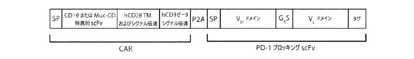
本発明の別の態様において、本開示は、組換え抗PD−1抗原結合性タンパク質をコードする核酸およびキメラ抗原受容体をコードする核酸を含むベクターに関し、ここで、前記組換え抗PD−1抗原結合性タンパク質は、前記キメラ抗原受容体と同一ではない。 In another aspect of the invention, the disclosure relates to a vector comprising a nucleic acid encoding a recombinant anti-PD-1 antigen binding protein and a nucleic acid encoding a chimeric antigen receptor, wherein the recombinant anti-PD-1 is described herein. The antigen-binding protein is not the same as the chimeric antigen receptor.
さらに別の態様において、本開示は、本明細書に記載するベクターを含む細胞に関する。関連する態様において、本開示は、組換え抗PD−1抗原結合性タンパク質をコードする核酸およびキメラ抗原受容体をコードする核酸を含む細胞に関し、ここで、前記組換え抗PD−1抗原結合性タンパク質は、前記キメラ抗原受容体と同一ではない。別の関連する態様において、本開示は、組換え抗PD−1抗原結合性タンパク質およびキメラ抗原受容体を含む細胞に関し、ここで、前記組換え抗PD−1抗原結合性タンパク質は、前記キメラ抗原受容体と同一ではない。 In yet another embodiment, the present disclosure relates to cells comprising the vectors described herein. In a related embodiment, the present disclosure relates to cells comprising a nucleic acid encoding a recombinant anti-PD-1 antigen binding protein and a nucleic acid encoding a chimeric antigen receptor, wherein the recombinant anti-PD-1 antigen binding property is described herein. The protein is not identical to the chimeric antigen receptor. In another related embodiment, the present disclosure relates to cells comprising a recombinant anti-PD-1 antigen binding protein and a chimeric antigen receptor, wherein the recombinant anti-PD-1 antigen binding protein is the chimeric antigen. Not identical to the receptor.
本明細書に記載するベクターまたは細胞のある実施形態において、キメラ抗原受容体はPD−1に特異的に結合するものではない。 In certain embodiments of the vectors or cells described herein, the chimeric antigen receptor is not specifically bound to PD-1.
本発明の別の態様において、本開示は、対象に治療上有効量の、本明細書に記載する組換え抗PD−1抗原結合性タンパク質、ベクター、細胞、または医薬組成物を投与することを含む、対象におけるT細胞応答を上昇させる方法を提供し、ここで、組換え抗PD−1抗原結合性タンパク質はPD−1アンタゴニストである。 In another aspect of the invention, the disclosure comprises administering to a subject a therapeutically effective amount of a recombinant anti-PD-1 antigen binding protein, vector, cell, or pharmaceutical composition described herein. Provided, including, a method of increasing T cell response in a subject, wherein the recombinant anti-PD-1 antigen binding protein is a PD-1 antagonist.
本発明のさらに別の態様において、本開示は、対象に治療上有効量の、本明細書に記載する組換え抗PD−1抗原結合性タンパク質、ベクター、細胞、または医薬組成物を投与することを含む、対象におけるT細胞応答を低下させる方法を提供し、ここで、組換え抗PD−1抗原結合性タンパク質はPD−1アゴニストである。 In yet another aspect of the invention, the present disclosure comprises administering to a subject a therapeutically effective amount of a recombinant anti-PD-1 antigen binding protein, vector, cell, or pharmaceutical composition described herein. Provided is a method of reducing a T cell response in a subject, wherein the recombinant anti-PD-1 antigen binding protein is a PD-1 agonist.
本発明の別の態様において、本開示は、対象に、治療上有効量の本明細書に記載する組換え抗PD−1抗原結合性タンパク質、ベクター、細胞、または医薬組成物を投与することを含む、PD1−陽性疾患を有する対象を治療する方法を提供する。 In another aspect of the invention, the disclosure comprises administering to a subject a therapeutically effective amount of a recombinant anti-PD-1 antigen binding protein, vector, cell, or pharmaceutical composition described herein. Provided are methods of treating subjects with PD1-positive diseases, including.
関連する態様において、本開示は、対象の少なくとも1つのT細胞に、組換え抗PD−1抗原結合性タンパク質をコードする核酸およびキメラ抗原受容体をコードする核酸を形質導入することを含む、PD1−陽性疾患を有する対象を治療する方法を提供し、ここで、前記組換え抗PD−1抗原結合性タンパク質は、前記キメラ抗原受容体と同一ではない。 In a related embodiment, the disclosure comprises transfecting at least one T cell of interest with a nucleic acid encoding a recombinant anti-PD-1 antigen binding protein and a nucleic acid encoding a chimeric antigen receptor. -Providing a method for treating a subject having a positive disease, wherein the recombinant anti-PD-1 antigen binding protein is not identical to the chimeric antigen receptor.
本明細書に記載する方法のある実施形態において、キメラ抗原受容体は、PD−1に特異的に結合するものではない。ある実施形態において、PD1−陽性疾患は癌である。ある実施形態において、PD1−陽性疾患は自己免疫疾患である。 In certain embodiments of the methods described herein, the chimeric antigen receptor is not specifically bound to PD-1. In certain embodiments, the PD1-positive disease is cancer. In certain embodiments, the PD1-positive disease is an autoimmune disease.
本明細書において言及するすべての刊行物、特許およびその他の引用文献は、それら全体が本開示に引用により組み込まれる。 All publications, patents and other references referred to herein are incorporated herein by reference in their entirety.
本開示を実施するにあたり、分子生物学、微生物学、細胞生物学、生化学および免疫学の多くの従来技術が使用され、それらは当業者の技術範囲内である。これらの技術は、より詳細に、例えば、Molecular Cloning:a Laboratory Manual 3rd edition,J.F.SambrookおよびD.W.Russell,ed.Cold Spring Harbor Laboratory Press 2001;Recombinant Antibodies for Immunotherapy,Melvyn Little,ed.Cambridge University Press 2009;「Oligonucleotide Synthesis」(M.J.Gait,ed.,1984);「Animal Cell Culture」(R.I.Freshney,ed.,1987);「Methods in Enzymology」(Academic Press,Inc.);「Current Protocols in Molecular Biology」(F.M.Ausubel et al,eds.,1987,および定期的アップデート);「PCR:The Polymerase Chain Reaction」,(Mullis et al,ed.,1994);「A Practical Guide to Molecular Cloning」(Perbal Bernard V.,1988);「Phage Display:A Laboratory Manual」(Barbas et al,2001)において記載される。製造業者らの指示書を含む、当業者に広く知られ、信頼されている標準的プロトコールを含むこれらの引用文献およびその他の引用文献の内容は、本開示の一部として引用により本明細書に組み込まれる。 Many prior art techniques of molecular biology, microbiology, cell biology, biochemistry and immunology have been used in carrying out this disclosure and are within the skill of one of ordinary skill in the art. These techniques are described in more detail, for example, in Molecular Cloning: a Laboratory Manual 3rd edition, J. Mol. F. Sambrook and D.M. W. Russell, ed. Cold Spring Harbor Laboratory Press 2001; Recombinant Antibodies for Immunotherapy, Melvyn Little, ed. Cambridge University Press 2009; "oligonucleotide Synthesis" (MJ Gait, ed., 1984); "Animal Cell Culture" (R.I. ); "Cell Protects in Molecular Biology" (FM Ausube et al, eds., 1987, and regular updates); "PCR: The Polymerase Chain Reaction", (Mullis et al, 19). Described in "A Practical Guide to Molecular Cloning" (Perbal Bernard V., 1988); "Phage Display: A Laboratory Manual" (Barbas et al, 2001). The contents of these and other citations, including standard protocols widely known and trusted by those of skill in the art, including manufacturer's instructions, are hereby incorporated by reference as part of this disclosure. Be incorporated.
以下の記載において、用語の使用に関してはいくつかの慣習にしたがう。一般に、本明細書に使用される用語は、当業者に知られているその用語の意味と一致するよう解釈されることが意図される。 In the following description, we will follow some conventions regarding the use of terms. In general, the terms used herein are intended to be construed to be consistent with the meanings of those terms known to those of skill in the art.
「抗原結合性タンパク質」は、抗原結合領域または抗原結合部分を含むタンパク質またはポリペプチド、即ち、それが結合するその他の分子に対して強い親和性を有するタンパク質またはポリペプチドである。抗原結合性タンパク質は、抗体、キメラ抗原受容体および融合タンパク質を包含する。 An "antigen-binding protein" is a protein or polypeptide that contains an antigen-binding region or antigen-binding moiety, i.e., a protein or polypeptide that has a strong affinity for other molecules to which it binds. Antigen-binding proteins include antibodies, chimeric antigen receptors and fusion proteins.
「抗体」および「複数の抗体」はそれらの用語が当該技術分野において知られている通り、免疫系の抗原結合性タンパク質のことをいう。本明細書において言及される「抗体」という用語は、全体の、全長抗体および「抗原結合部分」または「抗原結合領域」が保持されているその任意の断片、またはその単鎖を含む。天然の「抗体」は、ジスルフィド結合により相互接続された少なくとも2つの重(H)鎖および2つの軽(L)鎖を含む糖タンパク質である。各重鎖は、重鎖可変領域(本明細書において、VHと略称する)および重鎖定常(CH)領域から構成される。重鎖定常領域は、3つのドメイン、CH1、CH2およびCH3から構成される。各軽鎖は、軽鎖可変領域(本明細書において、VLと略称する)および軽鎖定常CL領域から構成される。軽鎖定常領域は1つのドメイン、CLから構成される。VHおよびVL領域はさらに、より保存された、フレームワーク領域(FR)と称される領域により分散された、相補性決定領域(CDR)と称される超可変性の領域に細分することができる。各VHおよびVLは、3つのCDRおよび4つのFRから構成され、これらはアミノ末端からカルボキシ末端の順に以下の順序で配列される:FR1、CDR1、FR2、CDR2、FR3、CDR3、FR4。重および軽鎖の可変領域は、抗原と相互作用する結合ドメインを含む。抗体の定常領域は、免疫系の様々な細胞(例えば、エフェクター細胞)および古典的補体系の第1成分(C1q)を含む宿主組織または因子への免疫グロブリンの結合を媒介することができる。 "Antibodies" and "multiple antibodies" refer to antigen-binding proteins of the immune system, as those terms are known in the art. The term "antibody" referred to herein includes a whole, full-length antibody and any fragment thereof, or a single chain thereof, that retains an "antigen binding moiety" or "antigen binding region". A natural "antibody" is a glycoprotein containing at least two heavy (H) chains and two light (L) chains interconnected by disulfide bonds. Each heavy chain is composed of a heavy chain variable region (abbreviated as VH in the present specification) and a heavy chain stationary (CH) region. The heavy chain constant region is composed of three domains, CH1, CH2 and CH3. Each light chain is composed of a light chain variable region (abbreviated herein as VL) and a light chain constant CL region. The light chain constant region is composed of one domain, CL. The VH and VL regions can be further subdivided into more conserved regions of hypervariability called complementarity determining regions (CDRs) dispersed by regions called framework regions (FRs). .. Each VH and VL is composed of 3 CDRs and 4 FRs, which are arranged in the following order from amino terminus to carboxy terminus: FR1, CDR1, FR2, CDR2, FR3, CDR3, FR4. The variable regions of the heavy and light chains contain binding domains that interact with the antigen. The constant region of the antibody can mediate the binding of immunoglobulins to host tissues or factors, including various cells of the immune system (eg, effector cells) and the first component of the classical complement system (C1q).
抗体の「抗原結合部分」または「抗原結合領域」という用語は、本明細書において用いる場合、抗原特異性を付与する抗体の領域または部分をいい;抗原結合性タンパク質の断片、例えば、抗体は、それゆえ抗原(例えば、HLA−ペプチド複合体)に特異的に結合する能力を保持する抗体の1以上の断片を含む。抗体の抗原結合機能は、全長抗体の断片によって実行され得ることが示されている。抗体の「抗体断片」という用語に包含される抗原結合性断片の例としては、Fab断片、VL、VH、CLおよびCH1ドメインからなる一価断片;F(ab)2断片、ヒンジ領域にてジスルフィド結合によって連結された2つのFab断片を含む二価断片;VHおよびCH1ドメインからなるFd断片;抗体の単腕のVLおよびVHドメインからなるFv断片;VHドメインからなるFab断片(Ward et al,1989 Nature 341:544〜546);および単離相補性決定領域(CDR)が挙げられる。 The term "antigen-binding portion" or "antigen-binding region" as used herein refers to a region or portion of an antibody that imparts antigen specificity; a fragment of an antigen-binding protein, eg, an antibody. It therefore comprises one or more fragments of an antibody that retains the ability to specifically bind an antigen (eg, an HLA-peptide complex). It has been shown that the antigen binding function of an antibody can be performed by a fragment of a full-length antibody. Examples of antigen-binding fragments included in the term "antibody fragment" of an antibody are Fab fragments, monovalent fragments consisting of VL, VH, CL and CH1 domains; F (ab) 2 fragments, disulfides at the hinge regions. A bivalent fragment containing two Fab fragments linked by binding; an Fd fragment consisting of VH and CH1 domains; an Fv fragment consisting of VL and VH domains of an antibody single arm; a Fab fragment consisting of VH domains (Ward et al, 1989). Nature 341: 544-546); and isolated complementarity determining regions (CDRs).
さらに、Fv断片の2つのドメインである、VLおよびVHは、別々の遺伝子にコードされているが、それらは、それらをVLおよびVH領域が対となって一価分子を形成している一本のタンパク質鎖として作ることを可能とする合成リンカーによって、組換え方法を用いて連結することができる。これらは単鎖Fv(scFv)として知られている;例えば、Bird et al,1988 Science 242:423〜426;およびHuston et al,1988 Proc.Natl.Acad.Sci.85:5879〜5883を参照されたい。かかる単鎖抗体もまた抗体の「抗原結合部分」という用語の範囲内に含まれる意図である。これらの抗体断片は当業者に知られた常套技術を用いて得られ、これら断片はインタクトな抗体と同様にして有用性についてスクリーニングされる。 Furthermore, the two domains of the Fv fragment, VL and VH, are encoded by separate genes, which are paired with the VL and VH regions to form a monovalent molecule. It can be linked using a recombinant method by a synthetic linker that allows it to be made as a protein chain of. These are known as single chain Fv (scFv); for example, Bird et al, 1988 Science 242: 423-426; and Huston et al, 1988 Proc. Natl. Acad. Sci. 85: 5879-5883. Such single chain antibodies are also intended to be included within the term "antigen binding portion" of the antibody. These antibody fragments are obtained using conventional techniques known to those of skill in the art, and these fragments are screened for usefulness in the same manner as intact antibodies.
「組換え抗体」または「組換え抗原結合性タンパク質」は、組換えにより作成された抗原結合性タンパク質、例えば、抗体への包含について、結合特性に基づいて同定され、選択された抗原結合部分を有するものである。 A "recombinant antibody" or "recombinant antigen-binding protein" refers to an antigen-binding protein produced by recombination, for example, an antigen-binding moiety identified and selected based on binding properties for inclusion in an antibody. It has.
「その相同配列」という用語は、表1〜14に示される配列に対して60〜99.9%同一であるアミノ酸およびヌクレオチド配列をいう。ある実施形態において、相同配列は、少なくとも60%、65%、70%、75%、80%、85%、90%、91%、92%、93%、94%、95%、96%、97%、98%、99%、99.5%、または99.9%の同一性を有する。1つの実施形態において、相同配列は95〜99.9%の同一性を有し;別の実施形態において、相同配列は98〜99.9%の同一性を有する。 The term "the homologous sequence" refers to an amino acid and nucleotide sequence that is 60-99.9% identical to the sequences shown in Tables 1-14. In certain embodiments, the homologous sequences are at least 60%, 65%, 70%, 75%, 80%, 85%, 90%, 91%, 92%, 93%, 94%, 95%, 96%, 97. %, 98%, 99%, 99.5%, or 99.9% identity. In one embodiment, the homologous sequence has 95-99.9% identity; in another embodiment, the homologous sequence has 98-99.9% identity.
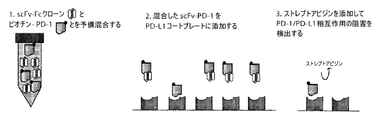
1つの実施形態において、ヒトPD−1に特異的に結合する単鎖可変断片(scFv)を選択し、試験した。scFvは、独占の(proprietary)完全にヒト抗体scFvファージライブラリー(Eureka Therapeutics、Emeryville CA)であるファージディスプレーライブラリーから単離した。このライブラリーは、100名を超える白人およびアジア人の健康ドナー、および自己免疫疾患、例えば全身性エリテマトーデス、強皮症等を有するドナーからのヒト抗体レパートリーから構成される。 In one embodiment, single chain variable fragments (scFv) that specifically bind to human PD-1 were selected and tested. scFv was isolated from a phage display library, which is a proprietary, fully human antibody scFv phage library (Eureka Therapeutics, Emeryville CA). This library consists of a repertoire of human antibodies from over 100 healthy Caucasian and Asian donors and donors with autoimmune diseases such as systemic lupus erythematosus, scleroderma and the like.
抗体ファージパニングに使用した抗原は、組換え融合タンパク質である、ヒトIgG1
Fcに融合させたPD−1細胞外ドメイン(PD−1 ECD−Fcドメイン)であった。PD−1 ECDおよびhIgG1 FcをコードするDNA配列は、Genewiz,Inc.(South Plainfield,NJ)により合成された。DNA配列を次いでEurekaの所有する哺乳類発現ベクターにサブクローニングし、これを次いで融合タンパク質発現のためにHEK293細胞にトランスフェクトした。PD−1 ECD−Fc融合タンパク質を細胞が死滅した後にHEK293細胞培地から標準的FPLC方法によって精製した。
The antigen used for antibody phage panning was human IgG1, a recombinant fusion protein.
It was a PD-1 extracellular domain (PD-1 ECD-Fc domain ) fused to Fc. The DNA sequences encoding PD-1 ECD and hIgG1 Fc are described in Genewiz, Inc. It was synthesized by (South Plainfield, NJ). The DNA sequence was then subcloned into Eureka's own mammalian expression vector, which was then transfected into HEK293 cells for fusion protein expression. The PD-1 ECD-Fc fusion protein was purified from HEK293 cell medium by standard FPLC methods after cell death.
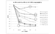
ヒトscFv抗体ファージディスプレーライブラリーをmAbクローンの選択のために使用する。簡単に説明すると、ビオチン化抗原(PD−1 ECD−Fc融合タンパク質)をまずヒトscFvファージライブラリーと混合することができ、次いで抗原−scFv抗体複合体を、ストレプトアビジン−接合Dynabeads M−280により磁気ラックを介してプルダウンすることができる。結合したクローンを次いで溶出し、大腸菌XL1−Blueに感染させるのに使用することができる。細菌中で発現したscFvファージクローンを精製することができる(Yasmina NA,et al.Probing the binding mechanism and affinity of tanezumab,a recombinant humanized anti−NGF monoclonal antibody,using a repertoire of biosensors.Protein Science 2008;17(8):1326〜1335;Roberts WK,et al.Vaccination with CD20 peptides induces a biologically active,specific immune response in mice.Blood 2002:99(10):3748〜3755)。パニングを3〜4サイクル行って、PD−1に特異的に結合するscFvファージクローンを濃縮することができる。陽性クローンは、ビオチン化単鎖PD−1に対する標準的ELISA方法によって決定することができる。陽性クローンは、生細胞表面上のPD−1へのそれらの結合について、PD−1+細胞株、例えば、3T3細胞株を用いてフローサイトメトリーによってさらに試験することができる。 The human scFv antibody phage display library is used for selection of mAb clones. Briefly, the biotinylated antigen (PD-1 ECD-Fc fusion protein) can first be mixed with the human scFv phage library, and then the antigen-scFv antibody complex is combined with streptavidin-conjugated Dynabeads M-280. It can be pulled down via a magnetic rack. The bound clones can then be eluted and used to infect E. coli XL1-Blue. ScFv phage clones expressed in bacteria can be purified (Yasmina NA, et al. Probing the binding mechanism and affinity of tanezumab, a recombinant humanized antibody, a recombinant humanized antibody. (8): 1326-1335; Roberts WK, et al. Vaccination with CD20 peptides induces a bacterially active, special immune response in microphone. Panning can be performed for 3-4 cycles to concentrate scFv phage clones that specifically bind to PD-1. Positive clones can be determined by standard ELISA methods for biotinylated single chain PD-1. Positive clones can be further tested by flow cytometry using a PD-1 + cell line, eg, a 3T3 cell line, for their binding to PD-1 on the surface of living cells.
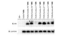
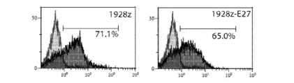
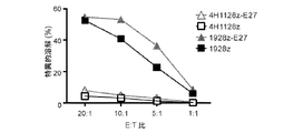
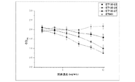
本開示により包含されるいくつかのクローンを、本明細書においてクローン14、16、18、19、23、26、27、31、36、37、40、42、46、および47と称する。これらの実施形態をコードする可変軽(VL)および可変重(VH)鎖アミノ酸配列およびヌクレオチド配列を以下の表1〜14に示す。ある実施形態において、VLおよびVH配列をセリングリシンリンカーによって連結してscFvを形成した。ある実施形態において、HA/Hisタグを、scFvの検出を可能とするために含めることができる。
Several clones included herein are referred to herein as
ある実施形態において、本開示は、二価タンパク質が生じるようにヒト免疫グロブリンのFc領域を有する抗体を形成するよう、重鎖の1以上の定常ドメインと融合させたscFv配列を有する抗体を含み、これにより、抗体の全体の結合活性(avididty)および安定性が上昇する。さらに、例えば、抗原定量研究における使用のために、親和性測定のために抗体を固定化するために、治療薬の標的化送達のために、免疫エフェクター細胞を用いてFc−媒介細胞毒性を試験するために、およびその他の多くの用途のために、Fc部分は、これらに限定されないが、蛍光色素、細胞毒素、放射性同位元素等が含まれる他の分子の抗体との直接の接合を可能とする。 In certain embodiments, the present disclosure comprises an antibody having an scFv sequence fused to one or more constant domains of a heavy chain to form an antibody having the Fc region of a human immunoglobulin to give rise to a divalent protein. This increases the overall avidty and stability of the antibody. In addition, Fc-mediated cytotoxicity is tested using immune effector cells for targeted delivery of therapeutic agents, eg, for use in antigen quantification studies, to immobilize antibodies for affinity measurements. And for many other uses, the Fc moiety allows for direct conjugation with antibodies of other molecules including, but not limited to, fluorescent dyes, cytotoxins, radioactive isotopes, etc. do.
ある実施形態において、抗PD−1抗原結合性タンパク質は、タンパク質安定性、抗体結合、発現レベルを改良するよう、または治療薬の接合のための部位を導入するよう、設計された1以上のフレームワーク領域アミノ酸置換を含み得る。これらのscFvは次いで、当業者に知られた方法にしたがって組換えヒトモノクローナルIgを産生するために使用される。 In certain embodiments, the anti-PD-1 antigen-binding protein has one or more frames designed to improve protein stability, antibody binding, expression levels, or to introduce sites for conjugation of therapeutic agents. Work region may include amino acid substitutions. These scFvs are then used to produce recombinant human monoclonal Ig according to methods known to those of skill in the art.
ある実施形態において、抗原結合性タンパク質は、キメラ抗原受容体(CAR)である。キメラ抗原受容体療法(CAR−T療法)は、標的化免疫療法の新しい形態である。それは、モノクローナル抗体の精巧な標的化特異性と、細胞傷害性T細胞によって提供される強力な細胞傷害性および長期間持続性とを組み合わせたものである。この技術は、T細胞が内因性TCRとは独立した長期間新規抗原特異性を獲得することを可能とする。臨床試験により、神経芽細胞腫(Louis C.U.et al,Blood 118(23):6050〜6056)、B−ALL(Maude S.L.et al,N.Engl.J.Med.371(16):1507〜1517,2014)、CLL(Brentjens R.J.et al,Blood 118(18):4817〜4828,2011)およびB細胞リンパ腫(Kochenderfer J.N.et al,Blood.116(20):4099〜4102,2010)においてCAR−T療法の臨床的に有意な抗腫瘍活性が示されている。1つの研究において、CD19−CAR T療法で治療したB−ALLを有する30名の患者における90%の完全寛解率が報告された(Maude S.L.et al,前掲)。 In certain embodiments, the antigen binding protein is a chimeric antigen receptor (CAR). Chimeric antigen receptor therapy (CAR-T therapy) is a new form of targeted immunotherapy. It combines the elaborate targeting specificity of monoclonal antibodies with the potent cytotoxicity and long-term persistence provided by cytotoxic T cells. This technique allows T cells to acquire long-term novel antigen specificity independent of endogenous TCR. According to clinical trials, neuroblastoma (Louis C. U. et al, Blood 118 (23): 6050-6056), B-ALL (Maude S. L. et al, N. Engl. J. Med. 371) ( 16): 1507 to 1517, 2014), CLL (Brentgens R.J. et al, Blood 118 (18): 4817 to 4828, 2011) and B-cell lymphoma (Kochenderfer J.N. et al, Blood.116 (20). ): Clinically significant antitumor activity of CAR-T therapy has been shown in 4099-4102, 2010). In one study, a 90% complete remission rate was reported in 30 patients with B-ALL treated with CD19-CART therapy (Maude S. L. et al, supra).
ある実施形態において、キメラ抗原受容体は、抗体部分を含む細胞外ドメイン、膜貫通ドメイン、および細胞内シグナル伝達ドメインを含む。ある実施形態において、細胞内シグナル伝達ドメインは、CD3ζ細胞内シグナル伝達配列および同時刺激シグナル伝達配列を含む。ある実施形態において、同時刺激シグナル伝達配列は、CD28細胞内シグナル伝達配列である。 In certain embodiments, the chimeric antigen receptor comprises an extracellular domain containing an antibody moiety, a transmembrane domain, and an intracellular signaling domain. In certain embodiments, the intracellular signaling domain comprises a CD3ζ intracellular signaling sequence and a co-stimulation signaling sequence. In certain embodiments, the co-stimulation signaling sequence is a CD28 intracellular signaling sequence.
本開示のその他の態様には、限定されないが、PD1関連疾患の治療のため、診断および予後判定用途ならびに細胞および組織におけるPD1の検出のためのリサーチツールとしての使用のための、抗原結合性タンパク質およびそれらをコードする核酸の使用が含まれる。開示された抗原結合性タンパク質および核酸を含む医薬組成物が本開示に包含される。ベクター化免疫療法による抗体に基づく治療のための本開示の核酸を含むベクターもまた、本開示により意図される。ベクターは、抗体の発現および分泌を可能とする発現ベクター、ならびにキメラ抗原受容体(CAR)などの抗原結合性タンパク質の細胞表面発現に向けられたベクターを含む。 Other aspects of the disclosure include, but are not limited to, antigen-binding proteins for the treatment of PD1-related diseases, diagnostic and prognostic uses, and as research tools for the detection of PD1 in cells and tissues. And the use of nucleic acids encoding them. Pharmaceutical compositions comprising the disclosed antigen-binding proteins and nucleic acids are included in the present disclosure. Vectors containing the nucleic acids of the present disclosure for antibody-based therapy with vectorized immunotherapy are also contemplated by the present disclosure. Vectors include expression vectors that allow expression and secretion of antibodies, as well as vectors directed to cell surface expression of antigen-binding proteins such as chimeric antigen receptors (CARs).
核酸を含む細胞、例えば、本開示のベクターによりトランスフェクトされた細胞もまた本開示に包含される。 Cells containing nucleic acids, such as cells transfected with the vectors of the present disclosure, are also included in the present disclosure.
診断および研究用途における使用のための、本開示のPD1抗体または核酸、アッセイ試薬、バッファーなどを含有するキットもまた提供される。

抗原結合性タンパク質が全長抗体である1つの実施形態において、本開示の抗体の重および軽鎖は全長であってもよく(例えば、抗体は、少なくとも1つ、好ましくは2つの、完全な重鎖、および少なくとも1つ、好ましくは2つの、完全な軽鎖を含み得る)または抗原結合部分(Fab、F(ab’)2、Fvまたは単鎖Fv断片(「scFv」))を含むものであってもよい。別の実施形態において、抗体重鎖定常領域は、例えば、IgG1、IgG2、IgG3、IgG4、IgM、IgA1、IgA2、IgD、およびIgEから選択される。ある実施形態において、免疫グロブリンアイソタイプは、IgG1、IgG2、IgG3、およびIgG4から選択され、より具体的には、IgG1(例えば、ヒトIgG1)である。抗体タイプの選択は、抗体が誘発するよう設計された免疫エフェクター機能に依存するであろう。 In one embodiment where the antigen-binding protein is a full-length antibody, the heavy and light chains of the antibodies of the present disclosure may be full-length (eg, the antibody is at least one, preferably two, complete heavy chains. , And at least one, preferably two, which may include a complete light chain) or an antigen binding moiety (Fab, F (ab') 2 , Fv or single chain Fv fragment (“scFv”)). You may. In another embodiment, the antibody heavy chain constant region is selected from, for example, IgG1, IgG2, IgG3, IgG4, IgM, IgA1, IgA2, IgD, and IgE. In certain embodiments, the immunoglobulin isotype is selected from IgG1, IgG2, IgG3, and IgG4, more specifically IgG1 (eg, human IgG1). The choice of antibody type will depend on the immune effector function designed to elicit the antibody.
組換え免疫グロブリンの構築において、様々な免疫グロブリンアイソタイプの定常領域についての適当なアミノ酸配列および広範な抗体を産生する方法は当業者に知られている。 Methods of producing suitable amino acid sequences and a wide range of antibodies for constant regions of various immunoglobulin isotypes in the construction of recombinant immunoglobulins are known to those of skill in the art.
本明細書において同定された抗原結合性タンパク質をコードする核酸は、組換え免疫エフェクター細胞を操作するために利用することができる。遺伝子操作されたT−細胞を作成するための方法およびベクターは、例えば、当該技術分野において知られている(Brentjens et al,Safety and persistence of adoptively transferred autologous CD19−targeted T cells in patients with relapsed or chemotherapy refractory B−cell leukemias,Blood 118(18):4817〜4828,2011年11月を参照されたい)。 Nucleic acids encoding antigen-binding proteins identified herein can be utilized to engineer recombinant immunoeffector cells. Methods and vectors for producing genetically engineered T-cells are, for example, known in the art (Brentgens et al, Safety and persistence of assistivery transferred autotransplantation) Refractory B-cell leukemias, Blood 118 (18): 4817-4828, see November 2011).
本開示のその他の実施形態には、本開示の抗原結合性タンパク質またはそれらの抗原結合性断片をコードする核酸を含む細胞および発現ベクターが含まれる。細胞は、本開示にしたがって抗原結合領域を含むキメラ抗原受容体を発現するよう遺伝子操作されたT−細胞などの組換え免疫エフェクター細胞であり得る。本開示にしたがって抗体を産生するよう操作された細胞もまた本開示に包含される。 Other embodiments of the present disclosure include cells and expression vectors comprising nucleic acids encoding the antigen-binding proteins of the present disclosure or antigen-binding fragments thereof. The cell can be a recombinant immune effector cell such as a T-cell genetically engineered to express a chimeric antigen receptor comprising an antigen binding region according to the present disclosure. Cells engineered to produce antibodies in accordance with the present disclosure are also included in the present disclosure.
さらに、本開示は、以下を含む本開示の抗体または抗体断片の産生方法を含む:(a)核酸配列が発現され、それにより軽および重鎖可変領域を含むポリペプチドが産生される条件下で培地中で本開示の抗体または抗体断片をコードする核酸を含む組換え細胞を培養すること;および(b)宿主細胞または培地からポリペプチドを回収すること。 In addition, the disclosure includes methods of producing the antibodies or antibody fragments of the present disclosure, including: (a) under conditions in which a nucleic acid sequence is expressed, thereby producing a polypeptide containing light and heavy chain variable regions. Culturing recombinant cells containing nucleic acids encoding the antibodies or antibody fragments of the present disclosure in medium; and (b) recovering polypeptides from host cells or media.
本開示の抗原結合性タンパク質のいくつかの実施形態は、アンタゴニスト性抗PD1抗体ならびにPD1のアゴニストとして機能する抗PD1抗体を包含する。いくつかの抗PD1抗体はアンタゴニストであり、即ち、それはそのリガンドによるPD1の結合をブロックするが、他の抗PD1抗体はアゴニスト、即ち、PD−1の免疫抑制シグナルを増強する効果を有し、例えば、自己免疫の治療に有用なものとする抗体である。アンタゴニスト活性を示す本開示の抗体にはクローン23および27が含まれるが、クローン16、18、26、31および40はアゴニストとして機能するようである。
Some embodiments of the antigen-binding proteins of the present disclosure include antagonistic anti-PD1 antibodies as well as anti-PD1 antibodies that act as agonists of PD1. Some anti-PD1 antibodies are antagonists, i.e. it blocks the binding of PD1 by its ligand, while other anti-PD1 antibodies have the effect of enhancing the agonist, i.e., the immunosuppressive signal of PD-1. For example, antibodies that make it useful in the treatment of autoimmunity. The antibodies of the present disclosure exhibiting antagonist activity include
本開示はまた、本開示の抗PD−1抗体または抗体断片の免疫応答を上昇させる医薬の調製のための使用ならびに本開示の抗PD−1抗体または抗体断片の癌を治療する医薬の調製のための使用も含む。 The present disclosure also includes the use for the preparation of a pharmaceutical that enhances the immune response of the anti-PD-1 antibody or antibody fragment of the present disclosure and the preparation of a pharmaceutical that treats cancer of the anti-PD-1 antibody or antibody fragment of the present disclosure. Also includes use for.
本明細書に開示する抗原結合性タンパク質、抗体、核酸、ベクターまたは核酸または抗原結合性タンパク質を含む細胞を、医薬上許容され得る担体とともに含む医薬組成物もまた本開示に含まれる。 Also included in the present disclosure are pharmaceutical compositions comprising an antigen-binding protein, antibody, nucleic acid, vector or cell comprising the nucleic acid or antigen-binding protein disclosed herein with a pharmaceutically acceptable carrier.
本開示は、本開示の抗PD−1抗体または抗体断片のワクチンアジュバントとしての使用もまた含む。 The present disclosure also includes the use of the anti-PD-1 antibody or antibody fragment of the present disclosure as a vaccine adjuvant.
別の態様において、本開示は、本明細書に開示する抗原結合性タンパク質、抗体またはその抗原結合性断片である第1成分を含む免疫接合体に関する。免疫接合体は、細胞毒素、検出可能標識、放射性同位元素、治療薬、第2のアミノ酸配列を有する結合性タンパク質または分子である第2成分を含む。第2成分が結合性タンパク質または第2抗体である場合、結合性タンパク質または第2抗体は、第1成分が特異的であるHLA−ペプチド複合体とは異なる標的に対する結合特異性を有する。 In another aspect, the present disclosure relates to an immunoconjugate comprising an antigen-binding protein, an antibody or a first component thereof, which is an antigen-binding fragment thereof, disclosed herein. The immunoconjugate comprises a cytotoxin, a detectable label, a radioisotope, a therapeutic agent, a second component that is a binding protein or molecule having a second amino acid sequence. When the second component is a binding protein or second antibody, the binding protein or second antibody has binding specificity for a different target than the HLA-peptide complex in which the first component is specific.
本開示はまた、対象に、治療上有効量の、本明細書に開示する抗原結合性タンパク質、抗体またはその抗原結合性断片、キメラ抗原受容体(CAR)、抗原結合性タンパク質をコードする核酸または核酸もしくはタンパク質を含む細胞を投与することを含む、PD−1関連疾患を有する対象の治療方法に関する。 The present disclosure also presents to the subject a therapeutically effective amount of an antigen-binding protein, antibody or antigen-binding fragment thereof, chimeric antigen receptor (CAR), nucleic acid encoding an antigen-binding protein or The present invention relates to a method for treating a subject having a PD-1-related disease, which comprises administering a cell containing a nucleic acid or a protein.
当業者であれば、いくつかの実施形態が本発明の範囲と精神の枠内で可能であることを認識するであろう。ここで、本発明を以下の非限定的な実施例に言及することにより、より詳細に記載する。以下の実施例は本発明をさらに例証するが、もちろん、その範囲をなんら限定するものと解釈すべきではない。 Those skilled in the art will recognize that some embodiments are possible within the scope and spirit of the invention. Here, the present invention will be described in more detail by reference to the following non-limiting examples. The following examples further illustrate the invention, but of course should not be construed as limiting its scope in any way.
実施例1
scFvがPD−1連結を阻害する能力を試験するために、scFv−Fcドメイン(scFv−Fc)融合タンパク質を作成し、ここで、scFvをマウスFc(マウスIgG1a)ドメインに連結させた。scFv−Fcクローンを次いで、ELISAプレートをヒトPD−1単量体でコーティングすることにより、PD−1に結合する能力について分析した。scFvのPD−1への結合をHRP−接合抗マウスIgG1 Fc二次抗体を用いて定量した。7つのすべての抗体は、用量依存的にPD1単量体に関して結合活性を示した(図1)。ET901 ScFv−Fc(マウスIgG1 Fc)を陰性対照として用いた。scFv−Fcクローンの結合親和性を順位付けしたところ、クローン31が弱く結合し、クローン26および27が最も強く結合した(31<23<40<18<16=27<26)。
Example 1
To test the ability of scFv to inhibit PD-1 ligation, a scFv-Fc domain (scFv-Fc) fusion protein was created, where scFv was ligated to the mouse Fc (mouse IgG1a) domain. The scFv-Fc clones were then analyzed for their ability to bind PD-1 by coating the ELISA plate with human PD-1 monomers. Binding of scFv to PD-1 was quantified using an HRP-conjugated anti-mouse IgG1 Fc secondary antibody. All seven antibodies showed binding activity with respect to PD1 monomers in a dose-dependent manner (FIG. 1). ET901 ScFv-Fc (mouse IgG1 Fc) was used as a negative control. When the binding affinities of the scFv-Fc clones were ranked, clone 31 bound weakly and
実施例2
PD−L1と相互作用するPD−1を阻害するscFv−Fcの能力を試験するために、模式的に図2Aに示す競合的リガンド−結合アッセイを行った。このアッセイにおいて、PD−L1−FcをELISAプレート上にコーティングした。ビオチン化−PD1−Fcを段階希釈したET901 ScFv−Fc(陰性対照)または抗PD1 ScFv−Fcと混合し、次いでPD−L1−Fcコートプレート中に添加した。プレート上にコーティングされたPD−L1へのPD−1−Fcの結合をHRP−接合ストレプトアビジンを介して可視化した(図2B)。同様の濃度のscFv−Fcを比較した場合(丸で囲む)、PD−1−PDL1相互作用を破壊する能力を順位付けしたところ、クローン40および23が最も弱く、クローン26が最も強い破壊能力を有していた(26>27=16>18>31>23=40、図2B)。
Example 2
To test the ability of scFv-Fc to inhibit PD-1 that interacts with PD-L1, a competitive ligand-binding assay schematically shown in FIG. 2A was performed. In this assay, PD-L1-Fc was coated on an ELISA plate. Biotinylated-PD1-Fc was mixed with serially diluted ET901 ScFv-Fc (negative control) or anti-PD1 ScFv-Fc and then added into PD-L1-Fc coated plates. Binding of PD-1-Fc to PD-L1 coated on the plate was visualized via HRP-conjugated streptavidin (FIG. 2B). When comparing scFv-Fc of similar concentrations (circled), the ability to disrupt the PD-1-PDL1 interaction was ranked, with
実施例3
これらのクローンを次いで特異的T細胞機能を制御するそれらの能力について調べた。本発明者らは以前に、T細胞が腫瘍−特異的キメラ抗原受容体(CAR)を発現するようレトロウイルスにより形質導入されている、腫瘍−標的化T細胞を作成している。本発明者らは以前に、CARの発現が所与の抗原を標的化するT細胞機能を再指示したことを実証している(Brentjens,Santos et al,2007)。本発明者らの研究室において、本発明者らは、1928zと称するCD19に特異的なCARを用いてB細胞悪性腫瘍を標的とする(Brentjens,Santosら.2007)。本発明者らは以前に、CAR改変T細胞がインビトロおよびインビボおよび臨床研究において有意な抗腫瘍活性を示したことを実証している(Brentjens,Latoucheら.2003;Brentjens,Davilaら.2013;Davila,Riviereら.2014)。7つのscFvクローンが、アゴニスト性であるか(PD−1を刺激する)、アンタゴニスト性であるか(PD−1をブロックする)またはPD−1に対して有意な効果を有さないかを決定するために、本発明者らは、抗PD−1 scFv遺伝子の近位にマウスカッパリーダー配列を含めることにより分泌可能scFvを作成した(図3)。本発明者らは次いで、1928z CARの発現および形質導入されたヒト末梢血T細胞からの抗PD−1 scFvの分泌を誘導するために2シストロン性レトロウイルスベクターを作成した。
Example 3
These clones were then examined for their ability to regulate specific T cell function. We have previously created tumor-targeted T cells in which T cells have been transduced with a retrovirus to express a tumor-specific chimeric antigen receptor (CAR). We have previously demonstrated that expression of CAR redirected the T cell function of targeting a given antigen (Brentgens, Santos et al, 2007). In our laboratory, we target B-cell malignancies using a CD19-specific CAR called 1928z (Brentjens, Santos et al. 2007). We have previously demonstrated that CAR-modified T cells exhibited significant antitumor activity in in vitro and in vivo and clinical studies (Brentgens, Ratouche et al. 2003; Brentgens, Davila et al. 2013; Davila). , In vivo et al. 2014). Determining whether 7 scFv clones are agonistic (stimulate PD-1), antagonistic (block PD-1) or have no significant effect on PD-1 To this end, we created a secretible scFv by including a mouse kappa leader sequence proximal to the anti-PD-1 scFv gene (FIG. 3). We then created a bicistron retroviral vector to induce the expression of 1928z CAR and the secretion of anti-PD-1 scFv from transduced human peripheral blood T cells.
実施例4
PD−1刺激がT細胞の増殖および機能を阻害する能力があるので、本発明者らは引き続いてT細胞増殖に対する抗PD−1 scFvの効果を特徴決定する探索を行った。これを達成するために、ヒトT細胞を健康ドナーの末梢血から単離し、以前に記載されている方法を用いて、CARを発現し、PD−1−特異的scFvを分泌するようレトロウイルス形質導入を介して改変した(Brentjens、Santosら.2007)。形質導入の後、改変されたT細胞をインビトロでの増殖についてモニターした(図4)。1928z CARを発現し、PD−1特異的scFvクローン23および27を分泌するT細胞は、1928z CARのみを発現するよう改変されたT細胞と同様に増殖した。CARを発現し、PD−1−特異的scFvクローン16、18、26、31または40を分泌するよう改変された細胞は増殖せず、これらのPD−1−特異的scFvクローンがアゴニスト性であり、潜在的にPD−1を刺激し、そしてT細胞増殖の低下をもたらしたことが示唆された。
Example 4
Since PD-1 stimulation has the ability to inhibit T cell proliferation and function, we subsequently conducted a search to characterize the effect of anti-PD-1 scFv on T cell proliferation. To achieve this, human T cells are isolated from the peripheral blood of healthy donors and retroviral traits to express CAR and secrete PD-1-specific scFv using previously described methods. Modified through introduction (Brentgens, Santos et al. 2007). After transduction, modified T cells were monitored for in vitro proliferation (Fig. 4). Express 1928Z CAR, T cells which secrete PD-1-
実施例5
増殖研究に加えて、本発明者らは、(CARを発現する、またはCARを発現し、PD−1−特異的scFvを分泌する)T細胞を、CD19+バーキットリンパ腫腫瘍細胞株、Rajiと共培養し、T細胞からのサイトカイン分泌のレベルを決定した。図5に示されるように、1928z CARを発現し、PD−1−特異的scFvクローン23または27を分泌するよう改変されたT細胞は、CARのみを発現するよう改変された細胞と比較してIFN−γの分泌の上昇を示し、これらのscFvクローンがPD−1シグナル伝達に関してアンタゴニスト性であったことが示唆された。1928z CARを発現し、PD−1−特異的scFvクローン16、18、26、31または40を分泌するよう改変された細胞は、CARのみにより改変された細胞と比較してより少ないIFN−γを分泌し、これらのscFvがPD−1に関してアゴニスト性であり、T細胞機能を阻害したことが示唆された。
Example 5
In addition to proliferation studies, we have combined T cells (expressing CAR or expressing CAR and secreting PD-1-specific scFv) with CD19 + Berkit lymphoma tumor cell line, Raji. Co-cultured to determine the level of cytokine secretion from T cells. As shown in FIG. 5, T cells modified to express 1928z CAR and secrete PD-1-
実施例6
1928 CARおよびPD−1ブロッキングscFvを発現するよう改変されたヒトT細胞をフローサイトメトリーによって分析した。CARの発現の検証(図6)の後、scFvの存在を、細胞可溶化液における検出を可能とするようにゴルジ阻害剤で処理されたヒトT細胞から調製した可溶化液についての(scFv設計において組み込まれたHAタグに対して特異的な抗体を使用する)ウェスタンブロットを使用して決定した。1928z CARおよびクローン23を発現するよう改変されたヒトT細胞では、その他のクローンと比較してscFvタンパク質が有意に少なかった(図7)。これらの細胞を次いで、PD−1リガンドである、PD−L1/L2を発現するか、または発現しない、3T3マウス線維芽細胞(人工抗原提示細胞、aAPC)上に蒔いた。aAPCとの24時間の培養の後、ヒトT細胞を活性化させるために培養物にCD3/D28ビーズを添加した。ヒトT細胞、aAPCおよびビーズの培養の3日後、ヒトT細胞を数え上げて、PD−1リガンドの存在がヒトT細胞のビーズに駆動される増殖を阻害したか、または抗PD−1 scFvがT細胞増殖のPD−1リガンドに媒介される阻害を妨げたかを、決定した。1928z CARおよび抗PD−1 scFvクローン26および23を発現するよう改変されたT細胞では、抑制性リガンドを発現しないaAPCと比較してPD−L1/L2を発現するaAPC上での増殖が低下していた(図8)。一方、1928z CARおよび抗PD−1 scFvクローン27を発現するよう改変されたT細胞は、PD−L1/L2を発現するaAPC上での増殖が上昇していた。
Example 6
Human T cells modified to express 1928 CAR and PD-1 blocking scFv were analyzed by flow cytometry. After validation of CAR expression (FIG. 6), the presence of scFv was prepared from human T cells treated with a Golgi inhibitor to allow detection in the cell solubilizer (scFv design). Determined using Western blots (using antibodies specific for the HA tag incorporated in). Human T cells modified to express 1928z CAR and clone 23 had significantly less scFv protein compared to other clones (FIG. 7). These cells were then sown on 3T3 mouse fibroblasts (artificial antigen presenting cells, aAPC) that expressed or did not express the PD-1 ligand, PD-L1 / L2. After 24 hours of culture with aPC, CD3 / D28 beads were added to the culture to activate human T cells. Three days after culturing human T cells, aAPC and beads, human T cells were counted and the presence of PD-1 ligand inhibited the bead-driven proliferation of human T cells, or anti-PD-1 scFv was T. It was determined whether the PD-1 ligand-mediated inhibition of cell proliferation was prevented. T cells modified to express 1928z CAR and anti-PD-1
実施例7
当該技術分野において知られている組換え技術を使用して、PD−1特異的scFvから組換えヒトモノクローナル抗体を作成し、即ち、対応するscFvにおいてみられるものと同じ可変重および軽鎖を有する完全にヒトのIg分子を作った(上記表1〜14および図9を参照されたい)。これらのモノクローナル抗体のPD−1への結合をフローサイトメトリーを使用して実証した(図10を参照されたい)。
Example 7
Recombinant techniques known in the art are used to generate recombinant human monoclonal antibodies from PD-1-specific scFv, i.e., having the same variable weight and light chain as found in the corresponding scFv. Completely human Ig molecules were made (see Tables 1-14 and FIG. 9 above). Binding of these monoclonal antibodies to PD-1 was demonstrated using flow cytometry (see Figure 10).
ヒトT細胞を、ヒトPD−1を過剰発現するように改変し、次いで1μg/mlの抗体とともにインキュベートした。クローン27 mAbはPD−1 T細胞に最も結合し、次いでクローン26であり、そして次にクローン23であった(図9)。対照モノクローナル抗体である、901は、PD−1 T細胞に結合しなかった。これらの抗体とともにインキュベートしたT細胞を次いで、PD−1リガンドの、T細胞の増殖およびモノクローナル抗体がこの相互作用を妨げる能力に対する影響を決定するために、ビーズとともに人工抗原提示細胞(aAPC)上に蒔いた。抗PD−1クローン27モノクローナル抗体とともにインキュベートされた19z1 T細胞は、PD−L1/L2 aAPC上でより高い程度に増殖した。
Human T cells were modified to overexpress human PD-1 and then incubated with 1 μg / ml antibody.
実施例8
PD−L1/L2を発現するaAPC上でインキュベートされた、第1世代CARを発現するよう改変されたT細胞は、抗PD−1クローン27モノクローナル抗体が存在する場合、増殖する。第1世代CD19−特異的CAR(19z1)を発現するよう改変されたヒトT細胞を、抗PD−1モノクローナル抗体とともに24時間インキュベートし、次いでPD−L1/L2を発現するか、または発現しないaAPC上に置いた。aAPCによる24時間の刺激の後、細胞を次いでCD3/CD28ビーズにより刺激した。3日後、細胞を数え上げた。モノクローナル抗体なしでインキュベートした19z1 T細胞、対照抗体(901)およびクローン23および26モノクローナル抗体とともにインキュベートした19z1 T細胞は、抑制性リガンドなしのaAPC上(図11黒色バー)と比較して、PD−1リガンドを発現するaAPC上での増殖(図11白色バー)がより低かった。しかしながら、抗PD−1クローン27モノクローナル抗体とともにインキュベートした19z1 T細胞はPD−L1/L2 aAPC上でより高い程度に増殖した。示したデータは1回の実験の代表である。
Example 8
T cells incubated on aAPC expressing PD-L1 / L2 and modified to express first generation CAR proliferate in the presence of anti-PD-1
実施例9
PD−1ブロッキングscFvである、E27を分泌するようさらに改変されたCAR T細胞の作成。CD19−特異的CAR(1928zと称する)または卵巣腫瘍抗原特異的CAR(4H1128zと称する)およびPD−1ブロッキングscFvである、E27をコードする2シストロン性レトロウイルスコンストラクトを作成した(図12A)。E27は、scFvの分泌を可能にするように、シグナルペプチドである、マウスIgKにより先行された。T細胞からいったん分泌されたscFvを検出するために、HA/Hisタグもまた含めた。ヒト末梢血T細胞に、CAR、1928z、またはCARおよびE27 PD−1ブロッキングscFv、1928z−E27をコードするレトロウイルスコンストラクトを形質導入した。形質導入の後、フローサイトメトリーを使用して、CD19−標的化CARに特異的に結合する、19E3と称する抗体を用いて、CARの発現を検出した(図12B)。形質導入されたヒトT細胞からの上清のウェスタンブロット分析を利用して、抗HA抗体を用いてPD−1ブロッキングscFvを検出した(図12C)。本発明者らはまた、CARおよび対照scFvである、B6を発現するよう改変されたT細胞からのscFv分泌を調べ、これは抗c−mycタグ抗体を用いて検出した。2つのCD19+腫瘍標的に対する標準的51Cr放出アッセイを行って、scFvの分泌が、CARがT細胞の細胞溶解能力を再指示する能力を妨げないことを確証した。CARのみ(1928zまたは4H1128z対照CAR)、CARおよびE27 scFv(1928z−E27または4H1128z−E27)、またはCARおよび対照scFv(1928z−B6H12.2または4H1128z−B6H12.2)のいずれかを発現するCAR T細胞を、51Cr標識化腫瘍細胞(RajiまたはNalm6)とともに4時間インキュベートした。CD19特異的CARを発現するT細胞は、腫瘍標的を同等のレベルで溶解することができ、卵巣−標的化CAR T細胞はRajiもNalm6も溶解することができなかった(図12D)。それゆえ、本発明者らは、scFvの分泌は、CARがT細胞溶解能力を再指示する能力を妨げなかったと結論する。
Example 9
Creation of CAR T cells further modified to secrete E27, a PD-1 blocking scFv. A two-cistron retroviral construct encoding E27, CD19-specific CAR (referred to as 1928z) or ovarian tumor antigen-specific CAR (referred to as 4H1128z) and PD-1 blocking scFv, was created (FIG. 12A). E27 was preceded by the signal peptide, mouse IgK, to allow the secretion of scFv. HA / His tags were also included to detect scFv once secreted from T cells. Human peripheral blood T cells were transduced with CAR, 1928z, or a retroviral construct encoding CAR and E27 PD-1 blocking scFv, 1928z-E27. After transduction, flow cytometry was used to detect CAR expression using an antibody called 19E3 that specifically binds to CD19-targeted CAR (FIG. 12B). Western blot analysis of supernatants from transduced human T cells was used to detect PD-1 blocking scFv using anti-HA antibodies (FIG. 12C). We also examined scFv secretion from T cells modified to express B6, CAR and control scFv, which was detected using an anti-c-myc tag antibody. A standard 51 Cr release assay for two CD19 + tumor targets was performed to confirm that scFv secretion does not interfere with the ability of CAR to redirect the cytolytic capacity of T cells. CAR T expressing either CAR alone (1928z or 4H1128z control CAR), CAR and E27 scFv (1928z-E27 or 4H1128z-E27), or CAR and control scFv (1928z-B6H12.2 or 4H1128z-B6H12.2). The cells were incubated with 51 Cr-labeled tumor cells (Razi or Nalm6) for 4 hours. T cells expressing CD19-specific CAR were able to lyse tumor targets at comparable levels, and ovary-targeted CAR T cells were unable to lyse either Raji or Nalm6 (FIG. 12D). Therefore, we conclude that the secretion of scFv did not interfere with the ability of CAR to redirect its ability to lyse T cells.
実施例10
CARを発現し、PD−1ブロッキングscFvを分泌するよう改変されたT細胞は、PD−L1−PD−1相互作用からの阻害に、インビトロで耐える。CARのみ(1928z)、またはCARおよびPD−1ブロッキングscFv(1928z−E27)を発現するT細胞を3T3空細胞またはヒトPD−L1を発現するよう改変された3T3細胞上で培養した。3T3フィーダー細胞上での24時間の後、細胞を、1:3のビーズ:T細胞比にて培養物に添加したCD3/CD28ビーズにより刺激した。T細胞の増殖をトリパンブルーでの数え上げにより決定し、新しいビーズを2回培養物に添加した(矢印によって示す)。1928z T細胞は、3T3空フィーダー細胞上で増殖したが、しかし、3T3−PD−L1フィーダー細胞上では増殖しなかった。一方、1928z−E27 T細胞は、3T3空および3T3−PD−L1フィーダー細胞の両方の上で増殖し、PD−L1−PD−1に媒介される抑制に対する抵抗性が示された(図13A)。図13Aに示すように3T3空または3T3−PD−L1細胞上でインキュベートしたT細胞をフローサイトメトリーによって分析して、抑制性受容体である、PD−1、2B4およびLAG3についての発現を検出した。1928z細胞は、1928z−E27細胞よりも上昇したレベルのPD−1を発現した(示さず)。PD−1+細胞でゲートをかけた場合、2B4およびLAG3の分析により、1928z細胞は、1928z−E27細胞と比較してPD−1+、2B4+およびLAG3+細胞のより高い割合を有していたことが明らかになった(図13B)。形質導入されたT細胞を、様々なエフェクター対標的(E:T)比(1:1、1:3、1:5)で72時間、Raji−PDL1またはNalm6−PDL1腫瘍細胞とともに培養した。抗CD3および抗CD19抗体での染色後のフローサイトメトリーおよびビーズの数え上げを使用して、腫瘍標的の溶解およびT細胞の増殖を経時的にモニターした。1928z−E27細胞は、PDL1+腫瘍細胞とともに培養した場合、1928z T細胞と比較してより高いレベルで増殖し続けた(図13C)。形質導入されたT細胞を図13Cに示すようにNalm6−PDL1腫瘍細胞で刺激し、Nalm6−PDL1腫瘍細胞で1:5のT E:T比で再刺激した。48時間の共培養の後、フローサイトメトリーを使用して腫瘍標的の溶解を決定した。1928z−E27は、1928z細胞と比較して再刺激の際にPD−L1腫瘍標的を溶解する能力を保持していた(図13D)。
Example 10
T cells modified to express CAR and secrete PD-1 blocking scFv tolerate inhibition from PD-L1-PD-1 interactions in vitro. T cells expressing CAR alone (1928z) or CAR and PD-1 blocking scFv (1928z-E27) were cultured on 3T3 empty cells or 3T3 cells modified to express human PD-L1. After 24 hours on 3T3 feeder cells, cells were stimulated with CD3 / CD28 beads added to the culture in a 1: 3 bead: T cell ratio. T cell proliferation was determined by enumeration in trypan blue and new beads were added to the culture twice (indicated by arrows). 1928z T cells proliferated on 3T3 empty feeder cells, but not on 3T3-PD-L1 feeder cells. On the other hand, 1928z-E27 T cells proliferated on both 3T3 empty and 3T3-PD-L1 feeder cells and showed resistance to PD-L1-PD-1 mediated inhibition (FIG. 13A). .. As shown in FIG. 13A, T cells incubated on 3T3 empty or 3T3-PD-L1 cells were analyzed by flow cytometry to detect expression of inhibitory receptors PD-1, 2B4 and LAG3. .. 1928z cells expressed (not shown) elevated levels of PD-1 than 1928z-E27 cells. When gated with PD-1 + cells, analysis of 2B4 and LAG3 showed that 1928z cells had a higher proportion of PD-1 + , 2B4 + and LAG3 + cells compared to 1928z-E27 cells. It became clear that this was the case (Fig. 13B). Transduced T cells were cultured with Razi-PDL1 or Nalm6-PDL1 tumor cells for 72 hours at various effector-to-target (E: T) ratios (1: 1, 1: 3, 1: 5). Flow cytometry and bead enumeration after staining with anti-CD3 and anti-CD19 antibodies were used to monitor tumor target lysis and T cell proliferation over time. 1928z-E27 cells continued to proliferate at higher levels compared to 1928z T cells when cultured with PDL1 + tumor cells (FIG. 13C). Transduced T cells were stimulated with Nalm6-PDL1 tumor cells as shown in FIG. 13C and restimulated with Nalm6-PDL1 tumor cells at a TE: T ratio of 1: 5. After 48 hours of co-culture, flow cytometry was used to determine lysis of the tumor target. 1928z-E27 retained the ability to lyse PD-L1 tumor targets upon restimulation compared to 1928z cells (FIG. 13D).
実施例11
CARを発現し、PD−1ブロッキングscFvを分泌するよう改変されたT細胞のインビボ抗腫瘍効力を図14に示す。SCID−ベージュマウスに静脈内注入により0日目にRaji−PD−L1腫瘍細胞を接種した。1日目に、マウスに106のCAR+T細胞を静脈内に注入し、生存を臨床的にモニターした。後肢麻痺が生じるとマウスを安楽死させた。
Example 11
The in vivo antitumor efficacy of T cells modified to express CAR and secrete PD-1 blocking scFv is shown in FIG. SCID-beige mice were inoculated with Razi-PD-L1 tumor cells on
実施例12
PD−1ブロッキングmAb候補である、E27、E26およびE23を競合的結合アッセイにおいて使用して、様々な濃度で、ヒトには存在しないハプテンを標的とする対照mAbと比較してのPD−1のPD−L1への結合の妨害を検出した。E23、E26およびE27 mAbはすべてPD−1のPD−L1への結合を妨げた(図15A)。1928z CARとともに分泌可能scFvを発現するよう形質導入された293Glv9パッケージング細胞からのSNについての、抗HA抗体により染色したウェスタンブロット。E27 scFvが最高レベルで検出され、それゆえ残りの研究において使用した(図15C)。
Example 12
PD-1 blocking mAb candidates, E27, E26 and E23, were used in competitive binding assays to compare PD-1 to control mAbs that target haptens that are not present in humans at various concentrations. Interference with binding to PD-L1 was detected. E23, E26 and E27 mAb all prevented PD-1 from binding to PD-L1 (FIG. 15A). Western blot stained with anti-HA antibody for SN from 293Glv9 packaging cells transduced to express secretory scFv with 1928z CAR. E27 scFv was detected at the highest levels and was therefore used in the rest of the study (Fig. 15C).
実施例13
T細胞を、CARを発現し、PD−1ブロッキングscFvである、E27を分泌するよう共改変することができる。図16Aは、1928z CARのみ(1928z)または1928z CARおよびE27 PD−1ブロッキングscFv(1928z−E27)を形質導入した後の、1928z CARに特異的に結合する19E3 mAbによる染色の後での、同等のCAR発現を実証する代表的フローサイトメトリープロットである。図16Bは、抗HA mAbで染色した、1928zおよび1928z−E27 T細胞からのSNについてのウェスタンブロットを示し、約30kDaタンパク質が1928z−E27細胞においてのみ示され、E27 scFvが、1928z−E27を形質導入されたT細胞から分泌されるが、CARのみを形質導入されたT細胞からは分泌されないことを実証している。図16Cは、形質導入の後に、1928z T細胞と比較して1928z−E27 T細胞上でのより低いレベルのPD−1発現を実証する代表的フローサイトメトリーである。PD−1の発現は、1928z T細胞と比較して1928z−E27 T細胞上で統計的に有意により低かった。示されたデータは、4回の独立した実験からの平均+/−SEMである(図16D)。Raji腫瘍細胞の溶解はE27 scFvの分泌によって影響を受けなかったことを実証する4時間51Cr放出アッセイ。1928zおよび1928z−E27 T細胞はRaji腫瘍細胞を同等に溶解した。対照4H1128z−E27 T細胞は、4H1128z T細胞と比較してRaji細胞の溶解の上昇を媒介しなかった(図16E)。示したデータは、2回の独立した実験の代表である。
Example 13
T cells can be co-modified to express CAR and secrete E27, a PD-1 blocking scFv. FIG. 16A shows the equivalent after transduction with 1928z CAR alone (1928z) or 1928z CAR and E27 PD-1 blocking scFv (1928z-E27) and after staining with 19E3 mAb specifically bound to 1928z CAR. It is a typical flow cytometric plot demonstrating the CAR expression of. FIG. 16B shows Western blots for SN from 1928z and 1928z-E27 T cells stained with anti-HA mAb, showing about 30 kDa protein only in 1928z-E27 cells and
実施例14
CARおよびE27の発現は、CD19+PD−L1+腫瘍細胞に関して、T細胞の増殖および溶解能力を保護する。Raji腫瘍細胞を、ヒトPD−L1を発現するようレトロウイルスにより改変し(Raji−PDL1)、PD−L1に対して特異的なmAbにより染色した。親Raji腫瘍(Raji)はPD−L1を発現せず、Raji−PDL1腫瘍細胞は高レベルのPD−L1を発現した(図17A)。図17Bは、1928z−E27 T細胞は、72時間の共培養の後のフローサイトメトリーにより決定して、1928z T細胞と比較してより多くのRaji−PDL1腫瘍細胞を溶解することを示す代表的フローサイトメトリープロットを示す。1928z−E27 T細胞は、1928z T細胞と比較して統計的に有意により多くのRaji−PDL1腫瘍細胞を溶解し、示されたデータは4回の独立した実験からの平均+/−SEMである(図17C)。1928z−E27 T細胞は、フローサイトメトリーおよびビーズの数え上げにより決定して、Raji−PDL1腫瘍細胞との共培養の後により多くの数に増殖し、示されたデータは4回の独立した実験からのT細胞の平均総数+/−SEMである(図17D)。図17Eは、Raji−PDL1腫瘍細胞との7日間の共培養の後の、1928z−E27 T細胞と比較して1928z T細胞上での上昇したPD−1発現を示す、代表的フローサイトメトリープロットを示す。1928z T細胞は、パーセンテージ陽性細胞およびPD−1染色の平均蛍光強度(MFI)に関して、1928z−E27 T細胞と比較して有意により多くのPD−1を発現する。示されたデータは、4回の独立した実験からの平均+/−SEMである(図17F)。図17Gは、7日間のRaji−PDL1との共培養の後に、1928z−E27細胞と比較して上昇したパーセンテージの2B4+PD−1+1928z T細胞を示す、代表的フローサイトメトリープロットを示す。1928z−E27 T細胞はまた、2B4+PD−1+集団について、より少ないBTLAおよびTIM3を発現する。示したデータは、3回の独立した実験の代表である。
Example 14
Expression of CAR and E27 protects T cell proliferation and lysis capacity for CD19 + PD-L1 + tumor cells. Razi tumor cells were modified with a retrovirus to express human PD-L1 (Razi-PDL1) and stained with mAbs specific for PD-L1. The parent Razi tumor (Razi) did not express PD-L1 and the Razi-PDL1 tumor cells expressed high levels of PD-L1 (FIG. 17A). FIG. 17B is representative showing that 1928z-E27 T cells lyse more Razi-PDL1 tumor cells compared to 1928z T cells, as determined by flow cytometry after 72 hours of co-culture. The flow cytometry plot is shown. 1928z-E27 T cells lysed statistically significantly more Razi-PDL1 tumor cells compared to 1928z T cells, and the data shown are mean +/- SEM from 4 independent experiments. (Fig. 17C). 1928z-E27 T cells proliferated to higher numbers after co-culture with Razi-PDL1 tumor cells, as determined by flow cytometry and bead counting, and the data shown are from 4 independent experiments. Average total number of T cells +/- SEM (Fig. 17D). FIG. 17E is a representative flow cytometric plot showing elevated PD-1 expression on 1928z T cells compared to 1928z-E27 T cells after 7 days of co-culture with Razi-PDL1 tumor cells. Is shown. 1928z T cells express significantly more PD-1 compared to 1928z-E27 T cells in terms of percentage positive cells and average fluorescence intensity (MFI) of PD-1 staining. The data shown are mean +/- SEM from 4 independent experiments (Fig. 17F). FIG. 17G shows a representative flow cytometric plot showing an elevated percentage of 2B4 + PD-1 + 1928z T cells compared to 1928z-E27 cells after 7 days of co-culture with Razi-PDL1. 1928z-E27 T cells also express less BTLA and TIM3 for the 2B4 + PD-1 + population. The data shown are representative of three independent experiments.
実施例15
E27は、PD−L1に関してCD3/CD28刺激T細胞の増殖能力を保護する。NIH3T3細胞をヒトPD−L1を発現するようレトロウイルスにより改変し(3T3−PDL1)、PD−L1に特異的なmAbにより染色した。親NIH3T3(3T3−空)は、PD−L1を発現せず、3T3−PDL1腫瘍細胞は、高レベルのPD−L1を発現した(図18A)。1928zおよび1928z−E27 T細胞を3T3−空または3T3−PDL1細胞とともに培養し、CD3/CD28ビーズにより刺激した。細胞を数え上げ、3、6、9および12日目に新しい3T3細胞上に再び蒔いた。1928z T細胞は3T3−空細胞と比較して3T3−PDL1細胞とともに培養した場合に増殖の低下を示した。1928z−E27細胞は、3T3−空または3T3−PDL1細胞上で培養した場合、同等の増殖を示した(図18B)。示したデータは、4回の独立した実験からの平均倍数増殖+/−SEMである。図18Cは、3T3−空細胞上で培養した1928z T細胞と比較して3T3−PDL1とともに培養した1928z T細胞上で2B4、PD−1、BTLAおよびTIM3の発現の上昇を示す、代表的フローサイトメトリープロットである。1928z−E27細胞は、3T3−空および3T3−PDL1とともに培養した場合、2B4、PD−1、BTLA−4およびTIM3の同等の発現を示した。示したデータは、3回の独立した実験の代表である。
Example 15
E27 protects the proliferative capacity of CD3 / CD28-stimulated T cells for PD-L1. NIH3T3 cells were modified with a retrovirus to express human PD-L1 (3T3-PDL1) and stained with PD-L1-specific mAbs. Parent NIH3T3 (3T3-empty) did not express PD-L1, and 3T3-PDL1 tumor cells expressed high levels of PD-L1 (FIG. 18A). 1928z and 1928z-E27 T cells were cultured with 3T3-empty or 3T3-PDL1 cells and stimulated with CD3 / CD28 beads. The cells were counted and re-sown on new 3T3 cells on
実施例16
E27 scFvを分泌するCAR T細胞は、抗腫瘍機能のインビボでの上昇を示す。SCID−ベージュマウスにRaji−PDL1腫瘍細胞を静脈内に接種し、翌日にCAR T細胞を静脈内に注入した。図19に示すように、1928z−E27 T細胞で処理されたマウスは1928z T細胞で処理されたマウスと比較して生存の向上を示した。1928z T細胞で処理したマウスは非処理マウスおよび無関係の抗原を標的としたCAR T細胞である、4H1128zおよび4H1128z−E27 T細胞で処理したマウスよりも長く生存した。示したデータは2回の独立した実験からのものである。
Example 16
CAR T cells that secrete E27 scFv show an in vivo increase in antitumor function. Razi-PDL1 tumor cells were intravenously inoculated into SCID-beige mice, and CAR T cells were intravenously injected the next day. As shown in FIG. 19, mice treated with 1928z-E27 T cells showed improved survival compared to mice treated with 1928z T cells. Mice treated with 1928z T cells survived longer than untreated mice and mice treated with 4H1128z and 4H1128z-E27 T cells, which are CAR T cells targeting unrelated antigens. The data shown are from two independent experiments.
実施例17
PD1/PDL1結合に対するブロッキング効果を確認するために、抗PD−1抗体ET130−23、ET130−26およびET130−27をELISAにより試験した。図20に示すように、ET901(陰性対照)は結合を示さず、一方、ET130−23、ET130−26およびET130−27は、0.031〜10μg/mlの濃度範囲にわたってPD1/PDL1結合に対してブロッキング効果を示した。
Example 17
Anti-PD-1 antibodies ET130-23, ET130-26 and ET130-27 were tested by ELISA to confirm the blocking effect on PD1 / PDL1 binding. As shown in FIG. 20, ET901 (negative control) showed no binding, while ET130-23, ET130-26 and ET130-27 against PD1 / PDL1 binding over a concentration range of 0.031-10 μg / ml. Showed a blocking effect.
同様に、PD1/PDL2結合に対するブロッキング効果を確認するために、ET130−23、ET130−26およびET130−27をELISAにより試験した。図21に示すように、ET901(陰性対照)は結合を示さず、一方、ET130−23、ET130−26およびET130−27は同じ濃度範囲にわたってPD1/PDL2結合に対するブロッキング効果を示した。ET130−26はPD1/PDL2に対して最高のブロッキング効果を示し、一方、ET130−23は最低の効果を示した。ブロッキングパターンは、PD1/PDL1結合に対応している。 Similarly, ET130-23, ET130-26 and ET130-27 were tested by ELISA to confirm the blocking effect on PD1 / PDL2 binding. As shown in FIG. 21, ET901 (negative control) showed no binding, while ET130-23, ET130-26 and ET130-27 showed a blocking effect on PD1 / PDL2 binding over the same concentration range. ET130-26 showed the highest blocking effect on PD1 / PDL2, while ET130-23 showed the lowest effect. The blocking pattern corresponds to PD1 / PDL1 binding.
実施例18
抗PD−1 scFvまたはモノクローナル抗体の適用を、scFvまたはモノクローナル抗体が免疫応答を弱め、自己免疫疾患を消失させる能力について調べる。これはまた、GVHDのマウスモデルを用いて調べることができる。ヒトT細胞の照射したNOD.SCID.IL−2Rγ−/−への注入の結果、ヒト細胞が生着し、ヒトT細胞がマウス組織を攻撃する重篤なGVHDが起こった。抗PD−1 scFvを分泌するT細胞(またはモノクローナル抗体の注入により増強されたT細胞)を対象に注入し、GVHDの発生を評価する。抗PD−1 scFv/mAbがアゴニスト性である場合、GVHD応答はヒトT細胞の抑制により阻害される。
例示的実施形態
1.以下の1つを含む組換え抗原結合性タンパク質またはその抗原結合性断片:
(A)配列番号10、配列番号21、配列番号32、配列番号43、配列番号53、配列番号61、配列番号72、配列番号83、配列番号94、配列番号103、配列番号114、配列番号125、配列番号133、配列番号142;その断片およびその相同配列からなる群から選択されるアミノ酸配列を含む抗原結合領域;
(B)配列番号6および8;配列番号17および19;配列番号28および30;配列番号39および41;配列番号49および51;配列番号57および59;配列番号68および70;配列番号79および81;配列番号90および92;配列番号99および101;配列番号110および112;配列番号121および123;配列番号129および131;配列番号138および140;それらの断片ならびにそれらの相同配列から選択されるアミノ酸配列をそれぞれ有する可変軽鎖(VL)および可変重鎖(VH)を含む抗原結合領域;
(C)以下を含む抗原結合領域:
(i)アミノ酸配列QSISSY(配列番号1)、AASおよびQQSYSTPLT(配列番号2)をそれぞれ有する軽鎖相補性決定領域(LCCDR)LCCDR1、LCCDR2およびLCCDR3を含む軽鎖(LC)ならびにアミノ酸配列GFTSSSYW(配列番号4)、IKQDGSEK(配列番号5)およびARGGWSYDM(配列番号6)をそれぞれ有する重鎖相補性決定領域(HCCDR)HCCDR1、HCCDR2およびHCCDR3を含む重鎖(HC);それらの断片ならびにそれらの相同配列;
(ii)アミノ酸配列SSNIGAGYA(配列番号12)、TNNおよびQSYDSSLSGVI(配列番号13)をそれぞれ有するLCCDR1、LCCDR2およびLCCDR3を含む軽鎖(LC)ならびにアミノ酸配列GYTLTELS(配列番号14)、FDPEDGET(配列番号15)およびARAYYGFDQ(配列番号16)をそれぞれ有するHCCDR1、HCCDR2およびHCCDR3を含む重鎖(HC);それらの断片ならびにそれらの相同配列;
(iii)アミノ酸配列SSNIGNNA(配列番号23)、YNDおよびAAWDDSVNGYV(配列番号24)をそれぞれ有するLCCDR1、LCCDR2およびLCCDR3を含む軽鎖(LC)ならびにアミノ酸配列GYTFTRFG(配列番号25)、ISVNNGNT(配列番号26)およびARYMYGRRDS(配列番号27)をそれぞれ有するHCCDR1、HCCDR2およびHCCDR3を含む重鎖(HC);それらの断片ならびにそれらの相同配列;
(iv)アミノ酸配列NIGSKS(配列番号34)、YDSおよびQVWDNHSDVV(配列番号35)をそれぞれ有するLCCDR1、LCCDR2およびLCCDR3を含む軽鎖(LC)ならびにアミノ酸配列RNKFSSYA(配列番号36)、ISGSGGTT(配列番号37)およびARWYSSYYDV(配列番号38)をそれぞれ有するHCCDR1、HCCDR2およびHCCDR3を含む重鎖(HC);それらの断片ならびにそれらの相同配列;
(v)アミノ酸配列NIGSKS(配列番号34)、YDSおよびQVWDSSSDYV(配列番号45)をそれぞれ有するLCCDR1、LCCDR2およびLCCDR3を含む軽鎖(LC)ならびにアミノ酸配列GFTFSSYA(配列番号46)、ISGSGGST(配列番号47)およびARNYISMFDS(配列番号48)をそれぞれ有するHCCDR1、HCCDR2およびHCCDR3を含む重鎖(HC);それらの断片ならびにそれらの相同配列;
(vi)アミノ酸配列NIGSKS(配列番号34)、YDSおよびQVWDSSSDHV(配列番号55)をそれぞれ有するLCCDR1、LCCDR2およびLCCDR3を含む軽鎖(LC)ならびにアミノ酸配列GFTFSSYA(配列番号46)、ISGSGGST(配列番号47)およびARGYSSYYDA(配列番号56)をそれぞれ有するHCCDR1、HCCDR2およびHCCDR3を含む重鎖(HC);それらの断片ならびにそれらの相同配列;
(vii)アミノ酸配列RSNIGENT(配列番号63)、SNNおよびAAWDDRLNGYV(配列番号64)をそれぞれ有するLCCDR1、LCCDR2およびLCCDR3を含む軽鎖(LC)ならびにアミノ酸配列GYTFTNYG(配列番号65)、IGAQKGDT(配列番号66)およびARSQGVPFDS(配列番号67)をそれぞれ有するHCCDR1、HCCDR2およびHCCDR3を含む重鎖(HC);それらの断片ならびにそれらの相同配列;
(viii)アミノ酸配列RSNIGSNT(配列番号74)、NNNおよびATWDDSLNEYV(配列番号75)をそれぞれ有するLCCDR1、LCCDR2およびLCCDR3を含む軽鎖(LC)ならびにアミノ酸配列GYTFTRYG(配列番号76)、ISGYNGNT(配列番号77)およびARHGYGYHGD(配列番号78)をそれぞれ有するHCCDR1、HCCDR2およびHCCDR3を含む重鎖(HC);それらの断片ならびにそれらの相同配列;
(ix)アミノ酸配列SSNIGAGYV(配列番号85)、HNNおよびQSYDSSLSGWV(配列番号86)をそれぞれ有するLCCDR1、LCCDR2およびLCCDR3を含む軽鎖(LC)ならびにアミノ酸配列GFTFKDYY(配列番号87)、ISTSGNSV(配列番号88)およびARSPGHSDYDS(配列番号89)をそれぞれ有するHCCDR1、HCCDR2およびHCCDR3を含む重鎖(HC);それらの断片ならびにそれらの相同配列;
(x)アミノ酸配列NIGDKS(配列番号96)、YDSおよびQVWASGTDHPYVI(配列番号97)をそれぞれ有するLCCDR1、LCCDR2およびLCCDR3を含む軽鎖(LC)ならびにアミノ酸配列GFTFSSYA(配列番号46)、ISGSGGST(配列番号47)およびARMYGSYTDM(配列番号98)をそれぞれ有するHCCDR1、HCCDR2およびHCCDR3を含む重鎖(HC);それらの断片ならびにそれらの相同配列;
(xi)アミノ酸配列SSNIGYNY(配列番号105)、RNNおよびTSWDDSLSGYV(配列番号106)をそれぞれ有するLCCDR1、LCCDR2およびLCCDR3を含む軽鎖(LC)ならびにアミノ酸配列GNAFTNFY(配列番号107)、INPSGTDLT(配列番号108)およびARQYAYGYSGFDM(配列番号109)をそれぞれ有するHCCDR1、HCCDR2およびHCCDR3を含む重鎖(HC);それらの断片ならびにそれらの相同配列;
(xii)アミノ酸配列QSVSNW(配列番号116)、AASおよびQQSYSTPIT(配列番号117)をそれぞれ有するLCCDR1、LCCDR2およびLCCDR3を含む軽鎖(LC)ならびにアミノ酸配列GYTFTSYY(配列番号118)、INPNTGGS(配列番号119)およびARGDVTYDE(配列番号120)をそれぞれ有するHCCDR1、HCCDR2およびHCCDR3を含む重鎖(HC);それらの断片ならびにそれらの相同配列;
(xiii)アミノ酸配列NIGSKS(配列番号34)、YDDおよびQVWDINDHYV(配列番号127)をそれぞれ有するLCCDR1、LCCDR2およびLCCDR3を含む軽鎖(LC)ならびにアミノ酸配列GFTFSSYA(配列番号46)、ISGSGGST(配列番号47)およびARSQASFMDI(配列番号128)をそれぞれ有するHCCDR1、HCCDR2およびHCCDR3を含む重鎖(HC);それらの断片ならびにそれらの相同配列;または
(xiv)アミノ酸配列NIGSKS(配列番号34)、DDSおよびQVWDSSSDQGV(配列番号135)をそれぞれ有するLCCDR1、LCCDR2およびLCCDR3を含む軽鎖(LC)ならびにアミノ酸配列GFTFSSYA(配列番号46)、IGTGGGT(配列番号136)およびARGTGYDGDQ(配列番号137)をそれぞれ有するHCCDR1、HCCDR2およびHCCDR3を含む重鎖(HC);それらの断片ならびにそれらの相同配列。
2.前記タンパク質が抗体である、実施形態1に記載の組換え抗原結合性タンパク質。
3.前記抗体がヒト抗体である、実施形態2に記載の組換え抗原結合性タンパク質。
4.前記抗体またはその抗原結合性断片がインタクトなIg、Fab、F(ab’)2、Fv、またはscFvである、実施形態2に記載の組換え抗原結合性タンパク質。
5.前記抗原結合性タンパク質がPD−1アゴニストである、実施形態1に記載の抗原結合性タンパク質。
6.前記抗原結合性タンパク質がPD−1アンタゴニストである、実施形態1に記載の抗原結合性タンパク質。
7.前記抗原結合性タンパク質がキメラ抗原受容体(CAR)である、実施形態1に記載の抗原結合性タンパク質。
8.実施形態1から7のいずれか一項に記載の抗原結合性タンパク質をコードする核酸。
9.実施形態8に記載の核酸を含むベクター。
10.実施形態1から7のいずれか一項に記載の抗原結合性タンパク質、実施形態8に記載の核酸または実施形態9に記載のベクターを含む細胞。
11.治療薬に接合した実施形態1から7のいずれか一項に記載の抗原結合性タンパク質。
12.前記治療薬が、薬物、毒素、放射性同位元素、タンパク質、またはペプチドである、実施形態11に記載の抗原結合性タンパク質。
13.実施形態1から7のいずれか一項に記載の抗原結合性タンパク質、実施形態8に記載の核酸、実施形態9に記載のベクター、実施形態10に記載の細胞または実施形態11もしくは12に記載の抗原結合性タンパク質を含む、医薬組成物。
14.医薬上許容され得る担体をさらに含む、実施形態13に記載の医薬組成物。
15.対象におけるT細胞応答を上昇させる方法であって、治療上有効量の、実施形態1から7のいずれか一項に記載の抗原結合性タンパク質またはその抗原結合性断片、実施形態8に記載の核酸、実施形態9に記載のベクター、実施形態10に記載の細胞、実施形態11もしくは12のいずれかに記載の抗原結合性タンパク質または実施形態13もしくは14のいずれかに記載の医薬組成物を、前記対象に投与することを含む、方法。
16.前記抗原結合性タンパク質またはその抗原結合性断片が、PD−1によって媒介されるシグナル伝達を阻害する、低下させる、調節する、または消失させる、実施形態15に記載の方法。
17.PD1−陽性疾患を有する対象を治療する方法であって、治療上有効量の、実施形態1から7のいずれか一項に記載の抗原結合性タンパク質またはその抗原結合性断片、実施形態8に記載の核酸、実施形態9に記載のベクター、実施形態10に記載の細胞、実施形態11もしくは12のいずれかに記載の抗原結合性タンパク質または実施形態13もしくは14のいずれかに記載の医薬組成物を、前記対象に投与することを含む、方法。
18.前記抗原結合性タンパク質またはその抗原結合性断片が、それに連結した細胞毒性部分を有する接合体である、実施形態17に記載の方法。
19.前記PD−1陽性疾患が癌である、実施形態17または18に記載の方法。
20.実施形態1から7のいずれか一項に記載の組換え抗PD1抗原結合性タンパク質またはその抗原結合性断片、実施形態8に記載の核酸、実施形態9に記載のベクター、実施形態10に記載の細胞、実施形態11もしくは12のいずれかに記載の抗原結合性タンパク質または実施形態13もしくは14のいずれかに記載の医薬組成物の、PD1リガンドに対するPD1の結合を阻害することによるPD1−陽性疾患の治療のための、使用。
21.前記PD1−陽性疾患が癌である、実施形態20に記載の使用。
22.実施形態1から7のいずれか一項に記載の組換え抗PD1抗原結合性タンパク質またはその抗原結合性断片、実施形態8に記載の核酸、実施形態9に記載のベクター、実施形態10に記載の細胞、実施形態11もしくは12のいずれかに記載の抗原結合性タンパク質または実施形態13もしくは14のいずれかに記載の医薬組成物の、PD−1シグナル経路を阻害することによる免疫調節のための、使用。
23.組換え抗PD−1抗原結合性タンパク質をコードする核酸およびキメラ抗原受容体をコードする核酸を含むベクターであって、前記組換え抗PD−1抗原結合性タンパク質が、前記キメラ抗原受容体と同一ではない、ベクター。
24.実施形態23に記載のベクターを含む、細胞。
25.組換え抗PD−1抗原結合性タンパク質をコードする核酸およびキメラ抗原受容体をコードする核酸を含む細胞であって、前記組換え抗PD−1抗原結合性タンパク質が、前記キメラ抗原受容体と同一ではない、細胞。
26.組換え抗PD−1抗原結合性タンパク質およびキメラ抗原受容体を含む細胞であって、前記組換え抗PD−1抗原結合性タンパク質が、前記キメラ抗原受容体と同一ではない、細胞。
27.前記キメラ抗原受容体がPD−1に特異的に結合するものではない、実施形態23から26のいずれか一項に記載のベクターまたは細胞。
28.前記組換え抗PD−1抗原結合性タンパク質が抗体である、実施形態23から27のいずれか一項に記載のベクターまたは細胞。
29.前記組換え抗PD−1抗原結合性タンパク質がヒト抗体である、実施形態23から28のいずれか一項に記載のベクターまたは細胞。
30.前記組換え抗PD−1抗原結合性タンパク質が、インタクトなIg、Fab、F(ab’)2、Fv、またはscFvである、実施形態23から29のいずれか一項に記載のベクターまたは細胞。
31.前記組換え抗PD−1抗原結合性タンパク質がPD−1アゴニストである、実施形態23から30のいずれか一項に記載のベクターまたは細胞。
32.前記組換え抗PD−1抗原結合性タンパク質がPD−1アンタゴニストである、実施形態23から30のいずれか一項に記載のベクターまたは細胞。
33.前記組換え抗PD−1抗原結合性タンパク質が分泌可能タンパク質である、実施形態23から32のいずれか一項に記載のベクターまたは細胞。
34.前記組換え抗PD−1抗原結合性タンパク質が、実施形態1に記載の抗原結合領域を含む、実施形態23から33のいずれか一項に記載のベクターまたは細胞。
35.前記キメラ抗原受容体がCD−19に特異的に結合する、実施形態23から34のいずれか一項に記載のベクターまたは細胞。
36.前記キメラ抗原受容体がヒトT細胞膜に挿入されることができる、実施形態23から35のいずれか一項に記載のベクターまたは細胞。
37.前記細胞がT細胞である、実施形態24から36のいずれか一項に記載の細胞。
38.実施形態23から37のいずれか一項に記載のベクターまたは細胞を含む、医薬組成物。
39.医薬上許容され得る担体をさらに含む、実施形態38に記載の医薬組成物。
40.対象におけるT細胞応答を上昇させる方法であって、治療上有効量の、実施形態23から37のいずれか一項に記載のベクターまたは細胞、または実施形態38もしくは39の医薬組成物を前記対象に投与することを含み、前記組換え抗PD−1抗原結合性タンパク質がPD−1アンタゴニストである、方法。
41.前記組換え抗PD−1抗原結合性タンパク質が、PD−1によって媒介されるシグナル伝達を阻害する、低下させる、調節する、または消失させる、実施形態40に記載の方法。
42.対象におけるT細胞応答を低下させる方法であって、治療上有効量の実施形態23から37のいずれか一項に記載のベクターまたは細胞、または実施形態38もしくは39に記載の医薬組成物を、前記対象に投与することを含み、前記組換え抗PD−1抗原結合性タンパク質がPD−1アゴニストである、方法。
43.PD1−陽性疾患を有する対象を治療する方法であって、治療上有効量の、実施形態23から37のいずれか一項に記載のベクターまたは細胞、または実施形態38もしくは39に記載の医薬組成物を、前記対象に投与することを含む、方法。
44.PD1−陽性疾患を有する対象を治療する方法であって、前記対象の少なくとも1つのT細胞に、組換え抗PD−1抗原結合性タンパク質をコードする核酸およびキメラ抗原受容体をコードする核酸を形質導入することを含み、前記組換え抗PD−1抗原結合性タンパク質が、前記キメラ抗原受容体と同一ではない、方法。
45.前記キメラ抗原受容体がPD−1に特異的に結合するものではない、実施形態44に記載の方法。
46.前記PD1−陽性疾患が癌である、実施形態42から45のいずれか一項に記載の方法。
47.実施形態23から37のいずれか一項に記載のベクターまたは細胞、または実施形態38もしくは39に記載の医薬組成物の、PD1リガンドへのPD1の結合を阻害することによるPD1−陽性疾患の治療のための、使用。
48.前記PD1−陽性疾患が癌である、実施形態47に記載の使用。
49.実施形態23から37のいずれか一項に記載のベクターまたは細胞、または実施形態38もしくは39に記載の医薬組成物の、PD−1シグナル経路を阻害することによる免疫調節のための、使用。
50.実施形態5に記載の抗原結合性タンパク質、実施形態23から37のいずれか一項に記載のベクターまたは細胞、または実施形態38もしくは39に記載の医薬組成物の、自己免疫疾患の治療のための使用であって、前記組換え抗PD−1抗原結合性タンパク質がPD−1アゴニストである、使用。
51.前記抗PD−1抗原結合性タンパク質およびキメラ抗原受容体の少なくとも1つが治療薬と接合している、実施形態23から37のいずれか一項に記載のベクターまたは細胞。
52.前記治療薬が、薬物、毒素、放射性同位元素、タンパク質、またはペプチドである、実施形態51に記載のベクターまたは細胞。
53.前記抗原結合性タンパク質または前記組換え抗PD−1抗原結合性タンパク質がPD−1アゴニストであり、前記PD1−陽性疾患が自己免疫疾患である、実施形態17および42から45のいずれか一項に記載の方法。
参考文献
Brentjens, R. J., M. L. Davila, et al. (2013). "CD19-targeted T cells rapidly induce molecular remissions in adults with chemotherapy-refractory acute lymphoblastic leukemia." Sci Transl Med 5(177): 177ra138.
Brentjens, R. J., J. B. Latouche, et al. (2003). "Eradication of systemic B-cell tumors by genetically targeted human T lymphocytes co-stimulated by CD80 and interleukin-15." Nat Med 9(3): 279-286.
Brentjens, R. J., E. Santos, et al. (2007). "Genetically targeted T cells eradicate systemic acute lymphoblastic leukemia xenografts." Clin Cancer Res 13 (18 Pt 1): 5426-5435.
Davila, M. L., I. Riviere, et al. (2014). "Efficacy and toxicity management of 19-28z CAR T cell therapy in B cell acute lymphoblastic leukemia." Sci Transl Med 6(224): 224ra225.
Example 18
Applications of anti-PD-1 scFv or monoclonal antibodies will be investigated for the ability of scFv or monoclonal antibodies to weaken the immune response and eliminate autoimmune diseases. This can also be investigated using a mouse model of GVHD. NOD irradiated with human T cells. SCID. Injection into IL-2Rγ − / − resulted in severe GVHD in which human cells engrafted and human T cells attacked mouse tissue. T cells secreting anti-PD-1 scFv (or T cells enhanced by infusion of monoclonal antibody) are injected into the subject and the development of GVHD is assessed. When anti-PD-1 scFv / mAb is agonistic, the GVHD response is inhibited by inhibition of human T cells.
(A) SEQ ID NO: 10, SEQ ID NO: 21, SEQ ID NO: 32, SEQ ID NO: 43, SEQ ID NO: 53, SEQ ID NO: 61, SEQ ID NO: 72, SEQ ID NO: 83, SEQ ID NO: 94, SEQ ID NO: 103, SEQ ID NO: 114, SEQ ID NO: 125. , SEQ ID NO: 133, SEQ ID NO: 142; an antigen-binding region containing an amino acid sequence selected from the group consisting of its fragments and their homologous sequences;
(B) SEQ ID NOs: 6 and 8; SEQ ID NOs: 17 and 19; SEQ ID NOs: 28 and 30; SEQ ID NOs: 39 and 41; SEQ ID NOs: 49 and 51; SEQ ID NOs: 57 and 59; SEQ ID NOs: 68 and 70; SEQ ID NOs: 79 and 81. SEQ ID NOs: 90 and 92; SEQ ID NOs: 99 and 101; SEQ ID NOs: 110 and 112; SEQ ID NOs: 121 and 123; SEQ ID NOs: 129 and 131; SEQ ID NOs: 138 and 140; An antigen-binding region containing a variable light chain (VL) and a variable heavy chain (VH) having sequences, respectively;
(C) Antigen binding region including the following:
(I) Light chain complementarity determining regions (LCCDR) containing the light chain complementarity determining regions (LCCDR) LCCDR1, LCCDR2 and LCCDR3 having the amino acid sequences QSISSY (SEQ ID NO: 1), AAS and QQSYSSTPLT (SEQ ID NO: 2), respectively, and the amino acid sequence GFTSSSYW (sequence). Heavy chain complementarity determining regions (HCCR) HCCDR1, HCCDR2 and HCCDR3 with heavy chain complementarity determining regions (HCCDR), respectively, having No. 4), IKQDGSEK (SEQ ID NO: 5) and ARGGWSYS (SEQ ID NO: 6); fragments thereof and homologous sequences thereof. ;
(Ii) A light chain (LC) containing LCCDR1, LCCDR2 and LCCDR3 having the amino acid sequence SSNIGGYA (SEQ ID NO: 12), TNN and QSYDSSLSGVI (SEQ ID NO: 13), respectively, and the amino acid sequence GYTLTELS (SEQ ID NO: 14), FDPEDGET (SEQ ID NO: 15). ) And a heavy chain (HC) comprising ACCDR1, HCCDR2 and HCCDR3 having ARAYYGFDQ (SEQ ID NO: 16), respectively; fragments thereof and homologous sequences thereof;
(Iii) A light chain (LC) comprising LCCDR1, LCCDR2 and LCCDR3 having the amino acid sequence SSNIGNNA (SEQ ID NO: 23), YND and AAWDDSVNGYV (SEQ ID NO: 24), respectively, and the amino acid sequence GYTTFFG (SEQ ID NO: 25), ISVNNGNT (SEQ ID NO: 26). ) And heavy chains (HC) containing HCCDR1, HCCDR2 and HCCDR3 having ARYMYGRRDS (SEQ ID NO: 27), respectively; fragments thereof and homologous sequences thereof;
(Iv) A light chain (LC) comprising LCCDR1, LCCDR2 and LCCDR3 having the amino acid sequences NIGSKS (SEQ ID NO: 34), YDS and QVWDNHSDVV (SEQ ID NO: 35), respectively, and the amino acid sequences RNKFSSYA (SEQ ID NO: 36), ISGSGGTT (SEQ ID NO: 37). ) And a heavy chain (HC) comprising HCCDR1, HCCDR2 and HCCDR3 having ARWYSSYDV (SEQ ID NO: 38), respectively; fragments thereof and homologous sequences thereof;
(V) A light chain (LC) comprising LCCDR1, LCCDR2 and LCCDR3 having the amino acid sequences NIGSKS (SEQ ID NO: 34), YDS and QVWDSSDYV (SEQ ID NO: 45), respectively, and the amino acid sequences GFTFSSYA (SEQ ID NO: 46), ISGSGGST (SEQ ID NO: 47). ) And heavy chains (HC) containing HCCDR1, HCCDR2 and HCCDR3 having ARNYISMFDS (SEQ ID NO: 48), respectively; fragments thereof and homologous sequences thereof;
(Vi) A light chain (LC) comprising LCCDR1, LCCDR2 and LCCDR3 having the amino acid sequences NIGSKS (SEQ ID NO: 34), YDS and QVWDSSSDHV (SEQ ID NO: 55), respectively, and the amino acid sequences GFTFSSYA (SEQ ID NO: 46), ISGSGGST (SEQ ID NO: 47). ) And a heavy chain (HC) containing HCCDR1, HCCDR2 and HCCDR3 having ARGYSSYYDA (SEQ ID NO: 56), respectively; fragments thereof and homologous sequences thereof;
(Vii) A light chain (LC) comprising LCCDR1, LCCDDR2 and LCCDR3 having the amino acid sequence RSNIGENT (SEQ ID NO: 63), SNN and AAWDDRLNGYV (SEQ ID NO: 64), respectively, and the amino acid sequence GYTFTNYG (SEQ ID NO: 65), IGAQKGDT (SEQ ID NO: 66). ) And heavy chains (HC) containing HCCDR1, HCCDR2 and HCCDR3 having ARSQGVPFDS (SEQ ID NO: 67), respectively; fragments thereof and homologous sequences thereof;
(Viii) A light chain (LC) comprising LCCDR1, LCCDR2 and LCCDR3 having the amino acid sequence RSNIGSNT (SEQ ID NO: 74), NNN and ATWDDSLNEYV (SEQ ID NO: 75), respectively, and the amino acid sequence GYTFTRYG (SEQ ID NO: 76), ISGYNGNT (SEQ ID NO: 77). ) And a heavy chain (HC) comprising HCCDR1, HCCDR2 and HCCDR3 having ARHGYGYHGD (SEQ ID NO: 78), respectively; fragments thereof and homologous sequences thereof;
(Ix) A light chain (LC) comprising LCCDR1, LCCDR2 and LCCDR3 having the amino acid sequence SSNIGAGYV (SEQ ID NO: 85), HNN and QSYDSSLSGWV (SEQ ID NO: 86), respectively, and the amino acid sequence GFTFKDYY (SEQ ID NO: 87), ISTSGNSV (SEQ ID NO: 88). ) And a heavy chain (HC) comprising HCCDR1, HCCDR2 and HCCDR3 having ARSPGHSDYDS (SEQ ID NO: 89), respectively; fragments thereof and homologous sequences thereof;
(X) A light chain (LC) comprising LCCDR1, LCCDR2 and LCCDR3 having the amino acid sequences NIGDKS (SEQ ID NO: 96), YDS and QVWASGTDHPYVI (SEQ ID NO: 97), respectively, and the amino acid sequences GFTFSSYA (SEQ ID NO: 46), ISGSGGST (SEQ ID NO: 47). ) And a heavy chain (HC) comprising HCCDR1, HCCDR2 and HCCDR3 having ARMYGSYTDM (SEQ ID NO: 98), respectively; fragments thereof and homologous sequences thereof;
(Xi) A light chain (LC) comprising LCCDR1, LCCDSR2 and LCCDR3 having the amino acid sequence SSNIGYNY (SEQ ID NO: 105), RNN and TSWDDSLSGYV (SEQ ID NO: 106), respectively, and the amino acid sequence GNAFTNFY (SEQ ID NO: 108), INPSGTDLT (SEQ ID NO: 108). ) And a heavy chain (HC) comprising HCCDR1, HCCDR2 and HCCDR3 having ARPYAYGYSGFDM (SEQ ID NO: 109), respectively; fragments thereof and homologous sequences thereof;
(Xii) A light chain (LC) comprising LCCDR1, LCCDR2 and LCCDR3 having the amino acid sequences QSVSNHW (SEQ ID NO: 116), AAS and QQSYSTPIT (SEQ ID NO: 117), respectively, and the amino acid sequences GYTFTSY (SEQ ID NO: 118), INPNTGGS (SEQ ID NO: 119). ) And heavy chains (HC) containing HCDDR1, HCCDR2 and HCDDR3 having ARGDVTYDE (SEQ ID NO: 120), respectively; fragments thereof and homologous sequences thereof;
(Xiii) Light chain (LC) containing LCCDR1, LCCDR2 and LCCDR3 having the amino acid sequences NIGSKS (SEQ ID NO: 34), YDD and QVWDINDHYV (SEQ ID NO: 127), respectively, and the amino acid sequence GFTFSSYA (SEQ ID NO: 46), ISGSGGST (SEQ ID NO: 47). ) And heavy chains (HC) containing HCDDR1, HCDDR2 and HCDDR3 having ARSQASFMDI (SEQ ID NO: 128), respectively; fragments thereof and their homologous sequences; or (xiv) amino acid sequence NIGSKS (SEQ ID NO: 34), DDS and QVWDSSSDQGV ( HCCDR1, HCCDR2 and HCCDR3 having the light chain (LC) containing LCCDR1, LCCDR2 and LCCDR3 having SEQ ID NO: 135) and the amino acid sequences GFTFSSYA (SEQ ID NO: 46), IGTGGGT (SEQ ID NO: 136) and ARGTGGYDGDQ (SEQ ID NO: 137), respectively. Heavy chains (HC); their fragments as well as their homologous sequences.
2. 2. The recombinant antigen-binding protein according to
3. 3. The recombinant antigen-binding protein according to
4. The recombinant antigen-binding protein according to
5. The antigen-binding protein according to
6. The antigen-binding protein according to
7. The antigen-binding protein according to
8. The nucleic acid encoding the antigen-binding protein according to any one of
9. The vector containing the nucleic acid according to the eighth embodiment.
10. A cell comprising the antigen-binding protein according to any one of
11. The antigen-binding protein according to any one of
12. The antigen-binding protein according to
13. The antigen-binding protein according to any one of
14. 13. The pharmaceutical composition according to
15. A method for increasing a T cell response in a subject, wherein a therapeutically effective amount of the antigen-binding protein according to any one of
16. 15. The method of
17. The method for treating a subject having a PD1-positive disease, wherein a therapeutically effective amount of the antigen-binding protein according to any one of
18. 17. The method of embodiment 17, wherein the antigen-binding protein or antigen-binding fragment thereof is a conjugate having a cytotoxic moiety linked thereto.
19. The method according to
20. The recombinant anti-PD1 antigen-binding protein or antigen-binding fragment thereof according to any one of
21. The use according to
22. The recombinant anti-PD1 antigen-binding protein or antigen-binding fragment thereof according to any one of
23. A vector containing a nucleic acid encoding a recombinant anti-PD-1 antigen-binding protein and a nucleic acid encoding a chimeric antigen receptor, wherein the recombinant anti-PD-1 antigen-binding protein is the same as the chimeric antigen receptor. Not a vector.
24. A cell comprising the vector according to
25. A cell containing a nucleic acid encoding a recombinant anti-PD-1 antigen-binding protein and a nucleic acid encoding a chimeric antigen receptor, wherein the recombinant anti-PD-1 antigen-binding protein is the same as the chimeric antigen receptor. Not a cell.
26. A cell containing a recombinant anti-PD-1 antigen-binding protein and a chimeric antigen receptor, wherein the recombinant anti-PD-1 antigen-binding protein is not the same as the chimeric antigen receptor.
27. The vector or cell according to any one of
28. The vector or cell according to any one of
29. The vector or cell according to any one of
30. The vector or cell according to any one of embodiments 23-29, wherein the recombinant anti-PD-1 antigen binding protein is intact Ig, Fab, F (ab') 2, Fv, or scFv.
31. The vector or cell according to any one of
32. The vector or cell according to any one of
33. The vector or cell according to any one of
34. The vector or cell according to any one of
35. The vector or cell according to any one of
36. The vector or cell according to any one of
37. The cell according to any one of
38. A pharmaceutical composition comprising the vector or cell according to any one of embodiments 23-37.
39. 38. The pharmaceutical composition according to
40. A method of increasing a T cell response in a subject, wherein a therapeutically effective amount of the vector or cell according to any one of
41. 40. The method of
42. A method of reducing a T cell response in a subject, wherein a therapeutically effective amount of the vector or cell according to any one of
43. A method of treating a subject having a PD1-positive disease, wherein a therapeutically effective amount of the vector or cell according to any one of
44. A method for treating a subject having a PD1-positive disease, wherein at least one T cell of the subject is ligated with a nucleic acid encoding a recombinant anti-PD-1 antigen-binding protein and a nucleic acid encoding a chimeric antigen receptor. A method comprising introducing, wherein the recombinant anti-PD-1 antigen binding protein is not identical to the chimeric antigen receptor.
45. 44. The method of embodiment 44, wherein the chimeric antigen receptor does not specifically bind to PD-1.
46. The method according to any one of
47. For the treatment of PD1-positive diseases by inhibiting the binding of PD1 to the PD1 ligand of the vector or cell according to any one of
48. The use according to
49. Use of the vector or cell according to any one of
50. The antigen-binding protein according to
51. The vector or cell according to any one of
52. 51. The vector or cell according to embodiment 51, wherein the therapeutic agent is a drug, toxin, radioisotope, protein, or peptide.
53. In any one of embodiments 17 and 42-45, wherein the antigen-binding protein or the recombinant anti-PD-1 antigen-binding protein is a PD-1 agonist and the PD1-positive disease is an autoimmune disease. The method described.
References
Brentjens, RJ, ML Davila, et al. (2013). "CD19-targeted T cells rapidly induce molecular remissions in adults with chemotherapy-refractory acute lymphoblastic leukemia." Sci Transl Med 5 (177): 177ra138.
Brentjens, RJ, JB Latouche, et al. (2003). "Eradication of systemic B-cell tumors by genetically targeted human T lymphocytes co-stimulated by CD80 and interleukin-15." Nat Med 9 (3): 279-286.
Brentjens, RJ, E. Santos, et al. (2007). "Genetically targeted T cells eradicate systemic acute lymphoblastic leukemia xenografts." Clin Cancer Res 13 (18 Pt 1): 5426-5435.
Davila, ML, I. Riviere, et al. (2014). "Efficacy and toxicity management of 19-28z CAR T cell therapy in B cell acute lymphoblastic leukemia." Sci Transl Med 6 (224): 224ra225.
Claims (24)
(B)配列番号49および51のアミノ酸配列をそれぞれ有する可変軽鎖(VL)および可変重鎖(VH)を含む抗原結合領域;
(C)アミノ酸配列NIGSKS(配列番号34)、YDSおよびQVWDSSSDYV(配列番号45)をそれぞれ有するLCCDR1、LCCDR2およびLCCDR3を含む軽鎖(LC)ならびにアミノ酸配列GFTFSSYA(配列番号46)、ISGSGGST(配列番号47)およびARNYISMFDS(配列番号48)をそれぞれ有するHCCDR1、HCCDR2およびHCCDR3を含む重鎖(HC)を含む抗原結合領域;
の1つを含む組換え抗原結合性タンパク質であって、
前記抗原結合性タンパク質は分泌可能scFvを含み、前記分泌可能scFvはプログラム細胞死(PD−1)に特異的に結合し、PD−1とそのリガンドであるPD−L1との結合を阻害する、前記組換え抗原結合性タンパク質。 (A) an antigen-binding region comprising the amino acid sequence depicted in SEQ ID NO: 53;
(B) an antigen-binding region comprising a variable light chain comprise the amino acid sequence depicted in SEQ ID NO: 49 and 51 (VL) and variable heavy chain (VH);
(C) amino acid sequence NIGSKS (SEQ ID NO: 34), YDS and QVWDSSSDYV LCCDR1 having (SEQ ID NO: 45), respectively, light chain comprising a LCCDR2 and LCCDR3 (LC) as well as the amino acid sequence GFTFSSYA (SEQ ID NO: 46), ISGSGGST (SEQ No. 47) and ARNYISMFDS (HCCDR1 having SEQ ID NO: 48), respectively, HCCDR2 and antigen-binding region comprising a heavy chain (HC) comprising HCCDR3;
A recombinant antigen-binding protein containing one of
Before SL antigen binding protein comprises a secretable scFv, the secretable scFv specifically binds to programmed cell death (PD-1), inhibits the binding of PD-L1 PD-1 and its ligand , The recombinant antigen-binding protein.
Priority Applications (1)
| Application Number | Priority Date | Filing Date | Title |
|---|---|---|---|
| JP2021117516A JP2021177768A (en) | 2015-06-23 | 2021-07-16 | Novel pd-1 immune modulating agents |
Applications Claiming Priority (5)
| Application Number | Priority Date | Filing Date | Title |
|---|---|---|---|
| US201562183297P | 2015-06-23 | 2015-06-23 | |
| US62/183,297 | 2015-06-23 | ||
| US201562266398P | 2015-12-11 | 2015-12-11 | |
| US62/266,398 | 2015-12-11 | ||
| PCT/US2016/039015 WO2016210129A1 (en) | 2015-06-23 | 2016-06-23 | Novel pd-1 immune modulating agents |
Related Child Applications (1)
| Application Number | Title | Priority Date | Filing Date |
|---|---|---|---|
| JP2021117516A Division JP2021177768A (en) | 2015-06-23 | 2021-07-16 | Novel pd-1 immune modulating agents |
Publications (3)
| Publication Number | Publication Date |
|---|---|
| JP2018518540A JP2018518540A (en) | 2018-07-12 |
| JP2018518540A5 JP2018518540A5 (en) | 2019-08-08 |
| JP6974311B2 true JP6974311B2 (en) | 2021-12-01 |
Family
ID=56373127
Family Applications (2)
| Application Number | Title | Priority Date | Filing Date |
|---|---|---|---|
| JP2018519261A Active JP6974311B2 (en) | 2015-06-23 | 2016-06-23 | New PD-1 immunomodulator |
| JP2021117516A Ceased JP2021177768A (en) | 2015-06-23 | 2021-07-16 | Novel pd-1 immune modulating agents |
Family Applications After (1)
| Application Number | Title | Priority Date | Filing Date |
|---|---|---|---|
| JP2021117516A Ceased JP2021177768A (en) | 2015-06-23 | 2021-07-16 | Novel pd-1 immune modulating agents |
Country Status (11)
| Country | Link |
|---|---|
| US (1) | US11136392B2 (en) |
| EP (1) | EP3313883B1 (en) |
| JP (2) | JP6974311B2 (en) |
| KR (1) | KR20180019725A (en) |
| CN (2) | CN114133448A (en) |
| AU (1) | AU2016281641B2 (en) |
| CA (1) | CA3006224A1 (en) |
| HK (1) | HK1254803A1 (en) |
| IL (1) | IL256463A (en) |
| TW (1) | TWI773647B (en) |
| WO (1) | WO2016210129A1 (en) |
Cited By (1)
| Publication number | Priority date | Publication date | Assignee | Title |
|---|---|---|---|---|
| JP2021177768A (en) * | 2015-06-23 | 2021-11-18 | メモリアル スローン ケタリング キャンサー センター | Novel pd-1 immune modulating agents |
Families Citing this family (41)
| Publication number | Priority date | Publication date | Assignee | Title |
|---|---|---|---|---|
| CN107995913B (en) | 2015-05-18 | 2022-02-11 | T细胞受体治疗公司 | Compositions and methods for reprogramming TCRs using fusion proteins |
| KR20180040138A (en) | 2015-07-13 | 2018-04-19 | 싸이톰스 테라퓨틱스, 인크. | Anti-PD-1 antibodies, activatable anti-PD-1 antibodies, and methods of using them |
| EA201890630A1 (en) | 2015-09-01 | 2018-10-31 | Эйдженус Инк. | ANTIBODIES AGAINST PD-1 AND METHODS OF THEIR APPLICATION |
| EA201890790A1 (en) | 2015-09-29 | 2018-10-31 | Селджин Корпорейшн | CONNECTING PD-1 PROTEINS AND METHODS OF THEIR APPLICATION |
| KR102146319B1 (en) | 2015-10-02 | 2020-08-25 | 에프. 호프만-라 로슈 아게 | Bispecific antibodies specific for PD1 and TIM3 |
| BR112018008904A2 (en) | 2015-11-03 | 2018-11-27 | Janssen Biotech Inc | antibodies specifically binding to tim-3 and their uses |
| WO2017133175A1 (en) * | 2016-02-04 | 2017-08-10 | Nanjing Legend Biotech Co., Ltd. | Engineered mammalian cells for cancer therapy |
| WO2017165683A1 (en) * | 2016-03-23 | 2017-09-28 | Novartis Ag | Cell secreted minibodies and uses thereof |
| US11446398B2 (en) | 2016-04-11 | 2022-09-20 | Obsidian Therapeutics, Inc. | Regulated biocircuit systems |
| WO2018053405A1 (en) | 2016-09-19 | 2018-03-22 | Celgene Corporation | Methods of treating immune disorders using pd-1 binding proteins |
| US10766958B2 (en) | 2016-09-19 | 2020-09-08 | Celgene Corporation | Methods of treating vitiligo using PD-1 binding antibodies |
| EP3544996A2 (en) | 2016-11-22 | 2019-10-02 | TCR2 Therapeutics Inc. | Compositions and methods for tcr reprogramming using fusion proteins |
| WO2018117069A1 (en) * | 2016-12-19 | 2018-06-28 | 昭和電工株式会社 | Method for operating redox flow cell |
| WO2018170288A1 (en) | 2017-03-15 | 2018-09-20 | Pandion Therapeutics, Inc. | Targeted immunotolerance |
| LT3606946T (en) | 2017-04-03 | 2022-10-25 | F. Hoffmann-La Roche Ag | Immunoconjugates of an anti-pd-1 antibody with a mutant il-2 or with il-15 |
| MX2019011916A (en) * | 2017-04-05 | 2020-01-09 | Hoffmann La Roche | Anti-lag3 antibodies. |
| KR102346336B1 (en) | 2017-04-05 | 2022-01-04 | 에프. 호프만-라 로슈 아게 | Bispecific antibodies that specifically bind to PD1 and LAG3 |
| EP3612222A4 (en) * | 2017-04-19 | 2020-12-23 | University of Southern California | Compositions and methods for treating cancer |
| CA3059753A1 (en) | 2017-04-26 | 2018-11-01 | Eureka Therapeutics, Inc. | Chimeric antibody/t-cell receptor constructs and uses thereof |
| CN110809582B (en) * | 2017-05-01 | 2023-12-22 | 儿童医疗中心有限公司 | Methods and compositions relating to anti-PD 1 antibody agents |
| BR112019024127A2 (en) | 2017-05-24 | 2020-06-23 | Pandion Therapeutics, Inc. | TARGETED IMMUNOTOLERANCE |
| TW201920275A (en) | 2017-07-06 | 2019-06-01 | 荷蘭商米樂斯股份有限公司 | Antibodies that modulate a biological activity expressed by a Cell |
| WO2019018727A1 (en) * | 2017-07-21 | 2019-01-24 | Washington University | Methods and compositions for t cell activation |
| US10174091B1 (en) | 2017-12-06 | 2019-01-08 | Pandion Therapeutics, Inc. | IL-2 muteins |
| US10946068B2 (en) | 2017-12-06 | 2021-03-16 | Pandion Operations, Inc. | IL-2 muteins and uses thereof |
| JP7383620B2 (en) * | 2018-01-31 | 2023-11-20 | セルジーン コーポレイション | Combination therapy using adoptive cell therapy and checkpoint inhibitors |
| WO2019161133A1 (en) | 2018-02-15 | 2019-08-22 | Memorial Sloan Kettering Cancer Center | Foxp3 targeting agent compositions and methods of use for adoptive cell therapy |
| AR114127A1 (en) | 2018-03-02 | 2020-07-22 | Lilly Co Eli | AGONIST ANTIBODIES AGAINST PD-1 AND USES OF THEM |
| KR20210011919A (en) | 2018-04-17 | 2021-02-02 | 셀덱스 쎄라퓨틱스, 인크. | Anti-CD27 antibody and anti-PD-L1 antibody and bispecific construct |
| AU2019299555A1 (en) * | 2018-07-04 | 2021-02-25 | Cytoimmune Therapeutics, Inc. | Compositions and methods for immunotherapy targeting FLT3, PD-1, and/or PD-L1 |
| CN111320690B (en) * | 2018-12-17 | 2022-04-05 | 程联胜 | Anti-human Tim3 monoclonal antibody and application thereof |
| CN111748580B (en) * | 2019-03-28 | 2022-09-13 | 百奥泰生物制药股份有限公司 | Method for detecting activity of immune checkpoint antibody |
| CN111892655B (en) * | 2019-05-06 | 2022-10-28 | 四川大学 | Screening and application of anti-PD-1 nano antibody |
| KR20220035333A (en) | 2019-05-20 | 2022-03-22 | 팬디온 오퍼레이션스, 인코포레이티드 | MADCAM Targeted Immune Tolerance |
| KR20220030956A (en) | 2019-07-05 | 2022-03-11 | 오노 야꾸힝 고교 가부시키가이샤 | Hematological cancer treatment by PD-1/CD3 bispecific protein |
| US20220281986A1 (en) * | 2019-07-12 | 2022-09-08 | The Research Foundation For The State University Of New York | Compositions and methods to block and bind cxcr4 to modulate cellular function |
| WO2021025140A1 (en) | 2019-08-08 | 2021-02-11 | 小野薬品工業株式会社 | Dual-specific protein |
| CN112852740A (en) * | 2019-11-27 | 2021-05-28 | 深圳市菲鹏生物治疗股份有限公司 | CAR-T cells and methods of detection |
| CN114437229B (en) * | 2020-11-01 | 2024-03-12 | 复旦大学 | Preparation and application of CAR T immune cells carrying PD-1 chain antibody and targeting EGFR antigen |
| CN112812174A (en) * | 2021-01-15 | 2021-05-18 | 新乡学院 | Pig PD-L14QN-AF epitope polypeptide and application thereof |
| CN115521378B (en) * | 2021-07-23 | 2023-12-22 | 南京吉盛澳玛生物医药有限公司 | PD-L1 antibodies and uses thereof |
Family Cites Families (19)
| Publication number | Priority date | Publication date | Assignee | Title |
|---|---|---|---|---|
| US4834976A (en) * | 1986-07-03 | 1989-05-30 | Genetic Systems Corporation | Monoclonal antibodies to pseudomonas aeruginosa flagella |
| JP4511943B2 (en) * | 2002-12-23 | 2010-07-28 | ワイス エルエルシー | Antibody against PD-1 and use thereof |
| LT2439273T (en) * | 2005-05-09 | 2019-05-10 | Ono Pharmaceutical Co., Ltd. | Human monoclonal antibodies to programmed death 1(PD-1) and methods for treating cancer using anti-PD-1 antibodies alone or in combination with other immunotherapeutics |
| CN109456410B (en) * | 2007-04-03 | 2022-01-28 | 安进研发(慕尼黑)股份有限公司 | Cross-species specific CD 3-epsilon binding domains |
| DK2170959T3 (en) * | 2007-06-18 | 2014-01-13 | Merck Sharp & Dohme | ANTIBODIES AGAINST HUMAN PROGRAMMED DEATH RECEPTOR PD-1 |
| ES2723181T3 (en) * | 2011-07-29 | 2019-08-22 | Univ Pennsylvania | Costimulatory switching receivers |
| EP2948475A2 (en) | 2013-01-23 | 2015-12-02 | AbbVie Inc. | Methods and compositions for modulating an immune response |
| CN103965363B (en) * | 2013-02-06 | 2021-01-15 | 上海白泽生物科技有限公司 | Fusion protein efficiently combined with PD-1 and VEGF, coding sequence and application thereof |
| KR102363191B1 (en) * | 2013-02-26 | 2022-02-17 | 메모리얼 슬로안 케터링 캔서 센터 | Compositions and methods for immunotherapy |
| CN103242448B (en) * | 2013-05-27 | 2015-01-14 | 郑州大学 | Full-humanized anti-PD-1 monoclonal antibody and preparation method and application thereof |
| CN104558177B (en) * | 2013-10-25 | 2020-02-18 | 苏州思坦维生物技术股份有限公司 | Monoclonal antibody for antagonizing and inhibiting programmed death receptor PD-1and ligand combination thereof, and coding sequence and application thereof |
| MX2017007249A (en) * | 2014-12-05 | 2018-01-25 | Memorial Sloan Kettering Cancer Center | Chimeric antigen receptors targeting fc receptor-like 5 and uses thereof. |
| CN104479020B (en) * | 2014-12-26 | 2019-08-02 | 上海复宏汉霖生物技术股份有限公司 | A kind of anti-PD-1 human antibody |
| CN107921127B (en) * | 2015-05-22 | 2022-04-08 | 纪念斯隆-凯特琳癌症中心 | T cell receptor-like antibodies specific for PRAME peptides |
| TWI773647B (en) * | 2015-06-23 | 2022-08-11 | 史隆凱特林紀念癌症中心 | Novel pd-1 immune modulating agents |
| EP3383913A4 (en) * | 2015-12-04 | 2019-10-30 | Memorial Sloan-Kettering Cancer Center | Antibodies targeting fc receptor-like 5 and methods of use |
| CN109069627A (en) * | 2016-01-14 | 2018-12-21 | 纪念斯隆-凯特琳癌症中心 | To the T cell receptor sample antibody of the derivative peptide specific of FOXP3 |
| AU2017355401A1 (en) * | 2016-11-02 | 2019-05-02 | Jounce Therapeutics, Inc. | Antibodies to PD-1 and uses thereof |
| KR20200070236A (en) * | 2017-09-26 | 2020-06-17 | 롱우드 유니버시티 | PD1-specific chimeric antigen receptor as an immunotherapeutic agent |
-
2016
- 2016-06-23 TW TW105119709A patent/TWI773647B/en active
- 2016-06-23 JP JP2018519261A patent/JP6974311B2/en active Active
- 2016-06-23 WO PCT/US2016/039015 patent/WO2016210129A1/en active Application Filing
- 2016-06-23 CN CN202111160281.3A patent/CN114133448A/en active Pending
- 2016-06-23 AU AU2016281641A patent/AU2016281641B2/en active Active
- 2016-06-23 CN CN201680037151.1A patent/CN108337890B/en active Active
- 2016-06-23 CA CA3006224A patent/CA3006224A1/en not_active Abandoned
- 2016-06-23 EP EP16736667.3A patent/EP3313883B1/en active Active
- 2016-06-23 KR KR1020187002014A patent/KR20180019725A/en active Search and Examination
-
2017
- 2017-12-20 IL IL256463A patent/IL256463A/en unknown
- 2017-12-20 US US15/849,107 patent/US11136392B2/en active Active
-
2018
- 2018-10-31 HK HK18113888.8A patent/HK1254803A1/en unknown
-
2021
- 2021-07-16 JP JP2021117516A patent/JP2021177768A/en not_active Ceased
Cited By (1)
| Publication number | Priority date | Publication date | Assignee | Title |
|---|---|---|---|---|
| JP2021177768A (en) * | 2015-06-23 | 2021-11-18 | メモリアル スローン ケタリング キャンサー センター | Novel pd-1 immune modulating agents |
Also Published As
| Publication number | Publication date |
|---|---|
| US20180127502A1 (en) | 2018-05-10 |
| JP2021177768A (en) | 2021-11-18 |
| CN108337890A (en) | 2018-07-27 |
| CA3006224A1 (en) | 2016-12-29 |
| TWI773647B (en) | 2022-08-11 |
| CN114133448A (en) | 2022-03-04 |
| AU2016281641B2 (en) | 2022-01-20 |
| JP2018518540A (en) | 2018-07-12 |
| AU2016281641A1 (en) | 2018-02-08 |
| TW201718648A (en) | 2017-06-01 |
| WO2016210129A1 (en) | 2016-12-29 |
| EP3313883A1 (en) | 2018-05-02 |
| US11136392B2 (en) | 2021-10-05 |
| KR20180019725A (en) | 2018-02-26 |
| IL256463A (en) | 2018-02-28 |
| HK1254803A1 (en) | 2019-07-26 |
| CN108337890B (en) | 2021-10-15 |
| EP3313883B1 (en) | 2023-12-06 |
Similar Documents
| Publication | Publication Date | Title |
|---|---|---|
| JP6974311B2 (en) | New PD-1 immunomodulator | |
| US20210220472A1 (en) | Antibodies specific to human t-cell immunoglobulin and itim domain (tigit) | |
| US20240018257A1 (en) | Antibodies specific to human poliovirus receptor (pvr) | |
| JP7448896B2 (en) | Compositions and methods for treating cancer with anti-CD33 immunotherapy | |
| KR102340832B1 (en) | Anti-PD-1 antibodies and uses thereof | |
| JP2021522801A (en) | Antibodies specific for humannectin 4 | |
| US20210038646A1 (en) | Chimeric antigen receptors targeting the tumor microenvironment | |
| CA3143995A1 (en) | Anti-tigit antibodies | |
| KR20200092301A (en) | Multi-specific antibodies and methods of making the same and uses thereof | |
| JP2024037918A (en) | Compositions and methods for treating cancer with anti-CD19 immunotherapy | |
| CN115768467A (en) | Antibodies against NKp46 and constructs thereof for the treatment of cancer and infection | |
| JP2023554422A (en) | Multispecific antibodies for cancer treatment | |
| CN114616245A (en) | anti-CD 38 antibody and application thereof | |
| KR102393776B1 (en) | Humanized antibody specific for CD22 and chimeric antigen receptor using the same | |
| US20240052031A1 (en) | Cea6 binding molecules and uses thereof | |
| WO2023200873A2 (en) | Chimeric antigen receptor compositions and methods of using the same | |
| Brentjens | PD-1 IMMUNE MODULATING AGENTS-Patent Information | |
| KR20230159823A (en) | Antibodies to CD112R and uses thereof | |
| KR20220143904A (en) | Human 4-1BB agonist antibodies and methods of use thereof |
Legal Events
| Date | Code | Title | Description |
|---|---|---|---|
| A521 | Request for written amendment filed |
Free format text: JAPANESE INTERMEDIATE CODE: A523 Effective date: 20190624 |
|
| A621 | Written request for application examination |
Free format text: JAPANESE INTERMEDIATE CODE: A621 Effective date: 20190624 |
|
| A131 | Notification of reasons for refusal |
Free format text: JAPANESE INTERMEDIATE CODE: A131 Effective date: 20200707 |
|
| A521 | Request for written amendment filed |
Free format text: JAPANESE INTERMEDIATE CODE: A523 Effective date: 20201007 |
|
| A02 | Decision of refusal |
Free format text: JAPANESE INTERMEDIATE CODE: A02 Effective date: 20210316 |
|
| A521 | Request for written amendment filed |
Free format text: JAPANESE INTERMEDIATE CODE: A523 Effective date: 20210716 |
|
| C60 | Trial request (containing other claim documents, opposition documents) |
Free format text: JAPANESE INTERMEDIATE CODE: C60 Effective date: 20210716 |
|
| A911 | Transfer to examiner for re-examination before appeal (zenchi) |
Free format text: JAPANESE INTERMEDIATE CODE: A911 Effective date: 20210826 |
|
| C21 | Notice of transfer of a case for reconsideration by examiners before appeal proceedings |
Free format text: JAPANESE INTERMEDIATE CODE: C21 Effective date: 20210831 |
|
| TRDD | Decision of grant or rejection written | ||
| A01 | Written decision to grant a patent or to grant a registration (utility model) |
Free format text: JAPANESE INTERMEDIATE CODE: A01 Effective date: 20211012 |
|
| A61 | First payment of annual fees (during grant procedure) |
Free format text: JAPANESE INTERMEDIATE CODE: A61 Effective date: 20211104 |
|
| R150 | Certificate of patent or registration of utility model |
Ref document number: 6974311 Country of ref document: JP Free format text: JAPANESE INTERMEDIATE CODE: R150 |