EP2080802B1 - Nouveaux antigènes transmembranaires à serpentin exprimés dans les cancers humains et utilisations associées - Google Patents
Nouveaux antigènes transmembranaires à serpentin exprimés dans les cancers humains et utilisations associées Download PDFInfo
- Publication number
- EP2080802B1 EP2080802B1 EP09002996.8A EP09002996A EP2080802B1 EP 2080802 B1 EP2080802 B1 EP 2080802B1 EP 09002996 A EP09002996 A EP 09002996A EP 2080802 B1 EP2080802 B1 EP 2080802B1
- Authority
- EP
- European Patent Office
- Prior art keywords
- steap
- prostate
- protein
- expression
- cells
- Prior art date
- Legal status (The legal status is an assumption and is not a legal conclusion. Google has not performed a legal analysis and makes no representation as to the accuracy of the status listed.)
- Expired - Lifetime
Links
Images


Classifications
-
- C—CHEMISTRY; METALLURGY
- C07—ORGANIC CHEMISTRY
- C07K—PEPTIDES
- C07K14/00—Peptides having more than 20 amino acids; Gastrins; Somatostatins; Melanotropins; Derivatives thereof
- C07K14/435—Peptides having more than 20 amino acids; Gastrins; Somatostatins; Melanotropins; Derivatives thereof from animals; from humans
- C07K14/46—Peptides having more than 20 amino acids; Gastrins; Somatostatins; Melanotropins; Derivatives thereof from animals; from humans from vertebrates
- C07K14/47—Peptides having more than 20 amino acids; Gastrins; Somatostatins; Melanotropins; Derivatives thereof from animals; from humans from vertebrates from mammals
- C07K14/4701—Peptides having more than 20 amino acids; Gastrins; Somatostatins; Melanotropins; Derivatives thereof from animals; from humans from vertebrates from mammals not used
- C07K14/4748—Tumour specific antigens; Tumour rejection antigen precursors [TRAP], e.g. MAGE
-
- A—HUMAN NECESSITIES
- A61—MEDICAL OR VETERINARY SCIENCE; HYGIENE
- A61P—SPECIFIC THERAPEUTIC ACTIVITY OF CHEMICAL COMPOUNDS OR MEDICINAL PREPARATIONS
- A61P1/00—Drugs for disorders of the alimentary tract or the digestive system
- A61P1/04—Drugs for disorders of the alimentary tract or the digestive system for ulcers, gastritis or reflux esophagitis, e.g. antacids, inhibitors of acid secretion, mucosal protectants
-
- A—HUMAN NECESSITIES
- A61—MEDICAL OR VETERINARY SCIENCE; HYGIENE
- A61P—SPECIFIC THERAPEUTIC ACTIVITY OF CHEMICAL COMPOUNDS OR MEDICINAL PREPARATIONS
- A61P13/00—Drugs for disorders of the urinary system
- A61P13/08—Drugs for disorders of the urinary system of the prostate
-
- A—HUMAN NECESSITIES
- A61—MEDICAL OR VETERINARY SCIENCE; HYGIENE
- A61P—SPECIFIC THERAPEUTIC ACTIVITY OF CHEMICAL COMPOUNDS OR MEDICINAL PREPARATIONS
- A61P13/00—Drugs for disorders of the urinary system
- A61P13/10—Drugs for disorders of the urinary system of the bladder
-
- A—HUMAN NECESSITIES
- A61—MEDICAL OR VETERINARY SCIENCE; HYGIENE
- A61P—SPECIFIC THERAPEUTIC ACTIVITY OF CHEMICAL COMPOUNDS OR MEDICINAL PREPARATIONS
- A61P35/00—Antineoplastic agents
-
- C—CHEMISTRY; METALLURGY
- C07—ORGANIC CHEMISTRY
- C07K—PEPTIDES
- C07K14/00—Peptides having more than 20 amino acids; Gastrins; Somatostatins; Melanotropins; Derivatives thereof
- C07K14/435—Peptides having more than 20 amino acids; Gastrins; Somatostatins; Melanotropins; Derivatives thereof from animals; from humans
- C07K14/705—Receptors; Cell surface antigens; Cell surface determinants
- C07K14/72—Receptors; Cell surface antigens; Cell surface determinants for hormones
- C07K14/723—G protein coupled receptor, e.g. TSHR-thyrotropin-receptor, LH/hCG receptor, FSH receptor
-
- C—CHEMISTRY; METALLURGY
- C07—ORGANIC CHEMISTRY
- C07K—PEPTIDES
- C07K16/00—Immunoglobulins [IGs], e.g. monoclonal or polyclonal antibodies
- C07K16/18—Immunoglobulins [IGs], e.g. monoclonal or polyclonal antibodies against material from animals or humans
- C07K16/28—Immunoglobulins [IGs], e.g. monoclonal or polyclonal antibodies against material from animals or humans against receptors, cell surface antigens or cell surface determinants
- C07K16/30—Immunoglobulins [IGs], e.g. monoclonal or polyclonal antibodies against material from animals or humans against receptors, cell surface antigens or cell surface determinants from tumour cells
- C07K16/3069—Reproductive system, e.g. ovaria, uterus, testes, prostate
-
- C—CHEMISTRY; METALLURGY
- C07—ORGANIC CHEMISTRY
- C07K—PEPTIDES
- C07K2317/00—Immunoglobulins specific features
- C07K2317/30—Immunoglobulins specific features characterized by aspects of specificity or valency
- C07K2317/34—Identification of a linear epitope shorter than 20 amino acid residues or of a conformational epitope defined by amino acid residues
-
- C—CHEMISTRY; METALLURGY
- C07—ORGANIC CHEMISTRY
- C07K—PEPTIDES
- C07K2319/00—Fusion polypeptide
-
- Y—GENERAL TAGGING OF NEW TECHNOLOGICAL DEVELOPMENTS; GENERAL TAGGING OF CROSS-SECTIONAL TECHNOLOGIES SPANNING OVER SEVERAL SECTIONS OF THE IPC; TECHNICAL SUBJECTS COVERED BY FORMER USPC CROSS-REFERENCE ART COLLECTIONS [XRACs] AND DIGESTS
- Y10—TECHNICAL SUBJECTS COVERED BY FORMER USPC
- Y10T—TECHNICAL SUBJECTS COVERED BY FORMER US CLASSIFICATION
- Y10T436/00—Chemistry: analytical and immunological testing
- Y10T436/14—Heterocyclic carbon compound [i.e., O, S, N, Se, Te, as only ring hetero atom]
- Y10T436/142222—Hetero-O [e.g., ascorbic acid, etc.]
- Y10T436/143333—Saccharide [e.g., DNA, etc.]
Definitions
- the invention described herein relates to a family of novel genes and their encoded proteins and tumor antigens, termed STEAPs. Diagnostic and therapeutic methods and compositions useful in the management of various cancers, particularly including prostate cancer, colon cancer, bladder cancer, ovarian cancer and pancreatic cancer are also disclosed.
- Cancer is the second leading cause of human death next to coronary disease.
- millions of people die from cancer every year.
- cancer cause the death of well over a half-million people each year, with some 1.4 million new cases diagnosed per year. While deaths from heart disease have been declining significantly, those resulting from cancer generally are on the rise.
- cancer is predicted to become the leading cause of death.
- carcinomas of the lung, prostate, breast, colon, pancreas, and ovary represent the leading causes of cancer death. These and virtually all other carcinomas share a common lethal feature. With very few exceptions, metastatic disease from a carcinoma is fatal. Moreover, even for those cancer patients that initially survive their primary cancers, common experience has shown that their lives are dramatically altered. Many cancer patients experience strong anxieties driven by the awareness of the potential for recurrence or treatment failure. Many cancer patients experience physical debilitations following treatment
- prostate cancer serves as a good example of the limited extent to which molecular biology has translated into real progress in the clinic. With limited exceptions, the situation is more or less the same for the other major carcinomas mentioned above.
- prostate cancer is the fourth most prevalent cancer in men. In North America and Northern Europe, it is by far the most common male cancer and is the second leading cause of cancer death in men. In the United States alone, well over 40,000 men die annually of this disease, second only to lung cancer. Despite the magnitude of these figures, there is still no effective treatment for metastatic prostate cancer. Surgical prostatectomy, radiation therapy, hormone ablation therapy, and chemotherapy remain as the main treatment modalities. Unfortunately, these treatments are clearly ineffective for many. Moreover, these treatments are often associated with significant undesirable consequences.
- the serum PSA assay has been a very useful tool. Nevertheless, the specificity and general utility of PSA is widely regarded as lacking in several respects. Neither PSA testing, nor any other test nor biological marker has been proven capable of reliably identifying early-stage disease. Similarly, there is no marker available for predicting the emergence of the typically fatal metastatic stage of the disease. Diagnosis of metastatic prostate cancer is achieved by open surgical or laparoscopic pelvic lymphadenectomy, whole body radionuclide scans, skeletal radiography, and/or bone lesion biopsy analysis. Clearly, better imaging and other less invasive diagnostic methods offer the promise of easing the difficulty those procedures place on a patient, as well as improving therapeutic options.
- prostate tumor markers capable of reliably identifying early-stage disease, predicting susceptibility to metastasis, and precisely imaging tumors
- the management of prostate cancer will continue to be extremely difficult. Accordingly, more specific molecular tumor markers are clearly needed in the management of prostate cancer.
- PSM prostate specific membrane antigen
- PSCA a GPI-linked cell surface molecule
- the present invention relates to a novel family of cell surface serpentine transmembrane antigens.
- Two of the proteins in this family are exclusively or predominantly expressed in the prostate, as well as in prostate cancer, and thus memers of this family have been termed "STEAP" (Six Transmembrane E pithelial A ntigens of the P rostate).
- STEAP Small Transmembrane E pithelial A ntigens of the P rostate.
- Four particular human STEAPs are described and characterized herein.
- the human STEAPs exhibit a high degree of structural conservation among them but show no significant structural homology to any known human proteins.
- STEAP-1 The prototype member of the STEAP family, STEAP-1, appears to be a type IIIa membrane protein expressed predominantly in prostate cells in normal human tissues. Structurally, STEAP-1 is a 339 amino acid protein characterized by a molecular topology of six transmembrane domains and intracellular N- and C- termini, suggesting that it; folds in a "serpentine" manner into three extracellular and two intracellular loops. STEAP-1 protein expression is maintained at high levels across various stages of prostate cancer. Moreover, STEAP-1 is highly over-expressed in certain other human cancers. In particular, cell surface expression of STEAP-1 has been definitively confirmed in a variety of prostate and prostate cancer cells, bladder cancer cells and colon cancer cells. These characteristics indicate that STEAP-1 is a specific cell-surface tumor antigen expressed at high levels in prostate, bladder, colon, and other cancers.
- STEAP-2, STEAP-3 and STEAP-4 are also described herein. All are structurally related, but show unique expression profiles.
- STEAP-2, like STEAP-1, is prostate-specific in normal human tissues and is also expressed in prostate cancer. In contrast, STEAP-3 and STEAP-4 appear to show a different restricted expression pattern.
- the invention provides polynucleotides in isolated form, encoding STEAP-1 proteins and fragments thereof. Also disclosed are DNA, RNA, DNA/RNA hybrid, and related molecules, polynucleotides or oligonucleotides complementary to the STEAP genes or mRNA sequences or parts thereof, and polynucleotides or oligonucleotides which hybridize to the STEAP genes, mRNAs, or to STEAP-encoding polynucleotides. Also disclosed are means for isolating cDNAs and the genes encoding STEAPs.
- Recombinant DNA molecules containing STEAP polyrtucleotides, cells transformed or transduced with such molecules, and host vector systems for the expression of STEAP gene products are also provided.
- the invention further provides STEAP proteins and polypeptide fragments thereof, as defined in the claims.
- This disclosure further provides antibodies that bind to STEAP proteins and polypeptide fragments thereof, including polyclonal and monoclonal antibodies, murine and other mammalian antibodies, chimeric antibodies, humanized and fully human antibodies, and antibodies labeled with a detectable marker, and antibodies conjugated to radionuclides, toxins or other therapeutic compositions.
- This disclosure further provides methods for detecting the presence of STEAP polynucleotides and proteins in various biological samples, as well as methods for identifying cells that express a STEAP.
- This disclosure further provides various therapeutic compositions and strategies for treating prostate cancer, including particularly, antibody, vaccine and small molecule therapy.
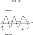
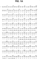
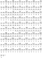
- FIG. 1 STEAP-1 structure.
- 1A Nucleotide and deduced amino acid sequences of STEAP-1 (8P1 D4) clone 10 cDNA (SEQ ID NOS. 1 and 2, respectively). The start Methionine is indicated in bold at amino acid residue position 1 and six putative transmembrane domains are indicated in bold and are underlined.
- 1B Schematic representation of STEAP-1 transmembrane orientation; amino acid residues bordering the predicted extracellular domains are indicated and correspond to the numbering scheme of FIG. 1A
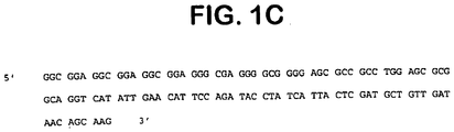
- 1C G/C rich 5' non-coding sequence of the STEAP-1 gene as determined by overlapping sequences of clone 10 and clone 3.
- FIG. 2 .
- Predominant expression of STEAP-1 In prostate tissue First strand cDNA was prepared from 16 normal tissues, the LAPC xenografts (4AD, 4AI and 9AD) and HeLa cells. Normalization was performed by PCR using primers to actin and GAPDH. Semi-quantitative PCR, using primers derived from STEAP-1 (8P1D4) cDNA ( FIG. 1A ), shows predominant expression of STEAP-1 in normal prostate and the LAPC xenografts.
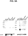
- FIG. 3 Northern blot analyses of STEAP-1 expression in various normal human tissues and prostate cancer xenografts, showing predominant expression of STEAP-1 in prostate tissue.
- FIG. 3A Two multiple tissue northern blots (Clontech) were probed with a full length STEAP cDNA clone 10 ( FIG. 1A ; SEQ ID NO: 1). Size standards in kilobases (kb) are indicated on the side.
- FIG. 3B Multiple tissue RNA dot blot (Clontech, Human Master Blot cat# 7770-1) probed with STEAP-1 cDNA clone 10 ( FIG. 1A ; SEQ ID NO: 1), showing approximately five-fold greater expression in prostate relative to other tissues with significant detectable expression.
- FIG. 4 Nucleotide sequence of STEAP-1 GTH9 clone (SEQ ID NO: 6) corresponding to the 4 kb message on northern blots ( FIG. 3A ). The sequence contains an intron of 2399 base pairs relative to the STEAP-1 clone 10 sequence of FIG.
- FIG. 5 Expression of STEAP-1 in prostate and multiple cancer cell lines and prostate cancer xenografts. Xenograft and cell line filters were prepared with 10 ⁇ g of total RNA per lane. The blots were analyzed using the STEAP-1 clone 10 as probe. All RNA samples were normalized by ethidium bromide staining and subsequent analysis with a p-actin probe. FIG.
- 5A Expression in various cancer cell lines and xenografts and prostate. Lanes as follows: (1) PrEC cells, (2) normal prostate tissue, (3) LAPC-4 AD xenograft, (4) LAPC-4 AI xenograft, (5) LAPC-9 AD xenograft, (6) LAPC-9 AI xenograft, (7) LNCaP cells, (8) PC-3 cells, (9) DU145 cells, (10) PANC-1 cells, (11) BxPC-3 cells, (12) HPAC cells, (13) Capan-1 cells, (14) CACO-2 cells, (15) LOVO cells, (16) T84 cells, (17) COLO-205 cells, (18) KCL-22 cells (acute lymphocytic leukemia, ALL), (19) HT1197 cells, (20) SCABER cells, (21) UM-UC-3 cells, (22) TCCSUP cells, (23) J82 cells, (24) 5637 cells, (25) RD-ES cells (Ewing sarcoma, EWS), (26) CAMA-1 cells, (2
- FIG. 5B The expression of STEAP-1 in subcutaneously (sc) grown LAPC xenografts compared to the expression in LAPC-4 and LAPC-9 xenografts grown in the tibia (it) of mice.
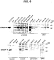
- FIG. 6 Western blot analysis of STEAP-1 protein expression in tissues and multiple cell lines. Western blots of cell lysates prepared from prostate cancer xenografts and cell lines were probed with a polyclonal anti-STEAP-1 antibody preparation (see Example XX for details). The samples contain 20 ⁇ g of protein and were normalized with anti-Grb-2 probing of the Western blots.
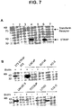
- FIG. 7 Cell surface biotinylation of STEAP-1.
- FIG. 7 Cell surface biotinylation of STEAP-1.
- FIG. 7B Prostate cancer (LNCaP, PC-3, DU145), bladder cancer (UM-UC-3, TCCSUP) and colon cancer (LOVO, COLO) cell lines were either biotinylated (+) or not (-) prior to lysis.
- Western blots of streptavidin-gel purified proteins were probed with anti-STEAP-1 antibodies. Molecular weight markers are indicated in kilodaltons (kD).
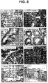
- FIG. 8 .
- Samples include: (a) LNCaP cells probed in the presence of N-terminal STEAP-1 peptide 1. (b) LNCaP plus non specific peptide 2, (c) normal prostate tissue, (d) grade 3 prostate carcinoma, (e) grade 4 prostate carcinoma, (f) LAPC-9 AD xenograft, (g) normal bladder, (h) normal colon. All images are at 400x magnification. FIG. 9 .
- FIG. 10 Nucleotide sequences of additional STEAP family members identified by searching the dbest database with the protein sequence of STEAP-1. In addition to STEAP-1, another three STEAP family members are indicated with their GenBank accession numbers. One of these corresponds to the gene 98P4B6 that was identified by SSH.
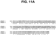
- FIG. 11 Primary structural comparison of STEAP family proteins.
- FIG. 11A Primary structural comparison of STEAP family proteins.
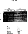
- FIG. 12 Predominant expression of AI139607 in placenta and prostate. First strand cDNA was prepared from 16 normal tissues.
- Normalization was performed by PCR using primers to actin and GAPDH.
- Semi-quantitative PCR using primers to AI139607, shows predominant expression of AI139607 in placenta and prostate after 25 cycles of amplification.
- the following primers were used to amplify AI139607: AI139607.1 5' TTAGGACAACTTGATCACCAGCA 3' (SEQ ID NO: 13) AI139607.2 5' TGTCCAGTCCAAACTGGGTTATTT 3' (SEQ ID NO: 14)
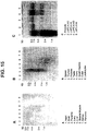
- FIG. 13 Predominant expression of R80991 in liver.
- First strand cDNA was prepared from 16 normal tissues. Normalization was performed by PCR using primers to actin and GAPDH.
- FIG. 14 Predominant expression of STEAP-2 (98P4B6) in prostate tissue.
- First strand cDNA was prepared from 8 normal tissues, the LAPC xenografts (4AD, 4AI and 9AD) and HeLa cells. Normalization was performed by PCR using primers to actin and GAPDH.
- Xenograft filter (C) was prepared with 10 ⁇ g of total RNA per lane. The blots were analyzed using the SSH derived 98P4B6 clone as probe. All RNA samples were normalized by ethidium bromide staining.
- FIG. 16 Expression of STEAP-2 in prostate and select cancer cell lines as determined by Northern blot analysis. Xenograft and cell line filters were prepared with 10 ⁇ g total RNA per lane. The blots were analyzed using an SSH derived 98P4B6 clone as probe. All RNA samples were normalized by ethidium bromide staining.
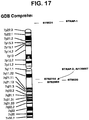
- FIG. 17 Chromosomal localization of STEAP family members.
- FIG. 18 Schematic representation of Intron-Exon boundaries within the ORF of human STEAP-1 gene. A total of 3 introns (i) and 4 exons (e) were identified.
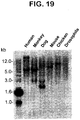
- FIG. 19 Zooblot southern analysis of STEAP-1 gene in various species. Genomic DNA was prepared from several different organisms including human, monkey, dog, mouse, chicken and Drosophila.
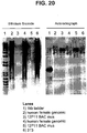
- FIG. 20 Southern blot analysis of mouse BAC with a STEAP-1 probe. DNA was prepared from human cells to isolate genomic DNA and from a mouse BAC clone (12P11) that contains the mouse STEAP gene. Each DNA sample was digested with EcoRI, blotted onto nitrocellulose and probed. Eight micrograms of genomic DNA was compared to 250 ng of mouse BAC DNA.
- the terms "advanced prostate cancer”, “locally advanced prostate cancer”, “advanced disease” and “locally advanced disease” mean prostate cancers which have extended through the prostate capsule, and are meant to include stage C disease under the American Urological Association (AUA) system, stage C1 - C2 disease under the Whitmore-Jewett system, and stage T3 - T4 and N+ disease under the TNM (tumor, node, metastasis) system.
- AUA American Urological Association
- stage C1 - C2 disease under the Whitmore-Jewett system
- TNM tumor, node, metastasis
- Locally advanced disease is clinically identified by palpable evidence of induration beyond the lateral border of the prostate, or asymmetry or induration above the prostate base.
- Locally advanced prostate cancer Is presently diagnosed pathologically following radical prostatectomy if the tumor invades or penetrates the prostatic capsule, extends into the surgical margin, or invades the seminal vesicles.
- metastatic prostate cancer and “metastatic disease” mean prostate cancers which have spread to regional lymph nodes or to distant sites, and are meant to include stage D disease under the AUA system and stage TxNxM+ under the TNM system.
- surgery is generally not indicated for patients with metastatic disease, and hormonal (androgen ablation) therapy is the preferred treatment modality.
- Patients with metastatic prostate cancer eventually develop an androgen-refractory state within 12 to 18 months of treatment initiation, and approximately half of these patients die within 6 months thereafter.
- the most common site for prostate cancer metastasis is bone.
- Prostate cancer bone metastases are, on balance, characteristically osteoblastic rather than osteolytic (i.e., resulting in net bone formation).
- Bone metastases are found most frequently in the spine, followed by the femur, pelvis, rib cage, skull and humerus. Other common sites for metastasis include lymph nodes, lung, liver and brain. Metastatic prostate cancer is typically diagnosed by open or laparoscopic pelvic lymphadenectomy, whole body radionuclide scans, skeletal radiography, and/or bone lesion biopsy.
- polynucleotide means a polymeric form of nucleotides of at least 10 bases or base pairs in length, either ribonucleotides or deoxynucleotides or a modified form of either type of nucleotide, and is meant to include single and double stranded forms of DNA.
- polypeptide means a polymer of at least 10 amino acids. Throughout the specification, standard three letter or single letter designations for amino acids are used.
- hybridize As used herein, the terms “hybridize”, “hybridizing”, “hybridizes” and the like, used in the context of polynucleotides, are meant to refer to conventional hybridization conditions, preferably such as hybridization in 50% formamide/6XSSC/0.1% SDS/100 ⁇ g/ml ssDNA, in which temperatures for hybridization are above 37 degrees C and temperatures for washing in 0.1XSSC/0.1% SOS are above 55 degrees C, and most preferably to stringent hybridization conditions.
- the invention relates to a novel family of proteins, termed STEAPs: Four STEAPs are specifically described herein by way of structural, molecular and biochemical features. As is further described in the Examples which follow, the STEAPS have been characterized in a variety of ways. For example, analyses of nucleotide coding and amino acid sequences were conducted in order to identify conserved structural elements within the STEAP family. Extensive RT-PCR and Northern blot analyses of STEAP mRNA expression were conducted in order to establish the range of normal and cancerous tissues expressing the various STEAP messages. Western blot, immunohistochemical and flow cytometric analyses of STEAP protein expression were conducted to determine protein expression profiles, cell surface localization and gross molecular topology of STEAP.
- the prototype member of the STEAP family, STEAP-1 is a six-transmembrane cell surface protein of 339 amino acids with no identifiable homology to any known human protein.
- the cDNA nucleotide and deduced amino acid sequences of human STEAP-1 are shown in FIG. 1A .
- a gross topological schematic of the STEAP-1 protein integrated within the cell membrane is shown in FIG. 1B .
- STEAP-1 expression is predominantly prostate-specific in normal tissues. Specifically, extensive analysis of STEAP-1 mRNA and protein expression in normal human tissues shows that STEAP-1 protein is predominantly expressed in prostate and, to a far smaller degree, in bladder. STEAP-1 mRNA is also relatively prostate specific, with only very low level expression detected in a few other normal tissues.
- STEAP-1 mRNA and protein is consistently expressed at high levels in prostate cancer and during all stages of the disease. STEAP-1 is also expressed in other cancers. Specifically, STEAP-1 mRNA is expressed at very high levels in bladder, colon, pancreatic, and ovarian cancer (as well as other cancers). In addition, cell surface expression of STEAP-1 protein has been established in prostate, bladder and colon cancers. Therefore, STEAP-1 has all of the hallmark characteristics of an excellent therapeutic target for the treatment of certain cancers, including particularly prostate, colon and bladder carcinomas.
- STEAP-2 is a highly homologous transmembrane protein encoded by a distinct gene.
- the STEAP-1 and STEAP-2 sequences show a high degree of structural conservation, particularly throughout their predicted transmembrane domains.
- the partial cDNA nucleotide and deduced amino acid sequences of STEAP-2 are shown in FIG. 9 .
- Both the STEAP-1 and STEAP-2 genes are located on chromosome 7, but on different arms.
- STEAP-2 exhibits a markedly different mRNA and protein expression profile relative to STEAP-1, suggesting that these two STEAP family members may be differentially regulated.
- STEAP-2 appears to be very prostate-specific, as significant mRNA expression is not detected in a variety of normal tissues.
- STEAP-2 also appears to follow a different course relative to STEAP-1, since STEAP-2 expression is down-regulated in at least some prostate cancers.
- STEAP-2 expression in other non-prostate cancers tested seems generally absent, although high level expression of STEAP-2 (like STEAP-1) is detected in Ewing sarcoma.
- STEAP-3 and STEAP-4 appear to be closely related to both STEAP-1 and STEAP-2 on a structural level, and both appear to be transmembrane proteins as well.
- STEAP-3 and STEAP-4 show unique expression profiles.
- STEAP-3 appears to have an expression pattern which is predominantly restricted to placenta and, to a smaller degree, expression is seen in prostate but not in other normal tissues tested.
- STEAP-4 seems to be expressed predominantly in liver.
- Neither STEAP-3 nor STEAP-4 appear to be expressed in prostate cancer xenografts which exhibit high level STEAP-1 and STEAP-2 expression.
- STEAP-1 maps within 7p22 (7p22.3), a large region of allelic gain reported for both primary and recurrent prostate cancers ( Visakorpi et al., 1995 Cancer Res. 55: 342 , Nupponen et al., 1998 American J. Pathol. 153: 141 ), suggesting that up-regulation of STEP-1 in cancer might include genomic mechanisms.
- xenopus oocytes (or other cells) expressing STEAP may being analyzed using voltage-clamp and patch-clamp experiments to determine if STEAP functions as an ion-channel. Oocyte cell volume may also be measured to determine If STEAP exhibits water channel properties.
- STEAPs function as channel or gap-junction proteins, they may serve as excellent targets for inhibition using, for example, antibodies, small molecules, and polynucleotides capable of inhibiting expression or function.
- the restricted expression pattern in normal tissue, and the high levels of expression in cancer tissue suggest that interfering with STEAP function may selectively kill cancer cells.
- STEAP proteins function as ion channels or gap-junction proteins in epithelial cell function. Ion channels have been implicated in proliferation and invasiveness of prostate cancer cells ( Lalani et al., 1997, Cancer Metastasis Rev 16:29 ). Both rat and human prostate cancer cells contain sub-population of cells with higher and lower expression levels of sodium channels. Higher levels of sodium channel expression correlate with more aggressive invasiveness in vitro ( Smith et al., 1998, FEBS Lett. 423:19 ).
- STEAP genes and proteins are meant to include the STEAP-1 and STEAP-2 genes and proteins, the genes and proteins corresponding to GeneBank Accession numbers AI139607 and R80991 (STEAP-3 and STEAP-4, respectively), and the genes and proteins corresponding to other STEAP proteins and structurally similar variants of the foregoing.
- Such other STEAP proteins and variants will generally have coding sequences which are highly homologous to the STEAP-1 and/or STEAP-2 coding sequences, and preferably will share at least about 50% amino acid identity and at least about 60% amino acid homology (using BLAST criteria), more preferably sharing 70% or greater homology (using BLAST criteria).
- the STEAP family member gene sequences described herein encode STEAP proteins sharing unique highly conserved amino acid sequence domains which distinguish them from other proteins. Proteins which include one or more of these unique highly conserved domains may be related to the STEAP family members or may represent new STEAP proteins.
- FIG. 11A which is an amino acid sequence alignment of the full STEAP-1 and partial STEAP-2 protein sequences, the STEAP-1 and STEAP-2 sequences share 61% identity and 79% homology, with particularly close sequence conservation in the predicted transmembrane domains. Referring to FIG.
- a STEAP polynucleotide may comprise a polynucleotide having the nucleotide sequence of human STEAP-1 as shown in FIG. 1A (SEQ ID NO. 1) or the nucleotide sequence of human STEAP-2 as shown in FIG. 9 (SEQ ID NO: 7), a sequence complementary to either of the foregoing, or a polynucleotide STEAP polynucleotide fragment of any of the foregoing.
- Another STEAP polynucleotide comprises a polynucleotide which encodes the human STEAP-1 protein amino acid sequence as shown in FIG. 1A (SEQ ID NO. 2) or which encodes the human STEAP-2 protein amino acid sequence as shown in FIG.
- Another STEAP polynucleotide comprises a polynucleotide which is capable of hybridizing under stringent hybridization conditions to the human STEAP-1 cDNA shown in FIG. 1A (SEQ ID NO. 1) or to a polynucleotide fragment thereof.
- Another STEAP polynucleotide comprises a polynucleotide which is capable of hybridizing under stringent hybridization conditions to the human STEAP-2 cDNA shown in FIG. 9 (SEQ ID NO. 1) or to a polynucleotide fragment thereof.
- genomic DNA e.g., genomic DNA, cDNAs, ribozymes, and antisense molecules
- nucleic acid molecules based on an alternative backbone or including alternative bases, whether derived from natural sources or synthesized.
- antisense molecules can be RNAs or other molecules, including peptide nucleic acids (PNAs) or non-nucleic acid molecules such as phosphorothioate derivatives, that specifically bind DNA or RNA in a base pair-dependent manner.
- PNAs peptide nucleic acids
- non-nucleic acid molecules such as phosphorothioate derivatives
- primers and primer pairs which allow the specific amplification of the polynucleotides of the invention or of any specific parts thereof, and probes that selectively or specifically hybridize to nucleic acid molecules of the invention or to any part thereof.
- Probes may be labeled with a detectable marker, such as, for example, a radioisotope, fluorescent compound, bioluminescent compound, a chemiluminescent compound, metal chelator or enzyme.
- a detectable marker such as, for example, a radioisotope, fluorescent compound, bioluminescent compound, a chemiluminescent compound, metal chelator or enzyme.
- Such probes and primers can be used to detect the presence of a STEAP polynucleotide in a sample and as a means for detecting a cell expressing a STEAP protein.
- probes examples include polypeptides comprising all or part of the human STEAP-1 cDNA sequence shown in FIG. 1A (SEQ ID NO. 1) and polypeptides comprising all or part of the human STEAP-2 cDNA sequence shown in FIG. 1A (SEQ ID NO. 2).
- primer pairs capable of specifically amplifying STEAP mRNAs are also described in the Examples which follow. As will be understood by the skilled artisan, a great many different primers and probes may be prepared based on the sequences provided in herein and used effectively to amplify and/or detect a STEAP mRNA or an mRNA encoding a particular STEAP family member (e.g., STEAP-1).
- a polynucleotide is said to be "isolated” when it is substantially separated from contaminant polynucleotides which correspond or are complementary to genes other than the STEAP gene or which encode polypeptides other than STEAP gene product or fragments thereof.
- a skilled artisan can readily employ nucleic acid isolation procedures to obtain an isolated STEAP polynucleotide.
- the STEAP polynucleotides of the invention are useful for a variety of purposes, including but not limited to their use as probes and primers for the amplification and/or detection of the STEAP gene(s), mRNA(s), or fragments thereof; as reagents for the diagnosis and/or prognosis of prostate cancer and other cancers; as coding sequences capable of directing the expression of STEAP polypeptides; as tools for modulating or inhibiting the expression of the STEAP gene(s) and/or translation of the STEAP transcript(s); and as therapeutic agents.
- the STEAP cDNA sequences described herein enable the isolation of other polynucleotides encoding STEAP gene product(s), as well as the isolation of polynucleotides encoding
- STEAP gene product homologues alternatively spliced isoforms, allelic, variants, and mutant forms of the STEAP gene product
- Various molecular cloning methods that can be employed to isolate full length cDNAs encoding a STEAP gene are well known (See, for example, Sambrook, J. et al., Molecular Cloning: A Laboratory Manual, 2d edition., Cold Spring Harbor Press, New York, 1989 ; Current Protocols in Molecular Biology. Ausubel et al., Eds., Wiley and Sons, 1995 ).
- lambda phage cloning methodologies may be conveniently employed, using commercially available cloning systems (e.g., Lambda ZAP Express, Stratagene).
- Phage clones containing STEAP gene cDNAs may be identified by probing with a labeled STEAP cDNA or a fragment thereof.
- the STEAP-1 cDNA ( FIG.1A ) or a portion thereof can be synthesized and used as a probe to retrieve overlapping and full length cDNAs corresponding to a STEAP gene.
- the STEAP-2 cDNA sequence may be employed.
- a STEAP gene may be isolated by screening genomic DNA libraries, bacterial artificial chromosome libraries (BACs), yeast artificial chromosome libraries (YACs), and the like, with STEAP DNA probes or primers.
- the invention also provides recombinant DNA or RNA molecules containing a STEAP polynucleotide according to the claims, including but not limited to phages, plasmids, phagemids, cosmids, YACs, BACs, as well as various viral and non-viral vectors well known in the art, and cells transformed or transfected with such recombinant DNA or RNA molecules.
- a recombinant DNA or RNA molecule is a DNA or RNA molecule that has been subjected to molecular manipulation in vitro. Methods for generating such molecules are well known (see, for example, Sambrook et al, 1989, supra).
- the invention further provides a host-vector system comprising a recombinant DNA molecule containing a STEAP polynucleotide according to the claims, within a suitable prokaryotic or eukaryotic host cell.
- suitable eukaryotic host cells include a yeast cell, a plant cell, or an animal cell, such as a mammalian cell or an insect cell (e.g., a baculovirus-infectible cell such as an Sf9 cell).
- suitable mammalian cells include various prostate cancer cell lines such LnCaP, PC-3, DU145, LAPC-4, TsuPr1, other transfectable or transducible prostate cancer cell lines, as well as a number of mammalian cells routinely used for the expression of recombinant proteins (e.g., COS, CHO, 293, 293T cells). More particularly, a polynucleotide comprising the coding sequence of a STEAP may be used to generate STEAP proteins or fragments thereof using any number of host-vector systems routinely used and widely known in the art.
- STEAP may be preferably expressed in several prostate cancer and non-prostate cell lines, including for example 293, 293T, rat-1, 3T3, PC-3, LNCaP and TsuPr1.
- the host-vector systems of the invention are useful for the production of a STEAP protein or fragment thereof. Such host-vector systems may be employed to study the functional properties of STEAP and STEAP mutations.
- Proteins encoded by the STEAP genes, or by fragments thereof, will have a variety of uses, including but not limited to generating antibodies and in methods for identifying ligands and other agents and cellular constituents that bind to a STEAP gene product.
- Antibodies raised against a STEAP protein or fragment thereof may be useful in diagnostic and prognostic assays, imaging methodologies (including, particularly, cancer imaging), and therapeutic methods in the management of human cancers characterized by expression of a STEAP protein, such as prostate, colon, breast, cervical and bladder carcinomas, ovarian cancers, testicular cancers and pancreatic cancers.
- Various immunological assays useful for the detection of STEAP proteins are contemplated, including but not limited to various types of radioimmunoassays, enzyme-linked immunosorbent assays (EUSA), enzyme-linked immunofluorescent assays (ELIFA), immunocytochemical methods, and the like.
- Such antibodies may be labeled and used as immunological imaging reagents capable of detecting prostate cells (e.g., in radioscintigraphic imaging methods).
- STEAP proteins may also be particularly useful in generating cancer vaccines, ss further described below.
- a STEAP protein refers to a protein that has or includes the amino acid sequence of human STEAP-1 as provided in FIG. 1A (SEQ ID NO. 2), human STEAP-2 as provided in FIG. 9 (SEQ ID NO. 8), the amino add sequence of other mammalian STEAP homologues and variants, as well as allelic variants and conservative substitution mutants of these proteins that have STEAP biological activity.
- STEAP proteins of the invention are defined in the claims. Fusion proteins which combine parts of different STEAP proteins or fragments thereof, as well as fusion proteins of a STEAP protein and a heterologous polypeptide are also included.
- STEAP polypeptide refers to a polypeptide fragment or a STEAP protein of at least 10 amino acids, preferably at least 15 amino acids.
- a specific embodiment of a STEAP protein comprises a polypeptide having the amino acid sequence of human STEAP-1 as shown in FIG. 1A (SEQ ID NO. 2).
- Another embodiment of a STEAP protein comprises a polypeptide containing the partial STEAP-2 amino acid sequence as shown In FIG. 9 (SEQ ID NO. 8).
- Another embodiment comprises a polypeptide containing the partial STEAP-3 amino acid sequence of shown in FIG. 11B .
- Yet another embodiment comprises a polypeptide containing the partial STEAP-4 amino acid sequence of shown in FIG. 11B .
- allelic variants of human STEAP will share a high degree of structural identity and homology (e.g., 90% or more identity).
- allelic variants of the STEAP proteins will contain conservative amino acid substitutions within the STEAP sequences described herein or will contain a substitution of an amino acid from a corresponding position in a STEAP homologue.
- One class of STEAP allelic variants will be proteins that share a high degree of homology with at least a small region of a particular STEAP amino acid sequence, but will further contain a radical departure from the sequence, such as a non-conservative substitution, truncation, insertion or frame shift Such alleles represent mutant STEAP proteins that typically do not perform the same biological functions or do not have all of the biological characteristics.
- Conservative amino acid substitutions can frequently be made in a protein without altering either the conformation or the function of the protein. Such changes include substituting any of isoleucine (I), valine (V), and leucine (L) for any other of these hydrophobic amino acids; aspartic acid (D) for glutamic add (E) and vice versa; glutamine (Q) for asparagine (N) and vice versa; and serine (S) for threonine (T) and vice versa. Other substitutions can also be considered conservative, depending on the environment of the particular amino acid and its role in the three-dimensional structure of the protein.
- glycine (G) and alanine (A) can frequently be interchangeable, as can alanine (A) and valine (V).
- Methionine (M) which is relatively hydrophobic, can frequently be interchanged with leucine and isoleucine, and sometimes with valine.
- Lysine (K) and arginine (R) are frequently interchangeable in locations in which the significant feature of the amino acid residue is its charge and the differing pK's of these two amino acid residues are not significant. Still other changes can be considered "conservative" in particular environments.
- STEAP proteins may be embodied in many forms, preferably in isolated form.
- a protein is said to be "isolated” when physical, mechanical or chemical methods are employed to remove the STEAP protein from cellular constituents that are normally associated with the protein.
- a skilled artisan can readily employ standard purification methods to obtain an isolated STEAP protein.
- a purified STEAP protein molecule will be substantially free of other proteins or molecules which impair the binding of STEAP to antibody or other ligand. The nature and degree of isolation and purification will depend on the intended use.
- Embodiments of a STEAP protein include a purified STEAP protein and a functional, soluble STEAP protein. In one form, such functional, soluble STEAP proteins or fragments thereof retain the ability to bind antibody or other ligand.
- the invention also provides STEAP polypeptides comprising biologically active fragments of the STEAP amino acid sequence, such as a polypeptide corresponding to part of the amino acid sequences for STEAP-1 as shown in FIG. 1A (SEQ ID NO. 2), as defined in claim 2 or claim 3.
- Such polypeptides of the invention exhibit properties of a STEAP protein, such as the ability to elicit the generation of antibodies which specifically bind an epitope associated with a STEAP protein.
- Polypeptides comprising amino acid sequences which are unique to a particular STEAP protein (relative to other STEAP proteins) may be used to generate antibodies which will specifically react with that particular STEAP protein.
- each molecule contains stretches of sequence unique to its structure. These unique stretches can be used to generate STEAP-1 or STEAP-2 specific antibodies.
- STEAP polypeptides can be generated using standard peptide synthesis technology or using chemical cleavage methods well known In the art based on the amino acid sequences of the human STEAP proteins disclosed herein.
- recombinant methods can be used to generate nucleic acid molecules that encode a polypeptide fragment of a STEAP protein.
- the STEAP-encoding nucleic acid molecules described herein provide means for generating defined fragments of STEAP proteins.
- STEAP polypeptides are particularly useful in generating and characterizing domain specific antibodies (e.g., antibodies recognizing an extracellular or intracellular epitope of a STEAP protein), In generating STEAP family member specific antibodies (e.g., anti-STEAP-1, anti-STEAP 2 antibodies), identifying agents or cellular factors that bind to a particular STEAP or STEAP domain, and in various therapeutic contexts, including but not limited to cancer vaccines.
- domain specific antibodies e.g., antibodies recognizing an extracellular or intracellular epitope of a STEAP protein
- STEAP family member specific antibodies e.g., anti-STEAP-1, anti-STEAP 2 antibodies
- STEAP polypeptides containing particularly interesting structures can be predicted and/or identified using various analytical techniques well known in the art, including, for example, the methods of Chou-Fasman, Garnier-Robson, Kyte-Doolittle, Eisenberg, Karplus-Schultz or Jameson-Wolf analysis, or on the basis of immunogenicity. Fragments containing such structures are particularly useful in generating subunit specific antl-STEAP antibodies or in identifying cellular factors that bind to STEAP.
- Anti-STEAP antibodies that are particularly contemplated include monoclonal and polyclonal antibodies as well as fragments containing the antigen binding domain and/or one or more complementarity determining regions of these antibodies.
- an antibody fragment is defined as at least a portion of the variable region of the immunoglobulin molecule which binds to its target, i.e., the antigen binding region.
- antibodies which specifically react with a particular STEAP protein and/or an epitope within a particular structural domain may be desirable to generate antibodies which specifically react with a particular STEAP protein and/or an epitope within a particular structural domain.
- preferred antibodies useful for cancer therapy and diagnostic imaging purposes are those which react with an epitope in an extracellular region of the STEAP protein as expressed in cancer cells.
- Such antibodies may be generated by using the STEAP proteins described herein, or using peptides derived from predicted extracellular domains thereof, as an immunogen.
- regions in the extracellular loops between the indicated transmembrane domains may be selected as used to design appropriate immunogens for raising extracellular-specific antibodies.
- STEAP antibodies may be particularly useful in prostate cancer therapeutic strategies, diagnostic and prognostic assays, and imaging methodologies.
- the disclosure provides various immunological assays useful for the detection and quantification of STEAP and mutant STEAP proteins and polypeptides.
- Such assays generally comprise one or more STEAP antibodies capable of recognizing and binding a STEAP or mutant STEAP protein, as appropriate, and may be performed within various immunological assay formats well known in the art, including but not limited to various types of radioimmunoassays, enzyme-linked immunosorbent assays (EUSA), enzyme-linked immunofluorescent assays (ELIFA), and the like.
- immunological imaging methods capable of detecting prostate cancer are also provided, including but limited to radioscintigraphic imaging methods using labeled STEAP antibodies. Such assays may be clinically useful in the detection, monitoring, and prognosis of prostate cancer, particularly advanced prostate cancer.
- the method of purifying a STEAP protein comprises incubating a STEAP antibody, which has been coupled to a solid matrix, with a lysate or other solution containing STEAP under conditions which permit the STEAP antibody to bind to STEAP; washing the solid matrix to eliminate impurities; and eluting the STEAP from the coupled antibody.
- Other uses of the STEAP antibodies include generating anti-idiotypic antibodies that mimic the STEAP protein.
- STEAP antibodies may also be used therapeutically by, for example, modulating or inhibiting the biological activity of a STEAP protein or targeting and destroying prostate cancer cells expressing a STEAP protein.
- Antibody therapy of prostate and other cancers is more specifically described in a separate subsection below.
- antibodies may be prepared by immunizing a suitable mammalian host using a STEAP protein, peptide, or fragment, in isolated or immunoconjugated form ( Antibodies: A Laboratory Manual, CSH Press, Eds., Harlow, and Lane (1988 ); Harlow, Antibodies, Cold Spring Harbor Press, NY (1989 )).
- fusion proteins of STEAP may also be used, such as a STEAP GST-fusion protein.
- a GST fusion protein comprising all or most of the open reading frame amino acid sequence of FIG. 1A may be produced and used as an immunogen to generate appropriate antibodies.
- Cells expressing or overexpressing STEAP may also be used for immunizations. Similarly, any cell engineered to express STEAP may be used. Such strategies may result in the production of monoclonal antibodies with enhanced capacities for recognizing endogenous STEAP.
- Another useful immunogen comprises STEAP proteins linked to the plasma membrane of sheep red blood cells.
- the amino acid sequence of STEAP as shown in FIG. 1A may be used to select specific regions of the STEAP protein for generating antibodies.
- hydrophobicity and hydrophilicity analyses of the STEAP amino acid sequence may be used to identify hydrophilic regions in the STEAP structure. Regions of the STEAP protein that show immunogenic structure, as well as other regions and domains, can readily be identified using various other methods known in the art, such as Chou-Fasman, Garnier-Robson, Kyte-Doolittle, Eisenberg, Karplus-Schultz or Jameson-Wolf analysis.
- amino acid sequences unique to the mutant are preferable.
- a protein or polypeptide for use as an immunogen and for preparing immunogenic conjugates of a protein with a carrier such as BSA, KLH, or other carrier proteins are well known in the art
- a carrier such as BSA, KLH, or other carrier proteins
- direct conjugation using, for example, carbodiimide reagents may be used; in other instances linking reagents such as those supplied by Pierce Chemical Co., Rockford, IL, may be effective.
- Administration of a STEAP immunogen is conducted generally by injection over a suitable time period and with use of a suitable adjuvant, as is generally understood in the art During the immunization schedule, titers of antibodies can be taken to determine adequacy of antibody formation.
- STEAP monoclonal antibodies are preferred and may be produced by various means well known in the art
- immortalized cell lines which secrete a desired monoclonal antibody may be prepared using the standard method of Kohler and Milstein or modifications which effect immortalization of lymphocytes or spleen cells, as is generally known.
- the immortalized cell lines secreting the desired antibodies are screened by immunoassay in which the antigen is the STEAP protein or STEAP fragment
- the cells may be expanded and antibodies produced either from in vitro cultures or from ascites fluid.
- STEAP polypeptides may be used as immunogens for generating monoclonal antibodies using traditional methods.
- a particular embodiment comprises an antibody which immunohistochemically stains 293T cells transfected with an expression plasmid carrying the STEAP-1 coding sequence, the transfected cells expressing STEAP-1 protein, but does immunohistochemically stain untransfected 293T cells.
- An assay for characterizing such antibodies is provided in Example 5 herein.
- STEAP-1 monoclonal antibodies may be generated using NIH 3T3 cells expressing STEAP-1 as an immunogen to generate mAbs that recognize the cell surface epitopes of STEAP-1.
- Reactive mAbs may be screened by cell-based EUSAs using PC-3 cells over-expressing STEAP-1.
- 3 peptides representing the extracellular regions of the STEAP-1 protein are coupled to sheep red blood cells for immunization.
- recombinant STEAP-1 protein generated with an amino-terminal His-tag using a suitable expression system e.g., baculovirus expression system pBlueBac4.5, Invitrogen
- a suitable expression system e.g., baculovirus expression system pBlueBac4.5, Invitrogen
- the antibodies or fragments may also be produced, using current technology, by recombinant means. Regions that bind specifically to the desired regions of the STEAP protein can also be produced in the context of chimeric or CDR grafted antibodies of multiple species origin. Humanized or human STEAP antibodies may also be produced and are preferred for use in therapeutic contexts. Various approaches for producing such humanized antibodies are known, and include chimeric and CDR grafting methods; methods for producing fully human monoclonal antibodies include phage display and transgenic methods (for review, see Vaughan et al., 1998, Nature Biotechnology 16: 535-539 ).
- Fully human STEAP monoclonal antibodies may be generated using cloning technologies employing large human Ig gene combinatorial libraries (i.e., phage display) ( Griffiths and Hoogenboom, Building an in vitro immune system: human antibodies from phage display libraries. In: Protein Engineering of Antibody Molecules for Prophylactic and Therapeutic Applications in Man. Clark, M. (Ed.), Nottingham Academic, pp 45-64 (1993 ); Burton and Barbas, Human Antibodies from combinatorial libraries. Id., pp 65-82 ).
- Fully human STEAP monoclonal antibodies may also be produced using transgenic mice engineered to contain human Immunoglobulin gene loci as described in PCT Patent Application WO98/24893, Kucherlapati and Jakobovits et al., published December 3, 1997 (see also, Jakobovits, 1998, Exp. Opin. Invest Drugs 7(4): 607-614 ). This method avoids the in vitro manipulation required with phage display technology and efficiently produces high affinity authentic human antibodies.
- Reactivity of STEAP antibodies with a STEAP protein may be established by a number of well known means, including Western blot, immunoprecipitation, ELISA, and FACS analyses using, as appropriate, STEAP proteins, peptides, STEAP-expressing cells or extracts thereof.
- a STEAP antibody or fragment thereof may be labeled with a detectable marker or conjugated to a second molecule, such as a cytotoxic agent, and used for targeting the second molecule to a STEAP positive cell ( Vitetta, E.S. et al., 1993, Immunotoxin therapy, In DeVita, Jr., V.T. et al., eds, Cancer: Principles and Practice of Oncology, 4th ed., J.B. Lippincott Co., Philadelphia, 2624-2636 ).
- Suitable detectable markers include, but are not limited to, a radioisotope, a fluorescent compound, a bioluminescent compound, chemiluminescent compound, a metal chelator or an enzyme.
- Another aspect of this disclosure relates to methods for detecting STEAP polynucleotides and STEAP proteins, as well as methods for identifying a cell which expresses STEAP.
- Detectable STEAP polynucleotides include, for example, a STEAP gene or fragments thereof, STEAP mRNA, alternative splice variant STEAP mRNAs, and recombinant DNA or RNA molecules containing a STEAP polynucleotide.
- a number of methods for amplifying and/or detecting the presence of STEAP polynucleotides are well known in the art and may be employed.
- a method for detecting a STEAP mRNA in a biological sample comprises producing cDNA from the sample by reverse transcription using at least one primer; amplifying the cDNA so produced using a STEAP polynucleotides as sense and antisense primers to amplify STEAP cDNAs therein; and detecting the presence of the amplified STEAP cDNA.
- a method of detecting a STEAP gene in a biological sample comprises first Isolating genomic DNA from the sample; amplifying the isolated genomic DNA using STEAP polynucleotides as sense and antisense primers to amplify the STEAP gene therein; and detecting the presence of the amplified STEAP gene.
- any number of appropriate sense and antisense probe combinations may be designed from the nucleotide sequences provided for STEAP-1 ( FIG. 1A ; SEQ ID NO. 1), STEAP-2 ( FIG. 9 ; SEQ ID NO. 7), STEAP-3 ( FIG. 10 ; SEQ ID NO. 11), or STEAP-4 ( FIG. 10 ; SEQ ID NO: 12), as appropriate, and used for this purpose.
- This disclosure also provides assays for detecting the presence of a STEAP protein in a.tissue of other biological sample such as serum, bone, prostate, and other tissues, urine, cell preparations, and the like.
- Methods for detecting a STEAP protein are also well known and include, for example, immunoprecipitation, immunohislochemical analysis, Western Blot analysis, molecular binding assays, ELISA, ELIFA and the like.
- a method of detecting the presence of a STEAP protein in a biological sample comprises first contacting the sample with a STEAP antibody, a STEAP-reactive fragment thereof, or a recombinant protein containing an antigen binding region of a STEAP antibody; and then detecting the binding of STEAP protein in the sample thereto.
- an assay for identifying a cell which expresses a STEAP gene comprises detecting the presence of STEAP mRNA in the cell.
- Methods for the detection of particular mRNAs in cells are well known and include, for example, hybridization assays using complementary DNA probes (such as in situ hybridization using labeled STEAP riboprobes, Northern blot and related techniques) and various nucleic acid amplification assays (such as RT-PCR using complementary primers specific for STEAP, and other amplification type detection methods, such as, for example, branched DNA, SISBA, TMA and the like).
- an assay for identifying a cell which expresses a STEAP gene comprises detecting the presence of STEAP protein in the cell or secreted by the cell.
- Various methods for the detection of proteins are well known in the art and may be employed for the detection of STEAP proteins and STEAP expressing cells.
- STEEP expression analysis may also be useful as a tool for identifying and evaluating agents which modulate STEAP gene expression.
- STEAP-1 expression is significantly upregulated in colon, bladder, pancreatic, ovarian and other cancers. Identifications of a molecule or biological agent that could inhibit STEAP-1 over-expression may be of therapeutic value in the treatment of cancer. Such an agent may be identified by using a screen that quantifies STEAP expression by RT-PCR, nucleic acid hybridization or antibody binding.
- Determining the status of STEAP expression patterns in an individual may be used to diagnose cancer and may provide prognostic information useful in defining appropriate therapeutic options. Similarly, the expression status of STEAP may provide information useful for predicting susceptibility to particular disease stages, progression, and/or tumor aggressiveness. This disclosure provides methods and assays for determining STEAP expression status and diagnosing cancers which express STEAP.
- this disclosure provides assays useful in determining the presence of cancer in an individual, comprising detecting a significant increase in STEAP mRNA or protein expression in a test cell or tissue sample relative to expression levels in the corresponding normal cell or tissue.
- the presence of STEAP 1 mRNA is evaluated in tissue samples of the colon, pancreas, bladder, ovary, cervix, testis or breast
- the presence of significant STEAP-1 expression in any of these tissues may be useful to indicate the emergence, presence and/or severity of these cancers, since the corresponding normal tissues do not express STEAP-1 mRNA.
- STEAP-1 expression status may be determined at the protein level rather than at the nucleic acid level.
- such a method or assay would comprise determining the level of STEAP-1 protein expressed by cells in a test tissue sample and comparing the level so determined to the level of STEAP expressed in a corresponding normal sample.
- the presence of STEAP-1 protein is evaluated, for example, using immunohistochemical methods.
- STEAP antibodies or binding partners capable of detecting STEAP protein expression may be used in a variety of assay formats well known in the art for this purpose.
- Peripheral blood may be conveniently assayed for the presence of cancer cells, including prostate, colon, pancreatic, bladder and ovarian cancers, using RT-PCR to detect STEAP-1 expression.
- the presence of RT-PCR amplifiable STEAP-1 1 mRNA provides an indication of the presence of one of these types of cancer.
- RT-PCR detection assays for tumor cells in peripheral blood are currently being evaluated for use in the diagnosis and management of a number of human solid tumors. In the prostate cancer field, these include RT-PCR assays for the detection of cells expressing PSA and PSM ( Verkaik et al., 1997, Urol. Res. 25: 373-384 ; Ghossein etal., 1995, J. Clin. Oncol.13:1195-2000 ; Heston et al.,1995, Clin. Chem. 41: 1687-1688 ). RT-PCR assays are well known in the art.
- a recently described sensitive assay for detecting and characterizing carcinoma cells in blood may be used ( Racita et al., 1998, Proc. Natl. Acad. Sci. USA 95: 4589-4594 ).
- This assay combines immunomagnetic enrichment with multiparameter flow cytometric and immunohistochemical analyses, and is highly sensitive for the detection of cancer cells in blood, reportedly capable of detecting one epithelial cell in 1 ml of peripheral blood.
- a related aspect of this disclosure is directed to predicting susceptibility to developing cancer in an individual.
- a method for predicting susceptibility to cancer comprises detecting STEAP mRNA or STEAP protein in a tissue sample, its presence indicating susceptibility to cancer, wherein the degree of STEAP mRNA expression present is proportional to the degree of susceptibility.
- a method for gauging aggressiveness of a tumor comprises determining the level of STEAP mRNA or STEAP protein expressed by cells in a sample of the tumor, comparing the level so determined to the level of STEP mRNA or STEAP protein expressed in a corresponding normal tissue taken from the same individual or a normal tissue reference sample, wherein the degree of STEAP mRNA or STEAP protein expression in the tumor sample relative to the normal sample indicates the degree of aggressiveness.
- Standard methods for the detection and quantification of STEAP mRNA include in situ hybridization using labeled STEAP riboprobes, Northern blot and related techniques using STEAP polynucleotide probes, RT-PCR analysis using primers specific for STEAP, and other amplification type detection methods, such as, for example, branched DNA, SISBA, TMA and the like.
- semiquantitative RT-PCR may be used to detect and quantify STEAP mRNA expression as described In the Examples which follow.
- primers capable of amplifying STEAP may be used for this purpose, including but not limited to the various primer sets specifically described herein. Standard methods for the detection and quantification of protein may be used for this purpose.
- polyclonal or monoclonal antibodies specifically reactive with the wild-type STEAP protein may be used in an immunohistochemical assay of biopsied tissue.
- the expression profiles of STEAP-1 and STEAP-2 indicate antibodies specific therefor may be particularly useful in radionuclide and other forms of diagnostic imaging of certain cancers.
- immunohistochemical analysis of STEAP-1 protein suggests that in normal tissues STEAP-1 is predominantly restricted to prostate and bladder.
- the transmembrane orientation of STEAP-1 (and presumably STEAP-2) provides a target readily identifiable by antibodies specifically reactive with extracellular epitopes.
- This tissue restricted expression, and the localization of STEAP to the cell surface of multiple cancers makes STEAP an ideal candidate for diagnostic imaging. Accordingly, in vivo imaging techniques may be used to image human cancers expressing a STEAP protein.
- cell surface STEAP-1 protein is expressed at very high levels in several human cancers, particularly prostate, bladder, colon and ovarian cancers, and Ewing sarcoma. Moreover, in normal tissues, STEAP-1 protein expression is largely restricted to prostate.
- radiolabeled antibodies specifically reactive with extracellular epitopes of STEAP-1 may be particularly useful in in vivo imaging of solid tumors of the foregoing cancers. Such labeled anti-STEAP-1 antibodies may provide very high level sensitivities for the detection of metastasis of these cancers.
- monoclonal antibodies are used in the diagnostic imaging methods.
- This disclosure provides various immunotherapeutic methods for treating prostate cancer, including antibody therapy, in vivo vaccines, and ex vivo Immunotherapy methods, which utilize polynucleotide and polypeptides corresponding to STEAP and STEAP antibodies. These therapeutic applications are described further In the following subsections.
- STEAP-1 is strongly expressed uniformly over the surface of glandular epithelial cells within prostate and prostate cancer cells. See, for details, immunohistochemical and Western blot analyses of STEAP-1 protein expression presented in Examples 3C (and 3D as well as the STEAP-1 mRNA expression profiles obtained from the Northern blot and RT-PCR generated data presented in Examples 1 and 3A, 3B. In particular, immunohistochemical analysis results show that the surface of human prostate epithelial cells (normal and cancer) appear to be uniformly coated with STEAP-1. Biochemical analysis confirms the cell surface localization of STEAP-1 initially suggested by its putative 6-transmembrane primary structural elements and by the pericellular staining plainly visualized by immunohistochemical staining.
- STEAP-1 is uniformly expressed at high levels over the surface of prostate glandular epithelia, an ideal situation for immunotherapeutic intervention strategies which target extracellular STEAP epitopes.
- Systemic administration of STEAP-immunoreactive compositions would be expected to result in extensive contact of the composition with prostate epithelial cells via binding to STEAP-1 extracellular epitopes.
- STEAP-1 protein expression in normal human tissues there is ample reason to expect extraordinarily without toxic, non-specific and/or non-target effects caused by the binding of the immunotherapeutic composition to STEAP-1 on non-target organs and tissues.
- STEAP-1 appears to be substantially over-expressed in a variety of other human cancers, including bladder, colon, pancreatic and ovarian cancers.
- high level STEAP-1 mRNA expression is detected in all tested prostate cancer tissues and cell lines, and in most of the pancreatic, colon, and bladder cancer cell lines tested.
- High level expression of STEAP-1 is also observed in some ovarian cancer cell lines.
- Lower level expression is observed in some breast, testicular, and cervical cancer cell lines.
- Very high level expression is also detected in a Ewing sarcoma cell line.
- Applicants have shown that cell surface STEAP-1 protein is expressed in bladder and colon cancers, while there is no detectable cell surface (or intracellular) STEAP-1 protein in normal colon and low expression in normal bladder.
- Antibodies specifically reactive with extracellular domains of STEAP-1 may be useful to treat these cancers systemically, either as toxin or therapeutic agent conjugates or as naked antibodies capable of inhibiting cell proliferation or function.
- STEAP-2 protein is also expressed in prostate cancer, and may be expressed in other cancers as well.
- STEAP-2 mRNA analysis by RT-PCR and Northern blot show that expression is restricted to prostate in normal tissues, is also expressed in some prostate, pancreatic, colon, testicular, ovarian and other cancers. Therefore, antibodies reactive with STEAP-2 may be useful In the treatment of prostate and other cancers.
- the expression of STEAP-3 and STEAP-4 (as well as other STEAPs) may be associated with some cancers. Thus antibodies reactive with these STEAP family member proteins may also be useful therapeutically.
- STEAP antibodies may be introduced into a patient such that the antibody binds to STEAP on the cancer cells and mediates the destruction of the cells and the tumor and/or inhibits the growth of the cells or the tumor.
- Mechanisms by which such antibodies exert a therapeutic effect may include complement-mediated cytolysis, antibody-dependent cellular cytotoxicity, modulating the physiologic function of STEAP, inhibiting ligand binding or signal transduction pathways, modulating tumor cell differentiation, altering tumor angiogenesis factor profiles, and/or by inducing apoptosis.
- STEAP antibodies conjugated to toxic or therapeutic agents may also be used therapeutically to deliver the toxic or therapeutic agent directly to STEAP-bearing tumor cells.
- Cancer immunotherapy using anti-STEAP antibodies may follow the teachings generated from various approaches which have been successfully employed with respect to other types of cancer, including but not limited to colon cancer ( Arien et al., 1998, Crit Rev Immunol 18:133-138 ), multiple myeloma ( Ozaki et al., 1997, Blood 90: 3179-3186 ; Tsunenari et al., 1997, Blood 90: 2437-2444 ), gastric cancer ( Kasprzyk et at..
- antibody therapy may be particularly appropriate and in advanced or metastatic cancers. Combining the antibody therapy with a chemotherapeutic or radiation regimen may be preferred in patients who have not received chemotherapeutic. treatment, whereas treatment with the antibody therapy of the invention may be indicated for patients who have received one or more chemotherapy. Additionally, antibody therapy may also enable the use of reduced dosages of concomitant chemotherapy, particularly in patients that do not tolerate the toxicity of the chemotherapeutic agent very well.
- non-prostate cancer patients may be evaluated for the presence and level of STEAP over-expression, preferably using immunohistochemical assessments of tumor tissue, quantitative STEAP imaging, or other techniques capable of reliably indicating the presence and degree of STEAP overexpression.
- Immunohistochemical analysis of tumor biopsies or surgical specimens may be preferred for this purpose. Methods for immunohistochemical analysis of tumor tissues are well known in the art
- Anti-STEAP monoclonal antibodies useful in treating prostate and other cancers include those which are capable of Initiating a potent immune response against the tumor and those which are capable of direct cytotoxicity.
- anti-STEAP mAbs may elicit tumor cell lysis by either complement-mediated or antibody-dependent cell cytotoxicity (ADCC) mechanisms, both of which require an intact Fc portion of the immunoglobulin molecule for interaction with effector cell Fc receptor sites or complement proteins.
- ADCC antibody-dependent cell cytotoxicity
- anti-STEAP mAbs which exert a direct biological effect on tumor growth are useful. Potential mechanisms by which such directly cytotoxic mAbs may act include inhibition of cell growth, modulation of cellular differentiation, modulation of tumor angiogenesis factor profiles, and the induction of apoptosis.
- the mechanism by which a particular anti-STEAP mAb exerts an anti-tumor effect may be evaluated using any number of in vitro assays designed to determine ADCC, AOMMC, complement-mediated cell lysis, and so forth, as is generally known in the art
- the anti-tumor activity of a particular anti-STEAP mAb, or combination of anti-STEAP mAbs may be evaluated in vivo using a suitable animal model.
- a suitable animal model For example, xenogenic prostate cancer models wherein human prostate cancer explants or passaged xenograft tissues are introduced into immune compromised animals, such as nude or SCID mice, are appropriate in relation to prostate cancer and have been described ( Klein et al., 1997, Nature Medicine 3: 402-408 ).
- preferred monoclonal antibodies used in the practice of the therapeutic methods disclosed herein are those which are either fully human or humanized and which bind specifically to the target 20P1F12/TMPRSS2 antigen with high affinity but exhibit low or no antigenicity in the patient
- the methods of this disclosure contemplate the administration of single anti-STEAP mAbs as well as combinations, or "cocktails, of different mAbs.
- Such mAb cocktails may have certain advantages inasmuch as they contain mAbs which exploit different effector mechanisms or combine directly cytotoxic mAbs with mAbs that rely on immune effector functionality. Such mAbs in combination may exhibit synergistic therapeutic effects.
- the administration of anti-STEAP mAbs may be combined with other therapeutic agents, including but not limited to various chemotherapeutic agents, androgen-blockers, and immune modulators (e.g., IL-2, GM-CSF).
- the anti-STEAP mAbs may be administered in their "naked” or unconjugated form, or may have therapeutic agents conjugated to them.
- the anti-STEAP monoclonal antibodies used in the practice of the methods of this disclosure may be formulated into pharmaceutical compositions comprising a carrier suitable for the desired delivery method.
- Suitable carriers include any material which when combined with the anti-STEAP mAbs retains the anti-tumor function of the antibody and is nonreactive with the subject's immune systems. Examples include, but are not limited to, any of a number of standard pharmaceutical carriers such as sterile phosphate buffered saline solutions, bacteriostatic water, and the like.
- the anti-STEAP antibody formulations may be administered via any route capable of delivering the antibodies to the tumor site.
- Potentially effective routes of administration include, but are not limited to, intravenous, intraperitoneal, intramuscular, intratumor, intradermal, and the like.
- the preferred route of administration is by intravenous injection.
- a preferred formulation for intravenous injection comprises the anti-STEAP mAbs in a solution of preserved bacteriostatic water, sterile unpreserved water, and/or diluted in polyvinylchloride or polyethylene bags containing 0.9% sterile Sodium Chloride for Injection, USP.
- the anti-STEAP mAb preparation may be lyophilized and stored as a sterile powder, preferably under vacuum, and then reconstituted in bacteriostatic water containing, for example, benzyl alcohol preservative, or in sterile water prior to injection.
- Treatment will generally involve the repeated administration of the anti-STEAP antibody preparation via an acceptable route of administration such as intravenous injection (IV), typically at a dose in the range of about 0.1 to about 10 mg/kg body weight. Doses in the range of 10-500 mg mAb per week may be effective and well tolerated. Based on clinical experience with the Herceptin mAb in the treatment of metastatic breast cancer, an initial loading dose of approximately 4 mg/kg patient body weight IV followed by weekly doses of about 2 mg/kg IV of the anti- STEAP mAb preparation may represent an acceptable dosing regimen. Preferably, the initial loading dose is administered as a 90 minute or longer infusion. The periodic maintenance dose may be administered as a 30 minute or longer infusion, provided the initial dose was well tolerated.
- IV intravenous injection
- Such factors may include, for example, the binding affinity and half life of the mAb or mAbs used, the degree of STEAP overexpression in the patient, the extent of circulating shed STEAP antigen, the desired steady-state antibody concentration level, frequency of treatment, and the influence of chemotherapeutic agents used in combination with the treatment method.
- patients should be evaluated for the level of circulating shed STEAP antigen in serum in order to assist In the determination of the most effective dosing regimen and related factors.
- evaluations may also be used for monitoring purposes throughout therapy, and may be useful to gauge therapeutic success in combination with evaluating other parameters (such as serum PSA levels in prostate cancer therapy).
- prostate cancer vaccines comprising a STEAP protein or fragment thereof.
- a tumor antigen in a vaccine for generating humoral and cell-mediated immunity for use in anti-cancer therapy is well known in the art and has been employed in prostate cancer using human PSMA and rodent PAP immunogens ( Hodge et al., 1995, Int. J. Cancer 63: 231-237 ; Fong et al., 1997. J. Immunol. 159: 3113-3117 ).
- Such methods can be readily practiced by employing a STEAP protein, or fragment thereof, or a STEAP-encoding nucleic acid molecule and recombinant vectors capable of expressing and appropriately presenting the STEAP immunogen.
- viral gene delivery systems may be used to deliver a STEAP-encoding nucleic acid molecule.
- Various viral gene delivery systems which can be used in the practice of this disclosure include, but are not limited to, vaccinia, fowlpox, canarypox. adenovirus, influenza, poliovirus, adeno-associated virus, lentivirus, and Sindbus virus ( Restifo, 1996, Curr.,Opin. Immunol. 8: 658-663 ).
- Non-viral delivery systems may also be employed by using naked DNA encoding a STEAP protein or fragment thereof introduced into the patient (e.g., intramuscularly) to induce an anti-tumor response.
- the full-length human STEAP cDNA may be employed.
- STEAP nucleic acid molecules encoding specific cytotoxic T lymphocyte (CTL) epitopes may be employed.
- CTL epitopes can be determined using specific algorithms (e.g., Epimer, Brown University) to identify peptides within a STEAP protein which are capable of optimally binding to specified HLA alleles.
- Dendritic cells express MHC class I and II, B7 costimulator, and IL-12, and are thus highly specialized antigen presenting cells.
- PSMA prostate-specific membrane antigen
- Dendritic cells can be used to present STEAP peptides to T cells in the context of MHC class I and II molecules.
- autologous dendritic cells are pulsed with STEAP peptides capable of binding to MHC molecules.
- dendritic cells are pulsed with the complete STEAP protein.
- Yet another embodiment involves engineering the overexpression of the STEAP gene in dendritic cells using various implementing vectors known in the art, such as adenovirus ( Arthur et al., 1997, Cancer Gene Ther. 4: 17-25 ), retrovirus ( Henderson et al., 1996, Cancer Res.
- Anti-idiotypic anti-STEAP antibodies can also be used in anti-cancer therapy as a vaccine for inducing an immune response to cells expressing a STEAP protein.
- the generation of anti-idiotypic antibodies is well known in the art and can readily be adapted to generate anti-idiotypic anti-STEAP antibodies that mimic an epitope on a STEAP protein (see, for example, Wagner et al.,1997, Hybridoma 16: 33-40 : Foon et al., 1995, J Clin Invest 96: 334-342 ; Herlyn et at, 1996, Cancer Immunol Immunother 43: 65-76 ).
- Such an anti-idiotypic antibody can be used in anti-idiotypic therapy as presently practiced with other anti-idiotypic antibodies directed against tumor antigens.
- Genetic immunization methods may be employed to generate prophylactic or therapeutic humoral and cellular immune responses directed against cancer cells expressing STEAP.
- Constructs comprising DNA encoding a STEAP proteinlimmunogen and appropriate regulatory sequences may be injected directly into muscle or skin of an individual, such that the cells of the muscle or skin take-up the construct and express the encoded STEAP protein/immunogen.
- Expression of the STEAP protein immunogen results in the generation of prophylactic or therapeutic humoral and cellular immunity against prostate cancer.
- Various prophylactic and therapeutic genetic immunization techniques known in the art may be used (for review, see information and references published at Internet address www.genweb.com).
- kits are also provided .
- Such kits may comprise a carrier means being compartmentalized to receive in close confinement one or more container means such as vials, tubes, and the like, each of the container means comprising one of the separate elements to be used in the method.
- one of the container means may comprise a probe which is or can be detectably labeled.
- probe may be an antibody or polynucleotide specific for a STEAP protein or a STEAP gene or message, respectively.
- the kit may also have containers containing nucleotide(s) for amplification of the target nucleic acid sequence and/or a container comprising a reporter-means, such as a biotin-binding protein, such as avidin or streptavidin, bound to a reporter molecule, such as an enzymatic, florescent, or radionucleatide label.
- a reporter-means such as a biotin-binding protein, such as avidin or streptavidin
- LAPS-4 AD and LAP-9 AD xenografts were routinely passaged as small tissue chunks in recipient SCID males.
- LAPC-4 AI and LAP-9 AI xenografts were derived as described previously (Klein et al.,1997) and were passaged in castrated males or in female SCID mice.
- a benign prostatic hyperplasia tissue sample was patient-derived.
- RNA and protein analyses were obtained from the Human Tissue Resource Center (HTRC) at the UCLA (Los Angeles, CA) and from QualTek, Inc. (Santa Barbara, CA).
- Nested primer (NP)1 was
- Nested primer (NP)2 Nested primer (NP)2 :
- SSH Suppression Subtractive Hybridization
- Double stranded cDNAs corresponding to the LAPC-4 AD xenograft (tester) and the BPH tissue (driver) were synthesized from 2 ⁇ g of poly(A)* RNA isolated from xenograft and BPH tissue, as described above, using CLONTECH's PCR-Select cDNA Subtraction Kit and 1 ng of oligonucleotide RSACDN as primer.
- Second-strand synthesis were carried out as described in the Kit's user manual protocol - (CLONTECH Protocol No. PT1117-1, Catalog No. K1804-1).
- the resulting cDNA was digested with Rsa I for 3 hrs. at 37°C.
- Digested cDNA was extracted with phenollchloroform (1:1) and ethanol precipitated.
- BPH Driver cDNA
- Tester cDNA (LAPC-4 AD) was generated by diluting 1 ⁇ l of Rsa I digested LAPC-4 AD cDNA (400 ng) in 5 ⁇ l of water. The diluted cDNA (2 ⁇ l, 160 ng) was then ligated to 2 ⁇ l of adaptor 1 and adaptor 2 (10 ⁇ M), in separate ligation reactions, in a total volume of 10 ⁇ l at 16°C overnight, using 400 u of T4 DNA ligase (CLONTECH). Ligation was terminated with 1 ⁇ l of 0.2 M EDTA and heating at 72°C for 5 min.
- the first hybridization was performed by adding 1.5 ⁇ l (600 ng) of driver cDNA to each of two tubes containing 1.5 ⁇ l (20 ng) adaptor 1- and adaptor 2- ligated tester cDNA. In a final volume of 4 ⁇ l the samples were overlayed with mineral oil, denatured in an MJ Research thermal cycler at 98°C for 1.5 minutes, and then were allowed to hybridize for 8 hrs at 68°C. The two hybridizations were then mixed together with an additional 1 ⁇ l of fresh denatured driver cDNA and were allowed to hybridize overnight at 68°C. The second hybridization was then diluted in 200 ⁇ l of 20 mM Hepes, pH 8.3, 50 mM NaCl, 0.2 mM EDTA, heated at 70°C for 7 min. and stored at-20°C.
- PCR 1 was conducted using the following conditions: 75°C for 5 min., 94°C for 25 sec., then 27 cycles of 94°C for 10 sec, 66°C.for 30 sec, 72°C for 1.5 min. Five separate primary PCR reactions were performed for each experiment.
- PCR 2 was performed using 10-12 cycles of 94°C for 10 sec, 68°C for 30 sec, 72°C for 1.5 minutes. The PCR products were analyzed using 2% agarose gel electrophoresis.
- PCR products were inserted into pCR2.1 using the T/A vector cloning kit (Invitrogen). Transformed E. coli were subjected to blue/white and ampicillin selection. White colonies were picked and arrayed into 96 well plates and were grown in liquid culture overnight. To identify inserts, PCR amplification was performed on 1 ml of bacterial culture using the conditions of PCR1 and NP1 and NP2 as primers. PCR products were analyzed using 2% agarose gel electrophoresis.
- Bacterial clones were stored in 20% glycerol in a 96 well format. Plasmid DNA was prepared, sequenced, and subjected to nucleic acid homology searches of the GenBank, dBest, and NCI-CGAP databases.
- First strand cDNAs were generated from 1 ⁇ g of mRNA with oligo (dT)12-18 priming using the Gibco-BRL Superscript Preamplification system. The manufacturers protocol was used and included an incubation for 50 min at 42°C with reverse transcriptase followed by RNAse H treatment at 37°C for 20 min. After completing the reaction, the volume was increased to 200 ⁇ l with water prior to normalization. First strand cDNAs from 16 different normal human tissues were obtained from Clontech.
- First strand cDNA (5 ⁇ l) was amplified in a total volume of 50 ⁇ l containing 0.4 ⁇ M primers, 0.2 ⁇ M each dNTPs.
- 1XPCR buffer (Clontech, 10 mM Tris-HCL, 1.5 mM MgCl 2 , 50 mM KCI, pH8.3) and 1X Klentaq DNA polymerase (Clontech).
- PCR Five ⁇ l of the PCR reaction was removed at 18, 20, and 22 cycles and used for agarose gel electrophoresis.
- PCR was performed using an MJ Research thermal cycler under the following conditions: initial denaturation was at 94°C for 15 sec, followed by a 18, 20, and 22 cycles of 94°C for 15, 65°C for 2 min, 72°C for 5 sec. A final extension at 72°C was carried out for 2 min.
- the band intensities of the 283 bp ⁇ -actin bands from multiple tissues were compared by visual inspection. Dilution factors for the first strand cDNAs were calculated to result in equal ⁇ -actin band Intensities in all tissues after 22 cycles of PCR. Three rounds of normalization were required to achieve equal band intensities in all tissues after 22 cycles of PCR.
- One of the cDNA clones was 436 bp in length and showed homology to an EST sequence in the NCI-CGAP tumor gene database.
- the full length cDNA encoding the 8P1D4 gene was subsequently isolated using this cDNA and re-named STEAP-1 (Example 2, below).
- the 8P1D4 cDNA nucleotide sequence corresponds to nucleotide residues 150 through 585 in the STEAP-1 cDNA sequence as shown in FIG. 1A .
- Another clone, designated 28P3E1, 561 bp in length showed homology to a number of EST sequences in the NCI-CGAP tumor gene database or in other databases.
- Part of the 28P3E1 sequence (356 bp) is identical to an EST derived from human fetal tissue. After the full length STEAP-1 cDNA was obtained and sequenced, it became apparent that this clone also corresponds to STEAP-1 (more specifically, to residues 622 through the 3' end of the STEAP-1 nucleotide sequence as shown in FIG. 1A ).
- the 436 by 8P1D4 gene fragment (Example 1) was used to isolate additional cDNAs encoding the 8P1D4/STEAP-1 gene. Briefly, a normal human prostate cDNA library (Clontech) was screened with a labeled probe generated from the 436 bp 8P1D4 cDNA.
- One of the positive clones, clone 10, is 1195 bp in length and encodes a 339 amino acid protein having nucleotide and encoded amino acid sequences bearing no significant homology to any known human genes or proteins (homology to a rat Kidney Injury Protein recently described in International Application WO98/53071 ).
- the encoded protein contains at least 6 predicted transmembrane motifs implying a cell surface orientation (see FIG. 1A , predicted transmembrane motifs underlined). These structural features led to the designation "STEAP”, for " S ix T ransmembrane E pithelial A ntigens of the P rostate”. Subsequent identification of additional STEAP proteins led to the re-designation of the 8P1D4 gene product as "STEAP-1".
- the STEAP-1 cDNA and encoded amino acid sequences are shown in FIG. 1A and correspond to SEQ ID NOS: 1 and 2, respectively.
- STEAP-1 cDNA clone 10 has been deposited with the American Type Culture Collection ("ATCC") (Mannassas, VA) as plasmid 8P1D4 clone 10.1 on August 26, 1998 as ATCC Accession Number 98849.
- ATCC American Type Culture Collection
- the STEAP-1 cDNA clone can be excised therefrom using EcoRI/Xbal double digest (EcoRI at the 5'end, Xbal at the 3'end).
- STEAP-1 STEAP-1 mRNA and STEAP-1 protein expression across a variety of human tissue specimens was undertaken. This evaluation included Northern blot, Western blot and immunohistochemical analysis of STEAP-1 expression in a large number of normal human tissues, human prostate cancer xenografts and cell lines, and various other human cancer cell lines.
- Example 3A Northern Blot Analysis of STEAP-1 mRNA Expression in Normal Human Tissues
- STEAP-1 mRNA expression in normal human tissues was conducted by Northern blotting two multiple tissue blots obtained from Clontech (Palo Alto, California), comprising a total of 16 different normal human tissues, using labeled STEAP-1 clone 10 as a probe. RNA samples were quantitatively normalized with a ⁇ -actin probe. The results are shown in FIG. 3A . The highest expression level was detected in normal prostate, with an approximately 5-10 fold lower level of expression detected in colon and liver. These northern blots showed two transcripts of approximately 1.4 kb and 4.0 kb, the former of which corresponds to the full length STEAP-1 clone 10 cDNA, which encodes the entire STEAP open reading frame. The larger transcript was separately cloned as a 3627 bp cDNA from a normal prostate library, the sequence of which contains a 2399 bp intron ( FIG. 4 ).
- Example 3B Northern Blot Analysis of STEAP-1 mRNA Expression in Prostate Cancer Xenografts and Cell Lines
- RNAs derived from human prostate cancer xenografts and an extensive panel of prostate and non-prostate cancer cell lines were analyzed by Northern blot using STEAP-1 cDNA clone 10 as probe. All RNA samples were quantitatively normalized by ethiduim bromide staining and subsequent analysis with a labeled ⁇ -actin probe.
- FIG. 5 show high level STEAP-1 expression in all the LAPC xenografts and all of the prostate cancer cell lines.
- Expression in the LAPC-9 xenografts was higher compared to the LAPC-4 xenografts, with no significant difference observed between androgen-dependent and androgen-independent sublines ( FIG. 5A ).
- Expression in the LAPC-4 xenografts was comparable to expression in normal prostate. Lower levels of expression were detected In PrEC cells (Clonetics), which represent the basal cell compartment of the prostate. Analysis of prostate cancer cell lines showed highest expression levels In LNCaP, an androgen dependent prostate carcinoma cell line. Significant expression was also detected in the androgen-independent cell lines PC-3 and DU145.
- High levels of STEAP expression were also detected in LAPC-4 and LAPC-9 tumors that were grown within the tibia of mice as a model of prostate cancer bone metastasis ( FIG. 5B ).
- STEAP-1 very strong STEAP-1 expression was also detected in many of the non-prostate human cancer cell lines analyzed ( FIG. 5A ). Particularly high level expression was observed in RD-ES cells, an Ewing sarcoma (EWS) derived cell line. Additionally, very high level expression was also detected in several of the colon cancer cell lines (e.g., CaCo-2, LoVo, T84 and Colo-205), bladder carcinoma cell lines (e.g., SCABER, UM-UG3, TCCSUP and 5637), ovarian cancer cell lines (e.g., OV-1063 and SW 626) and pancreatic cancer cell lines (e.g., HPAC, Capan-1, PANC-1 and BxPC-3). These results, combined with the absence of strong expression in the corresponding normal tissues ( FIG. 3 ), indicate that STEAP-1 may be generally up-regulated in these types (as well as other types) of human cancers.
- colon cancer cell lines e.g., CaCo-2, LoVo, T84
- Example 3C Western Blot Analysis of :STEAP-1 Protein Expression in Prostate and Other Cancers
- a 15 mer peptide corresponding to amino acid residues 14 through 28 of the STEAP-1 amino acid sequence as shown in FIG. 1A was synthesized and used to immunize sheep for the generation of sheep polyclonal antibodies towards the amino-terminus of the protein (anti-STEAP-1) as follows.
- the peptide was conjugated to KLH (keyhole limpet hemocyanin).
- KLH keyhole limpet hemocyanin
- Anti-STEAP antibody was affinity-purified from sheep serum using STEAP peptide coupled to affi-gel 10 (Bio Rad). Purified antibody is stored in phosphate-buffered saline with 0.1 % sodium azide.
- NIH 3T3 cells were infected with retroviruses encoding STEAP-1 and were selected in G418 for 2 weeks.
- Western blot analysis of protein extracts of infected and un-infected NIH 3T3 cells showed expression of a protein with an apparent molecular weight of 36 kD only in the infected cells ( FIG. 6 , lanes marked "3T3 STEAP” AND "3T3").
- the anti-STEAP-1 polyclonal antibody was used to probe Western blots of cell lysates prepared from a variety of prostate cancer xenograft tissues, prostate cancer cell lines and other non-prostate cancer cell lines. Protein samples (20 ⁇ g each) were quantitatively normalized by probing the blots with an anti-Grb-2 antibody.
- STEAP-1 protein was detected in all of the LAPC prostate cancer xenografts, all of the prostate cancer cell lines, a primary prostate cancer specimen and its matched normal prostate control. Highest STEAP-1 protein expression was detected in the LAPC-9 xenograft and in LNCaP cells, in agreement with the Northern blot analysis described immediately above. High level expression was also observed in the bladder carcinoma cell line UM-UG3. Expression in other cancer cell lines was also detectable ( FIG. 6 ).
- Example 3D 1 munohistachamical Analysis of STEAP-1 Protein Expression in Prostate Tumor Biopsy and Surgical Specimens
- tissue sections were prepared from a variety of prostate cancer biopsies and surgical samples for immunohistochemical analysis. Tissues were fixed in 10% formalin, embedded in paraffin, and sectioned according to standard protocol. Formalin-fixed, paraffin-embedded sections of LNCaP cells were used as a positive control. Sections were stained with an anti-STEAP-1 polyclonal antibody directed against a STEAP-1 N-terminal epitope (as described Immediately above).
- LNCaP sections were stained in the presence of an excess amount of the STEAP-1 N-terminal peptide immunogen used to generate the polyclonal antibody (peptide 1) or a non-specific peptide derived from a distinct region of the STEAP-1 protein (peptide 2; YQQVQQNKEDAWIEH).
- FIG. 8 LNCaP cells showed uniformly strong peri-cellular staining in all cells ( FIG. 8b ). Excess STEAP N-terminal peptide (peptide 1) was able to competitively inhibit antibody staining ( FIG. 8a ), while peptide 2 had no effect ( FIG. 8b ). Similarly, uniformly strong peri-cellular staining was seen In the LAPC-9 ( FIG. 8f ) and LAPC-4 prostate cancer xenografts (data not shown). These results are clear and suggest that the staining is STEAP specific. Moreover, these results visually localize STEAP to the plasma membrane, corroborating the biochemical findings presented in Example 4 below.
- FIG. 8c normal prostate tissue
- FIG. 8d grade 3 prostatic carcinoma
- FIG. 8e grade 4 prostatic carcinoma
- Light to strong staining was observed in the glandular epithelia of all prostate cancer samples tested as well as in all samples derived from normal prostate or benign disease.
- the signal appears to be strongest at the cell membrane of the epithelial cells, especially at the cell-cell junctions ( FIG. 8c, d and e ) and is also inhibited with excess STEEP N-terminal peptide 1 (data not shown).
- FIG. 8c, d and e Some basal cell staining is also seen in normal prostate ( FIG.
- FIG. 8c which is more apparent when examining atrophic glands (data not shown).
- STEAP-1 seems to be expressed at all stages of prostate cancer since lower grades ( FIG. 8d ), higher grades ( FIG. 8e ) and metastatic prostate cancer (represented by LAPC-9; FIG. 8f ) all exhibit strong staining.
- cDNA clone 10 (SEQ ID NO. 1) was cloned into the pcDNA 3.1 Myc-His plasmid (Invitrogen), which encodes a 6His tag at the carboxyl-terminus, transfected into 293T cells, and analyzed by flow cytometry using anti-His monoclonal antibody (His-probe, Santa Cruz) as well as the anti-STEAP-1 polyclonal antibody described above. Staining of cells was performed on intact cell as well as permeabilized cells. The results indicated that only permeabllized cells stained with both antibodies, suggesting that both termini of the STEAP-1 protein are localized intracellularly. It is therefore possible that one or more of the STEAP-1 protein termini are associated with intracellular organelles rather than the plasma membrane.
- STEAP-1 protein is expressed at the cell surface
- intact STEAP-1-transfected 293T cells were labeled with a biotinylation reagent that does not enter live cells.
- STEAP-1 was then immunoprecipitated from cell extracts using the anti-His and antiSTEAP antibodies.
- SV40 large T antigen, an intracellular protein that is expressed at high levels in 293T cells, and the endogenous cell surface transferrin receptor were immunoprecipitated as negative and positive controls, respectively.
- the proteins were transferred to a membrane and visualized with horseradish peroxidase-conjugated streptavidin. The results of this analysis are shown in FIG. 7 .
- STEAP-1 is a type IIIa membrane protein with a molecular topology of six potential transmembrane domains, 3 extracellular loops, 2 intracellular loops and two intracellular termini.
- a schematic representation of STEAP-1 protein topology relative to the cell membrane is shown in FIG. 1B .
- prostate, bladder and colon cancer cells were directly analyzed for cell surface expression of STEAP-1 by biotinylation studies. Briefly, biotinylated cell surface proteins were affinity purified with streptavidin-gel and probed with the anti-STEAP-1 polyclonal antibody described above.
- STEAP-1 has no homology to any known human genes.
- the protein sequence of STEAP-1 was used as an electronic probe to identify family members in the public EST (expression sequence tag) database (dbest).
- dbest expression sequence tag
- NCBI National Center for Biotechnology Information
- STEAP-1 related SSH cDNA fragment clone 98P4B6.
- This clone was isolated from SSH cloning using normal prostate cDNA as tester and LAPC-4 AD cDNA as driver.
- a larger partial sequence of the 98P4B6 clone was subsequently isolated from a normal prostate library; this clone encodes an ORF of 173 amino acids with close homology to the primary structure of STEAP-1, and thus was designated STEAP-2.
- the STEAP-2 partial nucleotide and encoded ORF amino acid sequences are shown in FIG. 9 .
- An amino acid alignment of the STEAP-1 and partial STEAP-2 primary structures is shown in FIG. 11A .
- STEAP-1 and -2 share 61% identity over their 171 amino acid residue overlap ( FIG. 11A ).
- STEAP-1 and -2 show significantly divergent expression patterns in normal and cancerous tissues and cells, and also map to distinct locations on opposite arms of human chromosome 7 (see Examples 6 and 7 below).
- FIG. 11B An amino acid alignment of the complete STEAP-1 protein sequence with the predicted partial STEAP-2, STEAP-3 and STEAP-4 amino acid sequences is shown in FIG. 11B .
- This alignment shows a close structural similarity between all four STEAP family proteins, particularly in the predicted transmembrane domains, even though only partial sequence information was available for three of them.
- the STEAP-3 and STEAP-4 proteins appear to be more closely related to STEAP-2 than to STEAP-1 or each other. Specifically, STEAP-3 shows 50% identity and 69% homology to STEAP-2, versus 37% identity and 63% homology to STEAP-1. STEAP-4 shows 56% identity and 87% homology to STEAP-2, versus 42% identity and 65% homology to STEAP-1.
- STEAP-3 and STEAP-4 are 38% identical and 57% homologous to each other. These figures are estimates based upon incomplete sequence information. However, these figures suggest conservation of at least some of the transmembrane domains, suggesting common topological characteristics if not functional characteristics.
- Example 6A Tissue Specific Expression of STEAP Family Members in Normal Human Tissues
- RT-PCR analysis of STEAP-2 shows expression in all the LAPC prostate cancer xenografts and in normal prostate ( FIG.14 , panel A).
- Analysis of 8 normal human tissues shows prostate-specific expression after 25 cycles of amplification ( FIG. 14 , panel B). Lower level expression in other tissues was detected only after 30 cycles of amplification.
- Northern blotting for STEAP-2 shows a pattern of 2 transcripts (approximately 3. and 8 kb in size) expressed, only in prostate (and at significantly lower levels in the LAPC xenografts), with no detectable expression in any of the 15 other normal human tissues analyzed ( FIG. 15 , panel C).
- STEAP-2 expression in normal human tissues appears to be highly prostate-specific.
- Example 6B Expression of STEAP-2 in Various Cancer Cell Lines
- STEAP-2 which appears very prostate-specific, seems to be expressed at lower levels in the LAPC xenografts. This is in contrast to STEAP-1, which is highly expressed in both normal and malignant prostate tissue.
- STEAP-2 prostate cancer expression profile (relative to STEAP-1 Northern blotting was performed on RNA derived from the LAPC xenografts, as well as several prostate and other cancer cell lines, using a STEAP-2 specific probe (labeled cDNA clone 98P486). The results are shown in FIG. 16 and can be summarized as follows. STEAP-2 is highly expressed in normal prostate and in some of the prostate cancer xenografts and cell lines. More particularly, very strong expression was observed in the LAPC-9 AD xenograft and the LNCaP cells. Significantly attenuated or no expression was observed in the other prostate cancer xenografts and cell lines.
- STEAP-1 The chromosomal localization of STEAP-1 was determined using the GeneBridge 4 Human/Hamster radiation hybrid (RH) panel ( Walter et al., 1994, Nat Genetics 7:22 ) (Research Genetics, Huntsville Al), while STEAP-2 and the STEAP homologues were mapped using the Stanford G3 radiation hybrid panel ( Stewart et al., 1997, Genome Res. 7:422 ).
- RH GeneBridge 4 Human/Hamster radiation hybrid
- the resulting STEAP-1 mapping vector for the 93 radiation hybrid panel DNAs (210000020110101000100000010111010122100011100111011010100010001000101001 021000001111001010000), and the mapping program available at the internet address ⁇ http://www-genome.wi.mil.edu/cgi-bin/contig/rhmapper.pl>, localized the STEAP-1 gene to chromosome 7p22.3, telomeric to D7S531.
- 98P4B6/STEAP-2 The following PCR primers were used for 98P4B6/STEAP-2: 98P4B6.1 5' GACTGAGCTGGAACTGGAATTTGT 3' (SEQ ID NO: 17) 98P4B6.2 5' TTTGAGGAGACTTCATCTCACTGG 3' (SEQ ID NO: 18)
- the resulting vector (0000010010000000000000000000100100000000001000100000000000001000010101010010011), and the mapping program available at the internet address ⁇ http://www-shgc.stanford.edu/RH/rhserverformnew.html> maps the 98P4B6 (STEAP-2) gene to chromosome 7q21.
- the resulting vector (0000000010000000000000000000100010000020000000100010000 0001000000100010001010000010), and the mapping program available at the internet address ⁇ http://www-shgc.stanford.eduIRH/rhserverformnew.html, maps Al139607 to chromosome 7q21.
- R80991 The following PCR primers were used for R80991: R80991.3 5' ACAAGAGCCACCTCTGGGTGAA 3' (SEQ ID NO:15) R80991.4 5' AGTTGAGCGAGTTTGCAATGGAC 3' (SEQ ID NO: 16)
- the resulting vector (00000000000200001020000000010000000000000000000010000 000001000011100000001001000001), and the mapping program available at the internet address ⁇ http://www-shgc.stanford.edu/RH/rhserverformnew.html> maps R80991 to chromosome 2q14-q21, near D2S2591.
- the above results show that three of the putative human STEAP family members localize to chromosome 7, as is schematically depicted in FIG. 17 .
- the STEAP-1 gene localizes to the fatelomeric region of the short arm of chromosome 7, at 7p22.3, while STEAP-2 and Al139607 localize to the long arm of chromosome 7, at 7q21 ( FIG. 17 ).
- R80991 maps to chromosome 2q14-q21.
- Genomic clones for STEAP-1 were identified by searching GenBank for BAC clones containing STEAP-1 sequences, resulting in the identification of accession numbers AC004969 (PAC DJ1121E10) and AC005053 (BAC RG041D11). Using the sequences derived from the PAC and BAC clones for STEAP the intron-exon boundaries were defined ( FIG.18 ). A total of 4 exons and 3 introns were identified within the coding region of the STEAP gene. Knowledge of the exact exon-intron structure of the STEAP-1 gene may be used for designing primers within intronic sequences which in turn may be used for genomic amplification of exons.
- Such amplification permits single-stranded conformational polymorphism (SSCP) analysis to search for polymorphisms associated with cancer.
- SSCP single-stranded conformational polymorphism
- Mutant or polymorphic exons may be sequenced and compared to wild type STEAP.
- Such analysis may be useful to identify patients who are more susceptible to aggressive prostate cancer, as well as other types of cancer, particularly colon, bladder, pancreatic, ovarian, cervical and testicular cancers.
- Southern blot analysis shows that the STEAP-1 gene exists in several species including mouse ( FIG. 19 ). Therefore, a mouse BAC library (Mouse ES 129-V release I, Genome Systems, FRAC-4431) was screened with the human cDNA for STEAP-1 (clone 10, Example 2). One positive clone, 12P11, was identified and confirmed by southern blotting ( FIG. 20 ). The intron-exon boundary information for human STEAP may be used to identify the mouse STEAP-1 coding sequences.
- the mouse STEAP-1 genomic clone may be used to study the biological role of STEAP-1 during development and tumorigenesis.
- the mouse genomic STEAP-1 clone may be inserted into a gene knock-out (K/O) vector for targeted disruption of the gene in mice, using methods generally known in the art.
- K/O gene knock-out
- Such K/O mice may be crossed with other prostate cancer mouse models, such as the TRAMP model ( Greenberg et al., 1995, PNAS 92:3439 ), to determine whether STEAP influences the development and progression of more or less aggressive and metastatic prostate cancers.
Claims (12)
- Polypeptide isolé ayant la séquence d'acides aminés du produit de gène 8P1D4 (STEAP-1) qui est codé par la séquence codante de l'ADNc montré dans SEQ ID n° 1.
- Fragment de polypeptide isolé de la séquence d'acides aminés montrée dans SEQ ID n° 2 ayant au moins 15 acides aminés contigus de ladite séquence d'acides aminés.
- Polypeptide isolé selon la revendication 2 choisi parmi SEQ ID n° 19, SEQ ID n° 20, SEQ ID n° 21, SEQ ID n° 30 et SEQ ID n° 31.
- Polypeptide isolé comprenant une séquence d'acides aminés qui est un variant allélique ou un variant à substitution conservatrice du produit de gène 8P1D4 (STEAP-1) codé par la séquence codante de l'ADNc montré dans SEQ ID n° 1,
le polypeptide isolé étant au moins identique à 90 % à la séquence d'acides aminés montrée dans SEQ ID n° 2 sur sa pleine longueur. - Polypeptide isolé comprenant une séquence d'acides aminés codée par l'ADNc déposé sous ATCC 98849.
- Polypeptide isolé selon l'une quelconque des revendications 1 à 5 fusionné à un polypeptide hétérologue.
- Polypeptide isolé selon l'une quelconque des revendications 1 à 5 dans lequel le polypeptide est soluble.
- Composition thérapeutique comprenant un polypeptide selon l'une quelconque des revendications 1 à 7 et un véhicule.
- Polypeptide isolé choisi dans le groupe constitué par(a) un polynucléotide ayant la séquence codante de 8P1D4 (STEAP-1) comme montré dans SEQ ID n° 1 dans laquelle T peut aussi être U ;(b) un polynucléotide ayant la séquence de l'ADNc contenu dans le plasmide 8P1D4 clone 10.1 tel que déposé à l'American Type Culture Collection sous le numéro d'accès n° 98849 ; et(c) un polynucléotide codant pour un polypeptide selon l'une quelconque des revendications 1 à 6.
- Vecteur d'expression recombinant qui contient un polynucléotide selon la revendication 9.
- Cellule hôte qui contient un vecteur d'expression selon la revendication 10.
- Procédé de production d'une protéine STEAP-1 comprenant la culture d'une cellule hôte selon la revendication 11 dans des conditions suffisantes pour la production du polypeptide et la récupération de la protéine STEAP-1 de la culture.
Applications Claiming Priority (3)
| Application Number | Priority Date | Filing Date | Title |
|---|---|---|---|
| US8752098P | 1998-06-01 | 1998-06-01 | |
| US9118398P | 1998-06-30 | 1998-06-30 | |
| EP99955273A EP1086223B1 (fr) | 1998-06-01 | 1999-06-01 | Nouveaux antigenes transmembranaires du type serpentin exprimes dans des cancers humains et utilisation de ces antigenes |
Related Parent Applications (1)
| Application Number | Title | Priority Date | Filing Date |
|---|---|---|---|
| EP99955273A Division EP1086223B1 (fr) | 1998-06-01 | 1999-06-01 | Nouveaux antigenes transmembranaires du type serpentin exprimes dans des cancers humains et utilisation de ces antigenes |
Publications (3)
| Publication Number | Publication Date |
|---|---|
| EP2080802A2 EP2080802A2 (fr) | 2009-07-22 |
| EP2080802A3 EP2080802A3 (fr) | 2009-08-05 |
| EP2080802B1 true EP2080802B1 (fr) | 2017-03-29 |
Family
ID=26777066
Family Applications (2)
| Application Number | Title | Priority Date | Filing Date |
|---|---|---|---|
| EP09002996.8A Expired - Lifetime EP2080802B1 (fr) | 1998-06-01 | 1999-06-01 | Nouveaux antigènes transmembranaires à serpentin exprimés dans les cancers humains et utilisations associées |
| EP99955273A Expired - Lifetime EP1086223B1 (fr) | 1998-06-01 | 1999-06-01 | Nouveaux antigenes transmembranaires du type serpentin exprimes dans des cancers humains et utilisation de ces antigenes |
Family Applications After (1)
| Application Number | Title | Priority Date | Filing Date |
|---|---|---|---|
| EP99955273A Expired - Lifetime EP1086223B1 (fr) | 1998-06-01 | 1999-06-01 | Nouveaux antigenes transmembranaires du type serpentin exprimes dans des cancers humains et utilisation de ces antigenes |
Country Status (13)
| Country | Link |
|---|---|
| US (10) | US6329503B1 (fr) |
| EP (2) | EP2080802B1 (fr) |
| JP (5) | JP4855577B2 (fr) |
| AT (1) | ATE437947T1 (fr) |
| AU (1) | AU757698C (fr) |
| CA (1) | CA2328989C (fr) |
| CY (1) | CY1109509T1 (fr) |
| DE (1) | DE69941187D1 (fr) |
| DK (1) | DK1086223T3 (fr) |
| ES (2) | ES2629442T3 (fr) |
| IL (2) | IL139699A0 (fr) |
| PT (1) | PT1086223E (fr) |
| WO (1) | WO1999062941A2 (fr) |
Families Citing this family (69)
| Publication number | Priority date | Publication date | Assignee | Title |
|---|---|---|---|---|
| US6136311A (en) | 1996-05-06 | 2000-10-24 | Cornell Research Foundation, Inc. | Treatment and diagnosis of cancer |
| US20020187472A1 (en) * | 2001-03-09 | 2002-12-12 | Preeti Lal | Steap-related protein |
| CA2328989C (fr) | 1998-06-01 | 2012-02-07 | Urogenesys, Inc. | Nouveaux antigenes transmembranaires du type serpentin exprimes dans des cancers humains et utilisation de ces antigenes |
| US20040048798A1 (en) * | 1999-06-01 | 2004-03-11 | Raitano Arthur B. | Nucleic acid and corresponding protein entitle 98P4B6 useful in treatment and detection of cancer |
| US20060052321A1 (en) * | 2002-04-05 | 2006-03-09 | Raitano Arthur B | Nucleic acid and corresponding protein entitled 98P4B6 useful in treatment and detection of cancer |
| US20030149531A1 (en) * | 2000-12-06 | 2003-08-07 | Hubert Rene S. | Serpentine transmembrane antigens expressed in human cancers and uses thereof |
| US20040141975A1 (en) * | 1998-06-01 | 2004-07-22 | Raitano Arthur B. | Nucleic acid and corresponding protein entitled 98P4B6 useful in treatment and detection of cancer |
| US6833438B1 (en) | 1999-06-01 | 2004-12-21 | Agensys, Inc. | Serpentine transmembrane antigens expressed in human cancers and uses thereof |
| US6902892B1 (en) | 1998-10-19 | 2005-06-07 | Diadexus, Inc. | Method of diagnosing, monitoring, staging, imaging and treating prostate cancer |
| WO2000071711A2 (fr) * | 1999-05-20 | 2000-11-30 | Fahri Saatcioglu | Genes exprimes de maniere differentielle en cas de cancer de la prostate |
| EP1244705B1 (fr) * | 1999-12-06 | 2010-01-27 | Agensys, Inc. | Nouveaux antigenes transmembranaires du type serpentin exprimes dans des cancers humains et utilisation de ces antigenes |
| HUP0300423A3 (en) * | 2000-02-10 | 2008-07-28 | Abbott Lab | Antibodies that bind human interleukin-18 and methods of making and using |
| CA2403637A1 (fr) * | 2000-03-24 | 2001-10-04 | Fahri Saatcioglu | Molecules d'acide nucleique specifiques de la prostate ou des testicules, polypeptides, techniques de diagnostic, et traitement therapeutique |
| US7189565B2 (en) | 2001-03-23 | 2007-03-13 | Fahri Saatcioglu | Prostate-specific or testis-specific nucleic acid molecules, polypeptides, and diagnostic and therapeutic methods |
| US7611892B2 (en) | 2000-03-24 | 2009-11-03 | President And Fellows Of Harvard College | Prostate-specific or testis-specific nucleic acid molecules, polypeptides, and diagnostic and therapeutic methods |
| DE60230482D1 (de) | 2001-01-17 | 2009-02-05 | Intreat Pty Ltd | Antikörper gegen nichtfunktionelle p 2 x 7 rezeptoren, diagnose und behandlung von krebs und anderen leiden |
| US20030194798A1 (en) | 2001-05-24 | 2003-10-16 | Surber Mark W. | Minicell compositions and methods |
| WO2003022995A2 (fr) * | 2001-09-06 | 2003-03-20 | Agensys, Inc. | Acide nucleique et proteine correspondante designes par steap-1 utiles dans le traitement et la detection du cancer |
| US7494646B2 (en) * | 2001-09-06 | 2009-02-24 | Agensys, Inc. | Antibodies and molecules derived therefrom that bind to STEAP-1 proteins |
| AU2002360670A1 (en) * | 2001-12-19 | 2003-07-09 | Wilk Patent Development Corporation | Method and related composition employing nanostructures |
| EP1487965A4 (fr) | 2002-02-25 | 2006-11-15 | Mpex Pharmaceuticals Inc | Compositions minicellulaires et methodes associees |
| EP2181595A1 (fr) * | 2002-08-16 | 2010-05-05 | Yeda Research And Development Company Ltd. | Antigène spécifique de tumeurs, peptides associés et utilisation de ceux-ci en tant que vaccins antitumoraux |
| JP5840351B2 (ja) * | 2002-09-06 | 2016-01-06 | アジェンシス,インコーポレイテッド | 癌の処置および検出において有用な98p4b6と称される、核酸および対応タンパク質 |
| SG195524A1 (en) | 2003-11-06 | 2013-12-30 | Seattle Genetics Inc | Monomethylvaline compounds capable of conjugation to ligands |
| CA2563735C (fr) * | 2004-04-22 | 2020-07-14 | Agensys, Inc. | Anticorps et molecules derivees de ceux-ci se liant aux proteines steap-1 |
| JP2009516757A (ja) * | 2005-10-21 | 2009-04-23 | メディバス エルエルシー | ポリ(エステルウレア)ポリマーおよび使用法 |
| EP1996716B1 (fr) | 2006-03-20 | 2011-05-11 | The Regents of the University of California | Anticorps modifies diriges contre l'antigene de cellules souches prostatiques servant a cibler le cancer |
| US20100036101A1 (en) * | 2006-10-10 | 2010-02-11 | Angus Gidley-Baird | Antibodies against non functional p2x7 receptor |
| EP2082032A4 (fr) * | 2006-10-10 | 2010-06-30 | Biosceptre Int Ltd | Hybridomes produisant des anticorps diriges contre le recepteur p2x7 non fonctionnel |
| PL2502938T3 (pl) | 2006-10-27 | 2015-07-31 | Genentech Inc | Przeciwciała i immunokoniugaty oraz ich zastosowanie |
| TWI439286B (zh) | 2007-07-16 | 2014-06-01 | Genentech Inc | 抗-cd79b抗體及免疫共軛物及使用方法 |
| ES2528922T3 (es) | 2007-07-16 | 2015-02-13 | Genentech, Inc. | Anticuerpos anti-CD79b humanizados e inmunoconjugados y métodos de uso |
| AU2008289108B2 (en) | 2007-08-17 | 2014-09-18 | Purdue Research Foundation | PSMA binding ligand-linker conjugates and methods for using |
| US8940298B2 (en) | 2007-09-04 | 2015-01-27 | The Regents Of The University Of California | High affinity anti-prostate stem cell antigen (PSCA) antibodies for cancer targeting and detection |
| JP5624885B2 (ja) | 2007-09-14 | 2014-11-12 | バイオセプター・インターナショナル・リミテッド | 細胞外体液中のプリン作動性(p2x)受容体 |
| AU2008299593B2 (en) | 2007-09-14 | 2013-04-18 | Biosceptre International Limited | Novel P2X7 epitopes |
| US20100291113A1 (en) * | 2007-10-03 | 2010-11-18 | Cornell University | Treatment of Proliferative Disorders Using Antibodies to PSMA |
| MY157403A (en) | 2008-01-31 | 2016-06-15 | Genentech Inc | Anti-cd79b antibodies and immunoconjugates and methods of use |
| WO2010000041A1 (fr) * | 2008-07-04 | 2010-01-07 | Biosceptre International Limiited | Peptides et épitopes anti-p2x<sb>7</sb> |
| ES2712732T3 (es) | 2009-02-17 | 2019-05-14 | Cornell Res Foundation Inc | Métodos y kits para el diagnóstico de cáncer y la predicción de valor terapéutico |
| ES2704711T3 (es) | 2009-08-20 | 2019-03-19 | Biosceptre Aust Pty Ltd | Anticuerpos anti-receptor P2X7 y fragmentos de los mismos |
| CN102753194B (zh) | 2009-12-02 | 2015-07-08 | 伊麦吉纳博公司 | 靶向人前列腺特异性膜抗原的j591微抗体和双抗体 |
| ES2667003T3 (es) | 2009-12-24 | 2018-05-09 | Biosceptre (Aust) Pty Ltd | Anticuerpos para receptores P2X7 oligoméricos no funcionales |
| US9951324B2 (en) | 2010-02-25 | 2018-04-24 | Purdue Research Foundation | PSMA binding ligand-linker conjugates and methods for using |
| NZ602840A (en) | 2010-06-03 | 2014-11-28 | Genentech Inc | Immuno-pet imaging of antibodies and immunoconjugates and uses therefor |
| US9562094B2 (en) | 2010-09-10 | 2017-02-07 | Biosceptre (Aust) Pty Ltd | Companion animal treatments |
| WO2012057765A1 (fr) | 2010-10-28 | 2012-05-03 | The Board Of Regents Of The University Of Texas System | Anticorps monoclonaux recombinants anti-cd19 |
| KR102364214B1 (ko) | 2010-11-12 | 2022-02-17 | 더 트러스티스 오브 더 유니버시티 오브 펜실바니아 | 공통 전립선 항원, 이것을 암호화하는 핵산 분자, 그리고 이것을 포함하는 백신 및 용도 |
| AU2015261742B2 (en) * | 2010-11-12 | 2017-10-26 | Inovio Pharmaceuticals, Inc. | Consensus prostate antigens nucleic acid molecule encoding the same and vaccine and uses comprising the same |
| CA2827443C (fr) | 2011-02-15 | 2021-07-06 | Vaxiion Therapeutics, Inc. | Compositions therapeutiques et procedes d'administration ciblee de molecules bioactives par des minicellules bacteriennes basee sur des molecules de ciblage contenant un fragment fc et des anticorps |
| JP6305920B2 (ja) | 2011-07-01 | 2018-04-04 | バイオセプター・(オーストラリア)・ピーティーワイ・リミテッド | 併用療法 |
| AR089028A1 (es) | 2011-11-29 | 2014-07-23 | Genentech Inc | Composiciones y metodos para el analisis de cancer de prostata |
| EA201890915A1 (ru) | 2012-11-15 | 2018-09-28 | Эндосайт, Инк. | Конъюгаты для доставки лекарственных средств и способы лечения заболеваний, вызванных клетками, экспрессирующими psma |
| AU2014255381A1 (en) | 2013-04-17 | 2015-10-08 | Pfizer Inc. | N-piperidin-3-ylbenzamide derivatives for treating cardiovascular diseases |
| KR101782623B1 (ko) | 2013-07-10 | 2017-09-28 | 서울대학교산학협력단 | 복합 막횡단 단백질의 세포막외 펩타이드 항원 및 그의 용도 |
| JP2016535013A (ja) | 2013-10-18 | 2016-11-10 | ドイチェス クレブスフォルシュンクスツェントルム | 前立腺特異的膜抗原(psma)の標識インヒビター、前立腺癌の治療のための画像化剤および薬剤としてのその使用 |
| PL3262071T3 (pl) | 2014-09-23 | 2020-08-10 | F. Hoffmann-La Roche Ag | Sposób stosowania immunokoniugatów anty-CD79b |
| US10188759B2 (en) | 2015-01-07 | 2019-01-29 | Endocyte, Inc. | Conjugates for imaging |
| AR104987A1 (es) | 2015-06-15 | 2017-08-30 | Genentech Inc | Inmunoconjugados anticuerpo-fármaco unidos por enlazadores no peptídicos |
| US11254744B2 (en) | 2015-08-07 | 2022-02-22 | Imaginab, Inc. | Antigen binding constructs to target molecules |
| US10588953B2 (en) | 2015-12-18 | 2020-03-17 | Agilvax, Inc. | Compositions and methods related to xCT peptides |
| KR102520731B1 (ko) * | 2016-09-23 | 2023-04-14 | 리제너론 파아마슈티컬스, 인크. | 항-steap2 항체, 항체-약물 컨쥬게이트, 및 steap2 및 cd3에 결합하는 이중특이적 항원-결합 분자, 및 이의 용도 |
| AU2017367734A1 (en) | 2016-12-01 | 2019-06-20 | Regeneron Pharmaceuticals, Inc. | Radiolabeled anti-PD-l1 antibodies for immuno-PET imaging |
| US11266745B2 (en) | 2017-02-08 | 2022-03-08 | Imaginab, Inc. | Extension sequences for diabodies |
| WO2018184966A1 (fr) | 2017-04-03 | 2018-10-11 | F. Hoffmann-La Roche Ag | Anticorps se liant à steap-1 |
| WO2018204278A1 (fr) | 2017-05-01 | 2018-11-08 | Agilvax, Inc. | Compositions et méthodes relatives à des anticorps xct |
| AU2019269685A1 (en) | 2018-05-17 | 2020-12-03 | Regeneron Pharmaceuticals, Inc. | Anti-CD63 antibodies, conjugates, and uses thereof |
| EP3817770A2 (fr) | 2018-07-02 | 2021-05-12 | Amgen Inc. | Protéine de liaison à l'antigène anti-steap1 |
| CN112771080A (zh) * | 2018-07-18 | 2021-05-07 | 美国安进公司 | 针对steap1的嵌合受体及其使用方法 |
Family Cites Families (57)
| Publication number | Priority date | Publication date | Assignee | Title |
|---|---|---|---|---|
| US5208020A (en) * | 1989-10-25 | 1993-05-04 | Immunogen Inc. | Cytotoxic agents comprising maytansinoids and their therapeutic use |
| US5851764A (en) * | 1990-10-25 | 1998-12-22 | The Trustees Of Columbia University In The City Of New York | Human prostate tumor inducing gene-1 and uses thereof |
| US5489525A (en) | 1992-10-08 | 1996-02-06 | The United States Of America As Represented By The Department Of Health And Human Services | Monoclonal antibodies to prostate cells |
| WO1995014772A1 (fr) | 1993-11-12 | 1995-06-01 | Kenichi Matsubara | Signature genique |
| DE69735294T2 (de) * | 1996-03-25 | 2006-09-21 | Medarex Inc. | Spezifische monoklonale antikörper für die extrazelluläre domäne von protasta-spezifischem membranantigen |
| US5859045A (en) * | 1996-05-08 | 1999-01-12 | Novo Nordisk A/S Novo Alle | Crystalline -! 3R 4R-trans-7 methoxy 2,2-dimethyl1-3-phenyl 1-4 4-12 pyrrolidin-1 -Y1!ethoxyl 1!chromane hydrogen fumarate |
| ATE305140T1 (de) * | 1996-07-15 | 2005-10-15 | Mayo Foundation | Verfahren zur detektion der hk2 polypeptide |
| US5824504A (en) * | 1996-09-26 | 1998-10-20 | Elshourbagy; Nabil A. | Human 7-transmembrane receptor and DNA |
| DE19641560A1 (de) * | 1996-10-09 | 1998-04-16 | Boehringer Mannheim Gmbh | Monoklonale Antikörper gegen einen Komplex aus humanem ACT und einer Serinprotease |
| AU4818997A (en) | 1996-10-15 | 1998-05-11 | Regents Of The University Of California, The | Animal models of human prostate cancer progression |
| WO1998018489A1 (fr) | 1996-10-30 | 1998-05-07 | The Uab Research Foundation | Augmentation de la chimiosensibilite et de la radiosensibilite de cellules tumorales au moyen d'anticorps intracellulaires a une seule chaine |
| WO1998024893A2 (fr) | 1996-12-03 | 1998-06-11 | Abgenix, Inc. | MAMMIFERES TRANSGENIQUES POSSEDANT DES LOCI DE GENES D'IMMUNOGLOBULINE D'ORIGINE HUMAINE, DOTES DE REGIONS VH ET Vλ, ET ANTICORPS PRODUITS A PARTIR DE TELS MAMMIFERES |
| US6800746B2 (en) | 1997-02-25 | 2004-10-05 | Corixa Corporation | Compositions and methods for the therapy and diagnosis of prostate cancer |
| DE69831222T2 (de) | 1997-02-25 | 2006-07-13 | Corixa Corp., Seattle | Verbindungen zur immundiagnose von prostatakrebs und deren verwendung |
| PL196260B1 (pl) | 1997-02-25 | 2007-12-31 | Corixa Corp | Polipeptyd, cząsteczka DNA, komórka gospodarza, białko fuzyjne, kompozycje farmaceutyczne i szczepionki do leczenia raka prostaty |
| CA2290783A1 (fr) * | 1997-05-23 | 1998-11-26 | Biogen, Inc. | Modulateurs de la regeneration tissulaire |
| EP1000146B1 (fr) | 1997-08-01 | 2006-08-30 | Serono Genetics Institute S.A. | Est 5' pour proteines secretees sans specificite tissulaire |
| CA2296287A1 (fr) | 1997-08-01 | 1999-02-11 | Genset | Marqueurs sequences 5' exprimes pour proteines secretees exprimees dans la prostate |
| JPH11164691A (ja) | 1997-10-03 | 1999-06-22 | Rikagaku Kenkyusho | 胚盤胞cDNA |
| US20030060612A1 (en) | 1997-10-28 | 2003-03-27 | Genentech, Inc. | Compositions and methods for the diagnosis and treatment of tumor |
| US6255049B1 (en) * | 1998-02-27 | 2001-07-03 | The Trustees Of Columbia University In The City Of New York | Detection of metastatic cancer cells using PCTA-1 |
| US6048970A (en) | 1998-05-22 | 2000-04-11 | Incyte Pharmaceuticals, Inc. | Prostate growth-associated membrane proteins |
| US20030064397A1 (en) | 1998-05-22 | 2003-04-03 | Incyte Genomics, Inc. | Transmembrane protein differentially expressed in prostate and lung tumors |
| CA2328989C (fr) * | 1998-06-01 | 2012-02-07 | Urogenesys, Inc. | Nouveaux antigenes transmembranaires du type serpentin exprimes dans des cancers humains et utilisation de ces antigenes |
| TW200532020A (en) | 1998-07-14 | 2005-10-01 | Corixa Corp | Compositions and methods for therapy and diagnosis of prostate cancer |
| AU3124000A (en) | 1998-12-17 | 2000-07-03 | Human Genome Sciences, Inc. | 47 human secreted proteins |
| ATE269357T1 (de) * | 1999-04-28 | 2004-07-15 | Univ Texas | Zusammensetzungen und verfahren zur krebsbehandlung durch die selektive hemmung von vegf |
| JP2003502061A (ja) | 1999-06-11 | 2003-01-21 | ヒューマン ジノーム サイエンシーズ, インコーポレイテッド | 48個のヒト分泌タンパク質 |
| WO2001012662A2 (fr) | 1999-08-17 | 2001-02-22 | Incyte Genomics, Inc. | Proteines associees a la membrane |
| GB2354821A (en) | 1999-09-29 | 2001-04-04 | Dine O Quick | Threshold crossing counter |
| WO2001025272A2 (fr) | 1999-10-04 | 2001-04-12 | Corixa Corporation | Compositions et methodes de therapie et de diagnostic du cancer de la prostate |
| UA74798C2 (uk) | 1999-10-06 | 2006-02-15 | Байоджен Айдек Ма Інк. | Спосіб лікування раку у ссавця за допомогою поліпептиду, що протидіє взаємодії april з його рецепторами |
| WO2001034802A2 (fr) | 1999-11-12 | 2001-05-17 | Corixa Corporation | Compositions et methodes de traitement et de diagnostic du cancer de la prostate |
| EP1244705B1 (fr) | 1999-12-06 | 2010-01-27 | Agensys, Inc. | Nouveaux antigenes transmembranaires du type serpentin exprimes dans des cancers humains et utilisation de ces antigenes |
| CZ20022756A3 (cs) | 2000-01-14 | 2003-02-12 | Corixa Corporation | Prostředky a způsoby pro léčení a diagnostiku rakoviny prostaty |
| AU2001234944A1 (en) | 2000-02-03 | 2001-08-14 | Hyseq, Inc. | Novel nucleic acids and polypeptides |
| WO2001057251A2 (fr) | 2000-02-04 | 2001-08-09 | Aeomica, Inc. | Procedes et appareil destines a predire, a confirmer, et a afficher des informations fonctionnelles derivees d'une sequence genomique |
| AU2001241541A1 (en) | 2000-02-17 | 2001-08-27 | Millennium Predictive Medicine, Inc. | Novel genes, compositions, kits, and methods for identification, assessment, prevention, and therapy of human prostate cancer |
| CA2403637A1 (fr) | 2000-03-24 | 2001-10-04 | Fahri Saatcioglu | Molecules d'acide nucleique specifiques de la prostate ou des testicules, polypeptides, techniques de diagnostic, et traitement therapeutique |
| JP2004504808A (ja) | 2000-03-27 | 2004-02-19 | コリクサ コーポレイション | 前立腺癌の治療及び診断のための組成物及び方法 |
| US6436703B1 (en) | 2000-03-31 | 2002-08-20 | Hyseq, Inc. | Nucleic acids and polypeptides |
| JP2004509612A (ja) | 2000-06-05 | 2004-04-02 | アバロン ファーマシューティカルズ | ガン遺伝子決定および特徴的な遺伝子群を用いた治療用スクリーニング |
| WO2001096388A2 (fr) | 2000-06-09 | 2001-12-20 | Corixa Corporation | Compositions et procedes servant a traiter et a diagnostiquer le cancer du colon |
| AU2002210791A1 (en) | 2000-07-28 | 2002-02-13 | Compugen Inc. | Oligonucleotide library for detecting rna transcripts and splice variants that populate a transcriptome |
| KR20030029847A (ko) | 2000-08-24 | 2003-04-16 | 제넨테크, 인크. | 종양의 진단 및 치료를 위한 조성물 및 방법 |
| AU2002215345A1 (en) | 2000-10-13 | 2002-04-22 | Eos Biotechnology, Inc. | Methods of diagnosis of prostate cancer, compositions and methods of screening for modulators of prostate cancer |
| AU2002246502A1 (en) | 2000-11-17 | 2002-08-06 | Hyseq, Inc. | Nucleic acids and polypeptides |
| WO2002057303A2 (fr) | 2001-01-12 | 2002-07-25 | Hybrigenics | Interactions proteine-proteine entre des polypeptides shigella flexneri et des polypeptides de mammiferes |
| CA2441397A1 (fr) | 2001-03-21 | 2002-10-03 | Human Genome Sciences, Inc. | Proteines humaines secretees |
| WO2003009814A2 (fr) | 2001-07-25 | 2003-02-06 | Millennium Pharmaceuticals, Inc. | Nouveaux genes, compositions, trousses et methodes d'identification, evaluation, prevention, et traitement du cancer de la prostate |
| US7494646B2 (en) * | 2001-09-06 | 2009-02-24 | Agensys, Inc. | Antibodies and molecules derived therefrom that bind to STEAP-1 proteins |
| WO2003022995A2 (fr) | 2001-09-06 | 2003-03-20 | Agensys, Inc. | Acide nucleique et proteine correspondante designes par steap-1 utiles dans le traitement et la detection du cancer |
| EP1308459A3 (fr) | 2001-11-05 | 2003-07-09 | Research Association for Biotechnology | Séquences d'ADN complementaires pleine longueur |
| US6918525B2 (en) * | 2003-05-23 | 2005-07-19 | Worktools, Inc. | Spring energized desktop stapler |
| CA2563735C (fr) * | 2004-04-22 | 2020-07-14 | Agensys, Inc. | Anticorps et molecules derivees de ceux-ci se liant aux proteines steap-1 |
| SI1791565T1 (sl) | 2004-09-23 | 2016-08-31 | Genentech, Inc. | Cisteinsko konstruirana protitelesa in konjugati |
| US7056186B1 (en) * | 2005-04-06 | 2006-06-06 | Weyenberg Karey M | Cleavage-enhancing foundation garment |
-
1999
- 1999-06-01 CA CA2328989A patent/CA2328989C/fr not_active Expired - Lifetime
- 1999-06-01 PT PT99955273T patent/PT1086223E/pt unknown
- 1999-06-01 EP EP09002996.8A patent/EP2080802B1/fr not_active Expired - Lifetime
- 1999-06-01 IL IL13969999A patent/IL139699A0/xx not_active IP Right Cessation
- 1999-06-01 EP EP99955273A patent/EP1086223B1/fr not_active Expired - Lifetime
- 1999-06-01 ES ES09002996.8T patent/ES2629442T3/es not_active Expired - Lifetime
- 1999-06-01 JP JP2000552151A patent/JP4855577B2/ja not_active Expired - Lifetime
- 1999-06-01 ES ES99955273T patent/ES2329851T3/es not_active Expired - Lifetime
- 1999-06-01 DE DE69941187T patent/DE69941187D1/de not_active Expired - Lifetime
- 1999-06-01 AT AT99955273T patent/ATE437947T1/de active
- 1999-06-01 AU AU43262/99A patent/AU757698C/en not_active Expired
- 1999-06-01 US US09/323,873 patent/US6329503B1/en not_active Expired - Lifetime
- 1999-06-01 DK DK99955273T patent/DK1086223T3/da active
- 1999-06-01 WO PCT/US1999/012157 patent/WO1999062941A2/fr active Application Filing
-
2001
- 2001-12-06 US US10/011,095 patent/US7053186B2/en not_active Expired - Lifetime
- 2001-12-06 US US10/010,667 patent/US6887975B2/en not_active Expired - Lifetime
-
2003
- 2003-12-31 US US10/750,262 patent/US7166714B2/en not_active Expired - Lifetime
-
2004
- 2004-06-01 US US10/858,887 patent/US7575749B2/en not_active Expired - Lifetime
-
2005
- 2005-09-12 US US11/225,661 patent/US7642054B2/en not_active Expired - Lifetime
-
2006
- 2006-10-12 US US11/580,538 patent/US7611904B2/en not_active Expired - Fee Related
-
2008
- 2008-10-29 JP JP2008278478A patent/JP4871341B2/ja not_active Expired - Lifetime
-
2009
- 2009-10-07 US US12/575,164 patent/US7968307B2/en not_active Expired - Fee Related
- 2009-10-07 US US12/575,092 patent/US7928201B2/en not_active Expired - Fee Related
- 2009-10-15 CY CY20091101080T patent/CY1109509T1/el unknown
-
2010
- 2010-01-26 IL IL203524A patent/IL203524A/en not_active IP Right Cessation
-
2011
- 2011-06-13 US US13/134,650 patent/US8414898B2/en not_active Expired - Fee Related
- 2011-08-31 JP JP2011188088A patent/JP5683414B2/ja not_active Expired - Lifetime
-
2014
- 2014-08-22 JP JP2014169206A patent/JP2014239695A/ja active Pending
-
2016
- 2016-11-28 JP JP2016229709A patent/JP6371362B2/ja not_active Expired - Lifetime
Non-Patent Citations (1)
| Title |
|---|
| None * |
Also Published As
Similar Documents
| Publication | Publication Date | Title |
|---|---|---|
| EP2080802B1 (fr) | Nouveaux antigènes transmembranaires à serpentin exprimés dans les cancers humains et utilisations associées | |
| US7485299B2 (en) | Serpentine transmembrane antigens expressed in human cancers and uses thereof | |
| AU2003204605B2 (en) | Novel serpentine transmembrane antigens expressed in human cancers and uses thereof | |
| AU2006200459B2 (en) | Novel serpentine transmembrane antigens expressed in human cancers and uses thereof |
Legal Events
| Date | Code | Title | Description |
|---|---|---|---|
| PUAI | Public reference made under article 153(3) epc to a published international application that has entered the european phase |
Free format text: ORIGINAL CODE: 0009012 |
|
| PUAL | Search report despatched |
Free format text: ORIGINAL CODE: 0009013 |
|
| 17P | Request for examination filed |
Effective date: 20090318 |
|
| AC | Divisional application: reference to earlier application |
Ref document number: 1086223 Country of ref document: EP Kind code of ref document: P |
|
| AK | Designated contracting states |
Kind code of ref document: A2 Designated state(s): AT BE CH CY DE DK ES FI FR GB GR IE IT LI LU MC NL PT SE |
|
| AK | Designated contracting states |
Kind code of ref document: A3 Designated state(s): AT BE CH CY DE DK ES FI FR GB GR IE IT LI LU MC NL PT SE |
|
| RIN1 | Information on inventor provided before grant (corrected) |
Inventor name: HUBERT, RENE S. Inventor name: LEONG, KAHAN Inventor name: RAITANO, ARTHUR B. Inventor name: MITCHELL, STEPHEN CHAPPELL Inventor name: AFAR, DANIEL E. Inventor name: SAFFRAN, DOUGLAS C. |
|
| 17Q | First examination report despatched |
Effective date: 20100303 |
|
| REG | Reference to a national code |
Ref country code: HK Ref legal event code: DE Ref document number: 1133672 Country of ref document: HK |
|
| AKX | Designation fees paid |
Designated state(s): AT BE CH CY DE DK ES FI FR GB GR IE IT LI LU MC NL PT SE |
|
| RAP1 | Party data changed (applicant data changed or rights of an application transferred) |
Owner name: AGENSYS, INC. |
|
| GRAP | Despatch of communication of intention to grant a patent |
Free format text: ORIGINAL CODE: EPIDOSNIGR1 |
|
| INTG | Intention to grant announced |
Effective date: 20161021 |
|
| GRAS | Grant fee paid |
Free format text: ORIGINAL CODE: EPIDOSNIGR3 |
|
| GRAA | (expected) grant |
Free format text: ORIGINAL CODE: 0009210 |
|
| AC | Divisional application: reference to earlier application |
Ref document number: 1086223 Country of ref document: EP Kind code of ref document: P |
|
| AK | Designated contracting states |
Kind code of ref document: B1 Designated state(s): AT BE CH CY DE DK ES FI FR GB GR IE IT LI LU MC NL PT SE |
|
| REG | Reference to a national code |
Ref country code: GB Ref legal event code: FG4D |
|
| REG | Reference to a national code |
Ref country code: CH Ref legal event code: EP |
|
| REG | Reference to a national code |
Ref country code: AT Ref legal event code: REF Ref document number: 879802 Country of ref document: AT Kind code of ref document: T Effective date: 20170415 |
|
| REG | Reference to a national code |
Ref country code: IE Ref legal event code: FG4D |
|
| REG | Reference to a national code |
Ref country code: DE Ref legal event code: R096 Ref document number: 69945735 Country of ref document: DE |
|
| REG | Reference to a national code |
Ref country code: FR Ref legal event code: PLFP Year of fee payment: 19 |
|
| PG25 | Lapsed in a contracting state [announced via postgrant information from national office to epo] |
Ref country code: FI Free format text: LAPSE BECAUSE OF FAILURE TO SUBMIT A TRANSLATION OF THE DESCRIPTION OR TO PAY THE FEE WITHIN THE PRESCRIBED TIME-LIMIT Effective date: 20170329 Ref country code: GR Free format text: LAPSE BECAUSE OF FAILURE TO SUBMIT A TRANSLATION OF THE DESCRIPTION OR TO PAY THE FEE WITHIN THE PRESCRIBED TIME-LIMIT Effective date: 20170630 |
|
| REG | Reference to a national code |
Ref country code: NL Ref legal event code: MP Effective date: 20170329 |
|
| REG | Reference to a national code |
Ref country code: ES Ref legal event code: FG2A Ref document number: 2629442 Country of ref document: ES Kind code of ref document: T3 Effective date: 20170809 |
|
| REG | Reference to a national code |
Ref country code: AT Ref legal event code: MK05 Ref document number: 879802 Country of ref document: AT Kind code of ref document: T Effective date: 20170329 |
|
| PG25 | Lapsed in a contracting state [announced via postgrant information from national office to epo] |
Ref country code: SE Free format text: LAPSE BECAUSE OF FAILURE TO SUBMIT A TRANSLATION OF THE DESCRIPTION OR TO PAY THE FEE WITHIN THE PRESCRIBED TIME-LIMIT Effective date: 20170329 |
|
| PG25 | Lapsed in a contracting state [announced via postgrant information from national office to epo] |
Ref country code: NL Free format text: LAPSE BECAUSE OF FAILURE TO SUBMIT A TRANSLATION OF THE DESCRIPTION OR TO PAY THE FEE WITHIN THE PRESCRIBED TIME-LIMIT Effective date: 20170329 |
|
| PG25 | Lapsed in a contracting state [announced via postgrant information from national office to epo] |
Ref country code: AT Free format text: LAPSE BECAUSE OF FAILURE TO SUBMIT A TRANSLATION OF THE DESCRIPTION OR TO PAY THE FEE WITHIN THE PRESCRIBED TIME-LIMIT Effective date: 20170329 |
|
| PG25 | Lapsed in a contracting state [announced via postgrant information from national office to epo] |
Ref country code: PT Free format text: LAPSE BECAUSE OF FAILURE TO SUBMIT A TRANSLATION OF THE DESCRIPTION OR TO PAY THE FEE WITHIN THE PRESCRIBED TIME-LIMIT Effective date: 20170731 |
|
| REG | Reference to a national code |
Ref country code: DE Ref legal event code: R097 Ref document number: 69945735 Country of ref document: DE |
|
| PG25 | Lapsed in a contracting state [announced via postgrant information from national office to epo] |
Ref country code: DK Free format text: LAPSE BECAUSE OF FAILURE TO SUBMIT A TRANSLATION OF THE DESCRIPTION OR TO PAY THE FEE WITHIN THE PRESCRIBED TIME-LIMIT Effective date: 20170329 Ref country code: MC Free format text: LAPSE BECAUSE OF FAILURE TO SUBMIT A TRANSLATION OF THE DESCRIPTION OR TO PAY THE FEE WITHIN THE PRESCRIBED TIME-LIMIT Effective date: 20170329 |
|
| PLBE | No opposition filed within time limit |
Free format text: ORIGINAL CODE: 0009261 |
|
| STAA | Information on the status of an ep patent application or granted ep patent |
Free format text: STATUS: NO OPPOSITION FILED WITHIN TIME LIMIT |
|
| REG | Reference to a national code |
Ref country code: HK Ref legal event code: GR Ref document number: 1133672 Country of ref document: HK |
|
| 26N | No opposition filed |
Effective date: 20180103 |
|
| REG | Reference to a national code |
Ref country code: IE Ref legal event code: MM4A |
|
| PG25 | Lapsed in a contracting state [announced via postgrant information from national office to epo] |
Ref country code: IE Free format text: LAPSE BECAUSE OF NON-PAYMENT OF DUE FEES Effective date: 20170601 Ref country code: LU Free format text: LAPSE BECAUSE OF NON-PAYMENT OF DUE FEES Effective date: 20170601 |
|
| REG | Reference to a national code |
Ref country code: FR Ref legal event code: PLFP Year of fee payment: 20 |
|
| REG | Reference to a national code |
Ref country code: BE Ref legal event code: MM Effective date: 20170630 |
|
| PGFP | Annual fee paid to national office [announced via postgrant information from national office to epo] |
Ref country code: DE Payment date: 20180608 Year of fee payment: 20 Ref country code: CH Payment date: 20180425 Year of fee payment: 20 |
|
| PG25 | Lapsed in a contracting state [announced via postgrant information from national office to epo] |
Ref country code: BE Free format text: LAPSE BECAUSE OF NON-PAYMENT OF DUE FEES Effective date: 20170630 |
|
| PGFP | Annual fee paid to national office [announced via postgrant information from national office to epo] |
Ref country code: FR Payment date: 20180516 Year of fee payment: 20 Ref country code: IT Payment date: 20180618 Year of fee payment: 20 |
|
| PGFP | Annual fee paid to national office [announced via postgrant information from national office to epo] |
Ref country code: ES Payment date: 20180702 Year of fee payment: 20 Ref country code: GB Payment date: 20180403 Year of fee payment: 20 |
|
| REG | Reference to a national code |
Ref country code: DE Ref legal event code: R071 Ref document number: 69945735 Country of ref document: DE |
|
| REG | Reference to a national code |
Ref country code: CH Ref legal event code: PL |
|
| REG | Reference to a national code |
Ref country code: GB Ref legal event code: PE20 Expiry date: 20190531 |
|
| PG25 | Lapsed in a contracting state [announced via postgrant information from national office to epo] |
Ref country code: GB Free format text: LAPSE BECAUSE OF EXPIRATION OF PROTECTION Effective date: 20190531 Ref country code: CY Free format text: LAPSE BECAUSE OF NON-PAYMENT OF DUE FEES Effective date: 20170329 |
|
| REG | Reference to a national code |
Ref country code: ES Ref legal event code: FD2A Effective date: 20201204 |
|
| PG25 | Lapsed in a contracting state [announced via postgrant information from national office to epo] |
Ref country code: ES Free format text: LAPSE BECAUSE OF EXPIRATION OF PROTECTION Effective date: 20190602 |