EP1244705B1 - Nouveaux antigenes transmembranaires du type serpentin exprimes dans des cancers humains et utilisation de ces antigenes - Google Patents
Nouveaux antigenes transmembranaires du type serpentin exprimes dans des cancers humains et utilisation de ces antigenes Download PDFInfo
- Publication number
- EP1244705B1 EP1244705B1 EP00983938A EP00983938A EP1244705B1 EP 1244705 B1 EP1244705 B1 EP 1244705B1 EP 00983938 A EP00983938 A EP 00983938A EP 00983938 A EP00983938 A EP 00983938A EP 1244705 B1 EP1244705 B1 EP 1244705B1
- Authority
- EP
- European Patent Office
- Prior art keywords
- steap
- protein
- cells
- expression
- prostate
- Prior art date
- Legal status (The legal status is an assumption and is not a legal conclusion. Google has not performed a legal analysis and makes no representation as to the accuracy of the status listed.)
- Expired - Lifetime
Links
Images


Classifications
-
- C—CHEMISTRY; METALLURGY
- C07—ORGANIC CHEMISTRY
- C07K—PEPTIDES
- C07K16/00—Immunoglobulins [IGs], e.g. monoclonal or polyclonal antibodies
- C07K16/18—Immunoglobulins [IGs], e.g. monoclonal or polyclonal antibodies against material from animals or humans
- C07K16/28—Immunoglobulins [IGs], e.g. monoclonal or polyclonal antibodies against material from animals or humans against receptors, cell surface antigens or cell surface determinants
-
- A—HUMAN NECESSITIES
- A61—MEDICAL OR VETERINARY SCIENCE; HYGIENE
- A61P—SPECIFIC THERAPEUTIC ACTIVITY OF CHEMICAL COMPOUNDS OR MEDICINAL PREPARATIONS
- A61P13/00—Drugs for disorders of the urinary system
- A61P13/08—Drugs for disorders of the urinary system of the prostate
-
- A—HUMAN NECESSITIES
- A61—MEDICAL OR VETERINARY SCIENCE; HYGIENE
- A61P—SPECIFIC THERAPEUTIC ACTIVITY OF CHEMICAL COMPOUNDS OR MEDICINAL PREPARATIONS
- A61P35/00—Antineoplastic agents
-
- A—HUMAN NECESSITIES
- A61—MEDICAL OR VETERINARY SCIENCE; HYGIENE
- A61P—SPECIFIC THERAPEUTIC ACTIVITY OF CHEMICAL COMPOUNDS OR MEDICINAL PREPARATIONS
- A61P43/00—Drugs for specific purposes, not provided for in groups A61P1/00-A61P41/00
-
- C—CHEMISTRY; METALLURGY
- C07—ORGANIC CHEMISTRY
- C07K—PEPTIDES
- C07K14/00—Peptides having more than 20 amino acids; Gastrins; Somatostatins; Melanotropins; Derivatives thereof
- C07K14/435—Peptides having more than 20 amino acids; Gastrins; Somatostatins; Melanotropins; Derivatives thereof from animals; from humans
- C07K14/705—Receptors; Cell surface antigens; Cell surface determinants
-
- C—CHEMISTRY; METALLURGY
- C07—ORGANIC CHEMISTRY
- C07K—PEPTIDES
- C07K16/00—Immunoglobulins [IGs], e.g. monoclonal or polyclonal antibodies
- C07K16/18—Immunoglobulins [IGs], e.g. monoclonal or polyclonal antibodies against material from animals or humans
- C07K16/28—Immunoglobulins [IGs], e.g. monoclonal or polyclonal antibodies against material from animals or humans against receptors, cell surface antigens or cell surface determinants
- C07K16/30—Immunoglobulins [IGs], e.g. monoclonal or polyclonal antibodies against material from animals or humans against receptors, cell surface antigens or cell surface determinants from tumour cells
- C07K16/3069—Reproductive system, e.g. ovaria, uterus, testes, prostate
-
- C—CHEMISTRY; METALLURGY
- C12—BIOCHEMISTRY; BEER; SPIRITS; WINE; VINEGAR; MICROBIOLOGY; ENZYMOLOGY; MUTATION OR GENETIC ENGINEERING
- C12Q—MEASURING OR TESTING PROCESSES INVOLVING ENZYMES, NUCLEIC ACIDS OR MICROORGANISMS; COMPOSITIONS OR TEST PAPERS THEREFOR; PROCESSES OF PREPARING SUCH COMPOSITIONS; CONDITION-RESPONSIVE CONTROL IN MICROBIOLOGICAL OR ENZYMOLOGICAL PROCESSES
- C12Q1/00—Measuring or testing processes involving enzymes, nucleic acids or microorganisms; Compositions therefor; Processes of preparing such compositions
- C12Q1/68—Measuring or testing processes involving enzymes, nucleic acids or microorganisms; Compositions therefor; Processes of preparing such compositions involving nucleic acids
- C12Q1/6876—Nucleic acid products used in the analysis of nucleic acids, e.g. primers or probes
- C12Q1/6883—Nucleic acid products used in the analysis of nucleic acids, e.g. primers or probes for diseases caused by alterations of genetic material
- C12Q1/6886—Nucleic acid products used in the analysis of nucleic acids, e.g. primers or probes for diseases caused by alterations of genetic material for cancer
-
- G—PHYSICS
- G01—MEASURING; TESTING
- G01N—INVESTIGATING OR ANALYSING MATERIALS BY DETERMINING THEIR CHEMICAL OR PHYSICAL PROPERTIES
- G01N33/00—Investigating or analysing materials by specific methods not covered by groups G01N1/00 - G01N31/00
- G01N33/48—Biological material, e.g. blood, urine; Haemocytometers
- G01N33/50—Chemical analysis of biological material, e.g. blood, urine; Testing involving biospecific ligand binding methods; Immunological testing
- G01N33/53—Immunoassay; Biospecific binding assay; Materials therefor
- G01N33/574—Immunoassay; Biospecific binding assay; Materials therefor for cancer
-
- A—HUMAN NECESSITIES
- A61—MEDICAL OR VETERINARY SCIENCE; HYGIENE
- A61K—PREPARATIONS FOR MEDICAL, DENTAL OR TOILETRY PURPOSES
- A61K38/00—Medicinal preparations containing peptides
-
- C—CHEMISTRY; METALLURGY
- C07—ORGANIC CHEMISTRY
- C07K—PEPTIDES
- C07K2317/00—Immunoglobulins specific features
- C07K2317/30—Immunoglobulins specific features characterized by aspects of specificity or valency
- C07K2317/34—Identification of a linear epitope shorter than 20 amino acid residues or of a conformational epitope defined by amino acid residues
-
- C—CHEMISTRY; METALLURGY
- C07—ORGANIC CHEMISTRY
- C07K—PEPTIDES
- C07K2319/00—Fusion polypeptide
-
- C—CHEMISTRY; METALLURGY
- C07—ORGANIC CHEMISTRY
- C07K—PEPTIDES
- C07K2319/00—Fusion polypeptide
- C07K2319/30—Non-immunoglobulin-derived peptide or protein having an immunoglobulin constant or Fc region, or a fragment thereof, attached thereto
-
- C—CHEMISTRY; METALLURGY
- C12—BIOCHEMISTRY; BEER; SPIRITS; WINE; VINEGAR; MICROBIOLOGY; ENZYMOLOGY; MUTATION OR GENETIC ENGINEERING
- C12Q—MEASURING OR TESTING PROCESSES INVOLVING ENZYMES, NUCLEIC ACIDS OR MICROORGANISMS; COMPOSITIONS OR TEST PAPERS THEREFOR; PROCESSES OF PREPARING SUCH COMPOSITIONS; CONDITION-RESPONSIVE CONTROL IN MICROBIOLOGICAL OR ENZYMOLOGICAL PROCESSES
- C12Q2600/00—Oligonucleotides characterized by their use
- C12Q2600/112—Disease subtyping, staging or classification
-
- C—CHEMISTRY; METALLURGY
- C12—BIOCHEMISTRY; BEER; SPIRITS; WINE; VINEGAR; MICROBIOLOGY; ENZYMOLOGY; MUTATION OR GENETIC ENGINEERING
- C12Q—MEASURING OR TESTING PROCESSES INVOLVING ENZYMES, NUCLEIC ACIDS OR MICROORGANISMS; COMPOSITIONS OR TEST PAPERS THEREFOR; PROCESSES OF PREPARING SUCH COMPOSITIONS; CONDITION-RESPONSIVE CONTROL IN MICROBIOLOGICAL OR ENZYMOLOGICAL PROCESSES
- C12Q2600/00—Oligonucleotides characterized by their use
- C12Q2600/136—Screening for pharmacological compounds
-
- C—CHEMISTRY; METALLURGY
- C12—BIOCHEMISTRY; BEER; SPIRITS; WINE; VINEGAR; MICROBIOLOGY; ENZYMOLOGY; MUTATION OR GENETIC ENGINEERING
- C12Q—MEASURING OR TESTING PROCESSES INVOLVING ENZYMES, NUCLEIC ACIDS OR MICROORGANISMS; COMPOSITIONS OR TEST PAPERS THEREFOR; PROCESSES OF PREPARING SUCH COMPOSITIONS; CONDITION-RESPONSIVE CONTROL IN MICROBIOLOGICAL OR ENZYMOLOGICAL PROCESSES
- C12Q2600/00—Oligonucleotides characterized by their use
- C12Q2600/154—Methylation markers
-
- C—CHEMISTRY; METALLURGY
- C12—BIOCHEMISTRY; BEER; SPIRITS; WINE; VINEGAR; MICROBIOLOGY; ENZYMOLOGY; MUTATION OR GENETIC ENGINEERING
- C12Q—MEASURING OR TESTING PROCESSES INVOLVING ENZYMES, NUCLEIC ACIDS OR MICROORGANISMS; COMPOSITIONS OR TEST PAPERS THEREFOR; PROCESSES OF PREPARING SUCH COMPOSITIONS; CONDITION-RESPONSIVE CONTROL IN MICROBIOLOGICAL OR ENZYMOLOGICAL PROCESSES
- C12Q2600/00—Oligonucleotides characterized by their use
- C12Q2600/158—Expression markers
-
- G—PHYSICS
- G01—MEASURING; TESTING
- G01N—INVESTIGATING OR ANALYSING MATERIALS BY DETERMINING THEIR CHEMICAL OR PHYSICAL PROPERTIES
- G01N2500/00—Screening for compounds of potential therapeutic value
Definitions
- the invention described herein relates to a novel gene and the encoded protein and tumor antigen, termed STEAP-2, and to diagnostic and therapeutic methods and compositions useful in the management of various cancers, particularly including prostate cancer, colon cancer, bladder cancer, lung cancer, ovarian cancer and pancreatic cancer.
- Cancer is the second leading cause of human death next to coronary disease. Worldwide, millions of people die from cancer every year. In the United States alone, cancer causes the death of well over a half-million people annually, with some 1.4 million new cases diagnosed per year. While deaths from heart disease have been declining significantly, those resulting from cancer generally are on the rise. In the early part of the next century, cancer is predicted to become the leading cause of death.
- carcinomas of the lung, prostate, breast, colon, pancreas, and ovary represent the primary causes of cancer death. These and virtually all other carcinomas share a common lethal feature. With very few exceptions, metastatic disease from a carcinoma is fatal. Moreover, even for those cancer patients who initially survive their primary cancers, common experience has shown that their lives are dramatically altered. Many cancer patients experience strong anxieties driven by the awareness of the potential for recurrence or treatment failure. Many cancer patients experience physical debilitations following treatment. Many cancer patients experience a recurrence.
- prostate cancer is the fourth most prevalent cancer in men. In North America and Northern Europe, it is by far the most common male cancer and is the second leading cause of cancer death in men. In the United States alone, well over 40,000 men die annually of this disease - second only to lung cancer. Despite the magnitude of these figures, there is still no effective treatment for metastatic prostate cancer. Surgical prostatectomy, radiation therapy, hormone ablation therapy, and chemotherapy continue to be the main treatment modalities. Unfortunately, these treatments are ineffective for many and are often associated with undesirable consequences.
- the lack of a prostate tumor marker that can accurately detect early-stage, localized tumors remains a significant limitation in the management of this disease.
- the serum PSA assay has been a very useful tool, its specificity and general utility is widely regarded as lacking in several important respects.
- the LAPC ( L os A ngeles P rostate C ancer) xenografts are prostate cancer xenografts that have survived passage in severe combined immune deficient (SCID) mice and have exhibited the capacity to mimic disease progression, including the transition from androgen dependence to androgen independence and the development of metastatic lesions ( U.S. Patent No. 6,107,540 ; Klein et al., 1997, Nat. Med.3:402 ). More recently identified prostate cancer markers include PCTA-1 ( Su et al., 1996, Proc. Natl. Acad. Sci.
- PSCA prostate stem cell antigen
- the present disclosure relates to a novel family of cell surface serpentine transmembrane antigens.
- Two of the proteins in this family are exclusively or predominantly expressed in the prostate, as well as in prostate cancer, and thus members of this family have been termed "STEAP".
- S ix T ransmembrane E pithelial A ntigen of the P rostate.
- Four particular human STEAPs are described and characterized herein.
- the human STEAPs exhibit a high degree of structural conservation among them but show no significant structural homology to any known human proteins.
- STEAP-1 The prototype member of the STEAP family, STEAP-1, appears to be a type IIIa membrane protein expressed predominantly in prostate cells in normal human tissues. Structurally, STEAP-1 is a 339 amino acid protein characterized by a molecular topology of six transmembrane domains and intracellular N- and C- termini, suggesting that it folds in a "serpentine" manner into three extracellular and two intracellular loops. STEAP-1 protein expression is maintained at high levels across various stages of prostate cancer. Moreover, STEAP-1 is highly over-expressed in certain other human cancers. In particular, cell surface expression of STEAP-1 has been definitively confirmed in a variety of prostate and prostate cancer cells, lung cancer, bladder cancer cells and colon cancer cells. These characteristics indicate that STEAP-1 is a specific cell-surface tumor antigen expressed at high levels in prostate, bladder, colon, and other cancers.
- STEEP-2 is a 454 amino acid protein with a predicted molecular topology similar to that of STEAP-1.
- STEAM-2 like STEAP-1, is prostate-specific in normal human tissues and is also expressed in prostate cancer. Alignment of the STEAP-2 and STEAP-1 ORFs shows 54.9% identity over a 237 amino acid residue overlap, and the locations of the six putative transmembrane domains in STEAP-2 coincide with the locations of the transmembrane domains in STEAP-1 ( FIG.11A-B ).
- STEAP-3 and STEAP-4 are also described herein. These are also structurally related, and show unique expression profiles. In particular, STEAP-3 and STEAP-4 appear to show a different tissue restriction patterns. An amino acid sequence alignment of all four STEAPs is shown in FIG. 11A-B .
- the invention provides polynucleotides corresponding or complementary to all or part of the STEAP-2 gene as described herein, mRNAs, and/or coding sequences, preferably in isolated form, including polynucleotides encoding STEAP-2 proteins and fragments thereof, DNA, RNA, DNA/RNA hybrid, and related molecules, polynucleotides or oligonucleotides complementary to the STEAP-2 gene or mRNA sequences or parts thereof, and polynucleotides or oligonucleotides which hybridize to the STEAP-2 gene, mRNAs, or to STEAP-2-encoding polynucleotides. Also provided are means for isolating cDNAs and the genes encoding STEAP-2. Recombinant DNA molecules containing STEAP-2 polynucleotides, cells transformed or transduced with such molecules, and host vector systems for the expression of STEAP-2 gene products are also provided.
- the invention provides STEAP-2 proteins and polypeptide fragments thereof.
- the invention further provides antibodies that bind to STEAP proteins as claimed and polypeptide fragments thereof, including polyclonal and monoclonal antibodies, murine and other mammalian antibodies, chimeric antibodies, humanized and fully human antibodies, and antibodies labeled with a detectable marker, and antibodies conjugated to radionudides/radioisotopes, toxins or other therapeutic compositions.
- the invention further provides methods for detecting the presence of STEAP-2 polynucleotides and proteins in various biological samples, as well as methods for identifying cells that express a STEAP-2.
- the invention further provides various therapeutic compositions and strategies for treating prostate and other cancers, including particularly, antibody, vaccine and small molecule therapy.
- the invention further provides methods for detecting the presence of STEAP-2 polynucleotides and proteins in various biological samples, as well as methods for identifying cells that express STEAP-2.
- the invention further provides various therapeutic compositions and strategies, including particularly, antibody, vaccine and small molecule therapy, for treating cancers of the prostate.
- the extracellular nature of this protein presents a number of therapeutic approaches using molecules that target STEAP-2 and its function, as well as molecules that target other proteins, factors and ligands that interact with STEAP-2. These therapeutic approaches include antibody therapy with anti-STEAP-2 antibodies, small molecule therapies, and vaccine therapies.
- STEAP-2 is useful as a diagnostic, staging and/or prognostic marker for prostate cancer and, similarly, may be a marker for other cancers expressing this protein.
- STEAP-1 also provides an excellent marker for identifying prostate cancer metastases.
- the present disclosure relates to a novel family of cell surface serpentine transmembrane antigens.
- Two of the proteins in this family are exclusively or predominantly expressed in the prostate, as well as in prostate cancer, and thus members of this family have been termed "STEAP" ( S ix T ransmembmne E pithelial A ntigen of the P rostate).
- STEAP S ix T ransmembmne E pithelial A ntigen of the P rostate.
- human STEAPs exhibit a high degree of structural conservation among them but show no significant structural homology to any known human proteins.
- the present disclosure relates to methods and compositions for the diagnosis and therapy of prostate and other cancers, which methods utilize isolated polynucleotides corresponding to human STEAP genes, proteins encoded by the STEAP genes and fragments thereof, and antibodies capable of specifically recognizing and binding to STEAP proteins.
- the terms "advanced prostate cancer”, “locally advanced prostate cancer”, “advanced disease” and “locally advanced disease” mean prostate cancers that have extended through the prostate capsule, and are meant to include stage C disease under the American Urological Association (AUA) system, stage C 1 - C2 disease under the Whitmore-Jewett system, and stage T3 - T4 and N+ disease under the TNM (tumor, node, metastasis) system.
- AUA American Urological Association
- stage C 1 - C2 disease under the Whitmore-Jewett system
- TNM tumor, node, metastasis
- Locally advanced disease is clinically identified by palpable evidence of induration beyond the lateral border of the prostate, or asymmetry or induration above the prostate base.
- Locally advanced prostate cancer is presently diagnosed pathologically following radical prostatectomy if the tumor invades or penetrates the prostatic capsule, extends into the surgical margin, or invades the seminal vesicles.
- metastatic prostate cancer and “metastatic disease” mean prostate cancers that have spread to regional lymph nodes or to distant sites, and are meant to include stage D disease under the AUA system and stage TxNxM+ under the TNM system.
- surgery is generally not indicated for patients with metastatic disease, and hormonal (androgen ablation) therapy is the preferred treatment modality.
- Patients with metastatic prostate cancer eventually develop an androgen-refractory state within 12 to 18 months of treatment initiation, and approximately half of these patients die within 6 months thereafter.
- the most common site for prostate cancer metastasis is bone.
- Prostate cancer bone metastases are, on balance, characteristically osteoblastic rather than osteolytic (i.e., resulting in net bone formation).
- Bone metastases are found most frequently in the spine, followed by the femur, pelvis, rib cage, skull and humerus. Other common sites for metastasis include lymph nodes, lung, liver and brain. Metastatic prostate cancer is typically diagnosed by open or laparoscopic pelvic lymphadenectomy, whole body radionuclide scans, skeletal radiography, and/or bone lesion biopsy.
- polynucleotide means a polymeric form of nucleotides of at least 10 bases or base pairs in length, either ribonucleotides or deoxynucleotides or a modified form of either type of nucleotide, and is meant to include single and double stranded forms of DNA.
- polypeptide means a polymer of at least 10 amino acids. Throughout the specification, standard three letter or single letter designations for amino acids are used.
- hybridize As used herein, the terms “hybridize”, “hybridizing”, “hybridizes” and the like, used in the context of polynucleotides, are meant to refer to conventional hybridization conditions, preferably such as hybridization in 50% formamide/6XSSC/0.1% SDS/100 ⁇ g/ml ssDNA, in which temperatures for hybridization are above 37°C and temperatures for washing in 0.1XSSC/0.1% SDS are above 55°C, and most preferably to stringent hybridization conditions.
- “Stringency” of hybridization reactions is readily determinable by one of ordinary skill in the art, and generally is an empirical calculation dependent upon probe length, washing temperature, and salt concentration. In general, longer probes require higher temperatures for proper annealing, while shorter probes need lower temperatures. Hybridization generally depends on the ability of denatured DNA to reanneal when complementary strands are present in an environment below their melting temperature. The higher the degree of desired homology between the probe and hybridizable sequence, the higher the relative temperature that can be used. As a result, it follows that higher relative temperatures would tend to make the reaction conditions more stringent, while lower temperatures less so. For additional details and explanation of stringency of hybridization reactions, see Ausubel et al., Current Protocols in Molecular Biology, Wiley Interscience Publishers, (1995 ).
- “Stringent conditions” or “high stringency conditions”, as defined herein, may be identified by those that: (1) employ low ionic strength and high temperature for washing, for example 0.015 M sodium chloride/0.0015 M sodium citrate/0.1% sodium dodecyl sulfate at 50°C; (2) employ during hybridization a denaturing agent, such as formamide, for example, 50% (v/v) formamide with 0.1% bovine serum albumin/0.1% Ficoll/0.1% polyvinylpyrrolidone/50mM sodium phosphate buffer at pH 6.5 with 750 mM sodium chloride, 75 mM sodium citrate at 42°C; or (3) employ 50% formamide, 5 x SSC (0.75 M NaCl, 0.075 M sodium citrate), 50 mM sodium phosphate (pH 6.8), 0.1% sodium pyrophosphate, 5 x Denhardt's solution, sonicated salmon sperm DNA (50 ⁇ g/ml), 0.1% SDS, and 10% dextran sul
- Modely stringent conditions may be identified as described by Sambrook et al., Molecular Cloning: A Laboratory Manual New York: Cold Spring Harbor Press, 1989 , and include the use of washing solution and hybridization conditions (e.g., temperatures, ionic strength and %SDS) less stringent than those described above.
- moderately stringent conditions is overnight incubation at 37°C in a solution comprising 20% formamide, 5 x SSC (150 mM NaCl, 15 mM trisodium citrate), 50 mM sodium phosphate (pH 7.6), 5 x Denhardt's solution, 10% dextran sulfate, and 20 mg/ml denatured sheared salmon sperm DNA, followed by washing the filters in 1 x SSC at about 37-50°C.
- 5 x SSC 150 mM NaCl, 15 mM trisodium citrate
- 50 mM sodium phosphate pH 7.6
- 5 x Denhardt's solution 10% dextran sulfate
- 20 mg/ml denatured sheared salmon sperm DNA followed by washing the filters in 1 x SSC at about 37-50°C.
- the skilled artisan will recognize how to adjust the temperature, ionic strength, etc. as necessary to accommodate factors such as probe length and the like.
- identity is used to express the percentage of amino acid residues at the same relative positions that are the same.
- homoology is used to express the percentage of amino acid residues at the same relative positions that are either identical or are similar, using the conserved amino acid criteria of BLAST analysis, as is generally understood in the art. For example, % identity values may be generated by WU-BLAST-2 ( Altschul et al., Methods in Enzymology, 266: 460-480 (1996): http://blast wustl/edu/blast/ READMEhtml). Further details regarding amino acid substitutions, which are considered conservative under such criteria, are provided below.
- polynucleotides corresponding or complementary to all or part of a STEAP-2 gene, mRNA, and/or coding sequence preferably in isolated form, including polynucleotides encoding a STEAP-2 protein and fragments thereof, DNA, RNA, DNA/RNA hybrid, and related molecules, polynucleotides or oligonucleotides complementary to a STEAP-2 gene or mRNA sequence or a part thereof, and polynucleotides or oligonucleotides that hybridize to a STEAP-2 gene, mRNA, or to a STEAP-2 encoding polynucleotides (collectively, "STEAP-2 polynucleotides").
- STEAP genes and proteins are meant to include the STEAP-1, STEAP-2 and STRAP-3 genes and proteins, and the gene and protein corresponding to GenBank Accession number R80991 (STEAP-4), and the genes and proteins corresponding to other STEAP proteins and structurally similar variants of the foregoing.
- Such other STEAP proteins and variants will generally have coding sequences that are highly homologous to the STEAP coding sequence, and preferably will share at least about 50% amino acid identity and at least about 60% amino acid homology (using BLAST criteria), more preferably sharing 70% or greater homology (using BLAST criteria).
- the STEAP family member gene sequences described herein encode STEAP proteins sharing unique highly conserved amino acid sequence domains which distinguish them from other proteins. Proteins which include one or more of these unique highly conserved domains may be related to the STEAP family members or may represent new STEAP proteins.
- FIG. 11A-B which is an amino acid sequence alignment of the full STEAP-1, STEAP-2, and STEAP-3 protein sequences as well as the partial STEAP-4 sequence, it is clear that the STEAPs are closely related at the structural level.
- FIG. 11C which is an amino acid sequence alignment of the full STEAP-1 and STEAP-2 protein sequences, close structural conservation is apparent, particularly in the predicted transmembrane domains.
- the STEAP-1 and STEAP-2 sequences share 54.9% identity over a 237 amino acid overlap. Additional amino acid sequence alignments between the STEAPs are shown in FIGS. 11D and 11E . These alignments show that STEAP-1 and STEAP-3 are 40.9% identical over a 264 amino acid region, while STEAP-2 and STEAP-3 are 47.8% identical over a 416 amino acid region.
- a STEAP polynucleotide may comprise a polynucleotide having the nucleotide sequence of human STEAP-1 as shown in FIG. 1A-B , the nucleotide sequence of human STEAP-2 as shown in FIG. 9A-D , the nucleotide sequence of human STEAP-3 as shown in FIG. 10A-E , or the nucleotide sequence of STEAP-4 as shown in FIG. 10F , or a sequence complementary thereto, or a polynucleotide fragment of any of the foregoing.
- Another embodiment comprises a polynucleotide which encodes the human STEAP-1, STEAP-2, STEAP-3 or STEAP-4 protein amino acid sequences, a sequence complementary thereto, or a polynucleotide fragment of any of the foregoing.
- Another embodiment comprises a polynucleotide which is capable of hybridizing under stringent hybridization conditions to the human STEAP-1 cDNA shown in FIG. 1A-B , the human STEAP-2 cDNA shown in FIG. 9A-D , the human STEAP-3 cDNA shown in FIG. 10A-E , or the STEAP-4 as shown in FIG. 10F , or to a polynucleotide fragment thereof.
- Typical embodiments disclosed herein include STEAP-2 polynucleotides encoding specific portions of a STEAP-2 mRNA sequence such as those that encode the protein and fragments thereof.
- polynucleotides encoding about amino acid 1 to about amino acid 10 of a STEAP protein shown in Figure 11A-B polynucleotides encoding about amino acid 20 to about amino acid 30 of a STEAP protein shown in Figure 11A-B
- polynucleotides encoding about amino acid 50 to about amino acid 60 of a STEAP protein shown in Figure 11A-B polynucleotides encoding about amino acid 60 to about amino acid 70 of a STEAP protein shown
- polynucleotides (of at least 10 amino acids) encoding further portions of the amino acid sequence of a STEAP protein are described herein.
- Such portions of a STEAP protein include amino acids 100-339 of a STEAP-1 protein shown in Figure 11A-B , or amino acids 100-454 of a STEAP-2 protein shown in Figure 11A-B , or amino acids 100-459 of a STEAP-3 protein shown in Figure 11A-B , and amino acids 100-133 of a STEAP-4 protein shown in Figure 11A-B .
- Polynucleotides encoding larger portions of the STEAP protein are also contemplated.
- polynucleotides encoding from about amino acid 1 (or 20 or 30 or 40 etc.) to about amino acid 20, (or 30, or 40 or 50 etc.) of a STEAP protein shown in Figure 11A-B may be generated by a variety of techniques well known in the art.
- Additional illustrative embodiments disclosed herein include STEAP-2 polynucleotide fragments encoding one or more of the biological motifs contained within the STEAP-2 protein sequence.
- Polynucleotide fragments as described herein can encode one or more of the regions of STEAP that exhibit homology to other STEAP family members, such as one or more of the transmembrane domains.
- Polynucleotide fragments as described herein can encode sequences that are unique to one or more STEAP alternative splicing variants, Polynucleotide fragments as described herein can encode an immunogenic portion of a STEAP protein.
- An immunogenic portion-of STEAP protein is amino acid residues 14 through 28 of the STEAP-1 amino acid sequence as shown in FIG. 1A-B (WKMKPRRNLEEDYL; SEQ ID NO: 22).
- polynucleotides of the preceding paragraphs have a number of different specific uses. As STEAPs are differentially expressed in prostate and other cancers, these polynucleotides may be used in methods assessing the status of STEAP gene products in normal versus cancerous tissues. Typically, polynucleotides encoding specific regions of a STEAP protein may be used to assess the presence of perturbations (such as deletions, insertions, point mutations etc.) in specific regions of the STEAP gene products. Exemplary assays include both RT-PCR assays as well as single-strand conformation polymorphism (SSCP) analysis (see e.g. Marrogi et aL, J. Cutan.
- SSCP single-strand conformation polymorphism
- antisense molecules include morpholino antisense molecules, as well as nucleic acid molecules based on an alternative backbone or including alternative bases, whether derived from natural sources or synthesized.
- antisense molecules can be RNAs or other molecules, including peptide nucleic acids (PNAs) or non-nucleic acid molecules such as phosphorothioate derivatives, that specifically bind DNA or RNA in a base pair-dependent manner.
- PNAs peptide nucleic acids
- non-nucleic acid molecules such as phosphorothioate derivatives
- Antisense technology entails the administration of exogenous oligonucleotides that bind to a target polynucleotide located within the cells.
- the term "antisense” refers to the fact that such oligonucleotides are complementary to their intracellular targets, e.g., STEAP. See for example, Jack Cohen, OLIGODEOXYNUCLEOTIDES, Antisense Inhibitors of Gene Expression, CRC Press, 1989 ; and Synthesis 1:1-5 (1988 ).
- the STEAP antisense oligonucleotides described herein include derivatives such as S-oligonucleotides (phosphorothioate derivatives or S-oligos, see, Jack Cohen, supra), which exhibit enhanced cancer cell growth inhibitory action.
- S-oligos are isoelectronic analogs of an oligonucleotide (O-oligo) in which a nonbridging oxygen atom of the phosphate group is replaced by a sulfur atom.
- O-oligo oligonucleotide
- the S-oligos described herein may be prepared by treatment of the corresponding O-oligos with 3H-1,2-benzodithiol-3-one-1,1-dioxide, which is a sulfur transfer reagent. See Iyer, R. P. et al, J. Org. Chem. 55:4693-4698 (1990 ); and Iyer, R. P. et al., J. Am. Chem. Soc.
- Additional STEAP antisense oligonucleotides include morpholino antisense oligonucleotides known in the art (see e.g. Partridge et al., 1996, Antisense & Nucleic Acid Drug Development 6: 169-175 ).
- the STEAP antisense oligonucleotides as described herein typically may be RNA or DNA that is complementary to and stably hybridizes with the first 100 N-terminal codons or last 100 C-terminal codons, or overlapping with the ATG start site, of the STEAP genome or the corresponding mRNA. While absolute complementarity is not required, high degrees of complementarity are preferred. Use of an oligonucleotide complementary to this region allows for the selective hybridization to STEAP mRNA and not to mRNA specifying other regulatory subunits of protein kinase.
- the STEAP antisense oligonucleotides are a 15 to 30-mer fragment of the antisense DNA molecule having a sequence that hybridizes to STEAP mRNA.
- STEAP antisense oligonucleotide is a 30-mer oligonucleotide that is complementary to a region in the first 10 N-terminal codons and last 10 C-terminal codons of STEAP.
- the antisense molecules are modified to employ ribozymes in the inhibition of STEAP expression. L. A. Couture & D. T. Stinchcomb; Trends Genet 12: 510-515 (1996 ).
- Probes may be labeled with a detectable marker, such as, for example, a radioisotope, fluorescent compound, bioluminescent compound, a chemiluminescent compound, metal chelator or enzyme.
- a detectable marker such as, for example, a radioisotope, fluorescent compound, bioluminescent compound, a chemiluminescent compound, metal chelator or enzyme.
- Such probes and primers can be used to detect the presence of a STEAP polynucleotide in a sample and as a means for detecting a cell expressing a STEAP protein.
- probes examples include polypeptides comprising all or part of a human STEAP cDNA sequence shown in Fig. 1A-B (SEQ ID NO:1), FIG. 9A-D (SEQ ID NO: 7) or Fig. 10A-E (SEQ ID NO: 9).
- primer pairs capable of specifically amplifying STEAP mRNAs are also described in the Examples that follow. As will be understood by the skilled artisan, a great many different primers and probes may be prepared based on the sequences provided in herein and used effectively to amplify and/or detect a STEAP mRNA.
- a polynucleotide is said to be "isolated” when it is substantially separated from contaminant polynucleotides that correspond or are complementary to genes other than the STEAP gene or that encode polypeptides other than STEAP gene product or fragments thereof
- a skilled artisan can readily employ nucleic acid isolation procedures to obtain an isolated STEAP polynucleotide.
- the STEAP polynucleotides described herein are useful for a variety of purposes, including but not limited to their use as probes and primers for the amplification and/or detection of the STEAP gene(s), mRNA(s), or fragments thereof; as reagents for the diagnosis and/or prognosis of prostate cancer and other cancers; as tools for identifying molecules that inhibit calcium entry specifically into prostate cells; as coding sequences capable of directing the expression of STEAP polypeptides; as tools for modulating or inhibiting the expression of the STEAP gene(s) and/or translation of the STEAP transcript(s); and as therapeutic agents.
- the present disclosure relates to a novel family of proteins, termed STEAPs.
- STEAPs Four STEAPs are specifically described herein by way of structural, molecular and biochemical features.
- the STEAPs have been characterized in a variety of ways. For example, analyses of nucleotide coding and amino acid sequences were conducted in order to identify conserved structural elements within the STEAP family. Extensive RT-PCR and Northern blot analyses of STEAP mRNA expression were conducted in order to establish the range of normal and cancerous tissues expressing the various STEAP messages. Western blot, immunohistochemical and flow cytometric analyses of STEAP protein expression were conducted to determine protein expression profiles, cell surface localization and gross molecular topology of STEAP.
- the prototype member of the STEAP family, STEAP-1 is a six-transmembrane cell surface protein of 339 amino acids with no identifiable homology to any known human protein.
- the cDNA nucleotide and deduced amino acid sequences of human STEAP-1 are shown in FIG. 1A-B .
- a gross topological schematic of the STEAP-1 protein integrated within the cell membrane is shown in FIG. 1B .
- STEAP-1 expression is predominantly prostate-specific in normal tissues. Specifically, extensive analysis of STEAP-1 mRNA and protein expression in normal human tissues shows that STEAP-1 protein is predominantly expressed in prostate and, to a far smaller degree, in bladder. STEAP-1 mRNA is also relatively prostate specific, with only very low level expression detected in a few other normal tissues.
- STEAP-1 mRNA and protein is consistently expressed at high levels in prostate cancer (including androgen-dependent and androgen-independent tumors) and during all stages of the disease.
- STEAP-1 is also expressed in other cancers.
- STEAP-1 mRNA is expressed at very high levels in bladder, colon, pancreatic, and ovarian cancer (as well as other cancers).
- cell surface expression of STEAP-1 protein has been established in prostate, bladder, lung and colon cancers. Therefore, STEAP-1 has all of the hallmark characteristics of an excellent diagnostic and therapeutic target for the treatment of certain cancers, including particularly prostate, colon and bladder carcinomas.
- a second member of the family, STEAP-2 is a 454 amino acid protein encoded by a distinct gene and having a predicted molecular topology similar to that of STEAP-1.
- the cDNA nucleotide and deduced amino acid sequences of STEAP-2 are shown in FIG. 9A-D .
- Amino acid alignment of the STEAP-1 and STEAP-2 sequences show a high degree of structural conservation (54.9% identity over a 237 amino acid residue overlap, and the locations of the six putative transmembrane domains in STEAP-1 and STEAP-2 coincide ( FIGS. 11A-B, 11C ).
- Structural homology between these STEAP-1 and STEAP-2 is highest in the regions spanned by the first putative extracellular loop to the fifth transmembrane domain.
- some significant structural differences between STEAP-1 and STEAP-2 are apparent.
- STEAP-2 exhibits a 205 a.a. long intracellular N-terminus (compared to 69 a.a. in STEAP-1) and a short 4 a.a. intracellular C-terminus (compared to 26 a.a. in STEAP-1).
- both the STEAP-1 and STEAP-2 genes are located on chromosome 7, but on different arms. These differences could imply significant differences in function and/or interaction with intracellular signaling pathways.
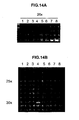
- STEAP-2 is expressed only in normal prostate among human tissues tested ( FIGS. 14 and 15 ) and is also expressed in prostate cancer ( FIG. 15 ), and thus shows some similarity in expression profile to STEAP-1. However, STEAP-2 exhibits a different mRNA expression profile relative to STEAP-1 in prostate cancer samples (compare FIGS. 3 and 15 ) and in other non-prostate cancers tested (compare FIGS. 5 and 16 ). These differences in the expression profiles of STEAP-1 and STEAP-2 suggest that they are differentially regulated.
- STEAP-3 and STEAP-4 appear to be closely related to both STEAP-1 and STEAP-2 on a structural level, and both appear to be transmembrane proteins as well.
- STEAP-3 is more related to STEAP-2 (47.8% identity) than to STEAP-1 (40.9% identity).
- STEAP-3 and STEAP-4 show unique expression profiles.
- STEAP-3 appears to have an expression pattern which is predominantly restricted to placenta and, to a smaller degree, expression is seen in prostate but not in other normal tissues tested.
- STEAP-4 seems to be expressed predominantly in liver by RT-PCR analysis. Neither STEAP-3 nor STEAP-4 appear to be expressed in prostate cancer xenografts which exhibit high level STEAP-1 and STEAP-2 expression.
- the STEAP proteins exhibit characteristics of proteins involved in cell signaling pathways. Specifically, STEAP-1 and STEAP-2, when expressed in PC3 cells, activate phosphorylation of p38, a protein involved in the MAPK signaling cascade. In addition, STEAP-2 expression induces tyrosine phosphorylation, and STEAP-1 mediates activation of tyrosine kinase in odorant-treated cells.
- STEAP-related molecules and cells modified to express STEAP in high throughput assays to identify molecules capable of altering cellular signaling pathways, leading to the identification of novel therapeutic agents.
- the assay identifies molecules capable of inhibiting STEAP function, which molecules are thereby capable of modulating the progression of cancer or other disease associated with dysregulated cell growth.
- STEAP-1 maps within 7p22 (7p22.3), a large region of allelic gain reported for both primary and recurrent prostate cancers ( Visakorpi et al., 1995 Cancer Res. 55: 342 , Nupponen et al., 1998 American J. Pathol. 153: 141 ), suggesting that up-regulation of STEAP-1 in cancer might include genomic mechanisms.
- both STEAP-2 and STEAP-3 locate to chromosome 7q21, suggesting that these two genes arose by gene duplication.
- cell surface molecules that contain six transmembrane domains include ion channels ( Dolly and Parcej, 1996 J Bioenerg Biomembr 28:231 ) and water channels or aquaporins ( Reizer et al., 1993 Crit Rev Biochem Mol Biol 28:235 ). Structural studies show that both types of molecules assemble into tetrameric complexes to form functional channels ( Christie, 1995, Clin Exp Pharmacol Physiol 22:944 , Walz et al., 1997 Nature 387:624 , Cheng et al., 1997 Nature 387:627 ).
- xenopus oocytes (or other cells) expressing STEAP may be analyzed using voltage-clamp and patch-clamp experiments to determine if STEAP functions as an ion-channel. Oocyte cell volume may also be measured to determine if STEAP exhibits water channel properties. If STEAPs function as channel or gap-junction proteins, they may serve as excellent targets for inhibition using, for example, antibodies, small molecules, and polynucleotides capable of inhibiting expression or function. The restricted expression pattern in normal tissue, and the high levels of expression in cancer tissue suggest that interfering with STEAP function may selectively kill cancer cells.
- STEAP proteins function as ion channels, transport proteins or gap-junction proteins in epithelial cell function. Ion channels have been implicated in proliferation and invasiveness of prostate cancer cells ( Lalani et al., 1997, Cancer Metastasis Rev 16:29 ). Both rat and human prostate cancer cells contain sub-population of cells with higher and lower expression levels of sodium channels. Higher levels of sodium channel expression correlate with more aggressive invasiveness in vitro ( Smith et al, 1998, FEBS Lett. 423:19 ).
- the STEAP cDNA sequences described herein enable the isolation of other polynucleotides encoding STEAP gene product(s), as well as the isolation of polynucleotides encoding STEAP gene product homologues, alternatively spliced isoforms, allelic variants, and mutant forms of the STEAP gene product.
- Various molecular cloning methods that can be employed to isolate full length cDNAs encoding a STEAP gene are well known (See, for example, Sambrook, J. et aL Molecular Cloning. A Laboratory Manual, 2d edition., Cold Spring Harbor Press, New York, 1989 ; Current Protocols in Molecular Biology.
- STEAP gene cDNAs may be identified by probing with labeled STEAP, cDNA or a fragment thereof.
- a STEAP cDNA or a portion thereof can be synthesized and used as a probe to retrieve overlapping and full length cDNAs corresponding to a STEAP gene.
- the STEAP- gene itself may be isolated by screening genomic DNA libraries, bacterial artificial chromosome libraries (BACs), yeast artificial chromosome libraries (YACs), and the like, with STEAP DNA probes or primer.
- recombinant DNA or RNA molecules containing a STEAP polynucleotide including but not limited to phages, plasmids, phagemids, cosmids, YACs, BACs, as well as various viral and non-viral vectors well known in the art, and cells transformed or transfected with such recombinant DNA or RNA molecules.
- a recombinant DNA or RNA molecule is a DNA or RNA molecule that has been subjected to molecular manipulation in vitro. Methods for generating such molecules are well known (see, for example, Sambrook et al, 1989, supra).
- a host-vector system comprising a recombinant DNA molecule containing a STEAP polynucleotide within a suitable prokaryotic or eukaryotic host cell.
- suitable eukaryotic host cells include a yeast cell, a plant cell, or an animal cell, such as a mammalian cell or an insect cell (e.g., a baculovirus-infectible cell such as an Sf9 cell).
- suitable mammalian cells include various prostate cancer cell lines such LNCaP, PC-3, DU145, LAPC-4, TsuPr1, other transfectable or transducible prostate cancer cell lines, as well as a number of mammalian cells routinely used for the expression of recombinant proteins (e.g., COS, CHO, 293, 293T cells). More particularly, a polynucleotide comprising the coding sequence of a STEAP may be used to generate STEAP proteins or fragments thereof using any number of host vector systems routinely used and widely known in the art.
- STEAP may be preferably expressed in several prostate cancer and non-prostate cell lines, including for example 293, 293T, rat-1, 3T3, PC-3, LNCaP and TsuPr1.
- the host vector systems as described herein are useful for the production of a STEAP protein or fragment thereof. Such host-vector systems may be employed to study the functional properties of STEAP and STEAP mutations.
- Proteins encoded by the STEAP genes, or by fragments thereof, will have a variety of uses, including but not limited to generating antibodies and in methods for identifying ligands and other agents and cellular constituents that bind to a STEAP gene product.
- Antibodies raised against a STEAP protein or fragment thereof may be useful in diagnostic and prognostic assays, imaging methodologies (including, particularly, cancer imaging), and therapeutic methods in the management of human cancers characterized by expression of a STEAP protein, including but not limited to cancer of the prostate.
- Various immunological assays useful for the detection of STEAP proteins are contemplated, including but not limited to various types of radioimmunoassays, enzyme-linked immunosorbent assays (ELISA), enzyme-linked immunofluorescent assays (ELIFA), immunocytochemical methods, and the like.
- ELISA enzyme-linked immunosorbent assays
- ELIFA enzyme-linked immunofluorescent assays
- Such antibodies may be labeled and used as immunological imaging reagents capable of detecting prostate cells (e.g., in radioscintigraphic imaging methods).
- STEAP proteins may also be particularly useful in generating cancer vaccines, as further described below.
- a STEAP protein refers to a protein that has or includes the amino acid sequence of human STEAP-1 as provided in FIG. 1A-B , human STEAP-2 as provided in FIG. 9A-D , human STEAP-3 as provided in FIG. 10A-E , the amino acid sequence of other mammalian STEAP homologs (e.g., STEAP-4) and variants, as well as allelic variants and conservative substitution mutants of these proteins that have STEAP biological activity, to the extent that such variants and homologs can be isolated/generated and characterized without undue experimentation following the methods outlined below.
- STEAP polypeptide refers to a polypeptide fragment or a STEAP protein of at least 10 amino acids, preferably at least 15 amino acids.
- a specific embodiment of a STEEP protein comprises a polypeptide having the amino acid sequence of human STEAP-1 as shown in FIG. 1A-B .
- Another embodiment of a STEAP protein comprises a polypeptide containing the STEAP-2 amino acid sequence as shown in FIG. 9A-D .
- Another embodiment comprises a polypeptide containing the STEAP-3 amino acid sequence of shown in FIG. 10A-E .
- Yet another embodiment comprises a polypeptide containing the partial STEAP-4 amino acid sequence of shown in FIG. 11A-B .
- allelic variants of human STEAP will share a high degree of structural identity and homology (e.g., 90% or more identity).
- allelic variants of the STEAP proteins will contain conservative amino acid substitutions within the STEAP sequences described herein or will contain a substitution of an amino acid from a corresponding position in a STEAP homologue.
- One class of STEAP allelic variants will be proteins that share a high degree of homology with at least a small region of a particular STEAP amino acid sequence, but will further contain a radical departure from the sequence, such as a non-conservative substitution, truncation insertion or frame shift.
- Conservative amino acid substitutions can frequently be made in a protein without altering either the conformation or the function of the protein. Such changes include substituting any of isoleucine (I), valine (V), and leucine (L) for any other of these hydrophobic amino acids; aspartic acid (D) for glutamic acid (E) and vice versa; glutamine (Q) for asparagine (N) and vice versa; and serine (S) for threonine (T) and vice versa. Other substitutions can also be considered conservative, depending on the environment of the particular amino acid and its role in the three-dimensional structure of the protein.
- glycine (G) and alanine (A) can frequently b interchangeable, as can alanine (A) and valine (V).
- Methionine (M) which is relatively hydrophobic, can frequently be interchanged with leucine and isoleucine, and sometimes with valine.
- Lysine (K) and arginine (R) are frequently interchangeable in locations in which the significant feature of the amino acid residue is its charge and the differing pK's of these two amino acid residues are not significant. Still other changes can be considered "conservative" in particular environments.
- STEAP proteins comprising at least one epitope in common with a STEAP protein having an amino acid sequence shown in Fig. 11A-B , such that an antibody that specifically binds to a STEAP protein or variant will also specifically bind to the STEAP protein having an amino acid sequence shown in Fig. 11A-B .
- One class of STEAP protein variants shares 90% or more identity with an amino acid sequence of Fig. 11A-B .
- a more specific class of STEAP protein variants comprises an extracellular protein SCP motif as described above.
- Preferred STEAP protein variants are capable of exhibiting one or more of the defensin functions described herein, including, for example, the ability to induce tumor death or to chemoattract and/or induce migration of cells.
- STEAP proteins may be embodied in many forms, preferably in isolated form.
- a protein is said to be "isolated” when physical, mechanical or chemical methods are employed to remove the STEAP protein from cellular constituents that are normally associated with the protein.
- a skilled artisan can readily employ standard purification methods to obtain an isolated STEAP protein.
- a purified STEAP protein molecule will be substantially free of other proteins or molecules that impair the binding of STEAP to antibody or other ligand. The nature and degree of isolation and purification will depend on the intended use.
- Embodiments of a STRAP protein include a purified STEAP protein and a functional, soluble STEAP protein. In one form, such functional, soluble STEAP proteins or fragments thereof retain the ability to bind antibody or other ligand.
- STEAP polypeptides comprising biologically active fragments of the STEAP amino acid sequence, such as a polypeptide corresponding to part of the amino acid sequences for STEAP-1 as shown in FIG. 1A-B , STEAP-2 as shown in FIG. 9A-D , STEAP-3 as shown in FIG. 10A-E , or STEAP-4 as shown in FIG. 11A-B .
- Polypeptides of the invention exhibit properties of a STEAP-2 protein, such as the ability to elicit the generation of antibodies which specifically bind an epitope associated with a STEAP-2 protein.
- Polypeptides comprising amino acid sequences which are unique to a particular STEAP protein (relative to other STEAP proteins) may be used to generate antibodies which will specifically react with that particular STEAP protein.
- STEAP protein relative to other STEAP proteins
- polypeptides comprising amino acid sequences which are unique to a particular STEAP protein (relative to other STEAP proteins) may be used to generate antibodies which will specifically react with that particular STEAP protein.
- each molecule contains stretches of sequence unique to its structure. These unique stretches can be used to generate antibodies specific to a particular STEAP. Similarly, regions of conserved sequence may be used to generate antibodies that may bind to multiple STEAPs.
- Embodiments of the invention disclosed herein include a wide variety of art accepted variants of STEAP-2 proteins such as polypeptides having amino acid insertions, deletions and substitutions.
- STEAP variants can be made using methods known in the art such as site-directed mutagenesis, alanine scanning, and PCR mutagenesis.
- Site-directed mutagenesis [ Carter et al., Nucl Acids Res., 13:4331 (1986 ); Zoller et al., Nucl. Acids Res., 10:6481 (1987 )]
- cassette mutagenesis [ Wells et al, Gene, 34:315 (1985 )]
- restriction selection mutagenesis [ Wells et al, Philos. Trans. R. Soc.
- scanning amino acid analysis can also be employed to identify one or more amino acids along a contiguous sequence.
- Such amino acids include alanine, glycine, serine, and cysteine.
- Alanine is typically a preferred scanning amino acid among this group because it eliminates the side-chain beyond the beta-carbon and is less likely to alter the main-chain conformation of the variant. Alanine is also typically preferred because it is the most common amino acid. Further, it is frequently found in both buried and exposed positions [ Creighton; The Proteins, (W.H. Freeman & Co., N.Y .); Chothia, J. Mol. Biol., 150:1 (1976 )]. If alanine substitution does not yield adequate amounts of variant, an isosteric amino acid can be used.
- embodiments disclosed herein include polypeptides containing less than the full amino acid sequence of a STEAP-2 protein shown in Figure 11A-B .
- representative embodiments disclosed herein include polypeptides consisting of about amino acid 1 to about amino acid 10 of a STEAP protein shown in Figure 11A-B , polypeptides consisting of about amino acid 20 to about amino acid 30 of a STEAP protein shown in Figure 11A-B , polypeptides consisting of about amino acid 30 to about amino acid 40 of a STEAP protein shown in Figure 11A-B , polypeptides consisting of about amino acid 40 to about amino acid 50 of a STEAP protein shown in Figure 11A-B , polypeptides consisting of about amino acid 50 to about amino add 60 of a STEAP protein shown in Figure 11A-B , polypeptides consisting of about amino acid 60 to about amino acid 70 of a STEAP protein shown in Figure 11A-B , polypeptides consisting of about amino acid 70 to about
- polypeptides consisting of portions of the amino acid sequence of amino acids 100-339 of a STEAP-1 protein shown in Figure 11A-B , or amino acids 100-454 of a STEAP-2 protein shown in Figure 11A-B , or amino acids 100-459 of a STEAP-3 protein shown in Figure 11A-B , or amino acids 100-133 of a STEAP-4 protein shown in Figure 11A-B , are described herein. Polypeptides consisting of larger portions of the STEAP protein are also contemplated.
- polypeptides consisting of about amino acid 1 (or 20 or 30 or 40 etc.) to about amino acid 20, (or 30, or 40 or 50 etc.) of a STEAP protein shown in Figure 11A-B may be generated by a variety of techniques well known in the art.
- Additional illustrative embodiments disclosed herein include STEAP polypeptides containing the amino acid residues of one or more of the biological motifs contained within STEAP-2 polypeptide sequence as shown in Fig. 11A-B .
- STEAP polypeptides containing one or more of these motifs or other select regions of interest described herein will typically include an additional 5 to 25 or more amino acid residues of adjacent STEAP protein sequence on one or both sides of the selected motif(s).
- typical polypeptides of the invention can contain one or more of the regions of STEAP-2 that exhibit homology to one or more other STEAP proteins.
- typical polypeptides can contain one or more immunogenic portions of a STEAP-2 protein.
- an immunogenic portion of a STEAP protein is amino acid residues 14 through 28 of the STEAP-1 amino acid sequence as shown in FIG. 1A-B (WKMKPRRNLEEDDYL; SEQ ID NO: 22).
- Polypeptides as described herein can contain one or more predicted HLA-A2 binding peptides such as amino acids 165-173 of STEAP-1, amino acids 86-94 of STEAP-1, amino acids 262-270 of STEAP-1, amino acids 302-310 of STEAP-1, amino acids 158-166 of STEAP-1, amino acids 227-235 of STEAP-2, amino acids 402-410 of STEAP-2, amino acids 307-315 of STEAP-2, amino acids 306-314 of STEAP-2, and amino acids 100-108 of STEAP-2.
- polypeptides consist of all or part of a fragment of STEAP-2, such as amino acids 1-245, 2-204, 121-454,153-165, 182-454, 183-387, 276-453, 345-358, or 419-454 of the STEAP-2 protein shown in FIG. 8 .
- Related embodiments include polypeptides containing combinations of the different motifs discussed above with preferable embodiments being those that contain no insertions, deletions or substitutions either within the motifs or the intervening sequences of these polypeptides.
- STEAP polypeptides can be generated using standard peptide synthesis technology or using chemical cleavage methods well known in the art based on the amino acid sequences of the human STEAP- proteins disclosed herein.
- recombinant methods can be used to generate nucleic acid molecules that encode a polypeptide fragment of a STEAP protein.
- the STEAP-encoding nucleic acid molecules described herein provide means for generating defined fragments of STRAP proteins.
- STEAP polypeptides are particularly useful in generating and characterizing domain specific antibodies (e.g., antibodies recognizing an extracellular or intracellular epitope of a STEAP protein), in identifying agents or cellular factors that bind to STEAP or a particular Structural domain thereof, and in various therapeutic contexts, including but not limited to cancer vaccines.
- STEAP polypeptides containing particularly interesting structures can be predicted and/or identified using various analytical techniques well known in the art, including, for example, the methods of Chou-Fasman, Garnier-Robson, Kyte-Doolittle, Eisenberg, Karplus-Schultz or Jameson-Wolf analysis, or on the basis of immunogenicity. Fragments containing such structures are particularly useful in generating subunit specific anti-STEAP antibodies or in identifying cellular factors that bind to STEAP.
- a secreted form of STEAP may be conveniently expressed in 293T cells transfected with a CMV-driven expression vector encoding STEAP with a C-terminal 6XHis and MYC tag (pcDNA3.1 /mycHIS, Invitrogen).
- the secreted HIS-tagged STEAP in the culture media may be purified using a nickel column and standard techniques.
- an AP-tag system may be used.
- STEAP such as covalent modifications
- One type of covalent modification includes reacting targeted amino acid residues of an STEAP polypeptide with an organic derivatizing agent that is capable of reacting with selected side chains or the N- or C- terminal residues of the STEAP.
- Another type of covalent modification of the STEAP polypeptide comprises altering the native glycosylation pattern of the polypeptide.
- “Altering the native glycosylation pattern” is intended for purposes herein to mean deleting one or more carbohydrate moieties found in native sequence STEAP- (either by removing the underlying glycosylation site or by deleting the glycosylation by chemical and/or enzymatic means), and/or adding one or more glycosylation sites that are not present in the native sequence STEAP.
- the phrase includes qualitative changes in the glycosylation of the native proteins, involving a change in the nature and proportions of the various carbohydrate moieties present.
- Another type of covalent modification of STEAP comprises linking the STEAP polypeptides to one of a variety of nonproteinaceous polymers, e.g., polyethylene glycol (PEG), polypropylene glycol or polyoxyalkylenes, in the manner set forth in U.S. Patent Nos. 4,640,835 ; 4,496,689 ; 4.301,144 ; 4,670,417 ; 4,791,192 or 4,179,337 .
- PEG polyethylene glycol
- the STEAP may also be modified in a way to form a chimeric molecule comprising STEAP fused to another, heterologous polypeptide or amino acid sequence.
- a chimeric molecule comprises a fusion of the STEAP- with a polyhistidine epitope tag, which provides an epitope to which immobilized nickel can selectively bind.
- the epitope tag is generally placed at the amino- or carboxyl- terminus of the STEAP.
- the chimeric molecule may comprise a fusion of the STEAP with an immunoglobulin or a particular region of an immunoglobulin.
- the chimeric molecule also referred to as an "immunoadhesin"
- a fusion could be to the Fc region of an IgG molecule.
- the Ig fusions preferably include the substitution of a soluble (transmembrane domain deleted or inactivated) form of an STEAP polypeptide in place of at least one variable region within an Ig molecule.
- the immunoglobulin fusion includes the binge, CH2 and CH3, or the hinge, CH2, CH2 and CH3 regions of an IgG1 molecule.
- Another aspect of the invention provides antibodies that bind to STEAP-2 proteins and polypeptides.
- the most preferred antibodies will selectively bind to a STEAP-2 protein and will not bind (or will bind weakly) to non-STEAP proteins and polypeptides.
- Anti-STEAP antibodies that are particularly contemplated include monoclonal and polyclonal antibodies as well as fragments containing the antigen-binding domain and/or one or more complementarity determining regions of these antibodies.
- an antibody fragment is defined as at least a portion of the variable region of the immunoglobulin molecule that binds to its target, i.e., the antigen binding region.
- antibodies which specifically react with a particular STEAP protein and/or an epitope within a particular structural domain may be desirable to generate antibodies which specifically react with a particular STEAP protein and/or an epitope within a particular structural domain.
- preferred antibodies useful for cancer therapy and diagnostic imaging purposes are those which react with an epitope in an extracellular region of the STEAP- protein as expressed in cancer cells.
- Such antibodies may be generated by using the STEAP proteins described herein, or using peptides derived from predicted extracellular domains thereof, as an immunogen.
- regions in the extracellular loops between the indicated transmembrane domains may be selected as used to design appropriate immunogens for raising extracellular-specific antibodies.
- STEAP-2 antibodies as described herein may be particularly useful in prostate cancer therapeutic strategies, diagnostic and prognostic assays, and imaging methodologies. Similarly, such antibodies may be useful in the treatment, diagnosis, and/or prognosis of other cancers, to the extent STEAP-2 is also expressed or overexpressed in other types of cancer.
- the invention provides various immunological assays useful for the detection and quantification of STEAP-2 and mutant STEAP-2 proteins and polypeptides.
- Such assays generally comprise one or more STEAP-2 antibodies capable of recognizing and binding a STEAP-2 or mutant STEAP-2 protein, as appropriate, and may be performed within various immunological assay formats well known in the art, including but not limited to various types of radioimmunoassays, enzyme-linked immunosorbent assays (ELI SA), enzyme-linked immunofluorescent assays (ELIFA), and the like.
- immunological imaging methods capable of detecting prostate cancer are also disclosed herein, including but limited to radioscintigraphic imaging methods using labeled STEAP antibodies. Such assays may be used clinically in the detection, monitoring, and prognosis of prostate cancer, particularly advanced prostate cancer.
- STEAP antibodies may also be used in methods for purifying STEAP and mutant STEAP proteins and polypeptides and for isolating STEAP homologues and related molecules.
- the method of purifying a STEAP protein comprises incubating a STEAP antibody, which has been coupled to a solid matrix, with a lysate or other solution containing STEAP under conditions which permit the STEAP antibody to bind to STEAP; washing the solid matrix to eliminate impurities; and eluting the STEAP from the coupled antibody.
- Other uses of the STEAP antibodies of the invention include generating anti-idiotypic antibodies that mimic the STEAP protein.
- STEAP antibodies may also be used therapeutically by, for example, modulating or inhibiting the biological activity of a STEAP protein or targeting and destroying cancer cells expressing a STEAP protein.
- Antibody therapy of prostate and other cancers is more specifically described in a separate subsection below.
- antibodies may be prepared by immunizing a suitable mammalian host using a STEAP protein, peptide, or fragment, in isolated or immunoconjugated form ( Antibodies: A Laboratory Manual, CSH Press, Eds., Harlow, and Lane (1988 ); Harlow, Antibodies, Cold Spring Harbor Press, NY (1989 )).
- protein immunogens include recombinant STEAP (expressed in a baculovirus system, mammalian system, etc.), STEAP extracellular domain or extracellular loops of STEAP protein conjugated to one or more antibody constant regions, AP-tagged STEAP, etc.
- fusion proteins of STEAP may also be used, such as a fusion of STEAP with GST, maltose-binding protein (MBP), green fluorescent protein (GFP), HisMax-TOPO or MycHis (see Examples below).
- a GST fusion protein comprising all or most of an open reading frame amino acid sequence as shown in Fig. 11A-B may be produced and used as an immunogen to generate appropriate antibodies.
- Cells expressing or overexpressing STEAP may also be used for immunizations.
- any cell engineered to express STEAP may be used. Such strategies may result in the production of monoclonal antibodies with enhanced capacities for recognizing endogenous STEAP.
- Another useful immunogen comprises STEAP peptides linked to the plasma membrane of sheep red blood cells.
- the amino acid sequences of STEAP proteins as shown in Fig. 11A-B may be used to select specific regions of a STEAP protein for generating antibodies.
- hydrophobicity and hydrophilicity analyses of the STEAP amino acid sequence may be used to identify hydrophilic regions in the STEAP structure.
- Regions of the STEAP protein that show immunogenic structure, as well as other regions and domains, can readily be identified using various other methods known in the art, such as Chou-Fasman, Gamier Robson, Kyte-Doolittle, Eisenberg, Karplus-Schultz or Jameson-Wolf analysis.
- Peptides of STEAP predicted to bind HLA-A2 may be selected for the generation of antibodies or used to generate a CTL response.
- Such predicted HLA-A2 binding peptides include, but are not limited to, amino acids 165-173 of STEAP-1, amino acids 86-94 of STEAP-1, amino acids 262-270 of STEAP-1, amino acids 302-310 of STEAP-1, amino acids 158-166 of STEAP-1, amino acids 227-235 of STEAP-2, amino acids 402-410 of STEAP-2, amino acids 307-315 of STEAP-2, amino acids 306-314 of STEAP-2, and amino acids 100-108 of STEAP-2.
- immunogenicity has been demonstrated with STEAP, which was used to generate polyclonal and monoclonal antibodies using rabbits and mice, respectively.
- This B cell response is the result of an initial T cell response elicited by the immunogenic portions of STEAP.
- Methods for preparing a protein or polypeptide for use as an immunogen and for preparing immunogenic conjugates of a protein with a carrier such as BSA, KLH, or other carrier proteins are well known in the art. In some circumstances, direct conjugation using, for example, carbodiimide reagents may be used; in other instances linking reagents such as those supplied by Pierce Chemical Co., Rockford, IL, may be effective.
- Administration of a STEAP immunogen is conducted generally by injection over a suitable period and with use of a suitable adjuvant, as is generally understood in the art. During the immunization schedule, titers of antibodies can be taken to determine adequacy of antibody formation.
- STEAP monoclonal antibodies are preferred and may be produced by various means well known in the art.
- immortalized cell lines which secrete a desired monoclonal antibody may be prepared using the standard hybridoma technology of Kohler and Milstein or modifications which immortalize producing B cells, a is generally known.
- the immortalized cell lines secreting the desired antibodies are screened by immunoassay in which the antigen is the STEAP protein or STEAP fragment.
- the cells may be expanded and antibodies produced either from in vitro cultures or from ascites fluid.
- the antibodies or fragments may also be produced, using current technology, by recombinant means. Regions that bind specifically to the desired regions of the STEAP protein can also be produced in the context of chimeric or CDR grafted antibodies of multiple species origin. Humanized or human STEAP antibodies may also be produced and are preferred for use in therapeutic contexts. Methods for humanizing murine and other non-human antibodies by substituting one or more of the non-human antibody CDRs for corresponding human antibody sequences are well known (see for example, Jones et al., 1986, Nature 321: 522-525 ; Riechmann et al., 1988, Nature 332: 323-327 ; Verhoeyen et al., 1988, Science 239:1534-1536 ).
- Fully human STEAP monoclonal antibodies may be generated using cloning technologies employing large human Ig gene combinatorial libraries (i.e., phage display) ( Griffiths and Hoogenboom, Building an in vitro immune system: human antibodies from phage display libraries. In: Protein Engineering of Antibody Molecules for Prophylactic and Therapeutic Applications in Man. Clark, M. (Ed.), Nottingham Academic, pp 45-64 (1993 ); Burton and Barbas, Human Antibodies from combinatorial libraries. Id., pp 65-82 ). Fully human STEAP monoclonal antibodies may also be produced using transgenic mice engineered to contain human immunoglobulin gene loci as described in U.S. Patent No.
- Reactivity of STEAP antibodies with a STEAP protein may be established by a number of well known means, including western blot, immunoprecipitation, ELISA, and FACS analyses using, as appropriate, STEAP proteins, peptides, STEAP expressing cells or extracts thereof.
- a STEAP antibody or fragment thereof of the invention may be labeled with a detectable marker or conjugated to a second molecule, such as a cytotoxin or other therapeutic agent, and used for targeting the second molecule to a STEAP positive cell ( Vitetta, E.S. et al., 1993, Immunotoxin therapy, in DeVita, Jr., V.T. et al., eds., Cancer. Principles and Practice of Oncology, 4th ed., J.B. Lippincott Co., Philadelphia, 2624-2636 ).
- a detectable marker or conjugated to a second molecule such as a cytotoxin or other therapeutic agent
- cytotoxic agents include, but are not limited to ricin, ricin A-chain, doxorubicin, maytansinoids, daunorubicin, taxol, ethidium bromide, mitomycin, etoposide, tenoposide, vincristine, vinblastine, colchicine, dihydroxy anthracin dione, actinomycin, diphtheria toxin, Pseudomonas exotoxin (PE) A, PE40, abrin, abrin A chain, modeccin A chain, alpha-sarcin, gelonin, mitogellin, retstrictocin, phenomycin, enomycin, curicin, crotin, calicheamicin, sapaonaria officinalis inhibitor, and glucocorticoid and other chemotherapeutic agents, as well as radioisotopes such as 212 Bi, 131 I, 131 In, 90 Y, and 186 Re.
- Suitable detectable markers include, but are not limited to, a radioisotope, a fluorescent compound, a bioluminescent compound, chemiluminescent compound, a metal chelator or an enzyme.
- Antibodies may also be conjugated to an anti-cancer pro-drug activating enzyme capable of converting the pro-drug to its active form. See, for example, US Patent No. 4,975,287 .
- bi-specific antibodies specific for two or more STEAP- epitopes may be generated using methods generally known in the art.
- antibody effector functions may be modified to enhance the therapeutic effect of STEAP antibodies on cancer cells.
- cysteine residues may be engineered into the Fc region, permitting the formation of interchain disulfide bonds and the generation of homodimers which may have enhanced capacities for internalization, ADCC and/or complement mediated cell killing (see, for example, Caron et al., 1992, J. Exp. Med. 176: 1191-1195 ; Shopes, 1992, J. Immunol. 148: 2918-2922 ).
- Homodimeric antibodies may also be generated by cross-linking techniques known in the art (e.g., Wolff et al., Cancer Res. 53: 2560-2565 ).
- Nucleic acids that encode STEAP or its modified forms can also be used to generate either transgenic animals or "knock out" animals which, in turn, are useful in the development and screening of therapeutically useful reagents.
- a transgenic animal e.g., a mouse or rat
- a transgenic animal is an animal having cells that contain a transgene, which transgene was introduced into the animal or an ancestor of the animal at a prenatal, e.g., an embryonic stage.
- a transgene is a DNA that is integrated into the genome of a cell from which a transgenic animal develops.
- cDNA encoding STEAP can be used to clone genomic DNA encoding STEAP in accordance with established techniques and the genomic sequences used to generate transgenic animals that contain cells that express DNA encoding STEAP.
- transgenic animals particularly animals such as mice or rats
- transgenic animals have become conventional in the art and are described, for example, in U.S. Patent Nos. 4,736,866 and 4,870,009 .
- particular cells would be targeted for STEAP transgene incorporation with tissue-specific enhancers.
- Transgenic animals that include a copy of a transgene encoding STEAP introduced into the germ line of the animal at an embryonic stage can be used to examine the effect of increased expression of DNA encoding STEAP.
- Such animals can be used as tester animals for reagents thought to confer protection from, for example, pathological conditions associated with its overexpression.
- an animal is treated with the reagent and a reduced incidence of the pathological condition, compared to untreated animals bearing the transgcne, would indicate a potential therapeutic intervention for the pathological condition.
- non-human homologues of STEAP can be used to construct a STEAP "knock out" animal that has a defective or altered gene encoding STEAP as a result of homologous recombination between the endogenous gene encoding STEAP and altered genomic DNA encoding STEAP introduced into an embryonic cell of the animal.
- cDNA encoding STEAP can be used to clone genomic DNA encoding STEAP in accordance with established techniques.
- a portion of the genomic DNA encoding STEAP can be deleted or replaced with another gene, such as a gene encoding a selectable marker that can be used to monitor integration.
- flanking DNA typically, several kilobases of unaltered flanking DNA (both at the 5' and 3' ends) are included in the vector (see e.g., Thomas and Capecchi, 1987, Cell 51:503 for a description of homologous recombination vectors).
- the vector is introduced into an embryonic stem cell line (e.g., by electroporation) and cells in which the introduced DNA has homologously recombined with the endogenous DNA are selected (see e.g., Li ct al., 1992, Cell 69:915 ).
- the selected cells are then injected into a blastocyst of an animal (e.g., a mouse or rat) to form aggregation chimeras (see e.g., Bradley, in Teratocarcinomas and Embryonic Stem Cells: A Practical Approach, E. J. Robertson, ed., IRL, Oxford, 1987, pp. 113-152 ).
- an animal e.g., a mouse or rat
- aggregation chimeras see e.g., Bradley, in Teratocarcinomas and Embryonic Stem Cells: A Practical Approach, E. J. Robertson, ed., IRL, Oxford, 1987, pp. 113-152 .
- a chimeric embryo can then be implanted into a suitable pseudopregnant female foster animal and the embryo brought to term to create a "knock out" animal.
- Progeny harboring the homologously recombined DNA in their germ cells can be identified by standard techniques and used to breed animals in which all cells of the animal contain the homologously recombined DNA.
- Knockout animals can be characterized for instance, for their ability to defend against certain pathological conditions and for their development of pathological conditions due to absence of the STEAP polypeptide.
- Assays that evaluate the status of a STEAP- gene and STEAP gene products in an individual may provide information on the growth or oncogenic potential of a biological sample from this individual. For example, because STEAP mRNA is so highly expressed in prostate, and not in most normal tissue, and because its expression is associated with certain cancers, assays that evaluate the relative levels of STEAP mRNA transcripts or proteins in a biological sample may be used to diagnose a disease associated with STEAP dysregulation, such as cancer or benign prostatic hyperplasia (BPH), and may provide prognostic information useful in defining appropriate therapeutic options. Similarly, assays that evaluate the integrity STEAP nucleotide and amino acid sequences in a biological sample, may also be used in this context.
- STEAP mRNA is expressed in prostate and other cancers, and not in most normal tissue, provides evidence that this gene is associated with dysregulated cell growth and therefore identifies this gene and its products as targets that the skilled artisan can use to evaluate biological samples from individuals suspected of having a disease associated with STEAP dysregulation.
- the expression of STEAP- is normally restricted to prostate, one can also evaluate biological samples taken from other tissues to detect STEAP expression as an indication of metastasis.
- antibodies directed to STEAP-1 provide a superior marker for detection of metastases, as compared to the conventional prostate cancer marker, PSA. Such a marker can be useful both in evaluation tissue biopsies and as part of in vivo imaging strategies.
- the evaluation of the expression status of STEAP gene and its products can be used to gain information on the disease potential of a tissue sample.
- expression status in this context is used to broadly refer to the variety of factors involved in the expression, function and regulation of a gene and its products such as the level of mRNA expression, the integrity of the expressed gene products (such as the nucleic and amino acid sequences) and transcriptional and translational modifications to these molecules.
- the expression status of STEAP may provide information useful for predicting susceptibility to particular disease stages, progression, and/or tumor aggressiveness.
- the invention provides methods and assays for determining STEAP-2 expression status and diagnosing cancers that express STEAP-2, such as cancers of the prostate.
- STEAP, expression status in patient samples may be analyzed by a number of means well known in the art, including without limitation, immunohistochemical analysis, in situ hybridization, RT-PCR analysis on laser capture micro-dissected samples, western blot analysis of clinical samples and cell lines, and tissue anay analysis.
- Typical protocols for evaluating the expression status of the STEAP gene and gene products can be found, for example in Current Protocols In Molecular Biology, Units 2 [Northern Blotting], 4 [Southern Blotting], 15 [Immunoblotting] and 18 (PCR Analysis), Frederick M. Ausubul et al. eds., 1995 .
- a disease associated with dysregulated cell growth such as hyperplasia or cancer
- the method comprises comparing the status of STEAP in the biological sample to the status of STEAP in a corresponding normal sample, wherein alterations in the status of STEAP in the biological sample are associated with dysregulated cellular growth.
- the status of STEAP in the biological sample can be evaluated by, for example, examining levels of STEAP mRNA expression or levels of STEAP protein expression.
- an alteration in the status of STEAP is identified by the presence of STEAP expressing cells in a biological sample from a tissue in which STEAP expressing cells are normally absent.
- Described herein are assays useful in determining the presence of cancer in an individual, comprising detecting a significant increase in STEAP mRNA or protein expression in a test cell or tissue sample relative to expression levels in the corresponding normal cell or tissue.
- the presence of STEAP mRNA may, for example, be evaluated in tissue samples including but not limited to colon, lung, prostate, pancreas, bladder, breast, ovary, cervix, testis, head and neck, brain, stomach, bone, etc.
- the presence of significant STEAP expression in any of these tissues may be useful to indicate the emergence, presence and/or severity of these cancers or a metastasis of cancer originating in another tissue, since the corresponding normal tissues do not express STEAP mRNA or express it at lower levels.
- STEAP expression status may be determined at the protein level rather than at the nucleic acid level.
- a method or assay would comprise determining the level of STEAP protein expressed by cells in a test tissue sample and comparing the level so determined to the level of STEAP expressed in a corresponding normal sample.
- the presence of STEAP protein is evaluated, for example, using immunohistochemical methods.
- STEAP antibodies or binding partners capable of detecting STEAP protein expression may be used in a variety of assay formats well known in the art for this purpose. As shown in the accompanying examples, STEAP immunoreactivity is associated with prostate, bladder and lung cancer, as well as BPH and prostate cancer metastases.
- Such embodiments are useful because perturbations in the nucleotide and amino acid sequences are observed in a large number of proteins associated with a growth dysregulated phenotype (see e.g. Marrogi et al, J. Cutan. Pathol 26(8): 369-378 (1999 )).
- assays for observing perturbations in nucleotide and amino acid sequences are well known in the art.
- nucleic acid or amino acid sequences of STEAP gene products may be observed by the northern, Southern, western, PCR and DNA sequencing protocols discussed herein.
- other methods for observing perturbations in nucleotide and amino acid sequences such as single strand conformation polymorphism analysis are well known in the art (see e.g. U.S. Patent Nos. 5,382,510 and 5,952,170 ).
- Aberrant demethylation and/or hypermethylation of CpG islands in gene 5' regulatory regions frequently occurs in immortalized and transformed cells and can result in altered expression of various genes.
- promoter hypermethylation of the pi-class glutathione S-transferase (a protein expressed in normal prostate but not expressed in >90% of prostate carcinomas) appears to permanently silence transcription of this gene and is the most frequently detected genomic alteration in prostate carcinomas ( De Marzo et al., 1999, Am. J. Pathol. 155(6): 1985-1992 ).
- LAGE-I tumor specific gene which is not expressed in normal prostate but is expressed in 25-50% of prostate cancers
- deoxy-azacytidine in lymphoblastoid cells
- assays for examining methylation status of a gene are well known in the art. For example, one can utilize in Southern hybridization approaches methylation-sensitive restriction enzymes which can not cleave sequences that contain methylated CpG sites in order to assess the overall methylation status of CpG islands.
- MSP methylation specific PCR
- methylation specific PCR can rapidly profile the methylation status of all the CpG sites present in a CpG island of a given gene. This procedure involves initial modification of DNA by sodium bisulfite (which will convert all unmethylated cytosines to uracil) followed by amplification using primers specific for methylated versus unmethylated DNA. Protocols involving methylation interference can also be found for example in Current Protocols In Molecular Biology, Units 12, Frederick M. Ausubel et al. eds., 1995 .
- Described herein are assays useful in determining the presence of cancer in an individual, comprising detecting a significant change in the STEAP alternative splice variants expressed in a test cell or tissue sample relative to expression levels in the corresponding normal cell or tissue.
- the monitoring of alternative splice variants of STEAP is useful because changes in the alternative splicing of proteins is suggested as one of the steps in a series of events that lead to the progression of cancers (see e.g. Carstens et al., Oncogene 15(250: 3059-3065 (1997 )).
- Gene amplification provides an additional method of assessing the status of STEAP.
- Gene amplification may be measured in a sample directly, for example, by conventional Southern blotting, northern blotting to quantitate the transcription of mRNA [ Thomas, Proc. Natl. Acad. Sci. USA, 77:5201-5205 (1980 )], dot blotting (DNA analysis), or in situ hybridization, using an appropriately labeled probe, based on the sequences provided herein.
- antibodies may be employed that can recognize specific duplexes, including DNA duplexes, RNA duplexes, and DNA-RNA hybrid duplexes or DNA-protein duplexes. The antibodies in turn may be labeled and the assay may be carried out where the duplex is bound to a surface, so that upon the formation of duplex on the surface, the presence of antibody bound to the duplex can be detected.
- peripheral blood may be conveniently assayed for the presence of cancer cells, including but not limited to prostate cancers, using RT-PCR to detect STEAP expression.
- RT-PCR detection assays for tumor cells in peripheral blood are currently being evaluated for use in the diagnosis and management of a number of human solid tumors. In the prostate cancer field, these include RT-PCR assays for the detection of cells expressing PSA and PSM ( Verkaik et al, 1997, UroL Res. 25: 373-384 ; Ghossein et al., 1995, J. Clin. Oncol. 13:1195-2000 ; Heston et al, 1995, Clin. Chem. 41: 1687-1688 ). RT-PCR assays are well known in the art.
- a method for predicting susceptibility to cancer comprises detecting STEAP mRNA or STEAP protein in a tissue sample, its presence indicating susceptibility to cancer, wherein the degree of STEAP mRNA expression present is proportional to the degree of susceptibility.
- the presence of STEAP in prostate tissue is examined, with the presence of STEAP in the sample providing an indication of prostate cancer susceptibility (or the emergence or existence of a prostate tumor).
- a method for gauging aggressiveness of a tumor comprises determining the level of STEAP mRNA or STEAP protein expressed by cells in a sample of the tumor, comparing the level so determined to the level of STEAP mRNA or STEAP protein expressed in a corresponding normal tissue taken from the same individual or a normal tissue reference sample, wherein the degree of STEAP mRNA or STEAP protein expression in the tumor sample relative to the normal sample indicates the degree of aggressiveness.
- aggressiveness of prostate tumors is evaluated by determining the extent to which STEAP is expressed in the tumor cells, with higher expression levels indicating more aggressive tumors.
- methods for observing the progression of a malignancy in an individual over time comprise determining the level of STEAP mRNA or STEAP protein expressed by cells in a sample of the tumor, comparing the level so determined to the level of STEAP mRNA or STEAP protein expressed in an equivalent tissue sample taken from the same individual at a different time, wherein the degree of STEAP mRNA or STEAP protein expression in the tumor sample over time provides information on the progression of the cancer.
- the progression of a cancer is evaluated by determining the extent to which STEAP expression in the tumor cells alters over time, with higher expression levels indicating a progression of the cancer.
- Another embodiment disclosed herein is directed to methods for observing a coincidence between the expression of STRAP gene and STEAP gene products (or perturbations in STEAP gene and STEAP gene products) and a factor that is associated with malignancy as a means of diagnosing and prognosticating the status of a tissue sample.
- factors associated with malignancy may be utilized such as the expression of genes otherwise associated with malignancy (including PSA, PSCA and PSM expression) as well as gross cytological observations (see e.g. Bocking et aL, 1984, AnaL Quant. Cytol.
- methods for observing a coincidence between the expression of STEAP gene and STEAP gene products (or perturbations in STEAP gene and STEAP gene products) and a factor that is associated with malignancy entails detecting the overexpression of STEAP mRNA or protein in a tissue sample, detecting the overexpression of PSA mRNA or protein in a tissue sample, and observing a coincidence of STEAP mRNA or protein and PSA mRNA or protein overexpression.
- the expression of STEAP and PSA mRNA in prostate tissue is examined.
- the coincidence of STEAP and PSA mRNA overexpression in the sample provides an indication of prostate cancer, prostate cancer susceptibility or the emergence or existence of a prostate tumor.
- Standard methods for the detection and quantification of STEAP mRNA include in situ hybridization using labeled STEAP riboprobes, northern blot and related techniques using STEAP polynucleotide probes, RT-PCR analysis using primers specific for STEAP, and other amplification type detection methods, such as, for example, branched DNA, SISBA, TMA and the like.
- semi-quantitative RT-PCR may be used to detect and quantify STEAP mRNA expression as described in the Examples that follow.
- primers capable of amplifying STEAP may be used for this purpose, including but not limited to the various primer sets specifically described herein. Standard methods for the detection and quantification of protein may be used for this purpose.
- polyclonal or monoclonal antibodies specifically reactive with the wild-type STEAP protein may be used in an immunohistochemical assay of biopsied tissue.
- Antibodies directed against STEAP protein can also be used to detect STEAP in a patient specimen (e.g., blood, urine, semen or other sample) using conventional techniques such as fluorescence-activated cell sorting (FACS) and/or ELISA.
- FACS fluorescence-activated cell sorting
- STEAP protein sequences disclosed herein allow the skilled artisan to identify proteins, small molecules and other agents that interact with STEAP and pathways activated by STEAP via any one of a variety of art accepted protocols.
- interaction trap systems also referred to as the "two-hybrid assay”
- molecules that interact reconstitute a transcription factor and direct expression of a reporter gene, the expression of which is then assayed.
- Typical systems identify protein-protein interactions in vivo through reconstitution of a eukaryotic transcriptional activator and are disclosed for example in U.S. Patent Nos. 5,955,280 , 5,925,523 , 5,846,722 and 6,004,746 .
- peptides that bind to selected receptor molecules such as STEAP are identified by screening libraries that encode a random or controlled collection of amino acids.
- Peptides encoded by the libraries are expressed as fusion proteins of bacteriophage coat proteins, and bacteriophage particles are then screened against the receptors of interest.
- Peptides having a wide variety of uses, such as therapeutic or diagnostic reagents may thus be identified without any prior information on the structure of the expected ligand or receptor molecule.
- Typical peptide libraries and screening methods that can be used to identify molecules that interact with STEAP protein sequences are disclosed for example in U.S. Patent Nos. 5,723,286 and 5,733,731 .
- cell lines expressing STEAP can be used to identify protein-protein interactions mediated by STEAP. This possibility can be examined using immunoprecipitation techniques as shown by others ( Hamilton BJ, et al. Biochem. Biophys. Res. Commun. 1999,261:646-51 ).
- STEAP protein can be immunoprecipitated from STEAP expressing prostate cancer cell lines using anti-STEAP antibodies.
- antibodies against His-tag can be used in a cell line engineered to express STEAP (vectors mentioned above).
- the immunoprecipitated complex can be examined for protein association by procedures such as western blotting, 35 S-methionine labeling of proteins, protein microsequencing, silver staining and two dimensional gel electrophoresis.
- Small molecules that interact with STEAP can be identified through related embodiments of such screening assays.
- small molecules can be identified that interfere with STEAP function, including molecules that interfere with STEAP's ability to bind to cells and/or to modulate tumor formation, progression, migration and/or apoptosis.
- Typical methods are discussed for example in U.S. Patent No. 5,928,868 and include methods for forming hybrid ligands in which at least one ligand is a small molecule.
- the hybrid ligand is introduced into cells that in turn contain a first and a second expression vector.
- Each expression vector includes DNA for expressing a hybrid protein that encodes a target protein linked to a coding sequence for a transcriptional module.
- the cells further contains a reporter gene, the expression of which is conditioned on the proximity of the first and second hybrid proteins to each other, an event that occurs only if the hybrid ligand binds to target sites on both hybrid proteins. Those cells that express the reporter gene are selected and the unknown small molecule or the unknown hybrid protein is identified.
- Described herein is a method of screening for a molecule that interacts with a STEAP amino acid sequence shown in Fig. 11A-B , comprising the steps of contacting a population of molecules with the STEAP amino acid sequence, allowing the population of molecules and the STEAP amino acid sequence to interact under conditions that facilitate an interaction, determining the presence of a molecule that interacts with the STEAP amino acid sequence and then separating molecules that do not interact with the STEAP amino acid sequence from molecules that do interact with the STEAP amino acid sequence.
- the method further includes purifying a molecule that interacts with the STEAP amino acid sequence.
- the STEAP amino acid sequence is contacted with a library of peptides. Additional assays for identifying molecules that modulate STEAP function are described in the Examples that follow.
- Also described herein is a method of screening for a molecule that modulates the activity of STEAP or a STEAP-related pathway.
- the method comprises contacting a molecule with a cell that expresses a STEAP protein and determining the activity of STEAP or a STEAP-related pathway.
- the determining can be via use of one of the phosphorylation assays described in the examples that follow, such as western blotting with an antibody directed to a phosphorylated signaling molecules. Alterations in the phosphorylation of the signaling molecule indicate a candidate molecule that modulates the activity of STEAP or a STEAP-related pathway.
- STEAP is a transmembrane protein, and its interaction with other cells and molecules likely plays a role in the regulation of the prostate environment and the initiation, development and/or progression of cancer.
- STEAP can be targeted for therapy via approaches aimed at inhibiting activity of the STEAP protein, inhibiting the binding or association of STEAP protein with other cells and molecules, inhibiting transcription or translation of STEAP, and/or via the use of cancer vaccines based on STEAP.
- the therapeutic strategy can thus be designed to inhibit a function of the molecule or to target the STEAP molecule itself.
- STEAP The expression profile of STEAP is reminiscent of the MAGEs, PSA and PMSA, which are tissue-specific genes that are up-regulated in melanomas and other cancers ( Van den Eynde and Boon, Int J Clin Lab Res. 27:81-86, 1997 ). Due to their tissue-specific expression and high expression levels in cancer, these molecules are currently being investigated as targets for cancer vaccines ( Durrant, Anticancer Drugs 8:727-733, 1997 ; Reynolds et al., Int J Cancer 72:972-976, 1997 ). The expression pattern of STEAP provides evidence that it is likewise an ideal target for a cancer vaccine approach to prostate cancer, as its expression is not detected in most normal tissues.
- therapeutic approaches targeting particular motifs of STEAP, or aimed at inhibiting the activity of the STEAP protein are expected to be useful for patients suffering from prostate cancer and other cancers expressing STEAP.
- the therapeutic approaches aimed at inhibiting the activity of the STEAP protein generally fall into two classes.
- One class comprises various methods for inhibiting the binding or association of the STEAP protein with its binding partner or with other proteins.
- Another class comprises a variety of methods for inhibiting the transcription of the STEAP gene or translation of STEAP mRNA.
- the cell surface nature and expression profiles of the STEAPs in cancers including prostate cancer indicate that they are promising targets for antibody therapy of prostate and other cancers expressing STEAPs.
- the experimental results described in the Examples herein provide compelling evidence that STEAP-1 and STEAP-2 are strongly expressed uniformly over the surface of glandular epithelial cells within prostate and prostate cancer cells.
- immunohistochemical analysis results show that the surface of human prostate epithelial cells (normal and cancer) appear to be uniformly coated with STEAP-1.
- Biochemical analysis confirms the cell surface localization of STEAP-1 initially suggested by its putative 6-transmembrane primary structural elements and by the pericellular staining plainly evident by immunohistochemical staining.
- STEAP-1 and STEAP-2 are uniformly expressed at high levels over the surface of prostate glandular epithelia, an ideal situation for immunotherapeutic intervention strategies that target extracellular STEAP epitopes.
- Systemic administration of STEAP-immunoreactive compositions would be expected to result in extensive contact of the composition with prostate epithelial cells via binding to STEAP extracellular epitopes.
- STEAP-1 protein expression in normal human tissues there is ample reason to expect extraordinarily without toxic, non-specific and/or non-target effects caused by the binding of the immunotherapeutic composition to STEAP-1 on non-target organs and tissues.
- STEAP-1 appears to be substantially over-expressed in a variety of other human cancers, including bladder, lung, colon, pancreatic and ovarian cancers.
- high level STEAP-1 mRNA expression is detected in all tested prostate cancer tissues and cell lines, and in most of the pancreatic, colon, and bladder cancer cell lines tested.
- High level expression of STEAP-1 is also observed in some ovarian cancer cell lines.
- Lower level expression is observed in some breast, testicular, and cervical cancer cell lines.
- Very high level expression is also detected in a Ewing sarcoma cell line.
- cell surface STRAP-1 protein is expressed in bladder, lung and colon cancers, while there is no detectable cell surface (or intracellular) STEAP-1 protein in normal colon and low expression in normal bladder.
- Antibodies specifically reactive with extracellular domains of STEAP-1 may be useful to treat these cancers systemically, either as toxin or therapeutic agent conjugates or as naked antibodies capable of inhibiting cell proliferation or function.
- STEAP-2 protein is also expressed in prostate cancer, and in other cancers as well, including colon and lung cancers.
- STEAP-2 mRNA analysis by RT-PCR and northern blot show that expression is restricted to prostate in normal tissues, is also expressed in some prostate, pancreatic, colon, testicular, ovarian and other cancers. Therefore, antibodies reactive with STEAP-2 may be useful in the treatment of prostate and other cancers.
- the expression of STEAP-3 and STEAP-4 (as well as other STEAPs) may be associated with some cancers. Thus antibodies reactive with these STEAP family member proteins may also be useful therapeutically.
- STEAP antibodies may be introduced into a patient such that the antibody binds to STEAP on the cancer cells and mediates the destruction of the cells and the tumor, inhibits the growth of the cells or the tumor, and/or eliminates STEAP function in the primary tumor, in circulating micrometastases, and/or in established metastases.
- the degree of tumor vascularization may provide guidance on which delivery approach is recommended.
- the grade and/or stage of disease would be expected to provide useful information in this regard. For example, a higher grade, more advanced tumor may be more likely to seed metastases, suggesting systemic administration in order to treat or prevent the emergence of metastases.
- Mechanisms by which such antibodies exert a therapeutic effect may include complement-mediated cytolysis, antibody-dependent cellular cytotoxicity, modulating the physiologic function of STEAP, inhibiting ligand binding or signal transduction pathways, modulating tumor cell differentiation, altering tumor angiogenesis factor profiles, and/or by inducing apoptosis.
- STEAP antibodies conjugated to toxic or therapeutic agents may also be used therapeutically to deliver the toxic or therapeutic agent directly to STEAP-bearing tumor cells.
- Cancer immunotherapy using anti-STEAP antibodies may follow the teachings generated from various approaches that have been successfully employed in the treatment of other types of cancer, including but not limited to colon cancer ( Arlen et al., 1998, Crit. Rev. Immunol. 18:133-138 ), multiple myeloma ( Ozaki et al., 1997, Blood 90:3179-3186 ; Tsunenari et al., 1997, Blood 90:2437-2444 ), gastric cancer ( Kasprzyk et al., 1992, Cancer Res. 52:2771-2776 ), B-cell lymphoma ( Funakoshi et al., 1996, J. Immunother. Emphasis Tumor Immunol.
- Some therapeutic approaches involve conjugation of naked antibody to a toxin, such as the conjugation of 131 I to anti-CD20 antibodies (e.g., Bexxar, Coulter Pharmaceutical), while others involve co-administration of antibodies and other therapeutic agents, such as Herceptin TM (trastuzumab) with paclitaxel (Genentech, Inc.).
- a toxin such as the conjugation of 131 I to anti-CD20 antibodies (e.g., Bexxar, Coulter Pharmaceutical)
- others involve co-administration of antibodies and other therapeutic agents, such as Herceptin TM (trastuzumab) with paclitaxel (Genentech, Inc.).
- Herceptin TM tacuzumab
- paclitaxel Genetech, Inc.
- STEAP antibodies can be administered in conjunction with radiation, chemotherapy or hormone ablation.
- antibody therapy may be particularly appropriate in advanced or metastatic cancers.
- Treatment with the antibody therapy of the invention may be indicated for patients who have received previously one or more chemotherapy, while combining the antibody therapy of the invention with a chemotherapeutic or radiation regimen may be preferred for patients who have not received chemotherapeutic treatment.
- antibody therapy may enable the use of reduced dosages of concomitant chemotherapy, particularly for patients who do not tolerate the toxicity of the chemotherapeutic agent very well.
- Anti-STEAP monoclonal antibodies useful in treating prostate and other cancers include those that are capable of initiating a potent immune response against the tumor and those that are capable of direct cytotoxicity.
- anti-STEAP monoclonal antibodies may elicit tumor cell lysis by either complement-mediated or antibody-dependent cell cytotoxicity (ADCC) mechanisms, both of which require an intact Fc portion of the immunoglobulin molecule for interaction with effector cell Fc receptor sites or complement proteins.
- ADCC antibody-dependent cell cytotoxicity
- anti-STEAP mAbs that exert a direct biological effect on tumor growth are useful in the practice of the invention.
- cytotoxic mAbs may act include inhibition of cell growth, modulation of cellular differentiation, modulation of tumor angiogenesis factor profiles, and the induction of apoptosis.
- the mechanism by which a particular anti-STEAP mAb exerts an anti-tumor effect may be evaluated using any number of in vitro assays designed to determine ADCC, ADMMC, complement-mediated cell lysis, and so forth, as is generally known in the art.
- the anti-tumor activity of a particular anti-STEAP mAb, or combination of anti-STEAP mAbs may be evaluated in vivo using a suitable animal model.
- a suitable animal model For example, xenogenic prostate cancer models wherein human prostate cancer explants or passaged xenograft tissues are introduced into immune compromised animals, such as nude or SCID mice, are appropriate in relation to prostate cancer and have been described ( U.S. Patent No. 6,107,540 ; Klein et al., 1997, Nature Medicine 3: 402-408 ).
- preferred monoclonal antibodies used in the practice of the therapeutic methods of the invention are those that are either fully human or humanized and that bind specifically to the target STEAP antigen with high affinity but exhibit low or no antigenicity in the patient.
- Therapeutic methods of the invention contemplate the administration of single anti-STEAP mAbs as well as combinations, or cocktails, of different mAbs.
- Such mAb cocktails may have certain advantages inasmuch as they contain mAbs that target different epitopes, exploit different effector mechanisms or combine directly cytotoxic mAbs with mAbs that rely on immune effector functionality. Such mAbs in combination may exhibit synergistic therapeutic effects.
- the administration of anti-STEAP mAbs may be combined with other therapeutic agents, including but not limited to various chemotherapeutic agents, androgen-blockers, and immune modulators (e.g., IL-2, GM-CSF).
- the anti-STEAP mAbs may be administered in their "naked" or unconjugated form, or may have therapeutic agents conjugated to them.
- the anti-STEAP monoclonal antibodies used in the practice of the method of the invention may be formulated into pharmaceutical compositions comprising a carrier suitable for the desired delivery method.
- Suitable carriers include any material which when combined with the anti-STEAP mAbs retains the anti-tumor function of the antibody and is non-reactive with the subject's immune systems. Examples include, but are not limited to, any of a number of standard pharmaceutical carriers such as sterile phosphate buffered saline solutions, bacteriostatic water, and the like.
- the anti-STEAP antibody formulations may be administered via any route capable of delivering the antibodies to the tumor site.
- Potentially effective routes of administration include, but are not limited to, intravenous, intraperitoneal, intramuscular, intratumor, intradermal, and the like.
- the preferred route of administration is by intravenous injection.
- a preferred formulation for intravenous injection comprises the anti-STEAP mAbs in a solution of preserved bacteriostatic water, sterile unpreserved water, and/or diluted in polyvinylchloride or polyethylene bags containing 0.9% sterile Sodium Chloride for Injection, USP.
- the anti-STEAP mAb preparation may be lyophilized and stored as a sterile powder, preferably under vacuum, and then reconstituted in bacteriostatic water containing, for example, benzyl alcohol preservative, or in sterile water prior to injection.
- Treatment will generally involve the repeated administration of the anti-STEAP antibody preparation via an acceptable route of administration such as intravenous injection (IV), typically at a dose in the range of about 0.1 to about 10 mg/kg body weight. Doses in the range of 10-500 mg mAb per week may be effective and well tolerated. Based on clinical experience with the Herceptin mAb in the treatment of metastatic breast cancer, an initial loading dose of approximately 4 mg/kg patient body weight IV followed by weekly doses of about 2 mg/kg IV of the anti- STEAP mAb preparation may represent an acceptable dosing regimen. Preferably, the initial loading dose is administered as a 90 minute or longer infusion. The periodic maintenance dose may be administered as a 30 minute or longer infusion, provided the initial dose was well tolerated.
- IV intravenous injection
- Such factors may include, for example, the binding affinity and half life of the mAb or mAbs used, the degree of STEAP overexpression in the patient, the extent of circulating shed STEAP antigen, the desired steady-state antibody concentration level, frequency of treatment, and the influence of chemotherapeutic agents used in combination with the treatment method of the invention.
- patients should be evaluated for the level of circulating shed STEAP antigen in serum in order to assist in the determination of the most effective dosing regimen and related factors.
- evaluations may also be used for monitoring purposes throughout therapy, and may be useful to gauge therapeutic success in combination with evaluating other parameters (such as serum PSA levels in prostate cancer therapy).
- Described herein are various methods and compositions for inhibiting the binding of STEAP to its binding partner or ligand, or its association with other protein(s) as well as methods for inhibiting STEAP function.
- recombinant molecules that are capable of binding to STEAP thereby preventing STEAP from accessing/binding to its binding partner(s) or associating with other protein(s) are used to inhibit STEAP function.
- Such recombinant molecules may, for example, contain the reactive part(s) of a STEAP specific antibody molecule.
- the STEAP binding domain of a STEAP binding partner may be engineered into a dimeric fusion protein comprising two STEAP ligand binding domains linked to the Fc portion of a human IgG, such as human IgG1.
- Such IgG portion may contain, for example, the C H 2 and C H 3 domains and the hinge region, but not the C H 1 domain.
- Such dimeric fusion proteins may be administered in soluble form to patients suffering from a cancer associated with the expression of STEAP, including but not limited to prostate cancer, where the dimeric fusion protein specifically binds to STEAP thereby blocking STEAP interaction with a binding partner and/or modulating STEAP function.
- Such dimeric fusion proteins may be further combined into multimeric proteins using known antibody linking technologies.
- recombinant vectors encoding single chain antibodies that specifically bind to STEAP may be introduced into STEAP expressing cells via gene transfer technologies, wherein the encoded single chain anti-STEAP antibody is expressed intracellularly, binds to STEAP protein, and thereby inhibits its function.
- Methods for engineering such intracellular single chain antibodies are well known.
- intracellular antibodies also known as "intrabodies”
- Intrabodies may be specifically targeted to a particular compartment within the cell, providing control over where the inhibitory activity of the treatment will be focused. This technology has been successfully applied in the art (for review, see Richardson and Marasco, 1995, TIBTECH vol. 13 ). Intrabodies have been shown to virtually eliminate the expression of otherwise abundant cell surface receptors.
- Single chain antibodies comprise the variable domains of the heavy and light chain joined by a flexible linker polypeptide, and are expressed as a single polypeptide.
- single chain antibodies may be expressed as a single chain variable region fragment joined to the light chain constant region.
- Well known intracellular trafficking signals may be engineered into recombinant polynucleotide vectors encoding such single chain antibodies in order to precisely target the expressed intrabody to the desired intracellular compartment.
- intrabodies targeted to the endoplasmic reticulum (ER) may be engineered to incorporate a leader peptide and, optionally, a C-terminal ER retention signal, such as the KDEL amino acid motif.
- Intrabodies intended to exert activity in the nucleus may be engineered to include a nuclear localization signal. Lipid moieties may be joined to intrabodies in order to tether the intrabody to the cytosolic side of the plasma membrane. Intrabodies may also be targeted to exert function in the cytosol. For example, cytosolic intrabodies may be used to sequester factors within the cytosol, thereby preventing them from being transported to their natural cellular destination.
- STEAP intrabodies are designed to bind specifically to a particular STEAP domain.
- cytosolic intrabodies that specifically bind to the STEAP protein may be used to prevent STEAP related molecules from gaining access to the nucleus, thereby preventing it from exerting any biological activity within the nucleus.
- the transcription of the intrabody may be placed under the regulatory control of an appropriate tumor-specific promoter and/or enhancer.
- an appropriate tumor-specific promoter and/or enhancer In order to target intrabody expression specifically to prostate, for example, the PSA promoter and/or promoter/enhancer may be utilized (See, for example, U.S. Patent No. 5,919,652 ).
- a method of inhibiting the transcription of the STEAP gene comprises contacting the STEAP gene with a STEAP antisense polynucleotide.
- a method of inhibiting STEAP mRNA translation comprises contacting the STEAP mRNA with an antisense polynucleotides.
- a STEAP specific ribozyme may be used to cleave the STEAP message, thereby inhibiting translation.
- Such antisense and ribozyme based methods may also be directed to the regulatory regions of the STEAP gene, such as the STEAP promoter and/or enhancer elements.
- proteins capable of inhibiting a STEAP gene transcription factor may be used to inhibit STEAP mRNA transcription.
- the various polynucleotides and compositions useful in the aforementioned methods have been described above. The use of antisense and ribozyme molecules to inhibit transcription and translation is well known in the art.
- Gene transfer and gene therapy technologies may be used for delivering therapeutic polynucleotide molecules to tumor cells synthesizing STEAP (i.e., antisense, ribozyme, polynucleotides encoding intrabodies and other STEAP inhibitory molecules).
- STEAP i.e., antisense, ribozyme, polynucleotides encoding intrabodies and other STEAP inhibitory molecules.
- a number of gene therapy approaches are known in the art.
- Recombinant vectors encoding STEAP antisense polynucleotides, ribozymes, factors capable of interfering with STEAP transcription, and so forth, may be delivered to target tumor cells using such gene therapy approaches.
- the above therapeutic approaches may be combined with any one of a wide variety of chemotherapy or radiation therapy regimens. These therapeutic approaches may also enable the use of reduced dosages of chemotherapy and/or less frequent administration, particularly in patients that do not tolerate the toxicity of the chemotherapeutic agent well.
- the anti-tumor activity of a particular composition may be evaluated using various in vitro and in vivo assay systems.
- In vitro assays for evaluating therapeutic potential include cell growth assays, soft agar assays and other assays indicative of tumor promoting activity, binding assays capable of determining the extent to which a therapeutic composition will inhibit the binding of STEAP to a binding partner, etc.
- a STEAP therapeutic composition may be evaluated in a suitable animal model
- xenogeneic prostate cancer models wherein human prostate cancer explants or passaged xenograft tissues are introduced into immune compromised animals, such as nude or SCID mice, are appropriate in relation to prostate cancer and have been described ( Klein et aL, 1997, Nature Medicine 3: 402-408 ).
- PCT Patent Application WO98/1662 Sawyers et al., published April 23,1998 describes various xenograft models of human prostate cancer capable of recapitulating the development of primary tumors, micrometastasis, and the formation of osteoblastic metastases characteristic of late stage disease. Efficacy may be predicted using assays that measure inhibition of tumor formation, tumor regression or metastasis, and the like. See, also, the Examples below.
- xenografts from bearing mice treated with the therapeutic composition may be examined for the presence of apoptotic foci and compared to untreated control xenograft-bearing mice. The extent to which apoptotic foci are found in the tumors of the treated mice provides an indication of the therapeutic efficacy of the composition.
- compositions used in the practice of the foregoing methods may be formulated into pharmaceutical compositions, including vaccine compositions, comprising a carrier suitable for the desired delivery method.
- Suitable carriers include any material that when combined with the therapeutic composition retains the anti-tumor function of the therapeutic composition and is non-reactive with the patient's immune system. Examples include, but are not limited to, any of a number of standard pharmaceutical carriers such as sterile phosphate buffered saline solutions, bacteriostatic water, and the like (see, generally, Remington's Pharmaceutical Sciences 16th Edition, A. Osal., Ed., 1980 ).
- Therapeutic formulations may be solubilized and administered via any route capable of delivering the therapeutic composition to the tumor site.
- Potentially effective routes of administration include, but are not limited to, intravenous, parenteral, intraperitoneal, intramuscular, intratumor, intradermal, intraorgan, orthotopic, and the like.
- a preferred formulation for intravenous injection comprises the therapeutic composition in a solution of preserved bacteriostatic water, sterile unpreserved water, and/or diluted in polyvinylchloride or polyethylene bags containing 0.9% sterile Sodium Chloride for Injection, USP.
- Therapeutic protein preparations may be lyophilized and stored as sterile powders, preferably under vacuum, and then reconstituted in bacteriostatic water containing, for example, benzyl alcohol preservative, or in sterile water prior to injection.
- Dosages and administration protocols for the treatment of cancers using the foregoing methods will vary with the method and the target cancer and will generally depend on a number of other factors appreciated in the art.
- the invention further provides cancer vaccines comprising a STEAP-2 protein or fragment thereof, as well as DNA based vaccines.
- STEAP cancer vaccines are expected to be effective at specifically preventing and/or treating STEAP expressing cancers without creating non-specific effects on non-target times.
- the use of a tumor antigen in a vaccine for generating humoral and cell-mediated immunity for use in anti-cancer therapy is well known in the art and has been employed in prostate cancer using human PSMA and rodent PAP immunogens ( Hodge et al.,1995, Int J. Cancer 63: 231-237 ; Fong et al,1997, J. Immunol. 159: 3113-3117 ).
- Such methods can be readily practiced by employing a STEAP protein, or fragment thereof, or a STEAP-encoding nucleic acid molecule and recombinant vectors capable of expressing and appropriately presenting the STEAP immunogen
- viral gene delivery systems may be used to deliver a STEAP-encoding nucleic acid molecule.
- Various viral gene delivery systems that can be used in the practice of this aspect of the invention include, but are not limited to, vaccinia, fowlpox, canarypox, adenovirus, influenza, poliovirus, adeno-associated virus, lentivirus, and Sindbus virus ( Restifo, 1996, Curr. Opin. Immunol. 8: 658-663 ).
- Non-viral delivery systems may also be employed by using naked DNA encoding a STEAP protein or fragment thereof introduced into the patient (e.g., intramuscularly) to induce an anti-tumor response.
- the full-length human STEAP cDNA may be employed.
- a STEAP cancer vaccine is based on the identification of immunogenic peptides within a STEAP amino acid sequence shown in Fig. 11A-B .
- STEAPs have been shown to induce T and B cell responses.
- STEAP-1 and STEAP-2 polypeptides have been used to generate an immune response in mice and rabbits for the production of monoclonal and polyclonal antibodies.
- specific portions of STEEP, and polynucleotides encoding these portions may be selected for the production of a cancer vaccine.
- One example of such a portion of a STEAP protein is amino acid residues 14 through 28 of the STEAP-1 amino acid sequence as shown in FIG. 1A-B (WKMKPRRNLEEDDYL; SEQ ID NO: 22).
- CTL epitopes can be determined using specific algorithms (e.g., Epimer, Brown University) to identify peptides within a STEAP protein that are capable of optimally binding to specified HLA alleles.
- One suitable algorithm is the HLA Peptide Motif Search algorithm available at the Bioinformatics and Molecular Analysis Section (BIMAS) web site (http://bimas.dertnih.gov/).
- This algorithm is based on binding of specific peptide sequences in the groove of HLA Class I molecules and specifically HLA-A2 ( Falk et al.,1991, Nature 351:290-6 ; Hunt et al., 1992, Science 255:1261-3 ; Parker et al., 1992.J. Immunol. 149:3580-7 ; Parker et al., 1994, J. Immunol. 152:163-75 ).
- the HLA Peptide Motif Search algorithm allows location and ranking of 8-mer, 9-mer, and 10-mer peptides from a complete protein sequence for predicted binding to HLA-A2 as well as other Class I molecules.
- HLA-A2 binding peptides are 9-mers, favorably containing a leucine at position 2 and a valine or leucine at position 9 ( Parker et al., 1992, J. Immunol. 149:3580-7 ).
- Actual binding of peptides to HLA-A2 can be evaluated by stabilization of HLA-A2 expression on the antigen processing defective cell line T2 ( Xue et al., 1997, Prostate 30:73-8 ; Peshwa et aL, 1998, Prostate 36:129-38 ).
- Immunogenicity of specific peptides can be evaluated in vitro by stimulation of CD8+ CTL in the presence of dendritic cells (Xue et al.; Peshwa et al., supra).
- Specific STEAP peptides predicted to bind HLA-A2 and preferred for use in cancer vaccines include peptides corresponding to amino acids 165-173 of STEAP-1, amino acids 86-94 of STEAP-1, amino acids 262-270 of STEAP-1, amino acids 302-310 of STEAP-1, amino acids 158-166 of STEAP-1, amino acids 227-235 of STEAP-2, amino acids 402-410 of STEAP-2, amino acids 307-315 of STEAP-2, amino acids 306-314 of STEAP-2, and amino acids 100-108 of STEAP-2.
- Dendritic cells express MHC class I and II, B7 co-stimulator, and IL-12, and are thus highly specialized antigen presenting cells.
- PSMA prostate-specific membrane antigen
- Dendritic cells can be used to present STEAP peptides to T cells in the context of MHC class I and II molecules.
- autologous dendritic cells are pulsed with STEAP peptides capable of binding to MHC molecules.
- dendritic cells are pulsed with the complete STEAP protein.
- Yet another embodiment involves engineering the overexpression of the STEAP gene in dendritic cells using various implementing vectors known in the art, such as adenovirus ( Arthur et al., 1997. Cancer Gene Ther. 4: 17-25 ), retrovirus ( Henderson et al, 1996. Cancer Res.
- Cells expressing STEAP may also be engineered to express immune modulators, such as GM-CSF, and used as immunizing agents.
- Anti-idiotypic and-STEAP antibodies can also be used in anti-cancer therapy as a vaccine for inducing an immune response to cells expressing a STEAP protein.
- the generation of anti-idiotypic antibodies is well known in the art and can readily be adapted to generate anti-idiotypic and-STEAP antibodies that mimic an epitope on a STEAP- protein (see, for example, Wagner et al., 1997, Hybridoma 16: 33-40 ; Foon et al., 1995, J Clin Invest 96: 334-342 ; Herlyn et al., 1996, Cancer Immunol Immunother 43: 65-76 ).
- Such an anti-idiotypic antibody can be used in cancer vaccine strategies.
- Genetic immunization methods may be employed to generate prophylactic or therapeutic humoral and cellular immune responses directed against cancer cells expressing STEAP.
- Constructs comprising DNA encoding a STEAP protein/immunogen and appropriate regulatory sequences may be injected directly into muscle or skin of an individual, such that the cells of the muscle or skin take up the construct and express the encoded STEAP protein/immunogen.
- Expression of the STEAP protein immunogen results in the generation of prophylactic or therapeutic humoral and cellular immunity against prostate and other STEAP-expressing cancers.
- Various prophylactic and therapeutic genetic immunization techniques known in the art may be used (for review, see information and references published at Internet address www.genweb.com).
- kits are also disclosed herein.
- Such kits may comprise a carrier means being compartmentalized to receive in close confinement one or more container means such as vials, tubes, and the like, each of the container means comprising one of the separate elements to be used in the method.
- one of the container means may comprise a probe that is or can be detectably labeled
- probe may be an antibody or polynucleotides specific for a STEAP protein or a STEAP gene or message, respectively.
- the kit may also have containers containing nucleotide(s) for amplification of the target nucleic acid sequence and/or a container comprising a reporter-means, such as a biotin-binding protein, such as avidin or streptavidin, bound to a reporter molecule, such as an enzymatic, florescent, or radioisotope label.
- a reporter-means such as a biotin-binding protein, such as avidin or streptavidin
- the kit as described herein will typically comprise the container described above and one or more other containers comprising materials desirable from a commercial and user standpoint, including buffers, diluents, filters, needles, syringes, and package inserts with instructions for use.
- a label may be present on the on the container to indicate that the composition is used for a specific therapy or non-thetapeutic application, and may also indicate directions for either in vivo or in vitro use, such as those described above.
- the invention also provides diagnostic compositions comprising STEAP-related molecules.
- Such molecules include the various STEAP-2 polynucleotides, primers, probes, proteins, fragments, antibodies described herein.
- the molecules included in the diagnostic composition may optionally be labeled with a detectable marker.
- STEAP diagnostic compositions may further comprise appropriate buffers, diluents, and other ingredients as desired.
- LAPC xenografts were obtained from Dr. Charles Sawyers (UCLA) and generated as described ( Klein et al, 1997, Nature Med 3: 402-408 ; Craft et al,1999, Cancer Res. 59: 5030-5036 ). Androgen dependent and independent LAPC-4 xenografts (LAPC-4 AD and AI, respectively) and LAPC-9 xenografts (LAPC-9 AD and AI, respectively) were grown in intact male SCID mice or in castrated males, respectively, and were passaged as small tissue chunks in recipient males.
- LAPC-4 AI xenografts were derived from LAPC-4 AD tumors and LAPC-9 AI xenografts were derived from LAPC-9 AD tumors. To generate the AI xenografts, male mice bearing LAPC AD tumors were castrated and maintained for 2-3 months. After the LAPC tumors re-grew, the tumors were harvested and passaged in castrated males or in female SCID mice.
- LAPC-4 AD xenografts were grown intratibially as follows.
- LAPC-4 AD xenograft tumor tissue grown subcutaneously was minced into 1-2 mm 3 sections while the tissue was bathed in 1X Iscoves medium, minced tissue was then centrifuged at 1.3K rpm for 4 minutes, the supernatant was resuspended in 10 ml ice cold 1X Iscoves medium and centrifuged at 1.3K rpm for 4 minutes. The pellet was then resuspended in 1X Iscoves with 1% pronase E and incubated for 20 minutes at room temperature with mild rocking agitation followed by incubation on ice for 2-4 minutes.
- Human cell lines e.g., HeLa
- Human tissue resources Center HTRC
- UCLA Lis Angeles, CA
- QualTek, Inc. Santa Barbara, CA
- Tumor tissue and cell lines were homogenized in Trizol reagent (Life Technologies, Gibco BRL) using 10 ml/ g tissue or 10 ml/ 10 8 cells to isolate total RNA.
- Poly A RNA was purified from total RNA using Qiagen's Oligotex mRNA Mini and Midi kits. Total and mRNA were quantified by spectrophotometric analysis (O.D. 260/280 nm) and analyzed by gel electrophoresis.
- SSH Suppression Subtractive Hybridization
- Double stranded cDNAs corresponding to the LAPC-4 AD xenograft (tester) and the BPH tissue (driver) were synthesized from 2 ⁇ g of poly(A)+ RNA isolated from xenograft and BPH tissue, as described above, using CLONTECH's PCR-Select cDNA Subtraction Kit and 1 ng of oligonucleotide RSACDN as primer.
- First- and second-strand synthesis were carried out as described in the Kit's user manual protocol (CLONTECH Protocol No. PT1117-1, Catalog No. K1804-1).
- the resulting cDNA was digested with Rsa I for 3 hrs. at 37°C. Digested cDNA was extracted with phenol/chloroform (1:1) and ethanol precipitated.
- BPH Driver cDNA
- Tester cDNA (LAPC-4 AD) was generated by diluting 1 ⁇ l of Rsa I digested IAPC-4 AD cDNA (400 ng) in 5 ⁇ l of water. The diluted cDNA (2 ⁇ l, 160 ng) was then ligated to 2 ⁇ l of adaptor 1 and adaptor 2 (10 ⁇ M), in separate ligation reactions, in a total volume of 10 ⁇ l at 16°C overnight, using 400 u of T4 DNA ligase (CLONTECH). Ligation was terminated with 1 ⁇ l of 0.2 M EDTA and heating at 72°C for 5 min.
- the first hybridization was performed by adding 1.5 ⁇ l (600 ng) of driver cDNA to each of two tubes containing 1.5 ⁇ l (20 ng) adaptor 1- and adaptor 2- ligated tester cDNA. In a final volume of 4 ⁇ l, the samples were overlayed with mineral oil, denatured in an MJ Research thermal cycler at 98°C for 1.5 minutes, and then were allowed to hybridize for 8 hrs at 68°C. The two hybridizations were then mixed together with an additional 1 ⁇ l of fresh denatured driver cDNA and were allowed to hybridize overnight at 68°C. The second hybridization was then diluted in 200 ⁇ l of 20 mM Hepes, pH 8.3, 50 mMNaCl, 0.2 mM EDTA, heated at 70°C for 7 min. and stored at -20°C.
- PCR 1 was conducted using the following conditions: 75°C for 5 min., 94°C for 25 sec., then 27 cycles of 94°C for 10 sec, 66°C for 30 sec, 72°C for 1.5 min. Five separate primary PCR reactions were performed for each experiment.
- PCR 2 was performed using 10-12 cycles of 94°C for 10 sec, 68°C for 30 sec, 72°C for 1.5 minutes. The PCR products were analyzed using 2% agarose gel electrophoresis.
- PCR products were inserted into pCR2.1 using the T/A vector cloning kit (Invitrogen). Transformed E. coli were subjected to blue/white and ampicillin selection. White colonies were picked and arrayed into 96 well plates and were grown in liquid culture overnight. To identify inserts, PCR amplification was performed on 1 ml of bacterial culture using the conditions of PCR1 and NP1 and NP2 as primers. PCR products were analyzed using 2% agarose gel electrophoresis.
- Bacterial clones were stored in 20% glycerol in a 96 well format. Plasmid DNA was prepared, sequenced, and subjected to nucleic acid homology searches of the GenBank, dbEST, and NCI-CGAP databases.
- First strand cDNAs were generated from 1 ⁇ g of mRNA with oligo (dT)12-18 priming using the Gibco-BRL Superscript Preamplification system. The manufacturers protocol was used and included an incubation for 50 min at 42°C with reverse transcriptase followed by RNase H treatment at 37°C for 20 min. After completing the reaction, the volume was increased to 200 ⁇ l with water prior to normalization. First strand cDNAs from 16 different normal human tissues were obtained from Clontech.
- Normalization of the first strand cDNAs from multiple tissues was performed by using the primers 5'atatcgccgcgctcgtcgtcgacaa3' (SEQ ID NO: 31) and 5'agccacacgcagctcattgtagaagg 3' (SEQ ID NO: 32) to amplify ⁇ -actin.
- First strand cDNA (5 ⁇ l) was amplified in a total volume of 50 ⁇ l containing 0.4 ⁇ M primers, 0.2 ⁇ M each dNTPs, 1XPCR buffer (Clontech, 10 mM Tris-HCL, 1.5 mM MgCl 2 , 50 mM KCl, pH8.3) and 1X Klentaq DNA polymerase (Clontech). Five ⁇ l of the PCR reaction was removed at 18, 20, and 22 cycles and used for agarose gel electrophoresis.
- PCR was performed using an MJ Research thermal cycler under the following conditions: initial denaturation was at 94°C for 15 sec, followed by a 18, 20, and 22 cycles of 94°C for 15, 65°C for 2 min, 72°C for 5 sec. A final extension at 72°C was carried out for 2 min. After agarose gel electrophoresis, the band intensities of the 283 bp ⁇ -actin bands from multiple tissues were compared by visual inspection. Dilution factors for the first strand cDNAs were calculated to result in equal ⁇ -actin band intensities in all tissues after 22 cycles of PCR. Three rounds of normalization were required to achieve equal band intensities in all tissues after 22 cycles of PCR.
- One of the cDNA clones was 436 bp in length and showed homology to an EST sequence in the NCI-CGAP tumor gene database.
- the full length cDNA encoding the 8P1D4 gene was subsequently isolated using this cDNA and renamed STEAP-1.
- the 8P1D4 cDNA nucleotide sequence corresponds to nucleotide residues 150 through 585 in the STEAP-1 cDNA sequence as shown in FIG. 1A-B .
- Another clone, designated 28P3E1, 561 bp in length showed homology to a number of EST sequences in the NCI-CGAP tumor gene database or in other databases.
- Part of the 28P3E1 sequence (356 bp) is identical to an EST derived from human fetal tissue. After the full length STEAP-1 cDNA was obtained and sequenced, it became apparent that this clone also corresponds to STEAP-1 (more specifically, to residues 622 through the 3' end of the STEAP-1 nucleotide sequence as shown in FIG. 1A-B ).
- the 436 bp 8P1D4 gene fragment (Example 1) was used to isolate additional cDNAs encoding the 8PlD4/STEAP-1 gene. Briefly, a normal human prostate cDNA library (Clontech) was screened with a labeled probe generated from the 436 bp 8P1D4 cDNA.
- One of the positive clones, clone 10, is 1195 bp in length and encodes a 339 amino acid protein having nucleotide and encoded amino acid sequences bearing no significant homology to any known human genes or proteins (homology to a rat Kidney Injury Protein recently described in International Application WO98/53071 ).
- the encoded protein contains at least 6 predicted transmembrane motifs implying a cell surface orientation (see FIG. 1A-B , predicted transmembrane motifs underlined). These structural features led to the designation "STEAP”, for " S ix T ransmembrane E pithelial A ntigen of the P rostate”.
- STEAP-1 The STEAP-1 cDNA and encoded amino acid sequences are shown in FIG. 1A-B and correspond to SEQ ID NOS: 1 and 2, respectively.
- STEAP-1 cDNA clone 10 was deposited with the American Type Culture Collection ("ATCC") (10801 University Boulevard., Manassas, VA 20110-2209 USA) as plasmid 8P1D4 clone 10.1 on August 26, 1998 as ATCC Accession Number 98849.
- ATCC American Type Culture Collection
- the STEAP-1 cDNA clone can be excised therefrom using EcoRI/XbaI double digest (EcoRI at the 5'end, XbaI at the 3'end).
- STEAP-1 STEAP-1 mRNA and STEAP-1 protein expression across a variety of human tissue specimens was undertaken. This evaluation included Northern blot, Western blot and immunohistochemical analysis of STEAP-1 expression in a large number of normal human tissues, human prostate cancer xenografts and cell lines, and various other human cancer cell lines.
- Example 3A Northern Blot Analysis of STEAP-1 mRNA Expression in Normal Human Tissues
- RNA samples were quantitatively normalized with a ⁇ -actin probe. The results are shown in FIG. 3A .
- the highest expression level was detected in normal prostate, with an approximately 5-10 fold lower level of expression detected in colon and liver.
- These northern blots showed two transcripts of approximately 1.4 kb and 4.0 kb, the former of which corresponds to the full length STEAP-1 clone 10 cDNA, which encodes the entire STEAP-1 open reading frame.
- the larger transcript was separately cloned as a 3627 bp cDNA from a normal prostate library, the sequence of which contains a 2399 bp intron ( FIG. 4 ).
- Example 3B Northern Blot Analysis of STEAP-1 mRNA Expression in Prostate Cancer Xenografts and Cell Lines
- RNAs derived from human prostate cancer xenografts and an extensive panel of prostate and non-prostate cancer cell lines were analyzed by Northern blot using STEAP-1 cDNA clone 10 as probe. All RNA samples were quantitatively normalized by ethidium bromide staining and subsequent analysis with a labeled ⁇ -actin probe.
- FIG. 5 show high level STEAP-1 expression in all the LAPC xenografts and all of the prostate cancer cell lines.
- Expression in the LAPC-9 xenografts was higher compared to the LAPC-4 xenografts, with no significant difference observed between androgen-dependent and androgen-independent sublines ( FIG. 5A ).
- Expression in the LAPC-4 xenografts was comparable to expression in normal prostate. Lower levels of expression were detected in PrEC cells (Clonetics), which represent the basal cell compartment of the prostate.
- Analysis of prostate cancer cell lines showed highest expression levels in LNCaP, an androgen dependent prostate carcinoma cell line. Significant expression was also detected in the androgen-independent cell lines PC-3 and DU145.
- High levels of STEAP expression were also detected in LAP-4 and LAPC-9 tumors that were grown within the tibia of mice as a model of prostate cancer bone metastasis ( FIG. 5B ).
- Example 3C Western Blot Analysis of STEAP-1 Protein Expression in Prostate and Other Cancers
- a 15 mer peptide corresponding to amino acid residues 14 through 28 of the STEAP-1 amino acid sequence as shown in FIG. 1A-B was synthesized and used to immunize sheep for the generation of sheep polyclonal antibodies towards the amino-terminus of the protein (anti-STEAP-1) as follows.
- the peptide was conjugated to KLH (keyhole limpet hemocyanin).
- KLH keyhole limpet hemocyanin
- the sheep was initially immunized with 400 ⁇ g of peptide in complete Freund's adjuvant. The animal was subsequently boosted every two weeks with 200 ⁇ g of peptide in incomplete Freund's adjuvant.
- Anti-STEAP antibody was affinity-purified from sheep serum using STEAP peptide coupled to affigel 10 (Bio Rad). Purified antibody is stored in phosphate-buffered saline with 0.1 % sodium azide.
- NIH 3T3 cells were infected with retroviruses encoding STEAP-1 and were selected in G418 for 2 weeks.
- Western blot analysis of protein extracts of infected and uninfected NIH 3T3 cells showed expression of a protein with an apparent molecular weight of 36 kD only in the infected cells ( FIG. 6 , lanes marked "3T3 STEAP” AND "3T3").
- the anti-STEAP-1 polyclonal antibody was used to probe western blots of cell lysates prepared from a variety of prostate cancer xenograft tissues, prostate cancer cell lines and other non-prostate cancer cell lines. Protein samples (20 ⁇ g each) were quantitatively normalized by probing the blots with an anti-Grb-2 antibody.
- STEP-1 protein was detected in all of the LAPC prostate cancer xenografts, all of the prostate cancer cell lines, a primary prostate cancer specimen and its matched normal prostate control. Highest STEAP-1 protein expression was detected in the LAPC-9 xenograft and in LNCaP cells, in agreement with the northern blot analysis described immediately above. High level expression was also observed in the bladder carcinoma cell line UM-UC-3. Expression in other cancer cell lines was also detectable ( FIG. 6 ).
- Example 3D Immunohistochemical Analysis of STEAP-1 Protein Expression in Prostate Tumor Biopsy and Surgical Specimens
- tissue sections were prepared from a variety of prostate cancer biopsies and surgical samples for immunohistochemical analysis. Tissues were fixed in 10% formalin, embedded in paraffin, and sectioned according to standard protocol. Formalin-fixed, paraffin-embedded sections of LNCaP cells were used as a positive control. Sections were stained with a sheep anti-STEAP-1 polyclonal antibody directed against a STEAP-1 N-terminal epitope (as described immediately above).
- LNCaP sections were stained in the presence of an excess amount of the STEAP-1 N-terminal peptide immunogen used to generate the polyclonal antibody (peptide 1) or a non-specific peptide derived from a distinct region of the STEAP-1 protein (peptide 2; YQQVQQNKEDAWIEH; SEQ ID NO: 33).
- FIG. 8 LNCaP cells showed uniformly strong pericellular staining in all cells ( FIG. 8B ). Excess STEAP N-terminal peptide (peptide 1) was able to competitively inhibit antibody staining ( FIG. 8A ), while peptide 2 had no effect ( FIG. 8B ). Similarly, uniformly strong pericellular staining was seen in the LAPC-9 ( FIG. 8F ) and LAPC-4 prostate cancer xenografts. These results are clear and suggest that the staining is STEAP specific. Moreover, these results show localization of STEAP to the plasma membrane, corroborating the biochemical findings presented in Example 4 below.
- FIG. 8C normal prostate tissue
- FIG. 8D grade 3 prostatic carcinoma
- FIG. 8E grade 4 prostatic carcinoma
- Light to strong staining was observed in the glandular epithelia of all prostate cancer samples tested as well as in all samples derived from normal prostate or benign disease (e.g., BPH.
- the signal appears to be strongest at the cell membrane of the epithelial cells, especially at the cell-cell junctions ( FIG. 8C-E ) and is also inhibited with excess STEAP N-terminal peptide 1.
- Some basal cell staining is also seen in normal prostate ( FIG. 8C ), which is more apparent when examining atrophic glands.
- STEAP-1 seems to be expressed at all stages of prostate cancer since lower grades ( FIG. 8D ), higher grades ( FIG. 8E ) and metastatic prostate cancer (represented by LAPC-9; FIG. 8F ; See also human patient metastases shown in FIG. 32A-B ) all exhibit strong staining.
- Table 1 Immunohistochemical Staining of Human Tissues With Anti-STEAP-1 Sheep Polyclonal Antibody Staining Intensity Tissues* None Cerebellum, cerebral cortex, spinal cord, heart, skeletal muscle (2), artery, thymus, spleen (4), bone marrow, lymph node (3), lung (8), liver (4), ileum, kidney (2), testis (2), ovary, placenta, breast, adrenal gland (2), thyroid gland (2), skin (2) Light Cytoplasmic Ureter, bladder cancer (2/5), colon (4/7), colon cancer (4/7, fallopian tubes (2), pituitary gland, pancreas, stomach (1/2), uterus (1/2) Light Membrane Prostate cancer bone marrow metastasis (1/6), prostate cancer lymph node metastasis (1/5), lung cancer (1/6) Moderate to Strong Membrane Prostate (7/7), BPH (5/5), prostate cancer (20/20)**, prostate cancer lymph node metastasis (4/5), prostate cancer bone marrow
- Example 3E Immunohistochemical Analysis of STEAP-1 Protein Expression in Bladder and Lung Cancer
- cDNA clone 10 was cloned into the pcDNA 3.1 Myc-His plasmid (Invitrogen), which encodes a 6His tag at the carboxyl-terminus, transfected into 293T cells, and analyzed by flow cytometry using and-His monoclonal antibody (His-probe, Santa Cruz) as well as the anti-STEAP-1 polyclonal antibody described above. Staining of cells was performed on intact cells as well as permeabilized cells. The results indicated that only permeabilized cells stained with both antibodies, suggesting that both termini of the STEAP-1 protein are localized intracellularly. It is therefore possible that one or more of the STEAP-1 protein termini are associated with intracellular organelles rather than the plasma membrane.
- STEAP-1 protein was expressed at the cell surface.
- intact STEAP-1-transfected 293T cells were labeled with a biotinylation reagent that does not enter live cells.
- STEAP-1 was then immunoprecipitated from cell extracts using the anti-His and anti-STEAP antibodies.
- SV40 large T antigen, an intracellular protein that is expressed at high levels in 293T cells, and the endogenous cell surface transferrin receptor were immunoprecipitated as negative and positive controls, respectively.
- the proteins were transferred to a membrane and visualized with horseradish peroxidase-conjugated streptavidin. The results of this analysis are shown in FIG. 7 .
- STEAP-1 is a type IIIa membrane protein with a molecular topology of six potential transmembrane domains, 3 extracellular loops, 2 intracellular loops and two intracellular termini.
- a schematic representation of STEAP-1 protein topology relative to the cell membrane is shown in FIG. 1B .
- prostate, bladder and colon cancer cells were directly analyzed for cell surface expression of STEAP-1 by biotinylation studies. Briefly, biotinylated cell surface proteins were affinity purified with streptavidin-gel and probed with the anti-STEAP- polyclonal antibody described above.
- STEAP-1 Glutathione S-transferase (GSf) gene by cloning into pGEX-6P-1(Amersham Pharmacia Biotech, NJ). All constructs were made to generate recombinant STEAP-1 protein sequences with GST fused at the N-terminus and a six histidine epitope at the C-terminus. The six histidine epitope tag was generated by adding the histidine codons to the cloning primer at the 3' end of the ORF. A Prescission"" recognition site permits cleavage of the GST tag from STEAP-1. The ampicillin resistance gene and pBR322 origin permits selection and maintenance of the plasmid in E . coli. The following fragments of STEAP-1 were cloned into pGEX-6P-1:
- Exemplary additional constructs that can be made in pGEX-6P-1 spanning the following regions of the STEAP-1 protein include:
- STEAP-1 To express STEAP-1 in mammalian cells, the 1,017 bp STEAP-1 ORF (with translational start Kozak consensus) was cloned into pcDNA3.1 /MycHis-Version B (Invitrogen, Carlsbad, CA). Protein expression is driven from the cytomegalovirus (CMV) promoter. The recombinant protein has the myc and six histidines fused to the C-terminus.
- CMV cytomegalovirus
- the pcDNA3.1 /MycHis vector also contains the bovine growth hormone (BGH) polyadenylation signal and transcription termination sequence to enhance mRNA stability along with the SV40 origin for episomal replication and simple vector rescue in cell lines expressing the large T antigen.
- BGH bovine growth hormone
- the Neomycin resistance gene allows for selection of mammalian cells expressing the protein and the ampicillin resistance gene and ColE1 origin permits selection and maintenance of the plasmid in E . coli.
- the 1,017 bp ORF (with translational start Kozak consensus) was cloned into pSRa constructs.
- Amphotropic and ecotropic retroviruses are generated by transfection of pSRa constructs into the 293T-10A1 packaging line or co-transfection of pSRa and a helper plasmid ( ⁇ -) in 293 cells, respectively.
- the retrovirus can be used to infect a variety of mammalian cell lines, resulting in the integration of the cloned gene, STEAP-1, into the host cell lines. Protein expression is driven from a long terminal repeat (LTR).
- LTR long terminal repeat
- Neomycin resistance gene allows for selection of mammalian cells expressing the protein and the ampicillin resistance gene and ColE1 origin permits selection and maintenance of the plasmid in E . coli.
- Additional pSRa constructs were made that fused the FLAG tag to the C and N-terminus to allow detection using anti-FLAG antibodies.
- the FLAG sequence 5' gat tac aag gat gac gac gat aag 3' (SEQ ID NO: 34) were added to cloning primer at the 3' end of the ORF.
- Another pSRa construct was made that contained amino acids 182 to 454. This truncated form of the STEAP-1 protein will help determine the function of the long extracellular N-Terminal region.
- STEAP-1 has no homology to any known human genes.
- the protein sequence of STEAP-1 was used as an electronic probe to identify family members in the public EST (expression sequence tag) database (dbEST).
- dbEST expression sequence tag
- NCBI national Center for Biotechnology Information
- SSH cloning experiments also identified a STEAP-1 related cDNA fragment, clone 98P4B6.
- This clone was isolated from SSH cloning using normal prostate cDNA as tester and LAPC-4 AD cDNA as driver.
- a larger partial sequence of the 98P4B6 clone was subsequently isolated from a normal prostate library; this clone encodes an ORF of 173 amino acids with close homology to the primary structure of STEA P-1, and thus was designated STEAP-2.
- a full length STEAP-2 cDNA of 2454 bp was isolated from a prostate library.
- the STEAP-2 nucleotide and encoded ORF amino acid sequences are shown in FIG. 9A-D .
- FIGS. 11A-B and 11C An amino acid alignment of the STEAP-1 and partial STEAP-2 primary structures is shown in FIGS. 11A-B and 11C .
- STEAP-1 and - 2 share 61% identity over their 171 amino acid residue overlap ( FIG. 11C ).
- the STEAP-2 cDNA has been deposited with the American Type Culture Collection ("ATCC"; 10801 University Boulevard., Manassas, VA 20110-2209 USA) as plasmid 98P4B6-GTD3 on July 2, 1999 as ATCC Accession Number PTA-311.
- ATCC American Type Culture Collection
- the STEAP-2 cDNA (98P4B6-GTD3) contains a 355 bp 5'UTR (untranslated region) that is 72% GC rich, suggesting that it contains translational regulatory elements.
- the cDNA encodes an open reading frame (ORF) of 454 amino acids (a.a.) with six potential transmembrane domains. This is in contrast to STEAP-1, which is 339 a.a. in length. Alignment with STEAP-1 demonstrates 54.9% identity over a 237 amino acid overlap. Interestingly, the locations of the six putative transmembrane domains in STEAP-2 coincide with the locations of the transmembrane domains in STEAP-1 (see alignment).
- STEAP-2 exhibits a 205 a.a. long intracellular N-terminus (compared to 69 a.a. in STEAP-1) and a short 4 a.a. intracellular C-terminus (compared to 26 a.a. in STEAP-1). These differences could imply significant differences in function and/or interaction with intracellular signaling pathways.
- STEAP-2 To identify a unique mouse EAST corresponding to STEAP-2, the unique N-terminus of STEAP-2 was used to query the dbEST database.
- One mouse EST was isolated (AI747886, mouse kidney) that may be used in the identification of mouse STEAP-2 and in expression analysis of STEAP-2 in mouse.
- FIG. 11A-B An amino acid alignment of the structures of STEAP-1, STEAP-2, STEAP-3 and the partial sequence of the putative STEAP-4 is shown in FIG. 11A-B .
- This alignment shows a close structural similarity between all four STEAP family proteins, particularly in the predicted transmembrane domains.
- STEAP-1 and STEAP-2 demonstrate 54.9% identity over a 237 amino acid overlap.
- STEAP-1 and STEAP-3 are 40.9% identical over a 264 amino acid region, while STEAP-2 and STEAP-3 are 47.8% identical over a 416 amino acid region.
- STEAP-2 To express STEAP-2 in bacterial cells, portions of STEAP-2 were fused to the Glutatione S-transferase (GST) gene by cloning into pGEX-6P-1 (Amersham Pharmacia Biotech, NJ). All constructs were made to generate recombinant STEAP-2 protein sequences with GST fused at the N-terminus and a six histidine epitope at the C-terminus. The six histidine epitope tag was generated by adding the histidine codons to the cloning primer at the 3' end of the ORF. A PreScission TM recognition site permits cleavage of the GST tag from STEAP-2. The ampicillin resistance gene and pBR322 origin permits selection and maintenance of the plasmid in E. coli. The following fragments of STEAP-2 were cloned into pGEX-6P-1:
- Additional exemplary constructs that can be made in pGEX-6P-1 spanning the following regions of the STEAP-2 protein include:
- a pCRII construct was generated using bp 367 to 877 of the GTD3 STEAP-2 cDNA.
- the pCRII vector has Sp6 and T7 promoters flanking the insert to drive the production of STEAP-2 RNA riboprobes which were used in RNA in situ hybridization experiments.
- the 1,362 bp STEAP-2 ORF was cloned into pcDNA4/HisMax-TOPO Version A (cat# K864-20, Invitrogen, Carlsbad, CA). Protein expression is driven from the cytomegalovirus (CMV) promoter and the SP163 translational enhancer.
- CMV cytomegalovirus
- SP163 the SP163 translational enhancer.
- the recombinant protein has Xpress TM and six histidine epitopes fused to the N-terminus.
- an additional construct was made with the C-terminus of the recombinant protein containing a 28 amino acid fusion resulting from vector sequences prior to the termination codon.
- the pcDNA4/HisMax-TOPO vector also contains the bovine growth hormone (BGH) polyadenylation signal and transcription termination sequence to enhance mRNA stability along with the SV40 origin for episomal replication and simple vector rescue in cell lines expressing the large T antigen.
- BGH bovine growth hormone
- the Zeocin resistance gene allows for selection of mammalian cells expressing the protein and the ampicillin resistance gene and ColE1 origin permits selection and maintenance of the plasmid in E. coli.
- telomere sequence is driven from the cytomegalovirus (CMV) promoter.
- CMV cytomegalovirus
- the recombinant protein has the myc and six histidines fused to the C-terminus.
- the pcDNA3.1/MycHis vector also contains the bovine growth hormone (BGH) polyadenylation signal and transcription termination sequence to enhance mRNA stability along with the SV40 origin for episomal replication and simple vector rescue in cell lines expressing the large T antigen.
- BGH bovine growth hormone
- neomycin resistance gene allows for selection of mammalian cells expressing the protein and the ampicillin resistance gene and ColE1 origin permits selection and maintenance of the plasmid in E. coli.
- Additional pcDNA3.1 /MycHis constructs were generated using STEAP-2 amino acids 6 to 454, 121-454 and 182-454.
- STEAP-1 ORF was cloned into pBlueBacHIS2A (Invitrogen, California). Protein expression is driven under the polyhedrin promoter. N-terminus poly HIS and Xpress tags allow for detection and purification of the recombinant STEAP-1 protein. A C-terminus enterokinase recognition site allows for cleavage of these tags. The ampicillin resistance gene and ColE1 origin permits selection and maintenance of the plasmid in E. coli. Following cotransfection with AcMNPV DNA, a homologous recombination event occurs between these sequences resulting in a recombinant virus carrying the gene of interest and the polyhedrin. A variety of restriction enzyme sites for simplified subcloning are present. In addition, a reporter gene (b-galactosidase) conveniently identifies recombinant plaques, to eliminate tedious plaque screening.
- b-galactosidase conveniently identifies recombinant plaques, to eliminate tedious plaque screening.
- the 1,362 bp ORF (with start Kozak consensus) was cloned into pcDNA3.1CT-GFP-TOPO (Invitrogen, CA). Protein expression is driven from the cytomegalovirus (CMV) promoter.
- CMV cytomegalovirus
- the recombinant protein has the Green Fluorescent Protein (GFP) fused to the C-terminus facilitating non-invasive, in vivo detection and cell biology studies.
- the pcDNA3.1/MycHis vector also contains the bovine growth hormone (BGH) polyadenylation signal and transcription termination sequence to enhance mRNA stability along with the SV40 origin for episomal replication and simple vector rescue in cell lines expressing the large T antigen.
- BGH bovine growth hormone
- the neomycin resistance gene allows for selection of mammalian cells expressing the protein and the ampicillin resistance gene and ColE1 origin permits selection and maintenance of the plasmid in E. coli.
- the 1362 bp ORF was cloned into pSRa constructs.
- Amphotropic and ecotropic retroviruses are generated by transfection of pSRa constructs into the 293T-10A1 packaging line or co-transfection of pSRa and a helper plasmid ( ⁇ -) in 293 cells, respectively.
- the retrovirus can be used to infect a variety of mammalian cell lines, resulting in the integration of the cloned gene, STEAP-2, into the host cell-lines. Protein expression is driven from a long terminal repeat (LTR).
- LTR long terminal repeat
- the neomycin resistance gene allows for selection of mammalian cells expressing the protein and the ampicillin resistance gene and ColE1 origin permits selection and maintenance of the plasmid in E. coli.
- Additional pSRa constructs were made that fused the FLAG tag to the C and N-terminus to allow detection using anti-FLAG antibodies.
- the FLAG sequence 5' gat tac aag gat gac gac gat aag 3' (SEQ ID NO: 34) was added to cloning primer at the 3' end of the ORF.
- Another pSRa construct was made that contained amino acids 182 to 454. This truncated form of the STEAP-2 protein will help determined the function of the long extracellular N-Terminal region.
- a pSRa construct can be made to produce a myc/6 HIS fusion protein of the full length STEAP protein.
- Example 8 Expression Analysis of STEAP-2 and Other Human STEAP Family Members
- Example 8A Tissue Specific Expression of STEAP Family Members in Normal Human Tissues
- RT-PCR analysis of STEAP-2 shows expression in all the LAPC prostate cancer xenografts and in normal prostate ( FIG. 14 , panel A).
- Analysis of 8 normal human tissues shows prostate-specific expression after 25 cycles of amplification ( FIG. 14 , panel B). Lower level expression in other tissues was detected only after 30 cycles of amplification.
- Northern blotting for STEAP-2 shows a pattern of 2 transcripts (approximately 3 and 8 kb in size) expressed only in prostate (and at significantly lower levels in the LAPC xenografts), with no detectable expression in any of the 15 other normal human tissues analyzed ( FIG. 15 , panel C).
- STEAP-2 expression in normal human tissues appears to be highly prostate-specific.
- STEAP-3/AI139607 expression is shown in FIG. 12A (Northern) and FIG. 12B (RT-PCR).
- STEAP-4/R80991 expression is shown in FIG. 13 .
- Example 8B Expression of STEAP-2 in Various Cancer Cell Lines
- STEAP-2 which appears very prostate-specific, seems to be expressed at lower levels in the LAPC xenografts. This is in contrast to STEAP-1, which is highly expressed in both normal and malignant prostate tissue.
- STEAP-2 prostate cancer expression profile (relative to STEAP-1)
- Northern blotting was performed on RNA derived from the LAPC xenografts, as well as several prostate and other cancer cell lines, using a STEAP-2 specific probe (labeled cDNA clone 98P4B6).
- STEAP-2 is highly expressed in normal prostate and in some of the prostate cancer xenografts and cell lines. More particularly, very strong expression was observed in the LAPC-9 AD xenograft and the INCaP cells. Significantly attenuated or no expression was observed in the other prostate cancer xenografts and cell lines.
- Example 8C Analysis of in situ Expression of STEAP-2 RNA in Normal Prostate and Prostate Cancer
- STEAP-2 RNA was evaluated using in situ hybridization with a labeled antisense probe and using a labeled sense probe as control. All four of four normal prostate tissue specimens examined were positive for STEAP ( Figures 22A-B ). Likewise, five of five prostate cancer tissue specimens were positive using STEAP RNA in situ hybridization ( Figures 23A-B ). A PIN specimen and a sample of LNCaP cells that were examined were positive as well.
- Example 8D Analysis of STEAP-2 Expression in Cancer Tissues
- lane 1 represents a sample from an LAPC4 AD xenograft
- lane 2 is LAPC9 AD xenograft
- lane 3 is LAPC9 AD 2 xenograft (grown with human bone explant)
- lane 4 is LAPC9 AD IT (grown intratibially)
- lane 5 is pooled tissue from colon cancer patients
- lane 6 is pooled tissue from lung cancer patients
- M represents a marker lane
- lane 7 is patient normal prostate tissue
- lane 8 is patient prostate cancer tissue
- lane 9 is pooled tissue from kidney cancer patients
- lane 10 is pooled tissue from bladder cancer patients
- lane 11 is HeLa cells
- lane 12 is a water blank.
- the highest expression is found in the three LAPC9 AD xenografts. High expression is observed in the LAPC4 AD xenograft as well, and in normal prostate and lung cancer. Significant expression was detected also in colon and bladder cancer. Lower expression was detected in prostate and kidney cancer patient samples.
- Example 8E Analysis of STEAP-2 Expression in Normal Tissues
- STEAP-2 The expression of STEAP-2 in 76 normal human tissues was examined using a dot blot analysis of RNA samples. As shown in Figure 25 , STEAP-2 is expressed at very high levels only in normal prostate.
- STEAP-1 The chromosomal localization of STEAP-1 was determined using the GencBridge 4 Human/Hamster radiation hybrid (RH) panel ( Walter et aL, 1994, Nat. Genetics 7:22 ) (Research Genetics, Huntsville Al), while STEAP-2 and the STEAP homologues were mapped using the Stanford G3 radiation hybrid panel ( Stewart et al., 1997. Genome Res. 7:422 ).
- the resulting STEAP-1 mapping vector for the 93 radiation hybrid panel DNAs (2100000201101010001000000101110101221000111001110110101000100010001010010 21000001111001010000), and the mapping program available at the internet address ⁇ http://www-genome.wi.mit.edu/cgi-bin/contig/rhmapper.pl>, localized the STEAP-1 gene to chromosome 7p22.3, telomeric to D7S531.
- 98P4B6/STEAP-2 The following PCR primers were used for 98P4B6/STEAP-2: 98P4B6.1 5' GACTGAGCTGGAACTGGAATTTGT 3' (SEQ ID NO: 20) 98P4B6.2 5' TTTGAGGAGACTTCATCTCACTGG 3' (SEQ ID NO: 21)
- the resulting vector (000001001000000000000000000010010000000000100 0100000000000001000010101010010011), and the mapping program available at the internet address ⁇ http://www-shgc.stanford.edu/RH/rhserverformnew.html> maps the 98P4B6 (STEAP-2) gene to chromosome 7q21.
- the resulting vector (0000000010000000000000000000100010000020000000100 0100000001000000100010001010000010), and the mapping program available at the internet address ⁇ http://www-shgc.stanford.edu/RH/rhserverformnew.html>, maps AI139607 to chromosome 7q21.
- R80991 The following PCR primers were used for R80991: R80991.3 5' ACAAGAGCCACCTCTGGGTGAA 3' (SEQ ID NO: 35) R80991.4 5' AGTTGAGCGAGTTTGCAATGGAC 3' (SEQ ID NO: 36)
- the resulting vector (0000000000020000102000000001000000000000000000 0010000000001000011100000001001000001), and the mapping program available at the internet address ⁇ http://www-shgc.stanford.edu/RH/rhserverformnew.html> maps R80991 to chromosome 2q14-q21, near D2S2591.
- the above results show that three of the putative human STEAP family members localize to chromosome 7, as is schematically depicted in FIG. 17 .
- the STEAP-1 gene localizes to the far telomeric region of the short arm of chromosome 7, at 7p22.3, while STEAP-2 and AI139607 localize to the long arm of chromosome 7, at 7q21 ( FIG. 17 ).
- R80991 maps to chromosome 2q14-q21.
- Genomic clones for STEAP-1 were identified by searching GenBank for BAC clones containing STEAP-1 sequences, resulting in the identification of accession numbers AC004969 (PAC DJ1121E10) and AC005053 (BAC RG041D11). Using the sequences derived from the PAC and BAC clones for STEAP the intron-exon boundaries were defined ( FIG. 18 ). A total of 4 exons and 3 introns were identified within the coding region of the STEAP gene. Knowledge of the exact exon-intron structure of the STEAP-1 gene may be used for designing primers within intronic sequences which in turn may be used for genomic amplification of exons.
- Such amplification permits single-stranded conformational polymorphism (SSCP) analysis to search for polymorphisms associated with cancer.
- SSCP single-stranded conformational polymorphism
- Mutant or polymorphic exons may be sequenced and compared to wild type STEAP.
- Such analysis may be useful to identify patients who are more susceptible to aggressive prostate cancer, as well as other types of cancer, particularly colon, bladder, pancreatic, ovarian, cervical and testicular cancers.
- Southern blot analysis shows that the STEAP-1 gene exists in several species including mouse ( FIG. 19 ). Therefore, a mouse BAC library (Mouse ES 129-V release I, Genome Systems, FRAC-4431) was screened with the human cDNA for STEAP-1 (clone 10, Example 2). One positive clone, 12P11, was identified and confirmed by southern blotting ( FIG. 20 ). The intron-exon boundary information for human STEAP may be used to identify the mouse STEAP-1 coding sequences.
- the mouse STEAP-1 genomic clone may be used to study the biological role of STEAP-1 during development and tumorigenesis.
- the mouse genomic STEAP-1 clone may be inserted into a gene knock-out (K/O) vector for targeted disruption of the gene in mice, using methods generally known in the art.
- K/O gene knock-out
- the role of STEAP in metabolic processes and epithelial cell function may be elucidated.
- K/O mice may be crossed with other prostate cancer mouse models, such as the TRAMP model (Greenberg et al., 1995, PNAS 92:3439), to determine whether STEAP influences the development and progression of more or less aggressive and metastatic prostate cancers.
- Example 11 Predicted HLA-A2 Binding Peptides of STEAP-1 and STEAP-2
- HLA Peptide Motif Search algorithm found in the Bioinformatics and Molecular Analysis Section (BIMAS) Web site (http://bimas.dcrt.nih.gov/).
- the HLA Peptide Motif Search algorithm was developed by Dr. Ken Parker based on binding of specific peptide sequences in the groove of HLA Class I molecules and specifically HLA-A2 ( Falk et al., 1991, Nature 351: 290-6 ; Hunt et al., 1992, Science 255:1261-3 ; Parker et al., 1992, J. Immunol. 149:3580-7 ; Parker et al., 1994, J. Immunol.
- HLA-A2 binding peptides are 9-mers favorably containing a leucine (L) at position 2 and a valine (V) or leucine (L) at position 9.
- the results of STEAP-1 and STEAP-2 predicted binding peptides are shown in Table 2 below.
- the top 5 ranking candidates are shown along with their location, the amino acid sequence of each specific peptide, and an estimated binding score.
- the binding score corresponds to the estimated half-time of dissociation of complexes containing the peptide at 37°C at pH 6.5.
- Peptides with the highest binding score i.e. 10776.470 for STEAP-1 peptide 165; 1789.612 for STEAP-2 peptide 227) are predicted to be the most tightly bound to HLA Class I on the cell surface and thus represent the best immunogenic targets for T-cell recognition.
- Multi-transmembrane proteins have the ability to transmit signal from the membrane and initiate signaling cascades that regulate a variety of downstream events, including gene expression, cellular differentiation, migration and proliferation ( Biochim. Biophys. Acta 1997; 1348:56-62 , J. Virol. 1995; 69:675-83 ).
- the effect of STEAP-1 and STEAP-2 on tyrosine phosphorylation of PC3 cells was investigated ( Figure 26 ).
- PC3 cells, stably expressing neo, STEAP-1, or STEAP-2 were grown overnight in 1% fetal bovine serum (FBS) to reduce receptor occupancy and background activity.
- FBS fetal bovine serum
- the cells were then incubated for 5 minutes in the presence of either 1% or 10% FBS, lysed and analyzed by western blotting with anti-phosphotyrosine (4G10 mAb). An overlay using anti-Grb2 Ab was used to show that the gel was equally loaded.
- Example 13 STEAP-1- and STEAP-2- Mediated Activation of MAPK Cascades
- PC3 cells stably expressing STEAP-1, STEAP-2 or neo were grown overnight in either 1% or 10% FBS.
- Whole cell lysates were analyzed by western blotting.
- PC3 cells treated with the known p38 activators, NaSaI or TNF, were used as a positive controL The results show that while expression of the control neo gene has no effect on p38 phosphorylation, expression of STEAP-1 and STEAP-2 in PC3 cells is sufficient to induce the activation of the p38 pathway ( Figure 27A ).
- the results were verified using western blotting with an anti-p38 Ab, which shows equal protein loading on the gels ( Figure 27B ).
- FBS contains several components that may contribute to receptor-mediated ERK activation
- PC3 cells expressing neo or STEAP-1 were grown in either 0.1% or 10% FBS overnight. The cells were analyzed by anti-Phospho-ERK western blotting. This experiment shows that STEAP-1 induces the phosphorylation of ERK in 0.1% FBS, and confirms that expression of STEAP-1 is sufficient to induce activation of the ERK signaling cascade in the absence of additional stimuli.
- PC3 cells stably expressing neo or STEAP-1 were grown overnight in 0.1% FBS to allow for receptor occupancy. The cells were then treated for 5 min with the indicated concentrations (0.1-10 ⁇ M) of citralva, ethylvanillin or IBMP. Treatment with 10% FBS was used as a control.
- Whole cell lysates (20 ⁇ g) generated from the different treatment conditions were separated by SDS-PAGE and analyzed by anti-phosphotyrosine western blotting.
- luciferase (luc) based transcriptional reporter assays are carried out in cells expressing STEAP. These transcriptional reporters contain consensus binding sites for known transcription factors which lie downstream of well characterized signal transduction pathways. The reporters and examples of their associated transcription factors, signal transduction pathways, and activation stimuli are listed below.
- Luciferase reporter plasmids may be introduced by lipid mediated transfection (TFX-50, Promega). Luciferase activity, an indicator of relative transcriptional activity, is measured by incubation of cells extracts with luciferin substrate and luminescence of the reaction is monitored in a luminometer.
- Example 16 In Vitro Assays of STEAP Function
- STEAP The expression profile of STEAP in prostate cancer suggests a functional role in tumor initiation, progression and/or maintenance.
- STEAP function can be assessed in mammalian cells using in vitro approaches.
- STEAP can be cloned into a number of appropriate vectors, including pcDNA 3.1 myc-His-tag and the retroviral vector pSR ⁇ tkneo ( Muller et al., 1991, MCB 11:1785 ).
- STEAP can be expressed in several cancer cell lines, including for example PC-3, NIH 3T3, LNCaP and 293T. Expression of STEAP can be monitored using anti-STEAP antibodies.
- Mammalian cell lines expressing STEAP can be tested in several in vitro and in vivo assays, including cell proliferation in tissue culture, activation of apoptotic signals, primary and metastatic tumor formation in SCID mice, and in vitro invasion using a membrane invasion culture system (MICS) ( Welch et al. ,Int. J. Cancer 43: 449-457 ).
- STEAP cell phenotype is compared to the phenotype of cells that lack expression of STEAP.
- cells treated with and without exogenously added STEAP protein may be analyzed for altered growth parameters.
- Cell lines expressing STEAP can also be assayed for alteration of invasive and migratory properties by measuring passage of cells through a matrigel coated porous membrane chamber (Becton Dickinson). Passage of cells through the membrane to the opposite side is monitored using a fluorescent assay (Becton Dickinson Technical Bulletin #428) using calcein-Am (Molecular Probes) loaded indicator cells.
- Cell lines analyzed include parental and STEAP overexpressing PC3, 3T3 and LNCaP cells. To assay whether STEAP has chemoattractant properties, parental indicator cells are monitored for passage through the porous membrane toward a gradient of STEAP, conditioned media compared to control media. This assay may also be used to qualify and quantify specific neutralization of the STEAP induced effect by candidate cancer therapeutic compositions.
- a recombinant alkaline phosphatase-STEAP fusion protein is generated using the AP-TAG system from GenHunter Corporation (Nashville, TN, cat# Q202), and the AP-TAG fusion used to test STEAP binding to a variety of prostate cancer cell lines as described ( Cheng and Flanagan, 1994, cell 79:157-168 ) After washing the cells and adding the AP substrate BCIP, which forms an insoluble blue precipitate upon dephosphorylation, STEAP- binding is determined by identifying cells staining blue under the light microscope.
- Various cancer cell lines can be examined, including without limitation, various prostate cancer cell lines (e.g., LNCaP, PC-3, DU145, TSUPR, LAPC4). Other cell lines such as PREC prostate cell line, 293T, PIN cells, and NIH 3T3, etc. may also be examined. Additionally, the LAPC and other prostate cancer xenografts may be tested. Equilibrum dissociation rate constants may be calculated to evaluate the strength of the binding interaction. In addition, the number of cell surface receptors per cell can be determined. Cell lines or tissues with the highest binding capacity for STEAP would be preferred for cloning a STEAP binding partner.
- NIH-3T3 cells stably expressing STEAP can be analyzed for their ability to form colonies in soft agar.
- cells used in such procedure e.g. NIH-3T3 cells
- STEAP- or neo or activated-Ras as the test gene, the negative and the positive controls, respectively
- experiments are performed in duplicate and the assays are evaluated approximately 4 weeks after cell plating. Where experimental observations demonstrate that STEAP induces an increase in colony formation relative to a negative control (e.g. neo) such results indicate that STEAP has significant transforming capabilities.
- Example 17 In Vivo Assay for STEAP Tumor Growth Promotion
- the effect of the STEAP protein on tumor cell growth may be evaluated in vivo by gene overexpression in tumor-bearing mice.
- SCID mice can be injected subcutaneously on each flank with 1 x10 6 of a prostate cell line containing tkNeo empty vector or STEAP.
- At least two strategies may be used: (1) Constitutive STEAP expression under regulation of an LTR promoter, and (2) Regulated expression under control of an inducible vector system, such as ecdysone, tet, etc.
- Tumor volume is then monitored at the appearance of palpable tumors and followed over time to determine if STEAP expressing cells grow at a faster rate.
- mice may be implanted with 1 x 10 5 of the same cells orthotopically to determine if STEAP has an effect on local growth in the target tissue (i.e., prostate) or on the ability of the cells to metastasize, specifically to lungs, lymph nodes, liver, bone marrow, etc.
- the effect of STEAP on bone tumor formation and growth may be assessed by injecting prostate tumor cells intratibially, as described in Example 1.
- assays are also useful to determine the STEAP inhibitory effect of candidate therapeutic compositions, such as for example, STEAP antibodies, STEAP antisense molecules and ribozymes.
- STEAP-1 immunohistochemical analysis was performed on 4 ⁇ m formalin-fixed tissues derived from mice bearing orthotopic LAPC-9 prostate cancer tumors and their derived lung and lymph node metastases. Serial lung sections were tested using anti-STEAP-1 sheep polyclonal antibody and anti-PSA rabbit polyclonal antibody (pAb). Microscopic examination of stained tissue was used to detect LAPC-9 prostate cancer cells, and the results are shown in Figures 31A-F .
- the anti-STEAP-1 sheep pAb readily detected human prostate cancer cells in the mouse prostate ( Figure 31A ), as well as in metastases to lymph nodes ( Figure 31B ) and lung ( Figure 31C-D ). Both small ( Figure 31C ) and large ( Figure 31D ) micrometastases to the lung were readily detected using the anti-STEAP-1 pAb.
- immunohistochemistry on serial lung sections was performed using the anti-PSA pAb ( Figure 31E-F ).
- the strong cell surface staining observed with the and-STEAP-1 pAb allows for easier detection of micrometastases as compared to the use of the anti-PSA polyclonal antibody.
- the STEAP-1 polyclonal Ab was used in a similar immunohistochemical assay of metastatic prostate cancer specimens from human patients. High expression of STEAP-1 was detected in both the lymph node and bone metastases studied. As shown in Figures 32A (lymph node metastasis) and 32B (bone metastasis), intense pericellular staining is observed in these human patient samples, confirming that STEAP-1 is an excellent marker for the detection of metastatic prostate cancer.
- fragments of these genes can be cloned into the pFc vector to generate C-terminal fusions of the human IgG1 Fc region.
- the pFc vector was generated by cloning human immunoglobulin G1 Fc (bases 74-768 of GenBank accession X70421) proceeded by the IgA protease cleavage site (Roche, Cat 1461265) coded by 5' cctcgacctccaacaccgggg 3' (SEQ ID NO: 47) into pTag-5 (GenHunter Corp.
- This construct generates an IgG1 Fc fusion at the C-terminus of STEAP-1, STEAP-2 and STEAP-3 extracellular regions while fusing the IgGK signal sequence to N-terminus.
- the resulting recombinant proteins are optimized for secretion into the media of transfected mammalian cells and can be used to identify proteins such as ligands or receptors that interact with the STEAP,-1, STEAP-2, and STEAP-3 protein. These protein fusions can also be used as immunogen to generate antibodies. Protein expression is driven from the CMV promoter.
- the Zeosin resistance gene allows for selection of mammalian cells expressing the protein and the ampicillin resistance gene permits selection of the plasmid in E. coli.
- Two immunogens were peptides encoding amino acids 153-165 (ALQLGPKDASRQV; SEQ ID NO: 45) and amino acids 345-358 (IENSWNEEEVWRIE; SEQ ID NO: 46) of the STEAP-2 protein sequence.
- the first peptide resides in the N-terminus of STEAP-2, which is intracellular using the membrane topology prediction program SOSUI.
- SOSUI membrane topology prediction program
- the latter peptide resides in the region between transmembrane domains 3 and 4, and this region is predicted to encode the second of 3 extracellular loops.
- a third immunogen was a glutathione-S-transferase (GST) fusion protein encompassing amino acids 2-204 of the STEAP-2 protein sequence.
- GST-STEAP-2 fusion protein was purified from induced bacteria by glutathione-sepharose affinity chromatography.
- peptides encoding other regions of the STEAP-2 protein, or bacterial and baculovirus virus produced proteins encoding either full length or partial sequences of the STEAP-2 protein are use to generate a variety of STEAP-2 specific antibodies. These antibodies are directed to regions that may modulate STEAP-2 function.
- the purified GST-fusion protein and the peptides coupled to Keyhole limpet hemacyanin (KLH) were used to immunize individual rabbits as follows.
- the rabbits were immunized with 200 ⁇ g of fusion protein or KLH-peptide antigen mixed in complete Freund's adjuvant.
- the rabbits were then injected every two weeks with 200 ⁇ g of immunogen in incomplete Freund's adjuvant.
- Test bleeds were taken approximately 7-10 days following each immunization.
- the titer of peptide 153-165 antiserum was at least 25,000 and of peptide 345-358 antiserum at least 10,000 as determined by ELISA to the respective immunogens.
- the titer of the GST-fusion serum was at least 300,000.
- Peptide antiserum is affinity purified by passage of the serum over an affinity column composed of the respective peptide covalently coupled to Affigel matrix (BioRad).
- Serum raised to the GST-fusion is semi-purified first by removal of GST-reactive antibodies by passage over a GST affinity column.
- STEAP-2 specific antibody is then isolated by passage over a GST-STEAP-2 affinity column.
- STEAP-2 specific antisera is isolated by affinity chromatography using a maltose binding protein (MBP)-STEAP-2 fusion protein encoding the same amino acids
- Balb C mice are immunized intraperitoneally with 20-50 ⁇ g of KLH-coupled peptide or with bacterial recombinant polypeptides, such as GST-fusion proteins, encoding regions of the respective STEAP member protein sequence mixed in complete Freund's adjuvant. Mice are then subsequently immunized every 2-4 weeks with 20-50 ⁇ g of immunogen mixed in Freund's incomplete adjuvant. Alternatively, Ribi adjuvant is used for initial immunizations. Test bleeds are taken 7-10 days following immunization to monitor titer and specificity of the immune response.
- Figure 33 is a western blot showing that anti-STEAP-1 murine pAb recognizes STEAP-1 protein in engineered cell lines and endogenous STEAP-1 protein in LNCaP cells. Lysates of LNCaP cells and 293T cells transfected with either pcDNA 3.1 MYC/HIS tagged STEAP-1 or neo empty vector, and RAT1 cells engineered to express STEAP-1 or a neo control gene, were separated by SDS-PAGE and transferred to nitrocellulose. The blot was then subjected to anti-STEAP western analysis using a 1:1000 dilution of serum from mice immunized with a GST-STEAP-1 fusion protein.
- antigens and immunization strategies are also used to generate mAbs with specific reactivity and specificity to various regions of the STEAP proteins.
- antigens may include baculovirus produced recombinant proteins or mammalian expressed and secreted human IgG FC-fusioa proteins encoding various regions of each STEAP family member protein sequence, such as predicted extracellular domains.
- a cell based immunization strategy is also used in which the cDNA of a STEAP family member is overexpressed in cells such as NIH3T3 mouse fibroblasts or 300.19 murine prc-B cells and whole cells or membrane preparations from these cells are used as immunogen.
- a DNA-based immunization protocol in which a mammalian expression vector encoding the STEAP-1 or STEAP-2 cDNA such as pcDNA 3.1 is used to immunize mice by direct injection of the plasmid DNA. This protocol is used either alone or in combination with protein and/or cell-based immunization strategies.
Landscapes
- Health & Medical Sciences (AREA)
- Chemical & Material Sciences (AREA)
- Life Sciences & Earth Sciences (AREA)
- Organic Chemistry (AREA)
- Immunology (AREA)
- General Health & Medical Sciences (AREA)
- Medicinal Chemistry (AREA)
- Engineering & Computer Science (AREA)
- Proteomics, Peptides & Aminoacids (AREA)
- Molecular Biology (AREA)
- Biochemistry (AREA)
- Genetics & Genomics (AREA)
- Biophysics (AREA)
- Cell Biology (AREA)
- Animal Behavior & Ethology (AREA)
- Bioinformatics & Cheminformatics (AREA)
- Veterinary Medicine (AREA)
- Public Health (AREA)
- Chemical Kinetics & Catalysis (AREA)
- Nuclear Medicine, Radiotherapy & Molecular Imaging (AREA)
- General Chemical & Material Sciences (AREA)
- Pharmacology & Pharmacy (AREA)
- Urology & Nephrology (AREA)
- Analytical Chemistry (AREA)
- Zoology (AREA)
- Pathology (AREA)
- Hematology (AREA)
- Microbiology (AREA)
- Hospice & Palliative Care (AREA)
- Oncology (AREA)
- Biomedical Technology (AREA)
- Wood Science & Technology (AREA)
- Physics & Mathematics (AREA)
- Biotechnology (AREA)
- Gynecology & Obstetrics (AREA)
- Reproductive Health (AREA)
- General Engineering & Computer Science (AREA)
- Pregnancy & Childbirth (AREA)
- Toxicology (AREA)
- Gastroenterology & Hepatology (AREA)
Claims (26)
- Polypeptide comprenant une séquence d'acides aminés ayant au moins 90% d'identité avec la séquence d'acides aminés de pleine longueur représentée sur la Fig. 9A-D.
- Polypeptide selon la revendication 1, comprenant la séquence des acides aminés STEAP-2 représentée sur la FIG. 9A-D.
- Polypeptide STEAP-2 comprenant(a) des résidus d'acides aminés 1 à 205 représentés sur la Fig. 9A-D ou(b) des résidus d'acides aminés 229 à 254 représentés sur la Fig. 9A-D ou(c) des résidus d'acides aminés 278 à 303 représentés sur la Fig. 9A-D.
- Polynucléotide isolé sélectionné dans le groupe consistant en:(a) un polynucléotide codant pour un polypeptide selon l'une quelconque des revendications 1 à 3;(b) un polynucléotide comprenant la séquence de nucléotides de la position 355 à et incluant la position 1719 représentée sur la Fig. 9A-D;(c) un polynucléotide comprenant la séquence de nucléotides de la position 355 à et incluant la position 970 représentée sur la Fig. 9A-D;(d) un polynucléotide comprenant la séquence de nucléotides de la position 1039 à et incluant la position 1115 représentée sur la Fig. 9A-D;(e) l'ADNc contenu dans le plasmide 98P4B6-GTD3 tel que déposé à la American Culture Collection sous le No. d'Accès PTA-311; où T peut également être U; et(f) un polynucléotide qui est entièrement complémentaire à un polynucléotide de 4(a)-4(e).
- Polynucléotide selon la revendication 4, marqué avec un marqueur détectable.
- Vecteur d'expression recombinant qui contient un polynucléotide de la revendication 4.
- Cellule hôte qui contient le vecteur d'expression de la revendication 6.
- Procédé de production d'un polypeptide STEAP-2 comprenant la culture d'une cellule hôte de la revendication 7 sous des conditions suffisantes pour la production du polypeptide et la récupération du polypeptide STEAP-2.
- Anticorps ou fragment de celui-ci qui se lie spécifiquement à un épitope dans les acides aminés 1-205, 229-254, 278-303, 100-108, 227-235 ou 306-314, de la séquence des acides aminés représentée sur la Fig. 9A-D.
- Anticorps ou fragment de celui-ci selon la revendication 9, qui est monoclonal.
- Protéine recombinante comprenant la région de liaison d'antigène d'un anticorps ou fragment d'anticorps monoclonal de la revendication 10.
- Anticorps ou fragment de celui-ci selon la revendication 9 ou la revendication 10, ou la protéine recombinante selon la revendication 11, qui est marqué avec un marqueur détectable.
- Anticorps ou fragment de celui-ci ou protéine recombinante selon la revendication 12, où le marqueur détectable comprend un radioisotope, chélateur métal, enzyme ou composé fluorescent, bioluminescent ou chimioluminescent.
- Anticorps ou fragment de celui-ci ou protéine recombinante selon l'une quelconque des revendications 9 à 13, qui est un anticorps humain.
- Anticorps ou fragment de celui-ci selon l'une quelconque des revendications 9-10 et 12-13, qui comprend des résidus murins de la région de liaison d'antigène et des résidus humains d'anticorps.
- Souris transgénique produisant un anticorps ou un fragment d'anticorps selon l'une quelconque des revendications 9-10 ou 14-15.
- Hybridome produisant un anticorps monoclonal selon l'une quelconque des revendications 10 ou 14-15.
- Anticorps monoclonal ou fragment de celui-ci selon la revendication 10, qui est conjugué à une toxine, un radioisotope ou un agent thérapeutique.
- Analyse pour détecter la présence d'un polypeptide STEAP-2 dans un échantillon comprenant la mise en contact de l'échantillon avec un anticorps, fragment ou protéine recombinante selon l'une quelconque des revendications 9-12, et la détection de la liaison du polypeptide STEAP-2 dans l'échantillon à celui-ci.
- Analyse pour détecter la présence d'un polynucléotide STEAP-2 dans un échantillon, comprenant:(a) mettre en contact l'échantillon avec une sonde polynucléotide qui s'hybride spécifiquement à un polynucléotide de la revendication 4; et(b) détecter la présence d'un complexe d'hybridation formé par l'hybridation de la sonde avec le polynucléotide STEAP-2 dans l'échantillon, où la présence du complexe d'hybridation indique la présence du polynucléotide STEAP-2 dans l'échantillon.
- Analyse pour détecter la présence d'un polynucléotide STEAP-2 ARNm dans un échantillon biologique comprenant:(a) produire l'ADNc à partir de l'échantillon par transcription inverse en utilisant au moins une amorce;(b) amplifier l'ADNc ainsi produit en utilisant des polynucléotides STEAP-2 comme amorces sens et anti-sens pour amplifier les STEAP-2 ADNc dans celui-ci;(c) détecter la présence du STEAP-2 ADNc,où les polynucléotides STEAP-2 utilisés comme amorces sens et anti-sens sont aptes à amplifier le polynucléotide de la revendication 4.
- Composition pharmaceutique comprenant un anticorps ou un fragment de celui-ci ou une protéine recombinante selon l'une quelconque des revendications 9 à 15 ou la revendication 18, un polypeptide STEAP-2 selon l'une quelconque des revendications 1-3 ou un polynucléotide STEAP-2 selon l'une quelconque des revendications 4 ou 5 et un support physiologiquement acceptable.
- Composition de vaccin comprenant un polypeptide STEAP-2 selon l'une quelconque des revendications 1-3 ou un polynucléotide STEAP-2 selon la revendication 4 et un support physiologiquement acceptable.
- Utilisation d'un vecteur codant pour un anticorps monoclonal à chaîne unique qui comprend les domaines variables des chaînes lourde et légère d'un anticorps monoclonal qui se lie spécifiquement à un polypeptide STEAP-2 selon l'une quelconque des revendications 1-3, dans la préparation d'un médicament pour le traitement d'un cancer qui exprime STEAP-2 de sorte que le vecteur transmet la séquence de codage d'anticorps monoclonaux à chaîne unique aux cellules cancéreuses, et l'anticorps à chaîne unique codé est exprimé de manière intracellulaire dans celles-ci.
- Anticorps ou fragment selon l'une quelconque des revendications 9, 10, 12-15 et 18 pour utilisation dans une méthode de traitement d'un cancer qui exprime STEAP-2.
- Utilisation d'un anticorps ou fragment selon l'une quelconque des revendications 9, 10, 12-15 et 18 dans la fabrication d'un médicament pour une méthode de traitement d'un cancer qui exprime STEAP-2.
Priority Applications (2)
| Application Number | Priority Date | Filing Date | Title |
|---|---|---|---|
| EP05014435A EP1642909A3 (fr) | 1999-12-06 | 2000-12-06 | Antigènes transmembranaires du type serpentin exprimés dans des cancers humains et utilisation de ces antigènes. |
| CY20101100373T CY1111034T1 (el) | 1999-12-06 | 2010-04-26 | Οφιοειδη διαμεμβρανικα αντιγονα εκφραζομενα σε καρκινους ανθρωπινου προστατη και χρησεις αυτων |
Applications Claiming Priority (3)
| Application Number | Priority Date | Filing Date | Title |
|---|---|---|---|
| US455486 | 1999-12-06 | ||
| US09/455,486 US6833438B1 (en) | 1999-06-01 | 1999-12-06 | Serpentine transmembrane antigens expressed in human cancers and uses thereof |
| PCT/US2000/033040 WO2001040276A2 (fr) | 1999-12-06 | 2000-12-06 | Nouveaux antigenes transmembranaires du type serpentin exprimes dans des cancers humains et utilisation de ces antigenes |
Related Child Applications (2)
| Application Number | Title | Priority Date | Filing Date |
|---|---|---|---|
| EP05014435A Division-Into EP1642909A3 (fr) | 1999-12-06 | 2000-12-06 | Antigènes transmembranaires du type serpentin exprimés dans des cancers humains et utilisation de ces antigènes. |
| EP05014435A Division EP1642909A3 (fr) | 1999-12-06 | 2000-12-06 | Antigènes transmembranaires du type serpentin exprimés dans des cancers humains et utilisation de ces antigènes. |
Publications (2)
| Publication Number | Publication Date |
|---|---|
| EP1244705A2 EP1244705A2 (fr) | 2002-10-02 |
| EP1244705B1 true EP1244705B1 (fr) | 2010-01-27 |
Family
ID=23809010
Family Applications (2)
| Application Number | Title | Priority Date | Filing Date |
|---|---|---|---|
| EP05014435A Withdrawn EP1642909A3 (fr) | 1999-12-06 | 2000-12-06 | Antigènes transmembranaires du type serpentin exprimés dans des cancers humains et utilisation de ces antigènes. |
| EP00983938A Expired - Lifetime EP1244705B1 (fr) | 1999-12-06 | 2000-12-06 | Nouveaux antigenes transmembranaires du type serpentin exprimes dans des cancers humains et utilisation de ces antigenes |
Family Applications Before (1)
| Application Number | Title | Priority Date | Filing Date |
|---|---|---|---|
| EP05014435A Withdrawn EP1642909A3 (fr) | 1999-12-06 | 2000-12-06 | Antigènes transmembranaires du type serpentin exprimés dans des cancers humains et utilisation de ces antigènes. |
Country Status (12)
| Country | Link |
|---|---|
| EP (2) | EP1642909A3 (fr) |
| JP (3) | JP4827357B2 (fr) |
| AT (1) | ATE456578T1 (fr) |
| AU (2) | AU775366B2 (fr) |
| CA (1) | CA2395053C (fr) |
| CY (1) | CY1111034T1 (fr) |
| DE (1) | DE60043784D1 (fr) |
| DK (1) | DK1244705T3 (fr) |
| ES (1) | ES2340249T3 (fr) |
| IL (3) | IL150018A0 (fr) |
| PT (1) | PT1244705E (fr) |
| WO (1) | WO2001040276A2 (fr) |
Families Citing this family (37)
| Publication number | Priority date | Publication date | Assignee | Title |
|---|---|---|---|---|
| IL139699A0 (en) | 1998-06-01 | 2002-02-10 | Urogenesys Inc | Novel serpentine transmembrane antigens expressed in human cancers and uses thereof |
| US20040141975A1 (en) | 1998-06-01 | 2004-07-22 | Raitano Arthur B. | Nucleic acid and corresponding protein entitled 98P4B6 useful in treatment and detection of cancer |
| US20030149531A1 (en) | 2000-12-06 | 2003-08-07 | Hubert Rene S. | Serpentine transmembrane antigens expressed in human cancers and uses thereof |
| US7037667B1 (en) | 1998-06-01 | 2006-05-02 | Agensys, Inc. | Tumor antigen useful in diagnosis and therapy of prostate and colon cancer |
| US6833438B1 (en) | 1999-06-01 | 2004-12-21 | Agensys, Inc. | Serpentine transmembrane antigens expressed in human cancers and uses thereof |
| US20060052321A1 (en) | 2002-04-05 | 2006-03-09 | Raitano Arthur B | Nucleic acid and corresponding protein entitled 98P4B6 useful in treatment and detection of cancer |
| WO2002016429A2 (fr) * | 2000-08-24 | 2002-02-28 | Genentech, Inc. | Compositions et procedes de diagnostic et traitement de tumeurs |
| US7611892B2 (en) | 2000-03-24 | 2009-11-03 | President And Fellows Of Harvard College | Prostate-specific or testis-specific nucleic acid molecules, polypeptides, and diagnostic and therapeutic methods |
| US7189565B2 (en) | 2001-03-23 | 2007-03-13 | Fahri Saatcioglu | Prostate-specific or testis-specific nucleic acid molecules, polypeptides, and diagnostic and therapeutic methods |
| WO2003022995A2 (fr) * | 2001-09-06 | 2003-03-20 | Agensys, Inc. | Acide nucleique et proteine correspondante designes par steap-1 utiles dans le traitement et la detection du cancer |
| US7494646B2 (en) | 2001-09-06 | 2009-02-24 | Agensys, Inc. | Antibodies and molecules derived therefrom that bind to STEAP-1 proteins |
| JP2005534287A (ja) * | 2002-04-05 | 2005-11-17 | アジェンシス, インコーポレイテッド | 癌の処置および検出において有用な98p4b6との名称の核酸および対応するタンパク質 |
| WO2004016643A2 (fr) * | 2002-08-16 | 2004-02-26 | Yeda Research And Development Co. Ltd. | Antigene specifique de tumeurs, peptides associes et utilisation de ceux-ci en tant que vaccins antitomoraux |
| JP5840351B2 (ja) | 2002-09-06 | 2016-01-06 | アジェンシス,インコーポレイテッド | 癌の処置および検出において有用な98p4b6と称される、核酸および対応タンパク質 |
| WO2004078146A2 (fr) * | 2003-03-05 | 2004-09-16 | Innexus Biotechnology International Limited | Inhibition d'apoptose induite par un anticorps tansmembranaire |
| CN107213469A (zh) * | 2003-11-06 | 2017-09-29 | 西雅图基因公司 | 能够与配体偶联的单甲基缬氨酸化合物 |
| CA2557053A1 (fr) * | 2004-02-19 | 2005-12-01 | Biomedisinsk Innovasjon As | Molecules d'acides nucleiques stamp2, polypeptides, et procedes diagnostiques et therapeutiques |
| ES2443996T3 (es) | 2004-04-22 | 2014-02-21 | Agensys, Inc. | Anticuerpos y moléculas derivadas de los mismos que se unen a proteínas STEAP-1 |
| JP4191189B2 (ja) | 2005-12-08 | 2008-12-03 | 康生 梅津 | がん関連遺伝子活性化能を有するペプチド |
| WO2008021290A2 (fr) * | 2006-08-09 | 2008-02-21 | Homestead Clinical Corporation | Protéines spécifiques d'organes et procédés d'utilisation |
| PT2845866T (pt) | 2006-10-27 | 2017-08-09 | Genentech Inc | Anticorpos e imunoconjugados e utilizações dos mesmos |
| JP5394246B2 (ja) * | 2007-03-30 | 2014-01-22 | ジェネンテック, インコーポレイテッド | 抗体及びイムノコンジュゲートとこれらの使用方法 |
| ES2381788T3 (es) | 2007-07-16 | 2012-05-31 | Genentech, Inc. | Anticuerpos anti-CD79b e inmunoconjugados y métodos de uso |
| AU2008276128B2 (en) | 2007-07-16 | 2013-10-10 | Genentech, Inc. | Humanized anti-CD79b antibodies and immunoconjugates and methods of use |
| WO2009046739A1 (fr) * | 2007-10-09 | 2009-04-16 | Curevac Gmbh | Composition pour traiter le cancer de la prostate (pca) |
| IL287292B (en) | 2008-01-31 | 2022-09-01 | Genentech Inc | and fusion antibody-drug-cd79b engineered antibodies cysteine- |
| WO2010037408A1 (fr) | 2008-09-30 | 2010-04-08 | Curevac Gmbh | Composition comprenant un arnm complexé et un arnm nu pour déclencher ou augmenter une réponse immunostimulante chez un mammifère et utilisations de ladite composition |
| WO2014165818A2 (fr) | 2013-04-05 | 2014-10-09 | T Cell Therapeutics, Inc. | Compositions et méthodes de prévention et de traitement du cancer de la prostate |
| RS60349B8 (sr) | 2014-09-23 | 2022-10-31 | Hoffmann La Roche | Postupak upotrebe anti-cd79b imunokonjugata |
| PT3218005T (pt) | 2014-11-12 | 2023-04-11 | Seagen Inc | Compostos que interagem com glicano e métodos de uso |
| US9879087B2 (en) | 2014-11-12 | 2018-01-30 | Siamab Therapeutics, Inc. | Glycan-interacting compounds and methods of use |
| SG11201803213XA (en) | 2015-11-12 | 2018-05-30 | Siamab Therapeutics Inc | Glycan-interacting compounds and methods of use |
| WO2018058001A1 (fr) * | 2016-09-23 | 2018-03-29 | Regeneron Pharmaceuticals, Inc. | Anticorps anti-steap2, conjugués anticorps-médicament, et molécules bispécifiques de liaison à l'antigène qui se lient à steap2 et cd3, et leurs utilisations |
| WO2018094143A1 (fr) | 2016-11-17 | 2018-05-24 | Siamab Therapeutics, Inc. | Composés interagissant avec le glycane et méthodes d'utilisation |
| KR20240044544A (ko) | 2017-03-03 | 2024-04-04 | 씨젠 인크. | 글리칸-상호작용 화합물 및 사용 방법 |
| RU2019134462A (ru) | 2017-04-03 | 2021-05-05 | Ф. Хоффманн-Ля Рош Аг | Антитела, связывающиеся с steap-1 |
| WO2019125184A1 (fr) | 2017-12-19 | 2019-06-27 | Auckland Uniservices Limited | Utilisation de biomarqueur en thérapie du cancer |
Family Cites Families (8)
| Publication number | Priority date | Publication date | Assignee | Title |
|---|---|---|---|---|
| US5489525A (en) * | 1992-10-08 | 1996-02-06 | The United States Of America As Represented By The Department Of Health And Human Services | Monoclonal antibodies to prostate cells |
| US5919652A (en) | 1994-11-09 | 1999-07-06 | The Regents Of The University Of California | Nucleic acid molecules comprising the prostate specific antigen (PSA) promoter and uses thereof |
| AU5243198A (en) * | 1996-10-30 | 1998-05-22 | Uab Research Foundation, The | Enhancement of tumor cell chemosensitivity and radiosensitivity using single chain intracellular antibodies |
| US6048970A (en) * | 1998-05-22 | 2000-04-11 | Incyte Pharmaceuticals, Inc. | Prostate growth-associated membrane proteins |
| IL139699A0 (en) * | 1998-06-01 | 2002-02-10 | Urogenesys Inc | Novel serpentine transmembrane antigens expressed in human cancers and uses thereof |
| JP2002532083A (ja) * | 1998-12-17 | 2002-10-02 | ヒューマン ジノーム サイエンシーズ, インコーポレイテッド | 47個のヒト分泌タンパク質 |
| US20020015950A1 (en) * | 1999-07-07 | 2002-02-07 | Karen Anne Jones | Atherosclerosis-associated genes |
| EP1206543A2 (fr) * | 1999-08-17 | 2002-05-22 | Incyte Genomics, Inc. | Proteines associees a la membrane |
-
2000
- 2000-12-06 EP EP05014435A patent/EP1642909A3/fr not_active Withdrawn
- 2000-12-06 DK DK00983938.2T patent/DK1244705T3/da active
- 2000-12-06 DE DE60043784T patent/DE60043784D1/de not_active Expired - Lifetime
- 2000-12-06 WO PCT/US2000/033040 patent/WO2001040276A2/fr active Application Filing
- 2000-12-06 AT AT00983938T patent/ATE456578T1/de active
- 2000-12-06 CA CA2395053A patent/CA2395053C/fr not_active Expired - Lifetime
- 2000-12-06 ES ES00983938T patent/ES2340249T3/es not_active Expired - Lifetime
- 2000-12-06 EP EP00983938A patent/EP1244705B1/fr not_active Expired - Lifetime
- 2000-12-06 PT PT00983938T patent/PT1244705E/pt unknown
- 2000-12-06 AU AU20629/01A patent/AU775366B2/en not_active Expired
- 2000-12-06 JP JP2001541031A patent/JP4827357B2/ja not_active Expired - Lifetime
- 2000-12-06 IL IL15001800A patent/IL150018A0/xx unknown
-
2002
- 2002-06-04 IL IL150018A patent/IL150018A/en active IP Right Grant
-
2004
- 2004-10-29 AU AU2004224964A patent/AU2004224964B2/en not_active Expired
-
2005
- 2005-12-19 JP JP2005365540A patent/JP2006204290A/ja not_active Withdrawn
-
2009
- 2009-06-10 IL IL199275A patent/IL199275A0/en active IP Right Grant
-
2010
- 2010-04-26 CY CY20101100373T patent/CY1111034T1/el unknown
- 2010-09-22 JP JP2010212334A patent/JP2011062201A/ja active Pending
Also Published As
| Publication number | Publication date |
|---|---|
| JP2003517306A (ja) | 2003-05-27 |
| IL150018A (en) | 2011-04-28 |
| IL150018A0 (en) | 2002-12-01 |
| JP2011062201A (ja) | 2011-03-31 |
| EP1244705A2 (fr) | 2002-10-02 |
| CA2395053A1 (fr) | 2001-06-07 |
| AU775366B2 (en) | 2004-07-29 |
| EP1642909A2 (fr) | 2006-04-05 |
| AU2062901A (en) | 2001-06-12 |
| DE60043784D1 (de) | 2010-03-18 |
| IL199275A0 (en) | 2011-07-31 |
| ATE456578T1 (de) | 2010-02-15 |
| WO2001040276A2 (fr) | 2001-06-07 |
| AU2004224964B2 (en) | 2008-10-02 |
| JP2006204290A (ja) | 2006-08-10 |
| CA2395053C (fr) | 2012-05-29 |
| DK1244705T3 (da) | 2010-05-31 |
| EP1642909A3 (fr) | 2006-04-12 |
| PT1244705E (pt) | 2010-05-04 |
| AU2004224964A1 (en) | 2004-11-25 |
| JP4827357B2 (ja) | 2011-11-30 |
| CY1111034T1 (el) | 2015-08-05 |
| WO2001040276A3 (fr) | 2002-01-10 |
| ES2340249T3 (es) | 2010-06-01 |
Similar Documents
| Publication | Publication Date | Title |
|---|---|---|
| EP1244705B1 (fr) | Nouveaux antigenes transmembranaires du type serpentin exprimes dans des cancers humains et utilisation de ces antigenes | |
| AU2008264161B2 (en) | Serpentine transmembrane antigens expressed in human cancers and uses thereof | |
| US8241626B2 (en) | Serpentine transmembrane antigens expressed in human cancers and uses thereof | |
| EP1086223B1 (fr) | Nouveaux antigenes transmembranaires du type serpentin exprimes dans des cancers humains et utilisation de ces antigenes | |
| EP1200590B1 (fr) | Antigene transmembranaire lectine du type c exprime dans le cancer humain de la prostate et ses utilisations | |
| US7834156B2 (en) | 125P5C8: tissue specific protein highly expressed in various cancers | |
| US7172898B2 (en) | 103P2D6: tissue specific protein highly expressed in various cancers | |
| AU2005201916B2 (en) | G protein-coupled receptor up-regulated in prostate cancer and uses thereof | |
| CA2386865C (fr) | Diagnostic et traitement du cancer utilisant des molecules liees au sgp-28 | |
| US20030032087A1 (en) | 121P1F1: a tissue specific protein highly expressed in various cancers | |
| US20020150972A1 (en) | 34P3D7: a tissue specific protein highly expressed in prostate cancer | |
| AU2005202361A1 (en) | Serpentine transmembrane antigens expressed in human cancers and uses thereof |
Legal Events
| Date | Code | Title | Description |
|---|---|---|---|
| PUAI | Public reference made under article 153(3) epc to a published international application that has entered the european phase |
Free format text: ORIGINAL CODE: 0009012 |
|
| 17P | Request for examination filed |
Effective date: 20020627 |
|
| AK | Designated contracting states |
Kind code of ref document: A2 Designated state(s): AT BE CH CY DE DK ES FI FR GB GR IE IT LI LU MC NL PT SE TR |
|
| AX | Request for extension of the european patent |
Free format text: AL;LT;LV;MK;RO;SI |
|
| 17Q | First examination report despatched |
Effective date: 20040611 |
|
| 17Q | First examination report despatched |
Effective date: 20040611 |
|
| GRAP | Despatch of communication of intention to grant a patent |
Free format text: ORIGINAL CODE: EPIDOSNIGR1 |
|
| GRAS | Grant fee paid |
Free format text: ORIGINAL CODE: EPIDOSNIGR3 |
|
| GRAA | (expected) grant |
Free format text: ORIGINAL CODE: 0009210 |
|
| AK | Designated contracting states |
Kind code of ref document: B1 Designated state(s): AT BE CH CY DE DK ES FI FR GB GR IE IT LI LU MC NL PT SE TR |
|
| REG | Reference to a national code |
Ref country code: GB Ref legal event code: FG4D |
|
| REG | Reference to a national code |
Ref country code: CH Ref legal event code: EP |
|
| REG | Reference to a national code |
Ref country code: IE Ref legal event code: FG4D |
|
| REF | Corresponds to: |
Ref document number: 60043784 Country of ref document: DE Date of ref document: 20100318 Kind code of ref document: P |
|
| REG | Reference to a national code |
Ref country code: NL Ref legal event code: T3 |
|
| REG | Reference to a national code |
Ref country code: PT Ref legal event code: SC4A Free format text: AVAILABILITY OF NATIONAL TRANSLATION Effective date: 20100427 |
|
| REG | Reference to a national code |
Ref country code: SE Ref legal event code: TRGR |
|
| REG | Reference to a national code |
Ref country code: GR Ref legal event code: EP Ref document number: 20100400945 Country of ref document: GR |
|
| REG | Reference to a national code |
Ref country code: DK Ref legal event code: T3 |
|
| REG | Reference to a national code |
Ref country code: ES Ref legal event code: FG2A Ref document number: 2340249 Country of ref document: ES Kind code of ref document: T3 |
|
| PLBE | No opposition filed within time limit |
Free format text: ORIGINAL CODE: 0009261 |
|
| STAA | Information on the status of an ep patent application or granted ep patent |
Free format text: STATUS: NO OPPOSITION FILED WITHIN TIME LIMIT |
|
| 26N | No opposition filed |
Effective date: 20101028 |
|
| REG | Reference to a national code |
Ref country code: FR Ref legal event code: PLFP Year of fee payment: 16 |
|
| REG | Reference to a national code |
Ref country code: FR Ref legal event code: PLFP Year of fee payment: 17 |
|
| REG | Reference to a national code |
Ref country code: FR Ref legal event code: PLFP Year of fee payment: 18 |
|
| PGFP | Annual fee paid to national office [announced via postgrant information from national office to epo] |
Ref country code: LU Payment date: 20190918 Year of fee payment: 20 |
|
| PGFP | Annual fee paid to national office [announced via postgrant information from national office to epo] |
Ref country code: IE Payment date: 20191125 Year of fee payment: 20 Ref country code: NL Payment date: 20191127 Year of fee payment: 20 Ref country code: DE Payment date: 20191114 Year of fee payment: 20 Ref country code: CY Payment date: 20191119 Year of fee payment: 20 Ref country code: FI Payment date: 20191125 Year of fee payment: 20 Ref country code: PT Payment date: 20191127 Year of fee payment: 20 Ref country code: SE Payment date: 20191209 Year of fee payment: 20 Ref country code: MC Payment date: 20191127 Year of fee payment: 20 |
|
| PGFP | Annual fee paid to national office [announced via postgrant information from national office to epo] |
Ref country code: BE Payment date: 20191119 Year of fee payment: 20 Ref country code: DK Payment date: 20191125 Year of fee payment: 20 Ref country code: FR Payment date: 20191122 Year of fee payment: 20 Ref country code: IT Payment date: 20191218 Year of fee payment: 20 Ref country code: GR Payment date: 20191125 Year of fee payment: 20 |
|
| PGFP | Annual fee paid to national office [announced via postgrant information from national office to epo] |
Ref country code: AT Payment date: 20191125 Year of fee payment: 20 Ref country code: TR Payment date: 20191126 Year of fee payment: 20 Ref country code: CH Payment date: 20191028 Year of fee payment: 20 |
|
| PGFP | Annual fee paid to national office [announced via postgrant information from national office to epo] |
Ref country code: GB Payment date: 20191126 Year of fee payment: 20 Ref country code: ES Payment date: 20200102 Year of fee payment: 20 |
|
| REG | Reference to a national code |
Ref country code: DE Ref legal event code: R071 Ref document number: 60043784 Country of ref document: DE |
|
| REG | Reference to a national code |
Ref country code: DK Ref legal event code: EUP Expiry date: 20201206 |
|
| REG | Reference to a national code |
Ref country code: NL Ref legal event code: MK Effective date: 20201205 |
|
| REG | Reference to a national code |
Ref country code: CH Ref legal event code: PL |
|
| REG | Reference to a national code |
Ref country code: GB Ref legal event code: PE20 Expiry date: 20201205 |
|
| REG | Reference to a national code |
Ref country code: FI Ref legal event code: MAE |
|
| REG | Reference to a national code |
Ref country code: AT Ref legal event code: MK07 Ref document number: 456578 Country of ref document: AT Kind code of ref document: T Effective date: 20201206 |
|
| PG25 | Lapsed in a contracting state [announced via postgrant information from national office to epo] |
Ref country code: GB Free format text: LAPSE BECAUSE OF EXPIRATION OF PROTECTION Effective date: 20201205 Ref country code: PT Free format text: LAPSE BECAUSE OF EXPIRATION OF PROTECTION Effective date: 20201218 |
|
| REG | Reference to a national code |
Ref country code: BE Ref legal event code: MK Effective date: 20201206 Ref country code: IE Ref legal event code: MK9A |
|
| PG25 | Lapsed in a contracting state [announced via postgrant information from national office to epo] |
Ref country code: IE Free format text: LAPSE BECAUSE OF EXPIRATION OF PROTECTION Effective date: 20201206 |
|
| REG | Reference to a national code |
Ref country code: SE Ref legal event code: EUG |
|
| REG | Reference to a national code |
Ref country code: ES Ref legal event code: FD2A Effective date: 20211229 |
|
| PG25 | Lapsed in a contracting state [announced via postgrant information from national office to epo] |
Ref country code: ES Free format text: LAPSE BECAUSE OF EXPIRATION OF PROTECTION Effective date: 20201207 |