EP1968464B1 - Dispositifs et procedes pour ancrage osseux - Google Patents
Dispositifs et procedes pour ancrage osseux Download PDFInfo
- Publication number
- EP1968464B1 EP1968464B1 EP06847885A EP06847885A EP1968464B1 EP 1968464 B1 EP1968464 B1 EP 1968464B1 EP 06847885 A EP06847885 A EP 06847885A EP 06847885 A EP06847885 A EP 06847885A EP 1968464 B1 EP1968464 B1 EP 1968464B1
- Authority
- EP
- European Patent Office
- Prior art keywords
- anchor
- bone
- keels
- neck
- bone anchor
- Prior art date
- Legal status (The legal status is an assumption and is not a legal conclusion. Google has not performed a legal analysis and makes no representation as to the accuracy of the status listed.)
- Active
Links
- 210000000988 bone and bone Anatomy 0.000 title claims description 77
- 238000000034 method Methods 0.000 title abstract description 22
- 238000004873 anchoring Methods 0.000 title description 6
- 210000001519 tissue Anatomy 0.000 claims description 58
- 239000007943 implant Substances 0.000 claims description 27
- 239000000463 material Substances 0.000 claims description 23
- 238000003780 insertion Methods 0.000 claims description 11
- 230000037431 insertion Effects 0.000 claims description 11
- 230000004888 barrier function Effects 0.000 claims description 10
- 239000003814 drug Substances 0.000 claims description 9
- 239000013543 active substance Substances 0.000 claims description 7
- 229940124597 therapeutic agent Drugs 0.000 claims description 7
- 229910000684 Cobalt-chrome Inorganic materials 0.000 claims description 4
- 238000000576 coating method Methods 0.000 claims description 4
- 239000010952 cobalt-chrome Substances 0.000 claims description 4
- RTAQQCXQSZGOHL-UHFFFAOYSA-N Titanium Chemical compound [Ti] RTAQQCXQSZGOHL-UHFFFAOYSA-N 0.000 claims description 3
- 229910045601 alloy Inorganic materials 0.000 claims description 3
- 239000000956 alloy Substances 0.000 claims description 3
- 230000008878 coupling Effects 0.000 claims description 3
- 238000010168 coupling process Methods 0.000 claims description 3
- 238000005859 coupling reaction Methods 0.000 claims description 3
- 239000004744 fabric Substances 0.000 claims description 3
- 229910001000 nickel titanium Inorganic materials 0.000 claims description 3
- 239000010936 titanium Substances 0.000 claims description 3
- 229910052719 titanium Inorganic materials 0.000 claims description 3
- 229910000831 Steel Inorganic materials 0.000 claims description 2
- 229940079593 drug Drugs 0.000 claims description 2
- 239000012528 membrane Substances 0.000 claims description 2
- 239000010959 steel Substances 0.000 claims description 2
- 230000008467 tissue growth Effects 0.000 claims 1
- 238000002513 implantation Methods 0.000 abstract description 11
- 238000002716 delivery method Methods 0.000 abstract description 4
- 230000035515 penetration Effects 0.000 abstract description 4
- 210000002414 leg Anatomy 0.000 description 16
- 230000007547 defect Effects 0.000 description 9
- 239000012634 fragment Substances 0.000 description 8
- 210000003484 anatomy Anatomy 0.000 description 5
- 230000001054 cortical effect Effects 0.000 description 5
- 238000013459 approach Methods 0.000 description 4
- 230000006870 function Effects 0.000 description 4
- 210000003041 ligament Anatomy 0.000 description 4
- 230000002787 reinforcement Effects 0.000 description 4
- 230000008439 repair process Effects 0.000 description 4
- 239000000523 sample Substances 0.000 description 4
- 230000009471 action Effects 0.000 description 3
- 230000006835 compression Effects 0.000 description 3
- 238000007906 compression Methods 0.000 description 3
- 230000000694 effects Effects 0.000 description 3
- -1 polyethylene Polymers 0.000 description 3
- 210000000278 spinal cord Anatomy 0.000 description 3
- 210000002435 tendon Anatomy 0.000 description 3
- 229910001069 Ti alloy Inorganic materials 0.000 description 2
- 230000003416 augmentation Effects 0.000 description 2
- 210000002683 foot Anatomy 0.000 description 2
- 230000007246 mechanism Effects 0.000 description 2
- 210000000944 nerve tissue Anatomy 0.000 description 2
- 230000008569 process Effects 0.000 description 2
- 210000004872 soft tissue Anatomy 0.000 description 2
- 238000011282 treatment Methods 0.000 description 2
- 102000009027 Albumins Human genes 0.000 description 1
- 108010088751 Albumins Proteins 0.000 description 1
- 102000008186 Collagen Human genes 0.000 description 1
- 108010035532 Collagen Proteins 0.000 description 1
- 102000008946 Fibrinogen Human genes 0.000 description 1
- 108010049003 Fibrinogen Proteins 0.000 description 1
- 102000018233 Fibroblast Growth Factor Human genes 0.000 description 1
- 108050007372 Fibroblast Growth Factor Proteins 0.000 description 1
- AEMRFAOFKBGASW-UHFFFAOYSA-N Glycolic acid Polymers OCC(O)=O AEMRFAOFKBGASW-UHFFFAOYSA-N 0.000 description 1
- 101000599951 Homo sapiens Insulin-like growth factor I Proteins 0.000 description 1
- 102100037852 Insulin-like growth factor I Human genes 0.000 description 1
- OUYCCCASQSFEME-QMMMGPOBSA-N L-tyrosine Chemical compound OC(=O)[C@@H](N)CC1=CC=C(O)C=C1 OUYCCCASQSFEME-QMMMGPOBSA-N 0.000 description 1
- 241000124008 Mammalia Species 0.000 description 1
- 102000010780 Platelet-Derived Growth Factor Human genes 0.000 description 1
- 108010038512 Platelet-Derived Growth Factor Proteins 0.000 description 1
- 239000004952 Polyamide Substances 0.000 description 1
- 229920002732 Polyanhydride Polymers 0.000 description 1
- 239000004698 Polyethylene Substances 0.000 description 1
- 229920000954 Polyglycolide Polymers 0.000 description 1
- 229920001710 Polyorthoester Polymers 0.000 description 1
- 239000004372 Polyvinyl alcohol Substances 0.000 description 1
- 206010046543 Urinary incontinence Diseases 0.000 description 1
- HZEWFHLRYVTOIW-UHFFFAOYSA-N [Ti].[Ni] Chemical compound [Ti].[Ni] HZEWFHLRYVTOIW-UHFFFAOYSA-N 0.000 description 1
- 210000002659 acromion Anatomy 0.000 description 1
- 210000001264 anterior cruciate ligament Anatomy 0.000 description 1
- 210000000709 aorta Anatomy 0.000 description 1
- 230000003190 augmentative effect Effects 0.000 description 1
- 239000005313 bioactive glass Substances 0.000 description 1
- 239000000560 biocompatible material Substances 0.000 description 1
- 210000002805 bone matrix Anatomy 0.000 description 1
- 239000001506 calcium phosphate Substances 0.000 description 1
- 229910000389 calcium phosphate Inorganic materials 0.000 description 1
- 235000011010 calcium phosphates Nutrition 0.000 description 1
- 230000004098 cellular respiration Effects 0.000 description 1
- 229920002678 cellulose Polymers 0.000 description 1
- 239000001913 cellulose Substances 0.000 description 1
- 229920001436 collagen Polymers 0.000 description 1
- 238000004891 communication Methods 0.000 description 1
- 230000000295 complement effect Effects 0.000 description 1
- 239000002131 composite material Substances 0.000 description 1
- 210000002808 connective tissue Anatomy 0.000 description 1
- 238000010276 construction Methods 0.000 description 1
- 230000002950 deficient Effects 0.000 description 1
- 238000000605 extraction Methods 0.000 description 1
- 229940012952 fibrinogen Drugs 0.000 description 1
- 229940126864 fibroblast growth factor Drugs 0.000 description 1
- 239000003102 growth factor Substances 0.000 description 1
- 230000035876 healing Effects 0.000 description 1
- 210000004095 humeral head Anatomy 0.000 description 1
- 229910052588 hydroxylapatite Inorganic materials 0.000 description 1
- 230000000266 injurious effect Effects 0.000 description 1
- 238000009434 installation Methods 0.000 description 1
- 230000005499 meniscus Effects 0.000 description 1
- 230000005012 migration Effects 0.000 description 1
- 238000013508 migration Methods 0.000 description 1
- 238000012986 modification Methods 0.000 description 1
- 230000004048 modification Effects 0.000 description 1
- 230000000921 morphogenic effect Effects 0.000 description 1
- 210000000276 neural tube Anatomy 0.000 description 1
- 210000004417 patella Anatomy 0.000 description 1
- XYJRXVWERLGGKC-UHFFFAOYSA-D pentacalcium;hydroxide;triphosphate Chemical compound [OH-].[Ca+2].[Ca+2].[Ca+2].[Ca+2].[Ca+2].[O-]P([O-])([O-])=O.[O-]P([O-])([O-])=O.[O-]P([O-])([O-])=O XYJRXVWERLGGKC-UHFFFAOYSA-D 0.000 description 1
- 229920000747 poly(lactic acid) Polymers 0.000 description 1
- 239000002745 poly(ortho ester) Substances 0.000 description 1
- 229920002627 poly(phosphazenes) Polymers 0.000 description 1
- 229920002239 polyacrylonitrile Polymers 0.000 description 1
- 229920002647 polyamide Polymers 0.000 description 1
- 229920000515 polycarbonate Polymers 0.000 description 1
- 239000004417 polycarbonate Substances 0.000 description 1
- 229920000728 polyester Polymers 0.000 description 1
- 229920000573 polyethylene Polymers 0.000 description 1
- 229920000642 polymer Polymers 0.000 description 1
- 229920001343 polytetrafluoroethylene Polymers 0.000 description 1
- 229920002451 polyvinyl alcohol Polymers 0.000 description 1
- 238000003825 pressing Methods 0.000 description 1
- 102000004169 proteins and genes Human genes 0.000 description 1
- 108090000623 proteins and genes Proteins 0.000 description 1
- 210000000513 rotator cuff Anatomy 0.000 description 1
- 210000001991 scapula Anatomy 0.000 description 1
- 229910001285 shape-memory alloy Inorganic materials 0.000 description 1
- 210000003625 skull Anatomy 0.000 description 1
- 239000007787 solid Substances 0.000 description 1
- 230000006641 stabilisation Effects 0.000 description 1
- 238000011105 stabilization Methods 0.000 description 1
- 229910001220 stainless steel Inorganic materials 0.000 description 1
- 239000010935 stainless steel Substances 0.000 description 1
- 239000000126 substance Substances 0.000 description 1
- 238000001356 surgical procedure Methods 0.000 description 1
- 210000004233 talus Anatomy 0.000 description 1
- 230000029305 taxis Effects 0.000 description 1
- 238000012546 transfer Methods 0.000 description 1
- QORWJWZARLRLPR-UHFFFAOYSA-H tricalcium bis(phosphate) Chemical compound [Ca+2].[Ca+2].[Ca+2].[O-]P([O-])([O-])=O.[O-]P([O-])([O-])=O QORWJWZARLRLPR-UHFFFAOYSA-H 0.000 description 1
- OUYCCCASQSFEME-UHFFFAOYSA-N tyrosine Natural products OC(=O)C(N)CC1=CC=C(O)C=C1 OUYCCCASQSFEME-UHFFFAOYSA-N 0.000 description 1
- 210000005166 vasculature Anatomy 0.000 description 1
- 210000002517 zygapophyseal joint Anatomy 0.000 description 1
Images


Classifications
-
- A—HUMAN NECESSITIES
- A61—MEDICAL OR VETERINARY SCIENCE; HYGIENE
- A61B—DIAGNOSIS; SURGERY; IDENTIFICATION
- A61B17/00—Surgical instruments, devices or methods, e.g. tourniquets
- A61B17/56—Surgical instruments or methods for treatment of bones or joints; Devices specially adapted therefor
- A61B17/58—Surgical instruments or methods for treatment of bones or joints; Devices specially adapted therefor for osteosynthesis, e.g. bone plates, screws, setting implements or the like
- A61B17/68—Internal fixation devices, including fasteners and spinal fixators, even if a part thereof projects from the skin
- A61B17/70—Spinal positioners or stabilisers ; Bone stabilisers comprising fluid filler in an implant
- A61B17/7056—Hooks with specially-designed bone-contacting part
-
- A—HUMAN NECESSITIES
- A61—MEDICAL OR VETERINARY SCIENCE; HYGIENE
- A61B—DIAGNOSIS; SURGERY; IDENTIFICATION
- A61B17/00—Surgical instruments, devices or methods, e.g. tourniquets
- A61B17/04—Surgical instruments, devices or methods, e.g. tourniquets for suturing wounds; Holders or packages for needles or suture materials
- A61B17/0401—Suture anchors, buttons or pledgets, i.e. means for attaching sutures to bone, cartilage or soft tissue; Instruments for applying or removing suture anchors
-
- A—HUMAN NECESSITIES
- A61—MEDICAL OR VETERINARY SCIENCE; HYGIENE
- A61B—DIAGNOSIS; SURGERY; IDENTIFICATION
- A61B17/00—Surgical instruments, devices or methods, e.g. tourniquets
- A61B17/064—Surgical staples, i.e. penetrating the tissue
- A61B17/0642—Surgical staples, i.e. penetrating the tissue for bones, e.g. for osteosynthesis or connecting tendon to bone
-
- A—HUMAN NECESSITIES
- A61—MEDICAL OR VETERINARY SCIENCE; HYGIENE
- A61B—DIAGNOSIS; SURGERY; IDENTIFICATION
- A61B17/00—Surgical instruments, devices or methods, e.g. tourniquets
- A61B17/068—Surgical staplers, e.g. containing multiple staples or clamps
- A61B17/0682—Surgical staplers, e.g. containing multiple staples or clamps for applying U-shaped staples or clamps, e.g. without a forming anvil
-
- A—HUMAN NECESSITIES
- A61—MEDICAL OR VETERINARY SCIENCE; HYGIENE
- A61B—DIAGNOSIS; SURGERY; IDENTIFICATION
- A61B17/00—Surgical instruments, devices or methods, e.g. tourniquets
- A61B17/56—Surgical instruments or methods for treatment of bones or joints; Devices specially adapted therefor
- A61B17/58—Surgical instruments or methods for treatment of bones or joints; Devices specially adapted therefor for osteosynthesis, e.g. bone plates, screws, setting implements or the like
- A61B17/68—Internal fixation devices, including fasteners and spinal fixators, even if a part thereof projects from the skin
- A61B17/70—Spinal positioners or stabilisers ; Bone stabilisers comprising fluid filler in an implant
-
- A—HUMAN NECESSITIES
- A61—MEDICAL OR VETERINARY SCIENCE; HYGIENE
- A61F—FILTERS IMPLANTABLE INTO BLOOD VESSELS; PROSTHESES; DEVICES PROVIDING PATENCY TO, OR PREVENTING COLLAPSING OF, TUBULAR STRUCTURES OF THE BODY, e.g. STENTS; ORTHOPAEDIC, NURSING OR CONTRACEPTIVE DEVICES; FOMENTATION; TREATMENT OR PROTECTION OF EYES OR EARS; BANDAGES, DRESSINGS OR ABSORBENT PADS; FIRST-AID KITS
- A61F2/00—Filters implantable into blood vessels; Prostheses, i.e. artificial substitutes or replacements for parts of the body; Appliances for connecting them with the body; Devices providing patency to, or preventing collapsing of, tubular structures of the body, e.g. stents
- A61F2/02—Prostheses implantable into the body
- A61F2/30—Joints
- A61F2/44—Joints for the spine, e.g. vertebrae, spinal discs
- A61F2/442—Intervertebral or spinal discs, e.g. resilient
-
- A—HUMAN NECESSITIES
- A61—MEDICAL OR VETERINARY SCIENCE; HYGIENE
- A61B—DIAGNOSIS; SURGERY; IDENTIFICATION
- A61B17/00—Surgical instruments, devices or methods, e.g. tourniquets
- A61B17/56—Surgical instruments or methods for treatment of bones or joints; Devices specially adapted therefor
- A61B17/58—Surgical instruments or methods for treatment of bones or joints; Devices specially adapted therefor for osteosynthesis, e.g. bone plates, screws, setting implements or the like
- A61B17/68—Internal fixation devices, including fasteners and spinal fixators, even if a part thereof projects from the skin
- A61B17/80—Cortical plates, i.e. bone plates; Instruments for holding or positioning cortical plates, or for compressing bones attached to cortical plates
- A61B17/8085—Cortical plates, i.e. bone plates; Instruments for holding or positioning cortical plates, or for compressing bones attached to cortical plates with pliable or malleable elements or having a mesh-like structure, e.g. small strips
-
- A—HUMAN NECESSITIES
- A61—MEDICAL OR VETERINARY SCIENCE; HYGIENE
- A61B—DIAGNOSIS; SURGERY; IDENTIFICATION
- A61B17/00—Surgical instruments, devices or methods, e.g. tourniquets
- A61B17/04—Surgical instruments, devices or methods, e.g. tourniquets for suturing wounds; Holders or packages for needles or suture materials
- A61B17/0401—Suture anchors, buttons or pledgets, i.e. means for attaching sutures to bone, cartilage or soft tissue; Instruments for applying or removing suture anchors
- A61B2017/0409—Instruments for applying suture anchors
-
- A—HUMAN NECESSITIES
- A61—MEDICAL OR VETERINARY SCIENCE; HYGIENE
- A61B—DIAGNOSIS; SURGERY; IDENTIFICATION
- A61B17/00—Surgical instruments, devices or methods, e.g. tourniquets
- A61B17/04—Surgical instruments, devices or methods, e.g. tourniquets for suturing wounds; Holders or packages for needles or suture materials
- A61B17/0401—Suture anchors, buttons or pledgets, i.e. means for attaching sutures to bone, cartilage or soft tissue; Instruments for applying or removing suture anchors
- A61B2017/0414—Suture anchors, buttons or pledgets, i.e. means for attaching sutures to bone, cartilage or soft tissue; Instruments for applying or removing suture anchors having a suture-receiving opening, e.g. lateral opening
-
- A—HUMAN NECESSITIES
- A61—MEDICAL OR VETERINARY SCIENCE; HYGIENE
- A61B—DIAGNOSIS; SURGERY; IDENTIFICATION
- A61B17/00—Surgical instruments, devices or methods, e.g. tourniquets
- A61B17/064—Surgical staples, i.e. penetrating the tissue
- A61B2017/0647—Surgical staples, i.e. penetrating the tissue having one single leg, e.g. tacks
-
- A—HUMAN NECESSITIES
- A61—MEDICAL OR VETERINARY SCIENCE; HYGIENE
- A61B—DIAGNOSIS; SURGERY; IDENTIFICATION
- A61B17/00—Surgical instruments, devices or methods, e.g. tourniquets
- A61B17/064—Surgical staples, i.e. penetrating the tissue
- A61B2017/0649—Coils or spirals
-
- A—HUMAN NECESSITIES
- A61—MEDICAL OR VETERINARY SCIENCE; HYGIENE
- A61F—FILTERS IMPLANTABLE INTO BLOOD VESSELS; PROSTHESES; DEVICES PROVIDING PATENCY TO, OR PREVENTING COLLAPSING OF, TUBULAR STRUCTURES OF THE BODY, e.g. STENTS; ORTHOPAEDIC, NURSING OR CONTRACEPTIVE DEVICES; FOMENTATION; TREATMENT OR PROTECTION OF EYES OR EARS; BANDAGES, DRESSINGS OR ABSORBENT PADS; FIRST-AID KITS
- A61F2/00—Filters implantable into blood vessels; Prostheses, i.e. artificial substitutes or replacements for parts of the body; Appliances for connecting them with the body; Devices providing patency to, or preventing collapsing of, tubular structures of the body, e.g. stents
- A61F2/02—Prostheses implantable into the body
- A61F2/30—Joints
- A61F2/44—Joints for the spine, e.g. vertebrae, spinal discs
- A61F2/442—Intervertebral or spinal discs, e.g. resilient
- A61F2002/4435—Support means or repair of the natural disc wall, i.e. annulus, e.g. using plates, membranes or meshes
Definitions
- the invention relates generally to tissue anchors, delivery methods, and associated treatments.
- Anchors according to one or more embodiments of the invention can provide superior pull-out resistance, stability and may, in some embodiments, maximize contact with tissue involving a minimum amount of penetration.
- Delivery methods include linear, lateral, and off-angle implantation or driving of anchors along, against or within tissue surfaces.
- Anchors described herein can be used throughout the human body and have general applicability to fastener art. Such anchors can be used to join or anchor like or disparate materials or tissues together, maintain alignment of materials, reinforce a fracture within a material, and provide an attachment site along or within a materials surface.
- the art includes both staples and screws.
- U.S. Patent 7,131,973 to Hoffman discloses an anchor and delivery system for treating urinary incontinence. The distal portion of the delivery tool is curved and hooked such that pulling on the instruments handle effects a retrograde delivery of the anchor.
- U.S. Patent 5,366,479 to McGarry et al. discloses a staple and delivery system. The staple is flat but contains a pair of inwardly curving prongs.
- U.S. Patent 5,391,170 to McGuire et al. discloses an angled screw driver for inserting bone screws in ligament tunnels as part of a ligament reconstruction procedure.
- U.S. Patent 5,217,462 to Asnis et al. discloses a screw and driver combination having threaded shank and sleeve that cooperate to hold and release the screw.
- U.S. Patent 5,002,550 to Li discloses a suture anchor with barbs and an installation tool that includes a curved needle for attaching a suture.
- US4936844 discloses a fixation system for drawing together into engaging relationship mutually opposed fracture surfaces of a pair of bone fragments at a fracture site.
- An elongated blade with a sharpened leading end is driven into a first bone fragment generally parallel to and spaced from a fracture surface of the first bone fragment.
- a flange member integral with the blade extends transverse of the blade and overlies the fracture site between the first and second bone fragments.
- At least one screw fastener with a head and threaded shank extends through an opening in the flange and is threaded into the second bone fragment along an axis which extends away from the blade.
- the wedge When viewed in plan toward the proximal truncated top, the wedge is sinusoidal in shape defining a series of prominences and depressions relative to the elongate slot.
- suture fastening apertures which may be elongate parallel to the proximal end, are placed for looping tissue fastening sutures prior to wedge insertion.
- a plurality of barbs which may be placed at the prominences, together with the compression of the wedge within a rectilinear slot, cause anchoring to bone upon wedge insertion to the rectilinear slot.
- Skewering tips at the end of the wedge enable skewered tissue to be entrained with the wedge into the bone upon insertion of the bone within a previously prepared rectilinear slot.
- WO01/78616 discloses a connecting body for bone pieces to be joined together, characterized in that it has turned ends at both sides of a substantially elongated, possibly square body, which may be completely driven into the bone pieces to be joined. At least one side of the body between the ends is provided with a sharp edge. The body is provided with at least one tooth pointing sideways and projecting away from the sharp edge.
- an anchor delivery system is provided.
- the anchor delivery system can be pre-loaded with an anchor or the anchor can be provided separately.
- the invention comprises one or more anchors.
- the invention comprises a delivery tool.
- the invention comprises a delivery system.
- the delivery system may comprise the delivery tool with or without an anchor.
- the anchor delivery system comprises an anchor and a hollow elongate guide body adapted to retain the anchor.
- the guide body comprises a proximal end and a distal end and comprises a curved passage or slot terminating in a lateral opening at the distal end.
- the curved passage or slot is adapted to retain the anchor.
- a push rod is slidably mounted within the guide body and is operable to contact the anchor in the curved passage or slot via linear advancement of said push rod to laterally drive out the anchor.
- the anchor alone or in combination with the anchor delivery system, according to one embodiment, comprises a bridge having a horizontal and vertical axis.
- the bridge terminates or substantially ends in at least a first prong and a second prong.
- the first prong and second prong extend at an angle from the bridge and are curved.
- the first prong and second prong are parallel along at least a portion of the first prong and the second prong.
- the first prong and second prong are perpendicular to the horizontal axis of the bridge.
- the first and/or second prong comprise distal tips beveled on one or more surfaces.
- the two prongs can be dimensioned identically or variably.
- the beveled tip is sharpened for advancement into bone so that a pilot hole need not be drilled.
- the anchor delivery system comprises an alignment means for aligning the lateral opening with a tissue surface.
- the anchor delivery system comprises an engagement means to engage tissue.
- the anchor delivery system comprises teeth, spikes, barbs, protrusions, friction plates, or combinations thereof.
- the anchor delivery system comprises a biologically active or therapeutic agent.
- a portion of the anchor or delivery tool may be impregnated or coated with a biologically active or therapeutic agent in some embodiments.
- the anchor delivery system comprises a prosthetic device.
- the anchor is coupled to a prosthetic device.
- the anchor delivery system or the anchor is operable to be coupled to an intervertebral disc anulus or nucleus augmentation device, or both.
- the anchor delivery device comprises a guide body having a length in the range of about 5 cm to about 50 cm, preferably 10 cm to about 30 cm and a width in the range of between about 0.1 cm to about 5 cm, preferably 0.5 cm to about 1.5 cm.
- the push rod has a length in the range of about 5 cm to about 70 cm, preferably about 15 cm to about 40 cm and a width in the range of between about 0.01 cm to about 5 cm, preferably about 0.1 cm to about 1 cm.
- the first prong and the second prongs have heights in the range of about 0.1 cm to about 10 cm, preferably about .2 cm to about 5 cm. Widths in the range of between about 0.01 cm to about 2 cm, preferably about 0.05 cm to about 0.5 cm, are provided.
- a third, forth, or fifth prong is provided. In some embodiments, more than five prongs can be used.
- Prongs can be of identical height and width or have dimensions different from one another.
- a fork-like device is provided, with each tine have a different height. In other embodiments, at least two of the tines have different widths or heights. In other embodiments, the prongs are dimensioned identically.
- prongs or tines may have different flexibilities.
- a single prong or tine may be variably flexibly along its length. Variation in tine or prong dimensions or rigidity may be well-suited to certain environments that have variable tissue types, depths, strength, fragility, or flexibility.
- the bridge has a length of about 0.01 cm to about 10 cm along its horizontal axis, preferably 0.1 cm to about 5 cm.
- the anchor is at least partially constructed from a material selected from the group consisting of one or more of the following: nickel titanium alloy, titanium, cobalt chrome alloys, steel, or combinations thereof.
- methods of delivering an anchor according to any of the embodiments described herein are provided.
- a minimally invasive method of treating a mammal with an anchored prosthetic is shown.
- a method of delivering an anchor along the surface of a tissue comprises providing a delivery device comprising an elongate guide body having a proximal and distal end and a push rod slidably mounted within the guide body.
- the guide body has a curved passage or slot terminating in a lateral opening at the distal end of the guide body.
- a curved anchor is inserted into the curved passage or slot.
- a distal end of the guide body is inserted along the tissue surface.
- the push rod is pushed to laterally expel the curved anchor into the tissue surface along a curvilinear trajectory.
- the curvilinear trajectory comprises an angle in a range between about 45 degrees to about 135, preferably about 75 degrees to about 100 degree, relative to the tissue surface.
- the tissue surface comprises bone, such as a vertebral endplate.
- the step of inserting a distal end of the guide body along the tissue surface comprises distracting opposing vertebral bodies.
- the method further comprises providing a depth stop on the guide body operable to limit the depth traveled by the distal tip and aligning the depth stop against an outer surface of the tissue.
- a method of providing an anchor within a surface comprises identifying a first surface adjacent to second surface wherein the surfaces are offset relative to each other and form an intersection defining a corner or angle.
- An anchor with an elongated plate-like keel portion having a length defined by a leading edge and trailing end and having a height defined by a lower edge of the keel is provided.
- the anchor also has a neck extending from the upper surface of the keel where the neck further comprises an attachment site.
- the anchor is positioned relative to the intersection such that at least a portion of the attachment site of the anchor is flush or beyond the intersection and at least a portion of the leading edge is adjacent the first surface.
- the leading edge is driven into the first surface while simultaneously advancing the attachment portion across the second surface.
- the method further comprises providing a bifurcated keel forming an apex at the intersection with the neck. In another embodiment, the method further comprises advancing the anchor beyond the surface of the first surface thereby countersinking it. In yet another embodiment, the method further comprises positioning at least a portion the neck above the intersection.
- the method further comprises identifying two surfaces that are substantially perpendicular.
- the first surface can be the exterior of a vertebral body and the second surface can be the corresponding adjacent vertebral endplate.
- an anchor comprising an elongated plate-like keel portion having a length defined by a leading edge and trailing end and having a height defined by a lower edge of the keel.
- the anchor may comprise a neck extending from an upper surface of the keel.
- the neck may comprise one or more attachment sites.
- the invention comprises an anchor comprising a neck terminating in one or more plate-like keel portions having a length defined by a leading edge and trailing end and having a height defined by a lower edge of the keel and a top portion of the neck.
- the neck may comprise one or more attachment sites.
- the cross-section of the anchor is shaped like an "upside-down Y.”
- the anchor comprises a neck terminating in two or more plate-shaped or plate-like keel portions having a length defined by a distance separating a sharpened leading edge and trailing end and having a height defined by a lower edge of the keel and a top portion of the neck.
- the keels form an angle between about 5 degrees to about 360 degrees, preferably about 10 degrees to about 180 degrees, more preferably about 90 degrees at the termination of the neck.
- the neck comprises an attachment site and extends along at least a portion of the length of the keel.
- the plate shaped keel portion appears as an ovoid or substantially circular shape.
- the keel portion comprises a wire frame or mesh.
- anchors are provided in some embodiments, two, three, four or more anchors are used in alternative embodiments.
- FIGS. 1A-B show an axial and sagittal view of a spine segment and various anchor sites.
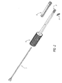
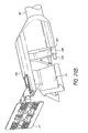
- FIG. 2 shows an exploded view of one embodiment of a curvilinear anchor and delivery instrument.
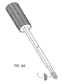
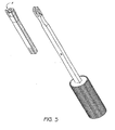
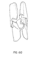
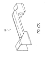
- FIG. 3 shows a perspective view of one embodiment of a curved two pronged staple type anchor.
- FIGS. 4A-E show a sequence involving loading an anchor into a delivery instrument and forcing it out of the lateral opening at the distal end of the delivery instrument according to one embodiment.
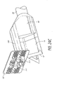
- FIG. 5 shows an exploded view of one embodiment of a delivery instrument and detachable sleeve.
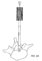
- FIGS. 6A-G show a delivery sequence involving a vertebral endplate according to one embodiment.
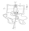
- FIG. 7 shows a prior art bone screw and intervertebral anatomy.
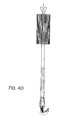
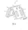
- FIG. 8 shows an embodiment of an anchor according to one or more embodiments of the invention.
- FIG. 9 shows another embodiment of an anchor according to one or more embodiments of the invention.
- FIG. 10 shows another embodiment of an anchor according to one or more embodiments of the invention.
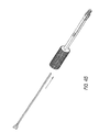
- FIG. 11 shows one embodiment a delivery tool.
- FIG. 12 shows the delivery tool in the previous figure with an anchor mounted
- FIG. 13 shows an axial cross sectional view of a vertebral body and implanted anchor.
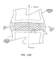
- FIGS. 14A-B show an expanded view and a frontal view of the implanted anchor in the previous figure.
- FIG. 15 shows a sagittal view of the implanted anchor in the previous figures.
- FIG. 16 shows an axial cross sectional view of a vertebral body and a delivery tool inserted along an endplate in the vicinity of an anulus defect or anulotomy.
- FIG. 17 shows an axial cross sectional view of a vertebral body wherein an anulus reinforcement device has been implanted along and within the anulus and is attached to an anchor embedded within the vertebral body.
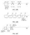
- FIGS. 18A-C show various views and features of anchors according to one or more embodiments of the invention.
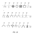
- FIG. 19 shows various profiles of the keel portion of one or more anchors.
- FIG. 20 shows a perspective view of another embodiment of an anchor according to one or more embodiments of the invention with a plate-like attachment means suitable for three sutures.
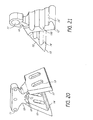
- FIG. 21 shows a perspective view of another embodiment of an anchor according to one or more embodiments of the invention with an "eye" attachment means.
- FIGS. 22A-B show embodiments of the anchor and delivery tool.
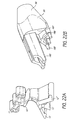
- FIG. 22A shows a perspective view of another embodiment of an anchor according to one or more embodiments of the invention having a three legged keel portion and designed such that only the attachment portion remains proud on the tissue surface.
- FIG. 22B shows a delivery tool for driving an anchor with a mated surface and alignment pins.
- FIGS. 23A-B show a perspective view of another embodiment of an anchor according to one or more embodiments of the invention having a flexible linkage member.
- FIGS. 24A-C show a series of perspective views of one embodiment of an anchor and barrier system according to one or more embodiments of the invention.
- FIGS. 25A-C show a series of perspective views of another embodiment of an anchor and barrier system according to one or more embodiments of the invention.
- FIGS. 26A-B show a side view and perspective view of an anchor with a sharpened leading edge having a recessed region corresponding to the cupped cortical rim of a vertebral endplate.
- Embodiments of the invention relate generally to tissue anchors.
- the tissue anchors provide pull-out resistance, stability and/or maximize contact with tissue involving a minimum amount of penetration.
- delivery methods are minimally invasive and include, but are not limited to, linear, lateral, and off-angle implantation or driving of anchors along, against or within tissue surfaces.
- bone anchors are provided.
- anchor as used herein shall be given its ordinary meaning and shall also include, but not be limited to, nails, staples, screws, fasteners, sutures, spikes, tacks, keys, pegs, rivets, spikes, bolts, and pins.
- the anchor comprises one or more tines or prongs.
- the anchor is forked.
- the anchor may be straight, curved, or partially curved.
- the anchors disclosed herein are particularly suited for hard tissues such as bone.
- soft tissue anchors are provided.
- One or more embodiments of the anchor can be delivered into a tissue and be secured within said tissue and resist extraction, migration, and/or rotation. Such stability is especially important in environments like the spine, where the anchor is adjacent delicate nerve tissue such as the spinal cord.
- the anchoring system may be used in other delicate vasculature such as the aorta.
- anchors according to the embodiments described herein have broad applications.
- the anchors described herein may be used in the radial head, ulnar head, humeral head, tibial plateau, scapula, acromion, talus, malleolus ; tendons and ligaments such as the talo-fibular ligament, anterior cruciate ligament, patella tibial tendon, achilies tendon, rotator cuff, and other tissues such as the meniscus.
- anchors according to one or more embodiments of the invention can disposed within artificial tissues or prosthetics.
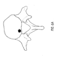
- FIG. 1A provides a sagittal view of a spine segment. Also shown are numerous potential anchor sites and are marked as "X.”
- FIG 1B is an axial view of the same spine segment and shows other possible anchoring sites including along or within a vertebral body, endplate, transverse process, spinous process, facet, and pedicle.
- an anchor can be placed along the cortical rim of the endplate or medially within the cancellous bone or relative to or within a pedicle, skull, or sacrum.
- Other anchoring sites include, but are not limited to: relative to a defect within the disc either in the area of the defect, at the interface of the anulus and nucleus or in the area of the nucleus.
- one or more anchors are used in connection with an anulus or nucleus augmentative device, as described in U.S. Patent Nos. 6,425,919 ; 6,482,235 ; 6,508,839 ; and 6,821,276 .
- one or more anchors are used to anchor an anulus augmentation device that is placed within or beyond a defect in the anulus to the vertebral endplates.
- One or more embodiments of the invention comprise anchors made at least partially of one or more of the following materials: any biocompatible material, material of synthetic or natural origin, and material of a resorbable or non-resorbable nature.
- the anchor may also be partially or wholly constructed from material including, but not limited to, autograft, allograft or xenograft; tissue materials including soft tissues, connective tissues, demineralized bone matrix and combinations thereof; resorbable materials including polylactide, polyglycolide, tyrosine derived polycarbonate, polyanhydride, polyorthoester, polyphosphazene, calcium phosphate, hydroxyapatite, bioactive glass, collagen, albumin, fibrinogen and combinations thereof; and non-resorbable materials including polyethylene, polyester, polyvinyl alcohol, polyacrylonitrile, polyamide, polytetrafluorethylene, polyparaphenylene terephthalamide, cellulose, and combinations thereof.
- Further examples of non-resorbable materials include carbon-re
- the anchor comprises an anchor body and an anchor attachment site.
- the anchor attachment site is adapted to accept or connect to a suture, linkage element, threaded screw, or provide a surface for ingrowth into an adjacent structure.
- the anchor attachment site can be integral to the anchor or a separate structure comprised of the same or different material as the anchor body.
- the anchor attachment site can be coupled to the anchor body.
- the anchor attachment site can be flexibly, rigidly, or rotationally connected to the anchor body.
- the anchor attachment site can comprise one or more of the following structures: head, flange, plate, disc, protrusion, channel, hole, cleat or eye. These structures can be placed at various positions along the anchor. For example, one or more of these structures may be placed at or near the ends of the anchor, in the middle of the anchor, or at any other desired position.
- the anchor attachment site comprises mesh, fabric, or membrane material, or a combination thereof. The site may be parallel, perpendicular or angled with respect to the body of the anchor. In one embodiment, the anchor attachment site is located on an end or terminus of the anchor body.
- the anchor comprises one anchor body and one anchor attachment site. In another body, the anchor comprises one or more anchor bodies and one or more anchor attachment sites. In one embodiment, the anchor comprises one body and two attachment sites.
- At least a portion of the anchor comprises a biologically active or therapeutic agent.
- at least a portion of the anchor can comprise growth factors such as bone morphogenic proteins, insulin-like growth factor 1, platelet derived growth factor, and fibroblast growth factor.
- both the anchor body and anchor attachment portion of the anchor can be adapted to deliver a biologically active or therapeutic agent.
- at least a portion of the anchor is coated with a biologically active or therapeutic agent.
- Anchors can be partially or wholly arcuate or curvilinear.
- the radius of curvature (the tightness or gentleness of the curve) can vary among embodiments as can the section of a circle corresponding to the anchor. For example, an anchor having a 90 degree curve would appear as 1 ⁇ 4 of a circle. Other ranges of curves between 0-180 degrees are also possible. In some embodiments, for example, the curvature is about 15, 30, 45, 60, 75, 90, 120, 150, or 180 degrees.
- An anchor can also be at least partially curved with a linear portion extending upward.
- the curved portion is adapted for driving into a tissue and the straight portion remains proud, or above the surface.
- the proud portion of the anchor can be anywhere from 0-180 degrees relative to the surface.
- the curvature of an embodiment of the anchor can also be variable along the anchor. Such a variable curvature could be employed to increase or decrease pressure on tissues adjacent to the anchor.
- the proud portion is about 15, 30, 45, 60, 75, 90, 120, 150, or 180 degrees relative to the surface.
- the surface or body of the anchor can be roughened, porous, barbed, lubricated, coated or impregnated with a biologically active or therapeutic agent.
- the anchor can be in the form of a curved nail or staple with a crown or bridge and having two or more prongs or legs extending therefrom.
- a slot or gap between the prongs in one ore more embodiments of a staple can be aimed at a suture or other structure already implanted in or along a surface and then hammered in place thereby anchoring the suture in place.
- the tips of the prongs of a staple can be beveled to effect a wedging action.
- the prong By beveling or angling the inner, outer, front, and/or back of a prong tip, the prong will tend to travel in a particular direction.
- the beveled tips can complement each other, work in opposition, or some combination thereof.
- the prong tips are beveled on the outside edge, in another embodiment the tips are beveled on the inside edge.
- the top of one prong is beveled and the bottom of another is beveled.
- the cross section of prongs may be variable along the length of the anchor.
- the anchor prong's smallest cross section is at or near the tip and at its greatest furthest from the tip, creating a wedge along the curve of the anchor. This may aid in increasing compression on all or part of the bone or other tissue in contact with the anchor.
- an anchor in another embodiment, can be resiliently flexible such that after passing through a curved slot or deflecting surface of the delivery device, the anchor (including staples, nails, etc) straightens out to its original shape as it is advanced out of the device and into the tissue.
- the original shape, predetermined shape, first shape, or unrestrained shape can be, for example, straight, angled, corkscrew, or offset.
- the prongs or legs of one or more embodiments of the anchor, such as, for example, a staple can be straight, curved, angled, corkscrew, or offset with respect to each other.
- a guide body 4 has a cylindrical grip or hand hold and first proximal and second distal end.
- the body 4 can be partially or fully hollow and contain a guide way chamber 5 for holding and orienting an anchor or staple 3 terminating in an opening at the distal end of the guide body.
- the opening can be oriented axially out of the front of the body or laterally and side mounted.
- the guide way chamber 5 comprises a curved or angled slot or passage and opens perpendicular or off angle (or between 0-180) with respect to the long axis of the guiding body.
- the radius of curvature along the passage can be constant or variable along the sweep of the curve.
- a curved nail or staple 3 can be inserted within the chamber 5 via a side loading window.
- a pusher rod 1 is carried within or by the body 4 and accesses or is in communication with the guide way chamber.
- the rod 1 has a first proximal end that can be configured with a head or striking surface for hammering and a second distal end for transmitting force to the end of a nail, staple, or anchor 3 within the guide way chamber 5.
- the distal end or anvil can be curved, beveled, or angled such that the linear force of the rod can be transmitted downward or along an arc as the staple 3 is driven out through the curved slot of the chamber 5.
- the rod 1 may be rigid or at least partially flexible in construction.
- a depth stop support 2 which can be configured as a snap on sleeve that fits over the body 4.
- a depth stop may simply be a projection off of the body that limits further travel of the body and/or guide way chamber opening within or adjacent a tissue.
- the depth stop may also be adjustable to allow for different implantation depths or locations.
- the depth stop may project in one or more directions from the long axis of the tool. Depth stops and other instrumentation described in U.S. Patent No. 6,821,276 may be incorporated in several embodiments of the invention.
- FIG. 3 is an example of an embodiment of a staple or anchor 3 with two prongs or legs that are barbed and beveled on the outside.
- the staple When the staple is driven into a surface such as a bone the prongs may or may not bend inward or be wedged together. This action will pinch and compress the bone tissue between the prongs while pressing outwardly against the sidewalls of the bone facilitating a stable anchorage.
- FIGS. 4A-E shows an embodiment of a delivery device being loaded with and anchor and the push rod applying force to the anchor and partially driving it out of the curved guide way chamber opening or lateral opening.
- Figure 4 B also shows the depth stop support sleeve 2 with a vertical slot corresponding to the guiding body distal slot which is aligned with the midline of the anchor and can be used to precisely implant or drive the anchor or staple around a suture or linear structure.
- an embodiment of the depth stop support is shown as an attachable sleeve that fits on or over the distal end of the guiding body.
- many of the features of the sleeve can be machined, welded or attached directly to the body if so desired.
- an alignment projection 8 is shown in addition to the vertical slot corresponding to the guiding body distal slot and adjustable depth stop.
- the alignment projection can form a right angle with the depth stop and have a beveled tip to ease insertion.
- the alignment tip can be a relatively flat and rectangular projection that in use can be rotated and rocked between to vertebrae or a hole in an anulus to distract the vertebrae.
- the tip and at least part of the guiding body can be inserted between the adjacent vertebral bodies.
- the depth stop can limit the amount of insertion by catching the edge of one or both of the opposing vertebral endplates. Vertebral taxis or the resistance of the anulus and endplates to further distraction can serve to immobilize the guiding body as the anchor is hammered out.
- the body can be wedged along an inferior superior plane to drive the opening of the guide way chamber against the desired anchor site.
- one or more depth stop surfaces may contain one or more barbs, spikes, nails, fasteners, or means for engaging or immovably coupling the distal end of the body to a boney structure such as a vertebral body.
- an upper depth stop surface may be configured to engage a superior vertebral body and a lower depth stop surface may be configured to engage an inferior vertebral body.
- the push rod and hammering method described infra is a preferred method of delivery other methods and devices can be used for this purpose.
- compressed gas and hydraulics can be utilized for driving.
- the push rod can be configured as a piston or threaded rod (that can be rotated to expel the implant) for imparting linear force.
- the threaded rod or piston can be flexible or have joints along its length to accommodate a curved or flexible guiding body.
- Delivery instruments and devices can also be used to implant other devices besides anchors and the like.
- a prosthetic device including, but not limited to, a barrier, mesh, patch, or collapsible implant
- an anchor according to several embodiments of the present invention, such as described in U.S. Patent Nos. 6,425,919 ; 6,482,235 ; and 6,508,839 ; 6,821,276 .
- the prosthetic device can be loaded within or along the guiding body of the device.
- the anchor and the prosthetic device may be constructed from identical, similar, or different materials.
- the anchor and prosthetic device may be coupled or removably or reversibly. Connections between the anchor and the prosthetic device may be temporary (such as restorable or dissolvable sutures) or permanent.
- the prosthetic device may also be of unitary construct or integral with the anchor.
- an implant such as collapsible patch is coupled to the anchor and oriented along or within the guiding body such that as the anchor is passed through the guide way chamber slot in a downward direction the patch is extruded outwardly or parallel to the long axis of the body.
- the patch can be held within the body which can have linear slot adjacent the curved slot of the guide way chamber or alternatively the patch can be mounted around the guide way chamber while coupled to the anchor within the chamber.
- the depth stop sleeve can also be used to compress and hold the patch in place.
- one or more anchors can be delivered separately from one or more implants.
- the implant is first delivered and positioned and then anchored in place.
- the anchor is first established in the implantation site and then the implant is delivered and connected to the anchor.
- FIGS 6A- L depicts an implantation sequence according to various embodiments of the invention.
- FIG. 6A is an axial cross section of a vertebral body, shown is a star shaped treatment zone along the vertebral endplate.
- the sequence shows an anchor being implanted into a posterior portion of a vertebral body along an endplate.
- the surface of the endplate can be accessed through a hole in the anulus.
- the hole in the anulus may be a naturally-occurring defect or surgically created.
- Devices according to the invention are not limited to a single location along a vertebral body or surgical approach.
- Various embodiments of anchor presented herein are designed to improve upon the weaknesses in conventional bone screws and staples that are limited by surgical access and suture or anchor attachment site placement.
- the posterior elements of vertebral bodies forming facet joints, spinal canal, neural foramen, and the delicate nerve tissues of the spinal cord create numerous obstacles for surgery and diagnostic and interventional methods.
- Surgical approaches have been adapted to minimize damage to these structures and involve tight windows usually off angle to the target tissue.
- FIG. 7 An example of such prior art anchor and environment is depicted in FIG. 7 , which shows a bone screw driven into a vertebral body from a posterior lateral approach.
- the anchor on the outside of the vertebral body is ineffective for retaining an implant within the disc and remains in dangerous proximity to the spinal cord.
- the anchor does not present attachment sites originating at a proximal end in the axial orientation from which they are driven.
- several embodiments of the present invention are advantageous because the anchor is adapted with an expansion mechanism that provides a "mushrooming" effect, and thus the pull-out resistance is not merely limited to the friction and forces generated by the sidewalls of the material or tissue.
- the attachment site of an anchor can be presented distally from the insertion site in a direction perpendicular or offset from the axial orientation of insertion.
- the anchor presents a larger surface area below or embedded within a surface, thereby offering improved pull-out resistance without requiring an expansion or "mushrooming" step or mechanism.
- one or more anchors are driven into the surface of a first plane and present a portion on an adjacent plane or surface perpendicular or angled relative the first plane.
- the anchor is driven into a first surface and across an adjacent surface in the same instance.
- at least a portion of the anchor such as the anchor attachment site is adapted to remain above or proud of the upper or second tissue surface or plane.
- the anchor With respect to the first surface (the front facing or lower surface into which the anchor is driven), the anchor can be driven in to a depth such that it is countersunk, left flush, or left partially external to the frontal tissue surface or plane.
- the anchor can also be delivered at a trajectory or angle relative to the second or top surface such that it is driven into the first surface and downwardly or upwardly across the second surface.
- the anchor is a flat plate-like nail or brad having a specialized keel portion and neck portion.
- the anchor is flat, plate-like, curved, corrugated, round, or a combination thereof.
- the neck can be terminated in a head or present an attachment portion along its length.
- the attachment portion or site can be comprised of a more flexible piece of fabric, wire, linkage, fastener component, hook eye, loop, or plate.
- the neck can be an extension, ridge, midline, or the apex of the keel portion.
- the neck can be oriented at the distal or proximal end of the keel or anywhere along its length.
- the neck is shorter than the keel.
- the neck is a thin rod or beam.
- the keel portion can have a cross-section similar to a wedge, "V", "U”, “T”, “W”, “X” and other shapes.
- Anchors according to one or more embodiments of the present invention have dimensions suitable to the implantation environment.
- the anchor has a height of about 0.2 cm to about 5 cm and a width of about 0.2 cm to about 5 cm.
- Anchors can have a length or depth from 0.2 cm to about 5 cm. In some embodiments, the length, width, height or depth can be less than 0.2 cm or greater than 5 cm.
- the anchor has a length of about 1 cm and a width of about 0.5 cm. In yet another embodiment, the anchor has a length of about 0.5 cm and a width of about 0.25 cm. In another embodiment, the anchor is dimensioned as follows: about 0.3 cm wide, 1 cm long and 0.5 cm deep.
- the length of the anchor can define a straight or curved line defined by a radius of curvature of about 0-90 degrees (e.g., about 15, 30, 45, 60, or 90 degrees).
- the keel, legs, extensions, blades, or fins can have a leading edge that is sharpened, left dull, or serrated.
- Other features of the neck and keel or extensions include, but are not limited to, barbs, tabs, roughened surface geometry, polished surface, coatings seeded carrier or drug eluting coatings or elements, concavities, scalloped ridges, grooves, "feet", ridges, voids, slots, and ingrowth openings are shown in the attached drawings. Secondary edges or ribs can protrude along portions of the keel to provide enhanced engagement with tissue.
- the anchor can also include an alignment means, engagement means or guide.
- Variations of the anchor alignment means can function to orient the anchor to a driver and couple it thereto.
- the anchor alignment means can comprise alignment components such as a protrusion, recess, or fastener component mated to a portion of a delivery instrument.
- the anchor engagement means can comprise engagement components or portions such as spikes, teeth, prongs, barbs, friction zones, or a combination thereof.
- the guide can comprise a protrusion, slot, arrow, tab, or a combination thereof.
- the anchor comprises means to align, means to engage, means to guide, or a combination thereof.
- FIG. 8 shows an embodiment of an anchor 25 with a leading edge 12, suture attachment sites 11, ingrowth features or voids 13, first and second plate-like legs or lateral extensions 15, 15' defining the keel, arcurate central ridge or apex 10, centering or alignment projection 16, and feet or ridges 14, 14'.
- Both the wedge-like shape of the keel portion of the implant i.e., the legs and the ridges or flange like extensions at the end of the legs function to hold the implant within a given tissue and to resist rotation and pull out from a variety of angles.
- the voids and ingrowth features serve to provide secondary stabilization over time and/or to allow chemical transfer or cellular respiration across the implantation site.
- the neck portion is bifurcated into two legs, extensions, blades, fins, or keels that meet at an apex and form an angle between about 10 and about 170 degrees. In one embodiment, the angle is about 30-90 degrees.
- the apex at the point of bifurcation can define a flat ridge or vertical extension or neck that can contain one or more anchor attachment sites.
- the neck can be in the form of an arc or eye projecting along the length of the body of the anchor.
- "V" or “U” shaped anchors can be modified to "L" shaped anchors in some embodiments.
- FIG. 9 an anchor similar to the one depicted in the previous figure is shown.
- the apex 10 which would correspond to the neck in other embodiments, does not extend and instead presents a smooth curve which can present a less injurious profile to the anatomy in certain applications.
- ridges 70 and scalloped teeth-like surface features 71 are also shown.
- FIG. 10 shows another embodiment of an anchor with deployed barbs 80 and 80'
- These features can be held compressed within a sleeve on a delivery instrument or simply forced to compress inwardly as the implant is driven in to tissue.
- One or more slots or recesses 82 are adapted for holding the barbs during implantation to streamline the anchors profile.
- One or more barbs can exert continuous outward pressure on the sidewalls of a tissue or expand to their maximum and form a shelf or flange if the tissue geometry widens, expand or become more pliant.
- the implant might be driven into cortical bone and then further into cancelous bone.
- the barbs flexible plate-like structure or engagement means can expand or extend outwards.
- the anchor is driven at least partially into the hollow of a boney structure such that the barbs expand and engage the inner wall of the bone.
- Element 83 is an opposing barb or expansion means however one or more barbs can be oriented relative to each other from 0-360 degrees.
- the barbs or other barb-like components may be orientated relative to each other at the following angles: 15, 30, 45, 60, 90, 120, 150, 180, or 360 degrees.
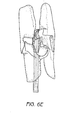
- FIG. 11 shows a delivery tool with a shaft 96 with distal end 95 having an anvil or striking surface 94 defining a leading edge mated to at least a portion of the cross section of the trailing edge of the anchor.
- the shaft may be connected at its proximal end to a handle or terminate in a striking surface. Because the anvil surface is similar to the anchor in some embodiments, both the anchor and at least part of the distal end of the delivery tool can be driven into a bone thereby counter sinking it.
- anchors according to one or more aspects of the invention can be left flush or partially countersunk.
- a mounting member 90 may extend beyond the implant when the implant is mounted or loaded on the tool.
- the mounting member 90 includes a flattened lower surface 93 and a rounded blunt front surface 91 for positioning along a bone surface, such as the top of a vertebral endplate, and a slot or engagement means 92 for accepting an aligning an anchor.
- FIG. 12 shows the anchor 25 mounted on the mounting member 90.
- the extended lower surface 93 and the leading edge of the implant 12 and 12' forms a means to engage bone or other tissue.
- the tissue (e.g., bone) engagement means comprises a device having an angled surface that may be used to hook onto, engage, or align the instrument with the edge of a vertebral body or the intersection of two tissue planes.
- the engagement means can be used to align the implant with the top of a vertebral endplate and its front outer surface, the anchor is then driven into and across the endplate.
- FIG. 13 shows a cross-section of a vertebral body 24 having an anulus fibrosus 23 bounding nucleus pulposus 22 with an anchor 25 embedded into a posterior aspect of an endplate and within or proximal to an anulotomy or defective region of the anulus fibrosus 23.
- This implantation site is also in the vicinity of the cortical rim or ring of dense bone of the vertebral endplate.
- the anchor is shown countersunk into the bone along the P-A axis but partially proud along the inferior-superior axis (the dotted lines indicating the portion of the implant below bone surface or level.
- FIG. 14A is an expanded view of FIG. 13 and shows dotted lines to represent the keel portion of the anchor 25 beneath the endplate surface.
- FIG. 14B is a dorsal view of FIG. 14A showing anchor 25.
- FIG. 15 a sagittal view of an implanted anchor 25 is shown at least partially within the defect 33 and inferior vertebral body 32. Superior vertebral body 31 is also shown. The cross-section of the vertebral bodies depicts the denser and thicker bone at the edge or rim where the anchor is implanted and the less dense cancellous bone within the vertebral body.
- FIG. 16 depicts a method of delivery for one embodiment of the anchor and associated delivery tool. Shown is a top cross sectional view of a vertebral body 24 and a delivery instrument 44 and an anchor 25.
- the delivery instrument or driver is used to transmit the force of a hammer or other means to drive the anchor in place.
- the driver can comprise a slot, holder, magnet, pins, mateable surfaces, fastener or other means at its distal end to engage or couple with the anchor.
- the anchor can also be attached to the distal end of the driver and then released once the desired delivery depth has been attained. Other features of a driver (not shown in FIG.
- FIG. 16 can include a depth stop, bone engagement means such as a spiked, hooked, or angled protrusion, and/or a retractable sleeve to protect adjacent anatomy as the anchor is positioned.
- FIG. 16 also shows a flat proximal end 1602 for hammering, if needed and a knurled handle 1600.
- FIG. 17 a top cross-sectional view of an anulus repair implant 51 lying along the inner surface of the posterior anulus is coupled, attached, or sutured 52 to an anchor 25.
- the connection between the anchor and implant can be permanent or detachable.
- the implant 52 can be delivered and positioned prior to, at the same time as, or subsequent to the implantation of the anchor 25.
- FIGS. 17A-17C show various features of anchors.
- FIG. 18A the surface level of a bone such as a vertebral endplate is shown as a dotted line.
- a side view is depicted.
- the leading edge of the keel or leg portion of the implant is thinner than the trailing edge.
- at least a portion of the leading edge, profile, proximal edge or side of an implant can have a thinner or tapered profile than an opposing end, distal end, or trailing edge or profile.
- FIG. 17B shows a series of anchor variations from a side view in which the top portion, apex, neck, or implant attachment site 170, 171, 172, 173, 174 is symmetrical, rounded, wedge shaped, oriented at the distal or proximal end of the anchor.
- FIG. 18C shows another side view along the bone surface level depicting and anchors with features discussed infra such as a serrated leading edge, voids or ingrowth holes, and a recess for engaging a delivery tool.
- FIG. 19 shows various embodiments of the anchor cross-sections including several keel profiles from a front view resistant to pullout and offering various surface areas. Some are solid shapes as in anchor profiles 182, 184, 187, and 200 and others are hollow and have an open midsection as in anchor profiles 183, 185, 188.
- FIG. 20 a perspective view of an anchor is shown with leading edges 12, 12', alignment means 16, suture or fastener attachment 11 site or neck 10, and voids 13.
- the apex does not run the entire length or depth of the anchor corresponding to the keel or opposing leg portions 15, 15' of the anchor.
- the neck is oriented towards the proximal end of the anchor forming a cut-out along the top portion of the anchor.
- the neck 10 is shown perpendicular to the keel 15 but can be alternatively oriented in a range from 0-180 degrees relative to it. In one embodiment, the neck is oriented at an angle of about 15, 30, 45, 60, 75, 90, 120, 150, or 180 degrees relative to the keel
- a "V" shaped anchor is shown in FIG. 21 .
- An “eye” or loop is integral to a neck extension portion 10 that bifurcates into two legs 15, 15'. Because the leading edges 12, 12' and at least a portion of the neck 10 is sharpened, this anchor can be driven more flush to the upper or first surface of a bone such as a vertebral endplate.
- both the neck 10and the leg portions 15, 15' of the device function as a keel.
- This embodiment also shows ridges12 and scalloped recesses. Anchors according to other embodiments described herein may also comprise ridges and/or scalloped recesses.
- FIG. 22A another embodiment of an anchor is shown.
- three legs defining the keel are provided.
- a relatively taller neck is provided beneath a perpendicular suture attachment member.
- the neck 10 is set back distally from the leading edge of the keel portion.
- FIG. 22B shows the distal tip of a delivery tool. Shown are attachment pins 180, anvil 186 or striking surface, depth stop 187, and mounting member 185.
- FIG. 23A an anchor 25 is shown with an attachment site for a flexible bridge 188.
- the bridge 188 is shown in FIG. 23B and is coupled to the neck 10 of the anchor 12 with a first and second flexible tab 193, 194 and has an attachment 11 site at the opposing end.
- FIG. 24A shows an anchor similar to the ones depicted previously with a bifurcated keel 15, 15', neck 10, and attachment plate 112 with a first and second coupling member 111, 11' or snap surface.
- FIG. 24B is an exploded view of a barrier, mesh, or reinforcement plate 52 adjacent an anchor wherein the anchor 25 is partially inserted or mounted within the distal end of a delivery tool.
- FIG. 24C shows all three elements connected and mounted and ready to be driven into a tissue site.
- FIGS. 25A-25C Another embodiment of an anulus reinforcement system is shown in FIGS. 25A-25C .
- a single attachment means 111 is used that can function as a fulcrum or hinge site for a flexible barrier 52 member shown in FIG. 25 B.
- Behind or distal to the attachment means 111 is a support member 112 or plate that is an extension of the neck 10. This feature, in some embodiments, minimizes or prevents the barrier 52 from folding backwards and may also reinforce the barrier 52.
- FIG. 25C shows a hood or sleeve 120 element that can be mounted on or carried by a delivery tool or instrument as described herein. The hood 120 retains the folded barrier until the anchor portion is fully established within the tissue whereupon it is retracted.
- FIGS. 26A-26B Another embodiment of the invention is shown in FIGS. 26A-26B .
- This embodiment shows an anchor especially adapted for use in a vertebral body and includes an upside down "V" shaped keel portion with a sharpened leading edge.
- the leading edges enable the anchor to be directly driven into the bone and do not require a pilot hole or pre-cut.
- One feature of this embodiment is the leading step in the sharpened edge which present more cutting surface blow the surface of the bone and more forward of the distal attachment site.
- the leading edge can have multiple steps or be curved and rounded. This profile reduce the risk that the leading edge might pierce or damage the endplate (which is not flat but has a "dip” or cupped portion in the middle). This feature facilitates insertion of a longer, stronger anchor into a disc that would otherwise (because of a pronounced dip) be difficult to position at the proper height and depth into the bone without damaging the endplate.
- Example illustrates one embodiment of the present invention and is not intended in any way to limit the invention.
- the anchors described herein can be used throughout the human body and have general applicability to fastener art.
- Such anchors can be used to join or anchor like or disparate materials or tissues together, maintain alignment of materials, reinforce a fracture within a material, and provide an attachment site along or within a materials surface.
- the anchor illustrated in FIG. 26 is used by way of example.
- the anchor is in the form of an upside down "Y" defined by a neck portion terminating at one end into two plate-like rectangular legs forming a keel and terminating into an suture attachment site 11 in the form of a loop on the other end.
- the leading edge of the legs 12 and neck 10 are sharpened and the upper portion of the legs is recessed, profiled or formed with a relief 113.
- the relief profile 113 can correspond to an anatomical structure, in this embodiment the forward recess or relief 112 corresponds to the concavity or cupping of an endplate.
- the angle between the keel plates is around 90 degrees.
- the neck 10 is about 0.1 millimeter high and about 0.2 wide millimeters wide and extends about 0.2 millimeters.
- the neck 10 and attachment site 11, an "eye" or loop in this embodiment, are mounted at the trailing or aft potion of the keel 15.
- the entire structure is made of nickel titanium and is machined from bar stock.
- the anchor is mounted on the distal end of a driver.
- the driver has a striking surface on one end and an anvil on the opposing end.
- the anvil has the identical cross-section as the trailing edge of the anchor and extends about 0.2 cm to allow for countersinking.
- the anchor is coupled to the anvil by a forked protrusion that holds the neck and a pin that fits into the eye.
- the anchor is used to secure an anulus repair device relative to a defect in the disc.
- a posterior-lateral approach is used to obtain access to the damaged disc. Part of the posterior elements on the opposing vertebral bodies may have to be removed in order to reach the disc.
- the anulus repair device is then implanted through the defect and along the inner surface of the anulus.
- the anchor which is mounted on the distal end of the driver, is aimed at the top edge or endplate of the inferior intervertebral body.
- An alignment projection forming a right angle at the tip of the drive is used to align the bottom potion of the attachment loop of the anchor with the upper surface of the endplate and to center the anchor within the defect.
- the anchor is then driven forward into the bone with light hammering applied to the driver.
- the anchor is driven roughly perpendicular to the outer surface of the vertebral body and roughly parallel to the endplate.
- the depth of insertion is controlled by the 0.2 cm countersinking anvil and the depth dimension of the anchor, in this case 0.5 cm for a total depth of 0.7 cm which is still shy of the border of the cortical rim and the cupping of the endplate. Only the upper potion of the loop remains proud of the endplate surface and the anular repair device can then be connected to it with a suture.
Landscapes
- Health & Medical Sciences (AREA)
- Life Sciences & Earth Sciences (AREA)
- Surgery (AREA)
- Orthopedic Medicine & Surgery (AREA)
- Engineering & Computer Science (AREA)
- Biomedical Technology (AREA)
- General Health & Medical Sciences (AREA)
- Animal Behavior & Ethology (AREA)
- Heart & Thoracic Surgery (AREA)
- Public Health (AREA)
- Veterinary Medicine (AREA)
- Nuclear Medicine, Radiotherapy & Molecular Imaging (AREA)
- Molecular Biology (AREA)
- Medical Informatics (AREA)
- Neurology (AREA)
- Rheumatology (AREA)
- Cardiology (AREA)
- Oral & Maxillofacial Surgery (AREA)
- Transplantation (AREA)
- Vascular Medicine (AREA)
- Prostheses (AREA)
- Surgical Instruments (AREA)
- Dowels (AREA)
Claims (16)
- Ancrage osseux (25) à des fins d'insertion dans une première surface d'un os et le long d'une seconde surface adjacente dudit os, comportant :un col (10) ayant une longueur définie par un bord d'attaque vif et une extrémité arrière ;dans lequel ladite extrémité arrière est configurée pour être à l'intérieur de la première surface de l'os ou pour être laissée dans l'alignement de celle-ci ;dans lequel ledit col (10) comporte un site de fixation (11) le long d'au moins une partie de sa longueur ;dans lequel ledit col (10) comporte une partie inférieure se terminant dans deux bases ou plus (15) ayant une longueur définie par un bord d'attaque et une extrémité arrière, la longueur du col étant plus courte que la longueur des bases ;dans lequel lesdites bases (15) forment un angle d'environ 10 à environ 180 degrés les unes par rapport aux autres ;dans lequel chacune desdites bases (15) comporte des bords d'attaque vifs (12) ;dans lequel les bords d'attaque vifs (12) desdites bases (15) sont adaptés pour être insérés dans la première surface de l'os le long d'un chemin de trajectoire sensiblement droit qui est sensiblement parallèle à la seconde surface tout en faisant avancer simultanément le site de fixation en travers de ladite seconde surface ; etdans lequel le site de fixation est configuré à des fins d'accouplement au niveau d'un tissu ou d'un implant prothétique à des fins de réparation dudit os ou tissu adjacent.
- Ancrage osseux selon la revendication 1, dans lequel lesdites bases (15) forment ensemble une partie base similaire à une plaque.
- Ancrage osseux (25) selon la revendication 1, dans lequel lesdites bases (15) forment un angle d'environ 180 degrés les unes par rapport aux autres.
- Ancrage osseux (25) selon la revendication 2, dans lequel lesdites bases (15) forment un d'environ 90 degrés les unes par rapport aux autres.
- Ancrage osseux (25) selon l'une quelconque des revendications précédentes, dans lequel l'implant prothétique comporte au moins l'un parmi un dispositif de type maille, un dispositif de type barrière ou un greffon,
- Ancrage osseux (25) selon l'une quelconque des revendications précédentes, dans lequel au moins une partie de l'ancrage est enduite d'un agent biologiquement actif ou thérapeutique.
- Ancrage osseux (25) selon l'une quelconque des revendications précédentes, dans lequel le site de fixation de l'ancrage comporte un matériau de type maille, tissu, ou membrane, ou une combinaison de ceux-ci.
- Ancrage osseux (25) selon l'une quelconque des revendications précédentes, dans lequel au moins une partie dudit col (10) ou desdites bases (15) comporte des bords dentelés pour faciliter la croissance tissulaire.
- Ancrage osseux (25) selon l'une quelconque des revendications précédentes, comportant par ailleurs des nervures qui font saillie le long de parties desdites bases (15) afin de mettre en oeuvre une meilleure mise en prise avec ledit os ou un tissu adjacent audit os.
- Ancrage osseux (25) selon l'une quelconque des revendications 1 à 6, dans lequel ledit ancrage (25) comporte par ailleurs des picots, des languettes, une géométrie à surface rugosifiée, une surface polie, des revêtements, des revêtements ou éléments à élution de médicaments ou porteurs à semis, des concavités, des arêtes dentelées, des rainures, des pieds (14), des arêtes (14), des vides (13), des fentes, des ouvertures d'interposition, des bords ou nervures secondaires.
- Ancrage osseux (25) selon l'une quelconque des revendications précédentes, dans lequel le site de fixation de l'ancrage comporte au moins l'un parmi une tête, une collerette, une plaque, un disque, une partie saillante, un profilé en U, un trou, une clavette ou un oeillet.
- Ancrage osseux (25) selon l'une quelconque des revendications précédentes, dans lequel le site de fixation (11) est situé à une distance allant à l'opposé de l'extrémité arrière du col (10).
- Ancrage osseux (25) selon l'une quelconque des revendications précédentes, dans lequel le site de fixation (11) est adapté à des fins d'acceptation de ou de connexion à au moins l'un parmi une suture, un élément de liaison, ou une vis à filet.
- Ancrage osseux (25) selon l'une quelconque des revendications précédentes, dans lequel les bases (15) comportent une section en coupe ayant une forme en coin, en 'V, en 'U', en 'T', en 'W' ou en 'X'.
- Ancrage osseux (25) selon l'une quelconque des revendications précédentes, dans lequel l'ancrage (25) est au moins partiellement construit à partir d'un matériau sélectionné dans le groupe constitué par un ou plusieurs parmi les suivants : alliage de nickel et de titane, titane, alliages de chrome et de cobalt, et acier.
- Ancrage osseux (25) selon l'une quelconque des revendications précédentes, dans lequel la première surface de l'os est l'extérieur d'un corps vertébral et la seconde surface de l'os est le plateau vertébral adjacent correspondant, et dans lequel l'implant prothétique est un dispositif de barrière prothétique.
Priority Applications (1)
| Application Number | Priority Date | Filing Date | Title |
|---|---|---|---|
| EP11194086.2A EP2430993B1 (fr) | 2005-12-28 | 2006-12-20 | Dispositifs et procedes pour ancrage osseux |
Applications Claiming Priority (3)
| Application Number | Priority Date | Filing Date | Title |
|---|---|---|---|
| US75423705P | 2005-12-28 | 2005-12-28 | |
| US11/641,253 US7972337B2 (en) | 2005-12-28 | 2006-12-19 | Devices and methods for bone anchoring |
| PCT/US2006/048727 WO2007078978A2 (fr) | 2005-12-28 | 2006-12-20 | Dispositifs et procedes pour ancrage osseux |
Related Child Applications (2)
| Application Number | Title | Priority Date | Filing Date |
|---|---|---|---|
| EP11194086.2A Division EP2430993B1 (fr) | 2005-12-28 | 2006-12-20 | Dispositifs et procedes pour ancrage osseux |
| EP11194086.2 Division-Into | 2011-12-16 |
Publications (3)
| Publication Number | Publication Date |
|---|---|
| EP1968464A2 EP1968464A2 (fr) | 2008-09-17 |
| EP1968464A4 EP1968464A4 (fr) | 2010-09-15 |
| EP1968464B1 true EP1968464B1 (fr) | 2012-02-22 |
Family
ID=38228779
Family Applications (2)
| Application Number | Title | Priority Date | Filing Date |
|---|---|---|---|
| EP11194086.2A Active EP2430993B1 (fr) | 2005-12-28 | 2006-12-20 | Dispositifs et procedes pour ancrage osseux |
| EP06847885A Active EP1968464B1 (fr) | 2005-12-28 | 2006-12-20 | Dispositifs et procedes pour ancrage osseux |
Family Applications Before (1)
| Application Number | Title | Priority Date | Filing Date |
|---|---|---|---|
| EP11194086.2A Active EP2430993B1 (fr) | 2005-12-28 | 2006-12-20 | Dispositifs et procedes pour ancrage osseux |
Country Status (10)
| Country | Link |
|---|---|
| US (7) | US7972337B2 (fr) |
| EP (2) | EP2430993B1 (fr) |
| JP (1) | JP4945574B2 (fr) |
| KR (1) | KR101316802B1 (fr) |
| AT (1) | ATE546103T1 (fr) |
| AU (1) | AU2006332933B2 (fr) |
| BR (1) | BRPI0620762A2 (fr) |
| CA (1) | CA2635537C (fr) |
| IL (1) | IL192431A (fr) |
| WO (1) | WO2007078978A2 (fr) |
Cited By (1)
| Publication number | Priority date | Publication date | Assignee | Title |
|---|---|---|---|---|
| US11058542B1 (en) | 2020-09-21 | 2021-07-13 | Randall F. Lee | System and method for joining boney structures |
Families Citing this family (106)
| Publication number | Priority date | Publication date | Assignee | Title |
|---|---|---|---|---|
| FR2897259B1 (fr) | 2006-02-15 | 2008-05-09 | Ldr Medical Soc Par Actions Si | Cage intersomatique transforaminale a greffon de fusion intervetebrale et instrument d'implantation de la cage |
| US8323341B2 (en) | 2007-09-07 | 2012-12-04 | Intrinsic Therapeutics, Inc. | Impaction grafting for vertebral fusion |
| US20040024465A1 (en) * | 1999-08-18 | 2004-02-05 | Gregory Lambrecht | Devices and method for augmenting a vertebral disc |
| US7998213B2 (en) | 1999-08-18 | 2011-08-16 | Intrinsic Therapeutics, Inc. | Intervertebral disc herniation repair |
| US7972337B2 (en) * | 2005-12-28 | 2011-07-05 | Intrinsic Therapeutics, Inc. | Devices and methods for bone anchoring |
| US7717961B2 (en) | 1999-08-18 | 2010-05-18 | Intrinsic Therapeutics, Inc. | Apparatus delivery in an intervertebral disc |
| US7553329B2 (en) * | 1999-08-18 | 2009-06-30 | Intrinsic Therapeutics, Inc. | Stabilized intervertebral disc barrier |
| EP1624832A4 (fr) | 1999-08-18 | 2008-12-24 | Intrinsic Therapeutics Inc | Dispositifs et procedes de densification du noyau du disque vertebral |
| US7220281B2 (en) | 1999-08-18 | 2007-05-22 | Intrinsic Therapeutics, Inc. | Implant for reinforcing and annulus fibrosis |
| CA2425951C (fr) * | 1999-08-18 | 2008-09-16 | Intrinsic Therapeutics, Inc. | Dispositifs et procede permettant d'augmenter ou de conserver un noyau gelatineux |
| FR2824261B1 (fr) | 2001-05-04 | 2004-05-28 | Ldr Medical | Prothese de disque intervertebral et procede et outils de mise en place |
| FR2827156B1 (fr) | 2001-07-13 | 2003-11-14 | Ldr Medical | Dispositif de cage vertebrale avec fixation modulaire |
| GB0208667D0 (en) | 2002-04-16 | 2002-05-29 | Atlantech Medical Devices Ltd | A transverse suspension device |
| FR2846550B1 (fr) | 2002-11-05 | 2006-01-13 | Ldr Medical | Prothese de disque intervertebral |
| WO2004112584A2 (fr) * | 2003-06-20 | 2004-12-29 | Intrinsic Therapeutics, Inc. | Dispositif et procede de pose d'un implant a travers une imperfection annulaire d'un disque intervertebral |
| WO2005023132A2 (fr) * | 2003-09-05 | 2005-03-17 | Buchanan L Stephen | Obturateurs ameliores |
| WO2005069924A2 (fr) | 2004-01-16 | 2005-08-04 | Arthrocare Corporation | Dispositif et procede de collecte osseuse |
| FR2865629B1 (fr) | 2004-02-04 | 2007-01-26 | Ldr Medical | Prothese de disque intervertebral |
| EP2113227B1 (fr) | 2004-02-04 | 2015-07-29 | LDR Medical | Prothèse de disque intervertébral |
| FR2869528B1 (fr) | 2004-04-28 | 2007-02-02 | Ldr Medical | Prothese de disque intervertebral |
| US20070156241A1 (en) | 2004-08-09 | 2007-07-05 | Reiley Mark A | Systems and methods for the fixation or fusion of bone |
| US9662158B2 (en) | 2004-08-09 | 2017-05-30 | Si-Bone Inc. | Systems and methods for the fixation or fusion of bone at or near a sacroiliac joint |
| US20180228621A1 (en) | 2004-08-09 | 2018-08-16 | Mark A. Reiley | Apparatus, systems, and methods for the fixation or fusion of bone |
| US9949843B2 (en) | 2004-08-09 | 2018-04-24 | Si-Bone Inc. | Apparatus, systems, and methods for the fixation or fusion of bone |
| US8172855B2 (en) | 2004-11-24 | 2012-05-08 | Abdou M S | Devices and methods for inter-vertebral orthopedic device placement |
| FR2879436B1 (fr) | 2004-12-22 | 2007-03-09 | Ldr Medical | Prothese de disque intervertebral |
| US7842042B2 (en) | 2005-05-16 | 2010-11-30 | Arthrocare Corporation | Convergent tunnel guide apparatus and method |
| FR2891135B1 (fr) | 2005-09-23 | 2008-09-12 | Ldr Medical Sarl | Prothese de disque intervertebral |
| FR2893838B1 (fr) | 2005-11-30 | 2008-08-08 | Ldr Medical Soc Par Actions Si | Prothese de disque intervertebral et instrumentation d'insertion de la prothese entre les vertebres |
| US7686838B2 (en) | 2006-11-09 | 2010-03-30 | Arthrocare Corporation | External bullet anchor apparatus and method for use in surgical repair of ligament or tendon |
| US8465546B2 (en) | 2007-02-16 | 2013-06-18 | Ldr Medical | Intervertebral disc prosthesis insertion assemblies |
| EP2148709A2 (fr) * | 2007-04-19 | 2010-02-03 | Smith & Nephew, Inc. | Implants prothétiques |
| US20080262522A1 (en) * | 2007-04-20 | 2008-10-23 | Rachadip Singh Sachasin | Minimally Invasive Percutaneous Restrictive Bariatric Procedure And Related Device |
| US20110319933A1 (en) * | 2007-04-21 | 2011-12-29 | Slobodan Tepic | Suture bone anchor |
| FR2916956B1 (fr) * | 2007-06-08 | 2012-12-14 | Ldr Medical | Cage intersomatique,prothese intervertebrale,dispositif d'ancrage et instrumentation d'implantation |
| US20110196492A1 (en) * | 2007-09-07 | 2011-08-11 | Intrinsic Therapeutics, Inc. | Bone anchoring systems |
| WO2009082376A1 (fr) * | 2007-12-26 | 2009-07-02 | Core Essence Orthopaedics, Llc. | Procédé et appareil pour délivrer de la chaleur à un article à mémoire de forme |
| WO2010081029A1 (fr) | 2009-01-08 | 2010-07-15 | Rotation Medical, Inc. | Systèmes implantables de protection des tendons et trousses et procédés associés |
| US9179910B2 (en) | 2009-03-20 | 2015-11-10 | Rotation Medical, Inc. | Medical device delivery system and method |
| US9179961B2 (en) | 2009-06-04 | 2015-11-10 | Rotation Medical, Inc. | Methods and apparatus for deploying sheet-like materials |
| CA2763794C (fr) | 2009-06-04 | 2017-01-17 | Rotation Medical, Inc. | Appareil de pose d'agrafes en forme d'arc sur un tissue cible |
| CA2773723C (fr) | 2009-09-17 | 2017-10-31 | Synthes Usa, Llc | Implant intervertebral comportant des elements de fixation d'os extensibles |
| US8449612B2 (en) | 2009-11-16 | 2013-05-28 | Arthrocare Corporation | Graft pulley and methods of use |
| US8764806B2 (en) | 2009-12-07 | 2014-07-01 | Samy Abdou | Devices and methods for minimally invasive spinal stabilization and instrumentation |
| JP5647264B2 (ja) | 2009-12-31 | 2014-12-24 | エル・デ・エール・メデイカル | 固定装置、椎間インプラント、および移植器具 |
| US9198750B2 (en) | 2010-03-11 | 2015-12-01 | Rotation Medical, Inc. | Tendon repair implant and method of arthroscopic implantation |
| DE102010048710B4 (de) * | 2010-03-30 | 2013-05-23 | 3T Textiltechnologietransfer Gmbh | Implantat mit einer Sandwich-Struktur |
| WO2012145059A1 (fr) | 2011-02-15 | 2012-10-26 | Rotation Medical, Inc. | Procédés et appareil pour fixer des matériaux de type en feuille à un tissu cible |
| US9314314B2 (en) | 2011-02-15 | 2016-04-19 | Rotation Medical, Inc. | Anatomical location markers and methods of use in positioning sheet-like materials during surgery |
| CA2825918C (fr) | 2011-02-15 | 2018-08-07 | Rotation Medical, Inc. | Procedes et appareil pour la distribution et le positionnement de materiaux en feuille |
| FI9329U1 (fi) * | 2011-03-21 | 2011-08-16 | Oulun Yliopisto | Luupora |
| US8845728B1 (en) | 2011-09-23 | 2014-09-30 | Samy Abdou | Spinal fixation devices and methods of use |
| US9247978B2 (en) | 2011-12-19 | 2016-02-02 | Rotation Medical, Inc. | Apparatus and method for forming pilot holes in bone and delivering fasteners therein for retaining an implant |
| US9107661B2 (en) | 2011-12-19 | 2015-08-18 | Rotation Medical, Inc. | Fasteners and fastener delivery devices for affixing sheet-like materials to bone or tissue |
| WO2013119321A1 (fr) * | 2011-12-19 | 2013-08-15 | Rotation Medical, Inc. | Organes de fixation et dispositifs de distribution d'organes de fixation pour fixer des matières de type feuille à un os ou à un tissu |
| WO2013096224A1 (fr) | 2011-12-19 | 2013-06-27 | Rotation Medical, Inc. | Organes de fixation pour fixer des matériaux en feuille à un os ou à un tissu |
| EP2797532B1 (fr) | 2011-12-29 | 2016-04-06 | Rotation Medical, Inc. | Appareil de distribution et de positionnement de matières de type feuille en chirurgie |
| WO2013101640A1 (fr) | 2011-12-29 | 2013-07-04 | Rotation Medical, Inc. | Fil-guide ayant un élément de fixation distal pour la pose et le positionnement de matières de type feuille en chirurgie |
| US20130226240A1 (en) | 2012-02-22 | 2013-08-29 | Samy Abdou | Spinous process fixation devices and methods of use |
| FR2987256B1 (fr) | 2012-02-24 | 2014-08-08 | Ldr Medical | Dispositif d'ancrage pour implant intervertebral, implant intervertebral et instrumentation d'implantation |
| JP6091529B2 (ja) * | 2012-03-09 | 2017-03-08 | エスアイ−ボーン・インコーポレイテッドSi−Bone, Inc. | 一体化インプラント |
| US10363140B2 (en) | 2012-03-09 | 2019-07-30 | Si-Bone Inc. | Systems, device, and methods for joint fusion |
| US9693876B1 (en) * | 2012-03-30 | 2017-07-04 | Ali H. MESIWALA | Spinal fusion implant and related methods |
| US10426533B2 (en) | 2012-05-04 | 2019-10-01 | Si-Bone Inc. | Fenestrated implant |
| WO2013179277A1 (fr) | 2012-05-30 | 2013-12-05 | Newvert Ltd. | Dispositif de fermeture d'un anneau de disque vertébral |
| US9572615B2 (en) | 2012-07-18 | 2017-02-21 | Jmea Corporation | Detachable front delivery assembly for a tissue repair system |
| US9198767B2 (en) | 2012-08-28 | 2015-12-01 | Samy Abdou | Devices and methods for spinal stabilization and instrumentation |
| US9320617B2 (en) | 2012-10-22 | 2016-04-26 | Cogent Spine, LLC | Devices and methods for spinal stabilization and instrumentation |
| US10786235B2 (en) | 2012-10-31 | 2020-09-29 | Anchor Innovation Medical, Inc. | Method and apparatus for closing a fissure in the annulus of an intervertebral disc, and/or for effecting other anatomical repairs and/or fixations |
| US10070969B2 (en) | 2013-01-17 | 2018-09-11 | Stryker European Holdings I, Llc | Annulus plug for intervertebral disc repair |
| US20140276968A1 (en) * | 2013-03-14 | 2014-09-18 | Ethicon, Inc. | Applicator systems for surgical fasteners |
| WO2014145902A1 (fr) | 2013-03-15 | 2014-09-18 | Si-Bone Inc. | Implants de fixation ou de fusion vertébrale |
| FR3005569B1 (fr) | 2013-05-16 | 2021-09-03 | Ldr Medical | Implant vertebral, dispositif de fixation vertebrale d'implant et instrumentation d'implantation |
| US9743961B2 (en) * | 2013-06-24 | 2017-08-29 | The University Of Toledo | Bioactive fusion device |
| AU2014306454B2 (en) | 2013-08-16 | 2019-06-13 | Suture Concepts Inc. | Method and apparatus for closing a fissure in the annulus of an intervertebral disc, and/or for effecting other anatomical repairs and/or fixations |
| US9839448B2 (en) | 2013-10-15 | 2017-12-12 | Si-Bone Inc. | Implant placement |
| US11147688B2 (en) | 2013-10-15 | 2021-10-19 | Si-Bone Inc. | Implant placement |
| FR3016793B1 (fr) | 2014-01-30 | 2021-05-07 | Ldr Medical | Dispositif d'ancrage pour implant spinal, implant spinal et instrumentation d'implantation |
| FR3020756B1 (fr) | 2014-05-06 | 2022-03-11 | Ldr Medical | Implant vertebral, dispositif de fixation vertebrale d'implant et instrumentation d'implantation |
| CA2945821C (fr) | 2014-05-09 | 2018-09-04 | Rotation Medical, Inc. | Systeme de pose d'implant medical pour un implant de type feuille |
| US10166033B2 (en) | 2014-09-18 | 2019-01-01 | Si-Bone Inc. | Implants for bone fixation or fusion |
| EP3193752B1 (fr) | 2014-09-18 | 2020-07-22 | SI-Bone, Inc. | Implant matriciel |
| EP3215025B1 (fr) | 2014-11-04 | 2020-12-23 | Rotation Medical, Inc. | Système de placement d'implant médical |
| US10675019B2 (en) | 2014-11-04 | 2020-06-09 | Rotation Medical, Inc. | Medical implant delivery system and related methods |
| EP3215026B1 (fr) | 2014-11-04 | 2023-10-25 | Rotation Medical, Inc. | Système de mise en place d'implant médical |
| US10098746B1 (en) | 2015-02-13 | 2018-10-16 | Nextstep Arthropedix, LLC | Medical implants having desired surface features and methods of manufacturing |
| US10376206B2 (en) | 2015-04-01 | 2019-08-13 | Si-Bone Inc. | Neuromonitoring systems and methods for bone fixation or fusion procedures |
| AU2016256857B2 (en) | 2015-05-06 | 2018-05-31 | Rotation Medical, Inc. | Medical implant delivery system and related methods |
| EP3307204B1 (fr) | 2015-06-15 | 2021-11-24 | Rotation Medical, Inc. | Implant de réparation de tendon |
| RU2594445C1 (ru) * | 2015-07-01 | 2016-08-20 | Федеральное государственное бюджетное учреждение "Российский ордена Трудового Красного Знамени научно-исследовательский институт травматологии и ортопедии им. Р.Р. Вредена" Министерства здравоохранения Российской Федерации (ФГБУ "РНИИТО им. Р.Р. Вредена" Минздрава России) | Способ замещения дефекта тела l5 позвонка из заднего оперативного доступа после корпорэктомии |
| DE102015112799A1 (de) * | 2015-08-04 | 2017-02-09 | Marcus Richter | Implantat zum Verschließen eines Defekts im Anulus fibrosus einer Bandscheibe |
| US10857003B1 (en) | 2015-10-14 | 2020-12-08 | Samy Abdou | Devices and methods for vertebral stabilization |
| JP6653389B2 (ja) | 2015-12-31 | 2020-02-26 | ローテーション メディカル インコーポレイテッドRotation Medical,Inc. | 医療用インプラント搬送システムおよび関連方法 |
| WO2017117437A1 (fr) | 2015-12-31 | 2017-07-06 | Rotation Medical, Inc. | Système de distribution d'agrafes et procédés associés |
| CN207708054U (zh) * | 2016-07-29 | 2018-08-10 | 上海沃比医疗科技有限公司 | 植入物输送系统 |
| US10973648B1 (en) | 2016-10-25 | 2021-04-13 | Samy Abdou | Devices and methods for vertebral bone realignment |
| US10744000B1 (en) | 2016-10-25 | 2020-08-18 | Samy Abdou | Devices and methods for vertebral bone realignment |
| US11116519B2 (en) | 2017-09-26 | 2021-09-14 | Si-Bone Inc. | Systems and methods for decorticating the sacroiliac joint |
| US10987210B2 (en) | 2017-12-07 | 2021-04-27 | Rotation Medical, Inc. | Medical implant delivery system and related methods |
| US11179248B2 (en) | 2018-10-02 | 2021-11-23 | Samy Abdou | Devices and methods for spinal implantation |
| WO2020168269A1 (fr) | 2019-02-14 | 2020-08-20 | Si-Bone Inc. | Implants de fixation et/ou de fusion vertébrale |
| US11369419B2 (en) | 2019-02-14 | 2022-06-28 | Si-Bone Inc. | Implants for spinal fixation and or fusion |
| JP2023505055A (ja) | 2019-11-27 | 2023-02-08 | エスアイ-ボーン・インコーポレイテッド | 骨安定インプラント及び仙腸関節を横切る配置方法 |
| WO2021119126A1 (fr) | 2019-12-09 | 2021-06-17 | Si-Bone Inc. | Implants de stabilisation d'articulation sacro-iliaque et procédés d'implantation |
| JP2023553120A (ja) | 2020-12-09 | 2023-12-20 | エスアイ-ボーン・インコーポレイテッド | 仙腸関節安定化インプラントおよびインプラント方法 |
| USD977640S1 (en) * | 2021-06-21 | 2023-02-07 | Pressio, Inc. | Staple instrument |
Family Cites Families (600)
| Publication number | Priority date | Publication date | Assignee | Title |
|---|---|---|---|---|
| US618848A (en) * | 1899-02-07 | Walter edward dennis | ||
| US114317A (en) * | 1871-05-02 | Improvement in lubricators | ||
| US3526567A (en) | 1967-04-19 | 1970-09-01 | Avant Corp | Laminated card envelope |
| US3634908A (en) * | 1970-02-11 | 1972-01-18 | Jarvis Corp | Vent-cutting tool |
| CA992255A (en) | 1971-01-25 | 1976-07-06 | Cutter Laboratories | Prosthesis for spinal repair |
| US3875595A (en) | 1974-04-15 | 1975-04-08 | Edward C Froning | Intervertebral disc prosthesis and instruments for locating same |
| GB1519139A (en) | 1974-06-18 | 1978-07-26 | Crock H V And Pericic L | L securing elongate members to structurs more especially in surgical procedures |
| US3921632A (en) | 1974-08-16 | 1975-11-25 | Frank M Bardani | Implant device |
| FI53062C (fr) | 1975-05-30 | 1978-02-10 | Erkki Einari Nissinen | |
| US4280954A (en) * | 1975-07-15 | 1981-07-28 | Massachusetts Institute Of Technology | Crosslinked collagen-mucopolysaccharide composite materials |
| US4041550A (en) | 1976-07-30 | 1977-08-16 | Frazier Calvin H | Artificial patella and method of repairing a natural patella |
| DE2917446A1 (de) | 1979-04-28 | 1980-11-06 | Merck Patent Gmbh | Chirurgisches material |
| CA1146301A (fr) | 1980-06-13 | 1983-05-17 | J. David Kuntz | Disque intervertebral prosthetique |
| JPS5746611A (en) | 1980-09-02 | 1982-03-17 | Hitachi Cable | Cable insulation connector |
| US4502161A (en) | 1981-09-21 | 1985-03-05 | Wall W H | Prosthetic meniscus for the repair of joints |
| US4741330A (en) | 1983-05-19 | 1988-05-03 | Hayhurst John O | Method and apparatus for anchoring and manipulating cartilage |
| US4473070A (en) | 1983-01-05 | 1984-09-25 | Regents Of The University Of Michigan | Intramedullary reamer |
| FR2545350B1 (fr) | 1983-05-04 | 1985-08-23 | Cotrel Yves | Dispositif pour l'etaiement du rachis |
| US4532926A (en) | 1983-06-20 | 1985-08-06 | Ethicon, Inc. | Two-piece tissue fastener with ratchet leg staple and sealable latching receiver |
| US4665906A (en) | 1983-10-14 | 1987-05-19 | Raychem Corporation | Medical devices incorporating sim alloy elements |
| US4873976A (en) | 1984-02-28 | 1989-10-17 | Schreiber Saul N | Surgical fasteners and method |
| US4837285A (en) | 1984-03-27 | 1989-06-06 | Medimatrix | Collagen matrix beads for soft tissue repair |
| US4573454A (en) | 1984-05-17 | 1986-03-04 | Hoffman Gregory A | Spinal fixation apparatus |
| JPS60259642A (ja) | 1984-06-02 | 1985-12-21 | Daiwa Spinning Co Ltd | オ−プンエンド精紡機の清掃方法 |
| JPS64887Y2 (fr) | 1984-12-21 | 1989-01-10 | ||
| US4781190A (en) | 1985-06-18 | 1988-11-01 | Lee Wilson K C | Method of arthroscopic repair of a limb joint |
| US4743256A (en) * | 1985-10-04 | 1988-05-10 | Brantigan John W | Surgical prosthetic implant facilitating vertebral interbody fusion and method |
| US4755184A (en) * | 1986-01-09 | 1988-07-05 | Mark Silverberg | Bone augmentation implant |
| US4798205A (en) | 1986-05-08 | 1989-01-17 | Cox-Uphoff International | Method of using a subperiosteal tissue expander |
| US4821942A (en) | 1986-09-11 | 1989-04-18 | Ophthalmic Ventures Limited Partnership | Driver for surgical microstapler |
| JPS6395043A (ja) | 1986-10-08 | 1988-04-26 | オリンパス光学工業株式会社 | 処置具起上装置付内視鏡 |
| US5007909A (en) | 1986-11-05 | 1991-04-16 | Chaim Rogozinski | Apparatus for internally fixing the spine |
| JPS6395043U (fr) | 1986-12-09 | 1988-06-18 | ||
| US4871094A (en) | 1986-12-31 | 1989-10-03 | Alcon Laboratories, Inc. | Means and method for dispensing substances |
| CH671691A5 (fr) | 1987-01-08 | 1989-09-29 | Sulzer Ag | |
| NL8700113A (nl) | 1987-01-19 | 1988-08-16 | Groningen Science Park | Entstuk, geschikt voor behandeling door reconstructieve chirurgie, met weefsel specifieke porositeit, alsmede werkwijze ter vervaardiging van het entstuk. |
| US4852568A (en) | 1987-02-17 | 1989-08-01 | Kensey Nash Corporation | Method and apparatus for sealing an opening in tissue of a living being |
| US4744364A (en) | 1987-02-17 | 1988-05-17 | Intravascular Surgical Instruments, Inc. | Device for sealing percutaneous puncture in a vessel |
| US4890612A (en) | 1987-02-17 | 1990-01-02 | Kensey Nash Corporation | Device for sealing percutaneous puncture in a vessel |
| US4738251A (en) | 1987-02-20 | 1988-04-19 | Codespi, Corporation | Correcting device for spine pathology |
| DE8704134U1 (de) | 1987-03-19 | 1987-07-16 | Zielke, Klaus, Dr.med., 3590 Bad Wildungen | Als Distraktions- und Kompressionsstab ausgestaltetes Implantat |
| US4863477A (en) | 1987-05-12 | 1989-09-05 | Monson Gary L | Synthetic intervertebral disc prosthesis |
| US5478353A (en) | 1987-05-14 | 1995-12-26 | Yoon; Inbae | Suture tie device system and method for suturing anatomical tissue proximate an opening |
| US4898156A (en) | 1987-05-18 | 1990-02-06 | Mitek Surgical Products, Inc. | Suture anchor |
| JPS64887A (en) | 1987-06-23 | 1989-01-05 | Matsushita Electric Ind Co Ltd | Speaker system |
| CH672588A5 (fr) | 1987-07-09 | 1989-12-15 | Sulzer Ag | |
| CH672589A5 (fr) | 1987-07-09 | 1989-12-15 | Sulzer Ag | |
| US5108438A (en) | 1989-03-02 | 1992-04-28 | Regen Corporation | Prosthetic intervertebral disc |
| US5306311A (en) | 1987-07-20 | 1994-04-26 | Regen Corporation | Prosthetic articular cartilage |
| US5258043A (en) | 1987-07-20 | 1993-11-02 | Regen Corporation | Method for making a prosthetic intervertebral disc |
| US4772287A (en) | 1987-08-20 | 1988-09-20 | Cedar Surgical, Inc. | Prosthetic disc and method of implanting |
| JPH01136655A (ja) | 1987-11-24 | 1989-05-29 | Asahi Optical Co Ltd | 人工椎間板 |
| US4874389A (en) | 1987-12-07 | 1989-10-17 | Downey Ernest L | Replacement disc |
| JPH0431072Y2 (fr) | 1988-03-14 | 1992-07-27 | ||
| DE8807485U1 (de) | 1988-06-06 | 1989-08-10 | Mecron Medizinische Produkte Gmbh, 1000 Berlin | Endoprothese der Zwischenwirbelscheibe |
| US4911718A (en) | 1988-06-10 | 1990-03-27 | University Of Medicine & Dentistry Of N.J. | Functional and biocompatible intervertebral disc spacer |
| US5772661A (en) | 1988-06-13 | 1998-06-30 | Michelson; Gary Karlin | Methods and instrumentation for the surgical correction of human thoracic and lumbar spinal disease from the antero-lateral aspect of the spine |
| JPH0529694Y2 (fr) | 1988-07-14 | 1993-07-29 | ||
| AU624627B2 (en) | 1988-08-18 | 1992-06-18 | Johnson & Johnson Orthopaedics, Inc. | Functional and biocompatible intervertebral disc spacer containing elastomeric material of varying hardness |
| US5545229A (en) | 1988-08-18 | 1996-08-13 | University Of Medicine And Dentistry Of Nj | Functional and biocompatible intervertebral disc spacer containing elastomeric material of varying hardness |
| US5053046A (en) | 1988-08-22 | 1991-10-01 | Woodrow W. Janese | Dural sealing needle and method of use |
| US4919667A (en) | 1988-12-02 | 1990-04-24 | Stryker Corporation | Implant |
| FR2639823A1 (fr) | 1988-12-06 | 1990-06-08 | Garcia Alain | Remplacement du nucleus du disque intervertebral par un polyurethane polymerise in situ |
| FR2641692A1 (fr) | 1989-01-17 | 1990-07-20 | Nippon Zeon Co | Bouchon de fermeture d'une breche pour application medicale et dispositif pour bouchon de fermeture l'utilisant |
| CA1318469C (fr) | 1989-02-15 | 1993-06-01 | Acromed Corporation | Disque artificiel |
| US4994073A (en) * | 1989-02-22 | 1991-02-19 | United States Surgical Corp. | Skin fastener |
| US5059206A (en) | 1989-04-12 | 1991-10-22 | Winters Thomas F | Method and apparatus for repairing a tear in a knee meniscus |
| SU1710007A1 (ru) * | 1989-04-17 | 1992-02-07 | Всесоюзный научно-исследовательский и испытательный институт медицинской техники | Скобка дл соединени отломков костей |
| CA2007210C (fr) | 1989-05-10 | 1996-07-09 | Stephen D. Kuslich | Trepan aleseur intervertebral |
| US5015255A (en) * | 1989-05-10 | 1991-05-14 | Spine-Tech, Inc. | Spinal stabilization method |
| US4936844A (en) * | 1989-05-25 | 1990-06-26 | Boehringer Mannheim Corporation | Bone fixation system |
| US5100422A (en) | 1989-05-26 | 1992-03-31 | Impra, Inc. | Blood vessel patch |
| US5002550A (en) | 1989-06-06 | 1991-03-26 | Mitek Surgical Products, Inc. | Suture anchor installation tool |
| US5129906A (en) | 1989-09-08 | 1992-07-14 | Linvatec Corporation | Bioabsorbable tack for joining bodily tissue and in vivo method and apparatus for deploying same |
| US5061274A (en) | 1989-12-04 | 1991-10-29 | Kensey Nash Corporation | Plug device for sealing openings and method of use |
| US5201729A (en) | 1990-01-12 | 1993-04-13 | Laserscope | Method for performing percutaneous diskectomy using a laser |
| ATE107150T1 (de) | 1990-04-02 | 1994-07-15 | Kanji Inoue | Vorrichtung zum verschliessen einer nebenschlussöffnung mittels eines nichtoperativen verfahrens. |
| EP0453393B1 (fr) | 1990-04-20 | 1993-10-06 | SULZER Medizinaltechnik AG | Implant, en particulier prothèse intervertébrale |
| US5021059A (en) | 1990-05-07 | 1991-06-04 | Kensey Nash Corporation | Plug device with pulley for sealing punctures in tissue and methods of use |
| US5342394A (en) | 1990-05-16 | 1994-08-30 | Olympus Optical Co., Ltd. | Apparatus for blocking a vein branch and method of blocking a vein branch |
| GB9020379D0 (en) | 1990-09-18 | 1990-10-31 | Femcare Ltd | Suture apparatus |
| FR2666981B1 (fr) | 1990-09-21 | 1993-06-25 | Commarmond Jacques | Ligament synthetique vertebral. |
| US5391183A (en) | 1990-09-21 | 1995-02-21 | Datascope Investment Corp | Device and method sealing puncture wounds |
| US5192300A (en) | 1990-10-01 | 1993-03-09 | Quinton Instrument Company | Insertion assembly and method of inserting a vessel plug into the body of a patient |
| US5147387A (en) | 1990-10-10 | 1992-09-15 | W. L. Gore & Associates, Inc. | Process of implanting a prosthetic sheet repair material |
| US5122155A (en) | 1990-10-11 | 1992-06-16 | Eberbach Mark A | Hernia repair apparatus and method of use |
| US5116357A (en) | 1990-10-11 | 1992-05-26 | Eberbach Mark A | Hernia plug and introducer apparatus |
| US5141515A (en) | 1990-10-11 | 1992-08-25 | Eberbach Mark A | Apparatus and methods for repairing hernias |
| FR2668921A1 (fr) * | 1990-11-12 | 1992-05-15 | Biostab | Plaque agrafe pour osteosynthese. |
| US5203787A (en) | 1990-11-19 | 1993-04-20 | Biomet, Inc. | Suture retaining arrangement |
| DE9016227U1 (de) | 1990-11-29 | 1991-02-14 | Howmedica GmbH, 2314 Schönkirchen | Korrekturimplantat für die menschliche Wirbelsäule |
| US5192326A (en) | 1990-12-21 | 1993-03-09 | Pfizer Hospital Products Group, Inc. | Hydrogel bead intervertebral disc nucleus |
| US5047055A (en) | 1990-12-21 | 1991-09-10 | Pfizer Hospital Products Group, Inc. | Hydrogel intervertebral disc nucleus |
| US5108420A (en) | 1991-02-01 | 1992-04-28 | Temple University | Aperture occlusion device |
| US5171252A (en) | 1991-02-05 | 1992-12-15 | Friedland Thomas W | Surgical fastening clip formed of a shape memory alloy, a method of making such a clip and a method of using such a clip |
| FR2672202B1 (fr) | 1991-02-05 | 1993-07-30 | Safir | Implant chirurgical osseux, notamment pour stabilisateur inter-vertebral. |
| WO1992013500A1 (fr) | 1991-02-08 | 1992-08-20 | Surgical Innovations, Inc. | Appareil et procede de reparation d'hernies inguinales |
| US5464407A (en) | 1991-02-19 | 1995-11-07 | Mcguire; David A. | Flexible surgical screwdriver and methods of arthroscopic ligament reconstruction |
| US5391170A (en) | 1991-12-13 | 1995-02-21 | David A. McGuire | Angled surgical screw driver and methods of arthroscopic ligament reconstruction |
| US5123926A (en) | 1991-02-22 | 1992-06-23 | Madhavan Pisharodi | Artificial spinal prosthesis |
| US5217462A (en) | 1991-03-05 | 1993-06-08 | Pfizer Hospital Products Group, Inc. | Screw and driver |
| US5254133A (en) | 1991-04-24 | 1993-10-19 | Seid Arnold S | Surgical implantation device and related method of use |
| US5269783A (en) | 1991-05-13 | 1993-12-14 | United States Surgical Corporation | Device and method for repairing torn tissue |
| US5217471A (en) | 1991-05-30 | 1993-06-08 | Burkhart Stephen S | Endoscopic suture knotting instrument |
| US5239982A (en) | 1991-06-07 | 1993-08-31 | Baxter International Inc. | Catheter depth gauge and method of use |
| JPH0529694A (ja) | 1991-07-25 | 1993-02-05 | Mitsubishi Electric Corp | レーザ装置 |
| US5242448A (en) | 1991-08-01 | 1993-09-07 | Pettine Kenneth A | Bone probe |
| EP0525791A1 (fr) | 1991-08-02 | 1993-02-03 | DeMatteis, Ralph A. | Méthode et dispositif de réparation laparoscopique d'hernie |
| US5320644A (en) | 1991-08-30 | 1994-06-14 | Sulzer Brothers Limited | Intervertebral disk prosthesis |
| US5289963A (en) | 1991-10-18 | 1994-03-01 | United States Surgical Corporation | Apparatus and method for applying surgical staples to attach an object to body tissue |
| US5189789A (en) | 1991-11-06 | 1993-03-02 | Hall United Technologies, Inc., Int'l | Method for sealing tubes |
| US5222974A (en) | 1991-11-08 | 1993-06-29 | Kensey Nash Corporation | Hemostatic puncture closure system and method of use |
| US5282827A (en) | 1991-11-08 | 1994-02-01 | Kensey Nash Corporation | Hemostatic puncture closure system and method of use |
| DK168419B1 (da) | 1991-11-25 | 1994-03-28 | Cook Inc A Cook Group Company | Støtteindretning for bugvæg og apparat til indføring heraf |
| US5258000A (en) | 1991-11-25 | 1993-11-02 | Cook Incorporated | Tissue aperture repair device |
| RU2020901C1 (ru) | 1991-11-28 | 1994-10-15 | Научно-практический центр имплантатов с памятью формы "Доктор" | Эндопротез межпозвонкового диска |
| US5439467A (en) | 1991-12-03 | 1995-08-08 | Vesica Medical, Inc. | Suture passer |
| EP0633748B1 (fr) | 1991-12-03 | 1998-03-04 | Boston Scientific Ireland Limited | Implant osseuse d'ancrage |
| WO1993010715A2 (fr) | 1991-12-03 | 1993-06-10 | Vesitec Medical, Inc. | Traitement chirurgical de l'incontinence d'urine a l'effort |
| US5147374A (en) | 1991-12-05 | 1992-09-15 | Alfredo Fernandez | Prosthetic mesh patch for hernia repair |
| US5176692A (en) | 1991-12-09 | 1993-01-05 | Wilk Peter J | Method and surgical instrument for repairing hernia |
| US5207649A (en) | 1991-12-13 | 1993-05-04 | Brigham And Women's Hospital | Introducer sheath having a hemostatic closure |
| US5258031A (en) | 1992-01-06 | 1993-11-02 | Danek Medical | Intervertebral disk arthroplasty |
| US5425773A (en) | 1992-01-06 | 1995-06-20 | Danek Medical, Inc. | Intervertebral disk arthroplasty device |
| US5902272A (en) | 1992-01-07 | 1999-05-11 | Arthrocare Corporation | Planar ablation probe and method for electrosurgical cutting and ablation |
| CA2089999A1 (fr) | 1992-02-24 | 1993-08-25 | H. Jonathan Tovey | Dispositif souple servant a positionner des implants chirurgicaux |
| US5171279A (en) | 1992-03-17 | 1992-12-15 | Danek Medical | Method for subcutaneous suprafascial pedicular internal fixation |
| FR2689750B1 (fr) | 1992-04-10 | 1997-01-31 | Eurosurgical | Element d'ancrage osseux et dispositif d'osteosynthese rachidienne incorporant de tels elements. |
| DE59206917D1 (de) | 1992-04-21 | 1996-09-19 | Sulzer Medizinaltechnik Ag | Künstlicher Bandscheibenkörper |
| US5222962A (en) | 1992-04-23 | 1993-06-29 | Burkhart Stephen S | Endoscopic surgical instrument for releasably grasping a curved needle |
| US5540712A (en) | 1992-05-01 | 1996-07-30 | Nitinol Medical Technologies, Inc. | Stent and method and apparatus for forming and delivering the same |
| SE9201557D0 (sv) | 1992-05-18 | 1992-05-18 | Astra Ab | Joint prosthesis and apparatus for preparing the bone prior to fitting of the prosthesis |
| US5766246A (en) | 1992-05-20 | 1998-06-16 | C. R. Bard, Inc. | Implantable prosthesis and method and apparatus for loading and delivering an implantable prothesis |
| JP3071563B2 (ja) | 1992-05-20 | 2000-07-31 | 株式会社マキタ | スクリュードライバーにおけるクラッチ装置 |
| FR2693364B1 (fr) | 1992-07-07 | 1995-06-30 | Erpios Snc | Prothese intervertebrale permettant une stabilisation des contraintes rotatoires et de flexion-extension. |
| US5292332A (en) | 1992-07-27 | 1994-03-08 | Lee Benjamin I | Methods and device for percutanceous sealing of arterial puncture sites |
| GB9217578D0 (en) | 1992-08-19 | 1992-09-30 | Surgicarft Ltd | Surgical implants,etc |
| US5342393A (en) | 1992-08-27 | 1994-08-30 | Duke University | Method and device for vascular repair |
| US5236438A (en) | 1992-09-10 | 1993-08-17 | Wilk Peter J | Method and assembly for repairing liver laceration |
| US5383905A (en) | 1992-10-09 | 1995-01-24 | United States Surgical Corporation | Suture loop locking device |
| US5562735A (en) | 1992-11-09 | 1996-10-08 | Hospital For Joint Diseases | Spinal stabilization system and improved method |
| IL103737A (en) | 1992-11-13 | 1997-02-18 | Technion Res & Dev Foundation | Stapler device particularly useful in medical suturing |
| US5972000A (en) | 1992-11-13 | 1999-10-26 | Influence Medical Technologies, Ltd. | Non-linear anchor inserter device and bone anchors |
| US5383926A (en) | 1992-11-23 | 1995-01-24 | Children's Medical Center Corporation | Re-expandable endoprosthesis |
| US5417699A (en) | 1992-12-10 | 1995-05-23 | Perclose Incorporated | Device and method for the percutaneous suturing of a vascular puncture site |
| US5320633A (en) | 1992-12-10 | 1994-06-14 | William C. Allen | Method and system for repairing a tear in the meniscus |
| US5725577A (en) | 1993-01-13 | 1998-03-10 | Saxon; Allen | Prosthesis for the repair of soft tissue defects |
| US5743917A (en) | 1993-01-13 | 1998-04-28 | Saxon; Allen | Prosthesis for the repair of soft tissue defects |
| US5676701A (en) | 1993-01-14 | 1997-10-14 | Smith & Nephew, Inc. | Low wear artificial spinal disc |
| US5356432B1 (en) | 1993-02-05 | 1997-02-04 | Bard Inc C R | Implantable mesh prosthesis and method for repairing muscle or tissue wall defects |
| DE69428143T2 (de) | 1993-02-09 | 2002-05-29 | Depuy Acromed, Inc. | Bandscheibe |
| US5368602A (en) | 1993-02-11 | 1994-11-29 | De La Torre; Roger A. | Surgical mesh with semi-rigid border members |
| US6066175A (en) * | 1993-02-16 | 2000-05-23 | Henderson; Fraser C. | Fusion stabilization chamber |
| US5534028A (en) | 1993-04-20 | 1996-07-09 | Howmedica, Inc. | Hydrogel intervertebral disc nucleus with diminished lateral bulging |
| EP0621020A1 (fr) | 1993-04-21 | 1994-10-26 | SULZER Medizinaltechnik AG | Prothèse intervertébrale et procédé d'implantation d'une telle prothèse |
| US5312435A (en) | 1993-05-17 | 1994-05-17 | Kensey Nash Corporation | Fail predictable, reinforced anchor for hemostatic puncture closure |
| FR2707480B1 (fr) | 1993-06-28 | 1995-10-20 | Bisserie Michel | Prothèse discale intervertébrale. |
| RU2080841C1 (ru) | 1993-06-28 | 1997-06-10 | Частное предприятие "Алекс" | Эндопротез межпозвонкового диска |
| US5500000A (en) | 1993-07-01 | 1996-03-19 | United States Surgical Corporation | Soft tissue repair system and method |
| US5391182A (en) * | 1993-08-03 | 1995-02-21 | Origin Medsystems, Inc. | Apparatus and method for closing puncture wounds |
| CA2124651C (fr) | 1993-08-20 | 2004-09-28 | David T. Green | Appareil et methode pour mettre en place et ajuster un dispositif d'ancrage de structures internes |
| US5507754A (en) | 1993-08-20 | 1996-04-16 | United States Surgical Corporation | Apparatus and method for applying and adjusting an anchoring device |
| US5397332A (en) | 1993-09-02 | 1995-03-14 | Ethicon, Inc. | Surgical mesh applicator |
| US5676698A (en) | 1993-09-07 | 1997-10-14 | Datascope Investment Corp. | Soft tissue implant |
| FR2709947B1 (fr) | 1993-09-13 | 1995-11-10 | Bard Sa Laboratoires | Filet prothétique galbé et son procédé de fabrication. |
| FR2709949B1 (fr) | 1993-09-14 | 1995-10-13 | Commissariat Energie Atomique | Prothèse de disque intervertébral. |
| US5522898A (en) | 1993-09-16 | 1996-06-04 | Howmedica Inc. | Dehydration of hydrogels |
| US5769893A (en) | 1993-09-29 | 1998-06-23 | Shah; Mrugesh K. | Apparatus and method for promoting growth and repair of soft tissue |
| US5556428A (en) | 1993-09-29 | 1996-09-17 | Shah; Mrugesh K. | Apparatus and method for promoting growth and repair of soft tissue |
| RU2055544C1 (ru) | 1993-10-26 | 1996-03-10 | Товарищество с ограниченной ответственностью "ЭКОПЛАСТ" | Имплантат для реконструктивно-восстановительной и пластической хирургии |
| US5514180A (en) | 1994-01-14 | 1996-05-07 | Heggeness; Michael H. | Prosthetic intervertebral devices |
| US5431658A (en) | 1994-02-14 | 1995-07-11 | Moskovich; Ronald | Facilitator for vertebrae grafts and prostheses |
| IL108832A (en) | 1994-03-03 | 1999-12-31 | Medinol Ltd | Urological stent and positioning device for it |
| US6093207A (en) * | 1994-03-18 | 2000-07-25 | Pisharodi; Madhavan | Middle expanded, removable intervertebral disk stabilizer disk |
| CA2551185C (fr) | 1994-03-28 | 2007-10-30 | Sdgi Holdings, Inc. | Appareil et methode pour stabilisation vertebrale anterieure |
| JP2865427B2 (ja) | 1994-03-31 | 1999-03-08 | ハウメディカ インク | 椎間板切除手術中に除去された核物質の測定 |
| US5545178A (en) | 1994-04-29 | 1996-08-13 | Kensey Nash Corporation | System for closing a percutaneous puncture formed by a trocar to prevent tissue at the puncture from herniating |
| US5531759A (en) | 1994-04-29 | 1996-07-02 | Kensey Nash Corporation | System for closing a percutaneous puncture formed by a trocar to prevent tissue at the puncture from herniating |
| US5888220A (en) | 1994-05-06 | 1999-03-30 | Advanced Bio Surfaces, Inc. | Articulating joint repair |
| US6140452A (en) | 1994-05-06 | 2000-10-31 | Advanced Bio Surfaces, Inc. | Biomaterial for in situ tissue repair |
| US5556429A (en) | 1994-05-06 | 1996-09-17 | Advanced Bio Surfaces, Inc. | Joint resurfacing system |
| DE4417637A1 (de) | 1994-05-19 | 1995-11-23 | Rudolf Dr Med Bertagnoli | Instrument zur perkutanen Behandlung von Gewebeteilen |
| US5571189A (en) | 1994-05-20 | 1996-11-05 | Kuslich; Stephen D. | Expandable fabric implant for stabilizing the spinal motion segment |
| AU2621295A (en) | 1994-05-24 | 1995-12-18 | Smith & Nephew Plc | Intervertebral disc implant |
| WO1995032671A1 (fr) | 1994-06-01 | 1995-12-07 | Perclose, Inc. | Procede et instrument d'hemostase vasculaire |
| WO1995034331A1 (fr) | 1994-06-10 | 1995-12-21 | Ao-Forschungsinstitut Davos | Obturateur de cavites a auto-dilatation et adaptable pour implantation de protheses endoarticulaires |
| SE502544C2 (sv) | 1994-07-05 | 1995-11-06 | Sandvik Ab | Vändskär med på primärfasen belägna mikrospånbrytare |
| US5846261A (en) | 1994-07-08 | 1998-12-08 | Aga Medical Corp. | Percutaneous catheter directed occlusion devices |
| GB9413855D0 (en) | 1994-07-08 | 1994-08-24 | Smith & Nephew | Prosthetic devices |
| EP0692227A1 (fr) | 1994-07-11 | 1996-01-17 | SULZER Medizinaltechnik AG | Implant en nappe |
| US5545166A (en) | 1994-07-14 | 1996-08-13 | Advanced Spine Fixation Systems, Incorporated | Spinal segmental reduction derotational fixation system |
| US5397355A (en) | 1994-07-19 | 1995-03-14 | Stentco, Inc. | Intraluminal stent |
| US5681310A (en) | 1994-07-20 | 1997-10-28 | Yuan; Hansen A. | Vertebral auxiliary fixation device having holding capability |
| US5582616A (en) | 1994-08-05 | 1996-12-10 | Origin Medsystems, Inc. | Surgical helical fastener with applicator |
| DE69522060T2 (de) | 1994-09-08 | 2002-05-29 | Stryker Technologies Corp., Kalamazoo | Bandscheibenkern aus Hydrogel |
| JPH08196538A (ja) | 1994-09-26 | 1996-08-06 | Ethicon Inc | エラストマー部材を有する外科用の組織付着器具および該組織へ外科用のメッシュを張り付ける方法 |
| US5569252A (en) | 1994-09-27 | 1996-10-29 | Justin; Daniel F. | Device for repairing a meniscal tear in a knee and method |
| US5730744A (en) | 1994-09-27 | 1998-03-24 | Justin; Daniel F. | Soft tissue screw, delivery device, and method |
| US5634931A (en) | 1994-09-29 | 1997-06-03 | Surgical Sense, Inc. | Hernia mesh patches and methods of their use |
| US5769864A (en) | 1994-09-29 | 1998-06-23 | Surgical Sense, Inc. | Hernia mesh patch |
| US5916225A (en) | 1994-09-29 | 1999-06-29 | Surgical Sense, Inc. | Hernia mesh patch |
| US5514130A (en) | 1994-10-11 | 1996-05-07 | Dorsal Med International | RF apparatus for controlled depth ablation of soft tissue |
| US5785705A (en) | 1994-10-11 | 1998-07-28 | Oratec Interventions, Inc. | RF method for controlled depth ablation of soft tissue |
| US5824093A (en) | 1994-10-17 | 1998-10-20 | Raymedica, Inc. | Prosthetic spinal disc nucleus |
| US5562736A (en) | 1994-10-17 | 1996-10-08 | Raymedica, Inc. | Method for surgical implantation of a prosthetic spinal disc nucleus |
| ES2216021T3 (es) | 1994-10-17 | 2004-10-16 | Raymedica, Inc. | Nucleo del disco espinal protesico. |
| US5681351A (en) | 1994-10-21 | 1997-10-28 | Ethicon, Inc. | Suture clip suitable for use on monofilament sutures |
| US5674296A (en) | 1994-11-14 | 1997-10-07 | Spinal Dynamics Corporation | Human spinal disc prosthesis |
| US5891558A (en) | 1994-11-22 | 1999-04-06 | Tissue Engineering, Inc. | Biopolymer foams for use in tissue repair and reconstruction |
| US5611801A (en) | 1994-11-29 | 1997-03-18 | Pioneer Laboratories, Inc. | Method and apparatus for bone fracture fixation |
| IT1278164B1 (it) | 1995-01-20 | 1997-11-17 | Trode Srl X | Endoprotesi coronarica e metodo per la sua realizzazione |
| US5634936A (en) | 1995-02-06 | 1997-06-03 | Scimed Life Systems, Inc. | Device for closing a septal defect |
| DE19504867C1 (de) | 1995-02-14 | 1996-02-29 | Harms Juergen | Platzhalter, insbesondere für einen Wirbel bzw. eine Bandscheibe |
| US6124523A (en) | 1995-03-10 | 2000-09-26 | Impra, Inc. | Encapsulated stent |
| US5591235A (en) | 1995-03-15 | 1997-01-07 | Kuslich; Stephen D. | Spinal fixation device |
| US5645084A (en) | 1995-06-07 | 1997-07-08 | Danek Medical, Inc. | Method for spinal fusion without decortication |
| US5733337A (en) | 1995-04-07 | 1998-03-31 | Organogenesis, Inc. | Tissue repair fabric |
| US6602248B1 (en) * | 1995-06-07 | 2003-08-05 | Arthro Care Corp. | Methods for repairing damaged intervertebral discs |
| US5641373A (en) | 1995-04-17 | 1997-06-24 | Baxter International Inc. | Method of manufacturing a radially-enlargeable PTFE tape-reinforced vascular graft |
| US5645549A (en) | 1995-04-24 | 1997-07-08 | Danek Medical, Inc. | Template for positioning interbody fusion devices |
| RU2078551C1 (ru) | 1995-04-28 | 1997-05-10 | Акционерное общество открытого типа "Имплант" | Эндопротез межпозвонкового диска |
| US5552100A (en) | 1995-05-02 | 1996-09-03 | Baxter International Inc. | Method for manufacturing porous fluoropolymer films |
| US5626613A (en) | 1995-05-04 | 1997-05-06 | Arthrex, Inc. | Corkscrew suture anchor and driver |
| US5571139A (en) | 1995-05-19 | 1996-11-05 | Jenkins, Jr.; Joseph R. | Bidirectional suture anchor |
| US5705780A (en) | 1995-06-02 | 1998-01-06 | Howmedica Inc. | Dehydration of hydrogels |
| WO1996040001A1 (fr) | 1995-06-07 | 1996-12-19 | Baxter International Inc. | Greffe vasculaire a bande de renforcement et support exterieur |
| WO2003024506A2 (fr) | 2001-09-14 | 2003-03-27 | Arthrocare Corporation | Methodes et appareil de traitement des disques intervertebraux |
| US5669935A (en) | 1995-07-28 | 1997-09-23 | Ethicon, Inc. | One-way suture retaining device for braided sutures |
| US5662683A (en) | 1995-08-22 | 1997-09-02 | Ortho Helix Limited | Open helical organic tissue anchor and method of facilitating healing |
| CA2229537A1 (fr) | 1995-08-24 | 1997-03-06 | Impra, Inc. | Extenseur endoluminal recouvert et procede d'assemblage |
| WO1997012568A1 (fr) | 1995-10-02 | 1997-04-10 | Remmler Daniel J | Appareil implantable, matrice et methode de correction des deformations osseuses cranio-faciales |
| US5716413A (en) | 1995-10-11 | 1998-02-10 | Osteobiologics, Inc. | Moldable, hand-shapable biodegradable implant material |
| US6190353B1 (en) | 1995-10-13 | 2001-02-20 | Transvascular, Inc. | Methods and apparatus for bypassing arterial obstructions and/or performing other transvascular procedures |
| US6073051A (en) | 1996-08-13 | 2000-06-06 | Oratec Interventions, Inc. | Apparatus for treating intervertebal discs with electromagnetic energy |
| US6007570A (en) | 1996-08-13 | 1999-12-28 | Oratec Interventions, Inc. | Apparatus with functional element for performing function upon intervertebral discs |
| DE59510855D1 (de) | 1995-11-08 | 2004-03-18 | Ct Pulse Orthopedics Ltd | Zwischenwirbelprothese |
| US5843084A (en) | 1995-11-17 | 1998-12-01 | Innovasive Devices, Inc. | Surgical fastening system and method for using the same |
| US5827298A (en) | 1995-11-17 | 1998-10-27 | Innovasive Devices, Inc. | Surgical fastening system and method for using the same |
| US5645597A (en) | 1995-12-29 | 1997-07-08 | Krapiva; Pavel I. | Disc replacement method and apparatus |
| US5702462A (en) | 1996-01-24 | 1997-12-30 | Oberlander; Michael | Method of meniscal repair |
| US5658286A (en) | 1996-02-05 | 1997-08-19 | Sava; Garard A. | Fabrication of implantable bone fixation elements |
| US6273912B1 (en) | 1996-02-28 | 2001-08-14 | Impra, Inc. | Flanged graft for end-to-side anastomosis |
| US5782844A (en) | 1996-03-05 | 1998-07-21 | Inbae Yoon | Suture spring device applicator |
| US5810851A (en) | 1996-03-05 | 1998-09-22 | Yoon; Inbae | Suture spring device |
| US5865845A (en) | 1996-03-05 | 1999-02-02 | Thalgott; John S. | Prosthetic intervertebral disc |
| US6099791A (en) | 1996-03-08 | 2000-08-08 | Baxter International Inc. | Methods of manufacture of multiaxially oriented fluoropolymer films |
| US5800550A (en) | 1996-03-13 | 1998-09-01 | Sertich; Mario M. | Interbody fusion cage |
| US5823994A (en) | 1996-03-15 | 1998-10-20 | Oratec Interventions, Inc. | Method and apparatus for soft tissue fixation |
| US5683465A (en) | 1996-03-18 | 1997-11-04 | Shinn; Gary Lee | Artificial intervertebral disk prosthesis |
| FR2747034B1 (fr) * | 1996-04-03 | 1998-06-19 | Scient X | Systeme de contention et de fusion intersomatique |
| US5740520A (en) | 1996-04-03 | 1998-04-14 | State Of Israel | Channel correction transceiver |
| US5788625A (en) * | 1996-04-05 | 1998-08-04 | Depuy Orthopaedics, Inc. | Method of making reconstructive SIS structure for cartilaginous elements in situ |
| US5716408A (en) | 1996-05-31 | 1998-02-10 | C.R. Bard, Inc. | Prosthesis for hernia repair and soft tissue reconstruction |
| US5843082A (en) | 1996-05-31 | 1998-12-01 | Acromed Corporation | Cervical spine stabilization method and system |
| US5690674A (en) | 1996-07-02 | 1997-11-25 | Cordis Corporation | Wound closure with plug |
| US5928279A (en) | 1996-07-03 | 1999-07-27 | Baxter International Inc. | Stented, radially expandable, tubular PTFE grafts |
| US7357798B2 (en) * | 1996-07-16 | 2008-04-15 | Arthrocare Corporation | Systems and methods for electrosurgical prevention of disc herniations |
| US5728150A (en) | 1996-07-29 | 1998-03-17 | Cardiovascular Dynamics, Inc. | Expandable microporous prosthesis |
| US5964807A (en) | 1996-08-08 | 1999-10-12 | Trustees Of The University Of Pennsylvania | Compositions and methods for intervertebral disc reformation |
| US6733496B2 (en) * | 2001-06-06 | 2004-05-11 | Oratec Interventions, Inc. | Intervertebral disc device employing flexible probe |
| US6126682A (en) | 1996-08-13 | 2000-10-03 | Oratec Interventions, Inc. | Method for treating annular fissures in intervertebral discs |
| US5776183A (en) | 1996-08-23 | 1998-07-07 | Kanesaka; Nozomu | Expandable stent |
| US5895426A (en) * | 1996-09-06 | 1999-04-20 | Osteotech, Inc. | Fusion implant device and method of use |
| US5716416A (en) | 1996-09-10 | 1998-02-10 | Lin; Chih-I | Artificial intervertebral disk and method for implanting the same |
| NL1004276C2 (nl) | 1996-10-15 | 1998-04-20 | Willem Marie Ysebaert | Werkwijzen voor het vervaardigen van huideilandjes, voor het verplaatsen van huid of huideilandjes, voor het spreiden van huideilandjes en het aanbrengen hiervan op een brandwond, alsmede een houder, snijraam, snijtafel, contradrager, klemorgaan, membraan, transportorgaan en spreidingsorgaan om te worden toegepast voor dergelijke werkwijzen. |
| US5716409A (en) | 1996-10-16 | 1998-02-10 | Debbas; Elie | Reinforcement sheet for use in surgical repair |
| WO1998017207A1 (fr) | 1996-10-21 | 1998-04-30 | Synthes Ag Chur | Prothese chirurgicale |
| EP1006885B1 (fr) | 1996-10-23 | 2006-09-20 | Oratec Interventions, Inc. | Appareil de traitement de disques intervertebraux |
| US6190414B1 (en) | 1996-10-31 | 2001-02-20 | Surgical Dynamics Inc. | Apparatus for fusion of adjacent bone structures |
| US5782831A (en) | 1996-11-06 | 1998-07-21 | Sdgi Holdings, Inc. | Method an device for spinal deformity reduction using a cable and a cable tensioning system |
| AU7178698A (en) * | 1996-11-15 | 1998-06-03 | Advanced Bio Surfaces, Inc. | Biomaterial system for in situ tissue repair |
| US5899938A (en) | 1996-11-27 | 1999-05-04 | Joseph H. Sklar | Graft ligament anchor and method for attaching a graft ligament to a bone |
| US6416537B1 (en) | 1996-12-03 | 2002-07-09 | Atrium Medical Corporation | Multi-stage prosthesis |
| US6027527A (en) | 1996-12-06 | 2000-02-22 | Piolax Inc. | Stent |
| US6406420B1 (en) * | 1997-01-02 | 2002-06-18 | Myocor, Inc. | Methods and devices for improving cardiac function in hearts |
| US6077214A (en) | 1998-07-29 | 2000-06-20 | Myocor, Inc. | Stress reduction apparatus and method |
| US5860977A (en) | 1997-01-02 | 1999-01-19 | Saint Francis Medical Technologies, Llc | Spine distraction implant and method |
| US6050936A (en) * | 1997-01-02 | 2000-04-18 | Myocor, Inc. | Heart wall tension reduction apparatus |
| US5961545A (en) | 1997-01-17 | 1999-10-05 | Meadox Medicals, Inc. | EPTFE graft-stent composite device |
| US6203735B1 (en) | 1997-02-03 | 2001-03-20 | Impra, Inc. | Method of making expanded polytetrafluoroethylene products |
| JP4080004B2 (ja) | 1997-02-06 | 2008-04-23 | ハウメディカ オステオニックス コーポレイション | 伸縮自在でねじ溝の切られていない脊椎固定装置 |
| US5954716A (en) | 1997-02-19 | 1999-09-21 | Oratec Interventions, Inc | Method for modifying the length of a ligament |
| DE69839051T2 (de) | 1997-03-07 | 2009-01-15 | Disc-O-Tech Medical Technologies, Ltd. | Systeme zur perkutanen knochen und wirbelstabilisirung,befestigung und reparatur |
| US5800549A (en) | 1997-04-30 | 1998-09-01 | Howmedica Inc. | Method and apparatus for injecting an elastic spinal implant |
| US6120539A (en) | 1997-05-01 | 2000-09-19 | C. R. Bard Inc. | Prosthetic repair fabric |
| US5922026A (en) | 1997-05-01 | 1999-07-13 | Origin Medsystems, Inc. | Surgical method and prosthetic strip therefor |
| US5938669A (en) | 1997-05-07 | 1999-08-17 | Klasamed S.A. | Adjustable gastric banding device for contracting a patient's stomach |
| US6102930A (en) | 1997-05-16 | 2000-08-15 | Simmons, Jr.; Edward D. | Volumetric measurement device and method in lateral recess and foraminal spinal stenosis |
| US6007575A (en) | 1997-06-06 | 1999-12-28 | Samuels; Shaun Laurence Wilkie | Inflatable intraluminal stent and method for affixing same within the human body |
| US5893889A (en) | 1997-06-20 | 1999-04-13 | Harrington; Michael | Artificial disc |
| GB9713330D0 (en) | 1997-06-25 | 1997-08-27 | Bridport Gundry Plc | Surgical implant |
| AU8386798A (en) | 1997-07-09 | 1999-02-08 | Tegementa, L.L.C. | Interbody device and method for treatment of osteoporotic vertebral collapse |
| GB9714580D0 (en) | 1997-07-10 | 1997-09-17 | Wardlaw Douglas | Prosthetic intervertebral disc nucleus |
| US5824082A (en) | 1997-07-14 | 1998-10-20 | Brown; Roderick B. | Patch for endoscopic repair of hernias |
| US6066776A (en) | 1997-07-16 | 2000-05-23 | Atrium Medical Corporation | Self-forming prosthesis for repair of soft tissue defects |
| US6017346A (en) * | 1997-07-18 | 2000-01-25 | Ultraortho, Inc. | Wedge for fastening tissue to bone |
| JP2001510067A (ja) | 1997-07-18 | 2001-07-31 | ガイラス・メディカル・リミテッド | 電気外科用器具 |
| US5957939A (en) | 1997-07-31 | 1999-09-28 | Imagyn Medical Technologies, Inc. | Medical device for deploying surgical fabrics |
| FR2767671B1 (fr) | 1997-08-27 | 1999-11-26 | Ethnor | Dispositif obturateur prothetique pour l'obturation de canaux herniaires |
| US5824094A (en) | 1997-10-17 | 1998-10-20 | Acromed Corporation | Spinal disc |
| US5972007A (en) | 1997-10-31 | 1999-10-26 | Ethicon Endo-Surgery, Inc. | Energy-base method applied to prosthetics for repairing tissue defects |
| US5947968A (en) | 1997-11-03 | 1999-09-07 | Rogozinski; Chaim | Graft anchor and method |
| US5888226A (en) | 1997-11-12 | 1999-03-30 | Rogozinski; Chaim | Intervertebral prosthetic disc |
| US6146420A (en) | 1997-12-10 | 2000-11-14 | Sdgi Holdings, Inc. | Osteogenic fusion device |
| US6162252A (en) | 1997-12-12 | 2000-12-19 | Depuy Acromed, Inc. | Artificial spinal disc |
| US5976174A (en) | 1997-12-15 | 1999-11-02 | Ruiz; Carlos E. | Medical hole closure device and methods of use |
| US6146380A (en) | 1998-01-09 | 2000-11-14 | Radionics, Inc. | Bent tip electrical surgical probe |
| US5911701A (en) * | 1998-01-29 | 1999-06-15 | Sdgi Holidings, Inc. | Surgical cutting instrument |
| EP1063931A2 (fr) | 1998-03-19 | 2001-01-03 | Oratec Interventions, Inc. | Catheter pour fournir de l'energie a un champ operatoire |
| US6224631B1 (en) | 1998-03-20 | 2001-05-01 | Sulzer Spine-Tech Inc. | Intervertebral implant with reduced contact area and method |
| US6019792A (en) | 1998-04-23 | 2000-02-01 | Cauthen Research Group, Inc. | Articulating spinal implant |
| KR20010043166A (ko) | 1998-04-29 | 2001-05-25 | 딤소 디스트리뷰숑 메디칼 두 수드 웨스트 | 전부 고정용 척추 골봉합 시스템 |
| US6024096A (en) | 1998-05-01 | 2000-02-15 | Correstore Inc | Anterior segment ventricular restoration apparatus and method |
| US6224630B1 (en) | 1998-05-29 | 2001-05-01 | Advanced Bio Surfaces, Inc. | Implantable tissue repair device |
| US6132465A (en) | 1998-06-04 | 2000-10-17 | Raymedica, Inc. | Tapered prosthetic spinal disc nucleus |
| US6174323B1 (en) * | 1998-06-05 | 2001-01-16 | Broncus Technologies, Inc. | Method and assembly for lung volume reduction |
| US6599311B1 (en) | 1998-06-05 | 2003-07-29 | Broncus Technologies, Inc. | Method and assembly for lung volume reduction |
| US6241722B1 (en) | 1998-06-17 | 2001-06-05 | Cryogen, Inc. | Cryogenic device, system and method of using same |
| US5928284A (en) | 1998-07-09 | 1999-07-27 | Mehdizadeh; Hamid M. | Disc replacement prosthesis |
| US6260552B1 (en) | 1998-07-29 | 2001-07-17 | Myocor, Inc. | Transventricular implant tools and devices |
| KR100707042B1 (ko) | 1998-09-03 | 2007-04-13 | 유니버시티 오브 서던 캘리포니아 | 용량부하를 제어가능한 전압레벨로 펄스 구동하는고효율의 장치 및 방법 |
| US6648903B1 (en) | 1998-09-08 | 2003-11-18 | Pierson, Iii Raymond H. | Medical tensioning system |
| DE69933880T2 (de) | 1998-09-30 | 2007-05-31 | Bard Peripheral Vascular, Inc., Tempe | Stenttransplantat beschichtungen mit ausgewähltem klebeeigenschaften, mandrin und verfahren zur herstellung dieses stenttransplantats |
| EP1726271B1 (fr) | 1998-09-30 | 2012-07-25 | Bard Peripheral Vascular, Inc. | Manchon d'endoprothèse à adhérence sélective, mandrin de fabrication et procédé de fabrication de cette endoprothèse |
| US6174311B1 (en) | 1998-10-28 | 2001-01-16 | Sdgi Holdings, Inc. | Interbody fusion grafts and instrumentation |
| CA2344891C (fr) * | 1998-10-30 | 2008-01-08 | Gary Karlin Michelson | Implant d'arthrodese rotatif a emboitement et brochage automatique, et procede de deploiement de l'implant |
| US6001056A (en) | 1998-11-13 | 1999-12-14 | Baxter International Inc. | Smooth ventricular assist device conduit |
| WO2000040159A1 (fr) | 1998-12-31 | 2000-07-13 | Yeung Teresa T | Dispositif de fixation de tissus et systeme d'acheminement |
| US5989256A (en) | 1999-01-19 | 1999-11-23 | Spineology, Inc. | Bone fixation cable ferrule |
| US6146422A (en) | 1999-01-25 | 2000-11-14 | Lawson; Kevin Jon | Prosthetic nucleus replacement for surgical reconstruction of intervertebral discs and treatment method |
| DE29901611U1 (de) | 1999-01-30 | 1999-04-22 | Aesculap AG & Co. KG, 78532 Tuttlingen | Chirurgisches Instrument zum Einführen von Zwischenwirbelimplantaten |
| US6398803B1 (en) | 1999-02-02 | 2002-06-04 | Impra, Inc., A Subsidiary Of C.R. Bard, Inc. | Partial encapsulation of stents |
| US6231597B1 (en) | 1999-02-16 | 2001-05-15 | Mark E. Deem | Apparatus and methods for selectively stenting a portion of a vessel wall |
| US6183518B1 (en) | 1999-02-22 | 2001-02-06 | Anthony C. Ross | Method of replacing nucleus pulposus and repairing the intervertebral disk |
| US6436143B1 (en) | 1999-02-22 | 2002-08-20 | Anthony C. Ross | Method and apparatus for treating intervertebral disks |
| US6206921B1 (en) | 1999-02-22 | 2001-03-27 | Peter A. Guagliano | Method of replacing nucleus pulposus and repairing the intervertebral disk |
| US6264659B1 (en) | 1999-02-22 | 2001-07-24 | Anthony C. Ross | Method of treating an intervertebral disk |
| AU3498300A (en) | 1999-02-22 | 2000-09-14 | Peter A. Guagliano | Method of treating an intervertebral disk |
| US6113638A (en) | 1999-02-26 | 2000-09-05 | Williams; Lytton A. | Method and apparatus for intervertebral implant anchorage |
| US6613074B1 (en) | 1999-03-10 | 2003-09-02 | Cordis Corporation | Endovascular aneurysm embolization device |
| US6113639A (en) | 1999-03-23 | 2000-09-05 | Raymedica, Inc. | Trial implant and trial implant kit for evaluating an intradiscal space |
| US6110210A (en) | 1999-04-08 | 2000-08-29 | Raymedica, Inc. | Prosthetic spinal disc nucleus having selectively coupled bodies |
| WO2000062719A1 (fr) | 1999-04-16 | 2000-10-26 | Nuvasive, Inc. | Systemes d'implants intervertebraux individuels relies entre eux |
| US6428576B1 (en) | 1999-04-16 | 2002-08-06 | Endospine, Ltd. | System for repairing inter-vertebral discs |
| US6299613B1 (en) | 1999-04-23 | 2001-10-09 | Sdgi Holdings, Inc. | Method for the correction of spinal deformities through vertebral body tethering without fusion |
| US6325805B1 (en) * | 1999-04-23 | 2001-12-04 | Sdgi Holdings, Inc. | Shape memory alloy staple |
| US6485517B1 (en) | 1999-05-05 | 2002-11-26 | Gary K. Michelson | Nested interbody spinal fusion implants |
| US6607530B1 (en) | 1999-05-10 | 2003-08-19 | Highgate Orthopedics, Inc. | Systems and methods for spinal fixation |
| JP4290894B2 (ja) | 1999-05-21 | 2009-07-08 | アースロケア コーポレイション | 椎間板の電気外科的治療用のシステムおよび方法 |
| US6371990B1 (en) | 1999-10-08 | 2002-04-16 | Bret A. Ferree | Annulus fibrosis augmentation methods and apparatus |
| US6419702B1 (en) * | 1999-08-13 | 2002-07-16 | Bret A. Ferree | Treating degenerative disc disease through transplantation of the nucleus pulposis |
| US6245107B1 (en) | 1999-05-28 | 2001-06-12 | Bret A. Ferree | Methods and apparatus for treating disc herniation |
| US6969404B2 (en) | 1999-10-08 | 2005-11-29 | Ferree Bret A | Annulus fibrosis augmentation methods and apparatus |
| US6419704B1 (en) | 1999-10-08 | 2002-07-16 | Bret Ferree | Artificial intervertebral disc replacement methods and apparatus |
| US6340369B1 (en) * | 1999-08-13 | 2002-01-22 | Bret A. Ferree | Treating degenerative disc disease with harvested disc cells and analogues of the extracellular matrix |
| US7273497B2 (en) | 1999-05-28 | 2007-09-25 | Anova Corp. | Methods for treating a defect in the annulus fibrosis |
| US20060247665A1 (en) | 1999-05-28 | 2006-11-02 | Ferree Bret A | Methods and apparatus for treating disc herniation and preventing the extrusion of interbody bone graft |
| US20070038231A1 (en) * | 1999-05-28 | 2007-02-15 | Ferree Bret A | Methods and apparatus for treating disc herniation and preventing the extrusion of interbody bone graft |
| US6244630B1 (en) | 1999-07-06 | 2001-06-12 | Electric Power Research Institute, Inc. | Method and apparatus for non-intrusive on-line leak sealing of flanged piping connections |
| US6454804B1 (en) | 1999-10-08 | 2002-09-24 | Bret A. Ferree | Engineered tissue annulus fibrosis augmentation methods and apparatus |
| US6200322B1 (en) | 1999-08-13 | 2001-03-13 | Sdgi Holdings, Inc. | Minimal exposure posterior spinal interbody instrumentation and technique |
| US6685695B2 (en) * | 1999-08-13 | 2004-02-03 | Bret A. Ferree | Method and apparatus for providing nutrition to intervertebral disc tissue |
| US6648918B2 (en) | 1999-08-13 | 2003-11-18 | Bret A. Ferree | Treating degenerative disc disease through the transplantation of dehydrated tissue |
| US7201776B2 (en) * | 1999-10-08 | 2007-04-10 | Ferree Bret A | Artificial intervertebral disc replacements with endplates |
| US6352557B1 (en) * | 1999-08-13 | 2002-03-05 | Bret A. Ferree | Treating degenerative disc disease through transplantion of extracellular nucleus pulposus matrix and autograft nucleus pulposus cells |
| US6719797B1 (en) | 1999-08-13 | 2004-04-13 | Bret A. Ferree | Nucleus augmentation with in situ formed hydrogels |
| US7435260B2 (en) | 1999-08-13 | 2008-10-14 | Ferree Bret A | Use of morphogenetic proteins to treat human disc disease |
| US6793677B2 (en) | 1999-08-13 | 2004-09-21 | Bret A. Ferree | Method of providing cells and other biologic materials for transplantation |
| US6344058B1 (en) * | 1999-08-13 | 2002-02-05 | Bret A. Ferree | Treating degenerative disc disease through transplantation of allograft disc and vertebral endplates |
| US6821276B2 (en) | 1999-08-18 | 2004-11-23 | Intrinsic Therapeutics, Inc. | Intervertebral diagnostic and manipulation device |
| US7220281B2 (en) | 1999-08-18 | 2007-05-22 | Intrinsic Therapeutics, Inc. | Implant for reinforcing and annulus fibrosis |
| US7972337B2 (en) | 2005-12-28 | 2011-07-05 | Intrinsic Therapeutics, Inc. | Devices and methods for bone anchoring |
| US8323341B2 (en) | 2007-09-07 | 2012-12-04 | Intrinsic Therapeutics, Inc. | Impaction grafting for vertebral fusion |
| US6508839B1 (en) | 1999-08-18 | 2003-01-21 | Intrinsic Orthopedics, Inc. | Devices and methods of vertebral disc augmentation |
| US7998213B2 (en) | 1999-08-18 | 2011-08-16 | Intrinsic Therapeutics, Inc. | Intervertebral disc herniation repair |
| CA2425951C (fr) | 1999-08-18 | 2008-09-16 | Intrinsic Therapeutics, Inc. | Dispositifs et procede permettant d'augmenter ou de conserver un noyau gelatineux |
| EP1624832A4 (fr) | 1999-08-18 | 2008-12-24 | Intrinsic Therapeutics Inc | Dispositifs et procedes de densification du noyau du disque vertebral |
| US6936072B2 (en) | 1999-08-18 | 2005-08-30 | Intrinsic Therapeutics, Inc. | Encapsulated intervertebral disc prosthesis and methods of manufacture |
| US7553329B2 (en) * | 1999-08-18 | 2009-06-30 | Intrinsic Therapeutics, Inc. | Stabilized intervertebral disc barrier |
| US20040044412A1 (en) | 1999-08-18 | 2004-03-04 | Gregory Lambrecht | Devices and method for augmenting a vertebral disc |
| US7717961B2 (en) | 1999-08-18 | 2010-05-18 | Intrinsic Therapeutics, Inc. | Apparatus delivery in an intervertebral disc |
| US6425919B1 (en) | 1999-08-18 | 2002-07-30 | Intrinsic Orthopedics, Inc. | Devices and methods of vertebral disc augmentation |
| US20040024465A1 (en) | 1999-08-18 | 2004-02-05 | Gregory Lambrecht | Devices and method for augmenting a vertebral disc |
| US20120316648A1 (en) | 1999-08-18 | 2012-12-13 | Intrinsic Therapeutics, Inc. | Intervertebral disc reinforcement systems |
| US6783546B2 (en) | 1999-09-13 | 2004-08-31 | Keraplast Technologies, Ltd. | Implantable prosthetic or tissue expanding device |
| WO2001021246A1 (fr) | 1999-09-20 | 2001-03-29 | Nuvasive, Inc. | Dispositif de fermeture d'annulotomie |
| US6964674B1 (en) | 1999-09-20 | 2005-11-15 | Nuvasive, Inc. | Annulotomy closure device |
| US6312462B1 (en) | 1999-09-22 | 2001-11-06 | Impra, Inc. | Prosthesis for abdominal aortic aneurysm repair |
| US6264695B1 (en) | 1999-09-30 | 2001-07-24 | Replication Medical, Inc. | Spinal nucleus implant |
| US8679180B2 (en) | 1999-10-08 | 2014-03-25 | Anova Corporation | Devices used to treat disc herniation and attachment mechanisms therefore |
| US6648920B2 (en) | 1999-10-08 | 2003-11-18 | Bret A. Ferree | Natural and synthetic supplements to engineered annulus and disc tissues |
| US6645247B2 (en) | 1999-10-08 | 2003-11-11 | Bret A. Ferree | Supplementing engineered annulus tissues with autograft of allograft tendons |
| US20030004574A1 (en) | 1999-10-08 | 2003-01-02 | Ferree Bret A. | Disc and annulus augmentation using biologic tissue |
| US6878167B2 (en) | 2002-04-24 | 2005-04-12 | Bret A. Ferree | Methods and apparatus for placing intradiscal devices |
| US20030040796A1 (en) | 1999-10-08 | 2003-02-27 | Ferree Bret A. | Devices used to treat disc herniation and attachment mechanisms therefore |
| US6648919B2 (en) | 1999-10-14 | 2003-11-18 | Bret A. Ferree | Transplantation of engineered meniscus tissue to the intervertebral disc |
| US6592625B2 (en) * | 1999-10-20 | 2003-07-15 | Anulex Technologies, Inc. | Spinal disc annulus reconstruction method and spinal disc annulus stent |
| US20020123807A1 (en) | 1999-10-20 | 2002-09-05 | Cauthen Joseph C. | Spinal disc annulus reconstruction method and spinal disc annulus stent |
| US7951201B2 (en) | 1999-10-20 | 2011-05-31 | Anulex Technologies, Inc. | Method and apparatus for the treatment of the intervertebral disc annulus |
| US8128698B2 (en) | 1999-10-20 | 2012-03-06 | Anulex Technologies, Inc. | Method and apparatus for the treatment of the intervertebral disc annulus |
| US7935147B2 (en) | 1999-10-20 | 2011-05-03 | Anulex Technologies, Inc. | Method and apparatus for enhanced delivery of treatment device to the intervertebral disc annulus |
| US6520967B1 (en) | 1999-10-20 | 2003-02-18 | Cauthen Research Group, Inc. | Spinal implant insertion instrument for spinal interbody prostheses |
| US7615076B2 (en) * | 1999-10-20 | 2009-11-10 | Anulex Technologies, Inc. | Method and apparatus for the treatment of the intervertebral disc annulus |
| US7004970B2 (en) | 1999-10-20 | 2006-02-28 | Anulex Technologies, Inc. | Methods and devices for spinal disc annulus reconstruction and repair |
| US20090024216A1 (en) | 1999-10-20 | 2009-01-22 | Cauthen Iii Joseph C | Spinal disc annulus reconstruction method and spinal disc annulus stent |
| US7052516B2 (en) | 1999-10-20 | 2006-05-30 | Anulex Technologies, Inc. | Spinal disc annulus reconstruction method and deformable spinal disc annulus stent |
| US8632590B2 (en) | 1999-10-20 | 2014-01-21 | Anulex Technologies, Inc. | Apparatus and methods for the treatment of the intervertebral disc |
| WO2001028464A1 (fr) | 1999-10-20 | 2001-04-26 | Anulex Technologies, Inc. | Procede de reconstruction de l'annulus du disque intervertebral et stent d'annulus de disque intervertebral |
| US6830570B1 (en) * | 1999-10-21 | 2004-12-14 | Sdgi Holdings, Inc. | Devices and techniques for a posterior lateral disc space approach |
| US7214245B1 (en) * | 1999-10-29 | 2007-05-08 | Drexel University | Associating hydrogels for nucleus pulposus replacement in intervertebral discs |
| WO2001039696A1 (fr) | 1999-12-02 | 2001-06-07 | Endologix, Inc. | Prothese endoluminale extra-plate entouree de ptfe |
| DK1244388T3 (da) | 1999-12-06 | 2007-05-14 | Warsaw Orthopedic Inc | Indretning til behandling af mellemhvirvelskiver |
| US7896872B2 (en) | 1999-12-21 | 2011-03-01 | Philip Michael Finch | Apparatus for thermal treatment of an intervertebral disc |
| US6648915B2 (en) | 1999-12-23 | 2003-11-18 | John A. Sazy | Intervertebral cage and method of use |
| KR200188511Y1 (ko) | 2000-01-06 | 2000-07-15 | 구자교 | 척추용 보철 플러그 |
| US6355063B1 (en) | 2000-01-20 | 2002-03-12 | Impra, Inc. | Expanded PTFE drug delivery graft |
| US7662173B2 (en) * | 2000-02-16 | 2010-02-16 | Transl, Inc. | Spinal mobility preservation apparatus |
| US20020133155A1 (en) | 2000-02-25 | 2002-09-19 | Ferree Bret A. | Cross-coupled vertebral stabilizers incorporating spinal motion restriction |
| US6248106B1 (en) * | 2000-02-25 | 2001-06-19 | Bret Ferree | Cross-coupled vertebral stabilizers |
| US6740093B2 (en) | 2000-02-28 | 2004-05-25 | Stephen Hochschuler | Method and apparatus for treating a vertebral body |
| US6537198B1 (en) * | 2000-03-21 | 2003-03-25 | Myocor, Inc. | Splint assembly for improving cardiac function in hearts, and method for implanting the splint assembly |
| NL1014790C2 (nl) * | 2000-03-30 | 2001-10-02 | Univ Amsterdam | Verbindingslichaam voor aan elkaar te verbinden botdelen. |
| US6425924B1 (en) * | 2000-03-31 | 2002-07-30 | Ethicon, Inc. | Hernia repair prosthesis |
| US6579291B1 (en) | 2000-10-10 | 2003-06-17 | Spinalabs, Llc | Devices and methods for the treatment of spinal disorders |
| US6805695B2 (en) * | 2000-04-04 | 2004-10-19 | Spinalabs, Llc | Devices and methods for annular repair of intervertebral discs |
| US6402750B1 (en) | 2000-04-04 | 2002-06-11 | Spinlabs, Llc | Devices and methods for the treatment of spinal disorders |
| US6689125B1 (en) * | 2000-04-04 | 2004-02-10 | Spinalabs, Llc | Devices and methods for the treatment of spinal disorders |
| US6395033B1 (en) | 2000-04-10 | 2002-05-28 | Tyco Healthcare Group Lp | Dynamic fusion mechanostat devices |
| US6733537B1 (en) | 2000-06-02 | 2004-05-11 | Reconstructive Technologies, Inc. | Apparatus for tissue expansion using pulsatile motion |
| US6503269B2 (en) | 2000-06-12 | 2003-01-07 | Scott A. Nield | Method of treating intervertebral discs using optical energy and optical temperature feedback |
| US6821277B2 (en) | 2000-06-23 | 2004-11-23 | University Of Southern California Patent And Copyright Administration | Percutaneous vertebral fusion system |
| US7025771B2 (en) | 2000-06-30 | 2006-04-11 | Spineology, Inc. | Tool to direct bone replacement material |
| US6730127B2 (en) | 2000-07-10 | 2004-05-04 | Gary K. Michelson | Flanged interbody spinal fusion implants |
| US6458131B1 (en) | 2000-08-07 | 2002-10-01 | Salut, Ltd. | Apparatus and method for reducing spinal deformity |
| US6706005B2 (en) | 2000-08-25 | 2004-03-16 | The Cleveland Clinic Foundation | Apparatus and method for assessing loads on adjacent bones |
| US20020026244A1 (en) | 2000-08-30 | 2002-02-28 | Trieu Hai H. | Intervertebral disc nucleus implants and methods |
| CA2419978C (fr) | 2000-08-30 | 2007-10-16 | Sdgi Holdings, Inc. | Implants de noyau de disque intervertebral et procedes |
| US7503936B2 (en) | 2000-08-30 | 2009-03-17 | Warsaw Orthopedic, Inc. | Methods for forming and retaining intervertebral disc implants |
| US6530932B1 (en) | 2000-08-30 | 2003-03-11 | Ethicon Endo-Surgery, Inc. | Anastomosis device having improved tissue presentation |
| US6620196B1 (en) | 2000-08-30 | 2003-09-16 | Sdgi Holdings, Inc. | Intervertebral disc nucleus implants and methods |
| US20030158545A1 (en) * | 2000-09-28 | 2003-08-21 | Arthrocare Corporation | Methods and apparatus for treating back pain |
| EP1192908A3 (fr) | 2000-10-02 | 2004-05-26 | Howmedica Osteonics Corp. | Dispositif et méthode de reconstruction de la colonne vertébrale |
| US6616684B1 (en) | 2000-10-06 | 2003-09-09 | Myocor, Inc. | Endovascular splinting devices and methods |
| US6723038B1 (en) * | 2000-10-06 | 2004-04-20 | Myocor, Inc. | Methods and devices for improving mitral valve function |
| US20020045942A1 (en) | 2000-10-16 | 2002-04-18 | Ham Michael J. | Procedure for repairing damaged discs |
| US6425900B1 (en) * | 2000-10-19 | 2002-07-30 | Ethicon Endo-Surgery | Method for attaching hernia mesh |
| US6733531B1 (en) * | 2000-10-20 | 2004-05-11 | Sdgi Holdings, Inc. | Anchoring devices and implants for intervertebral disc augmentation |
| JP4202749B2 (ja) | 2000-10-24 | 2008-12-24 | クライオライフ、インコーポレイテッド | 特に生体人工椎間板の原位置形成のための原位置生体人工フィラー及び方法 |
| ATE291890T1 (de) | 2000-10-27 | 2005-04-15 | Sdgi Holdings Inc | Annulusreparatursystem |
| US6508828B1 (en) | 2000-11-03 | 2003-01-21 | Radi Medical Systems Ab | Sealing device and wound closure device |
| US6551320B2 (en) | 2000-11-08 | 2003-04-22 | The Cleveland Clinic Foundation | Method and apparatus for correcting spinal deformity |
| JP4202134B2 (ja) * | 2000-12-15 | 2008-12-24 | スパイノロジー インコーポレイテッド | 線維輪補強バンド |
| US6638468B1 (en) | 2000-12-26 | 2003-10-28 | Scimed Life Systems, Inc. | Method of reducing the wall thickness of a PTFE tube |
| US6558387B2 (en) | 2001-01-30 | 2003-05-06 | Fastemetix, Llc | Porous interbody fusion device having integrated polyaxial locking interference screws |
| US6576017B2 (en) | 2001-02-06 | 2003-06-10 | Sdgi Holdings, Inc. | Spinal implant with attached ligament and methods |
| US6562073B2 (en) * | 2001-02-06 | 2003-05-13 | Sdgi Holding, Inc. | Spinal bone implant |
| US6645211B2 (en) | 2001-02-07 | 2003-11-11 | Howmedica Osteonics Corp. | Orthopedic support system and method of installation |
| AU2002240360A1 (en) * | 2001-02-13 | 2002-08-28 | Jeffrey E. Yeung | Intervertebral disc repair compression device and trocar |
| US6652585B2 (en) * | 2001-02-28 | 2003-11-25 | Sdgi Holdings, Inc. | Flexible spine stabilization system |
| US7229441B2 (en) | 2001-02-28 | 2007-06-12 | Warsaw Orthopedic, Inc. | Flexible systems for spinal stabilization and fixation |
| US6827743B2 (en) | 2001-02-28 | 2004-12-07 | Sdgi Holdings, Inc. | Woven orthopedic implants |
| US6595998B2 (en) | 2001-03-08 | 2003-07-22 | Spinewave, Inc. | Tissue distraction device |
| US6551356B2 (en) * | 2001-03-19 | 2003-04-22 | Ethicon, Inc. | Pocketed hernia repair |
| US7344539B2 (en) * | 2001-03-30 | 2008-03-18 | Depuy Acromed, Inc. | Intervertebral connection system |
| US6890355B2 (en) | 2001-04-02 | 2005-05-10 | Gary K. Michelson | Artificial contoured spinal fusion implants made of a material other than bone |
| US6989031B2 (en) | 2001-04-02 | 2006-01-24 | Sdgi Holdings, Inc. | Hemi-interbody spinal implant manufactured from a major long bone ring or a bone composite |
| FR2822674B1 (fr) * | 2001-04-03 | 2003-06-27 | Scient X | Systeme stabilise de fusion intersomatique pour vertebres |
| US20020147496A1 (en) | 2001-04-06 | 2002-10-10 | Integrated Vascular Systems, Inc. | Apparatus for treating spinal discs |
| US6582433B2 (en) | 2001-04-09 | 2003-06-24 | St. Francis Medical Technologies, Inc. | Spine fixation device and method |
| US20030078579A1 (en) | 2001-04-19 | 2003-04-24 | Ferree Bret A. | Annular repair devices and methods |
| US6632235B2 (en) | 2001-04-19 | 2003-10-14 | Synthes (U.S.A.) | Inflatable device and method for reducing fractures in bone and in treating the spine |
| US6726696B1 (en) | 2001-04-24 | 2004-04-27 | Advanced Catheter Engineering, Inc. | Patches and collars for medical applications and methods of use |
| US20030149438A1 (en) | 2001-04-30 | 2003-08-07 | Howmedica Osteonics Corp. | Insertion instrument |
| US20090234457A1 (en) | 2001-06-29 | 2009-09-17 | The Regents Of The University Of California | Systems, devices and methods for treatment of intervertebral disorders |
| FR2827156B1 (fr) | 2001-07-13 | 2003-11-14 | Ldr Medical | Dispositif de cage vertebrale avec fixation modulaire |
| WO2003039328A2 (fr) | 2001-08-27 | 2003-05-15 | Thomas James C Jr | Prothese expansible pour le remplacement partiel d'un disque et le renforcement d'un disque partiellement enleve lors d'une discectomie et pour la diminution et la maintenance de l'alignement de fractures osseuses spongieuses, et methodes et appareils associes |
| US6736815B2 (en) | 2001-09-06 | 2004-05-18 | Core Medical, Inc. | Apparatus and methods for treating spinal discs |
| US6706069B2 (en) | 2001-09-13 | 2004-03-16 | J. Lee Berger | Spinal grooved director with built in balloon |
| CA2463513C (fr) | 2001-10-19 | 2011-09-27 | Baylor College Of Medicine | Dispositifs et systemes de compression osseuse, et procede de remodelage osseux, ainsi que leur utilisation |
| US6783527B2 (en) | 2001-10-30 | 2004-08-31 | Sdgi Holdings, Inc. | Flexible spinal stabilization system and method |
| WO2003037166A2 (fr) | 2001-11-01 | 2003-05-08 | Boyd Lawrence M | Dispositifs et procedes de restauration de disque rachidien |
| US7799833B2 (en) | 2001-11-01 | 2010-09-21 | Spine Wave, Inc. | System and method for the pretreatment of the endplates of an intervertebral disc |
| CN1269635C (zh) | 2001-11-01 | 2006-08-16 | 艾恩费斯公司 | 导电性覆盖材料 |
| US7285121B2 (en) | 2001-11-05 | 2007-10-23 | Warsaw Orthopedic, Inc. | Devices and methods for the correction and treatment of spinal deformities |
| US20030130738A1 (en) | 2001-11-08 | 2003-07-10 | Arthrocare Corporation | System and method for repairing a damaged intervertebral disc |
| US7578835B2 (en) | 2001-11-09 | 2009-08-25 | Board Of Regents Of The Nevada System Of Higher Education | Apparatus and methods for bone fracture reduction and fixation |
| US7232802B2 (en) | 2001-12-21 | 2007-06-19 | Zimmer Orthobiologics, Inc. | Compositions and methods for promoting myocardial and peripheral angiogenesis |
| US6764510B2 (en) | 2002-01-09 | 2004-07-20 | Myocor, Inc. | Devices and methods for heart valve treatment |
| US7150750B2 (en) | 2002-01-10 | 2006-12-19 | Boston Scientific Scimed, Inc. | Method and device for endoscopic suturing |
| US7105023B2 (en) * | 2002-01-17 | 2006-09-12 | Concept Matrix, L.L.C. | Vertebral defect device |
| US6733534B2 (en) | 2002-01-29 | 2004-05-11 | Sdgi Holdings, Inc. | System and method for spine spacing |
| US6626909B2 (en) | 2002-02-27 | 2003-09-30 | Kingsley Richard Chin | Apparatus and method for spine fixation |
| DE60207893T2 (de) | 2002-03-14 | 2006-08-17 | Yeung, Teresa T., San Jose | Nahtanker und adaptationsvorrichtung |
| US20030187509A1 (en) | 2002-04-01 | 2003-10-02 | Lemole G. Michael | Modulus plating system and method |
| US6966910B2 (en) | 2002-04-05 | 2005-11-22 | Stephen Ritland | Dynamic fixation device and method of use |
| US7223289B2 (en) * | 2002-04-16 | 2007-05-29 | Warsaw Orthopedic, Inc. | Annulus repair systems and techniques |
| US7131973B2 (en) | 2002-05-16 | 2006-11-07 | Boston Scientific Scimed, Inc. | Bone anchor implantation device |
| US7033393B2 (en) | 2002-06-27 | 2006-04-25 | Raymedica, Inc. | Self-transitioning spinal disc anulus occulsion device and method of use |
| US20050143825A1 (en) * | 2002-07-09 | 2005-06-30 | Albert Enayati | Intervertebral prosthesis |
| US7018414B2 (en) * | 2002-07-30 | 2006-03-28 | Brau Salvador A | Support device for vertebral fusion |
| US7052497B2 (en) | 2002-08-14 | 2006-05-30 | Sdgi Holdings, Inc. | Techniques for spinal surgery and attaching constructs to vertebral elements |
| CA2496804A1 (fr) | 2002-08-27 | 2004-03-11 | Sdgi Holdings, Inc. | Systemes et procedes de reduction intravertebrale |
| US20040116922A1 (en) | 2002-09-05 | 2004-06-17 | Arthrocare Corporation | Methods and apparatus for treating intervertebral discs |
| CA2499035A1 (fr) * | 2002-09-24 | 2004-04-08 | Bogomir Gorensek | Dispositif et procedes de stabilisation de disque intervertebral |
| US6966916B2 (en) | 2002-09-26 | 2005-11-22 | Kumar Sarbjeet S | Device and method for surgical repair of abdominal wall hernias |
| US6712852B1 (en) * | 2002-09-30 | 2004-03-30 | Depuy Spine, Inc. | Laminoplasty cage |
| US8679179B2 (en) * | 2002-10-03 | 2014-03-25 | Anova Corp. | Annular repair devices and methods |
| US7232463B2 (en) * | 2002-10-23 | 2007-06-19 | U.S. Spinal Technologies, Llc | Intervertebral cage designs |
| US7682392B2 (en) * | 2002-10-30 | 2010-03-23 | Depuy Spine, Inc. | Regenerative implants for stabilizing the spine and devices for attachment of said implants |
| CA2504591C (fr) | 2002-11-05 | 2011-09-13 | Spineology Inc. | Systeme semi-biologique de remplacement de disque intervertebral |
| US7335213B1 (en) * | 2002-11-15 | 2008-02-26 | Abbott Cardiovascular Systems Inc. | Apparatus and methods for heart valve repair |
| US7404824B1 (en) | 2002-11-15 | 2008-07-29 | Advanced Cardiovascular Systems, Inc. | Valve aptation assist device |
| US6974479B2 (en) | 2002-12-10 | 2005-12-13 | Sdgi Holdings, Inc. | System and method for blocking and/or retaining a prosthetic spinal implant |
| US7235101B2 (en) * | 2003-09-15 | 2007-06-26 | Warsaw Orthopedic, Inc. | Revisable prosthetic device |
| US20040158254A1 (en) * | 2003-02-12 | 2004-08-12 | Sdgi Holdings, Inc. | Instrument and method for milling a path into bone |
| US7351223B2 (en) | 2003-05-05 | 2008-04-01 | Physicians Industries, Inc. | Infusion syringe with integrated pressure transducer |
| US6986771B2 (en) | 2003-05-23 | 2006-01-17 | Globus Medical, Inc. | Spine stabilization system |
| US7351262B2 (en) * | 2003-06-05 | 2008-04-01 | Warsaw Orthopedic, Inc. | Bone implants and methods of making same |
| US20040260300A1 (en) * | 2003-06-20 | 2004-12-23 | Bogomir Gorensek | Method of delivering an implant through an annular defect in an intervertebral disc |
| WO2004112584A2 (fr) * | 2003-06-20 | 2004-12-29 | Intrinsic Therapeutics, Inc. | Dispositif et procede de pose d'un implant a travers une imperfection annulaire d'un disque intervertebral |
| US20050015148A1 (en) | 2003-07-18 | 2005-01-20 | Jansen Lex P. | Biocompatible wires and methods of using same to fill bone void |
| US7182782B2 (en) | 2003-09-30 | 2007-02-27 | X-Spine Systems, Inc. | Spinal fusion system and method for fusing spinal bones |
| WO2005039390A2 (fr) * | 2003-10-20 | 2005-05-06 | Arthrocare Corporation | Procede et appareil d'electrochirurgie destines a retirer un tissu de l'interieur d'un corps osseux |
| CA2543121A1 (fr) * | 2003-10-22 | 2005-05-12 | Intrinsic Therapeutics, Inc. | Element barriere stabilise pour disque intervertebral |
| US20050125066A1 (en) * | 2003-12-08 | 2005-06-09 | Innovative Spinal Technologies | Nucleus replacement securing device and method |
| US7763077B2 (en) | 2003-12-24 | 2010-07-27 | Biomerix Corporation | Repair of spinal annular defects and annulo-nucleoplasty regeneration |
| US7833251B1 (en) | 2004-01-06 | 2010-11-16 | Nuvasive, Inc. | System and method for performing spinal fixation |
| US7297146B2 (en) | 2004-01-30 | 2007-11-20 | Warsaw Orthopedic, Inc. | Orthopedic distraction implants and techniques |
| US7465318B2 (en) | 2004-04-15 | 2008-12-16 | Soteira, Inc. | Cement-directing orthopedic implants |
| US20050244451A1 (en) | 2004-05-03 | 2005-11-03 | Robert Diaz | Method and device for reducing susceptibility to fractures in vertebral bodies |
| NZ551913A (en) | 2004-05-21 | 2009-11-27 | Synthes Gmbh | Replacement of nucleus pulposus using a hydrogel |
| FR2870719B1 (fr) | 2004-05-27 | 2007-09-21 | Spine Next Sa | Systeme d'arthroplastie rachidienne |
| US20060085081A1 (en) | 2004-06-07 | 2006-04-20 | Shadduck John H | Implants and methods for treating bone |
| US20050278023A1 (en) | 2004-06-10 | 2005-12-15 | Zwirkoski Paul A | Method and apparatus for filling a cavity |
| WO2006004887A2 (fr) | 2004-06-29 | 2006-01-12 | Spine Wave, Inc. | Procedes de traitement de defauts et de lesions sur disque intervertebral |
| EP1623727A1 (fr) | 2004-07-14 | 2006-02-08 | Bruno Sidler | Moyen de remplissage et dispositif d'introduction pour établir une structure de soutien dans une cavite intraosseuse |
| US7585326B2 (en) | 2004-08-06 | 2009-09-08 | Spinalmotion, Inc. | Methods and apparatus for intervertebral disc prosthesis insertion |
| US7749230B2 (en) * | 2004-09-02 | 2010-07-06 | Crosstrees Medical, Inc. | Device and method for distraction of the spinal disc space |
| WO2006029368A2 (fr) | 2004-09-09 | 2006-03-16 | University Of South Florida | Protheses de disques vertebraux susceptibles de fusion |
| JP2008514372A (ja) | 2004-09-30 | 2008-05-08 | サインコア,リミテッド・ライアビリティ・カンパニー | 人工椎間板核 |
| US7682393B2 (en) * | 2004-10-14 | 2010-03-23 | Warsaw Orthopedic, Inc. | Implant system, method, and instrument for augmentation or reconstruction of intervertebral disc |
| US20060085076A1 (en) * | 2004-10-15 | 2006-04-20 | Manoj Krishna | Posterior spinal arthroplasty-development of a new posteriorly inserted artificial disc and an artificial facet joint |
| US7799078B2 (en) | 2004-11-12 | 2010-09-21 | Warsaw Orthopedic, Inc. | Implantable vertebral lift |
| EP1836337A1 (fr) * | 2005-01-13 | 2007-09-26 | Card Monroe Corporation | Modules de crochets interchangeables |
| US20060265077A1 (en) | 2005-02-23 | 2006-11-23 | Zwirkoski Paul A | Spinal repair |
| US7972363B2 (en) | 2005-04-12 | 2011-07-05 | Moskowitz Ahmnon D | Bi-directional fixating/locking transvertebral body screw/intervertebral cage stand-alone constructs and posterior cervical and lumbar interarticulating joint stapling guns and devices for spinal fusion |
| US7608108B2 (en) | 2005-04-29 | 2009-10-27 | Jmea Corporation | Tissue repair system |
| US7632313B2 (en) | 2005-04-29 | 2009-12-15 | Jmea Corporation | Disc repair system |
| US20060247776A1 (en) | 2005-05-02 | 2006-11-02 | The Board Of Trustees Of The Leland Stanford Junior University | Systems and methods for augmenting intervertebral discs |
| US7967844B2 (en) | 2005-06-10 | 2011-06-28 | Depuy Spine, Inc. | Multi-level posterior dynamic stabilization systems and methods |
| US7601172B2 (en) | 2005-06-15 | 2009-10-13 | Ouroboros Medical, Inc. | Mechanical apparatus and method for artificial disc replacement |
| US7799060B2 (en) | 2005-06-20 | 2010-09-21 | Warsaw Orthopedic, Inc. | Multi-directional spinal stabilization systems and methods |
| EP1924227B1 (fr) | 2005-08-16 | 2014-12-17 | Benvenue Medical, Inc. | Dispositifs d'ecartement de tissus spinaux |
| US20090171461A1 (en) | 2005-08-26 | 2009-07-02 | Magellan Spine Technologies, Inc. | Spinal implants and methods |
| US20070067040A1 (en) | 2005-09-02 | 2007-03-22 | Anova Corporation | Methods and apparatus for reconstructing the anulus fibrosus |
| US20070055375A1 (en) | 2005-09-02 | 2007-03-08 | Anova Corporation | Methods and apparatus for reconstructing the annulus fibrosis |
| US20070093822A1 (en) | 2005-09-28 | 2007-04-26 | Christof Dutoit | Apparatus and methods for vertebral augmentation using linked expandable bodies |
| US8029575B2 (en) | 2005-10-25 | 2011-10-04 | Globus Medical, Inc. | Porous and nonporous materials for tissue grafting and repair |
| EP1787604A1 (fr) | 2005-11-16 | 2007-05-23 | Tissupor AG | Implant pour la fermeture et/ou la cicatrisation d'un défaut dans l'anneau fibreux d'un disque vertébral |
| WO2007057908A2 (fr) * | 2005-11-21 | 2007-05-24 | Nicast Ltd. | Appareil de prothèse de noyau vertébral |
| US8449614B2 (en) | 2005-12-08 | 2013-05-28 | Anova Corporation | Sutures for use in the repair of defects in the anulus fibrosus |
| US20070135921A1 (en) | 2005-12-09 | 2007-06-14 | Park Kee B | Surgical implant |
| US20070162132A1 (en) | 2005-12-23 | 2007-07-12 | Dominique Messerli | Flexible elongated chain implant and method of supporting body tissue with same |
| US7645301B2 (en) * | 2006-01-13 | 2010-01-12 | Zimmer Spine, Inc. | Devices and methods for disc replacement |
| US9301792B2 (en) | 2006-01-27 | 2016-04-05 | Stryker Corporation | Low pressure delivery system and method for delivering a solid and liquid mixture into a target site for medical treatment |
| US7959676B2 (en) | 2006-02-13 | 2011-06-14 | Lanx, Inc. | Method and apparatus for intervertebral disc support and repair |
| US20070239278A1 (en) * | 2006-04-06 | 2007-10-11 | Sdgi Holdings, Inc. | Intervertebral prosthetic devices and methods |
| US20070255406A1 (en) | 2006-04-27 | 2007-11-01 | Sdgi Holdings, Inc. | Devices, apparatus, and methods for bilateral approach to disc augmentation |
| US8702733B2 (en) | 2006-05-26 | 2014-04-22 | Anova Corporation | Fastening assemblies for disc herniation repair and methods of use |
| US7740659B2 (en) * | 2006-06-29 | 2010-06-22 | Depuy Spine, Inc. | Insert for nucleus implant |
| US8575274B2 (en) | 2006-07-17 | 2013-11-05 | Syracuse University | Multi-solution bone cements and methods of making the same |
| WO2008013763A2 (fr) | 2006-07-25 | 2008-01-31 | Musculoskeletal Transplant Foundation | Formes emballées de tissu spongieux déminéralisé pour augmentation, restauration ou remplacement de noyau de disque et procédés d'implantation |
| US20080082172A1 (en) | 2006-09-29 | 2008-04-03 | Jackson Roger P | Interspinous process spacer |
| US8840618B2 (en) | 2007-01-12 | 2014-09-23 | Warsaw Orthopedic, Inc. | System and method for pressure mixing bone filling material |
| US7946236B2 (en) | 2007-01-31 | 2011-05-24 | Nuvasive, Inc. | Using zigzags to create three-dimensional embroidered structures |
| US8267943B2 (en) | 2007-02-13 | 2012-09-18 | Anova Corporation | Methods and devices for bone, joint, and ligament reconstruction with bands |
| US7822465B2 (en) | 2007-04-25 | 2010-10-26 | Warsaw Orthopedic, Inc. | Device and method for image-based device performance measurement |
| WO2009009684A1 (fr) | 2007-07-10 | 2009-01-15 | Osteotech, Inc. | Système d'administration |
| US8177810B2 (en) * | 2007-07-17 | 2012-05-15 | Anova Corporation | Methods of annulus and ligament reconstruction using flexible devices |
| US8709054B2 (en) * | 2007-08-07 | 2014-04-29 | Transcorp, Inc. | Implantable vertebral frame systems and related methods for spinal repair |
| US20110196492A1 (en) | 2007-09-07 | 2011-08-11 | Intrinsic Therapeutics, Inc. | Bone anchoring systems |
| US20090118833A1 (en) * | 2007-11-05 | 2009-05-07 | Zimmer Spine, Inc. | In-situ curable interspinous process spacer |
| EP2441402B1 (fr) | 2007-12-28 | 2016-10-26 | Biedermann Technologies GmbH & Co. KG | Implant pour la stabilisation des vertèbres ou os |
| US8292961B2 (en) * | 2008-01-23 | 2012-10-23 | Osman Said G | Biologic vertebral reconstruction |
| US20090222093A1 (en) | 2008-02-28 | 2009-09-03 | Warsaw Orthopedic, Inc. | Nucleus Implant and Method of Installing Same |
| US8915963B2 (en) | 2008-03-31 | 2014-12-23 | Kamran Aflatoon | Artificial disc prosthesis for replacing a damaged nucleus |
| KR20110021792A (ko) | 2008-05-30 | 2011-03-04 | 신세스 게엠바하 | 벌룬 보조 테 복원 |
| WO2010040107A1 (fr) * | 2008-10-02 | 2010-04-08 | Life Spine, Inc. | Système de réparation pour une hernie du disque intervertébral |
| US8163022B2 (en) * | 2008-10-14 | 2012-04-24 | Anulex Technologies, Inc. | Method and apparatus for the treatment of the intervertebral disc annulus |
| US20100145454A1 (en) * | 2008-12-09 | 2010-06-10 | Zimmer Spine, Inc. | Intervertebral disc nucleus replacement prosthesis |
| US8137355B2 (en) | 2008-12-12 | 2012-03-20 | Zimmer Spine, Inc. | Spinal stabilization installation instrumentation and methods |
| US8133280B2 (en) * | 2008-12-19 | 2012-03-13 | Depuy Spine, Inc. | Methods and devices for expanding a spinal canal |
| US8470043B2 (en) * | 2008-12-23 | 2013-06-25 | Benvenue Medical, Inc. | Tissue removal tools and methods of use |
| US8182533B2 (en) * | 2009-01-19 | 2012-05-22 | Richard Perkins | Annular repair device and method |
| US9066816B2 (en) * | 2009-01-21 | 2015-06-30 | Warsaw Orthopedic, Inc. | Spinal nucleus replacement implants |
| CA2754720A1 (fr) | 2009-03-11 | 2010-09-16 | Synthes Usa, Llc | Systeme de reparation d'anomalie de tissu mou a noeud pouvant etre filete |
| US8790369B2 (en) | 2009-07-24 | 2014-07-29 | Depuy Mitek, Llc | Apparatus and method for repairing tissue |
| US8535338B2 (en) | 2010-03-01 | 2013-09-17 | Anulex Technologies, Inc. | System and method for intervertebral disc bulge reduction |
| US9943297B2 (en) | 2010-05-17 | 2018-04-17 | Globus Medical, Inc. | Soft tissue repair system |
| DE102015112799A1 (de) | 2015-08-04 | 2017-02-09 | Marcus Richter | Implantat zum Verschließen eines Defekts im Anulus fibrosus einer Bandscheibe |
-
2006
- 2006-12-19 US US11/641,253 patent/US7972337B2/en active Active
- 2006-12-20 BR BRPI0620762-6A patent/BRPI0620762A2/pt not_active Application Discontinuation
- 2006-12-20 EP EP11194086.2A patent/EP2430993B1/fr active Active
- 2006-12-20 EP EP06847885A patent/EP1968464B1/fr active Active
- 2006-12-20 JP JP2008548624A patent/JP4945574B2/ja active Active
- 2006-12-20 KR KR1020087018573A patent/KR101316802B1/ko active IP Right Grant
- 2006-12-20 AU AU2006332933A patent/AU2006332933B2/en active Active
- 2006-12-20 WO PCT/US2006/048727 patent/WO2007078978A2/fr active Application Filing
- 2006-12-20 AT AT06847885T patent/ATE546103T1/de active
- 2006-12-20 CA CA2635537A patent/CA2635537C/fr active Active
-
2008
- 2008-06-24 IL IL192431A patent/IL192431A/en active IP Right Grant
-
2009
- 2009-08-20 US US12/545,003 patent/US8114082B2/en active Active
-
2011
- 2011-07-01 US US13/175,278 patent/US8394146B2/en active Active
-
2013
- 2013-03-07 US US13/789,314 patent/US9039741B2/en active Active
-
2015
- 2015-05-20 US US14/717,211 patent/US9610106B2/en active Active
-
2017
- 2017-03-31 US US15/475,619 patent/US10470804B2/en active Active
-
2019
- 2019-11-08 US US16/678,616 patent/US11185354B2/en active Active
Cited By (3)
| Publication number | Priority date | Publication date | Assignee | Title |
|---|---|---|---|---|
| US11058542B1 (en) | 2020-09-21 | 2021-07-13 | Randall F. Lee | System and method for joining boney structures |
| US11160589B1 (en) | 2020-09-21 | 2021-11-02 | Randall F. Lee | System and method for joining boney structures |
| US11839547B2 (en) | 2020-09-21 | 2023-12-12 | Randall F. Lee | System and method for joining boney structures |
Also Published As
Similar Documents
| Publication | Publication Date | Title |
|---|---|---|
| US11185354B2 (en) | Bone anchor delivery systems and methods | |
| US10716685B2 (en) | Bone anchor delivery systems | |
| US8454612B2 (en) | Method for vertebral endplate reconstruction | |
| US8353938B2 (en) | Annulus repair systems, instruments and techniques |
Legal Events
| Date | Code | Title | Description |
|---|---|---|---|
| PUAI | Public reference made under article 153(3) epc to a published international application that has entered the european phase |
Free format text: ORIGINAL CODE: 0009012 |
|
| 17P | Request for examination filed |
Effective date: 20080718 |
|
| AK | Designated contracting states |
Kind code of ref document: A2 Designated state(s): AT BE BG CH CY CZ DE DK EE ES FI FR GB GR HU IE IS IT LI LT LU LV MC NL PL PT RO SE SI SK TR |
|
| A4 | Supplementary search report drawn up and despatched |
Effective date: 20100817 |
|
| 17Q | First examination report despatched |
Effective date: 20110331 |
|
| GRAP | Despatch of communication of intention to grant a patent |
Free format text: ORIGINAL CODE: EPIDOSNIGR1 |
|
| DAX | Request for extension of the european patent (deleted) | ||
| GRAS | Grant fee paid |
Free format text: ORIGINAL CODE: EPIDOSNIGR3 |
|
| GRAA | (expected) grant |
Free format text: ORIGINAL CODE: 0009210 |
|
| AK | Designated contracting states |
Kind code of ref document: B1 Designated state(s): AT BE BG CH CY CZ DE DK EE ES FI FR GB GR HU IE IS IT LI LT LU LV MC NL PL PT RO SE SI SK TR |
|
| REG | Reference to a national code |
Ref country code: GB Ref legal event code: FG4D |
|
| REG | Reference to a national code |
Ref country code: CH Ref legal event code: EP |
|
| REG | Reference to a national code |
Ref country code: AT Ref legal event code: REF Ref document number: 546103 Country of ref document: AT Kind code of ref document: T Effective date: 20120315 |
|
| REG | Reference to a national code |
Ref country code: IE Ref legal event code: FG4D |
|
| REG | Reference to a national code |
Ref country code: DE Ref legal event code: R096 Ref document number: 602006027859 Country of ref document: DE Effective date: 20120419 |
|
| REG | Reference to a national code |
Ref country code: CH Ref legal event code: NV Representative=s name: ISLER & PEDRAZZINI AG |
|
| REG | Reference to a national code |
Ref country code: NL Ref legal event code: T3 |
|
| LTIE | Lt: invalidation of european patent or patent extension |
Effective date: 20120222 |
|
| PG25 | Lapsed in a contracting state [announced via postgrant information from national office to epo] |
Ref country code: LT Free format text: LAPSE BECAUSE OF FAILURE TO SUBMIT A TRANSLATION OF THE DESCRIPTION OR TO PAY THE FEE WITHIN THE PRESCRIBED TIME-LIMIT Effective date: 20120222 Ref country code: IS Free format text: LAPSE BECAUSE OF FAILURE TO SUBMIT A TRANSLATION OF THE DESCRIPTION OR TO PAY THE FEE WITHIN THE PRESCRIBED TIME-LIMIT Effective date: 20120622 |
|
| PG25 | Lapsed in a contracting state [announced via postgrant information from national office to epo] |
Ref country code: PT Free format text: LAPSE BECAUSE OF FAILURE TO SUBMIT A TRANSLATION OF THE DESCRIPTION OR TO PAY THE FEE WITHIN THE PRESCRIBED TIME-LIMIT Effective date: 20120622 Ref country code: LV Free format text: LAPSE BECAUSE OF FAILURE TO SUBMIT A TRANSLATION OF THE DESCRIPTION OR TO PAY THE FEE WITHIN THE PRESCRIBED TIME-LIMIT Effective date: 20120222 Ref country code: GR Free format text: LAPSE BECAUSE OF FAILURE TO SUBMIT A TRANSLATION OF THE DESCRIPTION OR TO PAY THE FEE WITHIN THE PRESCRIBED TIME-LIMIT Effective date: 20120523 Ref country code: BE Free format text: LAPSE BECAUSE OF FAILURE TO SUBMIT A TRANSLATION OF THE DESCRIPTION OR TO PAY THE FEE WITHIN THE PRESCRIBED TIME-LIMIT Effective date: 20120222 Ref country code: FI Free format text: LAPSE BECAUSE OF FAILURE TO SUBMIT A TRANSLATION OF THE DESCRIPTION OR TO PAY THE FEE WITHIN THE PRESCRIBED TIME-LIMIT Effective date: 20120222 |
|
| REG | Reference to a national code |
Ref country code: AT Ref legal event code: MK05 Ref document number: 546103 Country of ref document: AT Kind code of ref document: T Effective date: 20120222 |
|
| PG25 | Lapsed in a contracting state [announced via postgrant information from national office to epo] |
Ref country code: CY Free format text: LAPSE BECAUSE OF FAILURE TO SUBMIT A TRANSLATION OF THE DESCRIPTION OR TO PAY THE FEE WITHIN THE PRESCRIBED TIME-LIMIT Effective date: 20120222 |
|
| PG25 | Lapsed in a contracting state [announced via postgrant information from national office to epo] |
Ref country code: DK Free format text: LAPSE BECAUSE OF FAILURE TO SUBMIT A TRANSLATION OF THE DESCRIPTION OR TO PAY THE FEE WITHIN THE PRESCRIBED TIME-LIMIT Effective date: 20120222 Ref country code: EE Free format text: LAPSE BECAUSE OF FAILURE TO SUBMIT A TRANSLATION OF THE DESCRIPTION OR TO PAY THE FEE WITHIN THE PRESCRIBED TIME-LIMIT Effective date: 20120222 Ref country code: SE Free format text: LAPSE BECAUSE OF FAILURE TO SUBMIT A TRANSLATION OF THE DESCRIPTION OR TO PAY THE FEE WITHIN THE PRESCRIBED TIME-LIMIT Effective date: 20120222 Ref country code: CZ Free format text: LAPSE BECAUSE OF FAILURE TO SUBMIT A TRANSLATION OF THE DESCRIPTION OR TO PAY THE FEE WITHIN THE PRESCRIBED TIME-LIMIT Effective date: 20120222 Ref country code: SI Free format text: LAPSE BECAUSE OF FAILURE TO SUBMIT A TRANSLATION OF THE DESCRIPTION OR TO PAY THE FEE WITHIN THE PRESCRIBED TIME-LIMIT Effective date: 20120222 Ref country code: RO Free format text: LAPSE BECAUSE OF FAILURE TO SUBMIT A TRANSLATION OF THE DESCRIPTION OR TO PAY THE FEE WITHIN THE PRESCRIBED TIME-LIMIT Effective date: 20120222 Ref country code: PL Free format text: LAPSE BECAUSE OF FAILURE TO SUBMIT A TRANSLATION OF THE DESCRIPTION OR TO PAY THE FEE WITHIN THE PRESCRIBED TIME-LIMIT Effective date: 20120222 |
|
| PG25 | Lapsed in a contracting state [announced via postgrant information from national office to epo] |
Ref country code: SK Free format text: LAPSE BECAUSE OF FAILURE TO SUBMIT A TRANSLATION OF THE DESCRIPTION OR TO PAY THE FEE WITHIN THE PRESCRIBED TIME-LIMIT Effective date: 20120222 |
|
| PLBE | No opposition filed within time limit |
Free format text: ORIGINAL CODE: 0009261 |
|
| STAA | Information on the status of an ep patent application or granted ep patent |
Free format text: STATUS: NO OPPOSITION FILED WITHIN TIME LIMIT |
|
| 26N | No opposition filed |
Effective date: 20121123 |
|
| PG25 | Lapsed in a contracting state [announced via postgrant information from national office to epo] |
Ref country code: AT Free format text: LAPSE BECAUSE OF FAILURE TO SUBMIT A TRANSLATION OF THE DESCRIPTION OR TO PAY THE FEE WITHIN THE PRESCRIBED TIME-LIMIT Effective date: 20120222 |
|
| REG | Reference to a national code |
Ref country code: DE Ref legal event code: R097 Ref document number: 602006027859 Country of ref document: DE Effective date: 20121123 |
|
| PG25 | Lapsed in a contracting state [announced via postgrant information from national office to epo] |
Ref country code: ES Free format text: LAPSE BECAUSE OF FAILURE TO SUBMIT A TRANSLATION OF THE DESCRIPTION OR TO PAY THE FEE WITHIN THE PRESCRIBED TIME-LIMIT Effective date: 20120602 |
|
| PG25 | Lapsed in a contracting state [announced via postgrant information from national office to epo] |
Ref country code: BG Free format text: LAPSE BECAUSE OF FAILURE TO SUBMIT A TRANSLATION OF THE DESCRIPTION OR TO PAY THE FEE WITHIN THE PRESCRIBED TIME-LIMIT Effective date: 20120522 Ref country code: MC Free format text: LAPSE BECAUSE OF NON-PAYMENT OF DUE FEES Effective date: 20121231 |
|
| PG25 | Lapsed in a contracting state [announced via postgrant information from national office to epo] |
Ref country code: LU Free format text: LAPSE BECAUSE OF NON-PAYMENT OF DUE FEES Effective date: 20121220 |
|
| PG25 | Lapsed in a contracting state [announced via postgrant information from national office to epo] |
Ref country code: HU Free format text: LAPSE BECAUSE OF FAILURE TO SUBMIT A TRANSLATION OF THE DESCRIPTION OR TO PAY THE FEE WITHIN THE PRESCRIBED TIME-LIMIT Effective date: 20061220 |
|
| REG | Reference to a national code |
Ref country code: FR Ref legal event code: PLFP Year of fee payment: 10 |
|
| REG | Reference to a national code |
Ref country code: FR Ref legal event code: PLFP Year of fee payment: 11 |
|
| REG | Reference to a national code |
Ref country code: FR Ref legal event code: PLFP Year of fee payment: 12 |
|
| P01 | Opt-out of the competence of the unified patent court (upc) registered |
Effective date: 20230509 |
|
| PGFP | Annual fee paid to national office [announced via postgrant information from national office to epo] |
Ref country code: NL Payment date: 20231116 Year of fee payment: 18 |
|
| REG | Reference to a national code |
Ref country code: DE Ref legal event code: R082 Ref document number: 602006027859 Country of ref document: DE Representative=s name: KRAUS & LEDERER PARTGMBB, DE |
|
| PGFP | Annual fee paid to national office [announced via postgrant information from national office to epo] |
Ref country code: GB Payment date: 20231026 Year of fee payment: 18 |
|
| PGFP | Annual fee paid to national office [announced via postgrant information from national office to epo] |
Ref country code: IT Payment date: 20231110 Year of fee payment: 18 Ref country code: IE Payment date: 20231009 Year of fee payment: 18 Ref country code: FR Payment date: 20231108 Year of fee payment: 18 Ref country code: DE Payment date: 20231024 Year of fee payment: 18 |
|
| PGFP | Annual fee paid to national office [announced via postgrant information from national office to epo] |
Ref country code: CH Payment date: 20240102 Year of fee payment: 18 |
|
| PGFP | Annual fee paid to national office [announced via postgrant information from national office to epo] |
Ref country code: TR Payment date: 20231219 Year of fee payment: 18 |