EP3307204B1 - Implant de réparation de tendon - Google Patents
Implant de réparation de tendon Download PDFInfo
- Publication number
- EP3307204B1 EP3307204B1 EP16733256.8A EP16733256A EP3307204B1 EP 3307204 B1 EP3307204 B1 EP 3307204B1 EP 16733256 A EP16733256 A EP 16733256A EP 3307204 B1 EP3307204 B1 EP 3307204B1
- Authority
- EP
- European Patent Office
- Prior art keywords
- tendon
- component
- implant
- repair implant
- tendon repair
- Prior art date
- Legal status (The legal status is an assumption and is not a legal conclusion. Google has not performed a legal analysis and makes no representation as to the accuracy of the status listed.)
- Active
Links
- 210000002435 tendon Anatomy 0.000 title claims description 185
- 239000007943 implant Substances 0.000 title claims description 177
- 230000008439 repair process Effects 0.000 title claims description 66
- 239000000463 material Substances 0.000 claims description 43
- 230000037361 pathway Effects 0.000 claims description 23
- 239000011148 porous material Substances 0.000 claims description 14
- 239000002131 composite material Substances 0.000 claims description 6
- 239000003795 chemical substances by application Substances 0.000 claims 1
- 239000002861 polymer material Substances 0.000 claims 1
- 210000001519 tissue Anatomy 0.000 description 48
- 239000000835 fiber Substances 0.000 description 28
- 210000003205 muscle Anatomy 0.000 description 19
- 238000000034 method Methods 0.000 description 17
- 210000000513 rotator cuff Anatomy 0.000 description 15
- 102000008186 Collagen Human genes 0.000 description 13
- 108010035532 Collagen Proteins 0.000 description 13
- 229920001436 collagen Polymers 0.000 description 13
- 230000035876 healing Effects 0.000 description 13
- 210000002758 humerus Anatomy 0.000 description 13
- 210000001991 scapula Anatomy 0.000 description 13
- 230000033001 locomotion Effects 0.000 description 11
- 230000003416 augmentation Effects 0.000 description 9
- 230000006378 damage Effects 0.000 description 9
- 238000011282 treatment Methods 0.000 description 9
- 238000003780 insertion Methods 0.000 description 8
- 230000037431 insertion Effects 0.000 description 8
- 230000014759 maintenance of location Effects 0.000 description 8
- 229920001432 poly(L-lactide) Polymers 0.000 description 8
- 230000002500 effect on skin Effects 0.000 description 7
- 239000003102 growth factor Substances 0.000 description 7
- 210000004095 humeral head Anatomy 0.000 description 7
- 230000008569 process Effects 0.000 description 7
- 230000006870 function Effects 0.000 description 6
- 238000007634 remodeling Methods 0.000 description 6
- 238000001356 surgical procedure Methods 0.000 description 6
- 241001260012 Bursa Species 0.000 description 5
- 238000010521 absorption reaction Methods 0.000 description 5
- 238000000576 coating method Methods 0.000 description 5
- 125000004122 cyclic group Chemical group 0.000 description 5
- 238000002513 implantation Methods 0.000 description 5
- 230000004044 response Effects 0.000 description 5
- 230000008467 tissue growth Effects 0.000 description 5
- 230000007704 transition Effects 0.000 description 5
- 208000027418 Wounds and injury Diseases 0.000 description 4
- 230000015556 catabolic process Effects 0.000 description 4
- 238000004132 cross linking Methods 0.000 description 4
- 238000005520 cutting process Methods 0.000 description 4
- 238000006731 degradation reaction Methods 0.000 description 4
- 230000001419 dependent effect Effects 0.000 description 4
- 208000014674 injury Diseases 0.000 description 4
- -1 poly(hydroxybutyrate) Polymers 0.000 description 4
- 238000012546 transfer Methods 0.000 description 4
- 241001653121 Glenoides Species 0.000 description 3
- 229920003171 Poly (ethylene oxide) Polymers 0.000 description 3
- 229920002732 Polyanhydride Polymers 0.000 description 3
- 229920000331 Polyhydroxybutyrate Polymers 0.000 description 3
- 210000002659 acromion Anatomy 0.000 description 3
- 229940061720 alpha hydroxy acid Drugs 0.000 description 3
- 150000001280 alpha hydroxy acids Chemical class 0.000 description 3
- 210000000988 bone and bone Anatomy 0.000 description 3
- 210000004027 cell Anatomy 0.000 description 3
- 229920002678 cellulose Polymers 0.000 description 3
- 239000001913 cellulose Substances 0.000 description 3
- 239000011248 coating agent Substances 0.000 description 3
- 230000003750 conditioning effect Effects 0.000 description 3
- 229920001577 copolymer Polymers 0.000 description 3
- 238000013461 design Methods 0.000 description 3
- 230000007774 longterm Effects 0.000 description 3
- 238000004519 manufacturing process Methods 0.000 description 3
- 229920001308 poly(aminoacid) Polymers 0.000 description 3
- 239000005015 poly(hydroxybutyrate) Substances 0.000 description 3
- 229920000747 poly(lactic acid) Polymers 0.000 description 3
- 229920002463 poly(p-dioxanone) polymer Polymers 0.000 description 3
- 229920001610 polycaprolactone Polymers 0.000 description 3
- 239000004632 polycaprolactone Substances 0.000 description 3
- 239000000622 polydioxanone Substances 0.000 description 3
- 229920002643 polyglutamic acid Polymers 0.000 description 3
- 238000011084 recovery Methods 0.000 description 3
- 230000009772 tissue formation Effects 0.000 description 3
- 206010061218 Inflammation Diseases 0.000 description 2
- 241001465754 Metazoa Species 0.000 description 2
- 208000024288 Rotator Cuff injury Diseases 0.000 description 2
- 208000026137 Soft tissue injury Diseases 0.000 description 2
- 230000009471 action Effects 0.000 description 2
- 230000001154 acute effect Effects 0.000 description 2
- 230000002411 adverse Effects 0.000 description 2
- 230000008901 benefit Effects 0.000 description 2
- 230000005540 biological transmission Effects 0.000 description 2
- 229960000074 biopharmaceutical Drugs 0.000 description 2
- 210000001124 body fluid Anatomy 0.000 description 2
- 239000010839 body fluid Substances 0.000 description 2
- 238000009954 braiding Methods 0.000 description 2
- 239000006227 byproduct Substances 0.000 description 2
- 230000000295 complement effect Effects 0.000 description 2
- 230000006835 compression Effects 0.000 description 2
- 238000007906 compression Methods 0.000 description 2
- 230000008602 contraction Effects 0.000 description 2
- 230000007423 decrease Effects 0.000 description 2
- 210000000852 deltoid muscle Anatomy 0.000 description 2
- 238000010828 elution Methods 0.000 description 2
- 239000006260 foam Substances 0.000 description 2
- 238000004108 freeze drying Methods 0.000 description 2
- 230000004054 inflammatory process Effects 0.000 description 2
- 230000028709 inflammatory response Effects 0.000 description 2
- 238000009940 knitting Methods 0.000 description 2
- 230000007246 mechanism Effects 0.000 description 2
- 230000004048 modification Effects 0.000 description 2
- 238000012986 modification Methods 0.000 description 2
- 230000000704 physical effect Effects 0.000 description 2
- 210000004623 platelet-rich plasma Anatomy 0.000 description 2
- 230000002980 postoperative effect Effects 0.000 description 2
- 210000000323 shoulder joint Anatomy 0.000 description 2
- 230000000087 stabilizing effect Effects 0.000 description 2
- 238000012360 testing method Methods 0.000 description 2
- 230000000472 traumatic effect Effects 0.000 description 2
- 238000009941 weaving Methods 0.000 description 2
- 230000037303 wrinkles Effects 0.000 description 2
- 206010003694 Atrophy Diseases 0.000 description 1
- 238000005299 abrasion Methods 0.000 description 1
- 239000002251 absorbable suture material Substances 0.000 description 1
- 210000000142 acromioclavicular joint Anatomy 0.000 description 1
- 230000004913 activation Effects 0.000 description 1
- 239000000654 additive Substances 0.000 description 1
- 210000003484 anatomy Anatomy 0.000 description 1
- 230000003466 anti-cipated effect Effects 0.000 description 1
- 230000037444 atrophy Effects 0.000 description 1
- 210000004204 blood vessel Anatomy 0.000 description 1
- 239000002775 capsule Substances 0.000 description 1
- 238000010276 construction Methods 0.000 description 1
- 230000003247 decreasing effect Effects 0.000 description 1
- 230000007547 defect Effects 0.000 description 1
- 230000007850 degeneration Effects 0.000 description 1
- 230000000694 effects Effects 0.000 description 1
- 210000002950 fibroblast Anatomy 0.000 description 1
- 239000012530 fluid Substances 0.000 description 1
- 238000003384 imaging method Methods 0.000 description 1
- 238000010348 incorporation Methods 0.000 description 1
- 230000006698 induction Effects 0.000 description 1
- 230000010354 integration Effects 0.000 description 1
- 210000003041 ligament Anatomy 0.000 description 1
- 238000012417 linear regression Methods 0.000 description 1
- 230000013011 mating Effects 0.000 description 1
- 239000000203 mixture Substances 0.000 description 1
- 230000003387 muscular Effects 0.000 description 1
- 230000000399 orthopedic effect Effects 0.000 description 1
- 238000012856 packing Methods 0.000 description 1
- 239000002245 particle Substances 0.000 description 1
- 210000002976 pectoralis muscle Anatomy 0.000 description 1
- 230000036316 preload Effects 0.000 description 1
- 238000012545 processing Methods 0.000 description 1
- 239000000047 product Substances 0.000 description 1
- 239000011208 reinforced composite material Substances 0.000 description 1
- 210000004872 soft tissue Anatomy 0.000 description 1
- 239000003356 suture material Substances 0.000 description 1
- 229920002994 synthetic fiber Polymers 0.000 description 1
- 239000012209 synthetic fiber Substances 0.000 description 1
- 230000005641 tunneling Effects 0.000 description 1
- 230000000007 visual effect Effects 0.000 description 1
- 238000003466 welding Methods 0.000 description 1
Images


Classifications
-
- A—HUMAN NECESSITIES
- A61—MEDICAL OR VETERINARY SCIENCE; HYGIENE
- A61F—FILTERS IMPLANTABLE INTO BLOOD VESSELS; PROSTHESES; DEVICES PROVIDING PATENCY TO, OR PREVENTING COLLAPSING OF, TUBULAR STRUCTURES OF THE BODY, e.g. STENTS; ORTHOPAEDIC, NURSING OR CONTRACEPTIVE DEVICES; FOMENTATION; TREATMENT OR PROTECTION OF EYES OR EARS; BANDAGES, DRESSINGS OR ABSORBENT PADS; FIRST-AID KITS
- A61F2/00—Filters implantable into blood vessels; Prostheses, i.e. artificial substitutes or replacements for parts of the body; Appliances for connecting them with the body; Devices providing patency to, or preventing collapsing of, tubular structures of the body, e.g. stents
- A61F2/02—Prostheses implantable into the body
- A61F2/08—Muscles; Tendons; Ligaments
-
- A—HUMAN NECESSITIES
- A61—MEDICAL OR VETERINARY SCIENCE; HYGIENE
- A61F—FILTERS IMPLANTABLE INTO BLOOD VESSELS; PROSTHESES; DEVICES PROVIDING PATENCY TO, OR PREVENTING COLLAPSING OF, TUBULAR STRUCTURES OF THE BODY, e.g. STENTS; ORTHOPAEDIC, NURSING OR CONTRACEPTIVE DEVICES; FOMENTATION; TREATMENT OR PROTECTION OF EYES OR EARS; BANDAGES, DRESSINGS OR ABSORBENT PADS; FIRST-AID KITS
- A61F2/00—Filters implantable into blood vessels; Prostheses, i.e. artificial substitutes or replacements for parts of the body; Appliances for connecting them with the body; Devices providing patency to, or preventing collapsing of, tubular structures of the body, e.g. stents
- A61F2/0063—Implantable repair or support meshes, e.g. hernia meshes
-
- A—HUMAN NECESSITIES
- A61—MEDICAL OR VETERINARY SCIENCE; HYGIENE
- A61F—FILTERS IMPLANTABLE INTO BLOOD VESSELS; PROSTHESES; DEVICES PROVIDING PATENCY TO, OR PREVENTING COLLAPSING OF, TUBULAR STRUCTURES OF THE BODY, e.g. STENTS; ORTHOPAEDIC, NURSING OR CONTRACEPTIVE DEVICES; FOMENTATION; TREATMENT OR PROTECTION OF EYES OR EARS; BANDAGES, DRESSINGS OR ABSORBENT PADS; FIRST-AID KITS
- A61F2210/00—Particular material properties of prostheses classified in groups A61F2/00 - A61F2/26 or A61F2/82 or A61F9/00 or A61F11/00 or subgroups thereof
- A61F2210/0004—Particular material properties of prostheses classified in groups A61F2/00 - A61F2/26 or A61F2/82 or A61F9/00 or A61F11/00 or subgroups thereof bioabsorbable
-
- A—HUMAN NECESSITIES
- A61—MEDICAL OR VETERINARY SCIENCE; HYGIENE
- A61F—FILTERS IMPLANTABLE INTO BLOOD VESSELS; PROSTHESES; DEVICES PROVIDING PATENCY TO, OR PREVENTING COLLAPSING OF, TUBULAR STRUCTURES OF THE BODY, e.g. STENTS; ORTHOPAEDIC, NURSING OR CONTRACEPTIVE DEVICES; FOMENTATION; TREATMENT OR PROTECTION OF EYES OR EARS; BANDAGES, DRESSINGS OR ABSORBENT PADS; FIRST-AID KITS
- A61F2210/00—Particular material properties of prostheses classified in groups A61F2/00 - A61F2/26 or A61F2/82 or A61F9/00 or A61F11/00 or subgroups thereof
- A61F2210/0076—Particular material properties of prostheses classified in groups A61F2/00 - A61F2/26 or A61F2/82 or A61F9/00 or A61F11/00 or subgroups thereof multilayered, e.g. laminated structures
-
- A—HUMAN NECESSITIES
- A61—MEDICAL OR VETERINARY SCIENCE; HYGIENE
- A61F—FILTERS IMPLANTABLE INTO BLOOD VESSELS; PROSTHESES; DEVICES PROVIDING PATENCY TO, OR PREVENTING COLLAPSING OF, TUBULAR STRUCTURES OF THE BODY, e.g. STENTS; ORTHOPAEDIC, NURSING OR CONTRACEPTIVE DEVICES; FOMENTATION; TREATMENT OR PROTECTION OF EYES OR EARS; BANDAGES, DRESSINGS OR ABSORBENT PADS; FIRST-AID KITS
- A61F2250/00—Special features of prostheses classified in groups A61F2/00 - A61F2/26 or A61F2/82 or A61F9/00 or A61F11/00 or subgroups thereof
- A61F2250/0014—Special features of prostheses classified in groups A61F2/00 - A61F2/26 or A61F2/82 or A61F9/00 or A61F11/00 or subgroups thereof having different values of a given property or geometrical feature, e.g. mechanical property or material property, at different locations within the same prosthesis
- A61F2250/0015—Special features of prostheses classified in groups A61F2/00 - A61F2/26 or A61F2/82 or A61F9/00 or A61F11/00 or subgroups thereof having different values of a given property or geometrical feature, e.g. mechanical property or material property, at different locations within the same prosthesis differing in density or specific weight
- A61F2250/0017—Special features of prostheses classified in groups A61F2/00 - A61F2/26 or A61F2/82 or A61F9/00 or A61F11/00 or subgroups thereof having different values of a given property or geometrical feature, e.g. mechanical property or material property, at different locations within the same prosthesis differing in density or specific weight differing in yarn density
-
- A—HUMAN NECESSITIES
- A61—MEDICAL OR VETERINARY SCIENCE; HYGIENE
- A61F—FILTERS IMPLANTABLE INTO BLOOD VESSELS; PROSTHESES; DEVICES PROVIDING PATENCY TO, OR PREVENTING COLLAPSING OF, TUBULAR STRUCTURES OF THE BODY, e.g. STENTS; ORTHOPAEDIC, NURSING OR CONTRACEPTIVE DEVICES; FOMENTATION; TREATMENT OR PROTECTION OF EYES OR EARS; BANDAGES, DRESSINGS OR ABSORBENT PADS; FIRST-AID KITS
- A61F2250/00—Special features of prostheses classified in groups A61F2/00 - A61F2/26 or A61F2/82 or A61F9/00 or A61F11/00 or subgroups thereof
- A61F2250/0014—Special features of prostheses classified in groups A61F2/00 - A61F2/26 or A61F2/82 or A61F9/00 or A61F11/00 or subgroups thereof having different values of a given property or geometrical feature, e.g. mechanical property or material property, at different locations within the same prosthesis
- A61F2250/0018—Special features of prostheses classified in groups A61F2/00 - A61F2/26 or A61F2/82 or A61F9/00 or A61F11/00 or subgroups thereof having different values of a given property or geometrical feature, e.g. mechanical property or material property, at different locations within the same prosthesis differing in elasticity, stiffness or compressibility
-
- A—HUMAN NECESSITIES
- A61—MEDICAL OR VETERINARY SCIENCE; HYGIENE
- A61F—FILTERS IMPLANTABLE INTO BLOOD VESSELS; PROSTHESES; DEVICES PROVIDING PATENCY TO, OR PREVENTING COLLAPSING OF, TUBULAR STRUCTURES OF THE BODY, e.g. STENTS; ORTHOPAEDIC, NURSING OR CONTRACEPTIVE DEVICES; FOMENTATION; TREATMENT OR PROTECTION OF EYES OR EARS; BANDAGES, DRESSINGS OR ABSORBENT PADS; FIRST-AID KITS
- A61F2250/00—Special features of prostheses classified in groups A61F2/00 - A61F2/26 or A61F2/82 or A61F9/00 or A61F11/00 or subgroups thereof
- A61F2250/0014—Special features of prostheses classified in groups A61F2/00 - A61F2/26 or A61F2/82 or A61F9/00 or A61F11/00 or subgroups thereof having different values of a given property or geometrical feature, e.g. mechanical property or material property, at different locations within the same prosthesis
- A61F2250/0023—Special features of prostheses classified in groups A61F2/00 - A61F2/26 or A61F2/82 or A61F9/00 or A61F11/00 or subgroups thereof having different values of a given property or geometrical feature, e.g. mechanical property or material property, at different locations within the same prosthesis differing in porosity
-
- A—HUMAN NECESSITIES
- A61—MEDICAL OR VETERINARY SCIENCE; HYGIENE
- A61F—FILTERS IMPLANTABLE INTO BLOOD VESSELS; PROSTHESES; DEVICES PROVIDING PATENCY TO, OR PREVENTING COLLAPSING OF, TUBULAR STRUCTURES OF THE BODY, e.g. STENTS; ORTHOPAEDIC, NURSING OR CONTRACEPTIVE DEVICES; FOMENTATION; TREATMENT OR PROTECTION OF EYES OR EARS; BANDAGES, DRESSINGS OR ABSORBENT PADS; FIRST-AID KITS
- A61F2250/00—Special features of prostheses classified in groups A61F2/00 - A61F2/26 or A61F2/82 or A61F9/00 or A61F11/00 or subgroups thereof
- A61F2250/0014—Special features of prostheses classified in groups A61F2/00 - A61F2/26 or A61F2/82 or A61F9/00 or A61F11/00 or subgroups thereof having different values of a given property or geometrical feature, e.g. mechanical property or material property, at different locations within the same prosthesis
- A61F2250/003—Special features of prostheses classified in groups A61F2/00 - A61F2/26 or A61F2/82 or A61F9/00 or A61F11/00 or subgroups thereof having different values of a given property or geometrical feature, e.g. mechanical property or material property, at different locations within the same prosthesis differing in adsorbability or resorbability, i.e. in adsorption or resorption time
Definitions
- the present disclosure pertains generally, but not by way of limitation, to orthopedic implants and methods of treatment. More particularly, the present invention relates to a tendon repair implant, such as one that is engineered for arthroscopic placement over or in the area of a full thickness tear of the supraspinatus tendon of the shoulder
- a common soft tissue injury is damage to the rotator cuff or rotator cuff tendons. Damage to the rotator cuff is a potentially serious medical condition that may occur during hyperextension, from an acute traumatic tear or from overuse of the joint.
- the accepted treatment for a full thickness rotator cuff tear includes reattaching the torn tendon to the humeral head using sutures. In treating large or massive full thickness tears, accepted practice also can include the placement of scaffolds and patches over the repaired tendon to mechanically augment the strength of the repaired tendon.
- a bioinductive implant For partial thickness rotator cuff tears, and for full thickness tears that do not require mechanical augmentation, a bioinductive implant has been used to induce new tendinous tissue, which biologically augments the tendon and enables healing of partial thickness defects, as well as improved reattachment of full thickness repairs to the humeral head.
- the healing response associated the bioinductive implant results in integration of the newly induced tissue with the native tissues, which provides a biological connection for load sharing that is not dependent on sutures or some other mechanical anchor or connector.
- This biological connection is a critically important aspect of biological augmentation because long-term load sharing is not dependent on sutures, which may migrate through the tissues, but is ensured by the continuity of the collagen fibers in the induced tissue with native tissues.
- the bioinductive implant does not provide any immediate mechanical augmentation; rather, the newly induced tissue provides biological augmentation over time.
- a tendon repair implant that combines the benefits of a bioinductive implant, which biologically augments a repaired tendon and improves healing, with an implant that provide mechanical augmentation for added strength in the early postoperative period.
- the invention relates to a tendon repair implant as defined by claim 1. Preferred embodiments are defined by the dependent claims.
- the tendon repair implant comprises a sheet-like structure including a first component configured to have a first tensile modulus of about 5 megapascals (MPa) to 50 MPa and comprising a plurality of pores for tissue in-growth and a second component configured to have a first tensile modulus of about 50 MPa to 150 MPa; wherein the first component and the second component are layered.
- references in the specification to "an embodiment”, “some embodiments”, “other embodiments”, etc., indicate that the embodiment(s) described may include a particular feature, structure, or characteristic, but every embodiment may not necessarily include the particular feature, structure, or characteristic. Moreover, such phrases are not necessarily referring to the same embodiment. Further, when a particular feature, structure, or characteristic is described in connection with an embodiment, it would be within the knowledge of one skilled in the art to affect such feature, structure, or characteristic in connection with other embodiments, whether or not explicitly described, unless clearly stated to the contrary.
- the rotator cuff 10 is the complex of four muscles that arise from the scapula 12 and whose tendons blend in with the subjacent capsule as they attach to the tuberosities of the humerus 14.
- the subscapularis 16 arises from the anterior aspect of the scapula 12 and attaches over much of the lesser tuberosity.
- the supraspinatus muscle 18 arises from the supraspinatus fossa of the posterior scapula, passes beneath the acromion and the acromioclavicular joint, and attaches to the superior aspect of the greater tuberosity 11.
- the infraspinatus muscle 13 arises from the infraspinous fossa of the posterior scapula and attaches to the posterolateral aspect of the greater tuberosity 11.
- the teres minor 15 arises from the lower lateral aspect of the scapula 12 and attaches to the lower aspect of the greater tuberosity 11.
- Proper functioning of the rotator depends on the fundamental centering and stabilizing role of the humeral head 15 with respect to sliding action during anterior and lateral lifting and rotational movements of the arm.
- the rotator cuff muscles 10 are critical elements of this shoulder muscle balance equation.
- the human shoulder has no fixed axis. In a specified position, activation of a muscle creates a unique set of rotational moments.
- the anterior deltoid can exert moments in forward elevation, internal rotation, and cross-body movement. If forward elevation is to occur without rotation, the cross-body and internal rotation moments of this muscle must be neutralized by other muscles, such as the posterior deltoid and infraspinatus.
- the timing and magnitude of these balancing muscle effects must be precisely coordinated to avoid unwanted directions of humeral motion.
- the mechanics of the rotator cuff 10 are complex.
- the cuff muscles 10 rotate the humerus 14 with respect to the scapula 12, compress the humeral head 17 into the glenoid fossa providing a critical stabilizing mechanism to the shoulder (known as concavity compression), and provide muscular balance.
- the supraspinatus and infraspinatus provide 45 percent of abduction and 90 percent of external rotation strength.
- the supraspinatus and deltoid muscles are equally responsible for producing torque about the shoulder joint in the functional planes of motion.
- This detachment may be partial or full, depending upon the severity of the injury. Additionally, the strain or tear can occur within the tendon itself. Injuries to the supraspinatus tendon 19 and recognized modalities for treatment are defined by the type and degree of tear.
- the first type of tear is a full thickness tear as also depicted in Figure 2 , which as the term indicates is a tear that extends through the thickness of the supraspinatus tendon regardless of the width of the tear.
- the second type of tear is a partial thickness tear which is further classified based on how much of the thickness is torn whether it is greater or less than 50% of the thickness.
- the accepted treatment for a full thickness tear or a partial thickness tear greater than 50% includes reattaching the torn tendon to the humeral head using sutures.
- the tear is often completed to a full thickness tear by cutting the tendon prior to reattachment of the tendon.
- accepted practice also can include the placement of scaffolds and patches over the repaired tendon to shield the sutured or repaired tendon area from anatomical load during rehabilitation.
- Wright Medical discloses that the GraftJacket® can be used to augment a suture repaired tendon in large and massive full thickness tears or smaller full-thickness tears in a shoulder having severely degenerated tissue.
- it is recognized that excessive shielding of the tendon from load can lead to atrophy and degeneration of the native tendon.
- Ball et al. disclose an implant that provides a healing modality that shields the tendon from most of the anatomical loads in the early part of the recovery period, and gradually experience increasing loads as the repair heals to full strength.
- Ball et al. disclose the strength of the surgical repair, expressed as percent strength of the final healed repair, begins post-surgically at the strength of the suture-to-tissue connection alone. In their illustrated example, the suture-to-tissue connection represents about 25% of the strength.
- the augmentation implant initially receives the 75% of the loads experienced during recovery through high initial strength. Gradually, the ratio of load sharing shifts to the suture-to-tissue connection as the repair heals and gains strength, while the implant is simultaneously absorbed by the body.
- Strength retention is defined to refer to the amount of strength that a material maintains over a period of time following implantation into a human or animal. For example, if the tensile strength of an absorbable mesh or fiber decreases by half over three months when implanted into an animal or human, the mesh or fiber's strength retention at 3 months would be 50%.
- some implants may not provide biological continuity between the implant and the tendon.
- the suture connection may be the only means to off-load the tendon.
- the implant may not carry any load if mechanical "conditioning" does not remove slack in the implant.
- Creep/stress relaxation may also limit the ability of the implant to carry the load long-term. Strain in the tendon is about 2% under normal loads, so an implant creep of only about 2% may completely eliminate the ability of the implant to provide augmentation).
- the implant may not support or encourage tissue ingrowth.
- the implant may be incapable of remodeling and therefore cannot adapt to changing demands as rehabilitation progresses.
- Figure 3 is a stylized anterior view of a patient 28.
- a shoulder 26 of patient 28 is shown in cross-section in Figure 3 .
- Shoulder 26 includes a humerus 24 and a scapula 23.
- the movement of humerus 24 relative to scapula 23 is controlled by the muscles of the rotator cuff as previously discussed with respect to Figure 1 .
- only the supraspinatus 30 is shown in Figure 3 .
- a distal tendon 22 of the supraspinatus 30 (hereinafter referred to as the supraspinatus tendon) meets humerus 24 at an insertion point 32.
- FIG 4 is an enlarged cross sectional view of shoulder 26 shown in the previous figure.
- a head 36 of humerus 24 is shown mating with a glenoid fossa of scapula 23 at a glenohumeral joint 38.
- the glenoid fossa comprises a shallow depression in scapula 23.
- a supraspinatus 30 and a deltoid 34 are also shown in Figure 4 .
- These muscles (along with others) control the movement of humerus 24 relative to scapula 23.
- a distal tendon 22 of supraspinatus 30 meets humerus 24 at an insertion point 32.
- tendon 22 includes a damaged portion 40 located near insertion point 32.
- Damaged portion 40 includes a tear 42 extending partially through tendon 22.
- Tear 42 may be referred to as a partial thickness tear.
- the depicted partial thickness tear is on the bursal side of the tendon; however, the tear can be on the opposite or articular side of the tendon or may include internal tears to the tendon not visible on either surface.
- Tendon 22 of Figure 4 has become frayed. A number of loose tendon fibers 44 are visible in Figure 4 .
- Scapula 23 includes an acromion 21.
- a subacromial bursa 20 is shown extending between acromion 21 of scapula 23 and head 36 of humerus 24.
- subacromial bursa 20 is shown overlaying supraspinatus 30.
- Subacromial bursa 20 is one of more than 150 bursae found the human body. Each bursa comprises a fluid filled sac. The presence of these bursae in the body reduces friction between bodily tissues.
- Figure 5 is an additional cross sectional view of shoulder 26 shown in the previous figure.
- a tendon repair implant 25 has been placed over the partial thickness tear 42.
- the tendon repair implant 25 is placed on the bursal side of the tendon regardless of whether the tear is on the bursal side, articular side or within the tendon.
- the tendon repair implant may overlay multiple tears, as an articular sided tear is also shown in Figure 5 .
- the tendon repair implant is engineered to provide a combination of structural features, properties and functions that are particularly appropriate for treating a full thickness tear or a partial thickness tear of greater than 50%, without physically cutting, then suturing the tendon, as is typically done in treating full thickness tears or partial thickness tears greater than 50%. While the tendon repair implant 25 is described with respect to full thickness tears or a partial thickness tear of greater than 50%, it is contemplated that the implant 25 may also be used in a partial thickness tear of less than 50%.
- These features may include: rapid deployment and fixation by arthroscopic means that compliment current procedures; tensile properties that result in desired sharing of anatomical load between the implant and native tendon during rehabilitation; selected porosity and longitudinal pathways for tissue in-growth; sufficient cyclic straining of the implant, having new tissue in-growth, in the longitudinal direction to promote remodeling of new tissue to tendon-like tissue; induction of a healing response; and, the tendon repair implant is bioabsorbable or otherwise absorbable to provide transfer of additional load to native tendon over time.
- tendon repair implants are structured for rapid deployment and fixation by arthroscopic means to complement current techniques used to relieve impingement or restricted movement of tendon relative to bone, such as acromioplasty and tunneling procedures in partial thickness tear treatments.
- the tendon repair implant 25 is a generally sheet-like structure that has a surface that conforms to the tendon surface when implanted. Further, the physical properties of the implant may be such that no significant prestretching or pre-loading of the implant during placement is required for it to function in sharing a sufficient portion of the anatomical load with the native tendon, as discussed below.
- the tensile properties of the implant may be designed to share a sufficient portion of the anatomical load present during rehabilitation by laying the implant in surface to surface contact with the tendon without any significant wrinkles. Therefore, the tendon repair implant may be delivered in a folded, rolled or other reduced configuration through an arthroscopic instrument and spread out into the sheet-like shape with its surface in contact and generally conforming to the tendon surface without significant stretching before fixation to the tendon. Fixation may be accomplished via arthroscopic suturing or stapling techniques.
- the tensile properties of some tendon repair implants described in the present disclosure are engineered to selectively share the anatomical load during rehabilitation.
- the tendon repair implant 25 and native tendon 22 are two generally parallel structures that each carry a portion of a load generated by contraction of the supraspinatus muscle 30. The relative load carried by each depends on the tensile properties of the each structure.
- the tendon repair implant 25 and the native tendon 22 each experience similar strain under a given load, depending on the effectiveness of the attachment between the tendon and the implant. It is known that native tendon will fail at strains of about 8%, and in normal use tendons experience less than 5% strain. During rehabilitation after surgery, the native tendon is exposed to strains of about 0% to 3%.
- tendon repair implants of the present disclosure are engineered with tensile properties in the range of 1% to 3% strain in order to properly share anatomical load during rehabilitation, as this is the range over which tensile properties affect the function of the implant.
- the tensile modulus of the implant relative to the tensile modulus of the tendon determines the load carried by the implant, i.e., the amount of load sharing.
- the tensile modulus of the implant ranges from about 5 megapascals (MPa) to about 150 MPa in the range of 1% to 3% strain.
- the tensile modulus of the implant may be about 20 to about 100 MPa in the range of 1% to 3% strain.
- the modulus value for a given material may be calculated from a best fit linear regression for data collected over the range of 1% to 3% strain. Depending upon the properties of the native tendon to which the implant is attached, this may result in initial load sharing following surgery with about 50% or more being carried by the implant. In some embodiments, about 10% to about 50% of the load may be carried by the implant.
- the load on the supraspinatus tendon during rehabilitation may be about 50 Newtons (N) to about 100 N, translating to a load on the implant of about 10 N to about 50 N.
- the tensile modulus can be measured with a 1 N preload at zero strain and elongation rate of 1% per second after positioning the sheet-like structure in a generally flat and non-wrinkled format.
- a tendon repair implant of the present disclosure includes a selected porosity and longitudinal pathways for tissue in-growth.
- the sheet-like structure of the implant comprises a material defining a plurality of pores that encourage tissue growth therein.
- the porosity and tissue in-growth allows for new collagen to integrate with collagen of the native tendon for functional load carrying.
- a coating that encourages tissue growth or in-growth may be applied to the surfaces of the sheet-like structure.
- sheet-like structure may comprise various pore defining structures without deviating from the spirit and scope of the present description.
- the sheet-like structure has a pore size in the range of about 20 to about 400 microns.
- the pore size is in the range of about 100 microns to about 300 microns, and in some embodiments it is about 150 to about 200 microns.
- the porosity may be about 30% to about 90%, or it may be within the range of at least about 50% to about 80%. Examples of pore defining structures are discussed in more detail below for specific embodiments, but may include, but not be limited to open cell foam structures, mesh structures, micromachined layered structures and structures comprising a plurality of fibers.
- the fibers may be interlinked with one another. Various processes may be used to interlink the fibers with one another. Examples of processes that may be suitable in some applications include weaving, knitting, and braiding.
- Tendon repair implants of the present invention may have a porosity greater than 50%, however, the porosity may be further structured to include tissue in-growth pathways in the longitudinal direction of the implant.
- Pathways may be included to extend through the thickness of the implant or laterally in the plane of the implant.
- Pathways may include segments extending longitudinally in the plane of the implant.
- longitudinally extending pathways comprise a majority of the porosity with such pathway segments having cross sections of about 150 to about 200 microns.
- Longitudinal pathways may be open channels or lumens that extend in the longitudinal direction in the plane of the sheet-like structure when laying flat. They may be defined in the thickness of the sheet in the longitudinal direction. Further, these longitudinal pathways may generally be maintained when the implant is subjected to longitudinal loads experienced during rehabilitation.
- a tendon repair implant may include tensile properties that allow for cyclic straining of the implant and new tissue in-growth to cause and facilitate remodeling of this new tissue to a more organized structure resembling tendon-like tissue.
- the new tissue based on the tensile properties of the implant, experiences tendon-like strain during rehabilitation.
- the tendon-like tissue which may not be as strong as native tendon, has added load bearing strength in the longitudinal direction relative to unorganized tissue. This remodeling of tissue begins within 4 to 8 weeks after implant and continues for months. The strength of the new tissue continues to increase as collagen fibers become more oriented due to the proper strain signal resulting from the properties of the implant.
- the tendon repair implant may have a compressive modulus greater than the native tendon.
- a published value for the compressive modulus of the supraspinatus tendon is in the range of 0.02-0.09 MPa ( J Biomech Eng 2001, 123:47-51 ).
- the implant provided by the implantable device should have a higher compressive modulus than the tendon to prevent collapse of pores in the implant.
- the compressive modulus may be at least about 0. 1 MPa, or at least about 0.2 MPa.
- the tendon repair implant is bioresorbable, biodegradable or otherwise absorbable to provide transfer of additional load to native tendon over time.
- the new tissue in-growth should have gained strength through remodeling and it may be desirable to transfer more load from the implant to the new tissue and native tendon combination.
- Absorption of the implant enables the new tissue, in combination with the native tendon, to carry all of the load and develop optimal collagen fiber alignment. Further, absorption avoids potential long-term problems with particles from non-absorbable materials.
- the tissue within the device implant will typically be developing and organizing during the first one to three months after implantation, so load sharing with the implant is desired in some embodiments.
- the tissue will typically be remodeling, so the mechanical properties of portions of the implant may gradually decline to zero to enable the new tissue to be subjected to load without the implant bearing any of the load. If the implant loses modulus faster than it loses strength, then the relative loads on the implant will be less at three months than when first implanted. For example, if the modulus of the implant drops 50% to 25 MPa at three months, then 2% strain of the implant would require a stress of only about 0.5 MPa. At the same time, if the strength of the implant drops about 30% to 3.5 MPa, then the strength of the implant will be about seven times the anticipated loads at three months, compared to about five times when first implanted.
- the device may be designed to have a degradation profile such that it is at least 85% degraded in less than 1 to 2 years after implantation.
- Cyclic creep is another design constraint to be considered in some embodiments.
- a strain of about 2% with a 30 millimeter (mm) long implant will result in an elongation of about only 0.6 mm. Therefore, very little cyclic creep can be tolerated in these embodiments to ensure that the implant will undergo strain with each load cycle.
- a test where a proposed implant design is cyclically strained to 2% at 0.5 Hertz with rest periods for 8 hours provides 9000 cycles, which likely exceeds the number of cycles experienced in three months of rehabilitation of a patient's joint. Incorporation of relaxation times should be considered in such testing. In some embodiments, a maximum of about 0.5% creep is an acceptable specification.
- the tendon repair implant comprises one or more bioabsorbable materials.
- bioabsorbable materials include those in the following list, which is not exhaustive: polylactide, poly-L-lactide (PLLA), poly-D-lactide (PDLA), polyglycolide (PGA), polydioxanone, polycaprolactone, polygluconate, polylactic acid-polyethylene oxide copolymers, modified cellulose, collagen, poly(hydroxybutyrate), polyanhydride, polyphosphoester; poly( amino acids), poly( alphahydroxy acid) or related copolymers materials.
- PLLA poly-L-lactide
- PDLA poly-D-lactide
- PGA polyglycolide
- polydioxanone polycaprolactone
- polygluconate polylactic acid-polyethylene oxide copolymers
- modified cellulose collagen, poly(hydroxybutyrate), polyanhydride, polyphosphoester
- poly( amino acids) poly( alphahydroxy acid) or related copolymers
- the tendon repair implant may be configured to allow loading and retention of biologic growth factors.
- the implant and/or the growth factors may be configured to controllably release the growth factors.
- the implant may be configured to allow transmission of body fluid to remove any degradation by-products in conjunction with a potential elution profile of biologics.
- the implant may also include platelet rich plasma at the time of implant or other biologic factor to promote healing and tissue formation.
- a tendon repair implant can include multiple layers or surface coatings.
- the bursal side of the implant can include a layer or surface that will preferably slide against tissue without adherence.
- the tendon side of the implant may include a layer or coating that is more compatible with fixation to the tendon surface.
- Various materials and formats may be used to produce tendon repair implants of the present invention.
- Each material and format is engineered to include selected material properties in the ranges discussed above. Material properties can be altered in the materials making up the sheet like structure or by altering the format or pattern of the material to adjust physical properties of the composite structure.
- FIG. 6 is a perspective, schematic view of an illustrative tendon repair implant 25.
- the sheet-like structure 25 is defined by a longitudinal dimension L, a lateral dimension W and a thickness T.
- lateral and longitudinal dimensions of the implant may range from about 20 millimeters (mm) to 50 mm in the lateral direction W and 25 mm to 50 mm in the longitudinal direction L.
- the thickness T of the sheet-like structure may be about 1 mm to 3 mm when dehydrated. It is contemplated that the thickness of the implant 25 may be thicker, in the range of about 3 mm to 5 mm, when hydrated.
- the longitudinal dimension L may extend generally in or parallel to the load bearing direction of the tendon.
- the longitudinal direction L follows the supraspinatus tendon from its origin in the supraspinatus muscle down to the area of attachment on the humerus. As is well understood in the art, loading of the tendon is in this general direction upon contraction of the supraspinatus muscle
- the implant 25 may include a first layer or component 50 having a first set of properties and a second layer or component 52 having a second set of properties.
- the first layer 50 may be a bioinductive implant or component and the second layer 52 may be a higher-strength component.
- the bioinductive component 50 and the higher-strength component 52 may be formed as a laminated structure.
- the bioinductive component 50 and the higher-strength component 52 may be formed as discrete layers, as shown in Figure 6 .
- the implant 25 may include a transition region between the layers 50, 52, such that the bioinductive component 50 and higher strength component 52 are blended in the transition region.
- the bioinductive component 50 and the higher-strength component 52 may be an integrated composite.
- the higher-strength component 52 may be dispersed throughout the bioinductive component 50.
- the bioinductive component 50 and the higher-strength component 52 may have a different tensile modulus and a different tensile strength from one another.
- the bioinductive component 50 may have properties which encourage tissue ingrowth, or a healing response, while the higher-strength component 52 may have properties which provide immediate mechanical strength, as will be discussed in more detail below.
- the bioinductive component 50 may include a selected porosity and longitudinal pathways to encourage tissue in-growth.
- the sheet-like structure of the bioinductive component 50 comprises a material defining a plurality of pores that encourage tissue growth therein.
- the porosity and tissue in-growth allows for new collagen to integrate with collagen of the native tendon for functional load carrying.
- a coating that encourages tissue growth or in-growth may be applied to the surfaces of the bioinductive component 50.
- sheet-like structure may comprise various pore defining structures without deviating from the spirit and scope of the present description.
- the sheet-like structure has a pore size in the range of about 20 to about 400 microns.
- the pore size is in the range of about 100 microns to about 300 microns, and in some embodiments it is about 150 to about 200 microns.
- the porosity may be about 30% to about 90%, or it may be within the range of at least about 50% to about 80%.
- the bioinductive component may have a dry density in the range of 0.2 grams per cubic centimeter (g/cm 3 ) to 0.4 g/cm 3 .
- Examples of pore defining structures are discussed in more detail below for specific embodiments, but may include, but not be limited to open cell foam structures, mesh structures, micromachined layered structures and structures comprising a plurality of fibers.
- the fibers may be interlinked with one another. Various processes may be used to interlink the fibers with one another. Examples of processes that may be suitable in some applications include weaving, knitting, and braiding.
- the porosity of the bioinductive component 50 may be further structured to include tissue in-growth pathways in the longitudinal direction of the implant. Pathways may be included to extend through the thickness of the implant or laterally in the plane of the bioinductive component 50. Pathways may include segments extending longitudinally in the plane of the bioinductive component 50. In some embodiments, longitudinally extending pathways comprise a majority of the porosity with such pathway segments having cross sections of about 150 to about 200 microns. Longitudinal pathways may be open channels or lumens that extend in the longitudinal direction in the plane of the sheet-like structure when laying flat. They may be defined in the thickness of the sheet in the longitudinal direction. Further, these longitudinal pathways may generally be maintained when the implant 25 is subjected to longitudinal loads experienced during rehabilitation.
- the initial tensile modulus of the bioinductive component 50 may be less than the tensile modulus of the supraspinatus tendon which is in the range of 50 MPa to 150 MPa.
- the bioinductive component 50 may be designed to have a tensile modulus in the range of 5 MPa to 50 MPa. In some embodiments, the tensile modulus may be approximately 10 MPa.
- the load may be about 100 N.
- the strain in the tendon due to the load during rehabilitation can be about 2%.
- the bioinductive component 50 can be designed to have an initial ultimate tensile strength of at least about 2 MPa.
- the tensile strength may be designed to be no more than about 50 MPa and no less than about 5 MPa with a failure load of approximately 50 N to 100 N.
- the compressive modulus may be designed to be at least about 0.2 MPa.
- the suture pull-out strength may be relatively low.
- the suture pull-out strength may be in the range of 5 N to 15 N.
- the bioinductive component 50 comprises one or more bioabsorbable materials.
- bioabsorbable materials that may be suitable in some applications include those in the following list, which is not exhaustive: polylactide, poly-L-lactide (PLLA), poly-D-lactide (PDLA), polyglycolide (PGA), polydioxanone, polycaprolactone, polygluconate, polylactic acid-polyethylene oxide copolymers, modified cellulose, collagen, poly(hydroxybutyrate), polyanhydride, polyphosphoester; poly( amino acids), poly( alphahydroxy acid) or related copolymers materials.
- the hydrothermal transition temperature may be selected to provide a desired absorption time. For example, a hydrothermal transition temperature of approximately 60 ° Celsius (°C) may provide an absorption time in the range of 3 to 6 months.
- the bioinductive component 50 may be configured to allow loading and retention of biologic growth factors.
- the bioinductive component 50 and/or the growth factors may be configured to controllably release the growth factors.
- the bioinductive component 50 may be configured to allow transmission of body fluid to remove any degradation by-products in conjunction with a potential elution profile of biologics.
- the bioinductive component 50 may also include platelet rich plasma at the time of implant or other biologic factor to promote healing and tissue formation.
- the structure 50 is a woven material including multiple strands 54 of a polymeric material, with each strand 54 including multiple filaments 56.
- the strands 54 include a weave pattern that forms longitudinally extending pathways. These longitudinally extending pathways have a cross section of about 150 to about 200 microns as indicated.
- One material for the filaments is poly-L-lactic acid.
- Other illustrative materials and formats for the sheet-like structure of the bioinductive component 50 are found in commonly assigned U.S. Patent Publication Number 2011/0224702 .
- the second, or higher-strength, component 52 may be generally stronger than the first component 50, having both a higher initial tensile strength and a higher initial tensile modulus than the first component 50.
- the second component 52 may have a tensile strength approximately equal to the tensile strength of the supraspinatus tendon, which may be in the range of 20 MPa to 30 MPa.
- the tensile strength of the second component 52 may be four to five times greater than the tensile strength of the bioinductive component.
- the second component 52 may have a failure load in the range of 200 N to 300 N.
- the initial tensile modulus of the second component 52 should be in the same general range as the tensile modulus of the tendon.
- the second component 52 may have a tensile modulus in the range of 50 MPa to 150 MPa. This may allow the initial load on the implant 25 to be in the range of 50% or more. It is contemplated that the implant 25 may need to carry loads in the range of 20 N to 80 N during rehabilitation.
- the suture pull-out strength of the second component 52 may be higher than the suture pull-out strength of the first component 50.
- the suture pull-out strength of all of the sutures combined needs to be sufficient to support the load of a worst case scenario (e.g. attachment of the implant to both bone and tendon).
- the implant 25 may need to carry loads of up to 80 N. If four sutures are used to affix the implant 25, each suture would need a pull out strength in the range of about 20 N. It is contemplated that the suture pull-out strength may be increase by bonding or "welding" the first and second components 50, 52 together around the perimeter thereof.
- the second component 52 may be designed to provide stress protection until the repaired tendon reattaches to the humeral head. This may occur over a time period of 3 to 6 months. Thus, the second component 52 may maintain its strength for at least 3 to 6 months, and in some instances, longer and then begins to biodegrade.
- the second component 52 may comprise one or more bioabsorbable materials.
- bioabsorbable materials examples include those in the following list, which is not exhaustive: polylactide, poly-L-lactide (PLLA), poly-D-lactide (PDLA), polyglycolide (PGA), polydioxanone, polycaprolactone, polygluconate, polylactic acid-polyethylene oxide copolymers, modified cellulose, collagen, poly(hydroxybutyrate ), polyanhydride, polyphosphoester; poly( amino acids), poly( alphahydroxy acid) or related copolymers materials.
- the hydrothermal transition temperature may be selected to provide a desired absorption time. It is contemplated that the second component 52 may be absorbed more slowly than the bioinductive component 50.
- the second component 52 may be completely absorbed in about one year. This is just an example.
- the second component 52 may be formed of a material that is not bioabsorbable.
- the material of the second component 52selected should be highly pure and have excellent biocompatibility in order to avoid and adverse inflammatory response.
- the second component 52 may have a generally woven structure including multiple strands of a polymeric material 64. Each strand may include multiple filaments 66.
- the strands 64 may be generally woven. However, in some embodiments, the strands forming the second component 52 may be less oriented (more disordered) than the strands of the first component 50. For example, the fibers of the second component 52 may have a lattice type structure. This may increase the suture pull out strength of the second component 52.
- the second, high-strength, component 52 may include a decellularized dermal graft.
- Suitable decellularized dermal grafts may include a ConexaTM xenograft manufactured by Tornier, Inc. or Graftjacket ® allograft manufactured by Wright Medical, Inc. It is contemplated that if a decelluarlized dermal graft is used as the second component 52, mechanical conditioning may be required to remove slack in the material. Due to this initial slack, current dermal grafts undergo about 20% to 30% elongation before they carry any significant load.
- dermal grafts would need to be pre-stretched to remove the slack and delivered to the surgical site without reverting to the pre-stretched condition. Further processing may also be required in order to improve the purity of the material.
- the material should be highly pure and have excellent biocompatibility in order to avoid an adverse inflammatory response.
- the decellularized dermal graft could also be processed to incorporate fenestrations in order to create channels for tissue ingrowth.
- the second high-strength component 52 may include a synthetic implant.
- a suitable synthetic implant may include a woven mesh of poly-L-lactic acid, such as the X-Repair product manufactured by Synthasome. It is contemplated that if a synthetic implant is used as the second component 52, mechanical conditioning may be required to remove slack in the material, as described above for dermal grafts.
- the second component 52 may be formed of the same material as the first component 50. However, the mechanical properties of the second component 52 are different from those of the first component 50. For example, the second component 52 may be made denser, less porous, and/or have increased cross-linking relative to the first component 50 to provide a material having increased strength, as detailed above. However, in some embodiments, the first component 50 and the second component 52 may be formed from different materials.
- the implant 25 may be formed from a layer of a first bioinductive component 50 and a second higher strength component 52.
- the higher strength component 52 may be layered on top of the bioinductive component 50.
- the second component 52 may be a high strength modification of the first component 50.
- the second component 52 may be formed of the same material as the first component 50, but have a higher density, lower porosity, and/or an increased degree of cross-linking relative to the first component 50.
- the implant 25 may have a thickness T in the range of 1 mm to 3 mm when dehydrated.
- the first component 50 and the second component 52 may have the same thickness.
- the first component 50 may have a thickness of 1 mm and the second component 52 may also have a thickness of 1 mm.
- the second component 52 may have a greater thickness than the first component 50, or vice versa. Increasing the thickness of the second, higher strength component 52 may increase the tensile strength of the implant 25.
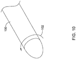
- the implant 25 may be formed by collecting fibers, such as, but not limited to, collagen fibers, on a rotating mandrel 100, as shown in Figure 10 .
- the first component 50 may be formed by rotating 102 the mandrel 100 at a first speed. Once the desired thickness of the first component 50 has been obtained, the mandrel 100 may be rotated 102 at a second speed, slower than the first speed. The mandrel 100 continues to collect the fibers at the second, slower speed. However, the decreased speed of rotation will result in less orientation of the fibers and a denser packing of the fibers to form the second, higher strength component 52.
- the continuity of the fiber collection process provides continuity between the first component 50 and the second component 52, effectively affixing the two components 50, 52 to one another to form the layered implant 25.
- the thickness of each component 50, 52 can be adjusted as necessary to achieve the desired final properties by adjusting the length of time fibers are collected at each rotation speed. It is contemplated that the process may be reversed and the second component formed first. In some instances, variation of the fiber collection process during build-up of the implant 25 may be used to produce a "fiber-reinforced" bioinductive component 50 in which the high-strength component 52 is distributed throughout the low-strength component 50, without distinct separate "layers".
- synthetic fibers such as, but not limited to absorbable suture material, may be incorporated into the bioinductive component 50 during the manufacturing process to create a reinforced composite material which is capable of invoking a healing response as well as providing immediate strength to the tendon.
- first component 50 and the second component 52 may be formed as separate layers.
- the layers 50, 52 may then be stacked one on top of the other and freeze-dried or lyophilized to form a composite.
- the composite is then cross-linked to form a link or bond between the first component 50 and the second component 52.
- the link or bond between the first component 50 and the second component 52 does not need to be a high-strength interface.
- the bond only needs to be sufficient enough to hold the two components 50, 52 together to enable handling during surgery.
- freeze-drying wet components 50, 52 after they have been stacked one on top of the other may sufficiently bond (at least temporarily) the two components to enable the implant 25 to be surgically implanted.
- the two components 50, 52 may be stitched together using, for example, bioabsorbable suture material. It is contemplated that stitching the components 50, 52 may be used in combination with or in place of the freeze-drying and/or cross-linking.
- the high-strength component 52 is formed of the same material as the bioinductive component 50 and is made stronger by increased cross-linking, the components 50, 52 may need to be produced and cross-linked separately. The two components 50, 52 may then be laminated or bonded as described above.
- Figure 8 is a perspective, schematic view of the illustrative tendon repair implant 25 including a third component, or layer 60.
- the third component 60 may be an additional low-strength, or bioinductive, component positioned on top of the second, high-strength component 52 to create an implant 25 including three layers. It is contemplated that the bioinductive component 60 may be affixed or secured to the high-strength component using any of the methods described above.
- a complete or partial thickness tear in a tendon are also provided, however without forming part of the invention.
- supraspinatus tendons having complete tears, partial thickness tears of greater than 50% and/or partial thickness tears of less than 50% are treated.
- the treatment site may be first arthroscopically accessed in the area of the damaged tendon.
- a tendon repair implant, such as previously described may be placed over a partial tear in a tendon.
- the implant may be placed over a tendon having complete or partial tear(s), abrasions and/or inflammation. Left untreated, minor or partial tendon tears may progress into larger or full tears.
- a complete or partial tear may be treated by protecting it with a tendon repair implant as described above.
- Such treatment can promote healing and provided immediate strength to the tendon, as well as prevent more extensive damage from occurring to the tendon, thereby averting the need for a more involved surgical procedure.
- the implant may be configured to be collapsible so that it may be inserted into or mounted on a tubular member for arthroscopic insertion to the treatment site.
- the implant and associated delivery device may be collapsed like an umbrella where the deployed delivery systems unfolds the pleats of the implant as mounted thereon to allow surface to surface engagement with the tendon without any substantial wrinkles.
- the tendon repair implant may then be affixed using sutures or other suitable means such as staples such that the tensile properties will assure that the anatomical load will be shared because the native tendon and implant experience the same strain under load.
- the tendon repair implant may comprise an absorbable material.
- the purpose of the implant is to protect an injured portion of a tendon during healing, provide an implant for new tissue growth, and/or temporarily share some of the tendon loads.
- the implant may induce additional tendon-like tissue formation, thereby adding strength and reducing pain, micro strains and inflammation.
- the implant 25 may provide immediate strength to the tendon and transfer the load to the native tendon as the tendon heals and the implant 25 biodegrades.
- organized collagen fibers are created that remodel to tendon-like tissue or neo-tendon with cell vitality and vascularity. Initial stiffness of the device may be less than that of the native tendon so as to not overload the fixation while tendon tissue is being generated.
- Material(s) used in the implanted device should be able to withstand the compression and shear loads consistent with accepted post surgical shoulder motions.
- the perimeter of the device may have different mechanical properties than the interior of the device, such as for facilitating better retention of sutures, staples or other fastening mechanisms.
- the material(s) may be chosen to be compatible with visual, radiographic, magnetic, ultrasonic, or other common imaging techniques.
- the material(s) may be capable of absorbing and retaining growth factors with the possibility of hydrophilic coatings to promote retention of additives.
Claims (14)
- Implant de réparation de tendon comprenant:une structure analogue à une feuille (25) incluant un premier composant (50) et un second composant (52), le premier composant (50) étant configuré pour avoir un premier module de traction d'environ 5 mégapascals (MPa) à 50 MPa et comprenant une pluralité de pores pour la croissance tissulaire à l'intérieur;dans lequel le premier composant (50) et le second composant (52) sont disposés en couches;caractérisé en ce que le second composant (52) est configuré pour avoir un premier module de traction d'environ 50 MPa à 150 MPa.
- Implant de réparation de tendon selon la revendication 1, dans lequel le premier composant (50) et le second composant (52) forment des couches discrètes.
- Implant de réparation de tendon selon la revendication 1, dans lequel le second composant (52) est au moins partiellement mélangé avec le premier composant (50) pour former un matériau composite.
- Implant de réparation de tendon selon la revendication 1, dans lequel la structure analogue à une feuille (25) est configurée pour se conformer à la surface du tendon et est configurée pour avoir une première part de charge représentant environ 50 % ou plus de la charge du tendon, dans lequel environ 50 % ou plus de la charge du tendon comprend entre 25 Newtons (N) et 50 N.
- Implant de réparation de tendon selon la revendication 1, dans lequel la structure analogue à une feuille (25) est configurée pour se dégrader en résistance à la traction à partir d'une première résistance à la traction au cours du temps.
- Implant de réparation de tendon selon la revendication 5, dans lequel la structure analogue à une feuille (25) est configurée pour conserver au moins environ 70 % de la première résistance à la traction après trois mois et pour conserver au moins environ 50 % du module de traction initial et environ 50 % d'un module de compression initial après trois mois.
- Implant de réparation de tendon selon la revendication 1, dans lequel le premier composant (50) est configuré pour se dégrader en résistance à la traction à partir d'une première résistance à la traction à une première vitesse et le second composant (52) est configuré pour se dégrader en résistance à la traction à partir d'une première résistance à la traction à une seconde vitesse différente de la première vitesse, de préférence dans lequel la seconde vitesse est plus lente que la première vitesse.
- Implant de réparation de tendon selon la revendication 1, dans lequel le premier composant (50) et/ou le second composant (52) comprend un matériau biorésorbable et/ou un matériau polymère.
- Implant de réparation de tendon selon la revendication 1, dans lequel la structure analogue à une feuille (25) est configurée en outre pour être fixée à la surface d'un tendon sans préchargement significatif ou pré-étirage significatif.
- Implant de réparation de tendon selon la revendication 1, dans lequel la structure analogue à une feuille (25) a un ou plusieurs agents fixés à celle-ci qui favorisent la croissance tissulaire à l'intérieur.
- Implant de réparation de tendon selon la revendication 1, dans lequel les pores ont des tailles entre environ 20 microns et 400 microns.
- Implant de réparation de tendon selon la revendication 1, dans lequel la structure analogue à une feuille (25) est définie par une dimension longitudinale, une dimension latérale et une dimension d'épaisseur, dans lequel la dimension longitudinale est supérieure à la dimension latérale et à la dimension d'épaisseur, et dans lequel la structure analogue à une feuille (25) comprend en outre un ou plusieurs chemins longitudinaux s'étendant le long de la dimension longitudinale de la structure analogue à une feuille (25).
- Implant de réparation de tendon selon la revendication 1, dans lequel la structure analogue à une feuille (25) a un maximum d'environ 0,5 % de fluage sur trois mois.
- Implant de réparation de tendon selon la revendication 1, comprenant en outre un troisième composant (60) configuré pour avoir un premier module de traction d'environ 5 MPa à 50 MPa.
Applications Claiming Priority (2)
| Application Number | Priority Date | Filing Date | Title |
|---|---|---|---|
| US201562175829P | 2015-06-15 | 2015-06-15 | |
| PCT/US2016/037354 WO2016205186A1 (fr) | 2015-06-15 | 2016-06-14 | Implant de réparation de tendon et son procédé d'implantation |
Publications (2)
| Publication Number | Publication Date |
|---|---|
| EP3307204A1 EP3307204A1 (fr) | 2018-04-18 |
| EP3307204B1 true EP3307204B1 (fr) | 2021-11-24 |
Family
ID=57515620
Family Applications (1)
| Application Number | Title | Priority Date | Filing Date |
|---|---|---|---|
| EP16733256.8A Active EP3307204B1 (fr) | 2015-06-15 | 2016-06-14 | Implant de réparation de tendon |
Country Status (3)
| Country | Link |
|---|---|
| US (4) | US10265156B2 (fr) |
| EP (1) | EP3307204B1 (fr) |
| WO (1) | WO2016205186A1 (fr) |
Families Citing this family (16)
| Publication number | Priority date | Publication date | Assignee | Title |
|---|---|---|---|---|
| WO2013025819A2 (fr) * | 2011-08-16 | 2013-02-21 | The University Of Kansas | Timbre trachéal fibreux |
| EP3367966A4 (fr) | 2015-10-30 | 2019-07-10 | New York Society for the Relief of the Ruptured and Crippled, Maintaining the Hospital for Special Surgery | Timbre de manchon de suture et procédés de pose dans un flux de travail arthroscopique existant |
| CA3008670A1 (fr) | 2015-12-31 | 2017-07-06 | Rotation Medical, Inc. | Systeme de distribution d'agrafes et procedes associes |
| JP6653389B2 (ja) | 2015-12-31 | 2020-02-26 | ローテーション メディカル インコーポレイテッドRotation Medical,Inc. | 医療用インプラント搬送システムおよび関連方法 |
| US11523812B2 (en) | 2016-02-01 | 2022-12-13 | Medos International Sarl | Soft tissue fixation repair methods using tissue augmentation constructs |
| US11484401B2 (en) | 2016-02-01 | 2022-11-01 | Medos International Sarl | Tissue augmentation scaffolds for use in soft tissue fixation repair |
| US10265160B2 (en) * | 2017-01-04 | 2019-04-23 | Arthrex, Inc. | Embroidered textile support a biological graft |
| CN106901868B (zh) * | 2017-02-24 | 2018-08-14 | 丁少华 | 一种上关节囊重建修复用补片及其制备方法 |
| WO2019113292A1 (fr) | 2017-12-07 | 2019-06-13 | Rotation Medical, Inc. | Système de pose d'implant médical et procédés associés |
| US11135066B2 (en) | 2018-04-23 | 2021-10-05 | Medos International Sarl | Mechanical fuse for surgical implants and related methods |
| CN108403258B (zh) * | 2018-06-05 | 2023-09-05 | 上海市第六人民医院 | 一种止点重建型人工肩袖补片及其制造方法 |
| CN111110404B (zh) * | 2020-01-10 | 2021-04-23 | 苏州诺普再生医学有限公司 | 一种3d打印的多结构骨复合支架 |
| WO2021163342A1 (fr) | 2020-02-11 | 2021-08-19 | Embody, Inc. | Dispositif d'ancrage chirurgical, dispositif de déploiement et méthode d'utilisation |
| EP4103259A4 (fr) | 2020-02-11 | 2024-02-28 | Embody Inc | Canule chirurgicale avec joint de pression amovible |
| WO2021163337A1 (fr) | 2020-02-11 | 2021-08-19 | Embody, Inc. | Dispositif de pose d'implant |
| EP4117743A1 (fr) * | 2020-03-12 | 2023-01-18 | Smith&Nephew, Inc. | Implant de réparation tissulaire, compositions et procédé d'implantation |
Family Cites Families (448)
| Publication number | Priority date | Publication date | Assignee | Title |
|---|---|---|---|---|
| US511238A (en) | 1893-12-19 | Half to alfred brown | ||
| US3123077A (en) | 1964-03-03 | Surgical suture | ||
| US765793A (en) | 1903-09-16 | 1904-07-26 | John F Ruckel | Surgical bridge. |
| US1728316A (en) | 1927-07-02 | 1929-09-17 | Kirurgiska Instr Fabriks Aktie | Wound clasp |
| US1868100A (en) | 1929-01-19 | 1932-07-19 | Bella Goodstein | Staple and method of driving the same |
| US1855546A (en) | 1931-04-28 | 1932-04-26 | Norman W File | Surgical appliance |
| US1910688A (en) | 1931-08-03 | 1933-05-23 | Bella Goodstein | Staple |
| US1940351A (en) | 1933-03-22 | 1933-12-19 | Dougald T Mckinnon | Surgical instrument |
| US2075508A (en) | 1934-07-18 | 1937-03-30 | Edward W Davidson | Suture retainer |
| US2034785A (en) | 1935-07-12 | 1936-03-24 | Wappler Frederick Charles | Endoscopic forceps |
| US2199025A (en) | 1936-06-08 | 1940-04-30 | Carl E Conn | Means and method of closing surgical incisions |
| US2158242A (en) | 1936-10-08 | 1939-05-16 | Boston Wire Stitcher Co | Staple for blind stitching |
| US2131321A (en) | 1937-10-11 | 1938-09-27 | Hart Wilber | Ligator |
| US2201610A (en) | 1938-05-20 | 1940-05-21 | Jr James C Dawson | Wound clip |
| US2254620A (en) | 1939-11-14 | 1941-09-02 | George I Miller | Clip |
| US2283814A (en) | 1940-07-29 | 1942-05-19 | Bocji Corp | Staple and method of stapling |
| US2277931A (en) | 1941-07-03 | 1942-03-31 | Boston Wire Stitcher Co | Staple |
| US2316297A (en) | 1943-01-15 | 1943-04-13 | Beverly A Southerland | Surgical instrument |
| US2421193A (en) | 1943-08-02 | 1947-05-27 | Cleveland Clinic Foundation | Surgical dressing |
| US2570497A (en) | 1949-07-12 | 1951-10-09 | Royal Mfg Company Inc | Undergarment |
| US2571813A (en) | 1950-08-17 | 1951-10-16 | Clarence L Austin | Hog ringer |
| US2630316A (en) | 1950-09-01 | 1953-03-03 | Edwin E Foster | Constant compression spring |
| US2910067A (en) | 1952-10-13 | 1959-10-27 | Technical Oil Tool Corp | Wound clip and extractor therefor |
| US2684070A (en) | 1953-03-23 | 1954-07-20 | Walter L Kelsey | Surgical clip |
| US2744251A (en) | 1953-06-04 | 1956-05-08 | Vollmer Leonhard | Automatic inserter for suturing clips |
| US2817339A (en) | 1953-08-10 | 1957-12-24 | Norman M Sullivan | Rigid fascial suture |
| US2825162A (en) | 1954-01-18 | 1958-03-04 | Dennison Mfg Co | String tag attachment device |
| US2881762A (en) | 1955-02-09 | 1959-04-14 | Robert J Lowrie | Surgical staple and stapler |
| US2790341A (en) | 1956-04-17 | 1957-04-30 | Francis J Keep | Split shot pliers and dispenser |
| US3077812A (en) | 1958-01-27 | 1963-02-19 | Josef Kihlberg | Staple |
| US3068870A (en) | 1960-03-18 | 1962-12-18 | Levin Abraham | Wound clip |
| US3209754A (en) | 1961-08-10 | 1965-10-05 | Ernest C Wood | Surgical clip |
| US3103666A (en) | 1961-12-28 | 1963-09-17 | Dennison Mfg Co | Tag attaching apparatus |
| US3221746A (en) | 1963-01-25 | 1965-12-07 | Noble John William | Surgical connecting device |
| US3527223A (en) | 1967-09-01 | 1970-09-08 | Melvin Shein | Ear stud and hollow piercer for insertion thereof |
| US3470834A (en) | 1968-03-08 | 1969-10-07 | Dennison Mfg Co | Fastener attaching device |
| US3577837A (en) | 1968-04-30 | 1971-05-11 | Karl F Bader Jr | Subdermal tendon implant |
| US3570497A (en) | 1969-01-16 | 1971-03-16 | Gerald M Lemole | Suture apparatus and methods |
| US3579831A (en) | 1969-03-05 | 1971-05-25 | Irving J Stevens | Bone implant |
| US3643851A (en) | 1969-08-25 | 1972-02-22 | United States Surgical Corp | Skin stapler |
| US3716058A (en) | 1970-07-17 | 1973-02-13 | Atlanta Res Inst | Barbed suture |
| US3687138A (en) | 1970-08-17 | 1972-08-29 | Robert K Jarvik | Repeating ligature gun |
| US3837555A (en) | 1970-12-14 | 1974-09-24 | Surgical Corp | Powering instrument for stapling skin and fascia |
| US3717294A (en) | 1970-12-14 | 1973-02-20 | Surgical Corp | Cartridge and powering instrument for stapling skin and fascia |
| US3757629A (en) | 1971-05-10 | 1973-09-11 | R Schneider | Resilient plastic staple |
| US3777538A (en) | 1972-03-15 | 1973-12-11 | Weck & Co Edward | Surgical clip applicator |
| US3875648A (en) | 1973-04-04 | 1975-04-08 | Dennison Mfg Co | Fastener attachment apparatus and method |
| US3845772A (en) | 1973-09-17 | 1974-11-05 | D Smith | Retention suture device and method |
| US3976079A (en) | 1974-08-01 | 1976-08-24 | Samuels Peter B | Securing devices for sutures |
| US3960147A (en) | 1975-03-10 | 1976-06-01 | Murray William M | Compression bone staples and methods of compressing bone segments |
| US4014492A (en) | 1975-06-11 | 1977-03-29 | Senco Products, Inc. | Surgical staple |
| US4127227A (en) | 1976-10-08 | 1978-11-28 | United States Surgical Corporation | Wide fascia staple cartridge |
| US4259959A (en) | 1978-12-20 | 1981-04-07 | Walker Wesley W | Suturing element |
| US4263903A (en) | 1979-01-08 | 1981-04-28 | Richards Manufacturing Co., Inc. | Medical staple means |
| US4265226A (en) | 1979-03-23 | 1981-05-05 | Cassimally K A I | Incision closing method |
| US4317451A (en) | 1980-02-19 | 1982-03-02 | Ethicon, Inc. | Plastic surgical staple |
| GB2084468B (en) | 1980-09-25 | 1984-06-06 | South African Inventions | Surgical implant |
| US4526174A (en) | 1981-03-27 | 1985-07-02 | Minnesota Mining And Manufacturing Company | Staple and cartridge for use in a tissue stapling device and a tissue closing method |
| US4400833A (en) | 1981-06-10 | 1983-08-30 | Kurland Kenneth Z | Means and method of implanting bioprosthetics |
| US4485816A (en) | 1981-06-25 | 1984-12-04 | Alchemia | Shape-memory surgical staple apparatus and method for use in surgical suturing |
| US4809695A (en) | 1981-10-21 | 1989-03-07 | Owen M. Gwathmey | Suturing assembly and method |
| US4422567A (en) | 1981-11-17 | 1983-12-27 | Haynes Taylor H | Medical suturing device |
| US4480641A (en) | 1982-02-05 | 1984-11-06 | Ethicon, Inc. | Tip configuration for a ligating clip applier |
| US4509518A (en) | 1982-02-17 | 1985-04-09 | United States Surgical Corporation | Apparatus for applying surgical clips |
| US4454875A (en) | 1982-04-15 | 1984-06-19 | Techmedica, Inc. | Osteal medical staple |
| US5601557A (en) | 1982-05-20 | 1997-02-11 | Hayhurst; John O. | Anchoring and manipulating tissue |
| US4741330A (en) | 1983-05-19 | 1988-05-03 | Hayhurst John O | Method and apparatus for anchoring and manipulating cartilage |
| US5417691A (en) | 1982-05-20 | 1995-05-23 | Hayhurst; John O. | Apparatus and method for manipulating and anchoring tissue |
| US4595007A (en) | 1983-03-14 | 1986-06-17 | Ethicon, Inc. | Split ring type tissue fastener |
| US4570623A (en) | 1983-06-02 | 1986-02-18 | Pfizer Hospital Products Group Inc. | Arched bridge staple |
| US4627437A (en) | 1983-06-20 | 1986-12-09 | Ethicon, Inc. | Method of applying a fastener to tissue with a pair of hollow needles |
| US4582237A (en) | 1983-08-26 | 1986-04-15 | Anthony Storace | Surgical stapling system, apparatus and staple |
| US4549545A (en) | 1984-03-05 | 1985-10-29 | Ethicon Inc. | Segmented polyurethane surgical buttressing pledgets |
| US4635637A (en) | 1984-03-29 | 1987-01-13 | Schreiber Saul N | Surgical suture |
| EP0185026A1 (fr) | 1984-05-07 | 1986-06-25 | PUCHY, David Peter William | Agrafeuse chirurgicale a degre variable de fermeture des agrafes |
| US4696300A (en) | 1985-04-11 | 1987-09-29 | Dennison Manufacturing Company | Fastener for joining materials |
| US4632100A (en) | 1985-08-29 | 1986-12-30 | Marlowe E. Goble | Suture anchor assembly |
| US4669473A (en) | 1985-09-06 | 1987-06-02 | Acufex Microsurgical, Inc. | Surgical fastener |
| USRE34021E (en) | 1985-11-18 | 1992-08-04 | Abbott Laboratories | Percutaneous fixation of hollow organs |
| US4738255A (en) | 1986-04-07 | 1988-04-19 | Biotron Labs, Inc. | Suture anchor system |
| US4924865A (en) | 1986-05-20 | 1990-05-15 | Concept, Inc. | Repair tack for bodily tissue |
| GB8620560D0 (en) | 1986-08-23 | 1986-10-01 | Femcare Ltd | Applicator |
| US4762260A (en) | 1986-09-11 | 1988-08-09 | Ophthalmic Ventures Limited Partnership | Surgical microstapler |
| US4719917A (en) | 1987-02-17 | 1988-01-19 | Minnesota Mining And Manufacturing Company | Surgical staple |
| US4799495A (en) | 1987-03-20 | 1989-01-24 | National Standard Company | Localization needle assembly |
| US5174295A (en) | 1987-04-10 | 1992-12-29 | Cardiometrics, Inc. | Apparatus, system and method for measuring spatial average velocity and/or volumetric flow of blood in a vessel and screw joint for use therewith |
| US4898156A (en) | 1987-05-18 | 1990-02-06 | Mitek Surgical Products, Inc. | Suture anchor |
| DE3722538A1 (de) | 1987-07-08 | 1989-01-19 | Wasserstein Isidor | Vorrichtung zur fixierung von knochensegmenten |
| US5261914A (en) | 1987-09-02 | 1993-11-16 | Russell Warren | Surgical fastener |
| US4887601A (en) | 1987-11-06 | 1989-12-19 | Ophthalmic Ventures Limited Partnership | Adjustable surgical staple and method of using the same |
| US4968315A (en) | 1987-12-15 | 1990-11-06 | Mitek Surgical Products, Inc. | Suture anchor and suture anchor installation tool |
| CA2004658C (fr) | 1988-06-03 | 1995-10-10 | Michael A. Oberlander | Clip arthroscopique et outil d'insertion |
| AU7139994A (en) | 1988-06-13 | 1995-01-03 | Karlin Technology, Inc. | Apparatus and method of inserting spinal implants |
| US4924866A (en) | 1988-10-26 | 1990-05-15 | Inbae Yoon | Wound-closing device |
| US5171273A (en) | 1989-01-13 | 1992-12-15 | University Of Medicine And Dentistry Of New Jersey | Synthetic collagen orthopaedic structures such as grafts, tendons and other structures |
| EP0389102B1 (fr) | 1989-02-22 | 1995-05-10 | United States Surgical Corporation | Agrafe pour la peau |
| US4994073A (en) | 1989-02-22 | 1991-02-19 | United States Surgical Corp. | Skin fastener |
| US4930674A (en) | 1989-02-24 | 1990-06-05 | Abiomed, Inc. | Surgical stapler |
| US5522817A (en) | 1989-03-31 | 1996-06-04 | United States Surgical Corporation | Absorbable surgical fastener with bone penetrating elements |
| CA2013539C (fr) | 1989-03-31 | 1999-07-06 | Thomas W. Sander | Fixateur chirurgical absorbable avec elements penetrant le tissu osseux |
| US5062563A (en) | 1989-04-10 | 1991-11-05 | United States Surgical Corporation | Fascia stapler |
| US5059206A (en) | 1989-04-12 | 1991-10-22 | Winters Thomas F | Method and apparatus for repairing a tear in a knee meniscus |
| US5441508A (en) | 1989-04-27 | 1995-08-15 | Gazielly; Dominique | Reinforcement and supporting device for the rotator cuff of a shoulder joint of a person |
| US5053047A (en) | 1989-05-16 | 1991-10-01 | Inbae Yoon | Suture devices particularly useful in endoscopic surgery and methods of suturing |
| US6764500B1 (en) | 1989-05-29 | 2004-07-20 | Kensey Nash Corporation | Sealing device |
| US5129906A (en) | 1989-09-08 | 1992-07-14 | Linvatec Corporation | Bioabsorbable tack for joining bodily tissue and in vivo method and apparatus for deploying same |
| US5123913A (en) | 1989-11-27 | 1992-06-23 | Wilk Peter J | Suture device |
| US5015249A (en) | 1989-12-26 | 1991-05-14 | Nakao Naomi L | Endoscopic stapling device and method |
| US5156609A (en) | 1989-12-26 | 1992-10-20 | Nakao Naomi L | Endoscopic stapling device and method |
| US5002563A (en) | 1990-02-22 | 1991-03-26 | Raychem Corporation | Sutures utilizing shape memory alloys |
| US5013316A (en) | 1990-03-26 | 1991-05-07 | Marlowe Goble E | Soft tissue anchor system |
| EP0474887B1 (fr) | 1990-04-02 | 1994-06-15 | Kanji Inoue | Procede pour refermer une ouverture de shunt au moyen d'un procede sans operateur |
| US5102421A (en) | 1990-06-14 | 1992-04-07 | Wm. E. Anpach, III | Suture anchor and method of forming |
| US5037422A (en) | 1990-07-02 | 1991-08-06 | Acufex Microsurgical, Inc. | Bone anchor and method of anchoring a suture to a bone |
| US5224946A (en) | 1990-07-02 | 1993-07-06 | American Cyanamid Company | Bone anchor and method of anchoring a suture to a bone |
| US5041129A (en) | 1990-07-02 | 1991-08-20 | Acufex Microsurgical, Inc. | Slotted suture anchor and method of anchoring a suture |
| US5100417A (en) | 1990-07-13 | 1992-03-31 | American Cyanamid Company | Suture anchor and driver assembly |
| US5725529A (en) | 1990-09-25 | 1998-03-10 | Innovasive Devices, Inc. | Bone fastener |
| US5141515A (en) | 1990-10-11 | 1992-08-25 | Eberbach Mark A | Apparatus and methods for repairing hernias |
| US5116357A (en) | 1990-10-11 | 1992-05-26 | Eberbach Mark A | Hernia plug and introducer apparatus |
| US5122155A (en) | 1990-10-11 | 1992-06-16 | Eberbach Mark A | Hernia repair apparatus and method of use |
| US5203787A (en) | 1990-11-19 | 1993-04-20 | Biomet, Inc. | Suture retaining arrangement |
| WO1992013500A1 (fr) | 1991-02-08 | 1992-08-20 | Surgical Innovations, Inc. | Appareil et procede de reparation d'hernies inguinales |
| US5720753A (en) | 1991-03-22 | 1998-02-24 | United States Surgical Corporation | Orthopedic fastener |
| CA2063159C (fr) | 1991-03-22 | 1999-06-15 | Thomas W. Sander | Attache orthopedique |
| US5174487A (en) | 1991-04-04 | 1992-12-29 | Ethicon, Inc. | Endoscopic stapler and hernia repair mechanism |
| US5217472A (en) | 1991-05-07 | 1993-06-08 | United States Surgical Corporation | Surgical fastening device |
| US5269783A (en) | 1991-05-13 | 1993-12-14 | United States Surgical Corporation | Device and method for repairing torn tissue |
| US5251642A (en) | 1991-06-06 | 1993-10-12 | Baxter International Inc. | Tissue measuring and suturing device |
| CA2075080A1 (fr) | 1991-08-02 | 1993-02-03 | Ralph A. Dematteis | Methode et appareil de reparation des hernies par la parascopie |
| US5282829A (en) | 1991-08-15 | 1994-02-01 | United States Surgical Corporation | Hollow body implants |
| US5290217A (en) | 1991-10-10 | 1994-03-01 | Earl K. Sipes | Method and apparatus for hernia repair |
| IL103442A (en) | 1991-10-18 | 1998-01-04 | Ethicon Inc | Installs the adhesive buffer |
| US5497933A (en) | 1991-10-18 | 1996-03-12 | United States Surgical Corporation | Apparatus and method for applying surgical staples to attach an object to body tissue |
| US5289963A (en) | 1991-10-18 | 1994-03-01 | United States Surgical Corporation | Apparatus and method for applying surgical staples to attach an object to body tissue |
| US5141520A (en) | 1991-10-29 | 1992-08-25 | Marlowe Goble E | Harpoon suture anchor |
| US5350400A (en) | 1991-10-30 | 1994-09-27 | American Cyanamid Company | Malleable, bioabsorbable, plastic staple; and method and apparatus for deforming such staple |
| US5176692A (en) | 1991-12-09 | 1993-01-05 | Wilk Peter J | Method and surgical instrument for repairing hernia |
| US5167665A (en) | 1991-12-31 | 1992-12-01 | Mckinney William W | Method of attaching objects to bone |
| US5156616A (en) | 1992-02-10 | 1992-10-20 | Meadows Bruce F | Apparatus and method for suture attachment |
| CA2090000A1 (fr) | 1992-02-24 | 1993-08-25 | H. Jonathan Tovey | Dispositif articule servant a positionner des implants chirurgicaux |
| US5333624A (en) | 1992-02-24 | 1994-08-02 | United States Surgical Corporation | Surgical attaching apparatus |
| CA2089999A1 (fr) | 1992-02-24 | 1993-08-25 | H. Jonathan Tovey | Dispositif souple servant a positionner des implants chirurgicaux |
| US5242457A (en) | 1992-05-08 | 1993-09-07 | Ethicon, Inc. | Surgical instrument and staples for applying purse string sutures |
| US5766246A (en) | 1992-05-20 | 1998-06-16 | C. R. Bard, Inc. | Implantable prosthesis and method and apparatus for loading and delivering an implantable prothesis |
| US5501695A (en) | 1992-05-27 | 1996-03-26 | The Anspach Effort, Inc. | Fastener for attaching objects to bones |
| US5176682A (en) | 1992-06-01 | 1993-01-05 | Chow James C Y | Surgical implement |
| US6312442B1 (en) | 1992-06-02 | 2001-11-06 | General Surgical Innovations, Inc. | Method for developing an anatomic space for laparoscopic hernia repair |
| US5797931A (en) | 1992-06-04 | 1998-08-25 | Olympus Optical Co., Ltd. | Tissue-fixing surgical instrument, tissue-fixing device, and method of fixing tissues |
| US5304187A (en) | 1992-06-30 | 1994-04-19 | United States Surgical Corporation | Surgical element deployment apparatus |
| US5269753A (en) | 1992-07-14 | 1993-12-14 | Wilk Peter J | Method for use in laparoscopic hernia repair |
| US5364408A (en) | 1992-09-04 | 1994-11-15 | Laurus Medical Corporation | Endoscopic suture system |
| CA2437773C (fr) | 1992-09-21 | 2005-02-22 | United States Surgical Corporation | Dispositif d'application d'une agrafe meniscale |
| US5464403A (en) | 1992-10-29 | 1995-11-07 | General Surgical Innovations, Inc. | Placement tool and method for laparoscopic hernia repair |
| IL103737A (en) | 1992-11-13 | 1997-02-18 | Technion Res & Dev Foundation | Stapler device particularly useful in medical suturing |
| US5622257A (en) | 1992-12-11 | 1997-04-22 | Avery Dennison Corporation | Fastener for attaching a button to a garment or like material and fastener clip including one or more of said fasteners |
| US5403326A (en) | 1993-02-01 | 1995-04-04 | The Regents Of The University Of California | Method for performing a gastric wrap of the esophagus for use in the treatment of esophageal reflux |
| US5380334A (en) | 1993-02-17 | 1995-01-10 | Smith & Nephew Dyonics, Inc. | Soft tissue anchors and systems for implantation |
| US5441502A (en) | 1993-02-17 | 1995-08-15 | Mitek Surgical Products, Inc. | System and method for re-attaching soft tissue to bone |
| US5342396A (en) | 1993-03-02 | 1994-08-30 | Cook Melvin S | Staples |
| US5354292A (en) | 1993-03-02 | 1994-10-11 | Braeuer Harry L | Surgical mesh introduce with bone screw applicator for the repair of an inguinal hernia |
| US5352229A (en) | 1993-05-12 | 1994-10-04 | Marlowe Goble E | Arbor press staple and washer and method for its use |
| US5505735A (en) | 1993-06-10 | 1996-04-09 | Mitek Surgical Products, Inc. | Surgical anchor and method for using the same |
| US5372604A (en) | 1993-06-18 | 1994-12-13 | Linvatec Corporation | Suture anchor for soft tissue fixation |
| CA2124996C (fr) | 1993-06-21 | 2006-01-31 | Thomas W. Sander | Applicateur de fixation orthopedique |
| US5500000A (en) | 1993-07-01 | 1996-03-19 | United States Surgical Corporation | Soft tissue repair system and method |
| US5478354A (en) | 1993-07-14 | 1995-12-26 | United States Surgical Corporation | Wound closing apparatus and method |
| US5507754A (en) | 1993-08-20 | 1996-04-16 | United States Surgical Corporation | Apparatus and method for applying and adjusting an anchoring device |
| CA2124651C (fr) | 1993-08-20 | 2004-09-28 | David T. Green | Appareil et methode pour mettre en place et ajuster un dispositif d'ancrage de structures internes |
| US5411522A (en) | 1993-08-25 | 1995-05-02 | Linvatec Corporation | Unitary anchor for soft tissue fixation |
| US5397332A (en) | 1993-09-02 | 1995-03-14 | Ethicon, Inc. | Surgical mesh applicator |
| US5560532A (en) | 1993-10-08 | 1996-10-01 | United States Surgical Corporation | Apparatus and method for applying surgical staples to body tissue |
| US5584835A (en) | 1993-10-18 | 1996-12-17 | Greenfield; Jon B. | Soft tissue to bone fixation device and method |
| US5545180A (en) | 1993-12-13 | 1996-08-13 | Ethicon, Inc. | Umbrella-shaped suture anchor device with actuating ring member |
| US5618314A (en) | 1993-12-13 | 1997-04-08 | Harwin; Steven F. | Suture anchor device |
| AU1011595A (en) | 1994-01-13 | 1995-07-20 | Ethicon Inc. | Spiral surgical tack |
| US5425490A (en) | 1994-01-18 | 1995-06-20 | Goble; E. Marlowe | Instrument with dual holding feature |
| US5417712A (en) | 1994-02-17 | 1995-05-23 | Mitek Surgical Products, Inc. | Bone anchor |
| US5486197A (en) | 1994-03-24 | 1996-01-23 | Ethicon, Inc. | Two-piece suture anchor with barbs |
| US5411523A (en) | 1994-04-11 | 1995-05-02 | Mitek Surgical Products, Inc. | Suture anchor and driver combination |
| US5582616A (en) | 1994-08-05 | 1996-12-10 | Origin Medsystems, Inc. | Surgical helical fastener with applicator |
| US5649963A (en) | 1994-11-10 | 1997-07-22 | Innovasive Devices, Inc. | Suture anchor assembly and methods |
| US5643321A (en) | 1994-11-10 | 1997-07-01 | Innovasive Devices | Suture anchor assembly and methods |
| IL111985A (en) | 1994-12-14 | 1999-04-11 | Medical Influence Technologies | Staple and thread assembly particularly for use in power-driven staplers for medical suturing |
| WO1996025847A1 (fr) | 1995-02-22 | 1996-08-29 | Kurt Ilgen | Dispositif pour reparer les fissures sur le sabot d'un animal |
| FR2731343B1 (fr) | 1995-03-08 | 1997-08-22 | De La Joliniere Jean H Bouquet | Dispositif de reperage de lesions suspectes du sein et appareil pour sa mise en place |
| US5919184A (en) | 1995-03-17 | 1999-07-06 | Tilton, Jr.; Eugene B. | Instrumentation for laparoscopic insertion and application of surgical sheet material |
| US5503623A (en) | 1995-03-17 | 1996-04-02 | Tilton, Jr.; Eugene B. | Instrumentation for laparoscopic insertion and application of sheet like surgical material |
| US6245072B1 (en) | 1995-03-27 | 2001-06-12 | Sdgi Holdings, Inc. | Methods and instruments for interbody fusion |
| US6123727A (en) | 1995-05-01 | 2000-09-26 | Massachusetts Institute Of Technology | Tissue engineered tendons and ligaments |
| US5702215A (en) | 1995-06-05 | 1997-12-30 | Li Medical Technologies, Inc. | Retractable fixation device |
| US5569306A (en) | 1995-06-06 | 1996-10-29 | Thal; Raymond | Knotless suture anchor assembly |
| US5667513A (en) | 1995-06-07 | 1997-09-16 | Smith & Nephew Dyonics Inc. | Soft tissue anchor delivery apparatus |
| US5681342A (en) | 1995-08-17 | 1997-10-28 | Benchetrit; Salomon | Device and method for laparoscopic inguinal hernia repair |
| US5662683A (en) | 1995-08-22 | 1997-09-02 | Ortho Helix Limited | Open helical organic tissue anchor and method of facilitating healing |
| US5997552A (en) | 1995-10-20 | 1999-12-07 | United States Surgical Corporation | Meniscal fastener applying device |
| WO1997021383A1 (fr) | 1995-12-13 | 1997-06-19 | Nycomed Arzneimittel Gmbh | Instrument destine a l'application d'une matiere chirurgicale |
| US5725541A (en) | 1996-01-22 | 1998-03-10 | The Anspach Effort, Inc. | Soft tissue fastener device |
| US5741282A (en) | 1996-01-22 | 1998-04-21 | The Anspach Effort, Inc. | Soft tissue fastener device |
| US5957953A (en) | 1996-02-16 | 1999-09-28 | Smith & Nephew, Inc. | Expandable suture anchor |
| US5885258A (en) | 1996-02-23 | 1999-03-23 | Memory Medical Systems, Inc. | Medical instrument with slotted memory metal tube |
| US5893856A (en) | 1996-06-12 | 1999-04-13 | Mitek Surgical Products, Inc. | Apparatus and method for binding a first layer of material to a second layer of material |
| US6117162A (en) | 1996-08-05 | 2000-09-12 | Arthrex, Inc. | Corkscrew suture anchor |
| US6482224B1 (en) | 1996-08-22 | 2002-11-19 | The Trustees Of Columbia University In The City Of New York | Endovascular flexible stapling device |
| US6984241B2 (en) | 1996-09-13 | 2006-01-10 | Tendon Technology, Ltd. | Apparatus and methods for tendon or ligament repair |
| US5830221A (en) | 1996-09-20 | 1998-11-03 | United States Surgical Corporation | Coil fastener applier |
| US5948000A (en) | 1996-10-03 | 1999-09-07 | United States Surgical Corporation | System for suture anchor placement |
| EP0834280B1 (fr) | 1996-10-04 | 2004-01-02 | United States Surgical Corporation | Appareil d'implantation d'un élément de fixation de tissus |
| US6063088A (en) | 1997-03-24 | 2000-05-16 | United States Surgical Corporation | Method and instrumentation for implant insertion |
| FR2758975B1 (fr) | 1997-02-05 | 1999-04-30 | Ethnor | Materiel pour la fixation d'un tendon de muscle sur un os |
| US5782864A (en) | 1997-04-03 | 1998-07-21 | Mitek Surgical Products, Inc. | Knotless suture system and method |
| US5922026A (en) | 1997-05-01 | 1999-07-13 | Origin Medsystems, Inc. | Surgical method and prosthetic strip therefor |
| US6001110A (en) | 1997-06-20 | 1999-12-14 | Boston Scientific Corporation | Hemostatic clips |
| US6338737B1 (en) | 1997-07-17 | 2002-01-15 | Haviv Toledano | Flexible annular stapler for closed surgery of hollow organs |
| US5957939A (en) | 1997-07-31 | 1999-09-28 | Imagyn Medical Technologies, Inc. | Medical device for deploying surgical fabrics |
| US5885294A (en) | 1997-09-22 | 1999-03-23 | Ethicon, Inc. | Apparatus and method for anchoring a cord-like element to a workpiece |
| US5868762A (en) | 1997-09-25 | 1999-02-09 | Sub-Q, Inc. | Percutaneous hemostatic suturing device and method |
| US5947984A (en) | 1997-10-10 | 1999-09-07 | Ethicon Endo-Surger, Inc. | Ultrasonic clamp coagulator apparatus having force limiting clamping mechanism |
| DE19801219A1 (de) | 1998-01-15 | 1999-07-22 | Holger K Dr Essiger | Knochennagel |
| US6096074A (en) | 1998-01-27 | 2000-08-01 | United States Surgical | Stapling apparatus and method for heart valve replacement |
| FR2774277B1 (fr) | 1998-02-03 | 2000-06-30 | Cogent Sarl | Agrafe de fixation prothetique resorbable |
| CA2333121C (fr) | 1998-05-21 | 2006-07-25 | Christopher J. Walshe | Systeme de fixation des tissus |
| US6726704B1 (en) | 1998-05-29 | 2004-04-27 | By-Pass, Inc. | Advanced closure device |
| US7063711B1 (en) | 1998-05-29 | 2006-06-20 | By-Pass, Inc. | Vascular surgery |
| WO2000016701A1 (fr) | 1998-09-18 | 2000-03-30 | United States Surgical Corporation | Applicateur de fixation endovasculaire |
| US6248112B1 (en) | 1998-09-30 | 2001-06-19 | C. R. Bard, Inc. | Implant delivery system |
| US6458092B1 (en) | 1998-09-30 | 2002-10-01 | C. R. Bard, Inc. | Vascular inducing implants |
| US6318616B1 (en) | 1998-10-23 | 2001-11-20 | United States Surgical | Surgical fastener applier |
| US6193731B1 (en) | 1998-10-27 | 2001-02-27 | Fziomed, Inc. | Laparoscopic insertion and deployment device |
| US6482210B1 (en) | 1998-11-12 | 2002-11-19 | Orthopaedic Biosystems, Ltd., Inc. | Soft tissue/ligament to bone fixation device with inserter |
| EP2055244B1 (fr) | 1998-12-31 | 2012-02-01 | Kensey Nash Corporation | Dispositifs de fixation de tissus |
| DE60037696T2 (de) | 1999-02-02 | 2009-01-08 | Arthrex Inc., Naples | Bioresorbierbare Gewebeheftzwecke mit ovalförmigen Kopf |
| US6387113B1 (en) | 1999-02-02 | 2002-05-14 | Biomet, Inc. | Method and apparatus for repairing a torn meniscus |
| US7226469B2 (en) | 1999-02-02 | 2007-06-05 | Arthrex, Inc. | Insert molded suture anchor |
| US6315789B1 (en) | 1999-02-08 | 2001-11-13 | Andrew H. Cragg | Medical device anchoring system and method |
| US20020090725A1 (en) | 2000-11-17 | 2002-07-11 | Simpson David G. | Electroprocessed collagen |
| US6391333B1 (en) | 1999-04-14 | 2002-05-21 | Collagen Matrix, Inc. | Oriented biopolymeric membrane |
| US6325805B1 (en) | 1999-04-23 | 2001-12-04 | Sdgi Holdings, Inc. | Shape memory alloy staple |
| US6482178B1 (en) | 1999-05-21 | 2002-11-19 | Cook Urological Incorporated | Localization device with anchoring barbs |
| GB9912240D0 (en) | 1999-05-27 | 1999-07-28 | Smith & Nephew | Implantable medical devices |
| US8025199B2 (en) | 2004-02-23 | 2011-09-27 | Tyco Healthcare Group Lp | Surgical cutting and stapling device |
| US6494888B1 (en) | 1999-06-22 | 2002-12-17 | Ndo Surgical, Inc. | Tissue reconfiguration |
| US6835200B2 (en) | 1999-06-22 | 2004-12-28 | Ndo Surgical. Inc. | Method and devices for tissue reconfiguration |
| US20040059416A1 (en) | 1999-06-22 | 2004-03-25 | Murray Martha M. | Biologic replacement for fibrin clot |
| US6964685B2 (en) | 1999-06-22 | 2005-11-15 | The Brigham And Women's Hospital, Inc. | Biologic replacement for fibrin clot |
| US7416554B2 (en) | 2002-12-11 | 2008-08-26 | Usgi Medical Inc | Apparatus and methods for forming and securing gastrointestinal tissue folds |
| US6179840B1 (en) | 1999-07-23 | 2001-01-30 | Ethicon, Inc. | Graft fixation device and method |
| US6436110B2 (en) | 1999-07-23 | 2002-08-20 | Ethicon, Inc. | Method of securing a graft using a graft fixation device |
| US7214232B2 (en) | 1999-07-23 | 2007-05-08 | Ethicon, Inc. | Graft fixation device |
| US6319252B1 (en) | 1999-07-23 | 2001-11-20 | Mcdevitt Dennis | System and method for attaching soft tissue to bone |
| US7972337B2 (en) | 2005-12-28 | 2011-07-05 | Intrinsic Therapeutics, Inc. | Devices and methods for bone anchoring |
| US6554852B1 (en) | 1999-08-25 | 2003-04-29 | Michael A. Oberlander | Multi-anchor suture |
| US6322563B1 (en) | 1999-09-17 | 2001-11-27 | Genzyme Corporation | Small tissue and membrane fixation apparatus and methods for use thereof |
| US6231561B1 (en) | 1999-09-20 | 2001-05-15 | Appriva Medical, Inc. | Method and apparatus for closing a body lumen |
| US6428548B1 (en) | 1999-11-18 | 2002-08-06 | Russell F. Durgin | Apparatus and method for compressing body tissue |
| FR2801191B1 (fr) | 1999-11-19 | 2002-02-15 | Proconcept | Dispositif de protection des nerfs apres intervention chirurgicale |
| US6740100B2 (en) | 1999-12-23 | 2004-05-25 | Omeros Corporation | Tendon repair using adhesive |
| US6524317B1 (en) | 1999-12-30 | 2003-02-25 | Opus Medical, Inc. | Method and apparatus for attaching connective tissues to bone using a knotless suture anchoring device |
| GB2359024A (en) | 2000-02-09 | 2001-08-15 | Anson Medical Ltd | Fixator for arteries |
| US6673094B1 (en) | 2000-02-23 | 2004-01-06 | Ethicon, Inc. | System and method for attaching soft tissue to bone |
| US7399304B2 (en) | 2000-03-03 | 2008-07-15 | C.R. Bard, Inc. | Endoscopic tissue apposition device with multiple suction ports |
| AU781098B2 (en) | 2000-04-11 | 2005-05-05 | United States Surgical Corporation | Single shot meniscal repair device |
| US6821298B1 (en) | 2000-04-18 | 2004-11-23 | Roger P. Jackson | Anterior expandable spinal fusion cage system |
| US20050171569A1 (en) | 2000-04-25 | 2005-08-04 | Impres Medical, Inc. | Method and apparatus for creating intrauterine adhesions |
| CA2377974A1 (fr) | 2000-04-25 | 2001-11-01 | John Alfred Campin | Systeme et procede de restriction de zone d'ablation par laser |
| US6478803B1 (en) | 2000-05-19 | 2002-11-12 | Genzyme Corporation | Device for delivery of surgical materials |
| CH694357A5 (fr) | 2000-05-31 | 2004-12-15 | M D Supply S A R L | Ensemble de fixation d'un tissu mou sur un os |
| US6575976B2 (en) | 2000-06-12 | 2003-06-10 | Arthrex, Inc. | Expandable tissue anchor |
| US6620185B1 (en) | 2000-06-27 | 2003-09-16 | Smith & Nephew, Inc. | Surgical procedures and instruments |
| US7144414B2 (en) | 2000-06-27 | 2006-12-05 | Smith & Nephew, Inc. | Surgical procedures and instruments |
| US6830174B2 (en) | 2000-08-30 | 2004-12-14 | Cerebral Vascular Applications, Inc. | Medical instrument |
| WO2002019922A1 (fr) | 2000-09-08 | 2002-03-14 | Coleman James E | Agrafeuse chirurgicale |
| US7037324B2 (en) | 2000-09-15 | 2006-05-02 | United States Surgical Corporation | Knotless tissue anchor |
| US6569186B1 (en) | 2000-10-05 | 2003-05-27 | Biomet, Inc. | Soft tissue screw and fixation device |
| US6887259B2 (en) | 2000-10-18 | 2005-05-03 | Depuy Mitek, Inc. | Suture anchor system and method of use |
| US6527795B1 (en) | 2000-10-18 | 2003-03-04 | Ethicon, Inc. | Knotless suture anchor system and method of use |
| US6641596B1 (en) | 2000-10-18 | 2003-11-04 | Ethicon, Inc. | Knotless bioabsorbable suture anchor system and method |
| US6425900B1 (en) | 2000-10-19 | 2002-07-30 | Ethicon Endo-Surgery | Method for attaching hernia mesh |
| US6447524B1 (en) | 2000-10-19 | 2002-09-10 | Ethicon Endo-Surgery, Inc. | Fastener for hernia mesh fixation |
| US6551333B2 (en) | 2000-10-19 | 2003-04-22 | Ethicon Endo-Surgery, Inc. | Method for attaching hernia mesh |
| US7485124B2 (en) | 2000-10-19 | 2009-02-03 | Ethicon Endo-Surgery, Inc. | Surgical instrument having a fastener delivery mechanism |
| WO2002034140A2 (fr) | 2000-10-23 | 2002-05-02 | Tyco Healthcare Group Lp | Fixation resorbable et dispositif de mise en place |
| US6648893B2 (en) | 2000-10-27 | 2003-11-18 | Blackstone Medical, Inc. | Facet fixation devices |
| FR2817142B1 (fr) | 2000-11-24 | 2003-05-16 | Sofradim Production | Attache de fixation prothetique et dispositif d'insertion transcutanee |
| US20020077687A1 (en) | 2000-12-14 | 2002-06-20 | Ahn Samuel S. | Catheter assembly for treating ischemic tissue |
| US7083638B2 (en) | 2001-02-12 | 2006-08-01 | Arthrocare Corporation | Method and apparatus for attaching connective tissues to bone using a knotless suture anchoring device |
| DE10110106B4 (de) | 2001-03-02 | 2007-08-02 | Richard Wolf Gmbh | Chirurgische Zange |
| EP1913896B1 (fr) | 2001-03-09 | 2012-12-26 | Boston Scientific Limited | Procède de fabrication d' une écharpe médicale |
| EP1365688B1 (fr) | 2001-03-09 | 2011-02-16 | Boston Scientific Limited | Systeme d'implantation d'une bandelette |
| US7144413B2 (en) | 2001-04-20 | 2006-12-05 | Synthes (U.S.A.) | Graft fixation system and method |
| US6503257B2 (en) | 2001-05-07 | 2003-01-07 | Ethicon Endo-Surgery, Inc. | Method for releasing buttress material attached to a surgical fastening device |
| US7033379B2 (en) | 2001-06-08 | 2006-04-25 | Incisive Surgical, Inc. | Suture lock having non-through bore capture zone |
| JP4302515B2 (ja) | 2001-07-16 | 2009-07-29 | デピュイ・プロダクツ・インコーポレイテッド | 単体型外科装置および方法 |
| IES20010748A2 (en) | 2001-08-09 | 2003-02-19 | Christy Cummins | Surgical Stapling Device and Method |
| IES20010749A2 (en) | 2001-08-09 | 2003-02-19 | Christy Cummins | Surgical Stapling Device |
| US6629988B2 (en) | 2001-08-28 | 2003-10-07 | Ethicon, Inc. | Composite staple for completing an anastomosis |
| US9005245B2 (en) | 2002-08-30 | 2015-04-14 | Arthrex, Inc. | Acromioclavicular joint fixation technique |
| US6893431B2 (en) | 2001-10-15 | 2005-05-17 | Scimed Life Systems, Inc. | Medical device for delivering patches |
| US6540769B1 (en) | 2001-10-31 | 2003-04-01 | Miller, Iii Archibald S. | Method and apparatus for closing a severed sternum |
| US6986781B2 (en) | 2001-11-08 | 2006-01-17 | Smith & Nephew, Inc. | Tissue repair system |
| US6723099B1 (en) | 2001-11-08 | 2004-04-20 | Biomet, Inc. | Three sided tack for bone fixation |
| JP2005516648A (ja) | 2001-12-13 | 2005-06-09 | エスディージーアイ・ホールディングス・インコーポレーテッド | インプラントを椎骨空間内に導入する器具及び方法 |
| WO2003053256A1 (fr) | 2001-12-13 | 2003-07-03 | Sumitomo Bakelite Company Limited | Dispositif de pince pour endoscope et pince pour endoscope utilisee dans ce dispositif |
| US7150750B2 (en) | 2002-01-10 | 2006-12-19 | Boston Scientific Scimed, Inc. | Method and device for endoscopic suturing |
| US6685728B2 (en) | 2002-01-25 | 2004-02-03 | Stryker Endoscopy | Threaded suture anchor and method of use |
| US7201754B2 (en) | 2002-02-08 | 2007-04-10 | Kenneth Stewart | Device for installing an anchor in a bone |
| WO2003071962A2 (fr) | 2002-02-25 | 2003-09-04 | Yeung Jeffrey E | Element de fixation expansible avec prises compressives |
| FR2836816B1 (fr) | 2002-03-08 | 2005-01-28 | Sofradim Production | Appareil de stockage, de distribution et de pose d'attaches chirurgicales en"i"couche |
| WO2003088815A2 (fr) | 2002-04-17 | 2003-10-30 | Tyco Healthcare Group, Lp | Instrument de fixation et fixation |
| US7229452B2 (en) | 2002-04-22 | 2007-06-12 | Tyco Healthcare Group Lp | Tack and tack applier |
| US7572276B2 (en) | 2002-05-06 | 2009-08-11 | Warsaw Orthopedic, Inc. | Minimally invasive instruments and methods for inserting implants |
| US20030212456A1 (en) | 2002-05-09 | 2003-11-13 | John Lipchitz | Implant for tissue repair |
| US6638297B1 (en) | 2002-05-30 | 2003-10-28 | Ethicon Endo-Surgery, Inc. | Surgical staple |
| US6986775B2 (en) | 2002-06-13 | 2006-01-17 | Guided Delivery Systems, Inc. | Devices and methods for heart valve repair |
| AU2003245606B2 (en) | 2002-06-19 | 2008-04-03 | Covidien Lp | Method and apparatus for anastomosis |
| US7112214B2 (en) | 2002-06-25 | 2006-09-26 | Incisive Surgical, Inc. | Dynamic bioabsorbable fastener for use in wound closure |
| US6726705B2 (en) | 2002-06-25 | 2004-04-27 | Incisive Surgical, Inc. | Mechanical method and apparatus for bilateral tissue fastening |
| US6972027B2 (en) | 2002-06-26 | 2005-12-06 | Stryker Endoscopy | Soft tissue repair system |
| US6932834B2 (en) | 2002-06-27 | 2005-08-23 | Ethicon, Inc. | Suture anchor |
| US7160326B2 (en) | 2002-06-27 | 2007-01-09 | Depuy Products, Inc. | Method and apparatus for implantation of soft tissue implant |
| US6702215B2 (en) | 2002-07-03 | 2004-03-09 | Quantum Corporation | Repositionable memory element in a single reel tape cartridge |
| US7048171B2 (en) | 2002-08-29 | 2006-05-23 | Dale H. Kosted | Surgical stapler with removable staple cartridge |
| US6966916B2 (en) | 2002-09-26 | 2005-11-22 | Kumar Sarbjeet S | Device and method for surgical repair of abdominal wall hernias |
| US7087064B1 (en) | 2002-10-15 | 2006-08-08 | Advanced Cardiovascular Systems, Inc. | Apparatuses and methods for heart valve repair |
| US7404824B1 (en) | 2002-11-15 | 2008-07-29 | Advanced Cardiovascular Systems, Inc. | Valve aptation assist device |
| US7517350B2 (en) | 2002-11-20 | 2009-04-14 | Orthopediatrics Corp. | Convertible threaded compression device and method of use |
| US7343920B2 (en) | 2002-12-20 | 2008-03-18 | Toby E Bruce | Connective tissue repair system |
| US8551162B2 (en) | 2002-12-20 | 2013-10-08 | Medtronic, Inc. | Biologically implantable prosthesis |
| US20040138705A1 (en) | 2003-01-09 | 2004-07-15 | Harri Heino | Surgical staple for tissue treatment |
| GB0300786D0 (en) | 2003-01-14 | 2003-02-12 | Barker Stephen G E | Laparoscopic port hernia device |
| US7322935B2 (en) | 2003-01-22 | 2008-01-29 | Medcanica, Llc | Endoscopic retractor |
| US7303577B1 (en) | 2003-02-05 | 2007-12-04 | Dean John C | Apparatus and method for use in repairs of injured soft tissue |
| US8197837B2 (en) | 2003-03-07 | 2012-06-12 | Depuy Mitek, Inc. | Method of preparation of bioabsorbable porous reinforced tissue implants and implants thereof |
| US7368124B2 (en) | 2003-03-07 | 2008-05-06 | Depuy Mitek, Inc. | Method of preparation of bioabsorbable porous reinforced tissue implants and implants thereof |
| US7465308B2 (en) | 2003-04-10 | 2008-12-16 | Smith & Nephew, Inc. | Fixation device |
| US8579989B2 (en) | 2003-04-22 | 2013-11-12 | Patrick Leahy | Device and method for use in parietal surgery |
| TW587933B (en) | 2003-04-30 | 2004-05-21 | A Spine Holding Group Corp | Device for anchoring bone tissue |
| US7320701B2 (en) | 2003-06-02 | 2008-01-22 | Linvatec Corporation | Push-in suture anchor, insertion tool, and method for inserting a push-in suture anchor |
| US7104966B2 (en) | 2003-07-16 | 2006-09-12 | Samuel Shiber | Guidewire system with exposed midsection |
| US7104999B2 (en) | 2003-06-28 | 2006-09-12 | Ethicon, Inc. | Surgical anchor inserter |
| US7559941B2 (en) | 2003-06-30 | 2009-07-14 | Depuy Products, Inc. | Instrument for delivery of implant |
| US7819880B2 (en) | 2003-06-30 | 2010-10-26 | Depuy Products, Inc. | Implant delivery instrument |
| WO2005016389A2 (fr) | 2003-08-04 | 2005-02-24 | Kelly Jackson | Instruments medicaux et leurs procedes d'utilisation |
| US7021316B2 (en) | 2003-08-07 | 2006-04-04 | Tools For Surgery, Llc | Device and method for tacking a prosthetic screen |
| US7481832B1 (en) | 2003-09-09 | 2009-01-27 | Biomet Sports Medicine, Llc | Method and apparatus for use of a self-tapping resorbable screw |
| JP2005086122A (ja) | 2003-09-11 | 2005-03-31 | Oki Electric Ind Co Ltd | 半導体装置の製造方法 |
| US7547312B2 (en) | 2003-09-17 | 2009-06-16 | Gore Enterprise Holdings, Inc. | Circular stapler buttress |
| US7175648B2 (en) | 2003-11-18 | 2007-02-13 | Granit Medical Innovations, Llc | Deep endoscopic staple and stapler |
| US8673021B2 (en) | 2003-11-26 | 2014-03-18 | Depuy Mitek, Llc | Arthroscopic tissue scaffold delivery device |
| EP1559379A1 (fr) | 2004-01-31 | 2005-08-03 | Labotect Verwaltungs GmbH | Dispositif d'implantation de marqueurs |
| US7212869B2 (en) | 2004-02-04 | 2007-05-01 | Medtronic, Inc. | Lead retention means |
| GB2429651C (en) | 2004-02-17 | 2009-03-25 | Cook Biotech Inc | Medical devices and methods useful for applying bolster material |
| US7758612B2 (en) | 2004-04-27 | 2010-07-20 | Tyco Healthcare Group Lp | Surgery delivery device and mesh anchor |
| US7390329B2 (en) | 2004-05-07 | 2008-06-24 | Usgi Medical, Inc. | Methods for grasping and cinching tissue anchors |
| US20050251175A1 (en) | 2004-05-07 | 2005-11-10 | Ethicon Endo-Surgery, Inc. | Anchors for use in attachment of bladder tissues to pelvic floor tissues following a prostatectomy |
| WO2005110529A1 (fr) | 2004-05-10 | 2005-11-24 | Advanced Bionics Corporation | Électrode implantable, outil et méthode d’insertion |
| US20070190108A1 (en) | 2004-05-17 | 2007-08-16 | Arindam Datta | High performance reticulated elastomeric matrix preparation, properties, reinforcement, and use in surgical devices, tissue augmentation and/or tissue repair |
| IES20040368A2 (en) | 2004-05-25 | 2005-11-30 | James E Coleman | Surgical stapler |
| CA2568562A1 (fr) | 2004-06-02 | 2006-06-08 | Michael L. Green | Systeme et procede de fixation de tissu mou a l'os |
| US7500983B1 (en) | 2004-06-09 | 2009-03-10 | Biomet Sports Medicine, Llc | Apparatus for soft tissue attachment |
| ATE519451T1 (de) | 2004-08-30 | 2011-08-15 | Neville Alleyne | Implantat zur behandlung von bändern und sehnen |
| US7452368B2 (en) | 2004-09-15 | 2008-11-18 | Ethicon, Inc. | System for holding surgical fasteners |
| JP2006081687A (ja) | 2004-09-15 | 2006-03-30 | Max Co Ltd | 医療用ステープラ |
| US7807192B2 (en) | 2004-10-22 | 2010-10-05 | Collagen Matrix, Inc. | Biopolymeric membranes |
| US7785255B2 (en) | 2004-10-29 | 2010-08-31 | Howmedica Osteonics Corp. | Ligament protection instruments |
| JP2008518713A (ja) | 2004-11-08 | 2008-06-05 | エンドグン メディカル システムズ リミテッド | 外科用グラフト |
| US20070078477A1 (en) | 2005-02-04 | 2007-04-05 | Heneveld Scott H Sr | Anatomical spacer and method to deploy |
| WO2006086679A1 (fr) | 2005-02-09 | 2006-08-17 | Miller Archibald S Iii | Dispositif de fermeture sternal a mecanisme de fermeture a cliquet |
| US7825083B2 (en) | 2005-02-10 | 2010-11-02 | Spine Wave, Inc. | Synovial fluid barrier |
| MX2007010097A (es) | 2005-02-18 | 2007-10-10 | Synthasome Inc | Estructura sintetica para la preparacion de tejido blando. |
| KR20070083994A (ko) | 2005-03-23 | 2007-08-24 | 가부시끼가이샤 제이엠에스 | 유착 방지 필름 |
| US7344544B2 (en) | 2005-03-28 | 2008-03-18 | Cardica, Inc. | Vascular closure system |
| US7740641B2 (en) | 2005-04-14 | 2010-06-22 | Ethicon Endo-Surgery, Inc. | Clip applier with migrational resistance features |
| EP1885259B1 (fr) | 2005-05-11 | 2016-08-17 | Mayo Foundation For Medical Education And Research | Dispositif pour interventions chirurgicales internes |
| US20070083236A1 (en) | 2005-06-24 | 2007-04-12 | Smith & Nephew, Inc. | Methods and devices for tissue repair |
| US20060293760A1 (en) | 2005-06-24 | 2006-12-28 | Dedeyne Patrick G | Soft tissue implants with improved interfaces |
| ITMI20051461A1 (it) | 2005-07-28 | 2007-01-29 | Gianni Ponte | Cambretta per sutura dello sterno ed apparecchio atto a posizionarla e serrarla |
| US20090030434A1 (en) | 2005-08-11 | 2009-01-29 | Endogun Medical Systems Ltd. | Surgical Fasteners and Devices for Surgical Fasteners |
| US7771440B2 (en) | 2005-08-18 | 2010-08-10 | Ethicon Endo-Surgery, Inc. | Method and apparatus for endoscopically performing gastric reduction surgery in a single pass |
| WO2007030676A2 (fr) | 2005-09-09 | 2007-03-15 | University Of South Florida | Dispositif d'extension de filet herniaire par laparoscopie |
| US20070112361A1 (en) | 2005-11-07 | 2007-05-17 | Schonholz Steven M | Surgical repair systems and methods of using the same |
| EP1971290A2 (fr) | 2006-01-12 | 2008-09-24 | Histogenics Corporation | Procede de reparation et de reconstruction de ligaments ou de tendons rompus ou de traitement de lesions ligamentaires et tendineuses |
| US9387011B2 (en) | 2006-02-02 | 2016-07-12 | Steven C. Chudik | Acromioclavicular joint repair system |
| US20070219558A1 (en) | 2006-03-15 | 2007-09-20 | Allen Deutsch | Method and apparatus for arthroscopic surgery using suture anchors |
| US20080009900A1 (en) | 2006-06-12 | 2008-01-10 | Kfx Medical Corporation | Surgical grasping device |
| CA2656278C (fr) | 2006-06-30 | 2016-02-09 | Biomimetic Therapeutics, Inc. | Compositions et procedes destines au traitement de lesions de la coiffe des rotateurs |
| US20080065153A1 (en) | 2006-09-08 | 2008-03-13 | Warsaw Orthopedic, Inc. | Surgical staple |
| US7506791B2 (en) | 2006-09-29 | 2009-03-24 | Ethicon Endo-Surgery, Inc. | Surgical stapling instrument with mechanical mechanism for limiting maximum tissue compression |
| US7780685B2 (en) | 2006-11-09 | 2010-08-24 | Ethicon Endo-Surgery, Inc. | Adhesive and mechanical fastener |
| US8308662B2 (en) | 2006-12-07 | 2012-11-13 | Arthrex, Inc. | Measuring device |
| US20080173691A1 (en) | 2007-01-24 | 2008-07-24 | Medtronic Vascular, Inc. | Low-Profile Vascular Closure Systems and Methods of Using Same |
| US20080188936A1 (en) | 2007-02-02 | 2008-08-07 | Tornier, Inc. | System and method for repairing tendons and ligaments |
| US8753391B2 (en) | 2007-02-12 | 2014-06-17 | The Trustees Of Columbia University In The City Of New York | Fully synthetic implantable multi-phased scaffold |
| US8337529B2 (en) | 2007-02-13 | 2012-12-25 | Anova Corp. | Methods of bone, joint, and ligament reconstruction |
| EP2111187A4 (fr) | 2007-02-14 | 2014-10-01 | Synthasome Inc | Structure synthétique pour réparation de partie molle |
| US7533790B1 (en) | 2007-03-08 | 2009-05-19 | Cardica, Inc. | Surgical stapler |
| US8142450B2 (en) | 2007-03-13 | 2012-03-27 | Longevity Surgical, Inc. | Methods for reducing gastric volume |
| CN104473704B (zh) | 2007-03-15 | 2017-06-16 | 矫正-空位有限公司 | 假体装置及其使用方法 |
| EP2129299A4 (fr) | 2007-03-15 | 2016-02-10 | Bioprotect Ltd | Dispositifs de fixation de tissu mou |
| AU2008249584A1 (en) | 2007-05-14 | 2008-11-20 | Bioprotect Ltd. | Delivery device for delivering bioactive agents to internal tissue in a body |
| US8062330B2 (en) | 2007-06-27 | 2011-11-22 | Tyco Healthcare Group Lp | Buttress and surgical stapling apparatus |
| US8821507B2 (en) | 2007-07-06 | 2014-09-02 | Howmedica Osteonics Corp. | Augmentation delivery device |
| US8382761B2 (en) | 2007-08-29 | 2013-02-26 | Covidien Lp | Surgical staple with adjustable width backspan |
| US8366741B2 (en) | 2007-09-13 | 2013-02-05 | Cardia, Inc. | Occlusion device with centering arm |
| US20090112085A1 (en) | 2007-10-30 | 2009-04-30 | University Of Washington | Preoperative localization wire |
| US20090156997A1 (en) | 2007-12-12 | 2009-06-18 | Rockford Orthopaedic Sports Medicine Services, Llc | Rotator cuff patch delivery device |
| US20090157193A1 (en) | 2007-12-18 | 2009-06-18 | Warsaw Orthopedic, Inc. | Tendon and Ligament Repair Sheet and Methods of Use |
| ATE472300T1 (de) | 2008-01-16 | 2010-07-15 | Roberto Zambelli | Führungsvorrichtung zur lokalisierung von neoplasien während eines chirurgischen verfahrens |
| US8317808B2 (en) | 2008-02-18 | 2012-11-27 | Covidien Lp | Device and method for rolling and inserting a prosthetic patch into a body cavity |
| US8758373B2 (en) | 2008-02-18 | 2014-06-24 | Covidien Lp | Means and method for reversibly connecting a patch to a patch deployment device |
| JP5536679B2 (ja) | 2008-03-14 | 2014-07-02 | トランスエンテリックス インコーポレイテッド | 一体型メッシュマニピュレータを有するヘルニアステープラ |
| US20090242609A1 (en) | 2008-03-31 | 2009-10-01 | Medtronic Vascular, Inc. | Vascular Puncture Closure Staple With Tip Protection |
| GB2471815A (en) | 2008-05-21 | 2011-01-12 | Cook Biotech Inc | Devices and methods for applying bolster materials to surgical fastening apparatus |
| US9387280B2 (en) | 2008-09-05 | 2016-07-12 | Synovis Orthopedic And Woundcare, Inc. | Device for soft tissue repair or replacement |
| CA2745571C (fr) | 2008-12-15 | 2015-03-24 | Allergan, Inc. | Dispositif prothetique et procede pour le fabriquer |
| US20100147921A1 (en) | 2008-12-16 | 2010-06-17 | Lee Olson | Surgical Apparatus Including Surgical Buttress |
| US8770460B2 (en) | 2008-12-23 | 2014-07-08 | George E. Belzer | Shield for surgical stapler and method of use |
| US20110011917A1 (en) | 2008-12-31 | 2011-01-20 | Hansen Medical, Inc. | Methods, devices, and kits for treating valve prolapse |
| US20100191332A1 (en) | 2009-01-08 | 2010-07-29 | Euteneuer Charles L | Implantable Tendon Protection Systems and Related Kits and Methods |
| FR2941144B1 (fr) | 2009-01-22 | 2012-04-27 | Sofradim Production | Agrafeuse chirurgicale pour le prepositionnement et la fixation de prothese en textile, methode de chargement de celle-ci et agrafe chirurgicale auto-agrippante |
| US9179910B2 (en) | 2009-03-20 | 2015-11-10 | Rotation Medical, Inc. | Medical device delivery system and method |
| US8241298B2 (en) | 2009-03-27 | 2012-08-14 | Depuy Mitek, Inc. | Methods and devices for delivering and affixing tissue scaffolds |
| WO2010114633A1 (fr) | 2009-04-03 | 2010-10-07 | Biomerix Corporation | Éléments de matrice élastomère réticulée au moins partiellement résorbables et leurs procédés de production |
| US20100256675A1 (en) | 2009-04-03 | 2010-10-07 | Romans Matthew L | Absorbable surgical staple |
| US20100274278A1 (en) | 2009-04-22 | 2010-10-28 | Pare Surgical, Inc. | Endoscopic tissue grasping apparatus and method |
| US8894669B2 (en) | 2009-05-12 | 2014-11-25 | Ethicon, Inc. | Surgical fasteners, applicator instruments, and methods for deploying surgical fasteners |
| WO2010141906A1 (fr) | 2009-06-04 | 2010-12-09 | Rotation Medical, Inc. | Procedes et appareil pour le deploiement de materiaux en feuille |
| WO2010141907A1 (fr) | 2009-06-04 | 2010-12-09 | Rotation Medical, Inc. | Appareil de pose d'agrafes en forme d'arc sur un tissue cible |
| CA2769707A1 (fr) | 2009-08-17 | 2011-02-24 | Tyco Healthcare Group Lp | Dispositif de deploiement de piece articulee et procede d'utilisation |
| EP3508144B1 (fr) | 2009-08-17 | 2021-04-07 | Covidien LP | Dispositif de déploiement de timbres |
| US8672967B2 (en) | 2009-10-30 | 2014-03-18 | Depuy Mitek, Llc | Partial thickness rotator cuff repair system and method |
| US8353439B2 (en) | 2009-11-19 | 2013-01-15 | Ethicon Endo-Surgery, Inc. | Circular stapler introducer with radially-openable distal end portion |
| EP2531114B1 (fr) | 2010-02-03 | 2018-12-05 | Covidien LP | Dispositif en forme de x pour le déploiement et placement d'un filet à hernie |
| US9198750B2 (en) | 2010-03-11 | 2015-12-01 | Rotation Medical, Inc. | Tendon repair implant and method of arthroscopic implantation |
| US20130035704A1 (en) | 2010-04-14 | 2013-02-07 | Moshe Dudai | Surgical spreadable sheet delivery and positioning system and method |
| CA2795819C (fr) | 2010-04-27 | 2018-02-27 | Synthes Usa, Llc | Systeme de fixation d'os comprenant une compression par fil de kirschner |
| US8900616B2 (en) | 2010-10-22 | 2014-12-02 | Covidien Lp | System and method for satellite drug delivery |
| WO2012145059A1 (fr) | 2011-02-15 | 2012-10-26 | Rotation Medical, Inc. | Procédés et appareil pour fixer des matériaux de type en feuille à un tissu cible |
| CA2825918C (fr) | 2011-02-15 | 2018-08-07 | Rotation Medical, Inc. | Procedes et appareil pour la distribution et le positionnement de materiaux en feuille |
| US9314314B2 (en) | 2011-02-15 | 2016-04-19 | Rotation Medical, Inc. | Anatomical location markers and methods of use in positioning sheet-like materials during surgery |
| US9283019B2 (en) | 2011-06-08 | 2016-03-15 | Warsaw Orthopedic, Inc. | Flexible guide wire |
| EP3403601A1 (fr) | 2011-12-19 | 2018-11-21 | Rotation Medical, Inc. | Appareil de formation de trous pilotes dans un os et de fourniture d'attaches permettant de retenir un implant |
| US9107661B2 (en) | 2011-12-19 | 2015-08-18 | Rotation Medical, Inc. | Fasteners and fastener delivery devices for affixing sheet-like materials to bone or tissue |
| WO2013096224A1 (fr) | 2011-12-19 | 2013-06-27 | Rotation Medical, Inc. | Organes de fixation pour fixer des matériaux en feuille à un os ou à un tissu |
| WO2013119321A1 (fr) | 2011-12-19 | 2013-08-15 | Rotation Medical, Inc. | Organes de fixation et dispositifs de distribution d'organes de fixation pour fixer des matières de type feuille à un os ou à un tissu |
| WO2013101640A1 (fr) | 2011-12-29 | 2013-07-04 | Rotation Medical, Inc. | Fil-guide ayant un élément de fixation distal pour la pose et le positionnement de matières de type feuille en chirurgie |
| WO2013101638A1 (fr) | 2011-12-29 | 2013-07-04 | Rotation Medical, Inc. | Procédés et appareil de distribution et de positionnement de matières de type feuille en chirurgie |
| GB201317636D0 (en) | 2013-10-04 | 2013-11-20 | Isis Innovation | Scaffold |
-
2016
- 2016-06-14 US US15/181,612 patent/US10265156B2/en active Active
- 2016-06-14 WO PCT/US2016/037354 patent/WO2016205186A1/fr active Application Filing
- 2016-06-14 EP EP16733256.8A patent/EP3307204B1/fr active Active
-
2018
- 2018-10-10 US US16/156,281 patent/US10888415B2/en active Active
-
2020
- 2020-12-17 US US17/124,961 patent/US11607305B2/en active Active
-
2023
- 2023-02-22 US US18/112,802 patent/US20230190446A1/en active Pending
Also Published As
| Publication number | Publication date |
|---|---|
| US20160361155A1 (en) | 2016-12-15 |
| US10265156B2 (en) | 2019-04-23 |
| WO2016205186A1 (fr) | 2016-12-22 |
| US20190038395A1 (en) | 2019-02-07 |
| US20210186677A1 (en) | 2021-06-24 |
| US20230190446A1 (en) | 2023-06-22 |
| EP3307204A1 (fr) | 2018-04-18 |
| US11607305B2 (en) | 2023-03-21 |
| US10888415B2 (en) | 2021-01-12 |
Similar Documents
| Publication | Publication Date | Title |
|---|---|---|
| US11607305B2 (en) | Tendon repair implant and method of implantation | |
| US11717393B2 (en) | Tendon repair implant and method of arthroscopic implantation | |
| US11413133B2 (en) | Implantable tendon protection systems and related kits and methods | |
| CA2598268C (fr) | Structure synthetique pour la reparation de tissus mous | |
| JP2016508854A (ja) | 乳房再建およびヘルニア修復のための吸収性合成編組マトリクス | |
| EP3297558A1 (fr) | Systèmes et procédés pour réparer des tissus mous | |
| US20220401627A1 (en) | Tissue repair implant and compositions and method of implantation | |
| Wroblesky | Augmentation of a Knit Structure with Increased Suture Retention and Poly (glycerol Sebacate) Coating for Rotator Cuff Repair Graft Applications |
Legal Events
| Date | Code | Title | Description |
|---|---|---|---|
| STAA | Information on the status of an ep patent application or granted ep patent |
Free format text: STATUS: THE INTERNATIONAL PUBLICATION HAS BEEN MADE |
|
| PUAI | Public reference made under article 153(3) epc to a published international application that has entered the european phase |
Free format text: ORIGINAL CODE: 0009012 |
|
| STAA | Information on the status of an ep patent application or granted ep patent |
Free format text: STATUS: REQUEST FOR EXAMINATION WAS MADE |
|
| 17P | Request for examination filed |
Effective date: 20171230 |
|
| AK | Designated contracting states |
Kind code of ref document: A1 Designated state(s): AL AT BE BG CH CY CZ DE DK EE ES FI FR GB GR HR HU IE IS IT LI LT LU LV MC MK MT NL NO PL PT RO RS SE SI SK SM TR |
|
| AX | Request for extension of the european patent |
Extension state: BA ME |
|
| DAV | Request for validation of the european patent (deleted) | ||
| DAX | Request for extension of the european patent (deleted) | ||
| GRAP | Despatch of communication of intention to grant a patent |
Free format text: ORIGINAL CODE: EPIDOSNIGR1 |
|
| STAA | Information on the status of an ep patent application or granted ep patent |
Free format text: STATUS: GRANT OF PATENT IS INTENDED |
|
| INTG | Intention to grant announced |
Effective date: 20210609 |
|
| GRAS | Grant fee paid |
Free format text: ORIGINAL CODE: EPIDOSNIGR3 |
|
| GRAA | (expected) grant |
Free format text: ORIGINAL CODE: 0009210 |
|
| STAA | Information on the status of an ep patent application or granted ep patent |
Free format text: STATUS: THE PATENT HAS BEEN GRANTED |
|
| AK | Designated contracting states |
Kind code of ref document: B1 Designated state(s): AL AT BE BG CH CY CZ DE DK EE ES FI FR GB GR HR HU IE IS IT LI LT LU LV MC MK MT NL NO PL PT RO RS SE SI SK SM TR |
|
| REG | Reference to a national code |
Ref country code: GB Ref legal event code: FG4D |
|
| REG | Reference to a national code |
Ref country code: AT Ref legal event code: REF Ref document number: 1449276 Country of ref document: AT Kind code of ref document: T Effective date: 20211215 |
|
| REG | Reference to a national code |
Ref country code: IE Ref legal event code: FG4D |
|
| REG | Reference to a national code |
Ref country code: DE Ref legal event code: R096 Ref document number: 602016066560 Country of ref document: DE |
|
| REG | Reference to a national code |
Ref country code: LT Ref legal event code: MG9D |
|
| REG | Reference to a national code |
Ref country code: NL Ref legal event code: MP Effective date: 20211124 |
|
| REG | Reference to a national code |
Ref country code: AT Ref legal event code: MK05 Ref document number: 1449276 Country of ref document: AT Kind code of ref document: T Effective date: 20211124 |
|
| PG25 | Lapsed in a contracting state [announced via postgrant information from national office to epo] |
Ref country code: RS Free format text: LAPSE BECAUSE OF FAILURE TO SUBMIT A TRANSLATION OF THE DESCRIPTION OR TO PAY THE FEE WITHIN THE PRESCRIBED TIME-LIMIT Effective date: 20211124 Ref country code: LT Free format text: LAPSE BECAUSE OF FAILURE TO SUBMIT A TRANSLATION OF THE DESCRIPTION OR TO PAY THE FEE WITHIN THE PRESCRIBED TIME-LIMIT Effective date: 20211124 Ref country code: FI Free format text: LAPSE BECAUSE OF FAILURE TO SUBMIT A TRANSLATION OF THE DESCRIPTION OR TO PAY THE FEE WITHIN THE PRESCRIBED TIME-LIMIT Effective date: 20211124 Ref country code: BG Free format text: LAPSE BECAUSE OF FAILURE TO SUBMIT A TRANSLATION OF THE DESCRIPTION OR TO PAY THE FEE WITHIN THE PRESCRIBED TIME-LIMIT Effective date: 20220224 Ref country code: AT Free format text: LAPSE BECAUSE OF FAILURE TO SUBMIT A TRANSLATION OF THE DESCRIPTION OR TO PAY THE FEE WITHIN THE PRESCRIBED TIME-LIMIT Effective date: 20211124 |
|
| PG25 | Lapsed in a contracting state [announced via postgrant information from national office to epo] |
Ref country code: IS Free format text: LAPSE BECAUSE OF FAILURE TO SUBMIT A TRANSLATION OF THE DESCRIPTION OR TO PAY THE FEE WITHIN THE PRESCRIBED TIME-LIMIT Effective date: 20220324 Ref country code: SE Free format text: LAPSE BECAUSE OF FAILURE TO SUBMIT A TRANSLATION OF THE DESCRIPTION OR TO PAY THE FEE WITHIN THE PRESCRIBED TIME-LIMIT Effective date: 20211124 Ref country code: PT Free format text: LAPSE BECAUSE OF FAILURE TO SUBMIT A TRANSLATION OF THE DESCRIPTION OR TO PAY THE FEE WITHIN THE PRESCRIBED TIME-LIMIT Effective date: 20220324 Ref country code: PL Free format text: LAPSE BECAUSE OF FAILURE TO SUBMIT A TRANSLATION OF THE DESCRIPTION OR TO PAY THE FEE WITHIN THE PRESCRIBED TIME-LIMIT Effective date: 20211124 Ref country code: NO Free format text: LAPSE BECAUSE OF FAILURE TO SUBMIT A TRANSLATION OF THE DESCRIPTION OR TO PAY THE FEE WITHIN THE PRESCRIBED TIME-LIMIT Effective date: 20220224 Ref country code: NL Free format text: LAPSE BECAUSE OF FAILURE TO SUBMIT A TRANSLATION OF THE DESCRIPTION OR TO PAY THE FEE WITHIN THE PRESCRIBED TIME-LIMIT Effective date: 20211124 Ref country code: LV Free format text: LAPSE BECAUSE OF FAILURE TO SUBMIT A TRANSLATION OF THE DESCRIPTION OR TO PAY THE FEE WITHIN THE PRESCRIBED TIME-LIMIT Effective date: 20211124 Ref country code: HR Free format text: LAPSE BECAUSE OF FAILURE TO SUBMIT A TRANSLATION OF THE DESCRIPTION OR TO PAY THE FEE WITHIN THE PRESCRIBED TIME-LIMIT Effective date: 20211124 Ref country code: GR Free format text: LAPSE BECAUSE OF FAILURE TO SUBMIT A TRANSLATION OF THE DESCRIPTION OR TO PAY THE FEE WITHIN THE PRESCRIBED TIME-LIMIT Effective date: 20220225 Ref country code: ES Free format text: LAPSE BECAUSE OF FAILURE TO SUBMIT A TRANSLATION OF THE DESCRIPTION OR TO PAY THE FEE WITHIN THE PRESCRIBED TIME-LIMIT Effective date: 20211124 |
|
| PG25 | Lapsed in a contracting state [announced via postgrant information from national office to epo] |
Ref country code: SM Free format text: LAPSE BECAUSE OF FAILURE TO SUBMIT A TRANSLATION OF THE DESCRIPTION OR TO PAY THE FEE WITHIN THE PRESCRIBED TIME-LIMIT Effective date: 20211124 Ref country code: SK Free format text: LAPSE BECAUSE OF FAILURE TO SUBMIT A TRANSLATION OF THE DESCRIPTION OR TO PAY THE FEE WITHIN THE PRESCRIBED TIME-LIMIT Effective date: 20211124 Ref country code: RO Free format text: LAPSE BECAUSE OF FAILURE TO SUBMIT A TRANSLATION OF THE DESCRIPTION OR TO PAY THE FEE WITHIN THE PRESCRIBED TIME-LIMIT Effective date: 20211124 Ref country code: EE Free format text: LAPSE BECAUSE OF FAILURE TO SUBMIT A TRANSLATION OF THE DESCRIPTION OR TO PAY THE FEE WITHIN THE PRESCRIBED TIME-LIMIT Effective date: 20211124 Ref country code: DK Free format text: LAPSE BECAUSE OF FAILURE TO SUBMIT A TRANSLATION OF THE DESCRIPTION OR TO PAY THE FEE WITHIN THE PRESCRIBED TIME-LIMIT Effective date: 20211124 Ref country code: CZ Free format text: LAPSE BECAUSE OF FAILURE TO SUBMIT A TRANSLATION OF THE DESCRIPTION OR TO PAY THE FEE WITHIN THE PRESCRIBED TIME-LIMIT Effective date: 20211124 |
|
| REG | Reference to a national code |
Ref country code: DE Ref legal event code: R097 Ref document number: 602016066560 Country of ref document: DE |
|
| PLBE | No opposition filed within time limit |
Free format text: ORIGINAL CODE: 0009261 |
|
| STAA | Information on the status of an ep patent application or granted ep patent |
Free format text: STATUS: NO OPPOSITION FILED WITHIN TIME LIMIT |
|
| PG25 | Lapsed in a contracting state [announced via postgrant information from national office to epo] |
Ref country code: AL Free format text: LAPSE BECAUSE OF FAILURE TO SUBMIT A TRANSLATION OF THE DESCRIPTION OR TO PAY THE FEE WITHIN THE PRESCRIBED TIME-LIMIT Effective date: 20211124 |
|
| 26N | No opposition filed |
Effective date: 20220825 |
|
| PG25 | Lapsed in a contracting state [announced via postgrant information from national office to epo] |
Ref country code: SI Free format text: LAPSE BECAUSE OF FAILURE TO SUBMIT A TRANSLATION OF THE DESCRIPTION OR TO PAY THE FEE WITHIN THE PRESCRIBED TIME-LIMIT Effective date: 20211124 |
|
| PG25 | Lapsed in a contracting state [announced via postgrant information from national office to epo] |
Ref country code: MC Free format text: LAPSE BECAUSE OF FAILURE TO SUBMIT A TRANSLATION OF THE DESCRIPTION OR TO PAY THE FEE WITHIN THE PRESCRIBED TIME-LIMIT Effective date: 20211124 |
|
| REG | Reference to a national code |
Ref country code: CH Ref legal event code: PL |
|
| REG | Reference to a national code |
Ref country code: BE Ref legal event code: MM Effective date: 20220630 |
|
| PG25 | Lapsed in a contracting state [announced via postgrant information from national office to epo] |
Ref country code: LU Free format text: LAPSE BECAUSE OF NON-PAYMENT OF DUE FEES Effective date: 20220614 Ref country code: LI Free format text: LAPSE BECAUSE OF NON-PAYMENT OF DUE FEES Effective date: 20220630 Ref country code: IE Free format text: LAPSE BECAUSE OF NON-PAYMENT OF DUE FEES Effective date: 20220614 Ref country code: CH Free format text: LAPSE BECAUSE OF NON-PAYMENT OF DUE FEES Effective date: 20220630 |
|
| PG25 | Lapsed in a contracting state [announced via postgrant information from national office to epo] |
Ref country code: IT Free format text: LAPSE BECAUSE OF FAILURE TO SUBMIT A TRANSLATION OF THE DESCRIPTION OR TO PAY THE FEE WITHIN THE PRESCRIBED TIME-LIMIT Effective date: 20211124 Ref country code: BE Free format text: LAPSE BECAUSE OF NON-PAYMENT OF DUE FEES Effective date: 20220630 |
|
| P01 | Opt-out of the competence of the unified patent court (upc) registered |
Effective date: 20230508 |
|
| PGFP | Annual fee paid to national office [announced via postgrant information from national office to epo] |
Ref country code: FR Payment date: 20230411 Year of fee payment: 8 Ref country code: DE Payment date: 20230418 Year of fee payment: 8 |
|
| PGFP | Annual fee paid to national office [announced via postgrant information from national office to epo] |
Ref country code: GB Payment date: 20230420 Year of fee payment: 8 |
|
| PG25 | Lapsed in a contracting state [announced via postgrant information from national office to epo] |
Ref country code: HU Free format text: LAPSE BECAUSE OF FAILURE TO SUBMIT A TRANSLATION OF THE DESCRIPTION OR TO PAY THE FEE WITHIN THE PRESCRIBED TIME-LIMIT; INVALID AB INITIO Effective date: 20160614 |