EP1844077B1 - Dr5 antibodies and uses thereof - Google Patents
Dr5 antibodies and uses thereof Download PDFInfo
- Publication number
- EP1844077B1 EP1844077B1 EP06734171A EP06734171A EP1844077B1 EP 1844077 B1 EP1844077 B1 EP 1844077B1 EP 06734171 A EP06734171 A EP 06734171A EP 06734171 A EP06734171 A EP 06734171A EP 1844077 B1 EP1844077 B1 EP 1844077B1
- Authority
- EP
- European Patent Office
- Prior art keywords
- antibody
- cancer
- antibodies
- fragment
- cells
- Prior art date
- Legal status (The legal status is an assumption and is not a legal conclusion. Google has not performed a legal analysis and makes no representation as to the accuracy of the status listed.)
- Active
Links
- 206010028980 Neoplasm Diseases 0.000 claims abstract description 145
- 238000000034 method Methods 0.000 claims abstract description 109
- 238000011282 treatment Methods 0.000 claims abstract description 56
- 201000011510 cancer Diseases 0.000 claims abstract description 55
- 239000000203 mixture Substances 0.000 claims abstract description 31
- 241000282414 Homo sapiens Species 0.000 claims description 150
- 125000003275 alpha amino acid group Chemical group 0.000 claims description 55
- 108020004414 DNA Proteins 0.000 claims description 54
- 230000035772 mutation Effects 0.000 claims description 50
- 239000012634 fragment Substances 0.000 claims description 45
- 238000003556 assay Methods 0.000 claims description 41
- 102220510527 Retinoic acid receptor alpha_L102Y_mutation Human genes 0.000 claims description 40
- 102000008394 Immunoglobulin Fragments Human genes 0.000 claims description 38
- 108010021625 Immunoglobulin Fragments Proteins 0.000 claims description 38
- 108060003951 Immunoglobulin Proteins 0.000 claims description 36
- 102000018358 immunoglobulin Human genes 0.000 claims description 36
- 239000013598 vector Substances 0.000 claims description 35
- 230000001093 anti-cancer Effects 0.000 claims description 32
- 239000003814 drug Substances 0.000 claims description 32
- 206010009944 Colon cancer Diseases 0.000 claims description 31
- 229930012538 Paclitaxel Natural products 0.000 claims description 29
- 230000006907 apoptotic process Effects 0.000 claims description 29
- 229960001592 paclitaxel Drugs 0.000 claims description 29
- RCINICONZNJXQF-MZXODVADSA-N taxol Chemical compound O([C@@H]1[C@@]2(C[C@@H](C(C)=C(C2(C)C)[C@H](C([C@]2(C)[C@@H](O)C[C@H]3OC[C@]3([C@H]21)OC(C)=O)=O)OC(=O)C)OC(=O)[C@H](O)[C@@H](NC(=O)C=1C=CC=CC=1)C=1C=CC=CC=1)O)C(=O)C1=CC=CC=C1 RCINICONZNJXQF-MZXODVADSA-N 0.000 claims description 29
- 229960004562 carboplatin Drugs 0.000 claims description 28
- 150000001413 amino acids Chemical class 0.000 claims description 26
- 239000003446 ligand Substances 0.000 claims description 24
- 238000000338 in vitro Methods 0.000 claims description 23
- 102220582002 Phospholipid-transporting ATPase ABCA3_N53Q_mutation Human genes 0.000 claims description 22
- 230000010056 antibody-dependent cellular cytotoxicity Effects 0.000 claims description 20
- 102220320590 rs1554306309 Human genes 0.000 claims description 19
- 238000002560 therapeutic procedure Methods 0.000 claims description 19
- 239000002246 antineoplastic agent Substances 0.000 claims description 18
- 208000002154 non-small cell lung carcinoma Diseases 0.000 claims description 18
- 150000007523 nucleic acids Chemical class 0.000 claims description 16
- 208000029729 tumor suppressor gene on chromosome 11 Diseases 0.000 claims description 16
- 102220470494 Proteasome subunit beta type-3_M34L_mutation Human genes 0.000 claims description 15
- 239000003795 chemical substances by application Substances 0.000 claims description 15
- 208000001333 Colorectal Neoplasms Diseases 0.000 claims description 13
- 229940127089 cytotoxic agent Drugs 0.000 claims description 13
- 102000039446 nucleic acids Human genes 0.000 claims description 13
- 108020004707 nucleic acids Proteins 0.000 claims description 13
- 208000015914 Non-Hodgkin lymphomas Diseases 0.000 claims description 12
- 208000029742 colonic neoplasm Diseases 0.000 claims description 11
- UWKQSNNFCGGAFS-XIFFEERXSA-N irinotecan Chemical compound C1=C2C(CC)=C3CN(C(C4=C([C@@](C(=O)OC4)(O)CC)C=4)=O)C=4C3=NC2=CC=C1OC(=O)N(CC1)CCC1N1CCCCC1 UWKQSNNFCGGAFS-XIFFEERXSA-N 0.000 claims description 11
- 201000009030 Carcinoma Diseases 0.000 claims description 9
- 102220477411 Zinc finger protein 280A_K51A_mutation Human genes 0.000 claims description 9
- 230000001939 inductive effect Effects 0.000 claims description 9
- 238000002360 preparation method Methods 0.000 claims description 9
- 229960004641 rituximab Drugs 0.000 claims description 9
- 208000008839 Kidney Neoplasms Diseases 0.000 claims description 8
- 102220469751 Voltage-dependent L-type calcium channel subunit beta-2_G50K_mutation Human genes 0.000 claims description 8
- 208000009956 adenocarcinoma Diseases 0.000 claims description 8
- 102200016465 rs104894275 Human genes 0.000 claims description 8
- 108010067060 Immunoglobulin Variable Region Proteins 0.000 claims description 7
- 102000017727 Immunoglobulin Variable Region Human genes 0.000 claims description 7
- 206010061902 Pancreatic neoplasm Diseases 0.000 claims description 7
- 206010038389 Renal cancer Diseases 0.000 claims description 7
- 201000010982 kidney cancer Diseases 0.000 claims description 7
- 201000007270 liver cancer Diseases 0.000 claims description 7
- 208000014018 liver neoplasm Diseases 0.000 claims description 7
- 208000015486 malignant pancreatic neoplasm Diseases 0.000 claims description 7
- 201000002528 pancreatic cancer Diseases 0.000 claims description 7
- 208000008443 pancreatic carcinoma Diseases 0.000 claims description 7
- 206010006187 Breast cancer Diseases 0.000 claims description 6
- 208000026310 Breast neoplasm Diseases 0.000 claims description 6
- 206010061535 Ovarian neoplasm Diseases 0.000 claims description 6
- 230000003213 activating effect Effects 0.000 claims description 6
- 201000000053 blastoma Diseases 0.000 claims description 6
- 201000008184 embryoma Diseases 0.000 claims description 6
- 229960005277 gemcitabine Drugs 0.000 claims description 6
- SDUQYLNIPVEERB-QPPQHZFASA-N gemcitabine Chemical compound O=C1N=C(N)C=CN1[C@H]1C(F)(F)[C@H](O)[C@@H](CO)O1 SDUQYLNIPVEERB-QPPQHZFASA-N 0.000 claims description 6
- 206010025323 Lymphomas Diseases 0.000 claims description 5
- 206010033128 Ovarian cancer Diseases 0.000 claims description 5
- 108010035587 R13Q Proteins 0.000 claims description 5
- 206010039491 Sarcoma Diseases 0.000 claims description 5
- 208000007097 Urinary Bladder Neoplasms Diseases 0.000 claims description 5
- 238000012258 culturing Methods 0.000 claims description 5
- 230000002440 hepatic effect Effects 0.000 claims description 5
- 206010073071 hepatocellular carcinoma Diseases 0.000 claims description 5
- 208000032839 leukemia Diseases 0.000 claims description 5
- 102200088023 rs1554239543 Human genes 0.000 claims description 5
- 206010005003 Bladder cancer Diseases 0.000 claims description 4
- 206010014733 Endometrial cancer Diseases 0.000 claims description 4
- 206010014759 Endometrial neoplasm Diseases 0.000 claims description 4
- 206010017993 Gastrointestinal neoplasms Diseases 0.000 claims description 4
- 102220644061 Prostaglandin E synthase 3_E12V_mutation Human genes 0.000 claims description 4
- 206010060862 Prostate cancer Diseases 0.000 claims description 4
- 208000000236 Prostatic Neoplasms Diseases 0.000 claims description 4
- 206010061934 Salivary gland cancer Diseases 0.000 claims description 4
- 206010041067 Small cell lung cancer Diseases 0.000 claims description 4
- 208000024770 Thyroid neoplasm Diseases 0.000 claims description 4
- 206010047741 Vulval cancer Diseases 0.000 claims description 4
- 239000003937 drug carrier Substances 0.000 claims description 4
- 201000003914 endometrial carcinoma Diseases 0.000 claims description 4
- 201000010536 head and neck cancer Diseases 0.000 claims description 4
- 208000014829 head and neck neoplasm Diseases 0.000 claims description 4
- 102220243676 rs1555601019 Human genes 0.000 claims description 4
- 102220041275 rs587778692 Human genes 0.000 claims description 4
- 201000003804 salivary gland carcinoma Diseases 0.000 claims description 4
- 208000000587 small cell lung carcinoma Diseases 0.000 claims description 4
- 206010041823 squamous cell carcinoma Diseases 0.000 claims description 4
- 208000017572 squamous cell neoplasm Diseases 0.000 claims description 4
- 230000004936 stimulating effect Effects 0.000 claims description 4
- 201000002510 thyroid cancer Diseases 0.000 claims description 4
- 201000005112 urinary bladder cancer Diseases 0.000 claims description 4
- 201000005102 vulva cancer Diseases 0.000 claims description 4
- 102220618022 Arginine-glutamic acid dipeptide repeats protein_S93Y_mutation Human genes 0.000 claims description 3
- 208000005718 Stomach Neoplasms Diseases 0.000 claims description 3
- 206010017758 gastric cancer Diseases 0.000 claims description 3
- 229960004768 irinotecan Drugs 0.000 claims description 3
- 230000001394 metastastic effect Effects 0.000 claims description 3
- 206010061289 metastatic neoplasm Diseases 0.000 claims description 3
- 201000011549 stomach cancer Diseases 0.000 claims description 3
- 101100020383 Oryza sativa subsp. japonica KS1 gene Proteins 0.000 claims description 2
- 102220183908 rs9282732 Human genes 0.000 claims 4
- 102220492950 Nuclear RNA export factor 1_R91A_mutation Human genes 0.000 claims 3
- 102220029066 rs374766360 Human genes 0.000 claims 2
- 102220064391 rs786205838 Human genes 0.000 claims 2
- 190000008236 carboplatin Chemical compound 0.000 claims 1
- 102220100736 rs878854061 Human genes 0.000 claims 1
- 230000005909 tumor killing Effects 0.000 claims 1
- 230000001976 improved effect Effects 0.000 abstract description 13
- 230000001225 therapeutic effect Effects 0.000 abstract description 8
- 210000004027 cell Anatomy 0.000 description 186
- 101000610604 Homo sapiens Tumor necrosis factor receptor superfamily member 10B Proteins 0.000 description 150
- 102100040112 Tumor necrosis factor receptor superfamily member 10B Human genes 0.000 description 139
- 108090000623 proteins and genes Proteins 0.000 description 83
- 102000046283 TNF-Related Apoptosis-Inducing Ligand Human genes 0.000 description 78
- 108700012411 TNFSF10 Proteins 0.000 description 78
- 230000027455 binding Effects 0.000 description 78
- 102000005962 receptors Human genes 0.000 description 60
- 108020003175 receptors Proteins 0.000 description 60
- 230000000694 effects Effects 0.000 description 56
- 241001465754 Metazoa Species 0.000 description 55
- 108090000765 processed proteins & peptides Proteins 0.000 description 55
- 239000000427 antigen Substances 0.000 description 54
- 241000699666 Mus <mouse, genus> Species 0.000 description 53
- 241000699670 Mus sp. Species 0.000 description 53
- 108091007433 antigens Proteins 0.000 description 52
- 102000036639 antigens Human genes 0.000 description 52
- 229920001184 polypeptide Polymers 0.000 description 51
- 102000004196 processed proteins & peptides Human genes 0.000 description 51
- 208000037265 diseases, disorders, signs and symptoms Diseases 0.000 description 40
- 235000018102 proteins Nutrition 0.000 description 38
- 102000004169 proteins and genes Human genes 0.000 description 38
- 241000124008 Mammalia Species 0.000 description 36
- 201000010099 disease Diseases 0.000 description 34
- 230000001404 mediated effect Effects 0.000 description 34
- 230000014509 gene expression Effects 0.000 description 31
- 235000001014 amino acid Nutrition 0.000 description 30
- 238000009097 single-agent therapy Methods 0.000 description 29
- 102000002259 TNF-Related Apoptosis-Inducing Ligand Receptors Human genes 0.000 description 28
- 108010000449 TNF-Related Apoptosis-Inducing Ligand Receptors Proteins 0.000 description 28
- VSRXQHXAPYXROS-UHFFFAOYSA-N azanide;cyclobutane-1,1-dicarboxylic acid;platinum(2+) Chemical compound [NH2-].[NH2-].[Pt+2].OC(=O)C1(C(O)=O)CCC1 VSRXQHXAPYXROS-UHFFFAOYSA-N 0.000 description 28
- 230000004044 response Effects 0.000 description 28
- 101000610605 Homo sapiens Tumor necrosis factor receptor superfamily member 10A Proteins 0.000 description 27
- 238000006467 substitution reaction Methods 0.000 description 27
- 101100369992 Homo sapiens TNFSF10 gene Proteins 0.000 description 25
- 102100040113 Tumor necrosis factor receptor superfamily member 10A Human genes 0.000 description 25
- 229940024606 amino acid Drugs 0.000 description 25
- 230000004614 tumor growth Effects 0.000 description 25
- 229940002612 prodrug Drugs 0.000 description 24
- 239000000651 prodrug Substances 0.000 description 24
- 238000012360 testing method Methods 0.000 description 24
- 239000003981 vehicle Substances 0.000 description 24
- -1 OX-40 Proteins 0.000 description 23
- 210000001744 T-lymphocyte Anatomy 0.000 description 23
- 229940079593 drug Drugs 0.000 description 23
- 230000004071 biological effect Effects 0.000 description 22
- 238000004132 cross linking Methods 0.000 description 22
- 230000013595 glycosylation Effects 0.000 description 22
- 238000006206 glycosylation reaction Methods 0.000 description 22
- 238000007912 intraperitoneal administration Methods 0.000 description 22
- 238000004519 manufacturing process Methods 0.000 description 22
- 239000002773 nucleotide Substances 0.000 description 22
- 125000003729 nucleotide group Chemical group 0.000 description 22
- 210000004881 tumor cell Anatomy 0.000 description 22
- FAPWRFPIFSIZLT-UHFFFAOYSA-M Sodium chloride Chemical compound [Na+].[Cl-] FAPWRFPIFSIZLT-UHFFFAOYSA-M 0.000 description 21
- 240000004808 Saccharomyces cerevisiae Species 0.000 description 20
- 235000014680 Saccharomyces cerevisiae Nutrition 0.000 description 20
- 238000011717 athymic nude mouse Methods 0.000 description 20
- 229940088598 enzyme Drugs 0.000 description 20
- 238000001727 in vivo Methods 0.000 description 20
- 102000004190 Enzymes Human genes 0.000 description 19
- 108090000790 Enzymes Proteins 0.000 description 19
- 238000004166 bioassay Methods 0.000 description 19
- 230000005764 inhibitory process Effects 0.000 description 19
- 210000001519 tissue Anatomy 0.000 description 19
- 239000000556 agonist Substances 0.000 description 18
- 230000006870 function Effects 0.000 description 18
- 230000012010 growth Effects 0.000 description 18
- 125000000539 amino acid group Chemical group 0.000 description 17
- 108010047041 Complementarity Determining Regions Proteins 0.000 description 16
- 230000004913 activation Effects 0.000 description 16
- 238000010171 animal model Methods 0.000 description 16
- 238000000159 protein binding assay Methods 0.000 description 16
- 241001529936 Murinae Species 0.000 description 15
- 238000002512 chemotherapy Methods 0.000 description 15
- 125000003295 alanine group Chemical group N[C@@H](C)C(=O)* 0.000 description 14
- 239000000463 material Substances 0.000 description 14
- 239000000243 solution Substances 0.000 description 14
- 241000588724 Escherichia coli Species 0.000 description 13
- 108060008682 Tumor Necrosis Factor Proteins 0.000 description 13
- 102000053594 human TNFRSF10B Human genes 0.000 description 13
- 210000004408 hybridoma Anatomy 0.000 description 13
- 210000004962 mammalian cell Anatomy 0.000 description 13
- 206010035226 Plasma cell myeloma Diseases 0.000 description 12
- 102000000852 Tumor Necrosis Factor-alpha Human genes 0.000 description 12
- 239000012636 effector Substances 0.000 description 12
- 230000003902 lesion Effects 0.000 description 12
- 239000013612 plasmid Substances 0.000 description 12
- 206010039073 rheumatoid arthritis Diseases 0.000 description 12
- ROHFNLRQFUQHCH-YFKPBYRVSA-N L-leucine Chemical compound CC(C)C[C@H](N)C(O)=O ROHFNLRQFUQHCH-YFKPBYRVSA-N 0.000 description 11
- 108010076504 Protein Sorting Signals Proteins 0.000 description 11
- 230000000295 complement effect Effects 0.000 description 11
- 230000006378 damage Effects 0.000 description 11
- 230000034994 death Effects 0.000 description 11
- 239000003623 enhancer Substances 0.000 description 11
- 230000004054 inflammatory process Effects 0.000 description 11
- 239000011780 sodium chloride Substances 0.000 description 11
- 102000004127 Cytokines Human genes 0.000 description 10
- 108090000695 Cytokines Proteins 0.000 description 10
- ZHNUHDYFZUAESO-UHFFFAOYSA-N Formamide Chemical compound NC=O ZHNUHDYFZUAESO-UHFFFAOYSA-N 0.000 description 10
- 241000283984 Rodentia Species 0.000 description 10
- 238000004458 analytical method Methods 0.000 description 10
- 230000037396 body weight Effects 0.000 description 10
- 238000010367 cloning Methods 0.000 description 10
- 238000011156 evaluation Methods 0.000 description 10
- 239000002609 medium Substances 0.000 description 10
- 238000002823 phage display Methods 0.000 description 10
- 102220004887 rs121913664 Human genes 0.000 description 10
- 239000000523 sample Substances 0.000 description 10
- 108010087819 Fc receptors Proteins 0.000 description 9
- 102000009109 Fc receptors Human genes 0.000 description 9
- 206010061218 Inflammation Diseases 0.000 description 9
- 241000829100 Macaca mulatta polyomavirus 1 Species 0.000 description 9
- 108091028043 Nucleic acid sequence Proteins 0.000 description 9
- 102100040115 Tumor necrosis factor receptor superfamily member 10C Human genes 0.000 description 9
- 230000000259 anti-tumor effect Effects 0.000 description 9
- 238000013459 approach Methods 0.000 description 9
- 230000015572 biosynthetic process Effects 0.000 description 9
- 238000006243 chemical reaction Methods 0.000 description 9
- 230000001419 dependent effect Effects 0.000 description 9
- 229940124452 immunizing agent Drugs 0.000 description 9
- 230000002401 inhibitory effect Effects 0.000 description 9
- 239000002502 liposome Substances 0.000 description 9
- 201000000050 myeloid neoplasm Diseases 0.000 description 9
- 238000000746 purification Methods 0.000 description 9
- 238000007920 subcutaneous administration Methods 0.000 description 9
- IJGRMHOSHXDMSA-UHFFFAOYSA-N Atomic nitrogen Chemical compound N#N IJGRMHOSHXDMSA-UHFFFAOYSA-N 0.000 description 8
- 108091026890 Coding region Proteins 0.000 description 8
- AOJJSUZBOXZQNB-TZSSRYMLSA-N Doxorubicin Chemical compound O([C@H]1C[C@@](O)(CC=2C(O)=C3C(=O)C=4C=CC=C(C=4C(=O)C3=C(O)C=21)OC)C(=O)CO)[C@H]1C[C@H](N)[C@H](O)[C@H](C)O1 AOJJSUZBOXZQNB-TZSSRYMLSA-N 0.000 description 8
- 241000282412 Homo Species 0.000 description 8
- 102100040110 Tumor necrosis factor receptor superfamily member 10D Human genes 0.000 description 8
- 206010047115 Vasculitis Diseases 0.000 description 8
- 230000001270 agonistic effect Effects 0.000 description 8
- 235000004279 alanine Nutrition 0.000 description 8
- WZSDNEJJUSYNSG-UHFFFAOYSA-N azocan-1-yl-(3,4,5-trimethoxyphenyl)methanone Chemical compound COC1=C(OC)C(OC)=CC(C(=O)N2CCCCCCC2)=C1 WZSDNEJJUSYNSG-UHFFFAOYSA-N 0.000 description 8
- 230000002860 competitive effect Effects 0.000 description 8
- 230000001086 cytosolic effect Effects 0.000 description 8
- 238000005516 engineering process Methods 0.000 description 8
- 239000013604 expression vector Substances 0.000 description 8
- 238000005755 formation reaction Methods 0.000 description 8
- 230000004927 fusion Effects 0.000 description 8
- NOESYZHRGYRDHS-UHFFFAOYSA-N insulin Chemical compound N1C(=O)C(NC(=O)C(CCC(N)=O)NC(=O)C(CCC(O)=O)NC(=O)C(C(C)C)NC(=O)C(NC(=O)CN)C(C)CC)CSSCC(C(NC(CO)C(=O)NC(CC(C)C)C(=O)NC(CC=2C=CC(O)=CC=2)C(=O)NC(CCC(N)=O)C(=O)NC(CC(C)C)C(=O)NC(CCC(O)=O)C(=O)NC(CC(N)=O)C(=O)NC(CC=2C=CC(O)=CC=2)C(=O)NC(CSSCC(NC(=O)C(C(C)C)NC(=O)C(CC(C)C)NC(=O)C(CC=2C=CC(O)=CC=2)NC(=O)C(CC(C)C)NC(=O)C(C)NC(=O)C(CCC(O)=O)NC(=O)C(C(C)C)NC(=O)C(CC(C)C)NC(=O)C(CC=2NC=NC=2)NC(=O)C(CO)NC(=O)CNC2=O)C(=O)NCC(=O)NC(CCC(O)=O)C(=O)NC(CCCNC(N)=N)C(=O)NCC(=O)NC(CC=3C=CC=CC=3)C(=O)NC(CC=3C=CC=CC=3)C(=O)NC(CC=3C=CC(O)=CC=3)C(=O)NC(C(C)O)C(=O)N3C(CCC3)C(=O)NC(CCCCN)C(=O)NC(C)C(O)=O)C(=O)NC(CC(N)=O)C(O)=O)=O)NC(=O)C(C(C)CC)NC(=O)C(CO)NC(=O)C(C(C)O)NC(=O)C1CSSCC2NC(=O)C(CC(C)C)NC(=O)C(NC(=O)C(CCC(N)=O)NC(=O)C(CC(N)=O)NC(=O)C(NC(=O)C(N)CC=1C=CC=CC=1)C(C)C)CC1=CN=CN1 NOESYZHRGYRDHS-UHFFFAOYSA-N 0.000 description 8
- 238000005259 measurement Methods 0.000 description 8
- 230000004048 modification Effects 0.000 description 8
- 238000012986 modification Methods 0.000 description 8
- 201000006417 multiple sclerosis Diseases 0.000 description 8
- 150000002482 oligosaccharides Polymers 0.000 description 8
- 230000001575 pathological effect Effects 0.000 description 8
- 239000000047 product Substances 0.000 description 8
- 238000001959 radiotherapy Methods 0.000 description 8
- 230000009261 transgenic effect Effects 0.000 description 8
- MTCFGRXMJLQNBG-REOHCLBHSA-N (2S)-2-Amino-3-hydroxypropansäure Chemical compound OC[C@H](N)C(O)=O MTCFGRXMJLQNBG-REOHCLBHSA-N 0.000 description 7
- 241000894006 Bacteria Species 0.000 description 7
- 102100024746 Dihydrofolate reductase Human genes 0.000 description 7
- 102000003886 Glycoproteins Human genes 0.000 description 7
- 108090000288 Glycoproteins Proteins 0.000 description 7
- 101000610602 Homo sapiens Tumor necrosis factor receptor superfamily member 10C Proteins 0.000 description 7
- 101000610609 Homo sapiens Tumor necrosis factor receptor superfamily member 10D Proteins 0.000 description 7
- FBOZXECLQNJBKD-ZDUSSCGKSA-N L-methotrexate Chemical compound C=1N=C2N=C(N)N=C(N)C2=NC=1CN(C)C1=CC=C(C(=O)N[C@@H](CCC(O)=O)C(O)=O)C=C1 FBOZXECLQNJBKD-ZDUSSCGKSA-N 0.000 description 7
- OUYCCCASQSFEME-QMMMGPOBSA-N L-tyrosine Chemical compound OC(=O)[C@@H](N)CC1=CC=C(O)C=C1 OUYCCCASQSFEME-QMMMGPOBSA-N 0.000 description 7
- 206010058467 Lung neoplasm malignant Diseases 0.000 description 7
- KDXKERNSBIXSRK-UHFFFAOYSA-N Lysine Natural products NCCCCC(N)C(O)=O KDXKERNSBIXSRK-UHFFFAOYSA-N 0.000 description 7
- 229920001213 Polysorbate 20 Polymers 0.000 description 7
- 241000700159 Rattus Species 0.000 description 7
- 108700019146 Transgenes Proteins 0.000 description 7
- 230000001640 apoptogenic effect Effects 0.000 description 7
- 238000004587 chromatography analysis Methods 0.000 description 7
- 238000011161 development Methods 0.000 description 7
- 230000018109 developmental process Effects 0.000 description 7
- 108020001096 dihydrofolate reductase Proteins 0.000 description 7
- 239000001963 growth medium Substances 0.000 description 7
- 230000016784 immunoglobulin production Effects 0.000 description 7
- 238000002513 implantation Methods 0.000 description 7
- 201000005202 lung cancer Diseases 0.000 description 7
- 208000020816 lung neoplasm Diseases 0.000 description 7
- 229960000485 methotrexate Drugs 0.000 description 7
- 238000002703 mutagenesis Methods 0.000 description 7
- 231100000350 mutagenesis Toxicity 0.000 description 7
- 229920001223 polyethylene glycol Polymers 0.000 description 7
- 229920005862 polyol Polymers 0.000 description 7
- 150000003077 polyols Chemical class 0.000 description 7
- 239000000256 polyoxyethylene sorbitan monolaurate Substances 0.000 description 7
- 235000010486 polyoxyethylene sorbitan monolaurate Nutrition 0.000 description 7
- 210000002966 serum Anatomy 0.000 description 7
- 239000000126 substance Substances 0.000 description 7
- 238000013518 transcription Methods 0.000 description 7
- 230000035897 transcription Effects 0.000 description 7
- 238000005406 washing Methods 0.000 description 7
- 102000053602 DNA Human genes 0.000 description 6
- 102000010170 Death domains Human genes 0.000 description 6
- 108050001718 Death domains Proteins 0.000 description 6
- GHASVSINZRGABV-UHFFFAOYSA-N Fluorouracil Chemical compound FC1=CNC(=O)NC1=O GHASVSINZRGABV-UHFFFAOYSA-N 0.000 description 6
- 102220602069 Glycophorin-C_R91A_mutation Human genes 0.000 description 6
- 208000009329 Graft vs Host Disease Diseases 0.000 description 6
- 102000006496 Immunoglobulin Heavy Chains Human genes 0.000 description 6
- 108010019476 Immunoglobulin Heavy Chains Proteins 0.000 description 6
- QNAYBMKLOCPYGJ-REOHCLBHSA-N L-alanine Chemical compound C[C@H](N)C(O)=O QNAYBMKLOCPYGJ-REOHCLBHSA-N 0.000 description 6
- COLNVLDHVKWLRT-QMMMGPOBSA-N L-phenylalanine Chemical compound OC(=O)[C@@H](N)CC1=CC=CC=C1 COLNVLDHVKWLRT-QMMMGPOBSA-N 0.000 description 6
- 102000004083 Lymphotoxin-alpha Human genes 0.000 description 6
- 108090000542 Lymphotoxin-alpha Proteins 0.000 description 6
- 108010035042 Osteoprotegerin Proteins 0.000 description 6
- 239000004365 Protease Substances 0.000 description 6
- 201000004681 Psoriasis Diseases 0.000 description 6
- 229920002684 Sepharose Polymers 0.000 description 6
- 108060008683 Tumor Necrosis Factor Receptor Proteins 0.000 description 6
- 102100032236 Tumor necrosis factor receptor superfamily member 11B Human genes 0.000 description 6
- KZSNJWFQEVHDMF-UHFFFAOYSA-N Valine Natural products CC(C)C(N)C(O)=O KZSNJWFQEVHDMF-UHFFFAOYSA-N 0.000 description 6
- 241000700605 Viruses Species 0.000 description 6
- 238000012867 alanine scanning Methods 0.000 description 6
- 239000005557 antagonist Substances 0.000 description 6
- 208000006673 asthma Diseases 0.000 description 6
- 150000001720 carbohydrates Chemical group 0.000 description 6
- 238000004113 cell culture Methods 0.000 description 6
- 230000001684 chronic effect Effects 0.000 description 6
- 150000001875 compounds Chemical class 0.000 description 6
- 235000018417 cysteine Nutrition 0.000 description 6
- 125000000151 cysteine group Chemical group N[C@@H](CS)C(=O)* 0.000 description 6
- 231100000433 cytotoxic Toxicity 0.000 description 6
- 230000001472 cytotoxic effect Effects 0.000 description 6
- 238000012217 deletion Methods 0.000 description 6
- 230000037430 deletion Effects 0.000 description 6
- 230000029087 digestion Effects 0.000 description 6
- 239000006167 equilibration buffer Substances 0.000 description 6
- 238000002474 experimental method Methods 0.000 description 6
- 229960002949 fluorouracil Drugs 0.000 description 6
- 210000004602 germ cell Anatomy 0.000 description 6
- 208000024908 graft versus host disease Diseases 0.000 description 6
- 230000028993 immune response Effects 0.000 description 6
- 230000036039 immunity Effects 0.000 description 6
- 229940072221 immunoglobulins Drugs 0.000 description 6
- 230000006872 improvement Effects 0.000 description 6
- 230000001965 increasing effect Effects 0.000 description 6
- 230000006698 induction Effects 0.000 description 6
- 210000003734 kidney Anatomy 0.000 description 6
- 210000004698 lymphocyte Anatomy 0.000 description 6
- 239000003550 marker Substances 0.000 description 6
- 125000001360 methionine group Chemical group N[C@@H](CCSC)C(=O)* 0.000 description 6
- XXUPLYBCNPLTIW-UHFFFAOYSA-N octadec-7-ynoic acid Chemical compound CCCCCCCCCCC#CCCCCCC(O)=O XXUPLYBCNPLTIW-UHFFFAOYSA-N 0.000 description 6
- 229920001542 oligosaccharide Polymers 0.000 description 6
- 210000003819 peripheral blood mononuclear cell Anatomy 0.000 description 6
- YBYRMVIVWMBXKQ-UHFFFAOYSA-N phenylmethanesulfonyl fluoride Chemical compound FS(=O)(=O)CC1=CC=CC=C1 YBYRMVIVWMBXKQ-UHFFFAOYSA-N 0.000 description 6
- 230000008569 process Effects 0.000 description 6
- 230000010076 replication Effects 0.000 description 6
- 238000005070 sampling Methods 0.000 description 6
- 230000001052 transient effect Effects 0.000 description 6
- 102000003298 tumor necrosis factor receptor Human genes 0.000 description 6
- XLYOFNOQVPJJNP-UHFFFAOYSA-N water Substances O XLYOFNOQVPJJNP-UHFFFAOYSA-N 0.000 description 6
- 108091003079 Bovine Serum Albumin Proteins 0.000 description 5
- 108020004705 Codon Proteins 0.000 description 5
- 208000016192 Demyelinating disease Diseases 0.000 description 5
- 102100026693 FAS-associated death domain protein Human genes 0.000 description 5
- WHUUTDBJXJRKMK-UHFFFAOYSA-N Glutamic acid Natural products OC(=O)C(N)CCC(O)=O WHUUTDBJXJRKMK-UHFFFAOYSA-N 0.000 description 5
- 101000911074 Homo sapiens FAS-associated death domain protein Proteins 0.000 description 5
- DCXYFEDJOCDNAF-REOHCLBHSA-N L-asparagine Chemical compound OC(=O)[C@@H](N)CC(N)=O DCXYFEDJOCDNAF-REOHCLBHSA-N 0.000 description 5
- AGPKZVBTJJNPAG-WHFBIAKZSA-N L-isoleucine Chemical compound CC[C@H](C)[C@H](N)C(O)=O AGPKZVBTJJNPAG-WHFBIAKZSA-N 0.000 description 5
- LRQKBLKVPFOOQJ-YFKPBYRVSA-N L-norleucine Chemical compound CCCC[C@H]([NH3+])C([O-])=O LRQKBLKVPFOOQJ-YFKPBYRVSA-N 0.000 description 5
- AYFVYJQAPQTCCC-GBXIJSLDSA-N L-threonine Chemical compound C[C@@H](O)[C@H](N)C(O)=O AYFVYJQAPQTCCC-GBXIJSLDSA-N 0.000 description 5
- 201000002481 Myositis Diseases 0.000 description 5
- 108091034117 Oligonucleotide Proteins 0.000 description 5
- 238000011579 SCID mouse model Methods 0.000 description 5
- 239000007983 Tris buffer Substances 0.000 description 5
- 102220541402 Tumor necrosis factor receptor superfamily member 6_T28A_mutation Human genes 0.000 description 5
- 239000002253 acid Substances 0.000 description 5
- 239000002671 adjuvant Substances 0.000 description 5
- 239000003242 anti bacterial agent Substances 0.000 description 5
- 229940088710 antibiotic agent Drugs 0.000 description 5
- 230000001363 autoimmune Effects 0.000 description 5
- 230000000903 blocking effect Effects 0.000 description 5
- 210000004369 blood Anatomy 0.000 description 5
- 239000008280 blood Substances 0.000 description 5
- 230000004540 complement-dependent cytotoxicity Effects 0.000 description 5
- 238000010276 construction Methods 0.000 description 5
- 230000002950 deficient Effects 0.000 description 5
- 238000003745 diagnosis Methods 0.000 description 5
- 208000035475 disorder Diseases 0.000 description 5
- 230000004761 fibrosis Effects 0.000 description 5
- 238000009472 formulation Methods 0.000 description 5
- 108020001507 fusion proteins Proteins 0.000 description 5
- 102000037865 fusion proteins Human genes 0.000 description 5
- HNDVDQJCIGZPNO-UHFFFAOYSA-N histidine Natural products OC(=O)C(N)CC1=CN=CN1 HNDVDQJCIGZPNO-UHFFFAOYSA-N 0.000 description 5
- 238000009396 hybridization Methods 0.000 description 5
- 208000027866 inflammatory disease Diseases 0.000 description 5
- 238000003780 insertion Methods 0.000 description 5
- 230000037431 insertion Effects 0.000 description 5
- 210000003292 kidney cell Anatomy 0.000 description 5
- 210000004072 lung Anatomy 0.000 description 5
- 210000000056 organ Anatomy 0.000 description 5
- 230000036961 partial effect Effects 0.000 description 5
- 239000002245 particle Substances 0.000 description 5
- 230000008506 pathogenesis Effects 0.000 description 5
- 229920001451 polypropylene glycol Polymers 0.000 description 5
- 238000010188 recombinant method Methods 0.000 description 5
- 150000003839 salts Chemical class 0.000 description 5
- 239000001509 sodium citrate Substances 0.000 description 5
- 241000894007 species Species 0.000 description 5
- 238000010561 standard procedure Methods 0.000 description 5
- 238000007619 statistical method Methods 0.000 description 5
- 229940124597 therapeutic agent Drugs 0.000 description 5
- 239000003053 toxin Substances 0.000 description 5
- 231100000765 toxin Toxicity 0.000 description 5
- 108700012359 toxins Proteins 0.000 description 5
- LENZDBCJOHFCAS-UHFFFAOYSA-N tris Chemical compound OCC(N)(CO)CO LENZDBCJOHFCAS-UHFFFAOYSA-N 0.000 description 5
- 102000002260 Alkaline Phosphatase Human genes 0.000 description 4
- 108020004774 Alkaline Phosphatase Proteins 0.000 description 4
- 208000023275 Autoimmune disease Diseases 0.000 description 4
- 108090000397 Caspase 3 Proteins 0.000 description 4
- 102000004068 Caspase-10 Human genes 0.000 description 4
- 108090000572 Caspase-10 Proteins 0.000 description 4
- 102100029855 Caspase-3 Human genes 0.000 description 4
- 102100026548 Caspase-8 Human genes 0.000 description 4
- 108090000538 Caspase-8 Proteins 0.000 description 4
- 241000699802 Cricetulus griseus Species 0.000 description 4
- UHDGCWIWMRVCDJ-CCXZUQQUSA-N Cytarabine Chemical compound O=C1N=C(N)C=CN1[C@H]1[C@@H](O)[C@H](O)[C@@H](CO)O1 UHDGCWIWMRVCDJ-CCXZUQQUSA-N 0.000 description 4
- 241000196324 Embryophyta Species 0.000 description 4
- 206010016654 Fibrosis Diseases 0.000 description 4
- WSFSSNUMVMOOMR-UHFFFAOYSA-N Formaldehyde Chemical compound O=C WSFSSNUMVMOOMR-UHFFFAOYSA-N 0.000 description 4
- 108700028146 Genetic Enhancer Elements Proteins 0.000 description 4
- DHMQDGOQFOQNFH-UHFFFAOYSA-N Glycine Chemical compound NCC(O)=O DHMQDGOQFOQNFH-UHFFFAOYSA-N 0.000 description 4
- 208000012766 Growth delay Diseases 0.000 description 4
- 108010061486 HLA-B27 Antigen Proteins 0.000 description 4
- 208000030836 Hashimoto thyroiditis Diseases 0.000 description 4
- 108010091358 Hypoxanthine Phosphoribosyltransferase Proteins 0.000 description 4
- 108010054477 Immunoglobulin Fab Fragments Proteins 0.000 description 4
- 102000001706 Immunoglobulin Fab Fragments Human genes 0.000 description 4
- 102000013463 Immunoglobulin Light Chains Human genes 0.000 description 4
- 108010065825 Immunoglobulin Light Chains Proteins 0.000 description 4
- 208000022559 Inflammatory bowel disease Diseases 0.000 description 4
- 102000004877 Insulin Human genes 0.000 description 4
- 108090001061 Insulin Proteins 0.000 description 4
- 241000235649 Kluyveromyces Species 0.000 description 4
- QIVBCDIJIAJPQS-VIFPVBQESA-N L-tryptophane Chemical compound C1=CC=C2C(C[C@H](N)C(O)=O)=CNC2=C1 QIVBCDIJIAJPQS-VIFPVBQESA-N 0.000 description 4
- 108090000157 Metallothionein Proteins 0.000 description 4
- 241000699660 Mus musculus Species 0.000 description 4
- 108091007491 NSP3 Papain-like protease domains Proteins 0.000 description 4
- 108090000526 Papain Proteins 0.000 description 4
- JGSARLDLIJGVTE-MBNYWOFBSA-N Penicillin G Chemical compound N([C@H]1[C@H]2SC([C@@H](N2C1=O)C(O)=O)(C)C)C(=O)CC1=CC=CC=C1 JGSARLDLIJGVTE-MBNYWOFBSA-N 0.000 description 4
- 239000002202 Polyethylene glycol Substances 0.000 description 4
- 239000012980 RPMI-1640 medium Substances 0.000 description 4
- MTCFGRXMJLQNBG-UHFFFAOYSA-N Serine Natural products OCC(N)C(O)=O MTCFGRXMJLQNBG-UHFFFAOYSA-N 0.000 description 4
- UIIMBOGNXHQVGW-UHFFFAOYSA-M Sodium bicarbonate Chemical compound [Na+].OC([O-])=O UIIMBOGNXHQVGW-UHFFFAOYSA-M 0.000 description 4
- 208000006045 Spondylarthropathies Diseases 0.000 description 4
- 229930006000 Sucrose Natural products 0.000 description 4
- CZMRCDWAGMRECN-UGDNZRGBSA-N Sucrose Chemical compound O[C@H]1[C@H](O)[C@@H](CO)O[C@@]1(CO)O[C@@H]1[C@H](O)[C@@H](O)[C@H](O)[C@@H](CO)O1 CZMRCDWAGMRECN-UGDNZRGBSA-N 0.000 description 4
- 201000009594 Systemic Scleroderma Diseases 0.000 description 4
- 206010042953 Systemic sclerosis Diseases 0.000 description 4
- NKANXQFJJICGDU-QPLCGJKRSA-N Tamoxifen Chemical compound C=1C=CC=CC=1C(/CC)=C(C=1C=CC(OCCN(C)C)=CC=1)/C1=CC=CC=C1 NKANXQFJJICGDU-QPLCGJKRSA-N 0.000 description 4
- IQFYYKKMVGJFEH-XLPZGREQSA-N Thymidine Chemical compound O=C1NC(=O)C(C)=CN1[C@@H]1O[C@H](CO)[C@@H](O)C1 IQFYYKKMVGJFEH-XLPZGREQSA-N 0.000 description 4
- 108010009583 Transforming Growth Factors Proteins 0.000 description 4
- 102000009618 Transforming Growth Factors Human genes 0.000 description 4
- QIVBCDIJIAJPQS-UHFFFAOYSA-N Tryptophan Natural products C1=CC=C2C(CC(N)C(O)=O)=CNC2=C1 QIVBCDIJIAJPQS-UHFFFAOYSA-N 0.000 description 4
- 102100024598 Tumor necrosis factor ligand superfamily member 10 Human genes 0.000 description 4
- 102100022203 Tumor necrosis factor receptor superfamily member 25 Human genes 0.000 description 4
- 238000001042 affinity chromatography Methods 0.000 description 4
- 230000004075 alteration Effects 0.000 description 4
- 206010003246 arthritis Diseases 0.000 description 4
- 210000003719 b-lymphocyte Anatomy 0.000 description 4
- 230000001580 bacterial effect Effects 0.000 description 4
- 239000011324 bead Substances 0.000 description 4
- 230000008901 benefit Effects 0.000 description 4
- 239000000872 buffer Substances 0.000 description 4
- 238000002648 combination therapy Methods 0.000 description 4
- 239000002299 complementary DNA Substances 0.000 description 4
- XUJNEKJLAYXESH-UHFFFAOYSA-N cysteine Natural products SCC(N)C(O)=O XUJNEKJLAYXESH-UHFFFAOYSA-N 0.000 description 4
- 230000009089 cytolysis Effects 0.000 description 4
- 239000002254 cytotoxic agent Substances 0.000 description 4
- 231100000599 cytotoxic agent Toxicity 0.000 description 4
- 229960004679 doxorubicin Drugs 0.000 description 4
- 230000002255 enzymatic effect Effects 0.000 description 4
- 239000012561 harvest cell culture fluid Substances 0.000 description 4
- 208000005252 hepatitis A Diseases 0.000 description 4
- 229940088597 hormone Drugs 0.000 description 4
- 239000005556 hormone Substances 0.000 description 4
- 230000001900 immune effect Effects 0.000 description 4
- 238000000099 in vitro assay Methods 0.000 description 4
- 238000011065 in-situ storage Methods 0.000 description 4
- 230000002757 inflammatory effect Effects 0.000 description 4
- 239000007924 injection Substances 0.000 description 4
- 238000002347 injection Methods 0.000 description 4
- 229940125396 insulin Drugs 0.000 description 4
- 230000003993 interaction Effects 0.000 description 4
- 210000000265 leukocyte Anatomy 0.000 description 4
- 125000005647 linker group Chemical group 0.000 description 4
- 210000002540 macrophage Anatomy 0.000 description 4
- 201000001441 melanoma Diseases 0.000 description 4
- 210000000440 neutrophil Anatomy 0.000 description 4
- 229910052757 nitrogen Inorganic materials 0.000 description 4
- 210000001672 ovary Anatomy 0.000 description 4
- 229940055729 papain Drugs 0.000 description 4
- 235000019834 papain Nutrition 0.000 description 4
- 238000012552 review Methods 0.000 description 4
- 230000011664 signaling Effects 0.000 description 4
- NLJMYIDDQXHKNR-UHFFFAOYSA-K sodium citrate Chemical compound O.O.[Na+].[Na+].[Na+].[O-]C(=O)CC(O)(CC([O-])=O)C([O-])=O NLJMYIDDQXHKNR-UHFFFAOYSA-K 0.000 description 4
- 201000005671 spondyloarthropathy Diseases 0.000 description 4
- 239000005720 sucrose Substances 0.000 description 4
- WYWHKKSPHMUBEB-UHFFFAOYSA-N tioguanine Chemical compound N1C(N)=NC(=S)C2=C1N=CN2 WYWHKKSPHMUBEB-UHFFFAOYSA-N 0.000 description 4
- 238000012546 transfer Methods 0.000 description 4
- 230000009466 transformation Effects 0.000 description 4
- 238000002054 transplantation Methods 0.000 description 4
- 230000003612 virological effect Effects 0.000 description 4
- MZOFCQQQCNRIBI-VMXHOPILSA-N (3s)-4-[[(2s)-1-[[(2s)-1-[[(1s)-1-carboxy-2-hydroxyethyl]amino]-4-methyl-1-oxopentan-2-yl]amino]-5-(diaminomethylideneamino)-1-oxopentan-2-yl]amino]-3-[[2-[[(2s)-2,6-diaminohexanoyl]amino]acetyl]amino]-4-oxobutanoic acid Chemical compound OC[C@@H](C(O)=O)NC(=O)[C@H](CC(C)C)NC(=O)[C@H](CCCN=C(N)N)NC(=O)[C@H](CC(O)=O)NC(=O)CNC(=O)[C@@H](N)CCCCN MZOFCQQQCNRIBI-VMXHOPILSA-N 0.000 description 3
- STQGQHZAVUOBTE-UHFFFAOYSA-N 7-Cyan-hept-2t-en-4,6-diinsaeure Natural products C1=2C(O)=C3C(=O)C=4C(OC)=CC=CC=4C(=O)C3=C(O)C=2CC(O)(C(C)=O)CC1OC1CC(N)C(O)C(C)O1 STQGQHZAVUOBTE-UHFFFAOYSA-N 0.000 description 3
- QTBSBXVTEAMEQO-UHFFFAOYSA-N Acetic acid Chemical compound CC(O)=O QTBSBXVTEAMEQO-UHFFFAOYSA-N 0.000 description 3
- 206010052747 Adenocarcinoma pancreas Diseases 0.000 description 3
- 108090000672 Annexin A5 Proteins 0.000 description 3
- 102000004121 Annexin A5 Human genes 0.000 description 3
- UHOVQNZJYSORNB-UHFFFAOYSA-N Benzene Chemical compound C1=CC=CC=C1 UHOVQNZJYSORNB-UHFFFAOYSA-N 0.000 description 3
- 102000011727 Caspases Human genes 0.000 description 3
- 108010076667 Caspases Proteins 0.000 description 3
- 241000282693 Cercopithecidae Species 0.000 description 3
- 208000035473 Communicable disease Diseases 0.000 description 3
- 108091035707 Consensus sequence Proteins 0.000 description 3
- 235000019750 Crude protein Nutrition 0.000 description 3
- 102000036364 Cullin Ring E3 Ligases Human genes 0.000 description 3
- 206010061818 Disease progression Diseases 0.000 description 3
- 239000006144 Dulbecco’s modified Eagle's medium Substances 0.000 description 3
- KCXVZYZYPLLWCC-UHFFFAOYSA-N EDTA Chemical compound OC(=O)CN(CC(O)=O)CCN(CC(O)=O)CC(O)=O KCXVZYZYPLLWCC-UHFFFAOYSA-N 0.000 description 3
- LFQSCWFLJHTTHZ-UHFFFAOYSA-N Ethanol Chemical compound CCO LFQSCWFLJHTTHZ-UHFFFAOYSA-N 0.000 description 3
- 241000206602 Eukaryota Species 0.000 description 3
- 241000233866 Fungi Species 0.000 description 3
- WQZGKKKJIJFFOK-GASJEMHNSA-N Glucose Natural products OC[C@H]1OC(O)[C@H](O)[C@@H](O)[C@@H]1O WQZGKKKJIJFFOK-GASJEMHNSA-N 0.000 description 3
- 208000031886 HIV Infections Diseases 0.000 description 3
- 208000037357 HIV infectious disease Diseases 0.000 description 3
- 208000035186 Hemolytic Autoimmune Anemia Diseases 0.000 description 3
- 241000238631 Hexapoda Species 0.000 description 3
- 102000008949 Histocompatibility Antigens Class I Human genes 0.000 description 3
- 101000679903 Homo sapiens Tumor necrosis factor receptor superfamily member 25 Proteins 0.000 description 3
- 102100029098 Hypoxanthine-guanine phosphoribosyltransferase Human genes 0.000 description 3
- 208000029462 Immunodeficiency disease Diseases 0.000 description 3
- 208000003456 Juvenile Arthritis Diseases 0.000 description 3
- 208000011200 Kawasaki disease Diseases 0.000 description 3
- 241001138401 Kluyveromyces lactis Species 0.000 description 3
- 102000003792 Metallothionein Human genes 0.000 description 3
- 229930193140 Neomycin Natural products 0.000 description 3
- 229910019142 PO4 Inorganic materials 0.000 description 3
- 108020004511 Recombinant DNA Proteins 0.000 description 3
- 230000018199 S phase Effects 0.000 description 3
- 206010039710 Scleroderma Diseases 0.000 description 3
- 241000607720 Serratia Species 0.000 description 3
- 108020004682 Single-Stranded DNA Proteins 0.000 description 3
- HEMHJVSKTPXQMS-UHFFFAOYSA-M Sodium hydroxide Chemical compound [OH-].[Na+] HEMHJVSKTPXQMS-UHFFFAOYSA-M 0.000 description 3
- DBMJMQXJHONAFJ-UHFFFAOYSA-M Sodium laurylsulphate Chemical compound [Na+].CCCCCCCCCCCCOS([O-])(=O)=O DBMJMQXJHONAFJ-UHFFFAOYSA-M 0.000 description 3
- 208000004732 Systemic Vasculitis Diseases 0.000 description 3
- 101150006914 TRP1 gene Proteins 0.000 description 3
- FOCVUCIESVLUNU-UHFFFAOYSA-N Thiotepa Chemical compound C1CN1P(N1CC1)(=S)N1CC1 FOCVUCIESVLUNU-UHFFFAOYSA-N 0.000 description 3
- 239000004473 Threonine Substances 0.000 description 3
- 208000031981 Thrombocytopenic Idiopathic Purpura Diseases 0.000 description 3
- 206010052779 Transplant rejections Diseases 0.000 description 3
- 206010067584 Type 1 diabetes mellitus Diseases 0.000 description 3
- 206010053613 Type IV hypersensitivity reaction Diseases 0.000 description 3
- 241000251539 Vertebrata <Metazoa> Species 0.000 description 3
- JXLYSJRDGCGARV-WWYNWVTFSA-N Vinblastine Natural products O=C(O[C@H]1[C@](O)(C(=O)OC)[C@@H]2N(C)c3c(cc(c(OC)c3)[C@]3(C(=O)OC)c4[nH]c5c(c4CCN4C[C@](O)(CC)C[C@H](C3)C4)cccc5)[C@@]32[C@H]2[C@@]1(CC)C=CCN2CC3)C JXLYSJRDGCGARV-WWYNWVTFSA-N 0.000 description 3
- 150000007513 acids Chemical class 0.000 description 3
- 239000013543 active substance Substances 0.000 description 3
- 230000003042 antagnostic effect Effects 0.000 description 3
- 229940041181 antineoplastic drug Drugs 0.000 description 3
- 201000000448 autoimmune hemolytic anemia Diseases 0.000 description 3
- 210000004899 c-terminal region Anatomy 0.000 description 3
- 229930195731 calicheamicin Natural products 0.000 description 3
- HXCHCVDVKSCDHU-LULTVBGHSA-N calicheamicin Chemical compound C1[C@H](OC)[C@@H](NCC)CO[C@H]1O[C@H]1[C@H](O[C@@H]2C\3=C(NC(=O)OC)C(=O)C[C@](C/3=C/CSSSC)(O)C#C\C=C/C#C2)O[C@H](C)[C@@H](NO[C@@H]2O[C@H](C)[C@@H](SC(=O)C=3C(=C(OC)C(O[C@H]4[C@@H]([C@H](OC)[C@@H](O)[C@H](C)O4)O)=C(I)C=3C)OC)[C@@H](O)C2)[C@@H]1O HXCHCVDVKSCDHU-LULTVBGHSA-N 0.000 description 3
- 229910052799 carbon Inorganic materials 0.000 description 3
- 239000000969 carrier Substances 0.000 description 3
- 230000030833 cell death Effects 0.000 description 3
- 230000022534 cell killing Effects 0.000 description 3
- 210000003169 central nervous system Anatomy 0.000 description 3
- 230000008859 change Effects 0.000 description 3
- 239000003153 chemical reaction reagent Substances 0.000 description 3
- 229960004316 cisplatin Drugs 0.000 description 3
- DQLATGHUWYMOKM-UHFFFAOYSA-L cisplatin Chemical compound N[Pt](N)(Cl)Cl DQLATGHUWYMOKM-UHFFFAOYSA-L 0.000 description 3
- 238000003776 cleavage reaction Methods 0.000 description 3
- 238000011284 combination treatment Methods 0.000 description 3
- 230000006957 competitive inhibition Effects 0.000 description 3
- 230000024203 complement activation Effects 0.000 description 3
- 239000003431 cross linking reagent Substances 0.000 description 3
- 235000019784 crude fat Nutrition 0.000 description 3
- STQGQHZAVUOBTE-VGBVRHCVSA-N daunorubicin Chemical compound O([C@H]1C[C@@](O)(CC=2C(O)=C3C(=O)C=4C=CC=C(C=4C(=O)C3=C(O)C=21)OC)C(C)=O)[C@H]1C[C@H](N)[C@H](O)[C@H](C)O1 STQGQHZAVUOBTE-VGBVRHCVSA-N 0.000 description 3
- 235000005911 diet Nutrition 0.000 description 3
- 230000037213 diet Effects 0.000 description 3
- 239000000539 dimer Substances 0.000 description 3
- 230000005750 disease progression Effects 0.000 description 3
- 238000012377 drug delivery Methods 0.000 description 3
- 210000001671 embryonic stem cell Anatomy 0.000 description 3
- VJJPUSNTGOMMGY-MRVIYFEKSA-N etoposide Chemical compound COC1=C(O)C(OC)=CC([C@@H]2C3=CC=4OCOC=4C=C3[C@@H](O[C@H]3[C@@H]([C@@H](O)[C@@H]4O[C@H](C)OC[C@H]4O3)O)[C@@H]3[C@@H]2C(OC3)=O)=C1 VJJPUSNTGOMMGY-MRVIYFEKSA-N 0.000 description 3
- 239000012091 fetal bovine serum Substances 0.000 description 3
- 239000000835 fiber Substances 0.000 description 3
- 239000012530 fluid Substances 0.000 description 3
- ZDXPYRJPNDTMRX-UHFFFAOYSA-N glutamine Natural products OC(=O)C(N)CCC(N)=O ZDXPYRJPNDTMRX-UHFFFAOYSA-N 0.000 description 3
- 239000003102 growth factor Substances 0.000 description 3
- 208000033519 human immunodeficiency virus infectious disease Diseases 0.000 description 3
- 210000000987 immune system Anatomy 0.000 description 3
- 230000003053 immunization Effects 0.000 description 3
- 238000002649 immunization Methods 0.000 description 3
- 230000008676 import Effects 0.000 description 3
- 230000028709 inflammatory response Effects 0.000 description 3
- 230000010354 integration Effects 0.000 description 3
- 230000003834 intracellular effect Effects 0.000 description 3
- 230000000366 juvenile effect Effects 0.000 description 3
- 208000017169 kidney disease Diseases 0.000 description 3
- 230000007246 mechanism Effects 0.000 description 3
- 239000012528 membrane Substances 0.000 description 3
- GLVAUDGFNGKCSF-UHFFFAOYSA-N mercaptopurine Chemical compound S=C1NC=NC2=C1NC=N2 GLVAUDGFNGKCSF-UHFFFAOYSA-N 0.000 description 3
- 108020004999 messenger RNA Proteins 0.000 description 3
- MYWUZJCMWCOHBA-VIFPVBQESA-N methamphetamine Chemical compound CN[C@@H](C)CC1=CC=CC=C1 MYWUZJCMWCOHBA-VIFPVBQESA-N 0.000 description 3
- 239000003094 microcapsule Substances 0.000 description 3
- KKZJGLLVHKMTCM-UHFFFAOYSA-N mitoxantrone Chemical compound O=C1C2=C(O)C=CC(O)=C2C(=O)C2=C1C(NCCNCCO)=CC=C2NCCNCCO KKZJGLLVHKMTCM-UHFFFAOYSA-N 0.000 description 3
- 210000001616 monocyte Anatomy 0.000 description 3
- 238000010172 mouse model Methods 0.000 description 3
- 208000001725 mucocutaneous lymph node syndrome Diseases 0.000 description 3
- 229960004927 neomycin Drugs 0.000 description 3
- 230000007935 neutral effect Effects 0.000 description 3
- 201000002094 pancreatic adenocarcinoma Diseases 0.000 description 3
- 230000037361 pathway Effects 0.000 description 3
- NBIIXXVUZAFLBC-UHFFFAOYSA-K phosphate Chemical compound [O-]P([O-])([O-])=O NBIIXXVUZAFLBC-UHFFFAOYSA-K 0.000 description 3
- 239000010452 phosphate Substances 0.000 description 3
- 229920000642 polymer Polymers 0.000 description 3
- 229920000136 polysorbate Polymers 0.000 description 3
- 238000012545 processing Methods 0.000 description 3
- 230000000750 progressive effect Effects 0.000 description 3
- 201000009732 pulmonary eosinophilia Diseases 0.000 description 3
- 238000002708 random mutagenesis Methods 0.000 description 3
- 230000008707 rearrangement Effects 0.000 description 3
- 230000009467 reduction Effects 0.000 description 3
- 102220274413 rs143950084 Human genes 0.000 description 3
- 102200082874 rs33962676 Human genes 0.000 description 3
- 230000007017 scission Effects 0.000 description 3
- 230000003248 secreting effect Effects 0.000 description 3
- 230000028327 secretion Effects 0.000 description 3
- 238000012163 sequencing technique Methods 0.000 description 3
- 238000002741 site-directed mutagenesis Methods 0.000 description 3
- 239000011734 sodium Substances 0.000 description 3
- 238000002415 sodium dodecyl sulfate polyacrylamide gel electrophoresis Methods 0.000 description 3
- 230000000087 stabilizing effect Effects 0.000 description 3
- 230000003068 static effect Effects 0.000 description 3
- 230000009885 systemic effect Effects 0.000 description 3
- 229960001196 thiotepa Drugs 0.000 description 3
- 206010043554 thrombocytopenia Diseases 0.000 description 3
- 210000001685 thyroid gland Anatomy 0.000 description 3
- 206010043778 thyroiditis Diseases 0.000 description 3
- 230000005030 transcription termination Effects 0.000 description 3
- 238000001890 transfection Methods 0.000 description 3
- 239000013638 trimer Substances 0.000 description 3
- OUYCCCASQSFEME-UHFFFAOYSA-N tyrosine Natural products OC(=O)C(N)CC1=CC=C(O)C=C1 OUYCCCASQSFEME-UHFFFAOYSA-N 0.000 description 3
- 238000000108 ultra-filtration Methods 0.000 description 3
- 241000701161 unidentified adenovirus Species 0.000 description 3
- 229960003048 vinblastine Drugs 0.000 description 3
- JXLYSJRDGCGARV-XQKSVPLYSA-N vincaleukoblastine Chemical compound C([C@@H](C[C@]1(C(=O)OC)C=2C(=CC3=C([C@]45[C@H]([C@@]([C@H](OC(C)=O)[C@]6(CC)C=CCN([C@H]56)CC4)(O)C(=O)OC)N3C)C=2)OC)C[C@@](C2)(O)CC)N2CCC2=C1NC1=CC=CC=C21 JXLYSJRDGCGARV-XQKSVPLYSA-N 0.000 description 3
- 230000003442 weekly effect Effects 0.000 description 3
- FDKXTQMXEQVLRF-ZHACJKMWSA-N (E)-dacarbazine Chemical compound CN(C)\N=N\c1[nH]cnc1C(N)=O FDKXTQMXEQVLRF-ZHACJKMWSA-N 0.000 description 2
- 108091032973 (ribonucleotides)n+m Proteins 0.000 description 2
- NFGXHKASABOEEW-UHFFFAOYSA-N 1-methylethyl 11-methoxy-3,7,11-trimethyl-2,4-dodecadienoate Chemical compound COC(C)(C)CCCC(C)CC=CC(C)=CC(=O)OC(C)C NFGXHKASABOEEW-UHFFFAOYSA-N 0.000 description 2
- GZCWLCBFPRFLKL-UHFFFAOYSA-N 1-prop-2-ynoxypropan-2-ol Chemical compound CC(O)COCC#C GZCWLCBFPRFLKL-UHFFFAOYSA-N 0.000 description 2
- WEEMDRWIKYCTQM-UHFFFAOYSA-N 2,6-dimethoxybenzenecarbothioamide Chemical compound COC1=CC=CC(OC)=C1C(N)=S WEEMDRWIKYCTQM-UHFFFAOYSA-N 0.000 description 2
- JKMHFZQWWAIEOD-UHFFFAOYSA-N 2-[4-(2-hydroxyethyl)piperazin-1-yl]ethanesulfonic acid Chemical compound OCC[NH+]1CCN(CCS([O-])(=O)=O)CC1 JKMHFZQWWAIEOD-UHFFFAOYSA-N 0.000 description 2
- LSBDFXRDZJMBSC-UHFFFAOYSA-N 2-phenylacetamide Chemical class NC(=O)CC1=CC=CC=C1 LSBDFXRDZJMBSC-UHFFFAOYSA-N 0.000 description 2
- OSJPPGNTCRNQQC-UWTATZPHSA-N 3-phospho-D-glyceric acid Chemical compound OC(=O)[C@H](O)COP(O)(O)=O OSJPPGNTCRNQQC-UWTATZPHSA-N 0.000 description 2
- AOJJSUZBOXZQNB-VTZDEGQISA-N 4'-epidoxorubicin Chemical compound O([C@H]1C[C@@](O)(CC=2C(O)=C3C(=O)C=4C=CC=C(C=4C(=O)C3=C(O)C=21)OC)C(=O)CO)[C@H]1C[C@H](N)[C@@H](O)[C@H](C)O1 AOJJSUZBOXZQNB-VTZDEGQISA-N 0.000 description 2
- 102000002627 4-1BB Ligand Human genes 0.000 description 2
- 108010082808 4-1BB Ligand Proteins 0.000 description 2
- TVZGACDUOSZQKY-LBPRGKRZSA-N 4-aminofolic acid Chemical compound C1=NC2=NC(N)=NC(N)=C2N=C1CNC1=CC=C(C(=O)N[C@@H](CCC(O)=O)C(O)=O)C=C1 TVZGACDUOSZQKY-LBPRGKRZSA-N 0.000 description 2
- 208000030507 AIDS Diseases 0.000 description 2
- 102000013563 Acid Phosphatase Human genes 0.000 description 2
- 108010051457 Acid Phosphatase Proteins 0.000 description 2
- 229920000936 Agarose Polymers 0.000 description 2
- 108010088751 Albumins Proteins 0.000 description 2
- 102000009027 Albumins Human genes 0.000 description 2
- 101710154825 Aminoglycoside 3'-phosphotransferase Proteins 0.000 description 2
- APKFDSVGJQXUKY-KKGHZKTASA-N Amphotericin-B Natural products O[C@H]1[C@@H](N)[C@H](O)[C@@H](C)O[C@H]1O[C@H]1C=CC=CC=CC=CC=CC=CC=C[C@H](C)[C@@H](O)[C@@H](C)[C@H](C)OC(=O)C[C@H](O)C[C@H](O)CC[C@@H](O)[C@H](O)C[C@H](O)C[C@](O)(C[C@H](O)[C@H]2C(O)=O)O[C@H]2C1 APKFDSVGJQXUKY-KKGHZKTASA-N 0.000 description 2
- 206010003445 Ascites Diseases 0.000 description 2
- 241000972773 Aulopiformes Species 0.000 description 2
- 206010071155 Autoimmune arthritis Diseases 0.000 description 2
- 206010050245 Autoimmune thrombocytopenia Diseases 0.000 description 2
- 241000194108 Bacillus licheniformis Species 0.000 description 2
- 208000035143 Bacterial infection Diseases 0.000 description 2
- DWRXFEITVBNRMK-UHFFFAOYSA-N Beta-D-1-Arabinofuranosylthymine Natural products O=C1NC(=O)C(C)=CN1C1C(O)C(O)C(CO)O1 DWRXFEITVBNRMK-UHFFFAOYSA-N 0.000 description 2
- 102100026189 Beta-galactosidase Human genes 0.000 description 2
- 208000008439 Biliary Liver Cirrhosis Diseases 0.000 description 2
- 108010006654 Bleomycin Proteins 0.000 description 2
- 241000283690 Bos taurus Species 0.000 description 2
- 241000701822 Bovine papillomavirus Species 0.000 description 2
- COVZYZSDYWQREU-UHFFFAOYSA-N Busulfan Chemical compound CS(=O)(=O)OCCCCOS(C)(=O)=O COVZYZSDYWQREU-UHFFFAOYSA-N 0.000 description 2
- 102000004634 CD30 Ligand Human genes 0.000 description 2
- 108010017987 CD30 Ligand Proteins 0.000 description 2
- 210000001266 CD8-positive T-lymphocyte Anatomy 0.000 description 2
- KLWPJMFMVPTNCC-UHFFFAOYSA-N Camptothecin Natural products CCC1(O)C(=O)OCC2=C1C=C3C4Nc5ccccc5C=C4CN3C2=O KLWPJMFMVPTNCC-UHFFFAOYSA-N 0.000 description 2
- GAGWJHPBXLXJQN-UORFTKCHSA-N Capecitabine Chemical compound C1=C(F)C(NC(=O)OCCCCC)=NC(=O)N1[C@H]1[C@H](O)[C@H](O)[C@@H](C)O1 GAGWJHPBXLXJQN-UORFTKCHSA-N 0.000 description 2
- 241000283707 Capra Species 0.000 description 2
- 108010084457 Cathepsins Proteins 0.000 description 2
- 102000005600 Cathepsins Human genes 0.000 description 2
- 102000000844 Cell Surface Receptors Human genes 0.000 description 2
- 108010001857 Cell Surface Receptors Proteins 0.000 description 2
- 102100025064 Cellular tumor antigen p53 Human genes 0.000 description 2
- 208000030939 Chronic inflammatory demyelinating polyneuropathy Diseases 0.000 description 2
- 102000007644 Colony-Stimulating Factors Human genes 0.000 description 2
- 108010071942 Colony-Stimulating Factors Proteins 0.000 description 2
- 241000557626 Corvus corax Species 0.000 description 2
- 241000699800 Cricetinae Species 0.000 description 2
- CMSMOCZEIVJLDB-UHFFFAOYSA-N Cyclophosphamide Chemical compound ClCCN(CCCl)P1(=O)NCCCO1 CMSMOCZEIVJLDB-UHFFFAOYSA-N 0.000 description 2
- 241000701022 Cytomegalovirus Species 0.000 description 2
- SRBFZHDQGSBBOR-IOVATXLUSA-N D-xylopyranose Chemical compound O[C@@H]1COC(O)[C@H](O)[C@H]1O SRBFZHDQGSBBOR-IOVATXLUSA-N 0.000 description 2
- 108010092160 Dactinomycin Proteins 0.000 description 2
- 102000009058 Death Domain Receptors Human genes 0.000 description 2
- 108010049207 Death Domain Receptors Proteins 0.000 description 2
- 102220632024 Decorin_N53A_mutation Human genes 0.000 description 2
- 206010012305 Demyelination Diseases 0.000 description 2
- 206010012438 Dermatitis atopic Diseases 0.000 description 2
- 206010012442 Dermatitis contact Diseases 0.000 description 2
- BWGNESOTFCXPMA-UHFFFAOYSA-N Dihydrogen disulfide Chemical compound SS BWGNESOTFCXPMA-UHFFFAOYSA-N 0.000 description 2
- 108090000204 Dipeptidase 1 Proteins 0.000 description 2
- 241000255925 Diptera Species 0.000 description 2
- 208000003556 Dry Eye Syndromes Diseases 0.000 description 2
- 206010013774 Dry eye Diseases 0.000 description 2
- 102000007989 Effector Caspases Human genes 0.000 description 2
- 108010089510 Effector Caspases Proteins 0.000 description 2
- 102220498103 Electron transfer flavoprotein subunit beta_G99A_mutation Human genes 0.000 description 2
- HTIJFSOGRVMCQR-UHFFFAOYSA-N Epirubicin Natural products COc1cccc2C(=O)c3c(O)c4CC(O)(CC(OC5CC(N)C(=O)C(C)O5)c4c(O)c3C(=O)c12)C(=O)CO HTIJFSOGRVMCQR-UHFFFAOYSA-N 0.000 description 2
- 206010015218 Erythema multiforme Diseases 0.000 description 2
- 102000003951 Erythropoietin Human genes 0.000 description 2
- 108090000394 Erythropoietin Proteins 0.000 description 2
- 108010008165 Etanercept Proteins 0.000 description 2
- 208000004332 Evans syndrome Diseases 0.000 description 2
- 241000724791 Filamentous phage Species 0.000 description 2
- 102000012673 Follicle Stimulating Hormone Human genes 0.000 description 2
- 108010079345 Follicle Stimulating Hormone Proteins 0.000 description 2
- 208000004262 Food Hypersensitivity Diseases 0.000 description 2
- 206010016946 Food allergy Diseases 0.000 description 2
- 206010017533 Fungal infection Diseases 0.000 description 2
- 241000287828 Gallus gallus Species 0.000 description 2
- 229930182566 Gentamicin Natural products 0.000 description 2
- CEAZRRDELHUEMR-URQXQFDESA-N Gentamicin Chemical compound O1[C@H](C(C)NC)CC[C@@H](N)[C@H]1O[C@H]1[C@H](O)[C@@H](O[C@@H]2[C@@H]([C@@H](NC)[C@@](C)(O)CO2)O)[C@H](N)C[C@@H]1N CEAZRRDELHUEMR-URQXQFDESA-N 0.000 description 2
- 208000032612 Glial tumor Diseases 0.000 description 2
- 206010018338 Glioma Diseases 0.000 description 2
- 206010018364 Glomerulonephritis Diseases 0.000 description 2
- 102100031132 Glucose-6-phosphate isomerase Human genes 0.000 description 2
- 108010070600 Glucose-6-phosphate isomerase Proteins 0.000 description 2
- 244000068988 Glycine max Species 0.000 description 2
- 235000010469 Glycine max Nutrition 0.000 description 2
- 208000003807 Graves Disease Diseases 0.000 description 2
- 208000015023 Graves' disease Diseases 0.000 description 2
- 241000288105 Grus Species 0.000 description 2
- 239000007995 HEPES buffer Substances 0.000 description 2
- 102000012153 HLA-B27 Antigen Human genes 0.000 description 2
- 208000005176 Hepatitis C Diseases 0.000 description 2
- 208000005331 Hepatitis D Diseases 0.000 description 2
- 108010088652 Histocompatibility Antigens Class I Proteins 0.000 description 2
- 101000721661 Homo sapiens Cellular tumor antigen p53 Proteins 0.000 description 2
- 101100100117 Homo sapiens TNFRSF10B gene Proteins 0.000 description 2
- 108010000521 Human Growth Hormone Proteins 0.000 description 2
- 102000002265 Human Growth Hormone Human genes 0.000 description 2
- 239000000854 Human Growth Hormone Substances 0.000 description 2
- OAKJQQAXSVQMHS-UHFFFAOYSA-N Hydrazine Chemical compound NN OAKJQQAXSVQMHS-UHFFFAOYSA-N 0.000 description 2
- 206010061598 Immunodeficiency Diseases 0.000 description 2
- 108700005091 Immunoglobulin Genes Proteins 0.000 description 2
- 102000003996 Interferon-beta Human genes 0.000 description 2
- 108090000467 Interferon-beta Proteins 0.000 description 2
- 102000015696 Interleukins Human genes 0.000 description 2
- 108010063738 Interleukins Proteins 0.000 description 2
- 208000029523 Interstitial Lung disease Diseases 0.000 description 2
- 208000012659 Joint disease Diseases 0.000 description 2
- 208000009319 Keratoconjunctivitis Sicca Diseases 0.000 description 2
- 102100020880 Kit ligand Human genes 0.000 description 2
- 102220497573 Leukotriene B4 receptor 1_N52A_mutation Human genes 0.000 description 2
- 102220497574 Leukotriene B4 receptor 1_N52Q_mutation Human genes 0.000 description 2
- 208000019693 Lung disease Diseases 0.000 description 2
- 102000009151 Luteinizing Hormone Human genes 0.000 description 2
- 108010073521 Luteinizing Hormone Proteins 0.000 description 2
- 102100026894 Lymphotoxin-beta Human genes 0.000 description 2
- 108090000362 Lymphotoxin-beta Proteins 0.000 description 2
- 230000027311 M phase Effects 0.000 description 2
- 102220486635 Mannose-1-phosphate guanyltransferase beta_S56A_mutation Human genes 0.000 description 2
- 206010027476 Metastases Diseases 0.000 description 2
- 208000034578 Multiple myelomas Diseases 0.000 description 2
- 208000031888 Mycoses Diseases 0.000 description 2
- NWIBSHFKIJFRCO-WUDYKRTCSA-N Mytomycin Chemical compound C1N2C(C(C(C)=C(N)C3=O)=O)=C3[C@@H](COC(N)=O)[C@@]2(OC)[C@@H]2[C@H]1N2 NWIBSHFKIJFRCO-WUDYKRTCSA-N 0.000 description 2
- 238000005481 NMR spectroscopy Methods 0.000 description 2
- 206010028851 Necrosis Diseases 0.000 description 2
- 108010006232 Neuraminidase Proteins 0.000 description 2
- 102000005348 Neuraminidase Human genes 0.000 description 2
- 230000004989 O-glycosylation Effects 0.000 description 2
- 108020005187 Oligonucleotide Probes Proteins 0.000 description 2
- 102000043276 Oncogene Human genes 0.000 description 2
- 108700020796 Oncogene Proteins 0.000 description 2
- 241000283973 Oryctolagus cuniculus Species 0.000 description 2
- 208000030852 Parasitic disease Diseases 0.000 description 2
- 239000004107 Penicillin G sodium Substances 0.000 description 2
- 102000035195 Peptidases Human genes 0.000 description 2
- 108091005804 Peptidases Proteins 0.000 description 2
- 241000276498 Pollachius virens Species 0.000 description 2
- 229920000776 Poly(Adenosine diphosphate-ribose) polymerase Polymers 0.000 description 2
- 241000288906 Primates Species 0.000 description 2
- 102220475755 Probable ATP-dependent RNA helicase DDX6_R100A_mutation Human genes 0.000 description 2
- ONIBWKKTOPOVIA-UHFFFAOYSA-N Proline Natural products OC(=O)C1CCCN1 ONIBWKKTOPOVIA-UHFFFAOYSA-N 0.000 description 2
- 206010037075 Protozoal infections Diseases 0.000 description 2
- 239000012564 Q sepharose fast flow resin Substances 0.000 description 2
- 102000014128 RANK Ligand Human genes 0.000 description 2
- 108010025832 RANK Ligand Proteins 0.000 description 2
- 108020005091 Replication Origin Proteins 0.000 description 2
- 208000009921 Rheumatoid Nodule Diseases 0.000 description 2
- 206010039085 Rhinitis allergic Diseases 0.000 description 2
- VYPSYNLAJGMNEJ-UHFFFAOYSA-N Silicium dioxide Chemical compound O=[Si]=O VYPSYNLAJGMNEJ-UHFFFAOYSA-N 0.000 description 2
- 208000021386 Sjogren Syndrome Diseases 0.000 description 2
- 241000256251 Spodoptera frugiperda Species 0.000 description 2
- 201000002661 Spondylitis Diseases 0.000 description 2
- 108010039445 Stem Cell Factor Proteins 0.000 description 2
- 108010090804 Streptavidin Proteins 0.000 description 2
- QAOWNCQODCNURD-UHFFFAOYSA-L Sulfate Chemical compound [O-]S([O-])(=O)=O QAOWNCQODCNURD-UHFFFAOYSA-L 0.000 description 2
- 239000004098 Tetracycline Substances 0.000 description 2
- AYFVYJQAPQTCCC-UHFFFAOYSA-N Threonine Natural products CC(O)C(N)C(O)=O AYFVYJQAPQTCCC-UHFFFAOYSA-N 0.000 description 2
- 102000036693 Thrombopoietin Human genes 0.000 description 2
- 108010041111 Thrombopoietin Proteins 0.000 description 2
- 102000006601 Thymidine Kinase Human genes 0.000 description 2
- 108020004440 Thymidine kinase Proteins 0.000 description 2
- 108010034949 Thyroglobulin Proteins 0.000 description 2
- 102000009843 Thyroglobulin Human genes 0.000 description 2
- 102000011923 Thyrotropin Human genes 0.000 description 2
- 108010061174 Thyrotropin Proteins 0.000 description 2
- 206010048302 Tubulointerstitial nephritis Diseases 0.000 description 2
- 108010078814 Tumor Suppressor Protein p53 Proteins 0.000 description 2
- 102100036922 Tumor necrosis factor ligand superfamily member 13B Human genes 0.000 description 2
- 102100031988 Tumor necrosis factor ligand superfamily member 6 Human genes 0.000 description 2
- 208000024780 Urticaria Diseases 0.000 description 2
- 206010047112 Vasculitides Diseases 0.000 description 2
- 208000036142 Viral infection Diseases 0.000 description 2
- IXKSXJFAGXLQOQ-XISFHERQSA-N WHWLQLKPGQPMY Chemical compound C([C@@H](C(=O)N[C@@H](CC=1C2=CC=CC=C2NC=1)C(=O)N[C@@H](CC(C)C)C(=O)N[C@@H](CCC(N)=O)C(=O)N[C@@H](CC(C)C)C(=O)N1CCC[C@H]1C(=O)NCC(=O)N[C@@H](CCC(N)=O)C(=O)N[C@@H](CC(O)=O)C(=O)N1CCC[C@H]1C(=O)N[C@@H](CCSC)C(=O)N[C@@H](CC=1C=CC(O)=CC=1)C(O)=O)NC(=O)[C@@H](N)CC=1C2=CC=CC=C2NC=1)C1=CNC=N1 IXKSXJFAGXLQOQ-XISFHERQSA-N 0.000 description 2
- 208000027418 Wounds and injury Diseases 0.000 description 2
- 238000012452 Xenomouse strains Methods 0.000 description 2
- HCHKCACWOHOZIP-UHFFFAOYSA-N Zinc Chemical group [Zn] HCHKCACWOHOZIP-UHFFFAOYSA-N 0.000 description 2
- JLCPHMBAVCMARE-UHFFFAOYSA-N [3-[[3-[[3-[[3-[[3-[[3-[[3-[[3-[[3-[[3-[[3-[[5-(2-amino-6-oxo-1H-purin-9-yl)-3-[[3-[[3-[[3-[[3-[[3-[[5-(2-amino-6-oxo-1H-purin-9-yl)-3-[[5-(2-amino-6-oxo-1H-purin-9-yl)-3-hydroxyoxolan-2-yl]methoxy-hydroxyphosphoryl]oxyoxolan-2-yl]methoxy-hydroxyphosphoryl]oxy-5-(5-methyl-2,4-dioxopyrimidin-1-yl)oxolan-2-yl]methoxy-hydroxyphosphoryl]oxy-5-(6-aminopurin-9-yl)oxolan-2-yl]methoxy-hydroxyphosphoryl]oxy-5-(6-aminopurin-9-yl)oxolan-2-yl]methoxy-hydroxyphosphoryl]oxy-5-(6-aminopurin-9-yl)oxolan-2-yl]methoxy-hydroxyphosphoryl]oxy-5-(6-aminopurin-9-yl)oxolan-2-yl]methoxy-hydroxyphosphoryl]oxyoxolan-2-yl]methoxy-hydroxyphosphoryl]oxy-5-(5-methyl-2,4-dioxopyrimidin-1-yl)oxolan-2-yl]methoxy-hydroxyphosphoryl]oxy-5-(4-amino-2-oxopyrimidin-1-yl)oxolan-2-yl]methoxy-hydroxyphosphoryl]oxy-5-(5-methyl-2,4-dioxopyrimidin-1-yl)oxolan-2-yl]methoxy-hydroxyphosphoryl]oxy-5-(5-methyl-2,4-dioxopyrimidin-1-yl)oxolan-2-yl]methoxy-hydroxyphosphoryl]oxy-5-(6-aminopurin-9-yl)oxolan-2-yl]methoxy-hydroxyphosphoryl]oxy-5-(6-aminopurin-9-yl)oxolan-2-yl]methoxy-hydroxyphosphoryl]oxy-5-(4-amino-2-oxopyrimidin-1-yl)oxolan-2-yl]methoxy-hydroxyphosphoryl]oxy-5-(4-amino-2-oxopyrimidin-1-yl)oxolan-2-yl]methoxy-hydroxyphosphoryl]oxy-5-(4-amino-2-oxopyrimidin-1-yl)oxolan-2-yl]methoxy-hydroxyphosphoryl]oxy-5-(6-aminopurin-9-yl)oxolan-2-yl]methoxy-hydroxyphosphoryl]oxy-5-(4-amino-2-oxopyrimidin-1-yl)oxolan-2-yl]methyl [5-(6-aminopurin-9-yl)-2-(hydroxymethyl)oxolan-3-yl] hydrogen phosphate Polymers Cc1cn(C2CC(OP(O)(=O)OCC3OC(CC3OP(O)(=O)OCC3OC(CC3O)n3cnc4c3nc(N)[nH]c4=O)n3cnc4c3nc(N)[nH]c4=O)C(COP(O)(=O)OC3CC(OC3COP(O)(=O)OC3CC(OC3COP(O)(=O)OC3CC(OC3COP(O)(=O)OC3CC(OC3COP(O)(=O)OC3CC(OC3COP(O)(=O)OC3CC(OC3COP(O)(=O)OC3CC(OC3COP(O)(=O)OC3CC(OC3COP(O)(=O)OC3CC(OC3COP(O)(=O)OC3CC(OC3COP(O)(=O)OC3CC(OC3COP(O)(=O)OC3CC(OC3COP(O)(=O)OC3CC(OC3COP(O)(=O)OC3CC(OC3COP(O)(=O)OC3CC(OC3COP(O)(=O)OC3CC(OC3COP(O)(=O)OC3CC(OC3CO)n3cnc4c(N)ncnc34)n3ccc(N)nc3=O)n3cnc4c(N)ncnc34)n3ccc(N)nc3=O)n3ccc(N)nc3=O)n3ccc(N)nc3=O)n3cnc4c(N)ncnc34)n3cnc4c(N)ncnc34)n3cc(C)c(=O)[nH]c3=O)n3cc(C)c(=O)[nH]c3=O)n3ccc(N)nc3=O)n3cc(C)c(=O)[nH]c3=O)n3cnc4c3nc(N)[nH]c4=O)n3cnc4c(N)ncnc34)n3cnc4c(N)ncnc34)n3cnc4c(N)ncnc34)n3cnc4c(N)ncnc34)O2)c(=O)[nH]c1=O JLCPHMBAVCMARE-UHFFFAOYSA-N 0.000 description 2
- 208000033017 acquired idiopathic inflammatory myopathy Diseases 0.000 description 2
- RJURFGZVJUQBHK-UHFFFAOYSA-N actinomycin D Natural products CC1OC(=O)C(C(C)C)N(C)C(=O)CN(C)C(=O)C2CCCN2C(=O)C(C(C)C)NC(=O)C1NC(=O)C1=C(N)C(=O)C(C)=C2OC(C(C)=CC=C3C(=O)NC4C(=O)NC(C(N5CCCC5C(=O)N(C)CC(=O)N(C)C(C(C)C)C(=O)OC4C)=O)C(C)C)=C3N=C21 RJURFGZVJUQBHK-UHFFFAOYSA-N 0.000 description 2
- 239000012082 adaptor molecule Substances 0.000 description 2
- OIRDTQYFTABQOQ-KQYNXXCUSA-N adenosine Chemical compound C1=NC=2C(N)=NC=NC=2N1[C@@H]1O[C@H](CO)[C@@H](O)[C@H]1O OIRDTQYFTABQOQ-KQYNXXCUSA-N 0.000 description 2
- 239000000443 aerosol Substances 0.000 description 2
- 230000009824 affinity maturation Effects 0.000 description 2
- 208000026935 allergic disease Diseases 0.000 description 2
- 201000010105 allergic rhinitis Diseases 0.000 description 2
- WQZGKKKJIJFFOK-PHYPRBDBSA-N alpha-D-galactose Chemical compound OC[C@H]1O[C@H](O)[C@H](O)[C@@H](O)[C@H]1O WQZGKKKJIJFFOK-PHYPRBDBSA-N 0.000 description 2
- 229960003896 aminopterin Drugs 0.000 description 2
- APKFDSVGJQXUKY-INPOYWNPSA-N amphotericin B Chemical compound O[C@H]1[C@@H](N)[C@H](O)[C@@H](C)O[C@H]1O[C@H]1/C=C/C=C/C=C/C=C/C=C/C=C/C=C/[C@H](C)[C@@H](O)[C@@H](C)[C@H](C)OC(=O)C[C@H](O)C[C@H](O)CC[C@@H](O)[C@H](O)C[C@H](O)C[C@](O)(C[C@H](O)[C@H]2C(O)=O)O[C@H]2C1 APKFDSVGJQXUKY-INPOYWNPSA-N 0.000 description 2
- 229960003942 amphotericin b Drugs 0.000 description 2
- 230000000890 antigenic effect Effects 0.000 description 2
- 239000012131 assay buffer Substances 0.000 description 2
- 239000012298 atmosphere Substances 0.000 description 2
- 201000008937 atopic dermatitis Diseases 0.000 description 2
- 208000010927 atrophic thyroiditis Diseases 0.000 description 2
- 208000022362 bacterial infectious disease Diseases 0.000 description 2
- 244000052616 bacterial pathogen Species 0.000 description 2
- 108010005774 beta-Galactosidase Proteins 0.000 description 2
- IQFYYKKMVGJFEH-UHFFFAOYSA-N beta-L-thymidine Natural products O=C1NC(=O)C(C)=CN1C1OC(CO)C(O)C1 IQFYYKKMVGJFEH-UHFFFAOYSA-N 0.000 description 2
- 102000006635 beta-lactamase Human genes 0.000 description 2
- 230000003115 biocidal effect Effects 0.000 description 2
- OYVAGSVQBOHSSS-UAPAGMARSA-O bleomycin A2 Chemical compound N([C@H](C(=O)N[C@H](C)[C@@H](O)[C@H](C)C(=O)N[C@@H]([C@H](O)C)C(=O)NCCC=1SC=C(N=1)C=1SC=C(N=1)C(=O)NCCC[S+](C)C)[C@@H](O[C@H]1[C@H]([C@@H](O)[C@H](O)[C@H](CO)O1)O[C@@H]1[C@H]([C@@H](OC(N)=O)[C@H](O)[C@@H](CO)O1)O)C=1N=CNC=1)C(=O)C1=NC([C@H](CC(N)=O)NC[C@H](N)C(N)=O)=NC(N)=C1C OYVAGSVQBOHSSS-UAPAGMARSA-O 0.000 description 2
- 108010006025 bovine growth hormone Proteins 0.000 description 2
- 229940098773 bovine serum albumin Drugs 0.000 description 2
- 208000019748 bullous skin disease Diseases 0.000 description 2
- 229960002092 busulfan Drugs 0.000 description 2
- 238000004364 calculation method Methods 0.000 description 2
- 229940127093 camptothecin Drugs 0.000 description 2
- VSJKWCGYPAHWDS-FQEVSTJZSA-N camptothecin Chemical compound C1=CC=C2C=C(CN3C4=CC5=C(C3=O)COC(=O)[C@]5(O)CC)C4=NC2=C1 VSJKWCGYPAHWDS-FQEVSTJZSA-N 0.000 description 2
- 230000010261 cell growth Effects 0.000 description 2
- 238000005119 centrifugation Methods 0.000 description 2
- 235000013330 chicken meat Nutrition 0.000 description 2
- 210000004978 chinese hamster ovary cell Anatomy 0.000 description 2
- 229960004630 chlorambucil Drugs 0.000 description 2
- JCKYGMPEJWAADB-UHFFFAOYSA-N chlorambucil Chemical compound OC(=O)CCCC1=CC=C(N(CCCl)CCCl)C=C1 JCKYGMPEJWAADB-UHFFFAOYSA-N 0.000 description 2
- HVYWMOMLDIMFJA-DPAQBDIFSA-N cholesterol Chemical compound C1C=C2C[C@@H](O)CC[C@]2(C)[C@@H]2[C@@H]1[C@@H]1CC[C@H]([C@H](C)CCCC(C)C)[C@@]1(C)CC2 HVYWMOMLDIMFJA-DPAQBDIFSA-N 0.000 description 2
- 239000013611 chromosomal DNA Substances 0.000 description 2
- 201000005795 chronic inflammatory demyelinating polyneuritis Diseases 0.000 description 2
- 229940047120 colony stimulating factors Drugs 0.000 description 2
- 238000012875 competitive assay Methods 0.000 description 2
- 238000004590 computer program Methods 0.000 description 2
- 230000001268 conjugating effect Effects 0.000 description 2
- 208000010247 contact dermatitis Diseases 0.000 description 2
- 239000000356 contaminant Substances 0.000 description 2
- 239000013078 crystal Substances 0.000 description 2
- 229960004397 cyclophosphamide Drugs 0.000 description 2
- 150000001945 cysteines Chemical class 0.000 description 2
- 210000000805 cytoplasm Anatomy 0.000 description 2
- 229960003901 dacarbazine Drugs 0.000 description 2
- 229960000975 daunorubicin Drugs 0.000 description 2
- 206010061811 demyelinating polyneuropathy Diseases 0.000 description 2
- 230000008021 deposition Effects 0.000 description 2
- 201000001981 dermatomyositis Diseases 0.000 description 2
- 229960000633 dextran sulfate Drugs 0.000 description 2
- 238000000502 dialysis Methods 0.000 description 2
- 238000002050 diffraction method Methods 0.000 description 2
- VSJKWCGYPAHWDS-UHFFFAOYSA-N dl-camptothecin Natural products C1=CC=C2C=C(CN3C4=CC5=C(C3=O)COC(=O)C5(O)CC)C4=NC2=C1 VSJKWCGYPAHWDS-UHFFFAOYSA-N 0.000 description 2
- 231100000673 dose–response relationship Toxicity 0.000 description 2
- VLCYCQAOQCDTCN-UHFFFAOYSA-N eflornithine Chemical compound NCCCC(N)(C(F)F)C(O)=O VLCYCQAOQCDTCN-UHFFFAOYSA-N 0.000 description 2
- 238000004520 electroporation Methods 0.000 description 2
- 239000003480 eluent Substances 0.000 description 2
- 230000002708 enhancing effect Effects 0.000 description 2
- 229960001904 epirubicin Drugs 0.000 description 2
- 229940105423 erythropoietin Drugs 0.000 description 2
- 229960005420 etoposide Drugs 0.000 description 2
- 210000003527 eukaryotic cell Anatomy 0.000 description 2
- 201000001155 extrinsic allergic alveolitis Diseases 0.000 description 2
- 230000003176 fibrotic effect Effects 0.000 description 2
- XRECTZIEBJDKEO-UHFFFAOYSA-N flucytosine Chemical compound NC1=NC(=O)NC=C1F XRECTZIEBJDKEO-UHFFFAOYSA-N 0.000 description 2
- 229960004413 flucytosine Drugs 0.000 description 2
- OVBPIULPVIDEAO-LBPRGKRZSA-N folic acid Chemical compound C=1N=C2NC(N)=NC(=O)C2=NC=1CNC1=CC=C(C(=O)N[C@@H](CCC(O)=O)C(O)=O)C=C1 OVBPIULPVIDEAO-LBPRGKRZSA-N 0.000 description 2
- 229940028334 follicle stimulating hormone Drugs 0.000 description 2
- 229930182830 galactose Natural products 0.000 description 2
- CHPZKNULDCNCBW-UHFFFAOYSA-N gallium nitrate Chemical compound [Ga+3].[O-][N+]([O-])=O.[O-][N+]([O-])=O.[O-][N+]([O-])=O CHPZKNULDCNCBW-UHFFFAOYSA-N 0.000 description 2
- 210000001035 gastrointestinal tract Anatomy 0.000 description 2
- 238000001502 gel electrophoresis Methods 0.000 description 2
- 229960002518 gentamicin Drugs 0.000 description 2
- 208000005017 glioblastoma Diseases 0.000 description 2
- 108020004445 glyceraldehyde-3-phosphate dehydrogenase Proteins 0.000 description 2
- 102000006602 glyceraldehyde-3-phosphate dehydrogenase Human genes 0.000 description 2
- 230000036541 health Effects 0.000 description 2
- 208000002672 hepatitis B Diseases 0.000 description 2
- 201000010284 hepatitis E Diseases 0.000 description 2
- 238000002744 homologous recombination Methods 0.000 description 2
- 230000006801 homologous recombination Effects 0.000 description 2
- 102000056173 human TNFRSF10A Human genes 0.000 description 2
- 238000012872 hydroxylapatite chromatography Methods 0.000 description 2
- 208000022098 hypersensitivity pneumonitis Diseases 0.000 description 2
- FDGQSTZJBFJUBT-UHFFFAOYSA-N hypoxanthine Chemical compound O=C1NC=NC2=C1NC=N2 FDGQSTZJBFJUBT-UHFFFAOYSA-N 0.000 description 2
- 229960001101 ifosfamide Drugs 0.000 description 2
- HOMGKSMUEGBAAB-UHFFFAOYSA-N ifosfamide Chemical compound ClCCNP1(=O)OCCCN1CCCl HOMGKSMUEGBAAB-UHFFFAOYSA-N 0.000 description 2
- 210000002865 immune cell Anatomy 0.000 description 2
- 230000008076 immune mechanism Effects 0.000 description 2
- 208000026278 immune system disease Diseases 0.000 description 2
- 230000007813 immunodeficiency Effects 0.000 description 2
- 230000002163 immunogen Effects 0.000 description 2
- 238000001114 immunoprecipitation Methods 0.000 description 2
- 238000005462 in vivo assay Methods 0.000 description 2
- 238000011534 incubation Methods 0.000 description 2
- 230000006882 induction of apoptosis Effects 0.000 description 2
- 208000015181 infectious disease Diseases 0.000 description 2
- 230000008595 infiltration Effects 0.000 description 2
- 238000001764 infiltration Methods 0.000 description 2
- 239000003112 inhibitor Substances 0.000 description 2
- 208000014674 injury Diseases 0.000 description 2
- 238000007689 inspection Methods 0.000 description 2
- 229940047122 interleukins Drugs 0.000 description 2
- 201000006334 interstitial nephritis Diseases 0.000 description 2
- 230000031146 intracellular signal transduction Effects 0.000 description 2
- 238000002955 isolation Methods 0.000 description 2
- 238000005304 joining Methods 0.000 description 2
- 230000000670 limiting effect Effects 0.000 description 2
- 239000007788 liquid Substances 0.000 description 2
- 210000005229 liver cell Anatomy 0.000 description 2
- 238000001325 log-rank test Methods 0.000 description 2
- 208000037841 lung tumor Diseases 0.000 description 2
- 229940040129 luteinizing hormone Drugs 0.000 description 2
- 230000003211 malignant effect Effects 0.000 description 2
- 210000001161 mammalian embryo Anatomy 0.000 description 2
- 108010082117 matrigel Proteins 0.000 description 2
- 239000011159 matrix material Substances 0.000 description 2
- HAWPXGHAZFHHAD-UHFFFAOYSA-N mechlorethamine Chemical compound ClCCN(C)CCCl HAWPXGHAZFHHAD-UHFFFAOYSA-N 0.000 description 2
- 229960004961 mechlorethamine Drugs 0.000 description 2
- SGDBTWWWUNNDEQ-LBPRGKRZSA-N melphalan Chemical compound OC(=O)[C@@H](N)CC1=CC=C(N(CCCl)CCCl)C=C1 SGDBTWWWUNNDEQ-LBPRGKRZSA-N 0.000 description 2
- 229960001924 melphalan Drugs 0.000 description 2
- 229960001428 mercaptopurine Drugs 0.000 description 2
- 230000009401 metastasis Effects 0.000 description 2
- HPNSFSBZBAHARI-UHFFFAOYSA-N micophenolic acid Natural products OC1=C(CC=C(C)CCC(O)=O)C(OC)=C(C)C2=C1C(=O)OC2 HPNSFSBZBAHARI-UHFFFAOYSA-N 0.000 description 2
- 229960001156 mitoxantrone Drugs 0.000 description 2
- 239000000178 monomer Substances 0.000 description 2
- 229960000951 mycophenolic acid Drugs 0.000 description 2
- HPNSFSBZBAHARI-RUDMXATFSA-N mycophenolic acid Chemical compound OC1=C(C\C=C(/C)CCC(O)=O)C(OC)=C(C)C2=C1C(=O)OC2 HPNSFSBZBAHARI-RUDMXATFSA-N 0.000 description 2
- 210000000822 natural killer cell Anatomy 0.000 description 2
- 230000017074 necrotic cell death Effects 0.000 description 2
- QZGIWPZCWHMVQL-UIYAJPBUSA-N neocarzinostatin chromophore Chemical compound O1[C@H](C)[C@H](O)[C@H](O)[C@@H](NC)[C@H]1O[C@@H]1C/2=C/C#C[C@H]3O[C@@]3([C@@H]3OC(=O)OC3)C#CC\2=C[C@H]1OC(=O)C1=C(O)C=CC2=C(C)C=C(OC)C=C12 QZGIWPZCWHMVQL-UIYAJPBUSA-N 0.000 description 2
- 238000011580 nude mouse model Methods 0.000 description 2
- 235000015097 nutrients Nutrition 0.000 description 2
- 210000004248 oligodendroglia Anatomy 0.000 description 2
- 239000002751 oligonucleotide probe Substances 0.000 description 2
- 238000011275 oncology therapy Methods 0.000 description 2
- 235000019369 penicillin G sodium Nutrition 0.000 description 2
- 229940056360 penicillin g Drugs 0.000 description 2
- 230000010412 perfusion Effects 0.000 description 2
- 230000002093 peripheral effect Effects 0.000 description 2
- 210000001428 peripheral nervous system Anatomy 0.000 description 2
- 210000001322 periplasm Anatomy 0.000 description 2
- 230000004962 physiological condition Effects 0.000 description 2
- BASFCYQUMIYNBI-UHFFFAOYSA-N platinum Chemical compound [Pt] BASFCYQUMIYNBI-UHFFFAOYSA-N 0.000 description 2
- 230000008488 polyadenylation Effects 0.000 description 2
- 208000005987 polymyositis Diseases 0.000 description 2
- 229940068977 polysorbate 20 Drugs 0.000 description 2
- OXCMYAYHXIHQOA-UHFFFAOYSA-N potassium;[2-butyl-5-chloro-3-[[4-[2-(1,2,4-triaza-3-azanidacyclopenta-1,4-dien-5-yl)phenyl]phenyl]methyl]imidazol-4-yl]methanol Chemical compound [K+].CCCCC1=NC(Cl)=C(CO)N1CC1=CC=C(C=2C(=CC=CC=2)C2=N[N-]N=N2)C=C1 OXCMYAYHXIHQOA-UHFFFAOYSA-N 0.000 description 2
- 230000003389 potentiating effect Effects 0.000 description 2
- 239000002243 precursor Substances 0.000 description 2
- 230000001737 promoting effect Effects 0.000 description 2
- 208000005069 pulmonary fibrosis Diseases 0.000 description 2
- RXWNCPJZOCPEPQ-NVWDDTSBSA-N puromycin Chemical compound C1=CC(OC)=CC=C1C[C@H](N)C(=O)N[C@H]1[C@@H](O)[C@H](N2C3=NC=NC(=C3N=C2)N(C)C)O[C@@H]1CO RXWNCPJZOCPEPQ-NVWDDTSBSA-N 0.000 description 2
- 230000005855 radiation Effects 0.000 description 2
- 230000002285 radioactive effect Effects 0.000 description 2
- 238000003127 radioimmunoassay Methods 0.000 description 2
- 229960004622 raloxifene Drugs 0.000 description 2
- GZUITABIAKMVPG-UHFFFAOYSA-N raloxifene Chemical compound C1=CC(O)=CC=C1C1=C(C(=O)C=2C=CC(OCCN3CCCCC3)=CC=2)C2=CC=C(O)C=C2S1 GZUITABIAKMVPG-UHFFFAOYSA-N 0.000 description 2
- 208000002574 reactive arthritis Diseases 0.000 description 2
- 210000005084 renal tissue Anatomy 0.000 description 2
- 238000011160 research Methods 0.000 description 2
- 239000011347 resin Substances 0.000 description 2
- 229920005989 resin Polymers 0.000 description 2
- 102220275989 rs1556435940 Human genes 0.000 description 2
- 102200092684 rs371769427 Human genes 0.000 description 2
- 102220267369 rs760252179 Human genes 0.000 description 2
- 235000019515 salmon Nutrition 0.000 description 2
- 201000000306 sarcoidosis Diseases 0.000 description 2
- 238000013391 scatchard analysis Methods 0.000 description 2
- 238000000926 separation method Methods 0.000 description 2
- 238000013207 serial dilution Methods 0.000 description 2
- 230000019491 signal transduction Effects 0.000 description 2
- 230000007781 signaling event Effects 0.000 description 2
- 238000003998 size exclusion chromatography high performance liquid chromatography Methods 0.000 description 2
- 208000017520 skin disease Diseases 0.000 description 2
- 206010040882 skin lesion Diseases 0.000 description 2
- 231100000444 skin lesion Toxicity 0.000 description 2
- 235000017557 sodium bicarbonate Nutrition 0.000 description 2
- 229910000030 sodium bicarbonate Inorganic materials 0.000 description 2
- 239000001488 sodium phosphate Substances 0.000 description 2
- 229910000162 sodium phosphate Inorganic materials 0.000 description 2
- DAEPDZWVDSPTHF-UHFFFAOYSA-M sodium pyruvate Chemical compound [Na+].CC(=O)C([O-])=O DAEPDZWVDSPTHF-UHFFFAOYSA-M 0.000 description 2
- 239000002904 solvent Substances 0.000 description 2
- 210000000952 spleen Anatomy 0.000 description 2
- 229960002385 streptomycin sulfate Drugs 0.000 description 2
- PVYJZLYGTZKPJE-UHFFFAOYSA-N streptonigrin Chemical compound C=1C=C2C(=O)C(OC)=C(N)C(=O)C2=NC=1C(C=1N)=NC(C(O)=O)=C(C)C=1C1=CC=C(OC)C(OC)=C1O PVYJZLYGTZKPJE-UHFFFAOYSA-N 0.000 description 2
- 239000007929 subcutaneous injection Substances 0.000 description 2
- 230000008093 supporting effect Effects 0.000 description 2
- 230000004083 survival effect Effects 0.000 description 2
- 208000024891 symptom Diseases 0.000 description 2
- 210000001258 synovial membrane Anatomy 0.000 description 2
- 238000010189 synthetic method Methods 0.000 description 2
- 201000000596 systemic lupus erythematosus Diseases 0.000 description 2
- 229960001603 tamoxifen Drugs 0.000 description 2
- RCINICONZNJXQF-XAZOAEDWSA-N taxol® Chemical compound O([C@@H]1[C@@]2(CC(C(C)=C(C2(C)C)[C@H](C([C@]2(C)[C@@H](O)C[C@H]3OC[C@]3(C21)OC(C)=O)=O)OC(=O)C)OC(=O)[C@H](O)[C@@H](NC(=O)C=1C=CC=CC=1)C=1C=CC=CC=1)O)C(=O)C1=CC=CC=C1 RCINICONZNJXQF-XAZOAEDWSA-N 0.000 description 2
- 229960002180 tetracycline Drugs 0.000 description 2
- 229930101283 tetracycline Natural products 0.000 description 2
- 235000019364 tetracycline Nutrition 0.000 description 2
- 150000003522 tetracyclines Chemical class 0.000 description 2
- 229940104230 thymidine Drugs 0.000 description 2
- 229960002175 thyroglobulin Drugs 0.000 description 2
- 229960003087 tioguanine Drugs 0.000 description 2
- 230000014616 translation Effects 0.000 description 2
- RYFMWSXOAZQYPI-UHFFFAOYSA-K trisodium phosphate Chemical compound [Na+].[Na+].[Na+].[O-]P([O-])([O-])=O RYFMWSXOAZQYPI-UHFFFAOYSA-K 0.000 description 2
- 230000005951 type IV hypersensitivity Effects 0.000 description 2
- 208000027930 type IV hypersensitivity disease Diseases 0.000 description 2
- 241001430294 unidentified retrovirus Species 0.000 description 2
- 238000011144 upstream manufacturing Methods 0.000 description 2
- 229960004528 vincristine Drugs 0.000 description 2
- OGWKCGZFUXNPDA-XQKSVPLYSA-N vincristine Chemical compound C([N@]1C[C@@H](C[C@]2(C(=O)OC)C=3C(=CC4=C([C@]56[C@H]([C@@]([C@H](OC(C)=O)[C@]7(CC)C=CCN([C@H]67)CC5)(O)C(=O)OC)N4C=O)C=3)OC)C[C@@](C1)(O)CC)CC1=C2NC2=CC=CC=C12 OGWKCGZFUXNPDA-XQKSVPLYSA-N 0.000 description 2
- OGWKCGZFUXNPDA-UHFFFAOYSA-N vincristine Natural products C1C(CC)(O)CC(CC2(C(=O)OC)C=3C(=CC4=C(C56C(C(C(OC(C)=O)C7(CC)C=CCN(C67)CC5)(O)C(=O)OC)N4C=O)C=3)OC)CN1CCC1=C2NC2=CC=CC=C12 OGWKCGZFUXNPDA-UHFFFAOYSA-N 0.000 description 2
- 230000009385 viral infection Effects 0.000 description 2
- 229910052725 zinc Inorganic materials 0.000 description 2
- 150000003952 β-lactams Chemical class 0.000 description 2
- HDTRYLNUVZCQOY-UHFFFAOYSA-N α-D-glucopyranosyl-α-D-glucopyranoside Natural products OC1C(O)C(O)C(CO)OC1OC1C(O)C(O)C(O)C(CO)O1 HDTRYLNUVZCQOY-UHFFFAOYSA-N 0.000 description 1
- NNJPGOLRFBJNIW-HNNXBMFYSA-N (-)-demecolcine Chemical compound C1=C(OC)C(=O)C=C2[C@@H](NC)CCC3=CC(OC)=C(OC)C(OC)=C3C2=C1 NNJPGOLRFBJNIW-HNNXBMFYSA-N 0.000 description 1
- UVGHPGOONBRLCX-UHFFFAOYSA-N (2,5-dioxopyrrolidin-1-yl) 6-[5-(2-oxo-1,3,3a,4,6,6a-hexahydrothieno[3,4-d]imidazol-4-yl)pentanoylamino]hexanoate Chemical compound S1CC2NC(=O)NC2C1CCCCC(=O)NCCCCCC(=O)ON1C(=O)CCC1=O UVGHPGOONBRLCX-UHFFFAOYSA-N 0.000 description 1
- DIGQNXIGRZPYDK-WKSCXVIASA-N (2R)-6-amino-2-[[2-[[(2S)-2-[[2-[[(2R)-2-[[(2S)-2-[[(2R,3S)-2-[[2-[[(2S)-2-[[2-[[(2S)-2-[[(2S)-2-[[(2R)-2-[[(2S,3S)-2-[[(2R)-2-[[(2S)-2-[[(2S)-2-[[(2S)-2-[[2-[[(2S)-2-[[(2R)-2-[[2-[[2-[[2-[(2-amino-1-hydroxyethylidene)amino]-3-carboxy-1-hydroxypropylidene]amino]-1-hydroxy-3-sulfanylpropylidene]amino]-1-hydroxyethylidene]amino]-1-hydroxy-3-sulfanylpropylidene]amino]-1,3-dihydroxypropylidene]amino]-1-hydroxyethylidene]amino]-1-hydroxypropylidene]amino]-1,3-dihydroxypropylidene]amino]-1,3-dihydroxypropylidene]amino]-1-hydroxy-3-sulfanylpropylidene]amino]-1,3-dihydroxybutylidene]amino]-1-hydroxy-3-sulfanylpropylidene]amino]-1-hydroxypropylidene]amino]-1,3-dihydroxypropylidene]amino]-1-hydroxyethylidene]amino]-1,5-dihydroxy-5-iminopentylidene]amino]-1-hydroxy-3-sulfanylpropylidene]amino]-1,3-dihydroxybutylidene]amino]-1-hydroxy-3-sulfanylpropylidene]amino]-1,3-dihydroxypropylidene]amino]-1-hydroxyethylidene]amino]-1-hydroxy-3-sulfanylpropylidene]amino]-1-hydroxyethylidene]amino]hexanoic acid Chemical compound C[C@@H]([C@@H](C(=N[C@@H](CS)C(=N[C@@H](C)C(=N[C@@H](CO)C(=NCC(=N[C@@H](CCC(=N)O)C(=NC(CS)C(=N[C@H]([C@H](C)O)C(=N[C@H](CS)C(=N[C@H](CO)C(=NCC(=N[C@H](CS)C(=NCC(=N[C@H](CCCCN)C(=O)O)O)O)O)O)O)O)O)O)O)O)O)O)O)N=C([C@H](CS)N=C([C@H](CO)N=C([C@H](CO)N=C([C@H](C)N=C(CN=C([C@H](CO)N=C([C@H](CS)N=C(CN=C(C(CS)N=C(C(CC(=O)O)N=C(CN)O)O)O)O)O)O)O)O)O)O)O)O DIGQNXIGRZPYDK-WKSCXVIASA-N 0.000 description 1
- WDQLRUYAYXDIFW-RWKIJVEZSA-N (2r,3r,4s,5r,6r)-4-[(2s,3r,4s,5r,6r)-3,5-dihydroxy-4-[(2r,3r,4s,5s,6r)-3,4,5-trihydroxy-6-(hydroxymethyl)oxan-2-yl]oxy-6-[[(2r,3r,4s,5s,6r)-3,4,5-trihydroxy-6-(hydroxymethyl)oxan-2-yl]oxymethyl]oxan-2-yl]oxy-6-(hydroxymethyl)oxane-2,3,5-triol Chemical compound O[C@@H]1[C@@H](CO)O[C@@H](O)[C@H](O)[C@H]1O[C@H]1[C@H](O)[C@@H](O[C@H]2[C@@H]([C@@H](O)[C@H](O)[C@@H](CO)O2)O)[C@H](O)[C@@H](CO[C@H]2[C@@H]([C@@H](O)[C@H](O)[C@@H](CO)O2)O)O1 WDQLRUYAYXDIFW-RWKIJVEZSA-N 0.000 description 1
- FLWWDYNPWOSLEO-HQVZTVAUSA-N (2s)-2-[[4-[1-(2-amino-4-oxo-1h-pteridin-6-yl)ethyl-methylamino]benzoyl]amino]pentanedioic acid Chemical compound C=1N=C2NC(N)=NC(=O)C2=NC=1C(C)N(C)C1=CC=C(C(=O)N[C@@H](CCC(O)=O)C(O)=O)C=C1 FLWWDYNPWOSLEO-HQVZTVAUSA-N 0.000 description 1
- KYBXNPIASYUWLN-WUCPZUCCSA-N (2s)-5-hydroxypyrrolidine-2-carboxylic acid Chemical compound OC1CC[C@@H](C(O)=O)N1 KYBXNPIASYUWLN-WUCPZUCCSA-N 0.000 description 1
- CGMTUJFWROPELF-YPAAEMCBSA-N (3E,5S)-5-[(2S)-butan-2-yl]-3-(1-hydroxyethylidene)pyrrolidine-2,4-dione Chemical compound CC[C@H](C)[C@@H]1NC(=O)\C(=C(/C)O)C1=O CGMTUJFWROPELF-YPAAEMCBSA-N 0.000 description 1
- TVIRNGFXQVMMGB-OFWIHYRESA-N (3s,6r,10r,13e,16s)-16-[(2r,3r,4s)-4-chloro-3-hydroxy-4-phenylbutan-2-yl]-10-[(3-chloro-4-methoxyphenyl)methyl]-6-methyl-3-(2-methylpropyl)-1,4-dioxa-8,11-diazacyclohexadec-13-ene-2,5,9,12-tetrone Chemical compound C1=C(Cl)C(OC)=CC=C1C[C@@H]1C(=O)NC[C@@H](C)C(=O)O[C@@H](CC(C)C)C(=O)O[C@H]([C@H](C)[C@@H](O)[C@@H](Cl)C=2C=CC=CC=2)C/C=C/C(=O)N1 TVIRNGFXQVMMGB-OFWIHYRESA-N 0.000 description 1
- XRBSKUSTLXISAB-XVVDYKMHSA-N (5r,6r,7r,8r)-8-hydroxy-7-(hydroxymethyl)-5-(3,4,5-trimethoxyphenyl)-5,6,7,8-tetrahydrobenzo[f][1,3]benzodioxole-6-carboxylic acid Chemical compound COC1=C(OC)C(OC)=CC([C@@H]2C3=CC=4OCOC=4C=C3[C@H](O)[C@@H](CO)[C@@H]2C(O)=O)=C1 XRBSKUSTLXISAB-XVVDYKMHSA-N 0.000 description 1
- XRBSKUSTLXISAB-UHFFFAOYSA-N (7R,7'R,8R,8'R)-form-Podophyllic acid Natural products COC1=C(OC)C(OC)=CC(C2C3=CC=4OCOC=4C=C3C(O)C(CO)C2C(O)=O)=C1 XRBSKUSTLXISAB-UHFFFAOYSA-N 0.000 description 1
- AESVUZLWRXEGEX-DKCAWCKPSA-N (7S,9R)-7-[(2S,4R,5R,6R)-4-amino-5-hydroxy-6-methyloxan-2-yl]oxy-6,9,11-trihydroxy-9-(2-hydroxyacetyl)-4-methoxy-8,10-dihydro-7H-tetracene-5,12-dione iron(3+) Chemical compound [Fe+3].COc1cccc2C(=O)c3c(O)c4C[C@@](O)(C[C@H](O[C@@H]5C[C@@H](N)[C@@H](O)[C@@H](C)O5)c4c(O)c3C(=O)c12)C(=O)CO AESVUZLWRXEGEX-DKCAWCKPSA-N 0.000 description 1
- JXVAMODRWBNUSF-KZQKBALLSA-N (7s,9r,10r)-7-[(2r,4s,5s,6s)-5-[[(2s,4as,5as,7s,9s,9ar,10ar)-2,9-dimethyl-3-oxo-4,4a,5a,6,7,9,9a,10a-octahydrodipyrano[4,2-a:4',3'-e][1,4]dioxin-7-yl]oxy]-4-(dimethylamino)-6-methyloxan-2-yl]oxy-10-[(2s,4s,5s,6s)-4-(dimethylamino)-5-hydroxy-6-methyloxan-2 Chemical compound O([C@@H]1C2=C(O)C=3C(=O)C4=CC=CC(O)=C4C(=O)C=3C(O)=C2[C@@H](O[C@@H]2O[C@@H](C)[C@@H](O[C@@H]3O[C@@H](C)[C@H]4O[C@@H]5O[C@@H](C)C(=O)C[C@@H]5O[C@H]4C3)[C@H](C2)N(C)C)C[C@]1(O)CC)[C@H]1C[C@H](N(C)C)[C@H](O)[C@H](C)O1 JXVAMODRWBNUSF-KZQKBALLSA-N 0.000 description 1
- INAUWOVKEZHHDM-PEDBPRJASA-N (7s,9s)-6,9,11-trihydroxy-9-(2-hydroxyacetyl)-7-[(2r,4s,5s,6s)-5-hydroxy-6-methyl-4-morpholin-4-yloxan-2-yl]oxy-4-methoxy-8,10-dihydro-7h-tetracene-5,12-dione;hydrochloride Chemical compound Cl.N1([C@H]2C[C@@H](O[C@@H](C)[C@H]2O)O[C@H]2C[C@@](O)(CC=3C(O)=C4C(=O)C=5C=CC=C(C=5C(=O)C4=C(O)C=32)OC)C(=O)CO)CCOCC1 INAUWOVKEZHHDM-PEDBPRJASA-N 0.000 description 1
- RCFNNLSZHVHCEK-IMHLAKCZSA-N (7s,9s)-7-(4-amino-6-methyloxan-2-yl)oxy-6,9,11-trihydroxy-9-(2-hydroxyacetyl)-4-methoxy-8,10-dihydro-7h-tetracene-5,12-dione;hydrochloride Chemical compound [Cl-].O([C@H]1C[C@@](O)(CC=2C(O)=C3C(=O)C=4C=CC=C(C=4C(=O)C3=C(O)C=21)OC)C(=O)CO)C1CC([NH3+])CC(C)O1 RCFNNLSZHVHCEK-IMHLAKCZSA-N 0.000 description 1
- NOPNWHSMQOXAEI-PUCKCBAPSA-N (7s,9s)-7-[(2r,4s,5s,6s)-4-(2,3-dihydropyrrol-1-yl)-5-hydroxy-6-methyloxan-2-yl]oxy-6,9,11-trihydroxy-9-(2-hydroxyacetyl)-4-methoxy-8,10-dihydro-7h-tetracene-5,12-dione Chemical compound N1([C@H]2C[C@@H](O[C@@H](C)[C@H]2O)O[C@H]2C[C@@](O)(CC=3C(O)=C4C(=O)C=5C=CC=C(C=5C(=O)C4=C(O)C=32)OC)C(=O)CO)CCC=C1 NOPNWHSMQOXAEI-PUCKCBAPSA-N 0.000 description 1
- FPVKHBSQESCIEP-UHFFFAOYSA-N (8S)-3-(2-deoxy-beta-D-erythro-pentofuranosyl)-3,6,7,8-tetrahydroimidazo[4,5-d][1,3]diazepin-8-ol Natural products C1C(O)C(CO)OC1N1C(NC=NCC2O)=C2N=C1 FPVKHBSQESCIEP-UHFFFAOYSA-N 0.000 description 1
- IEXUMDBQLIVNHZ-YOUGDJEHSA-N (8s,11r,13r,14s,17s)-11-[4-(dimethylamino)phenyl]-17-hydroxy-17-(3-hydroxypropyl)-13-methyl-1,2,6,7,8,11,12,14,15,16-decahydrocyclopenta[a]phenanthren-3-one Chemical compound C1=CC(N(C)C)=CC=C1[C@@H]1C2=C3CCC(=O)C=C3CC[C@H]2[C@H](CC[C@]2(O)CCCO)[C@@]2(C)C1 IEXUMDBQLIVNHZ-YOUGDJEHSA-N 0.000 description 1
- LKJPYSCBVHEWIU-KRWDZBQOSA-N (R)-bicalutamide Chemical compound C([C@@](O)(C)C(=O)NC=1C=C(C(C#N)=CC=1)C(F)(F)F)S(=O)(=O)C1=CC=C(F)C=C1 LKJPYSCBVHEWIU-KRWDZBQOSA-N 0.000 description 1
- AGNGYMCLFWQVGX-AGFFZDDWSA-N (e)-1-[(2s)-2-amino-2-carboxyethoxy]-2-diazonioethenolate Chemical compound OC(=O)[C@@H](N)CO\C([O-])=C\[N+]#N AGNGYMCLFWQVGX-AGFFZDDWSA-N 0.000 description 1
- NWUYHJFMYQTDRP-UHFFFAOYSA-N 1,2-bis(ethenyl)benzene;1-ethenyl-2-ethylbenzene;styrene Chemical compound C=CC1=CC=CC=C1.CCC1=CC=CC=C1C=C.C=CC1=CC=CC=C1C=C NWUYHJFMYQTDRP-UHFFFAOYSA-N 0.000 description 1
- FONKWHRXTPJODV-DNQXCXABSA-N 1,3-bis[2-[(8s)-8-(chloromethyl)-4-hydroxy-1-methyl-7,8-dihydro-3h-pyrrolo[3,2-e]indole-6-carbonyl]-1h-indol-5-yl]urea Chemical compound C1([C@H](CCl)CN2C(=O)C=3NC4=CC=C(C=C4C=3)NC(=O)NC=3C=C4C=C(NC4=CC=3)C(=O)N3C4=CC(O)=C5NC=C(C5=C4[C@H](CCl)C3)C)=C2C=C(O)C2=C1C(C)=CN2 FONKWHRXTPJODV-DNQXCXABSA-N 0.000 description 1
- OWEGMIWEEQEYGQ-UHFFFAOYSA-N 100676-05-9 Natural products OC1C(O)C(O)C(CO)OC1OCC1C(O)C(O)C(O)C(OC2C(OC(O)C(O)C2O)CO)O1 OWEGMIWEEQEYGQ-UHFFFAOYSA-N 0.000 description 1
- MXHRCPNRJAMMIM-SHYZEUOFSA-N 2'-deoxyuridine Chemical compound C1[C@H](O)[C@@H](CO)O[C@H]1N1C(=O)NC(=O)C=C1 MXHRCPNRJAMMIM-SHYZEUOFSA-N 0.000 description 1
- BTOTXLJHDSNXMW-POYBYMJQSA-N 2,3-dideoxyuridine Chemical compound O1[C@H](CO)CC[C@@H]1N1C(=O)NC(=O)C=C1 BTOTXLJHDSNXMW-POYBYMJQSA-N 0.000 description 1
- BOMZMNZEXMAQQW-UHFFFAOYSA-N 2,5,11-trimethyl-6h-pyrido[4,3-b]carbazol-2-ium-9-ol;acetate Chemical compound CC([O-])=O.C[N+]1=CC=C2C(C)=C(NC=3C4=CC(O)=CC=3)C4=C(C)C2=C1 BOMZMNZEXMAQQW-UHFFFAOYSA-N 0.000 description 1
- QCXJFISCRQIYID-IAEPZHFASA-N 2-amino-1-n-[(3s,6s,7r,10s,16s)-3-[(2s)-butan-2-yl]-7,11,14-trimethyl-2,5,9,12,15-pentaoxo-10-propan-2-yl-8-oxa-1,4,11,14-tetrazabicyclo[14.3.0]nonadecan-6-yl]-4,6-dimethyl-3-oxo-9-n-[(3s,6s,7r,10s,16s)-7,11,14-trimethyl-2,5,9,12,15-pentaoxo-3,10-di(propa Chemical compound C[C@H]1OC(=O)[C@H](C(C)C)N(C)C(=O)CN(C)C(=O)[C@@H]2CCCN2C(=O)[C@H](C(C)C)NC(=O)[C@H]1NC(=O)C1=C(N=C2C(C(=O)N[C@@H]3C(=O)N[C@H](C(N4CCC[C@H]4C(=O)N(C)CC(=O)N(C)[C@@H](C(C)C)C(=O)O[C@@H]3C)=O)[C@@H](C)CC)=C(N)C(=O)C(C)=C2O2)C2=C(C)C=C1 QCXJFISCRQIYID-IAEPZHFASA-N 0.000 description 1
- 125000000979 2-amino-2-oxoethyl group Chemical group [H]C([*])([H])C(=O)N([H])[H] 0.000 description 1
- VNBAOSVONFJBKP-UHFFFAOYSA-N 2-chloro-n,n-bis(2-chloroethyl)propan-1-amine;hydrochloride Chemical compound Cl.CC(Cl)CN(CCCl)CCCl VNBAOSVONFJBKP-UHFFFAOYSA-N 0.000 description 1
- AOPRXJXHLWYPQR-UHFFFAOYSA-N 2-phenoxyacetamide Chemical class NC(=O)COC1=CC=CC=C1 AOPRXJXHLWYPQR-UHFFFAOYSA-N 0.000 description 1
- YIMDLWDNDGKDTJ-QLKYHASDSA-N 3'-deamino-3'-(3-cyanomorpholin-4-yl)doxorubicin Chemical compound N1([C@H]2C[C@@H](O[C@@H](C)[C@H]2O)O[C@H]2C[C@@](O)(CC=3C(O)=C4C(=O)C=5C=CC=C(C=5C(=O)C4=C(O)C=32)OC)C(=O)CO)CCOCC1C#N YIMDLWDNDGKDTJ-QLKYHASDSA-N 0.000 description 1
- NDMPLJNOPCLANR-UHFFFAOYSA-N 3,4-dihydroxy-15-(4-hydroxy-18-methoxycarbonyl-5,18-seco-ibogamin-18-yl)-16-methoxy-1-methyl-6,7-didehydro-aspidospermidine-3-carboxylic acid methyl ester Natural products C1C(CC)(O)CC(CC2(C(=O)OC)C=3C(=CC4=C(C56C(C(C(O)C7(CC)C=CCN(C67)CC5)(O)C(=O)OC)N4C)C=3)OC)CN1CCC1=C2NC2=CC=CC=C12 NDMPLJNOPCLANR-UHFFFAOYSA-N 0.000 description 1
- PWMYMKOUNYTVQN-UHFFFAOYSA-N 3-(8,8-diethyl-2-aza-8-germaspiro[4.5]decan-2-yl)-n,n-dimethylpropan-1-amine Chemical compound C1C[Ge](CC)(CC)CCC11CN(CCCN(C)C)CC1 PWMYMKOUNYTVQN-UHFFFAOYSA-N 0.000 description 1
- QFVHZQCOUORWEI-UHFFFAOYSA-N 4-[(4-anilino-5-sulfonaphthalen-1-yl)diazenyl]-5-hydroxynaphthalene-2,7-disulfonic acid Chemical compound C=12C(O)=CC(S(O)(=O)=O)=CC2=CC(S(O)(=O)=O)=CC=1N=NC(C1=CC=CC(=C11)S(O)(=O)=O)=CC=C1NC1=CC=CC=C1 QFVHZQCOUORWEI-UHFFFAOYSA-N 0.000 description 1
- DODQJNMQWMSYGS-QPLCGJKRSA-N 4-[(z)-1-[4-[2-(dimethylamino)ethoxy]phenyl]-1-phenylbut-1-en-2-yl]phenol Chemical compound C=1C=C(O)C=CC=1C(/CC)=C(C=1C=CC(OCCN(C)C)=CC=1)/C1=CC=CC=C1 DODQJNMQWMSYGS-QPLCGJKRSA-N 0.000 description 1
- 101710169336 5'-deoxyadenosine deaminase Proteins 0.000 description 1
- IDPUKCWIGUEADI-UHFFFAOYSA-N 5-[bis(2-chloroethyl)amino]uracil Chemical compound ClCCN(CCCl)C1=CNC(=O)NC1=O IDPUKCWIGUEADI-UHFFFAOYSA-N 0.000 description 1
- NMUSYJAQQFHJEW-KVTDHHQDSA-N 5-azacytidine Chemical compound O=C1N=C(N)N=CN1[C@H]1[C@H](O)[C@H](O)[C@@H](CO)O1 NMUSYJAQQFHJEW-KVTDHHQDSA-N 0.000 description 1
- FHIDNBAQOFJWCA-UAKXSSHOSA-N 5-fluorouridine Chemical compound O[C@@H]1[C@H](O)[C@@H](CO)O[C@H]1N1C(=O)NC(=O)C(F)=C1 FHIDNBAQOFJWCA-UAKXSSHOSA-N 0.000 description 1
- 229940117976 5-hydroxylysine Drugs 0.000 description 1
- 102220496119 5-hydroxytryptamine receptor 3B_F27A_mutation Human genes 0.000 description 1
- WYXSYVWAUAUWLD-SHUUEZRQSA-N 6-azauridine Chemical compound O[C@@H]1[C@H](O)[C@@H](CO)O[C@H]1N1C(=O)NC(=O)C=N1 WYXSYVWAUAUWLD-SHUUEZRQSA-N 0.000 description 1
- 229960005538 6-diazo-5-oxo-L-norleucine Drugs 0.000 description 1
- YCWQAMGASJSUIP-YFKPBYRVSA-N 6-diazo-5-oxo-L-norleucine Chemical compound OC(=O)[C@@H](N)CCC(=O)C=[N+]=[N-] YCWQAMGASJSUIP-YFKPBYRVSA-N 0.000 description 1
- VVIAGPKUTFNRDU-UHFFFAOYSA-N 6S-folinic acid Natural products C1NC=2NC(N)=NC(=O)C=2N(C=O)C1CNC1=CC=C(C(=O)NC(CCC(O)=O)C(O)=O)C=C1 VVIAGPKUTFNRDU-UHFFFAOYSA-N 0.000 description 1
- ZCYVEMRRCGMTRW-UHFFFAOYSA-N 7553-56-2 Chemical compound [I] ZCYVEMRRCGMTRW-UHFFFAOYSA-N 0.000 description 1
- ZGXJTSGNIOSYLO-UHFFFAOYSA-N 88755TAZ87 Chemical compound NCC(=O)CCC(O)=O ZGXJTSGNIOSYLO-UHFFFAOYSA-N 0.000 description 1
- HDZZVAMISRMYHH-UHFFFAOYSA-N 9beta-Ribofuranosyl-7-deazaadenin Natural products C1=CC=2C(N)=NC=NC=2N1C1OC(CO)C(O)C1O HDZZVAMISRMYHH-UHFFFAOYSA-N 0.000 description 1
- 102000007469 Actins Human genes 0.000 description 1
- 108010085238 Actins Proteins 0.000 description 1
- 108010059616 Activins Proteins 0.000 description 1
- 102000005606 Activins Human genes 0.000 description 1
- 208000010507 Adenocarcinoma of Lung Diseases 0.000 description 1
- 102100036664 Adenosine deaminase Human genes 0.000 description 1
- 102220604157 Adenosine deaminase_K64A_mutation Human genes 0.000 description 1
- 241000256118 Aedes aegypti Species 0.000 description 1
- 241000256173 Aedes albopictus Species 0.000 description 1
- WQVFQXXBNHHPLX-ZKWXMUAHSA-N Ala-Ala-His Chemical compound C[C@H](N)C(=O)N[C@@H](C)C(=O)N[C@@H](Cc1cnc[nH]1)C(O)=O WQVFQXXBNHHPLX-ZKWXMUAHSA-N 0.000 description 1
- YYSWCHMLFJLLBJ-ZLUOBGJFSA-N Ala-Ala-Ser Chemical compound C[C@H](N)C(=O)N[C@@H](C)C(=O)N[C@@H](CO)C(O)=O YYSWCHMLFJLLBJ-ZLUOBGJFSA-N 0.000 description 1
- YYAVDNKUWLAFCV-ACZMJKKPSA-N Ala-Ser-Gln Chemical compound [H]N[C@@H](C)C(=O)N[C@@H](CO)C(=O)N[C@@H](CCC(N)=O)C(O)=O YYAVDNKUWLAFCV-ACZMJKKPSA-N 0.000 description 1
- 101710187573 Alcohol dehydrogenase 2 Proteins 0.000 description 1
- 101710133776 Alcohol dehydrogenase class-3 Proteins 0.000 description 1
- 108700028369 Alleles Proteins 0.000 description 1
- 102100023635 Alpha-fetoprotein Human genes 0.000 description 1
- CEIZFXOZIQNICU-UHFFFAOYSA-N Alternaria alternata Crofton-weed toxin Natural products CCC(C)C1NC(=O)C(C(C)=O)=C1O CEIZFXOZIQNICU-UHFFFAOYSA-N 0.000 description 1
- 206010002412 Angiocentric lymphomas Diseases 0.000 description 1
- 102220491290 Annexin A1_S34A_mutation Human genes 0.000 description 1
- 108010005853 Anti-Mullerian Hormone Proteins 0.000 description 1
- 101100107610 Arabidopsis thaliana ABCF4 gene Proteins 0.000 description 1
- BHSYMWWMVRPCPA-CYDGBPFRSA-N Arg-Arg-Ile Chemical compound CC[C@H](C)[C@@H](C(O)=O)NC(=O)[C@H](CCCN=C(N)N)NC(=O)[C@@H](N)CCCN=C(N)N BHSYMWWMVRPCPA-CYDGBPFRSA-N 0.000 description 1
- PTVGLOCPAVYPFG-CIUDSAMLSA-N Arg-Gln-Asp Chemical compound [H]N[C@@H](CCCNC(N)=N)C(=O)N[C@@H](CCC(N)=O)C(=O)N[C@@H](CC(O)=O)C(O)=O PTVGLOCPAVYPFG-CIUDSAMLSA-N 0.000 description 1
- 239000004475 Arginine Substances 0.000 description 1
- 102000014654 Aromatase Human genes 0.000 description 1
- 108010078554 Aromatase Proteins 0.000 description 1
- 206010003232 Arteritis coronary Diseases 0.000 description 1
- 206010003267 Arthritis reactive Diseases 0.000 description 1
- 102000009133 Arylsulfatases Human genes 0.000 description 1
- PTNFNTOBUDWHNZ-GUBZILKMSA-N Asn-Arg-Met Chemical compound [H]N[C@@H](CC(N)=O)C(=O)N[C@@H](CCCNC(N)=N)C(=O)N[C@@H](CCSC)C(O)=O PTNFNTOBUDWHNZ-GUBZILKMSA-N 0.000 description 1
- MECFLTFREHAZLH-ACZMJKKPSA-N Asn-Glu-Cys Chemical compound C(CC(=O)O)[C@@H](C(=O)N[C@@H](CS)C(=O)O)NC(=O)[C@H](CC(=O)N)N MECFLTFREHAZLH-ACZMJKKPSA-N 0.000 description 1
- KHCNTVRVAYCPQE-CIUDSAMLSA-N Asn-Lys-Asn Chemical compound [H]N[C@@H](CC(N)=O)C(=O)N[C@@H](CCCCN)C(=O)N[C@@H](CC(N)=O)C(O)=O KHCNTVRVAYCPQE-CIUDSAMLSA-N 0.000 description 1
- DCXYFEDJOCDNAF-UHFFFAOYSA-N Asparagine Natural products OC(=O)C(N)CC(N)=O DCXYFEDJOCDNAF-UHFFFAOYSA-N 0.000 description 1
- 241000228212 Aspergillus Species 0.000 description 1
- 241000351920 Aspergillus nidulans Species 0.000 description 1
- 241000228245 Aspergillus niger Species 0.000 description 1
- 206010003694 Atrophy Diseases 0.000 description 1
- 101710192393 Attachment protein G3P Proteins 0.000 description 1
- 241001203868 Autographa californica Species 0.000 description 1
- 208000032116 Autoimmune Experimental Encephalomyelitis Diseases 0.000 description 1
- 206010003827 Autoimmune hepatitis Diseases 0.000 description 1
- 241000713842 Avian sarcoma virus Species 0.000 description 1
- NOWKCMXCCJGMRR-UHFFFAOYSA-N Aziridine Chemical class C1CN1 NOWKCMXCCJGMRR-UHFFFAOYSA-N 0.000 description 1
- 208000037157 Azotemia Diseases 0.000 description 1
- 108091008875 B cell receptors Proteins 0.000 description 1
- 108010028006 B-Cell Activating Factor Proteins 0.000 description 1
- 102000019260 B-Cell Antigen Receptors Human genes 0.000 description 1
- 108010012919 B-Cell Antigen Receptors Proteins 0.000 description 1
- 235000014469 Bacillus subtilis Nutrition 0.000 description 1
- 206010004146 Basal cell carcinoma Diseases 0.000 description 1
- VGGGPCQERPFHOB-MCIONIFRSA-N Bestatin Chemical compound CC(C)C[C@H](C(O)=O)NC(=O)[C@@H](O)[C@H](N)CC1=CC=CC=C1 VGGGPCQERPFHOB-MCIONIFRSA-N 0.000 description 1
- 206010004659 Biliary cirrhosis Diseases 0.000 description 1
- 208000033222 Biliary cirrhosis primary Diseases 0.000 description 1
- 241000255789 Bombyx mori Species 0.000 description 1
- 241000409811 Bombyx mori nucleopolyhedrovirus Species 0.000 description 1
- 208000003174 Brain Neoplasms Diseases 0.000 description 1
- 102100033640 Bromodomain-containing protein 1 Human genes 0.000 description 1
- 102220562905 Bromodomain-containing protein 1_I51K_mutation Human genes 0.000 description 1
- MBABCNBNDNGODA-LTGLSHGVSA-N Bullatacin Natural products O=C1C(C[C@H](O)CCCCCCCCCC[C@@H](O)[C@@H]2O[C@@H]([C@@H]3O[C@H]([C@@H](O)CCCCCCCCCC)CC3)CC2)=C[C@H](C)O1 MBABCNBNDNGODA-LTGLSHGVSA-N 0.000 description 1
- KGGVWMAPBXIMEM-ZRTAFWODSA-N Bullatacinone Chemical compound O1[C@@H]([C@@H](O)CCCCCCCCCC)CC[C@@H]1[C@@H]1O[C@@H]([C@H](O)CCCCCCCCCC[C@H]2OC(=O)[C@H](CC(C)=O)C2)CC1 KGGVWMAPBXIMEM-ZRTAFWODSA-N 0.000 description 1
- KGGVWMAPBXIMEM-JQFCFGFHSA-N Bullatacinone Natural products O=C(C[C@H]1C(=O)O[C@H](CCCCCCCCCC[C@H](O)[C@@H]2O[C@@H]([C@@H]3O[C@@H]([C@@H](O)CCCCCCCCCC)CC3)CC2)C1)C KGGVWMAPBXIMEM-JQFCFGFHSA-N 0.000 description 1
- BHKSYEZGBQDNRW-SCGRZTRASA-N C(CCC(=O)O)(=O)O.N[C@@H](CCCNC(N)=N)C(=O)O.N[C@@H](CCCNC(N)=N)C(=O)O Chemical compound C(CCC(=O)O)(=O)O.N[C@@H](CCCNC(N)=N)C(=O)O.N[C@@H](CCCNC(N)=N)C(=O)O BHKSYEZGBQDNRW-SCGRZTRASA-N 0.000 description 1
- 239000002126 C01EB10 - Adenosine Substances 0.000 description 1
- 102000007499 CD27 Ligand Human genes 0.000 description 1
- 108010046080 CD27 Ligand Proteins 0.000 description 1
- 102100027207 CD27 antigen Human genes 0.000 description 1
- 108010029697 CD40 Ligand Proteins 0.000 description 1
- 101150013553 CD40 gene Proteins 0.000 description 1
- 102100032937 CD40 ligand Human genes 0.000 description 1
- 108010065524 CD52 Antigen Proteins 0.000 description 1
- 102000013135 CD52 Antigen Human genes 0.000 description 1
- OYPRJOBELJOOCE-UHFFFAOYSA-N Calcium Chemical compound [Ca] OYPRJOBELJOOCE-UHFFFAOYSA-N 0.000 description 1
- 241000222120 Candida <Saccharomycetales> Species 0.000 description 1
- 241000222122 Candida albicans Species 0.000 description 1
- 241000282465 Canis Species 0.000 description 1
- 241000282472 Canis lupus familiaris Species 0.000 description 1
- GAGWJHPBXLXJQN-UHFFFAOYSA-N Capecitabine Natural products C1=C(F)C(NC(=O)OCCCCC)=NC(=O)N1C1C(O)C(O)C(C)O1 GAGWJHPBXLXJQN-UHFFFAOYSA-N 0.000 description 1
- 101710169873 Capsid protein G8P Proteins 0.000 description 1
- OKTJSMMVPCPJKN-UHFFFAOYSA-N Carbon Chemical compound [C] OKTJSMMVPCPJKN-UHFFFAOYSA-N 0.000 description 1
- SHHKQEUPHAENFK-UHFFFAOYSA-N Carboquone Chemical compound O=C1C(C)=C(N2CC2)C(=O)C(C(COC(N)=O)OC)=C1N1CC1 SHHKQEUPHAENFK-UHFFFAOYSA-N 0.000 description 1
- 108010006303 Carboxypeptidases Proteins 0.000 description 1
- 102000005367 Carboxypeptidases Human genes 0.000 description 1
- AOCCBINRVIKJHY-UHFFFAOYSA-N Carmofur Chemical compound CCCCCCNC(=O)N1C=C(F)C(=O)NC1=O AOCCBINRVIKJHY-UHFFFAOYSA-N 0.000 description 1
- DLGOEMSEDOSKAD-UHFFFAOYSA-N Carmustine Chemical compound ClCCNC(=O)N(N=O)CCCl DLGOEMSEDOSKAD-UHFFFAOYSA-N 0.000 description 1
- 102000014914 Carrier Proteins Human genes 0.000 description 1
- 102220518329 Casein kinase I isoform gamma-2_D30A_mutation Human genes 0.000 description 1
- 108090000567 Caspase 7 Proteins 0.000 description 1
- 102100038902 Caspase-7 Human genes 0.000 description 1
- 102100026550 Caspase-9 Human genes 0.000 description 1
- 108090000566 Caspase-9 Proteins 0.000 description 1
- 241000700198 Cavia Species 0.000 description 1
- 241000700199 Cavia porcellus Species 0.000 description 1
- 102220583903 Cellular tumor antigen p53_V97A_mutation Human genes 0.000 description 1
- 206010008342 Cervix carcinoma Diseases 0.000 description 1
- JWBOIMRXGHLCPP-UHFFFAOYSA-N Chloditan Chemical compound C=1C=CC=C(Cl)C=1C(C(Cl)Cl)C1=CC=C(Cl)C=C1 JWBOIMRXGHLCPP-UHFFFAOYSA-N 0.000 description 1
- XCDXSSFOJZZGQC-UHFFFAOYSA-N Chlornaphazine Chemical compound C1=CC=CC2=CC(N(CCCl)CCCl)=CC=C21 XCDXSSFOJZZGQC-UHFFFAOYSA-N 0.000 description 1
- 241000282552 Chlorocebus aethiops Species 0.000 description 1
- MKQWTWSXVILIKJ-LXGUWJNJSA-N Chlorozotocin Chemical compound OC[C@@H](O)[C@@H](O)[C@H](O)[C@H](C=O)NC(=O)N(N=O)CCCl MKQWTWSXVILIKJ-LXGUWJNJSA-N 0.000 description 1
- 206010008609 Cholangitis sclerosing Diseases 0.000 description 1
- 102100021809 Chorionic somatomammotropin hormone 1 Human genes 0.000 description 1
- 208000006344 Churg-Strauss Syndrome Diseases 0.000 description 1
- 108090000746 Chymosin Proteins 0.000 description 1
- 208000015943 Coeliac disease Diseases 0.000 description 1
- 206010009900 Colitis ulcerative Diseases 0.000 description 1
- 108020004635 Complementary DNA Proteins 0.000 description 1
- 229920000742 Cotton Polymers 0.000 description 1
- 208000011231 Crohn disease Diseases 0.000 description 1
- 229930188224 Cryptophycin Natural products 0.000 description 1
- 102000000311 Cytosine Deaminase Human genes 0.000 description 1
- 108010080611 Cytosine Deaminase Proteins 0.000 description 1
- IGXWBGJHJZYPQS-SSDOTTSWSA-N D-Luciferin Chemical compound OC(=O)[C@H]1CSC(C=2SC3=CC=C(O)C=C3N=2)=N1 IGXWBGJHJZYPQS-SSDOTTSWSA-N 0.000 description 1
- QNAYBMKLOCPYGJ-UWTATZPHSA-N D-alanine Chemical compound C[C@@H](N)C(O)=O QNAYBMKLOCPYGJ-UWTATZPHSA-N 0.000 description 1
- QNAYBMKLOCPYGJ-UHFFFAOYSA-N D-alpha-Ala Natural products CC([NH3+])C([O-])=O QNAYBMKLOCPYGJ-UHFFFAOYSA-N 0.000 description 1
- 150000008574 D-amino acids Chemical group 0.000 description 1
- CKLJMWTZIZZHCS-UWTATZPHSA-N D-aspartic acid Chemical compound OC(=O)[C@H](N)CC(O)=O CKLJMWTZIZZHCS-UWTATZPHSA-N 0.000 description 1
- XUIIKFGFIJCVMT-GFCCVEGCSA-N D-thyroxine Chemical compound IC1=CC(C[C@@H](N)C(O)=O)=CC(I)=C1OC1=CC(I)=C(O)C(I)=C1 XUIIKFGFIJCVMT-GFCCVEGCSA-N 0.000 description 1
- 239000012624 DNA alkylating agent Substances 0.000 description 1
- WEAHRLBPCANXCN-UHFFFAOYSA-N Daunomycin Natural products CCC1(O)CC(OC2CC(N)C(O)C(C)O2)c3cc4C(=O)c5c(OC)cccc5C(=O)c4c(O)c3C1 WEAHRLBPCANXCN-UHFFFAOYSA-N 0.000 description 1
- CYCGRDQQIOGCKX-UHFFFAOYSA-N Dehydro-luciferin Natural products OC(=O)C1=CSC(C=2SC3=CC(O)=CC=C3N=2)=N1 CYCGRDQQIOGCKX-UHFFFAOYSA-N 0.000 description 1
- 102220548598 Delta and Notch-like epidermal growth factor-related receptor_Y32A_mutation Human genes 0.000 description 1
- NNJPGOLRFBJNIW-UHFFFAOYSA-N Demecolcine Natural products C1=C(OC)C(=O)C=C2C(NC)CCC3=CC(OC)=C(OC)C(OC)=C3C2=C1 NNJPGOLRFBJNIW-UHFFFAOYSA-N 0.000 description 1
- 108010002156 Depsipeptides Proteins 0.000 description 1
- AUGQEEXBDZWUJY-ZLJUKNTDSA-N Diacetoxyscirpenol Chemical compound C([C@]12[C@]3(C)[C@H](OC(C)=O)[C@@H](O)[C@H]1O[C@@H]1C=C(C)CC[C@@]13COC(=O)C)O2 AUGQEEXBDZWUJY-ZLJUKNTDSA-N 0.000 description 1
- AUGQEEXBDZWUJY-UHFFFAOYSA-N Diacetoxyscirpenol Natural products CC(=O)OCC12CCC(C)=CC1OC1C(O)C(OC(C)=O)C2(C)C11CO1 AUGQEEXBDZWUJY-UHFFFAOYSA-N 0.000 description 1
- 206010012735 Diarrhoea Diseases 0.000 description 1
- 208000002699 Digestive System Neoplasms Diseases 0.000 description 1
- 241000255601 Drosophila melanogaster Species 0.000 description 1
- 101100044298 Drosophila melanogaster fand gene Proteins 0.000 description 1
- 206010059866 Drug resistance Diseases 0.000 description 1
- 229930193152 Dynemicin Natural products 0.000 description 1
- 238000002965 ELISA Methods 0.000 description 1
- AFMYMMXSQGUCBK-UHFFFAOYSA-N Endynamicin A Natural products C1#CC=CC#CC2NC(C=3C(=O)C4=C(O)C=CC(O)=C4C(=O)C=3C(O)=C3)=C3C34OC32C(C)C(C(O)=O)=C(OC)C41 AFMYMMXSQGUCBK-UHFFFAOYSA-N 0.000 description 1
- SAMRUMKYXPVKPA-VFKOLLTISA-N Enocitabine Chemical compound O=C1N=C(NC(=O)CCCCCCCCCCCCCCCCCCCCC)C=CN1[C@H]1[C@@H](O)[C@H](O)[C@@H](CO)O1 SAMRUMKYXPVKPA-VFKOLLTISA-N 0.000 description 1
- 241000588914 Enterobacter Species 0.000 description 1
- 241000588921 Enterobacteriaceae Species 0.000 description 1
- 208000018428 Eosinophilic granulomatosis with polyangiitis Diseases 0.000 description 1
- 101800003838 Epidermal growth factor Proteins 0.000 description 1
- 102400001368 Epidermal growth factor Human genes 0.000 description 1
- YQYJSBFKSSDGFO-UHFFFAOYSA-N Epihygromycin Natural products OC1C(O)C(C(=O)C)OC1OC(C(=C1)O)=CC=C1C=C(C)C(=O)NC1C(O)C(O)C2OCOC2C1O YQYJSBFKSSDGFO-UHFFFAOYSA-N 0.000 description 1
- OBMLHUPNRURLOK-XGRAFVIBSA-N Epitiostanol Chemical compound C1[C@@H]2S[C@@H]2C[C@]2(C)[C@H]3CC[C@](C)([C@H](CC4)O)[C@@H]4[C@@H]3CC[C@H]21 OBMLHUPNRURLOK-XGRAFVIBSA-N 0.000 description 1
- 241000283086 Equidae Species 0.000 description 1
- 241000588698 Erwinia Species 0.000 description 1
- 241000588722 Escherichia Species 0.000 description 1
- 241001522878 Escherichia coli B Species 0.000 description 1
- 241001302584 Escherichia coli str. K-12 substr. W3110 Species 0.000 description 1
- 208000000461 Esophageal Neoplasms Diseases 0.000 description 1
- 229930189413 Esperamicin Natural products 0.000 description 1
- JOYRKODLDBILNP-UHFFFAOYSA-N Ethyl urethane Chemical compound CCOC(N)=O JOYRKODLDBILNP-UHFFFAOYSA-N 0.000 description 1
- 208000009386 Experimental Arthritis Diseases 0.000 description 1
- 101150064015 FAS gene Proteins 0.000 description 1
- 108010039471 Fas Ligand Protein Proteins 0.000 description 1
- 241000282326 Felis catus Species 0.000 description 1
- 208000028387 Felty syndrome Diseases 0.000 description 1
- 102000018233 Fibroblast Growth Factor Human genes 0.000 description 1
- 108050007372 Fibroblast Growth Factor Proteins 0.000 description 1
- 229920001917 Ficoll Polymers 0.000 description 1
- BJGNCJDXODQBOB-UHFFFAOYSA-N Fivefly Luciferin Natural products OC(=O)C1CSC(C=2SC3=CC(O)=CC=C3N=2)=N1 BJGNCJDXODQBOB-UHFFFAOYSA-N 0.000 description 1
- 238000012413 Fluorescence activated cell sorting analysis Methods 0.000 description 1
- 241000700662 Fowlpox virus Species 0.000 description 1
- 230000037057 G1 phase arrest Effects 0.000 description 1
- 206010017711 Gangrene Diseases 0.000 description 1
- 102220486715 Gap junction beta-2 protein_M34A_mutation Human genes 0.000 description 1
- 108700023863 Gene Components Proteins 0.000 description 1
- 208000034826 Genetic Predisposition to Disease Diseases 0.000 description 1
- 108700039691 Genetic Promoter Regions Proteins 0.000 description 1
- 208000007465 Giant cell arteritis Diseases 0.000 description 1
- WQWMZOIPXWSZNE-WDSKDSINSA-N Gln-Asp-Gly Chemical compound [H]N[C@@H](CCC(N)=O)C(=O)N[C@@H](CC(O)=O)C(=O)NCC(O)=O WQWMZOIPXWSZNE-WDSKDSINSA-N 0.000 description 1
- YYOBUPFZLKQUAX-FXQIFTODSA-N Glu-Asn-Glu Chemical compound [H]N[C@@H](CCC(O)=O)C(=O)N[C@@H](CC(N)=O)C(=O)N[C@@H](CCC(O)=O)C(O)=O YYOBUPFZLKQUAX-FXQIFTODSA-N 0.000 description 1
- 108010073178 Glucan 1,4-alpha-Glucosidase Proteins 0.000 description 1
- 102100022624 Glucoamylase Human genes 0.000 description 1
- 102000030595 Glucokinase Human genes 0.000 description 1
- 108010021582 Glucokinase Proteins 0.000 description 1
- JZNWSCPGTDBMEW-UHFFFAOYSA-N Glycerophosphorylethanolamin Natural products NCCOP(O)(=O)OCC(O)CO JZNWSCPGTDBMEW-UHFFFAOYSA-N 0.000 description 1
- 239000004471 Glycine Substances 0.000 description 1
- 102220485770 Glycophorin-A_I95A_mutation Human genes 0.000 description 1
- 102100023849 Glycophorin-C Human genes 0.000 description 1
- 102000051366 Glycosyltransferases Human genes 0.000 description 1
- 108700023372 Glycosyltransferases Proteins 0.000 description 1
- 102000006771 Gonadotropins Human genes 0.000 description 1
- 108010086677 Gonadotropins Proteins 0.000 description 1
- BLCLNMBMMGCOAS-URPVMXJPSA-N Goserelin Chemical compound C([C@@H](C(=O)N[C@H](COC(C)(C)C)C(=O)N[C@@H](CC(C)C)C(=O)N[C@@H](CCCN=C(N)N)C(=O)N1[C@@H](CCC1)C(=O)NNC(N)=O)NC(=O)[C@H](CO)NC(=O)[C@H](CC=1C2=CC=CC=C2NC=1)NC(=O)[C@H](CC=1NC=NC=1)NC(=O)[C@H]1NC(=O)CC1)C1=CC=C(O)C=C1 BLCLNMBMMGCOAS-URPVMXJPSA-N 0.000 description 1
- 108010069236 Goserelin Proteins 0.000 description 1
- 241000219146 Gossypium Species 0.000 description 1
- 206010018691 Granuloma Diseases 0.000 description 1
- 206010072579 Granulomatosis with polyangiitis Diseases 0.000 description 1
- 108010051696 Growth Hormone Proteins 0.000 description 1
- 208000035895 Guillain-Barré syndrome Diseases 0.000 description 1
- 102220576091 HLA class I histocompatibility antigen, B alpha chain_S35A_mutation Human genes 0.000 description 1
- 102000006390 HLA-B Antigens Human genes 0.000 description 1
- 108010058607 HLA-B Antigens Proteins 0.000 description 1
- 101100082540 Haemophilus influenzae (strain ATCC 51907 / DSM 11121 / KW20 / Rd) pcp gene Proteins 0.000 description 1
- HTTJABKRGRZYRN-UHFFFAOYSA-N Heparin Chemical compound OC1C(NC(=O)C)C(O)OC(COS(O)(=O)=O)C1OC1C(OS(O)(=O)=O)C(O)C(OC2C(C(OS(O)(=O)=O)C(OC3C(C(O)C(O)C(O3)C(O)=O)OS(O)(=O)=O)C(CO)O2)NS(O)(=O)=O)C(C(O)=O)O1 HTTJABKRGRZYRN-UHFFFAOYSA-N 0.000 description 1
- 241000700721 Hepatitis B virus Species 0.000 description 1
- 208000037319 Hepatitis infectious Diseases 0.000 description 1
- 208000009889 Herpes Simplex Diseases 0.000 description 1
- 102000005548 Hexokinase Human genes 0.000 description 1
- 108700040460 Hexokinases Proteins 0.000 description 1
- 208000017604 Hodgkin disease Diseases 0.000 description 1
- 208000021519 Hodgkin lymphoma Diseases 0.000 description 1
- 208000010747 Hodgkins lymphoma Diseases 0.000 description 1
- 101000716068 Homo sapiens C-C chemokine receptor type 6 Proteins 0.000 description 1
- 101000914511 Homo sapiens CD27 antigen Proteins 0.000 description 1
- 101000746783 Homo sapiens Cytochrome b-c1 complex subunit 6, mitochondrial Proteins 0.000 description 1
- 101000935040 Homo sapiens Integrin beta-2 Proteins 0.000 description 1
- 101100100119 Homo sapiens TNFRSF10C gene Proteins 0.000 description 1
- 101000795167 Homo sapiens Tumor necrosis factor receptor superfamily member 13B Proteins 0.000 description 1
- 101000801234 Homo sapiens Tumor necrosis factor receptor superfamily member 18 Proteins 0.000 description 1
- 101000611023 Homo sapiens Tumor necrosis factor receptor superfamily member 6 Proteins 0.000 description 1
- 101000851376 Homo sapiens Tumor necrosis factor receptor superfamily member 8 Proteins 0.000 description 1
- 101001115218 Homo sapiens Ubiquitin-40S ribosomal protein S27a Proteins 0.000 description 1
- 108010001336 Horseradish Peroxidase Proteins 0.000 description 1
- 108091006905 Human Serum Albumin Proteins 0.000 description 1
- 102000008100 Human Serum Albumin Human genes 0.000 description 1
- 241000701109 Human adenovirus 2 Species 0.000 description 1
- 241000701024 Human betaherpesvirus 5 Species 0.000 description 1
- VSNHCAURESNICA-UHFFFAOYSA-N Hydroxyurea Chemical compound NC(=O)NO VSNHCAURESNICA-UHFFFAOYSA-N 0.000 description 1
- 206010020772 Hypertension Diseases 0.000 description 1
- UGQMRVRMYYASKQ-UHFFFAOYSA-N Hypoxanthine nucleoside Natural products OC1C(O)C(CO)OC1N1C(NC=NC2=O)=C2N=C1 UGQMRVRMYYASKQ-UHFFFAOYSA-N 0.000 description 1
- MPBVHIBUJCELCL-UHFFFAOYSA-N Ibandronate Chemical compound CCCCCN(C)CCC(O)(P(O)(O)=O)P(O)(O)=O MPBVHIBUJCELCL-UHFFFAOYSA-N 0.000 description 1
- XDXDZDZNSLXDNA-TZNDIEGXSA-N Idarubicin Chemical compound C1[C@H](N)[C@H](O)[C@H](C)O[C@H]1O[C@@H]1C2=C(O)C(C(=O)C3=CC=CC=C3C3=O)=C3C(O)=C2C[C@@](O)(C(C)=O)C1 XDXDZDZNSLXDNA-TZNDIEGXSA-N 0.000 description 1
- XDXDZDZNSLXDNA-UHFFFAOYSA-N Idarubicin Natural products C1C(N)C(O)C(C)OC1OC1C2=C(O)C(C(=O)C3=CC=CC=C3C3=O)=C3C(O)=C2CC(O)(C(C)=O)C1 XDXDZDZNSLXDNA-UHFFFAOYSA-N 0.000 description 1
- 201000009794 Idiopathic Pulmonary Fibrosis Diseases 0.000 description 1
- 206010021245 Idiopathic thrombocytopenic purpura Diseases 0.000 description 1
- 102100026120 IgG receptor FcRn large subunit p51 Human genes 0.000 description 1
- 101710177940 IgG receptor FcRn large subunit p51 Proteins 0.000 description 1
- IOVUXUSIGXCREV-DKIMLUQUSA-N Ile-Leu-Phe Chemical compound CC[C@H](C)[C@H](N)C(=O)N[C@@H](CC(C)C)C(=O)N[C@H](C(O)=O)CC1=CC=CC=C1 IOVUXUSIGXCREV-DKIMLUQUSA-N 0.000 description 1
- IPFKIGNDTUOFAF-CYDGBPFRSA-N Ile-Val-Arg Chemical compound CC[C@H](C)[C@H](N)C(=O)N[C@@H](C(C)C)C(=O)N[C@H](C(O)=O)CCCN=C(N)N IPFKIGNDTUOFAF-CYDGBPFRSA-N 0.000 description 1
- 108010009817 Immunoglobulin Constant Regions Proteins 0.000 description 1
- 102000009786 Immunoglobulin Constant Regions Human genes 0.000 description 1
- 108010091135 Immunoglobulin Fc Fragments Proteins 0.000 description 1
- 102000018071 Immunoglobulin Fc Fragments Human genes 0.000 description 1
- 102000012745 Immunoglobulin Subunits Human genes 0.000 description 1
- 108010079585 Immunoglobulin Subunits Proteins 0.000 description 1
- 206010061216 Infarction Diseases 0.000 description 1
- 102000003746 Insulin Receptor Human genes 0.000 description 1
- 108010001127 Insulin Receptor Proteins 0.000 description 1
- 108090000723 Insulin-Like Growth Factor I Proteins 0.000 description 1
- 108090001117 Insulin-Like Growth Factor II Proteins 0.000 description 1
- 102220521681 Insulin-induced gene 1 protein_S94A_mutation Human genes 0.000 description 1
- 102100037852 Insulin-like growth factor I Human genes 0.000 description 1
- 102100025947 Insulin-like growth factor II Human genes 0.000 description 1
- 108010008212 Integrin alpha4beta1 Proteins 0.000 description 1
- 102100025390 Integrin beta-2 Human genes 0.000 description 1
- 108010064593 Intercellular Adhesion Molecule-1 Proteins 0.000 description 1
- 102100037877 Intercellular adhesion molecule 1 Human genes 0.000 description 1
- 238000012695 Interfacial polymerization Methods 0.000 description 1
- 102000006992 Interferon-alpha Human genes 0.000 description 1
- 108010047761 Interferon-alpha Proteins 0.000 description 1
- 102000008070 Interferon-gamma Human genes 0.000 description 1
- 108010074328 Interferon-gamma Proteins 0.000 description 1
- 102000014150 Interferons Human genes 0.000 description 1
- 108010050904 Interferons Proteins 0.000 description 1
- 108010002352 Interleukin-1 Proteins 0.000 description 1
- 102000003814 Interleukin-10 Human genes 0.000 description 1
- 108090000174 Interleukin-10 Proteins 0.000 description 1
- 108090000177 Interleukin-11 Proteins 0.000 description 1
- 102000003815 Interleukin-11 Human genes 0.000 description 1
- 102000013462 Interleukin-12 Human genes 0.000 description 1
- 108010065805 Interleukin-12 Proteins 0.000 description 1
- 108010002350 Interleukin-2 Proteins 0.000 description 1
- 102000000588 Interleukin-2 Human genes 0.000 description 1
- 102100039064 Interleukin-3 Human genes 0.000 description 1
- 108010002386 Interleukin-3 Proteins 0.000 description 1
- 102000004388 Interleukin-4 Human genes 0.000 description 1
- 108090000978 Interleukin-4 Proteins 0.000 description 1
- 102100039897 Interleukin-5 Human genes 0.000 description 1
- 108010002616 Interleukin-5 Proteins 0.000 description 1
- 108090001005 Interleukin-6 Proteins 0.000 description 1
- 102000004889 Interleukin-6 Human genes 0.000 description 1
- 102100021592 Interleukin-7 Human genes 0.000 description 1
- 108010002586 Interleukin-7 Proteins 0.000 description 1
- 108090001007 Interleukin-8 Proteins 0.000 description 1
- 102000004890 Interleukin-8 Human genes 0.000 description 1
- 108010002335 Interleukin-9 Proteins 0.000 description 1
- 102000000585 Interleukin-9 Human genes 0.000 description 1
- 108010036012 Iodide peroxidase Proteins 0.000 description 1
- 206010022941 Iridocyclitis Diseases 0.000 description 1
- 206010023198 Joint ankylosis Diseases 0.000 description 1
- 206010059176 Juvenile idiopathic arthritis Diseases 0.000 description 1
- 102220475982 Keratin, type I cytoskeletal 10_Q24A_mutation Human genes 0.000 description 1
- 241000588748 Klebsiella Species 0.000 description 1
- 244000285963 Kluyveromyces fragilis Species 0.000 description 1
- 241000235058 Komagataella pastoris Species 0.000 description 1
- 150000007649 L alpha amino acids Chemical class 0.000 description 1
- AHLPHDHHMVZTML-BYPYZUCNSA-N L-Ornithine Chemical compound NCCC[C@H](N)C(O)=O AHLPHDHHMVZTML-BYPYZUCNSA-N 0.000 description 1
- FFFHZYDWPBMWHY-VKHMYHEASA-N L-homocysteine Chemical compound OC(=O)[C@@H](N)CCS FFFHZYDWPBMWHY-VKHMYHEASA-N 0.000 description 1
- FFEARJCKVFRZRR-BYPYZUCNSA-N L-methionine Chemical compound CSCC[C@H](N)C(O)=O FFEARJCKVFRZRR-BYPYZUCNSA-N 0.000 description 1
- 125000000510 L-tryptophano group Chemical group [H]C1=C([H])C([H])=C2N([H])C([H])=C(C([H])([H])[C@@]([H])(C(O[H])=O)N([H])[*])C2=C1[H] 0.000 description 1
- KZSNJWFQEVHDMF-BYPYZUCNSA-N L-valine Chemical compound CC(C)[C@H](N)C(O)=O KZSNJWFQEVHDMF-BYPYZUCNSA-N 0.000 description 1
- JLERVPBPJHKRBJ-UHFFFAOYSA-N LY 117018 Chemical compound C1=CC(O)=CC=C1C1=C(C(=O)C=2C=CC(OCCN3CCCC3)=CC=2)C2=CC=C(O)C=C2S1 JLERVPBPJHKRBJ-UHFFFAOYSA-N 0.000 description 1
- 241000481961 Lachancea thermotolerans Species 0.000 description 1
- 241000235651 Lachancea waltii Species 0.000 description 1
- GUBGYTABKSRVRQ-QKKXKWKRSA-N Lactose Natural products OC[C@H]1O[C@@H](O[C@H]2[C@H](O)[C@@H](O)C(O)O[C@@H]2CO)[C@H](O)[C@@H](O)[C@H]1O GUBGYTABKSRVRQ-QKKXKWKRSA-N 0.000 description 1
- 108090001090 Lectins Proteins 0.000 description 1
- 102000004856 Lectins Human genes 0.000 description 1
- 229920001491 Lentinan Polymers 0.000 description 1
- ROHFNLRQFUQHCH-UHFFFAOYSA-N Leucine Natural products CC(C)CC(N)C(O)=O ROHFNLRQFUQHCH-UHFFFAOYSA-N 0.000 description 1
- 102220617348 Leucine-rich repeat-containing protein 34_S90A_mutation Human genes 0.000 description 1
- 108010000817 Leuprolide Proteins 0.000 description 1
- GQYIWUVLTXOXAJ-UHFFFAOYSA-N Lomustine Chemical compound ClCCN(N=O)C(=O)NC1CCCCC1 GQYIWUVLTXOXAJ-UHFFFAOYSA-N 0.000 description 1
- DDWFXDSYGUXRAY-UHFFFAOYSA-N Luciferin Natural products CCc1c(C)c(CC2NC(=O)C(=C2C=C)C)[nH]c1Cc3[nH]c4C(=C5/NC(CC(=O)O)C(C)C5CC(=O)O)CC(=O)c4c3C DDWFXDSYGUXRAY-UHFFFAOYSA-N 0.000 description 1
- 235000007688 Lycopersicon esculentum Nutrition 0.000 description 1
- 102000008072 Lymphokines Human genes 0.000 description 1
- 108010074338 Lymphokines Proteins 0.000 description 1
- 239000004472 Lysine Substances 0.000 description 1
- 101001133631 Lysinibacillus sphaericus Penicillin acylase Proteins 0.000 description 1
- 108091054437 MHC class I family Proteins 0.000 description 1
- 239000007993 MOPS buffer Substances 0.000 description 1
- 241000282567 Macaca fascicularis Species 0.000 description 1
- 239000004907 Macro-emulsion Substances 0.000 description 1
- FYYHWMGAXLPEAU-UHFFFAOYSA-N Magnesium Chemical compound [Mg] FYYHWMGAXLPEAU-UHFFFAOYSA-N 0.000 description 1
- 101710125418 Major capsid protein Proteins 0.000 description 1
- 101710156564 Major tail protein Gp23 Proteins 0.000 description 1
- GUBGYTABKSRVRQ-PICCSMPSSA-N Maltose Natural products O[C@@H]1[C@@H](O)[C@H](O)[C@@H](CO)O[C@@H]1O[C@@H]1[C@@H](CO)OC(O)[C@H](O)[C@H]1O GUBGYTABKSRVRQ-PICCSMPSSA-N 0.000 description 1
- 102000009112 Mannose-Binding Lectin Human genes 0.000 description 1
- 108010087870 Mannose-Binding Lectin Proteins 0.000 description 1
- 108010090665 Mannosyl-Glycoprotein Endo-beta-N-Acetylglucosaminidase Proteins 0.000 description 1
- VJRAUFKOOPNFIQ-UHFFFAOYSA-N Marcellomycin Natural products C12=C(O)C=3C(=O)C4=C(O)C=CC(O)=C4C(=O)C=3C=C2C(C(=O)OC)C(CC)(O)CC1OC(OC1C)CC(N(C)C)C1OC(OC1C)CC(O)C1OC1CC(O)C(O)C(C)O1 VJRAUFKOOPNFIQ-UHFFFAOYSA-N 0.000 description 1
- 229930126263 Maytansine Natural products 0.000 description 1
- 108010054862 Member 10c Tumor Necrosis Factor Receptors Proteins 0.000 description 1
- 108010061593 Member 14 Tumor Necrosis Factor Receptors Proteins 0.000 description 1
- IVDYZAAPOLNZKG-KWHRADDSSA-N Mepitiostane Chemical compound O([C@@H]1[C@]2(CC[C@@H]3[C@@]4(C)C[C@H]5S[C@H]5C[C@@H]4CC[C@H]3[C@@H]2CC1)C)C1(OC)CCCC1 IVDYZAAPOLNZKG-KWHRADDSSA-N 0.000 description 1
- 102220480557 Metabotropic glutamate receptor 3_Y98A_mutation Human genes 0.000 description 1
- 206010049567 Miller Fisher syndrome Diseases 0.000 description 1
- VFKZTMPDYBFSTM-KVTDHHQDSA-N Mitobronitol Chemical compound BrC[C@@H](O)[C@@H](O)[C@H](O)[C@H](O)CBr VFKZTMPDYBFSTM-KVTDHHQDSA-N 0.000 description 1
- 229930192392 Mitomycin Natural products 0.000 description 1
- 102000013967 Monokines Human genes 0.000 description 1
- 108010050619 Monokines Proteins 0.000 description 1
- 208000010428 Muscle Weakness Diseases 0.000 description 1
- 206010048654 Muscle fibrosis Diseases 0.000 description 1
- 208000029549 Muscle injury Diseases 0.000 description 1
- 206010028372 Muscular weakness Diseases 0.000 description 1
- 102100038895 Myc proto-oncogene protein Human genes 0.000 description 1
- 102220595762 Myc proto-oncogene protein_S62A_mutation Human genes 0.000 description 1
- 108010083674 Myelin Proteins Proteins 0.000 description 1
- 102000006386 Myelin Proteins Human genes 0.000 description 1
- 102220561259 Myocardin-related transcription factor B_L96A_mutation Human genes 0.000 description 1
- 102220561256 Myocardin-related transcription factor B_R54A_mutation Human genes 0.000 description 1
- OVBPIULPVIDEAO-UHFFFAOYSA-N N-Pteroyl-L-glutaminsaeure Natural products C=1N=C2NC(N)=NC(=O)C2=NC=1CNC1=CC=C(C(=O)NC(CCC(O)=O)C(O)=O)C=C1 OVBPIULPVIDEAO-UHFFFAOYSA-N 0.000 description 1
- OVRNDRQMDRJTHS-UHFFFAOYSA-N N-acelyl-D-glucosamine Natural products CC(=O)NC1C(O)OC(CO)C(O)C1O OVRNDRQMDRJTHS-UHFFFAOYSA-N 0.000 description 1
- OVRNDRQMDRJTHS-RTRLPJTCSA-N N-acetyl-D-glucosamine Chemical compound CC(=O)N[C@H]1C(O)O[C@H](CO)[C@@H](O)[C@@H]1O OVRNDRQMDRJTHS-RTRLPJTCSA-N 0.000 description 1
- OVRNDRQMDRJTHS-FMDGEEDCSA-N N-acetyl-beta-D-glucosamine Chemical group CC(=O)N[C@H]1[C@H](O)O[C@H](CO)[C@@H](O)[C@@H]1O OVRNDRQMDRJTHS-FMDGEEDCSA-N 0.000 description 1
- MBLBDJOUHNCFQT-LXGUWJNJSA-N N-acetylglucosamine Natural products CC(=O)N[C@@H](C=O)[C@@H](O)[C@H](O)[C@H](O)CO MBLBDJOUHNCFQT-LXGUWJNJSA-N 0.000 description 1
- 102220506264 N-alpha-acetyltransferase 50_Y31A_mutation Human genes 0.000 description 1
- ZDZOTLJHXYCWBA-VCVYQWHSSA-N N-debenzoyl-N-(tert-butoxycarbonyl)-10-deacetyltaxol Chemical compound O([C@H]1[C@H]2[C@@](C([C@H](O)C3=C(C)[C@@H](OC(=O)[C@H](O)[C@@H](NC(=O)OC(C)(C)C)C=4C=CC=CC=4)C[C@]1(O)C3(C)C)=O)(C)[C@@H](O)C[C@H]1OC[C@]12OC(=O)C)C(=O)C1=CC=CC=C1 ZDZOTLJHXYCWBA-VCVYQWHSSA-N 0.000 description 1
- 230000004988 N-glycosylation Effects 0.000 description 1
- 102220477724 NF-kappa-B inhibitor alpha_D31A_mutation Human genes 0.000 description 1
- 102220573906 Natural resistance-associated macrophage protein 2_N89A_mutation Human genes 0.000 description 1
- 108010025020 Nerve Growth Factor Proteins 0.000 description 1
- 102000007072 Nerve Growth Factors Human genes 0.000 description 1
- 241000221960 Neurospora Species 0.000 description 1
- 241000221961 Neurospora crassa Species 0.000 description 1
- 102220500409 Neutral and basic amino acid transport protein rBAT_S35H_mutation Human genes 0.000 description 1
- 244000061176 Nicotiana tabacum Species 0.000 description 1
- 235000002637 Nicotiana tabacum Nutrition 0.000 description 1
- SYNHCENRCUAUNM-UHFFFAOYSA-N Nitrogen mustard N-oxide hydrochloride Chemical compound Cl.ClCC[N+]([O-])(C)CCCl SYNHCENRCUAUNM-UHFFFAOYSA-N 0.000 description 1
- KGTDRFCXGRULNK-UHFFFAOYSA-N Nogalamycin Natural products COC1C(OC)(C)C(OC)C(C)OC1OC1C2=C(O)C(C(=O)C3=C(O)C=C4C5(C)OC(C(C(C5O)N(C)C)O)OC4=C3C3=O)=C3C=C2C(C(=O)OC)C(C)(O)C1 KGTDRFCXGRULNK-UHFFFAOYSA-N 0.000 description 1
- 238000000636 Northern blotting Methods 0.000 description 1
- 206010030155 Oesophageal carcinoma Diseases 0.000 description 1
- 229930187135 Olivomycin Natural products 0.000 description 1
- 108700026244 Open Reading Frames Proteins 0.000 description 1
- AHLPHDHHMVZTML-UHFFFAOYSA-N Orn-delta-NH2 Natural products NCCCC(N)C(O)=O AHLPHDHHMVZTML-UHFFFAOYSA-N 0.000 description 1
- UTJLXEIPEHZYQJ-UHFFFAOYSA-N Ornithine Natural products OC(=O)C(C)CCCN UTJLXEIPEHZYQJ-UHFFFAOYSA-N 0.000 description 1
- 102100021079 Ornithine decarboxylase Human genes 0.000 description 1
- 108700005126 Ornithine decarboxylases Proteins 0.000 description 1
- 108010058846 Ovalbumin Proteins 0.000 description 1
- 238000012408 PCR amplification Methods 0.000 description 1
- 206010056872 Palpable purpura Diseases 0.000 description 1
- 241000282579 Pan Species 0.000 description 1
- VREZDOWOLGNDPW-ALTGWBOUSA-N Pancratistatin Chemical compound C1=C2[C@H]3[C@@H](O)[C@H](O)[C@@H](O)[C@@H](O)[C@@H]3NC(=O)C2=C(O)C2=C1OCO2 VREZDOWOLGNDPW-ALTGWBOUSA-N 0.000 description 1
- VREZDOWOLGNDPW-MYVCAWNPSA-N Pancratistatin Natural products O=C1N[C@H]2[C@H](O)[C@H](O)[C@H](O)[C@H](O)[C@@H]2c2c1c(O)c1OCOc1c2 VREZDOWOLGNDPW-MYVCAWNPSA-N 0.000 description 1
- 108010067372 Pancreatic elastase Proteins 0.000 description 1
- 102000016387 Pancreatic elastase Human genes 0.000 description 1
- 241001443706 Papio papio Species 0.000 description 1
- 102000003982 Parathyroid hormone Human genes 0.000 description 1
- 108090000445 Parathyroid hormone Proteins 0.000 description 1
- 208000000733 Paroxysmal Hemoglobinuria Diseases 0.000 description 1
- 241001494479 Pecora Species 0.000 description 1
- 108010073038 Penicillin Amidase Proteins 0.000 description 1
- 101710123388 Penicillin G acylase Proteins 0.000 description 1
- 108010087702 Penicillinase Proteins 0.000 description 1
- 241000228143 Penicillium Species 0.000 description 1
- 108010057150 Peplomycin Proteins 0.000 description 1
- 108090000284 Pepsin A Proteins 0.000 description 1
- 102000057297 Pepsin A Human genes 0.000 description 1
- 102000015731 Peptide Hormones Human genes 0.000 description 1
- 108010038988 Peptide Hormones Proteins 0.000 description 1
- 102000000447 Peptide-N4-(N-acetyl-beta-glucosaminyl) Asparagine Amidase Human genes 0.000 description 1
- 108010055817 Peptide-N4-(N-acetyl-beta-glucosaminyl) Asparagine Amidase Proteins 0.000 description 1
- 206010034620 Peripheral sensory neuropathy Diseases 0.000 description 1
- 240000007377 Petunia x hybrida Species 0.000 description 1
- 101100237801 Phaeosphaeria nodorum (strain SN15 / ATCC MYA-4574 / FGSC 10173) MLNS gene Proteins 0.000 description 1
- 206010057249 Phagocytosis Diseases 0.000 description 1
- WEMYTDDMDBLPMI-DKIMLUQUSA-N Phe-Ile-Lys Chemical compound CC[C@H](C)[C@@H](C(=O)N[C@@H](CCCCN)C(=O)O)NC(=O)[C@H](CC1=CC=CC=C1)N WEMYTDDMDBLPMI-DKIMLUQUSA-N 0.000 description 1
- KIQUCMUULDXTAZ-HJOGWXRNSA-N Phe-Tyr-Tyr Chemical compound N[C@@H](Cc1ccccc1)C(=O)N[C@@H](Cc1ccc(O)cc1)C(=O)N[C@@H](Cc1ccc(O)cc1)C(O)=O KIQUCMUULDXTAZ-HJOGWXRNSA-N 0.000 description 1
- 102100036050 Phosphatidylinositol N-acetylglucosaminyltransferase subunit A Human genes 0.000 description 1
- 102220489100 Phosphatidylinositol transfer protein alpha isoform_N89L_mutation Human genes 0.000 description 1
- 102000001105 Phosphofructokinases Human genes 0.000 description 1
- 108010069341 Phosphofructokinases Proteins 0.000 description 1
- 102000012288 Phosphopyruvate Hydratase Human genes 0.000 description 1
- 108010022181 Phosphopyruvate Hydratase Proteins 0.000 description 1
- 108091000080 Phosphotransferase Proteins 0.000 description 1
- KMSKQZKKOZQFFG-HSUXVGOQSA-N Pirarubicin Chemical compound O([C@H]1[C@@H](N)C[C@@H](O[C@H]1C)O[C@H]1C[C@@](O)(CC=2C(O)=C3C(=O)C=4C=CC=C(C=4C(=O)C3=C(O)C=21)OC)C(=O)CO)[C@H]1CCCCO1 KMSKQZKKOZQFFG-HSUXVGOQSA-N 0.000 description 1
- 108010003044 Placental Lactogen Proteins 0.000 description 1
- 239000000381 Placental Lactogen Substances 0.000 description 1
- 101100335198 Pneumocystis carinii fol1 gene Proteins 0.000 description 1
- 206010035664 Pneumonia Diseases 0.000 description 1
- 229920000805 Polyaspartic acid Polymers 0.000 description 1
- 241001505332 Polyomavirus sp. Species 0.000 description 1
- 229920002685 Polyoxyl 35CastorOil Polymers 0.000 description 1
- 239000004743 Polypropylene Substances 0.000 description 1
- HFVNWDWLWUCIHC-GUPDPFMOSA-N Prednimustine Chemical compound O=C([C@@]1(O)CC[C@H]2[C@H]3[C@@H]([C@]4(C=CC(=O)C=C4CC3)C)[C@@H](O)C[C@@]21C)COC(=O)CCCC1=CC=C(N(CCCl)CCCl)C=C1 HFVNWDWLWUCIHC-GUPDPFMOSA-N 0.000 description 1
- 208000012654 Primary biliary cholangitis Diseases 0.000 description 1
- 102220475756 Probable ATP-dependent RNA helicase DDX6_S30A_mutation Human genes 0.000 description 1
- 108010076181 Proinsulin Proteins 0.000 description 1
- 102000003946 Prolactin Human genes 0.000 description 1
- 108010057464 Prolactin Proteins 0.000 description 1
- 229940124158 Protease/peptidase inhibitor Drugs 0.000 description 1
- 241000588769 Proteus <enterobacteria> Species 0.000 description 1
- 241000589516 Pseudomonas Species 0.000 description 1
- 101100084022 Pseudomonas aeruginosa (strain ATCC 15692 / DSM 22644 / CIP 104116 / JCM 14847 / LMG 12228 / 1C / PRS 101 / PAO1) lapA gene Proteins 0.000 description 1
- 206010037457 Pulmonary vasculitis Diseases 0.000 description 1
- 108010011939 Pyruvate Decarboxylase Proteins 0.000 description 1
- 102000013009 Pyruvate Kinase Human genes 0.000 description 1
- 108020005115 Pyruvate Kinase Proteins 0.000 description 1
- 239000012614 Q-Sepharose Substances 0.000 description 1
- 239000012979 RPMI medium Substances 0.000 description 1
- 102000004879 Racemases and epimerases Human genes 0.000 description 1
- 108090001066 Racemases and epimerases Proteins 0.000 description 1
- 102220500205 Ragulator complex protein LAMTOR4_R29A_mutation Human genes 0.000 description 1
- AHHFEZNOXOZZQA-ZEBDFXRSSA-N Ranimustine Chemical compound CO[C@H]1O[C@H](CNC(=O)N(CCCl)N=O)[C@@H](O)[C@H](O)[C@H]1O AHHFEZNOXOZZQA-ZEBDFXRSSA-N 0.000 description 1
- 241000700157 Rattus norvegicus Species 0.000 description 1
- 102220630957 Receptor-interacting serine/threonine-protein kinase 1_D30S_mutation Human genes 0.000 description 1
- 208000033464 Reiter syndrome Diseases 0.000 description 1
- 108090000103 Relaxin Proteins 0.000 description 1
- 102000003743 Relaxin Human genes 0.000 description 1
- 208000001647 Renal Insufficiency Diseases 0.000 description 1
- 208000006265 Renal cell carcinoma Diseases 0.000 description 1
- PLXBWHJQWKZRKG-UHFFFAOYSA-N Resazurin Chemical compound C1=CC(=O)C=C2OC3=CC(O)=CC=C3[N+]([O-])=C21 PLXBWHJQWKZRKG-UHFFFAOYSA-N 0.000 description 1
- OWPCHSCAPHNHAV-UHFFFAOYSA-N Rhizoxin Natural products C1C(O)C2(C)OC2C=CC(C)C(OC(=O)C2)CC2CC2OC2C(=O)OC1C(C)C(OC)C(C)=CC=CC(C)=CC1=COC(C)=N1 OWPCHSCAPHNHAV-UHFFFAOYSA-N 0.000 description 1
- NSFWWJIQIKBZMJ-YKNYLIOZSA-N Roridin A Chemical compound C([C@]12[C@]3(C)[C@H]4C[C@H]1O[C@@H]1C=C(C)CC[C@@]13COC(=O)[C@@H](O)[C@H](C)CCO[C@H](\C=C\C=C/C(=O)O4)[C@H](O)C)O2 NSFWWJIQIKBZMJ-YKNYLIOZSA-N 0.000 description 1
- 241000714474 Rous sarcoma virus Species 0.000 description 1
- 241000235070 Saccharomyces Species 0.000 description 1
- 101100068078 Saccharomyces cerevisiae (strain ATCC 204508 / S288c) GCN4 gene Proteins 0.000 description 1
- 101100121770 Saccharomyces cerevisiae (strain ATCC 204508 / S288c) GID8 gene Proteins 0.000 description 1
- 241000607142 Salmonella Species 0.000 description 1
- 241000293869 Salmonella enterica subsp. enterica serovar Typhimurium Species 0.000 description 1
- 241000235347 Schizosaccharomyces pombe Species 0.000 description 1
- 101100009020 Schizosaccharomyces pombe (strain 972 / ATCC 24843) dcr1 gene Proteins 0.000 description 1
- 241000311088 Schwanniomyces Species 0.000 description 1
- 241001123650 Schwanniomyces occidentalis Species 0.000 description 1
- 229920005654 Sephadex Polymers 0.000 description 1
- 239000012507 Sephadex™ Substances 0.000 description 1
- QMCDMHWAKMUGJE-IHRRRGAJSA-N Ser-Phe-Val Chemical compound [H]N[C@@H](CO)C(=O)N[C@@H](CC1=CC=CC=C1)C(=O)N[C@@H](C(C)C)C(O)=O QMCDMHWAKMUGJE-IHRRRGAJSA-N 0.000 description 1
- DKGRNFUXVTYRAS-UBHSHLNASA-N Ser-Ser-Trp Chemical compound [H]N[C@@H](CO)C(=O)N[C@@H](CO)C(=O)N[C@@H](CC1=CNC2=C1C=CC=C2)C(O)=O DKGRNFUXVTYRAS-UBHSHLNASA-N 0.000 description 1
- 241000607768 Shigella Species 0.000 description 1
- BQCADISMDOOEFD-UHFFFAOYSA-N Silver Chemical compound [Ag] BQCADISMDOOEFD-UHFFFAOYSA-N 0.000 description 1
- 241000700584 Simplexvirus Species 0.000 description 1
- 229920000519 Sizofiran Polymers 0.000 description 1
- 206010040943 Skin Ulcer Diseases 0.000 description 1
- VMHLLURERBWHNL-UHFFFAOYSA-M Sodium acetate Chemical compound [Na+].CC([O-])=O VMHLLURERBWHNL-UHFFFAOYSA-M 0.000 description 1
- 240000003768 Solanum lycopersicum Species 0.000 description 1
- 244000061456 Solanum tuberosum Species 0.000 description 1
- 235000002595 Solanum tuberosum Nutrition 0.000 description 1
- 102100038803 Somatotropin Human genes 0.000 description 1
- 238000002105 Southern blotting Methods 0.000 description 1
- 206010041660 Splenomegaly Diseases 0.000 description 1
- 241000269319 Squalius cephalus Species 0.000 description 1
- 241000187747 Streptomyces Species 0.000 description 1
- 101000895926 Streptomyces plicatus Endo-beta-N-acetylglucosaminidase H Proteins 0.000 description 1
- 108090000787 Subtilisin Proteins 0.000 description 1
- 241000282887 Suidae Species 0.000 description 1
- 230000024932 T cell mediated immunity Effects 0.000 description 1
- BXFOFFBJRFZBQZ-QYWOHJEZSA-N T-2 toxin Chemical compound C([C@@]12[C@]3(C)[C@H](OC(C)=O)[C@@H](O)[C@H]1O[C@H]1[C@]3(COC(C)=O)C[C@@H](C(=C1)C)OC(=O)CC(C)C)O2 BXFOFFBJRFZBQZ-QYWOHJEZSA-N 0.000 description 1
- CGMTUJFWROPELF-UHFFFAOYSA-N Tenuazonic acid Natural products CCC(C)C1NC(=O)C(=C(C)/O)C1=O CGMTUJFWROPELF-UHFFFAOYSA-N 0.000 description 1
- 241000255588 Tephritidae Species 0.000 description 1
- 108020005038 Terminator Codon Proteins 0.000 description 1
- 208000024313 Testicular Neoplasms Diseases 0.000 description 1
- 206010057644 Testis cancer Diseases 0.000 description 1
- 108090001109 Thermolysin Proteins 0.000 description 1
- COYHRQWNJDJCNA-NUJDXYNKSA-N Thr-Thr-Thr Chemical compound C[C@@H](O)[C@H](N)C(=O)N[C@@H]([C@@H](C)O)C(=O)N[C@@H]([C@@H](C)O)C(O)=O COYHRQWNJDJCNA-NUJDXYNKSA-N 0.000 description 1
- 206010043540 Thromboangiitis obliterans Diseases 0.000 description 1
- 206010043561 Thrombocytopenic purpura Diseases 0.000 description 1
- 102220481238 Thymocyte selection-associated high mobility group box protein TOX_F29A_mutation Human genes 0.000 description 1
- 102000014267 Thyroid peroxidases Human genes 0.000 description 1
- 241001149964 Tolypocladium Species 0.000 description 1
- IWEQQRMGNVVKQW-OQKDUQJOSA-N Toremifene citrate Chemical compound OC(=O)CC(O)(C(O)=O)CC(O)=O.C1=CC(OCCN(C)C)=CC=C1C(\C=1C=CC=CC=1)=C(\CCCl)C1=CC=CC=C1 IWEQQRMGNVVKQW-OQKDUQJOSA-N 0.000 description 1
- 101710120037 Toxin CcdB Proteins 0.000 description 1
- 102000004338 Transferrin Human genes 0.000 description 1
- 108090000901 Transferrin Proteins 0.000 description 1
- 102000004887 Transforming Growth Factor beta Human genes 0.000 description 1
- 108090001012 Transforming Growth Factor beta Proteins 0.000 description 1
- 102400001320 Transforming growth factor alpha Human genes 0.000 description 1
- 101800004564 Transforming growth factor alpha Proteins 0.000 description 1
- HDTRYLNUVZCQOY-WSWWMNSNSA-N Trehalose Natural products O[C@@H]1[C@@H](O)[C@@H](O)[C@@H](CO)O[C@@H]1O[C@@H]1[C@H](O)[C@@H](O)[C@@H](O)[C@@H](CO)O1 HDTRYLNUVZCQOY-WSWWMNSNSA-N 0.000 description 1
- 241000223259 Trichoderma Species 0.000 description 1
- UMILHIMHKXVDGH-UHFFFAOYSA-N Triethylene glycol diglycidyl ether Chemical compound C1OC1COCCOCCOCCOCC1CO1 UMILHIMHKXVDGH-UHFFFAOYSA-N 0.000 description 1
- 102000005924 Triose-Phosphate Isomerase Human genes 0.000 description 1
- 108700015934 Triose-phosphate isomerases Proteins 0.000 description 1
- 229940122618 Trypsin inhibitor Drugs 0.000 description 1
- 101710162629 Trypsin inhibitor Proteins 0.000 description 1
- 102100040247 Tumor necrosis factor Human genes 0.000 description 1
- 102100024568 Tumor necrosis factor ligand superfamily member 11 Human genes 0.000 description 1
- 101710097161 Tumor necrosis factor ligand superfamily member 11 Proteins 0.000 description 1
- 108050002568 Tumor necrosis factor ligand superfamily member 6 Proteins 0.000 description 1
- 102100029675 Tumor necrosis factor receptor superfamily member 13B Human genes 0.000 description 1
- 102220477731 Tumor necrosis factor receptor superfamily member 13C_L28A_mutation Human genes 0.000 description 1
- 102100028785 Tumor necrosis factor receptor superfamily member 14 Human genes 0.000 description 1
- 102100033728 Tumor necrosis factor receptor superfamily member 18 Human genes 0.000 description 1
- 101710187743 Tumor necrosis factor receptor superfamily member 1A Proteins 0.000 description 1
- 102100033732 Tumor necrosis factor receptor superfamily member 1A Human genes 0.000 description 1
- 102100033733 Tumor necrosis factor receptor superfamily member 1B Human genes 0.000 description 1
- 101710187830 Tumor necrosis factor receptor superfamily member 1B Proteins 0.000 description 1
- 102100040245 Tumor necrosis factor receptor superfamily member 5 Human genes 0.000 description 1
- 102100040403 Tumor necrosis factor receptor superfamily member 6 Human genes 0.000 description 1
- 102100036857 Tumor necrosis factor receptor superfamily member 8 Human genes 0.000 description 1
- 108091005956 Type II transmembrane proteins Proteins 0.000 description 1
- KHPLUFDSWGDRHD-SLFFLAALSA-N Tyr-Tyr-Pro Chemical compound C1C[C@@H](N(C1)C(=O)[C@H](CC2=CC=C(C=C2)O)NC(=O)[C@H](CC3=CC=C(C=C3)O)N)C(=O)O KHPLUFDSWGDRHD-SLFFLAALSA-N 0.000 description 1
- 102100023341 Ubiquitin-40S ribosomal protein S27a Human genes 0.000 description 1
- 208000025865 Ulcer Diseases 0.000 description 1
- 201000006704 Ulcerative Colitis Diseases 0.000 description 1
- 108091023045 Untranslated Region Proteins 0.000 description 1
- 208000006105 Uterine Cervical Neoplasms Diseases 0.000 description 1
- 101150117115 V gene Proteins 0.000 description 1
- 244000000188 Vaccinium ovalifolium Species 0.000 description 1
- 108010073929 Vascular Endothelial Growth Factor A Proteins 0.000 description 1
- 102000005789 Vascular Endothelial Growth Factors Human genes 0.000 description 1
- 108010019530 Vascular Endothelial Growth Factors Proteins 0.000 description 1
- 208000024248 Vascular System injury Diseases 0.000 description 1
- 208000012339 Vascular injury Diseases 0.000 description 1
- 206010047124 Vasculitis necrotising Diseases 0.000 description 1
- 241000863480 Vinca Species 0.000 description 1
- 108020005202 Viral DNA Proteins 0.000 description 1
- 102220479831 Voltage-dependent L-type calcium channel subunit beta-2_P55K_mutation Human genes 0.000 description 1
- 102220469745 Voltage-dependent L-type calcium channel subunit beta-2_Y59K_mutation Human genes 0.000 description 1
- 208000027207 Whipple disease Diseases 0.000 description 1
- 208000005946 Xerostomia Diseases 0.000 description 1
- 241000235013 Yarrowia Species 0.000 description 1
- 240000008042 Zea mays Species 0.000 description 1
- 235000005824 Zea mays ssp. parviglumis Nutrition 0.000 description 1
- 235000002017 Zea mays subsp mays Nutrition 0.000 description 1
- ZYVSOIYQKUDENJ-ASUJBHBQSA-N [(2R,3R,4R,6R)-6-[[(6S,7S)-6-[(2S,4R,5R,6R)-4-[(2R,4R,5R,6R)-4-[(2S,4S,5S,6S)-5-acetyloxy-4-hydroxy-4,6-dimethyloxan-2-yl]oxy-5-hydroxy-6-methyloxan-2-yl]oxy-5-hydroxy-6-methyloxan-2-yl]oxy-7-[(3S,4R)-3,4-dihydroxy-1-methoxy-2-oxopentyl]-4,10-dihydroxy-3-methyl-5-oxo-7,8-dihydro-6H-anthracen-2-yl]oxy]-4-[(2R,4R,5R,6R)-4-hydroxy-5-methoxy-6-methyloxan-2-yl]oxy-2-methyloxan-3-yl] acetate Chemical class COC([C@@H]1Cc2cc3cc(O[C@@H]4C[C@@H](O[C@@H]5C[C@@H](O)[C@@H](OC)[C@@H](C)O5)[C@H](OC(C)=O)[C@@H](C)O4)c(C)c(O)c3c(O)c2C(=O)[C@H]1O[C@H]1C[C@@H](O[C@@H]2C[C@@H](O[C@H]3C[C@](C)(O)[C@@H](OC(C)=O)[C@H](C)O3)[C@H](O)[C@@H](C)O2)[C@H](O)[C@@H](C)O1)C(=O)[C@@H](O)[C@@H](C)O ZYVSOIYQKUDENJ-ASUJBHBQSA-N 0.000 description 1
- SPJCRMJCFSJKDE-ZWBUGVOYSA-N [(3s,8s,9s,10r,13r,14s,17r)-10,13-dimethyl-17-[(2r)-6-methylheptan-2-yl]-2,3,4,7,8,9,11,12,14,15,16,17-dodecahydro-1h-cyclopenta[a]phenanthren-3-yl] 2-[4-[bis(2-chloroethyl)amino]phenyl]acetate Chemical compound O([C@@H]1CC2=CC[C@H]3[C@@H]4CC[C@@H]([C@]4(CC[C@@H]3[C@@]2(C)CC1)C)[C@H](C)CCCC(C)C)C(=O)CC1=CC=C(N(CCCl)CCCl)C=C1 SPJCRMJCFSJKDE-ZWBUGVOYSA-N 0.000 description 1
- IFJUINDAXYAPTO-UUBSBJJBSA-N [(8r,9s,13s,14s,17s)-17-[2-[4-[4-[bis(2-chloroethyl)amino]phenyl]butanoyloxy]acetyl]oxy-13-methyl-6,7,8,9,11,12,14,15,16,17-decahydrocyclopenta[a]phenanthren-3-yl] benzoate Chemical compound C([C@@H]1[C@@H](C2=CC=3)CC[C@]4([C@H]1CC[C@@H]4OC(=O)COC(=O)CCCC=1C=CC(=CC=1)N(CCCl)CCCl)C)CC2=CC=3OC(=O)C1=CC=CC=C1 IFJUINDAXYAPTO-UUBSBJJBSA-N 0.000 description 1
- IHGLINDYFMDHJG-UHFFFAOYSA-N [2-(4-methoxyphenyl)-3,4-dihydronaphthalen-1-yl]-[4-(2-pyrrolidin-1-ylethoxy)phenyl]methanone Chemical compound C1=CC(OC)=CC=C1C(CCC1=CC=CC=C11)=C1C(=O)C(C=C1)=CC=C1OCCN1CCCC1 IHGLINDYFMDHJG-UHFFFAOYSA-N 0.000 description 1
- XZSRRNFBEIOBDA-CFNBKWCHSA-N [2-[(2s,4s)-4-[(2r,4s,5s,6s)-4-amino-5-hydroxy-6-methyloxan-2-yl]oxy-2,5,12-trihydroxy-7-methoxy-6,11-dioxo-3,4-dihydro-1h-tetracen-2-yl]-2-oxoethyl] 2,2-diethoxyacetate Chemical compound O([C@H]1C[C@](CC2=C(O)C=3C(=O)C4=CC=CC(OC)=C4C(=O)C=3C(O)=C21)(O)C(=O)COC(=O)C(OCC)OCC)[C@H]1C[C@H](N)[C@H](O)[C@H](C)O1 XZSRRNFBEIOBDA-CFNBKWCHSA-N 0.000 description 1
- 230000001594 aberrant effect Effects 0.000 description 1
- 230000002159 abnormal effect Effects 0.000 description 1
- 238000002835 absorbance Methods 0.000 description 1
- ZOZKYEHVNDEUCO-XUTVFYLZSA-N aceglatone Chemical compound O1C(=O)[C@H](OC(C)=O)[C@@H]2OC(=O)[C@@H](OC(=O)C)[C@@H]21 ZOZKYEHVNDEUCO-XUTVFYLZSA-N 0.000 description 1
- 229950002684 aceglatone Drugs 0.000 description 1
- 230000002378 acidificating effect Effects 0.000 description 1
- 229930183665 actinomycin Natural products 0.000 description 1
- RJURFGZVJUQBHK-IIXSONLDSA-N actinomycin D Chemical compound C[C@H]1OC(=O)[C@H](C(C)C)N(C)C(=O)CN(C)C(=O)[C@@H]2CCCN2C(=O)[C@@H](C(C)C)NC(=O)[C@H]1NC(=O)C1=C(N)C(=O)C(C)=C2OC(C(C)=CC=C3C(=O)N[C@@H]4C(=O)N[C@@H](C(N5CCC[C@H]5C(=O)N(C)CC(=O)N(C)[C@@H](C(C)C)C(=O)O[C@@H]4C)=O)C(C)C)=C3N=C21 RJURFGZVJUQBHK-IIXSONLDSA-N 0.000 description 1
- 230000009471 action Effects 0.000 description 1
- 239000000488 activin Substances 0.000 description 1
- 229960005305 adenosine Drugs 0.000 description 1
- 229950004955 adozelesin Drugs 0.000 description 1
- BYRVKDUQDLJUBX-JJCDCTGGSA-N adozelesin Chemical compound C1=CC=C2OC(C(=O)NC=3C=C4C=C(NC4=CC=3)C(=O)N3C[C@H]4C[C@]44C5=C(C(C=C43)=O)NC=C5C)=CC2=C1 BYRVKDUQDLJUBX-JJCDCTGGSA-N 0.000 description 1
- 208000037844 advanced solid tumor Diseases 0.000 description 1
- 238000001261 affinity purification Methods 0.000 description 1
- 238000003314 affinity selection Methods 0.000 description 1
- 230000002776 aggregation Effects 0.000 description 1
- 238000004220 aggregation Methods 0.000 description 1
- 238000013019 agitation Methods 0.000 description 1
- 230000036428 airway hyperreactivity Effects 0.000 description 1
- 125000000217 alkyl group Chemical group 0.000 description 1
- 229940045714 alkyl sulfonate alkylating agent Drugs 0.000 description 1
- 150000008052 alkyl sulfonates Chemical class 0.000 description 1
- 229940100198 alkylating agent Drugs 0.000 description 1
- 239000002168 alkylating agent Substances 0.000 description 1
- SHGAZHPCJJPHSC-YCNIQYBTSA-N all-trans-retinoic acid Chemical compound OC(=O)\C=C(/C)\C=C\C=C(/C)\C=C\C1=C(C)CCCC1(C)C SHGAZHPCJJPHSC-YCNIQYBTSA-N 0.000 description 1
- 230000000735 allogeneic effect Effects 0.000 description 1
- HDTRYLNUVZCQOY-LIZSDCNHSA-N alpha,alpha-trehalose Chemical compound O[C@@H]1[C@@H](O)[C@H](O)[C@@H](CO)O[C@@H]1O[C@@H]1[C@H](O)[C@@H](O)[C@H](O)[C@@H](CO)O1 HDTRYLNUVZCQOY-LIZSDCNHSA-N 0.000 description 1
- 108010026331 alpha-Fetoproteins Proteins 0.000 description 1
- 229960000473 altretamine Drugs 0.000 description 1
- 238000003277 amino acid sequence analysis Methods 0.000 description 1
- 229960003437 aminoglutethimide Drugs 0.000 description 1
- ROBVIMPUHSLWNV-UHFFFAOYSA-N aminoglutethimide Chemical compound C=1C=C(N)C=CC=1C1(CC)CCC(=O)NC1=O ROBVIMPUHSLWNV-UHFFFAOYSA-N 0.000 description 1
- 229960002749 aminolevulinic acid Drugs 0.000 description 1
- 238000012870 ammonium sulfate precipitation Methods 0.000 description 1
- 229960000723 ampicillin Drugs 0.000 description 1
- AVKUERGKIZMTKX-NJBDSQKTSA-N ampicillin Chemical compound C1([C@@H](N)C(=O)N[C@H]2[C@H]3SC([C@@H](N3C2=O)C(O)=O)(C)C)=CC=CC=C1 AVKUERGKIZMTKX-NJBDSQKTSA-N 0.000 description 1
- 230000003321 amplification Effects 0.000 description 1
- 229960001220 amsacrine Drugs 0.000 description 1
- XCPGHVQEEXUHNC-UHFFFAOYSA-N amsacrine Chemical compound COC1=CC(NS(C)(=O)=O)=CC=C1NC1=C(C=CC=C2)C2=NC2=CC=CC=C12 XCPGHVQEEXUHNC-UHFFFAOYSA-N 0.000 description 1
- BBDAGFIXKZCXAH-CCXZUQQUSA-N ancitabine Chemical compound N=C1C=CN2[C@@H]3O[C@H](CO)[C@@H](O)[C@@H]3OC2=N1 BBDAGFIXKZCXAH-CCXZUQQUSA-N 0.000 description 1
- 229950000242 ancitabine Drugs 0.000 description 1
- 239000003098 androgen Substances 0.000 description 1
- 229940030486 androgens Drugs 0.000 description 1
- 238000005571 anion exchange chromatography Methods 0.000 description 1
- 239000003957 anion exchange resin Substances 0.000 description 1
- 150000001450 anions Chemical class 0.000 description 1
- 238000000137 annealing Methods 0.000 description 1
- 201000004612 anterior uveitis Diseases 0.000 description 1
- 230000002280 anti-androgenic effect Effects 0.000 description 1
- 229940046836 anti-estrogen Drugs 0.000 description 1
- 230000001833 anti-estrogenic effect Effects 0.000 description 1
- 230000000340 anti-metabolite Effects 0.000 description 1
- 239000000868 anti-mullerian hormone Substances 0.000 description 1
- 230000003460 anti-nuclear Effects 0.000 description 1
- 230000005809 anti-tumor immunity Effects 0.000 description 1
- 230000000840 anti-viral effect Effects 0.000 description 1
- 239000000051 antiandrogen Substances 0.000 description 1
- 229940030495 antiandrogen sex hormone and modulator of the genital system Drugs 0.000 description 1
- 210000000628 antibody-producing cell Anatomy 0.000 description 1
- 230000030741 antigen processing and presentation Effects 0.000 description 1
- 239000013059 antihormonal agent Substances 0.000 description 1
- 229940100197 antimetabolite Drugs 0.000 description 1
- 239000002256 antimetabolite Substances 0.000 description 1
- 229940045687 antimetabolites folic acid analogs Drugs 0.000 description 1
- 230000005775 apoptotic pathway Effects 0.000 description 1
- PYMYPHUHKUWMLA-UHFFFAOYSA-N arabinose Natural products OCC(O)C(O)C(O)C=O PYMYPHUHKUWMLA-UHFFFAOYSA-N 0.000 description 1
- 150000008209 arabinosides Chemical class 0.000 description 1
- ODKSFYDXXFIFQN-UHFFFAOYSA-N arginine Natural products OC(=O)C(N)CCCNC(N)=N ODKSFYDXXFIFQN-UHFFFAOYSA-N 0.000 description 1
- 210000001367 artery Anatomy 0.000 description 1
- 210000001188 articular cartilage Anatomy 0.000 description 1
- 125000003118 aryl group Chemical group 0.000 description 1
- 235000009582 asparagine Nutrition 0.000 description 1
- 229960001230 asparagine Drugs 0.000 description 1
- 125000000613 asparagine group Chemical group N[C@@H](CC(N)=O)C(=O)* 0.000 description 1
- 230000037444 atrophy Effects 0.000 description 1
- 208000037979 autoimmune inflammatory disease Diseases 0.000 description 1
- 230000006472 autoimmune response Effects 0.000 description 1
- 201000003710 autoimmune thrombocytopenic purpura Diseases 0.000 description 1
- 230000005784 autoimmunity Effects 0.000 description 1
- 229960002756 azacitidine Drugs 0.000 description 1
- 229950011321 azaserine Drugs 0.000 description 1
- 150000001541 aziridines Chemical class 0.000 description 1
- 210000000227 basophil cell of anterior lobe of hypophysis Anatomy 0.000 description 1
- 230000009286 beneficial effect Effects 0.000 description 1
- SRBFZHDQGSBBOR-UHFFFAOYSA-N beta-D-Pyranose-Lyxose Natural products OC1COC(O)C(O)C1O SRBFZHDQGSBBOR-UHFFFAOYSA-N 0.000 description 1
- 108010051210 beta-Fructofuranosidase Proteins 0.000 description 1
- GUBGYTABKSRVRQ-QUYVBRFLSA-N beta-maltose Chemical compound OC[C@H]1O[C@H](O[C@H]2[C@H](O)[C@@H](O)[C@H](O)O[C@@H]2CO)[C@H](O)[C@@H](O)[C@@H]1O GUBGYTABKSRVRQ-QUYVBRFLSA-N 0.000 description 1
- 229960000997 bicalutamide Drugs 0.000 description 1
- 108091008324 binding proteins Proteins 0.000 description 1
- 230000036782 biological activation Effects 0.000 description 1
- 229950008548 bisantrene Drugs 0.000 description 1
- 229950006844 bizelesin Drugs 0.000 description 1
- 210000002459 blastocyst Anatomy 0.000 description 1
- 229960001561 bleomycin Drugs 0.000 description 1
- 238000004159 blood analysis Methods 0.000 description 1
- 210000000601 blood cell Anatomy 0.000 description 1
- 230000017531 blood circulation Effects 0.000 description 1
- 210000004204 blood vessel Anatomy 0.000 description 1
- 210000001185 bone marrow Anatomy 0.000 description 1
- 210000000481 breast Anatomy 0.000 description 1
- 229960005520 bryostatin Drugs 0.000 description 1
- MJQUEDHRCUIRLF-TVIXENOKSA-N bryostatin 1 Chemical compound C([C@@H]1CC(/[C@@H]([C@@](C(C)(C)/C=C/2)(O)O1)OC(=O)/C=C/C=C/CCC)=C\C(=O)OC)[C@H]([C@@H](C)O)OC(=O)C[C@H](O)C[C@@H](O1)C[C@H](OC(C)=O)C(C)(C)[C@]1(O)C[C@@H]1C\C(=C\C(=O)OC)C[C@H]\2O1 MJQUEDHRCUIRLF-TVIXENOKSA-N 0.000 description 1
- MUIWQCKLQMOUAT-AKUNNTHJSA-N bryostatin 20 Natural products COC(=O)C=C1C[C@@]2(C)C[C@]3(O)O[C@](C)(C[C@@H](O)CC(=O)O[C@](C)(C[C@@]4(C)O[C@](O)(CC5=CC(=O)O[C@]45C)C(C)(C)C=C[C@@](C)(C1)O2)[C@@H](C)O)C[C@H](OC(=O)C(C)(C)C)C3(C)C MUIWQCKLQMOUAT-AKUNNTHJSA-N 0.000 description 1
- MBABCNBNDNGODA-LUVUIASKSA-N bullatacin Chemical compound O1[C@@H]([C@@H](O)CCCCCCCCCC)CC[C@@H]1[C@@H]1O[C@@H]([C@H](O)CCCCCCCCCC[C@@H](O)CC=2C(O[C@@H](C)C=2)=O)CC1 MBABCNBNDNGODA-LUVUIASKSA-N 0.000 description 1
- 102220364340 c.188T>C Human genes 0.000 description 1
- 102220414580 c.25G>A Human genes 0.000 description 1
- 102220361365 c.290G>C Human genes 0.000 description 1
- 102220414581 c.33A>G Human genes 0.000 description 1
- 108700002839 cactinomycin Proteins 0.000 description 1
- 229950009908 cactinomycin Drugs 0.000 description 1
- 229910052792 caesium Inorganic materials 0.000 description 1
- TVFDJXOCXUVLDH-UHFFFAOYSA-N caesium atom Chemical compound [Cs] TVFDJXOCXUVLDH-UHFFFAOYSA-N 0.000 description 1
- 239000011575 calcium Substances 0.000 description 1
- 229910052791 calcium Inorganic materials 0.000 description 1
- 239000001506 calcium phosphate Substances 0.000 description 1
- 229910000389 calcium phosphate Inorganic materials 0.000 description 1
- 235000011010 calcium phosphates Nutrition 0.000 description 1
- 244000309466 calf Species 0.000 description 1
- 229950009823 calusterone Drugs 0.000 description 1
- IVFYLRMMHVYGJH-PVPPCFLZSA-N calusterone Chemical compound C1C[C@]2(C)[C@](O)(C)CC[C@H]2[C@@H]2[C@@H](C)CC3=CC(=O)CC[C@]3(C)[C@H]21 IVFYLRMMHVYGJH-PVPPCFLZSA-N 0.000 description 1
- 229960004117 capecitabine Drugs 0.000 description 1
- 239000002775 capsule Substances 0.000 description 1
- FPPNZSSZRUTDAP-UWFZAAFLSA-N carbenicillin Chemical compound N([C@H]1[C@H]2SC([C@@H](N2C1=O)C(O)=O)(C)C)C(=O)C(C(O)=O)C1=CC=CC=C1 FPPNZSSZRUTDAP-UWFZAAFLSA-N 0.000 description 1
- 229960003669 carbenicillin Drugs 0.000 description 1
- 238000012511 carbohydrate analysis Methods 0.000 description 1
- 235000014633 carbohydrates Nutrition 0.000 description 1
- 229960002115 carboquone Drugs 0.000 description 1
- 210000000748 cardiovascular system Anatomy 0.000 description 1
- XREUEWVEMYWFFA-CSKJXFQVSA-N carminomycin Chemical compound C1[C@H](N)[C@H](O)[C@H](C)O[C@H]1O[C@@H]1C2=C(O)C(C(=O)C3=C(O)C=CC=C3C3=O)=C3C(O)=C2C[C@@](O)(C(C)=O)C1 XREUEWVEMYWFFA-CSKJXFQVSA-N 0.000 description 1
- 229930188550 carminomycin Natural products 0.000 description 1
- XREUEWVEMYWFFA-UHFFFAOYSA-N carminomycin I Natural products C1C(N)C(O)C(C)OC1OC1C2=C(O)C(C(=O)C3=C(O)C=CC=C3C3=O)=C3C(O)=C2CC(O)(C(C)=O)C1 XREUEWVEMYWFFA-UHFFFAOYSA-N 0.000 description 1
- 229960003261 carmofur Drugs 0.000 description 1
- 229960005243 carmustine Drugs 0.000 description 1
- 210000000845 cartilage Anatomy 0.000 description 1
- 229950001725 carubicin Drugs 0.000 description 1
- 229950007509 carzelesin Drugs 0.000 description 1
- BBZDXMBRAFTCAA-AREMUKBSSA-N carzelesin Chemical compound C1=2NC=C(C)C=2C([C@H](CCl)CN2C(=O)C=3NC4=CC=C(C=C4C=3)NC(=O)C3=CC4=CC=C(C=C4O3)N(CC)CC)=C2C=C1OC(=O)NC1=CC=CC=C1 BBZDXMBRAFTCAA-AREMUKBSSA-N 0.000 description 1
- 108010047060 carzinophilin Proteins 0.000 description 1
- 238000012219 cassette mutagenesis Methods 0.000 description 1
- 230000015556 catabolic process Effects 0.000 description 1
- 239000003729 cation exchange resin Substances 0.000 description 1
- 230000006369 cell cycle progression Effects 0.000 description 1
- 230000005779 cell damage Effects 0.000 description 1
- 230000006721 cell death pathway Effects 0.000 description 1
- 208000037887 cell injury Diseases 0.000 description 1
- 210000000170 cell membrane Anatomy 0.000 description 1
- 239000006285 cell suspension Substances 0.000 description 1
- 238000003570 cell viability assay Methods 0.000 description 1
- 230000001413 cellular effect Effects 0.000 description 1
- 230000008614 cellular interaction Effects 0.000 description 1
- 230000036755 cellular response Effects 0.000 description 1
- 230000007541 cellular toxicity Effects 0.000 description 1
- 208000010353 central nervous system vasculitis Diseases 0.000 description 1
- 201000010881 cervical cancer Diseases 0.000 description 1
- 208000019065 cervical carcinoma Diseases 0.000 description 1
- 238000012412 chemical coupling Methods 0.000 description 1
- 125000003636 chemical group Chemical group 0.000 description 1
- 238000007385 chemical modification Methods 0.000 description 1
- 229950008249 chlornaphazine Drugs 0.000 description 1
- 229960001480 chlorozotocin Drugs 0.000 description 1
- 235000012000 cholesterol Nutrition 0.000 description 1
- 238000011098 chromatofocusing Methods 0.000 description 1
- 208000019069 chronic childhood arthritis Diseases 0.000 description 1
- 229940080701 chymosin Drugs 0.000 description 1
- 230000004087 circulation Effects 0.000 description 1
- 238000012761 co-transfection Methods 0.000 description 1
- 238000005354 coacervation Methods 0.000 description 1
- 229910017052 cobalt Inorganic materials 0.000 description 1
- 239000010941 cobalt Substances 0.000 description 1
- GUTLYIVDDKVIGB-UHFFFAOYSA-N cobalt atom Chemical compound [Co] GUTLYIVDDKVIGB-UHFFFAOYSA-N 0.000 description 1
- 210000001072 colon Anatomy 0.000 description 1
- 230000000052 comparative effect Effects 0.000 description 1
- 230000004154 complement system Effects 0.000 description 1
- 230000000536 complexating effect Effects 0.000 description 1
- 238000009833 condensation Methods 0.000 description 1
- 230000005494 condensation Effects 0.000 description 1
- 239000003636 conditioned culture medium Substances 0.000 description 1
- 230000001143 conditioned effect Effects 0.000 description 1
- 230000021615 conjugation Effects 0.000 description 1
- 208000018631 connective tissue disease Diseases 0.000 description 1
- 239000000470 constituent Substances 0.000 description 1
- 230000008602 contraction Effects 0.000 description 1
- 239000005289 controlled pore glass Substances 0.000 description 1
- 238000007796 conventional method Methods 0.000 description 1
- 229920001577 copolymer Polymers 0.000 description 1
- 235000005822 corn Nutrition 0.000 description 1
- 230000001054 cortical effect Effects 0.000 description 1
- 230000008878 coupling Effects 0.000 description 1
- 238000010168 coupling process Methods 0.000 description 1
- 238000005859 coupling reaction Methods 0.000 description 1
- 230000009260 cross reactivity Effects 0.000 description 1
- 230000005574 cross-species transmission Effects 0.000 description 1
- 108010089438 cryptophycin 1 Proteins 0.000 description 1
- PSNOPSMXOBPNNV-VVCTWANISA-N cryptophycin 1 Chemical compound C1=C(Cl)C(OC)=CC=C1C[C@@H]1C(=O)NC[C@@H](C)C(=O)O[C@@H](CC(C)C)C(=O)O[C@H]([C@H](C)[C@@H]2[C@H](O2)C=2C=CC=CC=2)C/C=C/C(=O)N1 PSNOPSMXOBPNNV-VVCTWANISA-N 0.000 description 1
- 108010090203 cryptophycin 8 Proteins 0.000 description 1
- PSNOPSMXOBPNNV-UHFFFAOYSA-N cryptophycin-327 Natural products C1=C(Cl)C(OC)=CC=C1CC1C(=O)NCC(C)C(=O)OC(CC(C)C)C(=O)OC(C(C)C2C(O2)C=2C=CC=CC=2)CC=CC(=O)N1 PSNOPSMXOBPNNV-UHFFFAOYSA-N 0.000 description 1
- 238000012866 crystallographic experiment Methods 0.000 description 1
- 239000012228 culture supernatant Substances 0.000 description 1
- 238000009109 curative therapy Methods 0.000 description 1
- 229960000684 cytarabine Drugs 0.000 description 1
- 239000000824 cytostatic agent Substances 0.000 description 1
- 230000001085 cytostatic effect Effects 0.000 description 1
- 210000001151 cytotoxic T lymphocyte Anatomy 0.000 description 1
- 229960000640 dactinomycin Drugs 0.000 description 1
- 230000006240 deamidation Effects 0.000 description 1
- 230000007423 decrease Effects 0.000 description 1
- 230000007812 deficiency Effects 0.000 description 1
- 230000006735 deficit Effects 0.000 description 1
- 230000007850 degeneration Effects 0.000 description 1
- 238000006731 degradation reaction Methods 0.000 description 1
- 230000003413 degradative effect Effects 0.000 description 1
- 239000003405 delayed action preparation Substances 0.000 description 1
- YSMODUONRAFBET-UHFFFAOYSA-N delta-DL-hydroxylysine Natural products NCC(O)CCC(N)C(O)=O YSMODUONRAFBET-UHFFFAOYSA-N 0.000 description 1
- 229960005052 demecolcine Drugs 0.000 description 1
- 238000013461 design Methods 0.000 description 1
- MXHRCPNRJAMMIM-UHFFFAOYSA-N desoxyuridine Natural products C1C(O)C(CO)OC1N1C(=O)NC(=O)C=C1 MXHRCPNRJAMMIM-UHFFFAOYSA-N 0.000 description 1
- 230000001066 destructive effect Effects 0.000 description 1
- 238000001514 detection method Methods 0.000 description 1
- 229950003913 detorubicin Drugs 0.000 description 1
- 239000008121 dextrose Substances 0.000 description 1
- 206010012601 diabetes mellitus Diseases 0.000 description 1
- 238000012631 diagnostic technique Methods 0.000 description 1
- WVYXNIXAMZOZFK-UHFFFAOYSA-N diaziquone Chemical compound O=C1C(NC(=O)OCC)=C(N2CC2)C(=O)C(NC(=O)OCC)=C1N1CC1 WVYXNIXAMZOZFK-UHFFFAOYSA-N 0.000 description 1
- 229950002389 diaziquone Drugs 0.000 description 1
- 208000010643 digestive system disease Diseases 0.000 description 1
- 239000003085 diluting agent Substances 0.000 description 1
- 238000010790 dilution Methods 0.000 description 1
- 239000012895 dilution Substances 0.000 description 1
- LOKCTEFSRHRXRJ-UHFFFAOYSA-I dipotassium trisodium dihydrogen phosphate hydrogen phosphate dichloride Chemical compound P(=O)(O)(O)[O-].[K+].P(=O)(O)([O-])[O-].[Na+].[Na+].[Cl-].[K+].[Cl-].[Na+] LOKCTEFSRHRXRJ-UHFFFAOYSA-I 0.000 description 1
- 125000002228 disulfide group Chemical group 0.000 description 1
- 239000003534 dna topoisomerase inhibitor Substances 0.000 description 1
- AMRJKAQTDDKMCE-UHFFFAOYSA-N dolastatin Chemical compound CC(C)C(N(C)C)C(=O)NC(C(C)C)C(=O)N(C)C(C(C)C)C(OC)CC(=O)N1CCCC1C(OC)C(C)C(=O)NC(C=1SC=CN=1)CC1=CC=CC=C1 AMRJKAQTDDKMCE-UHFFFAOYSA-N 0.000 description 1
- 229930188854 dolastatin Natural products 0.000 description 1
- 230000003828 downregulation Effects 0.000 description 1
- ZWAOHEXOSAUJHY-ZIYNGMLESA-N doxifluridine Chemical compound O[C@@H]1[C@H](O)[C@@H](C)O[C@H]1N1C(=O)NC(=O)C(F)=C1 ZWAOHEXOSAUJHY-ZIYNGMLESA-N 0.000 description 1
- 229950005454 doxifluridine Drugs 0.000 description 1
- NOTIQUSPUUHHEH-UXOVVSIBSA-N dromostanolone propionate Chemical compound C([C@@H]1CC2)C(=O)[C@H](C)C[C@]1(C)[C@@H]1[C@@H]2[C@@H]2CC[C@H](OC(=O)CC)[C@@]2(C)CC1 NOTIQUSPUUHHEH-UXOVVSIBSA-N 0.000 description 1
- 229950004683 drostanolone propionate Drugs 0.000 description 1
- 206010013781 dry mouth Diseases 0.000 description 1
- 229960005501 duocarmycin Drugs 0.000 description 1
- VQNATVDKACXKTF-XELLLNAOSA-N duocarmycin Chemical compound COC1=C(OC)C(OC)=C2NC(C(=O)N3C4=CC(=O)C5=C([C@@]64C[C@@H]6C3)C=C(N5)C(=O)OC)=CC2=C1 VQNATVDKACXKTF-XELLLNAOSA-N 0.000 description 1
- 229930184221 duocarmycin Natural products 0.000 description 1
- AFMYMMXSQGUCBK-AKMKHHNQSA-N dynemicin a Chemical compound C1#C\C=C/C#C[C@@H]2NC(C=3C(=O)C4=C(O)C=CC(O)=C4C(=O)C=3C(O)=C3)=C3[C@@]34O[C@]32[C@@H](C)C(C(O)=O)=C(OC)[C@H]41 AFMYMMXSQGUCBK-AKMKHHNQSA-N 0.000 description 1
- 230000004064 dysfunction Effects 0.000 description 1
- 229960002759 eflornithine Drugs 0.000 description 1
- 238000001962 electrophoresis Methods 0.000 description 1
- XOPYFXBZMVTEJF-PDACKIITSA-N eleutherobin Chemical compound C(/[C@H]1[C@H](C(=CC[C@@H]1C(C)C)C)C[C@@H]([C@@]1(C)O[C@@]2(C=C1)OC)OC(=O)\C=C\C=1N=CN(C)C=1)=C2\CO[C@@H]1OC[C@@H](O)[C@@H](O)[C@@H]1OC(C)=O XOPYFXBZMVTEJF-PDACKIITSA-N 0.000 description 1
- XOPYFXBZMVTEJF-UHFFFAOYSA-N eleutherobin Natural products C1=CC2(OC)OC1(C)C(OC(=O)C=CC=1N=CN(C)C=1)CC(C(=CCC1C(C)C)C)C1C=C2COC1OCC(O)C(O)C1OC(C)=O XOPYFXBZMVTEJF-UHFFFAOYSA-N 0.000 description 1
- 230000008030 elimination Effects 0.000 description 1
- 238000003379 elimination reaction Methods 0.000 description 1
- 229950000549 elliptinium acetate Drugs 0.000 description 1
- 238000010828 elution Methods 0.000 description 1
- 210000002308 embryonic cell Anatomy 0.000 description 1
- 210000002257 embryonic structure Anatomy 0.000 description 1
- 229940073621 enbrel Drugs 0.000 description 1
- 230000002357 endometrial effect Effects 0.000 description 1
- 210000002889 endothelial cell Anatomy 0.000 description 1
- 229950011487 enocitabine Drugs 0.000 description 1
- 230000006862 enzymatic digestion Effects 0.000 description 1
- 229940116977 epidermal growth factor Drugs 0.000 description 1
- 229950002973 epitiostanol Drugs 0.000 description 1
- 229930013356 epothilone Natural products 0.000 description 1
- 150000003883 epothilone derivatives Chemical class 0.000 description 1
- 230000003628 erosive effect Effects 0.000 description 1
- YSMODUONRAFBET-UHNVWZDZSA-N erythro-5-hydroxy-L-lysine Chemical compound NC[C@H](O)CC[C@H](N)C(O)=O YSMODUONRAFBET-UHNVWZDZSA-N 0.000 description 1
- 210000003743 erythrocyte Anatomy 0.000 description 1
- 201000004101 esophageal cancer Diseases 0.000 description 1
- ITSGNOIFAJAQHJ-BMFNZSJVSA-N esorubicin Chemical compound O([C@H]1C[C@@](O)(CC=2C(O)=C3C(=O)C=4C=CC=C(C=4C(=O)C3=C(O)C=21)OC)C(=O)CO)[C@H]1C[C@H](N)C[C@H](C)O1 ITSGNOIFAJAQHJ-BMFNZSJVSA-N 0.000 description 1
- 229950002017 esorubicin Drugs 0.000 description 1
- LJQQFQHBKUKHIS-WJHRIEJJSA-N esperamicin Chemical compound O1CC(NC(C)C)C(OC)CC1OC1C(O)C(NOC2OC(C)C(SC)C(O)C2)C(C)OC1OC1C(\C2=C/CSSSC)=C(NC(=O)OC)C(=O)C(OC3OC(C)C(O)C(OC(=O)C=4C(=CC(OC)=C(OC)C=4)NC(=O)C(=C)OC)C3)C2(O)C#C\C=C/C#C1 LJQQFQHBKUKHIS-WJHRIEJJSA-N 0.000 description 1
- 150000002148 esters Chemical class 0.000 description 1
- 229960001842 estramustine Drugs 0.000 description 1
- FRPJXPJMRWBBIH-RBRWEJTLSA-N estramustine Chemical compound ClCCN(CCCl)C(=O)OC1=CC=C2[C@H]3CC[C@](C)([C@H](CC4)O)[C@@H]4[C@@H]3CCC2=C1 FRPJXPJMRWBBIH-RBRWEJTLSA-N 0.000 description 1
- 239000000328 estrogen antagonist Substances 0.000 description 1
- 229960000403 etanercept Drugs 0.000 description 1
- 238000012869 ethanol precipitation Methods 0.000 description 1
- QSRLNKCNOLVZIR-KRWDZBQOSA-N ethyl (2s)-2-[[2-[4-[bis(2-chloroethyl)amino]phenyl]acetyl]amino]-4-methylsulfanylbutanoate Chemical compound CCOC(=O)[C@H](CCSC)NC(=O)CC1=CC=C(N(CCCl)CCCl)C=C1 QSRLNKCNOLVZIR-KRWDZBQOSA-N 0.000 description 1
- LYCAIKOWRPUZTN-UHFFFAOYSA-N ethylene glycol Natural products OCCO LYCAIKOWRPUZTN-UHFFFAOYSA-N 0.000 description 1
- 229960005237 etoglucid Drugs 0.000 description 1
- 238000001704 evaporation Methods 0.000 description 1
- 230000005284 excitation Effects 0.000 description 1
- 230000001747 exhibiting effect Effects 0.000 description 1
- 208000012997 experimental autoimmune encephalomyelitis Diseases 0.000 description 1
- 230000034725 extrinsic apoptotic signaling pathway Effects 0.000 description 1
- 208000024711 extrinsic asthma Diseases 0.000 description 1
- 210000001508 eye Anatomy 0.000 description 1
- 229940043168 fareston Drugs 0.000 description 1
- 235000013861 fat-free Nutrition 0.000 description 1
- 230000002349 favourable effect Effects 0.000 description 1
- 210000003754 fetus Anatomy 0.000 description 1
- 210000002950 fibroblast Anatomy 0.000 description 1
- 229940126864 fibroblast growth factor Drugs 0.000 description 1
- 239000010408 film Substances 0.000 description 1
- 229960000961 floxuridine Drugs 0.000 description 1
- ODKNJVUHOIMIIZ-RRKCRQDMSA-N floxuridine Chemical compound C1[C@H](O)[C@@H](CO)O[C@H]1N1C(=O)NC(=O)C(F)=C1 ODKNJVUHOIMIIZ-RRKCRQDMSA-N 0.000 description 1
- 229960000390 fludarabine Drugs 0.000 description 1
- GIUYCYHIANZCFB-FJFJXFQQSA-N fludarabine phosphate Chemical compound C1=NC=2C(N)=NC(F)=NC=2N1[C@@H]1O[C@H](COP(O)(O)=O)[C@@H](O)[C@@H]1O GIUYCYHIANZCFB-FJFJXFQQSA-N 0.000 description 1
- MHMNJMPURVTYEJ-UHFFFAOYSA-N fluorescein-5-isothiocyanate Chemical compound O1C(=O)C2=CC(N=C=S)=CC=C2C21C1=CC=C(O)C=C1OC1=CC(O)=CC=C21 MHMNJMPURVTYEJ-UHFFFAOYSA-N 0.000 description 1
- 239000007850 fluorescent dye Substances 0.000 description 1
- 229960002074 flutamide Drugs 0.000 description 1
- MKXKFYHWDHIYRV-UHFFFAOYSA-N flutamide Chemical compound CC(C)C(=O)NC1=CC=C([N+]([O-])=O)C(C(F)(F)F)=C1 MKXKFYHWDHIYRV-UHFFFAOYSA-N 0.000 description 1
- 235000019152 folic acid Nutrition 0.000 description 1
- 239000011724 folic acid Substances 0.000 description 1
- 229960000304 folic acid Drugs 0.000 description 1
- 150000002224 folic acids Chemical class 0.000 description 1
- 235000008191 folinic acid Nutrition 0.000 description 1
- 239000011672 folinic acid Substances 0.000 description 1
- VVIAGPKUTFNRDU-ABLWVSNPSA-N folinic acid Chemical compound C1NC=2NC(N)=NC(=O)C=2N(C=O)C1CNC1=CC=C(C(=O)N[C@@H](CCC(O)=O)C(O)=O)C=C1 VVIAGPKUTFNRDU-ABLWVSNPSA-N 0.000 description 1
- 229960004783 fotemustine Drugs 0.000 description 1
- YAKWPXVTIGTRJH-UHFFFAOYSA-N fotemustine Chemical compound CCOP(=O)(OCC)C(C)NC(=O)N(CCCl)N=O YAKWPXVTIGTRJH-UHFFFAOYSA-N 0.000 description 1
- 238000005194 fractionation Methods 0.000 description 1
- 230000002538 fungal effect Effects 0.000 description 1
- 229940044658 gallium nitrate Drugs 0.000 description 1
- 238000005227 gel permeation chromatography Methods 0.000 description 1
- 238000003500 gene array Methods 0.000 description 1
- 238000010363 gene targeting Methods 0.000 description 1
- 230000002068 genetic effect Effects 0.000 description 1
- BRZYSWJRSDMWLG-CAXSIQPQSA-N geneticin Natural products O1C[C@@](O)(C)[C@H](NC)[C@@H](O)[C@H]1O[C@@H]1[C@@H](O)[C@H](O[C@@H]2[C@@H]([C@@H](O)[C@H](O)[C@@H](C(C)O)O2)N)[C@@H](N)C[C@H]1N BRZYSWJRSDMWLG-CAXSIQPQSA-N 0.000 description 1
- 210000004907 gland Anatomy 0.000 description 1
- 239000011521 glass Substances 0.000 description 1
- 102000018146 globin Human genes 0.000 description 1
- 108060003196 globin Proteins 0.000 description 1
- 239000008103 glucose Substances 0.000 description 1
- 235000013922 glutamic acid Nutrition 0.000 description 1
- 239000004220 glutamic acid Substances 0.000 description 1
- 150000004676 glycans Chemical class 0.000 description 1
- 230000002414 glycolytic effect Effects 0.000 description 1
- 229930182470 glycoside Natural products 0.000 description 1
- 239000002622 gonadotropin Substances 0.000 description 1
- 229960002913 goserelin Drugs 0.000 description 1
- 201000007192 granulomatous hepatitis Diseases 0.000 description 1
- 208000035474 group of disease Diseases 0.000 description 1
- 239000000122 growth hormone Substances 0.000 description 1
- 108010067006 heat stable toxin (E coli) Proteins 0.000 description 1
- 210000002443 helper t lymphocyte Anatomy 0.000 description 1
- 230000002489 hematologic effect Effects 0.000 description 1
- 210000003958 hematopoietic stem cell Anatomy 0.000 description 1
- 201000001505 hemoglobinuria Diseases 0.000 description 1
- 229920000669 heparin Polymers 0.000 description 1
- 229960002897 heparin Drugs 0.000 description 1
- 210000003494 hepatocyte Anatomy 0.000 description 1
- 230000001553 hepatotropic effect Effects 0.000 description 1
- 229940022353 herceptin Drugs 0.000 description 1
- UUVWYPNAQBNQJQ-UHFFFAOYSA-N hexamethylmelamine Chemical compound CN(C)C1=NC(N(C)C)=NC(N(C)C)=N1 UUVWYPNAQBNQJQ-UHFFFAOYSA-N 0.000 description 1
- 238000004128 high performance liquid chromatography Methods 0.000 description 1
- 238000010562 histological examination Methods 0.000 description 1
- 230000003118 histopathologic effect Effects 0.000 description 1
- 102000048638 human UQCRH Human genes 0.000 description 1
- 210000005260 human cell Anatomy 0.000 description 1
- 230000028996 humoral immune response Effects 0.000 description 1
- 230000002209 hydrophobic effect Effects 0.000 description 1
- 229920001600 hydrophobic polymer Polymers 0.000 description 1
- 125000002887 hydroxy group Chemical group [H]O* 0.000 description 1
- WGCNASOHLSPBMP-UHFFFAOYSA-N hydroxyacetaldehyde Natural products OCC=O WGCNASOHLSPBMP-UHFFFAOYSA-N 0.000 description 1
- 125000002349 hydroxyamino group Chemical group [H]ON([H])[*] 0.000 description 1
- 229960001330 hydroxycarbamide Drugs 0.000 description 1
- 229920003063 hydroxymethyl cellulose Polymers 0.000 description 1
- 229940031574 hydroxymethyl cellulose Drugs 0.000 description 1
- 230000000642 iatrogenic effect Effects 0.000 description 1
- 229940015872 ibandronate Drugs 0.000 description 1
- 229960000908 idarubicin Drugs 0.000 description 1
- 230000014726 immortalization of host cell Effects 0.000 description 1
- 230000036737 immune function Effects 0.000 description 1
- 230000001571 immunoadjuvant effect Effects 0.000 description 1
- 229940127121 immunoconjugate Drugs 0.000 description 1
- 238000003365 immunocytochemistry Methods 0.000 description 1
- 230000005847 immunogenicity Effects 0.000 description 1
- 239000000568 immunological adjuvant Substances 0.000 description 1
- 230000002637 immunotoxin Effects 0.000 description 1
- 229940051026 immunotoxin Drugs 0.000 description 1
- 239000002596 immunotoxin Substances 0.000 description 1
- 231100000608 immunotoxin Toxicity 0.000 description 1
- 239000007943 implant Substances 0.000 description 1
- DBIGHPPNXATHOF-UHFFFAOYSA-N improsulfan Chemical compound CS(=O)(=O)OCCCNCCCOS(C)(=O)=O DBIGHPPNXATHOF-UHFFFAOYSA-N 0.000 description 1
- 229950008097 improsulfan Drugs 0.000 description 1
- 238000007901 in situ hybridization Methods 0.000 description 1
- 230000005917 in vivo anti-tumor Effects 0.000 description 1
- 238000011503 in vivo imaging Methods 0.000 description 1
- 230000002779 inactivation Effects 0.000 description 1
- 230000007574 infarction Effects 0.000 description 1
- 239000012678 infectious agent Substances 0.000 description 1
- 230000002458 infectious effect Effects 0.000 description 1
- 210000004969 inflammatory cell Anatomy 0.000 description 1
- 238000001802 infusion Methods 0.000 description 1
- 239000000893 inhibin Substances 0.000 description 1
- ZPNFWUPYTFPOJU-LPYSRVMUSA-N iniprol Chemical compound C([C@H]1C(=O)NCC(=O)NCC(=O)N[C@H]2CSSC[C@H]3C(=O)N[C@@H](CCCCN)C(=O)N[C@@H](C)C(=O)N[C@@H](CCCNC(N)=N)C(=O)N[C@H](C(N[C@H](C(=O)N[C@@H](CCCNC(N)=N)C(=O)N[C@@H](CC=4C=CC(O)=CC=4)C(=O)N[C@@H](CC=4C=CC=CC=4)C(=O)N[C@@H](CC=4C=CC(O)=CC=4)C(=O)N[C@@H](CC(N)=O)C(=O)N[C@@H](C)C(=O)N[C@@H](CCCCN)C(=O)N[C@@H](C)C(=O)NCC(=O)N[C@@H](CC(C)C)C(=O)N[C@@H](CSSC[C@H](NC(=O)[C@H](CC(O)=O)NC(=O)[C@H](CCC(O)=O)NC(=O)[C@H](C)NC(=O)[C@H](CO)NC(=O)[C@H](CCCCN)NC(=O)[C@H](CC=4C=CC=CC=4)NC(=O)[C@H](CC(N)=O)NC(=O)[C@H](CC(N)=O)NC(=O)[C@H](CCCNC(N)=N)NC(=O)[C@H](CCCCN)NC(=O)[C@H](C)NC(=O)[C@H](CCCNC(N)=N)NC2=O)C(=O)N[C@@H](CCSC)C(=O)N[C@@H](CCCNC(N)=N)C(=O)N[C@@H]([C@@H](C)O)C(=O)N[C@@H](CSSC[C@H](NC(=O)[C@H](CC=2C=CC=CC=2)NC(=O)[C@H](CC(O)=O)NC(=O)[C@H]2N(CCC2)C(=O)[C@@H](N)CCCNC(N)=N)C(=O)N[C@@H](CC(C)C)C(=O)N[C@@H](CCC(O)=O)C(=O)N2[C@@H](CCC2)C(=O)N2[C@@H](CCC2)C(=O)N[C@@H](CC=2C=CC(O)=CC=2)C(=O)N[C@@H]([C@@H](C)O)C(=O)NCC(=O)N2[C@@H](CCC2)C(=O)N3)C(=O)NCC(=O)NCC(=O)N[C@@H](C)C(O)=O)C(=O)N[C@@H](CCC(N)=O)C(=O)N[C@H](C(=O)N[C@@H](CC=2C=CC=CC=2)C(=O)N[C@H](C(=O)N1)C(C)C)[C@@H](C)O)[C@@H](C)CC)=O)[C@@H](C)CC)C1=CC=C(O)C=C1 ZPNFWUPYTFPOJU-LPYSRVMUSA-N 0.000 description 1
- 239000003999 initiator Substances 0.000 description 1
- 150000002484 inorganic compounds Chemical class 0.000 description 1
- 229910010272 inorganic material Inorganic materials 0.000 description 1
- 102000006495 integrins Human genes 0.000 description 1
- 108010044426 integrins Proteins 0.000 description 1
- 229940047124 interferons Drugs 0.000 description 1
- 208000036971 interstitial lung disease 2 Diseases 0.000 description 1
- 230000008069 intimal proliferation Effects 0.000 description 1
- 230000008863 intramolecular interaction Effects 0.000 description 1
- 238000007918 intramuscular administration Methods 0.000 description 1
- 239000007928 intraperitoneal injection Substances 0.000 description 1
- 238000001990 intravenous administration Methods 0.000 description 1
- 238000010253 intravenous injection Methods 0.000 description 1
- 239000001573 invertase Substances 0.000 description 1
- 235000011073 invertase Nutrition 0.000 description 1
- 229910052740 iodine Inorganic materials 0.000 description 1
- 239000011630 iodine Substances 0.000 description 1
- 238000005342 ion exchange Methods 0.000 description 1
- 229910052741 iridium Inorganic materials 0.000 description 1
- GKOZUEZYRPOHIO-UHFFFAOYSA-N iridium atom Chemical compound [Ir] GKOZUEZYRPOHIO-UHFFFAOYSA-N 0.000 description 1
- 208000028867 ischemia Diseases 0.000 description 1
- 210000004153 islets of langerhan Anatomy 0.000 description 1
- AGPKZVBTJJNPAG-UHFFFAOYSA-N isoleucine Natural products CCC(C)C(N)C(O)=O AGPKZVBTJJNPAG-UHFFFAOYSA-N 0.000 description 1
- 229960000310 isoleucine Drugs 0.000 description 1
- 201000002215 juvenile rheumatoid arthritis Diseases 0.000 description 1
- 229960000318 kanamycin Drugs 0.000 description 1
- 229930027917 kanamycin Natural products 0.000 description 1
- SBUJHOSQTJFQJX-NOAMYHISSA-N kanamycin Chemical compound O[C@@H]1[C@@H](O)[C@H](O)[C@@H](CN)O[C@@H]1O[C@H]1[C@H](O)[C@@H](O[C@@H]2[C@@H]([C@@H](N)[C@H](O)[C@@H](CO)O2)O)[C@H](N)C[C@@H]1N SBUJHOSQTJFQJX-NOAMYHISSA-N 0.000 description 1
- 229930182823 kanamycin A Natural products 0.000 description 1
- 108010011519 keratan-sulfate endo-1,4-beta-galactosidase Proteins 0.000 description 1
- 108010045069 keyhole-limpet hemocyanin Proteins 0.000 description 1
- 208000022013 kidney Wilms tumor Diseases 0.000 description 1
- 201000006370 kidney failure Diseases 0.000 description 1
- 230000002147 killing effect Effects 0.000 description 1
- 238000011813 knockout mouse model Methods 0.000 description 1
- BRHPBVXVOVMTIQ-ZLELNMGESA-N l-leucine l-leucine Chemical compound CC(C)C[C@H](N)C(O)=O.CC(C)C[C@H](N)C(O)=O BRHPBVXVOVMTIQ-ZLELNMGESA-N 0.000 description 1
- 239000008101 lactose Substances 0.000 description 1
- 238000011031 large-scale manufacturing process Methods 0.000 description 1
- 239000002523 lectin Substances 0.000 description 1
- 229940115286 lentinan Drugs 0.000 description 1
- 229960001691 leucovorin Drugs 0.000 description 1
- GFIJNRVAKGFPGQ-LIJARHBVSA-N leuprolide Chemical compound CCNC(=O)[C@@H]1CCCN1C(=O)[C@H](CCCNC(N)=N)NC(=O)[C@H](CC(C)C)NC(=O)[C@@H](CC(C)C)NC(=O)[C@@H](NC(=O)[C@H](CO)NC(=O)[C@H](CC=1C2=CC=CC=C2NC=1)NC(=O)[C@H](CC=1N=CNC=1)NC(=O)[C@H]1NC(=O)CC1)CC1=CC=C(O)C=C1 GFIJNRVAKGFPGQ-LIJARHBVSA-N 0.000 description 1
- 229960004338 leuprorelin Drugs 0.000 description 1
- 238000012417 linear regression Methods 0.000 description 1
- 150000002632 lipids Chemical class 0.000 description 1
- 229960002247 lomustine Drugs 0.000 description 1
- 230000007774 longterm Effects 0.000 description 1
- 229960003538 lonidamine Drugs 0.000 description 1
- WDRYRZXSPDWGEB-UHFFFAOYSA-N lonidamine Chemical compound C12=CC=CC=C2C(C(=O)O)=NN1CC1=CC=C(Cl)C=C1Cl WDRYRZXSPDWGEB-UHFFFAOYSA-N 0.000 description 1
- 101150074251 lpp gene Proteins 0.000 description 1
- 201000005249 lung adenocarcinoma Diseases 0.000 description 1
- 210000005265 lung cell Anatomy 0.000 description 1
- 201000005243 lung squamous cell carcinoma Diseases 0.000 description 1
- 206010025135 lupus erythematosus Diseases 0.000 description 1
- 210000001165 lymph node Anatomy 0.000 description 1
- 208000006116 lymphomatoid granulomatosis Diseases 0.000 description 1
- 230000002934 lysing effect Effects 0.000 description 1
- 239000011777 magnesium Substances 0.000 description 1
- 229910052749 magnesium Inorganic materials 0.000 description 1
- 230000014759 maintenance of location Effects 0.000 description 1
- MQXVYODZCMMZEM-ZYUZMQFOSA-N mannomustine Chemical compound ClCCNC[C@@H](O)[C@@H](O)[C@H](O)[C@H](O)CNCCCl MQXVYODZCMMZEM-ZYUZMQFOSA-N 0.000 description 1
- 229950008612 mannomustine Drugs 0.000 description 1
- 238000004949 mass spectrometry Methods 0.000 description 1
- 230000008774 maternal effect Effects 0.000 description 1
- 231100000682 maximum tolerated dose Toxicity 0.000 description 1
- WKPWGQKGSOKKOO-RSFHAFMBSA-N maytansine Chemical compound CO[C@@H]([C@@]1(O)C[C@](OC(=O)N1)([C@H]([C@@H]1O[C@@]1(C)[C@@H](OC(=O)[C@H](C)N(C)C(C)=O)CC(=O)N1C)C)[H])\C=C\C=C(C)\CC2=CC(OC)=C(Cl)C1=C2 WKPWGQKGSOKKOO-RSFHAFMBSA-N 0.000 description 1
- 230000008018 melting Effects 0.000 description 1
- 238000002844 melting Methods 0.000 description 1
- 229950009246 mepitiostane Drugs 0.000 description 1
- 230000004060 metabolic process Effects 0.000 description 1
- 208000037843 metastatic solid tumor Diseases 0.000 description 1
- 229930182817 methionine Natural products 0.000 description 1
- VJRAUFKOOPNFIQ-TVEKBUMESA-N methyl (1r,2r,4s)-4-[(2r,4s,5s,6s)-5-[(2s,4s,5s,6s)-5-[(2s,4s,5s,6s)-4,5-dihydroxy-6-methyloxan-2-yl]oxy-4-hydroxy-6-methyloxan-2-yl]oxy-4-(dimethylamino)-6-methyloxan-2-yl]oxy-2-ethyl-2,5,7,10-tetrahydroxy-6,11-dioxo-3,4-dihydro-1h-tetracene-1-carboxylat Chemical compound O([C@H]1[C@@H](O)C[C@@H](O[C@H]1C)O[C@H]1[C@H](C[C@@H](O[C@H]1C)O[C@H]1C[C@]([C@@H](C2=CC=3C(=O)C4=C(O)C=CC(O)=C4C(=O)C=3C(O)=C21)C(=O)OC)(O)CC)N(C)C)[C@H]1C[C@H](O)[C@H](O)[C@H](C)O1 VJRAUFKOOPNFIQ-TVEKBUMESA-N 0.000 description 1
- DFTAZNAEBRBBKP-UHFFFAOYSA-N methyl 4-sulfanylbutanimidate Chemical compound COC(=N)CCCS DFTAZNAEBRBBKP-UHFFFAOYSA-N 0.000 description 1
- 239000004530 micro-emulsion Substances 0.000 description 1
- 230000000813 microbial effect Effects 0.000 description 1
- 244000005700 microbiome Species 0.000 description 1
- 230000002025 microglial effect Effects 0.000 description 1
- 238000000520 microinjection Methods 0.000 description 1
- 239000011859 microparticle Substances 0.000 description 1
- 230000003228 microsomal effect Effects 0.000 description 1
- 239000004005 microsphere Substances 0.000 description 1
- 210000000110 microvilli Anatomy 0.000 description 1
- 235000013336 milk Nutrition 0.000 description 1
- 239000008267 milk Substances 0.000 description 1
- 210000004080 milk Anatomy 0.000 description 1
- 230000003278 mimic effect Effects 0.000 description 1
- 229960005485 mitobronitol Drugs 0.000 description 1
- 230000004898 mitochondrial function Effects 0.000 description 1
- 229960003539 mitoguazone Drugs 0.000 description 1
- MXWHMTNPTTVWDM-NXOFHUPFSA-N mitoguazone Chemical compound NC(N)=N\N=C(/C)\C=N\N=C(N)N MXWHMTNPTTVWDM-NXOFHUPFSA-N 0.000 description 1
- VFKZTMPDYBFSTM-GUCUJZIJSA-N mitolactol Chemical compound BrC[C@H](O)[C@@H](O)[C@@H](O)[C@H](O)CBr VFKZTMPDYBFSTM-GUCUJZIJSA-N 0.000 description 1
- 229950010913 mitolactol Drugs 0.000 description 1
- 229960004857 mitomycin Drugs 0.000 description 1
- 229960000350 mitotane Drugs 0.000 description 1
- ZAHQPTJLOCWVPG-UHFFFAOYSA-N mitoxantrone dihydrochloride Chemical compound Cl.Cl.O=C1C2=C(O)C=CC(O)=C2C(=O)C2=C1C(NCCNCCO)=CC=C2NCCNCCO ZAHQPTJLOCWVPG-UHFFFAOYSA-N 0.000 description 1
- 239000003607 modifier Substances 0.000 description 1
- 238000010369 molecular cloning Methods 0.000 description 1
- 210000005087 mononuclear cell Anatomy 0.000 description 1
- 229940035032 monophosphoryl lipid a Drugs 0.000 description 1
- 150000002772 monosaccharides Chemical class 0.000 description 1
- FOYWNSCCNCUEPU-UHFFFAOYSA-N mopidamol Chemical compound C12=NC(N(CCO)CCO)=NC=C2N=C(N(CCO)CCO)N=C1N1CCCCC1 FOYWNSCCNCUEPU-UHFFFAOYSA-N 0.000 description 1
- 229950010718 mopidamol Drugs 0.000 description 1
- 230000004899 motility Effects 0.000 description 1
- 210000002346 musculoskeletal system Anatomy 0.000 description 1
- 210000005012 myelin Anatomy 0.000 description 1
- GNOLWGAJQVLBSM-UHFFFAOYSA-N n,n,5,7-tetramethyl-1,2,3,4-tetrahydronaphthalen-1-amine Chemical compound C1=C(C)C=C2C(N(C)C)CCCC2=C1C GNOLWGAJQVLBSM-UHFFFAOYSA-N 0.000 description 1
- NJSMWLQOCQIOPE-OCHFTUDZSA-N n-[(e)-[10-[(e)-(4,5-dihydro-1h-imidazol-2-ylhydrazinylidene)methyl]anthracen-9-yl]methylideneamino]-4,5-dihydro-1h-imidazol-2-amine Chemical compound N1CCN=C1N\N=C\C(C1=CC=CC=C11)=C(C=CC=C2)C2=C1\C=N\NC1=NCCN1 NJSMWLQOCQIOPE-OCHFTUDZSA-N 0.000 description 1
- 239000002088 nanocapsule Substances 0.000 description 1
- 239000002105 nanoparticle Substances 0.000 description 1
- 229940086322 navelbine Drugs 0.000 description 1
- 230000001338 necrotic effect Effects 0.000 description 1
- 230000002956 necrotizing effect Effects 0.000 description 1
- 230000009826 neoplastic cell growth Effects 0.000 description 1
- 210000005036 nerve Anatomy 0.000 description 1
- 230000003472 neutralizing effect Effects 0.000 description 1
- 208000004235 neutropenia Diseases 0.000 description 1
- 229960002653 nilutamide Drugs 0.000 description 1
- XWXYUMMDTVBTOU-UHFFFAOYSA-N nilutamide Chemical compound O=C1C(C)(C)NC(=O)N1C1=CC=C([N+]([O-])=O)C(C(F)(F)F)=C1 XWXYUMMDTVBTOU-UHFFFAOYSA-N 0.000 description 1
- 229960001420 nimustine Drugs 0.000 description 1
- VFEDRRNHLBGPNN-UHFFFAOYSA-N nimustine Chemical compound CC1=NC=C(CNC(=O)N(CCCl)N=O)C(N)=N1 VFEDRRNHLBGPNN-UHFFFAOYSA-N 0.000 description 1
- YMVWGSQGCWCDGW-UHFFFAOYSA-N nitracrine Chemical compound C1=CC([N+]([O-])=O)=C2C(NCCCN(C)C)=C(C=CC=C3)C3=NC2=C1 YMVWGSQGCWCDGW-UHFFFAOYSA-N 0.000 description 1
- 229950008607 nitracrine Drugs 0.000 description 1
- 125000000449 nitro group Chemical group [O-][N+](*)=O 0.000 description 1
- KGTDRFCXGRULNK-JYOBTZKQSA-N nogalamycin Chemical compound CO[C@@H]1[C@@](OC)(C)[C@@H](OC)[C@H](C)O[C@H]1O[C@@H]1C2=C(O)C(C(=O)C3=C(O)C=C4[C@@]5(C)O[C@H]([C@H]([C@@H]([C@H]5O)N(C)C)O)OC4=C3C3=O)=C3C=C2[C@@H](C(=O)OC)[C@@](C)(O)C1 KGTDRFCXGRULNK-JYOBTZKQSA-N 0.000 description 1
- 229950009266 nogalamycin Drugs 0.000 description 1
- 230000009871 nonspecific binding Effects 0.000 description 1
- 231100000252 nontoxic Toxicity 0.000 description 1
- 230000003000 nontoxic effect Effects 0.000 description 1
- 238000003199 nucleic acid amplification method Methods 0.000 description 1
- CZDBNBLGZNWKMC-MWQNXGTOSA-N olivomycin Chemical class O([C@@H]1C[C@@H](O[C@H](C)[C@@H]1O)OC=1C=C2C=C3C[C@H]([C@@H](C(=O)C3=C(O)C2=C(O)C=1)O[C@H]1O[C@@H](C)[C@H](O)[C@@H](OC2O[C@@H](C)[C@H](O)[C@@H](O)C2)C1)[C@H](OC)C(=O)[C@@H](O)[C@@H](C)O)[C@H]1C[C@H](O)[C@H](OC)[C@H](C)O1 CZDBNBLGZNWKMC-MWQNXGTOSA-N 0.000 description 1
- 229950011093 onapristone Drugs 0.000 description 1
- 229960003104 ornithine Drugs 0.000 description 1
- 230000002138 osteoinductive effect Effects 0.000 description 1
- 229940092253 ovalbumin Drugs 0.000 description 1
- 230000002611 ovarian Effects 0.000 description 1
- 230000002018 overexpression Effects 0.000 description 1
- 230000003647 oxidation Effects 0.000 description 1
- 238000007254 oxidation reaction Methods 0.000 description 1
- 230000001590 oxidative effect Effects 0.000 description 1
- QUANRIQJNFHVEU-UHFFFAOYSA-N oxirane;propane-1,2,3-triol Chemical compound C1CO1.OCC(O)CO QUANRIQJNFHVEU-UHFFFAOYSA-N 0.000 description 1
- 238000006213 oxygenation reaction Methods 0.000 description 1
- 238000012856 packing Methods 0.000 description 1
- 238000002559 palpation Methods 0.000 description 1
- VREZDOWOLGNDPW-UHFFFAOYSA-N pancratistatine Natural products C1=C2C3C(O)C(O)C(O)C(O)C3NC(=O)C2=C(O)C2=C1OCO2 VREZDOWOLGNDPW-UHFFFAOYSA-N 0.000 description 1
- 206010033675 panniculitis Diseases 0.000 description 1
- 238000004091 panning Methods 0.000 description 1
- 239000000199 parathyroid hormone Substances 0.000 description 1
- 229960001319 parathyroid hormone Drugs 0.000 description 1
- 230000001314 paroxysmal effect Effects 0.000 description 1
- 201000003045 paroxysmal nocturnal hemoglobinuria Diseases 0.000 description 1
- 230000003950 pathogenic mechanism Effects 0.000 description 1
- 230000007170 pathology Effects 0.000 description 1
- 229950009506 penicillinase Drugs 0.000 description 1
- 229960002340 pentostatin Drugs 0.000 description 1
- FPVKHBSQESCIEP-JQCXWYLXSA-N pentostatin Chemical compound C1[C@H](O)[C@@H](CO)O[C@H]1N1C(N=CNC[C@H]2O)=C2N=C1 FPVKHBSQESCIEP-JQCXWYLXSA-N 0.000 description 1
- QIMGFXOHTOXMQP-GFAGFCTOSA-N peplomycin Chemical compound N([C@H](C(=O)N[C@H](C)[C@@H](O)[C@H](C)C(=O)N[C@@H]([C@H](O)C)C(=O)NCCC=1SC=C(N=1)C=1SC=C(N=1)C(=O)NCCCN[C@@H](C)C=1C=CC=CC=1)[C@@H](O[C@H]1[C@H]([C@@H](O)[C@H](O)[C@H](CO)O1)O[C@@H]1[C@H]([C@@H](OC(N)=O)[C@H](O)[C@@H](CO)O1)O)C=1NC=NC=1)C(=O)C1=NC([C@H](CC(N)=O)NC[C@H](N)C(N)=O)=NC(N)=C1C QIMGFXOHTOXMQP-GFAGFCTOSA-N 0.000 description 1
- 229950003180 peplomycin Drugs 0.000 description 1
- 229940111202 pepsin Drugs 0.000 description 1
- 239000000813 peptide hormone Substances 0.000 description 1
- 239000000137 peptide hydrolase inhibitor Substances 0.000 description 1
- 238000010647 peptide synthesis reaction Methods 0.000 description 1
- 125000001151 peptidyl group Chemical group 0.000 description 1
- 208000008494 pericarditis Diseases 0.000 description 1
- 210000005105 peripheral blood lymphocyte Anatomy 0.000 description 1
- 208000033808 peripheral neuropathy Diseases 0.000 description 1
- 230000008855 peristalsis Effects 0.000 description 1
- 230000002688 persistence Effects 0.000 description 1
- 230000008782 phagocytosis Effects 0.000 description 1
- COLNVLDHVKWLRT-UHFFFAOYSA-N phenylalanine Natural products OC(=O)C(N)CC1=CC=CC=C1 COLNVLDHVKWLRT-UHFFFAOYSA-N 0.000 description 1
- 101150009573 phoA gene Proteins 0.000 description 1
- 239000002953 phosphate buffered saline Substances 0.000 description 1
- WTJKGGKOPKCXLL-RRHRGVEJSA-N phosphatidylcholine Chemical compound CCCCCCCCCCCCCCCC(=O)OC[C@H](COP([O-])(=O)OCC[N+](C)(C)C)OC(=O)CCCCCCCC=CCCCCCCCC WTJKGGKOPKCXLL-RRHRGVEJSA-N 0.000 description 1
- 150000008104 phosphatidylethanolamines Chemical class 0.000 description 1
- 102000020233 phosphotransferase Human genes 0.000 description 1
- 230000035790 physiological processes and functions Effects 0.000 description 1
- 229960000952 pipobroman Drugs 0.000 description 1
- NJBFOOCLYDNZJN-UHFFFAOYSA-N pipobroman Chemical compound BrCCC(=O)N1CCN(C(=O)CCBr)CC1 NJBFOOCLYDNZJN-UHFFFAOYSA-N 0.000 description 1
- NUKCGLDCWQXYOQ-UHFFFAOYSA-N piposulfan Chemical compound CS(=O)(=O)OCCC(=O)N1CCN(C(=O)CCOS(C)(=O)=O)CC1 NUKCGLDCWQXYOQ-UHFFFAOYSA-N 0.000 description 1
- 229950001100 piposulfan Drugs 0.000 description 1
- 229960001221 pirarubicin Drugs 0.000 description 1
- 239000004033 plastic Substances 0.000 description 1
- 229920003023 plastic Polymers 0.000 description 1
- 238000007747 plating Methods 0.000 description 1
- 229910052697 platinum Inorganic materials 0.000 description 1
- 150000003057 platinum Chemical class 0.000 description 1
- 208000008423 pleurisy Diseases 0.000 description 1
- 229920000233 poly(alkylene oxides) Polymers 0.000 description 1
- 108010054442 polyalanine Proteins 0.000 description 1
- 201000006292 polyarteritis nodosa Diseases 0.000 description 1
- 108010064470 polyaspartate Proteins 0.000 description 1
- 239000008389 polyethoxylated castor oil Substances 0.000 description 1
- 102000040430 polynucleotide Human genes 0.000 description 1
- 108091033319 polynucleotide Proteins 0.000 description 1
- 239000002157 polynucleotide Substances 0.000 description 1
- 229920001155 polypropylene Polymers 0.000 description 1
- 229920001282 polysaccharide Polymers 0.000 description 1
- 239000005017 polysaccharide Substances 0.000 description 1
- 239000001267 polyvinylpyrrolidone Substances 0.000 description 1
- 235000013855 polyvinylpyrrolidone Nutrition 0.000 description 1
- 229920000036 polyvinylpyrrolidone Polymers 0.000 description 1
- 239000011148 porous material Substances 0.000 description 1
- 230000004481 post-translational protein modification Effects 0.000 description 1
- 239000002244 precipitate Substances 0.000 description 1
- 229960004694 prednimustine Drugs 0.000 description 1
- XOFYZVNMUHMLCC-ZPOLXVRWSA-N prednisone Chemical compound O=C1C=C[C@]2(C)[C@H]3C(=O)C[C@](C)([C@@](CC4)(O)C(=O)CO)[C@@H]4[C@@H]3CCC2=C1 XOFYZVNMUHMLCC-ZPOLXVRWSA-N 0.000 description 1
- 229960004618 prednisone Drugs 0.000 description 1
- 238000009117 preventive therapy Methods 0.000 description 1
- 125000002924 primary amino group Chemical group [H]N([H])* 0.000 description 1
- 208000022256 primary systemic amyloidosis Diseases 0.000 description 1
- CPTBDICYNRMXFX-UHFFFAOYSA-N procarbazine Chemical compound CNNCC1=CC=C(C(=O)NC(C)C)C=C1 CPTBDICYNRMXFX-UHFFFAOYSA-N 0.000 description 1
- 229960000624 procarbazine Drugs 0.000 description 1
- 229940097325 prolactin Drugs 0.000 description 1
- 230000000644 propagated effect Effects 0.000 description 1
- 108010087851 prorelaxin Proteins 0.000 description 1
- 210000002307 prostate Anatomy 0.000 description 1
- 208000023958 prostate neoplasm Diseases 0.000 description 1
- 235000019419 proteases Nutrition 0.000 description 1
- 230000004853 protein function Effects 0.000 description 1
- 238000001742 protein purification Methods 0.000 description 1
- 238000001243 protein synthesis Methods 0.000 description 1
- 201000001474 proteinuria Diseases 0.000 description 1
- 230000017854 proteolysis Effects 0.000 description 1
- 230000002797 proteolythic effect Effects 0.000 description 1
- WOLQREOUPKZMEX-UHFFFAOYSA-N pteroyltriglutamic acid Chemical compound C=1N=C2NC(N)=NC(=O)C2=NC=1CNC1=CC=C(C(=O)NC(CCC(=O)NC(CCC(=O)NC(CCC(O)=O)C(O)=O)C(O)=O)C(O)=O)C=C1 WOLQREOUPKZMEX-UHFFFAOYSA-N 0.000 description 1
- 150000003212 purines Chemical class 0.000 description 1
- 229950010131 puromycin Drugs 0.000 description 1
- 150000003230 pyrimidines Chemical class 0.000 description 1
- 229960002185 ranimustine Drugs 0.000 description 1
- 102000016914 ras Proteins Human genes 0.000 description 1
- 238000011552 rat model Methods 0.000 description 1
- BMKDZUISNHGIBY-UHFFFAOYSA-N razoxane Chemical compound C1C(=O)NC(=O)CN1C(C)CN1CC(=O)NC(=O)C1 BMKDZUISNHGIBY-UHFFFAOYSA-N 0.000 description 1
- 229960000460 razoxane Drugs 0.000 description 1
- 230000009257 reactivity Effects 0.000 description 1
- 238000003259 recombinant expression Methods 0.000 description 1
- 238000011084 recovery Methods 0.000 description 1
- 230000002829 reductive effect Effects 0.000 description 1
- 230000022983 regulation of cell cycle Effects 0.000 description 1
- 230000001105 regulatory effect Effects 0.000 description 1
- 201000003233 renal Wilms' tumor Diseases 0.000 description 1
- 230000003252 repetitive effect Effects 0.000 description 1
- 230000003362 replicative effect Effects 0.000 description 1
- 230000004043 responsiveness Effects 0.000 description 1
- 108091008146 restriction endonucleases Proteins 0.000 description 1
- 229930002330 retinoic acid Natural products 0.000 description 1
- 238000004007 reversed phase HPLC Methods 0.000 description 1
- 230000002441 reversible effect Effects 0.000 description 1
- OWPCHSCAPHNHAV-LMONGJCWSA-N rhizoxin Chemical compound C/C([C@H](OC)[C@@H](C)[C@@H]1C[C@H](O)[C@]2(C)O[C@@H]2/C=C/[C@@H](C)[C@]2([H])OC(=O)C[C@@](C2)(C[C@@H]2O[C@H]2C(=O)O1)[H])=C\C=C\C(\C)=C\C1=COC(C)=N1 OWPCHSCAPHNHAV-LMONGJCWSA-N 0.000 description 1
- PYWVYCXTNDRMGF-UHFFFAOYSA-N rhodamine B Chemical compound [Cl-].C=12C=CC(=[N+](CC)CC)C=C2OC2=CC(N(CC)CC)=CC=C2C=1C1=CC=CC=C1C(O)=O PYWVYCXTNDRMGF-UHFFFAOYSA-N 0.000 description 1
- 229950004892 rodorubicin Drugs 0.000 description 1
- MBABCNBNDNGODA-WPZDJQSSSA-N rolliniastatin 1 Natural products O1[C@@H]([C@@H](O)CCCCCCCCCC)CC[C@H]1[C@H]1O[C@@H]([C@H](O)CCCCCCCCCC[C@@H](O)CC=2C(O[C@@H](C)C=2)=O)CC1 MBABCNBNDNGODA-WPZDJQSSSA-N 0.000 description 1
- IMUQLZLGWJSVMV-UOBFQKKOSA-N roridin A Natural products CC(O)C1OCCC(C)C(O)C(=O)OCC2CC(=CC3OC4CC(OC(=O)C=C/C=C/1)C(C)(C23)C45CO5)C IMUQLZLGWJSVMV-UOBFQKKOSA-N 0.000 description 1
- 102200153431 rs1060501000 Human genes 0.000 description 1
- 102200062292 rs1114167623 Human genes 0.000 description 1
- 102200104664 rs121908724 Human genes 0.000 description 1
- 102200061297 rs121909233 Human genes 0.000 description 1
- 102220053806 rs121918461 Human genes 0.000 description 1
- 102220069325 rs141790557 Human genes 0.000 description 1
- 102220278173 rs1554097576 Human genes 0.000 description 1
- 102220265843 rs1554890388 Human genes 0.000 description 1
- 102220273445 rs1555527015 Human genes 0.000 description 1
- 102200110965 rs199422212 Human genes 0.000 description 1
- 102220005307 rs33922018 Human genes 0.000 description 1
- 102220244279 rs373241693 Human genes 0.000 description 1
- 102220266417 rs374871539 Human genes 0.000 description 1
- 102200094314 rs74315399 Human genes 0.000 description 1
- 102220280441 rs776730435 Human genes 0.000 description 1
- VHXNKPBCCMUMSW-FQEVSTJZSA-N rubitecan Chemical compound C1=CC([N+]([O-])=O)=C2C=C(CN3C4=CC5=C(C3=O)COC(=O)[C@]5(O)CC)C4=NC2=C1 VHXNKPBCCMUMSW-FQEVSTJZSA-N 0.000 description 1
- 210000003079 salivary gland Anatomy 0.000 description 1
- 229930182947 sarcodictyin Natural products 0.000 description 1
- 230000037390 scarring Effects 0.000 description 1
- 208000010157 sclerosing cholangitis Diseases 0.000 description 1
- 238000012216 screening Methods 0.000 description 1
- 230000011218 segmentation Effects 0.000 description 1
- 230000035945 sensitivity Effects 0.000 description 1
- 230000001235 sensitizing effect Effects 0.000 description 1
- 201000005572 sensory peripheral neuropathy Diseases 0.000 description 1
- 210000000717 sertoli cell Anatomy 0.000 description 1
- 239000012679 serum free medium Substances 0.000 description 1
- 125000005629 sialic acid group Chemical group 0.000 description 1
- 102000035025 signaling receptors Human genes 0.000 description 1
- 108091005475 signaling receptors Proteins 0.000 description 1
- 239000000377 silicon dioxide Substances 0.000 description 1
- 229910052709 silver Inorganic materials 0.000 description 1
- 239000004332 silver Substances 0.000 description 1
- 229950001403 sizofiran Drugs 0.000 description 1
- 210000002027 skeletal muscle Anatomy 0.000 description 1
- 231100000019 skin ulcer Toxicity 0.000 description 1
- 150000003384 small molecules Chemical class 0.000 description 1
- 230000033398 smooth muscle atrophy Effects 0.000 description 1
- 239000001632 sodium acetate Substances 0.000 description 1
- 235000017281 sodium acetate Nutrition 0.000 description 1
- FQENQNTWSFEDLI-UHFFFAOYSA-J sodium diphosphate Chemical compound [Na+].[Na+].[Na+].[Na+].[O-]P([O-])(=O)OP([O-])([O-])=O FQENQNTWSFEDLI-UHFFFAOYSA-J 0.000 description 1
- 239000012064 sodium phosphate buffer Substances 0.000 description 1
- 229940048086 sodium pyrophosphate Drugs 0.000 description 1
- 229940054269 sodium pyruvate Drugs 0.000 description 1
- 229940074404 sodium succinate Drugs 0.000 description 1
- ZDQYSKICYIVCPN-UHFFFAOYSA-L sodium succinate (anhydrous) Chemical compound [Na+].[Na+].[O-]C(=O)CCC([O-])=O ZDQYSKICYIVCPN-UHFFFAOYSA-L 0.000 description 1
- 239000007787 solid Substances 0.000 description 1
- 230000000392 somatic effect Effects 0.000 description 1
- 230000009870 specific binding Effects 0.000 description 1
- 238000009987 spinning Methods 0.000 description 1
- 229950006315 spirogermanium Drugs 0.000 description 1
- 210000004989 spleen cell Anatomy 0.000 description 1
- ICXJVZHDZFXYQC-UHFFFAOYSA-N spongistatin 1 Natural products OC1C(O2)(O)CC(O)C(C)C2CCCC=CC(O2)CC(O)CC2(O2)CC(OC)CC2CC(=O)C(C)C(OC(C)=O)C(C)C(=C)CC(O2)CC(C)(O)CC2(O2)CC(OC(C)=O)CC2CC(=O)OC2C(O)C(CC(=C)CC(O)C=CC(Cl)=C)OC1C2C ICXJVZHDZFXYQC-UHFFFAOYSA-N 0.000 description 1
- 230000002269 spontaneous effect Effects 0.000 description 1
- 230000000638 stimulation Effects 0.000 description 1
- 239000011550 stock solution Substances 0.000 description 1
- 229960001052 streptozocin Drugs 0.000 description 1
- ZSJLQEPLLKMAKR-GKHCUFPYSA-N streptozocin Chemical compound O=NN(C)C(=O)N[C@H]1[C@@H](O)O[C@H](CO)[C@@H](O)[C@@H]1O ZSJLQEPLLKMAKR-GKHCUFPYSA-N 0.000 description 1
- 210000005065 subchondral bone plate Anatomy 0.000 description 1
- 238000010254 subcutaneous injection Methods 0.000 description 1
- 235000000346 sugar Nutrition 0.000 description 1
- 150000005846 sugar alcohols Polymers 0.000 description 1
- 150000008163 sugars Chemical class 0.000 description 1
- 108060007951 sulfatase Proteins 0.000 description 1
- 239000006228 supernatant Substances 0.000 description 1
- 239000013589 supplement Substances 0.000 description 1
- 238000004114 suspension culture Methods 0.000 description 1
- 208000011580 syndromic disease Diseases 0.000 description 1
- 230000002195 synergetic effect Effects 0.000 description 1
- 201000004595 synovitis Diseases 0.000 description 1
- 238000003786 synthesis reaction Methods 0.000 description 1
- 229940063683 taxotere Drugs 0.000 description 1
- 206010043207 temporal arteritis Diseases 0.000 description 1
- NRUKOCRGYNPUPR-QBPJDGROSA-N teniposide Chemical compound COC1=C(O)C(OC)=CC([C@@H]2C3=CC=4OCOC=4C=C3[C@@H](O[C@H]3[C@@H]([C@@H](O)[C@@H]4O[C@@H](OC[C@H]4O3)C=3SC=CC=3)O)[C@@H]3[C@@H]2C(OC3)=O)=C1 NRUKOCRGYNPUPR-QBPJDGROSA-N 0.000 description 1
- 229960001278 teniposide Drugs 0.000 description 1
- 208000001608 teratocarcinoma Diseases 0.000 description 1
- 201000003120 testicular cancer Diseases 0.000 description 1
- 229960005353 testolactone Drugs 0.000 description 1
- BPEWUONYVDABNZ-DZBHQSCQSA-N testolactone Chemical compound O=C1C=C[C@]2(C)[C@H]3CC[C@](C)(OC(=O)CC4)[C@@H]4[C@@H]3CCC2=C1 BPEWUONYVDABNZ-DZBHQSCQSA-N 0.000 description 1
- 235000019818 tetrasodium diphosphate Nutrition 0.000 description 1
- 239000001577 tetrasodium phosphonato phosphate Substances 0.000 description 1
- ZRKFYGHZFMAOKI-QMGMOQQFSA-N tgfbeta Chemical compound C([C@H](NC(=O)[C@H](C(C)C)NC(=O)CNC(=O)[C@H](CCC(O)=O)NC(=O)[C@H](CCCNC(N)=N)NC(=O)[C@H](CC(N)=O)NC(=O)[C@H](CC(C)C)NC(=O)[C@H]([C@@H](C)O)NC(=O)[C@H](CCC(O)=O)NC(=O)[C@H]([C@@H](C)O)NC(=O)[C@H](CC(C)C)NC(=O)CNC(=O)[C@H](C)NC(=O)[C@H](CO)NC(=O)[C@H](CCC(N)=O)NC(=O)[C@@H](NC(=O)[C@H](C)NC(=O)[C@H](C)NC(=O)[C@@H](NC(=O)[C@H](CC(C)C)NC(=O)[C@@H](N)CCSC)C(C)C)[C@@H](C)CC)C(=O)N[C@@H]([C@@H](C)O)C(=O)N[C@@H](C(C)C)C(=O)N[C@@H](CC=1C=CC=CC=1)C(=O)N[C@@H](C)C(=O)N1[C@@H](CCC1)C(=O)N[C@@H]([C@@H](C)O)C(=O)N[C@@H](CC(N)=O)C(=O)N[C@@H](CCC(O)=O)C(=O)N[C@@H](C)C(=O)N[C@@H](CC=1C=CC=CC=1)C(=O)N[C@@H](CCCNC(N)=N)C(=O)N[C@@H](C)C(=O)N[C@@H](CC(C)C)C(=O)N1[C@@H](CCC1)C(=O)N1[C@@H](CCC1)C(=O)N[C@@H](CCCNC(N)=N)C(=O)N[C@@H](CCC(O)=O)C(=O)N[C@@H](CCCNC(N)=N)C(=O)N[C@@H](CO)C(=O)N[C@@H](CCCNC(N)=N)C(=O)N[C@@H](CC(C)C)C(=O)N[C@@H](CC(C)C)C(O)=O)C1=CC=C(O)C=C1 ZRKFYGHZFMAOKI-QMGMOQQFSA-N 0.000 description 1
- 125000003396 thiol group Chemical group [H]S* 0.000 description 1
- RYYWUUFWQRZTIU-UHFFFAOYSA-K thiophosphate Chemical compound [O-]P([O-])([O-])=S RYYWUUFWQRZTIU-UHFFFAOYSA-K 0.000 description 1
- 125000000341 threoninyl group Chemical group [H]OC([H])(C([H])([H])[H])C([H])(N([H])[H])C(*)=O 0.000 description 1
- 229940034208 thyroxine Drugs 0.000 description 1
- XUIIKFGFIJCVMT-UHFFFAOYSA-N thyroxine-binding globulin Natural products IC1=CC(CC([NH3+])C([O-])=O)=CC(I)=C1OC1=CC(I)=C(O)C(I)=C1 XUIIKFGFIJCVMT-UHFFFAOYSA-N 0.000 description 1
- YFTWHEBLORWGNI-UHFFFAOYSA-N tiamiprine Chemical compound CN1C=NC([N+]([O-])=O)=C1SC1=NC(N)=NC2=C1NC=N2 YFTWHEBLORWGNI-UHFFFAOYSA-N 0.000 description 1
- 229950011457 tiamiprine Drugs 0.000 description 1
- 229940044693 topoisomerase inhibitor Drugs 0.000 description 1
- 229960000303 topotecan Drugs 0.000 description 1
- UCFGDBYHRUNTLO-QHCPKHFHSA-N topotecan Chemical compound C1=C(O)C(CN(C)C)=C2C=C(CN3C4=CC5=C(C3=O)COC(=O)[C@]5(O)CC)C4=NC2=C1 UCFGDBYHRUNTLO-QHCPKHFHSA-N 0.000 description 1
- XFCLJVABOIYOMF-QPLCGJKRSA-N toremifene Chemical compound C1=CC(OCCN(C)C)=CC=C1C(\C=1C=CC=CC=1)=C(\CCCl)C1=CC=CC=C1 XFCLJVABOIYOMF-QPLCGJKRSA-N 0.000 description 1
- 229960005026 toremifene Drugs 0.000 description 1
- 230000001988 toxicity Effects 0.000 description 1
- 231100000419 toxicity Toxicity 0.000 description 1
- 239000011573 trace mineral Substances 0.000 description 1
- 235000013619 trace mineral Nutrition 0.000 description 1
- 230000002103 transcriptional effect Effects 0.000 description 1
- 239000012581 transferrin Substances 0.000 description 1
- 230000001131 transforming effect Effects 0.000 description 1
- 238000011830 transgenic mouse model Methods 0.000 description 1
- 230000010474 transient expression Effects 0.000 description 1
- 238000003146 transient transfection Methods 0.000 description 1
- 238000013519 translation Methods 0.000 description 1
- 229960000575 trastuzumab Drugs 0.000 description 1
- 238000011277 treatment modality Methods 0.000 description 1
- 238000011269 treatment regimen Methods 0.000 description 1
- 229950001353 tretamine Drugs 0.000 description 1
- IUCJMVBFZDHPDX-UHFFFAOYSA-N tretamine Chemical compound C1CN1C1=NC(N2CC2)=NC(N2CC2)=N1 IUCJMVBFZDHPDX-UHFFFAOYSA-N 0.000 description 1
- 229960001727 tretinoin Drugs 0.000 description 1
- PXSOHRWMIRDKMP-UHFFFAOYSA-N triaziquone Chemical compound O=C1C(N2CC2)=C(N2CC2)C(=O)C=C1N1CC1 PXSOHRWMIRDKMP-UHFFFAOYSA-N 0.000 description 1
- 229960004560 triaziquone Drugs 0.000 description 1
- QORWJWZARLRLPR-UHFFFAOYSA-H tricalcium bis(phosphate) Chemical compound [Ca+2].[Ca+2].[Ca+2].[O-]P([O-])([O-])=O.[O-]P([O-])([O-])=O QORWJWZARLRLPR-UHFFFAOYSA-H 0.000 description 1
- 229930013292 trichothecene Natural products 0.000 description 1
- 150000003327 trichothecene derivatives Chemical class 0.000 description 1
- 229960001670 trilostane Drugs 0.000 description 1
- KVJXBPDAXMEYOA-CXANFOAXSA-N trilostane Chemical compound OC1=C(C#N)C[C@]2(C)[C@H]3CC[C@](C)([C@H](CC4)O)[C@@H]4[C@@H]3CC[C@@]32O[C@@H]31 KVJXBPDAXMEYOA-CXANFOAXSA-N 0.000 description 1
- NOYPYLRCIDNJJB-UHFFFAOYSA-N trimetrexate Chemical compound COC1=C(OC)C(OC)=CC(NCC=2C(=C3C(N)=NC(N)=NC3=CC=2)C)=C1 NOYPYLRCIDNJJB-UHFFFAOYSA-N 0.000 description 1
- 229960001099 trimetrexate Drugs 0.000 description 1
- 229950000212 trioxifene Drugs 0.000 description 1
- HRXKRNGNAMMEHJ-UHFFFAOYSA-K trisodium citrate Chemical compound [Na+].[Na+].[Na+].[O-]C(=O)CC(O)(CC([O-])=O)C([O-])=O HRXKRNGNAMMEHJ-UHFFFAOYSA-K 0.000 description 1
- 229940038773 trisodium citrate Drugs 0.000 description 1
- 229960000875 trofosfamide Drugs 0.000 description 1
- UMKFEPPTGMDVMI-UHFFFAOYSA-N trofosfamide Chemical compound ClCCN(CCCl)P1(=O)OCCCN1CCCl UMKFEPPTGMDVMI-UHFFFAOYSA-N 0.000 description 1
- 239000002753 trypsin inhibitor Substances 0.000 description 1
- HDZZVAMISRMYHH-LITAXDCLSA-N tubercidin Chemical compound C1=CC=2C(N)=NC=NC=2N1[C@@H]1O[C@@H](CO)[C@H](O)[C@H]1O HDZZVAMISRMYHH-LITAXDCLSA-N 0.000 description 1
- 230000004565 tumor cell growth Effects 0.000 description 1
- 108010087967 type I signal peptidase Proteins 0.000 description 1
- 229950009811 ubenimex Drugs 0.000 description 1
- 231100000397 ulcer Toxicity 0.000 description 1
- 238000013060 ultrafiltration and diafiltration Methods 0.000 description 1
- 229960001055 uracil mustard Drugs 0.000 description 1
- 210000003932 urinary bladder Anatomy 0.000 description 1
- VBEQCZHXXJYVRD-GACYYNSASA-N uroanthelone Chemical compound C([C@@H](C(=O)N[C@H](C(=O)N[C@@H](CS)C(=O)N[C@@H](CC(N)=O)C(=O)N[C@@H](CS)C(=O)N[C@H](C(=O)N[C@@H]([C@@H](C)CC)C(=O)NCC(=O)N[C@@H](CC=1C=CC(O)=CC=1)C(=O)N[C@@H](CO)C(=O)NCC(=O)N[C@@H](CC(O)=O)C(=O)N[C@@H](CCCNC(N)=N)C(=O)N[C@@H](CS)C(=O)N[C@@H](CCC(N)=O)C(=O)N[C@@H]([C@@H](C)O)C(=O)N[C@@H](CCCNC(N)=N)C(=O)N[C@@H](CC(O)=O)C(=O)N[C@@H](CC(C)C)C(=O)N[C@@H](CCCNC(N)=N)C(=O)N[C@@H](CC=1C2=CC=CC=C2NC=1)C(=O)N[C@@H](CC=1C2=CC=CC=C2NC=1)C(=O)N[C@@H](CCC(O)=O)C(=O)N[C@@H](CC(C)C)C(=O)N[C@@H](CCCNC(N)=N)C(O)=O)C(C)C)[C@@H](C)O)NC(=O)[C@H](CO)NC(=O)[C@H](CC(O)=O)NC(=O)[C@H](CC(C)C)NC(=O)[C@H](CO)NC(=O)[C@H](CCC(O)=O)NC(=O)[C@@H](NC(=O)[C@H](CC=1NC=NC=1)NC(=O)[C@H](CCSC)NC(=O)[C@H](CS)NC(=O)[C@@H](NC(=O)CNC(=O)CNC(=O)[C@H](CC(N)=O)NC(=O)[C@H](CC(C)C)NC(=O)[C@H](CS)NC(=O)[C@H](CC=1C=CC(O)=CC=1)NC(=O)CNC(=O)[C@H](CC(O)=O)NC(=O)[C@H](CC=1C=CC(O)=CC=1)NC(=O)[C@H](CO)NC(=O)[C@H](CO)NC(=O)[C@H]1N(CCC1)C(=O)[C@H](CS)NC(=O)CNC(=O)[C@H]1N(CCC1)C(=O)[C@H](CC=1C=CC(O)=CC=1)NC(=O)[C@H](CO)NC(=O)[C@@H](N)CC(N)=O)C(C)C)[C@@H](C)CC)C1=CC=C(O)C=C1 VBEQCZHXXJYVRD-GACYYNSASA-N 0.000 description 1
- 206010046766 uterine cancer Diseases 0.000 description 1
- 208000012991 uterine carcinoma Diseases 0.000 description 1
- 239000004474 valine Substances 0.000 description 1
- 231100000216 vascular lesion Toxicity 0.000 description 1
- 210000003462 vein Anatomy 0.000 description 1
- 229960004355 vindesine Drugs 0.000 description 1
- UGGWPQSBPIFKDZ-KOTLKJBCSA-N vindesine Chemical compound C([C@@H](C[C@]1(C(=O)OC)C=2C(=CC3=C([C@]45[C@H]([C@@]([C@H](O)[C@]6(CC)C=CCN([C@H]56)CC4)(O)C(N)=O)N3C)C=2)OC)C[C@@](C2)(O)CC)N2CCC2=C1N=C1[C]2C=CC=C1 UGGWPQSBPIFKDZ-KOTLKJBCSA-N 0.000 description 1
- GBABOYUKABKIAF-IELIFDKJSA-N vinorelbine Chemical compound C1N(CC=2C3=CC=CC=C3NC=22)CC(CC)=C[C@H]1C[C@]2(C(=O)OC)C1=CC([C@]23[C@H]([C@@]([C@H](OC(C)=O)[C@]4(CC)C=CCN([C@H]34)CC2)(O)C(=O)OC)N2C)=C2C=C1OC GBABOYUKABKIAF-IELIFDKJSA-N 0.000 description 1
- 229960002066 vinorelbine Drugs 0.000 description 1
- CILBMBUYJCWATM-PYGJLNRPSA-N vinorelbine ditartrate Chemical compound OC(=O)[C@H](O)[C@@H](O)C(O)=O.OC(=O)[C@H](O)[C@@H](O)C(O)=O.C1N(CC=2C3=CC=CC=C3NC=22)CC(CC)=C[C@H]1C[C@]2(C(=O)OC)C1=CC([C@]23[C@H]([C@@]([C@H](OC(C)=O)[C@]4(CC)C=CCN([C@H]34)CC2)(O)C(=O)OC)N2C)=C2C=C1OC CILBMBUYJCWATM-PYGJLNRPSA-N 0.000 description 1
- 230000004580 weight loss Effects 0.000 description 1
- 238000001262 western blot Methods 0.000 description 1
- 238000002424 x-ray crystallography Methods 0.000 description 1
- 229940053867 xeloda Drugs 0.000 description 1
- 238000012447 xenograft mouse model Methods 0.000 description 1
- 239000011701 zinc Substances 0.000 description 1
- 230000004572 zinc-binding Effects 0.000 description 1
- 229950009268 zinostatin Drugs 0.000 description 1
- 229960000641 zorubicin Drugs 0.000 description 1
- FBTUMDXHSRTGRV-ALTNURHMSA-N zorubicin Chemical compound O([C@H]1C[C@@](O)(CC=2C(O)=C3C(=O)C=4C=CC=C(C=4C(=O)C3=C(O)C=21)OC)C(\C)=N\NC(=O)C=1C=CC=CC=1)[C@H]1C[C@H](N)[C@H](O)[C@H](C)O1 FBTUMDXHSRTGRV-ALTNURHMSA-N 0.000 description 1
Images


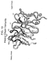


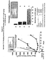

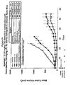
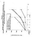

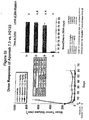


Classifications
-
- C—CHEMISTRY; METALLURGY
- C07—ORGANIC CHEMISTRY
- C07K—PEPTIDES
- C07K16/00—Immunoglobulins [IGs], e.g. monoclonal or polyclonal antibodies
- C07K16/18—Immunoglobulins [IGs], e.g. monoclonal or polyclonal antibodies against material from animals or humans
- C07K16/28—Immunoglobulins [IGs], e.g. monoclonal or polyclonal antibodies against material from animals or humans against receptors, cell surface antigens or cell surface determinants
- C07K16/2878—Immunoglobulins [IGs], e.g. monoclonal or polyclonal antibodies against material from animals or humans against receptors, cell surface antigens or cell surface determinants against the NGF-receptor/TNF-receptor superfamily, e.g. CD27, CD30, CD40, CD95
-
- A—HUMAN NECESSITIES
- A61—MEDICAL OR VETERINARY SCIENCE; HYGIENE
- A61P—SPECIFIC THERAPEUTIC ACTIVITY OF CHEMICAL COMPOUNDS OR MEDICINAL PREPARATIONS
- A61P35/00—Antineoplastic agents
-
- A—HUMAN NECESSITIES
- A61—MEDICAL OR VETERINARY SCIENCE; HYGIENE
- A61P—SPECIFIC THERAPEUTIC ACTIVITY OF CHEMICAL COMPOUNDS OR MEDICINAL PREPARATIONS
- A61P35/00—Antineoplastic agents
- A61P35/02—Antineoplastic agents specific for leukemia
-
- A—HUMAN NECESSITIES
- A61—MEDICAL OR VETERINARY SCIENCE; HYGIENE
- A61P—SPECIFIC THERAPEUTIC ACTIVITY OF CHEMICAL COMPOUNDS OR MEDICINAL PREPARATIONS
- A61P35/00—Antineoplastic agents
- A61P35/04—Antineoplastic agents specific for metastasis
-
- A—HUMAN NECESSITIES
- A61—MEDICAL OR VETERINARY SCIENCE; HYGIENE
- A61P—SPECIFIC THERAPEUTIC ACTIVITY OF CHEMICAL COMPOUNDS OR MEDICINAL PREPARATIONS
- A61P43/00—Drugs for specific purposes, not provided for in groups A61P1/00-A61P41/00
-
- C—CHEMISTRY; METALLURGY
- C07—ORGANIC CHEMISTRY
- C07K—PEPTIDES
- C07K16/00—Immunoglobulins [IGs], e.g. monoclonal or polyclonal antibodies
- C07K16/18—Immunoglobulins [IGs], e.g. monoclonal or polyclonal antibodies against material from animals or humans
- C07K16/28—Immunoglobulins [IGs], e.g. monoclonal or polyclonal antibodies against material from animals or humans against receptors, cell surface antigens or cell surface determinants
- C07K16/30—Immunoglobulins [IGs], e.g. monoclonal or polyclonal antibodies against material from animals or humans against receptors, cell surface antigens or cell surface determinants from tumour cells
-
- A—HUMAN NECESSITIES
- A61—MEDICAL OR VETERINARY SCIENCE; HYGIENE
- A61K—PREPARATIONS FOR MEDICAL, DENTAL OR TOILETRY PURPOSES
- A61K39/00—Medicinal preparations containing antigens or antibodies
- A61K2039/505—Medicinal preparations containing antigens or antibodies comprising antibodies
-
- C—CHEMISTRY; METALLURGY
- C07—ORGANIC CHEMISTRY
- C07K—PEPTIDES
- C07K2317/00—Immunoglobulins specific features
- C07K2317/20—Immunoglobulins specific features characterized by taxonomic origin
- C07K2317/21—Immunoglobulins specific features characterized by taxonomic origin from primates, e.g. man
-
- C—CHEMISTRY; METALLURGY
- C07—ORGANIC CHEMISTRY
- C07K—PEPTIDES
- C07K2317/00—Immunoglobulins specific features
- C07K2317/50—Immunoglobulins specific features characterized by immunoglobulin fragments
- C07K2317/56—Immunoglobulins specific features characterized by immunoglobulin fragments variable (Fv) region, i.e. VH and/or VL
- C07K2317/565—Complementarity determining region [CDR]
Definitions
- the present invention relates generally to DR5 antibodies, including agonistic antibodies, and to methods of using such DR5 antibodies.
- TNF tumor necrosis factor
- TNF-alpha tumor necrosis factor-alpha
- TNF-beta tumor necrosis factor-beta
- LT-beta lymphotoxin-beta
- CD30 ligand CD27 ligand
- CD40 ligand OX-40 ligand
- 4-1BB ligand LIGHT
- Apo-1 ligand also referred to as Fas ligand or CD95 ligand
- Apo-2 ligand also referred to as Apo2L or TRAIL
- Apo-3 ligand also referred to as TWEAK
- APRIL OPG ligand
- OPG ligand also referred to as RANK ligand, ODF, or TRANCE
- TALL-1 also referred to as BlyS, BAFF or THANK
- TNF family ligands Induction of various cellular responses mediated by such TNF family ligands is typically initiated by their binding to specific cell receptors. Some, but not all, TNF family ligands bind to, and induce various biological activity through, cell surface "death receptors" to activate caspases, or enzymes that carry out the cell death or apoptosis pathway ( Salvesen et al., Cell, 91:443-446 (1997 ).
- TNF receptor family members share the typical structure of cell surface receptors including extracellular, transmembrane and intracellular regions, while others are found naturally as soluble proteins lacking a transmembrane and intracellular domain.
- the extracellular portion of typical TNFRs contains a repetitive amino acid sequence pattern of multiple cysteine-rich domains (CRDs), starting from the NH 2 -terminus.
- Apo-2L or TRAIL The ligand referred to as Apo-2L or TRAIL was identified several years ago as a member of the TNF family of cytokines. (See, e.g., Wiley et al., Immunity, 3:673-682 (1995 ); Pitti et al., J. Biol. Chem., 271:12697-12690 (1996 ); WO 97/01633 ; WO 97/25428 ; US Patent 5,763,223 issued June 9, 1998 ; US Patent 6,284,236 issued September 4, 2001 ).
- the full-length native sequence human Apo2L/TRAIL polypeptide is a 281 amino acid long, Type II transmembrane protein.
- Some cells can produce a natural soluble form of the polypeptide, through enzymatic cleavage of the polypeptide's extracellular region ( Mariani et al., J. Cell. Biol., 137:221-229 (1997 )). Crystallographic studies of soluble forms of Apo2L/TRAIL reveal a homotrimeric structure similar to the structures of TNF and other related proteins ( Hymowitz et al., Molec.
- Apo2L/TRAIL unlike other TNF family members however, was found to have a unique structural feature in that three cysteine residues (at position 230 of each subunit in the homotrimer) together coordinate a zinc atom, and that the zinc binding is important for trimer stability and biological activity. (Hymowitz et al., supra; Bodmer et al., J. Biol. Chem., 275:20632-20637 (2000 )).
- Apo2L/TRAIL may play a role in immune system modulation, including autoimmune diseases such as rheumatoid arthritis [see, e.g., Thomas et al., J. Immunol., 161:2195-2200 (1998 ); Johnsen et al., Cytokine, 11:664-672 (1999 ); Griffith et al., J. Exp. Med., 189:1343-1353 (1999 ); Song et al., J. Exp. Med., 191:1095-1103 (2000 )].
- autoimmune diseases such as rheumatoid arthritis
- Soluble forms of Apo2L/TRAIL have also been reported to induce apoptosis in a variety of cancer cells, including colon, lung, breast, prostate, bladder, kidney, ovarian and brain tumors, as well as melanoma, leukemia, and multiple myeloma (see, e.g., Wiley et al., supra; Pitti et al., supra; US Patent 6,030,945 issued February 29, 2000 ; US Patent 6,746,668 issued June 8, 2004 ; Rieger et al., FEBS Letters, 427:124-128 (1998 ); Ashkenazi et al., J. Clin.
- Apo2L/TRAIL preparations may vary in terms of biochemical properties and biological activities on diseased versus normal cells, depending, for example, on the presence or absence of a tag molecule, zinc content, and % trimer content (See, Lawrence et al., Nature Med., Letter to the Editor, 7:383-385 (2001 ); Qin et al., Nature Med., Letter to the Editor, 7:385-386 (2001 )).
- Apo2L/TRAIL has been found to bind at least five different receptors. At least two of the receptors which bind Apo2L/TRAIL contain a functional, cytoplasmic death domain.
- One such receptor has been referred to as "DR4" (and alternatively as TR4 or TRAIL-R1) ( Pan et al., Science, 276:111-113 (1997 ); see also WO98/32856 published July 30, 1998 ; WO99/37684 published July 29, 1999 ; WO 00/73349 published December 7, 2000 ; US 6,433,147 issued August 13, 2002 ; US 6,461,823 issued October 8, 2002 , and US 6,342,383 issued January 29, 2002 ).
- DR5 Another such receptor for Apo2L/TRAIL has been referred to as Apo-2 (it has also been alternatively referred to as Apo-2; TRAIL-R or TRAIL-R2, TR6, Tango-63, hAPO8, TRICK2 or KILLER) (see, e.g., Sheridan et al., Science, 277:818-821 (1997 ), Pan et al., Science, 277:815-818 (1997 ), WO98/51793 published November 19, 1998 ; WO98/41629 published September 24, 1998 ; Screaton et al., Curr.
- DR5 is reported to contain three cysteine-rich domains in its extracellular portion and a single cytoplasmic death domain and be capable of signaling apoptosis upon ligand binding (or upon binding a molecule, such as an agonist antibody, which mimics the activity of the ligand).
- the crystal structure of the complex formed between Apo-2L/TRAIL and DR5 is described in Hymowitz et al., Molecular Cell, 4:563-571 (1999 ).
- both DR4 and DR5 can trigger apoptosis independently by recruiting and activating the apoptosis initiator, caspase-8, through the death-domain-containing adaptor molecule referred to as FADD/Mort1 [ Kischkel et al., Immunity. 12:611-620 (2000 ); Sprick et al., immunity, 12:599-609 (2000 ); Bodmer et al., Nature Cell Biol., 2:241-243 (2000 )].
- DR5 signals apoptosis through the "cell-extrinsic" pathway, which is independent of the p53 tumor suppressor gene ( Ashkenazi and Dixit, Science 281:1305-8 (1998 ); Ashkenazi, Nat Rev Cancer 2:420-30 (2002 )).
- Activation of this pathway involves raid formation of a death-inducing signaling complex (DISC) at the activated receptor's cytoplasmic death domain.
- DISC death-inducing signaling complex
- the adaptor molecule FADD binds to DR5 through homophilic death domain interaction (Kischkel et al., supra, Sprick et al., supra, Bodmer et al., supra).
- FADD recruits the apoptosis-initiating proteases caspase-8 and caspase-10, mediating their activation by induced proximity Caspase-9 and caspase-10 undergo self-processing, releasing soluble active caspase subunits into the cytoplasm, where they assemble and cleave effector caspases, such as caspase-3 and caspase-7. Cleavage results in activation of the effector caspases, which carry out the apoprotix cell program ( Thornberry and Lazebnik, Science 281:1312-6 (1998 )).
- DcR1, DcR2 and OPG which believed to function as inhibitors, rather than transducers of signaling
- DCR1 also referred to as TRID, LIT or TRAIL-R3
- TRID TRID, LIT or TRAIL-R3
- anti-DR4 antibodies directed to the DR4 receptor and having agonistic or apoptotic activity in certain mammalian cells are described in, e.g., WO 99/37684 published July 29, 1999 ; WO 00/73349 published July 12, 2000 ; WO 03/066661 published August 14, 2003 . See, also, e.g., Griffith et al., J. Immunol., 162:2597-2605 (1999 ); Chuntharapai et al., J.
- the present invention provides anti-DR5 antibody comprising mutations in the heavy and light chain of full-length antibody 16E2 as set out in SEQ ID NOS: 11 and 13, respectively, or a fragment thereof, wherein:
- the anti-HR5 antibody comprises a framework mutation selected from the group consisting of Q6E, V11L, E12V, R13Q, and K105Q,
- the anti-DR5 antibody comprises all of the framework mutations Q6E, V11 4 B12V, R13Q, and K105Q.
- the anti-DR5 antibody comprises the following mutations: G33A, N53Q, L102Y in the sequence of SEQ ID NO: 11, and Q24S, K51A, R91A in the sequence of SEQ ID NO: 13, and may additionally comprise at least one framework mutation, which may, for example, be at least one of residues 6,11,12,13, and 105 of SEQ ID NO: 11.
- anti-DR5 antibody examples include Apomabs1.1,2.1,3.1,4.1,5.1,6.1,7.1,8.1,9.1,1.2,2.2,3.2,4.2,5.2,6.2,7.2,8.2,9.2, 1.3,2.2,3.3, 4.3, 5.3, 6.3, 7.3, 8.3, and 9.3.
- anti-DR5 antibody is provided below and are selected from the group consisting of Apomabs 5.2, 5.3, 6.2; 6.3, 7.2, 7.3; 8.3 and 25.3.
- the anti-DR5 antibody is Apomab 7.3 or Apomab 8.3, especially Apomab 7.3.
- the anti-DR5 antibody is an antibody fragment, which may be selected from the group consisting of Fab, Fab', F(ab')2, and Fv fragments, diabodies, single chain antibody molecules, and multispecific antibodies formed from antibody fragments.
- the antibody can be a single-chain antibody.
- the anti-DR5 antibodies can, for example, have anti-cancer activity, such as, for example, they can possess the ability to activate or stimulate apoptosis in cancer cells.
- the cancer includes, for example, carcinoma, lymphoma, blastoma, sarcoma and leukemia.
- cancer More specific examples include squamous cell cancer, small-cell lung cancer, non-small cell lung cancer (NSCLC), non-Hodgkin's lymphoma, blastoma, gastrointestinal cancer, renal cancer, ovarian cancer, liver cancer, stomach cancer, bladder cancer, hepatoma, breast cancer, colon cancer, colorectal cancer, pancreatic cancer, endometrial carcinoma, salivary gland carcinoma, kidney cancer, liver cancer, prostate cancer, vulval cancer, thyroid cancer, hepatic carcinoma, and head and neck cancer.
- NSCLC non-small cell lung cancer
- lung cancer e.g., non-small cell lung carcinoma - NSCLC
- adenocarcinoma which can, for example, be colorectal, pancreatic, or metastatic adenocarcinoma.
- Hematological cancers are also included.
- Chimeric, humanized and human antibodies are within the scope herein, as are antibodies which mediate antibody-dependent cellular cytotoxicity (ADCC).
- ADCC antibody-dependent cellular cytotoxicity
- the anti-DR5 antibody comprises Apomab 7.3, or Apomab 8.3, or a fragment thereof.
- the antibodies may be in a dimeric form and/or in a form cross-linked, for example, with an anti-human IgG Fc region.
- the anti-DR5 antibodies herein are fused to an epitope tag sequence.
- the invention concerns a chimeric molecule comprising an anti-DR5 antibody or antibody fragment herein, fused to a heterologous amino acid sequence, where the heterologous amino acid sequence may, for example, comprise an immunoglobulin sequence, such as an anti-human IgG Fc region.
- the invention concerns isolated nucleic acid molecules encoding the ani-DR5 antibodies or antibody fragments herein, vectors comprising such nucleic acid molecules, host cells comprising such nucleic acid molecules, and methods for producing antibodies and antibody fragment herein.
- the invention further relates to a composition comprising an anti-DR5 antibody as herein above defined, and a carrier.
- the carrier may be a pharmaceutically acceptable carrier, and the composition may further comprise an additional anti-cancer agent, and/or an additional anti-DR5 antibody.
- the present invention provides an ex vivo method of inducing apoptosis comprising exposing mammalian cancer cells to an anti-DR5 antibody as defined herein, or a fragment thereof.
- the present invention provides the use of an anti-DR5 antibody as defined herein, or a fragment thereof, for the preparation of a medicament for the treatment of cancer in a mammalian subject.
- Apo-2 ligand refers to a polypeptide sequence which includes amino acid residues 114-281, inclusive, 95-281, inclusive, residues 92-281, inclusive, residues 91-281, inclusive, residues 41-281, inclusive, residues 39-281, inclusive, residues 15-281, inclusive, or residues 1-281, inclusive, of the amino acid sequence shown in Figure 1 (SEQ ID NO:1), as well as biologically active fragments, deletional, insertional, and/or substitutional variants of the above sequences.
- the polypeptide sequence comprises residues 114-281 of Figure 1 (SEQ ID NO:1).
- the polypeptide sequence comprises residues 92-281 or residues 91-281 of Figure 1 (SEQ ID NO:1).
- the Apo-2L polypeptides may be encoded by the native nucleotide sequence shown in Figure 1 (SEQ ID NO:2).
- the codon that encodes residue Pro119 ( Figure 1 ; SEQ ID NO:2) may be "CCT" or "CCG”.
- the fragments or variants are biologically active and have at least about 80% amino acid sequence identity, or at least about 90% sequence identity, or at least 95%, 96%, 97%, 98%, or 99% sequence identity with any one of the above sequences.
- the definition encompasses substitutional variants of Apo-2 ligand in which at least one of its native amino acids are substituted by another amino acid such as an alanine residue.
- the definition also encompasses a native sequence Apo-2 ligand isolated from an Apo-2 ligand source or prepared by recombinant and/or synthetic methods.
- the Apo-2 ligand of the invention includes the polypeptides referred to as Apo-2 ligand or TRAIL disclosed in WO97/01633 published January 16, 1997 , WO97/25428 published July 17, 1997 , WO99/36535 published July 22, 1999 , WO 01/00832 published January 4,2001 , WO02/09755 published February 7, 2002 , WO 00/75191 published December 14, 2000 , and U.S. Patent No. 6,030,945 issued February 29, 2000 .
- the terms are used to refer generally to forms of the Apo-2 ligand, which include monomer, dimer, trimer, hexamer or higher oligomer forms of the polypeptide. All numbering of amino acid residues referred to in the Apo-2L sequence use the numbering according to Figure 1 (SEQ ID NO:1), unless specifically stated otherwise.
- Apo-2 ligand receptor includes the receptors referred to in the art as “DR4" and "DR5" whose polynucleotide and polypeptide sequences are shown in Figures 2A-2C (SEQ ID NOS 4 and 3) and 3A-3C (SEQ ID NOS: 6 and 5), respectively.
- DR4 and DR5 whose polynucleotide and polypeptide sequences are shown in Figures 2A-2C (SEQ ID NOS 4 and 3) and 3A-3C (SEQ ID NOS: 6 and 5), respectively.
- TNF receptor family member referred to as "DR4" (Pan et al., Science , 276 :111-113 (1997); see also WO98/32856 published July 30, 1998 ; WO 99/37684 published July 29, 1999 ; WO 00/73349 published December 7, 2000 ; US 6,433,147 issued August 13, 2002 ; US 6,461,823 issued October 8, 2002 , and US 6,342,383 issued January 29, 2002 ).
- DR5 the receptor has also been alternatively referred to as Apo-2; TRAIL-R, TR6, Tango-63, hAPO8, TRICK2 or KILLER; Screaton et al., Curr.
- Apo-2L receptor when used herein encompasses native sequence receptor and receptor variants. These terms encompass Apo-2L receptor expressed in a variety of mammals, including humans. Apo-2L receptor may be endogenously expressed as occurs naturally in a variety of human tissue lineages, or may be expressed by recombinant or synthetic methods.
- a "native sequence Apo-2L receptor” comprises a polypeptide having the same amino acid sequence as an Apo-2L receptor derived from nature.
- a native sequence Apo-2L receptor can have the amino acid sequence of naturally occurring Apo-2L receptor from any mammal, including humans.
- Such native sequence Apo-2L receptor can be isolated from nature or can be produced by recombinant or synthetic means.
- the term "native sequence Apo-2L receptor” specifically encompasses naturally occurring truncated or secreted forms of the receptor (e.g ., a soluble form containing, for instance, an extracellular domain sequence), naturally occurring variant forms (e.g ., alternatively spliced forms) and naturally occurring allelic variants.
- Receptor variants may include fragments or deletion mutants of the native sequence Apo-2L receptor.
- Figures 3A-3C show the 411 amino acid sequence of human DR5 as published in WO 98/51793 on November 19, 1998 .
- a transcriptional splice variant of human DR5 is known in the art. This DR5 splice variant encodes the 440 amino acid sequence of human DR5 as shown in Figures 4A-4C , along with its nucleotide sequence (SEQ ID NOS: 7 and 8), and as published in WO 98/35986 on August 20, 1998 .
- Death receptor antibody is used herein to refer generally to antibody or antibodies directed to a receptor in the tumor necrosis factor receptor superfamily and containing a death domain capable of signaling apoptosis, and such antibodies include DR5 antibody and DR4 antibody.
- DR5 receptor antibody “DR5 antibody”, or “anti-DR5 antibody” is used in a broad sense to refer to antibodies that bind to at least one form of a DR5 receptor or extracellular domain thereof.
- the DR5 antibody is fused or linked to a heterologous sequence or molecule.
- the heterologous sequence allows or assists the antibody to form higher order or oligomeric complexes.
- the DR5 antibody binds to DR5 receptor but does not bind or cross-react with any additional Apo-2L receptor (e.g. DR4, DcR1, or DcR2).
- the antibody is an agonist of DR5 signaling activity.
- anti-DR5 antibody and is grammatical equivalents specifically encompass the antibodies described in the examples, including but not limited to the "Apomab” antibodies listed in Tables 11 and 12, such as, for example, Apomabs 1.1, 2.1, 3.1, 4.1, 5.1, 6.1, 7.1, 8.1, 9.1, 1.2, 2.2, 3.2, 4.2, 5.2, 6.2, 7.2, 8.2, 9.2, 1.3, 2.2, 3.3, 4.3, 5.3, 6.3, 7.3, 8.3, and 9.3, preferably Apomab 7.3.
- the DR5 antibody of the invention binds to a DR5 receptor at a concentration range of about 0.1 nM to about 20 mM as measured in a BIAcore binding assay.
- the DR5 antibodies of the invention exhibit an Ic 50 value of about 0.6 nM to about 18 mM as measured in a BIAcore binding assay.
- DR4 receptor antibody “DR4 antibody”, or “anti-DR4 antibody” is used in a broad sense to refer to antibodies that bind to at least one form of a DR4 receptor or extracellular domain thereof.
- the DR4 antibody is fused or linked to a heterologous sequence or molecule.
- the heterologous sequence allows or assists the antibody to form higher order or oligomeric complexes.
- the DR4 antibody binds to DR4 receptor but does not bind or cross-react with any additional Apo-2L receptor (e.g. DR5, DcR1, or DcR2).
- the antibody is an agonist of DR4 signaling activity.
- the DR4 antibody binds to a DR4 receptor at a concentration range of about 0.1 nM to about 20 mM as measured in a BIAcore binding assay.
- the DR5 antibodies of the invention exhibit an Ic 50 value of about 0.6 nM to about 18 mM as measured in a BIAcore binding assay.
- agonist is used in the broadest sense, and includes any molecule that partially or fully enhances, stimulates or activates one or more biological activities of Apo2L/TRAIL, DR4 or DR5, in vitro, in situ, or in vivo. Examples of such biological activities binding of Apo2L/TRAIL to DR4 or DR5, include apoptosis as well as those further reported in the literature.
- An agonist may function in a direct or indirect manner. For instance, the agonist may function to partially or fully enhance, stimulate or activate one or more biological activities of DR4 or DR5, in vitro, in situ, or in vivo as a result of its direct binding to DR4 or DR5, which causes receptor activation or signal transduction.
- the agonist may also function indirectly to partially or fully enhance, stimulate or activate one or more biological activities of DR4 or DR5, in vitro, in situ, or in vivo as a result of, e.g., stimulating another effector molecule which then causes DR4 or DR5 activation or signal transduction. It is contemplated that an agonist may act as an enhancer molecule which functions indirectly to enhance or increase DR4 or DR5 activation or activity. For instance, the agonist may enhance activity of endogenous Apo-2L in a mammal.
- extracellular domain refers to a form of ligand or receptor, which is essentially free of transmembrane and cytoplasmic domains. Ordinarily, the soluble ECD will have less than 1% of such transmembrane and cytoplasmic domains, and preferably, will have less than 0.5% of such domains.
- epitope tagged when used herein refers to a chimeric polypeptide comprising a protein such as Apo-2 ligand or DR5 receptor, or a portion thereof or an antibody which binds such ligand or receptor, fused to a "tag polypeptide".
- the tag polypeptide has enough residues to provide an epitope against which an antibody can be made, yet is short enough such that it does not interfere with activity of the ligand or receptor.
- the tag polypeptide preferably also is fairly unique so that the antibody does not substantially cross-react with other epitopes.
- Suitable tag polypeptides generally have at least six amino acid residues and usually between about 8 to about 50 amino acid residues (preferably, between about 10 to about 20 residues).
- Isolated when used to describe the various proteins disclosed herein, means protein that has been identified and separated and/or recovered from a component of its natural environment. Contaminant components of its natural environment are materials that would topically interfere with diagnostic or therapeutic uses for the protein, and may include enzymes, hormones, and other proteinaceous or non-proteinaceous solutes.
- the protein will be purified (1) to a degree sufficient to obtain at least 15 residues of N-terminal or internal amino acid sequence by use of a spinning cup sequenator, or (2) to homogeneity by SDS-PAGE under non-reducing or reducing conditions using Coomassie blue or, preferably, silver stain.
- Isolated protein includes protein in situ within recombinant cells, since at least one component of the Apo-2 ligand natural environment will not be present. Ordinarily, however, isolated protein will be prepared by at least one purification step.
- Percent (%) amino acid sequence identity with respect to the sequences identified herein is defined as the percentage of amino acid residues in a candidate sequence that are identical with the amino acid residues in the compared ligand, receptor, or antibody sequence, after aligning the sequences and introducing gaps, if necessary, to achieve the maximum percent sequence identity, and not considering any conservative substitutions as part of the sequence identity. Alignment for purposes of determining percent amino acid sequence identity can be achieved in various ways that are within the skill in the art can determine appropriate parameters for measuring alignment, including assigning algorithms needed to achieve maximal alignment over the full-length sequences being compared. For purposes herein, percent amino acid identity values can be obtained using the sequence comparison computer program, ALIGN-2, which was authored by Genentech, Inc.
- ALIGN-2 The ALIGN-2 program is publicly available through Genentech, Inc., South San Francisco, CA. All sequence comparison parameters are set by the ALIGN-2 program and do not vary. Percent amino acid sequence identity is then calculated relative to the longer sequence. Accordingly, even if the shorter sequence is fully included in the longer sequence, the sequence identity will be less than 100%.
- control sequences refers to DNA sequences necessary for the expression of an operably linked coding sequence in a particular host organism.
- the control sequences that are suitable for prokaryotes include a promoter, optionally an operator sequence, and a ribosome binding site: Eukaryotic cells are known to utilize promoters, polyadenylation signals, and enhancers.
- Nucleic acid is "operably linked" when it is placed into a functional relationship with another nucleic acid sequence.
- DNA for a presequence or secretory leader is operably linked to DNA for a polypeptide if it is expressed as a preprotein that participates in the secretion of the polypeptide;
- a promoter or enhancer is operably linked to a coding sequence if it affects the transcription of the sequence; or
- a ribosome binding site is operably linked to a coding sequence if it is positioned so as to facilitate translation.
- "operably linked” means that the DNA sequences being linked are contiguous, and, in the case of a secretory leader, contiguous and in reading phase. However, enhancers do not have to be contiguous. Linking is accomplished by ligation at convenient restriction sites. If such sites do not exist, the synthetic oligonucleotide adaptors or linkers are used in accordance with conventional practice.
- polyol when used herein refers broadly to polyhydric alcohol compounds.
- Polyols can be any water-soluble poly(alkylene oxide) polymer for example, and can have a linear or branched chain.
- Preferred polyols include those substituted at one or more hydroxyl positions with a chemical group, such as an alkyl group having between one and four carbon.
- the polyol is a poly(alkylene glycol), preferably poly(ethylene glycol) (PEG).
- PEG poly(ethylene glycol)
- PEG poly(ethylene glycol)
- the polyols include those well known in the art and those publicly available, such as from commercially available sources such as Nektar® Corporation.
- conjugate is used herein according to its broadest definition to mean joined or linked together. Molecules are “conjugated” when they act or operate as if joined.
- “Stringency” of hybridization reactions is readily determinable by one of ordinary skill in the art, and generally is an empirical calculation dependent upon probe length, washing temperature, and salt concentration. In general, longer probes require higher temperatures for proper annealing, while shorter probes need lower temperatures. Hybridization generally depends on the ability of denatured DNA to re-anneal when complementary strands are present in an environment below their melting temperature. The higher the degree of desired identity between the probe and hybridizable sequence, the higher the relative temperature, which can be used. As a result, it follows that higher relative temperatures would tend to make the reaction conditions more stringent, while lower temperatures less so. For additional details and explanation of stringency of hybridization reactions, see Ausubel et al., Current Protocols in Molecular Biology, Wiley Interscience Publishers, (1995 ).
- “Stringent conditions” or “high stringency conditions”, as defined herein, are identified by those that: (1) employ low ionic strength and high temperature for washing, for example 0.015 M sodium chloride/0.0015 M sodium citrate/0.1% sodium dodecyl sulfate at 50°C; (2) employ during hybridization a denaturing agent, such as formamide, for example, 50% (v/v) formamide with 0.1% bovine serum albumin/0.1% Ficoll/0.1% polyvinylpyrrolidone/50mM sodium phosphate buffer at pH 6.5 with 750 mM sodium chloride, 75 mM sodium citrate at 42°C; or (3) employ 50% formamide, 5 x SSC (0.75 M NaCl,0.075 M sodium citrate), 50 mM sodium phosphate (pH 6.8), 0.1% sodium pyrophosphate, 5 x Denhardt's solution, sonicated salmon sperm DNA (50 ⁇ g/ml), 0.1% SDS, and 10% dextran sulfate
- Modely stringent conditions are identified as described by Sambrook et al., Molecular Cloning: A Laboratory Manual, New York: Cold Spring Harbor Press, 1989 , and include the use of washing solution and hybridization conditions (e.g., temperature, ionic strength and %SDS) less stringent that those described above.
- moderately stringent conditions is overnight incubation at 37°C in a solution comprising: 20% formamide, 5 x SSC (150 mM NaCl, 15 mM trisodium citrate), 50 mM sodium phosphate (pH 7.6), 5 x Denhardt's solution, 10% dextran sulfate, and 20 mg/ml denatured sheared salmon sperm DNA, followed by washing the filters in 1 x SSC at about 37-50°C.
- the skilled artisan will recognize how to adjust the temperature, ionic strength, etc. as necessary to accommodate factors such as probe length and the like.
- amino acid and “amino acids” refer to all naturally occurring L-alpha-amino acids. This definition is meant to include norleucine, ornithine, and homocysteine.
- the amino acids are identified by either the single-letter or three-letter designations: Asp D aspartic acid Ile I isoleucine Thr T threonine Leu L leucine Ser S serine Tyr Y tyrosine Glu E glutamic acid Phe F phenylalanine Pro P proline His H histidine Gly G glycine Lys K lysine Ala A alanine Arg R arginine Cys C cysteine Trp W tryptophan Val V valine Gln Q glutamine Met M methionine Asn N asparagine
- antibody is used in the broadest sense and specifically covers single anti-DR5 monoclonal antibodies (including agonist, antagonist, and neutralizing or blocking antibodies) and anti-DR5 antibody compositions with, polyepitopic specificity.
- Antibody as used herein includes intact immunoglobulin or antibody molecules, polyclonal antibodies, multispecific antibodies (i.e., bispecific antibodies formed from at least two intact antibodies) and immunoglobulin fragments (such as Fab, F(ab') 2 , or Fv), so long as they exhibit any of the desired agonistic or antagonistic properties described herein.
- Antibodies are typically proteins or polypeptides which exhibit binding specificity to a specific antigen.
- Native antibodies are usually heterotetrameric glycoproteins, composed of two identical light (L) chains and two identical heavy (H) chains.
- L light
- H heavy
- each light chain is linked to a heavy chain by one covalent disulfide bond, while the number of disulfide linkages varies between the heavy chains of different immunoglobulin isotypes.
- Each heavy and light chain also has regularly spaced intrachain disulfide bridges.
- Each heavy chain has at one end a variable domain (V H ) followed by a number of constant domains.
- V H variable domain
- Each light chain has a variable domain at one end (V L ) and a constant domain at its other end; the constant domain of the light chain is aligned with the first constant domain of the heavy chain, and the light chain variable domain is aligned with the variable domain of the heavy chain.
- Particular amino acid residues are believed to form an interface between the light and heavy chain variable domains [ Chothia et al., J. Mol. Biol., 186:651-663 (1985 ); Novotny and Haber, Proc. Natl. Acad. Sci. USA, 82:4592-4596 (1985 )].
- immunoglobulins can be assigned to different classes. There are five major classes of immunoglobulins: IgA, IgD, IgE, IgG and IgM, and several of these may be further divided into subclasses (isotypes), e.g., IgG-1, IgG-2, IgG-3, and IgG-4; IgA-1 and IgA-2.
- the heavy chain constant domains that correspond to the different classes of immunoglobulins are called alpha, delta, epsilon, gamma, and mu, respectively.
- Antibody fragments comprise a portion of an intact antibody, generally the antigen binding or variable region of the intact antibody.
- antibody fragments include Fab, Fab', F(ab')2, and Fv fragments, diabodies, single chain antibody molecules, and multispecific antibodies formed from antibody fragments.
- variable is used herein to describe certain portions of the variable domains, which differ in sequence among antibodies and are used in the binding and specificity of each particular antibody for its particular antigen.
- variability is not usually evenly distributed through the variable domains of antibodies. It is typically concentrated in three segments called complementarity determining regions (CDRs) or hypervariable regions both in the light chain and the heavy chain variable domains.
- CDRs complementarity determining regions
- FR framework
- the variable domains of native heavy and light chains each comprise four FR regions, largely adopting a ⁇ -sheet configuration, connected by three CDRs, which form loops connecting, and in some cases forming part of, the ⁇ -sheet structure.
- the CDRs in each chain are held together in close proximity by the FR regions and, with the CDRs from the other chain, contribute to the formation of the antigen binding site of antibodies [see Kabat, E.A. et al., Sequences of Proteins of Immunological Interest, National Institutes of Health, Bethesda, MD (1987 )].
- the constant domains are not involved directly in binding an antibody to an antigen, but exhibit various effector functions, such as participation of the antibody in antibody-dependent cellular toxicity.
- monoclonal antibody refers to an antibody obtained from a population of substantially homogeneous antibodies, i.e ., the individual antibodies comprising the population are identical except for possible naturally-occurring mutations that may be present in minor amounts. Monoclonal antibodies are highly specific, being directed against a single antigenic site. Furthermore, in contrast to conventional (polyclonal) antibody preparations, which typically include different antibodies directed against different determinants (epitopes), each monoclonal antibody is directed against a single determinant on the antigen.
- the monoclonal antibodies herein include chimeric, hybrid and recombinant antibodies produced by splicing a variable (including hypervariable) domain of an anti-DR5 antibody with a constant domain (e.g . "humanized” antibodies), or a light chain with a heavy chain, or a chain from one species with a chain from another species, or fusions with heterologous proteins, regardless of species of origin or immunoglobulin class or subclass designation, as well as antibody fragments (e.g ., Fab, F(ab') 2 , and Fv), so long as they exhibit the desired biological activity or properties. See, e.g . U.S. Pat. No. 4,816,567 and Mage et al., in Monoclonal Antibody Production Techniques and Applications, pp.79-97 (Marcel Dekker, Inc.: New York, 1987 ).
- the modifier "monoclonal” indicates the character of the antibody as being obtained from a substantially homogeneous population of antibodies, and is not to be construed as requiring production of the antibody by any particular method.
- the monoclonal antibodies to be used in accordance with the present invention may be made by the hybridoma method first described by Kohler and Milstein, Nature, 256:495 (1975 ), or may be made by recombinant DNA methods such as described in U.S. Pat. No. 4,816,567 .
- the “monoclonal antibodies” may also be isolated from phage libraries generated using the techniques described in McCafferty et al., Nature, 348:552-554 (1990 ), for example.
- Humanized forms of non-human (e.g . murine) antibodies are specific chimeric immunoglobulins, immunoglobulin chains, or fragments thereof (such as Fv, Fab, Fab', F(ab') 2 or other antigen-binding subsequences of antibodies) which contain minimal sequence derived from non-human immunoglobulin.
- humanized antibodies are human immunoglobulins (recipient antibody) in which residues from a complementary determining region (CDR) of the recipient are replaced by residues from a CDR of a non-human species (donor antibody) such as mouse, rat, or rabbit having the desired specificity, affinity, and capacity.
- CDR complementary determining region
- Fv framework region (FR) residues of the human immunoglobulin are replaced by corresponding non-human residues.
- the humanized antibody may comprise residues, which are found neither in the recipient antibody nor in the imported CDR or framework sequences. These modifications are made to further refine and optimize antibody performance.
- the humanized antibody will comprise substantially all of at least one, and typically two, variable domains, in which all or substantially all of the CDR regions correspond to those of a non-human immunoglobulin and all or substantially all of the FR regions are those of a human immunoglobulin consensus sequence.
- the humanized antibody optimally also will comprise at least a portion of an immunoglobulin constant region or domain (Fc), typically that of a human immunoglobulin.
- a "human antibody” is one which possesses an amino acid sequence which corresponds to that of an antibody produced by a human and/or has been made using any of the techniques for making human antibodies known in the art or as disclosed herein.
- This definition of a human antibody includes antibodies comprising at least one human heavy chain polypeptide or at least one human light chain polypeptide, for example an antibody comprising murine light chain and human heavy chain polypeptides.
- Human antibodies can be produced using various techniques known in the art.
- the human antibody is selected from a phage library, where that phage library expresses human antibodies ( Vaughan et al. Nature Biotechnology, 14:309-314 (1996 ): Sheets et al.
- Human antibodies can also be made by introducing human immunoglobulin loci into transgenic animals, e.g., mice in which the endogenous immunoglobulin genes have been partially or completely inactivated. Upon challenge, human antibody production is observed, which closely resembles that seen in humans in all respects, including gene rearrangement, assembly, and antibody repertoire. This approach is described, for example, in U.S. Patent Nos.
- the human antibody may be prepared via immortalization of human B lymphocytes producing an antibody directed against a target antigen (such B lymphocytes may be recovered from an individual or may have been immunized in vitro). See, e.g., Cole et al., Monoclonal Antibodies and Cancer Therapy, Alan R. Liss, p. 77 (1985 ); Boerner et al., J. Immunol., 147 (1):86-95 (1991 ); and US Pat No. 5,750,373 .
- the term "Fc region” is used to define the C-terminal region of an immunoglobulin heavy chain, which may be generated by papain digestion of an intact antibody.
- the Fc region may be a native sequence Fc region or a variant Fc region.
- the human IgG heavy chain Fc region is usually defined to stretch from an amino acid residue at about position Cys226, or from about position Pro230, to the carboxyl-terminus of the Fc region (using herein the numbering system according to Kabat et al., supra) .
- the Fc region of an immunoglobulin generally comprises two constant domains, a CH2 domain and a CH3 domain, and optionally comprises a CH4 domain.
- Fc region chain herein is meant one of the two polypeptide chains of an Fc region.
- the "CH2 domain" of a human IgG Fc region usually extends from an amino acid residue at about position 231 to an amino acid residue at about position 340.
- the CH2 domain is unique in that it is not closely paired with another domain. Rather, two N-linked branched carbohydrate chains are interposed between the two CH2 domains of an intact native IgG molecule. It has been speculated that the carbohydrate may provide a substitute for the domain-domain pairing and help stabilize the CH2 domain.
- the CH2 domain herein may be a native sequence CH2 domain or variant CH2 domain.
- the "CH3 domain” comprises the stretch of residues C-terminal to a CH2 domain in an Fc region ( i.e . from an amino acid residue at about position 341 to an amino acid residue at about position 447 of an IgG).
- the CH3 region herein may be a native sequence CH3 domain or a variant CH3 domain (e.g . a CH3 domain with an introduced "protroberance” in one chain thereof and a corresponding introduced "cavity” in the other chain thereof; see US Patent No. 5,821,333 ).
- Such variant CH3 domains may be used to make multispecific (e.g . bispecific) antibodies as herein described.
- Hinge region is generally defined as stretching from about Glu216, or about Cys226, to about Pro230 of human IgG1 ( Burton, Molec. Immunol.22:161-206 (1985 )). Hinge regions of other IgG isotypes may be aligned with the IgG1 sequence by placing the first and last cysteine residues forming inter-heavy chain S-S bonds in the same positions.
- the hinge region herein may be a native sequence hinge region or a variant hinge region.
- the two polypeptide chains of a variant hinge region generally retain at least one cysteine residue per polypeptide chain, so that the two polypeptide chains of the variant hinge region can form a disulfide bond between the two chains.
- the preferred hinge region herein is a native sequence human hinge region, e.g . a native sequence human IgG1 hinge region.
- a “functional Fc region” possesses at least one "effector function” of a native sequence Fc region.
- effector functions include Clq binding; complement dependent cytotoxicity (CDC); Fc receptor binding; antibody-dependent cell-mediated cytotoxicity (ADCC); phagocytosis; down regulation of cell surface receptors ( e.g . B cell receptor; BCR), etc.
- Such effector functions generally require the Fc region to be combined with a binding domain (e.g . an antibody variable domain) and can be assessed using various assays known in the art for evaluating such antibody effector functions.
- a “native sequence Fc region” comprises an amino acid sequence identical to the amino acid sequence of an Fc region found in nature.
- a “variant Fc region” comprises an amino acid sequence, which differs from that of a native sequence Fc region by virtue of at least one amino acid modification.
- the variant Fc region has at least one amino acid substitution compared to a native sequence Fc region or to the Fc region of a parent polypeptide, e.g . from about one to about ten amino acid substitutions, and preferably from about one to about five amino acid substitutions in a native sequence Fc region or in the Fc region of the parent polypeptide.
- the variant Fc region herein will preferably possess at least about 80% sequence identity with a native sequence Fc region and/or with an Fc region of a parent polypeptide, and most preferably at least about 90% sequence identity therewith, more preferably at least about 95% sequence identity therewith.
- Antibody-dependent cell-mediated cytotoxicity and “ADCC” refer to a cell-mediated reaction in which nonspecific cytotoxic cells that express Fc receptors (FcRs) (e.g . Natural Killer (NK) cells, neutrophils, and macrophages) recognize bound antibody on a target cell and subsequently cause lysis of the target cell.
- FcRs Fc receptors
- FcR expression on hematopoietic cells is summarized in Table 3 on page 464 of Ravetch and Kinet, Annu. Rev. Immunol., 9:457-92 (1991 ).
- ADCC activity of a molecule of interest may be assessed in vitro, such as that described in US Patent No. 5,500,362 or 5,821,337.
- Useful effector cells for such assays include peripheral blood mononuclear cells (PBMC) and Natural Killer (NK) cells.
- PBMC peripheral blood mononuclear cells
- NK Natural Killer
- ADCC activity of the molecule of interest may be assessed in vivo, e.g., in a animal model such as that disclosed in Clynes et al. PNAS (USA), 95:652-656 (1998 ).
- Human effector cells are leukocytes which express one or more FcRs and perform effector functions. Preferably, the cells express at least Fc(RIII and perform ADCC effector function. Examples of human leukocytes which mediate ADCC include peripheral blood mononuclear cells (PBMC), natural killer (NK) cells, monocytes, cytotoxic T cells and neutrophils; with PBMCs and NK cells being preferred.
- PBMC peripheral blood mononuclear cells
- NK natural killer cells
- monocytes cytotoxic T cells and neutrophils
- the effector cells may be isolated from a native source thereof, e.g . from blood or PBMCs as described herein.
- Fc receptor and “FcR” are used to describe a receptor that binds to the Fc region of an antibody.
- the preferred FcR is a native sequence human FcR.
- a preferred FcR is one which binds an IgG antibody (a gamma receptor) and includes receptors of the Fc(RI, Fc(RII, and Fc(RIII subclasses, including allelic variants and alternatively spliced forms of these receptors.
- Fc(RII receptors include Fc(RIIA (an “activating receptor") and Fc(RIIB (an “inhibiting receptor”), which have similar amino acid sequences that differ primarily in the cytoplasmic domains thereof.
- Activating receptor Fc(RIIA contains an immunoreceptor tyrosine-based activation motif (ITAM in its cytoplasmic domain.
- Inhibiting receptor Fc(RIIB contains an immunoreceptor tyrosine-based inhibition motif (ITIM) in its cytoplasmic domain (reviewed in Da ⁇ ron, Annu. Rev. Immunol., 15:203-234 (1997 )).
- FcRs are reviewed in Ravetch and Kinet, Annu. Rev. Immunol., 9:457-92 (1991 ); Capel et al., Immunomethods, 4:25-34 (1994 ); and de Haas et al., J. Lab. Clin. Med., 126:330-41 (1995 ).
- FcR FcR
- FcRn neonatal receptor
- “Complement dependent cytotoxicity” and “CDC” refer to the lysing of a target in the presence of complement.
- the complement activation pathway is initiated by the binding of the first component of the complement system (C1q) to a molecule (e.g. an antibody) complexed with a cognate antigen.
- a CDC assay e.g. as described in Gazzano-Santoro et al., J. Immunol. Methods, 202:163 (1996 ), may be performed.
- affinity matured antibody is one with one or more alterations in one or more CDRs thereof which result an improvement in the affinity of the antibody for antigen, compared to a parent antibody which does not possess those alteration(s).
- Preferred affinity matured antibodies will have nanomolar or even picomolar affinities for the target antigen.
- Affinity matured antibodies are produced by procedures known in the art. Marks et al. Bio/Technology, 10:779-783 (1992 ) describes affinity maturation by VH and VL domain shuffling. Random mutagenesis of CDR and/or framework residues is described by: Barbas et al. Proc Nat. Acad. Sci. USA 91:3809-3813 (1994 ); Schier et al.
- immunospecific refers to the antigen specific binding interaction that occurs between the antigen-combining site of an antibody and the specific antigen recognized by that antibody.
- Bioly active and “desired biological activity” for the purposes herein mean having the ability to modulate DR5 activity or DR5 activation, including, by way of example, apoptosis (either in an agonistic or stimulating manner or in an antagonistic or blocking manner) in at least one type of mammalian cell in vivo or ex vivo, binding to Apo-2 ligand (TRAIL), or modulating activation of one or more molecules in the intracellular signaling pathway such as caspase 3, caspase 8, caspase 10 or FADD.
- Assays for determining activation of such intracellular molecules are known in the art, see, e.g., Boldin et al., J. Biol.
- agonist and agonistic when used herein refer to or describe a molecule which is capable of, directly or indirectly, substantially inducing, promoting or enhancing DR5 biological activity or activation.
- an "agonist DR5 antibody” is an antibody which has activity comparable to the ligand for DR5, known as Apo-2 ligand (TRAIL), or is capable of activating DR5 receptor which results in an activation of one or more intracellular signaling pathways which may include activation of caspase 3, caspase 8, caspase 10 or FADD.
- TRAIL Apo-2 ligand
- an antagonist when used herein refer to or describe a molecule which is capable of, directly or indirectly, substantially counteracting, reducing or inhibiting DR5 biological activity or DR5 activation.
- an antagonist is a molecule which neutralizes the biological activity resulting from DR5 activation or formation of a complex between DR5 and its ligand, such as Apo-2 ligand.
- apoptosis and “apoptotic activity” are used in a broad sense and refer to the orderly or controlled form of cell death in mammals that is typically accompanied by one or more characteristic cell changes, including condensation of cytoplasm, loss of plasma membrane microvilli, segmentation of the nucleus, degradation of chromosomal DNA or loss of mitochondrial function.
- This activity can be determined and measured, for instance, by cell viability assays, annexin V binding assays, PARP assays, FACS analysis or DNA electrophoresis, all of which are known in the art.
- apoptotic activity will be determined by way of an annexin V or PARP assay.
- cancer refers to or describe the physiological condition in mammals that is typically characterized by unregulated cell growth.
- cancer include but are not limited to, carcinoma, including adenocarcinoma, lymphoma, blastoma, melanoma, glioma, sarcoma, myeloma (such as multiple myeloma) and leukemia.
- cancers include squamous cell cancer, small-cell lung cancer, non-small cell lung cancer, lung adenocarcinoma, lung squamous cell carcinoma, gastrointestinal cancer, Hodgkin's and non-Hodgkin's lymphoma, pancreatic cancer, glioblastoma, cervical cancer, glioma, ovarian cancer, liver cancer such as hepatic carcinoma and hepatoma, bladder cancer, breast cancer, colon cancer, colorectal cancer, endometrial or uterine carcinoma, salivary gland carcinoma, kidney cancer such as renal cell carcinoma and Wilms' tumors, basal cell carcinoma, melanoma, prostate cancer, vulval cancer, thyroid cancer, testicular cancer, esophageal cancer, and various types of head and neck cancer.
- squamous cell cancer small-cell lung cancer, non-small cell lung cancer, lung adenocarcinoma, lung squamous cell carcinoma, gastrointestinal cancer, Hodgkin's and non-Ho
- immune related disease means a disease in which a component of the immune system of a mammal causes, mediates or otherwise contributes to a morbidity in the mammal. Also included are diseases in which stimulation or intervention of the immune response has an ameliorative effect on progression of the disease. Included within this term are autoimmune diseases, immune-mediated inflammatory diseases, non-immune-mediated inflammatory diseases, infectious diseases, and immunodeficiency diseases.
- immune-related and inflammatory diseases examples include systemic lupus erythematosis, rheumatoid arthritis, juvenile chronic arthritis, spondyloarthropathies, systemic sclerosis (scleroderma), idiopathic inflammatory myopathies (dermatomyositis, polymyositis), Sjogren's syndrome, systemic vasculitis, sarcoidosis, autoimmune hemolytic anemia (immune pancytopenia, paroxysmal nocturnal hemoglobinuria), autoimmune thrombocytopenia (idiopathic thrombocytopenic purpura, immune-mediated thrombocytopenia), thyroiditis (Grave's disease, Hashimoto's thyroiditis, juvenile lymphocytic thyroiditis, atrophic thyroiditis), diabetes mellitus, immune-mediated renal disease (glomerulonephritis, tubulointerstitial n
- Autoimmune disease is used herein in a broad, general sense to refer to disorders or conditions in mammals in which destruction of normal or healthy tissue arises from humoral or cellular immune responses of the individual mammal to his or her own tissue constituents. Examples include, but are not limited to, lupus erythematous, thyroiditis, rheumatoid arthritis, psoriasis, multiple sclerosis, autoimmune diabetes, and inflammatory bowel disease (IBD).
- IBD inflammatory bowel disease
- a “growth inhibitory agent” when used herein refers to a compound or composition which inhibits growth of a cell in vitro and/or in vivo.
- the growth inhibitory agent may be one which significantly reduces the percentage of cells in S phase.
- growth inhibitory agents include agents that block cell cycle progression (at a place other than S phase), such as agents that induce G1 arrest and M-phase arrest.
- Classical M-phase blockers include the vincas (vincristine and vinblastine), TAXOL®, and topo II inhibitors such as doxorubicin, epirubicin, daunorubicin, etoposide, and bleomycin.
- DNA alkylating agents such as tamoxifen, prednisone, dacarbazine, mechlorethamine, cisplatin, methotrexate, 5-fluorouracil, and ara-C.
- DNA alkylating agents such as tamoxifen, prednisone, dacarbazine, mechlorethamine, cisplatin, methotrexate, 5-fluorouracil, and ara-C.
- prodrug refers to a precursor or derivative form of a pharmaceutically active substance that is less cytotoxic to cancer cells compared to the parent drug and is capable of being enzymatically activated or converted into the more active parent form. See, e.g., Wilman, "Prodrugs in Cancer Chemotherapy” Biochemical Society Transactions, 14, pp. 375-382, 615th Meeting Harbor (1986 ) and Stella et al., “Prodrugs: A Chemical Approach to Targeted Drug Delivery,” Directed Drug Delivery, Borchardt et al., (ed.), pp. 247-267, Humana Press (1985 ).
- the prodrugs of this invention include, but are not limited to, phosphate-containing prodrugs, thiophosphate-containing prodrugs, sulfate-containing prodrugs, peptide-containing prodrugs, D-amino acid-modified prodrugs, glycosylated prodrugs, beta-lactam-containing prodrugs, optionally substituted phenoxyacetamide-containing prodrugs or optionally substituted phenylacetamide-containing prodrugs, 5-fluorocytosine and other 5-fluorouridine prodrugs which can be converted into the more active cytotoxic free drug.
- cytotoxic drugs that can be derivatized into a prodrug form for use in this invention include, but are not limited to, those chemotherapeutic agents described below.
- cytotoxic agent refers to a substance that inhibits or prevents the function of cells and/or causes destruction of cells.
- the term is intended to include radioactive isotopes (e.g. At 221 , I 131 , I 125 , Y 90 , Re 186 , Re 188 , Sm 153 , Bi 212 , P 32 and radioactive isotopes of Lu), chemotherapeutic agents, and toxins such as small molecule toxins or enzymatically active toxins of bacterial, fungal, plant or animal origin, including fragments and/or variants thereof.
- radioactive isotopes e.g. At 221 , I 131 , I 125 , Y 90 , Re 186 , Re 188 , Sm 153 , Bi 212 , P 32 and radioactive isotopes of Lu
- chemotherapeutic agents e.g. At 221 , I 131 , I 125 , Y 90 , Re 186
- chemotherapeutic agent is a chemical compound useful in the treatment of conditions like cancer.
- examples of chemotherapeutic agents include alkylating agents such as thiotepa and cyclosphosphamide (CYTOXANTM); alkyl sulfonates such as busulfan, improsulfan and piposulfan; aziridines such as benzodopa, carboquone, meturedopa, and uredopa; ethylenimines and methylamelamines including altretamine, triethylenemelamine, trietylenephosphoramide, triethylenethiophosphaoramide and trimethylolomelamine; acetogenins (especially bullatacin and bullatacinone); a camptothecin (including the synthetic analogue topotecan); bryostatin; callystatin; CC-1065 (including its adozelesin, carzelesin and bizelesin synthetic analogues); cryptophy
- calicheamicin especially calicheamicin ( 1 I and calicheamicin 2 I 1 , see, e.g ., Agnew Chem Intl. Ed. Engl., 33:183-186 (1994 ); dynemicin, including dynemicin A; an esperamicin; as well as neocarzinostatin chromophore and related chromoprotein enediyne antibiotic chromophores), aclacinomysins, actinomycin, authramycin, azaserine, bleomycins, cactinomycin, carabicin, carminomycin, carzinophilin, chromomycins, dactinomycin, daunorubicin, detorubicin, 6-diazo-5-oxo-L-norleucine, doxorubicin (including morpholino-doxorubicin, cyanomorpholino-doxorubicin, 2-pyrrolin
- paclitaxel TAXOL ® , Bristol-Myers Squibb Oncology, Princeton, NJ
- doxetaxel TAXOTERE ® , Rhône-Poulenc Rorer, Antony, France
- chlorambucil gemcitabine
- 6-thioguanine mercaptopurine
- methotrexate platinum analogs such as cisplatin and carboplatin; vinblastine; platinum; etoposide (VP-16); ifosfamide; mitomycin C; mitoxantrone; vincristine; vinorelbine; navelbine; novantrone; teniposide; daunomycin; aminopterin; xeloda; ibandronate; CPT-11; topoisomerase inhibitor RFS 2000; difluoromethylornithine (DMFO); retinoic acid; capecitabine; and pharmaceutically acceptable salts, acids or derivatives of any of the above.
- anti-hormonal agents that act to regulate or inhibit hormone action on tumors
- anti-estrogens including for example tamoxifen, raloxifene, aromatase inhibiting 4(5)-imidazoles, 4-hydroxytamoxifen, trioxifene, keoxifene, LY117018, onapristone, and toremifene (Fareston); and anti-androgens such as flutamide, nilutamide, bicalutamide, leuprolide, and goserelin; and pharmaceutically acceptable salts, acids or derivatives of any of the above.
- cytokine is a generic term for proteins released by one cell population which act on another cell as intercellular mediators.
- cytokines are lymphokines, monokines, and traditional polypeptide hormones. Included among the cytokines are growth hormone such as human growth hormone, N-methionyl human growth hormone, and bovine growth hormone; parathyroid hormone; thyroxine; insulin; proinsulin; relaxin; prorelaxin; glycoprotein hormones such as follicle stimulating hormone (FSH), thyroid stimulating hormone (TSH), and luteinizing hormone (LH); hepatic growth factor; fibroblast growth factor; prolactin; placental lactogen; tumor necrosis factor-alpha and -beta; mullerian-inhibiting substance; mouse gonadotropin-associated peptide; inhibin; activin; vascular endothelial growth factor; integrin; thrombopoietin (TPO); nerve growth factors such as NGF-alpha; platelet
- treating refers to curative therapy, prophylactic therapy, and preventative therapy.
- the term "therapeutically effective amount” refers to an amount of a drug effective to treat a disease or disorder in a mammal.
- the therapeutically effective amount of the drug may reduce the number of cancer cells; reduce the tumor size; inhibit ( i.e ., slow to some extent and preferably stop) cancer cell infiltration into peripheral organs; inhibit ( i.e ., slow to some extent and preferably stop) tumor metastasis; inhibit, to some extent, tumor growth; and/or relieve to some extent one or more of the symptoms associated with the disorder.
- the drug may prevent growth and/or kill existing cancer cells, it may be cytostatic and/or cytotoxic.
- efficacy in vivo can, for example, be measured by assessing tumor burden or volume, the time to disease progression (TTP) and/or determining the response rates (RR).
- mammal refers to any mammal classified as a mammal, including humans, higher primates, cows, horses, dogs and cats. In a preferred embodiment of the invention, the mammal is a human.
- object response is defined as a complete or partial response to treatment of a subject, as determined using the Response Evaluation Criteria in Solid Tumors (RECIST) ( J. Nat. Cancer Inst. 92(3):205-216 (2000 )).
- duration of response is used herein to refer to the time from an initial complete or partial response to the time of disease progression or death.
- progression-free survival is used herein to refer to the time from the first day of treatment to disease progression or death, whichever occurs first.
- CR complete regression
- partial regression indicates that the tumor volume is ⁇ 50% of its Day 1 volume, for three consecutive measurements, and ⁇ 13.5 mm 3 for one or more of these measurements.
- DR5 antibodies are provided.
- Exemplary antibodies include polyclonal, monoclonal, humanized, bispecific, and heteroconjugate antibodies. These antibodies may be agonists, antagonists or blocking antibodies.
- the antibodies of the invention may comprise polyclonal antibodies. Methods of preparing polyclonal antibodies are known to the skilled artisan. Polyclonal antibodies can be raised in a mammal, for example, by one or more injections of an immunizing agent and, if desired, an adjuvant. Typically, the immunizing agent and/or adjuvant will be injected in the mammal by multiple subcutaneous or intraperitoneal injections.
- the immunizing agent may include the DR5 polypeptide (or a DR5 ECD) or a fusion protein thereof. It may be useful to conjugate the immunizing agent to a protein known to be immunogenic in the mammal being immunized.
- immunogenic proteins include but are not limited to keyhole limpet hemocyanin, serum albumin, bovine thyroglobulin, and soybean trypsin inhibitor.
- adjuvants which may be employed include Freund's complete adjuvant and MPL-TDM adjuvant (monophosphoryl Lipid A, synthetic trehalose dicorynomycolate).
- the immunization protocol may be selected by one skilled in the art without undue experimentation. The mammal can then be bled, and the serum assayed for DR5 antibody titer. If desired, the mammal can be boosted until the antibody titer increases or plateaus.
- the antibodies of the invention may, alternatively, be monoclonal antibodies.
- Monoclonal antibodies may be prepared using hybridoma methods, such as those described by Kohler and Milstein, Nature, 256:495 (1975 ).
- a hybridoma method a mouse, hamster, or other appropriate host animal, is typically immunized with an immunizing agent to elicit lymphocytes that produce or are capable of producing antibodies that will specifically bind to the immunizing agent.
- the lymphocytes may be immunized in vitro.
- the immunizing agent will typically include the DR5 polypeptide (or a DR5 ECD) or a fusion protein thereof, such as a DR5 ECD-IgG fusion protein.
- the immunizing agent may alternatively comprise a fragment or portion of DR5 having one or more amino acids that participate in the binding of Apo-2L to DR5.
- the immunizing agent comprises an extracellular domain sequence of DR5 fused to an IgG sequence.
- peripheral blood lymphocytes are used if cells of human origin are desired, or spleen cells or lymph node cells are used if non-human mammalian sources are desired.
- the lymphocytes are then fused with an immortalized cell line using a suitable fusing agent, such as polyethylene glycol, to form a hybridoma cell [ Goding, Monoclonal Antibodies: Principles and Practice, Academic Press, (1986) pp. 59-103 ].
- Immortalized cell lines are usually transformed mammalian cells, particularly myeloma cells of rodent, bovine and human origin. Usually, rat or mouse myeloma cell lines are employed.
- the hybridoma cells may be cultured in a suitable culture medium that preferably contains one or more substances that inhibit the growth or survival of the unfused, immortalized cells.
- a suitable culture medium that preferably contains one or more substances that inhibit the growth or survival of the unfused, immortalized cells.
- the culture medium for the hybridomas typically will include hypoxanthine, aminopterin, and thymidine ("HAT medium"), which substances prevent the growth of HGPRT-deficient cells.
- Preferred immortalized cell lines are those that fuse efficiently, support stable high level expression of antibody by the selected antibody-producing cells, and are sensitive to a medium such as HAT medium.
- More preferred immortalized cell lines are murine myeloma lines, which can be obtained, for instance, from the Salk Institute Cell Distribution Center, San Diego, California and the American Type Culture Collection, Manassas, Virginia.
- An example of such a murine myeloma cell line is P3X63Ag8U.1, (ATCC CRL 1580).
- Human myeloma and mouse-human heteromyeloma cell lines also have been described for the production of human monoclonal antibodies [ Kozbor, J. Immunol., 133:3001 (1984 ); Brodeur et al., Monoclonal Antibody Production Techniques and Applications, Marcel Dekker, Inc., New York, (1987) pp. 51-63 ].
- the culture medium in which the hybridoma cells are cultured can then be assayed for the presence of monoclonal antibodies directed against DR5.
- the binding specificity of monoclonal antibodies produced by the hybridoma cells is determined by immunoprecipitation or by an in vitro binding assay, such as radioimmunoassay (RIA) or enzyme-linked immunoabsorbent assay (ELISA).
- RIA radioimmunoassay
- ELISA enzyme-linked immunoabsorbent assay
- the binding affinity of the monoclonal antibody can, for example, be determined by the Scatchard analysis of Munson and Pollard, Anal. Biochem., 107:220 (1980 ).
- the clones may be subcloned by limiting dilution procedures and grown by standard methods [Goding, supra ]. Suitable culture media for this purpose include, for example, Dulbecco's Modified Eagle's Medium or RPMI-1640 medium. Alternatively, the hybridoma cells may be grown in vivo as ascites in a mammal.
- the monoclonal antibodies secreted by the subclones may be isolated or purified from the culture medium or ascites fluid by conventional immunoglobulin purification procedures such as, for example, protein A-Sepharose, hydroxylapatite chromatography, gel electrophoresis, dialysis, or affinity chromatography.
- the monoclonal antibodies may also be made by recombinant DNA methods, such as those described in U.S. Patent No. 4,816,567 .
- DNA encoding the monoclonal antibodies is readily isolated and sequenced using conventional procedures (e.g ., by using oligonucleotide probes that are capable of binding specifically to genes encoding the heavy and light chains of the monoclonal antibodies).
- the hybridoma cells serve as a preferred source of such DNA. Once isolated, the DNA may be placed into expression vectors, which are then transfected into host cells such as E.
- the DNA also may be modified, for example, by substituting the coding sequence for human heavy and light chain constant domains in place of the homologous murine sequences, Morrison, et al., Proc. Nat. Acad. Sci. 81, 6851 (1984 ), or by covalently joining to the immunoglobulin coding sequence all or part of the coding sequence for a non-immunoglobulin polypeptide. In that manner, "chimeric" or "hybrid” antibodies are prepared that have the binding specificity of an anti-DR5 monoclonal antibody herein.
- non-immunoglobulin polypeptides are substituted for the constant domains of an antibody of the invention, or they are substituted for the variable domains of one antigen-combining site of an antibody of the invention to create a chimeric bivalent antibody comprising one antigen-combining site having specificity for DR5 and another antigen-combining site having specificity for a different antigen.
- Chimeric or hybrid antibodies also may be prepared in vitro using known methods in synthetic protein chemistry, including those involving crosslinking agents.
- immunotoxins may be constructed using a disulfide exchange reaction or by forming a thioether bond.
- suitable reagents for this purpose include iminothiolate and methyl-4-mercaptobutyrimidate.
- Single chain Fv fragments may also be produced, such as described in Iliades et al., FEBS Letters, 409:437-441 (1997 ). Coupling of such single chain fragments using various linkers is described in Kortt et al., Protein Engineering, 10:423-433 (1997 ). A variety of techniques for the recombinant production and manipulation of antibodies are well known in the art. Illustrative examples of such techniques that are typically utilized by skilled artisans are described in greater detail below.
- a humanized antibody has one or more amino acid residues introduced into it from a non-human source. These non-human amino acid residues are often referred to as "import" residues, which are typically taken from an "import” variable domain. Humanization can be essentially performed following the method of Winter and co-workers [ Jones et al., Nature, 321:522-525 (1986 ); Riechmann et al., Nature, 332:323-327 (1988 ); Verhoeyen et al., Science, 239:1534-1536 (1988 )], by substituting rodent CDRs or CDR sequences for the corresponding sequences of a human antibody.
- humanized antibodies are chimeric antibodies wherein substantially less than an intact human variable domain has been substituted by the corresponding sequence from a non-human species.
- humanized antibodies are typically human antibodies in which some CDR residues and possibly some FR residues are substituted by residues from analogous sites in rodent antibodies.
- humanized antibodies are prepared by a process of analysis of the parental sequences and various conceptual humanized products using three dimensional models of the parental and humanized sequences.
- Three dimensional immunoglobulin models are commonly available and are familiar to those skilled in the art.
- Computer programs are available which illustrate and display probable three-dimensional conformational structures of selected candidate immunoglobulin sequences. Inspection of these displays permits analysis of the likely role of the residues in the functioning of the candidate immunoglobulin sequence, i.e. the analysis of residues that influence the ability of the candidate immunoglobulin to bind its antigen.
- FR residues can be selected and combined from the consensus and import sequence so that the desired antibody characteristic, such as increased affinity for the target antigen(s), is achieved.
- the CDR residues are directly and most substantially involved in influencing antigen binding.
- Human monoclonal antibodies can be made by the hybridoma method. Human myeloma and mouse-human heteromyeloma cell lines for the production of human monoclonal antibodies have been described, for example, by Kozbor, J. Immunol. 133, 3001 (1984 ), and Brodeur, et al., Monoclonal Antibody Production Techniques and Applications, pp.51-63 (Marcel Dekker, Inc., New York, 1987 ).
- transgenic animals e.g. mice
- transgenic animals e.g. mice
- J H antibody heavy chain joining region
- transfer of the human germ-line immunoglobulin gene array in such germ-line mutant mice will result in the production of human antibodies upon antigen challenge.
- Jakobovits et al. Proc. Natl. Acad. Sci. USA 90, 2551-255 (1993 ); Jakobovits et al., Nature 362, 255-258 (1993 ).
- Mendez et al. (Nature Genetics 15: 146-156 [1997 ]) have further improved the technology and have generated a line of transgenic mice designated as "Xenomouse II" that, when challenged with an antigen, generates high affinity fully human antibodies. This was achieved by germ-line integration of megabase human heavy chain and light chain loci into mice with deletion into endogenous J H segment as described above.
- the Xenomouse II harbors 1,020 kb of human heavy chain locus containing approximately 66 V H genes, complete D H and J H regions and three different constant regions ( ⁇ , ⁇ and ⁇ ), and also harbors 800 kb of human ⁇ locus containing 32 V ⁇ genes, J ⁇ segments and C ⁇ genes.
- the antibodies produced in these mice closely resemble that seen in humans in all respects, including gene rearrangement, assembly, and repertoire.
- the human antibodies are preferentially expressed over endogenous antibodies due to deletion in endogenous J H segment that prevents gene rearrangement in the murine locus.
- the phage display technology can be used to produce human antibodies and antibody fragments in vitro, from immunoglobulin variable (V) domain gene repertoires from unimmunized donors.
- V domain genes are cloned in-frame into either a major or minor coat protein gene of a filamentous bacteriophage, such as M13 or fd, and displayed as functional antibody fragments on the surface of the phage particle. Because the filamentous particle contains a single-stranded DNA copy of the phage genome, selections based on the functional properties of the antibody also result in selection of the gene encoding the antibody exhibiting those properties.
- the phage mimics some of the properties of the B-cell.
- Phage display can be performed in a variety of formats; for their review see, e.g. Johnson, Kevin S. and Chiswell, David J., Current Opinion in Structural Biology 3, 564-571 (1993 ).
- V-gene segments can be used for phage display. Clackson et al., Nature 352, 624-628 (1991 ) isolated a diverse array of anti-oxazolone antibodies from a small random combinatorial library of V genes derived from the spleens of immunized mice.
- a repertoire of V genes from unimmunized human donors can be constructed and antibodies to a diverse array of antigens (including self-antigens) can be isolated essentially following the techniques described by Marks et al., J. Mol. Biol. 222, 581-597 (1991 ), or Griffith et al., EMBO J. 12, 725-734 (1993 ).
- antibody genes accumulate mutations at a high rate (somatic hypermutation). Some of the changes introduced will confer higher affinity, and B cells displaying high-affinity surface immunoglobulin are preferentially replicated and differentiated during subsequent antigen challenge. This natural process can be mimicked by employing the technique known as "chain shuffling" ( Marks et al., Bio/Technol.
- the affinity of "primary" human antibodies obtained by phage display can be improved by sequentially replacing the heavy and light chain V region genes with repertoires of naturally occurring variants (repertoires) of V domain genes obtained from unimmunized donors.
- This technique allows the production of antibodies and antibody fragments with affinities in the nM range.
- a strategy for making very large phage antibody repertoires (also known as "the mother-of-all libraries") has been described by Waterhouse et al., Nucl. Acids Res. 21, 2265-2266 (1993 ).
- Gene shuffling can also be used to derive human antibodies from rodent antibodies, where the human antibody has similar affinities and specificities to the starting rodent antibody.
- the heavy or light chain V domain gene of rodent antibodies obtained by phage display technique is replaced with a repertoire of human V domain genes, creating rodent-human chimeras. Selection on antigen results in isolation of human variable capable of restoring a functional antigen-binding site, i.e. the epitope governs (imprints) the choice of partner.
- a human antibody is obtained (see PCT patent application WO 93/06213, published 1 April 1993 ).
- this technique provides completely human antibodies, which have no framework or CDR residues of rodent origin.
- the antibodies of the invention may optionally comprise monomeric, antibodies, dimeric antibodies, as well as multivalent forms of antibodies.
- Those skilled in the art may construct such dimers or multivalent forms by techniques known in the art and using the DR5 antibodies herein.
- Methods for preparing monovalent antibodies are also well known in the art. For example, one method involves recombinant expression of immunoglobulin light chain and modified heavy chain. The heavy chain is truncated generally at any point in the Fc region so as to prevent heavy chain crosslinking. Alternatively, the relevant cysteine residues are substituted with another amino acid residue or are deleted so as to prevent crosslinking.
- Bispecific antibodies are monoclonal, preferably human or humanized, antibodies that have binding specificities for at least two different antigens.
- one of the binding specificities is for the DR5 receptor, the other one is for any other antigen, and preferably for another receptor or receptor subunit.
- bispecific antibodies specifically binding a DR5 receptor and another apoptosis/signaling receptor are within the scope of the present invention.
- bispecific antibodies are known in the art. Traditionally, the recombinant production of bispecific antibodies is based on the coexpression of two immunoglobulin heavy chain-light chain pairs, where the two heavy chains have different specificities ( Millstein and Cuello, Nature 305, 537-539 (1983 )). Because of the random assortment of immunoglobulin heavy and light chains, these hybridomas (quadromas) produce a potential mixture of 10 different antibody molecules, of which only one has the correct bispecific structure. The purification of the correct molecule, which is usually done by affinity chromatography steps, is rather cumbersome, and the product yields are low. Similar procedures are disclosed in PCT application publication No. WO 93/08829 (published 13 May 1993 ), and in Traunecker et al., EMBO 10, 3655-3659 (1991 ).
- antibody variable domains with the desired binding specificities are fused to immunoglobulin constant domain sequences.
- the fusion preferably is with an immunoglobulin heavy chain constant domain, comprising at least part of the hinge, CH2 and CH3 regions. It is preferred to have the first heavy chain constant region (CH1) containing the site necessary for light chain binding, present in at least one of the fusions.
- DNAs encoding the immunoglobulin heavy chain fusions and, if desired, the immunoglobulin light chain are inserted into separate expression vectors, and are cotransfected into a suitable host organism.
- the bispecific antibodies are composed of a hybrid immunoglobulin heavy chain with a first binding specificity in one arm, and a hybrid immunoglobulin heavy chain-light chain pair (providing a second binding specificity) in the other arm.
- Heteroconjugate antibodies are also within the scope of the present invention.
- Heteroconjugate antibodies are composed of two covalently joined antibodies. Such antibodies have, for example, been proposed to target immune system cells to unwanted cells ( U.S. Patent No. 4,676,980 ), and for treatment of HIV infection ( PCT application publication Nos. WO 91/00360 and WO 92/200373 ; EP 03089 ).
- Heteroconjugate antibodies may be made using any convenient cross-linking methods. Suitable cross-linking agents are well known in the art, and are disclosed in U.S. Patent No. 4,676,980 , along with a number of cross-linking techniques.
- the anti-DR5 antibody (including murine, human and humanized antibodies, and antibody variants) is an antibody fragment.
- Various techniques have been developed for the production of antibody fragments. Traditionally, these fragments were derived via proteolytic digestion of intact antibodies (see, e.g., Morimoto et al., J. Biochem. Biophys. Methods 24:107-117 (1992 ) and Brennan et al., Science 229:81 (1985 )). However, these fragments can now be produced directly by recombinant host cells. For example, Fab'-SH fragments can be directly recovered from E .
- F(ab') 2 is formed using the leucine zipper GCN4 to promote assembly of the F(ab') 2 molecule.
- Fv, Fab or F(ab') 2 fragments can be isolated directly from recombinant host cell culture. A variety of techniques for the production of antibody fragments will be apparent to the skilled practitioner. For instance, digestion can be performed using papain. Examples of papain digestion are described in WO 94/29348 published 12/22/94 and U.S. Patent No. 4,342,566 .
- Papain digestion of antibodies typically produces two identical antigen binding fragments, called Fab fragments, each with a single antigen binding site, and a residual Fc fragment. Pepsin treatment yields an F(ab') 2 fragment that has two antigen combining sites and is still capable of cross-linking antigen.
- the Fab fragments produced in the antibody digestion also contain the constant domains of the light chain and the first constant domain (CH 1 ) of the heavy chain.
- Fab' fragments differ from Fab fragments by the addition of a few residues at the carboxy terminus of the heavy chain CH 1 domain including one or more cysteines from the antibody hinge region.
- Fab'-SH is the designation herein for Fab' in which the cysteine residue(s) of the constant domains bear a free thiol group.
- F(ab') 2 antibody fragments originally were produced as pairs of Fab' fragments which have hinge cysteines between them. Other chemical couplings of antibody fragments are also known.
- Amino acid sequence variants of the anti-DR5 antibodies are prepared by introducing appropriate nucleotide changes into the anti-DR5 antibody DNA, or by peptide synthesis.
- Such variants include, for example, deletions from, and/or insertions into and/or substitutions of, residues within the amino acid sequences of the anti-DR5 antibodies of the examples herein. Any combination of deletion, insertion, and substitution is made to arrive at the final construct, provided that the final construct possesses the desired characteristics.
- the amino acid changes also may alter post-translational processes of the humanized or variant anti-DR5 antibody, such as changing the number or position of glycosylation sites.
- a useful method for identification of certain residues or regions of the anti-DR5 antibody that are preferred locations for mutagenesis is called "alanine scanning mutagenesis," as described by Cunningham and Wells Science, 244:1081-1085 (1989 ).
- a residue or group of target residues are identified (e.g ., charged residues such as arg, asp, his, lys, and glu) and replaced by a neutral or negatively charged amino acid (most preferably alanine or polyalanine) to affect the interaction of the amino acids with DR5 antigen.
- Those amino acid locations demonstrating functional sensitivity to the substitutions then are refined by introducing further or other variants at, or for, the sites of substitution.
- the site for introducing an amino acid sequence variation is predetermined, the nature of the mutation per se need not be predetermined.
- ala scanning or random mutagenesis is conducted at the target codon or region and the expressed anti-DR5 antibody variants are screened for the desired activity.
- Amino acid sequence insertions include amino- and/or carboxyl-terminal fusions ranging in length from one residue to polypeptides containing a hundred or more residues, as well as intrasequence insertions of single or multiple amino acid residues.
- terminal insertions include an anti-DR5 antibody with an N-terminal methionyl residue or the antibody fused to an epitope tag.
- Other insertional variants of the anti-DR5 antibody molecule include the fusion to the N- or C-terminus of the anti-DR5 antibody of an enzyme or a polypeptide or polyol which increases the serum half-life of the antibody.
- variants are an amino acid substitution variant. These variants have at least one amino acid residue in the anti-DR5 antibody molecule removed and a different residue inserted in its place.
- the sites of greatest interest for substitutional mutagenesis include the hypervariable regions, but FR alterations are also contemplated.
- Conservative substitutions are shown in Table 1 under the heading of "preferred substitutions". If such substitutions result in a change in biological activity, then more substantial changes, denominated "exemplary substitutions" in Table 1, or as further described below in reference to amino acid classes, may be introduced and the products screened.
- Substantial modifications in the biological properties of the antibody are accomplished by selecting substitutions that differ significantly in their effect on maintaining (a) the structure of the polypeptide backbone in the area of the substitution, for example, as a sheet or helical conformation, (b) the charge or hydrophobicity of the molecule at the target site, or (c) the bulk of the side chain.
- Naturally occurring residues are divided into groups based on common side-chain properties:
- Non-conservative substitutions will entail exchanging a member of one of these classes for another class.
- cysteine residues not involved in maintaining the proper conformation of the humanized or variant anti-DR5 antibody also may be substituted, generally with serine, to improve the oxidative stability of the molecule and prevent aberrant crosslinking.
- cysteine bond(s) may be added to the antibody to improve its stability (particularly where the antibody is an antibody fragment such as an Fv fragment).
- a particularly preferred type of substitutional variant involves substituting one or more hypervariable region residues of a parent antibody (e.g . a humanized or human antibody).
- a parent antibody e.g . a humanized or human antibody
- the resulting variant(s) selected for further development will have improved biological properties relative to the parent antibody from which they are generated.
- a convenient way for generating such substitutional variants is affinity maturation using phage display. Briefly, several hypervariable region sites (e.g . 6-7 sites) are mutated to generate all possible amino substitutions at each site.
- the antibody variants thus generated are displayed in a monovalent fashion from filamentous phage particles as fusions to the gene III product of M13 packaged within each particle. The phage-displayed variants are then screened for their biological activity (e.g .
- alanine scanning mutagenesis can be performed to identify hypervariable region residues contributing significantly to antigen binding.
- Such contact residues and neighboring residues are candidates for substitution according to the techniques elaborated herein: Once such variants are generated, the panel of variants is subjected to screening as described herein and antibodies with superior properties in one or more relevant assays may be selected for further development.
- Antibodies are glycosylated at conserved positions in their constant regions ( Jefferis and Lund, Chem. Immunol. 65:111-128 [1997 ]; Wright and Morrison, TibTECH 15:26-32 [1997 ]).
- the oligosaccharide side chains of the immunoglobulins affect the protein's function ( Boyd et al., Mol. Immunol. 32:1311-1318 [1996 ]; Wittwe and Howard, Biochem. 29:4175-4180 [1990 ]), and the intramolecular interaction between portions of the glycoprotein which can affect the conformation and presented three-dimensional surface of the glycoprotein (Hefferis and Lund, supra; Wyss and Wagner, Current Opin. Biotech.
- Oligosaccharides may also serve to target a given glycoprotein to certain molecules based upon specific recognition structures. For example, it has been reported that in agalactosylated IgG, the oligosaccharide moiety 'flips' out of the inter-CH2 space and terminal N-acetylglucosamine residues become available to bind mannose binding protein ( Malhotra et al., Nature Med. 1:237-243 [1995 ]).
- CAMPATH-1H a recombinant humanized murine monoclonal IgG1 antibody which recognizes the CDw52 antigen of human lymphocytes
- CHO Chinese Hamster Ovary
- CHO cells with tetracycline-regulated expression of ⁇ (1,4)-N-acetylglucosaminyltransferase III (GnTIII), a glycosyltransferase catalyzing formation of bisecting GlcNAc, was reported to have improved ADCC activity ( Umana et al., Mature Biotech. 17:176-180 [1999 ]).
- Glycosylation variants of antibodies are variants in which the glycosylation pattern of an antibody is altered. By altering is meant deleting one or more carbohydrate moieties found in the antibody, adding one or more carbohydrate moieties to the antibody, changing the composition of glycosylation (glycosylation pattern), the extent of glycosylation, etc. Glycosylation variants may, for example, be prepared by removing, changing and/or adding one or more glycosylation sites in the nucleic acid sequence encoding the antibody.
- N-linked refers to the attachment of the carbohydrate moiety to the side chain of an asparagine residue.
- the tripeptide sequences asparagine-X-serine and asparagine-X-threonine, where X is any amino acid except proline, are the recognition sequences for enzymatic attachment of the carbohydrate moiety to the asparagine side chain.
- X is any amino acid except proline
- O-linked glycosylation refers to the attachment of one of the sugars N-aceylgalactosamine, galactose, or xylose to a hydroxyamino acid, most commonly serine or threonine, although 5-hydroxyproline or 5-hydroxylysine may also be used.
- glycosylation sites to the antibody is conveniently accomplished by altering the amino acid sequence such that it contains one or more of the above-described tripeptide sequences (for N-linked glycosylation sites).
- the alteration may also be made by the addition of, or substitution by, one or more serine or threonine residues to the sequence of the original antibody (for O-linked glycosylation sites).
- Nucleic acid molecules encoding amino acid sequence variants of the anti-DR5 antibody are prepared by a variety of methods known in the art. These methods include, but are not limited to, isolation from a natural source (in the case of naturally occurring amino acid sequence variants) or preparation by oligonucleotide-mediated (or site-directed) mutagenesis, PCR mutagenesis, and cassette mutagenesis of an earlier prepared variant or a non-variant version of the anti-DR5 antibody.
- glycosylation may also be altered without altering the underlying nucleotide sequence. Glycosylation largely depends on the host cell used to express the antibody. Since the cell type used for expression of recombinant glycoproteins, e.g. antibodies, as potential therapeutics is rarely the native cell, significant variations in the glycosylation pattern of the antibodies can be expected (see, e.g. Hse et al., J. Biol. Chem. 272:9062-9070 [1997 ]). In addition to the choice of host cells, factors which affect glycosylation during recombinant production of antibodies include growth mode, media formulation, culture density, oxygenation, pH, purification schemes and the like.
- glycosylation structure of antibodies can be readily analyzed by conventional techniques of carbohydrate analysis, including lectin chromatography, NMR, Mass spectrometry, HPLC, GPC, monosaccharide compositional analysis, sequential enzymatic digestion, and HPAEC-PAD, which uses high pH anion exchange chromatography to separate oligosaccharides based on charge.
- Methods for releasing oligosaccharides for analytical purposes include, without limitation, enzymatic treatment (commonly performed using peptide-N-glycosidase F/endo- ⁇ -galactosidase), elimination using harsh alkaline environment to release mainly O-linked structures, and chemical methods using anhydrous hydrazine to release both N- and O-linked oligosaccharides.
- the DR5 antibodies of the invention will have the same biological characteristics as any of the anti-DR5 antibodies specifically disclosed herein.
- biological characteristics is used to refer to the in vitro and/or in vivo activities or properties of the monoclonal antibody, such as the ability to specifically bind to DR5 or to block, induce or enhance DR5 activation (or DR5-related activities).
- the properties and activities of the DR5 antibodies are further described in the Examples below.
- the monoclonal antibodies of the present invention will have the same biological characteristics as antibody 16E2 or any of the antibodies listed in Tables 11-13, and/or bind to the same epitope(s) as antibody 16E2, or any of the antibodies listed in Tables 11-13, in particular Apomab 7.3 or Apomab 8.3.
- This can be determined by conducting various assays, such as described herein and in the Examples. For instance, to determine whether a monoclonal antibody has the same specificity as the DR5 antibodies specifically referred to herein, one can compare its activity in competitive binding assays or apoptosis induction assays, such as those described in the Examples below.
- an epitope to which a particular anti-DR5 antibody binds can be determined by crystallography study of the complex between DR5 and the antibody in question.
- Human, chimeric, hybrid or recombinant anti-DR5 antibodies may comprise an antibody having full length heavy and light chains or fragments thereof, such as a Fab, Fab', F(ab') 2 or Fv fragment, a monomer or dimer of such light chain or heavy chain, a single chain Fv in which such heavy or light chain(s) are joined by a linker molecule, or having variable domains (or hypervariable domains) of such light or heavy chain(s) combined with still other types of antibody domains.
- the DR5 antibodies will optionally possess one or more desired biological activities or properties.
- Such DR5 antibodies may include but are not limited to chimeric, humanized, human, and affinity matured antibodies.
- the DR5 antibodies may be constructed or engineered using various techniques to achieve these desired activities or properties.
- the DR5 antibody will have a DR5 receptor binding affinity of at least 10 5 M -1 , preferably at least in the range of 10 6 M -1 to 10 7 M -1 , more preferably, at least in the range of 10 8 M -1 to 10 12 M -1 and even more preferably, at least in the range of 10 9 M -1 to 10 12 M -1 .
- the binding affinity of the DR5 antibody can be determined without undue experimentation by testing the DR5 antibody in accordance with techniques known in the art, including Scatchard analysis (see Munson et al., supra ).
- the DR5 antibody of the invention may bind the same epitope on DR5 to which Apo-2L binds, or bind an epitope on DR5 which coincides or overlaps with the epitope on DR5 to which Apo-2L binds, as, for example, the antibody designated Apomab 7.3.
- the DR5 antibody may also interact in such a way to create a steric conformation, which prevents Apo-2 ligand binding to DR5.
- the epitope binding property of a DR5 antibody of the present invention may be determined using techniques known in the art.
- the DR5 antibody may be tested in an in nitro assay, such as a competitive inhibition assay, to determine the ability of the DR5 antibody to block or inhibit binding of Apo-2L to DR5.
- the DR5 antibody may be tested in a competitive inhibition assay to determine the ability of the DR5 antibody to inhibit binding of an Apo-2L polypeptide to a DR5-IgG construct or to a cell expressing DR5.
- the DR5 antibody will be capable of blocking or inhibiting binding of Apo-2L to DR5 by at least 50%, preferably by at least 75% and even more preferably by at least 90%, which may be determined, by way of example, in an in vitro competitive inhibition assay using a soluble form of Apo-2 ligand (TRAIL) (such as the 114-281 extracellular domain sequence described in Pitti et al., J. Biol. Chem., supra, also referred to as Apo2L.0) and a DR5 ECD-IgG.
- TRAIL soluble form of Apo-2 ligand
- the epitope binding property of a DR5 antibody may also be determined using in vitro assays to test the ability of the DR5 antibody to block Apo-2L induced apoptosis, or by crystallography studies.
- the DR5 antibody will comprise an agonist antibody having activity comparable to Apo-2 ligand (TRAIL).
- TRAIL Apo-2 ligand
- an agonist DR5 antibody will induce apoptosis in at least one type of cancer or tumor cell line or primary tumor.
- the apoptotic activity of an agonist DR5 antibody may be determined using known in vitro or in vivo assays. Examples of a variety of such in vitro and in vivo assays are well known in the art. In vitro, apoptotic activity can be determined using known techniques such as Annexin V binding. In vivo, apoptotic activity may be determined, e.g., by measuring reduction in tumor burden or volume.
- the antibodies disclosed herein have a number of properties including the ability to modulate certain physiological interactions and/or processes. As shown in the examples below, antibodies disclosed herein are able to induce DR5 mediated apoptosis, and show potent anti-tumor properties in various murine xenograft models of cancer.
- the agonistic activity of the antibody is enhanced by crosslinking the antibodies with anti-human IgG Fc. In a preferred embodiment of the invention, this enhanced apoptosis is comparable to the apoptotic activity of Apo-2L.
- an anti-DR5 receptor antibody disclosed herein which is linked to one or more non-proteinaceous polymers selected from the group consisting of polyethylene glycol, polypropylene glycol, and polyoxyalkylene.
- an anti-DR5 receptor antibody disclosed herein is linked to a cytotoxic agent or enzyme.
- an anti-DR5 receptor antibody disclosed herein is linked to a radioisotope, a fluorescent compound or a chemiluminescent compound.
- an anti-DR5 receptor antibody disclosed herein is glycosylated or alternatively, unglycosylated.
- the antibodies of the invention can be used in a variety of methods of modulating physiological processes.
- One such embodiment of the invention includes a method of inducing apoptosis in mammalian cells comprising exposing mammalian cells expressing DR5 receptor to a therapeutically effective amount of an isolated anti-DR5 receptor monoclonal antibody, comprising an antibody which binds to a DR5 receptor shown in Figures 3A-3C (411 amino acids) or Figures 4A-4C (440 amino acids), especially the extracellular domain thereof.
- the mammalian cells are typically cancer cells.
- the anti-DR5 receptor antibody used in these methods is an Apomab antibody described in the Examples below, such as an Apomab 7.3 or Apomab 8.3 antibody.
- Yet another embodiment of the invention is a method of inducing apoptosis in mammalian cells comprising exposing mammalian cells expressing DR5 receptor to a therapeutically effective amount of an isolated anti-DR5 receptor monoclonal antibody, comprising an antibody which binds to DR5 receptor as hereinabove defined, or the extracellular domain thereof.
- Triabodies are also within the scope of the invention. Such antibodies are described for instance in Iliades et al., supra and Kortt et al., supra .
- the antibodies of the present invention may be modified by conjugating the antibody to a cytotoxic agent (like a toxin molecule) or a prodrug-activating enzyme which converts a prodrug (e.g . a peptidyl chemotherapeutic agent, see WO81/01145 ) to an active anti-cancer drug. See, for example, WO 88/07378 and U.S. Patent No. 4,975,278 . This technology is also referred to as "Antibody Dependent Enzyme Mediated Prodrug Therapy" (ADEPT).
- ADEPT Antibody Dependent Enzyme Mediated Prodrug Therapy
- the enzyme component of the immunoconjugate useful for ADEPT includes any enzyme capable of acting on a prodrug in such a way so as to covert it into its more active, cytotoxic form.
- Enzymes that are useful in the method of this invention include, but are not limited to, alkaline phosphatase useful for converting phosphate-containing prodrugs into free drugs; arylsulfatase useful for converting sulfate-containing prodrugs into free drugs; cytosine deaminase useful for converting non-toxic 5-fluorocytosine into the anti-cancer drug, 5-fluorouracil; proteases, such as serratia protease, thermolysin, subtilisin, carboxypeptidases and cathepsins (such as cathepsins B and L), that are useful for converting peptide-containing prodrugs into free drugs; caspases such as caspase-3; D-alanylcarboxypeptidases, useful for converting prod
- antibodies with enzymatic activity can be used to convert the prodrugs of the invention into free active drugs (see, e.g., Massey, Nature 328: 457-458 (1987 )).
- Antibody-enzyme conjugates can be prepared as described herein for delivery of the abzyme to a tumor cell population.
- the enzymes can be covalently bound to the antibodies by techniques well known in the art such as the use of heterobifunctional crosslinking reagents.
- fusion proteins comprising at least the antigen binding region of an antibody of the invention linked to at least a functionally active portion of an enzyme of the invention can be constructed using recombinant DNA techniques well known in the art (see, e.g ., Neuberger et al. Nature, 312: 604-608 (1984 ).
- the antibody may be linked to one of a variety of nonproteinaceous polymers, e.g ., polyethylene glycol, polypropylene glycol, polyoxyalkylenes, or copolymers of polyethylene glycol and polypropylene glycol.
- the antibody also may be entrapped in microcapsules prepared, for example, by coacervation techniques or by interfacial polymerization (for example, hydroxymethylcellulose or gelatin-microcapsules and poly-(methylmethacylate) microcapsules, respectively), in colloidal drug delivery systems (for example, liposomes, albumin microspheres, microemulsions, nano-particles and nanocapsules), or in macroemulsions.
- salvage receptor binding epitope refers to an epitope of the Fc region of an IgG molecule (e.g ., IgG 1 , IgG 2 , IgG 3 , or IgG 4 ) that is responsible for increasing the in vivo serum half-life of the IgG molecule.
- the anti-DR5 antibodies disclosed herein may also be formulated as immunoliposomes.
- Liposomes containing the antibody are prepared by methods known in the art, such as described in Epstein et al., Proc. Natl. Acad. Sci. USA 82:3688 (1985 ); Hwang et al., Proc. Natl Acad. Sci. USA 77:4030 (1980 ); and U.S. Pat. Nos. 4,485,045 and 4,544,545 .
- Liposomes with enhanced circulation time are disclosed in U.S. Patent No. 5,013,556 .
- Particularly useful liposomes can be generated by the reverse phase evaporation method with a lipid composition comprising phosphatidylcholine, cholesterol and PEG-derivatized phosphatidylethanolamine (PEG-PE). Liposomes are extruded through filters of defined pore size to yield liposomes with the desired diameter.
- Fab' fragments of the antibody of the present invention can be conjugated to the liposomes as described in Martin et al., J. Biol. Chem. 257:286-288 (1982 ) via a disulfide interchange reaction.
- a chemotherapeutic agent such as Doxorubicin is optionally contained within the liposome. See Gabizon et al., J. National Cancer Inst.81(19):1484 (1989 ).
- the antibodies of the invention include "cross-linked” DR5 antibodies.
- the term "cross-linked” as used herein refers to binding of at least two IgG molecules together to form one (or single) molecule.
- the DR5 antibodies may be cross-linked using various linker molecules, preferably the DR5 antibodies are cross-linked using an anti-IgG molecule, complement, chemical modification or molecular engineering. It is appreciated by those skilled in the art that complement has a relatively high affinity to antibody molecules once the antibodies bind to cell surface membrane. Accordingly, it is believed that complement may be used as a cross-linking molecule to link two or more anti-DR5 antibodies bound to cell surface membrane.
- the invention also provides isolated nucleic acids encoding DR5 antibodies as disclosed herein, vectors and host cells comprising the nucleic acid, and recombinant techniques for the production of the antibody.
- the nucleic acid encoding it is isolated and inserted into a replicable vector for further cloning (amplification of the DNA) or for expression.
- DNA encoding the antibody is readily isolated and sequenced using conventional procedures (e.g ., by using oligonucleotide probes that are capable of binding specifically to genes encoding the antibody).
- Many vectors are available.
- the vector components generally include, but are not limited to, one or more of the following: a signal sequence, an origin of replication, one or more marker genes, an enhancer element, a promoter, and a transcription termination sequence.
- the methods herein include methods for the production of chimeric or recombinant anti-DR5 antibodies which comprise the steps of providing a vector comprising a DNA sequence encoding an anti-DR5 antibody light chain or heavy chain (or both a light chain and a heavy chain), transfecting or transforming a host cell with the vector, and culturing the host cell(s) under conditions sufficient to produce the recombinant anti-DR5 antibody product.
- the anti-DR5 antibody of this invention may be produced recombinantly not only directly, but also as a fusion polypeptide with a heterologous polypeptide, which is preferably a signal sequence or other polypeptide having a specific cleavage site at the N-terminus of the mature protein or polypeptide.
- a heterologous polypeptide which is preferably a signal sequence or other polypeptide having a specific cleavage site at the N-terminus of the mature protein or polypeptide.
- the heterologous signal sequence selected preferably is one that is recognized and processed ( i.e ., cleaved by a signal peptidase) by the host cell.
- the signal sequence is substituted by a prokaryotic signal sequence selected, for example, from the group of the alkaline phosphatase, penicillinase, lpp, or heat-stable enterotoxin II leaders.
- a prokaryotic signal sequence selected, for example, from the group of the alkaline phosphatase, penicillinase, lpp, or heat-stable enterotoxin II leaders.
- yeast secretion the native signal sequence may be substituted by, e.g., the yeast invertase leader, ⁇ factor leader (including Saccharomyces and Kluyveromyces ⁇ -factor leaders), or acid phosphatase leader, the C . albicans glucoamylase leader, or the signal described in WO 90/13646 .
- mammalian signal sequences as well as viral secretory leaders for example, the herpes simplex gD signal, are available.
- the DNA for such precursor region is ligated in reading frame to DNA encoding the antibody.
- Both expression and cloning vectors contain a nucleic acid sequence that enables the vector to replicate in one or more selected host cells.
- this sequence is one that enables the vector to replicate independently of the host chromosomal DNA, and includes origins of replication or autonomously replicating sequences.
- origins of replication or autonomously replicating sequences are well known for a variety of bacteria, yeast, and viruses.
- the origin of replication from the plasmid pBR322 is suitable for most Gram-negative bacteria, the 2 ⁇ plasmid origin is suitable for yeast, and various viral origins (SV40, polyoma, adenovirus, VSV or BPV) are useful for cloning vectors in mammalian cells.
- the origin of replication component is not needed for mammalian expression vectors (the SV40 origin may typically be used only because it contains the early promoter).
- Selection genes may contain a selection gene, also termed a selectable marker.
- Typical selection genes encode proteins that (a) confer resistance to antibiotics or other toxins, e.g ., ampicillin, neomycin, methotrexate, or tetracycline, (b) complement auxotrophic deficiencies, or (c) supply critical nutrients not available from complex media, e.g., the gene encoding D-alanine racemase for Bacilli.
- One example of a selection scheme utilizes a drug to arrest growth of a host cell. Those cells that are successfully transformed with a heterologous gene produce a protein conferring drug resistance and thus survive the selection regimen. Examples of such dominant selection use the drugs neomycin, mycophenolic acid and hygromycin.
- Suitable selectable markers for mammalian cells are those that enable the identification of cells competent to take up the antibody nucleic acid, such as DHFR, thymidine kinase, metallothionein-I and -II, preferably primate metallothionein genes, adenosine deaminase, ornithine decarboxylase, etc.
- cells transformed with the DHFR selection gene are first identified by culturing all of the transformants in a culture medium that contains methotrexate (Mtx), a competitive antagonist of DHFR.
- Mtx methotrexate
- An appropriate host cell when wild-type DHFR is employed is the Chinese hamster ovary (CHO) cell line deficient in DHFR activity.
- host cells transformed or co-transformed with DNA sequences encoding the anti-DR5 antibody, wild-type DHFR protein, and another selectable marker such as aminoglycoside 3'-phosphotransferase (APH) can be selected by cell growth in medium containing a selection agent for the selectable marker such as an aminoglycosidic antibiotic, e.g ., kanamycin, neomycin, or G418. See U.S. Patent No. 4,965,199 .
- APH aminoglycoside 3'-phosphotransferase
- a suitable selection gene for use in yeast is the trp 1 gene present in the yeast plasmid YRp7 ( Stinchcomb et al., Nature, 282:39 (1979 )).
- the trp 1 gene provides a selection marker for a mutant strain of yeast lacking the ability to grow in tryptophan, for example, ATCC No. 44076 or PEP4-1. Jones, Genetics, 85:12 (1977).
- the presence of the trp 1 lesion in the yeast host cell genome then provides an effective environment for detecting transformation by growth in the absence of tryptophan.
- Leu 2-deficient yeast strains (ATCC 20,622 or 38,626) are complemented by known plasmids bearing the Leu 2 gene.
- vectors derived from the 1.6 ⁇ m circular plasmid pKD1 can be used for transformation of Kluyveromyces yeasts.
- an expression system for large-scale production of recombinant calf chymosin was reported for K. lactis. Van den Berg, Bio/Technology, 8:135 (1990 ).
- Stable multi-copy expression vectors for secretion of mature recombinant human serum albumin by industrial strains of Kluyveromyces have also been disclosed. Fleer et al., Bio/Technology, 9:968-975 (1991 ).
- Expression and cloning vectors usually contain a promoter that is recognized by the host organism and is operably linked to the antibody nucleic acid.
- Promoters suitable for use with prokaryotic hosts include the phoA promoter, ⁇ -lactamase and lactose promoter systems, alkaline phosphatase, a tryptophan (trp) promoter system, and hybrid promoters such as the tac promoter.
- phoA promoter phoA promoter
- ⁇ -lactamase and lactose promoter systems alkaline phosphatase
- trp tryptophan
- hybrid promoters such as the tac promoter.
- Other known bacterial promoters are suitable. Promoters for use in bacterial systems also will contain a Shine-Dalgarno (S.D.) sequence operably linked to the DNA encoding the anti-DR5 antibody.
- Promoter sequences are known for eukaryotes. Virtually all eukaryotic genes have an AT-rich region located approximately 25 to 30 bases upstream from the site where transcription is initiated. Another sequence found 70 to 80 bases upstream from the start of transcription of many genes is a CNCAAT region where N may be any nucleotide. At the 3' end of most eukaryotic genes is an AATAAA sequence that may be the signal for addition of the poly A tail to the 3' end of the coding sequence. All of these sequences are suitably inserted into eukaryotic expression vectors.
- suitable promoting sequences for use with yeast hosts include the promoters for 3-phosphoglycerate kinase or other glycolytic enzymes, such as enolase, glyceraldehyde-3-phosphate dehydrogenase, hexokinase, pyruvate decarboxylase, phosphofructokinase, glucose-6-phosphate isomerase, 3-phosphoglycerate mutase, pyruvate kinase, triosephosphate isomerase, phosphoglucose isomerase, and glucokinase.
- 3-phosphoglycerate kinase or other glycolytic enzymes such as enolase, glyceraldehyde-3-phosphate dehydrogenase, hexokinase, pyruvate decarboxylase, phosphofructokinase, glucose-6-phosphate isomerase, 3-phosphoglycerate mutase, pyruvate
- yeast promoters which are inducible promoters having the additional advantage of transcription controlled by growth conditions, are the promoter regions for alcohol dehydrogenase 2, isocytochrome C, acid phosphatase, degradative enzymes associated with nitrogen metabolism, metallothionein, glyceraldehyde-3-phosphate dehydrogenase, and enzymes responsible for maltose and galactose utilization.
- Suitable vectors and promoters for use in yeast expression are further described in EP 73,657 .
- Yeast enhancers also are advantageously used with yeast promoters.
- Anti-DR5 antibody transcription from vectors in mammalian host cells is controlled, for example, by promoters obtained from the genomes of viruses such as polyoma virus, fowlpox virus, adenovirus (such as Adenovirus 2), bovine papilloma virus, avian sarcoma virus, cytomegalovirus, a retrovirus, hepatitis-B virus and most preferably Simian Virus 40 (SV40), from heterologous mammalian promoters, e.g ., the actin promoter or an immunoglobulin promoter, from heat-shock promoters, provided such promoters are compatible with the host cell systems.
- viruses such as polyoma virus, fowlpox virus, adenovirus (such as Adenovirus 2), bovine papilloma virus, avian sarcoma virus, cytomegalovirus, a retrovirus, hepatitis-B virus and most preferably Simian Virus
- the early and late promoters of the SV40 virus are conveniently obtained as an SV40 restriction fragment that also contains the SV40 viral origin of replication.
- the immediate early promoter of the human cytomegalovirus is conveniently obtained as a HindDIII E restriction fragment.
- a system for expressing DNA in mammalian hosts using the bovine papilloma virus as a vector is disclosed in U.S. Patent No. 4,419,446 .
- a modification of this system is described in U.S. Patent No. 4,601,978 .
- Enhancer sequences are now known from mammalian genes (globin, elastase, albumin, ⁇ -fetoprotein, and insulin). Typically, however, one will use an enhancer from a eukaryotic cell virus. Examples include the SV40 enhancer on the late side of the replication origin (bp 100-270), the cytomegalovirus early promoter enhancer, the polyoma enhancer on the late side of the replication origin, and adenovirus enhancers.
- the enhancer may be spliced into the vector at a position 5' or 3' to the antibody-encoding sequence, but is preferably located at a site 5' from the promoter.
- Expression vectors used in eukaryotic host cells will also contain sequences necessary for the termination of transcription and for stabilizing the mRNA. Such sequences are commonly available from the 5' and, occasionally 3', untranslated regions of eukaryotic or viral DNAs or cDNAs. These regions contain nucleotide segments transcribed as polyadenylated fragments in the untranslated portion of the mRNA encoding the multivalent antibody.
- One useful transcription termination component is the bovine growth hormone polyadenylation region. See WO94/11026 and the expression vector disclosed therein.
- Suitable host cells for cloning or expressing the DNA in the vectors herein are the prokaryote, yeast, or higher eukaryote cells described above.
- Suitable prokaryotes for this purpose include eubacteria, such as Gram-negative or Gram-positive organisms, for example, Enterobacteriaceae such as Escherichia, e.g., E. coli, Enterobacter, Erwinia, Klebsiella, Proteus, Salmonella, e.g., Salmonella typhimurium, Serratia, e.g., Serratia marcescans, and Shigella, as well as Bacilli such as B. subtilis and B. licheniformis ( e.g., B.
- E. coli cloning host is E. coli 294 (ATCC 31,446), although other strains such as E. coli B, E. coli X1776 (ATCC 31,537), and E. coli W3110 (ATCC 27,325) are suitable. These examples are illustrative rather than limiting.
- eukaryotic microbes such as filamentous fungi or yeast are suitable cloning or expression hosts for DR5 antibody-encoding vectors.
- Saccharomyces cerevisiae or common baker's yeast, is the most commonly used among lower eukaryotic host microorganisms.
- a number of other genera, species, and strains are commonly available and useful herein, such as Schizosaccharomyces pombe; Kluyveromyces hosts such as, e.g., K. lactis, K. fragilis (ATCC 12,424), K. bulgaricus (ATCC 16,045), K. wickeramii (ATCC 24,178), K .
- waltii ATCC 56,500
- K. drosophilarum ATCC 36,906
- K. thermotolerans K marxianus
- yarrowia EP 402,226
- Pichia pastoris EP 183,070
- Candida Trichoderma reesia
- Neurospora crassa Schwanniomyces such as Schwanniomyces occidentalis
- filamentous fungi such as, e.g., Neurospora, Penicillium, Tolypocladium, and Aspergillus hosts such as A. nidulans and A. niger.
- Suitable host cells for the expression of glycosylated antibody are derived from multicellular organisms.
- invertebrate cells include plant and insect cells.
- Numerous baculoviral strains and variants and corresponding permissive insect host cells from hosts such as Spodoptera frugiperda (caterpillar), Aedes aegypti (mosquito), Aedes albopictus (mosquito), Drosophila melanogaster (fruitfly), and Bombyx mori have been identified.
- a variety of viral strains for transfection are publicly available, e.g ., the L-1 variant of Autographa californica NPV and the Bm-5 strain of Bombyx mori NPV, and such viruses may be used as the virus herein according to the present invention, particularly for transfection of Spodoptera frugiperda cells.
- Plant cell cultures of cotton, corn, potato, soybean, petunia, tomato, and tobacco can also be utilized as hosts.
- vertebrate cells have been greatest in vertebrate cells, and propagation of vertebrate cells in culture (tissue culture) has become a routine procedure.
- useful mammalian host cell lines are monkey kidney CV1 line transformed by SV40 (COS-7, ATCC CRL 1651); human embryonic kidney line (293 or 293 cells subcloned for growth in suspension culture, Graham et al., J. Gen Virol. 36:59 (1977 )); baby hamster kidney cells (BHK, ATCC CCL 10); Chinese hamster ovary cells/-DHFR (CHO, Urlaub et al., Proc. Natl. Acad. Sci. USA 77:4216 (1980 )); mouse sertoli cells (TM4, Mather, Biol. Reprod.
- SV40 monkey kidney CV1 line transformed by SV40
- human embryonic kidney line (293 or 293 cells subcloned for growth in suspension culture, Graham et al., J. Gen Virol. 36:59 (1977 )
- monkey kidney cells (CV1 ATCC CCL 70); African green monkey kidney cells (VERO-76, ATCC CRL-1587); human cervical carcinoma cells (HELA, ATCC CCL 2); canine kidney cells (MDCK, ATCC CCL 34); buffalo rat liver cells (BRL 3A, ATCC CRL 1442); human lung cells (W138, ATCC CCL 75); human liver cells (Hep G2, HB 8065); mouse mammary tumor (MMT 060562, ATCC CCL51); TRI cells ( Mather et al., Annals N.Y. Acad. Sci.
- MRC 5 cells MRC 5 cells; FS4 cells; a human hepatoma line (Hep G2); and myeloma or lymphoma cells ( e.g. Y0, J558L, P3 and NS0 cells) (see US Patent No. 5,807,715 ).
- Host cells are transformed with the above-described expression or cloning vectors for antibody production and cultured in conventional nutrient media modified as appropriate for inducing promoters, selecting transformants, or amplifying the genes encoding the desired sequences.
- the host cells used to produce the antibody of this invention may be cultured in a variety of media.
- Commercially available media such as Ham's F10 (Sigma), Minimal Essential Medium ((MEM), (Sigma), RPMI-1640 (Sigma), and Dulbecco's Modified Eagle's Medium ((DMEM), Sigma) are suitable for culturing the host cells.
- any of these media may be supplemented as necessary with hormones and/or other growth factors (such as insulin, transferrin, or epidermal growth factor), salts (such as sodium chloride, calcium, magnesium, and phosphate), buffers (such as HEPES), nucleotides (such as adenosine and thymidine), antibiotics (such as GENTAMYCINTM drug), trace elements (defined as inorganic compounds usually present at final concentrations in the micromolar range), and glucose or an equivalent energy source. Any other necessary supplements may also be included at appropriate concentrations that would be known to those skilled in the art.
- the culture conditions such as temperature, pH, and the like, are those previously used with the host cell selected for expression, and will be apparent to the ordinarily skilled artisan.
- the antibody can be produced intracellularly, in the periplasmic space, or directly secreted into the medium. If the antibody is produced intracellularly, as a first step, the particulate debris, either host cells or lysed fragments, is removed, for example, by centrifugation or ultrafiltration. Carter et al., Bio/Technology 10:163-167 (1992 ) describe a procedure for isolating antibodies which are secreted to the periplasmic space of E. coli. Briefly, cell paste is thawed in the presence of sodium acetate (pH 3.5), EDTA, and phenylmethylsulfonylfluoride (PMSF) over about 30 min.
- sodium acetate pH 3.5
- EDTA EDTA
- PMSF phenylmethylsulfonylfluoride
- Cell debris can be removed by centrifugation.
- supernatants from such expression systems are generally first concentrated using a commercially available protein concentration filter, for example, an Amicon or Millipore Pellicon ultrafiltration unit.
- a protease inhibitor such as PMSF may be included in any of the foregoing steps to inhibit proteolysis and antibiotics may be included to prevent the growth of adventitious contaminants.
- the antibody composition prepared from the cells can be purified using, for example, hydroxylapatite chromatography, gel electrophoresis, dialysis, and affinity chromatography, with affinity chromatography being the preferred purification technique.
- affinity chromatography is the preferred purification technique.
- the suitability of protein A as an affinity ligand depends on the species and isotype of any immunoglobulin Fc region that is present in the antibody.
- Protein A can be used to purify antibodies that are based on human ⁇ 1, ⁇ 2, or ⁇ 4 heavy chains ( Lindmark et al., J. Immunol. Meth. 62:1-13 (1983 )).
- Protein G is recommended for all mouse isotypes and for human ⁇ 3 ( Guss et al., EMBO J. 5:15671575 (1986 )).
- the matrix to which the affinity ligand is attached is most often agarose, but other matrices are available.
- Mechanically stable matrices such as controlled pore glass or poly(styrenedivinyl)benzene allow for faster flow rates and shorter processing times than can be achieved with agarose.
- the antibody comprises a C H 3 domain
- the Bakerbond ABXTM resin J. T. Baker, Phillipsburg, NJ is useful for purification.
- the DR5 antibodies of the invention have various utilities.
- DR5 is known to mediate apoptosis signaling. Although several types of normal cells express DR5, apoptosis signaling through this receptor appears to be restricted primarily to tumor cells, which become more susceptible to death receptor-mediated apoptosis in the context of their transformation by oncogenes such as MYC or RAS ( Wang et al., Cancer Cell 5:501-12 (2004 ); Nesterov et al., Cancer Res. 64:3922-7 (2004 )). DR5 is frequently expressed by human cancer cell lines as well as primary tumors. Thus, anti-DR5 antibodies find utility in the diagnosis and treatment of cancer. For example, DR5 agonistic antibodies may be used in methods for treating cancer in mammals, including humans.
- the DR5 antibody preferably an agonistic antibody
- the cancer can be any type of DR5-expressing cancer, including solid tumors, in particular advanced or metastatic solid tumors that have progressed on prior therapy or for which there is no effective known therapy.
- Special types of cancer include, without limitation, colorectal cancer, non-small cell lung cancer (NSCLC), pancreatic cancer, ovarian cancer, breast cancer, non-Hodgkin's lymphoma (NHL), glioblastoma, or melanoma, preferably colorectal cancer, NSCLC, or NHL.
- DR5 antibodies are useful in the diagnosis and treatment of other DR5-associated pathological conditions, such as immune-related diseases in mammals, including humans.
- Diagnosis in mammals of the various pathological conditions described herein can be made by the skilled practitioner. Diagnostic techniques are available in the art which allow, e.g., for the diagnosis or detection of cancer or immune related disease in a mammal. For instance, cancers may be identified through techniques, including but not limited to, palpation, blood analysis, x-ray, NMR and the like.
- Immune related diseases can also be readily identified.
- the central mediator of disease is the production of auto-reactive antibodies to self proteins/tissues and the subsequent generation of immune-mediated inflammation.
- Multiple organs and systems are affected clinically including kidney, lung, musculoskeletal system, mucocutaneous, eye, central nervous system, cardiovascular system, gastrointestinal tract, bone marrow and blood.
- Rheumatoid arthritis is a chronic systemic autoimmune inflammatory disease that mainly involves the synovial membrane of multiple joints with resultant injury to the articular cartilage.
- the pathogenesis is T lymphocyte dependent and is associated with the production of rheumatoid factors, auto-antibodies directed against self IgG, with the resultant formation of immune complexes that attain high levels in joint fluid and blood.
- These complexes in the joint may induce the marked infiltrate of lymphocytes and monocytes into the synovium and subsequent marked synovial changes; the joint space/fluid if infiltrated by similar cells with the addition of numerous neutrophils.
- Tissues affected are primarily the joints, often in symmetrical pattern.
- extra-articular disease also occurs in two major forms.
- One form is the development of extra-articular lesions with ongoing progressive joint disease and typical lesions of pulmonary fibrosis, vasculitis, and cutaneous ulcers.
- the second form of extra-articular disease is the so called Felty's syndrome which occurs late in the RA disease course, sometimes after joint disease has become quiescent, and involves the presence of neutropenia, thrombocytopenia and splenomegaly. This can be accompanied by vasculitis in multiple organs with formations of infarcts, skin ulcers and gangrene.
- RA rheumatoid nodules
- pericarditis pleuritis
- coronary arteritis interstitial pneumonitis with pulmonary fibrosis
- keratoconjunctivitis sicca and rheumatoid nodules.
- Juvenile chronic arthritis is a chronic idiopathic inflammatory disease which begins often at less than 16 years of age. Its phenotype has some similarities to RA; some patients which are rheumatoid factor positive are classified as juvenile rheumatoid arthritis. The disease is subclassified into three major categories: pauciarticular, polyarticular, and systemic. The arthritis can be severe and is typically destructive and leads to joint ankylosis and retarded growth. Other manifestations can include chronic anterior uveitis and systemic amyloidosis.
- Spondyloarthropathies are a group of disorders with some common clinical features and the common association with the expression of HLA-B27 gene product.
- the disorders include: ankylosing sponylitis, Reiter's syndrome (reactive arthritis), arthritis associated with inflammatory bowel disease, spondylitis associated with psoriasis, juvenile onset spondyloarthropathy and undifferentiated spondyloarthropathy.
- Distinguishing features include sacroileitis with or without spondylitis; inflammatory asymmetric arthritis; association with HLA-B27 (a serologically defined allele of the HLA-B locus of class I MHC); ocular inflammation, and absence of autoantibodies associated with other rheumatoid disease.
- the cell most implicated as key to induction of the disease is the CD8+ T lymphocyte, a cell which targets antigen presented by class I MHC molecules.
- CD8+ T cells may react against the class I MHC allele HLA-B27 as if it were a foreign peptide expressed by MHC class I molecules. It has been hypothesized that an epitope of HLA-B27 may mimic a bacterial or other microbial antigenic epitope and thus induce a CD8+ T cells response.
- Systemic sclerosis has an unknown etiology.
- a hallmark of the disease is induration of the skin; likely this is induced by an active inflammatory process.
- Scleroderma can be localized or systemic; vascular lesions are common and endothelial cell injury in the microvasculature is an early and important event in the development of systemic sclerosis; the vascular injury may be immune mediated.
- An immunologic basis is implied by the presence of mononuclear cell infiltrates in the cutaneous lesions and the presence of anti-nuclear antibodies in many patients.
- ICAM-1 is often upregulated on the cell surface of fibroblasts in skin lesions suggesting that T cell interaction with these cells may have a role in the pathogenesis of the disease.
- organs involved include: the gastrointestinal tract: smooth muscle atrophy and fibrosis resulting in abnormal peristalsis/motility; kidney: concentric subendothelial intimal proliferation affecting small arcuate and interlobular arteries with resultant reduced renal cortical blood flow, results in proteinuria, azotemia and hypertension; skeletal muscle: atrophy, interstitial fibrosis; inflammation; lung: interstitial pneumonitis and interstitial fibrosis; and heart: contraction band necrosis, scarring/fibrosis.
- Idiopathic inflammatory myopathies including dermatomyositis, polymyositis and others are disorders of chronic muscle inflammation of unknown etiology resulting in muscle weakness. Muscle injury/inflammation is often symmetric and progressive. Autoantibodies, are associated with most forms. These myositis-specific autoantibodies are directed against and inhibit the function of components, proteins and RNA's, involved in protein synthesis.
- Sjogren's syndrome is due to immune-mediated inflammation and subsequent functional destruction of the tear glands and salivary glands.
- the disease can be associated with or accompanied by inflammatory connective tissue diseases.
- the disease is associated with autoantibody production against Ro and La antigens, both of which are small RNA-protein complexes. Lesions result in keratoconjunctivitis sicca, xerostomia, with other manifestations or associations including biliary cirrhosis, peripheral or sensory neuropathy, and palpable purpura.
- Systemic vasculitis includes diseases in which the primary lesion is inflammation and subsequent damage to blood vessels which results in ischemia/necrosis/degeneration to tissues supplied by the affected vessels and eventual end-organ dysfunction in some cases.
- Vasculitides can also occur as a secondary lesion or sequelae to other immune-inflammatory mediated diseases such as rheumatoid arthritis, systemic sclerosis, etc., particularly in diseases also associated with the formation of immune complexes.
- Systemic necrotizing vasculitis polyarteritis nodosa, allergic angiitis and granulomatosis, polyangiitis; Wegener's granulomatosis; lymphomatoid granulomatosis; and giant cell arteritis.
- Miscellaneous vasculitides include: mucocutaneous lymph node syndrome (MLNS or Kawasaki's disease), isolated CNS vasculitis, Behet's disease, thromboangiitis obliterans (Buerger's disease) and cutaneous necrotizing venulitis.
- MLNS mucocutaneous lymph node syndrome
- isolated CNS vasculitis Behet's disease
- thromboangiitis obliterans Buerger's disease
- cutaneous necrotizing venulitis The pathogenic mechanism of most of the types of vasculitis listed is believed to be primarily due to the deposition of immunoglobulin complexes in the vessel wall and subsequent induction of an inflammatory response either via
- Sarcoidosis is a condition of unknown etiology which is characterized by the presence of epithelioid granulomas in nearly any tissue in the body; involvement of the lung is most common.
- the pathogenesis involves the persistence of activated macrophages and lymphoid cells at sites of the disease with subsequent chronic sequelae resultant from the release of locally and systemically active products released by these cell types.
- Autoimmune hemolytic anemia including autoimmune hemolytic anemia, immune pancytopenia, and paroxysmal noctural hemoglobinuria is a result of production of antibodies that react with antigens expressed on the surface of red blood cells (and in some cases other blood cells including platelets as well) and is a reflection of the removal of those antibody coated cells via complement mediated lysis and/or ADCC/Fc-receptor-mediated mechanisms.
- platelet destruction/removal occurs as a result of either antibody or complement attaching to platelets and subsequent removal by complement lysis, ADCC or FC-receptor mediated mechanisms.
- Thyroiditis including Grave's disease, Hashimoto's thyroiditis, juvenile lymphocytic thyroiditis, and atrophic thyroiditis, are the result of an autoimmune response against thyroid antigens with production of antibodies that react with proteins present in and often specific for the thyroid gland.
- Experimental models exist including spontaneous models: rats (BUF and BB rats) and chickens (obese chicken strain); inducible models: immunization of animals with either thyroglobulin, thyroid microsomal antigen (thyroid peroxidase).
- Type I diabetes mellitus or insulin-dependent diabetes is the autoimmune destruction of pancreatic islet ⁇ cells; this destruction is mediated by auto-antibodies and auto-reactive T cells.
- Antibodies to insulin or the insulin receptor can also produce the phenotype of insulin-non-responsiveness.
- Immune mediated renal diseases are the result of antibody or T lymphocyte mediated injury to renal tissue either directly as a result of the production of autoreactive antibodies or T cells against renal antigens or indirectly as a result of the deposition of antibodies and/or immune complexes in the kidney that are reactive against other, non-renal antigens.
- immune-mediated diseases that result in the formation of immune-complexes can also induce immune mediated renal disease as an indirect sequelae.
- Both direct and indirect immune mechanisms result in inflammatory response that produces/induces lesion development in renal tissues with resultant organ function impairment and in some cases progression to renal failure. Both humoral and cellular immune mechanisms can be involved in the pathogenesis of lesions.
- Demyelinating diseases of the central and peripheral nervous systems including Multiple Sclerosis; idiopathic demyelinating polyneuropathy or Guillain-Barr syndrome; and Chronic Inflammatory Demyelinating Polyneuropathy, are believed to have an autoimmune basis and result in nerve demyelination as a result of damage caused to oligodendrocytes or to myelin directly.
- MS there is evidence to suggest that disease induction and progression is dependent on T lymphocytes.
- Multiple Sclerosis is a demyelinating disease that is T lymphocyte-dependent and has either a relapsing-remitting course or a chronic progressive course.
- the etiology is unknown; however, viral infections, genetic predisposition, environment, and autoimmunity all contribute. Lesions contain infiltrates of predominantly T lymphocyte mediated, microglial cells and infiltrating macrophages; CD4+T lymphocytes are the predominant cell type at lesions. The mechanism of oligodendrocyte cell death and subsequent demyelination is not known but is likely T lymphocyte driven.
- Inflammatory and Fibrotic Lung Disease including Eosinophilic Pneumonias; Idiopathic Pulmomary Fibrosis, and Hypersensitivity Pneumonitis may involve a disregulated immune-inflammatory response. Inhibition of that response would be of therapeutic benefit.
- Autoimmune or Immune-mediated Skin Disease including Bullous Skin Diseases, Erythema Multiforme, and Contact Dermatitis are mediated by auto-antibodies, the genesis of which is T lymphocyte-dependent.
- Psoriasis is a T lymphocyte-mediated inflammatory disease. Lesions contain infiltrates of T lymphocytes, macrophages and antigen processing cells, and some neutrophils.
- Allergic diseases including asthma; allergic rhinitis; atopic dermatitis; food hypersensitivity; and urticaria are T lymphocyte dependent. These diseases are predominantly mediated by T lymphocyte induced inflammation, IgE mediated-inflammation or a combination of both.
- Transplantation associated diseases including Graft rejection and Graft-Versus-Host-Disease (GVHD) are T lymphocyte-dependent; inhibition of T lymphocyte function is ameliorative.
- GVHD Graft rejection and Graft-Versus-Host-Disease
- Infectious disease including but not limited to viral infection (including but not limited to AIDS, hepatitis A, B, C, D, E) bacterial infection, fungal infections, and protozoal and parasitic infections (molecules (or derivatives/agonists) which stimulate the MLR can be utilized therapeutically to enhance the immune response to infectious agents), diseases of immunodeficiency (molecules/derivatives/agonists) which stimulate the MLR can be utilized therapeutically to enhance the immune response for conditions of inherited, acquired, infectious induced (as in HIV infection), or iatrogenic (i.e. as from chemotherapy) immunodeficiency), and neoplasia.
- viral infection including but not limited to AIDS, hepatitis A, B, C, D, E
- bacterial infection including but not limited to AIDS, hepatitis A, B, C, D, E
- fungal infections including but not limited to AIDS, hepatitis A, B, C, D, E
- protozoal and parasitic infections
- the antibody is preferably administered to the mammal in a carrier; preferably a pharmaceutically-acceptable carrier.
- a carrier preferably a pharmaceutically-acceptable carrier.
- suitable carriers and their formulations are described in Remington's Pharmaceutical Sciences, 16th ed., 1980, Mack Publishing Co., edited by Oslo et al.
- an appropriate amount of a pharmaceutically-acceptable salt is used in the formulation to render the formulation isotonic.
- the carrier include saline, Ringer's solution and dextrose solution.
- the pH of the solution is preferably from about 5 to about 8, and more preferably from about 7 to about 7.5.
- Further carriers include sustained release preparations such as semipermeable matrices of solid hydrophobic polymers containing the antibody, which matrices are in the form of shaped articles, e.g., films, liposomes or microparticles. It will be apparent to those persons skilled in the art that certain carriers may be more preferable depending upon, for instance, the route of administration and concentration of antibody being administered.
- the antibody can be administered to the mammal by injection (e.g., intravenous, intraperitoneal, subcutaneous, intramuscular, intraportal), or by other methods such as infusion that ensure its delivery to the bloodstream in an effective form.
- the antibody may also be administered by isolated perfusion techniques, such as isolated tissue perfusion, to exert local therapeutic effects. Local or intravenous injection is preferred.
- Effective dosages and schedules for administering the antibody may be determined empirically, and making such determinations is within the skill in the art. Those skilled in the art will understand that the dosage of antibody that must be administered will vary depending on, for example, the mammal which will receive the antibody, the route of administration, the particular type of antibody used and other drugs being administered to the mammal. Guidance in selecting appropriate doses for antibody is found in the literature on therapeutic uses of antibodies, e.g., Handbook of Monoclonal Antibodies, Ferrone et al., eds., Noges Publications, Park Ridge, N.J., (1985) ch. 22 and pp.
- a typical daily dosage of the antibody used alone might range from about 1 ⁇ g/kg to up to 100 mg/kg of body weight or more per day, depending on the factors mentioned above.
- the antibody may also be administered to the mammal in combination with effective amounts of one or more other therapeutic agents.
- other therapeutic agents include chemotherapy (chemotherapeutic agents), radiation therapy, immunoadjuvants, growth inhibitory agents, cytotoxic agents, and/or cytokines.
- agents known to induce apoptosis in mammalian cells may also be employed, and such agents include TNF-alpha, TNF-beta, CD30 ligand, 4-1BB ligand and Apo-2 ligand, as well as other antibodies which can induce apoptosis.
- the one or more other therapies may include therapeutic antibodies (other than the DR5 antibody), and such antibodies may include anti-Her receptor antibodies (such as HERCEPTIN ® (trastuzumab), Genentech, Inc.), anti-VEGF antibodies, anti-CD20 antibodies (such as RITUXAN ® (rituximab), Genentech, Inc.) and antibodies against other receptors for Apo-2 ligand, such as anti-DR4 antibodies, or antibodies against other TNF receptor family members such as ENBREL ® (etanercept) (Immunex).
- anti-Her receptor antibodies such as HERCEPTIN ® (trastuzumab), Genentech, Inc.
- anti-VEGF antibodies such as RITUXAN ® (rituximab), Genentech, Inc.
- anti-CD20 antibodies such as RITUXAN ® (rituximab), Genentech, Inc.
- antibodies against other receptors for Apo-2 ligand such as anti-DR4 antibodies
- Chemotherapies contemplated by the invention include chemical substances or drugs which are known in the art and are commercially available, such as Doxorubicin, 5-Fluorouracil, etoposide, camptothecin, Leucovorin, Cytosine arabinoside, Cyclophosphamide, Thiotepa, Busulfan, Cytoxin, Taxol, Methotrexate, Cisplatin, Melphalan, Vinblastine and Carboplatin.
- Preparation and dosing schedules for such chemotherapy may be used according to manufacturer's instructions or as determined empirically by the skilled practitioner. Preparation and dosing schedules for such chemotherapy are also described in Chemotherapy Service Ed., M.C. Perry, Williams & Wilkins, Baltimore, MD (1992 ).
- the chemotherapy is preferably administered in a pharmaceutically-acceptable carrier, such as those described above.
- the mode of administration of the chemotherapy may be the same as employed for the DR5 antibody or it may be administered to the mammal via a different mode.
- the DR5 antibody may be injected while the chemotherapy is administered orally to the mammal.
- Radiation therapy can be administered to the mammal according to protocols commonly employed in the art and known to the skilled artisan.
- Such therapy may include cesium, iridium, iodine or cobalt radiation.
- the radiation therapy may be whole body radiation, or may be directed locally to a specific site or tissue in or on the body.
- radiation therapy is administered in pulses over a period of time from about 1 to about 2 weeks.
- the radiation therapy may, however, be administered over longer periods of time.
- the radiation therapy may be administered as a single dose or as multiple, sequential doses.
- the antibody may be administered sequentially or concurrently with the one or more other therapeutic agents.
- the amounts of antibody and therapeutic agent depend, for example, on what type of drugs are used, the pathological condition being treated, and the scheduling and routes of administration but would generally be less than if each were used individually.
- the mammal's physiological condition can be monitored in various ways well known to the skilled practitioner.
- DR5 antibodies may also be used in therapy.
- a DR5 antibody could be administered to a mammal (such as described above) to block DR5 receptor binding to Apo-2L, thus increasing the bioavailability of Apo-2L administered during Apo-2L therapy to induce apoptosis in cancer cells.
- the therapeutic effects of the DR5 antibodies of the invention can be examined in in vitro assays and using in vivo animal models.
- a variety of well known animal models can be used to further understand the role of the DR5 antibodies identified herein in the development and pathogenesis of for instance, cancer or immune-related diseases, and to test the efficacy of the candidate therapeutic agents.
- the in vivo nature of such models makes them particularly predictive of responses in human patients.
- Non-recombinant animal models include, for example, rodent, e.g., murine models. Such models can be generated by introducing cells into syngeneic mice using standard techniques, e.g. subcutaneous injection, tail vein injection, spleen implantation, intraperitoneal implantation, and implantation under the renal capsule.
- Graft-versus-host disease occurs when immunocompetent cells are transplanted into immunosuppressed or tolerant patients. The donor cells recognize and respond to host antigens. The response can vary from life threatening severe inflammation to mild cases of diarrhea and weight loss. Graft-versus-host disease models provide a means of assessing T cell reactivity against MHC antigens and minor transplant antigens. A suitable procedure is described in detail in Current Protocols in Immunology, unit 4.3.
- An animal model for skin allograft rejection is a means of testing the ability of T cells to mediate in vivo tissue destruction which is indicative of and a measure of their role in anti-viral and tumor immunity.
- the most common and accepted models use murine tail-skin grafts.
- Repeated experiments have shown that skin allograft rejection is mediated by T cells, helper T cells and killer-effector T cells, and not antibodies.
- a suitable procedure is described in detail in Current Protocols in Immunology, unit 4.4.
- transplant rejection models which can be used to test the compositions of the invention are the allogeneic heart transplant models described by Tanabe, M. et al., Transplantation, (1994) 58:23 and Tinubu, S. A. et al., J. Immunol., (1994) 4330-4338 .
- Delayed type hypersensitivity reactions are a T cell mediated in vivo immune response characterized by inflammation which does not reach a peak until after a period of time has elapsed after challenge with an antigen. These reactions also occur in tissue specific autoimmune diseases such as multiple sclerosis (MS) and experimental autoimmune encephalomyelitis (EAE, a model for MS). A suitable procedure is described in detail in Current Protocols in Immunology, unit 4.5.
- MS multiple sclerosis
- EAE experimental autoimmune encephalomyelitis
- An animal model for arthritis is collagen-induced arthritis. This model shares clinical, histological and immunological characteristics of human autoimmune rheumatoid arthritis and is an acceptable model for human autoimmune arthritis.
- Mouse and rat models are characterized by synovitis, erosion of cartilage and subchondral bone.
- the DR5 antibodies of the invention can be tested for activity against autoimmune arthritis using the protocols described in Current Protocols in Immunology, above, units 15.5. See also the model using a monoclonal antibody to CD18 and VLA-4 integrins described in Issekutz, A. C. et al., Immunology, (1996) 88:569 .
- a model of asthma has been described in which antigen-induced airway hyper-reactivity, pulmonary eosinophilia and inflammation are induced by sensitizing an animal with ovalbumin and then challenging the animal with the same protein delivered by aerosol.
- Several animal models (guinea pig, rat, non-human primate) show symptoms similar to atopic asthma in humans upon challenge with aerosol antigens.
- Murine models have many of the features of human asthma. Suitable procedures to test the compositions of the invention for activity and effectiveness in the treatment of asthma are described by Wolyniec, W. W. et al., Am. J. Respir. Cell Mol. Biol., (1998) 18:777 and the references cited therein.
- the DR5 antibodies of the invention can be tested on animal models for psoriasis like diseases.
- the DR5 antibodies of the invention can be tested in the scid/scid mouse model described by Schon, M. P. et al., Nat. Med., (1997) 3:183 , in which the mice demonstrate histopathologic skin lesions resembling psoriasis.
- Another suitable model is the human skin/scid mouse chimera prepared as described by Nickoloff, B. J. et al., Am. J. Path., (1995) 146:580 .
- Various animal models are well known for testing the safety and anti-cancer activity of a candidate therapeutic composition. These include human tumor xenografting into athymic nude mice or scid/scid mice, or genetic murine tumor models such as p53 knockout mice.
- Recombinant (transgenic) animal models can be engineered by introducing the coding portion of the molecules identified herein into the genome of animals of interest, using standard techniques for producing transgenic animals.
- Animals that can serve as a target for transgenic manipulation include, without limitation, mice, rats, rabbits, guinea pigs, sheep, goats, pigs, and non-human primates, e.g. baboons, chimpanzees and monkeys, such as cynomolgus monkeys.
- Techniques known in the art to introduce a transgene into such animals include pronucleic microinjection ( Hoppe and Wanger, U.S. Patent No.
- transgenic animals include those that carry the transgene only in part of their cells ("mosaic animals").
- the transgene can be integrated either as a single transgene, or in concatamers, e.g., head-to-head or head-to-tail tandems. Selective introduction of a transgene into a particular cell type is also possible by following, for example, the technique of Lasko et al., Proc. Natl. Acad. Sci. USA, 89, 6232-636 (1992 ).
- the expression of the transgene in transgenic animals can be monitored by standard techniques. For example, Southern blot analysis or PCR amplification can be used to verify the integration of the transgene. The level of mRNA expression can then be analyzed using techniques such as in situ hybridization, Northern blot analysis, PCR, or immunocytochemistry. The animals may be further examined for signs of immune disease pathology, for example by histological examination to determine infiltration of immune cells into specific tissues or for the presence of cancerous or malignant tissue.
- "knock out" animals can be constructed which have a defective or altered gene encoding a polypeptide identified herein, as a result of homologous recombination between the endogenous gene encoding the polypeptide and altered genomic DNA encoding the same polypeptide introduced into an embryonic cell of the animal.
- cDNA encoding a particular polypeptide can be used to clone genomic DNA encoding that polypeptide in accordance with established techniques.
- a portion of the genomic DNA encoding a particular polypeptide can be deleted or replaced with another gene, such as a gene encoding a selectable marker which can be used to monitor integration.
- flanking DNA typically, several kilobases of unaltered flanking DNA (both at the 5' and 3' ends) are included in the vector [see e.g., Thomas and Capecchi, Cell, 51:503 (1987 ) for a description of homologous recombination vectors].
- the vector is introduced into an embryonic stem cell line (e.g., by electroporation) and cells in which the introduced DNA has homologously recombined with the endogenous DNA are selected [see e.g., Li et al., Cell, 69:915 (1992 )].
- the selected cells are then injected into a blastocyst of an animal (e.g., a mouse or rat) to form aggregation chimeras [see e.g., Bradley, in Teratocarcinomas and Embryonic Stem Cells: A Practical Approach, E. J. Robertson, ed. (IRL, Oxford, 1987), pp. 113-152 ].
- a chimeric embryo can then be implanted into a suitable pseudopregnant female foster animal and the embryo brought to term to create a "knock out" animal.
- Progeny harboring the homologously recombined DNA in their germ cells can be identified by standard techniques and used to breed animals in which all cells of the animal contain the homologously recombined DNA.
- Knockout animals can be characterized for instance, for their ability to defend against certain pathological conditions and for their development of pathological conditions due to absence of the polypeptide.
- the antibodies may be employed in diagnostic assays to detect expression or overexpression of DR5 in specific cells and tissues.
- diagnostic assay techniques known in the art may be used, such as in vivo imaging assays, in vitro competitive binding assays, direct or indirect sandwich assays and immunoprecipitation assays conducted in either heterogeneous or homogeneous phases [ Zola, Monoclonal Antibodies: A Manual of Techniques, CRC Press, Inc. (1987) pp. 147-158 ].
- the antibodies used in the diagnostic assays can be labeled with a detectable moiety.
- the detectable moiety should be capable of producing, either directly or indirectly, a detectable signal.
- the detectable moiety may be a radioisotope, such as 3 H, 14 C, 32 P, 35 S, or 125 I, a fluorescent or chemiluminescent compound, such as fluorescein isothiocyanate, rhodamine, or luciferin, or an enzyme, such as alkaline phosphatase, beta-galactosidase or horseradish peroxidase.
- any method known in the art for conjugating the antibody to the detectable moiety may be employed, including those methods described by Hunter et al., Nature, 144:945 (1962 ); David et al., Biochemistry, 13:1014-1021 (1974 ); Pain et al., J. Immunol. Meth., 40:219-230 (1981 ); and Nygren, J. Histochem. and Cytochem., 30:407-412 (1982 ).
- DR5 antibodies also are useful for the affinity purification of DR5 from recombinant cell culture or natural sources.
- the antibodies against DR5 are immobilized on a suitable support, such a Sephadex resin or filter paper, using methods well known in the art.
- the immobilized antibody then is contacted with a sample containing the DR5 to be purified, and thereafter the support is washed with a suitable solvent that will remove substantially all the material in the sample except the DR5, which is bound to the immobilized antibody. Finally, the support is washed with another suitable solvent that will release the DR5 from the antibody.
- the article of manufacture comprises a container with a label.
- Suitable containers include, for example, bottles, vials, and test tubes.
- the containers may be formed from a variety of materials such as glass or plastic.
- the container holds a composition having an active agent which is effective for treating pathological conditions or for detecting or purifying DR5.
- the active agent in the composition is a DR5 antibody and preferably, comprises monoclonal antibodies specific for DR5.
- the label on the container indicates that the composition is used for treating pathological conditions or detecting or purifying DR5, and may also indicate directions for either in vivo or in vitro use, such as those described above.
- the kit of the invention comprises the container described above and a second container comprising a buffer. It may further include other materials desirable from a commercial and user standpoint, including other buffers, diluents, filters, needles, syringes, and package inserts with instructions for use.
- Anti-DR5 antibody 16E2 was derived as an scFv from a human antibody phage-display library and has been described in WO 98/51793 published November 19, 1998 (see Example 14).
- the nucleotide and amino acid sequences of scFv 16E2 are shown in Figure 5 (SEQ ID NO: 9) and Figure 6 (SEQ ID NO: 10), respectively.
- Figure 6 the signal sequence and the heavy and light chain CDR regions are identified (CDR1, CDR2, and CDR3 regions are underlined).
- variable domains of 16E2 were cloned into previously described pRK vectors suitable for mammalian cell expression of full length IgG1 antibodies ( Gorman et al., DNA Prot. Eng.Tech. 2:3-10 (1990 )).
- Comparison of the amino acid sequence of the 16E2 variable domain to the Kabat data base Kabat et al., Sequences of Proteins of Immunological Interest, U.S. Dept. of Health and Human Services, NIH, 5th editi on
- VL light chain variable region
- variable domain of 16E2 was first subcloned into a vector containing a lambda constant domain.
- PCR primers were designed to add restriction enzyme sites SfiI and MscI and then the amplified variable domain was digested with these two enzymes. This fragment was inserted into the similarly digested vector containing the lambda constant domain. Since this vector was designed for expression of Fabs in E.coli, for IgG expression the entire light chain coding region was again PCR amplified using primers to add the restriction site AgeI at the 5' end of the coding region, and HindIII at the 3' end. Then, this AgeI to HindIII fragment was inserted into a similarly digested vector, pDR1 (Clontech). The entire sequence of plasmid pDR1 is shown in Figure 11 (SEQ ID NO: 15).
- the heavy chain variable (VH) domain of the scFv 16E2 was PCR-amplified using primers designed to add a PvuII site at the 5'- end and an ApaI site at the 3'- end of the domain. This fragment was then cloned into the PvuII/ApaI sites of the vector pDR2 (Clontech) for expression of the complete heavy chain (VH-CH1-CH2-CH3 domains). The entire sequence of plasmid pDR2 is shown in Figure 12 (SEQ ID NO: 16).
- Figures 7-10 The nucleotide and amino acid sequences of full-length antibody 16E2 heavy and light chains are shown in Figures 7-10 (SEQ ID NOS: 11-14), respectively.
- Figures 7 and 8 show the amino acid and nucleotide sequences of full-length 16E2 heavy chain
- Figure 9 and 10 show the amino acid and nucleotide sequences of full-length 16E2 light chain.
- the heavy and light chains of the full-length 16E2 antibody will be hereinafter also referred to as "Version 1.”
- IgG variants Construction of IgG variants. Variants were constructed on the light or heavy chain separately using site-directed mutagenesis ( Kunkel et al., Proc. Natl. Acad. Sci USA 82:488-492 (1985 )). Plasmid pDR1 encoding light chain Version 1, or pDR2 encoding heavy chain Version 1, was transformed into E.coli strain CJ236 (BioRad, Joyce and Grindley, J. Bacteriol. 158:636-643 (1984 )) for preparation of deoxyuridine-containing single-stranded DNA templates. Aliquots of the mutagenesis reactions were transformed into E.coli strain XL-1 Blue (Stratagene, San Diego, CA) for purification of double stranded DNA. For each variant, the DNA coding for the light or heavy chain was completely sequenced using ABI377x1, or ABI3730x1 automated DNA sequencer (Perkin-Elmer Corp.).
- transient transfections were performed by cotransfecting a light-chain expressing plasmid and a heavy-chain expressing plasmid into an adenovirus-transformed human embryonic kidney cell line, 293 ( Graham et al., J. Gen. Virol., 36:59-74, (1977 )). Briefly, 293 cells were split on the day prior to transfection, and plated in serum-containing medium. On the following day, a calcium phosphate precipitate was prepared from double stranded DNA of the light and heavy chains, along with pAdVantageTMDNA (Promega, Madison, WI), and added dropwise to the plates.
- 293 adenovirus-transformed human embryonic kidney cell line
- Electrochemiluminescent DR5-Binding Assay The relative binding of the anti-DR5 antibodies was determined in a solution phase, competition-ELISA format.
- DR5-Fc fusion protein was biotinylated using biotin-X-NHS (Research Organics, Cleveland, OH), and the standard antibody (either Version 1 or Apomab 7.3) was labeled with ORI-TAG NHS ester (IGEN International, Gaithersburg, MD) according to the manufacturer's directions.
- test antibody samples were serially diluted in assay buffer (PBS, pH 7.4, containing 0.5% BSA and 0.5% Tween-20).
- Equal volumes (25 ⁇ l each) of the antibody sample (concentrations ranging from 50,000 - 0.85 ng/ml), ORI-TAG standard antibody (150 ng/ml), and biotinylated human DR5-Fc (15 ng/ml) were added to 96 well polypropylene plates and incubated for 1.5 hr at room temperature with gentle agitation. Magnetic streptavidin beads (IGEN International) were then added (25 ⁇ l/well), and the plates were incubated as above for an additional 30 min. Assay buffer was added to bring the final volume to 250 ⁇ l per well, and the plates were read using an ORIGEN M384 instrument (IGEN International). The IC50 values were calculated using four parameter fits of the sample curves.
- Bioassay tumor cell growth inhibition/killing. The apparent potency of each antibody variant was determined in an in vitro tumor cell-killing assay.
- Colo205 human colon carcinoma cell line was cultured in RPMI medium containing 10% fetal bovine serum. Two-fold serial dilutions of standard (either Version 1 or Apomab 7.3) and samples were performed in 96-well tissue culture plates containing medium with, or without, a cross-linking antibody (anti-human Fc, goat affinity-purified F(ab') 2 ) at 10 micrograms/ml. Cells (20,000/well) were then added to the plates. The plates were incubated at 37 °C for a total of 48 h.
- AlamarBlue was added to the wells for the last 3 h of incubation. Fluorescence was read using a fluorometer with excitation at 530 nm and emission of 590 nm. The data were analyzed using a four parameter curve-fitting program.
- Version 2 of the heavy chain contains 5 changes from Version 1. These changes (Q6E, V11L, E12V, R13Q, and K105Q) are in the framework of the variable domain, and were added in order to bring the framework closer to the human V H III consensus sequence. Shown in Table 1 are the first variants constructed with changes in the heavy chain CDRs. The ssDNA template for these mutants was version 2. The Leu to Tyr change at position 102 was made to improve packing, and thus stability. In combination with this change, Asn53 was changed to Gln or Tyr in order to remove the potential deamidation site. Met34 was changed to Leu to remove a potential oxidation site. These heavy chain variants were expressed with the original light chain to give versions 20-23 (Table 1).
- the heavy chain containing the three mutations M34L, N53Q, and L102Y, and the framework changes as in version 2, is subsequently referred to as "triple heavy one" or TH1, while the heavy chain with the three mutations M34L, N53Y, and L102Y and the framework of version 2, is similarly termed TH2.
- Table 1 The heavy chain containing the three mutations M34L, N53Y, and L102Y and the framework of version 2, is similarly termed TH2.
- TH4 was constructed having G33A, N53Y, and L102Y. Changing Thr28 to Ala also gave enhanced activity compared to v1, and the heavy chain containing T28A, N53Q, L102Y is termed TH9.
- the CDR changes in TH1, TH2, TH3, TH4, and TH9 are summarized in Table 5. These five heavy chains were further studied after co-expressing them with Light chain combinations (see below) Table 2.
- a second series of heavy chains using the same CDRs as versions TH1, TH2, TH3 and TH4 were constructed in which the framework residues E6, L11, V12, Q13, and Q105 were reverted to the amino acids found in Version 1, i.e. Q6, V11, E12, R13 and K105. These heavy chains are referred to as TH5, TH6, TH7, and TH8 (see Table 5). Table 5.
- each amino acid was changed to alanine using site-directed mutagenesis.
- Each of the light chain variants was combined with heavy chain v1 for transient expression of IgG as described above.
- Results of the light chain CDR ala scan are summarized in Table 6.
- CDR L1 appears to play a significant role in antigen binding.
- This light chain is a lambda chain, and the model shown in Figure 20 suggests that CDR1 could form an alpha helix. Substitutions to alanine in L2 and L3 are more tolerated with the exception of G50A in CDR2 which abolishes binding.
- phage display libraries were constructed separately and selected for clones with increased affinity to DR5-Ig. Inspection of the model ( Figure 20 ) indicated which residues of the CDRs were likely to be exposed and these residues were chosen for randomization.
- the entire lambda light chain and the VH domain of Version 1 were cloned into the phage-display vector pS1602, referenced in Vajdos et al., J. Mol. Biol. 320:415-428 (2002 ), and further described in Sidhu et al., Curr. Opin. Biotechnol. 11:610-616 (2002 ). Kunkel mutagenesis was used in construction of the libraries.
- the v1 phagemid was transformed into E.coli strain CJ236 for single stranded DNA preparation and oligonucleotides containing TAA codons at each site chosen for randomization were used to generate the library templates. Oligos using the degenerate codon NNS (where N is an equal mixture of G, A, T, and C, while S is an equal mixture of G and C) were then used to construct the libraries.
- CDRL1 was mutated using stop template oligo CA945 and library oligo CA946.
- CDRL1 was mutated using stop template oligo CA947 and library oligo CA948.
- CDRL3 was mutated using stop template oligo CA949 and library oligo CA950. Library construction is summarized in Table 8. Table 8 Oligo number Region Purpose Sequence CA945 CDRL1 Stop Template CA946 CDRL1 randomization CA947 CDRL2 Stop template CA948 CDRL2 randomization CA949 CDRL3 Stop template CA950 CDRL3 randomization
- the products of random mutagenesis reactions were electroporated into XLI-Blue E. coli cells (Stratagene) and amplified by growing 14-16 hours with M13K07 helper phage. Library size was estimated by serial dilution and plating if the initial transformation was onto carbenicillin plates, and was from 1.9 x 10 9 to 2.2 x 10 9 clones.
- the libraries were panned for 4 rounds in solution using biotinylated DR5-Ig (Genentech). Approximately 10 11 phage were blocked with a one ml solution of 3%nonfat dry milk, 0.2% Tween in PBS (phage block) on a rotating wheel for 1 h at RT. DR4-IG and CD4-IG were each added to the block solution at 1 micromolar to decrease non-specific binding. Biotinylated antigen was then added at 100 nM for the first round and binding was allowed to proceed for 2 h. In subsequent rounds of panning, antigen concentration was lowered to 10, 5 and 1 nanomolar.
- strepavidin-coated magnetic beads (Dynal) were first washed three times with phage block, and then blocked with one ml of phage block for 1 h at RT. Beads were concentrated using a magnet and added to the antigen-phage solution for 15 min. The magnet was then used to pull the bead-antigen-phage complexes out of solution. These particles were then washed 3 times with phage block, 3 times with PBS-Tween (0.02% Tween) and once with PBS. Phage were eluted from the beads with 100 microliters of 0,1 M HCl for 10 min, and neutralized with NaOH. Eluted phage were used to infect XL1Blue E.coli, and propagated as above for subsequent rounds. Stringency of washing was increased at each round.
- TL1 contains the L1 mutation identified in version 44 combined with the L2 mutation identified in version 26 and the L3 mutation in version 29.
- TL2 contains the L1 mutation from version 44 with the L2 mutation from v65 and the L3 mutation from v69, while TL3 combines L1 from v44 with L2 from v65 and L3 from v74. Table 10.
- Light chain TL1 TL2 TL3 Heavy chain TH1 Apomab 1.1 Apomab 1.2 Apomab 1.3 TH2 Apomab 2.1 Apomab 2.2 Apomab 2.3 TH3 Apomab 3.1 Apomab 3.2 Apomab 3.3 TH4 Apomab 4.1 Apomab 4.2 Apomab 4.3 TH5 Apomab 5.1 Apomab 5.2 Apomab 5.3 TH6 Apomab 6.1 Apomab 6.2 Apomab 6.3 TH7 Apomab 7.1 Apomab 7.2 Apomab 7.3 TH8 Apomab 8.1 Apomab 8.2 Apomab 8.3 TH9 Apomab 9.1 Apomab 9.2 Apomab 9.3 Further Apomab antibodies are shown in Table 12.
- HCCF Harvested Cell Culture Fluid
- HCCF Chromatography - Harvested Cell Culture Fluid
- Chinese Hamster Ovary (CHO) cells is adjusted pH to 7.0.with 1.5 M Tris base and then loaded onto the Prosep Protein A column (Millipore, U.S.A.) that is equilibrated with 25 mM NaCl/25 mM Tris/5mM EDTA, pH 7.5.
- the non-binding proteins are flowed through and removed by washing with equilibration buffer followed by a second wash step with 0.5 M TMAC in the equilibration buffer and a third wash with equilibration buffer.
- Apomab antibody is eluted off the Protein A column using a step elution of 0.1 M Acetic acid. Column eluent is monitored by A280. The Apomab antibody peak is pooled.
- SP-Sepharose Fast Flow Chromatography The pool of Apomab antibody from Prosep Protein A is adjusted pH to 5.5 with 1.5 M Trisbase and then loaded onto a column of SP-Sepharose Fast Flow (Amersham Pharmacia, Sweden) that is equilibrated with 25 mM MOPS pH 7.1. After sample is loaded, the column is washed with equilibration buffer to baseline @ A280. The Apomab antibody is eluted out of the column by using a linear, 12 column-volume gradient of 0 to 0.2 M Sodium chloride in the equilibration buffer, pH 8. Column eluent is monitored by A280. Fractions are collected and those which contain properly-folded Apomab antibody, as determined by SEC-HPLC analysis, are pooled.
- the pool of Q-Sepharose is concentrated by Ultrafiltration on a membrane having a molecular weight cut off about 10,000 daltons.
- the concentrated Q Sepharose FF pool is then diafiltrated with 10 volumes of 10 mM Histidine/8 % Sucrose, pH 6.
- the diafiltrated Q Sepharose pool is conditioned with 10 % Polysorbate 20 to achieve a final concentration of 0.02 % Polysorbate 20.
- the formulated bulk is filtered through a sterile 0.22 ⁇ m filter and stored at 2-8 °C or -70 °C.
- the final purity of Apomab antibodies is determined by SDS-PAGE, SEC-HPLC and Amino Acid sequence analysis.
- Sequencing is performed on ABI3700 or ABI3730 Applied Biosystems sequencing machines.
- the sequencing chromatograms are analyzed using Sequencer (GeneCodes, Ann Arbor, MI) sequence analysis software.
- Log Cell Kill log 10 V pre V post . difference between the logarithm of the actual tumor volume at the time of treatment, log 10 ( V pre ), and log 10 ( V post ) ( Chenevert et al., Clin. Cancer Res. 3:1457-1466 (1997 ).
- mice 6-8 week old female athymic nude mice (Charles River Laboratories) were inoculated with 5 million Colo 205 cells per mouse in a 0.2 ml volume/mouse, subcutaneously, in the right dorsal flank area. All mice were ear-tagged for identification. Once the tumor volume was approximately 100-200 mm 3 , the Colo 205 tumor-bearing mice were randomly grouped and treatment was administered.
- Treatment regimen was a single dose intraperitoneally, with doses of vehicle control, or standard and test antibodies, at 3 mg/kg/mouse or 10 mg/kg/mouse.
- 3 or 4 mice were euthanized from the vehicle and 10 mg/kg groups, and serum and tumors were collected at 5 minutes, 24 hours or 48 hours post treatment for serum drug concentration and tumor histology studies.
- tumor measurements were taken twice a week for the first 2 weeks, then once a week for another 4 weeks.
- Figure 21 shows that each of Apomabs 5.3, 6.3 and 8.3 was highly effective in reducing the mean tumor volume at all dosages tested, and their efficacy was essentially the same as that of antibody 16E2 version 1.
- Figure 25 illustrates the results of an assay comparing the antitumor activity of Apomab 7.3 derived from stable and transient cell lines in the Colo 205 mouse xenograft model.
- the data in Figure 25 shows that Apomab 7.3 derived from stable and transient cell lines, respectively, is equally effective in the Colo 205 mouse xenogaft model.
- Figure 26 shows the result of an experiment testing the anticancer activity of Apomab 7.3 (10 mg/kg dose) as monotherapy or as combination therapy with 80 mg/kg CPT-11 (irinotecan, a known drug for the treatment of colorectal cancer) in an HCT15 xenograft model of colorectal cancer.
- CPT-11 irinotecan, a known drug for the treatment of colorectal cancer
- Apomab 7.3 (10 mg/kg q1 wk) alone and in combination with RITUXAN ® (rituximab, Genentech. Inc.) (4 mg/kg, q1 wk) was assessed in a BJAB xenograft model of non-Hodgkin's lymphoma. Since lymphoma cells are known to grow better in SCID mice, 6-8 weeks old SCID mice (Charles River Laboratory) were used in this study. The treatment parameters and results are shown in Figure 28 . Apomab 7.3 and RITUXAN ® , when administered as a combination, showed synergistic activity.
- Apomab 7.3 (10 mg/kg, i.v.) alone and in combination with gemcitabine (160 mg/kg, ip) in a BxPC3 xenograft model of human pancreatic adenocarcinoma in female athymic nude mice (Charles River Laboratory) was investigated. The treatment parameters and results are shown in Figure 29 .
- the data show that the anticancer activity of Apomab 7.3 administered as monotherapy was far superior to the efficacy of gemcitabine. Combined administration of Apomab 7.3 and gemcitabine resulted in additional improvement in efficacy.
- mice The anticancer activity of Apomab 7.3 (10 mg/kg, 1x wk, IP) alone and in combination with carboplatin and taxol was assessed relative to a vehicle control and to the combination of carboplatin and taxol.
- Sixty female athymic nude mice (Charles River Laboratory) were inoculated with 5 million H460 cells/mouse in a 0.2 volume/mouse subcutaneously in the right dorsal flank area. All mice were ear tagged for identification. Tumors were allowed to reach a mean tumor volume of 100-200 mm 3 and treated as shown in Figure 30 . In brief, the mice were divided in four groups.
- Group 1 was a vehicle-treated control group (10 mM histidine, 8% sucrose and 0.02% Tween 20 (pH 6)).
- Group 2 was treated with Apomab 7.3 in a 10 mg/kg/mouse dose, IP, 1x/week for 2 weeks.
- Group 3 was administered carboplatin (100 mg/kg/mouse, IP, a single dose on Day 0) + taxol (6.25 mg/kg/mouse, s.c., daily for 5 consecutive days for 2 weeks).
- Group 4 received Apomab 7.3 (10 mg/kg/mouse dose, IP, 1x/week for 2 weeks) + carboplatin (100 mg/kg/mouse, IP, a single dose on Day 0) + taxol (6.25 mg/kg/mouse, s.c., daily for 5 consecutive days for 2 weeks).
- the anticancer activity of Apomab 7.3 administered as monotherapy was comparable to that of the carboplatin + taxol combination.
- Combined administration of Apomab 7.3 + carboplatin + taxol was found to be superior when compared to the other treatment modalities.
- mice Female athymic nude mice (Charles River Laboratory, 10 mice/group) were inoculated with 5 million H2122 cells/mouse in a 0.2 ml volume/mouse, subcutaneously, in the right dorsal flank area. All mice were ear tagged for identification. Tumors were allowed to reach a mean tumor volume of 100-200 mm 3 , randomly grouped in four groups (6 mice/group), and treated as shown in Figure 31 . Group 1 was a vehicle-treated control group (10 mM histidine, 8% sucrose and 0.02% Tween 20 (pH 6)).
- Group 2 was administered carboplatin (100 mg/kg/mouse, IP, a single dose on Day 0) + taxol (6.25 mg/kg/mouse, s.c., daily for 5 consecutive days for 2 weeks).
- Group 3 received Apomab 7.3 (10 mg/kg/mouse dose, EP, 1x/week for 2 weeks).
- Group 4 received Apomab 7.3 (10 mg/kg/mouse dose, IP, 1x/week for 2 weeks) + carboplatin (100 mg/kg/mouse, IP, a single dose on Day 0) + taxol (6.25 mg/kg/mouse, s.c., daily for 5 consecutive days for 2 weeks).
- mice Female athymic nude mice (Charles River Laboratory), 6-8 weeks of age, were inoculated with 5 million H2122 cells/mouse in a 0.2 ml volume/mouse, subcutaneously in the right dorsal flank area. All mice were ear tagged for identification. Tumors were allowed to reach a mean tumor volume of 100-200 mm 3 and randomly grouped (13 mice/group) and treated as described below. Any mice excluded from the treatment groups were euthanized.
- Group A was a vehicle-treated control group (0.5 M arginine succinate, 20 mM Tris, 0.02% Tween 20, pH 7.2). Vehicle was administered 5x/week, IP for one week.
- Group B was administered Apomab 7.3 in a 1 mg/kg/mouse single IV dose.
- Group C was administered Apomab 7.3 in a 3 mg/kg/mouse single IV dose.
- Group D was administered Apomab 7.3 in a 10 mg/kg/mouse single IV dose.
- 48 hours following the first treatment three mice from each of Groups B, C and D were euthanized, and their tumors were collected as follows. One tumor was preserved in 10% formalin for histology study. One tumor was frozen in liquid nitrogen for RNA studies.
- mice Female athymic nude mice (Charles River Laboratory) were inoculated with 5 million Colo 205 cells/mouse in a 0.2 ml volume/mouse, subcutaneously, in the right dorsal flank area. All mice were ear tagged for identification. Tumors were allowed to reach a mean tumor volume of 100-200 mm 3 , randomly grouped in seven groups (10 mice/group), and treated as shown in Figure 33 .
- Group 1 was a vehicle-treated control group (10 mM histidine, 8% sucrose and 0.02% Tween 20 (pH 6)).
- Group 2 was administered a single 3 mg/kg/mouse i.v. dose of Apomab 7.3.
- Group 3 was administered a single 10 mg/kg/mouse i.v. dose of Apomab 7.3.
- Group 4 was administered a single 3 mg/kg/mouse i.v. dose of Apomab 23.3.
- Group 5 was administered a single 10 mg/mg/mouse i.v. dose of Apomab 23.3.
- Group 6 was administered a single 3 mg/kg/mouse i.v. dose of Apomab 25.3.
- Group 7 was administered a single 10 mg/kg/mouse i.v. dose of Apomab 25.3. 24 hours post treatment all mice were weighed. Tumors were measured 2x/week for the first 2 weeks and then weekly for the next 4 weeks. After 6 weeks or when the tumors reached a size of >1000 mm 3 , the mice were sacrificed.
- mice Female athymic nude mice ( nu / nu , Harlan) were 11 or 12 weeks old on Day 1 of the study. The animals were fed ad libitum water and NIH 31 Modified and Irradiated Lab Diet ® consisting of 18.0% crude protein, 5.0% crude fat, and 5.0% crude fiber. The mice were housed on irradiated ALPHA-Dri ® bed-o'cobs ® Laboratory Animal Bedding in static microisolators on a 12-hour light cycle at 21-22 °C and 40-60% humidity.
- Xenografts were initiated from cultured Colo 205 human colon carcinoma cells.
- the tumor cells were growth to mid-log phase in RPMI-1640 supplemented with 10% heat-inactivated fetal bovine serum, 100 units/mL penicillin G sodium, 100 ⁇ /mL streptomycin sulfate, 0.25 ⁇ g/mL amphotericin B, and 25 ⁇ g/mL gentamicin, 2 mM glutamine, 1 mM sodium pyruvate, 10 mM HEPES and 0.075% sodium bicarbonate.
- Cell cultures were maintained in tissue culture flasks in a humidified incubator at 37 °C, in an atmosphere of 5% CO 2 and 95% air.
- Colo 205 cells were harvested and resuspended in 50% Matrigel matrix (BD Biosciences) in PBS at a concentration of 5 x 10 6 cells/mL. Each test mouse received 1 x 10 6 Colo 205 cells implanted subcutaneously in the right flank, and tumor growth was monitored as the average size approached 100 to 300 mm 3 . Thirteen days later, designated as Day 1 off the study, individual tumor volumes ranged from 126 to 288 mm 3 , and the animals were sorted into six groups each consisting of ten mice with mean tumor volumes of 188 mm 3 .
- Matrigel matrix BD Biosciences
- mice received vehicle administered intraperitoneally once daily for five days, followed by two day rest, then once daily for an additional five days.
- mice received vehicle administered intraperitoneally once daily for five days, followed by two day rest, then once daily for an additional five days.
- animals were treated with Apo2L.0 ligand (60 mg/kg i.p. on a 5/2/5 schedule), Apomab 7.3 (3 mg/kg i.v. Days 1, 8), and an anti-VEGF murine monoclonal antibody B20-4.1 (10 mg/kg i.p. Days 1 and 8).
- Groups 5 and 6 received the combination of B20-4.1 with Apo2L.0 and B20-4.1 with Apomab 7.3, respectively.
- Each dose was delivered in a volume of 0.2 mL per 20 g body weight (10 mL/kg), scaled to the body weight of the animal.
- Tumors were measured twice weekly using calipers. Each animal was euthanized when its tumor reached a volume of 2000 mm 3 or at the conclusion of the study on Day 68, whichever came first. An endpoint tumor volume of 1000 mm 3 was selected for analysis of tumor growth delay, due to the number of tumors that did not attain the 2000 mm 3 size.
- TTE time to endpoint
- b is the intercept and m is the slope of the line obtained by linear regression of a log-transformed tumor growth data set.
- the data set was comprised of the first observation that exceeded the study endpoint volume and the three consecutive observations that immediately preceded the attainment of the endpoint volume. Animals that did not reach the endpoint were assigned a TTE value equal to the last date of the study. Animals suffering treatment-related death or non-treatment related death due to metastasis were assigned a TTE value equal to the day of death. Animals suffering non-treatment related death or death of unknown causes were excluded from the TTE calculations.
- TGD tumor growth delay
- Treatment may cause partial regression (PR) or complete regression (CR) of tumor in an animal.
- PR partial regression
- CR complete regression
- the tumor volume is 50% or less of its Day 1 volume for three consecutive measurements during the course of the study, and equal to or greater than 13.5 mm 3 for one or more of these three measurements.
- a CR response the tumor volume is less than 13.5 mm 3 for one or more of these three measurements during the course of the study.
- An animal with a CR response at the termination of the study is additionally classified as a tumor-free survivor (TFS). Regression responses were monitored and recorded.
- TFS tumor-free survivor
- Tumor samples were taken at endpoint from animals in each group. These animals were euthanized by cervical dislocation just prior to sampling. The tumors of three animals per group were harvested, bisected, and preserved in 10% neutral buffered formalin for 12 to 24 hours at room temperature.
- Median tumor growth curves show group median tumor volumes as a function of time. When an animal exited the study due to tumor size, the final tumor volume recorded for the animal was included with the data used to calculate the group median tumor volume at subsequent time points. Kaplan-Meier plots were constructed to show the percentage of animals remaining in the study as a function of time. These plots used the same data set as the Logrank test.
- Figure 34 shows the group median tumor growth curves (upper panel) and the Kaplan-Meier plots (lower panel) for each group in this study.
- the median TTE of vehicle-treated control mice was 10.0 days, with one tumor (of ten) that did not attain the 1000 mm 3 endpoint tumor volume.
- B20-4.1 administered at 10 mg/kg i.p. on Days 1 and 8 produced a modest 19.9-day (113%) TGD that was not statistically significant.
- Apo2L.0 and Apomab 7.3 monotherapies were efficacious against Colo 205, producing tumor growth delays of 28.4 days (190%) and 53.0 days (355%), respectively.
- the addition of B20-4.1 to either Apo2L.0 or Apomab 7.3 did not improve the efficacy of treatment with respect to TGD or regression responses.
- mice Female athymic nude mice ( nu / nu, Harlan) were 9 to 10 weeks old and had body weights ranging from 17.4 to 25.4 g on Day 1 of the study. The animals were fed ad libitum water and NIH 31 Modified and Irradiated Lab Diet ® consisting of 18.0% crude protein, 5.0% crude fat, and 5.0% crude fiber. The mice were housed on irradiated ALPHA-Dri ® bed-o'cobs ® Laboratory Animal Bedding in static microisolators on a 12-hour light cycle at 21-22 °C and 40-60% humidity.
- Xenografts were initiated from SKMES-1 lung tumors maintained by serial transplantation at PRC. Each test mouse received a 1 mm 3 SKMES-1 tumor fragment implanted subcutaneously in the right flank, and tumor growth was monitored. Thirteen days later, designated as Day 1 of the study, individual tumor volumes ranged from 63 to 144 mm 3 , and the animals were sorted into four groups each consisting of ten mice and two groups each consisting of nine mice. Mean tumor volumes were 93 to 95 mm 3 .
- test materials were provided ready to dose at 0.1 mL per 20 g body weight (5 mL/kg), and were stored at -80 °C upon receipt. The test materials were thawed on the first day of dosing, kept on ice during dosing, then subsequently stored at 4 °C.
- Carboplatin PARAPLATIN ® injection, Bristol Myers Squibb
- D5W dextrose in water
- Paclitaxel Natural Pharmaceuticals, Inc. was diluted with D5W on each day of dosing from a 10X stock solution to yield a vehicle consisting of 5% ethanol and 5% Cremophor EL in 90% D5W (5% EC vehicle), so that the desired dose was delivered in 0.1 mL per 20 g body weight.
- mice received vehicle administered intraperitoneally once daily for five days (i.p. qd x 5), and served as tumor growth controls.
- Mice in Groups 2 and 3 received monotherapy with Apo2L.0 (60 mg/kg s.c. qd x 5) and Apomab 7.3 (10 mg/kg i.v. qd x 1), respectively.
- Mice in Group 4 received the combination of carboplatin (100 mg/kg i.p. qd x 1) plus paclitaxel (6.25 mg/kg s.c. qd x 5).
- Mice in Groups 5 and 6 received carboplatin plus paclitaxel in combination with Apo2L or Apomab 7.3, respectively. Each dose was administered in the volume indicated in the previous section, and was scaled to the body weight of the animal.
- Figures 35 and 36 show the group median tumor growth curves and Kaplan-Meier plots for groups treated with Apo2L.0 and Apomab 7.3, respectively.
- the tumors of all vehicle-treated control mice grew to be 1500 mm 3 endpoint volume with a median TTE of 18.9 days. Therefore, the maximum TGD achievable in this 45-day study was 26.1 days (138%).
- the median tumor growth curve and Kaplan-Meier plot for the control group are included in the upper and lower panels of Figures 35 and 36 .
- the median TTE was 22.9 days, and corresponded to a 4.0-day (21 %) TGD and statistically non-significant activity.
- Figure 28 indicates that median tumor volumes in Group 2 shrank during the treatment period, then rapid tumor growth returned.
- the 32.7-day median TTE with this triple combination was longer than the 22.9-day median TTE of Apo2L.0 monotherapy group or the 16.5-day median TTE of the chemotherapy control treatment, but the differences did not achieve statistical significance by Logrank analysis.
- the median tumor growth curves indicated greater activity with the Group 5 combination compared to either the Apo2L.0 monotherapy or carboplatin plus paclitaxel chemotherapy (see Figure 28 , upper panel).
- mice Female athymic nude mice ( nu / nu, Harlan) were 7-8 weeks old on Day 1 of the study. The animals were fed ad libitum water and NIH 31 Modified and Irradiated Lab Diet ® consisting of 18.0% crude protein, 5.0% crude fat, and 5.0% crude fiber. The mice were housed on irradiated ALPHA-Dri ® bed-o'cobs ® Laboratory Animal Bedding in static microisolators on a 12-hour light cycle at 21-22 °C and 40-60% humidity.
- Human Colo 205 colon carcinoma cells were cultured in RPMI 1640 medium containing 100 units/mL penicillin G sodium, 100 ⁇ g/mL streptomycin sulfate, 0.25 ⁇ g/mL amphotericin B, and 25 ⁇ g/mL gentamicin.
- the medium was supplemented with 10% heat-inactivated fetal bovine serum, 2 mM glutamine, and 1 mM sodium bicarbonate.
- the tumor cells were cultured in tissue culture flasks in a humidified incubator at 37 °C, in an atmosphere of 5% CO 2 and 95% air.
- the human Colo 205 carcinoma cells used for implantation were harvested during log phase growth and resuspended in 50% matrigel at 5 x 10 6 cells/mL. Each mouse was injected s.c. in the right flank with 1x10 6 cells (0.2 mL cell suspension). Tumors were monitored twice weekly and then daily as their volumes approached 100-300 mm 3 . On Day 1 of the study, animals were sorted into treatment groups with tumor sizes of 108.0-220.5 mm 3 and a group mean tumor size of 149.8 mm 3 . Tumor weight may be estimated with the assumption that 1 mg is equivalent to 1 mm 3 of tumor volume.
- test materials were provided ready for dosing.
- the dosing solutions were stored at 4 °C.
- mice were sorted into six groups with ten mice per group. All treatments were administered intraperitoneally (i.p.).
- Apo2L.0 and its vehicle were each administered once daily on Days 1-5 (qd x 5). Apomab 7.3 and murine anti-VEGF antibody anti-G6 were each given once on Day 1 (qd x 1).
- Control Group 1 mice were treated with the vehicle for Apo2L.0.
- Group 2 received Apo2L.0 monotherapy at 60 mg/kg.
- Group 3 received Apomab 7.3 monotherapy at 3 mg/kg.
- Group 4 received BY4 monotherapy at 5 mg/kg.
- Groups 5 and 6 received Apo2L.0 at 60 mg/kg and Apomab 7.3 at 3 mg/kg, respectively, each in combination with anti-G6 at 5 mg/kg.
- the dosing volume of 0.2 ml/20 g mouse was scaled to the body weight of each animal.
- mice received Apo2L.0 vehicle, and served as the control for all treatment groups. Tumors in all ten mice grew to the 1500 mm 3 endpoint volume with a median TTE of 20.8 days. Therefore, the maximum possible TGD in this 61-day study was 193%.
- Group 2 received Apo2L.0 monotherapy at 60 mg/kg. This treatment produced highly significant anti-tumor activity relative to the vehicle control group (P ⁇ 0.001), and a median TTE of 53.6 days. This median TTE corresponds to a 32.8-day T-C and 158% TGD. The median tumor volume on Day 61 for five mice was 1,210 mm 3 . One LTTFS was recorded.
- Group 3 received Apomab 7.3 monotherapy at 3 mg/kg. This treatment produced highly significant activity (P ⁇ 0.001), with the maximum possible 193% TGD and an MTV(6) of 776 mm 3 .
- One LTTFS, one transient CR response, and three FR responses were recorded.
- Group 4 received anti-G6 monotherapy at 5 mg/kg. This treatment produced highly significant activity (P ⁇ 0.01), with 86% TGF and an MTV(3) of 1,224 mm 3 . No regression responses were recorded.
- Group 5 received Apo2L.0 at 60 mg/kg, in combination with anti-G6 at 5 mg/kg.
- the combination treatment produced 138% TGD.
- Antitumor activity was highly significant relative to the vehicle treatment (P ⁇ 0.001), but insignificant relative to both monotherapies.
- the MTV(3) was 1,080 mm 3 , and one PR response was recorded.
- Group 6 received combination treatment with Apomab 7.3 at 3 mg/kg and anti-G6 at 5 mg/kg. This treatment produced the maximum possible 193% TGD. Antitumor activity was highly significant relative to the vehicle treatment (P ⁇ 0.001), significant relative to anti-G6 monotherapy, but insignificant relative to Apomab 7.3 monotherapy. In Group 6, the MTV(8) was 208 mm 3 , and five PR responses were recorded.
- Combination therapy with Apomab 7.3 and murine anti-VEGF antibody anti-G6 yielded stronger activity than observed with either Apomab 7.3 or anti-G6 alone.
- This combination treatment yielded eight 61-day survivors, and generated the study's lowest MTV, 208 mm 3 .
- the median tumor growth curve shows tumor reduction or stasis until Day 33, followed by very slow tumor growth ( Figure 37 ).
- the combination produced significantly stronger activity than the anti-G6 monotherapy, but did not differ significantly from the results of the Apomab 7.3 monotherapy.
- the combination yielded five PR responses, whereas the five regression responses obtained with Apomab 7.3 monotherapy include one transient CR and one LTTFS.
- the Apomab 7.3/anti-G6 combination therapy produced more 61-day survivors and a lower MTV than treatment with either of the corresponding monotherapies.
- the Apomab 7.3/anti-G6 combination did not, however, produce curative activity, which was observed with Apomab 7.3 monotherapy.
Landscapes
- Health & Medical Sciences (AREA)
- Chemical & Material Sciences (AREA)
- Organic Chemistry (AREA)
- Life Sciences & Earth Sciences (AREA)
- Immunology (AREA)
- General Health & Medical Sciences (AREA)
- Medicinal Chemistry (AREA)
- Biochemistry (AREA)
- Biophysics (AREA)
- Genetics & Genomics (AREA)
- Molecular Biology (AREA)
- Proteomics, Peptides & Aminoacids (AREA)
- General Chemical & Material Sciences (AREA)
- Chemical Kinetics & Catalysis (AREA)
- Nuclear Medicine, Radiotherapy & Molecular Imaging (AREA)
- Pharmacology & Pharmacy (AREA)
- Animal Behavior & Ethology (AREA)
- Public Health (AREA)
- Veterinary Medicine (AREA)
- Cell Biology (AREA)
- Oncology (AREA)
- Bioinformatics & Cheminformatics (AREA)
- Engineering & Computer Science (AREA)
- Hematology (AREA)
- Peptides Or Proteins (AREA)
- Medicines Containing Antibodies Or Antigens For Use As Internal Diagnostic Agents (AREA)
- Medicines That Contain Protein Lipid Enzymes And Other Medicines (AREA)
- Preparation Of Compounds By Using Micro-Organisms (AREA)
- Micro-Organisms Or Cultivation Processes Thereof (AREA)
- Pharmaceuticals Containing Other Organic And Inorganic Compounds (AREA)
- Acyclic And Carbocyclic Compounds In Medicinal Compositions (AREA)
Applications Claiming Priority (3)
| Application Number | Priority Date | Filing Date | Title |
|---|---|---|---|
| US64955005P | 2005-02-02 | 2005-02-02 | |
| US11/344,564 US8029783B2 (en) | 2005-02-02 | 2006-01-30 | DR5 antibodies and articles of manufacture containing same |
| PCT/US2006/003577 WO2006083971A2 (en) | 2005-02-02 | 2006-01-31 | Dr5 antibodies and uses thereof |
Publications (3)
| Publication Number | Publication Date |
|---|---|
| EP1844077A2 EP1844077A2 (en) | 2007-10-17 |
| EP1844077A4 EP1844077A4 (en) | 2010-03-17 |
| EP1844077B1 true EP1844077B1 (en) | 2011-10-05 |
Family
ID=36777882
Family Applications (1)
| Application Number | Title | Priority Date | Filing Date |
|---|---|---|---|
| EP06734171A Active EP1844077B1 (en) | 2005-02-02 | 2006-01-31 | Dr5 antibodies and uses thereof |
Country Status (17)
| Country | Link |
|---|---|
| US (3) | US8029783B2 (ko) |
| EP (1) | EP1844077B1 (ko) |
| JP (3) | JP5886509B2 (ko) |
| KR (1) | KR101297467B1 (ko) |
| CN (1) | CN101247825B (ko) |
| AT (1) | ATE526987T1 (ko) |
| AU (1) | AU2006210779B2 (ko) |
| BR (1) | BRPI0606891A2 (ko) |
| CA (1) | CA2594918C (ko) |
| ES (1) | ES2374301T3 (ko) |
| HK (1) | HK1109638A1 (ko) |
| IL (1) | IL184617A (ko) |
| MX (1) | MX2007009215A (ko) |
| NO (1) | NO341791B1 (ko) |
| NZ (1) | NZ556478A (ko) |
| RU (1) | RU2458072C2 (ko) |
| WO (1) | WO2006083971A2 (ko) |
Families Citing this family (84)
| Publication number | Priority date | Publication date | Assignee | Title |
|---|---|---|---|---|
| EP1539235A2 (en) | 2002-07-01 | 2005-06-15 | Human Genome Sciences, Inc. | Antibodies that specifically bind to reg iv |
| AU2005227322A1 (en) | 2004-03-23 | 2005-10-06 | Biogen Idec Ma Inc. | Receptor coupling agents and therapeutic uses thereof |
| KR100847010B1 (ko) | 2006-07-05 | 2008-07-17 | 아주대학교산학협력단 | 세포사멸 수용체 5 (dr5)에 특이적으로 결합하는 항체 및이를 포함하는 암 예방 또는 치료용 조성물 |
| HUE037633T2 (hu) | 2007-07-09 | 2018-09-28 | Genentech Inc | Diszulfidkötés redukciójának megelõzése polipeptidek rekombináns elõállítása során |
| CA2695991A1 (en) * | 2007-08-09 | 2009-02-12 | Daiichi Sankyo Company, Limited | Immunoliposome inducing apoptosis into cell expressing death domain-containing receptor |
| WO2010047509A2 (ko) * | 2008-10-24 | 2010-04-29 | 아주대학교 산학협력단 | 친화도와 안정성이 향상된 항 dr5 항체, 및 이를 포함하는 암 예방 또는 치료용 조성물 |
| US9506119B2 (en) | 2008-11-07 | 2016-11-29 | Adaptive Biotechnologies Corp. | Method of sequence determination using sequence tags |
| US8628927B2 (en) | 2008-11-07 | 2014-01-14 | Sequenta, Inc. | Monitoring health and disease status using clonotype profiles |
| US9528160B2 (en) | 2008-11-07 | 2016-12-27 | Adaptive Biotechnolgies Corp. | Rare clonotypes and uses thereof |
| EP4335932A3 (en) | 2008-11-07 | 2024-06-26 | Adaptive Biotechnologies Corporation | Methods of monitoring conditions by sequence analysis |
| US8748103B2 (en) | 2008-11-07 | 2014-06-10 | Sequenta, Inc. | Monitoring health and disease status using clonotype profiles |
| US9365901B2 (en) | 2008-11-07 | 2016-06-14 | Adaptive Biotechnologies Corp. | Monitoring immunoglobulin heavy chain evolution in B-cell acute lymphoblastic leukemia |
| US8685898B2 (en) | 2009-01-15 | 2014-04-01 | Imdaptive, Inc. | Adaptive immunity profiling and methods for generation of monoclonal antibodies |
| WO2010138725A1 (en) * | 2009-05-28 | 2010-12-02 | Amgen Inc. | Treatment of pancreatic cancer using a dr5 agonist in combination with gemcitabine |
| WO2010146059A2 (en) | 2009-06-16 | 2010-12-23 | F. Hoffmann-La Roche Ag | Biomarkers for igf-1r inhibitor therapy |
| CA2765949C (en) | 2009-06-25 | 2016-03-29 | Fred Hutchinson Cancer Research Center | Method of measuring adaptive immunity |
| WO2011004899A1 (en) * | 2009-07-06 | 2011-01-13 | Takeda Pharmaceutical Company Limited | Cancerous disease modifying antibodies |
| SG10201901417UA (en) | 2009-08-11 | 2019-03-28 | Genentech Inc | Production of proteins in glutamine-free cell culture media |
| GB0914691D0 (en) | 2009-08-21 | 2009-09-30 | Lonza Biologics Plc | Immunoglobulin variants |
| US20110070248A1 (en) * | 2009-09-24 | 2011-03-24 | Seattle Genetics, Inc. | Dr5 ligand drug conjugates |
| AU2010303142B2 (en) * | 2009-09-29 | 2013-05-09 | Roche Glycart Ag | Bispecific death receptor agonistic antibodies |
| CN102666588A (zh) * | 2009-11-05 | 2012-09-12 | Uab研究基金会 | 治疗基底细胞样基因型癌症 |
| WO2011084623A1 (en) | 2009-12-16 | 2011-07-14 | The United States Of America, As Represented By The Secretary, Department Of Health And Human Services | Method of sensitizing cancer cells to the cytotoxic effects of death receptor ligands in cancer treatment |
| SG10201501095WA (en) | 2010-02-12 | 2015-04-29 | Pharmascience Inc | Iap bir domain binding compounds |
| EP2569336A1 (en) * | 2010-05-14 | 2013-03-20 | Amgen Inc. | Enhanced death receptor agonists |
| AU2011285922B2 (en) * | 2010-08-05 | 2016-06-16 | Anaptysbio, Inc. | Antibodies directed against IL-17 |
| SI2636736T1 (sl) * | 2010-10-29 | 2016-09-30 | Daiichi Sankyo Company, Limited | Novo anti-dr5 protitelesce |
| EP2670426B1 (en) | 2011-01-31 | 2017-05-10 | The General Hospital Corporation | Multimodal trail molecules and uses in cellular therapies |
| WO2012123755A1 (en) | 2011-03-17 | 2012-09-20 | The University Of Birmingham | Re-directed immunotherapy |
| MX2013012716A (es) * | 2011-05-03 | 2014-03-21 | Genentech Inc | Agentes de disociacion vascular y sus usos. |
| US10385475B2 (en) | 2011-09-12 | 2019-08-20 | Adaptive Biotechnologies Corp. | Random array sequencing of low-complexity libraries |
| US9279159B2 (en) | 2011-10-21 | 2016-03-08 | Adaptive Biotechnologies Corporation | Quantification of adaptive immune cell genomes in a complex mixture of cells |
| EP2788509B1 (en) | 2011-12-09 | 2018-07-11 | Adaptive Biotechnologies Corporation | Diagnosis of lymphoid malignancies and minimal residual disease detection |
| US9499865B2 (en) | 2011-12-13 | 2016-11-22 | Adaptive Biotechnologies Corp. | Detection and measurement of tissue-infiltrating lymphocytes |
| WO2013134162A2 (en) | 2012-03-05 | 2013-09-12 | Sequenta, Inc. | Determining paired immune receptor chains from frequency matched subunits |
| JP6219923B2 (ja) * | 2012-03-28 | 2017-10-25 | アムジェン インコーポレイテッド | Dr5受容体アゴニストの組み合わせ |
| SG10201507700VA (en) | 2012-05-08 | 2015-10-29 | Adaptive Biotechnologies Corp | Compositions and method for measuring and calibrating amplification bias in multiplexed pcr reactions |
| US9500639B2 (en) | 2012-07-18 | 2016-11-22 | Theranos, Inc. | Low-volume coagulation assay |
| EP2877572B1 (en) | 2012-07-24 | 2018-11-28 | The General Hospital Corporation | Oncolytic virus therapy for resistant tumors |
| US9605074B2 (en) * | 2012-08-30 | 2017-03-28 | The General Hospital Corporation | Multifunctional nanobodies for treating cancer |
| ES2660027T3 (es) | 2012-10-01 | 2018-03-20 | Adaptive Biotechnologies Corporation | Evaluación de la inmunocompetencia por la diversidad de los receptores de inmunidad adaptativa y caracterización de la clonalidad |
| UA118028C2 (uk) | 2013-04-03 | 2018-11-12 | Рош Глікарт Аг | Біспецифічне антитіло, специфічне щодо fap і dr5, антитіло, специфічне щодо dr5, і спосіб їх застосування |
| EP2992104B1 (en) * | 2013-05-03 | 2019-04-17 | Fujifilm Diosynth Biotechnologies UK Limited | Expression process |
| US9708657B2 (en) | 2013-07-01 | 2017-07-18 | Adaptive Biotechnologies Corp. | Method for generating clonotype profiles using sequence tags |
| MX2016006551A (es) | 2013-11-25 | 2016-09-06 | Seattle Genetics Inc | Preparacion de anticuerpos de cultivos de celula de ovario de hamster chino para conjugacion. |
| WO2015134787A2 (en) | 2014-03-05 | 2015-09-11 | Adaptive Biotechnologies Corporation | Methods using randomer-containing synthetic molecules |
| US11299528B2 (en) | 2014-03-11 | 2022-04-12 | D&D Pharmatech Inc. | Long acting TRAIL receptor agonists for treatment of autoimmune diseases |
| US10066265B2 (en) | 2014-04-01 | 2018-09-04 | Adaptive Biotechnologies Corp. | Determining antigen-specific t-cells |
| ES2777529T3 (es) | 2014-04-17 | 2020-08-05 | Adaptive Biotechnologies Corp | Cuantificación de genomas de células inmunitarias adaptativas en una mezcla compleja de células |
| US9616114B1 (en) | 2014-09-18 | 2017-04-11 | David Gordon Bermudes | Modified bacteria having improved pharmacokinetics and tumor colonization enhancing antitumor activity |
| EP3715455A1 (en) | 2014-10-29 | 2020-09-30 | Adaptive Biotechnologies Corp. | Highly-multiplexed simultaneous detection of nucleic acids encoding paired adaptive immune receptor heterodimers from many samples |
| US10246701B2 (en) | 2014-11-14 | 2019-04-02 | Adaptive Biotechnologies Corp. | Multiplexed digital quantitation of rearranged lymphoid receptors in a complex mixture |
| US11066705B2 (en) | 2014-11-25 | 2021-07-20 | Adaptive Biotechnologies Corporation | Characterization of adaptive immune response to vaccination or infection using immune repertoire sequencing |
| US20180016346A1 (en) * | 2015-01-08 | 2018-01-18 | Kyowa Hakko Kirin Co., Ltd | Bispecific antibody binding to trailr2 and psma |
| DK3247728T3 (da) | 2015-01-20 | 2020-07-13 | Igm Biosciences Inc | Tumornekrosefaktor-(tnf)-superfamiliereceptorbindende molekyler og anvendelser deraf |
| JP6602875B2 (ja) | 2015-01-26 | 2019-11-06 | マクロジェニクス,インコーポレーテッド | Dr5結合ドメインを含む多価分子 |
| TW201627322A (zh) * | 2015-01-26 | 2016-08-01 | 宏觀基因股份有限公司 | 抗-dr5抗體和包括其dr5-結合結構域的分子 |
| EP3591074B1 (en) | 2015-02-24 | 2024-10-23 | Adaptive Biotechnologies Corp. | Methods for diagnosing infectious disease and determining hla status using immune repertoire sequencing |
| EP3277294B1 (en) | 2015-04-01 | 2024-05-15 | Adaptive Biotechnologies Corp. | Method of identifying human compatible t cell receptors specific for an antigenic target |
| CN108472359B (zh) * | 2015-08-19 | 2022-02-01 | 北京先通国际医药科技股份有限公司 | Trail受体结合剂及其用途 |
| WO2017075173A2 (en) * | 2015-10-30 | 2017-05-04 | Genentech, Inc. | Anti-factor d antibodies and conjugates |
| WO2017075484A2 (en) | 2015-10-30 | 2017-05-04 | Galaxy Biotech, Llc | Highly potent antibodies binding to death receptor 4 and death receptor 5 |
| EA038551B1 (ru) | 2015-12-17 | 2021-09-14 | Дзе Джонс Хопкинс Юниверсити | Способ лечения или профилактики системного склероза |
| US10849912B2 (en) | 2016-02-09 | 2020-12-01 | The United States Of America, As Represented By The Secretary, Department Of Health And Human Services | Method of sensitizing cancer cells to the cytotoxic effects of apoptosis inducing ligands in cancer treatment |
| KR102508650B1 (ko) * | 2016-04-07 | 2023-03-13 | 더 존스 홉킨스 유니버시티 | 사멸 수용체 작용제로써 췌장암 및 통증을 치료하기 위한 조성물 및 방법 |
| US10428325B1 (en) | 2016-09-21 | 2019-10-01 | Adaptive Biotechnologies Corporation | Identification of antigen-specific B cell receptors |
| US11129906B1 (en) | 2016-12-07 | 2021-09-28 | David Gordon Bermudes | Chimeric protein toxins for expression by therapeutic bacteria |
| US11180535B1 (en) | 2016-12-07 | 2021-11-23 | David Gordon Bermudes | Saccharide binding, tumor penetration, and cytotoxic antitumor chimeric peptides from therapeutic bacteria |
| EP3360898A1 (en) | 2017-02-14 | 2018-08-15 | Boehringer Ingelheim International GmbH | Bispecific anti-tnf-related apoptosis-inducing ligand receptor 2 and anti-cadherin 17 binding molecules for the treatment of cancer |
| KR101926834B1 (ko) | 2017-03-21 | 2018-12-07 | 동아에스티 주식회사 | 항-dr5 항체 및 그의 용도 |
| RU2019142330A (ru) | 2017-06-30 | 2021-07-30 | Займворкс, Инк. | Стабилизированные химерные fab |
| KR101951025B1 (ko) * | 2017-08-17 | 2019-02-21 | 서울대학교산학협력단 | 세포사멸 수용체 저해제를 유효성분으로 포함하는 cx3cl1 케모카인 과발현으로 인한 질환 예방 또는 치료용 조성물 |
| CN109810194B (zh) * | 2017-11-21 | 2020-11-03 | 深圳市中科艾深医药有限公司 | 抗dr5的抗体及其制备方法和应用 |
| WO2019100194A1 (zh) * | 2017-11-21 | 2019-05-31 | 深圳先进技术研究院 | 抗dr5的抗体及其制备方法和应用 |
| CN109810193B (zh) * | 2017-11-21 | 2020-11-03 | 深圳市中科艾深医药有限公司 | 抗dr5的抗体及其制备方法和应用 |
| WO2019100193A1 (zh) * | 2017-11-21 | 2019-05-31 | 深圳先进技术研究院 | 抗dr5的抗体及其制备方法和应用 |
| US11254980B1 (en) | 2017-11-29 | 2022-02-22 | Adaptive Biotechnologies Corporation | Methods of profiling targeted polynucleotides while mitigating sequencing depth requirements |
| CN109957025A (zh) * | 2017-12-25 | 2019-07-02 | 深圳宾德生物技术有限公司 | 一种靶向dr5的单链抗体、嵌合抗原受体t细胞及其制备方法和应用 |
| GB201800994D0 (en) * | 2018-01-22 | 2018-03-07 | Ucl Business Plc | Combination therapeutics |
| US12018045B2 (en) | 2018-03-06 | 2024-06-25 | The United States Of America, As Represented By The Secretary, Department Of Health And Human Services | 17-beta-hydroxywithanolides and use thereof in treating cancer |
| EP3896154A4 (en) * | 2018-06-13 | 2022-03-09 | Genex Health Co.,Ltd | PRIMARY COLORECTAL CANCER SOLID TUMOR CELL AND METHOD FOR CULTIVATING PRIMARY COLORECTAL CANCER ASCITIC FLUID TUMOR CELLS, AND MATCHING REAGENT |
| CA3120800A1 (en) | 2018-12-17 | 2020-06-25 | Revitope Limited | Twin immune cell engager |
| WO2022154664A1 (en) | 2021-01-15 | 2022-07-21 | Stichting Het Nederlands Kanker Instituut-Antoni van Leeuwenhoek Ziekenhuis | Inducers of senescence, in combination with a selective death receptor 5 (dr5) agonist, for use in a method of treating cancer |
| CN113493510A (zh) * | 2021-07-07 | 2021-10-12 | 上海交通大学 | 一种牛源抗金黄色葡萄球菌LukD毒力因子的单链抗体及其制备和应用 |
Family Cites Families (97)
| Publication number | Priority date | Publication date | Assignee | Title |
|---|---|---|---|---|
| FR2413974A1 (fr) | 1978-01-06 | 1979-08-03 | David Bernard | Sechoir pour feuilles imprimees par serigraphie |
| WO1981001145A1 (en) | 1979-10-18 | 1981-04-30 | Univ Illinois | Hydrolytic enzyme-activatible pro-drugs |
| US4342566A (en) * | 1980-02-22 | 1982-08-03 | Scripps Clinic & Research Foundation | Solid phase anti-C3 assay for detection of immune complexes |
| US4419446A (en) * | 1980-12-31 | 1983-12-06 | The United States Of America As Represented By The Department Of Health And Human Services | Recombinant DNA process utilizing a papilloma virus DNA as a vector |
| US4873191A (en) * | 1981-06-12 | 1989-10-10 | Ohio University | Genetic transformation of zygotes |
| US4485045A (en) * | 1981-07-06 | 1984-11-27 | Research Corporation | Synthetic phosphatidyl cholines useful in forming liposomes |
| US4601978A (en) * | 1982-11-24 | 1986-07-22 | The Regents Of The University Of California | Mammalian metallothionein promoter system |
| US4560655A (en) * | 1982-12-16 | 1985-12-24 | Immunex Corporation | Serum-free cell culture medium and process for making same |
| US4657866A (en) * | 1982-12-21 | 1987-04-14 | Sudhir Kumar | Serum-free, synthetic, completely chemically defined tissue culture media |
| US4816567A (en) * | 1983-04-08 | 1989-03-28 | Genentech, Inc. | Recombinant immunoglobin preparations |
| US4544545A (en) * | 1983-06-20 | 1985-10-01 | Trustees University Of Massachusetts | Liposomes containing modified cholesterol for organ targeting |
| US4767704A (en) * | 1983-10-07 | 1988-08-30 | Columbia University In The City Of New York | Protein-free culture medium |
| US4736866B1 (en) * | 1984-06-22 | 1988-04-12 | Transgenic non-human mammals | |
| US5807715A (en) * | 1984-08-27 | 1998-09-15 | The Board Of Trustees Of The Leland Stanford Junior University | Methods and transformed mammalian lymphocyte cells for producing functional antigen-binding protein including chimeric immunoglobulin |
| GB8516415D0 (en) | 1985-06-28 | 1985-07-31 | Celltech Ltd | Culture of animal cells |
| US4676980A (en) * | 1985-09-23 | 1987-06-30 | The United States Of America As Represented By The Secretary Of The Department Of Health And Human Services | Target specific cross-linked heteroantibodies |
| US4927762A (en) * | 1986-04-01 | 1990-05-22 | Cell Enterprises, Inc. | Cell culture medium with antioxidant |
| IL85035A0 (en) * | 1987-01-08 | 1988-06-30 | Int Genetic Eng | Polynucleotide molecule,a chimeric antibody with specificity for human b cell surface antigen,a process for the preparation and methods utilizing the same |
| GB8705477D0 (en) | 1987-03-09 | 1987-04-15 | Carlton Med Prod | Drug delivery systems |
| US4975278A (en) * | 1988-02-26 | 1990-12-04 | Bristol-Myers Company | Antibody-enzyme conjugates in combination with prodrugs for the delivery of cytotoxic agents to tumor cells |
| EP0435911B1 (en) | 1988-09-23 | 1996-03-13 | Cetus Oncology Corporation | Cell culture medium for enhanced cell growth, culture longevity and product expression |
| GB8823869D0 (en) * | 1988-10-12 | 1988-11-16 | Medical Res Council | Production of antibodies |
| US5047335A (en) * | 1988-12-21 | 1991-09-10 | The Regents Of The University Of Calif. | Process for controlling intracellular glycosylation of proteins |
| DE69029036T2 (de) | 1989-06-29 | 1997-05-22 | Medarex Inc | Bispezifische reagenzien für die aids-therapie |
| EP1132471A3 (de) | 1989-09-12 | 2001-11-28 | F. Hoffmann-La Roche Ag | TNF-bindende Proteine |
| US5013556A (en) * | 1989-10-20 | 1991-05-07 | Liposome Technology, Inc. | Liposomes with enhanced circulation time |
| US5278229A (en) * | 1990-02-01 | 1994-01-11 | Nippon Gohsei Kagaku Kogyo Kabushiki Kaisha | Polyolefin composition and the use thereof |
| US5545806A (en) * | 1990-08-29 | 1996-08-13 | Genpharm International, Inc. | Ransgenic non-human animals for producing heterologous antibodies |
| ATE158021T1 (de) * | 1990-08-29 | 1997-09-15 | Genpharm Int | Produktion und nützung nicht-menschliche transgentiere zur produktion heterologe antikörper |
| US5633425A (en) * | 1990-08-29 | 1997-05-27 | Genpharm International, Inc. | Transgenic non-human animals capable of producing heterologous antibodies |
| US5661016A (en) * | 1990-08-29 | 1997-08-26 | Genpharm International Inc. | Transgenic non-human animals capable of producing heterologous antibodies of various isotypes |
| US5625126A (en) * | 1990-08-29 | 1997-04-29 | Genpharm International, Inc. | Transgenic non-human animals for producing heterologous antibodies |
| US5122469A (en) * | 1990-10-03 | 1992-06-16 | Genentech, Inc. | Method for culturing Chinese hamster ovary cells to improve production of recombinant proteins |
| DK0564531T3 (da) * | 1990-12-03 | 1998-09-28 | Genentech Inc | Berigelsesfremgangsmåde for variantproteiner med ændrede bindingsegenskaber |
| US5278299A (en) | 1991-03-18 | 1994-01-11 | Scripps Clinic And Research Foundation | Method and composition for synthesizing sialylated glycosyl compounds |
| WO1992022653A1 (en) * | 1991-06-14 | 1992-12-23 | Genentech, Inc. | Method for making humanized antibodies |
| WO1993006213A1 (en) | 1991-09-23 | 1993-04-01 | Medical Research Council | Production of chimeric antibodies - a combinatorial approach |
| WO1993008829A1 (en) | 1991-11-04 | 1993-05-13 | The Regents Of The University Of California | Compositions that mediate killing of hiv-infected cells |
| WO1993010260A1 (en) * | 1991-11-21 | 1993-05-27 | The Board Of Trustees Of The Leland Stanford Junior University | Controlling degradation of glycoprotein oligosaccharides by extracellular glycosisases |
| WO1994004690A1 (en) | 1992-08-17 | 1994-03-03 | Genentech, Inc. | Bispecific immunoadhesins |
| DE69329503T2 (de) | 1992-11-13 | 2001-05-03 | Idec Pharma Corp | Therapeutische Verwendung von chimerischen und markierten Antikörpern, die gegen ein Differenzierung-Antigen gerichtet sind, dessen Expression auf menschliche B Lymphozyt beschränkt ist, für die Behandlung von B-Zell-Lymphoma |
| ES2108460T3 (es) | 1993-06-03 | 1997-12-16 | Therapeutic Antibodies Inc | Fragmentos de anticuerpos en terapeutica. |
| ES2233936T3 (es) * | 1994-02-04 | 2005-06-16 | Bio Merieux | Virus msrv1 asociado a la esclerosis en placas, sus constituyentes nucleicos y sus aplicaciones. |
| US5731168A (en) * | 1995-03-01 | 1998-03-24 | Genentech, Inc. | Method for making heteromultimeric polypeptides |
| US5739277A (en) * | 1995-04-14 | 1998-04-14 | Genentech Inc. | Altered polypeptides with increased half-life |
| US6113898A (en) * | 1995-06-07 | 2000-09-05 | Idec Pharmaceuticals Corporation | Human B7.1-specific primatized antibodies and transfectomas expressing said antibodies |
| US6284236B1 (en) * | 1995-06-29 | 2001-09-04 | Immunex Corporation | Cytokine that induces apoptosis |
| EP0835305B1 (en) | 1995-06-29 | 2005-11-23 | Immunex Corporation | Cytokine that induces apoptosis |
| US6030945A (en) * | 1996-01-09 | 2000-02-29 | Genentech, Inc. | Apo-2 ligand |
| US6998116B1 (en) * | 1996-01-09 | 2006-02-14 | Genentech, Inc. | Apo-2 ligand |
| JP2001503263A (ja) | 1996-10-25 | 2001-03-13 | ヒューマン ジノーム サイエンシーズ,インコーポレイテッド | ニュートロカインα |
| EP0944662A1 (en) | 1996-12-13 | 1999-09-29 | Du Pont-Toray Company, Ltd. | Polyestercarbonate-polyurethaneurea fibers |
| DE69740107D1 (de) | 1996-12-23 | 2011-03-10 | Immunex Corp | Rezeptor aktivator von nf-kappa b, rezeptor is mitglied der tnf rezeptor superfamilie |
| DE69837806T3 (de) | 1997-01-28 | 2012-01-05 | Human Genome Sciences, Inc. | "death-domain"-enthaltender rezeptor 4 (dr4), ein mitglied der tnf-rezeptor superfamilie, welcher an trail (apo-2l) bindet |
| US6433147B1 (en) * | 1997-01-28 | 2002-08-13 | Human Genome Sciences, Inc. | Death domain containing receptor-4 |
| US6072047A (en) * | 1997-02-13 | 2000-06-06 | Immunex Corporation | Receptor that binds trail |
| US20020160446A1 (en) * | 2000-11-14 | 2002-10-31 | Holtzman Douglas A. | Novel genes encoding proteins having prognostic diagnostic preventive therapeutic and other uses |
| US20010010924A1 (en) | 1997-03-14 | 2001-08-02 | Keith Charles Deen | Tumor necrosis factor related receptor, tr6 polynecleotides |
| US6313269B1 (en) * | 1997-03-14 | 2001-11-06 | Smithkline Beecham Corporation | Tumor necrosis factor related receptor, TR6 |
| US6872568B1 (en) * | 1997-03-17 | 2005-03-29 | Human Genome Sciences, Inc. | Death domain containing receptor 5 antibodies |
| NZ337795A (en) * | 1997-03-17 | 2001-06-29 | Human Genome Sciences Inc | Death domain containing receptor 5 and it's use in the treatment of DR5 related disease |
| AU7126498A (en) | 1997-04-16 | 1998-11-11 | Millennium Pharmaceuticals, Inc. | Tumor necrosis factor receptor related proteins tango-63d and tango-63e |
| ATE363533T1 (de) | 1997-04-16 | 2007-06-15 | Amgen Inc | Osteoprotegerin bindende proteine und rezeptoren |
| US6342369B1 (en) * | 1997-05-15 | 2002-01-29 | Genentech, Inc. | Apo-2-receptor |
| US5896768A (en) * | 1997-05-15 | 1999-04-27 | Ut Automotive Dearborn, Inc. | Electronic child security door lock system |
| ATE516354T1 (de) | 1997-05-15 | 2011-07-15 | Genentech Inc | Apo-2-rezeptor |
| AU8400398A (en) | 1997-07-11 | 1999-02-08 | Trustees Of The University Of Pennsylvania, The | Nucleic acid encoding a novel chemotherapy-induced protein, and methods of use |
| US6417328B2 (en) | 1997-08-15 | 2002-07-09 | Thomas Jefferson Univeristy | Trail receptors, nucleic acids encoding the same, and methods of use thereof |
| WO1999011791A2 (en) | 1997-09-05 | 1999-03-11 | University Of Washington | Tumor necrosis factor family receptors and ligands, encoding nucleic acids and related binding agents |
| DE69939732D1 (de) | 1998-01-15 | 2008-11-27 | Genentech Inc | Apo-2 ligand |
| US6468532B1 (en) * | 1998-01-22 | 2002-10-22 | Genentech, Inc. | Methods of treating inflammatory diseases with anti-IL-8 antibody fragment-polymer conjugates |
| WO1999037684A1 (en) | 1998-01-26 | 1999-07-29 | Genentech, Inc. | Antibodies to death receptor 4 (dr4) and uses thereof |
| AU4561199A (en) | 1998-06-12 | 1999-12-30 | Genentech Inc. | Method for making monoclonal antibodies and cross-reactive antibodies obtainable by the method |
| US6252050B1 (en) * | 1998-06-12 | 2001-06-26 | Genentech, Inc. | Method for making monoclonal antibodies and cross-reactive antibodies obtainable by the method |
| KR20060067983A (ko) * | 1999-01-15 | 2006-06-20 | 제넨테크, 인크. | 효과기 기능이 변화된 폴리펩티드 변이체 |
| GB9908533D0 (en) * | 1999-04-14 | 1999-06-09 | Novartis Ag | Organic compounds |
| PT1181319E (pt) | 1999-05-28 | 2009-07-14 | Genentech Inc | Anticorpos quiméricos contra dr4 e suas utilizações |
| CA2375149A1 (en) | 1999-06-09 | 2000-12-14 | Genentech, Inc. | Apo-2l receptor agonist and cpt-11 synergism |
| EP2339003A3 (en) | 1999-06-28 | 2011-10-19 | Genentech, Inc. | Apo-2 ligand substitutional variants |
| WO2001053277A1 (fr) | 2000-01-20 | 2001-07-26 | Rhodia Chimie | Procede d'obtention de polyisocyanate(s) ramifie(s) faiblement colore(s), et composition en decoulant |
| JP2001259961A (ja) * | 2000-03-15 | 2001-09-25 | Disco Abrasive Syst Ltd | 加工装置 |
| TWI318983B (en) | 2000-05-02 | 2010-01-01 | Uab Research Foundation | An antibody selective for a tumor necrosis factor-related apoptosis-inducing ligand receptor and uses thereof |
| US7279160B2 (en) * | 2000-05-02 | 2007-10-09 | The Uab Research Foundation | Combinations of DR5 antibodies and other therapeutic agents |
| IL153948A0 (en) | 2000-07-27 | 2003-07-31 | Genentech Inc | Apo-2l receptor agonist and cpt-11 synergism |
| US7960670B2 (en) * | 2005-05-03 | 2011-06-14 | Kla-Tencor Corporation | Methods of and apparatuses for measuring electrical parameters of a plasma process |
| CA2447602C (en) * | 2001-05-18 | 2011-11-01 | Kirin Beer Kabushiki Kaisha | Anti-trail-r antibodies |
| US7064189B2 (en) | 2001-05-25 | 2006-06-20 | Human Genome Sciences, Inc. | Antibodies that immunospecifically bind to trail receptors |
| CA2451680C (en) | 2001-07-03 | 2011-04-19 | Genentech, Inc. | Human dr4 antibodies and uses thereof |
| ES2357225T3 (es) | 2001-11-01 | 2011-04-20 | Uab Research Foundation | Combinaciones de anticuerpos anti-dr5 y anticuerpos anti-dr4 y otros agentes terapéuticos. |
| IL161686A0 (en) | 2001-11-01 | 2004-09-27 | Uab Research Foundation | Combinations of antibodies selective for a tumor necrosis factor-related apoptosis-inducing ligand receptor and other therapeutic agents |
| WO2003042367A2 (en) | 2001-11-14 | 2003-05-22 | Human Genome Sciences, Inc. | Antibodies that immunospecifically bind to trail receptors |
| NZ532881A (en) | 2001-12-20 | 2008-04-30 | Human Genome Sciences Inc | Antibodies that immunospecifically bind to trail receptors |
| EP1629011B1 (en) | 2003-05-31 | 2010-01-13 | Micromet AG | Human anti-human cd3 binding molecules |
| KR20070010046A (ko) | 2004-04-06 | 2007-01-19 | 제넨테크, 인크. | Dr5 항체 및 그의 용도 |
| JO3000B1 (ar) * | 2004-10-20 | 2016-09-05 | Genentech Inc | مركبات أجسام مضادة . |
| PE20071101A1 (es) * | 2005-08-31 | 2007-12-21 | Amgen Inc | Polipeptidos y anticuerpos |
| CN101074261A (zh) | 2006-04-30 | 2007-11-21 | 北京同为时代生物技术有限公司 | Trail受体1和/或trail受体2特异性抗体及其应用 |
-
2006
- 2006-01-30 US US11/344,564 patent/US8029783B2/en active Active
- 2006-01-31 AT AT06734171T patent/ATE526987T1/de active
- 2006-01-31 KR KR1020077019924A patent/KR101297467B1/ko active IP Right Grant
- 2006-01-31 BR BRPI0606891-0A patent/BRPI0606891A2/pt not_active Application Discontinuation
- 2006-01-31 ES ES06734171T patent/ES2374301T3/es active Active
- 2006-01-31 EP EP06734171A patent/EP1844077B1/en active Active
- 2006-01-31 CN CN200680009970.1A patent/CN101247825B/zh active Active
- 2006-01-31 NZ NZ556478A patent/NZ556478A/en unknown
- 2006-01-31 WO PCT/US2006/003577 patent/WO2006083971A2/en active Application Filing
- 2006-01-31 JP JP2007554188A patent/JP5886509B2/ja active Active
- 2006-01-31 CA CA2594918A patent/CA2594918C/en active Active
- 2006-01-31 RU RU2007132876/10A patent/RU2458072C2/ru active
- 2006-01-31 AU AU2006210779A patent/AU2006210779B2/en active Active
- 2006-01-31 MX MX2007009215A patent/MX2007009215A/es active IP Right Grant
- 2006-10-10 US US11/546,133 patent/US8030023B2/en active Active
-
2007
- 2007-07-15 IL IL184617A patent/IL184617A/en active IP Right Grant
- 2007-08-31 NO NO20074446A patent/NO341791B1/no unknown
-
2008
- 2008-04-10 HK HK08104047.7A patent/HK1109638A1/xx unknown
-
2011
- 2011-09-07 US US13/227,391 patent/US8409570B2/en active Active
-
2012
- 2012-10-31 JP JP2012239879A patent/JP2013027407A/ja active Pending
-
2015
- 2015-04-28 JP JP2015091261A patent/JP2015133985A/ja active Pending
Also Published As
Similar Documents
| Publication | Publication Date | Title |
|---|---|---|
| EP1844077B1 (en) | Dr5 antibodies and uses thereof | |
| US7744881B2 (en) | Human DR4 antibodies and uses thereof | |
| AU2005201915B2 (en) | DR4 antibodies and uses thereof | |
| US20080171036A1 (en) | Taci antibodies and uses thereof | |
| US20080095700A1 (en) | DR4 antibodies and uses thereof | |
| TWI419903B (zh) | Dr5抗體及其用途 |
Legal Events
| Date | Code | Title | Description |
|---|---|---|---|
| PUAI | Public reference made under article 153(3) epc to a published international application that has entered the european phase |
Free format text: ORIGINAL CODE: 0009012 |
|
| 17P | Request for examination filed |
Effective date: 20070803 |
|
| AK | Designated contracting states |
Kind code of ref document: A2 Designated state(s): AT BE BG CH CY CZ DE DK EE ES FI FR GB GR HU IE IS IT LI LT LU LV MC NL PL PT RO SE SI SK TR |
|
| AX | Request for extension of the european patent |
Extension state: AL BA HR MK YU |
|
| DAX | Request for extension of the european patent (deleted) | ||
| R17D | Deferred search report published (corrected) |
Effective date: 20080403 |
|
| RIC1 | Information provided on ipc code assigned before grant |
Ipc: C12N 15/00 20060101ALI20080421BHEP Ipc: C12N 5/00 20060101ALI20080421BHEP Ipc: A61K 39/00 20060101AFI20080421BHEP |
|
| REG | Reference to a national code |
Ref country code: HK Ref legal event code: DE Ref document number: 1109638 Country of ref document: HK |
|
| RIN1 | Information on inventor provided before grant (corrected) |
Inventor name: ASHKENAZI, AVI J. Inventor name: ADAMS, CAMELLIA, W. |
|
| A4 | Supplementary search report drawn up and despatched |
Effective date: 20100216 |
|
| 17Q | First examination report despatched |
Effective date: 20100604 |
|
| RIC1 | Information provided on ipc code assigned before grant |
Ipc: C12N 15/00 20060101ALI20110203BHEP Ipc: C12N 5/00 20060101ALI20110203BHEP Ipc: A61K 39/00 20060101AFI20110203BHEP |
|
| REG | Reference to a national code |
Ref country code: DE Ref legal event code: R079 Ref document number: 602006024890 Country of ref document: DE Free format text: PREVIOUS MAIN CLASS: C07K0016280000 Ipc: A61K0039395000 |
|
| GRAP | Despatch of communication of intention to grant a patent |
Free format text: ORIGINAL CODE: EPIDOSNIGR1 |
|
| RIC1 | Information provided on ipc code assigned before grant |
Ipc: C07K 16/28 20060101ALI20110317BHEP Ipc: A61K 39/00 20060101AFI20110317BHEP Ipc: C12N 5/10 20060101ALI20110317BHEP Ipc: C12N 15/13 20060101ALI20110317BHEP Ipc: A61P 35/00 20060101ALI20110317BHEP |
|
| RIC1 | Information provided on ipc code assigned before grant |
Ipc: A61K 39/395 20060101AFI20110318BHEP Ipc: A61P 35/00 20060101ALI20110318BHEP Ipc: C12N 5/10 20060101ALI20110318BHEP Ipc: C07K 16/28 20060101ALI20110318BHEP Ipc: C12N 15/13 20060101ALI20110318BHEP |
|
| GRAS | Grant fee paid |
Free format text: ORIGINAL CODE: EPIDOSNIGR3 |
|
| GRAA | (expected) grant |
Free format text: ORIGINAL CODE: 0009210 |
|
| AK | Designated contracting states |
Kind code of ref document: B1 Designated state(s): AT BE BG CH CY CZ DE DK EE ES FI FR GB GR HU IE IS IT LI LT LU LV MC NL PL PT RO SE SI SK TR |
|
| REG | Reference to a national code |
Ref country code: GB Ref legal event code: FG4D |
|
| REG | Reference to a national code |
Ref country code: CH Ref legal event code: EP |
|
| REG | Reference to a national code |
Ref country code: IE Ref legal event code: FG4D |
|
| REG | Reference to a national code |
Ref country code: DE Ref legal event code: R096 Ref document number: 602006024890 Country of ref document: DE Effective date: 20111208 |
|
| REG | Reference to a national code |
Ref country code: NL Ref legal event code: T3 |
|
| REG | Reference to a national code |
Ref country code: SE Ref legal event code: TRGR |
|
| REG | Reference to a national code |
Ref country code: ES Ref legal event code: FG2A Ref document number: 2374301 Country of ref document: ES Kind code of ref document: T3 Effective date: 20120215 |
|
| PG25 | Lapsed in a contracting state [announced via postgrant information from national office to epo] |
Ref country code: SI Free format text: LAPSE BECAUSE OF FAILURE TO SUBMIT A TRANSLATION OF THE DESCRIPTION OR TO PAY THE FEE WITHIN THE PRESCRIBED TIME-LIMIT Effective date: 20111005 |
|
| LTIE | Lt: invalidation of european patent or patent extension |
Effective date: 20111005 |
|
| PG25 | Lapsed in a contracting state [announced via postgrant information from national office to epo] |
Ref country code: IS Free format text: LAPSE BECAUSE OF FAILURE TO SUBMIT A TRANSLATION OF THE DESCRIPTION OR TO PAY THE FEE WITHIN THE PRESCRIBED TIME-LIMIT Effective date: 20120205 Ref country code: LT Free format text: LAPSE BECAUSE OF FAILURE TO SUBMIT A TRANSLATION OF THE DESCRIPTION OR TO PAY THE FEE WITHIN THE PRESCRIBED TIME-LIMIT Effective date: 20111005 |
|
| PG25 | Lapsed in a contracting state [announced via postgrant information from national office to epo] |
Ref country code: GR Free format text: LAPSE BECAUSE OF FAILURE TO SUBMIT A TRANSLATION OF THE DESCRIPTION OR TO PAY THE FEE WITHIN THE PRESCRIBED TIME-LIMIT Effective date: 20120106 Ref country code: PT Free format text: LAPSE BECAUSE OF FAILURE TO SUBMIT A TRANSLATION OF THE DESCRIPTION OR TO PAY THE FEE WITHIN THE PRESCRIBED TIME-LIMIT Effective date: 20120206 Ref country code: LV Free format text: LAPSE BECAUSE OF FAILURE TO SUBMIT A TRANSLATION OF THE DESCRIPTION OR TO PAY THE FEE WITHIN THE PRESCRIBED TIME-LIMIT Effective date: 20111005 |
|
| PG25 | Lapsed in a contracting state [announced via postgrant information from national office to epo] |
Ref country code: CY Free format text: LAPSE BECAUSE OF FAILURE TO SUBMIT A TRANSLATION OF THE DESCRIPTION OR TO PAY THE FEE WITHIN THE PRESCRIBED TIME-LIMIT Effective date: 20111005 |
|
| PG25 | Lapsed in a contracting state [announced via postgrant information from national office to epo] |
Ref country code: DK Free format text: LAPSE BECAUSE OF FAILURE TO SUBMIT A TRANSLATION OF THE DESCRIPTION OR TO PAY THE FEE WITHIN THE PRESCRIBED TIME-LIMIT Effective date: 20111005 Ref country code: CZ Free format text: LAPSE BECAUSE OF FAILURE TO SUBMIT A TRANSLATION OF THE DESCRIPTION OR TO PAY THE FEE WITHIN THE PRESCRIBED TIME-LIMIT Effective date: 20111005 Ref country code: BG Free format text: LAPSE BECAUSE OF FAILURE TO SUBMIT A TRANSLATION OF THE DESCRIPTION OR TO PAY THE FEE WITHIN THE PRESCRIBED TIME-LIMIT Effective date: 20120105 Ref country code: SK Free format text: LAPSE BECAUSE OF FAILURE TO SUBMIT A TRANSLATION OF THE DESCRIPTION OR TO PAY THE FEE WITHIN THE PRESCRIBED TIME-LIMIT Effective date: 20111005 Ref country code: EE Free format text: LAPSE BECAUSE OF FAILURE TO SUBMIT A TRANSLATION OF THE DESCRIPTION OR TO PAY THE FEE WITHIN THE PRESCRIBED TIME-LIMIT Effective date: 20111005 |
|
| PLBE | No opposition filed within time limit |
Free format text: ORIGINAL CODE: 0009261 |
|
| STAA | Information on the status of an ep patent application or granted ep patent |
Free format text: STATUS: NO OPPOSITION FILED WITHIN TIME LIMIT |
|
| REG | Reference to a national code |
Ref country code: HK Ref legal event code: GR Ref document number: 1109638 Country of ref document: HK |
|
| PG25 | Lapsed in a contracting state [announced via postgrant information from national office to epo] |
Ref country code: RO Free format text: LAPSE BECAUSE OF FAILURE TO SUBMIT A TRANSLATION OF THE DESCRIPTION OR TO PAY THE FEE WITHIN THE PRESCRIBED TIME-LIMIT Effective date: 20111005 Ref country code: PL Free format text: LAPSE BECAUSE OF FAILURE TO SUBMIT A TRANSLATION OF THE DESCRIPTION OR TO PAY THE FEE WITHIN THE PRESCRIBED TIME-LIMIT Effective date: 20111005 Ref country code: MC Free format text: LAPSE BECAUSE OF NON-PAYMENT OF DUE FEES Effective date: 20120131 |
|
| 26N | No opposition filed |
Effective date: 20120706 |
|
| REG | Reference to a national code |
Ref country code: DE Ref legal event code: R097 Ref document number: 602006024890 Country of ref document: DE Effective date: 20120706 |
|
| PG25 | Lapsed in a contracting state [announced via postgrant information from national office to epo] |
Ref country code: TR Free format text: LAPSE BECAUSE OF FAILURE TO SUBMIT A TRANSLATION OF THE DESCRIPTION OR TO PAY THE FEE WITHIN THE PRESCRIBED TIME-LIMIT Effective date: 20111005 |
|
| PG25 | Lapsed in a contracting state [announced via postgrant information from national office to epo] |
Ref country code: LU Free format text: LAPSE BECAUSE OF NON-PAYMENT OF DUE FEES Effective date: 20120131 |
|
| PG25 | Lapsed in a contracting state [announced via postgrant information from national office to epo] |
Ref country code: HU Free format text: LAPSE BECAUSE OF FAILURE TO SUBMIT A TRANSLATION OF THE DESCRIPTION OR TO PAY THE FEE WITHIN THE PRESCRIBED TIME-LIMIT Effective date: 20060131 |
|
| REG | Reference to a national code |
Ref country code: FR Ref legal event code: PLFP Year of fee payment: 11 |
|
| REG | Reference to a national code |
Ref country code: FR Ref legal event code: PLFP Year of fee payment: 12 |
|
| REG | Reference to a national code |
Ref country code: FR Ref legal event code: PLFP Year of fee payment: 13 |
|
| PGFP | Annual fee paid to national office [announced via postgrant information from national office to epo] |
Ref country code: GB Payment date: 20231219 Year of fee payment: 19 |
|
| PGFP | Annual fee paid to national office [announced via postgrant information from national office to epo] |
Ref country code: SE Payment date: 20231219 Year of fee payment: 19 Ref country code: NL Payment date: 20231219 Year of fee payment: 19 Ref country code: IE Payment date: 20231221 Year of fee payment: 19 Ref country code: FR Payment date: 20231219 Year of fee payment: 19 Ref country code: FI Payment date: 20231219 Year of fee payment: 19 |
|
| PGFP | Annual fee paid to national office [announced via postgrant information from national office to epo] |
Ref country code: BE Payment date: 20231219 Year of fee payment: 19 |
|
| PGFP | Annual fee paid to national office [announced via postgrant information from national office to epo] |
Ref country code: ES Payment date: 20240202 Year of fee payment: 19 |
|
| PGFP | Annual fee paid to national office [announced via postgrant information from national office to epo] |
Ref country code: AT Payment date: 20231222 Year of fee payment: 19 |
|
| PGFP | Annual fee paid to national office [announced via postgrant information from national office to epo] |
Ref country code: DE Payment date: 20231219 Year of fee payment: 19 Ref country code: CH Payment date: 20240201 Year of fee payment: 19 |
|
| PGFP | Annual fee paid to national office [announced via postgrant information from national office to epo] |
Ref country code: IT Payment date: 20240102 Year of fee payment: 19 |