EP1559782B1 - Hac-vektor (human artificial chromosome - künstliches menschliches chromosom) - Google Patents
Hac-vektor (human artificial chromosome - künstliches menschliches chromosom) Download PDFInfo
- Publication number
- EP1559782B1 EP1559782B1 EP03751334.8A EP03751334A EP1559782B1 EP 1559782 B1 EP1559782 B1 EP 1559782B1 EP 03751334 A EP03751334 A EP 03751334A EP 1559782 B1 EP1559782 B1 EP 1559782B1
- Authority
- EP
- European Patent Office
- Prior art keywords
- human
- chromosome
- cells
- human chromosome
- cell
- Prior art date
- Legal status (The legal status is an assumption and is not a legal conclusion. Google has not performed a legal analysis and makes no representation as to the accuracy of the status listed.)
- Expired - Lifetime
Links
Images









Classifications
-
- C—CHEMISTRY; METALLURGY
- C12—BIOCHEMISTRY; BEER; SPIRITS; WINE; VINEGAR; MICROBIOLOGY; ENZYMOLOGY; MUTATION OR GENETIC ENGINEERING
- C12N—MICROORGANISMS OR ENZYMES; COMPOSITIONS THEREOF; PROPAGATING, PRESERVING, OR MAINTAINING MICROORGANISMS; MUTATION OR GENETIC ENGINEERING; CULTURE MEDIA
- C12N15/00—Mutation or genetic engineering; DNA or RNA concerning genetic engineering, vectors, e.g. plasmids, or their isolation, preparation or purification; Use of hosts therefor
- C12N15/09—Recombinant DNA-technology
- C12N15/63—Introduction of foreign genetic material using vectors; Vectors; Use of hosts therefor; Regulation of expression
- C12N15/79—Vectors or expression systems specially adapted for eukaryotic hosts
- C12N15/85—Vectors or expression systems specially adapted for eukaryotic hosts for animal cells
-
- C—CHEMISTRY; METALLURGY
- C12—BIOCHEMISTRY; BEER; SPIRITS; WINE; VINEGAR; MICROBIOLOGY; ENZYMOLOGY; MUTATION OR GENETIC ENGINEERING
- C12N—MICROORGANISMS OR ENZYMES; COMPOSITIONS THEREOF; PROPAGATING, PRESERVING, OR MAINTAINING MICROORGANISMS; MUTATION OR GENETIC ENGINEERING; CULTURE MEDIA
- C12N15/00—Mutation or genetic engineering; DNA or RNA concerning genetic engineering, vectors, e.g. plasmids, or their isolation, preparation or purification; Use of hosts therefor
- C12N15/09—Recombinant DNA-technology
- C12N15/63—Introduction of foreign genetic material using vectors; Vectors; Use of hosts therefor; Regulation of expression
-
- A—HUMAN NECESSITIES
- A61—MEDICAL OR VETERINARY SCIENCE; HYGIENE
- A61P—SPECIFIC THERAPEUTIC ACTIVITY OF CHEMICAL COMPOUNDS OR MEDICINAL PREPARATIONS
- A61P1/00—Drugs for disorders of the alimentary tract or the digestive system
- A61P1/04—Drugs for disorders of the alimentary tract or the digestive system for ulcers, gastritis or reflux esophagitis, e.g. antacids, inhibitors of acid secretion, mucosal protectants
-
- A—HUMAN NECESSITIES
- A61—MEDICAL OR VETERINARY SCIENCE; HYGIENE
- A61P—SPECIFIC THERAPEUTIC ACTIVITY OF CHEMICAL COMPOUNDS OR MEDICINAL PREPARATIONS
- A61P13/00—Drugs for disorders of the urinary system
- A61P13/12—Drugs for disorders of the urinary system of the kidneys
-
- A—HUMAN NECESSITIES
- A61—MEDICAL OR VETERINARY SCIENCE; HYGIENE
- A61P—SPECIFIC THERAPEUTIC ACTIVITY OF CHEMICAL COMPOUNDS OR MEDICINAL PREPARATIONS
- A61P19/00—Drugs for skeletal disorders
- A61P19/02—Drugs for skeletal disorders for joint disorders, e.g. arthritis, arthrosis
-
- A—HUMAN NECESSITIES
- A61—MEDICAL OR VETERINARY SCIENCE; HYGIENE
- A61P—SPECIFIC THERAPEUTIC ACTIVITY OF CHEMICAL COMPOUNDS OR MEDICINAL PREPARATIONS
- A61P19/00—Drugs for skeletal disorders
- A61P19/08—Drugs for skeletal disorders for bone diseases, e.g. rachitism, Paget's disease
-
- A—HUMAN NECESSITIES
- A61—MEDICAL OR VETERINARY SCIENCE; HYGIENE
- A61P—SPECIFIC THERAPEUTIC ACTIVITY OF CHEMICAL COMPOUNDS OR MEDICINAL PREPARATIONS
- A61P21/00—Drugs for disorders of the muscular or neuromuscular system
- A61P21/04—Drugs for disorders of the muscular or neuromuscular system for myasthenia gravis
-
- A—HUMAN NECESSITIES
- A61—MEDICAL OR VETERINARY SCIENCE; HYGIENE
- A61P—SPECIFIC THERAPEUTIC ACTIVITY OF CHEMICAL COMPOUNDS OR MEDICINAL PREPARATIONS
- A61P25/00—Drugs for disorders of the nervous system
- A61P25/14—Drugs for disorders of the nervous system for treating abnormal movements, e.g. chorea, dyskinesia
- A61P25/16—Anti-Parkinson drugs
-
- A—HUMAN NECESSITIES
- A61—MEDICAL OR VETERINARY SCIENCE; HYGIENE
- A61P—SPECIFIC THERAPEUTIC ACTIVITY OF CHEMICAL COMPOUNDS OR MEDICINAL PREPARATIONS
- A61P25/00—Drugs for disorders of the nervous system
- A61P25/28—Drugs for disorders of the nervous system for treating neurodegenerative disorders of the central nervous system, e.g. nootropic agents, cognition enhancers, drugs for treating Alzheimer's disease or other forms of dementia
-
- A—HUMAN NECESSITIES
- A61—MEDICAL OR VETERINARY SCIENCE; HYGIENE
- A61P—SPECIFIC THERAPEUTIC ACTIVITY OF CHEMICAL COMPOUNDS OR MEDICINAL PREPARATIONS
- A61P29/00—Non-central analgesic, antipyretic or antiinflammatory agents, e.g. antirheumatic agents; Non-steroidal antiinflammatory drugs [NSAID]
-
- A—HUMAN NECESSITIES
- A61—MEDICAL OR VETERINARY SCIENCE; HYGIENE
- A61P—SPECIFIC THERAPEUTIC ACTIVITY OF CHEMICAL COMPOUNDS OR MEDICINAL PREPARATIONS
- A61P3/00—Drugs for disorders of the metabolism
-
- A—HUMAN NECESSITIES
- A61—MEDICAL OR VETERINARY SCIENCE; HYGIENE
- A61P—SPECIFIC THERAPEUTIC ACTIVITY OF CHEMICAL COMPOUNDS OR MEDICINAL PREPARATIONS
- A61P3/00—Drugs for disorders of the metabolism
- A61P3/06—Antihyperlipidemics
-
- A—HUMAN NECESSITIES
- A61—MEDICAL OR VETERINARY SCIENCE; HYGIENE
- A61P—SPECIFIC THERAPEUTIC ACTIVITY OF CHEMICAL COMPOUNDS OR MEDICINAL PREPARATIONS
- A61P3/00—Drugs for disorders of the metabolism
- A61P3/08—Drugs for disorders of the metabolism for glucose homeostasis
- A61P3/10—Drugs for disorders of the metabolism for glucose homeostasis for hyperglycaemia, e.g. antidiabetics
-
- A—HUMAN NECESSITIES
- A61—MEDICAL OR VETERINARY SCIENCE; HYGIENE
- A61P—SPECIFIC THERAPEUTIC ACTIVITY OF CHEMICAL COMPOUNDS OR MEDICINAL PREPARATIONS
- A61P35/00—Antineoplastic agents
-
- A—HUMAN NECESSITIES
- A61—MEDICAL OR VETERINARY SCIENCE; HYGIENE
- A61P—SPECIFIC THERAPEUTIC ACTIVITY OF CHEMICAL COMPOUNDS OR MEDICINAL PREPARATIONS
- A61P35/00—Antineoplastic agents
- A61P35/02—Antineoplastic agents specific for leukemia
-
- A—HUMAN NECESSITIES
- A61—MEDICAL OR VETERINARY SCIENCE; HYGIENE
- A61P—SPECIFIC THERAPEUTIC ACTIVITY OF CHEMICAL COMPOUNDS OR MEDICINAL PREPARATIONS
- A61P37/00—Drugs for immunological or allergic disorders
- A61P37/02—Immunomodulators
-
- A—HUMAN NECESSITIES
- A61—MEDICAL OR VETERINARY SCIENCE; HYGIENE
- A61P—SPECIFIC THERAPEUTIC ACTIVITY OF CHEMICAL COMPOUNDS OR MEDICINAL PREPARATIONS
- A61P37/00—Drugs for immunological or allergic disorders
- A61P37/02—Immunomodulators
- A61P37/04—Immunostimulants
-
- A—HUMAN NECESSITIES
- A61—MEDICAL OR VETERINARY SCIENCE; HYGIENE
- A61P—SPECIFIC THERAPEUTIC ACTIVITY OF CHEMICAL COMPOUNDS OR MEDICINAL PREPARATIONS
- A61P43/00—Drugs for specific purposes, not provided for in groups A61P1/00-A61P41/00
-
- A—HUMAN NECESSITIES
- A61—MEDICAL OR VETERINARY SCIENCE; HYGIENE
- A61P—SPECIFIC THERAPEUTIC ACTIVITY OF CHEMICAL COMPOUNDS OR MEDICINAL PREPARATIONS
- A61P7/00—Drugs for disorders of the blood or the extracellular fluid
-
- A—HUMAN NECESSITIES
- A61—MEDICAL OR VETERINARY SCIENCE; HYGIENE
- A61P—SPECIFIC THERAPEUTIC ACTIVITY OF CHEMICAL COMPOUNDS OR MEDICINAL PREPARATIONS
- A61P7/00—Drugs for disorders of the blood or the extracellular fluid
- A61P7/02—Antithrombotic agents; Anticoagulants; Platelet aggregation inhibitors
-
- A—HUMAN NECESSITIES
- A61—MEDICAL OR VETERINARY SCIENCE; HYGIENE
- A61P—SPECIFIC THERAPEUTIC ACTIVITY OF CHEMICAL COMPOUNDS OR MEDICINAL PREPARATIONS
- A61P7/00—Drugs for disorders of the blood or the extracellular fluid
- A61P7/04—Antihaemorrhagics; Procoagulants; Haemostatic agents; Antifibrinolytic agents
-
- A—HUMAN NECESSITIES
- A61—MEDICAL OR VETERINARY SCIENCE; HYGIENE
- A61P—SPECIFIC THERAPEUTIC ACTIVITY OF CHEMICAL COMPOUNDS OR MEDICINAL PREPARATIONS
- A61P7/00—Drugs for disorders of the blood or the extracellular fluid
- A61P7/06—Antianaemics
-
- A—HUMAN NECESSITIES
- A61—MEDICAL OR VETERINARY SCIENCE; HYGIENE
- A61P—SPECIFIC THERAPEUTIC ACTIVITY OF CHEMICAL COMPOUNDS OR MEDICINAL PREPARATIONS
- A61P9/00—Drugs for disorders of the cardiovascular system
-
- A—HUMAN NECESSITIES
- A61—MEDICAL OR VETERINARY SCIENCE; HYGIENE
- A61P—SPECIFIC THERAPEUTIC ACTIVITY OF CHEMICAL COMPOUNDS OR MEDICINAL PREPARATIONS
- A61P9/00—Drugs for disorders of the cardiovascular system
- A61P9/08—Vasodilators for multiple indications
-
- A—HUMAN NECESSITIES
- A61—MEDICAL OR VETERINARY SCIENCE; HYGIENE
- A61P—SPECIFIC THERAPEUTIC ACTIVITY OF CHEMICAL COMPOUNDS OR MEDICINAL PREPARATIONS
- A61P9/00—Drugs for disorders of the cardiovascular system
- A61P9/10—Drugs for disorders of the cardiovascular system for treating ischaemic or atherosclerotic diseases, e.g. antianginal drugs, coronary vasodilators, drugs for myocardial infarction, retinopathy, cerebrovascula insufficiency, renal arteriosclerosis
-
- C—CHEMISTRY; METALLURGY
- C12—BIOCHEMISTRY; BEER; SPIRITS; WINE; VINEGAR; MICROBIOLOGY; ENZYMOLOGY; MUTATION OR GENETIC ENGINEERING
- C12N—MICROORGANISMS OR ENZYMES; COMPOSITIONS THEREOF; PROPAGATING, PRESERVING, OR MAINTAINING MICROORGANISMS; MUTATION OR GENETIC ENGINEERING; CULTURE MEDIA
- C12N15/00—Mutation or genetic engineering; DNA or RNA concerning genetic engineering, vectors, e.g. plasmids, or their isolation, preparation or purification; Use of hosts therefor
- C12N15/09—Recombinant DNA-technology
-
- C—CHEMISTRY; METALLURGY
- C12—BIOCHEMISTRY; BEER; SPIRITS; WINE; VINEGAR; MICROBIOLOGY; ENZYMOLOGY; MUTATION OR GENETIC ENGINEERING
- C12N—MICROORGANISMS OR ENZYMES; COMPOSITIONS THEREOF; PROPAGATING, PRESERVING, OR MAINTAINING MICROORGANISMS; MUTATION OR GENETIC ENGINEERING; CULTURE MEDIA
- C12N5/00—Undifferentiated human, animal or plant cells, e.g. cell lines; Tissues; Cultivation or maintenance thereof; Culture media therefor
- C12N5/10—Cells modified by introduction of foreign genetic material
-
- C—CHEMISTRY; METALLURGY
- C12—BIOCHEMISTRY; BEER; SPIRITS; WINE; VINEGAR; MICROBIOLOGY; ENZYMOLOGY; MUTATION OR GENETIC ENGINEERING
- C12N—MICROORGANISMS OR ENZYMES; COMPOSITIONS THEREOF; PROPAGATING, PRESERVING, OR MAINTAINING MICROORGANISMS; MUTATION OR GENETIC ENGINEERING; CULTURE MEDIA
- C12N2800/00—Nucleic acids vectors
- C12N2800/20—Pseudochromosomes, minichrosomosomes
- C12N2800/208—Pseudochromosomes, minichrosomosomes of mammalian origin, e.g. minichromosome
-
- C—CHEMISTRY; METALLURGY
- C12—BIOCHEMISTRY; BEER; SPIRITS; WINE; VINEGAR; MICROBIOLOGY; ENZYMOLOGY; MUTATION OR GENETIC ENGINEERING
- C12N—MICROORGANISMS OR ENZYMES; COMPOSITIONS THEREOF; PROPAGATING, PRESERVING, OR MAINTAINING MICROORGANISMS; MUTATION OR GENETIC ENGINEERING; CULTURE MEDIA
- C12N2800/00—Nucleic acids vectors
- C12N2800/30—Vector systems comprising sequences for excision in presence of a recombinase, e.g. loxP or FRT
Definitions
- the present invention relates to a human artificial chromosome (HAC) vector and a method for producing the same.
- the present invention further relates to a method for introducing foreign DNA using a human artificial chromosome vector and a method for producing a cell which expresses foreign DNA.
- the present invention relates to a method for producing a protein.
- a vector for introducing and expressing an foreign gene in mammalian cells is not only an essential tool for the study of basic life science, but it has also played an important role in applying the results to practical use in industry (for example, large-scale production of drugs) and clinical practice (for example, gene therapy).
- Progress in genetic engineering technology after the later half of the 1970's facilitated the isolation and amplification of particular gene DNA fragments (gene cloning) using Escherichia coli and yeast.
- Cloned DNA has been used conventionally for gene transfer to mammalian cells.
- a addition site which are functional in mammalian cells has been prepared, or E.
- coli plasmid (a maximum of about 20 kb, cyclic), cosmid (a maximum of about 40 kb, cyclic), a bacteria artificial chromosome (BAC, maximum 200 kb, cyclic), and a yeast artificial chromosome (YAC, maximum 1 Mb, linear) which contain a genomic DNA fragment containing an original promoter and a poly A addition site as well as the coding region have been prepared in cyclic or linear form, and these have been transferred into cells by transfection or injection.
- the introduced vector DNA has no origin of replication derived from a mammal, expression of the introduced gene will become transient because it is incapable of replication in the host cells and will be omitted during cell divisions.
- the vector has an origin of replication, it produces a number of copies in the cells temporarily; however, they will be omitted gradually in the absence of selection pressure due to unequal partition to daughter cells during cell divisions. Therefore, expression is transient in this case as well. It is possible to select cell lines which express introduced genes in a constitutive manner by introducing a drug resistant gene simultaneously and applying drug selection pressure, though the introduced gene is incorporated into the chromosome of the host cell (integration). Integration affects both the introduced gene and the host chromosomes. Genes in the host chromosome may be destroyed ( Pravtcheva et al., Genomics (USA), Vol. 30, p.529-544, 1995 ).
- the number of copies may not be controlled, the copies may be inactivated ( Garrick et al., Nature Genet. (USA), Vol. 18, p.56-59, 1998 ) or affected by the control sequence on the host chromosome into which the gene has been integrated ( Dobie et al., P.N.A.S. (USA), Vol. 93, p.6659-6664, 1996 ,; Alami et al., Hum. Mol. Genet. (UK), Vol. 9, p.631-636, 2000 ).
- a solution to such problems is to construct an artificial chromosome capable of autonomous replication/partition in host cells from animals including humans and to introduce genes into animal cells using this as a vector.
- Masumoto et al (Chromosoma, 1998, 107 : 406-416 ) describe a human artificial chromosome, comprising multiple small repeat units containing alphoid DNA, to test whether alphoid DNA can assemble active centromers.
- HAC human artificial chromosomes
- a chromosome itself is an aggregate of genes, and possesses the elements required for autonomous replication/partition.
- Microcell mediated chromosome transfer has allowed for using a chromosome or fragments thereof as a tool for gene transfer in order to introduce a giant gene on the order of Mb, which exceeds the capacity of existing cloning vectors such as YAC.
- Fragments of human chromosomes 14, 2 and 22 including an antibody gene were transferred into mouse embryonic stem cells, and results showed that chimeras were produced, the antibody gene was expressed in the mice, the human chromosome fragments were retained stably in the chimeras and transmitted to the following generations through germ lines ( Tomizuka et al., Nature Genet. (USA), Vol.
- a chromosome vector serving as a base structure is provided into which a target gene is easily inserted in order to take advantage of chromosome fragments as a vector and increase their versatility.
- alphoid DNA was used based on the hope that it would be inserted into the centromere domain of human chromosome 1; however, no particular insertion site or the copy number was identified.
- IL-2 dependent mouse lymphoblast cells became multiplicable in the IL-2-independent manner as a result of cell fusion with the CHO cell that retained IL-2 minichromosome, indicating functional complementarity.
- release of chlorine ion by stimulation with cAMP was observed in the CHO cell which retained the CFTR minichromosome, and the release of chlorine ion was suppressed by addition of a CFTR inhibitor.
- Chromosome fragments (2-3 Mb) derived from an irradiated hybrid cell were retained stably in hamster cells, which contained the centromere and a portion of the long arm of human chromosome 1, and the SDHC (succinate dehydrogenase complex, subunit C) gene.
- the G418 resistance gene was inserted by homologous recombination at the SDHC region.
- X-ray cell fusion was performed with mouse cells (L and 3T3), giving G418 resistance hybrid cells ( Au et al., Cytogenet. Cell Genet. (Switzerland), Vol. 86, p.194-203, 1999 ).
- This HAC has unknown structure because it uses natural chromosome fragments.
- Homologous recombination was used to introduce foreign genes into the HAC in a site-specific manner, though this approach had low insertion efficiency and was unsuitable for general purposes. Because the micronucleate cell fusion method was not used, host chromosomes were also present in addition to the target chromosome fragment. This only suggested the idea of expressing foreign genes using chromosomes as a vector.
- a foreign gene hygromycin resistance gene
- hprt drug resistance gene
- This circular chromosome includes the centromere of human chromosome 20 and a portion of chromosome 1 (p22 region); however, its sequence has not been identified because it is a natural fragment.
- a foreign gene was introduced by site-specific recombination with a Cre/loxP system, though its constitution is unknown because the insertion of loxP into the chromosome is randomly occurred.
- telomere truncation A technology to shorten a chromosome by introducing a cloned telomere sequence by homologous recombination (telomere truncation) has been described as a method for deleting part of a chromosome ( Itzhaki et al, Nature Genet. (USA), Vol. 2, p.283-287, 1992 ).
- somatic cells of most animal species have extremely low homologous recombination frequency so that a lot of effort is required to obtain recombinants.
- Use of chicken cell line DT40 with high frequency homologous recombination as a host enabled efficient chromosome modification ( Kuroiwa et al., Nucleic Acids Res. (UK), Vol.
- the human X chromosome was transferred into the DT40 cell line by the microcell fusion method followed by telomere truncation ( Mills et al., Hum. Mol. Genet. (UK), Vol. 8, p.751-761, 1999 ).
- a linear minichromosome of 2.4 Mb was established by removing the short and long arms. The minichromosome was retained stably in hamster and human cells, though the copy number varied. Although stability of HACs was confirmed, no foreign gene was introduced so as to use them as a vector.
- HACs as vectors as described above.
- HACs Similarly important to the establishment of HACs as vectors as described above is the establishment of a method for introducing a target gene into the HAC.
- HACs itself has not yet been completed, and for the introduction of foreign genes, only random insertion of drug resistant genes has been suggested; besides no detailed analysis has been done.
- the inventors have conducted intensive study to 1) establish a chromosome vector which is free from extra genes and can be retained stably in animal cells and 2) establish an expression system by providing a cloning site for the chromosome vector and inserting a target gene into the cloning site as an expression cassette.
- a modified chromosome was prepared from human chromosome 21 by removing a known gene from its long arm, the stability of DT40 hybrid cells retaining the modified chromosome in long-term subculture was confirmed, a loxP sequence and an hCMV promoter were inserted into the proximal region of the long arm on the modified chromosome in a site-specific manner, the GFP gene was introduced into the modified chromosome using the Cre/loxP system, and expression of GFP was confirmed.
- the present invention provides:
- a human artificial chromosome vector comprising a fragment of human chromosome 21 or a fragment of human chromosome 14 from which the distal region of the long arm and/or the distal region of the short arm was deleted.
- the size of the fragment of human chromosome 21 is about 2-16 Mb, and preferably about 2-6 Mb.
- the distal region of the long arm of human chromosome 21 is deleted, for example within the 21q11 region, and preferably at AL163204.
- the distal region of the short arm of human chromosome 21 is deleted, for example within the 21p region, and preferably at AL163201.
- the size of the fragment of human chromosome 14 is about 20 Mb, preferably about 19 Mb or less, more preferably 18 Mb or less.
- the distal region of the long arm of human chromosome 14 is deleted, for example within the 14q region, preferably at AL157858, more preferably at AL512310.
- distal region of the short arm of human chromosome 14 is deleted, for example within the 14p region, preferably within the 14p12 region, more preferably at a position selected from the group consisting of OR4H12, OR4Q4, RNR2, OR4L1, RNU6C, FDPSL3, K12T, C14orf57, OR6S1, M195, OR4K14, MGC27165, LCH, OR10G3, OR4K3, OR4E2, H1RNA, ATP5C2, OR11H6 and OR4M1.
- the human artificial chromosome vector according to the present disclosure has a recognition site for site-specific recombination enzyme inserted at the proximal region of the long arm and/or the proximal region of the short arm of human chromosome 21 or human chromosome 14.
- the recognition site for site-specific recombination enzyme may be inserted into a more proximal region than AL163203 in the proximal region of the long arm of human chromosome 21 or into a more proximal region than AL157858 in the proximal region of the long arm of human chromosome 14, more preferably into a more proximal region than the deletion site of AL512310 or into a more proximal region than the deletion site within the 14p12 region of the proximal region of the short arm of human chromosome 14.
- the site-specific recombination enzyme may be Cre enzyme, and the recognition site for the site-specific recombination enzyme may be the loxP sequence.
- the deletion of the distal region of the long arm and/or the distal region of the short arm is by substitution with an artificial telomere sequence.
- Also disclosed herein is a method for producing a human artificial chromosome vector comprising the steps of:
- the cells that retain human chromosome 21 or human chromosome 14 have high homologous recombination efficiency.
- the cells with high homologous recombination efficiency are derived from chicken DT40 cells.
- the distal region of the long arm and/or the distal region of the short arm of human chromosome 21 or human chromosome 14 are deleted by substitution with an artificial telomere sequence.
- the distal region of the long arm of human chromosome 21 is deleted at AL163204 and the distal region of the short arm is removed at AL163201.
- the distal region of the long arm of human chromosome 14 may be removed within the 14q region and the distal region of the short arm is deleted within the 14p 12 region.
- the distal region of the long arm of human chromosome 14 may be deleted at A157858, more preferably at AL512310, and the distal region of the short arm may be deleted at a position selected from the group consisting of OR4H12, OR4Q4, RNR2, OR4L1, RNU6C, FDPSL3, K12T, C14orf57, OR6S1, M195, OR4K14, MGC27165, LCH, OR10G3, OR4K3, OR4E2, H1RNA, ATP5C2, OR11H6 and OR4M1.
- step (c) the site-specific recombination enzyme is Cre enzyme, and the recognition site for the site-specific recombination enzyme is the LoxP sequence.
- the recognition site for site-specific recombination enzyme may be inserted, for example, into a more proximal region than AL163203 in the proximal region of the long arm of human chromosome 21 or into a more proximal region than AL157858 of human chromosome 14, more preferably into a more proximal region than the deletion site of AL512310 or into a more proximal region than the deletion site within the 14p12 region of the proximal region of the short arm of human chromosome 14.
- Also disclosed herein is a human artificial chromosome vector obtainable by the method described above.
- Also disclosed herein is a human artificial chromosome vector containing foreign DNA obtainable by the method described above.
- a pharmaceutical composition which contains the cells that retain a human artificial chromosome vector comprising foreign DNA.
- the foreign DNA described above may be a gene encoding erythropoietin (EPO), thrombopoietin (TPO), blood coagulation factor, von Willebrand factor (vWF), dystrophin, dopamine synthase, insulin, insulin-like growth factor (IGF), insulin-like growth factor binding protein (IGFBP), antibody, telomerase, granulocyte colony stimulating factor, granulocyte/macrophage colony stimulating factor, immunoglobulin, growth hormone, interleukin 2, interleukin 3, interleukin 4, interleukin 5, interleukin 6, interleukin 7, interleukin 8, interleukin 9, interleukin 10, interleukin 11, interleukin 12, interleukin 15, CD40 ligand, interferon, adenosine deaminase, alpha-1 antitrypsin, ornithine
- Also disclosed herein is a method for introducing foreign DNA into a recipient cell comprising the steps of:
- the recipient cells described above are animal cells (isolated and/or non-human), preferably mammalian cells.
- the recipient cells described above may be pluripotent cells, for example, non human embryonic stem cells (ES cell), and mesenchymal stem cells and tissue stem/precursor cells.
- Also disclosed is a method for producing cells that express foreign DNA comprising the steps of:
- the recipient cells described above are animal cells (isolated and/or non-human), preferably mammalian cells.
- the recipient cells described above may be pluripotent cells, for example, non-human embryonic stem cells (ES cell), and mesenchymal stem cells and tissue stem/precursor cells.
- Also disclosed is a method for producing a protein comprising the steps of:
- examples of the protein described above may include erythropoietin (EPO), thrombopoietin (TPO), blood coagulation factor, factor VIII, factor IX, von Willebrand factor (vWF), dystrophin, dopamine synthase, insulin, insulin-like growth factor (IGF), insulin-like growth factor binding protein (IGFBP), antibody, telomerase, granulocyte colony stimulating factor, granulocyte/macrophage colony stimulating factor, immunoglobulin, growth hormone, interleukin 2, interleukin 3, interleukin 4, interleukin 5, interleukin 6, interleukin 7, interleukin 8, interleukin 9, interleukin 10, interleukin 11, interleukin 12, interleukin 15, CD40 ligand, interferon, adenosine deaminase, alpha-1 antitrypsin, ornithine transcarbamylase, purine nucleotide phosphorylase, growth inhibit
- human artificial chromosome vector or "HAC vector” as used herein refers to an artificial chromosome produced based on a human chromosome.
- human chromosome refers to a complex of a natural DNA derived from human cells and a protein. There are normally 46 chromosomes of 23 kinds (24 kinds in males), each of which contains DNA of about 50-300 Mb.
- fragment of a human chromosome or “human chromosome fragment” refers to a portion of a chromosome that is capable of stable replication and partition as an independent chromosome, and the size of a fragment may be normally 1 Mb or more, and sometimes 1 Mb or less.
- long arm and short arm refer to the arms on either sides of the centromere of a chromosome, and is referred to as the long arm (q) and short arm (p) according to the length.
- distal region of the long arm or proximal region of the long arm as used with respect to human chromosomes means a region at a distal (i.e., the telomere side) or proximal location relative to the centromere on the long arm.
- the distal region of the long arm refers to the telomere side of AL163204 and the proximal region of the long arm refers to the centromere side of AL163203, and in the case of human chromosome 14, the distal region of the long arm refers to the telomere side of AL132642 and the proximal region of the long arm refers to the centromere side of AL157858.
- distal region of the short arm or proximal region of the short arm means a region at a distal or proximal location relative to the centromere on the short arm.
- the distal and proximal regions of the short arm are bordered at AL163201, and in the case of human chromosome 14, they are bordered at the ribosomal RNA region.
- site-specific recombination enzyme and "recognition site for site-specific recombination enzyme” as used herein are the terms used in describing phenomena in which an enzyme recognizes a specific recognition site and causes DNA recombination at the recognition site in a specific manner, and they refer to the enzyme causing site-specific recombination and the site recognized by the enzyme, respectively.
- artificial telomere sequence refers to an artificially added telomere sequence.
- an artificial telomere sequence may be added, for example, by telomere truncation.
- foreign DNA refers to a DNA introduced into a target cell from outside, and means a DNA encoding a gene, of which expression is desired for material production, functional modification or functional analysis, and other functional sequences (for example, promoter sequences), and it may be homogeneous or heterogeneous.
- donor cell and "recipient cell” as used herein in describing the transfer or introduction of a human artificial chromosome vector refer to a cell (donor cell) that originally retains the vector and a cell (recipient cell) into which the vector is transferred from the donor cell.
- the present disclosure relates to a human artificial chromosome vector (hereinafter, also referred to as "the HAC vector”), and the HAC vector is derived from human chromosome 21 or 14, comprising a fragment of human chromosome 21 or human chromosome 14 from which the distal region of the long arm and/or the distal region of the short arm has been deleted.
- the HAC vector is derived from human chromosome 21 or 14, comprising a fragment of human chromosome 21 or human chromosome 14 from which the distal region of the long arm and/or the distal region of the short arm has been deleted.
- nucleotide sequences for the entire long arm and part of the short arm excluding the centromere region have been disclosed in public database (for example, refer to http://hgp.gsc.riken.go.jp/chr21/index.html (Riken Genomic Sciences Center, Human Genome Research Group)).
- sequence information By utilizing such sequence information, it will be possible to insert artificial telomere sequence or loxP sequence described later in a site-specific manner by homologous recombination.
- chromosome 21 of about 48 Mb will be decreased one third to about 16 Mb after deleting the distal region of the long arm, and a HAC vector of about 2 Mb which contains no known genes will be finally constructed after deleting the distal regions of the long and short arms.
- the stability of human chromosome in hybrid cells varies depending on the chromosome and the centromere is involved in stability.
- the centromere of human chromosome 21 (about 2 Mb in size: Triowell et al., Hum. Mol. Genet., 2: 1639-1649, 1993 ; Wang et al., Genome Res. 9: 1059-1073, 1999 ) functions in mouse cells/individuals, the HAC vector of the present invention which is prepared based on a fragment of human chromosome 21 that contains the centromere region is expected to be stably retained in hybrid cells.
- nucleotide sequences for a part of human chromosome 14 have been disclosed on public database. Furthermore, it is thought that a reduction in size similar to that for human chromosome 21 is also possible in a HAC vector derived from a spontaneous fragment of human chromosome 14 (SC20; Tomizuka et al., P.N.A.S. 97: 722-727, 2000 ). For SC20, deficiency in most parts of the distal and proximal regions of the long arm of human chromosome 14 have been reported ( Tomizuka et al., P.N.A.S. 97: 722-727, 2000 ; Kuroiwa et al., Nature Biotech. (USA), Vol.
- SC20 retains a region ranging from the telomere sequence to AL137229 (GenBank Accession number) of the long arm of human chromosome 14 and a region further on the centromere side ranging from AL121612 (GenBank Accession number) to the telomere side of AL157858 (GenBank Accession number) including 24-26 kb.
- AL137229 GenBank Accession number
- AL121612 GenBank Accession number
- the region between a point 24-26 kb from AL157858 (GenBank Accession number) on the telomere side and the centromere are deficient.
- SC20 was retained stably in cell lines including human cells and mice ( Shinohara et al., Chromosome Res., 8: 713-725, 2000 ), and stability was also retained in a modified SC20 into which a loxP site was inserted in the ribosomal RNA region (located in the short arm of human chromosome 14) ( Kuroiwa et al., Nature Biotech. (USA), Vol. 18, p. 1086-1090, 2000 ).
- a HAC in which an unstable chromosome region of about 10 Mb derived from a fragment of human chromosome 22 was translocated into the loxP site of the modified SC20, was stably retained in mouse ES cells and mouse individuals.
- a problem with the SC20 is that it contains a plurality of genes from the 14q32 region of chromosome 14, but by decreasing the SC20 in size according to the method described herein it can be retained stably in various cell types and a HAC vector containing no unnecessary genes can be obtained.
- HAC vector of the invention Production of the HAC vector of the invention, insertion of foreign DNA into the vector, and uses of the HAC vector are described below.
- the HAC vector disclosed herein is produced based on human chromosome 21 or human chromosome 14.
- Production of the HAC vector of the disclosure includes the following steps (a)-(c):
- steps (b) and (c) may be in any order.
- cells that retain human chromosomes are prepared.
- such cells are those that retain only human chromosome 21 or human chromosome 14 and have high homologous recombination efficiency for later manipulation. Therefore, cells which satisfy these conditions are first produced.
- cells retaining a human chromosome can be produced by screening a known mouse A9 hybrid cell library retaining a human single chromosome for clones retaining human chromosome 21 or human chromosome 14, and transferring the chromosome into cells with high homologous recombination efficiency.
- the mouse A9 hybrid cell library contains a human single chromosome labeled with a drug resistant gene, and has been described, for example, in WO00/10383 , Tanabe, H. et al. (Chromosome Res., 8: 319-334, 2000 ).
- mice A9 hybrid cells retaining human chromosome 21 and human chromosome 14 have been registered with the Japanese Collection of Research Bioresources (JCRB) under the registry number JCRB2221 (cell name: A9 (Hygro21)) and JCRB2214 (cell name: A9 (Hygro14)), respectively, and detailed information and culture conditions are available.
- JCRB2221 cell name: A9 (Hygro21)
- JCRB2214 cell name: A9 (Hygro14)
- Cells with high homologous recombination efficiency refer to those which show a high homologous recombination frequency when subjected to homologous recombination, and examples of such cells include a chicken DT40 cell ( Dieken et al., Nature Genetics, 12: 174-182, 1996 ) and mouse ES cell ( Shinichi Aizawa, Biomanual Series 8, Gene Targeting, Yodo-sha Co., Ltd., 1995 ).
- the chicken DT40 cell is used for the method of the present invention in light of ease of handling.
- the transfer of chromosomes can be performed by methods for chromosome transfer known in the art.
- methods for introducing only one desired chromosome include the microcell method described in Koi et al ( Koi et al., Jpn. J. Cancer Res., 80: 413-418, 1973 ). This method involves isolating microcells induced by a chemical that inhibits spindle formation in a certain cell, and fusing these microcells with recipient cells to introduce a few chromosomes.
- For specific procedures for transferring human chromosomes using this microcell method see for example WO97/07671 and WO00/10383 .
- cells that retain human chromosome 21 or human chromosome 14 can be produced.
- cells may be used that retain a spontaneously fragmented chromosome, for example a fragment of human chromosome 14 (SC20), instead of entire human chromosome 21 or human chromosome 14.
- SC20 human chromosome 14
- the chicken DT-40 cell (SC20) that retains the SC20 chromosome fragment has been deposited with the National Institute of Advanced Industrial Science and Technology, the International Patent Organism Depositary (Chuo 6, Higashi 1-1-1, Tsukuba-shi, Ibaraki, Japan), as of May 9, 2001 with the accession number of FERM BP-7583.
- telomere truncation For the production of a HAC vector from a cell retaining a human chromosome, the distal region of the long arm and/or the distal region of the short arm of the human chromosome is deleted. Deletion of a chromosome can be performed by methods known in the art, such as, preferably, by substitution with an artificial telomere sequence (telomere truncation) described in WO00/10383 .
- a specific procedure for deleting the distal region of the long arm and/or the distal region of the short arm involves, for example, constructing a targeting vector carrying an artificial telomere sequence in a cell retaining a human chromosome, obtaining a clone into which the artificial telomere sequence has been inserted at a desired site on the chromosome by homologous recombination, and obtaining a deletion mutant by telomere truncation (see, for example, Itzhaki et al., Nature Genet., 2:283-287, 1992 ; Brown et al., P.N.A.S., 93: 7125, 1996 ).
- the desired site of the chromosome refers to a cutting site of the distal region of the long arm or the distal region of the short arm to be deleted, and an artificial telomere sequence is inserted at this site by homologous recombination, and the distal region of the long arm or the short arm is substituted with the artificial telomere sequence (telomere truncation).
- the desired site can be set as appropriate by the design of the target sequence when constructing the targeting vector, and when deleting the distal region of the long arm, for example, the target sequence is designed based on a nucleotide sequence within the 21q11 region on human chromosome 21, preferably the nucleotide sequence at AL163204 (GenBank Accession number), such that telomere truncation will occur on the telomere side of the target sequence, thus cutting off the distal region of the long arm at the site used to design the target sequence (see, for example, Kuroiwa et al., Nucleic Acid Research, 26: 3447, 1998 ).
- the target sequence when deleting the distal region of the short arm, can be designed based on a nucleotide sequence within the 21p region on human chromosome 21, preferably the nucleotide sequence at AL163201 (GenBank Accession number). Those skilled in the art can design the target sequence as appropriate to produce a desired HAC vector without limitation to the region described above.
- the target sequence can be designed based on the nucleotide sequence within the AL 157858 region such that telomere truncation will occur on the telomere side of the target sequence, thus cutting off the distal region of the long arm at the site used to design the target sequence.
- NCBI National Center for Biotechnology Information
- the target sequence can be designed based on a nucleotide sequence within the 14q region, preferably the nucleotide sequence at AL512310 (GenBank Accession number) such that telomere truncation will occur on the telomere side of the target sequence, thus cutting off the distal region of the long arm at the site used to design the target sequence.
- NCBI National Center for Biotechnology Information
- a human chromosome fragment from which the distal region of the long arm and/or the distal region of the short arm was deleted has been formed and cells retaining these chromosome fragments provided.
- stability in cells can be achieved.
- a region of the chromosome may be deleted that is estimated to have adverse effects on the function/proliferation of the cells retaining the HAC vector and the cells described later into which the HAC vector will be introduced.
- a recognition site for a site-specific recombination enzyme is inserted into human chromosome 21 or human chromosome 14.
- Step (c) may be performed before or after step (b), and the order is not specifically limited.
- a recognition site for site-specific recombination enzyme may be inserted after having deleted the distal region of the long arm and/or the distal region of the short arm, or alternatively, the distal region of the long arm and/or the distal region of the short arm can be deleted after inserting a recognition site for site-specific recombination enzyme.
- Cre/loxP is a 38 KD protein derived from bacteriophage PI, and belongs to the family of recombinase Int (integrase). This enzyme recognizes the recognition site loxP sequence of about 34 bp, and induces DNA recombination specifically at this site.
- a deletion or translocation of the DNA between two loxP sequences is known to occur depending on the orientation of this loxP sequence.
- Other systems for the specific recognition sequence and specific recombination include the recombinase FLP derived from budding yeast ( Broach et al., Cell, 21:501-508, 1980 ), and the integrase derived from phage phiC31 ( Thorpe et al., P.N.A.S., 95: 5505-5510, 1998 ), and these enzymes could induce DNA recombination in mammalian cells ( Koch et al., Gene, 249: 135-144, 2000 ; Thyagarajan et al., Mol. Cell. Biol., 21: 3926-3934, 2000 ).
- Methods known in the art for gene recombination can be used to insert the recognition site for the site-specific recombination enzyme into a proximal region of the long arm and/or a proximal region of the short arm of a human chromosome.
- Those skilled in the art can, as appropriate, design the position for the insertion of the recognition site for the site-specific recombination enzyme by considering the location of unessential genes. For example, a recognition site for a site-specific recombination enzyme is inserted at any position in the proximal region of the long arm and/or the short arm of human chromosome 21 or human chromosome 14.
- the positions for insertion include, for example, AL163203 in the proximal region of the long arm of human chromosome 21, and a more proximal region than AL157858 (GenBank Accession number) in the proximal region of the long arm of human chromosome 14, more preferably a more proximal region than the deletion site of AL512310 (GenBank Accession number) in the proximal region of the long arm or a more proximal region than the deletion site within the 14p12 region of the proximal region of the short arm of human chromosome 14.
- a reporter gene is subsequently introduced according to the method described later for introducing foreign DNA, and can be checked for its expression to confirm the propriety of the position of the recognition site inserted onto the human chromosome.
- One or more recognition sites of one of the types described above, or a plurality of recognition sites from different systems may be inserted.
- the foreign DNA can be introduced in a site-specific manner and the position for the introduction of foreign DNA can be determined by placing the recognition site into a desired site since the HAC vector possesses a recognition site for a site-specific recombination enzyme, the position for introduction of foreign DNA will be consistent and unaffected by position effect.
- the procedure for introduction of foreign DNA will be simple and easy.
- a plurality of foreign DNA can be inserted sequentially by inserting a plurality of recognition sites from different systems.
- a sequence or element which is generally inserted upon constructing vectors may be inserted into the HAC vector produced as above by modification of human chromosomes.
- Such a sequence or element can be inserted into a desired site of the HAC vector using the homologous recombination method as described above.
- the inventors have confirmed that the HAC vector can be retained stably in host cells (for example, DT40 cells and CHO cells).
- step (d) of inserting foreign DNA in the presence of a site-specific recombination enzyme can be implemented to introduce a foreign DNA into the HAC vector.
- Step (d) should follow step (c) above, but may precede or follow step (b). Therefore, it should be noted that the order of steps (b)-(d) is not limited to that described herein.
- Foreign DNA refers to the DNA introduced into a cell from outside that encodes a gene and other functional sequences.
- the foreign DNA to be introduced may be any DNA that encodes either a gene the expression of which is desired for material production, functional modification and functional analysis or other functional sequences.
- the other functional sequences refer to sequences which function so as to express genes, such as promoters, enhancers and signal sequences.
- Foreign DNA is introduced using the system of the site-specific recombination enzyme.
- a targeting vector retaining the loxP sequence which is a recognition site of the Cre enzyme, and foreign DNA is constructed.
- the Cre enzyme by expressing the Cre enzyme in cells retaining the HAC vector (human chromosome 21 or human chromosome 14), the foreign DNA can be inserted onto the HAC vector by site-specific recombination of the region flanked by the loxP sequence and an artificial telomere sequence with the targeting vector above ( Kuroiwa et al., Nature Biotech., 18: 1086, 2000 ).
- Circular DNA retaining a recognition site (loxP sequence) for a site-specific recombination enzyme can be inserted in the HAC vector.
- existing vectors such as plasmids, BAC and PAC used in E. coli and cyclic YAC used in yeast.
- the size of foreign DNA to be introduced may be increased to 100 kb order, allowing for introduction of genomic DNA containing the gene expression regulatory region as well as the cDNA incorporated into plasmid vectors, which has been used in conventional expression experiments.
- the size of the foreign DNA to be introduced (inserted) is generally about 10 Mb to about 1 kb, preferably about 3 Mb to about 2 kb, more preferably about 1 Mb to about 3 kb.
- the gene expression induction system using, for example tetracycline is employed to overcome the problem and control the expression artificially while maintaining the physiological expression pattern.
- the HAC vector characterized in that the insert that can be introduced is large in size and a constant copy number is maintained is suitable for such a purpose.
- Tissue-specific/physiological gene expression is controled in the processes of transcription from the genome region encoding the gene, splicing of the transcription product, extranuclear transport and translation.
- a gene has a plurality of promoters, and the difference in the transcription initiation site and variations in splicing are known to result in tissue-specific isoforms.
- Cloned cDNA is only one of the transcriptional variant products derived from a gene.
- the gene region containing the control sequence is introduced as genomic DNA to reproduce physiological gene expression. Use of the HAC vector answers such a purpose.
- genomic DNA was isolated as BAC clones, then the nucleotide sequences were determined. Therefore, the nucleotide sequences have been registered in the database (for example, GenBank) in terms of BAC as well.
- GenBank the database
- One of the techniques for analyzing gene function is the generation of transgenic mice. By inserting BAC into the HAC vector as platform, it will be possible to analyze gene expression under constant conditions without being affected by position effect. Because many BAC vectors contain the loxP sequence, BAC of known nucleotide sequence can be inserted easily into the HAC vector as a cassette by using a system for negative selection of insertion into the HAC vector.
- Site-specific recombination between loxP sequences by the Cre enzyme involves insertion reaction in the case of linear chromosome and circular insert (foreign DNA), but reciprocal translocation reaction occurs between linear chromosomes.
- a chromosome fragment of the Mb order or higher that cannot be cloned into circular inserts can be introduced into the HAC vector ( Kuroiwa et al., Gene Ther. 9: 708, 2002 ).
- the insertion of foreign DNA into the HAC vector uses the positive selection based on reconstitution of a drug resistant gene as an indicator (see WO00/10383 for positive selection of recombinants).
- negative selection such as by the thymidine kinase/ganciclovir system can be used to obtain DNA with an insert (DNA into which foreign DNA has been inserted).
- the loxP sequence should be included in the circular DNA to be inserted. Because the BAC library used in the genome project contains the loxP sequence, a genome clone of known sequence can be easily inserted into the HAC vector if such a system for negative selection can be established.
- the loxP sequence which is preferably used in the present invention is a wild-type sequence derived from PI phage, and the insertion reaction of the circular insert into the loxP sequence on the HAC vector by the Cre enzyme is reversible.
- the Cre enzyme was expressed transiently and site-specific recombinants were selected for acquisition of drug resistance to obtain constitutional DNA with insert. Once a circular insert is inserted, two loxP sequences remain on the HAC vector. Therefore, if the Cre enzyme is expressed again, reverse reaction (excision of circular insert) may be occurred, making it difficult to make additional modifications to the HAC vector, such as inserting a secondary insert.
- the method of the present invention it will be possible both to introduce multicopies of the target gene into a desired cell (non-human and/or isolated) as foreign DNA and express the target gene in the cell in a copy number-dependent manner and to achieve, without position effect, previously difficult copy number-dependent expression of the target gene in the transgenic non-human animal generated using the cell.
- the HAC vector or the HAC vector containing foreign DNA can be transferred from the cell retaining these vectors to other cells (non-human and/or isolated).
- the cells to which these vectors are transferred include, but not limited to, animal cells (mammalian cells).
- animal cells mammalian cells
- CHO Chinese hamster ovary
- the CHO cell is known to form microcells efficiently (see, for example, Koi et al., SCIENCE 260:361, 1993 ), and the HAC vector can be further transferred from the CHO cell to other (non-human and/or isolated) cells (cells other than the CHO cell).
- the HAC vector can be transferred to pluripotent cells.
- pluripotent cell means a cell capable of differentiation into particular cells or tissues through given procedures. Examples of pluripotent cells include cells that are capable, through procedures such as infusion into host embryos and formation of collective embryos, of differentiating into two or more types of cells or tissues in chimeric animals, such as non-human embryonic stem cells (ES cells), non-human embryonic germ cells (EG cells) and non-human and/or isolated embryonic cancer cells (EC cells).
- ES cells non-human embryonic stem cells
- EG cells non-human embryonic germ cells
- EC cells non-human and/or isolated embryonic cancer cells
- chondrocytes or adipose cells capable of differentiating into bone cells, chondrocytes or adipose cells by culturing the cells in inducer medium supplemented with, for example growth factors (ex., transforming growth factor; TGF), more specifically somatic stem cells (ex., mesenchymal stem cells) .
- growth factors ex., transforming growth factor; TGF
- somatic stem cells ex., mesenchymal stem cells
- embryonic stem cell refers to a cultured cell derived from an early phase embryo characterized by the ability to multiply while maintaining undifferentiated nature (totipotency).
- Embryonic stem cells are cell lines established by culturing the cells in the internal cell mass, which is undifferentiated stem cells present inside the blastocyst of the initial embryo of animals, so as to keep multiplying while maintaining an undifferentiated state.
- embryonic germ cell or EG cell, means a cultured cell derived from a primordial germ cell characterized by ability almost equivalent to that of the embryonic stem cell above.
- Embryonic germ cells are cell lines established by culturing primordial germ cells obtained from embryos several days to several weeks after fertilization, for example about 8.5 days after fertilization for mice, so as to keep multiplying while maintaining an undifferentiated state.
- PubMed http://www.ncbi.nlm.
- Examples 13 and 14 of the present invention showed for the first time the possibility of transferring a HAC vector derived from human chromosome 14 fragment or human chromosome 21 to a human normal fibroblast.
- the transfer of a HAC vector derived from human chromosome 14 fragment or human chromosome 21 to a human normal somatic cell other than fibroblasts is possible.
- any HAC vector derived from human chromosomes that is produced according to the method of the present invention can be transferred to human normal somatic cells, without limitation to human chromosome 14 or human chromosome 21.
- Transfer of the HAC vector to cells can be performed using the microcell method.
- the microcell method can be performed as described above in "1. Production of human artificial chromosome (HAC)
- the human chromosome (HAC vector) can be transferred from the initial cell retaining the human chromosome to other cells in any of the stages; before, during or after the step of modifying the human chromosome.
- the present invention is intended to provide a vector as a basic tool and the technology to use it, and its effect on wide range fields from scientific study to industry is expected.
- the characteristics of the HAC vector of the invention are that (1) it is not inserted into the host chromosome and maintained independently (no fear of variation or canceration of host gene), (2) a constant copy number is maintained for a long period (no fear of overexpression or loss of expression), and (3) the DNA to be introduced is not limited in length (genes containing the DNA element that ensures normal expression control and multiple genes can be introduced simultaneously) should enable a number of things that have been difficult to achieve with conventional vectors.
- HAC vector examples include, but not limited to, (I) a vector for gene function analysis in cultured animal cells, (2) a vector for gene therapy for human illnesses, (3) a vector for gene transfer to human organ stem cells and non-human embryonic stem cells (ES cells), and (4) a vector for the production of non-human transgenic animals (for example, production of human disease model animals and humanization of particular genes combined with KO animals).
- a vector for gene function analysis in cultured animal cells (2) a vector for gene therapy for human illnesses, (3) a vector for gene transfer to human organ stem cells and non-human embryonic stem cells (ES cells), and (4) a vector for the production of non-human transgenic animals (for example, production of human disease model animals and humanization of particular genes combined with KO animals).
- ES cells non-human embryonic stem cells
- the following will be described as examples of use of the HAC vector: (1) introduction of foreign DNA into recipient cells, (2) production of cells expressing foreign DNA, (3) production of protein, (4) vector for analysis of gene function, (5) vector for gene transfer
- foreign DNA can be introduced into the HAC vector in cells and the HAC vector with inserted foreign DNA can be transferred to other cells, foreign DNA can be introduced into a desired recipient cell.
- Introduction of foreign DNA into a recipient cell includes, for example, the following steps:
- the chromosome fragment is transferred from the donor cell retaining the human chromosome to the recipient cell using the microcell method.
- the human chromosome to be transferred may be any of those before, during and after the modification of the chromosome in steps (b)-(d). Therefore, in step (d), for example, the chromosome may be transferred from the donor cell retaining the human chromosome to the recipient cell using the microcell method before foreign DNA is inserted into the human chromosome. Subsequently, the insertion procedure for foreign DNA in step (d) may be performed in the recipient cell to allow the recipient cell to retain the human chromosome into which the foreign DNA has been inserted. These procedures may be in other order, and the order of steps (d)-(f) is not limited to that described above.
- the microcell method can be performed as described above in "1. Production of human artificial chromosome (HAC) vector.”
- the recipient cells used here include, but not limited to, animal cells, and preferably mammalian cells (for example, mouse cells, isolated human cells).
- mammalian cells for example, mouse cells, isolated human cells.
- pluripotent cells for example, non-human embryonic stem cells (ES cell), and mesenchymal stem cells and tissue stem/precursor cells may also be used as recipient cells.
- ES cell non-human embryonic stem cells
- mesenchymal stem cells and tissue stem/precursor cells may also be used as recipient cells.
- HAC vector will allow for introduction of large-size foreign DNA into cells and stable retention in the cells.
- Step (g) is for confirming whether foreign DNA is expressed in the recipient cell and selecting cells expressing the foreign DNA.
- the confirmation of expression of foreign DNA can be done by methods known in the art, such as the northern blot method which uses a probe corresponding to the foreign DNA.
- HAC vector will allow for production of cells expressing large-size foreign DNA.
- a protein encoded by the foreign DNA can be produced. Production of a protein includes, for example, the following steps:
- the protein is collected from the resultant culture as described in step (h).
- the term "culture” means any of cultured cells or disrupted cells and culture supernatant.
- conventional protein purification methods can be used to collect the protein from the culture. For example, when the protein is produced in cells, it is extracted using conventional methods such as ultrasonication, grinding and pressure crushing. A protease inhibitor is added if necessary.
- the culture broth itself can be used. This solution is filtrated, centrifuged to remove solid matter, and treated with protamine, as required, to remove nucleic acid.
- ammonium sulfate, alcohol, and acetone can be added to the solution to fractionate it, and the precipitate is collected, giving crude protein solution.
- the protein solution is subjected to various chromotagraphic and electrophoretic analyses to obtain purified enzyme.
- an appropriate method is selected from such fractionation methods as gel filtration using Sephadex, ultragel or biogel, ion exchange chromatography, electrophoresis using, for example polyacrylamide gel, affinity chromatography and reverse phase chromatography, or combinations of these, to obtain the purified target protein.
- the target protein of the invention may be any desired protein, including, for example erythropoietin (EPO), thrombopoietin (TPO), blood coagulation factor, von Willebrand factor (vWF), dystrophin, dopamine synthase, insulin, insulin-like growth factor (IGF), insulin-like growth factor binding protein (IGFBP), antibody, telomerase, granulocyte colony stimulating factor, granulocyte/macrophage colony stimulating factor, immunoglobulin, growth hormone, interleukin 2, interleukin 3, interleukin 4, interleukin 5, interleukin 6, interleukin 7, interleukin 8, interleukin 9, interleukin 10, interleukin 11, interleukin 12, interleukin 15, CD40 ligand, interferon, adenosine deaminase, alpha-1 antitrypsin, ornithine transcarbamylase, purine nucleotide phosphorylase, growth inhibiting factor (GIF
- the foreign DNA inserted into the HAC vector is expressed intracellularly in a copy number-dependent and stable manner, so the HAC vector can be used to analyze gene function.
- RNA interference a method of controlling the expression of a target gene by expressing double-stranded RNA (dsRNA) comprising a complementary sequence in a portion of the nucleotide sequence encoding the target gene, is known (see, for example, Elbashir et al., Nature, 411: 494, 2001 ; McCaffrey et al., Nature, 418: 38, 2002 , for short interfering RNA (siRNA). See also Shinagawa, T. et al., Genes & Development, 17: 1340-1345, 2003 ).
- dsRNA double-stranded RNA
- siRNA short interfering RNA
- a method which is used for analyzing the effect of a target molecule using its dose dependence as an indicator.
- this method by introducing into the HAC vector the expression unit of a gene encoding a target molecule according to the invention by varying the copy number then transferring to cells (including tissue and individuals), it will be possible to perform dose-dependent analysis based on copy number-dependent expression control in the cells.
- the use of an expression induction system or a genomic region for the control of gene expression will allow for conditional or tissue-specific/physiological functional analysis.
- the HAC vector produced according to the method of the present invention can be used as a vector for gene transfer to non-human embryonic stem (ES) cells or mesenchymal stem cells (MSC).
- ES embryonic stem
- MSC mesenchymal stem cells
- the HAC vector described above can remain stable in non-human ES cells or MSC for a long time.
- the HAC vector remains stable in the tissue cell derived from MSC retaining the HAC vector produced according to the method of the present invention. Because when a chimeric mouse was generated from mouse ES cells retaining a fragment of human chromosome 21, the chromosome fragment was transmitted to the following generation and the cells that differentiated from the ES cells in tissue retained the fragment of human chromosome 21 ( Kazuki et al., J. Hum. Genet., 46: 600, 2000 ), the HAC vector produced according to the method of the invention may also be retained stably in the tissue cells that differentiate from non-human ES cells to which the HAC vector is transferred.
- the HAC vector produced according to the method of the present invention can be used as a vector for gene transfer to tissue stem/precursor cells (non-human and/or isolated), for example pluripotent stem/precursor cells derived from bone marrow, blood, nerve, muscle, liver, pancreas, skin, and inner ear.
- tissue stem/precursor cells non-human and/or isolated
- the HAC vector incorporating DNA encoding a factor involved in maintaining pluripotency, for example tanscription factors such as active Stat3, Oct-3/4 and Nanog in the case of mouse ES cells ( Niwa et al., Genes Dev., 12: 2048, 1998 ; Matsuda et al., EMBO J., 18: 4261, 1999 ; Niwa et al., Nature Genet., 24: 373, 2000 ; Mitsui et al., Cell, 113:631, 2003 ; Chambers et al., Cell, 113: 643, 2003 ) and transferring it to stem cells, it will be possible to multiply stem cells while maintaining pluripotency without mutating host chromosomes.
- tanscription factors such as active Stat3, Oct-3/4 and Nanog
- the control of the expression level of the molecule involved is an important element.
- an undifferentiated state was maintained when its physiological expression level was maintained at 100%, while differentiation into trophectoderm occurred when it was maintained at 50% or less and differentiation into primitive endoblast occurred when it was maintained at 150% or more ( Niwa et al., Nature Genet., 24:373, 2000 ).
- strict control of the expression level will be possible by introducing a gene expression ON/OFF induction system (for example, tetracycline-based expression induction system) into the HAC vector produced according to the method of the present invention.
- switching between the pluripotent state and the induced differentiation state will be possible by introducing a combination of a gene expression ON/OFF induction system (for example, tetracycline-based expression induction system) and a differentiation-inducing factor into the HAC vector produced according to the method of the present invention.
- a gene expression ON/OFF induction system for example, tetracycline-based expression induction system
- a differentiation-inducing factor into the HAC vector produced according to the method of the present invention.
- transplanted cells or donor-derived regenerated tissues will function as part of the recipient for an extended period (desirably for life). Therefore it is desirable that operations that may become a cause (for example, gene mutation) of induction of deviation from physiological control, such as canceration, in donor cells be avoided as far as possible.
- the HAC vector can be independent of host chromosomes, gene transfer can be performed without modifying host chromosomes.
- the HAC vector remains stable in human cells, it can express target molecules stably for a long time.
- the HAC vector according to the method of the invention By producing the HAC vector according to the method of the invention into which DNA is introduced that is fused with a target gene under the genome sequence containing either a gene genome region that is expressed in differentiated tissues or a tissue-specific expression control region, and by using stem cells (other than human embryonic stem cells) into which the HAC vector is transferred, it will be possible to express a target molecule in regenerated tissues in a physiological/tissue-specific manner.
- the HAC vector may be unnecessary unless the expression of the introduced gene is required.
- sorting HAC vector dropout clones after induced differentiation using, for example, but not limited to, drug resistance in selective culture as an indicator, it will be possible to remove the HAC vector which has become unnecessary.
- a method which involves spreading adherent cells on the bed of a culture flask and seeding target cells onto these culture feeder cells for cocultivation (Ed. Japanese Biochemical Society, Shin-Seikagaku-Jikken-Koza 14-generation, differentiation and aging, 1992, Tokyo Kagaku Dojin).
- necessary factors such as SCF, Flt3L, TPO, IL-6 and sIL-6R, are prepared as, for example recombinant proteins and added to the culture medium ( Ueda et al., J Clin Invest., 105: 1013-1021, 2000 ).
- Examples of strategies for gene and cell therapy using the HAC vector include (i) supplementation with enzymes and proteins that are primarily deficient, (ii) supportive treatment to supplement with metabolites that are secondarily deficient, (iii) method of adding new functions to cells so as to improve their viability (for example, in tissue regeneration using modified cells, the HAC vector can perform physiological/tissue-specific gene expression control by introducing the genome DNA into it. This will help avoid adverse effects such as functional disorders due to overexpression or insufficient expression.) and (iv) method of preventing degenerative disease which progresses in gain-of-function (for example, GDNF replacement therapy in Parkinson disease).
- the HAC vector can be used as a vector for the treatment of human disease, and the HAC vector incorporating therapeutic foreign DNA can be transferred into cells, which are then prepared as a pharmaceutical composition to administer to patients.
- the vector for the treatment of human disease can be used for the prevention of disease as well as for treatment.
- the cells used as raw material for gene and cell therapy and tissue regeneration therapy should be normal cells but not immortalized cells in light of safety.
- normal somatic cells are known to age, or stop multiplying/dividing before long, resulting in death, once they have undergone a given number of divisions ( Ide, T., Jikken Igaku, Vol. 16 No. 18 Extra Number: 18-24, 1998, Yodo-sha ).
- Therapeutic cells must be maintained for a certain period, desirably throughout the patients' life, so as to ensure long-lasting therapeutic effect.
- telomerase which is the repair enzyme for the repetitive sequence telomere present at the end of a chromosome
- overexpression of telomerase has been shown not to induce immortalization or canceration of cells ( Shinkai, Y., Jikken Igaku, Vol. 16 No. 18 Extra Number: 25-30, 1998, Yodo-sha ; Jiang et al., Nature Gent., 21: 111-114, 1999 ).
- telomere human telomerase
- the HAC vector produced by the method of the invention into which the genome encoding a target protein is introduced is transferred into human cells, for example normal human cells in the producing tissue, and transplanted to patients.
- the target protein can be expressed and supplied from the HAC vector in a physiological/tissue-specific manner to suppress the generation of autoantibodies in the patient.
- the donor lymphocyte infusion therapy As a treatment for recurrent leukemia, the donor lymphocyte infusion therapy ( Kolb et al., Blood, 76:2462, 1990 ) is known which utilizes the phenomenon that transplanted lymphocytes attack leukemic cells as tumor-specific cytotoxic T cells through graft-leukemia reaction.
- donor lymphocytes As an approach to addressing graft-versus-host disease, an adverse effect of the therapy above in which transplanted cells attack and damage recipient tissues, donor lymphocytes have been removed by transferring a drug-inducible suicide gene to donor lymphocytes by retrovirus and using drugs ( Onodera et al., Genome Medicine, Vol. 3: 45, 2003 , Medical Review). This method may affect the chromosomes of donor lymphocytes.
- the HAC vector produced according to the method of the present invention can be used as a vector for gene transfer of genes involving in immunity in cell therapy which does not cause host chromosomes to mutate.
- the HAC vector can also be used as a vector for gene transfer in therapy aimed at promoting antitumor activity, such as the immunological activation therapy ( Kato et al., Genome Medicine, Vol. 3: 53, 2003 , Medical Review) for lymphoma using CD40 ligand.
- the HAC vector produced according to the method of the present invention By introducing a genome region encoding a target antibody isolated from the hybridoma producing the target antibody into the HAC vector produced according to the method of the present invention, transferring the HAC vector to, for example the patient's hematopoietic stem cells or B cells, then retransplanting them to the patient, it will be possible to recruit/supply complete human antibodies by controlling physiological expression. This may also decrease the number of hospital visits and improve the patient's QOL.
- telomere truncation vector for use in deleting a distal region of the long-arm of human chromosome 21
- PBS-TEL/Puro Kuroiwa, Nucleic Acids Res., 26:3347, 1998
- a target sequence for use in inserting the telomere truncation vector was designed.
- primer oligonucleotides were used. The sequences of the primer oligonucleotides, to which a recognition sequence for restriction enzyme Bam H I was added, are shown below:
- DT40 hybridoma cell retaining human chromosome 21 was prepared by a microcell method by using mouse A9 hybridoma cell ( Shinohara, Hum Mol Genet, 10: 1163, 2001 ) retaining human chromosome 21, as a chromosome donor cell.
- a chromosome recipient cell, DT40 is available since it has been registered under Accession No. JCRB 2221 at the Japanese Collection of Research Bioresources (JCRB).
- JCRB Japanese Collection of Research Bioresources
- microcells were prepared from about 1 ⁇ 10 8 A9 (#21 neo) cells.
- A9 (#21 neo) cells were cultured in twelve 25 cm 2 -centrifugation flasks (Coasters) until a cell density reached about 60 to 70 % saturation.
- These A9 (#21 neo) cells were further cultured in a culture solution (10% CS, 0.05 ⁇ g/ml, G418, DMEM) containing colcemid (0.05 ⁇ g/ml, Demecolcine, Wako Pure Chemical Industries, Ltd.) for 72 hours to induce micronuclei.
- DMEM manufactured by Invitrogen was used.
- each of the centrifugation flasks was filled with a pre-heated (34°C) cytochalasin B solution (10 ⁇ g/ml in DMEM, Sigma), inserted in an acrylic centrifugation vessel, and centrifuged (34°C, 8000 rpm) for one hour.
- the microcells were recovered by suspending them in a serum-free medium (DMEM, Sigma) and purified by filtration. The microcells thus purified were resuspended in 4 ml of DMEM supplemented with 10 ⁇ g/ml phytohemaggulutinin-P (Sigma).
- DT40 cells (1 ⁇ 10 8 cells) were seeded in 2 wells of a 6-well cluster (Nunc) coated with 50 ⁇ g/ml Poly-L-Lysine (Sigma) and then allowed to stand for one hour. In this manner, DT40 cells were allowed to previously adhere at the bottom. To the wells, the microcell suspension was added and allowed to stand for 3 minutes, and then the supernatant was removed. The remaining matter was treated with 50 % w/v polyethylene glycol 1500 (Roche Diagnostics) for one minute.
- the fused cells thus obtained were suspended in 12 ml of serum-free DMEM, seeded in 4 wells of a 6-well plate, and cultured for 24 hours, and thereafter, subjected to selective culturing in a medium containing 1.5 ⁇ g/ml G418 for about 2 weeks.
- the formed drug resistant colonies were isolated.
- the DT 40 hybrid cells obtained above were cultured and genomic DNA was extracted from the cells by use of a Puregene DNA Isolation kit (Gentra System). Using this genomic DNA as a template, the target sequence for recombination was amplified by PCR method using the aforementioned primers. The PCR was performed using about 0.1 ⁇ g of genomic DNA as a template by a thermal cycler, GeneAmp9700 (Applied Biosystems), in accordance with Innis et al. (PCR experiment manual, HBJ publisher, 1991 ).
- the reaction was performed by using LA Taq (Takara Shuzo Co., Ltd.) as Taq polymerase, and the reaction condition includes reaction at 95°C for 2 minutes, and 35 cycles of denaturation at 95°C for 30 seconds and annealing/extension at 68°C for 6 minutes.
- the amplified product was digested with restriction enzyme Bam H I (Nippon Gene) and about a 5 kb DNA fragment having cohesive ends were separated and purified by agarose gel electrophoresis. This fragment was cloned into a Bam H I site of PBS-TEL/Puro plasmid. The size of the PBS-TEL/Puro construct finally obtained was about 10.6 kb.
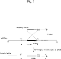
- the telomere truncation vector, the target sequence and the chromosomal allele to be resulted by homologous recombination are shown in Figure 1 .
- the PBS-TEL/Puro construct was digested with restriction enzyme EcoR I to provide linear DNA, which was introduced in a DT 40 hybrid cell retaining human chromosome 21.
- DT40 hybrid cells (1 x 10 7 ) were suspended in 0.75 ml of PBS and subjected to electroporation by use of Gene Pulser (Biorad) in the presence of 25 ⁇ g of DNA.
- a voltage of 750V was applied to a condenser having a capacitance of 25 ⁇ F and allowed to discharge by use of an electroporation cell having electrodes placed at an interval of 4 mm.
- the electroporated cells were suspended in DMEM medium (Invitrogen) supplemented with 10% fetus bovine serum (FBS), 1% chicken serum (ChS), and 50 ⁇ M 2-mercaptoethanol, and seeded in two 96-well clusters (Falcon). Two days later, puromycin dihydrochloride (Sigma) was added so as to have a final concentration of 0.3 ⁇ g/ml. Resistant colonies were formed in 2 to 3 weeks. The frequency of colony formation was 17.8 colonies in average per 1 x 10 7 DT40 hybrid cells. Transfection was performed 20 times to isolate 356 drug resistant colonies in total. The colonies were proliferated and subjected to the following analysis.
- the Registration Numbers of the aforementioned 6 types of STS markers are UniSTS: 76223, 45641, 54124, 22625, 54266, and 53746 sequentially in the order.
- sequences of primer oligonucleotides for the genes which were designed based on the nucleotide sequence obtained from the GenBank database, are shown below:
- genomic DNA As a template, three out of the aforementioned 8 types, that is, PRED 3 gene located in the proximity of a deletion site by homologous recombination, D21S265 marker and D21 S266 marker located in the distal region thereof were amplified by PCR (Innis et al., supra ). In the case where a long-arm distal region was deleted by telomere truncation, it was predicted that the genomic DNA might have PRED 3 gene but not have D21S265 and D21D266 markers. As a result, amplification was performed as predicted in 24 out of resistant 354 clones.
- a probe was designed within the target sequence for homologous recombination ( Figure 1 ).
- the pair of oligonucleotide primers shown below were used. PCR was performed by using the genomic DNA of a DT40 hybrid cell retaining human chromosome 21 as a template. Thereafter, a PCR amplification fragment was isolated and purified.
- genomic DNA extracted from the 24 clones obtained by the primary screening was digested by restriction enzyme Kpn I (Takara Shuzo Co., Ltd.) and subjected to Southern blot analysis in accordance with the method described in Ausubel et al. (Current Protocols in Molecular Biology, John Wiley & Sons, Inc., 1994 ).
- a signal from the DNA hybridized with a probe labeled with 32 P was detected by image analyzer BAS2000 (Fuji Photo Film Co., Ltd.). Representative results are shown in Figure 3 .
- the length of a restriction enzyme fragment was predicted based on a nucleotide sequence.
- FISH analysis was performed using a human specific probe Cot1 (Gibco BRL) in accordance with the method described in Matsubara et al. (FISH experimental protocol, Shujunsha, 1994 ).
- truncated human chromosome 21 was detected in almost all the mitotic images observed.
- Representative FISH images are shown in Figures 4a and 4b .
- the white arrow shows full-length human chromosome 21 before telomere truncation.
- the white arrow shows a fragment of human chromosome 21 where a long-arm distal region is deleted. Based on relative comparison with the chromosome of the host DT40 cell in size, it was confirmed that human chromosome 21 was truncated.
- pSF1 human artificial chromosome
- genomic DNA extracted from a DT40 hybridoma cell retaining human chromosome 21 was amplified by PCR. Each of them was digested with restriction enzymes EcoR I (Nippon Gene) or Bam H I (Nippon Gene) and subjected to agarose gel electrophoresis, thereby separating and purifying an about a 3 kb DNA fragment having cohesive ends. The fragments each are ligated to an EcoR I site or Bam H I site of pSF1 plasmid.
- a blasticidin resistant gene for use in screening of a homologous recombinant was excised out as about a 1.3 kb fragment from pCMV/Bsd (Invitrogen) by digestion with restriction enzymes Xho I (Nippon Gene) and Sal I (Nippon Gene) and cloned into the Xho I site of the pSF1 construct obtained above.
- the size of the pSF1 construct finally obtained was about 12.4 kb.
- the targeting vector, target sequence, and the chromosomal allele obtained by homologous recombination are shown in Figure 5 .
- the pSF1 construct was digested with restriction enzyme Apa I (Nippon Gene) to obtain linear DNA, which was introduced into a DT40 strain (DT40 (#21) puro-339) retaining human chromosome 21 where a long-arm distal region was deleted.
- DT40 hybrid cells (1 x 10 7 ) were suspended in 0.75 ml of PBS and subjected to electroporation using Gene Pulser (Biorad) in the presence of 10 ⁇ g of DNA.
- a voltage of 750V was applied to a condenser having a capacitance of 25 ⁇ F and allowed to discharge by use of an electroporation cell having electrodes placed at an interval of 4 mm.
- the electroporated cells were suspended in DMEM medium (Invitrogen) supplemented with 10% fetus bovine serum (FBS), 1% chicken serum (ChS), and 50 ⁇ M 2-mercaptoethanol and seeded in three 96-well clusters (Falcon). Two days later, Blasticidin S Hydrochloride (Funakoshi) was added so as to have a final concentration of 8 ⁇ g/ml. Resistant colonies were formed in 2 to 3 weeks. The frequency of colony formation was 5.8 colonies in average per 1 ⁇ 10 7 DT40 hybrid cells. Transfection was performed 14 times to isolate 82 colonies in total. The colonies were proliferated and subjected to the following analysis.
- Southern blot analysis was performed to screen homologous recombinants.
- a probe was designed outside the target sequence for homologous recombination.
- a pair of oligonucleotide primers as shown below were used as the probes.
- PCR was performed using the genomic DNA of a DT40 hybrid cell retaining human chromosome 21, as a template. The PCR amplified fragment was isolated and purified.
- the length of a restriction enzyme fragment was predicted based on a nucleotide sequence. It was 7.6 kb in the case of a homologous recombinant and 8.5 kb in the case of a wild type (non-homologous recombinant). It was found that 3 in total out of 82 blasticidin resistant clones were homologous recombinants (#60, #78, #79).
- a pair of oligonucleotide primers were designed so as to flank each of the target sequences. These pairs of oligonucleotide primers were designed on the chromosome and on the targeting vector. The positions of the primer pairs are indicated by arrows in Figure 5 .
- the sequences of the primer pairs are as follows:
- DT40 cell As a chromosome donor cell, DT40 cell (DT40(#21) bsd-79) retaining a HAC vector derived from human chromosome 21 obtained in Examples 1 and 2 by deleting a long-arm distal region and inserting a loxP sequence was used.
- a chromosome recipient cell Chinese hamster ovary derived cell line, CHO-K1 (available from ATCC, Accession No. JCRB9018) was used.
- microcells were prepared from about 10 9 DT40 (#21) bsd-79 cells.
- the DT40 (#21) bsd-79 cells were cultured up to a cell density corresponding to about 60 to 70% saturation in a culture solution (10% FBS, 1% ChS, 50 ⁇ M 2-mercaptoethanol, DMEM) containing colcemid (0.075 ⁇ g/ml, Demecolcine, Wako Pure Chemical Industries, Ltd.) for 12 to 15 hours to induce micronuclei.
- the cells were centrifugally collected, suspended in serum-free DMEM, and seeded in twelve 25 cm 2 -centrifugation flasks (Coasters) previously coated with poly-L-lysin.
- the flasks were allowed to stand still at 37°C for one hour. After the cells were adhered, the culture solution was removed. Each of the centrifugation flasks was filled with a pre-heated (37°C) solution of cytochalasin B (10 ⁇ g/ml in DMEM, Sigma), inserted in an acrylic centrifugation vessel, and centrifuged (34°C, 8000 rpm) for one hour. Microcells were recovered by suspending them in a serum-free medium (DMEM) and purified by filtration. The purified micronucleus cells were added to a 6 cm-diameter dish where CHO-K1 cells were cultured up to 80% saturation. They were fused with a PEG solution.
- DMEM serum-free medium
- the fused cells were dispersed by trypsin treatment and cultured in a selective medium (10% FBS, F12) containing blasticidin (8 ⁇ g/ml). After selective culture was performed for about 2 weeks, formed drug resistant colonies were separated and subjected to the following analysis. Microcell fusion was performed 12 times to obtain 4 blasticidin-resistant CHO clones in total.
- a chromosome was transferred or not was confirmed by the PCR method. More specifically, an attempt was made to detect marker genes, PRED 65 and PRED 3 genes (see Example 1, (3)) located in the proximal region of the long-arm of human chromosome 21. It was confirmed that 2 types of marker sequences were amplified in all of the 4 blasticidin-resistant CHO cell clones.
- FISH analysis was performed using a human specific probe Cot1 (Gibco BRL) in accordance with the method described in Matsubara et al. (FISH experimental protocol, Shujunsha, 1994 ).
- a human specific probe Cot1 Gibco BRL
- FIGS 7a and 7b show full-length human chromosome 21 before subjecting to telomere truncation.
- Figure 7b shows a fragment of human chromosome 21 where a long-arm distal region is deleted. Based on relative comparison with the chromosome of the host CHO cell in size, it was confirmed that truncated human chromosome 21 was transferred into the CHO cell.
- blasticidin resistant CHO clones retain a partial fragment of human chromosome 21 (HAC vector) where a long-arm distal region is deleted and a loxP sequence is inserted.
- CHO cell (CHO(#21)bsd-79-1) retaining a HAC vector derived from human chromosome 21 obtained in Example 3 by deleting a long-arm distal region and inserting a loxP sequence was used.
- human fibrosarcoma cell line HT1080 (available from ATCC, Accession No. CCL-121), was used.
- microcells were prepared from about 10 7 CHO(#21)bsd-79-1 cells.
- the CHO ((#21)bsd-79-1 cells which were cultured up to a cell density corresponding to about 60 to 70% saturation in six 25 cm 2 -centrifugation flasks (Coasters), were further cultured in a culture solution (10% FBS, 8 ⁇ g/ml blasticidin, F12) containing colcemid (0.075 ⁇ g/ml, Demecolcine, Wako Pure Chemical Industries, Ltd.) for 48 hours to induce micronuclei.
- each of the centrifugation flasks was filled with a pre-heated (37°C) solution of cytochalasin B (10 ⁇ g/ml in DMEM, Sigma), inserted in an acrylic centrifugation vessel, and centrifuged (34°C, 8000 rpm) for one hour.
- Microcells were recovered by suspending them in a serum-free medium (DMEM) and purified by filtration. The purified micronucleus cells were added to a 6 cm-diameter dish having HT1080 cells cultured up to 80% saturation. They were fused with a PEG solution.
- the fused cells were dispersed by trypsin treatment and cultured in a selective medium (10% CS, DMEM) containing blasticidin (8 ⁇ g/ml). After selective culture was performed for about 2 weeks, formed drug resistant colonies were isolated and subjected to the following analysis. Microcell fusion was performed twice to obtain 12 blasticidin-resistant HT1080 clones in total.
- oligonucleotide primers used herein are shown below:
- Chromosome analysis was performed by Giemsa staining in accordance with the method described in Kuroki et al. (Cell engineering handbook, Yodosha, 1992 ). About 20 metaphase chromosomal images of 4 clones (HT1080(#21)bsd79-1-3,6,11,14) out of the blasticidin resistant HT1080 clones were analyzed. A mini chromosome, which was smaller than endogenous chromosome 21 and not observed in the parent cell line, HT1080, was observed in a blasticidin resistant clone.
- blasticidin resistant HT 1080 clones retain a partial fragment (HAC vector) of human chromosome 21 where a long-arm distal region is deleted and a loxP sequence is inserted.
- DMEM supplemented with 10% CS was used as the nonselective culture solution for human cell clones.
- a selective culture solution was prepared by adding 4 ⁇ g/ml of blasticidin to the nonselective culture solution.
- 1.5 x 10 7 cells were seeded in a 10 cm-diameter dish.
- the number of cells was determined and 1.5 ⁇ 10 7 cells were again seeded in a 10 cm-diameter dish.
- 5.0 ⁇ 10 5 cells were seeded in a 10 cm-diameter dish.
- the number of cells was determined and 5.0 ⁇ 10 5 cells were again seeded in a 10 cm-diameter dish.
- the chicken cell line was collected 21, 42, 63, 84, 105 and 126 days after initiation of culturing, and the human cell clones were collected 10 and 20 days later. Chromosomal preparations were prepared.
- Detection of an artificial chromosome in chicken cells was performed by FISH analysis using a human specific probe Cot1 (Gibco BRL) in accordance with the method described in Matsubara et al. (FISH experimental protocol, Shujunsha, 1994 ). The presence or absence of a human chromosome was observed in 500 metaphase nuclei and a retention rate of the human chromosome was calculated. Detection of the artificial chromosome in human cells was performed by Giemsa staining in accordance with the method described in Kuroki et al. (Cell engineering handbook, Yodosha, 1992 ).
- a partial fragment of human chromosome 21 was stably retained in DT40 cells when cell division was performed in excess of 200 times under nonselective culture conditions.
- the number of human chromosomes per cell was counted by observing 100 chromosome images during metaphase. A single chromosome was observed in all without exception.
- the partial fragment of human chromosome 21 was stably retained at the time point (the number of cell division: 22) under selective culture conditions.
- a chromosome image during metaphase was observed, one or two chromosome portions per cell were observed.
- Figure 8 shows a method of inserting a GFP gene into a HAC vector derived from human chromosome 21.
- the HAC vector which was derived from human chromosome 21, was prepared by deleting a long-arm distal region by telomere truncation, and introducing a loxP site into a long-arm proximal region.
- a GFP expression plasmid containing a loxP sequence was prepared. The plasmid was introduced into the artificial chromosome by use of the site-specific recombination reaction between the loxP sequences by transiently expressing Cre recombination enzyme. Recombinant products having the insert were screened based on whether G418 resistance was acquired or not (reconstitution of a neo gene expression unit by disruption of a promoter).
- a GFP expression vector PEGFP-C1 (Clontech) was digested with restriction enzyme GbL II and Bam H I (Nippon Gene) to isolate/purify a 4.7 kb DNA fragment, which was self-ligated into a circular by use of DNA Ligation Kit Ver. 2 (Takara Shuzo Co., Ltd.).
- the recombinant plasmid was isolated by transformation of Escherichia Coli DH5 ⁇ to obtain a plasmid PEGFP-C1 ⁇ deficient in 51 bps of from GbL II to Bam H I within the multicloning site.
- an EGFP gene expression unit was amplified by PCR.
- Both ends of the EGFP gene expression unit thus amplified were digested with restriction enzymes Eco R I and Bam H I (Nippon Gene) to generate cohesive ends.
- the resultant construct was cloned into the Eco R I lBam H I site of a plasmid vector PBS226 (Lifetech) having a loxP sequence and a hCMV promoter.
- CHO cells (CHO(#21)bsd79-1) retaining the HAC vector derived from human chromosome 21 prepared in Example 3 was treated with trypsin and 5 x 10 6 cells were suspended in 0.8 ml of phosphate buffer (PBS). Electroporation was performed by use of Gene Pulser (Biorad) in the presence of 10 ⁇ g of PBS226/EGFP plasmid and 20 ⁇ g of Cre enzyme expression vector PBS 185 (Lifetech). A voltage of 750V was applied to a condenser having a capacitance of 25 ⁇ F and allowed to discharge by use of an electroporation cell having electrodes placed at an interval of 4 mm.
- Gene Pulser Biorad
- a voltage of 750V was applied to a condenser having a capacitance of 25 ⁇ F and allowed to discharge by use of an electroporation cell having electrodes placed at an interval of 4 mm.
- the electroporated cells were seeded in ten 100 mm plastic tissue-culture plates (Falcon) containing Eagle's F12 medium (hereinafter referred to as "F12", Invitrogen) supplemented with 10% fetus bovine serum (FBS). Two days later, the medium was replaced with a medium containing 800 ⁇ g/ml G418 (GENETICIN, Sigma) and 8 ⁇ g/ml Blasticidin S Hydrochloride (Funakoshi). Drug resistant colonies were formed in 2 to 3 weeks. The frequency of colony formation was 20 colonies per 5 x 10 6 of CHO cells. The colonies were isolated, proliferated and subjected to the following analysis.
- F12 Eagle's F12 medium
- FBS fetus bovine serum
- the isolated G418/blasticidin resistant CHO clone was subjected to observation by a fluorescent microscope. As a result, it was confirmed that GFP was expressed in 19 clones. A representative fluorescent image is shown in Figure 9 .
- Southern blot analysis was performed to confirm a homologous recombinant.
- parts of a G418 resistant gene and a GFP gene were used as probes and about 5 ⁇ g genomic DNA treated with restriction enzyme Eco R I or Bam H I (Nippon Gene) was used.
- GFP probe an 849 bp fragment obtained by digesting plasmid PEGFP-C1 (Clontech) with restriction enzymes Nhe I and GbL II (Nippon Gene) was used.
- the G418 resistant gene probe was a 1000-bp fragment obtained by digesting plasmid pSV2neo with restriction enzymes GbL II and Sma I (Nippon Gene).
- the probes were labeled with 32 P and the signal was detected by image analyzer BAS2000 (Fuji Photo Film Co., Ltd.). The representative results are shown in Figure 10 by way of example.
- DNA digested with EcoR I was detected by a neo probe.
- Lane 1 shows a DT40 line before insertion.
- Lane 2 and the subsequent lanes show G418 resistant DT40 clones.
- an allele before insertion a signal derived from a 5.7 kb fragment was detected, whereas a signal derived from a 6.9 kb fragment was detected in alleles after insertion.
- telomere truncation vector for deleting a distal region of the short-arm of human chromosome 21 was constructed by altering PBS-TEL/Puro ( Kuroiwa, Nucleic Acids Res., 26:3447, 1998 ). 1.7 kb expression unit for a puromycin resistant gene was removed from PBS-TEL/Puro, as a Not I fragment, and forming the blunt ends with T4 DNA Polymerase (DNA Blunting kit, Takara Shuzo Co., Ltd.) to produce PBS-TEF vector.
- PGKhygro/ ⁇ LT20 was digested with restriction enzymes Cla I and Sma I (Nippon Gene) and an expression unit for a hygromycin resistant gene under the control of a PGK promoter was isolated/purified, as a 1.8 kb fragment. This fragment was cloned into the PBS-TEL vector to obtain PBS-TEL/Hygro.
- telomere truncation vector Based on the nucleotide sequence (Accession No. AL163201) of the proximal region of the long-arm of human chromosome 21 obtained from the GenBank database, a target sequence for inserting a telomere truncation vector was designed.
- the sequences of primer oligonucleotides to which a recognition sequence for restriction enzyme Spe I or Bam H I is added, for use in amplification of the target sequence, are shown below:
- the target sequence was amplified by PCR using as a template, the genomic DNA extracted from a DT40 hybrid cell retaining human chromosome 21.
- the amplified product was digested with restriction enzymes Spe I and Bam H I (Nippon Gene).
- a DNA fragment of about 5 kb having cohesive ends was separated and purified by agarose gel electrophoresis.
- the DNA fragment was cloned into an Xba I/ Bam H I site of PBS-TEL/Hygro plasmid.
- the size of the PBS-TEL/Hygro construct finally obtained was about 5.8 kb.
- the telomere truncation vector, target sequence and chromosomal allele obtained by homologous recombination are shown in Figure 11 .
- the PBS-TEL/Hygro construct was digested with restriction enzyme Bam H I (Nippon Gene) to give a linear construct, which was introduced into DT40 hybrid cell (DT40(#21)bsd79) retaining human chromosome 21 where a long-arm distal region was deleted and a loxP site was inserted.
- the DT40 hybrid cells (1 x 10 7 ) were suspended in 0.75 ml of PBS and subjected to electroporation by Gene Pulser (Biorad) in the presence of 25 ⁇ g of DNA.
- a voltage of 750V was applied to a condenser having a capacitance of 25 ⁇ F and allowed to discharge by use of an electroporation cell having electrodes placed at an interval of 4 mm.
- the electroporated cells were suspended in DMEM medium (Invitrogen) supplemented with 10% fetus bovine serum (FBS), 1% chicken serum (ChS) and 50 ⁇ M 2-mercaptoethanol and seeded in five 96-well clusters (Falcon). Two days later, Hygromycin-B (Wako Pure Chemical Industries, Ltd.) was added so as to have a final concentration of 1.5 mg/ml. In 2 to 3 weeks, resistant colonies were formed. Transfection was carried out twice to isolate 63 drug resistant colonies in total. The colonies were proliferated and subjected to the following analysis.
- FBS fetus bovine serum
- ChS chicken serum
- 2-mercaptoethanol 50 ⁇ M 2-mercaptoethanol
- PCR analysis was performed for a primary screening for a homologous recombinant from hygromycin resistant DT40 clones.
- STS markers pCHB, D21S188, D21 S275 located in a short-arm proximal region of human chromosome 21 were amplified.
- Figure 12 a schematic chromosome map based on a G band image of human chromosome 21 is given at the left side. In addition, it is shown which marker is present in which band.
- DT40 (#21) represents a cell before subjected to telomere truncation. In the case where a short-arm distal region was deleted by telomere truncation, it was conceivable that D21 S275 might be present but D21 S 188 and pCHB might not be present. Therefore, 45 clones where either D21S188 or pCHB was not amplified were selected and subjected to Southern Blot analysis.
- a probe was designed within a target sequence for homologous recombination.
- the pair of oligonucleotide primers shown below were used.
- PCR was performed by using genomic DNA from a DT40 hybrid cell retaining human chromosome 21, as a template. A PCR amplification product was isolated and purified.
- the size of an amplified product was 5.9 kb. It was predicted that digestion of the amplified product with restriction enzyme Nsi I might produce fragments of 1.4 kb, 2.6 kb, and 1.9 kb ( Figure 11 ). PCR amplification was confirmed to occur in 2 clones where a homologous recombination allele was observed in Southern blot analysis, and generation of partial fragments digested with the restriction enzyme was confirmed ( Figure 14 ).
- FISH analysis was performed using a human specific probe Cot1 (Gibco BRL) in accordance with the method described in Matsubara et al. (FISH experimental protocol, Shujunsha, 1994 ). As a result, truncated human chromosome 21 was detected in almost all the mitotic images observed ( Figures 15a and 15b ).
- the human EPO gene was inserted into a HAC vector derived form human chromosome 21 in the same manner as in the case of a GFP gene described in Example 5.
- the HAC vector derived from human chromosome 21 was prepared by deleting a long-arm distal region by telomere truncation and introducing a loxP site into a long-arm proximal region.
- a human EPO expression plasmid containing a loxP sequence was prepared. The plasmid was integrated into the artificial chromosome by use of the site-specific recombination reaction between the loxP sequences by transiently expressing Cre recombination enzyme. Recombinant products having the insert were screened based on whether G418 resistance was acquired or not (reconstitution of a neo gene expression unit by disruption of a promoter).
- Both ends of the SV40 polyA additional unit which was amplified by PCR using pSTneoB as a template and SV40polyANp1 (SEQ ID No. 29) and SV40polyARp1 (SEQ ID No. 30), were digested with restriction enzymes Bam H I and Bgl II (Takara Shuzo Co., Ltd.) to obtain cohesive ends.
- the resultant construct was cloned into the Bam H I site of plasmid vector pBS226 (Lifetech) having a loxP sequence and an hCMV promoter. This was designated as pBS226-pA.
- both ends of the CMV promoter unit which was amplified by PCR using pBS226 as a template and CMVNp3 (SEQ ID No. 31) and CMVRp1(SEQ ID No. 32), were digested with restriction enzymes Eco R I and Bam H I (Takara Shuzo Co., Ltd.) to obtain cohesive ends.
- the resultant construct was cloned into the EcoR I -Bam H I site of pBS226-pA. This was designated as pLN1.
- both ends of the human EPO coding region which was amplified by PCR using human EPO cDNA as a template and hEPONp1 (SEQ ID No. 33) and hEPORp1 (SEQ ID No. 34), were digested with restriction enzymes Bam H I and Xho I (Takara Shuzo Co., Ltd.) to obtain cohesive ends.
- the resultant construct was cloned into the Bam H I -Xho I site of pLN1 This was designated as pLN1-EPO.
- the CHO cell (CHO(#21)bsd79-1) retaining a HAC vector derived from human chromosome 21 prepared in Example 3 was treated with trypsin and 5 x 10 6 cells were suspended in 0.8 ml of Hank's balanced salt solution (HBSS) and subjected to electroporation using Gene Pulser (Biorad) in the presence of 10 ⁇ g of pLN1-EPO vector prepared in Section (1) above, and 10 ⁇ g of Cre enzyme expression vector pBS185 (Lifetech). A voltage of 450V was applied to a condenser having a capacitance of 500 ⁇ F and allowed to discharge by use of an electroporation cell having electrodes placed at an interval of 4 mm.
- HBSS Hank's balanced salt solution
- the electroporated cells were seeded in four 48-well plastic tissue-culture plates (Falcon) containing Eagle's F12 medium (hereinafter referred to as "F12"; Invitrogen) supplemented with 10% fetus bovine serum (FBS). Two days later, the medium was replaced with a medium containing 800 ⁇ g/ml G418 (GENETICIN, Invitrogen) and 8 ⁇ g/ml Blasticidin S Hydrochloride (Funakoshi). Resistant colonies were formed in 2 to 3 weeks. The frequency of colony formation was 28 colonies in average per 5 x 10 6 CHO cells. Colonies were isolated, proliferated, and subjected to the following analysis. The cells thus obtained will be hereinafter referred to as "KH21E" cells.
- Expression of the human EPO gene was determined by quantifying human EPO protein produced in the culture supernatant in accordance with the enzyme-linked immunosorbent assay (ELISA).
- ELISA enzyme-linked immunosorbent assay
- the biological activity of the human EPO produced was analyzed based on proliferation activity of a human leukemia cell line, UT7-EPO cells (obtained from Prof. Norio Komatsu, Jichi Medical School), which proliferate in a human EPO-dependent manner.
- UT7-EPO cells obtained from Prof. Norio Komatsu, Jichi Medical School
- the culture supernatant was added to IMDM medium (Invitrogen) supplemented with 10% FBS so as to make final concentrations of 0.01, 0.1, 1, 5, 20, and 100 mIU/ml EPO based on the quantification values of Table 2.
- human EPO produced in the culture supernatant has the same biological activity as that of recombinant human EPO protein.
- Example 7 whether a chromosome was transferred or not was confirmed in each of the KH21E cell clones prepared in Example 7 (2) by PCR and FISH analysis.
- PCR amplification was performed with respect to marker PRED 65 and PRED3 genes which were located in a long-arm proximal region of human chromosome 21 and in the vicinity of a loxP site, and D21S265 marker located in a distal region thereof (see, Example 1, (3) and Figure 2 ). It was predicted that the human EPO gene insert introduced by site-specific recombination between loxP sequences might have the PRED 65 and PRED 3 genes but not have the D21S265 marker. As a result, in 21 out of 22 clones of the G418 resistant CHO cell, the expected amplification product was obtained.
- FISH analysis was performed using a human specific probe Cot1 (Gibco BRL) in accordance with the method described in Matsubara et al. (FISH experimental protocol, Shujunsha, 1994 ).
- Matsubara et al. FISH experimental protocol, Shujunsha, 1994 .
- truncated human chromosome 21 was detected in all the mitotic images observed.
- Table 3 Please note that clone KH21 listed in Table 3 represents a CHO cell (CHO(#21)bsd79-1) retaining the HAC vector derived from human chromosome 21 prepared in Example 3.
- a plurality of human EPO genes were inserted into a HAC vector derived from human chromosome 21 in the same manner as in the case of a human EPO gene described in Example 7.
- a HAC vector derived from human chromosome 21 was prepared by deleting a long-arm distal region by telomere truncation and introducing a loxP site in the long-arm proximal region thereto.
- a human EPO expression plasmid containing a loxP sequence was prepared. The plasmid was introduced into the artificial chromosome by use of the site-specific recombination reaction between the loxP sequences by transiently expressing Cre recombination enzyme. Recombinant products having the insert were screened based on whether G418 resistance was acquired or not (reconstitution of a neo gene expression unit by disruption of a promoter).
- EPOnF primer (SEQ ID No. 35) had EcoR I, Apa I, Mlu I restriction enzyme recognition sequences and a 5'-side partial sequence of CMV promoter sequentially from the 5' side, and prepared based on the nucleotide sequence of CMV promoter in pBS226.
- SVpAR primer (SEQ ID No. 36) had EcoR I, Bcl I restriction enzyme recognition sequences and a 3'-side partial complementary sequence of SV40 poly A additional unit sequentially from the 5' side, and prepared based on plasmid vector pSTneoB ( Kato et al., Cell Struct Funct, 12: 575-580, 1987 ).
- a PCR cycle comprises a reaction at 94°C for 2 minutes, and 30 cycles of denaturation at 94°C for 15 seconds, annealing at 60°C for 30 seconds, and extension at 68°C for 90 seconds. Both ends of the obtained DNA fragment were digested with restriction enzyme Eco R I (Takara Shuzo Co., Ltd.) to obtain cohesive ends. The resultant construct was cloned into the EcoR I site of a plasmid vector pLN1-EPO.
- the nucleotide sequence of the DNA fragment insert thus cloned was analyzed by a DNA sequencer (PRISM 3700, Applied Biosystems) and confirmed to be identical to the corresponding portion of the nucleotide sequence of the pLN1-EPO used as a template.
- a clone having 2 copies of the insert consisting of CMV promoter, human EPO gene, and SV40 poly A additional unit, in forward direction was designated as a plasmid vector pLN1-EPO2.
- Plasmid vector pLN1-EPO2 prepared in the Section (1) above was digested with Xba I (Takara Shuzo Co., Ltd.) to give a linear vector, and then treated with KOD polymerase (Toyobo) to generate the blunt ends. Thereafter, the linear fragment was digested with Apa I (Takara Shuzo Co., Ltd.) to obtain a DNA fragment for use in insertion containing 2 copies of a fragment consisting of CMV promoter, human EPO gene, and SV40 polyA additional unit.
- Plasmid vector pLN1-EPO2 was digested with Mlu I (Takara Shuzo Co., Ltd.), and then treated with KOD polymerase (Toyobo) to generate the blunt ends. The fragment was digested with Apa I (Takara Shuzo Co., Ltd.) to obtain an Apa I-blunt ended Mlu I site. To this site, the DNA fragment for use in insertion obtained above was cloned. The obtained plasmid vector containing 4 copies of the human EPO gene was designated as pLN1-EPO4.
- the CHO cells (CHO(#21)bsd79-1) retaining a HAC vector derived from human chromosome 21 and prepared in Example 3 were treated with trypsin and 5 ⁇ 10 6 cells were suspended in 0.8 ml of Hank's balanced salt solution (HBSS). Electroporation was performed by use of Gene Pulser (Biorad) in the presence of 10 ⁇ g of the pLN1-EPO2 vector or the pLN1-EPO4 vector prepared in the Section (1) or (2) above and 10 ⁇ g of Cre enzyme expression vector pBS185 (Lifetech).
- Gene Pulser Biorad
- a voltage of 450V was applied to a condenser having a capacitance of 500 ⁇ F and allowed to discharge by use of an electroporation cell having electrodes placed at an interval of 4 mm.
- the electroporated cells were seeded in five 48-well plastic tissue-culture plates (Falcon) containing Eagle's F12 medium (hereinafter referred to as "F12"; Invitrogen) supplemented with 10% fetus bovine serum (FBS). Two days later, the medium was replaced with a medium containing 800 ⁇ g/ml G418 (GENETICIN, Invitrogen) and 8 ⁇ g/ml Blasticidin S Hydrochloride (Funakoshi). Resistant colonies were formed in 2 to 3 weeks.
- F12 Eagle's F12 medium
- FBS fetus bovine serum
- the frequency of colony formation was 14 colonies per 5 x 10 6 CHO cells in the case of pLN1-EPO2 and 24 colonies in the case of pLN1-EPO4. Colonies were isolated, proliferated and subjected to the following analysis.
- the cells prepared by use of pLN1-EPO2 will be hereinafter referred to as "KH21E2 cells” and the cells prepared by use of pLN1-EPO4 as "KH21E4 cells”.
- PCR amplification was performed by using SVpANp1 primer (SEQ ID No. 37) designed in an SV40 poly A additional sequence region derived from pLN1-EPO2 or pLN1-EPO4 vector, and Neo Rp2 primer (SEQ ID No. 38) designed in a neomycin resistant gene derived from pSF1 on the HAC vector.
- SVpANp1 primer SEQ ID No. 37
- Neo Rp2 primer SEQ ID No. 38
- a recombinant having an insert it was predicted to obtain about 1.0 kbp fragment including a region from a portion having SV40 poly A additional sequence to a loxP sequence derived from pLN1-EPO2 or pLN1-EPO4 vector, and a region from the loxP sequence to a part of the neo gene derived from pSF1.
- amplification was performed as predicted in all of the 6 KH21E2 cell clones and the 6 KH21E4 cell clones.
- Neo Rp2 primer SEQ ID No. 38
- M13RV SEQ ID No. 39
- the 6 KH21E2 cell clones contain 2 copies of the DNA insert including CVM promoter, human EPO gene and SV40 polyA additional sequence.
- Expression of the human EPO gene was determined by quantifying human EPO protein produced in the culture supernatant in accordance with the enzyme-linked immunosorbent assay (ELISA).
- ELISA enzyme-linked immunosorbent assay
- the amount of human EPO contained in the culture supernatant was quantified by a human EPO ELISA kit (Quantikine IVD Human EPO Immunoassay, R&D system). The results are shown in Table 6. Please note that C1 and C4 were quantified in a 1 ⁇ 10 -4 dilution and C9, C11 and C20 in a 1 ⁇ 10 -5 dilution. Table 6 Measurement value Concentration of EPO in CM Clone No.
- the human EPO gene was inserted into a HAC vector derived from human chromosome 21.
- a HAC vector derived from chromosome 21 was prepared by deleting a long-arm distal region by telomere truncation and introducing a loxP site in a long-arm proximal region as well as deleting a short-arm distal region by telomere truncation.
- a human EPO expression plasmid containing a loxP sequence was prepared.
- the plasmid was introduced into the artificial chromosome by use of the site-specific recombination reaction between the loxP sequences by transiently expressing Cre recombination enzyme. Recombinant having the insert were screened based on whether G418 resistance was acquired or not (reconstitution of a neo gene expression unit by disruption of a promoter).
- H4 and H8 The 2 clones of CHO cells: CHO(#21)hyg4 and CHO(#21)hyg8 (hereinafter referred to as "H4" and "H8", respectively) retaining the HAC vector derived from human chromosome 21 prepared in Example 17 (described later) were treated with trypsin. 5 ⁇ 10 6 cells for each were suspended in 0.8 ml of Hank's balanced salt solution (HBSS) and subjected to electroporation using Gene Pulser (Biorad) in the presence of 10 ⁇ g of pLN1-EPO vector prepared in Example 7 (1) and 10 ⁇ g of Cre enzyme expression vector pBS185 (Lifetech).
- HBSS Hank's balanced salt solution
- a voltage of 450V was applied to a condenser having a capacitance of 500 ⁇ F and allowed to discharge by use of an electroporation cell having electrodes placed at an interval of 4 mm.
- the electroporated cells were seeded in five 48-well plastic tissue-culture plates (Falcon) containing Eagle's F12 medium (hereinafter referred to as "F12"; Invitrogen) supplemented with 10% fetus bovine serum (FBS). Two days later, the medium was replaced with a selective medium containing 800 ⁇ g/ml G418 (GENETICIN, Invitrogen) and 8 ⁇ g/ml Blasticidin S Hydrochloride (Funakoshi).
- F12 Eagle's F12 medium
- FBS fetus bovine serum
- H4E cells The cells prepared from H4 will be hereinafter referred to as "H4E cells” and the cells prepared from H8 as “H8E cells”.
- PCR amplification was performed with respect to markers PRED 65 and PRED 3 genes, which were located in a long-arm proximal region of human chromosome 21 in the vicinity of a loxP site, and D21 S265 marker located in a distal region thereof (Example 1 (3), Figure 2 ). It was predicted that a product having the human EPO gene insert introduced by the site-specific recombination between loxP sequences might have the PRED 65 and PRED 3 genes but not have the D215265 marker. As a result, it was confirmed that amplification was performed as predicted in 21 out of 22 the H4E cell clones.
- PCR amplification of STS markers (pCHB, D21S187, D21S275) positioned in a short-arm proximal region of human chromosome 21 was performed (see Example 6, (3), Figure 12 ). Since a short-arm distal region was deleted from of human chromosome 21 at the short-arm proximal region thereof, it is predicted that pCHB and D21S187 markers may not be present and D21S275 marker may be present. As a result, it was confirmed that amplification was performed as predicted in 15 clones.
- FISH analysis was performed using a human specific probe Cot1 (Gibco BRL) in accordance with the method described in Matsubara et al. (FISH experimental protocol, Shujunsha, 1994 ).
- Matsubara et al. FISH experimental protocol, Shujunsha, 1994 .
- truncated human chromosome 21 was detected in almost all the mitotic images observed.
- the results are shown in Table 7. From the above, based on relative comparison with the chromosome of a host CHO cell in size, it was confirmed that truncated human chromosome 21 was transferred into the CHO cell.
- Expression of the human EPO gene was determined by quantifying the human EPO protein produced in the culture supernatant in accordance with the enzyme-linked immunosorbent assay (ELISA).
- ELISA enzyme-linked immunosorbent assay
- Table 8 Measurement value Concentration of EPO in CM Clone No. (mIU/ml) (IU/ml) ( ⁇ g/ml) C6 57 1140 5.7 C10 27 540 2.7 C11 46 920 4.6 C15 55 1100 5.5 C16 52 1040 5.2 C17 26 520 2.6 C18 49 980 4.9 C19 40 800 4.0 C20 54 1080 5.4 C21 53 1060 5.3
- CHO #21hyg4 and CHO #21hyg8 (hereinafter referred to as "H4 cell” and "H8 cell", respectively) retaining a HAC vector derived from human chromosome 21 obtained in Example 17 by deleting a long-arm distal region and inserting a loxP sequence and thereafter deleting a short-arm distal region by telomere truncation.
- mouse A9 cell Oshimura et al., Environ. Health Perspect. 93:57, 1991 , Accession No. JCRB0211) was used. At first, microcells were prepared from about 10 7 H4 cells.
- the H4 or H8 cells which were cultured in twenty four 25 cm 2 -centrifugation flasks (Nunc) up to a cell density corresponding to about 60 to 70% saturation, were further cultured in a culture solution (20% FBS, 800 ⁇ g/ml G418, F12) containing colcemid (0.1 ⁇ g/ml, Demecolcine, Wako Pure Chemical Industries, Ltd.) for 5 days to induce micronuclei.
- a culture solution (20% FBS, 800 ⁇ g/ml G418, F12) containing colcemid (0.1 ⁇ g/ml, Demecolcine, Wako Pure Chemical Industries, Ltd.) for 5 days to induce micronuclei.
- each of the centrifugation flasks was filled with a pre-heated (37°C) solution of cytochalasin B (10 ⁇ g/ml in DMEM, Sigma), inserted in an acrylic centrifugation vessel, and centrifuged (34°C, 8000 rpm) for one hour.
- Microcells were recovered by suspending them in a serum-free medium (DMEM) and purified by filtration using SWINNEX-25 (Millipore) equipped with a filter (Whatman) of a pore size of 8 ⁇ m, 5 ⁇ m and 3 ⁇ m.
- microcells purified were resuspended in 2 ml of DMEM supplemented with 50 ⁇ g/ml phytohemaggulutinin-P (Difco).
- DMEM phytohemaggulutinin-P
- PEG 1000 final concentration of 50% (w/v), Sigma
- DMSO final concentration of 7% (w/v), Sigma
- PCR amplification was performed with respect to STS markers (pCHB, D21S187, D21S275), which were located in a short-arm proximal region of human chromosome 21 (see, Example 6, (3) and Figure 12 ). It was predicted that pCHB and D21 S 187 markers might not be present but D21 S275 marker might be present since the distal region of the short-arm was deleted from human chromosome 21 at the location of short-arm proximal region. As a result, it was confirmed that amplification was performed as predicted in 18 clones.
- FISH analysis was performed using a human specific probe Cot1 (Gibco BRL) in accordance with the method described in Matsubara et al. (FISH experimental protocol, Shujunsha, 1994 ).
- truncated human chromosome 21 was detected in almost all the mitotic images observed. The results are shown in Table 11.
- Table 11 Name of clone Number of analyzed samples mitotic image/metaphase nucleus Number of Cot-1 signals per cell mitotic image/metaphase nucleus Retention rate (metaphase nucleus) % 0 1 2 3 A9 ⁇ 11 50/100 11/31 36/65 3/2 0/2 69 A9 ⁇ 12 50/100 2/11 47/87 0/2 1/0 89
- Example 12 Transfer of HAC vector derived from human chromosome 21 having the human EPO gene into mouse A9 cell.
- a clone H4E C10, H4E C15 or H4E C16 cell
- mouse A9 cell Oshimura et al., Environ. Health Perspect. 93:57, 1991 , Accession No. JCRB0211
- microcells were prepared from about 10 8 H4E C15 or H4E C16 cells.
- H4E C 15 or H4E C16 cells which were cultured in twenty four 25 cm 2 -centrifugation flasks (Nunc) up to a cell density corresponding to about 60 to 70% saturation, were further cultured in a culture solution (20% FBS, 800 ⁇ g/ml G418, F12) containing colcemid (0.1 ⁇ g/ml, Demecolcine, Wako Pure Chemical Industries, Ltd.) for 4 days to induce micronuclei.
- each of the centrifugation flasks was filled with a pre-heated (37°C) solution of cytochalasin B (10 ⁇ g/ml in DMEM, Sigma), inserted in an acrylic centrifugation vessel, and centrifuged (34°C, 8000 rpm) for one hour.
- Microcells were recovered by suspending them in a serum-free medium (DMEM) and purified by filtration using SWINNEX-25 (Millipore) equipped with a filter (Whatman) of a pore size of 8 ⁇ m, 5 ⁇ m and 3 ⁇ m.
- microcells purified were resuspended in 2 ml of DMEM supplemented with 50 ⁇ g/ml or 100 ⁇ g/ml phytohemaggulutinin-P (Difco).
- DMEM phytohemaggulutinin-P
- PEG 1000 final concentration of 50% (w/v), Sigma
- DMSO final concentration of 7% (w/v), Sigma
- a HAC vector derived from human chromosome 21 that is, a hygromycin resistant gene (in short-arm distal region), a blasticidin resistant gene (in long-arm proximal region), and a neomycin resistant gene (in long-arm proximal region) present on the HAC vector derived from human chromosome 21, function in the presence of selective drugs, it was confirmed whether or not a region containing each of the drug resistant gene is present.
- FISH analysis was performed using a human specific probe Cotl (Gibco BRL) in accordance with the method described in Matsubara et al. (experimental protocol, Shujunsha, 1994). Seven clones in which all markers were amplified as predicted in the PCR analysis in Section (2-1) above, were analyzed. As a result, truncated human chromosome 21 was detected in almost all the mitotic images observed. The results are shown in Table 13.
- Neo Rp2 primer SEQ ID No. 38
- M13RVprimer SEQ ID No. 39
- Expression of the human EPO gene was determined by quantifying human EPO protein produced in the culture supernatant in accordance with the enzyme-linked immunosorbent assay (ELISA).
- ELISA enzyme-linked immunosorbent assay
- the human EPO concentrations of the culture supernatants of A ⁇ E51, A ⁇ E4 and A ⁇ E18 were greater than a detection limit by the human EPO ELISA kit (Quantikine IVD Human EPO Immunoassay, R&D system).
- chromosome donor cell mouse A9 cell (C11-SC20 cell, Tomizuka et al., Nature Genet. (USA), Vol.16, p.133-143, 1997 ) containing a human chromosome 14 fragment (SC20) was used.
- a chromosome recipient cell a normal human fibroblast cell, HFL-1 (obtained from the cellular material development laboratory of RIKEN, Accession No. RCB0521) was used. At first, microcells were prepared from about 10 7 cells.
- the C11-SC20 cells which were cultured in twelve 25 cm 2 -centrifugation flasks (Nunc) up to a cell density corresponding to about 80 to 90% saturation, were further cultured in a culture solution (20% FBS, 800 ⁇ g/ml G418, DMEM) containing colcemid (0.05 ⁇ g/ml, Demecolcine, Wako Pure Chemical Industries, Ltd.) for 48 hours to induce micronuclei.
- a culture solution (20% FBS, 800 ⁇ g/ml G418, DMEM) containing colcemid (0.05 ⁇ g/ml, Demecolcine, Wako Pure Chemical Industries, Ltd.) for 48 hours to induce micronuclei.
- each of the centrifugation flasks was filled with a pre-heated (37°C) solution of cytochalasin B (10 ⁇ g/ml in DMEM, Sigma), inserted in an acrylic centrifugation vessel, and centrifuged (34°C, 8000 rpm) for one hour.
- Microcells were recovered by suspending them in a serum-free medium (DMEM) and purified by filtration using WINNEX-25 (Millipore) equipped with a filter (Whatman) of a pore size of 8 ⁇ m, 5 ⁇ m and 3 ⁇ m.
- microcells purified were resuspended in 2 ml of DMEM supplemented with 50 ⁇ g/ml phytohemaggulutinin-P (Difco).
- DMEM phytohemaggulutinin-P
- PEG 1500 final concentration of 45% (w/v), Roche Diagnostics
- DMSO final concentration of 10% (w/v), Sigma
- Microcells were prepared and purified in the same manner as in Section (1-1) above and resuspended in 6 ml of DMEM.
- HFL-1 cells were cultured up to 90% saturation in a 175 cm 2 -culture flask (Falcon). After the cells were dispersed by trypsin treatment, they were washed with DMEM twice and then suspended in 7 ml of DMEM. The HFL-1 cell suspension was overlaid on the microcell suspension obtained above and centrifuged. After the supernatant was removed, the pellet was suspended by tapping. To the resultant suspension, 0.5 ml of PEG1500 (final concentration of 50% (w/v), Roche Diagnostics) was added and cell fusion was performed for 120 seconds.
- PEG1500 final concentration of 50% (w/v), Roche Diagnostics
- the transferred chromosome was confirmed by PCR amplification of a neo gene present on SC20.
- the sequences of the primer oligonucleotides used herein are shown below:
- primers were prepared based on the nucleotide sequence of plasmid vector pSTneoB ( Kato et al., Cell Struct Funct, 12:575-580, 1987 ).
- Chromosome analysis was performed by Giemsa staining in accordance with the method described in Kuroki et al. (Cell engineering handbook, Yodosha, 1992 ). About 20 metaphase chromosomal images of 2 clones out of the G418 resistant HFL-1 cells were analyzed. A mini chromosome, which was smaller than endogenous chromosome 14 and not observed in the parent line HFL-1, was observed in the G418 resistant clone.
- a chromosome donor cell mouse A9 cell (C11-SC20 cell, Tomizuka et al., Nature Genet. (USA), Vol. 16, p.133-143, 1997 ) containing a human chromosome 14 fragment (SC20) was used.
- a chromosome recipient cell a normal human fibroblast cell HUC-F2 (obtained from the cellular material development laboratory of RIKEN, Accession No. RCB0436) was used. At first, microcells were prepared from about 10 7 cells.
- the C11-SC20 cells which were cultured in twelve 25 cm 2 -centrifugation flasks (Nunc) up to a cell density corresponding to about 80 to 90% saturation, were further cultured in a culture solution (20% FBS, 800 ⁇ g/ml G418, DMEM) containing colcemid (0.05 ⁇ g/ml, Demecolcine, Wako Pure Chemical Industries, Ltd.) for 48 hours to induce micronuclei.
- a culture solution (20% FBS, 800 ⁇ g/ml G418, DMEM) containing colcemid (0.05 ⁇ g/ml, Demecolcine, Wako Pure Chemical Industries, Ltd.) for 48 hours to induce micronuclei.
- each of the centrifugation flasks was filled with a pre-heated (37°C) solution of cytochalasin B (10 ⁇ g/ml in DMEM, Sigma), inserted in an acrylic centrifugation vessel, and centrifuged (34°C, 8000 rpm) for one hour.
- Microcells were recovered by suspending them in a serum-free medium (DMEM) and purified by filtration by use of SWINNEX-25 (Millipore) equipped with a filter (Whatman) of a pore size of 8 ⁇ m, 5 ⁇ m and 3 ⁇ m.
- microcells purified were resuspended in 2 ml of DMEM supplemented with 50 ⁇ g/ml phytohemaggulutinin-P (Difco). To 25 cm 2 -culture flasks (Falcon) in which HUC-F2 cells were cultured up to 90% saturation, the purified micronucleus cells were added.
- the medium was replaced with a selective medium (10% FBS, ⁇ MEM) containing G418 (400 ⁇ g/ml). After selective culturing was performed for about 3 weeks, formed drug resistant colonies were isolated and subjected to the following analysis. Microcell fusion was performed 4 times to obtain 8 G418 resistant colonies.
- Microcells were prepared and purified in the same manner as in Section (1-1) above and resuspended in 6 ml of DMEM.
- HUC-F2 cells were cultured up to 90% saturation in a 175 cm 2 -culture flask (Falcon). After the cells were dispersed by trypsin treatment, they were washed with DMEM twice and then suspended in 7 ml of DMEM. The HUC-F2 cell suspension was overlaid on the microcell suspension obtained above and centrifuged. After the supernatant was removed, the pellet was suspended by tapping. To the resultant suspension, 0.5 ml of PEG1500 (final concentration of 50% (w/v), Roche Diagnostics) was added and cell fusion was performed for 120 seconds.
- PEG1500 final concentration of 50% (w/v), Roche Diagnostics
- the transferred chromosome was confirmed by PCR amplification of a neo gene present on SC20.
- PCR amplification was performed by using 421F primer (SEQ ID No. 40) and 778R primer (SEQ ID No. 41). It was predicted that an insert in a HAC vector might have a neo gene. As a result, it was confirmed that amplification was performed as predicted in all clones. From the experiment above, it was confirmed that the obtained G418 HUC-F2 clones retain SC20.
- a chromosome donor cell mouse A9 cell (C11-SC20 cell, Tomizuka et al., Nature Genet. (USA), Vol.16, p.133-143, 1997 ) containing a human chromosome 14 fragment (SC20) was used.
- a chromosome recipient cell a normal human fibroblast cell, HF-19 (obtained from the cellular material development laboratory of RIKEN, Accession No. RCB0210) was used. At first, microcells were prepared from about 10 7 cells.
- the C11-SC20 cells which were cultured in twelve 25 cm 2 -centrifugation flasks (Nunc) up to a cell density corresponding to about 80 to 90% saturation, were further cultured in a culture solution (20% FBS, 800 ⁇ g/ml G418, DMEM) containing colcemid (0.05 ⁇ g/ml, Demecolcine, Wako Pure Chemical Industries, Ltd.) for 48 hours to induce micronuclei.
- a culture solution (20% FBS, 800 ⁇ g/ml G418, DMEM) containing colcemid (0.05 ⁇ g/ml, Demecolcine, Wako Pure Chemical Industries, Ltd.) for 48 hours to induce micronuclei.
- each of the centrifugation flasks was filled with a pre-heated (37°C) solution of cytochalasin B (10 ⁇ g/ml in DMEM, Sigma), inserted in an acrylic centrifugation vessel, and centrifuged (34°C, 8000 rpm) for one hour.
- Microcells were recovered by suspending them in a serum-free medium (DMEM) and purified by filtration by use of SWINNEX-25 (Millipore) equipped with a filter (Whatman) of a pore size of 8 ⁇ m, 5 ⁇ m and 3 ⁇ m.
- microcells purified were resuspended in 2 ml of DMEM supplemented with 50 ⁇ g/ml phytohemaggulutinin-P (Difco).
- DMEM phytohemaggulutinin-P
- PEG 1500 final concentration of 45% (w/v), Roche Diagnostics
- DMSO final concentration of 10% (w/v), Sigma
- the cells were cultured in ⁇ MEM medium containing 10% FBS for 48 hours, the cells were dispersed by trypsin treatment, and seeded in a 48-well plastic tissue-culture plate (Falcon) coated with collagen I. Two days later, the medium was replaced with a selective medium (10% FBS, ⁇ MEM) containing G418 (400 ⁇ g/ml). After selective culturing was performed for about 3 weeks, formed drug resistant colonies were isolated. Microcell fusion was performed once to obtain a single G418 resistant colony.
- a selective medium (10% FBS, ⁇ MEM) containing G418 (400 ⁇ g/ml).
- a chromosome donor cell As a chromosome donor cell, a clone (H4E C10 cell) having a high micronucleus formation ability out of CHO cells obtained in Example 10 and retaining a HAC vector derived from human chromosome 21 containing a single copy of the human EPO gene, which was prepared by deleting a long-arm distal region and inserting a loxP sequence and thereafter deleting a short-arm distal region by telomere truncation, the human EPO gene being introduced by the site specific recombination reaction between the loxP sequences by transiently expressing Cre recombinant enzyme.
- microcells were prepared from 10 8 H4E C10 cells. More specifically, the H4E C10 cells, which were cultured in forty eight 25 cm 2 -centrifugation flasks (Nunc) up to a cell density corresponding to about 60 to 70% saturation, were further cultured in a culture solution (20% FBS, 800 ⁇ g/ml G418, F12) containing colcemid (0.1 ⁇ g/ml, Demecolcine, Wako Pure Chemical Industries, Ltd.) for 4 days to induce micronuclei.
- a culture solution (20% FBS, 800 ⁇ g/ml G418, F12
- colcemid 0.1 ⁇ g/ml, Demecolcine, Wako Pure Chemical Industries, Ltd.
- each of the centrifugation flasks was filled with a pre-heated (37°C) solution of cytochalasin B (10 ⁇ g/ml in DMEM, Sigma), inserted in an acrylic centrifugation vessel, and centrifuged (34°C, 8000 rpm) for one hour.
- Microcells were recovered by suspending them in a serum-free medium (DMEM) and purified by filtration by use of SWINNEX-25 (Millipore) equipped with a filter (Whatman) of a pore size of 8 ⁇ m, 5 ⁇ m and 3 ⁇ m
- DMEM serum-free medium
- SWINNEX-25 Micropore
- Whatman a filter of a pore size of 8 ⁇ m, 5 ⁇ m and 3 ⁇ m
- the microcells purified were resuspended in 2 ml of DMEM supplemented with 50 ⁇ g/ml phytohemaggulutinin-P (Difco).
- HC ⁇ E cells The cells will be referred to as "HC ⁇ E cells”.
- normal human fibroblast cell HFL-1 obtained from the cellular material development laboratory of RIKEN, Accession No. RCB0521
- microcells were prepared from about 10 7 cells. More specifically, A ⁇ 51 or A ⁇ E5 cells, which were cultured in twelve 25 cm 2 -centrifugation flasks (Nunc) up to a cell density corresponding to about a 80 to 90% saturation, were further cultured in a culture solution (20% FBS, 600 ⁇ g/ml G418, DMEM) containing colcemid (0.1 ⁇ g/ml, Demecolcine, Wako Pure Chemical Industries, Ltd.) for 72 hours to induce micronuclei.
- a culture solution (20% FBS, 600 ⁇ g/ml G418, DMEM) containing colcemid (0.1 ⁇ g/ml, Demecolcine, Wako Pure Chemical Industries, Ltd.) for 72 hours to induce micronuclei.
- each of the centrifugation flasks was filled with a pre-heated (37°C) solution of cytochalasin B (10 ⁇ g/ml) in DMEM, Sigma), inserted in an acrylic centrifugation vessel, and centrifuged (34°C, 8000 rpm) for one hour.
- Microcells were recovered by suspending them in a serum-free medium (DMEM) and purified by filtration by use of SWINNEX-25 (Millipore) equipped with a filter (Whatman) of a pore size of 8 ⁇ m, 5 ⁇ m and 3 ⁇ m
- DMEM serum-free medium
- SWINNEX-25 Micropore
- Whatman a filter of a pore size of 8 ⁇ m, 5 ⁇ m and 3 ⁇ m
- the microcells purified were resuspended in 2 ml of DMEM supplemented with 50 ⁇ g/ml phytohemaggulutinin-P (Difco).
- H ⁇ E cells The cells will be referred to as "H ⁇ E cells”.
- the transferred chromosome was confirmed by PCR amplification with reference to the presence or absence of a neo gene present on a HAC vector derived from human chromosome 21.
- the sequences of the primer oligonucleotides used for PCR amplification are shown below:
- primers were prepared based on the nucleotide sequence of plasmid vector pSTneoB ( Kato et al., Cell Struct Funct, 12:575-580, 1987 ).
- PCR amplification was performed by using 1291F primer (SEQ ID No. 42) and 1667R primer (SEQ ID No. 43).
- the HAC vector derived from human chromosome 21 was present, it was predicted to amplify a 0.4 kbp fragment containing a part of the neo gene. As a result, it was confirmed that amplification was performed as predicted in all of 5 H ⁇ E cell clones.
- H ⁇ E cell retains the HAC vector derived from human chromosome 21.
- Expression of the human EPO gene was determined by quantifying human EPO protein produced in the culture supernatant in accordance with the enzyme-linked immunosorbent assay (ELISA).
- ELISA enzyme-linked immunosorbent assay
- the human EPO concentration in a culture supernatant was greater than a detection limit by the human EPO ELISA kit (Quantikine IVD Human EPO Immunoassay, R&D system).
- a human telomerase (hTERT) gene has a code region of 3399 bp and contains a G/C rich sequence at the 5' region thereof. For the reason, it was predicted difficult to amplify the entire gene by designing the primers at the both end of the coding region. Therefore, the coding region was divided into 3 regions: 1 to 800 bp (hereinafter referred to as "5' hTERT"), 679 to 1993 bp (hereinafter referred to as "M-XhTRET”) and 1952 to 3339 bp (hereinafter referred to as "3' hTRET”. Please note that, the position in the nucleotide sequence was expressed by regarding "A" of initiation codon "ATG” as 1). After individual regions were amplified by PCR and cloned and these regions were ligated to each other. In this manner, cloning of the hTERT gene was performed. This method will be specifically described below.
- primer oligonucleotides used in construction of a plasmid vector are shown below.
- primers were prepared based on the nucleotide sequence (Accession No. NM003219) obtained from the GenBank.
- PCR amplification was performed in 50 ⁇ l of a reaction solution containing hTERT Fw6 (SEQ ID No. 44) and hTERT Rv6 (SEQ ID No. 45) each having a final concentration of 0.4 11M using 2.5 units of LA Taq (Takara Shuzo Co., Ltd.).
- hTERT Fw6 SEQ ID No. 44
- hTERT Rv6 SEQ ID No. 45
- the PCR amplification was performed by placing at 98°C for 10 minutes, followed by repeating a cycle consisting of denaturation at 98°C for 30 seconds, annealing and extension at 72°C for 5 minutes three times, a cycle consisting of denaturation at 98°C for 30 seconds, annealing and extension at 70°C for 5 minutes, 2 times, and a cycle consisting of denaturation at 98°C for 30 seconds, annealing and extension at 68°C for 5 minutes, 35 times. Furthermore, using 2 ⁇ l of the resultant PCR product as a template, PCR amplification was performed by using hTERT Fw1 (SEQ ID No. 46) and hTERT Rv7 (SEQ ID No.
- the PCR amplification was performed by placing at 98°C for 10 minutes, followed by repeating a cycle consisting of denaturation at 98°C for 30 seconds, annealing and extension at 72°C for 5 minutes, three times; a cycle consisting of denaturation at 98°C for 30 seconds, annealing and extension at 70°C for 5 minutes, twice; and a cycle consisting of denaturation at 98°C for 30 seconds, annealing and extension at 68°C for 5 minutes, 35 times.
- a DNA fragment of about 0.8 kb was obtained.
- the DNA fragment of about 0.8 kb was purified by QIAQUICK PCR Purification Kit (QIAGEN) and both ends thereof were digested with KOD DNA polymerase (Toyobo) to obtain blunt ends, and further digested with Bgl II (Takara Shuzo Co., Ltd.) to make the 5' end side cohesive. As a result, a DNA fragment for 5' hTERT insertion was obtained. After plasmid vector pLN1-EPO was digested with Xho I (Takara Shuzo Co., Ltd.), both ends thereof were digested with KOD DNA polymerase (Toyobo) to obtain blunt ends.
- the resultant fragment was further digested with Bam H I (Takara Shuzo Co., Ltd.) to remove the human EPO gene.
- Bam H I Tropa Shuzo Co., Ltd.
- the DNA fragment for 5'hTERT insert was cloned.
- the nucleotide sequence of the DNA fragment for 5' hTERT insert thus cloned was analyzed by a DNA sequencer (PRISM3700, Applied Biosystems) and confirmed to be identical to the corresponding portion of the nucleotide sequence obtained from the GenBank. From the above, the resultant plasmid vector was designated as pLN1-5'hTERT.
- primer oligonucleotides used in construction of a plasmid vector are shown below.
- primers were prepared based on the nucleotide sequence (Accession No. NM003219) obtained from the GenBank.
- PCR amplification was performed in 25 ⁇ l of a reaction solution containing hTERT Fw8-2 (SEQ ID No. 48) and hTERT 5'XhoIRv3 (SEQ ID No. 49), each having a final concentration of 0.4 ⁇ M, using 2.5 units of LA Taq (Takara Shuzo Co., Ltd.).
- the PCR amplification was performed by placing at 98°C for 5 minutes, followed by repeating a cycle consisting of reactions at 98°C for 15 seconds, at 55°C for 30 seconds, and 72°C for 90 seconds, 40 times.
- PCR amplification was performed in 25 ⁇ l of a reaction solution containing hTERT Fw8-1 (SEQ ID No. 50) and hTERT 5'XhoIRv3 (SEQ ID No. 49), each having a final concentration of 0.4 ⁇ M, using 2.5 units of LA Taq (Takara Shuzo Co., Ltd.).
- a thermal cycler GeneAmp9700 (Applied Biosystems) was used.
- the PCR amplification was performed by placing at 98°C for 5 minutes, followed by repeating a cycle consisting of denaturation at 98°C for 15 seconds, annealing at 55°C for 30 seconds, and extension at 72°C for 90 seconds, 40 times. As a result, a DNA fragment of about 1.2 kb was obtained.
- the DNA fragment of about 1.2 kb was purified by QIAQUICK PCR Purification Kit (QIAGEN) and both ends thereof were digested with Mlu I and Xho I (Takara Shuzo Co., Ltd.) to obtain cohesive ends, and cloned into a Mlu I -Xho I site of plasmid vector pLN1-EPO2.
- Mlu I and Xho I Takara Shuzo Co., Ltd.
- XL-10 Gold As a host Escherichia coli , XL-10 Gold (STRATAGENE) was used.
- the nucleotide sequence of the DNA fragment for M-XhTERT insert thus cloned was analyzed by a DNA sequencer (PRISM3700, Applied Biosystems) and confirmed as being identical to the corresponding portion of the nucleotide sequence obtained from the GenBank. From the above, the resultant plasmid vector was designated as pLN1-M-XhTERT.
- primer oligonucleotides used in construction of a plasmid vector are shown below.
- primers were prepared based on the nucleotide sequence (Accession No. NM003219) obtained form the GenBank.
- PCR amplification was performed in 25 ⁇ l of a reaction solution containing hTERT 3'XhoIFw2 (SEQ ID No. 53) and AP1 (SEQ ID No. 51), each having a final concentration of 0.3 ⁇ M, using 0.5 unit of KOD-Plus-(Toyobo).
- hTERT 3'XhoIFw2 SEQ ID No. 53
- AP1 SEQ ID No. 51
- PCR amplification was performed by placing at 94°C for 2 minutes, followed by repeating a cycle consisting of denaturation at 94°C for 15 seconds, annealing at 60°C for 30 seconds, and extension at 68°C for 3 minutes, 30 times. Furthermore, using 1 ⁇ l of the PCR product as a template, PCR amplification was performed by using hTERT 3'XhoIFw (SEQ ID No. 52) and hTERT Rv1 (SEQ ID No. 54), each having a final concentration of 0.3 ⁇ M, by use of 0.5 unit of KOD-Plus-(Toyobo) in 25 ⁇ l of a reaction solution.
- the PCR amplification was performed by placing at 98°C for 5 minutes, followed by repeating a cycle consisting of denaturation at 98°C for 15 seconds, annealing at 55°C for 30 seconds, and extension at 72°C for 90 seconds, 40 times. As a result, a DNA fragment of about 1.4 kb was obtained.
- the DNA fragment of about 1.2 kb was purified by QIAQUICK PCR Purification Kit (QIAGEN) and both ends thereof were digested with Xho I and Sal I (Takara Shuzo Co., Ltd.) to obtain cohesive ends, and cloned into an Xho I site of plasmid vector pLN1-EPO.
- XL-10 Gold As a host Escherichia coli, XL-10 Gold (STRATAGENE) was used.
- the nucleotide sequence of the DNA fragment of 3'-hTERT insert thus cloned was analyzed by a DNA sequencer (PRISM3700, Applied Biosystems) and confirmed as being identical to the corresponding portion of the nucleotide sequence obtained from the GenBank and confirmed as being inserted inversely to a transcription direction of CMV promoter on pLN1-EPO.
- the resultant plasmid vector was designated as pLN1-3'hTERT.
- PCR amplification was performed in 50 ⁇ l of a reaction solution containing hTERT 3'XhoIFw (SEQ ID No. 52) and hTERT Rv1 (SEQ ID No. 54), each having a final concentration of 0.3 ⁇ M, using 0.5 units of KOD-Plus-(Toyobo).
- hTERT 3'XhoIFw SEQ ID No. 52
- hTERT Rv1 SEQ ID No. 54
- KOD-Plus-(Toyobo KOD-Plus-(Toyobo
- the PCR amplification was performed by placing at 94°C for 2 minutes, followed by repeating a cycle consisting of denaturation at 94°C for 15 seconds, annealing at 60°C for 30 seconds, and extension at 68°C for 2 minutes, 30 times. As a result, a DNA fragment of about 1.4 kb was obtained.
- the DNA fragment of about 1.4 kb was purified by QIAQUICK PCR Purification Kit (QIAGEN), sequenced by DNA sequencer (PRISM3700, ABI) and confirmed as being identical as the corresponding portion of the nucleotide sequence obtained from the GenBank. Next, the DNA fragment of about 1.4 kb was digested with Xho I and Sal I (Takara Shuzo Co., Ltd.) to obtain cohesive ends.
- Plasmid vector pLN1-M-XhTERT obtained in Section (1-2) above was cloned, together with the M-XhTERT region obtained by digested with Mlu I and Xho I, into a region of the Mlu I- Xho I site of plasmid vector pLN1-EPO2 at which a region containing the human EPO gene was removed.
- XL-10 Gold As a host E coli , XL-10 Gold (STRATAGENE) was used.
- the nucleotide sequence of the inserted DNA fragment in the obtained clone was analyzed by DNA sequencer (PRISM3700, Applied Biosystems). As a result, it was confirmed that the DNA insert had a point mutation caused by a nucleotide replacement in the M-XhTERET region, and had a 3'-hTERT region identical to the corresponding portion of the nucleotide sequence obtained from Genbank, and that the DNA insert was introduced in a forward direction against the transcription direction of CMV promoter on pLN1-EPO.
- the plasmid vector obtained above was digested with EcoR I and Xho I to remove the M-XhTERT region.
- CMV promoter which was obtained by digesting pLN1-5'hTERT obtained in Section (1-1) with EcoR I and Mlu I so as to have cohesive ends
- 5'hTERT region were cloned together with the M-XhTERT region, which was obtained by digesting pLN1-M-XhTERT with Mlu I and Xho I so as to obtain cohesive ends.
- XL-10 Gold As a host Escherichia coli, XL-10 Gold (STRATAGETE) was used.
- the plasmid vector thus obtained was designated as pLN1-hTERT.
- a DNA fragment which contained 2 copies of the sequence consisting of the CMV promoter obtained by digesting the plasmid vector pLN1-EPO2 prepared in Example 9(1) with EcoR I, the human EPO gene and the SV40 poly A additional unit, was cloned into the EcoR I site of the plasmid vector pLN1-hTERT prepared in Section (1) above. This plasmid was designated as pLN1-EPO2-hTERT.
- a DNA fragment which contained 4 copies of the sequence consisting of the CMV promoter obtained by digesting the plasmid vector pLN1-EPO4 prepared in Example 9(2) with EcoR I, the human EPO gene and the SV40 poly A additional unit, was cloned into the EcoR I site of the plasmid vector pLN1-hTERT prepared in Section (1) above such that the 4 copies were arranged next to each other in the transcription direction. This was designated as pLN1-EPO4-hTERT.
- human EPO gene and hTERT gene were inserted into a HAC vector derived from human chromosome 21.
- a HAC vector derived from human chromosome 21 was prepared by deleting a long-arm distal region by telomere truncation, introducing a loxP site into a long-arm proximal region, and deleting a short-arm distal region by telomere truncation.
- EPO and hTERT expression plasmid containing a lox sequence was prepared.
- the EPO and hTERT expression plasmid was inserted in the artificial chromosome by site-specific recombination reaction between the loxP sequences by transiently expressing Cre recombinant enzyme. Recombinant fragments having the insert were screened based on whether G418 resistance was acquired or not (reconstitution of a neo gene expression unit by disruption of a promoter).
- Mouse A9 cells (A9 ⁇ 12 cells) retaining the HAC vector derived from human chromosome 21 obtained in Example 11 were cultured in a single 6-well plastic tissue-culture plate (Falcon) containing a selective medium (10% FBS, DMEM) containing blasticidin (4 ⁇ g/ml) to a cell density corresponding to about 60 to 70% saturation. Transfection was performed in the presence of pLN1-EPO-hTERT vector prepared in Example 15 (2) and a Cre enzyme expression vector pBS185 (Lifetech) by use of Fugene 6 (Roche Diagnostics) in accordance with the protocol attached thereto. After culturing was performed for 48 hours, the obtained cells were dispersed with trypsin treatment.
- the cells from the 6 wells were collectively suspended in a selective medium (DMEM medium supplemented with 10% FBS) containing G418 (600 ⁇ g/ml) and seeded in five 48-well plastic tissue-culture plates (Falcon). In 2 to 3 weeks, resistant colonies were formed. The frequency of colony formation was 4 colonies per 5 ⁇ 10 6 A9 ⁇ 12 cells. The colonies were isolated and further cultured. As a result, a single colony was proliferated.
- the cells obtained above will be hereinafter referred to as A9 ⁇ ET1 cells.
- A9 ⁇ ET1 cells were analyzed. PCR amplification was performed with respect to marker PRED65 and PRED3 genes, which were located in a long-arm proximal region of human chromosome 21 in the vicinity of a loxP site (see, Example 1, (3) and Figure 2 ). It was predicted that the human EPO gene insert introduced by site-specific recombination between loxP sequences might have the PRED65 and PRED3 genes. As a result, amplification was performed as predicted. Next, PCR amplification was performed with respect to STS marker D21S275 located in a short-arm proximal region of human chromosome 21 (see Example 6 (3), Figure 12 ). Since human chromosome 21 is devoid of a short-arm distal region at the proximal region of the short-arm, it was predicted that D21S275 marker might be present. As a result, it was confirmed that amplification was performed as predicted.
- a HAC vector derived from human chromosome 21 More specifically, a hygromycin resistant gene (short-arm distal region) and blasticidin resistant gene function in the presence of selective drugs, the presence or absence of a region containing each of the drug resistant genes was confirmed to be present.
- A9 ⁇ 1ET1 cells were cultured in each well of a 6-well culture plate (Falcon) containing a selective medium (10% FBS, DMEM) containing G418 (600 ⁇ g/ml) to a cell density corresponding to about 60 to 70% saturation. After rinsed with PBS (Gibco BRL) twice, cells were cultured in a solution containing hygromycin (700 ⁇ g/ml, Gibco BRL) alone, a solution containing blasticidin (4 ⁇ g/ml) alone, or a solution containing blasticidin, hygromycin, and G418 for one week. The results are shown in Table 16. Table 16 Name of clone Brasticidin (Bsd) Hygromycin (Hyg) A9 ⁇ ET1 R R
- A9 ⁇ ET1 cells had blasticidin resistance, hygromycin resistance, and G418 resistance.
- a recombinant having an insert that is, whether or not an insert was introduced into a loxP sequence site on a HAC vector derived from human chromosome 21, was confirmed by PCR amplification where primers were constructed on a sequence derived from a human EPO gene donor vector and on a HAC vector so as to flank the lox P sequence site.
- Neo Rp2 primer SEQ ID No. 38
- M13RV primer SEQ. ID No. 39
- A9 ⁇ ET1 cell retains the HAC vector derived from human chromosome 21, in which a copy of the insert DNA containing the CMV promoter, human EPO gene and SV40 polyA additional sequence is introduced.
- A9 ⁇ ET1 cell retains a HAC vector derived from human chromosome 21 devoid of the long-arm and the short-arm.
- DT40 cell As a chromosome donor cell, DT40 cell (DT40(#21)hyg4) retaining a HAC vector derived from human chromosome 21 obtained in Example 6 by deleting a long-arm distal region, inserting a loxP sequence and deleting a short-arm distal region was used.
- a chromosome recipient cell Chinese hamster ovary derived cell line, CHO-K1 (available from ATCC, Accession No. JCRB9018) was used. Preparation of microcells and fusion with the CHO cells were performed in the same manner as in Example 3 (1). Cell fusion was performed 4 times, 5 hygromycin resistant CHO clones were obtained after about 2 weeks from initiation of selective culturing.
- the PCR method was performed to confirm the presence of a transferred chromosome. More specifically, the presence or absence of markers pCHB, D21S187, and D215275 (Example 6 (3-1), Figure 12 ) located at a short-arm proximal region of human chromosome 21 were detected. It was confirmed that D21S275 located at the proximal region from a deletion site was amplified in 2 out of 5 hygromycin resistant CHO cell clones (CHO#21hyg4 and CHO#21hyg8).
- FISH analysis was performed using a human specific probe Cot1 (Gibco BRL) in accordance with the method described in Matsubara et al. (FISH experimental protocol, Shujunsha, 1994 ).
- Two clones (CHO#21hyg4 and CHO#21hyg8) of the hygromycin resistant CHO clones were analyzed.
- truncated human chromosome 21 was detected in almost all the mitotic images observed. Based on relative comparison with the chromosome of the host CHO cell in size, it was confirmed that truncated human chromosome 21 was transferred into the CHO cell.
- a hygromycin resistant CHO clone has a partial fragment (HAC vector) derived from human chromosome 21, obtained by deleting a long-arm distal region, inserting a loxP sequence, and deleting the short-arm distal region.
- HAC vector partial fragment derived from human chromosome 21
- CHO cells As a chromosome donor cell, CHO cells (CHO(#21)hyg4 and CHO(#21)hyg8) retaining a HAC vector derived from human chromosome 21 obtained in Example 17 by deleting a long-arm distal region, inserting a loxP sequence and deleting a short-arm distal region.
- human fibrosarcoma cell line HT1080 As a chromosome recipient cell, human fibrosarcoma cell line HT1080 (obtained from ATCC, Accession No. CCL-121) was used. Preparation of microcells and fusion with the HT1080 cells were performed in the same manner as in Example 4 (1).
- a chromosome was transferred or not was confirmed by the PCR amplification of a blasticidin resistant gene (see Example 4, (2-1)) and a hygromycin resistant gene.
- the sequences of oligonucleotide primers used herein are shown below:
- blasticidin resistant gene was amplified in all blasticidin resistant HT1080 clones.
- a hygromycin resistant gene was amplified in 5 out of 7 CHO(#21)hyg4 clones and 27 out of 30 CHO(#21)hyg8 clones.
- Chromosome was analyzed by FISH analysis using a human specific probe Cot1 (Gibco BRL) in accordance with the method described in Matsubara et al. (FISH experimental protocol, Shujunsha, 1994 ). Representative FISH images are shown in Figure 17 . In the case of blasticidin resistant clones, a chromosomal fragment, which was smaller than endogenous chromosome 21 and not observed in a parent HT1080 cell, was observed.
- blasticidin resistant HT1080 clone retains a human chromosome partial fragment (HAC vector) prepared by deleting a long-arm distal region, inserting a loxP sequence and deleting a short-arm distal region thereof.
- HAC vector human chromosome partial fragment
- human chromosome 21 where a long-arm distal region was deleted, and human chromosome 21 where a short-arm distal region was deleted in a cultured cell
- long-term subculture was performed under nonselective culture conditions by using the human cell clones (HT1080(#21)bsd79-1-1, 3, 6, 11, 14; HT1080(#21)bsd-H4-1, 3, 6; HT1080(#21)bsd-H8-4,9,2) used in Example 4.
- the nonselective culture solution for the human cell clones DMEM supplemented with 10% CS was used.
- a selective culture solution was prepared by adding 4 ⁇ g/ml blasticidin to the nonselective culture solution.
- the human cell clones For the human cell clones, 5.0 ⁇ 10 5 cells were seeded in a 10 cm-diameter dish. Three days later, the number of cells was determined and 5.0 ⁇ 10 5 cells were again seeded in a 10 cm-diameter dish. The human cell clones were collected at every cell population doubling level, that is, to 25, 50,100 levels, and chromosomal preparations were prepared.
- a human chromosome 21 partial fragment was stably retained in HT1080 cells on 100th time of cell division. Furthermore, when a chromosomal image during the metaphase was observed, 1 to 2 partial chromosome per cell were observed.
- Human HT 1080 cell clones (HT1080(#21)bsd79-1-6, 14; HT1080(#21)bsd-H4-1, 6; HT1080(#21)bsd-H8-2) retaining a HAC vector derived from human chromosome 21 and prepared in Example 18 were treated with trypsin and seeded in 6-well cluster (Nunc) with a density of 4 ⁇ 10 5 cells per well and cultured for a day.
- the isolated G418 resistant HT1080 cell clones were observed by a fluorescent microscope. As a result, it was confirmed that GFP was expressed in 14 out of 21 clones in the case of 21 ⁇ qHAC, and 28 out of 31 clones in the case of 21 ⁇ pqHAC.
- a representative fluorescent microscopic image and an optical microscopic image are shown in Figures 18a and 18b .
- sequences flanking with a recombinant target site were amplified by PCR.
- sequences of primer oligonucleotides designing on pBS226 and pSF1 plasmids are shown below:
- CHO cell clone (CHO(#21) ⁇ qGFP7-2) retaining a HAC vector derived from human chromosome 21, obtained in Example 5 by deleting a long-arm distal region and inserting a loxP sequence, with a GFP gene inserted; and CHO cell clone (CHO(#21)Hyg8) retaining a HAC vector derived from human chromosome 21 obtained in Example 17 by deleting a long-arm distal region, inserting a loxP sequence, and deleting a short-arm distal region were used.
- mouse ES cell line E14 Hooper el al., Nature, 326:292, 1987 .
- E14 cells were cultured in accordance with the method described in ( Shinichi Aizawa, biomanual series 8, gene targeting, Yodosha, 1995 ) by use of mouse embryo primary cultured cells (Invitrogen) treated with mitomycin C as nursing cells.
- microcells were prepared from about 10 8 donor cells and suspended in 5 ml of DMEM in total. About 10 7 E14 cells were washed with DMEM three times and suspended in 5 ml of DMEM, and thereafter, mixed with the microcells and subjected to centrifugation at 1250 rpm for 10 minutes.
- the medium was exchanged with a medium supplemented with 300 ⁇ g/ml of G418 (GENETICIN, Sigma) in the case where CHO cell clone, CHO(#21) ⁇ qGFP7-2 was used as the donor cell; and exchanged with a medium supplemented with 150 ⁇ g/ml of hygromycin (Wako Pure Chemical Industries, Ltd.) in the case where CHO cell clone, CHO(#21)Hyg8 was used as the donor cell. Everyday after that, medium was exchanged with a fresh one. Resistant colonies were formed in a week to 10 days. The frequency of colony formation was 2 to 5 colonies per 10 7 E14 cells.
- Colonies were isolated, proliferated, suspended in 1 ml per 5 ⁇ 10 7 colonies of a preservation medium (a medium for ES cells + 10% DMSO (Sigma)) and frozen at -80°C. Simultaneously, genome DNA was prepared from about 10 6 cells of each of the resistant clones (Puregene DNA Isolation kit (Gentra System)).
- a transferred chromosome and a region contained in the chromosome were confirmed by PCR amplification.
- the following primer oligonucleotides were newly designed.
- FISH analysis was performed using a human specific probe Cot1 (Gibco BRL) in accordance with the method described in Matsubara et al. (FISH experimental protocol, Shujunsha, 1994 ).
- truncated human chromosome 21 was detected in almost all the mitotic images observed.
- Representative FISH images are shown in Figures 20a and 20b .
- a human chromosome fragment was observed in one (E14(#21)neol) out of five G418 resistant clones derived from CHO(#21) ⁇ qGFP7-2, whereas it was observed in two of hygromycin resistant clones derived from CHO(#21)Hyg8.
- Detection of a HAC vector derived from human chromosome 21 in a mouse ES cell was performed by FISH analysis using a human specific probe Cot1 (Gibco BRL) in accordance with the method described in Matsubara et al. (FISH experimental protocol, Shujunsha, 1994 ).
- the presence or absence of a human chromosome fragment was checked in 200 metaphase images and a retention rate of the human chromosome was calculated. The results are shown in Table 19. Long-term subculture was performed in triplicate with respect to each clone and its retention rate is shown in average.
- a partial fragment prepared by deleting a long-arm distal region from human chromosome 21 tends to decrease with the progress of long-term subculture under nonselective conditions.
- a partial fragment prepared by deleting both a long-arm distal region and a short-arm distal region from human chromosome 21 was stably maintained without decreasing even if cell divisions were occurred in excess of 75 times.
- the copy number of chromosome fragments was 1 per cell at the time long-term subculture was initiated, the copy number did not increase.
- the number of copies was likely to decrease slightly.
- CHO cell clones (CHO(#21)hyg4 and CHO(#21)hyg 8) retaining a HAC vector derived from human chromosome 21 obtained in Example 17 by deleting a long-arm distal region, inserting a loxP sequence, and deleting a short-arm distal region.
- a chromosome recipient cell human bone marrow-derived mesenchymal stem cell line, hiMSC (obtained from Prof. Junya Toguchida, Kyoto University, Okamoto et a/., Biochem. Biophys. Res.
- microcells were prepared from about 10 7 CHO(#21)hyg4/8 cells.
- CHO(#21)hyg4/8 cells which were cultured in six 25 cm 2 -centrifugation flasks (Coasters) up to a cell density corresponding to about 60 to 70% saturation, were further cultured in a culture solution (10% FBS, 8 ⁇ g/ml blasticidin, F12) containing colcemid (0.075 ⁇ g/ml, Demecolcine, Wako Pure Chemical Industries, Ltd.) for 48 hours to induce micronuclei.
- each of the centrifugation flasks was filled with a pre-heated (37°C) solution of cytochalasin B (10 ⁇ g/ml in DMEM, Sigma), inserted in an acrylic centrifugation vessel, and centrifuged (34°C, 8000 rpm) for one hour.
- Microcells were recovered by suspending them in a serum-free medium (DMEM) and purified by filtration.
- DMEM serum-free medium
- Falcon 6 cm-diameter dish in which hiMSC cells were cultured up to 80% saturation, the purified micronucleus cells were added. The cells were fused with a PEG solution.
- the cells were dispersed by trypsin treatment, and cultured in a selective medium (10% CS, DMEM) containing blasticidin (8 ⁇ g/ml). After selective culturing was performed for about 2 weeks, formed drug resistant colonies were isolated and subjected to the following analysis. In the case of CHO(#21)hyg4, one clone of blasticidin resistant hiMSC was obtained. In the case of CHO(#21)hyg8, 4 clones of blasticidin resistant hiMSC were obtained.
- a transferred chromosome was confirmed by PCR amplification of a blasticidin resistant gene (see Example 4 (2-1)) and hygromycin resistant gene (see Example 18 (2-1)). Both of the blasticidin resistant gene and the hygromycin resistant gene were confirmed to be amplified in 5 blasticidin resistant HT1080 clones.
- Chromosome analysis was performed by the FISH method using a human specific probe Cot1 (Gibco BRL) in accordance with the method described in Matsubara et al. (FISH experimental protocol, Shujunsha, 1994 ).
- a representative FISH image is shown in Figure 21 .
- a chromosomal fragment which is smaller than endogenous chromosome 21 and not observed in a parent hiMSC cell, was observed.
- the blasticidin resistant hiMSC clone retains a human chromosome 21 partial fragment (HAC vector) prepared by deleting a long-arm distal region, inserting a lox P sequence, and deleting a short-arm distal region.
- HAC vector human chromosome 21 partial fragment
- Detection of a HAC vector derived from human chromosome 21 in human mesenchymal cells was performed by the FISH method in accordance with the method described in Matsubara et al. (FISH experimental protocol, Shujunsha, 1994 ) and using alphoid specific probe p11-4 (obtained from Prof. Hiroshi Masumoto, Nagoya University, Ikeno et al., Hum. Mol. Genet., 3: 1245, 1994 ) derived from human chromosome 21.
- the presence or absence of a fluorescent signal on a mini chromosome was checked in 50 metaphase images to calculate a retention rate. The results are shown in Table 20.
- Table 20 Stability of #21HAC in hiMSC cell Cell clones Selection with drug Retention rate % 0PDL 15PDL 40PDL 90PDL HiMSC(#21)-H4-1 + 96 92 94 88 - 82 77 65 HiMSC(#21)-H8-1 + 100 98 98 95 - 75 73 84 HiMSC(#21)-H8-2 + 87 82 82 80 - 80 77 80 HiMSC(#21)-H8-3 + 100 89 94 90 - 87 88 80 HiMSC(#21)-H8-4 + 89 87 88 90 - 75 77 80
- a human chromosome 21 partial fragment was retained stably in the hiMSC cell on the 90th time of cell division.
- a chromosome image during the metaphase was observed, a single copy of a partial chromosome fragment per cell was found.
- the hiMSC cells were seeded with a density of 3 ⁇ 10 3 /cm 2 and cultured in DMEM medium containing 10% FBS and supplemented with 100 nM dexamethasone (Sigma), 50 ⁇ M ascorbic acid 2-phosphate (Sigma), and 10 mM ⁇ -glycerophosphoric acid (Sigma) for 21 days. During the culture, the medium was exchanged with a fresh one every 2 days.
- hiMSC cells were seeded with a density of 3 ⁇ 10 3 /cm 2 in a culture dish. After cells were cultured up to confluency, culture for induction and maintenance were repeated three times.
- the induction culture was performed in an induction medium, DMEM containing 10% FBS and supplemented with 1 ⁇ M dexamethasone (Sigma), 0.2 mM indomethacin (Sigma), 10 ⁇ g/ml insulin (Sigma), and 0.5 mM 3-isobutyl-1-methylxanthine (Sigma), for 3 days. Maintenance culture was performed in DMEM medium containing 10% FBS supplemented with 10 ⁇ g/ml insulin (Roche) for 2 days.
- the cells were washed with PBS twice and fixed with 10% formalin.
- 5% silver nitrate (Nakarai) was used for staining.
- 0.3% oil red O (Nakarai) was used in staining.
- cartilage cell differentiation the fixed cell aggregation was dehydrated with ethanol, washed with xylene, embedded in paraffin, and sliced into pieces. The cut pieces were stained with Alcian blue (Nakarai).
- hiMSC(#21)bsd-H8-1 retaining a HAC vector derived from human chromosome 21 transferred therein was subjected to induced differentiation, it showed positive results to tissue staining specific to bone, cartilage and adipose cells, similarly to the case of its parent cells, hiMSC.
- a chromosome donor cell mouse A9 cell line (hereinafter referred to as "A9/SC20") carrying a human chromosome 14 fragment SC20 ( Tomizuka et al., Proc. Natl. Acad. Sci. USA, 97, 722-727, 2000 ) was used.
- A9/SC20 mouse A9 cell line carrying a human chromosome 14 fragment SC20
- a chromosome recipient cell cynomolgus monkey ES cell line, CMK6.4 ( Suemori el al., Dev. Dyn. 222, 273-279, 2001 ) was used.
- CMK6.4 cells were cultured in accordance with a method described in Suemori et al. ( supra ).
- the medium was composed of DMEM/F12 (Sigma D-6421) supplemented with 20% KSR (Knock out serum replacement, GIBCO BRL), nonessential amino acid solution ( ⁇ 100, Sigma, M7145) and L-glutamine solution (x 100, Sigma, M7522).
- KSR Kerat out serum replacement
- GIBCO BRL Nonessential amino acid solution
- x 100, Sigma, M7522 L-glutamine solution
- microcells were prepared from A9/SC20 cells cultured in twenty four 25 cm 2 -flasks (Nunc 152094) to a 70 to 80% confluent, in accordance with the method reported by Shimizu et al. (Cell engineering handbook, Yodosha, 1992 ). The total amount of the obtained microcells was suspended in 5 ml of DMEM (Sigma, D-5796).
- CMK6.4 cells were dispersed with a trypsin solution (0.25% trypsin, 20% KSR), they were washed with DMEM twice, suspended in 5 ml of DMEM, combined with the microcells, and centrifuged at 1500 rpm for 7 minutes, and then the supernatant was removed.
- a solution to be used for cell fusion 1:1.4 PEG solution, was prepared by dissolving 1 g of PEG (Sigma) in 1.2 ml of DMEM and adding 0.2 ml DMSO (Sigma) to the resultant solution. The precipitate was loosened by tapping.
- the medium was exchanged with a fresh one every day.
- drug resistant colonies were formed.
- Drug resistant ES cell colonies were picked up, seeded in a 4-well plate in which G418 resistant nursing cells were previously seeded, and cultured for 10 days in the presence of 50 ⁇ g/ml G418.
- the resulting ES cell colonies survived were picked up again, seeded in a 4-well plate in which nursing cells were previously seeded, and cultured for a further 10 days under nonselective conditions. It was demonstrated that the ES cells proliferated was positive to alkali phosphatase staining (Suemori et al., supra ) and maintained undifferentiated potency.
- genomic DNA was extracted in accordance with a standard method and whether or not the inserted chromosome is present was confirmed as follows.
- PCR PCR was performed by carrying out a cycle of a reaction at 94°C for 5 minutes and repeating a cycle consisting of reactions at 94°C for 15 seconds, 59°C for 15 seconds, and 72°C for 20 seconds, 35 times.
- mice ES cells which was prepared in Example 20 by transferring a HAC vector derived from human chromosome 21 having a GFP gene insert thereto, into nerve cells and the differentiation potency to the nerve cells was determined.
- mouse ES cells mouse ES cell line E14 (#21) neo1 described in Example 20, was used.
- E14(#21) neo1 cells were seeded on mouse embryo primary culture cells (Invitrogen) treated with mitomycin C in a 100 mm-diameter plastic tissue-culture plate and cultured in DMEM containing 20 % FBS and supplemented with 2 mM L-glutaminic acid (Invitrogen), 0.2 mM 2-mercaptoethanol (Sigma), 1 mM sodium pyruvate (Invitrogen), 0.1 mM MEM nonessential amino acid, and 1,000 U/ml LIF (Wako Pure Chemical Industries, Ltd.)
- the cells were dispersed by treating them with 0.1% trypsin and 0.04% EDTA and collected in a medium, washed with PBS twice, suspended in PBS so as to obtain a density of 1 ⁇ 10 6 cells/ml, and subjected to a cell sorter (EPICS ELITE, Beckman coulter) to select GFP-expressing cells.
- a cell sorter EPICS ELITE,
- PA 6 cells derived from the mouse bone marrow serving as nursing cells for use in induced differentiation were cultured in a medium of ⁇ MEM (Invitrogen) containing 10% FBS and supplemented with 2 mM L-glutaminic acid (Invitrogen).
- ⁇ MEM Invitrogen
- 1 ⁇ 10 3 cells of GFP-expressing ES cells selected in Section (1) were suspended in a medium for inducing differentiation containing neither blood serum nor LIF and then seeded on PA6 cells in a slide chamber (Nunc).
- the induced differentiation medium was prepared by adding, to G-MEM (Invitrogen) containing 10% knockout serum replacement (Invitrogen), 2 mM L-glutaminic acid (Invitrogen), 0.2 mM 2-mercaptoethanol (Sigma), 1 mM sodium pyruvate (Invitrogen), and 0.1 mM MEM nonessential amino acid.
- G-MEM Invitrogen
- the cells were cultured for 10 days, they were fixed with 4% paraformaldehyde, immunostained with an antibody (TUJI, Berkeley Antibody Company) against ⁇ tubulin that expressed specifically in the nerve cells, and subjected to observation by a confocus fluorescent microscope.
- the cells extended neuritis presenting the morphology of nerve cells. It was therefore confirmed that the cells stained with an anti- ⁇ -tubulin antibody expressed GFP. Representative images observed by a confocus fluorescent microscope are shown in Figures 22a and 22b .
- a human artificial chromosome (HAC) vector is provided by the present invention. Since the HAC vector is reduced in size and an unnecessary gene is deleted therefrom, it can be present stably in a cell.
- the HAC vector of the present invention is prepared based on a human chromosome. Therefore, a large foreign DNA can be inserted by the HAC vector.
- the HAC vector of the present invention has a recognition site for a site-specific recombinant enzyme. Therefore, a foreign DNA can be inserted simply as a cassette. Since the site where a foreign DNA is introduced can be appropriately designed, the HAC vector is free from positional effect. By use of the HAC vector of the present invention, a large foreign DNA can be introduced into a cell and expressed therein.
- the HAC vector of the present invention can be used in producing a desired protein by highly expressing a gene encoding the protein, in-vivo functional analysis of a gene or protein unknown in action and cloning of a large DNA.
- the HAC vector is useful in the fields relevant to gene engineering.
Landscapes
- Health & Medical Sciences (AREA)
- Engineering & Computer Science (AREA)
- Life Sciences & Earth Sciences (AREA)
- Organic Chemistry (AREA)
- Bioinformatics & Cheminformatics (AREA)
- Chemical & Material Sciences (AREA)
- General Health & Medical Sciences (AREA)
- Pharmacology & Pharmacy (AREA)
- Nuclear Medicine, Radiotherapy & Molecular Imaging (AREA)
- Medicinal Chemistry (AREA)
- General Chemical & Material Sciences (AREA)
- Animal Behavior & Ethology (AREA)
- Chemical Kinetics & Catalysis (AREA)
- Public Health (AREA)
- Veterinary Medicine (AREA)
- Genetics & Genomics (AREA)
- Biomedical Technology (AREA)
- Wood Science & Technology (AREA)
- Zoology (AREA)
- Biotechnology (AREA)
- General Engineering & Computer Science (AREA)
- Diabetes (AREA)
- Hematology (AREA)
- Immunology (AREA)
- Neurology (AREA)
- Biochemistry (AREA)
- Microbiology (AREA)
- Neurosurgery (AREA)
- Physical Education & Sports Medicine (AREA)
- Plant Pathology (AREA)
- Molecular Biology (AREA)
- Biophysics (AREA)
- Physics & Mathematics (AREA)
- Obesity (AREA)
- Cardiology (AREA)
- Orthopedic Medicine & Surgery (AREA)
- Heart & Thoracic Surgery (AREA)
- Rheumatology (AREA)
- Urology & Nephrology (AREA)
- Vascular Medicine (AREA)
Claims (31)
- Künstlicher Humanchromosomvektor, welcher ein Fragment des humanen Chromosoms 21 umfasst, aus welchem die distale Region des langen Arms und die distale Region des kurzen Arms deletiert wurde, wobei das Fragment des humanen Chromosoms 21 etwa 2-16 Mb groß ist, und wobei die distale Region des langen Arms des humanen Chromosoms 21 bei AL163204 deletiert ist, und wobei die distale Region des kurzen Arms des humanen Chromosoms 21 bei AL163201 deletiert ist.
- Künstlicher Humanchromosomvektor gemäß Anspruch 1, wobei eine Erkennungsstelle für ein ortsspezifisches Rekombinationsenzym in die proximale Region des langen Arms und/oder die proximale Region des kurzen Arms des humanen Chromosoms 21 eingefügt ist.
- Künstlicher Humanchromosomvektor gemäß Anspruch 2, wobei das ortsspezifische Rekombinationsenzym Cre-Enzym ist.
- Künstlicher Humanchromosomvektor gemäß Anspruch 2 oder 3, wobei die Erkennungsstelle für das ortsspezifische Rekombinationsenzym die loxP-Sequenz ist.
- Künstlicher Humanchromosomvektor gemäß irgendeinem der Ansprüche 2-4, wobei die Erkennungsstelle für das ortsspezifische Rekombinationsenzym in AL163203 in der proximalen Region des langen Arms des humanen Chromosoms 21 eingefügt ist.
- Künstlicher Humanchromosomvektor gemäß irgendeinem der Ansprüche 1-5, wobei die Deletion der distalen Region des langen Arms und/oder der distalen Region des kurzen Arms mittels Substitution mit einer artifiziellen Telomer-Sequenz erfolgt.
- Künstlicher Humanchromosomvektor gemäß irgendeinem der Ansprüche 1-6, welcher weiterhin fremde DNA umfasst.
- In vitro-Verfahren zum Herstellen eines künstlichen Humanchromosomvektors, welcher ein Fragment des humanen Chromosoms 21 umfasst, welches die Schritte umfasst:(a) Gewinnen von Zellen, die humanes Chromosom 21 behalten;(b) Deletieren einer distalen Region des langen Arms des humanen Chromosoms 21 bei AL163204 und einer distalen Region des kurzen Arms des humanen Chromosoms 21 bei AL163201; und(c) Einfügen einer Erkennungsstelle für ein ortsspezifisches Rekombinationsenzym in eine proximale Region des langen Arms und/oder eine proximale Region des kurzen Arms des humanen Chromosoms 21,wobei das Fragment des humanen Chromosoms 21 etwa 2-16 Mb groß ist, und wobei die distale Region des langen Arms des humanen Chromosoms 21 innerhalb der 21q11-Region deletiert ist.
- Verfahren gemäß Anspruch 8, wobei in Schritt (a) die Zellen, die humanes Chromosom 21 behalten, eine hohe homologe Rekombinationseffizienz aufweisen.
- Verfahren gemäß Anspruch 9, wobei die Zellen mit hoher homologer Rekombinationseffizienz von Hühner-DT40-Zellen abgeleitet sind.
- Verfahren gemäß irgendeinem der Ansprüche 8-10, wobei in Schritt (b) die Deletion der distalen Region des langen Arms und/oder der distalen Region des kurzen Arms mittels Substitution mit einer artifiziellen Telomer-Sequenz erfolgt.
- Verfahren gemäß irgendeinem der Ansprüche 8-11, wobei in Schritt (c) das ortsspezifische Rekombinationsenzym Cre-Enzym ist.
- Verfahren gemäß irgendeinem der Ansprüche 8-12, wobei in Schritt (c) die Erkennungsstelle für das ortsspezifische Rekombinationsenzym die loxP-Sequenz ist.
- Verfahren gemäß irgendeinem der Ansprüche 8-13, wobei die Erkennungsstelle für das ortsspezifische Rekombinationsenzym in AL163203 in der proximalen Region des langen Arms des humanen Chromosoms 21 eingefügt ist.
- Künstlicher Humanchromosomvektor, erhältlich durch das Verfahren gemäß irgendeinem der Ansprüche 8-14.
- Zelle, welche den künstlichen Humanchromosomvektor gemäß irgendeinem der Ansprüche 1-7 oder Anspruch 15 behält.
- In vitro-Verfahren zum Herstellen eines künstlichen Humanchromosomvektors, welcher fremde DNA umfasst, welches in dem Verfahren gemäß irgendeinem der Ansprüche 8-14 weiterhin den Schritt umfasst:(d) Einfügen fremder DNA in das humane Chromosom 21 in der Gegenwart eines ortsspezifischen Rekombinationsenzyms.
- Künstlicher Humanchromosomvektor, welcher fremde DNA umfasst, erhältlich durch das Verfahren gemäß Anspruch 17.
- Zelle, welche den künstlichen Humanchromosomvektor, welcher fremde DNA umfasst, gemäß Anspruch 15 oder 18 behält.
- Pharmazeutische Zusammensetzung, welche die Zelle gemäß Anspruch 19 oder Anspruch 16 umfasst.
- In vitro-Verfahren zum Einbringen fremder DNA in eine Empfängerzelle, welches die Schritte umfasst:(a) Gewinnen von Donorzellen, welche humanes Chromosom 21 behalten;(b) Deletieren einer distalen Region des langen Arms und einer distalen Region des kurzen Arms des humanen Chromosoms 21;(c) Einfügen einer Erkennungsstelle für ein ortsspezifisches Rekombinationsenzym in eine proximale Region des langen Arms und/oder eine proximale Region des kurzen Arms des humanen Chromosoms 21;(d) Einfügen fremder DNA in das humane Chromosom 21 in der Gegenwart eines ortsspezifischen Rekombinationsenzyms;(e) Erzeugen von Mikrozellen aus den Donorzellen, welche das humane Chromosom 21 behalten;(f) Fusionieren der Mikrozellen und Empfängerzellen; und(g) Bestätigen des Einbringens der fremden DNA in die fusionierten Empfängerzellen,wobei das humane Chromosom 21 ein Fragment des humanen Chromosoms 21 von etwa 2-16 Mb Größe umfasst, und wobei die distale Region des langen Arms des humanen Chromosoms 21 bei AL163204 deletiert ist, und wobei die distale Region des kurzen Arms des humanen Chromosoms 21 bei AL163201 deletiert ist.
- Verfahren zum Herstellen einer Zelle, welche fremde DNA exprimiert, welches die Schritte umfasst:(a) Gewinnen von Donorzellen, welche humanes Chromosom 21 behalten;(b) Deletieren einer distalen Region des langen Arms und einer distalen Region des kurzen Arms des humanen Chromosoms 21;(c) Einfügen einer Erkennungsstelle für ein ortsspezifisches Rekombinationsenzym in eine proximale Region des langen Arms und/oder eine proximale Region des kurzen Arms des humanen Chromosoms 21;(d) Einfügen fremder DNA in das humane Chromosom 21 in der Gegenwart eines ortsspezifischen Rekombinationsenzyms;(e) Erzeugen von Mikrozellen aus den Donorzellen, welche das humane Chromosom 21 behalten;(f) Fusionieren der Mikrozellen und Empfängerzellen; und(g) Auswählen von Zellen, welche die fremde DNA exprimieren, unter den fusionierten Empfängerzellen,wobei das humane Chromosom 21 ein Fragment des humanen Chromosoms 21 von etwa 2-16 Mb Größe umfasst, und wobei die distale Region des langen Arms des humanen Chromosoms 21 bei AL163204 deletiert ist, und wobei die distale Region des kurzen Arms des humanen Chromosoms 21 bei AL163201 deletiert ist.
- Verfahren gemäß Anspruch 21 oder Anspruch 22, wobei die Empfängerzelle eine Tierzelle ist.
- Verfahren gemäß Anspruch 23, wobei die Tierzelle eine Säugerzelle ist.
- Verfahren gemäß irgendeinem der Ansprüche 21-24, wobei die Empfängerzelle eine pluripotente Zelle ist, wobei die pluripotente Zelle keine humane embryonale Stammzelle ist.
- Verfahren gemäß Anspruch 25, wobei die pluripotente Zelle eine nicht-humane embryonale Stammzelle (ES-Zelle) oder eine mesenchymale Stammzelle oder eine Gewebe-Stamm/Vorläufer-Zelle ist.
- Verfahren zum Herstellen eines Proteins, welches die Schritte umfasst:(a) Gewinnen von Donorzellen, welche humanes Chromosom 21 behalten;(b) Deletieren einer distalen Region des langen Arms und einer distalen Region des kurzen Arms des humanen Chromosoms 21;(c) Einfügen einer Erkennungsstelle für ein ortsspezifisches Rekombinationsenzym in eine proximale Region des langen Arms und/oder eine proximale Region des kurzen Arms des humanen Chromosoms 21;(d) Einfügen fremder DNA, welche ein Protein codiert, in das humane Chromosom 21 unter der Expression des ortsspezifischen Rekombinationsenyzms;(e) Erzeugen von Mikrozellen aus den Donorzellen, welche das humane Chromosom 21 behalten;(f) Fusionieren der Mikrozellen und Empfängerzellen;(g) Inkubieren der fusionierten Empfängerzellen in Kulturmedium; und(h) Gewinnen des Proteins aus der resultierenden Kultur,wobei das humane Chromosom 21 ein Fragment des humanen Chromosoms 21 von etwa 2-16 Mb Größe umfasst, und wobei die distale Region des langen Arms des humanen Chromosoms 21 bei AL163204 deletiert ist, und wobei die distale Region des kurzen Arms des humanen Chromosoms 21 bei AL163201 deletiert ist.
- Verfahren gemäß Anspruch 27, wobei das Protein aus der Gruppe ausgewählt ist, bestehend aus Erythropoietin (EPO), Thrombopoietin (TPO), Blutgerinnungsfaktor, von Willebrand-Faktor (vWF), Dystrophin, Dopamin-Synthase, Insulin, Insulin-like Wachstumsfaktor (IGF), Insulin-like Wachstumsfaktor-bindendem Protein (IGFBP), Antikörper, Telomerase, Granulozytenkoloniestimulierendem Faktor, Granulozyten/Makrophagenkoloniestimulierendem Faktor, Immunglobulin, Wachstumshormon, Interleukin 2, Interleukin 3, Interleukin 4, Interleukin 5, Interleukin 6, Interleukin 7, Interleukin 8, Interleukin 9, Interleukin 10, Interleukin 11, Interleukin 12, Interleukin 15, CD40-Ligand, Interferon, Adenosindeaminase, Alpha-1-Antitrypsin, Ornithintranscarbamylase, Purin-Nukleotidphosphorylase, wachstumshemmendem Faktor (GIF), Tumornekrosefaktor (TNF), Leukämie-inhibierendem Faktor (LIF), Oncostatin M, Flt3-Ligand (Flt3L), Stromaabgeleitetem Faktor (SDF), Stammzell-Wachstumsfaktor (SCF), Fibroblasten-Wachstumsfaktor (FGF), epidermalem Wachstumsfaktor (EGF), vaskulärem endothelialem Wachstumsfaktor (VEGF), Angiopoietin, NervenWachstumsfaktor (NGF), Knochen-morphogenetischem Faktor (BMP), Activin, transformierendem Wachstumsfaktor (TGF) und Wnt.
- Künstlicher Humanchromosomvektor gemäß irgendeinem der Ansprüche 1-7 oder Anspruch 15 oder Anspruch 18 zur Verwendung in der Medizin.
- Künstlicher Humanchromosomvektor gemäß irgendeinem der Ansprüche 1-7 oder Anspruch 15 oder Anspruch 18 zur Verwendung bei der Behandlung von Krankheit.
- Künstlicher Humanchromosomvektor gemäß irgendeinem der Ansprüche 1-7 oder Anspruch 15 oder Anspruch 18 zur Verwendung alsa) ein Vektor für die Genfunktionsanalyse in Kultur-Tierzellen,b) ein Vektor für die Gentherapie humaner Erkrankungen,c) ein Vektor für die Genübertragung in Stammzellen, undd) ein Vektor für die Herstellung transgener Tiere.
Applications Claiming Priority (3)
| Application Number | Priority Date | Filing Date | Title |
|---|---|---|---|
| JP2002292853 | 2002-10-04 | ||
| JP2002292853 | 2002-10-04 | ||
| PCT/JP2003/012734 WO2004031385A1 (ja) | 2002-10-04 | 2003-10-03 | ヒト人工染色体(hac)ベクター |
Publications (3)
| Publication Number | Publication Date |
|---|---|
| EP1559782A1 EP1559782A1 (de) | 2005-08-03 |
| EP1559782A4 EP1559782A4 (de) | 2006-06-21 |
| EP1559782B1 true EP1559782B1 (de) | 2016-12-21 |
Family
ID=32063930
Family Applications (1)
| Application Number | Title | Priority Date | Filing Date |
|---|---|---|---|
| EP03751334.8A Expired - Lifetime EP1559782B1 (de) | 2002-10-04 | 2003-10-03 | Hac-vektor (human artificial chromosome - künstliches menschliches chromosom) |
Country Status (8)
| Country | Link |
|---|---|
| US (2) | US20060185025A1 (de) |
| EP (1) | EP1559782B1 (de) |
| JP (2) | JPWO2004031385A1 (de) |
| KR (1) | KR20050048666A (de) |
| CN (1) | CN1717483A (de) |
| AU (1) | AU2003271095B2 (de) |
| CA (1) | CA2501068C (de) |
| WO (1) | WO2004031385A1 (de) |
Cited By (7)
| Publication number | Priority date | Publication date | Assignee | Title |
|---|---|---|---|---|
| US11155836B2 (en) | 2015-02-09 | 2021-10-26 | CarryGenes Bioengineering | Compositions and methods for monitoring in real-time construction and bioengineering of mammalian synthetic chromosomes |
| US11268105B2 (en) | 2016-12-15 | 2022-03-08 | CarryGenes Bioengineering | Methods of cell renewal |
| US11692196B2 (en) | 2016-04-12 | 2023-07-04 | Carrygenes Bioengineering, Llc | Methods for creating synthetic chromosomes expressing biosynthetic pathways and uses thereof |
| US11851719B2 (en) | 2017-09-05 | 2023-12-26 | Carrygenes Bioengineering, Llc | Lineage reporter synthetic chromosomes and methods of use |
| US11898148B2 (en) | 2016-04-12 | 2024-02-13 | Carrygenes Bioengineering, Llc | Sequential loadings of multiple delivery vectors using a single selectable marker |
| US12522843B2 (en) | 2017-03-15 | 2026-01-13 | Carrygenes Bioengineering, Llc | Compositions and methods of chromosomal silencing |
| US12559567B2 (en) | 2016-04-12 | 2026-02-24 | Carrygenes Bioengineering, Llc | Methods for creating synthetic chromosomes having gene regulatory systems and uses thereof |
Families Citing this family (41)
| Publication number | Priority date | Publication date | Assignee | Title |
|---|---|---|---|---|
| US8022058B2 (en) | 2000-05-10 | 2011-09-20 | The Trustees Of Columbia University In The City Of New York | Agents for preventing and treating disorders involving modulation of the RyR receptors |
| US7718644B2 (en) | 2004-01-22 | 2010-05-18 | The Trustees Of Columbia University In The City Of New York | Anti-arrhythmic and heart failure drugs that target the leak in the ryanodine receptor (RyR2) and uses thereof |
| US7393652B2 (en) | 2000-05-10 | 2008-07-01 | The Trustees Of Columbia University In The City Of New York | Methods for identifying a chemical compound that directly enhances binding of FKBP12.6 to PKA-phosphorylated type 2 ryanodine receptor (RyR2) |
| US7879840B2 (en) | 2005-08-25 | 2011-02-01 | The Trustees Of Columbia University In The City Of New York | Agents for preventing and treating disorders involving modulation of the RyR receptors |
| US7544678B2 (en) | 2002-11-05 | 2009-06-09 | The Trustees Of Columbia University In The City Of New York | Anti-arrythmic and heart failure drugs that target the leak in the ryanodine receptor (RyR2) |
| AU2004220548A1 (en) | 2003-03-07 | 2004-09-23 | The Trustees Of Columbia University, In The City Of New York | Type 1 ryanodine receptor-based methods |
| US8710045B2 (en) | 2004-01-22 | 2014-04-29 | The Trustees Of Columbia University In The City Of New York | Agents for preventing and treating disorders involving modulation of the ryanodine receptors |
| JP2006101705A (ja) * | 2004-09-30 | 2006-04-20 | Olympus Corp | 細胞内小核の検出方法 |
| NZ554824A (en) | 2004-10-22 | 2010-10-29 | Revivicor Inc | Porcines that lack endogenous antibody chains and express exogenous immunoglobulins |
| JP4646600B2 (ja) * | 2004-11-02 | 2011-03-09 | オリンパス株式会社 | 間葉系幹細胞の培養方法 |
| JP2008054501A (ja) * | 2005-01-26 | 2008-03-13 | Tsuneko Okazaki | 線状哺乳類人工染色体及びその構築方法 |
| US9139849B2 (en) | 2005-04-08 | 2015-09-22 | The United States of America as Represented by the Government of the Department of Health and Human Services | Rapid generation of long synthetic centromeric tandem repeats for mammalian artificial chromosome formation |
| US7704990B2 (en) | 2005-08-25 | 2010-04-27 | The Trustees Of Columbia University In The City Of New York | Agents for preventing and treating disorders involving modulation of the RyR receptors |
| JP4895100B2 (ja) * | 2006-05-01 | 2012-03-14 | 国立大学法人鳥取大学 | 内在遺伝子を含まないヒト人工染色体ベクター |
| WO2007137114A2 (en) * | 2006-05-17 | 2007-11-29 | Pioneer Hi-Bred International, Inc. | Artificial plant minichromosomes |
| TW200817512A (en) * | 2006-07-07 | 2008-04-16 | Kirin Pharma Kk | Human artificial chromosome (hac) vector, and human cell pharmaceutical comprising human artificial chromosome (hac) vector |
| WO2008153692A2 (en) * | 2007-05-22 | 2008-12-18 | The Brigham And Women's Hospital, Inc. | Microrna expression profiling of cerebrospinal fluid |
| WO2010038904A1 (ja) * | 2008-09-30 | 2010-04-08 | 国立大学法人鳥取大学 | 外来性核初期化因子またはそれをコードするdnaを含まない人工多能性幹細胞およびその作製方法 |
| DK2348827T3 (en) | 2008-10-27 | 2015-07-20 | Revivicor Inc | IMMUNICIPLY COMPROMATED PETS |
| EP2356241B1 (de) * | 2008-11-28 | 2018-06-20 | Anton Bauer | Künstliches-chromosom-vektor |
| PT2483305T (pt) | 2009-10-01 | 2016-11-04 | Hoffmann La Roche | Filtração final de imunoglobulina em várias etapas |
| EP2522725B1 (de) | 2010-01-06 | 2016-10-05 | National University Corporation Tottori University | Künstlicher mauschromosomenvektor |
| US9884909B2 (en) | 2010-12-01 | 2018-02-06 | Alderbio Holdings Llc | Anti-NGF compositions and use thereof |
| US11214610B2 (en) | 2010-12-01 | 2022-01-04 | H. Lundbeck A/S | High-purity production of multi-subunit proteins such as antibodies in transformed microbes such as Pichia pastoris |
| NZ611076A (en) | 2010-12-01 | 2015-09-25 | Alderbio Holdings Llc | Anti-ngf compositions and use thereof |
| US9539324B2 (en) | 2010-12-01 | 2017-01-10 | Alderbio Holdings, Llc | Methods of preventing inflammation and treating pain using anti-NGF compositions |
| US9078878B2 (en) | 2010-12-01 | 2015-07-14 | Alderbio Holdings Llc | Anti-NGF antibodies that selectively inhibit the association of NGF with TrkA, without affecting the association of NGF with p75 |
| US9067988B2 (en) | 2010-12-01 | 2015-06-30 | Alderbio Holdings Llc | Methods of preventing or treating pain using anti-NGF antibodies |
| JP6133544B2 (ja) * | 2012-03-16 | 2017-05-24 | 康宏 香月 | ミクロセル保存方法 |
| CN106318965B (zh) * | 2015-06-26 | 2019-05-07 | 深圳华大生命科学研究院 | 人工半合成染色体的整合方法及含有完整合成染色体的微生物 |
| JP7202573B2 (ja) * | 2017-11-02 | 2023-01-12 | 国立大学法人鳥取大学 | 哺乳類人工染色体ベクターを利用するタンパク質の高生産方法 |
| JP6775224B2 (ja) | 2018-03-16 | 2020-10-28 | 国立大学法人鳥取大学 | マウス人工染色体ベクター及びその使用 |
| WO2020075823A1 (ja) | 2018-10-10 | 2020-04-16 | 国立大学法人鳥取大学 | 微小核細胞融合法による目的dnaを含む動物細胞の作製方法 |
| US20210123027A1 (en) | 2018-10-10 | 2021-04-29 | National University Corporation Tottori University | Method for producing human induced pluripotent stem cells containing exogenous chromosome |
| CA3125380A1 (en) | 2019-02-18 | 2020-08-27 | Biocytogen Pharmaceuticals (Beijing) Co., Ltd. | Genetically modified non-human animals with humanized immunoglobulin locus |
| CA3136586A1 (en) * | 2019-04-12 | 2020-10-15 | Humab Co., Ltd. | Artificial recombinant chromosome and use thereof |
| KR20230018439A (ko) | 2020-06-02 | 2023-02-07 | 바이오사이토젠 파마슈티컬스 (베이징) 컴퍼니 리미티드 | 공동 경쇄 면역글로불린 좌위를 갖는 유전자 변형 비인간 동물 |
| CN115806978B (zh) * | 2021-09-14 | 2026-02-27 | 中国农业大学 | 异源染色体定向转移的方法 |
| JPWO2023090372A1 (de) | 2021-11-16 | 2023-05-25 | ||
| JPWO2024181584A1 (de) | 2023-02-28 | 2024-09-06 | ||
| WO2024226178A2 (en) * | 2023-03-08 | 2024-10-31 | The Trustees Of The University Of Pennsylvania | Single copy human artificial chromosome (hac) vectors and methods of preparation and use thereof |
Family Cites Families (3)
| Publication number | Priority date | Publication date | Assignee | Title |
|---|---|---|---|---|
| US5523226A (en) | 1993-05-14 | 1996-06-04 | Biotechnology Research And Development Corp. | Transgenic swine compositions and methods |
| TWI255853B (en) * | 1998-08-21 | 2006-06-01 | Kirin Brewery | Method for modifying chromosomes |
| MXPA03004313A (es) * | 2000-11-17 | 2007-02-27 | Hematech Llc | Expresion de inmunoglobulinas xenogenas (humanas) en ungulados transgenicos, clonados. |
-
2003
- 2003-10-03 US US10/530,207 patent/US20060185025A1/en not_active Abandoned
- 2003-10-03 KR KR1020057005701A patent/KR20050048666A/ko not_active Ceased
- 2003-10-03 EP EP03751334.8A patent/EP1559782B1/de not_active Expired - Lifetime
- 2003-10-03 CN CNA2003801045902A patent/CN1717483A/zh active Pending
- 2003-10-03 AU AU2003271095A patent/AU2003271095B2/en not_active Expired
- 2003-10-03 CA CA2501068A patent/CA2501068C/en not_active Expired - Lifetime
- 2003-10-03 JP JP2004541291A patent/JPWO2004031385A1/ja active Pending
- 2003-10-03 WO PCT/JP2003/012734 patent/WO2004031385A1/ja not_active Ceased
-
2009
- 2009-08-10 JP JP2009185530A patent/JP5175814B2/ja not_active Expired - Lifetime
-
2010
- 2010-10-07 US US12/900,165 patent/US8703482B2/en not_active Expired - Lifetime
Non-Patent Citations (1)
| Title |
|---|
| MASUMOTO H ET AL: "Assay of centromere function using a human artificial chromosome.", CHROMOSOMA DEC 1998 LNKD- PUBMED:9914372, vol. 107, no. 6-7, December 1998 (1998-12-01), pages 406 - 416, ISSN: 0009-5915 * |
Cited By (7)
| Publication number | Priority date | Publication date | Assignee | Title |
|---|---|---|---|---|
| US11155836B2 (en) | 2015-02-09 | 2021-10-26 | CarryGenes Bioengineering | Compositions and methods for monitoring in real-time construction and bioengineering of mammalian synthetic chromosomes |
| US11692196B2 (en) | 2016-04-12 | 2023-07-04 | Carrygenes Bioengineering, Llc | Methods for creating synthetic chromosomes expressing biosynthetic pathways and uses thereof |
| US11898148B2 (en) | 2016-04-12 | 2024-02-13 | Carrygenes Bioengineering, Llc | Sequential loadings of multiple delivery vectors using a single selectable marker |
| US12559567B2 (en) | 2016-04-12 | 2026-02-24 | Carrygenes Bioengineering, Llc | Methods for creating synthetic chromosomes having gene regulatory systems and uses thereof |
| US11268105B2 (en) | 2016-12-15 | 2022-03-08 | CarryGenes Bioengineering | Methods of cell renewal |
| US12522843B2 (en) | 2017-03-15 | 2026-01-13 | Carrygenes Bioengineering, Llc | Compositions and methods of chromosomal silencing |
| US11851719B2 (en) | 2017-09-05 | 2023-12-26 | Carrygenes Bioengineering, Llc | Lineage reporter synthetic chromosomes and methods of use |
Also Published As
| Publication number | Publication date |
|---|---|
| JP5175814B2 (ja) | 2013-04-03 |
| US20060185025A1 (en) | 2006-08-17 |
| US8703482B2 (en) | 2014-04-22 |
| AU2003271095B2 (en) | 2009-06-11 |
| JP2010004887A (ja) | 2010-01-14 |
| JPWO2004031385A1 (ja) | 2006-02-02 |
| US20120093785A1 (en) | 2012-04-19 |
| CA2501068C (en) | 2014-12-16 |
| CN1717483A (zh) | 2006-01-04 |
| AU2003271095A1 (en) | 2004-04-23 |
| EP1559782A1 (de) | 2005-08-03 |
| EP1559782A4 (de) | 2006-06-21 |
| WO2004031385A1 (ja) | 2004-04-15 |
| CA2501068A1 (en) | 2004-04-15 |
| KR20050048666A (ko) | 2005-05-24 |
Similar Documents
| Publication | Publication Date | Title |
|---|---|---|
| US8703482B2 (en) | Human artificial chromosome (HAC) vector | |
| EP2048229B1 (de) | Hac-(human artificial chromosome-)vektor und diesen enthaltendes humanzellen-pharmazeutikum | |
| AU2020297451B2 (en) | Compositions and methods for editing beta-globin for treatment of hemaglobinopathies | |
| EP4053277A1 (de) | Zusammensetzungen und verfahren zur behandlung von hämoglobinopathien | |
| EP1536007B1 (de) | Künstliches säugerchromosom | |
| AU2004204509A1 (en) | Persistent expression of candidate molecule in proliferating stem and progenitor cells for delivery of therapeutic products | |
| JP2023510825A (ja) | 低酸素環境下で高い適応力を有する細胞及びその用途 | |
| CN112534051B (zh) | 基因组编辑方法、组合物、细胞、细胞制剂、以及细胞制剂的制造方法 | |
| JP2012080808A (ja) | Es細胞における相同組換え効率を改善する方法 | |
| KR20250128352A (ko) | 유전질환 치료용 조성물 및 이의 용도 | |
| HK40080440A (en) | Compositions and methods for the treatment of hemoglobinopathies | |
| CN121731476A (zh) | S100a4在制备促进心肌细胞增殖药物中的应用 | |
| WO2024015933A2 (en) | Transplanting stem cell-derived microglia to treat leukodystrophies | |
| WO2006014642A1 (en) | Method of producing autologous embryonic stem cells | |
| US20060110828A1 (en) | Compositions and methods for selection of a pure population of cells from a mixed population |
Legal Events
| Date | Code | Title | Description |
|---|---|---|---|
| PUAI | Public reference made under article 153(3) epc to a published international application that has entered the european phase |
Free format text: ORIGINAL CODE: 0009012 |
|
| 17P | Request for examination filed |
Effective date: 20050428 |
|
| AK | Designated contracting states |
Kind code of ref document: A1 Designated state(s): AT BE BG CH CY CZ DE DK EE ES FI FR GB GR HU IE IT LI LU MC NL PT RO SE SI SK TR |
|
| AX | Request for extension of the european patent |
Extension state: AL LT LV MK |
|
| DAX | Request for extension of the european patent (deleted) | ||
| A4 | Supplementary search report drawn up and despatched |
Effective date: 20060518 |
|
| 17Q | First examination report despatched |
Effective date: 20060929 |
|
| RAP1 | Party data changed (applicant data changed or rights of an application transferred) |
Owner name: KIRIN PHARMA KABUSHIKI KAISHA |
|
| RAP1 | Party data changed (applicant data changed or rights of an application transferred) |
Owner name: KYOWA HAKKO KIRIN CO., LTD. |
|
| GRAP | Despatch of communication of intention to grant a patent |
Free format text: ORIGINAL CODE: EPIDOSNIGR1 |
|
| INTG | Intention to grant announced |
Effective date: 20160126 |
|
| GRAS | Grant fee paid |
Free format text: ORIGINAL CODE: EPIDOSNIGR3 |
|
| GRAJ | Information related to disapproval of communication of intention to grant by the applicant or resumption of examination proceedings by the epo deleted |
Free format text: ORIGINAL CODE: EPIDOSDIGR1 |
|
| GRAL | Information related to payment of fee for publishing/printing deleted |
Free format text: ORIGINAL CODE: EPIDOSDIGR3 |
|
| GRAP | Despatch of communication of intention to grant a patent |
Free format text: ORIGINAL CODE: EPIDOSNIGR1 |
|
| INTG | Intention to grant announced |
Effective date: 20160728 |
|
| INTG | Intention to grant announced |
Effective date: 20160728 |
|
| GRAA | (expected) grant |
Free format text: ORIGINAL CODE: 0009210 |
|
| AK | Designated contracting states |
Kind code of ref document: B1 Designated state(s): AT BE BG CH CY CZ DE DK EE ES FI FR GB GR HU IE IT LI LU MC NL PT RO SE SI SK TR |
|
| REG | Reference to a national code |
Ref country code: GB Ref legal event code: FG4D |
|
| REG | Reference to a national code |
Ref country code: CH Ref legal event code: EP |
|
| REG | Reference to a national code |
Ref country code: DE Ref legal event code: R081 Ref document number: 60349744 Country of ref document: DE Owner name: KYOWA KIRIN CO., LTD., JP Free format text: FORMER OWNER: KIRIN BEER K.K., TOKIO/TOKYO, JP |
|
| REG | Reference to a national code |
Ref country code: IE Ref legal event code: FG4D |
|
| REG | Reference to a national code |
Ref country code: AT Ref legal event code: REF Ref document number: 855558 Country of ref document: AT Kind code of ref document: T Effective date: 20170115 |
|
| REG | Reference to a national code |
Ref country code: DE Ref legal event code: R096 Ref document number: 60349744 Country of ref document: DE |
|
| REG | Reference to a national code |
Ref country code: NL Ref legal event code: MP Effective date: 20161221 |
|
| PG25 | Lapsed in a contracting state [announced via postgrant information from national office to epo] |
Ref country code: SE Free format text: LAPSE BECAUSE OF FAILURE TO SUBMIT A TRANSLATION OF THE DESCRIPTION OR TO PAY THE FEE WITHIN THE PRESCRIBED TIME-LIMIT Effective date: 20161221 Ref country code: GR Free format text: LAPSE BECAUSE OF FAILURE TO SUBMIT A TRANSLATION OF THE DESCRIPTION OR TO PAY THE FEE WITHIN THE PRESCRIBED TIME-LIMIT Effective date: 20170322 |
|
| REG | Reference to a national code |
Ref country code: AT Ref legal event code: MK05 Ref document number: 855558 Country of ref document: AT Kind code of ref document: T Effective date: 20161221 |
|
| PG25 | Lapsed in a contracting state [announced via postgrant information from national office to epo] |
Ref country code: FI Free format text: LAPSE BECAUSE OF FAILURE TO SUBMIT A TRANSLATION OF THE DESCRIPTION OR TO PAY THE FEE WITHIN THE PRESCRIBED TIME-LIMIT Effective date: 20161221 |
|
| PG25 | Lapsed in a contracting state [announced via postgrant information from national office to epo] |
Ref country code: NL Free format text: LAPSE BECAUSE OF FAILURE TO SUBMIT A TRANSLATION OF THE DESCRIPTION OR TO PAY THE FEE WITHIN THE PRESCRIBED TIME-LIMIT Effective date: 20161221 |
|
| PG25 | Lapsed in a contracting state [announced via postgrant information from national office to epo] |
Ref country code: EE Free format text: LAPSE BECAUSE OF FAILURE TO SUBMIT A TRANSLATION OF THE DESCRIPTION OR TO PAY THE FEE WITHIN THE PRESCRIBED TIME-LIMIT Effective date: 20161221 Ref country code: CZ Free format text: LAPSE BECAUSE OF FAILURE TO SUBMIT A TRANSLATION OF THE DESCRIPTION OR TO PAY THE FEE WITHIN THE PRESCRIBED TIME-LIMIT Effective date: 20161221 Ref country code: RO Free format text: LAPSE BECAUSE OF FAILURE TO SUBMIT A TRANSLATION OF THE DESCRIPTION OR TO PAY THE FEE WITHIN THE PRESCRIBED TIME-LIMIT Effective date: 20161221 Ref country code: SK Free format text: LAPSE BECAUSE OF FAILURE TO SUBMIT A TRANSLATION OF THE DESCRIPTION OR TO PAY THE FEE WITHIN THE PRESCRIBED TIME-LIMIT Effective date: 20161221 |
|
| PG25 | Lapsed in a contracting state [announced via postgrant information from national office to epo] |
Ref country code: BG Free format text: LAPSE BECAUSE OF FAILURE TO SUBMIT A TRANSLATION OF THE DESCRIPTION OR TO PAY THE FEE WITHIN THE PRESCRIBED TIME-LIMIT Effective date: 20170321 Ref country code: ES Free format text: LAPSE BECAUSE OF FAILURE TO SUBMIT A TRANSLATION OF THE DESCRIPTION OR TO PAY THE FEE WITHIN THE PRESCRIBED TIME-LIMIT Effective date: 20161221 Ref country code: IT Free format text: LAPSE BECAUSE OF FAILURE TO SUBMIT A TRANSLATION OF THE DESCRIPTION OR TO PAY THE FEE WITHIN THE PRESCRIBED TIME-LIMIT Effective date: 20161221 Ref country code: AT Free format text: LAPSE BECAUSE OF FAILURE TO SUBMIT A TRANSLATION OF THE DESCRIPTION OR TO PAY THE FEE WITHIN THE PRESCRIBED TIME-LIMIT Effective date: 20161221 Ref country code: PT Free format text: LAPSE BECAUSE OF FAILURE TO SUBMIT A TRANSLATION OF THE DESCRIPTION OR TO PAY THE FEE WITHIN THE PRESCRIBED TIME-LIMIT Effective date: 20170421 Ref country code: BE Free format text: LAPSE BECAUSE OF FAILURE TO SUBMIT A TRANSLATION OF THE DESCRIPTION OR TO PAY THE FEE WITHIN THE PRESCRIBED TIME-LIMIT Effective date: 20161221 |
|
| REG | Reference to a national code |
Ref country code: DE Ref legal event code: R097 Ref document number: 60349744 Country of ref document: DE |
|
| REG | Reference to a national code |
Ref country code: FR Ref legal event code: PLFP Year of fee payment: 15 |
|
| PLBE | No opposition filed within time limit |
Free format text: ORIGINAL CODE: 0009261 |
|
| STAA | Information on the status of an ep patent application or granted ep patent |
Free format text: STATUS: NO OPPOSITION FILED WITHIN TIME LIMIT |
|
| 26N | No opposition filed |
Effective date: 20170922 |
|
| PG25 | Lapsed in a contracting state [announced via postgrant information from national office to epo] |
Ref country code: DK Free format text: LAPSE BECAUSE OF FAILURE TO SUBMIT A TRANSLATION OF THE DESCRIPTION OR TO PAY THE FEE WITHIN THE PRESCRIBED TIME-LIMIT Effective date: 20161221 |
|
| PG25 | Lapsed in a contracting state [announced via postgrant information from national office to epo] |
Ref country code: SI Free format text: LAPSE BECAUSE OF FAILURE TO SUBMIT A TRANSLATION OF THE DESCRIPTION OR TO PAY THE FEE WITHIN THE PRESCRIBED TIME-LIMIT Effective date: 20161221 |
|
| PG25 | Lapsed in a contracting state [announced via postgrant information from national office to epo] |
Ref country code: MC Free format text: LAPSE BECAUSE OF FAILURE TO SUBMIT A TRANSLATION OF THE DESCRIPTION OR TO PAY THE FEE WITHIN THE PRESCRIBED TIME-LIMIT Effective date: 20161221 |
|
| REG | Reference to a national code |
Ref country code: CH Ref legal event code: PL |
|
| REG | Reference to a national code |
Ref country code: IE Ref legal event code: MM4A |
|
| PG25 | Lapsed in a contracting state [announced via postgrant information from national office to epo] |
Ref country code: LU Free format text: LAPSE BECAUSE OF NON-PAYMENT OF DUE FEES Effective date: 20171003 Ref country code: CH Free format text: LAPSE BECAUSE OF NON-PAYMENT OF DUE FEES Effective date: 20171031 Ref country code: LI Free format text: LAPSE BECAUSE OF NON-PAYMENT OF DUE FEES Effective date: 20171031 |
|
| REG | Reference to a national code |
Ref country code: FR Ref legal event code: PLFP Year of fee payment: 16 |
|
| PG25 | Lapsed in a contracting state [announced via postgrant information from national office to epo] |
Ref country code: IE Free format text: LAPSE BECAUSE OF NON-PAYMENT OF DUE FEES Effective date: 20171003 |
|
| PG25 | Lapsed in a contracting state [announced via postgrant information from national office to epo] |
Ref country code: HU Free format text: LAPSE BECAUSE OF FAILURE TO SUBMIT A TRANSLATION OF THE DESCRIPTION OR TO PAY THE FEE WITHIN THE PRESCRIBED TIME-LIMIT; INVALID AB INITIO Effective date: 20031003 |
|
| PG25 | Lapsed in a contracting state [announced via postgrant information from national office to epo] |
Ref country code: CY Free format text: LAPSE BECAUSE OF NON-PAYMENT OF DUE FEES Effective date: 20161221 |
|
| REG | Reference to a national code |
Ref country code: DE Ref legal event code: R082 Ref document number: 60349744 Country of ref document: DE Representative=s name: HOFFMANN - EITLE PATENT- UND RECHTSANWAELTE PA, DE Ref country code: DE Ref legal event code: R081 Ref document number: 60349744 Country of ref document: DE Owner name: KYOWA KIRIN CO., LTD., JP Free format text: FORMER OWNER: KYOWA HAKKO KIRIN CO., LTD., TOKYO, JP |
|
| PG25 | Lapsed in a contracting state [announced via postgrant information from national office to epo] |
Ref country code: TR Free format text: LAPSE BECAUSE OF FAILURE TO SUBMIT A TRANSLATION OF THE DESCRIPTION OR TO PAY THE FEE WITHIN THE PRESCRIBED TIME-LIMIT Effective date: 20161221 |
|
| PGFP | Annual fee paid to national office [announced via postgrant information from national office to epo] |
Ref country code: FR Payment date: 20221013 Year of fee payment: 20 |
|
| PGFP | Annual fee paid to national office [announced via postgrant information from national office to epo] |
Ref country code: GB Payment date: 20221021 Year of fee payment: 20 Ref country code: DE Payment date: 20220615 Year of fee payment: 20 |
|
| REG | Reference to a national code |
Ref country code: DE Ref legal event code: R071 Ref document number: 60349744 Country of ref document: DE |
|
| REG | Reference to a national code |
Ref country code: GB Ref legal event code: PE20 Expiry date: 20231002 |
|
| PG25 | Lapsed in a contracting state [announced via postgrant information from national office to epo] |
Ref country code: GB Free format text: LAPSE BECAUSE OF EXPIRATION OF PROTECTION Effective date: 20231002 |