EP1440148B1 - Keratinozyten verwendbar als biologisch aktive substanz bei der behandlung von wunden - Google Patents
Keratinozyten verwendbar als biologisch aktive substanz bei der behandlung von wunden Download PDFInfo
- Publication number
- EP1440148B1 EP1440148B1 EP02781248A EP02781248A EP1440148B1 EP 1440148 B1 EP1440148 B1 EP 1440148B1 EP 02781248 A EP02781248 A EP 02781248A EP 02781248 A EP02781248 A EP 02781248A EP 1440148 B1 EP1440148 B1 EP 1440148B1
- Authority
- EP
- European Patent Office
- Prior art keywords
- keratinocytes
- cells
- carrier
- wounds
- poly
- Prior art date
- Legal status (The legal status is an assumption and is not a legal conclusion. Google has not performed a legal analysis and makes no representation as to the accuracy of the status listed.)
- Expired - Lifetime
Links
- 210000002510 keratinocyte Anatomy 0.000 title claims abstract description 132
- 206010052428 Wound Diseases 0.000 title claims abstract description 36
- 208000027418 Wounds and injury Diseases 0.000 title claims abstract description 36
- 238000011282 treatment Methods 0.000 title abstract description 20
- 239000013543 active substance Substances 0.000 title 1
- 230000001684 chronic effect Effects 0.000 claims abstract description 6
- 210000004027 cell Anatomy 0.000 claims description 106
- 239000000047 product Substances 0.000 claims description 32
- 238000000034 method Methods 0.000 claims description 26
- 230000029663 wound healing Effects 0.000 claims description 26
- 239000012876 carrier material Substances 0.000 claims description 11
- 230000000694 effects Effects 0.000 claims description 11
- 208000025865 Ulcer Diseases 0.000 claims description 9
- -1 poly(L-lactide) Polymers 0.000 claims description 9
- 231100000397 ulcer Toxicity 0.000 claims description 9
- 229920002674 hyaluronan Polymers 0.000 claims description 8
- 229960003160 hyaluronic acid Drugs 0.000 claims description 8
- 229920000642 polymer Polymers 0.000 claims description 8
- 239000000126 substance Substances 0.000 claims description 8
- VBEQCZHXXJYVRD-GACYYNSASA-N uroanthelone Chemical compound C([C@@H](C(=O)N[C@H](C(=O)N[C@@H](CS)C(=O)N[C@@H](CC(N)=O)C(=O)N[C@@H](CS)C(=O)N[C@H](C(=O)N[C@@H]([C@@H](C)CC)C(=O)NCC(=O)N[C@@H](CC=1C=CC(O)=CC=1)C(=O)N[C@@H](CO)C(=O)NCC(=O)N[C@@H](CC(O)=O)C(=O)N[C@@H](CCCNC(N)=N)C(=O)N[C@@H](CS)C(=O)N[C@@H](CCC(N)=O)C(=O)N[C@@H]([C@@H](C)O)C(=O)N[C@@H](CCCNC(N)=N)C(=O)N[C@@H](CC(O)=O)C(=O)N[C@@H](CC(C)C)C(=O)N[C@@H](CCCNC(N)=N)C(=O)N[C@@H](CC=1C2=CC=CC=C2NC=1)C(=O)N[C@@H](CC=1C2=CC=CC=C2NC=1)C(=O)N[C@@H](CCC(O)=O)C(=O)N[C@@H](CC(C)C)C(=O)N[C@@H](CCCNC(N)=N)C(O)=O)C(C)C)[C@@H](C)O)NC(=O)[C@H](CO)NC(=O)[C@H](CC(O)=O)NC(=O)[C@H](CC(C)C)NC(=O)[C@H](CO)NC(=O)[C@H](CCC(O)=O)NC(=O)[C@@H](NC(=O)[C@H](CC=1NC=NC=1)NC(=O)[C@H](CCSC)NC(=O)[C@H](CS)NC(=O)[C@@H](NC(=O)CNC(=O)CNC(=O)[C@H](CC(N)=O)NC(=O)[C@H](CC(C)C)NC(=O)[C@H](CS)NC(=O)[C@H](CC=1C=CC(O)=CC=1)NC(=O)CNC(=O)[C@H](CC(O)=O)NC(=O)[C@H](CC=1C=CC(O)=CC=1)NC(=O)[C@H](CO)NC(=O)[C@H](CO)NC(=O)[C@H]1N(CCC1)C(=O)[C@H](CS)NC(=O)CNC(=O)[C@H]1N(CCC1)C(=O)[C@H](CC=1C=CC(O)=CC=1)NC(=O)[C@H](CO)NC(=O)[C@@H](N)CC(N)=O)C(C)C)[C@@H](C)CC)C1=CC=C(O)C=C1 VBEQCZHXXJYVRD-GACYYNSASA-N 0.000 claims description 7
- 206010040943 Skin Ulcer Diseases 0.000 claims description 6
- 108010017842 Telomerase Proteins 0.000 claims description 6
- 238000012606 in vitro cell culture Methods 0.000 claims description 6
- 230000008569 process Effects 0.000 claims description 5
- KIUKXJAPPMFGSW-DNGZLQJQSA-N (2S,3S,4S,5R,6R)-6-[(2S,3R,4R,5S,6R)-3-Acetamido-2-[(2S,3S,4R,5R,6R)-6-[(2R,3R,4R,5S,6R)-3-acetamido-2,5-dihydroxy-6-(hydroxymethyl)oxan-4-yl]oxy-2-carboxy-4,5-dihydroxyoxan-3-yl]oxy-5-hydroxy-6-(hydroxymethyl)oxan-4-yl]oxy-3,4,5-trihydroxyoxane-2-carboxylic acid Chemical compound CC(=O)N[C@H]1[C@H](O)O[C@H](CO)[C@@H](O)[C@@H]1O[C@H]1[C@H](O)[C@@H](O)[C@H](O[C@H]2[C@@H]([C@@H](O[C@H]3[C@@H]([C@@H](O)[C@H](O)[C@H](O3)C(O)=O)O)[C@H](O)[C@@H](CO)O2)NC(C)=O)[C@@H](C(O)=O)O1 KIUKXJAPPMFGSW-DNGZLQJQSA-N 0.000 claims description 4
- 239000003814 drug Substances 0.000 claims description 4
- 229920006254 polymer film Polymers 0.000 claims description 4
- 229920001244 Poly(D,L-lactide) Polymers 0.000 claims description 3
- 229920002732 Polyanhydride Polymers 0.000 claims description 3
- 229920001710 Polyorthoester Polymers 0.000 claims description 3
- 230000009286 beneficial effect Effects 0.000 claims description 3
- 239000000416 hydrocolloid Substances 0.000 claims description 3
- 230000002209 hydrophobic effect Effects 0.000 claims description 3
- 229920001432 poly(L-lactide) Polymers 0.000 claims description 3
- 229920006209 poly(L-lactide-co-D,L-lactide) Polymers 0.000 claims description 3
- 229920001308 poly(aminoacid) Polymers 0.000 claims description 3
- 229920002627 poly(phosphazenes) Polymers 0.000 claims description 3
- 229920000515 polycarbonate Polymers 0.000 claims description 3
- 239000004417 polycarbonate Substances 0.000 claims description 3
- 229920000728 polyester Polymers 0.000 claims description 3
- 206010006802 Burns second degree Diseases 0.000 claims description 2
- 208000005230 Leg Ulcer Diseases 0.000 claims description 2
- 239000003242 anti bacterial agent Substances 0.000 claims description 2
- 230000000845 anti-microbial effect Effects 0.000 claims description 2
- 239000012528 membrane Substances 0.000 claims description 2
- 229920000117 poly(dioxanone) Polymers 0.000 claims description 2
- 210000002966 serum Anatomy 0.000 claims description 2
- 239000004599 antimicrobial Substances 0.000 claims 1
- 230000003115 biocidal effect Effects 0.000 claims 1
- 244000309466 calf Species 0.000 claims 1
- 206010012601 diabetes mellitus Diseases 0.000 claims 1
- 239000008194 pharmaceutical composition Substances 0.000 claims 1
- 239000013589 supplement Substances 0.000 claims 1
- 238000004519 manufacturing process Methods 0.000 abstract description 10
- 238000000338 in vitro Methods 0.000 abstract description 7
- 230000001154 acute effect Effects 0.000 abstract description 2
- 239000002609 medium Substances 0.000 description 24
- IAZDPXIOMUYVGZ-UHFFFAOYSA-N Dimethylsulphoxide Chemical compound CS(C)=O IAZDPXIOMUYVGZ-UHFFFAOYSA-N 0.000 description 18
- KCXVZYZYPLLWCC-UHFFFAOYSA-N EDTA Chemical compound OC(=O)CN(CC(O)=O)CCN(CC(O)=O)CC(O)=O KCXVZYZYPLLWCC-UHFFFAOYSA-N 0.000 description 12
- HEMHJVSKTPXQMS-UHFFFAOYSA-M Sodium hydroxide Chemical compound [OH-].[Na+] HEMHJVSKTPXQMS-UHFFFAOYSA-M 0.000 description 12
- 239000000463 material Substances 0.000 description 12
- PEDCQBHIVMGVHV-UHFFFAOYSA-N Glycerine Chemical compound OCC(O)CO PEDCQBHIVMGVHV-UHFFFAOYSA-N 0.000 description 9
- 239000011159 matrix material Substances 0.000 description 9
- 210000002950 fibroblast Anatomy 0.000 description 8
- 239000000243 solution Substances 0.000 description 8
- XLYOFNOQVPJJNP-UHFFFAOYSA-N water Chemical compound O XLYOFNOQVPJJNP-UHFFFAOYSA-N 0.000 description 8
- 239000008215 water for injection Substances 0.000 description 8
- 102000004142 Trypsin Human genes 0.000 description 7
- 108090000631 Trypsin Proteins 0.000 description 7
- 230000000735 allogeneic effect Effects 0.000 description 7
- 239000000969 carrier Substances 0.000 description 7
- 238000004113 cell culture Methods 0.000 description 7
- 238000005138 cryopreservation Methods 0.000 description 7
- 239000012588 trypsin Substances 0.000 description 7
- 108091003079 Bovine Serum Albumin Proteins 0.000 description 5
- 102000009024 Epidermal Growth Factor Human genes 0.000 description 5
- 101800003838 Epidermal growth factor Proteins 0.000 description 5
- 239000002577 cryoprotective agent Substances 0.000 description 5
- 229940116977 epidermal growth factor Drugs 0.000 description 5
- 239000012894 fetal calf serum Substances 0.000 description 5
- NOESYZHRGYRDHS-UHFFFAOYSA-N insulin Substances N1C(=O)C(NC(=O)C(CCC(N)=O)NC(=O)C(CCC(O)=O)NC(=O)C(C(C)C)NC(=O)C(NC(=O)CN)C(C)CC)CSSCC(C(NC(CO)C(=O)NC(CC(C)C)C(=O)NC(CC=2C=CC(O)=CC=2)C(=O)NC(CCC(N)=O)C(=O)NC(CC(C)C)C(=O)NC(CCC(O)=O)C(=O)NC(CC(N)=O)C(=O)NC(CC=2C=CC(O)=CC=2)C(=O)NC(CSSCC(NC(=O)C(C(C)C)NC(=O)C(CC(C)C)NC(=O)C(CC=2C=CC(O)=CC=2)NC(=O)C(CC(C)C)NC(=O)C(C)NC(=O)C(CCC(O)=O)NC(=O)C(C(C)C)NC(=O)C(CC(C)C)NC(=O)C(CC=2NC=NC=2)NC(=O)C(CO)NC(=O)CNC2=O)C(=O)NCC(=O)NC(CCC(O)=O)C(=O)NC(CCCNC(N)=N)C(=O)NCC(=O)NC(CC=3C=CC=CC=3)C(=O)NC(CC=3C=CC=CC=3)C(=O)NC(CC=3C=CC(O)=CC=3)C(=O)NC(C(C)O)C(=O)N3C(CCC3)C(=O)NC(CCCCN)C(=O)NC(C)C(O)=O)C(=O)NC(CC(N)=O)C(O)=O)=O)NC(=O)C(C(C)CC)NC(=O)C(CO)NC(=O)C(C(C)O)NC(=O)C1CSSCC2NC(=O)C(CC(C)C)NC(=O)C(NC(=O)C(CCC(N)=O)NC(=O)C(CC(N)=O)NC(=O)C(NC(=O)C(N)CC=1C=CC=CC=1)C(C)C)CC1=CN=CN1 NOESYZHRGYRDHS-UHFFFAOYSA-N 0.000 description 5
- 238000002955 isolation Methods 0.000 description 5
- 238000002360 preparation method Methods 0.000 description 5
- 108700028369 Alleles Proteins 0.000 description 4
- 241001529936 Murinae Species 0.000 description 4
- 230000008901 benefit Effects 0.000 description 4
- 230000004069 differentiation Effects 0.000 description 4
- 229940125396 insulin Drugs 0.000 description 4
- 239000000203 mixture Substances 0.000 description 4
- 239000011550 stock solution Substances 0.000 description 4
- 238000010257 thawing Methods 0.000 description 4
- 230000001225 therapeutic effect Effects 0.000 description 4
- 230000032258 transport Effects 0.000 description 4
- 229920001612 Hydroxyethyl starch Polymers 0.000 description 3
- 230000000295 complement effect Effects 0.000 description 3
- 239000001963 growth medium Substances 0.000 description 3
- 229940050526 hydroxyethylstarch Drugs 0.000 description 3
- 230000008092 positive effect Effects 0.000 description 3
- 230000010076 replication Effects 0.000 description 3
- 210000003491 skin Anatomy 0.000 description 3
- 210000001519 tissue Anatomy 0.000 description 3
- 238000002054 transplantation Methods 0.000 description 3
- 239000006163 transport media Substances 0.000 description 3
- RKDVKSZUMVYZHH-UHFFFAOYSA-N 1,4-dioxane-2,5-dione Chemical compound O=C1COC(=O)CO1 RKDVKSZUMVYZHH-UHFFFAOYSA-N 0.000 description 2
- JKMHFZQWWAIEOD-UHFFFAOYSA-N 2-[4-(2-hydroxyethyl)piperazin-1-yl]ethanesulfonic acid Chemical compound OCC[NH+]1CCN(CCS([O-])(=O)=O)CC1 JKMHFZQWWAIEOD-UHFFFAOYSA-N 0.000 description 2
- IJGRMHOSHXDMSA-UHFFFAOYSA-N Atomic nitrogen Chemical compound N#N IJGRMHOSHXDMSA-UHFFFAOYSA-N 0.000 description 2
- 206010056340 Diabetic ulcer Diseases 0.000 description 2
- 239000006144 Dulbecco’s modified Eagle's medium Substances 0.000 description 2
- 102000010834 Extracellular Matrix Proteins Human genes 0.000 description 2
- 108010037362 Extracellular Matrix Proteins Proteins 0.000 description 2
- OHCQJHSOBUTRHG-KGGHGJDLSA-N FORSKOLIN Chemical compound O=C([C@@]12O)C[C@](C)(C=C)O[C@]1(C)[C@@H](OC(=O)C)[C@@H](O)[C@@H]1[C@]2(C)[C@@H](O)CCC1(C)C OHCQJHSOBUTRHG-KGGHGJDLSA-N 0.000 description 2
- 241000722985 Fidia Species 0.000 description 2
- 239000004721 Polyphenylene oxide Substances 0.000 description 2
- UIIMBOGNXHQVGW-UHFFFAOYSA-M Sodium bicarbonate Chemical compound [Na+].OC([O-])=O UIIMBOGNXHQVGW-UHFFFAOYSA-M 0.000 description 2
- 241000897276 Termes Species 0.000 description 2
- AUYYCJSJGJYCDS-LBPRGKRZSA-N Thyrolar Chemical compound IC1=CC(C[C@H](N)C(O)=O)=CC(I)=C1OC1=CC=C(O)C(I)=C1 AUYYCJSJGJYCDS-LBPRGKRZSA-N 0.000 description 2
- 238000004458 analytical method Methods 0.000 description 2
- 229920001222 biopolymer Polymers 0.000 description 2
- 239000006143 cell culture medium Substances 0.000 description 2
- 239000011248 coating agent Substances 0.000 description 2
- 238000000576 coating method Methods 0.000 description 2
- 238000011443 conventional therapy Methods 0.000 description 2
- 238000012258 culturing Methods 0.000 description 2
- 210000002615 epidermis Anatomy 0.000 description 2
- 150000002148 esters Chemical class 0.000 description 2
- 210000002744 extracellular matrix Anatomy 0.000 description 2
- 210000003953 foreskin Anatomy 0.000 description 2
- 238000007710 freezing Methods 0.000 description 2
- 230000008014 freezing Effects 0.000 description 2
- 239000012595 freezing medium Substances 0.000 description 2
- 230000002068 genetic effect Effects 0.000 description 2
- 239000003102 growth factor Substances 0.000 description 2
- 230000035876 healing Effects 0.000 description 2
- JYGXADMDTFJGBT-VWUMJDOOSA-N hydrocortisone Chemical compound O=C1CC[C@]2(C)[C@H]3[C@@H](O)C[C@](C)([C@@](CC4)(O)C(=O)CO)[C@@H]4[C@@H]3CCC2=C1 JYGXADMDTFJGBT-VWUMJDOOSA-N 0.000 description 2
- 238000011534 incubation Methods 0.000 description 2
- 208000015181 infectious disease Diseases 0.000 description 2
- 229920000570 polyether Polymers 0.000 description 2
- 230000009467 reduction Effects 0.000 description 2
- 238000010079 rubber tapping Methods 0.000 description 2
- 238000009331 sowing Methods 0.000 description 2
- 230000000638 stimulation Effects 0.000 description 2
- 238000013337 sub-cultivation Methods 0.000 description 2
- 229940035722 triiodothyronine Drugs 0.000 description 2
- HDTRYLNUVZCQOY-UHFFFAOYSA-N α-D-glucopyranosyl-α-D-glucopyranoside Natural products OC1C(O)C(O)C(CO)OC1OC1C(O)C(O)C(O)C(CO)O1 HDTRYLNUVZCQOY-UHFFFAOYSA-N 0.000 description 1
- VPVXHAANQNHFSF-UHFFFAOYSA-N 1,4-dioxan-2-one Chemical compound O=C1COCCO1 VPVXHAANQNHFSF-UHFFFAOYSA-N 0.000 description 1
- NHBKXEKEPDILRR-UHFFFAOYSA-N 2,3-bis(butanoylsulfanyl)propyl butanoate Chemical compound CCCC(=O)OCC(SC(=O)CCC)CSC(=O)CCC NHBKXEKEPDILRR-UHFFFAOYSA-N 0.000 description 1
- 229930024421 Adenine Natural products 0.000 description 1
- GFFGJBXGBJISGV-UHFFFAOYSA-N Adenine Chemical compound NC1=NC=NC2=C1N=CN2 GFFGJBXGBJISGV-UHFFFAOYSA-N 0.000 description 1
- CURLTUGMZLYLDI-UHFFFAOYSA-N Carbon dioxide Chemical compound O=C=O CURLTUGMZLYLDI-UHFFFAOYSA-N 0.000 description 1
- 241000766026 Coregonus nasus Species 0.000 description 1
- 235000010799 Cucumis sativus var sativus Nutrition 0.000 description 1
- 244000299906 Cucumis sativus var. sativus Species 0.000 description 1
- 102000004127 Cytokines Human genes 0.000 description 1
- 108090000695 Cytokines Proteins 0.000 description 1
- SUZLHDUTVMZSEV-UHFFFAOYSA-N Deoxycoleonol Natural products C12C(=O)CC(C)(C=C)OC2(C)C(OC(=O)C)C(O)C2C1(C)C(O)CCC2(C)C SUZLHDUTVMZSEV-UHFFFAOYSA-N 0.000 description 1
- 206010063560 Excessive granulation tissue Diseases 0.000 description 1
- 239000007995 HEPES buffer Substances 0.000 description 1
- 241000282412 Homo Species 0.000 description 1
- 102000004877 Insulin Human genes 0.000 description 1
- 108090001061 Insulin Proteins 0.000 description 1
- 241000699666 Mus <mouse, genus> Species 0.000 description 1
- 241000699660 Mus musculus Species 0.000 description 1
- 241000699670 Mus sp. Species 0.000 description 1
- 208000012868 Overgrowth Diseases 0.000 description 1
- 102000035195 Peptidases Human genes 0.000 description 1
- 108091005804 Peptidases Proteins 0.000 description 1
- BELBBZDIHDAJOR-UHFFFAOYSA-N Phenolsulfonephthalein Chemical compound C1=CC(O)=CC=C1C1(C=2C=CC(O)=CC=2)C2=CC=CC=C2S(=O)(=O)O1 BELBBZDIHDAJOR-UHFFFAOYSA-N 0.000 description 1
- 208000004210 Pressure Ulcer Diseases 0.000 description 1
- 239000004365 Protease Substances 0.000 description 1
- 239000012891 Ringer solution Substances 0.000 description 1
- 229920002472 Starch Polymers 0.000 description 1
- HDTRYLNUVZCQOY-WSWWMNSNSA-N Trehalose Natural products O[C@@H]1[C@@H](O)[C@@H](O)[C@@H](CO)O[C@@H]1O[C@@H]1[C@H](O)[C@@H](O)[C@@H](O)[C@@H](CO)O1 HDTRYLNUVZCQOY-WSWWMNSNSA-N 0.000 description 1
- 208000000558 Varicose Ulcer Diseases 0.000 description 1
- 229960000643 adenine Drugs 0.000 description 1
- 239000002390 adhesive tape Substances 0.000 description 1
- 230000002411 adverse Effects 0.000 description 1
- HDTRYLNUVZCQOY-LIZSDCNHSA-N alpha,alpha-trehalose Chemical compound O[C@@H]1[C@@H](O)[C@H](O)[C@@H](CO)O[C@@H]1O[C@@H]1[C@H](O)[C@@H](O)[C@H](O)[C@@H](CO)O1 HDTRYLNUVZCQOY-LIZSDCNHSA-N 0.000 description 1
- 230000036592 analgesia Effects 0.000 description 1
- 230000033115 angiogenesis Effects 0.000 description 1
- 238000010171 animal model Methods 0.000 description 1
- 229940088710 antibiotic agent Drugs 0.000 description 1
- 108010054176 apotransferrin Proteins 0.000 description 1
- 238000013459 approach Methods 0.000 description 1
- 230000002146 bilateral effect Effects 0.000 description 1
- 238000001574 biopsy Methods 0.000 description 1
- ZSJHIZJESFFXAU-UHFFFAOYSA-N boric acid;phosphoric acid Chemical compound OB(O)O.OP(O)(O)=O ZSJHIZJESFFXAU-UHFFFAOYSA-N 0.000 description 1
- 235000011089 carbon dioxide Nutrition 0.000 description 1
- 230000032823 cell division Effects 0.000 description 1
- 230000004663 cell proliferation Effects 0.000 description 1
- 238000005119 centrifugation Methods 0.000 description 1
- 230000008859 change Effects 0.000 description 1
- 238000012512 characterization method Methods 0.000 description 1
- OHCQJHSOBUTRHG-UHFFFAOYSA-N colforsin Natural products OC12C(=O)CC(C)(C=C)OC1(C)C(OC(=O)C)C(O)C1C2(C)C(O)CCC1(C)C OHCQJHSOBUTRHG-UHFFFAOYSA-N 0.000 description 1
- 230000037319 collagen production Effects 0.000 description 1
- 239000002131 composite material Substances 0.000 description 1
- 239000012141 concentrate Substances 0.000 description 1
- 239000012228 culture supernatant Substances 0.000 description 1
- 230000001186 cumulative effect Effects 0.000 description 1
- 229940079593 drug Drugs 0.000 description 1
- 210000002919 epithelial cell Anatomy 0.000 description 1
- 239000011888 foil Substances 0.000 description 1
- 239000012634 fragment Substances 0.000 description 1
- 239000012737 fresh medium Substances 0.000 description 1
- 238000012252 genetic analysis Methods 0.000 description 1
- ZDXPYRJPNDTMRX-UHFFFAOYSA-N glutamine Natural products OC(=O)C(N)CCC(N)=O ZDXPYRJPNDTMRX-UHFFFAOYSA-N 0.000 description 1
- 210000001126 granulation tissue Anatomy 0.000 description 1
- 208000006454 hepatitis Diseases 0.000 description 1
- 231100000283 hepatitis Toxicity 0.000 description 1
- 229960000890 hydrocortisone Drugs 0.000 description 1
- 238000001727 in vivo Methods 0.000 description 1
- 231100000518 lethal Toxicity 0.000 description 1
- 230000001665 lethal effect Effects 0.000 description 1
- 239000007788 liquid Substances 0.000 description 1
- 239000003550 marker Substances 0.000 description 1
- 229940127554 medical product Drugs 0.000 description 1
- 239000013028 medium composition Substances 0.000 description 1
- 244000005700 microbiome Species 0.000 description 1
- 238000000386 microscopy Methods 0.000 description 1
- 230000005012 migration Effects 0.000 description 1
- 238000013508 migration Methods 0.000 description 1
- 125000004573 morpholin-4-yl group Chemical group N1(CCOCC1)* 0.000 description 1
- 230000000877 morphologic effect Effects 0.000 description 1
- 231100000219 mutagenic Toxicity 0.000 description 1
- 230000003505 mutagenic effect Effects 0.000 description 1
- 229910052757 nitrogen Inorganic materials 0.000 description 1
- 238000010899 nucleation Methods 0.000 description 1
- 238000011580 nude mouse model Methods 0.000 description 1
- 229960003531 phenolsulfonphthalein Drugs 0.000 description 1
- 238000000053 physical method Methods 0.000 description 1
- 230000002062 proliferating effect Effects 0.000 description 1
- 230000035755 proliferation Effects 0.000 description 1
- 230000000644 propagated effect Effects 0.000 description 1
- 230000008929 regeneration Effects 0.000 description 1
- 238000011069 regeneration method Methods 0.000 description 1
- 239000012266 salt solution Substances 0.000 description 1
- 230000028327 secretion Effects 0.000 description 1
- 238000011125 single therapy Methods 0.000 description 1
- 229910000030 sodium bicarbonate Inorganic materials 0.000 description 1
- 235000017557 sodium bicarbonate Nutrition 0.000 description 1
- 235000019698 starch Nutrition 0.000 description 1
- 239000008107 starch Substances 0.000 description 1
- 239000007858 starting material Substances 0.000 description 1
- 210000000130 stem cell Anatomy 0.000 description 1
- 238000003860 storage Methods 0.000 description 1
- 210000000434 stratum corneum Anatomy 0.000 description 1
- 239000000758 substrate Substances 0.000 description 1
- 238000012360 testing method Methods 0.000 description 1
- 210000004881 tumor cell Anatomy 0.000 description 1
- 230000003612 virological effect Effects 0.000 description 1
Images

Classifications
-
- C—CHEMISTRY; METALLURGY
- C12—BIOCHEMISTRY; BEER; SPIRITS; WINE; VINEGAR; MICROBIOLOGY; ENZYMOLOGY; MUTATION OR GENETIC ENGINEERING
- C12N—MICROORGANISMS OR ENZYMES; COMPOSITIONS THEREOF; PROPAGATING, PRESERVING, OR MAINTAINING MICROORGANISMS; MUTATION OR GENETIC ENGINEERING; CULTURE MEDIA
- C12N5/00—Undifferentiated human, animal or plant cells, e.g. cell lines; Tissues; Cultivation or maintenance thereof; Culture media therefor
- C12N5/06—Animal cells or tissues; Human cells or tissues
- C12N5/0602—Vertebrate cells
- C12N5/0625—Epidermal cells, skin cells; Cells of the oral mucosa
- C12N5/0629—Keratinocytes; Whole skin
-
- C—CHEMISTRY; METALLURGY
- C12—BIOCHEMISTRY; BEER; SPIRITS; WINE; VINEGAR; MICROBIOLOGY; ENZYMOLOGY; MUTATION OR GENETIC ENGINEERING
- C12N—MICROORGANISMS OR ENZYMES; COMPOSITIONS THEREOF; PROPAGATING, PRESERVING, OR MAINTAINING MICROORGANISMS; MUTATION OR GENETIC ENGINEERING; CULTURE MEDIA
- C12N5/00—Undifferentiated human, animal or plant cells, e.g. cell lines; Tissues; Cultivation or maintenance thereof; Culture media therefor
- C12N5/06—Animal cells or tissues; Human cells or tissues
-
- A—HUMAN NECESSITIES
- A61—MEDICAL OR VETERINARY SCIENCE; HYGIENE
- A61L—METHODS OR APPARATUS FOR STERILISING MATERIALS OR OBJECTS IN GENERAL; DISINFECTION, STERILISATION OR DEODORISATION OF AIR; CHEMICAL ASPECTS OF BANDAGES, DRESSINGS, ABSORBENT PADS OR SURGICAL ARTICLES; MATERIALS FOR BANDAGES, DRESSINGS, ABSORBENT PADS OR SURGICAL ARTICLES
- A61L15/00—Chemical aspects of, or use of materials for, bandages, dressings or absorbent pads
-
- A—HUMAN NECESSITIES
- A61—MEDICAL OR VETERINARY SCIENCE; HYGIENE
- A61L—METHODS OR APPARATUS FOR STERILISING MATERIALS OR OBJECTS IN GENERAL; DISINFECTION, STERILISATION OR DEODORISATION OF AIR; CHEMICAL ASPECTS OF BANDAGES, DRESSINGS, ABSORBENT PADS OR SURGICAL ARTICLES; MATERIALS FOR BANDAGES, DRESSINGS, ABSORBENT PADS OR SURGICAL ARTICLES
- A61L15/00—Chemical aspects of, or use of materials for, bandages, dressings or absorbent pads
- A61L15/16—Bandages, dressings or absorbent pads for physiological fluids such as urine or blood, e.g. sanitary towels, tampons
- A61L15/40—Bandages, dressings or absorbent pads for physiological fluids such as urine or blood, e.g. sanitary towels, tampons containing ingredients of undetermined constitution or reaction products thereof, e.g. plant or animal extracts
-
- A—HUMAN NECESSITIES
- A61—MEDICAL OR VETERINARY SCIENCE; HYGIENE
- A61L—METHODS OR APPARATUS FOR STERILISING MATERIALS OR OBJECTS IN GENERAL; DISINFECTION, STERILISATION OR DEODORISATION OF AIR; CHEMICAL ASPECTS OF BANDAGES, DRESSINGS, ABSORBENT PADS OR SURGICAL ARTICLES; MATERIALS FOR BANDAGES, DRESSINGS, ABSORBENT PADS OR SURGICAL ARTICLES
- A61L27/00—Materials for grafts or prostheses or for coating grafts or prostheses
- A61L27/50—Materials characterised by their function or physical properties, e.g. injectable or lubricating compositions, shape-memory materials, surface modified materials
- A61L27/60—Materials for use in artificial skin
-
- A—HUMAN NECESSITIES
- A61—MEDICAL OR VETERINARY SCIENCE; HYGIENE
- A61P—SPECIFIC THERAPEUTIC ACTIVITY OF CHEMICAL COMPOUNDS OR MEDICINAL PREPARATIONS
- A61P17/00—Drugs for dermatological disorders
- A61P17/02—Drugs for dermatological disorders for treating wounds, ulcers, burns, scars, keloids, or the like
-
- C—CHEMISTRY; METALLURGY
- C12—BIOCHEMISTRY; BEER; SPIRITS; WINE; VINEGAR; MICROBIOLOGY; ENZYMOLOGY; MUTATION OR GENETIC ENGINEERING
- C12N—MICROORGANISMS OR ENZYMES; COMPOSITIONS THEREOF; PROPAGATING, PRESERVING, OR MAINTAINING MICROORGANISMS; MUTATION OR GENETIC ENGINEERING; CULTURE MEDIA
- C12N5/00—Undifferentiated human, animal or plant cells, e.g. cell lines; Tissues; Cultivation or maintenance thereof; Culture media therefor
-
- A—HUMAN NECESSITIES
- A61—MEDICAL OR VETERINARY SCIENCE; HYGIENE
- A61K—PREPARATIONS FOR MEDICAL, DENTAL OR TOILETRY PURPOSES
- A61K35/00—Medicinal preparations containing materials or reaction products thereof with undetermined constitution
- A61K35/12—Materials from mammals; Compositions comprising non-specified tissues or cells; Compositions comprising non-embryonic stem cells; Genetically modified cells
-
- C—CHEMISTRY; METALLURGY
- C12—BIOCHEMISTRY; BEER; SPIRITS; WINE; VINEGAR; MICROBIOLOGY; ENZYMOLOGY; MUTATION OR GENETIC ENGINEERING
- C12N—MICROORGANISMS OR ENZYMES; COMPOSITIONS THEREOF; PROPAGATING, PRESERVING, OR MAINTAINING MICROORGANISMS; MUTATION OR GENETIC ENGINEERING; CULTURE MEDIA
- C12N2500/00—Specific components of cell culture medium
- C12N2500/05—Inorganic components
- C12N2500/10—Metals; Metal chelators
- C12N2500/20—Transition metals
- C12N2500/24—Iron; Fe chelators; Transferrin
- C12N2500/25—Insulin-transferrin; Insulin-transferrin-selenium
-
- C—CHEMISTRY; METALLURGY
- C12—BIOCHEMISTRY; BEER; SPIRITS; WINE; VINEGAR; MICROBIOLOGY; ENZYMOLOGY; MUTATION OR GENETIC ENGINEERING
- C12N—MICROORGANISMS OR ENZYMES; COMPOSITIONS THEREOF; PROPAGATING, PRESERVING, OR MAINTAINING MICROORGANISMS; MUTATION OR GENETIC ENGINEERING; CULTURE MEDIA
- C12N2501/00—Active agents used in cell culture processes, e.g. differentation
- C12N2501/01—Modulators of cAMP or cGMP, e.g. non-hydrolysable analogs, phosphodiesterase inhibitors, cholera toxin
-
- C—CHEMISTRY; METALLURGY
- C12—BIOCHEMISTRY; BEER; SPIRITS; WINE; VINEGAR; MICROBIOLOGY; ENZYMOLOGY; MUTATION OR GENETIC ENGINEERING
- C12N—MICROORGANISMS OR ENZYMES; COMPOSITIONS THEREOF; PROPAGATING, PRESERVING, OR MAINTAINING MICROORGANISMS; MUTATION OR GENETIC ENGINEERING; CULTURE MEDIA
- C12N2501/00—Active agents used in cell culture processes, e.g. differentation
- C12N2501/10—Growth factors
- C12N2501/11—Epidermal growth factor [EGF]
-
- C—CHEMISTRY; METALLURGY
- C12—BIOCHEMISTRY; BEER; SPIRITS; WINE; VINEGAR; MICROBIOLOGY; ENZYMOLOGY; MUTATION OR GENETIC ENGINEERING
- C12N—MICROORGANISMS OR ENZYMES; COMPOSITIONS THEREOF; PROPAGATING, PRESERVING, OR MAINTAINING MICROORGANISMS; MUTATION OR GENETIC ENGINEERING; CULTURE MEDIA
- C12N2501/00—Active agents used in cell culture processes, e.g. differentation
- C12N2501/30—Hormones
- C12N2501/38—Hormones with nuclear receptors
- C12N2501/39—Steroid hormones
-
- C—CHEMISTRY; METALLURGY
- C12—BIOCHEMISTRY; BEER; SPIRITS; WINE; VINEGAR; MICROBIOLOGY; ENZYMOLOGY; MUTATION OR GENETIC ENGINEERING
- C12N—MICROORGANISMS OR ENZYMES; COMPOSITIONS THEREOF; PROPAGATING, PRESERVING, OR MAINTAINING MICROORGANISMS; MUTATION OR GENETIC ENGINEERING; CULTURE MEDIA
- C12N2501/00—Active agents used in cell culture processes, e.g. differentation
- C12N2501/30—Hormones
- C12N2501/38—Hormones with nuclear receptors
- C12N2501/395—Thyroid hormones
-
- C—CHEMISTRY; METALLURGY
- C12—BIOCHEMISTRY; BEER; SPIRITS; WINE; VINEGAR; MICROBIOLOGY; ENZYMOLOGY; MUTATION OR GENETIC ENGINEERING
- C12N—MICROORGANISMS OR ENZYMES; COMPOSITIONS THEREOF; PROPAGATING, PRESERVING, OR MAINTAINING MICROORGANISMS; MUTATION OR GENETIC ENGINEERING; CULTURE MEDIA
- C12N2502/00—Coculture with; Conditioned medium produced by
- C12N2502/13—Coculture with; Conditioned medium produced by connective tissue cells; generic mesenchyme cells, e.g. so-called "embryonic fibroblasts"
Definitions
- the invention relates to novel in vitro culturable keratinocytes as well as their advantageous use for the production of a product which can be used for the treatment of acute and chronic wounds.
- the invention was thus in the field of medicine, especially in the field of wound healing by means of tissue engineering.
- Allogeneic keratinocytes have long been used successfully for the treatment of wounds, in particular ulcers and / or burns (Maier, 1993, Schönfeld et al., 1993). Wounds that are resistant to treatment over a long period of time in comparison with conventional therapies can very successfully be successfully treated using allogeneic keratinocytes (Phillips and Gilchrest, 1993, Beele et al., 1991, Leigh et al., 1991, Lindgren et al. 1998). In the beginning, when using allogeneic keratinocytes, the focus was primarily on skin replacement, ie on the regeneration of the transplanted epidermis.
- keratinocytes In addition to the main effect of re-epithelialization, keratinocytes also undergo macroscopically and microscopically detectable changes in the granulation tissue (Lang et al., 1996). Another beneficial and often described effect of keratinocytes on the patient is pronounced analgesia after transplantation (Schönfeld et al., 1993).
- undifferentiated, dividing keratinocytes are advantageous because dividing keratinocytes appear to have a complex secretion profile that favors wound healing.
- stem cells undifferentiated keratinocytes in vivo lose their proliferation potential in the course of natural differentiation after only a few cell divisions, a process that has always been observed in the in vitro cultivation of keratinocytes (Barrandon and Green, 1987). Consequently, the division-active keratinocytes which were hitherto particularly suitable for wound healing can only be finally cultured in vitro , which makes their medical use complicated and cost-intensive.
- the allogeneic keratinocytes are used therapeutically either directly in the form of so-called “keratinocyte sheets” consisting of a multi-layer, enzymatically detached cell composite or together with biocompatible carriers under the name “biologically active wound healing dressings".
- the latter has the advantage that the cells do not have to be pretreated enzymatically by being detached from the substrate of the culture dish prior to their use.
- biocompatible membranes as carriers for the cultivation of keratinocytes ( EP 0 462 462 . US 5,658,331 ; US 5,693,332 ; EP 0518 920 ) a time earlier completion of the biologically active wound healing associations, since the cells no longer have to form a closed cell dressing.
- subconfluent cell aggregates additionally has the advantage that correspondingly cultured keratinocytes are less heavily differentiated and thus are advantageous for the treatment of wounds.
- Keratinocytes can be cryopreserved without adverse effect on wound healing directly or in association with biocompatible carrier materials at temperatures between -30 and -196 ° C, but preferably between -70 and -90 ° C (De Luca et al., 1992, Teepe et al , 1993). This has further improved their therapeutic utility as biologically active wound dressings can be made available to some extent.
- the keratinocytes known from the state of the art and biologically active wound healing dressings produced therefrom currently have certain disadvantages whose overcoming was the aim of the present invention.
- Another disadvantage is that with the re-isolation of the keratinocytes from different donors there is an increased risk of infection for the recipient of the wound healing dressing. For example, an increased risk of HIV or hepatitis is to be considered.
- the keratinocytes according to the invention are primarily keratinocytes isolated from a donor and culturable in vitro , the isolation and initial cultivation being able to be carried out by a person skilled in the art, for example, according to the method described by Rheinwald and Green 1975.
- the invention relates to keratinocytes isolated from the epidermal portion of a foreskin. Keratinocytes of human origin, the culture KC-BI-1, were applied on 27 June 2001 to the DSMZ-German Collection of Microorganisms and Cell Cultures GmbH, Braunschweig, Germany under the accession number DSM ACC2514 for the purposes of patent proceedings. Budapest Treaty deposited.
- the invention also includes those keratinocytes derived from culture KC-BI-1 (DSM ACC2514).
- the invention thus also relates to all cells and cultures which can be generated and / or generated by sub-passage and / or subcloning of the original culture KC-BI-1.
- the cultivation of the keratinocytes according to the invention is exemplified for the keratinocytes KC-BI-1 (Example 1).
- Advantageous for cultivation is the use of the complex medium indicated in Example 1 and the use of feeder cells (English feeder cells ), preferably the use of lethally irradiated murine 3T3 fibroblasts.
- the proportion of fetal calf serum should be between 2 and 10%.
- the preparation of the feeder cells is known to the person skilled in the art, and can be carried out, for example, according to the method given in Example 2.
- Particularly advantageous here is the subcultivation of the keratinocytes according to the invention at a maximum confluency of 80%.
- the keratinocytes can be cultivated at 35 to 38 ° C, preferably at 37 ° C, an atmospheric humidity of> 90%, preferably 95% and a CO 2 saturation of 5 to 9%.
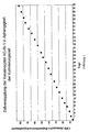
- the doubling time of the keratinocytes is between 1 and 2 days ( illustration 1 ).
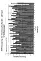
- the cells can be cultivated over numerous passages with an approximately constant doubling rate ( Figure 2 ).
- the present invention is not limited to the keratinocytes KC-BI-1 alone, but it is possible for a person skilled in the art to carry out the invention under appropriate conditions with all keratinocytes according to claim 1.
- non-immortalized in reference to the present invention means that primarily isolated keratinocytes, as well as the keratinocytes cultured here, are not spontaneously transformed and / or molecular biological, chemical or physical methods known in the art have been transformed. The latter means that the cells can not be incubated using e.g. viral factors or sequences, chemically mutagenic substances or e.g. treated by irradiation or a combination of different methods.
- Keratinocytes that are "not immortalized” also means that said keratinocytes have no or substantially lower telomerase activity compared to transformed tumor cell lines (Härle-Bachor and Boukamp, 1993) or compared to immortalized cell lines, preferably to the HeLa cell line (cf. , Figure 3 ). This applies in particular also in comparison to the immortalized keratinocyte cell line HaCat.
- non-immortalized further means that said keratinocytes can not be doubled in the absence of fetal calf serum and / or in the absence of feeder cells and / or in the absence of epidermal groth factor (EGF), such as This is possible, for example, with immortalized keratinocytes (Schoop et al., 1999).
- EGF epidermal groth factor
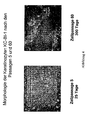
- non-immortalized also means that the said keratinocytes do not change their characteristic phenotype with increasing cell duplication (cf. Figure 4 ).
- non-immortalized also means that the said keratinocytes, after transplantation to nude mice, preferably BALB / c mice, show a normal differentiation profile.
- Normal differentiation potential here means the ability of the keratinocytes to develop into terminally differentiated keratinocytes and to form suprabasal epidermal layers and a stratum corneum corresponding to autologous keratinocytes.
- the invention also relates to keratinocytes which are not immortalized and can be doubled at least 200 times by in vitro cell culture methods.
- the invention further relates to keratinocytes which are not immortalized and can be duplicated at least 250 times by in vitro cell culture techniques.
- the invention additionally also relates to keratinocytes which are not immortalized and can be at least 300 double times by in vitro cell culture methods.
- the present invention enables the beneficial propagation of said keratinocytes from only one donor. Starting from one donation, for example, after 150 cell replications 1044, after 250 cell replications and 10 77 cells prepared in 1090 after 300 cell doublings.
- the invention makes it possible for the first time to provide large quantities of standardized cell material for the production of biologically active wound dressings of consistent and controllable quality.
- a corresponding amount of standardized cell material can be propagated, for example, starting from a cryopreserved cell bank, which in turn is produced from keratinocytes having the properties according to the invention.
- the present invention overcomes significant disadvantages, which currently consists in the limited passability of the previously known keratinocytes.
- the invention further relates to a product which consists of a carrier which is coated with the keratinocytes according to the invention.
- “Coated” in the sense of the invention means that the carrier is partially or completely overgrown on its surface with the keratinocytes according to the invention.
- a partially overgrown carrier is particularly suitable because a shorter culture time is needed until said carrier can be used for wound treatment.
- a carrier suitable for the purposes of the invention is characterized in that it is a biocompatible carrier material which can be used for the preparation of a medicament.
- a biocompatible carrier material which can be used for the preparation of a medicament.
- Useful are, for example, hydrophobic biocompatible carrier materials, such as those in WO 91/13638 described. However, carrier materials with predominantly hydrophilic properties can also be used.
- a preferred embodiment of the present invention is the use of support materials consisting of a polymer of esterified hyaluronic acid.
- it is a polymer of esterified hyaluronic acid, consisting of a perforated polymer film with a defined geometry.
- the polymer film in this case has, for example, a thickness of 10 to 500 .mu.m and is perforated with holes with a size between 10 and 1000 .mu.m, wherein the holes have a defined and constant size and form an ordered series, wherein they by a constant distance of 50 to 1000 microns are separated.
- a corresponding foil is in EP 0 462 426 described.
- Perforated support materials are particularly well suited because they do not require directional application of the biologically active wound dressing to the wound.
- Example 3 describes the exemplary preparation of a perforated carrier matrix of esterified hyaluronic acid with defined geometry overgrown with the keratinocytes according to the invention.
- the carrier matrix is a product marketed in Germany under the product name "Laserskin” by Fidia Advanced Biopolymers Ltd., Abano Terme, Italy.
- this carrier material for producing a biologically active wound dressing in combination with keratinocytes has already been demonstrated on the animal model (Lam et al., 1999) and on humans (Harris et al., 1999).
- the hyaluronic acid ester matrix has a positive effect on angiogenesis and collagen production.
- the wound dressings with hyaluronic acid ester as matrix currently used are coated with autologous keratinocytes and / or complex skin equivalents of keratinocytes and fibroblasts. For them, therefore, the above-described disadvantages of the prior art apply. This disadvantage can be overcome in particular by the use of the advantageous allogeneic keratinocytes according to the invention.
- a further preferred embodiment of the invention relates to a product consisting of the keratinocytes according to the invention together with resorbable polymers consisting of polyesters, polycarbonates, polyanhydrides, polyorthoesters, polydepsipeptides, polyether esters, polyamino acids or polyphosphazenes, in particular of poly (L-lactide), poly (D, L-lactide), poly (L-lactide-co-D, L-lactide), poly (glycolide), poly (L-lactide-co-glycolide), poly (L-lactide-co-trimethylene carbonate), or poly (dioxanone).
- the said polymers are both perforated and unperforated.
- the present invention also relates to a method for cryopreserving the keratinocytes according to the invention at a temperature of -20 ° C to -196 ° C, preferably at a temperature below -180 ° C.
- the said keratinocytes can be frozen according to standardized methods and familiar to a person skilled in the art.
- cryoprotectant can be used, inter alia, DMSO.
- other cryoprotectants such as glycerol, hydroxyethyl starch or a combination of these two and a combination of these with DMSO.
- Corresponding methods are exemplary in WO 96/24018 . US 5,891,617 or US 5,298,417 executed.
- the present invention also provides the cryopreservation of the coated with the keratinocytes according to the invention carrier, characterized in that, the keratinocytes with appropriate carrier at a temperature of -20 ° C to -196 ° C, preferably at -60 ° C to - 80 ° C. cryopreserved.
- Cryopreservation has the advantage of being able to store the product which can be produced in large quantities and thus of being randomly tested for its uniform quality prior to its medical use.
- the storage also makes it possible for the said wound dressings to be available for medical purposes at short notice.
- cryoprotectant for the product according to the invention is exemplified hydroxyethyl starch in a concentration of 7-13% (w / w).
- DMSO or glycerol as well as a combination of different cryoprotectants, in particular from hydroxyetyl starch, DMSO and / or glycerol.
- trehalose as a cryoprotectant is possible.
- the product according to the invention After a rapid temperature reduction of 37 ° C to -5 to -10 ° C, preferably -6 to -8 ° C within 2-5 min, the product according to the invention of carrier and keratinocytes at a corresponding temperature for 15-30 min, preferably for Equilibrated for 23-26 minutes. Subsequently, the product is frozen at a rate of ⁇ 1 ° C / min, preferably from 0.2 to 0.6 ° C / min, more preferably from 0.4 ° C / min to a temperature of e.g. - cooled down to 60 to - 80 ° C.
- Example 4 describes, by way of example, the cryopreservation of a carrier matrix of hyaluronic acid ester coated with KC-BI-1.
- other methods of cryopreservation eg those in WO 95/707611 .
- WO 96/24018 e.g., EP 0 296 475 however, the enumeration is not exhaustive but merely indicates that methods for the cryopreservation of products consisting of biocompatible carriers and keratinocytes belong to the current state of the art.
- the present invention also relates to the medical use of the keratinocytes of the invention described herein and / or the described product of said keratinocytes and a carrier, in particular for use in the treatment of wounds.
- One embodiment of the invention is the use of the keratinocytes according to the invention and / or the product of said keratinocytes and a carrier in the treatment of burns and / or ulcers.
- the treatable burns are preferably second degree burns, and ulcers are preferred chronic non-healing leg ulcers of the type ulcus cruris, preferably ulcus cruris or diabetic ulcers, and also decubital ulcers.
- the medical use includes a combined and / or complementary use of said keratinocytes and / or the product of keratinocytes and carriers according to the invention with conventional therapies known to those skilled in the art having a positive effect on wound healing.
- a combined and / or complementary use of one or more other substances with positive effects for wound healing is meant here.
- the invention also relates to the use of the keratinocytes according to the invention and / or the product of a biocompatible carrier and said keratinocytes for the production of a medical product for the treatment of wounds, in particular for the treatment of burns and / or ulcers, such as for the treatment of secondary burns Grades, ulcus cruris ( venosum ), diabetic ulcers or pressure ulcers.
- the invention furthermore also provides a method for treating said wounds, the method being characterized in that the keratinocytes according to the invention and / or the product according to the invention comprising keratinocytes and carriers are / is applied to the wounds to be treated. Said keratinocytes and said product can be used either fresh or after cryopreservation. A corresponding method for wound treatment is described under Example 5.
- Feeder cells can be used immediately or within 24 hours.
- Incubation of the cells is carried out at 35-39 ° C, preferably at 37 ° C.
- the humidity is> 90%, preferably 95% and the CO 2 concentration is 5-9%.
- the keratinocytes are subcultured at a maximum confluency of 80%.
- the cell culture supernatant is discarded.
- the feeder cells are rinsed twice with 0.02% EDTA (2-10 ml) and incubated for 5-10 min at 37 ° C followed by tapping it (or shaking) detached from the tissue culture flask. Subsequently, after a trypsin / EDTA treatment (0.05% / 0.01%, 1-6 ml), the keratinocytes are gently detached by tapping for 5-10 min at 37 ° C. If necessary, the remaining cells are gently detached with a cell scraper.
- the trypsin / EDTA solution is neutralized by addition of K / 1 medium and the cells are separated by gently pipetting up and down. The cell sowing takes place with the number of cells given under 2.2. The cell culture medium K / 1 is then changed on day 3 every two days.
- the cultivation of the cells can also be carried out starting from a fresh biopsy material, for example starting from the epidermal portion of a foreskin.
- the primary isolation of the undifferentiated, proliferating keratinocytes can be carried out according to the method described by Rheinwald and Green in 1975.
- feeder cells As feeder cells, the cells described in Example 2 can be used.
- the use of other, lethal fibroblasts is also conceivable, preferably the use of other murine fibroblasts, more preferably derivatives of the cell line 3T3.
- Murine 3T3 fibroblasts (eg ATCC CCL 92, 3T3-Swiss albino, contact-inhibited fibroblasts ) available from American Type Culture Collection (ATCC), 10801 University Boulevard, Manassas, VA, USA, DMEM + 10% fetal calf serum ( FCS ) ; PBS; 0.2% trypsin solution; 0.04% EDTA solution; Cell culture bottles (T-flasks): 25 cm 2 , 80 cm 2 , 175 cm 2
- ATCC American Type Culture Collection
- FCS fetal calf serum
- PBS 0.2% trypsin solution
- 0.04% EDTA solution Cell culture bottles
- the cells are examined daily for their growth.
- the cell density should not exceed a maximum confluency of 70-80%.
- the Subcultivation takes place according to experience every 2 to 4 days. For this, the medium is discarded and the cells are washed with a suitable amount of a 1: 2 mixture of EDTA and trypsin (0.5-5 ml). Subsequently, the detaching cells are taken up in 3.5-20 ml of medium. The cells are re-seeded at a density of 10 3 to 10 4 cells / cm 2 .
- the irradiation of the feeder cells takes place with a dose of approximately 60 Gy (6000 rad in a 137 Cs source).
- Both the irradiated and the unirradiated cells can be cryopreserved according to standard protocols in liquid nitrogen and stored for a long time.
- Example 3 Method of Coating a Carrier Matrix, here Laserskin, with Keratinocytes of Culture KC-BI-1 (DSM ACC2514)
- the production of the biologically active wound healing dressing according to the invention is described below by way of example.
- the wound healing dressing described here consists of the inventive keratinocytes KC-BI-1 and Laserskin, a bioresorbable carrier matrix of hyaluronic acid ester.
- the invention is not limited to the combination described here. Rather, all keratinocytes can be used for coating, which have the inventive properties according to claims 1-8.
- suitable carrier matrices are conceivable, provided it is a biocompatible carrier material that can be used for the manufacture of a drug.
- a biocompatible carrier material that can be used for the manufacture of a drug.
- Useful are, for example, hydrophobic biocompatible carrier materials, such as those in WO 91/13638 described.
- support materials with predominantly hydrophilic properties can also be used.
- a further preferred embodiment of the invention is the use of the keratinocytes according to the invention together with resorbable polymers consisting of polyesters, polycarbonates, polyanhydrides, polyorthoesters, polydepsipeptides, polyether esters, polyamino acids or polyphosphazenes, in particular of poly (L-lactide), poly (D, L lactide), poly (L-lactide-co-D, L-lactide), poly (glycolide), poly (L-lactide-co-glycolide), poly (L-lactide-co-trimethylene carbonate) or poly (L-lactide-co-trimethylene carbonate). dioxanone) and the use of perforated films consisting of said polymers.
- resorbable polymers consisting of polyesters, polycarbonates, polyanhydrides, polyorthoesters, polydepsipeptides, polyether esters, polyamino acids or polyphosphazenes, in particular
- K / 1 medium see Example 1
- PBS, 0.04% EDTA diluted to 0.02% with PBS
- Trypsin / EDTA 0.05% / 0.01%
- sterile Roux dishes T 25 cm 2 , T 80 cm 2 , T 175 cm 2, ), Laserskin (Fidia Advanced Biopolymers Sr1, Abano Terme, Italy) in 144x21 Petri dishes (145 cm 2 area); 3T3 feeder cells; keratinocytes according to the invention such as KC-BI-1
- Irradiated feeder cells eg the murine 3T3 fibroblasts performed under Example 2; keratinocytes according to the invention from the strain stock; 8.5cm x 8.5cm laser cucumber pieces in a Petri dish (finished product); K / 2 medium
- the feeder cells prepared according to Embodiment 2 are applied to Laserskin at a seeding density of about 15,000 to 25,000 cells / cm 2 (equivalent to about 3x10 6 cells / petri dish).
- the Petri dish is then incubated at> 90% humidity and 5-11% CO 2 , preferably 7-9% incubated in a 37 ° C incubator incubated at 35 to 37 ° C.
- the keratinocytes are sown either on the same day or at the latest on the next day (after 24 hours) on the feeder cell lawn.
- the keratinocytes according to the invention are used to produce the biologically active wound healing dressing.
- the subculturing of the keratinocytes can be carried out by way of example as described below:
- the subconfluent cultures are rinsed once with 0.02% EDTA (80 cm 2 Roux cup: 8 ml, 175 cm 2 Roux cup: 10 ml).
- the feeder cells are incubated with 0.02% EDTA for 5-10 min at 37 ° C (80 cm 2 Roux cup: 8 mL, 175 cm 2 Roux cup: 10 ml). and detached by careful shaking.
- the keratinocytes are dissolved according to embodiment 1 with a trypsin / EDTA mixture (0.05% / 0.01%) (80 cm 2 Rouxschale: 2-3 mL, 175 cm 2 Rouxschale: 5-6 ml), then added to Zellkukturmendium (80 cm 2 Roux cup: 7-8 mL, 175 cm 2 Roux cup: 14-15 ml) and separated by careful up and down pipetting.
- a trypsin / EDTA mixture 0.05% / 0.01%
- the keratinocytes according to the invention are applied to the laser skin film prepared with feeder cells at a seed density of about 15,000 to 25,000 cells / cm 2 (corresponding to about 3 ⁇ 10 6 cells / petri dish). Subsequently, the cells are incubated to a confluency of 30-100%, preferably 80-100% at 35-39 ° C., preferably at 37 ° C. The relative humidity is> 90%, preferably 95% and the CO 2 concentration is 5-9%.
- Example 4 Process for the cryopreservation of the biologically active wound healing dressing according to the invention
- the product according to the invention can be converted into suitable Containers, eg weldable PP bags, can be frozen in a controlled manner.
- suitable Containers eg weldable PP bags
- culture medium is carefully removed and replaced by 20 ml of 2-6 ° C cold freezing K / 2.
- the product is then packaged sterile and frozen according to the following program:
- the product After a rapid temperature reduction to -5 to -10 ° C., preferably -6 to -8 ° C. within 2-5 minutes, the product is equilibrated at the appropriate temperature for 15-30 minutes, preferably for 23-25 minutes. Subsequently, the product with a freezing rate of ⁇ 1 ° C / min, preferably from 0.2 to 0.6 ° C / min, more preferably from 0.4 ° C / min to a temperature of z.b. -60 to -80 ° C cooled down. The product is stored at -60 to -80 ° C.
- K / 1 growth medium (see Example 1) is added with 7-13% (w / w) hydroxyethyl starch.
- Example 5 Exemplary Use of a Carrier Matrix Coated with the Keratinocytes According to the Invention for Covering Wounds Using the Example of Ulcus Cruris Venosum
- the culture is rinsed one or more times with a suitable amount, preferably 30 ml, K / 3 transport medium.
- the biologically active wound healing dressing is transported in a suitable amount, preferably in 20 ml K / 3 transport medium.
- the headspace of the Petri dish is briefly fumigated with an air mixture of 5-10% CO 2 , sealed with an adhesive tape, for example Parafilm, and immediately taken to the clinic in a transport box.
- Growth medium K / 1 (see Example 1) without fetal calf serum ( FCS ).
- FCS fetal calf serum
- simple physiological salt solutions for example based on phosphate borate, for example PBS, or else HEPES (N-2-hydroxyethylpiperazine-N'-2-ethanesulfonic acid) or MES base ([2-N-1-one] morpholino] ethansufonklad).
- Cryopreserved wound dressings can typically be delivered to dry ice in the clinics. But it is also conceivable another form of transport, provided that the Wundheilverbixe are transported at a temperature of below - 60 ° C.
- cryopreserved wound healing bandages are thawed quickly. Subsequently, the freezing medium is removed and the dressing mixed with K / 3 transport medium (see above) or other suitable physiological solution, e.g. Ringer solution) rinsed one to several times.
- K / 3 transport medium see above
- suitable physiological solution e.g. Ringer solution
- the dressing is then applied to the wound.
- a directed application of the wound healing dressing with the cells in the direction of the wound is required.
- perforated supports which allow bilateral overgrowth of the carriers with keratinocytes e.g., Laserskin
- the treatment can be repeated several times.
- KC-BI-1 cells were subcultured by the above-described method according to the invention over several passages.
- Cells from passage 4, 13, and 121 were then subjected to genetic analysis, with the length polymorphism of 15 different loci examined (CSF1PO, D13S317, D16S539, D18S51, D21S11, D3S1358, D5S818, D7S820, D8S1179, FGA, Penta D, Penta E, TH01, TPOX and vWA).
- the analysis was carried out according to a method known in the art.
- the corresponding alleles were amplified using a test for determining paternity (PoewerPlex® 16 system) from Promega, (Mannheim, Germany) in accordance with the manufacturer's instructions.
- the alleles can be identified by determining the fragment length (length end standard ILS 600 is part of the above-mentioned kit). Information on allelic frequencies in the population can be found in the corresponding tables.
- the analysis revealed a match of all alleles at all loci for all analyzed cell passages.
- the data thus permit a genetic assignment of the KC-BI-1 cells. From the allele frequencies an assignment probability of> 99.999% was determined.
Landscapes
- Health & Medical Sciences (AREA)
- Life Sciences & Earth Sciences (AREA)
- Engineering & Computer Science (AREA)
- Chemical & Material Sciences (AREA)
- Biomedical Technology (AREA)
- General Health & Medical Sciences (AREA)
- Zoology (AREA)
- Organic Chemistry (AREA)
- Bioinformatics & Cheminformatics (AREA)
- Biotechnology (AREA)
- Genetics & Genomics (AREA)
- Wood Science & Technology (AREA)
- Veterinary Medicine (AREA)
- Animal Behavior & Ethology (AREA)
- Public Health (AREA)
- Epidemiology (AREA)
- Dermatology (AREA)
- Cell Biology (AREA)
- Microbiology (AREA)
- Biochemistry (AREA)
- General Engineering & Computer Science (AREA)
- Materials Engineering (AREA)
- Medicinal Chemistry (AREA)
- Chemical Kinetics & Catalysis (AREA)
- Botany (AREA)
- Hematology (AREA)
- Oral & Maxillofacial Surgery (AREA)
- Transplantation (AREA)
- General Chemical & Material Sciences (AREA)
- Pharmacology & Pharmacy (AREA)
- Nuclear Medicine, Radiotherapy & Molecular Imaging (AREA)
- Materials For Medical Uses (AREA)
- Micro-Organisms Or Cultivation Processes Thereof (AREA)
- Medicines Containing Material From Animals Or Micro-Organisms (AREA)
- Medicinal Preparation (AREA)
- Medicines That Contain Protein Lipid Enzymes And Other Medicines (AREA)
- Pharmaceuticals Containing Other Organic And Inorganic Compounds (AREA)
Abstract
Description
- Die Erfindung betrifft neue in vitro kultivierbare Keratinozyten so wie deren vorteilhafte Verwendung zur Herstellung eines Erzeugnisses, welches zur Behandlung von akuten und chronischen Wunden verwendet werden kann. Die Erfindung erfolgte somit auf dem Gebiet der Medizin, speziell auf dem Gebiet der Wundheilung mittels Tissue Engineering.
- Allogene Keratinozyten werden seit längerem erfolgreich zur Behandlung von Wunden, insbesondere von Geschwüren und/oder Verbrennungen eingesetzt (Maier, 1993; Schönfeld et al., 1993). Wunden, die sich gegenüber herkömmlichen Therapien über einen lange Zeitraum therapieresistent zeigen, lassen sich unter Verwendung von allogenen Keratinozyten sehr oft erfolgreich therapieren (Phillips and Gilchrest, 1993; Beele et al., 1991; Leigh et al., 1991; Lindgren et al, 1998). Zu Beginn war das Augenmerk bei der Verwendung allogener Keratinozyten vornehmlich auf den Hautersatz, d.h. auf die Regeneration der transplantierten Epidermis gerichtet. Es zeigte sich jedoch bald, dass der Therapieerfolg primär auf der Stimulation der körpereigenen Reepithelisierung beruht und nicht auf einem "Anwachsen" des allogenen Zelltransplantats in der Wunde. Zahlreiche Untersuchungen ergaben letztlich, dass die transplantierten Keratinozyten nur eine bestimmte Zeit in der Wunde verbleiben und dann von dem Körper klinisch nicht sichtbar eliminiert werden (Kawai et al., 1993). Die Stimulation der körpereigenen Wundheilung erfolgt also primär über eine räumlich und zeitlich optimale Freisetzung von einer Vielzahl an unterschiedlichen Wachstumsfaktoren, Zytokinen, Extrazellulärer Matrix (ECM), kleineren Moleküle und Proteasen durch die allogenen Keratinozyten (Lang et al., 1996; Marcusson et al., 1992). Die Komplexität und Vielzahl der freigesetzten Faktoren begründet hierbei den therapeutischen Vorteil gegenüber herkömmlichen Einzeltherapien, z.B. mit isolierten Wachstumsfaktoren. Neben dem Haupteffekt der Reepithelisierung treten durch Behandlung mit Keratinozyten aber auch makroskopisch und mikroskopisch nachweisbare Veränderungen im Granulationsgewebe auf (Lang et al., 1996). Ein weiterer, für den Patienten wohltuender und oft beschriebener Effekt der Keratinozyten betrifft die ausgeprägte Analgesie nach Transplantation (Schönfeld et al., 1993).
- Für die Wundheilung ist die Verwendung undifferenzierter, teilungsaktiver Keratinozyten vorteilhaft, denn teilungsaktive Keratinozyten scheinen über ein für die Wundheilung begünstigendes komplexes Sezernierungsprofil zu verfügen. Mit Ausnahme der Stammzellen verlieren undifferenzierte Keratinozyten in vivo ihr Proliferationspotential im Zuge der natürlicher Ausdifferenzierung bereits nach wenigen Zellteilungen, ein Prozess der bei der in vitro Kultivierung von Keratinozyten bisher immer beobachtet wurde (Barrandon and Green, 1987). Folglich lassen sich die bisher für die Wundheilung besonders geeigneten teilungsaktiven Keratinozyten nur endlich in vitro kultivieren, was ihre medizinische Verwendung aufwendig und kostenintensiv macht.
- Die allogenen Keratinozyten werden entweder direkt in Form sog. "Keratinozyten-Sheets", bestehend aus einem mehrschichtigen, enzymatisch abgelösten Zellverbund oder zusammen mit biokompatiblen Trägern unter der Bezeichnung "biologisch aktive Wundheilverbände" therapeutisch eingesetzt. Letzteres hat den Vorteil, dass die Zellen nicht enzymatisch vorbehandelt werden müssen, indem sie vor ihrer Verwendung vom Untergrund der Kulturschale abgelöst werden. Darüber hinaus ermöglicht die Verwendung von biokompatibler Membranen als Träger zur Kultivierung der Keratinozyten (
EP 0 462 462 ,US 5,658,331 ;US 5,693,332 ;EP 0518 920 ) eine zeitlich frühere Fertigstellung der biologisch aktiven Wundheilverbände, da die Zellen keinen geschlossen Zellverband mehr ausbilden müssen. Es lassen sich bereits subkonfluent bewachsene Träger zur Wundbehandlung einsetzten. Neben einer früheren Verfügbarkeit hat die Verwendung subkonfluenter Zellverbände zusätzlich den Vorteil, dass entsprechend kultivierte Keratinozyten weniger stark ausdifferenziert sind und somit die vorteilhaft für Behandlung von Wunden sind. - Keratinozyten lassen sich ohne nachteiligen Effekt für die Wundheilung direkt oder in Verbindung mit biokompatiblen Trägermaterialien bei Temperaturen zwischen -30 und -196°C, bevorzugt jedoch zwischen -70 und -90°C kryokonservieren (De Luca et al., 1992; Teepe et al., 1993). Dies hat ihre therapeutische Nutzbarkeit weiter verbessert, da sich biologisch aktive Wundheilverbände im gewissen Umfang auf Vorrat herstellen lassen.
- Die dem Stand der Technik bekannten Keratinozyten sowie aus diesen hergestellte biologisch aktive Wundheilverbände verfügen derzeit über gewisse Nachteile, deren Überwindung Ziel der vorliegenden Erfindung war.
- Ein offensichtlicher Nachteil der bisher bekannten Keratinozyten primären Ursprungs besteht darin, dass sich die besagten Zellen durch in vitro Zellkulturverfahren nur wenige Zellgenerationen verdoppeln lassen ohne dabei ihre hohe Teilungsaktivität und somit ihre bevorzugte Eignung zur Herstellung biologisch aktiver Wundheilverbände zu verlieren. Nach dem Stand der Technik ist es dem Fachmann lediglich möglich die Zahl der Zellen mittels in vitro Kultivierung um etwa das 103 bis 104-fache zu vermehren (Tanczos et al., 1999).
- Ein weiterer, hierdurch bedingter Nachteil besteht darin, dass die begrenzte in vitro Kultivierbarkeit der besagten Keratinozyten eine häufige und aufwändige Neuisolierung erforderlich macht; was wiederum zur Folge hat, dass das gewonnene und vermehrte Zellmaterial nicht einheitlich ist und somit die hieraus hergestellten Wundheilverbände mit hoher Wahrscheinlichkeit Qualitätsunterschiede aufweisen oder zumindest aufweisen können.
- Ein weiterer Nachteil besteht darin, dass mit der Neuisolierung der Keratinozyten aus verschiedenen Spendern ein erhöhte Infektionsrisiko für den Empfänger des Wundheilverbandes besteht. Zu denken ist hier beispielsweise an ein erhöhtes HIV- oder Hepatitis-Risiko.
- Gegenstand der Erfindung sind Keratinozyten dadurch gekennzeichnet, dass sie nicht immortalisiert sind und sich durch in vitro Zellkulturverfahren mindestens 150 mal verdoppeln lassen und dass es sich dabei um Zellen aus der Kultur KC-BI-1 (DSM ACC 2514) handelt, oder um hiervon abgeleitete Keratinozyten, wobei die besagten Keratinozyten
- a. nicht in Abwesenheit von Fötalem Kälber Serum und/oder
- b. nicht in Abwesenheit von Feeder-Zellen und/oder
- c. nicht in Abwesenheit von Epidermalem Wachstumsfaktor (EGF) verdoppelt werden können,
- Bei den erfindungsgemäßen Keratinozyten handelt es sich um primär aus einem Spender isolierte und in vitro kultivierbare Keratinozyten, wobei die Isolierung und anfängliche Kultivierung von einem Fachmann beispielsweise entsprechend dem von Rheinwald und Green 1975 beschriebenen Verfahren ausgeführt werden kann.
- Die Erfindung betrifft Keratinozyten, die aus dem epidermalen Anteil einer Vorhaut isoliert werden. Keratinozyten humanen Ursprungs, der Kultur KC-BI-1, wurden am 27.Juni 2001 bei der DSMZ-Deutsche Sammlung von Mikroorganismen und Zellkulturen GmbH, Braunschweig, Deutschland unter der Hinterlegungsnummer DSM ACC2514 für die Zwecke von Patentverfahren gem. Budapester Vertrag hinterlegt. Unter die Erfindung fallen auch solche Keratinozyten, die sich von der Kultur KC-BI-1 (DSM ACC2514) ableiten. Gegenstand der Erfindung sind somit auch alle Zellen und Kulturen, die durch Subpassagieren und/oder Subklonieren der ursprünglichen Kultur KC-BI-1 generiert werden und/oder generiert werden können.
- Die erfindungsgemäße Kultivierung der Keratinozyten ist beispielhaft für die Keratinozyten KC-BI-ldargestellt (Beispiel 1). Vorteilhaft für die Kultivierung ist die Verwendung des in Beispiel 1 angegebenen komplexen Mediums sowie die Verwendung von Feeder-Zellen (engl. feeder cells, deutsch: Ammenzellen), bevorzugt die Verwendung von letal bestrahlten murinen 3T3 Fibroblasten. Der Anteil an fötalem Kälberserum sollte zwischen 2 und 10 % betragen. Die Herstellung der Feeder-Zellen ist dem Fachmann bekannt, und kann z.B. gemäß dem in Beispiel 2 angegebenen Verfahren durchgeführt werden. Besonders vorteilhaft ist hierbei die Subkultivierung der erfindungsgemäßen Keratinozyten bei einer maximalen Konfluenz von 80%. Die Keratinozyten lassen sich bei 35 bis 38°C, bevorzugt bei 37°C, einer Luftfeuchtigkeit von > 90%, bevorzugt 95% und einer CO2-Sättigung von 5 bis 9% kultivieren.
- Mit dem unter Beispiel 1 dargestellten Verfahren beträgt die Verdopplungszeit der Keratinozyten zwischen 1 und 2 Tagen (
Abbildung 1 ). Die Zellen lassen sich hierbei über zahlreiche Passagen mit annähernd konstanter Verdopplungsrate kultivieren (Abbildung 2 ). Die vorliegenden Erfindung ist hierbei allerdings nicht allein auf die Keratinozyten KC-BI-1 beschränkt, vielmehr ist es für einen Fachmann möglich, die Erfindung unter entsprechenden Bedingungen mit allen Keratinozyten gemäß Anspruch 1 auszuführen. - Der Ausdruck "nicht immortalisiert" meint in Bezug auf die vorliegende Erfindung, dass primär isolierte Keratinozyten sowie die hier in Kultur genommenen Keratinozyten nicht spontan transformiert sind und/oder durch dem Stand der Forschung bekannte molekularbiologische, chemische oder physikalische Verfahren transformiert worden sind. Letzteres meint, dass die Zellen weder unter Verwendung von z.B. viralen Faktoren oder Sequenzen, chemisch mutagenen Substanzen oder z.B. durch Bestrahlung oder einer Kombination aus verschiedenen Verfahren behandelt worden sind.
- Keratinozyten, die "nicht immortalisiert sind" meint auch, dass die besagten Keratinozyten im Vergleich zu transformierten Tumorzellinien (Härle-Bachor and Boukamp, 1993) oder im Vergleich zu immortalisierten Zellienen, bevorzugt zu der Zellinie HeLa keine oder eine wesentliche geringere Telomeraseaktivität aufweist (Vgl.
Abbildung 3 ). Dies gilt insbesondere auch im Vergleich zu der immortalisierten Keratinozytenzellinie HaCat. - Der Ausdruck "nicht immortalisiert" meint ferner, dass die besagten Keratinozyten nicht in Abwesenheit von fötalem Kälberserum und/oder nicht in Abwesenheit von Feederzellen und/oder nicht in Abwesenheit von Epidermalem Wachstumsfaktor (englisch: epidermal groth factor, EGF) verdoppelt werden können, wie dies beispielsweise bei immortalisierten Keratinozyten möglich ist (Schoop et al., 1999).
- Der Ausdruck "nicht immortalisiert" meint aber auch, dass die besagten Keratinozyten ihren charakteristischen Phänotyp nicht mit zunehmender Zellverdopplung verändern (Vgl.
Abbildung 4 ). - Ferner meint "nicht immortalisiert" auch, dass die besagten Keratinozyten, nach Transplantation auf Nacktmäuse, bevorzugt BALB/c Mäuse ein normales Differenzierungsprofil zeigen. "Normales Differenzierungspotential" meint hier die Fähigkeit der Keratinozyten, sich zu terminal differenzierten Keratinozyten zu entwickeln und suprabasale epidermale Schichten sowie ein Stratum corneum entsprechend autologer Keratinozyten auszubilden.
- Gegenstand der Erfindung sind auch solche Keratinozyten, die nicht immortalisiert sind und sich mindestens 200 mal durch in vitro Zellkulturverfahren verdoppeln lassen. Die Erfindung betrifft ferner Keratinozyten, die nicht immortalisiert sind und sich mindestens 250 mal durch in vitro Zellkulturverfahren verdoppeln lassen. Die Erfindung betrifft darüber hinaus auch solche Keratinozyten, die nicht immortalisiert sind und sich mindestens 300 mal durch in vitro Zellkulturverfahren verdoppeln lassen.
- Die vorliegende Erfindung ermöglicht die vorteilhafte Vermehrung der besagten Keratinozyten ausgehend von nur einem Spender. Ausgehend von einer Spende lassen sich beispielsweise nach 150 Zellverdopplungen 1044, nach 250 Zellverdopplungen 1077 und nach 300 Zellverdopplungen 1090 Zellen herstellen. Dadurch ermöglicht die Erfindung erstmals die Bereitstellung großer Mengen an standardisiertem Zellmaterial für die Herstellung von biologisch aktiven Wundheilverbänden mit gleichbleibender und kontrollierbarer Qualität. Eine entsprechende Menge an standardisiertem Zellmaterial lässt sich z.B. ausgehend von einer kryokonservierten Zellbank vermehren, die wiederum aus Keratinozyten hergestellt wird, die über die erfindungsgemäßen Eigenschaften verfügen.
- Mit der Verwendung von standardisiertem Zellmaterial als Ausgangsmaterial für die Herstellung von biologisch aktiven Wundheilverbänden wird auch das Infektionsrisiko für die möglichen Empfänger verringert, da die Isolierung der Keratinozyten auf einen Spender beschränkt bleibt.
- Damit überwindet die vorliegende Erfindung wesentliche Nachteile, die zur Zeit in der lediglich eingeschränkten Passagierbarkeit der bisher bekannten Keratinozyten besteht.
- Gegenstand der Erfindung ist darüber hinaus ein Erzeugnis, das aus einem Träger besteht, der mit den erfindungsgemäßen Keratinozyten beschichtet ist. "Beschichtet" im Sinne der Erfindung meint, dass der Träger auf seiner Oberfläche partiell oder vollständig mit den erfindungsgemäßen Keratinozyten bewachsen ist. Ein partiell bewachsener Träger ist besonders geeignet, da eine kürzere Kultivierungszeit benötigt wird bis der besagte Träger zur Wundbehandlung verwendet werden kann.
- Ein im Sinne der Erfindung geeigneter Träger ist dadurch gekennzeichnet, dass es sich um ein biokompatibles Trägermaterial handelt, das zur Herstellung eines Arzneimittels verwendet werden kann. Verwendbar sind beispielsweise hydrophobe biokompatible Trägermaterialien, wie etwa die in
WO 91/13638 - Eine bevorzugte Ausführung der vorliegenden Erfindung besteht in der Verwendung von Trägermaterialien die aus einem Polymer aus veresterter Hyaluronsäure bestehen. Bei einer besonders bevorzugten Ausführung handelt es sich um ein Polymer aus veresterter Hyaluronsäure, bestehend aus einer perforierten Polymerfolie mit definierter Geometrie. Die Polymerfolie hat hierbei beispielweise eine Dicke von 10 bis 500 µm und ist mit Löchern mit einer Größe zwischen 10 und 1000 µm durchlöchert, wobei die Löcher eine definierte und konstante Größe haben und eine geordnete Reihe bilden, worin sie durch einen konstanten Abstand von 50 bis 1000 µm voneinander getrennt sind. Eine entsprechende Folie ist in
EP 0 462 426 beschrieben. Perforierte Trägermaterialien eignen sich besonders gut, da sie kein gerichtetes Auflegen des biologisch aktiven Wundverbandes auf die Wunde erfordern. In Beispiel 3 wird die beispielhafte Herstellung einer mit den erfindungsgemäßen Keratinozyten bewachsenen perforierten Trägermatrix aus veresterter Hyaluronsäure mit definierter Geometrie beschrieben. Bei der Trägermatrix handelt es sich um ein in Deutschland unter dem Produktnamen "Laserskin" vertriebenes Produkt der Firma Fidia Advanced Biopolymers Ltd., Abano Terme, Italien. - Die besonders bevorzugte Eignung dieses Trägermaterials zur Herstellung eines biologisch aktiven Wundverbandes in Kombination mit Keratinozyten wurde bereits am Tiermodell (Lam et al., 1999) und am Menschen gezeigt (Harris et al., 1999). Neben einer verbesserten Migration und Ausdifferenzierung der epithelialen Zellen hat die aus Hyaluronsäureester bestehende Matrix einen positiven Effekt auf die Angiogenese und die Kollagenproduktion. Die zur Zeit verwendeten Wundverbände mit Hyaluronssäureester als Matrix sind allerdings mit autologen Keratinozyten und/oder komplex aufgebauten Hautäquivalenten aus Keratinozyten und Fibroblasten beschichtet. Für sie gelten somit die oben beschriebenen Nachteile aus dem Stand der Technik. Dieser Nachteil kann insbesondere durch die Verwendung der vorteilhaften erfindungsgemäßen allogenen Keratinozyten überwunden werden.
- Eine weitere bevorzugte Ausführungsform der Erfindung betrifft ein Produkt bestehend aus den erfindungsgemäßen Keratinozyten zusammen mit resorbierbaren Polymeren, bestehend aus Polyester, Polycarbonate, Polyanhydride, Polyorthoester, Polydepsipeptide, Polyetherester, Polyaminosäuren oder Polyphosphazene, insbesondere aus Poly(L-lactid), Poly(D,L-lactid), Poly(L-lactid-co-D,L-lactid), Poly(glycolid), Poly(L-lactid-co-glycolid), Poly(L-lactid-co-trimethylen-carbonat) oder Poly(dioxanon). Die besagten Polymere sind dabei sowohl perforiert als auch unperforiert.
- Die vorliegende Erfindung betrifft ebenfalls ein Verfahren zur Kryokonservierung der erfindungsgemäßen Keratinozyten bei einer Temperatur von -20°C bis -196°C, bevorzugt bei einer Temperatur unter -180°C. Die besagten Keratinozyten lassen sich nach standardisierten und einem dem Fachmann geläufigen Verfahren einfrieren. Als Kryoprotektant lässt sich u.a. DMSO verwenden. Darüber hinaus ist auch die Verwendung anderer Kryoprotektanten möglich, wie etwa Glycerol, Hydroxyethylstärke oder eine Kombination aus diesen beiden sowie eine Kombination dieser mit DMSO. Entsprechende Verfahren sind beispielhaft in
WO 96/24018 US 5,891,617 oder auchUS 5,298,417 ausgeführt. - Gegenstand der vorliegenden Erfindung ist auch die Kryokonservierung der mit den erfindungsgemäßen Keratinozyten beschichteten Träger, dadurch gekennzeichnet dass, die Keratinozyten mit entsprechenden Träger bei einer Temperatur von -20°C bis -196°C, bevorzugt bei -60°C bis - 80°C kryokonserviert werden. Die Kryokonservierung hat den Vorteil, dass das in großen Mengen herstellbare Erzeugnis gelagert werden und somit vor seiner medizinischen Verwendung stichprobenartig auf seine einheitliche Qualität untersucht werden kann. Die Lagerung ermöglicht letztendlich auch dass die besagten Wundheiverbände kurzfristig für medizinische Zwecke verfügbar sind.
- Als Kryoprotektant für das erfindungsgemäße Erzeugnis eignet sich beispielhaft Hydroxyethylstärke in einer Konzentration von 7-13% (w/w). Möglich ist aber auch die Verwendung von DMSO oder Glycerol sowie eine Kombination aus verschiedenen Kryoprotektanten, insbesondere aus Hydroxyetylstärke, DMSO und/oder Glycerol. Ferner ist auch die Verwendung von Trehalose als Kryoprotektant möglich.
- Nach einer schnellen Temperaturerniedrigung von 37°C auf -5 bis -10°C, bevorzugt -6 bis -8°C innerhalb von 2-5 min wird das erfindungsgemäße Produkt aus Träger und Keratinozyten bei entsprechender Temperatur für 15-30 min, bevorzugt für 23-26 min äquilibriert. Anschließend wird das Erzeugnis mit einer Einfrierrate von < 1°C/min, bevorzugt von 0,2 bis 0,6°C/min besonders bevorzugt von 0,4°C/min auf eine Temperatur von z.B. - 60 bis - 80°C herabgekühlt.
- Beispiel 4 beschreibt beispielhaft die Kryokonservierung einer mit KC-BI-1 beschichteten Trägermatrix aus Hyaluronsäureester. Es ist aber auch möglich, dass andere Verfahren zur Kryokonservierung angewendet werden, z.B. die in
WO 95/707611 WO 96/24018 EP 0 296 475 beschriebenen Verfahren, wobei die Aufzählung nicht abschließend zu verstehen ist, sondern lediglich darauf verweist, dass Verfahren zur Kryokonservierung von Produkten bestehend aus biokompatiblen Trägern und Keratinozyten zu dem gegenwärtigen Stand der Technik gehören. - Die vorliegende Erfindung bezieht sich auch auf die medizinische Verwendung der hier beschriebenen erfindungsgemäßen Keratinozyten und/oder des beschriebenen Produkts aus besagten Keratinozyten und einem Träger, insbesondere auf die Verwendung zur Behandlung von Wunden. Eine Ausführungsform der Erfindung stellt die Verwendung der erfindungsgemäßen Keratinozyten und/oder des Produkts aus besagten Keratinozyten und einem Träger bei der Behandlung von Verbrennungen und/oder Geschwüren dar. Bei den behandelbaren Verbrennungen handelt es sich bevorzugt um Verbrennungen zweiten Grades, bei den Geschwüren bevorzugt um chronische schlecht heilenden Unterschenkelgeschwüre des Typs Ulcus cruris, bevorzugt Ulcus cruris venosum oder auch um diabetischen Ulcera, zusätzlich auch um Dekubitus.
- Die medizinische Verwendung schließt eine kombinierte und/oder ergänzende Verwendung der besagten Keratinozyten und/oder des erfindungsgemäßen Produkts aus Keratinozyten und Träger mit klassischen dem Fachmann bekannten Therapien mit positivem Effekt für die Wundheilung mit ein. Gemeint ist hierbei eine kombinierte und/oder ergänzende Verwendung eines oder mehreren anderen Stoff(en) mit positiver Wirkung für die Wundheilung. Beispielhaft zu nennen ist hier die ergänzende und oder kombinierte Behandlung von Ulcus cruris venosum mit Hydrokolloidverbänden und/oder die zusätzliche Verwendung von antimikrobiellen Substanzen, beispielsweise die Gabe von Antibiotika.
- Gegenstand der Erfindung ist ebenfalls die Verwendung der erfindungsgemäßen Keratinozyten und/oder des Produkts aus einem biokompatiblen Träger und den besagten Keratinozyten zur Herstellung eines medizinischen Erzeugnisses zur Behandlung von Wunden, insbesondere zur Behandlung von Verbrennung und/oder Geschwüren, wie z.B. zur Behandlung von Verbrennungen zweiten Grades, Ulcus cruris (venosum), diabetische Ulcera oder Dekubitus.
- Gegenstand der Erfindung ist ferner auch ein Verfahren zur Behandlung besagter Wunden, wobei das Verfahren dadurch gekennzeichnet ist, dass die erfindungsgemäßen Keratinozyten und/oder das erfindungsgemäße Produkt aus Keratinozyten und Träger auf die zu behandelnden Wunden aufgelegt werden/wird. Besagte Keratinozyten und besagtes Produkt können entweder frisch oder nach Kryokonservierung verwendet werden. Ein entsprechendes Verfahren zur Wundbehandlung ist unter Beispiel 5 beschrieben.
-
-
Abb. 1 : Zellverdopplung der Keratinozyten KC-BI-1 in Abhängigkeit der Kultivierungszeit. Dargestellt ist die Anzahl der Zellverdopplungen (CPD =Cumulative Population Doublings) der Keratinozyten KC-BI-1 über eine Kultivierungszeit von 94 Tagen (days). Innerhalb der beobachteten 94 Tagen verdoppelten sich die Zellen ungefähr 75 mal. Dies entspricht einer mittleren Verdopplungszeit von 1,25 Tagen pro Zellverdopplung, oder 12,5 pro10 Tage. -
Abb. 2 : Verdopplung der Keratinozyten KC-BI-1 über einen Zeitraum von 10 Monaten. Dargestellt ist die Zellverdopplung der Keratinozyten KC-BI-1 über 10 Monate, angegeben als Population doublings (PD) in Abhängigkeit der Zellpassagen (Passage) 1-67. -
Abb. 3 : Bestimmung der relativen Telomeraseaktivität. Dargestellt ist die relative Telomeraseaktivität für die Keratinozyten KC-BI-1 nach Passage 1, 12, 18, 40 und 57 im Vergleich zur Aktivität der Zellinie HeLa. Die Abbildung zeigt für die Keratinozyten KC-BI-1 fast keine bzw. lediglich eine geringe Telomeraseaktivität im Vergleich zu der immortalisierten Zellinie HeLa. -
Abb. 4 : Morphologie der Keratinozyten KC-BI-1 nach den Passagen 5 und 60. Die Keratinozyten KC-BI-1 zeigen in der lichtmikroskopischen Darstellung keine morphologischen Unterschiede zwischen Zellpassage 5 (Kultivierungszeit 25 Tage) und Zellpassage 60 (Kultivierungszeit 300 Tage). - Keratinozyten KC-BI-1; bestrahlte 3T3-Mäusefibroblasten (Ammenzellen, engl.:feeder-cells, Herstellung vgl. Beispiel 2); Zellkulturmedium K/-1 (Zusammensetzung siehe unten); EDTA (0,02%); Trypsin/EDTA (0,05%/0,01%); Zellkulturflaschen (T-flask): 25 cm2, 80 cm2, 175 cm2
- Zellen zügig auftauen und in 5-10 ml vorgewärmtes K/1 Medium geben. Eine entsprechende Menge an Feeder-Zellen wird in eine geeignete Zellkulturflasche überführt und mit K/1 Medium ergänzt:
25 cm2 T-flask 0,5x10⁶ Zellen Endvolumen an Medium: 5-6ml 80 cm2 T-flask 1,5x10⁶ Zellen Endvolumen an Medium: 20ml 175cm2 T-flask 3,5x10⁶ Zellen Endvolumen an Medium: 50ml - Die Feeder-Zellen können sofort oder innerhalb von 24 Std. verwendet werden.
- Zellen zügig auftauen und in 5-10 ml vorgewärmtes K/1 Medium geben. Eine entsprechende Menge an Zellen zu den mit Feeder-Zellen vorbereiteten Zellkulturflaschen geben und mit frischem Medium auffüllen:
25 cm2 T-flask 0,15 x 10⁶ Zellen; Endvolumen an Medium: 6 - 10 mL 80 cm2 T-flask 0,4 x 10⁶ Zellen; Endvolumen an Medium: 20 mL 175 cm2 T-flask 1 x 10⁶ Zellen; Endvolumen an Medium: 50 mL - Inkubation der Zellen erfolgt bei 35-39°C, bevorzugt bei 37°C. Die Luftfeuchtigkeit beträgt >90%, bevorzugt 95% und die CO2-Konzentration liegt bei 5-9%. Die Keratinozyten werden bei einer maximalen Konfluenz von 80% subkultiviert.
- Hierzu wird der Zellkulturüberstand verworfen. Die Feeder-Zellen werden nach zweimaligem Spülen mit 0,02% EDTA (2-10ml) und einer Inkubation für 5-10 min bei 37°C durch anschließendes Abklopfen (oder unter Schütteln) von der Zellkulturflasche gelöst. Nachfolgend werden die Keratinozyten nach einer Trypsin/EDTA-Behandlung (0,05%/0,01%, 1-6 ml) für 5-10 min bei 37°C vorsichtig durch Abklopfen gelöst. Falls erforderlich, werden die restlichen Zellen vorsichtig mit einem Zellschaber gelöst. Die Trypsin/EDTA-Lösung wird durch Zugabe von K/1 Medium neutralisiert und die Zellen werden durch vorsichtiges Auf- und Abpipetieren vereinzelt. Die Zellaussaat erfolgt mit der unter 2.2 angegebenen Zellzahl. Das Zellkulturmedium K/1 wird an Tag 3 anschließend alle zwei Tage ausgetauscht.
- Alle Komponenten sowie die Stammlösungen werden nacheinander entsprechend der in Tabelle 1 angegebenen Reihenfolge zusammengegeben. Der Ansatz wird auf 1 liter mit WFI (engl.: water for injection) aufgefüllt. Anschließend wird der pH mit NaOH or HCl auf 7,0 bis 7,2 eingestellt. Die Osmolarität sollte zwischen 320-400 mOsm/kg betragen. Abschließend wird das Medium K/1 sterilfiltriert.
- 13,6mg Trijodothyronine in 1ml 0,1M NaOH lösen und 99ml hinzugeben PBS. Die gebrauchsfertige Lösung 1:100 in PBS verdünnen. Pro Liter Medium wird 1 ml dieser Lösung benötigt.
- 1mg EGF in 100ml WFI lösen. Pro Liter Medium wird 1 ml dieser Lösung benötigt.
- 5g rh-Insulin werden in 0,9L WFI gegeben und der pH wird 6M HCl auf 2,5 eingestellt. Nachdem sich das rh-Insulin gelöst hat, den pH mit 1M NaOH auf 8,0 einstellen. Den Ansatz mit WFI auf 1 Liter auffüllen. Pro Liter Medium wird 1 ml dieser Lösung benötigt.
-
Komponenten Konzentration/L WFI (water for injection) 0,8L DMEM with glutamine 10,035g HAM's F12 2,66g Natriumhydrogencarbonate 3,07g Na-pyruvate 0,11g Apo-Transferrin 5mg Adenine-Hemisulfate 147,4mg Forskolin (soluble in some trops of DMSO) 2,05mg Rh-Insulin-Concentrate 1,0mL Hydrocortison 10mM 0,11mL Trij odothyronine-stocksolution 1,0mL EGF-stocksolution 1,0mL Phenol red 8,1mg FCS (2-10%) 20-100mL 6M HCl as required 40% NaOH as required WFI add to1,0L - Die Kultivierung der Zellen kann aber auch ausgehend von einem frischem Biopsiematerial erfolgen, beispielsweise ausgehend von dem epidermalen Anteil einer Vorhaut. Die Primärisolierung der undifferenzierten, proliferierenden Keratinozyten kann hierbei nach dem von Rheinwald und Green 1975 beschriebenen Verfahren durchgeführt werden.
- Als Feeder-Zellen können die unter Beispiel 2 beschriebenen Zellen verwendet werden. Darüber hinaus ist auch die Verwendung anderer, letaler Fibroblasten vorstellbar, bevorzugt die Verwendung von anderen murinen Fibroblasten, besonders bevorzugt Abkömmlinge der Zellinie 3T3.
- Murine 3T3 Fibroblasten (z.B. ATCC CCL 92, 3T3-Swiss albino, contact-inhibited fibroblasts) zu beziehen bei der American Type Culture Collection (ATCC), 10801 University Boulevar, Manassas, VA, USA, DMEM+10% Fötales Kälberserum (FCS); PBS; 0,2% Trypsin Lösung; 0,04% EDTA Lösung; Zellkulturflaschen (T-flasks): 25 cm2, 80 cm2, 175 cm2
- Zellen zügig auftauen und in 5-10 ml vorgewärmtes Medium geben. DMSO-haltiges Medium nach Zentrifugation entfernen. Zellen in 5-10 ml Medium suspendieren. Nach Bestimmung der Zellzahl werden die Zellen mit einer Dichte von 103 to 104 Zellen/cm2 in geeignete Zellkulturflaschen ausgesät. Die Inkubation erfolgt bei 35 - 39°C, bevorzugt bei 37°C. Die Luftfeuchtigkeit beträgt > 90%, bevorzugt 95% und die CO2-Konzentration liegt bei 5-9%.
- Die Zellen werden täglich auf ihr Wachstum hin untersucht. Die Zelldichte sollte eine maximale Konfluenz von 70-80% nicht überschreiten. Die Subkultivierung erfolgt erfahrungsgemäß alle 2 bis 4 Tage. Hierzu wird das Medium verworfen und die Zellen mit einer geeigneten Menge einer 1:2 Mischung an EDTA und Trypsin (0,5-5 ml) gewaschen. Anschließend werden die sich ablösenden Zellen in 3,5-20 ml Medium aufgenommen. Die Zellen werden mit einer Dichte von 103 to 104 Zellen/cm2 neuausgesät.
- Die Bestrahlung der Feeder-Zellen erfolgt mit einer Dosis von ungefähr 60 Gy (6000 rad in einer 137Cs-Quelle).
- Sowohl die bestrahlten als auch die unbestrahlten Zellen lassen sich nach Standardprotokollen in flüssigen Stickstoff kryokonservieren und über längere Zeit aufbewahren.
- Nachfolgend wird beispielhaft die Herstellung des erfindungsgemäßen biologisch aktiven Wundheilverbandes beschrieben. Der hier beschriebene Wundheilverband besteht aus den findungsgemäßen Keratinozyten KC-BI-1 und Laserskin, einer bioresorbierbaren Trägermatrix aus Hyaluronsäureester.
- Die Erfindung ist dabei nicht auf die hier beschriebene Kombination beschränkt. Vielmehr können alle Keratinozyten zur Beschichtung verwendet werden, die über die erfinderischen Eigenschaften gemäß der Ansprüche 1-8 verfügen.
- Darüber hinaus ist auch die Verwendung anderer geeigneter Trägermatrices denkbar, vorausgesetzt es handelt sich um ein biokompatibles Trägermaterial, das zur Herstellung eines Arzneimittels verwendet werden kann. Verwendbar sind beispielsweise hydrophobe biokompatible Trägermaterialien, wie etwa die in
WO 91/13638 - Eine weitere bevorzugte Ausführung der Erfindung liegt in der Verwendung der erfindungsgemäßen Keratinozyten zusammen mit resorbierbaren Polymeren, bestehend aus Polyester, Polycarbonate, Polyanhydride, Polyorthoester, Polydepsipeptide, Polyetherester, Polyaminosäuren oder Polyphosphazene, insbesondere aus Poly(L-lactid), Poly(D,L-lactid), Poly(L-lactid-co-D,L-lactid), Poly(glycolid), Poly(L-lactid-co-glycolid), Poly(L-lactid-co-trimethylen-carbonat) oder Poly(dioxanon) sowie die Verwendung von perforierten Folien, bestehend aus den besagten Polymeren.
- K/1-Medium (vgl. Beispiel 1); PBS, 0,04% EDTA (wird mit PBS auf 0,02% verdünnt); Trypsin/EDTA (0,05%/0,01%); sterile Rouxschalen (T 25 cm2, T 80 cm2, T 175 cm2,),Laserskin (Fa. Fidia Advanced Biopolymers sr1, Abano Terme, Italien) in 144x21 Petrischalen (145cm2 Fläche); 3T3-Feeder-Zellen; erfindungsgemäße Keratinozyten wie zum Beispiel KC-BI-1
- Bestrahlte Feeder-Zellen, z.B. die unter Beispiel 2 ausgeführten murinen 3T3 Fibroblasten; erfindungsgemäße Keratinozyten aus der Stammhaltung; 8,5cm x 8,5cm große Laserskinstücke in einer Petrischale (Fertigprodukt); K/2-Medium
- Die Feeder-Zellen, hergestellt gemäß des Ausführungsbeispiels 2, werden mit einer Einsaatdichte von ca. 15.000 bis 25.000 Zellen/cm2 (entspricht ungefähr 3x10⁶ Zellen/Petrischale) auf Laserskin aufgebracht. Die Petrischale wird anschließend bei >90% Luftfeuchtigkeit und 5-11 % CO2, vorzugweise 7-9% in einem 37°C Brutschrank inkubiert bei 35 bis 37°C inkubiert. Die Keratinozyten werden entweder am selben Tag oder spätestens am nächsten Tag (nach 24 Std.) auf den Feeder-Zellrasen gesät.
- Mit den erfindungsgemäßen Keratinozyten werden die biologisch aktiven Wundheilverband hergestellt. Die Subkultivierung der Keratinozyten kann beispielhaft wie nachfolgend beschrieben erfolgen: Die subkonfluenten Kulturen werden einmal mit 0,02% EDTA gespült (80 cm2 Rouxschale: 8 mL; 175 cm2 Rouxschale: 10 ml). Anschließend werden die Feeder-Zellen mit 0,02% EDTA für 5-10 min bei 37°C inkubiert (80 cm2 Rouxschale: 8 mL; 175 cm2 Rouxschale: 10 ml). und durch vorsichtiges Schütteln abgelöst.
- Die Keratinozyten werden entsprechend Ausführungsbeispiel 1 mit einem Trypsin/EDTA Gemisch (0,05%/0,01%) gelöst (80 cm2 Rouxschale: 2-3 mL; 175 cm2 Rouxschale: 5-6 ml), anschließend in Zellkukturmendium aufgenommen (80 cm2 Rouxschale: 7-8 mL; 175 cm2 Rouxschale: 14-15 ml) und durch vorsichtiges Auf- und Abpipetieren vereinzelt.
- Auf die mit Feeder-Zellen vorbereitete Laserskin-Folie werden die erfindgungsgemäßen Keratinozyten mit einer Aussatdichte von ca. 15.000 bis 25.000 Zellen/cm2 (entspricht ungefähr 3x10⁶ Zellen/Petrischale) aufgebracht. Anschließend werden die Zellen bis zu einer Konfluenz von 30 - 100%, bevorzugt von 80 - 100% bei 35 - 39°C, bevorzugt bei 37°C inkubiert. Die Luftfeuchtigkeit beträgt > 90%, bevorzugt 95% und die CO2-Konzentration liegt bei 5 - 9%.
- Nachdem die Keratinozyten die Trägermatrix mit einer Konfluenz von 30- 100% vorzugsweise 80-100% bewachsen haben, kann das erfindungsgemäße Produkt in geeigneten Behältnissen, z.B. verschweißbaren PP-Beuteln, kontrolliert eingefroren werden. Hierzu wird Kulturmedium vorsichtig entfernt und durch 20 ml 2-6°C kaltes Einfriermedium K/2 ausgetauscht. Das Produkt wird anschließend steril verpackt und entsprechend dem nachfolgenden Programm eingefroren:
- Nach einer schnellen Temperaturerniedrigung auf - 5 bis - 10°C, bevorzugt -6 bis -8°C innerhalb von 2-5 min wird das Produkt bei entsprechender Temperatur für 15-30 min, bevorzugt für 23-25 min äquilibriert. Anschließend wird das Produkt mit einer Einfrierrate von < 1°C/min, bevorzugt von 0,2 bis 0,6°C/min besonders bevorzugt von 0,4°C/min auf eine Temperatur von z.b. -60 bis -80°C herabgekühlt. Die Lagerung des Produktes erfolgt bei -60 bis -80°C.
- Nachdem die Keratinozyten 30-100%, bevorzugt 80-100% konfluent auf dem Laserskin bewachsen sind, wird die Kultur mit einer geeigneten Menge, vorzugsweise 30 ml, K/3-Transportmedium ein bzw. mehrmals gespült. Transportiert wird der biologisch aktive Wundheilverband in geeigneter Menge, vorzugsweise in 20 ml K/3-Transportmedium. Der Kopfraum der Petrischale wird mit einem Luftgemisch aus 5-10% CO2 kurz begast, mit einem Klebeband z.B. Parafilm verschlossen und sofort in einer Transportbox in die Klinik gebracht.
- Wachstumsmedium K/1 (vgl. Beispiel 1) ohne Fötales Kälber Serum (FCS). Vorstellbar ist aber auch die Verwendung von einfachen physiologischen Salzlösungen etwa auf Phosphat-Borat-Basis, z.B. PBS, oder auch auf HEPES- (N-2-hydroxyelhylpiperazin-N'-2-ethansulfonsäure) oder MES-Basis ([2-N-morpholino]ethansufonsäure).
- Kryokonservierte Wundheilverbände können typischerweise auf Trockeneis in den Kliniken angeliefert werden. Es ist aber auch eine andere Transportform vorstellbar, sofern die Wundheilverbände bei einer Temperatur von unter - 60°C transportiert werden.
- Die kryokonservierten Wundheilverbände werden zügig aufgetaut. Anschließend wird das Einfriermedium entfernt und das Verband mit K/3-Transportmedium (vgl. oben) oder einer anderen geeigneten physiologischen Lösung, z.B. Ringerlösung) ein bis mehrmals gespült.
- Der Verband wird anschließend auf die Wunde aufgelegt. Bei der Verwendung nicht perforierter Trägermaterialien, ist ein gerichtetes Auflegen des Wundheilverbandes mit den Zellen in Richtung der Wunde erforderlich. Die Verwendung von perforierten Trägern, die ein beidseitiges Bewachsen der Träger mit Keratinozyten ermöglichen (z.B. Laserskin), erfordert kein gerichtetes Auflegen des erfindungsgemäßen Wundheilverbandes auf die zu behandelnde Wunde. Je nach Therapieerfolg kann die Behandlung mehrfach wiederholt werden.
- KC-BI-1 Zellen wurden nach der oben beschriebenen erfindungsgemäßen Methode über mehrere Passagen subkultiviert. Zellen aus Passage 4, 13 und 121 wurden anschließend einer genetischen Analyse unterzogen wobei der Längenpolymorphismus von 15 verschiedenen Genorten untersucht wurde (CSF1PO, D13S317, D16S539, D18S51, D21S11, D3S1358, D5S818, D7S820, D8S1179, FGA, Penta D, Penta E, TH01, TPOX und vWA). Die Analyse erfolgte nach einem im Stand der Technik bekannten Verfahren. Hierzu wurden die entsprechenden Allele unter Verwendung eines Test zur Bestimmung der Vaterschaft (PoewerPlex® 16 System) der Firma Promega, (Mannheim, Deutschland) entsprechend der Herstellerangaben amplifiziert. Die Allele können durch Bestimmung der Fragmentlänge identifiziert werden (Längendstandard ILS 600 ist Bestandteil des oben genannten Kits). Angaben zu Allelhäufigkeiten in der Bevölkerung sind den entsprechenden Tabellen zu entnehmen.
- Die Analyse hat eine Übereinstimmung sämtlicher Allele an sämtlichen Genorten für alle analysierten Zellpassagen ergeben. Die Daten erlauben somit eine genetische Zuordnung der KC-BI-1 Zellen. Aus den Allelhäufigkeiten wurde eine Zuordnungswahrscheinlichkeit von > 99.999% ermittelt.
- Bestimmung des DNA-Längenpolymorphismus:
Marker Spender KC-BI-1 Passage 4 KC-BI-1 Passage 13 KC-BI-1 Passage 121 CSF1PO 11 11 11 11 12 12 12 12 D13S317 8 8 8 8 13 13 13 13 D16S539 11 11 11 11 13 13 13 13 D18S51 13 13 13 13 17 17 17 17 D21S11 29 29 29 29 29 29 29 29 D3S1358 15 15 15 15 15 15 15 15 D5S818 9 9 9 9 11 11 11 11 D7S820 9 9 9 9 9 9 9 D8S1179 10 10 10 10 14 14 14 14 FGA 23 23 23 23 26 26 26 26 Penta D 11 11 11 11 12 12 12 12 Penta E 10 10 10 10 12 12 12 12 TH01 8 8 8 8 9,3 9,3 9,3 9,3 TPOX 8 8 8 8 10 10 10 10 vWA 16 16 16 16 18 18 18 18 -
- Beele H; Naeyaert JM; Goeteyn M; De Mil M; Kint A (1991): Repeated cultured epidermal allografts in the treatment of chronic leg ulcers of various origins. Dermatologica, 183 (1) 31-5.
- De Luca M; Albanese E; Cancedda R; Viacava A; Faggioni A; Zambruno G; Giannetti A (1992): Treatment of leg ulcers with cryopreserved allogeneic cultured epithelium. A multicenter study. Archives of Dermatology, 128 (5) 633-8.
- Härle-Bachor C; Boukamp P (1993) Telomerase activity in the regenerative basal layer of the epidermis in human skin and in immortal and carcinoma-derived skin keratinocytes. PNAS 93; 6476-6481
- Falanga V et al. (1998): Rapid Healing of venous ulcers and lack of clinical rejection with an allogeneic cultured human skin equivalent. Archives of Dermatology, 134, 293-300
- Harris PA; Di Franncesco F; Barisoni D; Leigh IM; Navasaria (1999): Use of hyaluronic acid and cultured autologous keratinocytes and fibroblasts in extensive burns. Lancet, 353, 35-36 Kswai K, Ikarashi Y et al. (1993): Rejection of cultured keratinocyte allografts in persensitized mice. Transplantation 56: 265-269
- Lam PK; Chan ESY; Edward WH et al. (1999): Developement and evaluation of a new composite Laserskin graft. J. of Trauma: Injury, Infection and Critical Care, 47; 918-922
- Lang E; Schäfer BM; Eickhoff U; Hohl HP; Kramer, M D; Maier-Reif K (1996): Rapid Normalization of epidermal integrin expression after allografting of human keratinocytes. Journal of Investigative Dermatology, 107, 423-427
- Leigh IM; Navsaria H; Purkis PE; McKay I (1991): Clinical practice and biological effects of keratinocyte grafting. Annals of the Academy of Medicine, 20 (4)
- Lindgren C; Marcusson JA; Toftgard R (1998): Treatment of venous leg ulcers with cryopreserved cultured allogeneic keratinocytes: a prospective open controlled study. British Journal of Dermatology, 139 (2) 271-5.
- Maier K (1993): Transplantation von in vitro Epidermis - Chancen und Risiken. Quintessenz 3:289-304
- Phillips TJ; Gilchrest BA (1989): Cultured allogenic keratinocyte grafts in the management of wound healing: prognostic factors. Journal of Dermatologic Surgery and Oncology, 15 (11)
- Reinwald, JG and Green (1975): Serial cultivation of strains of human epidermal keratinocytes: the formation of keratinizing colonies from single cells. Cell 6, 331-344.
- Schönfeld M; Moll I; Maier K; Jung EG (1993): Keratinozyten aus der Zellkultur zur Therapie von Hautdefekten. Hautarzt, 44:281-289
- Schopp VM; Mirancea N; Fusenig NE.(1999): Epidermal Organizationand Differentiation of HaCaT Keratinocytes in Organotypic Coculture with Human dermal Fibroblasts. J. Invest. Dermatology 112. 343-353
- Tanczos E; Horch RE; Bannasch H; Andree C; Walgenbach KJ; Voigt M; Stark GB (1999): Keratinozytentransplantation und Tissue Engineering. Neue Ansatze in der Behandlung chronischer Wunden. Zentralbl Chir 124 Suppl 1, 81-86
- Teepe RGC; Roseeuw DI; Hermans J.; Koebrugge EJ; Altena T; De Coninck A; Ponec M; Jan Vermeer B (1993): Randomized trial comparing cryopreserved cultured epidermal allografts with hydrocolloid dressings in healing chronic venous ulcers. Journal of the American Academy of Dermatology, 29/6 (982-988).
- Wagner G; Horch R; Debus M; Tanczos E; Jiao XJ; Saied S; Stark G.B. (1997): Human keratinocytes cultured subconfluent on esterified hyaluronic acid membranes for resurface full thickness nude mice wounds. European Journal of Cell Biology, 74, No.47, pp. 61.
und wobei abgeleitete Keratinozyten Zellen und Kulturen sind, die durch Subpassagieren und / oder Subklonieren der ursprünglichen Kultur KC-BI-1 (DSM ACC 2514) generiert werden und/ oder generiert werden können. Hieraus resultiert eine Zellvermehrung um ungefähr das 1044-fache. Die entsprechenden Keratinozyten behalten hierbei ihre vorteilhaften Eigenschaften für die Behandlung von Wunden.
Claims (18)
- Keratinozyten dadurch gekennzeichnet, dass sie nicht immortalisiert sind und sich durch in vitro Zellkulturverfahren mindestens 150 mal verdoppeln lassen und dass es sich dabei um Zellen aus der Kultur KC-BI-1 (DSM ACC 2514) handelt, oder um hiervon abgeleitete Keratinozyten, wobei die besagten Keratinozytena. nicht in Abwesenheit von Fötalem Kälber Serum und/oderb. nicht in Abwesenheit von Feeder-Zellen und/oderc. nicht in Abwesenheit von Epidermalem Wachstumsfaktor (EGF) verdoppelt werden können,und wobei die Telomeraseaktivität der besagten Keratinozyten im Vergleich zu der Zellinie HaCaT um mindestens Faktor 2 niedriger ist,
und wobei abgeleitete Keratinozyten Zellen und Kulturen sind, die durch Subpassagieren und / oder Subklonieren der ursprünglichen Kultur KC-BI-1 (DSM ACC 2514) generiert werden und/ oder generiert werden können. - Produkt bestehend aus einem Träger, der mit Keratinozyten gemäß Anspruch 1 beschichtet ist,a. wobei der Träger partiell mit Keratinozyten bewachsen ist; oderb. wobei der Träger vollständig mit Keratinozyten bewachsen ist.
- Produkt gemäß Anspruch 2, dadurch gekennzeichnet, dass es sich bei dem Träger um ein biokompatibles Trägermaterial handelt, das zur Herstellung eines Arzneimittels verwendet werden kann.
- Produkt gemäß Anspruch 3, dadurch gekennzeichnet, dass das Trägermaterial eine hydrophobe oder hydrophile biodegradierbare Membran ist.
- Produkt gemäß der Ansprüche 3 oder 4, dadurch gekennzeichnet, dass es sich bei dem Träger um ein Polymer aus veresterter Hyaluronsäure handelt, bevorzugt um ein perforierte Polymerfolie mit definierter Geometrie,wobei die Polymerfolie eine Dicke von 10 bis 500 µm und mit Löchern mit einer Größe zwischen 10 und 1000 µm durchlöchert ist, wobei die Löcher eine definierte und konstante Größe haben und eine geordnete Reihe bilden, worin sie durch einen konstanten Abstand von 50 bis 1000 µm voneinander getrennt sind.
- Produkt gemäß der Ansprüche 3 oder 4, dadurch gekennzeichnet, dass es sich bei dem Trägermaterial um Polyester, Polycarbonate, Polyanhydride, Polyorthoester, Polydepsipeptide, Polyetherester, Polyaminosäuren oder Polyphosphazene handelt,a. insbesondere um Poly(L-lactid), Poly(D,L-lactid), Poly(L-lactid-co-D,L-lactid), Poly(glycolid), Poly(L-lactid-co-glycolid), Poly(L-lactid-co-trimethylen-cabonat) oder Poly(dioxanon),b. wobei die besagten Polymere perforiert oderc. nicht perforiert sind.
- Verfahren zur Kryokonservierung von Keratinozyten gemäß Anspruch 1 oder eines Produkts gemäß der Ansprüche 2-6, bei dem die Keratinozyten oder das Produkt bei einer Temperatur von -20°C bis -196°C, bevorzugt bei -60 bis -80°C kryokonserviert werden/wird.
- Keratinozyten gemäß Anspruch 1 oder Produkt aus Keratinozyten und Träger gemäß der Ansprüche 2-6 die/das nach dem unter Anspruch 7 beanspruchten Verfahren behandelt worden sind/ist.
- Keratinozyten gemäß der Ansprüche 1 und 8 oder Produkt gemäß der Ansprüche 2-6 zur medizinischen Verwendung.
- Verwendung von Keratinozyten gemäß Anspruch 1 und 8 oder eines Produkts gemäß der Ansprüche 2-6 zur Herstellung eines Medikamentes zur Behandlung von Wunden.
- Verwendung gemäß Anspruch 10, wobei es sich bei den Wunden bevorzugt um Verbrennungen und/oder Geschwüren handelt.
- Verwendung gemäß Anspruch 11, wobei es sich bei den Wunden bevorzugt um Verbrennungen zweiten Grades handelt.
- Verwendung gemäß Anspruch 11, wobei es sich bei den Wunden bevorzugt um chronisch schlecht heilende Unterschenkelgeschwüre des Typs Ulcus cruris, bevorzugt Ulcus cruris venosum handelt.
- Verwendung gemäß Anspruch 11, wobei es sich bei den Wunden bevorzugt um diabetisch bedingten Ulcus handelt.
- Verwendung gemäß Anspruch 11, wobei es sich bei den Wunden bevorzugt um Dekubitus handelt.
- Verwendung gemäß der Ansprüche 11-15 in Ergänzung zur oder in Kombination mit der Verwendung eines oder mehreren anderen Stoff(en) mit positiver Wirkung für die Wundheilung.
- Verwendung gemäß des Anspruchs 16, wobei es sich bei dem anderen Stoff um Hydrokolloidverbänden handelt.
- Verwendung gemäß des Anspruchs 16, wobei es sich bei dem anderen Stoff um antimikrobielle Substanzen handelt, wie z.B. um Antibiotika.
Applications Claiming Priority (3)
| Application Number | Priority Date | Filing Date | Title |
|---|---|---|---|
| DE10151296 | 2001-10-17 | ||
| DE10151296A DE10151296A1 (de) | 2001-10-17 | 2001-10-17 | Keratinozyten verwendbar als biologisch aktive Substanz bei der Behandlung von Wunden |
| PCT/EP2002/011459 WO2003033686A2 (de) | 2001-10-17 | 2002-10-14 | Keratinozyten verwendbar als biologisch aktive substanz bei der behandlung von wunden |
Publications (2)
| Publication Number | Publication Date |
|---|---|
| EP1440148A2 EP1440148A2 (de) | 2004-07-28 |
| EP1440148B1 true EP1440148B1 (de) | 2010-07-14 |
Family
ID=7702826
Family Applications (1)
| Application Number | Title | Priority Date | Filing Date |
|---|---|---|---|
| EP02781248A Expired - Lifetime EP1440148B1 (de) | 2001-10-17 | 2002-10-14 | Keratinozyten verwendbar als biologisch aktive substanz bei der behandlung von wunden |
Country Status (30)
| Country | Link |
|---|---|
| US (3) | US7247478B2 (de) |
| EP (1) | EP1440148B1 (de) |
| JP (1) | JP4476625B2 (de) |
| KR (1) | KR101019916B1 (de) |
| CN (1) | CN100432220C (de) |
| AR (1) | AR036827A1 (de) |
| AT (1) | ATE474039T1 (de) |
| AU (1) | AU2002349352B2 (de) |
| BR (1) | BR0213282A (de) |
| CA (1) | CA2461141C (de) |
| CO (1) | CO5580844A2 (de) |
| DE (2) | DE10151296A1 (de) |
| DK (1) | DK1440148T3 (de) |
| EA (1) | EA009073B1 (de) |
| EC (1) | ECSP045065A (de) |
| ES (1) | ES2345767T3 (de) |
| HR (1) | HRP20040347B1 (de) |
| HU (1) | HU228887B1 (de) |
| IL (2) | IL161442A0 (de) |
| MX (1) | MXPA04003487A (de) |
| MY (1) | MY137828A (de) |
| NO (1) | NO334974B1 (de) |
| NZ (1) | NZ532899A (de) |
| PE (1) | PE20030627A1 (de) |
| PL (1) | PL367338A1 (de) |
| RS (1) | RS30104A (de) |
| UA (1) | UA81398C2 (de) |
| UY (1) | UY27486A1 (de) |
| WO (1) | WO2003033686A2 (de) |
| ZA (1) | ZA200402306B (de) |
Families Citing this family (34)
| Publication number | Priority date | Publication date | Assignee | Title |
|---|---|---|---|---|
| US7407955B2 (en) | 2002-08-21 | 2008-08-05 | Boehringer Ingelheim Pharma Gmbh & Co., Kg | 8-[3-amino-piperidin-1-yl]-xanthines, the preparation thereof and their use as pharmaceutical compositions |
| WO2005012492A2 (en) * | 2003-08-01 | 2005-02-10 | Stratatech Corporation | Human skin equivalents expressing exogenous polypeptides |
| WO2006010161A2 (en) * | 2004-07-20 | 2006-01-26 | The Regents Of The University Of California | In vitro wound healing assay and device |
| DE102004054054A1 (de) | 2004-11-05 | 2006-05-11 | Boehringer Ingelheim Pharma Gmbh & Co. Kg | Verfahren zur Herstellung chiraler 8-(3-Amino-piperidin-1-yl)-xanthine |
| CN102838599A (zh) | 2006-05-04 | 2012-12-26 | 贝林格尔.英格海姆国际有限公司 | 多晶型 |
| EP1852108A1 (de) | 2006-05-04 | 2007-11-07 | Boehringer Ingelheim Pharma GmbH & Co.KG | Zusammensetzungen von DPP-IV-Inhibitoren |
| PE20080251A1 (es) | 2006-05-04 | 2008-04-25 | Boehringer Ingelheim Int | Usos de inhibidores de dpp iv |
| US8080223B2 (en) * | 2007-10-19 | 2011-12-20 | Biointraface, Inc. | Method of making a composite from metal oxide and polymer precursors |
| PE20091730A1 (es) | 2008-04-03 | 2009-12-10 | Boehringer Ingelheim Int | Formulaciones que comprenden un inhibidor de dpp4 |
| EP2303293A1 (de) | 2008-05-27 | 2011-04-06 | Pluristem Ltd. | Verfahren zur behandlung von entzündlichen kolonerkrankungen |
| KR20200118243A (ko) | 2008-08-06 | 2020-10-14 | 베링거 인겔하임 인터내셔날 게엠베하 | 메트포르민 요법이 부적합한 환자에서의 당뇨병 치료 |
| UY32030A (es) | 2008-08-06 | 2010-03-26 | Boehringer Ingelheim Int | "tratamiento para diabetes en pacientes inapropiados para terapia con metformina" |
| US20200155558A1 (en) | 2018-11-20 | 2020-05-21 | Boehringer Ingelheim International Gmbh | Treatment for diabetes in patients with insufficient glycemic control despite therapy with an oral antidiabetic drug |
| EA022310B1 (ru) | 2008-12-23 | 2015-12-30 | Бёрингер Ингельхайм Интернациональ Гмбх | Солевые формы органического соединения |
| AR074990A1 (es) | 2009-01-07 | 2011-03-02 | Boehringer Ingelheim Int | Tratamiento de diabetes en pacientes con un control glucemico inadecuado a pesar de la terapia con metformina |
| NZ599298A (en) | 2009-11-27 | 2014-11-28 | Boehringer Ingelheim Int | Treatment of genotyped diabetic patients with dpp-iv inhibitors such as linagliptin |
| WO2011138421A1 (en) | 2010-05-05 | 2011-11-10 | Boehringer Ingelheim International Gmbh | Combination therapy |
| EP2711422B1 (de) * | 2010-06-09 | 2014-10-01 | Chanel Parfums Beauté | Inhibitoren von Mikro-RNAs zur Verwendung zur Vorbeugung und/oder Abschwächung von Hautalterung und/oder zur Hydratisierung der Haut |
| NZ603319A (en) | 2010-06-24 | 2015-04-24 | Boehringer Ingelheim Int | Diabetes therapy |
| AR083878A1 (es) | 2010-11-15 | 2013-03-27 | Boehringer Ingelheim Int | Terapia antidiabetica vasoprotectora y cardioprotectora, linagliptina, metodo de tratamiento |
| CA2841552C (en) | 2011-07-15 | 2020-06-23 | Boehringer Ingelheim International Gmbh | Substituted quinazolines, the preparation thereof and the use thereof in pharmaceutical compositions |
| US9555001B2 (en) | 2012-03-07 | 2017-01-31 | Boehringer Ingelheim International Gmbh | Pharmaceutical composition and uses thereof |
| EP3685839A1 (de) | 2012-05-14 | 2020-07-29 | Boehringer Ingelheim International GmbH | Linagliptin zur verwendung in der behandlung von albuminuria und nierenbedingten erkrankungen |
| US20130303554A1 (en) | 2012-05-14 | 2013-11-14 | Boehringer Ingelheim International Gmbh | Use of a dpp-4 inhibitor in sirs and/or sepsis |
| WO2013174767A1 (en) | 2012-05-24 | 2013-11-28 | Boehringer Ingelheim International Gmbh | A xanthine derivative as dpp -4 inhibitor for use in modifying food intake and regulating food preference |
| WO2013174769A1 (en) | 2012-05-25 | 2013-11-28 | Boehringer Ingelheim International Gmbh | Use of keratinocytes as a biologically active substance in the treatment of wounds, such as diabetic wounds, optionally in combination with a dpp-4 inhibitor |
| ES2950384T3 (es) | 2014-02-28 | 2023-10-09 | Boehringer Ingelheim Int | Uso médico de un inhibidor de DPP-4 |
| CN105219697A (zh) * | 2014-07-04 | 2016-01-06 | 赫柏慧康生物科技无锡有限公司 | 一种人原代角质细胞分离制备的方法 |
| EP3132809A1 (de) | 2015-08-21 | 2017-02-22 | Bioskinco GmbH | Zusammensetzung und produkte mit alterungszellen zur geweberegeneration |
| US10668259B2 (en) | 2015-10-21 | 2020-06-02 | Materials Science Associates, LLC | Metal oxide and polymer controlled delivery systems, sunscreens, treatments, and topical coating applicators |
| WO2017178472A1 (en) * | 2016-04-12 | 2017-10-19 | Unicyte Ev Ag | Isolation of extracellular vesicles (evs) from biological fluid samples |
| WO2017211979A1 (en) | 2016-06-10 | 2017-12-14 | Boehringer Ingelheim International Gmbh | Combinations of linagliptin and metformin |
| CN109055303A (zh) * | 2018-09-12 | 2018-12-21 | 山东麦德克斯生物科技有限公司 | 一种皮肤组织的构建方法 |
| RU2731065C1 (ru) * | 2019-12-24 | 2020-08-28 | Федеральное государственное бюджетное учреждение науки Институт биологии развития им. Н.К. Кольцова РАН (ИБР РАН) | Состав криоконсерванта для длительного хранения первичных кератиноцитов |
Family Cites Families (12)
| Publication number | Priority date | Publication date | Assignee | Title |
|---|---|---|---|---|
| US4016038A (en) * | 1968-04-01 | 1977-04-05 | Hayashibara Company | Process for preparing maltoses from starches |
| US4016036A (en) * | 1975-11-14 | 1977-04-05 | Massachusetts Institute Of Technology | Process for serially culturing keratinocytes |
| IT1207525B (it) * | 1987-06-23 | 1989-05-25 | Ist Naz Ric Sul Cancro | Metodo per la preservazione difogli trapiantabili di epitelio coltivato in vitro vitale. |
| US5292655A (en) * | 1990-01-29 | 1994-03-08 | Wille Jr John J | Method for the formation of a histologically-complete skin substitute |
| IT1248934B (it) * | 1990-06-01 | 1995-02-11 | Fidia Spa | Membrane forate biocompatibili,processi per la loro preparazione,loro impiego come supporto per la crescita in vitro di cellule epiteliali, pelli artificiali cosi' ottenute e loro impiego nei trapianti di pelle |
| US6585969B1 (en) * | 1991-11-20 | 2003-07-01 | N. V. Innogenetics S.A. | Non-viable keratinocyte cell composition or lysate for promoting wound healing |
| ATE307599T1 (de) * | 1991-11-20 | 2005-11-15 | Innogenetics Nv | Keratinozyt-abstammende granulate zur verwendung als wundheilendes mittel |
| US5891617A (en) * | 1993-09-15 | 1999-04-06 | Organogenesis Inc. | Cryopreservation of harvested skin and cultured skin or cornea equivalents by slow freezing |
| US5518878A (en) | 1993-09-15 | 1996-05-21 | Organogenesis Inc. | Cryopreservation of cultured skin or cornea equivalents with agitation |
| US5693332C1 (en) * | 1995-08-11 | 2001-01-09 | Univ California | Human keratinocytes supported on a hydrophilic membrane and methods of using same to effect wound closure |
| AUPP244498A0 (en) * | 1998-03-18 | 1998-04-09 | Medvet Science Pty. Ltd. | Keratinocyte stem cells |
| DE19917532A1 (de) | 1999-04-19 | 2000-10-26 | Christian Toloczyki | Trägergebundene kultivierte Stamm-bzw. Vorläuferzellen von Keratinozyten |
-
2001
- 2001-10-17 DE DE10151296A patent/DE10151296A1/de not_active Withdrawn
-
2002
- 2002-10-10 US US10/268,496 patent/US7247478B2/en not_active Expired - Fee Related
- 2002-10-14 DK DK02781248.6T patent/DK1440148T3/da active
- 2002-10-14 UY UY27486A patent/UY27486A1/es not_active Application Discontinuation
- 2002-10-14 KR KR1020047005677A patent/KR101019916B1/ko not_active Expired - Fee Related
- 2002-10-14 EP EP02781248A patent/EP1440148B1/de not_active Expired - Lifetime
- 2002-10-14 NZ NZ532899A patent/NZ532899A/en not_active IP Right Cessation
- 2002-10-14 BR BR0213282-6A patent/BR0213282A/pt not_active Application Discontinuation
- 2002-10-14 EA EA200400482A patent/EA009073B1/ru not_active IP Right Cessation
- 2002-10-14 HU HU0600341A patent/HU228887B1/hu not_active IP Right Cessation
- 2002-10-14 ES ES02781248T patent/ES2345767T3/es not_active Expired - Lifetime
- 2002-10-14 CA CA2461141A patent/CA2461141C/en not_active Expired - Fee Related
- 2002-10-14 JP JP2003536415A patent/JP4476625B2/ja not_active Expired - Fee Related
- 2002-10-14 RS YU30104A patent/RS30104A/sr unknown
- 2002-10-14 IL IL16144202A patent/IL161442A0/xx unknown
- 2002-10-14 UA UA20040503620A patent/UA81398C2/xx unknown
- 2002-10-14 WO PCT/EP2002/011459 patent/WO2003033686A2/de not_active Ceased
- 2002-10-14 DE DE50214540T patent/DE50214540D1/de not_active Expired - Lifetime
- 2002-10-14 HR HRP20040347AA patent/HRP20040347B1/xx not_active IP Right Cessation
- 2002-10-14 PL PL02367338A patent/PL367338A1/xx unknown
- 2002-10-14 AU AU2002349352A patent/AU2002349352B2/en not_active Ceased
- 2002-10-14 CN CNB028207874A patent/CN100432220C/zh not_active Expired - Fee Related
- 2002-10-14 AT AT02781248T patent/ATE474039T1/de active
- 2002-10-15 MY MYPI20023849A patent/MY137828A/en unknown
- 2002-10-15 PE PE2002001015A patent/PE20030627A1/es not_active Application Discontinuation
- 2002-10-16 AR ARP020103870A patent/AR036827A1/es unknown
-
2004
- 2004-03-24 ZA ZA200402306A patent/ZA200402306B/en unknown
- 2004-04-14 MX MXPA04003487A patent/MXPA04003487A/es active IP Right Grant
- 2004-04-15 EC EC2004005065A patent/ECSP045065A/es unknown
- 2004-04-15 IL IL161442A patent/IL161442A/en not_active IP Right Cessation
- 2004-04-16 CO CO04035176A patent/CO5580844A2/es not_active Application Discontinuation
- 2004-04-16 NO NO20041570A patent/NO334974B1/no not_active IP Right Cessation
-
2006
- 2006-11-03 US US11/556,367 patent/US7306943B2/en not_active Expired - Fee Related
-
2007
- 2007-10-31 US US11/930,880 patent/US7754481B2/en not_active Expired - Fee Related
Also Published As
Similar Documents
| Publication | Publication Date | Title |
|---|---|---|
| EP1440148A2 (de) | Keratinozyten verwendbar als biologisch aktive substanz bei der behandlung von wunden | |
| DE69615549T2 (de) | Kunsthaut, enthaltend als träger bioverträgliche materialen aus hyaluronsäurederivaten | |
| DE69233558T2 (de) | Keratinozyt-abstammende Granulate zur Verwendung als wundheilendes Mittel | |
| DE69626035T2 (de) | Ein biologisches material, das eine wirksame kultur von knochenmarkstammzellen, die teilweise oder vollständing in bindegewebszellen differenziert sind, enthält, und eine dreidimensionale, bioverträgliche und biologische abbaubare matrix, die aus einen hyaluronsäurederivat besteht | |
| DE69927600T2 (de) | Biotechnisch konstruiertes gewebe und verfahren zu dessen produktion und benutzung | |
| DE69330562T2 (de) | Biotherapeutische mikrosphären beschichtet mit zellen | |
| DE69530409T2 (de) | Epithelzellen enthaltendes biomaterial und ihre verwendung als transplantat | |
| DE4438015A1 (de) | Epithelzellenhaltiges Biomaterial und dessen Verwendung als Transplantat | |
| EP1235901A2 (de) | Verfahren und zusammensetzungen zur herstellung von zelltransplantaten | |
| DE60027724T2 (de) | Verbesserte keratinozytenkultur und deren verwendungen | |
| DE69320491T2 (de) | Wässrige kryoschutzende Lösungen zur Verwendung für die Konservierung von in vitro Epithelgeweben. | |
| DE69333547T2 (de) | Therapeutische anwendungen von chimänischer organogenese | |
| DE69100748T2 (de) | Kühllagerung gezüchteter epithelschichten. | |
| DE10204405A1 (de) | Plasmagel | |
| HK1070099A (en) | Keratinocytes which may be used as biologically active substances for the treatment of wounds |
Legal Events
| Date | Code | Title | Description |
|---|---|---|---|
| PUAI | Public reference made under article 153(3) epc to a published international application that has entered the european phase |
Free format text: ORIGINAL CODE: 0009012 |
|
| AK | Designated contracting states |
Kind code of ref document: A2 Designated state(s): AT BE BG CH CY CZ DE DK EE ES FI FR GB GR IE IT LI LU MC NL PT SE SK TR |
|
| AX | Request for extension of the european patent |
Extension state: AL LT LV MK RO SI |
|
| 17P | Request for examination filed |
Effective date: 20040812 |
|
| 17Q | First examination report despatched |
Effective date: 20060720 |
|
| RIC1 | Information provided on ipc code assigned before grant |
Ipc: C12N 5/07 20100101AFI20091216BHEP |
|
| GRAP | Despatch of communication of intention to grant a patent |
Free format text: ORIGINAL CODE: EPIDOSNIGR1 |
|
| GRAS | Grant fee paid |
Free format text: ORIGINAL CODE: EPIDOSNIGR3 |
|
| GRAA | (expected) grant |
Free format text: ORIGINAL CODE: 0009210 |
|
| AK | Designated contracting states |
Kind code of ref document: B1 Designated state(s): AT BE BG CH CY CZ DE DK EE ES FI FR GB GR IE IT LI LU MC NL PT SE SK TR |
|
| AX | Request for extension of the european patent |
Extension state: LT LV MK RO SI |
|
| REG | Reference to a national code |
Ref country code: GB Ref legal event code: FG4D Free format text: NOT ENGLISH |
|
| REG | Reference to a national code |
Ref country code: CH Ref legal event code: EP |
|
| REG | Reference to a national code |
Ref country code: IE Ref legal event code: FG4D |
|
| REF | Corresponds to: |
Ref document number: 50214540 Country of ref document: DE Date of ref document: 20100826 Kind code of ref document: P |
|
| REG | Reference to a national code |
Ref country code: DK Ref legal event code: T3 |
|
| REG | Reference to a national code |
Ref country code: ES Ref legal event code: FG2A Ref document number: 2345767 Country of ref document: ES Kind code of ref document: T3 |
|
| REG | Reference to a national code |
Ref country code: NL Ref legal event code: T3 |
|
| REG | Reference to a national code |
Ref country code: SE Ref legal event code: TRGR |
|
| PG25 | Lapsed in a contracting state [announced via postgrant information from national office to epo] |
Ref country code: BG Free format text: LAPSE BECAUSE OF FAILURE TO SUBMIT A TRANSLATION OF THE DESCRIPTION OR TO PAY THE FEE WITHIN THE PRESCRIBED TIME-LIMIT Effective date: 20101014 Ref country code: CY Free format text: LAPSE BECAUSE OF FAILURE TO SUBMIT A TRANSLATION OF THE DESCRIPTION OR TO PAY THE FEE WITHIN THE PRESCRIBED TIME-LIMIT Effective date: 20100714 Ref country code: PT Free format text: LAPSE BECAUSE OF FAILURE TO SUBMIT A TRANSLATION OF THE DESCRIPTION OR TO PAY THE FEE WITHIN THE PRESCRIBED TIME-LIMIT Effective date: 20101115 |
|
| PG25 | Lapsed in a contracting state [announced via postgrant information from national office to epo] |
Ref country code: GR Free format text: LAPSE BECAUSE OF FAILURE TO SUBMIT A TRANSLATION OF THE DESCRIPTION OR TO PAY THE FEE WITHIN THE PRESCRIBED TIME-LIMIT Effective date: 20101015 |
|
| PLBE | No opposition filed within time limit |
Free format text: ORIGINAL CODE: 0009261 |
|
| STAA | Information on the status of an ep patent application or granted ep patent |
Free format text: STATUS: NO OPPOSITION FILED WITHIN TIME LIMIT |
|
| PG25 | Lapsed in a contracting state [announced via postgrant information from national office to epo] |
Ref country code: SK Free format text: LAPSE BECAUSE OF FAILURE TO SUBMIT A TRANSLATION OF THE DESCRIPTION OR TO PAY THE FEE WITHIN THE PRESCRIBED TIME-LIMIT Effective date: 20100714 Ref country code: MC Free format text: LAPSE BECAUSE OF NON-PAYMENT OF DUE FEES Effective date: 20101031 Ref country code: CZ Free format text: LAPSE BECAUSE OF FAILURE TO SUBMIT A TRANSLATION OF THE DESCRIPTION OR TO PAY THE FEE WITHIN THE PRESCRIBED TIME-LIMIT Effective date: 20100714 Ref country code: EE Free format text: LAPSE BECAUSE OF FAILURE TO SUBMIT A TRANSLATION OF THE DESCRIPTION OR TO PAY THE FEE WITHIN THE PRESCRIBED TIME-LIMIT Effective date: 20100714 |
|
| 26N | No opposition filed |
Effective date: 20110415 |
|
| REG | Reference to a national code |
Ref country code: DE Ref legal event code: R097 Ref document number: 50214540 Country of ref document: DE Effective date: 20110415 |
|
| PG25 | Lapsed in a contracting state [announced via postgrant information from national office to epo] |
Ref country code: LU Free format text: LAPSE BECAUSE OF NON-PAYMENT OF DUE FEES Effective date: 20101014 |
|
| PG25 | Lapsed in a contracting state [announced via postgrant information from national office to epo] |
Ref country code: TR Free format text: LAPSE BECAUSE OF FAILURE TO SUBMIT A TRANSLATION OF THE DESCRIPTION OR TO PAY THE FEE WITHIN THE PRESCRIBED TIME-LIMIT Effective date: 20100714 |
|
| PGFP | Annual fee paid to national office [announced via postgrant information from national office to epo] |
Ref country code: DK Payment date: 20141022 Year of fee payment: 13 |
|
| PGFP | Annual fee paid to national office [announced via postgrant information from national office to epo] |
Ref country code: SE Payment date: 20141021 Year of fee payment: 13 Ref country code: FI Payment date: 20141013 Year of fee payment: 13 Ref country code: IE Payment date: 20141024 Year of fee payment: 13 Ref country code: CH Payment date: 20141021 Year of fee payment: 13 Ref country code: ES Payment date: 20141028 Year of fee payment: 13 Ref country code: FR Payment date: 20141022 Year of fee payment: 13 Ref country code: DE Payment date: 20141022 Year of fee payment: 13 Ref country code: GB Payment date: 20141021 Year of fee payment: 13 |
|
| PGFP | Annual fee paid to national office [announced via postgrant information from national office to epo] |
Ref country code: NL Payment date: 20141021 Year of fee payment: 13 Ref country code: AT Payment date: 20141022 Year of fee payment: 13 |
|
| PGFP | Annual fee paid to national office [announced via postgrant information from national office to epo] |
Ref country code: IT Payment date: 20141024 Year of fee payment: 13 |
|
| PGFP | Annual fee paid to national office [announced via postgrant information from national office to epo] |
Ref country code: BE Payment date: 20141021 Year of fee payment: 13 |
|
| REG | Reference to a national code |
Ref country code: DE Ref legal event code: R119 Ref document number: 50214540 Country of ref document: DE |
|
| REG | Reference to a national code |
Ref country code: LT Ref legal event code: MM9D Effective date: 20151014 |
|
| REG | Reference to a national code |
Ref country code: DK Ref legal event code: EBP Effective date: 20151031 |
|
| REG | Reference to a national code |
Ref country code: SE Ref legal event code: EUG Ref country code: CH Ref legal event code: PL |
|
| REG | Reference to a national code |
Ref country code: AT Ref legal event code: MM01 Ref document number: 474039 Country of ref document: AT Kind code of ref document: T Effective date: 20151014 |
|
| GBPC | Gb: european patent ceased through non-payment of renewal fee |
Effective date: 20151014 |
|
| REG | Reference to a national code |
Ref country code: NL Ref legal event code: MM Effective date: 20151101 |
|
| REG | Reference to a national code |
Ref country code: IE Ref legal event code: MM4A |
|
| PG25 | Lapsed in a contracting state [announced via postgrant information from national office to epo] |
Ref country code: DE Free format text: LAPSE BECAUSE OF NON-PAYMENT OF DUE FEES Effective date: 20160503 Ref country code: LI Free format text: LAPSE BECAUSE OF NON-PAYMENT OF DUE FEES Effective date: 20151031 Ref country code: CH Free format text: LAPSE BECAUSE OF NON-PAYMENT OF DUE FEES Effective date: 20151031 Ref country code: GB Free format text: LAPSE BECAUSE OF NON-PAYMENT OF DUE FEES Effective date: 20151014 Ref country code: IT Free format text: LAPSE BECAUSE OF NON-PAYMENT OF DUE FEES Effective date: 20151014 |
|
| REG | Reference to a national code |
Ref country code: FR Ref legal event code: ST Effective date: 20160630 |
|
| PG25 | Lapsed in a contracting state [announced via postgrant information from national office to epo] |
Ref country code: NL Free format text: LAPSE BECAUSE OF NON-PAYMENT OF DUE FEES Effective date: 20151101 Ref country code: FR Free format text: LAPSE BECAUSE OF NON-PAYMENT OF DUE FEES Effective date: 20151102 Ref country code: SE Free format text: LAPSE BECAUSE OF NON-PAYMENT OF DUE FEES Effective date: 20151015 Ref country code: AT Free format text: LAPSE BECAUSE OF NON-PAYMENT OF DUE FEES Effective date: 20151014 |
|
| PG25 | Lapsed in a contracting state [announced via postgrant information from national office to epo] |
Ref country code: DK Free format text: LAPSE BECAUSE OF NON-PAYMENT OF DUE FEES Effective date: 20151031 Ref country code: IE Free format text: LAPSE BECAUSE OF NON-PAYMENT OF DUE FEES Effective date: 20151014 |
|
| REG | Reference to a national code |
Ref country code: ES Ref legal event code: FD2A Effective date: 20170203 |
|
| PG25 | Lapsed in a contracting state [announced via postgrant information from national office to epo] |
Ref country code: ES Free format text: LAPSE BECAUSE OF NON-PAYMENT OF DUE FEES Effective date: 20151015 |
|
| PG25 | Lapsed in a contracting state [announced via postgrant information from national office to epo] |
Ref country code: FI Free format text: LAPSE BECAUSE OF NON-PAYMENT OF DUE FEES Effective date: 20151014 |
|
| PG25 | Lapsed in a contracting state [announced via postgrant information from national office to epo] |
Ref country code: BE Free format text: LAPSE BECAUSE OF NON-PAYMENT OF DUE FEES Effective date: 20151031 |