EP1199564B1 - Verfahren zum Screenen von Substanzen die einen effekt auf intrazelluläre Translokation haben - Google Patents
Verfahren zum Screenen von Substanzen die einen effekt auf intrazelluläre Translokation haben Download PDFInfo
- Publication number
- EP1199564B1 EP1199564B1 EP01204477A EP01204477A EP1199564B1 EP 1199564 B1 EP1199564 B1 EP 1199564B1 EP 01204477 A EP01204477 A EP 01204477A EP 01204477 A EP01204477 A EP 01204477A EP 1199564 B1 EP1199564 B1 EP 1199564B1
- Authority
- EP
- European Patent Office
- Prior art keywords
- seq
- cell
- gfp
- information
- type
- Prior art date
- Legal status (The legal status is an assumption and is not a legal conclusion. Google has not performed a legal analysis and makes no representation as to the accuracy of the status listed.)
- Expired - Lifetime
Links
Images


Classifications
-
- G—PHYSICS
- G01—MEASURING; TESTING
- G01N—INVESTIGATING OR ANALYSING MATERIALS BY DETERMINING THEIR CHEMICAL OR PHYSICAL PROPERTIES
- G01N33/00—Investigating or analysing materials by specific methods not covered by groups G01N1/00 - G01N31/00
- G01N33/48—Biological material, e.g. blood, urine; Haemocytometers
- G01N33/50—Chemical analysis of biological material, e.g. blood, urine; Testing involving biospecific ligand binding methods; Immunological testing
- G01N33/5005—Chemical analysis of biological material, e.g. blood, urine; Testing involving biospecific ligand binding methods; Immunological testing involving human or animal cells
- G01N33/5008—Chemical analysis of biological material, e.g. blood, urine; Testing involving biospecific ligand binding methods; Immunological testing involving human or animal cells for testing or evaluating the effect of chemical or biological compounds, e.g. drugs, cosmetics
- G01N33/502—Chemical analysis of biological material, e.g. blood, urine; Testing involving biospecific ligand binding methods; Immunological testing involving human or animal cells for testing or evaluating the effect of chemical or biological compounds, e.g. drugs, cosmetics for testing non-proliferative effects
- G01N33/5041—Chemical analysis of biological material, e.g. blood, urine; Testing involving biospecific ligand binding methods; Immunological testing involving human or animal cells for testing or evaluating the effect of chemical or biological compounds, e.g. drugs, cosmetics for testing non-proliferative effects involving analysis of members of signalling pathways
-
- G—PHYSICS
- G01—MEASURING; TESTING
- G01N—INVESTIGATING OR ANALYSING MATERIALS BY DETERMINING THEIR CHEMICAL OR PHYSICAL PROPERTIES
- G01N33/00—Investigating or analysing materials by specific methods not covered by groups G01N1/00 - G01N31/00
- G01N33/48—Biological material, e.g. blood, urine; Haemocytometers
- G01N33/50—Chemical analysis of biological material, e.g. blood, urine; Testing involving biospecific ligand binding methods; Immunological testing
- G01N33/5005—Chemical analysis of biological material, e.g. blood, urine; Testing involving biospecific ligand binding methods; Immunological testing involving human or animal cells
- G01N33/5008—Chemical analysis of biological material, e.g. blood, urine; Testing involving biospecific ligand binding methods; Immunological testing involving human or animal cells for testing or evaluating the effect of chemical or biological compounds, e.g. drugs, cosmetics
- G01N33/5014—Chemical analysis of biological material, e.g. blood, urine; Testing involving biospecific ligand binding methods; Immunological testing involving human or animal cells for testing or evaluating the effect of chemical or biological compounds, e.g. drugs, cosmetics for testing toxicity
-
- G—PHYSICS
- G01—MEASURING; TESTING
- G01N—INVESTIGATING OR ANALYSING MATERIALS BY DETERMINING THEIR CHEMICAL OR PHYSICAL PROPERTIES
- G01N33/00—Investigating or analysing materials by specific methods not covered by groups G01N1/00 - G01N31/00
- G01N33/48—Biological material, e.g. blood, urine; Haemocytometers
- G01N33/50—Chemical analysis of biological material, e.g. blood, urine; Testing involving biospecific ligand binding methods; Immunological testing
- G01N33/5005—Chemical analysis of biological material, e.g. blood, urine; Testing involving biospecific ligand binding methods; Immunological testing involving human or animal cells
- G01N33/5008—Chemical analysis of biological material, e.g. blood, urine; Testing involving biospecific ligand binding methods; Immunological testing involving human or animal cells for testing or evaluating the effect of chemical or biological compounds, e.g. drugs, cosmetics
- G01N33/502—Chemical analysis of biological material, e.g. blood, urine; Testing involving biospecific ligand binding methods; Immunological testing involving human or animal cells for testing or evaluating the effect of chemical or biological compounds, e.g. drugs, cosmetics for testing non-proliferative effects
- G01N33/5035—Chemical analysis of biological material, e.g. blood, urine; Testing involving biospecific ligand binding methods; Immunological testing involving human or animal cells for testing or evaluating the effect of chemical or biological compounds, e.g. drugs, cosmetics for testing non-proliferative effects on sub-cellular localization
-
- G—PHYSICS
- G01—MEASURING; TESTING
- G01N—INVESTIGATING OR ANALYSING MATERIALS BY DETERMINING THEIR CHEMICAL OR PHYSICAL PROPERTIES
- G01N33/00—Investigating or analysing materials by specific methods not covered by groups G01N1/00 - G01N31/00
- G01N33/48—Biological material, e.g. blood, urine; Haemocytometers
- G01N33/50—Chemical analysis of biological material, e.g. blood, urine; Testing involving biospecific ligand binding methods; Immunological testing
- G01N33/53—Immunoassay; Biospecific binding assay; Materials therefor
- G01N33/531—Production of immunochemical test materials
- G01N33/532—Production of labelled immunochemicals
- G01N33/533—Production of labelled immunochemicals with fluorescent label
-
- G—PHYSICS
- G01—MEASURING; TESTING
- G01N—INVESTIGATING OR ANALYSING MATERIALS BY DETERMINING THEIR CHEMICAL OR PHYSICAL PROPERTIES
- G01N33/00—Investigating or analysing materials by specific methods not covered by groups G01N1/00 - G01N31/00
- G01N33/48—Biological material, e.g. blood, urine; Haemocytometers
- G01N33/50—Chemical analysis of biological material, e.g. blood, urine; Testing involving biospecific ligand binding methods; Immunological testing
- G01N33/58—Chemical analysis of biological material, e.g. blood, urine; Testing involving biospecific ligand binding methods; Immunological testing involving labelled substances
- G01N33/582—Chemical analysis of biological material, e.g. blood, urine; Testing involving biospecific ligand binding methods; Immunological testing involving labelled substances with fluorescent label
-
- G—PHYSICS
- G01—MEASURING; TESTING
- G01N—INVESTIGATING OR ANALYSING MATERIALS BY DETERMINING THEIR CHEMICAL OR PHYSICAL PROPERTIES
- G01N2333/00—Assays involving biological materials from specific organisms or of a specific nature
- G01N2333/435—Assays involving biological materials from specific organisms or of a specific nature from animals; from humans
- G01N2333/43504—Assays involving biological materials from specific organisms or of a specific nature from animals; from humans from invertebrates
- G01N2333/43595—Assays involving biological materials from specific organisms or of a specific nature from animals; from humans from invertebrates from coelenteratae, e.g. medusae
-
- G—PHYSICS
- G01—MEASURING; TESTING
- G01N—INVESTIGATING OR ANALYSING MATERIALS BY DETERMINING THEIR CHEMICAL OR PHYSICAL PROPERTIES
- G01N2333/00—Assays involving biological materials from specific organisms or of a specific nature
- G01N2333/90—Enzymes; Proenzymes
- G01N2333/91—Transferases (2.)
- G01N2333/912—Transferases (2.) transferring phosphorus containing groups, e.g. kinases (2.7)
- G01N2333/91205—Phosphotransferases in general
- G01N2333/9121—Phosphotransferases in general with an alcohol group as acceptor (2.7.1), e.g. general tyrosine, serine or threonine kinases
Definitions
- the present invention relates to a method and tools for extracting quantitative information relating to an influence, on a cellular response, in particular an influence caused by contacting or incubating the cell with a substance influencing a cellular response, where the cellular response is manifested in redistribution of at least one component in the cell.
- the invention relates to a method for extracting quantitative information relating to an influence on an intracellular pathway involving redistribution of at least one component associated with the pathway.
- the method of the invention may be used as a very efficient procedure for testing or discovering the influence of a substance on a physiological process, for example in connection with screening for new drugs, testing of substances for toxicity, identifying drug targets for known or novel drugs.
- the present invention relates to a method of detecting intracellular translocation or redistribution of biologically active polypeptides, preferably an enzyme, affecting intracellular processes, and a DNA construct and a cell for use in the method.
- Intracellular pathways are tightly regulated by a cascade of components that undergoes modulation in a temporally and spatially characteristic manner.
- Several disease states can be attributed to altered activity of individual signalling components (i.e. protein kinases, protein phosphatases, transcription factors). These components therefore render themselves as attractive targets for therapeutic intervention.
- Protein kinases and phosphatases are well described components of several intracellular signalling pathways.
- the catalytic activity of protein kinases and phosphatases are assumed to play a role in virtually all regulatable cellular processes.
- the involvement of protein kinases in cellular signalling and regulation have been subjected to extensive studies, detailed knowledge on e.g. the exact timing and spatial characteristics of signalling events is often difficult to obtain due to lack of a convenient technology.
- Novel ways of monitoring specific modulation of intracellular pathways in intact, living cells are assumed to provide new opportunities in drug discovery, functional genomics, toxicology, patient monitoring etc.
- the spatial orchestration of protein kinase activity is likely to be essential for the high degree of specificity of individual protein kinases.
- the phosphorylation mediated by protein kinases is balanced by phosphatase activity.
- translocation has been observed, e.g. translocation of PTP2C to membrane ruffles [(Cossette et al . 1996)], and likewise is likely to be indicative of phosphatase activity.
- Protein kinases often show a specific intracellular distribution before, during and after activation. Monitoring the translocation processes and/or redistribution of individual protein kinases or subunits thereof is thus likely to be indicative of their functional activity.
- a connection between translocation and catalytic activation has been shown for protein kinases like the diacyl glycerol (DAG)-dependent protein kinase C (PKC), the cAMP-dependent protein kinase (PKA) [(DeBernardi et al . 1996)] and the mitogen-activated-protein kinase Erk-1 [(Sano et al . 1995)].
- DAG diacyl glycerol
- PKA cAMP-dependent protein kinase
- Erk-1 mitogen-activated-protein kinase Erk-1
- DAG-dependent protein kinase Cs PKCs
- DAG diacyl glycerol
- PKCs protein kinase Cs
- Catalytically inactive DAG-dependent PKCs are generally distributed throughout the cytoplasm, whereas they upon activation translocate to become associated with different cellular components, e.g. plasma membrane [(Farese, 1992),(Fulop Jr. et al . 1995)] nucleus [(Khalil et al . 1992)], cytoskeleton [(Blobe et al . 1996)].
- the translocation phenomenon being indicative of PKC activation has been monitored using different approaches: a) immunocytochemistry where the localisation of individual isoforms can be detected after permeabilisation and fixation of the cells [(Khalil et al . 1992)]; and b) tagging all DAG-dependent PKC isoforms with a fluorescently labelled phorbol myristate acetate (PMA) [(Godson et al . 1996)]; and c) chemical tagging PKC b1 with the fluorophore Cy3 [(Bastiaens & Jovin 1996)] and d) genetic tagging of PKC ⁇ ([Schmidt et al .
- protein kinase A activity has been measured in living cells by chemical labelling one of the kinase's subunits (Adams et al . 1991).
- the basis of the methodology is that the regulatory and catalytic subunit of purified protein kinase A is labelled with fluorescein and rhodamine, respectively.
- protein kinase A is assembled in a heterotetrameric form which enables fluorescence resonance energy transfer between the two fluorescent dyes.
- Activation of protein kinase A leads to dissociation of the complex, thereby eliminating the energy transfer.
- a disadvantage of this technology is that the labelled protein kinase A has to be microinjected into the cells of interest.
- GFP Green Fluorescent Protein
- fusion proteins with GFP for detection or quantitation of translocation or redistribution of biologically active polypeptides affecting intracellular processes upon activation, such as proteins involved in signalling pathways, e.g. protein kinases or phosphatases, has not been suggested.
- WO 95/07463 further describes cells useful for the detection of molecules, such as hormones or heavy metals, in a biological sample, by operatively linking a regulatory element of the gene which is affected by the molecule of interest to a GFP, the presence of the molecules will affect the regulatory element which in turn will affect the expression of the GFP.
- the gene encoding GFP is used as a reporter gene in a cell which is constructed for monitoring the presence of a specific molecular identity.
- Green Fluorescent Protein has been used in an assay for the detection of translocation of the glucocorticoid receptor (GR) [Carey, KL et al., The Journal of Cell Biology, Vol. 133, No. 5, p. 985-996 (1996)].
- a GR-S65TGFP fusion has been used to study the mechanisms involved in translocation of the glucocorticoid receptor (GR) in response to the agonist dexamethasone from the cytosol, where it is present in the absence of a ligand, through the nuclear pore to the nucleus where it remains after ligand binding.
- the use of a GR-GFP fusion enables real-time imaging and quantitation of nuclear/cytoplasmic ratios of the fluorescence signal.
- the present invention provides an important new dimension in the investigation of cellular systems involving redistribution in that the invention provides quantification of the redistribution responses or events caused by an influence, typically contact with a chemical substance or mixture of chemical substances, but also changes in the physical environment.
- the quantification makes it possible to set up meaningful relationships, expressed numerically, or as curves or graphs, between the influences (or the degree of influences) on cellular systems and the redistribution response. This is highly advantageous because, as has been found, the quantification can be achieved in both a fast and reproducible manner, and - what is perhaps even more important - the systems which become quantifiable utilizing the method of the invention are systems from which enormous amounts of new information and insight can be derived.
- the present screening program has the distinct advantage over other screening assays, e.g., receptor binding assays, enzymatic assays, and reporter gene assays, in providing a system in which biologically active substances with completely novel modes of action, e.g. inhibition or promotion of redistribution/translocation of a biologically active polypeptide as a way of regulating its action rather than inhibition/activation of enzymatic activity, can be identified in a way that insures very high selectivity to the particular isoform of the biologically active polypeptide and further development of compound selectivity versus other isoforms of the same biologically active polypeptide or other components of the same signalling pathway.
- biologically active substances with completely novel modes of action e.g. inhibition or promotion of redistribution/translocation of a biologically active polypeptide as a way of regulating its action rather than inhibition/activation of enzymatic activity
- the invention relates to a screening program comprising a method for identification of a biologically active substance that directly or indirectly influences translocation of a biologically active polypeptide, the biologically active polypeptide being a polypeptide affecting intracellular processes upon activation of an intracellular signalling pathway, the intracellular signalling pathway being the coordinated intracellular processes whereby a living cell transduces an external or internal signal into cellular responses, the intracellular signalling pathway involving an enzymatic reaction, the method comprising:
- the cell and/or cells are mechanically intact and alive throughout the experiment.
- the cell or cells is/are fixed at a point in time after the application of the influence at which the response has been predetermined to be significant, and the recording is made at an arbitrary later time.
- the mechanically intact living cell or cells could be selected from the group consisting of fungal cell or cells, such as a yeast cell or cells; invertebrate cell or cells including insect cell or cells; and vertebrate cell or cells, such as mammalian cell or cells.
- This cell or these cells is/are incubated at a temperature of 30°C or above, preferably at a temperature of from 32°C to 39°C, more preferably at a temperature of from 35°C to 38°C, and most preferably at a temperature of about 37°C during the time period over which the influence is observed.
- the mechanically intact living cell is part of a matrix of identical or non-identical cells.
- a cell used in the present invention should contain a nucleic acid construct encoding a fusion polypeptide as defined herein and stably express the sequence encoded by the construct.
- the cell is a eukaryotic cell selected from the group consisting of fungal cells, such as yeast cells; invertebrate cells including insect cells; vertebrate cells such as mammalian cells.
- the preferred cells are mammalian cells.
- the cells could be from an organism carrying in at least one of its component cells a nucleic acid sequence encoding a fusion polypeptide as defined herein and stably express said nucleic acid sequence.
- the organism is selected from the group consisting of unicellular and multicellular organisms, such as a mammal.
- the luminophore is the component which allows the redistribution to be visualised and/or recorded by emitting light in a spatial distribution related to the degree of influence.
- the luminophore is capable of being redistributed in a manner which is physiologically relevant to the degree of the influence.
- the luminophore is capable of associating with a component which is capable of being redistributed in a manner which is physiologically relevant to the degree of the influence.
- the luminophore correlation between the redistribution of the luminophore and the degree of the influence could be determined experimentally.
- the luminophore is capable of being redistributed in substantially the same manner as the at least one component of an intracellular pathway.
- the luminophore is capable of being quenched upon spatial association with a component which is redistributed by modulation of the pathway, the quenching being measured as a change in the intensity of the luminescence.
- the luminophore could be a fluorophore.
- the luminophore could be a polypeptide encoded by and expressed from a nucleotide sequence harboured in the cell or cells.
- the luminophore could be a hybrid polypeptide comprising a fusion of at least a portion of each of two polypeptides one of which comprises a luminescent polypeptide and the other one of which comprises a biologically active polypeptide, as defined herein.
- the luminescent polypeptide could be a GFP as defined herein or could be selected from the group consisting of green fluorescent proteins having the F64L mutation as defined herein such as F64L-GFP, F64L-Y66H-GFP, F64L-S65T-GFP, and EGFP.
- the GFP could be N- or C-terminally tagged, optionally via a peptide linker, to the biologically active polypeptide or a part or a subunit thereof.
- the fluorescent probe could be a component of a intracellular signalling pathway. The probe is coded for by a nucleic acid construct.
- the pathway of investigation in the present invention could be an intracellular signalling pathway.
- the influence could be contact between the mechanically intact living cell or the group of mechanically intact living cells with a chemical substance and/or incubation of the mechanically intact living cell or the group of mechanically intact living cells with a chemical substance.
- the influence will modulate the intracellular processes.
- the modulation could be an activation of the intracellular processes.
- the modulation could be an deactivation of the intracellular processes.
- the influence could inhibit or promote the redistribution without directly affecting the metabolic activity of the component of the intracellular processes.
- the invention is used as a basis for a screening program, where the effect of unknown influences such as a compound library, can be compared to influence of known reference compounds under standardised conditions.
- the intensity there are several parameters of fluorescence or luminescence which can be modulated by the effect of the influence on the underlying cellular phenomena, and can therefore be used in the invention. Some examples are resonance energy transfer, fluorescence lifetime, polarisation, wavelength shift. Each of these methods requires a particular kind of filter in the emission light path to select the component of the light desired and reject other components.
- the recording of property of light could be in the form of an ordered array of values such as a CCD array or a vacuum tube device such as a vidicon tube.
- the spatially distributed light emitted by a luminophore could be detected by a change in the resonance energy transfer between the luminophore and another luminescent entity capable of delivering energy to the luminophore, each of which has been selected or engineered to become part of, bound to or associated with particular components of the intracellular pathway.
- the luminophore or the luminescent entity capable of delivering energy to the luminophore undergoes redistribution in response to an influence.
- the resonance energy transfer would be measured as a change in the intensity of emission from the luminophore, preferably sensed by a single channel photodetector which responds only to the average intensity of the luminophore in a non-spatially resolved fashion.
- the recording of the spatially distributed light could be made at a single point in time after the application of the influence. In another embodiment, the recording could be made at two points in time, one point being before, and the other point being after the application of the influence.
- the result or variation is determined from the change in fluorescence compared to the fluorescence measured prior to the influence or modulation.
- the recording could be performed at a series of points in time, in which the application of the influence occurs at some time after the first time point in the series of recordings, the recording being performed, e.g., with a predetermined time spacing of from 0.1 seconds to 1 hour, preferably from 1 to 60 seconds, more preferably from 1 to 30 seconds, in particular from 1 to 10 seconds, over a time span of from 1 second to 12 hours, such as from 10 seconds to 12 hours, e.g., from 10 seconds to one hour, such as from 60 seconds to 30 minutes or 20 minutes.
- the result or variation is determined from the change in fluorescence over time.
- the result or variation could also be determined as a change in the spatial distribution of the fluorescence over time.
- the recording of spatially distributed luminescence emitted from the luminophore is performed by an apparatus for measuring the distribution of fluorescence in the cell or cells, and thereby any change in the distribution of fluorescence in the cell or cells, which includes at a minimum the following component parts: (a) a light source, (b) a method for selecting the wavelength(s) of light from the source which will excite the fluorescence of the protein, (c) a device which can rapidly block or pass the excitation light into the rest of the system, (d) a series of optical elements for conveying the excitation light to the specimen, collecting the emitted fluorescence in a spatially resolved fashion, and forming an image from this fluorescence emission, (e) a bench or stand which holds the container of the cells being measured in a predetermined geometry with respect to the series of optical elements, (f) a detector to record the spatially resolved fluorescence in the form of an image, (g) a computer or electronic system and associated software to acquire and store the recorded images, and to compute the
- the apparatus system is automated.
- the components in d and e mentioned above comprise a fluorescence microscope.
- the component in f mentioned above is a CCD camera.
- the image is formed and recorded by an optical scanning system.
- a liquid addition system is used to add a known or unknown compound to any or all of the cells in the cell holder at a time determined in advance.
- the liquid addition system is under the control of the computer or electronic system.
- Such an automated system can be used for a screening program due to its ability to generate results from a larger number of test compounds than a human operator could generate using the apparatus in a manual fashion.
- the recording of the variation or result with respect to light emitted from the luminophore is performed by recording the spatially distributed light as one or more digital images, and the processing of the recorded variation to reduce it to one or more numbers representative of the degree of redistribution comprises a digital image processing procedure or combination of digital image processing procedures.
- the quantitative information which is indicative of the degree of the cellular response to the influence or the result of the influence on the intracellular pathway is extracted from the recording or recordings according to a predetermined calibration based on responses or results, recorded in the same manner, to known degrees of a relevant specific influence.
- This calibration procedure is developed according to principles described below (Developing an Image-based Assay Technique). Specific descriptions of the procedures for particular assays are given in the examples.
- the individual steps are generally well-known methods of image processing.
- Some examples of the individual steps are point operations such as subtraction, ratioing, and thresholding, digital filtering methods such as smoothing, sharpening, and edge detection, spatial frequency methods such as Fourier filtering, image cross-correlation and image autocorrelation, object finding and classification (blob analysis), and colour space manipulations for visualisation.
- point operations such as subtraction, ratioing, and thresholding
- digital filtering methods such as smoothing, sharpening, and edge detection
- spatial frequency methods such as Fourier filtering, image cross-correlation and image autocorrelation, object finding and classification (blob analysis), and colour space manipulations for visualisation.
- heuristic methods such as neural networks may also be used.
- nucleic acid constructs used in the present invention encode in their nucleic acid sequences fusion polypeptides comprising a biologically active polypeptide that is a component of an intracellular signalling pathway, or a part thereof, and a GFP, preferably an F64L mutant of GFP, N- or C-terminally fused, optionally via a peptide linker, to the biologically active polypeptide or part thereof.
- the biologically active polypeptide encoded by the nucleic acid construct is a protein kinase or a phosphatase.
- the biologically active polypeptide encoded by the nucleic acid construct is a transcription factor or a part thereof which changes cellular localisation upon activation.
- the biologically active polypeptide encoded by the nucleic acid construct is a protein, or a part thereof, which is associated with the cytoskeletal network and which changes cellular localisation upon activation.
- the biologically active polypeptide encoded by the nucleic acid construct is a protein kinase or a part thereof which changes cellular localisation upon activation.
- the biologically active polypeptide encoded by the nucleic acid construct is a serine/threonine protein kinase or a part thereof capable of changing intracellular localisation upon activation.
- the biologically active polypeptide encoded by the nucleic acid construct is a tyrosine protein kinase or a part thereof capable of changing intracellular localisation upon activation.
- the biologically active polypeptide encoded by the nucleic acid construct is a phospholipid-dependent serine/threonine protein kinase or a part thereof capable of changing intracellular localisation upon activation.
- the biologically active polypeptide encoded by the nucleic acid construct is a cAMP-dependent protein kinase or a part thereof capable of changing cellular localisation upon activation.
- the biologically active polypeptide encoded by the nucleic acid construct is a PKAc-F64L-S65T-GFP fusion.
- the biologically active polypeptide encoded by the nucleic acid construct is a cGMP-dependent protein kinase or a part thereof capable of changing cellular localisation upon activation.
- the biologically active polypeptide encoded by the nucleic acid construct is a calmodulin-dependent serine/threonine protein kinase or a part thereof capable of changing cellular localisation upon activation.
- the biologically active polypeptide encoded by the nucleic acid construct is a mitogen-activated serine/threonine protein kinase or a part thereof capable of changing cellular localisation upon activation.
- the biologically active polypeptide encoded by the nucleic acid constructs is an ERK1-F64L-S65T-GFP fusion or an EGFP-ERK1 fusion.
- the biologically active polypeptide encoded by the nucleic acid construct is a cyclin-dependent serine/threonine protein kinase or a part thereof capable of changing cellular localisation upon activation.
- the biologically active polypeptide encoded by the nucleic acid construct is a protein phosphatase or a part thereof capable of changing cellular localisation upon activation.
- the nucleic acid constructs may be DNA constructs.
- the biologically active polypeptide encoded by the nucleic acid construct In one embodiment the gene encoding GFP in the nucleic acid construct is derived from Aequorea victoria. In a preferred embodiment the gene encoding GFP in the nucleic acid construct is EGFP or a GFP variant selected from F64L-GFP, F64L-Y66H-GFP and F64L-S65T-GFP.
- the DNA constructs which can be identified by any of the DNA sequences shown in SEQ ID NO: 38, 40, 42, 44, 46, 48, 50, 52, 54, 56, 58, 60, 62, 64, 66, 68, 70, 72, 74, 76, 78, 108, 110, 112, 114, 116, 118, 120, 122, 124, 126, 128, 130, 132, 134, 136, 138, 140, 142 or are variants of these sequences capable of encoding the same fusion polypeptide or a fusion polypeptide which is biologically equivalent thereto, e.g. an isoform, or a splice variant or a homologue from another species.
- the present invention describes a method that may be used to establish a screening program for the identification of biologically active substances that directly or indirectly affects intracellular signalling pathways and because of this property are potentially useful as medicaments. Based on measurements in living cells of the redistribution of spatially resolved luminescence from luminophores which undergo a change in distribution upon activation or deactivation of an intracellular signalling pathway the result of the individual measurement of each substance being screened indicates its potential biological activity.
- the screening program is used for the identification of a biologically toxic substance as defined herein that exerts its toxic effect by interfering with an intracellular signalling pathway. Based on measurements in living cells of the redistribution of spatially resolved luminescence from luminophores which undergo a change in distribution upon activation or deactivation of an intracellular signalling pathway the result of the individual measurement of each substance being screened indicates its potential biologically toxic activity.
- a compound that modulates a component of an intracellular pathway as defined herein can be found and the therapeutic amount of the compound estimated by a method according to the method of the invention.
- the present invention leads to the discovery of a new way of treating a condition or disease related to the intracellular function of a biologically active polypeptide comprising administration to a patient suffering from said condition or disease of an effective amount of a compound which has been discovered by any method according to the invention.
- a method is established for identification of a new drug target or several new drug targets among the group of biologically active polypeptides which are components of intracellular signalling pathways.
- an individual treatment regimen is established for the selective treatment of a selected patient suffering from an ailment where the available medicaments used for treatment of the ailment are tested on a relevant primary cell or cells obtained from said patient from one or several tissues, using a method comprising transfecting the cell or cells with at least one DNA sequence encoding a fluorescent probe according to the invention, transferring the transfected cell or cells back the said patient, or culturing the cell or cells under conditions permitting the expression of said probes and exposing it to an array of the available medicaments, then comparing changes in fluorescence patterns or redistribution patterns of the fluorescent probes in the intact living cell or cells to detect the cellular response to the specific medicaments (obtaining a cellular action profile), then selecting one or more medicament or medicaments based on the desired activity and acceptable level of side effects and administering an effective amount of these medicaments to the selected patient.
- the present invention describes a method that may be used to establish a screening program for back-tracking signal transduction pathways as defined herein.
- the screening program is used to establish more precisely at which level one or several compounds affect a specific signal transduction pathway by successively or in parallel testing the influence of the compound or compounds on the redistribution of spatially resolved luminescence from several of the luminophores which undergo a change in distribution upon activation or deactivation of the intracellular signalling pathway under study.
- a probe i.e. a "GeneX”-GFP fusion or a GFP-"GeneX” fusion
- a probe is constructed using PCR with "GeneX”-specific primers followed by a cloning step to fuse "GeneX” in frame with GFP.
- the fusion may contain a short vector derived sequence between "GeneX” and GFP (e.g. part of a multiple cloning site region in the plasmid) resulting in a peptide linker between "GeneX” and GFP in the resulting fusion protein.
- the primers contain at least one recognition sequence for a restriction enzyme, to allow subsequent cloning of the PCR product.
- the sites are chosen so that they are unique in the PCR product and compatible with sites in the cloning vector.
- the primers always contain a few nucleotides in front of the restriction enzyme site to allow efficient digestion with the enzyme.
- the actual cloning of the PCR product should present no difficulty as it typically will be a one-step cloning of a fragment digested with two different restriction enzymes into a vector digested with the same two enzymes. If the cloning proves to be problematic, it may be because the restriction enzymes did not work well with the PCR fragment. In this case one could add longer extensions to the end of the primers to overcome a possible difficulty of digestion close to a fragment end, or one could introduce an intermediate cloning step not based on restriction enzyme digestion. Several companies offer systems for this approach, e.g. Invitrogen (San Diego) and Clontech (Palo Alto).
- the resulting product usually a plasmid, should be carefully checked to make sure it is as expected. The most exact test would be to obtain the nucleotide sequence of the fusion-gene.
- the intensity should usually be at least as strong as that of unfused GFP in the cells. If it is not, the sequence or quality of the probe-DNA might be faulty, and should be carefully checked.
- the sub-cellular localization is an indication of whether the probe is likely to perform well. If it localizes as expected for the gene in question, e.g. is excluded from the nucleus, it can immediately go on to a functional test. If the probe is not localized soon after the transfection procedure, it may be because of overexpression at this point in time, as the cell typically will have taken of very many copies of the plasmid, and localization will occur in time, e.g. within a few weeks, as plasmid copy number and expression level decreases. If localization does not occur after prolonged time, it may be because the fusion to GFP has destroyed a localization function, e.g. masked a protein sequence essential for interaction with its normal cellular anchor-protein.
- a localization function e.g. masked a protein sequence essential for interaction with its normal cellular anchor-protein.
- GFP-GeneX might, as two different parts of GeneX will be affected by the proximity to GFP. If this does not work, the proximity of GFP at either end might be a problem, and it could be attempted to increase the distance by incorporating a longer linker between GeneX and GFP in the DNA construct.
- the cells expressing the probe are treated with at least one compound known to perturb, usually by activating, the signalling pathway on which the probe is expected to report by redistributing itself within the cell. If the redistribution is as expected, e.g. if prior knowledge tell that it should translocate from location X to location Y, it has passed the first critical test. In this case it can go on to further characterization and quantification of the response.
- the cell lacks at least one component of the signalling pathway, e.g. a cell surface receptor, or there is species incompatibility, e.g. if the probe is modelled on sequence information of a human geneproduct, and the cell is of hamster origin. In both instances one should identify other cell types for the testing process where these potential problems would not apply.
- the signalling pathway e.g. a cell surface receptor
- species incompatibility e.g. if the probe is modelled on sequence information of a human geneproduct, and the cell is of hamster origin.
- the analysis of the redistribution will have to be done in greater depth to identify what the essential and indicative features are, and when this is clear, it can go on to further characterization and quantification of the response. If no feature of redistribution can be identified, the problem might be as mentioned above, and the probe should be retested under more optimal cellular conditions.
- the process of developing an image-based redistribution assay begins with either the unplanned experimental observation that a redistribution phenomenon can be visualised, or the design of a probe specifically to follow a redistribution phenomenon already known to occur. In either event, the first and best exploratory technique is for a trained scientist or technician to observe the phenomenon. Even with the rapid advances in computing technology, the human eye-brain combination is still the most powerful pattern recognition system known, and requires no advance knowledge of the system in order to detect potentially interesting and useful patterns in raw data. This is especially if those data are presented in the form of images, which are the natural "data type" for human visual processing. Because human visual processing operates most effectively in a relatively narrow frequency range, i.e., we cannot see either very fast or very slow changes in our visual field, it may be necessary to record the data and play it back with either time dilation or time compression.
- luminescence phenomena cannot be seen directly by the human eye. Examples include polarization and fluorescence lifetime.
- these signals can be recorded as images or sequences of images and displayed to the human in the fashion just described. In this way, patterns can be detected and the same methods can be applied.
- one or more data sets are generated for the purpose of developing a procedure for extracting the quantitative information from the data.
- the biological and optical conditions are determined which will give the best quality raw data for the assay. This can become an iterative process; it may be necessary to develop a quantitative procedure in order to assess the effect on the assay of manipulating the assay conditions.
- the data sets are examined by a person or persons with knowledge of the biological phenomenon and skill in the application of image processing techniques.
- the goal of this exercise is to determine or at least propose a method which will reduce the image or sequence of images constituting the record of a "response" to a value corresponding to the degree of the response.
- the method is encoded as a procedure or algorithm which takes the image or images as input and generates the degree of response (in any units) as its output.
- the method may be obvious; in others, a number of possible procedures may suggest themselves. Even if one method appears clearly superior to others, optimisation of parameters may be required.
- the various procedures are applied to the data set and the criteria suggested above are determined, or the single procedure is applied repeatedly with adjustment of the parameter or parameters until the most satisfactory combination of signal, noise, range, etc. are arrived at. This is equivalent to the calibration of any type of single-channel sensor.
- Image-based assays are no different than other assay techniques in that their usefulness is characterised by parameters such as the specificity for the desired component of the sample, the dynamic range, the variance, the sensitivity, the concentration range over which the assay will work, and other such parameters. While it is not necessary to characterise each and every one of these before using the assay, they represent the only way to compare one assay with another.
- GLUT4 is a member of the class of glucose transporter molecules which are important in cellular glucose uptake. It is known to translocate to the plasma membrane under some conditions of stimulation of glucose uptake. The ability to visualize the glucose uptake response noninvasively, without actually measuring glucose uptake, would be a very useful assay for anyone looking for, for example, treatments for type II diabetes.
- a CHO cell line which stably expressed the human insulin receptor was used as the basis for a new cell line which stably expressed a fusion between GLUT4 and GFP.
- This cell line was expected to show translocation of GLUT4 to the plasma membrane as visualized by the movement of the GFP. The translocation could definitely be seen in the form of the appearance of local increases in the fluorescence in regions of the plasma membrane which had a characteristic shape or pattern. This is shown in Figure 12.
- the resultant image was rescaled to its min/max range, and an automatic threshold was applied to divide the image into two levels.
- the thresholded image contains a background of one value all found object with another value.
- the found objects were first filtered through a filter to remove objects far too large and far too small to be snircles.
- the remaining objects which represent snircles and other artifacts from the image with approximately the same size and intensity characteristics as snircles, are passed into a classification procedure which has been previously trained with many images of snircles to recognize snircles and exclude the other artifacts.
- the result of this procedure is a binary image which shows only the found snircles to the degree to which the classification procedure can accurately identify them.
- the total area of the snircles is then summed and this value is the quantitative measure of the degree of snircling for that image.
- an influence covers any influence to which the cellular response comprises a redistribution.
- heating, cooling, high pressure, low pressure, humidifying, or drying are influences on the cellular response on which the resulting redistribution can be quantified, but as mentioned above, perhaps the most important influences are the influences of contacting or incubating the cell or cells with substances which are known or suspected to exert and influence on the cellular response involving a redistribution contribution.
- the influence could be substances from a compound drug library.
- green fluorescent protein is intended to indicate a protein which, when expressed by a cell, emits fluorescence upon exposure to light of the correct excitation wavelength (cf. [(Chalfie et al . 1994)]).
- GFP in which one or more amino acids have been substituted, inserted or deleted is most often termed "modified GFP".
- GFP as used herein includes wild-type GFP derived from the jelly fish Aequorea victoria and modifications of GFP, such as the blue fluorescent variant of GFP disclosed by Heim et al. (1994). Proc.Natl.Acad.Sci.
- GFP GFP fluorescence protein derived from Aequorea Green Fluorescent Protein (GFP) or any functional analogue thereof, wherein the amino acid in position 1 upstream from the chromophore has been mutated to provide an increase of fluorescence intensity when the fluorescent protein of the invention is expressed in cells.
- GFP variants are F64L-GFP, F64L-Y66H-GFP and F64L-S65T-GFP.
- GFP GFP
- DNA encoding EGFP which is a F64L-S65T variant with codons optimized for expression in mammalian cells is available from Clontech, Palo Alto, plasmids containing the EGFP DNA sequence, cf. GenBank Acc. Nos. U55762, U55763).
- intracellular signalling pathway and “signal transduction pathway” are intended to indicate the coordinated intracellular processes whereby a living cell transduce an external or internal signal into cellular responses.
- Said signal transduction will involve an enzymatic reaction said enzymes include but are not limited to protein kinases, GTPases, ATPases, protein phosphatases, phospholipases.
- the cellular responses include but are not limited to gene transcription, secretion, proliferation, mechanical activity, metabolic activity, cell death.
- second messenger is used to indicate a low molecular weight component involved in the early events of intracellular signal transduction pathways.
- luminophore is used to indicate a chemical substance which has the property of emitting light either inherently or upon stimulation with chemical or physical means. This includes but is not limited to fluorescence, bioluminescence, phosphorescence, chemiluminescence.
- mechanically intact living cell is used to indicate a cell which is considered living according to standard criteria for that particular type of cell such as maintenance of normal membrane potential, energy metabolism, proliferative capability, and has not experienced any physically invasive treatment designed to introduce external substances into the cell such as microinjection.
- physiologically relevant when applied to an experimentally determined redistribution of an intracellular component, as measured by a change in the luminescence properties or distribution, is used to indicate that said redistribution can be explained in terms of the underlying biological phenomenon which gives rise to the redistribution.
- image processing and “image analysis” are used to describe a large family of digital data analysis techniques or combination of such techniques which reduce ordered arrays of numbers (images) to quantitative information describing those ordered arrays of numbers.
- images represent measured values from a physical process
- the quantitative information derived is therefore a measure of the physical process.
- fluorescent probe is used to indicate a fluorescent fusion polypeptide comprising a GFP or any functional part thereof which is N- or C-terminally fused to a biologically active polypeptide as defined herein, optionally via a peptide linker consisting of one or more amino acid residues, where the size of the linker peptide in itself is not critical as long as the desired functionality of the fluorescent probe is maintained.
- a fluorescent probe according to the invention is expressed in a cell and basically mimics the physiological behaviour of the biologically active polypeptide moiety of the fusion polypeptide.
- mammalian cell is intended to indicate any living cell of mammalian origin.
- the cell may be an established cell line, many of which are available from The American Type Culture Collection (ATCC, Virginia, USA) or a primary cell with a limited life span derived from a mammalian tissue, including tissues derived from a transgenic animal, or a newly established immortal cell line derived from a mammalian tissue including transgenic tissues, or a hybrid cell or cell line derived by fusing different celltypes of mammalian origin e.g. hybridoma cell lines.
- the cells may optionally express one or more non-native gene products, e.g. receptors, enzymes, enzyme substrates, prior to or in addition to the fluorescent probe.
- Preferred cell lines include but are not limited to those of fibroblast origin, e.g. BHK, CHO, BALB, or of endothelial origin, e.g. HUVEC, BAE (bovine artery endothelial), CPAE (cow pulmonary artery endothelial) or of pancreatic origin, e.g. RIN, INS-1, MIN6, bTC3, aTC6, bTC6, HIT, or of hematopoietic origin, e.g. adipocyte origin, e.g. 3T3-L1, neuronal/neuroendocrine origin, e.g. AtT20, PC12, GH3, muscle origin, e.g. SKMC, A10, C2C12, renal origin, e.g. HEK 293, LLC-PK1.
- endothelial origin e.g. HUVEC, BAE (bovine artery endothelial), CPAE (cow pulmonary artery
- hybrid polypeptide is intended to indicate a polypeptide which is a fusion of at least a portion of each of two proteins, in this case at least a portion of the green fluorescent protein, and at least a portion of a catalytic and/or regulatory domain of a protein kinase.
- hybrid polypeptide is intended to indicate a fusion polypeptide comprising a GFP or at least a portion of the green fluorescent protein that contains a functional fluorophore, and at least a portion of a biologically active polypeptide as defined herein provided that said fusion is not the PKC ⁇ -GFP, PKC ⁇ -GFP, and PKC ⁇ -GFP disclosed by Schmidt et al.and Sakai et al., respectively.
- GFP may be N- or C-terminally tagged to a biologically active polypeptide, optionally via a linker portion or linker peptide consisting of a sequence of one or more amino acids.
- the hybrid polypeptide or fusion polypeptide may act as a fluorescent probe in intact living cells carrying a DNA sequence encoding the hybrid polypeptide under conditions permitting expression of said hybrid polypeptide.
- kinase is intended to indicate an enzyme that is capable of phosphorylating a cellular component.
- protein kinase is intended to indicate an enzyme that is capable of phosphorylating serine and/or threonine and/or tyrosine in peptides and/or proteins.
- phosphatase is intended to indicate an enzyme that is capable of dephosphorylating phosphoserine and/or phosphothreonine and/or phosphotyrosine in peptides and/or proteins.
- biologically active polypeptide is intended to indicate a polypeptide affecting intracellular processes upon activation, such as an enzyme which is active in intracellular processes or a portion thereof comprising a desired amino acid sequence which has a biological function or exerts a biological effect in a cellular system.
- an enzyme which is active in intracellular processes or a portion thereof comprising a desired amino acid sequence which has a biological function or exerts a biological effect in a cellular system.
- one or several aminoacids may have been deleted, inserted or replaced to alter its biological function, e.g. by rendering a catalytic site inactive.
- the biologically active polypeptide is selected from the group consisting of proteins taking part in an intracellular signalling pathway, such as enzymes involved in the intracellular phosphorylation and dephosphorylation processes including kinases, protein kinases and phosphorylases as defined herein, but also proteins making up the cytoskeleton play important roles in intracellular signal transduction and are therefore included in the meaning of "biologically active polypeptide" herein.
- the biologically active polypeptide is a protein which according to its state as activated or non-activated changes localisation within the cell, preferably as an intermediary component in a signal transduction pathway. Included in this preferred group of biologically active polypeptides are cAMP dependent protein kinase A.
- a substance having biological activity is intended to indicate any sample which has a biological function or exerts a biological effect in a cellular system.
- the sample may be a sample of a biological material such as a sample of a body fluid including blood, plasma, saliva, milk, urine, or a microbial or plant extract, an environmental sample containing pollutants including heavy metals or toxins, or it may be a sample containing a compound or mixture of compounds prepared by organic synthesis or genetic techniques.
- any change in fluorescence means any change in absorption properties, such as wavelength and intensity, or any change in spectral properties of the emitted light, such as a change of wavelength, fluorescence lifetime, intensity or polarisation, or any change in the intracellular localisation of the fluorophore. It may thus be localised to a specific cellular component (e.g. organelle, membrane, cytoskeleton, molecular structure) or it may be evenly distributed throughout the cell or parts of the cell.
- a specific cellular component e.g. organelle, membrane, cytoskeleton, molecular structure
- organism indicates any unicellular or multicellular organism preferably originating from the animal kingdom including protozoans, but also organisms that are members of the plant kingdoms, such as algae, fungi, bryophytes, and vascular plants are included in this definition.
- nucleic acid is intended to indicate any type of poly- or oligonucleic acid sequence, such as a DNA sequence, a cDNA sequence, or an RNA sequence.
- biologically equivalent as it relates to proteins is intended to mean that a first protein is equivalent to a second protein if the cellular functions of the two proteins may substitute for each other, e.g. if the two proteins are closely related isoforms encoded by different genes, if they are splicing variants, or allelic variants derived from the same gene, if they perform identical cellular functions in different cell types, or in different species.
- biologically equivalent as it relates to DNA is intended to mean that a first DNA sequence encoding a polypeptide is equivalent to a second DNA sequence encoding a polypeptide if the functional proteins encoded by the two genes are biologically equivalent.
- back-tracking of a signal transduction pathway is intended to indicate a process for defining more precisely at what level a signal transduction pathway is affected, either by the influence of chemical compounds or a disease state in an organism.
- a specific signal transduction pathway represented by the bioactive polypeptides A - B - C - D, with signal transduction from A towards D.
- compounds or disease states that influence the activity or redistribution of only D can be considered to act on C or downstream of C whereas compounds or disease states that influence the activity or redistribution of C and D, but not of A and B can be considered to act downstream of B.
- fixed cells is used to mean cells treated with a cytological fixative such as glutaraldehyde or formaldehyde, treatments which serve to chemically cross-link and stabilize soluble and insoluble proteins within the structure of the cell. Once in this state, such proteins cannot be lost from the structure of the now-dead cell.
- a cytological fixative such as glutaraldehyde or formaldehyde
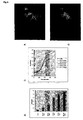
- the catalytic subunit of the murine cAMP dependent protein kinase (PKAc) was fused C-terminally to a F64L-S65T derivative of GFP.
- the resulting fusion (PKAc-F64L-S65T-GFP) was used for monitoring in vivo the translocation and thereby the activation of PKA.
- the PKAc amplification product was then digested with HindIII+AscI and the F64L-S65T-GFP product with Ascl+Xhol.
- the two digested PCR products were subsequently ligated with a Hindlll+Xhol digested plasmid (pZeoSV® mammalian expression vector, Invitrogen, San Diego, CA, USA).
- the resulting fusion construct (SEQ ID NO:68 & 69) was under control of the SV40 promoter.
- CHO Chinese hamster ovary cells
- DMEM with 1000 mg glucose/l, 10 % fetal bovine serum (FBS), 100 mg penicillin-streptomycin mixture ml -1 , 2 mM L-glutamine purchased from Life Technologies Inc., Gaithersburg, MD, USA.
- Untransfected CHO cells were used as the control.
- the PKAc-F64L-S65T-GFP fusion was stably expressed in baby hamster kidney cells overexpressing the human glucagon receptor (BHK/GR cells) Untransfected BHK/GR cells were used as the control. Expression of GR was maintained with 500 mg G418/ml ( Neo marker) andPKAc-F64L-S65T-GFP was maintained with 500 mg Zeocin/ml ( Sh ble marker). CHO cells were also simultaneously co-transfected with vectors containing the PKAc-F64L-S65T-GFP fusion and the human a2a adrenoceptor (hARa2a).
- cells were allowed to adhere to Lab-Tek chambered coverglasses (Nalge Nunc Int., Naperville, IL, USA) for at least 24 hours and cultured to about 80% confluence. Prior to experiments, the cells were cultured over night without selection pressure in HAM F-12 medium with glutamax (Life Technologies), 100 mg penicillin-streptomycin mixture ml -1 and 0.3 % FBS. This medium has low autofluorescence enabling fluorescence microscopy of cells straight from the incubator.
- Image acquisition of live cells were gathered using a Zeiss Axiovert 135M fluorescence microscope fitted with a Fluar 40X, NA: 1.3 oil immersion objective and coupled to a Photometrics CH250 charged coupled device (CCD) camera.
- the cells were illuminated with a 100 W HBO arc lamp. In the light path was a 470 ⁇ 20 nm excitation filter, a 510 nm dichroic mirror and a 515 ⁇ 15 nm emission filter for minimal image background.
- the cells were kept and monitored to be at 37°C with a custom built stage heater.
- Cells were cultured in HAM F-12 medium as described above, but in 96-well plates.
- the medium was exchanged with Ca 2+ -HEPES buffer including 100 mM IBMX and the cells were stimulated with different concentrations of forskolin for 10 min. Reactions were stopped with addition of NaOH to 0.14 M and the amount of cAMP produced was measured with the cAMP-SPA kit, RPA538 (Amersham) as described by the manufacturer.
- cAMP levels were used to vary cAMP levels: Forskolin, an activator of adenylate cyclase; dbcAMP, a membrane permeable cAMP analog which is not degraded by phosphodiesterase; IBMX, an inhibitor of phosphodiesterase.
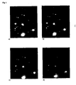
- Fig. 2 shows the progression of response in time following treatment with 1 mM forskolin.
- BHK cells stably transfected with the glucagon receptor and the PKA-F64L-S65T-GFP probe were exposed to glucagon stimulation.
- the glucagon receptor is coupled to a G S protein which activates adenylate cyclase, thereby increasing the cAMP level.
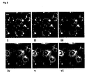
- Transiently transfected CHO cells expressing hARa2a and the PKA-F64L-S65T-GFP probe were treated with 10 mM forskolin for 7.5 minutes, then, in the continued presence of forskolin, exposed to 10 mM norepinephrine to stimulate the exogenous adrenoreceptors, which couple to a G I protein, which inhibit adenylate cyclase.
- the probe was constructed by ligating two restriction enzyme treated polymerase chain reaction (PCR) amplification products of the cDNA for murine PKC ⁇ (GenBank Accession number: M25811) and F64L-S65T-GFP (sequence disclosed in WO 97/11094) respectively.
- PCR polymerase chain reaction
- Taq® polymerase and the following oligonucleotide primers were used for PCR;
- the hybrid DNA strand was inserted into the pZeoSV® mammalian expression vector as a Hindlll-Xhol casette as described in example 1.
- BHK cells expressing the human M1 receptor under the control of the inducible metallothionine promoter and maintained with the dihydrofolate reductase marker were transfected with the PKC ⁇ -F64L-S65T-GFP probe using the calcium phosphate precipitate method in HEPES buffered saline (HBS [pH 7.10]).
- Stable transfectants were selected using 1000 ⁇ g Zeocin®/ml in the growth medium (DMEM with 1000 mg glucose/l, 10 % foetal bovine serum (FBS), 100 mg penicillin-streptomycin mixture ml-1, 2 mM I-glutamine).
- the hM1 receptor and PKC ⁇ -F64L-S65T-GFP fusion protein were maintained with 500 nM methotrexate and 500 ⁇ g Zeocin®/ml respectively. 24 hours prior to any experiment, the cells were transferred to HAM F-12 medium with glutamax, 100 ⁇ g penicillin-streptomycin mixture ml -1 and 0.3 % FBS. This medium relieves selection pressure, gives a low induction of signal transduction pathways and has a low autofluorescence at the relevant wavelength enabling fluorescence microscopy of cells straight from the incubator.
- Digital images of live cells were gathered using a Zeiss Axiovert 135M fluorescence microscope fitted with a 40X, NA: 1.3 oil immersion objective and coupled to a Photometrics CH250 charged coupled device (CCD) camera.
- the cells were illuminated with a 100 W arc lamp. In the light path was a 470 ⁇ 20 nm excitation filter, a 510 nm dichroic mirror and a 515 ⁇ 15 nm emission filter for minimal image background.
- the cells were kept and monitored to be at 37°C with a custom built stage heater.
- Useful for monitoring signalling pathways involving MAPK e.g. to identify compounds which modulate the activity of the pathway in living cells.
- Erk1 a serine/threonine protein kinase, is a component of a signalling pathway which is activated by e.g. many growth factors.
- the extracellular signal regulated kinase (ERK-1, a mitogen activated protein kinase, MAPK) is fused N- or C-terminally to a derivative of GFP.
- ERK-1 a mitogen activated protein kinase, MAPK
- the resulting fusions expressed in different mammalian cells are used for monitoring in vivo the nuclear translocation, and thereby the activation, of ERK1 in response to stimuli that activate the MAPK pathway.
- PCR polymerase chain reaction
- the ERK1 amplification product is digested with HindIII+AscI and the F64L-S65T-GFP product with AscI+XhoI.
- the ERK1 amplification product is then digested with HindIII+Bsu36I and the F64L-S65T-GFP product with Bsu36I+XhoI.
- the two pairs of digested PCR products are subsequently ligated with a Hindlll+Xhol digested plasmid (pZeoSV® mammalian expression vector, Invitrogen, San Diego, CA, USA).
- the resulting fusion constructs are under control of the SV40 promoter.
- the human Erk1 gene (GenBank Accession number: X60188) was amplified using PCR according to standard protocols with primers Erk1-top (SEQ ID NO:9) and Erk1-bottom/+stop (SEQ ID NO:10).
- the PCR product was digested with restriction enzymes EcoR1 and BamH1, and ligated into pEGFP-C1 (Clontech, Palo Alto; GenBank Accession number U55763) digested with EcoR1 and BamH1. This produces an EGFP-Erk1 fusion (SEQ ID NO:38 &39) under the control of a CMV promoter.
- the plasmid containing the EGFP-Erk1 fusion was transfected into HEK293 cells employing the FUGENE transfection reagent (Boehringer Mannheim). Prior to experiments the cells were grown to 80%-90% confluency 8 well chambers in DMEM with 10% FCS. The cells were washed in plain HAM F-12 medium (without FCS), and then incubated for 30-60 minutes in plain HAM F-12 (without FCS) with 100 micromolar PD98059, an inhibitor of MEK1, a kinase which activates Erk1; this step effectively empties the nucleus of EGFP-Erk1. Just before starting the experiment, the HAM F-12 was replaced with Hepes buffer following a wash with Hepes buffer. This removes the PD98059 inhibitor; if blocking of MEK1 is still wanted (e.g. in control experiments), the inhibitor is included in the Hepes buffer.
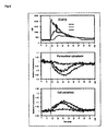
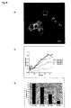
- the response was quantitated as described below and a dose-dependent relationship between EGF concentration and nuclear translocation of EGFP-Erk1 was found (Fig. 9c,d). Reditribution of GFP fluorescence is expressed in this example as the change in the ratio value between areas in nuclear versus cytoplasmic compartments of the cell. Each time profile is the average of nuclear to cytoplasmic ratios from six cells in each treatment.
- Useful for monitoring signalling pathways involving MAPK e.g. to identify compounds which modulate the activity of the pathway in living cells.
- Erk2 a serine/threonine protein kinase, is closely related to Erk1 but not identical; it is a component of a signalling pathway which is activated by e.g. many growth factors.
- the resulting plasmids were transfected into CHO cells and BHK cells.
- the cells were grown under standard conditions. Prior to experiments, the cells were starved in medium without serum for 48-72 hours. This led to a predominantly cytoplasmic localization of both probes, especially in BHK cells. 10% fetal calf serum was added to the cells and the fluorescence of the cells was recorded as explained in example 3. Addition of serum caused the probes to redistribute into the nucleus within minutes of addition of serum.
- Useful for monitoring signalling pathways activated by some members of the transforming growth factor-beta family e.g. to identify compounds which modulate the activity of the pathway in living cells.
- Smad 2 a signal transducer, is a component of a signalling pathway which is induced by some members of the TGFbeta family of cytokines.
- the plasmid containing the EGFP-Smad2 fusion was transfected into HEK293 cells, where it showed a cytoplasmic distribution. Prior to experiments the cells were grown in 8 well Nunc chambers in DMEM with 10% FCS to 80% confluency and starved overnight in HAM F-12 medium without FCS.
- the HAM F-12 medium was replaced with Hepes buffer pH 7.2.
- the experimental setup of the microscope was as described in example 1. 90 images were collected with 10 seconds between each, and with the test compound added after image number 5.
- each nucleus contains less GFP fluorescence than the surrounding cytoplasm (Fig. 10a).
- the redistribution of fluorescence within the treated cells was quantified simply as the fractional increase in nuclear fluorescence normalised to the starting value of GFP fluorescence in the nucleus of each unstimulated cell.
- Useful for monitoring signalling pathways involving rearrangement of cytoskeletal elements e.g. to identify compounds which modulate the activity of the pathway in living cells.
- VASP a phosphoprotein
- the resulting plasmid was transfected into CHO cells expressing the human insulin receptor using the calcium-phosphate transfection method. Prior to experiments, cells were grown in 8 well Nunc chambers and starved overnight in medium without FCS.
- Useful for monitoring signalling pathways involving rearrangement or formation of actin filaments e.g. to identify compounds which modulate the activity of pathways leading to cytoskeletal rearrangements in living cells.
- Actin is a component of cytoskeletal structures, which redistributes in response to very many cellular signals.
- the actin binding domain of the human alpha-actinin gene (GenBank Accession number: X15804) was amplified using PCR according to standard protocols with primers ABD-top (SEQ ID NO:90) and ABD-bottom/-stop (SEQ ID NO:91).
- the PCR product was digested with restriction enzymes Hind3 and BamH1, and ligated into pEGFP-N1 (Clontech, Palo Alto; GenBank Accession number U55762) digested with Hind3 and BamH1. This produced an actin-binding-domain-EGFP fusion (SEQ ID NO:128 &129) under the control of a CMV promoter.
- the resulting plasmid was transfected into CHO cells expressing the human insulin receptor. Cells were stimulated with insulin which caused the actin binding domain-EGFP probe to become redistributed into morphologically distinct membrane-associated structures.
- Useful for monitoring signalling pathways responding to various cellular stress situations e.g. to identify compounds which modulate the activity of the pathway in living cells, or as a counterscreen.
- p38 a serine/thronine protein kinase
- TNFalpha a stress-induced signalling pathway which is activated by many types of cellular stress, e.g. TNFalpha, anisomycin, UV and mitomycin C.
- the resulting plasmids are transfected into a suitable cell line, e.g. HEK293, in which the EGFP-p38 probe and/or the p38-EGFP probe should change its cellular distribution from predominantly cytoplasmic to nuclear within minutes in response to activation of the signalling pathway with e.g. anisomycin.
- a suitable cell line e.g. HEK293
- the EGFP-p38 probe and/or the p38-EGFP probe should change its cellular distribution from predominantly cytoplasmic to nuclear within minutes in response to activation of the signalling pathway with e.g. anisomycin.
- Useful for monitoring signalling pathways responding to various cellular stress situations e.g. to identify compounds which modulate the activity of the pathway in living cells, or as a counterscreen.
- Jnk1 a serine/threonine protein kinase, is a component of a stress-induced signalling pathway different from the p38 described above, though it also is activated by many types of cellular stress, e.g. TNFalpha, anisomycin and UV.
- the resulting plasmids are transfected into a suitable cell line, e.g. HEK293, in which the EGFP-Jnk1 probe and/or the Jnk1-EGFP probe should change its cellular distribution from predominantly cytoplasmic to nuclear in response to activation of the signalling pathway with e.g. anisomycin.
- a suitable cell line e.g. HEK293
- the EGFP-Jnk1 probe and/or the Jnk1-EGFP probe should change its cellular distribution from predominantly cytoplasmic to nuclear in response to activation of the signalling pathway with e.g. anisomycin.
- Useful for monitoring signalling pathways involving changes in cyclic GMP levels e.g. to identify compounds which modulate the activity of the pathway in living cells.
- PGK a cGMP-dependent serine/threonine protein kinase, mediates the guanylyl-cyclase/cGMP signal.
- the resulting plasmids are transfected into a suitable cell line, e.g. A10, in which the EGFP-PKG probe and/or the PKG-EGFP probe should change its cellular distribution from cytoplasmic to one associated with cytoskeletal elements within minutes in response to treatment with agents which raise nitric oxide (NO) levels.
- a suitable cell line e.g. A10
- the EGFP-PKG probe and/or the PKG-EGFP probe should change its cellular distribution from cytoplasmic to one associated with cytoskeletal elements within minutes in response to treatment with agents which raise nitric oxide (NO) levels.
- NO nitric oxide
- Useful for monitoring signalling pathways leading to NFkappaB activation e.g. to identify compounds which modulate the activity of the pathway in living cells.
- IkappaB kinase a serine/threonine kinase
- IkappaB kinase is a component of a signalling pathway which is activated by a variety of inducers including cytokines, lymphokines, growth factors and stress.
- the resulting plasmids are transfected into a suitable cell line, e.g. Jurkat, in which the EGFP-lkappaB-kinase probe and/or the IkappaB-kinase-EGFP probe should achieve a more cytoplasmic distribution within seconds following stimulation with e.g. TNFalpha.
- a suitable cell line e.g. Jurkat
- the EGFP-lkappaB-kinase probe and/or the IkappaB-kinase-EGFP probe should achieve a more cytoplasmic distribution within seconds following stimulation with e.g. TNFalpha.
- Useful for monitoring signalling pathways of the cell cycle e.g. to identify compounds which modulate the activity of the pathway in living cells.
- CDK2 a cyclin-dependent serine/threonine kinase, is a component of the signalling system which regulates the cell cycle.
- the resulting plasmids are transfected into a suitable cell line, e.g. HEK293 in which the EGFP-CDK2 probe and/or the CDK2-EGFP probe should change its cellular distribution from cytoplasmic in contact-inhibited cells, to nuclear location in response to activation with a number of growth factors, e.g. IGF.
- a suitable cell line e.g. HEK293 in which the EGFP-CDK2 probe and/or the CDK2-EGFP probe should change its cellular distribution from cytoplasmic in contact-inhibited cells, to nuclear location in response to activation with a number of growth factors, e.g. IGF.
- Useful for monitoring signalling pathways involving desensitization of G-protein coupled receptors e.g. to identify compounds which modulate the activity of the pathway in living cells.
- Grk5 a G-protein coupled receptor kinase, is a component of signalling pathways involving membrane bound G-protein coupled receptors.
- the resulting plasmids are transfected into a suitable cell line, e.g. HEK293 expressing a rat dopamine D1A receptor, in which the EGFP-Grk5 probe and/or the Grk5-EGFP probe should change its cellular distribution from predominantly cytoplasmic to peripheral in response to activation of the signalling pathway with e.g. dopamine.
- a suitable cell line e.g. HEK293 expressing a rat dopamine D1A receptor
- the EGFP-Grk5 probe and/or the Grk5-EGFP probe should change its cellular distribution from predominantly cytoplasmic to peripheral in response to activation of the signalling pathway with e.g. dopamine.
- Useful for monitoring signalling pathways involving the T cell receptor e.g. to identify compounds which modulate the activity of the pathway in living cells.
- Zap70 a tyrosine kinase, is a component of a signalling pathway which is active in e.g. T-cell differentiation.
- the resulting plasmids are transfected into a suitable cell line, e.g. Jurkat, in which the EGFP-Zap70 probe and/or the Zap70-EGFP probe should change its cellular distribution from cytoplasmic to membrane-associated within seconds in response to activation of the T cell receptor signalling pathway with e.g. antibodies to CD3epsilon.
- a suitable cell line e.g. Jurkat
- the EGFP-Zap70 probe and/or the Zap70-EGFP probe should change its cellular distribution from cytoplasmic to membrane-associated within seconds in response to activation of the T cell receptor signalling pathway with e.g. antibodies to CD3epsilon.
- Useful for monitoring signalling pathways involving PI-3 kinase e.g. to identify compounds which modulate the activity of the pathway in living cells.
- p85alpha is the regulatory subunit of PI3-kinase which is a component of many pathways involving membrane-bound tyrosine kinase receptors and G-protein-coupled receptors.
- the resulting plasmids are transfected into a suitable cell line, e.g. CHO expressing the human insulin receptor, in which the EGFP-p85 probe and/or the p85-EGFP probe may change its cellular distribution from cytoplasmic to membrane-associated within minutes in response to activation of the receptor with insulin.
- a suitable cell line e.g. CHO expressing the human insulin receptor, in which the EGFP-p85 probe and/or the p85-EGFP probe may change its cellular distribution from cytoplasmic to membrane-associated within minutes in response to activation of the receptor with insulin.
- Useful for monitoring signalling pathways involving tyrosine kinases e.g. to identify compounds which modulate the activity of the pathway in living cells.
- Protein-tyrosine phosphatase1C is an inhibitory component in signalling pathways involving e.g. some growth factors.
- the resulting plasmids are transfected into a suitable cell line, e.g. MCF-7 in which the EGFP-PTP probe and/or the PTP-EGFP probe should change its cellular distribution from cytoplasm to the plasma menbrane within minutes in response to activation of the growth inhibitory signalling pathway with e.g. somatostatin.
- a suitable cell line e.g. MCF-7 in which the EGFP-PTP probe and/or the PTP-EGFP probe should change its cellular distribution from cytoplasm to the plasma menbrane within minutes in response to activation of the growth inhibitory signalling pathway with e.g. somatostatin.
- Smad4 a signal transducer, is a common component of signalling pathways induced by various members of the TGFbeta family of cytokines.
- the resulting plasmids are transfected into a cell line, e.g. HEK293 in which the EGFP-Smad4 probe and/or the Smad4-EGFP probe should change its cellular distribution within minutes from cytoplasmic to nuclear in response to activation of the signalling pathway with e.g. TGFbeta.
- a cell line e.g. HEK293 in which the EGFP-Smad4 probe and/or the Smad4-EGFP probe should change its cellular distribution within minutes from cytoplasmic to nuclear in response to activation of the signalling pathway with e.g. TGFbeta.
- Useful for monitoring signalling pathways involving the activation of tyrosine kinases of the Jak family e.g. to identify compounds which modulate the activity of the pathway in living cells.
- Stat5 signal transducer and activator of transcription, is a component of signalling pathways which are induced by e.g. many cytokines and growth factors.
- the resulting plasmids are transfected into a suitable cell line, e.g. MIN6 in which the EGFP-Stat5 probe and/or the Stat5-EGFP probe should change its cellular distribution from cytoplasmic to nuclear within minutes in response to activation signalling pathway with e.g. prolactin.
- a suitable cell line e.g. MIN6 in which the EGFP-Stat5 probe and/or the Stat5-EGFP probe should change its cellular distribution from cytoplasmic to nuclear within minutes in response to activation signalling pathway with e.g. prolactin.
- Useful for monitoring signalling pathways involving activation of NFAT e.g. to identify compounds which modulate the activity of the pathway in living cells.
- NFAT an activator of transcription
- NFAT an activator of transcription
- the resulting plasmids are transfected into a suitable cell line, e.g. Jurkat, in which the EGFP-NFAT probe and/or the NFAT-EGFP probe should change its cellular distribution from cytoplasmic to nuclear within minutes in response to activation of the signalling pathway with e.g. antibodies to CD3epsilon.
- a suitable cell line e.g. Jurkat
- the EGFP-NFAT probe and/or the NFAT-EGFP probe should change its cellular distribution from cytoplasmic to nuclear within minutes in response to activation of the signalling pathway with e.g. antibodies to CD3epsilon.
- Useful for monitoring signalling pathways leading to activation of NFkappaB e.g. to identify compounds which modulate the activity of the pathway in living cells.
- NFkappaB an activator of transcription
- NFkappaB is a component of signalling pathways which are responsive to a varity of inducers including cytokines, lymphokines, some immunosuppressive agents.
- the resulting plasmids are transfected into a suitable cell line, e.g. Jurkat, in which the EGFP-NFkappaB probe and/or the NFkappaB-EGFP probe should change its cellular distribution from cytoplasmic to nuclear in response to activation of the signalling pathway with e.g. TNFalpha.
- a suitable cell line e.g. Jurkat
- the EGFP-NFkappaB probe and/or the NFkappaB-EGFP probe should change its cellular distribution from cytoplasmic to nuclear in response to activation of the signalling pathway with e.g. TNFalpha.
- Useful for monitoring signalling pathways involving RhoA e.g. to identify compounds which modulate the activity of the pathway in living cells.
- RhoA a small GTPase, is a component of many signalling pathways, e.g. LPA induced cytoskeletal rearrangements.
- RhoA gene (GenBank Accession number: L25080) was amplified using PCR according to standard protocols with primers RhoA-top (SEQ ID NO:92) and RhoA-bottom/+stop (SEQ ID NO:93).
- the PCR product was digested with restriction enzymes Hind3 and BamH1, and ligated into pEGFP-C1 (Clontech, Palo Alto; GenBank Accession number U55763) digested with Hind3and BamH1. This produced an EGFP-RhoA fusion (SEQ ID NO:126 &127) under the control of a CMV promoter.
- the resulting plasmid is transfected into a suitable cell line, e.g. Swiss3T3, in which the EGFP-RhoA probe should change its cellular distribution from a reasonably homogenous to a peripheral distribution within minutes of activation of the signalling pathway with e.g. LPA.
- a suitable cell line e.g. Swiss3T3
- the EGFP-RhoA probe should change its cellular distribution from a reasonably homogenous to a peripheral distribution within minutes of activation of the signalling pathway with e.g. LPA.
- Useful for monitoring signalling pathways involving PKB e.g. to identify compounds which modulate the activity of the pathway in living cells.
- PKB a serine/threonine kinase
- the resulting plasmids are transfected into a suitable cell line, e.g. CHO expressing the human insulin receptor, in which the EGFP-PKB probe and/or the PKB-EGFP probe cycles between cytoplasmic and membrane locations during the activation-deactivation process following addition of insulin.
- a suitable cell line e.g. CHO expressing the human insulin receptor, in which the EGFP-PKB probe and/or the PKB-EGFP probe cycles between cytoplasmic and membrane locations during the activation-deactivation process following addition of insulin. The transition should be apparent within minutes.
Claims (29)
- Ein Nachweisverfahren umfassend ein Verfahren für die Identifikation einer biologisch aktiven Substanz, die direkt oder indirekt die Translokation eines biologisch aktiven Polypeptids beeinflusst, wobei das biologisch aktive Polypeptid ein Polypeptid ist, dass intrazelluläre Abläufe beeinflusst nach Aktivierung eines intrazellulären Signaltransduktionswegs, wobei der intrazelluläre Signaltransduktionsweg ein koordinierter intrazellulärer Ablauf ist, in dem eine lebende Zelle ein externes oder internes Signal in zelluläre Antworten umwandelt, wobei der intrazelluläre Signaltransduktionsweg eine enzymatische Reaktion umfasst. Das Verfahren umfasst:(a) die Kultivierung einer oder mehrerer Zellen, die eine stabile Expression einer Nukleinsäure zeigt/-en, die für ein Hybridpolypeptid kodiert, welches ein Fluorophor umfasst, das von einer Nukleinsäure kodiert wird, die mit dem biologisch aktiven Polypeptid oder einem Teil davon verbunden ist, unter Bedingungen, die eine Expression der Nukleotidsequenz erlauben,(b) die Inkubation der mindestens einen Zelle mit einem für die biologische Funktion oder biologische Wirkung zu screenenden Stoffs, und(c) das Messen des Lichts, das von dem Fluorophor in der mindestens einen inkubierten Zelle emittiert wird und Bestimmen des Ergebnisses oder der Veränderung in Hinblick auf das emittierte Licht vom Fluorophor, wobei ein solches Ergebnis oder eine solche Veränderung die Translokation des biologisch aktiven Polypeptids in der oder den Zellen anzeigt.
- Verfahren nach Anspruch 1, wobei der biologisch aktive Stoff ein Medikament ist.
- Verfahren nach Anspruch 1, wobei der biologisch aktive Stoff ein toxischer Stoff ist.
- Verfahren nach Anspruch 1, wobei Schritt (c) durch Gewinnen von quantitativen Informationen, die mit der Translokation des genannten biologisch aktiven Polypeptids in Beziehung stehen, durch Aufzeichnen der Veränderung in räumlich verteiltem Licht, das vom Fluorophor emittiert wird, wobei eine solche Veränderung die Translokation des biologisch aktiven Polypeptids in der Zelle oder den Zellen anzeigt.
- Verfahren nach Anspruch 4, wobei die quantitative Information von der oder den Aufzeichnungen gewonnen wird gemäß einer vorgegebenen Kalibrierung, die auf Wirkungen oder Ergebnissen basiert, die in der gleichen Weise aufgezeichnet wurden bei bekannten Graden eines relevanten spezifischen Einflusses.
- Verfahren nach einem der Ansprüche 1-5, wobei das biologisch aktive Polypeptid den Teil der Aminosäuresequenz umfasst, die eine biologische Funktion hat oder eine biologische Wirkung im zellulären System ausübt.
- Verfahren nach einem der Ansprüche 1-6, wobei Schritt c) zu einem einzelnen Zeitpunkt nach der Anwendung der Substanz durchgeführt wird.
- Verfahren nach einem der Ansprüche 1-6, wobei Schritt c) zu zwei Zeitpunkten durchgeführt wird, wobei ein Punkt vor und der andere Punkt nach der Anwendung der Substanz liegt.
- Verfahren nach Anspruch 8, wobei das emittierte Licht der Zelle oder Zellen vor der Inkubation der Zelle oder Zellen mit dem Stoff gemessen wird und das/die in Schritt (c) bestimmte Ergebnis oder Veränderung eine Veränderung des emittierten Lichts verglichen mit dem emittierten Licht, das vor der Inkubation der Zelle oder Zellen mit dem Stoff gemessen wurde, ist.
- Verfahren nach einem der Ansprüche 1 - 6, wobei zu einer Serie von Zeitpunkten Schritt (c) durchgeführt wird, bei welchen die Anwendung des Einflusses einige Zeit nach dem ersten Zeitpunkt in den Aufzeichnungsserien auftritt, indem die Aufzeichnung z.B. mit einem vorbestimmten Zeitabstand von 0,1 Sekunden bis 1 Stunde, vorzugsweise von 1 bis 60 Sekunden, stärker bevorzug von 1 bis 30 Sekunden, insbesondere von 1 bis 10 Sekunden über eine Zeitspanne von 1 Sekunde bis 12 Stunden wie von 10 Sekunden bis 12 Stunden, z.B. von 10 Sekunden bis 1 Stunde wie von 60 Sekunden bis 30 Minuten oder 20 Minuten durchgeführt wird.
- Verfahren nach einem der Ansprüche 1 - 7, wobei die Zelle oder Zellen zu einem Zeitpunkt nach der Anwendung des Stoffs, bei welchem die Antwort als bedeutsam vorbestimmt wurde, fixiert wird/werden und die Aufzeichnung zu einer willkürlichen späteren Zeit durchgeführt wird.
- Verfahren nach einem der vorangehenden Ansprüche, wobei der Fluorophor ein Fluorophor ist, das in einer Weise transloziert werden kann, die für den Grad des Stoffs physiologisch relevant ist.
- Verfahren nach einem der vorangehenden Ansprüche, wobei der Fluorophor ein Fluorophor ist, das in einer Weise transloziert werden kann, von welcher experimentell bestimmt wurde, dass sie mit dem Grad des Einflusses des Stoffs in Beziehung steht.
- Verfahren nach einem der vorangehenden Ansprüche 4 - 13, wobei die Veränderung oder das Ergebnis in Hinblick auf das räumlich verteilte Licht, das durch das Fluorophor emittiert wird, durch eine Veränderung des Resonanzenergietransfer zwischen dem Fluorophor und einer anderen lumineszierenden Einheit nachgewiesen wird, die dem Fluorophor Energie liefern kann, wobei jede dieser ausgewählt worden ist oder künstlich erzeugt worden ist, um ein Teil von bestimmten Bestandteilen des intrazellulären Signaltransduktionswegs zu werden oder an diese gebunden oder mit diesen assoziiert zu werden, und eine der beiden einer Translokation in Antwort auf den Einfluss unterzogen wird, wodurch die Menge des Resonanzenergietransfers verändert wird, wobei die Veränderung des Resonanzenergietransfers als eine Veränderung in der Intensität der Emission vom Fluorophor gemessen wird.
- Verfahren nach einem der vorangehenden Ansprüche, wobei die Aufzeichnung des Ergebnisses oder der Veränderung durchgeführt wird durch Aufzeichnung der Eigenschaft des emittierten Lichts ausgewählt aus der Gruppe bestehend aus Intensität, Fluoreszenz-Zeitspanne, Polarisierung und Wellenlängenverschiebung.
- Verfahren nach Anspruch 15, wobei die aufgezeichnete Eigenschaft des Lichts die Intensität ist.
- Verfahren nach einem der vorangehenden Ansprüche, wobei die Veränderung in der Intensität der Emission vom Fluorophor durch einen einkanaligen Lichtdetektor abgetastet wird, der nur auf die mittlere Intensität des Fluorophors in einer nichträumlich aufgelösten Art reagiert.
- Verfahren nach einem der vorangehenden Ansprüche 4 - 17, wobei das Aufzeichnen des räumlich verteilten Lichts unter Verwendung eines Aufzeichnungssystems durchgeführt wird, das die räumliche Verteilung einer aufzeichenbaren Eigenschaft des Lichts in Form einer geregelten Anordnung von Werten aufzeichnet.
- Verfahren nach einem der vorangehenden Ansprüche 4 - 17, wobei das Aufzeichnen der räumlichen Verteilung der aufzeichenbaren Eigenschaft des Lichts unter Verwendung einer Ladungstransfervorrichtung wie einem CCD-Array oder einer Vakuumröhrenvorrichtung wie einer Vidiconröhre durchgeführt wird.
- Verfahren nach einem der vorangehenden Ansprüche, wobei Schritt (c) durch Fluoreszenzmikroskopie durchgeführt wird.
- Verfahren nach einem der vorangehenden Ansprüche 4 - 20, wobei das Aufzeichnen der Veränderung oder des Ergebnisses in Hinsicht auf das von der Fluorophore emittierte Licht durchgeführt wird, indem das räumlich verteilte Licht als eines oder mehrere digitale Bilder aufgezeichnet wird, und die Verarbeitung der aufgezeichneten Veränderung zu deren Umsetzung in eine oder mehrere Zahlen, die für den Grad der Neuverteilung repräsentativ sind, ein Verfahren zur Digitalbildverarbeitung oder eine Kombination von Verfahren zur Digitalbildverarbeitung umfasst.
- Verfahren nach einem der vorangehenden Ansprüche, wobei das Hybridprotein eine Fusion von zumindest einem Teil des Grün-Fluoreszenz-Proteins und mindestens einem Teil einer katalytischen oder regulatorischen Domäne einer Proteinkinase ist.
- Verfahren nach einem der vorangehenden Ansprüche, wobei das Fluorophor ein Grün-Fluoreszenz-Protein (GFP) ist.
- Verfahren nach einem der vorangehenden Ansprüche, wobei das GFP ausgewählt ist aus GFPs mit der F64L-Mutation.
- Verfahren nach einem der vorangehenden Ansprüche, wobei das GFP eine GFP-Variante, ausgewählt aus der Gruppe umfassend aus F64L-GFP, F64L-Y66H-GFP, F64L-S65T-GFP und EGFP ist.
- Verfahren nach einem der vorangehenden Ansprüche, wobei die Zelle oder Zellen eine Säugerzelle oder Säugerzellen ist/sind, die während des Zeitraums, über den der Einfluss beobachtet wird, bei einer Temperatur von 30°C oder höher inkubiert wird/werden, vorzugsweise bei einer Temperatur von 32°C bis 39°C, stärker bevorzugt bei einer Temperatur von 35°C bis 38°C und am stärksten bevorzugt bei einer Temperatur von etwa 37°C.
- Verfahren nach einem der vorangehenden Ansprüche, wobei mindestens eine Zelle ein Teil einer Matrix von identischen oder nichtidentischen Zellen ist.
- Verfahren nach einem der vorangehenden Ansprüche, wobei die Zelle oder Zellen aus der Gruppe, bestehend aus Pilzzellen wie eine Hefezelle, Invertebratzellen einschließlich Insektenzellen und Vertebratzellen wie Säugerzellen ausgewählt wird/werden.
- Verfahren nach einem der vorangehenden Ansprüche, wobei das biologisch aktive Polypeptid aus der Gruppe ausgewählt ist, die aus einer Proteinkinase, einer Phosphatase, einem Transkriptionsfaktor und einem Protein, das mit einem Zytoskelett-Netzwerk verbunden ist, welches die zelluläre Lokalisierung nach Aktivierung verändert, besteht.
Priority Applications (1)
| Application Number | Priority Date | Filing Date | Title |
|---|---|---|---|
| EP03028919A EP1435519B1 (de) | 1997-04-07 | 1998-04-07 | Ein Verfahren zum Screenen von Substanzen hinsichtlich ihres Effekts auf cAMP Spiegel basierend auf intrazellulärer Translokation von PKA |
Applications Claiming Priority (3)
| Application Number | Priority Date | Filing Date | Title |
|---|---|---|---|
| DK39297 | 1997-04-07 | ||
| DK39297 | 1997-04-07 | ||
| EP98913541A EP0986753B2 (de) | 1997-04-07 | 1998-04-07 | Verfahren um quantitativen informationen über einflüsse auf zelluläre reaktionen zu entnehmen |
Related Parent Applications (1)
| Application Number | Title | Priority Date | Filing Date |
|---|---|---|---|
| EP98913541A Division EP0986753B2 (de) | 1997-04-07 | 1998-04-07 | Verfahren um quantitativen informationen über einflüsse auf zelluläre reaktionen zu entnehmen |
Related Child Applications (1)
| Application Number | Title | Priority Date | Filing Date |
|---|---|---|---|
| EP03028919A Division EP1435519B1 (de) | 1997-04-07 | 1998-04-07 | Ein Verfahren zum Screenen von Substanzen hinsichtlich ihres Effekts auf cAMP Spiegel basierend auf intrazellulärer Translokation von PKA |
Publications (3)
| Publication Number | Publication Date |
|---|---|
| EP1199564A2 EP1199564A2 (de) | 2002-04-24 |
| EP1199564A3 EP1199564A3 (de) | 2002-05-08 |
| EP1199564B1 true EP1199564B1 (de) | 2004-06-23 |
Family
ID=8092994
Family Applications (3)
| Application Number | Title | Priority Date | Filing Date |
|---|---|---|---|
| EP98913541A Expired - Lifetime EP0986753B2 (de) | 1997-04-07 | 1998-04-07 | Verfahren um quantitativen informationen über einflüsse auf zelluläre reaktionen zu entnehmen |
| EP01204477A Expired - Lifetime EP1199564B1 (de) | 1997-04-07 | 1998-04-07 | Verfahren zum Screenen von Substanzen die einen effekt auf intrazelluläre Translokation haben |
| EP03028919A Expired - Lifetime EP1435519B1 (de) | 1997-04-07 | 1998-04-07 | Ein Verfahren zum Screenen von Substanzen hinsichtlich ihres Effekts auf cAMP Spiegel basierend auf intrazellulärer Translokation von PKA |
Family Applications Before (1)
| Application Number | Title | Priority Date | Filing Date |
|---|---|---|---|
| EP98913541A Expired - Lifetime EP0986753B2 (de) | 1997-04-07 | 1998-04-07 | Verfahren um quantitativen informationen über einflüsse auf zelluläre reaktionen zu entnehmen |
Family Applications After (1)
| Application Number | Title | Priority Date | Filing Date |
|---|---|---|---|
| EP03028919A Expired - Lifetime EP1435519B1 (de) | 1997-04-07 | 1998-04-07 | Ein Verfahren zum Screenen von Substanzen hinsichtlich ihres Effekts auf cAMP Spiegel basierend auf intrazellulärer Translokation von PKA |
Country Status (11)
| Country | Link |
|---|---|
| US (3) | US6518021B1 (de) |
| EP (3) | EP0986753B2 (de) |
| JP (1) | JP2001522454A (de) |
| AT (3) | ATE269975T1 (de) |
| AU (1) | AU6820998A (de) |
| CA (1) | CA2286293C (de) |
| DE (4) | DE69824763T2 (de) |
| DK (2) | DK1435519T3 (de) |
| ES (2) | ES2191575T3 (de) |
| PT (2) | PT986753E (de) |
| WO (1) | WO1998045704A2 (de) |
Cited By (1)
| Publication number | Priority date | Publication date | Assignee | Title |
|---|---|---|---|---|
| US7625951B2 (en) | 2000-07-13 | 2009-12-01 | University Of Kentucky Research Foundation | Stimuli-responsive hydrogel microdomes integrated with genetically engineered proteins for high-throughput screening of pharmaceuticals |
Families Citing this family (84)
| Publication number | Priority date | Publication date | Assignee | Title |
|---|---|---|---|---|
| ATE217971T1 (de) | 1996-01-26 | 2002-06-15 | Virco Nv | Verfahren zur überwachung der chemotherapie von hiv-positiven patienten basierend auf der phenotypischen medikamentempfindlichkeit vom hiv stamm des patienten |
| US7062219B2 (en) * | 1997-01-31 | 2006-06-13 | Odyssey Thera Inc. | Protein fragment complementation assays for high-throughput and high-content screening |
| US6759206B1 (en) | 1997-02-27 | 2004-07-06 | Cellomics, Inc. | System for cell-based screening |
| US7853411B2 (en) | 1997-02-27 | 2010-12-14 | Cellomics, Inc. | System for cell-based screening |
| US6416959B1 (en) | 1997-02-27 | 2002-07-09 | Kenneth Giuliano | System for cell-based screening |
| DE69824763T2 (de) * | 1997-04-07 | 2005-07-07 | Bioimage A/S | Verfahren zum Screenen von Substanzen die einen effekt auf intrazelluläre Translokation haben |
| US7160687B1 (en) | 1997-05-29 | 2007-01-09 | Cellomics, Inc. | Miniaturized cell array methods and apparatus for cell-based screening |
| US6235512B1 (en) * | 1997-07-01 | 2001-05-22 | Tularik Inc. | IKK-α proteins, nucleic acids and methods |
| US20050221280A1 (en) | 1998-02-02 | 2005-10-06 | Odyssey Thera, Inc. | Protein-protein interactions for pharmacological profiling |
| US6388788B1 (en) | 1998-03-16 | 2002-05-14 | Praelux, Inc. | Method and apparatus for screening chemical compounds |
| US20030036855A1 (en) | 1998-03-16 | 2003-02-20 | Praelux Incorporated, A Corporation Of New Jersey | Method and apparatus for screening chemical compounds |
| EP1064360B1 (de) * | 1998-03-27 | 2008-03-05 | Prolume, Ltd. | Luciferase, gfp fluoreszenzproteine, kodierende nukleinsaüre und ihre verwendung in der diagnose |
| US6037133A (en) * | 1998-04-17 | 2000-03-14 | Clontech Laboratories, Inc. | IκBEGFP constructs, cell lines and methods of use |
| US20030220258A1 (en) | 2001-12-21 | 2003-11-27 | Robbert Benner | Treatment of ischemic events |
| US8680059B2 (en) * | 1998-05-20 | 2014-03-25 | Biotempt B.V. | Oligopeptide acetate and formulations thereof |
| US6844315B2 (en) * | 1998-05-20 | 2005-01-18 | Erasmus Universiteit Rotterdam | Immunoregulator |
| CA2346937A1 (en) * | 1998-10-15 | 2000-04-27 | Per O. G. Arkhammar | Specific therapeutic interventions obtained by interference with redistribution and/or targeting |
| JP2002527761A (ja) * | 1998-10-15 | 2002-08-27 | バイオイメージ エイ/エス | 細胞応答への影響に関する量的情報を入手するための改良方法 |
| GB9901072D0 (en) * | 1999-01-19 | 1999-03-10 | Imp Cancer Res Tech | Methods for detecting changes to a macromolecular component of a cell |
| EP1155304B1 (de) * | 1999-02-26 | 2003-05-07 | Cellomics, Inc. | Ein system für zellbasierte reihenuntersuchungen |
| DE19915310A1 (de) * | 1999-04-03 | 2000-10-05 | Bayer Ag | Diffusionskontrollierende Sensorschicht |
| DE69906253D1 (de) * | 1999-04-29 | 2003-04-30 | Univ Erasmus | Bestimmung von Analytbeweglichkeit |
| AU4847100A (en) * | 1999-05-14 | 2000-12-05 | Cytokinetics, Inc. | Method and apparatus for predictive cellular bioinformatics |
| US20030228565A1 (en) * | 2000-04-26 | 2003-12-11 | Cytokinetics, Inc. | Method and apparatus for predictive cellular bioinformatics |
| US6743576B1 (en) | 1999-05-14 | 2004-06-01 | Cytokinetics, Inc. | Database system for predictive cellular bioinformatics |
| US7151847B2 (en) | 2001-02-20 | 2006-12-19 | Cytokinetics, Inc. | Image analysis of the golgi complex |
| US6651008B1 (en) | 1999-05-14 | 2003-11-18 | Cytokinetics, Inc. | Database system including computer code for predictive cellular bioinformatics |
| EP1055929A1 (de) * | 1999-05-26 | 2000-11-29 | Tibotec N.V. | Mittel und Verfahren für Medizinentdeckung und phenotypische Charakterisierung von Zellen |
| US6573059B1 (en) * | 1999-06-04 | 2003-06-03 | Rmf Dictagene S.A. | Use of the regulatory subunit of the camp dependent protein kinase (PKA) from dictyostelium for camp measurements |
| AU5870800A (en) * | 1999-06-09 | 2000-12-28 | Ljl Biosystems, Inc. | Cell-signaling assays |
| US6673554B1 (en) | 1999-06-14 | 2004-01-06 | Trellie Bioinformatics, Inc. | Protein localization assays for toxicity and antidotes thereto |
| WO2000079241A2 (en) * | 1999-06-21 | 2000-12-28 | Cellomics, Inc. | A system for cell-based screening |
| US6121047A (en) * | 1999-07-21 | 2000-09-19 | Isis Pharmaceuticals Inc. | Antisense modulation of SHP-1 expression |
| US6489151B1 (en) * | 2000-03-27 | 2002-12-03 | The Research Foundation Of State University Of New York | Biologically active alternative form of the IKKα IκB kinase |
| EP1300418A1 (de) * | 2001-10-04 | 2003-04-09 | Erasmus Universiteit Rotterdam | Genregulation durch Oligopeptide |
| KR100414641B1 (ko) * | 2000-04-07 | 2004-01-13 | 동부한농화학 주식회사 | 형질전환식물체의 생체분석방법 및 그를 이용한 시스템 |
| JP2004500835A (ja) * | 2000-04-17 | 2004-01-15 | バイオイメージ エイ/エス | 生細胞でのホスホジエステラーゼ(pde)酵素の細胞内分布を調節する化合物の同定法 |
| AU2001252109A1 (en) * | 2000-04-26 | 2001-11-07 | Bioimage A/S | Chemical signal enhancement of dynamic intensity-based intracellular protein- and fluorophore-based redistribution assays for drug screening |
| EP1322788A2 (de) * | 2000-06-23 | 2003-07-02 | Cytokinetics, Inc. | Bildanalyse zur bestimmung des phenotyps von mutantenzellen-gruppen |
| US7282347B2 (en) | 2000-07-04 | 2007-10-16 | Fisher Bioimage Aps | Method for extracting quantitative information relating to interactions between cellular components |
| DE10037759A1 (de) * | 2000-08-03 | 2002-07-04 | Gruenenthal Gmbh | Screeningverfahren |
| KR100414781B1 (ko) * | 2000-11-24 | 2004-01-13 | 재단법인서울대학교산학협력재단 | NF-κB 발현을 인간 피부세포에서 정량적으로 측정하기위한 안정한 분석시스템 및 그의 용도 |
| US7218764B2 (en) | 2000-12-04 | 2007-05-15 | Cytokinetics, Inc. | Ploidy classification method |
| US7016787B2 (en) | 2001-02-20 | 2006-03-21 | Cytokinetics, Inc. | Characterizing biological stimuli by response curves |
| US6956961B2 (en) | 2001-02-20 | 2005-10-18 | Cytokinetics, Inc. | Extracting shape information contained in cell images |
| US7033790B2 (en) | 2001-04-03 | 2006-04-25 | Curagen Corporation | Proteins and nucleic acids encoding same |
| WO2003012068A2 (en) | 2001-08-01 | 2003-02-13 | Cellomics, Inc. | Novel fusion proteins and assays for molecular binding |
| AU2002358657A1 (en) * | 2001-12-10 | 2003-06-23 | Evotec Oai Ag | Method for determining the influence of non-physiological compounds the derivatives and decomposition products thereof on organisms organs and cells |
| US7176303B2 (en) * | 2003-11-06 | 2007-02-13 | Isis Pharmaceuticals, Inc. | Modulation of STAT5 expression |
| US20080242837A1 (en) * | 2001-12-21 | 2008-10-02 | Khan Nisar A | Peptide compositions |
| US20080318871A1 (en) * | 2001-12-21 | 2008-12-25 | Khan Nisar A | Treatment of neurological disorders |
| US7786084B2 (en) * | 2001-12-21 | 2010-08-31 | Biotempt B.V. | Treatment of burns |
| US7560433B2 (en) | 2001-12-21 | 2009-07-14 | Biotempt B.V. | Treatment of multiple sclerosis (MS) |
| US20030220257A1 (en) * | 2001-12-21 | 2003-11-27 | Robbert Benner | Treatment of trauma |
| CA2413475C (en) | 2002-04-25 | 2010-07-27 | The Government Of The United States Of America As Represented By The Secretary Of The Department Of Health And Human Services | Zap-70 expression as a marker for chronic lymphocytic leukemia/small lymphocytic lymphoma (cll/sll) |
| GB0216674D0 (en) * | 2002-07-18 | 2002-08-28 | Amersham Biosciences Uk Ltd | Functional screening method |
| US8168568B1 (en) | 2003-03-10 | 2012-05-01 | The United States Of America, As Represented By The Secretary Of The Department Of Health And Human Services | Combinatorial therapy for protein signaling diseases |
| US7235353B2 (en) | 2003-07-18 | 2007-06-26 | Cytokinetics, Inc. | Predicting hepatotoxicity using cell based assays |
| US20050014217A1 (en) | 2003-07-18 | 2005-01-20 | Cytokinetics, Inc. | Predicting hepatotoxicity using cell based assays |
| WO2005010677A2 (en) | 2003-07-18 | 2005-02-03 | Cytokinetics, Inc. | Characterizing biological stimuli by response curves |
| US20050059083A1 (en) * | 2003-09-15 | 2005-03-17 | Becton Dickinson And Company | High throughput method to identify ligands for cell attachment |
| US20050060100A1 (en) * | 2003-09-15 | 2005-03-17 | Becton Dickinson And Company | Computer software and algorithms for systems biologically linked to cellular phenotype |
| US20090227505A1 (en) * | 2004-01-07 | 2009-09-10 | Biotempt B.V. | Methods and uses for protein breakdown products |
| EP1766095B1 (de) | 2004-05-18 | 2013-10-23 | The Board of Trustees of the Leland Stanford Junior University | Nachweis der proteintranslokation durch beta-galactosidase-reporterfragment-komplementierung |
| US7323318B2 (en) | 2004-07-15 | 2008-01-29 | Cytokinetics, Inc. | Assay for distinguishing live and dead cells |
| US8084260B2 (en) * | 2004-11-24 | 2011-12-27 | Applied Biosystems, Llc | Spectral calibration method and system for multiple instruments |
| WO2007004869A2 (en) * | 2005-07-05 | 2007-01-11 | Biotempt B.V. | Treatment of tumors |
| JP2009513150A (ja) * | 2005-10-28 | 2009-04-02 | ジーイー・ヘルスケア・ユーケイ・リミテッド | シグナリングアッセイ及び株化細胞 |
| JP2009526519A (ja) * | 2006-01-17 | 2009-07-23 | セルーメン、インコーポレイテッド | 生物学的システム応答の予測方法 |
| EP1864692A1 (de) * | 2006-06-07 | 2007-12-12 | Biotempt B.V. | Verwendung von peptiden zum Schutz von Strahlenschäden |
| WO2007136724A2 (en) | 2006-05-17 | 2007-11-29 | Cellumen, Inc. | Method for automated tissue analysis |
| WO2008060483A2 (en) * | 2006-11-10 | 2008-05-22 | Cellumen, Inc. | Protein-protein interaction biosensors and methods of use thereof |
| US20100178652A1 (en) * | 2007-01-05 | 2010-07-15 | Litherland Sally A | Materials and Methods for the Detection, Prevention and Treatment of Autoimmune Disease |
| CA2677948C (en) * | 2007-02-12 | 2014-12-09 | Biotempt B.V. | Treatment of trauma-hemorrhage with short oligopeptides |
| US20080217002A1 (en) * | 2007-03-07 | 2008-09-11 | Floyd Randolph Simonds | Sand control screen having a micro-perforated filtration layer |
| JP5438962B2 (ja) * | 2008-12-25 | 2014-03-12 | シスメックス株式会社 | 細胞画像表示装置 |
| US8574837B2 (en) | 2008-12-29 | 2013-11-05 | President And Fellows Of Harvard College | System for screening agonists/antagonists of cellular signaling pathways |
| KR101088605B1 (ko) | 2009-06-30 | 2011-11-30 | 한국산업기술대학교산학협력단 | 2대의 카메라를 이용한 세포 조작 방법 |
| JP5089713B2 (ja) * | 2010-01-18 | 2012-12-05 | シャープ株式会社 | 画像圧縮装置、圧縮画像出力装置、画像圧縮方法、コンピュータプログラム及び記録媒体 |
| EP2686688B1 (de) | 2011-03-17 | 2019-05-08 | Cernostics, Inc. | Systeme und zusammensetzungen zur diagnose von barrett-ösophagen und verwendungsverfahren dafür |
| EP2825887A4 (de) | 2012-03-15 | 2015-08-19 | Montana Molecular Llc | Genetisch codierte, fluoreszente sensoren zur detektion intrazellulärer signale durch diacylglycerol-signalwege |
| EP2682187B1 (de) * | 2012-07-02 | 2017-09-13 | Molecular Devices, LLC | Auswahlverfahren und -vorrichtung |
| EP3065754B1 (de) | 2013-11-04 | 2020-06-10 | Montana Molecular LLC | Genetisch codierte fluoreszenzsensoren zur erkennung von liganden-bias und intrazellulärer signalisierung durch camp-signalwege |
| DE102014012130B3 (de) | 2014-08-13 | 2015-03-05 | Alfred-Wegener-Institut, Helmholtz-Zentrum für Polar- und Meeresforschung | Nachweisverfahren unter Verwendung von rekombinanten lebenden Zellen zum Nachweis von xenobiotischen Stoffen sowie Anordnung und Testkit zur Durchführung des Nachweisverfahrens |
Family Cites Families (27)
| Publication number | Priority date | Publication date | Assignee | Title |
|---|---|---|---|---|
| AT321257B (de) | 1971-05-26 | 1975-03-25 | Koreska Gmbh W | Anlage zur Rückgewinnung flüchtiger Lösungsmittel |
| JPS57198544A (en) * | 1981-05-28 | 1982-12-06 | Sony Corp | Manufacture of magnetic recording medium |
| JPS5880136A (ja) * | 1981-11-02 | 1983-05-14 | Fuji Photo Film Co Ltd | 磁気記録材料の製造方法 |
| DE3240063A1 (de) | 1981-11-02 | 1983-05-26 | Basf Ag, 6700 Ludwigshafen | Vorrichtung zur herstellung eines magnetogrammtraegers |
| JPH0656656B2 (ja) * | 1983-10-03 | 1994-07-27 | 富士写真フイルム株式会社 | 磁気記録媒体の製造方法 |
| JPS6292132A (ja) * | 1985-10-18 | 1987-04-27 | Fuji Photo Film Co Ltd | 磁気記録媒体の製造方法 |
| US4923766A (en) * | 1988-03-29 | 1990-05-08 | Canon Kabushiki Kaisha | Process for preparing magnetic recording |
| GB8916806D0 (en) * | 1989-07-22 | 1989-09-06 | Univ Wales Medicine | Modified proteins |
| DE4009797A1 (de) * | 1990-03-27 | 1991-10-02 | Pagendarm Gmbh | Verfahren und anordnung zum kondensieren von dampffoermigen substanzen |
| EP0703984A1 (de) * | 1993-04-07 | 1996-04-03 | Cancer Research Campaign Technology Limited | Methoden zum Screenen von Substanzen mit therapeutischer Wirkung und Hefewelche dafür verwendet wird |
| AU694745B2 (en) * | 1993-09-10 | 1998-07-30 | Trustees Of Columbia University In The City Of New York, The | Uses of green fluorescent protein |
| WO1996003649A1 (en) * | 1994-07-22 | 1996-02-08 | The University Of North Carolina At Chapel Hill | Src SH3 BINDING PEPTIDES AND METHODS OF ISOLATING AND USING SAME |
| US6184205B1 (en) * | 1994-07-22 | 2001-02-06 | University Of North Carolina At Chapel Hill | GRB2 SH3 binding peptides and methods of isolating and using same |
| US5874231A (en) * | 1994-08-22 | 1999-02-23 | Mcgill University | Methods of screening for non-hormone compounds which effect modulation of polypeptide translation |
| US5958713A (en) | 1995-01-31 | 1999-09-28 | Novo Nordisk A/S | Method of detecting biologically active substances by using green fluorescent protein |
| DE69618350T2 (de) * | 1995-01-31 | 2002-09-19 | Bioimage As Soeborg | Ein verfahren zum nachweis biologisch aktiver substanzen |
| ATE184613T1 (de) * | 1995-09-22 | 1999-10-15 | Novo Nordisk As | Varianten des grünen fluoreszenzproteins, gfp |
| US6455300B1 (en) * | 1995-12-08 | 2002-09-24 | Han Htun | Method and compositions for monitoring DNA binding molecules in living cells |
| US5874304A (en) * | 1996-01-18 | 1999-02-23 | University Of Florida Research Foundation, Inc. | Humanized green fluorescent protein genes and methods |
| US5989835A (en) * | 1997-02-27 | 1999-11-23 | Cellomics, Inc. | System for cell-based screening |
| AU3801997A (en) * | 1996-07-16 | 1998-02-09 | Regents Of The University Of California, The | Assays for protein kinases using fluorescent protein substrates |
| WO1998030715A1 (en) * | 1997-01-07 | 1998-07-16 | California Institute Of Technology | Optical sensors of cell signaling |
| CA2282658C (en) * | 1997-02-27 | 2003-02-25 | Cellomics, Inc. | A system for cell-based screening |
| US6416959B1 (en) * | 1997-02-27 | 2002-07-09 | Kenneth Giuliano | System for cell-based screening |
| DE69824763T2 (de) * | 1997-04-07 | 2005-07-07 | Bioimage A/S | Verfahren zum Screenen von Substanzen die einen effekt auf intrazelluläre Translokation haben |
| WO2000049183A1 (en) * | 1999-02-17 | 2000-08-24 | Clontech Laboratories, Inc. | FLUORESCENCE RESONANCE ENERGY TRANSFER DETECTION OF cAMP IN LIVING CELLS USING GFP VARIANTS |
| US6573059B1 (en) * | 1999-06-04 | 2003-06-03 | Rmf Dictagene S.A. | Use of the regulatory subunit of the camp dependent protein kinase (PKA) from dictyostelium for camp measurements |
-
1998
- 1998-04-07 DE DE69824763T patent/DE69824763T2/de not_active Expired - Lifetime
- 1998-04-07 PT PT98913541T patent/PT986753E/pt unknown
- 1998-04-07 DE DE69804446T patent/DE69804446T3/de not_active Expired - Fee Related
- 1998-04-07 AT AT01204477T patent/ATE269975T1/de not_active IP Right Cessation
- 1998-04-07 AT AT03028919T patent/ATE354089T1/de not_active IP Right Cessation
- 1998-04-07 DK DK03028919T patent/DK1435519T3/da active
- 1998-04-07 DE DE1199564T patent/DE1199564T1/de active Pending
- 1998-04-07 EP EP98913541A patent/EP0986753B2/de not_active Expired - Lifetime
- 1998-04-07 CA CA002286293A patent/CA2286293C/en not_active Expired - Lifetime
- 1998-04-07 EP EP01204477A patent/EP1199564B1/de not_active Expired - Lifetime
- 1998-04-07 EP EP03028919A patent/EP1435519B1/de not_active Expired - Lifetime
- 1998-04-07 ES ES01204477T patent/ES2191575T3/es not_active Expired - Lifetime
- 1998-04-07 PT PT01204477T patent/PT1199564E/pt unknown
- 1998-04-07 DE DE69837118T patent/DE69837118T2/de not_active Expired - Fee Related
- 1998-04-07 ES ES98913541T patent/ES2173573T5/es not_active Expired - Lifetime
- 1998-04-07 WO PCT/DK1998/000145 patent/WO1998045704A2/en active IP Right Grant
- 1998-04-07 JP JP54227698A patent/JP2001522454A/ja active Pending
- 1998-04-07 DK DK98913541T patent/DK0986753T4/da active
- 1998-04-07 AT AT98913541T patent/ATE215227T1/de not_active IP Right Cessation
- 1998-04-07 AU AU68209/98A patent/AU6820998A/en not_active Abandoned
-
1999
- 1999-10-07 US US09/417,197 patent/US6518021B1/en not_active Expired - Lifetime
-
2002
- 2002-02-05 US US10/072,036 patent/US8058008B2/en not_active Expired - Fee Related
-
2007
- 2007-10-29 US US11/927,246 patent/US20090023598A1/en not_active Abandoned
Cited By (1)
| Publication number | Priority date | Publication date | Assignee | Title |
|---|---|---|---|---|
| US7625951B2 (en) | 2000-07-13 | 2009-12-01 | University Of Kentucky Research Foundation | Stimuli-responsive hydrogel microdomes integrated with genetically engineered proteins for high-throughput screening of pharmaceuticals |
Also Published As
Similar Documents
| Publication | Publication Date | Title |
|---|---|---|
| EP1199564B1 (de) | Verfahren zum Screenen von Substanzen die einen effekt auf intrazelluläre Translokation haben | |
| JP3576491B2 (ja) | 細胞ベースのスクリーニング用のシステム | |
| JP3863372B2 (ja) | プロテアーゼバイオセンサーをコードする組換え核酸、組換え発現ベクター、プロテアーゼバイオセンサー、化合物を同定する方法、及び化合物を同定するためのキット | |
| JP4693382B2 (ja) | 光学システムによる細胞の解析 | |
| JP5953293B2 (ja) | 発光タンパクによる長期モニタリング方法および解析方法 | |
| WO2000023615A2 (en) | Method for extracting quantitative information relating to an influence on a cellular response | |
| CA2538852A1 (en) | Fragment complementation assays for g-protein-coupled receptors and their signaling pathways | |
| Demaurex | Calcium measurements in organelles with Ca2+-sensitive fluorescent proteins | |
| US7282347B2 (en) | Method for extracting quantitative information relating to interactions between cellular components | |
| McAffee et al. | Discrete LAT condensates encode antigen information from single pMHC: TCR binding events | |
| Prinz et al. | Fluorescence and bioluminescence procedures for functional proteomics | |
| US20030143634A1 (en) | Method to detect interactions between cellular components in intact living cells, and to extract quantitative information relating to those interactions by fluorescence redistribution | |
| Meyer et al. | Studies of signal transduction events using chimeras to green fluorescent protein | |
| Leslie et al. | Studying nuclear protein import in yeast | |
| CA2450698A1 (en) | A method for screening substances for effect on camp levels based on intracellular translocation of pka | |
| Brinkenfeldt et al. | Suitable use of FRET-based biosensors for quantitative detection of GPCR activation | |
| JP2022129999A (ja) | 脂肪酸トランスポーターの細胞内動態の分析方法、及び脂肪酸トランスポーター改変タンパク質の利用 | |
| JP2005517459A (ja) | スクリーニング方法 |
Legal Events
| Date | Code | Title | Description |
|---|---|---|---|
| PUAI | Public reference made under article 153(3) epc to a published international application that has entered the european phase |
Free format text: ORIGINAL CODE: 0009012 |
|
| PUAL | Search report despatched |
Free format text: ORIGINAL CODE: 0009013 |
|
| 17P | Request for examination filed |
Effective date: 20020118 |
|
| AC | Divisional application: reference to earlier application |
Ref document number: 986753 Country of ref document: EP |
|
| AK | Designated contracting states |
Kind code of ref document: A2 Designated state(s): AT BE CH CY DE DK ES FI FR GB GR IE IT LI LU MC NL PT SE |
|
| AX | Request for extension of the european patent |
Free format text: AL;LT;LV;MK;RO;SI |
|
| AK | Designated contracting states |
Kind code of ref document: A3 Designated state(s): AT BE CH CY DE DK ES FI FR GB GR IE IT LI LU MC NL PT SE |
|
| AX | Request for extension of the european patent |
Free format text: AL;LT;LV;MK;RO;SI |
|
| 17Q | First examination report despatched |
Effective date: 20020606 |
|
| AKX | Designation fees paid |
Designated state(s): AT BE CH CY DE DK ES FI FR GB GR IE IT LI LU MC NL PT SE |
|
| REG | Reference to a national code |
Ref country code: GR Ref legal event code: PP Ref document number: 20030300009 Country of ref document: GR |
|
| REG | Reference to a national code |
Ref country code: SE Ref legal event code: TRCL |
|
| TCAT | At: translation of patent claims filed | ||
| TCNL | Nl: translation of patent claims filed | ||
| EL | Fr: translation of claims filed | ||
| RAP1 | Party data changed (applicant data changed or rights of an application transferred) |
Owner name: BIOIMAGE A/S |
|
| DET | De: translation of patent claims | ||
| GRAP | Despatch of communication of intention to grant a patent |
Free format text: ORIGINAL CODE: EPIDOSNIGR1 |
|
| RIN1 | Information on inventor provided before grant (corrected) |
Inventor name: ARKHAMMER, PER OLOF GUNNAR Inventor name: ALMHOLT, KASPER Inventor name: TULLIN, SOEREN Inventor name: BJOERN, SARA PETERSEN Inventor name: THASTRUP, OLE Inventor name: TERRY, BERNARD ROBERT Inventor name: SCUDDER, KURT |
|
| GRAS | Grant fee paid |
Free format text: ORIGINAL CODE: EPIDOSNIGR3 |
|
| GRAA | (expected) grant |
Free format text: ORIGINAL CODE: 0009210 |
|
| AC | Divisional application: reference to earlier application |
Ref document number: 0986753 Country of ref document: EP Kind code of ref document: P |
|
| AK | Designated contracting states |
Kind code of ref document: B1 Designated state(s): AT BE CH CY DE DK ES FI FR GB GR IE IT LI LU MC NL PT SE |
|
| REG | Reference to a national code |
Ref country code: GB Ref legal event code: FG4D |
|
| REG | Reference to a national code |
Ref country code: CH Ref legal event code: EP |
|
| REG | Reference to a national code |
Ref country code: CH Ref legal event code: NV Representative=s name: MICHELI & CIE INGENIEURS-CONSEILS |
|
| REG | Reference to a national code |
Ref country code: IE Ref legal event code: FG4D |
|
| REF | Corresponds to: |
Ref document number: 69824763 Country of ref document: DE Date of ref document: 20040729 Kind code of ref document: P |
|
| PG25 | Lapsed in a contracting state [announced via postgrant information from national office to epo] |
Ref country code: GR Free format text: LAPSE BECAUSE OF FAILURE TO SUBMIT A TRANSLATION OF THE DESCRIPTION OR TO PAY THE FEE WITHIN THE PRESCRIBED TIME-LIMIT Effective date: 20040923 |
|
| REG | Reference to a national code |
Ref country code: SE Ref legal event code: TRGR |
|
| REG | Reference to a national code |
Ref country code: PT Ref legal event code: SC4A Free format text: AVAILABILITY OF NATIONAL TRANSLATION Effective date: 20040831 Ref country code: CH Ref legal event code: AEN Free format text: DAS PATENT IST AUFGRUND DES WEITERBEHANDLUNGSANTRAGS VOM 24.09.2004 REAKTIVIERT WORDEN. |
|
| REG | Reference to a national code |
Ref country code: DK Ref legal event code: T3 |
|
| REG | Reference to a national code |
Ref country code: ES Ref legal event code: FG2A Ref document number: 2191575 Country of ref document: ES Kind code of ref document: T3 |
|
| ET | Fr: translation filed | ||
| PG25 | Lapsed in a contracting state [announced via postgrant information from national office to epo] |
Ref country code: CY Free format text: LAPSE BECAUSE OF FAILURE TO SUBMIT A TRANSLATION OF THE DESCRIPTION OR TO PAY THE FEE WITHIN THE PRESCRIBED TIME-LIMIT Effective date: 20050407 |
|
| PLBE | No opposition filed within time limit |
Free format text: ORIGINAL CODE: 0009261 |
|
| STAA | Information on the status of an ep patent application or granted ep patent |
Free format text: STATUS: NO OPPOSITION FILED WITHIN TIME LIMIT |
|
| PG25 | Lapsed in a contracting state [announced via postgrant information from national office to epo] |
Ref country code: MC Free format text: LAPSE BECAUSE OF NON-PAYMENT OF DUE FEES Effective date: 20050430 |
|
| 26N | No opposition filed |
Effective date: 20050324 |
|
| PGFP | Annual fee paid to national office [announced via postgrant information from national office to epo] |
Ref country code: PT Payment date: 20060316 Year of fee payment: 9 |
|
| PGFP | Annual fee paid to national office [announced via postgrant information from national office to epo] |
Ref country code: LU Payment date: 20060413 Year of fee payment: 9 |
|
| REG | Reference to a national code |
Ref country code: GB Ref legal event code: 732E |
|
| REG | Reference to a national code |
Ref country code: FR Ref legal event code: TP |
|
| REG | Reference to a national code |
Ref country code: CH Ref legal event code: PUE Owner name: FISHER BIOIMAGE APS Free format text: BIOIMAGE A/S#MOERKHOEJ BYGADE 28#2860 SOEBORG (DK) -TRANSFER TO- FISHER BIOIMAGE APS#MORKHOJ BYGADE 28#2860 SOBORG (DK) |
|
| NLS | Nl: assignments of ep-patents |
Owner name: FISHER BIOIMAGE APS Effective date: 20070611 |
|
| REG | Reference to a national code |
Ref country code: ES Ref legal event code: PC2A |
|
| REG | Reference to a national code |
Ref country code: PT Ref legal event code: MM4A Free format text: LAPSE DUE TO NON-PAYMENT OF FEES Effective date: 20071008 |
|
| REG | Reference to a national code |
Ref country code: PT Ref legal event code: PC4A Owner name: FISHETR BIOLMAGE APS, DE Effective date: 20071016 |
|
| PG25 | Lapsed in a contracting state [announced via postgrant information from national office to epo] |
Ref country code: PT Free format text: LAPSE BECAUSE OF NON-PAYMENT OF DUE FEES Effective date: 20071008 |
|
| PGFP | Annual fee paid to national office [announced via postgrant information from national office to epo] |
Ref country code: DK Payment date: 20090423 Year of fee payment: 12 Ref country code: IE Payment date: 20090423 Year of fee payment: 12 Ref country code: ES Payment date: 20090422 Year of fee payment: 12 |
|
| PG25 | Lapsed in a contracting state [announced via postgrant information from national office to epo] |
Ref country code: LU Free format text: LAPSE BECAUSE OF NON-PAYMENT OF DUE FEES Effective date: 20070407 |
|
| PGFP | Annual fee paid to national office [announced via postgrant information from national office to epo] |
Ref country code: AT Payment date: 20090427 Year of fee payment: 12 Ref country code: IT Payment date: 20090429 Year of fee payment: 12 Ref country code: NL Payment date: 20090427 Year of fee payment: 12 Ref country code: FI Payment date: 20090420 Year of fee payment: 12 |
|
| PGFP | Annual fee paid to national office [announced via postgrant information from national office to epo] |
Ref country code: BE Payment date: 20090427 Year of fee payment: 12 |
|
| BERE | Be: lapsed |
Owner name: *FISHER BIOIMAGE APS Effective date: 20100430 |
|
| REG | Reference to a national code |
Ref country code: NL Ref legal event code: V1 Effective date: 20101101 |
|
| REG | Reference to a national code |
Ref country code: DK Ref legal event code: EBP |
|
| REG | Reference to a national code |
Ref country code: IE Ref legal event code: MM4A |
|
| PG25 | Lapsed in a contracting state [announced via postgrant information from national office to epo] |
Ref country code: NL Free format text: LAPSE BECAUSE OF NON-PAYMENT OF DUE FEES Effective date: 20101101 Ref country code: AT Free format text: LAPSE BECAUSE OF NON-PAYMENT OF DUE FEES Effective date: 20100407 Ref country code: FI Free format text: LAPSE BECAUSE OF NON-PAYMENT OF DUE FEES Effective date: 20100407 Ref country code: IE Free format text: LAPSE BECAUSE OF NON-PAYMENT OF DUE FEES Effective date: 20100407 |
|
| PG25 | Lapsed in a contracting state [announced via postgrant information from national office to epo] |
Ref country code: BE Free format text: LAPSE BECAUSE OF NON-PAYMENT OF DUE FEES Effective date: 20100430 Ref country code: IT Free format text: LAPSE BECAUSE OF NON-PAYMENT OF DUE FEES Effective date: 20100407 |
|
| PG25 | Lapsed in a contracting state [announced via postgrant information from national office to epo] |
Ref country code: DK Free format text: LAPSE BECAUSE OF NON-PAYMENT OF DUE FEES Effective date: 20100503 |
|
| REG | Reference to a national code |
Ref country code: ES Ref legal event code: FD2A Effective date: 20110718 |
|
| PG25 | Lapsed in a contracting state [announced via postgrant information from national office to epo] |
Ref country code: ES Free format text: LAPSE BECAUSE OF NON-PAYMENT OF DUE FEES Effective date: 20110706 |
|
| PG25 | Lapsed in a contracting state [announced via postgrant information from national office to epo] |
Ref country code: ES Free format text: LAPSE BECAUSE OF NON-PAYMENT OF DUE FEES Effective date: 20100408 |
|
| REG | Reference to a national code |
Ref country code: FR Ref legal event code: PLFP Year of fee payment: 19 |
|
| REG | Reference to a national code |
Ref country code: FR Ref legal event code: PLFP Year of fee payment: 20 |
|
| PGFP | Annual fee paid to national office [announced via postgrant information from national office to epo] |
Ref country code: FR Payment date: 20170313 Year of fee payment: 20 |
|
| PGFP | Annual fee paid to national office [announced via postgrant information from national office to epo] |
Ref country code: CH Payment date: 20170412 Year of fee payment: 20 Ref country code: DE Payment date: 20170404 Year of fee payment: 20 Ref country code: GB Payment date: 20170405 Year of fee payment: 20 |
|
| PGFP | Annual fee paid to national office [announced via postgrant information from national office to epo] |
Ref country code: SE Payment date: 20170411 Year of fee payment: 20 |
|
| REG | Reference to a national code |
Ref country code: DE Ref legal event code: R071 Ref document number: 69824763 Country of ref document: DE |
|
| REG | Reference to a national code |
Ref country code: CH Ref legal event code: PL |
|
| REG | Reference to a national code |
Ref country code: GB Ref legal event code: PE20 Expiry date: 20180406 |
|
| REG | Reference to a national code |
Ref country code: SE Ref legal event code: EUG |
|
| PG25 | Lapsed in a contracting state [announced via postgrant information from national office to epo] |
Ref country code: GB Free format text: LAPSE BECAUSE OF EXPIRATION OF PROTECTION Effective date: 20180406 |