EP0967934B1 - Compositions et procedes relatifs a une matrice extracellulaire secretee naturellement - Google Patents
Compositions et procedes relatifs a une matrice extracellulaire secretee naturellement Download PDFInfo
- Publication number
- EP0967934B1 EP0967934B1 EP96921369A EP96921369A EP0967934B1 EP 0967934 B1 EP0967934 B1 EP 0967934B1 EP 96921369 A EP96921369 A EP 96921369A EP 96921369 A EP96921369 A EP 96921369A EP 0967934 B1 EP0967934 B1 EP 0967934B1
- Authority
- EP
- European Patent Office
- Prior art keywords
- cells
- extracellular matrix
- framework
- collagen
- dimensional
- Prior art date
- Legal status (The legal status is an assumption and is not a legal conclusion. Google has not performed a legal analysis and makes no representation as to the accuracy of the status listed.)
- Expired - Lifetime
Links
- 102000010834 Extracellular Matrix Proteins Human genes 0.000 title claims abstract description 94
- 108010037362 Extracellular Matrix Proteins Proteins 0.000 title claims abstract description 94
- 210000002744 extracellular matrix Anatomy 0.000 title claims abstract description 86
- 238000000034 method Methods 0.000 title claims abstract description 30
- 239000000203 mixture Substances 0.000 title abstract description 26
- 210000004027 cell Anatomy 0.000 claims abstract description 109
- 230000001413 cellular effect Effects 0.000 claims abstract description 17
- 238000012258 culturing Methods 0.000 claims abstract description 4
- 210000001519 tissue Anatomy 0.000 claims description 70
- 210000002950 fibroblast Anatomy 0.000 claims description 45
- 239000000463 material Substances 0.000 claims description 40
- 210000002889 endothelial cell Anatomy 0.000 claims description 10
- 238000004519 manufacturing process Methods 0.000 claims description 10
- -1 polypropylene Polymers 0.000 claims description 9
- 230000010261 cell growth Effects 0.000 claims description 6
- 210000001612 chondrocyte Anatomy 0.000 claims description 6
- KIUKXJAPPMFGSW-DNGZLQJQSA-N (2S,3S,4S,5R,6R)-6-[(2S,3R,4R,5S,6R)-3-Acetamido-2-[(2S,3S,4R,5R,6R)-6-[(2R,3R,4R,5S,6R)-3-acetamido-2,5-dihydroxy-6-(hydroxymethyl)oxan-4-yl]oxy-2-carboxy-4,5-dihydroxyoxan-3-yl]oxy-5-hydroxy-6-(hydroxymethyl)oxan-4-yl]oxy-3,4,5-trihydroxyoxane-2-carboxylic acid Chemical compound CC(=O)N[C@H]1[C@H](O)O[C@H](CO)[C@@H](O)[C@@H]1O[C@H]1[C@H](O)[C@@H](O)[C@H](O[C@H]2[C@@H]([C@@H](O[C@H]3[C@@H]([C@@H](O)[C@H](O)[C@H](O3)C(O)=O)O)[C@H](O)[C@@H](CO)O2)NC(C)=O)[C@@H](C(O)=O)O1 KIUKXJAPPMFGSW-DNGZLQJQSA-N 0.000 claims description 5
- 229920002674 hyaluronan Polymers 0.000 claims description 5
- 229960003160 hyaluronic acid Drugs 0.000 claims description 5
- 229920000954 Polyglycolide Polymers 0.000 claims description 4
- 239000004793 Polystyrene Substances 0.000 claims description 4
- 210000002919 epithelial cell Anatomy 0.000 claims description 4
- 239000004633 polyglycolic acid Substances 0.000 claims description 4
- 229920002223 polystyrene Polymers 0.000 claims description 4
- 210000000329 smooth muscle myocyte Anatomy 0.000 claims description 4
- 229920002307 Dextran Polymers 0.000 claims description 3
- 108010010803 Gelatin Proteins 0.000 claims description 3
- 229920000159 gelatin Polymers 0.000 claims description 3
- 235000019322 gelatine Nutrition 0.000 claims description 3
- 235000011852 gelatine desserts Nutrition 0.000 claims description 3
- 229920001343 polytetrafluoroethylene Polymers 0.000 claims description 3
- 229920001661 Chitosan Polymers 0.000 claims description 2
- 229920000742 Cotton Polymers 0.000 claims description 2
- 239000000020 Nitrocellulose Substances 0.000 claims description 2
- 239000004952 Polyamide Substances 0.000 claims description 2
- 239000004743 Polypropylene Substances 0.000 claims description 2
- 239000002729 catgut Substances 0.000 claims description 2
- 239000001913 cellulose Substances 0.000 claims description 2
- 229920002678 cellulose Polymers 0.000 claims description 2
- 150000001875 compounds Chemical class 0.000 claims description 2
- 230000002349 favourable effect Effects 0.000 claims description 2
- 239000008273 gelatin Substances 0.000 claims description 2
- 229920001220 nitrocellulos Polymers 0.000 claims description 2
- 210000000963 osteoblast Anatomy 0.000 claims description 2
- 229920000058 polyacrylate Polymers 0.000 claims description 2
- 229920002647 polyamide Polymers 0.000 claims description 2
- 229920000515 polycarbonate Polymers 0.000 claims description 2
- 239000004417 polycarbonate Substances 0.000 claims description 2
- 229920000728 polyester Polymers 0.000 claims description 2
- 229920001155 polypropylene Polymers 0.000 claims description 2
- 239000011148 porous material Substances 0.000 claims description 2
- 230000002207 retinal effect Effects 0.000 claims description 2
- 229920002554 vinyl polymer Polymers 0.000 claims description 2
- 210000004416 odontoblast Anatomy 0.000 claims 1
- 239000003356 suture material Substances 0.000 claims 1
- 210000002536 stromal cell Anatomy 0.000 abstract description 48
- 239000011159 matrix material Substances 0.000 abstract description 28
- 238000000338 in vitro Methods 0.000 abstract description 16
- 230000007547 defect Effects 0.000 abstract description 15
- 210000004872 soft tissue Anatomy 0.000 abstract description 13
- 230000008439 repair process Effects 0.000 abstract description 9
- 208000032544 Cicatrix Diseases 0.000 abstract description 3
- 231100000241 scar Toxicity 0.000 abstract description 3
- 230000037387 scars Effects 0.000 abstract description 3
- 230000003248 secreting effect Effects 0.000 abstract description 3
- 230000037303 wrinkles Effects 0.000 abstract description 3
- 230000028327 secretion Effects 0.000 abstract description 2
- 102000008186 Collagen Human genes 0.000 description 78
- 108010035532 Collagen Proteins 0.000 description 78
- 229920001436 collagen Polymers 0.000 description 77
- 108090000623 proteins and genes Proteins 0.000 description 22
- 238000002360 preparation method Methods 0.000 description 21
- 210000003491 skin Anatomy 0.000 description 19
- 238000002347 injection Methods 0.000 description 16
- 239000007924 injection Substances 0.000 description 16
- 241000283690 Bos taurus Species 0.000 description 15
- 239000002609 medium Substances 0.000 description 15
- 239000003102 growth factor Substances 0.000 description 14
- 102000004169 proteins and genes Human genes 0.000 description 14
- 108091003079 Bovine Serum Albumin Proteins 0.000 description 13
- 239000007943 implant Substances 0.000 description 13
- 230000002500 effect on skin Effects 0.000 description 12
- 229920001778 nylon Polymers 0.000 description 12
- QTBSBXVTEAMEQO-UHFFFAOYSA-N Acetic acid Chemical compound CC(O)=O QTBSBXVTEAMEQO-UHFFFAOYSA-N 0.000 description 11
- 239000004677 Nylon Substances 0.000 description 11
- 229920002683 Glycosaminoglycan Polymers 0.000 description 10
- 239000000835 fiber Substances 0.000 description 10
- 239000002245 particle Substances 0.000 description 9
- 239000000047 product Substances 0.000 description 9
- 239000000243 solution Substances 0.000 description 9
- 239000006144 Dulbecco’s modified Eagle's medium Substances 0.000 description 8
- 239000000501 collagen implant Substances 0.000 description 8
- 239000003599 detergent Substances 0.000 description 8
- 239000012091 fetal bovine serum Substances 0.000 description 8
- 230000012010 growth Effects 0.000 description 8
- 230000035755 proliferation Effects 0.000 description 8
- 239000000725 suspension Substances 0.000 description 8
- 102000016359 Fibronectins Human genes 0.000 description 7
- 108010067306 Fibronectins Proteins 0.000 description 7
- 108090000631 Trypsin Proteins 0.000 description 7
- 102000004142 Trypsin Human genes 0.000 description 7
- 230000003416 augmentation Effects 0.000 description 7
- 238000004113 cell culture Methods 0.000 description 7
- 210000002808 connective tissue Anatomy 0.000 description 7
- 238000000151 deposition Methods 0.000 description 7
- 108060002894 fibrillar collagen Proteins 0.000 description 7
- 102000013373 fibrillar collagen Human genes 0.000 description 7
- 238000001727 in vivo Methods 0.000 description 7
- 239000010410 layer Substances 0.000 description 7
- 239000000314 lubricant Substances 0.000 description 7
- 210000000056 organ Anatomy 0.000 description 7
- 239000002356 single layer Substances 0.000 description 7
- 239000012588 trypsin Substances 0.000 description 7
- 229960001322 trypsin Drugs 0.000 description 7
- 238000003556 assay Methods 0.000 description 6
- 210000001185 bone marrow Anatomy 0.000 description 6
- 210000000845 cartilage Anatomy 0.000 description 6
- 230000008021 deposition Effects 0.000 description 6
- 238000009826 distribution Methods 0.000 description 6
- 238000001914 filtration Methods 0.000 description 6
- 239000012530 fluid Substances 0.000 description 6
- 238000011534 incubation Methods 0.000 description 6
- 238000011081 inoculation Methods 0.000 description 6
- 210000004185 liver Anatomy 0.000 description 6
- 239000007787 solid Substances 0.000 description 6
- UCSJYZPVAKXKNQ-HZYVHMACSA-N streptomycin Chemical compound CN[C@H]1[C@H](O)[C@@H](O)[C@H](CO)O[C@H]1O[C@@H]1[C@](C=O)(O)[C@H](C)O[C@H]1O[C@@H]1[C@@H](NC(N)=N)[C@H](O)[C@@H](NC(N)=N)[C@H](O)[C@H]1O UCSJYZPVAKXKNQ-HZYVHMACSA-N 0.000 description 6
- KCXVZYZYPLLWCC-UHFFFAOYSA-N EDTA Chemical compound OC(=O)CN(CC(O)=O)CCN(CC(O)=O)CC(O)=O KCXVZYZYPLLWCC-UHFFFAOYSA-N 0.000 description 5
- 230000008901 benefit Effects 0.000 description 5
- 238000005119 centrifugation Methods 0.000 description 5
- 210000004207 dermis Anatomy 0.000 description 5
- 239000006185 dispersion Substances 0.000 description 5
- 230000001976 improved effect Effects 0.000 description 5
- 239000002953 phosphate buffered saline Substances 0.000 description 5
- 229920001296 polysiloxane Polymers 0.000 description 5
- 238000000926 separation method Methods 0.000 description 5
- XLYOFNOQVPJJNP-UHFFFAOYSA-N water Chemical compound O XLYOFNOQVPJJNP-UHFFFAOYSA-N 0.000 description 5
- 108091032973 (ribonucleotides)n+m Proteins 0.000 description 4
- IJGRMHOSHXDMSA-UHFFFAOYSA-N Atomic nitrogen Chemical compound N#N IJGRMHOSHXDMSA-UHFFFAOYSA-N 0.000 description 4
- 102000004190 Enzymes Human genes 0.000 description 4
- 108090000790 Enzymes Proteins 0.000 description 4
- DHMQDGOQFOQNFH-UHFFFAOYSA-N Glycine Chemical compound NCC(O)=O DHMQDGOQFOQNFH-UHFFFAOYSA-N 0.000 description 4
- 108060003393 Granulin Proteins 0.000 description 4
- 239000012981 Hank's balanced salt solution Substances 0.000 description 4
- 206010020751 Hypersensitivity Diseases 0.000 description 4
- ZDXPYRJPNDTMRX-VKHMYHEASA-N L-glutamine Chemical compound OC(=O)[C@@H](N)CCC(N)=O ZDXPYRJPNDTMRX-VKHMYHEASA-N 0.000 description 4
- 239000012980 RPMI-1640 medium Substances 0.000 description 4
- 102000007000 Tenascin Human genes 0.000 description 4
- 108010008125 Tenascin Proteins 0.000 description 4
- 229960000583 acetic acid Drugs 0.000 description 4
- 210000000988 bone and bone Anatomy 0.000 description 4
- 230000007423 decrease Effects 0.000 description 4
- 229940088598 enzyme Drugs 0.000 description 4
- 238000009472 formulation Methods 0.000 description 4
- 239000012737 fresh medium Substances 0.000 description 4
- 238000002513 implantation Methods 0.000 description 4
- 230000007774 longterm Effects 0.000 description 4
- 210000002540 macrophage Anatomy 0.000 description 4
- 210000004379 membrane Anatomy 0.000 description 4
- 239000012528 membrane Substances 0.000 description 4
- 210000001616 monocyte Anatomy 0.000 description 4
- 230000008569 process Effects 0.000 description 4
- 102000012422 Collagen Type I Human genes 0.000 description 3
- 108010022452 Collagen Type I Proteins 0.000 description 3
- 102000029816 Collagenase Human genes 0.000 description 3
- 108060005980 Collagenase Proteins 0.000 description 3
- 102000004237 Decorin Human genes 0.000 description 3
- 108090000738 Decorin Proteins 0.000 description 3
- IAZDPXIOMUYVGZ-UHFFFAOYSA-N Dimethylsulphoxide Chemical compound CS(C)=O IAZDPXIOMUYVGZ-UHFFFAOYSA-N 0.000 description 3
- 102100028071 Fibroblast growth factor 7 Human genes 0.000 description 3
- 108090000385 Fibroblast growth factor 7 Proteins 0.000 description 3
- 229930182566 Gentamicin Natural products 0.000 description 3
- CEAZRRDELHUEMR-URQXQFDESA-N Gentamicin Chemical compound O1[C@H](C(C)NC)CC[C@@H](N)[C@H]1O[C@H]1[C@H](O)[C@@H](O[C@@H]2[C@@H]([C@@H](NC)[C@@](C)(O)CO2)O)[C@H](N)C[C@@H]1N CEAZRRDELHUEMR-URQXQFDESA-N 0.000 description 3
- SXRSQZLOMIGNAQ-UHFFFAOYSA-N Glutaraldehyde Chemical compound O=CCCCC=O SXRSQZLOMIGNAQ-UHFFFAOYSA-N 0.000 description 3
- PEDCQBHIVMGVHV-UHFFFAOYSA-N Glycerine Chemical compound OCC(O)CO PEDCQBHIVMGVHV-UHFFFAOYSA-N 0.000 description 3
- 108010039918 Polylysine Proteins 0.000 description 3
- DNIAPMSPPWPWGF-UHFFFAOYSA-N Propylene glycol Chemical compound CC(O)CO DNIAPMSPPWPWGF-UHFFFAOYSA-N 0.000 description 3
- 206010040954 Skin wrinkling Diseases 0.000 description 3
- 239000007983 Tris buffer Substances 0.000 description 3
- 239000013543 active substance Substances 0.000 description 3
- 230000001464 adherent effect Effects 0.000 description 3
- 210000001789 adipocyte Anatomy 0.000 description 3
- 239000003242 anti bacterial agent Substances 0.000 description 3
- 229940088710 antibiotic agent Drugs 0.000 description 3
- 230000032823 cell division Effects 0.000 description 3
- 229960002424 collagenase Drugs 0.000 description 3
- 230000000295 complement effect Effects 0.000 description 3
- 239000002537 cosmetic Substances 0.000 description 3
- 238000004132 cross linking Methods 0.000 description 3
- 239000002270 dispersing agent Substances 0.000 description 3
- 239000003937 drug carrier Substances 0.000 description 3
- 230000000694 effects Effects 0.000 description 3
- 239000012894 fetal calf serum Substances 0.000 description 3
- 239000013020 final formulation Substances 0.000 description 3
- 229960002518 gentamicin Drugs 0.000 description 3
- ZDXPYRJPNDTMRX-UHFFFAOYSA-N glutamine Natural products OC(=O)C(N)CCC(N)=O ZDXPYRJPNDTMRX-UHFFFAOYSA-N 0.000 description 3
- 230000005847 immunogenicity Effects 0.000 description 3
- 239000007972 injectable composition Substances 0.000 description 3
- 239000007788 liquid Substances 0.000 description 3
- 239000003589 local anesthetic agent Substances 0.000 description 3
- 239000008188 pellet Substances 0.000 description 3
- 229920000656 polylysine Polymers 0.000 description 3
- 230000002062 proliferating effect Effects 0.000 description 3
- 230000001105 regulatory effect Effects 0.000 description 3
- 102000037983 regulatory factors Human genes 0.000 description 3
- 108091008025 regulatory factors Proteins 0.000 description 3
- 230000002441 reversible effect Effects 0.000 description 3
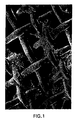
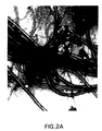
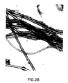
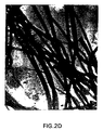
- 238000004626 scanning electron microscopy Methods 0.000 description 3
- 238000000527 sonication Methods 0.000 description 3
- 238000012421 spiking Methods 0.000 description 3
- 229960005322 streptomycin Drugs 0.000 description 3
- 239000000758 substrate Substances 0.000 description 3
- 230000017423 tissue regeneration Effects 0.000 description 3
- LENZDBCJOHFCAS-UHFFFAOYSA-N tris Chemical compound OCC(N)(CO)CO LENZDBCJOHFCAS-UHFFFAOYSA-N 0.000 description 3
- OWEGMIWEEQEYGQ-UHFFFAOYSA-N 100676-05-9 Natural products OC1C(O)C(O)C(CO)OC1OCC1C(O)C(O)C(O)C(OC2C(OC(O)C(O)C2O)CO)O1 OWEGMIWEEQEYGQ-UHFFFAOYSA-N 0.000 description 2
- 102000007299 Amphiregulin Human genes 0.000 description 2
- 108010033760 Amphiregulin Proteins 0.000 description 2
- CIWBSHSKHKDKBQ-JLAZNSOCSA-N Ascorbic acid Chemical compound OC[C@H](O)[C@H]1OC(=O)C(O)=C1O CIWBSHSKHKDKBQ-JLAZNSOCSA-N 0.000 description 2
- 206010003694 Atrophy Diseases 0.000 description 2
- 206010009269 Cleft palate Diseases 0.000 description 2
- 206010010356 Congenital anomaly Diseases 0.000 description 2
- QNAYBMKLOCPYGJ-UHFFFAOYSA-N D-alpha-Ala Natural products CC([NH3+])C([O-])=O QNAYBMKLOCPYGJ-UHFFFAOYSA-N 0.000 description 2
- 102000016911 Deoxyribonucleases Human genes 0.000 description 2
- 108010053770 Deoxyribonucleases Proteins 0.000 description 2
- 102000016942 Elastin Human genes 0.000 description 2
- 108010014258 Elastin Proteins 0.000 description 2
- 101150021185 FGF gene Proteins 0.000 description 2
- 239000004471 Glycine Substances 0.000 description 2
- 102000003886 Glycoproteins Human genes 0.000 description 2
- 108090000288 Glycoproteins Proteins 0.000 description 2
- 101800001649 Heparin-binding EGF-like growth factor Proteins 0.000 description 2
- QNAYBMKLOCPYGJ-UWTATZPHSA-N L-Alanine Natural products C[C@@H](N)C(O)=O QNAYBMKLOCPYGJ-UWTATZPHSA-N 0.000 description 2
- QNAYBMKLOCPYGJ-REOHCLBHSA-N L-alanine Chemical compound C[C@H](N)C(O)=O QNAYBMKLOCPYGJ-REOHCLBHSA-N 0.000 description 2
- GUBGYTABKSRVRQ-PICCSMPSSA-N Maltose Natural products O[C@@H]1[C@@H](O)[C@H](O)[C@@H](CO)O[C@@H]1O[C@@H]1[C@@H](CO)OC(O)[C@H](O)[C@H]1O GUBGYTABKSRVRQ-PICCSMPSSA-N 0.000 description 2
- 229930182555 Penicillin Natural products 0.000 description 2
- JGSARLDLIJGVTE-MBNYWOFBSA-N Penicillin G Chemical compound N([C@H]1[C@H]2SC([C@@H](N2C1=O)C(O)=O)(C)C)C(=O)CC1=CC=CC=C1 JGSARLDLIJGVTE-MBNYWOFBSA-N 0.000 description 2
- 102100033762 Proheparin-binding EGF-like growth factor Human genes 0.000 description 2
- 108020004511 Recombinant DNA Proteins 0.000 description 2
- 108010081750 Reticulin Proteins 0.000 description 2
- DBMJMQXJHONAFJ-UHFFFAOYSA-M Sodium laurylsulphate Chemical compound [Na+].CCCCCCCCCCCCOS([O-])(=O)=O DBMJMQXJHONAFJ-UHFFFAOYSA-M 0.000 description 2
- QAOWNCQODCNURD-UHFFFAOYSA-N Sulfuric acid Chemical compound OS(O)(=O)=O QAOWNCQODCNURD-UHFFFAOYSA-N 0.000 description 2
- 102000004887 Transforming Growth Factor beta Human genes 0.000 description 2
- 108090001012 Transforming Growth Factor beta Proteins 0.000 description 2
- 101800004564 Transforming growth factor alpha Proteins 0.000 description 2
- 102400001320 Transforming growth factor alpha Human genes 0.000 description 2
- 102000005789 Vascular Endothelial Growth Factors Human genes 0.000 description 2
- 108010019530 Vascular Endothelial Growth Factors Proteins 0.000 description 2
- 230000002378 acidificating effect Effects 0.000 description 2
- 239000000853 adhesive Substances 0.000 description 2
- 230000001070 adhesive effect Effects 0.000 description 2
- 229960003767 alanine Drugs 0.000 description 2
- 150000001299 aldehydes Chemical class 0.000 description 2
- 150000001413 amino acids Chemical class 0.000 description 2
- APKFDSVGJQXUKY-INPOYWNPSA-N amphotericin B Chemical compound O[C@H]1[C@@H](N)[C@H](O)[C@@H](C)O[C@H]1O[C@H]1/C=C/C=C/C=C/C=C/C=C/C=C/C=C/[C@H](C)[C@@H](O)[C@@H](C)[C@H](C)OC(=O)C[C@H](O)C[C@H](O)CC[C@@H](O)[C@H](O)C[C@H](O)C[C@](O)(C[C@H](O)[C@H]2C(O)=O)O[C@H]2C1 APKFDSVGJQXUKY-INPOYWNPSA-N 0.000 description 2
- 210000004102 animal cell Anatomy 0.000 description 2
- 239000008365 aqueous carrier Substances 0.000 description 2
- 239000012298 atmosphere Substances 0.000 description 2
- 230000037444 atrophy Effects 0.000 description 2
- 238000011888 autopsy Methods 0.000 description 2
- 210000002469 basement membrane Anatomy 0.000 description 2
- 239000012620 biological material Substances 0.000 description 2
- 238000001574 biopsy Methods 0.000 description 2
- 210000004556 brain Anatomy 0.000 description 2
- 239000006285 cell suspension Substances 0.000 description 2
- 230000033077 cellular process Effects 0.000 description 2
- 230000030944 contact inhibition Effects 0.000 description 2
- 230000006378 damage Effects 0.000 description 2
- 230000004069 differentiation Effects 0.000 description 2
- 230000029087 digestion Effects 0.000 description 2
- 102000038379 digestive enzymes Human genes 0.000 description 2
- 108091007734 digestive enzymes Proteins 0.000 description 2
- 229920002549 elastin Polymers 0.000 description 2
- 239000003797 essential amino acid Substances 0.000 description 2
- 235000020776 essential amino acid Nutrition 0.000 description 2
- 238000002474 experimental method Methods 0.000 description 2
- 230000001605 fetal effect Effects 0.000 description 2
- 230000003394 haemopoietic effect Effects 0.000 description 2
- 210000003630 histaminocyte Anatomy 0.000 description 2
- 238000000265 homogenisation Methods 0.000 description 2
- JYGXADMDTFJGBT-VWUMJDOOSA-N hydrocortisone Chemical compound O=C1CC[C@]2(C)[C@H]3[C@@H](O)C[C@](C)([C@@](CC4)(O)C(=O)CO)[C@@H]4[C@@H]3CCC2=C1 JYGXADMDTFJGBT-VWUMJDOOSA-N 0.000 description 2
- 230000028993 immune response Effects 0.000 description 2
- 238000003119 immunoblot Methods 0.000 description 2
- 230000002163 immunogen Effects 0.000 description 2
- 238000002955 isolation Methods 0.000 description 2
- 238000012423 maintenance Methods 0.000 description 2
- 210000001724 microfibril Anatomy 0.000 description 2
- 238000002156 mixing Methods 0.000 description 2
- 210000004498 neuroglial cell Anatomy 0.000 description 2
- 229910052757 nitrogen Inorganic materials 0.000 description 2
- 210000000496 pancreas Anatomy 0.000 description 2
- 229940049954 penicillin Drugs 0.000 description 2
- 210000003668 pericyte Anatomy 0.000 description 2
- 230000002688 persistence Effects 0.000 description 2
- 210000004180 plasmocyte Anatomy 0.000 description 2
- 229920000915 polyvinyl chloride Polymers 0.000 description 2
- 239000004800 polyvinyl chloride Substances 0.000 description 2
- 102000004196 processed proteins & peptides Human genes 0.000 description 2
- 108090000765 processed proteins & peptides Proteins 0.000 description 2
- 230000001737 promoting effect Effects 0.000 description 2
- 238000012552 review Methods 0.000 description 2
- 238000001878 scanning electron micrograph Methods 0.000 description 2
- 238000007790 scraping Methods 0.000 description 2
- 210000002966 serum Anatomy 0.000 description 2
- 235000019333 sodium laurylsulphate Nutrition 0.000 description 2
- DAEPDZWVDSPTHF-UHFFFAOYSA-M sodium pyruvate Chemical compound [Na+].CC(=O)C([O-])=O DAEPDZWVDSPTHF-UHFFFAOYSA-M 0.000 description 2
- 238000007920 subcutaneous administration Methods 0.000 description 2
- 230000029305 taxis Effects 0.000 description 2
- 210000002435 tendon Anatomy 0.000 description 2
- ZRKFYGHZFMAOKI-QMGMOQQFSA-N tgfbeta Chemical compound C([C@H](NC(=O)[C@H](C(C)C)NC(=O)CNC(=O)[C@H](CCC(O)=O)NC(=O)[C@H](CCCNC(N)=N)NC(=O)[C@H](CC(N)=O)NC(=O)[C@H](CC(C)C)NC(=O)[C@H]([C@@H](C)O)NC(=O)[C@H](CCC(O)=O)NC(=O)[C@H]([C@@H](C)O)NC(=O)[C@H](CC(C)C)NC(=O)CNC(=O)[C@H](C)NC(=O)[C@H](CO)NC(=O)[C@H](CCC(N)=O)NC(=O)[C@@H](NC(=O)[C@H](C)NC(=O)[C@H](C)NC(=O)[C@@H](NC(=O)[C@H](CC(C)C)NC(=O)[C@@H](N)CCSC)C(C)C)[C@@H](C)CC)C(=O)N[C@@H]([C@@H](C)O)C(=O)N[C@@H](C(C)C)C(=O)N[C@@H](CC=1C=CC=CC=1)C(=O)N[C@@H](C)C(=O)N1[C@@H](CCC1)C(=O)N[C@@H]([C@@H](C)O)C(=O)N[C@@H](CC(N)=O)C(=O)N[C@@H](CCC(O)=O)C(=O)N[C@@H](C)C(=O)N[C@@H](CC=1C=CC=CC=1)C(=O)N[C@@H](CCCNC(N)=N)C(=O)N[C@@H](C)C(=O)N[C@@H](CC(C)C)C(=O)N1[C@@H](CCC1)C(=O)N1[C@@H](CCC1)C(=O)N[C@@H](CCCNC(N)=N)C(=O)N[C@@H](CCC(O)=O)C(=O)N[C@@H](CCCNC(N)=N)C(=O)N[C@@H](CO)C(=O)N[C@@H](CCCNC(N)=N)C(=O)N[C@@H](CC(C)C)C(=O)N[C@@H](CC(C)C)C(O)=O)C1=CC=C(O)C=C1 ZRKFYGHZFMAOKI-QMGMOQQFSA-N 0.000 description 2
- 231100000331 toxic Toxicity 0.000 description 2
- 230000002588 toxic effect Effects 0.000 description 2
- 238000012546 transfer Methods 0.000 description 2
- 238000004627 transmission electron microscopy Methods 0.000 description 2
- DNIAPMSPPWPWGF-GSVOUGTGSA-N (R)-(-)-Propylene glycol Chemical compound C[C@@H](O)CO DNIAPMSPPWPWGF-GSVOUGTGSA-N 0.000 description 1
- CMCBDXRRFKYBDG-UHFFFAOYSA-N 1-dodecoxydodecane Chemical compound CCCCCCCCCCCCOCCCCCCCCCCCC CMCBDXRRFKYBDG-UHFFFAOYSA-N 0.000 description 1
- IEQAICDLOKRSRL-UHFFFAOYSA-N 2-[2-[2-[2-[2-[2-[2-[2-[2-[2-[2-[2-[2-[2-[2-[2-[2-[2-[2-[2-[2-[2-(2-dodecoxyethoxy)ethoxy]ethoxy]ethoxy]ethoxy]ethoxy]ethoxy]ethoxy]ethoxy]ethoxy]ethoxy]ethoxy]ethoxy]ethoxy]ethoxy]ethoxy]ethoxy]ethoxy]ethoxy]ethoxy]ethoxy]ethoxy]ethanol Chemical compound CCCCCCCCCCCCOCCOCCOCCOCCOCCOCCOCCOCCOCCOCCOCCOCCOCCOCCOCCOCCOCCOCCOCCOCCOCCOCCOCCO IEQAICDLOKRSRL-UHFFFAOYSA-N 0.000 description 1
- UMCMPZBLKLEWAF-BCTGSCMUSA-N 3-[(3-cholamidopropyl)dimethylammonio]propane-1-sulfonate Chemical compound C([C@H]1C[C@H]2O)[C@H](O)CC[C@]1(C)[C@@H]1[C@@H]2[C@@H]2CC[C@H]([C@@H](CCC(=O)NCCC[N+](C)(C)CCCS([O-])(=O)=O)C)[C@@]2(C)[C@@H](O)C1 UMCMPZBLKLEWAF-BCTGSCMUSA-N 0.000 description 1
- 208000002874 Acne Vulgaris Diseases 0.000 description 1
- 206010067484 Adverse reaction Diseases 0.000 description 1
- 208000002109 Argyria Diseases 0.000 description 1
- 241000894006 Bacteria Species 0.000 description 1
- 206010007882 Cellulitis Diseases 0.000 description 1
- 229920001287 Chondroitin sulfate Polymers 0.000 description 1
- 108090000317 Chymotrypsin Proteins 0.000 description 1
- 102000004266 Collagen Type IV Human genes 0.000 description 1
- 108010042086 Collagen Type IV Proteins 0.000 description 1
- 229920004934 Dacron® Polymers 0.000 description 1
- 229920000045 Dermatan sulfate Polymers 0.000 description 1
- 208000002021 Enophthalmos Diseases 0.000 description 1
- IAYPIBMASNFSPL-UHFFFAOYSA-N Ethylene oxide Chemical compound C1CO1 IAYPIBMASNFSPL-UHFFFAOYSA-N 0.000 description 1
- 206010016654 Fibrosis Diseases 0.000 description 1
- 241000233866 Fungi Species 0.000 description 1
- 208000002325 Funnel Chest Diseases 0.000 description 1
- 229920002527 Glycogen Polymers 0.000 description 1
- 201000003200 Goldenhar Syndrome Diseases 0.000 description 1
- 206010061199 Head deformity Diseases 0.000 description 1
- 229920002971 Heparan sulfate Polymers 0.000 description 1
- HTTJABKRGRZYRN-UHFFFAOYSA-N Heparin Chemical compound OC1C(NC(=O)C)C(O)OC(COS(O)(=O)=O)C1OC1C(OS(O)(=O)=O)C(O)C(OC2C(C(OS(O)(=O)=O)C(OC3C(C(O)C(O)C(O3)C(O)=O)OS(O)(=O)=O)C(CO)O2)NS(O)(=O)=O)C(C(O)=O)O1 HTTJABKRGRZYRN-UHFFFAOYSA-N 0.000 description 1
- 101000599951 Homo sapiens Insulin-like growth factor I Proteins 0.000 description 1
- 102000003839 Human Proteins Human genes 0.000 description 1
- 108090000144 Human Proteins Proteins 0.000 description 1
- 108010003272 Hyaluronate lyase Proteins 0.000 description 1
- 102000001974 Hyaluronidases Human genes 0.000 description 1
- PMMYEEVYMWASQN-DMTCNVIQSA-N Hydroxyproline Chemical compound O[C@H]1CN[C@H](C(O)=O)C1 PMMYEEVYMWASQN-DMTCNVIQSA-N 0.000 description 1
- 108090000723 Insulin-Like Growth Factor I Proteins 0.000 description 1
- 102000004218 Insulin-Like Growth Factor I Human genes 0.000 description 1
- 102000048143 Insulin-Like Growth Factor II Human genes 0.000 description 1
- 108090001117 Insulin-Like Growth Factor II Proteins 0.000 description 1
- 102100037852 Insulin-like growth factor I Human genes 0.000 description 1
- 102100034343 Integrase Human genes 0.000 description 1
- 108010002352 Interleukin-1 Proteins 0.000 description 1
- 102000000589 Interleukin-1 Human genes 0.000 description 1
- 229920000288 Keratan sulfate Polymers 0.000 description 1
- 229930182816 L-glutamine Natural products 0.000 description 1
- 239000004166 Lanolin Substances 0.000 description 1
- NNJVILVZKWQKPM-UHFFFAOYSA-N Lidocaine Chemical compound CCN(CC)CC(=O)NC1=C(C)C=CC=C1C NNJVILVZKWQKPM-UHFFFAOYSA-N 0.000 description 1
- 102000004882 Lipase Human genes 0.000 description 1
- 108090001060 Lipase Proteins 0.000 description 1
- 239000004367 Lipase Substances 0.000 description 1
- 208000000185 Localized scleroderma Diseases 0.000 description 1
- 241001465754 Metazoa Species 0.000 description 1
- 206010027982 Morphoea Diseases 0.000 description 1
- 101001055320 Myxine glutinosa Insulin-like growth factor Proteins 0.000 description 1
- 206010061875 Nose deformity Diseases 0.000 description 1
- 101710163270 Nuclease Proteins 0.000 description 1
- 108010067372 Pancreatic elastase Proteins 0.000 description 1
- 102000016387 Pancreatic elastase Human genes 0.000 description 1
- 206010034204 Pectus excavatum Diseases 0.000 description 1
- 239000004264 Petrolatum Substances 0.000 description 1
- 102000015439 Phospholipases Human genes 0.000 description 1
- 108010064785 Phospholipases Proteins 0.000 description 1
- 208000019222 Poland syndrome Diseases 0.000 description 1
- 239000002202 Polyethylene glycol Substances 0.000 description 1
- 229920001213 Polysorbate 20 Polymers 0.000 description 1
- 108010059712 Pronase Proteins 0.000 description 1
- 102000016611 Proteoglycans Human genes 0.000 description 1
- 108010067787 Proteoglycans Proteins 0.000 description 1
- 108010092799 RNA-directed DNA polymerase Proteins 0.000 description 1
- MUPFEKGTMRGPLJ-RMMQSMQOSA-N Raffinose Natural products O(C[C@H]1[C@@H](O)[C@H](O)[C@@H](O)[C@@H](O[C@@]2(CO)[C@H](O)[C@@H](O)[C@@H](CO)O2)O1)[C@@H]1[C@H](O)[C@@H](O)[C@@H](O)[C@@H](CO)O1 MUPFEKGTMRGPLJ-RMMQSMQOSA-N 0.000 description 1
- 102000006382 Ribonucleases Human genes 0.000 description 1
- 108010083644 Ribonucleases Proteins 0.000 description 1
- 239000006146 Roswell Park Memorial Institute medium Substances 0.000 description 1
- 108010071390 Serum Albumin Proteins 0.000 description 1
- 102000007562 Serum Albumin Human genes 0.000 description 1
- YIQKLZYTHXTDDT-UHFFFAOYSA-H Sirius red F3B Chemical compound C1=CC(=CC=C1N=NC2=CC(=C(C=C2)N=NC3=C(C=C4C=C(C=CC4=C3[O-])NC(=O)NC5=CC6=CC(=C(C(=C6C=C5)[O-])N=NC7=C(C=C(C=C7)N=NC8=CC=C(C=C8)S(=O)(=O)[O-])S(=O)(=O)[O-])S(=O)(=O)O)S(=O)(=O)O)S(=O)(=O)[O-])S(=O)(=O)[O-].[Na+].[Na+].[Na+].[Na+].[Na+].[Na+] YIQKLZYTHXTDDT-UHFFFAOYSA-H 0.000 description 1
- 206010040943 Skin Ulcer Diseases 0.000 description 1
- FAPWRFPIFSIZLT-UHFFFAOYSA-M Sodium chloride Chemical compound [Na+].[Cl-] FAPWRFPIFSIZLT-UHFFFAOYSA-M 0.000 description 1
- 101710172711 Structural protein Proteins 0.000 description 1
- 229930006000 Sucrose Natural products 0.000 description 1
- CZMRCDWAGMRECN-UGDNZRGBSA-N Sucrose Chemical compound O[C@H]1[C@H](O)[C@@H](CO)O[C@@]1(CO)O[C@@H]1[C@H](O)[C@@H](O)[C@H](O)[C@@H](CO)O1 CZMRCDWAGMRECN-UGDNZRGBSA-N 0.000 description 1
- 238000003917 TEM image Methods 0.000 description 1
- 239000004809 Teflon Substances 0.000 description 1
- 229920006362 Teflon® Polymers 0.000 description 1
- 102000046299 Transforming Growth Factor beta1 Human genes 0.000 description 1
- 101800002279 Transforming growth factor beta-1 Proteins 0.000 description 1
- 102000056172 Transforming growth factor beta-3 Human genes 0.000 description 1
- 108090000097 Transforming growth factor beta-3 Proteins 0.000 description 1
- GLNADSQYFUSGOU-GPTZEZBUSA-J Trypan blue Chemical compound [Na+].[Na+].[Na+].[Na+].C1=C(S([O-])(=O)=O)C=C2C=C(S([O-])(=O)=O)C(/N=N/C3=CC=C(C=C3C)C=3C=C(C(=CC=3)\N=N\C=3C(=CC4=CC(=CC(N)=C4C=3O)S([O-])(=O)=O)S([O-])(=O)=O)C)=C(O)C2=C1N GLNADSQYFUSGOU-GPTZEZBUSA-J 0.000 description 1
- MUPFEKGTMRGPLJ-UHFFFAOYSA-N UNPD196149 Natural products OC1C(O)C(CO)OC1(CO)OC1C(O)C(O)C(O)C(COC2C(C(O)C(O)C(CO)O2)O)O1 MUPFEKGTMRGPLJ-UHFFFAOYSA-N 0.000 description 1
- 208000006812 Velopharyngeal Insufficiency Diseases 0.000 description 1
- 206010066790 Velopharyngeal incompetence Diseases 0.000 description 1
- 241000700605 Viruses Species 0.000 description 1
- 208000005248 Vocal Cord Paralysis Diseases 0.000 description 1
- 238000010521 absorption reaction Methods 0.000 description 1
- 239000002253 acid Substances 0.000 description 1
- 206010000496 acne Diseases 0.000 description 1
- 230000003213 activating effect Effects 0.000 description 1
- 230000006838 adverse reaction Effects 0.000 description 1
- 150000001335 aliphatic alkanes Chemical class 0.000 description 1
- 208000026935 allergic disease Diseases 0.000 description 1
- 230000000172 allergic effect Effects 0.000 description 1
- 208000030961 allergic reaction Diseases 0.000 description 1
- 230000007815 allergy Effects 0.000 description 1
- 229940024606 amino acid Drugs 0.000 description 1
- 230000003321 amplification Effects 0.000 description 1
- 239000012491 analyte Substances 0.000 description 1
- 238000004458 analytical method Methods 0.000 description 1
- 230000033115 angiogenesis Effects 0.000 description 1
- 230000001857 anti-mycotic effect Effects 0.000 description 1
- 230000000890 antigenic effect Effects 0.000 description 1
- 239000002543 antimycotic Substances 0.000 description 1
- 238000013459 approach Methods 0.000 description 1
- 239000012736 aqueous medium Substances 0.000 description 1
- 239000007900 aqueous suspension Substances 0.000 description 1
- 238000003491 array Methods 0.000 description 1
- 229940072107 ascorbate Drugs 0.000 description 1
- 235000010323 ascorbic acid Nutrition 0.000 description 1
- 239000011668 ascorbic acid Substances 0.000 description 1
- 108010045569 atelocollagen Proteins 0.000 description 1
- 208000010668 atopic eczema Diseases 0.000 description 1
- 230000003190 augmentative effect Effects 0.000 description 1
- 239000011324 bead Substances 0.000 description 1
- 235000013871 bee wax Nutrition 0.000 description 1
- 229940092738 beeswax Drugs 0.000 description 1
- 239000012166 beeswax Substances 0.000 description 1
- 210000000013 bile duct Anatomy 0.000 description 1
- 230000003115 biocidal effect Effects 0.000 description 1
- 230000015572 biosynthetic process Effects 0.000 description 1
- 210000004204 blood vessel Anatomy 0.000 description 1
- 229940098773 bovine serum albumin Drugs 0.000 description 1
- 239000000872 buffer Substances 0.000 description 1
- CDQSJQSWAWPGKG-UHFFFAOYSA-N butane-1,1-diol Chemical compound CCCC(O)O CDQSJQSWAWPGKG-UHFFFAOYSA-N 0.000 description 1
- 239000002775 capsule Substances 0.000 description 1
- 125000004432 carbon atom Chemical group C* 0.000 description 1
- 230000024245 cell differentiation Effects 0.000 description 1
- 230000004663 cell proliferation Effects 0.000 description 1
- 230000008614 cellular interaction Effects 0.000 description 1
- 239000000919 ceramic Substances 0.000 description 1
- 239000002738 chelating agent Substances 0.000 description 1
- 239000013043 chemical agent Substances 0.000 description 1
- 238000010382 chemical cross-linking Methods 0.000 description 1
- 238000006243 chemical reaction Methods 0.000 description 1
- 239000003153 chemical reaction reagent Substances 0.000 description 1
- 239000003795 chemical substances by application Substances 0.000 description 1
- 230000001587 cholestatic effect Effects 0.000 description 1
- 229940094517 chondroitin 4-sulfate Drugs 0.000 description 1
- KXKPYJOVDUMHGS-OSRGNVMNSA-N chondroitin sulfate Chemical compound CC(=O)N[C@H]1[C@H](O)O[C@H](OS(O)(=O)=O)[C@H](O)[C@@H]1O[C@H]1[C@H](O)[C@@H](O)[C@H](O)[C@@H](C(O)=O)O1 KXKPYJOVDUMHGS-OSRGNVMNSA-N 0.000 description 1
- 229960002376 chymotrypsin Drugs 0.000 description 1
- 238000010367 cloning Methods 0.000 description 1
- 239000011248 coating agent Substances 0.000 description 1
- 238000000576 coating method Methods 0.000 description 1
- 239000000515 collagen sponge Substances 0.000 description 1
- 230000001143 conditioned effect Effects 0.000 description 1
- 238000013270 controlled release Methods 0.000 description 1
- 238000012937 correction Methods 0.000 description 1
- 239000003431 cross linking reagent Substances 0.000 description 1
- 230000002338 cryopreservative effect Effects 0.000 description 1
- 239000002577 cryoprotective agent Substances 0.000 description 1
- 210000004268 dentin Anatomy 0.000 description 1
- 230000000994 depressogenic effect Effects 0.000 description 1
- AVJBPWGFOQAPRH-FWMKGIEWSA-L dermatan sulfate Chemical compound CC(=O)N[C@H]1[C@H](O)O[C@H](CO)[C@H](OS([O-])(=O)=O)[C@@H]1O[C@H]1[C@H](O)[C@@H](O)[C@H](O)[C@H](C([O-])=O)O1 AVJBPWGFOQAPRH-FWMKGIEWSA-L 0.000 description 1
- 229940051593 dermatan sulfate Drugs 0.000 description 1
- 238000001514 detection method Methods 0.000 description 1
- 238000011161 development Methods 0.000 description 1
- 230000018109 developmental process Effects 0.000 description 1
- LOKCTEFSRHRXRJ-UHFFFAOYSA-I dipotassium trisodium dihydrogen phosphate hydrogen phosphate dichloride Chemical compound P(=O)(O)(O)[O-].[K+].P(=O)(O)([O-])[O-].[Na+].[Na+].[Cl-].[K+].[Cl-].[Na+] LOKCTEFSRHRXRJ-UHFFFAOYSA-I 0.000 description 1
- 108010007093 dispase Proteins 0.000 description 1
- 238000010494 dissociation reaction Methods 0.000 description 1
- 230000005593 dissociations Effects 0.000 description 1
- 239000012153 distilled water Substances 0.000 description 1
- PMMYEEVYMWASQN-UHFFFAOYSA-N dl-hydroxyproline Natural products OC1C[NH2+]C(C([O-])=O)C1 PMMYEEVYMWASQN-UHFFFAOYSA-N 0.000 description 1
- 229940079593 drug Drugs 0.000 description 1
- 239000003814 drug Substances 0.000 description 1
- 229960001484 edetic acid Drugs 0.000 description 1
- 210000004177 elastic tissue Anatomy 0.000 description 1
- 238000010894 electron beam technology Methods 0.000 description 1
- 238000001493 electron microscopy Methods 0.000 description 1
- 238000001962 electrophoresis Methods 0.000 description 1
- 230000003511 endothelial effect Effects 0.000 description 1
- 210000003038 endothelium Anatomy 0.000 description 1
- 230000006862 enzymatic digestion Effects 0.000 description 1
- 230000002255 enzymatic effect Effects 0.000 description 1
- 210000003527 eukaryotic cell Anatomy 0.000 description 1
- 230000007717 exclusion Effects 0.000 description 1
- 210000001723 extracellular space Anatomy 0.000 description 1
- 230000001815 facial effect Effects 0.000 description 1
- 208000002980 facial hemiatrophy Diseases 0.000 description 1
- 210000003754 fetus Anatomy 0.000 description 1
- 210000000968 fibrocartilage Anatomy 0.000 description 1
- 230000004761 fibrosis Effects 0.000 description 1
- 230000003176 fibrotic effect Effects 0.000 description 1
- 239000010419 fine particle Substances 0.000 description 1
- 238000001943 fluorescence-activated cell sorting Methods 0.000 description 1
- 210000003953 foreskin Anatomy 0.000 description 1
- 238000007710 freezing Methods 0.000 description 1
- ZZUFCTLCJUWOSV-UHFFFAOYSA-N furosemide Chemical compound C1=C(Cl)C(S(=O)(=O)N)=CC(C(O)=O)=C1NCC1=CC=CO1 ZZUFCTLCJUWOSV-UHFFFAOYSA-N 0.000 description 1
- 239000000499 gel Substances 0.000 description 1
- 239000012362 glacial acetic acid Substances 0.000 description 1
- 230000002518 glial effect Effects 0.000 description 1
- 150000004676 glycans Polymers 0.000 description 1
- 229940096919 glycogen Drugs 0.000 description 1
- 210000002288 golgi apparatus Anatomy 0.000 description 1
- 230000005484 gravity Effects 0.000 description 1
- 230000035876 healing Effects 0.000 description 1
- 210000003958 hematopoietic stem cell Anatomy 0.000 description 1
- 208000017918 hemifacial microsomia Diseases 0.000 description 1
- 210000004276 hyalin Anatomy 0.000 description 1
- 229960002773 hyaluronidase Drugs 0.000 description 1
- 229960000890 hydrocortisone Drugs 0.000 description 1
- 229960002591 hydroxyproline Drugs 0.000 description 1
- 210000002865 immune cell Anatomy 0.000 description 1
- 238000010166 immunofluorescence Methods 0.000 description 1
- 230000006872 improvement Effects 0.000 description 1
- 238000011065 in-situ storage Methods 0.000 description 1
- 230000001939 inductive effect Effects 0.000 description 1
- 208000015181 infectious disease Diseases 0.000 description 1
- 230000008611 intercellular interaction Effects 0.000 description 1
- FZWBNHMXJMCXLU-BLAUPYHCSA-N isomaltotriose Chemical compound O[C@@H]1[C@@H](O)[C@H](O)[C@@H](CO)O[C@@H]1OC[C@@H]1[C@@H](O)[C@H](O)[C@@H](O)[C@@H](OC[C@@H](O)[C@@H](O)[C@H](O)[C@@H](O)C=O)O1 FZWBNHMXJMCXLU-BLAUPYHCSA-N 0.000 description 1
- YWXYYJSYQOXTPL-SLPGGIOYSA-N isosorbide mononitrate Chemical compound [O-][N+](=O)O[C@@H]1CO[C@@H]2[C@@H](O)CO[C@@H]21 YWXYYJSYQOXTPL-SLPGGIOYSA-N 0.000 description 1
- KXCLCNHUUKTANI-RBIYJLQWSA-N keratan Chemical compound CC(=O)N[C@@H]1[C@@H](O)C[C@@H](COS(O)(=O)=O)O[C@H]1O[C@@H]1[C@@H](O)[C@H](O[C@@H]2[C@H](O[C@@H](O[C@H]3[C@H]([C@@H](COS(O)(=O)=O)O[C@@H](O)[C@@H]3O)O)[C@H](NC(C)=O)[C@H]2O)COS(O)(=O)=O)O[C@H](COS(O)(=O)=O)[C@@H]1O KXCLCNHUUKTANI-RBIYJLQWSA-N 0.000 description 1
- 210000001865 kupffer cell Anatomy 0.000 description 1
- 235000019388 lanolin Nutrition 0.000 description 1
- 229940039717 lanolin Drugs 0.000 description 1
- 230000003902 lesion Effects 0.000 description 1
- 229960004194 lidocaine Drugs 0.000 description 1
- 208000016809 linear scleroderma Diseases 0.000 description 1
- 235000019421 lipase Nutrition 0.000 description 1
- 229960005015 local anesthetics Drugs 0.000 description 1
- 230000033001 locomotion Effects 0.000 description 1
- 210000004698 lymphocyte Anatomy 0.000 description 1
- 230000014759 maintenance of location Effects 0.000 description 1
- 230000035800 maturation Effects 0.000 description 1
- 230000007246 mechanism Effects 0.000 description 1
- 108020004999 messenger RNA Proteins 0.000 description 1
- 230000001394 metastastic effect Effects 0.000 description 1
- 206010061289 metastatic neoplasm Diseases 0.000 description 1
- 230000001617 migratory effect Effects 0.000 description 1
- 230000011278 mitosis Effects 0.000 description 1
- 238000004264 monolayer culture Methods 0.000 description 1
- 210000004400 mucous membrane Anatomy 0.000 description 1
- 210000004788 neurological cell Anatomy 0.000 description 1
- 238000010899 nucleation Methods 0.000 description 1
- 238000003199 nucleic acid amplification method Methods 0.000 description 1
- 235000015097 nutrients Nutrition 0.000 description 1
- UYDLBVPAAFVANX-UHFFFAOYSA-N octylphenoxy polyethoxyethanol Chemical compound CC(C)(C)CC(C)(C)C1=CC=C(OCCOCCOCCOCCO)C=C1 UYDLBVPAAFVANX-UHFFFAOYSA-N 0.000 description 1
- 229920000620 organic polymer Polymers 0.000 description 1
- 239000003960 organic solvent Substances 0.000 description 1
- 230000003204 osmotic effect Effects 0.000 description 1
- 230000002188 osteogenic effect Effects 0.000 description 1
- 239000012188 paraffin wax Substances 0.000 description 1
- 238000005192 partition Methods 0.000 description 1
- 229940066842 petrolatum Drugs 0.000 description 1
- 235000019271 petrolatum Nutrition 0.000 description 1
- 238000002135 phase contrast microscopy Methods 0.000 description 1
- 239000008363 phosphate buffer Substances 0.000 description 1
- OXNIZHLAWKMVMX-UHFFFAOYSA-N picric acid Chemical class OC1=C([N+]([O-])=O)C=C([N+]([O-])=O)C=C1[N+]([O-])=O OXNIZHLAWKMVMX-UHFFFAOYSA-N 0.000 description 1
- 210000002826 placenta Anatomy 0.000 description 1
- 229920001223 polyethylene glycol Polymers 0.000 description 1
- 239000005020 polyethylene terephthalate Substances 0.000 description 1
- 238000003752 polymerase chain reaction Methods 0.000 description 1
- 239000000256 polyoxyethylene sorbitan monolaurate Substances 0.000 description 1
- 235000010486 polyoxyethylene sorbitan monolaurate Nutrition 0.000 description 1
- 229920001184 polypeptide Polymers 0.000 description 1
- 150000004804 polysaccharides Polymers 0.000 description 1
- 239000004810 polytetrafluoroethylene Substances 0.000 description 1
- 229920000036 polyvinylpyrrolidone Polymers 0.000 description 1
- 239000001267 polyvinylpyrrolidone Substances 0.000 description 1
- 235000013855 polyvinylpyrrolidone Nutrition 0.000 description 1
- 238000001556 precipitation Methods 0.000 description 1
- 230000002035 prolonged effect Effects 0.000 description 1
- 230000005855 radiation Effects 0.000 description 1
- MUPFEKGTMRGPLJ-ZQSKZDJDSA-N raffinose Chemical compound O[C@H]1[C@H](O)[C@@H](CO)O[C@@]1(CO)O[C@@H]1[C@H](O)[C@@H](O)[C@H](O)[C@@H](CO[C@@H]2[C@@H]([C@@H](O)[C@@H](O)[C@@H](CO)O2)O)O1 MUPFEKGTMRGPLJ-ZQSKZDJDSA-N 0.000 description 1
- 238000011084 recovery Methods 0.000 description 1
- 230000025053 regulation of cell proliferation Effects 0.000 description 1
- 230000003362 replicative effect Effects 0.000 description 1
- 238000011160 research Methods 0.000 description 1
- 230000004044 response Effects 0.000 description 1
- 238000003757 reverse transcription PCR Methods 0.000 description 1
- 206010039073 rheumatoid arthritis Diseases 0.000 description 1
- 210000003935 rough endoplasmic reticulum Anatomy 0.000 description 1
- 230000037390 scarring Effects 0.000 description 1
- 238000005204 segregation Methods 0.000 description 1
- 230000035945 sensitivity Effects 0.000 description 1
- 210000004927 skin cell Anatomy 0.000 description 1
- 210000001626 skin fibroblast Anatomy 0.000 description 1
- 231100000019 skin ulcer Toxicity 0.000 description 1
- 210000002460 smooth muscle Anatomy 0.000 description 1
- 239000011780 sodium chloride Substances 0.000 description 1
- 229940054269 sodium pyruvate Drugs 0.000 description 1
- 239000001590 sorbitan monolaureate Substances 0.000 description 1
- 235000011067 sorbitan monolaureate Nutrition 0.000 description 1
- 210000005070 sphincter Anatomy 0.000 description 1
- 238000010186 staining Methods 0.000 description 1
- 238000010561 standard procedure Methods 0.000 description 1
- 239000008223 sterile water Substances 0.000 description 1
- 230000004936 stimulating effect Effects 0.000 description 1
- 239000000126 substance Substances 0.000 description 1
- 239000005720 sucrose Substances 0.000 description 1
- 239000006228 supernatant Substances 0.000 description 1
- 230000000153 supplemental effect Effects 0.000 description 1
- 230000003746 surface roughness Effects 0.000 description 1
- 230000008961 swelling Effects 0.000 description 1
- 208000024891 symptom Diseases 0.000 description 1
- 208000011580 syndromic disease Diseases 0.000 description 1
- 230000008467 tissue growth Effects 0.000 description 1
- FGMPLJWBKKVCDB-UHFFFAOYSA-N trans-L-hydroxy-proline Natural products ON1CCCC1C(O)=O FGMPLJWBKKVCDB-UHFFFAOYSA-N 0.000 description 1
- 230000002103 transcriptional effect Effects 0.000 description 1
- GPRLSGONYQIRFK-MNYXATJNSA-N triton Chemical compound [3H+] GPRLSGONYQIRFK-MNYXATJNSA-N 0.000 description 1
- 210000004881 tumor cell Anatomy 0.000 description 1
- 230000002792 vascular Effects 0.000 description 1
- 235000015112 vegetable and seed oil Nutrition 0.000 description 1
- 239000008158 vegetable oil Substances 0.000 description 1
- 239000003981 vehicle Substances 0.000 description 1
- 230000035899 viability Effects 0.000 description 1
- 210000004127 vitreous body Anatomy 0.000 description 1
- 108010047303 von Willebrand Factor Proteins 0.000 description 1
- 102100036537 von Willebrand factor Human genes 0.000 description 1
- 230000029663 wound healing Effects 0.000 description 1
- 210000000707 wrist Anatomy 0.000 description 1
Images

Classifications
-
- A—HUMAN NECESSITIES
- A61—MEDICAL OR VETERINARY SCIENCE; HYGIENE
- A61L—METHODS OR APPARATUS FOR STERILISING MATERIALS OR OBJECTS IN GENERAL; DISINFECTION, STERILISATION OR DEODORISATION OF AIR; CHEMICAL ASPECTS OF BANDAGES, DRESSINGS, ABSORBENT PADS OR SURGICAL ARTICLES; MATERIALS FOR BANDAGES, DRESSINGS, ABSORBENT PADS OR SURGICAL ARTICLES
- A61L27/00—Materials for grafts or prostheses or for coating grafts or prostheses
- A61L27/36—Materials for grafts or prostheses or for coating grafts or prostheses containing ingredients of undetermined constitution or reaction products thereof, e.g. transplant tissue, natural bone, extracellular matrix
- A61L27/38—Materials for grafts or prostheses or for coating grafts or prostheses containing ingredients of undetermined constitution or reaction products thereof, e.g. transplant tissue, natural bone, extracellular matrix containing added animal cells
- A61L27/3839—Materials for grafts or prostheses or for coating grafts or prostheses containing ingredients of undetermined constitution or reaction products thereof, e.g. transplant tissue, natural bone, extracellular matrix containing added animal cells characterised by the site of application in the body
-
- A—HUMAN NECESSITIES
- A61—MEDICAL OR VETERINARY SCIENCE; HYGIENE
- A61F—FILTERS IMPLANTABLE INTO BLOOD VESSELS; PROSTHESES; DEVICES PROVIDING PATENCY TO, OR PREVENTING COLLAPSING OF, TUBULAR STRUCTURES OF THE BODY, e.g. STENTS; ORTHOPAEDIC, NURSING OR CONTRACEPTIVE DEVICES; FOMENTATION; TREATMENT OR PROTECTION OF EYES OR EARS; BANDAGES, DRESSINGS OR ABSORBENT PADS; FIRST-AID KITS
- A61F2/00—Filters implantable into blood vessels; Prostheses, i.e. artificial substitutes or replacements for parts of the body; Appliances for connecting them with the body; Devices providing patency to, or preventing collapsing of, tubular structures of the body, e.g. stents
- A61F2/0059—Cosmetic or alloplastic implants
-
- A—HUMAN NECESSITIES
- A61—MEDICAL OR VETERINARY SCIENCE; HYGIENE
- A61F—FILTERS IMPLANTABLE INTO BLOOD VESSELS; PROSTHESES; DEVICES PROVIDING PATENCY TO, OR PREVENTING COLLAPSING OF, TUBULAR STRUCTURES OF THE BODY, e.g. STENTS; ORTHOPAEDIC, NURSING OR CONTRACEPTIVE DEVICES; FOMENTATION; TREATMENT OR PROTECTION OF EYES OR EARS; BANDAGES, DRESSINGS OR ABSORBENT PADS; FIRST-AID KITS
- A61F2/00—Filters implantable into blood vessels; Prostheses, i.e. artificial substitutes or replacements for parts of the body; Appliances for connecting them with the body; Devices providing patency to, or preventing collapsing of, tubular structures of the body, e.g. stents
- A61F2/02—Prostheses implantable into the body
- A61F2/10—Hair or skin implants
- A61F2/105—Skin implants, e.g. artificial skin
-
- A—HUMAN NECESSITIES
- A61—MEDICAL OR VETERINARY SCIENCE; HYGIENE
- A61L—METHODS OR APPARATUS FOR STERILISING MATERIALS OR OBJECTS IN GENERAL; DISINFECTION, STERILISATION OR DEODORISATION OF AIR; CHEMICAL ASPECTS OF BANDAGES, DRESSINGS, ABSORBENT PADS OR SURGICAL ARTICLES; MATERIALS FOR BANDAGES, DRESSINGS, ABSORBENT PADS OR SURGICAL ARTICLES
- A61L27/00—Materials for grafts or prostheses or for coating grafts or prostheses
- A61L27/36—Materials for grafts or prostheses or for coating grafts or prostheses containing ingredients of undetermined constitution or reaction products thereof, e.g. transplant tissue, natural bone, extracellular matrix
- A61L27/3604—Materials for grafts or prostheses or for coating grafts or prostheses containing ingredients of undetermined constitution or reaction products thereof, e.g. transplant tissue, natural bone, extracellular matrix characterised by the human or animal origin of the biological material, e.g. hair, fascia, fish scales, silk, shellac, pericardium, pleura, renal tissue, amniotic membrane, parenchymal tissue, fetal tissue, muscle tissue, fat tissue, enamel
- A61L27/3633—Extracellular matrix [ECM]
-
- A—HUMAN NECESSITIES
- A61—MEDICAL OR VETERINARY SCIENCE; HYGIENE
- A61L—METHODS OR APPARATUS FOR STERILISING MATERIALS OR OBJECTS IN GENERAL; DISINFECTION, STERILISATION OR DEODORISATION OF AIR; CHEMICAL ASPECTS OF BANDAGES, DRESSINGS, ABSORBENT PADS OR SURGICAL ARTICLES; MATERIALS FOR BANDAGES, DRESSINGS, ABSORBENT PADS OR SURGICAL ARTICLES
- A61L27/00—Materials for grafts or prostheses or for coating grafts or prostheses
- A61L27/36—Materials for grafts or prostheses or for coating grafts or prostheses containing ingredients of undetermined constitution or reaction products thereof, e.g. transplant tissue, natural bone, extracellular matrix
- A61L27/3683—Materials for grafts or prostheses or for coating grafts or prostheses containing ingredients of undetermined constitution or reaction products thereof, e.g. transplant tissue, natural bone, extracellular matrix subjected to a specific treatment prior to implantation, e.g. decellularising, demineralising, grinding, cellular disruption/non-collagenous protein removal, anti-calcification, crosslinking, supercritical fluid extraction, enzyme treatment
-
- A—HUMAN NECESSITIES
- A61—MEDICAL OR VETERINARY SCIENCE; HYGIENE
- A61L—METHODS OR APPARATUS FOR STERILISING MATERIALS OR OBJECTS IN GENERAL; DISINFECTION, STERILISATION OR DEODORISATION OF AIR; CHEMICAL ASPECTS OF BANDAGES, DRESSINGS, ABSORBENT PADS OR SURGICAL ARTICLES; MATERIALS FOR BANDAGES, DRESSINGS, ABSORBENT PADS OR SURGICAL ARTICLES
- A61L27/00—Materials for grafts or prostheses or for coating grafts or prostheses
- A61L27/36—Materials for grafts or prostheses or for coating grafts or prostheses containing ingredients of undetermined constitution or reaction products thereof, e.g. transplant tissue, natural bone, extracellular matrix
- A61L27/38—Materials for grafts or prostheses or for coating grafts or prostheses containing ingredients of undetermined constitution or reaction products thereof, e.g. transplant tissue, natural bone, extracellular matrix containing added animal cells
- A61L27/3804—Materials for grafts or prostheses or for coating grafts or prostheses containing ingredients of undetermined constitution or reaction products thereof, e.g. transplant tissue, natural bone, extracellular matrix containing added animal cells characterised by specific cells or progenitors thereof, e.g. fibroblasts, connective tissue cells, kidney cells
Definitions
- the present invention relates to compositions and methods for the treatment and repair of soft tissue and skin defects such as wrinkles and scars. More particularly, the invention relates to an injectable composition of human extracellular matrix components and methods of preparing and using same.
- the injectable preparation is obtained from three-dimensional living stromal tissues that are prepared in vitro .
- Collagen is the principal extracellular structural protein of the animal body. At least fourteen types of mammalian collagen have been described. The common characteristic amongst them is a three stranded helix, consisting of three polypeptide chains, called alpha-chains. All alpha-chains have the same configuration, but differ in the composition and sequence of their amino acids. Although this leads to different types of alpha-chains, however, they all have glycine at every third position in the amino acid sequence. The glycine at every third position allows for the helical structure of the alpha-chains.
- Type I collagen is composed of two alpha 1 -chains and one alpha 2 -chain and is the principal extracellular material of skin, tendon and bone. When clinicians mention "collagen", they are usually referring to type I collagen. See Table I, infra, for a detailed listing of collagen types I-V and in which tissues they are found.
- Collagen has been used as an implant material to replace or augment hard or soft connective tissue, such as skin, tendon, cartilage, bone and interstitium. Additionally, collagen implants have been used for cosmetic purposes for a number of years since collagen can help cellular ingrowth at the placement site. Early collagen implants were often solid collagen masses which were cross-linked with chemical agents, radiation or other means to improve mechanical properties, decrease immunogenicity and/or increase resistance to resorption. The collagen utilized was in a variety of forms, including cross-linked and non-cross-linked fibrillar collagens, gelatins, and the like and sometimes was combined with various other components, such as lubricants, osteogenic factors and the like, depending on use. A major disadvantage of solid cross-linked collagen implants is the requirement for surgical implantation by means of incision. In addition, lack of deformability and flexibility are other disadvantages of solid collagen implants.
- U.S. Pat. No. 3,949,073 An alternative to surgically implanted solid collagen material is disclosed in U.S. Pat. No. 3,949,073.
- U.S. Patent No. 3,949,073 describes the use of atelopeptide solutions of bovine collagen as an injectable implant material for augmenting soft tissue.
- the bovine collagen is reconstituted before implantation and forms a fibrous mass of tissue when implanted.
- the patent suggests adding particles of insoluble bovine collagen microfibrils to control the shrinkage of the fibrous mass formed at the augmentation site.
- the commercial embodiment of the material described in the patent is composed of reconstituted atelopeptide bovine collagen in saline that contains a small amount of local anesthetic.
- the implant shrinks in volume after implantation due primarily to absorption of its fluid component by the body.
- volume consistency is essential, an additional injection or injections of supplemental implant material is required.
- This specific composition has many serious drawbacks, e.g ., the collagen is from a bovine source, not human, and the preparation process is not only lengthy and expensive but also requires the addition of microfibrils.
- U.S. Patent No. 4,424,208 describes an injectable dispersion of cross-linked atelopeptide bovine collagen and reconstituted atelopeptide bovine collagen fibers in an aqueous carrier which exhibited improved volume consistency over the material of U.S. Pat. No. 3,949,073.
- U.S. Patent No. 4,582,640 discloses an improved injectable implant over U.S. Pat. Nos. 3,949,073 and 4,424,208 in which the improvement consists of improved volume consistency and resistance to physical deformation, improved injectability as compared to the dispersion of U.S. Pat. No. 4,424,208 and that the bovine collagen contains only a single physical form of collagen as compared to the two physical forms found in U.S. Pat. No. 4,424,208.
- U.S. Patent No. 4,803,075 describes bovine collagen compositions including a lubricant material to enhance injectability through narrow diameter needles for soft tissue repair.
- the present invention relates to injectable materials for soft tissue augmentation and methods for use and manufacture of the same, which overcome the shortcomings of bovine injectable collagen and other injectable materials, including silicone, of the prior art.
- the injectable materials used in accordance with the present invention comprise naturally secreted extracellular matrix preparations as well as preparations derived from naturally secreted extracellular matrix. These preparations are biocompatible, biodegradable and are capable of promoting connective tissue deposition, angiogenesis, reepithilialization and fibroplasia, which is useful in the repair of skin and other tissue defects. These extracellular matrix preparations may be used to repair tissue defects by injection at the site of the defect.
- the injectable preparations of the present invention have many advantages over conventional injectable collagen preparations used for the repair of skin defects.
- the extracellular matrix preparations of the present invention contain only human proteins, therefore, there is a reduced risk of an immune response due to foreign proteins or peptides, especially the type of immune response seen with bovine collagen found in conventional injectable collagen preparations.
- the injected preparations of the present invention should persist longer and even if multiple injections are required, the injections should not be subject to the "no more than three injections per year" rule of bovine collagen-based preparations due to the lack of immunogenicity.
- Another advantage provided by the present invention is that the preparations of native extracellular matrix contain a mixture of extracellular matrix proteins which closely mimics the compositions of physiologically normal conditions, for example, in an extracellular matrix derived from dermal cells, type I and III collagens, hyaluronic acid as well as various glycosaminoglycans and natural growth factors are present. Many of these extracellular matrix proteins and growth factors have been studied extensively and have been shown to be critical for wound healing and tissue restoration.
- the preparations can be used in highly improved systems for in vitro tissue culture.
- naturally secreted extracellular matrix coated three-dimensional frameworks can be used to culture cells which require attachment to a support in order to grow but do not attach to conventional tissue culture vessels.
- the extracellular matrix secreted by the cells onto the framework can be collected and used to coat vessels for use in tissue culture.
- the extracellular matrix, acting as a base substrate, may allow cells normally unable to attach to conventional tissue culture dish base substrates to attach and subsequently grow.
- Yet another embodiment of the present invention is directed to a novel method for determining the ability for cellular taxis of a particular cell.
- the method involves inoculating one end of a native extracellular matrix coated three-dimensional framework with the cell type in question and over time measure the distance traversed across the framework by the cell. Because the extracellular matrix is secreted naturally by the cells onto the framework, it is an excellent in vitro equivalent of extracellular matrix found in the body.
- Such an assay may inform whether isolated tumor cells are metastatic or whether certain immune cells can migrate across or even chemotact across the framework, thus, indicating that the cell has such cellular taxis ability.
- One embodiment of the present invention involves the preparation and use of an injectable extracellular matrix composition for the treatment of skin defects.
- the extracellular matrix proteins are derived from a living stromal tissue prepared in vitro by growing stromal cells on a three-dimensional framework resulting in a multi-layer cell culture system. In conventional tissue culture systems, the cells were grown in a monolayer. Cells grown on a three-dimensional framework support, in accordance with the present invention, grow in multiple layers, forming a cellular matrix. This matrix system approaches physiologic conditions found in vivo to a greater degree than previously described monolayer tissue culture systems.
- the three-dimensional cell culture system is applicable to the proliferation of different types of stromal cells and formation of a number of different stromal tissues, including but not limited to dermis, bone marrow stroma, glial tissue, cartilage, to name but a few.
- the pre-established living stromal tissue comprises stromal cells grown on a three-dimensional framework or network.
- the stromal cells can comprise fibroblasts with or without additional cells and/or elements described more fully herein.
- the fibroblasts and other cells and/or elements that comprise the stroma can be fetal or adult in origin, and can be derived from convenient sources such as skin, liver, pancreas, etc.
- Such tissues and/or organs can be obtained by appropriate biopsy or upon autopsy.
- cadaver organs may be used to provide a generous supply of stromal cells and elements.
- the stromal cells will proliferate on the framework, and elaborate growth factors, regulatory factors and extracellular matrix proteins that are deposited on the support.
- the living stromal tissue will sustain active proliferation of the culture for long periods of time. Growth and regulatory factors can be added to the culture, but are not necessary since they are elaborated by the stromal support matrix.
- the naturally secreted extracellular matrix is collected from the three-dimensional framework and is processed further with a pharmaceutically acceptable aqueous carrier and placed in a syringe for precise placement of the biomaterial into tissues, such as the facial dermis.
- the present invention is based, in part, on the discovery that during the growth of human stromal cells on a biodegradable or non-biodegradable three-dimensional support framework, the cells synthesize and deposit on the three-dimensional support framework a human extracellular matrix as produced in normal human tissue.
- the extracellular matrix is secreted locally by cells and not only binds cells and tissues together but also influences the development and behavior of the cells it contacts.
- the extracellular matrix contains various fiber-forming proteins interwoven in a hydrated gel composed of a network of glycosaminoglycan chains.
- the glycosaminoglycans are a heterogeneous group of long, negatively charged polysaccharide chains, which (except for hyaluronic acid) are covalently linked to protein to form proteoglycan molecules.
- the fiber-forming proteins are of two functional types: mainly structural (collagens and elastin) and mainly adhesive (such as fibronectin and laminin).
- the fibrillar collagens (types I, II, and III) are rope-like, triple-stranded helical molecules that aggregate into long cable-like fibrils in the extracellular space; these in turn can assemble into a variety of highly ordered arrays.
- Type IV collagen molecules assemble into a sheetlike meshwork that forms the core of all basal laminae.
- Elastin molecules form an extensive cross-linked network of fibers and sheets that can stretch and recoil, imparting elasticity to the matrix.
- Fibronectin and laminin are examples of large adhesive glycoproteins in the matrix; fibronectin is widely distributed in connective tissues, whereas laminin is found mainly in basal laminae. By means of their multiple binding domains, such proteins help cells adhere to and become organized by the extracellular matrix.
- a naturally secreted human dermal extracellular matrix contains type I and type III collagens, fibronectin, tenascin, glycosaminoglycans, acidic and basic FGF, TGF- ⁇ and TGF- ⁇ , KGF, decorin and various other secreted human dermal matrix proteins.
- the various extracellular matrix proteins are produced in the quantities and ratios similar to that existing in vivo .
- growth of the stromal cells in three dimensions will sustain active proliferation of cells in culture for much longer time periods than will monolayer systems.
- the three-dimensional system supports the maturation, differentiation, and segregation of cells in culture in vitro to form components of adult tissues analogous to counterparts found in vivo .
- the extracellular matrix created by the cells in culture is more analogous to native tissues.
- the three-dimensional stromal support, the culture system itself, and its maintenance, as well as various uses of the three-dimensional cultures and of the naturally secreted extracellular matrix are described in greater detail in the subsections below. Solely for ease of explanation, the detailed description of the invention is divided into the three sections, (i) growth of the three-dimensional stromal cell culture, (ii) isolation of the naturally secreted human extracellular matrix, and (iii) formulation of the isolated extracellular matrix into preparations for injection at the site of soft tissue defects.
- the three-dimensional support used to culture stromal tissue may be of any material and/or shape that:
- non-biodegradable materials include but are not limited to: nylon (polyamides), dacron (polyesters), polystyrene, polypropylene, polyacrylates, polyvinyl compounds (e.g. , polyvinylchloride), polycarbonate (PVC), polytetrafluorethylene (PTFE; teflon), thermanox (TPX), etc.
- biodegradable material may also be utilized, including but not limited to: nitrocellulose, cotton, polyglycolic acid (PGA), cat gut sutures, cellulose, gelatin, dextran, collagen, chitosan, hyaluronic acid, etc. Any of these materials, bio- or non-biodegradable, can be woven into a mesh to form a three-dimensional framework. Alternatively, the materials can be used to form other types of three-dimensional frameworks, for example, sponges, such as collagen sponges.
- nylon polystyrene, etc.
- nylon frameworks can be treated with 0.1 M acetic acid, and incubated in polylysine, FBS, and/or collagen to coat the nylon.
- Polystyrene can be similarly treated using sulfuric acid.
- a convenient nylon mesh which can be used in accordance with the invention is Nitex, a nylon filtration mesh having an average pore size of 210 ⁇ m and an average nylon fiber diameter of 90 ⁇ m (#3-210/36, Tetko, Inc., N.Y.).
- Stromal calls comprising fibroblasts derived from adult or fetal tissue, with or without other cells and elements described below, are inoculated onto the framework.
- fibroblasts may be derived from organs, such as skin, liver, pancreas, etc. which can be obtained by biopsy, where appropriate, or upon autopsy.
- fibroblasts can be obtained in quantity rather conveniently from any appropriate cadaver organ.
- fetal fibroblasts can be obtained in high quantity from foreskin.
- Fibroblasts may be readily isolated by disaggregating an appropriate organ or tissue which is to serve as the source of the fibroblasts. This can be readily accomplished using techniques known to those skilled in the art.
- the tissue or organ can be disaggregated mechanically and/or treated with digestive enzymes and/or chelating agents that weaken the connections between neighboring cells making it possible to disperse the tissue into a suspension of individual cells without appreciable cell breakage.
- Enzymatic dissociation can be accomplished by mincing the tissue and treating the minced tissue with any of a number of digestive enzymes either alone or in combination.
- the suspension can be fractionated into subpopulations from which the fibroblasts and/or other stromal cells and/or elements can be obtained.
- This also may be accomplished using standard techniques for cell separation including, but not limited to, cloning and selection of specific cell types, selective destruction of unwanted cells (negative selection), separation based upon differential cell agglutinability in the mixed population, freeze-thaw procedures, differential adherence properties of the cells in the mixed population, filtration, conventional and zonal centrifugation, centrifugal elutriation (counter-streaming centrifugation), unit gravity separation, countercurrent distribution, electrophoresis and fluorescence-activated cell sorting.
- fibroblasts for example, can be carried out as follows: fresh tissue samples are thoroughly washed and minced in Hanks' balanced salt solution (HBSS) in order to remove serum. The minced tissue is incubated from 1-12 hours in a freshly prepared solution of a dissociating enzyme such as trypsin. After such incubation, the dissociated cells are suspended, pelleted by centrifugation and plated onto culture dishes. All fibroblasts will attach before other cells, therefore, appropriate stromal cells can be selectively isolated and grown. The isolated fibroblasts can then be grown to confluency, lifted from the confluent culture and inoculated onto the three-dimensional framework, see Naughton et al., 1987, J. Med.
- HBSS Hanks' balanced salt solution
- stromal cell culture-producing extracellular matrix can be added to form the three-dimensional stromal cell culture-producing extracellular matrix.
- other cells found in loose connective tissue may be inoculated onto the three-dimensional support framework along with fibroblasts.
- Such cells include, but are not limited to, endothelial cells, pericytes, macrophages, monocytes, plasma cells, mast cells, adipocytes, chondrocytes, etc.
- These stromal cells can be readily derived from appropriate organs such as skin, liver, etc., using methods known, such as those discussed above.
- stromal cells which are specialized for the particular tissue to be cultured can be added to the fibroblast stroma for the production of a tissue type specific extracellular matrix.
- dermal fibroblasts can be used to form the three-dimensional subconfluent stroma for the production of skin-specific extracellular matrix in vitro .
- stromal cells of hematopoietic tissue including, but not limited to, fibroblast endothelial cells, macrophages/monocytes, adipocytes and reticular cells, can be used to form the three-dimensional subconfluent stroma for the production of a bone marrow-specific extracellular matrix in vitro, see infra .
- Hematopoietic stromal cells can be readily obtained from the "buffy coat" formed in bone marrow suspensions by centrifugation at low forces, e.g. , 3000x g.
- Stromal cells of liver may include fibroblasts, Kupffer cells, and vascular and bile duct endothelial cells.
- glial cells can be used as the stroma to support the proliferation of neurological cells and tissues. Glial cells for this purpose can be obtained by trypsinization or collagenase digestion of embryonic or adult brain. Ponten and Westermark, 1980, In Federof, S. Hertz, L., eds , "Advances in Cellular Neurobiology," Vol.1, New York, Academic Press, pp.209-227.
- the stromal cells For certain uses in vivo it is preferable to obtain the stromal cells from the patient's own tissues.
- the growth of cells in the presence of the three-dimensional stromal support framework can be further enhanced by adding to the framework, or coating the framework support with proteins, e.g ., collagens, elastic fibers, reticular fibers, glycoproteins; glycosaminoglycans, e.g ., heparin sulfate, chondroitin-4-sulfate, chondroitin-6-sulfate, dermatan sulfate, keratan sulfate, etc.; a cellular matrix, and/or other materials.
- proteins e.g ., collagens, elastic fibers, reticular fibers, glycoproteins
- glycosaminoglycans e.g ., heparin sulfate, chondroitin-4-sulfate, chondroitin-6-sulfate, dermatan
- the three-dimensional framework is incubated in an appropriate nutrient medium under physiologic conditions favorable for cell growth, i.e ., promoting mitosis, i.e. , cell division.
- an appropriate nutrient medium such as RPMI 1640, Fisher's, Iscove's, McCoy's, and the like may be suitable for use. It is important that the three-dimensional stromal culture be suspended or floated in the medium during the incubation period in order to maximize proliferative activity. In addition, the culture should be "fed” periodically to remove the spent media, depopulate released cells, and to add fresh media.
- the stromal cells will grow linearly along and envelop the three-dimensional framework before beginning to grow into the openings of the framework.
- the cells are grown to an appropriate degree to allow for adequate deposition of extracellular matrix proteins.
- the openings of the framework should be of an appropriate size to allow the stromal cells to stretch across the openings. Maintaining actively growing stromal cells which stretch across the framework enhances the production of growth factors which are elaborated by the stromal cells, and hence, will support long term cultures. For example, if the openings are too small, the stromal cells may rapidly achieve confluence but be unable to easily exit from the mesh. Trapped cells can exhibit contact inhibition and cease production of the appropriate factors necessary to support proliferation and maintain long term cultures. If the openings are too large, the stromal cells are unable to stretch across the opening. This will also decrease stromal cell production of the appropriate factors necessary to support proliferation and maintain long term cultures.
- openings ranging from about 150 ⁇ m to about 220 ⁇ m will work satisfactorily.
- other sizes may work equally well.
- any shape or structure that allows the stromal cells to stretch and continue to replicate and grow for lengthy time periods will work in accordance with the present invention.
- the matrix should preferably contain collagen types III, IV and I in an approximate ratio of 6:3:1 in the initial matrix.
- collagen types I and III are preferably deposited in the initial matrix.
- the proportions of collagen types deposited can be manipulated or enhanced by selecting fibroblasts which elaborate the appropriate extracellular matrix proteins. This can be accomplished using monoclonal antibodies of an appropriate isotype or subclass which are capable of activating complement, and which define particular collagen types. These antibodies in combination with complement can be used to negatively select the fibroblasts which express the desired collagen type.
- the stroma used to inoculate the framework can be a mixture of cells which synthesize the appropriate collagen types desired.
- the distribution and origins of the five types of collagen is shown in Table I.
- DISTRIBUTIONS AND ORIGINS OF THE FIVE TYPES OF COLLAGEN Collagen Type Principal Tissue distribution Cells of Origin I Loose and dense ordinary Fibroblasts and connective tissue, collagen reticular cells, fibers smooth muscle cells Fibrocartilage Bone Osteoblast Dentin Odontoblaste II Hyaline and elastic Chondrocytes cartilage Retinal Cells Vitreous body of eye III Loose connective tissue ; Fibroblasts and reticular fibers reticular cells Papillary layer of dermis smooth muscle cells; Blood vessels endothelial cells IV Basement membranes Epithelial and endothelial cells Lens capsule of eye Lens fibers V Petal membranes; placenta Fibroblasts Basement membranes Bone Smooth muscle Smooth muscle cells
- the collagen reticular cells
- the three-dimensional extracellular matrix producing culture of the present invention affords a vehicle for introducing gene products in vivo.
- the cells may be genetically engineered to express the gene product, or altered forms of the gene product that are immobilized in the extracellular matrix laid down by the stromal cells.
- a gene of interest can be placed under the control of an inducible promoter.
- the recombinant DNA construct containing the gene can be used to transform or transfect a host cell which is cloned and then clonally expanded in the three-dimensional culture system.
- the use of the three-dimensional culture in this regard has a number of advantages.
- the number of transfected cells can be substantially enhanced to be of clinical value, relevance, and utility.
- the three-dimensional cultures of the present invention allow for expansion of the number of transfected cells and amplification (via cell division) of transfected cells.
- the expression control elements used should allow for the regulated expression of the gene so that the product can be over synthesized in culture.
- the transcriptional promoter chosen, generally, and promoter elements specifically, depends, in part, upon the type of tissue and cells cultured. Cells and tissues which are capable of secreting proteins (e.g ., those characterized by abundant rough endoplasmic reticulum and golgi complex) are preferable.
- proliferating cells are released from the framework. These released cells can stick to the walls of the culture vessel where they can continue to proliferate and form a confluent monolayer. This should be prevented or minimized, for example, by removal of the released cells during feeding, or by transferring the three-dimensional framework to a new culture vessel. The presence of a confluent monolayer in the vessel will "shut down" the growth of cells in the three-dimensional framework and/or culture. Removal of the confluent monolayer or transfer of the stromal culture to fresh media in a new vessel will restore proliferative activity of the three-dimensional culture system. Such removal or transfers should be done in any culture vessel which has a stromal monolayer exceeding 25% confluency.
- the culture system can be agitated to prevent the released cells from sticking, or instead of periodically feeding the cultures, the culture system could be set up so that fresh media continuously flows through the system.
- the flow rate can be adjusted to both maximize proliferation within the three-dimensional culture, and to wash out and remove cells released from the matrix, so that they will not stick to the walls of the vessel and grow to confluence.
- the released stromal cells can be collected and crypreserved for future use.
- fibroblasts Once inoculated onto the three-dimensional framework, adherence of the fibroblasts is seen quickly ( e.g ., within hours) and the fibroblasts begin to stretch across the framework openings within days. These fibroblasts are metabolically active, secrete extracellular matrix and rapidly form a dermal equivalent consisting of active fibroblasts and collagen.
- Figure 1 illustrates the ability of the fibroblasts to arrange themselves into parallel layers between the naturally-secreted collagen bundles. These fibroblasts exhibit a rapid rate of cell division and protein secretion.
- the cells After the cells have been inoculated onto the framework and cultured under conditions favoring cellular growth, such that a desired amount of extracellular matrix is secreted on to the three-dimensional framework, the cells are killed and the naturally secreted extracellular matrix is processed further.
- the cells can be killed by flash-freezing the living stromal tissue prepared in vitro in liquid nitrogen without a cryopreservative.
- Another way to kill the cells is to irrigate the inoculated three-dimensional framework with sterile water, such that the cells burst in response to osmotic pressure.
- the framework can be subjected to enzymatic digestion and/or extracting with reagents that break down the cellular membranes and allow removal of cell contents.
- detergents include non-ionic detergents (for example, TRITON X-100, octylphenoxy polyethoxyethanol, (Rohm and Haas); BRIJ-35, a polyethoxyethanol lauryl ether (Atlas Chemical Co.), TWEEN 20, a polyethoxyethanol sorbitan monolaureate (Rohm and Haas), LUBROL-PX, or polyethylene lauryl ether (Rohm and Haas)); and ionic detergents (for example, sodium dodecyl sulphate, sulfated higher aliphatic alcohol, sulfonated alkane and sulfonated alkylarene containing 7 to 22 carbon atoms in a branched or unbranched chain).
- non-ionic detergents for example, TRITON X-100, octylphenoxy polyethoxyethanol, (Rohm and Haas); BRIJ-35, a
- Enzymes can be used also and can include nucleases (for example, deoxyribonuclease and ribonuclease), phospholipases and lipases.
- nucleases for example, deoxyribonuclease and ribonuclease
- phospholipases for example, phospholipases and lipases.
- the collection of the naturally secreted human extracellular matrix can be accomplished in a variety of ways which depends on whether the three-dimensional framework is composed of material that is biodegradable or non-biodegradable. For example, if the framework is composed of non-biodegradable material, one can remove the extracellular matrix from a non-biodegradable support by subjecting the three-dimensional framework to sonication and/or to high pressure water jets and/or to mechanical scraping and/or to a mild treatment with detergents and/or enzymes to remove the attached extracellular matrix from the framework.
- the extracellular matrix is deposited on a biodegradable three-dimensional framework, after killing and removing the cells and cellular debris, the extracellular matrix can be recovered, for example, by simply allowing the framework to degrade in solution, i.e ., allow the framework to dissolve, thus freeing the extracellular matrix.
- the biodegradable support is composed of a material which can be injected, like the extracellular matrix itself, one can process the entire extracellular matrix coated framework into syringes for injection.
- the matrix can be removed by the same methods as if the matrix had been deposited on a non-biodegradable support, i.e ., by subjecting the three-dimensional framework to sonication and/or to high pressure water jets and/or to mechanical scraping and/or to a mild treatment with detergents and/or enzymes to remove the attached extracellular matrix from the framework. None of the removal processes are designed to damage and/or denature the naturally secreted human extracellular matrix produced by the cells.
- the extracellular matrix can be homogenized to fine particles, such that it can pass through a surgical needle. Homogenization is well known in the art, for example, by sonication. Further, the extracellular matrix can be cross-linked by gamma irradiation without the use of chemical cross-linking agents, such as glutaraldehyde, which are toxic.
- the gamma irradiation should be a minimum of 20 M rads to sterilize the material since all bacteria, fungi, and viruses are destroyed at 0.2 M rads.
- the extracellular matrix can be irradiated from 0.25 to 2 M rads to sterilize and cross-link the extracellular matrix.
- the amounts and/or ratios of the collagens and other proteins may be adjusted by mixing extracellular matrices secreted by other cell types prior to placing the material in a syringe.
- biologically active substances such as proteins and drugs
- exemplary biologically active substances can include tissue growth factors, such as TCF- ⁇ , and the like which promote healing and tissue repair at the site of the injection.
- Final formulation of the aqueous suspension of naturally secreted human extracellular matrix will typically involve adjusting the ionic strength of the suspension to isotonicity (i.e. , about 0.1 to 0.2) and to physiological pH (i.e. , about pH 6.8 to 7.5) and adding a local anesthetic, such as lidocaine, (usually at a concentration of about 0.3% by weight) to reduce local pain upon injection.
- the final formulation will also typically contain a fluid lubricant, such as maltose, which must be tolerated by the body.
- Exemplary lubricant components include glycerol, glycogen, maltose and the like.
- Organic polymer base materials such as polyethylene glycol and hyaluronic acid as well as non-fibrillar collagen, preferably succinylated collagen, can also act as lubricants.
- Such lubricants are generally used to improve the injectability, intrudability and dispersion of the injected biomaterial at the site of injection and to decrease the amount of spiking by modifying the viscosity of the compositions.
- This final formulation is by definition the processed extracellular matrix in a pharmaceutically acceptable carrier.
- the matrix is subsequently placed in a syringe or other injection apparatus for precise placement of the matrix at the site of the tissue defect.
- injectable means the formulation can be dispensed from syringes having a gauge as low as 25 under normal conditions under normal pressure without substantial spiking. spiking can cause the composition to ooze from the syringe rather than be injected into the tissue.
- needles as fine as 27 gauge (200 ⁇ I.D.) or even 30 gauge (150 ⁇ I.D.) are desirable.
- the maximum particle size that can be extruded through such needles will be a complex function of at least the following: particle maximum dimension, particle aspect ratio (length:width), particle rigidity, surface roughness of particles and related factors affecting particle:particle adhesion, the viscoelastic properties of the suspending fluid, and the rate of flow through the needle.
- particle maximum dimension particle aspect ratio (length:width)
- particle rigidity particle rigidity
- surface roughness of particles and related factors affecting particle:particle adhesion the viscoelastic properties of the suspending fluid
- the rate of flow through the needle Rigid spherical beads suspended in a Newtonian fluid represent the simplest case, while fibrous or branched particles in a viscoelastic fluid are likely to be more complex.
- the above described steps in the process for preparing injectable naturally secreted human extracellular matrix are preferably carried out under sterile conditions using sterile materials.
- the processed extracellular matrix in a pharmaceutically acceptable carrier can be injected intradermally or subcutaneously to augment soft tissue, to repair or correct congenital anomalies, acquired defects or cosmetic defects.
- congenital anomalies as hemifacial microsomia, malar and zygomatic hypoplasia, unilateral mammary hypoplasia, pectus excavatum, pectoralis agenesis (Poland's anomaly) and velopharyngeal incompetence secondary to cleft palate repair or submucous cleft palate (as a retropharyngeal implant); acquired defects (post-traumatic, post-surgical, post-infectious) such as depressed scars, subcutaneous atrophy ( e.g ., secondary to discoid lupis erythematosus), keratotic lesions, enophthalmos in the unucleated eye (also superior sulcus syndrome), acne pitting of the face, linear scleroderma with subcutaneous atrophy, saddle-nose deformity, Romberg's disease and unilateral vocal cord paralysis; and cosmetic defects such as glabellar frown lines, deep nasolabial creases, circum-
- the three-dimensional culture system of the invention is described based upon the type of tissue and cells used in various systems. These descriptions specifically include but are not limited to bone marrow, skin, epithelial cells, and cartilage but it is expressly understood that the three-dimensional culture system can be used with other types of cells and tissues.
- the invention is also illustrated by way of examples, which demonstrate characteristic data generated for each system described.
- Skin fibroblasts were isolated by mincing dermal tissue, trypsinization for 2 hours, and separation of cells into a suspension by physical means. Fibroblasts were grown to confluency in 25 cm 2 Falcon tissue culture dishes and fed with RPMI 1640 (Sigma, MO) supplemented with 10% fetal bovine serum (FBS), fungizone, gentamicin, and penicillin/streptomycin. Fibroblasts were lifted by mild trypsinization and cells were plated onto nylon filtration mesh, the fibers of which are approximately 90 ⁇ m in diameter and are assembled into a square weave with a mesh opening of 210 ⁇ m (Tetko, Inc., NY).
- FIG. 1 is a scanning electron micrograph depicting fibroblast attachment and extension of cellular processes across the mesh opening.
- Bone marrow was aspirated from multiple sites on the posterior iliac crest of hematologically normal adult volunteers after informed consent was obtained. Specimens were collected into heparinized tubes and suspended in 8 ml of RPMI 1640 medium which was conditioned with 10% FBS and 5-10% HS and supplemented with hydrocortisone, fungizone, and streptomycin. The cell clumps were disaggregated and divided into aliquots of 5 x 10 6 nucleated cells.
- Nylon filtration screen (#3-210/36, Tetko Inc., NY) was used as a three-dimensional framework to support all stromal cell cultures described in the examples below.
- the screen consisted of fibers, which were 90 ⁇ m in diameter, assembled into a square weave pattern with sieve openings of 210 ⁇ m.
- Stromal cells were inoculated using the protocols described in Section 6.1. Adherence and subsequent growth of the stromal elements was monitored using inverted phase contrast microscopy and scanning electron microscopy (SEM).
- Samples of oral mucosal tissue were obtained from orthodontic surgical specimens. Tissue was washed three times with fresh MEM containing antibiotics (2 ml of antibiotic antimycotic solution from GIBCO, Cat. #600-5240 AG; and 0.01 ml of gentamicin solution from GIBCO Cat. #600-5710 AD per 100 cc MEM), cut into small pieces, then washed with 0.02% EDTA (w/v). 0.25% trypsin (in PBS without Ca ++ or Mg ++ ) was added; after a few seconds, the tissue pieces were removed and placed in fresh trypsin (in PBS without Ca ++ or Mg ++ ) and refrigerated at 4°C overnight.
- MEM antibiotic antimycotic solution from GIBCO, Cat. #600-5240 AG
- gentamicin solution from GIBCO Cat. #600-5710 AD per 100 cc MEM
- Tissues were removed and placed in fresh trypsin solution, and gently agitated until cell appeared to form a single-cell suspension.
- the single-cell suspension was then diluted in MEM containing 10% heat inactivated fetal bovine serum and centrifuged at 1400x g for 7 minutes. The supernatant was decanted and the pellet containing mucosal epithelial cells was placed into seeding medium.
- Medium consisted of DMEM with 2% Ultrosen G, 1 X L-glutamine, 1 X non-essential amino acids, penicillin and streptomycin. The cells were seeded onto a three-dimensional framework.
- the three-dimensional stromal culture was generated using oral fibroblasts and 8 mm x 45 mm pieces of nylon filtration screen (#3-210/36, Tetko Inc., NY). The mesh was soaked in 0.1 M acetic acid for 30 minutes and treated with 10 mM polylysine suspension for 1 hour. The meshes were placed in a sterile petri dish and inoculated with 1 X 10 6 oral fibroblasts collected as described above in DMEM complete medium. After 1-2 hours of incubation at 5% CO 2 the meshes were placed in a Corning 25 cm 2 tissue culture flask, floated with an additional 5 ml of medium, and allowed to reach subconfluence, being fed at 3 day intervals. Cultures were maintained in DMEM complete medium at 37°C and 5% CO 2 in a humidified atmosphere and were fed with fresh medium every 3 days.
- Small vessel endothelial cells isolated from the brain according to the method of Larson et al., 1987, Microvasc. Res. 34 :184 were cultured in vitro using T-75 tissue culture flasks.
- the cells were maintained in Dulbecco's Modified Eagle Medium/Hams-F-12 medium combination (the solution is available as a 1:1 mixture).
- the medium was supplemented with 20% heat-inactivated fetal calf serum (FCS) , glutamine, and antibiotics.
- FCS heat-inactivated fetal calf serum
- the cells were seeded at a concentration of 1 x 10 6 calls per flank, and reached a confluent state within one week.
- the cells were passaged once a week, and, in addition, were fed once a week with DMEM/Hams-F-12 containing FCS, glutamine, and antibiotics as described.
- flasks were rinsed twice with 5 ml of PBS (without Ca ++ or Mg ++ ) and trypsinized with 3 ml of 0.05% Trypsin and 0.53 mM EDTA.
- the cells were pelleted, resuspended, and tested for viability by trypan blue exclusion, seeded and fed with 25 ml of the above mentioned DMEM/Hams-F-12 supplemented medium.
- a factor VIII related antigen assay Grulnick et al., 1977, Ann. Int. Med. 86 :598-616, is used to positively identify endothelial cells, and silver staining was used to identify tight junctional complexes, specific to only small vessel endothelium.
- Nylon filtration screen mesh (#3-210/36, Tetko, Inc., NY) was prepared essentially as described above. The mesh was soaked in an acetic acid solution (1 ml glacial acetic acid plus 99 ml distilled H 2 O) for thirty minutes, was rinsed with copious amounts of distilled water and then autoclaved. Meshes were coated with 6 ml fetal bovine serum per 8 x 8 cm mesh and incubated overnight. The meshes were then stacked, three high, and 3 x 10 7 small vessel endothelial cells (cultured as described) were seeded onto the stack, and incubated for three hours at 37°C under 5% CO 2 in a humidified atmosphere. The inoculated meshes were fed with 10 ml of DMEM/Hams-F-12 medium every 3-4'days until complete confluence was reached (in approximately two weeks).
- Cartilage was harvested from articular surfaces of human joints.
- the cartilage pieces were digested with collagenase (0.2% w/v) in complete medium (DMEM with 10% fetal bovine serum, glutamine, non-essential amino acids, sodium pyruvate, 50 ⁇ g/ml ascorbate and 35 ⁇ g/ml gentamicin) for 20 hours at 37°C.
- DMEM complete medium
- Liberated chondrocytes were spun, resuspended in complete medium, counted and plated at 1 X 10 6 cells per T-150 flask. Cells were routinely passed at confluence (every 5-7 days).
- Polyglycolic acid mesh (1 mm diameter x 2 mm thick) was sterilized by ethylene oxide or electron beam treatment and presoaked overnight in complete medium. The mesh was seeded in 6 well plates with 3-4 X 10 6 cells per mesh in a total volume of 10 ⁇ l and incubated for 3-4 hours at 37°C in a tissue culture incubator. At this time, 1.5 ml of media was added. The seeded mesh was incubated overnight. 5 ml of media was added the next day. Media was changed three times per week until confluence is reached.
- the extracellular matrix has been characterized by a number of analytic methods to determine its content of matrix proteins, each value is the average of at least two independent determinations.
- the matrix contained type I and type III collagens, fibronectin, tenascin, sulfated glycosaminoglycans, decorin and various other secreted human extracellular matrix proteins. Additionally, the secreted matrix proteins were found throughout the three-dimensional support framework.
- the extracellular matrix contained a total protein amount of 292 mg/cm 2 ⁇ 0.06; fibronectin was present at 3.4 mg/cm 2 ⁇ 1.2; and tenascin at 1.7 mg/cm 2 ⁇ 0.6. Both fibronectin and tenascin showed the expected molecular weight distributions on immunoblots.
- Collagen content of the extracellular matrix was determined using the Sirius Red assay.
- the binding of Sirius Red F3BA in saturated picric acid solution has been used widely to estimate fibrotic collagen deposition.
- the specificity of Sirius Red binding to collagen is based largely on its use as a histological stain. In rat liver with various degrees of cholestatic fibrosis, collagen content measured by Sirius Red binding shows strong correlation with hydroxyproline content. Walsh et al., 1992, Analyt. Biochem.
- Sirius Red is birefringent, indicating directional binding related to the orientation of the collagen strands. Sirius Red is known to bind to proteins other than the classical collagens that contain collagen-like triple helices, such as the complement component C1. Some minor binding to serum albumin has also been found, although control experiments using bovine serum albumin standard showed no interference with the assay. The interference is estimated to represent less than 2% of the collagen signal in the extracellular matrix and the use of Sirius Red assay gives a reproducible method for measuring collagens.
- the extracellular matrix contained a collagen content of 0.61 mg/cm 2 ⁇ 0.09. Further, collagens I and III showed the expected molecular weight distributions on immunoblots.
- Collagen derived from the dermal tissue grown in vitro and collagen derived from a normal adult human dermal sample were processed and visualized by transmission electron microscopy (TEM). Briefly, the respective collagens were weighed and placed in a sterile 50 ml centrifuge tube with 30 ml 0.05 M Tris buffer, pH 8.0. After mixing for two hours on a wrist shaker, the Tris buffer was removed and the specimen placed in a homogenization cylinder along with 30 ml fresh 0.05 M Tris buffer. The sample was homogenized for 30 seconds in buffer alone and then for two 30 second bursts following the addition of a dispersing agent as described in U.S. Patent No. 4,969,912 and 5,332,802.
- the temperature was maintained at 5-10°C during the mechanical disruption process.
- the dispersing agent was added at a concentration of 0.05% (wet weight of the collagen).
- the homogenized preparation was centrifuged at 3500 rpm for 6 minutes to separate the dispersed collagenous material from the yet undispersed material.
- the undispersed residue was again treated with dispersing agent at 0.05% (wet weight of the collagen) and homogenized for two 30 second bursts. The dispersion was again centrifuged to recover dispersed collagenous material which was added to the first recovery.
- Glycosaminoglycans have been shown to play a variety of structural and functional roles in the body and their presence in the secreted extracellular matrix is important. Table II lists a number of examples of glycosaminoglycans which have been determined to be found in the extracellular matrix as well as their functional importance in normal dermis. Further, the extracellular matrix was found to contain a total or 2.8 mg/cm 2 ⁇ 0.1 sulfated glycosaminoglycans.
- the cells producing and depositing the extracellular matrix also expressed a number of different growth factors. Growth factors are important in the extracellular matrix for two reasons. During the growth of and deposition of the extracellular matrix, naturally seeded growth factors help to control cell proliferation and activity. Further, growth factors remain attached to the extracellular matrix. A variety of growth factors have been determined to be expressed during the deposition of the matrix.
- acidic and basic FGF, TGF- ⁇ and TGF- ⁇ , and KGF mRNA transcripts were present as were several others as shown in Table III, including PDGF, amphiregulin, HBEGF, IGF, SPARC and VEGF.
- PDGF and TGF- ⁇ 3 are thought to be involved in regulation of cell proliferation and matrix deposition in culture, while TGF- ⁇ 1, HBEGF, KGF, SPARC, VEGF and decorin are deposited in the matrix.
- Amphiregulin, IGF-1, IGF-2 and IL-1 were not expressed at the sensitivity used in these experiments.
Claims (5)
- Un procédé pour la production d'une charpente tridimensionnelle enduite d'une matrice extracellulaire sécrétée naturellement comportant les étapes de :(a) cultiver des cellules sur une charpente tridimensionnelle dans des conditions favorables pour la croissance cellulaire pendant un laps de temps prédéterminé de telle sorte qu'une matrice extracellulaire soit sécrétée sur la charpente inoculée créant une charpente enduite ;(b) tuer les cellules et(c) ôter les cellules et débris cellulaires.
- Un procédé selon la revendication 1 dans lequel la charpente tridimensionnelle est une matière sélectionnée dans le groupe consistant en polyamide, polyester, polystyrène, polypropylène, polyacrylates, composés polyvinyliques, polycarbonate, polytétrafluoréthylène, thermanox, nitrocellulose, coton, acide polyglycolique, matière de suture en catgut, cellulose, gélatine, chitosane, acide hyaluronique et dextrane.
- Un procédé selon la revendication 1 ou la revendication 2, dans lequel la charpente tridimensionnelle a des espaces formant pores d'environ 150 µm a environ 220 µm.
- Le procédé selon n'importe laquelle des revendications 1 à 3, dans lequel la matrice extracellulaire est sécrétée par des cellules spécifiques à du tissu sélectionnées dans le groupe consistant en fibroblastes, ostéoblastes, odontoblastes, chondrocytes, cellules épithéliales, cellules de muscles lisses, cellules rétiniennes, cellules endothéliales, cellules stromales ou combinaisons de ceux-ci.
- Un procédé pour la production d'une matrice extracellulaire sécrétée naturellement comportant le fait de réaliser les étapes (a), (b) et (c) selon n'importe laquelle des revendications 1 à 4 et ensuite (d) de recueillir la matrice extracellulaire déposée sur la charpente enduite.
Applications Claiming Priority (3)
| Application Number | Priority Date | Filing Date | Title |
|---|---|---|---|
| US470101 | 1995-06-06 | ||
| US08/470,101 US5830708A (en) | 1995-06-06 | 1995-06-06 | Methods for production of a naturally secreted extracellular matrix |
| PCT/US1996/009482 WO1996039101A1 (fr) | 1995-06-06 | 1996-06-06 | Compositions et procedes relatifs a une matrice extracellulaire secretee naturellement |
Publications (3)
| Publication Number | Publication Date |
|---|---|
| EP0967934A1 EP0967934A1 (fr) | 2000-01-05 |
| EP0967934A4 EP0967934A4 (fr) | 2001-11-28 |
| EP0967934B1 true EP0967934B1 (fr) | 2005-03-09 |
Family
ID=23866272
Family Applications (1)
| Application Number | Title | Priority Date | Filing Date |
|---|---|---|---|
| EP96921369A Expired - Lifetime EP0967934B1 (fr) | 1995-06-06 | 1996-06-06 | Compositions et procedes relatifs a une matrice extracellulaire secretee naturellement |
Country Status (11)
| Country | Link |
|---|---|
| US (1) | US5830708A (fr) |
| EP (1) | EP0967934B1 (fr) |
| JP (1) | JP3709487B2 (fr) |
| KR (1) | KR19990022216A (fr) |
| AT (1) | ATE290350T1 (fr) |
| AU (1) | AU708090B2 (fr) |
| CA (1) | CA2223892A1 (fr) |
| DE (1) | DE69634454T2 (fr) |
| ES (1) | ES2238076T3 (fr) |
| NZ (1) | NZ311313A (fr) |
| WO (1) | WO1996039101A1 (fr) |
Cited By (1)
| Publication number | Priority date | Publication date | Assignee | Title |
|---|---|---|---|---|
| CN104303186A (zh) * | 2012-05-09 | 2015-01-21 | 皇家飞利浦有限公司 | 介入信息代理医学追踪接口 |
Families Citing this family (106)
| Publication number | Priority date | Publication date | Assignee | Title |
|---|---|---|---|---|
| US6974571B2 (en) * | 1995-03-28 | 2005-12-13 | Thomas Jefferson University | Isolated stromal cells and methods of using the same |
| US6284284B1 (en) * | 1995-06-06 | 2001-09-04 | Advanced Tissue Sciences, Inc. | Compositions and methods for production and use of an injectable naturally secreted extracellular matrix |
| US7767452B2 (en) | 1997-02-20 | 2010-08-03 | Kleinsek Don A | Tissue treatments with adipocyte cells |
| WO1998036705A1 (fr) * | 1997-02-20 | 1998-08-27 | Keller Gregory S | Reparation de defauts tissulaire au niveau du derme, de l'hypoderme et des cordes vocales et augmentation des tissus |
| US6225118B1 (en) | 1997-10-01 | 2001-05-01 | Biocure Limited | Multicellular in vitro assay of angiogenesis |
| GB9720987D0 (en) * | 1997-10-01 | 1997-12-03 | Biocure Ltd | A multicellular in vitro assay of angiogenesis |
| US6660301B1 (en) | 1998-03-06 | 2003-12-09 | Biosphere Medical, Inc. | Injectable microspheres for dermal augmentation and tissue bulking |
| AU3097999A (en) * | 1998-03-18 | 1999-10-11 | University Of Pittsburgh | Chitosan-based composite materials containing glycosaminoglycan for cartilage repair |
| US6378527B1 (en) * | 1998-04-08 | 2002-04-30 | Chondros, Inc. | Cell-culture and polymer constructs |
| US6428978B1 (en) | 1998-05-08 | 2002-08-06 | Cohesion Technologies, Inc. | Methods for the production of gelatin and full-length triple helical collagen in recombinant cells |
| ES2249928T3 (es) * | 1998-11-19 | 2006-04-01 | Organogenesis Inc. | Constructos de tejido manipulado mediante bioingenieria y metodos para producirlos y utilizarlos. |
| US7615373B2 (en) | 1999-02-25 | 2009-11-10 | Virginia Commonwealth University Intellectual Property Foundation | Electroprocessed collagen and tissue engineering |
| US6662805B2 (en) | 1999-03-24 | 2003-12-16 | The Johns Hopkins University | Method for composite cell-based implants |
| US20030007954A1 (en) | 1999-04-12 | 2003-01-09 | Gail K. Naughton | Methods for using a three-dimensional stromal tissue to promote angiogenesis |
| US6372494B1 (en) | 1999-05-14 | 2002-04-16 | Advanced Tissue Sciences, Inc. | Methods of making conditioned cell culture medium compositions |
| EP1196206A1 (fr) * | 1999-07-09 | 2002-04-17 | Advanced Tissue Sciences, Inc. | Appareil revetu d'une matrice extracellulaire naturellement secretee par l'homme |
| CA2383398C (fr) * | 1999-08-23 | 2013-06-11 | Organogenesis, Inc. | Compositions et traitements pour soins cutanes |
| US20080267923A2 (en) * | 1999-11-05 | 2008-10-30 | Donald Kleinsek | Hair undifferentiated cells |
| WO2001032129A2 (fr) * | 1999-11-05 | 2001-05-10 | Gerigene Medical Corporation | Augmentation et reparation des imperfections des tissus mous lies a l'age |
| US20090074729A2 (en) * | 1999-11-05 | 2009-03-19 | Donald Kleinsek | Augmentation and repair of spincter defects with cells including fibroblasts |
| US7799325B2 (en) * | 1999-11-05 | 2010-09-21 | Kleinsek Donald A | Removal of hypertrophic scars |
| US20080286242A2 (en) * | 1999-11-05 | 2008-11-20 | Donald Kleinsek | Augmentation and repair of spincter defects with cells including mesenchymal cells |
| EP1259634A4 (fr) | 2000-03-02 | 2004-07-07 | Univ Rochester | Generation ex vivo de cellules leucemiques fonctionnelles dans un bioreacteur tridimensionnel |
| WO2001070290A2 (fr) | 2000-03-20 | 2001-09-27 | Biosphere Medical, Inc. | Microspheres injectables destinees a la construction de tissus |
| US6436424B1 (en) * | 2000-03-20 | 2002-08-20 | Biosphere Medical, Inc. | Injectable and swellable microspheres for dermal augmentation |
| US7338657B2 (en) * | 2001-03-15 | 2008-03-04 | Biosphere Medical, Inc. | Injectable microspheres for tissue construction |
| EP1274472A2 (fr) | 2000-03-20 | 2003-01-15 | Biosphere Medical, Inc. | Microspheres injectables, susceptibles de foisonnement, visant a faire gonfler un tissu |
| US6638312B2 (en) | 2000-08-04 | 2003-10-28 | Depuy Orthopaedics, Inc. | Reinforced small intestinal submucosa (SIS) |
| US8366787B2 (en) * | 2000-08-04 | 2013-02-05 | Depuy Products, Inc. | Hybrid biologic-synthetic bioabsorbable scaffolds |
| WO2002014480A2 (fr) * | 2000-08-16 | 2002-02-21 | Duke University | Constructions de tissu decellularise issues de l'ingenierie tissulaire et tissus ainsi produits |
| MXPA03001914A (es) | 2000-09-01 | 2004-05-24 | Univ Virginia Commonwealth | Matrices basadas en fibrina electroprocesada y tejidos. |
| US20090130066A1 (en) * | 2000-11-06 | 2009-05-21 | Gerigene Medical Corporation | Augmentation and repair of sphincter defects with cells including muscle cells |
| CA2467359C (fr) * | 2000-11-17 | 2010-11-02 | Virginia Commonwealth University Intellectual Property Foundation | Collagene soumis a traitement electrique |
| US7311905B2 (en) | 2002-02-13 | 2007-12-25 | Anthrogenesis Corporation | Embryonic-like stem cells derived from post-partum mammalian placenta, and uses and methods of treatment using said cells |
| KR100915482B1 (ko) | 2000-12-06 | 2009-09-03 | 하리리 로버트 제이 | 태반 줄기 세포의 회수 방법 |
| US7041868B2 (en) | 2000-12-29 | 2006-05-09 | Kimberly-Clark Worldwide, Inc. | Bioabsorbable wound dressing |
| EP1362095B1 (fr) | 2001-02-14 | 2015-05-27 | Anthrogenesis Corporation | Placenta post-gravidique de mammifere, son utilisation et cellules souches placentaires correspondantes |
| NZ528035A (en) | 2001-02-14 | 2005-07-29 | Robert J Hariri | Renovation and repopulation of decellularized tissues and cadaveric organs by stem cells |
| US8025896B2 (en) | 2001-07-16 | 2011-09-27 | Depuy Products, Inc. | Porous extracellular matrix scaffold and method |
| US7819918B2 (en) * | 2001-07-16 | 2010-10-26 | Depuy Products, Inc. | Implantable tissue repair device |
| WO2003007784A2 (fr) | 2001-07-16 | 2003-01-30 | Depuy Products, Inc. | Dispositif et procede de regeneration du menisque |
| US7361195B2 (en) * | 2001-07-16 | 2008-04-22 | Depuy Products, Inc. | Cartilage repair apparatus and method |
| WO2003007790A2 (fr) | 2001-07-16 | 2003-01-30 | Depuy Products, Inc. | Echafaudages de matrice extracellulaire poreux hybrides biologique/synthetique |
| JP4197157B2 (ja) * | 2001-07-16 | 2008-12-17 | デピュイ・プロダクツ・インコーポレイテッド | 軟骨修復再生装置および方法 |
| WO2003007839A2 (fr) * | 2001-07-16 | 2003-01-30 | Depuy Products, Inc. | Dispositifs a base de matieres biologiquement derivees d'origine naturelle |
| US7201917B2 (en) | 2001-07-16 | 2007-04-10 | Depuy Products, Inc. | Porous delivery scaffold and method |
| EP2301343A1 (fr) | 2002-02-13 | 2011-03-30 | Anthrogenesis Corporation | Cellules souches de type embryonnaire dérivées de placenta mammalien post-partum, utilisations, et procédés de traitement à base de cellules de ce type |
| AU2003219830A1 (en) * | 2002-02-22 | 2003-09-09 | Ebi, L.P. | Methods and compositions for treating bone or cartilage defects |
| US7498171B2 (en) | 2002-04-12 | 2009-03-03 | Anthrogenesis Corporation | Modulation of stem and progenitor cell differentiation, assays, and uses thereof |
| KR100527623B1 (ko) * | 2002-06-01 | 2005-11-15 | 라이프코드인터내셔날 주식회사 | 세포외기질을 포함하는 인공장기 제조용 생분해성 고분자기질 및 그의 제조방법 |
| EP1571910A4 (fr) | 2002-11-26 | 2009-10-28 | Anthrogenesis Corp | Agents cytotherapeutiques, unites cytotherapeutiques et procedes de traitement dans lesquels ils interviennent |
| IL156945A0 (en) * | 2003-07-15 | 2004-02-08 | Itzhak Tavori | Device and a method for orthopedic delivery of bone reconstruction medium |
| US7326571B2 (en) * | 2003-07-17 | 2008-02-05 | Boston Scientific Scimed, Inc. | Decellularized bone marrow extracellular matrix |
| JP2005211099A (ja) * | 2004-01-27 | 2005-08-11 | Olympus Corp | 人工臓器構成材および人工臓器構成体の製造方法および人工臓器 |
| WO2005110050A2 (fr) * | 2004-05-11 | 2005-11-24 | Synthasome Inc. | Tricot tissulaire |
| US20070190108A1 (en) * | 2004-05-17 | 2007-08-16 | Arindam Datta | High performance reticulated elastomeric matrix preparation, properties, reinforcement, and use in surgical devices, tissue augmentation and/or tissue repair |
| AU2005279878A1 (en) * | 2004-08-30 | 2006-03-09 | Theregen, Inc. | Conditioned medium comprising Wnt proteins to promote repair of damaged tissue |
| US7513866B2 (en) | 2004-10-29 | 2009-04-07 | Depuy Products, Inc. | Intestine processing device and associated method |
| JP2008546686A (ja) * | 2005-06-17 | 2008-12-25 | セレゲン,インコーポレーテッド | 虚血組織の治療方法 |
| US20070065415A1 (en) * | 2005-09-16 | 2007-03-22 | Kleinsek Donald A | Compositions and methods for the augmentation and repair of defects in tissue |
| ES2452595T3 (es) | 2005-10-13 | 2014-04-02 | Anthrogenesis Corporation | Inmunomodulación usando células madre de la placenta |
| JP5409009B2 (ja) * | 2005-12-01 | 2014-02-05 | エイジェンシー フォー サイエンス,テクノロジー アンド リサーチ | 組織工学の足場としての三次元再構成細胞外マトリクス |
| PL2471904T3 (pl) | 2005-12-29 | 2019-08-30 | Celularity, Inc. | Populacje komórek macierzystych łożyska |
| US8455250B2 (en) | 2005-12-29 | 2013-06-04 | Anthrogenesis Corporation | Co-culture of placental stem cells and stem cells from a second source |
| US20070178137A1 (en) * | 2006-02-01 | 2007-08-02 | Toby Freyman | Local control of inflammation |
| CA2850793A1 (fr) | 2006-10-23 | 2008-05-02 | Anthrogenesis Corporation | Procedes et compositions pour le traitement de deficits osseux au moyen de populations de cellules placentaires |
| US7871440B2 (en) | 2006-12-11 | 2011-01-18 | Depuy Products, Inc. | Unitary surgical device and method |
| AU2008216749B2 (en) | 2007-02-12 | 2014-03-13 | Celularity Inc. | Treatment of inflammatory diseases using placental stem cells |
| EP2129775A1 (fr) | 2007-02-12 | 2009-12-09 | Anthrogenesis Corporation | Hepatocytes et chondrocytes provenant de cellules souches placentaires adherentes ; et populations de cellules enrichies avec des cellules souches placentaires cd34+, cd45- |
| US9200253B1 (en) | 2007-08-06 | 2015-12-01 | Anthrogenesis Corporation | Method of producing erythrocytes |
| EP2783692B1 (fr) | 2007-09-28 | 2019-01-02 | Celularity, Inc. | Suppression de tumeurs au moyen d'un perfusat placentaire humain et cellules tueuses naturelles intermédiaires provenant d'un placenta humain |
| MX2010005018A (es) | 2007-11-07 | 2010-05-27 | Anthrogenesis Corp | Tratamiento de complicaciones de nacimiento prematuro. |
| PL2247285T3 (pl) | 2007-11-30 | 2014-11-28 | Rnl Bio Co Ltd | Komórkowy środek terapeutyczny na nietrzymanie moczu zawierający komórki macierzyste pochodzące z doczesnej |
| WO2009073178A1 (fr) | 2007-12-03 | 2009-06-11 | Sanbio, Inc. | Matrice extracellulaire provenant de cellules pluripotentes |
| KR101650957B1 (ko) | 2008-01-30 | 2016-08-24 | 히스토젠, 인코포레이티드 | 세포외 기질 조성물 |
| HUE043642T2 (hu) * | 2008-07-02 | 2019-08-28 | Allergan Inc | Kompozíció lágy szövet feltöltésére és regenerálására |
| MX2011001990A (es) | 2008-08-20 | 2011-03-29 | Anthrogenesis Corp | Composiciones mejoradas de celulas y metodos para preparar las mismas. |
| CN102497871B (zh) | 2008-11-14 | 2014-10-15 | 希斯托金公司 | 用于癌症治疗的胞外基质组合物 |
| KR20110086176A (ko) | 2008-11-19 | 2011-07-27 | 안트로제네시스 코포레이션 | 양막 유래 부착성 세포 |
| CN102482698B (zh) | 2009-07-10 | 2018-09-25 | 希斯托金公司 | 条件培养基和来自低氧含量条件下培养的细胞的胞外基质组合物 |
| ES2673726T3 (es) | 2010-01-05 | 2018-06-25 | Cell Constructs I, Llc | Biomateriales fabricados a partir de cabello humano |
| ES2646750T3 (es) | 2010-01-26 | 2017-12-15 | Anthrogenesis Corporation | Tratamiento de cánceres relacionados con hueso utilizando células madre placentarias |
| SI2556145T1 (sl) | 2010-04-07 | 2017-01-31 | Anthrogenesis Corporation | Angiogeneza z uporabo placentnih matičnih celic |
| NZ605505A (en) | 2010-07-13 | 2015-02-27 | Anthrogenesis Corp | Methods of generating natural killer cells |
| EP2637641B1 (fr) | 2010-11-12 | 2019-02-20 | Allergan, Inc. | Milieu de croissance conditionné, métabolisé, et ses procédés d'utilisation |
| WO2012092458A2 (fr) | 2010-12-30 | 2012-07-05 | Anthrogenesis Corporation | Compositions contenant des cellules souches placentaires et du plasma riche en plaquettes, et leurs méthodes d'utilisation |
| US20120171180A1 (en) | 2010-12-30 | 2012-07-05 | Sascha Abramson | Compositions comprising amnion derived adherent cells and platelet-rich plasma |
| AR093183A1 (es) | 2010-12-31 | 2015-05-27 | Anthrogenesis Corp | Aumento de la potencia de celulas madre de placenta usando moleculas de arn moduladoras |
| EP3011936B8 (fr) | 2011-01-06 | 2019-07-24 | Humacyte, Inc. | Constructions obtenues par génie tissulaire |
| EP3443968A1 (fr) | 2011-06-01 | 2019-02-20 | Celularity, Inc. | Traitement de la douleur à l'aide de cellules souches placentaires |
| US10501791B2 (en) | 2011-10-14 | 2019-12-10 | President And Fellows Of Harvard College | Sequencing by structure assembly |
| ES2953308T3 (es) | 2011-12-22 | 2023-11-10 | Harvard College | Composiciones y métodos para la detección de analitos |
| US11021737B2 (en) | 2011-12-22 | 2021-06-01 | President And Fellows Of Harvard College | Compositions and methods for analyte detection |
| WO2013184754A2 (fr) | 2012-06-05 | 2013-12-12 | President And Fellows Of Harvard College | Séquençage spatial d'acides nucléiques à l'aide de sondes d'origami d'adn |
| AU2014215458A1 (en) | 2013-02-05 | 2015-08-13 | Anthrogenesis Corporation | Natural killer cells from placenta |
| US10138509B2 (en) | 2013-03-12 | 2018-11-27 | President And Fellows Of Harvard College | Method for generating a three-dimensional nucleic acid containing matrix |
| JP6882282B2 (ja) | 2015-11-03 | 2021-06-02 | プレジデント アンド フェローズ オブ ハーバード カレッジ | 三次元核酸含有マトリックスの立体撮像のための方法と装置 |
| CN109415761B (zh) | 2016-04-25 | 2022-09-20 | 哈佛学院董事及会员团体 | 用于原位分子检测的杂交链反应方法 |
| CN105920679B (zh) * | 2016-04-26 | 2018-10-19 | 青岛大学 | 一种具有三维梯度孔结构的皮肤支架材料的制备方法 |
| CN109923216A (zh) | 2016-08-31 | 2019-06-21 | 哈佛学院董事及会员团体 | 将生物分子的检测组合到使用荧光原位测序的单个试验的方法 |
| CN109983125A (zh) | 2016-08-31 | 2019-07-05 | 哈佛学院董事及会员团体 | 生成用于通过荧光原位测序检测的核酸序列文库的方法 |
| US11185568B2 (en) | 2017-04-14 | 2021-11-30 | President And Fellows Of Harvard College | Methods for generation of cell-derived microfilament network |
| US11718829B2 (en) | 2017-07-28 | 2023-08-08 | Breakthrough Tech Llc | Methods and compositions for manufacturing extracellular matrix |
| CN112770776A (zh) | 2018-07-30 | 2021-05-07 | 瑞德库尔有限责任公司 | 用于样品处理或分析的方法和系统 |
| CN111084788A (zh) * | 2018-10-22 | 2020-05-01 | 上海蓝盎电子科技发展有限公司 | 人工细胞间质 |
| CN115006597B (zh) * | 2022-06-02 | 2024-01-19 | 上海威高医疗技术发展有限公司 | 一种口腔修复膜及其制备方法 |
Family Cites Families (20)
| Publication number | Priority date | Publication date | Assignee | Title |
|---|---|---|---|---|
| US3949073A (en) * | 1974-11-18 | 1976-04-06 | The Board Of Trustees Of Leland Stanford Junior University | Process for augmenting connective mammalian tissue with in situ polymerizable native collagen solution |
| US4424208A (en) * | 1982-01-11 | 1984-01-03 | Collagen Corporation | Collagen implant material and method for augmenting soft tissue |
| US4582640A (en) * | 1982-03-08 | 1986-04-15 | Collagen Corporation | Injectable cross-linked collagen implant material |
| US4837285A (en) * | 1984-03-27 | 1989-06-06 | Medimatrix | Collagen matrix beads for soft tissue repair |
| US5288617A (en) * | 1984-10-31 | 1994-02-22 | Commonwealth Scientific And Industrial Research Organization | Method of producing an antigenic preparation |
| US5460939A (en) * | 1986-04-18 | 1995-10-24 | Advanced Tissue Sciences, Inc. | Temporary living skin replacement |
| US5032508A (en) * | 1988-09-08 | 1991-07-16 | Marrow-Tech, Inc. | Three-dimensional cell and tissue culture system |
| US4721096A (en) * | 1986-04-18 | 1988-01-26 | Marrow-Tech Incorporated | Process for replicating bone marrow in vitro and using the same |
| US5160490A (en) * | 1986-04-18 | 1992-11-03 | Marrow-Tech Incorporated | Three-dimensional cell and tissue culture apparatus |
| US5266480A (en) * | 1986-04-18 | 1993-11-30 | Advanced Tissue Sciences, Inc. | Three-dimensional skin culture system |
| US4963489A (en) * | 1987-04-14 | 1990-10-16 | Marrow-Tech, Inc. | Three-dimensional cell and tissue culture system |
| US4803075A (en) * | 1986-06-25 | 1989-02-07 | Collagen Corporation | Injectable implant composition having improved intrudability |
| JPH041118Y2 (fr) * | 1986-07-31 | 1992-01-14 | ||
| GB8708009D0 (en) * | 1987-04-03 | 1987-05-07 | Clayton Found Res | Injectable soft tissue augmentation materials |
| US5342765A (en) * | 1991-08-01 | 1994-08-30 | Occidental Chemical Corporation | Method of producing extracellular products from aerobic microorganisms |
| US5204382A (en) * | 1992-02-28 | 1993-04-20 | Collagen Corporation | Injectable ceramic compositions and methods for their preparation and use |
| US5478739A (en) * | 1992-10-23 | 1995-12-26 | Advanced Tissue Sciences, Inc. | Three-dimensional stromal cell and tissue culture system |
| US5541106A (en) * | 1993-04-05 | 1996-07-30 | Desmos, Inc. | Cell matrix stimulated attachment and hemidesmosome assembly |
| US5397352A (en) * | 1993-08-27 | 1995-03-14 | Burres; Steven | Method of recollagenation |
| US5516532A (en) * | 1994-08-05 | 1996-05-14 | Children's Medical Center Corporation | Injectable non-immunogenic cartilage and bone preparation |
-
1995
- 1995-06-06 US US08/470,101 patent/US5830708A/en not_active Expired - Lifetime
-
1996
- 1996-06-06 WO PCT/US1996/009482 patent/WO1996039101A1/fr active Search and Examination
- 1996-06-06 EP EP96921369A patent/EP0967934B1/fr not_active Expired - Lifetime
- 1996-06-06 KR KR1019970708695A patent/KR19990022216A/ko not_active Application Discontinuation
- 1996-06-06 ES ES96921369T patent/ES2238076T3/es not_active Expired - Lifetime
- 1996-06-06 CA CA002223892A patent/CA2223892A1/fr not_active Abandoned
- 1996-06-06 AT AT96921369T patent/ATE290350T1/de not_active IP Right Cessation
- 1996-06-06 AU AU62609/96A patent/AU708090B2/en not_active Expired
- 1996-06-06 NZ NZ311313A patent/NZ311313A/xx unknown
- 1996-06-06 JP JP50179097A patent/JP3709487B2/ja not_active Expired - Lifetime
- 1996-06-06 DE DE69634454T patent/DE69634454T2/de not_active Expired - Lifetime
Cited By (2)
| Publication number | Priority date | Publication date | Assignee | Title |
|---|---|---|---|---|
| CN104303186A (zh) * | 2012-05-09 | 2015-01-21 | 皇家飞利浦有限公司 | 介入信息代理医学追踪接口 |
| CN104303186B (zh) * | 2012-05-09 | 2018-12-11 | 皇家飞利浦有限公司 | 介入信息代理医学追踪接口 |
Also Published As
| Publication number | Publication date |
|---|---|
| DE69634454T2 (de) | 2006-05-11 |
| US5830708A (en) | 1998-11-03 |
| ATE290350T1 (de) | 2005-03-15 |
| EP0967934A4 (fr) | 2001-11-28 |
| KR19990022216A (ko) | 1999-03-25 |
| DE69634454D1 (de) | 2005-04-14 |
| JP3709487B2 (ja) | 2005-10-26 |
| JPH11507558A (ja) | 1999-07-06 |
| NZ311313A (en) | 2000-05-26 |
| ES2238076T3 (es) | 2005-08-16 |
| EP0967934A1 (fr) | 2000-01-05 |
| WO1996039101A1 (fr) | 1996-12-12 |
| AU708090B2 (en) | 1999-07-29 |
| CA2223892A1 (fr) | 1996-12-12 |
| AU6260996A (en) | 1996-12-24 |
Similar Documents
| Publication | Publication Date | Title |
|---|---|---|
| EP0967934B1 (fr) | Compositions et procedes relatifs a une matrice extracellulaire secretee naturellement | |
| US6284284B1 (en) | Compositions and methods for production and use of an injectable naturally secreted extracellular matrix | |
| EP0641223B1 (fr) | Microspheres biotherapeutiques recouvertes de cellules | |
| Rotter et al. | Cartilage reconstruction in head and neck surgery: comparison of resorbable polymer scaffolds for tissue engineering of human septal cartilage | |
| US5919702A (en) | Production of cartilage tissue using cells isolated from Wharton's jelly | |
| US5736372A (en) | Biodegradable synthetic polymeric fibrous matrix containing chondrocyte for in vivo production of a cartilaginous structure | |
| US5041138A (en) | Neomorphogenesis of cartilage in vivo from cell culture | |
| AU715282B2 (en) | Method for making and/or repairing cartilage | |
| Freed et al. | Biodegradable polymer scaffolds for tissue engineering | |
| WO1999052572A1 (fr) | Procedes et compositions pour la regeneration tissulaire | |
| EP1196206A1 (fr) | Appareil revetu d'une matrice extracellulaire naturellement secretee par l'homme | |
| AU668959C (en) | Biotherapeutic cell-coated microspheres | |
| Yannas | Regenerative medicine II: clinical and preclinical applications |
Legal Events
| Date | Code | Title | Description |
|---|---|---|---|
| PUAI | Public reference made under article 153(3) epc to a published international application that has entered the european phase |
Free format text: ORIGINAL CODE: 0009012 |
|
| 17P | Request for examination filed |
Effective date: 19980105 |
|
| AK | Designated contracting states |
Kind code of ref document: A1 Designated state(s): AT BE CH DE DK ES FI FR GB GR IE IT LI LU MC NL PT SE |
|
| A4 | Supplementary search report drawn up and despatched |
Effective date: 20011012 |
|
| AK | Designated contracting states |
Kind code of ref document: A4 Designated state(s): AT BE CH DE DK ES FI FR GB GR IE IT LI LU MC NL PT SE |
|
| 17Q | First examination report despatched |
Effective date: 20021021 |
|
| RAP1 | Party data changed (applicant data changed or rights of an application transferred) |
Owner name: T.J. SMITH & NEPHEW LIMITED |
|
| GRAP | Despatch of communication of intention to grant a patent |
Free format text: ORIGINAL CODE: EPIDOSNIGR1 |
|
| GRAS | Grant fee paid |
Free format text: ORIGINAL CODE: EPIDOSNIGR3 |
|
| GRAA | (expected) grant |
Free format text: ORIGINAL CODE: 0009210 |
|
| AK | Designated contracting states |
Kind code of ref document: B1 Designated state(s): AT BE CH DE DK ES FI FR GB GR IE IT LI LU MC NL PT SE |
|
| PG25 | Lapsed in a contracting state [announced via postgrant information from national office to epo] |
Ref country code: NL Free format text: LAPSE BECAUSE OF FAILURE TO SUBMIT A TRANSLATION OF THE DESCRIPTION OR TO PAY THE FEE WITHIN THE PRESCRIBED TIME-LIMIT Effective date: 20050309 Ref country code: LI Free format text: LAPSE BECAUSE OF FAILURE TO SUBMIT A TRANSLATION OF THE DESCRIPTION OR TO PAY THE FEE WITHIN THE PRESCRIBED TIME-LIMIT Effective date: 20050309 Ref country code: FI Free format text: LAPSE BECAUSE OF FAILURE TO SUBMIT A TRANSLATION OF THE DESCRIPTION OR TO PAY THE FEE WITHIN THE PRESCRIBED TIME-LIMIT Effective date: 20050309 Ref country code: CH Free format text: LAPSE BECAUSE OF FAILURE TO SUBMIT A TRANSLATION OF THE DESCRIPTION OR TO PAY THE FEE WITHIN THE PRESCRIBED TIME-LIMIT Effective date: 20050309 Ref country code: BE Free format text: LAPSE BECAUSE OF FAILURE TO SUBMIT A TRANSLATION OF THE DESCRIPTION OR TO PAY THE FEE WITHIN THE PRESCRIBED TIME-LIMIT Effective date: 20050309 Ref country code: AT Free format text: LAPSE BECAUSE OF FAILURE TO SUBMIT A TRANSLATION OF THE DESCRIPTION OR TO PAY THE FEE WITHIN THE PRESCRIBED TIME-LIMIT Effective date: 20050309 |
|
| REG | Reference to a national code |
Ref country code: GB Ref legal event code: FG4D |
|
| REG | Reference to a national code |
Ref country code: CH Ref legal event code: EP |
|
| REG | Reference to a national code |
Ref country code: IE Ref legal event code: FG4D |
|
| REF | Corresponds to: |
Ref document number: 69634454 Country of ref document: DE Date of ref document: 20050414 Kind code of ref document: P |
|
| PG25 | Lapsed in a contracting state [announced via postgrant information from national office to epo] |
Ref country code: LU Free format text: LAPSE BECAUSE OF NON-PAYMENT OF DUE FEES Effective date: 20050606 |
|
| PG25 | Lapsed in a contracting state [announced via postgrant information from national office to epo] |
Ref country code: IE Free format text: LAPSE BECAUSE OF NON-PAYMENT OF DUE FEES Effective date: 20050607 |
|
| PG25 | Lapsed in a contracting state [announced via postgrant information from national office to epo] |
Ref country code: GR Free format text: LAPSE BECAUSE OF FAILURE TO SUBMIT A TRANSLATION OF THE DESCRIPTION OR TO PAY THE FEE WITHIN THE PRESCRIBED TIME-LIMIT Effective date: 20050609 Ref country code: DK Free format text: LAPSE BECAUSE OF FAILURE TO SUBMIT A TRANSLATION OF THE DESCRIPTION OR TO PAY THE FEE WITHIN THE PRESCRIBED TIME-LIMIT Effective date: 20050609 |
|
| PG25 | Lapsed in a contracting state [announced via postgrant information from national office to epo] |
Ref country code: MC Free format text: LAPSE BECAUSE OF NON-PAYMENT OF DUE FEES Effective date: 20050630 |
|
| REG | Reference to a national code |
Ref country code: ES Ref legal event code: FG2A Ref document number: 2238076 Country of ref document: ES Kind code of ref document: T3 |
|
| NLV1 | Nl: lapsed or annulled due to failure to fulfill the requirements of art. 29p and 29m of the patents act | ||
| PG25 | Lapsed in a contracting state [announced via postgrant information from national office to epo] |
Ref country code: PT Free format text: LAPSE BECAUSE OF FAILURE TO SUBMIT A TRANSLATION OF THE DESCRIPTION OR TO PAY THE FEE WITHIN THE PRESCRIBED TIME-LIMIT Effective date: 20050907 |
|
| REG | Reference to a national code |
Ref country code: CH Ref legal event code: PL |
|
| PLBE | No opposition filed within time limit |
Free format text: ORIGINAL CODE: 0009261 |
|
| STAA | Information on the status of an ep patent application or granted ep patent |
Free format text: STATUS: NO OPPOSITION FILED WITHIN TIME LIMIT |
|
| 26N | No opposition filed |
Effective date: 20051212 |
|
| ET | Fr: translation filed | ||
| REG | Reference to a national code |
Ref country code: IE Ref legal event code: MM4A |
|
| PG25 | Lapsed in a contracting state [announced via postgrant information from national office to epo] |
Ref country code: SE Free format text: LAPSE BECAUSE OF FAILURE TO SUBMIT A TRANSLATION OF THE DESCRIPTION OR TO PAY THE FEE WITHIN THE PRESCRIBED TIME-LIMIT Effective date: 20050609 |
|
| REG | Reference to a national code |
Ref country code: FR Ref legal event code: PLFP Year of fee payment: 20 |
|
| PGFP | Annual fee paid to national office [announced via postgrant information from national office to epo] |
Ref country code: DE Payment date: 20150629 Year of fee payment: 20 Ref country code: GB Payment date: 20150629 Year of fee payment: 20 Ref country code: ES Payment date: 20150626 Year of fee payment: 20 |
|
| PGFP | Annual fee paid to national office [announced via postgrant information from national office to epo] |
Ref country code: FR Payment date: 20150617 Year of fee payment: 20 |
|
| PGFP | Annual fee paid to national office [announced via postgrant information from national office to epo] |
Ref country code: IT Payment date: 20150625 Year of fee payment: 20 |
|
| REG | Reference to a national code |
Ref country code: DE Ref legal event code: R071 Ref document number: 69634454 Country of ref document: DE |
|
| REG | Reference to a national code |
Ref country code: GB Ref legal event code: PE20 Expiry date: 20160605 |
|
| PG25 | Lapsed in a contracting state [announced via postgrant information from national office to epo] |
Ref country code: GB Free format text: LAPSE BECAUSE OF EXPIRATION OF PROTECTION Effective date: 20160605 |
|
| REG | Reference to a national code |
Ref country code: ES Ref legal event code: FD2A Effective date: 20160926 |
|
| PG25 | Lapsed in a contracting state [announced via postgrant information from national office to epo] |
Ref country code: ES Free format text: LAPSE BECAUSE OF EXPIRATION OF PROTECTION Effective date: 20160607 |