EP0852004B1 - Gleichzeitige mehrfachanalyse klinischer proben - Google Patents
Gleichzeitige mehrfachanalyse klinischer proben Download PDFInfo
- Publication number
- EP0852004B1 EP0852004B1 EP96936310A EP96936310A EP0852004B1 EP 0852004 B1 EP0852004 B1 EP 0852004B1 EP 96936310 A EP96936310 A EP 96936310A EP 96936310 A EP96936310 A EP 96936310A EP 0852004 B1 EP0852004 B1 EP 0852004B1
- Authority
- EP
- European Patent Office
- Prior art keywords
- assay
- bead
- beads
- subset
- microspheres
- Prior art date
- Legal status (The legal status is an assumption and is not a legal conclusion. Google has not performed a legal analysis and makes no representation as to the accuracy of the status listed.)
- Expired - Lifetime
Links
Images


Classifications
-
- G—PHYSICS
- G01—MEASURING; TESTING
- G01N—INVESTIGATING OR ANALYSING MATERIALS BY DETERMINING THEIR CHEMICAL OR PHYSICAL PROPERTIES
- G01N33/00—Investigating or analysing materials by specific methods not covered by groups G01N1/00 - G01N31/00
- G01N33/48—Biological material, e.g. blood, urine; Haemocytometers
- G01N33/50—Chemical analysis of biological material, e.g. blood, urine; Testing involving biospecific ligand binding methods; Immunological testing
- G01N33/53—Immunoassay; Biospecific binding assay; Materials therefor
- G01N33/543—Immunoassay; Biospecific binding assay; Materials therefor with an insoluble carrier for immobilising immunochemicals
- G01N33/54313—Immunoassay; Biospecific binding assay; Materials therefor with an insoluble carrier for immobilising immunochemicals the carrier being characterised by its particulate form
-
- G—PHYSICS
- G01—MEASURING; TESTING
- G01N—INVESTIGATING OR ANALYSING MATERIALS BY DETERMINING THEIR CHEMICAL OR PHYSICAL PROPERTIES
- G01N15/00—Investigating characteristics of particles; Investigating permeability, pore-volume or surface-area of porous materials
- G01N15/10—Investigating individual particles
- G01N15/1012—Calibrating particle analysers; References therefor
-
- G—PHYSICS
- G01—MEASURING; TESTING
- G01N—INVESTIGATING OR ANALYSING MATERIALS BY DETERMINING THEIR CHEMICAL OR PHYSICAL PROPERTIES
- G01N15/00—Investigating characteristics of particles; Investigating permeability, pore-volume or surface-area of porous materials
- G01N15/10—Investigating individual particles
- G01N15/14—Optical investigation techniques, e.g. flow cytometry
- G01N15/1456—Optical investigation techniques, e.g. flow cytometry without spatial resolution of the texture or inner structure of the particle, e.g. processing of pulse signals
-
- G—PHYSICS
- G01—MEASURING; TESTING
- G01N—INVESTIGATING OR ANALYSING MATERIALS BY DETERMINING THEIR CHEMICAL OR PHYSICAL PROPERTIES
- G01N33/00—Investigating or analysing materials by specific methods not covered by groups G01N1/00 - G01N31/00
- G01N33/48—Biological material, e.g. blood, urine; Haemocytometers
- G01N33/50—Chemical analysis of biological material, e.g. blood, urine; Testing involving biospecific ligand binding methods; Immunological testing
- G01N33/5005—Chemical analysis of biological material, e.g. blood, urine; Testing involving biospecific ligand binding methods; Immunological testing involving human or animal cells
- G01N33/5094—Chemical analysis of biological material, e.g. blood, urine; Testing involving biospecific ligand binding methods; Immunological testing involving human or animal cells for blood cell populations
-
- G—PHYSICS
- G01—MEASURING; TESTING
- G01N—INVESTIGATING OR ANALYSING MATERIALS BY DETERMINING THEIR CHEMICAL OR PHYSICAL PROPERTIES
- G01N15/00—Investigating characteristics of particles; Investigating permeability, pore-volume or surface-area of porous materials
- G01N15/10—Investigating individual particles
- G01N15/14—Optical investigation techniques, e.g. flow cytometry
- G01N15/149—Optical investigation techniques, e.g. flow cytometry specially adapted for sorting particles, e.g. by their size or optical properties
-
- G—PHYSICS
- G01—MEASURING; TESTING
- G01N—INVESTIGATING OR ANALYSING MATERIALS BY DETERMINING THEIR CHEMICAL OR PHYSICAL PROPERTIES
- G01N15/00—Investigating characteristics of particles; Investigating permeability, pore-volume or surface-area of porous materials
- G01N15/10—Investigating individual particles
- G01N15/1012—Calibrating particle analysers; References therefor
- G01N2015/1014—Constitution of reference particles
-
- G—PHYSICS
- G01—MEASURING; TESTING
- G01N—INVESTIGATING OR ANALYSING MATERIALS BY DETERMINING THEIR CHEMICAL OR PHYSICAL PROPERTIES
- G01N15/00—Investigating characteristics of particles; Investigating permeability, pore-volume or surface-area of porous materials
- G01N15/10—Investigating individual particles
- G01N15/14—Optical investigation techniques, e.g. flow cytometry
- G01N2015/1477—Multiparameters
-
- G—PHYSICS
- G01—MEASURING; TESTING
- G01N—INVESTIGATING OR ANALYSING MATERIALS BY DETERMINING THEIR CHEMICAL OR PHYSICAL PROPERTIES
- G01N15/00—Investigating characteristics of particles; Investigating permeability, pore-volume or surface-area of porous materials
- G01N15/10—Investigating individual particles
- G01N15/14—Optical investigation techniques, e.g. flow cytometry
- G01N2015/1486—Counting the particles
-
- G—PHYSICS
- G01—MEASURING; TESTING
- G01N—INVESTIGATING OR ANALYSING MATERIALS BY DETERMINING THEIR CHEMICAL OR PHYSICAL PROPERTIES
- G01N15/00—Investigating characteristics of particles; Investigating permeability, pore-volume or surface-area of porous materials
- G01N15/10—Investigating individual particles
- G01N15/14—Optical investigation techniques, e.g. flow cytometry
- G01N2015/1488—Methods for deciding
-
- Y—GENERAL TAGGING OF NEW TECHNOLOGICAL DEVELOPMENTS; GENERAL TAGGING OF CROSS-SECTIONAL TECHNOLOGIES SPANNING OVER SEVERAL SECTIONS OF THE IPC; TECHNICAL SUBJECTS COVERED BY FORMER USPC CROSS-REFERENCE ART COLLECTIONS [XRACs] AND DIGESTS
- Y10—TECHNICAL SUBJECTS COVERED BY FORMER USPC
- Y10S—TECHNICAL SUBJECTS COVERED BY FORMER USPC CROSS-REFERENCE ART COLLECTIONS [XRACs] AND DIGESTS
- Y10S435/00—Chemistry: molecular biology and microbiology
- Y10S435/973—Simultaneous determination of more than one analyte
Definitions
- Apendix A contains a listing of selected Visual Basic and C programming source code in accordance with a assay method.
- the invention relates generally to laboratory diagnostic and genetic analysis and, more particularly, to a computer readable medium as claimed in claim 1.
- multiplexing a capability to perform simultaneous, multiple determinations in a single assay process is known as “multiplexing” and a process to implement such a capability is a “multiplexed assay.”
- Flow cytometry is an optical technique that analyzes particular particles in a fluid mixture based on the particles' optical characteristics using an instrument known as a flow cytometer. Background information on flow cytometry may be found in Shapiro, "Practical Flow Cytometry,” Third Ed. (Alan R. Liss, Inc. 1995 ); and Melamed et al., “Flow Cytometry and Sorting,” Second Ed. (Wiley-Liss 1990 ).
- Flow cytometers hydrodynamically focus a fluid suspension of particles into a thin stream so that the particles flow down the stream in substantially single file and pass through an examination zone.
- a focused light beam such as a laser beam illuminates the particles as they flow through the examination zone.
- Optical detectors within the flow cytometer measure certain characteristics of the light as it interacts with the particles.
- Commonly used flow cytometers such as the Becton-Dickinson Immunocytometry Systems "FACSCAN" (San Jose, CA) can measure forward light scatter (generally correlated with the refractive index and size of the particle being illuminated), side light scatter (generally correlated with the particle's size), and particle fluorescence at one or more wavelengths.
- microspheres or beads for use in flow cytometry are generally known in the art and may be obtained from manufacturers such as Spherotech (Libertyville, IL), and Molecular Probes (Eugene, OR).
- PCR polymerase chain reaction
- PCR is capable of amplifying short fragments of DNA, providing short (20 bases or more) nucleotides are supplied as primers.
- the primers anneal to either end of a span of denatured DNA target and, upon renaturation, enzymes synthesize the intervening complementary sequences by extending the primer along the target strand.
- the temperature is raised to break apart the target and newly synthesized complementary sequence.
- primers bind to the target and the newly made opposite strand and now the primer is extended again creating the complement. The result is that in each cycle of heating and renaturation followed by primer extension, the amount of target sequence is doubled.
- That method involves amplifying a DNA sequence suspected of containing the disease associated mutation, combining the amplified product with an RNA probe to produce an RNA-DNA hybrid and detecting the mutation by digesting unhybridized portions of the RNA strand by treating the hybridized product with an RNAse to detect mutations, and then measuring the size of the products of the RNAse reaction to determine whether cleavage of the RNA molecule has occurred.
- the avidin beads bearing the annealed complementary material were then processed by a flow cytometer.
- the procedure was limited, inter alia, in that avidin beads having only a single specificity were employed. Further, real-time analysis of the assay's data was not possible.
- the present invention enables the simultaneous determination of multiple distinct analytes to a far greater degree than existing techniques. Further, the invention provides an improved data classification and analysis methodology that enables the meaningful analysis of highly multiplexed assays in real-time.
- the invention is broadly applicable to multiplexed analysis of a number of analytes in a host of bioassays in which there is currently a need in the art.
- the present invention provides improved methods, and products for detecting multiple analytes in a fluid sample by flow cytometric analysis and for analyzing and presenting the data in real-time.
- An advantage of the invention is that it allows one rapidly and simultaneously to detect a wide variety of analytes of interest in a single assay step.
- the invention employs a pool of bead subsets.
- the individual subsets are prepared so that beads within a subset are relatively homogeneous but differ in at least one distinguishing characteristic from beads in any other subset. Therefore, the subset to which a bead belongs can readily be determined after beads from different subsets are pooled.
- the beads within each subset are uniform with respect to at least three and preferably four known classification parameter values measured with a flow cytometer: e.g., forward light scatter ( C 1 ) which generally correlates with size and refractive index; side light scatter ( C 2 ) which generally correlates with size; and fluorescent emission in at least one wavelength ( C 3 ), and preferably in two wavelengths ( C 3 and C 4 ) , which generally results from the presence of fluorochrome(s) in or on the beads.
- a flow cytometer e.g., forward light scatter ( C 1 ) which generally correlates with size and refractive index; side light scatter ( C 2 ) which generally correlates with size; and fluorescent emission in at least one wavelength ( C 3 ), and preferably in two wavelengths ( C 3 and C 4 ) , which generally results from the presence of fluorochrome(s) in or on the beads.
- beads from different subsets differ in at least one of the above listed classification parameters, and the classification parameters for each subset are known, a bead's subset identity can be verified during flow cytometric analysis of the pool in a single assay step and in real-time.
- the beads within each subset can be coupled to a reactant that will specifically react with a given analyte of interest in a fluid sample to be tested.
- a reactant that will specifically react with a given analyte of interest in a fluid sample to be tested.
- different subsets will be coupled to different reactants so as to detect different analytes.
- subset 1 may be labeled so as to detect analyte A (AnA);
- subset 2 may be labeled so as to detect analyte B (AnB); etc.
- the variously labeled subsets are pooled.
- the pooled beads, or beadset are then mixed with a fluid sample to test for analytes reactive with the various reactants bound to the beads.
- the system is designed so that reactions between the reactants on the bead surfaces and the corresponding analytes in the fluid sample will cause changes in the intensity of at least one additional fluorescent signal ( F m ) emitted from a fluorochrome that fluoresces at a wavelength distinct from the wavelengths of classification parameters C 3 or C 4 .
- the F m signal serves as a "measurement signal," that is, it indicates the extent to which the reactant on a given bead has undergone a reaction with its corresponding analyte.
- the F m signal may result from the addition to the assay mixture of fluorescently labeled "secondary" reagent that binds to the bead surface at the site where a reactant-analyte reaction has occurred.
- each bead is individually examined.
- the classification parameters e.g., C 1 , C 2 , C 3 , and C 4 , are measured and used to classify each bead into the subset to which it belongs and, therefore, identify the analyte that the bead is designed to detect.
- the F m value of the bead is determined to indicate the concentration of analyte of interest in the fluid sample. Not only are many beads from each subset rapidly evaluated in a single run, multiple subsets are evaluated in a single run. Thus, in a single-pass and in real-time a sample is evaluated for multiple analytes.
- Measured F m values for all beads assayed and classified as belonging to a given subset may be averaged or otherwise manipulated statistically to give a single meaningful data point, displayed in histogram format to provide information about the distribution of F m values within the subset, or analyzed as a function of time to provide information about the rate of a reaction involving that analyte.
- the beads will have two or more fluorochromes incorporated within or on them so that each of the beads in a given subset will possess at least four different classification parameters, e.g., C 1 , C 2 , C 3 , and C 4 .
- the beads may be made to contain a red fluorochrome ( C 3 ), such as nile red, and bear an orange fluorochrome ( C 4 ), such as Cy3 or phycoerythrin.
- a third fluorochrome, such as fluorescein may be used as a source of the C n or F m signal.
- additional fluorochromes may be used to generate additional C n signals. That is, given suitable fluorochromes and equipment, those of skill in the art may use multiple fluorochromes to measure a variety of C n or F m values, thus expanding the multiplexing power of the system even further.
- multiple subsets of beads may be coupled to the same reactant but at varying concentrations so as to produce subsets of beads varying in density of bound reactant rather than in the type of reactant.
- the reactant associated with classification parameter C 4 may be incorporated directly into the reactive reagent that is coupled to the beads, thereby allowing C 4 conveniently to serve as an indicator of density of reactant on the bead surface as well as an indicator of reactant identity.
- each subset differing from the other subsets in one or more of C 1 , C 2 , or C 3 .
- Each of those subsets may be further subdivided into a number of aliquots.
- Beads in each aliquot may be coupled with a reactant of choice that has been fluorescently labeled with a fluorochrome associated with C 4 (e.g., Analyte A labeled with Cy3) under conditions such that the concentration or density of reactant bound to the beads of each aliquot will differ from that of each other aliquot in the subset.
- a fluorochrome associated with C 4 e.g., Analyte A labeled with Cy3
- an entire subset may be treated with the C 4 fluorochrome under conditions that produce a heterogeneous distribution of C 4 reactant on beads within the subset.
- the subset may then be sorted with a cell sorter on the basis of the intensity of C 4 to yield further subsets that differ from one another in C 4 intensity.
- C 4 labeled reactant as a classification agent
- the number of subsets that can be prepared and used in practice of the invention is theoretically quite high, but in practice will depend, inter alia, on the level of homogeneity within a subset and the precision of the measurements that are obtained with a flow cytometer.
- the intra-subset heterogeneity for a given parameter e.g., forward angle light scatter C 1
- Bead subsets may be subjected to flow cytometric sorting or other procedures at various different points in preparation or maintenance of the bead subsets to increase homogeneity within the subset.
- flow cytometric sorting or other procedures at various different points in preparation or maintenance of the bead subsets to increase homogeneity within the subset.
- more heterogeneity can be allowed within a subset without compromising the reliability of the assay.
- the beads are used to test for a variety of antibodies in a fluid sample.
- a panel of bead subsets having known varying C 1 , C 2 , C 3 , and C 4 values is first prepared or otherwise obtained.
- the beads within each subset are then coupled to a given antigen of interest.
- Each subset receives a different antigen.
- the subsets are then pooled to form an assay beadset and may be stored for later use and/or sold as a commercial test kit.
- the beads are mixed with the fluid to be analyzed for antibodies reactive with the variety of antigens carried on the beads under conditions that will permit antigen-antibody interaction.
- the beads are labeled with a "secondary" reagent that binds to antibodies bound to the antigens on the beads and that also bears the measurement fluorochrome associated with parameter F m (e.g., fluorescein).
- F m e.g., fluorescein
- a fluoresceinated antibody specific for immunoglobulin may be used for this purpose.
- the beads are then run through a flow cytometer, and each bead is classified by its characteristic classification parameters as belonging to subset-1, subset-2, etc.
- the presence of antibodies specific for antigen A, B, etc. can be detected by measuring green fluorescence, F m , of each bead.
- the classification parameters C 1 , C 2 , C 3 , and C 4 allow one to determine the subset to which a bead belongs, which serves as an identifier for the antigen carried on the bead.
- the F m value of the bead indicates the extent to which the antibody reactive with that antigen is present in the sample.
- assays for antibodies were used above as an illustration, those of ordinary skill in the art will recognize that the invention is not so limited in scope, but is widely applicable to detecting any of a number of analytes in a sample of interest.
- the methods described here may be used to detect enzymes or DNA or virtually any analyte detectable by virtue of a given physical or chemical reaction.
- a number of suitable assay procedures for detection and quantification of enzymes and DNA are described in more detail below.
- the present invention also provides a significant advance in the art by providing a rapid and sensitive flow cytometric assay for analysis of genetic sequences that is widely applicable to detection of RNA, differing alleles, and any of a number of genetic abnormalities.
- the methods of the present invention employ a competitive hybridization assay using DNA coupled microspheres and fluorescent DNA probes. Probes and microsphere-linkedoligonucleotides could also include RNA, PNA, and non-natural nucleotide analogs.
- oligonucleotides from a region of a gene of interest often a polymorphic allele or a region to which a disease associated mutation has been mapped, are synthesized and coupled to a microsphere (bead) by standard techniques such as by carbodiimide coupling.
- DNA which is to be tested is purified and either assayed unamplified, or subjected to amplification by PCR, RT-PCR, or LCR amplification using standard techniques and PCR initiation probes directed to amplify the particular region of DNA of interest.
- the PCR product is then incubated with the beads under conditions sufficient to allow hybridization between the amplified DNA and the oligonucleotides present on the beads.
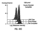
- a fluorescent DNA probe that is complementary to the oligonucleotide coupled to the beads is also added under competitive hybridization conditions. Aliquots of the beads so reacted are then run through a flow cytometer and the intensity of fluorescence on each bead is measured to detect the level of fluorescence which indicates the presence or absence of given sequences in the samples.
- the PCR product will effect a significant competitive displacement of Fluorescent oligonucleotide probe from the beads and, therefore, cause a measurable decrease in fluorescence of the beads, e.g., as compared to a control reaction that did not receive PCR reaction product.
- a PCR product from an individual having a mutation in the region of interest is incubated with the beads bearing the wild-type probe, a significantly lesser degree of displacement and resulting decrease in intensity of fluorescence on the beads will be observed because the mutated PCR product will be a less effective competitor for binding to the oligonucleotide coupled to the bead than the perfectly complementary fluorescent wild-type probe.
- the beads may be coupled to an oligonucleotide corresponding to a mutation known to be associated with a particular disease and similar principles applied.
- bead subsets are prepared with all known, or possible, variants of the sequence of interest and then mixed to form a bead set.
- test sample e.g. PCR product
- wild-type sequence and other variants can then be assayed simultaneously.
- the relative reactivity of the PCR product with subsets bearing the wild-type or variant sequences identifies the sequence of the PCR product.
- the matrix of information derived from this type of competitive hybridization in which the test sequence and the entire panel of probe sequences react simultaneously allows identification of the PCR product as wild-type, known mutant, or unknown mutant.
- the invention thus provides one with the ability to measure any of a number of genetic variations including point mutations, insertions, deletions, inversions, and alleles in a simple, appropriately sensitive, and efficient format.
- assay components and methods for the measurement of enzymes, DNA fragments, antibodies, and other biomolecules are provided.
- inventive technology improves the speed and sensitivity of flow cytometric analysis while reducing the cost of performing diagnostic and genetic assays.
- a multiplexed assay in accordance with the invention enables the simultaneous automated assay of multiple (at least an order of magnitude greater than available in the prior techniques) biomolecules or DNA sequences in real-time.
- Beadsets may be prepared, for example, so as to detect or screen for any of a number of sample characteristics, pathological conditions, or reactants in fluids. Beadsets may be designed, for example, to detect antigens or antibodies associated with any of a number of infectious agents including (without limitation, bacteria, viruses, fungi, mycoplasma, rickettsia, chlamydia, and protozoa), to assay for autoantibodies associated with autoimmune disease, to assay for agents of sexually transmitted disease, or to assay for analytes associated with pulmonary disorders, gastrointestinal disorders, cardiovascular disorders, and the like.
- the beadset may be designed to detect any of a number of substances of abuse, environmental substances, or substances of veterinary importance.
- An advantage of the invention is that it allows one to assemble a panel of tests that may be run on an individual suspected of having a syndrome to simultaneously detect a causative agent for the syndrome.
- Suitable panels may include, for example, a tumor marker panel including antigens such as prostate-specific antigen (PSA), carcinoembryonic antigen (CEA), and other suitable tumor markers; a regional allergy panel including pollen and allergens tested for by allergists of a particular region and comprising allergens known to occur in that region; a pregnancy panel comprising tests for human chorionic gonadotropin, hepatitis B surface antigen, rubella virus, alpha fetoprotein, 3' estradiol, and other substances of interest in a pregnant individual; a hormone panel comprising tests for T4, TSH, and other hormones of interests; an autoimmune disease panel comprising tests for rheumatoid factors and antinuclear antibodies and other markers associated with autoimmune disease; a blood borne virus panel and a therapeutic drug panel comprising tests for Cyclosporin, Digoxin, and other therapeutic drugs of interest.
- PSA prostate-specific antigen
- CEA carcinoembryonic antigen
- An important feature of the flow cytometric technology and techniques described here is the fabrication and use of particles (e.g., microspheres or beads that make up a beadset). It is through the use of appropriately labeled homogeneous bead subsets, combined to produce a pooled beadset, that the instant multiplexed assay method is practiced.
- Beads suitable for use as a starting material in accordance with the invention are generally known in the art and may be obtained from manufacturers such as Spherotech (Libertyville, IL) and Molecular Probes (Eugene, OR). Once a homogeneous subset of beads is obtained, the beads are labeled with an appropriate reactant such as a biomolecule, DNA sequence, and/or other reactant.
- an appropriate reactant such as a biomolecule, DNA sequence, and/or other reactant.
- Known methods to incorporate such labels include polymerization, dissolving, and attachment.
- development of a multiplexed assay for use in accordance with the invention can be divided into three phases: (1) preprocessing, (2) real-time analysis, and (3) interpretation.
- preprocessing phase baseline data is collected independently, via flow cytometric techniques, for each of an assay's bead subsets. Baseline data is used to generate a set of functions that can classify any individual bead as belonging to one of the assay's subsets or to a rejection class.
- flow cytometric measurements are used to classify, in real-time, each bead within an exposed beadset according to the aforementioned functions. Additionally, measurements relating to each subset's analyte are accumulated.
- interpretation phase the assay's real-time numerical results are associated with textual explanations and these textual explanations are displayed to a user.
- the inventive method allows the detection of a plurality of analytes simultaneously during a single flow cytometric processing step.
- Benefits of the inventive multiplex assay method include increased speed and reduced cost to analyze a clinical sample.
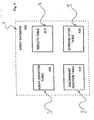
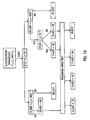
- FIG. 1 shows, in block diagram form, a system for implementing the inventive multiplexed assay method.
- Flow cytometer 100 output consists of a series of electrical signals indicative of one or more specified measured characteristics on each bead processed. These measurement signals are transmitted to computer 105 via data bus 110 and interface board 115.
- the signals are used by the computer to generate an assay database.
- the signals are processed by the computer (using the assay database) in accordance with the inventive method to produce a multiplexed/simultaneous assay of a clinical sample.
- Flow cytometer 100 operates in a conventional manner. That is, beads are processed by illuminating them, essentially one at a time, with a laser beam. Measurements of the scattered laser light are obtained for each illuminated bead by a plurality of optical detectors. In addition, if a bead contains at least one appropriate fluorescing compound it will fluoresce when illuminated. A plurality of optical detectors within the flow cytometer measure fluorescence at a plurality of wavelengths. Typical measured bead characteristics include, but are not limited to, forward light scatter, side light scatter, red fluorescence, green fluorescence, and orange fluorescence.
- green fluorescent markers or labels can cause cross-channel interference between optical detectors designed to detect green and orange wavelengths (e.g., approximately 530 nanometers and approximately 585 nanometers respectively).
- a training set of beads, in combination with standard data manipulation, can correct for this cross-channel interference by providing the physical measurements required for mathematical correction of the fluorescence measurements.
- Computer 105 can be a conventional computer such as a personal computer or engineering workstation.
- the computer is a personal computer having an Intel "486” processor, running Microsoft Corporation's "WINDOWS” operating system, and a number of ISA expansion slots.
- Interface board 115 is designed to plug into one of the computer's 100 ISA (Industry Standard Architecture) expansion slots. While the design of an interface board is, in general, different for each specific type of flow cytometer 100, its primary functions include (1) receiving and parsing measurement data signals generated by the flow cytometer's detectors, (2) receiving control parameter status information from the flow cytometer, and (3) sending control parameter commands to the flow cytometer. The precise manner in which these functions are carried out are dependent upon the type (make and model) of the flow cytometer used. In one embodiment, employing a Becton-Dickinson "FACSCAN" flow cytometer (San Jose, CA), the interface board uses control signals generated by the flow cytometer to distinguish measurement data and flow cytometer parameter and control signals. Measured data include forward light scatter, side light scatter, red fluorescence, green fluorescence, and orange fluorescence. Parameter and control signals include flow cytometer amplifier gain adjustments and status information.
- ISA Industry Standard Architecture
- an interface board 115 for use with the inventive assay method would be a routine task for one skilled in the art of diagnostic medical equipment design having the benefit of this disclosure
- an important aspect for any interface board is its ability to accommodate the transmission data rate generated by whatever flow cytometer is used.
- the "FACSCAN" flow cytometer can transmit a 16-bit (2 byte) word every 4 microseconds resulting in burst data rates of 500,000 bytes per second.
- Microfiche appendix A provides a detailed source code embodiment of the inventive assay method for use with the "FACSCAN" flow cytometer.
- Data bus 115 provides a physical communication link between the flow cytometer 100 and the interface board 110. Its physical and electrical characteristics (e.g., data width and bandwidth) are dependent upon the capabilities of the flow cytometer. It is noted that the data bus need not be a totally digital bus. If the flow cytometer does not include analog-to-digital conversion of measured bead characteristics (e.g., light scatter and fluorescence signals), then the data bus must communicate these analog signals to the interface board. It is then necessary that digital conversion of these signals be provided by either the interface board or another peripheral device before the data is transmitted to the computer 105.
- analog-to-digital conversion of measured bead characteristics e.g., light scatter and fluorescence signals
- GUI graphical user interface
- DLL dynamically linked library
- an important aspect of the inventive assay method is that it performs a simultaneous analysis for multiple analytes in real-time.
- the operational program code i.e., the DLL 205.
- the "FACSCAN" flow cytometer can process, or measure, approximately 2,000 beads per second, where each bead is associated with eight 16-bit data values.
- the DLL should be able to accept, and process, at a consistent data rate of at least 32,000 bytes per second. The need to accommodate this data rate, while also having sufficient time to perform real-time analysis based on the data, will generally necessitate that some of the DLL code be written in assembly language.
- GUI 200 is implemented in the visual basic programming language and the DLL 205 is implemented in C and assembly language programming.
- Microfiche appendix A contains source code listings for one embodiment of the GUI and DLL.
- a function of the preprocessing phase is to generate an assay database for use during the real-time analysis of an exposed beadset (clinical sample).
- preprocessing is performed prior to combining separately labeled bead subsets to form assay beadsets.
- Assay definition, discriminant function definition, and interpretation tables are created at the time an assay beadset is created.
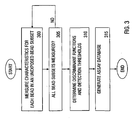
- Figure 3 shows, in flow chart form, the steps taken during the preprocessing phase.
- a bead subset is characterized by (1) the analyte it is designed to identify, (2) one or more classification parameters C 1 ... C m and (3) one or more measurement parameters F m1 -F mx - During the preprocessing phase the classification parameters are used to generate a set of functions, referred to as discriminant functions, that can classify a bead as belonging to one of the assay's subsets or a rejection class. Measurement parameters are used during the real-time analysis phase to determine if a specified analyte is present in the clinical sample being analyzed.
- each bead subset contains an equal number of beads.
- the precise number of beads within any given bead subset can vary depending upon many factors including, but not limited to, the number of analytes an assay beadset is designed to detect, the uniformity of the labeled beads (with respect to each of the measured parameters C 1 ... C m , F m1 ... F mx ), and the penalty of misclassifying (e.g., making a type 1 or type 2 classification error) a bead during analysis.
- each bead in an unexposed subset is measured by a flow cytometer 100 and the resulting data values accumulated for later use 300. For example, if the flow cytometer measures n classification parameters and x measurement parameters, i.e., generates ( n + x ) values for each bead, data for each of the subset's ( n + x ) parameters are updated based on each bead's measurements. This data collection step is repeated independently for each subset in the assay's bead set 305. The collection of such data for each of an assay's subsets constitutes an assay's baseline data.
- a set of discriminant functions are determined 310.
- the discriminant functions are used to classify a bead into one of the assay's bead subsets or a rejection class based solely on the measured classification parameters, C 1 ... C n .
- This step in principle and practice, is a problem of multidimensional classification or cluster analysis. Many prior art techniques and commercial software programs exist to perform this task.
- Beads are generally manufactured in large quantities referred to as batches. Each bead in a batch is of nearly identical size and has substantially the same dye absorption capacity. In light of this manufacturing process, bead subsets can be created using precise dilutions of chosen dyes and, because of their nearly identical size, all classification parameters will exhibit essentially equal variances. By correcting for scaling of the photo-multipliers within a flow cytometer, a linear classification rule can be generated. Further, since there are equal quantities of beads in each subset, the prior probabilities will be equal. This allows use of Fisher's linear discriminant technique to calculate the discriminant functions which define classification boundaries.
- linear hierarchical discriminant functions may be chosen which are equidistant, in a Euclidean sense, between the centers or centroids of any two of an assay's bead subsets.
- other types of discriminant functions such as quadratic functions and those discriminating on more than two classification parameters at once, are also possible.
- a set of threshold values are chosen which are used during the real-time analysis phase to detect the presence of a target analyte. For example, assume measurement parameter F m1 is used to detect analyte-A. During preprocessing, the baseline or unexposed value for F m1 is measured and accumulated for that subset's beads. Analyte-A's threshold could then, for example, be set to F m1' s baseline mean value plus one standard deviation of F m1 ' s baseline value.
- a threshold depends upon the parameter being measured (e.g., its distribution) and the cost of making a classification error (e.g., a type 1 or a type 2 error). It is routine that such values be based on an empirical review of the baseline data. The important criterion is that the threshold reliably distinguish between the presence and absence of the target analyte in an exposed assay beadset.
- an assay database is generated 315.
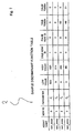
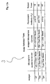
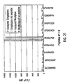
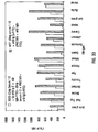
- an assay database 400 consists of an assay definition table 405, a discriminant function table 410, a results table 415, and an interpretation table 420. See Figure 4 .
- the assay definition table 405 defines an assay which, as described above, comprises two or more bead subsets each of which is designed to detect a specified analyte.
- Each row in the assay definition table describes a bead subset and contains the following entries: (1) assay name, (2) subset name, (3) subset token, (4) baseline values for each of the subset's measurement parameters F m1 - F mx, and (5) test-type token.
- the subset name entry is a text string identifying the subset by, for example, the type of analyte it is labeled to detect.
- the subset token is a unique subset identifier.
- the measurement parameter baseline entries are used during the interpretation phase to associate a numerical result (collected during the real-time analysis of a clinical sample) with a textual output string.
- the test-type token identifies which one of a possible plurality of interpretation tests to perform on the collected (real-time) data during the interpretation phase.
- the discriminant function table 410 is used to systematically set forth an assay's set of discriminant functions. Each row in the discriminant function table implements a single discriminant function and includes entries for (1) the assay's name, (2) a unique row identifier, (3) one or more classification parameters upon which to evaluate, (4) high and low discriminant values for each of the listed classification parameters, and (5) evaluation tokens which are assigned as a result of evaluating the discriminant function.
- the results table 415 is used to store, or accumulate, data on an assay's beadset during the real-time analysis phase of the inventive method and is discussed further in Section 6.2(d).
- the interpretation table 420 provides a means to associate text messages with each enumerated assay result and is discussed further in Section 6.2(e).
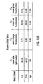
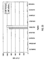
- the assay's beadset is comprised of four bead subsets, each labeled for a different analyte.
- the assay beadset is to be processed by a Becton-Dickinson Immunocytometry Systems "FACSCAN” flow cytometer. For each bead processed, the "FACSCAN” measures forward light scatter, side light scatter, red fluorescence, orange fluorescence, and green fluorescence.
- classification parameter C 1 be forward light scatter
- classification parameter C 2 be side light scatter
- classification parameter C 3 be red fluorescence
- classification parameter C 4 be orange fluorescence
- measurement parameter F m1 be green fluorescence.
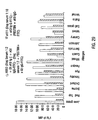
- each subset is characterized by a mean ( ⁇ ) and standard deviation ( ⁇ ) for each of its four classification parameters. See Figure 5 .
- ⁇ mean
- ⁇ standard deviation
- the precise number of individual beads contained in any given bead subset can be calculated by those of ordinary skill in the art. This calculation is required to obtain good statistical characterization of the subset's parameters - e.g., small, or relatively fixed, coefficient of variations for each parameter.
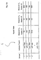
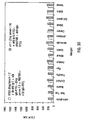
- the assay definition table 405 is comprised of general information relevant to the overall diagnostic function of the assay.
- each of the assay's subset's may be assigned a token used for identification: e.g., token 46 represents the bead subset labeled to detect a wildtype coding sequence for a specified gene; subset tokens 21, 50, and 5 represent subsets labeled to detect various mutant type coding sequences for a specified gene(s).
- measurement parameter F m1 ' s baseline in this example the mean
- standard deviation values are listed.
- a test-type token is listed.
- test-type token of '0' means an OVER/UNDER interpretation test is to be performed and a test-type token of '1' means a SHIFT interpretation test is to be performed. See Section 6.2(f) for further discussion of these issues.
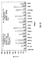
- Discriminate functions are generated by viewing the assay's baseline data graphically in three dimensions and creating planes to separate the different subset clusters. These "planes" are created by applying Fischer's Linear Discriminant to the n -dimensional classification parameter space.
- a populated discriminate function table based on the baseline data of Figure 5 is shown in Figure 7 .
- the discriminant function table provides a systematic means of evaluating a series of classification values ( C 1 , C 2 , C 3 , C 4 ) in order to classify a bead.
- bead classification proceeds by entering the discriminant function table at row 0, performing a test on a specified parameter (e.g., C 1 , C 2 , C 3 , or C 4 ) and then, depending upon the result, either classifying the bead or proceeding to another test which involves evaluating a different row in the table.
- a specified parameter e.g., C 1 , C 2 , C 3 , or C 4
- C 1 V 1
- C 2 V 2
- C 3 V 3
- C 4 V 4 .
- Classification of bead A via the discriminant function table of Figure 7 begins as follows (the pseudo-code below would demonstrate to those skilled in the art of programming the logic involved in the classification process):
- a discriminant function table embodies a (classification) decision tree.
- Figure 8 shows this relationship for the discriminant function table of Figure 7 explicitly.
- a discussion of the discriminant function table as it relates to the real-time processing of an exposed assay beadset is provided in Section 6.2(d). Once a beadset is preprocessed, the data may be employed in real-time analysis of many assays using that set.
- a bitmap or look up table could be used to classify the bead sets.
- the beadset may be exposed to a test sample. That is, they may be used to analyze a clinical sample. After exposure the beadset is ready for real-time analysis.
- the real-time analysis phase is initiated by installing the exposed beads into a conventional flow cytometer for processing.
- a flow cytometer 100 For each bead processed a flow cytometer 100 generates electrical signals indicative of a plurality of measured parameters, C 1 ... C n , F m1 ... F mx . These values are transmitted to computer 105 via data bus 110 and interface board 115. Values for a bead's classification parameters C 1 ... C n are used to evaluate the assay's discriminant functions, as encoded in a discriminant function table 410, the result of which is an initial classification of the bead into one of the assay's bead subsets or a reject class.
- a bead's measured classification parameter values C 1 ... C n can be checked against their ( C 1 ... C n ) baseline values to determine if it is "reasonable" to classify the bead as belonging to the initially identified class.
- this reasonableness test is implemented by computing the distance between the measured classification parameter values and the mean values obtained during preprocessing. If the measured values for C 1 ... C n for a particular bead are sufficiently distant from the identified subsets baseline values, the bead is assigned to a reject class. Use of this technique allows for the rejection of beads that were initially misclassified and improves the overall reliability of the analysis.
- a preferred embodiment's pooled beadset will include a bead subset which has no bound reactants (e.g., a placebo bead subset) in a known ratio to the beadset's other subsets.
- a bead subset which has no bound reactants (e.g., a placebo bead subset) in a known ratio to the beadset's other subsets.
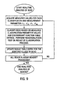
- FIG. 9 shows, in block diagram form, the general steps performed during the real-time analysis phase of a method in accordance with the invention.
- the following data are accumulated in the results table for each class (subset) of bead in the assay: (1) total count of the number of beads detected in the specified class, (2) a running sum for each measurement parameter F m1 - F mx , (3) for each measurement parameter the total count of the number of beads in the class whose measurement value is less than the parameter's baseline value, and (4) for each measurement parameter the total count of the number of beads in the class whose measurement value is more than the parameter's baseline value.
- the assay beadset is designed to simultaneously detect four analytes using four classification parameters ( C 1 represents forward light scatter, C 2 represents side light scatter, C 3 represents red fluorescence, and C 4 represents orange fluorescence) and one measurement parameter ( F m1 representing green fluorescence).
- C 1 represents forward light scatter
- C 2 represents side light scatter
- C 3 represents red fluorescence
- C 4 represents orange fluorescence
- F m1 representing green fluorescence
- values for C 1 , C 2 , C 3 , and C 4 are evaluated in accordance with the discriminant function table shown in Figure 7 to initially classify the bead as belonging to a particular subset, for example, in a genetic analysis intended to detect mutations in the Kras oncogene, the classification could proceed as follows: (1) class 46, Kras CODON 46 WILDTYPE, (2) class 21, Kras CODON 21 MUTANT, (3) class 50, Kras CODON 50 MUTANT, (4) class 5, Kras CODON 5 MUTANT, or (5) a reject class.
- data i.e., count, and measured F m1 values
- count, and measured F m1 values for each bead classified as a reject can also be collected.
- the user may select to see a text based presentation or interpretation of the assay's numerical results.
- the assay's real-time numerical results are associated with textual explanations. These textual explanations can be displayed to the user.
- each row in the interpretation table provides the necessary information to make a single interpretation and typically includes entries for (1) the assay's name, (2) a subset token identifying the class or subset on which the interpretation is based, (3) an outcome identifier for the identified subset, (4) a test-type token, (5) high and low discriminant values for each measurement parameter utilized in the identified test, and (6) a text string describing the row's result.
- the test-type token identifies which one of a possible plurality of interpretation tests to perform on the collected (real-time) data during the interpretation phase.
- the test-type token is either '0' or '1'.
- a value of '0' indicates an OVER/UNDER interpretation test is to be performed.
- a value of '1' indicates a SHIFT interpretation test is to be performed.
- the OVER/UNDER test is generally used for qualitative measurements where the level of reactivity of beads is an indication of the condition or concentration of a biomolecule present in the sample.
- the shift test is used where the result sought is a determination of the a minimally detectable level of a particular biomolecule.
- One of ordinary skill will recognize that many other tests could be performed. Examples include ranking, stratification, ratio of means to a standard, or to each other, etc.
- an interpretation table 420 may associate any number of entries or interpretations (e.g., rows within the table) with a single assay class or bead subset.
- bead subset Y could have a single measurement parameter ( F m1 ) associated with it and this measurement parameter could indicate, depending upon its value, that one or more interpretations are appropriate.
- the contents of the interpretation table 420 are generated during the preprocessing phase. This implies that the target assay be understood and that the various assay results be considered prior to construction of multiplexed assays.
- Figure 11 shows a sample interpretation table for this assay. Interpretation of the assay's real-time numerical results is initiated by, for example, the user selecting "interpret results" via the inventive method's graphical user interface.
- each bead subset (class) within an assay has an entry or row in the results table, Figure 10 .
- the general procedure for interpreting an assay's real-time numerical results is shown in flow-chart form in Figure 12 .
- each row of the results table is matched against every row in the interpretation table with the same subset token. If the result of performing the specified test is between the identified row's low and high values, then the associated textual message is displayed to the user.
- the next results table row is evaluated. This process is repeated until the every row in the interpretation table has been compared to the appropriate results table entry.
- subset 50 results table entry.
- the subset's token, 50 is used to identify three rows in the interpretation table (having outcome IDs of 1, 2, and 3) that contain information regarding evaluation of the mutant analyte.
- the result of the interpretation phase is a series of textual messages that describe the results of the assay.
- Conclusion of the interpretation phase marks the end of the assay.
- Assay definition, discriminant function definition, and interpretation tables are created at the time an assay beadset is created.
- Baseline classification data is collected only once for a given assay. That is, once an assay is defined and its baseline data is obtained, any number of beadsets can be manufactured to perform the analysis. To allow this "sharing" of baseline data the assay beadset may contain a center or calibration bead subset.
- a calibration beadset can be used to adjust any given flow cytometer to a standard. Calibration beadsets are typically processed separately from an assay. Further, calibration is generally performed daily. The purpose of calibration is to adjust the sensitivity of a flow cytometer's photomultipliers to accommodate day to day and machine to machine differences.
- Assays for antibody are widely used in medicine and clinical analysis for an wide variety of purposes, from detection of infections to determination of autoantibody.
- the following example illustrates use of the inventive method in an antibody assay and assumes the use of a flow cytometer capable of providing at least five measurements for each bead processed: forward light scatter as classification parameter C 1 , side light scatter as classification parameter C 2 , red fluorescence as classification parameter C 3 , orange fluorescence as classification parameter C 4 , and green fluorescence as measurement parameter F m1 .
- a number of bead subsets are prepared, for example, by using a cell sorter to sort a heterogeneous population to collect a homogeneous subset or alternatively, by preparing the beads using tightly controlled specifications to ensure production of a homogeneous subset.
- Each subset is distinguishable by its characteristic pattern of classification parameters C 1 , C 2 , C 3 , and C 4 .
- the beads in each subset are then labeled with a different antigen such as AgA, AgB, etc.
- Antigens AgA through AgJ may be attached to the beads by any of a number of conventional procedures such as by chemical or physical absorption as described by Colvin et al., "The Covalent Binding of Enzymes and Immunoglobulins to Hydrophilic Microspheres” in Microspheres: Medical and Biological Applications, 1-13, CRC, Boca Raton, FL, 1988 ; Cantarero et al., "The Adsorptive Characteristics of Proteins for Polystyrene and Their Significance in Solid-Phase Immunoassays," Anal. Biochem., 105, 375-382 (1980 ); and Ilium et al., "Attachment of Monoclonal Antibodies to Microspheres," Methods in Enzymol., 112, 67-84 (1985 ).
- the pooled set is prepared with equal volumes of beads from each subset, so that the set contains about the same number of beads from each subset.
- the assay beadset may then be incubated with a fluid sample of interest, such as serum or plasma, to test for the presence of antibodies in the fluid that are reactive with antigens on the beads.
- a fluid sample of interest such as serum or plasma
- Such incubation will generally be performed under conditions of temperature, pH, ionic concentrations, and the like that facilitate specific reaction of antibodies in the fluid sample with antigen on the bead surface.
- the beads in the mixture are centrifuged, washed and incubated (again under controlled conditions) for another period of time with a "secondary" antibody such as, for example, fluorescein labeled goat anti human immunoglobulin.
- the secondary antibody will bind to and fluorescently label antibodies bound to antigen on the beads.
- the beads are processed by the flow cytometer and the four classification parameters forward light scatter, side light scatter, red fluorescence, and orange fluorescence are measured and used to identify the subset to which each bead in the assay beadset belongs.
- a simultaneous measurement of green fluorescence (measurement parameter) for each bead allows one to determine whether the bead has antibody bound to it. Because the subset to which a bead belongs is correlated with the presence of a particular antigen, e.g., sS1-AgA, one may readily determine the specificity of the antibody bound to a bead as a function of the subset to which it belongs.
- each of four samples e.g., blood serum from four patients
- the reactions were incubated at room temperature for 45 minutes, and then analyzed on the "FACSCAN" using side light scatter ( C 1 ), orange fluorescence ( C 2 ), and red fluorescence ( C 3 ) as classification parameters.
- Green fluorescence was used as the measurement parameter ( F m ); an increase in green fluorescence by 30-fold indicates a specific interaction between an antigen and its corresponding fluorescinated antibody.
- the assay database was built, it was tested by running 5,000 beads from each bead subset individually through the system. After rejecting 23.8% of the beads as doublets, the remaining crimson beads (subset 18) were classified with 99.88% accuracy. Dark red beads (subset 45) were classified with 99.96% accuracy with 22.9% rejected as doublets. Clear beads (subset 50) were classified with 100% accuracy with 9.4% of the beads rejected as doublets.
- the three bead subsets were pooled to form an assay beadset and divided into 4 sample tubes and processed by the system shown in Figure 1 .
- the contents of each sample and the mean measured fluorescence ( F m ) for each bead subset are listed in Figure 13e .
- the inventive method correctly identified the antibody or antibodies present in each sample.
- a variety (for example five) of protein antigens are employed. Bead subsets are first generated based on differences in one or more of C 1 , C 2 , and C 3 . Next, a selected antigen labeled with Cy3NHS (an orange fluorophore) is bound to the beads in each subset. To minimize the measured orange fluorescence coefficient of variation for each bead subset, the beads are sorted with a high speed cell sorter so that only a narrow range of antigen (orange fluorophore) is found on each bead within a subset.
- Cy3NHS an orange fluorophore
- the measured intensity of C 4 for AgA should differ from the measured intensity of C 4 from AgB, etc.
- saturation binding with fluoresceinated monoclonal antibody is tested - each bead ought to have restricted ranges of both orange and green fluorescence. While the construction of beadsets by this method is more laborious, the increase in measurement precision may be useful and will allow the sampling of fewer beads to arrive at a suitable determination of antibody concentration.
- the assays previously mentioned measure any antibody with specificity for antigen upon an appropriately labeled bead.
- the antigen can be quite simple or rather complex and thus, the inventive methods can measure a highly restricted antibody or a broad array of antibodies.
- a hexapeptide just large enough to bind to a monoclonal antibody can be employed as antigen or a large protein with many epitopes can be used.
- the level of antibody eventually found associated with the bead F m1
- the level of antibody eventually found associated with the bead is a function of the number of epitopes per bead, the concentration of epitopes, the amount of antibody and the affinity of the antibody and the valence of the antibody-antigen interaction.
- Assays for many substances in a clinical laboratory are based on the interference with specific ligand-ligate or antigen-antibody interactions.
- one member of the ligand-ligate pair is labeled with the F m fluorophore and one member is immobilized on the beads.
- Soluble, unlabeled material (analyte) ,which may be ligand or ligate, is added to the reaction mixture to competitively inhibit interaction of the labeled component with the immobilized component. It is usually not important which member of the pair is labeled and which is immobilized; however, in certain assays, functional advantages may dictate the orientation of the assay.
- each bead subset is modified with an antigen.
- the " antigen-coated beads are then reacted with an F m labeled antibody specific for the antigen on the bead surface.
- Subsequent addition of a test fluid containing soluble analyte (inhibitor) will displace the F m labeled antibody from the beads in direct proportion to the concentration of the soluble analyte.
- a standard curve of known analyte concentrations is used to provide accurate quantification of analyte in the test sample.
- the fluid containing the beadset may be subjected to dissociating conditions such as a change in pH, ionic strength or temperature, after mixture of the beadset with the sample to be tested.
- the F m labeled component may be added to the beadset after addition of the test sample. In either case, it is not necessary for equilibrium to be achieved to determine analyte concentration if the kinetics and linearity of the assays have been established.
- a competitive inhibition analysis is used to quantitate levels of selected analytes, here IgG, IgA, and IgM.
- a second experimental refinement demonstrates the utility of multiplexed assays in epitope mapping of a monoclonal antibody.
- that approach involved the use of antibody detection technology using a fluoresceinated monoclonal antibody in combinatorial epitope screening (e.g. of peptide libraries) to map a particular epitope to which a monoclonal antibody of interest bound, together with a displacement (competitive inhibition) aspect to demonstrate the specificity of the assay.
- ToRCH assay for screening of human serum for antibodies to a number of infectious agents known to pose special hazards to pregnant women. Allergy screening is exemplified by detection of serum IgE against a panel of grass antigens. Yet an additional experimental example reflects the ability of the multiplexed assay in pregnancy testing, e.g. in testing for hormones or other analytes commonly elevated during pregnancy. Each of these examples is set forth below.
- This example illustrates the determination of multiple analyte levels in a liquid sample simultaneously using competitive inhibition analysis.
- the use of a competitive inhibition assay to accurately determine analyte levels in liquid solutions is a commonly used format for many analyte assays.
- the uniqueness of this assay is the simultaneous determination of three distinct serum proteins at the same time in the same tube from one serum sample.
- Immunoglobulins G, A and M are three distinct serum proteins whose levels are determined by a number of genetic and environmental factors in human serum. As changes to these levels may indicate the presence of disease, clinicians often request assay determinations of IgG, A and M using conventional techniques. The most common technique is nephelometry that depends upon the absorption of light by precipitates formed between these immunoglobulins and antibodies made in animals to the human immunoglobulins. As these immunoglobulins are present in human serum at fairly high levels, this type of assay is sufficient. Nephelometry however suffers from a number of limitations including the need for large quantities of reagents, long reaction times for precipitation to equilibrate and an inability to perform more than one reaction per tube or sample.
- Each assay consists of a DFM coated with the immunoglobulin of choice and a polyclonal, goat anti-human Ig labeled with a green fluorescent molecule (Bodipy).
- Bodipy a polyclonal, goat anti-human Ig labeled with a green fluorescent molecule
- the Bodipy -antibody causes the immunoglobulin (Ig) coated microsphere to emit green fluorescence ( F m ) .
- F m green fluorescence
- soluble Ig the green signal is reduced.
- Each assay is balanced to reflect a sensitivity range near the physiological level of the Ig in question at a 1:500 dilution of human serum.
- Antibody labeling Goat anti-human IgG, goat anti-human IgA, and goat anti-human IgM antibodies (Cappel Division, Organon Teknika, Durham, NC) were labeled with Bodipy FL-CASE (Molecular Probes, Inc., Eugene, OR) using methods described by the manufacturer of the Bodipy succinymidyl ester. The resulting Bodipy labeled antibodies were stored in PBS containing I mg/mL BSA as stabilizer.
- Antigen conjugation to microspheres Four DFM (5.5 ⁇ M carboxylate, Bangs Laboratories, Inc.
- microspheres were washed twice with 500 ⁇ L PBS, pH 7.4 using centrifugation at 13,400 x g for 30 seconds to harvest the microspheres.
- washed beads were suspended in 250 ⁇ L of a 0.05 mg/mL solution of protein in PBS, pH 7.4.
- the microspheres were blocked by addition of 250 ⁇ L of 1.0 mg/mL BSA, 0.02% Tween, 0.2 M glycine, in PBS, pH 7.4 and incubated for an additional 30 minutes.
- Protein coated microspheres were washed twice with 500 ⁇ L 0.02% Tween 20, 1 mg/mL BSA in PBS, pH 7.4 (PBSTB).
- the Bodipy-labeled goat anti-hIgM was used at 2.5 ⁇ g/mL.
- Cross reactivity assay Equivalent amounts of each of the four protein loaded microspheres were mixed to produce a bead mixture. 10 ⁇ L of the bead mixture (7,500 microspheres) was mixed with 10 ⁇ L of diluted serum calibrators of known Ig level. The assay was initiated by addition of 10 ⁇ L of one of the Bodipy-labeled antibodies "spiked" with a small quantity of soluble Ig antigen to alleviate the "hook effect". The mixtures were incubated for 30 minutes, diluted to 300 ⁇ L in PBSTB and assayed by flow cytometry.
- the Bodipy-labeled goat anti-hIgG was used at 30 ⁇ g/mL.
- IgA the Bodipy-labeled goat anti-hIgA was used at 8 ⁇ g/mL.
- the Bodipy-labeled goat anti-hIgM was used at 2.5 ⁇ g/mL.
- the quantities of antigen "spikes" were 1.6 ⁇ g/mL for IgG, 0.6 ⁇ g/mL for IgA and 0.4 ⁇ g/mL for IgM.
- the Bodipy-labeled goat anti-hIgA was used at 8 ⁇ g/mL.
- the Bodipy-labeled goat anti-hIgM was used at 2.5 ⁇ g/mL.
- the quantities of antigen "spikes" were 1.6 ⁇ g/mL for IgG, 0.6 ⁇ g/mL for IgA and 0.4 ⁇ g/mL for IgM.
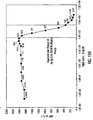
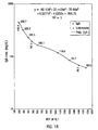
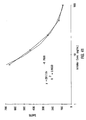
- IgG single analyte assay Results of the single analyte inhibition analysis for IgG level is shown in Table 1 and Figure 15A .
- This assay was designed to be most sensitive to inhibition in the anticipated range of IgG in human serum at a 1:500 dilution.
- Figure 15A the area of the inhibition curve between the dotted lines, left and right, cover the range of sensitivity.
- the inhibitor was known amounts of human IgG from a serum calibrator diluted into human serum containing no IgG, IgA or IgM. Dilutions of the calibrator were then diluted 1:500 in PBSTB and included as inhibitor in the assay.
- the Bodipy-labeled anti-hIgG was used at 30 ⁇ g/mL in PBSTB. 7,500 microspheres were used in this experiment and 250 were counted by flow cytometry. Note that as the amount of soluble IgG increased, the degree of inhibition as monitored by the MIF of F m increased proportionally until saturation of the system was achieved. On the other end of the inhibition curve note that the lower levels of soluble inhibitor caused an elevation in the MIF of F m as compared with the negative control (human serum with no Ig). This "hook effect" is common in immunoassay and can be adjusted up or down the inhibition curve by adjusting both the amount of antibody and antigen in the soluble portion of the assay.
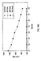
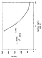
- IgG single analyte assay Results of single analyte inhibition analysis for IgA level is shown in Table 1 and Figure 15B . This assay was designed to be most sensitive to inhibition in the anticipated range of IgA in human serum at a 1:500 dilution. In Figure 15B , the area of the inhibition curve between the dotted lines, left and right, cover the range of sensitivity.
- the inhibitor was known amounts of human IgA from a serum calibrator diluted into human serum containing no IgG, IgA or IgM. Dilutions of the calibrator were then diluted 1:500 in PBSTB and included as inhibitor in the assay.
- the Bodipy-labeled anti-hIgA was used at 8 ⁇ g/mL in PBSTB. 7,500 microspheres were used in this experiment and 250 were counted by flow cytometry. Note that as the amount of soluble IgA increased, the degree of inhibition as monitored by the MIF of F m increased proportionally until saturation of the system was achieved.
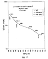
- IgM single analyte assay Results of single analyte inhibition analysis for IgM level is shown in Table 1 and Figure 15C . This assay was designed to be most sensitive to inhibition in the anticipated range of IgM in human serum at a 1:500 dilution. In Figure 15C, the area of the inhibition curve between the dotted lines, left and right, cover the range of sensitivity.
- the inhibitor was known amounts of human IgM from a serum calibrator diluted into human serum containing no IgG, IgA or IgM. Dilutions of the calibrator were then diluted 1:500 in PBSTB to be included as inhibitor in the assay..
- the Bodipy-labeled anti-hIgM was used at 2.5 ⁇ g/mL in PBSTB. 7,500 microspheres were used in this experiment and 250 were counted by flow cytometry. Note that as the amount of soluble IgM increased, the degree of inhibition as monitored by the MIF of F m increased proportionally until saturation of the system was achieved.
- This example demonstrates the screening of combinatorial chemistry products for a biologically active molecule.
- the generation of random chemical products for empirical discovery of biologically significant molecules is a method that holds great promise for progress in numerous disciplines of science including biology, pharmacology and medicine.
- One general problem with the technique is the screening of large numbers of unique molecules for a specific activity. Screening methods are required that provide high throughput levels of screening with adequate specificity and sensitivity for detection of the biological event in question.
- a monoclonal antibody (MAB 384) was chosen that was produced using the spleen cells of a mouse hyper-immunized with a defined peptide (amino acid 67-74) from the amino acid sequence of human myelin basic protein (MBP). Using the amino acid sequence of this region of MBP, nine overlapping octapeptides were synthesized that covered the predicted epitope. To the carboxyl terminal end of each peptide, glycine-lysine-biotin residues were added.
- MAB 384 (Chemicon International, Inc., Temecula, CA) was labeled with Bodipy FL-X (Molecular Probes, Inc., Eugene, OR) using methods described by the manufacturer of the Bodipy succinymidyl ester. Absorbance at 280 nm and 504 nm revealed that the resulting Bodipy-labeled antibody had a Bodipy to protein ratio of 3.31 and was stored in PBS containing 1 mg/mL BSA as stabilizer.
- Avidin conjugation to microspheres Nine distinctly dyed DFM (5.5 ⁇ M, Bangs Laboratories, Inc.
- microspheres were washed twice with 100 ⁇ L PBS, pH 7.4 using centrifugation at 13,400 x g for 30 seconds to harvest the microspheres. Activated, washed beads were suspended in 50 ⁇ L of a 0.25 mg/mL solution of Neutravidin in PBS, pH 7.4. After 2 hours, the microspheres were blocked by addition of 50 ⁇ L of 0.2 M glycine, 0.02% Tween 20 in PBS, pH 7.4 and incubated for an additional 30 minutes.
- Protein coated microspheres were washed twice with 100 ⁇ L 0.02% Tween 20, 1 mg/mL BSA in PBS, pH 7.4 (PBSTB) and stored in PBSTB at approximately 3,000,000 microspheres/mL as determined by hemocytometer count.
- PBSTB pH 7.4
- Peptide attachment to microspheres Each of the nine DFM conjugated to Neutravidin were treated separately with one of the nine biotinylated peptides. 10 ⁇ L of biotinylated peptides at 100 - 200 ng/mL was mixed with 10 ⁇ L of microspheres and reacted for 5 minutes followed by 2 x 100 ⁇ L washes in PBSTB. The peptide loaded microspheres were suspended in 20 ⁇ L of PBSTB.
- Single analyte assay 10 ⁇ L of each of the peptide loaded microspheres was reacted with 10 ⁇ L of the Bodipy-labeled MAB 384 at 15.5 ⁇ g/mL in PBSTB for 1 hour, diluted to 300 ⁇ L in PBSTB and assayed using flow cytometry. Negative controls included the microspheres without peptide and with the Bodipy MAB 384.
- Multiple analyte assay 10 ⁇ L of each of the 9 peptide loaded microspheres was mixed to produce a bead set.
- 10 ⁇ L of the set was reacted with 10 ⁇ L of the Bodipy-labeled MAB 384 at 15.5 ⁇ g/mL in PBSTB for 1 hour, diluted to 300 ⁇ L in PBSTB and assayed using flow cytometry.
- Negative controls included the microsphere set without peptide and treated with the Bodipy MAB 384.
- Competitive inhibition with soluble peptide 10 ⁇ L of each of the 9 peptide loaded microspheres was mixed to produce a bead set.
- 10 ⁇ L of the Bodipy-labeled MAB 384 at 15.5 ⁇ g/mL in PBSTB was reacted with 10 ⁇ L of soluble peptide containing the epitope sequence HYGSLPQK (SEQ ID NO.
- peptides to be screened The amino acid sequence upstream and downstream from the epitope of monoclonal antibody MAB 384 (amino acid 67-74, YGSLPQ, SEQ ID NO. 2) was determined using the published amino acid sequence ( Roth, H.J., et al., J. Neurosci. Res.. 17, 321-328, 1990 ). The table below shows the amino acid sequence of the nine overlapping peptides produced for the screening assay. Note that to the carboxy-terminal end of all peptides was added a glycine (G)-lysine (K)-biotin.
- G glycine
- K glysine
- each separate microsphere was reacted with Bodipy-labeled MAB 384 at 15.5 ⁇ g/mL for 60 minutes and the mixture assayed using flow cytometry.
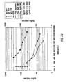
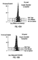
- the Mean Intensity of Fluorescence (MIF) of the green fluorescence channel ( F m ) is shown for each peptide-bead as the darker set of bars in Figure 21 .
- the darkest bars represents single analyte analysis of each bead in the absence of peptide as a negative control.
- This epitope mapping example demonstrates the useful application of the instant invention to the area of combinatorial screening.
- the peptide carrying the epitope for the mouse monoclonal antibody screened in this example was clearly identified in a set of nine peptides. The identification was further shown to be specific by competitive inhibition with soluble epitope peptide. In addition, the stability of the avidin-biotin interaction for use with flow cytometry was demonstrated in an excess of free biotin.
- This example demonstrates the utility of this invention in the screening of human serum for antibodies to infectious disease agents. Screening of serum for antibodies to certain infectious disease agents is often the only method available to determine if a patient has been, or is infected with the agent in question. For example, a common method of diagnosing HIV infection is by detection of HIV specific antibodies in the serum. This phenomenon known as seroconversion is commonly employed for diagnosis of several important pathogenic infections.
- One of the most commonly employed assay panels of this type is the ToRCH panel. ToRCH assays detect both serum IgG and serum IgM responses to To xoplasma gondii, R ubella virus, C ytomegalovirus, and H erpes Simplex Virus Types 1 and 2.
- a ToRCH assay using flow cytometry has been developed by coupling purified antigens of T. gondii, Rubella, CMV and HSV Type 1 and Type 2 to five Differentially Fluorescent Microspheres (DFM).
- DFM Differentially Fluorescent Microspheres
- the specificity of the assay has been demonstrated by treating this bead set with human serum calibrators certified to be either positive or negative for all five agents. After this treatment, the bead set was treated with either Goat anti-human IgG-Bodipy or Goat anti-human IgM-Bodipy used to develop the assay.
- a third calibrator with known levels of reactivity to each agent was assayed and the results reported.
- Antibody labeling Goat anti-human IgG and goat anti-human IgM (Cappel Division, Organon Teknika, Durham, NC) were labeled with Bodipy FL-CASE (Molecular Probes, Inc., Eugene, OR) using methods described by the manufacturer of the Bodipy succinymidyl ester. Bodipy-labeled antibodies were stored in PBS containing 1 mg/mL BSA as stabilizer.
- Antigen conjugation to microspheres Five DFM (5.5 ⁇ M carboxylate, Bangs Laboratories, Inc., Carmel, IN, dyed by Emerald Diagnostics, Inc., Eugene, OR) were conjugated separately to the five ToRCH antigens (Viral Antigens, Inc.) with a two-step EDC coupling method (Pierce Chemicals, Rockford, IL) using sulfo-NHS to stabilize the amino-reactive intermediate. All antigens were dialyzed into PBS to remove any reactive amino groups such as sodium azide or glycine. The T.
- gondii preparation (Chemicon, Inc., Temecula, CA) was sonicated for 2 minutes in PBS, 10 mM EDTA to lyse the tachyzoites.
- 20 ⁇ L (8.4 million microspheres) of each bead type was activated for 20 minutes in a total volume of 100 ⁇ L containing 500 ⁇ g of EDC and Sulfo-NHS in 50 mM sodium phosphate buffer, pH 7.0.
- Microspheres were washed twice with 200 ⁇ L PBS, pH 7.4 using centrifugation at 13,400 x g for 30 seconds to harvest the microspheres.
- Activated and washed beads were suspended in 100 ⁇ L of antigen at 0.05 to 0.15 mg/mL in PBS, pH 7.4. After 2 hours, the microspheres were blocked by addition of 100 ⁇ L of 0.2 M glycine, 0.02% Tween 20 in PBS, pH 7.4 and incubated for an additional 30 minutes. Antigen coated microspheres were washed twice with 200 ⁇ L 0.02% Tween 20, 1 mg/mL BSA in PBS, pH 7.4 (PBSTB). and stored in PBSTB at approximately 3,000,000 microspheres/mL as determined by hemacytometer count.
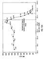
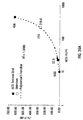
- Rubella assay Rubella antigen loaded microspheres were used to examine several parameters of the assay in a single analyte format prior to the performance of multiple analyte assays. 10 ⁇ L (30,000 microspheres) of Rubella antigen coated beads were reacted with 10 ⁇ L of a 1:10 dilution of four different Rubella calibrator sera (Consolidated Technologies, Inc., Oak Brook, IL) and the mixture incubated for 1 hour. These sera were defined using a standard assay for the anti-Rubella IgG activity by the manufacturer of the calibrators. The units were defined as International Units/ mL or IU/mL.
- Beads were washed in PBSTB by centrifugation at 13,400 x g for 30 seconds and suspended in 40 ⁇ L of a 10 ⁇ g/mL solution of Bodipy-labeled anti-human IgG. This mixture was incubated for 1 hour, diluted to 300 ⁇ L in PBSTB and assayed using flow cytometry. Negative controls included the microspheres with no serum treated with the Bodipy-labeled antibodies. In addition one calibrator serum containing 70 IU/mL of anti-Rubella IgG activity was titrated in a single analyte assay.
- Beads were washed in PBSTB by centrifugation at 13,400 x g for 30 seconds and suspended in 20 ⁇ L of a 40 ⁇ g/mL solution of Bodipy-labeled anti-human IgG or IgM. This mixture was incubated for 1 hour, diluted to 300 ⁇ L in PBSTB and assayed using flow cytometry. Negative controls included the microspheres with no serum treatment and the microspheres treated with the ToRCH negative control serum. Both negative controls were developed with the Bodipy-labeled antibodies.
- Rubella assay Rubella coated DFM were reacted with 4 human serum calibrators containing known levels of IgG antibodies specific for Rubella virions defined by International units (IU/mL). The beads were washed and developed with goat anti-human IgG-Bodipy. Results are shown in Table 9 and Figure 24 . Increasing units of anti-Rubella activity were reflected in the Mean Intensity of Fluorescence (MIF) of F m (green channel). Luminex Units (LU) were defined as the MIF of F m for each data point minus the MIF of F m for the negative control (no serum) multiplied by 0.1, and are included in Table 9.
- MIF Mean Intensity of Fluorescence
- LU Luminex Units
- Rubella calibrator titration The human serum calibrator containing 70 IU/mL of anti-Rubella IgG was serially diluted in PBSTB and assayed with the Rubella coated microspheres and Bodipy-labeled anti-human IgG. Results shown in Table 10 and Figure 25 show that, as expected, the IgG antibodies specific for Rubella were titrated with dilution.
- the known levels of anti-ToRCH IgG reactivities for the ToRCH Calibrator were compared to the Luminex units of each IgG activity as determined by the multiple analyte analysis. Luminex units were defined by subtracting the negative control serum average MIF of F m from the average MIF of F m for each antigen and multiplying by 0.1. The levels of the ToRCH calibrator were defined by the manufacturer as a factor of activity for each antigen above the limit of detection for that antigen on a specific diagnostic machine. These results are listed in Table 13.
- a demonstrative ToRCH assay has been developed to simultaneously assay for serum IgG or IgM specific for the five ToRCH pathogens in a single tube. Results of the assay indicate that it is specific for each pathogen and is as sensitive as currently available instrument based assays.
- the multiple analyte format provides a uniquely powerful technology for rapid and less expensive serum testing for seroconversion to ToRCH pathogens as well as other infectious agents diagnosed in this manner.
- This example demonstrates the screening of serum for IgE antibodies specific for allergens. Screening of serum for IgE antibodies specific to allergens is a viable option for allergy testing as compared with skin sensitivity testing.
- the instant invention provides for a format that can assay for either IgG or IgE responses to numerous allergens at the same time in the same tube with the same sample and is therefore a uniquely powerful method of screening.
- An allergy assay has been developed including 16 grass allergens in a multiple analyte, simultaneous format.
- a panel of 16 grass allergens were attached to 16 Differentially Fluorescent Microspheres (DFM) with one grass allergen being coated onto one unique member of the bead set.
- the allergen bead set was treated with diluted dog serum for 1 hour and treated with a solution of either Goat anti-Dog IgE or goat anti-dog IgG-FITC for an additional hour.
- beads were washed clear of this antibody and the bead set treated with an affinity purified rabbit anti-goat IgG-FITC antibody as probe.
- Results demonstrate a uniquely powerful method of serum screening for allergies that provides a true multiple analyte format, as well as sensitivity and specificity.
- Allergen conjugation to microspheres Sixteen DFM (5.5 ⁇ M carboxylate) were conjugated separately to 16 soluble grass allergens (provided by Dr. Bill Mandy, BioMedical Services, Austin, TX) with a two-step EDC coupling method (Pierce Chemicals, Rockford, IL) using sulfo-NHS to stabilize the amino-reactive intermediate. All grass allergens were diluted 1:100 into PBS, pH 7.4.
- Protein coated microspheres were washed twice with 100 ⁇ L 0.02% Tween 20, 1 mg/mL BSA in PBS, pH 7.4 (PBSTB), and stored in PBSTB at approximately 3,000,000 microspheres/mL as determined by hemacytometer count.
- Multiplexed K-9 grass allergen IgE assay Equivalent amounts of each of the 16 grass allergen loaded microspheres was mixed to produce a bead mixture. 20 ⁇ L (60,000 microspheres) of the mixture was reacted with 60 ⁇ L of a 1:3 dilution of dog serum in PBSTB and the mixture incubated for 1 hour.
- Beads were washed in 200 ⁇ L PBSTB by centrifugation at 13,400 x g for 30 seconds and suspended in 40 ⁇ L of a 50 ⁇ g/mL solution of anti-dog IgE (provided by Dr. Bill Mandy, BioMedical Services, Austin, TX). After incubation for 1 hour, beads were washed in 200 ⁇ L PBSTB by centrifugation at 13,400 x g for 30 seconds. Beads were then treated with 40 ⁇ L of rabbit anti-goat IgG-FITC (Sigma, St. Louis, MO) at 20 ⁇ g/mL. After one hour the bead mixture was diluted to 300 ⁇ L in PBSTB and assayed using flow cytometry.
- MIF mean intensity of fluorescence
- This example illustrates the determination of multiple analyte levels in a liquid sample simultaneously by immunometric or capture-sandwich assay.
- capture-sandwich assays to accurately determine analyte levels in liquid solutions is a commonly used format for many analyte assays.
- the technique is especially useful for those analytes present in low quantities as the first step serves to capture and thus concentrate the analyte.
- the uniqueness of this assay is the multiple analyte format allowing the simultaneous determination of two distinct serum proteins at the same time in the same tube from the same serum sample.
- hCG Human chorionic gonadotropin
- AFP Alpha fetoprotein
- AFP is the fetal cell equivalent to human , serum albumin.
- AFP is elevated in pregnancy and in certain types of malignancies.
- Many clinical fertility or pregnancy test panels include immunometric assays for these two serum proteins. Immunometric or capture-sandwich assays for hCG and AFP were developed separately and then combined in a multiple analyte format.
- the hCG assay was developed by examining several antibody pairs for their ability to capture and quantitate hCG levels in solution. First, a monoclonal antibody was coupled through carbodiimide linkage to a carboxylate substituted Differentially Fluorescent Microsphere (DFM). Next, a polyclonal, affinity purified antibody was Bodipy-labeled and used to probe DFM captured hormone. Once this assay was adjusted to include physiological sensitive ranges, the process was repeated for AFP. Cross-reactivity of the two assays was examined to demonstrate that the two assays would not interfere. The assays were then performed simultaneously. Commercially available serum calibrators were used to demonstrate that both assays were sensitive in clinically relevant ranges and an unknown was include to demonstrate how the two assays work simultaneously.
- DFM carboxylate substituted Differentially Fluorescent Microsphere
- Antibody labeling The two affinity purified polyclonal anti-hCG (AB633) and anti-AFP (M20077) antibodies (Chemicon, Inc., Temecula, CA and Medix Division, Genzyme, San Carlos, CA) were labeled with Bodipy FL-CASE (Molecular Probes, Inc., Eugene, OR) using methods described by the manufacturer of the Bodipy succinymidyl ester. The resulting Bodipy-labeled antibodies were stored in PBS containing 1 mg/mL BSA as stabilizer.
- Antibody conjugation to microspheres Monoclonal anti-hCG (MAB602) and anti-AFP (S10473) capture antibodies were conjugated to microspheres with a two-step EDC coupling method (Pierce Chemicals, Rockford, IL) using sulfo-NHS to stabilize the amino-reactive intermediate. 20 ⁇ L ( 8.4 million microspheres) of each DFM was activated for 20 minutes in a total volume of 100 ⁇ L containing 500 ⁇ g of EDC and Sulfo-NHS in 50 mM sodium phosphate buffer, pH 7.0. Microspheres were washed twice with 200 ⁇ L PBS, pH 7.4 using centrifugation at 13,400 x g for 30 seconds to harvest the microspheres.
- Capture assay antibody pairs were screened by coupling potential capture antibodies to microspheres and assaying them using all potential combinations of capture antibody-bead/ Bodipy-labeled probe antibody. Assays were performed using 10 ⁇ L of capture antibody microspheres (approximately 30,000) plus 20 ⁇ L of antigen solution at 10 ⁇ g/mL in PBSTB for a 1 hour incubation. Beads were washed in PBSTB by centrifugation at 13,400 x g for 30 seconds and suspended in 20 ⁇ L of a 25 ⁇ g/mL solution of Bodipy-labeled probe antibody.
- Antigen titration assay Once an antibody pair was chosen for use, the pair was analyzed for sensitivity and limit of detection by titration of antigen. Assays were performed using 10 ⁇ L of capture antibody microspheres plus 20 ⁇ L of antigen dilutions in PBSTB for a 1 hour incubation. Beads were washed in 200 ⁇ L PBSTB by centrifugation at 13,400 x g for 30 seconds and suspended in 20 ⁇ L of a 25 ⁇ g/mL solution of Bodipy-labeled probe antibody.
- This mixture representing a homogenous (no-wash) assay was also incubated for 1 hour, diluted to 300 ⁇ L in PBSTB and assayed using flow cytometry.
- Multiple analyte assay Once the AFP and hCG antibody pairs were shown not to cross-react and were adjusted to provide clinically relevant ranges of sensitivity in a homogenous assay, the assays were performed simultaneously using commercially available serum calibrators as the source of AFP and hCG antigens. Equivalent amounts of each of the two capture antibody loaded microspheres was mixed to produce an AFP/hCG capture mixture.
- Antibody pair analysis for hCG capture assay For hCG antibody pair analysis, five capture antibody/microspheres were prepared and the identical five antibodies were Bodipy-labeled to serve as probes. Three of the antibodies were specific for the alpha sub-unit of hCG and two for the beta sub-unit. The three anti-alpha sub-unit antibody/microspheres were assayed for utility with the two Bodipy-labeled anti-beta hCG antibodies. Conversely, the two anti-beta sub-unit antibody/microspheres were assayed for utility with the three Bodipy-labeled anti-alpha hCG antibodies. Results of this screen are shown in Table 14 and Figure 35 .
- the 12 combinations of antibodies are shown with (odd numbers) and without (even numbers) hCG at 20 ⁇ g/mL. It is apparent that the first two antibody pairs, #1 and #3 demonstrated the highest mean intensity of fluorescence (MIF) of the F m (green channel). Further examination of these two pairs led to the decision to chose the #3 pair of MAB 602 for capture antibody and AB633-Bodipy as probe antibody for the hCG capture/sandwich assay.
- Antigen titration The MAB 602/AB633 anti-hCG capture system was assayed by hCG titration to determine if the level of sensitivity required for clinical assay was achievable.
- a limit of detection of at least 1 ng hCG/mL was the target as this was the level of hCG in the low serum calibrator to be used later in this project.
- the results of this antigen titration is shown in Table 15 and Figure 36 .
- the limit of detection was between 20 and 200 pg/mL. This revealed that the MAB602/AB633 anti-hCG antibody pair was sufficiently sensitive for hCG analysis. Included in this analysis was MIF of F m measurements from counting of 100 or 1000 microspheres. Results were similar.
- a similar analysis of antibody pairs and antigen titration for AFP identified an AFP pair that could be further developed.
- Cross-reactivity assay The MAB 602/AB633 anti-hCG capture system and S-10473/M20077 anti-AFP capture system were examined for cross reactivity by assaying each capture bead with each antigen and Bodipy-labeled antibody. Results are shown in Table 16 and Figures 37A and 37B . No significant cross-reactivity between the hCG and AFP capture systems was detected. No-wash vs. washed assay format: The hCG and AFP assays were performed simultaneously and examined for the limit of quantitation or dynamic range in both a washed format and no wash or homogenous format. Result of these antigen titrations are shown in Table 17 and Figures 38A and 38B .
- Multiple analyte hCG/AFP assay The two assays were performed simultaneously using serum calibrators of known hCG and AFP levels to generate a standard curve. For each standard curve one serum of unknown hCG and AFP level was included to demonstrate how the assay would determine the level of hCG and AFP in the serum.
- the Randox Tri-level calibrators consisted of three serum samples with high, medium and low levels of hCG and AFP documented in mU or U/mL for hCG and AFP respectively. These calibrators are used in at least 12 different diagnostic instruments including those of Abbott (Abbott Park, IL), bioMerieux (St. Louis, MO), Ciba Coming (Medfield, MA), Diagnostics Products (Los Angeles, CA), Kodak (Rochester, NY), Syva (San Jose, CA), Tosoh (Atlanta, GA) and Wallac (Gaithersburg, MD). Literature with the Randox Tri-Level control listed the units of each known analyte as defined by each diagnostic machine.
- Table 18 shows the results of this multiple analyte assay.
- the assay was performed in triplicate and the average MIF of F m computed for graphing. Coefficients of variation (CV) for the triplicates were consistently less than 10% are shown. Also included in the table are the number of microspheres correctly identified by the flow cytometry out of the 400 counted per tube. Of the 400 beads counted the expected ratio of MAB 602 containing 60/40 beads to S-10473 containing 40/60 beads was 1:1. Therefore of the 200 beads expected, this was the number of beads correctly identified and used to compute the MIF of F m for that data point.
- CV coefficients of variation
- Figures 39A and 39B graphically represent the data of Table 18.
- a plot of the MIF vs. the log of antigen concentration produced a line that was best fit using a third level polynomial equation.
- the fit for the hCG curve provided an R 2 of 1.0 and for AFP an R 2 of 0.9999 was achieved.
- the concentration of the unknowns was computed. Results of these analyses are seen in Table 18.
- the unknown serum contained 218.55 ⁇ 6.56 mU/mL of hCG and 39.59 ⁇ 1.19 U/mL of AFP.
- This example provides a demonstration of the measurement of ligand-ligate reactions using a multiplexed beadset standard curve.
- ligand-ligate reactions quantitation, known amounts of the ligand or ligate are introduced to the reaction leading to the production of a standard curve. Values for unknown samples are compared to the standard curve and quantified.
- the true multiple assay capability of this invention allows for an additional type of standard to be utilized.
- a multiplexed beadset standard curve for measuring analyte concentration is created by using several Differentially Fluorescent Microspheres (DFM) coated with either 1) different amounts of ligand (antigen), or 2) different amounts of ligate (antibody), or 3) different ligates possessing different avidities for the ligand (different monoclonal antibodies).
- DFM Differentially Fluorescent Microspheres
- MIF Mean Intensity of Fluorescence
- microspheres were blocked by addition of 50 ⁇ L of 0.2 M glycine, 0.02% Tween 20 in PBS, pH 7.4 and incubated for an additional 30 minutes. Protein coated microspheres were washed twice with 200 ⁇ L 0.02% Tween 20, 1 mg/mL BSA in PBS, pH 7.4 (PBSTB). and stored in PBSTB at approximately 3,000,000 microspheres/mL as determined by hemacytometer count.
- Antibody labeling Goat anti-human IgG (Cappel Division, Organon Teknika, Durham, NC) was labeled with Bodipy FL-CASE (Molecular Probes, Inc., Eugene, OR) using methods described by the manufacturer of the Bodipy succinymidyl ester.
- Bodipy-labeled antibody was stored in PBS containing 1 mg/mL BSA as stabilizer.
- Multiplexed beadset standard curve Equivalent amounts of each of the 4 differentially loaded IgG microspheres was mixed to produce a bead mixture. 10 ⁇ L of the goat anti-hIgG-Bodipy at 25 ⁇ g/mL in PBSTB was mixed with 10 ⁇ L of a dilution of hIgG in PBSTB. Immediately 10 ⁇ L (30,000 microspheres) of the bead mixture was added and the mixture incubated at room temperature for 30 minutes. Beads were diluted to 300 ⁇ L in PBSTB and assayed using flow cytometry.
- a negative control included the microspheres with the goat anti-hIgG-Bodipy with no inhibitor (hIgG).
- Each bead subset was assigned the value of a consecutive integer (i.e. the bead subset coupled with the lowest concentration of IgG was given a value of 1, the next highest concentration was given a value of 2, etcetera) and those numbers plotted against the MIF of each bead subset at each concentration of inhibitor tested.
- the slopes (designated here as inter-bead subset slopes) were computed using linear regression analysis. The inter-subset slopes were then plotted against the concentration of inhibitor using a logarithmic scale for the concentration of inhibitor.
- Common standard curve Using the data from the assay described above, a common standard curve was constructed to compare results with the multiple analyte standard curve. Data from the DFM coated at 50 ⁇ g/mL hIgG was utilized to produce a five-point standard curve by plotting the MIF of F m against the log of hIgG concentration. Curve fitting was performed using a power function trendline and the R 2 correlation was reported.
- Multiplexed beadset standard curve for a competitive inhibition assay Four differentially loaded IgG microspheres were utilized in a multiple beadset competitive inhibition assay for hIgG at five different concentrations of soluble inhibitor (hIgG). Results of the assay are shown in Table 19. The inhibition pattern on each bead is plotted in Figure 40 . The inter-bead subset slopes are plotted against the log concentration of inhibitor in Figure 41 . A Power Trendline in Excel was used to produce the R 2 of 0.9933.
- Advantages of the multiplexed beadset standard curve include the inclusion of the standard curve microspheres in each point of a multiplexed beadset assay, and the extension of an assay's dynamic range. This may be achieved by increasing the concentration range of ligand or ligate on the microspheres or by increasing the range of avidities for ligand on a series of microspheres.
- a major advance in this area is here. That advance employs a flow cytometric bead-based hybridization assay which permits the extremely rapid and accurate detection of genetic sequences of interest.
- a bead to which a nucleic acid segment of interest has been coupled is provided.
- a PCR product of interest (or any other DNA or cDNA segment) is detected by virtue of its ability to competitively inhibit hybridization between the nucleic acid segment on the bead and a complementary fluorescent nucleic acid probe.
- the method is so sensitive and precise as to allow the detection of single point mutations in the PCR product or nucleic acid of interest.
- the multiplexed DNA analysis method described here can be applied to detect any PCR product or other DNA of interest for specific polymorphisms or mutations or for levels of expression, e.g. mRNA.
- individuals can be screened for the presence of histocompatibility alleles associated with susceptibility to diseases, mutations associated with genetic diseases, autoimmune diseases, or mutations of oncogenes associated with neoplasia or risk of neoplasia.
- the analysis of DNA sequences occurs generally as follows:
- the methods of the present invention find wide utility in the detection of foreign DNA's in, for example, diagnostic assays.
- the DNA segment to be analyzed can be any DNA sequence
- the selected segment will be a DNA segment of a pathogenic organism such as, but not limited to, bacterial, viral, fungal, mycoplasmal, rickettsial, chlamydial, or protozoal pathogens.
- the procedure has particular value in detecting infection by pathogens that are latent in the host, found in small amounts, do not induce inflammatory or immune responses, or are difficult or cumbersome to cultivate in the laboratory.
- the multiplexed DNA detection method of the present invention is likely to find particular utility as a diagnostic assay for analysis of a sample from a patient having clinical symptoms known to be caused by a variety of organisms using a beadset designed to detect DNAs from the variety of organisms known to cause such symptoms to determine which of such organisms is responsible for the symptoms.
- DNA would be extracted from tissue, fluid or other sources and analyzed as described above.
- the invention may also be used to measure a variety of genetic polymorphisms in a target DNA of interest. For example, there are several genes in the MHC and many are polymorphic. There are at least two applications in which determination of the alleles at each position of the MHC is of critical importance. The first is the determination of haplotype for transplantation, and the second is determination of haplotype as indicator of susceptibility to disease. See Gross et al., "The Major Histocompatibility Complex-Specific Prolongation of Murine Skin and Cardiac Allograft Survival After In Vivo Depletion of V ⁇ + T Cells," J. Exp. Med., 177, 35-44 (1993 ).
- the MHC complex contains two kinds of polymorphic molecules, Class I genes, HLA A, B and D which have 41, 61 and 18 known alleles and Class 10 genes, HLA-DRI,3,4,5 HLA-DQAI and BI HLA-DP, DPA1, DPB1, also with many alleles. Each human can have up to 6 co-dominant Class I genes and 12 co-dominant Class 10 genes.
- a multiplexed assay in accordance with the invention may be employed to perform tissue typing quickly and accurately to identify suitable matches for transplantation.
- IDDM insulin-dependent diabetes mellitus
- DNA is obtained from blood or other extractable source, and amplified with primers specific for the MHC genes under analysis, for example, HLA-DQA.
- primers specific for the MHC genes under analysis for example, HLA-DQA.
- HLA-DQA For a full genotyping of the MHC, several samples of DNA would be amplified with different sets of primers to accommodate the large number of loci and the high degree of polymorphism.
- the PCR products are then screened for specific alleles using beadsets and fluorescent probes as described above.
- a given genetic locus or multiple loci, can be simultaneously screened for the presence of wild type or mutant sequences.
- multiple known mutations can be distinguished from each other and from the wild type sequence and uncharacterized mutations.
- the homozygosity or heterozygosity of known sequences can be determined.
- a general approach for detecting a DNA mutation in accordance with this aspect of the invention is as follows.
- a suitable probe for detecting a mutation of interest is selected.
- selected oligonucleotides, representing wild-type and mutant sequences, from a region of a gene known to contain a mutation are prepared.
- Such oligonucleotides are coupled to microspheres by techniques known in the art, (e.g., carbodiimide coupling, or other means) to produce individual aliquots of beads having known oligonucleotides coupled thereto.
- the oligonucleotides must be a sufficient length to allow specific hybridization in the assay, e.g., generally between about 10 and 50 nucleotides, more preferably between about 20 and 30 nucleotides in length. In a preferred embodiment, a saturating amount of the oligonucleotide is bound to the bead. Fluorescent oligonucleotides, complementary to all or part of the sequences attached to each bead, are also prepared.
- PCR primers are selected to amplify that region of the test DNA corresponding to the selected probe, which are then used to amplify the particular region of DNA in the sample that contains the sequence corresponding to the oligonucleotide coupled to the beads. Either double stranded or single stranded PCR techniques may be used. If double stranded product is produced, the amplified PCR product is made single stranded by heating to a sufficient temperature to and for a sufficient time to denature the DNA (e.g., for about 1 to about 5 minutes at about 90-95°C in 2.3X SSC hybridization buffer).
- the mixture is cooled, and the beads are added and incubated with the PCR product under conditions suitable to allow hybridization to occur between the oligonucleotide on the beads and the PCR product (e.g., at room temperature for about 10 minutes).
- the fluorescent DNA probe may then be added and the entire mixture incubated under hybridization conditions suitable to allow competitive hybridization to occur (e.g., 5 minutes at 65°C, then cooling to room temperature over a period of several hours in 2.3X SSC buffer).
- concentrations of the PCR product and fluorescent probe to be used may vary and may be adjusted to optimize the reaction.
- the concentrations of PCR product and fluorescent probe to be used are adjusted so as to optimize the detectable loss of fluorescence resulting from competitive inhibition without sacrificing the ability of the assay to discriminate between perfect complementarity and one or more nucleotide mismatches.
- the concentration of PCR product complementary to the oligonucleotide bound to the beads may be on the order of 1 to 10 times the molar concentration of fluorescent probe used.
- the fluorescent probe should preferably be added in an amount sufficient to achieve slightly less than saturation of the complementary oligonucleotide on the beads in order to obtain maximum sensitivity for competitive inhibition.
- beadsets are separately prepared, pooled, and the bead-based hybridization analysis performed.
- beadset subsets are prepared such that subset 1 is coupled to a DNA segment identical to the wild type sequence, subset 2 is coupled to a DNA segment identical to a known mutation 1 (which may represent a single or multiple point mutations, deletions or insertions), subset 3 is coupled to a DNA segment identical to a second known mutation 2, and so on.
- the subsets are then mixed to create a pooled beadset.
- test sample is less inhibitory than the perfectly complementary sequence for any of the known sequences represented by the subsets then a new uncharacterized mutation is indicated.
- the test sample could then be sequenced to characterize the new mutation, and this sequence information used to construct a new subset for the beadset to detect the newly discovered mutation.
- the present invention has wide-spread advantages for detection of any of a number of nucleic acid sequences of interest in the genomic DNA of an individual or organism and has the advantages of being both rapid and extremely accurate in effecting the detection of such mutations.
- the invention will find wide applicability in diagnosis of a number of genetically associated disorders as well as in other applications where identification of genetic mutations may be important.
- Exemplary diseases include without limitation, diseases such as cystic fibrosis, generalized myotonia and myotonia congenita, hyperkalemic periodic paralysis, hereditary ovalocytosis, hereditary spherocytosis and glucose malabsorption; which are associated with mutations in the genes encoding ion transporters; multiple endocrine neoplasia, which is associated with mutations in the MEN2a, b, and MEN1 genes; familial medullary thyroid carcinoma, and Hirschsprung's disease, which are associated with mutations in the ret proto-oncogene; familial hypercholesterolemia, which is associated with mutations in the LDL receptor gene; neurofibromatosis and tuberous sclerosis, which are associated with mutations in the NF1 gene, and NF type 2 gene; breast and ovarian cancer, which are associated with mutations in the BRCA1, BRCA2, BRCA3 genes; familial adenomatouspolyposis, which is associated with mutations in the
- the two complementary strands of a double-stranded DNA segment are referred to as strand “A” and strand “B". Either strand may be designated “A” or “B”.
- the wild-type "B” strand oligo (ras codon 12) having the oligonucleotide sequence 5'-GCCTACGCCACCAGCTCCAACTAC-3' (SEQ ID NO. 3) was coupled to 3.0 micrometers ( ⁇ m) latex microspheres (manufactured by Interfacial Dynamics, Portland, OR) by carbodiimide coupling.
- Double stranded competitor was prepared by combining equal amounts of both the "A” and “B” strands of either the wild-type or mutant version of the oligo, mutant "B” strand having the sequence 5'-GCCTACGCCAC A AGCTCCAACTAG3' (SEQ ID NO. 4) (ras codon 12) in 5X SSC buffer. Annealing was accomplished by heating the mixture to 65°C for five minutes, then cooling slowly to room temperature.
- Competitive hybridization was accomplished by combining approximately 40 picomoles of the bead-attached oligo (wild-type "B” strand) with the indicated amounts of double stranded competitor in 2.3X SSC buffer at approximately 25°C.
- the wild-type "B" strand oligo (ras codon 12) was coupled to 3.0 ⁇ m latex microspheres (manufactured by Interfacial Dynamics) by carbodiimide coupling.
- Competitive hybridization was accomplished by combining approximately 40 picomoles of the bead-attached oligo with 100 picomoles of the fluorescinated oligo (wild-type "A" strand) in 2.3X SSC buffer.
- the indicated amounts of single stranded competitor (either mutant or wild-type) were added to two separate aliquots of the reaction mixture. These aliquots were incubated for two hours at room temperature, and then diluted with 300 ⁇ l of saline pH 7.3.
- PCR products were derived from the DQA1 gene, in the region of the gene which determines the major alleles of DQA1.
- the DQA1 gene represents the DNA coding sequence for the alpha chain of the DQ molecule.
- DQ is classified as a class II histocompatibility locus and is expressed in allelic form in all humans. Most individuals are heterozygous for DQA , i.e., they express two different DQA alleles. The determination of DQA alleles is used in identity testing for paternity and forensic purposes.
- DQA1 Seventeen alleles of DQA1 have been defined by DNA sequencing; however, eight major alleles account for the large majority of the population. These alleles are determined by fourteen unique DNA sequences contained within four regions of the DQA1 gene; all four regions are contained within a 227 base pair PCR product derived from human genomic DNA.
- Flow cytometry was used to determine the presence or absence of all fourteen DNA sequences in a PCR product simultaneously in a single reaction tube, thereby allowing determination of the DQA alleles expressed in a given sample.
- the system is based on competitive hybridization between the PCR product and complementary oligonucleotide pairs representing each of the fourteen unique DNA sequences.
- One strand of each oligonucleotide pair is coupled to a unique subset of microspheres and the complementary strand is labeled with a green emitting fluorophore. After coupling, the fourteen unique microsphere subsets were pooled to produce the mixed bead set.
- Microspheres Carboxylate-modified latex (CML) microspheres of 5.5 micron mean diameter were obtained from Bangs Laboratories, Inc. (Carmel, IN). The microspheres were differentially dyed with varying concentrations of two fluorescent dyes with orange and red emission spectra to produce fourteen unique microsphere subsets.
- CML Carboxylate-modified latex
- Oligonucleotides Fourteen oligonucleotide pairs (complementary strands designated "A” and "B") corresponding to allelic sequences within the DQA1 gene (Table 22) were synthesized by Oligos, Etc. (Wilsonville, OR). using standard automated techniques. Each eighteen-base oligonucleotide was substituted at the 5' end with an amino-terminal linker during synthesis. Oligonucleotide coupling to microspheres : The "B" strand of each oligonucleotide pair was coupled to a unique subset of CML microspheres using carbodiimide chemistry.
- 0.1 mL of a 1 mM solution of oligonucleotide in 0.1 M MES (2-[ N -morpholino]ethanesulfonic acid), pH 4.5 was added to 1.0 mL of microspheres (1% solids) in 0.1 M MES, pH 4.5.
- 0.05 mL of a 10 mg/mL solution of EDC (1-ethyl-3-[3-dimethylaminopropyl]-carbodiimide hydrochloride) was added and mixed vigorously. The mixture was incubated for 30 minutes at room temperature, followed by another addition of EDC, mixing, and incubation as above.
- Oligonucleotide labeling The "A" strand of each oligonucleotide pair was fluorescently labeled with Bodipy FL-X (6-((4,4-difluoro-5,7-dimethyl-4-bora-3a,4a-diaza-s-indacene-3-propionyl) amino)hexanoic acid, succinimidyl ester) (Molecular Probes, Inc. , Eugene, OR).
- 1 x 10 7 tissue culture cells or 200 ⁇ L whole blood is lysed with Qiagen protease and Buffer AL.
- the lysate is incubated at 70°C for 10 minutes followed by addition of 210 ⁇ L ethanol.
- the mixture is applied to a QIAmp spin column and centrifuged at 8,000 x g for 1 minute.
- the filtrate is discarded, 500 ⁇ L Buffer AW is added to the column and the centrifugation is repeated; this step is repeated.
- the filtrates are discarded and the DNA is eluted into a new tube by addition of 200 ⁇ L Buffer AE, incubation at room temperature for 1 minute, followed by centrifugation as above.
- PCR primers designated DQA AMP-A (5'-ATGGTGTAAA CTTGTACCAGT-3', SEQ ID NO. 5) and DQA AMP-B (5'-TTGGTAGCAG CGGTAGAGTTG-3', SEQ ID NO. 6) (World Health Organization, 1994) were synthesized by Oligos, Etc. (Wilsonville, OR) using standard automated techniques. PCR was performed with reagents (PCR buffer, dNTPs, MgCl 2 , and TAQ DNA polymerase) from Life Technologies, Inc.(Gaithersburg, MD).
- the reaction mixture (50 ⁇ L) contained 1 ⁇ M of each primer, 200 nM dNTPs, 3 mM MgCl 2 , 4 - 10 ⁇ g/mL DNA template, and 2.5 units TAQ DNA polymerase in PCR buffer.
- the PCR reaction was performed on an Idaho Technologies thermal cycler (Idaho Falls, ID) using and initial step at 94°C for 45 sec, and 32 cycles of 94°C for 30 sec, 48°C for 60 sec, and 72°C for 60 sec followed by a final hold at 72°C for 7 minutes.
- the hybridization reaction was performed in a total volume of 40 ⁇ L, containing approximately 8,000 of each bead subset for a total of approximately 110,000 microspheres, 50 nM of each fluorescent oligonucleotide, and 10 - 200 nM PCR product, as competitor, in hybridization buffer (3 M trimethyl ammonium chloride, 0.15% sodium dodecyl sulfate, 3 mM EDTA, and 75 mM TrisHCl, pH 8.0).
- hybridization buffer 3 M trimethyl ammonium chloride, 0.15% sodium dodecyl sulfate, 3 mM EDTA, and 75 mM TrisHCl, pH 8.0.
- the beadset mixture in hybridization buffer, was equilibrated at 55°C.
- the mixture of fluorescent oligonucleotides and PCR product was denatured in a boiling water bath for 10 minutes followed by quick-chilling on ice for 2 minutes.
- the microspheres were added, mixed well, and the entire reaction was allowed to hybridize for 30 minutes at 55°C.
- the mixture was diluted to 250 ⁇ L using hybridization buffer and analyzed by flow cytometry.
- FIG. 45 illustrates the classification, using orange and red fluorescence, of the fourteen microsphere subsets used in the DQA1 analysis.
- Each distinct microsphere subset bears one of the fourteen unique oligonucleotide capture probes on its surface.
- the level of green fluorescence associated with each subset, after hybridization with the fluorescent oligonucleotide probes, is also determined simultaneously, and measures the reactivity of the fluorescent oligonucleotides (and therefore, the reactivity of the PCR product) with each unique oligonucleotide sequence.
- FIG. 46 illustrates the hybridization of increasing concentrations of fluorescent oligonucleotide "5503A” to microspheres coupled to oligonucleotide "5503B” in the presence or absence of a 200 nM concentration of double-stranded 0301 PCR product which contains the 5503 sequence.
- the level of "5503A” which hybridizes to the microspheres, detected as FL1 increases in a linear manner and reaches saturation at approximately 10 nM.
- Figure 47 illustrates the inhibition of fluorescent oligonucleotide hybridization by varying concentrations of complementary and point mutant competitors in the presence of a fixed concentration of fluorescent oligonucleotide.
- the solid lines show the inhibition of hybridization to bead "3401B” induced by competitors 3401 (u) or 3402 (n).
- the dashed lines show inhibition of hybridization to bead "3402B” induced by competitors 3401(s) or 3402 (1). Even at the lowest competitor concentration (10 nM), there is approximately a two-fold difference between the reactivity of the identical sequence versus the point mutant.
- Figure 49 illustrates the typing of four simulated alleles of DQA1. By comparison to the allele reactivity chart shown in Table 24, it can be seen that each of the simulated alleles types correctly.
- Typing of homozygous genomic DNA To verify the ability of flow cytometry to correctly type PCR products prepared from genomic DNA, samples of DNA of known, homozygous DQA1 type were obtained from the UCLA Tissue Typing Laboratory, Los Angeles, CA. After PCR amplification, these samples were typed using flow cytometry; the results are shown in Figure 50 . By comparison to the allele reactivity chart (Table 24), it can be seen that the system correctly types these samples.
- DQA1 DNA Sequences Name Sequence "A” Strand Sequence "B” Strand Allele Specificities DQA2501 TGGCCAGTACACCCATGA (SEQ ID NO. 7) TCATGGGTGTACTGGCCA (SEQ ID NO. 8) 0101,0401, 0501 DQA2502 TGGCCAGTTCACCCATGA (SEQ ID NO. 9) TCATGGGTGAACTGGCCA (SEQ ID NO. 10) 0103,0201, 0601 DQA2503 TGGGCAGTACAGCCATGA (SEQ ID NO. 11) TCATGGCTGTACTGCCCA (SEQ ID NO. 12) 0301 DQA3401 GAGATGAGGAGTTCTACG (SEQ ID NO.
- TCTCCTTCCTCTCCAGGT (SEQ ID NO. 22) 0101,0102, 0201,0301 DQA4102 ACCTGGAGAAGAAGGAGA (SEQ ID NO. 23) TCTCCTTCTTCTCCAGGT (SEQ ID NO. 24) 0103 DQA4103 ACCTGGGGAGGAAGGAGA (SEQ ID NO. 25) TCTCCTTCCTCCCCAGGT (SEQ ID NO. 26) 0401,0501, 0601 DQA5501N TCAGCAAATTTGGAGGTT (SEQ ID NO. 27) AACCTCCAAATTTGCTGA (SEQ ID NO. 28) 0101,0102, 0103 DQA5502N TCCACAGACTTAGATTTG (SEQ ID NO.
- CAAATCTAAGTCTGTGGA SEQ ID NO. 30
- 0201 DQA5503 TCCGCAGATTTAGAAGAT
- ATCTTCTAAATCTGCGGA SEQ ID NO. 32
- 0301 DQA5504 TCAGACAATTTAGATTTG
- CAAATCTAAATTGTCTGA SEQ ID NO.
- the invention may also be used in several formats for measurement of enzymes, enzyme inhibitors and other analytes.
- bead subsets can be generated that are modified with selected fluorescent substrates which can be enzymatically cleaved from the bead, resulting in a loss of fluorescence ( F m ).
- Enzymes that can be detected and measured using the invention include but are not restricted to, protease, glycosidase, nucleotidase, and oxidoreductase. Any enzyme that results in selected bond cleavage can be measured.
- a cartoon of the action of enzyme on a bead-bound enzyme is shown in Figure 51a .
- An enzyme that acts upon a bead-bound substrate so that the bead-bound substrate becomes fluorescent or loses fluorescence comprises an assay for the level of enzyme affecting such a change.
- Alteration of the substrate could be an oxidation or reduction, alteration of a chemical bond such a hydrolysis or other alteration of the bead-bound substrate so that the fluorescence of the substrate is altered in intensity or spectrum.
- Enzymes that act upon pro-enzymes can be measured using a bead-bound substrate providing the reaction mixture contains the pro-enzyme and beads bearing a substrate that can be acted upon by the active form of the enzyme. (Providing the specificity of each activated enzyme is distinct, a multiplexed assay is achievable in which several pro-enzymes can be measured at the same time.)
- the sample is introduced into a mixture of pro-enzymes under reaction conditions. After a fixed time interval during which the convertase acts upon the pro-enzyme, the beadsets specific for each enzyme generated from each pro-enzyme are added and the newly generated activities measured in a subsequent time period which is terminated when the beadsets are analyzed by flow cytometry.
- Such a process for a single pro-enzyme to enzyme conversion is illustrated by the cartoon in Figure 51d .
- the action of the enzyme can be measured in an indirect but facile manner using a bead bound substrate as depicted in Figure 51e .
- the action of the enzyme on the bead-bound substrate results in the formation or revelation of a ligate for a fluorescent ligand present in the reaction mixture.
- the bead bearing the modified substrate then becomes fluorescent by virtue of binding of the fluorescent ligand to the newly formed ligate.
- the enzyme(s) would be added to the beadset under reactive conditions. After a defined time period during which the enzyme acts upon the bead bound substrate, the enzyme action would be stopped and the fluorescent ligands added and after a period for association of ligand with the beadsets, the mixture analyzed by flow cytometry.
- the fluorescent ligands could be of a single reactivity or multiple ligands employed, the critical specificity is that of the enzyme for the substrate.
- the bead-bound substrate may be used to detect the activation of enzyme when the enzyme requires a cofactor for activity. Under this circumstance, the level of the cofactor becomes the limiting component of the reaction mixture and determination of the level of cofactor can be measured.
- the reaction mixture contains the bead-bound substrate as well as the apo-enzyme. After introduction of the analyte (enzyme cofactor), the reaction mixture is held under reactive conditions for a fixed period of time followed by analysis of the beads by flow cytometry, the level of cofactor limits the level of enzyme activity. Providing the enzymes present require different cofactors and have action on different substrate-bearing beadsets, several cofactors could be measured in a single assay mixture.
- bead-borne substrates can be used as reagent as are soluble substrates for enzymes.
- a mixture of bead subsets can be used to measure several enzyme activities simultaneously in the same reaction mixture.
- Fluids that can be analyzed using these techniques include plasma, serum, tears, mucus, saliva, urine, pleural fluid, spinal fluid and gastric fluid, sweat, semen, vaginal secretions, fluid from ulcers and other surface eruptions, blisters, and abscesses, and extracts of tissues including biopsies of normal, malignant, and suspect tissues.
- An assay according to this aspect of the invention proceeds as follows:
- the presence of an enzyme in the clinical sample is indicated by loss of fluorescence resulting from the cleavage of the fluorescent F m substrate from the bead surface. Because the beads are analyzed in a very small volume (e.g., about 6 picoliters) as they are passed through the flow cytometer's laser beam, interference from free fluorescent molecules (cleaved substrate) will not significantly interfere with the assay. This obviates the requirement of washing of the beads prior to assay and simplifies the procedure significantly.
- Time measurement is an important feature of the analysis.
- the essence of the measurement of an enzyme activity is a change in substrate with time.
- the activity can be determined by setting a period of time during which the clinical sample is in contact with the beads using standard conditions of pH, ionic composition and temperature.
- Two alternative processes are available for determination of the bead-bound substrate with time, that is the time expired while the enzyme(s) is (are) acting on each beadset(s).
- the time at which each measurement was obtained is recorded along with the bead's other measurements.
- the baseline measurement is determined.
- the enzyme clinical sample
- the sample analysis begins.
- the time data collected is used to determine the length of time that the bead has been exposed to the clinical sample.
- the F m data collected over the period of the assay is used to determine the rate of change of substrate on the beads (kinetics) and thus the rate readily derived for each bead subset in the mixture exposed to the clinical sample.
- Time can be determined and at the same time a quality control internally generated by including a "timer" bead subset that bears a substrate which is acted on by an enzyme that does not naturally occur in the clinical sample to be tested.
- the corresponding "timer” enzyme is added to the dilution buffer so that a known concentration of the "timer” enzyme is present in the buffer.
- the degree of action of the "timer" enzyme upon the beads in the "timer” subset can be measured as a function of the loss of fluorescence of the beads in the subset to ensure that proper reaction conditions are achieved.
- the level of fluorescence of the timer beads can thus be used as an internal standard and an estimation of time.
- an assay of this type may also be used to detect enzyme inhibitors or regulators.
- samples being tested for inhibitors are added to the beadset followed by the corresponding enzymes. If inhibitors are present, the measured fluorescent ( F m ) values will not be decreased to the same extent as a control containing no inhibitors.
- F m fluorescent
- the present invention provides numerous advantages and overcomes many problems associated with prior art techniques of multiplexed diagnostic and genetic analysis apparatus and methods. It will be appreciated by those of ordinary skill having the benefit of this disclosure that numerous variations from the foregoing illustration will be possible without departing from the inventive concept described herein. Accordingly, it is the claims set forth below, and not merely the foregoing illustration, which are intended to define the exclusive rights claimed in this application program.
Landscapes
- Health & Medical Sciences (AREA)
- Life Sciences & Earth Sciences (AREA)
- Immunology (AREA)
- Chemical & Material Sciences (AREA)
- Engineering & Computer Science (AREA)
- General Health & Medical Sciences (AREA)
- Biochemistry (AREA)
- Analytical Chemistry (AREA)
- General Physics & Mathematics (AREA)
- Physics & Mathematics (AREA)
- Pathology (AREA)
- Urology & Nephrology (AREA)
- Molecular Biology (AREA)
- Hematology (AREA)
- Biomedical Technology (AREA)
- Cell Biology (AREA)
- Dispersion Chemistry (AREA)
- Biotechnology (AREA)
- Medicinal Chemistry (AREA)
- Microbiology (AREA)
- Food Science & Technology (AREA)
- Ecology (AREA)
- Tropical Medicine & Parasitology (AREA)
- Investigating Or Analysing Biological Materials (AREA)
- Investigating, Analyzing Materials By Fluorescence Or Luminescence (AREA)
- Investigating Or Analysing Materials By The Use Of Chemical Reactions (AREA)
- Measuring Or Testing Involving Enzymes Or Micro-Organisms (AREA)
- Analysing Materials By The Use Of Radiation (AREA)
- Apparatus Associated With Microorganisms And Enzymes (AREA)
- Ultra Sonic Daignosis Equipment (AREA)
- Automatic Analysis And Handling Materials Therefor (AREA)
Claims (7)
- Computerlesbares Medium, das eine Assaydatenbank (1) speichert, zum Verarbeiten einer gepoolten Population von Teilmengen von Teilchen, wobei jedes davon zum Nachweisen eines spezifizierten Analyts während einer Fließzytometrieanalyse bestimmt ist, wobei die Assaydatenbank (1) umfasst:(a) eine Diskriminanzfunktionstabelle (2), die einen Entscheidungsbaum zum Klassifizieren von jedem Teilchen in den Teilmengen auf der Basis zumindest teilweise mindestens eines Klassifikationsparameters C1-Cn verschlüsselt;(b) eine Assaydefinitionstabelle (3), die einen Entscheidungsbaum zum Definieren eines Assays auf der Basis zumindest teilweise von vorbestimmten Basislinienwerten von einem oder mehreren Analyt-Nachweismessparametem Fm1-Fmx verschlüsselt;(c) eine Ergebnistabelle (4), die in der Lage ist, eine statistische Ansammlung von Messdaten eines fließzytometrischen Echtzeit-Analytnachweis zu verschlüsseln, wobei die statistische Ansammlung für jede Teilmenge eine laufende Summe von gesammelten Messwerten umfasst; und(d) eine Interpretationstabelle (5), die in der Lage ist, textliche Assayergebnisbeschreibungsinformationen zu verschlüsseln.
- Computerlesbares Medium nach Anspruch 1, wobei der mindestens eine Klassifikationsparameter eine Fluoreszenzemissionsintensität innerhalb eines bestimmten Wellenlängenbereichs ist.
- Computerlesbares Medium nach Anspruch 1, wobei die Diskriminanzfunktionstabelle (2) einen Entscheidungsbaum zum Klassifizieren von jedem Teilchen auf der Basis zumindest teilweise von zwei Klassifikationsparametern jeder der Fluoreszenzemissionsintensitäten von unterschiedlichen Wellenlängenbereichen verschlüsselt.
- Computerlesbares Medium nach Anspruch 1, wobei die Interpretationstabelle (5) in der Lage ist, textliche Assayergebnisbeschreibungsinformationen auf der Basis der Basislinienwerte zu verschlüsseln.
- Computerlesbares Medium nach Anspruch 1, wobei die statistische Ansammlung des Weiteren für jede Teilmenge eine Gesamtzahl an pro Untereinheit klassifizierten Teilchen, eine Gesamtzahl an Teilchen mit gesammelten Assaymesswerten, die niedriger als ein Basislinienwert sind, und eine Gesamtzahl an Teilchen mit gesammelten Assaymesswerten, die größer als ein Basislinienwert sind, umfasst.
- Computerlesbares Medium nach Anspruch 1, wobei die Diskriminanzfunktionstabelle (2) einen Entscheidungsbaum zum Klassifizieren von jedem Teilchen in seine jeweilige Teilmenge oder eine Ausschussteilmenge verschlüsselt.
- Computerlesbares Medium nach Anspruch 1, wobei die Diskriminanzfunktionstabelle (2) einen Entscheidungsbaum zum anfänglichen Klassifizieren von jedem Teilchen in seine jeweilige Teilmenge oder eine Ausschussteilmenge verschlüsselt, Bestimmung der Begründung der anfänglichen Klassifikation der Teilchen in die Teilmengen , und Reklassifikation der Teilchen in die Ausschussteilmenge, deren anfängliche Klassifikationen als unbegründet galten.
Applications Claiming Priority (3)
| Application Number | Priority Date | Filing Date | Title |
|---|---|---|---|
| US08/540,814 US5981180A (en) | 1995-10-11 | 1995-10-11 | Multiplexed analysis of clinical specimens apparatus and methods |
| US08/542,401 US5736330A (en) | 1995-10-11 | 1995-10-11 | Method and compositions for flow cytometric determination of DNA sequences |
| PCT/US1996/016198 WO1997014028A2 (en) | 1995-10-11 | 1996-10-10 | Multiplexed analysis of clinical specimens apparatus and method |
Publications (2)
| Publication Number | Publication Date |
|---|---|
| EP0852004A2 EP0852004A2 (de) | 1998-07-08 |
| EP0852004B1 true EP0852004B1 (de) | 2011-01-19 |
Family
ID=27066528
Family Applications (1)
| Application Number | Title | Priority Date | Filing Date |
|---|---|---|---|
| EP96936310A Expired - Lifetime EP0852004B1 (de) | 1995-10-11 | 1996-10-10 | Gleichzeitige mehrfachanalyse klinischer proben |
Country Status (7)
| Country | Link |
|---|---|
| US (2) | US6524793B1 (de) |
| EP (1) | EP0852004B1 (de) |
| AT (1) | ATE496288T1 (de) |
| AU (1) | AU7398996A (de) |
| CA (1) | CA2227895C (de) |
| DE (1) | DE69638321D1 (de) |
| WO (1) | WO1997014028A2 (de) |
Cited By (1)
| Publication number | Priority date | Publication date | Assignee | Title |
|---|---|---|---|---|
| WO2013158333A1 (en) * | 2012-04-16 | 2013-10-24 | Bio-Rad Laboratories, Inc. | Multiplex immunoassay for rheumatoid arthritis and other autoimmune diseases |
Families Citing this family (366)
| Publication number | Priority date | Publication date | Assignee | Title |
|---|---|---|---|---|
| USRE43097E1 (en) | 1994-10-13 | 2012-01-10 | Illumina, Inc. | Massively parallel signature sequencing by ligation of encoded adaptors |
| US7144119B2 (en) | 1996-04-25 | 2006-12-05 | Bioarray Solutions Ltd. | System and method for programmable illumination pattern generation |
| US7041510B2 (en) | 1996-04-25 | 2006-05-09 | Bioarray Solutions Ltd. | System and method for programmable illumination pattern generation |
| US6958245B2 (en) | 1996-04-25 | 2005-10-25 | Bioarray Solutions Ltd. | Array cytometry |
| US6387707B1 (en) | 1996-04-25 | 2002-05-14 | Bioarray Solutions | Array Cytometry |
| JP4034351B2 (ja) | 1996-04-25 | 2008-01-16 | バイオアレイ ソリューションズ エルエルシー | 粒子近接表面の光制御した動電学的アッセンブリ |
| SE9602545L (sv) | 1996-06-25 | 1997-12-26 | Michael Mecklenburg | Metod för att diskriminera komplexa biologiska prover |
| US6023540A (en) * | 1997-03-14 | 2000-02-08 | Trustees Of Tufts College | Fiber optic sensor with encoded microspheres |
| US7622294B2 (en) | 1997-03-14 | 2009-11-24 | Trustees Of Tufts College | Methods for detecting target analytes and enzymatic reactions |
| US6327410B1 (en) * | 1997-03-14 | 2001-12-04 | The Trustees Of Tufts College | Target analyte sensors utilizing Microspheres |
| US20030027126A1 (en) | 1997-03-14 | 2003-02-06 | Walt David R. | Methods for detecting target analytes and enzymatic reactions |
| US6406845B1 (en) | 1997-05-05 | 2002-06-18 | Trustees Of Tuft College | Fiber optic biosensor for selectively detecting oligonucleotide species in a mixed fluid sample |
| WO1998053093A1 (en) | 1997-05-23 | 1998-11-26 | Bioarray Solutions Llc | Color-encoding and in-situ interrogation of matrix-coupled chemical compounds |
| EP0985142A4 (de) | 1997-05-23 | 2006-09-13 | Lynx Therapeutics Inc | System und gerät zur sequentiellen behandlung von analyten |
| US5948627A (en) | 1997-05-30 | 1999-09-07 | One Lambda | Immunobead flow cytometric detection of anti-HLA panel-reactive antibody |
| US7115884B1 (en) | 1997-10-06 | 2006-10-03 | Trustees Of Tufts College | Self-encoding fiber optic sensor |
| US7348181B2 (en) | 1997-10-06 | 2008-03-25 | Trustees Of Tufts College | Self-encoding sensor with microspheres |
| WO1999022030A1 (en) * | 1997-10-28 | 1999-05-06 | The Regents Of The University Of California | Dna polymorphism identity determination using flow cytometry |
| US7271009B1 (en) | 1997-11-18 | 2007-09-18 | Bio-Rad Laboratories, Inc. | Multi-analyte diagnostic test for thyroid disorders |
| EP0965044B1 (de) | 1997-11-18 | 2003-03-19 | Bio-Rad Laboratories, Inc. | Multiplex-zufluss-immunotest mit magnetischen teilchen als festphase |
| JP4318859B2 (ja) * | 1997-12-04 | 2009-08-26 | ジーイー・ヘルスケア・ユーケイ・リミテッド | 複合アッセイ方法 |
| US6210910B1 (en) | 1998-03-02 | 2001-04-03 | Trustees Of Tufts College | Optical fiber biosensor array comprising cell populations confined to microcavities |
| US6455263B2 (en) * | 1998-03-24 | 2002-09-24 | Rigel Pharmaceuticals, Inc. | Small molecule library screening using FACS |
| EP1079967A4 (de) * | 1998-04-13 | 2003-07-23 | Luminex Corp | Flüssige markierung mit fluorezierenden mikropartikeln |
| JP3662850B2 (ja) * | 1998-06-24 | 2005-06-22 | イルミナ インコーポレイテッド | 微小球を有するアレイセンサーのデコード |
| EP1090138A4 (de) * | 1998-06-24 | 2003-01-02 | Glaxo Group Ltd | Nachweisverfahren für nukleotide |
| WO2000014545A1 (en) * | 1998-09-03 | 2000-03-16 | Trellis Bioinformatics, Inc. | Multihued labels |
| US6642062B2 (en) * | 1998-09-03 | 2003-11-04 | Trellis Bioinformatics, Inc. | Multihued labels |
| US7612020B2 (en) | 1998-12-28 | 2009-11-03 | Illumina, Inc. | Composite arrays utilizing microspheres with a hybridization chamber |
| US6429027B1 (en) | 1998-12-28 | 2002-08-06 | Illumina, Inc. | Composite arrays utilizing microspheres |
| US6846460B1 (en) | 1999-01-29 | 2005-01-25 | Illumina, Inc. | Apparatus and method for separation of liquid phases of different density and for fluorous phase organic syntheses |
| US6696304B1 (en) | 1999-02-24 | 2004-02-24 | Luminex Corporation | Particulate solid phase immobilized protein quantitation |
| GB9905807D0 (en) * | 1999-03-12 | 1999-05-05 | Amersham Pharm Biotech Uk Ltd | Analysis of differential gene expression |
| US20060275782A1 (en) * | 1999-04-20 | 2006-12-07 | Illumina, Inc. | Detection of nucleic acid reactions on bead arrays |
| US6355431B1 (en) | 1999-04-20 | 2002-03-12 | Illumina, Inc. | Detection of nucleic acid amplification reactions using bead arrays |
| EP1923471B1 (de) * | 1999-04-20 | 2012-12-19 | Illumina, Inc. | Erkennung von Nukleinsäurereaktionen auf Bead-Arrays |
| AU4710800A (en) * | 1999-05-12 | 2000-11-21 | Axys Pharmaceuticals, Inc. | Methods of software driven flow sorting for reiterative synthesis cycles |
| US6544732B1 (en) | 1999-05-20 | 2003-04-08 | Illumina, Inc. | Encoding and decoding of array sensors utilizing nanocrystals |
| WO2000075373A2 (en) | 1999-05-20 | 2000-12-14 | Illumina, Inc. | Combinatorial decoding of random nucleic acid arrays |
| US8080380B2 (en) | 1999-05-21 | 2011-12-20 | Illumina, Inc. | Use of microfluidic systems in the detection of target analytes using microsphere arrays |
| US8481268B2 (en) | 1999-05-21 | 2013-07-09 | Illumina, Inc. | Use of microfluidic systems in the detection of target analytes using microsphere arrays |
| US6673554B1 (en) | 1999-06-14 | 2004-01-06 | Trellie Bioinformatics, Inc. | Protein localization assays for toxicity and antidotes thereto |
| WO2001000875A2 (en) * | 1999-06-25 | 2001-01-04 | Motorola, Inc. | Novel methods and products for arrayed microsphere analysis |
| ATE412184T1 (de) * | 1999-08-17 | 2008-11-15 | Luminex Corp | Verfahren zur analyse einer mehrzahl von proben verschiedenen ursprungs auf einen analyten |
| AU775380B2 (en) | 1999-08-18 | 2004-07-29 | Illumina, Inc. | Compositions and methods for preparing oligonucleotide solutions |
| AU2246601A (en) | 1999-08-30 | 2001-04-10 | Illumina, Inc. | Methods for improving signal detection from an array |
| WO2001020533A2 (en) * | 1999-09-15 | 2001-03-22 | Luminex Corporation | Creation of a database of biochemical data and methods of use |
| WO2001021840A2 (en) * | 1999-09-23 | 2001-03-29 | Gene Logic, Inc. | Indexing populations |
| JP3781934B2 (ja) * | 1999-12-22 | 2006-06-07 | 株式会社ニチレイバイオサイエンス | 酵素−タンパク質複合体 |
| WO2001046675A2 (en) * | 1999-12-23 | 2001-06-28 | Illumina, Inc. | Decoding of array sensors with microspheres |
| US7361488B2 (en) | 2000-02-07 | 2008-04-22 | Illumina, Inc. | Nucleic acid detection methods using universal priming |
| US7611869B2 (en) | 2000-02-07 | 2009-11-03 | Illumina, Inc. | Multiplexed methylation detection methods |
| US6913884B2 (en) | 2001-08-16 | 2005-07-05 | Illumina, Inc. | Compositions and methods for repetitive use of genomic DNA |
| EP1990428B1 (de) | 2000-02-07 | 2010-12-22 | Illumina, Inc. | Nukleinsäuredetektionsverfahren mit universellem Priming |
| AU3806701A (en) | 2000-02-07 | 2001-08-14 | Illumina Inc | Nucleic acid detection methods using universal priming |
| US7582420B2 (en) | 2001-07-12 | 2009-09-01 | Illumina, Inc. | Multiplex nucleic acid reactions |
| US6770441B2 (en) | 2000-02-10 | 2004-08-03 | Illumina, Inc. | Array compositions and methods of making same |
| WO2001061043A2 (en) | 2000-02-16 | 2001-08-23 | Illumina, Inc. | Parallel genotyping of multiple patient samples |
| WO2001063284A2 (en) * | 2000-02-25 | 2001-08-30 | Luminex Corporation | Internal standards and controls for multiplexed assay |
| WO2001073443A2 (en) * | 2000-03-28 | 2001-10-04 | The Government Of The United State Of America, As Represented By The Secretary Of The Department Of Health And Human Services | Methods and compositions for the simultaneous detection of multiple analytes |
| WO2001084157A2 (en) | 2000-05-04 | 2001-11-08 | Dade Behring Marburg Gmbh | Compositions for detection of multiple analytes |
| DE10065632A1 (de) | 2000-05-12 | 2001-11-15 | Smtech Biovision Holding Ag Ec | Verfahren zum Nachweis von Polynukleotiden |
| AU2001262284A1 (en) * | 2000-05-12 | 2001-11-20 | Gnothis Holding Sa | Method for detecting polynucleotides using fluorescence correlation spectroscopy |
| US9709559B2 (en) | 2000-06-21 | 2017-07-18 | Bioarray Solutions, Ltd. | Multianalyte molecular analysis using application-specific random particle arrays |
| US7057704B2 (en) | 2000-09-17 | 2006-06-06 | Bioarray Solutions Ltd. | System and method for programmable illumination pattern generation |
| US7465540B2 (en) * | 2000-09-21 | 2008-12-16 | Luminex Corporation | Multiple reporter read-out for bioassays |
| US20030045005A1 (en) | 2000-10-17 | 2003-03-06 | Michael Seul | Light-controlled electrokinetic assembly of particles near surfaces |
| AU2002229977A1 (en) * | 2001-02-13 | 2002-08-28 | Smartbead Technologies Limited | Biochemical method and apparatus for detecting genetic characteristics |
| EP1360491B1 (de) * | 2001-02-13 | 2007-04-11 | Pronostics Limited | Biochemisches verfahren und vorrichtung zur bestimmung von eigenschaften von proteinen |
| US7785819B2 (en) | 2001-04-10 | 2010-08-31 | The Board Of Trustees Of The Leland Stanford Junior University | Therapeutic and diagnostic uses of antibody specificity profiles |
| US7262063B2 (en) | 2001-06-21 | 2007-08-28 | Bio Array Solutions, Ltd. | Directed assembly of functional heterostructures |
| US7473767B2 (en) | 2001-07-03 | 2009-01-06 | The Institute For Systems Biology | Methods for detection and quantification of analytes in complex mixtures |
| US7479630B2 (en) | 2004-03-25 | 2009-01-20 | Bandura Dmitry R | Method and apparatus for flow cytometry linked with elemental analysis |
| US8148171B2 (en) | 2001-10-09 | 2012-04-03 | Luminex Corporation | Multiplexed analysis of clinical specimens apparatus and methods |
| EP1463825B1 (de) | 2001-10-15 | 2017-12-06 | BioArray Solutions Ltd. | Gemultiplexte analyse polymorphischer stellen durch gleichzeitige abfrage und enzymvermittelte detektion |
| US6838289B2 (en) * | 2001-11-14 | 2005-01-04 | Beckman Coulter, Inc. | Analyte detection system |
| AU2002362013B2 (en) | 2001-11-21 | 2008-04-24 | Applied Biosystems, Llc. | Digital assay |
| US7232691B2 (en) * | 2001-11-27 | 2007-06-19 | Los Alamos National Security, Llc | Bioassay and biomolecular identification, sorting, and collection methods using magnetic microspheres |
| US20030109067A1 (en) * | 2001-12-06 | 2003-06-12 | Immunetech, Inc. | Homogeneous immunoassays for multiple allergens |
| US20030119073A1 (en) * | 2001-12-21 | 2003-06-26 | Stephen Quirk | Sensors and methods of detection for proteinase enzymes |
| US7499806B2 (en) | 2002-02-14 | 2009-03-03 | Illumina, Inc. | Image processing in microsphere arrays |
| EP1365034A3 (de) | 2002-05-21 | 2004-02-18 | Bayer HealthCare AG | Verfahren und Zusammensetzungen zur Vorhersage, Diagnose, Prognose, Prävention und Behandlung von malignen Tumoren |
| NZ537328A (en) | 2002-05-31 | 2008-04-30 | Genetic Technologies Ltd | Maternal antibodies as fetal cell markers to identify and enrich fetal cells from maternal blood |
| US7900836B2 (en) * | 2002-08-20 | 2011-03-08 | Illumina, Inc. | Optical reader system for substrates having an optically readable code |
| US7872804B2 (en) * | 2002-08-20 | 2011-01-18 | Illumina, Inc. | Encoded particle having a grating with variations in the refractive index |
| EP1535241A1 (de) * | 2002-08-20 | 2005-06-01 | Cyvera Corporation | Auf einem beugungsgitter basierendes optisches identifikationselement |
| US7901630B2 (en) * | 2002-08-20 | 2011-03-08 | Illumina, Inc. | Diffraction grating-based encoded microparticle assay stick |
| JP4485949B2 (ja) * | 2002-08-20 | 2010-06-23 | シヴェラ コーポレイション | マルチプレックス実験用の回折格子ベースのコード化マイクロパーティクル |
| US7923260B2 (en) | 2002-08-20 | 2011-04-12 | Illumina, Inc. | Method of reading encoded particles |
| US20050227252A1 (en) * | 2002-08-20 | 2005-10-13 | Moon John A | Diffraction grating-based encoded articles for multiplexed experiments |
| US7441703B2 (en) * | 2002-08-20 | 2008-10-28 | Illumina, Inc. | Optical reader for diffraction grating-based encoded optical identification elements |
| US7508608B2 (en) * | 2004-11-17 | 2009-03-24 | Illumina, Inc. | Lithographically fabricated holographic optical identification element |
| US7164533B2 (en) | 2003-01-22 | 2007-01-16 | Cyvera Corporation | Hybrid random bead/chip based microarray |
| US7126755B2 (en) * | 2002-09-12 | 2006-10-24 | Moon John A | Method and apparatus for labeling using diffraction grating-based encoded optical identification elements |
| AU2002950953A0 (en) * | 2002-08-23 | 2002-09-12 | Genera Biosystems Pty Ltd | Coded nucleic acid carriers |
| AU2003270726A1 (en) * | 2002-09-12 | 2004-04-30 | Cidra Corporation | Diffraction grating-based encoded micro-particles for multiplexed experiments |
| WO2004025561A1 (en) * | 2002-09-12 | 2004-03-25 | Cyvera Corporation | Chemical synthesis using diffraction grating-based encoded optical elements |
| US20100255603A9 (en) | 2002-09-12 | 2010-10-07 | Putnam Martin A | Method and apparatus for aligning microbeads in order to interrogate the same |
| CA2498933C (en) * | 2002-09-12 | 2012-08-28 | Cyvera Corporation | Method and apparatus for aligning elongated microbeads in order to interrogate the same |
| WO2004025560A1 (en) * | 2002-09-12 | 2004-03-25 | Cyvera Corporation | Assay stick comprising coded microbeads |
| US7092160B2 (en) | 2002-09-12 | 2006-08-15 | Illumina, Inc. | Method of manufacturing of diffraction grating-based optical identification element |
| US7526114B2 (en) | 2002-11-15 | 2009-04-28 | Bioarray Solutions Ltd. | Analysis, secure access to, and transmission of array images |
| WO2004065000A1 (en) | 2003-01-21 | 2004-08-05 | Illumina Inc. | Chemical reaction monitor |
| US6943768B2 (en) | 2003-02-21 | 2005-09-13 | Xtellus Inc. | Thermal control system for liquid crystal cell |
| ITMI20030339A1 (it) * | 2003-02-26 | 2004-08-27 | Pranha 50 Ltda | Metodo, apparato e dispositivi per il risanamento di condotte mediante introduzione di tubi in materiale plastico. |
| WO2004078999A1 (en) | 2003-03-05 | 2004-09-16 | Genetic Technologies Limited | Identification of fetal dna and fetal cell markers in maternal plasma or serum |
| ATE550664T1 (de) * | 2003-03-31 | 2012-04-15 | Womens & Childrens Hospital | Multiplex-screening auf lysosomale speicherkrankheiten (lsds) |
| FR2855613B1 (fr) * | 2003-05-26 | 2005-08-19 | Biocytex | Procede de detection et de quantification multiplex d'analytes dans un echantillon a l'aide de microspheres |
| US20040259100A1 (en) * | 2003-06-20 | 2004-12-23 | Illumina, Inc. | Methods and compositions for whole genome amplification and genotyping |
| US20050181394A1 (en) * | 2003-06-20 | 2005-08-18 | Illumina, Inc. | Methods and compositions for whole genome amplification and genotyping |
| CA2528577C (en) | 2003-06-20 | 2012-08-07 | Illumina, Inc. | Methods and compositions for whole genome amplification and genotyping |
| AU2003903417A0 (en) * | 2003-07-04 | 2003-07-17 | Genera Biosystems Pty Ltd | Multiplex detection |
| WO2005008219A2 (en) * | 2003-07-18 | 2005-01-27 | Bio-Rad Laboratories, Inc. | System and method for multi-analyte detection |
| US8012768B2 (en) * | 2003-07-18 | 2011-09-06 | Bio-Rad Laboratories, Inc. | System and method for multi-analyte detection |
| US7260485B2 (en) * | 2003-07-18 | 2007-08-21 | Luminex Corporation | Method and systems for distinguishing between materials having similar spectra |
| CN103898207B (zh) * | 2003-08-01 | 2016-09-14 | 戴诺生物技术有限公司 | 自杂交多重靶核酸探针及其使用方法 |
| AU2003270759A1 (en) | 2003-08-04 | 2005-03-07 | Emory University | Porous materials embedded with nanospecies |
| US7692773B2 (en) * | 2003-08-05 | 2010-04-06 | Luminex Corporation | Light emitting diode based measurement systems |
| US7069191B1 (en) | 2003-08-06 | 2006-06-27 | Luminex Corporation | Methods for reducing the susceptibility of a peak search to signal noise |
| KR101166180B1 (ko) * | 2003-08-13 | 2012-07-18 | 루미넥스 코포레이션 | 유세포 분석기식 측정 시스템의 하나 이상의 파라미터의 제어 방법 |
| US20060057729A1 (en) * | 2003-09-12 | 2006-03-16 | Illumina, Inc. | Diffraction grating-based encoded element having a substance disposed thereon |
| GB0321508D0 (en) * | 2003-09-13 | 2003-10-15 | Secr Defence | Antibody-based identification of bacterial phenotypes |
| WO2005029078A1 (en) * | 2003-09-17 | 2005-03-31 | Guava Technologies, Inc. | Compositions and methods for analysis of target analytes |
| CA2539824C (en) | 2003-09-22 | 2015-02-03 | Xinwen Wang | Surface immobilized polyelectrolyte with multiple functional groups capable of covalently bonding to biomolecules |
| EP1522594A3 (de) | 2003-10-06 | 2005-06-22 | Bayer HealthCare AG | Verfahren und kit zur untersuchung von krebs |
| CA2899287A1 (en) | 2003-10-28 | 2005-05-12 | Bioarray Solutions Ltd. | Optimization of gene expression analysis using immobilized capture probes |
| US20050136414A1 (en) * | 2003-12-23 | 2005-06-23 | Kevin Gunderson | Methods and compositions for making locus-specific arrays |
| US7323696B2 (en) | 2004-01-09 | 2008-01-29 | Applera Corporation | Phosphor particle coded beads |
| JP2007518106A (ja) * | 2004-01-14 | 2007-07-05 | ルミネックス・コーポレーション | 測定システムの1つ以上のパラメータを変更するための方法 |
| CA2553025A1 (en) * | 2004-01-14 | 2005-07-28 | Luminex Corporation | Methods and systems for dynamic range expansion |
| US7433123B2 (en) | 2004-02-19 | 2008-10-07 | Illumina, Inc. | Optical identification element having non-waveguide photosensitive substrate with diffraction grating therein |
| WO2005082110A2 (en) * | 2004-02-26 | 2005-09-09 | Illumina Inc. | Haplotype markers for diagnosing susceptibility to immunological conditions |
| US7229778B2 (en) | 2004-02-26 | 2007-06-12 | The Procter & Gamble Company | Methods for determining the relative benefits and/or evaluating quantitative changes of products on epithelial tissue |
| EP1735619A4 (de) * | 2004-04-01 | 2007-09-26 | Rules Based Medicine Inc | Universelle flintenprüfung |
| US7702466B1 (en) | 2004-06-29 | 2010-04-20 | Illumina, Inc. | Systems and methods for selection of nucleic acid sequence probes |
| US7635563B2 (en) * | 2004-06-30 | 2009-12-22 | Massachusetts Institute Of Technology | High throughput methods relating to microRNA expression analysis |
| WO2006020363A2 (en) * | 2004-07-21 | 2006-02-23 | Illumina, Inc. | Method and apparatus for drug product tracking using encoded optical identification elements |
| US7848889B2 (en) | 2004-08-02 | 2010-12-07 | Bioarray Solutions, Ltd. | Automated analysis of multiplexed probe-target interaction patterns: pattern matching and allele identification |
| US7867774B2 (en) * | 2004-10-12 | 2011-01-11 | Luminex Corporation | Methods for altering surface characteristics of microspheres |
| US8088629B1 (en) | 2004-10-12 | 2012-01-03 | Luminex Corporation | Methods for forming dyed microspheres and populations of microspheres |
| WO2006044413A2 (en) | 2004-10-12 | 2006-04-27 | Luminex Corporation | Methods for forming dyed microspheres and populations of dyed microspheres |
| EP1820001A1 (de) * | 2004-11-12 | 2007-08-22 | Luminex Corporation | Verfahren und systeme zur positionierung von mikrokugeln für die bilderzeugung |
| US7604173B2 (en) * | 2004-11-16 | 2009-10-20 | Illumina, Inc. | Holographically encoded elements for microarray and other tagging labeling applications, and method and apparatus for making and reading the same |
| US7602952B2 (en) | 2004-11-16 | 2009-10-13 | Illumina, Inc. | Scanner having spatial light modulator |
| ATE459933T1 (de) | 2004-11-16 | 2010-03-15 | Illumina Inc | Verfahren und vorrichtung zum lesen von kodierten mikrokugeln |
| US20060172339A1 (en) * | 2004-11-29 | 2006-08-03 | Perkinelmer Las, Inc. | Particle-based multiplex assay for identifying glycosylation |
| AU2005313858B2 (en) * | 2004-12-10 | 2011-04-07 | Genera Biosystems Limited | Human papilloma virus (HPV) detection using nucleic acid probes, microbeads and fluorescent-activated cell sorter (FACS) |
| EP1828748A1 (de) * | 2004-12-17 | 2007-09-05 | Luminex Corporation | Systeme, beleuchtungsuntersysteme und verfahren zur erhöhung der durch ein fluorophor emittierten fluoreszenz |
| US20060160102A1 (en) | 2005-01-18 | 2006-07-20 | Hossein Fakhrai-Rad | Identification of rare alleles by enzymatic enrichment of mismatched heteroduplexes |
| CN101650998B (zh) | 2005-01-20 | 2013-02-06 | 卢米尼克斯股份有限公司 | 微球、包含其的微球群、以及分析试验样品的方法 |
| CA2775655C (en) * | 2005-01-20 | 2014-03-25 | The Government Of The United States Of America As Represented By The Secretary Of The Department Of Health And Human Services, Centers For Disease Control And Prevention | Multiplexed analysis for determining a serodiagnosis of viral infection |
| US7507588B2 (en) * | 2005-04-20 | 2009-03-24 | Becton, Dickinson And Company | Multiplex microparticle system |
| JP4520361B2 (ja) * | 2005-05-25 | 2010-08-04 | 日立ソフトウエアエンジニアリング株式会社 | プローブビーズの品質検査方法 |
| JP5331476B2 (ja) | 2005-06-15 | 2013-10-30 | カリダ・ジェノミックス・インコーポレイテッド | 遺伝子解析および化学解析用の単分子アレイ |
| US20070043510A1 (en) * | 2005-08-19 | 2007-02-22 | Beckman Coulter, Inc. | Assay system |
| US7504235B2 (en) | 2005-08-31 | 2009-03-17 | Kimberly-Clark Worldwide, Inc. | Enzyme detection technique |
| CN102622746B (zh) | 2005-09-21 | 2016-05-25 | 卢米尼克斯股份有限公司 | 图像数据处理的方法和系统 |
| GB2432419A (en) | 2005-11-16 | 2007-05-23 | Agency Science Tech & Res | Influenza A virus detection method |
| US7623624B2 (en) * | 2005-11-22 | 2009-11-24 | Illumina, Inc. | Method and apparatus for labeling using optical identification elements characterized by X-ray diffraction |
| WO2007075891A2 (en) * | 2005-12-23 | 2007-07-05 | Perkinelmer Las, Inc. | Multiplex assays using magnetic and non-magnetic particles |
| EP1963531B1 (de) | 2005-12-23 | 2011-09-21 | Nanostring Technologies, Inc. | Nanoreporter und verfahren zu deren herstellung und verwendung |
| WO2007075910A2 (en) * | 2005-12-23 | 2007-07-05 | Perkinelmer Las, Inc. | Methods and compositions for detecting enzymatic activity |
| WO2008018905A2 (en) * | 2006-01-17 | 2008-02-14 | Cellumen, Inc. | Method for predicting biological systems responses |
| US8309025B1 (en) | 2006-01-26 | 2012-11-13 | Luminex Corporation | Methods and systems for determining composition and completion of an experiment |
| US20070190560A1 (en) * | 2006-02-13 | 2007-08-16 | Olga Ornatsky | Element-tagged olignucleotide gene expression analysis |
| CA2571904A1 (en) * | 2006-02-15 | 2007-08-15 | Fio Corporation | System and method of detecting pathogens |
| US20070207513A1 (en) * | 2006-03-03 | 2007-09-06 | Luminex Corporation | Methods, Products, and Kits for Identifying an Analyte in a Sample |
| US7914988B1 (en) | 2006-03-31 | 2011-03-29 | Illumina, Inc. | Gene expression profiles to predict relapse of prostate cancer |
| US8758989B2 (en) | 2006-04-06 | 2014-06-24 | Kimberly-Clark Worldwide, Inc. | Enzymatic detection techniques |
| US8124943B1 (en) | 2006-04-06 | 2012-02-28 | Lugade Ananda G | Methods and systems for altering fluorescent intensities of a plurality of particles |
| US7830575B2 (en) * | 2006-04-10 | 2010-11-09 | Illumina, Inc. | Optical scanner with improved scan time |
| WO2007121436A2 (en) * | 2006-04-17 | 2007-10-25 | Luminex Corporation | Methods, particles, and kits for determining activity of a kinase |
| CN101484806A (zh) | 2006-05-17 | 2009-07-15 | 协乐民公司 | 一种对组织进行自动分析的方法 |
| EP2021766A2 (de) * | 2006-05-17 | 2009-02-11 | Luminex Corporation | Chip-basiertes flusszytometer-system zur analyse fluoreszierend markierter partikel |
| US8296088B2 (en) | 2006-06-02 | 2012-10-23 | Luminex Corporation | Systems and methods for performing measurements of one or more materials |
| CN101479603B (zh) | 2006-06-02 | 2012-11-21 | 卢米尼克斯股份有限公司 | 包括使用磁粒子和应用磁场的用于执行一种或多种分析物的测量系统和方法 |
| EP2520935A3 (de) | 2006-08-09 | 2013-02-13 | Homestead Clinical Corporation | Organspezifische Proteine und Verfahren zu deren Verwendung |
| CA2658787C (en) | 2006-08-15 | 2013-04-09 | Alexei Antonov | Apparatus and method for elemental analysis of particles by mass spectrometry |
| US7501290B2 (en) * | 2006-09-13 | 2009-03-10 | Hanley Brian P | Intraplexing method for improving precision of suspended microarray assays |
| US9465036B2 (en) | 2006-11-02 | 2016-10-11 | Fluidigm Canada Inc. | Particles containing detectable elemental code |
| WO2008060483A2 (en) * | 2006-11-10 | 2008-05-22 | Cellumen, Inc. | Protein-protein interaction biosensors and methods of use thereof |
| US8062609B2 (en) | 2006-11-10 | 2011-11-22 | Luminex Corporation | Flow cytometer and fluidic line assembly with multiple injection needles |
| SG143090A1 (en) * | 2006-11-27 | 2008-06-27 | Agency Science Tech & Res | Influenza b virus detection method and kit therefor |
| EP2857526B1 (de) | 2006-12-13 | 2016-08-17 | Luminex Corporation | Systeme und Verfahren zur Multiplexanalyse von PCR in Echtzeit |
| US7897360B2 (en) | 2006-12-15 | 2011-03-01 | Kimberly-Clark Worldwide, Inc. | Enzyme detection techniques |
| CA2580589C (en) * | 2006-12-19 | 2016-08-09 | Fio Corporation | Microfluidic detection system |
| US8003312B2 (en) * | 2007-02-16 | 2011-08-23 | The Board Of Trustees Of The Leland Stanford Junior University | Multiplex cellular assays using detectable cell barcodes |
| US20080241831A1 (en) * | 2007-03-28 | 2008-10-02 | Jian-Bing Fan | Methods for detecting small RNA species |
| CA2682826C (en) | 2007-04-02 | 2013-08-13 | Fio Corporation | System and method of deconvolving multiplexed fluorescence spectral signals generated by quantum dot optical coding technology |
| CA2692259C (en) | 2007-06-22 | 2012-07-31 | Fio Corporation | Systems and methods for manufacturing quantum dot-doped polymer microbeads |
| EP2171097A2 (de) | 2007-06-29 | 2010-04-07 | Population Genetics Technologies LTD. | Verfahren und zusammensetzungen zur isolierung der varianten von nukleinsäuresequenzen |
| EP2174115B1 (de) * | 2007-07-09 | 2013-08-28 | Fio Corporation | Systeme und verfahren zur verbesserung des fluoreszenznachweises von zielmolekülen in einer testprobe |
| BRPI0814205A2 (pt) * | 2007-07-23 | 2015-01-27 | Fio Corp | Método e sistema de coleta, armazenamento, análise e permissão de acesso a dados coletados e analisados associados a indivíduos de testes biológicos e ambientais |
| US20090029347A1 (en) * | 2007-07-27 | 2009-01-29 | Thornthwaite Jerry T | Method for Identifying Multiple Analytes Using Flow Cytometry |
| JP2010539516A (ja) * | 2007-09-17 | 2010-12-16 | ルミネックス コーポレーション | フロー内の粒子を識別するためのシステム、記憶媒体、及び方法 |
| US8551763B2 (en) * | 2007-10-12 | 2013-10-08 | Fio Corporation | Flow focusing method and system for forming concentrated volumes of microbeads, and microbeads formed further thereto |
| US20090170214A1 (en) * | 2007-12-27 | 2009-07-02 | Luminex Corporation | Luminescent Reporter Modality for Analyzing an Assay |
| WO2009091643A1 (en) | 2008-01-07 | 2009-07-23 | Luminex Corporation | Isolation and enumeration of cells from a complex sample matrix |
| US8318445B2 (en) * | 2008-01-07 | 2012-11-27 | Luminex Corporation | Immunomagnetic capture and imaging of biological targets |
| AU2009223222A1 (en) | 2008-03-13 | 2009-09-17 | Perkinelmer Health Sciences, Inc. | Enzymatic substrates for multiple detection systems |
| JP2011519034A (ja) * | 2008-04-23 | 2011-06-30 | ルミネックス コーポレーション | 生物学的起原の出発材料中に見出される複数の検体のスタンダードを作成する方法 |
| WO2009155704A1 (en) | 2008-06-25 | 2009-12-30 | Fio Corporation | Bio-threat alert system |
| US8250021B2 (en) * | 2008-07-17 | 2012-08-21 | Luminex Corporation | Methods, storage mediums, and systems for configuring classification regions within a classification matrix of an analysis system and for classifying particles of an assay |
| US8519115B2 (en) | 2008-08-14 | 2013-08-27 | Nanostring Technologies, Inc. | Stable nanoreporters |
| CA2734321A1 (en) * | 2008-08-15 | 2010-02-18 | Litmus Rapid-B Llc | Flow cytometry-based systems and methods for detecting microbes |
| EP2329278A4 (de) | 2008-08-29 | 2014-05-14 | Fio Corp | Tragbare diagnostische testvorrichtung zur einmaligen verwendung sowie zugehöriges system und verfahren zum testen biologischer und ökologischer proben |
| FR2935802B1 (fr) * | 2008-09-05 | 2012-12-28 | Horiba Abx Sas | Procede et dispositif de classification, de visualisation et d'exploration de donnees biologiques |
| WO2010048337A2 (en) | 2008-10-22 | 2010-04-29 | Illumina, Inc. | Preservation of information related to genomic dna methylation |
| EP2350651B1 (de) | 2008-11-10 | 2015-01-14 | Luminex Corporation | Verfahren zur herstellung makroporöser kügelchen |
| CN102348986B (zh) | 2009-01-13 | 2015-05-06 | Fio公司 | 与电子设备和快速诊断测试中的测试盒结合使用的手持诊断测试设备和方法 |
| US20100220315A1 (en) * | 2009-02-27 | 2010-09-02 | Beckman Coulter, Inc. | Stabilized Optical System for Flow Cytometry |
| US20110046919A1 (en) * | 2009-03-02 | 2011-02-24 | Juliesta Elaine Sylvester | Method for accurate measurement of enzyme activities |
| US20100261292A1 (en) * | 2009-04-10 | 2010-10-14 | Meso Scale Technologies, Llc | Methods for Conducting Assays |
| US9447467B2 (en) | 2009-04-21 | 2016-09-20 | Genetic Technologies Limited | Methods for obtaining fetal genetic material |
| US8343775B2 (en) * | 2009-05-06 | 2013-01-01 | Hanley Brian P | Method and device for affinity differential intraplexing |
| EP2290102A1 (de) | 2009-07-08 | 2011-03-02 | Administración General De La Communidad Autónoma De Euskadi | Verfahren zur Diagnose von multipler Sklerose basierend auf dem MikroRNA-Expressionsprofil |
| WO2011047087A2 (en) | 2009-10-13 | 2011-04-21 | Nanostring Technologies, Inc. | Protein detection via nanoreporters |
| GB2475226A (en) | 2009-11-03 | 2011-05-18 | Genetic Analysis As | Universal Prokaryote 16S ribosome PCR primer pair |
| EP2336769A1 (de) | 2009-12-18 | 2011-06-22 | F. Hoffmann-La Roche AG | Assay zur Unterscheidung zwischen rheumatischen und nicht rheumatischen Leiden |
| EP2516681B1 (de) | 2010-02-11 | 2017-10-18 | Nanostring Technologies, Inc | Zusammensetzungen und verfahren zur erkennung von vorzugsweise kleiner rnas mittels brücken hybridisierung und ligation |
| GB201002627D0 (en) | 2010-02-16 | 2010-03-31 | Loxbridge Res Llp | Aptamer based analyte detection method |
| PL2539450T3 (pl) | 2010-02-25 | 2016-08-31 | Advanced Liquid Logic Inc | Sposób wytwarzania bibliotek kwasu nukleinowego |
| WO2011127390A2 (en) | 2010-04-09 | 2011-10-13 | Luminex Corporation | Magnetic separation device |
| WO2011133433A2 (en) | 2010-04-16 | 2011-10-27 | The Government Of The United States Of America As Represented By The Secretary Of The Department Of Health And Human Services, Centers For Disease Control And Prevention | Real time pcr assay for detection of bacterial respiratory pathogens |
| US9228240B2 (en) | 2010-06-03 | 2016-01-05 | California Institute Of Technology | Methods for detecting and quantifying viable bacterial endo-spores |
| US8274656B2 (en) | 2010-06-30 | 2012-09-25 | Luminex Corporation | Apparatus, system, and method for increasing measurement accuracy in a particle imaging device |
| US8767069B2 (en) | 2010-06-30 | 2014-07-01 | Luminex Corporation | Apparatus, system, and method for increasing measurement accuracy in a particle imaging device using light distribution |
| WO2012051386A2 (en) | 2010-10-14 | 2012-04-19 | Meso Scale Technologies, Llc | Reagent storage in an assay device |
| CA2816060C (en) | 2010-10-26 | 2021-04-13 | The Government Of The United States Of America As Represented By The Secretary Of The Department Of Health And Human Services, Centers For Disease Control And Prevention | Rapid salmonella serotyping assay |
| US9115408B2 (en) | 2010-10-26 | 2015-08-25 | The United States Of America As Represented By The Secretary, Department Of Health And Human Services, Centers For Disease Control And Prevention | Rapid Salmonella serotyping assay |
| WO2012068055A2 (en) | 2010-11-17 | 2012-05-24 | Advanced Liquid Logic, Inc. | Capacitance detection in a droplet actuator |
| GB201021399D0 (en) | 2010-12-16 | 2011-01-26 | Genetic Analysis As | Oligonucleotide probe set and methods of microbiota profiling |
| GB201021397D0 (en) | 2010-12-16 | 2011-01-26 | Genetic Analysis As | Diagnosis of Crohn's disease |
| CN103460043A (zh) * | 2011-01-05 | 2013-12-18 | 宙斯科技公司 | 诊断方法 |
| GB201102693D0 (en) | 2011-02-16 | 2011-03-30 | Genetic Analysis As | Method for identifying neonates at risk for necrotizing enterocolitis |
| ES2739623T3 (es) | 2011-03-17 | 2020-02-03 | Cernostics Inc | Sistemas y composiciones para diagnosticar el esófago de Barrett y métodos para usarlos |
| US9758834B2 (en) | 2011-03-28 | 2017-09-12 | Nanostring Technologies, Inc. | Compositions and methods for diagnosing cancer |
| WO2012142301A2 (en) | 2011-04-12 | 2012-10-18 | Quanterix Corporation | Methods of determining a treatment protocol for and/or a prognosis of a patients recovery from a brain injury |
| CN103562729A (zh) | 2011-05-02 | 2014-02-05 | 先进流体逻辑公司 | 分子诊断平台 |
| WO2012154745A2 (en) | 2011-05-09 | 2012-11-15 | Advanced Liquid Logic, Inc. | Microfluidic feedback using impedance detection |
| US9140635B2 (en) | 2011-05-10 | 2015-09-22 | Advanced Liquid Logic, Inc. | Assay for measuring enzymatic modification of a substrate by a glycoprotein having enzymatic activity |
| EP2546358A1 (de) | 2011-07-15 | 2013-01-16 | Laboratorios Del. Dr. Esteve, S.A. | Verfahren und Reagenzien zur effizienten Steuerung des HIV-Fortschritts |
| CN104039978A (zh) | 2011-09-29 | 2014-09-10 | 露美内克丝公司 | 水解探针 |
| KR101979287B1 (ko) * | 2011-10-18 | 2019-08-28 | 루미넥스 코포레이션 | 이미지 데이터를 처리하기 위한 방법 및 시스템 |
| EP3467500B1 (de) | 2011-11-11 | 2021-05-12 | Eli N. Glezer | Durch co-binder unterstützte testverfahren |
| WO2013078216A1 (en) | 2011-11-21 | 2013-05-30 | Advanced Liquid Logic Inc | Glucose-6-phosphate dehydrogenase assays |
| MX349562B (es) | 2011-12-09 | 2017-08-02 | Pasteur Institut | Ensayo de inmuno deteccion multiplex. |
| EP2812704A4 (de) | 2012-02-07 | 2016-03-23 | Intuitive Biosciences Inc | Mycobakteriumstuberkulose-spezifische peptide zur erkennung einer infektion oder immunisierung nichtmenschlicher primaten |
| US9012022B2 (en) | 2012-06-08 | 2015-04-21 | Illumina, Inc. | Polymer coatings |
| ES2434853B1 (es) | 2012-06-12 | 2014-09-30 | Fundación Centro Nacional De Investigaciones Cardiovasculares Carlos Iii | Marcador molecular de potencia terapéutica de células madre mesenquimales humanas y sus usos |
| US9394574B2 (en) | 2012-06-12 | 2016-07-19 | The United States Of America As Represented By The Secretary Of The Department Of Health And Human Services | Methods for detecting Legionella nucleic acids in a sample |
| JP6105061B2 (ja) | 2012-06-15 | 2017-03-29 | ルミネックス コーポレーション | ガウシアンフィット残差選択基準を用いて画像を正規化するための装置、システム及び方法 |
| CN105378477B9 (zh) | 2013-03-11 | 2021-08-06 | 梅索磅秤技术有限公司 | 用于进行多路测定法的改进方法 |
| US10114015B2 (en) | 2013-03-13 | 2018-10-30 | Meso Scale Technologies, Llc. | Assay methods |
| CA2904181A1 (en) | 2013-03-13 | 2014-10-09 | Anahit Aghvanyan | Sandwich immunoassay comprising anchoring reagent |
| CN105358708B (zh) | 2013-03-14 | 2024-07-16 | 儿童医学中心公司 | Cd36鉴定癌症对象以用于治疗的用途 |
| EP3608022A1 (de) | 2013-03-15 | 2020-02-12 | The Trustees of Princeton University | Verfahren und vorrichtungen für reinigung mit hohem durchsatz |
| BR112015022448B1 (pt) | 2013-03-15 | 2020-12-08 | Illumina Cambridge Limited | molécula de nucleotídeo ou nucleosídeo modificado, métodos para preparar o crescimento de polinucleotídeo complementar a polinucleotídeo alvo de fita simples em reação de sequenciação e para determinar a sequência de polinucleotídeo alvo de fita simples e kit |
| US20150064153A1 (en) | 2013-03-15 | 2015-03-05 | The Trustees Of Princeton University | High efficiency microfluidic purification of stem cells to improve transplants |
| US12590955B2 (en) | 2013-03-15 | 2026-03-31 | Zeon Corporation | Methods and systems for processing particles |
| KR102134168B1 (ko) | 2013-03-15 | 2020-07-15 | 루브리졸 어드밴스드 머티어리얼스, 인코포레이티드 | 중금속이 없는 씨피브이씨 조성물 |
| CN105051521B (zh) | 2013-03-15 | 2020-03-24 | 贝克曼考尔特公司 | 用于设计流式细胞术中的面板的系统和方法 |
| EP3569313A1 (de) | 2013-03-15 | 2019-11-20 | GPB Scientific, LLC | Mikrofluidische on-chip-verarbeitung von partikeln |
| CA2906839A1 (en) | 2013-03-15 | 2014-09-18 | Perkinelmer Health Sciences, Inc. | Compounds and methods relating to testing for lysosomal storage disorders |
| EP2787347A1 (de) * | 2013-04-03 | 2014-10-08 | Affiris AG | Verfahren zur Erkennung von Aß-spezifischen Antikörpern in einer biologischen Probe |
| EP2787348A1 (de) * | 2013-04-03 | 2014-10-08 | Affiris AG | Verfahren zur Erkennung von aSyn-spezifischen Antikörpern in einer biologischen Probe |
| EP2787349A1 (de) | 2013-04-03 | 2014-10-08 | Affiris AG | Verfahren zur Erkennung von proteinopathiespezifischen Antikörpern in einer biologischen Probe |
| CN103235120B (zh) * | 2013-04-22 | 2014-12-31 | 苏州华益美生物科技有限公司 | 一种复合检测戊型肝炎病毒抗体谱的试剂盒及其应用 |
| ES2523016B1 (es) | 2013-05-20 | 2015-09-09 | 3P Biopharmaceuticals | Vectores alfavirales y líneas celulares para la producción de proteínas recombinantes |
| US20140378327A1 (en) | 2013-06-19 | 2014-12-25 | Luminex Corporation | Real-time multiplexed hydrolysis probe assay using spectrally identifiable microspheres |
| WO2015002813A1 (en) | 2013-07-01 | 2015-01-08 | Illumina, Inc. | Catalyst-free surface functionalization and polymer grafting |
| EP4697022A2 (de) | 2013-08-09 | 2026-02-18 | Luminex Corporation | Sonden zur verbesserten schmelzunterscheidung und multiplexierung in nukleinsäuretests |
| WO2015023747A1 (en) | 2013-08-13 | 2015-02-19 | Advanced Liquid Logic, Inc. | Methods of improving accuracy and precision of droplet metering using an on-actuator reservoir as the fluid input |
| WO2015026873A1 (en) | 2013-08-19 | 2015-02-26 | Singular Bio, Inc. | Assays for single molecule detection and use thereof |
| KR20160081896A (ko) | 2013-08-30 | 2016-07-08 | 일루미나, 인코포레이티드 | 친수성 또는 다양한-친수성 표면 상에서의 액적의 조작 |
| CA2927695C (en) | 2013-10-21 | 2022-03-01 | Dyax Corp. | Diagnosis and treatment of autoimmune diseases |
| EP3572875A1 (de) | 2013-12-19 | 2019-11-27 | Illumina, Inc. | Roll-to-roll-verfahren zur herstellung eines strukturierten substrats und mittels diesem verfahren hergestelltes strukturiertes substrat |
| US10364469B2 (en) | 2014-01-16 | 2019-07-30 | Illumina, Inc | Gene expression panel for prognosis of prostate cancer recurrence |
| WO2015164714A1 (en) | 2014-04-24 | 2015-10-29 | Immusant, Inc. | Use of interleukin-2 for diagnosis of celiac disease |
| SG11201609053YA (en) | 2014-04-29 | 2016-11-29 | Illumina Inc | Multiplexed single cell gene expression analysis using template switch and tagmentation |
| WO2015175856A1 (en) | 2014-05-15 | 2015-11-19 | Meso Scale Technologies, Llc. | Improved assay methods |
| GB201414098D0 (en) | 2014-08-08 | 2014-09-24 | Illumina Cambridge Ltd | Modified nucleotide linkers |
| EP4613872A3 (de) | 2014-08-11 | 2025-11-12 | Luminex Corporation | Sonden zur verbesserten schmelzunterscheidung und multiplexierung in nukleinsäuretests |
| US9982250B2 (en) | 2014-08-21 | 2018-05-29 | Illumina Cambridge Limited | Reversible surface functionalization |
| WO2016049240A1 (en) | 2014-09-23 | 2016-03-31 | Ohmx Corporation | Prostate specific antigen proteolytic activity for clinical use |
| US20170224758A1 (en) | 2014-10-17 | 2017-08-10 | The Broad Institute, Inc. | Compositions and methods of treating muscular dystrophy |
| EP3970849B1 (de) | 2014-10-31 | 2025-12-17 | Illumina Cambridge Limited | Polymere und dna-copolymerbeschichtungen |
| EP3221344A2 (de) | 2014-11-21 | 2017-09-27 | Immusant Inc. | Peptide zur behandlung und diagnose von typ-1-diabetes |
| WO2016154038A1 (en) | 2015-03-20 | 2016-09-29 | Illumina, Inc. | Fluidics cartridge for use in the vertical or substantially vertical position |
| EP3325649B1 (de) | 2015-07-17 | 2024-01-03 | Nanostring Technologies, Inc. | Gleichzeitigen quantifizierung einer vielzahl von proteinen in einer benutzerdefinierten region eines gewebequerschnitts |
| WO2017015099A1 (en) | 2015-07-17 | 2017-01-26 | Nanostring Technologies, Inc. | Simultaneous quantification of gene expression in a user-defined region of a cross-sectioned tissue |
| ES2945607T3 (es) | 2015-07-17 | 2023-07-04 | Illumina Inc | Láminas de polímero para aplicaciones de secuenciación |
| US10976232B2 (en) | 2015-08-24 | 2021-04-13 | Gpb Scientific, Inc. | Methods and devices for multi-step cell purification and concentration |
| HK1250543A1 (zh) | 2015-08-24 | 2018-12-21 | 日本瑞翁株式会社 | 用於多步骤细胞纯化和浓缩的方法和装置 |
| EP4368715A3 (de) | 2015-08-28 | 2024-07-24 | Illumina, Inc. | Nukleinsäuresequenzanalyse aus einzelzellen |
| CA3006356C (en) | 2015-11-25 | 2024-07-02 | Cernostics, Inc. | METHODS FOR PREDICTING ENDOBRACHYOESOPHAGE PROGRESSION |
| AU2017246899B2 (en) | 2016-04-07 | 2020-04-09 | Illumina, Inc. | Methods and systems for construction of normalized nucleic acid libraries |
| US10900076B2 (en) | 2016-05-18 | 2021-01-26 | Illumina, Inc. | Self assembled patterning using patterned hydrophobic surfaces |
| US20170336397A1 (en) | 2016-05-19 | 2017-11-23 | Roche Molecular Systems, Inc. | RFID Detection Systems And Methods |
| JP7132233B2 (ja) | 2016-11-11 | 2022-09-06 | アセンダント ディーエックス, エルエルシー | 全身型若年性特発性関節炎および川崎病を診断および識別するための組成物および方法 |
| KR102313431B1 (ko) | 2016-11-21 | 2021-10-18 | 나노스트링 테크놀로지스, 인크. | 화학적 조성물 및 이것을 사용하는 방법 |
| US12130290B2 (en) | 2016-11-29 | 2024-10-29 | Trustees Of Tufts College | Compositions and methods for diagnosing breast cancer |
| CA3078460A1 (en) | 2017-10-04 | 2019-04-11 | Opko Pharmaceuticals, Llc | Articles and methods directed to personalized therapy of cancer |
| EP3490237A1 (de) | 2017-11-23 | 2019-05-29 | Viramed Biotech AG | Vorrichtung und verfahren zur parallelen auswertung mehrerer testflächen |
| EA202091673A1 (ru) | 2018-01-29 | 2021-02-03 | Ст. Джуд Чилдрен'С Рисерч Хоспитал, Инк. | Способ амплификации нуклеиновых кислот |
| KR20250161650A (ko) | 2018-02-12 | 2025-11-17 | 브루커 스페이셜 바이올로지, 인크. | 유전자 및 단백질 발현을 검출하는 생체 분자 프로브 및 방법 |
| EP4310184A3 (de) | 2018-02-23 | 2024-05-01 | Meso Scale Technologies, LLC. | Verfahren zum screenen von antigen-bindenden molekülen durch normalisierung auf die konzentration eines antigen-bindenden moleküls |
| AU2019271028B2 (en) | 2018-05-14 | 2025-09-18 | Bruker Spatial Biology, Inc. | Chemical compositions and methods of using same |
| US12480940B2 (en) | 2018-05-17 | 2025-11-25 | Meso Scale Technologies, Llc. | Methods for isolating surface marker displaying agents |
| US10936163B2 (en) | 2018-07-17 | 2021-03-02 | Methodical Mind, Llc. | Graphical user interface system |
| US11929171B2 (en) | 2018-10-18 | 2024-03-12 | The Board Of Trustees Of The Leland Stanford Junior University | Methods for evaluation and treatment of glycemic dysregulation and atherosclerotic cardiovascular disease and applications thereof |
| WO2020086751A1 (en) | 2018-10-23 | 2020-04-30 | Meso Scale Technologies, Llc. | Methods for isolating surface marker displaying agents |
| US11293061B2 (en) | 2018-12-26 | 2022-04-05 | Illumina Cambridge Limited | Sequencing methods using nucleotides with 3′ AOM blocking group |
| AU2020232954B2 (en) | 2019-03-01 | 2026-01-15 | Meso Scale Technologies, Llc. | Electrochemiluminescent labeled probes for use in immunoassay methods, methods using such and kits comprising same |
| US11421271B2 (en) | 2019-03-28 | 2022-08-23 | Illumina Cambridge Limited | Methods and compositions for nucleic acid sequencing using photoswitchable labels |
| CA3149610A1 (en) | 2019-07-31 | 2021-02-04 | BioSkryb Genomics, Inc. | Single cell analysis |
| CN114829369A (zh) | 2019-11-27 | 2022-07-29 | 伊鲁米纳剑桥有限公司 | 含环辛四烯的染料和组合物 |
| US11537269B2 (en) | 2019-12-27 | 2022-12-27 | Methodical Mind, Llc. | Graphical user interface system |
| BR112022019541A2 (pt) | 2020-03-30 | 2022-11-16 | Illumina Inc | Métodos e composições para preparação de bibliotecas de ácido nucleico |
| IL316161A (en) | 2020-06-22 | 2024-12-01 | Illumina Cambridge Ltd | Nucleotides and nucleosides with a 3' masking group |
| EP4182687A4 (de) * | 2020-07-20 | 2024-09-11 | Bio-Rad Laboratories, Inc. | Immunoassay für sars-cov-2-neutralisierende antikörper und materialien dafür |
| US11981964B2 (en) | 2020-07-28 | 2024-05-14 | Illumina Cambridge Limited | Substituted coumarin dyes and uses as fluorescent labels |
| EP4208720A2 (de) | 2020-09-04 | 2023-07-12 | Meso Scale Technologies, LLC | Verfahren zur isolierung von markern für die oberfläche des zentralen nervensystems |
| US12426782B2 (en) | 2020-11-06 | 2025-09-30 | The Board Of Trustees Of The Leland Stanford Junior University | Systems and temporal alignment methods for evaluation of gestational age and time to delivery |
| US20220195516A1 (en) | 2020-12-17 | 2022-06-23 | Illumina Cambridge Limited | Methods, systems and compositions for nucleic acid sequencing |
| US20220195196A1 (en) | 2020-12-17 | 2022-06-23 | Illumina Cambridge Limited | Alkylpyridinium coumarin dyes and uses in sequencing applications |
| US20220195517A1 (en) | 2020-12-17 | 2022-06-23 | Illumina Cambridge Limited | Long stokes shift chromenoquinoline dyes and uses in sequencing applications |
| US20220195518A1 (en) | 2020-12-22 | 2022-06-23 | Illumina Cambridge Limited | Methods and compositions for nucleic acid sequencing |
| JP2024507652A (ja) | 2021-01-11 | 2024-02-21 | メソ スケール テクノロジーズ エルエルシー | アッセイシステムの較正システム及び方法 |
| CA3206950A1 (en) * | 2021-01-29 | 2022-08-04 | Flowmetric Life Sciences, Inc. | Detecting sars-cov-2 and other infective agents |
| US20240210399A1 (en) | 2021-04-26 | 2024-06-27 | Meso Scale Technologies, Llc. | Methods for isolating and analyzing a target analyte encapsulated by a surface marker displaying agent |
| JP2024516191A (ja) | 2021-05-05 | 2024-04-12 | イルミナ ケンブリッジ リミテッド | ビス-ホウ素縮合複素環を含有する蛍光色素及び配列決定における使用 |
| BR112023024130A2 (pt) | 2021-05-20 | 2024-01-30 | Illumina Cambridge Ltd | Composições e métodos para sequenciamento por síntese |
| WO2022265994A1 (en) | 2021-06-15 | 2022-12-22 | Illumina, Inc. | Hydrogel-free surface functionalization for sequencing |
| KR20240109974A (ko) | 2021-07-21 | 2024-07-12 | 메쏘디컬 마인드, 엘엘씨. | 그래픽 사용자 인터페이스 시스템 가이드 모듈 |
| CN117813391A (zh) | 2021-07-23 | 2024-04-02 | 因美纳有限公司 | 制备用于dna测序的基底表面的方法 |
| US20230304086A1 (en) | 2022-03-28 | 2023-09-28 | Illumina Cambridge Limited | Labeled avidin and methods for sequencing |
| EP4499872B1 (de) | 2022-03-29 | 2026-04-29 | Illumina, Inc. | Systeme und verfahren zur sequenzierung von polynukleotiden |
| EP4515233A2 (de) | 2022-04-29 | 2025-03-05 | Meso Scale Technologies, LLC | Verfahren zum nachweis und zur isolierung von extrazellulären vesikeln |
| CA3251383A1 (en) | 2022-05-31 | 2023-12-07 | Illumina, Inc | NUCLEIC ACID COMPOSITIONS AND SEQUENCE METHODS |
| WO2024003087A1 (en) | 2022-06-28 | 2024-01-04 | Illumina, Inc. | Fluorescent dyes containing fused tetracyclic bis-boron heterocycle and uses in sequencing |
| US20250361558A1 (en) | 2022-08-19 | 2025-11-27 | Illumina, Inc. | Third dna base pair site-specific dna detection |
| CA3246518A1 (en) | 2022-09-30 | 2024-04-04 | Illumina, Inc. | Compositions and methods for reducing photolesions during sequencing |
| EP4638795A1 (de) | 2022-12-22 | 2025-10-29 | Illumina, Inc. | Übergangsmetallkatalysatorzusammensetzungen und verfahren zur sequenzierung durch synthese |
| CA3246545A1 (en) | 2022-12-22 | 2024-06-27 | Illumina, Inc. | PALLADIUM CATALYST COMPOSITIONS AND SYNTHETIC SEQUENCE PROCESSES |
| CN119095984A (zh) | 2022-12-27 | 2024-12-06 | 伊路米纳有限公司 | 使用3'烯丙基封端的核苷酸的测序方法 |
| CN120770016A (zh) | 2023-01-09 | 2025-10-10 | 方法思维有限责任公司 | 用于质量控制的图形用户界面系统 |
| US20240266044A1 (en) | 2023-01-25 | 2024-08-08 | Methodical Mind, Llc | Laboratory coordination devices, methods, and systems |
| WO2024206407A2 (en) | 2023-03-29 | 2024-10-03 | Illumina, Inc. | Naphthalimide dyes and uses in nucleic acid sequencing |
| CN119452101A (zh) | 2023-03-30 | 2025-02-14 | 因美纳公司 | 用于核酸测序的组合物和方法 |
| WO2025006460A1 (en) | 2023-06-30 | 2025-01-02 | Illumina, Inc. | Systems and methods of sequencing polynucleotides with modified bases |
| WO2025006466A1 (en) | 2023-06-30 | 2025-01-02 | Illumina, Inc. | Systems and methods of sequencing polynucleotides with four labeled nucleotides |
| WO2025006464A1 (en) | 2023-06-30 | 2025-01-02 | Illumina, Inc. | Systems and methods of sequencing polynucleotides with alternative scatterplots |
| CN119510760A (zh) * | 2023-08-18 | 2025-02-25 | 江西赛基生物技术有限公司 | 一种基于流式细胞仪同时检测cea、ca125、ca153的试剂盒及方法 |
| US20250075269A1 (en) | 2023-08-31 | 2025-03-06 | Illumina, Inc. | Compositions and methods for nucleic acid sequencing |
| US20250137048A1 (en) | 2023-10-26 | 2025-05-01 | Illumina, Inc. | 4,5-substituted naphthalimide dyes and uses in nucleic acid sequencing |
| WO2025106715A1 (en) | 2023-11-17 | 2025-05-22 | Illumina, Inc. | Conjugated polymers or polymer dots as fluorescent labels |
| WO2025136890A1 (en) | 2023-12-18 | 2025-06-26 | Illumina, Inc. | Hydrogel nanoparticles as labeling scaffold in sequencing |
| US20250215493A1 (en) | 2023-12-28 | 2025-07-03 | Illumina, Inc. | Nucleotides with enzymatically cleavable 3'-o-glycoside blocking groups for sequencing |
| WO2025144711A1 (en) | 2023-12-29 | 2025-07-03 | Illumina, Inc. | Tricyclic polymethine dyes for nucleic acid sequencing |
| WO2025184226A1 (en) | 2024-02-28 | 2025-09-04 | Illumina, Inc. | Nucleotides with terminal phosphate capping |
| WO2025188906A1 (en) | 2024-03-08 | 2025-09-12 | Illumina, Inc. | Modified adenosine nucleotides |
| WO2025189303A1 (en) * | 2024-03-09 | 2025-09-18 | Beckman Coulter, Inc. | System and method for instrument calibration and optimization |
| WO2025221895A1 (en) | 2024-04-19 | 2025-10-23 | Illumina, Inc. | Water soluble polymer scaffolds for dye labeling |
| WO2025230914A1 (en) | 2024-04-29 | 2025-11-06 | Illumina, Inc. | Nucleotides with enzyme-triggered self-immolative linkers for sequencing by synthesis |
| WO2026035914A1 (en) | 2024-08-09 | 2026-02-12 | Illumina, Inc. | Nucleotides having 3'-hydroxy blocking groups |
Family Cites Families (88)
| Publication number | Priority date | Publication date | Assignee | Title |
|---|---|---|---|---|
| US4166105A (en) | 1973-07-30 | 1979-08-28 | Block Engineering, Inc. | Dye tagged reagent |
| GB1451669A (en) | 1974-01-02 | 1976-10-06 | Radiochemical Centre Ltd | Protein staining |
| US4108972A (en) | 1974-03-15 | 1978-08-22 | Dreyer William J | Immunological reagent employing radioactive and other tracers |
| US3959650A (en) | 1974-08-26 | 1976-05-25 | Intelcom Rad Tech | Method for detecting and identifying allergy |
| SE7412164L (sv) | 1974-09-27 | 1976-03-29 | Pharmacia Ab | Medel for intravaskuler administrering |
| US3952091A (en) | 1974-10-23 | 1976-04-20 | Hoffmann-La Roche Inc. | Simultaneous multiple radioimmunoassay |
| US4169137A (en) | 1974-12-20 | 1979-09-25 | Block Engineering, Inc. | Antigen detecting reagents |
| US4075462A (en) * | 1975-01-08 | 1978-02-21 | William Guy Rowe | Particle analyzer apparatus employing light-sensitive electronic detector array |
| US4010250A (en) | 1975-03-06 | 1977-03-01 | The United States Of America As Represented By The Secretary Of The Navy | Radioactive iodine (125I) labeling of latex particles |
| US4166102A (en) | 1975-04-07 | 1979-08-28 | Becton, Dickinson And Company | Immobilized immunoadsorbent |
| US4018884A (en) | 1975-06-26 | 1977-04-19 | Hoffmann-La Roche Inc. | Fluorogenic materials and labeling techniques |
| US4341957A (en) | 1975-11-26 | 1982-07-27 | Analytical Radiation Corporation | Fluorescent antibody composition for immunofluorometric assay |
| US4317810A (en) | 1975-09-29 | 1982-03-02 | Cordis Laboratories, Inc. | Waffle-like matrix for immunoassay and preparation thereof |
| US4201763A (en) | 1975-10-09 | 1980-05-06 | Bio-Rad Laboratories, Inc. | Solid phase immunofluorescent assay method |
| US4113433A (en) | 1975-12-15 | 1978-09-12 | Gyaneshwar Prasad Khare | Radioimmunoassay of hormones and metabolites in blood serum and plasma |
| US4028056A (en) | 1976-04-20 | 1977-06-07 | Technicon Instruments Corporation | Substance separation technique |
| GB1582956A (en) | 1976-07-30 | 1981-01-21 | Ici Ltd | Composite magnetic particles |
| US4108974A (en) | 1976-08-27 | 1978-08-22 | Bio-Rad Laboratories, Inc. | Radioimmunoassay for thyroid hormone |
| US4090850A (en) | 1976-11-01 | 1978-05-23 | E. R. Squibb & Sons, Inc. | Apparatus for use in radioimmunoassays |
| US4088746A (en) | 1976-11-11 | 1978-05-09 | Bio-Rad Laboratories, Inc. | Radioimmunoassay for thyroid-stimulating hormone (TSH) |
| US4182750A (en) | 1977-04-21 | 1980-01-08 | Sullivan Thomas E | Bloodcompatible functional polymers |
| DE2720704C2 (de) | 1977-05-07 | 1986-09-25 | Behringwerke Ag, 3550 Marburg | Neues Glycoprotein, Verfahren zu seiner Herstellung und seine Verwendung |
| US4115535A (en) | 1977-06-22 | 1978-09-19 | General Electric Company | Diagnostic method employing a mixture of normally separable protein-coated particles |
| US4219335A (en) | 1978-09-18 | 1980-08-26 | E. I. Du Pont De Nemours And Company | Immunochemical testing using tagged reagents |
| US4184849A (en) | 1977-12-05 | 1980-01-22 | Technicon Instruments Corporation | Mixed agglutination |
| US4231750A (en) | 1977-12-13 | 1980-11-04 | Diagnostic Reagents, Inc. | Methods for performing chemical assays using fluorescence and photon counting |
| US4283382A (en) | 1977-12-28 | 1981-08-11 | Eastman Kodak Company | Fluorescent labels comprising rare earth chelates |
| US4225783A (en) | 1978-07-24 | 1980-09-30 | Abbott Laboratories | Determination of microbial cells in aqueous samples |
| US4302536A (en) | 1978-08-15 | 1981-11-24 | Longenecker Robert W | Colorimetric immunoassay process |
| US4244940A (en) | 1978-09-05 | 1981-01-13 | Bio-Rad Laboratories, Inc. | Single-incubation two-site immunoassay |
| US4259313A (en) | 1978-10-18 | 1981-03-31 | Eastman Kodak Company | Fluorescent labels |
| DE3000483A1 (de) | 1979-01-09 | 1980-07-17 | Fuji Photo Film Co Ltd | Mikrokapseln fuer immunologische bestimmungen |
| US4278653A (en) | 1979-02-01 | 1981-07-14 | New England Nuclear Corporation | Methods and kits for double antibody immunoassay providing a colored pellet for easy visualization |
| US4254096A (en) | 1979-10-04 | 1981-03-03 | Bio-Rad Laboratories, Inc. | Reagent combination for solid phase immunofluorescent assay |
| US4340564A (en) | 1980-07-21 | 1982-07-20 | Daryl Laboratories, Inc. | Immunoadsorptive surface coating for solid-phase immunosubstrate and solid-phase immunosubstrate |
| US4673288A (en) | 1981-05-15 | 1987-06-16 | Ratcom, Inc. | Flow cytometry |
| US4499052A (en) * | 1982-08-30 | 1985-02-12 | Becton, Dickinson And Company | Apparatus for distinguishing multiple subpopulations of cells |
| JPS59174742A (ja) | 1983-03-25 | 1984-10-03 | Agency Of Ind Science & Technol | 微小粒子を区分又は選別する方法及びその装置 |
| US4713348A (en) | 1983-04-05 | 1987-12-15 | Syntex (U.S.A.) Inc. | Fluorescent multiparameter particle analysis |
| DE3322373C2 (de) | 1983-05-19 | 1986-12-04 | Ioannis Dr. 3000 Hannover Tripatzis | Testmittel und Verfahren zum Nachweis von Antigenen und/oder Antikörpern |
| US4710021A (en) | 1983-10-14 | 1987-12-01 | Sequoia-Turner Corporation | Particulate matter analyzing apparatus and method |
| US4665020A (en) | 1984-05-30 | 1987-05-12 | United States Department Of Energy | Flow cytometer measurement of binding assays |
| US4661913A (en) | 1984-09-11 | 1987-04-28 | Becton, Dickinson And Company | Apparatus and method for the detection and classification of articles using flow cytometry techniques |
| US4676640A (en) | 1984-09-12 | 1987-06-30 | Syntex (U.S.A.) Inc. | Fluctuation analysis for enhanced particle detection |
| US5380663A (en) * | 1984-12-24 | 1995-01-10 | Caribbean Microparticles Corporation | Automated system for performance analysis and fluorescence quantitation of samples |
| US4918004A (en) | 1984-12-24 | 1990-04-17 | Caribbean Microparticles Corporation | Method of calibrating a flow cytometer or fluorescence microscope for quantitating binding antibodies on a selected sample, and microbead calibration kit therefor |
| US4857451A (en) | 1984-12-24 | 1989-08-15 | Flow Cytometry Standards Corporation | Method of compensating and calibrating a flow cytometer, and microbead standards kit therefor |
| US5093234A (en) | 1984-12-24 | 1992-03-03 | Caribbean Microparticles Corporation | Method of aligning, compensating, and calibrating a flow cytometer for analysis of samples, and microbead standards kit therefor |
| US4884886A (en) | 1985-02-08 | 1989-12-05 | The United States Of America As Represented By The Department Of Energy | Biological particle identification apparatus |
| ES8706660A1 (es) | 1985-04-23 | 1987-07-01 | Hoechst Roussel Pharma | Un procedimiento para preparar (aminoalquiltio)hidroxidibenzoxepinas |
| EP0200113A3 (de) * | 1985-04-30 | 1987-03-18 | Pandex Laboratories, Inc. | Verfahren der Nukleinsäuren-Hybridisierungsprobe unter Zusatz eines lumineszierenden Labels |
| US4868104A (en) | 1985-09-06 | 1989-09-19 | Syntex (U.S.A.) Inc. | Homogeneous assay for specific polynucleotides |
| US5281517A (en) * | 1985-11-04 | 1994-01-25 | Cell Analysis Systems, Inc. | Methods for immunoploidy analysis |
| US4767205A (en) | 1986-01-28 | 1988-08-30 | Flow Cytometry Standards Corporation | Composition and method for hidden identification |
| CA1301606C (en) * | 1986-05-02 | 1992-05-26 | David H. Gillespie | Chaotropic method for evaluating nucleic acids in a biological sample |
| US5319079A (en) | 1986-05-15 | 1994-06-07 | Beckman Instruments, Inc. | Process for terminal substituting of a polynucleotide |
| SE458968B (sv) * | 1987-06-16 | 1989-05-22 | Wallac Oy | Biospecifikt analysfoerfarande foer flera analyter i vilket ingaar partikelraekning och maerkning med fluorescerande maerksubstanser |
| US4987539A (en) * | 1987-08-05 | 1991-01-22 | Stanford University | Apparatus and method for multidimensional characterization of objects in real time |
| US4887721A (en) | 1987-11-30 | 1989-12-19 | The United States Of America As Represented By The United States Department Of Energy | Laser particle sorter |
| US5403711A (en) | 1987-11-30 | 1995-04-04 | University Of Iowa Research Foundation | Nucleic acid hybridization and amplification method for detection of specific sequences in which a complementary labeled nucleic acid probe is cleaved |
| US5104791A (en) * | 1988-02-09 | 1992-04-14 | E. I. Du Pont De Nemours And Company | Particle counting nucleic acid hybridization assays |
| US5385822A (en) | 1988-05-02 | 1995-01-31 | Zynaxis, Inc. | Methods for detection and quantification of cell subsets within subpopulations of a mixed cell population |
| NO164622C (no) * | 1988-05-11 | 1990-10-24 | Tore Lindmo | Binaer immunometrisk partikkelbasert metode for maaling av spesifikke serum-antigener ved hjelp av vaeskestroemsmikrofotometri og et ferdigpreparert maaloppsett derav. |
| US4905169A (en) | 1988-06-02 | 1990-02-27 | The United States Of America As Represented By The United States Department Of Energy | Method and apparatus for simultaneously measuring a plurality of spectral wavelengths present in electromagnetic radiation |
| US5149661A (en) | 1988-06-08 | 1992-09-22 | Sarasep, Inc. | Fluid analysis with particulate reagent suspension |
| US5408307A (en) | 1988-07-11 | 1995-04-18 | Omron Tateisi Electronics Co. | Cell analyzer |
| FR2638848B1 (fr) | 1988-11-04 | 1993-01-22 | Chemunex Sa | Procede de detection et/ou de dosage dans un milieu liquide ou semi-liquide d'au moins une substance organique, biologique ou medicamenteuse soluble, par une methode d'agglutination |
| GB8902689D0 (en) * | 1989-02-07 | 1989-03-30 | Ici Plc | Assay method |
| US5156810A (en) | 1989-06-15 | 1992-10-20 | Biocircuits Corporation | Biosensors employing electrical, optical and mechanical signals |
| CA1341094C (en) | 1989-09-25 | 2000-09-05 | Ronald G. Worton | Diagnosis for malignant hyperthermia |
| US5107422A (en) | 1989-10-02 | 1992-04-21 | Kamentsky Louis A | Method and apparatus for measuring multiple optical properties of biological specimens |
| US5401847A (en) | 1990-03-14 | 1995-03-28 | Regents Of The University Of California | DNA complexes with dyes designed for energy transfer as fluorescent markers |
| US5150313A (en) | 1990-04-12 | 1992-09-22 | Regents Of The University Of California | Parallel pulse processing and data acquisition for high speed, low error flow cytometry |
| US5326692B1 (en) | 1992-05-13 | 1996-04-30 | Molecular Probes Inc | Fluorescent microparticles with controllable enhanced stokes shift |
| US5224058A (en) | 1990-05-01 | 1993-06-29 | Becton, Dickinson And Company | Method for data transformation |
| US5273881A (en) | 1990-05-07 | 1993-12-28 | Daikin Industries, Ltd. | Diagnostic applications of double D-loop formation |
| US5127730A (en) | 1990-08-10 | 1992-07-07 | Regents Of The University Of Minnesota | Multi-color laser scanning confocal imaging system |
| US5204884A (en) | 1991-03-18 | 1993-04-20 | University Of Rochester | System for high-speed measurement and sorting of particles |
| US5199576A (en) | 1991-04-05 | 1993-04-06 | University Of Rochester | System for flexibly sorting particles |
| AU1791192A (en) * | 1991-04-05 | 1992-11-02 | Pattern Recognition | Direct data base analysis, forecasting and diagnosis method |
| US5286452A (en) | 1991-05-20 | 1994-02-15 | Sienna Biotech, Inc. | Simultaneous multiple assays |
| ATE171543T1 (de) * | 1991-07-16 | 1998-10-15 | Transmed Biotech Inc | Verfahren und zusammensetzungen für die gleichzeitige analyse einer vielzahl von analyten |
| US5290707A (en) * | 1991-11-25 | 1994-03-01 | The United States Of America As Represented By The Secretary Of The Army | Method for detection of microorganisms |
| AU5407994A (en) | 1992-10-30 | 1994-05-24 | Cetac Technologies Incorporated | Method for particulate reagent sample treatment |
| US5429923A (en) | 1992-12-11 | 1995-07-04 | President And Fellows Of Harvard College | Method for detecting hypertrophic cardiomyophathy associated mutations |
| US5367474A (en) | 1993-02-08 | 1994-11-22 | Coulter Corporation | Flow cytometer |
| CA2125228A1 (en) * | 1993-06-08 | 1994-12-09 | Thomas J. Mercolino | Three-color flow cytometry with automatic gating function |
| US5981180A (en) * | 1995-10-11 | 1999-11-09 | Luminex Corporation | Multiplexed analysis of clinical specimens apparatus and methods |
-
1996
- 1996-10-10 DE DE69638321T patent/DE69638321D1/de not_active Expired - Lifetime
- 1996-10-10 AT AT96936310T patent/ATE496288T1/de not_active IP Right Cessation
- 1996-10-10 EP EP96936310A patent/EP0852004B1/de not_active Expired - Lifetime
- 1996-10-10 CA CA2227895A patent/CA2227895C/en not_active Expired - Lifetime
- 1996-10-10 AU AU73989/96A patent/AU7398996A/en not_active Abandoned
- 1996-10-10 WO PCT/US1996/016198 patent/WO1997014028A2/en not_active Ceased
-
1999
- 1999-06-18 US US09/335,719 patent/US6524793B1/en not_active Expired - Fee Related
-
2002
- 2002-12-27 US US10/329,391 patent/US6939720B2/en not_active Expired - Fee Related
Cited By (1)
| Publication number | Priority date | Publication date | Assignee | Title |
|---|---|---|---|---|
| WO2013158333A1 (en) * | 2012-04-16 | 2013-10-24 | Bio-Rad Laboratories, Inc. | Multiplex immunoassay for rheumatoid arthritis and other autoimmune diseases |
Also Published As
| Publication number | Publication date |
|---|---|
| US20050118574A1 (en) | 2005-06-02 |
| CA2227895C (en) | 2012-07-17 |
| EP0852004A2 (de) | 1998-07-08 |
| US6524793B1 (en) | 2003-02-25 |
| WO1997014028A2 (en) | 1997-04-17 |
| US6939720B2 (en) | 2005-09-06 |
| AU7398996A (en) | 1997-04-30 |
| CA2227895A1 (en) | 1997-04-17 |
| WO1997014028A3 (en) | 1997-09-25 |
| ATE496288T1 (de) | 2011-02-15 |
| DE69638321D1 (de) | 2011-03-03 |
Similar Documents
| Publication | Publication Date | Title |
|---|---|---|
| EP0852004B1 (de) | Gleichzeitige mehrfachanalyse klinischer proben | |
| US6449562B1 (en) | Multiplexed analysis of clinical specimens apparatus and method | |
| WO1997014028A9 (en) | Multiplexed analysis of clinical specimens apparatus and method | |
| WO1999036564A1 (en) | Multiplexed analysis of clinical specimens apparatus and methods | |
| US5981180A (en) | Multiplexed analysis of clinical specimens apparatus and methods | |
| US6057107A (en) | Methods and compositions for flow cytometric determination of DNA sequences | |
| US5567627A (en) | Method and composition for the simultaneous and discrete analysis of multiple analytes | |
| Fulton et al. | Advanced multiplexed analysis with the FlowMetrixTM system | |
| US20020107640A1 (en) | Methods for determining the true signal of an analyte | |
| US20040143403A1 (en) | Status determination | |
| JP2003523176A (ja) | 動物健康診断 | |
| CN108004304B (zh) | 一种检测淋巴细胞相关基因重排的克隆性的方法 | |
| CA2505151C (en) | Status determination | |
| US8148171B2 (en) | Multiplexed analysis of clinical specimens apparatus and methods | |
| Gorodetskiy et al. | Analysis of a single-institution cohort of patients with Felty's syndrome and T-cell large granular lymphocytic leukemia in the setting of rheumatoid arthritis | |
| Kelesidis et al. | Assessment of telomere length, phenotype, and DNA content | |
| US7674632B1 (en) | Method and composition for homogeneous multiplexed microparticle-based assay | |
| US20050164261A1 (en) | Multiplexed analysis of clinical specimens apparatus and methods | |
| Owen et al. | Comparison of fluorescent consensus IgH PCR and allele‐specific oligonucleotide probing in the detection of minimal residual disease in childhood ALL | |
| US8615367B2 (en) | Number coding for identification of subtypes of coded types of solid phase carriers | |
| McDade et al. | Rapid, economical testing in the clinical laboratory: a new flow cytometry-based multiplex system | |
| Wit et al. | Measuring Hormones, Molecular Tests, and Their Clinical Application | |
| AU2003275800B2 (en) | Status determination | |
| Jett | Molecular cytometry | |
| Bigbee et al. | Counting of RBC variants using rapid flow techniques |
Legal Events
| Date | Code | Title | Description |
|---|---|---|---|
| PUAI | Public reference made under article 153(3) epc to a published international application that has entered the european phase |
Free format text: ORIGINAL CODE: 0009012 |
|
| 17P | Request for examination filed |
Effective date: 19980209 |
|
| AK | Designated contracting states |
Kind code of ref document: A2 Designated state(s): AT BE CH DE DK ES FI FR GB GR IE IT LI LU MC NL PT SE |
|
| AX | Request for extension of the european patent |
Free format text: AL PAYMENT 980130;LT PAYMENT 980130;LV PAYMENT 980130;SI PAYMENT 980130 |
|
| 17Q | First examination report despatched |
Effective date: 20070806 |
|
| GRAP | Despatch of communication of intention to grant a patent |
Free format text: ORIGINAL CODE: EPIDOSNIGR1 |
|
| RTI1 | Title (correction) |
Free format text: MULTIPLEXED ANALYSIS OF CLINICAL SPECIMENS |
|
| GRAS | Grant fee paid |
Free format text: ORIGINAL CODE: EPIDOSNIGR3 |
|
| GRAA | (expected) grant |
Free format text: ORIGINAL CODE: 0009210 |
|
| AK | Designated contracting states |
Kind code of ref document: B1 Designated state(s): AT BE CH DE DK ES FI FR GB GR IE IT LI LU MC NL PT SE |
|
| AX | Request for extension of the european patent |
Extension state: AL LT LV SI |
|
| REG | Reference to a national code |
Ref country code: GB Ref legal event code: FG4D |
|
| REG | Reference to a national code |
Ref country code: CH Ref legal event code: EP |
|
| REG | Reference to a national code |
Ref country code: IE Ref legal event code: FG4D |
|
| REF | Corresponds to: |
Ref document number: 69638321 Country of ref document: DE Date of ref document: 20110303 Kind code of ref document: P |
|
| REG | Reference to a national code |
Ref country code: DE Ref legal event code: R096 Ref document number: 69638321 Country of ref document: DE Effective date: 20110303 |
|
| REG | Reference to a national code |
Ref country code: NL Ref legal event code: T3 |
|
| LTIE | Lt: invalidation of european patent or patent extension |
Effective date: 20110119 |
|
| PG25 | Lapsed in a contracting state [announced via postgrant information from national office to epo] |
Ref country code: GR Free format text: LAPSE BECAUSE OF FAILURE TO SUBMIT A TRANSLATION OF THE DESCRIPTION OR TO PAY THE FEE WITHIN THE PRESCRIBED TIME-LIMIT Effective date: 20110420 Ref country code: ES Free format text: LAPSE BECAUSE OF FAILURE TO SUBMIT A TRANSLATION OF THE DESCRIPTION OR TO PAY THE FEE WITHIN THE PRESCRIBED TIME-LIMIT Effective date: 20110430 Ref country code: PT Free format text: LAPSE BECAUSE OF FAILURE TO SUBMIT A TRANSLATION OF THE DESCRIPTION OR TO PAY THE FEE WITHIN THE PRESCRIBED TIME-LIMIT Effective date: 20110519 Ref country code: SE Free format text: LAPSE BECAUSE OF FAILURE TO SUBMIT A TRANSLATION OF THE DESCRIPTION OR TO PAY THE FEE WITHIN THE PRESCRIBED TIME-LIMIT Effective date: 20110119 |
|
| PG25 | Lapsed in a contracting state [announced via postgrant information from national office to epo] |
Ref country code: FI Free format text: LAPSE BECAUSE OF FAILURE TO SUBMIT A TRANSLATION OF THE DESCRIPTION OR TO PAY THE FEE WITHIN THE PRESCRIBED TIME-LIMIT Effective date: 20110119 Ref country code: BE Free format text: LAPSE BECAUSE OF FAILURE TO SUBMIT A TRANSLATION OF THE DESCRIPTION OR TO PAY THE FEE WITHIN THE PRESCRIBED TIME-LIMIT Effective date: 20110119 Ref country code: AT Free format text: LAPSE BECAUSE OF FAILURE TO SUBMIT A TRANSLATION OF THE DESCRIPTION OR TO PAY THE FEE WITHIN THE PRESCRIBED TIME-LIMIT Effective date: 20110119 |
|
| PG25 | Lapsed in a contracting state [announced via postgrant information from national office to epo] |
Ref country code: DK Free format text: LAPSE BECAUSE OF FAILURE TO SUBMIT A TRANSLATION OF THE DESCRIPTION OR TO PAY THE FEE WITHIN THE PRESCRIBED TIME-LIMIT Effective date: 20110119 |
|
| PLBE | No opposition filed within time limit |
Free format text: ORIGINAL CODE: 0009261 |
|
| STAA | Information on the status of an ep patent application or granted ep patent |
Free format text: STATUS: NO OPPOSITION FILED WITHIN TIME LIMIT |
|
| 26N | No opposition filed |
Effective date: 20111020 |
|
| PG25 | Lapsed in a contracting state [announced via postgrant information from national office to epo] |
Ref country code: IT Free format text: LAPSE BECAUSE OF FAILURE TO SUBMIT A TRANSLATION OF THE DESCRIPTION OR TO PAY THE FEE WITHIN THE PRESCRIBED TIME-LIMIT Effective date: 20110119 |
|
| REG | Reference to a national code |
Ref country code: DE Ref legal event code: R097 Ref document number: 69638321 Country of ref document: DE Effective date: 20111020 |
|
| PG25 | Lapsed in a contracting state [announced via postgrant information from national office to epo] |
Ref country code: MC Free format text: LAPSE BECAUSE OF NON-PAYMENT OF DUE FEES Effective date: 20111031 |
|
| REG | Reference to a national code |
Ref country code: CH Ref legal event code: PL |
|
| PG25 | Lapsed in a contracting state [announced via postgrant information from national office to epo] |
Ref country code: LI Free format text: LAPSE BECAUSE OF NON-PAYMENT OF DUE FEES Effective date: 20111031 Ref country code: CH Free format text: LAPSE BECAUSE OF NON-PAYMENT OF DUE FEES Effective date: 20111031 |
|
| REG | Reference to a national code |
Ref country code: IE Ref legal event code: MM4A |
|
| REG | Reference to a national code |
Ref country code: AT Ref legal event code: MK05 Ref document number: 496288 Country of ref document: AT Kind code of ref document: T Effective date: 20110119 |
|
| PG25 | Lapsed in a contracting state [announced via postgrant information from national office to epo] |
Ref country code: IE Free format text: LAPSE BECAUSE OF NON-PAYMENT OF DUE FEES Effective date: 20111010 |
|
| PG25 | Lapsed in a contracting state [announced via postgrant information from national office to epo] |
Ref country code: LU Free format text: LAPSE BECAUSE OF NON-PAYMENT OF DUE FEES Effective date: 20111010 |
|
| REG | Reference to a national code |
Ref country code: DE Ref legal event code: R082 Ref document number: 69638321 Country of ref document: DE Representative=s name: D YOUNG & CO LLP, GB |
|
| REG | Reference to a national code |
Ref country code: FR Ref legal event code: PLFP Year of fee payment: 20 |
|
| PGFP | Annual fee paid to national office [announced via postgrant information from national office to epo] |
Ref country code: DE Payment date: 20151028 Year of fee payment: 20 Ref country code: GB Payment date: 20151027 Year of fee payment: 20 |
|
| PGFP | Annual fee paid to national office [announced via postgrant information from national office to epo] |
Ref country code: FR Payment date: 20151019 Year of fee payment: 20 Ref country code: NL Payment date: 20151026 Year of fee payment: 20 |
|
| REG | Reference to a national code |
Ref country code: DE Ref legal event code: R071 Ref document number: 69638321 Country of ref document: DE |
|
| REG | Reference to a national code |
Ref country code: NL Ref legal event code: MK Effective date: 20161009 |
|
| REG | Reference to a national code |
Ref country code: GB Ref legal event code: PE20 Expiry date: 20161009 |
|
| PG25 | Lapsed in a contracting state [announced via postgrant information from national office to epo] |
Ref country code: GB Free format text: LAPSE BECAUSE OF EXPIRATION OF PROTECTION Effective date: 20161009 |