EP0658564B1 - Composés électro-luminescentes et intermédiaires pour leur préparation - Google Patents
Composés électro-luminescentes et intermédiaires pour leur préparation Download PDFInfo
- Publication number
- EP0658564B1 EP0658564B1 EP94120516A EP94120516A EP0658564B1 EP 0658564 B1 EP0658564 B1 EP 0658564B1 EP 94120516 A EP94120516 A EP 94120516A EP 94120516 A EP94120516 A EP 94120516A EP 0658564 B1 EP0658564 B1 EP 0658564B1
- Authority
- EP
- European Patent Office
- Prior art keywords
- compound
- antibody
- ruthenium
- bipyridine
- solution
- Prior art date
- Legal status (The legal status is an assumption and is not a legal conclusion. Google has not performed a legal analysis and makes no representation as to the accuracy of the status listed.)
- Expired - Lifetime
Links
- 150000001875 compounds Chemical class 0.000 title claims abstract description 108
- 230000015572 biosynthetic process Effects 0.000 title abstract description 17
- 238000003786 synthesis reaction Methods 0.000 title abstract description 6
- 239000000543 intermediate Substances 0.000 title abstract description 4
- 239000012491 analyte Substances 0.000 claims abstract description 92
- 150000001413 amino acids Chemical class 0.000 claims abstract description 18
- 239000002773 nucleotide Substances 0.000 claims abstract description 14
- 125000003729 nucleotide group Chemical group 0.000 claims abstract description 14
- 125000005647 linker group Chemical group 0.000 claims abstract description 6
- ZFXYFBGIUFBOJW-UHFFFAOYSA-N theophylline Chemical group O=C1N(C)C(=O)N(C)C2=C1NC=N2 ZFXYFBGIUFBOJW-UHFFFAOYSA-N 0.000 claims description 152
- 229960000278 theophylline Drugs 0.000 claims description 87
- 239000000203 mixture Substances 0.000 claims description 80
- 239000000126 substance Substances 0.000 claims description 63
- ROFVEXUMMXZLPA-UHFFFAOYSA-N Bipyridyl Chemical compound N1=CC=CC=C1C1=CC=CC=N1 ROFVEXUMMXZLPA-UHFFFAOYSA-N 0.000 claims description 61
- YAYGSLOSTXKUBW-UHFFFAOYSA-N ruthenium(2+) Chemical compound [Ru+2] YAYGSLOSTXKUBW-UHFFFAOYSA-N 0.000 claims description 28
- 108090000765 processed proteins & peptides Proteins 0.000 claims description 20
- 235000001014 amino acid Nutrition 0.000 claims description 17
- 150000001450 anions Chemical group 0.000 claims description 16
- 108010062540 Chorionic Gonadotropin Proteins 0.000 claims description 13
- 102000011022 Chorionic Gonadotropin Human genes 0.000 claims description 12
- 229940084986 human chorionic gonadotropin Drugs 0.000 claims description 12
- OKTJSMMVPCPJKN-UHFFFAOYSA-N Carbon Chemical compound [C] OKTJSMMVPCPJKN-UHFFFAOYSA-N 0.000 claims description 8
- IQFYYKKMVGJFEH-XLPZGREQSA-N Thymidine Chemical group O=C1NC(=O)C(C)=CN1[C@@H]1O[C@H](CO)[C@@H](O)C1 IQFYYKKMVGJFEH-XLPZGREQSA-N 0.000 claims description 8
- 229910052799 carbon Inorganic materials 0.000 claims description 8
- 239000002253 acid Substances 0.000 claims description 7
- IJGRMHOSHXDMSA-UHFFFAOYSA-N nitrogen Substances N#N IJGRMHOSHXDMSA-UHFFFAOYSA-N 0.000 claims description 6
- SHIBSTMRCDJXLN-UHFFFAOYSA-N Digoxigenin Natural products C1CC(C2C(C3(C)CCC(O)CC3CC2)CC2O)(O)C2(C)C1C1=CC(=O)OC1 SHIBSTMRCDJXLN-UHFFFAOYSA-N 0.000 claims description 5
- QONQRTHLHBTMGP-UHFFFAOYSA-N digitoxigenin Natural products CC12CCC(C3(CCC(O)CC3CC3)C)C3C11OC1CC2C1=CC(=O)OC1 QONQRTHLHBTMGP-UHFFFAOYSA-N 0.000 claims description 5
- SHIBSTMRCDJXLN-KCZCNTNESA-N digoxigenin Chemical group C1([C@@H]2[C@@]3([C@@](CC2)(O)[C@H]2[C@@H]([C@@]4(C)CC[C@H](O)C[C@H]4CC2)C[C@H]3O)C)=CC(=O)OC1 SHIBSTMRCDJXLN-KCZCNTNESA-N 0.000 claims description 5
- XUJNEKJLAYXESH-REOHCLBHSA-N L-Cysteine Chemical compound SC[C@H](N)C(O)=O XUJNEKJLAYXESH-REOHCLBHSA-N 0.000 claims description 4
- FFEARJCKVFRZRR-BYPYZUCNSA-N L-methionine Chemical compound CSCC[C@H](N)C(O)=O FFEARJCKVFRZRR-BYPYZUCNSA-N 0.000 claims description 4
- XUJNEKJLAYXESH-UHFFFAOYSA-N cysteine Natural products SCC(N)C(O)=O XUJNEKJLAYXESH-UHFFFAOYSA-N 0.000 claims description 4
- 235000018417 cysteine Nutrition 0.000 claims description 4
- 229930182817 methionine Natural products 0.000 claims description 4
- 229910052757 nitrogen Inorganic materials 0.000 claims description 4
- DWRXFEITVBNRMK-UHFFFAOYSA-N Beta-D-1-Arabinofuranosylthymine Natural products O=C1NC(=O)C(C)=CN1C1C(O)C(O)C(CO)O1 DWRXFEITVBNRMK-UHFFFAOYSA-N 0.000 claims description 3
- IQFYYKKMVGJFEH-UHFFFAOYSA-N beta-L-thymidine Natural products O=C1NC(=O)C(C)=CN1C1OC(CO)C(O)C1 IQFYYKKMVGJFEH-UHFFFAOYSA-N 0.000 claims description 3
- 229940104230 thymidine Drugs 0.000 claims description 3
- NINIDFKCEFEMDL-UHFFFAOYSA-N Sulfur Chemical compound [S] NINIDFKCEFEMDL-UHFFFAOYSA-N 0.000 claims description 2
- 125000000217 alkyl group Chemical group 0.000 claims description 2
- QJGQUHMNIGDVPM-UHFFFAOYSA-N nitrogen group Chemical group [N] QJGQUHMNIGDVPM-UHFFFAOYSA-N 0.000 claims description 2
- HAUUZZAWBNQNKJ-UHFFFAOYSA-N c1ccc(nc1)-c1ccccn1.c1ccc(nc1)-c1ccccn1.Cc1ccnc(c1)-c1cc([Ru])ccn1 Chemical compound c1ccc(nc1)-c1ccccn1.c1ccc(nc1)-c1ccccn1.Cc1ccnc(c1)-c1cc([Ru])ccn1 HAUUZZAWBNQNKJ-UHFFFAOYSA-N 0.000 claims 2
- 150000001721 carbon Chemical group 0.000 claims 1
- 229910052717 sulfur Inorganic materials 0.000 claims 1
- 239000011593 sulfur Substances 0.000 claims 1
- 238000003556 assay Methods 0.000 abstract description 75
- 239000000243 solution Substances 0.000 description 217
- 239000000523 sample Substances 0.000 description 167
- 239000003153 chemical reaction reagent Substances 0.000 description 137
- 238000000034 method Methods 0.000 description 117
- 229940027941 immunoglobulin g Drugs 0.000 description 96
- XLYOFNOQVPJJNP-UHFFFAOYSA-N water Substances O XLYOFNOQVPJJNP-UHFFFAOYSA-N 0.000 description 95
- RTZKZFJDLAIYFH-UHFFFAOYSA-N Diethyl ether Chemical compound CCOCC RTZKZFJDLAIYFH-UHFFFAOYSA-N 0.000 description 79
- 229940098773 bovine serum albumin Drugs 0.000 description 78
- 108091003079 Bovine Serum Albumin Proteins 0.000 description 75
- 229910052707 ruthenium Inorganic materials 0.000 description 75
- 239000002953 phosphate buffered saline Substances 0.000 description 73
- LOKCTEFSRHRXRJ-UHFFFAOYSA-I dipotassium trisodium dihydrogen phosphate hydrogen phosphate dichloride Chemical compound P(=O)(O)(O)[O-].[K+].P(=O)(O)([O-])[O-].[Na+].[Na+].[Cl-].[K+].[Cl-].[Na+] LOKCTEFSRHRXRJ-UHFFFAOYSA-I 0.000 description 72
- KJTLSVCANCCWHF-UHFFFAOYSA-N Ruthenium Chemical compound [Ru] KJTLSVCANCCWHF-UHFFFAOYSA-N 0.000 description 70
- 229910001868 water Inorganic materials 0.000 description 70
- 238000006243 chemical reaction Methods 0.000 description 63
- OKKJLVBELUTLKV-UHFFFAOYSA-N Methanol Chemical compound OC OKKJLVBELUTLKV-UHFFFAOYSA-N 0.000 description 57
- 241000283973 Oryctolagus cuniculus Species 0.000 description 55
- 238000001514 detection method Methods 0.000 description 54
- 230000005670 electromagnetic radiation Effects 0.000 description 49
- 235000013305 food Nutrition 0.000 description 48
- 241000894006 Bacteria Species 0.000 description 47
- 210000004027 cell Anatomy 0.000 description 46
- 229940088598 enzyme Drugs 0.000 description 46
- 239000000047 product Substances 0.000 description 44
- IAZDPXIOMUYVGZ-UHFFFAOYSA-N Dimethylsulphoxide Chemical compound CS(C)=O IAZDPXIOMUYVGZ-UHFFFAOYSA-N 0.000 description 43
- 239000000463 material Substances 0.000 description 43
- 102000004190 Enzymes Human genes 0.000 description 41
- 108090000790 Enzymes Proteins 0.000 description 41
- ZMXDDKWLCZADIW-UHFFFAOYSA-N N,N-Dimethylformamide Chemical compound CN(C)C=O ZMXDDKWLCZADIW-UHFFFAOYSA-N 0.000 description 41
- 238000003018 immunoassay Methods 0.000 description 40
- WEVYAHXRMPXWCK-UHFFFAOYSA-N Acetonitrile Chemical compound CC#N WEVYAHXRMPXWCK-UHFFFAOYSA-N 0.000 description 39
- 238000012360 testing method Methods 0.000 description 39
- 231100000655 enterotoxin Toxicity 0.000 description 38
- 210000002966 serum Anatomy 0.000 description 38
- 241000588724 Escherichia coli Species 0.000 description 37
- YMWUJEATGCHHMB-UHFFFAOYSA-N Dichloromethane Chemical compound ClCCl YMWUJEATGCHHMB-UHFFFAOYSA-N 0.000 description 36
- BASFCYQUMIYNBI-UHFFFAOYSA-N platinum Substances [Pt] BASFCYQUMIYNBI-UHFFFAOYSA-N 0.000 description 36
- 239000000872 buffer Substances 0.000 description 35
- 238000002360 preparation method Methods 0.000 description 34
- 239000012528 membrane Substances 0.000 description 33
- 230000005855 radiation Effects 0.000 description 33
- XKRFYHLGVUSROY-UHFFFAOYSA-N Argon Chemical compound [Ar] XKRFYHLGVUSROY-UHFFFAOYSA-N 0.000 description 32
- 238000005259 measurement Methods 0.000 description 32
- 239000002609 medium Substances 0.000 description 30
- FAPWRFPIFSIZLT-UHFFFAOYSA-M Sodium chloride Chemical compound [Na+].[Cl-] FAPWRFPIFSIZLT-UHFFFAOYSA-M 0.000 description 29
- LFQSCWFLJHTTHZ-UHFFFAOYSA-N Ethanol Chemical compound CCO LFQSCWFLJHTTHZ-UHFFFAOYSA-N 0.000 description 27
- MUBZPKHOEPUJKR-UHFFFAOYSA-N Oxalic acid Chemical compound OC(=O)C(O)=O MUBZPKHOEPUJKR-UHFFFAOYSA-N 0.000 description 27
- 239000002245 particle Substances 0.000 description 27
- 239000004793 Polystyrene Substances 0.000 description 26
- 229920002223 polystyrene Polymers 0.000 description 26
- 239000007787 solid Substances 0.000 description 26
- 238000010790 dilution Methods 0.000 description 25
- 239000012895 dilution Substances 0.000 description 25
- 239000007789 gas Substances 0.000 description 25
- 229960005156 digoxin Drugs 0.000 description 24
- 239000006228 supernatant Substances 0.000 description 24
- HEDRZPFGACZZDS-UHFFFAOYSA-N Chloroform Chemical compound ClC(Cl)Cl HEDRZPFGACZZDS-UHFFFAOYSA-N 0.000 description 22
- 239000000020 Nitrocellulose Substances 0.000 description 22
- -1 biochemical Substances 0.000 description 22
- 229920001220 nitrocellulos Polymers 0.000 description 22
- 239000000725 suspension Substances 0.000 description 22
- 239000000427 antigen Substances 0.000 description 21
- LTMHDMANZUZIPE-AMTYYWEZSA-N Digoxin Natural products O([C@H]1[C@H](C)O[C@H](O[C@@H]2C[C@@H]3[C@@](C)([C@@H]4[C@H]([C@]5(O)[C@](C)([C@H](O)C4)[C@H](C4=CC(=O)OC4)CC5)CC3)CC2)C[C@@H]1O)[C@H]1O[C@H](C)[C@@H](O[C@H]2O[C@@H](C)[C@H](O)[C@@H](O)C2)[C@@H](O)C1 LTMHDMANZUZIPE-AMTYYWEZSA-N 0.000 description 20
- LYCAIKOWRPUZTN-UHFFFAOYSA-N Ethylene glycol Chemical compound OCCO LYCAIKOWRPUZTN-UHFFFAOYSA-N 0.000 description 20
- 108091007433 antigens Proteins 0.000 description 20
- 102000036639 antigens Human genes 0.000 description 20
- 230000027455 binding Effects 0.000 description 20
- 238000009739 binding Methods 0.000 description 20
- 230000000295 complement effect Effects 0.000 description 20
- LTMHDMANZUZIPE-PUGKRICDSA-N digoxin Chemical compound C1[C@H](O)[C@H](O)[C@@H](C)O[C@H]1O[C@@H]1[C@@H](C)O[C@@H](O[C@@H]2[C@H](O[C@@H](O[C@@H]3C[C@@H]4[C@]([C@@H]5[C@H]([C@]6(CC[C@@H]([C@@]6(C)[C@H](O)C5)C=5COC(=O)C=5)O)CC4)(C)CC3)C[C@@H]2O)C)C[C@@H]1O LTMHDMANZUZIPE-PUGKRICDSA-N 0.000 description 20
- LTMHDMANZUZIPE-UHFFFAOYSA-N digoxine Natural products C1C(O)C(O)C(C)OC1OC1C(C)OC(OC2C(OC(OC3CC4C(C5C(C6(CCC(C6(C)C(O)C5)C=5COC(=O)C=5)O)CC4)(C)CC3)CC2O)C)CC1O LTMHDMANZUZIPE-UHFFFAOYSA-N 0.000 description 20
- 108020004707 nucleic acids Proteins 0.000 description 19
- 150000007523 nucleic acids Chemical class 0.000 description 19
- 102000039446 nucleic acids Human genes 0.000 description 19
- 239000000758 substrate Substances 0.000 description 19
- CSCPPACGZOOCGX-UHFFFAOYSA-N Acetone Chemical compound CC(C)=O CSCPPACGZOOCGX-UHFFFAOYSA-N 0.000 description 18
- GUBGYTABKSRVRQ-QKKXKWKRSA-N Lactose Natural products OC[C@H]1O[C@@H](O[C@H]2[C@H](O)[C@@H](O)C(O)O[C@@H]2CO)[C@H](O)[C@@H](O)[C@H]1O GUBGYTABKSRVRQ-QKKXKWKRSA-N 0.000 description 18
- ROOXNKNUYICQNP-UHFFFAOYSA-N ammonium persulfate Chemical compound [NH4+].[NH4+].[O-]S(=O)(=O)OOS([O-])(=O)=O ROOXNKNUYICQNP-UHFFFAOYSA-N 0.000 description 18
- 235000010633 broth Nutrition 0.000 description 18
- 238000011534 incubation Methods 0.000 description 18
- 239000008101 lactose Substances 0.000 description 18
- 239000007788 liquid Substances 0.000 description 18
- 238000003756 stirring Methods 0.000 description 18
- ARQXEQLMMNGFDU-JHZZJYKESA-N 4-methylumbelliferone beta-D-glucuronide Chemical compound C1=CC=2C(C)=CC(=O)OC=2C=C1O[C@@H]1O[C@H](C(O)=O)[C@@H](O)[C@H](O)[C@H]1O ARQXEQLMMNGFDU-JHZZJYKESA-N 0.000 description 17
- 230000002550 fecal effect Effects 0.000 description 17
- 235000018102 proteins Nutrition 0.000 description 17
- 102000004169 proteins and genes Human genes 0.000 description 17
- 108090000623 proteins and genes Proteins 0.000 description 17
- JUJWROOIHBZHMG-UHFFFAOYSA-N Pyridine Chemical compound C1=CC=NC=C1 JUJWROOIHBZHMG-UHFFFAOYSA-N 0.000 description 16
- 229910052786 argon Inorganic materials 0.000 description 16
- 229910052762 osmium Inorganic materials 0.000 description 16
- 230000035945 sensitivity Effects 0.000 description 16
- 241000894007 species Species 0.000 description 16
- 239000003053 toxin Substances 0.000 description 16
- 231100000765 toxin Toxicity 0.000 description 16
- 108700012359 toxins Proteins 0.000 description 16
- 239000000306 component Substances 0.000 description 15
- 239000012153 distilled water Substances 0.000 description 15
- 239000000147 enterotoxin Substances 0.000 description 15
- 238000009396 hybridization Methods 0.000 description 15
- 239000011780 sodium chloride Substances 0.000 description 15
- 229920005654 Sephadex Polymers 0.000 description 14
- 239000012507 Sephadex™ Substances 0.000 description 14
- 239000002244 precipitate Substances 0.000 description 14
- 238000004458 analytical method Methods 0.000 description 13
- 239000007864 aqueous solution Substances 0.000 description 13
- 238000004519 manufacturing process Methods 0.000 description 13
- 229910052751 metal Inorganic materials 0.000 description 13
- 241000589248 Legionella Species 0.000 description 12
- UIIMBOGNXHQVGW-UHFFFAOYSA-M Sodium bicarbonate Chemical compound [Na+].OC([O-])=O UIIMBOGNXHQVGW-UHFFFAOYSA-M 0.000 description 12
- DKGAVHZHDRPRBM-UHFFFAOYSA-N Tert-Butanol Chemical compound CC(C)(C)O DKGAVHZHDRPRBM-UHFFFAOYSA-N 0.000 description 12
- WYURNTSHIVDZCO-UHFFFAOYSA-N Tetrahydrofuran Chemical compound C1CCOC1 WYURNTSHIVDZCO-UHFFFAOYSA-N 0.000 description 12
- 241000700605 Viruses Species 0.000 description 12
- 235000021056 liquid food Nutrition 0.000 description 12
- 238000004020 luminiscence type Methods 0.000 description 12
- 239000003550 marker Substances 0.000 description 12
- 239000002184 metal Substances 0.000 description 12
- PUZPDOWCWNUUKD-UHFFFAOYSA-M sodium fluoride Chemical compound [F-].[Na+] PUZPDOWCWNUUKD-UHFFFAOYSA-M 0.000 description 12
- QOSSAOTZNIDXMA-UHFFFAOYSA-N Dicylcohexylcarbodiimide Chemical compound C1CCCCC1N=C=NC1CCCCC1 QOSSAOTZNIDXMA-UHFFFAOYSA-N 0.000 description 11
- 238000002965 ELISA Methods 0.000 description 11
- 208000031886 HIV Infections Diseases 0.000 description 11
- 230000000694 effects Effects 0.000 description 11
- 230000001939 inductive effect Effects 0.000 description 11
- 229920000642 polymer Polymers 0.000 description 11
- 208000007764 Legionnaires' Disease Diseases 0.000 description 10
- 238000002835 absorbance Methods 0.000 description 10
- 238000002474 experimental method Methods 0.000 description 10
- 239000000284 extract Substances 0.000 description 10
- 238000002875 fluorescence polarization Methods 0.000 description 10
- SYQBFIAQOQZEGI-UHFFFAOYSA-N osmium atom Chemical compound [Os] SYQBFIAQOQZEGI-UHFFFAOYSA-N 0.000 description 10
- 239000008363 phosphate buffer Substances 0.000 description 10
- 229910052697 platinum Inorganic materials 0.000 description 10
- 230000009467 reduction Effects 0.000 description 10
- 239000011550 stock solution Substances 0.000 description 10
- QTBSBXVTEAMEQO-UHFFFAOYSA-N Acetic acid Chemical compound CC(O)=O QTBSBXVTEAMEQO-UHFFFAOYSA-N 0.000 description 9
- 101710146739 Enterotoxin Proteins 0.000 description 9
- 108060003951 Immunoglobulin Proteins 0.000 description 9
- HEMHJVSKTPXQMS-UHFFFAOYSA-M Sodium hydroxide Chemical compound [OH-].[Na+] HEMHJVSKTPXQMS-UHFFFAOYSA-M 0.000 description 9
- YXFVVABEGXRONW-UHFFFAOYSA-N Toluene Chemical compound CC1=CC=CC=C1 YXFVVABEGXRONW-UHFFFAOYSA-N 0.000 description 9
- 239000007983 Tris buffer Substances 0.000 description 9
- 229910001870 ammonium persulfate Inorganic materials 0.000 description 9
- 230000000890 antigenic effect Effects 0.000 description 9
- 230000001580 bacterial effect Effects 0.000 description 9
- 239000008367 deionised water Substances 0.000 description 9
- 229910021641 deionized water Inorganic materials 0.000 description 9
- 238000000855 fermentation Methods 0.000 description 9
- 230000004151 fermentation Effects 0.000 description 9
- 102000018358 immunoglobulin Human genes 0.000 description 9
- 239000003960 organic solvent Substances 0.000 description 9
- 125000001436 propyl group Chemical group [H]C([*])([H])C([H])([H])C([H])([H])[H] 0.000 description 9
- YBJHBAHKTGYVGT-ZKWXMUAHSA-N (+)-Biotin Chemical compound N1C(=O)N[C@@H]2[C@H](CCCCC(=O)O)SC[C@@H]21 YBJHBAHKTGYVGT-ZKWXMUAHSA-N 0.000 description 8
- 0 CCCCC1C(C(CC)CC2)C2C(C)*C1 Chemical compound CCCCC1C(C(CC)CC2)C2C(C)*C1 0.000 description 8
- 102000029797 Prion Human genes 0.000 description 8
- 108091000054 Prion Proteins 0.000 description 8
- BQCADISMDOOEFD-UHFFFAOYSA-N Silver Chemical compound [Ag] BQCADISMDOOEFD-UHFFFAOYSA-N 0.000 description 8
- PXIPVTKHYLBLMZ-UHFFFAOYSA-N Sodium azide Chemical compound [Na+].[N-]=[N+]=[N-] PXIPVTKHYLBLMZ-UHFFFAOYSA-N 0.000 description 8
- 241000589262 Tatlockia micdadei Species 0.000 description 8
- 241000726445 Viroids Species 0.000 description 8
- 230000008901 benefit Effects 0.000 description 8
- 239000006285 cell suspension Substances 0.000 description 8
- 239000000356 contaminant Substances 0.000 description 8
- 239000013078 crystal Substances 0.000 description 8
- 238000000921 elemental analysis Methods 0.000 description 8
- 239000012634 fragment Substances 0.000 description 8
- 229940088597 hormone Drugs 0.000 description 8
- 239000005556 hormone Substances 0.000 description 8
- 239000002207 metabolite Substances 0.000 description 8
- 239000003921 oil Substances 0.000 description 8
- 235000019198 oils Nutrition 0.000 description 8
- 238000007254 oxidation reaction Methods 0.000 description 8
- 238000000425 proton nuclear magnetic resonance spectrum Methods 0.000 description 8
- 238000000746 purification Methods 0.000 description 8
- UMJSCPRVCHMLSP-UHFFFAOYSA-N pyridine Natural products COC1=CC=CN=C1 UMJSCPRVCHMLSP-UHFFFAOYSA-N 0.000 description 8
- 238000010992 reflux Methods 0.000 description 8
- GPRLSGONYQIRFK-MNYXATJNSA-N triton Chemical compound [3H+] GPRLSGONYQIRFK-MNYXATJNSA-N 0.000 description 8
- 108091032973 (ribonucleotides)n+m Proteins 0.000 description 7
- 102000002260 Alkaline Phosphatase Human genes 0.000 description 7
- 108020004774 Alkaline Phosphatase Proteins 0.000 description 7
- VGGSQFUCUMXWEO-UHFFFAOYSA-N Ethene Chemical compound C=C VGGSQFUCUMXWEO-UHFFFAOYSA-N 0.000 description 7
- 239000005977 Ethylene Substances 0.000 description 7
- 108090000288 Glycoproteins Proteins 0.000 description 7
- 102000003886 Glycoproteins Human genes 0.000 description 7
- 102000004160 Phosphoric Monoester Hydrolases Human genes 0.000 description 7
- 108090000608 Phosphoric Monoester Hydrolases Proteins 0.000 description 7
- ZFFFJLDTCLJDHL-JQYCEVDMSA-N Pregnanediol-3-glucuronide Chemical compound O([C@H]1C[C@H]2CC[C@H]3[C@@H]4CC[C@@H]([C@]4(CC[C@@H]3[C@@]2(C)CC1)C)[C@@H](O)C)[C@@H]1O[C@H](C(O)=O)[C@@H](O)[C@H](O)[C@H]1O ZFFFJLDTCLJDHL-JQYCEVDMSA-N 0.000 description 7
- 241000607142 Salmonella Species 0.000 description 7
- PMZURENOXWZQFD-UHFFFAOYSA-L Sodium Sulfate Chemical compound [Na+].[Na+].[O-]S([O-])(=O)=O PMZURENOXWZQFD-UHFFFAOYSA-L 0.000 description 7
- PNEYBMLMFCGWSK-UHFFFAOYSA-N aluminium oxide Inorganic materials [O-2].[O-2].[O-2].[Al+3].[Al+3] PNEYBMLMFCGWSK-UHFFFAOYSA-N 0.000 description 7
- 239000001273 butane Substances 0.000 description 7
- 238000001816 cooling Methods 0.000 description 7
- 230000008878 coupling Effects 0.000 description 7
- 238000010168 coupling process Methods 0.000 description 7
- 238000005859 coupling reaction Methods 0.000 description 7
- 239000002158 endotoxin Substances 0.000 description 7
- 238000001914 filtration Methods 0.000 description 7
- 239000012530 fluid Substances 0.000 description 7
- 239000011521 glass Substances 0.000 description 7
- 239000003446 ligand Substances 0.000 description 7
- 150000002632 lipids Chemical class 0.000 description 7
- 229920006008 lipopolysaccharide Polymers 0.000 description 7
- 238000005374 membrane filtration Methods 0.000 description 7
- IJDNQMDRQITEOD-UHFFFAOYSA-N n-butane Chemical compound CCCC IJDNQMDRQITEOD-UHFFFAOYSA-N 0.000 description 7
- OFBQJSOFQDEBGM-UHFFFAOYSA-N n-pentane Natural products CCCCC OFBQJSOFQDEBGM-UHFFFAOYSA-N 0.000 description 7
- 125000002524 organometallic group Chemical group 0.000 description 7
- 239000007800 oxidant agent Substances 0.000 description 7
- 239000002831 pharmacologic agent Substances 0.000 description 7
- 239000012071 phase Substances 0.000 description 7
- 229920000136 polysorbate Polymers 0.000 description 7
- 239000002904 solvent Substances 0.000 description 7
- 238000000967 suction filtration Methods 0.000 description 7
- LENZDBCJOHFCAS-UHFFFAOYSA-N tris Chemical compound OCC(N)(CO)CO LENZDBCJOHFCAS-UHFFFAOYSA-N 0.000 description 7
- 238000005406 washing Methods 0.000 description 7
- ADFXKUOMJKEIND-UHFFFAOYSA-N 1,3-dicyclohexylurea Chemical compound C1CCCCC1NC(=O)NC1CCCCC1 ADFXKUOMJKEIND-UHFFFAOYSA-N 0.000 description 6
- BPBRCLYSUCQTPZ-UHFFFAOYSA-N 2-pyridin-2-ylpyridine;ruthenium(2+) Chemical compound [Ru+2].N1=CC=CC=C1C1=CC=CC=N1.N1=CC=CC=C1C1=CC=CC=N1 BPBRCLYSUCQTPZ-UHFFFAOYSA-N 0.000 description 6
- 241000283707 Capra Species 0.000 description 6
- XEKOWRVHYACXOJ-UHFFFAOYSA-N Ethyl acetate Chemical compound CCOC(C)=O XEKOWRVHYACXOJ-UHFFFAOYSA-N 0.000 description 6
- 108090001030 Lipoproteins Proteins 0.000 description 6
- 102000004895 Lipoproteins Human genes 0.000 description 6
- SECXISVLQFMRJM-UHFFFAOYSA-N N-Methylpyrrolidone Chemical compound CN1CCCC1=O SECXISVLQFMRJM-UHFFFAOYSA-N 0.000 description 6
- 229910019142 PO4 Inorganic materials 0.000 description 6
- CDBYLPFSWZWCQE-UHFFFAOYSA-L Sodium Carbonate Chemical compound [Na+].[Na+].[O-]C([O-])=O CDBYLPFSWZWCQE-UHFFFAOYSA-L 0.000 description 6
- 150000001412 amines Chemical class 0.000 description 6
- 229910052925 anhydrite Inorganic materials 0.000 description 6
- 229940125717 barbiturate Drugs 0.000 description 6
- 230000000903 blocking effect Effects 0.000 description 6
- 210000004369 blood Anatomy 0.000 description 6
- 239000008280 blood Substances 0.000 description 6
- HQABUPZFAYXKJW-UHFFFAOYSA-N butan-1-amine Chemical compound CCCCN HQABUPZFAYXKJW-UHFFFAOYSA-N 0.000 description 6
- OSGAYBCDTDRGGQ-UHFFFAOYSA-L calcium sulfate Chemical compound [Ca+2].[O-]S([O-])(=O)=O OSGAYBCDTDRGGQ-UHFFFAOYSA-L 0.000 description 6
- 230000001413 cellular effect Effects 0.000 description 6
- 238000004587 chromatography analysis Methods 0.000 description 6
- 235000014113 dietary fatty acids Nutrition 0.000 description 6
- UAOMVDZJSHZZME-UHFFFAOYSA-N diisopropylamine Chemical compound CC(C)NC(C)C UAOMVDZJSHZZME-UHFFFAOYSA-N 0.000 description 6
- 238000011984 electrochemiluminescence immunoassay Methods 0.000 description 6
- 238000010828 elution Methods 0.000 description 6
- 229930195729 fatty acid Natural products 0.000 description 6
- 239000000194 fatty acid Substances 0.000 description 6
- 150000004665 fatty acids Chemical class 0.000 description 6
- 150000004676 glycans Chemical class 0.000 description 6
- 238000002372 labelling Methods 0.000 description 6
- 239000010410 layer Substances 0.000 description 6
- 230000001000 lipidemic effect Effects 0.000 description 6
- 238000002844 melting Methods 0.000 description 6
- 230000008018 melting Effects 0.000 description 6
- 150000002902 organometallic compounds Chemical class 0.000 description 6
- 235000006408 oxalic acid Nutrition 0.000 description 6
- 239000007981 phosphate-citrate buffer Substances 0.000 description 6
- 229920001282 polysaccharide Polymers 0.000 description 6
- 239000005017 polysaccharide Substances 0.000 description 6
- 239000011541 reaction mixture Substances 0.000 description 6
- 230000009257 reactivity Effects 0.000 description 6
- 239000011347 resin Substances 0.000 description 6
- 229920005989 resin Polymers 0.000 description 6
- 239000012487 rinsing solution Substances 0.000 description 6
- 229910000030 sodium bicarbonate Inorganic materials 0.000 description 6
- 239000011775 sodium fluoride Substances 0.000 description 6
- 235000013024 sodium fluoride Nutrition 0.000 description 6
- 229910052938 sodium sulfate Inorganic materials 0.000 description 6
- 150000003431 steroids Chemical class 0.000 description 6
- 235000000346 sugar Nutrition 0.000 description 6
- 239000004094 surface-active agent Substances 0.000 description 6
- YLQBMQCUIZJEEH-UHFFFAOYSA-N tetrahydrofuran Natural products C=1C=COC=1 YLQBMQCUIZJEEH-UHFFFAOYSA-N 0.000 description 6
- 239000003204 tranquilizing agent Substances 0.000 description 6
- 230000002936 tranquilizing effect Effects 0.000 description 6
- 239000011782 vitamin Substances 0.000 description 6
- 235000013343 vitamin Nutrition 0.000 description 6
- 229940088594 vitamin Drugs 0.000 description 6
- 229930003231 vitamin Natural products 0.000 description 6
- YTQABVCCMZQUIS-UHFFFAOYSA-N 2,2-bis[2,5-dioxo-3-(2-pyridin-2-ylpyridin-3-yl)pyrrol-1-yl]hexanoic acid Chemical group O=C1C=C(C=2C(=NC=CC=2)C=2N=CC=CC=2)C(=O)N1C(C(O)=O)(CCCC)N(C1=O)C(=O)C=C1C1=CC=CN=C1C1=CC=CC=N1 YTQABVCCMZQUIS-UHFFFAOYSA-N 0.000 description 5
- QKNYBSVHEMOAJP-UHFFFAOYSA-N 2-amino-2-(hydroxymethyl)propane-1,3-diol;hydron;chloride Chemical compound Cl.OCC(N)(CO)CO QKNYBSVHEMOAJP-UHFFFAOYSA-N 0.000 description 5
- SPBDXSGPUHCETR-JFUDTMANSA-N 8883yp2r6d Chemical compound O1[C@@H](C)[C@H](O)[C@@H](OC)C[C@@H]1O[C@@H]1[C@@H](OC)C[C@H](O[C@@H]2C(=C/C[C@@H]3C[C@@H](C[C@@]4(O[C@@H]([C@@H](C)CC4)C(C)C)O3)OC(=O)[C@@H]3C=C(C)[C@@H](O)[C@H]4OC\C([C@@]34O)=C/C=C/[C@@H]2C)/C)O[C@H]1C.C1C[C@H](C)[C@@H]([C@@H](C)CC)O[C@@]21O[C@H](C\C=C(C)\[C@@H](O[C@@H]1O[C@@H](C)[C@H](O[C@@H]3O[C@@H](C)[C@H](O)[C@@H](OC)C3)[C@@H](OC)C1)[C@@H](C)\C=C\C=C/1[C@]3([C@H](C(=O)O4)C=C(C)[C@@H](O)[C@H]3OC\1)O)C[C@H]4C2 SPBDXSGPUHCETR-JFUDTMANSA-N 0.000 description 5
- SXRSQZLOMIGNAQ-UHFFFAOYSA-N Glutaraldehyde Chemical compound O=CCCCC=O SXRSQZLOMIGNAQ-UHFFFAOYSA-N 0.000 description 5
- MHAJPDPJQMAIIY-UHFFFAOYSA-N Hydrogen peroxide Chemical compound OO MHAJPDPJQMAIIY-UHFFFAOYSA-N 0.000 description 5
- 239000004677 Nylon Substances 0.000 description 5
- 238000010521 absorption reaction Methods 0.000 description 5
- 229930013930 alkaloid Natural products 0.000 description 5
- 230000001726 anti-coliform Effects 0.000 description 5
- 239000012300 argon atmosphere Substances 0.000 description 5
- HNYOPLTXPVRDBG-UHFFFAOYSA-N barbituric acid Chemical compound O=C1CC(=O)NC(=O)N1 HNYOPLTXPVRDBG-UHFFFAOYSA-N 0.000 description 5
- 238000012875 competitive assay Methods 0.000 description 5
- 230000002860 competitive effect Effects 0.000 description 5
- 238000000502 dialysis Methods 0.000 description 5
- WHELTKFSBJNBMQ-UHFFFAOYSA-L dichlororuthenium;2-pyridin-2-ylpyridine;hexahydrate Chemical compound O.O.O.O.O.O.[Cl-].[Cl-].[Ru+2].N1=CC=CC=C1C1=CC=CC=N1.N1=CC=CC=C1C1=CC=CC=N1.N1=CC=CC=C1C1=CC=CC=N1 WHELTKFSBJNBMQ-UHFFFAOYSA-L 0.000 description 5
- 239000003814 drug Substances 0.000 description 5
- 238000000295 emission spectrum Methods 0.000 description 5
- 230000001900 immune effect Effects 0.000 description 5
- 230000001965 increasing effect Effects 0.000 description 5
- MHCFAGZWMAWTNR-UHFFFAOYSA-M lithium perchlorate Chemical compound [Li+].[O-]Cl(=O)(=O)=O MHCFAGZWMAWTNR-UHFFFAOYSA-M 0.000 description 5
- 229910001486 lithium perchlorate Inorganic materials 0.000 description 5
- 230000005291 magnetic effect Effects 0.000 description 5
- 238000002156 mixing Methods 0.000 description 5
- 229920001778 nylon Polymers 0.000 description 5
- LPMXVESGRSUGHW-HBYQJFLCSA-N ouabain Chemical compound O[C@@H]1[C@H](O)[C@@H](O)[C@H](C)O[C@H]1O[C@@H]1C[C@@]2(O)CC[C@H]3[C@@]4(O)CC[C@H](C=5COC(=O)C=5)[C@@]4(C)C[C@@H](O)[C@@H]3[C@@]2(CO)[C@H](O)C1 LPMXVESGRSUGHW-HBYQJFLCSA-N 0.000 description 5
- 230000003647 oxidation Effects 0.000 description 5
- 230000001590 oxidative effect Effects 0.000 description 5
- VLTRZXGMWDSKGL-UHFFFAOYSA-N perchloric acid Chemical compound OCl(=O)(=O)=O VLTRZXGMWDSKGL-UHFFFAOYSA-N 0.000 description 5
- 239000003208 petroleum Substances 0.000 description 5
- 239000010452 phosphate Substances 0.000 description 5
- 238000001556 precipitation Methods 0.000 description 5
- 230000002285 radioactive effect Effects 0.000 description 5
- BYNZKLMEELCCLR-UHFFFAOYSA-L ruthenium(2+);diperchlorate Chemical compound [Ru+2].[O-]Cl(=O)(=O)=O.[O-]Cl(=O)(=O)=O BYNZKLMEELCCLR-UHFFFAOYSA-L 0.000 description 5
- 150000003839 salts Chemical class 0.000 description 5
- 239000012279 sodium borohydride Substances 0.000 description 5
- 229910000033 sodium borohydride Inorganic materials 0.000 description 5
- 239000012064 sodium phosphate buffer Substances 0.000 description 5
- 238000010561 standard procedure Methods 0.000 description 5
- 210000001519 tissue Anatomy 0.000 description 5
- 229940125725 tranquilizer Drugs 0.000 description 5
- 238000012546 transfer Methods 0.000 description 5
- 210000002700 urine Anatomy 0.000 description 5
- 150000003722 vitamin derivatives Chemical class 0.000 description 5
- OBTZDIRUQWFRFZ-UHFFFAOYSA-N 2-(5-methylfuran-2-yl)-n-(4-methylphenyl)quinoline-4-carboxamide Chemical compound O1C(C)=CC=C1C1=CC(C(=O)NC=2C=CC(C)=CC=2)=C(C=CC=C2)C2=N1 OBTZDIRUQWFRFZ-UHFFFAOYSA-N 0.000 description 4
- LFMPLEMXVCGLMX-UHFFFAOYSA-N 4-[2-(4-methylpyridin-2-yl)pyridin-4-yl]butanoic acid Chemical compound CC1=CC=NC(C=2N=CC=C(CCCC(O)=O)C=2)=C1 LFMPLEMXVCGLMX-UHFFFAOYSA-N 0.000 description 4
- ZXLYKNKUQNVBPB-UHFFFAOYSA-N 4-[3-(1,3-dioxolan-2-yl)propyl]-4-methyl-2-pyridin-2-yl-3H-pyridine ruthenium Chemical compound [Ru].CC1(CC(=NC=C1)C1=NC=CC=C1)CCCC1OCCO1 ZXLYKNKUQNVBPB-UHFFFAOYSA-N 0.000 description 4
- QRXMUCSWCMTJGU-UHFFFAOYSA-N 5-bromo-4-chloro-3-indolyl phosphate Chemical compound C1=C(Br)C(Cl)=C2C(OP(O)(=O)O)=CNC2=C1 QRXMUCSWCMTJGU-UHFFFAOYSA-N 0.000 description 4
- FCNCGHJSNVOIKE-UHFFFAOYSA-N 9,10-diphenylanthracene Chemical compound C1=CC=CC=C1C(C1=CC=CC=C11)=C(C=CC=C2)C2=C1C1=CC=CC=C1 FCNCGHJSNVOIKE-UHFFFAOYSA-N 0.000 description 4
- QGZKDVFQNNGYKY-UHFFFAOYSA-N Ammonia Chemical compound N QGZKDVFQNNGYKY-UHFFFAOYSA-N 0.000 description 4
- 108090001008 Avidin Proteins 0.000 description 4
- BPYKTIZUTYGOLE-IFADSCNNSA-N Bilirubin Chemical compound N1C(=O)C(C)=C(C=C)\C1=C\C1=C(C)C(CCC(O)=O)=C(CC2=C(C(C)=C(\C=C/3C(=C(C=C)C(=O)N\3)C)N2)CCC(O)=O)N1 BPYKTIZUTYGOLE-IFADSCNNSA-N 0.000 description 4
- FERIUCNNQQJTOY-UHFFFAOYSA-N Butyric acid Chemical compound CCCC(O)=O FERIUCNNQQJTOY-UHFFFAOYSA-N 0.000 description 4
- KRKNYBCHXYNGOX-UHFFFAOYSA-K Citrate Chemical compound [O-]C(=O)CC(O)(CC([O-])=O)C([O-])=O KRKNYBCHXYNGOX-UHFFFAOYSA-K 0.000 description 4
- MZRVEZGGRBJDDB-UHFFFAOYSA-N N-Butyllithium Chemical compound [Li]CCCC MZRVEZGGRBJDDB-UHFFFAOYSA-N 0.000 description 4
- NQTADLQHYWFPDB-UHFFFAOYSA-N N-Hydroxysuccinimide Chemical compound ON1C(=O)CCC1=O NQTADLQHYWFPDB-UHFFFAOYSA-N 0.000 description 4
- 239000007832 Na2SO4 Substances 0.000 description 4
- KDLHZDBZIXYQEI-UHFFFAOYSA-N Palladium Chemical compound [Pd] KDLHZDBZIXYQEI-UHFFFAOYSA-N 0.000 description 4
- VYPSYNLAJGMNEJ-UHFFFAOYSA-N Silicium dioxide Chemical compound O=[Si]=O VYPSYNLAJGMNEJ-UHFFFAOYSA-N 0.000 description 4
- 108010090804 Streptavidin Proteins 0.000 description 4
- 150000003797 alkaloid derivatives Chemical class 0.000 description 4
- 210000003567 ascitic fluid Anatomy 0.000 description 4
- 150000001540 azides Chemical class 0.000 description 4
- 230000000721 bacterilogical effect Effects 0.000 description 4
- 239000012148 binding buffer Substances 0.000 description 4
- 229960002685 biotin Drugs 0.000 description 4
- 235000020958 biotin Nutrition 0.000 description 4
- 239000011616 biotin Substances 0.000 description 4
- 238000005119 centrifugation Methods 0.000 description 4
- 239000007795 chemical reaction product Substances 0.000 description 4
- 239000003638 chemical reducing agent Substances 0.000 description 4
- 238000012790 confirmation Methods 0.000 description 4
- 229940079593 drug Drugs 0.000 description 4
- 230000005284 excitation Effects 0.000 description 4
- 239000000706 filtrate Substances 0.000 description 4
- 230000003100 immobilizing effect Effects 0.000 description 4
- 239000012535 impurity Substances 0.000 description 4
- 238000010348 incorporation Methods 0.000 description 4
- 244000005700 microbiome Species 0.000 description 4
- 239000013642 negative control Substances 0.000 description 4
- 150000002894 organic compounds Chemical class 0.000 description 4
- 239000000843 powder Substances 0.000 description 4
- 108700042226 ras Genes Proteins 0.000 description 4
- 238000011084 recovery Methods 0.000 description 4
- YYMBJDOZVAITBP-UHFFFAOYSA-N rubrene Chemical compound C1=CC=CC=C1C(C1=C(C=2C=CC=CC=2)C2=CC=CC=C2C(C=2C=CC=CC=2)=C11)=C(C=CC=C2)C2=C1C1=CC=CC=C1 YYMBJDOZVAITBP-UHFFFAOYSA-N 0.000 description 4
- 210000003296 saliva Anatomy 0.000 description 4
- UMGDCJDMYOKAJW-UHFFFAOYSA-N thiourea Chemical compound NC(N)=S UMGDCJDMYOKAJW-UHFFFAOYSA-N 0.000 description 4
- LWIHDJKSTIGBAC-UHFFFAOYSA-K tripotassium phosphate Chemical compound [K+].[K+].[K+].[O-]P([O-])([O-])=O LWIHDJKSTIGBAC-UHFFFAOYSA-K 0.000 description 4
- JKSJUJYQRLQIRH-UHFFFAOYSA-N 2-[4-[3-(1,3-dioxolan-2-yl)propyl]pyridin-2-yl]-4-methylpyridine Chemical compound CC1=CC=NC(C=2N=CC=C(CCCC3OCCO3)C=2)=C1 JKSJUJYQRLQIRH-UHFFFAOYSA-N 0.000 description 3
- NHKTUSUPCAKVHT-UHFFFAOYSA-L 2-pyridin-2-ylpyridine;ruthenium(2+);dichloride;dihydrate Chemical compound O.O.Cl[Ru]Cl.N1=CC=CC=C1C1=CC=CC=N1.N1=CC=CC=C1C1=CC=CC=N1 NHKTUSUPCAKVHT-UHFFFAOYSA-L 0.000 description 3
- YPWTYFNCPHJVKG-UHFFFAOYSA-N 4-[2-(4-methylpyridin-2-yl)pyridin-4-yl]butanoic acid;2-pyridin-2-ylpyridine;ruthenium Chemical group [Ru].N1=CC=CC=C1C1=CC=CC=N1.N1=CC=CC=C1C1=CC=CC=N1.CC1=CC=NC(C=2N=CC=C(CCCC(O)=O)C=2)=C1 YPWTYFNCPHJVKG-UHFFFAOYSA-N 0.000 description 3
- VHUUQVKOLVNVRT-UHFFFAOYSA-N Ammonium hydroxide Chemical compound [NH4+].[OH-] VHUUQVKOLVNVRT-UHFFFAOYSA-N 0.000 description 3
- 206010003445 Ascites Diseases 0.000 description 3
- 241000167854 Bourreria succulenta Species 0.000 description 3
- 102000010183 Bradykinin receptor Human genes 0.000 description 3
- 108050001736 Bradykinin receptor Proteins 0.000 description 3
- 108020003215 DNA Probes Proteins 0.000 description 3
- 239000003298 DNA probe Substances 0.000 description 3
- 108010001682 Dextranase Proteins 0.000 description 3
- TWRXJAOTZQYOKJ-UHFFFAOYSA-L Magnesium chloride Chemical compound [Mg+2].[Cl-].[Cl-] TWRXJAOTZQYOKJ-UHFFFAOYSA-L 0.000 description 3
- SJRJJKPEHAURKC-UHFFFAOYSA-N N-Methylmorpholine Chemical compound CN1CCOCC1 SJRJJKPEHAURKC-UHFFFAOYSA-N 0.000 description 3
- 108091034117 Oligonucleotide Proteins 0.000 description 3
- 108020005187 Oligonucleotide Probes Proteins 0.000 description 3
- 102000035195 Peptidases Human genes 0.000 description 3
- 108091005804 Peptidases Proteins 0.000 description 3
- BELBBZDIHDAJOR-UHFFFAOYSA-N Phenolsulfonephthalein Chemical group C1=CC(O)=CC=C1C1(C=2C=CC(O)=CC=2)C2=CC=CC=C2S(=O)(=O)O1 BELBBZDIHDAJOR-UHFFFAOYSA-N 0.000 description 3
- 229920001213 Polysorbate 20 Polymers 0.000 description 3
- 229920002684 Sepharose Polymers 0.000 description 3
- ZMANZCXQSJIPKH-UHFFFAOYSA-N Triethylamine Chemical compound CCN(CC)CC ZMANZCXQSJIPKH-UHFFFAOYSA-N 0.000 description 3
- 230000002159 abnormal effect Effects 0.000 description 3
- 239000002250 absorbent Substances 0.000 description 3
- 230000002745 absorbent Effects 0.000 description 3
- 150000001408 amides Chemical class 0.000 description 3
- 244000309466 calf Species 0.000 description 3
- 150000003857 carboxamides Chemical class 0.000 description 3
- 239000003795 chemical substances by application Substances 0.000 description 3
- 235000019693 cherries Nutrition 0.000 description 3
- KRKNYBCHXYNGOX-UHFFFAOYSA-N citric acid Chemical compound OC(=O)CC(O)(C(O)=O)CC(O)=O KRKNYBCHXYNGOX-UHFFFAOYSA-N 0.000 description 3
- 238000004140 cleaning Methods 0.000 description 3
- 238000002484 cyclic voltammetry Methods 0.000 description 3
- DHCWLIOIJZJFJE-UHFFFAOYSA-L dichlororuthenium Chemical compound Cl[Ru]Cl DHCWLIOIJZJFJE-UHFFFAOYSA-L 0.000 description 3
- ZBCBWPMODOFKDW-UHFFFAOYSA-N diethanolamine Chemical compound OCCNCCO ZBCBWPMODOFKDW-UHFFFAOYSA-N 0.000 description 3
- 229940043237 diethanolamine Drugs 0.000 description 3
- 239000000975 dye Substances 0.000 description 3
- 238000001704 evaporation Methods 0.000 description 3
- 230000008020 evaporation Effects 0.000 description 3
- 238000000695 excitation spectrum Methods 0.000 description 3
- 238000000605 extraction Methods 0.000 description 3
- 208000002672 hepatitis B Diseases 0.000 description 3
- 239000002054 inoculum Substances 0.000 description 3
- 230000003993 interaction Effects 0.000 description 3
- 239000011159 matrix material Substances 0.000 description 3
- 150000002739 metals Chemical class 0.000 description 3
- 238000012986 modification Methods 0.000 description 3
- 230000004048 modification Effects 0.000 description 3
- JPXMTWWFLBLUCD-UHFFFAOYSA-N nitro blue tetrazolium(2+) Chemical group COC1=CC(C=2C=C(OC)C(=CC=2)[N+]=2N(N=C(N=2)C=2C=CC=CC=2)C=2C=CC(=CC=2)[N+]([O-])=O)=CC=C1[N+]1=NC(C=2C=CC=CC=2)=NN1C1=CC=C([N+]([O-])=O)C=C1 JPXMTWWFLBLUCD-UHFFFAOYSA-N 0.000 description 3
- 238000007899 nucleic acid hybridization Methods 0.000 description 3
- 239000002751 oligonucleotide probe Substances 0.000 description 3
- 150000007524 organic acids Chemical class 0.000 description 3
- 229960003531 phenolsulfonphthalein Drugs 0.000 description 3
- 239000000256 polyoxyethylene sorbitan monolaurate Substances 0.000 description 3
- 235000010486 polyoxyethylene sorbitan monolaurate Nutrition 0.000 description 3
- 230000008569 process Effects 0.000 description 3
- 230000001172 regenerating effect Effects 0.000 description 3
- 238000011160 research Methods 0.000 description 3
- 229920006395 saturated elastomer Polymers 0.000 description 3
- 239000006152 selective media Substances 0.000 description 3
- 235000017557 sodium bicarbonate Nutrition 0.000 description 3
- 229910000029 sodium carbonate Inorganic materials 0.000 description 3
- BAZAXWOYCMUHIX-UHFFFAOYSA-M sodium perchlorate Chemical compound [Na+].[O-]Cl(=O)(=O)=O BAZAXWOYCMUHIX-UHFFFAOYSA-M 0.000 description 3
- 229910001488 sodium perchlorate Inorganic materials 0.000 description 3
- GGCZERPQGJTIQP-UHFFFAOYSA-N sodium;9,10-dioxoanthracene-2-sulfonic acid Chemical compound [Na+].C1=CC=C2C(=O)C3=CC(S(=O)(=O)O)=CC=C3C(=O)C2=C1 GGCZERPQGJTIQP-UHFFFAOYSA-N 0.000 description 3
- 238000010408 sweeping Methods 0.000 description 3
- 238000004809 thin layer chromatography Methods 0.000 description 3
- 239000011800 void material Substances 0.000 description 3
- NWUYHJFMYQTDRP-UHFFFAOYSA-N 1,2-bis(ethenyl)benzene;1-ethenyl-2-ethylbenzene;styrene Chemical compound C=CC1=CC=CC=C1.CCC1=CC=CC=C1C=C.C=CC1=CC=CC=C1C=C NWUYHJFMYQTDRP-UHFFFAOYSA-N 0.000 description 2
- RXYPXQSKLGGKOL-UHFFFAOYSA-N 1,4-dimethylpiperazine Chemical compound CN1CCN(C)CC1 RXYPXQSKLGGKOL-UHFFFAOYSA-N 0.000 description 2
- HCYFGRCYSCXKNQ-UHFFFAOYSA-N 2-(1,3-dimethyl-2,6-dioxo-7-purinyl)acetic acid Chemical compound O=C1N(C)C(=O)N(C)C2=C1N(CC(O)=O)C=N2 HCYFGRCYSCXKNQ-UHFFFAOYSA-N 0.000 description 2
- HZAXFHJVJLSVMW-UHFFFAOYSA-N 2-Aminoethan-1-ol Chemical compound NCCO HZAXFHJVJLSVMW-UHFFFAOYSA-N 0.000 description 2
- QBWVHZUSTRNLHR-UHFFFAOYSA-N 2-[4-(4-bromobutyl)pyridin-2-yl]-4-methylpyridine Chemical compound CC1=CC=NC(C=2N=CC=C(CCCCBr)C=2)=C1 QBWVHZUSTRNLHR-UHFFFAOYSA-N 0.000 description 2
- KZUZNWJDQZNLJS-UHFFFAOYSA-N 2-[4-[2-(4-methylpyridin-2-yl)pyridin-4-yl]butyl]isoindole-1,3-dione Chemical compound CC1=CC=NC(C=2N=CC=C(CCCCN3C(C4=CC=CC=C4C3=O)=O)C=2)=C1 KZUZNWJDQZNLJS-UHFFFAOYSA-N 0.000 description 2
- BFSVOASYOCHEOV-UHFFFAOYSA-N 2-diethylaminoethanol Chemical compound CCN(CC)CCO BFSVOASYOCHEOV-UHFFFAOYSA-N 0.000 description 2
- VKIGAWAEXPTIOL-UHFFFAOYSA-N 2-hydroxyhexanenitrile Chemical compound CCCCC(O)C#N VKIGAWAEXPTIOL-UHFFFAOYSA-N 0.000 description 2
- BZSVVCFHMVMYCR-UHFFFAOYSA-N 2-pyridin-2-ylpyridine;ruthenium Chemical compound [Ru].N1=CC=CC=C1C1=CC=CC=N1.N1=CC=CC=C1C1=CC=CC=N1.N1=CC=CC=C1C1=CC=CC=N1 BZSVVCFHMVMYCR-UHFFFAOYSA-N 0.000 description 2
- GNCLXBRZMPJPTL-UHFFFAOYSA-N 2-pyridin-2-ylpyridine;ruthenium Chemical compound [Ru].N1=CC=CC=C1C1=CC=CC=N1.N1=CC=CC=C1C1=CC=CC=N1 GNCLXBRZMPJPTL-UHFFFAOYSA-N 0.000 description 2
- OVLLDIFWPZHRRQ-UHFFFAOYSA-N 3-chloro-2-(3-chloropyridin-2-yl)pyridine Chemical compound ClC1=CC=CN=C1C1=NC=CC=C1Cl OVLLDIFWPZHRRQ-UHFFFAOYSA-N 0.000 description 2
- NGBYKZGOXFIONW-UHFFFAOYSA-N 4-(1,3-dimethyl-2,6-dioxo-7h-purin-8-yl)butanoic acid Chemical compound O=C1N(C)C(=O)N(C)C2=C1NC(CCCC(O)=O)=N2 NGBYKZGOXFIONW-UHFFFAOYSA-N 0.000 description 2
- XZKIHKMTEMTJQX-UHFFFAOYSA-N 4-Nitrophenyl Phosphate Chemical compound OP(O)(=O)OC1=CC=C([N+]([O-])=O)C=C1 XZKIHKMTEMTJQX-UHFFFAOYSA-N 0.000 description 2
- NBPGPQJFYXNFKN-UHFFFAOYSA-N 4-methyl-2-(4-methylpyridin-2-yl)pyridine Chemical compound CC1=CC=NC(C=2N=CC=C(C)C=2)=C1 NBPGPQJFYXNFKN-UHFFFAOYSA-N 0.000 description 2
- OHSRAWAUPZWNKY-UHFFFAOYSA-N 4-methyl-2-pyridin-2-ylpyridine Chemical compound CC1=CC=NC(C=2N=CC=CC=2)=C1 OHSRAWAUPZWNKY-UHFFFAOYSA-N 0.000 description 2
- RZVAJINKPMORJF-UHFFFAOYSA-N Acetaminophen Chemical compound CC(=O)NC1=CC=C(O)C=C1 RZVAJINKPMORJF-UHFFFAOYSA-N 0.000 description 2
- 229920001817 Agar Polymers 0.000 description 2
- NLXLAEXVIDQMFP-UHFFFAOYSA-N Ammonium chloride Substances [NH4+].[Cl-] NLXLAEXVIDQMFP-UHFFFAOYSA-N 0.000 description 2
- CIWBSHSKHKDKBQ-JLAZNSOCSA-N Ascorbic acid Chemical compound OC[C@H](O)[C@H]1OC(=O)C(O)=C1O CIWBSHSKHKDKBQ-JLAZNSOCSA-N 0.000 description 2
- 206010005003 Bladder cancer Diseases 0.000 description 2
- 102000004506 Blood Proteins Human genes 0.000 description 2
- 108010017384 Blood Proteins Proteins 0.000 description 2
- 102400000967 Bradykinin Human genes 0.000 description 2
- 101800004538 Bradykinin Proteins 0.000 description 2
- DRSHXJFUUPIBHX-UHFFFAOYSA-N COc1ccc(cc1)N1N=CC2C=NC(Nc3cc(OC)c(OC)c(OCCCN4CCN(C)CC4)c3)=NC12 Chemical compound COc1ccc(cc1)N1N=CC2C=NC(Nc3cc(OC)c(OC)c(OCCCN4CCN(C)CC4)c3)=NC12 DRSHXJFUUPIBHX-UHFFFAOYSA-N 0.000 description 2
- 241000701022 Cytomegalovirus Species 0.000 description 2
- 108020004414 DNA Proteins 0.000 description 2
- 102000053602 DNA Human genes 0.000 description 2
- 229920002307 Dextran Polymers 0.000 description 2
- KCXVZYZYPLLWCC-UHFFFAOYSA-N EDTA Chemical compound OC(=O)CN(CC(O)=O)CCN(CC(O)=O)CC(O)=O KCXVZYZYPLLWCC-UHFFFAOYSA-N 0.000 description 2
- 108010010803 Gelatin Proteins 0.000 description 2
- QXZGBUJJYSLZLT-UHFFFAOYSA-N H-Arg-Pro-Pro-Gly-Phe-Ser-Pro-Phe-Arg-OH Natural products NC(N)=NCCCC(N)C(=O)N1CCCC1C(=O)N1C(C(=O)NCC(=O)NC(CC=2C=CC=CC=2)C(=O)NC(CO)C(=O)N2C(CCC2)C(=O)NC(CC=2C=CC=CC=2)C(=O)NC(CCCN=C(N)N)C(O)=O)CCC1 QXZGBUJJYSLZLT-UHFFFAOYSA-N 0.000 description 2
- 102000001554 Hemoglobins Human genes 0.000 description 2
- 108010054147 Hemoglobins Proteins 0.000 description 2
- 102100024319 Intestinal-type alkaline phosphatase Human genes 0.000 description 2
- 101710184243 Intestinal-type alkaline phosphatase Proteins 0.000 description 2
- ZOKXTWBITQBERF-UHFFFAOYSA-N Molybdenum Chemical compound [Mo] ZOKXTWBITQBERF-UHFFFAOYSA-N 0.000 description 2
- 101000978840 Mus musculus Glycoprotein endo-alpha-1,2-mannosidase Proteins 0.000 description 2
- JLTDJTHDQAWBAV-UHFFFAOYSA-N N,N-dimethylaniline Chemical compound CN(C)C1=CC=CC=C1 JLTDJTHDQAWBAV-UHFFFAOYSA-N 0.000 description 2
- 238000005481 NMR spectroscopy Methods 0.000 description 2
- 239000004743 Polypropylene Substances 0.000 description 2
- 206010036790 Productive cough Diseases 0.000 description 2
- JQYMGXZJTCOARG-UHFFFAOYSA-N Reactive blue 2 Chemical compound C1=2C(=O)C3=CC=CC=C3C(=O)C=2C(N)=C(S(O)(=O)=O)C=C1NC(C=C1S(O)(=O)=O)=CC=C1NC(N=1)=NC(Cl)=NC=1NC1=CC=CC(S(O)(=O)=O)=C1 JQYMGXZJTCOARG-UHFFFAOYSA-N 0.000 description 2
- 239000012327 Ruthenium complex Substances 0.000 description 2
- XSQUKJJJFZCRTK-UHFFFAOYSA-N Urea Natural products NC(N)=O XSQUKJJJFZCRTK-UHFFFAOYSA-N 0.000 description 2
- 230000002378 acidificating effect Effects 0.000 description 2
- 230000009471 action Effects 0.000 description 2
- 230000004913 activation Effects 0.000 description 2
- 239000008272 agar Substances 0.000 description 2
- 239000003570 air Substances 0.000 description 2
- 125000003277 amino group Chemical group 0.000 description 2
- 235000011114 ammonium hydroxide Nutrition 0.000 description 2
- 239000012298 atmosphere Substances 0.000 description 2
- 201000001531 bladder carcinoma Diseases 0.000 description 2
- QXZGBUJJYSLZLT-FDISYFBBSA-N bradykinin Chemical compound NC(=N)NCCC[C@H](N)C(=O)N1CCC[C@H]1C(=O)N1[C@H](C(=O)NCC(=O)N[C@@H](CC=2C=CC=CC=2)C(=O)N[C@@H](CO)C(=O)N2[C@@H](CCC2)C(=O)N[C@@H](CC=2C=CC=CC=2)C(=O)N[C@@H](CCCNC(N)=N)C(O)=O)CCC1 QXZGBUJJYSLZLT-FDISYFBBSA-N 0.000 description 2
- 229940045348 brown mixture Drugs 0.000 description 2
- GGNALUCSASGNCK-UHFFFAOYSA-N carbon dioxide;propan-2-ol Chemical compound O=C=O.CC(C)O GGNALUCSASGNCK-UHFFFAOYSA-N 0.000 description 2
- 230000008859 change Effects 0.000 description 2
- HVYWMOMLDIMFJA-DPAQBDIFSA-N cholesterol Chemical compound C1C=C2C[C@@H](O)CC[C@]2(C)[C@@H]2[C@@H]1[C@@H]1CC[C@H]([C@H](C)CCCC(C)C)[C@@]1(C)CC2 HVYWMOMLDIMFJA-DPAQBDIFSA-N 0.000 description 2
- 239000012141 concentrate Substances 0.000 description 2
- 230000021615 conjugation Effects 0.000 description 2
- 238000011109 contamination Methods 0.000 description 2
- 239000012043 crude product Substances 0.000 description 2
- 230000006378 damage Effects 0.000 description 2
- 238000010908 decantation Methods 0.000 description 2
- MGAJEYXQYJBLQL-UHFFFAOYSA-L dichlororuthenium;2-pyridin-2-ylpyridine Chemical compound Cl[Ru]Cl.N1=CC=CC=C1C1=CC=CC=N1.N1=CC=CC=C1C1=CC=CC=N1 MGAJEYXQYJBLQL-UHFFFAOYSA-L 0.000 description 2
- 229940043279 diisopropylamine Drugs 0.000 description 2
- 239000003085 diluting agent Substances 0.000 description 2
- MOTZDAYCYVMXPC-UHFFFAOYSA-N dodecyl hydrogen sulfate Chemical compound CCCCCCCCCCCCOS(O)(=O)=O MOTZDAYCYVMXPC-UHFFFAOYSA-N 0.000 description 2
- 229940043264 dodecyl sulfate Drugs 0.000 description 2
- 238000001035 drying Methods 0.000 description 2
- 238000002848 electrochemical method Methods 0.000 description 2
- 238000001378 electrochemiluminescence detection Methods 0.000 description 2
- 239000012149 elution buffer Substances 0.000 description 2
- 239000000839 emulsion Substances 0.000 description 2
- 230000007613 environmental effect Effects 0.000 description 2
- 239000006153 eosin methylene blue Substances 0.000 description 2
- NLFBCYMMUAKCPC-KQQUZDAGSA-N ethyl (e)-3-[3-amino-2-cyano-1-[(e)-3-ethoxy-3-oxoprop-1-enyl]sulfanyl-3-oxoprop-1-enyl]sulfanylprop-2-enoate Chemical compound CCOC(=O)\C=C\SC(=C(C#N)C(N)=O)S\C=C\C(=O)OCC NLFBCYMMUAKCPC-KQQUZDAGSA-N 0.000 description 2
- 210000003608 fece Anatomy 0.000 description 2
- 239000008273 gelatin Substances 0.000 description 2
- 229920000159 gelatin Polymers 0.000 description 2
- 235000019322 gelatine Nutrition 0.000 description 2
- 235000011852 gelatine desserts Nutrition 0.000 description 2
- 238000002523 gelfiltration Methods 0.000 description 2
- 229910021397 glassy carbon Inorganic materials 0.000 description 2
- 230000005484 gravity Effects 0.000 description 2
- 238000010438 heat treatment Methods 0.000 description 2
- XLYOFNOQVPJJNP-ZSJDYOACSA-N heavy water Substances [2H]O[2H] XLYOFNOQVPJJNP-ZSJDYOACSA-N 0.000 description 2
- 208000006454 hepatitis Diseases 0.000 description 2
- 230000000984 immunochemical effect Effects 0.000 description 2
- 230000005764 inhibitory process Effects 0.000 description 2
- 238000005342 ion exchange Methods 0.000 description 2
- 239000003456 ion exchange resin Substances 0.000 description 2
- 229920003303 ion-exchange polymer Polymers 0.000 description 2
- 229910052741 iridium Inorganic materials 0.000 description 2
- GKOZUEZYRPOHIO-UHFFFAOYSA-N iridium atom Chemical compound [Ir] GKOZUEZYRPOHIO-UHFFFAOYSA-N 0.000 description 2
- 150000003951 lactams Chemical class 0.000 description 2
- 239000004816 latex Substances 0.000 description 2
- 229920000126 latex Polymers 0.000 description 2
- 238000012417 linear regression Methods 0.000 description 2
- KWGKDLIKAYFUFQ-UHFFFAOYSA-M lithium chloride Chemical compound [Li+].[Cl-] KWGKDLIKAYFUFQ-UHFFFAOYSA-M 0.000 description 2
- 229910001629 magnesium chloride Inorganic materials 0.000 description 2
- 239000006249 magnetic particle Substances 0.000 description 2
- NUJOXMJBOLGQSY-UHFFFAOYSA-N manganese dioxide Chemical compound O=[Mn]=O NUJOXMJBOLGQSY-UHFFFAOYSA-N 0.000 description 2
- JBDAGYZKJPXEMC-UHFFFAOYSA-L methyl 2-(4-methoxycarbonylpyridin-2-yl)pyridine-4-carboxylate;ruthenium(2+);dichloride Chemical compound Cl[Ru]Cl.COC(=O)C1=CC=NC(C=2N=CC=C(C=2)C(=O)OC)=C1.COC(=O)C1=CC=NC(C=2N=CC=C(C=2)C(=O)OC)=C1 JBDAGYZKJPXEMC-UHFFFAOYSA-L 0.000 description 2
- 235000013336 milk Nutrition 0.000 description 2
- 239000008267 milk Substances 0.000 description 2
- 210000004080 milk Anatomy 0.000 description 2
- 229910052750 molybdenum Inorganic materials 0.000 description 2
- 239000011733 molybdenum Substances 0.000 description 2
- 238000012544 monitoring process Methods 0.000 description 2
- 230000007935 neutral effect Effects 0.000 description 2
- GLDOVTGHNKAZLK-UHFFFAOYSA-N octadecan-1-ol Chemical compound CCCCCCCCCCCCCCCCCCO GLDOVTGHNKAZLK-UHFFFAOYSA-N 0.000 description 2
- 230000003287 optical effect Effects 0.000 description 2
- 235000005985 organic acids Nutrition 0.000 description 2
- 239000003973 paint Substances 0.000 description 2
- 229910052763 palladium Inorganic materials 0.000 description 2
- JRKICGRDRMAZLK-UHFFFAOYSA-L peroxydisulfate Chemical compound [O-]S(=O)(=O)OOS([O-])(=O)=O JRKICGRDRMAZLK-UHFFFAOYSA-L 0.000 description 2
- NBIIXXVUZAFLBC-UHFFFAOYSA-K phosphate Chemical compound [O-]P([O-])([O-])=O NBIIXXVUZAFLBC-UHFFFAOYSA-K 0.000 description 2
- 238000005424 photoluminescence Methods 0.000 description 2
- 239000004033 plastic Substances 0.000 description 2
- 229920003023 plastic Polymers 0.000 description 2
- 238000007747 plating Methods 0.000 description 2
- 229920001155 polypropylene Polymers 0.000 description 2
- 239000013641 positive control Substances 0.000 description 2
- 239000012286 potassium permanganate Substances 0.000 description 2
- 229910000160 potassium phosphate Inorganic materials 0.000 description 2
- 239000008057 potassium phosphate buffer Substances 0.000 description 2
- 235000011009 potassium phosphates Nutrition 0.000 description 2
- 102000004196 processed proteins & peptides Human genes 0.000 description 2
- 235000019833 protease Nutrition 0.000 description 2
- 239000005297 pyrex Substances 0.000 description 2
- 238000003127 radioimmunoassay Methods 0.000 description 2
- 230000002441 reversible effect Effects 0.000 description 2
- 229910052702 rhenium Inorganic materials 0.000 description 2
- WUAPFZMCVAUBPE-UHFFFAOYSA-N rhenium atom Chemical compound [Re] WUAPFZMCVAUBPE-UHFFFAOYSA-N 0.000 description 2
- 229910052703 rhodium Inorganic materials 0.000 description 2
- 239000010948 rhodium Substances 0.000 description 2
- MHOVAHRLVXNVSD-UHFFFAOYSA-N rhodium atom Chemical compound [Rh] MHOVAHRLVXNVSD-UHFFFAOYSA-N 0.000 description 2
- 108020004418 ribosomal RNA Proteins 0.000 description 2
- 150000003304 ruthenium compounds Chemical class 0.000 description 2
- 239000000741 silica gel Substances 0.000 description 2
- 229910002027 silica gel Inorganic materials 0.000 description 2
- SQGYOTSLMSWVJD-UHFFFAOYSA-N silver(1+) nitrate Chemical compound [Ag+].[O-]N(=O)=O SQGYOTSLMSWVJD-UHFFFAOYSA-N 0.000 description 2
- 239000002356 single layer Substances 0.000 description 2
- 239000007974 sodium acetate buffer Substances 0.000 description 2
- 235000011152 sodium sulphate Nutrition 0.000 description 2
- 235000021055 solid food Nutrition 0.000 description 2
- 239000011343 solid material Substances 0.000 description 2
- 239000007790 solid phase Substances 0.000 description 2
- 230000003595 spectral effect Effects 0.000 description 2
- 210000003802 sputum Anatomy 0.000 description 2
- 208000024794 sputum Diseases 0.000 description 2
- 239000007858 starting material Substances 0.000 description 2
- 238000003860 storage Methods 0.000 description 2
- 229910052713 technetium Inorganic materials 0.000 description 2
- GKLVYJBZJHMRIY-UHFFFAOYSA-N technetium atom Chemical compound [Tc] GKLVYJBZJHMRIY-UHFFFAOYSA-N 0.000 description 2
- 238000000108 ultra-filtration Methods 0.000 description 2
- 208000010570 urinary bladder carcinoma Diseases 0.000 description 2
- NWZSZGALRFJKBT-KNIFDHDWSA-N (2s)-2,6-diaminohexanoic acid;(2s)-2-hydroxybutanedioic acid Chemical compound OC(=O)[C@@H](O)CC(O)=O.NCCCC[C@H](N)C(O)=O NWZSZGALRFJKBT-KNIFDHDWSA-N 0.000 description 1
- CHEANNSDVJOIBS-MHZLTWQESA-N (3s)-3-cyclopropyl-3-[3-[[3-(5,5-dimethylcyclopenten-1-yl)-4-(2-fluoro-5-methoxyphenyl)phenyl]methoxy]phenyl]propanoic acid Chemical compound COC1=CC=C(F)C(C=2C(=CC(COC=3C=C(C=CC=3)[C@@H](CC(O)=O)C3CC3)=CC=2)C=2C(CCC=2)(C)C)=C1 CHEANNSDVJOIBS-MHZLTWQESA-N 0.000 description 1
- 102000040650 (ribonucleotides)n+m Human genes 0.000 description 1
- VEFLKXRACNJHOV-UHFFFAOYSA-N 1,3-dibromopropane Chemical compound BrCCCBr VEFLKXRACNJHOV-UHFFFAOYSA-N 0.000 description 1
- SZQVEOLVJHOCMY-UHFFFAOYSA-N 2-(2,5-dioxopyrrol-1-yl)hexanoic acid Chemical compound CCCCC(C(O)=O)N1C(=O)C=CC1=O SZQVEOLVJHOCMY-UHFFFAOYSA-N 0.000 description 1
- CPYVWMZEWABQJJ-UHFFFAOYSA-N 2-(2,5-dioxopyrrol-1-yl)hexanoic acid;2-pyridin-2-ylpyridine Chemical compound N1=CC=CC=C1C1=CC=CC=N1.N1=CC=CC=C1C1=CC=CC=N1.CCCCC(C(O)=O)N1C(=O)C=CC1=O CPYVWMZEWABQJJ-UHFFFAOYSA-N 0.000 description 1
- GGZQLTVZPOGLCC-UHFFFAOYSA-N 2-(2-bromoethyl)-1,3-dioxolane Chemical compound BrCCC1OCCO1 GGZQLTVZPOGLCC-UHFFFAOYSA-N 0.000 description 1
- VJEZYZLITKUTFH-UHFFFAOYSA-N 2-(hydrazinecarbonyl)benzoic acid Chemical compound NNC(=O)C1=CC=CC=C1C(O)=O VJEZYZLITKUTFH-UHFFFAOYSA-N 0.000 description 1
- OBYNJKLOYWCXEP-UHFFFAOYSA-N 2-[3-(dimethylamino)-6-dimethylazaniumylidenexanthen-9-yl]-4-isothiocyanatobenzoate Chemical compound C=12C=CC(=[N+](C)C)C=C2OC2=CC(N(C)C)=CC=C2C=1C1=CC(N=C=S)=CC=C1C([O-])=O OBYNJKLOYWCXEP-UHFFFAOYSA-N 0.000 description 1
- JQTIUAVTCSZFNT-UHFFFAOYSA-N 2-pyridin-2-ylpyridin-3-amine Chemical class NC1=CC=CN=C1C1=CC=CC=N1 JQTIUAVTCSZFNT-UHFFFAOYSA-N 0.000 description 1
- ABFYEILPZWAIBN-UHFFFAOYSA-N 3-(iminomethylideneamino)-n,n-dimethylpropan-1-amine;hydrochloride Chemical compound Cl.CN(C)CCCN=C=N ABFYEILPZWAIBN-UHFFFAOYSA-N 0.000 description 1
- IITIZHOBOIBGBW-UHFFFAOYSA-N 3-ethyl-2h-1,3-benzothiazole Chemical compound C1=CC=C2N(CC)CSC2=C1 IITIZHOBOIBGBW-UHFFFAOYSA-N 0.000 description 1
- MCSXGCZMEPXKIW-UHFFFAOYSA-N 3-hydroxy-4-[(4-methyl-2-nitrophenyl)diazenyl]-N-(3-nitrophenyl)naphthalene-2-carboxamide Chemical group Cc1ccc(N=Nc2c(O)c(cc3ccccc23)C(=O)Nc2cccc(c2)[N+]([O-])=O)c(c1)[N+]([O-])=O MCSXGCZMEPXKIW-UHFFFAOYSA-N 0.000 description 1
- HIJQFTSZBHDYKW-UHFFFAOYSA-N 4,4-dimethyloxane-2,6-dione Chemical compound CC1(C)CC(=O)OC(=O)C1 HIJQFTSZBHDYKW-UHFFFAOYSA-N 0.000 description 1
- HUDPLKWXRLNSPC-UHFFFAOYSA-N 4-aminophthalhydrazide Chemical compound O=C1NNC(=O)C=2C1=CC(N)=CC=2 HUDPLKWXRLNSPC-UHFFFAOYSA-N 0.000 description 1
- FNLDCZCNGSQKIC-UHFFFAOYSA-N 5,6-diamino-1,3-dimethylpyrimidine-2,4-dione;hydrate Chemical compound O.CN1C(N)=C(N)C(=O)N(C)C1=O FNLDCZCNGSQKIC-UHFFFAOYSA-N 0.000 description 1
- HRPVXLWXLXDGHG-UHFFFAOYSA-N Acrylamide Chemical compound NC(=O)C=C HRPVXLWXLXDGHG-UHFFFAOYSA-N 0.000 description 1
- 229930195730 Aflatoxin Natural products 0.000 description 1
- 229920000936 Agarose Polymers 0.000 description 1
- 108010065511 Amylases Proteins 0.000 description 1
- 102000013142 Amylases Human genes 0.000 description 1
- 241000193830 Bacillus <bacterium> Species 0.000 description 1
- LSNNMFCWUKXFEE-UHFFFAOYSA-M Bisulfite Chemical compound OS([O-])=O LSNNMFCWUKXFEE-UHFFFAOYSA-M 0.000 description 1
- ZTBNJBRVZXGKJW-UHFFFAOYSA-M CC1=CC(C2=NC=CC(CCCC([O-])=O)=C2)=NC=C1.C1=CC(C2=NC=CC=C2)=NC=C1.C1=CC(C2=NC=CC=C2)=NC=C1.[Ru+2] Chemical compound CC1=CC(C2=NC=CC(CCCC([O-])=O)=C2)=NC=C1.C1=CC(C2=NC=CC=C2)=NC=C1.C1=CC(C2=NC=CC=C2)=NC=C1.[Ru+2] ZTBNJBRVZXGKJW-UHFFFAOYSA-M 0.000 description 1
- 241000589876 Campylobacter Species 0.000 description 1
- 241000588919 Citrobacter freundii Species 0.000 description 1
- 235000005979 Citrus limon Nutrition 0.000 description 1
- 244000131522 Citrus pyriformis Species 0.000 description 1
- 241000193403 Clostridium Species 0.000 description 1
- UCTLRSWJYQTBFZ-UHFFFAOYSA-N Dehydrocholesterol Natural products C1C(O)CCC2(C)C(CCC3(C(C(C)CCCC(C)C)CCC33)C)C3=CC=C21 UCTLRSWJYQTBFZ-UHFFFAOYSA-N 0.000 description 1
- 102000004533 Endonucleases Human genes 0.000 description 1
- 108010042407 Endonucleases Proteins 0.000 description 1
- 241000588722 Escherichia Species 0.000 description 1
- 241000701959 Escherichia virus Lambda Species 0.000 description 1
- PIICEJLVQHRZGT-UHFFFAOYSA-N Ethylenediamine Chemical compound NCCN PIICEJLVQHRZGT-UHFFFAOYSA-N 0.000 description 1
- 208000019331 Foodborne disease Diseases 0.000 description 1
- 206010017916 Gastroenteritis staphylococcal Diseases 0.000 description 1
- 239000004366 Glucose oxidase Substances 0.000 description 1
- 108010015776 Glucose oxidase Proteins 0.000 description 1
- 108010056771 Glucosidases Proteins 0.000 description 1
- 102000004366 Glucosidases Human genes 0.000 description 1
- 108010060309 Glucuronidase Proteins 0.000 description 1
- 102000053187 Glucuronidase Human genes 0.000 description 1
- 108010031186 Glycoside Hydrolases Proteins 0.000 description 1
- 102000005744 Glycoside Hydrolases Human genes 0.000 description 1
- 238000010268 HPLC based assay Methods 0.000 description 1
- 108010001336 Horseradish Peroxidase Proteins 0.000 description 1
- DGAQECJNVWCQMB-PUAWFVPOSA-M Ilexoside XXIX Chemical compound C[C@@H]1CC[C@@]2(CC[C@@]3(C(=CC[C@H]4[C@]3(CC[C@@H]5[C@@]4(CC[C@@H](C5(C)C)OS(=O)(=O)[O-])C)C)[C@@H]2[C@]1(C)O)C)C(=O)O[C@H]6[C@@H]([C@H]([C@@H]([C@H](O6)CO)O)O)O.[Na+] DGAQECJNVWCQMB-PUAWFVPOSA-M 0.000 description 1
- 241000588915 Klebsiella aerogenes Species 0.000 description 1
- FEWJPZIEWOKRBE-JCYAYHJZSA-L L-tartrate(2-) Chemical compound [O-]C(=O)[C@H](O)[C@@H](O)C([O-])=O FEWJPZIEWOKRBE-JCYAYHJZSA-L 0.000 description 1
- JVTAAEKCZFNVCJ-UHFFFAOYSA-M Lactate Chemical compound CC(O)C([O-])=O JVTAAEKCZFNVCJ-UHFFFAOYSA-M 0.000 description 1
- 238000003231 Lowry assay Methods 0.000 description 1
- 238000009013 Lowry's assay Methods 0.000 description 1
- 102000004317 Lyases Human genes 0.000 description 1
- 108090000856 Lyases Proteins 0.000 description 1
- FYYHWMGAXLPEAU-UHFFFAOYSA-N Magnesium Chemical compound [Mg] FYYHWMGAXLPEAU-UHFFFAOYSA-N 0.000 description 1
- PEEHTFAAVSWFBL-UHFFFAOYSA-N Maleimide Chemical compound O=C1NC(=O)C=C1 PEEHTFAAVSWFBL-UHFFFAOYSA-N 0.000 description 1
- OFOBLEOULBTSOW-UHFFFAOYSA-L Malonate Chemical compound [O-]C(=O)CC([O-])=O OFOBLEOULBTSOW-UHFFFAOYSA-L 0.000 description 1
- RVVUSLYRMFRBKW-UHFFFAOYSA-N N1=CC=CC=C1C1=CC=CC=N1.N1=CC=CC=C1C1=CC=CC=N1.CC1=CC=NC(C=2N=CC=C([Ru+])C=2)=C1 Chemical compound N1=CC=CC=C1C1=CC=CC=N1.N1=CC=CC=C1C1=CC=CC=N1.CC1=CC=NC(C=2N=CC=C([Ru+])C=2)=C1 RVVUSLYRMFRBKW-UHFFFAOYSA-N 0.000 description 1
- 229910017673 NH4PF6 Inorganic materials 0.000 description 1
- 108020004711 Nucleic Acid Probes Proteins 0.000 description 1
- 240000007817 Olea europaea Species 0.000 description 1
- 235000019502 Orange oil Nutrition 0.000 description 1
- 108091000080 Phosphotransferase Proteins 0.000 description 1
- 235000016816 Pisum sativum subsp sativum Nutrition 0.000 description 1
- 229920002594 Polyethylene Glycol 8000 Polymers 0.000 description 1
- 239000004353 Polyethylene glycol 8000 Substances 0.000 description 1
- 108010021757 Polynucleotide 5'-Hydroxyl-Kinase Proteins 0.000 description 1
- 102000008422 Polynucleotide 5'-hydroxyl-kinase Human genes 0.000 description 1
- 239000004365 Protease Substances 0.000 description 1
- LCTONWCANYUPML-UHFFFAOYSA-M Pyruvate Chemical compound CC(=O)C([O-])=O LCTONWCANYUPML-UHFFFAOYSA-M 0.000 description 1
- 108020004518 RNA Probes Proteins 0.000 description 1
- 239000003391 RNA probe Substances 0.000 description 1
- 241000577483 Salmonella enterica subsp. enterica serovar Paratyphi B Species 0.000 description 1
- 241000607121 Salmonella enterica subsp. enterica serovar Potsdam Species 0.000 description 1
- 108020004682 Single-Stranded DNA Proteins 0.000 description 1
- VMHLLURERBWHNL-UHFFFAOYSA-M Sodium acetate Chemical compound [Na+].CC([O-])=O VMHLLURERBWHNL-UHFFFAOYSA-M 0.000 description 1
- 208000008582 Staphylococcal Food Poisoning Diseases 0.000 description 1
- 108010088160 Staphylococcal Protein A Proteins 0.000 description 1
- 241000191940 Staphylococcus Species 0.000 description 1
- 229920002472 Starch Polymers 0.000 description 1
- 241000194017 Streptococcus Species 0.000 description 1
- 208000000389 T-cell leukemia Diseases 0.000 description 1
- 208000028530 T-cell lymphoblastic leukemia/lymphoma Diseases 0.000 description 1
- 239000004809 Teflon Substances 0.000 description 1
- 229920006362 Teflon® Polymers 0.000 description 1
- 108010046334 Urease Proteins 0.000 description 1
- LEHOTFFKMJEONL-UHFFFAOYSA-N Uric Acid Chemical compound N1C(=O)NC(=O)C2=C1NC(=O)N2 LEHOTFFKMJEONL-UHFFFAOYSA-N 0.000 description 1
- TVWHNULVHGKJHS-UHFFFAOYSA-N Uric acid Natural products N1C(=O)NC(=O)C2NC(=O)NC21 TVWHNULVHGKJHS-UHFFFAOYSA-N 0.000 description 1
- 241000607598 Vibrio Species 0.000 description 1
- 241000607734 Yersinia <bacteria> Species 0.000 description 1
- HBSOWQXQQXKEBZ-UHFFFAOYSA-L [Ru+2].[O-]Cl(=O)(=O)=O.[O-]Cl(=O)(=O)=O.C1C(C=2N=CC=CC=2)=NC=CC1(C)CCCC1OCCO1 Chemical compound [Ru+2].[O-]Cl(=O)(=O)=O.[O-]Cl(=O)(=O)=O.C1C(C=2N=CC=CC=2)=NC=CC1(C)CCCC1OCCO1 HBSOWQXQQXKEBZ-UHFFFAOYSA-L 0.000 description 1
- NBXPGTKIFRPXNI-UHFFFAOYSA-N [Ru].CC1(CC(=NC=C1)C1=NC=CC=C1)CCCC(=O)O.N1=C(C=CC=C1)C1=NC=CC=C1.N1=C(C=CC=C1)C1=NC=CC=C1 Chemical group [Ru].CC1(CC(=NC=C1)C1=NC=CC=C1)CCCC(=O)O.N1=C(C=CC=C1)C1=NC=CC=C1.N1=C(C=CC=C1)C1=NC=CC=C1 NBXPGTKIFRPXNI-UHFFFAOYSA-N 0.000 description 1
- 238000004847 absorption spectroscopy Methods 0.000 description 1
- 229940008309 acetone / ethanol Drugs 0.000 description 1
- PBCJIPOGFJYBJE-UHFFFAOYSA-N acetonitrile;hydrate Chemical compound O.CC#N PBCJIPOGFJYBJE-UHFFFAOYSA-N 0.000 description 1
- 239000011149 active material Substances 0.000 description 1
- 238000001261 affinity purification Methods 0.000 description 1
- 239000005409 aflatoxin Substances 0.000 description 1
- 239000011543 agarose gel Substances 0.000 description 1
- 230000004520 agglutination Effects 0.000 description 1
- 150000001299 aldehydes Chemical class 0.000 description 1
- 229910052782 aluminium Inorganic materials 0.000 description 1
- XAGFODPZIPBFFR-UHFFFAOYSA-N aluminium Chemical compound [Al] XAGFODPZIPBFFR-UHFFFAOYSA-N 0.000 description 1
- 230000009435 amidation Effects 0.000 description 1
- 238000007112 amidation reaction Methods 0.000 description 1
- 229910021529 ammonia Inorganic materials 0.000 description 1
- 230000003321 amplification Effects 0.000 description 1
- 235000019418 amylase Nutrition 0.000 description 1
- 229940025131 amylases Drugs 0.000 description 1
- 239000003242 anti bacterial agent Substances 0.000 description 1
- 230000002788 anti-peptide Effects 0.000 description 1
- 229940088710 antibiotic agent Drugs 0.000 description 1
- 230000009830 antibody antigen interaction Effects 0.000 description 1
- 238000011091 antibody purification Methods 0.000 description 1
- 239000008346 aqueous phase Substances 0.000 description 1
- 238000010936 aqueous wash Methods 0.000 description 1
- 125000003118 aryl group Chemical group 0.000 description 1
- 229940072107 ascorbate Drugs 0.000 description 1
- 235000010323 ascorbic acid Nutrition 0.000 description 1
- 239000011668 ascorbic acid Substances 0.000 description 1
- 238000002820 assay format Methods 0.000 description 1
- 230000000712 assembly Effects 0.000 description 1
- 238000000429 assembly Methods 0.000 description 1
- QVGXLLKOCUKJST-UHFFFAOYSA-N atomic oxygen Chemical compound [O] QVGXLLKOCUKJST-UHFFFAOYSA-N 0.000 description 1
- 102000005936 beta-Galactosidase Human genes 0.000 description 1
- 108010005774 beta-Galactosidase Proteins 0.000 description 1
- HSHNITRMYYLLCV-UHFFFAOYSA-N beta-methyl umbelliferone Natural products C1=C(O)C=CC2=C1OC(=O)C=C2C HSHNITRMYYLLCV-UHFFFAOYSA-N 0.000 description 1
- 238000005513 bias potential Methods 0.000 description 1
- 230000001588 bifunctional effect Effects 0.000 description 1
- 210000000941 bile Anatomy 0.000 description 1
- 238000010876 biochemical test Methods 0.000 description 1
- 239000013060 biological fluid Substances 0.000 description 1
- 239000012620 biological material Substances 0.000 description 1
- 239000004305 biphenyl Substances 0.000 description 1
- 238000009835 boiling Methods 0.000 description 1
- 239000007975 buffered saline Substances 0.000 description 1
- 239000006227 byproduct Substances 0.000 description 1
- 210000002421 cell wall Anatomy 0.000 description 1
- 229920002301 cellulose acetate Polymers 0.000 description 1
- 235000013351 cheese Nutrition 0.000 description 1
- 229940068911 chloride hexahydrate Drugs 0.000 description 1
- 150000003841 chloride salts Chemical class 0.000 description 1
- UCTLRSWJYQTBFZ-DDPQNLDTSA-N cholesta-5,7-dien-3beta-ol Chemical compound C1[C@@H](O)CC[C@]2(C)[C@@H](CC[C@@]3([C@@H]([C@H](C)CCCC(C)C)CC[C@H]33)C)C3=CC=C21 UCTLRSWJYQTBFZ-DDPQNLDTSA-N 0.000 description 1
- 235000012000 cholesterol Nutrition 0.000 description 1
- 238000013375 chromatographic separation Methods 0.000 description 1
- 239000007979 citrate buffer Substances 0.000 description 1
- 238000003776 cleavage reaction Methods 0.000 description 1
- 239000011248 coating agent Substances 0.000 description 1
- 238000000576 coating method Methods 0.000 description 1
- 239000012230 colorless oil Substances 0.000 description 1
- 238000004440 column chromatography Methods 0.000 description 1
- 230000000052 comparative effect Effects 0.000 description 1
- 230000009137 competitive binding Effects 0.000 description 1
- 238000002967 competitive immunoassay Methods 0.000 description 1
- 230000000536 complexating effect Effects 0.000 description 1
- 239000006184 cosolvent Substances 0.000 description 1
- 239000003431 cross linking reagent Substances 0.000 description 1
- 230000009260 cross reactivity Effects 0.000 description 1
- ATDGTVJJHBUTRL-UHFFFAOYSA-N cyanogen bromide Chemical compound BrC#N ATDGTVJJHBUTRL-UHFFFAOYSA-N 0.000 description 1
- 125000004122 cyclic group Chemical group 0.000 description 1
- OPTASPLRGRRNAP-UHFFFAOYSA-N cytosine Chemical group NC=1C=CNC(=O)N=1 OPTASPLRGRRNAP-UHFFFAOYSA-N 0.000 description 1
- 230000001419 dependent effect Effects 0.000 description 1
- 238000013461 design Methods 0.000 description 1
- 238000011161 development Methods 0.000 description 1
- 230000018109 developmental process Effects 0.000 description 1
- SRBXXQDKBKTWOC-UHFFFAOYSA-J diazanium;hexachloroosmium(2-) Chemical compound [NH4+].[NH4+].[Cl-].[Cl-].[Cl-].[Cl-].[Cl-].[Cl-].[Os+4] SRBXXQDKBKTWOC-UHFFFAOYSA-J 0.000 description 1
- CEJLBZWIKQJOAT-UHFFFAOYSA-N dichloroisocyanuric acid Chemical compound ClN1C(=O)NC(=O)N(Cl)C1=O CEJLBZWIKQJOAT-UHFFFAOYSA-N 0.000 description 1
- 239000006160 differential media Substances 0.000 description 1
- SBZXBUIDTXKZTM-UHFFFAOYSA-N diglyme Chemical compound COCCOCCOC SBZXBUIDTXKZTM-UHFFFAOYSA-N 0.000 description 1
- 150000004683 dihydrates Chemical group 0.000 description 1
- ZOMNIUBKTOKEHS-UHFFFAOYSA-L dimercury dichloride Chemical class Cl[Hg][Hg]Cl ZOMNIUBKTOKEHS-UHFFFAOYSA-L 0.000 description 1
- BNIILDVGGAEEIG-UHFFFAOYSA-L disodium hydrogen phosphate Chemical compound [Na+].[Na+].OP([O-])([O-])=O BNIILDVGGAEEIG-UHFFFAOYSA-L 0.000 description 1
- 229910000397 disodium phosphate Inorganic materials 0.000 description 1
- VIYFPAMJCJLZKD-UHFFFAOYSA-L disodium;(4-nitrophenyl) phosphate Chemical group [Na+].[Na+].[O-][N+](=O)C1=CC=C(OP([O-])([O-])=O)C=C1 VIYFPAMJCJLZKD-UHFFFAOYSA-L 0.000 description 1
- KAKKHKRHCKCAGH-UHFFFAOYSA-L disodium;(4-nitrophenyl) phosphate;hexahydrate Chemical compound O.O.O.O.O.O.[Na+].[Na+].[O-][N+](=O)C1=CC=C(OP([O-])([O-])=O)C=C1 KAKKHKRHCKCAGH-UHFFFAOYSA-L 0.000 description 1
- 238000006073 displacement reaction Methods 0.000 description 1
- 238000004090 dissolution Methods 0.000 description 1
- 230000005518 electrochemistry Effects 0.000 description 1
- 239000003792 electrolyte Substances 0.000 description 1
- 238000006056 electrooxidation reaction Methods 0.000 description 1
- 239000003480 eluent Substances 0.000 description 1
- 238000004993 emission spectroscopy Methods 0.000 description 1
- 244000088681 endo Species 0.000 description 1
- 238000005516 engineering process Methods 0.000 description 1
- 229940092559 enterobacter aerogenes Drugs 0.000 description 1
- 230000000369 enteropathogenic effect Effects 0.000 description 1
- 230000000688 enterotoxigenic effect Effects 0.000 description 1
- 125000004185 ester group Chemical group 0.000 description 1
- 150000002148 esters Chemical class 0.000 description 1
- 229940011871 estrogen Drugs 0.000 description 1
- 239000000262 estrogen Substances 0.000 description 1
- 125000001495 ethyl group Chemical group [H]C([H])([H])C([H])([H])* 0.000 description 1
- 238000013401 experimental design Methods 0.000 description 1
- MHMNJMPURVTYEJ-UHFFFAOYSA-N fluorescein-5-isothiocyanate Chemical compound O1C(=O)C2=CC(N=C=S)=CC=C2C21C1=CC=C(O)C=C1OC1=CC(O)=CC=C21 MHMNJMPURVTYEJ-UHFFFAOYSA-N 0.000 description 1
- 238000001506 fluorescence spectroscopy Methods 0.000 description 1
- 238000007478 fluorogenic assay Methods 0.000 description 1
- 230000004907 flux Effects 0.000 description 1
- 239000011888 foil Substances 0.000 description 1
- 235000012041 food component Nutrition 0.000 description 1
- 239000005428 food component Substances 0.000 description 1
- 238000004108 freeze drying Methods 0.000 description 1
- 239000003517 fume Substances 0.000 description 1
- 238000001641 gel filtration chromatography Methods 0.000 description 1
- 238000007429 general method Methods 0.000 description 1
- 239000011491 glass wool Substances 0.000 description 1
- 229940116332 glucose oxidase Drugs 0.000 description 1
- 235000019420 glucose oxidase Nutrition 0.000 description 1
- 239000010439 graphite Substances 0.000 description 1
- 229910002804 graphite Inorganic materials 0.000 description 1
- 231100001261 hazardous Toxicity 0.000 description 1
- 231100000283 hepatitis Toxicity 0.000 description 1
- VOAPTKOANCCNFV-UHFFFAOYSA-N hexahydrate;hydrochloride Chemical compound O.O.O.O.O.O.Cl VOAPTKOANCCNFV-UHFFFAOYSA-N 0.000 description 1
- CTRFEEQNDJFNNL-UHFFFAOYSA-N hexanoic acid;pyrrole-2,5-dione Chemical compound O=C1NC(=O)C=C1.CCCCCC(O)=O CTRFEEQNDJFNNL-UHFFFAOYSA-N 0.000 description 1
- 238000004128 high performance liquid chromatography Methods 0.000 description 1
- 238000000265 homogenisation Methods 0.000 description 1
- 210000004408 hybridoma Anatomy 0.000 description 1
- IKDUDTNKRLTJSI-UHFFFAOYSA-N hydrazine monohydrate Substances O.NN IKDUDTNKRLTJSI-UHFFFAOYSA-N 0.000 description 1
- XLYOFNOQVPJJNP-UHFFFAOYSA-M hydroxide Chemical compound [OH-] XLYOFNOQVPJJNP-UHFFFAOYSA-M 0.000 description 1
- 125000002887 hydroxy group Chemical group [H]O* 0.000 description 1
- WGCNASOHLSPBMP-UHFFFAOYSA-N hydroxyacetaldehyde Natural products OCC=O WGCNASOHLSPBMP-UHFFFAOYSA-N 0.000 description 1
- 150000002466 imines Chemical class 0.000 description 1
- 229940127121 immunoconjugate Drugs 0.000 description 1
- 238000010166 immunofluorescence Methods 0.000 description 1
- 238000013198 immunometric assay Methods 0.000 description 1
- 238000011065 in-situ storage Methods 0.000 description 1
- 238000002329 infrared spectrum Methods 0.000 description 1
- 230000010354 integration Effects 0.000 description 1
- 230000001678 irradiating effect Effects 0.000 description 1
- 238000002955 isolation Methods 0.000 description 1
- 230000002045 lasting effect Effects 0.000 description 1
- YADSGOSSYOOKMP-UHFFFAOYSA-N lead dioxide Inorganic materials O=[Pb]=O YADSGOSSYOOKMP-UHFFFAOYSA-N 0.000 description 1
- 239000006194 liquid suspension Substances 0.000 description 1
- HWYHZTIRURJOHG-UHFFFAOYSA-N luminol Chemical compound O=C1NNC(=O)C2=C1C(N)=CC=C2 HWYHZTIRURJOHG-UHFFFAOYSA-N 0.000 description 1
- 210000004698 lymphocyte Anatomy 0.000 description 1
- 239000011777 magnesium Substances 0.000 description 1
- 229910052749 magnesium Inorganic materials 0.000 description 1
- 230000007246 mechanism Effects 0.000 description 1
- XELZGAJCZANUQH-UHFFFAOYSA-N methyl 1-acetylthieno[3,2-c]pyrazole-5-carboxylate Chemical compound CC(=O)N1N=CC2=C1C=C(C(=O)OC)S2 XELZGAJCZANUQH-UHFFFAOYSA-N 0.000 description 1
- 244000000010 microbial pathogen Species 0.000 description 1
- 230000002906 microbiologic effect Effects 0.000 description 1
- 229940126619 mouse monoclonal antibody Drugs 0.000 description 1
- 239000004081 narcotic agent Substances 0.000 description 1
- 229910000510 noble metal Inorganic materials 0.000 description 1
- 235000012149 noodles Nutrition 0.000 description 1
- VIKNJXKGJWUCNN-XGXHKTLJSA-N norethisterone Chemical compound O=C1CC[C@@H]2[C@H]3CC[C@](C)([C@](CC4)(O)C#C)[C@@H]4[C@@H]3CCC2=C1 VIKNJXKGJWUCNN-XGXHKTLJSA-N 0.000 description 1
- 238000003199 nucleic acid amplification method Methods 0.000 description 1
- 239000002853 nucleic acid probe Substances 0.000 description 1
- 229920002113 octoxynol Polymers 0.000 description 1
- XULSCZPZVQIMFM-IPZQJPLYSA-N odevixibat Chemical compound C12=CC(SC)=C(OCC(=O)N[C@@H](C(=O)N[C@@H](CC)C(O)=O)C=3C=CC(O)=CC=3)C=C2S(=O)(=O)NC(CCCC)(CCCC)CN1C1=CC=CC=C1 XULSCZPZVQIMFM-IPZQJPLYSA-N 0.000 description 1
- 239000010502 orange oil Substances 0.000 description 1
- 239000012044 organic layer Substances 0.000 description 1
- 230000010355 oscillation Effects 0.000 description 1
- 150000002907 osmium Chemical class 0.000 description 1
- 150000002908 osmium compounds Chemical class 0.000 description 1
- MQZFZDIZKWNWFX-UHFFFAOYSA-N osmium(2+) Chemical compound [Os+2] MQZFZDIZKWNWFX-UHFFFAOYSA-N 0.000 description 1
- 229940039748 oxalate Drugs 0.000 description 1
- 150000003891 oxalate salts Chemical class 0.000 description 1
- 230000033116 oxidation-reduction process Effects 0.000 description 1
- 239000001301 oxygen Substances 0.000 description 1
- 229910052760 oxygen Inorganic materials 0.000 description 1
- 239000013618 particulate matter Substances 0.000 description 1
- 239000000447 pesticide residue Substances 0.000 description 1
- RXNXLAHQOVLMIE-UHFFFAOYSA-N phenyl 10-methylacridin-10-ium-9-carboxylate Chemical compound C12=CC=CC=C2[N+](C)=C2C=CC=CC2=C1C(=O)OC1=CC=CC=C1 RXNXLAHQOVLMIE-UHFFFAOYSA-N 0.000 description 1
- CAQLJSRXLNNKOR-UHFFFAOYSA-N phenyl(2-phenylphosphanylethenyl)phosphane Chemical group C=1C=CC=CC=1PC=CPC1=CC=CC=C1 CAQLJSRXLNNKOR-UHFFFAOYSA-N 0.000 description 1
- 102000020233 phosphotransferase Human genes 0.000 description 1
- 230000001443 photoexcitation Effects 0.000 description 1
- XKJCHHZQLQNZHY-UHFFFAOYSA-N phthalimide Chemical compound C1=CC=C2C(=O)NC(=O)C2=C1 XKJCHHZQLQNZHY-UHFFFAOYSA-N 0.000 description 1
- 231100000614 poison Toxicity 0.000 description 1
- 239000002574 poison Substances 0.000 description 1
- 229920001467 poly(styrenesulfonates) Polymers 0.000 description 1
- 238000002264 polyacrylamide gel electrophoresis Methods 0.000 description 1
- 239000004417 polycarbonate Substances 0.000 description 1
- 229920000515 polycarbonate Polymers 0.000 description 1
- 235000019446 polyethylene glycol 8000 Nutrition 0.000 description 1
- 229940085678 polyethylene glycol 8000 Drugs 0.000 description 1
- 239000011148 porous material Substances 0.000 description 1
- FYRHIOVKTDQVFC-UHFFFAOYSA-M potassium phthalimide Chemical compound [K+].C1=CC=C2C(=O)[N-]C(=O)C2=C1 FYRHIOVKTDQVFC-UHFFFAOYSA-M 0.000 description 1
- ZNNZYHKDIALBAK-UHFFFAOYSA-M potassium thiocyanate Chemical compound [K+].[S-]C#N ZNNZYHKDIALBAK-UHFFFAOYSA-M 0.000 description 1
- 230000001376 precipitating effect Effects 0.000 description 1
- 238000012746 preparative thin layer chromatography Methods 0.000 description 1
- 125000002924 primary amino group Chemical group [H]N([H])* 0.000 description 1
- 238000012545 processing Methods 0.000 description 1
- 230000002035 prolonged effect Effects 0.000 description 1
- LVTJOONKWUXEFR-FZRMHRINSA-N protoneodioscin Natural products O(C[C@@H](CC[C@]1(O)[C@H](C)[C@@H]2[C@]3(C)[C@H]([C@H]4[C@@H]([C@]5(C)C(=CC4)C[C@@H](O[C@@H]4[C@H](O[C@H]6[C@@H](O)[C@@H](O)[C@@H](O)[C@H](C)O6)[C@@H](O)[C@H](O[C@H]6[C@@H](O)[C@@H](O)[C@@H](O)[C@H](C)O6)[C@H](CO)O4)CC5)CC3)C[C@@H]2O1)C)[C@H]1[C@H](O)[C@H](O)[C@H](O)[C@@H](CO)O1 LVTJOONKWUXEFR-FZRMHRINSA-N 0.000 description 1
- 230000005180 public health Effects 0.000 description 1
- 235000021251 pulses Nutrition 0.000 description 1
- 239000012264 purified product Substances 0.000 description 1
- 238000000197 pyrolysis Methods 0.000 description 1
- HNJBEVLQSNELDL-UHFFFAOYSA-N pyrrolidin-2-one Chemical compound O=C1CCCN1 HNJBEVLQSNELDL-UHFFFAOYSA-N 0.000 description 1
- 238000011002 quantification Methods 0.000 description 1
- 238000010791 quenching Methods 0.000 description 1
- 238000000163 radioactive labelling Methods 0.000 description 1
- 239000000376 reactant Substances 0.000 description 1
- 230000035484 reaction time Effects 0.000 description 1
- 108020003175 receptors Proteins 0.000 description 1
- 102000005962 receptors Human genes 0.000 description 1
- 238000001953 recrystallisation Methods 0.000 description 1
- 239000001044 red dye Substances 0.000 description 1
- 235000021067 refined food Nutrition 0.000 description 1
- 238000005057 refrigeration Methods 0.000 description 1
- 230000008929 regeneration Effects 0.000 description 1
- 239000012557 regeneration buffer Substances 0.000 description 1
- 238000011069 regeneration method Methods 0.000 description 1
- 230000003252 repetitive effect Effects 0.000 description 1
- 230000004044 response Effects 0.000 description 1
- 108091008146 restriction endonucleases Proteins 0.000 description 1
- 150000003303 ruthenium Chemical class 0.000 description 1
- 239000012488 sample solution Substances 0.000 description 1
- 239000012047 saturated solution Substances 0.000 description 1
- 238000009738 saturating Methods 0.000 description 1
- 235000013580 sausages Nutrition 0.000 description 1
- 230000007017 scission Effects 0.000 description 1
- 238000007790 scraping Methods 0.000 description 1
- 238000011896 sensitive detection Methods 0.000 description 1
- 238000000926 separation method Methods 0.000 description 1
- 239000010865 sewage Substances 0.000 description 1
- 239000000377 silicon dioxide Substances 0.000 description 1
- 229920002545 silicone oil Polymers 0.000 description 1
- 229910052709 silver Inorganic materials 0.000 description 1
- 239000004332 silver Substances 0.000 description 1
- 229910001961 silver nitrate Inorganic materials 0.000 description 1
- 239000011734 sodium Substances 0.000 description 1
- 229910052708 sodium Inorganic materials 0.000 description 1
- 239000001632 sodium acetate Substances 0.000 description 1
- 235000017281 sodium acetate Nutrition 0.000 description 1
- AJPJDKMHJJGVTQ-UHFFFAOYSA-M sodium dihydrogen phosphate Chemical compound [Na+].OP(O)([O-])=O AJPJDKMHJJGVTQ-UHFFFAOYSA-M 0.000 description 1
- JQWHASGSAFIOCM-UHFFFAOYSA-M sodium periodate Chemical compound [Na+].[O-]I(=O)(=O)=O JQWHASGSAFIOCM-UHFFFAOYSA-M 0.000 description 1
- 229910000162 sodium phosphate Inorganic materials 0.000 description 1
- 230000006641 stabilisation Effects 0.000 description 1
- 238000011105 stabilization Methods 0.000 description 1
- 239000012086 standard solution Substances 0.000 description 1
- 201000002190 staphyloenterotoxemia Diseases 0.000 description 1
- 239000008107 starch Substances 0.000 description 1
- 235000019698 starch Nutrition 0.000 description 1
- 230000000638 stimulation Effects 0.000 description 1
- 150000008163 sugars Chemical class 0.000 description 1
- 229940095064 tartrate Drugs 0.000 description 1
- 239000012085 test solution Substances 0.000 description 1
- MPLHNVLQVRSVEE-UHFFFAOYSA-N texas red Chemical compound [O-]S(=O)(=O)C1=CC(S(Cl)(=O)=O)=CC=C1C(C1=CC=2CCCN3CCCC(C=23)=C1O1)=C2C1=C(CCC1)C3=[N+]1CCCC3=C2 MPLHNVLQVRSVEE-UHFFFAOYSA-N 0.000 description 1
- 230000001225 therapeutic effect Effects 0.000 description 1
- 210000001541 thymus gland Anatomy 0.000 description 1
- 238000004448 titration Methods 0.000 description 1
- 238000005891 transamination reaction Methods 0.000 description 1
- 150000003626 triacylglycerols Chemical class 0.000 description 1
- 229940116269 uric acid Drugs 0.000 description 1
- 230000000007 visual effect Effects 0.000 description 1
- 238000001075 voltammogram Methods 0.000 description 1
- 239000002699 waste material Substances 0.000 description 1
- 239000002351 wastewater Substances 0.000 description 1
- 238000004457 water analysis Methods 0.000 description 1
- 239000003643 water by type Substances 0.000 description 1
- 229910052727 yttrium Inorganic materials 0.000 description 1
Images

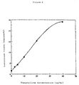


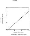
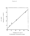

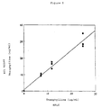
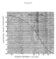

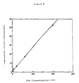


Classifications
-
- G—PHYSICS
- G01—MEASURING; TESTING
- G01N—INVESTIGATING OR ANALYSING MATERIALS BY DETERMINING THEIR CHEMICAL OR PHYSICAL PROPERTIES
- G01N33/00—Investigating or analysing materials by specific methods not covered by groups G01N1/00 - G01N31/00
- G01N33/48—Biological material, e.g. blood, urine; Haemocytometers
- G01N33/50—Chemical analysis of biological material, e.g. blood, urine; Testing involving biospecific ligand binding methods; Immunological testing
- G01N33/53—Immunoassay; Biospecific binding assay; Materials therefor
- G01N33/531—Production of immunochemical test materials
- G01N33/532—Production of labelled immunochemicals
-
- G—PHYSICS
- G01—MEASURING; TESTING
- G01N—INVESTIGATING OR ANALYSING MATERIALS BY DETERMINING THEIR CHEMICAL OR PHYSICAL PROPERTIES
- G01N33/00—Investigating or analysing materials by specific methods not covered by groups G01N1/00 - G01N31/00
- G01N33/48—Biological material, e.g. blood, urine; Haemocytometers
- G01N33/50—Chemical analysis of biological material, e.g. blood, urine; Testing involving biospecific ligand binding methods; Immunological testing
- G01N33/74—Chemical analysis of biological material, e.g. blood, urine; Testing involving biospecific ligand binding methods; Immunological testing involving hormones or other non-cytokine intercellular protein regulatory factors such as growth factors, including receptors to hormones and growth factors
- G01N33/76—Human chorionic gonadotropin including luteinising hormone, follicle stimulating hormone, thyroid stimulating hormone or their receptors
-
- C—CHEMISTRY; METALLURGY
- C07—ORGANIC CHEMISTRY
- C07D—HETEROCYCLIC COMPOUNDS
- C07D473/00—Heterocyclic compounds containing purine ring systems
- C07D473/02—Heterocyclic compounds containing purine ring systems with oxygen, sulphur, or nitrogen atoms directly attached in positions 2 and 6
- C07D473/04—Heterocyclic compounds containing purine ring systems with oxygen, sulphur, or nitrogen atoms directly attached in positions 2 and 6 two oxygen atoms
- C07D473/06—Heterocyclic compounds containing purine ring systems with oxygen, sulphur, or nitrogen atoms directly attached in positions 2 and 6 two oxygen atoms with radicals containing only hydrogen and carbon atoms, attached in position 1 or 3
- C07D473/08—Heterocyclic compounds containing purine ring systems with oxygen, sulphur, or nitrogen atoms directly attached in positions 2 and 6 two oxygen atoms with radicals containing only hydrogen and carbon atoms, attached in position 1 or 3 with methyl radicals in positions 1 and 3, e.g. theophylline
-
- C—CHEMISTRY; METALLURGY
- C07—ORGANIC CHEMISTRY
- C07F—ACYCLIC, CARBOCYCLIC OR HETEROCYCLIC COMPOUNDS CONTAINING ELEMENTS OTHER THAN CARBON, HYDROGEN, HALOGEN, OXYGEN, NITROGEN, SULFUR, SELENIUM OR TELLURIUM
- C07F15/00—Compounds containing elements of Groups 8, 9, 10 or 18 of the Periodic System
- C07F15/0006—Compounds containing elements of Groups 8, 9, 10 or 18 of the Periodic System compounds of the platinum group
- C07F15/002—Osmium compounds
- C07F15/0026—Osmium compounds without a metal-carbon linkage
-
- C—CHEMISTRY; METALLURGY
- C07—ORGANIC CHEMISTRY
- C07F—ACYCLIC, CARBOCYCLIC OR HETEROCYCLIC COMPOUNDS CONTAINING ELEMENTS OTHER THAN CARBON, HYDROGEN, HALOGEN, OXYGEN, NITROGEN, SULFUR, SELENIUM OR TELLURIUM
- C07F15/00—Compounds containing elements of Groups 8, 9, 10 or 18 of the Periodic System
- C07F15/0006—Compounds containing elements of Groups 8, 9, 10 or 18 of the Periodic System compounds of the platinum group
- C07F15/0046—Ruthenium compounds
- C07F15/0053—Ruthenium compounds without a metal-carbon linkage
-
- C—CHEMISTRY; METALLURGY
- C07—ORGANIC CHEMISTRY
- C07H—SUGARS; DERIVATIVES THEREOF; NUCLEOSIDES; NUCLEOTIDES; NUCLEIC ACIDS
- C07H21/00—Compounds containing two or more mononucleotide units having separate phosphate or polyphosphate groups linked by saccharide radicals of nucleoside groups, e.g. nucleic acids
-
- C—CHEMISTRY; METALLURGY
- C12—BIOCHEMISTRY; BEER; SPIRITS; WINE; VINEGAR; MICROBIOLOGY; ENZYMOLOGY; MUTATION OR GENETIC ENGINEERING
- C12Q—MEASURING OR TESTING PROCESSES INVOLVING ENZYMES, NUCLEIC ACIDS OR MICROORGANISMS; COMPOSITIONS OR TEST PAPERS THEREFOR; PROCESSES OF PREPARING SUCH COMPOSITIONS; CONDITION-RESPONSIVE CONTROL IN MICROBIOLOGICAL OR ENZYMOLOGICAL PROCESSES
- C12Q1/00—Measuring or testing processes involving enzymes, nucleic acids or microorganisms; Compositions therefor; Processes of preparing such compositions
- C12Q1/02—Measuring or testing processes involving enzymes, nucleic acids or microorganisms; Compositions therefor; Processes of preparing such compositions involving viable microorganisms
- C12Q1/04—Determining presence or kind of microorganism; Use of selective media for testing antibiotics or bacteriocides; Compositions containing a chemical indicator therefor
-
- C—CHEMISTRY; METALLURGY
- C12—BIOCHEMISTRY; BEER; SPIRITS; WINE; VINEGAR; MICROBIOLOGY; ENZYMOLOGY; MUTATION OR GENETIC ENGINEERING
- C12Q—MEASURING OR TESTING PROCESSES INVOLVING ENZYMES, NUCLEIC ACIDS OR MICROORGANISMS; COMPOSITIONS OR TEST PAPERS THEREFOR; PROCESSES OF PREPARING SUCH COMPOSITIONS; CONDITION-RESPONSIVE CONTROL IN MICROBIOLOGICAL OR ENZYMOLOGICAL PROCESSES
- C12Q1/00—Measuring or testing processes involving enzymes, nucleic acids or microorganisms; Compositions therefor; Processes of preparing such compositions
- C12Q1/68—Measuring or testing processes involving enzymes, nucleic acids or microorganisms; Compositions therefor; Processes of preparing such compositions involving nucleic acids
- C12Q1/6813—Hybridisation assays
- C12Q1/6816—Hybridisation assays characterised by the detection means
-
- G—PHYSICS
- G01—MEASURING; TESTING
- G01N—INVESTIGATING OR ANALYSING MATERIALS BY DETERMINING THEIR CHEMICAL OR PHYSICAL PROPERTIES
- G01N33/00—Investigating or analysing materials by specific methods not covered by groups G01N1/00 - G01N31/00
- G01N33/48—Biological material, e.g. blood, urine; Haemocytometers
- G01N33/50—Chemical analysis of biological material, e.g. blood, urine; Testing involving biospecific ligand binding methods; Immunological testing
- G01N33/53—Immunoassay; Biospecific binding assay; Materials therefor
- G01N33/531—Production of immunochemical test materials
- G01N33/532—Production of labelled immunochemicals
- G01N33/533—Production of labelled immunochemicals with fluorescent label
-
- G—PHYSICS
- G01—MEASURING; TESTING
- G01N—INVESTIGATING OR ANALYSING MATERIALS BY DETERMINING THEIR CHEMICAL OR PHYSICAL PROPERTIES
- G01N33/00—Investigating or analysing materials by specific methods not covered by groups G01N1/00 - G01N31/00
- G01N33/48—Biological material, e.g. blood, urine; Haemocytometers
- G01N33/50—Chemical analysis of biological material, e.g. blood, urine; Testing involving biospecific ligand binding methods; Immunological testing
- G01N33/53—Immunoassay; Biospecific binding assay; Materials therefor
- G01N33/543—Immunoassay; Biospecific binding assay; Materials therefor with an insoluble carrier for immobilising immunochemicals
-
- G—PHYSICS
- G01—MEASURING; TESTING
- G01N—INVESTIGATING OR ANALYSING MATERIALS BY DETERMINING THEIR CHEMICAL OR PHYSICAL PROPERTIES
- G01N33/00—Investigating or analysing materials by specific methods not covered by groups G01N1/00 - G01N31/00
- G01N33/48—Biological material, e.g. blood, urine; Haemocytometers
- G01N33/50—Chemical analysis of biological material, e.g. blood, urine; Testing involving biospecific ligand binding methods; Immunological testing
- G01N33/53—Immunoassay; Biospecific binding assay; Materials therefor
- G01N33/569—Immunoassay; Biospecific binding assay; Materials therefor for microorganisms, e.g. protozoa, bacteria, viruses
- G01N33/56911—Bacteria
-
- G—PHYSICS
- G01—MEASURING; TESTING
- G01N—INVESTIGATING OR ANALYSING MATERIALS BY DETERMINING THEIR CHEMICAL OR PHYSICAL PROPERTIES
- G01N33/00—Investigating or analysing materials by specific methods not covered by groups G01N1/00 - G01N31/00
- G01N33/48—Biological material, e.g. blood, urine; Haemocytometers
- G01N33/50—Chemical analysis of biological material, e.g. blood, urine; Testing involving biospecific ligand binding methods; Immunological testing
- G01N33/58—Chemical analysis of biological material, e.g. blood, urine; Testing involving biospecific ligand binding methods; Immunological testing involving labelled substances
- G01N33/582—Chemical analysis of biological material, e.g. blood, urine; Testing involving biospecific ligand binding methods; Immunological testing involving labelled substances with fluorescent label
-
- G—PHYSICS
- G01—MEASURING; TESTING
- G01N—INVESTIGATING OR ANALYSING MATERIALS BY DETERMINING THEIR CHEMICAL OR PHYSICAL PROPERTIES
- G01N2458/00—Labels used in chemical analysis of biological material
- G01N2458/30—Electrochemically active labels
Definitions
- the presence of these materials can often be determined by binding methods which exploit the high degree of specificity which characterizes many biochemical and biological systems. Frequently used methods are based on, for example, antigen-antibody systems, nucleic acid hybridization techniques, and protein-ligand systems. In these methods, the existence of a complex of diagnostic value is typically indicated by the presence or absence of an observable "label" which has been attached to one or more of the complexing materials.
- a preferred label should be inexpensive, safe, and capable of being attached efficiently to a wide variety of chemical, biochemical, and biological materials without changing the important binding characteristics of those materials.
- the label should give a highly characteristic signal, and should be rarely, and preferably never, found in nature.
- the label should be stable and detectable in aqueous systems over periods of time ranging up to months. Detection of the label should be rapid, sensitive, and reproducible without the need for expensive, specialized facilities or personnel. Quantification of the label should be relatively independent of variables such as temperature and the composition of the mixture to be assayed.
- labels which can be used in homogeneous systems, i.e., systems in which separation of the complexed and uncomplexed labeled material is not necessary. This is possible if the detectability of the label is modulated when the labeled material is incorporated into a specific complex.
- radioactive labels are quite versatile, and can be detected at very low concentrations. However, they are expensive, hazardous, and their use requires sophisticated equipment and trained personnel. Furthermore, the sensitivity of radioactive labels is limited by the fact that the detectable event can, in its essential nature, occur only once per radioactive atom in the labeled material. Moreover, radioactive labels cannot be used in homogeneous methods.
- non-radioactive labels include molecules observable by spectrophotometric, spin resonance, and luminescence techniques, as well as enzymes which produce such molecules.
- organometallic compounds are useful non-radioactive labeling materials. Because of the rarity of some metals in biological systems, methods which specifically assay the metal component of the organometallic compounds can be successfully exploited. For example, Cais, U.S. Patent No. 4,205,952 (1980) discloses the use of immunochemically active materials labeled with certain organometallic compounds for use in quantitating specific antigens.
- Any general method of detecting the chosen metals can be used with these labels, including emission, absorption and fluorescence spectroscopy, atomic absorption, and neutron activation. These methods often suffer from lack of sensitivity, can seldom be adapted to a homogeneous system, and as with atomic absorption, sometimes entail destruction of the sample.
- Photoluminescence is the process whereby a material is induced to luminesce when it absorbs electromagnetic radiation. Fluorescence and phosphorescence are types of photoluminescence.
- Chemiluminescent processes entail the creation of the luminescent species by a chemical transfer of energy.
- Electrohemiluminescence entails the creation of the luminescent species electrochemically.
- Suitable labels comprise electrochemiluminescent compounds, including organic compounds and organometallic compounds. Electrochemiluminescent methods of determining the presence of labeled materials are preferred over other methods for many reasons. They are highly diagnostic of the presence of a particular label, sensitive, nonhazardous, inexpensive, and can be used in a wide variety of applications.
- Organic compounds which are suitable electrochemical labels include, for example, rubrene and 9,10-diphenyl anthracene.
- Many organometallic compounds are suitable electrochemical labels, but of particular use are Ru-containing and Os-containing compounds.
- the present invention is concerned with the use of Ru-containing and Os-containing labels which can be detected by a wide variety of methods. These labels are advantageous for many reasons that will be discussed herein.
- Ru-containing and Os-containing organometallic compounds have been discussed in the literature. Cais discloses that any metal element or combination of metal elements, including noble metals from group VIII such as Ru, would be suitable components of organmetallic labels detectable by atomic absorption methods. (Cais, column 11, line 20). However, ruthenium is not a preferred metal in Cais, osmium is not specifically mentioned, no data are presented on the efficiency of using Ru or Os in any of the methods disclosed, and the preferred method of detection, atomic absorption, entails destruction of the sample.
- Weber U.S. Patent No. 4,293,310 (1981), discloses the use of Ru-containing and Os-containing complexes as electrochemical labels for analytes in immunoassays.
- the disclosed complexes are linked to amino groups on the analytes through a thiourea linkage.
- Weber also suggests the possibility of forming carboxylate esters between the labels and hydroxy groups on other analytes.
- the presence of the labeled materials can be determined with an apparatus and method which comprises a quencher and an electrochemical flow cell with light means.
- the photoelectrochemically active label upon photoexcitation transfers an electron to a quencher molecule; the oxidized molecule is subsequently reduced with an electron from an electrode of the flow cell which is held at suitable potential. This electron is measured as photocurrent.
- the amount of free labelled analyte in the system is determined by the photocurrent signal. Note that this method is the reverse of electrochemiluminescent detection of luminescent materials.
- the particular labels with which the present invention is concerned are electrochemiluminescent. They can often be excited to a luminescent state without their oxidation or reduction by exposing the compounds to electromagnetic radiation or to a chemical energy source such as that created by typical oxalate-H 2 O 2 systems. In addition, luminescence of these compounds can be induced by electrochemical methods which do entail their oxidation and reduction.
- a third mechanism for production of electrochemiluminescence from Ru(bpy) 3 2+ involves the oscillation of an electrode potential between a potential sufficiently negative to produce Ru(bpy) 3 1+ and sufficiently positive to produce Ru(bpy) 3 3+. These three methods are called, respectively, “oxidative-reduction,” “reductive-oxidation,” and “the Ru(bpy) 3 3+/+ regenerative system”.
- the oxidative-reduction method can be performed in water, and produces an intense, efficient, stable luminescence, which is relatively insensitive to the presence of oxygen or impurities.
- This luminescence from Ru(bpy) 3 2+ depends upon the presence of oxalate or other organic acids such as pyruvate, lactate, malonate, tartrate and citrate, and means of oxidatively producing Ru(bpy) 3 3+ species.
- This oxidation can be performed chemically by such strong oxidants as PbO 2 or a Ce(IV) salt. It can be performed electrochemically by a sufficiently positive potential applied either continuously or intermittently.
- Suitable electrodes for the electrochemical oxidation of Ru(bpy) 3 2+ are, for example, Pt, pyrolytic graphite, and glassy carbon. Although the oxalate or other organic acid is consumed during chemiluminescence, a strong, constant chemiluminescence for many hours can be achieved by the presence of an excess of the consumed material, or by a continuous supply of the consumed material to the reaction chamber.
- the reductive-oxidation method can be performed in partially aqueous solutions containing an organic cosolvent such as, for example, acetonitrile.
- This luminescence depends upon the presence of peroxydisulfate and a means of reductively producing Ru(bpy) 3 1+ species.
- the reduction can be performed chemically by strong reductants such as, for example, magnesium or other metals. It can be performed electrochemically by a sufficiently negative potential applied either continuously or intermittently.
- a suitable electrode for the electrochemical reduction of Ru(bpy) 3 2+ is, for example, a polished glassy-carbon electrode.
- continuous, intense luminescence can by achieved for many hours by inclusion of excess reagents, or by continous addition of the consumed reagents to the reaction mixture.
- the Ru(bpy) 3 3+/+ regenerative system can be performed in organic solvents such as acetonitrile or in partially aqueous systems, by pulsing an electrode potential between a potential sufficiently negative to reduce Ru(bpy) 3 2+ and a potential sufficiently positive to oxidize Ru(bpy) 3 2+.
- a suitable electrode for such a regenerative system is, for example, a Pt electrode. This system does not consume chemical reagents and can proceed, in principle, for an unlimited duration.
- Curtis et al. reports only unpublished observations that Ru complexes can be induced to emit light when chemically excited by an oxalate/H 2 O 2 system (Curtis et al. p. 350).
- analytes of interest and chemical moieties that bind to analytes of interest may be conveniently attached to Ru-containing or Os-containing labels through amide or amine linkages.
- the labeled materials may then be determined by any of a wide variety of means, but by far the most efficient, reliable, and sensitive means are photoluminescent, chemiluminescent, and electrochemiluminescent means.
- electrochemiluminescent labels including Ru-containing and Os-containing labels and organic molecules such as rubrene and 9,10-diphenyl anthracene, are particularly versatile and advantageous. The great advantages of the use of these novel labeled materials, and of the methods of detecting them, are further discussed hereinbelow.
- Salmonella and Staphylococcal enterotoxins Two examples which serve to illustrate these problems are the currently recognized standard methods for detecting the presence of Salmonella and Staphylococcal enterotoxins in foods.
- the detection of Salmonella in foods involves several enrichment stages due to the fact that these bacteria, when present in foods, are usually found in low numbers and are often sublethally injured. Therefore, detection methods for Salmonella must be sensitive and allow for the resuscitation and growth of injured cells.
- monoclonal antibodies in an assay system for a food contaminant assures the constant supply of a reagent which imparts unchanging specificity and sensitivity to the test itself.
- Monoclonal antibodies have been used in ELISA systems to test for food contaminants such as Salmonella (8) and Staphylococcal enterotoxins (9).
- EIA methodology Bio-Enzabead Screen Kit, Litton Bionetics
- DNA probe technology Gene-Trak, Integrated Genetics
- E. coli and/or coliforms are based on the properties of acid or gas production from the fermentation of lactose.
- the most widely used methods are: the Most Probable Number (MPN) assay and the Membrane Filtration (MF) test. Both techniques are approved by the Environmental Protection Agency (EPA) and the American Public Health Association (APHA) for the microbiological examination of water and waste water (10), and also by the Food and Drug Administration (FDA) for the bacteriological examination of milk and foods (11).
- MPN Most Probable Number
- MF Membrane Filtration
- the MPN method is actually comprised of three (12) separate assays (10).
- a nonselective medium such as Lauryl Sulfate Tryptose (LST) broth or Lactose broth is used to check for gas production from the fermentation of lactose.
- Gas positive tubes are then subcultured into a more selective medium, Brillant Green Lactose Bile (BGLB) broth for coliforms and E . coli (EC) broth for fecal coliforms, and again checked for gas production (confirmed test).
- BGLB Brillant Green Lactose Bile
- EC E . coli
- the MF technique for the bacteriological examination of water was introduced in the early 1950's (12). Unlike the MPN assay, which was tedious and time consuming, MF analysis could be completed in 24 hours without the need for further confirmations.
- the basic MF procedure is as follows: A volume of water sample, usually 100 ml is filtered through a 0.45 um pore diameter filter, and then incubated on a sterile pad saturated with selective medium. The two media most often used are the mEndo broth, selective for coliforms at 35°C, and the mFC broth, selective for fecal coliforms at 44.5°C (10).
- the present invention provides a method of detecting in a predetermined volume of a multicomponent, liquid sample an analyte of interest present in the sample at a concentration below about 10 -3 molar which comprises: a) contacting a sample with a reagent (i) capable of being induced to repeatedly emit electromagnetic radiation upon exposure to an amount of electrochemical energy from a suitable source effective to induce the reagent to repeatedly emit radiation and (ii) capable of combining with the analyte of interest, the contact being effected under appropriate conditions such that the analyte and the reagent combine; b) exposing the resulting sample to an amount of electrochemical energy from a suitable source effective to induce the reagent to repeatedly emit radiation, the exposure being effected under suitable conditions so as to induce the reagent to repeatedly emit electromagnetic radiation; and c) detecting electromagnetic radiation so emitted and thereby detecting the presence of the analyte of interest in the sample.
- the present invention also provides a competitive method for detecting in a predetermined volume of a multicomponent, liquid sample an analyte of interest present in the sample at a concentration below about 10 -3 molar which comprises: a) contacting the sample with a reagent (i) capable of being induced to repeatedly emit electromagnetic radiation upon exposure to an amount of electrochemical energy from a suitable source effective to induce the reagent to repeatedly emit radiation and (ii) capable of competing with the analyte of interest for binding sites on a complementary material not normally present in the sample, and with the complementary material, the contact being effected under appropriate conditions such that the analyte of interest and the reagent competitively bind to the complementary material; b) exposing the resulting sample to an amount of electrochemical energy from a suitable source effected to induce the reagent to repeatedly emit radiation, the exposure being effective under suitable conditions so as to induce the reagent to repeatedly emit electromagnetic radiation; and c) detecting electromagnetic radiation so emitted and thereby detecting the presence of the an
- a method for quantitatively determining in a predetermined volume of a multicomponent, liquid sample, the amount of an analyte of interest present in the sample which comprises: a) contacting the sample with a known amount of a reagent (i) capable of being induced to repeatedly emit electromagnetic radiation upon exposure to an amount of electrochemical energy from a suitable source effective to induce the reagent to repeatedly emit radiation and (ii) capable of combining with the analyte of interest, the contact being effected under appropriate conditions such that the analyte and reagent combine; b) exposing the resulting sample to an amount of electrochemical energy from a suitable source effective to induce the reagent to repeatedly emit radiation, the exposure being effected under suitable conditions so as to induce the reagent to repeatedly emit electromagnetic radiation; and c) quantitatively determining the amount of radiation so emitted and thereby quantitatively determining the amount of the analyte of interest present in the sample.
- the invention further provides a competitive method for quantitatively determining in a predetermined volume of a multicomponent, liquid sample the amount of an analyte of interest present in the sample.
- This method comprises: a) contacting the sample with a known amount of a reagent (i) capable of being induced to repeatedly emit electromagnetic radiation upon exposure to an amount of electrochemical energy from a suitable source effective to induce the reagent to repeatedly emit radiation and (ii) capable of competing with the analyte of interest for binding sites on a complementary material not normally present in the sample, and with a known amount of the complementary material, the contact being effected under appropriate conditions such that the analyte of interest and the reagent competitively bind to the complementary material; b) exposing the resulting sample to an amount of electrochemical energy from a suitable source effective to induce the reagent to repeatedly emit radiation, the exposure being effected under suitable conditions so as to induce the reagent to repeatedly emit electromagnetic radiation; and c) quantitatively determining the amount of radiation so emitted
- a method for detecting and identifying the presence of a multiplicity of analytes of interest in a liquid food or food homogenate comprises: a) immersing into the liquid food or food homogenate a portion of a diagnostic reagent holder suitable for immersing into a liquid or solid suspension and having immobilized to it a multiplicity of reagents, each reagent being immobilized to the diagnostic reagent holder in distinct, identifiable regions and capable of forming a complex with a single analyte of interest so as to allow the formation of immobilized reagent-analyte of interest complexes; b) removing the diagnostic reagent holder from the liquid food or food homogenate; c) rinsing the diagnostic reagent holder with a suitable rinsing solution; d) immersing the portion of the diagnostic reagent holder which contains the immobilized reagent-analyte of interest complexes into a detection solution which contains at least
- the present invention also provides a kit useful for detecting and identifying the presence of a multiplicity of enterotoxins in a sample.
- This kit comprises: a) a diagnostic reagent holder provided with a handle connected to a surface area suitable for immersing into a liquid or solid suspension, said surface area having at least one nitrocellulose membrance attached to it, said nitrocellulose membrane having a multiplicity of monoclonal antibodies separately and distinctly immobilized to identifiable reagions on it each immobilized monoclonal antibody being specific for an antigenic determinant on one enterotoxin; b) a first rinsing solution which comprises a buffered-aqueous solution containing a surfactant; c) a detection which comprises at least one monoclonal antibody enzyme conjugate, the monoclonal antibody of which is specific for antigenic determinants different from but located on each enterotoxin for which the monoclonal antibodies immobilize to the nitrocellulose membrance attached to the diagnostic reagent holder are
- a method for detecting the presence of at least one species of bacteria in a sample comprises: a) innoculating the sample into at least one receptacle which is provided with an open end and which contains a suitable medium for supporting the growth of the species of bacteria; b) coupling a cap to the end of the receptacle, the face of the cap which when coupled to the receptacle, is exposed to the interior of the receptacle being provided with a surface suitable for immobilizing at least a component of the bacteria; c) incubating the innoculated media under conditions so as to allow bacteria innoculated into the media to reproduce; d) turning each receptacle upside-down for a suitable length of time so as to allow components of the bacterium present in the medium to become immobilized to the surface of the cap which is exposed to the interior of the receptacle; e) turning the upside-down receptacle rightside-up; f) uncoupling the cap from the
- a method for detecting the presence of coliform bacteria in a sample comprises: a) innoculating the sample into at least one receptacle which has an open end and contains a non-selective lactose medium and an inverted Durham; b) coupling a cap provided with a polystyrene insert to the open end of each receptacle; c) incubating the inoculated medium for at least 2 hours at 37°C; d) determining whether each receptacle has gas produced by bacterial fermentation trapped within the inverted Durham vial; e) turning receptacles which have gas trapped within the inverted Durham vial upside-down for a suitable length of time so as to coliform bacteria present in the medium to form coliform-polystyrene complexes; f) turning the upside-down receptacles rightside-up; g) uncoupling the caps from the receptacles; h) treating the polyst
- Figure 1 depicts electrochemiluminescent measurements made for a homogeneous immunoassay for the determination of the concentration of an antigen in solution.
- Figure 2 graphically depicts the results of a homogeneous ECL theophylline assay.
- Figure 3 graphically depicts the results of a homogeneous theophylline assay in various sera.
- Figure 4 graphically depicts the results of an ECL theophylline assay compared to the results of a fluorescence polarization theophylline assay.
- FIG. 6 graphically depicts the modulation of an ECL signal generated in an ECL digoxin immunoassay.
- FIG. 7 graphically depicts the results of an ECL digoxin immunoassay.
- FIG. 8 graphically depicts the ECL signal generated by various concentrations of MBI 38-Compound I.
- Figure 10 shows the results of a Specificity Study of MBI 38-Compound I.
- the present invention provides a method of detecting in a predetermined volume of a multicomponent, liquid sample an analyte of interest present in the sample at a concentration below about 10 -3 molar which comprises: a) contacting a sample with a reagent (i) capable of being induced to repeatedly emit electromagnetic radiation upon exposure to an amount of electrochemical energy from a suitable source effective to induce the reagent to repeatedly emit radiation and (ii) capable of combining with the analyte of interest, the contact being effected under appropriate conditions such that the analyte and the reagent combine; b) exposing the resulting sample to an amount of electrochemical energy from a suitable source effective to induce the reagent to repeatedly emit radiation, the exposure being effected under suitable conditions so as to induce the reagent to repeatedly emit electromagnetic radiation; and c) detecting electromagnetic radiation so emitted and thereby detecting the presence of the analyte of interest in the sample.
- molar means the concentration of an analyte in solution in moles per liter or the amount of particulate matter present in a liquid sample in particles or units per liter. For example, 1 x 10 23 particles per liter may be expressed as 1 molar.
- the methods provided by the present invention may be performed as heterogeneous assays, i.e. assays in which unbound labeled reagent is separated from bound labeled reagent prior to exposure of the bound labeled reagent to electrochemical energy, and homogeneous assays, i.e. assays in which unbound labeled reagent and bound labeled reagent are exposed to electrochemical energy together.
- the electromagnetic radiation emitted by the bound labeled reagent is distinguishable from the electromagnetic radiation emitted by the unbound labeled reagent, either as an increase or as a decrease in the amount of electromagnetic radiation emitted by the bound labeled reagent in comparison to the unbound labeled reagent, or as electromagnetic radiation of adifferent wavelength. Accordingly, in one embodiment of the invention any reagent which is not combined with the analyte of interest is separated from the sample, which had been contacted with the reagent, prior to exposure of the sample to electrochemical energy. In another embodiment of the invention, prior to contacting the sample with the reagent, the sample is treated so as to immobilize the analyte of interest.
- Means for immobilizing analytes of interest include contacting the sample with a polystyrene, nitrocellulose or nylon surface, or a surface coated with whole cells, subcellular particles, viruses, prions, viroids, lipids, fatty acids, nucleic acids, polysaccharides, proteins, lipoproteins, lipopolysaccharides, glycoproteins, peptides, cellular metabolites, hormones, pharmacological agents, tranquilizers, barbiturates, alkaloids, steroids, vitamins, amino acids, sugars, nonbiological polymers, synthetic organic molecules, organometallic molecules or inorganic molecules. Additionally, the analyte of interest may be any of these substances.
- the analyte of interest is theophylline. In another embodiment of the invention, the analyte of interest is digoxin. In still another embodiment of the invention, the analyte of interest is human chorionic gonadotropin (hCG).
- the analyte of interest may be a whole cell, subcellular particle, virus, prion, viroid, nucleic acid, protein, lipoprotein, lipopolysaccharide, glycoprotein, peptide, hormone, pharmacological agent, nonbiological polymer, synthetic organic molecule, organometallic molecule or an inorganic molecule present in the sample at a concentration below about 10 -12 molar.
- the analyte of interest may be a whole cell, subcellular particle, virus, prion, viroid or nucleic acid present in the sample at a concentration below about 10 -15 molar.
- the reagent which is contacted with the sample may comprise an electrochemiluminescent chemical moiety conjugated to a whole cell, subcellular particle, virus, prion, viroid, lipid, fatty acid, nucleic acid, polysaccharide, protein, lipopro ein, lipopolysaccharide, glycoprotein, peptide, cellular metabolite, hormone, pharmacological agent, tranquilizer, barbiturate, alkaloid, steroid, vitamin, amino acid, sugar, nonbiological polymer, synthetic organic molecule, organometallic molecule, inorganic molecule, biotin, avidin or streptavidin.
- the agent is an electrochemiluminescent moiety conjugated to an antibody, antigen, nucleic acid, hapten, ligand or enzyme, or biotin avidin or streptavidin.
- the electrochemiluminescent chemical moiety may comprise a metal-containing organic compound wherein the metal is selected from the group consisting of ruthenium, osmium, rhenium, iridium, rhodium, platinum, palladium, molybdenum and technetium.
- the metal is ruthenium or osmium.
- the electrochemiluminescent chemical moeity is rubrene or 9, 10-diphenylanthracene.
- the electrochemiluminescent chemical moiety is bis[(4,4'-carbomethoxy)-2,2'-bipyridine) 2-[3-(4-methyl-2,2'-bipyridine-4-yl)propyl]-1,3-dioxolane ruthenium (II).
- the electrochemiluminescent chemical moiety is bis (2,2'bipyridine) [4-(butan-l-al)-4'-methyl-2,2'-bipyridine] ruthenium (II).
- the electrochemiluminescent chemical moiety is bis (2,2'-bipyridine) [4-(4'-methyl-2,2'-bipyridine-4'-yl)-butyric acid] ruthenium (II).
- the electrochemiluminescent chemical moiety is (2,2'-bipyridine)[cis-bis(1,2-diphenylphosphino)ethylene] ⁇ 2-[3-(4-methyl-2,2'-bipyridine-4'-yl)propyl]-1,3-dioxolane ⁇ osmium (II).
- the electrochemiluminescent chemical moiety is bis(2,2'-bipyridine) [4-(4'-methyl-2,2'-bipyridine)-butylamine] ruthenium (II). In yet another embodiment of the invention the electrochemiluminescent chemical moiety is bis(2,2'-bipyridine)[1-bromo-4(4'-methyl-2,2'-bipyridine-4-yl)butane] ruthenium (II).
- the electrochemiluminescent chemical moiety is bis(2,2'-bipyridine)maleimidohexanoic acid, 4-methyl-2,2'-bipyridine-4'-butylamide ruthenium (II).
- the sample may be derived from a solid, emulsion, suspension, liquid or gas. Furthermore, the sample may be derived from water, food, blood, serum, urine, feces, tissue, saliva, oils, organic solvents or air. Moreover, the sample may comprise acetonitrile, dimethylsulfoxide, dimethylformamide, n-methylpyrrolidinone or tert-butyl alcohol. The sample may comprise a reducing agent or an oxidizing agent.
- the present invention also provides a competitive method for detecting in a predetermined volume of a multicomponent, liquid sample an analyte of interest present in the sample at a concentration below about 10 -3 molar which comprises: a) contacting the sample with a reagent (i) capable of being induced to repeatedly emit electromagnetic radiation upon exposure to an amount of electrochemical energy from a suitable source effective to induce the reagent to repeatedly emit radiation and (ii) capable of competing with the analyte of interest for binding sites on a complementary material not normally present in the sample, and with the complementary material, the contact being effected under appropriate conditions such that the analyte of interest and the reagent competitively bind to the complementary material; b) exposing the resulting sample to an amount of electrochemical energy from a suitable source effected to induce the reagent to repeatedly emit radiation, the exposure being effected under suitable conditions so as to induce the reagent to repeatedly emit electromagnetic radiation; and c) detecting electromagnetic radiation so emitted and thereby detecting the presence of
- the reagent may be the analyte of interest conjugated to an electrochemiluminescent chemical moeity or an analogue of the analyte of interest conjugated to an electrochemiluminescent moiety.
- the analyte of interest may be theophylline, digoxin or hCG.
- the electrochemiluminescent chemical moiety may be bis((4,4'-carbomethoxy)-2,2'-bipyridine] 2-[3-(4-methyl-2,2'-bipyridine-4-yl)propyl]-1,3-dioxolane ruthenium (II).
- the electrochemiluminescent chemical moiety is bis(2,2'-bipyridine)[4-(butan-1-al)-4'-methyl-2,2'-bipyridine] ruthenium (II).
- the electrochemiluminescent chemical moiety is bis (2,2'-bipyridine) [4-(4-methyl-2,2'-bipyridine-4'-yl)-butyric acid] ruthenium (II).
- the electrochemiluminescent chemical moiety is (2,2'-bipyridine)[cis-bis(1,2-diphenylphosphino)ethylene] ⁇ 2-[3-(4-methyl-2,2'-bipyridine-4'-yl)propyl]-1,3-dioxolane ⁇ osmium (II).
- the electrochemiluminescent chemical moiety is bis(2,2'-bipyridine) [4-(4'-methyl-2,2'-bipyridine)-butylamine] ruthenium (II).
- the electrochemiluminescent chemical moiety is bis(2,2'-bipyridine)[1-bromo-4(4'-methyl-2,2'-bipyridine-4-yl)butane] ruthenium (II).
- the electrochemiluminescent chemical moiety is bis(2,2'-bipyridine)maleimidohexanoic acid, 4-methyl-2,2'-bipyridine-4'-butylamide ruthenium (II).
- the complementary material may be a whole cell, subcellular particle, virus, prion, viroid, lipid, fatty acid, nucleic acid, polysaccharide, protein, lipoprotein, lipopolysaccharide, glycoprotein, peptide, cellular metabolite, hormone, pharmacological agent, tranquilizer, barbiturate, steroid, vitamin, amino acid, sugar, non-biological polymer, synthetic organic molecule, organometallic molecule or inorganic molecule.
- the present invention provides a method for quantitatively determining in a predetermined volume of a multicomponent, liquid sample, the amount of an analyte of interest present in the sample which comprises: a) contacting the sample with a known amount of a reagent (i) capable of being induced to repeatedly emit electromagnetic radiation upon exposure to an amount of electrochemical energy from a suitable source effective to induce the reagent to repeatedly emit radiation and (ii) capable of combining with the analyte of interest, the contact being effected under appropriate conditions such that the analyte and reagent combine; b) exposing the resulting sample to an amount of electrochemical energy from a suitable source effective to induce the reagent to repeatedly emit radiation, the exposure being effected under suitable conditions so as to induce the reagent to repeatedly emit electromagnetic radiation; and c) quantitatively determining the amount of radiation so emitted and thereby quantitatively determining
- This method may be performed as a heterogeneous assay or as a homogeneous assay.
- any reagent which is not combined with the analyte of interest is separated from the sample, which had been contacted with a known amount of the reagent, prior to the exposure of the sample to an amount of electrochemical energy from a suitable source effective to induce the reagent to repeatedly emit radiation.
- the sample prior to contacting the sample with the reagent, the sample is treated so as to immobilize the analyte of interest.
- the analyte of interest may be a whole cell, subcellular particle, virus, prion, viroid, lipid, fatty acid, nucleic acid, polysaccharide, protein, lipoprotein, lipopolysaccharide, glycoprotein, peptide, cellular metabolite, hormone, pharmacological agent, tranquilizer, barbiturate, alkaloid, steroid, vitamin, amino acid, sugar, non-biological polymer, synthetic organic molecule, organometallic molecule or inorganic molecule.
- the analyte of interest is theophylline.
- the analyte of interest is digoxin.
- the analyte of interest is hCG.
- the reagent with which the sample is contacted may be an electrochemiluminescent chemical moiety conjugated to a whole cell, subcellular particle, virus, prion, viroid, lipid, fatty acid, nucleic acid, polysaccharide, protein, lipoprotein, lipopolysaccharide, glycoprotein, peptide, cellular metabolite, hormone, pharmacological agent, tranquilizer, barbiturate, alkaloid, steroid, vitamin, amino acid, sugar, non-biological polymer, synthetic organic molecule, organometallic molecule or inorganic molecule.
- the reagent is a electrochemiluminescent chemical moiety conjugated to an antibody, antigen, nucleic acid, hapten, ligand or enzyme, or biotin, avidin or streptavidin.
- the electrochemiluminescent moiety may be a metal-containing organic compound wherein the metal is selected from the group consisting of ruthenium, osmium, rhenium, iridium, rhodium, platinum, palladium, molybdenum and technetium.
- the metal is ruthenium or osmium.
- the electrochemiluminescent chemical moiety is rubrene or 9, 10-diphenylanthracene.
- the electrochemiluminescent chemical moiety is bis[(4,4'-carbomethoxy)-2,2'-bipyridine] 2-[3-(4-methyl-2,2'-bipyridine-4-yl)propyl]-1,3-dioxolane ruthenium (II).
- the electrochemiluminescent chemical moiety is bis(2,2'-bipyridine)[4-(butan-1-al)-4'-methyl-2,2'-bipyridine] ruthenium (II).
- the electrochemiluminescent chemical moiety is bis (2,2'-bipyridine) [4-(4-methyl-2,2'-bipyridine-4'-yl)-butyric acid] ruthenium (II).
- the electrochemiluminescent chemical moiety is (2,2'-bipyridine)[cis-bis(1,2-diphenylphosphino)ethylene] ⁇ 2-[3-(4-methyl-2,2'-bipyridine-4'-yl)propyl]-1,3- dioxolane ⁇ osmium (II).
- the electrochemiluminescent chemical moiety is bis(2,2;-bipyridine) [4-(4'-methyl-2,2'-bipyridine)-butylamine]-ruthenium (II).
- the electrochemiluminescent chemical moiety is bis(2,2'-bipyridine)[1-bromo-4(4'-methyl-2,2'-bipyridine-4-yl)butane] ruthenium (II).
- the electrochemiluminescent chemical moiety is bis(2,2'-bipyridine)maleimidohexanoic acid, 4-methyl-2,2'-bipyridine-4'-butylamide ruthenium (II).
- the sample may be derived from a solid, emulsion, suspension, liquid or gas.
- Samples which comprise the analyte of interest may be derived from water, food, blood, serum, urine, feces, tissue, saliva, oils, organic solvents or air. Additionally, samples may comprise acetonitrile, dimethylsulfoxide, dimethylformamide, n-methylpyrrolidinone or tert-butyl alcohol. Futhermore, the sample may comprise a reducing agent or an oxidizing agent.
- the invention also provides a competitive method for quantitatively determining in a predetermined volume of a multicomponent, liquid sample the amount of an analyte of interest present in the sample.
- This method comprises: a) contacting the sample with a known amount of a reagent (i) capable of being induced to repeatedly emit electromagnetic radiation upon exposure to an amount of electrochemical energy from a suitable source effective to induce the reagent to repeatedly emit radiation and (ii) capable of competing with the analyte of interest for binding sites on a complementary material not normally present in the sample, and with a known amount of the complementary material, the contact being effected under appropriate conditions such that the analyte of interest and the reagent competitively bind to the complementary material; b) exposing the resulting sample to an amount of electrochemical energy from a suitable source effective to induce the reagent to repeatedly emit radiation, the exposure being effected under suitable conditions so as to induce the reagent to repeatedly emit electromagnetic radiation; and c) quantitatively determining the amount of radiation so emitted
- the analyte of interest may be theophylline, digoxin or hCG.
- the reagent is the analyte of interest conjugated to an electrochemiluminescent chemical moiety or an analogue of the analyte of interest conjugated to an electrochemiluminescent chemical moiety.
- the electrochemiluminescent chemical moiety may be bis[(4,4'-carbomethoxy)-2,2'-bipyridine] 2-[3-(4-methyl-2,2'-bipyridine-4-yl)propyl]-1,3-dioxolane ruthenium (II).
- the electrochemiluminescent chemical moiety is bis(2,2'-bipyridine)[4-(butan-1-al)-4'-methyl-2,2'-bipyridine] ruthenium (II).
- the electrochemiluminescent chemical moiety is bis (2,2'-bipyridine) [4-(4-methyl-2,2'-bipyridine-4'-yl)-butyric acid] ruthenium (II).
- the electrochemiluminescent chemical moiety is (2,2'-bipyridine)[cis-bis(1,2-diphenylphosphino)ethylene] ⁇ 2-[3-(4-methyl-2,2'-bipyridine-4'-yl)propyl]-1,3-dioxolane ⁇ osmium (II).
- the electrochemiluminescent chemical moiety is bis(2,2'-bipyridine) [4-(4'-methyl-2,2'-bipyridine)butylamine] ruthenium II.
- the electrochemiluminescent chemical moiety is bis(2,2'-bipyridine)[1-bromo-4(4'-methyl-2,2'-bipyridine-4-yl)butane] ruthenium (II).
- the electrochemiluminescent chemical moiety is bis(2,2'-bipyridine)maleimidohexanoic acid, 4-methyl-2,2'-bipyridine-4'-butylamide ruthenium (II).
- the complementary material may be a whole cell, subcellular particle, virus, prion, viroid, lipid, fatty acid, nucleic acid, polysaccharide, protein, lipoprotein, lipopolysaccharide, glycoprotein, peptide, cellular metabolite, hormone, pharmacological agent, tranquilizer, barbiturate, alkaloid, steroid, vitamin, amino acid, sugar, non-biological polymer, synthetic organic molecule, organometallic molecule or inorganic molecule.
- the present invention further provides a method for detecting and identifying the presence of a multiplicity of analytes of interest in a liquid food or food homogenate.
- This method comprises: a) immersing into the liquid food or food homogenate a portion of a diagnostic reagent holder suitable for immersing into a liquid or solid suspension and having immobilized to it a multiplicity of reagents, each reagent being immobilized to the diagnostic reagent holder in distinct, identifiable regions and capable of forming a complex with a single analyte of interest so as to allow the formation of immobilized reagent-analyte of interest complexes; b) removing the diagnostic reagent holder from the liquid food or food homogenate; c) rinsing the diagnostic reagent holder with a suitable rinsing solution; d) immersing the portion of the diagnostic reagent holder which contains the immobilized reagent-analyte of interest complexes into a detection
- the analytes of interest may be microorganisms.
- the microorganisms may be viable or nonviable. Additionally, the microorganisms may be bacteria. Examples of bacteria which may detected by this method include, but are not limited to, Salmonella, Campylobacter, Escherichia, Yersinia, Bacillus, Vibrio, Legionella, Clostridium, Streptococcus or Staphylococcus.
- the analytes of interest may be antigens.
- antigens include, but are not limited to, enterotoxins and aflatoxins.
- the immobilized reagents and the detection reagents may be polyclonal antibodies, monoclonal antibodies, mixtures of monoclonal antibodies, or mixtures of polyclonal and monoclonal antibodies.
- the detection reagents may be labeled with a detectable marker.
- the detection reagents are each labeled with the same detectable marker.
- markers are well known in the art and may comprise an enzyme, e.g. alkaline phosphatase, horseradish peroxidase, glucose oxidase, beta-galactosidase or urease, a fluorescent moiety, e.g. fluorescein isothiocyanate, tetramethylrhodamine isothiocyanate, dichlorotriazinylamino fluorescein or Texas Red or a chemiluminescent moiety, e.g.
- the detectable marker may be an electrochemiluminescent chemical moiety capable of being induced to repeatedly emit electromagnetic radiation upon exposure to an amount of electrochemical energy from a suitable source effective to induce the moiety to emit radiation.
- This electrochemiluminescent chemical moiety may comprise ruthenium or osmium.
- the immobilized reagents may be immobilized to a nitrocellulose membrane which is attached to the diagnostic reagent holder.
- the present invention also provides a kit useful for detecting and identifying the presence of a multiplicity of enterotoxins in a sample.
- This kit comprises: a) a diagnostic reagent holder provided with a handle connected to a surface area suitable for immersing into a liquid or solid suspension, said surface area having at least one nitrocellulose membrance attached to it, said nitrocellulose membrane having a multiplicity of monoclonal antibodies separately and distinctly immobilized to identifiable regions on it, each immobilized monoclonal antibody being specific for an antigenic determinant on one enterotoxin; b) a first rinsing solution which comprises a buffered-aqueous solution containing a surfactant; c) a detection which comprises at least one monoclonal antibody enzyme conjugate, the monoclonal antibody of which is specific for antigenic determinants different from but located on each enterotoxin for which the monoclonal antibodies immobilize to the nitrocellulose membrance attached to the diagnostic reagent holder are specific;
- the nitrocellulose membrane is provided with monoclonal antibodies which are separately and distinctly immobilized to it and which are specific for antigenic determinants on a multiplicity of Staphylococcal enterotoxins.
- the nitrocellulose membrane is provided with four monoclonal antibodies which are separately and distinctly immobilized to it and which are specific for Staphylococcal enterotoxins A, B, D and E.
- this nitrocellulose membrance may be additionally provided with a monoclonal antibody which is separately and distinctly immobilized to it and which is specific for an antigenic determinant on more than one Staphylococcal enterotoxin.
- This enterotoxin may be specific for antigenic determinants on Staphylococcal enterotoxins C 1 , C 2 and C 3 .
- the enzyme of this kit which is conjugated to the monoclonal antibody which is specific for antigenic determinant different from but located on each Staphylococcal enterotoxin for which the nitrocellulose immobilized monoclonal antibodies are specific may be alkaline phosphatase, the enzyme substrate which is capable of reacting with the enzyme is 5-bromo, 4-chloro indolyl phosphate and the colorless dye is nitroblue tetrazolium.
- the invention further provides a method for detecting and identifying the presence of a multiplicity of Staphylococcal enterotoxins in a liquid or solid suspension.
- This method comprises: a) immersing a diagnostic reagent holder provided with a multiplicity of monoclonal antibodies specific for Strephylococcal enterotoxins and which are separately and distinctly immobilized to it into the liquid or solid suspension for a suitable length of time so as to allow the formation of immobilized monoclonal antibody-Staphylococcal enterotoxin complexes; b) removing the diagnostic reagent holder from the sample; c) immersing the diagnostic holder into a buffered-aqueous solution which comprises a surfactant; d) removing the diagnostic reagent holder from the buffered-aqueous solution which contains a surfactant; e) immersing the diagnostic reagent holder into a detection solution which comprises a monoclonal antibody-alkaline phosphatase conjugate for a
- a method for detecting the presence of at least one species of bacteria in a sample comprises: a) inoculating the sample into at least one receptacle which is provided with an open end and which contains a suitable medium for supporting the growth of the species of bacteria; b) coupling a cap to the end of the receptacle, the face of the cap which when coupled to the receptacle, is exposed to the interior of the receptacle being provided with a surface suitable for immobilizing at least a component of the bacteria; c) incubating the inoculated media under conditions so as to allow bacteria inoculated into the media to reproduce; d) turning each receptacle upside-down for a suitable length of time so as to allow components of the bacterium present in the medium to become immobilized to the surface of the cap which is exposed to the interior of the receptacle; e) turning the upside-down receptacle rightside-up; f) uncoupling the cap from the recept
- component of bacteria includes, but is not limited to, a whole cell, cell wall component, cell membrane component and flagellar component.
- the surface of the cap which is suitable for immobilizing at least a component of the bacteria may be a polystyrene insert, a nitrocellulose membrane or a nylon membrane. Furthermore, the surface may be coated with a polyclonal antibody, a monoclonal antibody, a mixture of monoclonal antibodies or a mixture of polyclonal and monoclonal antibodies.
- the reagent capable of forming a complex with the immobilized bacterial components may be a polyclonal antibody, a monoclonal antibody, a mixture of monoclonal antibodies or a mixture of polyclonal and monoclonal antibodies.
- the reagent may be labeled with a detectable marker, for example a detectable enzyme, a fluorescent moiety or a chemiluminescent moiety.
- the detectable marker may be an electrochemiluminescent moiety.
- the electrochemiluminescent moiety comprises ruthenium or osmium.
- the immobilized bacterial component-reagent complexes may be detected with a detectably marked second reagent capable of forming a complex with the immobilized bacterial component-reagent complexes.
- the reagent may be a polyclonal antibody, a monoclonal antibody, a mixture of monoclonal antibodies or a mixture of polyclonal and monoclonal antibodies and the second reagent may be a detectably marked anti-antibody directed to the reagent.
- Samples in which bacteria may be detected include water and food.
- the medium suitable for supporting the growth of the species of bacteria is a non-selective lactose medium.
- the medium is a nonselective lactose medium and the species of bacteria which is detected is at least one coliform.
- a method for detecting the presence of coliform bacteria in a sample comprises: a) inoculating the sample into at least one receptacle which has an open end and contains a non-selective lactose medium and an inverted Durham; b) coupling a cap provided with a polystyrene insert to the open end of each receptacle; c) incubating the inoculated medium for at least 2 hours at 37°C; d) determining whether each receptacle has gas produced by bacterial fermentation trapped within the inverted Durham vial; e) turning receptacles which have gas trapped within the inverted Durham vial upside-down for a suitable length of time so as to allow coliform bacteria present in the medium to form coliform-polystyrene complexes; f) turning the upside-down receptacles rightside-up; g) uncoupling the caps from the receptacles; h) treating the polys
- the non-selective lactose medium is phenol red broth.
- the substance suitable for blocking unbound sites on the polystyrene insert may be bovine serum albumin.
- the anti-coliform bacteria antibody labeled with a detectable marker may be a monoclonal antibody labeled with a detectable marker.
- the detectable marker may be an enzyme conjugated to the monoclonal antibody.
- the enzyme is calf intestinal alkaline phosphatase.
- the detectable marker is a fluorescent moiety conjugated to the monoclonal antibody.
- the detectable marker is an electrochemiluminescent moiety conjugated to the monoclonal antibody.
- the electrochemiluminescent moiety may comprise ruthenium or osmium.
- the sample is water. In another embodiment of the invention the sample is food.
- the receptacle which has an open end may be a vial.
- kits for detecting in water samples the presence of coliform bacteria comprises: a) at least one vial containing a non-selective lactose medium; b) at least one Durham vial; c) at least one vial cap provided with a polystyrene insert; d) a solution of bovine serum albumin; e) an anti-coliform bacteria monoclonal antibody-enzyme conjugate; f) a buffered-aqueous wash solution; and g) a solution of enzyme substrate.
- the enzyme conjugated to the anti-coliform bacteria monoclonal antibody is calf intestinal alkaline phosphatase and the enzyme substrate is p-nitrophenyl phosphate disodium.
- the invention also provides a compound having the structure wherein n is an integer. In one embodiment of the invention, n is 2.
- n is an integer. In one embodiment of the invention, n is 2.
- the invention also provides a compound having the structure wherein n is an integer. In one embodiment of the invention, n is 2.
- the invention provides a compound having the structure wherein R is an anion and n is an integer. In one embodiment of the invention, n is 2.
- This compound may comprise a composition of matter having the structure X-(Y) n -Z wherein X represents one or more nucleotides which may be the same or different, one or more amino acids which may be the same or different, an antibody, an analyte of interest or an analogue of an analyte of interest, n represents an integer, and Z represents the compound provided by this invention.
- the invention further provides a compound having the structure wherein R is an anion an n is an integer. In one embodiment of the invention, n is 2.
- This compound may comprise a composition of matter having the structure X-(Y) n -Z wherein X represents one or more nucleotides which may be the same or different, one or more amino acids which may be the same or different, an antibody, an analyte of interest or an analogue of an analyte of interest, n represents an integer, and Z represents the compound provided by this invention.
- n is 3.
- This compound may comprise a composition of matter having the structure X-(Y) n -Z wherein X represents one or more nucletodies which may be the same or different, one or more amino acids which may be the same or different, an antibody, an analyte of interest or an analogue of an analyte of interest, n represents an integer, and Z represents the compound provided by this invention.
- the invention further provides a compound having the structure wherein R is an anion and n is an integer. In one embodiment of the invention, n is 3.
- This compound may comprise a composition of matter having the structure X-(Y) n -Z wherein X represents one or more nucleotides which may be the same or different, one or more nucletodies which may be the same or different, an antibody, an analyte of interest or an analogue of an analyte of interest, n represents an integer, and Z represents the compound provided by this invention.
- the invention also provides a compound having the structure wherein n is an integer. In one embodiment of the invention, n is 3.
- the invention provides a compound having the structure wherein m and n are each integers which may be the same or different. In one embodiment of the invention, m and n are both 3.
- n is 2.
- This compound may comprise a composition of matter having the structure X-(Y) n -Z wherein X represents one or more nucleotides which may be the same or different, one or more amino acids which may be the same or different, an antibody, an analyte of interest or an analogue of an analyte of interest, n represents an integer, and Z represents the compound provided by this invention.
- the invention further provides a compound having the structure
- the invention provides a compound having the structure wherein R is an anion and n is an integer. In one embodiment of the invention, n is 2.
- This compound may comprise a composition of matter having the structure X-(Y) n -Z wherein X represents one or more nucleotides which may be the same or different, one or more amino acids which may be the same or different, an antibody, an analyte of interest or an analogue of an analyte of interest, n represents an integer, and Z represents the compound provided by this invention.
- the invention further provides a compound having the structure wherein R is an anion and m and n are integers. In one embodiment of the invention, m is 5 and n is 3.
- This compound may comprise a composition of matter having the strucutre X-(Y) n -Z wherein X represents one or more nucleotides which may be the same or different, one or more amino acids which may be the same or different, an antibody, an analyte of interest or an analogue of an analyte of interest, n represents an integer, and Z represents the compound provided by this invention.
- X is theophylline.
- X is digoxigenin.
- X is a peptide derived from hCG.
- X is thymidine attached to CH at carbon 5, n is 7 and m is 3.
- Z is bis (2,2'-bipyridine) [4 -(butan-1-al)-4' methyl-2,2'-bipyridine] ruthenium (II).
- the thymidine nucleotide is a 3' terminal nucleotide attached to the nucleotide sequence
- composition of matter having the structure [T-Y-Z] 2+ (R) 2 wherein T represents theophylline, Y represents a linker group attaching T to Z, Z represents bis-(2,2'-bipyridine) [4-methyl-2,2'-bipyridine-4'-yl] ruthenium (II) and R represents an anion.
- Y is attached to the carbon at position 8 of T.
- Y has the structure (CH 2 ) m -CO-NH-(CH 2 ) n wherein m and n represent an integer, which may be the same or different, greater than or equal to 1.
- m is 3 and n is 4.
- m and n are both 3.
- Y has the structure wherein m, n are r represent an integer, which may be the same or different, greater than or equal to 1. In one embodiment of the invention, m is 1, n is 1 and r is 4.
- Y has the structure wherein m, n and r represent an integer, which may the same or different, greater than or equal to 1. In one embodiment of the invention, m is 1, n is 1 and r is 4.
- Y is attached to the nitrogen at position 7 of T.
- Y has the structure (CH 2 ) n wherein n is an integer greater than or equal to 1. In still another embodiment of the invention, n is 4.
- the invention further provides a compound having the structure wherein Y is a linker arm.
- Y has the structure (CH 2 ) m -NH-CO-(CH 2 ) n wherein m and n are integers, which may be the same or different, greater than or equal to 1.
- m is 3 and n is 2.
- the invention also provides a compound having the structure wherein m and n are integers which may be the same or different. In one embodiment of the invention, m and n are both 1.
- composition of matter having the structure X-Z wherein X represents one or more amino acids which may be the same or different, comprising at least one amino acid which is cysteine or methionine, and Z is bis (2,2'-bipyridine) maleimidohexanoic acid, 4-methyl-2,2'-bipyridine-4'-butylamide ruthenium (II) attached by the carbon at position 3 or 4 of the maleimide to a sulfur substituent of cysteine or methionine.
- X represents one or more amino acids which may be the same or different, comprising at least one amino acid which is cysteine or methionine
- Z is bis (2,2'-bipyridine) maleimidohexanoic acid, 4-methyl-2,2'-bipyridine-4'-butylamide ruthenium (II) attached by the carbon at position 3 or 4 of the maleimide to a sulfur substituent of cysteine or methionine.
- the electrochemiluminesence of tris (2,2'-bipyridyl) ruthenium (II) chloride hexahydrate was measured in a 15 ml three-neck, round bottom flask containing 10 ml of a solution prepared as described below; a 1.5mm x 10 mm magnetic stir bar; a 1.0mm diameter silver wire quasi-reference electrode; a combination 28 gauge platinum wire counter electrode; and a working electrode consisting of a 22 gauge platinum wire welded to a 1 cm x 1 cm square piece of 0.1 mm thick, highly polished platinum foil.
- the working platinum foil electrode was shaped into a 4.76 mm (3/16 of an inch) diameter semi-circle surrounding the 28 gauge platinum wire counter electrode by 2.38 mm (3/32 of an inch) equidistantly.)
- the silver wire was connected to the EG&G Model 178 electrometer probe of the EG&G Model 173 potentiostat/galvanostat.
- the platinum wire counter electrode and the platinum working electrode were connected to the anode and cathode respectively of the EG&G Model 173 potentiostat.
- the device was grounded.
- Cyclic voltammetry was performed with the EG&G Model 173 potentiostat to which an EG&G Model 175 universal programmer was attached. The programmer was set for 100 mV/second sweeps between +1.75 volt anodic and-1.80 volt cathodic potentials. Electrochemiluminescence was detected using a Hamamatsu R928 photomultiplier tube, set inside a Products for Research Model PR1402RF photomultiplier tube housing which was fitted with a Kodak #23A gelatin (red) filter. The photomultiplier tube housing was connected to an Oriel Model 7070 photomultiplier detection system. The cyclic voltammogram was recorded on a Houston Instruments Model 200 X-Y recorder.
- Cyclic voltammograms were generated for 1mM tris (2, 2' - bipyridyl) ruthenium (II) chloride hexahydrate (Aldrich Chemical Company), 0.1M tetrabutylammonium tetrafluoroborate (TBABF 4 ) (Aldrich Chemical Company) solutions prepared with the following organic solvents: acetonitrile; n-dimethylformamide; dimethylsulfoxide and 1-methyl, 2-pyrrolidinone (Aldrich Chemical Company).
- solutions were prepared as follows: sufficient amounts of tris (2,2'-bipyridyl) ruthenium (II) chloride hexahydrate and TBABF 4 were dissolved in the spectroscopic grade organic solvents (Aldrich Chemical Company) described above to provide final concentrations of 1mM and 0.1M, respectively. 10ml of the resulting solution was then added to the 15 ml three-neck round bottom flask. The electrodes were immersed in the solution and the working electrode pulsed between a +1.75 and -1.45 volt potential to generate electrochemiluminescence. Electrochemiluminesence was visually observed in each of the solutions described above.
- solutions were prepared as follows: sufficient amounts of tris (2, 2'-bipyridyl) ruthenium (II) chloride hexahydrate and TBABF 4 were added to the organic solvents described above to provide final concentrations of 2 mM and 0.2 M respectively. To an aliquot of this solution was added an equal volume of deionized, distilled water containing a strong oxidizing ammonium persulfate, at a concentration of 36 mM. Control solutions that did not contain the tris (2, 2'-bipyridyl) ruthenium (II) chloride hexahyrate were prepared. 10ml of the resulting solution was then added to the 15 ml three-neck round bottom flask. Electrochemiluminescence was accomplished by pulsing for one second intervals, between zero and -2.0 volts cathodic potential.
- Electrochemiluminescent measurements were performed by integrating the resulting electrochemiluminescent photomultiplier tube signal using an. integrator connected to a Micronta Model 22191 digital multimeter. The electrochemiluminescent signal was integrated for 10 seconds during the pulsing and recorded in millivolts. The results are shown in Table I and indicate that variation of solvents effects quantum efficiency of the ruthenium (II) chloride.
- the silver wire was connected to the EG&G Model 178 electrometer probe of the EG&G Model 173 potentiostat/qalvanostat.
- the platinum wire counter electrode and the platinum working electrode were connected to the anode and cathode respectively of the EG&G Model 173 potentiostat.
- the device was grounded.
- the electrochemiluminescence emitted from the ruthenium-labeled rabbit anti-mouse IgG antibody solution was detected using an Hamamatsu R928 photomultiplier tube, set inside a Products for Research Model PR1402RF photomultiplier tube housing which was fitted with a Kodak #23A gelatin (red) filter.
- the photomultiplier tube housing was connected to an Oriel Model 7070 photomultiplier detection system.
- Electrochemiluminescence was induced by pulsing for one second intervals, between zero and -2.0 volts cathodic potential. Electrochemiluminescent measurements were performed by integrating the resulting electrochemiluminescent photomultiplier tube signal using an integrator connected to a Micronta Model 22191 digital multimeter. The electrochemiluminescent signal was integrated for 10 seconds during the pulsing and recorded in millivolts.
- a stock solution of 1.25 x 10 -7 M ruthenium-labeled rabbit anti-mouse IgG antibody was prepared from a concentrated solution (2 mg/ml, 7.5 Ru/antibody) of the labeled antibody by dilution in phosphate-buffered saline (PBS). An aliquot of this solution (80 microliters) was added to 10 ml of dimethylsulfoxide (DMSO)/deionized, distilled water (1:1) containing 0.1 M tetrabutylammonium tetrafluoroborate (TBABF 4 ) and 18 mM ammonium persulfate in the reaction vessel. The final ruthenium-labeled antibody concentration was 1 x 10 -9 M. Electrochemiluminescence was measured as described above.
- DMSO dimethylsulfoxide
- TABF 4 tetrabutylammonium tetrafluoroborate
- a solution containing 6 mg/ml rabbit anti-BSA antibody was diluted 1:20,000, 1:30,000, 1:40,000, 1:50,000, and 1:60,000 in PBS, and the dilutions were added in duplicate to the wells coated with ruthenium-labeled BSA or unlabeled BSA, and the plate was incubated for one hour at room temperature. After three washes with PBS as before,' the presence of bound rabbit anti-BSA antibody was determined by adding goat anti-rabbit IgG-peroxidase conjugate (1:1000 dilution in PBS of a 0.5 mg/ml solution, 50 microliters/well) to each well and incubating the plate for one hour at room temperature.
- Rabbit anti-mouse IgG antibody labeled with 4,4' - (dichloromethyl) - 2,2' - bipyridyl, bis(2,2' - bipyridyl) ruthenium (II) (ruthenium-labeled rabbit anti-mouse IgG antibody) was compared with unlabeled rabbit anti-mouse IgG antibody with respect to its ability to compete with enzyme-labeled, anti-mouse IgG antibody for binding to mouse IgG.
- the enzyme substrate p-nitrophenyl phosphate (1.5 mg/ml in 10% diethanolamine buffer, pH 9.6) was added to each well (200 microliters/well); the plate was incubated for 30 minutes at room temperature and read spectrophotometrically at 405 nm.
- the mean background absorbance in the control wells was subtracted from the mean value of duplicate readings for each of the three solutions at each dilution. These absorbance values are shown in Table VI.
- the ruthenium-labeled, anti-mouse IgG curve on a point-by-point comparison, is approximately 81% as low as the unlabeled anti-mouse IgG curve in comparison to the enzyme conjugate curve.
- the ruthenium-labeled, anti-mouse IgG antibody is immunologically reactive for its antigen (mouse IgG), and is approximately 81% as effective as unlabeled anti-mouse IgG antibody in competing with enzyme-labeled, anti-mouse IgG antibody for binding to mouse IgG.
- a solution containing 7.8 x 10 -6 M bovine serum albumin (BSA) labeled with 4,4' - (dichloromethyl) - 2,2'-bipyridyl, bis(2,2' bipyridyl) ruthenium (II) (ruthenium-labeled bovine serum albumin) was prepared from a stock solution of ruthenium-labeled BSA (2.6 mg/ml, 6 Ru/BSA) by dilution in phosphate-buffered saline. 26 microliters of this solution were added to 10 ml of DMSO/deionized, distilled water (1:1) containing 0.1 M TBABF 4 and 18 mM ammonium persulfate in the reaction vessel. The final ruthenium-labeled BSA concentration was 2 x 10 -8 M. Electrochemiluminescence was measured as described in Example V.
- a solution containing 1.25 x 10 -6 M rabbit anti-mouse IgG antibody labeled with 4,4 - (dichloromethyl) - 2,2' - bipyridyl, bis(2,2' - bipyridyl) ruthenium (II) (ruthenium-labeled, rabbit anti-mouse IgG antibody) was prepared from a stock solution of ruthenium-labeled, rabbit anti-mouse IgG antibody (2 mg/ml, 7.5 Ru/antibody) by dilution in phosphate-buffered saline.
- a solution containing 7.8 x 10 -6 M bovine serum albumin (BSA) labeled with 4,4' - ( dichloromethyl-) - 2,2' bipyridyl, bis (2,2 - bipyridyl) ruthenium (II) (ruthenium-labeled bovine serum albumin) was prepared from a stock solution of ruthenium-labeled BSA (2.5 mg/ml, 6 Ru/BSA) by dilution in phosphate-buffered saline (PBS).
- BSA bovine serum albumin
- PBS phosphate-buffered saline
- a solution containing 3.75 x 10 -5 M rabbit anti-BSA antibody was prepared from a stock solution of rabbit anti-BSA antibody (6.0 mg/ml) by dilution in PBS, and an aliquot (26 microliters) was added to the solution of ruthenium-labeled BSA in the reaction vessel to give a final rabbit anti-BSA antibody concentration of 1 x 10 -7 M.
- the electrochemiluminescence of the resulting mixture of ruthenium-labeled BSA antigen and antibody was measured.
- the results shown in Table VII indicate a reduction in the electrochemiluminescence of the ruthenium-labeled BSA upon addition of rabbit anti-BSA antibody and demonstrate that a homogeneous electrochemiluminescent detection of antibody to BSA may be achieved. Based upon these results one skilled in the art would know that a homogeneous electrochemiluminescent immunoassay for detecting other analytes of interest may be developed.
- a solution containing 6.25 x 10 -6 M rabbit anti-mouse IgG antibody labeled with 4,4' - (dichloromethyl) - 2,2' - bipyridyl, bis (2,2' - bipyridyl) ruthenium (II) (ruthenium-labeled rabbit anti-mouse IgG antibody) was prepared from a stock solution of ruthenium-labeled rabbit anti-mouse IgG antibody (2 mg/ml, 7.5 Ru/antibody) by dilution in phosphate-buffered saline (PBS).
- a solution containing 2.5 x 10 -5 M mouse IgG was prepared from a stock solution of mouse IgG (4.0 mg/ml) by dilution in PBS, and different aliquots (20 microliters and 40 microliters) of this solution were added to the solution of ruthenium-labeled, anti-mouse IgG antibody in the reaction vessel to give final mouse IgG concentrations of 5 x 10 -8 M and 1 x 10 -7 M, respectively.
- a formalinized suspension of the bacterium Legionella micdadei was adjusted to an optical density (at 425nm) of I.00 by dilution with PBS buffer. Approximately 3 x 10 9 cells were added to a conical microcentrifuge tube. The cells were centrifuged (10 minutes, 10,000 RPM), the supernatant decanted, and the cells resuspended in a 1:50 dilution of a mouse monoclonal IgG antibody, (1.45 mg/ml) specific for Legionella midadei , in PBS (1 ml). After incubation at room temperature for 1 hour, the cells were centrifuged, the supernatant decanted, the cells resuspended in PBS buffer and centrifuged again.
- the cells were resuspended in a 1:50 dilution (in PBS) of rabbit anti-mouse IgG antibody labeled with 4,4' - (dichloromethyl) - 2,2' - bipyridyl, bis (2,2' - bipyridyl) ruthenium (II) i.e. ruthenium-labeled rabbit anti-mouse IgG antibody, (2mg/ml, 7.5 Ru/antibody). After incubation at room temperature for 1 hour, the cells were centrifuged, the supernatant decanted, and the cells resuspended in PBS and washed twice, with centrifugation, as before.
- II ruthenium-labeled rabbit anti-mouse IgG antibody
- the cells were resuspended in 200 microliters of PBS.
- 100 microliters of the cell suspension was added to the reaction vessel containing 10 ml of DMSO/deionized, distilled water (1:1) containing 0.1 M TBABF 4 and 18 mM ammonium persulfate and transferred to the reaction vessel.
- the electrochemiluminescence was measured for the cell suspension.
- Another 100 microliters of the cell suspension was added to the reaction vessel and electrochemiluminescence measured. Electrochemiluminescence was measured for the solution without cells as a control according to the method described in Example 2.
- a suspension of the bacterium Legionella micdadei was prepared and incubated with a mouse monoclonal IgG antibody specific for Legionella as described in Example 9.
- the cells were centrifuged, washed, and resuspended in 0.2 ml of PBS.
- ruthenium-labeled rabbit anti-mouse IgG antibody (1.25 x 10 -6 M) was added to the cell suspension, and the mixture was incubated for 2 hours at room temperature.
- ruthenium-labeled, rabbit anti-mouse IgG antibody was incubated in the same way in the absence of the cell suspension.
- the solution of labeled antibody was added to 10 ml of DMSO/deionized, distilled water (1:1) containing 0.1 m TBABF 4 and 18mM ammonium persulfate in the reaction vessel to give a final ruthenium-labeled, rabbit anti-mouse IgG antibody concentration of 1 x 10 -8 M.
- the electrochemiluminescence was measured as described in Example 2. The same procedure was followed for the cell suspension with added ruthenium-labeled rabbit anti-mouse IgG antibody.
- a formalinized suspension of the bacterium Legionella micdadei was adjusted to an optical density (at 425nm) of 1.00 by dilution with PBS buffer and 2 ml of this suspension were added to a conical microcentrifuge tube. The cells were centrifuged (10 minutes, 10,000 RPM), the supernatant decanted, and the cells were resuspended in a 1:10 dilution in PBS (0.5 ml) of a mouse monoclonal IgG antibody, (1.45 mg/ml) specific for Legionella micdadei .
- the cells were centrifgued as before, the supernatant was decanted, the cells were resuspended in PBS buffer and centrifuged again. Following decantation of the supernatant, the cells were resuspended in a 1:50 dilution (in PBS) of ruthenium-labeled, rabbit anti-mouse IgG antibody (1ml, 7.5 Ru/antibody). After incubation at room temperature for 1 hour, the cells were centrifuged as before, the supernatant was decanted, and the cells resuspended in PBS and washed twice, with centrifugation as before.
- PBS ruthenium-labeled, rabbit anti-mouse IgG antibody
- the cells were resuspended in 100 microliters of either PBS or 1.0M acetic acid - 0.9%NaCl (normal saline) solution and incubated at room temperature for 40 minutes. After centrifugation, 100 microliters of the cell supernatant fluid was transferred into the reaction vessel along with 10 ml of DMSO-deionized, distilled water (1:1) containing 0.1 M TBABF 4 and 18 mM ammonium persulfate. The electrochemiluminescence of the acetic acid/normal saline cell supernatant fluid and for the supernatant fluid from the PBS washed cells was measured according to the method described in Example 2.
- the electrochemiluminescence measurements are shown in Table XI, and demonstrate that the elution of the ruthenium-labeled, rabbit anti-mouse IgG from the monoclonal antibody coated Legionella bacteria by treating the cells with 1.0M acetic acid-normal saline (Ref.23) results in an increase in the electrochemiluminescence generated by the unbound ruthenium-labeled antibody. These results also show that the ruthenium labeled antibody is bound to the monoclonal antibody-coated Legionella, and that the PBS wash did not result in an increase in ECL in comparison to the background signal.
- Pregnane-diol-3-glucuronide may be detected and quantified in urine by contacting a sample of urine with a known amount of PD3G labeled with an electrochemiluminescent moiety and a known amount of an anti-PD3G antibody under conditions such that the PD3G present in the sample and the PD3G-electrochemiluminescent moiety compete for binding sites on the antibody. After a suitable time the resulting sample may be induced to repeatedly emit electromagnetic radiation upon direct exposure to an electrochemical energy source effective for inducing the electrochemiluminescent moiety to repeatedly emit electromagnetic radiation. Emitted radiation may be quantified, and the amount of PD3G present in the sample determined therefrom.
- Nucleic acid samples (5 to 25 micrograms) in 10mM tris hydrochloride (pH 8.0)-1mM EDTA may be heat denatured, cooled and modified by bisulphite catalysed transamination of cytosine residues with ethylenediamine for three hours at 42°C. After overnight dialysis against three changes of 5mM sodium phosphate buffer, pH 8.5, the samples may be concentrated to 100 microliters by ultrafiltration. These modified nucleic acids (1 to 10 micrograms) may be diluted in 100 microliters with 0.1 M sodium phosphate buffer, pH 8.5.
- a tris-ruthenium bipyridyl-N-hydroxysuccinimide ester derivative may be prepared as a 0.2M stock solution in N, N' dimethylformamide (DMF) by methods known in the art. 5 microliters of this ester solution may be added to the modified nucleic acid solution in 0.1 M sodium phosphate buffer, pH 8.5 and incubated at room temperature for 1 hour. Labeled nucleic acid probes may be purified by dialysis against at least three changes of 150mM sodium chloride - 10mM sodium phosphate buffer (pH 7.0) stored at 4°C until used.
- DMF N, N' dimethylformamide
- HTLV-III virus may be detected in whole blood by treating the blood to release the RNA from virus particles.
- a sample containing the HTLV-III RNA is contacted with a single-stranded oligonuceotide probe complementary to the HTLV-III RNA and labeled with an electrochemiluminescent moiety.
- the resulting sample may be induced to repeatedly emit electromagnetic radiation upon direct exposure to an electrochemical energy source effective for inducing the electrochemil uminescent moiety to repeatedly emit electromagnetic radiation. Emitted radiation may be quantified, and the amount of HTLV-III RNA present in the sample determined therefrom.
- Legionella bacteria may be detected in a clinical sample such as sputum by treating the sputum to release the ribosomal RNA from the bacteria.
- the resulting sample is contacted with a single-stranded oligonucleotide probe that is complementary to sequences in the ribosomal RNA, specific for Legionella, and labeled with an electrochemiluminescent moiety.
- the resulting sample may be induced to repeatedly emit electromagnetic radiation upon direct exposure to an electrochemical energy source effective for inducing the electrochemiluminescent moiety to repeatedly emit electromagnetic radiation. Emitted radiation may be quantified, and the amount of Legionella Bacteria present in the sample determined therefrom.
- Cytomegolavirus (CMV) DNA may be detected in human genomic DNA by treating a tissue sample, for example lymphocytes, to release the DNA and cleaving the DNA with restriction enzymes.
- the resulting sample containing double-stranded fragments of CMV is contacted with a single-stranded oligonucleotide probe that is complementary to the CMV DNA, labeled with a electrochemiluminescent moiety and capable of displacing one of the strands of the DNA duplex.
- the resulting sample may be induced to repeatedly emit electromagnetic radiation upon direct exposure to an electrochemical energy source effective for inducing the electrochemiluminescent moiety to repeatedly emit electromagnetic radiation. Emitted radiation may be quantified, and the amount of Cytomegalovirus RNA present in the sample determined therefrom.
- Example 17 Hydrization Assay for the Detection of the Ras-Oncogene in Human Bladder Carcinoma Cells using a Heterogeneous Method
- the ras-oncogene may be detected in human bladder carcinoma cells by treating a tissue sample to release the DNA, cleaving the DNA with restriction enyzmes, melting the DNA to single strands and binding to nitrocellulose paper as previously described in Maniatis, T. et al. (24).
- the filter paper with bound single-stranded DNA is contacted with an oligonucleotide probe specific for the ras-oncogene, and labeled with a electrochemiluminescent moiety.
- the resulting sample may be induced to repeatedly emit electromagnetic radiation upon direct exposure to an electrochemical energy source effective for inducing the electrochemiluminescent moiety to repeatedly emit electromagnetic radiation. Emitted radiation may be detected and the presence of ras-oncogene RNA in the sample determined.
- a macromolecular enzyme substrate for example starch, dextran or a synthetically prepared macromolecular polymer, may be labeled with an electrochemiluminescent moiety.
- the labeled substrate may be used to determine and quantify the presence of an enzyme present in a multicomponent system.
- the labeled substrate may also be used to quantify an amount of enzyme linked to an antibody, DNA probe, RNA probe, streptavidin, avidin, or biotin.
- the labeled substrate may be cleaved into smaller fragments selectively by an appropriate enzyme. Any enzyme-substrate combination may be used.
- enzymes include glycosidases, glucosidases, amylases, proteases, endonucleases, lyases and dextranases.
- the resulting sample may be induced to repeatedly emit electromagnetic radiation upon direct exposure to an electrochemical energy source effective for inducing the electrochemiluminescent moiety to repeatedly emit electromagnetic radiation. Emitted radiation may be quantified, and the amount of enzyme present in the sample determined therefrom.
- the advantage is that an amplification of the electrochemiluminescent signal will result from the cleaved fragments each labeled with electrochemiluminescent moiety.
- a sample containing streptococci may be contacted with a reagent mixture containing a synthetic peptide labeled with an electrochemiluminescent moiety.
- the synthetic peptide being a substrate specific for a peptidase produce by the bacteria. Action of the peptidase on the synthetic peptide results in the production of fragments labeled with the electrochemiluminescent moeity.
- the resulting sample may be induced to repeatedly emit electromagnetic radiation upon direct exposure to an electrochemical, energy source effective for inducing the electrochemiluminescent moiety to repeatedly emit electromagnetic radiation. Emitted radiation may be quantified, and the amount of streptococci present in the sample determined therefrom.
- a blood sample containing Hepatitis B surface antigen (HBsag) is contacted for a suitable time with an antibody specific for the HBsag and labeled with dextranase.
- the antibody not bound to HBsag is removed and the antigen-antibody complex is contacted with a dextran polymer labeled with an electrochemiluminescent moiety. Action of the dextranase on the labeled polymers will result in the production of fragments each labeled with the electrochemiluminescent moiety.
- the resulting sample may be induced to repeatedly emit electromagnetic radiation upon direct exposure to an electrochemical energy source effective for inducing the electrochemiluminescent moiety to repeatedly emit electromagnetic radiation. Emitted radiation may be quantified, and the amount of Hepatitis B surface antigen present in the sample determined therefrom.
- a tissue sample containing bradykinin receptors is suitably prepared and contacted with bradykinin labeled with an electrochemiluminescent moiety. After a suitable amount of time the resulting sample may be induced to repeatedly emit electromagnetic radiation upon direct exposure to an electrochemical energy source effective for inducing the electrochemiluminescent moiety to repeatedly emit electromagnetic radiation. Emitted radiation may be quantified, and the amount of bradykinin receptor present in the sample determined therefrom.
- This assay could also be used for measuring other ligand-receptor interactions such as estrogen-estrogen receptor. In addition this assay may be used to determine and quantify the amount of unlabeled ligands using a competitive binding assay format.
- a sample containing nucleic acid hybrids such as double-stranded DNA, DNA-RNA duplexes, or double-stranded RNA is contacted with an electrochemiluminescent moiety that specifically intercalates into nucleic acid hybrids.
- an electrochemiluminescent moiety that specifically intercalates into nucleic acid hybrids.
- the resulting sample may be induced to repeatedly emit electromagnetic radiation upon direct exposure to an electrochemical energy source effective for inducing the electrochemiluminescent moiety to repeatedly emit electromagnetic radiation. Emitted radiation may be quantified, and the amount of nucleic acid hybrids present in the sample determined therefrom.
- Example 23 - Detection of Human T-Cell Leukemia Virus III (HTLV-III) Antigen Complexed to Antibody in Saliva bu a Homogeneous Immunoassay using Electrochemiluminescence
- a saliva sample containing HTLV-III complexed to antibody is contacted with a solution containing a chaotopic agent to disrupt the antigen-antibody complexes.
- This solution is then contacted with an antibody specific for HTLV-III and labeled with an electrochemiluminescent moiety.
- the chaotopic agent is removed allowing the labeled and unlabeled antibodies to recombine with antigen.
- the resulting sample may be induced to repeatedly emit electromagnetic radiation upon direct exposure to an electrochemical energy source effective for inducing the electrochemiluminescent moiety to repeatedly emit electromagnetic radiation. Emitted radiation may be quantified, and the amount of human T-cell leukemia virus III (HTLV-III) antigen present in the sample determined therefrom.
- HTLV-III human T-cell leukemia virus III
- Monoclonal antibodies specific for each of the Staphylococcal enterotoxins A, B, C, D, and E and monoclonal antibodies which are cross-reactive for these enterotoxins were purified.
- Each antibody was purified from ascitic fluid by passage of the ascites through a column of Staphylococcal protein A coupled to an agarose gel support matrix which is provided with binding, elution, and regeneration buffers as part of the Monoclonal Antibody Purification System (Bio-Rad Laboratories, Inc.).
- the effluent was monitored for absorbance at 280 nm (A 280 ), and 1 ml fractions were collected.
- a 280 had returned to a stable baseline value
- the column was washed with 5 bed volumes of binding buffer, and the elution buffer was then used to elute the purified immunoglobulin from the column.
- the eluate was collected in 0.3 ml of 1 M Tris-HCl, pH 9.0.
- the A 280 had again returned to a stable baseline value, the column was washed with 5 bed volumes of elution buffer, followed by 10 bed volumes of regeneration buffer and 5 bed volumes of binding buffer so that the column was ready for the next purification cycle.
- the fractions containing the purified immunoglobulin were pooled, and concentrated using a stirred ultrafiltration cell (Amicon Corp.). The final concentration of purified immunoglobulin was greater than 5 mg/ml as determined by the Lowry assay for total protein.
- the purified antibody was dialyzed against two changes of phosphate-buffered saline (PBS) containing 0.1% sodium azide for 48 hours at 4°C.
- PBS phosphate-buffered saline
- the purified monoclonal antibodies were titrated in a 96-well microtiter plate ELISA system to provide a measure of their immunological reactivity for specific enterotoxin.
- the end point titers for the antibodies which were obtained were compared to reference titer values for previously acceptable lots of each antibody.
- Antibodies of sufficient titer were accepted as immunologically functional coating antibodies for immobilization to a diagnostic reagent holder, e.g. dipstick or for conjugation to an enzyme for use as probes in the immunoassay.
- Diagnostic reagent holders with nitrocellulose membranes attached were prepared for immobilization of antibodies to the membrane surface by first immersing the membranes in phosphate-buffered saline for one hour at room temperature to improve the wetability of the membrane. The sticks were removed from the solution and the membranes were allowed to air dry. Solutions of the purified monoclonal antibodies, each of which is specific for one of the Staphylococcal enterotoxins A, B, C, D or E, and a non-specific control mouse immunoglobulin (Jackson Immuno Research Laboratories, Inc.) were applied to different regions of the membrane by spotting 2 microliters of antibody solution directly onto the membrane surface.
- the concentration of each antibody used was one which was known to saturate the binding sites of the nitrocellulose: 250 micrograms/ml of 3A antibody (specific for A toxin), 50 micrograms/ml of 2B antibody (specific for B toxin) , 300 micrograms/ml of 1C3 antibody (specific for C1, C2, and C3 toxins) 200 micrograms/ml of 3D antibody (specific for D toxin) 250 micrograms/ml of 4E antibody (specific for E toxin), and 200 mircrograms/ml of non-specific control mouse immunoglobulin.
- the dipsticks were allowed to air dry at room temperature, and the remaining protein binding sites on the membrane were blocked by immersing the dipsticks in a solution of phosphate-buffered saline containing 0.5% Tween 20 and 3% bovine serum albumin. Following an overnight incubation at room temperature in this blocking solution, the sticks were allowed to air dry.
- Purified monoclonal antibodies 2A (specific for A and E toxins), 6B (specific for B, C1, C2, and C3 toxins), and 1D (specific for D toxin) were covalently linked to the enzyme alkaline phosphatase for use as conjugated probes to determine the presence of A, B, C1, C2, C3, D, and E toxins bound to antibodies immobilized to the membrane surface of the diagnostic reagent holder dipsitck.
- Each conjugate was prepared by a stoichiometrically controlled two step procedure which utilizes glutaraldehyde as the bifunctional crosslinking agent.
- the conjugate was dialyzed against two changes of phosphate- buffered saline containing 0.1% sodium azide for 48 hours at 4°C.
- Bovine serum albumin was added to the dialysate to a final concentration of 3% for stabilization during prolonged storage at 4°C.
- the antibody-enzyme conjugates were titrated in a 96-well microliter plate ELISA system to provide a measure of their immunological reactivity for specific enteroxin.
- the end point titers for the conjugates which were obtained were compared to reference titer values for previously acceptable lots of each conjugate. Conjugates of sufficient titer were accepted for use as probes in the dipstick assay for Staphylococcal enterotoxins.
- Solid foods such as ham, sausage, noodles and cheese, which represent the types of foods most commonly assoicated with outbreaks of Staphylococcal food poisoning, were converted to a homogeneous liquid suspension in order to perform the assay for Staphylococcal entertoxins.
- 20 milliliters of water were added to 20 grams of food, and the mixture was homogenized for 30 seconds using a stomacher lab-blender (Tekmar Co.). For more viscous foods, twice the volume of water was used.
- Staphylococcal enteroxin was added to the food either before or after homogenization. The food homogenate was tested directly using the diagnostic reagent holder dipstick described above.
- Liquid foods such as milk were tested directly by immersing the diagnostic reagent holder dipstick in the liquid food and running the test in an identical manner as for the solid food homogenates. In testing these foods, it was also determined that sensitivity may be improved to 1 nanogram per milliliter by carrying out the additional extraction steps described above.
- Diagnostic reagent holders provided with a nitrocellulose membrane having immobilized to it monoclonal antibodies specific for the individual Staphylococcal enterotoxins were immersed in a tube containing 1 ml of a liquid food, food homogenate, or food extract prepared as described above, or 1 ml of PBS. Each of these solutions contained 1 ng/ml of an added Staphylococcal enterotoxin or a mixture of enterotoxins, each at 1 ng/ml concentration. The dipstick remained immersed in these sample solutions for a 1 hour incubation period at room temperature with shaking on a rotary shaker.
- a mixture of monoclonal antibody-alkaline phosphatase conjugates prepared as described was made as follows: 1:1000 dilutions of 2A conjugate (specific for A and E toxins), 6B conjugate (specific for B, C1, C2 and C3 toxins), and 1D conjugate (specific for D toxin) were made in the same solution (30 microliters of each conjugate in 30 ml of PBS containing 0.5% Tween-20 and 3% BSA).
- the dipsticks were immersed in this conjugate mixture, 2 ml per stick, and incubated for 30 minutes at room temperature with shaking.
- the dipsticks were washed twice in PBS-Tween, then three times with shaking in PBS, 5 minutes per wash to remove unbound monoclonal antibody-alkaline phosphatase conjugate from the membrane surface.
- BCIP 5-bromo 4-chloro indolyl phosphate
- NBT nitroblue tetrazolium
- the dipsticks were immersed in the substrate mixture for 30 minutes at room temperature with shaking, then rinsed with water and air dried, providing a permanent record of the assay result.
- the entire assay procedure requires few to none pipetting steps, minimal hands-on time, and can be performed in approximately two hours.
- the substrate of the enzyme conjugate, BCIP was hydrolyzed by the conjugate bound to the toxin on the membrane to yield a reaction product which reduces the NBT to an insoluble blue product that binds to the membrane on the dipstick.
- This method which employs a diagnostic reagent holder dipstick, is capable of detecting one nanogram per milliliter of one or a muliplicity of Staphylococcal enterotoxins in a liquid food, food homogenate or food extract.
- E. coli the principal fecal coliform, is the only organism prevalent in water which can cleave MUG to release a fluorogenic radical visible under UV light (4, 10).
- the CAP-EIA MPN assay format provides 3 types of data:
- the experiment was performed as follows: to obtain the three 10-fold dilution series as required for an MPN assay, the following volumes of water sample collected from a nearby creek were used to inoculate the vials containing 10 ml of PRLB-MUG and equipped with a polystyrene well inside the vial cap.
- the wells were treated for 30 minutes at 35°C with a solution of 3% bovine serum albumin (BSA; Sigma Chemical Co., St. Louis, MO) in phosphate-buffered saline (PBS; NaCl, 8.5 g: Na 2 HPO 4 , 1.02 g: NaH 2 PO 4 .H 2 O, 0.386 g: distilled water, 1000 ml, pH 7.2) to block out any unbound sites on the polystyrene.
- BSA bovine serum albumin
- PBS phosphate-buffered saline
- the wells were washed twice with PBS, then 50 microliters of a hybridoma cell supernatant which contained monoclonal antibodies directed to E. coli cells were added to each well and incubated for 1 hour at 35°C to allow the antibody to interact with the specific antigens ( E. coli cells) bound to the polystyrene.
- traces of any unbound antibody were removed by washing the wells 3 times using a PBS - Tween trade name 20 solution (PBS + 0.05% Tween trade name 20, J.T. Baker Chemical Co., Phillipsburg, NJ).
- experiment 1 indicates that, based on gas production from lactose, a positive combination of 551 is obtained, which according to the statistical MPN table is 35 coliforms/ml (presumptive test).
- gas positive (+) vials only 4 (A2-A5) were positive by the MUG assay, and therefore most likely contained E. coli (presumptive confirmation for E. coli or fecal coliforms).
- the Conventional EC test showed 5 tubes (A1-A5) to be positive for fecal coliforms, whereas by the CAP-EIA method, only 4 vials, (A2-A5) showed positive reactivity.
- the positive EIA readings ranged from 0.55 up to 1.06, with the negative reaction reading around 0.20.
- the positive EIA data are strongly supported by the positive MUG data in that both of these tests indicated the presence of fecal coliforms in the same vials, A2 - A5.
- vial A1 was negative on MUG and EIA but positive by EC was not surprising because of the incidence of false (+) and false (-) reactions commonly associated with the Conventional MPN Methods (15, 16, 20).
- the selectivity of the EC medium along with the elevated temperature of incubation has been known to affect the recovery of fecal coliforms. Also, about 7% of the fecal coliforms ( E.
- the CAP-EIA test offers several distinct advantages over the conventional MPN test, namely: 1) simplicity - both the presumptive and the confirmatory test can be done using the same vial without the need for additional transfers or medium; 2) speed - the CAP-EIA test can be completed in 1/3 to 1/2 the time required for a conventional test; and 3) specificity - antibodies are highly specific for their antigenic targets.
- EIA EIA in a vial or tube cap containing a polystyrene insert is not limited to coliform or fecal coliform assay, and should be equally applicable to the detection of other bacteria.
- the resulting brown mixture was further stirred at -78°C for 2 hrs, 10 g of 2-(2-bromoethyl)-1,3-dioxolane (55 mmol) were added by syringe and the resulting mixture stirred at -78°C for 5 min.
- the reaction vessel was then placed in an ice bath (10°C) and after 30 min began to change color (after 1 hr the color was dark violet; after 2 hrs the color was blue; after 2.5 hrs the color was green; and after 3.25 hrs the color was lemon yellow.
- reaction mixture was quenched with 30 ml of saturated NaCl followed by 10 ml of water and 50 ml of ether.
- the aqueous phase was extracted twice with 300 ml of ether and the combined ether phases were back-extracted with 100 ml of water and dried over anhydrous sodium sulphate.
- the sample was separated on alumina (Merck) 90, activity III, neutral.
- the eluants used were petroleum ether/diethyl ether (2:1) followed by petroleum ether/diethyl ether (1:1) (the starting material elutes completely with petroleum ether/diethyl ether (2:1), followed by the product).
- the yellow oil was purified on a silica gel column using ethyl acetate/toluene (1:1) as the eluant, the impurity being eluted with methanol.
- the precipitate was dissolved in hot water and recrystallyzed by adding perchloric acid to the solution to produce bright orange crystals which were then filtered, washed with cold water and dried. This recrystallization procedure was repeated, yielding a total of 150 mg of bright orange crystals.
- the resulting brown mixture was further stirred at -78°C for 2 hrs, 100 g of 1,3-dibromopropane (0.495 mol) were rapidly added and the resulting mixture stirred at -78°C for 1 hr. The mixture was then stirred at room temperature for 2 more hrs. The color of the mixture changed from brown to blue to yellow. Most of the solvent was rotoevaporated and 200 ml of water were added causing the formation of two phases. Concentrated HCl was added to lower the pH to 0. The organic layer was discarded. The aqueous layer was washed twice with 100 ml of ether. The pH was raised to between 1 and 2. A red oil separated and was extracted into CH 2 Cl 2 .
- the resulting residue was dissolved in approximately 1-3 ml of methanol, loaded onto a Sephadex trade name LH-20 chromatography column (75 cm x 3 cm) and eluted at a flow rate of about 0.4-0.7 ml/min.
- a bright red band (product) was closely followed by a brown, nonluminescent band (impurity) and two luminescent bands.
- the red product band was found to be contaminated with a small amount of the material from the brown, nonluminescent band. This contaminating material was separated from the product by running the sample on a second Sephadex trade name LH-20 column under similar conditions.
- the red product was obtained by stripping the solvent off by rotoevaporation.
- the resulting solid material was dissolved in approximately 1 ml of methanol and reprecipitated in approximately 75 ml of diethyl ether to yield an orange powder which was collected by filtration.
- the Ru(II)-Compound III Conjugate described in Example 33 was diluted to a final concentration of 150 nM using 0.1M phosphate buffer, pH 6.0, containing 0.35 M sodium fluoride (PBF Buffer).
- Monoclonal antibody (clone number 9-49, ascites lot number WO399, cat number 046) specific for theophylline was obtained from Kallestad Laboratories, Inc. (Chaska, MN).
- the monoclonal antibody was diluted to different concentrations using PBF Buffer (between 21.9 micrograms of protein/ml to 700 micrograms/ml).
- control MAB Another monoclonal antibody that was not reactive with theophylline was obtained from Sigma (St. Louis, MO) and was diluted to different concentrations between 21.9 micrograms of protein/ml to 700 micrograms/ml using PBF Buffer.
- a standard solution of theophylline was prepared using theophylline obtained from Aldrich Chemical Co., (Milwaukee, WI, cat number 26-140-8, M.W. 180.17). Theophylline was dissolved in PBF Buffer to give a final concentration of 75 micromolar and was diluted with PBF Buffer to 6 micromolar for use in assays.
- a solution containing 250 mM oxalic acid and 5% (v/v) Triton-X trade name 100 (ECl solution) was added to the reaction mixture. Measurements were made using a Berthold luminometer that was modified to allow the placement of two platinum gauze electrodes into the test tube containing the reaction solution. The electrodes were connected to a potentiostat and the electrochemiluminescence measurement was made by sweeping an applied potential across the electrodes from 1.5 to 2.5 volts at a scan rate of 50 mV/sec.
- the Berthold luminometer used for the measurement had a high gain, red sensitive photomultiplier tube. The luminometer output to the recorder was adjusted to 10 5 counts/volt.
- the measurements were recorded on an X-YY' recorder and the peak height was used as the measurement of electrochemiluminescence.
- the electrodes were cleaned between measurements by rinsing with a buffer at pH 4.2 containing 0.1 M phosphate, 0.1 M citrate, 0.025 M oxalic acid, and 1% Triton trade name X-100; pulsing the electrodes in this solution between +2.2 to -2.2 volts for 60 sec; and followed by +2.2 volts for 10 seconds. Next the electrodes were removed from this solution, rinsed in distilled water and wiped dry. The experiment was carried out as outlined in Table XIV.
- a solution of control monoclonal antibodies, antibodies to theophylline or PBF Buffer was added to a set of test tubes (Step 1).
- a solution of theophylline or PBF Buffer was added (Step 2).
- the solutions were mixed by briefly shaking the test tubes and allowed to react for 25 min at room temperature.
- a solution of Ru(II)-Compound III Conjugate was added to the tubes (Step 3).
- the test tubes were shaken and kept at room temperature for 15 min.
- 100 microliters of the ECL solution was added to each tube and electrochemiluminescence was measured as described above. The results are listed in Table XV.
- Step I 100 microliters of: Step 2 200 microliters of: Step 3 100 microliters of: A. Control monoclonal antibody (2.19 micrograms to 70 micrograms) Buffer Ru(II)-Compound III Conjugate or B. Anti-theophylline antibody (2.19 micrograms to 70 micrograms) Buffer or Theophylline Ru(II)-Compound III Conjugate or C.
- the electrochemiluminescence of duplicate samples were measured as described above.
- the electrochemiluminescence of Ru(II)-Compound III Conjugate used in the above study was 57,200 when measured in buffer without the addition of antibody.
- the background for the buffer mixture was 5750.
- Example 35 Assay for Theophylline in Serum Based on a Homogeneous Electrochemiluminescent Immunoassay
- Example 34 Based on the results described Example 34, a homogeneous immunoassay for theophylline was developed using antibody to theophylline and the Ru(II)-Compound III Conjugate described in Example 33 in a competitive binding format.
- the materials used were described in Example 34 except the PBF buffer was 0.1M phosphate buffer, pH 6.0, containing 0.1M sodium fluoride.
- a specific concentration of monoclonal antibody to theophylline was chosen. The antibody concentration was 55 micrograms/ml.
- the Ru(II)-Compound III Conjugate concentration was adjusted to 175 nM.
- Theophylline was added to human serum to give final concentrations of 2.5, 5, 10, 20 and 40 micrograms of theophylline/ml of serum.
- the assay was performed by adding 10 microliters of serum to 290 microliters of anti-theophylline monoclonal antibody and holding the solution at room temperature for 25 min. Then 100 microliters Ru(II)-Compound III Conjugate were added to each tube to give a final concentration of 35 nM and holding this solution at room temperature for 15 min. 100 microliters of the ECL solution described in Example 34 were then added to each tube and electrochemiluminescent properties of the solutions were measured as previously described using a sweep mode for 1.5 volts to 2.5 volts at 50 mV/sec.
- Example 36 Assay for Theophylline in Hemolyzed, Lipemic, Icteric and Normal Serum Samples Based on a Homogeneous Electrochemiluminescent Immunoassay and Comparison to a Fluorescence Polarization Assay
- the concentration of theophylline in different types of serum samples was determined using a homogeneous electrochemiluminescent immunoassay.
- the format for the assay was a competitive binding assay using a monoclonal antibody specific for theophylline and the Ru(II)-Compound III Conjugate described in Example 33.
- the reagents and methods for electrochemiluminescence are described in the previous example.
- the fluorescence polarization assay used to measure the concentration of theophylline in the different serum samples was carried out using an automated TDX instrument from Abbott Laboratories (North Chicago, IL). Hemolyzed, lipemic, icteric and normal sera were used in the assays and data for the abnormal sera are listed in Table XVI below.
- Each serum sample was also analyzed for the concentration of theophylline by a fluorescence polarization assay.
- concentration of theophylline measured by the homogeneous electrochemiluminescence immunoassay and the fluorescence polarization assay were compared.
- the data were plotted as a scattergram and are shown in Figures 4A-D.
- the data points were analyzed by linear regression and the correlation coefficients were calculated.
- the analysis demonstrates an excellent correlation between the two assays.
- the correlation coefficients (r) were between 0.98 and 1.00.
- the slopes of the curves for normal, hemolyzed, and lipemic serum samples were between 0.8 and 1.2, demonstrating excellent recovery of theophylline from these serum samples.
- the concentration of theophylline in an icteric sample may be determined by establishing a standard curve for the sample by adding known amounts of the Ru(II)-Compound Conjugate to aliquots of the icteric serum.
- a homogeneous electrochemiluminescent immunoassay offers advantages over a fluorescence polarization method because of the versatility of ECL detection, e.g., more sensitive detection at higher concentrations of biological molecules.
- a homogenous electrochemiluminescent immunoassay offers further advantages over a fluorescence polarization method because no incident light source is required; electrical excitation being the only requirement for efficient light-generation. Consequently, no sophisticated optics are necessary. Since the measurement principle is purely specific photon emission induced by electrochemical stimulation, the sensitivity of the system is potentially much greater than fluorescence polarization and a wider dynamic range will be achievable. Also, measurement of a much greater variety of analytes is possible with a homogeneous electrochemiluminescent immunoassay than is provided by the fluorescence polarization technique, due to the selective modulation of electrically-stimulated chemiluminesence by biomolecular recognition events, e.g., antibody-antigen interactions. Based on these results, one skilled in the art would know that homogeneous electrochemiluminescent immunoassays for detecting other analytes of interest in abnormal serum samples may be developed.
- Example 37 Assay for Theophylline in Serum Based on a Homogeneous Electrochemiluminescence Immunoassay and Comparison to a High Pressure Liquid Chromatographic (HPLC) Method
- Different amounts of theophylline were added to human serum samples to give final concentrations between 2.5 micrograms theophylline/ml and 40 micrograms theophyl-line/ml. Each sample was then divided into two aliquots and the concentration of theophylline in the sample was determined by a homogeneous electrochemiluminescence immunoassay and compared to the results obtained for the same serum samples using an HPLC method.
- the format for the homogeneous electrochemiluminescence immunoassay was a competitive binding assay using a monoclonal antibody specific for theophylline and the Ru(II)-Compound III Conjugate. The reagents and methods for this assay are described in a previous example.
- the HPLC method used to measure the concentration of theophylline in different serum samples is described as follows.
- Theophylline (1,3-dimethylxanthine) was separated from serum proteins by precipitation of the latter with acetonitrile.
- the supernatant fluid containing theophylline was run on an HPLC system equipped with a Waters Associates Micro Bondapak trade name C18 column, (3.9 mmx30 cm). The chromatogram was completely resolved in less than 10 min.
- the following reagents were used: sodium acetate (reagent grade), deionized water (purified by the Millipore Milli Q trade name system), acetonitrile (HPLC grade) and theophylline standard, (Sigma).
- the solvent used for precipitating the serum proteins was a 20 mM sodium acetate buffer, pH 4.0, containing 14% (v/v) acetonitrile.
- the HPLC mobile phase buffer was 10 mM sodium acetate buffer, pH 4.0, containing 7% (v/v) acetonitrile.
- the flow rate was 1.5 ml/min, and the eluant was monitored by a UV spectrophotometer set at 270 nm.
- the sensitivity of the UV absorbance detector was set at 0.02 Absorbance Units Full Scale (AUFS).
- the ambient temperature ranged typically between 22°C and 24°C.
- the slope of the curve was 1.197, demonstrating excellent recovery of the theophylline from the serum sample for the homogeneous electrochemiluminescent immunoassay compared to a standard method based on HPLC.
- the homogeneous electrochemiluminescence assay offers advantages over the HPLC method because of the speed, sensitivity and ability to easily handle multiple samples. Based on these results, one skilled in the art would know that homogeneous electrochemiluminescent immunoassays for detecting analytes of interest, which may be detected by HPLC and similar methods, may be developed.
- Theophylline-bovine serum albumin (BSA) conjugates were prepared from theophylline-3-methyl-butyric acid, theophylline-8-butyric acid and theophylline-7-acetic acid by the following procedure. 50 mg of BSA were dissolved in 1.5 ml of 0.15M NaHCO 3 , pH 9.0.
- EDCI ethyl 3'3-dimethyl amino propyl carbodiimide hydrochloride
- NHS N-hydroxy succinimide
- 17.8 mg of theophylline-7-acetic acid 20 mg of theophyllic-8-butyric acid or 20.9 mg of theophylline-3-methyl-butyric acid dissolved in dimethylsulfoxide were added and the solution heated at 45°C for 1-2 hrs.
- the solution was added to the BSA solution dropwise and allowed to react for 1 hr.
- Theophylline conjugates of BSA were purified by gel filtration chromatography on a Sephadex trade name G-25 column (1.6 cm x 38 cm) using 0.15 M PBS/0.1% azide, pH 7.4, as the mobile phase and a flow rate of 30 ml/hr.
- Biomag trade name amine particles (Advanced Magnetic, Inc., Cambridge, MA) was washed 2-3 times in separate T-flasks with 20 ml of phosphate buffered saline (Sigma) (PBS), pH 7.4, containing 0.008% Nonidet P-40 (NP-40).
- PBS phosphate buffered saline
- NP-40 Nonidet P-40
- the activated Biomag trade name particles were washed as described above for a total of 4 washes and transfered to a T-flask.
- the activated Biomag trade name wet cake was washed 3 times with 20 ml of 1% BSA/0.15M PBS/0.1% azide (pH 7.4), the first wash lasting for approximately 30 min using a rotary mixer.
- a mouse anti-theophylline monclonal antibody (Kallestad, lot no. WO399; 4.6 mg/ml) were centrifuged at 10,000 g for 8 min.
- the buffer was exchanged with 0.2M NaHCO 3 , 0.15 M NaCl, with azide, pH 9.0, using an Amicon Centricon trade name 30 concentrator (centrifuged at 3000 x g).
- the antibody solution was diluted to 2 ml and 3.2 mg of Compound I were added. The reaction was allowed to proceed for 2 hrs at room temperature with stirring and 79 microliters of aqueous NaBH 4 at 1 mg/ml was added and the solution stirred for 30 min.
- the resulting solution was loaded onto a Sephadex trade name G-25 (1.0 cm x 18.0 cm) previously equilibrated with tris buffer and eluted at a flow rate of 15 ml/hr.
- the fractions containing the Compound I-anti-theophylline conjugate were pooled, transfered to dialysis tubing, and dialyzed against 0.15M phosphate/0.15 M NaF (PBS), pH 6.9 (4 1).
- Example 41 Assay for theophylline in serum based on a heterogeneous electrochemiluminescent assay
- a heterogeneous assay for theophylline was developed using a Compound I labeled anti-theophylline antibody and theophylline BSA immobilized on Biomag trade name magnetic particles.
- the antibody concentration was 20 micrograms/ml.
- the magnetic particle concentration was 1% solids (wt/vol).
- Theophylline was added to a final concentration of 10 and 40 micrograms/ml of serum.
- the theophylline serum standards were diluted 1000 fold in PBF Buffer (sodium phosphate buffer, pH 7.0, 0.1 M sodium fluoride) containing 0.1% BSA.
- the assay was performed by the addition of 75 microliters of the diluted serum standards to 75 microliters of antibody conjugated to Compound I and incubating the solution at room temperature for 20 min. Then 50 microliters of the theophylline-BSA-Biomag trade name particles were added and the suspension was allowed to stand for 5 min. The particles were separated magnetically and 100 microliters of the supernatant was measured for electrochemiluminescence as described in Examine 48.
- the flask was allowed to stand in the dark with stirring in the hood for 24 hrs, at which time 6 mg (0.015 mmol) of digoxigenin and 20 mg (0.097 mmol) of DCC were added.
- the solution was capped and allowed to stir at room temperature in the dark. The next day no evidence of dicyclohexyl urea precipitation was observed.
- An additional 18 mg (0.05 mmol) digoxigenin and 103 mg (0.50 mmol) DCC were added.
- the flask was resealed and stirring continued in the dark. After 72 hrs an additional 103 mg of DCC were added and the solution stirred in the dark for 3 hrs.
- the resulting red solid was covered with aluminum foil and dried overnight under vacuum over CaSO 4 in a dessicator.
- the dried solid was then dissolved in 3-5 ml of methanol and approximately 0.25 g of anhydrous solid LiClO 4 were added to the mixture. Once the LiClO 4 was dissolved, the solution was loaded onto a Sephadex trade name LH-20 column (75 cm x 19 mm) and eluted with methanol at a flow rate between 7-10 sec/drop.
- Monoclonal antibody to digoxin was diluted to the following concentrations in immunoassay buffer: 1, 10, 50, 200, 400 microgams/ml
- Compound II-Digoxigenin Conjugate (50 micromolar) was diluted to 150 nM in immunoassay buffer (0.1M phosphate buffer, pH 6.0, containing 0.1M sodium fluoride).
- non-specific antibody modulation of signal was 67,000 counts compared to 36,000 counts in the presence of a specific antibody to digoxin.
- a homogenous electrochemiluminescent immunoassay for digoxin may be developed using antibody to digoxin and the Compound II-Digoxigenin conjugate using a competitive binding assay format.
- the reagents which may be used have been described in Example 43.
- a specific concentration of monoclonal antibody to digoxin would be chosen.
- the antibody concentration may be between 75 to 100 micrograms per ml.
- the Compound II-Digoxigenin Conjugate concentrations may be between 5-15 nM (Final Concentration).
- Digoxin Standard would be added to human serum to give a final concentration of 0.1, 0.5, 1, 2, 4, 8 and 16 nanograms of digoxin per ml of serum.
- the assay may be performed by adding 10-30 microliters of serum to 300 microliters of anti-digoxin monoclonal antibody and holding the solution at room temperature of 30 min. Then 100 microliters of the Compound II-Digoxigenin conjugate may be added to each tube to give a final concentration within the range of 5 to 15 nM and incubating the solution at room temperature for 20 min. 100 microliters of the ECL solution previously described may be added to each tube and ECL may be measured as previously described.
- BSA bovine serum albumin
- the resin was subjected to 100 ml washes with each of the following solutions: coupling buffer; 0.15M PBS; 1mM HCl; couling buffer; 0.2% NP-40 in PBS; and PBS.
- the resin was resuspended to 11 ml and a sufficient amount of this suspension was added to a 1.0 cm inside diameter column (Pharmacia) to give a total bed volume of 3.5 ml.
- 450 microliters of a 1 mg/ml stock solution of a mouse anti-digoxin monoclonal antibody (Cambridge Medical Diagnostics, cat. no. 200M-014, lot no. MA 2507F) were concentrated to 100 microliters using an Amicon Centricon 30 trade name concentrating unit. To this concentrate were added 900 microliters of 0.2 M sodium bicarbonate buffer, pH 9.6 and 0.6 mg of Compound I. The reaction (amidation) was allowed to proceed at room temperature for 2 hours before 30 microliters of 1.0 M NaBH 4 (aq) (Sigma) was added. The resulting solution was allowed to stand for 1 hour.
- Fractions 5, 6, 7 and 8 were pooled, loaded onto a prewashed oubain-BSA-Sepharose trade name affinity column (3.5 ml column with 1 cm diameter), and eluted at a flow rate of 15 ml/hr. After unretained material was eluted, the flow rate was increased to 30 ml/hr until 20 ml of eluate were collected. The saline concetration of the mobile phase were increased to 0.5M and another 20 ml were eluted through the column.
- Oubain specific anti-digoxin-Compound I conjugate was removed by addition of 4 M and 6 M KSCN. Fractions corresponding to anti-digoxin-Compound I conjugate were pooled and dialyzed against 10 mM phosphate buffered saline (4 1; pH 7.4) followed by 0.1 M phosphate buffer (4 1; pH 7.0).
- ECL Buffer 0.15 M phosphate buffer, pH 7.0, containing 0.1% BSA and 0.15 M NaF
- the sample was placed into a Berthold instrument and the electrochemiluminescence was measured as previously described except the procedure was modified by stepping the applied potential from open circuit to 2.2V and integrating the photon counts for 10 sec.
- the electrode was cleaned between measurements using phosphate-citrate buffer as follows:
- the contents were stirred and allowed to stand at room temperature overnight.
- DNA eluted in fractions 9 and 10 were well separated from unreacted ruthenium bipyridyl complex.
- the collected DNA sample exhibited typical UV absorption and additionally showed a fluorescent emission spectrum at 620 nm when excited at 450 nm.
- the fluoresent emission shows the presence of the ruthenium bipyridyl moiety in the DNA sample.
- the product travels as a single orange fluorescent band on polyacrylamide gel electrophoresis.
- the electrophoretic mobility of the labeled DNA (MBI 38-Compound I Conjugate) is approximately the same as the unlabeled DNA.
- the ruthenium complex was first converted into an N-hydroxysuccinimide derivative by dissolving 3 mg in 60 microliters of anhydrous dimethylformamide and treating it with a solution of N-hydroxysuccinimide (52 mg) in 200 microliters of anhydrous DMF in the presence of 94 mg dicyclohexylcarbodiimide (DCC). The reaction was allowed to proceed for 4 hrs at 0°C. Precipitated dicyclohexylurea was removed by centrifugation and the supernatant (200 microliters) was added to the solution of amino-linked DNA (described in Synthesis A) in .01 M phosphate buffer pH 8.77 (2A 260 in 100 microliters of buffer).
- the reaction was allowed to proceed overnight at room temperature. A considerable amount of solid appeared in the reaction which was removed by filtration through glass wool. The filtrate was concentrated and dissolved in 0.5 ml of 1 M triethylammonium acetate (pH 6.8). The reaction mixture was then chromatographed as described in Sythesis A. The labeled DNA exhibited all spectral and electrophoretic characteristics as discussed for the material prepared in Synthesis A.
- the labeled DNA sample from Example 49, Synthesis A (MBI 38-Compound I) was used to study its electrochemiluminescent properties.
- Various concentrations of labeled DNA were dissolved in 0.5 ml of 0.1 M phosphate buffer, pH 4.2, containing 0.1 M citrate, 25 mM oxalate and 1.0% Triton trade name X-100 and measured on a modified Berthold luminometer.
- Figure 8 shows the response of the electrochemluminescent signal to various DNA concentrations.
- the complementary strand to the 38 mer described in Example 50 was synthesized using the ABI model 380 B DNA Synthesizer and was designated MGEN-38.
- Concentrations of MGEN-38 DNA ranging from 50 ng down to 0.05 ng, were spotted on a nylon membrane and allowed to air dry. Duplicate membranes were set up. The blots were treated for 2 min each in : 1.5M NaCl-0.5M NaOH to fully denature the DNA; 1.5M, NaCl-0.5M TRIS to neutralize the blot, and finally in 2X SSC. The blot was baked in a vacuum oven at 80°C for 2 hrs.
- the hybridization probe was prepared as follows: 3 micrograms of MBI 38 and MBI 38-Compound I were kinased with 10 units of T4 kinase and 125 microcuries of gamma 32 P-ATP. The percentage of isotope incorporation into DNA was determined and shown below.
- MBI 38 total count 4.1 X 10 6 cpm/microliter incorporated counts % incorporation 75.6% 3.1 X 10 5 cpm/microliter
- Prehybridization and hybridization solutions were prepared according to Maniatis (24). Blots were prehybridized for 4 hrs at 53°C with 50 micrograms/ml of calf thymus DNA. The blots were then placed in hybridization solution containing the respective probes at 10,000,000 cpm, and allowed to hybridize overnight (12 hrs) at 53°C. The following day, the blots were washed as follows:
- the blots were then air dried and exposed to Kodak X-omat trade name film at -70°C.
- Genomic DNA from several E. coli and non- E. coli strains were isolated according to methods described by Maniatis (24). The organisms used were:
- DNA from the above-mentioned strains were spotted in duplicate in 3 micrograms aliquots on a Gelman RP nylon membrane and on a S & S nitrocellulose membrane.
- 50 ng of phage lamda DNA were spotted.
- Positive control consisted of various concentrations of the complementary strand, MGEN-38, ranging from 50 ng down to 0.5 ng.
- the blots were prepared and treated as described in Example 51.
- the probing DNA consisted of 3.0 micrograms of the MBI 38-Compound I fragment which was T4 kinased with 125 microcuries of gamma 32 P-ATP to incorporate radioactivity. The following amount of activity was incorporated.
- MBI 38-Compound total counts- 3.35 X 10 6 cpm/microliter incorporated counts - 1.50 X 10 6 cpm/microliter % incorporation - 44.7%
- Precipitation was completed by cooling the solution at 0°C in an ice bath for 2 hrs.
- the product was collected by suction filtration on a glass fritted filter.
- the product was used without further purification.
- Example 53 0.9g(1.60 mmol)of the (bpyH 2 2+ ) (OsCl 6 2- ) salt prepared in Example 53 was weighed into a pyrex test tube. The test tube was placed into a pyrex furnace tube fitted with an argon inlet and outlet and a thermometer (400°C max. reading).
- the entire assembly was placed in a tube furnace and flushed with argon.
- the furnace was activated and brought to a temperature of 270-300°C.
- a slow stream of argon was passed through the tube throughout the reaction.
- the argon outlet was vented into the fume hood to carry HCl produced during the reaction out of the laboratory.
- HCl gas was observed to evolve from the solid. Vigorous evolution of HCl was observed for 30 min and then slowly began to subside. After 6 hrs the oven was shut off and the sample cooled to room temperature.
- the product was used without further purification.
- the precipitate was collected by suction filtration on a glass fritted filter (medium porosity) and washed with five 20 ml portions of anhydrous diethyl ether. The precipitate was then dissolved in 15 ml CH 2 Cl 2 to give a forest green solution. The solution was allowed to gravity drip through a glass fritted filter (medium porosity) into approximately 300 ml stirred anhydrous diethyl ether.
- a column (30 cm height x 19 mm i.d.) of SP-Sephadex trade name C-25 ion exchange resin in water was prepared. The contents of the reaction flask were loaded onto the column. The column was eluted with H 2 O (approximately 500 ml) to remove ethylene glycol and excess bpyoxal ligand. elution with 0.25 NaCl (aq) solution separated a small pale yellow band (orange fluorescence under long wave UV light) followed by a non-luminescent olive green band. These bands represented side-products of the reaction. No attempt was made to identify them and they were discarded. Once these bands were removed from the column, elution with 0.5M NaCl (aq) was begun.
- the volume of solution in the fraction (approximately 400 ml) containing the (bpy)(DDPene) (bpyoxal)Os(II) 2+ was reduced by evaporation to approximately 50 ml on a rotary evaporator.
- the aqueous NaCl concentrate was then extracted with three 50 ml portions of CH 2 Cl 2 .
- the CH 2 Cl 2 extracts were combined, dried over anhydrous Na 2 SO 4 , and gravity filtered through a fluted filter paper.
- the red CH 2 Cl 2 solution was concentrated to a volume of 10 ml by evaporation on the rotary evaporator.
- the complex was isolated as an orange solid by slowly dripping the CH 2 Cl 2 solution into 200 ml of well-stirred petroleum ether.
- the precipitated complex was collected by suction filtration on a glass fritted filter (medium porosity).
- the product was washed three times with 15 ml portions of diethyl ether.
- the product was dried over CaSO 4 overnight in the vacuum desiccator.
- the CH 2 Cl 2 extracts were washed with six 200 ml portions of water. The water layers were tested for fluorescence (red) after each washing and washing was continued if necessary until no fluorescence could be detected in the aqueous layer.
- the CH 2 Cl 2 extracts were dried over anhydrous Na 2 SO 4 .
- the product was isolated by evaporation of the CH 2 Cl 2 solution of the product into a stirred 10-fold volume excess of anhydrous diethyl ether.
- the product is analytically pure.
- the conjugate was loaded onto a Sephadex trade name G-25 column (1.0 cm x 18.0 cm) equilibrated at room temperature with 0.2M Tris, pH 8.0, and the eluant was monitored at 280 nm. The void volume peak was collected, pooled, and dialyzed against 2 liters of the Tris buffer. The conjugate was tested for immunological activity by standard ELISA methods, and stored at 4°C until used.
- Compound I-human IgG conjugate was diluted 1:50 in 0.15M phosphate buffer containing 0.1% BSA and aliquoted at 250 microliters/tube.
- 150 microliters of diluent (PB w/BSA as above) were added per tube, followed by the addition of either various dilutions of analyte (human IgG) or diluent (negative controls).
- PB w/BSA as above
- analyte human IgG
- diluent negative controls
- a non-sepecific analyte goat anti-rabbit IgG was used in some tubes to check for assay specificity.
- 50 microliters of 1% Biomag trade name coupled with goat anti-human IgG (H&L) were added to each tube.
- the tubes were mixed on a vortex and incubated at room temperature for 15 min with gentle shaking. The particles were magnetically removed from solution and 100 microliters of the resulting supernatant were transferred to a Berthold tube to which 400 microliters of a citrate-oxalate-Triton trade name X-100 solution were added.
- the tube was mixed on a vortex and read in a modified Berthold luminometer fitted with an R-268 photomultiplier tube and a 0.29 inch diameter circular 52 gauge double platinum mesh electrode supported by a conductive paint covered polycarbonate support and connected to the voltage source through a 0.01 inch diameter platinium wire/silver paint contact. The potential applied was stepped from +1.4 to +2.15 V while a 10 second integration was done.
- the electrode was electrochemically cleaned by rinsing with deionized water, then immersing in 0.1M phosphate-citrate buffer containing oxalic acid and Triton trade name X-100 and stepping the applied potential from -2.2V to +2.2V (with 3 seconds at each potential) for 2 min and 20 seconds followed by pausing the potential at +2.2V for 10 seconds.
- the electrode was removed from the cleaning solution, rinsed with deionized water and blotted dry with an absorbent wiper. In this format the electrochemiluminescent signal was directly proportional to the analyte concentration.
- hCG human chorionic gonadotropin
- the sample was then loaded onto a Bio-Gel P-2 column (Bio-Rad; 1 cm X 45 cm) which was equilibrated at room temperature with 0.2M Tris-base, pH 8.5 at a flow rate of 15 ml/hr and the eluant was monitored at 280 nm.
- the void volume of the run was collected, pooled, and loaded onto a QAE-Sephadex trade name A-25 column (Pharmacia; 1 cm x 10 cm) which was equilibrated at room temperature with 50 mM Tris-HCl, pH 7.0.
- the void volume peak was collected, pooled from several runs, and concentrated by placing the eluant in a 12K MWCO dialysis sack which was surrounded with polyethlyene glycol 6,000 (Sigma), The antibody was tested for immunoreactivity using standard ELISA techniques and for purity by HPLC, which resulted in one major peak, indicating a pure, active preparation.
- hCG peptide-Compound IV Conjugate was diluted in 0.1M phosphate buffer containing 0.1M citrate, pH 6.2. Aliquots of this mixture were placed in microfuge tubes. The previously purified anti-peptide antibody was diluted to various concentrations and added to the tubes, which were mixed on a vortex and incubated for 1 hr at room temperature. Just before reading electrochemiluminescence, an aliquot was transferred to a Berthold tube to which oxalic acid and Triton trade name X-100 (Sigma) were added.
- the tubes were mixed on a vortex and read using sweep mode (+1.5 to +2.5V at 50mV/sec of which 3 scans were done, taking the peak height of the second scan as the value) on a Berthold luminometer using an R-268 photomulitplier tube with a double platinum mesh electrode. Between readings the electrode was cleaned electrochemically by first rinsing it with deionized water and then immersing it in 0.1M phosphate-citrate buffer, pH 6.1, containing 25mM oxalate and 1% Trion trade name X-100. The applied potential was switched, with three seconds at each potential, between +2.2V and -2.2V for 1 min and 50 sec and poised at +2.2V for 10 sec. The power was then shut off and the electrode removed from the cleaning solution and rinsed with deionized water and blotted dry with absorbent toweling.
- Ru(II)-Compound III conjugate was diluted to 300 nM in 0.1M phosphate-citrate buffer, pH 6.1, either with or without 1% normal human serum (NHS). One of each buffer type was incubated at 4°, 20°, 37° and 55°C and sampled on incubation days 3, 4, and 5. The samples were equilibrated at room temperature and evaluated for their electrochemiluminescence signal output. 400 microliters of sample was mixed with 100 microliters of 125mM oxalic acid-5% Triton trade name X-100 in a tube and placed in a modified Berthold luminometer with a Hamamatsu R-268 photomultiplier tube and a double mesh platinum gauze electrode.
- the reading was made by sweeping the applied potential from +1.5V to +2.5V at 50 mV/second for 3 cycles and the height of the second cycle peak was recorded and converted into electrochemiluminescent counts. Between readings the electrode was cleaned electrochemically by rinsing with deionized water, immersing the electrode in 0.1M phosphate-citrate buffer containing 25 mM oxalate and 1% Triton trade name X-100 and pulsing the potential from -2.2V to +2.2V with a 3 sec pause at each potential for 1 min and 50 sec. The potential was poised at +2.2V for 10 sec and then removed. The electrode was removed from the cleaning solution, rinsed with deionized water and blotted dry with absorbent toweling. The results show that there was consistent signal in each buffer type over the course of the study. This indicates the stability of the reagent and its capability of generating an electrochemiluminescent signal.
- the Ru(II)-Compound III conjugate was diluted and incubated under the conditions as described in Example 66. Samples were taken on days 3, 4, and 5 cooled to room temperature and tested for immunoreactivity by Particle Concentration Fluorescent Immunoassay (PCFIA) using a Pandex Screen Machine obtained from Pandex, Inc., Mundelein, IL. The PCFIA was run in a competitive assay format. The latex particles (Pandex) were conjugated with theophylline-BSA.
- PCFIA Particle Concentration Fluorescent Immunoassay
- Example 68 Electrochemiluminescence of various ruthenium and osmium compounds
- the electrochemistry of various osmium and ruthenium compounds was measured as 1 mM solutions in 10 ml of nitrogen-purged acetonitrile with 0.1 M tetrabutylammonium tetrafluoroborate as an electrolyte.
- the working electrode was a platinum disk electrode obtained from Bioanalytical Systems, Inc., West Lafayette, IN.
- a platinum wire counter electrode and a 1.0 mm silver wire was used as a reference electrode. Measurements were made by scanning from -2.2V to +2.2V (vs SCE) at a scan rate of 100 mV/second. After each electrochemical measurement the potential difference between a Saturated Calomel Reference Electrode (SCE) and the silver wire was determined. Thus, the values reported are corrected to the potential versus SCE.
- SCE Saturated Calomel Reference Electrode
- Electrochemiluminescent (ECL) measurments were made in 0.5 ml aqueous solutions containing 0.1M phosphate-citrate buffer (pH 4.2), 25 mM oxalic acid, and 1% Triton trade name X-100.
- the electrode system used consisted of two platinum gauze (52 gauge) electrodes connected to a Radio Shack transistor socket (#276-548) by a 0.1 mm platinum wire.
- the electrodes were mounted on the outside of a 60 ml thick piece of cellulose acetate plastic. This plastic was machined so that a 6.35mm (1/4 inch) diameter hole allows solution to easily flow between the working and counter-reference electrodes.
- the electrodes were connected to the potentiostat so that one electrode functioned as a working electrode (which was closer to the photomultiplier tube) and one electrode functioned as the counter and reference electrode. Measurements were made by sweeping from 1.5V to 2.5V (bias potential) at a scan rate of 50 mV/second. The ECl measurements are reported as the signal to noise ratio, i.e., or signal to background ratio for a given concentration of compound. Background is defined as the luminescent counts observed with buffer and no ECl compounds added. Luminescent measurements were the peak light output observed during the first or second linear sweep.
- ECL electrochemiluminescent
- cyclic voltammetric measurements of each solution were performed with either a EG&G Model 273 potentiostat or a bipotentiostat from Ursar Scientific Instruments, Oxford, England.
- the photon flux of each ECL measurement was monitored with a Berthold Biolumat LB 9500 luminometer from Wilebad, West Germany, modified so that either a two or three electrode system could be placed in the 0.5 ml measuring solution.
- Both electrochemical and electrochemiluminescent measurements were recorded on a Kipp & Zonen Model BD 91 X, Y, Y' recorder from Delft, Holland.
- Fluorescence measurements were made with 50 micromolar solutions of the desired compound in 3.0 ml of ECL solution, or when insoluble in ECL solution, in acetonitrile. Measurements were made on a Perkin-Elmer LS-5 Fluorescence Spectrophotometer. Prescans of the solutions' excitation and emission spectra were performed before the excitation and emission spectra were recorded so that the emission spectrum could be measured while irradiating at the maximum excitation wavelength and conversely, the excitation spectrum could be recorded while monitoring the maximum emission wavelength. Compound E ox vs. SCE E red Fluorescence Emission Max ECL(S/N) & Conc.
- compound C Unlike the ECL of the other compounds which were measured at pH 4.2, compound C) also displays significant ECL at the physiological pH of 7.0.
- the silver salt of theophylline was prepared by mixing together an ammonia solution of theophylline (2.168 g in 25 ml conc. NH 4 OH and 75 ml water) and an ammonia solution of silver nitrate (2.006 g in 10 ml conc. NH 4 CH and 30 ml H 2 O), the reaction being carried out in the dark. Shortly after mixing the two solutions, a white product appeared. Once the precipitation of the product was completed, the precipitate was collected by filtration on a 60 ml medium glass frit. The precipitate was washed with dilute ammonia and dried under vacuum. The structure of the silver salt of theophylline was supported by elemental analysis.
- the red filtrate was treated with 0.2g of lithium perchlorate and after dissolution of LiClO 4 , the solution was evaporated under vacuum and the red residue was dried overnight in the dark under vacuum.
- the sample was redissolved in 3-5 ml of methanol and 0.25 g of anhydrous LiClO 4 was added and dissolved.
- the resulting solution was chromatographed on a Sephadex LH-20 Column (75 cm x 19 cm i.d.) using methanol as the eluant.
- the major product eluted as fraction #2 (dark red band).
- Fraction #2 was concentrated, redissolved in methanol and precipitated into ethyl ether.
- the product was dried under vacuum to yield 80 mg (50% yield) of material, the structure of which was confirmed by elemental analysis, infra red and emission spectra.
Claims (51)
- Composé ayant la structure :dans laquelle n représente un entier.

- Composé selon la revendication 1, dans lequel n est égal à 3.
- Composé ayant la structure :dans laquelle n représente un entier.

- Composé selon la revendication 3, dans lequel n est égal à 2.
- Composé ayant la structure :dans laquelle n représente un entier.

- Composé selon la revendication 5, dans lequel n est égal à 2.
- Composé ayant la structure :dans laquelle R représente un anion et n représente un entier.

- Composé selon la revendication 7, dans lequel n est égal à 2.
- Composé ayant la structure:dans laquelle R représente un anion et n représente un entier.

- Composé selon la revendication 9, dans lequel n est égal à 2.
- Composé ayant la structure :dans laquelle R représente un anion et n représente un entier.

- Composé selon la revendication 11, dans lequel n est égal à 3.
- Composé ayant la structure :dans laquelle R représente un anion et n représente un entier.

- Composé selon la revendication 13, dans lequel n est égal à 3.
- Composé ayant la structure:dans laquelle n représente un entier.

- Composé selon la revendication 15, dans lequel n est égal à 3.
- Composé ayant la structure :dans laquelle m et n représentent chacun des entiers pouvant être identiques ou différents.

- Composé selon la revendication 17, dans lequel m et n sont tous doux égaux à 3.
- Composé ayant la structure :dans laquelle R représente un anion et n représente un entier.

- Composé selon la revendication 19, dans lequel n est égal à 2.
- Composé ayant la structure :

- Composé ayant la structure :dans laquelle R représente un anion et n représente un entier.

- Composé selon la revendication 22, dans lequel n est égal à 2.
- Composé ayant la structure :dans laquelle R représente un anion et m et n représentent chacun des entiers pouvant être identiques ou différents.

- Composé selon la revendication 24, dans lequel m est égal à 5 et n est égal à 3.
- Composition de produit ayant la structure :X représente un ou plusieurs nucléotides qui peuvent être identiques ou différents, un ou plusieurs acides aminés qui peuvent être identiques ou différents, un anticorps, un analyte intéressant ou un analogue d'un analyte intéressant ;Y représente un groupe de liaison ayant au moins un atome de carbone et attaché à X et à Z ; etZ représente un composé selon la revendication 7, la revendication 9, la revendication 11, la revendication 13, la revendication 19, la revendication 22 ou la revendication 24.
- Composition de produit selon la revendication 26, dans laquelle X représente la théophylline.
- Composition de produit selon la revendication 26, dans laquelle X représente la digoxigénine.
- Composition de produit selon la revendication 28, dans laquelle X représente un peptide dérivé de la gonadotropine chorionique humaine
- Composition de produit ayant la structure :X représente un ou plusieurs nucléotides qui peuvent être identiques ou différents ;Z représente une entité chimique électro-luminescente selon l'une quelconque des revendications 7, 9, 11, 13, 19, 22 ou 24 ;n représente un entier supérieur ou égal à 1 ; etm représente un entier supérieur ou égal à 1.
- Composition de produit selon la revendication 30, dans laquelle X représente la thymidine attachée au groupe CH sur le carbone cn position 5, n est égal à 7 et m est égal à 3.
- Composition de produit selon la revendication 31, dans laquelle Z représente un groupe bis(2,2'-bipyridine) [4-(butan-1-al)-4'-méthyl-1,2'-bipyridine]ruthénium (II).
- Composition de produit selon la revendication 32, dans laquelle le nucléotide thymidine est un nucléotide 3'-terminal attaché à la séquence nucléotide :

- Composition de produit ayant la structure :T représente la théophylline ;Y représente un groupe de liaison attachant T à Z ;Z représente le groupe bis-(2,2'-bipyridine) [4-méthyl-2,2'-bipyridin-4'-yl]ruthénium (II) ; etR représente un anion.
- Composition de produit selon la revendication 34, dans laquelle Y est attaché au carbone en position 8 de T.
- Composition de produit selon la revendication 35, dans laquelle Y a la structure :
- Composition de produit selon la revendication 36, dans laquelle m est égal à 3 et n est égal à 4.
- Composition de produit selon la revendication 36, dans laquelle m et n sont tous deux égaux à 3.
- Composition de produit selon la revendication 35, dans laquelle Y a la structure :dans laquelle m, n et r représentent des entiers qui peuvent être identiques ou différents, supérieurs ou égaux à 1.

- Composition de produit selon la revendication 39, dans laquelle m est égal à 1, n est égal à 1 et r est égal à 4.
- Composition selon la revendication 35, dans laquelle Y a la structure :dans laquelle m, n et r représentent des entiers qui peuvent être identiques ou différents, supérieurs ou égaux à 1.

- Composition de produit selon la revendication 41, dans laquelle m est égal à 1, n est égal à 1 et r est égal à 4.
- Composition de produit selon la revendication 34, dans laquelle Y est attaché à l'azote en position 7 de T.
- Composition de produit selon la revendication 43, dans laquelle Y a la structure :
- Composition de produit selon la revendication 44, dans laquelle n est égal à 4.
- Composé ayant la structure :dans laquelle Y représente un bras de liaison.

- Composé selon la revendication 46, dans lequel Y a la structure :
ou différents, supérieurs ou égaux à 1. - Composé selon la revendication 47, dans lequel m est égal à 3 et n est égal à 2.
- Composé ayant la structure :dans laquelle m et n représentent des entiers qui peuvent être identiques

ou différents. - Composé selon la revendication 49, dans lequel m et n sont tous deux égaux à 1.
- Composition de produit ayant la structure :X représente un ou plusicurs acides aminés qui peuvent être identiques ou différents, comprenant au moins un acide aminé qui est la cystéine ou la méthionine ; etZ représente l'acide bis(2,2'-bipyridine)maléimidohexanoique, le 4-méthyl-2,2'-bipyridine-4'-butylamide ruthénium (II) attaché par le carbone en position 3 ou 4 du maléimide à un substituant soufre de la cystéine ou de la méthionine.
Applications Claiming Priority (3)
| Application Number | Priority Date | Filing Date | Title |
|---|---|---|---|
| US85835486A | 1986-04-30 | 1986-04-30 | |
| US858354 | 1986-04-30 | ||
| EP87904151A EP0265519B1 (fr) | 1986-04-30 | 1987-04-30 | Analyses electrochimiluminescentes |
Related Parent Applications (2)
| Application Number | Title | Priority Date | Filing Date |
|---|---|---|---|
| EP87904151.5 Division | 1987-04-30 | ||
| EP87904151A Division EP0265519B1 (fr) | 1986-04-30 | 1987-04-30 | Analyses electrochimiluminescentes |
Publications (2)
| Publication Number | Publication Date |
|---|---|
| EP0658564A1 EP0658564A1 (fr) | 1995-06-21 |
| EP0658564B1 true EP0658564B1 (fr) | 2002-01-16 |
Family
ID=25328109
Family Applications (3)
| Application Number | Title | Priority Date | Filing Date |
|---|---|---|---|
| EP94120254A Ceased EP0647849A3 (fr) | 1986-04-30 | 1987-04-30 | Détection de la présence d'analytes dans un échantillon. |
| EP87904151A Expired - Lifetime EP0265519B1 (fr) | 1986-04-30 | 1987-04-30 | Analyses electrochimiluminescentes |
| EP94120516A Expired - Lifetime EP0658564B1 (fr) | 1986-04-30 | 1987-04-30 | Composés électro-luminescentes et intermédiaires pour leur préparation |
Family Applications Before (2)
| Application Number | Title | Priority Date | Filing Date |
|---|---|---|---|
| EP94120254A Ceased EP0647849A3 (fr) | 1986-04-30 | 1987-04-30 | Détection de la présence d'analytes dans un échantillon. |
| EP87904151A Expired - Lifetime EP0265519B1 (fr) | 1986-04-30 | 1987-04-30 | Analyses electrochimiluminescentes |
Country Status (13)
| Country | Link |
|---|---|
| EP (3) | EP0647849A3 (fr) |
| JP (6) | JPH0737464B2 (fr) |
| KR (1) | KR960016340B1 (fr) |
| AT (2) | ATE127923T1 (fr) |
| AU (2) | AU644150B2 (fr) |
| CA (1) | CA1339465C (fr) |
| DE (2) | DE3752345T2 (fr) |
| DK (1) | DK173025B1 (fr) |
| FI (1) | FI102017B (fr) |
| HK (2) | HK1007442A1 (fr) |
| IL (1) | IL82411A0 (fr) |
| NO (1) | NO176071C (fr) |
| WO (1) | WO1987006706A1 (fr) |
Families Citing this family (134)
| Publication number | Priority date | Publication date | Assignee | Title |
|---|---|---|---|---|
| US6451225B1 (en) | 1986-04-30 | 2002-09-17 | Igen International, Inc. | Electrochemiluminescent reaction utilizing amine-derived reductant |
| US6165729A (en) * | 1986-04-30 | 2000-12-26 | Hyperion Catalysis International, Inc. | Electrochemiluminescent reaction utilizing amine-derived reductant |
| US5770459A (en) * | 1986-04-30 | 1998-06-23 | Igen International, Inc. | Methods and apparatus for improved luminescence assays using particle concentration, electrochemical generation of chemiluminescence detection |
| US5635347A (en) * | 1986-04-30 | 1997-06-03 | Igen, Inc. | Rapid assays for amplification products |
| US6271041B1 (en) * | 1986-04-30 | 2001-08-07 | Igen International, Inc. | Electrochemiluminescent reaction utilizing amine-derived reductant |
| US6881536B1 (en) * | 1986-04-30 | 2005-04-19 | Bioveris Corporation | Particle based electrochemiluminescent assays |
| US6881589B1 (en) | 1987-04-30 | 2005-04-19 | Bioveris Corporation | Electrochemiluminescent localizable complexes for assay compositions |
| US5935779A (en) * | 1988-11-03 | 1999-08-10 | Igen International Inc. | Methods for improved particle electrochemiluminescence assay |
| US6702986B1 (en) | 1988-04-29 | 2004-03-09 | Igen International, Inc. | Electrochemiluminescent reaction utilizing amine-derived reductant |
| JPH076913B2 (ja) * | 1988-11-03 | 1995-01-30 | イゲン,インコーポレーテッド | アミン誘導還元剤を利用する電気化学発光反応 |
| US5705402A (en) * | 1988-11-03 | 1998-01-06 | Igen International, Inc. | Method and apparatus for magnetic microparticulate based luminescence assay including plurality of magnets |
| US5746974A (en) * | 1988-11-03 | 1998-05-05 | Igen International, Inc. | Apparatus for improved luminescence assays using particle concentration, electrochemical generation of chemiluminescence and chemiluminescence detection |
| US5962218A (en) * | 1988-11-03 | 1999-10-05 | Igen International Inc. | Methods and apparatus for improved luminescence assays |
| EP0757252A3 (fr) * | 1988-11-03 | 1997-04-16 | Igen Inc | Analyses électrochimiluminescentes |
| JPH0363571A (ja) * | 1989-08-02 | 1991-03-19 | Chisso Corp | 大腸菌群の検査キット |
| IL100867A (en) * | 1991-02-06 | 1995-12-08 | Igen Inc | Method and device for improved luminescence testing |
| IL100866A (en) * | 1991-02-06 | 1995-10-31 | Igen Inc | Luminescence test method and device based on magnetic tiny particles, containing many magnets |
| ZA92803B (en) | 1991-02-06 | 1992-11-25 | Igen Inc | Method and apparatus for magnetic microparticulate based luminescene asay including plurality of magnets |
| JP3053112B2 (ja) * | 1991-07-10 | 2000-06-19 | イゲン,インコーポレーテッド | 粒子濃縮と化学発光検出を利用する改良発光検定のための方法ならびに装置 |
| DK0567635T3 (da) * | 1991-11-15 | 2000-09-25 | Igen Int Inc | Hurtige assays for amplifikationsprodukter |
| ZA929351B (en) * | 1991-12-11 | 1993-06-04 | Igen Inc | Electrochemiluminescent label for DNA assays. |
| FR2699170B1 (fr) * | 1992-12-15 | 1995-07-28 | Asulab Sa | Complexes d'un métal de transition à ligands 2,2'-bipyridine substitués par au moins un radical ammonium alkyle, leur procédé de fabrication et leur application comme médiateur redox. |
| US5541113A (en) * | 1993-09-22 | 1996-07-30 | Beckman Instruments, Inc. | Method for detecting an analyte using an electrochemical luminescent transition metal label |
| JP3583436B2 (ja) * | 1994-07-25 | 2004-11-04 | ロシュ ダイアグノスティックス ゲーエムベーハー | 親水性金属錯体 |
| CN1661115A (zh) * | 1995-03-10 | 2005-08-31 | 梅索磅秤技术有限公司 | 多阵列、多特异性的电化学发光检验 |
| US6319670B1 (en) | 1995-05-09 | 2001-11-20 | Meso Scale Technology Llp | Methods and apparatus for improved luminescence assays using microparticles |
| JP3784072B2 (ja) | 1995-06-07 | 2006-06-07 | イゲン,インコーポレイテッド | 電気化学発光酵素イムノアッセイ |
| EP0791821A4 (fr) * | 1995-09-12 | 1999-10-27 | Eisai Co Ltd | Procede et appareil de detection d'electrochemiluminescence |
| US7885697B2 (en) | 2004-07-13 | 2011-02-08 | Dexcom, Inc. | Transcutaneous analyte sensor |
| US6001067A (en) | 1997-03-04 | 1999-12-14 | Shults; Mark C. | Device and method for determining analyte levels |
| US6862465B2 (en) | 1997-03-04 | 2005-03-01 | Dexcom, Inc. | Device and method for determining analyte levels |
| US8527026B2 (en) | 1997-03-04 | 2013-09-03 | Dexcom, Inc. | Device and method for determining analyte levels |
| US7192450B2 (en) | 2003-05-21 | 2007-03-20 | Dexcom, Inc. | Porous membranes for use with implantable devices |
| US6613583B1 (en) | 1997-06-27 | 2003-09-02 | Igen International, Inc. | Electrochemiluminescent label based on multimetallic assemblies |
| DE19803528A1 (de) | 1998-01-30 | 1999-08-05 | Roche Diagnostics Gmbh | Verfahren zur Analyse einer Probe mittels eines Elektrochemolumineszenz-Bindungsreaktion-Tests |
| DE19811582A1 (de) * | 1998-03-17 | 1999-09-23 | Roche Diagnostics Gmbh | Stabilisierung und Verstärkung von Elektrochemilumineszenz-Signalen |
| DE19828441A1 (de) * | 1998-06-25 | 1999-12-30 | Roche Diagnostics Gmbh | Verwendung derivatisierter Reagenzien in Chemilumineszenz- und Elektrochemilumineszenz-Nachweisverfahren |
| US6312896B1 (en) | 1998-09-17 | 2001-11-06 | Igen Inaternational, Inc. | Assays for measuring nucleic acid binding proteins and enzyme activities |
| US6214552B1 (en) | 1998-09-17 | 2001-04-10 | Igen International, Inc. | Assays for measuring nucleic acid damaging activities |
| US7041807B1 (en) | 1999-06-23 | 2006-05-09 | Caprion Pharmaceuticals, Inc. | Antibodies to a YYX epitope of a mammalian prion protein |
| CA2451789C (fr) | 2001-06-29 | 2012-03-27 | Meso Scale Technologies, Llc. | Plaques d'epreuve, systemes lecteurs et methodes de mesures de tests par luminescence |
| US20030032874A1 (en) | 2001-07-27 | 2003-02-13 | Dexcom, Inc. | Sensor head for use with implantable devices |
| AU2002365119A1 (en) | 2001-07-30 | 2003-09-04 | Meso Scale Technologies, Llc. | Assay electrodes having immobilized lipid/protein layers and methods of making and using the same |
| CA3081228C (fr) | 2001-09-10 | 2022-02-15 | Meso Scale Technologies, Llc | Tampon de dosage, compositions contenant ce tampon et procedes d'utilisation correspondants |
| US9282925B2 (en) | 2002-02-12 | 2016-03-15 | Dexcom, Inc. | Systems and methods for replacing signal artifacts in a glucose sensor data stream |
| US8010174B2 (en) | 2003-08-22 | 2011-08-30 | Dexcom, Inc. | Systems and methods for replacing signal artifacts in a glucose sensor data stream |
| US10022078B2 (en) | 2004-07-13 | 2018-07-17 | Dexcom, Inc. | Analyte sensor |
| US8260393B2 (en) | 2003-07-25 | 2012-09-04 | Dexcom, Inc. | Systems and methods for replacing signal data artifacts in a glucose sensor data stream |
| US7482116B2 (en) | 2002-06-07 | 2009-01-27 | Dna Genotek Inc. | Compositions and methods for obtaining nucleic acids from sputum |
| US20040072236A1 (en) | 2002-09-27 | 2004-04-15 | Neil Cashman | PrPSc -interacting molecules and uses thereof |
| US8423113B2 (en) | 2003-07-25 | 2013-04-16 | Dexcom, Inc. | Systems and methods for processing sensor data |
| US8369919B2 (en) | 2003-08-01 | 2013-02-05 | Dexcom, Inc. | Systems and methods for processing sensor data |
| US8275437B2 (en) | 2003-08-01 | 2012-09-25 | Dexcom, Inc. | Transcutaneous analyte sensor |
| US8761856B2 (en) | 2003-08-01 | 2014-06-24 | Dexcom, Inc. | System and methods for processing analyte sensor data |
| US20190357827A1 (en) | 2003-08-01 | 2019-11-28 | Dexcom, Inc. | Analyte sensor |
| US6931327B2 (en) | 2003-08-01 | 2005-08-16 | Dexcom, Inc. | System and methods for processing analyte sensor data |
| US7774145B2 (en) | 2003-08-01 | 2010-08-10 | Dexcom, Inc. | Transcutaneous analyte sensor |
| US7925321B2 (en) | 2003-08-01 | 2011-04-12 | Dexcom, Inc. | System and methods for processing analyte sensor data |
| US7591801B2 (en) | 2004-02-26 | 2009-09-22 | Dexcom, Inc. | Integrated delivery device for continuous glucose sensor |
| US20100168657A1 (en) | 2003-08-01 | 2010-07-01 | Dexcom, Inc. | System and methods for processing analyte sensor data |
| US8160669B2 (en) | 2003-08-01 | 2012-04-17 | Dexcom, Inc. | Transcutaneous analyte sensor |
| US7920906B2 (en) | 2005-03-10 | 2011-04-05 | Dexcom, Inc. | System and methods for processing analyte sensor data for sensor calibration |
| US20140121989A1 (en) | 2003-08-22 | 2014-05-01 | Dexcom, Inc. | Systems and methods for processing analyte sensor data |
| WO2005051170A2 (fr) | 2003-11-19 | 2005-06-09 | Dexcom, Inc. | Recepteur integre pour capteur d'analyte en continu |
| US9247900B2 (en) | 2004-07-13 | 2016-02-02 | Dexcom, Inc. | Analyte sensor |
| US8423114B2 (en) | 2006-10-04 | 2013-04-16 | Dexcom, Inc. | Dual electrode system for a continuous analyte sensor |
| US8364231B2 (en) | 2006-10-04 | 2013-01-29 | Dexcom, Inc. | Analyte sensor |
| EP2239566B1 (fr) | 2003-12-05 | 2014-04-23 | DexCom, Inc. | Techniques d'étalonnage pour capteur d'analytes en continu |
| US11633133B2 (en) | 2003-12-05 | 2023-04-25 | Dexcom, Inc. | Dual electrode system for a continuous analyte sensor |
| US8774886B2 (en) | 2006-10-04 | 2014-07-08 | Dexcom, Inc. | Analyte sensor |
| WO2005057175A2 (fr) | 2003-12-09 | 2005-06-23 | Dexcom, Inc. | Traitement de signal pour capteur continu d'analyte |
| US8808228B2 (en) | 2004-02-26 | 2014-08-19 | Dexcom, Inc. | Integrated medicament delivery device for use with continuous analyte sensor |
| ITTO20040372A1 (it) * | 2004-06-01 | 2004-09-01 | Giuseppe Caputo | Composti luminescenti aventi un braccio di legame funzionalizzato. |
| AU2005326758B2 (en) | 2004-06-23 | 2011-07-14 | Board Of Regents Of The University Of Texas System | Methods and compositions for the detection of biological molecules using a two particle complex |
| US8452368B2 (en) | 2004-07-13 | 2013-05-28 | Dexcom, Inc. | Transcutaneous analyte sensor |
| US7857760B2 (en) | 2004-07-13 | 2010-12-28 | Dexcom, Inc. | Analyte sensor |
| JPWO2006038686A1 (ja) * | 2004-10-07 | 2008-05-15 | 松下電器産業株式会社 | 遺伝子検出方法、及び遺伝子検出装置 |
| US8211279B2 (en) | 2005-06-03 | 2012-07-03 | Board Of Regents Of The University Of Texas System | Electrochemistry and electrogenerated chemiluminescence with a single faradaic electrode |
| EP1950566A4 (fr) * | 2005-11-10 | 2008-11-26 | Univ Toyama Nat Univ Corp | Procede d'evaluation des proprietes de liaison entre un biomateriau et un materiau de liaison de biomateriaux |
| EP1991110B1 (fr) | 2006-03-09 | 2018-11-07 | DexCom, Inc. | Systèmes et procédés de traitement de données de capteur de substance à analyser |
| CA2688184A1 (fr) | 2007-06-08 | 2008-12-18 | Dexcom, Inc. | Dispositif de distribution de medicament integre pour une utilisation avec un capteur de substance a analyser en continu |
| US9452258B2 (en) | 2007-10-09 | 2016-09-27 | Dexcom, Inc. | Integrated insulin delivery system with continuous glucose sensor |
| US8664396B2 (en) * | 2007-11-21 | 2014-03-04 | Bio-Rad Laboratories, Inc. | Photoluminescent metal complexes for protein staining |
| CA2715628A1 (fr) | 2008-02-21 | 2009-08-27 | Dexcom, Inc. | Systemes et procedes pour traiter, transmettre et afficher des donnees de detecteur |
| SI2271938T1 (sl) | 2008-04-11 | 2014-07-31 | Board Of Regents Of The University Of Texas System | Postopek in naprava za ojačenje električno generirane kemiluminiscence nanodelcev |
| AU2014203680B2 (en) * | 2008-05-08 | 2015-03-05 | Board Of Regents Of The University Of Texas System | Luminescent nanostructured materials for use in electrogenerated chemiluminescence |
| JP5583116B2 (ja) * | 2008-05-08 | 2014-09-03 | ボード・オブ・リージェンツ・オブ・ザ・ユニバーシティ・オブ・テキサス・システム | 起電性化学発光で使用する発光ナノ構造化材料 |
| US8264347B2 (en) | 2008-06-24 | 2012-09-11 | Trelleborg Sealing Solutions Us, Inc. | Seal system in situ lifetime measurement |
| US9801575B2 (en) | 2011-04-15 | 2017-10-31 | Dexcom, Inc. | Advanced analyte sensor calibration and error detection |
| FR2939895B1 (fr) * | 2008-12-15 | 2011-01-14 | Commissariat Energie Atomique | Procede de detection non intrusive d'element chimique |
| EP3925533B1 (fr) | 2009-04-30 | 2024-04-10 | DexCom, Inc. | Rapports de performances associés à des données de détecteur continues à partir de périodes multiples de temps d'analyse |
| US20140038834A1 (en) | 2010-07-07 | 2014-02-06 | Vereniging Voor Christelijk Hoger Onderwijs, Wetenschappelijk Onderzoek En Patiëntenzorg | Novel biomarkers for detecting neuronal loss |
| PL2673283T3 (pl) * | 2011-02-09 | 2017-12-29 | F. Hoffmann-La Roche Ag | Nowe kompleksy na bazie irydu dla ecl |
| US9442046B2 (en) | 2011-06-19 | 2016-09-13 | Abogen, Inc. | Device for sample collection |
| AU2012280022A1 (en) * | 2011-07-07 | 2014-01-09 | Dupont Nutrition Biosciences Aps | Assay |
| US9075042B2 (en) | 2012-05-15 | 2015-07-07 | Wellstat Diagnostics, Llc | Diagnostic systems and cartridges |
| US9213043B2 (en) | 2012-05-15 | 2015-12-15 | Wellstat Diagnostics, Llc | Clinical diagnostic system including instrument and cartridge |
| US9625465B2 (en) | 2012-05-15 | 2017-04-18 | Defined Diagnostics, Llc | Clinical diagnostic systems |
| CA2879093C (fr) | 2012-08-02 | 2020-10-20 | F. Hoffmann-La Roche Ag | Complexes bis-iridium pour la fabrication d'etiquettes-livrets |
| AU2014243796B2 (en) | 2013-03-13 | 2018-07-19 | GeneWeave Biosciences, Inc. | Non-replicative transduction particles and transduction particle-based reporter systems |
| US9481903B2 (en) | 2013-03-13 | 2016-11-01 | Roche Molecular Systems, Inc. | Systems and methods for detection of cells using engineered transduction particles |
| US9540675B2 (en) | 2013-10-29 | 2017-01-10 | GeneWeave Biosciences, Inc. | Reagent cartridge and methods for detection of cells |
| ES2908856T3 (es) * | 2014-04-10 | 2022-05-04 | Dna Genotek Inc | Método y sistema para lisis microbiana mediante el uso de peryodatos |
| EP3230739B1 (fr) | 2014-12-08 | 2019-01-23 | F.Hoffmann-La Roche Ag | Procédé de mesure de la vitamine d |
| ES2734807T3 (es) | 2015-08-20 | 2019-12-12 | Hoffmann La Roche | Inmunoensayo basado en partículas que usa un agente de unión específica a un analito pegilado |
| US10351893B2 (en) | 2015-10-05 | 2019-07-16 | GeneWeave Biosciences, Inc. | Reagent cartridge for detection of cells |
| JP7065781B2 (ja) | 2016-03-07 | 2022-05-12 | エフ.ホフマン-ラ ロシュ アーゲー | 抗p53抗体の検出 |
| WO2017153574A1 (fr) | 2016-03-11 | 2017-09-14 | Roche Diagnostics Gmbh | Amines à chaîne ramifiée utilisées dans la détection d'électrochimiluminescence |
| JP7068323B2 (ja) | 2017-02-02 | 2022-05-16 | エフ.ホフマン-ラ ロシュ アーゲー | 少なくとも2種のペグ化された分析物特異的結合剤を使用する免疫アッセイ |
| US11077444B2 (en) | 2017-05-23 | 2021-08-03 | Roche Molecular Systems, Inc. | Packaging for a molecular diagnostic cartridge |
| US11331022B2 (en) | 2017-10-24 | 2022-05-17 | Dexcom, Inc. | Pre-connected analyte sensors |
| US20190120785A1 (en) | 2017-10-24 | 2019-04-25 | Dexcom, Inc. | Pre-connected analyte sensors |
| WO2019201901A1 (fr) | 2018-04-18 | 2019-10-24 | F. Hoffmann-La Roche Ag | Nouveaux anticorps anti-thymidine kinase |
| WO2019223691A1 (fr) * | 2018-05-21 | 2019-11-28 | 博阳生物科技(上海)有限公司 | Procédé et système analytiques de chimioluminescence et kit utilisant ceux-ci |
| EP3844503A1 (fr) | 2018-08-31 | 2021-07-07 | F. Hoffmann-La Roche AG | Thymidine kinase (tk-1) dans la détermination d'indices de pronostic pour le dlbcl |
| CN114144674A (zh) | 2019-07-22 | 2022-03-04 | 豪夫迈·罗氏有限公司 | P物质作为用于子宫内膜异位症的非侵入性诊断的血液生物标志物 |
| CN114144673A (zh) | 2019-07-22 | 2022-03-04 | 豪夫迈·罗氏有限公司 | S100a8作为血液生物标志物用于子宫内膜异位症的非侵入性诊断 |
| EP4004555A1 (fr) | 2019-07-22 | 2022-06-01 | F. Hoffmann-La Roche AG | S100a6 en tant que biomarqueur du sang pour le diagnostic non invasif de l'endométriose |
| KR20220016210A (ko) | 2019-07-22 | 2022-02-08 | 에프. 호프만-라 로슈 아게 | 자궁내막증의 비-침습적 진단용 혈액 생물표지자로써 s100a9 |
| JP7315781B2 (ja) | 2019-07-22 | 2023-07-26 | エフ. ホフマン-ラ ロシュ アーゲー | 子宮内膜症の非侵襲的診断のための血液バイオマーカーとしてのs100a12 |
| EP4058459A1 (fr) | 2019-11-15 | 2022-09-21 | F. Hoffmann-La Roche AG | Dérivation d'antibiotiques de bêta-lactame pour des mesures par spectrométrie de masse dans des échantillons de patients |
| WO2022040529A2 (fr) | 2020-08-21 | 2022-02-24 | Meso Scale Technologies, Llc. | Électrodes auxiliaires et leurs procédés d'utilisation et de fabrication |
| EP4237441A1 (fr) | 2020-11-02 | 2023-09-06 | F. Hoffmann-La Roche AG | Anticorps nucléocapsidiques du sars-cov-2 |
| WO2022207628A1 (fr) | 2021-03-30 | 2022-10-06 | F. Hoffmann-La Roche Ag | Scf comme biomarqueur sanguin pour le diagnostic non invasif de l'endométriose |
| WO2022207685A1 (fr) | 2021-04-01 | 2022-10-06 | F. Hoffmann-La Roche Ag | Psp94 utilisée en tant que biomarqueur sanguin pour le diagnostic non invasif de l'endométriose |
| EP4341700A1 (fr) | 2021-05-17 | 2024-03-27 | F. Hoffmann-La Roche AG | Sfrp4 en tant que biomarqueur sanguin pour le diagnostic non invasif de l'adénomyose |
| IL310671A (en) | 2021-08-13 | 2024-04-01 | Meso Scale Technologies Llc | Electrochemical cell devices and manufacturing methods |
| WO2023072904A1 (fr) | 2021-10-26 | 2023-05-04 | F. Hoffmann-La Roche Ag | Anticorps monoclonaux spécifiques du rbd du sars-cov-2 |
| WO2023133377A1 (fr) | 2022-01-04 | 2023-07-13 | Meso Scale Technologies, Llc. | Dispositifs à cellules électrochimiques et leurs procédés de fabrication |
| WO2023131594A1 (fr) | 2022-01-05 | 2023-07-13 | F. Hoffmann-La Roche Ag | Dérivatisation de composés dans des échantillons de patients pour la pharmacovigilance thérapeutique (tdm) |
| WO2023247752A1 (fr) | 2022-06-23 | 2023-12-28 | F. Hoffmann-La Roche Ag | Procédé de diagnostic de l'endométriose et de classification du stade de l'endométriose |
| WO2024017983A1 (fr) | 2022-07-22 | 2024-01-25 | F. Hoffmann-La Roche Ag | Protéine de type météorine (metrnl) utilisée en tant que biomarqueur (sang) pour le diagnostic du syndrome des ovaires polykystiques |
| WO2024017985A1 (fr) | 2022-07-22 | 2024-01-25 | F. Hoffmann-La Roche Ag | Protéine 1 de liaison au facteur de croissance des fibroblastes (fgfbp1) utilisée en tant que biomarqueur (sanguin) pour le diagnostic du syndrome des ovaires polykystiques |
| WO2024017982A1 (fr) | 2022-07-22 | 2024-01-25 | F. Hoffmann-La Roche Ag | Leukotriène a4 hydrolase (lta4h) en tant que biomarqueur (sang) pour le diagnostic du syndrome des ovaires polykystiques |
Family Cites Families (15)
| Publication number | Priority date | Publication date | Assignee | Title |
|---|---|---|---|---|
| IL49685A (en) * | 1976-05-31 | 1978-10-31 | Technion Res & Dev Foundation | Specific binding assay method for determining the concentration of materials and reagent means therefor |
| US4233144A (en) * | 1979-04-16 | 1980-11-11 | Technicon Instruments Corporation | Electrode for voltammetric immunoassay |
| US4280815A (en) * | 1979-06-18 | 1981-07-28 | Technicon Instruments Corporation | Electrochemiluminescent immunoassay and apparatus therefor |
| WO1981001883A1 (fr) * | 1979-12-19 | 1981-07-09 | Electro Nucleonics | Systeme de substrat a amplification chimique de la luminescence chimique pour l'immunochimie |
| US4293310A (en) * | 1980-03-14 | 1981-10-06 | University Of Pittsburgh | Photoelectrochemical immunoassay |
| EP0063810B1 (fr) * | 1981-04-29 | 1986-03-05 | Ciba-Geigy Ag | Dispositif et trousses pour les analyses immunologiques |
| US4514508A (en) * | 1982-07-06 | 1985-04-30 | Biond Inc. | Assaying for a multiplicity of antigens or antibodies with a detection compound |
| GB2135773B (en) * | 1983-01-31 | 1985-12-04 | Boots Celltech Diagnostics | Enzyme inhibitor labelled immunoassay |
| EP0139373A1 (fr) * | 1983-08-26 | 1985-05-02 | The Regents Of The University Of California | Système pour immunoessais multiples |
| IL75464A (en) * | 1984-06-12 | 1990-08-31 | Orgenics Ltd | Method and apparatus for multi-analyte assay |
| CA1340372C (fr) * | 1984-07-09 | 1999-02-02 | John D. Clements | Production d'une sous-unite d'enterotoxine lt-b d'e. coli |
| DK365785A (da) * | 1984-09-17 | 1986-03-18 | Hoffmann La Roche | Metalcomplexer |
| US4617265A (en) * | 1984-09-19 | 1986-10-14 | Board Of Regents, University Of Texas System | Colony blot assay for enterotoxigenic bacteria |
| US5238808A (en) * | 1984-10-31 | 1993-08-24 | Igen, Inc. | Luminescent metal chelate labels and means for detection |
| US5120662A (en) * | 1989-05-09 | 1992-06-09 | Abbott Laboratories | Multilayer solid phase immunoassay support and method of use |
-
1987
- 1987-04-30 KR KR1019870701252A patent/KR960016340B1/ko not_active IP Right Cessation
- 1987-04-30 EP EP94120254A patent/EP0647849A3/fr not_active Ceased
- 1987-04-30 DE DE3752345T patent/DE3752345T2/de not_active Expired - Lifetime
- 1987-04-30 EP EP87904151A patent/EP0265519B1/fr not_active Expired - Lifetime
- 1987-04-30 DE DE3751516T patent/DE3751516T2/de not_active Expired - Lifetime
- 1987-04-30 AT AT87904151T patent/ATE127923T1/de not_active IP Right Cessation
- 1987-04-30 AT AT94120516T patent/ATE212030T1/de not_active IP Right Cessation
- 1987-04-30 JP JP62503860A patent/JPH0737464B2/ja not_active Expired - Fee Related
- 1987-04-30 WO PCT/US1987/000987 patent/WO1987006706A1/fr active IP Right Grant
- 1987-04-30 EP EP94120516A patent/EP0658564B1/fr not_active Expired - Lifetime
- 1987-04-30 CA CA000536079A patent/CA1339465C/fr not_active Expired - Lifetime
- 1987-05-01 IL IL82411A patent/IL82411A0/xx not_active IP Right Cessation
- 1987-12-28 FI FI875732A patent/FI102017B/fi not_active IP Right Cessation
- 1987-12-29 NO NO875476A patent/NO176071C/no unknown
-
1991
- 1991-04-10 AU AU74338/91A patent/AU644150B2/en not_active Expired
-
1993
- 1993-02-12 DK DK199300162A patent/DK173025B1/da not_active IP Right Cessation
-
1994
- 1994-03-02 AU AU57540/94A patent/AU685071B2/en not_active Expired
- 1994-09-08 JP JP6251174A patent/JP2674957B2/ja not_active Expired - Lifetime
- 1994-11-09 JP JP27517494A patent/JP3349279B2/ja not_active Expired - Lifetime
-
1996
- 1996-06-26 JP JP8166280A patent/JP2648474B2/ja not_active Expired - Lifetime
-
1998
- 1998-06-24 HK HK98106428A patent/HK1007442A1/xx not_active IP Right Cessation
- 1998-07-06 JP JP19061598A patent/JP3476679B2/ja not_active Expired - Lifetime
- 1998-12-23 HK HK98115071A patent/HK1013653A1/xx not_active IP Right Cessation
-
2002
- 2002-07-12 JP JP2002204082A patent/JP3509812B2/ja not_active Expired - Lifetime
Also Published As
Similar Documents
| Publication | Publication Date | Title |
|---|---|---|
| EP0658564B1 (fr) | Composés électro-luminescentes et intermédiaires pour leur préparation | |
| US6316607B1 (en) | Electrochemiluminescent assays | |
| US6468741B1 (en) | Electrochemiluminescent rhenium moieties | |
| US5731147A (en) | Luminescent metal chelate labels and means for detection | |
| EP0580979B1 (fr) | Procédé de détection par induction d'électrochimiluminescence | |
| EP0493745A1 (fr) | Composé fluorescent, complexe, réactif et test de liaison spécifique | |
| US6916606B2 (en) | Electrochemiluminescent assays | |
| EP0674176B1 (fr) | Complexes de rhénium luminescents et procédés de production | |
| AU605158C (en) | Electrochemiluminescent assays | |
| WO1997032886A1 (fr) | Marqueurs electrochemiluminescents presentant des proprietes de liaison non specifique ameliorees | |
| DK172940B1 (da) | Fremgangsmåde til kvalitativ og kvantitativ elektrokemiluminescerende analysering af multikomponentvæsker | |
| IL101990A (en) | Conjugate of ruthenium- or osmium- containing electrochemiluminescent chemical moieties |
Legal Events
| Date | Code | Title | Description |
|---|---|---|---|
| PUAI | Public reference made under article 153(3) epc to a published international application that has entered the european phase |
Free format text: ORIGINAL CODE: 0009012 |
|
| 17P | Request for examination filed |
Effective date: 19941223 |
|
| AC | Divisional application: reference to earlier application |
Ref document number: 265519 Country of ref document: EP |
|
| AK | Designated contracting states |
Kind code of ref document: A1 Designated state(s): AT BE CH DE FR GB IT LI LU NL SE |
|
| 17Q | First examination report despatched |
Effective date: 19980316 |
|
| RAP1 | Party data changed (applicant data changed or rights of an application transferred) |
Owner name: IGEN INTERNATIONAL, INC. |
|
| GRAG | Despatch of communication of intention to grant |
Free format text: ORIGINAL CODE: EPIDOS AGRA |
|
| GRAG | Despatch of communication of intention to grant |
Free format text: ORIGINAL CODE: EPIDOS AGRA |
|
| GRAH | Despatch of communication of intention to grant a patent |
Free format text: ORIGINAL CODE: EPIDOS IGRA |
|
| RAP1 | Party data changed (applicant data changed or rights of an application transferred) |
Owner name: IGEN INTERNATIONAL, INC. |
|
| GRAH | Despatch of communication of intention to grant a patent |
Free format text: ORIGINAL CODE: EPIDOS IGRA |
|
| GRAA | (expected) grant |
Free format text: ORIGINAL CODE: 0009210 |
|
| REG | Reference to a national code |
Ref country code: GB Ref legal event code: IF02 |
|
| AC | Divisional application: reference to earlier application |
Ref document number: 265519 Country of ref document: EP |
|
| AK | Designated contracting states |
Kind code of ref document: B1 Designated state(s): AT BE CH DE FR GB IT LI LU NL SE |
|
| REF | Corresponds to: |
Ref document number: 212030 Country of ref document: AT Date of ref document: 20020215 Kind code of ref document: T |
|
| REG | Reference to a national code |
Ref country code: CH Ref legal event code: EP |
|
| REF | Corresponds to: |
Ref document number: 3752345 Country of ref document: DE Date of ref document: 20020221 |
|
| REG | Reference to a national code |
Ref country code: CH Ref legal event code: NV Representative=s name: ISLER & PEDRAZZINI AG |
|
| ET | Fr: translation filed | ||
| PLBE | No opposition filed within time limit |
Free format text: ORIGINAL CODE: 0009261 |
|
| STAA | Information on the status of an ep patent application or granted ep patent |
Free format text: STATUS: NO OPPOSITION FILED WITHIN TIME LIMIT |
|
| 26N | No opposition filed | ||
| REG | Reference to a national code |
Ref country code: GB Ref legal event code: 732E |
|
| REG | Reference to a national code |
Ref country code: FR Ref legal event code: TP |
|
| REG | Reference to a national code |
Ref country code: CH Ref legal event code: PUE Owner name: BIOVERIS CORPORATION Free format text: IGEN INTERNATIONAL, INC.#16020 INDUSTRIAL DRIVE#GAITHERSBURG, MD 20877 (US) -TRANSFER TO- BIOVERIS CORPORATION#16020 INDUSTRIAL DRIVE#GAITHERSBURG, MD 20877 (US) |
|
| PG25 | Lapsed in a contracting state [announced via postgrant information from national office to epo] |
Ref country code: IT Free format text: LAPSE BECAUSE OF NON-PAYMENT OF DUE FEES Effective date: 20050430 |
|
| NLS | Nl: assignments of ep-patents |
Owner name: BIOVERIS CORPORATION |
|
| PGFP | Annual fee paid to national office [announced via postgrant information from national office to epo] |
Ref country code: AT Payment date: 20060403 Year of fee payment: 20 |
|
| PGFP | Annual fee paid to national office [announced via postgrant information from national office to epo] |
Ref country code: FR Payment date: 20060417 Year of fee payment: 20 |
|
| PGFP | Annual fee paid to national office [announced via postgrant information from national office to epo] |
Ref country code: GB Payment date: 20060424 Year of fee payment: 20 |
|
| PGFP | Annual fee paid to national office [announced via postgrant information from national office to epo] |
Ref country code: SE Payment date: 20060426 Year of fee payment: 20 Ref country code: CH Payment date: 20060426 Year of fee payment: 20 |
|
| PGFP | Annual fee paid to national office [announced via postgrant information from national office to epo] |
Ref country code: LU Payment date: 20060503 Year of fee payment: 20 |
|
| PGFP | Annual fee paid to national office [announced via postgrant information from national office to epo] |
Ref country code: BE Payment date: 20060516 Year of fee payment: 20 |
|
| PGFP | Annual fee paid to national office [announced via postgrant information from national office to epo] |
Ref country code: NL Payment date: 20060517 Year of fee payment: 20 |
|
| PGFP | Annual fee paid to national office [announced via postgrant information from national office to epo] |
Ref country code: DE Payment date: 20060531 Year of fee payment: 20 |
|
| PG25 | Lapsed in a contracting state [announced via postgrant information from national office to epo] |
Ref country code: NL Free format text: LAPSE BECAUSE OF EXPIRATION OF PROTECTION Effective date: 20070430 |
|
| REG | Reference to a national code |
Ref country code: GB Ref legal event code: PE20 |
|
| REG | Reference to a national code |
Ref country code: CH Ref legal event code: PL |
|
| NLV7 | Nl: ceased due to reaching the maximum lifetime of a patent |
Effective date: 20070430 |
|
| EUG | Se: european patent has lapsed | ||
| PG25 | Lapsed in a contracting state [announced via postgrant information from national office to epo] |
Ref country code: GB Free format text: LAPSE BECAUSE OF EXPIRATION OF PROTECTION Effective date: 20070429 |
|
| BE20 | Be: patent expired |
Owner name: *BIO VERIS CORP. Effective date: 20070430 |
|
| PGFP | Annual fee paid to national office [announced via postgrant information from national office to epo] |
Ref country code: IT Payment date: 20060430 Year of fee payment: 20 |
|
| PGRI | Patent reinstated in contracting state [announced from national office to epo] |
Ref country code: IT Effective date: 20080301 |