WO2019235581A1 - Regnase-1が関与する疾患の治療および/または予防方法 - Google Patents
Regnase-1が関与する疾患の治療および/または予防方法 Download PDFInfo
- Publication number
- WO2019235581A1 WO2019235581A1 PCT/JP2019/022582 JP2019022582W WO2019235581A1 WO 2019235581 A1 WO2019235581 A1 WO 2019235581A1 JP 2019022582 W JP2019022582 W JP 2019022582W WO 2019235581 A1 WO2019235581 A1 WO 2019235581A1
- Authority
- WO
- WIPO (PCT)
- Prior art keywords
- regnase
- group
- seq
- binding
- phosphorylation
- Prior art date
- Legal status (The legal status is an assumption and is not a legal conclusion. Google has not performed a legal analysis and makes no representation as to the accuracy of the status listed.)
- Ceased
Links
Images


Classifications
-
- A—HUMAN NECESSITIES
- A61—MEDICAL OR VETERINARY SCIENCE; HYGIENE
- A61K—PREPARATIONS FOR MEDICAL, DENTAL OR TOILETRY PURPOSES
- A61K38/00—Medicinal preparations containing peptides
- A61K38/16—Peptides having more than 20 amino acids; Gastrins; Somatostatins; Melanotropins; Derivatives thereof
- A61K38/43—Enzymes; Proenzymes; Derivatives thereof
- A61K38/45—Transferases (2)
-
- A—HUMAN NECESSITIES
- A61—MEDICAL OR VETERINARY SCIENCE; HYGIENE
- A61K—PREPARATIONS FOR MEDICAL, DENTAL OR TOILETRY PURPOSES
- A61K38/00—Medicinal preparations containing peptides
- A61K38/16—Peptides having more than 20 amino acids; Gastrins; Somatostatins; Melanotropins; Derivatives thereof
- A61K38/17—Peptides having more than 20 amino acids; Gastrins; Somatostatins; Melanotropins; Derivatives thereof from animals; from humans
- A61K38/1703—Peptides having more than 20 amino acids; Gastrins; Somatostatins; Melanotropins; Derivatives thereof from animals; from humans from vertebrates
- A61K38/1709—Peptides having more than 20 amino acids; Gastrins; Somatostatins; Melanotropins; Derivatives thereof from animals; from humans from vertebrates from mammals
-
- A—HUMAN NECESSITIES
- A61—MEDICAL OR VETERINARY SCIENCE; HYGIENE
- A61P—SPECIFIC THERAPEUTIC ACTIVITY OF CHEMICAL COMPOUNDS OR MEDICINAL PREPARATIONS
- A61P11/00—Drugs for disorders of the respiratory system
-
- A—HUMAN NECESSITIES
- A61—MEDICAL OR VETERINARY SCIENCE; HYGIENE
- A61P—SPECIFIC THERAPEUTIC ACTIVITY OF CHEMICAL COMPOUNDS OR MEDICINAL PREPARATIONS
- A61P11/00—Drugs for disorders of the respiratory system
- A61P11/06—Antiasthmatics
-
- A—HUMAN NECESSITIES
- A61—MEDICAL OR VETERINARY SCIENCE; HYGIENE
- A61P—SPECIFIC THERAPEUTIC ACTIVITY OF CHEMICAL COMPOUNDS OR MEDICINAL PREPARATIONS
- A61P13/00—Drugs for disorders of the urinary system
- A61P13/12—Drugs for disorders of the urinary system of the kidneys
-
- A—HUMAN NECESSITIES
- A61—MEDICAL OR VETERINARY SCIENCE; HYGIENE
- A61P—SPECIFIC THERAPEUTIC ACTIVITY OF CHEMICAL COMPOUNDS OR MEDICINAL PREPARATIONS
- A61P17/00—Drugs for dermatological disorders
-
- A—HUMAN NECESSITIES
- A61—MEDICAL OR VETERINARY SCIENCE; HYGIENE
- A61P—SPECIFIC THERAPEUTIC ACTIVITY OF CHEMICAL COMPOUNDS OR MEDICINAL PREPARATIONS
- A61P17/00—Drugs for dermatological disorders
- A61P17/02—Drugs for dermatological disorders for treating wounds, ulcers, burns, scars, keloids, or the like
-
- A—HUMAN NECESSITIES
- A61—MEDICAL OR VETERINARY SCIENCE; HYGIENE
- A61P—SPECIFIC THERAPEUTIC ACTIVITY OF CHEMICAL COMPOUNDS OR MEDICINAL PREPARATIONS
- A61P17/00—Drugs for dermatological disorders
- A61P17/06—Antipsoriatics
-
- A—HUMAN NECESSITIES
- A61—MEDICAL OR VETERINARY SCIENCE; HYGIENE
- A61P—SPECIFIC THERAPEUTIC ACTIVITY OF CHEMICAL COMPOUNDS OR MEDICINAL PREPARATIONS
- A61P19/00—Drugs for skeletal disorders
- A61P19/02—Drugs for skeletal disorders for joint disorders, e.g. arthritis, arthrosis
-
- A—HUMAN NECESSITIES
- A61—MEDICAL OR VETERINARY SCIENCE; HYGIENE
- A61P—SPECIFIC THERAPEUTIC ACTIVITY OF CHEMICAL COMPOUNDS OR MEDICINAL PREPARATIONS
- A61P25/00—Drugs for disorders of the nervous system
-
- A—HUMAN NECESSITIES
- A61—MEDICAL OR VETERINARY SCIENCE; HYGIENE
- A61P—SPECIFIC THERAPEUTIC ACTIVITY OF CHEMICAL COMPOUNDS OR MEDICINAL PREPARATIONS
- A61P27/00—Drugs for disorders of the senses
- A61P27/02—Ophthalmic agents
-
- A—HUMAN NECESSITIES
- A61—MEDICAL OR VETERINARY SCIENCE; HYGIENE
- A61P—SPECIFIC THERAPEUTIC ACTIVITY OF CHEMICAL COMPOUNDS OR MEDICINAL PREPARATIONS
- A61P29/00—Non-central analgesic, antipyretic or antiinflammatory agents, e.g. antirheumatic agents; Non-steroidal antiinflammatory drugs [NSAID]
-
- A—HUMAN NECESSITIES
- A61—MEDICAL OR VETERINARY SCIENCE; HYGIENE
- A61P—SPECIFIC THERAPEUTIC ACTIVITY OF CHEMICAL COMPOUNDS OR MEDICINAL PREPARATIONS
- A61P31/00—Antiinfectives, i.e. antibiotics, antiseptics, chemotherapeutics
- A61P31/12—Antivirals
- A61P31/14—Antivirals for RNA viruses
-
- A—HUMAN NECESSITIES
- A61—MEDICAL OR VETERINARY SCIENCE; HYGIENE
- A61P—SPECIFIC THERAPEUTIC ACTIVITY OF CHEMICAL COMPOUNDS OR MEDICINAL PREPARATIONS
- A61P37/00—Drugs for immunological or allergic disorders
- A61P37/02—Immunomodulators
- A61P37/06—Immunosuppressants, e.g. drugs for graft rejection
-
- A—HUMAN NECESSITIES
- A61—MEDICAL OR VETERINARY SCIENCE; HYGIENE
- A61P—SPECIFIC THERAPEUTIC ACTIVITY OF CHEMICAL COMPOUNDS OR MEDICINAL PREPARATIONS
- A61P37/00—Drugs for immunological or allergic disorders
- A61P37/08—Antiallergic agents
-
- A—HUMAN NECESSITIES
- A61—MEDICAL OR VETERINARY SCIENCE; HYGIENE
- A61P—SPECIFIC THERAPEUTIC ACTIVITY OF CHEMICAL COMPOUNDS OR MEDICINAL PREPARATIONS
- A61P43/00—Drugs for specific purposes, not provided for in groups A61P1/00-A61P41/00
-
- A—HUMAN NECESSITIES
- A61—MEDICAL OR VETERINARY SCIENCE; HYGIENE
- A61P—SPECIFIC THERAPEUTIC ACTIVITY OF CHEMICAL COMPOUNDS OR MEDICINAL PREPARATIONS
- A61P9/00—Drugs for disorders of the cardiovascular system
-
- C—CHEMISTRY; METALLURGY
- C07—ORGANIC CHEMISTRY
- C07K—PEPTIDES
- C07K16/00—Immunoglobulins [IG], e.g. monoclonal or polyclonal antibodies
- C07K16/18—Immunoglobulins [IG], e.g. monoclonal or polyclonal antibodies against material from animals or humans
-
- C—CHEMISTRY; METALLURGY
- C07—ORGANIC CHEMISTRY
- C07K—PEPTIDES
- C07K16/00—Immunoglobulins [IG], e.g. monoclonal or polyclonal antibodies
- C07K16/40—Immunoglobulins [IG], e.g. monoclonal or polyclonal antibodies against enzymes
-
- C—CHEMISTRY; METALLURGY
- C07—ORGANIC CHEMISTRY
- C07K—PEPTIDES
- C07K16/00—Immunoglobulins [IG], e.g. monoclonal or polyclonal antibodies
- C07K16/44—Immunoglobulins [IG], e.g. monoclonal or polyclonal antibodies against material not provided for elsewhere, e.g. haptens, metals, DNA, RNA, amino acids
-
- C—CHEMISTRY; METALLURGY
- C07—ORGANIC CHEMISTRY
- C07K—PEPTIDES
- C07K7/00—Peptides having 5 to 20 amino acids in a fully defined sequence; Derivatives thereof
- C07K7/64—Cyclic peptides containing only normal peptide links
-
- C—CHEMISTRY; METALLURGY
- C12—BIOCHEMISTRY; BEER; SPIRITS; WINE; VINEGAR; MICROBIOLOGY; ENZYMOLOGY; MUTATION OR GENETIC ENGINEERING
- C12Q—MEASURING OR TESTING PROCESSES INVOLVING ENZYMES, NUCLEIC ACIDS OR MICROORGANISMS; COMPOSITIONS OR TEST PAPERS THEREFOR; PROCESSES OF PREPARING SUCH COMPOSITIONS; CONDITION-RESPONSIVE CONTROL IN MICROBIOLOGICAL OR ENZYMOLOGICAL PROCESSES
- C12Q1/00—Measuring or testing processes involving enzymes, nucleic acids or microorganisms; Compositions therefor; Processes of preparing such compositions
- C12Q1/48—Measuring or testing processes involving enzymes, nucleic acids or microorganisms; Compositions therefor; Processes of preparing such compositions involving transferase
- C12Q1/485—Measuring or testing processes involving enzymes, nucleic acids or microorganisms; Compositions therefor; Processes of preparing such compositions involving transferase involving kinase
-
- G—PHYSICS
- G01—MEASURING; TESTING
- G01N—INVESTIGATING OR ANALYSING MATERIALS BY DETERMINING THEIR CHEMICAL OR PHYSICAL PROPERTIES
- G01N33/00—Investigating or analysing materials by specific methods not covered by groups G01N1/00 - G01N31/00
- G01N33/48—Biological material, e.g. blood, urine; Haemocytometers
- G01N33/50—Chemical analysis of biological material, e.g. blood, urine; Testing involving biospecific ligand binding methods; Immunological testing
- G01N33/53—Immunoassay; Biospecific binding assay; Materials therefor
- G01N33/573—Immunoassay; Biospecific binding assay; Materials therefor for enzymes or isoenzymes
-
- A—HUMAN NECESSITIES
- A61—MEDICAL OR VETERINARY SCIENCE; HYGIENE
- A61K—PREPARATIONS FOR MEDICAL, DENTAL OR TOILETRY PURPOSES
- A61K39/00—Medicinal preparations containing antigens or antibodies
- A61K2039/505—Medicinal preparations containing antigens or antibodies comprising antibodies
-
- A—HUMAN NECESSITIES
- A61—MEDICAL OR VETERINARY SCIENCE; HYGIENE
- A61K—PREPARATIONS FOR MEDICAL, DENTAL OR TOILETRY PURPOSES
- A61K38/00—Medicinal preparations containing peptides
-
- C—CHEMISTRY; METALLURGY
- C07—ORGANIC CHEMISTRY
- C07K—PEPTIDES
- C07K2317/00—Immunoglobulins specific features
- C07K2317/30—Immunoglobulins specific features characterized by aspects of specificity or valency
- C07K2317/33—Crossreactivity, e.g. for species or epitope, or lack of said crossreactivity
-
- C—CHEMISTRY; METALLURGY
- C07—ORGANIC CHEMISTRY
- C07K—PEPTIDES
- C07K2317/00—Immunoglobulins specific features
- C07K2317/30—Immunoglobulins specific features characterized by aspects of specificity or valency
- C07K2317/34—Identification of a linear epitope shorter than 20 amino acid residues or of a conformational epitope defined by amino acid residues
-
- C—CHEMISTRY; METALLURGY
- C07—ORGANIC CHEMISTRY
- C07K—PEPTIDES
- C07K2317/00—Immunoglobulins specific features
- C07K2317/70—Immunoglobulins specific features characterized by effect upon binding to a cell or to an antigen
- C07K2317/76—Antagonist effect on antigen, e.g. neutralization or inhibition of binding
-
- C—CHEMISTRY; METALLURGY
- C12—BIOCHEMISTRY; BEER; SPIRITS; WINE; VINEGAR; MICROBIOLOGY; ENZYMOLOGY; MUTATION OR GENETIC ENGINEERING
- C12N—MICROORGANISMS OR ENZYMES; COMPOSITIONS THEREOF; PROPAGATING, PRESERVING, OR MAINTAINING MICROORGANISMS; MUTATION OR GENETIC ENGINEERING; CULTURE MEDIA
- C12N15/00—Mutation or genetic engineering; DNA or RNA concerning genetic engineering, vectors, e.g. plasmids, or their isolation, preparation or purification; Use of hosts therefor
- C12N15/09—Recombinant DNA-technology
- C12N15/11—DNA or RNA fragments; Modified forms thereof; Non-coding nucleic acids having a biological activity
- C12N15/52—Genes encoding for enzymes or proenzymes
-
- G—PHYSICS
- G01—MEASURING; TESTING
- G01N—INVESTIGATING OR ANALYSING MATERIALS BY DETERMINING THEIR CHEMICAL OR PHYSICAL PROPERTIES
- G01N2333/00—Assays involving biological materials from specific organisms or of a specific nature
- G01N2333/90—Enzymes; Proenzymes
- G01N2333/91—Transferases (2.)
- G01N2333/912—Transferases (2.) transferring phosphorus containing groups, e.g. kinases (2.7)
- G01N2333/91205—Phosphotransferases in general
-
- G—PHYSICS
- G01—MEASURING; TESTING
- G01N—INVESTIGATING OR ANALYSING MATERIALS BY DETERMINING THEIR CHEMICAL OR PHYSICAL PROPERTIES
- G01N2500/00—Screening for compounds of potential therapeutic value
- G01N2500/02—Screening involving studying the effect of compounds C on the interaction between interacting molecules A and B (e.g. A = enzyme and B = substrate for A, or A = receptor and B = ligand for the receptor)
-
- G—PHYSICS
- G01—MEASURING; TESTING
- G01N—INVESTIGATING OR ANALYSING MATERIALS BY DETERMINING THEIR CHEMICAL OR PHYSICAL PROPERTIES
- G01N2500/00—Screening for compounds of potential therapeutic value
- G01N2500/04—Screening involving studying the effect of compounds C directly on molecule A (e.g. C are potential ligands for a receptor A, or potential substrates for an enzyme A)
Definitions
- the present invention relates to a method for treating and / or preventing a disease involving Regnase-1.
- Regnase-1 (also known as “Zc3h12a” or “MCPIP-1”, sometimes referred to as such herein) belongs to the Regnase family with CCCH-type zinc finger domain and PIN-like domain. , A nuclease that recognizes and degrades mRNA (Non-patent Document 1). Regnase-1 destabilizes mRNA of interleukin (IL) -6 and IL-12p40, and is involved in the post-transcriptional control thereof (Non-patent Document 2).
- IL interleukin
- Mouse Regnase-1 Ser (serine) 435 and Ser439 are phosphorylation sites by I ⁇ B kinase (IKK) induced by IL-1 ⁇ stimulation, and these amino acid residues are substituted with Ala (alanine).
- IKK I ⁇ B kinase
- Ala alanine
- IL-1 ⁇ stimulation compared to cells expressing wild-type Regnase-1 It has been reported that later expression of IL-6 mRNA is suppressed (Non-patent Document 3).
- Regnase-1 hetero-KO mice have been reported to deteriorate in pathological conditions in experimental experiments of experimental autoimmune encephalomyelitis and psoriasis (Non-patent Documents 4 and 5).
- an object of the present invention is to provide a method for treating a disease by inhibiting phosphorylation of Regnase-1.
- the present inventors searched for kinases that can phosphorylate Regnase-1, and found the amino acid residues of Regnase-1 that are phosphorylated by these kinases. And among these amino acid residues, we found that inhibition of phosphorylation of specific residues is effective for the treatment and / or prevention of inflammatory diseases, autoimmune diseases, allergic diseases, fibrotic diseases, The present invention has been completed.
- the present invention includes, in one specific, non-limiting aspect: [A1] A method for treating and / or preventing a disease involving Regnase-1 by selectively inhibiting phosphorylation of a Ser residue. [A2] A method for suppressing inflammation by selectively inhibiting phosphorylation of Ser residues in Regnase-1. [A3] A method for suppressing fibrosis of a cell, tissue or organ; or suppressing epithelial hyperplasia by selectively inhibiting phosphorylation of Ser residues. [A4] A method of suppressing destabilization of Regnase-1 and / or intracellular degradation by selectively inhibiting phosphorylation of Ser residues.
- [A5] A signal involving at least one molecule selected from the group consisting of IL-17, IL-1, IL-36, and TLR ligands for the destabilization and / or intracellular degradation of Regnase-1
- [A6] A method for suppressing the production of inflammatory factors by selectively inhibiting phosphorylation of Ser residues in Regnase-1.
- [A7] Expression of at least one mRNA selected from the group consisting of IL6, IL1a, CXCL1, CXCL2, HBEGF, CTGF, DDR1 and PDGFB by selectively inhibiting phosphorylation of Ser residue How to suppress.
- the Ser residue is a Ser residue at at least one position selected from the group consisting of positions corresponding to positions 513, 494, 439, and 435 of SEQ ID NO: 1 in Regnase-1.
- positions corresponding to positions 513, 494, 439, and 435 of SEQ ID NO: 1 are (i) positions 513, 494, 439, and 435 of SEQ ID NO: 1; or (ii) SEQ ID NO: 2
- the Ser residue is at least one selected from the group consisting of YWSEP (SEQ ID NO: 3), HFSVP (SEQ ID NO: 4) and DSGIGS (SEQ ID NO: 5) contained in the amino acid sequence of Regnase-1.
- [A12] The method according to any one of [a1] to [a11], wherein the Ser residue is a Ser residue of (i) and (ii) below: (I) Ser residues at either or both positions corresponding to positions 513 and 494 of SEQ ID NO: 1 in Regnase-1, respectively; and (ii) at positions 439 and 435 of SEQ ID NO: 1 in Regnase-1, respectively Ser residues at either or both corresponding positions.
- this invention includes the following in one non-limiting specific aspect.
- Treatment of a disease involving Regnase-1 by inhibiting the binding of Regnase-1 with at least one binding molecule selected from the group consisting of TBK1, IKKi, Act-1, IKK and IRAK and / or Or prevention methods.
- a method for suppressing inflammation by inhibiting the binding of Regnase-1 to at least one binding molecule selected from the group consisting of TBK1, IKKi, Act-1, IKK and IRAK.
- [A5] A signal involving at least one molecule selected from the group consisting of IL-17, IL-1, IL-36 and TLR ligands for the destabilization and / or intracellular degradation of Regnase-1
- [A6] A method for suppressing the production of inflammatory factors by inhibiting the binding of Regnase-1 to at least one binding molecule selected from the group consisting of TBK1, IKKi, Act-1, IKK and IRAK.
- [A8] inhibits phosphorylation of Ser residue in at least one position selected from the group consisting of positions corresponding to positions 513, 494, 439, and 435 of SEQ ID NO: 1 in Regnase-1, [A1 ] To [A7].
- At least one binding molecule selected from the group consisting of TBK1, IKKi, Act-1, IKK and IRAK is at least one binding molecule selected from the group consisting of TBK1, IKKi and Act-1.
- this invention includes the following in one non-limiting specific aspect.
- Regnase-1 that inhibits phosphorylation of Ser residue at at least one position selected from the group consisting of positions corresponding to positions 513, 494, 439, and 435 of SEQ ID NO: 1 in Regnase-1
- a composition for treating and / or preventing a disease involving Regnase-1 comprising a binding molecule.
- [B2] Included in at least one amino acid sequence selected from the group consisting of YWSEP (SEQ ID NO: 3), HFSVP (SEQ ID NO: 4) and DSGIGS (SEQ ID NO: 5) included in the amino acid sequence of Regnase-1
- Regnase-1 containing Regnase-1 binding molecule that inhibits binding between Regnase-1 and at least one binding molecule selected from the group consisting of TBK1, IKKi, Act-1, IKK and IRAK is involved
- [B4] inhibits phosphorylation of a Ser residue at least one position selected from the group consisting of positions corresponding to positions 513, 494, 439, and 435 of SEQ ID NO: 1 in Regnase-1, [B3 ]
- the disease involving Regnase-1 is at least one disease selected from the group consisting of inflammatory diseases, autoimmune diseases, allergic diseases, fibrotic diseases, and RNA virus infections. [B1] To [B4]. [B6] The disease involving Regnase-1 is at least one factor selected from the group consisting of IL6, IL1a, CXCL1, CXCL2, HBEGF, CTGF, DDR1 and PDGFB.
- the disease involving Regnase-1 is at least one factor selected from the group consisting of IL-17, IL-1, IL-36 and TLR ligand, which is responsible for the formation, exacerbation and / or continuation of the disease.
- the disease involving Regnase-1 is a disease in at least one tissue or organ selected from the group consisting of kidney, lung, skin, blood vessel, eye, brain and nerve, [B1] to [B7 ] The composition in any one of.
- the disease involving Regnase-1 is multiple sclerosis, psoriasis, scleroderma, nephritis, uveitis, pulmonary fibrosis, renal fibrosis, vascular fibrosis, keloid, rheumatoid arthritis, systemic lupus erythematosus
- Any one of [B1] to [B9], which is at least one disease selected from the group consisting of Sjogren's syndrome, pneumonia, dermatitis, vasculitis, neuritis, arthritis, ocular inflammation, encephalomyelitis, and asthma The composition as described.
- disease involving Regnase-1 is a disease involving fibrosis of cells, tissues, or organs.
- [B14] The composition according to any one of [B1] to [B13] for suppressing production of inflammatory factors.
- [B15] Regnase-1 that inhibits phosphorylation of Ser residue at least at one position selected from the group consisting of positions corresponding to positions 513, 494, 439, and 435 of SEQ ID NO: 1 in Regnase-1 A composition for suppressing inflammation, comprising a binding molecule.
- Regnase-1 that inhibits phosphorylation of Ser residue at least at one position selected from the group consisting of positions corresponding to positions 513, 494, 439, and 435 of SEQ ID NO: 1 in Regnase-1
- Regnase-1 that inhibits phosphorylation of Ser residue at at least one position selected from the group consisting of positions corresponding to positions 513, 494, 439, and 435 of SEQ ID NO: 1 in Regnase-1
- [B18] Regnase-1 that inhibits phosphorylation of a Ser residue at least at one position selected from the group consisting of positions corresponding to positions 513, 494, 439, and 435 of SEQ ID NO: 1 in Regnase-1
- positions corresponding to positions 513, 494, 439, and 435 of SEQ ID NO: 1 are (i) positions 513, 494, 439, and 435 of SEQ ID NO: 1; or (ii) SEQ ID NO: 2
- the positions corresponding to positions 513 and 494 of SEQ ID NO: 1 are (i) positions 513 and 494 of SEQ ID NO: 1; or (ii) positions 516 and 497 of SEQ ID NO: 2, [B20 ] The composition of description.
- At least one binding molecule selected from the group consisting of TBK1, IKKi, Act-1, IKK and IRAK is at least one binding molecule selected from the group consisting of TBK1, IKKi and Act-1. , [B3] to [B21].
- Ser residues at at least one position selected from the group consisting of positions corresponding to positions 513, 494, 439, and 435 of SEQ ID NO: 1 in Regnase-1 are the following (i) and (ii) The composition according to any one of [B1] to [B23], which is a Ser residue of: (I) Ser residues at either or both positions corresponding to positions 513 and 494 of SEQ ID NO: 1 in Regnase-1, respectively; and (ii) at positions 439 and 435 of SEQ ID NO: 1 in Regnase-1, respectively Ser residues at either or both corresponding positions.
- [B25] The composition according to any one of [B1] to [B24], wherein at least one binding molecule selected from the group consisting of TBK1, IKKi, Act-1, IKK and IRAK is TBK1 and IKK.
- [B26] The composition according to any one of [B1] to [B25], wherein the Regnase-1 binding molecule is the Regnase-1 binding molecule according to any one of [H1] to [H24].
- this invention includes the following in one non-limiting specific aspect.
- [D2] The method according to [D1], comprising the following steps (a) and (b): (A) a step of mixing a kinase capable of phosphorylating Regnase-1 and Regnase-1 in the presence of a test substance, and detecting phosphorylation of Regnase-1 by the kinase; (B) A step of identifying a substance that inhibits phosphorylation of Regnase-1 by the kinase as compared to the absence of a test substance.
- the kinase is a kinase capable of phosphorylating a Ser residue at at least one position selected from the group consisting of positions corresponding to positions 513, 494, 439, and 435 of SEQ ID NO: 1. D1] or the method according to [D2]. [D4] The method according to any one of [D1] to [D3], wherein the kinase is at least one kinase selected from the group consisting of TBK1, IKKi, IKK and IRAK.
- Detection of phosphorylation of Regnase-1 in the step (a) is performed at Ser at at least one position selected from the group consisting of positions corresponding to positions 513, 494, 439 and 435 of SEQ ID NO: 1.
- the method according to any one of [D1] to [D4], which is performed using an antibody capable of detecting residue phosphorylation.
- the Regnase-1 is human Regnase-1, and positions corresponding to positions 513, 494, 439 and 435 of SEQ ID NO: 1 are positions 516, 497, 442 and 438 of SEQ ID NO: 2, respectively. , [D1] to [D5].
- this invention includes the following in one non-limiting specific aspect.
- a composition for identifying a substance that inhibits phosphorylation of Regnase-1 which comprises a predetermined amount of kinase and Regnase-1.
- the kinase is at least one kinase selected from the group consisting of TBK1, IKKi, IKK and IRAK.
- the phosphorylation of Regnase-1 is phosphorylation of a Ser residue at at least one position selected from the group consisting of positions corresponding to positions 513, 494, 439 and 435 of SEQ ID NO: 1. [E1] or [E2].
- the Regnase-1 is human Regnase-1, and positions corresponding to positions 513, 494, 439 and 435 of SEQ ID NO: 1 are positions 516, 497, 442 and 438 of SEQ ID NO: 2, respectively. [E1] to [E3]. [E5] The phosphorylation of Regnase-1 is phosphorylation of a Ser residue at at least one position selected from the group consisting of positions corresponding to positions 513 and 494 of SEQ ID NO: 1. [E1 ] To [E4]. [E6] The composition according to any one of [E1] to [E5], further comprising a Regnase-1 binding molecule.
- this invention includes the following in one non-limiting specific aspect.
- a substance that inhibits the binding between Regnase-1 and at least one binding molecule selected from the group consisting of TBK1, IKKi, Act-1, IKK and IRAK comprising the following steps (a) and (b) How to identify: (A) At least one binding molecule selected from the group consisting of TBK1, IKKi, Act-1, IKK and IRAK and Regnase-1 are mixed in the presence of a test substance, and the binding molecule and Regnase-1 Measuring the binding activity; (B) A step of identifying a substance that can reduce the binding activity between the binding molecule and Regnase-1 as compared to the absence of the test substance.
- At least one binding molecule selected from the group consisting of TBK1, IKKi, Act-1, IKK and IRAK is at least one binding molecule selected from the group consisting of TBK1, IKKi and Act-1.
- [F3] A composition for specifying a substance that inhibits binding between Regnase-1 and at least one binding molecule selected from the group consisting of TBK1, IKKi, Act-1, IKK, and IRAK. The composition comprising a defined amount of binding molecule and Regnase-1.
- [F4] selected from the group consisting of TBK1, IKKi, Act-1, IKK and IRAK, including the method according to any one of [D1] to [D8] and the method according to [F1] or [F2]
- [F5] The method according to any one of [F1], [F2] and [F4], wherein the test substance is a Regnase-1 binding molecule.
- this invention includes the following in one non-limiting specific aspect.
- [G1] specifically recognizes Regnase-1 in which at least one Ser residue selected from the group consisting of positions corresponding to positions 513, 494, 439 and 435 of SEQ ID NO: 1 is phosphorylated antibody.
- [G2] An antibody that specifically recognizes Regnase-1 in which at least one Ser residue selected from the group consisting of positions corresponding to positions 513 and 494 of SEQ ID NO: 1 is phosphorylated.
- G3 An antibody that specifically recognizes Regnase-1 in which the Ser residue corresponding to position 513 of SEQ ID NO: 1 is phosphorylated.
- [G4] An antibody that specifically recognizes Regnase-1 in which at least one Ser residue selected from the group consisting of positions corresponding to positions 439 and 435 of SEQ ID NO: 1 is phosphorylated.
- [G5] An antibody that specifically recognizes Regnase-1 in which Ser residues at positions corresponding to positions 439 and 435 of SEQ ID NO: 1 are phosphorylated.
- [G6] An antibody that specifically recognizes human Regnase-1 in which a Ser residue at at least one position selected from positions 516, 497, 442, and 438 of SEQ ID NO: 2 is phosphorylated.
- this invention includes the following in one non-limiting specific aspect.
- [H1] Regnase-1 that inhibits phosphorylation of Ser residue at least at one position selected from the group consisting of positions corresponding to positions 513, 494, 439, and 435 of SEQ ID NO: 1 in Regnase-1 Binding molecule.
- [H6] The Regnase-1 binding molecule according to any one of [H1], [H2], [H4], or [H5], which does not compete with the compound PP7 for binding to Regnase-1.
- [H7] The Regnase-1 binding molecule according to any one of [H1] to [H3], [H5], or [H6], which does not compete with the compound PP23 for binding to Regnase-1.
- [H8] The Regnase-1 binding molecule according to any one of [H1] to [H4], [H6], or [H7], which does not compete with the compound PP10 for binding to Regnase-1.
- [H9] The Regnase-1 binding molecule according to any one of [H1] to [H8], which specifically binds to Regnase-1.
- positions corresponding to positions 513, 494, 439, and 435 of SEQ ID NO: 1 are (i) positions 513, 494, 439, and 435 of SEQ ID NO: 1; or (ii) SEQ ID NO: 2 Regnase-1 binding molecule according to any one of [H1] to [H9], which is at positions 516, 497, 442, and 438.
- [H11] The Regnase-1 binding molecule according to any one of [H1] to [H10], which inhibits phosphorylation of the Ser residues of (i) and (ii) below: (I) Ser residues at either or both positions corresponding to positions 513 and 494 of SEQ ID NO: 1 in Regnase-1, respectively; and (ii) at positions 439 and 435 of SEQ ID NO: 1 in Regnase-1, respectively Ser residues at either or both corresponding positions.
- [H12] inhibits phosphorylation of Ser residue at least one position selected from the group consisting of positions corresponding to positions 513 and 494 of SEQ ID NO: 1 in Regnase-1, [H1] to [H11] Regnase-1 binding molecule
- [H16] The Regnase-1 binding molecule according to any one of [H1] to [H15], which binds to Regnase-1 at the same site as the binding site on Regnase-1 to which compound PP23 binds.
- [H17] The Regnase-1 binding molecule according to any one of [H1] to [H16], which binds to Regnase-1 at the same site as the binding site on Regnase-1 to which compound PP10 binds.
- the Ser residue is at least one selected from the group consisting of YWSEP (SEQ ID NO: 3), HFSVP (SEQ ID NO: 4) and DSGIGS (SEQ ID NO: 5) contained in the amino acid sequence of Regnase-1.
- the Regnase-1-binding molecule according to any one of [H1] to [H17], which is a Ser residue contained in one amino acid sequence.
- Regnase-1 binding molecule [H20] Regnase- according to any one of [H1] to [H19], wherein the phosphorylation is phosphorylation that can be induced by at least one molecule selected from the group consisting of IL-17 and IL-1. 1 binding molecule.
- [H21] A Regnase-1 binding molecule that inhibits binding of Regnase-1 to at least one molecule selected from the group consisting of TBK1, IKKi, Act-1, IKK, and IRAK.
- [H22] [H1] to [H21] which binds to the amino acid residue contained in the amino acid sequence of 544 to 596 shown in SEQ ID NO: 1 or the amino acid sequence of 547 to 599 shown in SEQ ID NO: 2 Regnase-1 binding molecule
- this invention includes the following in one non-limiting specific aspect. [I1] administering a composition according to any one of [B1] to [B26] or a Regnase-1-binding molecule according to any one of [H1] to [H24] to a subject in need thereof.
- a method for treating and / or preventing a disease involving Regnase-1 (where a subject in need thereof is suffering from or may be affected by a disease involving Regnase-1) It can be a subject.)
- [I2] The composition according to any one of [B1] to [B26] or any one of [H1] to [H24] in the manufacture of a medicament for the treatment and / or prevention of a disease involving Regnase-1 Use of the Regnase-1 binding molecule described in 1.
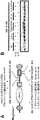
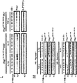
- a schematic diagram of the wild-type Regnase-1 gene (top), targeting vector (middle), and putative mutant allele (bottom) is shown.
- the targeting vector contains the S435A and S439A mutations in exon 6.
- B shows the sequencing result of Regnase-1 exon 6 in Regnase-1AA / AA mouse genome.
- the sequence chromatogram shows that TCA and TCC of Ser435 and Ser439 are replaced by GCA and GCC, respectively.
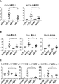
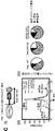
- C NF ⁇ B, phospho-NF ⁇ B, I ⁇ B, phospho-I ⁇ B, MAPK p38, phospho-MAPK p38, ERK1 in wild-type and Regnase-1AA / AA macrophages stimulated with LPS (100 ng / ml) for 0-240 minutes , The immunoblot analysis result of phospho-ERK1, JNK, and phospho-JNK is shown.
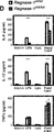
- IL-6 IL by wild-type and Regnase-1AA / AA macrophages stimulated with low concentrations of LPS (10 ng / ml), CpG (0.1 ⁇ M), or Pam3Csk4 (10 (ng / ml) for 24 hours -12 and TNF- ⁇ production results are shown. It is the result of evaluating cytokine production in a cell supernatant by ELISA. Error bars represent mean ⁇ SEM. *** P ⁇ 0.005.
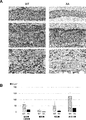
- FIG. 1 shows the results of histological analysis of CD4 + T cell infiltration into the spinal cord. The results of staining frozen sections with hematoxylin-eosin (upper row) and anti-CD3 ⁇ (lower row). Arrow indicates inflammatory cell infiltration. Scale bar, 200 ⁇ m.
- C Number of CD4 + T cells in spinal cord cells (1.0 ⁇ 10 5 cells) 15 days after immunization. It is an analysis result using flow cytometry.
- Spleen (E) and fifth lumbar spinal cord (F) sections were prepared 12 hours after intravenous injection of pathogenic CD4 + T cells (1.5 x 10 7 cells / mouse), anti-type IV collagen antibody and anti-phospho- This is the result of staining with STAT3 antibody.
- Black and white arrows indicate phospho-STAT3 positive and negative endothelial cells, respectively.
- Scale bar 50 ⁇ m.
- G and H shows the results of measuring the relative number of anti-phospho-STAT3-positive cells in vascular endothelial cells (type IV collagen positive) in the spleen (H) and the fifth lumbar spinal cord (I).
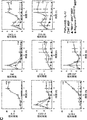
- (I) shows qPCR analysis of IL-6, Regnase-1, CXCL-1, CXCL2, and CCL-20 mRNA in wild type and Regnase-1AA / AA MEF. Results of stimulation of cells for 0-24 hours with TNF- ⁇ (20 ng / ml) and IL-17A (50 ng / ml). Error bars represent mean ⁇ SEM. * P ⁇ 0.05, ** P ⁇ 0.01, *** P ⁇ 0.005.
- (A) Flow cytometry analysis results of CD4 + T cell subsets (TH1, TH17, and iTreg) differentiated from naive CD4 + T cells under in vitro conditions.
- FIG. B shows qPCR analysis of IL-6, TNF- ⁇ , CXCL-1, and CXCL2 mRNA in wild-type and Regnase-1AA / AA hepatic sinusoidal endothelial cells (LSEC). Results of stimulation of cells with TNF- ⁇ (20 ng / ml) and IL-17A (50 ng / ml) for 0-24 hours.
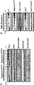
- FIG. 6 shows changes in the expression levels of various genes in the skin of the auricle application area of wild-type mice and Regnase-1 ⁇ ⁇ AA mutant mice in an imiquimod-induced psoriasis model (normalized by B2m expression levels).
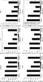
- A Serum creatinine value and ratio of urinary total protein to creatinine value in wild-type mice and Regnase-1 AA mutant mice in anti-glomerular basement membrane antibody-induced nephritis model (Mann-Whitney U test; ****: p ⁇ 0.0001).
- B shows the amount of hydroxyproline per kidney weight of wild type mice and Regnase-1 AA mutant mice in the anti-glomerular basement membrane antibody-induced nephritis model (Mann-Whitney U test; ***: p ⁇ 0.001).
- (B) The ratio of glomerular lesions in wild type mice and Regnase-1 AA mutant mice in the anti-glomerular basement membrane antibody-induced nephritis model is shown (mean value and standard deviation; 15 cases in each group).
- (C) The histological images of the lungs of wild-type mice and Regnase-1 AA mutant mice in the anti-glomerular basement membrane antibody-induced nephritis model are shown (hematoxylin-eosin stained specimen).
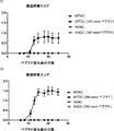
- (A) shows the amount of hydroxyproline per skin weight of wild type mice and Regnase-1 AA mutant mice in a bleomycin-induced scleroderma model.
- WTNC shows the results of pathological non-induced wild type mice
- WTDC shows the results of pathologically induced wild type mice
- AANC shows the results of non-pathologically induced Regnase-1 AA mutant mice
- AADC shows the results of pathologically induced Regnase-1 AA mutant mice.
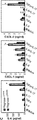
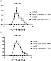
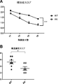
- A shows inflammation scores in both eyes of mice under the condition of a peptide dose of 140 nmol.
- the horizontal axis of the graph represents the number of days after peptide administration, and the vertical axis represents the inflammation score (mean ⁇ standard error).
- B shows inflammation scores in both eyes of mice under the condition of peptide dosage of 280 nmol.
- the horizontal axis of the graph represents the number of days after peptide administration, and the vertical axis represents the inflammation score (mean ⁇ standard error).
- C shows the structural disorder score of both eyes of mice under the condition of a peptide dose of 140 nmol.
- the horizontal axis of the graph represents the number of days after peptide administration, and the vertical axis represents the structural disorder score (mean ⁇ standard error).
- D shows the structural disorder score of both eyes of mice under the condition of peptide dosage of 280 nmol.
- the horizontal axis of the graph represents the number of days after peptide administration, and the vertical axis represents the structural disorder score (mean ⁇ standard error).
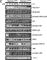
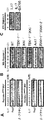
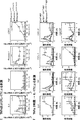
- (A) shows the results of immunoblot analysis of Regnase-1 in IL-17-stimulated wild type and Regnase-1AA / AA MEF. Two arrows indicate the phosphorylated form of Regnase-1 (top) and the non-phosphorylated form (bottom).
- (B) shows the results of immunoblot analysis of Regnase-1 in IL-17-stimulated wild-type and each molecule-deficient MEF.
- (C) shows the results of immunoblot analysis of Regnase-1 in IL-17-stimulated wild-type MEF in the presence of BX795 (50 ⁇ M).
- (D) shows phosphorylation of Regnase-1 by TBK1 and IKKi under in vitro conditions.
- Regnase-1 from Regnase-1-deficient MEF expressing FLAG-tagged Regnase-1 AA mutant is incubated with recombinant TBK1 and / or IKKi in the presence / absence of ⁇ -phosphatase for 3 hours did.
- Regnase-1 phosphorylation was analyzed by Western blotting (i) and [32P] -autoradiography (ii). The arrow indicates phosphorylated Regnase-1.
- the immunoblot analysis result of Regnase-1 is shown.
- the FLAG-tagged Regnase-1 variant was co-immunoprecipitated with Myc-tagged Act1.
- the eluted protein was subjected to immunoblot analysis using anti-FLAG antibody and anti-Myc antibody.
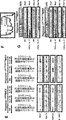
- (I) shows the results of immunoblot analysis of Regnase-1, I ⁇ B, phospho-I ⁇ B, NF ⁇ B, and phospho-NF ⁇ B expression in wild-type and Regnase-1AA / AA MEFs stimulated with IL-17A.
- (J) shows the results of immunoblot analysis of Regnase-1 expression in wild type, Regnase-1AA / AA, TBK1 / IKKi double deficiency, Act1 deficiency, and IRAK1 / IRAK2 double deficiency MEFs stimulated with IL-1 ⁇ .
- FIG. B shows the results of immunoblot analysis of Regnase-1 in HeLa cells transfected with Regnase-1 mutant (wild type, S494A, T505A / S508A, S513A, and S494A / S513A).
- Cells were stimulated with IL-1 ⁇ (10 ng / ml) and IL-17A (50 ng / ml) for 1 hour.
- C (i) shows a diagram of the construct of GST-fused Regnase-1 (440-598).
- (D) shows the results of immunoblotting of Regnase-1.
- Regnase-1 was obtained from Regnase-1-deficient MEFs expressing FLAG-tagged Regnase-1 AA mutants stimulated with IL-1 ⁇ (10 ng / ml) and IL-17A (50 mg / ml) for 1 hour It was.
- Purified Regnase-1 was analyzed by native PAGE and Western blotting.
- E shows the results of immunoblot analysis of Regnase-1 phosphorylated by TBK1 and IKKi. Purified Regnase-1 was incubated with GST-fused TBK1 and / or IKKi for 3 hours in the presence / absence of ⁇ -phosphatase.
- Proteins were separated by native PAGE and SDS-PAGE and analyzed by Regnase-1 Western blotting.
- a and B The immunoblot analysis result of an intracellular organelle fraction is shown.
- Regnase-1, ribosomal protein L7a (rpL7a; ER marker), and GAPDH (cytoplasmic marker) in cell homogenates, soluble cytoplasmic fractions, microsomes, and rough ER membranes.
- Fractions were obtained from Regnase-1AA / AA MEF stimulated with IL-1 ⁇ (10 ng / ml) and IL-17A (50 ng / ml) for 1 hour, respectively (A), and TNF- ⁇ (20 ng / ml).
- IL-17A 50 ng / ml
- A shows the results of immunoblotting and quantitative PCR (qPCR) analysis of wild-type MEF stimulated with TNF- ⁇ for 2 hours and subsequently with IL-17A for 0 to 4 hours.
- I Cell lysates were analyzed by immunoblotting of Regnase-1.
- II shows IL-6 mRNA expression in wild type cells stimulated by the combination of TNF- ⁇ and IL-17A as described above.
- Total mRNA was prepared from cells after treatment with doxycycline for 0-4 hours and then subjected to Northern blotting using [32P] -labeled probe.
- (Ii) shows relative IL-6 mRNA levels in Tet-off HEK293 cells during doxycycline treatment.
- (D) shows the results of immunoblot analysis of Regnase-1 in Regnase-1AA / AA MEF. Cells treated with IL-17A (50 ng / ml) for 1 hour, then in medium without control (control), medium with cycloheximide (100 ⁇ M), or medium with both cycloheximide and okadaic acid (0.5 ⁇ M) And incubated for 0-240 minutes.
- (C) shows the results of co-expression of Act-1, TBK-1, and IKKi with FLAG-tagged wild type Regnase-1 or Regnase-1 ⁇ CTD in HEK293 cells.
- Cell lysates were subjected to immunoblot analysis using anti-FLAG antibody.
- (D) shows the results of coimmunoprecipitation of FLAG-tagged wild-type Regnase-1, Regnase-1 ⁇ CTD, and Myc-tagged Act-1.
- Cell lysates from HEK293 transfectants were mixed as described and co-immunoprecipitated with anti-FLAG M2 agarose beads.
- the eluted protein was subjected to immunoblot analysis using anti-FLAG antibody, anti-Myc antibody, and anti-actin antibody.
- E in intracellular organelles (homogenates, cytosols, and microsomes) isolated from wild-type and Regnase-1 ⁇ CTD / ⁇ CTD MEF after stimulation with 50 ng / ml IL-17A for 0, 1, and 4 hours, The immunoblot analysis result of Regnase-1, Rpl7a, GAPDH, and phospho-TBK-1 is shown.
- F and G The immunoblot analysis result of a polysome fraction is shown.
- F UV absorbance profile (at 260 nm) of sucrose gradient fraction from cell lysate of MEF.
- FIG. 1 shows the results of immunoblot analysis of Regnase-1 and RpL7a in sucrose gradient fractions isolated from wild-type and Regnase-1 ⁇ CTD / ⁇ CTD cell lysates. RpL7a is indicated by an arrow.
- H shows qPCR analysis results of IL-6, TNF, LCN-2, and GM-CSF mRNA in wild type, Regnase-1 ⁇ CTD / +, and Regnase-1 ⁇ CTD / ⁇ CTD MEF. Cells were stimulated with TNF- ⁇ (20 ng / ml) and IL-17A (50 ng / ml) for 0-24 hours.
- (L) shows the results of immunoblot analysis of Regnase-1 and ⁇ -actin in Regnase-1 S513A MEF stimulated with IL-1 ⁇ , IL-17A, or TNF- ⁇ for 0 to 4 hours.
- (M) shows the immunoblot analysis results of Regnase-1 in wild type, Regnase-1AA / AA, and Regnase-1aseS513A MEF. Cells were stimulated with TNF- ⁇ , IL-1 ⁇ , LPS, or IL-17A for 0-120 minutes in the presence of the transcription inhibitor cycloheximide.
- A A schematic diagram of wild-type Regnase-1 (upper) and 1 bp deletion Regnase-1 (lower) is shown. The CRISPR-Cas9 targeting site is located in the proline-rich region of Regnase-1. Amino acid sequences introduced by a frameshift mutation and an immature stop codon 146 bases downstream from the mutation are underlined.
- the sequence chromatogram shows the deletion of the cytosine base in Pro517 to initiate a frameshift mutation.
- C Schematic representation of wild type Regnase-1 (upper) and S513A mutant Regnase-1 (lower). The CRISPR-Cas9 targeting site is located in Ser513 of Regnase-1.
- D Sequencing of Regnase-1 exon 6 in the mouse genome mutated by the CRISPR-Cas9 system. The sequence chromatogram shows the substitution of Tyr511 from TAC to TAT (nonsense mutation) and Ser513 from TCT to GCT (S513A mutation), respectively.
- (E) shows qPCR analysis results of IL-6, TNF, Regnase-1, and LCN-2N mRNA in wild type, Regnase-1 ⁇ CTD / ⁇ CTD, and Regnase-1 S513A MEF.
- Cells were stimulated with TNF- ⁇ and IL-17A for 0-24 hours.
- FIG. 20 is a diagram showing a continuation of FIG. 19-1.
- FIG. 20 is a diagram showing a continuation of FIG. 19-2. It is a figure which shows the continuation of FIG. 19-3. It is a figure which shows the continuation of FIG. 19-4.
- the detection result of phosphorylated Regnase-1 by Western blotting is shown.
- Regnase-1 was phosphorylated by reacting kinase (IKK ⁇ or TBK1) with Regnase-1 in the presence of ATP.
- the antibody against phosphorylated Regnase-1 was reacted with Regnase-1 after the above reaction, and the amount of binding was measured by AlphaScreen.
- the left figure shows the results when IKK ⁇ is used, and the right figure shows the results when TBK1 is used.
- the result of phosphorylation inhibition of full length Regnase-1 (FL_Reg1) by a compound is shown.
- RNA concentration in the reaction solution was measured.
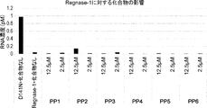
- the influence of the compound on the RNA degradation activity of wild-type Regnase-1 is shown.
- a compound (each compound of PP7 to PP25) was added to and reacted with a mixed solution of RNA and wild-type Regnase-1, and then the RNA concentration in the reaction solution was measured.
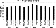
- the influence of the compound on the RNA degradation activity of mutant type Regnase-1 is shown.
- RNA concentration in the reaction mixture was measured.
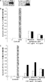
- the binding of anti-Regnase-1 antibody to human Regnase-1 peptide and human full-length Regnase-1 is shown.
- anti-Regnase-1 antibodies REA0023, REA0027, REB0007, REB0014, and REB0022 were used.
- Peptide 1 (CLDSGIGSLESQMSELWGVRGG) and peptide 2 (AFPPREYWSEPYPLPPPTC-NH2) in the figure are both partial peptides of human Regnase-1.
- FL_Reg1 represents human full length Regnase-1.
- the result of having evaluated the Regnase-1 phosphorylation inhibitory activity of an anti-Regnase-1 antibody is shown.
- A The result of detecting phosphorylation of Regnase-1 by each kinase (IKK ⁇ or TBK1) by Western blotting is shown.
- B shows the inhibitory activity of anti-Regnase-1 antibodies (REA0023, REA0027) on the phosphorylation of Regnase-1 by IKK ⁇ . Both anti-Regnase-1 antibodies with final concentrations of 16.7 ⁇ g / ml and 5.0 ⁇ g / ml have been evaluated.
- (C) shows the inhibitory activity of anti-Regnase-1 antibodies (REB0007, REB0014, REB0022) on the phosphorylation of Regnase-1 by TBK1. Both anti-Regnase-1 antibodies with final concentrations of 16.7 ⁇ g / ml and 5.0 ⁇ g / ml have been evaluated. The influence of the anti-Regnase-1 antibody with respect to the RNA degradation activity of wild type Regnase-1 is shown. Anti-Regnase-1 antibodies (REA0023, REA0027, REB0007, REB0014, REB0022) were added to the mixture of RNA and wild-type Regnase-1 for reaction, and then the RNA concentration in the reaction solution was measured.
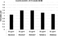
- the influence of the anti-Regnase-1 antibody on the RNA degradation activity of mutant type Regnase-1 is shown.
- Anti-Regnase-1 antibodies (REA0023, REA0027, REB0007, REB0014, REB0022) were added to the mixture of RNA and mutant Regnase-1 (D226N, D244N) for reaction, and then the RNA concentration in the reaction solution was measured.
- the results of pathological analysis of wild-type mice and Regnase-1aseAA mutant mice in an experimental autoimmune uveitis T cell transfer model are shown.
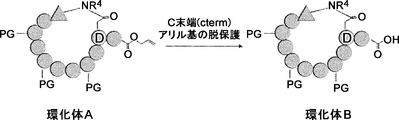
- a synthesis scheme of a compound in which GG-TFPI-tag is bound to the C-terminus of a cyclic polypeptide is shown.
- a synthesis scheme of Fmoc-Asp (O-Trt (2-Cl) -resin) -bMeAla-OAllyl (Compound RS3) is shown.
- a step of synthesizing cyclized product B from cyclized product A is shown.
- Regnase-1 also known as Zc3h12a or MCPIP-1
- primate eg, human
- rodent eg, mouse, rat
- the term encompasses Regnase-1 which has not undergone "full length” processing as well as Regnase-1 which results from processing in the cell.
- the amino acid sequence of an exemplary mouse Regnase-1 is published as Uniprot accession number Q5D1E7 (SEQ ID NO: 1), and the amino acid sequence of an exemplary human Regnase-1 is published as Uniprot accession number: Q5D1E8 (SEQ ID NO: 2) .
- the literature describing Regnase-1 is, for example, WO2010 / 098429; Nature immunology, Vol. 12, NUMBER 12, DECEMBER 2011, p. 1167-1175; Nature 458, 2009, p. 1185-1190; Cold Spring Harbor Symposia on Quantitative Biology, Volume LXXVIII, 2013, p.51-60; Biochimica et Biophysica Acta 1823, 2012, p.1905-1913.
- Regnase-1 in this specification is preferably mammalian Regnase-1.
- a disease involving Regnase-1 means a disease in which Regnase-1 is involved in the formation, exacerbation and / or continuation of the disease.
- Diseases that are involved in the formation, exacerbation and / or continuation of diseases include not only diseases that are directly involved in the formation, exacerbation and / or continuation of diseases, but also diseases that are indirectly involved.
- “disease involving Regnase-1” means, for example, a disease in which destabilization and / or intracellular degradation of Regnase-1 is involved in the formation, exacerbation and / or continuation of the disease. It's okay.
- Diseases involving Regnase-1 include inflammatory diseases, autoimmune diseases, allergic diseases, fibrotic diseases, and RNA virus infections.
- the “disease involving Regnase-1” may be a TH17 cell-related disease.
- inflammatory disease is a disease or illness resulting from excessive activation of an individual's immune system. Inflammatory diseases can be caused by a pathological stage of inflammation, typically but not limited to leukocyte influx and / or neutrophil chemotaxis.
- diseases include inflammatory skin diseases (including psoriasis and atopic dermatitis); systemic sclerosis; nephritis; reactions associated with inflammatory bowel disease (Crohn's disease and ulcerative colitis); Surgical tissue reperfusion injury, myocardial ischemic symptoms such as myocardial failure, heart failure, perfusion after heart surgery and perfusion after percutaneous transluminal coronary angioplasty, stroke, and ischemic perfusion disease including abdominal aortic aneurysm; Edema; cranial trauma; hypovolemia shock; respiratory arrest; adult respiratory distress syndrome; acute lung injury; Behcet's disease; dermatomyositis; polymyositis; multiple sclerosis; dermatitis; meningitis; encephalitis; Eye inflammation; diabetic retinopathy; diabetic macular edema; osteoarthritis; Lubus nephritis; diabetic nephropathy; autoimmune diseases such as rheumatoid
- Preferred symptoms include acute lung injury, adult respiratory distress syndrome, ischemic perfusion (including surgical tissue perfusion injury, myocardial ischemia, and acute myocardial failure), hypotensive shock, asthma, bacterial pneumonia and ulcers Inflammatory bowel diseases such as ulcerative colitis are included. Inflammatory diseases overlap in part with other classes of diseases such as autoimmune diseases, allergic diseases, fibrotic diseases, and vice versa.
- autoimmune disease refers to a disease or disorder that arises from and is directed against the individual's own tissue.
- an autoimmune disease is one that specifically excludes a malignant or cancerous disease or condition, in particular B-cell lymphoma, acute lymphoblastic leukemia (ALL), chronic lymphocytes Exclude chronic leukemia (chronic lymphocytic leukemia: CLL), hairy cell leukemia, and chronic myeloblastic leukemia.
- ALL acute lymphoblastic leukemia
- CLL chronic lymphocytic leukemia
- hairy cell leukemia and chronic myeloblastic leukemia.
- autoimmune diseases or disorders include, but are not limited to: inflammatory reactions such as inflammatory skin diseases including psoriasis and dermatitis (eg, atopic dermatitis); Scleroderma and sclerosis; reactions associated with inflammatory bowel disease (eg, Crohn's disease and ulcerative colitis); respiratory distress syndrome (including adult respiratory distress syndrome; adult respiratory distress syndrome: ARDS); dermatitis; Meningitis; Encephalitis; Uveitis; Ocular inflammation; Colitis; Glomerulonephritis; Allergic conditions such as eczema and asthma and other conditions with T cell infiltration and chronic inflammatory response; Atherosclerosis; Rheumatoid arthritis; systemic lupus erythematosus (SLE) (including but not limited to lupus nephritis, cutaneous lupus); diabetes (eg, I Diabetes or insulin-dependent diabetes); multiple sclerosis; Raynaud's syndrome; autoimmune thyroiditis; Hashimoto'
- allergic diseases means any symptoms, tissue damage, or loss of tissue function resulting from allergy.
- the allergic diseases include hypersensitivity classified as immediate type and delayed type, or allergic diseases classified as allergy type I to type IV.
- types of such diseases include type I allergies (eg, including systemic anaphylaxis, bronchial asthma and hay fever), type II allergies (eg, hemolysis in blood group-incompatible transfusions and autoimmune hemolytic anemia) , Including but not limited to type III allergies (including, for example, serum sickness, glomerulonephritis and rheumatoid arthritis) and type IV allergies (including, for example, contact dermatitis, granulomas and rejection in transplants).
- type I allergies eg, including systemic anaphylaxis, bronchial asthma and hay fever
- type II allergies eg, hemolysis in blood group-incompatible transfusions and autoimmune hemolytic anemia
- type III allergies including, for example, serum sickness, glomerulonephritis and rheum
- allergic diseases include asthma; allergic encephalomyelitis; autoimmune encephalomyelitis; allergic neuritis; contact hypersensitivity; delayed hypersensitivity; airway hypersensitivity; atopic dermatitis; Antigen-specific allergy; allergic rhinitis; urticaria.
- Allergic diseases overlap in part with other classes of diseases such as autoimmune diseases, inflammatory diseases, fibrotic diseases, and vice versa.
- TH17 cell-related disease is a disease in which TH17 cells play a role in the formation, exacerbation and / or continuation of the disease.
- diseases include inflammatory diseases, autoimmune diseases, and allergic diseases, in which TH17 cells are associated with the formation, exacerbation and / or continuation of the diseases, and in particular, multiple sclerosis Rheumatoid arthritis, scleroderma, psoriasis, nephritis (eg glomerulonephritis), asthma, contact hypersensitivity, delayed hypersensitivity, and airway hypersensitivity.
- fibrotic disease refers to a condition involving abnormal or excessive formation of fibrous connective tissue in cells, organs or tissues. Fibrotic diseases can occur as part of a recovery or reaction process in a cell, tissue or organ due to, for example, physical injury, inflammation, infection, etc. As used herein, the term “fibrotic disease” may be used interchangeably with the terms “fibrosis”, “fibrotic disorder” and “fibrotic symptoms”.
- fibrotic diseases include, but are not limited to, vascular fibrosis, pulmonary fibrosis (eg, idiopathic pulmonary fibrosis), dermal fibrosis (eg, scleroderma, trauma) Later, surgical scarring, keloid and skin keloid formation), scleroderma, systemic scleroderma, liver fibrosis (e.g. after hepatitis C virus infection or after liver transplantation), renal fibrosis (e.g.
- Interstitial fibrosis and renal systemic fibrosis in focal segmental glomerulosclerosis Interstitial fibrosis and renal systemic fibrosis in focal segmental glomerulosclerosis
- musculoskeletal fibrosis cardiac fibrosis (eg, endocardial myocardial fibrosis, idiopathic cardiomyopathy), splenic fibrosis Ocular fibrosis (e.g., eye sclerosis, glaucoma, conjunctival and corneal scars, and pterygium), progressive systemic sclerosis (PSS), chronic transplant versus host disease, Peyronie's disease, connective tissue disease, cystoscope Later urethral stricture, mediastinal fibrosis, idiopathic and pharmacologically induced peritoneum Square fibrosis, progressive severe fibrosis, proliferative fibrosis, neoplastic fibrosis, and fibrosis caused by surgical implantation of artificial organ
- fibrosis diseases, disorders, and symptoms associated with fibrosis include, for example, cirrhosis that can result in liver fibrosis, diffuse lung disease, pain syndrome after vasectomy, tuberculosis, spleen that can result in pulmonary fibrosis And clonal diseases that can cause recurrent inflammation and healing of the intestinal tissue leading to sickle cell anemia, rheumatoid arthritis, and ultimately fibrosis of the intestinal wall that can cause fibrosis.
- Fibrotic diseases also occur as viral hepatitis, alcoholism, hemochromatosis complications, Wilson disease, schistosomiasis, bile duct disorders, toxin exposure, and metabolic disorders. Fibrotic diseases overlap in part with other classes of diseases such as autoimmune diseases, allergic diseases, inflammatory diseases, and vice versa.
- RNA virus means a virus having an RNA genome. RNA viruses include single stranded RNA viruses (including positive and negative stranded RNA viruses) and double stranded RNA viruses.
- RNA virus infection refers to any disorder caused by the entry of an RNA virus into the surface, topic, or whole body of a host. A host may be an individual as used herein.
- treatment is clinical that is intended to alter the natural course of the individual being treated. Means intervention and can be carried out either for prevention or during the course of clinical pathology. Desirable effects of treatment include but are not limited to prevention of disease occurrence or recurrence, reduction of symptoms, attenuation of any direct or indirect pathological effects of the disease, prevention of metastasis, disease Includes reduced rate of progression, recovery or alleviation of disease state, and remission or improved prognosis.
- the Regnase-1-binding molecules of the invention are used to delay the onset of disease or slow the progression of disease.
- inhibiting phosphorylation of a molecule means reducing the degree to which a molecule is phosphorylated or preventing a molecule from being phosphorylated.
- “selectively inhibits phosphorylation of Regnase-1” means that phosphorylation of Regnase-1 is inhibited while phosphorylation of other molecules is not inhibited, or other than Regnase-1
- the degree of inhibition of molecular phosphorylation is smaller than the degree of inhibition of Regnase-1 phosphorylation (for example, 50% or less, 40% or less, 30% or less, 20% or less, or 10% or less). May be.)
- Regnase-1 binding molecule may be used to inhibit phosphorylation of Regnase-1 selectively by inhibiting phosphorylation of Regnase-1.
- the phosphorylation of the kinase substrate can be inhibited in a non-selective manner. It is not included in “inhibiting oxidation of Regnase-1 selectively”.
- the “position corresponding to” in the present specification refers to the amino acid residue in Regnase-1 having different origin (shared source) or the processed Regnase-1 amino acid sequence of mouse Regnase-1 (SEQ ID NO: 1). Can be used to characterize by reference.
- the alignment for determining the corresponding position may be determined by various methods within the skill in the art, such as BLAST, BLAST-2, ALIGN, Megalign® (DNASTAR) ® software, or GENETYX® (Genetics Corporation). This can be achieved by using publicly available computer software such as One skilled in the art can determine appropriate parameters for aligning sequences, including any algorithms needed to achieve maximal alignment over the full length of the sequences being compared.
- FIG. 1-1 An alignment of the amino acid sequences of mouse and human Regnase-1 prepared using GENETYX (registered trademark) is shown in FIG. 1-1.
- Table 1 shows amino acid residues in human Regnase-1 at positions corresponding to some of the amino acid residues in mouse Regnase-1.
- inflammation means that inflammation does not occur, inflammation progresses slowly compared to an untreated control group, inflammation that has already occurred is reduced, or the extent of inflammation is reduced. May mean. Although not limited, suppression of the production of inflammatory factors may be used as one index of inflammation suppression.
- inflammatory factors include inflammatory cytokines and leukocyte migration factors. Examples of inflammatory factors are disclosed herein.
- target and target of Regnase-1 mean that a certain molecule can be degraded by the RNase activity of Regnase-1, and a certain mRNA can be a target of Regnase-1. Whether or not can be confirmed by, for example, the method described in the item of “activity measurement method” in this specification.
- fibrosis means reducing or eliminating fibrotic lesions in a tissue in which fibrosis has occurred, or delaying or preventing the progression of further fibrosis (increasing fibrotic lesions). Means to suppress).
- epithelial hyperplasia refers to a state in which the number of normal cells normally arranged in epithelial tissue is abnormally increased. Epithelial hyperplasia is known as a feature of many disorders, including psoriasis. As used herein, “suppressing epithelial hyperplasia” means reducing the number of normal cells increased in epithelial tissue, or delaying or preventing further proliferation.
- inhibition of keratinocyte proliferation means to reduce the number of keratinocytes or delay or prevent further proliferation. Whether or not a certain substance suppresses the growth of keratinocytes can be verified using, for example, histological examination.
- degradation of Regnase-1 in a cell means that the amount of Regnase-1 protein in the cell decreases or Regnase-1 disappears from the cell, and the ubiquitin-proteasome Includes degradation through the system.
- the amount of protein of Regnase-1 is higher in cells treated with the test substance than in cells not treated with the test substance, the degradation of Regnase-1 in the cells by the test substance treatment May be regarded as being suppressed.
- Regnase-1 destabilization means that the RNase activity of Regnase-1 is reduced compared to a control (for example, non-phosphorylated Regnase-1 can be used). For example, if Regnase-1 is present but loses the ability to degrade the target mRNA, it is expressed that Regnase-1 is destabilized. Although not limited, the destabilization of Regnase-1 can be confirmed by the method described herein (for example, refer to the item of activity measurement method), and for example, IL-6 mRNA may be targeted. Such Regnase-1 with reduced RNase activity is sometimes referred to as “inactive form”.
- suppressing destabilization of Regnase-1 means suppressing the destabilization of Regnase-1 or suppressing the generation of an inactive form of Regnase-1. It's okay.
- inhibitortion of dissociation of Regnase-1 oligomer suppresses dissociation of Regnase-1 oligomer in in vitro or in vivo, resulting in an aggregate or monomer formed in a smaller number Or to inhibit.
- Illustrative examples include inhibiting or inhibiting Regnase-1 hexamers and higher aggregates from dissociating into trimers and monomers.
- inhibittion of Regnase-1 release from the endoplasmic reticulum refers to inhibiting or inhibiting Regnase-1 from being released from the endoplasmic reticulum in vitro or in vivo.
- Endoplasmic reticulum is sometimes referred to as “ER”.
- endoplasmic reticulum preferably means a rough endoplasmic reticulum.
- the “method for identifying a substance that inhibits phosphorylation” includes a method for screening a substance that inhibits phosphorylation, a method for confirming that a certain substance is a substance that inhibits phosphorylation, and the like. Including, but not limited to.
- the “binding molecule” means a molecule that can bind to a certain molecule. For example, when A can bind to B, A is expressed as a binding molecule of B.
- Regnase-1 binding molecule means a molecule that can bind to Regnase-1.
- Illustrative examples include, but are not limited to, synthetic low molecular weight compounds, peptides, polypeptides, proteins, antibodies, carbohydrates, nucleic acids, and derivatives thereof.
- a “Regnase-1 binding molecule” may be a molecule that can specifically bind to Regnase-1.
- polypeptide refers to a substance in which four or more amino acids and / or amino acid analogs are linked by an amide bond and / or an ester bond. Any of natural polypeptide, synthetic polypeptide, recombinant polypeptide and the like may be used. Polypeptides include antibodies and cyclic polypeptides.
- antibody is used in the broadest sense and includes, but is not limited to, monoclonal antibodies, polyclonal antibodies, multispecific antibodies (eg, bispecific antibodies) as long as they exhibit the desired antigen binding activity. Antibody), modified antibodies, and antibody fragments.
- modified antibody refers to an antibody in which an amino acid, a sugar chain modification state, or the like is modified from a parent antibody that has not been modified.
- alterations to increase antigen affinity alterations to increase blood half-life, alterations to alter C1q binding and complement-dependent cytotoxicity (CDC), enhance antibody translocation capacity
- an antibody derivative to which a non-protein portion for example, a drug, polyethylene glycol (PEG), or nucleic acid is added is also included in the modified antibody.
- a non-protein portion for example, a drug, polyethylene glycol (PEG), or nucleic acid
- antibody fragment refers to a molecule other than the complete antibody, including a portion of the complete antibody that binds to the antigen to which the complete antibody binds.
- antibody fragments include, but are not limited to, Fv, Fab, Fab ′, Fab′-SH, F (ab ′) 2 ; diabodies; linear antibodies; single chain antibody molecules (eg, scFv And multispecific antibodies formed from antibody fragments.
- full-length antibody “complete antibody”, and “total antibody” are used interchangeably herein and have a structure that is substantially similar to a native antibody structure or that includes an Fc region.
- An antibody having a chain is used interchangeably herein and have a structure that is substantially similar to a native antibody structure or that includes an Fc region.
- An antibody having a chain is used interchangeably herein and have a structure that is substantially similar to a native antibody structure or that includes an Fc region.
- An antibody having a chain An antibody having a chain.
- cyclic polypeptide means a polypeptide comprising a cyclic structure formed by four or more amino acids and / or amino acid analogs.
- the cyclic polypeptide may have a linear part in addition to the cyclic part.
- the bonding mode of the cyclized portion is not particularly limited, and may be a bond other than an amide bond or an ester bond. Examples of the bonding mode of the cyclization unit include amide bond, carbon-carbon bond, disulfide bond, ester bond, thioester bond, thioether bond, lactam bond, bond via azoline skeleton, bond via triazole structure, fluorophore
- a covalent bond such as a bond through a structure is preferably exemplified.
- the position of a functional group such as a carboxy group or an amino group used for cyclization may be on the main chain or on the side chain, and is not particularly limited as long as it is in a cyclizable position.
- the “linking mode of the cyclized portion” refers to a binding mode of a site where cyclization is formed by a cyclization reaction.
- amino acid includes natural amino acids and non-natural amino acids.
- “natural amino acid” means Gly (glycine), Ala (alanine), Ser (serine), Thr (threonine), Val (valine), Leu (leucine), Ile (isoleucine), Phe (phenylalanine). , Tyr (tyrosine), Trp (tryptophan), His (histidine), Glu (glutamic acid), Asp (aspartic acid), Gln (glutamine), Asn (asparagine), Cys (cysteine), Met (methionine), Lys (lysine) ), Arg (arginine), Pro (proline).
- Non-natural amino acids are not particularly limited, and examples include ⁇ -amino acids, ⁇ -amino acids, D-type amino acids, N-substituted amino acids, ⁇ , ⁇ -disubstituted amino acids, amino acids whose side chains are different from natural amino acids, and the like. Arbitrary steric configurations are allowed as amino acids in the present specification.
- the selection of the side chain of the amino acid is not particularly limited.
- amino acids in which the main chain amino group is substituted are referred to as “N-substituted amino acids”.
- N-substituted amino acids include N alkyl amino acids, of which N methyl amino acids are preferred.
- the “amino acid analog” in the present specification preferably means hydroxycarboxylic acid, more preferably ⁇ -hydroxycarboxylic acid.
- the side chain of ⁇ -hydroxycarboxylic acid is not particularly limited as in the case of amino acids.
- amino acids constituting proteins, polypeptides, and peptides are sometimes referred to as amino acid residues.
- Serine residues are sometimes referred to as “Ser residues” and threonine residues as “Thr residues”.
- Serine residues are sometimes referred to as “Ser residues” and threonine residues as “Thr residues”.
- the serine residue at position 513 in a certain amino acid sequence may be expressed as S513 or Ser513, and the substitution of the serine residue with alanine may be described as S513A or Ser513Ala.
- affinity refers to the total strength of a non-covalent interaction between a binding site of a molecule (eg, an antibody) and a binding partner (eg, an antigen) of the molecule.
- binding affinity refers to intrinsic binding affinity that reflects a 1: 1 interaction between members of a binding pair (eg, an antibody and an antigen).
- the affinity of a molecule X for its partner Y can generally be represented by the dissociation constant (KD). Affinity can be measured by conventional methods known in the art. Specific illustrative and exemplary embodiments for measuring binding affinity are described below.
- molecules that can specifically bind to Regnase-1 and “molecules that can specifically recognize Regnase-1” are used interchangeably, and these are specific for Regnase-1 with sufficient affinity.
- the extent of binding of a “molecule capable of specifically binding Regnase-1” to an irrelevant non-Regnase-1 protein is determined by, for example, surface plasmon resonance assay, radioimmunoassay (RIA), enzyme immunity It is less than about 10% of the binding to Regnase-1 when measured by a measurement method.
- a “molecule capable of specifically binding to Regnase-1” is 1 ⁇ M or less, 100 nM or less, 10 nM or less, 1 nM or less, 0.1 nM or less, 0.01 nM or less, or 0.001 nM or less (eg, 10 ⁇ 8 M
- KD dissociation constant
- the degree of binding of a “molecule capable of specifically binding Regnase-1” to an irrelevant non-Regnase-1 protein is measured by the method described herein, for example, by a surface plasmon resonance assay.
- a “molecule capable of specifically binding Regnase-1” binds to, but is not limited to, an epitope of Regnase-1 that is conserved among Regnase-1 from different species. In certain embodiments, “a molecule capable of specifically binding to Regnase-1” binds to, but is not limited to, mouse and human Regnase-1.
- a molecule capable of specifically binding to phosphorylated Regnase-1 and “a molecule capable of specifically recognizing phosphorylated Regnase-1” are used interchangeably.
- the degree of binding to Regnase-1 at which a specific site is not phosphorylated is measured by a method such as surface plasmon resonance assay, radioimmunoassay (RIA), western blotting, etc. It may be less than about 10% of the binding to Regnase-1 where the specific site is phosphorylated.
- the “molecule capable of specifically binding to Regnase-1” includes, but is not limited to, polypeptides such as antibodies and cyclic polypeptides.
- TLR Toll-like receptor
- TLR1 ligand includes, but is not limited to, a TLR1 ligand, a TLR2 ligand, a TLR7 ligand, or a TLR4 ligand (lipopolysaccharide (LPS)).
- TLR2 ligand includes, but is not limited to, a TLR1 ligand, a TLR2 ligand, a TLR7 ligand, or a TLR4 ligand (lipopolysaccharide (LPS)).
- LPS lipopolysaccharide
- an “effective amount” of an agent refers to the amount at a required dose and over a required period of time that is effective to achieve a desired therapeutic or prophylactic result.
- host cell refers to a cell (including the progeny of such a cell) into which a foreign nucleic acid has been introduced.
- Host cells include “transformants” and “transformed cells”, including the primary transformed cell and progeny derived from that cell regardless of passage number.
- the progeny may not be completely identical in nucleic acid content with the parent cell, and may contain mutations. Also included herein are mutant progeny that have the same function or biological activity as was used when the original transformed cells were screened or selected.
- mammals include, but are not limited to, domestic animals (eg, cattle, sheep, cats, dogs, horses), primates (eg, humans and non-human primates such as monkeys), rabbits, and , Including rodents (eg, mice and rats).
- the individual or subject is a human.
- the term “monoclonal antibody” refers to an antibody obtained from a substantially homogeneous population of antibodies. That is, the individual antibodies that make up the population are mutated antibodies that can occur (eg, mutated antibodies that contain naturally occurring mutations, or mutated antibodies that occur during the production of monoclonal antibody preparations. Are present in the same amount and / or bind to the same epitope. In contrast to polyclonal antibody preparations that typically include different antibodies to different determinants (epitopes), each monoclonal antibody of a monoclonal antibody preparation is against a single determinant on the antigen.
- monoclonal indicates the character of the antibody as being obtained from a substantially homogeneous population of antibodies, and is not to be construed as requiring production of the antibody by any particular method.
- monoclonal antibodies used in accordance with the present invention include, but are not limited to, hybridoma methods, recombinant DNA methods, phage display methods, transgenic animals containing all or part of a human immunoglobulin locus. It may be created by various methods including a method of using
- polyclonal antibody refers to a population that typically includes different antibodies to different determinants (epitopes).
- the modifier “polyclonal” indicates the character of the antibody and should not be construed as requiring production of the antibody by any particular method.
- TBK1 is a serine / threonine kinase also known as TANK-binding kinase 1, and examples of amino acid sequences of human TBK1 and mouse TBK1 are available from Uniprot accession numbers Q9UHD2 and Q9WUN2, respectively.
- IKKi is a kinase also called inducible I ⁇ B kinase or IKK-E, and examples of amino acid sequences of human IKKi and mouse IKKi can be obtained from Uniprot accession numbers Q14164 and Q9R0T8, respectively.
- Act-1 is an adapter molecule that is also called TRAF3IP2, CIKS, or Nuclear®factor®NF-kappa-Bactivator®1.
- TRAF3IP2 an adapter molecule that is also called TRAF3IP2, CIKS, or Nuclear®factor®NF-kappa-Bactivator®1.
- An example of the amino acid sequence of human Act-1 is available from Uniprot accession number 043734.
- IKK is used synonymously with I ⁇ B kinase, and IKK includes IKK ⁇ and / or IKK ⁇ . IKK ⁇ is preferably exemplified as IKK in the present specification.
- IRAK is used synonymously with IL-1 receptor-associated kinase (IL-1R-associated kinase), and IRAK includes IRAK1 and IRAK2.
- IRAK1 and IRAK2 are preferably exemplified as IRAK in the present specification.
- pharmaceutical formulation refers to a preparation that is in a form such that the biological activity of the active ingredient contained therein can be effective and is unacceptable to the subject to which the formulation is administered. Refers to a preparation that does not contain additional toxic elements.
- “Pharmaceutically acceptable carrier” refers to an ingredient other than the active ingredient in a pharmaceutical preparation that is non-toxic to a subject.
- Pharmaceutically acceptable carriers include, but are not limited to, buffers, excipients, stabilizers, or preservatives.
- vector refers to a nucleic acid molecule that can multiply another nucleic acid to which it has been linked.
- the term includes vectors as self-replicating nucleic acid structures and vectors that are integrated into the genome of the host cell into which it has been introduced. Certain vectors can provide for the expression of nucleic acids to which they are operably linked. Such vectors are also referred to herein as “expression vectors”.
- the “treatment and / or prevention method” may be simply referred to as “therapeutic method”.
- “therapeutic and / or prophylactic composition” may be simply referred to as “therapeutic composition”.
- the present invention relates to treatment of inflammatory diseases, autoimmune diseases, allergic diseases, fibrotic diseases, RNA virus infections, TH17 cell-related diseases, etc., by inhibiting phosphorylation of specific sites of Regnase-1 And / or based on finding effectiveness in prevention.
- the present invention has found that inhibiting phosphorylation of a specific site of Regnase-1 is effective for at least one selected from the group consisting of (i) to (xi) below: Based on: (i) treatment and / or prevention of diseases involving Reginase-1; (ii) inhibition of inflammation; (iii) inhibition of fibrosis of cells, tissues or organs; (iv) inhibition of epithelial hyperplasia; (v) Suppression of expression of at least one mRNA selected from the group consisting of IL6, IL1a, CXCL1, CXCL2, HBEGF, CTGF, DDR1 and PDGFB; (vi) Suppression of destabilization of Regnase-1; (vii) (Viii) inhibition of intracellular degradation of Regnase-1; (ix) inhibition of release of Regnase-1 from the endoplasmic reticulum; (x) inhibition of dissociation of Regnase-1 oligomers; xi) Suppression of
- Regnase-1 is present in various cells, such as macrophages and fibroblasts, in an unstimulated state despite its rapid induction of mRNA, and acts to suppress unwanted inflammatory responses. Possible (Nature immunology, Vol.12, NUMBER 12, DECEMBER 2011, p.1167-1175). In contrast, Regnase-1 is phosphorylated by I ⁇ B kinase (IKK) and undergoes ubiquitin-dependent degradation in response to external stimuli mediated by Toll-like receptor (TLR) ligands and MyD88 such as the IL-1 family. Possible (Nature immunology, Vol.12, NUMBER 12, DECEMBER 2011, p.1167-1175).
- IKK I ⁇ B kinase
- TLR Toll-like receptor
- mouse Regnase-1 having S435A and S439A mutations is phosphorylated and converted into an inactive form, but its degradation is suppressed. Subsequently, it was considered that a part of Regnase-1 was dephosphorylated to generate an active form of Regnase-1 having RNase activity.
- Regnase-1 mutants As a result of experiments in which various diseases were induced in animals expressing such Regnase-1 mutants (EAE, which is a model of multiple sclerosis, and models of psoriasis, glomerulonephritis, and scleroderma), Regnase The therapeutic utility of inhibiting -1 destabilization and / or intracellular degradation has been demonstrated.
- EAE which is a model of multiple sclerosis, and models of psoriasis, glomerulonephritis, and scleroderma
- Regnase-1 having mutations of S435A and S439A is also phosphorylated and changed to an inactive form, and therefore, if this can be suppressed, a stronger therapeutic effect can be expected.
- IL-17 and IL-1 stimulation are shown below, as described later, phosphorylation similar to IL-17 and IL-1 stimulation is also performed in LPS (known as a ligand of TLR4) stimulation.
- the present inventors may inhibit phosphorylation of sites where Regnase-1 is phosphorylated by IKK (Ser residues corresponding to positions 435 and 439 of SEQ ID NO: 1).
- the present invention has been found to be effective in at least one selected from the group consisting of the following (i) to (viii): (i) treatment and / or prevention of diseases involving Regnase-1; Inhibition; (iii) Inhibition of fibrosis of sputum cells, tissues or organs; (iv) Inhibition of epithelial hyperplasia; (v) Selected from the group consisting of IL6, IL1a, CXCL1, CXCL2, HBEGF, CTGF, DDR1 and PDGFB Suppression of expression of at least one mRNA; (vi) suppression of inflammatory factor production; (vii) suppression of intracellular degradation of Regnase-1; (viii) suppression of proliferation of keratinocytes.
- the present inventors identified Ser494, Thr505, Ser508, and Ser513 of SEQ ID NO: 1 among the amino acid residues of mouse Regnase-1 that were confirmed to be phosphorylated by IL-17 and IL-1 stimulation. Pay attention. For the purpose of verifying the effect of phosphorylation of each amino acid residue on the destabilization of Regnase-1, in order to inhibit phosphorylation of these four residues, each was substituted with alanine (Ala). -1 mutants were created. Using these Regnase-1 mutants, the phosphorylation of Regnase-1 after stimulation with IL-17 and IL-1 was verified by Western blotting.
- Regnase-1 exists in an oligomeric form in the endoplasmic reticulum, a ribosome-containing organelle.
- the phosphorylation of Regnase-1 caused by cell stimulation dissociates Regnase-1 oligomers and promotes release from the endoplasmic reticulum and subsequent translocation to the cytoplasm.
- Phosphorylated Regnase-1 loses RNase activity.
- Phosphorylated Regnase-1 is degraded in the cytoplasm by the proteasome.
- Act-1 contributes to phosphorylation of Regnase-1 through TBK1 and IKKi.
- IRAK was assumed as a kinase that phosphorylates Ser494 and Ser513 upon IL-1 stimulation.
- the inventors of the present invention provide a phosphorylation of a Ser residue at at least one position selected from the group consisting of positions corresponding to positions 513 and 494 of SEQ ID NO: 1 in Regnase-1.
- the signal by IL-36 stimulation uses the pathway through MyD88 as well as stimulation by IL-1 or TLR ligand.
- Regnase-1 destabilization and degradation by IL-1 and TLR ligand stimulation is thought to proceed by activation of IKK and IRAK via MyD88 and phosphorylation of Regnase-1 by these kinases .
- Regnase-1 phosphorylation occurs via ILK-36 activation through activation of IKK and IRAK, resulting in instability and / or degradation of Regnase-1.
- the present inventors consider as follows. Regnase-1 has been reported to exhibit anti-RNA viral activity (J Immunol 2014; 193: 4159-4168; Proc Natl Acad Sci USA 2013; 110: 19083-19088; Nucleic Acids Res 2013; 41: 3314-3326 ; Nature 2009; 461: 399-401).
- the present invention can suppress Regnase-1 destabilization and / or intracellular degradation, so that the RNase activity of Regnase-1 is maintained and effective against RNA virus infections. It is thought that.
- the method of the present invention may be a method by selectively inhibiting phosphorylation of Ser residues at Regnase-1.
- the method of the present invention may include a step of selectively inhibiting Sernase phosphorylation.
- compositions of the invention may inhibit phosphorylation of the Sernase-1 Ser residue, and in one embodiment may selectively inhibit Regnase-1 phosphorylation.
- the composition of the invention may comprise a Regnase-1 binding molecule that inhibits phosphorylation of the Sernase-1 Ser residue.
- the method or composition of the invention comprises at least one binding molecule selected from the group consisting of TBK1, IKKi, Act-1, IKK and IRAK (herein these molecules are referred to as “Regnase”). -1 acting molecule ”) and Regnase-1 may be inhibited.
- the method or composition of the present invention comprises Regnase-1 and any of the following (i) to (xi): May bind to any binding molecule: (i) at least one binding molecule selected from the group consisting of ⁇ TBK1, IKKi, Act-1 and IKK; (ii) TBK1, IKKi, Act-1 and IRAK At least one binding molecule selected from the group consisting of: (iii) at least one binding molecule selected from the group consisting of TBK1, IKKi and Act-1; (iv) TBK1 and IKKi; (v) Act-1; (Vi) TBK1, IKKi and Act-1; (vii) TBK1; (viii) IKKi; (ix) IRAK; (x) IKK; (xi) TBK1 and IKK.
- IKK ⁇ is exemplified as the IKK in the above (i) to (xi).
- the composition of the invention comprises Regnase-1 binding that inhibits binding between Regnase-1 and at least one binding molecule selected from the group consisting of TBK1, IKKi, Act-1, IKK and IRAK It may contain molecules.
- the method or composition of the invention may be a method or composition for at least one selected from the group consisting of (i) to (xi): (i) Regnase-1 (Ii) to suppress sputum inflammation; (iii) to suppress fibrosis of sputum cells, tissues or organs; (iv) to suppress sputum hyperplasia; (v) To suppress the expression of at least one mRNA selected from the group consisting of IL6, IL1a, CXCL1, CXCL2, HBEGF, CTGF, DDR1 and PDGFB; (vi) To suppress the destabilization of Regnase-1 (Vii) to suppress the production of inflammatory factors; (viii) to suppress the degradation of Regnase-1 in cells; (ix) to inhibit the release of Regnase-1 from the endoplasmic reticulum; ) To inhibit the dissociation of Regnase-1 oligomer; (xi) To inhibit the proliferation of keratinocytes.
- the method or composition of the present invention is a method for two or more, three or more, four or more, or five or more selected from (i) to (xi) above It may be a composition.
- Whether a substance or composition inhibits the release of Regnase-1 from the endoplasmic reticulum can be determined, for example, by the method described in the Examples (intracellular compartments are isolated and their protein distribution is determined using Western blotting). (Analysis method). Whether or not a certain substance inhibits dissociation of Regnase-1 oligomer can be confirmed, for example, using non-denaturing PAGE analysis described in the Examples.
- the Ser residue in the present invention is at least one position selected from the group consisting of positions corresponding to positions 513, 494, 439, and 435 of SEQ ID NO: 1 in Regnase-1, or It may be a Ser residue at more than one position and consists of (i) positions 513, 494, 439 and 435 of SEQ ID NO: 1; or (ii) consisting of positions 516, 497, 442 and 438 of SEQ ID NO: 2.
- the Ser residue may be at least one position selected from the group, or two or more positions.
- the Ser residue in the present invention is at least one position selected from the group consisting of positions corresponding to positions 513 and 494 of SEQ ID NO: 1 in Regnase-1, or Ser at both positions, respectively. Ser at positions 513 and 494 of SEQ ID NO: 1; or (ii) at least one position selected from the group consisting of positions 516 and 497 of SEQ ID NO: 2, or both positions It may be a residue.
- the Ser residue in the present invention may be a Ser residue corresponding to position 513 of SEQ ID NO: 1 in Regnase-1, and (i) position 513 of SEQ ID NO: 1; or (ii) It may be the Ser residue at position 516 of SEQ ID NO: 2.
- the Ser residue in the present invention is at least one position selected from the group consisting of positions corresponding to positions 439 and 435 of SEQ ID NO: 1 in Regnase-1, or Ser at both positions, respectively. Ser at positions 439 and 435 of SEQ ID NO: 1; or (ii) at least one position selected from the group consisting of positions 442 and 438 of SEQ ID NO: 2, or both positions It may be a residue.
- the Ser residue in the present invention can be both of the following (i) and (ii) Ser residues: (I) Ser residues at either or both positions corresponding to each of 513 and 494 of SEQ ID NO: 1 in Regnase-1; (Ii) Ser residues at either or both positions corresponding to 439 and 435 of SEQ ID NO: 1 in Regnase-1.
- the phosphorylation of both Ser residues in (i) phosphorylated by TBK1 and IKKi and (ii) phosphorylated by IKK in Regnase-1 By suppressing oxidation, the effect of the present invention can be exerted more strongly. For example, higher effects can be obtained in the treatment and / or prevention of inflammatory diseases, autoimmune diseases, allergic diseases, fibrotic diseases, RNA virus infections, TH17 cell-related diseases and the like.
- the Ser residue in the present invention is from the group consisting of YWSEP (SEQ ID NO: 3), HFSVP (SEQ ID NO: 4) and DSGIGS (SEQ ID NO: 5) contained in the amino acid sequence of Regnase-1. It may be a Ser residue contained in at least one or two or more selected amino acid sequences.
- the substance capable of inhibiting phosphorylation of a Ser residue in the present invention may be at least one compound (cyclic polypeptide) selected from PP1 to PP25 described herein. . PP1 to PP25 have the amino acid sequences described in SEQ ID NOs: 11 to 16 and 30 to 48, respectively. In some embodiments, the substance capable of inhibiting phosphorylation of Ser residue in the present invention is at least one selected from PP7 + tag, PP10 + tag and PP23 + tag described herein. It can be a compound (a cyclic polypeptide having a linear portion). PP7 + tag, PP10 + tag, and PP23 + tag have the amino acid sequences set forth in SEQ ID NOs: 57 to 59 in this order.
- the substance capable of inhibiting phosphorylation of Ser residues in the present invention is an antibody.
- the antibody can be selected from, for example, anti-Regnase-1 antibodies containing the amino acid sequences described below.
- REA0023 comprising a heavy chain comprising the amino acid sequence set forth in SEQ ID NO: 20 and a light chain comprising the amino acid sequence set forth in SEQ ID NO: 21.
- REA0027 comprising a heavy chain comprising the amino acid sequence set forth in SEQ ID NO: 22 and a light chain comprising the amino acid sequence set forth in SEQ ID NO: 23.
- REB0007 comprising a heavy chain comprising the amino acid sequence set forth in SEQ ID NO: 24 and a light chain comprising the amino acid sequence set forth in SEQ ID NO: 25.
- REB0014 comprising a heavy chain comprising the amino acid sequence set forth in SEQ ID NO: 26 and a light chain comprising the amino acid sequence set forth in SEQ ID NO: 27.
- REB0022 comprising a heavy chain comprising the amino acid sequence set forth in SEQ ID NO: 28 and a light chain comprising the amino acid sequence set forth in SEQ ID NO: 29.
- phosphorylation in the present invention is selected from the group consisting of IL-17, IL-1, IL-36 and TLR ligand; or from the group consisting of IL-17, IL-1 and TLR ligand Phosphorylation that can be induced by at least one molecule, preferably phosphorylation that can be induced by at least one molecule selected from the group consisting of IL-17 and IL-1, more preferably IL-17 It may be.
- said IL-17; IL-1; IL-36; and TLR ligand are each independently IL-17A; IL-1 ⁇ ; IL-36 ⁇ ; and a ligand of TLR1, It may be a ligand for TLR2, a ligand for TLR4, a ligand for TLR7, or LPS.
- the cells stimulated by the molecules here are not particularly limited, but may be non-hematopoietic cells, such as macrophages and fibroblasts (for example, mouse fetal fibroblasts (MEF) can be used experimentally), Endothelial cells (for example, hepatic sinusoidal endothelial cells (LSEC) can be used experimentally) are exemplified.
- the phosphorylation in the present invention is a group consisting of TBK1 (TANK-binding kinase 1), IKKi (inducible I ⁇ B kinase), IRAK (IL-1R-associated kinase) 1, IRAK2, and IKK (I ⁇ B kinase). It may be phosphorylation by at least one kinase selected from, may be phosphorylation by at least one kinase selected from the group consisting of TBK1, IKKi and IKK, may be phosphorylation by IKK, It may be phosphorylated by IRAK or phosphorylated by TBK1 and / or IKKi.
- TBK1 TANK-binding kinase 1
- IKKi inducible I ⁇ B kinase
- IRAK IL-1R-associated kinase
- IKK I ⁇ B kinase
- the degree of inhibition of phosphorylation of Regnase-1 in the present invention is not particularly limited, as long as the degree of phosphorylation of Regnase-1 when a test substance is added is reduced compared to a negative control without the test substance. , Phosphorylation may be inhibited.
- the degree of phosphorylation may be reduced to 50% or less, 40% or less, 30% or less, 20% or less, or 10% or less.
- the degree of inhibition of binding between Regnase-1 and at least one binding molecule selected from the group consisting of TBK1, IKKi, Act-1, IKK and IRAK is not particularly limited.
- the degree of binding between the binding molecule and Regnase-1 when the test substance is added is reduced, the binding between the binding molecule and Regnase-1 may be considered to be inhibited.
- it may be reduced to 50% or less, 40% or less, 30% or less, 20% or less, or 10% or less.
- test substance in the present invention is not particularly limited, and examples thereof include peptides, proteins, non-peptidic compounds, synthetic compounds, fermentation products, cell extracts and the like, preferably antibodies and cyclic polypeptides. It is done.
- the methods and / or compositions of the invention may be for treating and / or preventing diseases involving Regnase-1.
- Regnase-1 related disease is at least one disease selected from the group consisting of inflammatory diseases, autoimmune diseases, allergic diseases, fibrotic diseases, RNA virus infections, and TH17 cell-related diseases. It's okay.
- the “disease involving Regnase-1” is a group consisting of inflammatory disease with fibrosis and / or epithelial hyperplasia; autoimmune disease; allergic disease; RNA virus infection; and TH17 cell-related disease It may be at least one disease more selected.
- Examples of “diseases involving Regnase-1” include multiple sclerosis, psoriasis, scleroderma, nephritis (including but not limited to glomerulonephritis), uveitis, pulmonary fibrosis, kidney fibrosis Examples include, but are not limited to, vascular fibrosis, keloid, rheumatoid arthritis, systemic lupus erythematosus, Sjogren's syndrome, pneumonia, dermatitis, vasculitis, neuritis, arthritis, ocular inflammation, encephalomyelitis and asthma.
- a “disease involving Regnase-1” may be a disease in the following tissues or organs: kidney, lung, skin, liver, heart, pancreas, bone marrow, blood vessel (vascular endothelium) Cells), nerves, eyes, uterus, brain and prostate.
- tissue or organs kidney, lung, skin, liver, heart, pancreas, bone marrow, blood vessel (vascular endothelium) Cells), nerves, eyes, uterus, brain and prostate.
- Illustrative examples include, but are not limited to, at least one tissue or organ selected from the group consisting of kidney, skin, lung, blood vessel, eye, brain and nerve.
- the “disease associated with Regnase-1” may be at least one disease selected from the group consisting of the following (i) to (viii): (i) Regnase-1 (Ii) ⁇ diseases in which TH17 cells are involved in the formation, exacerbation and / or continuation of disease; (iii) -17 IL-17, IL A disease in which at least one selected from the group consisting of -1 and a TLR ligand is involved in the formation, exacerbation and / or continuation of the disease; (iv) at least one selected from the group consisting of IL-17 and IL-1 (V) diseases involving fibrosis of sputum cells, tissues or organs; (vi) diseases involving epithelial hyperplasia; (vii) IL6, IL1a, CXCL1, CXCL2 At least one selected from the group consisting of HBEGF, CTGF, DDR1 and PDGFB (Viii) at least one selected from the group consisting of HBEGF
- the IL-17, IL-1, IL-36 and TLR ligands are each independently IL-17A; IL-1 ⁇ ; IL-36 ⁇ ; and a ligand of TLR1, TLR2.
- the “mRNA that can be a target of Regnase-1” in the above (i) is not limited as long as it can be degraded by Regnase-1.
- Examples of mRNA that can be degraded by Regnase-1 include: FABP5, ACKR3, CTGF, ADAMTS1, ATF2, CD80, CYR61, DDR1, DUOX, DUSP6, CSF3, HBEGF, ID3, IL19, MAP3K8, IL1a, MCOLN3 , MITF, ORC1, PDGFB, PTGS1, SESN1, PTGER4, SHQ1, SULF1, TNFRSF9, ZC3H12C, RARB and TMEM9 (see examples); CXCL1, CXCL2, CXCL3, NFCIBZ, NFKBID, PTGS2, ID1, MAFK, TMC3H12, And IL6 (Cell.
- the “mRNA that can be the target of Regnase-1” may be at least one selected from the group consisting of IL6, IL12b, IL1a, CXCL1, CXCL2, HBEGF, CTGF, DDR1, and PDGFB. Protein information corresponding to these mRNAs can be obtained from the database Uniprot.
- the “mRNA that can be a target of Regnase-1” may be a mRNA of a molecule produced from a non-hematopoietic cell.
- each name of mRNA may be used without distinguishing between uppercase and lowercase letters (for example, “HBEFG” and “Hbefg” represent the same mRNA).
- mRNAs that can be degraded to Regnase-1 include IL6, IL1a, IL1b, IL12b, CXCL1, CXCL2, and CXCL3 (inflammatory factors); CTGF, DDR1, and PDGFB (organ fibrosis-related factors) And IL2 and HBEGF (cell growth factor), preferably IL6 and IL1a; CXCL1 and CXCL2; HBEGF; and CTGF, DDR1 and PDGFB.
- IL6 IL-6
- IL1a IL-1 ⁇
- IL1b IL-1 ⁇
- IL12b IL-12 subunit ⁇
- CXCL1 CXCL -1
- CXCL2 CXCL-2
- CXCL3 CXCL-3
- CTGF Connective tissue growth factor
- DDR1 Epidermal discoidin domain-containing receptor 1
- PDGFB Plate-derived growth factor subunit B (PDGF-2)
- IL2 IL-2
- HBEGF Proheparin-binding EGF-like growth factor
- the present invention may degrade the target mRNA described in any of (i) to (iii) below.
- inhibiting phosphorylation of Regnase-1; and / or binding of Regnase-1 to at least one binding molecule selected from the group consisting of TBK1, IKKi, Act-1, IKK and IRAK May inhibit the target mRNA described in any of (i) to (iii) below.
- Fibrosis-related factors examples include CTGF, DDR1 and PDGFB.
- CTGF, DDR1 and PDGFB which are indicators of organ fibrosis
- inhibition of phosphorylation of Regnase-1 suppresses the expression of these factors and suppresses fibrosis of organs in animal models.
- the present invention is based on these findings, and provides a method or composition that suppresses the expression of at least one, two or more, or all mRNAs selected from the group consisting of CTGF, DDR1 and PDGFB To do.
- a method for suppressing fibrosis of a cell, tissue, or organ by suppressing the expression of at least one, two or more, or all mRNAs selected from the group consisting of CTGF, DDR1, and PDGFB or A composition is provided.
- Inflammatory factors For example, IL2, IL6, IL12b, IL12p40, IL1a, IL1b, CXCL1, CXCL2, CXCL3, CCL5 CCL30, GMCSF, PTGS2, NFKBIZ, NFKBID, ICOS, OX40, c-Rel, NFATC1, Gata3, C / EBPb, IL-18R, CXL2L1, RELB, Ackr3, Adamts1, Atf2, CD80, Cyr61, Duox, Dusp6, Csf3, ID3, IL19, Map3k8, Ptgs1, Ptger4, Tnfrsf9 and zc3h12c.
- IL2 IL6, IL12b, IL12p40, IL1a, IL1b, CXCL1, CXCL2, CXCL3, CCL5 CCL30, GMCSF, PTGS2, NFKBIZ, NFKBID, ICOS, OX40,
- IL6, IL1a, IL1b, IL2, IL12b, CXCL1, CXCL2 and CXCL3 are preferably exemplified.
- the inventors have found for the first time that Ackr3, Adamts1, Atf2, CD80, Cyr61, Duox, Dusp6, Csf3, ID3, IL19, Map3k8, Ptgs1, Ptger4, Tnfrsf9 and zc3h12c can be targets of Regnase-1.
- Ackr3, Adamts1, Atf2, CD80, Cyr61, Duox, Dusp6, Csf3, ID3, IL19, Map3k8, Ptgs1, Ptger4, Tnfrsf9 and zc3h12c can be targets of Regnase-1.
- inhibition of Regnase-1 phosphorylation was found to suppress the expression of IL6, IL1a, CXCL1, and CXCL2, and to suppress inflammation.
- the present invention is based on these findings and is selected from the group consisting of IL6, IL1a, IL1b, IL2, IL12b, CXCL1, CXCL2 and CXCL3; or selected from the group consisting of IL6, IL1a, CXCL1 and CXCL2
- a method or composition for suppressing the expression of at least one, or two or more mRNAs is provided.
- IL6, IL1a, CXCL1 and CXCL2 A method or composition for suppressing inflammation by inhibiting the expression of is provided.
- Cell growth factor Examples include ID1, TM2D3, CD44, BIRC3, BCL3, Fabp5, Hbefg, mcoln3, Mitf, Orc1, Sesn1, Sulf1, Rarb, and Tmem9.
- the present inventors have shown that cell growth factors ID1, TM2D3, CD44, BIRC3, BCL3, Fabp5, Hbefg, mcoln3, Mitf, Orc1, Sesn1, Sulf1, Rarb, and Tmem9 can be targets of Regnase-1. I found it for the first time. In addition, it has been found that inhibition of phosphorylation of Regnase-1 suppresses the expression of HBEFG and suppresses the proliferation of keratinocytes in animal models.
- the present invention is based on these findings, and provides a method or composition for suppressing the expression of HBEGF mRNA.
- a method or composition for inhibiting epithelial hyperplasia by inhibiting the expression of HBEGF mRNA is provided.
- the protein corresponding to HBEGF mRNA is known as Proheparin-binding EGF-like growth factor (HB-EGF).
- the methods and / or compositions of the invention may suppress the expression of mRNA that can be the target of Regnase-1.
- the expression of at least one mRNA selected from the group consisting of the molecules listed as “mRNA that can be a target of Regnase-1” may be suppressed, among which an inflammatory factor; a cell growth factor; and The expression of at least one mRNA selected from the group consisting of fibrosis-related factors may be suppressed.
- such molecules may be molecules produced from non-hematopoietic cells.
- the methods and / or compositions of the invention comprise IL6, IL12b, IL1a, CXCL1, CXCL2, CCL5, CCL20, LCN2, GMCSF, HBEGF, SPRR2I, KERATIN 6A, COL1A1, ACTA2 May suppress the expression of at least one mRNA selected from the group consisting of CTGF, DDR1, and PDGFB, and is selected from the group consisting of IL6, IL12b, IL1a, CXCL1, CXCL2, HBEGF, CTGF, DDR1 and PDGFB The expression of at least one mRNA may be suppressed.
- the methods and / or compositions of the present invention may have at least one feature selected from the group consisting of: (i) to (iv): i) can suppress the production of inflammatory factors; (ii) can suppress the production of cell growth factors; (iii) can suppress the production of fibrosis-related factors; and (iv) can inhibit the activation of STAT-3.
- Inflammatory cells herein include, but are not limited to, T cells and neutrophils.
- the methods and / or compositions of the present invention may be used to treat and / or prevent symptoms in the following tissues or organs: kidney, lung, skin, liver, heart, pancreas, bone marrow , Blood vessels (including vascular endothelial cells), nerves, eyes, uterus, brain and prostate.
- tissues or organs kidney, lung, skin, liver, heart, pancreas, bone marrow , Blood vessels (including vascular endothelial cells), nerves, eyes, uterus, brain and prostate.
- Illustrative examples include, but are not limited to, at least one tissue or organ selected from the group consisting of kidney, skin, lung, blood vessel, eye, brain and nerve.
- the symptom include, but are not limited to, inflammation, autoimmune reaction, fibrosis, and epithelial hyperplasia.
- Regnase-1 destabilization and / or intracellular degradation in the present invention comprises the group consisting of IL-17, IL-1, IL-36 and a TLR ligand; IL-17, IL- 1 and the group consisting of TLR ligands; or destabilization of Regnase-1 and / or intracellularly downstream of at least one signal selected from any group consisting of IL-17 and IL-1 It may be a decomposition.
- said IL-17; IL-1; IL-36; and TLR ligand are each independently IL-17A; IL-1 ⁇ ; IL-36 ⁇ ; and a ligand of TLR1, It may be a ligand for TLR2, a ligand for TLR4, a ligand for TLR7, or LPS.
- an effective amount of the composition of the present invention may be administered to a mammal, with a preferred mammal being a human.
- Regnase-1-binding molecules of the present invention may be used in therapeutic and / or prophylactic methods.
- Regnase-1 binding molecules for use as pharmaceuticals are provided.
- the Regnase-1-binding molecule of the present invention may inhibit phosphorylation of the Sernase-1 Ser residue.
- the Regnase-1 binding molecule of the present invention inhibits binding between Regnase-1 and at least one binding molecule selected from the group consisting of TBK1, IKKi, Act-1, IKK and IRAK. May be.
- Regnase-1-binding molecules are provided for use in the treatment and / or prevention of diseases involving Regnase-1.
- Regnase-1 binding molecules of the invention are provided for use in therapeutic and / or prophylactic methods.
- the present invention provides a method for treating an individual having a disease involving Regnase-1 and / or a method for preventing an individual who may develop a disease involving Regnase-1.
- a Regnase-1 binding molecule of the invention for use in a method comprising administering to an individual an effective amount of the Regnase-1 binding molecule of the invention.
- the method further comprises administering to the individual an effective amount of at least one additional therapeutic agent (eg, as described below).
- the present invention provides a Regnase-1-binding molecule according to the present invention for use in (i) suppression of fibrosis, (ii) suppression of epithelial hyperplasia, and / or (iii) suppression of inflammation To do.
- the present invention provides the method of (i), (ii) and / or (iii) in an individual, wherein said individual for (i), (ii) and / or (iii) Further provided is a Regnase-1 binding molecule for use in a method comprising the step of administering an effective amount of a Regnase-1 binding molecule in the present invention.
- An “individual” according to any of the above embodiments is preferably a human.
- the present invention provides the use of a Regnase-1 binding molecule in the present invention in the manufacture or preparation of a medicament.
- the medicament is for the treatment and / or prevention of diseases involving Regnase-1.
- the medicament is for use in a method of treating a disease involving Regnase-1, comprising administering an effective amount of the medicament to an individual having the disease.
- the method further comprises administering to the individual an effective amount of at least one additional therapeutic agent (eg, as described below).
- the medicament is for (i) suppression of fibrosis, sputum (ii) suppression of epithelial hyperplasia, and / or (iii) suppression of inflammation.
- a medicament is the method of (i), (ii) and / or (iii) in an individual, wherein the medicament is administered to the individual for said (i), (ii) and / or (iii) For use in a method comprising administering an effective amount of.
- An “individual” according to any of the above aspects may be a human.
- the present invention provides a method for treating and / or preventing diseases involving Regnase-1.
- the method comprises the step of administering an effective amount of a Regnase-1-binding molecule in the present invention to an individual who has or may have a future disease involving such Regnase-1.
- the method further comprises administering to the individual an effective amount of at least one additional therapeutic agent (as described below).
- An “individual” according to any of the above aspects may be a mammal, preferably a human.
- the present invention provides a method for (i) suppression of fibrosis, (ii) suppression of epithelial hyperplasia, and / or (iii) suppression of inflammation in an individual.
- the method comprises the step of administering to said individual an effective amount of a Regnase-1-binding molecule in the present invention for said (i), (ii) and / or (iii).
- an “individual” is a mammal, preferably a human.
- the present invention provides a pharmaceutical composition comprising any Regnase-1-binding molecule according to the present invention (eg for use in any of the therapeutic and / or prophylactic methods described above).
- the Regnase-1-binding molecule of the present invention can inhibit phosphorylation of the Sernase-1 Ser residue.
- the Regnase-1-binding molecule of the present invention inhibits the binding of Regnase-1 with at least one binding molecule selected from the group consisting of TBK1, IKKi, Act-1, IKK and IRAK Can do.
- the pharmaceutical composition comprises any of the Regnase-1-binding molecules in the present invention and a pharmaceutically acceptable carrier.
- the pharmaceutical composition comprises any of the Regnase-1-binding molecules in the present invention and at least one additional therapeutic agent (eg, as described below).
- the Regnase-1 binding molecule of the present invention can be used in therapy either alone or in combination with other agents.
- the Regnase-1-binding molecule of the present invention may be co-administered with at least one additional therapeutic agent.
- the Regnase-1-binding molecule (and any additional therapeutic agent) of the present invention can be administered orally, parenterally, intrapulmonary, and nasally, and intralesionally if desired for local treatment.
- Administration can be by any suitable means, including.
- Parenteral injection includes intramuscular, intravenous, intraarterial, intraperitoneal, or subcutaneous administration.
- Dosing can be by any suitable route, such as by injection, such as intravenous or subcutaneous injection, depending in part on whether administration is short or long term.
- Various dosing schedules are within the scope of this specification, including, but not limited to, single doses or repeated doses over various time points, bolus doses, and pulse infusions.
- the present invention provides a pharmaceutical composition comprising the Regnase-1 binding molecule of the present invention.
- the pharmaceutical composition of the present invention can be formulated by a known method by introducing a pharmaceutically acceptable carrier in addition to the Regnase-1-binding molecule of the present invention.
- a pharmaceutically acceptable carrier in addition to the Regnase-1-binding molecule of the present invention.
- An oxidizing agent or the like can be used, and it is generally formulated by combining conventional ingredients used as a raw material for pharmaceutical preparations.
- the compound according to the present invention or a pharmaceutically acceptable salt and excipient thereof and if necessary, a binder, a disintegrant, a lubricant, a coloring agent, a flavoring agent. After adding, etc., it is made into powders, fine granules, granules, tablets, coated tablets, capsules, etc. by conventional methods.
- these components include animal and vegetable oils such as soybean oil, beef tallow and synthetic glycerides; hydrocarbons such as liquid paraffin, squalane and solid paraffin; ester oils such as octyldodecyl myristate and isopropyl myristate; cetostearyl alcohol and behenyl alcohol Higher alcohols; silicone resins; silicone oils; surfactants such as polyoxyethylene fatty acid esters, sorbitan fatty acid esters, glycerin fatty acid esters, polyoxyethylene sorbitan fatty acid esters, polyoxyethylene hydrogenated castor oil, polyoxyethylene polyoxypropylene block copolymers Water-soluble such as hydroxyethylcellulose, polyacrylic acid, carboxyvinyl polymer, polyethylene glycol, polyvinylpyrrolidone, methylcellulose Polymers; lower alcohols such as ethanol and isopropanol; polyhydric alcohols such as glycerin, propylene glycol, dipropylene glycol
- excipient examples include lactose, corn starch, sucrose, glucose, mannitol, sorbitol, crystalline cellulose, silicon dioxide and the like.
- binder examples include polyvinyl alcohol, polyvinyl ether, methylcellulose, ethylcellulose, gum arabic, tragacanth, gelatin, shellac, hydroxypropylmethylcellulose, hydroxypropylcellulose, polyvinylpyrrolidone, polypropylene glycol, polyoxyethylene block polymer, meglumine, and the like. It is done.
- disintegrant examples include starch, agar, gelatin powder, crystalline cellulose, calcium carbonate, sodium bicarbonate, calcium citrate, dextrin, pectin, carboxymethylcellulose / calcium and the like.
- lubricant examples include magnesium stearate, talc, polyethylene glycol, silica, and hardened vegetable oil.
- coloring agents those permitted to be added to pharmaceuticals are used, and as flavoring agents, cocoa powder, mint brain, aroma powder, mint oil, dragon brain, cinnamon powder, and the like are used.
- these tablets and granules may be coated with sugar coating and other coatings as necessary.
- a liquid preparation such as a syrup or an injectable preparation
- a compound according to the present invention or a pharmacologically acceptable salt thereof, a pH adjuster, a solubilizer, an isotonic agent, etc. are necessary. Add a solubilizing agent, stabilizer, etc. accordingly, and formulate it by a conventional method.
- a pharmacologically acceptable carrier or medium specifically, sterile water or physiological saline, vegetable oil, emulsifier, suspension, surfactant, stabilizer, flavoring agent, excipient, vehicle, preservative It is conceivable to prepare a pharmaceutical preparation by combining with a binder or the like as appropriate and mixing in a unit dosage form generally required for pharmaceutical practice.
- Aqueous solutions for injection or eye drops include, for example, physiological saline, isotonic solutions containing glucose and other adjuvants, such as D-sorbitol, D-mannose, D-mannitol and sodium chloride. It may be used in combination with other solubilizing agents such as alcohols, specifically ethanol, polyalcohols such as propylene glycol, polyethylene glycol, nonionic surfactants such as polysorbate 80 (registered trademark), HCO-50.
- solubilizing agents such as alcohols, specifically ethanol, polyalcohols such as propylene glycol, polyethylene glycol, nonionic surfactants such as polysorbate 80 (registered trademark), HCO-50.
- oily liquid examples include sesame oil and soybean oil, which may be used in combination with benzyl benzoate or benzyl alcohol as a solubilizing agent.
- oily liquid examples include sesame oil and soybean oil, which may be used in combination with benzyl benzoate or benzyl alcohol as a solubilizing agent.
- buffer for example, phosphate buffer, sodium acetate buffer, a soothing agent, for example, procaine hydrochloride, stabilizer, for example, benzyl alcohol, phenol, antioxidant.
- the prepared injection solution is usually filled into a suitable ampoule.
- Administration is preferably oral administration, but the administration method is not limited to oral administration.
- parenteral administration include injection, nasal administration, pulmonary administration, transdermal administration, and eye drop type.
- injection form can be administered systemically or locally by, for example, intravenous injection, intramuscular injection, intraperitoneal injection, subcutaneous injection, intravitreal injection, and the like.
- the administration method can be appropriately selected depending on the age and symptoms of the patient.
- the dose of the pharmaceutical composition containing the peptide compound produced by the method of the present invention can be selected, for example, in the range of 0.0001 mg to 1000 mg per kg of body weight at a time. Alternatively, for example, the dose can be selected in the range of 0.001 to 100,000 mg / body per patient, but is not necessarily limited to these values.
- As an eye drop for example, a concentration of 0.0001% to 10% (w / v), preferably 0.01% to 5% (w / v), once to several times a day, or several days apart.
- the administration can be performed after emptying, but is not limited thereto.
- the dose and administration method vary depending on the weight, age, symptoms, etc. of the patient, but can be appropriately selected by those skilled in the art.
- the present invention relates to inhibition of Regnase-1 phosphorylation and / or inhibition of binding of TBK1, IKKi, Act-1, IKK, and IRAK to Regnase-1. Based in part on the finding that it is effective for treatment and / or prevention.
- Regnase-1 binding molecules that inhibit Regnase-1 phosphorylation are provided.
- a Regnase-1 binding molecule that inhibits binding between Regnase-1 and at least one binding molecule (Regnase-1 acting molecule) selected from the group consisting of TBK1, IKKi, Act-1, IKK, and IRAK Provided.
- the Regnase-1-binding molecule of the present invention is useful, for example, for the treatment and / or prevention of diseases involving Regnase-1.
- the present invention has found that, for the first time, we have found that Regnase-1 interacts with TBK1, IKKi and Act-1. To inhibit the phosphorylation of Regnase-1 due to the interaction of RNA plays an important role in exerting effects such as anti-inflammation, fibrosis inhibition, and epithelial hyperplasia mediated by degradation of target mRNA by Regnase-1 Is based on the finding of In addition, from experiments using Regnase-1 having a mutation of S513A and / or S494A, phosphorylation sites (Ser513 and Ser494 of SEQ ID NO: 1) that lead Regnase-1 to an inactive form are IL-17 and IL-1.
- any method, molecule and composition capable of inhibiting the interaction between Regnase-1 and at least one molecule selected from the group consisting of TBK1, IKKi, Act-1, IKK and IRAK are included in one aspect of the present invention. May be included in the part.
- a person skilled in the art will identify a substance capable of inhibiting the interaction between Regnase-1 and at least one molecule selected from the group consisting of TBK1, IKKi, Act-1, IKK and IRAK based on the disclosure of the present specification. And can be manufactured. For example, in a system to which a substance capable of binding to Regnase-1 is added, the interaction between TBK1, IKKi or Act-1 and Regnase-1 is analyzed using a known method such as surface plasmon resonance (SPR). Thus, the target substance can be specified and manufactured.
- SPR surface plasmon resonance
- the present invention provides that the inventors have identified certain Ser residues of Regnase-1 (positions 513, 494, 439 and 435 of SEQ ID NO: 1). At least one selected from the group consisting of positions corresponding to each; preferably at least one selected from the group consisting of positions corresponding to positions 513 and 494; preferably (i) and ( Both ii): (I) either or both of the positions corresponding to positions 513 and 494 of SEQ ID NO: 1; and (ii) either or both of the positions corresponding to positions 439 and 435 of SEQ ID NO: 1.
- any method, molecule and composition that can inhibit phosphorylation of the Ser residue can be included as part of an embodiment of the present invention.
- a person skilled in the art can identify and produce a substance capable of inhibiting phosphorylation of the Ser residue based on the disclosure of the present specification. For example, by analyzing phosphorylation inhibition of Regnase-1 by TBK1 or IKKi using a method disclosed in this specification or a known technique in a system to which a substance capable of binding to Regnase-1 is added. The target substance can be identified and manufactured.
- the present invention provides a Regnase-1 binding molecule that inhibits phosphorylation of Regnase-1. In one aspect, the present invention provides a Regnase-1 binding molecule that inhibits binding between Regnase-1 and at least one binding molecule selected from the group consisting of TBK1, IKKi, Act-1, IKK, and IRAK.
- the Regnase-1-binding molecule of the present invention may inhibit phosphorylation of a Ser residue or Thr residue contained in Regnase-1, and preferably inhibits phosphorylation of a Ser residue. .
- the amino acid residues of Regnase-1 whose phosphorylation can be inhibited by the Regnase-1 binding molecule of the present invention include Ser28, Ser124, Thr115, Ser288, among SEQ ID NO: 1 (mouse Regnase-1).
- Ser494, Ser508, Ser513, Thr109, Ser404, Ser435, Ser454, Ser470, Ser482, Thr498, Thr505, Ser592, Ser21, Ser268, Ser386, Ser439, and Ser474 May be amino acid residues at two or more, or more than two positions.
- such amino acid residues are SEQ ID NO: 1 (mouse Regnase-1).
- amino acid residues at positions corresponding to Ser513 and Ser494 can be used.
- the Regnase-1 binding molecule of the invention may inhibit phosphorylation of the Ser residue of Regnase-1.
- the Ser residue may be a Ser residue in the present invention described above.
- the phosphorylation may be a phosphorylation in the present invention that has already been described.
- the Sernase of Regnase-1 whose phosphorylation is inhibited by the Regnase-1 binding molecule of the present invention includes YWSEP (SEQ ID NO: 3), HFSVP ( It may be a Ser residue contained in at least one amino acid sequence selected from the group consisting of SEQ ID NO: 4) and DSGIGS (SEQ ID NO: 5).
- the Regnase-1 binding molecule of the invention consists of at least one compound selected from PP1-PP25 described herein for binding to Regnase-1, such as PP7, PP23 and PP10. It may be a compound that competes with at least one compound selected from the group. In certain embodiments, the Regnase-1 binding molecule of the invention can be a compound that does not compete with at least one compound selected from the group consisting of PP7, PP23, and PP10. In certain embodiments, the Regnase-1-binding molecule of the present invention may be a compound that competes with PP7 and PP23 and does not compete with PP10.
- Such a Regnase-1 binding molecule is preferably a molecule that specifically binds to Regnase-1, and is a compound selected from PP1 to PP25, for example, at least one selected from the group consisting of PP7, PP23 and PP10.
- a molecule that binds Regnase-1 at the same site as Regnase-1 to which two compounds bind is preferable.
- the Regnase-1 binding molecule in the present invention comprises a compound that competes with at least one compound selected from PP7 + tag, PP10 + tag and PP23 + tag described herein for binding to Regnase-1. Include.
- a Regnase-1 binding molecule of the invention competes with at least one antibody selected from REA0023, REA0027, REB0007, REB0014, REB0022 as described herein for binding to Regnase-1. It can be a compound.
- a Regnase-1-binding molecule is preferably a molecule that specifically binds to Regnase-1, and is the same as the region of Regnase-1 to which an antibody selected from REA0023, REA0027, REB0007, REB0014, REB0022 binds.
- a molecule that binds to Regnase-1 at a site is preferable.
- the Regnase-1-binding molecule of the present invention comprises the amino acid sequence from the 544th to the 596th amino acid sequence shown in SEQ ID NO: 1, or the amino acid sequence from the 547th to 599th amino acid sequence shown in SEQ ID NO: 2. Bind to residue.
- the Regnase-1 binding molecule of the present invention comprises the amino acid sequence of amino acids 1 to 543 shown in SEQ ID NO: 1, or the amino acid sequence of amino acids 1 to 546 shown in SEQ ID NO: 2. Bind to residue.
- the Regnase-1 binding molecule of the present invention comprises the amino acid sequence 301-596 shown in SEQ ID NO: 1 or the amino acid sequence 301-599 shown in SEQ ID NO: 2. Bind to residue.
- the Regnase-1 binding molecule of the present invention comprises the amino acid sequence 1 to 300 shown in SEQ ID NO: 1, or the amino acid sequence included in the amino acid sequence 1 to 300 shown in SEQ ID NO: 2. Bind to residue.
- the Regnase-1 binding molecules of the invention do not substantially inhibit or reduce the RNase activity of Regnase-1.
- the RNase activity of Regnase-1 in the presence of the Regnase-1-binding molecule of the present invention, the RNase activity of Regnase-1 remains 50% or more, 60% or more, or 70% or more compared to the absence of the molecule. ing.
- the RNase activity of Regnase-1 in the presence of the Regnase-1 binding molecule of the present invention, is 80% or more, 85% or more, 90% or more, or 95% compared to the absence of the molecule. It remains.
- the RNase activity can be measured, for example, according to the method described in “C. Activity measurement method” in “9. Measurement method (assay)” of the present specification.
- the Regnase-1 binding molecule of the present invention may inhibit binding between Regnase-1 and any of the following binding molecules (i) to (xi): (i) TBK1, IKKi, At least one binding molecule selected from the group consisting of Act-1 and IKK; (ii) at least one binding molecule selected from the group consisting of TBK1, IKKi, Act-1 and IRAK; (iii) TBK1, IKKi and At least one binding molecule selected from the group consisting of Act-1; (iv) TBK1 and IKKi; (vii) Act-1; (vi) TBK1, IKKi and Act-1; (vii) TBK1; (viii) IKKi (Ix) IRAK; (x) IKK; (xi) TBK1 and IKK.
- the IKK in the above (i) to (xi) may be IKK ⁇ .
- the Regnase-1-binding molecule in the present invention may have the property of being effective in at least one selected from the group consisting of (i) to (xi) below: (i) Regnase Treatment and / or prevention of diseases involving -1; (ii) suppression of sputum inflammation; (iii) suppression of sputum cell, tissue or organ fibrosis; (iv) suppression of sputum epithelial hyperplasia; (v) IL6, IL1a, Suppression of expression of at least one mRNA selected from the group consisting of CXCL1, CXCL2, HBEGF, CTGF, DDR1 and PDGFB; (vi) suppression of Regnase-1 destabilization; (vii) suppression of inflammatory factor production; (viii) inhibition of intracellular degradation of Regnase-1; (ix) inhibition of release of Regnase-1 from the endoplasmic reticulum; (x) inhibition of dissociation of Regnase-1 oligomer;
- the Regnase-1 binding molecule in the present invention may be a polypeptide, and the polypeptide may be a cyclic polypeptide.
- the molecular weight of the cyclic polypeptide in the present invention may be 500-4000, 500-3000, or 500-2000.
- the cyclic polypeptide of the present invention may include at least one selected from the group consisting of natural amino acids, unnatural amino acids, and amino acid analogs.
- the ratio of these amino acids is not particularly limited.
- the total number of amino acids and amino acid analogs included in the cyclic polypeptides in the present invention is 4-20, 4-15, 6-15, 8-15, 9-13, or 10-13. It's okay.
- the cyclic polypeptide of the present invention may have a total number of amino acids and amino acid analogs contained in the cyclic portion of 5 to 15, 7 to 12, 10 to 13, or 9 to 11.
- the number of amino acids and / or amino acid analogs in the linear portion is preferably 0 to 17, and preferably 0 to 8. Is more preferable, 0 to 5 is more preferable, and 0 to 3 is particularly preferable.
- the “linear portion” in this specification may include natural amino acids and unnatural amino acids (including chemically modified and skeleton-converted amino acids).
- the linear part of the cyclic polypeptide of the present invention may be a linear part composed of a tag and a linker.
- the tag in the present invention can include at least one, two, three or four amino acid residues selected from, for example, Thr, MePhe, Pro and Ile.
- a tag more preferably, a tag comprising one, two, or three or more sequences consisting of Thr-MePhe-Pro-Ile (SEQ ID NO: 61), more preferably a “TFIP tag” in the present specification.
- SEQ ID NO: 61 sequences consisting of Thr-MePhe-Pro-Ile
- various tags known to those skilled in the art can be suitably used in addition to the FLAG tag, GST tag, HA tag, and Myc tag described in this specification.
- a linker composed of Gly-Gly linker or Gly and Ser for example, Gly-Gly-Gly-Ser (SEQ ID NO: 62) is repeated 1 to 3 times
- a linker composed of Thr and Gly for example, 1 to 3 repetitions of Thr-Gly
- the straight chain portion in the present invention may be one in which the C-terminal amino acid residue is protected with a protecting group.
- the polypeptide of the present invention may be modified to enhance intracellular translocation ability.
- a modification is not particularly limited because a known method can be used, but a method of binding a cell membrane-permeable peptide is exemplified.
- the cell membrane permeable peptide a known sequence can be used.
- a Tat peptide derived from HIV Tat protein GRKKRRQRRRPPQ [SEQ ID NO: 10]
- GRKKRRQRRRPPQ HIV Tat protein
- polyarginine consisting of 6-12 residues arginine
- the cyclic polypeptide in the present invention can be at least one compound selected from PP1-PP25 described herein. In some embodiments, the cyclic polypeptide in the present invention is at least one compound selected from PP7 + tag, PP10 + tag, and PP23 + tag described herein (cyclic polypeptide having a linear portion). Can be.
- the polypeptide in the present invention may be an antibody (anti-Regnase-1 antibody). That is, in some embodiments, the antibody of the present invention can be at least one antibody selected from the antibodies REA0023, REA0027, REB0007, REB0014, REB0022 described herein.
- the anti-Regnase-1 antibody is a monoclonal antibody, including a chimeric, humanized, or human antibody.
- the anti-Regnase-1 antibody is an antibody fragment, such as, for example, an Fv, Fab, Fab ′, scFv, diabody, or F (ab ′) 2 fragment.
- the antibody is, for example, an IgG antibody or a full-length antibody of other antibody class or isotype.
- the antibody is a multispecific antibody (eg, bispecific antibody).
- Certain embodiments of the invention provide such antibodies or antigen-binding fragments thereof that can be targeted to intracellular Regnase-1.
- An antibody that inhibits phosphorylation of Regnase-1 can be delivered into cells by modifying or modifying the antibody using techniques known to those skilled in the art.
- the antibodies of the present invention can be expressed intracellularly as intrabodies (intracellularly expressed antibodies).
- intrabodies intracellularly expressed antibodies.
- the term “intrabody” as used herein refers to, for example, Marasco, Gene Therapy 4: 11-15 (1997); Kontermann, Methods 34: 163-170 (2004); US Pat. Nos.
- 6,004,940 and 6,329,173 refers to an antibody or antigen-binding fragment thereof expressed in a cell and capable of selectively binding to a target molecule, as described in Patent Application Publication No. 2003/0104402 and PCT Publication WO2003 / 077945. See also, for example, WO96 / 007321 published 14 March 1996 on the use of gene therapy to generate intracellular antibodies.
- Intrabody intracellular expression introduces a nucleic acid encoding the desired antibody or antigen-binding fragment thereof into a target cell (typically lacking the wild-type leader sequence and secretion signal associated with the gene encoding the antibody or antigen-binding fragment) Can be achieved.
- One or more nucleic acids encoding all or a portion of an antibody of the invention will express one or more intrabodies capable of binding to the intracellular target polypeptide and modulating the activity of the target polypeptide.
- the nucleic acid (optionally contained in a vector) can be introduced into a patient's cells by in vivo and ex vivo methods.
- the nucleic acid is injected directly into the patient, eg, at the site where therapeutic intervention is required.
- the nucleic acid is transfected with a viral vector (eg, adenovirus, type I herpes simplex virus, or adeno-associated virus) and a lipid-based system (for lipid-mediated gene transfer).
- a viral vector eg, adenovirus, type I herpes simplex virus, or adeno-associated virus
- lipid-based system for lipid-mediated gene transfer.
- Useful lipids are introduced into cells using, for example, DOTMA, DOPE, and DC-Chol.
- nucleic acids are introduced into those isolated cells, and the modified cells are administered directly to the patient or, for example, porous implanted in the patient Encapsulated within a membrane (see, eg, US Pat. Nos. 4,892,538 and 5,283,187).
- a commonly used vector for ex-vivo delivery of nucleic acids is a retroviral vector.
- an internalized antibody (internalized antibody) is provided.
- the antibody can have certain characteristics that improve delivery of the antibody to the cell, or can be modified to have such characteristics.
- Techniques for accomplishing this are known in the art.
- a complete immunoglobulin form of an antibody (Cytotransmab) having a humanized light chain variable region (VL) single domain that can penetrate and distribute in the cytoplasm inside the cell is known (see, eg, WO2016 / 013870) )
- VH heavy chain variable region
- VH heavy chain variable region having a specific binding ability to Regnase-1 is selected, and this is completely immunized by penetrating inside the cell and distributing in the cytoplasm.
- the complete immunoglobulin-type anti-Regnase-1 antibody (iMab: internalizing & interfering monoclonal antibody) that can penetrate inside the cell and specifically bind to Regnase-1 in the cytoplasm Can be made (see eg WO2016 / 013870).
- iMab internalizing & interfering monoclonal antibody
- binding of a phosphorothioate nucleic acid or a phosphorothioate polymer backbone to an antibody enables intracellular delivery of the antibody (see, for example, WO2015 / 031837).
- An antibody capable of penetrating into the cell and specifically binding to Regnase-1 in the cytoplasm can be produced by covalently or non-covalently binding a phosphorothioate nucleic acid or a phosphorothioate polymer backbone to an antibody against Regnase-1.
- cationization of antibodies is known to promote their cellular uptake (see, eg, US Pat. No. 6,703,019).
- Lipofection or liposomes can also be used to deliver antibodies into cells. Where antibody fragments are used, the smallest inhibitory fragment that specifically binds to the target protein may be used.
- peptide molecules can be designed that retain the ability to bind to a target protein sequence based on the variable region sequence of the antibody.
- Such peptides can be synthesized chemically and / or made by recombinant DNA techniques. For example, see Marasco et al., Proc. Natl. Acad. Sci. USA 90: 7889-7893 (1993).
- the antibody is treated with an enzyme such as papain or pepsin to generate antibody fragments, or DNAs encoding these antibody fragments or low molecular weight antibodies are constructed and introduced into an expression vector.
- an enzyme such as papain or pepsin
- Invasion of antibodies into target cells can be enhanced by other methods known in the art.
- certain sequences such as those from HIVHITat or Antennapedia homeodomain proteins, can result in efficient uptake of heterologous proteins across the cell membrane. See, for example, Chen et al., Proc. Natl. Acad. Sci. USA 96: 4325-4329 (1999).
- the Regnase-1 binding molecule of the invention may be a dominant negative of at least one molecule selected from the group consisting of TBK1, IKKi, Act-1, IKK and IRAK. .
- a dominant negative is not particularly limited as long as it binds to Regnase-1 but lacks the ability to phosphorylate Regnase-1.
- the dominant negative form of TBK1 or IKKi may lack kinase activity
- the dominant negative form of Act-1 may lack the ability to bind to TBK1 and / or IKKi. Good.
- the method for identifying of the present invention comprises the steps of Ser residues at at least one position selected from the group consisting of positions corresponding to positions 513, 494, 439 and 435 of SEQ ID NO: 1, respectively. It may be a method for identifying a substance that inhibits phosphorylation of Regnase-1, using phosphorylation as an index. In one embodiment, the method for identifying a substance that inhibits phosphorylation according to the present invention comprises phosphorylating a Ser residue at at least one position selected from the group consisting of positions corresponding to positions 513 and 494 of SEQ ID NO: 1, respectively. May be used as an index.
- the method for identifying a substance that inhibits phosphorylation of the present invention may be based on phosphorylation of both the following (i) and (ii) Ser residues.
- Ser residues at either or both positions corresponding to 439 and 435 of SEQ ID NO: 1 in Regnase-1 may be based on phosphorylation of both the following (i) and (ii) Ser residues.
- the method for identifying a substance that inhibits phosphorylation of the present invention may comprise the following steps (a) and (b): (A) a step of mixing a kinase capable of phosphorylating Regnase-1 and Regnase-1 in the presence of a test substance, and detecting phosphorylation of Regnase-1 by the kinase; (B) A step of identifying a substance that inhibits phosphorylation of Regnase-1 by the kinase as compared to the absence of a test substance.
- the method for identifying a substance that inhibits phosphorylation of the present invention may comprise the following steps (a) and (b): (A) contacting Regnase-1 with a test substance under conditions allowing phosphorylation of Regnase-1, and detecting phosphorylation of Regnase-1; (B) A step of identifying a substance that inhibits phosphorylation of Regnase-1 as compared to the absence of the test substance.
- the method for identifying a substance that inhibits phosphorylation of the present invention compares the degree of phosphorylation of a specific amino acid residue in Regnase-1 in the presence and absence of a test substance.
- the test substance may be selected by reducing the degree of phosphorylation.
- test substance in the method for identifying a substance that inhibits phosphorylation of the present invention may be a Regnase-1-binding molecule.
- the method for identifying a substance that inhibits phosphorylation of the present invention may include a method of screening for a substance having a specific binding ability to Regnase-1.
- phosphorylated Ser residue Antibodies can be used to confirm using the methods disclosed herein.
- the method for identifying a substance that inhibits phosphorylation of the present invention is at least one selected from the group consisting of positions corresponding to positions 513, 494, 439, and 435 of SEQ ID NO: 1. It may be performed using an antibody capable of detecting phosphorylation of Ser residues at one position.
- the method for identifying a substance that inhibits phosphorylation of the present invention may be a method for identifying a substance that inhibits phosphorylation of human Regnase-1.
- the method for specifying the present invention is to identify a substance that inhibits phosphorylation of Regnase-1, using phosphorylation of Ser residues at positions 516, 497, 442 and 438 of SEQ ID NO: 2. It may be a method.
- the “substance that inhibits phosphorylation of Regnase-1” may be a Regnase-1-binding molecule. Therefore, the method for specifying the present invention includes, as one aspect thereof, a step of measuring the binding activity of a test substance to Regnase-1 and / or a step of specifying or selecting a test substance having binding activity to Regnase-1. May further be included.
- the present invention provides an antibody that specifically recognizes phosphorylated Regnase-1. As described above, such an antibody can be used for identifying a substance that inhibits phosphorylation of Regnase-1.
- the antibody of the present invention may be an antibody that recognizes Regnase-1 in which a Ser residue is phosphorylated, and positions corresponding to positions 513, 494, 439, and 435 of SEQ ID NO: 1, respectively. It may be an antibody that specifically recognizes Regnase-1 in which a Ser residue at at least one position selected from the group consisting of is phosphorylated.
- the antibody of the present invention may be an antibody capable of binding to phosphorylated human Regnase-1, and can bind to both phosphorylated mouse Regnase-1 and phosphorylated human Regnase-1. It may be an antibody.
- the present invention provides a composition for identifying a substance that inhibits phosphorylation of Regnase-1.
- a composition may comprise a predetermined amount of kinase and / or a predetermined amount of Regnase-1.
- the kinase used in the identifying method of the present invention, or the kinase included in the composition of the present invention is each of positions 513, 494, 439 and 435 of SEQ ID NO: 1 in Regnase-1. It may be a kinase capable of phosphorylating a Ser residue at at least one position selected from the group consisting of positions corresponding to.
- kinases examples include at least one kinase selected from the group consisting of TBK1, IKKi, IRAK and IKK; a group consisting of TBK1, IKKi and IKK; a group consisting of TBK1, IKKi and IRAK; or TBK1 and It may be at least one kinase selected from the group consisting of IKKi.
- the phosphorylation in the present invention is phosphorylation of at least one Ser residue selected from the group consisting of positions corresponding to positions 513, 494, 439 and 435 of SEQ ID NO: 1,
- it may be phosphorylation of a Ser residue at at least one position selected from the group consisting of positions corresponding to positions 513 and 494 of SEQ ID NO: 1, respectively, 516, 497, 442 of SEQ ID NO: 2, and Phosphorylation of a Ser residue at at least one position selected from the group consisting of position 438, and phosphorylation of a Ser residue at at least one position selected from the group consisting of positions 516 and 497 of SEQ ID NO: 2.
- oxidation may be phosphorylation of Ser residue at position 516 of SEQ ID NO: 2, and phosphorylation of Ser residue at at least one position selected from the group consisting of positions 442 and 438 of SEQ ID NO: 2 It may be.
- compositions of the present invention may include Regnase-1 binding molecules and may include a predetermined amount of Regnase-1 binding molecules.
- Regnase-1 in the present invention may be human Regnase-1.
- the composition of the invention may comprise a predetermined amount of kinase and a predetermined amount of Regnase-1.
- Regnase-1 included in the composition of the present invention may be dephosphorylated Regnase-1, or dephosphorylated Regnase-1.
- the “predetermined amount” in the present invention is not particularly limited, and may be a predetermined amount before performing the assay.
- the present invention provides a method for identifying a substance that inhibits binding between Regnase-1 and at least one binding molecule selected from the group consisting of TBK1, IKKi, Act-1, IKK, and IRAK.
- a method for identifying a substance that inhibits the binding of the present invention may comprise the following steps (a) and (b): (A) At least one binding molecule selected from the group consisting of TBK1, IKKi, Act-1, IKK and IRAK and Regnase-1 are mixed in the presence of a test substance, and the binding molecule and Regnase-1 Measuring the binding activity; (B) A step of identifying a substance that can reduce the binding activity between the binding molecule and Regnase-1 as compared to the absence of the test substance.
- the binding activity in the present invention can be measured by the method described below.
- inhibiting binding means reducing the binding activity between the first molecule and the second molecule, or preventing both molecules from binding.
- the “substance that inhibits binding between Regnase-1 and at least one binding molecule selected from the group consisting of TBK1, IKKi, Act-1, IKK, and IRAK” is a Regnase-1 binding molecule. It may be. Therefore, the method for specifying the present invention includes, as one aspect thereof, a step of measuring the binding activity of a test substance to Regnase-1 and / or a step of specifying or selecting a test substance having binding activity to Regnase-1. May further be included.
- the “substance that inhibits binding between Regnase-1 and at least one binding molecule selected from the group consisting of TBK1, IKKi, Act-1, IKK, and IRAK” is Regnase-1. It may be a substance that inhibits phosphorylation. Therefore, the method for specifying the present invention, as one aspect thereof, further comprises the step of measuring the phosphorylation activity of Regnase-1 of the test substance and / or the step of specifying or selecting the test substance having the activity. But you can.
- the method for identifying a substance that inhibits the binding between Regnase-1 and at least one binding molecule selected from the group consisting of TBK1, IKKi, Act-1, IKK, and IRAK of the present invention You may use in combination with the method for specifying the substance which inhibits phosphorylation of the said Regnase-1.
- the present invention provides a composition for identifying a substance that inhibits binding between Regnase-1 and at least one binding molecule selected from the group consisting of TBK1, IKKi, Act-1, IKK, and IRAK To do.
- the composition of the present invention may comprise a predetermined amount of binding molecule and Regnase-1, and may comprise a predetermined amount of binding molecule and a predetermined amount of Regnase-1. .
- the method for identifying the present invention may be a method for identifying a substance that inhibits binding between Regnase-1 and any of the following (i) to (x):
- the composition may comprise any of the following binding molecules (i) to (x) and Regnase-1: (i) at least one binding molecule selected from the group consisting of TBK1, IKKi, Act-1 and IKK (Ii) at least one binding molecule selected from the group consisting of TBK1, IKKi, Act-1 and IRAK; (iii) at least one binding molecule selected from the group consisting of TBK1, IKKi and Act-1; (vi) TBK1 and IKKi; (v) Act-1; (vi) TBK1, IKKi and Act-1; (vii) TBK1; (viii) IKKi; (ix) IRAK; (x) IKK; (xi) TBK1 and IKK .
- the IKK in the above (i) to (xi) may be IKK ⁇ .
- Regnase-1 used in the identifying method of the present invention, or Regnase-1 contained in the composition of the present invention is dephosphorylated Regnase-1, or dephosphorylated Regnase It can be -1.
- the present invention provides a method for identifying a test substance that competes with a reference substance for binding to Regnase-1. Regarding the method. In one aspect, the present invention relates to a method for identifying a test substance that binds to the same site on Regnase-1 to which a reference substance binds. By using this method, a Regnase-1 binding molecule that inhibits phosphorylation of the Ser residue of Regnase-1 can be obtained. Such molecules can be used in the treatment and / or prevention of diseases involving Regnase-1.
- competition assays can be used to identify such substances. That is, the method for identifying a substance that competes with a reference substance for binding to Regnase-1 in the present invention is illustratively described in “B. Binding measurement” in “9. Measurement method (assay)” of this specification. Performing a competition assay as described in the section "Methods and other assays".
- the amount of the reference substance that binds to Regnase-1 is the candidate competitor (test substance) that competes with the reference substance for binding to Regnase-1, more specifically, the site where the reference substance on Regnase-1 binds It indirectly correlates with the binding ability of a candidate competitor substance (test substance) that competes for binding to.
- the amount of substance binding increases.
- an appropriately labeled reference substance and a test substance to be evaluated are simultaneously added to Regnase-1, and the bound reference substance is detected using the label.
- the amount of the reference substance bound to Regnase-1 can be easily measured by labeling the substance in advance.
- This labeling is not particularly limited, but a labeling method corresponding to the technique is selected. Specific examples of the labeling method include fluorescent labeling, radiolabeling, enzyme labeling and the like.
- a substance that binds to the same site on Regnase-1 to which the reference substance binds can also be obtained by a known epitope mapping method (for epitope mapping, refer to “9. (See also “B. Binding Measurement Methods and Other Measurement Methods” in “Measurement Methods (Assays)”).
- the method for identifying a substance that binds to the same site on Regnase-1 to which the reference substance binds in the present invention can illustratively include performing an epitope mapping method.
- the substance thus obtained is related to the phosphorylation inhibitory activity of Regnase-1 Ser residue.
- the compounds PP1 to PP25, PP7 + tag, PP10 + tag, PP23 + tag isolated in the Examples for binding to Regnase-1 or substances that compete with the antibodies REA0023, REA0027, REB0007, REB0014, REB0022.
- test substance in the present invention is not particularly limited, and examples thereof include peptides, proteins, non-peptidic compounds, synthetic compounds, fermentation products, cell extracts and the like, preferably antibodies and cyclic polypeptides. It is done.
- the region on Regnase-1 to which the Regnase-1 binding molecule and / or the reference substance binds is the amino acid sequence from 544 to 596 shown in SEQ ID NO: 1, or SEQ ID NO: 2.
- the amino acid sequence of the 547th to 599th shown, or at least one amino acid residue contained in these amino acid sequences can be included.
- the region on Regnase-1 to which the Regnase-1 binding molecule and / or the reference substance binds is the amino acid sequence from 1 to 543 shown in SEQ ID NO: 1, or SEQ ID NO: 2. It can contain the amino acid sequence shown at positions 1 to 546, or at least one amino acid residue contained in these amino acid sequences.
- a region on Regnase-1 to which a Regnase-1 binding molecule and / or a reference substance binds is represented by the amino acid sequence of positions 301 to 596 shown in SEQ ID NO: 1 or SEQ ID NO: 2.
- the amino acid sequence shown at positions 301 to 599, or at least one amino acid residue contained in these amino acid sequences can be included.
- the region on Regnase-1 to which the Regnase-1 binding molecule and / or the reference substance binds is the amino acid sequence from 1 to 300 shown in SEQ ID NO: 1, or SEQ ID NO: 2. It may contain the amino acid sequence of 1 to 300 shown, or at least one amino acid residue contained in these amino acid sequences.
- the reference substance in the present invention is not particularly limited as long as it can bind to Regnase-1 and inhibit phosphorylation.
- at least one compound selected from the following PP1-PP25, PP7 + tag, PP10 + tag, PP23 + tag as a reference substance or REA0023, REA0027, REB0007, REB0014, as described herein, At least one antibody selected from REB0022 can be utilized.
- phosphorylation activity of at least one binding molecule (Regnase-1 acting molecule) selected from the group consisting of TBK1, IKKi, Act-1, IKK and IRAK by the test substance selected by the above-described competitive assay And measuring a test substance that has inhibited or decreased phosphorylation activity may be further included.
- the phosphorylation activity can be measured, for example, according to the method described in the section “A. Detection Method for Phosphorylation Inhibition” in “9. Measurement Method (Assay)” of this specification.
- the present invention may further include the step of measuring the RNase activity of Regnase-1 by the test substance selected by the competition assay, and the step of selecting the test substance that does not inhibit or reduce the RNase activity.
- the RNase activity can be measured, for example, according to the method described in “C. Activity measurement method” in “9. Measurement method (assay)” of the present specification.
- test substance in the present invention is not particularly limited, and examples thereof include peptides, proteins, non-peptidic compounds, synthetic compounds, fermentation products, cell extracts and the like, preferably antibodies and cyclic polypeptides. It is done.
- Production method of Regnase-1 binding molecule of the present invention is not particularly limited. For example, a method for chemically synthesizing polypeptides described later, or recombinant polypeptide expression using cells The method can be used. In one embodiment, the production method may include a method for identifying a substance that inhibits phosphorylation of Regnase-1 in the present invention, and illustratively, thereby inhibiting phosphorylation of Regnase-1. Regnase-1 binding molecules can be produced.
- the production method comprises a reference substance (for example, the above-mentioned compounds PP1 to PP25, PP7 + tag, PP10 + tag, PP23 + tag or antibodies REA0023, REA0027) for binding to Regnase-1 in the present invention.
- a reference substance for example, the above-mentioned compounds PP1 to PP25, PP7 + tag, PP10 + tag, PP23 + tag or antibodies REA0023, REA0027
- REB0007, REB0014, or REB0022 may be included.
- the method for producing a Regnase-1 binding molecule of the present invention may include a method for identifying a Regnase-1 binding molecule.
- Methods known to those skilled in the art can be used to identify Regnase-1 binding molecules.
- an animal may be immunized with Regnase-1 or a peptide fragment thereof, and an antibody that binds to Regnase-1 may be identified. These methods are also described herein.
- peptides that bind to Regnase-1 may be identified using a peptide library. Such an identification method is known, for example, in WO2013 / 100132, WO2012 / 033154, and the like.
- Methods for chemically synthesizing polypeptides examples include liquid phase synthesis methods, solid phase synthesis methods using Fmoc and Boc, and combinations thereof. Is done.
- Fmoc synthesis the main chain amino group is protected by the Fmoc group, the side chain functional group is protected with a basic non-cleavable protecting group such as piperidine as necessary, and an amino acid that does not protect the main chain carboxylic acid as a basic unit.
- the basic unit is not particularly limited as long as it is a combination having an Fmoc-protected amino group and a carboxylic acid group.
- a dipeptide may be used as a basic unit.
- the basic unit arranged at the N-terminus may be other than the Fmoc amino acid.
- it may be a Boc amino acid or a carboxylic acid analog having no amino group.
- the main chain carboxylic acid group is supported on the solid phase by a chemical reaction with the functional group of the solid phase carrier. Subsequently, the Fmoc group is deprotected with a base such as piperidine or DBU, and a peptide bond is generated by a condensation reaction between a newly generated amino group and a protected amino acid having a basic unit carboxylic acid added subsequently. .
- the desired peptide sequence can be generated by repeating the de-Fmoc group and the subsequent peptide bond generation reaction. After the desired sequence is obtained, excision from the solid phase and deprotection of the protecting group of the introduced side chain functional group are performed as necessary. It is also possible to perform structural transformation or cyclization of the peptide before excision from the solid phase. The excision from the solid phase and the deprotection may be performed under the same conditions, for example, 90:10 TFA / H 2 O, or may be deprotected under different conditions as necessary.
- Steps such as cyclization can also be performed during or at the end of these steps.
- the side chain carboxylic acid and the amino group of the N-terminal main chain can be condensed, or the side chain amino group and the C-terminal main chain carboxylic acid can be condensed.
- the protecting group is selected in consideration of the orthogonality of the protecting group. It is also possible to cyclize with the thiol group of the side chain of the cysteine residue by positioning a chloroacetyl group at the N-terminus.
- the reaction product thus obtained can be purified by a reverse phase column, a molecular sieve column or the like. These details are described, for example, in a solid-phase synthesis handbook issued by Merck on May 1, 2002.
- polypeptides that bind to Regnase-1 can be produced using recombinant methods and configurations.
- an isolated nucleic acid encoding an anti-Regnase-1 antibody described herein is provided, eg, as described in US Pat. No. 4,816,567.
- Such a nucleic acid may encode an amino acid sequence comprising an antibody VL and / or an amino acid sequence comprising a VH (eg, an antibody light and / or heavy chain).
- one or more vectors eg, expression vectors
- host cells comprising such nucleic acids are provided.
- the host cell comprises (1) a vector comprising a nucleic acid encoding an amino acid sequence comprising the antibody VL and an amino acid sequence comprising the antibody VH, or (2) an amino acid comprising the antibody VL.
- a first vector comprising a nucleic acid encoding the sequence and a second vector comprising a nucleic acid encoding an amino acid sequence comprising the antibody VH are included (eg, transformed).
- the host cells are eukaryotic (eg, Chinese hamster ovary (CHO) cells) or lymphoid cells (eg, Y0, NS0, Sp2 / 0 cells)).
- a method for producing an anti-Regnase-1 antibody comprising recovering from a medium) is provided.
- nucleic acid encoding the antibody (eg, as described above) is isolated and one or more for further cloning and / or expression in a host cell. Insert into the vector.
- nucleic acids will be readily isolated and sequenced using conventional procedures (eg, oligonucleotide probes capable of specifically binding to the genes encoding antibody heavy and light chains). By using).
- Suitable host cells for cloning or expression of antibody-encoding vectors include prokaryotic or eukaryotic cells as described herein.
- antibodies may be produced in bacteria, particularly where glycosylation and Fc effector function are not required. See, eg, US Pat. Nos. 5,648,237, 5,789,199, and 5,840,523 for expression of antibody fragments and polypeptides in bacteria. (In addition, see also Charlton, Methods Molecular Biology, Vol.BK248 (BKC Lo, ed., Humana Press, Totowa, NJ, 2003), pp.245-254, which describes the expression of antibody fragments in E. coli. ) After expression, the antibody may be isolated from the bacterial cell paste in a soluble fraction and can be further purified.
- true species such as filamentous fungi or yeast, including fungal and yeast strains in which the glycosylation pathway is “humanized”, resulting in the production of antibodies with partial or complete human glycosylation patterns.
- Nuclear microorganisms are suitable cloning or expression hosts for antibody-encoding vectors. See Gerngross, Nat. Biotech. 22: 1409-1414 (2004) and Li et al., Nat. Biotech. 24: 210-215 (2006).
- Vertebrate cells can also be used as hosts.
- mammalian cell lines adapted to grow in suspension will be useful.
- Other examples of useful mammalian host cell lines include monkey kidney CV1 strain (COS-7) transformed with SV40; human embryonic kidney strain (Graham et al., J. Gen Virol. 36:59 (1977 293 or 293 cells as described in); pup hamster kidney cells (BHK); mouse Sertoli cells (TM4 cells as described in Mather, Biol. Reprod.
- monkey kidney cell (CV1 ); African green monkey kidney cell line (VERO-76); Human cervical cancer cell line (HELA); Canine kidney cell line (MDCK); Buffalo rat liver cell line (BRL 3A); Human lung cell line (W138); Hep G2); mouse breast cancer (MMT 060562); TRI cells (for example, described in Mather et al., Annals NY Acad. Sci. 383: 44-68 (1982)); MRC5 cells; and FS4 cells.
- Other useful mammalian host cell lines include Chinese hamster ovary (CHO) cells, including DHFR- CHO cells (Urlaub et al., Proc. Natl. Acad. Sci.
- Y0 Includes myeloma cell lines such as NS0 and Sp2 / 0.
- myeloma cell lines such as NS0 and Sp2 / 0.
- Measurement method (assay) Regnase-1-binding molecules in the present invention may be identified, screened, or revealed for physical / chemical properties and / or biological activity by various assays known in the art. .
- tagged Regnase-1 or tagged kinase may be used as appropriate.
- the tag FLAG, GST, HA, Myc and the like can be used, but are not limited thereto.
- A. Method for detecting phosphorylation inhibition The method for detecting whether a substance inhibits phosphorylation of Regnase-1 is not particularly limited, but a method using radioisotope-labeled ATP, and Examples include a method using an antibody that specifically recognizes, and a method using a cell, a method using a cell lysate, a cell free assay, and the like are applicable. Examples of kinases that can be used for experiments include TBK1, IKKi, IKK, IRAK1, and IRAK2.
- an antibody that specifically recognizes phosphorylated Regnase-1 it is possible to detect whether phosphorylation of a specific amino acid residue of Regnase-1 is inhibited.
- an antibody that specifically recognizes human Regnase-1 phosphorylated at position 516 of SEQ ID NO: 2 phosphorylation of Ser516 can be detected by Western blotting or the like. More specifically, the method described in the Example mentioned later is illustrated.
- the antibody that specifically recognizes phosphorylated Regnase-1 provided by the present invention binds to phosphorylated Regnase-1 and does not bind to unphosphorylated Regnase-1 Or an antibody that binds strongly to phosphorylated Regnase-1 as compared to binding to unphosphorylated Regnase-1.
- the antibody may be a polyclonal antibody or a monoclonal antibody.
- the antibody may be an antibody that specifically recognizes at least one Regnase-1 selected from the group consisting of (i) to (viii): (i) mouse Regnase-1 in which Ser435 and / or Ser439 of SEQ ID NO: 1 is phosphorylated; (ii) mouse Regnase-1 in which Ser494 of SEQ ID NO: 1 is phosphorylated; (iii) Mouse Regnase-1 in which Ser513 of SEQ ID NO: 1 is phosphorylated; (iv) human Regnase-1 in which Ser438 and / or Ser442 of SEQ ID NO: 2 is phosphorylated; (v) human Regnase-1 in which Ser437 of SEQ ID NO: 2 is phosphorylated; (vi) human Regnase-1 in which Ser516 of SEQ ID NO: 2 is phosphorylated; (vii) mouse Regnase-1 in which Ser435, Ser439, Ser494 and Ser513 of SEQ ID NO: 1 are phosphorylated; (i
- the antibody of the present invention may be an antibody capable of detecting phosphorylation of human Regnase-1, and may be an antibody capable of detecting phosphorylation of either mouse Regnase-1 or human Regnase-1. And specifically recognizes Regnase-1 of (i) and (iv); (ii) and (v); (iii) and (iv); or (vii) and (viii) It may be an antibody.
- An antibody specifically recognizing phosphorylated Regnase-1 can be prepared by a known method using Regnase-1, in which a specific amino acid residue is phosphorylated, preferably a partial peptide as an antigen.
- an animal such as a rabbit can be immunized with the antigen using an ordinary method, and an antibody can be obtained from the serum of the animal, but is not limited to this method. More specifically, the method described in the Example mentioned later is illustrated.
- Whether the antibody obtained by the above method specifically recognizes phosphorylated Regnase-1 is determined by Western blotting or the like for the binding activity to phosphorylated Regnase-1 and non-phosphorylated Regnase-1. It can be confirmed by using and evaluating.
- the present invention provides an antibody that specifically recognizes phosphorylated Regnase-1 as described above.
- the Regnase-1 binding molecule of the present invention is tested for its Regnase-1 binding activity by known methods such as ELISA, Western blotting, surface plasmon resonance assay, etc. .
- competition assays can be used to identify Regnase-1 binding molecules that compete with a reference material for binding to Regnase-1.
- such competing molecules bind to the same site on Regnase-1 (epitope, eg, linear or conformational epitope) that is bound by the reference agent.
- epitope eg, linear or conformational epitope
- a detailed exemplary method for mapping the epitope to which a polypeptide binds is provided by Morris (1996) “Epitope Mapping Protocols,” in Methods in Molecular Biology vol. 66 (Humana Press, Totowa, NJ).
- a reference substance for example, at least one compound selected from PP1 to PP25, PP7 + tag, PP10 + tag, PP23 + tag described in the present specification, or REA0023, REA0027, REB0007 described in the present specification.
- At least one antibody selected from REB0014 and REB0022 can be used, but is not limited thereto.
- at least one compound selected from the group consisting of PP7, PP23 and PP10 described in the present specification may be used.
- immobilized Regnase-1 is tested for a first labeled substance that binds Regnase-1 and the ability to compete with the first substance for binding to Regnase-1. Incubate in solution containing two unlabeled Regnase-1-binding molecules. As a control, immobilized Regnase-1 is incubated in a solution containing a first labeled substance but no second unlabeled Regnase-1 binding molecule. After incubation under conditions that allow binding of the first substance to Regnase-1, excess unbound substance is removed and the amount of label bound to immobilized Regnase-1 is measured.
- Another exemplary competitive assay uses BIACORE® analysis to determine the ability of a test substance to compete for binding to Regnase-1 by a second (reference) substance.
- Regnase-1 is CM5 BIACORE® registered using standard techniques known in the art. The surface is captured on a trademark chip and coated with Regnase-1. Typically, 200-800 resonance units of Regnase-1 are bound to the chip (an amount that provides an easily measurable level of binding but is easily saturable at the concentration of the test substance used).
- test substance and reference substance Two substances to be evaluated for their ability to compete with each other (ie, test substance and reference substance) are mixed in a suitable buffer with a molar ratio of binding sites of 1: 1 to produce a mixture.
- concentration based on the binding site the molecular weight of the test substance or reference substance is obtained by dividing the total molecular weight of the corresponding substance by the number of Regnase-1 binding sites on the substance.
- concentration of each substance (i.e., test substance and reference substance) in the mixture should be sufficient to easily saturate the binding site of the substance on Regnase-1 molecules captured on the BIACORE® chip. Must be high.
- test substance and reference substance in the mixture are at the same molar concentration (based on binding), typically 1.00-1.5 micromolar (based on binding site).
- Separate solutions containing only the test substance and only the reference substance are also prepared.
- the test substance and reference substance in these solutions are in the same buffer as the mixture and can be at the same concentration and conditions.
- the mixture containing the test substance and reference substance is passed over a BIACORE® chip coated with Regnase-1 and the total amount of binding is recorded.
- the chip is then treated to remove the test or reference substance bound without damaging Regnase-1 bound to the chip. Typically this is done by treating the chip with 30 mM HCl for 60 seconds.
- test substance and reference substance mixture is calculated, which is the binding of each substance (i.e., test and reference) when it passes alone over the Regnase-1 surface. It is the sum. If the actual recorded binding of the mixture is less than this theoretical maximum, the test substance and reference substance are competing with each other for binding to Regnase-1.
- competing test substances will have a theoretical binding between the combined test substance and reference substance in the presence of the reference substance in the presence of the reference substance during the BIACORE® blocking assay described above.
- the binding maximum eg 80%>-4%
- 75% and 0.1% of the theoretical binding maximum eg 75%
- a substance that binds to Regnase-1 in the assay so that it is between 70% and 0.1% of the theoretical maximum of binding eg 70% to 4%.
- Another exemplary competitive assay uses BIACORE® analysis to determine the ability of a test substance to compete for binding to Regnase-1 by a second (reference) substance.
- the reference material may be BIACORE® using standard techniques known in the art.
- a surface that is captured on a chip and coated with a reference material is created.
- 200-800 resonance units of reference material are bound to the chip (an amount that provides an easily measurable level of Regnase-1 binding).
- a test substance to be evaluated for its ability to compete with a reference substance and Regnase-1 are mixed in an appropriate buffer to produce a mixture.
- a separate solution containing only Regnase-1 is also prepared.
- Regnase-1 in these solutions is in the same buffer as the mixture and can be at the same concentration and conditions.
- a solution containing only Regnase-1 is passed over a BIACORE® chip coated with a reference material and the total amount of binding is recorded.
- the chip is treated to remove the reference material coated on the chip surface.
- a reference substance is coated via an antibody on a BIACORE (registered trademark) chip on which Protein A is immobilized by covalent bonding, it is treated with 10 mM Glycine-HCl to remove the antibody bound to Protein A. Is done.
- the mixture containing the test substance and Regnase-1 is then passed over a BIACORE® chip coated with a reference substance and the total amount of binding is recorded.
- the total amount of binding recorded when passing the test substance and the mixture containing Regnase-1 is smaller than the total amount of binding recorded when passing a solution containing only Regnase-1
- the test substance and reference substance are competing substances.
- competition between a test substance and a reference substance means that (the total amount of binding when a solution containing only Regnase-1 is passed) passes through the test mixture containing the test substance and Regnase-1. It may mean that the value obtained by dividing (the total amount of bonds at the time) is 0.8 or less, 0.7 or less, 0.6 or less, or 0.5 or less.
- a tag may be bound to the reference substance, and examples of such tags include TFPI-tag and FLAG-tag described in this specification. .
- the reference substance and the tag may be linked via a linker. Examples of such a linker include a Gly-Gly linker and a linker composed of Gly and Ser (for example, Gly-Gly-Gly-Ser (SEQ ID NO: 62 )), And a linker composed of Thr and Gly (for example, 1 to 3 repetitions of Thr-Gly).
- examples of the tagged reference material include PP7 + tag, PP23 + tag, and PP10 + tag disclosed in the present specification.
- a tagged reference material may be captured on the chip via an antibody against the tag. A more detailed method for capturing the tagged reference material on the chip is described in this example.
- a measuring method for identifying the biological activity of a Regnase-1 binding molecule is provided.
- the biological activity may include, for example, an activity of degrading the target mRNA (RNase activity) or an activity of suppressing the expression of the target mRNA.
- RNase activity an activity of degrading the target mRNA
- Regnase-1-binding molecules having such biological activity under in vivo and / or in vitro conditions.
- “suppressing the expression of mRNA” means reducing the amount of mRNA, and includes reducing the amount of mRNA by degrading mRNA.
- Regnase-1 binding molecules of the invention are tested for such biological activity.
- the activity of degrading the target mRNA can be measured by using the method described herein.
- HEK293 cells are overexpressed with IL-6 mRNA and 3 ′ UTR as target mRNA, and by Northern blotting, IL-6 mRNA in the presence and absence of the test substance You may measure by evaluating the difference in level.
- the target is not limited to IL-6 mRNA.
- a test substance may be administered to a disease model animal, and the target mRNA expression level in a tissue collected from the animal may be measured by quantitative PCR analysis.
- the dissociation constant (KD) is measured by a radiolabeled antigen binding assay (RIA).
- RIA is performed using the Fab version of the antibody of interest and its antigen.
- Fab binding affinity of an antigen to an antigen is such that the Fab is equilibrated with a minimal concentration of ( 125I ) -labeled antigen in the presence of increasing series of unlabeled antigen, and then the bound antigen is coated with an anti-Fab antibody. Measured by catching on a plate. (See, eg, Chen et al., J. Mol. Biol. 293: 865-881 (1999)).
- MICROTITER® multiwell plates (Thermo Scientific) were coated overnight with 5 ⁇ g / ml capture anti-Fab antibody (Cappel Labs) in 50 mM sodium carbonate (pH 9.6), then Block with 2% (w / v) bovine serum albumin in PBS for 2-5 hours at room temperature (approximately 23 ° C.).
- a non-adsorbed plate (Nunc # 269620), 100 pM or 26 pM [ 125 I] -antigen (see, for example, the anti-VEGF antibody Fab- in Presta et al., Cancer Res. 57: 4593-4599 (1997)).
- KD is measured using a BIACORE® surface plasmon resonance assay.
- a measurement method using BIACORE (registered trademark) -2000 or BIACORE (registered trademark) -3000 (BIAcore, Inc., Piscataway, NJ) is used to measure CM5 in which an antigen of approximately 10 response units (RU) is immobilized. Performed at 25 ° C using a chip.
- a carboxymethylated dextran biosensor chip (CM5, BIACORE, Inc.) is prepared according to the supplier's instructions with N-ethyl-N ′-(3-dimethylaminopropyl) -carbodiimide hydrochloride (EDC) and N Activated with -hydroxysuccinimide (NHS).
- EDC N-ethyl-N ′-(3-dimethylaminopropyl) -carbodiimide hydrochloride
- NHS N Activated with -hydroxysuccinimide
- the antigen is brought to 5 ⁇ g / ml (approximately 0.2 ⁇ M) using 10 mM sodium acetate, pH 4.8 before being injected at a flow rate of 5 ⁇ l / min to achieve approximately 10 reaction units (RU) of protein binding. Diluted. After the injection of antigen, 1M ethanolamine is injected to block unreacted groups.
- a 2-fold serial dilution of Fab (0.78 nM) in PBS (PBST) containing 0.05% polysorbate 20 (TWEEN-20 TM) surfactant at 25 ° C. and a flow rate of approximately 25 ⁇ l / min. ⁇ 500nM) is injected.
- the association rate (k on ) and dissociation rate (k off ) can be determined by simultaneously fitting the association and dissociation sensorgrams using a simple one-to-one Langmuir association model (BIACORE® evaluation software version 3.2). Calculated.
- the equilibrium dissociation constant (Kd) is calculated as the ratio k off / k on . See, for example, Chen et al., J. Mol. Biol.
- the on-rate exceeds 10 6 M -1 s -1 by the surface plasmon resonance assay described above, the on-rate is measured using a spectrophotometer (eg, a stop flow spectrophotometer (Aviv Instruments) or a 8000 series SLM- Fluorescence emission intensity at 25 ° C.
- a spectrophotometer eg, a stop flow spectrophotometer (Aviv Instruments) or a 8000 series SLM- Fluorescence emission intensity at 25 ° C.
- the present invention provides that at least one Ser residue selected from the group consisting of positions corresponding to positions 513 and 494 of SEQ ID NO: 1 in Regnase-1 is another amino acid. Regnase-1 variants substituted for residues are provided.
- a Regnase-1 variant of the invention comprises a Regnase in which the Ser residues at positions corresponding to positions 513 and 494; position 513; or position 494 of SEQ ID NO: 1 are substituted with other amino acids, respectively.
- -1 variants are SEQ ID NO: 1 at positions 513 and 494; SEQ ID NO: 1 at position 513; SEQ ID NO: 1 at position 494; SEQ ID NO: 2 at positions 516 and 497; Or a Regnase-1 variant in which the Ser residue at position 497 of SEQ ID NO: 2 is replaced with another amino acid. Amino acid substitutions can be made using methods well known to those skilled in the art.
- the Regnase-1 variant of the invention may be mammalian Regnase-1, and may be mouse or human Regnase-1.
- substituted means that an amino acid residue at a certain position in the reference amino acid sequence is occupied by other amino acid residues, and does not require an actual substitution step as an essential requirement. Absent.
- other amino acids in which the Ser residue in the present invention is substituted include, but are not limited to, Ala or Glu.
- Ala or Glu By substituting with Ala or the like, a mutant of Regnase-1 in which the amino acid at the target position is not phosphorylated can be obtained, which is useful as a control substance for non-phosphorylated Regnase-1.
- substitution with Glu can simulate phosphorylation of an amino acid at a target position, it is useful as a simulated substance of phosphorylated Regnase-1.
- the present invention also relates to a non-human animal having a mutation in Regnase-1 and its progeny.
- a non-human animal can be obtained by using a method known to those skilled in the art, for example, by creating a transgenic non-human animal into which a gene encoding the Regnase-1 mutant described herein has been introduced. Can do.
- non-human animals include monkeys, pigs, dogs, rats, mice, rabbits, hamsters, cows, sheep, cats, horses and the like.
- Genomic DNA containing Regnase-1 gene was isolated from embryonic stem (ES) cells (GSI-1). An approximately 12 kbp genomic fragment encompassing exon 5, exon 6, and downstream of the Regnase-1 gene ends was subcloned into the pCR-TOPO vector (Thermo Fisher Scientific). A targeting vector was designed to replace the Ser435 and Ser439 residues of exon 6 with Ala by site-directed mutagenesis. A neomycin resistance gene flanked by two loxPs was inserted into the intron between exon 5 and 6. The linearized vector was introduced into GSI-1 ES cells by electroporation.
- Targeted ES cells were screened and identified by genomic PCR and Southern blotting. These cells were microinjected into blastocysts derived from C57BL / 6 mice. Chimeric male mice were mated with C57BL / 6 female mice to produce F1 heterozygous mice. To remove the neomycin gene cassette, F1 mice were further mated with CAG-Cre transgenic mice.
- Regnase-1AA / + mice After removing the CAG-Cre allele by crossing with C57BL / 6 mice, backcross at least 10 generations of Regnase-1 AA heterozygous (Regnase-1AA / +) mice with C57BL / 6 mice, crossbreed, Regnase- 1AA / AA homozygote was obtained (this mouse may be referred to as “Regnase-1AA / AA mouse” or “Regnase-1 AA mutant mouse” in this specification).
- Regnase-1 frameshift mutant mice were designed using the CRISPR / Cas9 genome editing technology, by NPO Developmental Engineering Research Group (Biotechnology Research and Development, Dr M. Ikawa and M. Okabe, Osaka University, Osaka, Japan) It was constructed.
- the gRNA sequences used in this study are: 5'-GTGGGTGGGGGTAATGGGTA-3 '(SEQ ID NO: 52) and 5'-CCTACCCATCCAGAGTAC-3' (SEQ ID NO: 53).
- Each gRNA sequence was cloned in frame into the CRISPR / Cas9 vector pSpCas9 (BB) -2A-Puro PX459 (Addgene; # 62988) (Nat Protoc 8, 2281-2308 (2013)).
- the PX459 vector was introduced into fertilized eggs derived from C57BL / 6 x C57BL / 6 mice using the method described previously (Dev Growth Differ 56, 122-129 (2014)). These mouse embryos were transferred to the fallopian tubes of pseudopregnant ICR females. Genetic mutations in Regnase-1 allele were identified by DNA sequencing.
- Regnase-1 ⁇ CTD C-terminal truncated Regnase-1 protein
- Mouse Regnase-1 S513A mutant mouse was constructed by NPO Developmental Engineering Society (Biotechnology Research and Development, Dr M. Ikawa and M. Okabe, Osaka University, Osaka, Japan) using CRISPR / Cas9 genome editing technology .
- the gRNA sequences used in this study are: 5'-GTGGGTGGGGGTAATGGGTA-3 'and 5'-CCTACCCATCCAGAGTAC-3'.
- Each gRNA sequence was cloned in-frame into the CRISPR / Cas9 vector pX330-U6-Chimeric_BB-CBh-hSpCas9 (Addgene; # 42230) (Science 339, 819-23 (2013)).
- a single-stranded oligodeoxynucleotide sequence (103bases, 5′-CCACCGACTATGTGCCCCCGCCACCCACCTACCCATCCAGAGAGTAtTGGgCTGAGCCGTAtCC ATTACCCCCACCCACTCCTGTCCTTCAGGAGCCCCAGAG-3 ′) (SEQ ID NO: 54) was synthesized for the S513A mutation.
- Regnase-1 a portion of Regnase-1 containing a proline-rich domain and a C-terminal domain (441-598) is amplified from Regnase-1 cDNA and the pGEX 6P vector ( GE Healthcare).
- Myc-Act1, HA-TBK1, and HA-IKKi were PCR amplified from each cDNA and inserted in-frame into the pcDNA3.1 vector.
- the pTREtight-IL-6 CDS + 3′UTR vector has been previously described (Nature 458, 1185-1190 (2009)).
- Mouse Regnase-1 (45-339) An expression construct was obtained by adding a GST tag to the N-terminus of the fragment of amino acid numbers 45-339 of mouse Regnase-1 (ZC3h12a) (Uniprot ID: Q5D1E7) (SEQ ID NO: 1).
- Mouse Regnase-1 (45-339) is expressed in E. coli using the above construct, purified with Glutathione-Sepharose 4B (GE Healthcare), GST portion is cleaved with PreScission Protease (GE Healthcare), and gel filtration chromatography is performed. Isolated.
- Mouse Regnase-1 (45-339) protein was administered to rabbits for immunization.
- RNA was prepared from immunized rabbit cells, RT-PCR was performed, the antibody gene was increased, and then the antibody gene was incorporated into a plasmid.
- the plasmid containing the antibody gene was introduced into E. coli and cultured, and the plasmid was purified from the cultured E. coli.
- the plasmid into which the antibody gene was introduced was introduced into HEK293 cells, and the antibody was expressed in the culture supernatant.
- the antibody in the culture supernatant was purified by Protein A.
- a peptide CTYPSREYW (pS) EPY (SEQ ID NO: 7) in which a cysteine residue is connected to the end of the sequence containing the 513th phosphorylated serine residue of mouse Regnase-1 was synthesized, and KLH was added to each cysteine residue. The protein conjugated with was administered to rabbits for immunization.
- Antibodies were purified from immunized rabbit antiserum by affinity purification with phosphorylated peptides and absorption with non-phosphorylated peptides.
- the original amino acid sequence LDSGIGSLESQMSE (SEQ ID NO: 8) and a part of the original sequence REYWSEPY (SEQ ID NO: 9) used here were conserved between mice and humans.
- T cell epitope peptide (Phe-Asn-Asn-Phe-Thr-Ver-Ser-Phe-Trp-Leu-Arg-Val-Pro-Lys-Val-Ser-Ala-Ser-His-Leu-Glu- TFPI-tag peptide sequence (Thr-Gly-Thr-Gly-Thr-Gly-Thr-MeF-Pro-Ile-Thr-MeF-Pro with Gly (SEQ ID NO: 55), tetanus toxin TT p30) and PEG5 spacer -Ile (SEQ ID NO: 56), MeF represents N-methylphenylalanine) was prepared synthetically and administered to rabbits for immunization.
- RNA was prepared from the immunized rabbit cells, RT-PCR was performed, the antibody gene was increased, and then the antibody gene was incorporated into a plasmid.
- the plasmid containing the antibody gene was introduced into E. coli and cultured, and the plasmid was purified from the cultured E. coli.
- the plasmid into which the antibody gene was introduced was introduced into HEK293 cells, and the antibody was expressed in the culture supernatant.
- the antibody in the culture supernatant was purified by Protein A.
- Recombinant mouse IL-17A was purchased from R & D systems. Recombinant mouse IL-1 ⁇ , mouse TNF- ⁇ , mouse IL-6, and human IL-17A were purchased from Biolegend. Anti-Act1 (H-300), I ⁇ B- ⁇ (C-21), NF ⁇ B p65 (C-20), MAPK p38 (C-20), and ERK1 (K23) were purchased from Santa Cruz Biotechnology. Anti-RPL7A (15340-1-AP) was obtained from Proteintech. Anti-FLAG M2, Myc (9E10), and HA (12CA5) antibodies, FLAG M2 affinity gel, and FLAG x 3 peptide were purchased from Sigma.
- Anti-phospho-I ⁇ B ⁇ (Ser32 / 36), phospho-NF ⁇ B p65 (Ser468), phospho-MAPK p38 (Thr180 / Tyr182), phospho-ERK 1/2 (Thr202 / Tyr204), JNK, phospho-JNK (Thr183 / Tyr185) , Phospho-STAT3 (Tyr705), phospho-TBK1 (Ser172), and phospho-IKK ⁇ (Ser172) were obtained from Cell Signaling Technology.
- Anti-CD3 ⁇ and type IV collagen were obtained from Abcam.
- LPS S. Minnesota R595) and BX795 were purchased from InvivoGen.
- Bone marrow-derived macrophages were generated by culturing bone marrow cells in RPMI medium containing 20 ng / ml macrophage colony stimulating factor (M-CSF) (Peprotech). HeLa cells were purchased from the American Type Culture Collection. Wild type and Regnase-1AA / AA MEFs were prepared from 13.5 day gestation mouse embryos. Act1-deficient MEFs were generated from Traf3ip2ADJM mice donated by Dr. Y. Matsushima (J Immunol 185, 2340-2349 (2010)).
- M-CSF macrophage colony stimulating factor
- TH1, TH17 or for the induction of iTreg cells were seeded with na ⁇ ve CD4 + T cells (1.0 x 10 6 cells) were coated with anti-CD3 ⁇ in (BD Bioscience, 10 ⁇ g / ml ) 96 -well plates, anti CD3 ⁇ (1 ⁇ g / ml) antibody and anti-CD28 antibody (1 ⁇ g / ml) and cultured for 3 days under TH1, TH17, or iTreg differentiation conditions: 10 ⁇ g / ml anti-IL-4 (BD Bioscience) and 10 for TH1 For ng / ml IL-12 (Peprotech), TH17 for 10 ⁇ g / ml anti-IL-4, 10 ⁇ g / ml anti-IFN- ⁇ (BD Bioscience), 10 ng / ml TGF- ⁇ (Peprotech), 30 ng / ml IL- 6, and 50 ng / ml IL-23 (Peprotech
- LSEC was prepared as previously described (Journal of leukocyte biology 38, 213-230 (1985)).
- Cell line expressing Regnase-1 with N-terminal FLAG epitope tag was used with virus-containing culture supernatant from Plat-E retrovirus packaging cells transfected with pMRX-FLAG-Regnase-1-ires-puro It was constructed by retroviral infection of Regnase-1-/-immortalized MEF (Gene therapy 7, 1063-1066 (2000)). Cells were cultured and maintained in DMEM containing 2 ⁇ g / ml puromycin.
- EAE model Normal EAE was induced by immunization with myelin oligodendrocyte glycoprotein (MOG) (35-55) peptide (AnaSpec). Mice (35-55) peptide (300 ng / mouse) emulsion and complete Freund's adjuvant (CFA, InvivoGen) were mixed at a ratio of 1: 1 and injected subcutaneously into mice. After intraperitoneal injection of pertussis toxin on day 0 and day 2, mice were monitored daily and assessed by clinical scoring between days 7-28 after immunization. Clinical scores were measured using a previously defined scale (Immunity 14, 471-481 (2001)).
- mice For the construction of bone marrow chimeric mice, 4-5 week old ⁇ -irradiated mice (10 Gy) were intravenously injected with bone marrow cells (3.0-5.0 ⁇ 10 7 cells / ml). At least 4 weeks later, chimeric mice were challenged with EAE. EAE induction induced by passive transplantation of pathogenic CD4 + T cells was performed according to previously described methods (Cell 148, 447-457 (2012)). Briefly, wild type mice were sacrificed 10 days after the injection of MOG (35-55) peptide / CFA and pertussis toxin.
- MOG 35-55
- CD4 + T cells were isolated from splenocytes (4 ⁇ 10 6 cells) and co-cultured with irradiated splenocytes pulsed with MOG peptide.
- CD4 + T cells isolated from co-cultured cells using CD4 (L3T4) microbeads and autoMACS separator (Miltenyi), wild type, Regnase-1AA / AA, and Regnase-1 ⁇ CTD mice (1.5 x 10 7 cells / mouse) ) was intravenously injected.
- Frozen sections were prepared from spinal cord, lymph nodes, and spleen using a Leica CM 1850 cryostat (Leica) and immunostained with the indicated antibodies. To prepare spinal cord sections, we used the method previously described (Archives of histology and cytology 66, 123-143 (2003)).
- intracellular staining IFN ⁇ , IL-17A, and Foxp3
- Cytofix / Cytoperm intracellular staining kit for BD bioscience, IFN ⁇ and IL-17A
- Foxp3 / transcription factor staining buffer This was carried out using any of the kits (Affymetrix, Foxp3).
- Phosphorylated mouse Regnase-1 was purified from MEF stably expressing FLAG-Regnase-1. Following stimulation with IL-1 ⁇ or IL-17A, the cells were disrupted by sonication. Cell lysates were incubated with anti-FLAG M2 affinity gel for 1 hour at 4 ° C. Bound protein was eluted with 0.15 mg / ml FLAG x 3 peptide (Sigma) in Tris buffer and subjected to 7.5% native PAGE gel.
- the C-terminal segment (441-598) of mouse Regnase-1 was produced as a GST fusion protein in E. coli Rosetta2 (DE3) cells (Merck Millipore). Cells were lysed in Bugbuster protein extraction reagent (Merck Millipore) supplemented with protease inhibitors.
- the fusion protein is purified from the cell lysate by affinity chromatography using Glutathione Sepharose 4B (GE Healthcare), and Tris buffer [20 mM Tris-HCl (pH 7.4) and 150 mM using PreScission Protease (GE Healthcare). In NaCl] to release the mouse Regnase-1 segment.
- GST protein was removed using a Glutathione Sepharose 4B column. Purified mouse Regnase-1 segment was loaded onto a Superdex 200 gel filtration column (GE Healthcare). The apparent molecular weight of the elution peak was estimated from the elution pattern of the molecular weight marker for gel filtration chromatography (Sigma).
- Mouse Regnase-1 protein was obtained from MEFs that stably express FLAG-Regnase-1. Suspend cells in 20 mM Tris-HCl, pH 7.4, and 150 mM NaCl supplemented with Complete mini protease inhibitor and PhosStop phosphatase inhibitor cocktail (Roche) and disrupt using an ultrasonic water bath (Bioruptor Plus, Diagenode) did. Regnase-1 was purified on a FLAG M2 affinity gel (Sigma).
- the following recombinant proteins were prepared for in vitro phosphorylation assays: GST protein (Sigma), GST-tagged TBK1 (Sigma), GST-tagged IKKi (Thermo Fisher Scientific), and Lambda protein phosphatase (New England BioLabs ).
- FLAG-Regnase-1 50 ⁇ g / ml) in 0.2 M HEPES, pH 7.0, 20 mM MgCl2, 2 mM ATP (or 50 ⁇ Ci [ ⁇ -32P] ATP), and 1.0 M mannitol for 4 hours at 30 ° C. Incubated.
- Samples are mixed with either 3 x SDS sample buffer 14 or 4 x native PAGE sample buffer (0.2 M Tris-HCl, pH 6.8, 40% glycerol, and 0.4% bromophenol blue) and 10% SDS-PAGE Gels or 7.5% native PAGE gels were loaded. Regnase-1 was detected by immunoblotting. The phosphorylated protein was visualized by autoradiography in the presence of [ ⁇ -32P] ATP.
- the supernatant was further subjected to 65,000 xg high speed centrifugation (Beckman TLA 45 rotor) for 20 minutes at 4 ° C.
- the microsomal pellet was resuspended in Tris buffer.
- To isolate the ER membrane fraction the homogenate was mixed with 2.5 M sucrose in HKM buffer [50 mM HEPES-KOH, 150 mM KoAc, and 5 mM Mg (OAc) 2] in a ratio of 1: 4. .
- Over 2 ml of the mixture was layered 0.5 ml of 0.25 M sucrose in HKM buffer and 0.75 ml of 1.3 M sucrose in HKM buffer. After centrifugation at 500,000 ⁇ g for 45 minutes at 4 ° C.
- the ER membrane within the 1.3 M / 2.0 M sucrose interface was extracted and diluted with HKM buffer. After centrifugation at 500,000 ⁇ g for 20 minutes at 4 ° C., the membrane was resuspended in Tris buffer.
- sucrose polysome buffer 50 mM HEPES-KOH, pH 7.5, 150 mM KCl, 10 mM MgSO4, 2 mM DTT, 1 mM PMSF, 100 ⁇ g / ml cycloheximide, and 100 U / ml RNaseOUT ribonuclease inhibitor.
- PCR amplification of cDNA was performed with a Thunderbird® Probe qPCR mix (TOYOBO).
- TaqMan probes for mouse IL-6, TNF, LCN-2, GM-CSF, CXCL-1, CXCL2, CCL5, and CCL-20 were purchased from Applied Biosystems. Fluorescence was detected by Viia7 TM real-time PCR system (Applied Biosystems). Expression of 18S rRNA was used to normalize mRNA expression levels.
- Tet-off system HEK293 Tet-off cells (3.0 x 10 6 cells) were cotransfected with the previously described 8pTREtight-IL6-CDS + 3'UTR along with a series of expression vectors encoding mouse Regnase-1, Act1, and IKKi. I did it. After 3 hours, the cells were divided into three 60 mm culture dishes and cultured overnight. Suppression of IL6-CDS + 3′UTR expression was induced by culturing cells in the presence of 1 ⁇ g / ml doxycycline.
- Example 1 In this example, the S435A / S439A (AA) mutation was shown to prevent IKK-mediated phosphorylation and degradation of Regnase-1.
- Regnase-1 is phosphorylated by the IKK complex and then degraded in LPS-activated macrophages via the ubiquitin-proteasome system.
- Two serine residues Ser435 and Ser439 in Regnase-1 have been determined as putative IKK phosphorylation sites.
- Ser435 and Ser439 in Regnase-1 were put two of Ser435 and Ser439 in Regnase-1 protein into Ala.
- a knock-in mouse with amino acid substitution was prepared (FIG. 1-2A).
- the mutated Regnase-1 gene was confirmed by genomic PCR and direct sequencing (FIGS. 1-2B).
- Regnase-1AA / AA Homozygous Regnase-1 knock-in mice (Regnase-1AA / AA) were born at the expected Mendelian ratio, developed normally, and did not have any symptoms of autoimmune disease as previously described for Regnase-1-deficient mice .
- LPS eg, I ⁇ B and NF- ⁇ B p65 in the NF- ⁇ B signaling pathway, and others induced by mitogen-activated protein kinase (MAPK) p38 and extracellular signal-regulated kinase (ERK) 1/2 in the MAPK pathway Phosphorylation is the same in both wild type and Regnase-1AA / AA macrophages ( Figure 1-3C), indicating that this mutation does not affect the LPS signaling pathway in Regnase-1AA / AA macrophages It has been shown.
- MAPK mitogen-activated protein kinase
- ERK extracellular signal-regulated kinase
- Regnase-1AA / AA macrophages show reduced production of IL-6 and IL-12 compared to wild type in the presence of very low concentrations of LPS or TLR ligands, but TNF- ⁇ is not shown (FIGS. 1-4D), indicating that cytokine production was suppressed by this mutation upon exposure to various TLR ligands under in vitro conditions.
- Example 2 Regnase-1AA / AA mice were shown to be resistant to experimental autoimmune encephalomyelitis (EAE) through a weakened response to IL-17.
- EAE experimental autoimmune encephalomyelitis
- na ⁇ ve CD4 + T cells are TH1, TH17, Alternatively, the ability to differentiate into Treg cells was compared between wild type and Regnase-1AA / AA mice. These showed a similar differentiation pattern ( Figure 3-1A).
- the inventors next investigated, using bone marrow chimeras, whether immune cells made from bone marrow or non-hematopoietic cells are required for the suppression of EAE pathogenesis in Regnase-1AA / AA mice.
- IL-17 activates both NF- ⁇ B and MAPK signaling pathways, but is a weak activator of NF- ⁇ B (Nature reviews. Immunology 9, 556-567 (2009)). In non-hematopoietic cells, IL-6 and IL-17 cooperative stimulation leads to overexpression of IL-6 in the feedback amplification loop of NF- ⁇ B and STAT3 activation (Immunity 29, 628-636 (2008) J Immunol 189, 1928-1936 (2012)). We measured cytokine and chemokine production in primary cells from wild-type and Regnase-1AA / AA mice following stimulation with IL-17A or IL-17A in combination with TNF- ⁇ or IL-6 did.
- the difference in target mRNA expression in MEF between wild type and Regnase-1AA / AA cells was more apparent in the second half of the stimulation period (FIG. 2-2I).
- Example 3 Relieving effect of Regnase-1 AA mutant mice on imiquimod-induced psoriasis model Of imiquimod cream (Beserna cream 5%; Mochida Pharmaceutical Co., Ltd.) was applied every day for 5 days. Evaluation of the pinna lesion was performed by measuring the thickness of the right pinna over time using a dial thickness gauge (Ozaki Seisakusho) before applying imiquimod. In addition, the cervical dorsal skin was evaluated by macroscopically evaluating the degree of erythema, thickening, and scale in 4 stages, and the total score (12 points) was combined.
- Example 4 Disease-reducing effect of Regnase-1 AA mutant mouse in anti-glomerular basement membrane antibody-induced nephritis model Nephritis induced by anti-glomerular basement membrane antibody was induced in wild-type mice and Regnase-1 AA mutant mice. Mice were immunized with sheep-derived IgG antibody mixed with an adjuvant, and then sheep antiserum (nephrotoxic serum) obtained by immunizing rat glomeruli with sheep was administered once a day for 4 consecutive days. Blood was collected at 2 weeks after nephrotoxic serum administration, and serum creatinine, urea nitrogen, and cystatin C, which are indicators of renal damage, were measured.
- WTNC non-pathologically induced wild-type mouse
- AANC non-pathologically induced Regnase-1 AA mutant mouse
- WTDC pathologically induced wild-type mouse
- AADC pathologically induced Regnase-1 AA mutant mouse
- mice Two weeks after nephrotoxic serum administration, mice were euthanized and kidneys were collected. The collected kidneys were lyophilized and then weighed. 6N Hydrochloric acid was added and the mixture was hydrolyzed overnight at 95 ° C., and the amount of hydroxyproline (Hyp) per kidney weight was measured by mass spectrometry.
- Hyp hydroxyproline
- Gapdh (Applied biosystems, Mm99999915_g1): Endogenous control Col1a1 (Applied biosystems, Mm00801666_g1): Organ fibrosis index Acta2 (Applied biosystems, Mm00725412_s1): Organ fibrosis index Ctgf (Applied biosystems, Mm01192933_g1): Organ fibrosis index Ddr1 (Applied biosystems, Mm01273496_m1): Organ fibrosis index Pdgfb (Applied biosystems, Mm00440677_m1): Organ fibrosis index
- wild type mice showed various lesions of meniscal glomerulonephritis such as meniscus formation and glomerulosclerosis (FIGS. 9A and 9B).
- Regnase-1 AA mutant mice showed reduced glomerulonephritis lesions than wild-type mice.
- wild type mice showed prominent inflammatory cell infiltration or granulomas in the alveoli or perivascular stroma (FIG. 9C, Table 3).
- Regnase-1 AA mutant mice showed a reduction in their lung lesions compared to wild-type mice.
- FIG. Disease-reducing effect of Regnase-1 AA mutant mice in bleomycin-induced scleroderma model The bleomycin-induced scleroderma model was induced in wild-type mice and Regnase-1 AA mutant mice. Bleomycin was administered with a subcutaneous implantation pump, and after 4 weeks, euthanasia was performed, and the skin and lungs of the administration site were collected. Abbreviations in each figure are as follows: non-pathologically induced wild-type mouse (WTNC); non-pathologically induced Regnase-1 AA mutant mouse (AANC); pathologically induced wild-type mouse (WTDC); pathologically induced Regnase-1 AA mutant mouse (AADC).
- WTNC non-pathologically induced wild-type mouse
- AANC non-pathologically induced Regnase-1 AA mutant mouse
- WTDC pathologically induced wild-type mouse
- AADC pathologically induced Regnase-1 AA mutant mouse
- Lung pathological examination Four weeks after the start of bleomycin administration, wild-type mice or Regnase-1 AA mutant mice were necropsied after euthanasia by exsanguination under deep anesthesia. Lungs were sampled and fixed by immersion in a 4% paraformaldehyde solution, embedded in paraffin by a conventional method, and sliced with about 3 micrometers to prepare a hematoxylin-eosin (HE) stained specimen. Lung HE-stained specimens were observed histopathologically under an optical microscope.
- HE hematoxylin-eosin
- alveolar epithelial degeneration / necrosis As a result, in wild-type mice, alveolar epithelial degeneration / necrosis, inflammatory cell infiltration in the alveoli / stroma (neutrophils, mononuclear cells / foam cells), eosinophilic substance / exudate in the alveoli Bronchoalveolar hyperplasia, alveolar / interstitial fibrosis, and edema / lymphatic dilatation in the vascular / peribronchial stroma were prominently observed (FIG. 10B, Table 4).
- Regnase-1 AA mutant mice among those of lung injury lesions, compared with wild-type mice, especially inflammatory cell infiltration, bronchoalveolar epithelial hyperplasia, alveolar / stromal fibrosis, between vascular and peribronchial There was a reduction in quality edema / lymphatic dilation.
- Example 6 Reducing pathological effects of Regnase-1 AA mutant mice in an experimental autoimmune uveitis model
- IRBP interphotoreceptor retinoid binding protein
- Example 7 Disease-reducing effect of Regnase-1 AA mutant mouse in experimental autoimmune uveitis T cell transfer model
- complete Freund's adjuvant and IRBP ( interreceptor retinoid binding protein) peptide were mixed and C57BL / Six mice (wild type mice) were immunized intradermally. On day 12 after immunization, mice were euthanized and spleens were collected. After spleen cells were hemolyzed, the cells were cultured for 2 days in RPMI-1640 medium containing IRBP peptide 5 ⁇ M and ODN1826 (Invitrogen, tlrl-1826) 0.3 ⁇ g / ml.
- IRBP interreceptor retinoid binding protein
- Eyeballs are sampled, fixed by immersion in glutaraldehyde solution or 4% paraformaldehyde solution, embedded in paraffin by conventional method or AMeX method, sliced at about 3 micrometers, and stained with hematoxylin eosin (HE) A specimen was prepared. The eyeball HE-stained specimen was observed histopathologically under an optical microscope. With respect to structural changes in the retina, each part was scored according to the following criteria, and the sum was calculated.
- Rod outer segment score 1, cell infiltration; score 2, partial loss; score 3, moderate loss; score 4, almost complete loss.
- Nerve cell layer score 1, cell infiltration; score 2, partial loss; score 3, moderate loss; score 4, almost total loss; score 5, total loss.
- Retinal structure score 1, retinal fold less than 10%; score 2, retinal fold 10% to 50%; score 3, retinal fold more extensive than 50%.
- Regnase-1 AA mutant mice showed an effect of reducing inflammation in the eyes (FIG. 36A).
- Regnase-1 AA mutant mice (AA) showed a reduction in retinal structural disorder score (FIG. 36B). Therefore, it was suggested that Regnase-1 may be involved not only in immunization but also in the development of pathological conditions after the creation of sensitization.
- Example 8 Regnase-1 was shown to be phosphorylated by TBK1 and IKKi in the IL-17 receptor signaling pathway.
- MEF lacking either TBK1 or IKKi showed phosphorylation of Regnase-1 in response to IL-17A, indicating that TBK1 and IKKi have Regnase-1 kinase activity independent of each other (Fig. 12-1B).
- TBK1 and IKKi directly phosphorylate Regnase-1, we prepared recombinant Regnase-1 purified from a MEF cell line stably expressing FLAG-tagged Regnase-1. .
- Example 9 it was shown that Act1 contributes to TBK1 / IKKi-mediated Regnase-1 phosphorylation by TBK1 / IKKi by interacting with Regnase-1.
- the present inventors investigated whether Regnase-1 interacts with Act1, TBK1, and IKKi. Co-immunoprecipitation of full-length or N-terminal or C-terminal truncated Regnase-1 and Act1 revealed that Regnase-1 binds to Act1 via its C-terminal domain (FIG. 12-2F). Next, we co-expressed Act1, TBK1, or IKKi and Regnase-1 in HEK293 cells.
- Regnase-1 phosphorylation band shift was seen when co-expressed with Act1, but not with TBK1 or IKKi, but co-expressed with Act-1 and TBK1 or with Act-1 and IKKi A stronger band of phosphorylated Regnase-1 was detected (FIG. 12-2G).
- the amount of phosphorylated Regnase-1 was co-expressed with Act-1 mutant lacking the C-terminus containing the SEFIR domain (Act1 ⁇ SEFIR) and TBK1 or with Act1 ⁇ SEFIR and IKKi (FIG. 12-2H) This suggests that Regnase-1 phosphorylation by TBK1 or IKKi requires an interaction between the C-terminal domain of Regnase-1 and Act1.
- Example 10 In this example, phosphorylation of residues in the proline-rich region was shown to dissociate oligomerized Regnase-1.
- Phosphorylated Regnase-1 (derived from mouse) was prepared by co-expression of Act1 and TBK1 or IKKi. Purified phosphorylated Regnase-1 was digested with protease and analyzed using high-resolution liquid chromatography-mass spectrometry (LC-MS). Five Regnase-1 residues (Ser439, Ser494, Thr505, Ser508, and Ser513) were identified as important phosphorylation sites ( Figure 13-1A, Table 5). One of the five residues (Ser439) corresponded to the phosphorylation target of IKK.
- the inventors next investigated the role of Regnase-1 phosphorylation in this proline-rich region.
- a region rich in proline has been reported to be involved in the oligomerization of Regnase-1, and the Regnase-1 truncation mutant lacking this region has lost its RNase activity (Mol Cell 44, 424-436 (2011); Nucleic Acids Res 41, 3314-3326 (2013)).
- These findings suggest that phosphorylation of proline-rich segments may regulate Regnase-1 self-assembly.
- the present inventors made and purified a GST-tagged Regnase-1 fragment containing a proline-rich region and a C-terminal domain.
- Example 11 In this example, Regnase-1 phosphorylation was shown to change its own intracellular localization from ER to cytosol.
- Regnase-1 protein is found in the rough ER membrane fraction (Cell 161, 1058-1073 (2015)).
- Example 12 it was shown that IL-17-mediated Regnase-1 phosphorylation results in loss of its RNase activity.
- IL-6 mRNA level upon cell stimulation Despite IL-1 ⁇ and TNF- ⁇ stimulation, IL-17A induces Regnase-1 phosphorylation, but this cytokine is IL-6 due to its weak NF- ⁇ B activation. Does not induce mRNA sufficiently. TNF- ⁇ induces IL-6 mRNA expression with strong NF- ⁇ B activation, but cannot induce Regnase-1 phosphorylation.
- TNF- ⁇ induces IL-6 mRNA expression with strong NF- ⁇ B activation, but cannot induce Regnase-1 phosphorylation.
- IL-6A mRNA induction was strongly enhanced upon IL-17A stimulation, which caused Regnase-1 phosphorylation (FIG. 15-1A).
- IL-6 mRNA induction was suppressed upon IL-17A stimulation, and in particular, double-deficient MEFs were significantly inhibited (Fig. 15-1B).
- TBK1 / IKKi double-deficient cells did not show Regnase-1 phosphorylation and maintained subcellular localization in ribosome-containing organelles in response to IL-17A (data not shown).
- the present inventors evaluated the influence of Regnase-1 phosphorylation on mRNA resolution using a Tet-off induction system.
- Regnase-1 was readily phosphorylated when co-expressed with Act1 and IKKi.
- IL-6 mRNA and 3 'UTR were overexpressed in Tet-off HEK293 cells in the presence of either Regnase-1 or phosphorylated Regnase-1, and then IL-6 mRNA levels were assessed by Northern blotting.
- IL-6 mRNA degradation by Regnase-1 was blocked by co-expression with Act1 and IKKi (FIG. 15-2C). This strongly indicates that phosphorylated Regnase-1 lacks the ability to degrade its target mRNA.
- the present inventors investigated the mechanism of target mRNA suppression in Regnase-1AA / AA cells. As described above, suppression of target mRNA is more enhanced at later stages of IL-17A stimulation.
- Example 13 In this example, Regnase-1 C-terminal truncation mutation (Regnase-1 ⁇ CTD) and S513A mutation (Regnase-1 S513A) inhibit IL-17-mediated phosphorylation and inhibit IL-17 in vitro and in vivo. It has been shown to abolish mediated inflammatory responses.
- Phosphorylated Regnase-1 is released from the ER by conversion from a constitutively active oligomer to an inactive monomer. This finding suggests the possibility that Regnase-1 mutant may continue to maintain RNase function when it is not phosphorylated.
- the present inventors expressed various Regnase-1 mutants that are stable in MEF, and searched for Regnase-1 mutants that are resistant to IL-17-mediated phosphorylation. The present inventors have found that Regnase-1 mutant lacking the C-terminal domain essential for interaction with Act-1 is not phosphorylated by IL-17A stimulation (FIG. 16-1A).
- the present inventors produced mutant mice in which Ser513 was substituted with alanine (FIGS. 17-1C and 17-1D).
- MEFs derived from Regnase-1 S513A mutant mice were stimulated with TNF- ⁇ , IL-17A, and IL-1 ⁇ . These stimuli induced an NF- ⁇ B-dependent increase in Regnase-1 S513A protein, and no mobility-shifted band representing phosphorylation was observed (FIG. 16-4L).
- the protein stability of the Regnase-1 mutant protein was examined.
- Regnase-1 AA mutant mouse-derived MEF and Regnase-1 S513A mutant mouse-derived MEF were stimulated with TNF- ⁇ , IL-1 ⁇ , LPS and IL-17A in the presence of the transcription inhibitor cycloheximide.
- Regnase-1 protein level decreased significantly after IL-1 ⁇ , LPS and IL-17A stimulation in WT MEFs, whereas those from Regnase-1 AA mutant mice and Regnase-1 S513A mutant mice MEF showed no decrease in Regnase-1 protein levels (Fig. 16-4M, Fig. 16-5N). Therefore, the Regnase-1 S513A mutation was resistant to phosphorylation and degradation induced after IL-17A stimulation, resulting in enhanced stability of the Regnase-1 protein.
- the inventors next investigated the binding pattern of Regnase-1 ⁇ CTD protein in ribosome-containing organelles.
- Regnase-1 ⁇ CTD protein in microsomes This was accompanied by an increase in Regnase-1 ⁇ CTD protein in microsomes (FIG. 16-2E).
- Reganase-1 also binds to ribosomes with translational activity assembled on polysomes and promotes degradation of mRNA in translation ribosomes (Cell 161, 1058-1073 (2015)).
- the present inventors confirmed binding of Regnase-1 to polysomes having translation activity in wild-type and Regnase-1 ⁇ CTD / ⁇ CTD MEFs stimulated with TNF- ⁇ and IL-17A (FIG. 16-2F).
- Regnase-1 ⁇ CTD protein production of IL-6, CXCL-1, and CXCL-2 after stimulation with IL-17A and either IL-6 or TNF- ⁇ for 24 hours is the MEF of Regnase-1 ⁇ CTD / ⁇ CTD. It was significantly lower than cells derived from the wild type (FIG. 17-3D). These indicate that Regnase-1 ⁇ CTD protein maintains its subcellular localization in ribosome-containing organelles following IL-17A stimulation, and enhances IL-17A-mediated mRNA stability and subsequent inflammation It has been shown to act as a negative regulator that inhibits cytokine production.
- TH1 and TH17 cells in spleen and lymph node cells was similar between wild-type and mutant mice, except for spleen-derived TH1 cells, which increased in number in mutant mice compared to wild-type (Fig. 18).
- Example 14 Search for Regnase-1 target gene by luciferase assay (luciferase assay) PGL3-target gene 3'UTR plasmid expressing firefly luciferase or control pGL3-empty plasmid, wild-type Regnase-1 expression plasmid, mutant Regnase-1 (D141N) expression plasmid or control empty plasmid was transfected into HEK293. At the same time, the Renilla-luciferase expression plasmid was transfected as an internal control. After 24 hours of culturing, the luciferase activity in the cell lysate was measured using a Dual-luciferase reporter assay system (Promega Corporation).
- luciferase assay luciferase assay
- the pGL3-target gene 3′UTR plasmid was prepared by inserting the 3′UTR sequence of the target gene mRNA into the XbaI cleavage site of the pGL3 plasmid (promega Inc.).
- Target genes were selected by RNA immunoprecipitation assay using RAW264.7 and immortalized mouse keratinocytes.
- Example 15 Detection of phosphorylation of Regnase-1 by IKK ⁇ or TBK1 (Preparation of human Regnase-1)
- a full-length human Regnase-1 (UniProt ID: Q5D1E8) (SEQ ID NO: 2) was used as a target protein, and a construct having a GST tag on the N-terminal side and a recognition sequence for biotin ligase BirA on the C-terminal side was used.
- Regnase-1 was expressed in mammalian cell Expi293, purified with glutathione sepharose, cleaved with GST tag with Turbo3C protease (Accelagen), and isolated by gel filtration chromatography.
- Phosphorylated human Regnase-1 was detected by Western blotting using the above antibody.
- Western blotting was performed by transferring the protein in the SDS-PAGE gel to a PVDF membrane (Bio-Rad).
- the anti-Regnase-1 antibody or anti-phosphorylated Regnase-1 antibody was used as the primary antibody, and the anti-rabbit IgG horseradish peroxidase linked antibody (manufactured by Cell Signaling Technology) was used as the secondary antibody.
- the peroxidase reaction was performed by Super signal Westpura Extended Duration Substrate (Thermo), and chemiluminescence was detected by ImageQuant LAS 4000 mini (GE Healthcare).
- the anti-phosphorylated Regnase-1 antibody specifically detected phosphorylated human Regnase-1 produced by the phosphorylation reaction by the kinase, and the anti-Regnase-1 antibody specifically detected human Regnase-1 (FIG. 20). .
- Example 15 From the results of Example 15, it was confirmed that human Regnase-1 was phosphorylated by IKK ⁇ and TBK1. Further, the phosphorylation site in human Regnase-1 was considered to be a site corresponding to the phosphorylation site in mouse Regnase-1. By using the methods of these Examples, it is possible to identify a substance that inhibits phosphorylation of Regnase-1.
- Regnase-1 undergoes two-step phosphorylation in response to IL-1 or LPS that activates the MyD88-dependent pathway. Initial phosphorylation is mediated by the IRAK family of protein kinases, followed by IKK-mediated phosphorylation, which results in protease-dependent degradation. Regnase-1 protein levels reduced by IKK-dependent degradation appear to attenuate its function as a “brake” of mRNA expression, which induces expression of the target mRNA of Regnase-1. However, in the present results of the present inventors, it was demonstrated that IKK-independent phosphorylation of Regnase-1 contributes to termination of RNase activity.
- Regnase-1 exists in oligomeric form in ribosome-containing organelles, but phosphorylation caused by cell stimulation prevents Regnase-1 self-assembly and releases it from ER and translationally active polysomes.
- Regnase-1 phosphorylation was maintained for a longer period of time than degradation, contributing to the stabilization of Regnase-1 target mRNA in response to stimulation by cytokines or TLR ligands.
- the present inventors introduced a Regnase-1 AA mutant in which two serine residues phosphorylated by IKK are mutated to alanine residues.
- IL-17A stimulation induced rapid phosphorylation of Regnase-1 in both wild type and Regnase-1 AA mutant MEFs.
- the appearance of non-phosphorylated Regnase-1 was observed in the later stage of IL-17A stimulation. This may be responsible for the suppression of a series of mRNAs regulated by Regnase-1 protein and the reduced severity of EAE observed in Regnase-1 AA mutant mice.
- the present inventors also created a Regnase-1 ⁇ CTD mutant mouse expressing a Regnase-1 protein lacking the C-terminal domain necessary for the interaction with Act1. Because TH17 cell-mediated inflammation was attenuated in non-hematopoietic cells, these mice also showed reduced EAE severity compared to wild type, as did Regnase-1 AA mutant mice. In contrast, previous reports have shown that Regnase-1-deficient heterozygotes are more sensitive to EAE because of increased inflammation of non-hematopoietic cells (Immunity, 2015 Sep 15; 43 (3): 475-487), which demonstrates an important role for Regnase-1 in the suppression of inflammation in non-hematopoietic cells during EAE pathogenesis.
- Regnase-1 ⁇ CTD / ⁇ CTD mice may be different from that in Regnase-1AA / AA mice.
- Regnase-1 AA mutant protein is more abundant than wild type, which has a beneficial effect in EAE.
- Regnase-1 ⁇ CTD variant shows lower levels of protein expression at steady state, increased expression when stimulated by several inflammatory cytokines, and promotes the accumulation of unphosphorylated Regnase-1 in the ER And accelerated target mRNA degradation.
- Regnae-1 mRNA contains its own binding site in the 3 'UTR, low levels of Regnase-1 ⁇ CTD at steady state may arise from self-regulation of its own mRNA. This regulates the phosphorylation of Regnase-1 not only in transmitting signals that lead to proteolysis but also in reducing the target mRNA degradation activity to avoid intracellular accumulation of non-phosphorylated / active proteins It provides important insights about roles.
- IL-17 brings a signal into the cytoplasm through the interaction between Act1 and an IL-17 receptor (IL-17R), which is an adapter protein essential in the IL-17 signal transduction pathway.
- IL-17R IL-17 receptor
- Regnase-1 associates with Act-1 via the C-terminal domain. Act1 binding to Regnase-1 strongly promotes Regnase-1 phosphorylation by TBK1 and IKKi, which enhances the transfer of Regnase-1 from the ER to the cytoplasm and blocks its mRNA resolution.
- Regnase-1 is an RNA-binding RNase that destabilizes a specific mRNA in a steady state and is rapidly inactivated when stimulated with IL-17. Stabilize.
- IL-17-induced phosphorylation of Regnase-1 was severely impaired in Regnase-1 ⁇ CTD mutants.
- Co-stimulation with TNF- ⁇ and IL-17 produces IL-6, CXCL1, CXCL2, CCL-20, Lipocalin-2, and GM-CSF production in Regnase-1 ⁇ CTD mutant cells to the same extent as TBK1 / IKKi double knockout cells Dramatically reduced.
- Example 16 Effect of phosphorylation of Regnase-1 at position 513 on IL-6 production
- TBK1 / A mouse Regnase-1 mutant resistant to phosphorylation by IKKi was prepared.
- Two MEF cell lines expressing Regnase-1 mutant were established: one contained Ser513 substitution with Ala (Regnase-1 S513A) and the other lacked the C-terminal domain (Regnase-1 ⁇ CTD). Neither mutant showed Regnase-1 phosphorylation and maintained intracellular localization in the ER in response to IL-1 ⁇ or IL-17A (FIG. 21A).
- IL-6 mRNA production upon co-stimulation with TNF- ⁇ and IL-17A was significantly reduced with these mutant MEFs compared to wild type cells (FIG. 21B). This indicates that IL-6 production is strongly suppressed by inhibiting phosphorylation at position 513 of Regnase-1.
- Example 17 Disease mitigation effect of Regnase-1 S513A mutant mice in imiquimod-induced psoriasis model Of imiquimod cream (Beserna cream 5%; Mochida Pharmaceutical Co., Ltd.) was applied every other day. Evaluation of the pinna lesion was performed by measuring the thickness of the right pinna continuously using a digital thickness gauge (Ozaki Seisakusho) before application of imiquimod. In addition, the cervical dorsal skin was evaluated by macroscopically evaluating the degree of erythema, thickening, and scale in 4 stages, and the total score (12 points) was combined. As a result, in the wild-type mouse, a marked thickening of the auricle was observed with the application of imiquimod (FIG.
- Example 18 Relief effect of Regnase-1 S513A mutant mouse in experimental autoimmune encephalomyelitis model (EAE model) Normal EAE was induced by immunization with myelin oligodendrocyte glycoprotein (MOG) (35-55) peptide (AnaSpec). Mice (35-55) peptide (300 ng / mouse) emulsion and complete Freund's adjuvant (CFA, InvivoGen) were mixed at a ratio of 1: 1 and injected subcutaneously into mice. Following intraperitoneal injection of pertussis toxin (100 ng / mouse) on day 0 and 2, mice were monitored every 2 days and assessed by clinical scoring between 7-28 days after immunization.
- EAE model Normal EAE was induced by immunization with myelin oligodendrocyte glycoprotein (MOG) (35-55) peptide (AnaSpec). Mice (35-55) peptide (300 ng / mouse) emulsion
- Example 19 In vitro selection of target binding polypeptides (panning) 19-1 Randomized double-stranded DNA library encoding a polypeptide library A DNA library was constructed according to the method described in the patent literature (WO2013 / 100132). The library was prepared so that a triplet of random regions appeared 9 or 10 times.
- Example 20 The following abbreviations were used in the cyclic polypeptide synthesis examples.
- AA Ammonium acetate
- DBU 1,8-diazabicyclo [5.4.0] -7-undecene
- DCE 1,2-dichloroethane
- DMF N
- DMSO dimethyl sulfoxide DIC N
- N -Diisopropylethylamine FA formic acid MTBE methyl-tert-butyl ether
- NMP N-methyl-2-pyrrolidone
- TFA trifluoroacetic acid
- TFE 2,2,2-trifluoroethanol
- TIPS triisopropylsilane
- the reaction solvent used for peptide synthesis and solid phase synthesis was for peptide synthesis (purchased from Watanabe Chemical, Wako Pure Chemical). For example, DCM, DMF, NMP, 2% DBU in DMF, TFA, etc. In addition, in a reaction in which water was not added as a solvent, a dehydrated solvent, a super-dehydrated solvent, and an anhydrous solvent (purchased from Kanto Chemical, Wako Pure Chemical, etc.) were used.
- Fmoc-amino acids used for peptide synthesis by a peptide synthesizer
- the following Fmoc-amino acids were used for synthesis by a peptide synthesizer.
- the obtained solid was dried with a pump under reduced pressure at 25 ° C. for 2 hours to obtain 1.40 g of a residue.
- T-Butyl methyl ether (TBME, 25 mL) and pH 2.1 0.05 M aqueous phosphoric acid solution (70 mL) were added to the resulting residue, and the mixture was stirred at 25 ° C. for 5 min. did.
- t-butyl methyl ether (TBME, 25 mL) After extracting by adding t-butyl methyl ether (TBME, 25 mL) to the aqueous layer, all the obtained organic layers were mixed and washed twice with a saturated aqueous sodium chloride solution (25 mL). The organic layer was dried over sodium sulfate, and the solvent was distilled off under reduced pressure.
- the obtained solid was dried with a pump under reduced pressure at 25 ° C. for 2 hours to obtain a sodium salt of Fmoc-Thr (THP) —OH (2.80 g, 6.26 mmol).
- a sodium salt of Fmoc-Thr (THP) —OH (2.80 g, 6.26 mmol).
- ethyl acetate (50 mL) and 0.05 M aqueous phosphoric acid solution (140 mL) at pH 2.1 were added, and the mixture was stirred at 25 ° C. for 5 minutes. The organic and aqueous layers were separated.
- Peptide synthesis was performed by the Fmoc method using a peptide solid-phase synthesis peptide synthesizer (Multipep RS; manufactured by Intavis) using an automatic synthesizer .
- the detailed operation procedure was in accordance with the manual attached to the synthesizer.
- Fmoc-Thr (THP) -OH and Fmoc-Ser (THP) -OH coexist with oxyma in the NMP solution, and molecular sieves 4A1 / 8 (Wako Pure Chemical Industries) or molecular sieves 4A1 / 16 (Wako Pure Chemical Industries) was added and set.
- the synthesis was performed using a DMF solution (2% v / v) of diazabicycloundecene (DBU) as the Fmoc deprotection solution. After the resin was washed with DMF, Fmoc deprotection followed by Fmoc amino acid condensation reaction was taken as one cycle, and this cycle was repeated to extend the peptide on the resin surface. After completion of the peptide extension, the N-terminal Fmoc group of the resin was removed on a peptide synthesizer, and the resin was washed with DMF.
- DBU diazabicycloundecene
- Table 8A and Table 8B summarize the structural information and analysis data of the synthetic peptides. Tables 7, 8A, and 8B will be described.
- the PP1-25 listed in the table are the amino group of “Core 01” amino acid (MeAla (N-methyl-L-alanine) or D-MeAla (N-methyl-D-alanine)) and the C-terminal amino acid, respectively. This represents a peptide in which the side chain carboxy group of (Asp) is cyclized by an amide bond.
- the “cterm” column means a functional group that condenses with the main chain carboxy group of the C-terminal amino acid (Asp), and pyrro means pyrrolidine.
- the absorbance (304 nm) of the obtained solution was measured (measured using Shimadzu, UV-1600PC (cell length: 1.0 cm)), and Fmoc-Ile-Ott (2-Cl) -resin (compound RS4)
- the loading amount was calculated as 0.549 mmol / g.
- dimethylformamide (DMF) 0.5 mL
- DMF dimethylformamide
- DCM dichloromethane
- DCM dichloromethane
- TFE trifluoroethanol
- DCM dichloromethane
- Structure information of cyclized compound + GG-TFPI-tag compound The structure information of the cyclized compound + GG-TFPI-tag compound is shown in FIG. Further, the sequence information and analysis information of the cyclized compound + GG-TFPI-tag compound are shown in Table 9 and Table 10, respectively.
- Core 01 to 13 are amino acids constituting the cyclic part
- H 01 to 17 are defined as amino acids representing the linear part.
- Asp2 represents a cyclization site, indicating that the carboxylic acid site of the aspartic acid (Asp) side chain is cyclized with an N-terminal of Core 01 by an amide bond.
- C-terminal of Asp2 and the N-terminal of the straight chain part H01 were connected by an amide bond, and defined as H01, H02,.
- Cterm represents the functional group of the main chain carboxylic acid of H17
- “none” represents that the C-terminal of H17 is a carboxylic acid.
- Example 21 Detection of Regnase-1 phosphorylation by IKK ⁇ or TBK1 (AlphaScreen) (Preparation of human Regnase-1)
- a human Regnase-1 (UniProt ID: Q5D1E8) full-length sequence was used as a target protein, and a construct having a FLAG tag on the N-terminal side and a recognition sequence for biotin ligase BirA on the C-terminal side (FL_Reg1) was used.
- Regnase-1 was expressed in mammalian cell Expi293, purified with FLAG M2 agarose, and isolated by gel filtration chromatography.
- the 301st to 599th amino acid sequence of the full-length human Regnase-1 was designated as C-terminal Regnase-1, and a construct having a FLAG tag on the N-terminal side and a recognition sequence for biotin ligase BirA on the C-terminal side was prepared ( Reg1_301-599).
- Regnase-1 was expressed in mammalian cell Expi293, purified with FLAG M2 agarose, and isolated by gel filtration chromatography. When biotinylation was required, the method of a nonpatent literature (BMC biotechnology, 2008,8,41 and Protein Science, 1999,8,921-929) was used.
- Phosphorylated Regnase-1 was detected by the AlphaScreen system. After each of the above phosphorylated Regnase-1 antibodies was added and reacted at room temperature for 1 hour, Anti FLAG AlphaLISA Acceptor Beads (PerkinElmer) and Protein A AlphaScreen Donor Beads (PerkinElmer) were added, and EnVision (PerkinElmer) was added. The excited luminescence was measured. As a result, phosphorylated Regnase-1 produced by the phosphorylation reaction by kinase was specifically detected, and it was confirmed that the signal was enhanced depending on Regnase-1 and kinase concentration (FIG. 24).
- the compounds PP2, PP3, PP4, PP5, PP6 are included in the portion other than the C-terminal domain of human Regnase-1 (SEQ ID NO: 2) (the 1-546th domain in SEQ ID NO: 2). While acting on amino acids and exhibiting phosphorylation inhibitory activity, compound PP1 is in the C-terminal domain of human Regnase-1 (SEQ ID NO: 2) (the 547-599th domain in SEQ ID NO: 2). It was suggested that it may act on the amino acids contained to exert phosphorylation inhibitory activity.
- Example 22 Binding of IKK ⁇ or TBK1 to Regnase-1 (AlphaScreen) (Preparation of human Regnase-1)
- a construct having a FLAG tag on the N-terminal side of the full-length sequence of human Regnase-1 (UniProt ID: Q5D1E8) and a recognition sequence for biotin ligase BirA on the C-terminal side was prepared (FL_Reg1).
- Regnase-1 was expressed in mammalian cell Expi293, purified with FLAG M2 agarose, and isolated by gel filtration chromatography. When biotinylation was required, the method of a nonpatent literature (BMC biotechnology, 2008,8,41 and Protein Science, 1999,8,921-929) was used.
- CTD_Reg1 corresponding to amino acids 1 to 546 of human Regnase-1 (SEQ ID NO: 2)
- ⁇ CTD_Reg1 corresponding to amino acids 1 to 546 of human Regnase-1 (SEQ ID NO: 2)
- these compounds may act on the region (amino acid 1-546th region of human Regnase-1) and exhibit phosphorylation inhibitory activity.
- C-terminal Regnase-1 (Reg1_301-599; corresponding to amino acids 301-599 of human Regnase-1 (SEQ ID NO: 2)) is used as a substrate, it is inhibited by compounds other than PP1, PP11, PP16 and PP20. Activity was observed.
- Regnase-1 CTD deficient ( ⁇ CTD_Reg1; equivalent to amino acids 1-546 of human Regnase-1 (SEQ ID NO: 2)) is used as a substrate, compounds other than PP1, PP11, PP20 and PP23 have inhibitory activity Admitted. It was suggested that these compounds may act on the region (amino acid 1-546th region of human Regnase-1) and exhibit phosphorylation inhibitory activity.
- C-terminal Regnase-1 (Reg1_301-599; corresponding to amino acids 301-599 of human Regnase-1 (SEQ ID NO: 2)) is used as a substrate, it is inhibited by compounds other than PP1, PP11, PP20, and PP23. Activity was observed.
- Example 23 Detection of RNA degradation activity by Regnase-1 (PP1-6)
- An evaluation system for measuring RNA degradation activity (RNase activity) by recombinant Regnase-1 protein was used to evaluate the effect of the compound on the activity.
- RNase activity RNA degradation activity
- a DNA having a T7 promoter upstream of the transcription start site of the human IL-8 gene (NCBI, Refseq NM — 000584.3) was obtained by chemical synthesis and then cloned into the pUC57 vector for amplification of E. coli.
- a part of IL-8 gene containing T7 promoter was amplified by PCR, and a fraction containing the desired DNA product was collected by agarose gel electrophoresis.
- Regnase-1 D141N mutant In the human Regnase-1 full-length sequence, the 141st amino acid was mutated from aspartic acid (D) to asparagine (N), and the FLAG tag on the N-terminal side and the biotin ligase BirA recognition sequence on the C-terminal side Created a construct with The above Regnase-1 was expressed in mammalian cell Expi293, purified with FLAG M2 agarose and cOmplete His-Tag Purification Resin (Roche) and isolated by gel filtration chromatography. When biotinylation was required, the methods of non-patent literature (BMC biotechnology, 2008, 8, 41, and non-patent literature Protein Science, 1999, 8, 921-929) were used.
- RNA detection After completion of the reaction, the IL8 RNA concentration in the reaction solution was quantified by Taqman PCR method. A standard curve was prepared by serially diluting IL8 RNA of known concentration used for the reaction, and the IL8 RNA concentration in the reaction solution was calculated. Taqman PCR was performed according to QuantiTect Probe RT-PCR Kit (QIAGEN) and analyzed with LightCycler 480 II (Roche). The Taqman probe used is shown below. IL8 (Applied biosystems, Hs01553824_g1)
- RNA concentration is the average of the results of duplicate measurements. Compared with the D141N mutant having no RNA degrading activity, a decrease in RNA concentration was observed when Regnase-1 was added. Therefore, RNA degradation dependent on Regnase-1 was confirmed in this evaluation system. In addition, when Regnase-1 and a compound were allowed to coexist, none of the compounds inhibited the RNA degradation activity by Regnase-1 (FIG. 28).
- FIG. 29 shows the effect of adding a compound to D141N. Neither compound was shown to significantly affect RNA detection itself.
- Example 24 Detection of RNA degradation activity by Regnase-1 (PP7-25) In the same manner as described in Example 23, the effect of the compound on RNase activity was evaluated.
- RNA detection After completion of the reaction, the IL8 RNA concentration in the reaction solution was quantified by Taqman PCR method. A standard curve was prepared by serially diluting IL8 RNA of known concentration used in the reaction, and the IL8 RNA concentration in the reaction solution was calculated. Taqman PCR was performed according to QuantiTect Probe RT-PCR Kit (QIAGEN) and analyzed with LightCycler 480 II (Roche). The Taqman probe used is shown below. IL8 (Applied biosystems, Hs01553824_g1)
- Regnase-1_wt and Regnase-1_D226N, D244N Human Regnase-1 full-length sequence or a mutation that changes the 226th amino acid from aspartic acid (D) to asparagine (N) and the 244th amino acid from aspartic acid (D) to asparagine (N)
- a mutation was introduced, and a construct having a FLAG tag on the N-terminal side and a Sortase recognition sequence (Leu-Pro-Met-Thr-Gly) (SEQ ID NO: 49) and a histidine tag on the C-terminal side was created (Regnase- 1_wt and Regnase-1_D226N, D244N).
- the above Regnase-1 was expressed in mammalian cell Expi293, further purified with FLAG M2 agarose and, if necessary, cOmplete His-Tag Purification Resin (Roche) and isolated by gel filtration chromatography.
- RNA concentration is the average of the results of duplicate measurements. Compared with the D226N and D244N mutants having no RNA degrading activity, a decrease in RNA concentration was observed when Regnase-1 was added. Therefore, RNA degradation dependent on Regnase-1 was confirmed in this evaluation system. In addition, when Regnase-1 and a compound were allowed to coexist, none of the compounds inhibited the RNA degradation activity by Regnase-1 (FIG. 30).
- FIG. 31 shows the effect of adding a compound to D226N and D244N. Neither compound was shown to significantly affect RNA detection itself.
- Example 25 Binding of Regnase-1 and peptide compounds (PP1 to PP25) (Affinity mass analysis) To 96 PP plate (Greiner bio-one, 651201) coated with lipidure (Lipidure-CM5206) (NOF CORPORATION, 71S42000001), 2 ⁇ L of Tris-Buffer (25 mM Tris-NaOH pH 8.3, 300 mM NaCl, 1 mM DTT, 1 mM EDTA, 1% DMSO) was dispensed, and then DMSO samples adjusted to 1 mM with DMSO were added in 20 nL aliquots with a LABCYTE ECHO (registered trademark) acoustic dispensing system.
- Tris-Buffer 25 mM Tris-NaOH pH 8.3, 300 mM NaCl, 1 mM DTT, 1 mM EDTA, 1% DMSO
- Example 26 Evaluation of competitiveness by surface plasmon resonance The competitiveness evaluation between Regnase-1 binding molecules was measured and analyzed by a method using Biacore T200 (GE healthcare). 3 ⁇ M Regnase-1 solution or 3 ⁇ M Regnase as an analyte for ligand PP7 + tag, PP10 + tag, or PP23 + tag captured via anti-TFPI-tag peptide antibody on Sensor chip Protein A (GE healthcare) The binding of Regnase-1 was detected by adding a mixture of -1 and 10 ⁇ M peptide compounds, and the competitiveness was evaluated from the comparison. A 10 mM Glycine (pH 1.5) solution was used for regeneration of the sensor chip surface.
- Example 27 Preparation of anti-Regnase-1 antibody (preparation of peptide antigen)
- a peptide CLDSGIGSLESQMSELWGVRGG (SEQ ID NO: 50) in which a cysteine residue is connected to the end of a sequence containing the 438th serine and 442th serine of the full-length human Regnase-1 sequence, and a cysteine at the end of the sequence containing the 516th serine
- a peptide AFPPREYWSEPYPLPPPTC-NH2 (SEQ ID NO: 51) in which residues were connected was synthesized.
- the immunized rabbit cells were lysed and subjected to RT and PCR to increase the antibody gene, and then the antibody gene was incorporated into the plasmid.
- the plasmid containing the antibody gene was introduced into E. coli and cultured, and the plasmid was purified from the cultured E. coli.
- the plasmid into which the antibody gene was introduced was introduced into HEK293 cells, and the antibody was expressed in the culture supernatant.
- the antibody in the culture supernatant was purified by Protein A.
- the plasmid was sequenced, and the antibody sequence was determined with Sequencher Ver5 and Genetyx Ver14. Table 17 shows the names of the obtained anti-Regnase-1 antibodies and the heavy chain and light chain amino acid sequences.
- Biotin conjugated FL_Reg1 Biotin conjugated peptide CLDSGIGSLESQMSELWGVRGG
- biotin conjugated peptide AFPPREYWSEPYPLPPPTC-NH2 Biotinylated BSA (Sigma, A8549) were each bound to streptavidin immobilized beads. These beads were reacted with anti-Regnase-1 antibody. Thereafter, Anti-Rabbit IgG1 Alexa488 (molecular probes, A21244) was mixed and reacted, and analysis was performed with iQue screener PLUS (intellicyt). Data was analyzed with FlowJo ver10.
- Example 28 Evaluation of anti-Regnase-1 antibody inhibition activity of Regnase-1 phosphorylation (method)
- a solution (ATP buffer) containing Kinase Assay Buffer III (SignalChem) supplemented with 2 mM DTT, 100 ⁇ M sodium vanadate, 2 mM manganese chloride, and 15 ⁇ M ATP was mixed to a dephosphorylated Regnase-1 concentration of 150 nM. . 4 ⁇ l of this solution and 4 ⁇ l of HEPES Buffered Saline containing 50 ⁇ g / ml or 15 ⁇ g / ml of anti-Regnase-1 antibody were mixed and reacted for 1 hour.
- ATP buffer containing Kinase Assay Buffer III (SignalChem) supplemented with 2 mM DTT, 100 ⁇ M sodium vanadate, 2 mM manganese chloride, and 15 ⁇ M ATP was mixed to a dephosphorylated Regnase-1 concentration of 150 nM
- reaction solution was mixed with 4 ⁇ l of ATP buffer containing 150 nM of each kinase (IKK ⁇ or TBK1), and further reacted for 1 hour. After the reaction, 4 ⁇ l of 4 ⁇ Laemmli Sample buffer (Bio-Rad) was added and incubated at 95 ° C. for 5 minutes for protein denaturation treatment. The sample was ice-cooled for 3 minutes, and 10 ⁇ l was added to a super-sep phos-tag 7.5% 17-well (manufactured by Wako) installed in an easy separator (manufactured by Wako).
- the transferred PVDF membrane is washed with Tris Buffered Saline with Tween (registered trademark) 20 (TBS-T) (Takara) and immersed in PVDF Blocking Reagent for Can Get Signal (registered trademark) (TOYOBO). Blocked for a minute. Thereafter, it was immersed in Can Get Signal_Solution.1 (TOYOBO) containing an antibody against phosphorylated Regnase-1 diluted 4000 times and reacted at 4 ° C. overnight.
- the membrane was washed 3 times for 15 minutes with TBS-T, immersed in Can Get Signal_Solution.2 (TOYOBO) containing 2000-fold diluted Anti-rabbit IgG, HRP-linked Antibody (Cell Signaling Technology), The reaction was allowed to proceed for 1 hour at room temperature. The plate was washed with TBS-T three times for 15 minutes, and Super Signal West Dura Extended Duration Substrate (manufactured by Thermo) was added to perform peroxidase reaction. Chemiluminescence was detected using ImageQuant LAS4000 mini (GE Healthcare).
- Each kinase treatment detected a phosphorylated Regnase-1 band (FIG. 33A).
- Band density was measured with Image Quant-TL (GE Healthcare). The relative value of the band density of the sample to which the compound was added was calculated, assuming that the band density of the sample to which the kinase was added in the Control group to which the compound was not added was 1.
- the density of the phosphorylated Regnase-1 band during IKK ⁇ treatment decreased (FIG. 33B).
- Example 29 Detection of RNA degradation activity by Regnase-1
- the effect of anti-Regnase-1 antibody on RNase activity was evaluated.
- IL8 RNA and Regnase-1 protein FLAG_Regnase-1 or FLAG_Regnase-1 or FLAG_Regnase-1 (D226N, D244N) using 0.75 mM DTT, 3.8 mM MgCl 2 , 9.4 ⁇ M ZnCl 2 , 150 mM NaCl in Tris-HCl (pH 7.5) buffer ) was mixed to a final concentration of 100 nM and reacted at 37 ° C. for 1 hour to perform RNA cleavage reaction with Regnase-1.
- HBS HBS-buffered saline
- IL8 RNA concentration in the reaction solution was quantified by Taqman PCR method.
- Taqman PCR was performed according to QuantiTect Probe RT-PCR Kit (QIAGEN) and analyzed with LightCycler 480 II (Roche). The Taqman probe used is shown below.
- IL8 Applied biosystems, Hs01553824_g1
- RNA concentration is the average of the results of duplicate measurements. As shown in Fig. 34, the RNA concentration decreased when Regnase-1 was added compared to the D226N and D244N mutants with no RNA degradation activity. RNA degradation was confirmed. In addition, when Regnase-1 and anti-Regnase-1 antibody were allowed to coexist, none of the antibodies inhibited the RNA degradation activity by Regnase-1.
- FIG. 35 shows the effect of adding a compound to D226N and D244N. Neither antibody was shown to significantly affect RNA detection itself.
- inhibiting phosphorylation of the Ser residue in Regnase-1 is effective in treating and / or preventing diseases.
- inhibiting the binding of Regnase-1 with at least one factor selected from the group consisting of TBK1, IKKi, Act-1, IKK and IRAK is effective in treating and / or preventing diseases. It was found.
- the present invention is useful in the field of treatment and / or prevention of diseases involving Regnase-1.
Landscapes
- Health & Medical Sciences (AREA)
- Chemical & Material Sciences (AREA)
- Life Sciences & Earth Sciences (AREA)
- Organic Chemistry (AREA)
- General Health & Medical Sciences (AREA)
- Engineering & Computer Science (AREA)
- Medicinal Chemistry (AREA)
- Bioinformatics & Cheminformatics (AREA)
- Pharmacology & Pharmacy (AREA)
- Veterinary Medicine (AREA)
- Public Health (AREA)
- Animal Behavior & Ethology (AREA)
- Immunology (AREA)
- General Chemical & Material Sciences (AREA)
- Chemical Kinetics & Catalysis (AREA)
- Nuclear Medicine, Radiotherapy & Molecular Imaging (AREA)
- Proteomics, Peptides & Aminoacids (AREA)
- Molecular Biology (AREA)
- Biochemistry (AREA)
- Genetics & Genomics (AREA)
- Biophysics (AREA)
- Zoology (AREA)
- Biomedical Technology (AREA)
- Urology & Nephrology (AREA)
- Dermatology (AREA)
- Wood Science & Technology (AREA)
- Pulmonology (AREA)
- Microbiology (AREA)
- Physics & Mathematics (AREA)
- Biotechnology (AREA)
- Analytical Chemistry (AREA)
- Epidemiology (AREA)
- Hematology (AREA)
- Gastroenterology & Hepatology (AREA)
- Neurology (AREA)
- General Engineering & Computer Science (AREA)
- Neurosurgery (AREA)
- Transplantation (AREA)
- Rheumatology (AREA)
- Virology (AREA)
Priority Applications (5)
| Application Number | Priority Date | Filing Date | Title |
|---|---|---|---|
| US15/734,885 US20220125891A1 (en) | 2018-06-06 | 2019-06-06 | Method for treating and/or preventing regnase-1-related disease |
| EP19814696.1A EP3804759A4 (en) | 2018-06-06 | 2019-06-06 | METHODS OF TREATMENT OF DANDRUFF OR PREVENTION OF GNASE 1 SECONDARY DISEASES |
| JP2020523181A JP7778301B2 (ja) | 2018-06-06 | 2019-06-06 | Regnase-1が関与する疾患の治療および/または予防方法 |
| JP2023149578A JP2023175819A (ja) | 2018-06-06 | 2023-09-14 | Regnase-1が関与する疾患の治療および/または予防方法 |
| US18/753,584 US20250018014A1 (en) | 2018-06-06 | 2024-06-25 | Method for treating and/or preventing regnase-1-related disease |
Applications Claiming Priority (8)
| Application Number | Priority Date | Filing Date | Title |
|---|---|---|---|
| JP2018108400 | 2018-06-06 | ||
| JP2018-108400 | 2018-06-06 | ||
| JP2018167326 | 2018-09-06 | ||
| JP2018-167326 | 2018-09-06 | ||
| JP2018229845 | 2018-12-07 | ||
| JP2018-229845 | 2018-12-07 | ||
| JP2019089270 | 2019-05-09 | ||
| JP2019-089270 | 2019-05-09 |
Related Child Applications (2)
| Application Number | Title | Priority Date | Filing Date |
|---|---|---|---|
| US15/734,885 A-371-Of-International US20220125891A1 (en) | 2018-06-06 | 2019-06-06 | Method for treating and/or preventing regnase-1-related disease |
| US18/753,584 Division US20250018014A1 (en) | 2018-06-06 | 2024-06-25 | Method for treating and/or preventing regnase-1-related disease |
Publications (1)
| Publication Number | Publication Date |
|---|---|
| WO2019235581A1 true WO2019235581A1 (ja) | 2019-12-12 |
Family
ID=68769816
Family Applications (1)
| Application Number | Title | Priority Date | Filing Date |
|---|---|---|---|
| PCT/JP2019/022582 Ceased WO2019235581A1 (ja) | 2018-06-06 | 2019-06-06 | Regnase-1が関与する疾患の治療および/または予防方法 |
Country Status (4)
| Country | Link |
|---|---|
| US (2) | US20220125891A1 (https=) |
| EP (1) | EP3804759A4 (https=) |
| JP (2) | JP7778301B2 (https=) |
| WO (1) | WO2019235581A1 (https=) |
Cited By (3)
| Publication number | Priority date | Publication date | Assignee | Title |
|---|---|---|---|---|
| US11111493B2 (en) | 2018-03-15 | 2021-09-07 | KSQ Therapeutics, Inc. | Gene-regulating compositions and methods for improved immunotherapy |
| KR20220136640A (ko) * | 2021-04-01 | 2022-10-11 | 가톨릭대학교 산학협력단 | 세포투과성 Regnase-1 재조합 단백질 및 이를 유효성분으로 함유하는 항염증용 조성물 |
| US11497773B2 (en) | 2020-09-23 | 2022-11-15 | Crispr Therapeutics Ag | Genetically engineered t cells with regnase-1 and/or TGFBRII disruption have improved functionality and persistence |
Families Citing this family (1)
| Publication number | Priority date | Publication date | Assignee | Title |
|---|---|---|---|---|
| WO2021132329A1 (ja) * | 2019-12-24 | 2021-07-01 | 国立大学法人大阪大学 | 体脂肪低下剤および体脂肪低下作用を有する物質のスクリーニング方法 |
Citations (17)
| Publication number | Priority date | Publication date | Assignee | Title |
|---|---|---|---|---|
| US4816567A (en) | 1983-04-08 | 1989-03-28 | Genentech, Inc. | Recombinant immunoglobin preparations |
| US4892538A (en) | 1987-11-17 | 1990-01-09 | Brown University Research Foundation | In vivo delivery of neurotransmitters by implanted, encapsulated cells |
| WO1993025673A1 (en) | 1992-06-04 | 1993-12-23 | The Regents Of The University Of California | In vivo gene therapy with intron-free sequence of interest |
| US5283187A (en) | 1987-11-17 | 1994-02-01 | Brown University Research Foundation | Cell culture-containing tubular capsule produced by co-extrusion |
| WO1996007321A1 (en) | 1994-09-06 | 1996-03-14 | The Uab Research Foundation | Methods for modulating protein function in cells using intracellular antibody homologues |
| US5648237A (en) | 1991-09-19 | 1997-07-15 | Genentech, Inc. | Expression of functional antibody fragments |
| US5789199A (en) | 1994-11-03 | 1998-08-04 | Genentech, Inc. | Process for bacterial production of polypeptides |
| US5840523A (en) | 1995-03-01 | 1998-11-24 | Genetech, Inc. | Methods and compositions for secretion of heterologous polypeptides |
| US6004940A (en) | 1992-07-17 | 1999-12-21 | Dana-Farber Cancer Institute, Inc. | Intracellular targeting of endogenous proteins |
| US20030104402A1 (en) | 2001-01-23 | 2003-06-05 | University Of Rochester | Methods of producing or identifying intrabodies in eukaryotic cells |
| WO2003077945A1 (en) | 2002-03-14 | 2003-09-25 | Medical Research Council | Intracellular antibodies |
| US6703019B1 (en) | 1991-04-30 | 2004-03-09 | Bernard Malfroy-Camine | Cationized antibodies against intracellular proteins |
| WO2010098429A1 (ja) | 2009-02-27 | 2010-09-02 | 国立大学法人大阪大学 | 免疫アジュバント組成物、及びその利用 |
| WO2012033154A1 (ja) | 2010-09-09 | 2012-03-15 | 国立大学法人 東京大学 | N-メチルアミノ酸およびその他の特殊アミノ酸を含む特殊ペプチド化合物ライブラリーの翻訳構築と活性種探索法 |
| WO2013100132A1 (ja) | 2011-12-28 | 2013-07-04 | 中外製薬株式会社 | ペプチド化合物の環化方法 |
| WO2015031837A1 (en) | 2013-08-29 | 2015-03-05 | City Of Hope | Cell penetrating conjugates and methods of use thereof |
| WO2016013870A1 (ko) | 2014-07-22 | 2016-01-28 | 아주대학교산학협력단 | 완전한 이뮤노글로불린 형태의 항체를 세포막을 투과하여 세포질에 위치시키는 방법 및 그의 이용 |
Family Cites Families (9)
| Publication number | Priority date | Publication date | Assignee | Title |
|---|---|---|---|---|
| US7892730B2 (en) * | 2000-12-22 | 2011-02-22 | Sagres Discovery, Inc. | Compositions and methods for cancer |
| US20070142288A1 (en) * | 2005-12-20 | 2007-06-21 | Pappachan Kolattukudy | Isolated MCPIP and methods of use |
| US20130195882A1 (en) * | 2012-01-31 | 2013-08-01 | Pappachan Kolattukudy | Mcpip protection against osteoclast production |
| JP5605602B2 (ja) * | 2007-03-26 | 2014-10-15 | 国立大学法人 東京大学 | 環状ペプチド化合物の合成方法 |
| EP4166664A1 (en) * | 2015-03-13 | 2023-04-19 | Chugai Seiyaku Kabushiki Kaisha | Modified aminoacyl-trna synthetase and use thereof |
| EP3655007A4 (en) * | 2017-07-21 | 2021-04-28 | The Cleveland Clinic Foundation | SBE APTAMERE FOR THE TREATMENT OF IL-17A DISEASES AND DISEASES |
| US11179412B2 (en) * | 2017-12-04 | 2021-11-23 | University of Pittsburgh—of the Commonwealth System of Higher Education | Methods of treating conditions involving elevated inflammatory response |
| WO2019182055A1 (ja) * | 2018-03-22 | 2019-09-26 | 国立大学法人京都大学 | 炎症抑制用の組成物 |
| WO2020032160A1 (ja) * | 2018-08-09 | 2020-02-13 | 国立大学法人大阪大学 | 炎症性腸疾患治療薬およびそのスクリーニング方法 |
-
2019
- 2019-06-06 US US15/734,885 patent/US20220125891A1/en not_active Abandoned
- 2019-06-06 WO PCT/JP2019/022582 patent/WO2019235581A1/ja not_active Ceased
- 2019-06-06 JP JP2020523181A patent/JP7778301B2/ja active Active
- 2019-06-06 EP EP19814696.1A patent/EP3804759A4/en active Pending
-
2023
- 2023-09-14 JP JP2023149578A patent/JP2023175819A/ja active Pending
-
2024
- 2024-06-25 US US18/753,584 patent/US20250018014A1/en active Pending
Patent Citations (18)
| Publication number | Priority date | Publication date | Assignee | Title |
|---|---|---|---|---|
| US4816567A (en) | 1983-04-08 | 1989-03-28 | Genentech, Inc. | Recombinant immunoglobin preparations |
| US4892538A (en) | 1987-11-17 | 1990-01-09 | Brown University Research Foundation | In vivo delivery of neurotransmitters by implanted, encapsulated cells |
| US5283187A (en) | 1987-11-17 | 1994-02-01 | Brown University Research Foundation | Cell culture-containing tubular capsule produced by co-extrusion |
| US6703019B1 (en) | 1991-04-30 | 2004-03-09 | Bernard Malfroy-Camine | Cationized antibodies against intracellular proteins |
| US5648237A (en) | 1991-09-19 | 1997-07-15 | Genentech, Inc. | Expression of functional antibody fragments |
| WO1993025673A1 (en) | 1992-06-04 | 1993-12-23 | The Regents Of The University Of California | In vivo gene therapy with intron-free sequence of interest |
| US6004940A (en) | 1992-07-17 | 1999-12-21 | Dana-Farber Cancer Institute, Inc. | Intracellular targeting of endogenous proteins |
| US6329173B1 (en) | 1992-07-17 | 2001-12-11 | Dana-Farber Cancer Institute, Inc. | Method of intracellular binding target molecules |
| WO1996007321A1 (en) | 1994-09-06 | 1996-03-14 | The Uab Research Foundation | Methods for modulating protein function in cells using intracellular antibody homologues |
| US5789199A (en) | 1994-11-03 | 1998-08-04 | Genentech, Inc. | Process for bacterial production of polypeptides |
| US5840523A (en) | 1995-03-01 | 1998-11-24 | Genetech, Inc. | Methods and compositions for secretion of heterologous polypeptides |
| US20030104402A1 (en) | 2001-01-23 | 2003-06-05 | University Of Rochester | Methods of producing or identifying intrabodies in eukaryotic cells |
| WO2003077945A1 (en) | 2002-03-14 | 2003-09-25 | Medical Research Council | Intracellular antibodies |
| WO2010098429A1 (ja) | 2009-02-27 | 2010-09-02 | 国立大学法人大阪大学 | 免疫アジュバント組成物、及びその利用 |
| WO2012033154A1 (ja) | 2010-09-09 | 2012-03-15 | 国立大学法人 東京大学 | N-メチルアミノ酸およびその他の特殊アミノ酸を含む特殊ペプチド化合物ライブラリーの翻訳構築と活性種探索法 |
| WO2013100132A1 (ja) | 2011-12-28 | 2013-07-04 | 中外製薬株式会社 | ペプチド化合物の環化方法 |
| WO2015031837A1 (en) | 2013-08-29 | 2015-03-05 | City Of Hope | Cell penetrating conjugates and methods of use thereof |
| WO2016013870A1 (ko) | 2014-07-22 | 2016-01-28 | 아주대학교산학협력단 | 완전한 이뮤노글로불린 형태의 항체를 세포막을 투과하여 세포질에 위치시키는 방법 및 그의 이용 |
Non-Patent Citations (79)
| Title |
|---|
| "solid-phase synthesis handbook", 1 May 2002, MERCK |
| "Uniprot", Database accession no. 043734 |
| AKIRA, S: "Regnase-1, a ribonuclease involved in the regulation of immune responses", COLD SPRING HARBOR SYMPOSIA ON QUANTITATIVE BIOLOGY, vol. 78, 2013, pages 51 - 60, XP055403306, DOI: 10.1101/sqb.2013.78.019877 |
| ALLERGY CLIN IMMUNOL, vol. S0091-6749, no. 17, 27 October 2017 (2017-10-27), pages 31654 - 8 |
| ANDERSON ET AL., SCIENCE, vol. 256, 1992, pages 808 - 813 |
| ANNU REV IMMUNOL, vol. 28, 2010, pages 445 - 489 |
| ARCHIVES OF HISTOLOGY AND CYTOLOGY, vol. 66, 2003, pages 123 - 143 |
| BETTER, M.HORWITZ, A. H., METHODS ENZYMOL, vol. 178, 1989, pages 497 - 515 |
| BIOCHIMICA ET BIOPHYSICA ACTA, vol. 1823, 2012, pages 1905 - 1913 |
| BIRD, R. E.WALKER, B. W., TRENDS BIOTECHNOL, vol. 9, 1991, pages 132 - 137 |
| BMC BIOTECHNOLOGY, vol. 8, 2008, pages 41 |
| BROOKS, H. ET AL., ADVANCED DRUG DELIVERY REVIEWS, vol. 57, 2005, pages 559 - 577 |
| CANCER RES, vol. 76, no. 6, 15 March 2016 (2016-03-15), pages 1429 - 40 |
| CELL REP, vol. 19, no. 8, 23 May 2017 (2017-05-23), pages 1614 - 1630 |
| CELL, vol. 148, 2012, pages 447 - 457 |
| CELL, vol. 153, no. 5, 23 May 2013 (2013-05-23), pages 1036 - 49 |
| CELL, vol. 161, 2015, pages 1058 - 1073 |
| CELL, vol. 161, no. 5, 21 May 2015 (2015-05-21), pages 1058 - 1073 |
| CHEN ET AL., J. MOL. BIOL., vol. 293, 1999, pages 865 - 881 |
| CHEN ET AL., PROC. NATL. ACAD. SCI. USA, vol. 96, 1999, pages 4325 - 4329 |
| CO, M. S. ET AL., J. IMMUNOL., vol. 152, 1994, pages 2968 - 2976 |
| COLD SPRING HARBOR SYMPOSIA ON QUANTITATIVE BIOLOGY, vol. LXXVIII, 2013, pages 51 - 60 |
| COVIC, L. ET AL., NAT MED, vol. 8, 2002, pages 1161 |
| DEV GROWTH DIFFER, vol. 56, 2014, pages 122 - 129 |
| DR. I. VERMA, GENES & DEVELOPMENT, vol. 14, 2000, pages 1729 - 1733 |
| DR. Y. MATSUSHIMA, J IMMUNOL, vol. 185, 2010, pages 2340 - 2349 |
| ENDRES, P. J. ET AL., MOLECULAR IMAGING, vol. 4, 2006, pages 485 |
| EXP EYE RES, vol. 87, no. 4, 2008, pages 319 - 326 |
| GARG, A.V. ET AL.: "MCPIP1 Endoribonuclease Activity Negatively Regulates Interleukin-17-Mediated Signaling and Inflammation", IMMUNITY, vol. 43, 2015, pages 475 - 487 |
| GENE, vol. 7, 2000, pages 1063 - 1066 |
| GERNGROSS, NAT. BIOTECH., vol. 22, 2004, pages 1409 - 1414 |
| GOUBAEVA, F ET AL., J. BIOL. CHEM., vol. 278, pages 19634 |
| GRAHAM ET AL., J. GEN VIROL., vol. 36, 1977, pages 59 |
| HARLOWLANE: "Antibodies: A Laboratory Manual", 1988, COLD SPRING HARBOR LABORATORY |
| HIDENORI IWASAKI, TAKEUCHI OSAMU, TERAGUCHI SHUNSUKE, MATSUSHITA KAZUFUMI, UEHATA TAKUYA, KUNIYOSHI KANAKO, SATOH TAKASHI, SAITOH : "The I kappa B kinase complex regulates the stability of cytokine-encoding mRNA induced by TLR- IL -1R by controlling degradation of regnase-1", NATURE IMMULOGY , vol. 12, no. 12, 1 December 2011 (2011-12-01), pages 1167 - 1175, XP055764902, ISSN: 1529-2908, DOI: 10.1038/ni.2137 * |
| IMMUNITY, vol. 14, 2001, pages 471 - 481 |
| IMMUNITY, vol. 29, 2008, pages 628 - 636 |
| IMMUNITY, vol. 43, no. 3, 15 September 2015 (2015-09-15), pages 475 - 487 |
| IWASAKI, H. ET AL.: "The IkappaB kinase complex regulates the stability of cytokine-encoding mRNA induced by TLR-IL-1R by controlling degradation of regnase-1", NAT IMMUNOL, vol. 12, 2011, pages 1167 - 1175 |
| J EXP MED, vol. 199, 2004, pages 1641 - 1650 |
| J IMMUNOL, vol. 189, 2012, pages 1928 - 1936 |
| J IMMUNOL, vol. 193, 2014, pages 4159 - 4168 |
| J IMMUNOL, vol. 199, no. 12, 15 December 2017 (2017-12-15), pages 4066 - 4077 |
| JOURNAL OF INVESTIGATIVE DERMATOLOGY, vol. 138, 2018, pages 1439 - 1442 |
| JOURNAL OF LEUKOCYTE BIOLOGY, vol. 38, 1985, pages 213 - 230 |
| KONTERMANN, METHODS, vol. 34, 2004, pages 163 - 170 |
| L. MONIN ET AL.: "MCPIP1/Regnase-1 Restricts IL-17A- and IL-17C-Dependent Skin Inflammation", J. IMMUNOL., vol. 198, 2017, pages 767 - 775, XP055663047, DOI: 10.4049/jimmunol.1601551 |
| LETTERS IN PEPTIDE SCIENCE, vol. 9, 2002, pages 203 |
| LI ET AL., NAT. BIOTECH., vol. 24, 2006, pages 210 - 215 |
| MAO, R. ET AL.: "Regnase-1, a rapid response ribonuclease regulating inflammation and stress responses", CELL MOL. IMMUNOL., vol. 14, no. 5, 2017, pages 412 - 422, XP055638306, DOI: 10.1038/cmi.2016.70 * |
| MARASCO ET AL., PROC. NATL. ACAD. SCI. USA, vol. 90, 1993, pages 7889 - 7893 |
| MARASCO, GENE THERAPY, vol. 4, 1997, pages 11 - 15 |
| MATHER ET AL., ANNALS N.Y. ACAD. SCI., vol. 383, 1982, pages 44 - 68 |
| MATHER, BIOL. REPROD., vol. 23, 1980, pages 243 - 251 |
| MATSUSHITA, K. ET AL.: "Zc3h12a is an RNase essential for controlling immune responses by regulating mRNA decay", NATURE, vol. 458, no. 7242, 25 March 2009 (2009-03-25), pages 1185 - 1190, XP055663052 * |
| MATSUSHITA, K. ET AL.: "Zc3hl2a is an RNase essential for controlling immune responses by regulating mRNA decay", NATURE, vol. 458, 2009, pages 1185 - 1190, XP055663052, DOI: 10.1038/nature07924 |
| MIKIRO TAKAISHI ,TAKASHI SATOH ,SHIZUO AKIRA ,SHIGETOSHI SANO: "Regnase-1, an immunomodulator, limits the IL -36/ IL -36R autostimulatory loop in keratinocytes to suppress skin inflammation", J. INVEST. DERMATOL., vol. 138, no. 6, 1 June 2018 (2018-06-01), pages 1439 - 1442, XP055764906, ISSN: 0022-202X, DOI: 10.1016/j.jid.2017.12.033 * |
| MOL CELL, vol. 44, 2011, pages 424 - 436 |
| MONIN, L. ET AL.: "MCPIP1/Regnase-1 restricts IL -17A- and IL -17C-dependent skin inflammation", J. IMMUNOL., vol. 198, no. 2, 15 January 2017 (2017-01-15), pages 767 - 775, XP055663047 * |
| NAKASE, I ET AL., ADVANCED DRUG DELIVERY REVIEWS, vol. 60, 2008, pages 598 - 607 |
| NAT IMMUNOL, vol. 12, 2011, pages 1167 - 1175 |
| NAT IMMUNOL, vol. 9, 2008, pages 684 - 691 |
| NAT PROTOC, vol. 8, 2013, pages 2281 - 2308 |
| NATURE IMMUNOLOGY, vol. 12, no. 12, December 2011 (2011-12-01), pages 1167 - 1175 |
| NATURE REVIEWS. IMMUNOLOGY, vol. 9, 2009, pages 556 - 567 |
| NATURE, vol. 458, no. 7242, 30 April 2009 (2009-04-30), pages 1185 - 90 |
| NATURE, vol. 461, 2009, pages 1185 - 1190 |
| NUCLEIC ACIDS RES, vol. 41, 2013, pages 3314 - 3326 |
| PLOS ONE, vol. 12, no. 3, 22 March 2017 (2017-03-22), pages e0174381 |
| PRESTA ET AL., CANCER RES, vol. 57, 1997, pages 4593 - 4599 |
| PROC NATL ACAD SCI USA, vol. 110, 2013, pages 19083 - 19088 |
| PROTEIN SCIENCE, vol. 8, 1999, pages 921 - 929 |
| RNA, vol. 9, 2003, pages 1123 - 1137 |
| ROUSSEAUX, J. ET AL., METHODS ENZYMOL, vol. 121, 1986, pages 663 - 669 |
| SCIENCE, vol. 339, 2013, pages 819 - 23 |
| See also references of EP3804759A4 |
| TANAKA, H. ET AL.: "Phosphorylation-dependent Regnase-1 release from endoplasmic reticulum is critical in IL -17 response", J. EXP. MED., vol. 216, no. 6, 3 June 2019 (2019-06-03), pages 1431 - 1449, XP055663043 * |
| URLAUB ET AL., PROC. NATL. ACAD. SCI. USA, vol. 77, 1980, pages 4216 |
| YAZAKIWU: "Methods in Molecular Biology", vol. 248, 1996, HUMANA PRESS, article "Epitope Mapping Protocols", pages: 255 - 268 |
Cited By (9)
| Publication number | Priority date | Publication date | Assignee | Title |
|---|---|---|---|---|
| US11111493B2 (en) | 2018-03-15 | 2021-09-07 | KSQ Therapeutics, Inc. | Gene-regulating compositions and methods for improved immunotherapy |
| US11421228B2 (en) | 2018-03-15 | 2022-08-23 | KSQ Therapeutics, Inc. | Gene-regulating compositions and methods for improved immunotherapy |
| US11608500B2 (en) | 2018-03-15 | 2023-03-21 | KSQ Therapeutics, Inc. | Gene-regulating compositions and methods for improved immunotherapy |
| US11497773B2 (en) | 2020-09-23 | 2022-11-15 | Crispr Therapeutics Ag | Genetically engineered t cells with regnase-1 and/or TGFBRII disruption have improved functionality and persistence |
| US11679131B2 (en) | 2020-09-23 | 2023-06-20 | Crispr Therapeutics Ag | Genetically engineered T cells with regnase-1 and/or TGFBRII disruption have improved functionality and persistence |
| US11679130B2 (en) | 2020-09-23 | 2023-06-20 | Crispr Therapeutics Ag | Genetically engineered t cells with Regnase-1 and/or TGFBRII disruption have improved functionality and persistence |
| US11857574B2 (en) | 2020-09-23 | 2024-01-02 | Crispr Therapeutics Ag | Genetically engineered T cells with Regnase-1 and/or TGFBRII disruption have improved functionality and persistence |
| KR20220136640A (ko) * | 2021-04-01 | 2022-10-11 | 가톨릭대학교 산학협력단 | 세포투과성 Regnase-1 재조합 단백질 및 이를 유효성분으로 함유하는 항염증용 조성물 |
| KR102560134B1 (ko) | 2021-04-01 | 2023-07-27 | 가톨릭대학교 산학협력단 | 세포투과성 Regnase-1 재조합 단백질 및 이를 유효성분으로 함유하는 항염증용 조성물 |
Also Published As
| Publication number | Publication date |
|---|---|
| JP2023175819A (ja) | 2023-12-12 |
| JPWO2019235581A1 (ja) | 2021-07-08 |
| JP7778301B2 (ja) | 2025-12-02 |
| EP3804759A4 (en) | 2022-07-20 |
| EP3804759A1 (en) | 2021-04-14 |
| US20220125891A1 (en) | 2022-04-28 |
| US20250018014A1 (en) | 2025-01-16 |
Similar Documents
| Publication | Publication Date | Title |
|---|---|---|
| US20250018014A1 (en) | Method for treating and/or preventing regnase-1-related disease | |
| US20240352135A1 (en) | Antibodies and polypeptides directed against cd127 | |
| CN107172879B (zh) | 抗白细胞介素-33抗体及其用途 | |
| JP7324565B2 (ja) | Cd127に対する抗体 | |
| ES2964168T3 (es) | Anticuerpo anti-interleucina 22 (IL-22) y usos del mismo | |
| JP2020045361A (ja) | 補体阻害剤による慢性障害の治療方法 | |
| JP6671069B2 (ja) | 炎症性疾患の予防又は治療剤 | |
| JP6914193B2 (ja) | Bdnfを含む融合蛋白質 | |
| CN106061999A (zh) | Il‑21抗体 | |
| JP2024500920A (ja) | 抗il1rap抗体 | |
| WO2020169472A2 (en) | Methods of inducing phenotypic changes in macrophages | |
| JP2022517029A (ja) | 変異したインターロイキン-34(il-34)ポリペプチドおよび治療におけるその使用 | |
| KR20190085935A (ko) | 항-dkk-1 항체를 사용하여 암을 치료하기 위한 바이오마커로서의 베타-카테닌의 용도 | |
| WO2015021409A1 (en) | Compositions and methods relating to c5l2 | |
| US11136383B2 (en) | Methods and compositions for modulaton of transforming growth factor beta-regulated functions | |
| US20140329758A1 (en) | Peptide inhibitors for mediating stress responses | |
| JP2022549854A (ja) | 抗il-27抗体及びその使用 | |
| US20250064889A1 (en) | Trpv6 inhibitors and combination therapies for treating cancers | |
| US20100266579A1 (en) | Treatment of inflammatory diseases | |
| CN120187445A (zh) | 用于治疗炎症和自身免疫性疾病的方法和组合物 | |
| US11351227B2 (en) | Chemokine decoy receptors of rodent gammaherpesviruses and uses thereof | |
| JP5843170B2 (ja) | グリオーマの治療方法、グリオーマの検査方法、所望の物質をグリオーマに送達させる方法、及びそれらの方法に用いられる薬剤 | |
| AU2020376350A1 (en) | Prophylactic and/or therapeutic agent for inflammatory pulmonary disease | |
| KR101026016B1 (ko) | T 세포 접착 분자 및 이에 대한 항체 | |
| KR101008387B1 (ko) | 쥐알파13 단백질의 기능 하향조절제를 포함하는 천식의예방 또는 치료용 약학 조성물 |
Legal Events
| Date | Code | Title | Description |
|---|---|---|---|
| 121 | Ep: the epo has been informed by wipo that ep was designated in this application |
Ref document number: 19814696 Country of ref document: EP Kind code of ref document: A1 |
|
| ENP | Entry into the national phase |
Ref document number: 2020523181 Country of ref document: JP Kind code of ref document: A |
|
| NENP | Non-entry into the national phase |
Ref country code: DE |
|
| ENP | Entry into the national phase |
Ref document number: 2019814696 Country of ref document: EP Effective date: 20210111 |