WO2019235581A1 - METHOD FOR TREATING AND/OR PREVENTING Regnase-1-RELATED DISEASE - Google Patents
METHOD FOR TREATING AND/OR PREVENTING Regnase-1-RELATED DISEASE Download PDFInfo
- Publication number
- WO2019235581A1 WO2019235581A1 PCT/JP2019/022582 JP2019022582W WO2019235581A1 WO 2019235581 A1 WO2019235581 A1 WO 2019235581A1 JP 2019022582 W JP2019022582 W JP 2019022582W WO 2019235581 A1 WO2019235581 A1 WO 2019235581A1
- Authority
- WO
- WIPO (PCT)
- Prior art keywords
- regnase
- group
- seq
- binding
- phosphorylation
- Prior art date
Links
Images


Classifications
-
- A—HUMAN NECESSITIES
- A61—MEDICAL OR VETERINARY SCIENCE; HYGIENE
- A61K—PREPARATIONS FOR MEDICAL, DENTAL OR TOILETRY PURPOSES
- A61K38/00—Medicinal preparations containing peptides
- A61K38/16—Peptides having more than 20 amino acids; Gastrins; Somatostatins; Melanotropins; Derivatives thereof
- A61K38/43—Enzymes; Proenzymes; Derivatives thereof
- A61K38/45—Transferases (2)
-
- A—HUMAN NECESSITIES
- A61—MEDICAL OR VETERINARY SCIENCE; HYGIENE
- A61K—PREPARATIONS FOR MEDICAL, DENTAL OR TOILETRY PURPOSES
- A61K38/00—Medicinal preparations containing peptides
- A61K38/16—Peptides having more than 20 amino acids; Gastrins; Somatostatins; Melanotropins; Derivatives thereof
- A61K38/17—Peptides having more than 20 amino acids; Gastrins; Somatostatins; Melanotropins; Derivatives thereof from animals; from humans
- A61K38/1703—Peptides having more than 20 amino acids; Gastrins; Somatostatins; Melanotropins; Derivatives thereof from animals; from humans from vertebrates
- A61K38/1709—Peptides having more than 20 amino acids; Gastrins; Somatostatins; Melanotropins; Derivatives thereof from animals; from humans from vertebrates from mammals
-
- A—HUMAN NECESSITIES
- A61—MEDICAL OR VETERINARY SCIENCE; HYGIENE
- A61P—SPECIFIC THERAPEUTIC ACTIVITY OF CHEMICAL COMPOUNDS OR MEDICINAL PREPARATIONS
- A61P11/00—Drugs for disorders of the respiratory system
-
- A—HUMAN NECESSITIES
- A61—MEDICAL OR VETERINARY SCIENCE; HYGIENE
- A61P—SPECIFIC THERAPEUTIC ACTIVITY OF CHEMICAL COMPOUNDS OR MEDICINAL PREPARATIONS
- A61P11/00—Drugs for disorders of the respiratory system
- A61P11/06—Antiasthmatics
-
- A—HUMAN NECESSITIES
- A61—MEDICAL OR VETERINARY SCIENCE; HYGIENE
- A61P—SPECIFIC THERAPEUTIC ACTIVITY OF CHEMICAL COMPOUNDS OR MEDICINAL PREPARATIONS
- A61P13/00—Drugs for disorders of the urinary system
- A61P13/12—Drugs for disorders of the urinary system of the kidneys
-
- A—HUMAN NECESSITIES
- A61—MEDICAL OR VETERINARY SCIENCE; HYGIENE
- A61P—SPECIFIC THERAPEUTIC ACTIVITY OF CHEMICAL COMPOUNDS OR MEDICINAL PREPARATIONS
- A61P17/00—Drugs for dermatological disorders
-
- A—HUMAN NECESSITIES
- A61—MEDICAL OR VETERINARY SCIENCE; HYGIENE
- A61P—SPECIFIC THERAPEUTIC ACTIVITY OF CHEMICAL COMPOUNDS OR MEDICINAL PREPARATIONS
- A61P17/00—Drugs for dermatological disorders
- A61P17/02—Drugs for dermatological disorders for treating wounds, ulcers, burns, scars, keloids, or the like
-
- A—HUMAN NECESSITIES
- A61—MEDICAL OR VETERINARY SCIENCE; HYGIENE
- A61P—SPECIFIC THERAPEUTIC ACTIVITY OF CHEMICAL COMPOUNDS OR MEDICINAL PREPARATIONS
- A61P17/00—Drugs for dermatological disorders
- A61P17/06—Antipsoriatics
-
- A—HUMAN NECESSITIES
- A61—MEDICAL OR VETERINARY SCIENCE; HYGIENE
- A61P—SPECIFIC THERAPEUTIC ACTIVITY OF CHEMICAL COMPOUNDS OR MEDICINAL PREPARATIONS
- A61P19/00—Drugs for skeletal disorders
- A61P19/02—Drugs for skeletal disorders for joint disorders, e.g. arthritis, arthrosis
-
- A—HUMAN NECESSITIES
- A61—MEDICAL OR VETERINARY SCIENCE; HYGIENE
- A61P—SPECIFIC THERAPEUTIC ACTIVITY OF CHEMICAL COMPOUNDS OR MEDICINAL PREPARATIONS
- A61P25/00—Drugs for disorders of the nervous system
-
- A—HUMAN NECESSITIES
- A61—MEDICAL OR VETERINARY SCIENCE; HYGIENE
- A61P—SPECIFIC THERAPEUTIC ACTIVITY OF CHEMICAL COMPOUNDS OR MEDICINAL PREPARATIONS
- A61P27/00—Drugs for disorders of the senses
- A61P27/02—Ophthalmic agents
-
- A—HUMAN NECESSITIES
- A61—MEDICAL OR VETERINARY SCIENCE; HYGIENE
- A61P—SPECIFIC THERAPEUTIC ACTIVITY OF CHEMICAL COMPOUNDS OR MEDICINAL PREPARATIONS
- A61P29/00—Non-central analgesic, antipyretic or antiinflammatory agents, e.g. antirheumatic agents; Non-steroidal antiinflammatory drugs [NSAID]
-
- A—HUMAN NECESSITIES
- A61—MEDICAL OR VETERINARY SCIENCE; HYGIENE
- A61P—SPECIFIC THERAPEUTIC ACTIVITY OF CHEMICAL COMPOUNDS OR MEDICINAL PREPARATIONS
- A61P31/00—Antiinfectives, i.e. antibiotics, antiseptics, chemotherapeutics
- A61P31/12—Antivirals
- A61P31/14—Antivirals for RNA viruses
-
- A—HUMAN NECESSITIES
- A61—MEDICAL OR VETERINARY SCIENCE; HYGIENE
- A61P—SPECIFIC THERAPEUTIC ACTIVITY OF CHEMICAL COMPOUNDS OR MEDICINAL PREPARATIONS
- A61P37/00—Drugs for immunological or allergic disorders
- A61P37/02—Immunomodulators
- A61P37/06—Immunosuppressants, e.g. drugs for graft rejection
-
- A—HUMAN NECESSITIES
- A61—MEDICAL OR VETERINARY SCIENCE; HYGIENE
- A61P—SPECIFIC THERAPEUTIC ACTIVITY OF CHEMICAL COMPOUNDS OR MEDICINAL PREPARATIONS
- A61P37/00—Drugs for immunological or allergic disorders
- A61P37/08—Antiallergic agents
-
- A—HUMAN NECESSITIES
- A61—MEDICAL OR VETERINARY SCIENCE; HYGIENE
- A61P—SPECIFIC THERAPEUTIC ACTIVITY OF CHEMICAL COMPOUNDS OR MEDICINAL PREPARATIONS
- A61P43/00—Drugs for specific purposes, not provided for in groups A61P1/00-A61P41/00
-
- A—HUMAN NECESSITIES
- A61—MEDICAL OR VETERINARY SCIENCE; HYGIENE
- A61P—SPECIFIC THERAPEUTIC ACTIVITY OF CHEMICAL COMPOUNDS OR MEDICINAL PREPARATIONS
- A61P9/00—Drugs for disorders of the cardiovascular system
-
- C—CHEMISTRY; METALLURGY
- C07—ORGANIC CHEMISTRY
- C07K—PEPTIDES
- C07K16/00—Immunoglobulins [IGs], e.g. monoclonal or polyclonal antibodies
- C07K16/18—Immunoglobulins [IGs], e.g. monoclonal or polyclonal antibodies against material from animals or humans
-
- C—CHEMISTRY; METALLURGY
- C07—ORGANIC CHEMISTRY
- C07K—PEPTIDES
- C07K16/00—Immunoglobulins [IGs], e.g. monoclonal or polyclonal antibodies
- C07K16/40—Immunoglobulins [IGs], e.g. monoclonal or polyclonal antibodies against enzymes
-
- C—CHEMISTRY; METALLURGY
- C07—ORGANIC CHEMISTRY
- C07K—PEPTIDES
- C07K16/00—Immunoglobulins [IGs], e.g. monoclonal or polyclonal antibodies
- C07K16/44—Immunoglobulins [IGs], e.g. monoclonal or polyclonal antibodies against material not provided for elsewhere, e.g. haptens, metals, DNA, RNA, amino acids
-
- C—CHEMISTRY; METALLURGY
- C07—ORGANIC CHEMISTRY
- C07K—PEPTIDES
- C07K7/00—Peptides having 5 to 20 amino acids in a fully defined sequence; Derivatives thereof
- C07K7/64—Cyclic peptides containing only normal peptide links
-
- C—CHEMISTRY; METALLURGY
- C12—BIOCHEMISTRY; BEER; SPIRITS; WINE; VINEGAR; MICROBIOLOGY; ENZYMOLOGY; MUTATION OR GENETIC ENGINEERING
- C12Q—MEASURING OR TESTING PROCESSES INVOLVING ENZYMES, NUCLEIC ACIDS OR MICROORGANISMS; COMPOSITIONS OR TEST PAPERS THEREFOR; PROCESSES OF PREPARING SUCH COMPOSITIONS; CONDITION-RESPONSIVE CONTROL IN MICROBIOLOGICAL OR ENZYMOLOGICAL PROCESSES
- C12Q1/00—Measuring or testing processes involving enzymes, nucleic acids or microorganisms; Compositions therefor; Processes of preparing such compositions
- C12Q1/48—Measuring or testing processes involving enzymes, nucleic acids or microorganisms; Compositions therefor; Processes of preparing such compositions involving transferase
- C12Q1/485—Measuring or testing processes involving enzymes, nucleic acids or microorganisms; Compositions therefor; Processes of preparing such compositions involving transferase involving kinase
-
- G—PHYSICS
- G01—MEASURING; TESTING
- G01N—INVESTIGATING OR ANALYSING MATERIALS BY DETERMINING THEIR CHEMICAL OR PHYSICAL PROPERTIES
- G01N33/00—Investigating or analysing materials by specific methods not covered by groups G01N1/00 - G01N31/00
- G01N33/48—Biological material, e.g. blood, urine; Haemocytometers
- G01N33/50—Chemical analysis of biological material, e.g. blood, urine; Testing involving biospecific ligand binding methods; Immunological testing
- G01N33/53—Immunoassay; Biospecific binding assay; Materials therefor
- G01N33/573—Immunoassay; Biospecific binding assay; Materials therefor for enzymes or isoenzymes
-
- A—HUMAN NECESSITIES
- A61—MEDICAL OR VETERINARY SCIENCE; HYGIENE
- A61K—PREPARATIONS FOR MEDICAL, DENTAL OR TOILETRY PURPOSES
- A61K39/00—Medicinal preparations containing antigens or antibodies
- A61K2039/505—Medicinal preparations containing antigens or antibodies comprising antibodies
-
- A—HUMAN NECESSITIES
- A61—MEDICAL OR VETERINARY SCIENCE; HYGIENE
- A61K—PREPARATIONS FOR MEDICAL, DENTAL OR TOILETRY PURPOSES
- A61K38/00—Medicinal preparations containing peptides
-
- C—CHEMISTRY; METALLURGY
- C07—ORGANIC CHEMISTRY
- C07K—PEPTIDES
- C07K2317/00—Immunoglobulins specific features
- C07K2317/30—Immunoglobulins specific features characterized by aspects of specificity or valency
- C07K2317/33—Crossreactivity, e.g. for species or epitope, or lack of said crossreactivity
-
- C—CHEMISTRY; METALLURGY
- C07—ORGANIC CHEMISTRY
- C07K—PEPTIDES
- C07K2317/00—Immunoglobulins specific features
- C07K2317/30—Immunoglobulins specific features characterized by aspects of specificity or valency
- C07K2317/34—Identification of a linear epitope shorter than 20 amino acid residues or of a conformational epitope defined by amino acid residues
-
- C—CHEMISTRY; METALLURGY
- C07—ORGANIC CHEMISTRY
- C07K—PEPTIDES
- C07K2317/00—Immunoglobulins specific features
- C07K2317/70—Immunoglobulins specific features characterized by effect upon binding to a cell or to an antigen
- C07K2317/76—Antagonist effect on antigen, e.g. neutralization or inhibition of binding
-
- C—CHEMISTRY; METALLURGY
- C12—BIOCHEMISTRY; BEER; SPIRITS; WINE; VINEGAR; MICROBIOLOGY; ENZYMOLOGY; MUTATION OR GENETIC ENGINEERING
- C12N—MICROORGANISMS OR ENZYMES; COMPOSITIONS THEREOF; PROPAGATING, PRESERVING, OR MAINTAINING MICROORGANISMS; MUTATION OR GENETIC ENGINEERING; CULTURE MEDIA
- C12N15/00—Mutation or genetic engineering; DNA or RNA concerning genetic engineering, vectors, e.g. plasmids, or their isolation, preparation or purification; Use of hosts therefor
- C12N15/09—Recombinant DNA-technology
- C12N15/11—DNA or RNA fragments; Modified forms thereof; Non-coding nucleic acids having a biological activity
- C12N15/52—Genes encoding for enzymes or proenzymes
-
- G—PHYSICS
- G01—MEASURING; TESTING
- G01N—INVESTIGATING OR ANALYSING MATERIALS BY DETERMINING THEIR CHEMICAL OR PHYSICAL PROPERTIES
- G01N2333/00—Assays involving biological materials from specific organisms or of a specific nature
- G01N2333/90—Enzymes; Proenzymes
- G01N2333/91—Transferases (2.)
- G01N2333/912—Transferases (2.) transferring phosphorus containing groups, e.g. kinases (2.7)
- G01N2333/91205—Phosphotransferases in general
-
- G—PHYSICS
- G01—MEASURING; TESTING
- G01N—INVESTIGATING OR ANALYSING MATERIALS BY DETERMINING THEIR CHEMICAL OR PHYSICAL PROPERTIES
- G01N2500/00—Screening for compounds of potential therapeutic value
- G01N2500/02—Screening involving studying the effect of compounds C on the interaction between interacting molecules A and B (e.g. A = enzyme and B = substrate for A, or A = receptor and B = ligand for the receptor)
-
- G—PHYSICS
- G01—MEASURING; TESTING
- G01N—INVESTIGATING OR ANALYSING MATERIALS BY DETERMINING THEIR CHEMICAL OR PHYSICAL PROPERTIES
- G01N2500/00—Screening for compounds of potential therapeutic value
- G01N2500/04—Screening involving studying the effect of compounds C directly on molecule A (e.g. C are potential ligands for a receptor A, or potential substrates for an enzyme A)
Landscapes
- Health & Medical Sciences (AREA)
- Chemical & Material Sciences (AREA)
- Life Sciences & Earth Sciences (AREA)
- Organic Chemistry (AREA)
- General Health & Medical Sciences (AREA)
- Engineering & Computer Science (AREA)
- Medicinal Chemistry (AREA)
- Bioinformatics & Cheminformatics (AREA)
- Veterinary Medicine (AREA)
- Public Health (AREA)
- Animal Behavior & Ethology (AREA)
- Pharmacology & Pharmacy (AREA)
- Immunology (AREA)
- General Chemical & Material Sciences (AREA)
- Nuclear Medicine, Radiotherapy & Molecular Imaging (AREA)
- Chemical Kinetics & Catalysis (AREA)
- Proteomics, Peptides & Aminoacids (AREA)
- Molecular Biology (AREA)
- Biochemistry (AREA)
- Genetics & Genomics (AREA)
- Biophysics (AREA)
- Zoology (AREA)
- Urology & Nephrology (AREA)
- Biomedical Technology (AREA)
- Wood Science & Technology (AREA)
- Dermatology (AREA)
- Pulmonology (AREA)
- Microbiology (AREA)
- Biotechnology (AREA)
- Physics & Mathematics (AREA)
- Analytical Chemistry (AREA)
- Epidemiology (AREA)
- Hematology (AREA)
- Gastroenterology & Hepatology (AREA)
- Transplantation (AREA)
- Neurosurgery (AREA)
- General Engineering & Computer Science (AREA)
- Neurology (AREA)
- Virology (AREA)
- Rheumatology (AREA)
Abstract
The present inventor found that inhibition of the phosphorylation of, for example, a Ser residue in Regnase-1 is effective for treating and/or preventing a disease, etc. The present inventor also found that inhibition of the binding of, for example, at least one factor selected from the group consisting of TBK1, IKKi, Act-1, IKK and IRAK to Regnase-1 is effective for treating and/or preventing a disease, etc.
Description
本発明は、Regnase-1が関与する疾患の治療および/または予防方法等に関する。
The present invention relates to a method for treating and / or preventing a disease involving Regnase-1.
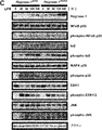
Regnase-1 (「Zc3h12a」または「MCPIP-1」としても知られ、本明細書においてこれらのように記載することがある。)は、CCCH型zincフィンガードメインとPIN様ドメインを持つRegnaseファミリーに属し、mRNAを認識して分解するヌクレアーゼである(非特許文献1)。Regnase-1は、インターロイキン(IL)-6やIL-12p40のmRNAを不安定化し、それらの転写後制御に関与している(非特許文献2)。マウスRegnase-1のSer(セリン)435およびSer439がIL-1β刺激により誘導されるIκBキナーゼ(IKK)によるリン酸化部位であること、これらのアミノ酸残基をAla(アラニン)に置換したRegnase-1は、IL-1β刺激による分解に耐性を有すること、並びに、前記アラニンに置換されたRegnase-1を発現させた細胞では、野生型のRegnase-1を発現させた細胞に比べてIL-1β刺激後のIL-6 mRNAの発現が抑制されることが報告されている(非特許文献3)。Regnase-1のヘテロKOマウスは、実験的自己免疫性脳脊髄炎および乾癬のモデル実験においてその病態が悪化することが報告されている(非特許文献4、5)。
Regnase-1 (also known as “Zc3h12a” or “MCPIP-1”, sometimes referred to as such herein) belongs to the Regnase family with CCCH-type zinc finger domain and PIN-like domain. , A nuclease that recognizes and degrades mRNA (Non-patent Document 1). Regnase-1 destabilizes mRNA of interleukin (IL) -6 and IL-12p40, and is involved in the post-transcriptional control thereof (Non-patent Document 2). Mouse Regnase-1 Ser (serine) 435 and Ser439 are phosphorylation sites by IκB kinase (IKK) induced by IL-1β stimulation, and these amino acid residues are substituted with Ala (alanine). Has resistance to degradation by IL-1β stimulation, and in cells expressing Regnase-1 substituted with alanine, IL-1β stimulation compared to cells expressing wild-type Regnase-1 It has been reported that later expression of IL-6 mRNA is suppressed (Non-patent Document 3). Regnase-1 hetero-KO mice have been reported to deteriorate in pathological conditions in experimental experiments of experimental autoimmune encephalomyelitis and psoriasis (Non-patent Documents 4 and 5).
しかしながら、Regnase-1のリン酸化を阻害することにより、炎症性疾患、自己免疫疾患、アレルギー疾患、線維化疾患などを治療または予防する方法は知られていなかった。本発明は、一局面において、Regnase-1のリン酸化を阻害することによる疾患の治療方法等を提供することを課題とする。
However, a method for treating or preventing inflammatory diseases, autoimmune diseases, allergic diseases, fibrotic diseases, etc. by inhibiting phosphorylation of Regnase-1 has not been known. In one aspect, an object of the present invention is to provide a method for treating a disease by inhibiting phosphorylation of Regnase-1.
本発明者らは、上記課題を解決するために、Regnase-1をリン酸化し得るキナーゼを探索し、それらキナーゼによりリン酸化を受けるRegnase-1のアミノ酸残基を見出した。そして、これらのアミノ酸残基の中でも、特定の残基のリン酸化の阻害が、炎症性疾患、自己免疫疾患、アレルギー疾患、線維化疾患などの治療および/または予防に有効であることを見出し、本発明を完成するに至った。
In order to solve the above problems, the present inventors searched for kinases that can phosphorylate Regnase-1, and found the amino acid residues of Regnase-1 that are phosphorylated by these kinases. And among these amino acid residues, we found that inhibition of phosphorylation of specific residues is effective for the treatment and / or prevention of inflammatory diseases, autoimmune diseases, allergic diseases, fibrotic diseases, The present invention has been completed.
本発明は、非限定の具体的な一態様において以下を包含する。
〔a1〕Ser残基のリン酸化をRegnase-1選択的に阻害することによる、Regnase-1が関与する疾患の治療および/または予防方法。
〔a2〕Ser残基のリン酸化をRegnase-1選択的に阻害することによる、炎症抑制方法。
〔a3〕Ser残基のリン酸化をRegnase-1選択的に阻害することによる、細胞、組織または臓器の線維化;または上皮の過形成を抑制する方法。
〔a4〕Ser残基のリン酸化をRegnase-1選択的に阻害することによる、Regnase-1の不安定化および/または細胞内での分解を抑制する方法。
〔a5〕前記Regnase-1の不安定化および/または細胞内での分解が、IL-17、IL-1、IL-36およびTLRリガンドからなる群より選択される少なくとも一つの分子が関与するシグナル伝達の下流におけるRegnase-1の不安定化および/または細胞内での分解である、〔a4〕に記載の方法。
〔a6〕Ser残基のリン酸化をRegnase-1選択的に阻害することによる、炎症性因子の産生抑制方法。
〔a7〕Ser残基のリン酸化をRegnase-1選択的に阻害することによる、IL6、IL1a、CXCL1、CXCL2、HBEGF、CTGF、DDR1およびPDGFBからなる群より選択される少なくとも一つのmRNAの発現を抑制する方法。
〔a8〕前記Ser残基が、Regnase-1における配列番号1の513、494、439、および435位のそれぞれに相当する位置からなる群より選択される少なくとも一つの位置のSer残基である、〔a1〕~〔a7〕のいずれかに記載の方法。
〔a9〕前記配列番号1の513、494、439、および435位のそれぞれに相当する位置が、(i)配列番号1の513、494、439、および435位;または、(ii)配列番号2の516、497、442、および438位である、〔a1〕~〔a8〕のいずれかに記載の方法。
〔a10〕前記Ser残基が、Regnase-1のアミノ酸配列に含まれるYWSEP (配列番号:3)、HFSVP (配列番号:4)およびDSGIGS (配列番号:5)からなる群より選択される少なくとも一つのアミノ酸配列に含まれるSer残基である、〔a1〕~〔a9〕のいずれかに記載の方法。
〔a11〕前記Regnase-1が哺乳動物のRegnase-1である、〔a1〕~〔a10〕のいずれかに記載の方法。
〔a12〕前記Ser残基が以下(i)および(ii)のSer残基である、〔a1〕~〔a11〕のいずれかに記載の方法:
(i)Regnase-1における配列番号1の513および494位のそれぞれに相当する位置のいずれか又は両方のSer残基;および
(ii)Regnase-1における配列番号1の439および435位のそれぞれに相当する位置のいずれか又は両方のSer残基。 The present invention includes, in one specific, non-limiting aspect:
[A1] A method for treating and / or preventing a disease involving Regnase-1 by selectively inhibiting phosphorylation of a Ser residue.
[A2] A method for suppressing inflammation by selectively inhibiting phosphorylation of Ser residues in Regnase-1.
[A3] A method for suppressing fibrosis of a cell, tissue or organ; or suppressing epithelial hyperplasia by selectively inhibiting phosphorylation of Ser residues.
[A4] A method of suppressing destabilization of Regnase-1 and / or intracellular degradation by selectively inhibiting phosphorylation of Ser residues.
[A5] A signal involving at least one molecule selected from the group consisting of IL-17, IL-1, IL-36, and TLR ligands for the destabilization and / or intracellular degradation of Regnase-1 The method according to [a4], which is destabilization and / or intracellular degradation of Regnase-1 downstream of transmission.
[A6] A method for suppressing the production of inflammatory factors by selectively inhibiting phosphorylation of Ser residues in Regnase-1.
[A7] Expression of at least one mRNA selected from the group consisting of IL6, IL1a, CXCL1, CXCL2, HBEGF, CTGF, DDR1 and PDGFB by selectively inhibiting phosphorylation of Ser residue How to suppress.
[A8] The Ser residue is a Ser residue at at least one position selected from the group consisting of positions corresponding to positions 513, 494, 439, and 435 of SEQ ID NO: 1 in Regnase-1. The method according to any one of [a1] to [a7].
[A9] positions corresponding to positions 513, 494, 439, and 435 of SEQ ID NO: 1 are (i) positions 513, 494, 439, and 435 of SEQ ID NO: 1; or (ii) SEQ ID NO: 2 The method according to any one of [a1] to [a8], which is at positions 516, 497, 442, and 438.
[A10] The Ser residue is at least one selected from the group consisting of YWSEP (SEQ ID NO: 3), HFSVP (SEQ ID NO: 4) and DSGIGS (SEQ ID NO: 5) contained in the amino acid sequence of Regnase-1. The method according to any one of [a1] to [a9], which is a Ser residue contained in one amino acid sequence.
[A11] The method according to any one of [a1] to [a10], wherein the Regnase-1 is mammalian Regnase-1.
[A12] The method according to any one of [a1] to [a11], wherein the Ser residue is a Ser residue of (i) and (ii) below:
(I) Ser residues at either or both positions corresponding to positions 513 and 494 of SEQ ID NO: 1 in Regnase-1, respectively; and (ii) at positions 439 and 435 of SEQ ID NO: 1 in Regnase-1, respectively Ser residues at either or both corresponding positions.
また本発明は、非限定の具体的な一態様において以下を包含する。
〔A1〕TBK1、IKKi、Act-1、IKKおよびIRAKからなる群より選択される少なくとも一つの結合分子とRegnase-1との結合を阻害することによる、Regnase-1が関与する疾患の治療および/または予防方法。
〔A2〕TBK1、IKKi、Act-1、IKKおよびIRAKからなる群より選択される少なくとも一つの結合分子とRegnase-1との結合を阻害することによる、炎症抑制方法。
〔A3〕TBK1、IKKi、Act-1、IKKおよびIRAKからなる群より選択される少なくとも一つの結合分子とRegnase-1との結合を阻害することによる、細胞、組織または臓器の線維化;または上皮の過形成を抑制する方法。
〔A4〕TBK1、IKKi、Act-1、IKKおよびIRAKからなる群より選択される少なくとも一つの結合分子とRegnase-1との結合を阻害することによる、Regnase-1の不安定化および/または細胞内での分解を抑制する方法。
〔A5〕前記Regnase-1の不安定化および/または細胞内での分解が、IL-17、IL-1、IL-36およびTLRリガンドからなる群より選択される少なくとも一つの分子が関与するシグナル伝達の下流におけるRegnase-1の不安定化および/または細胞内での分解である〔A4〕に記載の方法。
〔A6〕TBK1、IKKi、Act-1、IKKおよびIRAKからなる群より選択される少なくとも一つの結合分子とRegnase-1との結合を阻害することによる、炎症性因子の産生抑制方法。
〔A7〕TBK1、IKKi、Act-1、IKKおよびIRAKからなる群より選択される少なくとも一つの結合分子とRegnase-1との結合を阻害することによる、IL6、IL1a、CXCL1、CXCL2、HBEGF、CTGF、DDR1およびPDGFBからなる群より選択される少なくとも一つのmRNAの発現を抑制する方法。
〔A8〕Regnase-1における配列番号1の513、494、439、および435位のそれぞれに相当する位置からなる群より選択される少なくとも一つの位置のSer残基のリン酸化を阻害する、〔A1〕~〔A7〕のいずれかに記載の方法。
〔A9〕TBK1、IKKi、Act-1、IKKおよびIRAKからなる群より選択される少なくとも一つの結合分子が、TBK1、IKKi、およびAct-1からなる群より選択される少なくとも一つの結合分子である、〔A1〕~〔A8〕のいずれかに記載の方法。
〔A10〕前記Regnase-1が哺乳動物のRegnase-1である、〔A1〕~〔A9〕のいずれかに記載の方法。
〔A11〕TBK1、IKKi、Act-1、IKKおよびIRAKからなる群より選択される少なくとも一つの結合分子が、TBK1およびIKKである、〔A1〕~〔A10〕のいずれかに記載の方法。 Moreover, this invention includes the following in one non-limiting specific aspect.
[A1] Treatment of a disease involving Regnase-1 by inhibiting the binding of Regnase-1 with at least one binding molecule selected from the group consisting of TBK1, IKKi, Act-1, IKK and IRAK and / or Or prevention methods.
[A2] A method for suppressing inflammation by inhibiting the binding of Regnase-1 to at least one binding molecule selected from the group consisting of TBK1, IKKi, Act-1, IKK and IRAK.
[A3] Fibrosis of a cell, tissue or organ by inhibiting the binding of Regnase-1 with at least one binding molecule selected from the group consisting of TBK1, IKKi, Act-1, IKK and IRAK; or epithelium To suppress hyperplasia of the skin.
[A4] Instability of Regnase-1 and / or cell by inhibiting binding of Regnase-1 with at least one binding molecule selected from the group consisting of TBK1, IKKi, Act-1, IKK and IRAK To suppress internal decomposition.
[A5] A signal involving at least one molecule selected from the group consisting of IL-17, IL-1, IL-36 and TLR ligands for the destabilization and / or intracellular degradation of Regnase-1 The method according to [A4], which is destabilization and / or intracellular degradation of Regnase-1 downstream of transmission.
[A6] A method for suppressing the production of inflammatory factors by inhibiting the binding of Regnase-1 to at least one binding molecule selected from the group consisting of TBK1, IKKi, Act-1, IKK and IRAK.
[A7] IL6, IL1a, CXCL1, CXCL2, HBEGF, CTGF by inhibiting the binding of Regnase-1 with at least one binding molecule selected from the group consisting of TBK1, IKKi, Act-1, IKK and IRAK A method for suppressing the expression of at least one mRNA selected from the group consisting of DDR1 and PDGFB.
[A8] inhibits phosphorylation of Ser residue in at least one position selected from the group consisting of positions corresponding to positions 513, 494, 439, and 435 of SEQ ID NO: 1 in Regnase-1, [A1 ] To [A7].
[A9] At least one binding molecule selected from the group consisting of TBK1, IKKi, Act-1, IKK and IRAK is at least one binding molecule selected from the group consisting of TBK1, IKKi and Act-1. [A1] to [A8].
[A10] The method according to any one of [A1] to [A9], wherein the Regnase-1 is mammalian Regnase-1.
[A11] The method according to any one of [A1] to [A10], wherein at least one binding molecule selected from the group consisting of TBK1, IKKi, Act-1, IKK and IRAK is TBK1 and IKK.
また本発明は、非限定の具体的な一態様において以下を包含する。
〔B1〕Regnase-1における配列番号1の513、494、439、および435位のそれぞれに相当する位置からなる群より選択される少なくとも一つの位置のSer残基のリン酸化を阻害するRegnase-1結合分子を含有する、Regnase-1が関与する疾患を治療および/または予防するための組成物。
〔B2〕Regnase-1のアミノ酸配列に含まれるYWSEP (配列番号:3)、HFSVP (配列番号:4)およびDSGIGS (配列番号:5)からなる群より選択される少なくとも一つのアミノ酸配列に含まれるSer残基のリン酸化を阻害するRegnase-1結合分子を含有する、Regnase-1が関与する疾患を治療および/または予防するための組成物。
〔B3〕TBK1、IKKi、Act-1、IKKおよびIRAKからなる群より選択される少なくとも一つの結合分子とRegnase-1との結合を阻害するRegnase-1結合分子を含有する、Regnase-1が関与する疾患を治療および/または予防するための組成物。
〔B4〕Regnase-1における配列番号1の513、494、439、および435位のそれぞれに相当する位置からなる群より選択される少なくとも一つの位置のSer残基のリン酸化を阻害する、〔B3〕に記載の組成物。
〔B5〕前記Regnase-1が関与する疾患が、炎症性疾患、自己免疫疾患、アレルギー疾患、線維化疾患、およびRNAウイルス感染症からなる群より選択される少なくとも一つの疾患である、〔B1〕~〔B4〕のいずれかに記載の組成物。
〔B6〕前記Regnase-1が関与する疾患が、IL6、IL1a、CXCL1、CXCL2、HBEGF、CTGF、DDR1およびPDGFBからなる群より選択される少なくとも一つの因子が、疾患の形成、増悪および/または継続に関与する疾患である、〔B1〕~〔B5〕のいずれかに記載の組成物。
〔B7〕前記Regnase-1が関与する疾患が、IL-17、IL-1、IL-36およびTLRリガンドからなる群より選択される少なくとも一つの因子が、疾患の形成、増悪および/または継続に関与する疾患である、〔B1〕~〔B6〕のいずれかに記載の組成物。
〔B8〕前記Regnase-1が関与する疾患が、腎臓、肺、皮膚、血管、眼、脳および神経からなる群より選択される少なくとも一つの組織または臓器における疾患である、〔B1〕~〔B7〕のいずれかに記載の組成物。
〔B9〕前記Regnase-1が関与する疾患がTH17細胞関連疾患である、〔B1〕~〔B8〕のいずれかに記載の組成物。
〔B10〕前記Regnase-1が関与する疾患が、多発性硬化症、乾癬、強皮症、腎炎、ブドウ膜炎、肺線維症、腎臓線維症、血管線維症、ケロイド、関節リウマチ、全身性エリトマトーデス、シェーグレン症候群、肺炎、皮膚炎、血管炎、神経炎、関節炎、眼炎症、脳脊髄炎および喘息からなる群より選択される少なくとも一つの疾患である、〔B1〕~〔B9〕のいずれかに記載の組成物。
〔B11〕前記Regnase-1が関与する疾患が、細胞、組織または臓器の線維化を伴う疾患である、〔B1〕~〔B10〕のいずれかに記載の組成物。
〔B12〕前記Regnase-1が関与する疾患が、上皮の過形成を伴う疾患である、〔B1〕~〔B10〕のいずれかに記載の組成物。
〔B13〕IL6、IL1a、CXCL1、CXCL2、HBEGF、CTGF、DDR1およびPDGFBからなる群より選択される少なくとも一つのmRNAの発現を抑制するための、〔B1〕~〔B12〕のいずれかに記載の組成物。
〔B14〕炎症性因子の産生を抑制するための、〔B1〕~〔B13〕のいずれかに記載の組成物。
〔B15〕Regnase-1における配列番号1の513、494、439、および435位のそれぞれに相当する位置からなる群より選択される少なくとも一つの位置のSer残基のリン酸化を阻害するRegnase-1結合分子を含有する、炎症を抑制するための組成物。
〔B16〕Regnase-1における配列番号1の513、494、439、および435位のそれぞれに相当する位置からなる群より選択される少なくとも一つの位置のSer残基のリン酸化を阻害するRegnase-1結合分子を含有する、細胞、組織もしくは臓器の線維化、または上皮の過形成を抑制するための組成物。
〔B17〕Regnase-1における配列番号1の513、494、439、および435位のそれぞれに相当する位置からなる群より選択される少なくとも一つの位置のSer残基のリン酸化を阻害するRegnase-1結合分子を含有する、IL6、IL1a、CXCL1、CXCL2、HBEGF、CTGF、DDR1およびPDGFBからなる群より選択される少なくとも一つのmRNAの発現を抑制するための組成物。
〔B18〕Regnase-1における配列番号1の513、494、439、および435位のそれぞれに相当する位置からなる群より選択される少なくとも一つの位置のSer残基のリン酸化を阻害するRegnase-1結合分子を含有する、炎症性因子の産生を抑制するための組成物。
〔B19〕前記配列番号1の513、494、439、および435位のそれぞれに相当する位置が、(i)配列番号1の513、494、439、および435位;または、(ii)配列番号2の516、497、442、および438位である、〔B1〕~〔B18〕のいずれかに記載の組成物。
〔B20〕前記配列番号1の513および494位のそれぞれに相当する位置からなる群より選択される少なくとも一つの位置のSer残基のリン酸化を阻害するRegnase-1結合分子を含有する、〔B1〕~〔B19〕のいずれかに記載の組成物。
〔B21〕前記配列番号1の513および494位のそれぞれに相当する位置が、(i)配列番号1の513および494位;または、(ii)配列番号2の516および497位である、〔B20〕に記載の組成物。
〔B22〕TBK1、IKKi、Act-1、IKKおよびIRAKからなる群より選択される少なくとも一つの結合分子が、TBK1、IKKi、およびAct-1からなる群より選択される少なくとも一つの結合分子である、〔B3〕~〔B21〕のいずれかに記載の組成物。
〔B23〕前記Regnase-1が哺乳動物のRegnase-1である、〔B1〕~〔B22〕のいずれかに記載の組成物。
〔B24〕Regnase-1における配列番号1の513、494、439、および435位のそれぞれに相当する位置からなる群より選択される少なくとも一つの位置のSer残基が、以下(i)および(ii)のSer残基である、〔B1〕~〔B23〕のいずれかに記載の組成物:
(i)Regnase-1における配列番号1の513および494位のそれぞれに相当する位置のいずれか又は両方のSer残基;および
(ii)Regnase-1における配列番号1の439および435位のそれぞれに相当する位置のいずれか又は両方のSer残基。
〔B25〕TBK1、IKKi、Act-1、IKKおよびIRAKからなる群より選択される少なくとも一つの結合分子が、TBK1およびIKKである、〔B1〕~〔B24〕のいずれかに記載の組成物。
〔B26〕前記Regnase-1結合分子が、〔H1〕~〔H24〕のいずれかに記載のRegnase-1結合分子である、〔B1〕~〔B25〕のいずれかに記載の組成物。 Moreover, this invention includes the following in one non-limiting specific aspect.
[B1] Regnase-1 that inhibits phosphorylation of Ser residue at at least one position selected from the group consisting of positions corresponding to positions 513, 494, 439, and 435 of SEQ ID NO: 1 in Regnase-1 A composition for treating and / or preventing a disease involving Regnase-1 comprising a binding molecule.
[B2] Included in at least one amino acid sequence selected from the group consisting of YWSEP (SEQ ID NO: 3), HFSVP (SEQ ID NO: 4) and DSGIGS (SEQ ID NO: 5) included in the amino acid sequence of Regnase-1 A composition for treating and / or preventing a disease involving Regnase-1, comprising a Regnase-1-binding molecule that inhibits phosphorylation of Ser residues.
[B3] Regnase-1 containing Regnase-1 binding molecule that inhibits binding between Regnase-1 and at least one binding molecule selected from the group consisting of TBK1, IKKi, Act-1, IKK and IRAK is involved The composition for treating and / or preventing the disease which carries out.
[B4] inhibits phosphorylation of a Ser residue at least one position selected from the group consisting of positions corresponding to positions 513, 494, 439, and 435 of SEQ ID NO: 1 in Regnase-1, [B3 ] The composition of description.
[B5] The disease involving Regnase-1 is at least one disease selected from the group consisting of inflammatory diseases, autoimmune diseases, allergic diseases, fibrotic diseases, and RNA virus infections. [B1] To [B4].
[B6] The disease involving Regnase-1 is at least one factor selected from the group consisting of IL6, IL1a, CXCL1, CXCL2, HBEGF, CTGF, DDR1 and PDGFB. The composition according to any one of [B1] to [B5], wherein the composition is a disease related to the disease.
[B7] The disease involving Regnase-1 is at least one factor selected from the group consisting of IL-17, IL-1, IL-36 and TLR ligand, which is responsible for the formation, exacerbation and / or continuation of the disease. The composition according to any one of [B1] to [B6], which is a disease involved.
[B8] The disease involving Regnase-1 is a disease in at least one tissue or organ selected from the group consisting of kidney, lung, skin, blood vessel, eye, brain and nerve, [B1] to [B7 ] The composition in any one of.
[B9] The composition according to any one of [B1] to [B8], wherein the disease involving Regnase-1 is a TH17 cell-related disease.
[B10] The disease involving Regnase-1 is multiple sclerosis, psoriasis, scleroderma, nephritis, uveitis, pulmonary fibrosis, renal fibrosis, vascular fibrosis, keloid, rheumatoid arthritis, systemic lupus erythematosus Any one of [B1] to [B9], which is at least one disease selected from the group consisting of Sjogren's syndrome, pneumonia, dermatitis, vasculitis, neuritis, arthritis, ocular inflammation, encephalomyelitis, and asthma The composition as described.
[B11] The composition according to any one of [B1] to [B10], wherein the disease involving Regnase-1 is a disease involving fibrosis of cells, tissues, or organs.
[B12] The composition according to any one of [B1] to [B10], wherein the disease involving Regnase-1 is a disease associated with epithelial hyperplasia.
[B13] any one of [B1] to [B12] for suppressing the expression of at least one mRNA selected from the group consisting of IL6, IL1a, CXCL1, CXCL2, HBEGF, CTGF, DDR1 and PDGFB Composition.
[B14] The composition according to any one of [B1] to [B13] for suppressing production of inflammatory factors.
[B15] Regnase-1 that inhibits phosphorylation of Ser residue at least at one position selected from the group consisting of positions corresponding to positions 513, 494, 439, and 435 of SEQ ID NO: 1 in Regnase-1 A composition for suppressing inflammation, comprising a binding molecule.
[B16] Regnase-1 that inhibits phosphorylation of Ser residue at least at one position selected from the group consisting of positions corresponding to positions 513, 494, 439, and 435 of SEQ ID NO: 1 in Regnase-1 A composition for inhibiting fibrosis of cells, tissues or organs, or epithelial hyperplasia, comprising a binding molecule.
[B17] Regnase-1 that inhibits phosphorylation of Ser residue at at least one position selected from the group consisting of positions corresponding to positions 513, 494, 439, and 435 of SEQ ID NO: 1 in Regnase-1 A composition for suppressing the expression of at least one mRNA selected from the group consisting of IL6, IL1a, CXCL1, CXCL2, HBEGF, CTGF, DDR1 and PDGFB, containing a binding molecule.
[B18] Regnase-1 that inhibits phosphorylation of a Ser residue at least at one position selected from the group consisting of positions corresponding to positions 513, 494, 439, and 435 of SEQ ID NO: 1 in Regnase-1 A composition for inhibiting the production of inflammatory factors, comprising a binding molecule.
[B19] positions corresponding to positions 513, 494, 439, and 435 of SEQ ID NO: 1 are (i) positions 513, 494, 439, and 435 of SEQ ID NO: 1; or (ii) SEQ ID NO: 2 The composition according to any one of [B1] to [B18], which is at positions 516, 497, 442, and 438.
[B20] containing a Regnase-1 binding molecule that inhibits phosphorylation of a Ser residue at at least one position selected from the group consisting of positions corresponding to positions 513 and 494 of SEQ ID NO: 1, [B1 ] To [B19].
[B21] The positions corresponding to positions 513 and 494 of SEQ ID NO: 1 are (i) positions 513 and 494 of SEQ ID NO: 1; or (ii) positions 516 and 497 of SEQ ID NO: 2, [B20 ] The composition of description.
[B22] At least one binding molecule selected from the group consisting of TBK1, IKKi, Act-1, IKK and IRAK is at least one binding molecule selected from the group consisting of TBK1, IKKi and Act-1. , [B3] to [B21].
[B23] The composition according to any one of [B1] to [B22], wherein the Regnase-1 is mammalian Regnase-1.
[B24] Ser residues at at least one position selected from the group consisting of positions corresponding to positions 513, 494, 439, and 435 of SEQ ID NO: 1 in Regnase-1 are the following (i) and (ii) The composition according to any one of [B1] to [B23], which is a Ser residue of:
(I) Ser residues at either or both positions corresponding to positions 513 and 494 of SEQ ID NO: 1 in Regnase-1, respectively; and (ii) at positions 439 and 435 of SEQ ID NO: 1 in Regnase-1, respectively Ser residues at either or both corresponding positions.
[B25] The composition according to any one of [B1] to [B24], wherein at least one binding molecule selected from the group consisting of TBK1, IKKi, Act-1, IKK and IRAK is TBK1 and IKK.
[B26] The composition according to any one of [B1] to [B25], wherein the Regnase-1 binding molecule is the Regnase-1 binding molecule according to any one of [H1] to [H24].
また本発明は、非限定の具体的な一態様において以下を包含する。
〔D1〕配列番号1の513、494、439および435位のそれぞれに相当する位置からなる群より選択される少なくとも一つの位置のSer残基のリン酸化を指標とする、Regnase-1のリン酸化を阻害する物質を特定するための方法。
〔D2〕以下(a)および(b)の工程を含む、〔D1〕に記載の方法:
(a)Regnase-1をリン酸化し得るキナーゼとRegnase-1とを被験物質の存在下で混合し、前記キナーゼによるRegnase-1のリン酸化を検出する工程;
(b)被験物質の非存在下と比較して、前記キナーゼによるRegnase-1のリン酸化を阻害する物質を特定する工程。
〔D3〕前記キナーゼが、配列番号1の513、494、439および435位のそれぞれに相当する位置からなる群より選択される少なくとも一つの位置のSer残基をリン酸化し得るキナーゼである、〔D1〕又は〔D2〕に記載の方法。
〔D4〕前記キナーゼが、TBK1、IKKi、IKKおよびIRAKからなる群より選択される少なくとも一つのキナーゼである、〔D1〕~〔D3〕のいずれかに記載の方法。
〔D5〕前記工程(a)におけるRegnase-1のリン酸化の検出が、配列番号1の513、494、439および435位のそれぞれに相当する位置からなる群より選択される少なくとも一つの位置のSer残基のリン酸化を検出し得る抗体を用いて行われる、〔D1〕~〔D4〕のいずれかに記載の方法。
〔D6〕前記Regnase-1がヒトRegnase-1であり、前記配列番号1の513、494、439および435位のそれぞれに相当する位置が、配列番号2の516、497、442および438位である、〔D1〕~〔D5〕のいずれかに記載の方法。
〔D7〕配列番号1の513および494位のそれぞれに相当する位置からなる群より選択される少なくとも一つの位置のSer残基のリン酸化を指標とする、〔D1〕~〔D6〕のいずれかに記載の方法。
〔D8〕前記被験物質がRegnase-1結合分子である、〔D2〕~〔D7〕のいずれかに記載の方法。 Moreover, this invention includes the following in one non-limiting specific aspect.
[D1] Phosphorylation of Regnase-1 using as an index phosphorylation of a Ser residue at at least one position selected from the group consisting of positions corresponding to positions 513, 494, 439 and 435 of SEQ ID NO: 1 A method for identifying a substance that inhibits a substance.
[D2] The method according to [D1], comprising the following steps (a) and (b):
(A) a step of mixing a kinase capable of phosphorylating Regnase-1 and Regnase-1 in the presence of a test substance, and detecting phosphorylation of Regnase-1 by the kinase;
(B) A step of identifying a substance that inhibits phosphorylation of Regnase-1 by the kinase as compared to the absence of a test substance.
[D3] The kinase is a kinase capable of phosphorylating a Ser residue at at least one position selected from the group consisting of positions corresponding to positions 513, 494, 439, and 435 of SEQ ID NO: 1. D1] or the method according to [D2].
[D4] The method according to any one of [D1] to [D3], wherein the kinase is at least one kinase selected from the group consisting of TBK1, IKKi, IKK and IRAK.
[D5] Detection of phosphorylation of Regnase-1 in the step (a) is performed at Ser at at least one position selected from the group consisting of positions corresponding to positions 513, 494, 439 and 435 of SEQ ID NO: 1. The method according to any one of [D1] to [D4], which is performed using an antibody capable of detecting residue phosphorylation.
[D6] The Regnase-1 is human Regnase-1, and positions corresponding to positions 513, 494, 439 and 435 of SEQ ID NO: 1 are positions 516, 497, 442 and 438 of SEQ ID NO: 2, respectively. , [D1] to [D5].
[D7] Any one of [D1] to [D6], wherein phosphorylation of a Ser residue at at least one position selected from the group consisting of positions corresponding to positions 513 and 494 of SEQ ID NO: 1 is used as an index The method described in 1.
[D8] The method according to any one of [D2] to [D7], wherein the test substance is a Regnase-1 binding molecule.
また本発明は、非限定の具体的な一態様において以下を包含する。
〔E1〕Regnase-1のリン酸化を阻害する物質を特定するための組成物であって、予め定められた量のキナーゼおよびRegnase-1を含む、前記組成物。
〔E2〕前記キナーゼがTBK1、IKKi、IKKおよびIRAKからなる群より選択される少なくとも一つのキナーゼである、〔E1〕に記載の組成物。
〔E3〕前記Regnase-1のリン酸化が、配列番号1の513、494、439および435位のそれぞれに相当する位置からなる群より選択される少なくとも一つの位置のSer残基のリン酸化である、〔E1〕または〔E2〕に記載の組成物。
〔E4〕前記Regnase-1がヒトRegnase-1であり、前記配列番号1の513、494、439および435位のそれぞれに相当する位置が、配列番号2の516、497、442および438位である、〔E1〕~〔E3〕のいずれかに記載の組成物。
〔E5〕前記Regnase-1のリン酸化が、配列番号1の513、および494位のそれぞれに相当する位置からなる群より選択される少なくとも一つの位置のSer残基のリン酸化である、〔E1〕~〔E4〕のいずれかに記載の組成物。
〔E6〕さらにRegnase-1結合分子を含む、〔E1〕~〔E5〕のいずれかに記載の組成物。 Moreover, this invention includes the following in one non-limiting specific aspect.
[E1] A composition for identifying a substance that inhibits phosphorylation of Regnase-1, which comprises a predetermined amount of kinase and Regnase-1.
[E2] The composition according to [E1], wherein the kinase is at least one kinase selected from the group consisting of TBK1, IKKi, IKK and IRAK.
[E3] The phosphorylation of Regnase-1 is phosphorylation of a Ser residue at at least one position selected from the group consisting of positions corresponding to positions 513, 494, 439 and 435 of SEQ ID NO: 1. [E1] or [E2].
[E4] The Regnase-1 is human Regnase-1, and positions corresponding to positions 513, 494, 439 and 435 of SEQ ID NO: 1 are positions 516, 497, 442 and 438 of SEQ ID NO: 2, respectively. [E1] to [E3].
[E5] The phosphorylation of Regnase-1 is phosphorylation of a Ser residue at at least one position selected from the group consisting of positions corresponding to positions 513 and 494 of SEQ ID NO: 1. [E1 ] To [E4].
[E6] The composition according to any one of [E1] to [E5], further comprising a Regnase-1 binding molecule.
また本発明は、非限定の具体的な一態様において以下を包含する。
〔F1〕以下(a)および(b)の工程を含む、TBK1、IKKi、Act-1、IKKおよびIRAKからなる群より選択される少なくとも一つの結合分子とRegnase-1との結合を阻害する物質を特定するための方法:
(a)TBK1、IKKi、Act-1、IKKおよびIRAKからなる群より選択される少なくとも一つの結合分子と Regnase-1 とを被験物質の存在下で混合し、前記結合分子とRegnase-1との結合活性を測定する工程;
(b)被験物質の非存在下と比較して、前記結合分子とRegnase-1との結合活性を低減させ得る物質を特定する工程。
〔F2〕TBK1、IKKi、Act-1、IKKおよびIRAKからなる群より選択される少なくとも一つの結合分子が、TBK1、IKKi、およびAct-1からなる群より選択される少なくとも一つの結合分子である、〔F1〕に記載の方法。
〔F3〕TBK1、IKKi、Act-1、IKKおよびIRAKからなる群より選択される少なくとも一つの結合分子とRegnase-1との結合を阻害する物質を特定するための組成物であって、予め定められた量の結合分子およびRegnase-1を含む、前記組成物。
〔F4〕〔D1〕~〔D8〕のいずれかに記載の方法と〔F1〕または〔F2〕に記載の方法を含む、TBK1、IKKi、Act-1、IKKおよびIRAKからなる群より選択される少なくとも一つの結合分子とRegnase-1との結合を阻害し、Regnase-1のリン酸化を阻害する物質を特定するための方法。
〔F5〕前記被験物質がRegnase-1結合分子である、〔F1〕、〔F2〕および〔F4〕のいずれかに記載の方法。 Moreover, this invention includes the following in one non-limiting specific aspect.
[F1] A substance that inhibits the binding between Regnase-1 and at least one binding molecule selected from the group consisting of TBK1, IKKi, Act-1, IKK and IRAK, comprising the following steps (a) and (b) How to identify:
(A) At least one binding molecule selected from the group consisting of TBK1, IKKi, Act-1, IKK and IRAK and Regnase-1 are mixed in the presence of a test substance, and the binding molecule and Regnase-1 Measuring the binding activity;
(B) A step of identifying a substance that can reduce the binding activity between the binding molecule and Regnase-1 as compared to the absence of the test substance.
[F2] At least one binding molecule selected from the group consisting of TBK1, IKKi, Act-1, IKK and IRAK is at least one binding molecule selected from the group consisting of TBK1, IKKi and Act-1. [F1].
[F3] A composition for specifying a substance that inhibits binding between Regnase-1 and at least one binding molecule selected from the group consisting of TBK1, IKKi, Act-1, IKK, and IRAK. The composition comprising a defined amount of binding molecule and Regnase-1.
[F4] selected from the group consisting of TBK1, IKKi, Act-1, IKK and IRAK, including the method according to any one of [D1] to [D8] and the method according to [F1] or [F2] A method for identifying a substance that inhibits the binding of Regnase-1 by inhibiting the binding of at least one binding molecule to Regnase-1.
[F5] The method according to any one of [F1], [F2] and [F4], wherein the test substance is a Regnase-1 binding molecule.
また本発明は、非限定の具体的な一態様において以下を包含する。
〔G1〕配列番号1の513、494、439および435位のそれぞれに相当する位置からなる群より選択される少なくとも一つの位置のSer残基がリン酸化されたRegnase-1を特異的に認識する抗体。
〔G2〕配列番号1の513および494位のそれぞれに相当する位置からなる群より選択される少なくとも一つの位置のSer残基がリン酸化されたRegnase-1を特異的に認識する抗体。
〔G3〕配列番号1の513位に相当するSer残基がリン酸化されたRegnase-1を特異的に認識する抗体。
〔G4〕配列番号1の439および435位のそれぞれに相当する位置からなる群より選択される少なくとも一つの位置のSer残基がリン酸化されたRegnase-1を特異的に認識する抗体。
〔G5〕配列番号1の439および435位のそれぞれに相当する位置のSer残基がリン酸化されたRegnase-1を特異的に認識する抗体。
〔G6〕配列番号2の516、497、442および438位から選択される少なくとも一つの位置のSer残基がリン酸化されたヒトRegnase-1を特異的に認識する抗体。
〔G7〕配列番号2の516および497位から選択される少なくとも一つの位置のSer残基がリン酸化されたヒトRegnase-1を特異的に認識する抗体。
〔G8〕配列番号2の516位のSer残基がリン酸化されたヒトRegnase-1を特異的に認識する抗体。
〔G9〕配列番号2の442および438位から選択される少なくとも一つの位置のSer残基がリン酸化されたヒトRegnase-1を特異的に認識する抗体。
〔G10〕配列番号2の442および438位のSer残基がリン酸化されたヒトRegnase-1を特異的に認識する抗体。
〔G11〕ヒトRegnase-1およびマウスRegnase-1のいずれにも結合できる、〔G1〕~〔10〕のいずれかに記載の抗体。 Moreover, this invention includes the following in one non-limiting specific aspect.
[G1] specifically recognizes Regnase-1 in which at least one Ser residue selected from the group consisting of positions corresponding to positions 513, 494, 439 and 435 of SEQ ID NO: 1 is phosphorylated antibody.
[G2] An antibody that specifically recognizes Regnase-1 in which at least one Ser residue selected from the group consisting of positions corresponding to positions 513 and 494 of SEQ ID NO: 1 is phosphorylated.
[G3] An antibody that specifically recognizes Regnase-1 in which the Ser residue corresponding to position 513 of SEQ ID NO: 1 is phosphorylated.
[G4] An antibody that specifically recognizes Regnase-1 in which at least one Ser residue selected from the group consisting of positions corresponding to positions 439 and 435 of SEQ ID NO: 1 is phosphorylated.
[G5] An antibody that specifically recognizes Regnase-1 in which Ser residues at positions corresponding to positions 439 and 435 of SEQ ID NO: 1 are phosphorylated.
[G6] An antibody that specifically recognizes human Regnase-1 in which a Ser residue at at least one position selected from positions 516, 497, 442, and 438 of SEQ ID NO: 2 is phosphorylated.
[G7] An antibody that specifically recognizes human Regnase-1 in which a Ser residue at at least one position selected from positions 516 and 497 of SEQ ID NO: 2 is phosphorylated.
[G8] An antibody that specifically recognizes human Regnase-1 in which the Ser residue at position 516 of SEQ ID NO: 2 is phosphorylated.
[G9] An antibody that specifically recognizes human Regnase-1 in which a Ser residue at at least one position selected from positions 442 and 438 of SEQ ID NO: 2 is phosphorylated.
[G10] An antibody that specifically recognizes human Regnase-1 in which Ser residues at positions 442 and 438 of SEQ ID NO: 2 are phosphorylated.
[G11] The antibody according to any one of [G1] to [10], which is capable of binding to both human Regnase-1 and mouse Regnase-1.
また本発明は、非限定の具体的な一態様において以下を包含する。
〔H1〕Regnase-1における配列番号1の513、494、439、および435位のそれぞれに相当する位置からなる群より選択される少なくとも一つの位置のSer残基のリン酸化を阻害するRegnase-1結合分子。
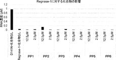
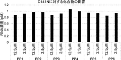
〔H2〕Regnase-1への結合について、以下のPP1~PP25、PP7+tag、PP10+tagおよびPP23+tag:


 より選択される少なくとも1つの化合物と競合する、または以下の抗体
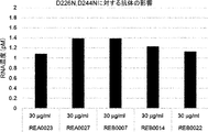
(i)配列番号:20に記載のアミノ酸配列を含む重鎖および配列番号:21に記載のアミノ酸配列を含む軽鎖を含む抗体(REA0023)、
(ii)配列番号:22に記載のアミノ酸配列を含む重鎖および配列番号:23に記載のアミノ酸配列を含む軽鎖を含む抗体(REA0027)、
(iii)配列番号:24に記載のアミノ酸配列を含む重鎖および配列番号:25に記載のアミノ酸配列を含む軽鎖を含む抗体(REB0007)、
(iv)配列番号:26に記載のアミノ酸配列を含む重鎖および配列番号:27に記載のアミノ酸配列を含む軽鎖を含む抗体(REB0014)および
(v)配列番号:28に記載のアミノ酸配列を含む重鎖および配列番号:29に記載のアミノ酸配列を含む軽鎖を含む抗体(REB0022)
より選択される少なくとも1つの抗体と競合する、
〔H1〕に記載のRegnase-1結合分子。
〔H3〕Regnase-1への結合について、化合物PP7と競合する、〔H1〕または〔H2〕に記載のRegnase-1結合分子。
〔H4〕Regnase-1への結合について、化合物PP23と競合する、〔H1〕~〔H3〕のいずれかに記載のRegnase-1結合分子。
〔H5〕Regnase-1への結合について、化合物PP10と競合する、〔H1〕~〔H4〕のいずれかに記載のRegnase-1結合分子。
〔H6〕Regnase-1への結合について、化合物PP7と競合しない、〔H1〕、〔H2〕、〔H4〕、または〔H5〕のいずれかに記載のRegnase-1結合分子。
〔H7〕Regnase-1への結合について、化合物PP23と競合しない、〔H1〕~〔H3〕、〔H5〕、または〔H6〕のいずれかに記載のRegnase-1結合分子。
〔H8〕Regnase-1への結合について、化合物PP10と競合しない、〔H1〕~〔H4〕、〔H6〕、または〔H7〕のいずれかに記載のRegnase-1結合分子。
〔H9〕Regnase-1に特異的に結合する、〔H1〕~〔H8〕のいずれかに記載のRegnase-1結合分子。
〔H10〕前記配列番号1の513、494、439、および435位のそれぞれに相当する位置が、(i)配列番号1の513、494、439、および435位;または、(ii)配列番号2の516、497、442、および438位である、〔H1〕~〔H9〕のいずれかに記載のRegnase-1結合分子。
〔H11〕以下(i)および(ii)のSer残基のリン酸化を阻害する、〔H1〕~〔H10〕のいずれかに記載のRegnase-1結合分子:
(i)Regnase-1における配列番号1の513および494位のそれぞれに相当する位置のいずれか又は両方のSer残基;および
(ii)Regnase-1における配列番号1の439および435位のそれぞれに相当する位置のいずれか又は両方のSer残基。
〔H12〕Regnase-1における配列番号1の513および494位のそれぞれに相当する位置からなる群より選択される少なくとも一つの位置のSer残基のリン酸化を阻害する、〔H1〕~〔H11〕のいずれかに記載のRegnase-1結合分子。
〔H13〕配列番号1の439および435位のそれぞれに相当する位置からなる群より選択される少なくとも一つの位置のSer残基のリン酸化を阻害する、〔H1〕~〔H12〕のいずれかに記載のRegnase-1結合分子。
〔H14〕化合物PP1~PP25、PP7+tag、PP10+tagおよびPP23+tagより選択される化合物、または抗体REA0023、REA0027、REB0007、REB0014およびREB0022より選択される抗体が結合するRegnase-1上の結合部位と同じ部位でRegnase-1に結合する、〔H1〕~〔H13〕のいずれかに記載のRegnase-1結合分子。
〔H15〕化合物PP7が結合するRegnase-1上の結合部位と同じ部位でRegnase-1に結合する、〔H1〕~〔H14〕のいずれかに記載のRegnase-1結合分子。
〔H16〕化合物PP23が結合するRegnase-1上の結合部位と同じ部位でRegnase-1に結合する、〔H1〕~〔H15〕のいずれかに記載のRegnase-1結合分子。
〔H17〕化合物PP10が結合するRegnase-1上の結合部位と同じ部位でRegnase-1に結合する、〔H1〕~〔H16〕のいずれかに記載のRegnase-1結合分子。
〔H18〕前記Ser残基が、Regnase-1のアミノ酸配列に含まれるYWSEP (配列番号:3)、HFSVP (配列番号:4)およびDSGIGS (配列番号:5)からなる群より選択される少なくとも一つのアミノ酸配列に含まれるSer残基である、〔H1〕~〔H17〕のいずれかに記載のRegnase-1結合分子。
〔H19〕TBK1、IKKi、Act-1、IKK、およびIRAKからなる群より選択される少なくとも一つの分子とRegnase-1との結合を阻害する、〔H1〕~〔H18〕のいずれかに記載のRegnase-1結合分子。
〔H20〕前記リン酸化が、IL-17およびIL-1からなる群より選択される少なくとも一つの分子により誘導され得るリン酸化である、〔H1〕~〔H19〕のいずれかに記載のRegnase-1結合分子。
〔H21〕TBK1、IKKi、Act-1、IKK、およびIRAKからなる群より選択される少なくとも一つの分子とRegnase-1との結合を阻害するRegnase-1結合分子。
〔H22〕配列番号:1に示される544~596番目のアミノ酸配列、または配列番号:2に示される547~599番目 のアミノ酸配列に含まれるアミノ酸残基に結合する、〔H1〕~〔H21〕のいずれかに記載のRegnase-1結合分子。
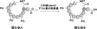
〔H23〕環状ポリペプチドである、〔H1〕~〔H22〕のいずれかに記載のRegnase-1結合分子。
〔H24〕抗体である、〔H1〕~〔H22〕のいずれかに記載のRegnase-1結合分子。 Moreover, this invention includes the following in one non-limiting specific aspect.
[H1] Regnase-1 that inhibits phosphorylation of Ser residue at least at one position selected from the group consisting of positions corresponding to positions 513, 494, 439, and 435 of SEQ ID NO: 1 in Regnase-1 Binding molecule.
[H2] For binding to Regnase-1, the following PP1 to PP25, PP7 + tag, PP10 + tag and PP23 + tag:
An antibody (i) that competes with at least one compound selected from the following, or an antibody (i) comprising a heavy chain comprising the amino acid sequence set forth in SEQ ID NO: 20 and a light chain comprising the amino acid sequence set forth in SEQ ID NO: 21 ( REA0023),
(Ii) an antibody (REA0027) comprising a heavy chain comprising the amino acid sequence set forth in SEQ ID NO: 22 and a light chain comprising the amino acid sequence set forth in SEQ ID NO: 23;
(Iii) an antibody (REB0007) comprising a heavy chain comprising the amino acid sequence set forth in SEQ ID NO: 24 and a light chain comprising the amino acid sequence set forth in SEQ ID NO: 25
(Iv) an antibody (REB0014) comprising a heavy chain comprising the amino acid sequence set forth in SEQ ID NO: 26 and a light chain comprising the amino acid sequence set forth in SEQ ID NO: 27; and (v) an amino acid sequence set forth in SEQ ID NO: 28. An antibody comprising a heavy chain comprising and a light chain comprising the amino acid sequence set forth in SEQ ID NO: 29 (REB0022)
Competing with at least one antibody selected from
Regnase-1 binding molecule according to [H1].
[H3] The Regnase-1 binding molecule according to [H1] or [H2], which competes with the compound PP7 for binding to Regnase-1.
[H4] The Regnase-1 binding molecule according to any one of [H1] to [H3], which competes with the compound PP23 for binding to Regnase-1.
[H5] The Regnase-1 binding molecule according to any one of [H1] to [H4], which competes with the compound PP10 for binding to Regnase-1.
[H6] The Regnase-1 binding molecule according to any one of [H1], [H2], [H4], or [H5], which does not compete with the compound PP7 for binding to Regnase-1.
[H7] The Regnase-1 binding molecule according to any one of [H1] to [H3], [H5], or [H6], which does not compete with the compound PP23 for binding to Regnase-1.
[H8] The Regnase-1 binding molecule according to any one of [H1] to [H4], [H6], or [H7], which does not compete with the compound PP10 for binding to Regnase-1.
[H9] The Regnase-1 binding molecule according to any one of [H1] to [H8], which specifically binds to Regnase-1.
[H10] positions corresponding to positions 513, 494, 439, and 435 of SEQ ID NO: 1 are (i) positions 513, 494, 439, and 435 of SEQ ID NO: 1; or (ii) SEQ ID NO: 2 Regnase-1 binding molecule according to any one of [H1] to [H9], which is at positions 516, 497, 442, and 438.
[H11] The Regnase-1 binding molecule according to any one of [H1] to [H10], which inhibits phosphorylation of the Ser residues of (i) and (ii) below:
(I) Ser residues at either or both positions corresponding to positions 513 and 494 of SEQ ID NO: 1 in Regnase-1, respectively; and (ii) at positions 439 and 435 of SEQ ID NO: 1 in Regnase-1, respectively Ser residues at either or both corresponding positions.
[H12] inhibits phosphorylation of Ser residue at least one position selected from the group consisting of positions corresponding to positions 513 and 494 of SEQ ID NO: 1 in Regnase-1, [H1] to [H11] Regnase-1 binding molecule | numerator in any one of.
[H13] any one of [H1] to [H12], which inhibits phosphorylation of a Ser residue at at least one position selected from the group consisting of positions corresponding to positions 439 and 435 of SEQ ID NO: 1, The described Regnase-1 binding molecule.
[H14] Binding on Regnase-1 to which a compound selected from the compounds PP1 to PP25, PP7 + tag, PP10 + tag and PP23 + tag or an antibody selected from the antibodies REA0023, REA0027, REB0007, REB0014 and REB0022 binds The Regnase-1 binding molecule according to any one of [H1] to [H13], which binds to Regnase-1 at the same site.
[H15] The Regnase-1 binding molecule according to any one of [H1] to [H14], which binds to Regnase-1 at the same site as the binding site on Regnase-1 to which compound PP7 binds.
[H16] The Regnase-1 binding molecule according to any one of [H1] to [H15], which binds to Regnase-1 at the same site as the binding site on Regnase-1 to which compound PP23 binds.
[H17] The Regnase-1 binding molecule according to any one of [H1] to [H16], which binds to Regnase-1 at the same site as the binding site on Regnase-1 to which compound PP10 binds.
[H18] The Ser residue is at least one selected from the group consisting of YWSEP (SEQ ID NO: 3), HFSVP (SEQ ID NO: 4) and DSGIGS (SEQ ID NO: 5) contained in the amino acid sequence of Regnase-1. The Regnase-1-binding molecule according to any one of [H1] to [H17], which is a Ser residue contained in one amino acid sequence.
[H19] The inhibitor according to any one of [H1] to [H18], which inhibits binding between Regnase-1 and at least one molecule selected from the group consisting of TBK1, IKKi, Act-1, IKK, and IRAK. Regnase-1 binding molecule.
[H20] Regnase- according to any one of [H1] to [H19], wherein the phosphorylation is phosphorylation that can be induced by at least one molecule selected from the group consisting of IL-17 and IL-1. 1 binding molecule.
[H21] A Regnase-1 binding molecule that inhibits binding of Regnase-1 to at least one molecule selected from the group consisting of TBK1, IKKi, Act-1, IKK, and IRAK.
[H22] [H1] to [H21] which binds to the amino acid residue contained in the amino acid sequence of 544 to 596 shown in SEQ ID NO: 1 or the amino acid sequence of 547 to 599 shown in SEQ ID NO: 2 Regnase-1 binding molecule | numerator in any one of.
[H23] The Regnase-1 binding molecule according to any one of [H1] to [H22], which is a cyclic polypeptide.
[H24] The Regnase-1 binding molecule according to any one of [H1] to [H22], which is an antibody.
〔H1〕Regnase-1における配列番号1の513、494、439、および435位のそれぞれに相当する位置からなる群より選択される少なくとも一つの位置のSer残基のリン酸化を阻害するRegnase-1結合分子。
〔H2〕Regnase-1への結合について、以下のPP1~PP25、PP7+tag、PP10+tagおよびPP23+tag:
(i)配列番号:20に記載のアミノ酸配列を含む重鎖および配列番号:21に記載のアミノ酸配列を含む軽鎖を含む抗体(REA0023)、
(ii)配列番号:22に記載のアミノ酸配列を含む重鎖および配列番号:23に記載のアミノ酸配列を含む軽鎖を含む抗体(REA0027)、
(iii)配列番号:24に記載のアミノ酸配列を含む重鎖および配列番号:25に記載のアミノ酸配列を含む軽鎖を含む抗体(REB0007)、
(iv)配列番号:26に記載のアミノ酸配列を含む重鎖および配列番号:27に記載のアミノ酸配列を含む軽鎖を含む抗体(REB0014)および
(v)配列番号:28に記載のアミノ酸配列を含む重鎖および配列番号:29に記載のアミノ酸配列を含む軽鎖を含む抗体(REB0022)
より選択される少なくとも1つの抗体と競合する、
〔H1〕に記載のRegnase-1結合分子。
〔H3〕Regnase-1への結合について、化合物PP7と競合する、〔H1〕または〔H2〕に記載のRegnase-1結合分子。
〔H4〕Regnase-1への結合について、化合物PP23と競合する、〔H1〕~〔H3〕のいずれかに記載のRegnase-1結合分子。
〔H5〕Regnase-1への結合について、化合物PP10と競合する、〔H1〕~〔H4〕のいずれかに記載のRegnase-1結合分子。
〔H6〕Regnase-1への結合について、化合物PP7と競合しない、〔H1〕、〔H2〕、〔H4〕、または〔H5〕のいずれかに記載のRegnase-1結合分子。
〔H7〕Regnase-1への結合について、化合物PP23と競合しない、〔H1〕~〔H3〕、〔H5〕、または〔H6〕のいずれかに記載のRegnase-1結合分子。
〔H8〕Regnase-1への結合について、化合物PP10と競合しない、〔H1〕~〔H4〕、〔H6〕、または〔H7〕のいずれかに記載のRegnase-1結合分子。
〔H9〕Regnase-1に特異的に結合する、〔H1〕~〔H8〕のいずれかに記載のRegnase-1結合分子。
〔H10〕前記配列番号1の513、494、439、および435位のそれぞれに相当する位置が、(i)配列番号1の513、494、439、および435位;または、(ii)配列番号2の516、497、442、および438位である、〔H1〕~〔H9〕のいずれかに記載のRegnase-1結合分子。
〔H11〕以下(i)および(ii)のSer残基のリン酸化を阻害する、〔H1〕~〔H10〕のいずれかに記載のRegnase-1結合分子:
(i)Regnase-1における配列番号1の513および494位のそれぞれに相当する位置のいずれか又は両方のSer残基;および
(ii)Regnase-1における配列番号1の439および435位のそれぞれに相当する位置のいずれか又は両方のSer残基。
〔H12〕Regnase-1における配列番号1の513および494位のそれぞれに相当する位置からなる群より選択される少なくとも一つの位置のSer残基のリン酸化を阻害する、〔H1〕~〔H11〕のいずれかに記載のRegnase-1結合分子。
〔H13〕配列番号1の439および435位のそれぞれに相当する位置からなる群より選択される少なくとも一つの位置のSer残基のリン酸化を阻害する、〔H1〕~〔H12〕のいずれかに記載のRegnase-1結合分子。
〔H14〕化合物PP1~PP25、PP7+tag、PP10+tagおよびPP23+tagより選択される化合物、または抗体REA0023、REA0027、REB0007、REB0014およびREB0022より選択される抗体が結合するRegnase-1上の結合部位と同じ部位でRegnase-1に結合する、〔H1〕~〔H13〕のいずれかに記載のRegnase-1結合分子。
〔H15〕化合物PP7が結合するRegnase-1上の結合部位と同じ部位でRegnase-1に結合する、〔H1〕~〔H14〕のいずれかに記載のRegnase-1結合分子。
〔H16〕化合物PP23が結合するRegnase-1上の結合部位と同じ部位でRegnase-1に結合する、〔H1〕~〔H15〕のいずれかに記載のRegnase-1結合分子。
〔H17〕化合物PP10が結合するRegnase-1上の結合部位と同じ部位でRegnase-1に結合する、〔H1〕~〔H16〕のいずれかに記載のRegnase-1結合分子。
〔H18〕前記Ser残基が、Regnase-1のアミノ酸配列に含まれるYWSEP (配列番号:3)、HFSVP (配列番号:4)およびDSGIGS (配列番号:5)からなる群より選択される少なくとも一つのアミノ酸配列に含まれるSer残基である、〔H1〕~〔H17〕のいずれかに記載のRegnase-1結合分子。
〔H19〕TBK1、IKKi、Act-1、IKK、およびIRAKからなる群より選択される少なくとも一つの分子とRegnase-1との結合を阻害する、〔H1〕~〔H18〕のいずれかに記載のRegnase-1結合分子。
〔H20〕前記リン酸化が、IL-17およびIL-1からなる群より選択される少なくとも一つの分子により誘導され得るリン酸化である、〔H1〕~〔H19〕のいずれかに記載のRegnase-1結合分子。
〔H21〕TBK1、IKKi、Act-1、IKK、およびIRAKからなる群より選択される少なくとも一つの分子とRegnase-1との結合を阻害するRegnase-1結合分子。
〔H22〕配列番号:1に示される544~596番目のアミノ酸配列、または配列番号:2に示される547~599番目 のアミノ酸配列に含まれるアミノ酸残基に結合する、〔H1〕~〔H21〕のいずれかに記載のRegnase-1結合分子。
〔H23〕環状ポリペプチドである、〔H1〕~〔H22〕のいずれかに記載のRegnase-1結合分子。
〔H24〕抗体である、〔H1〕~〔H22〕のいずれかに記載のRegnase-1結合分子。 Moreover, this invention includes the following in one non-limiting specific aspect.
[H1] Regnase-1 that inhibits phosphorylation of Ser residue at least at one position selected from the group consisting of positions corresponding to positions 513, 494, 439, and 435 of SEQ ID NO: 1 in Regnase-1 Binding molecule.
[H2] For binding to Regnase-1, the following PP1 to PP25, PP7 + tag, PP10 + tag and PP23 + tag:
(Ii) an antibody (REA0027) comprising a heavy chain comprising the amino acid sequence set forth in SEQ ID NO: 22 and a light chain comprising the amino acid sequence set forth in SEQ ID NO: 23;
(Iii) an antibody (REB0007) comprising a heavy chain comprising the amino acid sequence set forth in SEQ ID NO: 24 and a light chain comprising the amino acid sequence set forth in SEQ ID NO: 25
(Iv) an antibody (REB0014) comprising a heavy chain comprising the amino acid sequence set forth in SEQ ID NO: 26 and a light chain comprising the amino acid sequence set forth in SEQ ID NO: 27; and (v) an amino acid sequence set forth in SEQ ID NO: 28. An antibody comprising a heavy chain comprising and a light chain comprising the amino acid sequence set forth in SEQ ID NO: 29 (REB0022)
Competing with at least one antibody selected from
Regnase-1 binding molecule according to [H1].
[H3] The Regnase-1 binding molecule according to [H1] or [H2], which competes with the compound PP7 for binding to Regnase-1.
[H4] The Regnase-1 binding molecule according to any one of [H1] to [H3], which competes with the compound PP23 for binding to Regnase-1.
[H5] The Regnase-1 binding molecule according to any one of [H1] to [H4], which competes with the compound PP10 for binding to Regnase-1.
[H6] The Regnase-1 binding molecule according to any one of [H1], [H2], [H4], or [H5], which does not compete with the compound PP7 for binding to Regnase-1.
[H7] The Regnase-1 binding molecule according to any one of [H1] to [H3], [H5], or [H6], which does not compete with the compound PP23 for binding to Regnase-1.
[H8] The Regnase-1 binding molecule according to any one of [H1] to [H4], [H6], or [H7], which does not compete with the compound PP10 for binding to Regnase-1.
[H9] The Regnase-1 binding molecule according to any one of [H1] to [H8], which specifically binds to Regnase-1.
[H10] positions corresponding to positions 513, 494, 439, and 435 of SEQ ID NO: 1 are (i) positions 513, 494, 439, and 435 of SEQ ID NO: 1; or (ii) SEQ ID NO: 2 Regnase-1 binding molecule according to any one of [H1] to [H9], which is at positions 516, 497, 442, and 438.
[H11] The Regnase-1 binding molecule according to any one of [H1] to [H10], which inhibits phosphorylation of the Ser residues of (i) and (ii) below:
(I) Ser residues at either or both positions corresponding to positions 513 and 494 of SEQ ID NO: 1 in Regnase-1, respectively; and (ii) at positions 439 and 435 of SEQ ID NO: 1 in Regnase-1, respectively Ser residues at either or both corresponding positions.
[H12] inhibits phosphorylation of Ser residue at least one position selected from the group consisting of positions corresponding to positions 513 and 494 of SEQ ID NO: 1 in Regnase-1, [H1] to [H11] Regnase-1 binding molecule | numerator in any one of.
[H13] any one of [H1] to [H12], which inhibits phosphorylation of a Ser residue at at least one position selected from the group consisting of positions corresponding to positions 439 and 435 of SEQ ID NO: 1, The described Regnase-1 binding molecule.
[H14] Binding on Regnase-1 to which a compound selected from the compounds PP1 to PP25, PP7 + tag, PP10 + tag and PP23 + tag or an antibody selected from the antibodies REA0023, REA0027, REB0007, REB0014 and REB0022 binds The Regnase-1 binding molecule according to any one of [H1] to [H13], which binds to Regnase-1 at the same site.
[H15] The Regnase-1 binding molecule according to any one of [H1] to [H14], which binds to Regnase-1 at the same site as the binding site on Regnase-1 to which compound PP7 binds.
[H16] The Regnase-1 binding molecule according to any one of [H1] to [H15], which binds to Regnase-1 at the same site as the binding site on Regnase-1 to which compound PP23 binds.
[H17] The Regnase-1 binding molecule according to any one of [H1] to [H16], which binds to Regnase-1 at the same site as the binding site on Regnase-1 to which compound PP10 binds.
[H18] The Ser residue is at least one selected from the group consisting of YWSEP (SEQ ID NO: 3), HFSVP (SEQ ID NO: 4) and DSGIGS (SEQ ID NO: 5) contained in the amino acid sequence of Regnase-1. The Regnase-1-binding molecule according to any one of [H1] to [H17], which is a Ser residue contained in one amino acid sequence.
[H19] The inhibitor according to any one of [H1] to [H18], which inhibits binding between Regnase-1 and at least one molecule selected from the group consisting of TBK1, IKKi, Act-1, IKK, and IRAK. Regnase-1 binding molecule.
[H20] Regnase- according to any one of [H1] to [H19], wherein the phosphorylation is phosphorylation that can be induced by at least one molecule selected from the group consisting of IL-17 and IL-1. 1 binding molecule.
[H21] A Regnase-1 binding molecule that inhibits binding of Regnase-1 to at least one molecule selected from the group consisting of TBK1, IKKi, Act-1, IKK, and IRAK.
[H22] [H1] to [H21] which binds to the amino acid residue contained in the amino acid sequence of 544 to 596 shown in SEQ ID NO: 1 or the amino acid sequence of 547 to 599 shown in SEQ ID NO: 2 Regnase-1 binding molecule | numerator in any one of.
[H23] The Regnase-1 binding molecule according to any one of [H1] to [H22], which is a cyclic polypeptide.
[H24] The Regnase-1 binding molecule according to any one of [H1] to [H22], which is an antibody.
また本発明は、非限定の具体的な一態様において以下を包含する。
〔I1〕〔B1〕~〔B26〕のいずれかに記載の組成物または〔H1〕~〔H24〕のいずれかに記載のRegnase-1結合分子を、それを必要とする対象に投与することを含む、Regnase-1が関与する疾患を治療および/または予防する方法(ここで、それを必要とする対象とは、当該Regnase-1が関与する疾患に罹患しているか、または、罹患する恐れのある対象であってよい。)。
〔I2〕Regnase-1が関与する疾患の治療および/または予防のための医薬の製造における、〔B1〕~〔B26〕のいずれかに記載の組成物または〔H1〕~〔H24〕のいずれかに記載のRegnase-1結合分子の使用。
〔I3〕Regnase-1が関与する疾患の治療および/または予防において使用するための、〔B1〕~〔B26〕のいずれかに記載の組成物または〔H1〕~〔H24〕のいずれかに記載のRegnase-1結合分子。 Moreover, this invention includes the following in one non-limiting specific aspect.
[I1] administering a composition according to any one of [B1] to [B26] or a Regnase-1-binding molecule according to any one of [H1] to [H24] to a subject in need thereof. A method for treating and / or preventing a disease involving Regnase-1 (where a subject in need thereof is suffering from or may be affected by a disease involving Regnase-1) It can be a subject.)
[I2] The composition according to any one of [B1] to [B26] or any one of [H1] to [H24] in the manufacture of a medicament for the treatment and / or prevention of a disease involving Regnase-1 Use of the Regnase-1 binding molecule described in 1.
[I3] The composition according to any one of [B1] to [B26] or any one of [H1] to [H24] for use in the treatment and / or prevention of a disease involving Regnase-1 Regnase-1 binding molecule.
1.定義
本明細書でいう用語「Regnase-1」(Zc3h12aまたはMCPIP-1としても知られる。)は、別段示さない限り、霊長類(例えば、ヒト)、げっ歯類(例えば、マウス、ラット)などの哺乳動物を含む、任意の脊椎動物供給源からの任意の天然型Regnase-1のことをいう。この用語は、「全長」のプロセシングを受けていないRegnase-1も、細胞中でのプロセシングの結果生じるRegnase-1も包含する。例示的なマウスRegnase-1のアミノ酸配列はUniprot受託番号Q5D1E7(配列番号:1)として、例示的なヒトRegnase-1のアミノ酸配列はUniprot受託番号:Q5D1E8(配列番号:2)として公開されている。Regnase-1について記載した文献は、例えば、WO2010/098429;Nature immunology, Vol.12, NUMBER 12, DECEMBER 2011, p.1167-1175;Nature 458, 2009, p.1185-1190;Cold Spring Harbor Symposia on Quantitative Biology, Volume LXXVIII, 2013, p.51-60;Biochimica et Biophysica Acta 1823, 2012, p.1905-1913が挙げられる。本明細書におけるRegnase-1は、哺乳動物のRegnase-1であることが好ましい。 1. Definitions As used herein, the term “Regnase-1” (also known as Zc3h12a or MCPIP-1) is primate (eg, human), rodent (eg, mouse, rat) and the like, unless otherwise indicated. Refers to any native Regnase-1 from any vertebrate source, including any mammal. The term encompasses Regnase-1 which has not undergone "full length" processing as well as Regnase-1 which results from processing in the cell. The amino acid sequence of an exemplary mouse Regnase-1 is published as Uniprot accession number Q5D1E7 (SEQ ID NO: 1), and the amino acid sequence of an exemplary human Regnase-1 is published as Uniprot accession number: Q5D1E8 (SEQ ID NO: 2) . The literature describing Regnase-1 is, for example, WO2010 / 098429; Nature immunology, Vol. 12,NUMBER 12, DECEMBER 2011, p. 1167-1175; Nature 458, 2009, p. 1185-1190; Cold Spring Harbor Symposia on Quantitative Biology, Volume LXXVIII, 2013, p.51-60; Biochimica et Biophysica Acta 1823, 2012, p.1905-1913. Regnase-1 in this specification is preferably mammalian Regnase-1.
本明細書でいう用語「Regnase-1」(Zc3h12aまたはMCPIP-1としても知られる。)は、別段示さない限り、霊長類(例えば、ヒト)、げっ歯類(例えば、マウス、ラット)などの哺乳動物を含む、任意の脊椎動物供給源からの任意の天然型Regnase-1のことをいう。この用語は、「全長」のプロセシングを受けていないRegnase-1も、細胞中でのプロセシングの結果生じるRegnase-1も包含する。例示的なマウスRegnase-1のアミノ酸配列はUniprot受託番号Q5D1E7(配列番号:1)として、例示的なヒトRegnase-1のアミノ酸配列はUniprot受託番号:Q5D1E8(配列番号:2)として公開されている。Regnase-1について記載した文献は、例えば、WO2010/098429;Nature immunology, Vol.12, NUMBER 12, DECEMBER 2011, p.1167-1175;Nature 458, 2009, p.1185-1190;Cold Spring Harbor Symposia on Quantitative Biology, Volume LXXVIII, 2013, p.51-60;Biochimica et Biophysica Acta 1823, 2012, p.1905-1913が挙げられる。本明細書におけるRegnase-1は、哺乳動物のRegnase-1であることが好ましい。 1. Definitions As used herein, the term “Regnase-1” (also known as Zc3h12a or MCPIP-1) is primate (eg, human), rodent (eg, mouse, rat) and the like, unless otherwise indicated. Refers to any native Regnase-1 from any vertebrate source, including any mammal. The term encompasses Regnase-1 which has not undergone "full length" processing as well as Regnase-1 which results from processing in the cell. The amino acid sequence of an exemplary mouse Regnase-1 is published as Uniprot accession number Q5D1E7 (SEQ ID NO: 1), and the amino acid sequence of an exemplary human Regnase-1 is published as Uniprot accession number: Q5D1E8 (SEQ ID NO: 2) . The literature describing Regnase-1 is, for example, WO2010 / 098429; Nature immunology, Vol. 12,
本明細書で用いられる「Regnase-1が関与する疾患」とは、Regnase-1が疾患の形成、増悪および/または継続に関与する疾患を意味する。「疾患の形成、増悪および/または継続に関与する疾患」には、疾患の形成、増悪および/または継続に直接的に関与する疾患だけでなく、間接的に関与する疾患も含まれる。限定されないが、「Regnase-1が関与する疾患」は、例えば、Regnase-1の不安定化および/または細胞内での分解が、疾患の形成、増悪および/または継続に関与する疾患を意味してよい。「Regnase-1が関与する疾患」には、炎症性疾患、自己免疫疾患、アレルギー疾患、線維化疾患、およびRNAウイルス感染症が包含される。「Regnase-1が関与する疾患」は、TH17細胞関連疾患であってもよい。
As used herein, “a disease involving Regnase-1” means a disease in which Regnase-1 is involved in the formation, exacerbation and / or continuation of the disease. “Diseases that are involved in the formation, exacerbation and / or continuation of diseases” include not only diseases that are directly involved in the formation, exacerbation and / or continuation of diseases, but also diseases that are indirectly involved. Without limitation, “disease involving Regnase-1” means, for example, a disease in which destabilization and / or intracellular degradation of Regnase-1 is involved in the formation, exacerbation and / or continuation of the disease. It's okay. “Diseases involving Regnase-1” include inflammatory diseases, autoimmune diseases, allergic diseases, fibrotic diseases, and RNA virus infections. The “disease involving Regnase-1” may be a TH17 cell-related disease.
本明細書で用いられる「炎症性疾患」とは、個体の免疫系の過剰活性化から生じる疾患または疾病である。炎症性疾患は、炎症を起こす病理的段階、典型的には白血球の流入および/または好中球走化性によって生じ得るがこれに限定されない。そのような疾患の例には、炎症性皮膚疾患(乾癬及びアトピー性皮膚炎が含まれる);全身性硬化症;腎炎;炎症性腸疾患(クローン病及び潰瘍性大腸炎)に関連する反応;手術組織再潅流傷害、心筋不全等の心筋虚血症状、心不全、心臓手術後の潅流及び経皮経管冠動脈形成後の潅流、発作、及び腹部大動脈瘤を含む虚血潅流疾患;発作後の脳水腫;頭蓋外傷;血液量減少性ショック;呼吸停止;成人呼吸窮迫症候群;急性肺障害;ベーチェット病;皮膚筋炎;多発性筋炎;多発性硬化症;皮膚炎;髄膜炎;脳炎;ブドウ膜炎;眼炎症;糖尿病網膜症;糖尿病黄斑浮腫;骨関節炎;ルーブス腎炎;糖尿病性腎症;リウマチ様関節炎、シェーグレン症候群、血管炎等の自己免疫疾患;脊椎関節炎(強直性脊椎炎および乾癬性関節炎が含まれる);白血球漏出を含む疾患;中枢神経系(CNS)炎症性疾患、敗血症又は心的外傷の後の多臓器傷害;アルコール性肝炎;細菌性肺炎;糸球体腎炎を含む抗原-抗体複合媒介疾患;敗血症;サルコイドーシス;組織/臓器移植後の移植片拒絶反応;移植片対宿主病;加齢性黄斑変性;川崎病;好酸性食道炎;神経炎;胸膜炎、肺胞炎、肺炎、慢性気管支炎、気管支拡張症、びまん性汎細気管支炎、過敏性肺炎、特発性肺線維症(IPF)、および嚢胞性線維症等を含む肺の炎症が含まれる。好ましい症状には、急性肺傷害、成人呼吸窮迫症候群、虚血性潅流(外科組織潅流傷害、心筋虚血症、及び急性心筋不全が含まれる)、血液量減少性ショック、喘息、細菌性肺炎及び潰瘍性大腸炎等の炎症性腸疾患が含まれる。炎症性疾患は、自己免疫疾患、アレルギー疾患、線維化疾患等の他の分類の疾患と一部において重複し、逆も同様である。
As used herein, “inflammatory disease” is a disease or illness resulting from excessive activation of an individual's immune system. Inflammatory diseases can be caused by a pathological stage of inflammation, typically but not limited to leukocyte influx and / or neutrophil chemotaxis. Examples of such diseases include inflammatory skin diseases (including psoriasis and atopic dermatitis); systemic sclerosis; nephritis; reactions associated with inflammatory bowel disease (Crohn's disease and ulcerative colitis); Surgical tissue reperfusion injury, myocardial ischemic symptoms such as myocardial failure, heart failure, perfusion after heart surgery and perfusion after percutaneous transluminal coronary angioplasty, stroke, and ischemic perfusion disease including abdominal aortic aneurysm; Edema; cranial trauma; hypovolemia shock; respiratory arrest; adult respiratory distress syndrome; acute lung injury; Behcet's disease; dermatomyositis; polymyositis; multiple sclerosis; dermatitis; meningitis; encephalitis; Eye inflammation; diabetic retinopathy; diabetic macular edema; osteoarthritis; Lubus nephritis; diabetic nephropathy; autoimmune diseases such as rheumatoid arthritis, Sjogren's syndrome, vasculitis; included Disease involving leukocyte leakage; central nervous system (CNS) inflammatory disease, multi-organ injury following sepsis or trauma; alcoholic hepatitis; bacterial pneumonia; antigen-antibody complex mediated disease including glomerulonephritis; sepsis Sarcoidosis; graft rejection after tissue / organ transplant; graft versus host disease; age-related macular degeneration; Kawasaki disease; eosinophilic esophagitis; neuritis; pleurisy, alveolitis, pneumonia, chronic bronchitis, bronchi Inflammation of the lung includes dilatation, diffuse panbronchiolitis, irritable pneumonia, idiopathic pulmonary fibrosis (IPF), cystic fibrosis and the like. Preferred symptoms include acute lung injury, adult respiratory distress syndrome, ischemic perfusion (including surgical tissue perfusion injury, myocardial ischemia, and acute myocardial failure), hypotensive shock, asthma, bacterial pneumonia and ulcers Inflammatory bowel diseases such as ulcerative colitis are included. Inflammatory diseases overlap in part with other classes of diseases such as autoimmune diseases, allergic diseases, fibrotic diseases, and vice versa.
本明細書で用いられる「自己免疫疾患」は、その個体自身の組織から生じかつその個体自身の組織に対して向けられる疾患または障害のことをいう。本明細書で、自己免疫疾患は、悪性またはがん性の疾患または状態を明確に除外するものであり、特にB細胞リンパ腫、急性リンパ芽球性白血病 (acute lymphoblastic leukemia: ALL)、慢性リンパ球性白血病 (chronic lymphocytic leukemia: CLL)、ヘアリー細胞白血病、および慢性骨髄芽球性白血病を除外する。自己免疫疾患または障害の例は、これらに限定されるものではないが、以下のものを含む:乾癬および皮膚炎(例えば、アトピー性皮膚炎)を含む炎症性皮膚疾患などの炎症性反応;全身性強皮症および硬化症;炎症性腸疾患に関連する反応(例えば、クローン病および潰瘍性大腸炎);呼吸窮迫症候群(成人呼吸窮迫症候群;adult respiratory distress syndrome: ARDSを含む);皮膚炎;髄膜炎;脳炎;ブドウ膜炎;眼炎症;大腸炎;糸球体腎炎;例えば湿疹および喘息ならびにT細胞の浸潤および慢性炎症反応を伴う他の状態などのアレルギー性状態;アテローム硬化;白血球接着不全症;関節リウマチ;全身性エリテマトーデス (systemic lupus erythematosus: SLE) (ループス腎炎、皮膚ループスを含むがこれらに限定されない);糖尿病(例えば、I型糖尿病またはインスリン依存性糖尿病);多発性硬化症;レイノー症候群;自己免疫性甲状腺炎;橋本甲状腺炎;アレルギー性脳脊髄炎;自己免疫性脳脊髄炎;シェーグレン症候群;若年発症糖尿病;ならびに典型的に結核、サルコイドーシス、多発性筋炎、肉芽腫症、および血管炎において見られるサイトカインおよびTリンパ球によって媒介される急性および遅延型過敏症に関連する免疫反応;悪性貧血(アジソン病);白血球の漏出を伴う疾患;中枢神経系 (central nervous system: CNS) 炎症性障害;多臓器損傷症候群;溶血性貧血(クリオグロブリン血症またはクームス陽性貧血を含むがこれらに限定されない);重症筋無力症;抗原-抗体複合体介在性疾患;抗糸球体基底膜疾患;抗リン脂質症候群;アレルギー性神経炎;グレーブス病;ランバート-イートン筋無力症候群;水疱性類天疱瘡;天疱瘡;自己免疫性多腺性内分泌障害;ライター病;スティフマン症候群;ベーチェット病;巨細胞性動脈炎;免疫複合体性腎炎;IgA腎症;IgM多発性ニューロパシー;免疫性血小板減少性紫斑病(immune thrombocytopenic purpura: ITP)または自己免疫性血小板減少症。自己免疫疾患は、炎症性疾患、アレルギー疾患、線維化疾患等の他の分類の疾患と一部において重複し、逆も同様である。
As used herein, “autoimmune disease” refers to a disease or disorder that arises from and is directed against the individual's own tissue. As used herein, an autoimmune disease is one that specifically excludes a malignant or cancerous disease or condition, in particular B-cell lymphoma, acute lymphoblastic leukemia (ALL), chronic lymphocytes Exclude chronic leukemia (chronic lymphocytic leukemia: CLL), hairy cell leukemia, and chronic myeloblastic leukemia. Examples of autoimmune diseases or disorders include, but are not limited to: inflammatory reactions such as inflammatory skin diseases including psoriasis and dermatitis (eg, atopic dermatitis); Scleroderma and sclerosis; reactions associated with inflammatory bowel disease (eg, Crohn's disease and ulcerative colitis); respiratory distress syndrome (including adult respiratory distress syndrome; adult respiratory distress syndrome: ARDS); dermatitis; Meningitis; Encephalitis; Uveitis; Ocular inflammation; Colitis; Glomerulonephritis; Allergic conditions such as eczema and asthma and other conditions with T cell infiltration and chronic inflammatory response; Atherosclerosis; Rheumatoid arthritis; systemic lupus erythematosus (SLE) (including but not limited to lupus nephritis, cutaneous lupus); diabetes (eg, I Diabetes or insulin-dependent diabetes); multiple sclerosis; Raynaud's syndrome; autoimmune thyroiditis; Hashimoto's thyroiditis; allergic encephalomyelitis; autoimmune encephalomyelitis; Sjogren's syndrome; Immune response associated with acute and delayed hypersensitivity mediated by cytokines and T lymphocytes found in tuberculosis, sarcoidosis, polymyositis, granulomatosis, and vasculitis; pernicious anemia (Addison disease); leukocyte leakage Concomitant diseases; central nervous system system (CNS) inflammatory disorder; multiple organ injury syndrome; hemolytic anemia (including but not limited to cryoglobulinemia or Coombs-positive anemia); myasthenia gravis; Antibody complex-mediated disease; anti-glomerular basement membrane disease; anti-phospholipid syndrome; allergic neuritis; Graves' disease Lambert-Eaton myasthenia syndrome; bullous pemphigoid; pemphigus; autoimmune multiglandular endocrine disorder; Reiter's disease; Stiffman syndrome; Behcet's disease; giant cell arteritis; immune complex nephritis; IgM polyneuropathy; immune thrombocytopenic purpura (ITP) or autoimmune thrombocytopenia. Autoimmune diseases overlap in part with other classes of diseases such as inflammatory diseases, allergic diseases, fibrotic diseases, and vice versa.
本明細書で用いられる「アレルギー疾患」は、アレルギーから生じるあらゆる症状、組織損傷、又は組織機能の消失を意味する。アレルギー疾患は、即時型および遅延型に分類される過敏症、またはアレルギーI型~IV型に分類されるアレルギー疾患を含む。そのような疾患の例としては、I型アレルギー(例えば、全身性アナフィラキシー、気管支喘息および花粉症を含む)、II型アレルギー(例えば、血液型不適合輸血における溶血および自己免疫性溶血性貧血を含む)、III型アレルギー(例えば、血清病、糸球体腎炎および関節リウマチを含む)およびIV型アレルギー(例えば、接触性皮膚炎、肉芽腫および移植における拒絶反応を含む)を含むが、これらに限定されない。アレルギー疾患の例としては、喘息;アレルギー性脳脊髄炎;自己免疫性脳脊髄炎;アレルギー性神経炎;接触型過敏症;遅延型過敏症;気道過敏症;アトピー性皮膚炎;花粉症をはじめとした抗原特異的アレルギー;アレルギー性鼻炎;蕁麻疹が挙げられる。アレルギー疾患は、自己免疫疾患、炎症性疾患、線維化疾患等の他の分類の疾患と一部において重複し、逆も同様である。
As used herein, “allergic disease” means any symptoms, tissue damage, or loss of tissue function resulting from allergy. The allergic diseases include hypersensitivity classified as immediate type and delayed type, or allergic diseases classified as allergy type I to type IV. Examples of such diseases include type I allergies (eg, including systemic anaphylaxis, bronchial asthma and hay fever), type II allergies (eg, hemolysis in blood group-incompatible transfusions and autoimmune hemolytic anemia) , Including but not limited to type III allergies (including, for example, serum sickness, glomerulonephritis and rheumatoid arthritis) and type IV allergies (including, for example, contact dermatitis, granulomas and rejection in transplants). Examples of allergic diseases include asthma; allergic encephalomyelitis; autoimmune encephalomyelitis; allergic neuritis; contact hypersensitivity; delayed hypersensitivity; airway hypersensitivity; atopic dermatitis; Antigen-specific allergy; allergic rhinitis; urticaria. Allergic diseases overlap in part with other classes of diseases such as autoimmune diseases, inflammatory diseases, fibrotic diseases, and vice versa.
本明細書で用いられる「TH17細胞関連疾患」は、疾患の形成、増悪および/または継続に、TH17細胞が一定の役割を果たしている疾患である。このような疾患としては、炎症性疾患、自己免疫疾患、またはアレルギー疾患のうち、TH17細胞が該疾患の形成、増悪および/または継続に関連している疾患が例示され、中でも、多発性硬化症、関節リウマチ、強皮症、乾癬、腎炎(例えば糸球体腎炎)、喘息、接触型過敏症、遅延型過敏症、および気道過敏症が例示される。
As used herein, “TH17 cell-related disease” is a disease in which TH17 cells play a role in the formation, exacerbation and / or continuation of the disease. Examples of such diseases include inflammatory diseases, autoimmune diseases, and allergic diseases, in which TH17 cells are associated with the formation, exacerbation and / or continuation of the diseases, and in particular, multiple sclerosis Rheumatoid arthritis, scleroderma, psoriasis, nephritis (eg glomerulonephritis), asthma, contact hypersensitivity, delayed hypersensitivity, and airway hypersensitivity.
本明細書で用いられる「線維化疾患」は、細胞、臓器または組織における線維性結合組織の異常または過剰な形成を伴う状態をいう。線維化疾患は、例えば、身体的傷害、炎症、感染等に起因する、細胞、組織または臓器における回復または反応過程の一部として起こり得る。本明細書において、用語「線維化疾患」は、用語「線維症」、「線維化障害」および「線維化症状」と互換的に使用され得る。
As used herein, “fibrotic disease” refers to a condition involving abnormal or excessive formation of fibrous connective tissue in cells, organs or tissues. Fibrotic diseases can occur as part of a recovery or reaction process in a cell, tissue or organ due to, for example, physical injury, inflammation, infection, etc. As used herein, the term “fibrotic disease” may be used interchangeably with the terms “fibrosis”, “fibrotic disorder” and “fibrotic symptoms”.
線維化疾患としては、以下のものが例示されるがこれらに限定するものではない:血管線維症、肺線維症(例えば、特発性肺線維症)、皮膚線維症(例えば、強皮症、外傷後、手術による皮膚の痕跡化、ケロイド、および皮膚のケロイド形成)、強皮症、全身性強皮症、肝臓線維症(例えば、肝炎Cウイルス感染後または肝臓移植後)、腎臓線維症(例えば、巣状分節状糸球体硬化症における間質性線維症および腎性全身線維症)、筋骨格線維症、心臓線維症(例えば、心内膜心筋線維症、特発性心筋症)、脾臓線維症、眼の線維症(例えば、眼の硬化、緑内障、結膜および角膜瘢痕、並びに翼状片)、進行性の全身性硬化症(PSS)、慢性移植対宿主疾患、ペイロニー疾患、結合組織病、膀胱鏡後の尿道狭窄症、縦隔線維症、特発性および薬理学的誘導性の腹膜後方線維症、進行性の重度の線維症、増殖性線維症、腫瘍性線維症、および人工臓器の外科的移植により生じる線維症。線維症と関連した他の疾患、障害、および症状は、例えば、肝臓の線維症をもたらし得る硬変、びまん性肺疾患、精管切除後の疼痛症候群、肺の線維症をもたらし得る結核、脾臓の肥大および最終的には線維症をたらし得る鎌状赤血球細胞貧血、リウマチ関節炎、および腸壁の線維症をもたらす腸組織の反復性の炎症および治癒を引き起こし得るクローン疾患を含む。線維化疾患は、ウイルス肝炎、アルコール依存症、血色素症の合併症、ウィルソン疾患、住血吸虫症、胆管障害、毒素への暴露、および代謝障害としてもおこる。線維化疾患は、自己免疫疾患、アレルギー疾患、炎症性疾患等の他の分類の疾患と一部において重複し、逆も同様である。
Examples of fibrotic diseases include, but are not limited to, vascular fibrosis, pulmonary fibrosis (eg, idiopathic pulmonary fibrosis), dermal fibrosis (eg, scleroderma, trauma) Later, surgical scarring, keloid and skin keloid formation), scleroderma, systemic scleroderma, liver fibrosis (e.g. after hepatitis C virus infection or after liver transplantation), renal fibrosis (e.g. , Interstitial fibrosis and renal systemic fibrosis in focal segmental glomerulosclerosis), musculoskeletal fibrosis, cardiac fibrosis (eg, endocardial myocardial fibrosis, idiopathic cardiomyopathy), splenic fibrosis Ocular fibrosis (e.g., eye sclerosis, glaucoma, conjunctival and corneal scars, and pterygium), progressive systemic sclerosis (PSS), chronic transplant versus host disease, Peyronie's disease, connective tissue disease, cystoscope Later urethral stricture, mediastinal fibrosis, idiopathic and pharmacologically induced peritoneum Square fibrosis, progressive severe fibrosis, proliferative fibrosis, neoplastic fibrosis, and fibrosis caused by surgical implantation of artificial organs. Other diseases, disorders, and symptoms associated with fibrosis include, for example, cirrhosis that can result in liver fibrosis, diffuse lung disease, pain syndrome after vasectomy, tuberculosis, spleen that can result in pulmonary fibrosis And clonal diseases that can cause recurrent inflammation and healing of the intestinal tissue leading to sickle cell anemia, rheumatoid arthritis, and ultimately fibrosis of the intestinal wall that can cause fibrosis. Fibrotic diseases also occur as viral hepatitis, alcoholism, hemochromatosis complications, Wilson disease, schistosomiasis, bile duct disorders, toxin exposure, and metabolic disorders. Fibrotic diseases overlap in part with other classes of diseases such as autoimmune diseases, allergic diseases, inflammatory diseases, and vice versa.
本明細書で用いられる「RNAウイルス」は、RNAゲノムを有するウイルスを意味する。RNAウイルスには、一本鎖RNAウイルス(プラス鎖RNAウイルスおよびマイナス鎖RNAウイルスを含む)、および二本鎖RNAウイルスが含まれる。用語「RNAウイルス感染症」は、RNAウイルスが宿主の表面、局所、若しくは全身へ侵入することにより生じるあらゆる障害を意味する。宿主は、本明細書で用いられる個体であってよい。
As used herein, “RNA virus” means a virus having an RNA genome. RNA viruses include single stranded RNA viruses (including positive and negative stranded RNA viruses) and double stranded RNA viruses. The term “RNA virus infection” refers to any disorder caused by the entry of an RNA virus into the surface, topic, or whole body of a host. A host may be an individual as used herein.
本明細書で用いられる「治療」(および、その文法上の派生語、例えば「治療する」、「治療すること」など)は、治療される個体の自然経過を改変することを企図した臨床的介入を意味し、予防のためにも、臨床的病態の経過の間にも実施され得る。治療の望ましい効果は、これらに限定されるものではないが、疾患の発生または再発の防止、症状の軽減、疾患による任意の直接的または間接的な病理的影響の減弱、転移の防止、疾患の進行速度の低減、疾患状態の回復または緩和、および寛解または改善された予後を含む。いくつかの態様において、本発明のRegnase-1結合分子は、疾患の発症を遅らせる、または疾患の進行を遅くするために用いられる。
As used herein, “treatment” (and grammatical derivatives thereof, eg, “treat”, “treating”, etc.) is clinical that is intended to alter the natural course of the individual being treated. Means intervention and can be carried out either for prevention or during the course of clinical pathology. Desirable effects of treatment include but are not limited to prevention of disease occurrence or recurrence, reduction of symptoms, attenuation of any direct or indirect pathological effects of the disease, prevention of metastasis, disease Includes reduced rate of progression, recovery or alleviation of disease state, and remission or improved prognosis. In some embodiments, the Regnase-1-binding molecules of the invention are used to delay the onset of disease or slow the progression of disease.
本明細書において、ある分子の「リン酸化を阻害する」とは、ある分子がリン酸化される程度を低減させること、またはある分子がリン酸化されないようにすることを意味する。
In this specification, “inhibiting phosphorylation” of a molecule means reducing the degree to which a molecule is phosphorylated or preventing a molecule from being phosphorylated.
本明細書において「リン酸化をRegnase-1選択的に阻害する」とは、Regnase-1のリン酸化を阻害する一方で、その他の分子のリン酸化を阻害しないこと、または、Regnase-1以外の分子のリン酸化を阻害する程度が、Regnase-1のリン酸化を阻害する程度に比べて小さいこと(例えば、50%以下、40%以下、30%以下、20%以下、または10%以下であってよい。)を意味する。限定はされないが、一態様においては、Regnase-1結合分子を用いてRegnase-1のリン酸化を阻害することにより、Regnase-1選択的にリン酸化を阻害してよい。なお、Regnase-1をリン酸化するキナーゼ自体の活性を阻害することにより、同キナーゼの基質(Regnase-1以外の分子を含む。)のリン酸化を基質非選択的に阻害する態様は、「リン酸化をRegnase-1選択的に阻害する」ことに含まれない。
In the present specification, “selectively inhibits phosphorylation of Regnase-1” means that phosphorylation of Regnase-1 is inhibited while phosphorylation of other molecules is not inhibited, or other than Regnase-1 The degree of inhibition of molecular phosphorylation is smaller than the degree of inhibition of Regnase-1 phosphorylation (for example, 50% or less, 40% or less, 30% or less, 20% or less, or 10% or less). May be.) Although not limited thereto, in one embodiment, Regnase-1 binding molecule may be used to inhibit phosphorylation of Regnase-1 selectively by inhibiting phosphorylation of Regnase-1. In addition, by inhibiting the activity of the kinase itself that phosphorylates Regnase-1, the phosphorylation of the kinase substrate (including molecules other than Regnase-1) can be inhibited in a non-selective manner. It is not included in “inhibiting oxidation of Regnase-1 selectively”.
本明細書における「に相当する位置」は、由来(共有源)が異なるRegnase-1やプロセシングを受けたRegnase-1のアミノ酸配列中のアミノ酸残基を、マウスRegnase-1(配列番号1)の参照により特徴づけるために用いられ得る。相当する位置を決めるためのアラインメントは、当該技術分野における技術の範囲内にある種々の方法、例えば、BLAST、BLAST-2、ALIGN、Megalign (DNASTAR) ソフトウェア、またはGENETYX(登録商標)(株式会社ゼネティックス)などの、公に入手可能なコンピュータソフトウェアを使用することにより達成することができる。当業者は、比較される配列の全長にわたって最大のアラインメントを達成するために必要な任意のアルゴリズムを含む、配列のアラインメントをとるための適切なパラメータを決定することができる。
The “position corresponding to” in the present specification refers to the amino acid residue in Regnase-1 having different origin (shared source) or the processed Regnase-1 amino acid sequence of mouse Regnase-1 (SEQ ID NO: 1). Can be used to characterize by reference. The alignment for determining the corresponding position may be determined by various methods within the skill in the art, such as BLAST, BLAST-2, ALIGN, Megalign® (DNASTAR) ® software, or GENETYX® (Genetics Corporation). This can be achieved by using publicly available computer software such as One skilled in the art can determine appropriate parameters for aligning sequences, including any algorithms needed to achieve maximal alignment over the full length of the sequences being compared.
GENETYX(登録商標)を使用して作成したマウスとヒトのRegnase-1のアミノ酸配列のアラインメントを図1-1に示す。マウスRegnase-1の一部のアミノ酸残基のそれぞれについて、それらに相当する位置のヒトRegnase-1におけるアミノ酸残基を表1に示す。
An alignment of the amino acid sequences of mouse and human Regnase-1 prepared using GENETYX (registered trademark) is shown in FIG. 1-1. Table 1 shows amino acid residues in human Regnase-1 at positions corresponding to some of the amino acid residues in mouse Regnase-1.

本明細書において「炎症を抑制する」とは、炎症が発生しないか、治療されない対照群と比較して炎症がゆっくり進行するか、既に生じている炎症を弱めるか、炎症範囲を減少させることを意味してよい。限定はされないが、炎症抑制の指標の一つとして、炎症性因子の産生抑制を用いてよい。
As used herein, “suppressing inflammation” means that inflammation does not occur, inflammation progresses slowly compared to an untreated control group, inflammation that has already occurred is reduced, or the extent of inflammation is reduced. May mean. Although not limited, suppression of the production of inflammatory factors may be used as one index of inflammation suppression.
本明細書における「炎症性因子」には、炎症性サイトカインおよび白血球遊走因子が包含される。炎症性因子の例は、本明細書に開示される。
In the present specification, “inflammatory factors” include inflammatory cytokines and leukocyte migration factors. Examples of inflammatory factors are disclosed herein.
本明細書においてRegnase-1の「標的となる」、「標的とする」とは、ある分子がRegnase-1のRNase活性により分解され得ることを意味し、あるmRNAがRegnase-1の標的となり得るか否かは、例えば、本明細書の「活性測定法」の項目に記載した方法で確認することができる。
In the present specification, “target” and “target” of Regnase-1 mean that a certain molecule can be degraded by the RNase activity of Regnase-1, and a certain mRNA can be a target of Regnase-1. Whether or not can be confirmed by, for example, the method described in the item of “activity measurement method” in this specification.
本明細書において「線維化を抑制する」とは、線維化が生じた組織における線維化病変を減少もしくは消失させるか、またはそれ以上の線維化の進行を遅延もしくは阻止する(線維化病変の増大を抑制する)ことを意味する。
As used herein, “suppressing fibrosis” means reducing or eliminating fibrotic lesions in a tissue in which fibrosis has occurred, or delaying or preventing the progression of further fibrosis (increasing fibrotic lesions). Means to suppress).
本明細書において「上皮過形成」とは、上皮組織において正常に配列された正常細胞の数が異常に増加した状態をいう。上皮過形成は、乾癬を含む多くの障害の特徴として知られる。本明細書において「上皮過形成を抑制する」とは、上皮組織において増加した正常細胞の数を減少させるか、または、それ以上の増殖を遅延もしくは阻止することを意味する。
As used herein, “epithelial hyperplasia” refers to a state in which the number of normal cells normally arranged in epithelial tissue is abnormally increased. Epithelial hyperplasia is known as a feature of many disorders, including psoriasis. As used herein, “suppressing epithelial hyperplasia” means reducing the number of normal cells increased in epithelial tissue, or delaying or preventing further proliferation.
本明細書において「ケラチノサイトの増殖抑制」とは、ケラチノサイトの数を減少させるか、または、それ以上の増殖を遅延もしくは阻止することを意味する。ある物質がケラチノサイトの増殖を抑制させるか否かは、例えば、組織学的検査を用いて検証することができる。
In the present specification, “inhibition of keratinocyte proliferation” means to reduce the number of keratinocytes or delay or prevent further proliferation. Whether or not a certain substance suppresses the growth of keratinocytes can be verified using, for example, histological examination.
本明細書において「Regnase-1の細胞内での分解」とは、細胞内におけるRegnase-1のタンパク質量が減少すること、または細胞内からRegnase-1が消失することを意味し、ユビキチン-プロテアソーム系を介した分解が含まれる。例示的には、被験物質処理をしていない細胞と比べて、被験物質処理をした細胞において、Regnase-1のタンパク質量が多い場合には、被験物質処理によりRegnase-1の細胞内での分解が抑制されているとみなしてよい。
As used herein, “degradation of Regnase-1 in a cell” means that the amount of Regnase-1 protein in the cell decreases or Regnase-1 disappears from the cell, and the ubiquitin-proteasome Includes degradation through the system. Illustratively, when the amount of protein of Regnase-1 is higher in cells treated with the test substance than in cells not treated with the test substance, the degradation of Regnase-1 in the cells by the test substance treatment May be regarded as being suppressed.
本明細書において「Regnase-1の不安定化」とは、対照(例えば非リン酸化Regnase-1を利用できる。)と比較してRegnase-1のRNase活性が低下することを意味する。例えば、Regnase-1が存在するものの標的mRNAを分解する能力を失っている場合には、そのRegnase-1は不安定化されていると表現する。限定はされないが、Regnase-1の不安定化は、本明細書に記載した方法(例えば、活性測定法の項目を参照)で確認することができ、例えばIL-6のmRNAを標的としてよい。このようなRNase活性が低下したRegnase-1を「不活性体」と呼ぶことがある。
As used herein, “regnase-1 destabilization” means that the RNase activity of Regnase-1 is reduced compared to a control (for example, non-phosphorylated Regnase-1 can be used). For example, if Regnase-1 is present but loses the ability to degrade the target mRNA, it is expressed that Regnase-1 is destabilized. Although not limited, the destabilization of Regnase-1 can be confirmed by the method described herein (for example, refer to the item of activity measurement method), and for example, IL-6 mRNA may be targeted. Such Regnase-1 with reduced RNase activity is sometimes referred to as “inactive form”.
本明細書において「Regnase-1の不安定化を抑制する」とは、Regnase-1が不安定化することを抑制すること、またはRegnase-1の不活性体の発生を抑制することを意味してよい。
As used herein, “suppressing destabilization of Regnase-1” means suppressing the destabilization of Regnase-1 or suppressing the generation of an inactive form of Regnase-1. It's okay.
本明細書において「Regnase-1オリゴマーの解離阻害」とは、in vitroまたはin vivoにおいて、Regnase-1オリゴマーが解離して、より少ない数で形成される会合体または単量体になることを抑制または阻害することをいう。例示的には、Regnase-1の六量体やそれ以上の会合体が解離して、三量体や単量体になることを抑制または阻害することが挙げられる。
In this specification, “inhibition of dissociation of Regnase-1 oligomer” suppresses dissociation of Regnase-1 oligomer in in vitro or in vivo, resulting in an aggregate or monomer formed in a smaller number Or to inhibit. Illustrative examples include inhibiting or inhibiting Regnase-1 hexamers and higher aggregates from dissociating into trimers and monomers.
本明細書において「Regnase-1の小胞体からの遊離阻害」とは、in vitroまたはin vivoにおいて、Regnase-1が小胞体から遊離することを抑制または阻害することをいう。「小胞体」を「ER」と記載することがある。本明細書において「小胞体」とは、好ましくは粗面小胞体を意味する。
As used herein, “inhibition of Regnase-1 release from the endoplasmic reticulum” refers to inhibiting or inhibiting Regnase-1 from being released from the endoplasmic reticulum in vitro or in vivo. “Endoplasmic reticulum” is sometimes referred to as “ER”. As used herein, “endoplasmic reticulum” preferably means a rough endoplasmic reticulum.
本明細書において「リン酸化を阻害する物質を特定するための方法」には、リン酸化を阻害する物質をスクリーニングする方法、ある物質がリン酸化を阻害する物質であることを確認する方法などが含まれるが、これらに限定されない。
In this specification, the “method for identifying a substance that inhibits phosphorylation” includes a method for screening a substance that inhibits phosphorylation, a method for confirming that a certain substance is a substance that inhibits phosphorylation, and the like. Including, but not limited to.
本明細書において「結合分子」とは、ある分子と結合し得る分子を意味する。例えば、AがBと結合し得る場合、AはBの結合分子であると表現する。
In the present specification, the “binding molecule” means a molecule that can bind to a certain molecule. For example, when A can bind to B, A is expressed as a binding molecule of B.
本明細書において「Regnase-1結合分子」は、Regnase-1に結合できる分子を意味する。例示的には、合成低分子化合物、ペプチド、ポリペプチド、タンパク質、抗体、炭水化物、核酸、およびこれらの誘導体が含まれるが、これらに限定されない。「Regnase-1結合分子」は、Regnase-1に特定的に結合できる分子であってよい。
As used herein, “Regnase-1 binding molecule” means a molecule that can bind to Regnase-1. Illustrative examples include, but are not limited to, synthetic low molecular weight compounds, peptides, polypeptides, proteins, antibodies, carbohydrates, nucleic acids, and derivatives thereof. A “Regnase-1 binding molecule” may be a molecule that can specifically bind to Regnase-1.
本明細書において「ポリペプチド」とは、4以上のアミノ酸および/またはアミノ酸類縁体がアミド結合および/またはエステル結合により連結されている物質をいう。天然ポリペプチド、合成ポリペプチド、組換えポリペプチド等のいずれであってもよい。ポリペプチドには、抗体および環状ポリペプチドが包含される。
As used herein, “polypeptide” refers to a substance in which four or more amino acids and / or amino acid analogs are linked by an amide bond and / or an ester bond. Any of natural polypeptide, synthetic polypeptide, recombinant polypeptide and the like may be used. Polypeptides include antibodies and cyclic polypeptides.
用語「抗体」は、最も広い意味で使用され、所望の抗原結合活性を示す限りは、これらに限定されるものではないが、モノクローナル抗体、ポリクローナル抗体、多重特異性抗体(例えば、二重特異性抗体)、改変抗体、および抗体断片を含む、種々の抗体構造を包含する。
The term “antibody” is used in the broadest sense and includes, but is not limited to, monoclonal antibodies, polyclonal antibodies, multispecific antibodies (eg, bispecific antibodies) as long as they exhibit the desired antigen binding activity. Antibody), modified antibodies, and antibody fragments.
用語「改変抗体」は、改変が加えられていない親抗体からアミノ酸や糖鎖修飾状態等が改変された抗体をいう。例えば、抗原に対するアフィニティを高めるための改変、血中における半減期を延長させるための改変、C1q結合性や補体依存性細胞傷害 (CDC)を変えるための改変、抗体の細胞内移行能を高めるための改変などが挙げられる。本明細書においては、例えば、非タンパク質部分(例えば、薬剤、ポリエチレングリコール (PEG)、または核酸など)が付加された抗体誘導体も、改変抗体に含まれる。
The term “modified antibody” refers to an antibody in which an amino acid, a sugar chain modification state, or the like is modified from a parent antibody that has not been modified. For example, alterations to increase antigen affinity, alterations to increase blood half-life, alterations to alter C1q binding and complement-dependent cytotoxicity (CDC), enhance antibody translocation capacity For example. In the present specification, for example, an antibody derivative to which a non-protein portion (for example, a drug, polyethylene glycol (PEG), or nucleic acid) is added is also included in the modified antibody.
用語「抗体断片」は、完全抗体が結合する抗原に結合する当該完全抗体の一部分を含む、当該完全抗体以外の分子のことをいう。抗体断片の例は、これらに限定されるものではないが、Fv、Fab、Fab'、Fab’-SH、F(ab')2;ダイアボディ;線状抗体;単鎖抗体分子(例えば、scFv);および、抗体断片から形成された多重特異性抗体を含む。
The term “antibody fragment” refers to a molecule other than the complete antibody, including a portion of the complete antibody that binds to the antigen to which the complete antibody binds. Examples of antibody fragments include, but are not limited to, Fv, Fab, Fab ′, Fab′-SH, F (ab ′) 2 ; diabodies; linear antibodies; single chain antibody molecules (eg, scFv And multispecific antibodies formed from antibody fragments.
用語「全長抗体」、「完全抗体」、および「全部抗体」は、本明細書では相互に交換可能に用いられ、天然型抗体構造に実質的に類似した構造を有する、またはFc領域を含む重鎖を有する抗体のことをいう。
The terms “full-length antibody”, “complete antibody”, and “total antibody” are used interchangeably herein and have a structure that is substantially similar to a native antibody structure or that includes an Fc region. An antibody having a chain.
用語「環状ポリペプチド」とは、4個以上のアミノ酸および/またはアミノ酸類縁体により形成される環状構造を含むポリペプチドを意味する。環状ポリペプチドは、環状部以外に直鎖部を有してもよい。環化部の結合様式は特に限定されず、アミド結合又はエステル結合以外の結合であってもよい。環化部の結合様式としては、例えば、アミド結合、炭素-炭素結合、ジスルフィド結合、エステル結合、チオエステル結合、チオエーテル結合、ラクタム結合、アゾリン骨格を介した結合、トリアゾール構造を介した結合、フルオロフォア構造を介した結合などの共有結合が好ましく例示される。環化に用いられるカルボキシ基やアミノ基等の官能基の位置は、主鎖上のものでも、側鎖上のものでもよく、環化可能な位置にあれば、特に制限されない。本明細書において「環化部の結合様式」とは、環化反応により環化が形成された部位の結合様式のことである。
The term “cyclic polypeptide” means a polypeptide comprising a cyclic structure formed by four or more amino acids and / or amino acid analogs. The cyclic polypeptide may have a linear part in addition to the cyclic part. The bonding mode of the cyclized portion is not particularly limited, and may be a bond other than an amide bond or an ester bond. Examples of the bonding mode of the cyclization unit include amide bond, carbon-carbon bond, disulfide bond, ester bond, thioester bond, thioether bond, lactam bond, bond via azoline skeleton, bond via triazole structure, fluorophore A covalent bond such as a bond through a structure is preferably exemplified. The position of a functional group such as a carboxy group or an amino group used for cyclization may be on the main chain or on the side chain, and is not particularly limited as long as it is in a cyclizable position. In the present specification, the “linking mode of the cyclized portion” refers to a binding mode of a site where cyclization is formed by a cyclization reaction.
本明細書における「アミノ酸」には、天然アミノ酸、および非天然アミノ酸が含まれる。本明細書における「天然アミノ酸」とは、Gly(グリシン)、Ala(アラニン)、Ser(セリン)、Thr(スレオニン)、Val(バリン)、Leu(ロイシン)、Ile(イソロイシン)、Phe(フェニルアラニン)、Tyr(チロシン)、Trp(トリプトファン)、His(ヒスチジン)、Glu(グルタミン酸)、Asp(アスパラギン酸)、Gln(グルタミン)、Asn(アスパラギン)、Cys(システイン)、Met(メチオニン)、Lys(リシン)、Arg(アルギニン)、Pro(プロリン)を指す。非天然アミノ酸は特に限定されないが、β-アミノ酸、γ-アミノ酸、D型アミノ酸、N置換アミノ酸、α,α-二置換アミノ酸、側鎖が天然アミノ酸と異なるアミノ酸などが例示される。本明細書におけるアミノ酸としては、任意の立体配置が許容される。アミノ酸の側鎖の選択は特に制限を設けない。本明細書において、主鎖アミノ基が置換されているアミノ酸のことを「N置換アミノ酸」という。限定を意図しないが、N置換アミノ酸には、Nアルキルアミノ酸が含まれ、中でもNメチルアミノ酸が好ましく例示される。本明細書における「アミノ酸類縁体」とは、好ましくはヒドロキシカルボン酸、より好ましくはα-ヒドロキシカルボン酸を意味する。α-ヒドロキシカルボン酸の側鎖は、アミノ酸と同様に特に限定されない。
In the present specification, the “amino acid” includes natural amino acids and non-natural amino acids. In the present specification, “natural amino acid” means Gly (glycine), Ala (alanine), Ser (serine), Thr (threonine), Val (valine), Leu (leucine), Ile (isoleucine), Phe (phenylalanine). , Tyr (tyrosine), Trp (tryptophan), His (histidine), Glu (glutamic acid), Asp (aspartic acid), Gln (glutamine), Asn (asparagine), Cys (cysteine), Met (methionine), Lys (lysine) ), Arg (arginine), Pro (proline). Non-natural amino acids are not particularly limited, and examples include β-amino acids, γ-amino acids, D-type amino acids, N-substituted amino acids, α, α-disubstituted amino acids, amino acids whose side chains are different from natural amino acids, and the like. Arbitrary steric configurations are allowed as amino acids in the present specification. The selection of the side chain of the amino acid is not particularly limited. In the present specification, amino acids in which the main chain amino group is substituted are referred to as “N-substituted amino acids”. Although not intended to be limiting, N-substituted amino acids include N alkyl amino acids, of which N methyl amino acids are preferred. The “amino acid analog” in the present specification preferably means hydroxycarboxylic acid, more preferably α-hydroxycarboxylic acid. The side chain of α-hydroxycarboxylic acid is not particularly limited as in the case of amino acids.
本明細書において、タンパク質、ポリペプチド、およびペプチドを構成するアミノ酸をアミノ酸残基ということがある。セリン残基を「Ser残基」、スレオニン残基を「Thr残基」ということがある。また、例えばあるアミノ酸配列中の513番目のセリン残基をS513またはSer513と表記することがあり、同セリン残基をアラニンに置換することをS513AまたはSer513Alaのように記載することがある。
In the present specification, amino acids constituting proteins, polypeptides, and peptides are sometimes referred to as amino acid residues. Serine residues are sometimes referred to as “Ser residues” and threonine residues as “Thr residues”. Further, for example, the serine residue at position 513 in a certain amino acid sequence may be expressed as S513 or Ser513, and the substitution of the serine residue with alanine may be described as S513A or Ser513Ala.
用語「アフィニティ」は、分子(例えば、抗体)の結合部位1個と、分子の結合パートナー(例えば、抗原)との間の、非共有結合的な相互作用の合計の強度のことをいう。別段示さない限り、本明細書で用いられる「結合アフィニティ」は、ある結合対のメンバー(例えば、抗体と抗原)の間の1:1相互作用を反映する、固有の結合アフィニティのことをいう。分子XのそのパートナーYに対するアフィニティは、一般的に、解離定数 (KD) により表すことができる。アフィニティは、当該技術分野において知られた通常の方法によって測定され得る。結合アフィニティを測定するための具体的な実例となるおよび例示的な態様については、下で述べる。
The term “affinity” refers to the total strength of a non-covalent interaction between a binding site of a molecule (eg, an antibody) and a binding partner (eg, an antigen) of the molecule. Unless otherwise indicated, “binding affinity” as used herein refers to intrinsic binding affinity that reflects a 1: 1 interaction between members of a binding pair (eg, an antibody and an antigen). The affinity of a molecule X for its partner Y can generally be represented by the dissociation constant (KD). Affinity can be measured by conventional methods known in the art. Specific illustrative and exemplary embodiments for measuring binding affinity are described below.
用語「Regnase-1に特異的に結合できる分子」、および「Regnase-1を特異的に認識できる分子」は相互に交換可能に用いられ、これらは、充分なアフィニティでRegnase-1に特異的に結合することのできる分子であって、その結果その分子がRegnase-1を標的化したときに診断剤および/または治療剤として有用であるような、分子のことをいう。一態様において、無関係な非Regnase-1タンパク質への「Regnase-1に特異的に結合できる分子」の結合の程度は、例えば、表面プラズモン共鳴アッセイ、放射免疫測定法 (radioimmunoassay: RIA)、酵素免疫測定法などにより測定したとき、Regnase-1への結合の約10%未満である。特定の態様において、「Regnase-1に特異的に結合できる分子」は、1μM以下、100nM以下、10nM以下、1nM以下、0.1nM以下、0.01nM以下、または0.001nM以下(例えば、10-8M以下、例えば10-8M~10-13M、例えば、10-9M~10-13M)の解離定数 (KD) を有する。一態様において、無関係な非Regnase-1タンパク質への「Regnase-1に特異的に結合できる分子」の結合の程度は、例えば、表面プラズモン共鳴アッセイにより本明細書に記載の方法で測定したとき 、Regnase-1への結合の約10%未満である。特定の態様において、「Regnase-1に特異的に結合できる分子」は、異なる種からのRegnase-1の間で保存されているRegnase-1のエピトープに結合するが、これに限定されない。特定の態様において、「Regnase-1に特異的に結合できる分子」は、マウスおよびヒトのRegnase-1に結合するが、これに限定されない。
The terms “molecules that can specifically bind to Regnase-1” and “molecules that can specifically recognize Regnase-1” are used interchangeably, and these are specific for Regnase-1 with sufficient affinity. A molecule that can bind, such that the molecule is useful as a diagnostic and / or therapeutic agent when it targets Regnase-1. In one embodiment, the extent of binding of a “molecule capable of specifically binding Regnase-1” to an irrelevant non-Regnase-1 protein is determined by, for example, surface plasmon resonance assay, radioimmunoassay (RIA), enzyme immunity It is less than about 10% of the binding to Regnase-1 when measured by a measurement method. In certain embodiments, a “molecule capable of specifically binding to Regnase-1” is 1 μM or less, 100 nM or less, 10 nM or less, 1 nM or less, 0.1 nM or less, 0.01 nM or less, or 0.001 nM or less (eg, 10 −8 M Hereinafter, it has a dissociation constant (KD) of, for example, 10 −8 M to 10 −13 M, for example, 10 −9 M to 10 −13 M. In one embodiment, the degree of binding of a “molecule capable of specifically binding Regnase-1” to an irrelevant non-Regnase-1 protein is measured by the method described herein, for example, by a surface plasmon resonance assay. Less than about 10% of binding to Regnase-1. In certain embodiments, a “molecule capable of specifically binding Regnase-1” binds to, but is not limited to, an epitope of Regnase-1 that is conserved among Regnase-1 from different species. In certain embodiments, “a molecule capable of specifically binding to Regnase-1” binds to, but is not limited to, mouse and human Regnase-1.
本明細書において、特定部位が「リン酸化されたRegnase-1に特異的に結合できる分子」、および「リン酸化されたRegnase-1を特異的に認識できる分子」は相互に交換可能に用いられ、一態様において、特定部位がリン酸化されていないRegnase-1への結合の程度は、例えば、表面プラズモン共鳴アッセイ、放射免疫測定法 (radioimmunoassay: RIA)、ウェスタンブロッティングなどの方法により測定したとき、特定部位がリン酸化されているRegnase-1への結合の約10%未満であってよい。
In the present specification, “a molecule capable of specifically binding to phosphorylated Regnase-1” and “a molecule capable of specifically recognizing phosphorylated Regnase-1” are used interchangeably. In one embodiment, the degree of binding to Regnase-1 at which a specific site is not phosphorylated is measured by a method such as surface plasmon resonance assay, radioimmunoassay (RIA), western blotting, etc. It may be less than about 10% of the binding to Regnase-1 where the specific site is phosphorylated.
本明細書における「Regnase-1に特異的に結合できる分子」には、抗体や環状ポリペプチドなどのポリペプチドが包含されるが、これらに限定されない。
In the present specification, the “molecule capable of specifically binding to Regnase-1” includes, but is not limited to, polypeptides such as antibodies and cyclic polypeptides.
本明細書における「Toll様受容体(TLR)リガンド」は、TLR1リガンド、TLR2リガンド、TLR7リガンド、またはTLR4リガンド(リポ多糖(LPS))が包含されるが、これらに限定されない。
As used herein, “Toll-like receptor (TLR) ligand” includes, but is not limited to, a TLR1 ligand, a TLR2 ligand, a TLR7 ligand, or a TLR4 ligand (lipopolysaccharide (LPS)).
ある剤(例えば、薬学的製剤)の「有効量」は、所望の治療的または予防的結果を達成するために有効である、必要な用量におけるおよび必要な期間にわたっての、量のことをいう。
An “effective amount” of an agent (eg, a pharmaceutical formulation) refers to the amount at a required dose and over a required period of time that is effective to achieve a desired therapeutic or prophylactic result.
用語「宿主細胞」、「宿主細胞株」、および「宿主細胞培養物」は、相互に交換可能に用いられ、外来核酸を導入された細胞(そのような細胞の子孫を含む)のことをいう。宿主細胞は「形質転換体」および「形質転換細胞」を含み、これには初代の形質転換細胞および継代数によらずその細胞に由来する子孫を含む。子孫は、親細胞と核酸の内容において完全に同一でなくてもよく、変異を含んでいてもよい。オリジナルの形質転換細胞がスクリーニングされたまたは選択された際に用いられたものと同じ機能または生物学的活性を有する変異体子孫も、本明細書では含まれる。
The terms “host cell”, “host cell line”, and “host cell culture” are used interchangeably and refer to a cell (including the progeny of such a cell) into which a foreign nucleic acid has been introduced. . Host cells include “transformants” and “transformed cells”, including the primary transformed cell and progeny derived from that cell regardless of passage number. The progeny may not be completely identical in nucleic acid content with the parent cell, and may contain mutations. Also included herein are mutant progeny that have the same function or biological activity as was used when the original transformed cells were screened or selected.
用語「個体」または「被験体」は哺乳動物である。哺乳動物は、これらに限定されるものではないが、飼育動物(例えば、ウシ、ヒツジ、ネコ、イヌ、ウマ)、霊長類(例えば、ヒト、およびサルなどの非ヒト霊長類)、ウサギ、ならびに、げっ歯類(例えば、マウスおよびラット)を含む。特定の態様では、個体または被験体は、ヒトである。
The term “individual” or “subject” is a mammal. Mammals include, but are not limited to, domestic animals (eg, cattle, sheep, cats, dogs, horses), primates (eg, humans and non-human primates such as monkeys), rabbits, and , Including rodents (eg, mice and rats). In certain embodiments, the individual or subject is a human.
用語「モノクローナル抗体」は、実質的に均一な抗体の集団から得られる抗体のことをいう。すなわち、その集団を構成する個々の抗体は、生じ得る変異抗体(例えば、自然に生じる変異を含む変異抗体、またはモノクローナル抗体調製物の製造中に発生する変異抗体。そのような変異体は通常若干量存在している。)を除いて、同一でありおよび/または同じエピトープに結合する。異なる決定基(エピトープ)に対する異なる抗体を典型的に含むポリクローナル抗体調製物とは対照的に、モノクローナル抗体調製物の各モノクローナル抗体は、抗原上の単一の決定基に対するものである。したがって、修飾語「モノクローナル」は、実質的に均一な抗体の集団から得られるものである、という抗体の特徴を示し、何らかの特定の方法による抗体の製造を求めるものと解釈されるべきではない。例えば、本発明にしたがって用いられるモノクローナル抗体は、これらに限定されるものではないが、ハイブリドーマ法、組換えDNA法、ファージディスプレイ法、ヒト免疫グロブリン遺伝子座の全部または一部を含んだトランスジェニック動物を利用する方法を含む、様々な手法によって作成されてよい。
The term “monoclonal antibody” refers to an antibody obtained from a substantially homogeneous population of antibodies. That is, the individual antibodies that make up the population are mutated antibodies that can occur (eg, mutated antibodies that contain naturally occurring mutations, or mutated antibodies that occur during the production of monoclonal antibody preparations. Are present in the same amount and / or bind to the same epitope. In contrast to polyclonal antibody preparations that typically include different antibodies to different determinants (epitopes), each monoclonal antibody of a monoclonal antibody preparation is against a single determinant on the antigen. Thus, the modifier “monoclonal” indicates the character of the antibody as being obtained from a substantially homogeneous population of antibodies, and is not to be construed as requiring production of the antibody by any particular method. For example, monoclonal antibodies used in accordance with the present invention include, but are not limited to, hybridoma methods, recombinant DNA methods, phage display methods, transgenic animals containing all or part of a human immunoglobulin locus. It may be created by various methods including a method of using
用語「ポリクローナル抗体」は、異なる決定基(エピトープ)に対する異なる抗体を典型的に含む集団をいう。修飾語「ポリクローナル」は、抗体の特徴を示すのであって、何らかの特定の方法による抗体の製造を求めるものと解釈されるべきではない。
The term “polyclonal antibody” refers to a population that typically includes different antibodies to different determinants (epitopes). The modifier “polyclonal” indicates the character of the antibody and should not be construed as requiring production of the antibody by any particular method.
用語「TBK1」は、TANK結合キナーゼ1としても知られるセリン/スレオニンキナーゼであり、ヒトTBK1およびマウスTBK1のアミノ酸配列の一例は、それぞれUniprot受託番号Q9UHD2、Q9WUN2から入手できる。
The term “TBK1” is a serine / threonine kinase also known as TANK-binding kinase 1, and examples of amino acid sequences of human TBK1 and mouse TBK1 are available from Uniprot accession numbers Q9UHD2 and Q9WUN2, respectively.
用語「IKKi」は、inducible IκBキナーゼやIKK-Eとも呼ばれるキナーゼであり、ヒトIKKiおよびマウスIKKiのアミノ酸配列の一例は、それぞれUniprot受託番号Q14164、Q9R0T8から入手できる。
The term “IKKi” is a kinase also called inducible IκB kinase or IKK-E, and examples of amino acid sequences of human IKKi and mouse IKKi can be obtained from Uniprot accession numbers Q14164 and Q9R0T8, respectively.
用語「Act-1」は、TRAF3IP2、CIKS、またはNuclear factor NF-kappa-B activator 1とも呼ばれるアダプター分子である。ヒトAct-1のアミノ酸配列の一例は、Uniprot受託番号043734から入手できる。
The term “Act-1” is an adapter molecule that is also called TRAF3IP2, CIKS, or Nuclear®factor®NF-kappa-Bactivator® 1. An example of the amino acid sequence of human Act-1 is available from Uniprot accession number 043734.
用語「IKK」は、IκBキナーゼと同義で用いられ、IKKにはIKKαおよび/またはIKKβが含まれる。本明細書におけるIKKとしては、IKKβが好ましく例示される。
The term “IKK” is used synonymously with IκB kinase, and IKK includes IKKα and / or IKKβ. IKKβ is preferably exemplified as IKK in the present specification.
用語「IRAK」は、IL-1受容体関連キナーゼ(IL-1R-associated kinase)と同義で用いられ、IRAKにはIRAK1およびIRAK2が含まれる。本明細書におけるIRAKとしては、IRAK1およびIRAK2が好ましく例示される。
The term “IRAK” is used synonymously with IL-1 receptor-associated kinase (IL-1R-associated kinase), and IRAK includes IRAK1 and IRAK2. IRAK1 and IRAK2 are preferably exemplified as IRAK in the present specification.
用語「薬学的製剤」は、その中に含まれた有効成分の生物学的活性が効果を発揮し得るような形態にある調製物であって、かつ製剤が投与される被験体に許容できない程度に毒性のある追加の要素を含んでいない、調製物のことをいう。
The term “pharmaceutical formulation” refers to a preparation that is in a form such that the biological activity of the active ingredient contained therein can be effective and is unacceptable to the subject to which the formulation is administered. Refers to a preparation that does not contain additional toxic elements.
「薬学的に許容される担体」は、被験体に対して無毒な、薬学的製剤中の有効成分以外の成分のことをいう。薬学的に許容される担体は、これらに限定されるものではないが、緩衝液、賦形剤、安定化剤、または保存剤を含む。
“Pharmaceutically acceptable carrier” refers to an ingredient other than the active ingredient in a pharmaceutical preparation that is non-toxic to a subject. Pharmaceutically acceptable carriers include, but are not limited to, buffers, excipients, stabilizers, or preservatives.
本明細書で用いられる用語「ベクター」は、それが連結されたもう1つの核酸を増やすことができる、核酸分子のことをいう。この用語は、自己複製核酸構造としてのベクター、および、それが導入された宿主細胞のゲノム中に組み入れられるベクターを含む。あるベクターは、自身が動作的に連結された核酸の、発現をもたらすことができる。そのようなベクターは、本明細書では「発現ベクター」とも称される。
As used herein, the term “vector” refers to a nucleic acid molecule that can multiply another nucleic acid to which it has been linked. The term includes vectors as self-replicating nucleic acid structures and vectors that are integrated into the genome of the host cell into which it has been introduced. Certain vectors can provide for the expression of nucleic acids to which they are operably linked. Such vectors are also referred to herein as “expression vectors”.
2.治療方法および治療用組成物等
本項目において、「治療および/または予防方法」を単に「治療方法」と称することがある。また、「治療および/または予防用組成物」を単に「治療用組成物」と称することがある。 2. In this item, such as a therapeutic method and a therapeutic composition , the “treatment and / or prevention method” may be simply referred to as “therapeutic method”. In addition, “therapeutic and / or prophylactic composition” may be simply referred to as “therapeutic composition”.
本項目において、「治療および/または予防方法」を単に「治療方法」と称することがある。また、「治療および/または予防用組成物」を単に「治療用組成物」と称することがある。 2. In this item, such as a therapeutic method and a therapeutic composition , the “treatment and / or prevention method” may be simply referred to as “therapeutic method”. In addition, “therapeutic and / or prophylactic composition” may be simply referred to as “therapeutic composition”.
一局面において、本発明は、Regnase-1の特定部位のリン酸化を阻害することが、炎症性疾患、自己免疫疾患、アレルギー疾患、線維化疾患、RNAウイルス感染症、TH17細胞関連疾患などの治療および/または予防に有効であることを見出したことに基づく。一局面において、本発明は、Regnase-1の特定部位のリン酸化を阻害することが、以下(i)~(xi)からなる群より選択される少なくとも一つに有効であることを見出したことに基づく:(i) Regnase-1が関与する疾患の治療および/または予防;(ii) 炎症の抑制;(iii) 細胞、組織または臓器の線維化の抑制;(iv) 上皮過形成の抑制;(v) IL6、IL1a、CXCL1、CXCL2、HBEGF、CTGF、DDR1およびPDGFBからなる群より選択される少なくとも一つのmRNAの発現の抑制;(vi) Regnase-1の不安定化の抑制;(vii) 炎症性因子の産生抑制;(viii) Regnase-1の細胞内での分解の抑制;(ix) Regnase-1の小胞体からの遊離の阻害;(x) Regnase-1オリゴマーの解離の阻害;(xi) ケラチノサイトの増殖抑制。一局面において、本発明は、IL-17刺激によるRegnase-1のリン酸化に(a)TBK1またはIKKi、および(b)Act-1が関与していることを見出したことに基づく。
In one aspect, the present invention relates to treatment of inflammatory diseases, autoimmune diseases, allergic diseases, fibrotic diseases, RNA virus infections, TH17 cell-related diseases, etc., by inhibiting phosphorylation of specific sites of Regnase-1 And / or based on finding effectiveness in prevention. In one aspect, the present invention has found that inhibiting phosphorylation of a specific site of Regnase-1 is effective for at least one selected from the group consisting of (i) to (xi) below: Based on: (i) treatment and / or prevention of diseases involving Reginase-1; (ii) inhibition of inflammation; (iii) inhibition of fibrosis of cells, tissues or organs; (iv) inhibition of epithelial hyperplasia; (v) Suppression of expression of at least one mRNA selected from the group consisting of IL6, IL1a, CXCL1, CXCL2, HBEGF, CTGF, DDR1 and PDGFB; (vi) Suppression of destabilization of Regnase-1; (vii) (Viii) inhibition of intracellular degradation of Regnase-1; (ix) inhibition of release of Regnase-1 from the endoplasmic reticulum; (x) inhibition of dissociation of Regnase-1 oligomers; xi) Suppression of keratinocyte proliferation. In one aspect, the present invention is based on the discovery that (a) TBK1 or IKKi and (b) Act-1 are involved in phosphorylation of Regnase-1 by IL-17 stimulation.
Regnase-1は、そのmRNAの迅速な誘導にも関わらず、マクロファージや線維芽細胞などの様々な細胞に未刺激の状態でも存在しており、不要な炎症反応を抑制する働きをしていると考えられる(Nature immunology, Vol.12, NUMBER 12, DECEMBER 2011, p.1167-1175)。一方、Toll様受容体(TLR)リガンドおよびIL-1ファミリーなどのMyD88を介する外界の刺激に応答して、Regnase-1はIκBキナーゼ(IKK)によりリン酸化され、ユビキチン依存的な分解を受けると考えられる(Nature immunology, Vol.12, NUMBER 12, DECEMBER 2011, p.1167-1175)。並行して、IKKによりリン酸化されたIκB も同様に分解され、NF-κBが放出された結果、NF-κB が核内に移行して、Regnase-1を含む様々な炎症関連遺伝子が誘導される。このようにして誘導されたRegnase-1が標的mRNAを分解し、過剰な炎症の持続を抑制するといったネガティブフィードバック機構により生体反応を制御していると考えられる。
Regnase-1 is present in various cells, such as macrophages and fibroblasts, in an unstimulated state despite its rapid induction of mRNA, and acts to suppress unwanted inflammatory responses. Possible (Nature immunology, Vol.12, NUMBER 12, DECEMBER 2011, p.1167-1175). In contrast, Regnase-1 is phosphorylated by IκB kinase (IKK) and undergoes ubiquitin-dependent degradation in response to external stimuli mediated by Toll-like receptor (TLR) ligands and MyD88 such as the IL-1 family. Possible (Nature immunology, Vol.12, NUMBER 12, DECEMBER 2011, p.1167-1175). In parallel, IκB phosphorylated by IKK is similarly degraded, and NF-κB is released. As a result, NF-κB moves into the nucleus and various inflammation-related genes including Regnase-1 are induced. The It is considered that Regnase-1 thus induced degrades the target mRNA and controls the biological reaction by a negative feedback mechanism that suppresses the persistence of excessive inflammation.
特定の理論に拘束されることを意図しないが、本発明者らは、以下のように考えた。すなわち、本明細書に開示される結果から、S435AおよびS439Aの変異を有するマウスRegnase-1はリン酸化を受け不活性体に変化するものの、その分解が抑制される。その後、一部のRegnase-1が脱リン酸化されることで、RNase活性を有する活性体のRegnase-1が生じると考えられた。このようなRegnase-1変異体を発現する動物に各種疾患を誘導した実験(多発性硬化症のモデルとされるEAE、並びに、乾癬、糸球体腎炎、および強皮症のモデル)の結果、Regnase-1の不安定化および/または細胞内での分解を抑制することの治療的有用性が実証された。一方で、S435AおよびS439Aの変異を有するRegnase-1であってもリン酸化を受けて不活性体に変化することから、これを抑制することができればより強い治療効果が期待できると考えた。なお、以下においてIL-17およびIL-1刺激による研究結果を示しているが、後述するようにLPS(TLR4のリガンドとして知られる。)刺激においてもIL-17およびIL-1刺激と同様のリン酸化パターンを示すこと、Regnase-1AA/AA細胞に対してPam3-Csk4(TLR1およびTLR2のリガンドとして知られる。)刺激をすると、Regnase-1の標的であるIL-6およびIL-12の産生量が減少すること、並びに、乾癬モデルの誘発に用いられたイミキモドはTLR7のアゴニストであることが知られていることから、TLRリガンドが関与する疾患においても、Regnase-1の不安定化および/または細胞内での分解を抑制することが有効であると考えられた。
Although not intended to be bound by a specific theory, the present inventors thought as follows. That is, from the results disclosed herein, mouse Regnase-1 having S435A and S439A mutations is phosphorylated and converted into an inactive form, but its degradation is suppressed. Subsequently, it was considered that a part of Regnase-1 was dephosphorylated to generate an active form of Regnase-1 having RNase activity. As a result of experiments in which various diseases were induced in animals expressing such Regnase-1 mutants (EAE, which is a model of multiple sclerosis, and models of psoriasis, glomerulonephritis, and scleroderma), Regnase The therapeutic utility of inhibiting -1 destabilization and / or intracellular degradation has been demonstrated. On the other hand, Regnase-1 having mutations of S435A and S439A is also phosphorylated and changed to an inactive form, and therefore, if this can be suppressed, a stronger therapeutic effect can be expected. In addition, although the research results by IL-17 and IL-1 stimulation are shown below, as described later, phosphorylation similar to IL-17 and IL-1 stimulation is also performed in LPS (known as a ligand of TLR4) stimulation. Oxidation pattern, production of IL-6 and IL-12, which are targets of Regnase-1, when Regnase-1AA / AA cells are stimulated with Pam3-Csk4 (known as TLR1 and TLR2 ligands) And imiquimod used to induce the psoriasis model is known to be an agonist of TLR7. Therefore, in diseases involving TLR ligands, Regnase-1 destabilization and / or It was considered effective to suppress intracellular degradation.
これらの知見から、一局面において、本発明者らは、Regnase-1がIKKによりリン酸化される部位(配列番号1の435および439位に相当するSer残基)のリン酸化を阻害することが、以下(i)~(viii)からなる群より選択される少なくとも一つに有効であることを見出した:(i) Regnase-1が関与する疾患の治療および/または予防;(ii) 炎症の抑制;(iii) 細胞、組織または臓器の線維化の抑制;(iv) 上皮過形成の抑制;(v) IL6、IL1a、CXCL1、CXCL2、HBEGF、CTGF、DDR1およびPDGFBからなる群より選択される少なくとも一つのmRNAの発現の抑制;(vi) 炎症性因子の産生抑制;(vii)Regnase-1の細胞内での分解の抑制;(viii) ケラチノサイトの増殖抑制。
From these findings, in one aspect, the present inventors may inhibit phosphorylation of sites where Regnase-1 is phosphorylated by IKK (Ser residues corresponding to positions 435 and 439 of SEQ ID NO: 1). The present invention has been found to be effective in at least one selected from the group consisting of the following (i) to (viii): (i) treatment and / or prevention of diseases involving Regnase-1; Inhibition; (iii) Inhibition of fibrosis of sputum cells, tissues or organs; (iv) Inhibition of epithelial hyperplasia; (v) Selected from the group consisting of IL6, IL1a, CXCL1, CXCL2, HBEGF, CTGF, DDR1 and PDGFB Suppression of expression of at least one mRNA; (vi) suppression of inflammatory factor production; (vii) suppression of intracellular degradation of Regnase-1; (viii) suppression of proliferation of keratinocytes.
特定の理論に拘束されることを意図しないが、本発明者らは以下(1)~(4)のように考えた。
Although not intended to be bound by a specific theory, the present inventors considered the following (1) to (4).
(1)本発明者らは、IL-17およびIL-1刺激によりリン酸化を受けることが確認されたマウスRegnase-1のアミノ酸残基のうち、配列番号1のSer494、Thr505、Ser508およびSer513に着目した。各アミノ酸残基のリン酸化がRegnase-1の不安定化に与える影響の検証を目的の一つとして、これら4残基のリン酸化を阻害するために、それぞれをアラニン(Ala)に置換したRegnase-1変異体を作成した。これらのRegnase-1変異体を利用してIL-17およびIL-1刺激後のRegnase-1のリン酸化をウエスタンブロッティング法により検証した結果、T505A/S508Aの変異を有するRegnase-1ではリン酸化を示すバンドシフトが検出されたものの、S494A、S513A、並びにS494AおよびS513Aの変異を有するRegnase-1では前記バンドシフトは検出されなかった。このことから、前記バンドシフトには、Regnase-1における配列番号1の494および/または513に相当する位置のSer残基のリン酸化が寄与していると考えられた。
(1) The present inventors identified Ser494, Thr505, Ser508, and Ser513 of SEQ ID NO: 1 among the amino acid residues of mouse Regnase-1 that were confirmed to be phosphorylated by IL-17 and IL-1 stimulation. Pay attention. For the purpose of verifying the effect of phosphorylation of each amino acid residue on the destabilization of Regnase-1, in order to inhibit phosphorylation of these four residues, each was substituted with alanine (Ala). -1 mutants were created. Using these Regnase-1 mutants, the phosphorylation of Regnase-1 after stimulation with IL-17 and IL-1 was verified by Western blotting. As a result, Regnase-1 with T505A / S508A mutation was phosphorylated. Although the indicated band shift was detected, the band shift was not detected in Regnase-1 having S494A, S513A, and S494A and S513A mutations. From this, it was considered that phosphorylation of Ser residues at positions corresponding to 494 and / or 513 of SEQ ID NO: 1 in Regnase-1 contributed to the band shift.
(2)本発明者らは、IL-17およびIL-1刺激により生じる前記と同様のシフトしたバンドに相当するリン酸化Regnase-1が、標的mRNAの分解能力が低下した不活性体であることを、IL-6 mRNAを標的とした実験により示した。したがって、Regnase-1のSer494およびSer513のリン酸化を阻害することで、不活性体であるリン酸化Regnase-1の生成を抑制でき(Regnase-1の不安定化を抑制でき)、Regnase-1の活性を維持させることができると考えられた。
(2) The present inventors indicate that phosphorylated Regnase-1, which corresponds to a shifted band similar to that described above caused by IL-17 and IL-1 stimulation, is an inactive substance having a reduced ability to degrade target mRNA. Was shown by an experiment targeting IL-6 mRNA. Therefore, by inhibiting the phosphorylation of Regnase-1 Ser494 and Ser513, the production of phosphorylated Regnase-1 which is an inactive form can be suppressed (regnase-1 destabilization can be suppressed). It was thought that the activity could be maintained.
(3)本明細書において初めて開示された研究結果に基づき、前記Ser494およびSer513の細胞内での役割について、本発明者らは次のように考えた。Regnase-1はリボソーム含有オルガネラである小胞体においてオリゴマー形態で存在している。細胞刺激によって引き起こされるRegnase-1のリン酸化は、Regnase-1オリゴマーを解離させ、小胞体からの遊離とそれに続く細胞質への移行を促す。また、リン酸化されたRegnase-1はRNase活性を失う。そして、リン酸化されたRegnase-1は細胞質でプロテアソームにより分解される。IL-17および/またはIL-1刺激によるRegnase-1のリン酸化において中心的な役割を担うアミノ酸残基である、配列番号1における494および/または513位に相当する位置のSer残基のリン酸化を阻害することにより、細胞刺激によって引き起こされるRegnase-1オリゴマーの解離を抑制し、小胞体からの遊離を阻害し、また、Regnase-1の分解を抑制することができる。Regnase-1は、小胞体において標的mRNAを分解する。そのため、配列番号1における494および/または513位に相当する位置のSer残基のリン酸化を阻害し、Regnase-1の小胞体からの遊離(また、それに続く細胞質への移行)を阻害することにより、刺激後もなおRegnase-1による標的mRNA分解活性が発揮され得る(Regnase-1の不安定化を抑制することができる)。
(3) Based on the research results disclosed for the first time in this specification, the present inventors considered the role of Ser494 and Ser513 in cells as follows. Regnase-1 exists in an oligomeric form in the endoplasmic reticulum, a ribosome-containing organelle. The phosphorylation of Regnase-1 caused by cell stimulation dissociates Regnase-1 oligomers and promotes release from the endoplasmic reticulum and subsequent translocation to the cytoplasm. Phosphorylated Regnase-1 loses RNase activity. Phosphorylated Regnase-1 is degraded in the cytoplasm by the proteasome. Phosphorus at the Ser residue at a position corresponding to positions 494 and / or 513 in SEQ ID NO: 1, which is an amino acid residue that plays a central role in phosphorylation of Regnase-1 by IL-17 and / or IL-1 stimulation By inhibiting oxidation, dissociation of Regnase-1 oligomers caused by cell stimulation can be suppressed, release from the endoplasmic reticulum can be inhibited, and degradation of Regnase-1 can be suppressed. Regnase-1 degrades the target mRNA in the endoplasmic reticulum. Therefore, it inhibits phosphorylation of Ser residues at positions corresponding to positions 494 and / or 513 in SEQ ID NO: 1 and inhibits release of Regnase-1 from the endoplasmic reticulum (and subsequent transfer to the cytoplasm). Thus, even after stimulation, the target mRNA degradation activity by Regnase-1 can be exhibited (the destabilization of Regnase-1 can be suppressed).
(4)また、本発明者らは、TBK1およびIKKiが前記Ser494およびSer513をリン酸化するキナーゼであるという本明細書に開示される知見に基づき、TBK1およびIKKiによりリン酸化されないようにしたRegnase-1ΔCTDを発現する動物を用いて研究を進めた。その結果、前記Ser513およびSer494のリン酸化を阻害することが、Regnase-1が関与する疾患の治療および/または予防に強い効果を発揮することを示した。この治療および/または予防効果は、前記Ser435およびSer439をAlaに置換することによりこれらの残基のリン酸化を阻害したときの効果よりも大きかった。また、この過程で、Act-1が、TBK1やIKKiを介したRegnase-1のリン酸化に寄与していることが示された。また、IL-1刺激の際に前記Ser494およびSer513をリン酸化するキナーゼとしてはIRAKが想定された。
(4) In addition, the present inventors, based on the knowledge disclosed in the present specification that TBK1 and IKKi are kinases that phosphorylate Ser494 and Ser513, have not been phosphorylated by TBK1 and IKKi. The study proceeded with animals expressing 1ΔCTD. As a result, it was shown that inhibiting phosphorylation of Ser513 and Ser494 exerts a strong effect on the treatment and / or prevention of diseases involving Regnase-1. This therapeutic and / or prophylactic effect was greater than the effect of inhibiting phosphorylation of these residues by replacing Ser435 and Ser439 with Ala. In addition, it was shown that Act-1 contributes to phosphorylation of Regnase-1 through TBK1 and IKKi. In addition, IRAK was assumed as a kinase that phosphorylates Ser494 and Ser513 upon IL-1 stimulation.
これらの知見から、一局面において、本発明者らは、Regnase-1における配列番号1の513および494位のそれぞれに相当する位置からなる群より選択される少なくとも一つの位置のSer残基のリン酸化を阻害すること、および/または、TBK1、IKKi、Act-1、IKKおよびIRAKからなる群より選択される少なくとも一つとRegnase-1の相互作用を阻害することにより、以下(i)~(ix)からなる群より選択される少なくとも一つに有効であることを見出した:(i) Regnase-1が関与する疾患の治療および/または予防;(ii) 炎症の抑制;(iii) 細胞、組織または臓器の線維化の抑制;(iv) 上皮過形成の抑制;(v) IL6、IL1a、CXCL1、CXCL2、HBEGF、CTGF、DDR1およびPDGFBからなる群より選択される少なくとも一つのmRNAの発現の抑制;(vi) 炎症性因子の産生抑制;(vii) Regnase-1の不安定化の抑制;(viii) Regnase-1の細胞内での分解の抑制;(ix) ケラチノサイトの増殖抑制。
Based on these findings, in one aspect, the inventors of the present invention provide a phosphorylation of a Ser residue at at least one position selected from the group consisting of positions corresponding to positions 513 and 494 of SEQ ID NO: 1 in Regnase-1. By inhibiting oxidation and / or inhibiting the interaction of Regnase-1 with at least one selected from the group consisting of TBK1, IKKi, Act-1, IKK and IRAK, the following (i) to (ix ) It was found to be effective for at least one selected from the group consisting of: (i) treatment and / or prevention of diseases involving Reginase-1; (ii) suppression of inflammation; (iii) sputum cells, tissues Or suppression of organ fibrosis; (iv) suppression of epithelial hyperplasia; (v) suppression of expression of at least one mRNA selected from the group consisting of IL6, IL1a, CXCL1, CXCL2, HBEGF, CTGF, DDR1 and PDGFB ; (Vi) 抑制 suppression of inflammatory factor production; (vii) suppression of deregulation of Regnase-1 ; (Viii) Regnase-1 inhibition of degradation within the cell; (ix) growth inhibition of keratinocytes.
特定の理論に拘束されることを意図しないが、本発明者らは以下のようにも考える。IL-36刺激によるシグナルは、IL-1やTLRリガンドによる刺激と同様にMyD88を介した経路を利用する。IL-1やTLRリガンド刺激によるRegnase-1の不安定化および分解は、MyD88を介したIKKおよびIRAKの活性化を経て、これらのキナーゼによりRegnase-1がリン酸化されることにより進行すると考えられる。そのため、IL-36刺激においてもIKKおよびIRAKの活性化を経由したRegnase-1のリン酸化が起こり、Regnase-1の不安定化および/または分解が生じると考えられる。現に、IL-36刺激によりRegnase-1が分解されることが報告されており、IL-36が関連する疾患とRegnase-1の関連性も示されている(Journal of Investigative Dermatology (2018) 138, 1439-1442)。このことから、IL-36が関与する疾患に対しても、Regnase-1の不安定化および/または細胞内での分解を抑制することが有効であると考えられ、そのためにRegnase-1のリン酸化阻害が有力な手段となり得る。
Although not intended to be bound by a specific theory, the present inventors consider as follows. The signal by IL-36 stimulation uses the pathway through MyD88 as well as stimulation by IL-1 or TLR ligand. Regnase-1 destabilization and degradation by IL-1 and TLR ligand stimulation is thought to proceed by activation of IKK and IRAK via MyD88 and phosphorylation of Regnase-1 by these kinases . For this reason, it is considered that Regnase-1 phosphorylation occurs via ILK-36 activation through activation of IKK and IRAK, resulting in instability and / or degradation of Regnase-1. In fact, it is reported that Regnase-1 is degraded by IL-36 stimulation, and the relationship between IL-36-related diseases and Regnase-1 has also been shown (Journal of Investigative Dermatology (2018) 138, 1439-1442). This suggests that it is also effective to suppress the destabilization and / or intracellular degradation of Regnase-1 for diseases involving IL-36. Inhibition of oxidation can be a powerful tool.
特定の理論に拘束されることを意図しないが、本発明者らは以下のように考える。Regnase-1は抗RNAウイルス活性を示すことが報告されている(J Immunol 2014; 193:4159-4168;Proc Natl Acad Sci USA 2013; 110: 19083-19088;Nucleic Acids Res 2013; 41:3314-3326;Nature 2009; 461: 399-401)。本発明は、一態様において、Regnase-1の不安定化および/または細胞内での分解を抑制することができるため、Regnase-1のRNase活性が維持され、RNAウイルス感染症に対しても有効であると考えられる。
Although not intended to be bound by a specific theory, the present inventors consider as follows. Regnase-1 has been reported to exhibit anti-RNA viral activity (J Immunol 2014; 193: 4159-4168; Proc Natl Acad Sci USA 2013; 110: 19083-19088; Nucleic Acids Res 2013; 41: 3314-3326 ; Nature 2009; 461: 399-401). In one aspect, the present invention can suppress Regnase-1 destabilization and / or intracellular degradation, so that the RNase activity of Regnase-1 is maintained and effective against RNA virus infections. It is thought that.
いくつかの態様において、本発明の方法は、Ser残基のリン酸化をRegnase-1選択的に阻害することによる方法であってよい。あるいは本発明の方法は、Ser残基のリン酸化をRegnase-1選択的に阻害する工程を含んでもよい。
In some embodiments, the method of the present invention may be a method by selectively inhibiting phosphorylation of Ser residues at Regnase-1. Alternatively, the method of the present invention may include a step of selectively inhibiting Sernase phosphorylation.
いくつかの態様において、本発明の組成物は、Regnase-1のSer残基のリン酸化を阻害してよく、一態様においてRegnase-1選択的にリン酸化を阻害してよい。いくつかの態様において、本発明の組成物は、Regnase-1のSer残基のリン酸化を阻害するRegnase-1結合分子を含んでよい。
In some embodiments, the compositions of the invention may inhibit phosphorylation of the Sernase-1 Ser residue, and in one embodiment may selectively inhibit Regnase-1 phosphorylation. In some embodiments, the composition of the invention may comprise a Regnase-1 binding molecule that inhibits phosphorylation of the Sernase-1 Ser residue.
いくつかの態様において、本発明の方法または組成物は、TBK1、IKKi、Act-1、IKKおよびIRAKからなる群より選択される少なくとも一つの結合分子(本明細書において、これらの分子を「Regnase-1作用分子」とも称する。)とRegnase-1との結合を阻害してよく、一態様において、本発明の方法または組成物は、Regnase-1と、以下(i)~(xi)のいずれかの結合分子との結合を阻害してよい:(i) TBK1、IKKi、Act-1およびIKKからなる群より選択される少なくとも一つの結合分子;(ii) TBK1、IKKi、Act-1およびIRAK からなる群より選択される少なくとも一つの結合分子;(iii) TBK1、IKKiおよびAct-1からなる群より選択される少なくとも一つの結合分子;(iv)TBK1およびIKKi;(v)Act-1;(vi)TBK1、IKKiおよびAct-1;(vii)TBK1;(viii)IKKi;(ix)IRAK;(x)IKK;(xi)TBK1およびIKK。限定はされないが、前記(i)~(xi)におけるIKKとしてはIKKβが例示される。
In some embodiments, the method or composition of the invention comprises at least one binding molecule selected from the group consisting of TBK1, IKKi, Act-1, IKK and IRAK (herein these molecules are referred to as “Regnase”). -1 acting molecule ") and Regnase-1 may be inhibited. In one embodiment, the method or composition of the present invention comprises Regnase-1 and any of the following (i) to (xi): May bind to any binding molecule: (i) at least one binding molecule selected from the group consisting of 群 TBK1, IKKi, Act-1 and IKK; (ii) TBK1, IKKi, Act-1 and IRAK At least one binding molecule selected from the group consisting of: (iii) at least one binding molecule selected from the group consisting of TBK1, IKKi and Act-1; (iv) TBK1 and IKKi; (v) Act-1; (Vi) TBK1, IKKi and Act-1; (vii) TBK1; (viii) IKKi; (ix) IRAK; (x) IKK; (xi) TBK1 and IKK. Although not limited, IKKβ is exemplified as the IKK in the above (i) to (xi).
いくつかの態様において、本発明の組成物は、TBK1、IKKi、Act-1、IKKおよびIRAKからなる群より選択される少なくとも一つの結合分子とRegnase-1との結合を阻害するRegnase-1結合分子を含有してよい。
In some embodiments, the composition of the invention comprises Regnase-1 binding that inhibits binding between Regnase-1 and at least one binding molecule selected from the group consisting of TBK1, IKKi, Act-1, IKK and IRAK It may contain molecules.
いくつかの態様において、本発明の方法または組成物は、以下(i)~(xi)からなる群より選択される少なくとも一つのための方法または組成物であってよい:(i) Regnase-1が関与する疾患を治療および/または予防するため;(ii) 炎症を抑制するため;(iii) 細胞、組織または臓器の線維化を抑制するため;(iv) 上皮の過形成を抑制するため;(v) IL6、IL1a、CXCL1、CXCL2、HBEGF、CTGF、DDR1およびPDGFBからなる群より選択される少なくとも一つのmRNAの発現を抑制するため;(vi) Regnase-1の不安定化を抑制するため;(vii) 炎症性因子の産生を抑制するため;(viii) Regnase-1の細胞内での分解を抑制するため;(ix) Regnase-1の小胞体からの遊離を阻害するため;(x) Regnase-1オリゴマーの解離を阻害するため;(xi) ケラチノサイトの増殖を抑制するため。
In some embodiments, the method or composition of the invention may be a method or composition for at least one selected from the group consisting of (i) to (xi): (i) Regnase-1 (Ii) to suppress sputum inflammation; (iii) to suppress fibrosis of sputum cells, tissues or organs; (iv) to suppress sputum hyperplasia; (v) To suppress the expression of at least one mRNA selected from the group consisting of IL6, IL1a, CXCL1, CXCL2, HBEGF, CTGF, DDR1 and PDGFB; (vi) To suppress the destabilization of Regnase-1 (Vii) to suppress the production of inflammatory factors; (viii) to suppress the degradation of Regnase-1 in cells; (ix) to inhibit the release of Regnase-1 from the endoplasmic reticulum; ) To inhibit the dissociation of Regnase-1 oligomer; (xi) To inhibit the proliferation of keratinocytes.
限定はされないが、いくつかの態様においては、本発明の方法または組成物は、前記(i)~(xi)から選択される二以上、三以上、四以上、または五以上のための方法または組成物であってもよい。
Although not limited, in some embodiments, the method or composition of the present invention is a method for two or more, three or more, four or more, or five or more selected from (i) to (xi) above It may be a composition.
ある物質または組成物が、Regnase-1の小胞体からの遊離を阻害するか否かは、例えば、実施例に記載の方法(細胞内区画を単離し、ウエスタンブロッティングを用いてそれらのタンパク質分布を解析する方法)により確認できる。ある物質が、Regnase-1オリゴマーの解離を阻害するか否かは、例えば、実施例に記載の非変性PAGE解析を用いて確認できる。
Whether a substance or composition inhibits the release of Regnase-1 from the endoplasmic reticulum can be determined, for example, by the method described in the Examples (intracellular compartments are isolated and their protein distribution is determined using Western blotting). (Analysis method). Whether or not a certain substance inhibits dissociation of Regnase-1 oligomer can be confirmed, for example, using non-denaturing PAGE analysis described in the Examples.
いくつかの態様において、本発明におけるSer残基は、Regnase-1における配列番号1の513、494、439、および435位のそれぞれに相当する位置からなる群より選択される少なくとも一つの位置、または二以上の位置のSer残基であってよく、(i)配列番号1の513、494、439、および435位;または、(ii)配列番号2の516、497、442、および438位からなる群より選択される少なくとも一つの位置、または二以上の位置のSer残基であってもよい。
In some embodiments, the Ser residue in the present invention is at least one position selected from the group consisting of positions corresponding to positions 513, 494, 439, and 435 of SEQ ID NO: 1 in Regnase-1, or It may be a Ser residue at more than one position and consists of (i) positions 513, 494, 439 and 435 of SEQ ID NO: 1; or (ii) consisting of positions 516, 497, 442 and 438 of SEQ ID NO: 2. The Ser residue may be at least one position selected from the group, or two or more positions.
いくつかの態様において、本発明におけるSer残基は、Regnase-1における配列番号1の513および494位のそれぞれに相当する位置からなる群より選択される少なくとも一つの位置、または両方の位置のSer残基であってよく、(i)配列番号1の513および494位;または、(ii)配列番号2の516および497位からなる群より選択される少なくとも一つの位置、または両方の位置のSer残基であってもよい。
In some embodiments, the Ser residue in the present invention is at least one position selected from the group consisting of positions corresponding to positions 513 and 494 of SEQ ID NO: 1 in Regnase-1, or Ser at both positions, respectively. Ser at positions 513 and 494 of SEQ ID NO: 1; or (ii) at least one position selected from the group consisting of positions 516 and 497 of SEQ ID NO: 2, or both positions It may be a residue.
いくつかの態様において、本発明におけるSer残基は、Regnase-1における配列番号1の513位に相当するSer残基であってよく、(i)配列番号1の513位;または、(ii)配列番号2の516位のSer残基であってもよい。
In some embodiments, the Ser residue in the present invention may be a Ser residue corresponding to position 513 of SEQ ID NO: 1 in Regnase-1, and (i) position 513 of SEQ ID NO: 1; or (ii) It may be the Ser residue at position 516 of SEQ ID NO: 2.
いくつかの態様において、本発明におけるSer残基は、Regnase-1における配列番号1の439および435位のそれぞれに相当する位置からなる群より選択される少なくとも一つの位置、または両方の位置のSer残基であってよく、(i)配列番号1の439および435位;または、(ii)配列番号2の442および438位からなる群より選択される少なくとも一つの位置、または両方の位置のSer残基であってもよい。
In some embodiments, the Ser residue in the present invention is at least one position selected from the group consisting of positions corresponding to positions 439 and 435 of SEQ ID NO: 1 in Regnase-1, or Ser at both positions, respectively. Ser at positions 439 and 435 of SEQ ID NO: 1; or (ii) at least one position selected from the group consisting of positions 442 and 438 of SEQ ID NO: 2, or both positions It may be a residue.
いくつかの態様において、本発明におけるSer残基は、以下の(i)および(ii)の両方のSer残基であることができる:
(i)Regnase-1における配列番号1の513および494のそれぞれに相当する位置のいずれか又は両方のSer残基;
(ii)Regnase-1における配列番号1の439および435のそれぞれに相当する位置のいずれか又は両方のSer残基。
特定の理論に拘束されることを意図しないが、Regnase-1において、TBK1およびIKKiによってリン酸化される前記(i)および、IKKによってリン酸化される前記(ii)の両方のSer残基のリン酸化を抑制することにより、本発明の効果をより強く発揮することができ得る。例えば、炎症性疾患、自己免疫疾患、アレルギー疾患、線維化疾患、RNAウイルス感染症、TH17細胞関連疾患などの治療および/または予防においてより高い効果を得ることができる。 In some embodiments, the Ser residue in the present invention can be both of the following (i) and (ii) Ser residues:
(I) Ser residues at either or both positions corresponding to each of 513 and 494 of SEQ ID NO: 1 in Regnase-1;
(Ii) Ser residues at either or both positions corresponding to 439 and 435 of SEQ ID NO: 1 in Regnase-1.
While not intending to be bound by any particular theory, the phosphorylation of both Ser residues in (i) phosphorylated by TBK1 and IKKi and (ii) phosphorylated by IKK in Regnase-1 By suppressing oxidation, the effect of the present invention can be exerted more strongly. For example, higher effects can be obtained in the treatment and / or prevention of inflammatory diseases, autoimmune diseases, allergic diseases, fibrotic diseases, RNA virus infections, TH17 cell-related diseases and the like.
いくつかの態様において、本発明におけるSer残基は、Regnase-1のアミノ酸配列に含まれるYWSEP (配列番号:3)、HFSVP (配列番号:4)およびDSGIGS (配列番号:5)からなる群より選択される少なくとも一つ、または二以上のアミノ酸配列に含まれるSer残基であってよい。
In some embodiments, the Ser residue in the present invention is from the group consisting of YWSEP (SEQ ID NO: 3), HFSVP (SEQ ID NO: 4) and DSGIGS (SEQ ID NO: 5) contained in the amino acid sequence of Regnase-1. It may be a Ser residue contained in at least one or two or more selected amino acid sequences.
いくつかの態様において、本発明におけるSer残基のリン酸化を阻害することが可能な物質は、本明細書に記載のPP1~PP25より選択される少なくとも1つの化合物(環状ポリペプチド)であり得る。PP1~PP25は、順に、配列番号:11~16、30~48に記載のアミノ酸配列を有する。
またいくつかの態様において、本発明におけるSer残基のリン酸化を阻害することが可能な物質は、本明細書に記載のPP7+tag、PP10+tagおよびPP23+tagより選択される少なくとも1つの化合物(直鎖部を有する環状ポリペプチド)であり得る。PP7+tag、PP10+tagおよびPP23+tagは、順に、配列番号:57~59に記載のアミノ酸配列を有する。 In some embodiments, the substance capable of inhibiting phosphorylation of a Ser residue in the present invention may be at least one compound (cyclic polypeptide) selected from PP1 to PP25 described herein. . PP1 to PP25 have the amino acid sequences described in SEQ ID NOs: 11 to 16 and 30 to 48, respectively.
In some embodiments, the substance capable of inhibiting phosphorylation of Ser residue in the present invention is at least one selected from PP7 + tag, PP10 + tag and PP23 + tag described herein. It can be a compound (a cyclic polypeptide having a linear portion). PP7 + tag, PP10 + tag, and PP23 + tag have the amino acid sequences set forth in SEQ ID NOs: 57 to 59 in this order.
いくつかの態様において、本発明におけるSer残基のリン酸化を阻害することが可能な物質は、抗体である。抗体は、例えば以下に記載のアミノ酸配列を含む抗Regnase-1抗体の中から選択することができる。
配列番号:20に記載のアミノ酸配列を含む重鎖および配列番号:21に記載のアミノ酸配列を含む軽鎖を含むREA0023。
配列番号:22に記載のアミノ酸配列を含む重鎖および配列番号:23に記載のアミノ酸配列を含む軽鎖を含むREA0027。
配列番号:24に記載のアミノ酸配列を含む重鎖および配列番号:25に記載のアミノ酸配列を含む軽鎖を含むREB0007。
配列番号:26に記載のアミノ酸配列を含む重鎖および配列番号:27に記載のアミノ酸配列を含む軽鎖を含むREB0014。
配列番号:28に記載のアミノ酸配列を含む重鎖および配列番号:29に記載のアミノ酸配列を含む軽鎖を含むREB0022。 In some embodiments, the substance capable of inhibiting phosphorylation of Ser residues in the present invention is an antibody. The antibody can be selected from, for example, anti-Regnase-1 antibodies containing the amino acid sequences described below.
REA0023 comprising a heavy chain comprising the amino acid sequence set forth in SEQ ID NO: 20 and a light chain comprising the amino acid sequence set forth in SEQ ID NO: 21.
REA0027 comprising a heavy chain comprising the amino acid sequence set forth in SEQ ID NO: 22 and a light chain comprising the amino acid sequence set forth in SEQ ID NO: 23.
REB0007 comprising a heavy chain comprising the amino acid sequence set forth in SEQ ID NO: 24 and a light chain comprising the amino acid sequence set forth in SEQ ID NO: 25.
REB0014 comprising a heavy chain comprising the amino acid sequence set forth in SEQ ID NO: 26 and a light chain comprising the amino acid sequence set forth in SEQ ID NO: 27.
REB0022 comprising a heavy chain comprising the amino acid sequence set forth in SEQ ID NO: 28 and a light chain comprising the amino acid sequence set forth in SEQ ID NO: 29.
いくつかの態様において、本発明におけるリン酸化は、IL-17、IL-1、IL-36およびTLRリガンドからなる群;または、IL-17、IL-1、およびTLRリガンドからなる群より選択される少なくとも一つの分子により誘導され得るリン酸化であってよく、好ましくはIL-17およびIL-1からなる群より選択される少なくとも一つの分子、より好ましくは、IL-17により誘導され得るリン酸化であってよい。限定はされないが、一態様において、前記IL-17;IL-1;IL-36;およびTLRリガンドは、それぞれ独立して、IL-17A;IL-1β;IL-36α;および、TLR1のリガンド、TLR2のリガンド、TLR4のリガンド、TLR7のリガンド、またはLPSであってよい。ここでの前記分子により刺激される細胞は特に限定されないが、非造血細胞であってよく、マクロファージ、線維芽細胞(例えば、実験的にはマウス胎仔線維芽細胞(MEF)が使用できる。)、内皮細胞(例えば、実験的には肝類洞壁内皮細胞(LSEC)が使用できる。)が例示される。
In some embodiments, phosphorylation in the present invention is selected from the group consisting of IL-17, IL-1, IL-36 and TLR ligand; or from the group consisting of IL-17, IL-1 and TLR ligand Phosphorylation that can be induced by at least one molecule, preferably phosphorylation that can be induced by at least one molecule selected from the group consisting of IL-17 and IL-1, more preferably IL-17 It may be. In one embodiment, without limitation, said IL-17; IL-1; IL-36; and TLR ligand are each independently IL-17A; IL-1β; IL-36α; and a ligand of TLR1, It may be a ligand for TLR2, a ligand for TLR4, a ligand for TLR7, or LPS. The cells stimulated by the molecules here are not particularly limited, but may be non-hematopoietic cells, such as macrophages and fibroblasts (for example, mouse fetal fibroblasts (MEF) can be used experimentally), Endothelial cells (for example, hepatic sinusoidal endothelial cells (LSEC) can be used experimentally) are exemplified.
別の態様において、本発明におけるリン酸化は、TBK1(TANK-binding kinase 1)、IKKi(inducible IκB kinase)、IRAK(IL-1R-associated kinase)1、IRAK2、およびIKK(IκB kinase)からなる群より選択される少なくとも一つのキナーゼによるリン酸化であってよく、TBK1、IKKiおよびIKKからなる群より選択される少なくとも一つのキナーゼによるリン酸化であってよく、IKKによるリン酸化であってもよく、IRAKによるリン酸化であってもよく、TBK1および/またはIKKiによるリン酸化であってもよい。
In another embodiment, the phosphorylation in the present invention is a group consisting of TBK1 (TANK-binding kinase 1), IKKi (inducible IκB kinase), IRAK (IL-1R-associated kinase) 1, IRAK2, and IKK (IκB kinase). It may be phosphorylation by at least one kinase selected from, may be phosphorylation by at least one kinase selected from the group consisting of TBK1, IKKi and IKK, may be phosphorylation by IKK, It may be phosphorylated by IRAK or phosphorylated by TBK1 and / or IKKi.
本発明におけるRegnase-1のリン酸化阻害の程度は特に限定されず、被験物質なしの陰性対照と比較して、被験物質を添加した場合のRegnase-1のリン酸化の程度が低減されていれば、リン酸化が阻害されているとされてよい。例示的には、前記のリン酸化の程度が、50%以下、40%以下、30%以下、20%以下、または10%以下に低減されていてよい。
The degree of inhibition of phosphorylation of Regnase-1 in the present invention is not particularly limited, as long as the degree of phosphorylation of Regnase-1 when a test substance is added is reduced compared to a negative control without the test substance. , Phosphorylation may be inhibited. Illustratively, the degree of phosphorylation may be reduced to 50% or less, 40% or less, 30% or less, 20% or less, or 10% or less.
本発明における、TBK1、IKKi、Act-1、IKKおよびIRAKからなる群より選択される少なくとも一つの結合分子とRegnase-1との結合阻害の程度は特に限定されず、被験物質なしの陰性対照と比較して、被験物質を添加した場合の結合分子とRegnase-1との結合の程度が低減されていれば、結合分子とRegnase-1との結合が阻害されているとされてよい。例示的には、50%以下、40%以下、30%以下、20%以下、10%以下に低減されていてよい。
In the present invention, the degree of inhibition of binding between Regnase-1 and at least one binding molecule selected from the group consisting of TBK1, IKKi, Act-1, IKK and IRAK is not particularly limited. In comparison, if the degree of binding between the binding molecule and Regnase-1 when the test substance is added is reduced, the binding between the binding molecule and Regnase-1 may be considered to be inhibited. Illustratively, it may be reduced to 50% or less, 40% or less, 30% or less, 20% or less, or 10% or less.
本発明における被験物質としては、特に限定されるものではなく、例えばペプチド、タンパク質、非ペプチド性化合物、合成化合物、発酵生産物、細胞抽出物などが挙げられ、好ましくは抗体および環状ポリペプチドが挙げられる。
The test substance in the present invention is not particularly limited, and examples thereof include peptides, proteins, non-peptidic compounds, synthetic compounds, fermentation products, cell extracts and the like, preferably antibodies and cyclic polypeptides. It is done.
いくつかの態様において、本発明の方法および/または組成物は、Regnase-1が関与する疾患を治療および/または予防するためのものであってよい。
In some embodiments, the methods and / or compositions of the invention may be for treating and / or preventing diseases involving Regnase-1.
「Regnase-1が関与する疾患」としては、炎症性疾患、自己免疫疾患、アレルギー疾患、線維化疾患、RNAウイルス感染症、およびTH17細胞関連疾患からなる群より選択される少なくとも一つの疾患であってよい。一態様において、「Regnase-1が関与する疾患」は、線維化症および/または上皮過形成を伴う炎症性疾患;自己免疫疾患;アレルギー疾患;RNAウイルス感染症;およびTH17細胞関連疾患からなる群より選択される少なくとも一つの疾患であってよい。このような疾患に対して、本発明の方法および/または組成物を適用することにより、抗炎症作用と共に、線維化抑制作用や上皮過形成抑制作用が発揮され得る。「Regnase-1が関与する疾患」としては、多発性硬化症、乾癬、強皮症、腎炎(糸球体腎炎が例示されるがこれに限定されない。)、ブドウ膜炎、肺線維症、腎臓線維症、血管線維症、ケロイド、関節リウマチ、全身性エリトマトーデス、シェーグレン症候群、肺炎、皮膚炎、血管炎、神経炎、関節炎、眼炎症、脳脊髄炎および喘息が例示されるが、これらに限定されない。
“Regnase-1 related disease” is at least one disease selected from the group consisting of inflammatory diseases, autoimmune diseases, allergic diseases, fibrotic diseases, RNA virus infections, and TH17 cell-related diseases. It's okay. In one embodiment, the “disease involving Regnase-1” is a group consisting of inflammatory disease with fibrosis and / or epithelial hyperplasia; autoimmune disease; allergic disease; RNA virus infection; and TH17 cell-related disease It may be at least one disease more selected. By applying the method and / or composition of the present invention against such diseases, an anti-inflammatory effect and an action to suppress fibrosis and an epithelial hyperplasia can be exhibited. Examples of “diseases involving Regnase-1” include multiple sclerosis, psoriasis, scleroderma, nephritis (including but not limited to glomerulonephritis), uveitis, pulmonary fibrosis, kidney fibrosis Examples include, but are not limited to, vascular fibrosis, keloid, rheumatoid arthritis, systemic lupus erythematosus, Sjogren's syndrome, pneumonia, dermatitis, vasculitis, neuritis, arthritis, ocular inflammation, encephalomyelitis and asthma.
限定はされないが、一態様において、「Regnase-1が関与する疾患」は、以下の組織または臓器における疾患であってよい:腎臓、肺、皮膚、肝臓、心臓、膵臓、骨髄、血管(血管内皮細胞を含む。)、神経、眼、子宮、脳および前立腺。例示的には、腎臓、皮膚、肺、血管、眼、脳および神経からなる群より選択される少なくとも一つの組織または臓器が挙げられるが、これらに限定されない。
In one embodiment, but not limited to, a “disease involving Regnase-1” may be a disease in the following tissues or organs: kidney, lung, skin, liver, heart, pancreas, bone marrow, blood vessel (vascular endothelium) Cells), nerves, eyes, uterus, brain and prostate. Illustrative examples include, but are not limited to, at least one tissue or organ selected from the group consisting of kidney, skin, lung, blood vessel, eye, brain and nerve.
限定はされないが、一態様において、「Regnase-1が関与する疾患」は、以下(i)~(viii)からなる群より選択される少なくとも一つの疾患であってよい:(i) Regnase-1の標的となり得るmRNAの発現が疾患の形成、増悪および/または継続に関与する疾患;(ii) TH17細胞が疾患の形成、増悪および/または継続に関与する疾患;(iii) IL-17、IL-1およびTLRリガンドからなる群より選択される少なくとも一つが疾患の形成、増悪および/または継続に関与する疾患;(iv) IL-17およびIL-1からなる群より選択される少なくとも一つが疾患の形成、増悪および/または継続に関与する疾患;(v) 細胞、組織、または臓器の線維化を伴う疾患;(vi)上皮の過形成を伴う疾患;(vii) IL6、IL1a、CXCL1、CXCL2、HBEGF、CTGF、DDR1およびPDGFBからなる群より選択される少なくとも一つの分子が疾患の形成、増悪および/または継続に関与する疾患;(viii)IL-17、IL-1、IL-36およびTLRリガンドからなる群より選択される少なくとも一つが疾患の形成、増悪および/または継続に関与する疾患。限定はされないが、一態様において、前記IL-17、IL-1、IL-36およびTLRリガンドは、それぞれ独立して、IL-17A;IL-1β;IL-36α;および、TLR1のリガンド、TLR2のリガンド、TLR4のリガンド、TLR7のリガンド、またはLPSであってよい。
Although not limited, in one embodiment, the “disease associated with Regnase-1” may be at least one disease selected from the group consisting of the following (i) to (viii): (i) Regnase-1 (Ii) 疾患 diseases in which TH17 cells are involved in the formation, exacerbation and / or continuation of disease; (iii) -17 IL-17, IL A disease in which at least one selected from the group consisting of -1 and a TLR ligand is involved in the formation, exacerbation and / or continuation of the disease; (iv) at least one selected from the group consisting of IL-17 and IL-1 (V) diseases involving fibrosis of sputum cells, tissues or organs; (vi) diseases involving epithelial hyperplasia; (vii) IL6, IL1a, CXCL1, CXCL2 At least one selected from the group consisting of HBEGF, CTGF, DDR1 and PDGFB (Viii) at least one selected from the group consisting of IL-17, IL-1, IL-36 and a TLR ligand is the formation, exacerbation and / or progression of the disease Or a disease involved in continuation. Although not limited, in one embodiment, the IL-17, IL-1, IL-36 and TLR ligands are each independently IL-17A; IL-1β; IL-36α; and a ligand of TLR1, TLR2. Ligand, TLR4 ligand, TLR7 ligand, or LPS.
前記(i)における「Regnase-1の標的となり得るmRNA」としては、Regnase-1により分解され得るものである限り限定されない。Regnase-1により分解され得るmRNAとしては、以下が例示される:FABP5、ACKR3、CTGF、ADAMTS1、ATF2、CD80、CYR61、DDR1、DUOX、DUSP6、CSF3、HBEGF、ID3、IL19、MAP3K8、IL1a、MCOLN3、MITF、ORC1、PDGFB、PTGS1、SESN1、PTGER4、SHQ1、SULF1、TNFRSF9、ZC3H12C、RARBおよびTMEM9(実施例参照);CXCL1、CXCL2、CXCL3、NFKIBZ、NFKBID、PTGS2、ID1、MAFK、ZC3H12A、TM2D3、およびIL6(Cell. 2015 May 21;161(5):1058-1073);REL、TNFRSF4、IL2、ICOS、CD44、TNFRSF1B、IL1b、およびNFATC1(Cell. 2013 May 23;153(5):1036-49);GATA3(J Allergy Clin Immunol. 2017 Oct 27. pii: S0091-6749(17)31654-8);CEBPB(PLoS One. 2017 Mar 22;12(3):e0174381.);FURIN、IL12RB1、RC3H1、RC3H2、およびIL18R1(J Immunol. 2017 Dec 15;199(12):4066-4077.);BCL2L1、RELB、BIRC3、およびBCL3(Cancer Res. 2016 Mar 15;76(6):1429-40.);IL12b、およびCALCR(Nature. 2009 Apr 30;458(7242):1185-90);TFRC、およびEGLN3(Cell Rep. 2017 May 23;19(8):1614-1630.)。「Regnase-1の標的となり得るmRNA」としては、IL6、IL12b、IL1a、CXCL1、CXCL2、HBEGF、CTGF、DDR1およびPDGFBからなる群より選択される少なくとも一つであってもよい。これらのmRNAに対応するタンパク質の情報は、データベースUniprotから入手できる。Regnase-1の標的となり得るmRNAか否かは、前記で引用される文献に記載の公知の方法や本明細書に記載の方法を用いて確認することができる。一態様において、前記「Regnase-1の標的となり得るmRNA」は、非造血細胞から産生される分子のmRNAであってよい。本明細書においてmRNAの各名称を表記する際には、大文字と小文字を区別せずに使用することがある(例えば、「HBEFG」と「Hbefg」は同じmRNAを表す。)。
The “mRNA that can be a target of Regnase-1” in the above (i) is not limited as long as it can be degraded by Regnase-1. Examples of mRNA that can be degraded by Regnase-1 include: FABP5, ACKR3, CTGF, ADAMTS1, ATF2, CD80, CYR61, DDR1, DUOX, DUSP6, CSF3, HBEGF, ID3, IL19, MAP3K8, IL1a, MCOLN3 , MITF, ORC1, PDGFB, PTGS1, SESN1, PTGER4, SHQ1, SULF1, TNFRSF9, ZC3H12C, RARB and TMEM9 (see examples); CXCL1, CXCL2, CXCL3, NFCIBZ, NFKBID, PTGS2, ID1, MAFK, TMC3H12, And IL6 (Cell. 2015 May 21; 161 (5): 1058-1073); REL, TNFRSF4, IL2, ICOS, CD44, TNFRSF1B, IL1b, and NFATC1 (Cell. 2013 May 23; 153 (5): 1036-49 ); GATA3 (J Allergy Clin Immunol. 2017 Oct 27. pii: S0091-6749 (17) 31654-8); CEBPB (PLoS One. 2017 Mar 22; 12 (3): e0174381.); FURIN, IL12RB1, RC3H1, RC3H2, and IL18R1 (J Immunol. 2017 Dec 15; 199 (12): 4066-4077.); BCL2L1, RELB, BIRC3, and BCL3 (Cancer Res. 2016 Mar 15; 76 (6): 1429-40.); IL12b and CALCR (Nature. 2009 Apr 30; 458 (7242): 1185-90); TFRC, And EGLN3 (Cell Rep. 2017 May 23; 19 (8): 1614-1630.). The “mRNA that can be the target of Regnase-1” may be at least one selected from the group consisting of IL6, IL12b, IL1a, CXCL1, CXCL2, HBEGF, CTGF, DDR1, and PDGFB. Protein information corresponding to these mRNAs can be obtained from the database Uniprot. Whether or not the mRNA can be a target of Regnase-1 can be confirmed using a known method described in the literature cited above or a method described in the present specification. In one embodiment, the “mRNA that can be a target of Regnase-1” may be a mRNA of a molecule produced from a non-hematopoietic cell. In the present specification, each name of mRNA may be used without distinguishing between uppercase and lowercase letters (for example, “HBEFG” and “Hbefg” represent the same mRNA).
限定を意図しないが、前記Regnase-1に分解され得るmRNAとしては、IL6、IL1a、IL1b、IL12b、CXCL1、CXCL2、およびCXCL3(炎症性因子);CTGF、DDR1およびPDGFB(臓器線維化関連因子);並びに、IL2およびHBEGF(細胞増殖因子)であってよく、IL6およびIL1a;CXCL1およびCXCL2;HBEGF;並びに、CTGF、DDR1およびPDGFBが好ましく例示される。前記のmRNAとそれから産生されるタンパク質の対応を次に示す:IL6(IL-6)、IL1a(IL-1α)、IL1b(IL-1β)、IL12b(IL-12サブユニットβ)、CXCL1(CXCL-1)、CXCL2(CXCL-2)、CXCL3(CXCL-3);CTGF(Connective tissue growth factor)、DDR1(Epithelial discoidin domain-containing receptor 1)、PDGFB(Platelet-derived growth factor subunit B(PDGF-2));IL2(IL-2)、HBEGF(Proheparin-binding EGF-like growth factor)。
Although not intended to be limiting, mRNAs that can be degraded to Regnase-1 include IL6, IL1a, IL1b, IL12b, CXCL1, CXCL2, and CXCL3 (inflammatory factors); CTGF, DDR1, and PDGFB (organ fibrosis-related factors) And IL2 and HBEGF (cell growth factor), preferably IL6 and IL1a; CXCL1 and CXCL2; HBEGF; and CTGF, DDR1 and PDGFB. The correspondence between the mRNA and the protein produced therefrom is as follows: IL6 (IL-6), IL1a (IL-1α), IL1b (IL-1β), IL12b (IL-12 subunit β), CXCL1 (CXCL -1), CXCL2 (CXCL-2), CXCL3 (CXCL-3); CTGF (Connective tissue growth factor), DDR1 (Epithelial discoidin domain-containing receptor 1), PDGFB (Platelet-derived growth factor subunit B (PDGF-2) )); IL2 (IL-2), HBEGF (Proheparin-binding EGF-like growth factor).
いくつかの態様において、本発明は、以下の(i)~(iii)のいずれかに記載の標的mRNAを分解してよい。一態様においては、Regnase-1のリン酸化を阻害すること;および/または、TBK1、IKKi、Act-1、IKKおよびIRAKからなる群より選択される少なくとも一つの結合分子とRegnase-1との結合を阻害することによって以下の(i)~(iii)のいずれかに記載の標的mRNAを分解してよい。
In some embodiments, the present invention may degrade the target mRNA described in any of (i) to (iii) below. In one embodiment, inhibiting phosphorylation of Regnase-1; and / or binding of Regnase-1 to at least one binding molecule selected from the group consisting of TBK1, IKKi, Act-1, IKK and IRAK May inhibit the target mRNA described in any of (i) to (iii) below.
(i) 線維化関連因子
例えば、CTGF、DDR1およびPDGFBが挙げられる。本発明者らは、臓器線維化の指標であるCTGF、DDR1およびPDGFBがRegnase-1の標的になり得ることを初めて見出した。加えて、動物モデルにおいて、Regnase-1のリン酸化阻害が、これら因子の発現を抑制すること、並びに、臓器の線維化を抑制することを見出した。一局面において、本発明はこれらの知見に基づくものであり、CTGF、DDR1およびPDGFBからなる群より選択される少なくとも一つ、二以上、またはすべてのmRNAの発現を抑制する方法または組成物を提供する。別の局面においては、CTGF、DDR1およびPDGFBからなる群より選択される少なくとも一つ、二以上、またはすべてのmRNAの発現を抑制することによる、細胞、組織または臓器の線維化を抑制する方法または組成物が提供される。 (i) Fibrosis-related factors Examples include CTGF, DDR1 and PDGFB. The present inventors have found for the first time that CTGF, DDR1 and PDGFB, which are indicators of organ fibrosis, can be targets of Regnase-1. In addition, it has been found that inhibition of phosphorylation of Regnase-1 suppresses the expression of these factors and suppresses fibrosis of organs in animal models. In one aspect, the present invention is based on these findings, and provides a method or composition that suppresses the expression of at least one, two or more, or all mRNAs selected from the group consisting of CTGF, DDR1 and PDGFB To do. In another aspect, a method for suppressing fibrosis of a cell, tissue, or organ by suppressing the expression of at least one, two or more, or all mRNAs selected from the group consisting of CTGF, DDR1, and PDGFB or A composition is provided.
例えば、CTGF、DDR1およびPDGFBが挙げられる。本発明者らは、臓器線維化の指標であるCTGF、DDR1およびPDGFBがRegnase-1の標的になり得ることを初めて見出した。加えて、動物モデルにおいて、Regnase-1のリン酸化阻害が、これら因子の発現を抑制すること、並びに、臓器の線維化を抑制することを見出した。一局面において、本発明はこれらの知見に基づくものであり、CTGF、DDR1およびPDGFBからなる群より選択される少なくとも一つ、二以上、またはすべてのmRNAの発現を抑制する方法または組成物を提供する。別の局面においては、CTGF、DDR1およびPDGFBからなる群より選択される少なくとも一つ、二以上、またはすべてのmRNAの発現を抑制することによる、細胞、組織または臓器の線維化を抑制する方法または組成物が提供される。 (i) Fibrosis-related factors Examples include CTGF, DDR1 and PDGFB. The present inventors have found for the first time that CTGF, DDR1 and PDGFB, which are indicators of organ fibrosis, can be targets of Regnase-1. In addition, it has been found that inhibition of phosphorylation of Regnase-1 suppresses the expression of these factors and suppresses fibrosis of organs in animal models. In one aspect, the present invention is based on these findings, and provides a method or composition that suppresses the expression of at least one, two or more, or all mRNAs selected from the group consisting of CTGF, DDR1 and PDGFB To do. In another aspect, a method for suppressing fibrosis of a cell, tissue, or organ by suppressing the expression of at least one, two or more, or all mRNAs selected from the group consisting of CTGF, DDR1, and PDGFB or A composition is provided.
(ii) 炎症性因子
例えば、IL2, IL6, IL12b, IL12p40, IL1a, IL1b, CXCL1, CXCL2, CXCL3, CCL5 CCL30, GMCSF、PTGS2, NFKBIZ, NFKBID, ICOS, OX40, c-Rel, NFATC1, Gata3, C/EBPb, IL-18R, CXL2L1, RELB, Ackr3, Adamts1, Atf2, CD80, Cyr61, Duox, Dusp6, Csf3, ID3, IL19, Map3k8, Ptgs1, Ptger4, Tnfrsf9およびzc3h12cが挙げられる。中でも、IL6、IL1a、IL1b、IL2、IL12b、CXCL1、CXCL2およびCXCL3が好ましく例示される。本発明者らは、Ackr3, Adamts1, Atf2, CD80, Cyr61, Duox, Dusp6, Csf3, ID3, IL19, Map3k8, Ptgs1, Ptger4, Tnfrsf9およびzc3h12cがRegnase-1の標的となり得ることを初めて見出した。また、動物モデルにおいて、Regnase-1のリン酸化阻害が、IL6、IL1a 、CXCL1およびCXCL2の発現を抑制すること、並びに、炎症を抑制することを見出した。一局面において、本発明はこれらの知見に基づくものであり、IL6、IL1a、IL1b、IL2、IL12b、CXCL1、CXCL2およびCXCL3からなる群;または、IL6、IL1a、CXCL1およびCXCL2からなる群より選択される少なくとも一つ、または二以上のmRNAの発現を抑制する方法または組成物を提供する。別の局面においては、IL6、IL1a、IL1b、IL2、IL12b、CXCL1、CXCL2およびCXCL3からなる群;または、IL6、IL1a、CXCL1およびCXCL2からなる群より選択される少なくとも一つ、または二以上のmRNAの発現を抑制することによる炎症抑制方法または組成物が提供される。 (ii) Inflammatory factors For example, IL2, IL6, IL12b, IL12p40, IL1a, IL1b, CXCL1, CXCL2, CXCL3, CCL5 CCL30, GMCSF, PTGS2, NFKBIZ, NFKBID, ICOS, OX40, c-Rel, NFATC1, Gata3, C / EBPb, IL-18R, CXL2L1, RELB, Ackr3, Adamts1, Atf2, CD80, Cyr61, Duox, Dusp6, Csf3, ID3, IL19, Map3k8, Ptgs1, Ptger4, Tnfrsf9 and zc3h12c. Among these, IL6, IL1a, IL1b, IL2, IL12b, CXCL1, CXCL2 and CXCL3 are preferably exemplified. The inventors have found for the first time that Ackr3, Adamts1, Atf2, CD80, Cyr61, Duox, Dusp6, Csf3, ID3, IL19, Map3k8, Ptgs1, Ptger4, Tnfrsf9 and zc3h12c can be targets of Regnase-1. In animal models, inhibition of Regnase-1 phosphorylation was found to suppress the expression of IL6, IL1a, CXCL1, and CXCL2, and to suppress inflammation. In one aspect, the present invention is based on these findings and is selected from the group consisting of IL6, IL1a, IL1b, IL2, IL12b, CXCL1, CXCL2 and CXCL3; or selected from the group consisting of IL6, IL1a, CXCL1 and CXCL2 A method or composition for suppressing the expression of at least one, or two or more mRNAs is provided. In another aspect, at least one mRNA selected from the group consisting of IL6, IL1a, IL1b, IL2, IL12b, CXCL1, CXCL2 and CXCL3; or IL6, IL1a, CXCL1 and CXCL2 A method or composition for suppressing inflammation by inhibiting the expression of is provided.
例えば、IL2, IL6, IL12b, IL12p40, IL1a, IL1b, CXCL1, CXCL2, CXCL3, CCL5 CCL30, GMCSF、PTGS2, NFKBIZ, NFKBID, ICOS, OX40, c-Rel, NFATC1, Gata3, C/EBPb, IL-18R, CXL2L1, RELB, Ackr3, Adamts1, Atf2, CD80, Cyr61, Duox, Dusp6, Csf3, ID3, IL19, Map3k8, Ptgs1, Ptger4, Tnfrsf9およびzc3h12cが挙げられる。中でも、IL6、IL1a、IL1b、IL2、IL12b、CXCL1、CXCL2およびCXCL3が好ましく例示される。本発明者らは、Ackr3, Adamts1, Atf2, CD80, Cyr61, Duox, Dusp6, Csf3, ID3, IL19, Map3k8, Ptgs1, Ptger4, Tnfrsf9およびzc3h12cがRegnase-1の標的となり得ることを初めて見出した。また、動物モデルにおいて、Regnase-1のリン酸化阻害が、IL6、IL1a 、CXCL1およびCXCL2の発現を抑制すること、並びに、炎症を抑制することを見出した。一局面において、本発明はこれらの知見に基づくものであり、IL6、IL1a、IL1b、IL2、IL12b、CXCL1、CXCL2およびCXCL3からなる群;または、IL6、IL1a、CXCL1およびCXCL2からなる群より選択される少なくとも一つ、または二以上のmRNAの発現を抑制する方法または組成物を提供する。別の局面においては、IL6、IL1a、IL1b、IL2、IL12b、CXCL1、CXCL2およびCXCL3からなる群;または、IL6、IL1a、CXCL1およびCXCL2からなる群より選択される少なくとも一つ、または二以上のmRNAの発現を抑制することによる炎症抑制方法または組成物が提供される。 (ii) Inflammatory factors For example, IL2, IL6, IL12b, IL12p40, IL1a, IL1b, CXCL1, CXCL2, CXCL3, CCL5 CCL30, GMCSF, PTGS2, NFKBIZ, NFKBID, ICOS, OX40, c-Rel, NFATC1, Gata3, C / EBPb, IL-18R, CXL2L1, RELB, Ackr3, Adamts1, Atf2, CD80, Cyr61, Duox, Dusp6, Csf3, ID3, IL19, Map3k8, Ptgs1, Ptger4, Tnfrsf9 and zc3h12c. Among these, IL6, IL1a, IL1b, IL2, IL12b, CXCL1, CXCL2 and CXCL3 are preferably exemplified. The inventors have found for the first time that Ackr3, Adamts1, Atf2, CD80, Cyr61, Duox, Dusp6, Csf3, ID3, IL19, Map3k8, Ptgs1, Ptger4, Tnfrsf9 and zc3h12c can be targets of Regnase-1. In animal models, inhibition of Regnase-1 phosphorylation was found to suppress the expression of IL6, IL1a, CXCL1, and CXCL2, and to suppress inflammation. In one aspect, the present invention is based on these findings and is selected from the group consisting of IL6, IL1a, IL1b, IL2, IL12b, CXCL1, CXCL2 and CXCL3; or selected from the group consisting of IL6, IL1a, CXCL1 and CXCL2 A method or composition for suppressing the expression of at least one, or two or more mRNAs is provided. In another aspect, at least one mRNA selected from the group consisting of IL6, IL1a, IL1b, IL2, IL12b, CXCL1, CXCL2 and CXCL3; or IL6, IL1a, CXCL1 and CXCL2 A method or composition for suppressing inflammation by inhibiting the expression of is provided.
(iii)細胞増殖因子
例えば、ID1, TM2D3, CD44, BIRC3, BCL3, Fabp5, Hbefg, mcoln3, Mitf, Orc1, Sesn1, Sulf1, Rarb, Tmem9が挙げられる。本発明者らは、細胞増殖因子であるID1, TM2D3, CD44, BIRC3, BCL3, Fabp5, Hbefg, mcoln3, Mitf, Orc1, Sesn1, Sulf1, Rarb, およびTmem9がRegnase-1の標的になり得ることを初めて見出した。加えて、動物モデルにおいて、Regnase-1のリン酸化阻害が、HBEFGの発現を抑制すること、並びに、ケラチノサイトの増殖を抑制することを見出した。一局面において、本発明はこれらの知見に基づくものであり、HBEGFのmRNAの発現を抑制する方法または組成物を提供する。別の局面においては、HBEGFのmRNAの発現を抑制することによる上皮過形成を抑制する方法または組成物が提供される。なお、HBEGF mRNAに対応するタンパク質は、Proheparin-binding EGF-like growth factor(HB-EGF)として知られる。 (iii) Cell growth factor Examples include ID1, TM2D3, CD44, BIRC3, BCL3, Fabp5, Hbefg, mcoln3, Mitf, Orc1, Sesn1, Sulf1, Rarb, and Tmem9. The present inventors have shown that cell growth factors ID1, TM2D3, CD44, BIRC3, BCL3, Fabp5, Hbefg, mcoln3, Mitf, Orc1, Sesn1, Sulf1, Rarb, and Tmem9 can be targets of Regnase-1. I found it for the first time. In addition, it has been found that inhibition of phosphorylation of Regnase-1 suppresses the expression of HBEFG and suppresses the proliferation of keratinocytes in animal models. In one aspect, the present invention is based on these findings, and provides a method or composition for suppressing the expression of HBEGF mRNA. In another aspect, a method or composition for inhibiting epithelial hyperplasia by inhibiting the expression of HBEGF mRNA is provided. The protein corresponding to HBEGF mRNA is known as Proheparin-binding EGF-like growth factor (HB-EGF).
例えば、ID1, TM2D3, CD44, BIRC3, BCL3, Fabp5, Hbefg, mcoln3, Mitf, Orc1, Sesn1, Sulf1, Rarb, Tmem9が挙げられる。本発明者らは、細胞増殖因子であるID1, TM2D3, CD44, BIRC3, BCL3, Fabp5, Hbefg, mcoln3, Mitf, Orc1, Sesn1, Sulf1, Rarb, およびTmem9がRegnase-1の標的になり得ることを初めて見出した。加えて、動物モデルにおいて、Regnase-1のリン酸化阻害が、HBEFGの発現を抑制すること、並びに、ケラチノサイトの増殖を抑制することを見出した。一局面において、本発明はこれらの知見に基づくものであり、HBEGFのmRNAの発現を抑制する方法または組成物を提供する。別の局面においては、HBEGFのmRNAの発現を抑制することによる上皮過形成を抑制する方法または組成物が提供される。なお、HBEGF mRNAに対応するタンパク質は、Proheparin-binding EGF-like growth factor(HB-EGF)として知られる。 (iii) Cell growth factor Examples include ID1, TM2D3, CD44, BIRC3, BCL3, Fabp5, Hbefg, mcoln3, Mitf, Orc1, Sesn1, Sulf1, Rarb, and Tmem9. The present inventors have shown that cell growth factors ID1, TM2D3, CD44, BIRC3, BCL3, Fabp5, Hbefg, mcoln3, Mitf, Orc1, Sesn1, Sulf1, Rarb, and Tmem9 can be targets of Regnase-1. I found it for the first time. In addition, it has been found that inhibition of phosphorylation of Regnase-1 suppresses the expression of HBEFG and suppresses the proliferation of keratinocytes in animal models. In one aspect, the present invention is based on these findings, and provides a method or composition for suppressing the expression of HBEGF mRNA. In another aspect, a method or composition for inhibiting epithelial hyperplasia by inhibiting the expression of HBEGF mRNA is provided. The protein corresponding to HBEGF mRNA is known as Proheparin-binding EGF-like growth factor (HB-EGF).
限定はされないが、いくつかの態様において、本発明の方法および/または組成物は、Regnase-1の標的となり得るmRNAの発現を抑制してよい。一態様において、前記「Regnase-1の標的となり得るmRNA」として列記された分子からなる群より選択される少なくとも一つのmRNAの発現を抑制してよく、中でも、炎症性因子;細胞増殖因子;および、線維化関連因子からなる群より選択される少なくとも一つのmRNAの発現を抑制してよい。一態様において、このような分子は、非造血細胞から産生される分子であってよい。
Without limitation, in some embodiments, the methods and / or compositions of the invention may suppress the expression of mRNA that can be the target of Regnase-1. In one embodiment, the expression of at least one mRNA selected from the group consisting of the molecules listed as “mRNA that can be a target of Regnase-1” may be suppressed, among which an inflammatory factor; a cell growth factor; and The expression of at least one mRNA selected from the group consisting of fibrosis-related factors may be suppressed. In one aspect, such molecules may be molecules produced from non-hematopoietic cells.
限定はされないが、いくつかの態様において、本発明の方法および/または組成物は、IL6、IL12b、IL1a、CXCL1、CXCL2、CCL5、CCL20、LCN2、GMCSF、HBEGF、SPRR2I、KERATIN 6A、COL1A1、ACTA2、CTGF、DDR1、およびPDGFBからなる群より選択される少なくとも一つのmRNAの発現を抑制してよく、IL6、IL12b、IL1a、CXCL1、CXCL2、HBEGF、CTGF、DDR1およびPDGFBからなる群より選択される少なくとも一つのmRNAの発現を抑制してもよい。
In some embodiments, but not limited to, the methods and / or compositions of the invention comprise IL6, IL12b, IL1a, CXCL1, CXCL2, CCL5, CCL20, LCN2, GMCSF, HBEGF, SPRR2I, KERATIN 6A, COL1A1, ACTA2 May suppress the expression of at least one mRNA selected from the group consisting of CTGF, DDR1, and PDGFB, and is selected from the group consisting of IL6, IL12b, IL1a, CXCL1, CXCL2, HBEGF, CTGF, DDR1 and PDGFB The expression of at least one mRNA may be suppressed.
限定はされないが、いくつかの態様において、本発明の方法および/または組成物は、以下の(i)~(iv)からなる群より選択される少なくとも一つの特徴を有していてよい:(i)炎症性因子の産生を抑制できる;(ii)細胞増殖因子の産生を抑制できる;(iii)線維化関連因子の産生を抑制できる;(iv)STAT-3の活性化を阻害できる。
Without limitation, in some embodiments, the methods and / or compositions of the present invention may have at least one feature selected from the group consisting of: (i) to (iv): i) can suppress the production of inflammatory factors; (ii) can suppress the production of cell growth factors; (iii) can suppress the production of fibrosis-related factors; and (iv) can inhibit the activation of STAT-3.
本明細書における「炎症性細胞」には、T細胞および好中球が含まれるが、これらに限定されない。
“Inflammatory cells” herein include, but are not limited to, T cells and neutrophils.
いくつかの態様において、本発明の方法および/または組成物は、以下の組織または臓器における症状を治療および/または予防するために用いてよい:腎臓、肺、皮膚、肝臓、心臓、膵臓、骨髄、血管(血管内皮細胞を含む。)、神経、眼、子宮、脳および前立腺。例示的には、腎臓、皮膚、肺、血管、眼、脳および神経からなる群より選択される少なくとも一つの組織または臓器が挙げられるが、これらに限定されない。前記症状としては、限定されないが、炎症、自己免疫反応、線維化、上皮過形成が例示される。
In some embodiments, the methods and / or compositions of the present invention may be used to treat and / or prevent symptoms in the following tissues or organs: kidney, lung, skin, liver, heart, pancreas, bone marrow , Blood vessels (including vascular endothelial cells), nerves, eyes, uterus, brain and prostate. Illustrative examples include, but are not limited to, at least one tissue or organ selected from the group consisting of kidney, skin, lung, blood vessel, eye, brain and nerve. Examples of the symptom include, but are not limited to, inflammation, autoimmune reaction, fibrosis, and epithelial hyperplasia.
いくつかの態様において、本発明におけるRegnase-1の不安定化および/または細胞内での分解は、IL-17、IL-1、IL-36およびTLRリガンドからなる群;IL-17、IL-1、およびTLRリガンドからなる群;または、IL-17およびIL-1からなる群のいずれかの群より選択される少なくとも一つのシグナルの下流におけるRegnase-1の不安定化および/または細胞内での分解であってよい。限定はされないが、一態様において、前記IL-17;IL-1;IL-36;およびTLRリガンドは、それぞれ独立して、IL-17A;IL-1β;IL-36α;および、TLR1のリガンド、TLR2のリガンド、TLR4のリガンド、TLR7のリガンド、またはLPSであってよい。
In some embodiments, Regnase-1 destabilization and / or intracellular degradation in the present invention comprises the group consisting of IL-17, IL-1, IL-36 and a TLR ligand; IL-17, IL- 1 and the group consisting of TLR ligands; or destabilization of Regnase-1 and / or intracellularly downstream of at least one signal selected from any group consisting of IL-17 and IL-1 It may be a decomposition. In one embodiment, without limitation, said IL-17; IL-1; IL-36; and TLR ligand are each independently IL-17A; IL-1β; IL-36α; and a ligand of TLR1, It may be a ligand for TLR2, a ligand for TLR4, a ligand for TLR7, or LPS.
いくつかの態様において、本発明の組成物はその有効量を哺乳動物に投与してよく、好ましい哺乳動物はヒトである。
In some embodiments, an effective amount of the composition of the present invention may be administered to a mammal, with a preferred mammal being a human.
本発明のRegnase-1結合分子のいずれも、治療および/または予防的な方法において使用されてよい。一局面において、医薬品としての使用のためのRegnase-1結合分子が提供される。一局面において、本発明のRegnase-1結合分子は、Regnase-1のSer残基のリン酸化を阻害するものであってもよい。一局面において、本発明のRegnase-1結合分子は、TBK1、IKKi、Act-1、IKKおよびIRAKからなる群より選択される少なくとも一つの結合分子とRegnase-1との結合を阻害するものであってもよい。さらなる局面において、Regnase-1が関与する疾患の治療および/または予防における使用のためのRegnase-1結合分子が提供される。特定の態様において、治療および/または予防方法における使用のための、本発明のRegnase-1結合分子が提供される。特定の態様において、本発明は、Regnase-1が関与する疾患を有する個体を治療する方法および/またはRegnase-1が関与する疾患を発症する可能性のある個体を予防する方法であって、当該個体に本発明のRegnase-1結合分子の有効量を投与する工程を含む方法における使用のための、本発明のRegnase-1結合分子を提供する。このような態様の1つにおいて、方法は、当該個体に少なくとも1つの(例えば後述するような)追加治療剤の有効量を投与する工程を、さらに含む。さらなる態様において、本発明は、(i)線維化の抑制、(ii)上皮過形成の抑制、および/または(iii)炎症の抑制における使用のための、本発明におけるRegnase-1結合分子を提供する。特定の態様において、本発明は、個体における前記(i)、(ii) および/または(iii)の方法であって、前記(i)、(ii)および/または(iii)のために当該個体に、本発明におけるRegnase-1結合分子の有効量を投与する工程を含む方法における使用のための、Regnase-1結合分子を提供する。上記態様の任意のものによる「個体」は、好適にはヒトである。
Any of the Regnase-1-binding molecules of the present invention may be used in therapeutic and / or prophylactic methods. In one aspect, Regnase-1 binding molecules for use as pharmaceuticals are provided. In one aspect, the Regnase-1-binding molecule of the present invention may inhibit phosphorylation of the Sernase-1 Ser residue. In one aspect, the Regnase-1 binding molecule of the present invention inhibits binding between Regnase-1 and at least one binding molecule selected from the group consisting of TBK1, IKKi, Act-1, IKK and IRAK. May be. In a further aspect, Regnase-1-binding molecules are provided for use in the treatment and / or prevention of diseases involving Regnase-1. In certain embodiments, Regnase-1 binding molecules of the invention are provided for use in therapeutic and / or prophylactic methods. In certain embodiments, the present invention provides a method for treating an individual having a disease involving Regnase-1 and / or a method for preventing an individual who may develop a disease involving Regnase-1. There is provided a Regnase-1 binding molecule of the invention for use in a method comprising administering to an individual an effective amount of the Regnase-1 binding molecule of the invention. In one such embodiment, the method further comprises administering to the individual an effective amount of at least one additional therapeutic agent (eg, as described below). In a further aspect, the present invention provides a Regnase-1-binding molecule according to the present invention for use in (i) suppression of fibrosis, (ii) suppression of epithelial hyperplasia, and / or (iii) suppression of inflammation To do. In a particular embodiment, the present invention provides the method of (i), (ii) and / or (iii) in an individual, wherein said individual for (i), (ii) and / or (iii) Further provided is a Regnase-1 binding molecule for use in a method comprising the step of administering an effective amount of a Regnase-1 binding molecule in the present invention. An “individual” according to any of the above embodiments is preferably a human.
さらなる局面において、本発明は医薬品の製造または調製における、本発明におけるRegnase-1結合分子の使用を提供する。一態様において、医薬品は、Regnase-1が関与する疾患の治療および/または予防のためのものである。さらなる態様において、医薬品は、Regnase-1が関与する疾患を治療する方法であって、前記疾患を有する個体に医薬品の有効量を投与する工程を含む方法における使用のためのものである。このような態様の1つにおいて、方法は、当該個体に少なくとも1つの(例えば後述するような)追加治療剤の有効量を投与する工程を、さらに含む。さらなる態様において、医薬品は、(i)線維化の抑制、 (ii)上皮過形成の抑制、および/または(iii)炎症の抑制のためのものである。さらなる態様において、医薬品は、個体において前記(i)、(ii)および/または(iii)の方法であって、前記(i)、(ii)および/または(iii)のために当該個体に医薬品の有効量を投与する工程を含む方法における使用のためのものである。上記態様の任意のものによる「個体」は、ヒトであってもよい。
In a further aspect, the present invention provides the use of a Regnase-1 binding molecule in the present invention in the manufacture or preparation of a medicament. In one embodiment, the medicament is for the treatment and / or prevention of diseases involving Regnase-1. In a further embodiment, the medicament is for use in a method of treating a disease involving Regnase-1, comprising administering an effective amount of the medicament to an individual having the disease. In one such embodiment, the method further comprises administering to the individual an effective amount of at least one additional therapeutic agent (eg, as described below). In a further embodiment, the medicament is for (i) suppression of fibrosis, sputum (ii) suppression of epithelial hyperplasia, and / or (iii) suppression of inflammation. In a further embodiment, a medicament is the method of (i), (ii) and / or (iii) in an individual, wherein the medicament is administered to the individual for said (i), (ii) and / or (iii) For use in a method comprising administering an effective amount of. An “individual” according to any of the above aspects may be a human.
さらなる局面において、本発明はRegnase-1が関与する疾患を治療および/または予防する方法を提供する。一態様において、方法は、そのようなRegnase-1が関与する疾患を有するまたは将来有する可能性のある個体に、本発明におけるRegnase-1結合分子の有効量を投与する工程を含む。そのような態様の1つにおいて、方法は、当該個体に少なくとも1つの(後述するような)追加治療剤の有効量を投与する工程を、さらに含む。上記態様の任意のものによる「個体」は哺乳動物であってよく、好ましくはヒトであってもよい。
In a further aspect, the present invention provides a method for treating and / or preventing diseases involving Regnase-1. In one embodiment, the method comprises the step of administering an effective amount of a Regnase-1-binding molecule in the present invention to an individual who has or may have a future disease involving such Regnase-1. In one such embodiment, the method further comprises administering to the individual an effective amount of at least one additional therapeutic agent (as described below). An “individual” according to any of the above aspects may be a mammal, preferably a human.
さらなる局面において、本発明は個体における(i)線維化の抑制、 (ii)上皮過形成の抑制,および/または(iii)炎症の抑制ための方法を提供する。一態様において、方法は、前記(i)、(ii)および/または(iii)のために個体に、本発明におけるRegnase-1結合分子の有効量を投与する工程を含む。一態様において、「個体」は哺乳動物、好ましくはヒトである。
In a further aspect, the present invention provides a method for (i) suppression of fibrosis, (ii) suppression of epithelial hyperplasia, and / or (iii) suppression of inflammation in an individual. In one embodiment, the method comprises the step of administering to said individual an effective amount of a Regnase-1-binding molecule in the present invention for said (i), (ii) and / or (iii). In one embodiment, an “individual” is a mammal, preferably a human.
さらなる局面において、本発明は、本発明における任意のRegnase-1結合分子を含む、医薬組成物を提供する(例えば上述の治療的および/または予防的方法の任意のものにおける使用のための)。一態様において、本発明のRegnase-1結合分子は、Regnase-1のSer残基のリン酸化を阻害することができる。別の一態様において、本発明のRegnase-1結合分子は、TBK1、IKKi、Act-1、IKKおよびIRAKからなる群より選択される少なくとも一つの結合分子とRegnase-1との結合を阻害することができる。一態様において、医薬組成物は、本発明におけるRegnase-1結合分子の任意のものと、薬学的に許容される担体とを含む。別の態様において、医薬組成物は、本発明におけるRegnase-1結合分子の任意のものと、少なくとも1つの(例えば後述するような)追加治療剤とを含む。
In a further aspect, the present invention provides a pharmaceutical composition comprising any Regnase-1-binding molecule according to the present invention (eg for use in any of the therapeutic and / or prophylactic methods described above). In one embodiment, the Regnase-1-binding molecule of the present invention can inhibit phosphorylation of the Sernase-1 Ser residue. In another embodiment, the Regnase-1-binding molecule of the present invention inhibits the binding of Regnase-1 with at least one binding molecule selected from the group consisting of TBK1, IKKi, Act-1, IKK and IRAK Can do. In one embodiment, the pharmaceutical composition comprises any of the Regnase-1-binding molecules in the present invention and a pharmaceutically acceptable carrier. In another embodiment, the pharmaceutical composition comprises any of the Regnase-1-binding molecules in the present invention and at least one additional therapeutic agent (eg, as described below).
本発明のRegnase-1結合分子は、療法において、単独または他の剤との組み合わせのどちらでも使用され得る。例えば、本発明のRegnase-1結合分子は、少なくとも1つの追加の治療剤と同時投与されてもよい。
The Regnase-1 binding molecule of the present invention can be used in therapy either alone or in combination with other agents. For example, the Regnase-1-binding molecule of the present invention may be co-administered with at least one additional therapeutic agent.
本発明のRegnase-1結合分子(および、任意の追加治療剤)は、経口投与、非経口投与、肺内投与、および経鼻投与、また局所的処置のために望まれる場合は病巣内投与を含む、任意の好適な手段によって投与され得る。非経口注入は、筋肉内、静脈内、動脈内、腹腔内、または皮下投与を含む。投薬は、投与が短期か長期かに一部応じて、例えば、静脈内注射または皮下注射などの注射によるなど、任意の好適な経路によってなされ得る。これらに限定されるものではないが、単回投与または種々の時点にわたる反復投与、ボーラス投与、および、パルス注入を含む、種々の投薬スケジュールが本明細書の考慮の内である。
The Regnase-1-binding molecule (and any additional therapeutic agent) of the present invention can be administered orally, parenterally, intrapulmonary, and nasally, and intralesionally if desired for local treatment. Administration can be by any suitable means, including. Parenteral injection includes intramuscular, intravenous, intraarterial, intraperitoneal, or subcutaneous administration. Dosing can be by any suitable route, such as by injection, such as intravenous or subcutaneous injection, depending in part on whether administration is short or long term. Various dosing schedules are within the scope of this specification, including, but not limited to, single doses or repeated doses over various time points, bolus doses, and pulse infusions.
(医薬組成物)
本発明は、本発明のRegnase-1結合分子を含有する医薬組成物を提供する。本発明の医薬組成物は、本発明のRegnase-1結合分子に加えて、薬学的に許容し得る担体を導入し、公知の方法で製剤化することが可能である。製剤化には通常用いられる賦形剤、結合剤、滑沢剤、着色剤、矯味矯臭剤や、必要により安定化剤、乳化剤、吸収促進剤、界面活性剤、pH調製剤、防腐剤、抗酸化剤などを使用することができ、一般に医薬品製剤の原料として用いられる成分を配合して常法により製剤化される。例えば、経口製剤を製造するには、本発明にかかる化合物またはその薬学的に許容される塩と賦形剤、さらに必要に応じて結合剤、崩壊剤、滑沢剤、着色剤、矯味矯臭剤などを加えた後、常法により散剤、細粒剤、顆粒剤、錠剤、被覆錠剤、カプセル剤等とする。 (Pharmaceutical composition)
The present invention provides a pharmaceutical composition comprising the Regnase-1 binding molecule of the present invention. The pharmaceutical composition of the present invention can be formulated by a known method by introducing a pharmaceutically acceptable carrier in addition to the Regnase-1-binding molecule of the present invention. Excipients, binders, lubricants, colorants, flavoring agents, and, if necessary, stabilizers, emulsifiers, absorption promoters, surfactants, pH adjusters, preservatives, antiseptics. An oxidizing agent or the like can be used, and it is generally formulated by combining conventional ingredients used as a raw material for pharmaceutical preparations. For example, in order to produce an oral preparation, the compound according to the present invention or a pharmaceutically acceptable salt and excipient thereof, and if necessary, a binder, a disintegrant, a lubricant, a coloring agent, a flavoring agent. After adding, etc., it is made into powders, fine granules, granules, tablets, coated tablets, capsules, etc. by conventional methods.
これらの成分としては例えば、大豆油、牛脂、合成グリセライド等の動植物油;流動パラフィン、スクワラン、固形パラフィン等の炭化水素;ミリスチン酸オクチルドデシル、ミリスチン酸イソプロピル等のエステル油;セトステアリルアルコール、ベヘニルアルコール等の高級アルコール;シリコン樹脂;シリコン油;ポリオキシエチレン脂肪酸エステル、ソルビタン脂肪酸エステル、グリセリン脂肪酸エステル、ポリオキシエチレンソルビタン脂肪酸エステル、ポリオキシエチレン硬化ひまし油、ポリオキシエチレンポリオキシプロピレンブロックコポリマー等の界面活性剤;ヒドロキシエチルセルロース、ポリアクリル酸、カルボキシビニルポリマー、ポリエチレングリコール、ポリビニルピロリドン、メチルセルロースなどの水溶性高分子;エタノール、イソプロパノールなどの低級アルコール;グリセリン、プロピレングリコール、ジプロピレングリコール、ソルビトールなどの多価アルコール;グルコース、ショ糖などの糖;無水ケイ酸、ケイ酸アルミニウムマグネシウム、ケイ酸アルミニウムなどの無機粉体、精製水などがあげられる。
Examples of these components include animal and vegetable oils such as soybean oil, beef tallow and synthetic glycerides; hydrocarbons such as liquid paraffin, squalane and solid paraffin; ester oils such as octyldodecyl myristate and isopropyl myristate; cetostearyl alcohol and behenyl alcohol Higher alcohols; silicone resins; silicone oils; surfactants such as polyoxyethylene fatty acid esters, sorbitan fatty acid esters, glycerin fatty acid esters, polyoxyethylene sorbitan fatty acid esters, polyoxyethylene hydrogenated castor oil, polyoxyethylene polyoxypropylene block copolymers Water-soluble such as hydroxyethylcellulose, polyacrylic acid, carboxyvinyl polymer, polyethylene glycol, polyvinylpyrrolidone, methylcellulose Polymers; lower alcohols such as ethanol and isopropanol; polyhydric alcohols such as glycerin, propylene glycol, dipropylene glycol and sorbitol; sugars such as glucose and sucrose; inorganics such as silicic anhydride, magnesium magnesium silicate and aluminum silicate Examples include powder and purified water.
賦形剤としては、例えば乳糖、コーンスターチ、白糖、ブドウ糖、マンニトール、ソルビット、結晶セルロース、二酸化ケイ素などが挙げられる。
Examples of the excipient include lactose, corn starch, sucrose, glucose, mannitol, sorbitol, crystalline cellulose, silicon dioxide and the like.
結合剤としては、例えばポリビニルアルコール、ポリビニルエーテル、メチルセルロース、エチルセルロース、アラビアゴム、トラガント、ゼラチン、シェラック、ヒドロキシプロピルメチルセルロース、ヒドロキシプロピルセルロース、ポリビニルピロリドン、ポリプロピレングリコール・ポリオキシエチレン・ブロックポリマー、メグルミンなどが挙げられる。
Examples of the binder include polyvinyl alcohol, polyvinyl ether, methylcellulose, ethylcellulose, gum arabic, tragacanth, gelatin, shellac, hydroxypropylmethylcellulose, hydroxypropylcellulose, polyvinylpyrrolidone, polypropylene glycol, polyoxyethylene block polymer, meglumine, and the like. It is done.
崩壊剤としては、例えば澱粉、寒天、ゼラチン末、結晶セルロース、炭酸カルシウム、炭酸水素ナトリウム、クエン酸カルシウム、デキストリン、ペクチン、カルボキシメチルセルロース・カルシウム等が挙げられる。
Examples of the disintegrant include starch, agar, gelatin powder, crystalline cellulose, calcium carbonate, sodium bicarbonate, calcium citrate, dextrin, pectin, carboxymethylcellulose / calcium and the like.
滑沢剤としては、例えばステアリン酸マグネシウム、タルク、ポリエチレングリコール、シリカ、硬化植物油等が挙げられる。
Examples of the lubricant include magnesium stearate, talc, polyethylene glycol, silica, and hardened vegetable oil.
着色剤としては医薬品に添加することが許可されているものが、矯味矯臭剤としては、ココア末、ハッカ脳、芳香散、ハッカ油、竜脳、桂皮末等が用いられる。
As coloring agents, those permitted to be added to pharmaceuticals are used, and as flavoring agents, cocoa powder, mint brain, aroma powder, mint oil, dragon brain, cinnamon powder, and the like are used.
これらの錠剤・顆粒剤には糖衣、その他必要により適宜コーティングすることはもちろん差支えない。また、シロップ剤や注射用製剤等の液剤を製造する際には、本発明にかかる化合物またはその薬理学的に許容される塩にpH調整剤、溶解剤、等張化剤などと、必要に応じて溶解補助剤、安定化剤などを加えて、常法により製剤化する。
Of course, these tablets and granules may be coated with sugar coating and other coatings as necessary. Further, when producing a liquid preparation such as a syrup or an injectable preparation, a compound according to the present invention or a pharmacologically acceptable salt thereof, a pH adjuster, a solubilizer, an isotonic agent, etc. are necessary. Add a solubilizing agent, stabilizer, etc. accordingly, and formulate it by a conventional method.
例えば、水もしくはそれ以外の薬学的に許容し得る液との無菌性溶液、又は懸濁液剤の注射剤の形で非経口的に使用できる。例えば、薬理学上許容される担体もしくは媒体、具体的には、滅菌水や生理食塩水、植物油、乳化剤、懸濁剤、界面活性剤、安定剤、香味剤、賦形剤、ベヒクル、防腐剤、結合剤などと適宜組み合わせて、一般に認められた製薬実施に要求される単位用量形態で混和することによって製剤化することが考えられる。具体的には、軽質無水ケイ酸、乳糖、結晶セルロース、マンニトール、デンプン、カルメロースカルシウム、カルメロースナトリウム、ヒドロキシプロピルセルロース、ヒドロキシプロピルメチルセルロース、ポリビニルアセタールジエチルアミノアセテート、ポリビニルピロリドン、ゼラチン、中鎖脂肪酸トリグリセライド、ポリオキシエチレン硬化ヒマシ油60、白糖、カルボキシメチルセルロース、コーンスターチ、無機塩類等を担体として挙げることができる。これら製剤における有効成分量は指示された範囲の適当な容量が得られるようにするものである。注射のための無菌組成物は注射用蒸留水のようなベヒクルを用いて通常の製剤実施に従って処方することができる。
For example, it can be used parenterally in the form of a sterile solution with water or other pharmaceutically acceptable liquid, or an injection of a suspension. For example, a pharmacologically acceptable carrier or medium, specifically, sterile water or physiological saline, vegetable oil, emulsifier, suspension, surfactant, stabilizer, flavoring agent, excipient, vehicle, preservative It is conceivable to prepare a pharmaceutical preparation by combining with a binder or the like as appropriate and mixing in a unit dosage form generally required for pharmaceutical practice. Specifically, light anhydrous silicic acid, lactose, crystalline cellulose, mannitol, starch, carmellose calcium, carmellose sodium, hydroxypropylcellulose, hydroxypropylmethylcellulose, polyvinylacetal diethylaminoacetate, polyvinylpyrrolidone, gelatin, medium chain fatty acid triglyceride, Examples of the carrier include polyoxyethylene hydrogenated castor oil 60, sucrose, carboxymethylcellulose, corn starch, and inorganic salts. The amount of active ingredient in these preparations is such that an appropriate volume within the indicated range can be obtained. Sterile compositions for injection can be formulated according to normal pharmaceutical practice using a vehicle such as distilled water for injection.
注射用の水溶液または点眼用の基剤としては、例えば生理食塩水、ブドウ糖やその他の補助薬を含む等張液、例えばD-ソルビトール、D-マンノース、D-マンニトール、塩化ナトリウムが挙げられ、適当な溶解補助剤、例えばアルコール、具体的にはエタノール、ポリアルコール、例えばプロピレングリコール、ポリエチレングリコール、非イオン性界面活性剤、例えばポリソルベート80(登録商標)、HCO-50と併用してもよい。
Aqueous solutions for injection or eye drops include, for example, physiological saline, isotonic solutions containing glucose and other adjuvants, such as D-sorbitol, D-mannose, D-mannitol and sodium chloride. It may be used in combination with other solubilizing agents such as alcohols, specifically ethanol, polyalcohols such as propylene glycol, polyethylene glycol, nonionic surfactants such as polysorbate 80 (registered trademark), HCO-50.
油性液としてはゴマ油、大豆油があげられ、溶解補助剤として安息香酸ベンジル、ベンジルアルコールと併用してもよい。また、緩衝剤、例えばリン酸塩緩衝液、酢酸ナトリウム緩衝液、無痛化剤、例えば、塩酸プロカイン、安定剤、例えばベンジルアルコール、フェノール、酸化防止剤と配合してもよい。調製された注射液は通常、適当なアンプルに充填させる。
Examples of the oily liquid include sesame oil and soybean oil, which may be used in combination with benzyl benzoate or benzyl alcohol as a solubilizing agent. Moreover, you may mix | blend with buffer, for example, phosphate buffer, sodium acetate buffer, a soothing agent, for example, procaine hydrochloride, stabilizer, for example, benzyl alcohol, phenol, antioxidant. The prepared injection solution is usually filled into a suitable ampoule.
投与は好ましくは経口投与であるが、投与方法は経口投与に拘らない。非経口投与としては、具体的には、注射剤型、経鼻投与剤型、経肺投与剤型、経皮投与型、点眼型などが挙げられる。注射剤型の例としては、例えば、静脈内注射、筋肉内注射、腹腔内注射、皮下注射、硝子体内注射などにより全身または局部的に投与することができる。
Administration is preferably oral administration, but the administration method is not limited to oral administration. Specific examples of parenteral administration include injection, nasal administration, pulmonary administration, transdermal administration, and eye drop type. Examples of the injection form can be administered systemically or locally by, for example, intravenous injection, intramuscular injection, intraperitoneal injection, subcutaneous injection, intravitreal injection, and the like.
また、患者の年齢、症状により適宜投与方法を選択することができる。本発明の方法で製造されたペプチド化合物を含有する医薬組成物の投与量としては、例えば、一回につき体重1kgあたり0.0001 mgから1000 mgの範囲で選ぶことが可能である。あるいは、例えば、患者あたり0.001から100000 mg/bodyの範囲で投与量を選ぶことができるが、これらの数値に必ずしも制限されるものではない。点眼剤としては、例えば0.0001%~10%(w/v)、好ましくは0.01%~5%(w/v)の濃度で1日1回~数回、又は数日の間隔を空けて投与することができるが、これらに限定されるものではない。投与量、投与方法は、患者の体重や年齢、症状などにより変動するが、当業者であれば適宜選択することが可能である。
Moreover, the administration method can be appropriately selected depending on the age and symptoms of the patient. The dose of the pharmaceutical composition containing the peptide compound produced by the method of the present invention can be selected, for example, in the range of 0.0001 mg to 1000 mg per kg of body weight at a time. Alternatively, for example, the dose can be selected in the range of 0.001 to 100,000 mg / body per patient, but is not necessarily limited to these values. As an eye drop, for example, a concentration of 0.0001% to 10% (w / v), preferably 0.01% to 5% (w / v), once to several times a day, or several days apart. The administration can be performed after emptying, but is not limited thereto. The dose and administration method vary depending on the weight, age, symptoms, etc. of the patient, but can be appropriately selected by those skilled in the art.
3.Regnase-1結合分子
一局面において本発明は、Regnase-1のリン酸化阻害、および/または、TBK1、IKKi、Act-1、IKKおよびIRAKとRegnase-1との結合阻害が、ある種の疾患の治療および/または予防に有効であるという知見に部分的に基づくものである。いくつかの態様において、Regnase-1のリン酸化を阻害するRegnase-1結合分子が提供される。ある態様において、TBK1、IKKi、Act-1、IKKおよびIRAKからなる群より選択される少なくとも一つの結合分子(Regnase-1作用分子)とRegnase-1との結合を阻害するRegnase-1結合分子が提供される。本発明のRegnase-1結合分子は、例えば、Regnase-1が関与する疾患の治療および/または予防のために有用である。 3. In one aspect of the Regnase-1 binding molecule , the present invention relates to inhibition of Regnase-1 phosphorylation and / or inhibition of binding of TBK1, IKKi, Act-1, IKK, and IRAK to Regnase-1. Based in part on the finding that it is effective for treatment and / or prevention. In some embodiments, Regnase-1 binding molecules that inhibit Regnase-1 phosphorylation are provided. In one embodiment, a Regnase-1 binding molecule that inhibits binding between Regnase-1 and at least one binding molecule (Regnase-1 acting molecule) selected from the group consisting of TBK1, IKKi, Act-1, IKK, and IRAK Provided. The Regnase-1-binding molecule of the present invention is useful, for example, for the treatment and / or prevention of diseases involving Regnase-1.
一局面において本発明は、Regnase-1のリン酸化阻害、および/または、TBK1、IKKi、Act-1、IKKおよびIRAKとRegnase-1との結合阻害が、ある種の疾患の治療および/または予防に有効であるという知見に部分的に基づくものである。いくつかの態様において、Regnase-1のリン酸化を阻害するRegnase-1結合分子が提供される。ある態様において、TBK1、IKKi、Act-1、IKKおよびIRAKからなる群より選択される少なくとも一つの結合分子(Regnase-1作用分子)とRegnase-1との結合を阻害するRegnase-1結合分子が提供される。本発明のRegnase-1結合分子は、例えば、Regnase-1が関与する疾患の治療および/または予防のために有用である。 3. In one aspect of the Regnase-1 binding molecule , the present invention relates to inhibition of Regnase-1 phosphorylation and / or inhibition of binding of TBK1, IKKi, Act-1, IKK, and IRAK to Regnase-1. Based in part on the finding that it is effective for treatment and / or prevention. In some embodiments, Regnase-1 binding molecules that inhibit Regnase-1 phosphorylation are provided. In one embodiment, a Regnase-1 binding molecule that inhibits binding between Regnase-1 and at least one binding molecule (Regnase-1 acting molecule) selected from the group consisting of TBK1, IKKi, Act-1, IKK, and IRAK Provided. The Regnase-1-binding molecule of the present invention is useful, for example, for the treatment and / or prevention of diseases involving Regnase-1.
特定の理論に拘束されることを意図しないが、一局面において、本発明は、本発明者らが、Regnase-1がTBK1、IKKiおよびAct-1と相互作用することを初めて見出し、さらにはこれらの相互作用によるRegnase-1のリン酸化を阻害することがRegnase-1による標的mRNAの分解を介した抗炎症、線維化抑制、上皮過形成抑制などの効果発揮に重要な役割を果たしていることまでをも見出したことに基づくものである。また、S513Aおよび/またはS494Aの変異を有するRegnase-1を用いた実験から、Regnase-1を不活性体へ導くリン酸化部位(配列番号1のSer513およびSer494)が、IL-17およびIL-1のいずれの刺激においても共通していると考えられた。そのため、IL-1の下流でRegnase-1をリン酸化するキナーゼとして機能するIRAKとの相互作用によるRegnase-1のリン酸化を阻害することが上記と同様の効果をもたらすことが理解できる。そのため、TBK1、IKKi、Act-1、IKKおよびIRAKからなる群より選択される少なくとも一つの分子とRegnase-1との相互作用を阻害できるいかなる方法、分子および組成物も、本発明の態様の一部に含まれ得る。当業者であれば、本明細書の開示に基づき、TBK1、IKKi、Act-1、IKKおよびIRAKからなる群より選択される少なくとも一つの分子とRegnase-1との相互作用を阻害できる物質を特定し、製造することができる。例示的には、Regnase-1に結合し得る物質を添加した系において、TBK1、IKKiまたはAct-1とRegnase-1の相互作用を表面プラズモン共鳴(SPR)などの公知の手法を用いて解析することにより目的の物質を特定し、製造することができる。
While not intending to be bound by any particular theory, in one aspect, the present invention has found that, for the first time, we have found that Regnase-1 interacts with TBK1, IKKi and Act-1. To inhibit the phosphorylation of Regnase-1 due to the interaction of RNA plays an important role in exerting effects such as anti-inflammation, fibrosis inhibition, and epithelial hyperplasia mediated by degradation of target mRNA by Regnase-1 Is based on the finding of In addition, from experiments using Regnase-1 having a mutation of S513A and / or S494A, phosphorylation sites (Ser513 and Ser494 of SEQ ID NO: 1) that lead Regnase-1 to an inactive form are IL-17 and IL-1. These stimuli were considered to be common. Therefore, it can be understood that inhibition of phosphorylation of Regnase-1 by interaction with IRAK functioning as a kinase that phosphorylates Regnase-1 downstream of IL-1 brings about the same effect as described above. Therefore, any method, molecule and composition capable of inhibiting the interaction between Regnase-1 and at least one molecule selected from the group consisting of TBK1, IKKi, Act-1, IKK and IRAK are included in one aspect of the present invention. May be included in the part. A person skilled in the art will identify a substance capable of inhibiting the interaction between Regnase-1 and at least one molecule selected from the group consisting of TBK1, IKKi, Act-1, IKK and IRAK based on the disclosure of the present specification. And can be manufactured. For example, in a system to which a substance capable of binding to Regnase-1 is added, the interaction between TBK1, IKKi or Act-1 and Regnase-1 is analyzed using a known method such as surface plasmon resonance (SPR). Thus, the target substance can be specified and manufactured.
特定の理論に拘束されることを意図しないが、一局面において、本発明は、本発明者らが、Regnase-1の特定のSer残基(配列番号1の513、494、439および435位のそれぞれに相当する位置からなる群より選択される少なくとも一つ;好ましくは、513、および494位のそれぞれに相当する位置からなる群より選択される少なくとも一つ;好ましくは、以下(i)および(ii)の両方:
(i)配列番号1の513および494位のそれぞれに相当する位置のいずれか又は両方;および
(ii)配列番号1の439および435位のそれぞれに相当する位置のいずれか又は両方。)のリン酸化を阻害することがRegnase-1による標的mRNAの分解を介した抗炎症、線維化抑制、上皮過形成抑制などの効果発揮に重要な役割を果たしていることを見出したことに基づくものである。そのため、該Ser残基のリン酸化を阻害できるいかなる方法、分子および組成物も、本発明の態様の一部に含まれ得る。当業者であれば、本明細書の開示に基づき、該Ser残基のリン酸化を阻害できる物質を特定し、製造することができる。例示的には、Regnase-1に結合し得る物質を添加した系において、本明細書に開示した方法または公知の手法を用いて、TBK1またはIKKiによるRegnase-1のリン酸化阻害を解析することにより目的の物質を特定し、製造することができる。 While not intending to be bound by any particular theory, in one aspect, the present invention provides that the inventors have identified certain Ser residues of Regnase-1 (positions 513, 494, 439 and 435 of SEQ ID NO: 1). At least one selected from the group consisting of positions corresponding to each; preferably at least one selected from the group consisting of positions corresponding to positions 513 and 494; preferably (i) and ( Both ii):
(I) either or both of the positions corresponding to positions 513 and 494 of SEQ ID NO: 1; and (ii) either or both of the positions corresponding to positions 439 and 435 of SEQ ID NO: 1. Based on the discovery that inhibiting phosphorylation of) plays an important role in exerting effects such as anti-inflammation, fibrosis inhibition, and epithelial hyperplasia mediated by degradation of target mRNA by Regnase-1 It is. As such, any method, molecule and composition that can inhibit phosphorylation of the Ser residue can be included as part of an embodiment of the present invention. A person skilled in the art can identify and produce a substance capable of inhibiting phosphorylation of the Ser residue based on the disclosure of the present specification. For example, by analyzing phosphorylation inhibition of Regnase-1 by TBK1 or IKKi using a method disclosed in this specification or a known technique in a system to which a substance capable of binding to Regnase-1 is added. The target substance can be identified and manufactured.
一局面において、本発明は、Regnase-1 のリン酸化を阻害するRegnase-1結合分子を提供する。一局面において、本発明は、TBK1、IKKi、Act-1、IKKおよびIRAKからなる群より選択される少なくとも一つの結合分子とRegnase-1との結合を阻害するRegnase-1結合分子を提供する。
In one aspect, the present invention provides a Regnase-1 binding molecule that inhibits phosphorylation of Regnase-1. In one aspect, the present invention provides a Regnase-1 binding molecule that inhibits binding between Regnase-1 and at least one binding molecule selected from the group consisting of TBK1, IKKi, Act-1, IKK, and IRAK.
いくつかの態様において、本発明のRegnase-1結合分子は、Regnase-1に含まれるSer残基またはThr残基のリン酸化を阻害してよく、Ser残基のリン酸化を阻害することが好ましい。
In some embodiments, the Regnase-1-binding molecule of the present invention may inhibit phosphorylation of a Ser residue or Thr residue contained in Regnase-1, and preferably inhibits phosphorylation of a Ser residue. .
いくつかの態様において、本発明のRegnase-1結合分子によってリン酸化が阻害され得るRegnase-1のアミノ酸残基は、配列番号1(マウスRegnase-1)のうち、Ser28, Ser124, Thr115, Ser288, Ser494, Ser508, Ser513, Thr109, Ser404, Ser435, Ser454, Ser470, Ser482, Thr498, Thr505, Ser592, Ser21, Ser268, Ser386, Ser439, およびSer474のそれぞれに相当する位置からなる群より選択される少なくとも一つ、二つ以上、または三つ以上の位置のアミノ酸残基であってよい。いくつかの態様において、本発明のRegnase-1結合分子によってリン酸化が阻害され得るRegnase-1のアミノ酸残基が複数ある場合、そのようなアミノ酸残基は、配列番号1(マウスRegnase-1)のうち、Ser513およびSer494のそれぞれに相当する位置のいずれか又は両方のアミノ酸残基、並びにSer439およびSer435のそれぞれに相当する位置のいずれか又は両方のアミノ酸残基であることができる。
In some embodiments, the amino acid residues of Regnase-1 whose phosphorylation can be inhibited by the Regnase-1 binding molecule of the present invention include Ser28, Ser124, Thr115, Ser288, among SEQ ID NO: 1 (mouse Regnase-1). Ser494, Ser508, Ser513, Thr109, Ser404, Ser435, Ser454, Ser470, Ser482, Thr498, Thr505, Ser592, Ser21, Ser268, Ser386, Ser439, and Ser474 May be amino acid residues at two or more, or more than two positions. In some embodiments, when there are multiple amino acid residues of Regnase-1 that can be phosphorylated by the Regnase-1 binding molecule of the invention, such amino acid residues are SEQ ID NO: 1 (mouse Regnase-1). Of these, either or both amino acid residues at positions corresponding to Ser513 and Ser494, and either or both amino acid residues at positions corresponding to Ser439 and Ser435, respectively, can be used.
いくつかの態様において、本発明のRegnase-1結合分子は、Regnase-1のSer残基のリン酸化を阻害してよい。いくつかの態様において、該Ser残基としては、既に記載した本発明におけるSer残基であってよい。いくつかの態様において、該リン酸化は、既に記載した本発明におけるリン酸化であってよい。
In some embodiments, the Regnase-1 binding molecule of the invention may inhibit phosphorylation of the Ser residue of Regnase-1. In some embodiments, the Ser residue may be a Ser residue in the present invention described above. In some embodiments, the phosphorylation may be a phosphorylation in the present invention that has already been described.
またいくつかの態様において、本発明のRegnase-1結合分子によってリン酸化が阻害されるRegnase-1のSer残基は、Regnase-1のアミノ酸配列に含まれるYWSEP (配列番号:3)、HFSVP (配列番号:4)およびDSGIGS (配列番号:5)からなる群より選択される少なくとも一つのアミノ酸配列に含まれるSer残基であることができる。
In some embodiments, the Sernase of Regnase-1 whose phosphorylation is inhibited by the Regnase-1 binding molecule of the present invention includes YWSEP (SEQ ID NO: 3), HFSVP ( It may be a Ser residue contained in at least one amino acid sequence selected from the group consisting of SEQ ID NO: 4) and DSGIGS (SEQ ID NO: 5).
いくつかの態様において、本発明のRegnase-1結合分子は、Regnase-1への結合について、本明細書に記載のPP1~PP25より選択される少なくとも1つの化合物、例えばPP7、PP23およびPP10からなる群より選択される少なくとも1つの化合物と競合する化合物であり得る。ある態様において、本発明のRegnase-1結合分子は、PP7、PP23およびPP10からなる群より選択される少なくとも1つの化合物と競合しない化合物であり得る。ある態様において、本発明のRegnase-1結合分子は、PP7およびPP23と競合する化合物であって、PP10と競合しない化合物であり得る。
そのようなRegnase-1結合分子は、Regnase-1に特異的に結合する分子であることが好ましく、PP1~PP25より選択される化合物、例えばPP7、PP23およびPP10からなる群より選択される少なくとも1つの化合物が結合するRegnase-1の部位と同じ部位でRegnase-1と結合する分子であることが好ましい。
さらに本発明におけるRegnase-1結合分子は、Regnase-1への結合について、本明細書に記載のPP7+tag、PP10+tagおよびPP23+tagより選択される少なくとも1つの化合物と競合する化合物をも包含する。 In some embodiments, the Regnase-1 binding molecule of the invention consists of at least one compound selected from PP1-PP25 described herein for binding to Regnase-1, such as PP7, PP23 and PP10. It may be a compound that competes with at least one compound selected from the group. In certain embodiments, the Regnase-1 binding molecule of the invention can be a compound that does not compete with at least one compound selected from the group consisting of PP7, PP23, and PP10. In certain embodiments, the Regnase-1-binding molecule of the present invention may be a compound that competes with PP7 and PP23 and does not compete with PP10.
Such a Regnase-1 binding molecule is preferably a molecule that specifically binds to Regnase-1, and is a compound selected from PP1 to PP25, for example, at least one selected from the group consisting of PP7, PP23 and PP10. A molecule that binds Regnase-1 at the same site as Regnase-1 to which two compounds bind is preferable.
Furthermore, the Regnase-1 binding molecule in the present invention comprises a compound that competes with at least one compound selected from PP7 + tag, PP10 + tag and PP23 + tag described herein for binding to Regnase-1. Include.
いくつかの態様において、本発明のRegnase-1結合分子は、Regnase-1への結合について、本明細書に記載のREA0023、REA0027、REB0007、REB0014、REB0022より選択される少なくとも1つの抗体と競合する化合物であり得る。
そのようなRegnase-1結合分子は、Regnase-1に特異的に結合する分子であることが好ましく、REA0023、REA0027、REB0007、REB0014、REB0022より選択される抗体が結合するRegnase-1の部位と同じ部位でRegnase-1と結合する分子であることが好ましい。 In some embodiments, a Regnase-1 binding molecule of the invention competes with at least one antibody selected from REA0023, REA0027, REB0007, REB0014, REB0022 as described herein for binding to Regnase-1. It can be a compound.
Such a Regnase-1-binding molecule is preferably a molecule that specifically binds to Regnase-1, and is the same as the region of Regnase-1 to which an antibody selected from REA0023, REA0027, REB0007, REB0014, REB0022 binds. A molecule that binds to Regnase-1 at a site is preferable.
Regnase-1への結合について、あるRegnase-1結合分子が他のRegnase-1結合分子と競合するか否かは、例示的には、本明細書の「9.測定法(アッセイ)」の「B.結合測定法およびその他の測定法」の項に記載される競合アッセイにより確認することができる。
Whether or not one Regnase-1 binding molecule competes with another Regnase-1 binding molecule for binding to Regnase-1 is exemplified by “9. Measurement Method (Assay)” in this specification. It can be confirmed by the competitive assay described in the section “B. Binding assay and other assay methods”.
いくつかの態様において、本発明のRegnase-1結合分子は、配列番号:1に示される544~596番目のアミノ酸配列、または配列番号:2に示される547~599番目のアミノ酸配列に含まれるアミノ酸残基に結合する。
In some embodiments, the Regnase-1-binding molecule of the present invention comprises the amino acid sequence from the 544th to the 596th amino acid sequence shown in SEQ ID NO: 1, or the amino acid sequence from the 547th to 599th amino acid sequence shown in SEQ ID NO: 2. Bind to residue.
いくつかの態様において、本発明のRegnase-1結合分子は、配列番号:1に示される1~543番目のアミノ酸配列、または配列番号:2に示される1~546番目のアミノ酸配列に含まれるアミノ酸残基に結合する。
In some embodiments, the Regnase-1 binding molecule of the present invention comprises the amino acid sequence of amino acids 1 to 543 shown in SEQ ID NO: 1, or the amino acid sequence of amino acids 1 to 546 shown in SEQ ID NO: 2. Bind to residue.
いくつかの態様において、本発明のRegnase-1結合分子は、配列番号:1に示される301~596番目のアミノ酸配列、または配列番号:2に示される301~599番目のアミノ酸配列に含まれるアミノ酸残基に結合する。
In some embodiments, the Regnase-1 binding molecule of the present invention comprises the amino acid sequence 301-596 shown in SEQ ID NO: 1 or the amino acid sequence 301-599 shown in SEQ ID NO: 2. Bind to residue.
いくつかの態様において、本発明のRegnase-1結合分子は、配列番号:1に示される1~300番目のアミノ酸配列、または配列番号:2に示される1~300番目のアミノ酸配列に含まれるアミノ酸残基に結合する。
In some embodiments, the Regnase-1 binding molecule of the present invention comprises the amino acid sequence 1 to 300 shown in SEQ ID NO: 1, or the amino acid sequence included in the amino acid sequence 1 to 300 shown in SEQ ID NO: 2. Bind to residue.
いくつかの態様において、本発明のRegnase-1結合分子は、Regnase-1のRNase活性を実質的に阻害しない、または減少させない。特定の態様において、本発明のRegnase-1結合分子の存在下では、当該分子の非存在下と比較して、Regnase-1のRNase活性が50%以上、60%以上、あるいは70%以上残存している。さらなる態様において、本発明のRegnase-1結合分子の存在下では、当該分子の非存在下と比較して、Regnase-1のRNase活性が80%以上、85%以上、90%以上、あるいは95%以上残存している。RNase活性の測定は、例えば、本明細書の「9.測定法(アッセイ)」の「C.活性測定法」の項に記載された方法にしたがって行うことができる。
In some embodiments, the Regnase-1 binding molecules of the invention do not substantially inhibit or reduce the RNase activity of Regnase-1. In certain embodiments, in the presence of the Regnase-1-binding molecule of the present invention, the RNase activity of Regnase-1 remains 50% or more, 60% or more, or 70% or more compared to the absence of the molecule. ing. In a further embodiment, in the presence of the Regnase-1 binding molecule of the present invention, the RNase activity of Regnase-1 is 80% or more, 85% or more, 90% or more, or 95% compared to the absence of the molecule. It remains. The RNase activity can be measured, for example, according to the method described in “C. Activity measurement method” in “9. Measurement method (assay)” of the present specification.
いくつかの態様において、本発明のRegnase-1結合分子は、以下(i)~(xi)のいずれかの結合分子とRegnase-1との結合を阻害してよい:(i) TBK1、IKKi、Act-1およびIKKからなる群より選択される少なくとも一つの結合分子;(ii) TBK1、IKKi、Act-1およびIRAK からなる群より選択される少なくとも一つの結合分子;(iii) TBK1、IKKiおよびAct-1からなる群より選択される少なくとも一つの結合分子;(iv)TBK1およびIKKi;(vii)Act-1;(vi)TBK1、IKKiおよびAct-1;(vii)TBK1;(viii)IKKi;(ix)IRAK;(x)IKK;(xi)TBK1およびIKK。前記(i)~(xi)におけるIKKはIKKβであってもよい。
In some embodiments, the Regnase-1 binding molecule of the present invention may inhibit binding between Regnase-1 and any of the following binding molecules (i) to (xi): (i) TBK1, IKKi, At least one binding molecule selected from the group consisting of Act-1 and IKK; (ii) at least one binding molecule selected from the group consisting of TBK1, IKKi, Act-1 and IRAK; (iii) TBK1, IKKi and At least one binding molecule selected from the group consisting of Act-1; (iv) TBK1 and IKKi; (vii) Act-1; (vi) TBK1, IKKi and Act-1; (vii) TBK1; (viii) IKKi (Ix) IRAK; (x) IKK; (xi) TBK1 and IKK. The IKK in the above (i) to (xi) may be IKKβ.
いくつかの態様において、本発明におけるRegnase-1結合分子は、以下(i)~(xi)からなる群より選択される少なくとも一つに有効であるという性質を有してよい:(i) Regnase-1が関与する疾患の治療および/または予防;(ii) 炎症の抑制;(iii) 細胞、組織または臓器の線維化の抑制;(iv) 上皮過形成の抑制;(v) IL6、IL1a、CXCL1、CXCL2、HBEGF、CTGF、DDR1およびPDGFBからなる群より選択される少なくとも一つのmRNAの発現の抑制;(vi) Regnase-1の不安定化の抑制;(vii) 炎症性因子の産生抑制;(viii) Regnase-1の細胞内での分解の抑制;(ix) Regnase-1の小胞体からの遊離の阻害;(x) Regnase-1オリゴマーの解離の阻害;(xi) ケラチノサイトの増殖抑制。
In some embodiments, the Regnase-1-binding molecule in the present invention may have the property of being effective in at least one selected from the group consisting of (i) to (xi) below: (i) Regnase Treatment and / or prevention of diseases involving -1; (ii) suppression of sputum inflammation; (iii) suppression of sputum cell, tissue or organ fibrosis; (iv) suppression of sputum epithelial hyperplasia; (v) IL6, IL1a, Suppression of expression of at least one mRNA selected from the group consisting of CXCL1, CXCL2, HBEGF, CTGF, DDR1 and PDGFB; (vi) suppression of Regnase-1 destabilization; (vii) suppression of inflammatory factor production; (viii) inhibition of intracellular degradation of Regnase-1; (ix) inhibition of release of Regnase-1 from the endoplasmic reticulum; (x) inhibition of dissociation of Regnase-1 oligomer; (xi) inhibition of proliferation of keratinocytes.
限定を意図しないが、いくつかの態様において、本発明におけるRegnase-1結合分子はポリペプチドであってよく、ポリペプチドは、環状ポリペプチドであってよい。いくつかの態様において、本発明における環状ポリペプチドの分子量は、500~4000、500~3000、または500~2000であってよい。
Although not intended to be limiting, in some embodiments, the Regnase-1 binding molecule in the present invention may be a polypeptide, and the polypeptide may be a cyclic polypeptide. In some embodiments, the molecular weight of the cyclic polypeptide in the present invention may be 500-4000, 500-3000, or 500-2000.
いくつかの態様において、本発明における環状ポリペプチドには、天然アミノ酸、非天然アミノ酸、およびアミノ酸類縁体からなる群より選択される少なくとも一種が含まれてよい。これらのアミノ酸の比率は特に限定されない。
In some embodiments, the cyclic polypeptide of the present invention may include at least one selected from the group consisting of natural amino acids, unnatural amino acids, and amino acid analogs. The ratio of these amino acids is not particularly limited.
いくつかの態様において、本発明における環状ポリペプチドに含まれるアミノ酸およびアミノ酸類縁体の総数は、4~20、4~15、6~15、8~15、9~13、または10~13であってよい。ある態様において、本発明における環状ポリペプチドは、環状部に含まれるアミノ酸およびアミノ酸類縁体の総数が5~15、7~12、10~13、または9~11であってよい。
In some embodiments, the total number of amino acids and amino acid analogs included in the cyclic polypeptides in the present invention is 4-20, 4-15, 6-15, 8-15, 9-13, or 10-13. It's okay. In certain embodiments, the cyclic polypeptide of the present invention may have a total number of amino acids and amino acid analogs contained in the cyclic portion of 5 to 15, 7 to 12, 10 to 13, or 9 to 11.
いくつかの態様において、本発明における環状ポリペプチドが直鎖部を有する場合、該直鎖部のアミノ酸及び/またはアミノ酸類縁体の数は0~17であることが好ましく、0~8であることがより好ましく、0~5がさらに好ましく、0~3が特に好ましい。なお、非限定の一態様において、本明細書における「直鎖部」には天然アミノ酸や非天然アミノ酸(化学修飾や骨格変換されたアミノ酸を含む)が含まれる場合がある。
In some embodiments, when the cyclic polypeptide of the present invention has a linear portion, the number of amino acids and / or amino acid analogs in the linear portion is preferably 0 to 17, and preferably 0 to 8. Is more preferable, 0 to 5 is more preferable, and 0 to 3 is particularly preferable. In one non-limiting embodiment, the “linear portion” in this specification may include natural amino acids and unnatural amino acids (including chemically modified and skeleton-converted amino acids).
本発明の環状ポリペプチドの直鎖部は、一態様において、タグとリンカーから構成される直鎖部であってもよい。本発明におけるタグは、例えばThr、MePhe、ProおよびIleから選択されるアミノ酸残基の少なくとも1つ、2つ、3つまたは4つを含むことができる。そのようなタグとして、より好ましくは、Thr‐MePhe‐Pro‐Ile(配列番号:61)からなる配列を1つ、2つまたは3つ以上含むタグ、さらに好ましくは本明細書において「TFIPタグ」とも称されるタグを例示することできるがこれらに限定されない。本発明においては、本明細書に記載のFLAGタグ、GSTタグ、HAタグ、Mycタグに加え、当業者に公知の様々なタグを好適に用いることができる。
本発明の直鎖部を構成するリンカーとしてはGly-GlyリンカーやGlyとSerから構成されるリンカー(例えば、Gly-Gly-Gly-Ser(配列番号:62)の1~3回の繰り返し)、ThrとGlyから構成されるリンカー(例えば、Thr-Glyの1~3回の繰り返し)を例示することができるがこれらに限定されない。
本発明における直鎖部は、そのC末端のアミノ酸残基が保護基で保護されたものであってもよい。 In one embodiment, the linear part of the cyclic polypeptide of the present invention may be a linear part composed of a tag and a linker. The tag in the present invention can include at least one, two, three or four amino acid residues selected from, for example, Thr, MePhe, Pro and Ile. As such a tag, more preferably, a tag comprising one, two, or three or more sequences consisting of Thr-MePhe-Pro-Ile (SEQ ID NO: 61), more preferably a “TFIP tag” in the present specification. Although the tag also called can be illustrated, it is not limited to these. In the present invention, various tags known to those skilled in the art can be suitably used in addition to the FLAG tag, GST tag, HA tag, and Myc tag described in this specification.
As the linker constituting the linear part of the present invention, a linker composed of Gly-Gly linker or Gly and Ser (for example, Gly-Gly-Gly-Ser (SEQ ID NO: 62) is repeated 1 to 3 times), A linker composed of Thr and Gly (for example, 1 to 3 repetitions of Thr-Gly) can be exemplified, but is not limited thereto.
The straight chain portion in the present invention may be one in which the C-terminal amino acid residue is protected with a protecting group.
いくつかの局面において、本発明におけるポリペプチドは、細胞内移行能を高めるための修飾がされていてもよい。このような修飾としては、公知の方法が利用できるため特に限定されないが、細胞膜透過性ペプチドを結合する方法が例示される。細胞膜透過性ペプチドとしては、公知の配列が使用できるが、例えばHIV Tatタンパク質に由来するTatペプチド(GRKKRRQRRRPPQ[配列番号:10])(Brooks, H. et al. Advanced Drug Delivery Reviews, Vol 57, Issue 4, 2005, p.559-577)、または、6~12残基のアルギニンからなるポリアルギニン(Nakase, I. et al. Advanced Drug Delivery Reviews, Vol 60, 2008, p.598-607)が挙げられる。また、脂肪酸またはスチルベン誘導体を連結することにより、ペプチドが細胞質側にアクセスすることが可能になることが報告されている(Covic,L.ら(2002)Nat Med.8:1161;Endres,P.J.ら(2006)Molecular Imaging 4:485;およびGoubaeva,F.ら,J.Biol.Chem.278:19634)。このような方法を用いることで、ポリペプチドを細胞内に移行させることができる。
In some aspects, the polypeptide of the present invention may be modified to enhance intracellular translocation ability. Such a modification is not particularly limited because a known method can be used, but a method of binding a cell membrane-permeable peptide is exemplified. As the cell membrane permeable peptide, a known sequence can be used. For example, a Tat peptide derived from HIV Tat protein (GRKKRRQRRRPPQ [SEQ ID NO: 10]) (Brooks, H. et al. Advanced Drug Delivery Reviews, Vol 57, Issue 4, 2005, p.559-577), or polyarginine consisting of 6-12 residues arginine (Nakase, I. et al. Advanced Drug Delivery Reviews, Vol 60, 2008, p.598-607) It is done. It has also been reported that linking a fatty acid or stilbene derivative allows the peptide to access the cytoplasmic side (Covic, L. et al. (2002) NatN Med. 8: 1161; Endres, P. et al. J. et al. (2006) Molecular Imaging 4: 485; and Goubaeva, F. et al., J. Biol. Chem. 278: 19634). By using such a method, the polypeptide can be transferred into the cell.
いくつかの態様において、本発明における環状ポリペプチドは、本明細書に記載のPP1~PP25より選択される少なくとも1つの化合物であることができる。
またいくつかの態様において、本発明における環状ポリペプチドは、本明細書に記載のPP7+tag、PP10+tagおよびPP23+tagより選択される少なくとも1つの化合物(直鎖部を有する環状ポリペプチド)であることができる。 In some embodiments, the cyclic polypeptide in the present invention can be at least one compound selected from PP1-PP25 described herein.
In some embodiments, the cyclic polypeptide in the present invention is at least one compound selected from PP7 + tag, PP10 + tag, and PP23 + tag described herein (cyclic polypeptide having a linear portion). Can be.
限定を意図しないが、いくつかの態様において、本発明におけるポリペプチドは、抗体(抗Regnase-1抗体)であってよい。すわなち、いくつかの態様において本発明の抗体は、本明細書に記載の抗体REA0023、REA0027、REB0007、REB0014、REB0022の中から選択される少なくとも1つの抗体であることができる。
Although not intended to be limiting, in some embodiments, the polypeptide in the present invention may be an antibody (anti-Regnase-1 antibody). That is, in some embodiments, the antibody of the present invention can be at least one antibody selected from the antibodies REA0023, REA0027, REB0007, REB0014, REB0022 described herein.
本発明のさらなる局面において、抗Regnase-1抗体は、キメラ、ヒト化、またはヒト抗体を含む、モノクローナル抗体である。一態様において、抗Regnase-1抗体は、例えば、Fv、Fab、Fab’、scFv、ダイアボディ、またはF(ab’)2断片などの、抗体断片である。別の態様において、抗体は、例えば、IgG抗体や、他の抗体クラスまたはアイソタイプの、全長抗体である。別の態様において、抗体は、多重特異性抗体(例えば二重特異性抗体)である。
In a further aspect of the invention, the anti-Regnase-1 antibody is a monoclonal antibody, including a chimeric, humanized, or human antibody. In one embodiment, the anti-Regnase-1 antibody is an antibody fragment, such as, for example, an Fv, Fab, Fab ′, scFv, diabody, or F (ab ′) 2 fragment. In another embodiment, the antibody is, for example, an IgG antibody or a full-length antibody of other antibody class or isotype. In another embodiment, the antibody is a multispecific antibody (eg, bispecific antibody).
(細胞内標的への薬剤送達)
本発明の特定の態様は、細胞内のRegnase-1に標的化できるような抗体またはその抗原結合断片を提供する。当業者に公知の手法を用いて、抗体を改変や修飾等することにより、Regnase-1のリン酸化を阻害する抗体を細胞内に送達することができる。一態様において、本発明の抗体は、イントラボディ(細胞内発現抗体)として細胞内で発現され得る。本明細書で用いられる用語「イントラボディ」は、例えばMarasco, Gene Therapy 4: 11-15 (1997);Kontermann, Methods 34: 163-170 (2004);米国特許第6,004,940号および第6,329,173号;米国特許出願公開第2003/0104402号、ならびにPCT公開 WO2003/077945に記載される、細胞内で発現され、標的分子に選択的に結合することができる、抗体またはその抗原結合断片のことをいう。例えば、細胞内抗体を生成する遺伝子療法の使用に関する、1996年3月14日公開のWO96/007321も参照のこと。 (Drug delivery to intracellular targets)
Certain embodiments of the invention provide such antibodies or antigen-binding fragments thereof that can be targeted to intracellular Regnase-1. An antibody that inhibits phosphorylation of Regnase-1 can be delivered into cells by modifying or modifying the antibody using techniques known to those skilled in the art. In one embodiment, the antibodies of the present invention can be expressed intracellularly as intrabodies (intracellularly expressed antibodies). The term “intrabody” as used herein refers to, for example, Marasco, Gene Therapy 4: 11-15 (1997); Kontermann, Methods 34: 163-170 (2004); US Pat. Nos. 6,004,940 and 6,329,173; It refers to an antibody or antigen-binding fragment thereof expressed in a cell and capable of selectively binding to a target molecule, as described in Patent Application Publication No. 2003/0104402 and PCT Publication WO2003 / 077945. See also, for example, WO96 / 007321 published 14 March 1996 on the use of gene therapy to generate intracellular antibodies.
イントラボディの細胞内発現は、(通常その抗体または抗原結合断片をコードする遺伝子に付随する野生型リーダー配列および分泌シグナルを欠く)所望の抗体またはその抗原結合断片をコードする核酸を標的細胞に導入することによって達成され得る。本発明の抗体のすべてまたは一部分をコードする1つまたは複数の核酸は、細胞内標的ポリペプチドに結合し標的ポリペプチドの活性を調整することができる1つまたは複数のイントラボディが発現されるように、標的細胞に送達され得る。以下を含む(がこれらに限定されない)細胞に核酸を導入する任意の標準的な方法が使用され得る:マイクロインジェクション、バリスティックインジェクション、エレクトロポレーション、リン酸カルシウム沈降、リポソーム、ならびに目的の核酸を保持するレトロウイルス、アデノウイルス、アデノ随伴ウイルス、およびワクシニアベクターを用いたトランスフェクション。
Intrabody intracellular expression introduces a nucleic acid encoding the desired antibody or antigen-binding fragment thereof into a target cell (typically lacking the wild-type leader sequence and secretion signal associated with the gene encoding the antibody or antigen-binding fragment) Can be achieved. One or more nucleic acids encoding all or a portion of an antibody of the invention will express one or more intrabodies capable of binding to the intracellular target polypeptide and modulating the activity of the target polypeptide. Can be delivered to target cells. Any standard method for introducing nucleic acid into cells including (but not limited to) the following can be used: microinjection, ballistic injection, electroporation, calcium phosphate precipitation, liposomes, and retaining the nucleic acid of interest. Transfection with retrovirus, adenovirus, adeno-associated virus, and vaccinia vector.
特定の態様において、(任意でベクター中に含まれる)核酸は、in vivoおよびex vivo方法によって患者の細胞に導入され得る。in vivo送達の1つの例において、核酸は、患者に、例えば治療的介入が必要とされる部位に、直接注射される。in vivo送達のさらなる例において、核酸は、ウイルスベクター(例えば、アデノウイルス、I型単純ヘルペスウイルス、またはアデノ随伴ウイルス)を用いたトランスフェクションおよび脂質ベースのシステム(脂質に媒介された遺伝子移入のために有用な脂質は、例えばDOTMA、DOPE、およびDC-Cholである)を用いて細胞に導入される。特定の遺伝子マーキングおよび遺伝子療法プロトコルの総説については、Anderson et al., Science 256:808-813 (1992)およびWO93/25673ならびにそれらで引用されている参考文献を参照のこと。ex vivo処置の例においては、患者の細胞が取り出され、それらの単離された細胞に核酸が導入され、そして改変された細胞が患者に直接投与されるかまたは、例えば患者に移植される多孔質膜内に封入される(例えば、米国特許第4,892,538号および第5,283,187号を参照のこと)。核酸のex vivo送達において共通して使用されているベクターは、レトロウイルスベクターである。
In certain embodiments, the nucleic acid (optionally contained in a vector) can be introduced into a patient's cells by in vivo and ex vivo methods. In one example of in vivo delivery, the nucleic acid is injected directly into the patient, eg, at the site where therapeutic intervention is required. In further examples of in vivo delivery, the nucleic acid is transfected with a viral vector (eg, adenovirus, type I herpes simplex virus, or adeno-associated virus) and a lipid-based system (for lipid-mediated gene transfer). Useful lipids are introduced into cells using, for example, DOTMA, DOPE, and DC-Chol. For a review of specific gene markings and gene therapy protocols, see Anderson et al., Science 256: 808-813 (1992) and WO93 / 25673 and references cited therein. In the example of ex vivo treatment, patient cells are removed, nucleic acids are introduced into those isolated cells, and the modified cells are administered directly to the patient or, for example, porous implanted in the patient Encapsulated within a membrane (see, eg, US Pat. Nos. 4,892,538 and 5,283,187). A commonly used vector for ex-vivo delivery of nucleic acids is a retroviral vector.
別の態様において、インターナライズ抗体(内在化抗体)が提供される。抗体は、細胞への抗体の送達を向上させる特定の特徴を備えることが可能であり、またはそのような特徴を備えるように改変され得る。これを達成する手法は、当技術分野で公知である。例えば、細胞の内部に浸透および細胞質に分布可能なヒト化軽鎖可変領域(VL)単一ドメインを有する、完全型免疫グロブリン形態の抗体(Cytotransmab)が知られている(例えばWO2016/013870を参照のこと)。重鎖可変領域(VH)ライブラリを用いてRegnase-1に対して特異的結合能を有する重鎖可変領域(VH)を選別し、これを前記細胞の内部に浸透および細胞質に分布する完全型免疫グロブリン形態の抗体のVHと置き換えることによって、細胞の内部に浸透して細胞質にあるRegnase-1に特異的に結合できる完全型免疫グロブリン形態の抗Regnase-1抗体(iMab:internalizing & interfering monoclonal antibody)を作製できる(例えばWO2016/013870を参照のこと)。また、例えば、抗体にホスホロチオエート核酸またはホスホロチオエートポリマー骨格を結合することにより、抗体の細胞内送達が可能となることが知られている(例えばWO2015/031837を参照のこと)。細胞の内部に浸透して細胞質にあるRegnase-1に特異的に結合できる抗体は、Regnase-1に対する抗体にホスホロチオエート核酸またはホスホロチオエートポリマー骨格を共有結合または非共有結合することにより作製できる。また、例えば、抗体のカチオン化は、その細胞取り込みを促進することが知られている(例えば、米国特許第6,703,019号を参照のこと)。リポフェクションまたはリポソームもまた、抗体を細胞内へと送達するために使用され得る。抗体断片を使用する場合、標的タンパク質に特異的に結合する最小の阻害性の断片を使用してもよい。例えば、抗体の可変領域配列に基づき、標的タンパク質配列に結合する能力を保持するペプチド分子が設計され得る。そのようなペプチドは、化学的に合成され得および/または組換えDNA技術によって作製され得る。例えば、Marasco et al., Proc. Natl. Acad. Sci. USA 90: 7889-7893 (1993) を参照のこと。あるいは、抗体を酵素、例えば、パパイン、ペプシンなどで処理し、抗体断片を生成させるか、もしくはこれらの抗体断片または低分子化抗体をコードするDNAを構築し、これを発現ベクターに導入した後、適当な宿主細胞で発現させて作製され得る(例えば、Co, M. S. et al., J. Immunol. (1994) 152, 2968-2976 ; Better, M. and Horwitz, A. H., Methods Enzymol. (1989) 178, 476-496 ; Pluckthun, A. and Skerra, A., Methods Enzymol. (1989) 178, 497-515 ; Lamoyi, E., Methods Enzymol. (1986) 121, 652-663 ; Rousseaux, J. et al., Methods Enzymol. (1986) 121, 663-669 ; Bird, R. E. and Walker, B. W., Trends Biotechnol. (1991) 9, 132-137参照)。
In another aspect, an internalized antibody (internalized antibody) is provided. The antibody can have certain characteristics that improve delivery of the antibody to the cell, or can be modified to have such characteristics. Techniques for accomplishing this are known in the art. For example, a complete immunoglobulin form of an antibody (Cytotransmab) having a humanized light chain variable region (VL) single domain that can penetrate and distribute in the cytoplasm inside the cell is known (see, eg, WO2016 / 013870) ) Using a heavy chain variable region (VH) library, a heavy chain variable region (VH) having a specific binding ability to Regnase-1 is selected, and this is completely immunized by penetrating inside the cell and distributing in the cytoplasm. By replacing the VH of the globulin-type antibody, the complete immunoglobulin-type anti-Regnase-1 antibody (iMab: internalizing & interfering monoclonal antibody) that can penetrate inside the cell and specifically bind to Regnase-1 in the cytoplasm Can be made (see eg WO2016 / 013870). In addition, it is known that, for example, binding of a phosphorothioate nucleic acid or a phosphorothioate polymer backbone to an antibody enables intracellular delivery of the antibody (see, for example, WO2015 / 031837). An antibody capable of penetrating into the cell and specifically binding to Regnase-1 in the cytoplasm can be produced by covalently or non-covalently binding a phosphorothioate nucleic acid or a phosphorothioate polymer backbone to an antibody against Regnase-1. Also, for example, cationization of antibodies is known to promote their cellular uptake (see, eg, US Pat. No. 6,703,019). Lipofection or liposomes can also be used to deliver antibodies into cells. Where antibody fragments are used, the smallest inhibitory fragment that specifically binds to the target protein may be used. For example, peptide molecules can be designed that retain the ability to bind to a target protein sequence based on the variable region sequence of the antibody. Such peptides can be synthesized chemically and / or made by recombinant DNA techniques. For example, see Marasco et al., Proc. Natl. Acad. Sci. USA 90: 7889-7893 (1993). Alternatively, the antibody is treated with an enzyme such as papain or pepsin to generate antibody fragments, or DNAs encoding these antibody fragments or low molecular weight antibodies are constructed and introduced into an expression vector. (E.g. Co, M. S. et al., J. Immunol. (1994) 152, 2968-2976; Better, M. and Horwitz, A. H., Methods Enzymol. (1989) 178, 476-496; Pluckthun, A. and Skerra, A., Methods Enzymol. (1989) 178, 497-515; Lamoyi, E., Methods Enzymol. (1986) 121, 652-663; Rousseaux, J. et al., Methods Enzymol. (1986) 121, 663-669; Bird, R. E. and Walker, B. W., Trends Biotechnol. (1991) 9, 132-137).
標的細胞への抗体の侵入は、当技術分野で公知の他の方法によって強化され得る。例えば、HIV Tatまたはアンテナペディアホメオドメインタンパク質由来のものなど、特定の配列は、細胞膜をまたいだ異種タンパク質の効率的な取り込みをもたらすことができる。例えば、Chen et al., Proc. Natl. Acad. Sci. USA 96:4325-4329 (1999) を参照のこと。
Invasion of antibodies into target cells can be enhanced by other methods known in the art. For example, certain sequences, such as those from HIVHITat or Antennapedia homeodomain proteins, can result in efficient uptake of heterologous proteins across the cell membrane. See, for example, Chen et al., Proc. Natl. Acad. Sci. USA 96: 4325-4329 (1999).
限定されないが、いくつかの態様において、本発明のRegnase-1結合分子は、TBK1、IKKi、Act-1、IKKおよびIRAKからなる群より選択される少なくとも一つの分子のドミナントネガティブ体であってよい。このようなドミナントネガティブ体としては、Regnase-1に結合するものの、Regnase-1をリン酸化する能力を欠失しているものであれば、特に限定されない。例示的には、TBK1またはIKKiのドミナントネガティブ体の場合、キナーゼ活性を欠失していてよく、Act-1のドミナントネガティブ体の場合、TBK1および/またはIKKiとの結合能を欠失していてよい。
In some embodiments, but not limited, the Regnase-1 binding molecule of the invention may be a dominant negative of at least one molecule selected from the group consisting of TBK1, IKKi, Act-1, IKK and IRAK. . Such a dominant negative is not particularly limited as long as it binds to Regnase-1 but lacks the ability to phosphorylate Regnase-1. Illustratively, the dominant negative form of TBK1 or IKKi may lack kinase activity, and the dominant negative form of Act-1 may lack the ability to bind to TBK1 and / or IKKi. Good.
4.Regnase-1のリン酸化を阻害する物質を特定するための方法
上述したとおり、本発明者らによる鋭意研究の結果、Regnase-1のリン酸化を阻害することが、ある種の疾患の治療および/または予防に有効であることが見出された。特に、配列番号1における513および/または494位に相当するSer残基のリン酸化が、Regnase-1オリゴマーの解離(例えば、モノマー化)、小胞体からの遊離、Regnase-1の不活性体の生成(Regnase-1の不安定化)、およびその後のRegnase-1の分解を誘導すると考えられた。 4). Method for Identifying Substance that Inhibits Regnase-1 Phosphorylation As described above, as a result of intensive studies by the present inventors, inhibition of Regnase-1 phosphorylation is effective in treating certain diseases and / or Or it was found to be effective for prevention. In particular, phosphorylation of the Ser residue corresponding to positions 513 and / or 494 in SEQ ID NO: 1 may result in dissociation of Regnase-1 oligomer (eg, monomerization), release from the endoplasmic reticulum, inactivation of Regnase-1 It was thought to induce formation (Regnase-1 destabilization) and subsequent degradation of Regnase-1.
上述したとおり、本発明者らによる鋭意研究の結果、Regnase-1のリン酸化を阻害することが、ある種の疾患の治療および/または予防に有効であることが見出された。特に、配列番号1における513および/または494位に相当するSer残基のリン酸化が、Regnase-1オリゴマーの解離(例えば、モノマー化)、小胞体からの遊離、Regnase-1の不活性体の生成(Regnase-1の不安定化)、およびその後のRegnase-1の分解を誘導すると考えられた。 4). Method for Identifying Substance that Inhibits Regnase-1 Phosphorylation As described above, as a result of intensive studies by the present inventors, inhibition of Regnase-1 phosphorylation is effective in treating certain diseases and / or Or it was found to be effective for prevention. In particular, phosphorylation of the Ser residue corresponding to positions 513 and / or 494 in SEQ ID NO: 1 may result in dissociation of Regnase-1 oligomer (eg, monomerization), release from the endoplasmic reticulum, inactivation of Regnase-1 It was thought to induce formation (Regnase-1 destabilization) and subsequent degradation of Regnase-1.
いくつかの態様において、本発明の特定するための方法は、配列番号1の513、494、439および435位のそれぞれに相当する位置からなる群より選択される少なくとも一つの位置のSer残基のリン酸化を指標とする、Regnase-1のリン酸化を阻害する物質を特定するための方法であってよい。一態様において、本発明のリン酸化を阻害する物質の特定方法は、配列番号1の513および494位のそれぞれに相当する位置からなる群より選択される少なくとも一つの位置のSer残基のリン酸化を指標としてよい。
In some embodiments, the method for identifying of the present invention comprises the steps of Ser residues at at least one position selected from the group consisting of positions corresponding to positions 513, 494, 439 and 435 of SEQ ID NO: 1, respectively. It may be a method for identifying a substance that inhibits phosphorylation of Regnase-1, using phosphorylation as an index. In one embodiment, the method for identifying a substance that inhibits phosphorylation according to the present invention comprises phosphorylating a Ser residue at at least one position selected from the group consisting of positions corresponding to positions 513 and 494 of SEQ ID NO: 1, respectively. May be used as an index.
一態様において、本発明のリン酸化を阻害する物質を特定するための方法は、以下の(i)および(ii)の両方のSer残基のリン酸化を指標としてよい。
(i)Regnase-1における配列番号1の513および494のそれぞれに相当する位置のいずれか又は両方のSer残基;
(ii)Regnase-1における配列番号1の439および435のそれぞれに相当する位置のいずれか又は両方のSer残基。 In one embodiment, the method for identifying a substance that inhibits phosphorylation of the present invention may be based on phosphorylation of both the following (i) and (ii) Ser residues.
(I) Ser residues at either or both positions corresponding to each of 513 and 494 of SEQ ID NO: 1 in Regnase-1;
(Ii) Ser residues at either or both positions corresponding to 439 and 435 of SEQ ID NO: 1 in Regnase-1.
いくつかの態様において、本発明のリン酸化を阻害する物質を特定するための方法は、以下(a)および(b)の工程を含んでよい:
(a)Regnase-1をリン酸化し得るキナーゼとRegnase-1とを被験物質の存在下で混合し、前記キナーゼによるRegnase-1のリン酸化を検出する工程;
(b)被験物質の非存在下と比較して、前記キナーゼによるRegnase-1のリン酸化を阻害する物質を特定する工程。 In some embodiments, the method for identifying a substance that inhibits phosphorylation of the present invention may comprise the following steps (a) and (b):
(A) a step of mixing a kinase capable of phosphorylating Regnase-1 and Regnase-1 in the presence of a test substance, and detecting phosphorylation of Regnase-1 by the kinase;
(B) A step of identifying a substance that inhibits phosphorylation of Regnase-1 by the kinase as compared to the absence of a test substance.
あるいは、いくつかの態様において、本発明のリン酸化を阻害する物質を特定するための方法は、以下(a)および(b)の工程を含んでよい:
(a)Regnase-1をRegnase-1のリン酸化を可能にする条件下で被験物質と接触させ、Regnase-1のリン酸化を検出する工程、
(b)被験物質の非存在下と比較して、Regnase-1のリン酸化を阻害する物質を特定する工程。 Alternatively, in some embodiments, the method for identifying a substance that inhibits phosphorylation of the present invention may comprise the following steps (a) and (b):
(A) contacting Regnase-1 with a test substance under conditions allowing phosphorylation of Regnase-1, and detecting phosphorylation of Regnase-1;
(B) A step of identifying a substance that inhibits phosphorylation of Regnase-1 as compared to the absence of the test substance.
いくつかの態様において、本発明のリン酸化を阻害する物質を特定するための方法は、被験物質の存在下および非存在下において、Regnase-1における特定のアミノ酸残基のリン酸化の程度を比較し、リン酸化の程度を低下させる被験物質を選択することにより行ってよい。
In some embodiments, the method for identifying a substance that inhibits phosphorylation of the present invention compares the degree of phosphorylation of a specific amino acid residue in Regnase-1 in the presence and absence of a test substance. The test substance may be selected by reducing the degree of phosphorylation.
いくつかの態様において、本発明のリン酸化を阻害する物質を特定するための方法における被験物質は、Regnase-1結合分子であってよい。
In some embodiments, the test substance in the method for identifying a substance that inhibits phosphorylation of the present invention may be a Regnase-1-binding molecule.
いくつかの態様において、本発明のリン酸化を阻害する物質を特定するための方法は、Regnase-1に対する特異的な結合能を有する物質をスクリーニングする方法を含んでよい。
In some embodiments, the method for identifying a substance that inhibits phosphorylation of the present invention may include a method of screening for a substance having a specific binding ability to Regnase-1.
ある物質が、Regnase-1における配列番号1の513、494、439または435位に相当するSer残基のリン酸化を阻害するか否かは、例えば、リン酸化された前記Ser残基を認識する抗体を用いて、本明細書に開示した方法を用いて確認できる。
Whether or not a substance inhibits phosphorylation of a Ser residue corresponding to position 513, 494, 439 or 435 of SEQ ID NO: 1 in Regnase-1 recognizes, for example, the phosphorylated Ser residue Antibodies can be used to confirm using the methods disclosed herein.
いくつかの態様において、本発明のリン酸化を阻害する物質を特定するための方法は、配列番号1の513、494、439および435位のそれぞれに相当する位置からなる群より選択される少なくとも一つの位置のSer残基のリン酸化を検出し得る抗体を用いて行われてよい。
In some embodiments, the method for identifying a substance that inhibits phosphorylation of the present invention is at least one selected from the group consisting of positions corresponding to positions 513, 494, 439, and 435 of SEQ ID NO: 1. It may be performed using an antibody capable of detecting phosphorylation of Ser residues at one position.
いくつかの態様において、本発明のリン酸化を阻害する物質を特定するための方法は、ヒトRegnase-1のリン酸化を阻害する物質を特定する方法であってよい。一態様において、本発明の特定するための方法は、配列番号2の516、497、442および438位のSer残基のリン酸化を指標とする、Regnase-1のリン酸化を阻害する物質の特定方法であってよい。
In some embodiments, the method for identifying a substance that inhibits phosphorylation of the present invention may be a method for identifying a substance that inhibits phosphorylation of human Regnase-1. In one embodiment, the method for specifying the present invention is to identify a substance that inhibits phosphorylation of Regnase-1, using phosphorylation of Ser residues at positions 516, 497, 442 and 438 of SEQ ID NO: 2. It may be a method.
いくつかの態様において、上記「Regnase-1のリン酸化を阻害する物質」は、Regnase-1結合分子であってよい。したがって本発明の特定するための方法は、その一態様として、被験物質のRegnase-1に対する結合活性を測定する工程、および/または、Regnase-1に対する結合活性を有する被験物質を特定もしくは選択する工程をさらに含んでもよい。
In some embodiments, the “substance that inhibits phosphorylation of Regnase-1” may be a Regnase-1-binding molecule. Therefore, the method for specifying the present invention includes, as one aspect thereof, a step of measuring the binding activity of a test substance to Regnase-1 and / or a step of specifying or selecting a test substance having binding activity to Regnase-1. May further be included.
一局面において、本発明は、リン酸化Regnase-1を特異的に認識する抗体を提供する。前記のとおりこのような抗体は、Regnase-1のリン酸化を阻害する物質の特定の際に利用可能である。
In one aspect, the present invention provides an antibody that specifically recognizes phosphorylated Regnase-1. As described above, such an antibody can be used for identifying a substance that inhibits phosphorylation of Regnase-1.
いくつかの態様において、本発明の抗体は、Ser残基がリン酸化されたRegnase-1を認識する抗体であってよく、配列番号1の513、494、439および435位のそれぞれに相当する位置からなる群より選択される少なくとも一つの位置のSer残基がリン酸化されたRegnase-1を特異的に認識する抗体であってよい。
In some embodiments, the antibody of the present invention may be an antibody that recognizes Regnase-1 in which a Ser residue is phosphorylated, and positions corresponding to positions 513, 494, 439, and 435 of SEQ ID NO: 1, respectively. It may be an antibody that specifically recognizes Regnase-1 in which a Ser residue at at least one position selected from the group consisting of is phosphorylated.
一態様において、本発明の抗体は、リン酸化されたヒトRegnase-1に結合できる抗体であってよく、リン酸化されたマウスRegnase-1およびリン酸化されたヒトRegnase-1のいずれにも結合できる抗体であってもよい。
In one embodiment, the antibody of the present invention may be an antibody capable of binding to phosphorylated human Regnase-1, and can bind to both phosphorylated mouse Regnase-1 and phosphorylated human Regnase-1. It may be an antibody.
一局面において、本発明は、Regnase-1のリン酸化を阻害する物質を特定するための組成物を提供する。このような組成物は、予め定められた量のキナーゼおよび/または予め定められた量のRegnase-1を含んでよい。
In one aspect, the present invention provides a composition for identifying a substance that inhibits phosphorylation of Regnase-1. Such a composition may comprise a predetermined amount of kinase and / or a predetermined amount of Regnase-1.
いくつかの態様において、本発明の特定するための方法で使用されるキナーゼ、または本発明の組成物に含まれるキナーゼは、Regnase-1における配列番号1の513、494、439および435位のそれぞれに相当する位置からなる群より選択される少なくとも一つの位置のSer残基をリン酸化し得るキナーゼであってよい。このようなキナーゼとしては、TBK1、IKKi、IRAKおよびIKK からなる群より選択される少なくとも一つのキナーゼが例示され、TBK1、IKKiおよびIKKからなる群;TBK1、IKKiおよびIRAKからなる群;またはTBK1およびIKKiからなる群より選択される少なくとも一つのキナーゼであってよい。
In some embodiments, the kinase used in the identifying method of the present invention, or the kinase included in the composition of the present invention, is each of positions 513, 494, 439 and 435 of SEQ ID NO: 1 in Regnase-1. It may be a kinase capable of phosphorylating a Ser residue at at least one position selected from the group consisting of positions corresponding to. Examples of such kinases include at least one kinase selected from the group consisting of TBK1, IKKi, IRAK and IKK; a group consisting of TBK1, IKKi and IKK; a group consisting of TBK1, IKKi and IRAK; or TBK1 and It may be at least one kinase selected from the group consisting of IKKi.
いくつかの態様において、本発明におけるリン酸化は、配列番号1の513、494、439および435位のそれぞれに相当する位置からなる群より選択される少なくとも一つのSer残基のリン酸化であってよく、配列番号1の513、および494位のそれぞれに相当する位置からなる群より選択される少なくとも一つの位置のSer残基のリン酸化であってよく、配列番号2の516、497、442および438位からなる群より選択される少なくとも一つの位置のSer残基のリン酸化であってよく、配列番号2の516および497位からなる群より選択される少なくとも一つの位置のSer残基のリン酸化であってよく、配列番号2の516位のSer残基のリン酸化であってよく、配列番号2の442および438位からなる群より選択される少なくとも一つの位置のSer残基のリン酸化であってよい。
In some embodiments, the phosphorylation in the present invention is phosphorylation of at least one Ser residue selected from the group consisting of positions corresponding to positions 513, 494, 439 and 435 of SEQ ID NO: 1, Preferably, it may be phosphorylation of a Ser residue at at least one position selected from the group consisting of positions corresponding to positions 513 and 494 of SEQ ID NO: 1, respectively, 516, 497, 442 of SEQ ID NO: 2, and Phosphorylation of a Ser residue at at least one position selected from the group consisting of position 438, and phosphorylation of a Ser residue at at least one position selected from the group consisting of positions 516 and 497 of SEQ ID NO: 2. It may be oxidation, may be phosphorylation of Ser residue at position 516 of SEQ ID NO: 2, and phosphorylation of Ser residue at at least one position selected from the group consisting of positions 442 and 438 of SEQ ID NO: 2 It may be.
いくつかの態様において、本発明の組成物は、Regnase-1結合分子を含んでよく、予め定められた量のRegnase-1結合分子を含んでもよい。
In some embodiments, the compositions of the present invention may include Regnase-1 binding molecules and may include a predetermined amount of Regnase-1 binding molecules.
いくつかの態様において、本発明におけるRegnase-1はヒトRegnase-1であってよい。
In some embodiments, Regnase-1 in the present invention may be human Regnase-1.
いくつかの態様において、本発明の組成物は、予め定められた量のキナーゼおよび予め定められた量のRegnase-1を含んでよい。いくつかの態様において、本発明の組成物に含まれるRegnase-1は、脱リン酸化Regnase-1、または脱リン酸化処理されたRegnase-1であってよい。
In some embodiments, the composition of the invention may comprise a predetermined amount of kinase and a predetermined amount of Regnase-1. In some embodiments, Regnase-1 included in the composition of the present invention may be dephosphorylated Regnase-1, or dephosphorylated Regnase-1.
本発明における「予め定められた量」は特に限定されず、アッセイをする前に定められた量であってよい。
The “predetermined amount” in the present invention is not particularly limited, and may be a predetermined amount before performing the assay.
5.結合分子とRegnase-1との結合を阻害する物質を特定するための方法
本発明者らによる鋭意研究の結果、TBK1およびIKKi、またはAct-1とRegnase-1との結合を阻害することが、ある種の疾患の治療および/または予防に有効であることが見出された。一局面において、本発明は、TBK1、IKKi、Act-1、IKKおよびIRAKからなる群より選択される少なくとも一つの結合分子とRegnase-1との結合を阻害する物質の特定方法を提供する。 5. Method for identifying a substance that inhibits binding between a binding molecule and Regnase-1 As a result of intensive studies by the present inventors, inhibiting the binding between TBK1 and IKKi, or Act-1 and Regnase-1, It has been found effective in the treatment and / or prevention of certain diseases. In one aspect, the present invention provides a method for identifying a substance that inhibits binding between Regnase-1 and at least one binding molecule selected from the group consisting of TBK1, IKKi, Act-1, IKK, and IRAK.
本発明者らによる鋭意研究の結果、TBK1およびIKKi、またはAct-1とRegnase-1との結合を阻害することが、ある種の疾患の治療および/または予防に有効であることが見出された。一局面において、本発明は、TBK1、IKKi、Act-1、IKKおよびIRAKからなる群より選択される少なくとも一つの結合分子とRegnase-1との結合を阻害する物質の特定方法を提供する。 5. Method for identifying a substance that inhibits binding between a binding molecule and Regnase-1 As a result of intensive studies by the present inventors, inhibiting the binding between TBK1 and IKKi, or Act-1 and Regnase-1, It has been found effective in the treatment and / or prevention of certain diseases. In one aspect, the present invention provides a method for identifying a substance that inhibits binding between Regnase-1 and at least one binding molecule selected from the group consisting of TBK1, IKKi, Act-1, IKK, and IRAK.
いくつかの態様において、本発明の結合を阻害する物質を特定するための方法は、以下の(a)および(b)の工程を含んでよい:
(a)TBK1、IKKi、Act-1、IKKおよびIRAKからなる群より選択される少なくとも一つの結合分子と Regnase-1 とを被験物質の存在下で混合し、前記結合分子とRegnase-1との結合活性を測定する工程;
(b)被験物質の非存在下と比較して、前記結合分子とRegnase-1との結合活性を低減させ得る物質を特定する工程。 In some embodiments, a method for identifying a substance that inhibits the binding of the present invention may comprise the following steps (a) and (b):
(A) At least one binding molecule selected from the group consisting of TBK1, IKKi, Act-1, IKK and IRAK and Regnase-1 are mixed in the presence of a test substance, and the binding molecule and Regnase-1 Measuring the binding activity;
(B) A step of identifying a substance that can reduce the binding activity between the binding molecule and Regnase-1 as compared to the absence of the test substance.
本発明における結合活性は、後述の方法により測定することができる。
The binding activity in the present invention can be measured by the method described below.
本明細書において、「結合を阻害する」とは、第一の分子と第二の分子の結合活性を低減させること、または両分子が結合しないようにすることを意味する。
In the present specification, “inhibiting binding” means reducing the binding activity between the first molecule and the second molecule, or preventing both molecules from binding.
いくつかの態様において、上記「TBK1、IKKi、Act-1、IKKおよびIRAKからなる群より選択される少なくとも一つの結合分子とRegnase-1との結合を阻害する物質」は、Regnase-1結合分子であってよい。したがって本発明の特定するための方法は、その一態様として、被験物質のRegnase-1に対する結合活性を測定する工程、および/または、Regnase-1に対する結合活性を有する被験物質を特定もしくは選択する工程をさらに含んでもよい。
In some embodiments, the “substance that inhibits binding between Regnase-1 and at least one binding molecule selected from the group consisting of TBK1, IKKi, Act-1, IKK, and IRAK” is a Regnase-1 binding molecule. It may be. Therefore, the method for specifying the present invention includes, as one aspect thereof, a step of measuring the binding activity of a test substance to Regnase-1 and / or a step of specifying or selecting a test substance having binding activity to Regnase-1. May further be included.
あるいはいくつかの態様において、上記「TBK1、IKKi、Act-1、IKKおよびIRAKからなる群より選択される少なくとも一つの結合分子とRegnase-1との結合を阻害する物質」は、Regnase-1のリン酸化を阻害する物質であってよい。したがって本発明の特定するための方法は、その一態様として、被験物質のRegnase-1のリン酸化活性を測定する工程、および/または、当該活性を有する被験物質を特定もしくは選択する工程をさらに含んでもよい。
あるいは一態様において、本発明のTBK1、IKKi、Act-1、IKKおよびIRAKからなる群より選択される少なくとも一つの結合分子とRegnase-1との結合を阻害する物質を特定するための方法は、前記のRegnase-1のリン酸化を阻害する物質を特定するための方法と組み合わせて用いてもよい。 Alternatively, in some embodiments, the “substance that inhibits binding between Regnase-1 and at least one binding molecule selected from the group consisting of TBK1, IKKi, Act-1, IKK, and IRAK” is Regnase-1. It may be a substance that inhibits phosphorylation. Therefore, the method for specifying the present invention, as one aspect thereof, further comprises the step of measuring the phosphorylation activity of Regnase-1 of the test substance and / or the step of specifying or selecting the test substance having the activity. But you can.
Alternatively, in one embodiment, the method for identifying a substance that inhibits the binding between Regnase-1 and at least one binding molecule selected from the group consisting of TBK1, IKKi, Act-1, IKK, and IRAK of the present invention, You may use in combination with the method for specifying the substance which inhibits phosphorylation of the said Regnase-1.
一局面において、本発明はTBK1、IKKi、Act-1、IKKおよびIRAKからなる群より選択される少なくとも一つの結合分子とRegnase-1との結合を阻害する物質を特定するための組成物を提供する。一態様において、本発明の組成物は、予め定められた量の結合分子およびRegnase-1を含んでよく、予め定められた量の結合分子および予め定められた量のRegnase-1を含んでもよい。
In one aspect, the present invention provides a composition for identifying a substance that inhibits binding between Regnase-1 and at least one binding molecule selected from the group consisting of TBK1, IKKi, Act-1, IKK, and IRAK To do. In one embodiment, the composition of the present invention may comprise a predetermined amount of binding molecule and Regnase-1, and may comprise a predetermined amount of binding molecule and a predetermined amount of Regnase-1. .
いくつかの態様において、本発明の特定するための方法は以下(i)~(x)のいずれかとRegnase-1との結合を阻害する物質を特定するための方法であってよく、本発明の組成物は以下(i)~(x)のいずれかの結合分子とRegnase-1を含んでよい:(i) TBK1、IKKi、Act-1およびIKKからなる群より選択される少なくとも一つの結合分子;(ii) TBK1、IKKi、Act-1およびIRAK からなる群より選択される少なくとも一つの結合分子;(iii) TBK1、IKKiおよびAct-1からなる群より選択される少なくとも一つの結合分子;(iv)TBK1およびIKKi;(v)Act-1;(vi)TBK1、IKKiおよびAct-1;(vii)TBK1;(viii)IKKi;(ix)IRAK;(x)IKK;(xi)TBK1およびIKK。前記(i)~(xi)におけるIKKはIKKβであってもよい。
In some embodiments, the method for identifying the present invention may be a method for identifying a substance that inhibits binding between Regnase-1 and any of the following (i) to (x): The composition may comprise any of the following binding molecules (i) to (x) and Regnase-1: (i) at least one binding molecule selected from the group consisting of TBK1, IKKi, Act-1 and IKK (Ii) at least one binding molecule selected from the group consisting of TBK1, IKKi, Act-1 and IRAK; (iii) at least one binding molecule selected from the group consisting of TBK1, IKKi and Act-1; (vi) TBK1 and IKKi; (v) Act-1; (vi) TBK1, IKKi and Act-1; (vii) TBK1; (viii) IKKi; (ix) IRAK; (x) IKK; (xi) TBK1 and IKK . The IKK in the above (i) to (xi) may be IKKβ.
いくつかの態様において、本発明の特定するための方法に用いられるRegnase-1、または本発明の組成物に含まれるRegnase-1は、脱リン酸化Regnase-1、または脱リン酸化処理されたRegnase-1であってよい。
In some embodiments, Regnase-1 used in the identifying method of the present invention, or Regnase-1 contained in the composition of the present invention is dephosphorylated Regnase-1, or dephosphorylated Regnase It can be -1.
6.Regnase-1への結合について参照物質と競合する物質を特定するための方法
7.参照物質が結合するRegnase-1上の部位と同じ部位に結合する物質を特定するための方法
一局面において本発明は、Regnase-1への結合について参照物質と競合する被験物質を特定するための方法に関する。一局面において本発明は、参照物質が結合するRegnase-1上の部位と同じ部位に結合する被験物質を特定するための方法に関する。当該方法を用いれば、Regnase-1のSer残基のリン酸化を阻害するRegnase-1結合分子を取得することができる。このような分子は、Regnase-1が関与する疾患の治療および/または予防において使用することができる。 6). Method for identifying substances that compete with a reference substance for binding to Regnase-1
7). In one aspect of the present invention, the present invention provides a method for identifying a test substance that competes with a reference substance for binding to Regnase-1. Regarding the method. In one aspect, the present invention relates to a method for identifying a test substance that binds to the same site on Regnase-1 to which a reference substance binds. By using this method, a Regnase-1 binding molecule that inhibits phosphorylation of the Ser residue of Regnase-1 can be obtained. Such molecules can be used in the treatment and / or prevention of diseases involving Regnase-1.
このような物質を同定するために、例えば競合アッセイが使用され得る。すなわち、本発明におけるRegnase-1への結合について参照物質と競合する物質を特定するための方法は、例示的には、本明細書の「9.測定法(アッセイ)」の「B.結合測定法およびその他の測定法」の項に記載される競合アッセイを行うことを含むことができる。
Regnase-1に結合する参照物質の量は、Regnase-1への結合に対して参照物質と競合する候補競合物質(被験物質)、より具体的にはRegnase-1上の参照物質が結合する部位への結合に対して競合する候補競合物質(被験物質)の結合能に間接的に相関している。すなわち、参照物質が結合するRegnase-1上の部位と同じ部位に結合する被験物質の量や親和性が大きくなるほど、参照物質のRegnase-1への結合量は低下し、Regnase-1への被験物質の結合量は増加する。具体的には、Regnase-1に対し、適当な標識をした参照物質と評価すべき被験物質を同時に添加し、標識を利用して結合している参照物質を検出する。Regnase-1に結合した参照物質の量は、該物質を予め標識しておくことで、容易に測定できる。この標識は特には制限されないが、手法に応じた標識方法を選択する。標識方法は、具体的には蛍光標識、放射標識、酵素標識などが挙げられる。 For example, competition assays can be used to identify such substances. That is, the method for identifying a substance that competes with a reference substance for binding to Regnase-1 in the present invention is illustratively described in “B. Binding measurement” in “9. Measurement method (assay)” of this specification. Performing a competition assay as described in the section "Methods and other assays".
The amount of the reference substance that binds to Regnase-1 is the candidate competitor (test substance) that competes with the reference substance for binding to Regnase-1, more specifically, the site where the reference substance on Regnase-1 binds It indirectly correlates with the binding ability of a candidate competitor substance (test substance) that competes for binding to. That is, the greater the amount and affinity of the test substance that binds to the same site on Regnase-1 to which the reference substance binds, the lower the amount of binding of the reference substance to Regnase-1 and the lower the amount of test substance to Regnase-1. The amount of substance binding increases. Specifically, an appropriately labeled reference substance and a test substance to be evaluated are simultaneously added to Regnase-1, and the bound reference substance is detected using the label. The amount of the reference substance bound to Regnase-1 can be easily measured by labeling the substance in advance. This labeling is not particularly limited, but a labeling method corresponding to the technique is selected. Specific examples of the labeling method include fluorescent labeling, radiolabeling, enzyme labeling and the like.
あるいは本発明においては、参照物質が結合するRegnase-1上の部位と同じ部位に結合する物質を、公知のエピトープマッピング法によって取得することもできる(エピトープマッピングについては、本明細書の「9.測定法(アッセイ)」の「B.結合測定法およびその他の測定法」の項も参照)。具体的には、Regnase-1やその部分ペプチド等を用いるエピトープマッピング法により参照物質が結合するRegnase-1上の部位(エピトープ)を解析し、同定されたエピトープを含むペプチドを用いて、これに結合する物質を調製することにより、参照物質が結合するRegnase-1上の部位と同じ部位に結合する物質を得ることもできる。したがって、本発明における参照物質が結合するRegnase-1上の部位と同じ部位に結合する物質を特定するための方法は、例示的にはエピトープマッピング法を行うことを含むことができる。
Alternatively, in the present invention, a substance that binds to the same site on Regnase-1 to which the reference substance binds can also be obtained by a known epitope mapping method (for epitope mapping, refer to “9. (See also “B. Binding Measurement Methods and Other Measurement Methods” in “Measurement Methods (Assays)”). Specifically, by analyzing the site (epitope) on Regnase-1 to which the reference substance binds by the epitope mapping method using Regnase-1 or its partial peptide, and using the peptide containing the identified epitope, By preparing a substance that binds, a substance that binds to the same site on Regnase-1 to which the reference substance binds can be obtained. Therefore, the method for identifying a substance that binds to the same site on Regnase-1 to which the reference substance binds in the present invention can illustratively include performing an epitope mapping method.
このようにして得られる物質は、Regnase-1のSer残基のリン酸化阻害活性に関して、実施例で取得した化合物PP1~PP25、PP7+tag、PP10+tag、PP23+tagまたは抗体REA0023、REA0027、REB0007、REB0014、REB0022と同様の阻害作用を発揮することが期待される。このように、Regnase-1への結合について実施例で単離した化合物PP1~PP25、PP7+tag、PP10+tag、PP23+tagまたは抗体REA0023、REA0027、REB0007、REB0014、REB0022と競合する物質であって、Regnase-1のSer残基のリン酸化阻害活性を有する物質、あるいは、実施例で取得した化合物PP1~PP25、PP7+tag、PP10+tag、PP23+tagまたは抗体REA0023、REA0027、REB0007、REB0014、REB0022が結合するRegnase-1の部位と実質的に同じ部位に結合する物質であって、Regnase-1のSer残基のリン酸化阻害活性を有する物質は、本発明におけるRegnase-1結合分子として好適に使用することができる。
The substance thus obtained is related to the phosphorylation inhibitory activity of Regnase-1 Ser residue. The compounds PP1 to PP25, PP7 + tag, PP10 + tag, PP23 + tag or antibodies REA0023, REA0027, It is expected to exhibit the same inhibitory action as REB0007, REB0014, and REB0022. Thus, the compounds PP1 to PP25, PP7 + tag, PP10 + tag, PP23 + tag isolated in the Examples for binding to Regnase-1 or substances that compete with the antibodies REA0023, REA0027, REB0007, REB0014, REB0022. A substance having phosphorylation inhibitory activity on the Ser residue of Regnase-1, or the compounds PP1 to PP25, PP7 + tag, PP10 + tag, PP23 + tag or antibodies REA0023, REA0027, REB0007, REB0014 obtained in the Examples , A substance that binds to substantially the same site as Regnase-1 to which REB0022 binds, and has a phosphorylation inhibitory activity for the Ser residue of Regnase-1 as a Regnase-1 binding molecule in the present invention. It can be preferably used.
本発明における被験物質としては、特に限定されるものではなく、例えばペプチド、タンパク質、非ペプチド性化合物、合成化合物、発酵生産物、細胞抽出物などが挙げられ、好ましくは抗体および環状ポリペプチドが挙げられる。
The test substance in the present invention is not particularly limited, and examples thereof include peptides, proteins, non-peptidic compounds, synthetic compounds, fermentation products, cell extracts and the like, preferably antibodies and cyclic polypeptides. It is done.
非限定の一態様において、Regnase-1結合分子、及び/又は参照物質が結合するRegnase-1上の部位は、配列番号:1に示される544~596番目のアミノ酸配列、または配列番号:2に示される547~599番目のアミノ酸配列、またはこれらのアミノ酸配列に含まれる少なくとも1つのアミノ酸残基を含むことができる。
In one non-limiting embodiment, the region on Regnase-1 to which the Regnase-1 binding molecule and / or the reference substance binds is the amino acid sequence from 544 to 596 shown in SEQ ID NO: 1, or SEQ ID NO: 2. The amino acid sequence of the 547th to 599th shown, or at least one amino acid residue contained in these amino acid sequences can be included.
非限定の一態様において、Regnase-1結合分子、及び/又は参照物質が結合するRegnase-1上の部位は、配列番号:1に示される1~543番目のアミノ酸配列、または配列番号:2に示される1~546番目のアミノ酸配列、またはこれらのアミノ酸配列に含まれる少なくとも1つのアミノ酸残基を含むことができる。
In one non-limiting embodiment, the region on Regnase-1 to which the Regnase-1 binding molecule and / or the reference substance binds is the amino acid sequence from 1 to 543 shown in SEQ ID NO: 1, or SEQ ID NO: 2. It can contain the amino acid sequence shown at positions 1 to 546, or at least one amino acid residue contained in these amino acid sequences.
非限定の一態様において、Regnase-1結合分子、及び/又は参照物質が結合するRegnase-1上の部位は、配列番号:1に示される301~596番目のアミノ酸配列、または配列番号:2に示される301~599番目のアミノ酸配列、またはこれらのアミノ酸配列に含まれる少なくとも1つのアミノ酸残基を含むことができる。
In one non-limiting embodiment, a region on Regnase-1 to which a Regnase-1 binding molecule and / or a reference substance binds is represented by the amino acid sequence of positions 301 to 596 shown in SEQ ID NO: 1 or SEQ ID NO: 2. The amino acid sequence shown at positions 301 to 599, or at least one amino acid residue contained in these amino acid sequences can be included.
非限定の一態様において、Regnase-1結合分子、及び/又は参照物質が結合するRegnase-1上の部位は、配列番号:1に示される1~300番目のアミノ酸配列、または配列番号:2に示される1~300番目のアミノ酸配列、またはこれらのアミノ酸配列に含まれる少なくとも1つのアミノ酸残基を含むことができる。
In one non-limiting embodiment, the region on Regnase-1 to which the Regnase-1 binding molecule and / or the reference substance binds is the amino acid sequence from 1 to 300 shown in SEQ ID NO: 1, or SEQ ID NO: 2. It may contain the amino acid sequence of 1 to 300 shown, or at least one amino acid residue contained in these amino acid sequences.
本発明における参照物質は、Regnase-1に結合してリン酸化を阻害することができるものであれば特に限定されない。非限定の一態様において、参照物質として以下のPP1~PP25、PP7+tag、PP10+tag、PP23+tagより選択される少なくとも1つの化合物または本明細書に記載のREA0023、REA0027、REB0007、REB0014、REB0022より選択される少なくとも1つの抗体を利用することができる。
The reference substance in the present invention is not particularly limited as long as it can bind to Regnase-1 and inhibit phosphorylation. In one non-limiting embodiment, at least one compound selected from the following PP1-PP25, PP7 + tag, PP10 + tag, PP23 + tag as a reference substance or REA0023, REA0027, REB0007, REB0014, as described herein, At least one antibody selected from REB0022 can be utilized.
本発明においては、上記競合アッセイにより選択された被験物質による、TBK1、IKKi、Act-1、IKKおよびIRAKからなる群より選択される少なくとも一つの結合分子(Regnase-1作用分子)のリン酸化活性を測定する工程、および、リン酸化活性を阻害したまたは減少させた被験物質を選択する工程をさらに含んでもよい。リン酸化活性の測定は、例えば、本明細書の「9.測定法(アッセイ)」の「A.リン酸化阻害の検出方法」の項に記載された方法にしたがって行うことができる。
あるいは本発明においては、上記競合アッセイにより選択された被験物質による、Regnase-1のRNase活性を測定する工程、およびRNase活性を阻害しないまたは減少させない被験物質を選択する工程をさらに含んでもよい。RNase活性の測定は、例えば、本明細書の「9.測定法(アッセイ)」の「C.活性測定法」の項に記載された方法にしたがって行うことができる。 In the present invention, phosphorylation activity of at least one binding molecule (Regnase-1 acting molecule) selected from the group consisting of TBK1, IKKi, Act-1, IKK and IRAK by the test substance selected by the above-described competitive assay And measuring a test substance that has inhibited or decreased phosphorylation activity may be further included. The phosphorylation activity can be measured, for example, according to the method described in the section “A. Detection Method for Phosphorylation Inhibition” in “9. Measurement Method (Assay)” of this specification.
Alternatively, the present invention may further include the step of measuring the RNase activity of Regnase-1 by the test substance selected by the competition assay, and the step of selecting the test substance that does not inhibit or reduce the RNase activity. The RNase activity can be measured, for example, according to the method described in “C. Activity measurement method” in “9. Measurement method (assay)” of the present specification.
本発明における被験物質としては、特に限定されるものではなく、例えばペプチド、タンパク質、非ペプチド性化合物、合成化合物、発酵生産物、細胞抽出物などが挙げられ、好ましくは抗体および環状ポリペプチドが挙げられる。
The test substance in the present invention is not particularly limited, and examples thereof include peptides, proteins, non-peptidic compounds, synthetic compounds, fermentation products, cell extracts and the like, preferably antibodies and cyclic polypeptides. It is done.
8.本発明のRegnase-1結合分子の製造方法
本発明のRegnase-1結合分子の製造方法は、特に限定されないが、例えば、後述するポリペプチドの化学合成方法、または細胞を用いた組換えポリペプチド発現方法を利用することができる。また、一態様において、同製造方法は、前記の本発明におけるRegnase-1のリン酸化を阻害する物質の特定方法を含んでもよく、例示的にはこれによってRegnase-1のリン酸化を阻害する、Regnase-1結合分子を製造することができる。また、一態様において、同製造方法は、前記の本発明におけるRegnase-1への結合について参照物質(例えば上述の化合物PP1~PP25、PP7+tag、PP10+tag、PP23+tagまたは抗体REA0023、REA0027、REB0007、REB0014、REB0022のいずれか)と競合する物質を特定する方法を含んでもよい。 8). Production method of Regnase-1 binding molecule of the present invention The production method of Regnase-1 binding molecule of the present invention is not particularly limited. For example, a method for chemically synthesizing polypeptides described later, or recombinant polypeptide expression using cells The method can be used. In one embodiment, the production method may include a method for identifying a substance that inhibits phosphorylation of Regnase-1 in the present invention, and illustratively, thereby inhibiting phosphorylation of Regnase-1. Regnase-1 binding molecules can be produced. In one embodiment, the production method comprises a reference substance (for example, the above-mentioned compounds PP1 to PP25, PP7 + tag, PP10 + tag, PP23 + tag or antibodies REA0023, REA0027) for binding to Regnase-1 in the present invention. , REB0007, REB0014, or REB0022) may be included.
本発明のRegnase-1結合分子の製造方法は、特に限定されないが、例えば、後述するポリペプチドの化学合成方法、または細胞を用いた組換えポリペプチド発現方法を利用することができる。また、一態様において、同製造方法は、前記の本発明におけるRegnase-1のリン酸化を阻害する物質の特定方法を含んでもよく、例示的にはこれによってRegnase-1のリン酸化を阻害する、Regnase-1結合分子を製造することができる。また、一態様において、同製造方法は、前記の本発明におけるRegnase-1への結合について参照物質(例えば上述の化合物PP1~PP25、PP7+tag、PP10+tag、PP23+tagまたは抗体REA0023、REA0027、REB0007、REB0014、REB0022のいずれか)と競合する物質を特定する方法を含んでもよい。 8). Production method of Regnase-1 binding molecule of the present invention The production method of Regnase-1 binding molecule of the present invention is not particularly limited. For example, a method for chemically synthesizing polypeptides described later, or recombinant polypeptide expression using cells The method can be used. In one embodiment, the production method may include a method for identifying a substance that inhibits phosphorylation of Regnase-1 in the present invention, and illustratively, thereby inhibiting phosphorylation of Regnase-1. Regnase-1 binding molecules can be produced. In one embodiment, the production method comprises a reference substance (for example, the above-mentioned compounds PP1 to PP25, PP7 + tag, PP10 + tag, PP23 + tag or antibodies REA0023, REA0027) for binding to Regnase-1 in the present invention. , REB0007, REB0014, or REB0022) may be included.
本発明のRegnase-1結合分子を製造する方法は、Regnase-1結合分子を同定する方法を含んでよい。Regnase-1結合分子を同定する方法は、当業者に公知の手法を用いることができる。例えば、動物にRegnase-1またはそのペプチド断片を免疫し、Regnase-1に結合する抗体を同定してよい。これらの方法は、本明細書にも記載されている。また、ペプチドライブラリーを用いて、Regnase-1に結合するペプチドを同定してよい。このような同定方法は、例えばWO2013/100132、WO2012/033154などで公知である。
The method for producing a Regnase-1 binding molecule of the present invention may include a method for identifying a Regnase-1 binding molecule. Methods known to those skilled in the art can be used to identify Regnase-1 binding molecules. For example, an animal may be immunized with Regnase-1 or a peptide fragment thereof, and an antibody that binds to Regnase-1 may be identified. These methods are also described herein. In addition, peptides that bind to Regnase-1 may be identified using a peptide library. Such an identification method is known, for example, in WO2013 / 100132, WO2012 / 033154, and the like.
A.ポリペプチドの化学合成方法
本発明におけるRegnase-1に結合するポリペプチドの化学合成方法としては、液相合成法、FmocやBoc等を用いた固相合成法、及びこれらの組み合わせ等が例示される。Fmoc合成では、主鎖アミノ基がFmoc基により保護され、側鎖官能基が必要に応じてピペリジンなどの塩基性で切断されない保護基で保護され、主鎖カルボン酸を保護しないアミノ酸を基本単位とする。基本単位としては、その他、Fmoc保護されたアミノ基とカルボン酸基を有する組み合わせであれば、特に限定されない。例えばジペプチドを基本単位としてもよい。N末端に配置させる基本単位は、Fmocアミノ酸以外であってもよい。例えば、Bocアミノ酸であってもよく、アミノ基を有しないカルボン酸類縁体であってもよい。主鎖カルボン酸基を固相担体の官能基との化学反応により固相に担持させる。続いてピペリジンやDBUなどの塩基によりFmoc基を脱保護し、新たに生じたアミノ基と続いて添加される基本単位のカルボン酸を有する保護アミノ酸とを縮合反応させることで、ペプチド結合を生成させる。縮合反応では、DICとHOBtの組み合わせ、DICとHOAtの組み合わせ、HATUとDIPEAとの組み合わせなど様々な組み合わせが可能である。脱Fmoc基とそれに続くペプチド結合生成反応の繰り返しにより、望みのペプチド配列を生成させることができる。望みの配列が得られた後、固相からの切り出しと必要に応じて導入した側鎖官能基の保護基の脱保護が行われる。また、固相からの切り出しを行う前にペプチドの構造変換や環化を行うことも可能である。固相からの切り出しと、脱保護は同一の条件、例えば90:10のTFA/H2Oで行ってもよいし、必要に応じて別々の条件で脱保護しても良い。固相からの切り出しは、1% TFAなどの弱酸での切り出しが可能なものもあれば、保護基としてPdなどを利用して、両者の化学反応の直行性を利用することが可能である。これらの工程の間もしくは最後に、環化などの工程を実施することもできる。例えば、側鎖カルボン酸とN末端主鎖のアミノ基とを縮合させることや、側鎖アミノ基とC末端主鎖のカルボン酸を縮合させることができる。この際、C末端側のカルボン酸と環化させる側鎖カルボン酸の間、もしくはN末端側の主鎖アミノ基またはヒドロキシ基と環化させる側鎖アミノ基の間で反応直行性が必要であり、前述のとおり保護基の直行性を考慮して保護基の選択が行われる。また、N末端にクロロアセチル基を位置づけることにより、システイン残基の側鎖のチオール基との間で環化させることも可能である。このようにして得られた反応物を逆相カラムや、分子ふるいカラムなどで精製することができる。これらの詳細は、例えばメルク株式会社が平成14年5月1日に発行した固相合成ハンドブックなどに記載されている。 A. Methods for chemically synthesizing polypeptides Examples of methods for chemically synthesizing polypeptides that bind to Regnase-1 in the present invention include liquid phase synthesis methods, solid phase synthesis methods using Fmoc and Boc, and combinations thereof. Is done. In Fmoc synthesis, the main chain amino group is protected by the Fmoc group, the side chain functional group is protected with a basic non-cleavable protecting group such as piperidine as necessary, and an amino acid that does not protect the main chain carboxylic acid as a basic unit. To do. The basic unit is not particularly limited as long as it is a combination having an Fmoc-protected amino group and a carboxylic acid group. For example, a dipeptide may be used as a basic unit. The basic unit arranged at the N-terminus may be other than the Fmoc amino acid. For example, it may be a Boc amino acid or a carboxylic acid analog having no amino group. The main chain carboxylic acid group is supported on the solid phase by a chemical reaction with the functional group of the solid phase carrier. Subsequently, the Fmoc group is deprotected with a base such as piperidine or DBU, and a peptide bond is generated by a condensation reaction between a newly generated amino group and a protected amino acid having a basic unit carboxylic acid added subsequently. . In the condensation reaction, various combinations such as a combination of DIC and HOBt, a combination of DIC and HOAt, and a combination of HATU and DIPEA are possible. The desired peptide sequence can be generated by repeating the de-Fmoc group and the subsequent peptide bond generation reaction. After the desired sequence is obtained, excision from the solid phase and deprotection of the protecting group of the introduced side chain functional group are performed as necessary. It is also possible to perform structural transformation or cyclization of the peptide before excision from the solid phase. The excision from the solid phase and the deprotection may be performed under the same conditions, for example, 90:10 TFA / H 2 O, or may be deprotected under different conditions as necessary. If there is one that can be cleaved with a weak acid such as 1% TFA, it is possible to use the orthogonality of the chemical reaction between them using Pd as a protecting group. Steps such as cyclization can also be performed during or at the end of these steps. For example, the side chain carboxylic acid and the amino group of the N-terminal main chain can be condensed, or the side chain amino group and the C-terminal main chain carboxylic acid can be condensed. In this case, it is necessary to perform direct reaction between the carboxylic acid on the C-terminal side and the side-chain carboxylic acid to be cyclized, or between the main-chain amino group on the N-terminal side or the hydroxy group and the side-chain amino group to be cyclized. As described above, the protecting group is selected in consideration of the orthogonality of the protecting group. It is also possible to cyclize with the thiol group of the side chain of the cysteine residue by positioning a chloroacetyl group at the N-terminus. The reaction product thus obtained can be purified by a reverse phase column, a molecular sieve column or the like. These details are described, for example, in a solid-phase synthesis handbook issued by Merck on May 1, 2002.
本発明におけるRegnase-1に結合するポリペプチドの化学合成方法としては、液相合成法、FmocやBoc等を用いた固相合成法、及びこれらの組み合わせ等が例示される。Fmoc合成では、主鎖アミノ基がFmoc基により保護され、側鎖官能基が必要に応じてピペリジンなどの塩基性で切断されない保護基で保護され、主鎖カルボン酸を保護しないアミノ酸を基本単位とする。基本単位としては、その他、Fmoc保護されたアミノ基とカルボン酸基を有する組み合わせであれば、特に限定されない。例えばジペプチドを基本単位としてもよい。N末端に配置させる基本単位は、Fmocアミノ酸以外であってもよい。例えば、Bocアミノ酸であってもよく、アミノ基を有しないカルボン酸類縁体であってもよい。主鎖カルボン酸基を固相担体の官能基との化学反応により固相に担持させる。続いてピペリジンやDBUなどの塩基によりFmoc基を脱保護し、新たに生じたアミノ基と続いて添加される基本単位のカルボン酸を有する保護アミノ酸とを縮合反応させることで、ペプチド結合を生成させる。縮合反応では、DICとHOBtの組み合わせ、DICとHOAtの組み合わせ、HATUとDIPEAとの組み合わせなど様々な組み合わせが可能である。脱Fmoc基とそれに続くペプチド結合生成反応の繰り返しにより、望みのペプチド配列を生成させることができる。望みの配列が得られた後、固相からの切り出しと必要に応じて導入した側鎖官能基の保護基の脱保護が行われる。また、固相からの切り出しを行う前にペプチドの構造変換や環化を行うことも可能である。固相からの切り出しと、脱保護は同一の条件、例えば90:10のTFA/H2Oで行ってもよいし、必要に応じて別々の条件で脱保護しても良い。固相からの切り出しは、1% TFAなどの弱酸での切り出しが可能なものもあれば、保護基としてPdなどを利用して、両者の化学反応の直行性を利用することが可能である。これらの工程の間もしくは最後に、環化などの工程を実施することもできる。例えば、側鎖カルボン酸とN末端主鎖のアミノ基とを縮合させることや、側鎖アミノ基とC末端主鎖のカルボン酸を縮合させることができる。この際、C末端側のカルボン酸と環化させる側鎖カルボン酸の間、もしくはN末端側の主鎖アミノ基またはヒドロキシ基と環化させる側鎖アミノ基の間で反応直行性が必要であり、前述のとおり保護基の直行性を考慮して保護基の選択が行われる。また、N末端にクロロアセチル基を位置づけることにより、システイン残基の側鎖のチオール基との間で環化させることも可能である。このようにして得られた反応物を逆相カラムや、分子ふるいカラムなどで精製することができる。これらの詳細は、例えばメルク株式会社が平成14年5月1日に発行した固相合成ハンドブックなどに記載されている。 A. Methods for chemically synthesizing polypeptides Examples of methods for chemically synthesizing polypeptides that bind to Regnase-1 in the present invention include liquid phase synthesis methods, solid phase synthesis methods using Fmoc and Boc, and combinations thereof. Is done. In Fmoc synthesis, the main chain amino group is protected by the Fmoc group, the side chain functional group is protected with a basic non-cleavable protecting group such as piperidine as necessary, and an amino acid that does not protect the main chain carboxylic acid as a basic unit. To do. The basic unit is not particularly limited as long as it is a combination having an Fmoc-protected amino group and a carboxylic acid group. For example, a dipeptide may be used as a basic unit. The basic unit arranged at the N-terminus may be other than the Fmoc amino acid. For example, it may be a Boc amino acid or a carboxylic acid analog having no amino group. The main chain carboxylic acid group is supported on the solid phase by a chemical reaction with the functional group of the solid phase carrier. Subsequently, the Fmoc group is deprotected with a base such as piperidine or DBU, and a peptide bond is generated by a condensation reaction between a newly generated amino group and a protected amino acid having a basic unit carboxylic acid added subsequently. . In the condensation reaction, various combinations such as a combination of DIC and HOBt, a combination of DIC and HOAt, and a combination of HATU and DIPEA are possible. The desired peptide sequence can be generated by repeating the de-Fmoc group and the subsequent peptide bond generation reaction. After the desired sequence is obtained, excision from the solid phase and deprotection of the protecting group of the introduced side chain functional group are performed as necessary. It is also possible to perform structural transformation or cyclization of the peptide before excision from the solid phase. The excision from the solid phase and the deprotection may be performed under the same conditions, for example, 90:10 TFA / H 2 O, or may be deprotected under different conditions as necessary. If there is one that can be cleaved with a weak acid such as 1% TFA, it is possible to use the orthogonality of the chemical reaction between them using Pd as a protecting group. Steps such as cyclization can also be performed during or at the end of these steps. For example, the side chain carboxylic acid and the amino group of the N-terminal main chain can be condensed, or the side chain amino group and the C-terminal main chain carboxylic acid can be condensed. In this case, it is necessary to perform direct reaction between the carboxylic acid on the C-terminal side and the side-chain carboxylic acid to be cyclized, or between the main-chain amino group on the N-terminal side or the hydroxy group and the side-chain amino group to be cyclized. As described above, the protecting group is selected in consideration of the orthogonality of the protecting group. It is also possible to cyclize with the thiol group of the side chain of the cysteine residue by positioning a chloroacetyl group at the N-terminus. The reaction product thus obtained can be purified by a reverse phase column, a molecular sieve column or the like. These details are described, for example, in a solid-phase synthesis handbook issued by Merck on May 1, 2002.
B.細胞を用いた組換えポリペプチド発現方法
Regnase-1に結合するポリペプチドは組み換えの方法や構成を用いて製造することができる。一態様において、当該ポリペプチドが抗体の場合、例えば、米国特許第4,816,567号に記載されるとおり、本明細書に記載の抗Regnase-1抗体をコードする、単離された核酸が提供される。そのような核酸は、抗体のVLを含むアミノ酸配列および/またはVHを含むアミノ酸配列(例えば、抗体の軽鎖および/または重鎖)をコードしてもよい。さらなる態様において、このような核酸を含む1つまたは複数のベクター(例えば、発現ベクター)が提供される。さらなる態様において、このような核酸を含む宿主細胞が提供される。このような態様の1つでは、宿主細胞は、(1)抗体のVLを含むアミノ酸配列および抗体のVHを含むアミノ酸配列をコードする核酸を含むベクター、または、(2)抗体のVLを含むアミノ酸配列をコードする核酸を含む第一のベクターと抗体のVHを含むアミノ酸配列をコードする核酸を含む第二のベクターを含む(例えば、形質転換されている)。一態様において、宿主細胞は、真核性である(例えば、チャイニーズハムスター卵巣 (CHO) 細胞)またはリンパ系の細胞(例えば、Y0、NS0、Sp2/0細胞))。一態様において、抗Regnase-1抗体の発現に好適な条件下で、上述のとおり当該抗体をコードする核酸を含む宿主細胞を培養すること、および任意で、当該抗体を宿主細胞(または宿主細胞培養培地)から回収することを含む、抗Regnase-1抗体を作製する方法が提供される。 B. Recombinant Polypeptide Expression Method Using Cells Polypeptides that bind to Regnase-1 can be produced using recombinant methods and configurations. In one embodiment, when the polypeptide is an antibody, an isolated nucleic acid encoding an anti-Regnase-1 antibody described herein is provided, eg, as described in US Pat. No. 4,816,567. Such a nucleic acid may encode an amino acid sequence comprising an antibody VL and / or an amino acid sequence comprising a VH (eg, an antibody light and / or heavy chain). In further embodiments, one or more vectors (eg, expression vectors) comprising such nucleic acids are provided. In a further aspect, host cells comprising such nucleic acids are provided. In one such embodiment, the host cell comprises (1) a vector comprising a nucleic acid encoding an amino acid sequence comprising the antibody VL and an amino acid sequence comprising the antibody VH, or (2) an amino acid comprising the antibody VL. A first vector comprising a nucleic acid encoding the sequence and a second vector comprising a nucleic acid encoding an amino acid sequence comprising the antibody VH are included (eg, transformed). In one embodiment, the host cells are eukaryotic (eg, Chinese hamster ovary (CHO) cells) or lymphoid cells (eg, Y0, NS0, Sp2 / 0 cells)). In one embodiment, culturing a host cell comprising a nucleic acid encoding the antibody as described above under conditions suitable for expression of the anti-Regnase-1 antibody, and optionally, the antibody is transformed into a host cell (or host cell culture). A method for producing an anti-Regnase-1 antibody comprising recovering from a medium) is provided.
Regnase-1に結合するポリペプチドは組み換えの方法や構成を用いて製造することができる。一態様において、当該ポリペプチドが抗体の場合、例えば、米国特許第4,816,567号に記載されるとおり、本明細書に記載の抗Regnase-1抗体をコードする、単離された核酸が提供される。そのような核酸は、抗体のVLを含むアミノ酸配列および/またはVHを含むアミノ酸配列(例えば、抗体の軽鎖および/または重鎖)をコードしてもよい。さらなる態様において、このような核酸を含む1つまたは複数のベクター(例えば、発現ベクター)が提供される。さらなる態様において、このような核酸を含む宿主細胞が提供される。このような態様の1つでは、宿主細胞は、(1)抗体のVLを含むアミノ酸配列および抗体のVHを含むアミノ酸配列をコードする核酸を含むベクター、または、(2)抗体のVLを含むアミノ酸配列をコードする核酸を含む第一のベクターと抗体のVHを含むアミノ酸配列をコードする核酸を含む第二のベクターを含む(例えば、形質転換されている)。一態様において、宿主細胞は、真核性である(例えば、チャイニーズハムスター卵巣 (CHO) 細胞)またはリンパ系の細胞(例えば、Y0、NS0、Sp2/0細胞))。一態様において、抗Regnase-1抗体の発現に好適な条件下で、上述のとおり当該抗体をコードする核酸を含む宿主細胞を培養すること、および任意で、当該抗体を宿主細胞(または宿主細胞培養培地)から回収することを含む、抗Regnase-1抗体を作製する方法が提供される。 B. Recombinant Polypeptide Expression Method Using Cells Polypeptides that bind to Regnase-1 can be produced using recombinant methods and configurations. In one embodiment, when the polypeptide is an antibody, an isolated nucleic acid encoding an anti-Regnase-1 antibody described herein is provided, eg, as described in US Pat. No. 4,816,567. Such a nucleic acid may encode an amino acid sequence comprising an antibody VL and / or an amino acid sequence comprising a VH (eg, an antibody light and / or heavy chain). In further embodiments, one or more vectors (eg, expression vectors) comprising such nucleic acids are provided. In a further aspect, host cells comprising such nucleic acids are provided. In one such embodiment, the host cell comprises (1) a vector comprising a nucleic acid encoding an amino acid sequence comprising the antibody VL and an amino acid sequence comprising the antibody VH, or (2) an amino acid comprising the antibody VL. A first vector comprising a nucleic acid encoding the sequence and a second vector comprising a nucleic acid encoding an amino acid sequence comprising the antibody VH are included (eg, transformed). In one embodiment, the host cells are eukaryotic (eg, Chinese hamster ovary (CHO) cells) or lymphoid cells (eg, Y0, NS0, Sp2 / 0 cells)). In one embodiment, culturing a host cell comprising a nucleic acid encoding the antibody as described above under conditions suitable for expression of the anti-Regnase-1 antibody, and optionally, the antibody is transformed into a host cell (or host cell culture). A method for producing an anti-Regnase-1 antibody comprising recovering from a medium) is provided.
抗Regnase-1抗体の組換え製造のために、(例えば、上述したものなどの)抗体をコードする核酸を単離し、さらなるクローニングおよび/または宿主細胞中での発現のために、1つまたは複数のベクターに挿入する。そのような核酸は、従来の手順を用いて容易に単離および配列決定されるだろう(例えば、抗体の重鎖および軽鎖をコードする遺伝子に特異的に結合することができるオリゴヌクレオチドプローブを用いることで)。
For recombinant production of anti-Regnase-1 antibodies, nucleic acid encoding the antibody (eg, as described above) is isolated and one or more for further cloning and / or expression in a host cell. Insert into the vector. Such nucleic acids will be readily isolated and sequenced using conventional procedures (eg, oligonucleotide probes capable of specifically binding to the genes encoding antibody heavy and light chains). By using).
抗体をコードするベクターのクローニングまたは発現に好適な宿主細胞は、本明細書に記載の原核細胞または真核細胞を含む。例えば、抗体は、特にグリコシル化およびFcエフェクター機能が必要とされない場合は、細菌で製造してもよい。細菌での抗体断片およびポリペプチドの発現に関して、例えば、米国特許第5,648,237号、第5,789,199号、および第5,840,523号を参照のこと。(加えて、大腸菌における抗体断片の発現について記載したCharlton, Methods in Molecular Biology, Vol. 248 (B.K.C. Lo, ed., Humana Press, Totowa, NJ, 2003), pp.245-254も参照のこと。)発現後、抗体は細菌細胞ペーストから可溶性フラクション中に単離されてもよく、またさらに精製することができる。
Suitable host cells for cloning or expression of antibody-encoding vectors include prokaryotic or eukaryotic cells as described herein. For example, antibodies may be produced in bacteria, particularly where glycosylation and Fc effector function are not required. See, eg, US Pat. Nos. 5,648,237, 5,789,199, and 5,840,523 for expression of antibody fragments and polypeptides in bacteria. (In addition, see also Charlton, Methods Molecular Biology, Vol.BK248 (BKC Lo, ed., Humana Press, Totowa, NJ, 2003), pp.245-254, which describes the expression of antibody fragments in E. coli. ) After expression, the antibody may be isolated from the bacterial cell paste in a soluble fraction and can be further purified.
原核生物に加え、部分的なまたは完全なヒトのグリコシル化パターンを伴う抗体の産生をもたらす、グリコシル化経路が「ヒト化」されている菌類および酵母の株を含む、糸状菌または酵母などの真核性の微生物は、抗体コードベクターの好適なクローニングまたは発現宿主である。Gerngross, Nat. Biotech. 22:1409-1414 (2004)および Li et al., Nat. Biotech. 24:210-215 (2006) を参照のこと。
In addition to prokaryotes, true species such as filamentous fungi or yeast, including fungal and yeast strains in which the glycosylation pathway is “humanized”, resulting in the production of antibodies with partial or complete human glycosylation patterns. Nuclear microorganisms are suitable cloning or expression hosts for antibody-encoding vectors. See Gerngross, Nat. Biotech. 22: 1409-1414 (2004) and Li et al., Nat. Biotech. 24: 210-215 (2006).
脊椎動物細胞もまた宿主として使用できる。例えば、浮遊状態で増殖するように適応された哺乳動物細胞株は、有用であろう。有用な哺乳動物宿主細胞株の他の例は、SV40で形質転換されたサル腎CV1株 (COS-7);ヒト胎児性腎株(Graham et al., J. Gen Virol. 36:59 (1977) などに記載の293または293細胞);仔ハムスター腎細胞 (BHK);マウスセルトリ細胞(Mather, Biol. Reprod. 23:243-251 (1980) などに記載のTM4細胞);サル腎細胞 (CV1);アフリカミドリザル腎細胞 (VERO-76);ヒト子宮頸部癌細胞 (HELA);イヌ腎細胞 (MDCK);Buffalo系ラット肝細胞 (BRL 3A);ヒト肺細胞 (W138);ヒト肝細胞 (Hep G2);マウス乳癌 (MMT 060562);TRI細胞(例えば、Mather et al., Annals N.Y. Acad. Sci. 383:44-68 (1982) に記載);MRC5細胞;および、FS4細胞などである。他の有用な哺乳動物宿主細胞株は、DHFR- CHO細胞 (Urlaub et al., Proc. Natl. Acad. Sci. USA 77:4216 (1980)) を含むチャイニーズハムスター卵巣 (CHO) 細胞;およびY0、NS0、およびSp2/0などの骨髄腫細胞株を含む。抗体産生に好適な特定の哺乳動物宿主細胞株の総説として、例えば、Yazaki and Wu, Methods in Molecular Biology, Vol. 248 (B.K.C. Lo, ed., Humana Press, Totowa, NJ), pp. 255-268 (2003) を参照のこと。
Vertebrate cells can also be used as hosts. For example, mammalian cell lines adapted to grow in suspension will be useful. Other examples of useful mammalian host cell lines include monkey kidney CV1 strain (COS-7) transformed with SV40; human embryonic kidney strain (Graham et al., J. Gen Virol. 36:59 (1977 293 or 293 cells as described in); pup hamster kidney cells (BHK); mouse Sertoli cells (TM4 cells as described in Mather, Biol. Reprod. 23: 243-251 (1980) ;); monkey kidney cell (CV1 ); African green monkey kidney cell line (VERO-76); Human cervical cancer cell line (HELA); Canine kidney cell line (MDCK); Buffalo rat liver cell line (BRL 3A); Human lung cell line (W138); Hep G2); mouse breast cancer (MMT 060562); TRI cells (for example, described in Mather et al., Annals NY Acad. Sci. 383: 44-68 (1982)); MRC5 cells; and FS4 cells. Other useful mammalian host cell lines include Chinese hamster ovary (CHO) cells, including DHFR- CHO cells (Urlaub et al., Proc. Natl. Acad. Sci. USA 77: 4216 (1980)); and Y0, Includes myeloma cell lines such as NS0 and Sp2 / 0. For a review of specific mammalian host cell lines suitable for antibody production, see, e.g., Yazaki and Wu, Methods in Molecular Biology, Vol. 248 (BKC Lo, ed., Humana Press, Totowa, NJ), pp. 255-268 (2003) See.
9.測定法(アッセイ)
本発明におけるRegnase-1結合分子は、当該技術分野において知られている種々の測定法によって同定され、スクリーニングされ、または物理的/化学的特性および/または生物学的活性について明らかにされてもよい。 9. Measurement method (assay)
Regnase-1-binding molecules in the present invention may be identified, screened, or revealed for physical / chemical properties and / or biological activity by various assays known in the art. .
本発明におけるアッセイでは、タグ付きRegnase-1やタグ付きキナーゼを適宜利用してよい。前記タグとしては、FLAG、GST、HA、Mycなどが使用可能であるがこれらに限定されない。
In the assay of the present invention, tagged Regnase-1 or tagged kinase may be used as appropriate. As the tag, FLAG, GST, HA, Myc and the like can be used, but are not limited thereto.
A.リン酸化阻害の検出方法
ある物質が、Regnase-1のリン酸化を阻害するか否かの検出方法は、特に限定されないが、放射性同位体標識したATPを用いる方法、および、リン酸化Regnase-1を特異的に認識する抗体を用いる方法が例示され、細胞を用いる方法、細胞溶解物(cell lysate)を用いる方法、無細胞アッセイ(cell free assay)などが適用可能である。実験に利用可能なキナーゼとしては、TBK1, IKKi, IKK, IRAK1, IRAK2などが例示される。 A. Method for detecting phosphorylation inhibition The method for detecting whether a substance inhibits phosphorylation of Regnase-1 is not particularly limited, but a method using radioisotope-labeled ATP, and Examples include a method using an antibody that specifically recognizes, and a method using a cell, a method using a cell lysate, a cell free assay, and the like are applicable. Examples of kinases that can be used for experiments include TBK1, IKKi, IKK, IRAK1, and IRAK2.
ある物質が、Regnase-1のリン酸化を阻害するか否かの検出方法は、特に限定されないが、放射性同位体標識したATPを用いる方法、および、リン酸化Regnase-1を特異的に認識する抗体を用いる方法が例示され、細胞を用いる方法、細胞溶解物(cell lysate)を用いる方法、無細胞アッセイ(cell free assay)などが適用可能である。実験に利用可能なキナーゼとしては、TBK1, IKKi, IKK, IRAK1, IRAK2などが例示される。 A. Method for detecting phosphorylation inhibition The method for detecting whether a substance inhibits phosphorylation of Regnase-1 is not particularly limited, but a method using radioisotope-labeled ATP, and Examples include a method using an antibody that specifically recognizes, and a method using a cell, a method using a cell lysate, a cell free assay, and the like are applicable. Examples of kinases that can be used for experiments include TBK1, IKKi, IKK, IRAK1, and IRAK2.
放射性同位体標識したATPを用いる方法としては、[γ-32P]ATPを用いてある種のキナーゼによりRegnase-1のリン酸化反応を行った後、リン酸化Regnase-1をオートラジオグラフィーにより可視化する方法が例示されるが、より具体的には、後述する実施例に記載の方法が例示される。
As a method of using radioisotope-labeled ATP, phosphorylation of Regnase-1 was performed using a certain kinase using [γ- 32 P] ATP, and then phosphorylated Regnase-1 was visualized by autoradiography. The method to perform is illustrated, More specifically, the method as described in the Example mentioned later is illustrated.
また、リン酸化Regnase-1を特異的に認識する抗体を用いることで、Regnase-1の特定のアミノ酸残基のリン酸化を阻害するか否かを検出できる。例えば、配列番号:2の516位のSerがリン酸化されたヒトRegnase-1を特異的に認識する抗体を用いることで、ウエスタンブロッティング等により前記Ser516のリン酸化を検出することができる。より具体的には、後述する実施例に記載の方法が例示される。
In addition, by using an antibody that specifically recognizes phosphorylated Regnase-1, it is possible to detect whether phosphorylation of a specific amino acid residue of Regnase-1 is inhibited. For example, by using an antibody that specifically recognizes human Regnase-1 phosphorylated at position 516 of SEQ ID NO: 2, phosphorylation of Ser516 can be detected by Western blotting or the like. More specifically, the method described in the Example mentioned later is illustrated.
いくつかの態様において、本発明が提供するリン酸化されたRegnase-1を特異的に認識する抗体は、リン酸化されたRegnase-1に結合し、リン酸化されていないRegnase-1に結合しない抗体であってよく、または、リン酸化されていないRegnase-1に対する結合と比較して、リン酸化されたRegnase-1に対して強く結合する抗体であってもよい。前記抗体は、ポリクローナル抗体であってもモノクローナル抗体であってもよい。いくつかの態様において、前記抗体は、以下(i)~(viii)からなる群より選択される少なくとも一つのRegnase-1を特異的に認識する抗体であってよい:
(i)配列番号1のSer435および/またはSer439がリン酸化されたマウスRegnase-1;
(ii) 配列番号1のSer494がリン酸化されたマウスRegnase-1;
(iii) 配列番号1のSer513がリン酸化されたマウスRegnase-1;
(iv)配列番号2のSer438および/またはSer442がリン酸化されたヒトRegnase-1;
(v) 配列番号2のSer437がリン酸化されたヒトRegnase-1;
(vi) 配列番号2のSer516がリン酸化されたヒトRegnase-1;
(vii) 配列番号1のSer435、Ser439、Ser494およびSer513がリン酸化されたマウスRegnase-1;
(viii) 配列番号2のSer438、Ser442、Ser437およびSer516がリン酸化されたマウスRegnase-1。 In some embodiments, the antibody that specifically recognizes phosphorylated Regnase-1 provided by the present invention binds to phosphorylated Regnase-1 and does not bind to unphosphorylated Regnase-1 Or an antibody that binds strongly to phosphorylated Regnase-1 as compared to binding to unphosphorylated Regnase-1. The antibody may be a polyclonal antibody or a monoclonal antibody. In some embodiments, the antibody may be an antibody that specifically recognizes at least one Regnase-1 selected from the group consisting of (i) to (viii):
(i) mouse Regnase-1 in which Ser435 and / or Ser439 of SEQ ID NO: 1 is phosphorylated;
(ii) mouse Regnase-1 in which Ser494 of SEQ ID NO: 1 is phosphorylated;
(iii) Mouse Regnase-1 in which Ser513 of SEQ ID NO: 1 is phosphorylated;
(iv) human Regnase-1 in which Ser438 and / or Ser442 of SEQ ID NO: 2 is phosphorylated;
(v) human Regnase-1 in which Ser437 of SEQ ID NO: 2 is phosphorylated;
(vi) human Regnase-1 in which Ser516 of SEQ ID NO: 2 is phosphorylated;
(vii) mouse Regnase-1 in which Ser435, Ser439, Ser494 and Ser513 of SEQ ID NO: 1 are phosphorylated;
(viii) Mouse Regnase-1 in which Ser438, Ser442, Ser437 and Ser516 of SEQ ID NO: 2 were phosphorylated.
いくつかの態様において、本発明の抗体は、ヒトRegnase-1のリン酸化を検出し得る抗体であってよく、マウスRegnase-1およびヒトRegnase-1のいずれのリン酸化をも検出し得る抗体であってもよく、前記(i)および(iv);前記(ii)および(v);前記(iii)および(iv);または前記(vii)および(viii)のRegnase-1を特異的に認識する抗体であってよい。
In some embodiments, the antibody of the present invention may be an antibody capable of detecting phosphorylation of human Regnase-1, and may be an antibody capable of detecting phosphorylation of either mouse Regnase-1 or human Regnase-1. And specifically recognizes Regnase-1 of (i) and (iv); (ii) and (v); (iii) and (iv); or (vii) and (viii) It may be an antibody.
リン酸化されたRegnase-1を特異的に認識する抗体は、特定のアミノ酸残基がリン酸化されているRegnase-1、好ましくは部分ペプチドを抗原として、公知の方法により作製することができる。例えば、通常の方法を用いて、前記抗原によりウサギなどの動物を免疫し、該動物の血清から抗体を得ることができるが、この方法に限定されない。より具体的には、後述する実施例に記載の方法が例示される。
An antibody specifically recognizing phosphorylated Regnase-1 can be prepared by a known method using Regnase-1, in which a specific amino acid residue is phosphorylated, preferably a partial peptide as an antigen. For example, an animal such as a rabbit can be immunized with the antigen using an ordinary method, and an antibody can be obtained from the serum of the animal, but is not limited to this method. More specifically, the method described in the Example mentioned later is illustrated.
上記の方法により得られた抗体が、リン酸化されたRegnase-1を特異的に認識するか否かは、リン酸化Regnase-1および非リン酸化Regnase-1に対する結合活性をウエスタンブロッティングなどの手法を用いて評価することにより確認することができる。
Whether the antibody obtained by the above method specifically recognizes phosphorylated Regnase-1 is determined by Western blotting or the like for the binding activity to phosphorylated Regnase-1 and non-phosphorylated Regnase-1. It can be confirmed by using and evaluating.
一局面において、本発明は、上記のようなリン酸化Regnase-1を特異的に認識する抗体を提供する。
In one aspect, the present invention provides an antibody that specifically recognizes phosphorylated Regnase-1 as described above.
B.結合測定法およびその他の測定法
一局面において、本発明のRegnase-1結合分子は、例えばELISA、ウエスタンブロッティング法、表面プラズモン共鳴アッセイ等の公知の方法によって、そのRegnase-1結合活性に関して試験される。 B. In one aspect of the binding assay and other assay methods , the Regnase-1 binding molecule of the present invention is tested for its Regnase-1 binding activity by known methods such as ELISA, Western blotting, surface plasmon resonance assay, etc. .
一局面において、本発明のRegnase-1結合分子は、例えばELISA、ウエスタンブロッティング法、表面プラズモン共鳴アッセイ等の公知の方法によって、そのRegnase-1結合活性に関して試験される。 B. In one aspect of the binding assay and other assay methods , the Regnase-1 binding molecule of the present invention is tested for its Regnase-1 binding activity by known methods such as ELISA, Western blotting, surface plasmon resonance assay, etc. .
別の局面において、Regnase-1への結合に関してある参照物質と競合するRegnase-1結合分子を同定するために、競合アッセイが使用され得る。特定の態様において、そのような競合分子は、前記参照物質によって結合されるのと同じRegnase-1上の部位(エピトープ、例えば、線状または立体構造エピトープ)に結合する。ポリペプチドが結合するエピトープをマッピングする、詳細な例示的方法は、Morris (1996) “Epitope Mapping Protocols,” in Methods in Molecular Biology vol. 66 (Humana Press, Totowa, NJ)に提供されている。
In another aspect, competition assays can be used to identify Regnase-1 binding molecules that compete with a reference material for binding to Regnase-1. In certain embodiments, such competing molecules bind to the same site on Regnase-1 (epitope, eg, linear or conformational epitope) that is bound by the reference agent. A detailed exemplary method for mapping the epitope to which a polypeptide binds is provided by Morris (1996) “Epitope Mapping Protocols,” in Methods in Molecular Biology vol. 66 (Humana Press, Totowa, NJ).
このような参照物質として、例えば、本明細書に記載のPP1~PP25、PP7+tag、PP10+tag、PP23+tagより選択される少なくとも1つの化合物または本明細書に記載のREA0023、REA0027、REB0007、REB0014、REB0022より選択される少なくとも1つの抗体を利用することができるが、これに限定されない。参照物質として、例えば、本明細書に記載のPP7、PP23およびPP10からなる群より選択される少なくとも1つの化合物を利用してもよい。
As such a reference substance, for example, at least one compound selected from PP1 to PP25, PP7 + tag, PP10 + tag, PP23 + tag described in the present specification, or REA0023, REA0027, REB0007 described in the present specification. At least one antibody selected from REB0014 and REB0022 can be used, but is not limited thereto. As the reference substance, for example, at least one compound selected from the group consisting of PP7, PP23 and PP10 described in the present specification may be used.
例示的な競合アッセイにおいて、固定化されたRegnase-1は、Regnase-1に結合する第1の標識された物質およびRegnase-1への結合に関して第1の物質と競合する能力に関して試験される第2の未標識のRegnase-1結合分子を含む溶液中でインキュベートされる。対照として、固定化されたRegnase-1が、第1の標識された物質を含むが第2の未標識のRegnase-1結合分子を含まない溶液中でインキュベートされる。第1の物質のRegnase-1に対する結合を許容する条件下でのインキュベーションの後、余分な未結合の物質が除去され、固定化されたRegnase-1に結合した標識の量が測定される。固定化されたRegnase-1に結合した標識の量が対照サンプルと比較して試験サンプルにおいて実質的に減少している場合、それは第2の分子がRegnase-1への結合に関して第1の物質と競合していることを示す。Harlow and Lane (1988) Antibodies: A Laboratory Manual ch.14 (Cold Spring Harbor Laboratory, Cold Spring Harbor, NY) を参照のこと。
In an exemplary competition assay, immobilized Regnase-1 is tested for a first labeled substance that binds Regnase-1 and the ability to compete with the first substance for binding to Regnase-1. Incubate in solution containing two unlabeled Regnase-1-binding molecules. As a control, immobilized Regnase-1 is incubated in a solution containing a first labeled substance but no second unlabeled Regnase-1 binding molecule. After incubation under conditions that allow binding of the first substance to Regnase-1, excess unbound substance is removed and the amount of label bound to immobilized Regnase-1 is measured. If the amount of label bound to immobilized Regnase-1 is substantially reduced in the test sample as compared to the control sample, it indicates that the second molecule is associated with the first substance for binding to Regnase-1. Indicates that there is a conflict. See Harlow and Lane (1988) Antibodies: A Laboratory Manual ch.14 (Cold Spring Harbor Laboratory, Cold Spring Harbor, NY).
別の例示的な競合測定法では、BIACORE(登録商標)解析を用いて、被験物質が第2(参照)物質によるRegnase-1への結合と競合する能力を決定する。BIACORE(登録商標)機器(例えば、BIACORE(登録商標)3000)をメーカーの推奨にしたがって操作する更なる局面において、当技術分野で公知の標準的な技術を用いてRegnase-1をCM5 BIACORE(登録商標)チップ上に捕捉し、Regnase-1でコーティングされた表面を作成する。典型的には、200~800共鳴単位のRegnase-1をチップに結合させる(容易に測定可能なレベルの結合をもたらすが、使用される被験物質の濃度で容易に飽和可能な量)。互いと競合する能力について評価される2つの物質(すなわち、被験物質と参照物質)を、適切な緩衝液中に結合部位のモル比1:1で混合して、混合物を生成する。結合部位に基づいて該濃度を算出する場合、被験物質または参照物質の分子量は、対応する物質の総分子量を該物質上のRegnase-1結合部位の数で割ったものとする。該混合物中の各該物質(すなわち、被験物質と参照物質)の濃度は、BIACORE(登録商標)チップ上に捕捉されたRegnase-1分子上の該物質の結合部位を容易に飽和するように充分に高くなければならない。混合物中の被験物質と参照物質は、(結合に基づいて)同じモル濃度であり、典型的には(結合部位に基づいて)1.00~1.5マイクロモルである。被験物質だけ、および参照物質だけを含有する別個の溶液も調製する。これらの溶液中の被験物質および参照物質は、該混合物と同じ緩衝液中にあり、同じ濃度および条件とすることができる。被験物質と参照物質を含有する該混合物に、Regnase-1でコーティングしたBIACORE(登録商標)チップ上を通過させて、結合の総量を記録する。次いで、チップに結合したRegnase-1に損傷を与えることなく結合した被験物質または参照物質を除去するように、チップを処理する。典型的には、これは、チップを30mM HClで60秒間処理することによって行われる。その後、被験物質だけの溶液に、Regnase-1でコーティングした表面上を通過させて、結合の量を記録する。再度、チップに結合させたRegnase-1に損傷を与えることなく結合した物質の全てを除去するように、チップを処理する。その後、参照物質だけの溶液に、Regnase-1でコーティングした表面上を通過させて、結合の量を記録する。次に、被験物質と参照物質の混合物の結合の理論上の最大値を算出するが、これは、Regnase-1表面上を単独で通過したときの各物質(すなわち、試験および参照)の結合の合計である。該混合物の実際の記録された結合がこの理論上の最大値よりも小さい場合には、被験物質と参照物質がRegnase-1への結合に関して互いと競合している。したがって、一般的に、競合する被験物質は、上記のBIACORE(登録商標)ブロッキング測定法の間に、参照物質の存在下で、記録される結合が、組み合わせた被験物質と参照物質の理論上の結合最大値(上記で定義した通り)の80%と0.1%の間(例えば、80%>~4%)、特に、理論上の結合最大値の75%と0.1%の間(例えば、75%~4%)、より具体的には理論上の結合最大値の70%と0.1%の間(例えば、70%~4%)となるように該測定法においてRegnase-1に結合する、物質である。
Another exemplary competitive assay uses BIACORE® analysis to determine the ability of a test substance to compete for binding to Regnase-1 by a second (reference) substance. In a further aspect of operating a BIACORE® instrument (eg, BIACORE® 3000) according to the manufacturer's recommendations, Regnase-1 is CM5 BIACORE® registered using standard techniques known in the art. The surface is captured on a trademark chip and coated with Regnase-1. Typically, 200-800 resonance units of Regnase-1 are bound to the chip (an amount that provides an easily measurable level of binding but is easily saturable at the concentration of the test substance used). Two substances to be evaluated for their ability to compete with each other (ie, test substance and reference substance) are mixed in a suitable buffer with a molar ratio of binding sites of 1: 1 to produce a mixture. When calculating the concentration based on the binding site, the molecular weight of the test substance or reference substance is obtained by dividing the total molecular weight of the corresponding substance by the number of Regnase-1 binding sites on the substance. The concentration of each substance (i.e., test substance and reference substance) in the mixture should be sufficient to easily saturate the binding site of the substance on Regnase-1 molecules captured on the BIACORE® chip. Must be high. The test substance and reference substance in the mixture are at the same molar concentration (based on binding), typically 1.00-1.5 micromolar (based on binding site). Separate solutions containing only the test substance and only the reference substance are also prepared. The test substance and reference substance in these solutions are in the same buffer as the mixture and can be at the same concentration and conditions. The mixture containing the test substance and reference substance is passed over a BIACORE® chip coated with Regnase-1 and the total amount of binding is recorded. The chip is then treated to remove the test or reference substance bound without damaging Regnase-1 bound to the chip. Typically this is done by treating the chip with 30 mM HCl for 60 seconds. Thereafter, a solution containing only the test substance is passed over the surface coated with Regnase-1, and the amount of binding is recorded. Again, the chip is treated to remove all of the bound material without damaging Regnase-1 bound to the chip. Thereafter, a solution of reference material alone is passed over the surface coated with Regnase-1, and the amount of binding is recorded. Next, the theoretical maximum of the binding of the test substance and reference substance mixture is calculated, which is the binding of each substance (i.e., test and reference) when it passes alone over the Regnase-1 surface. It is the sum. If the actual recorded binding of the mixture is less than this theoretical maximum, the test substance and reference substance are competing with each other for binding to Regnase-1. Therefore, in general, competing test substances will have a theoretical binding between the combined test substance and reference substance in the presence of the reference substance in the presence of the reference substance during the BIACORE® blocking assay described above. Between 80% and 0.1% of the binding maximum (as defined above) (eg 80%>-4%), in particular between 75% and 0.1% of the theoretical binding maximum (eg 75% A substance that binds to Regnase-1 in the assay so that it is between 70% and 0.1% of the theoretical maximum of binding (eg 70% to 4%). is there.
別の例示的な競合測定法では、BIACORE(登録商標)解析を用いて、被験物質が第2(参照)物質によるRegnase-1への結合と競合する能力を決定する。BIACORE(登録商標)機器(例えば、BIACORE(登録商標)3000)をメーカーの推奨にしたがって操作する更なる局面において、当技術分野で公知の標準的な技術を用いて参照物質をBIACORE(登録商標)チップ上に捕捉し、参照物質でコーティングされた表面を作成する。典型的には、200~800共鳴単位の参照物質をチップに結合させる(容易に測定可能なレベルのRegnase-1結合をもたらす量)。参照物質と競合する能力について評価される被験物質とRegnase-1を、適切な緩衝液中に混合して、混合物を生成する。Regnase-1だけを含有する別個の溶液も調製する。これらの溶液中のRegnase-1は、該混合物と同じ緩衝液中にあり、同じ濃度および条件とすることができる。Regnase-1だけを含有する溶液に、参照物質でコーティングしたBIACORE(登録商標)チップ上を通過させて、結合の総量を記録する。チップの再生のためには、チップ表面にコーティングされた参照物質を除去するように、チップを処理する。例えば、Protein Aが共有結合にて固相化されたBIACORE(登録商標)チップに抗体を介して参照物質がコーティングされていた場合、10mM Glycine-HClで処理し、Protein Aに結合した抗体を除去することで行われる。その後、被験物質とRegnase-1を含有する該混合物に、参照物質でコーティングしたBIACORE(登録商標)チップ上を通過させて、結合の総量を記録する。Regnase-1だけを含有する溶液を通過させた際に記録した結合の総量と比較して、被験物質とRegnase-1を含有する該混合物を通過させた際に記録した結合の総量の方が小さい場合には、その被験物質と参照物質は競合する物質である。限定はされないが、被験物質と参照物質が競合するとは、(Regnase-1だけを含有する溶液を通過させた際の結合の総量)にて(被験物質とRegnase-1を含有する試験混合物を通過させた際の結合の総量)を割った値が、0.8以下、0.7以下、0.6以下、又は0.5以下であることを意味してよい。
Another exemplary competitive assay uses BIACORE® analysis to determine the ability of a test substance to compete for binding to Regnase-1 by a second (reference) substance. In a further aspect of operating a BIACORE® instrument (eg, BIACORE® 3000) in accordance with the manufacturer's recommendations, the reference material may be BIACORE® using standard techniques known in the art. A surface that is captured on a chip and coated with a reference material is created. Typically, 200-800 resonance units of reference material are bound to the chip (an amount that provides an easily measurable level of Regnase-1 binding). A test substance to be evaluated for its ability to compete with a reference substance and Regnase-1 are mixed in an appropriate buffer to produce a mixture. A separate solution containing only Regnase-1 is also prepared. Regnase-1 in these solutions is in the same buffer as the mixture and can be at the same concentration and conditions. A solution containing only Regnase-1 is passed over a BIACORE® chip coated with a reference material and the total amount of binding is recorded. For chip regeneration, the chip is treated to remove the reference material coated on the chip surface. For example, when a reference substance is coated via an antibody on a BIACORE (registered trademark) chip on which Protein A is immobilized by covalent bonding, it is treated with 10 mM Glycine-HCl to remove the antibody bound to Protein A. Is done. The mixture containing the test substance and Regnase-1 is then passed over a BIACORE® chip coated with a reference substance and the total amount of binding is recorded. The total amount of binding recorded when passing the test substance and the mixture containing Regnase-1 is smaller than the total amount of binding recorded when passing a solution containing only Regnase-1 In some cases, the test substance and reference substance are competing substances. Although not limited, competition between a test substance and a reference substance means that (the total amount of binding when a solution containing only Regnase-1 is passed) passes through the test mixture containing the test substance and Regnase-1. It may mean that the value obtained by dividing (the total amount of bonds at the time) is 0.8 or less, 0.7 or less, 0.6 or less, or 0.5 or less.
BIACORE(登録商標)チップ上に参照物質を捕捉するために、参照物質にタグを結合させてもよく、そのようなタグとしては本明細書に記載のTFPI-tagやFLAG-tagが例示される。参照物質とタグはリンカーを介して連結されていてもよく、そのようなリンカーとしてはGly-GlyリンカーやGlyとSerから構成されるリンカー(例えば、Gly-Gly-Gly-Ser(配列番号:62)の1~3回の繰り返し)、ThrとGlyから構成されるリンカー(例えば、Thr-Glyの1~3回の繰り返し)が例示される。限定されないが、タグ付きの参照物質としては、本明細書で開示されるPP7+tag、PP23+tag及びPP10+tagが例示される。タグ付き参照物質を、タグに対する抗体を介して前記チップ上に捕捉してもよい。タグ付き参照物質をチップ上に捕捉するためのより詳細な方法は、本実施例に記載される。
In order to capture the reference substance on the BIACORE® chip, a tag may be bound to the reference substance, and examples of such tags include TFPI-tag and FLAG-tag described in this specification. . The reference substance and the tag may be linked via a linker. Examples of such a linker include a Gly-Gly linker and a linker composed of Gly and Ser (for example, Gly-Gly-Gly-Ser (SEQ ID NO: 62 )), And a linker composed of Thr and Gly (for example, 1 to 3 repetitions of Thr-Gly). Although not limited, examples of the tagged reference material include PP7 + tag, PP23 + tag, and PP10 + tag disclosed in the present specification. A tagged reference material may be captured on the chip via an antibody against the tag. A more detailed method for capturing the tagged reference material on the chip is described in this example.
C.活性測定法
一局面において、Regnase-1結合分子の生物活性を同定するための測定法が提供される。生物学的活性は、例えば、標的mRNAを分解する活性(RNase活性)、または標的mRNAの発現を抑制する活性を含んでよい。また、このような生物学的活性をin vivoおよび/またはin vitro条件下で有するRegnase-1結合分子が、提供される。 C. Activity Measuring Method In one aspect, a measuring method for identifying the biological activity of a Regnase-1 binding molecule is provided. The biological activity may include, for example, an activity of degrading the target mRNA (RNase activity) or an activity of suppressing the expression of the target mRNA. Also provided are Regnase-1-binding molecules having such biological activity under in vivo and / or in vitro conditions.
一局面において、Regnase-1結合分子の生物活性を同定するための測定法が提供される。生物学的活性は、例えば、標的mRNAを分解する活性(RNase活性)、または標的mRNAの発現を抑制する活性を含んでよい。また、このような生物学的活性をin vivoおよび/またはin vitro条件下で有するRegnase-1結合分子が、提供される。 C. Activity Measuring Method In one aspect, a measuring method for identifying the biological activity of a Regnase-1 binding molecule is provided. The biological activity may include, for example, an activity of degrading the target mRNA (RNase activity) or an activity of suppressing the expression of the target mRNA. Also provided are Regnase-1-binding molecules having such biological activity under in vivo and / or in vitro conditions.
本明細書において「mRNAの発現を抑制する」とは、mRNA量を低下させることを意味し、mRNAを分解することによりmRNA量を低下させることが含まれる。
In the present specification, “suppressing the expression of mRNA” means reducing the amount of mRNA, and includes reducing the amount of mRNA by degrading mRNA.
特定の態様において、本発明のRegnase-1結合分子は、このような生物学的活性について試験される。標的mRNAを分解する活性は、本明細書に記載の方法を用いることにより測定できる。例示的には、Regnase-1存在下でHEK293細胞に標的mRNAとしてのIL-6 mRNAおよび3’UTRを過剰発現させ、ノーザンブロッティングにより、被験物質の存在下および非存在下における、IL-6 mRNAレベルの差を評価することにより測定してよい。標的はIL-6 mRNAに限定されない。他には、疾患モデル動物に被験物質を投与し、動物から採取された組織における標的mRNA発現レベルを定量的PCR解析により測定してもよい。
In certain embodiments, Regnase-1 binding molecules of the invention are tested for such biological activity. The activity of degrading the target mRNA can be measured by using the method described herein. Illustratively, in the presence of Regnase-1, HEK293 cells are overexpressed with IL-6 mRNA and 3 ′ UTR as target mRNA, and by Northern blotting, IL-6 mRNA in the presence and absence of the test substance You may measure by evaluating the difference in level. The target is not limited to IL-6 mRNA. Alternatively, a test substance may be administered to a disease model animal, and the target mRNA expression level in a tissue collected from the animal may be measured by quantitative PCR analysis.
D.アフィニティの測定法
一態様において、解離定数(KD)は、放射性標識抗原結合測定法 (radiolabeled antigen binding assay: RIA) によって測定される。一態様において、RIAは、目的の抗体のFabバージョンおよびその抗原を用いて実施される。例えば、抗原に対するFabの溶液中結合アフィニティは、非標識抗原の漸増量系列の存在下で最小濃度の (125I) 標識抗原によりFabを平衡化させ、次いで結合した抗原を抗Fab抗体でコーティングされたプレートにより捕捉することによって測定される。(例えば、Chen et al., J. Mol. Biol. 293:865-881(1999) を参照のこと)。測定条件を構築するために、MICROTITER(登録商標)マルチウェルプレート (Thermo Scientific) を50mM炭酸ナトリウム (pH9.6) 中5μg/mlの捕捉用抗Fab抗体 (Cappel Labs) で一晩コーティングし、その後に室温(およそ23℃)で2~5時間、PBS中2% (w/v) ウシ血清アルブミンでブロックする。非吸着プレート (Nunc #269620) において、100 pMまたは26 pMの [125I]-抗原を、(例えば、Presta et al., Cancer Res. 57:4593-4599 (1997) における抗VEGF抗体、Fab-12の評価と同じように)目的のFabの段階希釈物と混合する。次いで、目的のFabを一晩インキュベートするが、このインキュベーションは、平衡が確実に達成されるよう、より長時間(例えば、約65時間)継続され得る。その後、混合物を、室温でのインキュベーション(例えば、1時間)のために捕捉プレートに移す。次いで溶液を除去し、プレートをPBS中0.1%のポリソルベート20(TWEEN-20(登録商標))で8回洗浄する。プレートが乾燥したら、150μl/ウェルのシンチラント(MICROSCINT-20(商標)、Packard)を添加し、TOPCOUNT(商標)ガンマカウンター (Packard) においてプレートを10分間カウントする。最大結合の20%以下を与える各Fabの濃度を、競合結合アッセイにおいて使用するために選択する。 D. In one embodiment of the affinity measurement method , the dissociation constant (KD) is measured by a radiolabeled antigen binding assay (RIA). In one embodiment, RIA is performed using the Fab version of the antibody of interest and its antigen. For example, Fab binding affinity of an antigen to an antigen is such that the Fab is equilibrated with a minimal concentration of ( 125I ) -labeled antigen in the presence of increasing series of unlabeled antigen, and then the bound antigen is coated with an anti-Fab antibody. Measured by catching on a plate. (See, eg, Chen et al., J. Mol. Biol. 293: 865-881 (1999)). To establish the measurement conditions, MICROTITER® multiwell plates (Thermo Scientific) were coated overnight with 5 μg / ml capture anti-Fab antibody (Cappel Labs) in 50 mM sodium carbonate (pH 9.6), then Block with 2% (w / v) bovine serum albumin in PBS for 2-5 hours at room temperature (approximately 23 ° C.). In a non-adsorbed plate (Nunc # 269620), 100 pM or 26 pM [ 125 I] -antigen (see, for example, the anti-VEGF antibody Fab- in Presta et al., Cancer Res. 57: 4593-4599 (1997)). Mix with serial dilutions of the desired Fab (as in the 12 assessment). The Fab of interest is then incubated overnight, which can be continued for a longer time (eg, about 65 hours) to ensure that equilibrium is achieved. The mixture is then transferred to a capture plate for incubation at room temperature (eg, 1 hour). The solution is then removed and the plate is washed 8 times with 0.1% polysorbate 20 (TWEEN-20®) in PBS. When the plate is dry, 150 μl / well scintillant (MICROSCINT-20 ™, Packard) is added and the plate is counted for 10 minutes in a TOPCOUNT ™ gamma counter (Packard). The concentration of each Fab that gives 20% or less of maximum binding is selected for use in the competitive binding assay.
一態様において、解離定数(KD)は、放射性標識抗原結合測定法 (radiolabeled antigen binding assay: RIA) によって測定される。一態様において、RIAは、目的の抗体のFabバージョンおよびその抗原を用いて実施される。例えば、抗原に対するFabの溶液中結合アフィニティは、非標識抗原の漸増量系列の存在下で最小濃度の (125I) 標識抗原によりFabを平衡化させ、次いで結合した抗原を抗Fab抗体でコーティングされたプレートにより捕捉することによって測定される。(例えば、Chen et al., J. Mol. Biol. 293:865-881(1999) を参照のこと)。測定条件を構築するために、MICROTITER(登録商標)マルチウェルプレート (Thermo Scientific) を50mM炭酸ナトリウム (pH9.6) 中5μg/mlの捕捉用抗Fab抗体 (Cappel Labs) で一晩コーティングし、その後に室温(およそ23℃)で2~5時間、PBS中2% (w/v) ウシ血清アルブミンでブロックする。非吸着プレート (Nunc #269620) において、100 pMまたは26 pMの [125I]-抗原を、(例えば、Presta et al., Cancer Res. 57:4593-4599 (1997) における抗VEGF抗体、Fab-12の評価と同じように)目的のFabの段階希釈物と混合する。次いで、目的のFabを一晩インキュベートするが、このインキュベーションは、平衡が確実に達成されるよう、より長時間(例えば、約65時間)継続され得る。その後、混合物を、室温でのインキュベーション(例えば、1時間)のために捕捉プレートに移す。次いで溶液を除去し、プレートをPBS中0.1%のポリソルベート20(TWEEN-20(登録商標))で8回洗浄する。プレートが乾燥したら、150μl/ウェルのシンチラント(MICROSCINT-20(商標)、Packard)を添加し、TOPCOUNT(商標)ガンマカウンター (Packard) においてプレートを10分間カウントする。最大結合の20%以下を与える各Fabの濃度を、競合結合アッセイにおいて使用するために選択する。 D. In one embodiment of the affinity measurement method , the dissociation constant (KD) is measured by a radiolabeled antigen binding assay (RIA). In one embodiment, RIA is performed using the Fab version of the antibody of interest and its antigen. For example, Fab binding affinity of an antigen to an antigen is such that the Fab is equilibrated with a minimal concentration of ( 125I ) -labeled antigen in the presence of increasing series of unlabeled antigen, and then the bound antigen is coated with an anti-Fab antibody. Measured by catching on a plate. (See, eg, Chen et al., J. Mol. Biol. 293: 865-881 (1999)). To establish the measurement conditions, MICROTITER® multiwell plates (Thermo Scientific) were coated overnight with 5 μg / ml capture anti-Fab antibody (Cappel Labs) in 50 mM sodium carbonate (pH 9.6), then Block with 2% (w / v) bovine serum albumin in PBS for 2-5 hours at room temperature (approximately 23 ° C.). In a non-adsorbed plate (Nunc # 269620), 100 pM or 26 pM [ 125 I] -antigen (see, for example, the anti-VEGF antibody Fab- in Presta et al., Cancer Res. 57: 4593-4599 (1997)). Mix with serial dilutions of the desired Fab (as in the 12 assessment). The Fab of interest is then incubated overnight, which can be continued for a longer time (eg, about 65 hours) to ensure that equilibrium is achieved. The mixture is then transferred to a capture plate for incubation at room temperature (eg, 1 hour). The solution is then removed and the plate is washed 8 times with 0.1% polysorbate 20 (TWEEN-20®) in PBS. When the plate is dry, 150 μl / well scintillant (MICROSCINT-20 ™, Packard) is added and the plate is counted for 10 minutes in a TOPCOUNT ™ gamma counter (Packard). The concentration of each Fab that gives 20% or less of maximum binding is selected for use in the competitive binding assay.
別の態様によれば、KDは、BIACORE(登録商標)表面プラズモン共鳴アッセイを用いて測定される。例えば、BIACORE(登録商標)-2000またはBIACORE(登録商標)-3000 (BIAcore, Inc., Piscataway, NJ) を用いる測定法が、およそ10反応単位 (response unit: RU) の抗原が固定されたCM5チップを用いて25℃で実施される。一態様において、カルボキシメチル化デキストランバイオセンサーチップ (CM5、BIACORE, Inc.) は、供給元の指示にしたがいN-エチル-N’- (3-ジメチルアミノプロピル)-カルボジイミドヒドロクロリド (EDC) およびN-ヒドロキシスクシンイミド (NHS) を用いて活性化される。抗原は、およそ10反応単位 (RU) のタンパク質の結合を達成するよう、5μl/分の流速で注入される前に、10mM酢酸ナトリウム、pH4.8を用いて5μg/ml(およそ0.2μM)に希釈される。抗原の注入後、未反応基をブロックするために1Mエタノールアミンが注入される。キネティクスの測定のために、25℃、およそ25μl/分の流速で、0.05%ポリソルベート20(TWEEN-20(商標))界面活性剤含有PBS (PBST) 中のFabの2倍段階希釈物 (0.78nM~500nM) が注入される。結合速度 (kon) および解離速度 (koff) は、単純な1対1ラングミュア結合モデル(BIACORE(登録商標)評価ソフトウェアバージョン3.2)を用いて、結合および解離のセンサーグラムを同時にフィッティングすることによって計算される。平衡解離定数 (Kd) は、koff/kon比として計算される。例えば、Chen et al., J. Mol. Biol. 293:865-881 (1999) を参照のこと。上記の表面プラズモン共鳴アッセイによってオン速度が106M-1s-1を超える場合、オン速度は、分光計(例えばストップフロー式分光光度計 (Aviv Instruments) または撹拌キュベットを用いる8000シリーズのSLM-AMINCO(商標)分光光度計 (ThermoSpectronic))において測定される、漸増濃度の抗原の存在下でのPBS、pH7.2中20nMの抗抗原抗体(Fab形態)の25℃での蛍光発光強度(励起=295nm;発光=340nm、バンドパス16nm)の増加または減少を測定する蛍光消光技術を用いることによって決定され得る。
According to another embodiment, KD is measured using a BIACORE® surface plasmon resonance assay. For example, a measurement method using BIACORE (registered trademark) -2000 or BIACORE (registered trademark) -3000 (BIAcore, Inc., Piscataway, NJ) is used to measure CM5 in which an antigen of approximately 10 response units (RU) is immobilized. Performed at 25 ° C using a chip. In one embodiment, a carboxymethylated dextran biosensor chip (CM5, BIACORE, Inc.) is prepared according to the supplier's instructions with N-ethyl-N ′-(3-dimethylaminopropyl) -carbodiimide hydrochloride (EDC) and N Activated with -hydroxysuccinimide (NHS). The antigen is brought to 5 μg / ml (approximately 0.2 μM) using 10 mM sodium acetate, pH 4.8 before being injected at a flow rate of 5 μl / min to achieve approximately 10 reaction units (RU) of protein binding. Diluted. After the injection of antigen, 1M ethanolamine is injected to block unreacted groups. For kinetic measurements, a 2-fold serial dilution of Fab (0.78 nM) in PBS (PBST) containing 0.05% polysorbate 20 (TWEEN-20 ™) surfactant at 25 ° C. and a flow rate of approximately 25 μl / min. ~ 500nM) is injected. The association rate (k on ) and dissociation rate (k off ) can be determined by simultaneously fitting the association and dissociation sensorgrams using a simple one-to-one Langmuir association model (BIACORE® evaluation software version 3.2). Calculated. The equilibrium dissociation constant (Kd) is calculated as the ratio k off / k on . See, for example, Chen et al., J. Mol. Biol. 293: 865-881 (1999). When the on-rate exceeds 10 6 M -1 s -1 by the surface plasmon resonance assay described above, the on-rate is measured using a spectrophotometer (eg, a stop flow spectrophotometer (Aviv Instruments) or a 8000 series SLM- Fluorescence emission intensity at 25 ° C. (excitation) of 20 nM anti-antigen antibody (Fab form) in PBS, pH 7.2 in the presence of increasing concentrations of antigen as measured in an AMINCO ™ spectrophotometer (ThermoSpectronic)) = 295 nm; emission = 340 nm, bandpass 16 nm) can be determined by using a fluorescence quenching technique that measures the increase or decrease.
10.Regnase-1変異体
一局面において、本発明は、Regnase-1における配列番号1の513および494位のそれぞれに相当する位置からなる群より選択される少なくとも一つの位置のSer残基が他のアミノ酸残基に置換されたRegnase-1変異体を提供する。 10. In one aspect of the Regnase-1 variant , the present invention provides that at least one Ser residue selected from the group consisting of positions corresponding to positions 513 and 494 of SEQ ID NO: 1 in Regnase-1 is another amino acid. Regnase-1 variants substituted for residues are provided.
一局面において、本発明は、Regnase-1における配列番号1の513および494位のそれぞれに相当する位置からなる群より選択される少なくとも一つの位置のSer残基が他のアミノ酸残基に置換されたRegnase-1変異体を提供する。 10. In one aspect of the Regnase-1 variant , the present invention provides that at least one Ser residue selected from the group consisting of positions corresponding to positions 513 and 494 of SEQ ID NO: 1 in Regnase-1 is another amino acid. Regnase-1 variants substituted for residues are provided.
いくつかの態様において、本発明のRegnase-1変異体は、配列番号1の513および494位;513位;または494位のそれぞれに相当する位置のSer残基が他のアミノ酸に置換されたRegnase-1変異体であってよい。一態様において、このようなRegnase-1変異体は、配列番号1の513および494位;配列番号1の513位;配列番号1の494位;配列番号2の516および497位;配列番号2の516位;または配列番号2の497位のSer残基が他のアミノ酸に置換されたRegnase-1変異体であってよい。アミノ酸置換は当業者に周知の方法を用いて行うことができる。
In some embodiments, a Regnase-1 variant of the invention comprises a Regnase in which the Ser residues at positions corresponding to positions 513 and 494; position 513; or position 494 of SEQ ID NO: 1 are substituted with other amino acids, respectively. -1 variants. In one embodiment, such Regnase-1 variants are SEQ ID NO: 1 at positions 513 and 494; SEQ ID NO: 1 at position 513; SEQ ID NO: 1 at position 494; SEQ ID NO: 2 at positions 516 and 497; Or a Regnase-1 variant in which the Ser residue at position 497 of SEQ ID NO: 2 is replaced with another amino acid. Amino acid substitutions can be made using methods well known to those skilled in the art.
いくつかの態様において、本発明のRegnase-1変異体は哺乳動物のRegnase-1であってよく、マウスまたはヒトRegnase-1であってよい。
In some embodiments, the Regnase-1 variant of the invention may be mammalian Regnase-1, and may be mouse or human Regnase-1.
本明細書において「置換された」とは、参照アミノ酸配列におけるある位置のアミノ酸残基が他のアミノ酸残で占められていることを意味し、実際に置換する工程を必須の要件とするものではない。
In the present specification, “substituted” means that an amino acid residue at a certain position in the reference amino acid sequence is occupied by other amino acid residues, and does not require an actual substitution step as an essential requirement. Absent.
いくつかの態様において、本発明におけるSer残基が置換される他のアミノ酸としては、AlaまたはGluが例示されるがこれらに限定されない。Ala等に置換することで、目的の位置のアミノ酸がリン酸化されていないRegnase-1変異体を得ることができ、非リン酸化Regnase-1の対照物質として有用である。また、Gluに置換することで、目的の位置のアミノ酸のリン酸化を模擬し得るため、リン酸化Regnase-1の模擬物質として有用である。
In some embodiments, other amino acids in which the Ser residue in the present invention is substituted include, but are not limited to, Ala or Glu. By substituting with Ala or the like, a mutant of Regnase-1 in which the amino acid at the target position is not phosphorylated can be obtained, which is useful as a control substance for non-phosphorylated Regnase-1. Moreover, since substitution with Glu can simulate phosphorylation of an amino acid at a target position, it is useful as a simulated substance of phosphorylated Regnase-1.
また本発明は、Regnase-1に変異を有する非ヒト動物およびその子孫に関する。そのような非ヒト動物は、当業者に公知の方法を用いて、例えば本明細書に記載のRegnase-1変異体をコードする遺伝子が導入されたトランスジェニック非ヒト動物を作製することにより得ることができる。非ヒト動物としては、例えば、サル、ブタ、イヌ、ラット、マウス、ウサギ、ハムスター、ウシ、ヒツジ、ネコ、ウマなどを挙げることができる。
The present invention also relates to a non-human animal having a mutation in Regnase-1 and its progeny. Such a non-human animal can be obtained by using a method known to those skilled in the art, for example, by creating a transgenic non-human animal into which a gene encoding the Regnase-1 mutant described herein has been introduced. Can do. Examples of non-human animals include monkeys, pigs, dogs, rats, mice, rabbits, hamsters, cows, sheep, cats, horses and the like.
以下は、本発明の実施例である。上述の一般的な記載に照らし、種々の他の態様が実施され得ることが、理解されるであろう。
The following are examples of the present invention. It will be understood that various other aspects may be implemented in light of the above general description.
<材料および方法>
各実施例に特に説明のない限り、本項目に記載の方法を用いて実験を行った。 <Materials and methods>
Unless otherwise specified in each example, experiments were performed using the method described in this section.
各実施例に特に説明のない限り、本項目に記載の方法を用いて実験を行った。 <Materials and methods>
Unless otherwise specified in each example, experiments were performed using the method described in this section.
(マウスRegnase-1 S435A/S439A(AA)ノックインマウスの作製)
Regnase-1遺伝子を含むゲノムDNAを胚幹(ES)細胞(GSI-1)から単離した。exon 5、exon 6、およびRegnase-1遺伝子末端の下流を包含するおよそ12kbpのゲノム断片を、pCR-TOPOベクター(Thermo Fisher Scientific)にサブクローニングした。部位特異的突然変異誘発によってexon 6のSer435およびSer439残基をAlaで置換するように、標的化ベクターを設計した。2つのloxPに挟まれたネオマイシン耐性遺伝子を、exon 5と6の間のイントロンに挿入した。線状化したベクターをエレクトロポレーションによってGSI-1 ES細胞に導入した。標的化されたES細胞をスクリーニングし、ゲノムPCRおよびサザンブロッティングにより同定した。これらの細胞を、C57BL/6マウス由来の胚盤胞にマイクロインジェクションした。キメラ雄マウスをC57BL/6雌マウスと交配させ、F1ヘテロ接合マウスを作製した。ネオマイシン遺伝子カセットを除去するため、F1マウスをCAG-Creトランスジェニックマウスとさらに交配させた。C57BL/6マウスとの交配によってCAG-Creアリルを除去した後、Regnase-1 AAヘテロ接合(Regnase-1AA/+)マウスをC57BL/6マウスで少なくとも10世代戻し交配し、交雑して、Regnase-1AA/AAホモ接合体を得た(本明細書において、このマウスを「Regnase-1AA/AAマウス」または「Regnase-1 AA変異マウス」と称することがある。)。 (Preparation of mouse Regnase-1 S435A / S439A (AA) knock-in mouse)
Genomic DNA containing Regnase-1 gene was isolated from embryonic stem (ES) cells (GSI-1). An approximately 12 kbp genomicfragment encompassing exon 5, exon 6, and downstream of the Regnase-1 gene ends was subcloned into the pCR-TOPO vector (Thermo Fisher Scientific). A targeting vector was designed to replace the Ser435 and Ser439 residues of exon 6 with Ala by site-directed mutagenesis. A neomycin resistance gene flanked by two loxPs was inserted into the intron between exon 5 and 6. The linearized vector was introduced into GSI-1 ES cells by electroporation. Targeted ES cells were screened and identified by genomic PCR and Southern blotting. These cells were microinjected into blastocysts derived from C57BL / 6 mice. Chimeric male mice were mated with C57BL / 6 female mice to produce F1 heterozygous mice. To remove the neomycin gene cassette, F1 mice were further mated with CAG-Cre transgenic mice. After removing the CAG-Cre allele by crossing with C57BL / 6 mice, backcross at least 10 generations of Regnase-1 AA heterozygous (Regnase-1AA / +) mice with C57BL / 6 mice, crossbreed, Regnase- 1AA / AA homozygote was obtained (this mouse may be referred to as “Regnase-1AA / AA mouse” or “Regnase-1 AA mutant mouse” in this specification).
Regnase-1遺伝子を含むゲノムDNAを胚幹(ES)細胞(GSI-1)から単離した。exon 5、exon 6、およびRegnase-1遺伝子末端の下流を包含するおよそ12kbpのゲノム断片を、pCR-TOPOベクター(Thermo Fisher Scientific)にサブクローニングした。部位特異的突然変異誘発によってexon 6のSer435およびSer439残基をAlaで置換するように、標的化ベクターを設計した。2つのloxPに挟まれたネオマイシン耐性遺伝子を、exon 5と6の間のイントロンに挿入した。線状化したベクターをエレクトロポレーションによってGSI-1 ES細胞に導入した。標的化されたES細胞をスクリーニングし、ゲノムPCRおよびサザンブロッティングにより同定した。これらの細胞を、C57BL/6マウス由来の胚盤胞にマイクロインジェクションした。キメラ雄マウスをC57BL/6雌マウスと交配させ、F1ヘテロ接合マウスを作製した。ネオマイシン遺伝子カセットを除去するため、F1マウスをCAG-Creトランスジェニックマウスとさらに交配させた。C57BL/6マウスとの交配によってCAG-Creアリルを除去した後、Regnase-1 AAヘテロ接合(Regnase-1AA/+)マウスをC57BL/6マウスで少なくとも10世代戻し交配し、交雑して、Regnase-1AA/AAホモ接合体を得た(本明細書において、このマウスを「Regnase-1AA/AAマウス」または「Regnase-1 AA変異マウス」と称することがある。)。 (Preparation of mouse Regnase-1 S435A / S439A (AA) knock-in mouse)
Genomic DNA containing Regnase-1 gene was isolated from embryonic stem (ES) cells (GSI-1). An approximately 12 kbp genomic
(C末端アミノ酸にフレームシフト変異を有するマウスRegnase-1変異アリルの作製)
Regnase-1フレームシフト変異マウスをデザインし、CRISPR/Cas9ゲノム編集技術を用いて、NPO法人発生工学研究会(Biotechnology Research and Development、Dr M. Ikawa and M. Okabe、Osaka University、Osaka、Japan)によって構築した。この試験で使用したgRNA配列は以下である:5’-GTGGGTGGGGGTAATGGGTA-3’(配列番号:52)および5’-CCTACCCATCCAGAGTAC-3’(配列番号:53)。各gRNA配列を、CRISPR/Cas9ベクターpSpCas9(BB)-2A-Puro PX459(Addgene; #62988)にインフレームでクローニングした(Nat Protoc 8, 2281-2308 (2013))。以前に記載された方法を用いて、PX459ベクターを、C57BL/6 x C57BL/6マウス由来の受精卵に導入した(Dev Growth Differ 56, 122-129 (2014))。これらのマウス胚を、偽妊娠ICR雌の卵管に移した。Regnase-1アリルにおける遺伝子変異を、DNAシーケンシングによって同定した。C末端切断型Regnase-1タンパク質(Regnase-1ΔCTD)の発現をもたらすフレームシフト変異を含む変異マウスを交雑させて、Regnase-1ΔCTDホモ接合体(「Regnase-1ΔCTD/ΔCTD」とも称する。)を得た。 (Preparation of mouse Regnase-1 mutant allele with frameshift mutation at C-terminal amino acid)
Regnase-1 frameshift mutant mice were designed using the CRISPR / Cas9 genome editing technology, by NPO Developmental Engineering Research Group (Biotechnology Research and Development, Dr M. Ikawa and M. Okabe, Osaka University, Osaka, Japan) It was constructed. The gRNA sequences used in this study are: 5'-GTGGGTGGGGGTAATGGGTA-3 '(SEQ ID NO: 52) and 5'-CCTACCCATCCAGAGTAC-3' (SEQ ID NO: 53). Each gRNA sequence was cloned in frame into the CRISPR / Cas9 vector pSpCas9 (BB) -2A-Puro PX459 (Addgene; # 62988) (Nat Protoc 8, 2281-2308 (2013)). The PX459 vector was introduced into fertilized eggs derived from C57BL / 6 x C57BL / 6 mice using the method described previously (Dev Growth Differ 56, 122-129 (2014)). These mouse embryos were transferred to the fallopian tubes of pseudopregnant ICR females. Genetic mutations in Regnase-1 allele were identified by DNA sequencing. Mutant mice containing a frameshift mutation that leads to expression of C-terminal truncated Regnase-1 protein (Regnase-1ΔCTD) were crossed to obtain Regnase-1ΔCTD homozygote (also referred to as “Regnase-1ΔCTD / ΔCTD”). .
Regnase-1フレームシフト変異マウスをデザインし、CRISPR/Cas9ゲノム編集技術を用いて、NPO法人発生工学研究会(Biotechnology Research and Development、Dr M. Ikawa and M. Okabe、Osaka University、Osaka、Japan)によって構築した。この試験で使用したgRNA配列は以下である:5’-GTGGGTGGGGGTAATGGGTA-3’(配列番号:52)および5’-CCTACCCATCCAGAGTAC-3’(配列番号:53)。各gRNA配列を、CRISPR/Cas9ベクターpSpCas9(BB)-2A-Puro PX459(Addgene; #62988)にインフレームでクローニングした(Nat Protoc 8, 2281-2308 (2013))。以前に記載された方法を用いて、PX459ベクターを、C57BL/6 x C57BL/6マウス由来の受精卵に導入した(Dev Growth Differ 56, 122-129 (2014))。これらのマウス胚を、偽妊娠ICR雌の卵管に移した。Regnase-1アリルにおける遺伝子変異を、DNAシーケンシングによって同定した。C末端切断型Regnase-1タンパク質(Regnase-1ΔCTD)の発現をもたらすフレームシフト変異を含む変異マウスを交雑させて、Regnase-1ΔCTDホモ接合体(「Regnase-1ΔCTD/ΔCTD」とも称する。)を得た。 (Preparation of mouse Regnase-1 mutant allele with frameshift mutation at C-terminal amino acid)
Regnase-1 frameshift mutant mice were designed using the CRISPR / Cas9 genome editing technology, by NPO Developmental Engineering Research Group (Biotechnology Research and Development, Dr M. Ikawa and M. Okabe, Osaka University, Osaka, Japan) It was constructed. The gRNA sequences used in this study are: 5'-GTGGGTGGGGGTAATGGGTA-3 '(SEQ ID NO: 52) and 5'-CCTACCCATCCAGAGTAC-3' (SEQ ID NO: 53). Each gRNA sequence was cloned in frame into the CRISPR / Cas9 vector pSpCas9 (BB) -2A-Puro PX459 (Addgene; # 62988) (
(マウスRegnase-1 S513A/S513A変異アリルの作製)
マウスRegnase-1 S513A変異マウスは、CRISPR/Cas9ゲノム編集技術を用いて、NPO法人発生工学研究会(Biotechnology Research and Development、Dr M. Ikawa and M. Okabe、Osaka University、Osaka、Japan)によって構築した。この試験で使用したgRNA配列は以下である:5’-GTGGGTGGGGGTAATGGGTA-3’および5’-CCTACCCATCCAGAGTAC-3’。各gRNA配列を、CRISPR/Cas9ベクターpX330-U6-Chimeric_BB-CBh-hSpCas9 (Addgene; #42230) にインフレームでクローニングした(Science 339, 819-23 (2013))。また、S513A変異のため、一本鎖オリゴデオキシヌクレオチド配列(103bases, 5’-CCACCGACTATGTGCCCCCGCCACCCACCTACCCATCCAGAGAGTAtTGGgCTGAGCCGTAtCC ATTACCCCCACCCACTCCTGTCCTTCAGGAGCCCCAGAG -3’(配列番号:54))を合成した。既報(Dev Growth Differ 56, 122-129 (2014))に従い、前述のPX459ベクターと一本鎖オリゴデオキシヌクレオチド配列を、C57BL/6 x C57BL/6マウス由来の受精卵に導入した。これらのマウス胚を、偽妊娠ICR雌の卵管に移植した。Regnase-1アリルにおける遺伝子変異を、DNAシーケンシングによって同定した。 (Preparation of mouse Regnase-1 S513A / S513A mutant allele)
Mouse Regnase-1 S513A mutant mouse was constructed by NPO Developmental Engineering Society (Biotechnology Research and Development, Dr M. Ikawa and M. Okabe, Osaka University, Osaka, Japan) using CRISPR / Cas9 genome editing technology . The gRNA sequences used in this study are: 5'-GTGGGTGGGGGTAATGGGTA-3 'and 5'-CCTACCCATCCAGAGTAC-3'. Each gRNA sequence was cloned in-frame into the CRISPR / Cas9 vector pX330-U6-Chimeric_BB-CBh-hSpCas9 (Addgene; # 42230) (Science 339, 819-23 (2013)). In addition, a single-stranded oligodeoxynucleotide sequence (103bases, 5′-CCACCGACTATGTGCCCCCGCCACCCACCTACCCATCCAGAGAGTAtTGGgCTGAGCCGTAtCC ATTACCCCCACCCACTCCTGTCCTTCAGGAGCCCCAGAG-3 ′) (SEQ ID NO: 54)) was synthesized for the S513A mutation. According to the previous report (Dev Growth Differ 56, 122-129 (2014)), the above-mentioned PX459 vector and single-stranded oligodeoxynucleotide sequence were introduced into fertilized eggs derived from C57BL / 6 × C57BL / 6 mice. These mouse embryos were transplanted into the fallopian tubes of pseudopregnant ICR females. Genetic mutations in Regnase-1 allele were identified by DNA sequencing.
(プラスミド)
pFLAG-CMV2(Sigma)およびpcDNA3.1-Mycを含むRegnase-1発現ベクターならびにウイルス発現ベクター、例えばpMRX-FLAG-Regnase-1-ires-puroについては、以前に記載されている(Nature 458, 1185-1190 (2009);Nat Immunol 12, 1167-1175 (2011))。N末端またはC末端を欠く切断型のRegnase-1は、Regnase-1 cDNAのPCR増幅によって構築し、pFLAG-CMV2ベクターに挿入した。Regnase-1発現コンストラクトの点変異は、Quickchange II部位特異的突然変異誘発キット(Agilent Technologies)を用いて調製した。大腸菌(E. coli)を用いる過剰発現のために、プロリンに富むドメインとC末端ドメイン(441~598)とを含むRegnase-1の一部をRegnase-1 cDNAから増幅して、pGEX 6Pベクター(GE Healthcare)に挿入した。Myc-Act1、HA-TBK1、およびHA-IKKiを各cDNAからPCR増幅して、pcDNA3.1ベクターにインフレームで挿入した。pTREtight-IL-6 CDS+3’UTRベクターは、以前に記載されている(Nature 458, 1185-1190 (2009))。 (Plasmid)
Regnase-1 expression vectors including pFLAG-CMV2 (Sigma) and pcDNA3.1-Myc as well as viral expression vectors such as pMRX-FLAG-Regnase-1-ires-puro have been previously described (Nature 458, 1185). -1190 (2009);Nat Immunol 12, 1167-1175 (2011)). A truncated form of Regnase-1 lacking the N-terminus or C-terminus was constructed by PCR amplification of Regnase-1 cDNA and inserted into the pFLAG-CMV2 vector. Point mutations in Regnase-1 expression constructs were prepared using the Quickchange II site-directed mutagenesis kit (Agilent Technologies). For overexpression using E. coli, a portion of Regnase-1 containing a proline-rich domain and a C-terminal domain (441-598) is amplified from Regnase-1 cDNA and the pGEX 6P vector ( GE Healthcare). Myc-Act1, HA-TBK1, and HA-IKKi were PCR amplified from each cDNA and inserted in-frame into the pcDNA3.1 vector. The pTREtight-IL-6 CDS + 3′UTR vector has been previously described (Nature 458, 1185-1190 (2009)).
pFLAG-CMV2(Sigma)およびpcDNA3.1-Mycを含むRegnase-1発現ベクターならびにウイルス発現ベクター、例えばpMRX-FLAG-Regnase-1-ires-puroについては、以前に記載されている(Nature 458, 1185-1190 (2009);Nat Immunol 12, 1167-1175 (2011))。N末端またはC末端を欠く切断型のRegnase-1は、Regnase-1 cDNAのPCR増幅によって構築し、pFLAG-CMV2ベクターに挿入した。Regnase-1発現コンストラクトの点変異は、Quickchange II部位特異的突然変異誘発キット(Agilent Technologies)を用いて調製した。大腸菌(E. coli)を用いる過剰発現のために、プロリンに富むドメインとC末端ドメイン(441~598)とを含むRegnase-1の一部をRegnase-1 cDNAから増幅して、pGEX 6Pベクター(GE Healthcare)に挿入した。Myc-Act1、HA-TBK1、およびHA-IKKiを各cDNAからPCR増幅して、pcDNA3.1ベクターにインフレームで挿入した。pTREtight-IL-6 CDS+3’UTRベクターは、以前に記載されている(Nature 458, 1185-1190 (2009))。 (Plasmid)
Regnase-1 expression vectors including pFLAG-CMV2 (Sigma) and pcDNA3.1-Myc as well as viral expression vectors such as pMRX-FLAG-Regnase-1-ires-puro have been previously described (Nature 458, 1185). -1190 (2009);
(マウスRegnase-1(45-339))
マウスRegnase-1(ZC3h12a)(Uniprot ID: Q5D1E7)(配列番号:1)のアミノ酸番号45-339の断片のN末端にGSTタグが付加されたものを発現コンストラクトとした。マウスRegnase-1(45-339)は上記コンストラクトを用いて大腸菌で発現させ、Glutathione-Sepharose 4B (GE Healthcare)で精製し、PreScission Protease (GE Healthcare)でGST部分を切断し、ゲル濾過クロマトグラフィーにより単離した。 (Mouse Regnase-1 (45-339))
An expression construct was obtained by adding a GST tag to the N-terminus of the fragment of amino acid numbers 45-339 of mouse Regnase-1 (ZC3h12a) (Uniprot ID: Q5D1E7) (SEQ ID NO: 1). Mouse Regnase-1 (45-339) is expressed in E. coli using the above construct, purified with Glutathione-Sepharose 4B (GE Healthcare), GST portion is cleaved with PreScission Protease (GE Healthcare), and gel filtration chromatography is performed. Isolated.
(抗Regnase-1抗体)
マウスRegnase-1(45-339)タンパク質をウサギに投与して免疫感作を行った。免疫感作されたウサギの細胞からRNAを調整し、RT-PCRを行い、抗体遺伝子を増やしたのちに、抗体遺伝子をプラスミドに組み込んだ。抗体遺伝子を含むプラスミドについては大腸菌に導入して培養し、培養した大腸菌からプラスミドを精製した。抗体遺伝子が導入されたプラスミドはHEK293細胞に導入し、培養上清中に抗体を発現させた。培養上清中の抗体はProtein Aにより精製した。 (Anti-Regnase-1 antibody)
Mouse Regnase-1 (45-339) protein was administered to rabbits for immunization. RNA was prepared from immunized rabbit cells, RT-PCR was performed, the antibody gene was increased, and then the antibody gene was incorporated into a plasmid. The plasmid containing the antibody gene was introduced into E. coli and cultured, and the plasmid was purified from the cultured E. coli. The plasmid into which the antibody gene was introduced was introduced into HEK293 cells, and the antibody was expressed in the culture supernatant. The antibody in the culture supernatant was purified by Protein A.
(抗リン酸化Regnase-1抗体)
マウスRegnase-1の435番目のリン酸化セリン残基および439番目のリン酸化セリン残基を含む配列の末端にシステイン残基をつなげたペプチドLD(pS)GIG(pS)LESQMSEC(配列番号:6)、並びにマウスRegnase-1の513番目のリン酸化セリン残基を含む配列の末端にシステイン残基をつなげたペプチドCTYPSREYW(pS)EPY(配列番号:7)を合成し、それぞれのシステイン残基にKLHをコンジュゲートしたタンパク質をウサギに投与して免疫感作を行った。免疫感作されたウサギの抗血清からリン酸化ペプチドによるアフィニティ精製と非リン酸化ペプチドによる吸収を行い、抗体を精製した。ここで用いた元のアミノ酸配列LDSGIGSLESQMSE(配列番号:8)、および元の配列の一部REYWSEPY(配列番号:9)はマウスとヒトで保存されていた。 (Anti-phosphorylated Regnase-1 antibody)
A peptide LD (pS) GIG (pS) LESQMSEC (SEQ ID NO: 6) in which a cysteine residue is connected to the end of the sequence containing the 435th phosphorylated serine residue and the 439th phosphorylated serine residue of mouse Regnase-1 In addition, a peptide CTYPSREYW (pS) EPY (SEQ ID NO: 7) in which a cysteine residue is connected to the end of the sequence containing the 513th phosphorylated serine residue of mouse Regnase-1 was synthesized, and KLH was added to each cysteine residue. The protein conjugated with was administered to rabbits for immunization. Antibodies were purified from immunized rabbit antiserum by affinity purification with phosphorylated peptides and absorption with non-phosphorylated peptides. The original amino acid sequence LDSGIGSLESQMSE (SEQ ID NO: 8) and a part of the original sequence REYWSEPY (SEQ ID NO: 9) used here were conserved between mice and humans.
(抗TFPI-tagペプチド抗体 )
N末端にT cellエピトープペプチド(Phe-Asn-Asn-Phe-Thr-Val-Ser-Phe-Trp-Leu-Arg-Val-Pro-Lys-Val-Ser-Ala-Ser-His-Leu-Glu-Gly(配列番号:55)、tetanus toxin TT p30由来)およびPEG5スペーサーを有するTFPI-tagペプチド配列(Thr-Gly-Thr-Gly-Thr-Gly-Thr-MeF-Pro-Ile-Thr-MeF-Pro-Ile(配列番号:56)、MeFはN-メチルフェニルアラニンを示す)を合成により調製し、ウサギに投与して免疫感作を行った。免疫感作されたウサギの細胞からRNAを調製し、RT-PCRを行い、抗体遺伝子を増やしたのちに、抗体遺伝子をプラスミドに組み込んだ。抗体遺伝子を含むプラスミドについては大腸菌に導入して培養し、培養した大腸菌からプラスミドを精製した。抗体遺伝子が導入されたプラスミドはHEK293細胞に導入し、培養上清中に抗体を発現させた。培養上清中の抗体はProtein Aにより精製した。 (Anti-TFPI-tag peptide antibody)
T cell epitope peptide (Phe-Asn-Asn-Phe-Thr-Ver-Ser-Phe-Trp-Leu-Arg-Val-Pro-Lys-Val-Ser-Ala-Ser-His-Leu-Glu- TFPI-tag peptide sequence (Thr-Gly-Thr-Gly-Thr-Gly-Thr-MeF-Pro-Ile-Thr-MeF-Pro with Gly (SEQ ID NO: 55), tetanus toxin TT p30) and PEG5 spacer -Ile (SEQ ID NO: 56), MeF represents N-methylphenylalanine) was prepared synthetically and administered to rabbits for immunization. RNA was prepared from the immunized rabbit cells, RT-PCR was performed, the antibody gene was increased, and then the antibody gene was incorporated into a plasmid. The plasmid containing the antibody gene was introduced into E. coli and cultured, and the plasmid was purified from the cultured E. coli. The plasmid into which the antibody gene was introduced was introduced into HEK293 cells, and the antibody was expressed in the culture supernatant. The antibody in the culture supernatant was purified by Protein A.
(試薬、細胞)
組換えマウスIL-17Aは、R&D systemsより購入した。組換えマウスIL-1β、マウスTNF-α、マウスIL-6、およびヒトIL-17AはBiolegendより購入した。抗Act1(H-300)、IκB-α(C-21)、NFκB p65(C-20)、MAPK p38(C-20)、およびERK1(K23)は、Santa Cruz Biotechnologyより購入した。抗RPL7A(15340-1-AP)は、Proteintechより得た。抗FLAG M2、Myc(9E10)、およびHA(12CA5)抗体、FLAG M2アフィニティーゲル、ならびにFLAG x 3ペプチドは、Sigmaより購入した。抗phospho-IκBα(Ser32/36)、phospho-NFκB p65(Ser468)、phospho-MAPK p38(Thr180/Tyr182)、phospho-ERK 1/2(Thr202/Tyr204)、JNK、phospho-JNK(Thr183/Tyr185)、phospho-STAT3(Tyr705)、phospho-TBK1(Ser172)、およびphospho-IKKε(Ser172)は、Cell Signaling Technologyより得た。抗CD3εおよびIV型コラーゲンは、Abcamより得た。LPS(S.ミネソタ(S. Minnesota)R595)およびBX795は、InvivoGenより購入した。骨髄由来マクロファージは、20 ng/mlマクロファージコロニー刺激因子(M-CSF)(Peprotech)を含むRPMI培地で骨髄細胞を培養することによって作製した。HeLa細胞は、American Type Culture Collectionから購入した。野生型およびRegnase-1AA/AA MEFは、妊娠13.5日目のマウス胚から調製した。Act1 欠損MEFは、Y. Matsushima博士より供与いただいたTraf3ip2ADJMマウスから作製した(J Immunol 185, 2340-2349 (2010))。Regnase-1-/-、TBK1-/-、IKKi-/-、およびTBK1-/-/IKKi-/- MEFは、以前に記載されたとおり調製した(Nature 458, 1185-1190 (2009);J Exp Med 199, 1641-1650 (2004);Nat Immunol 9, 684-691 (2008))。IKKα-/-/IKKβ-/- MEFは、I. Verma博士より供与いただいた(Genes & development 14, 1729-1733 (2000))。in vitro条件下における分化によって誘導されたエフェクターCD4+ T細胞は、以前に報告された方法に従って作製した(Annu Rev Immunol 28, 445-489 (2010))。TH1、TH17、またはiTreg細胞の誘導のために、ナイーブCD4+ T細胞(細胞1.0 x 10⁶個)を、抗CD3εで被覆した(BD Bioscience、10 μg/ml)96ウェルプレートに播種し、抗CD3ε(1μg/ml)抗体および抗CD28抗体(1μg/ml)で活性化し、TH1、TH17、またはiTreg分化条件下で3日間培養した:TH1については10μg/ml抗IL-4(BD Bioscience)および10 ng/ml IL-12(Peprotech)、TH17については10μg/ml抗IL-4、10μg/ml抗IFN-γ(BD Bioscience)、10 ng/ml TGF-β(Peprotech)、30 ng/ml IL-6、および50 ng/ml IL-23(Peprotech)、または、iTregについては10μg/ml抗IL-4、10μg/ml抗IFN-γ、および10 ng/ml TGF-β。LSECは、以前に記載されたように調製した(Journal of leukocyte biology 38, 213-230 (1985).)。N末端FLAGエピトープタグを有するRegnase-1を発現する細胞株を、pMRX-FLAG-Regnase-1-ires-puroをトランスフェクトしたPlat-Eレトロウイルスパッケージング細胞からのウイルス含有培養上清を用いたRegnase-1-/-不死化MEFのレトロウイルス感染によって、構築した(Gene therapy 7, 1063-1066 (2000))。2μg/mlピューロマイシンを含むDMEM中で、細胞の培養および維持を行った。 (Reagents, cells)
Recombinant mouse IL-17A was purchased from R & D systems. Recombinant mouse IL-1β, mouse TNF-α, mouse IL-6, and human IL-17A were purchased from Biolegend. Anti-Act1 (H-300), IκB-α (C-21), NFκB p65 (C-20), MAPK p38 (C-20), and ERK1 (K23) were purchased from Santa Cruz Biotechnology. Anti-RPL7A (15340-1-AP) was obtained from Proteintech. Anti-FLAG M2, Myc (9E10), and HA (12CA5) antibodies, FLAG M2 affinity gel, and FLAG x 3 peptide were purchased from Sigma. Anti-phospho-IκBα (Ser32 / 36), phospho-NFκB p65 (Ser468), phospho-MAPK p38 (Thr180 / Tyr182), phospho-ERK 1/2 (Thr202 / Tyr204), JNK, phospho-JNK (Thr183 / Tyr185) , Phospho-STAT3 (Tyr705), phospho-TBK1 (Ser172), and phospho-IKKε (Ser172) were obtained from Cell Signaling Technology. Anti-CD3ε and type IV collagen were obtained from Abcam. LPS (S. Minnesota R595) and BX795 were purchased from InvivoGen. Bone marrow-derived macrophages were generated by culturing bone marrow cells in RPMI medium containing 20 ng / ml macrophage colony stimulating factor (M-CSF) (Peprotech). HeLa cells were purchased from the American Type Culture Collection. Wild type and Regnase-1AA / AA MEFs were prepared from 13.5 day gestation mouse embryos. Act1-deficient MEFs were generated from Traf3ip2ADJM mice donated by Dr. Y. Matsushima (J Immunol 185, 2340-2349 (2010)). Regnase-1-/-, TBK1-/-, IKKi-/-, and TBK1-/-/ IKKi-/-MEFs were prepared as previously described (Nature 458, 1185-1190 (2009); J Exp Med 199, 1641-1650 (2004); Nat Immunol 9, 684-691 (2008)). IKKα-/-/ IKKβ-/-MEF was provided by Dr. I. Verma (Genes & development 14, 1729-1733 (2000)). Effector CD4 + T cells induced by differentiation under in vitro conditions were generated according to a previously reported method (Annu Rev Immunol 28, 445-489 (2010)). TH1, TH17 or for the induction of iTreg cells, were seeded with naïve CD4 + T cells (1.0 x 10 6 cells) were coated with anti-CD3ε in (BD Bioscience, 10 μg / ml ) 96 -well plates, anti CD3ε (1 μg / ml) antibody and anti-CD28 antibody (1 μg / ml) and cultured for 3 days under TH1, TH17, or iTreg differentiation conditions: 10 μg / ml anti-IL-4 (BD Bioscience) and 10 for TH1 For ng / ml IL-12 (Peprotech), TH17 for 10 μg / ml anti-IL-4, 10 μg / ml anti-IFN-γ (BD Bioscience), 10 ng / ml TGF-β (Peprotech), 30 ng / ml IL- 6, and 50 ng / ml IL-23 (Peprotech), or 10 μg / ml anti-IL-4, 10 μg / ml anti-IFN-γ, and 10 ng / ml TGF-β for iTreg. LSEC was prepared as previously described (Journal of leukocyte biology 38, 213-230 (1985)). Cell line expressing Regnase-1 with N-terminal FLAG epitope tag was used with virus-containing culture supernatant from Plat-E retrovirus packaging cells transfected with pMRX-FLAG-Regnase-1-ires-puro It was constructed by retroviral infection of Regnase-1-/-immortalized MEF (Gene therapy 7, 1063-1066 (2000)). Cells were cultured and maintained in DMEM containing 2 μg / ml puromycin.
組換えマウスIL-17Aは、R&D systemsより購入した。組換えマウスIL-1β、マウスTNF-α、マウスIL-6、およびヒトIL-17AはBiolegendより購入した。抗Act1(H-300)、IκB-α(C-21)、NFκB p65(C-20)、MAPK p38(C-20)、およびERK1(K23)は、Santa Cruz Biotechnologyより購入した。抗RPL7A(15340-1-AP)は、Proteintechより得た。抗FLAG M2、Myc(9E10)、およびHA(12CA5)抗体、FLAG M2アフィニティーゲル、ならびにFLAG x 3ペプチドは、Sigmaより購入した。抗phospho-IκBα(Ser32/36)、phospho-NFκB p65(Ser468)、phospho-MAPK p38(Thr180/Tyr182)、phospho-ERK 1/2(Thr202/Tyr204)、JNK、phospho-JNK(Thr183/Tyr185)、phospho-STAT3(Tyr705)、phospho-TBK1(Ser172)、およびphospho-IKKε(Ser172)は、Cell Signaling Technologyより得た。抗CD3εおよびIV型コラーゲンは、Abcamより得た。LPS(S.ミネソタ(S. Minnesota)R595)およびBX795は、InvivoGenより購入した。骨髄由来マクロファージは、20 ng/mlマクロファージコロニー刺激因子(M-CSF)(Peprotech)を含むRPMI培地で骨髄細胞を培養することによって作製した。HeLa細胞は、American Type Culture Collectionから購入した。野生型およびRegnase-1AA/AA MEFは、妊娠13.5日目のマウス胚から調製した。Act1 欠損MEFは、Y. Matsushima博士より供与いただいたTraf3ip2ADJMマウスから作製した(J Immunol 185, 2340-2349 (2010))。Regnase-1-/-、TBK1-/-、IKKi-/-、およびTBK1-/-/IKKi-/- MEFは、以前に記載されたとおり調製した(Nature 458, 1185-1190 (2009);J Exp Med 199, 1641-1650 (2004);Nat Immunol 9, 684-691 (2008))。IKKα-/-/IKKβ-/- MEFは、I. Verma博士より供与いただいた(Genes & development 14, 1729-1733 (2000))。in vitro条件下における分化によって誘導されたエフェクターCD4+ T細胞は、以前に報告された方法に従って作製した(Annu Rev Immunol 28, 445-489 (2010))。TH1、TH17、またはiTreg細胞の誘導のために、ナイーブCD4+ T細胞(細胞1.0 x 10⁶個)を、抗CD3εで被覆した(BD Bioscience、10 μg/ml)96ウェルプレートに播種し、抗CD3ε(1μg/ml)抗体および抗CD28抗体(1μg/ml)で活性化し、TH1、TH17、またはiTreg分化条件下で3日間培養した:TH1については10μg/ml抗IL-4(BD Bioscience)および10 ng/ml IL-12(Peprotech)、TH17については10μg/ml抗IL-4、10μg/ml抗IFN-γ(BD Bioscience)、10 ng/ml TGF-β(Peprotech)、30 ng/ml IL-6、および50 ng/ml IL-23(Peprotech)、または、iTregについては10μg/ml抗IL-4、10μg/ml抗IFN-γ、および10 ng/ml TGF-β。LSECは、以前に記載されたように調製した(Journal of leukocyte biology 38, 213-230 (1985).)。N末端FLAGエピトープタグを有するRegnase-1を発現する細胞株を、pMRX-FLAG-Regnase-1-ires-puroをトランスフェクトしたPlat-Eレトロウイルスパッケージング細胞からのウイルス含有培養上清を用いたRegnase-1-/-不死化MEFのレトロウイルス感染によって、構築した(Gene therapy 7, 1063-1066 (2000))。2μg/mlピューロマイシンを含むDMEM中で、細胞の培養および維持を行った。 (Reagents, cells)
Recombinant mouse IL-17A was purchased from R & D systems. Recombinant mouse IL-1β, mouse TNF-α, mouse IL-6, and human IL-17A were purchased from Biolegend. Anti-Act1 (H-300), IκB-α (C-21), NFκB p65 (C-20), MAPK p38 (C-20), and ERK1 (K23) were purchased from Santa Cruz Biotechnology. Anti-RPL7A (15340-1-AP) was obtained from Proteintech. Anti-FLAG M2, Myc (9E10), and HA (12CA5) antibodies, FLAG M2 affinity gel, and FLAG x 3 peptide were purchased from Sigma. Anti-phospho-IκBα (Ser32 / 36), phospho-NFκB p65 (Ser468), phospho-MAPK p38 (Thr180 / Tyr182), phospho-
(EAEモデル)
通常のEAEは、ミエリンオリゴデンドロサイト糖タンパク質(MOG)(35~55)ペプチド(AnaSpec)を用いた免疫化によって誘発した。MOG(35~55)ペプチド(300 ng/マウス)のエマルジョンと完全フロイントアジュバント(CFA、InvivoGen)を比率1:1で混合して、マウスに皮下注射した。0日目および2日目に百日咳毒素を腹腔内注射した後、マウスを毎日モニタリングし、免疫化後7~28日目の間の臨床スコアリングによって評価した。臨床スコアは、以前に規定されたスケールを用いて測定した(Immunity 14, 471-481 (2001))。骨髄キメラマウスの構築のために、4~5週齢のγ線照射マウス(10 Gy)に骨髄細胞(細胞3.0~5.0 x 10⁷個/ml)を静脈内注射した。少なくとも4週間後、キメラマウスにEAEの誘発を施した。病原性CD4+ T細胞の受動的な移植によって誘発されるEAE誘発を、以前に記載された方法にしたがって実施した(Cell 148, 447-457 (2012))。簡潔には、MOG(35~55)ペプチド/CFAおよび百日咳毒素の注入の10日後に、野生型マウスを屠殺した。脾細胞(細胞4 x 106個)からCD4+ T細胞を単離して、MOGペプチドをパルスした照射脾細胞とともに共培養した。CD4(L3T4)マイクロビーズおよびautoMACSセパレータ(Miltenyi)を用いて共培養細胞から単離したCD4+ T細胞を、野生型、Regnase-1AA/AA、およびRegnase-1ΔCTDマウス(細胞1.5 x 107個/マウス)に静脈内注射した。Leica CM 1850 クリオスタット(Leica)を用いて脊髄、リンパ節、および脾臓から凍結切片を調製し、示された抗体で免疫染色した。脊髄切片を調製するために、本発明者らは、以前に記載された方法を使用した(Archives of histology and cytology 66, 123-143 (2003))。 (EAE model)
Normal EAE was induced by immunization with myelin oligodendrocyte glycoprotein (MOG) (35-55) peptide (AnaSpec). Mice (35-55) peptide (300 ng / mouse) emulsion and complete Freund's adjuvant (CFA, InvivoGen) were mixed at a ratio of 1: 1 and injected subcutaneously into mice. After intraperitoneal injection of pertussis toxin onday 0 and day 2, mice were monitored daily and assessed by clinical scoring between days 7-28 after immunization. Clinical scores were measured using a previously defined scale (Immunity 14, 471-481 (2001)). For the construction of bone marrow chimeric mice, 4-5 week old γ-irradiated mice (10 Gy) were intravenously injected with bone marrow cells (3.0-5.0 × 10 7 cells / ml). At least 4 weeks later, chimeric mice were challenged with EAE. EAE induction induced by passive transplantation of pathogenic CD4 + T cells was performed according to previously described methods (Cell 148, 447-457 (2012)). Briefly, wild type mice were sacrificed 10 days after the injection of MOG (35-55) peptide / CFA and pertussis toxin. CD4 + T cells were isolated from splenocytes (4 × 10 6 cells) and co-cultured with irradiated splenocytes pulsed with MOG peptide. CD4 + T cells isolated from co-cultured cells using CD4 (L3T4) microbeads and autoMACS separator (Miltenyi), wild type, Regnase-1AA / AA, and Regnase-1ΔCTD mice (1.5 x 10 7 cells / mouse) ) Was intravenously injected. Frozen sections were prepared from spinal cord, lymph nodes, and spleen using a Leica CM 1850 cryostat (Leica) and immunostained with the indicated antibodies. To prepare spinal cord sections, we used the method previously described (Archives of histology and cytology 66, 123-143 (2003)).
通常のEAEは、ミエリンオリゴデンドロサイト糖タンパク質(MOG)(35~55)ペプチド(AnaSpec)を用いた免疫化によって誘発した。MOG(35~55)ペプチド(300 ng/マウス)のエマルジョンと完全フロイントアジュバント(CFA、InvivoGen)を比率1:1で混合して、マウスに皮下注射した。0日目および2日目に百日咳毒素を腹腔内注射した後、マウスを毎日モニタリングし、免疫化後7~28日目の間の臨床スコアリングによって評価した。臨床スコアは、以前に規定されたスケールを用いて測定した(Immunity 14, 471-481 (2001))。骨髄キメラマウスの構築のために、4~5週齢のγ線照射マウス(10 Gy)に骨髄細胞(細胞3.0~5.0 x 10⁷個/ml)を静脈内注射した。少なくとも4週間後、キメラマウスにEAEの誘発を施した。病原性CD4+ T細胞の受動的な移植によって誘発されるEAE誘発を、以前に記載された方法にしたがって実施した(Cell 148, 447-457 (2012))。簡潔には、MOG(35~55)ペプチド/CFAおよび百日咳毒素の注入の10日後に、野生型マウスを屠殺した。脾細胞(細胞4 x 106個)からCD4+ T細胞を単離して、MOGペプチドをパルスした照射脾細胞とともに共培養した。CD4(L3T4)マイクロビーズおよびautoMACSセパレータ(Miltenyi)を用いて共培養細胞から単離したCD4+ T細胞を、野生型、Regnase-1AA/AA、およびRegnase-1ΔCTDマウス(細胞1.5 x 107個/マウス)に静脈内注射した。Leica CM 1850 クリオスタット(Leica)を用いて脊髄、リンパ節、および脾臓から凍結切片を調製し、示された抗体で免疫染色した。脊髄切片を調製するために、本発明者らは、以前に記載された方法を使用した(Archives of histology and cytology 66, 123-143 (2003))。 (EAE model)
Normal EAE was induced by immunization with myelin oligodendrocyte glycoprotein (MOG) (35-55) peptide (AnaSpec). Mice (35-55) peptide (300 ng / mouse) emulsion and complete Freund's adjuvant (CFA, InvivoGen) were mixed at a ratio of 1: 1 and injected subcutaneously into mice. After intraperitoneal injection of pertussis toxin on
(フローサイトメトリー)
フローサイトメトリーのために以下の抗体を調製した:PerCP-Cy5.5コンジュゲート抗マウスCD4、PEコンジュゲート抗マウスIL-17A、FITCコンジュゲート抗マウスIFN-γ、PEコンジュゲート抗マウスCD25(BD bioscience)、およびAlexa-647コンジュゲート抗マウスFoxp3(Biolegend)。脊髄細胞を抗CD4抗体およびF4/80抗体(Biolegend)によって染色した。CD4+ T細胞を、100 nM ホルボール12-ミリステート13-アセテート(PMA)(Sigma)、1μMイオノマイシン(Sigma)、およびGolgiPlug(BD bioscience)とともに2時間37℃で培養した。細胞の透過処理および固定の後に、細胞内染色(IFNγ、IL-17A、およびFoxp3)を、Cytofix/Cytoperm細胞内染色キット(BD bioscience、IFNγおよびIL-17Aの場合)またはFoxp3/転写因子染色バッファーキット(Affymetrix、Foxp3の場合)のいずれかを用いて実施した。 (Flow cytometry)
The following antibodies were prepared for flow cytometry: PerCP-Cy5.5 conjugated anti-mouse CD4, PE conjugated anti-mouse IL-17A, FITC conjugated anti-mouse IFN-γ, PE conjugated anti-mouse CD25 (BD bioscience), and Alexa-647 conjugated anti-mouse Foxp3 (Biolegend). Spinal cord cells were stained with anti-CD4 antibody and F4 / 80 antibody (Biolegend). CD4 + T cells were cultured with 100 nM phorbol 12-myristate 13-acetate (PMA) (Sigma), 1 μM ionomycin (Sigma), and GolgiPlug (BD bioscience) for 2 hours at 37 ° C. After cell permeabilization and fixation, intracellular staining (IFNγ, IL-17A, and Foxp3), Cytofix / Cytoperm intracellular staining kit (for BD bioscience, IFNγ and IL-17A) or Foxp3 / transcription factor staining buffer This was carried out using any of the kits (Affymetrix, Foxp3).
(免疫沈降)
HEK293細胞を用いるタンパク質の一過性発現のための手法は、以前に記載されている(Nat Immunol 12, 1167-1175 (2011).)。トランスフェクションから24時間後に、超音波処理によって細胞を破砕した。20,000 x gで5分間の遠心分離によって細胞デブリを除去した後、細胞溶解物をDynabeads Protein G(Thermo Fisher Scientific)に結合させた抗FLAG M2抗体または抗Myc抗体(Sigma)のいずれかとともに4℃で1時間インキュベートした。その後、ビーズをTrisバッファーで2回洗浄した。免疫沈降したタンパク質を、3 x SDSサンプルバッファーを用いて溶出させ、10% SDS-PAGEゲルに供した。FLAG-Regnase-1を安定的に発現するMEFから、リン酸化マウスRegnase-1を精製した。IL-1βまたはIL-17Aによる刺激の後に、超音波処理によって細胞を破砕した。細胞溶解物を、抗FLAG M2アフィニティーゲルとともに4℃で1時間インキュベートした。結合したタンパク質を、Trisバッファー中の0.15 mg/ml FLAG x 3ペプチド(Sigma)によって溶出させ、7.5%未変性PAGEゲルに供した。 (Immunoprecipitation)
Techniques for transient expression of proteins using HEK293 cells have been previously described (Nat Immunol 12, 1167-1175 (2011).). Cells were disrupted by sonication 24 hours after transfection. After removing cell debris by centrifugation at 20,000 xg for 5 minutes, the cell lysate was incubated at 4 ° C with either anti-FLAG M2 antibody or anti-Myc antibody (Sigma) conjugated to Dynabeads Protein G (Thermo Fisher Scientific). Incubated for 1 hour. Thereafter, the beads were washed twice with Tris buffer. The immunoprecipitated protein was eluted with 3 × SDS sample buffer and applied to a 10% SDS-PAGE gel. Phosphorylated mouse Regnase-1 was purified from MEF stably expressing FLAG-Regnase-1. Following stimulation with IL-1β or IL-17A, the cells were disrupted by sonication. Cell lysates were incubated with anti-FLAG M2 affinity gel for 1 hour at 4 ° C. Bound protein was eluted with 0.15 mg / ml FLAG x 3 peptide (Sigma) in Tris buffer and subjected to 7.5% native PAGE gel.
HEK293細胞を用いるタンパク質の一過性発現のための手法は、以前に記載されている(Nat Immunol 12, 1167-1175 (2011).)。トランスフェクションから24時間後に、超音波処理によって細胞を破砕した。20,000 x gで5分間の遠心分離によって細胞デブリを除去した後、細胞溶解物をDynabeads Protein G(Thermo Fisher Scientific)に結合させた抗FLAG M2抗体または抗Myc抗体(Sigma)のいずれかとともに4℃で1時間インキュベートした。その後、ビーズをTrisバッファーで2回洗浄した。免疫沈降したタンパク質を、3 x SDSサンプルバッファーを用いて溶出させ、10% SDS-PAGEゲルに供した。FLAG-Regnase-1を安定的に発現するMEFから、リン酸化マウスRegnase-1を精製した。IL-1βまたはIL-17Aによる刺激の後に、超音波処理によって細胞を破砕した。細胞溶解物を、抗FLAG M2アフィニティーゲルとともに4℃で1時間インキュベートした。結合したタンパク質を、Trisバッファー中の0.15 mg/ml FLAG x 3ペプチド(Sigma)によって溶出させ、7.5%未変性PAGEゲルに供した。 (Immunoprecipitation)
Techniques for transient expression of proteins using HEK293 cells have been previously described (
(ゲル濾過解析)
マウスRegnase-1のC末端セグメント(441~598)を、大腸菌Rosetta2(DE3)細胞(Merck Millipore)内でGST融合タンパク質として作製した。細胞を、プロテアーゼ阻害剤を追加したBugbusterタンパク質抽出試薬(Merck Millipore)中で溶解させた。融合タンパク質を、Glutathione Sepharose 4B(GE Healthcare)を用いたアフィニティークロマトグラフィーによって細胞溶解物から精製し、PreScission Protease(GE Healthcare)を用いてTrisバッファー[20 mM Tris-HCl(pH7.4)および150 mM NaCl]中で切断し、マウスRegnase-1セグメントを放出させた。タンパク質溶解性を上昇させるための1% CHAPSの添加の後、Glutathione Sepharose 4Bカラムを用いてGSTタンパク質を除去した。精製済のマウスRegnase-1セグメントをSuperdex 200ゲル濾過カラム(GE Healthcare)にロードした。溶出ピークの見かけの分子量を、ゲル濾過クロマトグラフィー用分子量マーカー(Sigma)の溶出パターンから推定した。 (Gel filtration analysis)
The C-terminal segment (441-598) of mouse Regnase-1 was produced as a GST fusion protein in E. coli Rosetta2 (DE3) cells (Merck Millipore). Cells were lysed in Bugbuster protein extraction reagent (Merck Millipore) supplemented with protease inhibitors. The fusion protein is purified from the cell lysate by affinity chromatography using Glutathione Sepharose 4B (GE Healthcare), and Tris buffer [20 mM Tris-HCl (pH 7.4) and 150 mM using PreScission Protease (GE Healthcare). In NaCl] to release the mouse Regnase-1 segment. After addition of 1% CHAPS to increase protein solubility, GST protein was removed using a Glutathione Sepharose 4B column. Purified mouse Regnase-1 segment was loaded onto aSuperdex 200 gel filtration column (GE Healthcare). The apparent molecular weight of the elution peak was estimated from the elution pattern of the molecular weight marker for gel filtration chromatography (Sigma).
マウスRegnase-1のC末端セグメント(441~598)を、大腸菌Rosetta2(DE3)細胞(Merck Millipore)内でGST融合タンパク質として作製した。細胞を、プロテアーゼ阻害剤を追加したBugbusterタンパク質抽出試薬(Merck Millipore)中で溶解させた。融合タンパク質を、Glutathione Sepharose 4B(GE Healthcare)を用いたアフィニティークロマトグラフィーによって細胞溶解物から精製し、PreScission Protease(GE Healthcare)を用いてTrisバッファー[20 mM Tris-HCl(pH7.4)および150 mM NaCl]中で切断し、マウスRegnase-1セグメントを放出させた。タンパク質溶解性を上昇させるための1% CHAPSの添加の後、Glutathione Sepharose 4Bカラムを用いてGSTタンパク質を除去した。精製済のマウスRegnase-1セグメントをSuperdex 200ゲル濾過カラム(GE Healthcare)にロードした。溶出ピークの見かけの分子量を、ゲル濾過クロマトグラフィー用分子量マーカー(Sigma)の溶出パターンから推定した。 (Gel filtration analysis)
The C-terminal segment (441-598) of mouse Regnase-1 was produced as a GST fusion protein in E. coli Rosetta2 (DE3) cells (Merck Millipore). Cells were lysed in Bugbuster protein extraction reagent (Merck Millipore) supplemented with protease inhibitors. The fusion protein is purified from the cell lysate by affinity chromatography using Glutathione Sepharose 4B (GE Healthcare), and Tris buffer [20 mM Tris-HCl (pH 7.4) and 150 mM using PreScission Protease (GE Healthcare). In NaCl] to release the mouse Regnase-1 segment. After addition of 1% CHAPS to increase protein solubility, GST protein was removed using a Glutathione Sepharose 4B column. Purified mouse Regnase-1 segment was loaded onto a
(in vitroリン酸化アッセイ)
FLAG-Regnase-1を安定的に発現するMEFから、マウスRegnase-1タンパク質を得た。Complete miniプロテアーゼ阻害剤およびPhosStopホスファターゼ阻害剤カクテル(Roche)を追加した20 mM Tris-HCl、pH7.4、および150 mM NaClにおいて細胞を懸濁し、超音波水浴(Bioruptor Plus, Diagenode)を用いて破砕した。Regnase-1を、FLAG M2アフィニティーゲル(Sigma)で精製した。in vitroリン酸化アッセイのために、以下の組換えタンパク質を調製した:GSTタンパク質(Sigma)、GSTタグ付TBK1(Sigma)、GSTタグ付IKKi(Thermo Fisher Scientific)、およびLambdaプロテインホスファターゼ(New England BioLabs)。FLAG-Regnase-1(50μg/ml)を、0.2 M HEPES、pH7.0、20 mM MgCl2、2 mM ATP(または50μCi [γ-32P] ATP)、および1.0 Mマンニトール中で4時間、30℃でインキュベートした。サンプルを、3 x SDSサンプルバッファー14または4 x 未変性PAGEサンプルバッファー(0.2 M Tris-HCl、pH6.8、40%グリセロール、および0.4%ブロモフェノールブルー)のいずれかと混合し、10%SDS-PAGEゲルまたは7.5%未変性PAGEゲルにロードした。Regnase-1はイムノブロッティングにより検出した。オートラジオグラフィーにより、[γ-32P] ATPの存在下でリン酸化タンパク質を可視化した。 (In vitro phosphorylation assay)
Mouse Regnase-1 protein was obtained from MEFs that stably express FLAG-Regnase-1. Suspend cells in 20 mM Tris-HCl, pH 7.4, and 150 mM NaCl supplemented with Complete mini protease inhibitor and PhosStop phosphatase inhibitor cocktail (Roche) and disrupt using an ultrasonic water bath (Bioruptor Plus, Diagenode) did. Regnase-1 was purified on a FLAG M2 affinity gel (Sigma). The following recombinant proteins were prepared for in vitro phosphorylation assays: GST protein (Sigma), GST-tagged TBK1 (Sigma), GST-tagged IKKi (Thermo Fisher Scientific), and Lambda protein phosphatase (New England BioLabs ). FLAG-Regnase-1 (50 μg / ml) in 0.2 M HEPES, pH 7.0, 20 mM MgCl2, 2 mM ATP (or 50 μCi [γ-32P] ATP), and 1.0 M mannitol for 4 hours at 30 ° C. Incubated. Samples are mixed with either 3 x SDS sample buffer 14 or 4 x native PAGE sample buffer (0.2 M Tris-HCl, pH 6.8, 40% glycerol, and 0.4% bromophenol blue) and 10% SDS-PAGE Gels or 7.5% native PAGE gels were loaded. Regnase-1 was detected by immunoblotting. The phosphorylated protein was visualized by autoradiography in the presence of [γ-32P] ATP.
(細胞内画分の単離)
以前に記載された方法にしたがって、ER膜画分の分離および単離を行った(Rna 9, 1123-1137 (2003))。MEF細胞(細胞5 x 10⁷個)を、ホモジナイゼーションバッファー [10 mM HEPES-KOH、pH7.5、10 mM KoAc、1.5 mM Mg(OAc)2、2 mM DTT、1 mM PMSF、および200 U/ml RNaseOUTリボヌクレアーゼ阻害剤(Thermo Fisher Scientific)]に懸濁した後、Dounceホモジナイザーで破壊した。ミクロソーム画分およびサイトゾル画分を分離するために、ホモジネートを低速遠心分離(1,500 x g)に5分間供した。上清をさらに、4℃で20分間の65,000 x gの高速遠心分離(Beckman TLA 45ローター)に供した。ミクロソームペレットを、Trisバッファーに再懸濁した。ER膜画分を単離するために、ホモジネートを、HKMバッファー[50 mM HEPES-KOH、150 mM KoAc、および5 mM Mg(OAc)2]中の2.5 Mスクロースと、比率1:4で混合した。混合物2 mlの上に、HKMバッファー中の0.5mlの0.25 MスクロースおよびHKMバッファー中の0.75 mlの1.3 Mスクロースを積層した。4℃で45分間の500,000 x gの遠心分離(Beckmann TLA100.3ローター)の後、1.3 M/2.0 Mスクロース界面内のER膜を抽出し、HKMバッファーで希釈した。4℃で20分間の500,000 x gの遠心分離の後、膜をTrisバッファーに再懸濁した。 (Isolation of intracellular fraction)
Separation and isolation of the ER membrane fraction was performed according to previously described methods (Rna 9, 1123-1137 (2003)). MEF cells (5 x 10 7 cells) were mixed with homogenization buffer [10 mM HEPES-KOH, pH 7.5, 10 mM KoAc, 1.5 mM Mg (OAc) 2, 2 mM DTT, 1 mM PMSF, and 200 U / ml RNaseOUT ribonuclease inhibitor (Thermo Fisher Scientific)] and then disrupted with a Dounce homogenizer. In order to separate the microsomal and cytosolic fractions, the homogenate was subjected to low speed centrifugation (1,500 × g) for 5 minutes. The supernatant was further subjected to 65,000 xg high speed centrifugation (Beckman TLA 45 rotor) for 20 minutes at 4 ° C. The microsomal pellet was resuspended in Tris buffer. To isolate the ER membrane fraction, the homogenate was mixed with 2.5 M sucrose in HKM buffer [50 mM HEPES-KOH, 150 mM KoAc, and 5 mM Mg (OAc) 2] in a ratio of 1: 4. . Over 2 ml of the mixture was layered 0.5 ml of 0.25 M sucrose in HKM buffer and 0.75 ml of 1.3 M sucrose in HKM buffer. After centrifugation at 500,000 × g for 45 minutes at 4 ° C. (Beckmann TLA100.3 rotor), the ER membrane within the 1.3 M / 2.0 M sucrose interface was extracted and diluted with HKM buffer. After centrifugation at 500,000 × g for 20 minutes at 4 ° C., the membrane was resuspended in Tris buffer.
以前に記載された方法にしたがって、ER膜画分の分離および単離を行った(Rna 9, 1123-1137 (2003))。MEF細胞(細胞5 x 10⁷個)を、ホモジナイゼーションバッファー [10 mM HEPES-KOH、pH7.5、10 mM KoAc、1.5 mM Mg(OAc)2、2 mM DTT、1 mM PMSF、および200 U/ml RNaseOUTリボヌクレアーゼ阻害剤(Thermo Fisher Scientific)]に懸濁した後、Dounceホモジナイザーで破壊した。ミクロソーム画分およびサイトゾル画分を分離するために、ホモジネートを低速遠心分離(1,500 x g)に5分間供した。上清をさらに、4℃で20分間の65,000 x gの高速遠心分離(Beckman TLA 45ローター)に供した。ミクロソームペレットを、Trisバッファーに再懸濁した。ER膜画分を単離するために、ホモジネートを、HKMバッファー[50 mM HEPES-KOH、150 mM KoAc、および5 mM Mg(OAc)2]中の2.5 Mスクロースと、比率1:4で混合した。混合物2 mlの上に、HKMバッファー中の0.5mlの0.25 MスクロースおよびHKMバッファー中の0.75 mlの1.3 Mスクロースを積層した。4℃で45分間の500,000 x gの遠心分離(Beckmann TLA100.3ローター)の後、1.3 M/2.0 Mスクロース界面内のER膜を抽出し、HKMバッファーで希釈した。4℃で20分間の500,000 x gの遠心分離の後、膜をTrisバッファーに再懸濁した。 (Isolation of intracellular fraction)
Separation and isolation of the ER membrane fraction was performed according to previously described methods (
(ポリソームプロファイリング)
WTおよびマウスRegnase-1ΔCTD MEF細胞(細胞1~2 × 10⁸個)を、細胞回収前に10分間、5μg/mlシクロヘキシミドで前処理した。回収した細胞を、100μg/mlシクロヘキシミドを追加した1 mlホモジナイゼーションバッファーに懸濁し、ダウンス型ホモジナイザーの数回のストロークによって低浸透圧溶解させた。細胞画分を含む膜を可溶化するために、10%ジギトニンを、最終濃度2%となるよう添加した。5分間の低速遠心分離(1500 × g)の後、溶解物上清を、直線勾配の10~60%スクロースポリソームバッファー[50 mM HEPES-KOH、pH7.5、150 mM KCl、10 mM MgSO4、2 mM DTT、1 mM PMSF、100μg/mlシクロヘキシミド、および100 U/ml RNaseOUTリボヌクレアーゼ阻害剤]の上部にロードした。Beckman SW41 Tiローターにおける4℃で2時間にわたる36,000 rpmの超遠心分離の後、ピストン勾配フラクショネーター(Biocomp Instruments)を用いてスクロース勾配の上部から画分を回収し、UV-Star(登録商標)96ウェルプレート(Greiner Bio-one)に移し、260 nmでのUV吸光度によってモニタリングした。 (Polysome profiling)
WT and mouse Regnase-1ΔCTD MEF cells (1-2 × 10 8 cells) were pretreated with 5 μg / ml cycloheximide for 10 minutes prior to cell harvest. The collected cells were suspended in 1 ml homogenization buffer supplemented with 100 μg / ml cycloheximide, and lysed with low osmotic pressure by several strokes of a Dounce homogenizer. In order to solubilize the membrane containing the cell fraction, 10% digitonin was added to a final concentration of 2%. After 5 minutes of low speed centrifugation (1500 × g), the lysate supernatant was added to a linear gradient of 10-60% sucrose polysome buffer [50 mM HEPES-KOH, pH 7.5, 150 mM KCl, 10 mM MgSO4, 2 mM DTT, 1 mM PMSF, 100 μg / ml cycloheximide, and 100 U / ml RNaseOUT ribonuclease inhibitor]. After ultracentrifugation at 36,000 rpm for 2 hours at 4 ° C. in a Beckman SW41 Ti rotor, fractions were collected from the top of the sucrose gradient using a piston gradient fractionator (Biocomp Instruments) and UV-Star® Transfer to a 96-well plate (Greiner Bio-one) and monitor by UV absorbance at 260 nm.
(定量的PCR解析)
総RNAをHigh Pure(商標)RNA単離キット(Roche)によって精製し、ReverTra Ace(登録商標)キット(TOYOBO)を用いて逆転写した。定量的PCRのために、Thunderbird(登録商標)Probe qPCRミックス(TOYOBO)によってcDNAのPCR増幅を行った。マウスIL-6、TNF、LCN-2、GM-CSF、CXCL-1、CXCL2、CCL5、およびCCL-20に対するTaqManプローブを、Applied Biosystemsから購入した。Viia7(商標)リアルタイムPCRシステム(Applied Biosystems)によって、蛍光を検出した。18S rRNAの発現を用いてmRNA発現レベルを標準化した。 (Quantitative PCR analysis)
Total RNA was purified by High Pure ™ RNA isolation kit (Roche) and reverse transcribed using ReverTra Ace ™ kit (TOYOBO). For quantitative PCR, PCR amplification of cDNA was performed with a Thunderbird® Probe qPCR mix (TOYOBO). TaqMan probes for mouse IL-6, TNF, LCN-2, GM-CSF, CXCL-1, CXCL2, CCL5, and CCL-20 were purchased from Applied Biosystems. Fluorescence was detected by Viia7 ™ real-time PCR system (Applied Biosystems). Expression of 18S rRNA was used to normalize mRNA expression levels.
(Tet-offシステム)
HEK293 Tet-off細胞(細胞3.0 x 10⁶ 個)に、以前に記載された8pTREtight-IL6-CDS + 3’UTRを、マウスRegnase-1、Act1、およびIKKiをコードする一連の発現ベクターとともに同時トランスフェクトした。3時間後、細胞を3つの60mm培養皿に分け、一晩培養した。IL6-CDS + 3’UTR発現の抑制は、1μg/mlドキシサイクリンの存在下で細胞を培養することによって誘導した。 (Tet-off system)
HEK293 Tet-off cells (3.0 x 10 6 cells) were cotransfected with the previously described 8pTREtight-IL6-CDS + 3'UTR along with a series of expression vectors encoding mouse Regnase-1, Act1, and IKKi. I did it. After 3 hours, the cells were divided into three 60 mm culture dishes and cultured overnight. Suppression of IL6-CDS + 3′UTR expression was induced by culturing cells in the presence of 1 μg / ml doxycycline.
(統計学的解析)
特に説明のない限り、2群間の差異の統計学的解析は、スチューデントt検定(両側)によって行った。P < 0.05を、統計学的有意と見なした。 (Statistical analysis)
Unless otherwise stated, statistical analysis of differences between the two groups was performed by Student's t test (two-sided). P <0.05 was considered statistically significant.
特に説明のない限り、2群間の差異の統計学的解析は、スチューデントt検定(両側)によって行った。P < 0.05を、統計学的有意と見なした。 (Statistical analysis)
Unless otherwise stated, statistical analysis of differences between the two groups was performed by Student's t test (two-sided). P <0.05 was considered statistically significant.
<結果>
実施例1
本実施例では、S435A/S439A(AA)変異は、Regnase-1のIKK仲介性リン酸化および分解を妨げることが示された。 <Result>
Example 1
In this example, the S435A / S439A (AA) mutation was shown to prevent IKK-mediated phosphorylation and degradation of Regnase-1.
実施例1
本実施例では、S435A/S439A(AA)変異は、Regnase-1のIKK仲介性リン酸化および分解を妨げることが示された。 <Result>
Example 1
In this example, the S435A / S439A (AA) mutation was shown to prevent IKK-mediated phosphorylation and degradation of Regnase-1.
Regnase-1はIKK複合体によってリン酸化された後、LPS活性化マクロファージ中でユビキチン-プロテアソーム系を介して分解される。Regnase-1中の2つのセリン残基Ser435およびSer439は推定IKKリン酸化部位として決定されている。Regnase-1のIKK仲介性リン酸化および分解が免疫応答においてどのようにサイトカイン発現を制御するのかを解明するために、本発明者らは、Regnase-1タンパク質中Ser435およびSer439の2つをAlaにアミノ酸置換したノックインマウスを作製した(図1-2A)。変異させたRegnase-1遺伝子を、ゲノムPCRおよび直接シーケンシングによって確認した(図1-2B)。ホモ接合Regnase-1ノックインマウス(Regnase-1AA/AA)は予想メンデル比で生まれ、正常に発育し、Regnase-1欠損マウスについて以前に記載されたような自己免疫疾患のいかなる症状も有さなかった。
Regnase-1 is phosphorylated by the IKK complex and then degraded in LPS-activated macrophages via the ubiquitin-proteasome system. Two serine residues Ser435 and Ser439 in Regnase-1 have been determined as putative IKK phosphorylation sites. To elucidate how IKK-mediated phosphorylation and degradation of Regnase-1 regulates cytokine expression in the immune response, we put two of Ser435 and Ser439 in Regnase-1 protein into Ala. A knock-in mouse with amino acid substitution was prepared (FIG. 1-2A). The mutated Regnase-1 gene was confirmed by genomic PCR and direct sequencing (FIGS. 1-2B). Homozygous Regnase-1 knock-in mice (Regnase-1AA / AA) were born at the expected Mendelian ratio, developed normally, and did not have any symptoms of autoimmune disease as previously described for Regnase-1-deficient mice .
Regnase-1の非リン酸化形態およびリン酸化形態を検出するために、本発明者らは、優れた感度を有するモノクローナル抗体を作製した。予想されたとおり、LPS刺激Regnase-1AA/AAマクロファージはRegnase-1タンパク質分解を全く示さず、それにより、この変異タンパク質がIKK仲介性分解に抵抗性であることが示唆された(図1-3C)。野生型マクロファージと比較して、Regnase-1AA/AAマクロファージにおいては、そのタンパク質産生量が経時的に有意に上昇した。これは、Regnase-1のIKK仲介性リン酸化が、その分解に必須であることを示している。IRAKによるRegnase-1のリン酸化は、電気泳動において遅延バンドとして検出され(図1-3C)、このプロセスはIKK非依存的経路を介して起こることからRegnase-1AA/AAマクロファージ中で典型的に観察された。LPS、例えば、NF-κBシグナル伝達経路におけるIκBおよびNF-κB p65、ならびに、MAPK経路におけるマイトジェン活性化プロテインキナーゼ(MAPK)p38および細胞外シグナル制御キナーゼ(ERK)1/2によって誘導される他のリン酸化は、野生型とRegnase-1AA/AAマクロファージのどちらにおいても同じであり(図1-3C)、これにより、この変異がRegnase-1AA/AAマクロファージにおけるLPSシグナル伝達経路に影響を与えないことが示された。それにもかかわらず、Regnase-1AA/AAマクロファージは、非常に低い濃度のLPSまたはTLRリガンドの存在下で、野生型と比較してIL-6およびIL-12の低下した産生を示すが、TNF-αについては示さず(図1-4D)、これは、in vitro条件下での様々なTLRリガンドへの曝露に際して、サイトカイン産生がこの変異によって抑制されたことを示している。
In order to detect the non-phosphorylated and phosphorylated forms of Regnase-1, the present inventors produced monoclonal antibodies with excellent sensitivity. As expected, LPS-stimulated Regnase-1AA / AA macrophages did not show any Regnase-1 proteolysis, suggesting that this mutant protein is resistant to IKK-mediated degradation (Figure 1-3C). ). Compared to wild-type macrophages, Regnase-1AA / AA macrophages showed a significant increase in protein production over time. This indicates that IKK-mediated phosphorylation of Regnase-1 is essential for its degradation. The phosphorylation of Regnase-1 by IRAK is detected as a delayed band in electrophoresis (Figure 1-3C), and this process typically occurs in Regnase-1AA / AA macrophages because it occurs via an IKK-independent pathway. Observed. LPS, eg, IκB and NF-κB p65 in the NF-κB signaling pathway, and others induced by mitogen-activated protein kinase (MAPK) p38 and extracellular signal-regulated kinase (ERK) 1/2 in the MAPK pathway Phosphorylation is the same in both wild type and Regnase-1AA / AA macrophages (Figure 1-3C), indicating that this mutation does not affect the LPS signaling pathway in Regnase-1AA / AA macrophages It has been shown. Nevertheless, Regnase-1AA / AA macrophages show reduced production of IL-6 and IL-12 compared to wild type in the presence of very low concentrations of LPS or TLR ligands, but TNF- α is not shown (FIGS. 1-4D), indicating that cytokine production was suppressed by this mutation upon exposure to various TLR ligands under in vitro conditions.
実施例2
本実施例では、Regnase-1AA/AAマウスはIL-17に対する弱められた応答を介して実験的自己免疫性脳脊髄炎(EAE)に対して耐性であることが示された。 Example 2
In this example, Regnase-1AA / AA mice were shown to be resistant to experimental autoimmune encephalomyelitis (EAE) through a weakened response to IL-17.
本実施例では、Regnase-1AA/AAマウスはIL-17に対する弱められた応答を介して実験的自己免疫性脳脊髄炎(EAE)に対して耐性であることが示された。 Example 2
In this example, Regnase-1AA / AA mice were shown to be resistant to experimental autoimmune encephalomyelitis (EAE) through a weakened response to IL-17.
Regnase-1 AA変異体の免疫抑制効果をさらに調べるために、本発明者らはEAEモデルを使用した。Regnase-1AA/AAマウスは、対照と比べて、EAEの発症遅延および緩徐な進行を示した(図2-1A)。脊髄の組織学的解析により、Regnase-1AA/AAマウスにおける炎症、脱髄、軸索変性、およびニューロン組織へのT細胞浸潤の有意な低下が明らかになった(図2-1B)。浸潤性CD4+ T細胞の数は、対照マウスよりもRegnase-1AA/AAマウスにおいて有意に少なかった(図2-1C)。リンパ節の免疫蛍光解析により、Regnase-1AA/AAマウスのリンパ節における胚中心の形成障害および形質細胞の蓄積が明らかになった(データは示さず)。
In order to further investigate the immunosuppressive effect of the Regnase-1 AA mutant, the present inventors used the EAE model. Regnase-1AA / AA mice showed delayed onset and slow progression of EAE compared to controls (Figure 2-1A). Histological analysis of the spinal cord revealed significant reduction in inflammation, demyelination, axonal degeneration, and T cell infiltration into neuronal tissue in Regnase-1AA / AA mice (Figure 2-1B). The number of infiltrating CD4 + T cells was significantly lower in Regnase-1AA / AA mice than in control mice (FIG. 2-1C). Immunofluorescence analysis of lymph nodes revealed impaired germinal center formation and plasma cell accumulation in lymph nodes of Regnase-1AA / AA mice (data not shown).
本発明者らは、Regnase-1AA/AAタンパク質を発現するどの細胞型がEAEに対する耐性の原因となっているのかを明確にするために、ナイーブCD4+ T細胞がin vitro条件下でTH1、TH17、またはTreg細胞に分化する能力を、野生型マウスとRegnase-1AA/AAマウスの間で比較した。これらは類似した分化パターンを示した(図3-1A)。本発明者らは次に、骨髄キメラを用いて、骨髄から作製された免疫細胞と非造血細胞のどちらがRegnase-1AA/AAマウスにおけるEAEの病理発生の抑制に必要であるのかを調査した。Regnase-1AA/AAマウスへの野生型骨髄細胞の移植は対照よりも有効な疾患抑制をもたらし、一方、Regnase-1AA/AA骨髄細胞を有する野性型マウスは有意な違いを示さなかった(図2-1D)。これらの結果は、Regnase-1AA/AAマウスにおけるEAE疾患の改善が、免疫細胞ではなく非造血細胞によってもたらされたことを示唆している。
In order to clarify which cell types that express Regnase-1AA / AA proteins are responsible for resistance to EAE, the inventors have determined that naïve CD4 + T cells are TH1, TH17, Alternatively, the ability to differentiate into Treg cells was compared between wild type and Regnase-1AA / AA mice. These showed a similar differentiation pattern (Figure 3-1A). The inventors next investigated, using bone marrow chimeras, whether immune cells made from bone marrow or non-hematopoietic cells are required for the suppression of EAE pathogenesis in Regnase-1AA / AA mice. Transplantation of wild type bone marrow cells into Regnase-1AA / AA mice resulted in more effective disease suppression than controls, whereas wild type mice with Regnase-1AA / AA bone marrow cells showed no significant differences (Figure 2). -1D). These results suggest that the improvement of EAE disease in Regnase-1AA / AA mice was caused by non-hematopoietic cells but not immune cells.
これをさらに確認するために、本発明者らは、MOG特異的自己反応性CD4+ T細胞を野生型およびRegnase-1AA/AAマウスに静脈内移植した、移植EAEモデルを使用した(Immunity 29, 628-636 (2008);Cell 148, 447-457 (2012))。このモデルにおいて、活性化TH17細胞は、第5腰髄の背行血管の内皮細胞におけるSTAT3(signal transducer and activator of transcription 3)活性化と炎症とを、IL-6およびIL-17に依存的な様式で誘発することができる。脾臓内皮細胞(IV型コラーゲン陽性)における、炎症の指標であるリン酸化STAT3の免疫組織学的解析によって、Regnase-1AA/AAマウスにおける低レベルのphospho-STAT3が明らかになり、これは、Regnase-1 AA変異が、病原性TH17細胞によって主に誘発される内皮細胞の炎症を抑制することを示唆した(図2-2Eおよび図2-2F)。STAT3リン酸化は、Regnase-1AA/AAマウスの第5腰髄の背行血管の内皮細胞においても低下した(図2-2Gおよび図2-2H)。これらの知見は、この変異させたタンパク質が、IL-6産生を低下させることにより内皮細胞におけるSTAT3活性化を抑制すること、および、IKKによるRegnase-1の分解が、EAEの病理発生の誘発において重要な役割を有することを示している。
To further confirm this, we used a transplanted EAE model in which MOG-specific autoreactive CD4 + T cells were transplanted intravenously into wild type and Regnase-1AA / AA mice (Immunity 29, 628). -636 (2008); Cell 148, 447-457 (2012)). In this model, activated TH17 cells depend on IL-6 and IL-17 activation and inflammation of STAT3 (signal transducer and activator of transcription 3) in the endothelial cells of the dorsal blood vessels of the fifth lumbar spinal cord Can be triggered in style. Immunohistochemical analysis of phosphorylated STAT3, an indicator of inflammation, in splenic endothelial cells (type IV collagen positive) revealed low levels of phospho-STAT3 in Regnase-1AA / AA mice. It was suggested that 1 内皮 AA mutation suppresses inflammation of endothelial cells mainly induced by pathogenic TH17 cells (FIGS. 2-2E and 2-2F). STAT3 phosphorylation was also reduced in endothelial cells of the dorsal blood vessels of the 5th lumbar spinal cord of Regnase-1AA / AA mice (FIGS. 2-2G and 2-2H). These findings indicate that this mutated protein suppresses STAT3 activation in endothelial cells by reducing IL-6 production, and that the degradation of Regnase-1 by IKK is in inducing EAE pathogenesis It shows an important role.
IL-17はNF-κBとMAPKのシグナル伝達経路の両方を活性化するが、NF-κBの弱い活性化因子である(Nature reviews. Immunology 9, 556-567 (2009))。非造血細胞において、IL-6およびIL-17の協働的刺激により、NF-κBおよびSTAT3活性化のフィードバック増幅ループにおいてIL-6の過剰発現がもたらされる(Immunity 29, 628-636 (2008);J Immunol 189, 1928-1936 (2012))。本発明者らは、IL-17A、またはIL-17AとTNF-αもしくはIL-6との組み合わせによる刺激の後に、野生型およびRegnase-1AA/AAマウス由来の初代細胞におけるサイトカインおよびケモカイン産生を測定した。Regnase-1AA/AAマウス由来のマウス胎仔線維芽細胞(MEF)および肝類洞壁内皮細胞(LSEC)において、TNF-α+IL-17Aによる刺激から24時間後、野生型のものと比べ、IL-6、Regnase-1、CXCL-1、CXCL-2、およびCCL-20 mRNAの発現低下が認められた(図2-2I[MEF]および図3-1B[LSEC])。野生型とRegnase-1AA/AA細胞との間のMEFにおける標的mRNA発現についての違いは、刺激期間後半においてより明白であった(図2-2I)。同様に、IL-17AとIL-6またはTNF-αのいずれかとで刺激した後のIL-6、CXCL-1、およびCXCL-2 mRNAのレベルもまた、野生型の細胞よりもRegnase-1AA/AAマウスのLSECにおいて低かった(図3-2C)。これらの結果から、非造血細胞におけるIL-17A刺激に際しNF-κB活性化を介してRegnase-1のIKK仲介性リン酸化および分解が起こること、ならびに、これにより、炎症性因子のmRNAの発現が調節され得ることが示された。
IL-17 activates both NF-κB and MAPK signaling pathways, but is a weak activator of NF-κB (Nature reviews. Immunology 9, 556-567 (2009)). In non-hematopoietic cells, IL-6 and IL-17 cooperative stimulation leads to overexpression of IL-6 in the feedback amplification loop of NF-κB and STAT3 activation (Immunity 29, 628-636 (2008) J Immunol 189, 1928-1936 (2012)). We measured cytokine and chemokine production in primary cells from wild-type and Regnase-1AA / AA mice following stimulation with IL-17A or IL-17A in combination with TNF-α or IL-6 did. Regnase-1AA / AA mouse embryonic fibroblasts (MEF) and hepatic sinusoidal endothelial cells (LSEC) 24 hours after stimulation with TNF-α + IL-17A, compared to IL-6 , Regnase-1, CXCL-1, CXCL-2, and CCL-20 mRNA expression was decreased (FIG. 2-2I [MEF] and FIG. 3-1B [LSEC]). The difference in target mRNA expression in MEF between wild type and Regnase-1AA / AA cells was more apparent in the second half of the stimulation period (FIG. 2-2I). Similarly, the levels of IL-6, CXCL-1, and CXCL-2 刺激 mRNA after stimulation with IL-17A and either IL-6 or TNF-α were also higher than in Regnase-1AA / It was low in LSEC of AA mice (Figure 3-2C). These results indicate that IKK-mediated phosphorylation and degradation of Regnase-1 occurs via NF-κB activation upon IL-17A stimulation in non-hematopoietic cells, and this leads to the expression of inflammatory factor mRNA It has been shown that it can be adjusted.
実施例3.イミキモド誘発乾癬モデルにおけるRegnase-1 AA変異マウスの病態軽減効果
C57BL/6マウス(野生型マウス;WT)またはRegnase-1 AA変異マウス(AA)の右耳介並びに除毛した頸背部皮膚に62.5 mgのイミキモドクリーム(ベセルナクリーム5%;持田製薬(株))を5日間連日塗布した。耳介の病変の評価は、右耳介の厚さをダイヤルシックネスゲージ(尾崎製作所)を用いて、イミキモド塗布前に継時的に測定することにより行った。また、頸背部皮膚の評価は、紅斑、肥厚、鱗屑の程度を4段階でそれぞれ肉眼的に評価し、それらを合わせたトータルスコア(12点)で示した。 Example 3 Relieving effect of Regnase-1 AA mutant mice on imiquimod-induced psoriasis model Of imiquimod cream (Beserna cream 5%; Mochida Pharmaceutical Co., Ltd.) was applied every day for 5 days. Evaluation of the pinna lesion was performed by measuring the thickness of the right pinna over time using a dial thickness gauge (Ozaki Seisakusho) before applying imiquimod. In addition, the cervical dorsal skin was evaluated by macroscopically evaluating the degree of erythema, thickening, and scale in 4 stages, and the total score (12 points) was combined.
C57BL/6マウス(野生型マウス;WT)またはRegnase-1 AA変異マウス(AA)の右耳介並びに除毛した頸背部皮膚に62.5 mgのイミキモドクリーム(ベセルナクリーム5%;持田製薬(株))を5日間連日塗布した。耳介の病変の評価は、右耳介の厚さをダイヤルシックネスゲージ(尾崎製作所)を用いて、イミキモド塗布前に継時的に測定することにより行った。また、頸背部皮膚の評価は、紅斑、肥厚、鱗屑の程度を4段階でそれぞれ肉眼的に評価し、それらを合わせたトータルスコア(12点)で示した。 Example 3 Relieving effect of Regnase-1 AA mutant mice on imiquimod-induced psoriasis model Of imiquimod cream (
その結果、野生型マウスでは、イミキモド塗布に伴い、顕著な耳介の肥厚が認められた(図4A)。また、イミキモドを塗布した頸背部皮膚においては、紅斑および肥厚、鱗屑といった乾癬様の皮膚病変が誘導された(図4B)。一方、Regnase-1 AA変異マウスでは、野生型マウスに比べて、耳介の肥厚並びに頸背部の乾癬様皮膚病変(紅斑、肥厚、鱗屑)が軽減した(図4A、4B)。
As a result, in the wild-type mouse, a marked thickening of the pinna was observed with the application of imiquimod (FIG. 4A). In addition, psoriasis-like skin lesions such as erythema, thickening, and scales were induced in the cervical dorsal skin to which imiquimod was applied (FIG. 4B). On the other hand, in Regnase-1 AA mutant mice, thickening of the auricle and psoriasis-like skin lesions (erythema, thickening, scales) in the back of the neck were reduced compared to wild-type mice (FIGS. 4A and 4B).
(病理学的検査)
イミキモド最終塗布後3日に、野生型マウスあるいはRegnase-1 AA変異マウスを深麻酔下で放血により安楽殺後に剖検した。耳介塗布部皮膚を採材し、10%中性緩衝ホルマリン固定液にて浸漬固定し、常法によりパラフィン包埋し、約3マイクロメーターにて薄切し、ヘマトキシリン-エオジン(HE)染色標本を作製した。耳介塗布部皮膚のHE染色標本を光学顕微鏡下で病理組織学的に観察した。また、マイクロメーターを用いて表皮の厚さを以下の区分に分類して計測した:基底層及び有棘層、顆粒層、角質層、並びに表皮全層。 (Pathological examination)
Three days after the final application of imiquimod, wild-type mice or Regnase-1 AA mutant mice were necropsied after being euthanized by exsanguination under deep anesthesia. The skin of the auricle application area was sampled, immersed and fixed in 10% neutral buffered formalin fixative, embedded in paraffin by a conventional method, sliced with about 3 micrometers, and hematoxylin-eosin (HE) stained specimen Was made. The HE-stained specimen of the skin applied to the auricle was observed histopathologically under an optical microscope. In addition, the thickness of the epidermis was measured and classified into the following categories using a micrometer: basal layer and spiny layer, granule layer, stratum corneum, and whole epidermis layer.
その結果、野生型マウスでは、真皮における好中球浸潤を伴う表皮の過形成や微小膿瘍といった乾癬様皮膚病変が顕著にみられた(図5A、表2)。一方、Regnase-1 AA変異マウスでは、野生型マウスよりもそれら乾癬様病変の軽減がみられた。また、表皮の厚さの形態計測の結果、いずれの区分においても、Regnase-1 AA変異マウスは野生型マウスよりも低値を示した(図5B)。
As a result, psoriatic skin lesions such as hyperplasia of the epidermis with neutrophil infiltration in the dermis and microabscess were observed in wild type mice (FIG. 5A, Table 2). On the other hand, Regnase-1 AA mutant mice showed reduced psoriasis-like lesions than wild-type mice. In addition, as a result of morphometry of the epidermis thickness, Regnase-1 AA mutant mice showed lower values than wild-type mice in any category (FIG. 5B).

(各種遺伝子発現の定量)
イミキモドをDay0, 2, 4に隔日塗布し、塗布翌日に段階的にマウスを安楽死処置して塗布部位の耳介を採取した。QIAZOL Lysis Reagent (QIAGEN, No.79306)及び RNeasy 96 kit (QIAGEN, No.74182)を用い、常法に従ってRNAを抽出した。逆転写にて相補的DNAを得た後、Taqman PCR法により以下の目的遺伝子の発現を測定した。Taqman PCRはQuantiTect Probe RT-PCR Kit (QIAGEN, No.204445)を用い、LightCycler 480 II (Roche社製)にて解析を実施した。使用したTaqmanプローブを下記に示した。
B2m (Applied biosystems社製、Mm00437762_m1):内在性コントロール
Il6 (Applied biosystems社製、Mm00446190_m1) : 炎症性サイトカイン
Il1a (Applied biosystems社製、Mm00439620_m1):炎症性サイトカイン
Cxcl2 (Applied biosystems社製、Mm00436450_m1):白血球遊走因子
Hbegf (Applied biosystems社製、Mm00439306_m1):細胞増殖因子
Sprr2i (Applied biosystems社製、Mm00726832_s1):ケラチノサイトマーカー
Keratin 6A (Applied biosystems社製、Mm00833464_g1):ケラチノサイトマーカー (Quantification of gene expression)
Imiquimod was applied every other day on Days 0, 2, and 4, and the mice were euthanized step by step the day after application, and the auricles at the application site were collected. RNA was extracted according to a conventional method using QIAZOL Lysis Reagent (QIAGEN, No. 79306) and RNeasy 96 kit (QIAGEN, No. 74182). After obtaining complementary DNA by reverse transcription, expression of the following target genes was measured by Taqman PCR method. Taqman PCR was analyzed with LightCycler 480 II (Roche) using QuantiTect Probe RT-PCR Kit (QIAGEN, No. 204445). The Taqman probe used is shown below.
B2m (Applied biosystems, Mm00437762_m1): Endogenous control
Il6 (Applied biosystems, Mm00446190_m1): Inflammatory cytokine
Il1a (Applied biosystems, Mm00439620_m1): Inflammatory cytokine
Cxcl2 (Applied biosystems, Mm00436450_m1): leukocyte migration factor
Hbegf (Applied biosystems, Mm00439306_m1): Cell growth factor
Sprr2i (Applied biosystems, Mm00726832_s1): Keratinocyte marker
Keratin 6A (Applied biosystems, Mm00833464_g1): Keratinocyte marker
イミキモドをDay0, 2, 4に隔日塗布し、塗布翌日に段階的にマウスを安楽死処置して塗布部位の耳介を採取した。QIAZOL Lysis Reagent (QIAGEN, No.79306)及び RNeasy 96 kit (QIAGEN, No.74182)を用い、常法に従ってRNAを抽出した。逆転写にて相補的DNAを得た後、Taqman PCR法により以下の目的遺伝子の発現を測定した。Taqman PCRはQuantiTect Probe RT-PCR Kit (QIAGEN, No.204445)を用い、LightCycler 480 II (Roche社製)にて解析を実施した。使用したTaqmanプローブを下記に示した。
B2m (Applied biosystems社製、Mm00437762_m1):内在性コントロール
Il6 (Applied biosystems社製、Mm00446190_m1) : 炎症性サイトカイン
Il1a (Applied biosystems社製、Mm00439620_m1):炎症性サイトカイン
Cxcl2 (Applied biosystems社製、Mm00436450_m1):白血球遊走因子
Hbegf (Applied biosystems社製、Mm00439306_m1):細胞増殖因子
Sprr2i (Applied biosystems社製、Mm00726832_s1):ケラチノサイトマーカー
Keratin 6A (Applied biosystems社製、Mm00833464_g1):ケラチノサイトマーカー (Quantification of gene expression)
Imiquimod was applied every other day on
B2m (Applied biosystems, Mm00437762_m1): Endogenous control
Il6 (Applied biosystems, Mm00446190_m1): Inflammatory cytokine
Il1a (Applied biosystems, Mm00439620_m1): Inflammatory cytokine
Cxcl2 (Applied biosystems, Mm00436450_m1): leukocyte migration factor
Hbegf (Applied biosystems, Mm00439306_m1): Cell growth factor
Sprr2i (Applied biosystems, Mm00726832_s1): Keratinocyte marker
Keratin 6A (Applied biosystems, Mm00833464_g1): Keratinocyte marker
その結果、野生型マウスでは、Regnase-1の標的である遺伝子(Il6, Il1a, Cxcl2, Hbegf)(図19-1~図19-5参照)が病態惹起に伴い増加した(図6)。一方、Regnase-1 AA変異マウスでは、病態惹起に伴い誘導されるこれら遺伝子の増加が抑制されていた。Regnase-1 AA変異マウスにおいては、病態に伴う炎症性サイトカインの産生並びに細胞増殖活性に対し抑制作用があることが示唆された。また、野生型マウスでは、ケラチノサイトに発現する因子であるSprr2i, Keratin 6Aが病態惹起に伴い増加した(図6)。一方、Regnase-1 AA変異マウスでは、乾癬様病変軽減効果を反映してこれら遺伝子の増加が抑制されていた。Regnase-1 AA変異マウスにおいては、ケラチノサイトの増殖が抑制されたことが示唆された。
As a result, in wild-type mice, the genes (Il6, Illa, Cxcl2, Hbegf) that are targets of Regnase-1 (see FIGS. 19-1 to 19-5) increased with the pathogenesis (FIG. 6). On the other hand, in Regnase-1 AA mutant mice, the increase in these genes induced by pathogenesis was suppressed. It was suggested that Regnase-1 AA mutant mice have an inhibitory effect on inflammatory cytokine production and cell proliferation activity associated with pathological conditions. In wild-type mice, the factors expressed in keratinocytes, Sprr2i and Keratin 6A, increased with the pathogenesis (Fig. 6). On the other hand, in Regnase-1 AA mutant mice, the increase in these genes was suppressed, reflecting the effect of reducing psoriasis-like lesions. It was suggested that the growth of keratinocytes was suppressed in Regnase-1 チ AA mutant mice.
実施例4.抗糸球体基底膜抗体誘発腎炎モデルにおけるRegnase-1 AA変異マウスの病態軽減効果
抗糸球体基底膜抗体にて誘発する腎炎を、野生型マウスおよびRegnase-1 AA変異マウスに誘導した。マウスにヒツジ由来IgG抗体をアジュバントと混ぜて免疫し、次いで、ラット糸球体をヒツジに免疫して得たヒツジ抗血清(腎毒性血清)を1日1回、4日間連続投与した。腎毒性血清投与後2週の時点において採血を実施し、腎障害の指標である血清クレアチニン、尿素窒素、シスタチンCを測定した。また、同様に2週の時点において蓄尿による採尿を実施し、尿中総蛋白値、尿中クレアチニン値を測定した。血清パラメータ、尿中パラメータの測定はTBA-120-FR(東芝メディカルシステムズ(株)社製)を使用して実施した。各図における略号は以下を用いた:病態非誘導野生型マウス(WTNC);病態非誘導Regnase-1 AA変異マウス(AANC);病態誘導野生型マウス(WTDC);病態誘導Regnase-1 AA変異マウス(AADC)。 Example 4 Disease-reducing effect of Regnase-1 AA mutant mouse in anti-glomerular basement membrane antibody-induced nephritis model Nephritis induced by anti-glomerular basement membrane antibody was induced in wild-type mice and Regnase-1 AA mutant mice. Mice were immunized with sheep-derived IgG antibody mixed with an adjuvant, and then sheep antiserum (nephrotoxic serum) obtained by immunizing rat glomeruli with sheep was administered once a day for 4 consecutive days. Blood was collected at 2 weeks after nephrotoxic serum administration, and serum creatinine, urea nitrogen, and cystatin C, which are indicators of renal damage, were measured. Similarly, urine collection was performed at 2 weeks, and the urinary total protein level and urinary creatinine level were measured. Serum parameters and urine parameters were measured using TBA-120-FR (manufactured by Toshiba Medical Systems Corporation). Abbreviations in each figure are as follows: non-pathologically induced wild-type mouse (WTNC); non-pathologically induced Regnase-1 AA mutant mouse (AANC); pathologically induced wild-type mouse (WTDC); pathologically induced Regnase-1 AA mutant mouse (AADC).
抗糸球体基底膜抗体にて誘発する腎炎を、野生型マウスおよびRegnase-1 AA変異マウスに誘導した。マウスにヒツジ由来IgG抗体をアジュバントと混ぜて免疫し、次いで、ラット糸球体をヒツジに免疫して得たヒツジ抗血清(腎毒性血清)を1日1回、4日間連続投与した。腎毒性血清投与後2週の時点において採血を実施し、腎障害の指標である血清クレアチニン、尿素窒素、シスタチンCを測定した。また、同様に2週の時点において蓄尿による採尿を実施し、尿中総蛋白値、尿中クレアチニン値を測定した。血清パラメータ、尿中パラメータの測定はTBA-120-FR(東芝メディカルシステムズ(株)社製)を使用して実施した。各図における略号は以下を用いた:病態非誘導野生型マウス(WTNC);病態非誘導Regnase-1 AA変異マウス(AANC);病態誘導野生型マウス(WTDC);病態誘導Regnase-1 AA変異マウス(AADC)。 Example 4 Disease-reducing effect of Regnase-1 AA mutant mouse in anti-glomerular basement membrane antibody-induced nephritis model Nephritis induced by anti-glomerular basement membrane antibody was induced in wild-type mice and Regnase-1 AA mutant mice. Mice were immunized with sheep-derived IgG antibody mixed with an adjuvant, and then sheep antiserum (nephrotoxic serum) obtained by immunizing rat glomeruli with sheep was administered once a day for 4 consecutive days. Blood was collected at 2 weeks after nephrotoxic serum administration, and serum creatinine, urea nitrogen, and cystatin C, which are indicators of renal damage, were measured. Similarly, urine collection was performed at 2 weeks, and the urinary total protein level and urinary creatinine level were measured. Serum parameters and urine parameters were measured using TBA-120-FR (manufactured by Toshiba Medical Systems Corporation). Abbreviations in each figure are as follows: non-pathologically induced wild-type mouse (WTNC); non-pathologically induced Regnase-1 AA mutant mouse (AANC); pathologically induced wild-type mouse (WTDC); pathologically induced Regnase-1 AA mutant mouse (AADC).
結果として、糸球体障害の指標である血清クレアチニン値および尿総蛋白対尿クレアチニン比は、病態誘導野生型マウス(WTDC)に比べて病態誘導Regnase-1 AA変異マウス(AADC)において低値を示した(図7A)。Regnase-1 AA変異マウスは、腎障害に対する保護作用を持つことが示唆された。
As a result, serum creatinine levels and urinary total protein-to-urine creatinine ratios, which are indicators of glomerular damage, were lower in pathologically induced Regnase-1 AA mutant mice (AADC) than in pathogenic wildtype mice (WTDC). (FIG. 7A). It was suggested that Regnase-1 AA mutant mice have protective effects against kidney damage.
(ハイドロキシプロリンの定量)
腎毒性血清投与後2週後にマウスを安楽死処置し、腎臓を採取した。
採取した腎臓を凍結乾燥後、重量を測定した。6規定塩酸を加えて95℃にて一晩加水分解処理し、質量分析法により、腎臓重量当たりのハイドロキシプロリン(Hyp)量を測定した。 (Quantification of hydroxyproline)
Two weeks after nephrotoxic serum administration, mice were euthanized and kidneys were collected.
The collected kidneys were lyophilized and then weighed. 6N Hydrochloric acid was added and the mixture was hydrolyzed overnight at 95 ° C., and the amount of hydroxyproline (Hyp) per kidney weight was measured by mass spectrometry.
結果として、組織線維化の指標であるハイドロキシプロリン量は、病態誘導野生型マウス(WTDC)に比べて病態誘導Regnase-1 AA変異マウス(AADC)において低値を示した(図7B)。Regnase-1 AA変異マウスは、腎線維化に対する保護作用を有することが示唆された。
As a result, the amount of hydroxyproline, which is an index of tissue fibrosis, was lower in pathologically induced Regnase-1 AA mutant mice (AADC) than in pathologically induced wild type mice (WTDC) (FIG. 7B). It was suggested that Regnase-1 AA mutant mice have a protective effect against renal fibrosis.
(各種遺伝子発現の定量)
腎毒性血清投与後2週後にマウスを安楽死処置し、腎臓を採取した。採取した腎臓をQIAZOL Lysis Reagent (QIAGEN, No.79306)及び RNeasy 96 kit (QIAGEN, No.74182)を用い、常法に従ってRNAを抽出した。逆転写にて相補的DNAを得たのち、Taqman PCR法により以下の目的遺伝子の発現を測定した。Taqman PCRはQuantiTect Probe RT-PCR Kit (QIAGEN, No.204445)を用い、LightCycler 480 II (Roche社製)にて解析を実施した。使用したTaqmanプローブを下記に示した。
Gapdh (Applied biosystems社製、 Mm99999915_g1):内在性コントロール
Col1a1 (Applied biosystems社製、 Mm00801666_g1) : 臓器線維化指標
Acta2 (Applied biosystems社製、 Mm00725412_s1):臓器線維化指標
Ctgf (Applied biosystems社製、Mm01192933_g1):臓器線維化指標
Ddr1 (Applied biosystems社製、Mm01273496_m1):臓器線維化指標
Pdgfb (Applied biosystems社製、Mm00440677_m1):臓器線維化指標 (Quantification of gene expression)
Two weeks after nephrotoxic serum administration, mice were euthanized and kidneys were collected. RNA was extracted from the collected kidney using QIAZOL Lysis Reagent (QIAGEN, No. 79306) and RNeasy 96 kit (QIAGEN, No. 74182) according to a conventional method. After obtaining complementary DNA by reverse transcription, expression of the following target genes was measured by Taqman PCR method. Taqman PCR was analyzed withLightCycler 480 II (Roche) using QuantiTect Probe RT-PCR Kit (QIAGEN, No. 204445). The Taqman probe used is shown below.
Gapdh (Applied biosystems, Mm99999915_g1): Endogenous control
Col1a1 (Applied biosystems, Mm00801666_g1): Organ fibrosis index
Acta2 (Applied biosystems, Mm00725412_s1): Organ fibrosis index
Ctgf (Applied biosystems, Mm01192933_g1): Organ fibrosis index
Ddr1 (Applied biosystems, Mm01273496_m1): Organ fibrosis index
Pdgfb (Applied biosystems, Mm00440677_m1): Organ fibrosis index
腎毒性血清投与後2週後にマウスを安楽死処置し、腎臓を採取した。採取した腎臓をQIAZOL Lysis Reagent (QIAGEN, No.79306)及び RNeasy 96 kit (QIAGEN, No.74182)を用い、常法に従ってRNAを抽出した。逆転写にて相補的DNAを得たのち、Taqman PCR法により以下の目的遺伝子の発現を測定した。Taqman PCRはQuantiTect Probe RT-PCR Kit (QIAGEN, No.204445)を用い、LightCycler 480 II (Roche社製)にて解析を実施した。使用したTaqmanプローブを下記に示した。
Gapdh (Applied biosystems社製、 Mm99999915_g1):内在性コントロール
Col1a1 (Applied biosystems社製、 Mm00801666_g1) : 臓器線維化指標
Acta2 (Applied biosystems社製、 Mm00725412_s1):臓器線維化指標
Ctgf (Applied biosystems社製、Mm01192933_g1):臓器線維化指標
Ddr1 (Applied biosystems社製、Mm01273496_m1):臓器線維化指標
Pdgfb (Applied biosystems社製、Mm00440677_m1):臓器線維化指標 (Quantification of gene expression)
Two weeks after nephrotoxic serum administration, mice were euthanized and kidneys were collected. RNA was extracted from the collected kidney using QIAZOL Lysis Reagent (QIAGEN, No. 79306) and RNeasy 96 kit (QIAGEN, No. 74182) according to a conventional method. After obtaining complementary DNA by reverse transcription, expression of the following target genes was measured by Taqman PCR method. Taqman PCR was analyzed with
Gapdh (Applied biosystems, Mm99999915_g1): Endogenous control
Col1a1 (Applied biosystems, Mm00801666_g1): Organ fibrosis index
Acta2 (Applied biosystems, Mm00725412_s1): Organ fibrosis index
Ctgf (Applied biosystems, Mm01192933_g1): Organ fibrosis index
Ddr1 (Applied biosystems, Mm01273496_m1): Organ fibrosis index
Pdgfb (Applied biosystems, Mm00440677_m1): Organ fibrosis index
結果として、組織線維化の指標であるCol1a1 遺伝子量およびActa2遺伝子量は、病態誘導野生型マウス(WTDC)に比べて病態誘導Regnase-1 AA変異マウス(AADC)において低値を示した(図8A)。Regnase-1 AA変異マウスは、腎線維化に対する保護作用を持つことが示唆された。
As a result, the amount of Col1a1 gene and Acta2 gene, which are indicators of tissue fibrosis, was lower in disease-induced Regnase-1 AA mutant mice (AADC) than in disease-induced wild-type mice (WTDC) (FIG. 8A). ). It was suggested that Regnase-1 AA mutant mice have a protective effect against renal fibrosis.
また、野生型マウスでは、Regnase-1の標的である遺伝子(Ctgf, Ddr1, Pdgfb)(図19-1~図19-5参照)が病態惹起に伴い増加した。一方、Regnase-1 AA変異マウスでは、これら遺伝子の増加が抑制されていた(図8B)。Regnase-1 AA変異マウスにおいては、線維化関連因子の発現抑制を介して、線維化の抑制作用があることが示唆された。
In wild-type mice, the genes (Ctgf, Ddr1, Pdgfb) that are targets of Regnase-1 (see FIGS. 19-1 to 19-5) increased with the pathogenesis. On the other hand, the increase in these genes was suppressed in Regnase-1 AA mutant mice (FIG. 8B). In Regnase-1nAA mutant mice, it was suggested that fibrosis was suppressed by suppressing the expression of fibrosis-related factors.
(血中の各種血球数の変化)
腎毒性血清投与後2週後のマウスから得た血液を用いて、XT-2000iV(シスメックス株式会社製)により血球数を測定した。野生型マウスでは、病態誘導に伴い、単位血液容積あたりの白血球数、好中球数、単球数が増加したが、Regnase-1 AA変異マウス(AADC)では、病態に伴う血中の好中球数並びに単球数の増加を抑制した(図8C)。Regnase-1 AA変異マウスは腎炎発症に伴う炎症性細胞の増加を抑制する可能性が示唆された。 (Changes in the number of blood cells in the blood)
The blood count was measured by XT-2000iV (manufactured by Sysmex Corporation) using blood obtained frommice 2 weeks after administration of nephrotoxic serum. In wild-type mice, the number of white blood cells, neutrophils, and monocytes increased per unit blood volume as the disease state was induced, but in Regnase-1 AA mutant mice (AADC), blood neutrophils associated with the disease state The increase in the number of spheres and monocytes was suppressed (FIG. 8C). These results suggest that Regnase-1 AA mutant mice may suppress the increase in inflammatory cells associated with nephritis.
腎毒性血清投与後2週後のマウスから得た血液を用いて、XT-2000iV(シスメックス株式会社製)により血球数を測定した。野生型マウスでは、病態誘導に伴い、単位血液容積あたりの白血球数、好中球数、単球数が増加したが、Regnase-1 AA変異マウス(AADC)では、病態に伴う血中の好中球数並びに単球数の増加を抑制した(図8C)。Regnase-1 AA変異マウスは腎炎発症に伴う炎症性細胞の増加を抑制する可能性が示唆された。 (Changes in the number of blood cells in the blood)
The blood count was measured by XT-2000iV (manufactured by Sysmex Corporation) using blood obtained from
(病理学的検査)
抗糸球体基底膜抗体投与開始後2週に、野生型マウスあるいはRegnase-1 AA変異マウスを深麻酔下で放血により安楽殺後に剖検した。腎臓及び肺を採材し、4%パラフォルムアルデヒド液にて浸漬固定し、常法によりパラフィン包埋し、約3マイクロメーターにて薄切し、ヘマトキシリン-エオジン(HE)染色標本を作製した。腎臓及び肺のHE染色標本を光学顕微鏡下で病理組織学的に観察した。また、腎臓については標本上の全糸球体の病変について、半月体、硬化、半月体及び硬化の和、並びに全糸球体病変についてそれぞれ計数し、その割合を算出した。 (Pathological examination)
Two weeks after the start of anti-glomerular basement membrane antibody administration, wild-type mice or Regnase-1 AA mutant mice were necropsied after being euthanized by exsanguination under deep anesthesia. The kidneys and lungs were sampled, immersed and fixed in 4% paraformaldehyde solution, embedded in paraffin by a conventional method, and sliced with about 3 micrometers to prepare a hematoxylin-eosin (HE) stained specimen. The kidney and lung HE-stained specimens were observed histopathologically under an optical microscope. For the kidney, the total glomerular lesions on the specimen were counted for the meniscus, sclerosis, the sum of the meniscus and sclerosis, and the total glomerular lesion, and the ratio was calculated.
その結果、腎臓においては、野生型マウス(WT)では半月体形成及び糸球体硬化といった半月体形成性糸球体腎炎の各種病変が顕著にみられた(図9A、9B)。一方、Regnase-1 AA変異マウス(AA)では、野生型マウスよりもそれら糸球体腎炎の病変の軽減がみられた。肺においては、野生型マウスでは肺胞あるいは脈管周囲間質における炎症性細胞浸潤あるいは肉芽腫が顕著にみられた(図9C、表3)。一方、Regnase-1 AA変異型マウスでは、野生型マウスよりもそれら肺病変の軽減がみられた。
As a result, in the kidney, wild type mice (WT) showed various lesions of meniscal glomerulonephritis such as meniscus formation and glomerulosclerosis (FIGS. 9A and 9B). On the other hand, Regnase-1 AA mutant mice (AA) showed reduced glomerulonephritis lesions than wild-type mice. In the lung, wild type mice showed prominent inflammatory cell infiltration or granulomas in the alveoli or perivascular stroma (FIG. 9C, Table 3). On the other hand, Regnase-1 AA mutant mice showed a reduction in their lung lesions compared to wild-type mice.

実施例5.ブレオマイシン誘発強皮症モデルにおけるRegnase-1 AA変異マウスの病態軽減効果
ブレオマイシン誘発強皮症モデルを野生型マウスおよびRegnase-1 AA変異マウスに誘導した。ブレオマイシンを皮下埋め込みポンプにて投与し、4週後に安楽死処置して投与部皮膚及び肺を採材した。各図における略号は以下を用いた:病態非誘導野生型マウス(WTNC);病態非誘導Regnase-1 AA変異マウス(AANC);病態誘導野生型マウス(WTDC);病態誘導Regnase-1 AA変異マウス(AADC)。 Example 5 FIG. Disease-reducing effect of Regnase-1 AA mutant mice in bleomycin-induced scleroderma model The bleomycin-induced scleroderma model was induced in wild-type mice and Regnase-1 AA mutant mice. Bleomycin was administered with a subcutaneous implantation pump, and after 4 weeks, euthanasia was performed, and the skin and lungs of the administration site were collected. Abbreviations in each figure are as follows: non-pathologically induced wild-type mouse (WTNC); non-pathologically induced Regnase-1 AA mutant mouse (AANC); pathologically induced wild-type mouse (WTDC); pathologically induced Regnase-1 AA mutant mouse (AADC).
ブレオマイシン誘発強皮症モデルを野生型マウスおよびRegnase-1 AA変異マウスに誘導した。ブレオマイシンを皮下埋め込みポンプにて投与し、4週後に安楽死処置して投与部皮膚及び肺を採材した。各図における略号は以下を用いた:病態非誘導野生型マウス(WTNC);病態非誘導Regnase-1 AA変異マウス(AANC);病態誘導野生型マウス(WTDC);病態誘導Regnase-1 AA変異マウス(AADC)。 Example 5 FIG. Disease-reducing effect of Regnase-1 AA mutant mice in bleomycin-induced scleroderma model The bleomycin-induced scleroderma model was induced in wild-type mice and Regnase-1 AA mutant mice. Bleomycin was administered with a subcutaneous implantation pump, and after 4 weeks, euthanasia was performed, and the skin and lungs of the administration site were collected. Abbreviations in each figure are as follows: non-pathologically induced wild-type mouse (WTNC); non-pathologically induced Regnase-1 AA mutant mouse (AANC); pathologically induced wild-type mouse (WTDC); pathologically induced Regnase-1 AA mutant mouse (AADC).
(皮膚のハイドロキシプロリンの定量)
採取した皮膚を凍結乾燥後、重量を測定した。6規定塩酸を加えて加水分解処理し、質量分析法により、皮膚重量当たりのハイドロキシプロリン量を測定した。 (Quantification of hydroxyproline in skin)
The collected skin was lyophilized and then weighed. 6N Hydrochloric acid was added for hydrolysis treatment, and the amount of hydroxyproline per skin weight was measured by mass spectrometry.
結果として、病態誘導に伴い、組織線維化の指標であるハイドロキシプロリン量は、病態誘導野生型マウス(WTDC)に比べて病態誘導Regnase-1 AA変異マウス(AADC)において低値を示した。(図10A)。Regnase-1 AA変異マウスは、皮膚線維化に対する保護作用を持つことが示唆された。
As a result, the amount of hydroxyproline, which is an index of tissue fibrosis, was lower in pathologically induced Regnase-1 AA mutant mice (AADC) than in pathologically induced wild type mice (WTDC). (Figure 10A). It was suggested that Regnase-1 AA mutant mice have a protective effect against skin fibrosis.
(肺の各種遺伝子発現の定量)
ブレオマイシン投与開始4週後にマウスを安楽死処置し、肺を採取した。採取した肺をQIAZOL Lysis Reagent (QIAGEN, No.79306)及び RNeasy 96 kit (QIAGEN, No.74182)を用い、常法に従ってRNAを抽出した。逆転写にて相補的DNAを得たのち、Taqman PCR法により以下の目的遺伝子の発現を測定した。Taqman PCRはQuantiTect Probe RT-PCR Kit (QIAGEN, No.204445)を用い、LightCycler 480 II (Roche社製)にて解析を実施した。使用したTaqmanプローブを下記に示した。
Gapdh (Applied biosystems社製、 Mm99999915_g1):内在性コントロール
Col1a1 (Applied biosystems社製、 Mm00801666_g1) : 臓器線維化指標 (Quantification of lung gene expression)
Four weeks after the start of bleomycin administration, the mice were euthanized and the lungs were collected. RNA was extracted from the collected lungs using QIAZOL Lysis Reagent (QIAGEN, No. 79306) and RNeasy 96 kit (QIAGEN, No. 74182) according to a conventional method. After obtaining complementary DNA by reverse transcription, expression of the following target genes was measured by Taqman PCR method. Taqman PCR was analyzed withLightCycler 480 II (Roche) using QuantiTect Probe RT-PCR Kit (QIAGEN, No. 204445). The Taqman probe used is shown below.
Gapdh (Applied biosystems, Mm99999915_g1): Endogenous control
Col1a1 (Applied biosystems, Mm00801666_g1): Organ fibrosis index
ブレオマイシン投与開始4週後にマウスを安楽死処置し、肺を採取した。採取した肺をQIAZOL Lysis Reagent (QIAGEN, No.79306)及び RNeasy 96 kit (QIAGEN, No.74182)を用い、常法に従ってRNAを抽出した。逆転写にて相補的DNAを得たのち、Taqman PCR法により以下の目的遺伝子の発現を測定した。Taqman PCRはQuantiTect Probe RT-PCR Kit (QIAGEN, No.204445)を用い、LightCycler 480 II (Roche社製)にて解析を実施した。使用したTaqmanプローブを下記に示した。
Gapdh (Applied biosystems社製、 Mm99999915_g1):内在性コントロール
Col1a1 (Applied biosystems社製、 Mm00801666_g1) : 臓器線維化指標 (Quantification of lung gene expression)
Four weeks after the start of bleomycin administration, the mice were euthanized and the lungs were collected. RNA was extracted from the collected lungs using QIAZOL Lysis Reagent (QIAGEN, No. 79306) and RNeasy 96 kit (QIAGEN, No. 74182) according to a conventional method. After obtaining complementary DNA by reverse transcription, expression of the following target genes was measured by Taqman PCR method. Taqman PCR was analyzed with
Gapdh (Applied biosystems, Mm99999915_g1): Endogenous control
Col1a1 (Applied biosystems, Mm00801666_g1): Organ fibrosis index
結果、野生型マウスでは、線維化指標であるCol1a1遺伝子が病態惹起に伴い増加した。一方、Regnase-1 AA変異マウスでは、遺伝子の増加が抑制されていた(図10C)。Regnase-1 AA変異マウスにおいては、線維化の抑制作用があることが示唆された。
As a result, in wild-type mice, the fibrosis index Col1a1 gene increased with pathogenesis. On the other hand, in Regnase-1 AA mutant mice, gene increase was suppressed (FIG. 10C). In Regnase-1nAA mutant mice, it was suggested that fibrosis was suppressed.
(肺の病理学的検査)
ブレオマイシン投与開始後4週に、野生型マウスあるいはRegnase-1 AA変異マウスを深麻酔下で放血により安楽殺後に剖検した。肺を採材し、4%パラフォルムアルデヒド液にて浸漬固定し、常法によりパラフィン包埋し、約3マイクロメーターにて薄切し、ヘマトキシリン-エオジン(HE)染色標本を作製した。肺のHE染色標本を光学顕微鏡下で病理組織学的に観察した。 (Lung pathological examination)
Four weeks after the start of bleomycin administration, wild-type mice or Regnase-1 AA mutant mice were necropsied after euthanasia by exsanguination under deep anesthesia. Lungs were sampled and fixed by immersion in a 4% paraformaldehyde solution, embedded in paraffin by a conventional method, and sliced with about 3 micrometers to prepare a hematoxylin-eosin (HE) stained specimen. Lung HE-stained specimens were observed histopathologically under an optical microscope.
その結果、野生型マウスでは、肺胞上皮の変性・壊死、肺胞/間質における炎症性細胞浸潤(好中球、単核球系細胞/泡沫細胞)、肺胞における好酸性物質/浸出物、気管支肺胞上皮過形成、肺胞/間質の線維化、脈管・気管支周囲間質における水腫/リンパ管拡張が顕著にみられた(図10B、表4)。一方、Regnase-1 AA変異マウスでは、野生型マウスよりもそれら肺傷害病変のうち、特に炎症性細胞浸潤、気管支肺胞上皮過形成、肺胞/間質の線維化、脈管・気管支周囲間質の水腫/リンパ管拡張の軽減がみられた。
As a result, in wild-type mice, alveolar epithelial degeneration / necrosis, inflammatory cell infiltration in the alveoli / stroma (neutrophils, mononuclear cells / foam cells), eosinophilic substance / exudate in the alveoli Bronchoalveolar hyperplasia, alveolar / interstitial fibrosis, and edema / lymphatic dilatation in the vascular / peribronchial stroma were prominently observed (FIG. 10B, Table 4). On the other hand, Regnase-1 AA mutant mice, among those of lung injury lesions, compared with wild-type mice, especially inflammatory cell infiltration, bronchoalveolar epithelial hyperplasia, alveolar / stromal fibrosis, between vascular and peribronchial There was a reduction in quality edema / lymphatic dilation.

実施例6.実験的自己免疫性ぶどう膜炎モデルにおけるRegnase-1 AA変異マウスの病態軽減効果
完全フロイントアジュバントとIRBP(光受容体間レチノイド結合タンパク質)ペプチドを混合し、C57BL/6マウス(野生型マウス)またはRegnase-1 AA変異マウスに皮内免疫した。非病態対照群に割り付けられたマウスには、ペプチドを加えずに溶媒と完全フロイントアジュバント混合物を投与した。ペプチドは一個体あたり140 nmolまたは280 nmolを投与した。用いたペプチドの配列を以下に示す。
IRBPペプチド:LAQGAYRTAVDLESLASQLT(配列番号:19) Example 6 Reducing pathological effects of Regnase-1 AA mutant mice in an experimental autoimmune uveitis model Mixing complete Freund's adjuvant and IRBP ( interphotoreceptor retinoid binding protein) peptide, C57BL / 6 mice (wild type mice) or Regnase -1 AA mutant mice were immunized intradermally. Mice assigned to the non-pathological control group received the vehicle and complete Freund's adjuvant mixture without adding peptide. The peptide was administered at 140 nmol or 280 nmol per individual. The sequence of the peptide used is shown below.
IRBP peptide: LAQGAYRTAVDLESLASQLT (SEQ ID NO: 19)
免疫後、週3回程度眼底検査を行い、炎症および網膜構造障害の程度をスコア化した。スコアリングは文献を参考に実施し、スコアは0~4の5段階で評価した(Exp Eye Res 87(4), 319-326 (2008))。結果を図11-1のA、Bおよび図11-2のC、Dに示す。
After immunization, the fundus examination was performed about three times a week, and the degree of inflammation and retinal structure disorder was scored. Scoring was performed with reference to the literature, and the score was evaluated on a scale of 0 to 4 (Exp Eye Res 87 (4), 319-326 (2008)). The results are shown in FIGS. 11-1 A and B and FIGS. 11-2 C and D.
結果として、野生型マウスでは病態誘導によって炎症スコアおよび構造障害スコアの増加が認められたが、Regnase-1 AA 変異マウスにおいては炎症スコアおよび構造障害スコアの増加がほとんど見られなかった。Regnase-1 AA変異マウスは眼炎症および網膜構造障害に対する保護作用を持つことが示唆された。
As a result, in the wild-type mice, the inflammation score and the structural disorder score were increased by the induction of the disease state, but in the Regnase-1 AA ス コ ア mutant mice, the inflammation score and the structural disorder score were hardly increased. These results suggest that Regnase-1 AA mutant mice have protective effects against ocular inflammation and retinal structural damage. *
実施例7.実験的自己免疫性ぶどう膜炎T細胞移入モデルにおけるRegnase-1 AA変異マウスの病態軽減効果
実施例6と同様に完全フロイントアジュバントとIRBP(光受容体間レチノイド結合タンパク質)ペプチドを混合し、C57BL/6マウス(野生型マウス)に皮内免疫した。
免疫後12日目にマウスを安楽死処置し、脾臓を採取した。脾臓細胞を溶血処理した後、IRBPペプチド5μM、ODN1826 (Invitrogen, tlrl-1826) 0.3μg/mlを含有するRPMI-1640培地中で2日間培養した。Pan T Cell Isolation Kit II, mouse (Miltenyi, 130-095-130)を用いて細胞を単離し、1匹当たり1x10⁶ 個の細胞を腹腔内投与にて、野生型マウスおよびRegnase-1 AA変異マウスに移入した。
移入後、11日目から18日目まで両眼の眼底検査を行い、炎症の程度をスコア化した。スコアは0~4の5段階で評価した。
移入後18日目に動物を深麻酔下で放血により安楽殺後に剖検した。眼球を採材し、グルタールアルデヒド溶液あるいは4%パラフォルムアルデヒド溶液にて浸漬固定し、常法あるいはAMeX法によりパラフィン包埋し、約3マイクロメーターにて薄切し、ヘマトキシリンエオジン(HE)染色標本を作製した。眼球のHE染色標本を光学顕微鏡下で病理組織学的に観察した。網膜の構造変化について、各部位を以下の基準でスコア化し、その和を算出した。杆体外節:スコア1、細胞浸潤;スコア2、部分的な消失;スコア3、中等度な消失;スコア4、ほぼ全体の消失。神経細胞層:スコア1、細胞浸潤;スコア2、部分的な消失;スコア3、中等度な消失;スコア4、ほぼ全体の消失;スコア5、全体の消失。網膜構造:スコア1、10%未満の網膜ひだ;スコア2、10%~50%の網膜ひだ;スコア3、50%より広範の網膜ひだ。
眼底検査の結果として、Regnase-1 AA 変異マウス(AA)では眼における炎症の軽減効果を示した(図36A)。病理組織学的解析の結果として、Regnase-1 AA 変異マウス(AA)では網膜構造障害スコアの軽減がみられた(図36B)。したがってRegnase-1は免疫感作のみならず、感作成立後の病態の進展にも関与する可能性が示唆された。 Example 7 Disease-reducing effect of Regnase-1 AA mutant mouse in experimental autoimmune uveitis T cell transfer model As in Example 6, complete Freund's adjuvant and IRBP ( interreceptor retinoid binding protein) peptide were mixed and C57BL / Six mice (wild type mice) were immunized intradermally.
Onday 12 after immunization, mice were euthanized and spleens were collected. After spleen cells were hemolyzed, the cells were cultured for 2 days in RPMI-1640 medium containing IRBP peptide 5 μM and ODN1826 (Invitrogen, tlrl-1826) 0.3 μg / ml. Cells were isolated using Pan T Cell Isolation Kit II, mouse (Miltenyi, 130-095-130), and 1x10 6 cells per mouse were administered intraperitoneally to wild type mice and Regnase-1 AA mutant mice I moved to.
After transfer, the fundus examination of both eyes was performed fromday 11 to day 18, and the degree of inflammation was scored. The score was evaluated on a scale of 0 to 4.
Onday 18 after transfer, the animals were necropsied after being euthanized by exsanguination under deep anesthesia. Eyeballs are sampled, fixed by immersion in glutaraldehyde solution or 4% paraformaldehyde solution, embedded in paraffin by conventional method or AMeX method, sliced at about 3 micrometers, and stained with hematoxylin eosin (HE) A specimen was prepared. The eyeball HE-stained specimen was observed histopathologically under an optical microscope. With respect to structural changes in the retina, each part was scored according to the following criteria, and the sum was calculated. Rod outer segment: score 1, cell infiltration; score 2, partial loss; score 3, moderate loss; score 4, almost complete loss. Nerve cell layer: score 1, cell infiltration; score 2, partial loss; score 3, moderate loss; score 4, almost total loss; score 5, total loss. Retinal structure: score 1, retinal fold less than 10%; score 2, retinal fold 10% to 50%; score 3, retinal fold more extensive than 50%.
As a result of fundus examination, Regnase-1 AA mutant mice (AA) showed an effect of reducing inflammation in the eyes (FIG. 36A). As a result of histopathological analysis, Regnase-1 AA mutant mice (AA) showed a reduction in retinal structural disorder score (FIG. 36B). Therefore, it was suggested that Regnase-1 may be involved not only in immunization but also in the development of pathological conditions after the creation of sensitization.
実施例6と同様に完全フロイントアジュバントとIRBP(光受容体間レチノイド結合タンパク質)ペプチドを混合し、C57BL/6マウス(野生型マウス)に皮内免疫した。
免疫後12日目にマウスを安楽死処置し、脾臓を採取した。脾臓細胞を溶血処理した後、IRBPペプチド5μM、ODN1826 (Invitrogen, tlrl-1826) 0.3μg/mlを含有するRPMI-1640培地中で2日間培養した。Pan T Cell Isolation Kit II, mouse (Miltenyi, 130-095-130)を用いて細胞を単離し、1匹当たり1x10⁶ 個の細胞を腹腔内投与にて、野生型マウスおよびRegnase-1 AA変異マウスに移入した。
移入後、11日目から18日目まで両眼の眼底検査を行い、炎症の程度をスコア化した。スコアは0~4の5段階で評価した。
移入後18日目に動物を深麻酔下で放血により安楽殺後に剖検した。眼球を採材し、グルタールアルデヒド溶液あるいは4%パラフォルムアルデヒド溶液にて浸漬固定し、常法あるいはAMeX法によりパラフィン包埋し、約3マイクロメーターにて薄切し、ヘマトキシリンエオジン(HE)染色標本を作製した。眼球のHE染色標本を光学顕微鏡下で病理組織学的に観察した。網膜の構造変化について、各部位を以下の基準でスコア化し、その和を算出した。杆体外節:スコア1、細胞浸潤;スコア2、部分的な消失;スコア3、中等度な消失;スコア4、ほぼ全体の消失。神経細胞層:スコア1、細胞浸潤;スコア2、部分的な消失;スコア3、中等度な消失;スコア4、ほぼ全体の消失;スコア5、全体の消失。網膜構造:スコア1、10%未満の網膜ひだ;スコア2、10%~50%の網膜ひだ;スコア3、50%より広範の網膜ひだ。
眼底検査の結果として、Regnase-1 AA 変異マウス(AA)では眼における炎症の軽減効果を示した(図36A)。病理組織学的解析の結果として、Regnase-1 AA 変異マウス(AA)では網膜構造障害スコアの軽減がみられた(図36B)。したがってRegnase-1は免疫感作のみならず、感作成立後の病態の進展にも関与する可能性が示唆された。 Example 7 Disease-reducing effect of Regnase-1 AA mutant mouse in experimental autoimmune uveitis T cell transfer model As in Example 6, complete Freund's adjuvant and IRBP ( interreceptor retinoid binding protein) peptide were mixed and C57BL / Six mice (wild type mice) were immunized intradermally.
On
After transfer, the fundus examination of both eyes was performed from
On
As a result of fundus examination, Regnase-1 AA mutant mice (AA) showed an effect of reducing inflammation in the eyes (FIG. 36A). As a result of histopathological analysis, Regnase-1 AA mutant mice (AA) showed a reduction in retinal structural disorder score (FIG. 36B). Therefore, it was suggested that Regnase-1 may be involved not only in immunization but also in the development of pathological conditions after the creation of sensitization.
実施例8
本実施例では、Regnase-1はIL-17受容体シグナル伝達経路においてTBK1およびIKKiによってリン酸化されることが示された。 Example 8
In this example, Regnase-1 was shown to be phosphorylated by TBK1 and IKKi in the IL-17 receptor signaling pathway.
本実施例では、Regnase-1はIL-17受容体シグナル伝達経路においてTBK1およびIKKiによってリン酸化されることが示された。 Example 8
In this example, Regnase-1 was shown to be phosphorylated by TBK1 and IKKi in the IL-17 receptor signaling pathway.
本発明者らは次に、Regnase-1がIL-17Aシグナル伝達成分によってどのように翻訳後修飾されるのかを調査した。野生型およびRegnase-1AA/AA MEFにおけるRegnase-1のイムノブロット解析により、IL-17A処理がRegnase-1リン酸化を誘導することが明らかになり、これはRegnase-1の電気泳動移動度シフトの形態として表された(図12-1A)。野生型MEFにおいて、IL-17A刺激の際にRegnase-1の劇的な消失は起こらなかったが(図12-1A)、Regnase-1リン酸化パターンは、LPSまたはIL-1刺激に際してIRAK1およびIRAK2仲介性リン酸化に伴って見られるものと類似していた(Nat Immunol 12, 1167-1175 (2011))。IL-17A刺激はNF-κBの弱い活性化をもたらし(図12-3I)、これにより、IL-17Aシグナル伝達経路における弱いIKK活性化がIL-17A刺激の際のRegnase-1の弱い分解をもたらしたことが示された。実際に、Regnase-1 AA変異型タンパク質のリン酸化型は、Regnase-1AA/AA MEFにおいてIL-17A刺激の間に徐々に蓄積していた。
The inventors next investigated how post-translational modification of Regnase-1 by the IL-17A signaling component. Immunoblotting analysis of Regnase-1 in wild-type and Regnase-1AA / AA MEFs revealed that IL-17A treatment induces Regnase-1 phosphorylation, which is indicative of Regnase-1 electrophoretic mobility shift. It was expressed as a morphology (Figure 12-1A). In wild-type MEF, there was no dramatic loss of Regnase-1 upon IL-17A stimulation (FIG. 12-1A), but Regnase-1 phosphorylation patterns were IRAK1 and IRAK2 upon LPS or IL-1 stimulation. It was similar to that seen with mediated phosphorylation (Nat Immunol 12, 1167-1175 (2011)). IL-17A stimulation results in weak activation of NF-κB (Figure 12-3I), which causes weak IKK activation in the IL-17A signaling pathway to weakly degrade Regnase-1 upon IL-17A stimulation It was shown to have brought. In fact, the phosphorylated form of Regnase-1 AA mutant protein gradually accumulated during IL-17A stimulation in Regnase-1AA / AA MEF.
IL-17Aシグナル伝達経路におけるRegnase-1のリン酸化の原因となるキナーゼを同定するために、本発明者らは、TBK1、IKKi、TBK1/IKKi、Act1、IKKα/IKKβ、またはIRAK1/IRAK2を欠く一連のMEFを調査した。本発明者らは、TBK1とIKKiの両方、または、IL-17シグナル伝達経路に不可欠なアダプタータンパク質であるAct1を欠くMEFにおいてRegnase-1リン酸化が起こらないことを見出した(図12-1B)。TBK1またはIKKiのどちらかを欠くMEFでは、IL-17Aに応答したRegnase-1のリン酸化が見られ、これは、TBK1およびIKKiが互いに独立したRegnase-1キナーゼ活性を有することを示した(図12-1B)。TBK1およびIKKi両方の阻害剤であるBX795を用いたMEFの処理は、IL-17A刺激に際してRegnase-1リン酸化を阻害した(図12-1C)。TBK1およびIKKiがRegnase-1を直接リン酸化するかどうかを調査するため、本発明者らは、FLAGタグ付Regnase-1を安定に発現するMEF細胞株から精製した組換えRegnase-1を調製した。in vitro条件下での組換えTBK1および/またはIKKiによる組換えRegnase-1のリン酸化は、イムノブロッティングおよび32Pオートラジオグラフィーによる電気泳動移動度シフトパターンとして検出された(図12-2D)。これらの結果により、TBK1およびIKKiのどちらも、IL-17Aに応答したRegnase-1リン酸化の原因となることが確認された。IRAKによるRegnase-1のリン酸化パターンはTBK1およびIKKiによるものと類似しているように見えたが、IRAK1/IRAK2二重欠損MEFにおいてRegnase-1リン酸化が起こった(図12-2E)。対照的に、IL-1βに応答したRegnase-1リン酸化はTBK1/IKKi二重欠損MEFで起こった(図12-3J)。これらの結果から、TBK1およびIKKiはIRAK非依存的な様式でRegnase-1をリン酸化し、IRAKはTBK1およびIKKi非依存的にRegnase-1をリン酸化することが示唆される。
In order to identify the kinase responsible for Regnase-1 phosphorylation in the IL-17A signaling pathway, we lack TBK1, IKKi, TBK1 / IKKi, Act1, IKKα / IKKβ, or IRAK1 / IRAK2. A series of MEFs were investigated. The inventors have found that Regnase-1 phosphorylation does not occur in MEFs that lack both TBK1 and IKKi, or Act1, an adapter protein essential for the IL-17 signaling pathway (FIG. 12-1B). . MEF lacking either TBK1 or IKKi showed phosphorylation of Regnase-1 in response to IL-17A, indicating that TBK1 and IKKi have Regnase-1 kinase activity independent of each other (Fig. 12-1B). Treatment of MEF with BX795, an inhibitor of both TBK1 and IKKi, inhibited Regnase-1 phosphorylation upon IL-17A stimulation (FIG. 12-1C). In order to investigate whether TBK1 and IKKi directly phosphorylate Regnase-1, we prepared recombinant Regnase-1 purified from a MEF cell line stably expressing FLAG-tagged Regnase-1. . Phosphorylation of recombinant Regnase-1 by recombinant TBK1 and / or IKKi under in vitro conditions was detected as an electrophoretic mobility shift pattern by immunoblotting and 32 P autoradiography (FIG. 12-2D). These results confirmed that both TBK1 and IKKi cause Regnase-1 phosphorylation in response to IL-17A. The phosphorylation pattern of Regnase-1 by IRAK appeared to be similar to that by TBK1 and IKKi, but Regnase-1 phosphorylation occurred in IRAK1 / IRAK2 double-deficient MEF (FIG. 12-2E). In contrast, Regnase-1 phosphorylation in response to IL-1β occurred in TBK1 / IKKi double-deficient MEF (FIG. 12-3J). These results suggest that TBK1 and IKKi phosphorylate Regnase-1 in an IRAK-independent manner, and IRAK phosphorylates Regnase-1 in a TBK1- and IKKi-independent manner.
実施例9
本実施例では、Act1はRegnase-1と相互作用することによってTBK1/IKKiによるTBK1/IKKi仲介性のRegnase-1リン酸化に寄与することが示された。 Example 9
In this example, it was shown that Act1 contributes to TBK1 / IKKi-mediated Regnase-1 phosphorylation by TBK1 / IKKi by interacting with Regnase-1.
本実施例では、Act1はRegnase-1と相互作用することによってTBK1/IKKiによるTBK1/IKKi仲介性のRegnase-1リン酸化に寄与することが示された。 Example 9
In this example, it was shown that Act1 contributes to TBK1 / IKKi-mediated Regnase-1 phosphorylation by TBK1 / IKKi by interacting with Regnase-1.
本発明者らは、Regnase-1がAct1、TBK1、およびIKKiと相互作用するかどうかを調べた。全長またはN末端もしくはC末端切断型のRegnase-1とAct1の共免疫沈降によって、Regnase-1はそのC末端ドメインを介してAct1と結合することが明らかになった(図12-2F)。本発明者らは次に、HEK293細胞内でAct1、TBK1、またはIKKiとRegnase-1を共発現させた。Regnase-1のリン酸化バンドシフトは、Act1と共発現させた場合に見られたがTBK1またはIKKiとの共発現では見られず、Act-1およびTBK1とまたはAct-1およびIKKiと共発現させた場合に、リン酸化Regnase-1のより強いバンドが検出された(図12-2G)。対照的に、リン酸化Regnase-1の量は、SEFIRドメインを含むC末端部を欠いたAct-1変異型(Act1ΔSEFIR)およびTBK1とまたはAct1ΔSEFIRおよびIKKiと共発現させた場合(図12-2H)には低下し、これは、TBK1またはIKKiによるRegnase-1リン酸化がRegnase-1のC末端ドメインとAct1の間の相互作用を必要とすることを示唆した。共免疫沈降解析によって、Regnase-1がTBK1、IKKi、およびAct-1 SEFIRドメインと相互作用することと、Act1がTBK1、IKKi、およびRegnase-1と相互作用することが示された(図12-2Gおよび図12-2H)。これらの結果から、Act1のC末端ドメインを介したRegnase-1への結合によって、TBK1およびIKKiに対するRegnase-1の到達性の向上とTBK1またはIKKiによるRegnase-1およびAct1の同時リン酸化とがもたらされたことが示される。
The present inventors investigated whether Regnase-1 interacts with Act1, TBK1, and IKKi. Co-immunoprecipitation of full-length or N-terminal or C-terminal truncated Regnase-1 and Act1 revealed that Regnase-1 binds to Act1 via its C-terminal domain (FIG. 12-2F). Next, we co-expressed Act1, TBK1, or IKKi and Regnase-1 in HEK293 cells. Regnase-1 phosphorylation band shift was seen when co-expressed with Act1, but not with TBK1 or IKKi, but co-expressed with Act-1 and TBK1 or with Act-1 and IKKi A stronger band of phosphorylated Regnase-1 was detected (FIG. 12-2G). In contrast, the amount of phosphorylated Regnase-1 was co-expressed with Act-1 mutant lacking the C-terminus containing the SEFIR domain (Act1ΔSEFIR) and TBK1 or with Act1ΔSEFIR and IKKi (FIG. 12-2H) This suggests that Regnase-1 phosphorylation by TBK1 or IKKi requires an interaction between the C-terminal domain of Regnase-1 and Act1. Co-immunoprecipitation analysis showed that Regnase-1 interacts with TBK1, IKKi, and Act-1 SEFIR domains, and that Act1 interacts with TBK1, IKKi, and Regnase-1 (Figure 12- 2G and Figure 12-2H). These results indicate that binding to Regnase-1 via the C-terminal domain of Act1 leads to improved accessibility of Regnase-1 to TBK1 and IKKi and simultaneous phosphorylation of Regnase-1 and Act1 by TBK1 or IKKi. It is shown that it was done.
実施例10
本実施例では、プロリンに富む領域内の残基のリン酸化は、オリゴマー化したRegnase-1を解離させることが示された。 Example 10
In this example, phosphorylation of residues in the proline-rich region was shown to dissociate oligomerized Regnase-1.
本実施例では、プロリンに富む領域内の残基のリン酸化は、オリゴマー化したRegnase-1を解離させることが示された。 Example 10
In this example, phosphorylation of residues in the proline-rich region was shown to dissociate oligomerized Regnase-1.
本発明者らは、TBK1およびIKKiによりリン酸化されたRegnase-1アミノ酸残基の同定を試みた。Act1とTBK1またはIKKiとの共発現によってリン酸化Regnase-1(マウス由来)を調製した。精製済みのリン酸化Regnase-1をプロテアーゼで消化し、高分解能液体クロマトグラフィー-質量分析(LC-MS)を用いて解析した。重要なリン酸化部位として5つのRegnase-1残基(Ser439、Ser494、Thr505、Ser508、およびSer513)を同定した(図13-1A、表5)。5残基のうちの1つ(Ser439)は、IKKのリン酸化標的に対応していた。残りの4残基(Ser494、Thr505、Ser508、およびSer513)はRegnase-1のプロリンに富む領域内に局在し、リン酸化に関するいかなるコンセンサス配列も含んでいなかった。本発明者らは、IL-17受容体(IL-17R)シグナル伝達経路におけるRegnase-1のリン酸化に対して、これらの残基が寄与しているのかを調べた。Ser494およびSer513のアラニンによる置換は、HeLa細胞においてIL-1βまたはIL-17Aによって刺激した場合のリン酸化Regnase-1の消失をもたらし(図13-1B)、これは、これらの残基がリン酸化Regnase-1における電気泳動移動度の変化を引き起こした中心的な残基であることと、これらがIRAKおよびTBK1/IKKiに共通するリン酸化部位であることを示した。
The present inventors tried to identify Regnase-1 amino acid residues phosphorylated by TBK1 and IKKi. Phosphorylated Regnase-1 (derived from mouse) was prepared by co-expression of Act1 and TBK1 or IKKi. Purified phosphorylated Regnase-1 was digested with protease and analyzed using high-resolution liquid chromatography-mass spectrometry (LC-MS). Five Regnase-1 residues (Ser439, Ser494, Thr505, Ser508, and Ser513) were identified as important phosphorylation sites (Figure 13-1A, Table 5). One of the five residues (Ser439) corresponded to the phosphorylation target of IKK. The remaining 4 residues (Ser494, Thr505, Ser508, and Ser513) were located within the proline-rich region of Regnase-1 and did not contain any consensus sequence for phosphorylation. The present inventors investigated whether these residues contribute to the phosphorylation of Regnase-1 in the IL-17 receptor (IL-17R) signal transduction pathway. Substitution of Ser494 and Ser513 with alanine resulted in loss of phosphorylated Regnase-1 when stimulated by IL-1β or IL-17A in HeLa cells (FIG. 13-1B), indicating that these residues were phosphorylated It was shown that it is the central residue that caused the electrophoretic mobility change in Regnase-1 and that these are the phosphorylation sites common to IRAK and TBK1 / IKKi.

本発明者らは次に、このプロリンに富む領域内におけるRegnase-1リン酸化の役割を調査した。プロリンに富む領域はRegnase-1のオリゴマー化に関与すると報告されており、この領域を欠くRegnase-1切断変異型はそのRNase活性を失っている(Mol Cell 44, 424-436 (2011);Nucleic Acids Res 41, 3314-3326 (2013))。これらの知見から、プロリンに富むセグメントのリン酸化がRegnase-1の自己集合を調節している可能性が浮上した。本発明者らは、プロリンに富む領域およびC末端ドメインを含むGSTタグ付Regnase-1断片の作製および精製を行った。また本発明者らは、リン酸化を模倣するために、プロリンに富む領域におけるSerおよびThr残基をグルタミン酸で置換した変異断片も構築した。Regnase-1断片(野生型、S494E/S513E、およびS494E/T505E/S508E/S513E)のオリゴマー化は、ゲル濾過クロマトグラフィーを用いて解析した。野生型の断片は高分子量のオリゴマーをいくつか形成したが、変異型ではオリゴマー化が阻害された(図13-2C)。これにより、プロリンに富む領域へのホスホセリンおよびホスホトレオニン残基の導入はオリゴマー化Regnase-1の解離を促進することが示される。リン酸化Regnase-1の分子集合を調査するため、本発明者らは、N末端FLAGタグ付Regnase-1を安定的に発現するMEFから精製されたRegnase-1の未変性PAGE解析を使用した。この実験により、IL-1βまたはIL-17A刺激はRegnase-1モノマーの出現を誘発することが実証された(図13-3D)。また、組換えTBK1およびIKKiによるRegnase-1のin vitroリン酸化も、モノマー形態への転換を誘導した(図13-3E)。これらの結果は、Regnase-1のプロリンに富む領域におけるリン酸化が、Regnase-1の自己集合に必要な相互作用に影響を与え、かつオリゴマー化形態からモノマー形態への転換を促進するという見解を支持するものである。
The inventors next investigated the role of Regnase-1 phosphorylation in this proline-rich region. A region rich in proline has been reported to be involved in the oligomerization of Regnase-1, and the Regnase-1 truncation mutant lacking this region has lost its RNase activity (Mol Cell 44, 424-436 (2011); Nucleic Acids Res 41, 3314-3326 (2013)). These findings suggest that phosphorylation of proline-rich segments may regulate Regnase-1 self-assembly. The present inventors made and purified a GST-tagged Regnase-1 fragment containing a proline-rich region and a C-terminal domain. We also constructed mutant fragments in which Ser and Thr residues in proline-rich regions were replaced with glutamic acid to mimic phosphorylation. The oligomerization of Regnase-1 fragments (wild type, S494E / S513E, and S494E / T505E / S508E / S513E) was analyzed using gel filtration chromatography. The wild type fragment formed some high molecular weight oligomers, but the mutant type inhibited oligomerization (FIG. 13-2C). This indicates that introduction of phosphoserine and phosphothreonine residues into the proline-rich region promotes the dissociation of oligomerized Regnase-1. To investigate the molecular assembly of phosphorylated Regnase-1, we used native PAGE analysis of Regnase-1 purified from MEFs that stably express N-terminal FLAG-tagged Regnase-1. This experiment demonstrated that IL-1β or IL-17A stimulation induces the appearance of Regnase-1 monomer (FIG. 13-3D). Also, inRegvitro phosphorylation of Regnase-1 by recombinant TBK1 and IKKi also induced conversion to a monomeric form (FIG. 13-3E). These results indicate that phosphorylation of Regnase-1 in the proline-rich region affects the interactions required for Regnase-1 self-assembly and promotes the conversion from oligomerized to monomeric forms. It is something to support.
実施例11
本実施例では、Regnase-1リン酸化は、それ自体の細胞内局在をERからサイトゾルへと変化させることが示された。 Example 11
In this example, Regnase-1 phosphorylation was shown to change its own intracellular localization from ER to cytosol.
本実施例では、Regnase-1リン酸化は、それ自体の細胞内局在をERからサイトゾルへと変化させることが示された。 Example 11
In this example, Regnase-1 phosphorylation was shown to change its own intracellular localization from ER to cytosol.
Regnase-1タンパク質は、粗面ER膜画分に見られる(Cell 161, 1058-1073 (2015))。本発明者らは、IL-1βまたはIL-17Aで刺激されたMEFの細胞ホモジネートから、ER膜、ミクロソーム、および可溶性細胞質画分などの細胞内区画を単離し、ウエスタンブロッティングを用いてそれらのタンパク質分布を解析した。全てのリン酸化Regnase-1タンパク質は細胞質内に存在していたが(図14-1A)、一方で非リン酸化Regnase-1は依然リボソーム含有オルガネラに局在した(図14-1A)。Regnase-1タンパク質のこのサイトゾル分布は、Regnase-1がリン酸化されていないTNF-α刺激MEFまたはIL-17A刺激Act-1欠損MEFでは観察されず(図14-1B)、これにより、Regnase-1リン酸化およびその後のRegnase-1オリゴマーの解離は、ERから細胞質へのRegnase-1の移行を引き起こしたことが示された。興味深いことに、IL-17A刺激後、ER画分内の非リン酸化Regnase-1の量は、野生型細胞においてよりもRegnase-1AA/AA MEFにおいて経時的に多くなった(図14-1C)。この知見から、IL-17A刺激の際にERに付着した非リン酸化Regnase-1タンパク質が、Regnase-1を標的とするmRNAレベルの調節を媒介することが示唆される。
Regnase-1 protein is found in the rough ER membrane fraction (Cell 161, 1058-1073 (2015)). We isolated intracellular compartments such as ER membranes, microsomes, and soluble cytoplasmic fractions from cell homogenates of MEF stimulated with IL-1β or IL-17A, and their proteins using Western blotting The distribution was analyzed. All phosphorylated Regnase-1 proteins were present in the cytoplasm (FIG. 14-1A), while non-phosphorylated Regnase-1 was still localized to ribosome-containing organelles (FIG. 14-1A). This cytosolic distribution of Regnase-1 protein is not observed in TNF-α-stimulated or IL-17A-stimulated Act-1 deficient MEFs where Regnase-1 is not phosphorylated (Figure 14-1B), -1 phosphorylation and subsequent dissociation of Regnase-1 oligomers was shown to cause the transfer of Regnase-1 from the ER to the cytoplasm. Interestingly, after IL-17A stimulation, the amount of unphosphorylated Regnase-1 in the ER fraction increased over time in Regnase-1AA / AA MEF than in wild-type cells (Figure 14-1C) . This finding suggests that non-phosphorylated Regnase-1 protein attached to the ER upon IL-17A stimulation mediates regulation of mRNA levels targeting Regnase-1.
リン酸化Regnase-1の細胞内分布の変化は、Regnase-1がERでAct1およびTBK1/IKKiに結合する可能性も高める。本発明者らは、Regnase-1、Act1、およびTBK1/IKKiの間の相互作用がERで起こるかどうかを、IL-17A刺激細胞または非刺激細胞から単離した細胞内画分の共免疫沈降によって調査した。リン酸化TBK1およびリン酸化IKKiと相互作用したRegnase-1は、ミクロソームでは見られたが、可溶性細胞質画分では見られなかった(図14-2D)。野生型およびAct1欠損MEFから単離した細胞内画分のウエスタンブロット解析によって、Act1は、IL-17Aに応答したERにおけるTBK1およびIKKi両方のリン酸化の原因でありかつRegnase-1のキナーゼ仲介性リン酸化を促進することが明らかになった(図14-2E)。これらの結果から、Act1は、IL-17A刺激後にER膜上でTBK1、IKKi、およびRegnase-1のリン酸化を促進するためのシグナル伝達因子として働いていることが示唆される。
The change in the intracellular distribution of phosphorylated Regnase-1 also increases the possibility that Regnase-1 binds to Act1 and TBK1 / IKKi by ER. We determined whether the interaction between Regnase-1, Act1, and TBK1 / IKKi occurs in the ER by coimmunoprecipitation of intracellular fractions isolated from IL-17A stimulated or unstimulated cells. investigated. Regnase-1 that interacted with phosphorylated TBK1 and phosphorylated IKKi was found in the microsomes but not in the soluble cytoplasmic fraction (FIG. 14-2D). Western blot analysis of intracellular fractions isolated from wild-type and Act1-deficient MEFs revealed that Act1 is responsible for phosphorylation of both TBK1 and IKKi in the ER in response to IL-17A and kinase-mediated phosphorylation of Regnase-1 It was revealed that it promotes oxidation (Figure 14-2E). These results suggest that Act1 acts as a signaling factor for promoting phosphorylation of TBK1, IKKi, and Regnase-1 on the ER membrane after IL-17A stimulation.
実施例12
本実施例では、IL-17仲介性Regnase-1リン酸化がそのRNase活性の喪失をもたらすことが示された。 Example 12
In this example, it was shown that IL-17-mediated Regnase-1 phosphorylation results in loss of its RNase activity.
本実施例では、IL-17仲介性Regnase-1リン酸化がそのRNase活性の喪失をもたらすことが示された。 Example 12
In this example, it was shown that IL-17-mediated Regnase-1 phosphorylation results in loss of its RNase activity.
本発明者らは次に、ERからのリン酸化Regnase-1の解離が細胞刺激に際してIL-6 mRNAレベルに影響を与えるのかを調査した。IL-1βおよびTNF-αによる刺激の場合とは異なるものの、IL-17AはRegnase-1リン酸化を誘導するにもかかわらず、このサイトカインは、その弱いNF-κB活性化が理由でIL-6 mRNAを十分に誘導しない。TNF-αは、強いNF-κB活性化を伴ってIL-6 mRNA発現を誘導するが、Regnase-1リン酸化を誘導することはできない。本発明者らは、TNFで前処理された後にIL-17Aのみで刺激されたMEFにおけるIL-6 mRNAのレベルを測定した。野生型MEFでは、IL-17A刺激の際にIL-6 mRNA誘導が強く増強され、これによってRegnase-1リン酸化が引き起こされた(図15-1A)。Regnase-1 AA変異細胞およびTBK1/IKKi二重欠損細胞ではIL-17A刺激の際にIL-6 mRNA誘導が抑制され、特に、二重欠損MEFでは有意に阻害された(図15-1B)。TBK1/IKKi二重欠損細胞はRegnase-1リン酸化を示さず、IL-17Aに応答しリボソーム含有オルガネラにおける細胞内局在化を維持していた(データは示さず)。
Next, the present inventors investigated whether dissociation of phosphorylated Regnase-1 from ER affects IL-6 mRNA level upon cell stimulation. Despite IL-1β and TNF-α stimulation, IL-17A induces Regnase-1 phosphorylation, but this cytokine is IL-6 due to its weak NF-κB activation. Does not induce mRNA sufficiently. TNF-α induces IL-6 mRNA expression with strong NF-κB activation, but cannot induce Regnase-1 phosphorylation. We measured the level of IL-6 mRNA in MEF pretreated with TNF and then stimulated with IL-17A alone. In wild-type MEF, IL-6A mRNA induction was strongly enhanced upon IL-17A stimulation, which caused Regnase-1 phosphorylation (FIG. 15-1A). In Regnase-1 AA mutant cells and TBK1 / IKKi double-deficient cells, IL-6 mRNA induction was suppressed upon IL-17A stimulation, and in particular, double-deficient MEFs were significantly inhibited (Fig. 15-1B). TBK1 / IKKi double-deficient cells did not show Regnase-1 phosphorylation and maintained subcellular localization in ribosome-containing organelles in response to IL-17A (data not shown).
本発明者らは、Tet-off誘導システムを用いて、Regnase-1リン酸化がmRNA分解能に与える影響を評価した。Act1およびIKKiと共発現させると、Regnase-1は容易にリン酸化された。Regnase-1またはリン酸化Regnase-1どちらかの存在下のTet-off HEK293細胞においてIL-6 mRNAおよび3’UTRを過剰発現させ、その後、ノーザンブロッティングによりIL-6 mRNAレベルを評価した。Regnase-1によるIL-6 mRNA分解は、Act1およびIKKiとの共発現によって阻止された(図15-2C)。これは、リン酸化Regnase-1がその標的mRNAを分解する能力を欠いていることを強く示す。
The present inventors evaluated the influence of Regnase-1 phosphorylation on mRNA resolution using a Tet-off induction system. Regnase-1 was readily phosphorylated when co-expressed with Act1 and IKKi. IL-6 mRNA and 3 'UTR were overexpressed in Tet-off HEK293 cells in the presence of either Regnase-1 or phosphorylated Regnase-1, and then IL-6 mRNA levels were assessed by Northern blotting. IL-6 mRNA degradation by Regnase-1 was blocked by co-expression with Act1 and IKKi (FIG. 15-2C). This strongly indicates that phosphorylated Regnase-1 lacks the ability to degrade its target mRNA.
本発明者らは次に、Regnase-1AA/AA細胞における標的mRNA抑制の機構を調査した。上記のとおり、標的mRNAの抑制は、IL-17A刺激のより遅い段階において、より増強される。本発明者らは、IL-17A刺激が終わった後の回復期の間にリン酸化および非リン酸化Regnase-1のイムノブロット解析を用いた。非リン酸化Regnase-1タンパク質は野生型MEFにおいては回復期の間に徐々に現れ、これは、タンパク質合成阻害剤の存在下で観察された(図15-2D)。非リン酸化Regnase-1タンパク質量の上昇は、野生型細胞においてよりもRegnase-1AA/AA MEFにおいて、はるかに強く増強された。対照的に、プロテインホスファターゼ-1および2Aの阻害剤であるオカダ酸の存在下では非リン酸化Regnase-1は現れず(図15-2D)、これは、非リン酸化Regnase-1の出現がホスファターゼに仲介されることを示した。また、Regnase-1AA/AA MEFのTNF-αおよびIL-17A処理後の回復期の間に、オカダ酸の存在下でIL-6のmRNA安定性も増強された(図15-2E)。これらの結果は、IL-17A刺激の際にRegnase-1AA/AA細胞で観察されるRegnase-1の標的mRNAの抑制には、リン酸化Regnase-1から非リン酸化型への転換が必要であることを示唆している。
Next, the present inventors investigated the mechanism of target mRNA suppression in Regnase-1AA / AA cells. As described above, suppression of target mRNA is more enhanced at later stages of IL-17A stimulation. We used immunoblot analysis of phosphorylated and unphosphorylated Regnase-1 during the recovery phase after the end of IL-17A stimulation. Non-phosphorylated Regnase-1 protein gradually appeared during the recovery phase in wild-type MEF, which was observed in the presence of protein synthesis inhibitors (FIG. 15-2D). The increase in the amount of unphosphorylated Regnase-1 protein was enhanced much more strongly in Regnase-1AA / AA MEF than in wild type cells. In contrast, unphosphorylated Regnase-1 does not appear in the presence of okadaic acid, an inhibitor of protein phosphatase-1 and 2A (Figure 15-2D), indicating that the appearance of unphosphorylated Regnase-1 is phosphatase It was shown to be mediated by. In addition, during the recovery period after Regnase-1AA / AA MEF treatment with TNF-α and IL-17A, mRNA stability of IL-6 was also enhanced in the presence of okadaic acid (FIG. 15-2E). These results indicate that the repression of Regnase-1 target mRNA observed in Regnase-1AA / AA cells upon IL-17A stimulation requires conversion from phosphorylated Regnase-1 to a non-phosphorylated form. Suggests that.
実施例13
本実施例では、Regnase-1のC末端切断型変異(Regnase-1ΔCTD)およびS513A変異(Regnase-1 S513A)は、IL-17仲介性リン酸化を阻害し、in vitroおよびin vivoにおけるIL-17仲介性炎症応答を消失させることが示された。 Example 13
In this example, Regnase-1 C-terminal truncation mutation (Regnase-1ΔCTD) and S513A mutation (Regnase-1 S513A) inhibit IL-17-mediated phosphorylation and inhibit IL-17 in vitro and in vivo. It has been shown to abolish mediated inflammatory responses.
リン酸化Regnase-1は、構成的に活性なオリゴマーから不活性なモノマーへの転換によって、ERから遊離する。この知見は、リン酸化を受けない場合にRegnase-1変異型がRNase機能を維持し続け得る可能性を示唆している。これを調べるため、本発明者らは、MEF中で安定である様々なRegnase-1変異型を発現させ、IL-17仲介性リン酸化に抵抗性のあるRegnase-1変異型を探求した。本発明者らは、Act-1との相互作用に必須であるC末端ドメインを欠くRegnase-1変異型が、IL-17A刺激によってリン酸化されないことを見出した(図16-1A)。本発明者らは次に、Regnase-1のプロリンに富むドメインにフレームシフトおよび未成熟終止コドンを導入することにより、C末端切断型Regnase-1を発現する遺伝学的に変異したマウスを作製することを試みた(図17-1A)。本発明者らは、コドン517における1bpの欠失を有する変異マウスを得ることに成功し、それによってフレームシフト変異を導いた(Regnase-1ΔCTDと称する、図17-1B)。この変異がタンパク質発現レベルおよび任意の翻訳後修飾に与える影響を調べるため、Regnase-1ΔCTD/ΔCTD変異マウス由来のMEFをTNF-α、IL-17A、およびIL-1βで刺激した。これらの刺激はRegnase-1ΔCTDタンパク質のNF-κB依存的な増加を誘導し、リン酸化を表す移動度シフトしたバンドはみられなかった(図16-1B)。IL-17A刺激の際のRegnase-1ΔCTDタンパク質リン酸化の可能性を調査するため、HEK293細胞においてMyc-Act-1、HA-TBK-1、およびHA-IKKiとFLAGタグ付Regnase-1ΔCTDを共発現させ、共免疫沈降させた。Regnase-1ΔCTDタンパク質は、野生型と比較して有意に低下したリン酸化を示し(図16-1C)、Act-1とは共免疫沈降しなかった(図16-1D)。これらの結果は、Regnase-1ΔCTDがRegnase-1リン酸化に不可欠なAct-1への結合を欠いていること、およびIL-17A刺激の際に非リン酸化型のままであることを実証している。
Phosphorylated Regnase-1 is released from the ER by conversion from a constitutively active oligomer to an inactive monomer. This finding suggests the possibility that Regnase-1 mutant may continue to maintain RNase function when it is not phosphorylated. In order to investigate this, the present inventors expressed various Regnase-1 mutants that are stable in MEF, and searched for Regnase-1 mutants that are resistant to IL-17-mediated phosphorylation. The present inventors have found that Regnase-1 mutant lacking the C-terminal domain essential for interaction with Act-1 is not phosphorylated by IL-17A stimulation (FIG. 16-1A). We next create genetically mutated mice expressing C-terminal truncated Regnase-1 by introducing a frameshift and immature stop codon into the proline-rich domain of Regnase-1. This was tried (Figure 17-1A). We succeeded in obtaining mutant mice with a 1 bp deletion at codon 517, leading to a frameshift mutation (referred to as Regnase-1ΔCTD, FIG. 17-1B). To examine the effect of this mutation on protein expression levels and any post-translational modifications, MEFs from Regnase-1ΔCTD / ΔCTD mutant mice were stimulated with TNF-α, IL-17A, and IL-1β. These stimuli induced an NF-κB-dependent increase in Regnase-1ΔCTD protein, and no mobility-shifted band representing phosphorylation was observed (FIG. 16-1B). Co-expressing Myc-Act-1, HA-TBK-1, and HA-IKKi with FLAG-tagged Regnase-1ΔCTD in HEK293 cells to investigate the possibility of Regnase-1ΔCTD protein phosphorylation upon IL-17A stimulation And co-immunoprecipitated. Regnase-1ΔCTD protein showed significantly reduced phosphorylation compared to the wild type (FIG. 16-1C) and did not co-immunoprecipitate with Act-1 (FIG. 16-1D). These results demonstrate that Regnase-1ΔCTD lacks the binding to Act-1 essential for Regnase-1 phosphorylation and remains unphosphorylated upon IL-17A stimulation. Yes.
本発明者らは、Ser513をアラニンに置換した変異マウスを作製した(図17-1C、図17-1D)。この変異がタンパク質発現レベルおよび任意の翻訳後修飾に与える影響を調べるため、Regnase-1 S513A変異マウス由来のMEFをTNF-α、IL-17A、およびIL-1βで刺激した。これらの刺激はRegnase-1 S513Aタンパク質のNF-κB依存的な増加を誘導し、リン酸化を表す移動度シフトしたバンドはみられなかった(図16-4L)。次に、Regnase-1変異タンパクのタンパク安定性について調べた。Regnase-1 AA変異マウス由来のMEFとRegnase-1 S513A変異マウス由来のMEFを転写抑制剤であるシクロヘキシミドの存在下、TNF-α、IL-1β、LPSおよびIL-17Aで刺激した。その結果、WTのMEFではIL-1β、LPSおよびIL-17A刺激後Regnase-1タンパクレベルは顕著に減少するのに対し、Regnase-1 AA変異マウス由来のMEFとRegnase-1 S513A変異マウス由来のMEFではRegnase-1タンパクレベルの減少が認められなかった(図16-4M、図16-5N)。従って、Regnase-1 S513A変異は、IL-17A刺激後に誘導されるリン酸化と分解に耐性を示し、その結果、Regnase-1タンパクの安定性を増強した。
The present inventors produced mutant mice in which Ser513 was substituted with alanine (FIGS. 17-1C and 17-1D). To examine the effect of this mutation on protein expression levels and any post-translational modifications, MEFs derived from Regnase-1 S513A mutant mice were stimulated with TNF-α, IL-17A, and IL-1β. These stimuli induced an NF-κB-dependent increase in Regnase-1 S513A protein, and no mobility-shifted band representing phosphorylation was observed (FIG. 16-4L). Next, the protein stability of the Regnase-1 mutant protein was examined. Regnase-1 AA mutant mouse-derived MEF and Regnase-1 S513A mutant mouse-derived MEF were stimulated with TNF-α, IL-1β, LPS and IL-17A in the presence of the transcription inhibitor cycloheximide. As a result, Regnase-1 protein level decreased significantly after IL-1β, LPS and IL-17A stimulation in WT MEFs, whereas those from Regnase-1 AA mutant mice and Regnase-1 S513A mutant mice MEF showed no decrease in Regnase-1 protein levels (Fig. 16-4M, Fig. 16-5N). Therefore, the Regnase-1 S513A mutation was resistant to phosphorylation and degradation induced after IL-17A stimulation, resulting in enhanced stability of the Regnase-1 protein.
本発明者らは次に、リボソーム含有オルガネラにおけるRegnase-1ΔCTDタンパク質の結合パターンを調査した。本発明者らは、IL-17Aで刺激されたRegnase-1ΔCTD/ΔCTD MEFの細胞ホモジネートから細胞質およびミクロソーム画分を単離し、ウエスタンブロット解析によって、Regnase-1、リボソームタンパク質L7a、GAPDH、およびphospho-TBK1のタンパク質分布を解析した。野生型Regnase-1はIL-17Aによってリン酸化され、細胞質へのその移行に伴いもはやミクロソームには局在していなかったが、Regnase-1ΔCTD変異型はIL-17A刺激後にミクロソームに結合しつづける。これには、ミクロソームにおけるRegnase-1ΔCTDタンパク質の増加が付随していた(図16-2E)。Reganase-1は、ポリソーム上で集合した翻訳活性のあるリボソームにも結合し、翻訳用リボソームにおけるmRNAの分解を促進する(Cell 161, 1058-1073 (2015))。本発明者らは、TNF-αおよびIL-17Aで刺激された野生型およびRegnase-1ΔCTD/ΔCTD MEFにおける、翻訳活性のあるポリソームへのRegnase-1の結合を確認した(図16-2F)。野生型MEFでは、Regnase-1のポリソームへの局在性はサイトカイン刺激によって低下したが、翻訳活性のあるポリソームにおけるRegnase-1の局在性は、これらのサイトカインで刺激された変異MEFにおいて、刺激されていないものよりも増大していた(図16-2G)。野生型と変異型Regnase-1の間でタンパク質局在化のパターンが反対であることは、炎症刺激の間でさえ標的mRNA発現を低下させることにおけるRegnase-1ΔCTD変異の役割を意味するものである。これを検証するため、本発明者らは、炎症誘発性サイトカインで刺激された野生型およびRegnase-1ΔCTD/ΔCTD MEF、Regnase-1 S513A MEFにおけるRegnase-1ターゲティング遺伝子のmRNA誘導を調べた。野生型MEFにおいて、TNF-α+IL-17Aによる刺激はIL-6、LCN-2、およびGM-CSFなどのRegnase-1標的mRNAの時間依存的な増加を誘導したが、これらは変異MEFでは見られなかった(図16-3H、図17-4E)。変異MEFにおいて、TNF-αおよびIL-17Aによる逐次的な刺激は、IL-6、CXCL-1、CXCL-2、CCL-20、LCN-2、およびGM-CSFなどのIL-17関連遺伝子の低下したmRNA産生を誘導した(図17-2C、図17-4F)。さらに、IL-17AとIL-6またはTNF-αのいずれかとで24時間刺激した後のIL-6、CXCL-1、およびCXCL-2のタンパク質産生量は、Regnase-1ΔCTD/ΔCTDのMEFにおいて、野生型由来の細胞よりも有意に低かった(図17-3D)。これらは、Regnase-1ΔCTDタンパク質がIL-17A刺激の後にリボソーム含有オルガネラにおけるその細胞内局在性を維持していること、および、IL-17Aによって仲介されるmRNA安定性の増強とそれに続く炎症性サイトカインの産生とを阻害する負の調節因子として働くことを示している。
The inventors next investigated the binding pattern of Regnase-1ΔCTD protein in ribosome-containing organelles. We isolated cytoplasmic and microsomal fractions from Regnase-1ΔCTD / ΔCTD MEF cell homogenates stimulated with IL-17A, and by Western blot analysis, Regnase-1, ribosomal proteins L7a, GAPDH, and phospho- The protein distribution of TBK1 was analyzed. Wild-type Regnase-1 was phosphorylated by IL-17A and was no longer localized to the microsome with its translocation to the cytoplasm, but the Regnase-1ΔCTD mutant continues to bind to the microsome after IL-17A stimulation. This was accompanied by an increase in Regnase-1ΔCTD protein in microsomes (FIG. 16-2E). Reganase-1 also binds to ribosomes with translational activity assembled on polysomes and promotes degradation of mRNA in translation ribosomes (Cell 161, 1058-1073 (2015)). The present inventors confirmed binding of Regnase-1 to polysomes having translation activity in wild-type and Regnase-1ΔCTD / ΔCTD MEFs stimulated with TNF-α and IL-17A (FIG. 16-2F). In wild-type MEFs, the localization of Regnase-1 to polysomes was reduced by cytokine stimulation, but the localization of Regnase-1 in translationally active polysomes was stimulated by mutant MEFs stimulated with these cytokines. It was greater than that which was not (Figure 16-2G). The opposite pattern of protein localization between wild-type and mutant Regnase-1 implies a role for Regnase-1ΔCTD mutation in reducing target mRNA expression even during inflammatory stimuli . To verify this, we examined mRNA induction of Regnase-1 targeting genes in wild-type and Regnase-1ΔCTD / ΔCTD MEF, Regnase-1 S513A MEF stimulated with pro-inflammatory cytokines. In wild-type MEF, stimulation with TNF-α + IL-17A induced a time-dependent increase in Regnase-1 target mRNAs such as IL-6, LCN-2, and GM-CSF, which were found in mutant MEFs None (Figure 16-3H, Figure 17-4E). In mutant MEFs, sequential stimulation with TNF-α and IL-17A is associated with IL-17-related genes such as IL-6, CXCL-1, CXCL-2, CCL-20, LCN-2, and GM-CSF. Reduced mRNA production was induced (FIGS. 17-2C and 17-4F). Furthermore, the protein production of IL-6, CXCL-1, and CXCL-2 after stimulation with IL-17A and either IL-6 or TNF-α for 24 hours is the MEF of Regnase-1ΔCTD / ΔCTD. It was significantly lower than cells derived from the wild type (FIG. 17-3D). These indicate that Regnase-1ΔCTD protein maintains its subcellular localization in ribosome-containing organelles following IL-17A stimulation, and enhances IL-17A-mediated mRNA stability and subsequent inflammation It has been shown to act as a negative regulator that inhibits cytokine production.
本発明者らは、ERからのRegnase-1のIL-17仲介性遊離が損なわれていることがTH17細胞仲介性自己免疫障害の抑制にとって重要であるかどうかを検証するために、Regnase-1ΔCTD/ΔCTDマウスにおいてEAEを誘導した。これらのマウスにおいて、EAE重篤度は野性型マウスと比べて強力に弱められた(図16-3I)。免疫化から28日後のマウスのフローサイトメトリー解析は、Regnase-1ΔCTD/ΔCTDマウスのニューロン組織へのCD4+ T細胞およびマクロファージの浸潤の有意な減少を示した(図16-3JおよびK)。一方、野生型と比べて変異マウスで数が増大した脾臓由来TH1細胞を除いて、脾臓およびリンパ節の細胞におけるTH1およびTH17細胞数は、野生型と変異マウスの間で同等であった(図18)。これらの結果は、Regnase-1ΔCTD変異もまた、内皮細胞におけるSTAT3活性化の持続的阻害を介してEAE病理発生の抑制を引き起こし、TH17細胞が神経器官に浸潤するために必要であるTH17細胞仲介性炎症を妨害することを示唆している。
To verify whether impaired IL-17-mediated release of Regnase-1 from the ER is important for suppression of TH17 cell-mediated autoimmune disorders, we investigated Regnase-1ΔCTD. EAE was induced in / ΔCTD mice. In these mice, EAE severity was strongly attenuated compared to wild-type mice (Figure 16-3I). Flow cytometric analysis of mice 28 days after immunization showed a significant decrease in infiltration of CD4 + T cells and macrophages into the neuronal tissue of Regnase-1ΔCTD / ΔCTD mice (FIGS. 16-3J and K). On the other hand, the number of TH1 and TH17 cells in spleen and lymph node cells was similar between wild-type and mutant mice, except for spleen-derived TH1 cells, which increased in number in mutant mice compared to wild-type (Fig. 18). These results indicate that the Regnase-1ΔCTD mutation also causes suppression of EAE pathogenesis through sustained inhibition of STAT3 activation in endothelial cells, and is required for TH17 cells to infiltrate neural organs Suggests that it interferes with inflammation.
実施例14.ルシフェラーゼアッセイによるRegnase-1標的遺伝子の探索
(ルシフェラーゼアッセイ)
ホタルルシフェラーゼ(firefly-luciferase)を発現するpGL3-標的遺伝子3’UTRプラスミドまたはコントロールのpGL3-emptyプラスミドと、野生型Regnase-1発現プラスミド、変異型Regnase-1(D141N)発現プラスミドまたはコントロールのemptyプラスミドをHEK293に形質移入した。同時に、ウミシイタケルシフェラーゼ(renilla-luciferase)発現プラスミドを内部コントロールとして形質移入した。培養24時間後、Dual-luciferase reporter assay system (Promega株式会社)を用いて細胞溶解液中のルシフェラーゼ活性を測定した。 Example 14 Search for Regnase-1 target gene by luciferase assay (luciferase assay)
PGL3-target gene 3'UTR plasmid expressing firefly luciferase or control pGL3-empty plasmid, wild-type Regnase-1 expression plasmid, mutant Regnase-1 (D141N) expression plasmid or control empty plasmid Was transfected into HEK293. At the same time, the Renilla-luciferase expression plasmid was transfected as an internal control. After 24 hours of culturing, the luciferase activity in the cell lysate was measured using a Dual-luciferase reporter assay system (Promega Corporation).
(発現プラスミド)
野生型のmouse Regnase-1および141番目のアミノ酸をDからNに置換した変異型mouse Regnase-1(D141N)をpCXND3プラスミド(中外製薬株式会社)に挿入し、野生型Regnase-1発現プラスミドおよび変異型Regnase-1(D141N)発現プラスミドを調製した。FLAGタグをpCXND3プラスミドに挿入したものをコントロール用のemptyプラスミドとして調製した。
pGL3-標的遺伝子3’UTRプラスミドは、標的遺伝子mRNAの3’UTR配列をpGL3プラスミド(promega株式会社)のXbaI切断部位に挿入して作成した。標的遺伝子はRAW264.7および不死化したmouse keratinocyteを用いたRNA immunoprecipitation assayにより選定した。 (Expression plasmid)
Wild type mouse Regnase-1 and mutant type mouse Regnase-1 (D141N) in which the 141st amino acid is replaced from D to N are inserted into pCXND3 plasmid (Chugai Pharmaceutical Co., Ltd.), wild type Regnase-1 expression plasmid and mutation A type Regnase-1 (D141N) expression plasmid was prepared. A FLAG tag inserted into the pCXND3 plasmid was prepared as an empty plasmid for control.
The pGL3-target gene 3′UTR plasmid was prepared by inserting the 3′UTR sequence of the target gene mRNA into the XbaI cleavage site of the pGL3 plasmid (promega Inc.). Target genes were selected by RNA immunoprecipitation assay using RAW264.7 and immortalized mouse keratinocytes.
野生型のmouse Regnase-1および141番目のアミノ酸をDからNに置換した変異型mouse Regnase-1(D141N)をpCXND3プラスミド(中外製薬株式会社)に挿入し、野生型Regnase-1発現プラスミドおよび変異型Regnase-1(D141N)発現プラスミドを調製した。FLAGタグをpCXND3プラスミドに挿入したものをコントロール用のemptyプラスミドとして調製した。
pGL3-標的遺伝子3’UTRプラスミドは、標的遺伝子mRNAの3’UTR配列をpGL3プラスミド(promega株式会社)のXbaI切断部位に挿入して作成した。標的遺伝子はRAW264.7および不死化したmouse keratinocyteを用いたRNA immunoprecipitation assayにより選定した。 (Expression plasmid)
Wild type mouse Regnase-1 and mutant type mouse Regnase-1 (D141N) in which the 141st amino acid is replaced from D to N are inserted into pCXND3 plasmid (Chugai Pharmaceutical Co., Ltd.), wild type Regnase-1 expression plasmid and mutation A type Regnase-1 (D141N) expression plasmid was prepared. A FLAG tag inserted into the pCXND3 plasmid was prepared as an empty plasmid for control.
The pGL3-
結果、Fabp5、Ackr3、Ctgf、Adamts1、Atf2、Cd80、Cyr61、Ddr1、Duox1、Dusp6、Csf3、Hbegf、Id3、Il19、Map3k8、Il1a、mcoln3、Mitf、Orc1、Pdgfb、Ptgs1、Sesn1、Ptger4、Shq1、Sulf1、Tnfrsf9、zc3h12c、RarbおよびTmem9 3’UTRを挿入したpGL3プラスミドを形質移入した細胞では、emptyプラスミドを形質移入した場合と比べてRegnase-1の形質移入によりルシフェラーゼ活性が顕著に低下し、用量依存性がみられた(図19-1~図19-5)。一方で、変異型Regnase-1(D141N)発現プラスミドを形質移入した場合はルシフェラーゼ活性に大きな変化は見られなかった。これらのことから、上記遺伝子がRegnase-1によって制御される新規標的である可能性が示された。
Result, Fabp5, Ackr3, Ctgf, Adamts1, Atf2, Cd80, Cyr61, Ddr1, Duox1, Dusp6, Csf3, Hbegf, Id3, Il19, Map3k8, Il1a, mcoln3, Mitf, Orc1, Pdgfb, Ptgs, Pdgfb, Ptgs1, In cells transfected with pGL3 plasmid inserted with Sulf1, Tnfrsf9, zc3h12c, Rarb and Tmem9 3'UTR, luciferase activity was significantly reduced by transfection of Regnase-1 compared to transfection with empty plasmid. Dependence was observed (Figures 19-1 to 19-5). On the other hand, when mutated Regnase-1 (D141N) expression plasmid was transfected, there was no significant change in luciferase activity. These facts indicated that the above gene may be a novel target controlled by Regnase-1.
実施例15.IKKβまたはTBK1によるRegnase-1のリン酸化の検出
(ヒトRegnase-1の調製)
標的タンパク質として、ヒトRegnase-1(UniProt ID: Q5D1E8)(配列番号:2)の全長配列を利用し、N末端側にGSTタグ、C末端側にビオチンリガーゼBirAの認識配列を持つコンストラクトとした。上記Regnase-1は哺乳細胞Expi293で発現、グルタチオンセファロースで精製し、Turbo3C protease (Accelagen) によりGSTタグを切断後、ゲル濾過クロマトグラフィーにより単離した。必要に応じて、Regnase-1 1μgに対してlambda phosphatase (SIGMA, P9614)を7unit添加し、4℃で2時間インキュベートすることで脱リン酸化を実施後、ゲル濾過クロマトグラフィーにより単離した。 Example 15. Detection of phosphorylation of Regnase-1 by IKKβ or TBK1 (Preparation of human Regnase-1)
A full-length human Regnase-1 (UniProt ID: Q5D1E8) (SEQ ID NO: 2) was used as a target protein, and a construct having a GST tag on the N-terminal side and a recognition sequence for biotin ligase BirA on the C-terminal side was used. Regnase-1 was expressed in mammalian cell Expi293, purified with glutathione sepharose, cleaved with GST tag with Turbo3C protease (Accelagen), and isolated by gel filtration chromatography. If necessary, 7 units of lambda phosphatase (SIGMA, P9614) was added to 1 μg of Regnase-1 and the mixture was incubated at 4 ° C. for 2 hours for dephosphorylation, and then isolated by gel filtration chromatography.
(リン酸化反応)
2mM DTT、100μMバナジウム酸ナトリウム、2 mM塩化マンガンを添加したKinase assay buffer III(SignalChem社製)を用いて、20 μMのATP存在下でキナーゼ(IKKβ(SignalChem社製)またはTBK1(SignalChem社製))と前記で調製したビオチン化ヒトRegnase-1を室温で1時間反応させることによりリン酸化反応を行った。 (Phosphorylation reaction)
Using Kinase assay buffer III (SignalChem) supplemented with 2 mM DTT, 100 μM sodium vanadate, and 2 mM manganese chloride, kinase (IKKβ (SignalChem) or TBK1 (SignalChem)) in the presence of 20 μM ATP ) And the biotinylated human Regnase-1 prepared above was allowed to react at room temperature for 1 hour to carry out a phosphorylation reaction.
(リン酸化ヒトRegnase-1の検出)
リン酸化ヒトRegnase-1の検出は、上記抗体を用いたウエスタンブロッティングにより行った。ウエスタンブロッティングは、SDS-PAGEゲル中のタンパク質をPVDFメンブレン(Bio-Rad社製)に転写することにより行った。一次抗体として前記抗Regnase-1抗体、または抗リン酸化Regnase-1抗体を、二次抗体としてAnti-rabbit IgG horseradish peroxidase linked antibody (Cell Signaling Technology社製)を用いた。ペルオキシダーゼ反応は、Super signal Westpura Extended Duration Substrate(Thermo社製)により行い、化学発光をImageQuant LAS 4000 mini (GE Healthcare社製)により検出した。その結果、抗リン酸化Regnase-1抗体はキナーゼによるリン酸化反応によって生成されたリン酸化ヒトRegnase-1を、抗Regnase-1抗体はヒトRegnase-1を、それぞれ特異的に検出した(図20)。 (Detection of phosphorylated human Regnase-1)
Phosphorylated human Regnase-1 was detected by Western blotting using the above antibody. Western blotting was performed by transferring the protein in the SDS-PAGE gel to a PVDF membrane (Bio-Rad). The anti-Regnase-1 antibody or anti-phosphorylated Regnase-1 antibody was used as the primary antibody, and the anti-rabbit IgG horseradish peroxidase linked antibody (manufactured by Cell Signaling Technology) was used as the secondary antibody. The peroxidase reaction was performed by Super signal Westpura Extended Duration Substrate (Thermo), and chemiluminescence was detected byImageQuant LAS 4000 mini (GE Healthcare). As a result, the anti-phosphorylated Regnase-1 antibody specifically detected phosphorylated human Regnase-1 produced by the phosphorylation reaction by the kinase, and the anti-Regnase-1 antibody specifically detected human Regnase-1 (FIG. 20). .
リン酸化ヒトRegnase-1の検出は、上記抗体を用いたウエスタンブロッティングにより行った。ウエスタンブロッティングは、SDS-PAGEゲル中のタンパク質をPVDFメンブレン(Bio-Rad社製)に転写することにより行った。一次抗体として前記抗Regnase-1抗体、または抗リン酸化Regnase-1抗体を、二次抗体としてAnti-rabbit IgG horseradish peroxidase linked antibody (Cell Signaling Technology社製)を用いた。ペルオキシダーゼ反応は、Super signal Westpura Extended Duration Substrate(Thermo社製)により行い、化学発光をImageQuant LAS 4000 mini (GE Healthcare社製)により検出した。その結果、抗リン酸化Regnase-1抗体はキナーゼによるリン酸化反応によって生成されたリン酸化ヒトRegnase-1を、抗Regnase-1抗体はヒトRegnase-1を、それぞれ特異的に検出した(図20)。 (Detection of phosphorylated human Regnase-1)
Phosphorylated human Regnase-1 was detected by Western blotting using the above antibody. Western blotting was performed by transferring the protein in the SDS-PAGE gel to a PVDF membrane (Bio-Rad). The anti-Regnase-1 antibody or anti-phosphorylated Regnase-1 antibody was used as the primary antibody, and the anti-rabbit IgG horseradish peroxidase linked antibody (manufactured by Cell Signaling Technology) was used as the secondary antibody. The peroxidase reaction was performed by Super signal Westpura Extended Duration Substrate (Thermo), and chemiluminescence was detected by
実施例15の結果から、ヒトRegnase-1がIKKβおよびTBK1によってリン酸化を受けることが確認された。また、ヒトRegnase-1における前記リン酸化部位は、マウスRegnase-1におけるリン酸化部位に相当する部位であると考えられた。これら実施例の手法を用いることにより、Regnase-1のリン酸化を阻害する物質を特定することが可能である。
From the results of Example 15, it was confirmed that human Regnase-1 was phosphorylated by IKKβ and TBK1. Further, the phosphorylation site in human Regnase-1 was considered to be a site corresponding to the phosphorylation site in mouse Regnase-1. By using the methods of these Examples, it is possible to identify a substance that inhibits phosphorylation of Regnase-1.
特定の理論に拘束されることを意図しないが、本発明者らは本研究の結果から、以下(1)~(5)のように考察する。
Although not intended to be bound by a specific theory, the present inventors consider the following (1) to (5) based on the results of this study.
(1)Regnase-1は、MyD88依存的経路を活性化するIL-1またはLPSに応答して二段階リン酸化を受ける。最初のリン酸化はタンパク質キナーゼのIRAKファミリーが仲介し、次にIKK仲介性リン酸化が起こり、これによって、プロテアーゼ依存的分解がもたらされる。IKK依存的分解によって低下したRegnase-1タンパク質レベルは、mRNA発現の「ブレーキ」としてのその機能を減衰させるようであり、これが、Regnase-1の標的mRNAの発現を誘導する。しかしながら、本発明者らの本結果において、Regnase-1のIKK非依存的リン酸化がRNase活性の停止に寄与していることが実証された。Regnase-1はリボソーム含有オルガネラにおいてオリゴマー形態で存在しているが、細胞刺激によって引き起こされるリン酸化は、Regnase-1の自己集合を妨害して、ERおよび翻訳活性のあるポリソームから遊離させる。本発明者らの実験において、Regnase-1のリン酸化は分解よりも長い期間にわたって維持され、サイトカインまたはTLRリガンドによる刺激に応答した、Regnase-1の標的mRNAの安定化に寄与した。
(1) Regnase-1 undergoes two-step phosphorylation in response to IL-1 or LPS that activates the MyD88-dependent pathway. Initial phosphorylation is mediated by the IRAK family of protein kinases, followed by IKK-mediated phosphorylation, which results in protease-dependent degradation. Regnase-1 protein levels reduced by IKK-dependent degradation appear to attenuate its function as a “brake” of mRNA expression, which induces expression of the target mRNA of Regnase-1. However, in the present results of the present inventors, it was demonstrated that IKK-independent phosphorylation of Regnase-1 contributes to termination of RNase activity. Regnase-1 exists in oligomeric form in ribosome-containing organelles, but phosphorylation caused by cell stimulation prevents Regnase-1 self-assembly and releases it from ER and translationally active polysomes. In our experiments, Regnase-1 phosphorylation was maintained for a longer period of time than degradation, contributing to the stabilization of Regnase-1 target mRNA in response to stimulation by cytokines or TLR ligands.
(2)本発明者らは、IKKによってリン酸化される2つのセリン残基をアラニン残基に変異させたRegnase-1 AA変異型を導入した。IL-17A刺激は、野生型MEFとRegnase-1 AA変異型MEFの両方において、Regnase-1の迅速なリン酸化を誘導した。Regnase-1 AA変異細胞の場合に、IL-17A刺激の後期における非リン酸化Regnase-1の出現を認めた。これは、Regnase-1タンパク質によって調節される一連のmRNAの抑制と、Regnase-1 AA変異マウスで観察されたEAE重篤度の低下の原因である可能性がある。Regnase-1 AA変異細胞における非リン酸化Regnase-1タンパク質のこの蓄積は、タンパク質合成阻害剤の存在下であっても起こったため、リン酸化Regnase-1が非リン酸化型に転換されていることが示唆された。本発明者らは、非リン酸化Regnase-1タンパク質の出現はホスファターゼによる脱リン酸化に起因する可能性があると推測した。Regnase-1 AA変異型細胞をホスファターゼ阻害剤であるオカダ酸で処理することによってこの推論を確認し、それにより、IL-6 mRNAの半減期の劇的な延長がもたらされた。Regnase-1の脱リン酸化は、ERへの付着を介してmRNA分解活性の回復をもたらす。したがって、Regnase-1は、可逆的な脱リン酸化およびERへの迅速な再配置を介した弱い刺激に応答してその標的mRNAの発現を阻害する可能性がある。
(2) The present inventors introduced a Regnase-1 AA mutant in which two serine residues phosphorylated by IKK are mutated to alanine residues. IL-17A stimulation induced rapid phosphorylation of Regnase-1 in both wild type and Regnase-1 AA mutant MEFs. In the case of Regnase-1 AA mutant cells, the appearance of non-phosphorylated Regnase-1 was observed in the later stage of IL-17A stimulation. This may be responsible for the suppression of a series of mRNAs regulated by Regnase-1 protein and the reduced severity of EAE observed in Regnase-1 AA mutant mice. This accumulation of non-phosphorylated Regnase-1 protein in Regnase-1 AA mutant cells occurred even in the presence of protein synthesis inhibitors, indicating that phosphorylated Regnase-1 was converted to non-phosphorylated form. It was suggested. The inventors speculated that the appearance of non-phosphorylated Regnase-1 protein may be due to dephosphorylation by phosphatase. This reasoning was confirmed by treating Regnase-1 AA mutant cells with okadaic acid, a phosphatase inhibitor, which resulted in a dramatic increase in the half-life of IL-6 mRNA. Regnase-1 dephosphorylation results in restoration of mRNA degradation activity through attachment to the ER. Thus, Regnase-1 may inhibit the expression of its target mRNA in response to weak stimuli through reversible dephosphorylation and rapid relocation to the ER.
(3)本発明者らはまた、Act1との相互作用に必要なC末端ドメインを欠くRegnase-1タンパク質を発現したRegnase-1ΔCTD変異マウスも作製した。TH17細胞仲介性炎症が非造血細胞において弱められているために、これらのマウスも、Regnase-1 AA変異マウスと同様、野生型と比較して低下したEAE重篤度を示した。その一方、Regnase-1欠損ヘテロ接合体は、非造血細胞の炎症増加が理由でEAEに対する感受性がより高いことが、以前の報告により示されており(Immunity, 2015 Sep 15; 43(3): 475-487)、これは、EAEの病理発生の際の非造血細胞での炎症の抑制におけるRegnase-1の重要な役割を立証するものである。しかしながら、Regnase-1ΔCTD/ΔCTDマウスにおけるEAE病理発生の阻害の機構は、Regnase-1AA/AAマウスにおけるそれとは異なっている可能性がある。IKK仲介性リン酸化によって誘発されるその分解の欠如のために、Regnase-1 AA変異タンパク質は野生型よりも豊富であり、これはEAEにおける有益な効果を有する。Regnase-1ΔCTD変異型は、定常状態ではより低いレベルのタンパク質発現を示し、いくつかの炎症性サイトカインで刺激されると発現の増大を示して、ERへの非リン酸化Regnase-1の蓄積を促進し、標的mRNA分解を加速させた。Regnae-1 mRNAは3’UTRにそれ自体の結合部位を含んでいるので、定常状態にある低いレベルのRegnase-1ΔCTDは、それ自体のmRNAの自己調節から生じている可能性がある。これは、タンパク質分解を導くシグナルを伝達する際だけでなく、非リン酸化/活性タンパク質の細胞内蓄積を回避するために標的mRNAの分解活性を弱める際における、Regnase-1のリン酸化の調節的役割に関する重要な洞察を提供するものである。
(3) The present inventors also created a Regnase-1ΔCTD mutant mouse expressing a Regnase-1 protein lacking the C-terminal domain necessary for the interaction with Act1. Because TH17 cell-mediated inflammation was attenuated in non-hematopoietic cells, these mice also showed reduced EAE severity compared to wild type, as did Regnase-1 AA mutant mice. In contrast, previous reports have shown that Regnase-1-deficient heterozygotes are more sensitive to EAE because of increased inflammation of non-hematopoietic cells (Immunity, 2015 Sep 15; 43 (3): 475-487), which demonstrates an important role for Regnase-1 in the suppression of inflammation in non-hematopoietic cells during EAE pathogenesis. However, the mechanism of inhibition of EAE pathogenesis in Regnase-1ΔCTD / ΔCTD mice may be different from that in Regnase-1AA / AA mice. Because of its lack of degradation induced by IKK-mediated phosphorylation, Regnase-1 AA mutant protein is more abundant than wild type, which has a beneficial effect in EAE. Regnase-1ΔCTD variant shows lower levels of protein expression at steady state, increased expression when stimulated by several inflammatory cytokines, and promotes the accumulation of unphosphorylated Regnase-1 in the ER And accelerated target mRNA degradation. Since Regnae-1 mRNA contains its own binding site in the 3 'UTR, low levels of Regnase-1ΔCTD at steady state may arise from self-regulation of its own mRNA. This regulates the phosphorylation of Regnase-1 not only in transmitting signals that lead to proteolysis but also in reducing the target mRNA degradation activity to avoid intracellular accumulation of non-phosphorylated / active proteins It provides important insights about roles.
(4)IL-17は、IL-17シグナル伝達経路において必須のアダプタータンパク質であるAct1とIL-17受容体(IL-17R)との相互作用を介して、細胞質内にシグナルをもたらす。本発明者らは、ERにおけるRegnase-1と、Act1、TBK1、およびIKKiとの間の会合を同定した。Regnase-1はC末端ドメインを介してAct-1と会合する。Regnase-1に対するAct1の結合は、TBK1およびIKKiによるRegnase-1リン酸化を強く促進し、これは、ERから細胞質へのRegnase-1の移行を強化して、そのmRNA分解能を遮断する。これらの知見は、Regnase-1リン酸化が、IL-17R会合後に起こるAct1活性化と直接的に相関していること、および、Regnase-1が、IL-17誘導性mRNA発現の調節に関与することを示唆している。実際に、IL-17に応答してアップレギュレートされる遺伝子の一群、例えばIL-6、IL-8、CXCL1、およびCXCL2は、Regnase-1の標的mRNAに対応している。
(4) IL-17 brings a signal into the cytoplasm through the interaction between Act1 and an IL-17 receptor (IL-17R), which is an adapter protein essential in the IL-17 signal transduction pathway. We have identified an association between Regnase-1 in the ER and Act1, TBK1, and IKKi. Regnase-1 associates with Act-1 via the C-terminal domain. Act1 binding to Regnase-1 strongly promotes Regnase-1 phosphorylation by TBK1 and IKKi, which enhances the transfer of Regnase-1 from the ER to the cytoplasm and blocks its mRNA resolution. These findings indicate that Regnase-1 phosphorylation is directly correlated with Act1 activation following IL-17R association and that Regnase-1 is involved in the regulation of IL-17-induced mRNA expression Suggests that. Indeed, a group of genes that are up-regulated in response to IL-17, such as IL-6, IL-8, CXCL1, and CXCL2, correspond to Regnase-1 target mRNA.
(5)Regnase-1は、定常状態において特定のmRNAを不安定化させかつIL-17で刺激されると急速に不活性化されるRNA結合RNaseであり、IL-17応答に際して特定のmRNAを安定化させる。本発明者らが本明細書において示した研究において、IL-17誘導性のRegnase-1のリン酸化は、Regnase-1ΔCTD変異体において激しく損なわれていた。TNF-αおよびIL-17の同時刺激は、Regnase-1ΔCTD変異細胞におけるIL-6、CXCL1、CXCL2、CCL-20、Lipocalin-2、およびGM-CSF産生を、TBK1/IKKi ダブルノックアウト細胞と同程度まで劇的に低下させた。これらの結果は、Regnase-1、Act1、およびTBK1/IKKiの間の相互作用が、IL-17刺激によって誘導されるこれらの炎症性遺伝子の発現を左右していること、ならびに、Regnase-1が、IL-17に応答した炎症性応答を調節するのに中心的役割を果たしていることを、強く示唆する。Regnase-1リン酸化の遮断は、TH17細胞関連疾患の治療のための新たな戦略を提供し得る。
(5) Regnase-1 is an RNA-binding RNase that destabilizes a specific mRNA in a steady state and is rapidly inactivated when stimulated with IL-17. Stabilize. In the studies we have shown here, IL-17-induced phosphorylation of Regnase-1 was severely impaired in Regnase-1ΔCTD mutants. Co-stimulation with TNF-α and IL-17 produces IL-6, CXCL1, CXCL2, CCL-20, Lipocalin-2, and GM-CSF production in Regnase-1ΔCTD mutant cells to the same extent as TBK1 / IKKi double knockout cells Dramatically reduced. These results show that the interaction between Regnase-1, Act1, and TBK1 / IKKi affects the expression of these inflammatory genes induced by IL-17 stimulation, and that Regnase-1 Strongly suggests that it plays a central role in regulating the inflammatory response in response to IL-17. Blocking Regnase-1 phosphorylation may provide a new strategy for the treatment of TH17 cell-related diseases.
実施例16.Regnase-1の513位のリン酸化によるIL-6産生への影響
IL-6 mRNAレベルが、刺激に際してRegnase-1リン酸化およびERからのその解離によって制御されていることを確認するため、TBK1/IKKiによるリン酸化に抵抗性のあるマウスRegnase-1変異型を作製した。Regnase-1変異型を発現する2つのMEF細胞株を樹立した:一方はAlaによるSer513の置換を含み(Regnase-1 S513A)、他方はC末端ドメインを欠いていた(Regnase-1 ΔCTD)。どちらの変異型もRegnase-1リン酸化を示さず、IL-1βまたはIL-17Aに応答しERにおける細胞内局在化を維持していた(図21A)。TNF-αとIL-17Aを用いた同時刺激の際のIL-6 mRNA産生は、野生型細胞と比べて、これらの変異MEFで有意に低下していた(図21B)。このことから、Regnase-1の513位のリン酸化を阻害することで、IL-6の産生が強く抑制されることが示された。 Example 16 Effect of phosphorylation of Regnase-1 at position 513 on IL-6 production To confirm that IL-6 mRNA levels are regulated by Regnase-1 phosphorylation and its dissociation from ER upon stimulation, TBK1 / A mouse Regnase-1 mutant resistant to phosphorylation by IKKi was prepared. Two MEF cell lines expressing Regnase-1 mutant were established: one contained Ser513 substitution with Ala (Regnase-1 S513A) and the other lacked the C-terminal domain (Regnase-1 ΔCTD). Neither mutant showed Regnase-1 phosphorylation and maintained intracellular localization in the ER in response to IL-1β or IL-17A (FIG. 21A). IL-6 mRNA production upon co-stimulation with TNF-α and IL-17A was significantly reduced with these mutant MEFs compared to wild type cells (FIG. 21B). This indicates that IL-6 production is strongly suppressed by inhibiting phosphorylation at position 513 of Regnase-1.
IL-6 mRNAレベルが、刺激に際してRegnase-1リン酸化およびERからのその解離によって制御されていることを確認するため、TBK1/IKKiによるリン酸化に抵抗性のあるマウスRegnase-1変異型を作製した。Regnase-1変異型を発現する2つのMEF細胞株を樹立した:一方はAlaによるSer513の置換を含み(Regnase-1 S513A)、他方はC末端ドメインを欠いていた(Regnase-1 ΔCTD)。どちらの変異型もRegnase-1リン酸化を示さず、IL-1βまたはIL-17Aに応答しERにおける細胞内局在化を維持していた(図21A)。TNF-αとIL-17Aを用いた同時刺激の際のIL-6 mRNA産生は、野生型細胞と比べて、これらの変異MEFで有意に低下していた(図21B)。このことから、Regnase-1の513位のリン酸化を阻害することで、IL-6の産生が強く抑制されることが示された。 Example 16 Effect of phosphorylation of Regnase-1 at position 513 on IL-6 production To confirm that IL-6 mRNA levels are regulated by Regnase-1 phosphorylation and its dissociation from ER upon stimulation, TBK1 / A mouse Regnase-1 mutant resistant to phosphorylation by IKKi was prepared. Two MEF cell lines expressing Regnase-1 mutant were established: one contained Ser513 substitution with Ala (Regnase-1 S513A) and the other lacked the C-terminal domain (Regnase-1 ΔCTD). Neither mutant showed Regnase-1 phosphorylation and maintained intracellular localization in the ER in response to IL-1β or IL-17A (FIG. 21A). IL-6 mRNA production upon co-stimulation with TNF-α and IL-17A was significantly reduced with these mutant MEFs compared to wild type cells (FIG. 21B). This indicates that IL-6 production is strongly suppressed by inhibiting phosphorylation at position 513 of Regnase-1.
実施例17.イミキモド誘発乾癬モデルにおけるRegnase-1 S513A変異マウスの病態軽減効果
C57BL/6マウス(野生型マウス;WT)またはRegnase-1 S513A変異マウス(S513A)の右耳介並びに除毛した頸背部皮膚に62.5 mgのイミキモドクリーム(ベセルナクリーム5%;持田製薬(株))をDay0, 2, 4に隔日塗布した。耳介の病変の評価は、右耳介の厚さをデジタルシックネスゲージ(尾崎製作所)を用いて、イミキモド塗布前から継時的に測定することにより行った。また、頸背部皮膚の評価は、紅斑、肥厚、鱗屑の程度を4段階でそれぞれ肉眼的に評価し、それらを合わせたトータルスコア(12点)で示した。
その結果、野生型マウスでは、イミキモド塗布に伴い、顕著な耳介の肥厚が認められた(図22A)。また、イミキモドを塗布した頸背部皮膚においては、紅斑および肥厚、鱗屑といった乾癬様の皮膚病変が誘導された(図22B)。一方、Regnase-1 S513A変異マウスでは、野生型マウスに比べて、耳介の肥厚並びに頸背部の乾癬様皮膚病変(紅斑、肥厚、鱗屑)が軽減した(図22A、22B)。 Example 17. Disease mitigation effect of Regnase-1 S513A mutant mice in imiquimod-induced psoriasis model Of imiquimod cream (Beserna cream 5%; Mochida Pharmaceutical Co., Ltd.) was applied every other day. Evaluation of the pinna lesion was performed by measuring the thickness of the right pinna continuously using a digital thickness gauge (Ozaki Seisakusho) before application of imiquimod. In addition, the cervical dorsal skin was evaluated by macroscopically evaluating the degree of erythema, thickening, and scale in 4 stages, and the total score (12 points) was combined.
As a result, in the wild-type mouse, a marked thickening of the auricle was observed with the application of imiquimod (FIG. 22A). In addition, psoriasis-like skin lesions such as erythema, thickening, and scales were induced in the cervical dorsal skin to which imiquimod was applied (FIG. 22B). On the other hand, in the Regnase-1 S513A mutant mouse, thickening of the auricle and psoriasis-like skin lesions (erythema, thickening, scales) in the back of the neck were reduced compared to the wild type mouse (FIGS. 22A and 22B).
C57BL/6マウス(野生型マウス;WT)またはRegnase-1 S513A変異マウス(S513A)の右耳介並びに除毛した頸背部皮膚に62.5 mgのイミキモドクリーム(ベセルナクリーム5%;持田製薬(株))をDay0, 2, 4に隔日塗布した。耳介の病変の評価は、右耳介の厚さをデジタルシックネスゲージ(尾崎製作所)を用いて、イミキモド塗布前から継時的に測定することにより行った。また、頸背部皮膚の評価は、紅斑、肥厚、鱗屑の程度を4段階でそれぞれ肉眼的に評価し、それらを合わせたトータルスコア(12点)で示した。
その結果、野生型マウスでは、イミキモド塗布に伴い、顕著な耳介の肥厚が認められた(図22A)。また、イミキモドを塗布した頸背部皮膚においては、紅斑および肥厚、鱗屑といった乾癬様の皮膚病変が誘導された(図22B)。一方、Regnase-1 S513A変異マウスでは、野生型マウスに比べて、耳介の肥厚並びに頸背部の乾癬様皮膚病変(紅斑、肥厚、鱗屑)が軽減した(図22A、22B)。 Example 17. Disease mitigation effect of Regnase-1 S513A mutant mice in imiquimod-induced psoriasis model Of imiquimod cream (
As a result, in the wild-type mouse, a marked thickening of the auricle was observed with the application of imiquimod (FIG. 22A). In addition, psoriasis-like skin lesions such as erythema, thickening, and scales were induced in the cervical dorsal skin to which imiquimod was applied (FIG. 22B). On the other hand, in the Regnase-1 S513A mutant mouse, thickening of the auricle and psoriasis-like skin lesions (erythema, thickening, scales) in the back of the neck were reduced compared to the wild type mouse (FIGS. 22A and 22B).
S513Aマウス評価(各種遺伝子発現の定量)
マウスを安楽死処置して塗布部位の耳介を採取した。QIAZOL Lysis Reagent (QIAGEN, No.79306)及び RNeasy 96 kit (QIAGEN, No.74182)を用い、常法に従ってRNAを抽出した。逆転写にて相補的DNAを得た後、Taqman PCR法により以下の目的遺伝子の発現を測定した。Taqman PCRはQuantiTect Probe RT-PCR Kit (QIAGEN, No.204445)を用い、LightCycler 480 II (Roche社製)にて解析を実施した。使用したTaqmanプローブを下記に示した。
B2m (Applied biosystems社製、Mm00437762_m1):内在性コントロール
Il6 (Applied biosystems社製、Mm00446190_m1) : 炎症性サイトカイン
Il1a (Applied biosystems社製、Mm00439620_m1):炎症性サイトカイン
Cxcl2 (Applied biosystems社製、Mm00436450_m1):白血球遊走因子
Hbegf (Applied biosystems社製、Mm00439306_m1):細胞増殖因子
Sprr2i (Applied biosystems社製、Mm00726832_s1):ケラチノサイトマーカー
Keratin 6A (Applied biosystems社製、Mm00833464_g1):ケラチノサイトマーカー
結果、S513Aマウスでは、ケラチノサイトマーカーであるSprr2i遺伝子、Keratin6A遺伝子の、病態惹起に伴う誘導が抑制されていたことから、乾癬モデル病態に特徴的なケラチノサイト増加が抑制されていることが明らかとなった。
また、野生型マウスでは、Regnase-1の標的である遺伝子(Il6, Il1a, Cxcl2, Hbegf)が病態惹起に伴い増加した(図37)。一方、S513A変異マウスでは、病態惹起に伴い誘導されるこれら遺伝子の増加が抑制されていた。Regnase-1 AA変異マウスにおいては、病態に伴う炎症性サイトカインの産生並びに表皮細胞増殖活性に対し抑制作用があることが示唆された。 S513A mouse evaluation (quantification of gene expression)
Mice were euthanized and the pinna at the application site was collected. RNA was extracted according to a conventional method using QIAZOL Lysis Reagent (QIAGEN, No. 79306) and RNeasy 96 kit (QIAGEN, No. 74182). After obtaining complementary DNA by reverse transcription, expression of the following target genes was measured by Taqman PCR method. Taqman PCR was analyzed withLightCycler 480 II (Roche) using QuantiTect Probe RT-PCR Kit (QIAGEN, No. 204445). The Taqman probe used is shown below.
B2m (Applied biosystems, Mm00437762_m1): Endogenous control
Il6 (Applied biosystems, Mm00446190_m1): Inflammatory cytokine
Il1a (Applied biosystems, Mm00439620_m1): Inflammatory cytokine
Cxcl2 (Applied biosystems, Mm00436450_m1): leukocyte migration factor
Hbegf (Applied biosystems, Mm00439306_m1): Cell growth factor
Sprr2i (Applied biosystems, Mm00726832_s1): Keratinocyte marker
Keratin 6A (Applied biosystems, Mm00833464_g1): Keratinocyte marker As a result, in S513A mice, the induction of keratinocyte markers Sprr2i gene and Keratin6A gene associated with pathogenesis was suppressed, which is characteristic of psoriasis model pathology It became clear that the increase in keratinocytes was suppressed.
In wild-type mice, the genes targeted for Regnase-1 (Il6, Il1a, Cxcl2, Hbegf) increased with the pathogenesis (FIG. 37). On the other hand, in the S513A mutant mouse, the increase of these genes induced by pathogenesis was suppressed. It was suggested that Regnase-1 AA mutant mice have an inhibitory effect on inflammatory cytokine production and epidermal cell proliferation activity associated with the pathological condition.
マウスを安楽死処置して塗布部位の耳介を採取した。QIAZOL Lysis Reagent (QIAGEN, No.79306)及び RNeasy 96 kit (QIAGEN, No.74182)を用い、常法に従ってRNAを抽出した。逆転写にて相補的DNAを得た後、Taqman PCR法により以下の目的遺伝子の発現を測定した。Taqman PCRはQuantiTect Probe RT-PCR Kit (QIAGEN, No.204445)を用い、LightCycler 480 II (Roche社製)にて解析を実施した。使用したTaqmanプローブを下記に示した。
B2m (Applied biosystems社製、Mm00437762_m1):内在性コントロール
Il6 (Applied biosystems社製、Mm00446190_m1) : 炎症性サイトカイン
Il1a (Applied biosystems社製、Mm00439620_m1):炎症性サイトカイン
Cxcl2 (Applied biosystems社製、Mm00436450_m1):白血球遊走因子
Hbegf (Applied biosystems社製、Mm00439306_m1):細胞増殖因子
Sprr2i (Applied biosystems社製、Mm00726832_s1):ケラチノサイトマーカー
Keratin 6A (Applied biosystems社製、Mm00833464_g1):ケラチノサイトマーカー
結果、S513Aマウスでは、ケラチノサイトマーカーであるSprr2i遺伝子、Keratin6A遺伝子の、病態惹起に伴う誘導が抑制されていたことから、乾癬モデル病態に特徴的なケラチノサイト増加が抑制されていることが明らかとなった。
また、野生型マウスでは、Regnase-1の標的である遺伝子(Il6, Il1a, Cxcl2, Hbegf)が病態惹起に伴い増加した(図37)。一方、S513A変異マウスでは、病態惹起に伴い誘導されるこれら遺伝子の増加が抑制されていた。Regnase-1 AA変異マウスにおいては、病態に伴う炎症性サイトカインの産生並びに表皮細胞増殖活性に対し抑制作用があることが示唆された。 S513A mouse evaluation (quantification of gene expression)
Mice were euthanized and the pinna at the application site was collected. RNA was extracted according to a conventional method using QIAZOL Lysis Reagent (QIAGEN, No. 79306) and RNeasy 96 kit (QIAGEN, No. 74182). After obtaining complementary DNA by reverse transcription, expression of the following target genes was measured by Taqman PCR method. Taqman PCR was analyzed with
B2m (Applied biosystems, Mm00437762_m1): Endogenous control
Il6 (Applied biosystems, Mm00446190_m1): Inflammatory cytokine
Il1a (Applied biosystems, Mm00439620_m1): Inflammatory cytokine
Cxcl2 (Applied biosystems, Mm00436450_m1): leukocyte migration factor
Hbegf (Applied biosystems, Mm00439306_m1): Cell growth factor
Sprr2i (Applied biosystems, Mm00726832_s1): Keratinocyte marker
Keratin 6A (Applied biosystems, Mm00833464_g1): Keratinocyte marker As a result, in S513A mice, the induction of keratinocyte markers Sprr2i gene and Keratin6A gene associated with pathogenesis was suppressed, which is characteristic of psoriasis model pathology It became clear that the increase in keratinocytes was suppressed.
In wild-type mice, the genes targeted for Regnase-1 (Il6, Il1a, Cxcl2, Hbegf) increased with the pathogenesis (FIG. 37). On the other hand, in the S513A mutant mouse, the increase of these genes induced by pathogenesis was suppressed. It was suggested that Regnase-1 AA mutant mice have an inhibitory effect on inflammatory cytokine production and epidermal cell proliferation activity associated with the pathological condition.
実施例18. 実験的自己免疫性脳脊髄炎モデルにおけるRegnase-1 S513A変異マウスの病態軽減効果
(EAEモデル)
通常のEAEは、ミエリンオリゴデンドロサイト糖タンパク質(MOG)(35~55)ペプチド(AnaSpec)を用いた免疫化によって誘発した。MOG(35~55)ペプチド(300 ng/マウス)のエマルジョンと完全フロイントアジュバント(CFA、InvivoGen)を比率1:1で混合して、マウスに皮下注射した。0日目および2日目に百日咳毒素(100 ng/マウス)を腹腔内注射した後、マウスを2日毎にモニタリングし、免疫化後7~28日目の間の臨床スコアリングによって評価した。臨床スコアは、以前に規定されたスケールを用いて測定した(Immunity 14, 471-481 (2001))。
(統計学的解析)
特に説明のない限り、2群間の差異の統計学的解析は、スチューデントt検定(両側)によって行った。P < 0.05を、統計学的有意と見なした。
Regnase-1 S513A変異マウスでは、野生型マウスに比べて、病態に伴う臨床スコアの増加を低減し、病態を軽減した(図38)。 Example 18. Relief effect of Regnase-1 S513A mutant mouse in experimental autoimmune encephalomyelitis model (EAE model)
Normal EAE was induced by immunization with myelin oligodendrocyte glycoprotein (MOG) (35-55) peptide (AnaSpec). Mice (35-55) peptide (300 ng / mouse) emulsion and complete Freund's adjuvant (CFA, InvivoGen) were mixed at a ratio of 1: 1 and injected subcutaneously into mice. Following intraperitoneal injection of pertussis toxin (100 ng / mouse) on day 0 and 2, mice were monitored every 2 days and assessed by clinical scoring between 7-28 days after immunization. Clinical scores were measured using a previously defined scale (Immunity 14, 471-481 (2001)).
(Statistical analysis)
Unless otherwise stated, statistical analysis of differences between the two groups was performed by Student's t test (two-sided). P <0.05 was considered statistically significant.
Regnase-1 S513A mutant mice reduced the clinical score associated with the pathological condition and reduced the pathological condition compared to wild-type mice (FIG. 38).
(EAEモデル)
通常のEAEは、ミエリンオリゴデンドロサイト糖タンパク質(MOG)(35~55)ペプチド(AnaSpec)を用いた免疫化によって誘発した。MOG(35~55)ペプチド(300 ng/マウス)のエマルジョンと完全フロイントアジュバント(CFA、InvivoGen)を比率1:1で混合して、マウスに皮下注射した。0日目および2日目に百日咳毒素(100 ng/マウス)を腹腔内注射した後、マウスを2日毎にモニタリングし、免疫化後7~28日目の間の臨床スコアリングによって評価した。臨床スコアは、以前に規定されたスケールを用いて測定した(Immunity 14, 471-481 (2001))。
(統計学的解析)
特に説明のない限り、2群間の差異の統計学的解析は、スチューデントt検定(両側)によって行った。P < 0.05を、統計学的有意と見なした。
Regnase-1 S513A変異マウスでは、野生型マウスに比べて、病態に伴う臨床スコアの増加を低減し、病態を軽減した(図38)。 Example 18. Relief effect of Regnase-1 S513A mutant mouse in experimental autoimmune encephalomyelitis model (EAE model)
Normal EAE was induced by immunization with myelin oligodendrocyte glycoprotein (MOG) (35-55) peptide (AnaSpec). Mice (35-55) peptide (300 ng / mouse) emulsion and complete Freund's adjuvant (CFA, InvivoGen) were mixed at a ratio of 1: 1 and injected subcutaneously into mice. Following intraperitoneal injection of pertussis toxin (100 ng / mouse) on
(Statistical analysis)
Unless otherwise stated, statistical analysis of differences between the two groups was performed by Student's t test (two-sided). P <0.05 was considered statistically significant.
Regnase-1 S513A mutant mice reduced the clinical score associated with the pathological condition and reduced the pathological condition compared to wild-type mice (FIG. 38).
実施例19. 標的結合ポリペプチドの試験管内選択(パニング)
19-1 ポリペプチドライブラリをコードするランダム化された二本鎖DNAライブラリ
特許文献(WO2013/100132)に記載の手法に準じ、DNAライブラリを構築した。同ライブラリは、ランダム領域のトリプレットが9または10回繰り返しで出現するように調製した。 Example 19. In vitro selection of target binding polypeptides (panning)
19-1 Randomized double-stranded DNA library encoding a polypeptide library A DNA library was constructed according to the method described in the patent literature (WO2013 / 100132). The library was prepared so that a triplet of random regions appeared 9 or 10 times.
19-2 環状ポリペプチドライブラリ
前述の二本鎖DNAライブラリから調製したmRNA-ピューロマイシンリンカーライゲーション産物と無細胞翻訳液を使用し、WO2013/100132に記載の方法に準じて、環状ポリペプチドライブラリを翻訳合成した。同ライブラリを構成する環状ポリペプチドのランダム領域には、メチオニンおよびシステインを除いた18種類の天然アミノ酸をランダムに割り当てた。 19-2 Cyclic polypeptide library Using the mRNA-puromycin linker ligation product and cell-free translation solution prepared from the above double-stranded DNA library, the cyclic polypeptide library is translated according to the method described in WO2013 / 100132. Synthesized. Eighteen natural amino acids except methionine and cysteine were randomly assigned to the random region of the cyclic polypeptide constituting the library.
前述の二本鎖DNAライブラリから調製したmRNA-ピューロマイシンリンカーライゲーション産物と無細胞翻訳液を使用し、WO2013/100132に記載の方法に準じて、環状ポリペプチドライブラリを翻訳合成した。同ライブラリを構成する環状ポリペプチドのランダム領域には、メチオニンおよびシステインを除いた18種類の天然アミノ酸をランダムに割り当てた。 19-2 Cyclic polypeptide library Using the mRNA-puromycin linker ligation product and cell-free translation solution prepared from the above double-stranded DNA library, the cyclic polypeptide library is translated according to the method described in WO2013 / 100132. Synthesized. Eighteen natural amino acids except methionine and cysteine were randomly assigned to the random region of the cyclic polypeptide constituting the library.
19-3 パニングの実施
前述の環状ポリペプチドライブラリを使用し、WO2013/100132に記載の方法に準じて、パニングを実施した。パニングの際の標的分子としては、ビオチン化したヒトRegnase-1を用いた。パニングを複数ラウンド繰り返し濃縮された配列を、標的分子であるRegnase-1に結合する環状ポリペプチドの配列として同定した。その中のPP1~PP6について、後述する本実施例に示す方法により合成した。さらにPP7~PP25についても、同様に合成を行った。 19-3 Execution of panning Using the above-mentioned cyclic polypeptide library, panning was performed according to the method described in WO2013 / 100132. Biotinylated human Regnase-1 was used as a target molecule for panning. The sequence enriched by repeating multiple rounds of panning was identified as the sequence of the cyclic polypeptide that binds to the target molecule Regnase-1. Among them, PP1 to PP6 were synthesized by the method shown in the examples described later. Further, PP7 to PP25 were synthesized in the same manner.
前述の環状ポリペプチドライブラリを使用し、WO2013/100132に記載の方法に準じて、パニングを実施した。パニングの際の標的分子としては、ビオチン化したヒトRegnase-1を用いた。パニングを複数ラウンド繰り返し濃縮された配列を、標的分子であるRegnase-1に結合する環状ポリペプチドの配列として同定した。その中のPP1~PP6について、後述する本実施例に示す方法により合成した。さらにPP7~PP25についても、同様に合成を行った。 19-3 Execution of panning Using the above-mentioned cyclic polypeptide library, panning was performed according to the method described in WO2013 / 100132. Biotinylated human Regnase-1 was used as a target molecule for panning. The sequence enriched by repeating multiple rounds of panning was identified as the sequence of the cyclic polypeptide that binds to the target molecule Regnase-1. Among them, PP1 to PP6 were synthesized by the method shown in the examples described later. Further, PP7 to PP25 were synthesized in the same manner.
実施例20.環状ポリペプチドの合成
実施例中では以下の略号を使用した。
AA 酢酸アンモニウム
DBU 1,8-ジアザビシクロ[5.4.0]-7-ウンデセン
DCM ジクロロメタン
DCE 1,2-ジクロロエタン
DMF N,N-ジメチルホルムアミド
DMSO ジメチルスルホキシド
DIC N,N’-ジイソプロピルカルボジイミド
DIPEA N,N-ジイソプロピルエチルアミン
FA ギ酸
MTBE メチル-tert-ブチルエーテル
NMP N-メチル-2-ピロリドン
TFA トリフルオロ酢酸
TFE 2,2,2-トリフルオロエタノール
TIPS トリイソプロピルシラン
HOAt 1-ヒドロキシ-7-アザベンゾトリアゾール
HATU O-(7-アザ-1H-ベンゾトリアゾール-1-イル)-N,N,N′,N′-テトラメチルウロニウムヘキサフルオロリン酸塩 Example 20. The following abbreviations were used in the cyclic polypeptide synthesis examples.
AAAmmonium acetate DBU 1,8-diazabicyclo [5.4.0] -7-undecene DCM dichloromethane DCE 1,2-dichloroethane DMF N, N-dimethylformamide DMSO dimethyl sulfoxide DIC N, N'-diisopropylcarbodiimide DIPEA N, N -Diisopropylethylamine FA formic acid MTBE methyl-tert-butyl ether NMP N-methyl-2-pyrrolidone TFA trifluoroacetic acid TFE 2,2,2-trifluoroethanol TIPS triisopropylsilane HOAt 1-hydroxy-7-azabenzotriazole HATU O- (7-aza-1H-benzotriazol-1-yl) -N, N, N ′, N′-tetramethyluronium hexafluorophosphate
実施例中では以下の略号を使用した。
AA 酢酸アンモニウム
DBU 1,8-ジアザビシクロ[5.4.0]-7-ウンデセン
DCM ジクロロメタン
DCE 1,2-ジクロロエタン
DMF N,N-ジメチルホルムアミド
DMSO ジメチルスルホキシド
DIC N,N’-ジイソプロピルカルボジイミド
DIPEA N,N-ジイソプロピルエチルアミン
FA ギ酸
MTBE メチル-tert-ブチルエーテル
NMP N-メチル-2-ピロリドン
TFA トリフルオロ酢酸
TFE 2,2,2-トリフルオロエタノール
TIPS トリイソプロピルシラン
HOAt 1-ヒドロキシ-7-アザベンゾトリアゾール
HATU O-(7-アザ-1H-ベンゾトリアゾール-1-イル)-N,N,N′,N′-テトラメチルウロニウムヘキサフルオロリン酸塩 Example 20. The following abbreviations were used in the cyclic polypeptide synthesis examples.
AA
ペプチド合成及び、固相合成に用いる反応溶媒はペプチド合成用(渡辺化学、和光純薬から購入)を用いた。例えばDCM、DMF、NMP、2% DBU in DMF、TFAなどである。また、水を溶媒として加えない反応では、脱水溶媒、超脱水溶媒、無水溶媒(関東化学、和光純薬などから購入)を用いた。
The reaction solvent used for peptide synthesis and solid phase synthesis was for peptide synthesis (purchased from Watanabe Chemical, Wako Pure Chemical). For example, DCM, DMF, NMP, 2% DBU in DMF, TFA, etc. In addition, in a reaction in which water was not added as a solvent, a dehydrated solvent, a super-dehydrated solvent, and an anhydrous solvent (purchased from Kanto Chemical, Wako Pure Chemical, etc.) were used.
LC/MSの分析条件は、表6に記載のとおりである。

LC / MS analysis conditions are as shown in Table 6.
ペプチド化合物の化学合成
WO2013/100132に記載のFmoc法によるペプチド合成法に従い、下記の基本ルートでペプチドの伸長を行った。すなわち、1)Asp側鎖のカルボン酸を2-クロロトリチルレジンに担持させたものの、AspのN末からのFmoc法によるペプチド伸長反応、2)2-クロロトリチルレジンからのペプチドの切り出し過程、3)切り出し過程によって2-クロロトリチルレジンから外れて生じたAsp側鎖のカルボン酸と、ペプチド鎖N末端(三角ユニット)のアミノ基との縮合によるアミド環化、4)ペプチド鎖に含まれる側鎖官能基の保護基の脱保護、5)preparativeHPLCによる化合物の精製、の5段階の工程である。本実施例において、特に記述がない限り、この基本ルートをもとにペプチド化合物の合成をおこなった(図23)。 Chemical synthesis of peptide compounds According to the peptide synthesis method by the Fmoc method described in WO2013 / 100132, the peptide was elongated by the following basic route. That is, 1) Asp side chain carboxylic acid was supported on 2-chlorotrityl resin, but peptide elongation reaction from Asp N-terminal by Fmoc method, 2) peptide excision process from 2-chlorotrityl resin, 3 ) Amide cyclization by condensation of carboxylic acid of Asp side chain generated from 2-chlorotrityl resin by cleaving process and amino group of peptide chain N-terminal (triangle unit) 4) Side chain contained in peptide chain This is a five-step process: deprotection of the protecting group of the functional group, and 5) purification of the compound by preparative HPLC. In this example, unless otherwise stated, peptide compounds were synthesized based on this basic route (FIG. 23).
WO2013/100132に記載のFmoc法によるペプチド合成法に従い、下記の基本ルートでペプチドの伸長を行った。すなわち、1)Asp側鎖のカルボン酸を2-クロロトリチルレジンに担持させたものの、AspのN末からのFmoc法によるペプチド伸長反応、2)2-クロロトリチルレジンからのペプチドの切り出し過程、3)切り出し過程によって2-クロロトリチルレジンから外れて生じたAsp側鎖のカルボン酸と、ペプチド鎖N末端(三角ユニット)のアミノ基との縮合によるアミド環化、4)ペプチド鎖に含まれる側鎖官能基の保護基の脱保護、5)preparativeHPLCによる化合物の精製、の5段階の工程である。本実施例において、特に記述がない限り、この基本ルートをもとにペプチド化合物の合成をおこなった(図23)。 Chemical synthesis of peptide compounds According to the peptide synthesis method by the Fmoc method described in WO2013 / 100132, the peptide was elongated by the following basic route. That is, 1) Asp side chain carboxylic acid was supported on 2-chlorotrityl resin, but peptide elongation reaction from Asp N-terminal by Fmoc method, 2) peptide excision process from 2-chlorotrityl resin, 3 ) Amide cyclization by condensation of carboxylic acid of Asp side chain generated from 2-chlorotrityl resin by cleaving process and amino group of peptide chain N-terminal (triangle unit) 4) Side chain contained in peptide chain This is a five-step process: deprotection of the protecting group of the functional group, and 5) purification of the compound by preparative HPLC. In this example, unless otherwise stated, peptide compounds were synthesized based on this basic route (FIG. 23).
ペプチド合成機によるペプチド合成に用いるFmoc-アミノ酸
本明細書内に記載するペプチド合成において、ペプチド合成機による合成には、以下のFmoc-アミノ酸を用いた。Fmoc-Ala-OH、Fmoc-Arg(Pbf)-OH、Fmoc-Asn(Trt)-OH、Fmoc-Asp(OPis)-OH、Fmoc-D-MeAla-OH、Fmoc-Gln(Trt)-OH、Fmoc-Gly-OH、Fmoc-Glu(OtBu)-OH、Fmoc-His(Trt)-OH、Fmoc-Ile-OH、Fmoc-Leu-OH、Fmoc-Lys(Boc)-OH、Fmoc-MeAla-OH、Fmoc-Pro-OH、Fmoc-Phe-OH、Fmoc-Trp-OH、Fmoc-Tyr(tBu)-OH、Fmoc-Val-OH、Fmoc-β-MeAla-OH(Fmoc-bMeAla-OHと表記する場合もある)、Fmoc-Asp(OMpe)-OH、Fmoc-MePhe-OHなどは東京化成工業、渡辺化学工業、Chempep社、Chem-Impex社、もしくはAmatek社から購入した。
また、Fmoc-Ser(THP)-OH、Fmoc-Thr(THP)-OHは、以下の方法にて合成した。 Fmoc-amino acids used for peptide synthesis by a peptide synthesizer In the peptide synthesis described in the present specification, the following Fmoc-amino acids were used for synthesis by a peptide synthesizer. Fmoc-Ala-OH, Fmoc-Arg (Pbf) -OH, Fmoc-Asn (Trt) -OH, Fmoc-Asp (OPis) -OH, Fmoc-D-MeAla-OH, Fmoc-Gln (Trt) -OH, Fmoc-Gly-OH, Fmoc-Glu (OtBu) -OH, Fmoc-His (Trt) -OH, Fmoc-Ile-OH, Fmoc-Leu-OH, Fmoc-Lys (Boc) -OH, Fmoc-MeAla-OH , Fmoc-Pro-OH, Fmoc-Phe-OH, Fmoc-Trp-OH, Fmoc-Tyr (tBu) -OH, Fmoc-Val-OH, Fmoc-β-MeAla-OH (referred to as Fmoc-bMeAla-OH) In some cases), Fmoc-Asp (OMpe) -OH, Fmoc-MePh -OH such as Tokyo Chemical Industry Co., Watanabe chemical industry, Chempep, Inc., Chem-Impex, Inc., or were purchased from Amatek company.
Fmoc-Ser (THP) -OH and Fmoc-Thr (THP) -OH were synthesized by the following method.
(2S)-2-((((9H-フルオレン-9-イル)メトキシ)カルボニル)アミノ)-3-((テトラヒドロ-2H-ピラン-2-イル)オキシ)プロパン酸(化合物1、Fmoc-Ser(THP)-OH)の合成
 (S)-2-((((9H-フルオレン-9-イル)メトキシ)カルボニル)アミノ)-3-ヒドロキシプロパン酸(Fmoc-Ser-OH、渡辺化学より購入、1.0g, 3.06mmol)とp-トルエンスルホン酸ピリジニウム(PPTS、0.038 g, 0.153 mmol)の混合物にトルエン(10 mL)を加えて、減圧下トルエンを留去することで共沸により含まれている水分を除去した。得られた残渣に超脱水テトラヒドロフラン(THF、6.1 mL)と3,4-ジヒドロ-2H-ピラン(1.9 mL, 21.3 mmol)を加え、窒素雰囲気下、50℃にて4時間攪拌した。LCMS(SQDFA05)にて原料の消失を確認後、混合物を25℃まで冷却し、酢酸エチル(6 mL)を加えた。続いて飽和塩化ナトリウム水溶液(6 mL)を加えて有機層を洗浄し、水層を酢酸エチル(6 mL)で抽出した。得られた全ての有機層を混合し、これをさらに飽和塩化ナトリウム水溶液(6 mL)にて2度洗浄した。有機層を硫酸ナトリウムにて乾燥し、溶媒を減圧下留去した。
得られた残渣をテトラヒドロフラン(THF、12.2 mL)に溶解させ、次いでpH 8.0に調製した1.0 M リン酸緩衝液(12.2 mL)を加えた。この混合物を50℃で3時間攪拌した。25℃まで冷却した後、酢酸エチル(12.2 mL)を加え、有機層と水層を分離した。水層に酢酸エチル(12.2 mL)を加えて抽出を行った後、得られた全ての有機層を混合し、飽和塩化ナトリウム水溶液(12.2 mL)で2度洗浄した。有機層を硫酸ナトリウムで乾燥させ、溶媒を減圧下留去し、さらにポンプにて減圧下、25℃で30分乾燥させた。
得られた残渣をジクロロメタン(7 mL)に溶解させ、次いでヘプタン(16.6 mL)を加えた。制御した減圧下(~100 hPa)、ジクロロメタンのみを留去し、得られた混合物をろ過して固体を得た。このヘプタンでの洗浄操作を2度繰り返した。得られた固体をポンプにて減圧下、25℃で2時間乾燥させ、1.40 gの残渣を得た。
得られた残渣にt-ブチルメチルエーテル(TBME、25 mL)とpH 2.1の0.05 M リン酸水溶液(70 mL)を加えて、25℃にて5分間攪拌した後、有機層と水層を分離した。水層にt-ブチルメチルエーテル(TBME、25 mL)を加えて抽出した後、得られた全ての有機層を混合し、飽和塩化ナトリウム水溶液(25 mL)にて2度洗浄した。有機層を硫酸ナトリウムにて乾燥させ、減圧下溶媒を留去した。残渣をポンプにて減圧下、25℃で2時間乾燥させることで、(2S)-2-((((9H-フルオレン-9-イル)メトキシ)カルボニル)アミノ)-3-((テトラヒドロ-2H-ピラン-2-イル)オキシ)プロパン酸(化合物1、Fmoc-Ser(THP)-OH、1.22 g, 30 mol%のt-ブチルメチルエーテル(TBME)が残留)を得た。得られたFmoc-Ser(THP)-OHは-25℃の冷凍庫にて保存した。
LCMS(ESI)m/z=410.2(M-H)
保持時間:0.81分(分析条件SQDFA05) (2S) -2-((((9H-Fluoren-9-yl) methoxy) carbonyl) amino) -3-((tetrahydro-2H-pyran-2-yl) oxy) propanoic acid (Compound 1, Fmoc-Ser Synthesis of (THP) -OH)
(S) -2-(((((9H-Fluoren-9-yl) methoxy) carbonyl) amino) -3-hydroxypropanoic acid (Fmoc-Ser-OH, purchased from Watanabe Chemical, 1.0 g, 3.06 mmol) and p -Toluenesulfonic acid pyridinium (PPTS, 0.038 g, 0.153 mmol) was added with toluene (10 mL), and the water contained by azeotropy was removed by distilling off toluene under reduced pressure. Super-dehydrated tetrahydrofuran (THF, 6.1 mL) and 3,4-dihydro-2H-pyran (1.9 mL, 21.3 mmol) were added to the resulting residue, and the mixture was stirred at 50 ° C. for 4 hours under a nitrogen atmosphere. After confirming disappearance of the raw materials by LCMS (SQDFA05), the mixture was cooled to 25 ° C., and ethyl acetate (6 mL) was added. Subsequently, a saturated aqueous sodium chloride solution (6 mL) was added to wash the organic layer, and the aqueous layer was extracted with ethyl acetate (6 mL). All the organic layers obtained were mixed, and this was further washed twice with a saturated aqueous sodium chloride solution (6 mL). The organic layer was dried over sodium sulfate, and the solvent was distilled off under reduced pressure.
The obtained residue was dissolved in tetrahydrofuran (THF, 12.2 mL), and then 1.0 M phosphate buffer (12.2 mL) adjusted to pH 8.0 was added. The mixture was stirred at 50 ° C. for 3 hours. After cooling to 25 ° C., ethyl acetate (12.2 mL) was added, and the organic layer and the aqueous layer were separated. After extraction was performed by adding ethyl acetate (12.2 mL) to the aqueous layer, all the organic layers obtained were mixed and washed twice with a saturated aqueous sodium chloride solution (12.2 mL). The organic layer was dried over sodium sulfate, the solvent was distilled off under reduced pressure, and further dried at 25 ° C. for 30 minutes under reduced pressure with a pump.
The resulting residue was dissolved in dichloromethane (7 mL) and then heptane (16.6 mL) was added. Under controlled vacuum (˜100 hPa), only dichloromethane was distilled off and the resulting mixture was filtered to give a solid. This washing operation with heptane was repeated twice. The obtained solid was dried with a pump under reduced pressure at 25 ° C. for 2 hours to obtain 1.40 g of a residue.
T-Butyl methyl ether (TBME, 25 mL) and pH 2.1 0.05 M aqueous phosphoric acid solution (70 mL) were added to the resulting residue, and the mixture was stirred at 25 ° C. for 5 min. did. After extracting by adding t-butyl methyl ether (TBME, 25 mL) to the aqueous layer, all the obtained organic layers were mixed and washed twice with a saturated aqueous sodium chloride solution (25 mL). The organic layer was dried over sodium sulfate, and the solvent was distilled off under reduced pressure. The residue was dried with a pump under reduced pressure at 25 ° C. for 2 hours to give (2S) -2-((((9H-fluoren-9-yl) methoxy) carbonyl) amino) -3-((tetrahydro-2H -Pyran-2-yl) oxy) propanoic acid (compound 1, Fmoc-Ser (THP) -OH, 1.22 g, 30 mol% of t-butyl methyl ether (TBME) remained) was obtained. The obtained Fmoc-Ser (THP) -OH was stored in a freezer at −25 ° C.
LCMS (ESI) m / z = 410.2 (M−H)
Retention time: 0.81 minutes (analysis condition SQDFA05)
得られた残渣をテトラヒドロフラン(THF、12.2 mL)に溶解させ、次いでpH 8.0に調製した1.0 M リン酸緩衝液(12.2 mL)を加えた。この混合物を50℃で3時間攪拌した。25℃まで冷却した後、酢酸エチル(12.2 mL)を加え、有機層と水層を分離した。水層に酢酸エチル(12.2 mL)を加えて抽出を行った後、得られた全ての有機層を混合し、飽和塩化ナトリウム水溶液(12.2 mL)で2度洗浄した。有機層を硫酸ナトリウムで乾燥させ、溶媒を減圧下留去し、さらにポンプにて減圧下、25℃で30分乾燥させた。
得られた残渣をジクロロメタン(7 mL)に溶解させ、次いでヘプタン(16.6 mL)を加えた。制御した減圧下(~100 hPa)、ジクロロメタンのみを留去し、得られた混合物をろ過して固体を得た。このヘプタンでの洗浄操作を2度繰り返した。得られた固体をポンプにて減圧下、25℃で2時間乾燥させ、1.40 gの残渣を得た。
得られた残渣にt-ブチルメチルエーテル(TBME、25 mL)とpH 2.1の0.05 M リン酸水溶液(70 mL)を加えて、25℃にて5分間攪拌した後、有機層と水層を分離した。水層にt-ブチルメチルエーテル(TBME、25 mL)を加えて抽出した後、得られた全ての有機層を混合し、飽和塩化ナトリウム水溶液(25 mL)にて2度洗浄した。有機層を硫酸ナトリウムにて乾燥させ、減圧下溶媒を留去した。残渣をポンプにて減圧下、25℃で2時間乾燥させることで、(2S)-2-((((9H-フルオレン-9-イル)メトキシ)カルボニル)アミノ)-3-((テトラヒドロ-2H-ピラン-2-イル)オキシ)プロパン酸(化合物1、Fmoc-Ser(THP)-OH、1.22 g, 30 mol%のt-ブチルメチルエーテル(TBME)が残留)を得た。得られたFmoc-Ser(THP)-OHは-25℃の冷凍庫にて保存した。
LCMS(ESI)m/z=410.2(M-H)
保持時間:0.81分(分析条件SQDFA05) (2S) -2-((((9H-Fluoren-9-yl) methoxy) carbonyl) amino) -3-((tetrahydro-2H-pyran-2-yl) oxy) propanoic acid (
The obtained residue was dissolved in tetrahydrofuran (THF, 12.2 mL), and then 1.0 M phosphate buffer (12.2 mL) adjusted to pH 8.0 was added. The mixture was stirred at 50 ° C. for 3 hours. After cooling to 25 ° C., ethyl acetate (12.2 mL) was added, and the organic layer and the aqueous layer were separated. After extraction was performed by adding ethyl acetate (12.2 mL) to the aqueous layer, all the organic layers obtained were mixed and washed twice with a saturated aqueous sodium chloride solution (12.2 mL). The organic layer was dried over sodium sulfate, the solvent was distilled off under reduced pressure, and further dried at 25 ° C. for 30 minutes under reduced pressure with a pump.
The resulting residue was dissolved in dichloromethane (7 mL) and then heptane (16.6 mL) was added. Under controlled vacuum (˜100 hPa), only dichloromethane was distilled off and the resulting mixture was filtered to give a solid. This washing operation with heptane was repeated twice. The obtained solid was dried with a pump under reduced pressure at 25 ° C. for 2 hours to obtain 1.40 g of a residue.
T-Butyl methyl ether (TBME, 25 mL) and pH 2.1 0.05 M aqueous phosphoric acid solution (70 mL) were added to the resulting residue, and the mixture was stirred at 25 ° C. for 5 min. did. After extracting by adding t-butyl methyl ether (TBME, 25 mL) to the aqueous layer, all the obtained organic layers were mixed and washed twice with a saturated aqueous sodium chloride solution (25 mL). The organic layer was dried over sodium sulfate, and the solvent was distilled off under reduced pressure. The residue was dried with a pump under reduced pressure at 25 ° C. for 2 hours to give (2S) -2-((((9H-fluoren-9-yl) methoxy) carbonyl) amino) -3-((tetrahydro-2H -Pyran-2-yl) oxy) propanoic acid (
LCMS (ESI) m / z = 410.2 (M−H)
Retention time: 0.81 minutes (analysis condition SQDFA05)
(2S,3R)-2-((((9H-フルオレン-9-イル)メトキシ)カルボニル)アミノ)-3-((テトラヒドロ-2H-ピラン-2-イル)オキシ)ブタン酸(化合物2、Fmoc-Thr(THP)-OH)の合成
 (2S,3R)-2-((((9H-フルオレン-9-イル)メトキシ)カルボニル)アミノ)-3-ヒドロキシブタン酸 一水和物(Fmoc-Thr-OHの一水和物、東京化成より購入、5.0 g, 13.9 mmol)とp-トルエンスルホン酸ピリジニウム(PPTS、0.175 g, 0.70 mmol)の混合物にトルエン(50 mL)を加えて、減圧下トルエンを留去することで共沸により含まれている水分を除去した。得られた残渣に超脱水テトラヒドロフラン(THF、28 mL)と3,4-ジヒドロ-2H-ピラン(8.8 mL, 97 mmol)を加え、窒素雰囲気下、50℃にて4時間攪拌した。LCMS(SQDFA05)にて原料の消失を確認後、混合物を25℃まで冷却し、酢酸エチル(30 mL)を加えた。続いて飽和塩化ナトリウム水溶液(30 mL)を加えて有機層を洗浄し、水層を酢酸エチル(30 mL)で抽出した。得られた全ての有機層を混合し、これをさらに飽和塩化ナトリウム水溶液(30 mL)にて2度洗浄した。有機層を硫酸ナトリウムにて乾燥し、溶媒を減圧下留去し、粗生成物を9.3 g得た。
得られた粗生成物のうち、4.65 gをテトラヒドロフラン(THF、30 mL)に溶解させ、次いでpH 8.0に調製した1.0 M リン酸緩衝液(30 mL)を加えた。この混合物を50℃で4時間攪拌した。25℃まで冷却した後、酢酸エチル(30 mL)を加え、有機層と水層を分離した。水層に酢酸エチル(30 mL)を加えて抽出を行った後、得られた全ての有機層を混合し、飽和塩化ナトリウム水溶液(30 mL)で2度洗浄した。有機層を硫酸ナトリウムで乾燥させ、溶媒を減圧下留去し、さらにポンプにて減圧下、25℃で30分乾燥させた。
得られた残渣をジエチルエーテル(50 mL)に溶解させ、次いでヘプタン(50 mL)を加えた。制御した減圧下(~100 hPa)、ジエチルエーテルのみを留去し、得られた混合物をろ過して固体を得た。このヘプタンでの洗浄操作を2度繰り返した。得られた固体をポンプにて減圧下、25℃で2時間乾燥させ、Fmoc-Thr(THP)-OHのナトリウム塩(2.80 g, 6.26 mmol)を得た。
得られた全量のFmoc-Thr(THP)-OHのナトリウム塩に酢酸エチル(50 mL)とpH 2.1の0.05 M リン酸水溶液(140 mL)を加えて、25℃にて5分間攪拌した後、有機層と水層を分離した。水層に酢酸エチル(50 mL)を加えて抽出した後、得られた全ての有機層を混合し、飽和塩化ナトリウム水溶液(50 mL)にて2度洗浄した。有機層を硫酸ナトリウムにて乾燥させ、減圧下溶媒を留去した。残渣をポンプにて減圧下、25℃で2時間乾燥させた後、得られた固体をt-ブチルメチルエーテル(TBME、50 mL)に溶解させ、溶媒を減圧下留去した。さらにポンプにて減圧下、25℃で1時間乾燥させることで、(2S,3R)-2-((((9H-フルオレン-9-イル)メトキシ)カルボニル)アミノ)-3-((テトラヒドロ-2H-ピラン-2-イル)オキシ)ブタン酸(化合物2、Fmoc-Thr(THP)-OH、2.70 g, 30mol%のt-ブチルメチルエーテル(TBME)が残留)をTHP保護上の不斉炭素に由来するジアステレオマーとして得た。得られたFmoc-Thr(THP)-OHは-25℃の冷凍庫にて保存した。
LCMS(ESI)m/z=424.2(M-H)
保持時間:0.84分、0.85分(分析条件SQDFA05) (2S, 3R) -2-((((9H-Fluoren-9-yl) methoxy) carbonyl) amino) -3-((tetrahydro-2H-pyran-2-yl) oxy) butanoic acid (Compound 2, Fmoc -Thr (THP) -OH)
(2S, 3R) -2-((((9H-Fluoren-9-yl) methoxy) carbonyl) amino) -3-hydroxybutanoic acid monohydrate (Fmoc-Thr-OH monohydrate, Tokyo Kasei) Purchased from 5.0 g, 13.9 mmol) and a mixture of pyridinium p-toluenesulfonate (PPTS, 0.175 g, 0.70 mmol) with toluene (50 mL) and azeotroped by distilling off toluene under reduced pressure Removed moisture. Super-dehydrated tetrahydrofuran (THF, 28 mL) and 3,4-dihydro-2H-pyran (8.8 mL, 97 mmol) were added to the resulting residue, and the mixture was stirred at 50 ° C. for 4 hours under a nitrogen atmosphere. After confirming disappearance of the raw materials by LCMS (SQDFA05), the mixture was cooled to 25 ° C., and ethyl acetate (30 mL) was added. Subsequently, a saturated aqueous sodium chloride solution (30 mL) was added to wash the organic layer, and the aqueous layer was extracted with ethyl acetate (30 mL). All the organic layers obtained were mixed, and this was further washed twice with a saturated aqueous sodium chloride solution (30 mL). The organic layer was dried over sodium sulfate, and the solvent was distilled off under reduced pressure to obtain 9.3 g of a crude product.
Of the obtained crude product, 4.65 g was dissolved in tetrahydrofuran (THF, 30 mL), and then 1.0 M phosphate buffer (30 mL) adjusted to pH 8.0 was added. The mixture was stirred at 50 ° C. for 4 hours. After cooling to 25 ° C., ethyl acetate (30 mL) was added, and the organic layer and the aqueous layer were separated. After extraction was performed by adding ethyl acetate (30 mL) to the aqueous layer, all the obtained organic layers were mixed and washed twice with a saturated aqueous sodium chloride solution (30 mL). The organic layer was dried over sodium sulfate, the solvent was distilled off under reduced pressure, and further dried at 25 ° C. for 30 minutes under reduced pressure with a pump.
The resulting residue was dissolved in diethyl ether (50 mL) and then heptane (50 mL) was added. Under controlled vacuum (˜100 hPa), only diethyl ether was distilled off and the resulting mixture was filtered to obtain a solid. This washing operation with heptane was repeated twice. The obtained solid was dried with a pump under reduced pressure at 25 ° C. for 2 hours to obtain a sodium salt of Fmoc-Thr (THP) —OH (2.80 g, 6.26 mmol).
To the total amount of Fmoc-Thr (THP) -OH sodium salt obtained, ethyl acetate (50 mL) and 0.05 M aqueous phosphoric acid solution (140 mL) at pH 2.1 were added, and the mixture was stirred at 25 ° C. for 5 minutes. The organic and aqueous layers were separated. After extraction by adding ethyl acetate (50 mL) to the aqueous layer, all the obtained organic layers were mixed and washed twice with a saturated aqueous sodium chloride solution (50 mL). The organic layer was dried over sodium sulfate, and the solvent was distilled off under reduced pressure. The residue was dried with a pump under reduced pressure at 25 ° C. for 2 hours, and then the obtained solid was dissolved in t-butyl methyl ether (TBME, 50 mL), and the solvent was evaporated under reduced pressure. Further, by drying with a pump under reduced pressure at 25 ° C. for 1 hour, (2S, 3R) -2-((((9H-fluoren-9-yl) methoxy) carbonyl) amino) -3-((tetrahydro- 2H-pyran-2-yl) oxy) butanoic acid (compound 2, Fmoc-Thr (THP) -OH, 2.70 g, 30 mol% t-butyl methyl ether (TBME) remains) asymmetric carbon on THP protection Obtained as a diastereomer derived from The obtained Fmoc-Thr (THP) -OH was stored in a freezer at −25 ° C.
LCMS (ESI) m / z = 424.2 (M−H)
Retention time: 0.84 minutes, 0.85 minutes (analysis condition SQDFA05)
得られた粗生成物のうち、4.65 gをテトラヒドロフラン(THF、30 mL)に溶解させ、次いでpH 8.0に調製した1.0 M リン酸緩衝液(30 mL)を加えた。この混合物を50℃で4時間攪拌した。25℃まで冷却した後、酢酸エチル(30 mL)を加え、有機層と水層を分離した。水層に酢酸エチル(30 mL)を加えて抽出を行った後、得られた全ての有機層を混合し、飽和塩化ナトリウム水溶液(30 mL)で2度洗浄した。有機層を硫酸ナトリウムで乾燥させ、溶媒を減圧下留去し、さらにポンプにて減圧下、25℃で30分乾燥させた。
得られた残渣をジエチルエーテル(50 mL)に溶解させ、次いでヘプタン(50 mL)を加えた。制御した減圧下(~100 hPa)、ジエチルエーテルのみを留去し、得られた混合物をろ過して固体を得た。このヘプタンでの洗浄操作を2度繰り返した。得られた固体をポンプにて減圧下、25℃で2時間乾燥させ、Fmoc-Thr(THP)-OHのナトリウム塩(2.80 g, 6.26 mmol)を得た。
得られた全量のFmoc-Thr(THP)-OHのナトリウム塩に酢酸エチル(50 mL)とpH 2.1の0.05 M リン酸水溶液(140 mL)を加えて、25℃にて5分間攪拌した後、有機層と水層を分離した。水層に酢酸エチル(50 mL)を加えて抽出した後、得られた全ての有機層を混合し、飽和塩化ナトリウム水溶液(50 mL)にて2度洗浄した。有機層を硫酸ナトリウムにて乾燥させ、減圧下溶媒を留去した。残渣をポンプにて減圧下、25℃で2時間乾燥させた後、得られた固体をt-ブチルメチルエーテル(TBME、50 mL)に溶解させ、溶媒を減圧下留去した。さらにポンプにて減圧下、25℃で1時間乾燥させることで、(2S,3R)-2-((((9H-フルオレン-9-イル)メトキシ)カルボニル)アミノ)-3-((テトラヒドロ-2H-ピラン-2-イル)オキシ)ブタン酸(化合物2、Fmoc-Thr(THP)-OH、2.70 g, 30mol%のt-ブチルメチルエーテル(TBME)が残留)をTHP保護上の不斉炭素に由来するジアステレオマーとして得た。得られたFmoc-Thr(THP)-OHは-25℃の冷凍庫にて保存した。
LCMS(ESI)m/z=424.2(M-H)
保持時間:0.84分、0.85分(分析条件SQDFA05) (2S, 3R) -2-((((9H-Fluoren-9-yl) methoxy) carbonyl) amino) -3-((tetrahydro-2H-pyran-2-yl) oxy) butanoic acid (
Of the obtained crude product, 4.65 g was dissolved in tetrahydrofuran (THF, 30 mL), and then 1.0 M phosphate buffer (30 mL) adjusted to pH 8.0 was added. The mixture was stirred at 50 ° C. for 4 hours. After cooling to 25 ° C., ethyl acetate (30 mL) was added, and the organic layer and the aqueous layer were separated. After extraction was performed by adding ethyl acetate (30 mL) to the aqueous layer, all the obtained organic layers were mixed and washed twice with a saturated aqueous sodium chloride solution (30 mL). The organic layer was dried over sodium sulfate, the solvent was distilled off under reduced pressure, and further dried at 25 ° C. for 30 minutes under reduced pressure with a pump.
The resulting residue was dissolved in diethyl ether (50 mL) and then heptane (50 mL) was added. Under controlled vacuum (˜100 hPa), only diethyl ether was distilled off and the resulting mixture was filtered to obtain a solid. This washing operation with heptane was repeated twice. The obtained solid was dried with a pump under reduced pressure at 25 ° C. for 2 hours to obtain a sodium salt of Fmoc-Thr (THP) —OH (2.80 g, 6.26 mmol).
To the total amount of Fmoc-Thr (THP) -OH sodium salt obtained, ethyl acetate (50 mL) and 0.05 M aqueous phosphoric acid solution (140 mL) at pH 2.1 were added, and the mixture was stirred at 25 ° C. for 5 minutes. The organic and aqueous layers were separated. After extraction by adding ethyl acetate (50 mL) to the aqueous layer, all the obtained organic layers were mixed and washed twice with a saturated aqueous sodium chloride solution (50 mL). The organic layer was dried over sodium sulfate, and the solvent was distilled off under reduced pressure. The residue was dried with a pump under reduced pressure at 25 ° C. for 2 hours, and then the obtained solid was dissolved in t-butyl methyl ether (TBME, 50 mL), and the solvent was evaporated under reduced pressure. Further, by drying with a pump under reduced pressure at 25 ° C. for 1 hour, (2S, 3R) -2-((((9H-fluoren-9-yl) methoxy) carbonyl) amino) -3-((tetrahydro- 2H-pyran-2-yl) oxy) butanoic acid (
LCMS (ESI) m / z = 424.2 (M−H)
Retention time: 0.84 minutes, 0.85 minutes (analysis condition SQDFA05)
自動合成機によるペプチド固相合成
ペプチド合成機(Multipep RS;Intavis社製)を用いて、Fmoc法によりペプチド合成を行った。なお、操作の詳細な手順については合成機に付属のマニュアルに従った。
合成機にN末端がFmocで保護されたアスパラギン酸の側鎖カルボン酸部位が結合した2-クロロトリチルレジン(1カラムあたり100mg)と、各種Fmoc-アミノ酸(0.6mol/L)と1-ヒドロキシ-7-アザベンゾトリアゾール(HOAt)のNMP溶液と、ジイソプロピルカルボジイミド(DIC)のN,N-ジメチルホルムアミド(DMF)溶液(10%v/v)をセットした。Fmoc-Thr(THP)-OH、Fmoc-Ser(THP)-OHはNMP溶液においてoxymaと共存させ、さらにモレキュラシーブス4A1/8(和光純薬工業)もしくはモレキュラシーブス4A1/16(和光純薬工業)を添加してセットした。
Fmoc脱保護溶液としてジアザビシクロウンデセン(DBU)のDMF溶液(2%v/v)を用いて合成を行った。レジンはDMFにて洗浄した後、Fmoc脱保護に次いでFmocアミノ酸の縮合反応を1サイクルとし、このサイクルを繰り返すことでレジン表面上にペプチドを伸長させた。ペプチド伸長完了後、レジンのN末端のFmoc基の除去をペプチド合成機上にて行った後、レジンをDMFにて洗浄した。 Peptide synthesis was performed by the Fmoc method using a peptide solid-phase synthesis peptide synthesizer (Multipep RS; manufactured by Intavis) using an automatic synthesizer . The detailed operation procedure was in accordance with the manual attached to the synthesizer.
2-chlorotrityl resin (100 mg per column) to which the side chain carboxylic acid moiety of aspartic acid protected with Fmoc at the N-terminal was bonded to the synthesizer, various Fmoc-amino acids (0.6 mol / L) and 1-hydroxy An NMP solution of -7-azabenzotriazole (HOAt) and an N, N-dimethylformamide (DMF) solution (10% v / v) of diisopropylcarbodiimide (DIC) were set. Fmoc-Thr (THP) -OH and Fmoc-Ser (THP) -OH coexist with oxyma in the NMP solution, and molecular sieves 4A1 / 8 (Wako Pure Chemical Industries) or molecular sieves 4A1 / 16 (Wako Pure Chemical Industries) Was added and set.
The synthesis was performed using a DMF solution (2% v / v) of diazabicycloundecene (DBU) as the Fmoc deprotection solution. After the resin was washed with DMF, Fmoc deprotection followed by Fmoc amino acid condensation reaction was taken as one cycle, and this cycle was repeated to extend the peptide on the resin surface. After completion of the peptide extension, the N-terminal Fmoc group of the resin was removed on a peptide synthesizer, and the resin was washed with DMF.
伸長したペプチドのレジンからの切り出し
上記の方法によって得られた固相上に担持された鎖状ペプチドに対し、DCMを加えレジンを再膨潤させた後、レジンに2,2,2-トリフルオロエタノール(TFE)/DCM(1/1,v/v,2mL)を加えて室温にて2時間振とうした。続いてチューブ内の溶液を合成用カラムでろ過することによりレジンを除き、残ったレジンをさらに2,2,2-トリフルオロエタノール(TFE)/DCM(1/1,v/v,1mL)にて2回洗浄した。得られた全ての切り出し溶液を混合し、減圧下濃縮した。 Cutting out the elongated peptide from the resin After adding DCM to the chain peptide supported on the solid phase obtained by the above method and re-swelling the resin, 2,2,2-trifluoroethanol was added to the resin. (TFE) / DCM (1/1, v / v, 2 mL) was added and shaken at room temperature for 2 hours. Subsequently, the resin in the tube was filtered through a synthesis column to remove the resin, and the remaining resin was further added to 2,2,2-trifluoroethanol (TFE) / DCM (1/1, v / v, 1 mL). And washed twice. All obtained cutting solutions were mixed and concentrated under reduced pressure.
上記の方法によって得られた固相上に担持された鎖状ペプチドに対し、DCMを加えレジンを再膨潤させた後、レジンに2,2,2-トリフルオロエタノール(TFE)/DCM(1/1,v/v,2mL)を加えて室温にて2時間振とうした。続いてチューブ内の溶液を合成用カラムでろ過することによりレジンを除き、残ったレジンをさらに2,2,2-トリフルオロエタノール(TFE)/DCM(1/1,v/v,1mL)にて2回洗浄した。得られた全ての切り出し溶液を混合し、減圧下濃縮した。 Cutting out the elongated peptide from the resin After adding DCM to the chain peptide supported on the solid phase obtained by the above method and re-swelling the resin, 2,2,2-trifluoroethanol was added to the resin. (TFE) / DCM (1/1, v / v, 2 mL) was added and shaken at room temperature for 2 hours. Subsequently, the resin in the tube was filtered through a synthesis column to remove the resin, and the remaining resin was further added to 2,2,2-trifluoroethanol (TFE) / DCM (1/1, v / v, 1 mL). And washed twice. All obtained cutting solutions were mixed and concentrated under reduced pressure.
切り出したペプチドの環化
切り出し後に減圧下濃縮した残渣をDMF/DCM(1/1,v/v,8mL)に溶解した。0.5MのO-(7-アザ-1H-ベンゾトリアゾール-1-イル)-N,N,N,N-テトラメチルウロニウムヘキサフルオロホスフェート(HATU)/DMF溶液(用いたレジン上のモル数に対して1.5等量)と、DIPEA(用いたレジン上のモル数に対して1.8等量)を加え、室温にて2時間撹拌した。その後、減圧下溶媒を留去した。 The residue concentrated under reduced pressure after cyclization of the cleaved peptide was dissolved in DMF / DCM (1/1, v / v, 8 mL). 0.5 M O- (7-aza-1H-benzotriazol-1-yl) -N, N, N, N-tetramethyluronium hexafluorophosphate (HATU) / DMF solution (moles on resin used) 1.5 equivalents) and DIPEA (1.8 equivalents relative to the number of moles on the resin used) were added and stirred at room temperature for 2 hours. Thereafter, the solvent was distilled off under reduced pressure.
切り出し後に減圧下濃縮した残渣をDMF/DCM(1/1,v/v,8mL)に溶解した。0.5MのO-(7-アザ-1H-ベンゾトリアゾール-1-イル)-N,N,N,N-テトラメチルウロニウムヘキサフルオロホスフェート(HATU)/DMF溶液(用いたレジン上のモル数に対して1.5等量)と、DIPEA(用いたレジン上のモル数に対して1.8等量)を加え、室温にて2時間撹拌した。その後、減圧下溶媒を留去した。 The residue concentrated under reduced pressure after cyclization of the cleaved peptide was dissolved in DMF / DCM (1/1, v / v, 8 mL). 0.5 M O- (7-aza-1H-benzotriazol-1-yl) -N, N, N, N-tetramethyluronium hexafluorophosphate (HATU) / DMF solution (moles on resin used) 1.5 equivalents) and DIPEA (1.8 equivalents relative to the number of moles on the resin used) were added and stirred at room temperature for 2 hours. Thereafter, the solvent was distilled off under reduced pressure.
環状ポリペプチドが有する側鎖官能基の保護基の脱保護と精製
TFA-TIPS-H2O混合溶液(95/2.5/2.5、 v/v/v、1.5mL)を加え、残渣を溶解させた後、室温にて1時間撹拌した。減圧下、溶媒を留去した後、DMSOを加え、不溶物をフィルターろ過にて取り除いた後、preparative-HPLCで精製した。
合成ペプチドの構造情報と分析データをまとめたものを表7に示す。 Deprotection of the protecting group of the side chain functional group of the cyclic polypeptide and addition of purified TFA-TIPS-H 2 O mixed solution (95 / 2.5 / 2.5, v / v / v, 1.5 mL), After the residue was dissolved, the mixture was stirred at room temperature for 1 hour. After evaporating the solvent under reduced pressure, DMSO was added, insolubles were removed by filtration, and the residue was purified by preparative-HPLC.
Table 7 summarizes the structural information and analysis data of the synthetic peptides.
環状ポリペプチドが有する側鎖官能基の保護基の脱保護と精製(その2)
TFA-TIPS-H2O混合溶液(95/2.5/2.5、 v/v/v、1.5mL)を0℃にて加え、残渣を溶解させた後、室温にて30分間撹拌した。0℃にてMTBE(10mL)を加え、生じた沈殿物を遠心分離にて回収した。回収した粗固体をMTBE(15mL)で3回洗浄した。得られた固体にDMSOを加え溶解し、不溶物をフィルターろ過にて取り除いた後、preparative-HPLCで精製した。
合成ペプチドの構造情報と分析データをまとめたものを表8Aおよび表8Bに示す。
表7、8A、および8Bについて説明する。表に記載のPP1~25はそれぞれ、「Core 01」のアミノ酸(MeAla(N-メチル-L-アラニン)またはD-MeAla(N-メチル-D-アラニン))のアミノ基と、C末端のアミノ酸(Asp)の側鎖カルボキシ基をアミド結合により環化させたペプチドであることを表す。「cterm」カラムは前記C末端のアミノ酸(Asp)の主鎖カルボキシ基と縮合する官能基を意味しており、pyrroはピロリジンを意味する。 Deprotection and purification of protecting groups of side chain functional groups of cyclic polypeptides (Part 2)
TFA-TIPS-H 2 O mixed solution (95 / 2.5 / 2.5, v / v / v, 1.5 mL) was added at 0 ° C. to dissolve the residue, followed by stirring at room temperature for 30 minutes. did. MTBE (10 mL) was added at 0 ° C., and the resulting precipitate was collected by centrifugation. The recovered crude solid was washed 3 times with MTBE (15 mL). DMSO was added to the obtained solid to dissolve it, and insoluble matters were removed by filtration, followed by purification by preparative-HPLC.
Table 8A and Table 8B summarize the structural information and analysis data of the synthetic peptides.
Tables 7, 8A, and 8B will be described. The PP1-25 listed in the table are the amino group of “Core 01” amino acid (MeAla (N-methyl-L-alanine) or D-MeAla (N-methyl-D-alanine)) and the C-terminal amino acid, respectively. This represents a peptide in which the side chain carboxy group of (Asp) is cyclized by an amide bond. The “cterm” column means a functional group that condenses with the main chain carboxy group of the C-terminal amino acid (Asp), and pyrro means pyrrolidine.
TFA-TIPS-H2O混合溶液(95/2.5/2.5、 v/v/v、1.5mL)を0℃にて加え、残渣を溶解させた後、室温にて30分間撹拌した。0℃にてMTBE(10mL)を加え、生じた沈殿物を遠心分離にて回収した。回収した粗固体をMTBE(15mL)で3回洗浄した。得られた固体にDMSOを加え溶解し、不溶物をフィルターろ過にて取り除いた後、preparative-HPLCで精製した。
合成ペプチドの構造情報と分析データをまとめたものを表8Aおよび表8Bに示す。
表7、8A、および8Bについて説明する。表に記載のPP1~25はそれぞれ、「Core 01」のアミノ酸(MeAla(N-メチル-L-アラニン)またはD-MeAla(N-メチル-D-アラニン))のアミノ基と、C末端のアミノ酸(Asp)の側鎖カルボキシ基をアミド結合により環化させたペプチドであることを表す。「cterm」カラムは前記C末端のアミノ酸(Asp)の主鎖カルボキシ基と縮合する官能基を意味しており、pyrroはピロリジンを意味する。 Deprotection and purification of protecting groups of side chain functional groups of cyclic polypeptides (Part 2)
TFA-TIPS-H 2 O mixed solution (95 / 2.5 / 2.5, v / v / v, 1.5 mL) was added at 0 ° C. to dissolve the residue, followed by stirring at room temperature for 30 minutes. did. MTBE (10 mL) was added at 0 ° C., and the resulting precipitate was collected by centrifugation. The recovered crude solid was washed 3 times with MTBE (15 mL). DMSO was added to the obtained solid to dissolve it, and insoluble matters were removed by filtration, followed by purification by preparative-HPLC.
Table 8A and Table 8B summarize the structural information and analysis data of the synthetic peptides.
Tables 7, 8A, and 8B will be described. The PP1-25 listed in the table are the amino group of “
[規則26に基づく補充 09.07.2019]


[Supplement under rule 26 09.07.2019]
[Supplement under rule 26 09.07.2019]
[規則26に基づく補充 09.07.2019]


[Supplement under rule 26 09.07.2019]
[規則26に基づく補充 09.07.2019]


[Supplement under rule 26 09.07.2019]
環状ポリペプチドのC末端(cterm)にGG-TFPI-tagのN末端を結合させた化合物(環化体+GG-TFPI-tag)の化学合成
既に実施例に記載の方法である自動合成機によるペプチド固相合成によりFmoc-Asp(O-Trt(2-Cl)-resin)-bMeAla-OAllyl(化合物RS3)のレジンを用いて、C末端(cterm)がアリル(Allyl)基で保護された鎖状ペプチドを合成した。続いて、既に実施例に記載の方法で固相上からの切り出し、環化を行った。得られた環化体(環化体A)のC末端(cterm)のアリル基の脱保護を行い環化体Bへ変化した後、環化体Bと別途Fmoc-Ile-O-Trt(2-Cl)-resin(化合物RS4)を用いて、既に実施例に記載の方法である自動合成機によるペプチド固相合成により合成した固相上に担持されたGG-TFPI-tagのN末端とを縮合反応により結合させた。その後、実施例記載の方法により固相上からの切り出しを行い、得られた化合物の全ての保護基(PG:protecting group)をTFA-TIPS-H2O(95/2.5/2.5、v/v/v)混合溶液にて脱保護した後、preparative-HPLCで精製することで、目的物である環状ポリペプチドのC末端(cterm)にGG-TFPI-tagを結合させた化合物(環化体+GG-TFPI-tag)を得た(図39)。 Chemical synthesis of a compound in which the N-terminus of GG-TFPI-tag is linked to the C-terminus (cyclic) of a cyclic polypeptide (cyclized product + GG-TFPI-tag) Peptide by an automatic synthesizer which is the method already described in the examples Using a resin of Fmoc-Asp (O-Trt (2-Cl) -resin) -bMeAla-OAllyl (compound RS3) by solid-phase synthesis, a chain structure in which the C-terminus (cterm) is protected with an allyl group Peptides were synthesized. Subsequently, it was already cut out from the solid phase and cyclized by the method described in the Examples. The C-terminal (cterm) allyl group of the obtained cyclized product (cyclized product A) was deprotected and changed to cyclized product B, and then Fmoc-Ile-O-Trt (2 -Cl) -resin (compound RS4) and the N-terminus of GG-TFPI-tag supported on the solid phase synthesized by peptide solid phase synthesis by an automatic synthesizer that is the method described in the examples. They were combined by a condensation reaction. Thereafter, the solid phase was cleaved by the method described in the Examples, and all the protecting groups (PG: protecting group) of the obtained compound were converted to TFA-TIPS-H 2 O (95 / 2.5 / 2.5). , V / v / v) A compound in which GG-TFPI-tag is bound to the C-terminus (cterm) of the target cyclic polypeptide by deprotection with a mixed solution and purification by preparative-HPLC ( A cyclized product + GG-TFPI-tag) was obtained (FIG. 39).
既に実施例に記載の方法である自動合成機によるペプチド固相合成によりFmoc-Asp(O-Trt(2-Cl)-resin)-bMeAla-OAllyl(化合物RS3)のレジンを用いて、C末端(cterm)がアリル(Allyl)基で保護された鎖状ペプチドを合成した。続いて、既に実施例に記載の方法で固相上からの切り出し、環化を行った。得られた環化体(環化体A)のC末端(cterm)のアリル基の脱保護を行い環化体Bへ変化した後、環化体Bと別途Fmoc-Ile-O-Trt(2-Cl)-resin(化合物RS4)を用いて、既に実施例に記載の方法である自動合成機によるペプチド固相合成により合成した固相上に担持されたGG-TFPI-tagのN末端とを縮合反応により結合させた。その後、実施例記載の方法により固相上からの切り出しを行い、得られた化合物の全ての保護基(PG:protecting group)をTFA-TIPS-H2O(95/2.5/2.5、v/v/v)混合溶液にて脱保護した後、preparative-HPLCで精製することで、目的物である環状ポリペプチドのC末端(cterm)にGG-TFPI-tagを結合させた化合物(環化体+GG-TFPI-tag)を得た(図39)。 Chemical synthesis of a compound in which the N-terminus of GG-TFPI-tag is linked to the C-terminus (cyclic) of a cyclic polypeptide (cyclized product + GG-TFPI-tag) Peptide by an automatic synthesizer which is the method already described in the examples Using a resin of Fmoc-Asp (O-Trt (2-Cl) -resin) -bMeAla-OAllyl (compound RS3) by solid-phase synthesis, a chain structure in which the C-terminus (cterm) is protected with an allyl group Peptides were synthesized. Subsequently, it was already cut out from the solid phase and cyclized by the method described in the Examples. The C-terminal (cterm) allyl group of the obtained cyclized product (cyclized product A) was deprotected and changed to cyclized product B, and then Fmoc-Ile-O-Trt (2 -Cl) -resin (compound RS4) and the N-terminus of GG-TFPI-tag supported on the solid phase synthesized by peptide solid phase synthesis by an automatic synthesizer that is the method described in the examples. They were combined by a condensation reaction. Thereafter, the solid phase was cleaved by the method described in the Examples, and all the protecting groups (PG: protecting group) of the obtained compound were converted to TFA-TIPS-H 2 O (95 / 2.5 / 2.5). , V / v / v) A compound in which GG-TFPI-tag is bound to the C-terminus (cterm) of the target cyclic polypeptide by deprotection with a mixed solution and purification by preparative-HPLC ( A cyclized product + GG-TFPI-tag) was obtained (FIG. 39).
環状ポリペプチドのC末端(cterm)にGG-TFPI-tagのN末端を結合させた化合物(環化体+GG-TFPI-tag)の化学合成に用いたレジンの調製法
Fmoc-Asp(O-Trt(2-Cl)-resin)-bMeAla-OAllyl(化合物RS3)の合成は図40に記載のスキームに従って行った。 Preparation method of resin used for chemical synthesis of compound (cyclized body + GG-TFPI-tag) in which N-terminal of GG-TFPI-tag is bonded to C-terminal (cterm) of cyclic polypeptide Fmoc-Asp (O-Trt) (2-Cl) -resin) -bMeAla-OAllyl (Compound RS3) was synthesized according to the scheme described in FIG.
Fmoc-Asp(O-Trt(2-Cl)-resin)-bMeAla-OAllyl(化合物RS3)の合成は図40に記載のスキームに従って行った。 Preparation method of resin used for chemical synthesis of compound (cyclized body + GG-TFPI-tag) in which N-terminal of GG-TFPI-tag is bonded to C-terminal (cterm) of cyclic polypeptide Fmoc-Asp (O-Trt) (2-Cl) -resin) -bMeAla-OAllyl (Compound RS3) was synthesized according to the scheme described in FIG.
(S)-3-((((9H-フルオレン-9-イル)メトキシ)カルボニル)アミノ)-4-((3-(アリルオキシ)-3-オキソプロピル)(メチル)アミノ)-4-オキソブタン酸 2-フェニルプロパン-2-イル(化合物RS1、Fmoc-Asp(OPis)-bMeAla-OAllyl)の合成
 市販のFmoc-β-MeAla-OH(Fmoc-bMeAla-OHとも表記する場合がある。CAS#172965-84-3)(19.52 g、60.0 mmol)、1-ヒドロキシベンゾトリアゾール(HOAt)(12.25 g、90.0 mmol)、1-エチル-3-(3-ジメチルアミノプロピル)カルボジイミド塩酸塩(EDCI・HCl)(17.25 g、90.0 mmol)のジクロロメタン(DCM)溶液(120 mL)にアリルアルコール(12.24 mL、180 mmol)、N,N-ジイソプロピルエチルアミン(DIPEA)(15.72 mL、90 mmol)を0℃にて加え、室温にて1時間撹拌した。反応液に水を加えた後、ジクロロメタン(DCM)で抽出し、飽和塩化アンモニウム水溶液、50%飽和食塩水で洗浄した。得られた有機層を無水硫酸ナトリウムで乾燥、ろ過を行った後、減圧下濃縮することで、3-((((9H-フルオレン-9-イル)メトキシ)カルボニル)(メチル)アミノ)プロパン酸アリル(Fmoc-bMeAla-OAllyl)(20.9 g)を粗生成物として得た。
粗生成物である3-((((9H-フルオレン-9-イル)メトキシ)カルボニル)(メチル)アミノ)プロパン酸アリル(Fmoc-bMeAla-OAllyl)(10.96 g)のジメチルホルムアミド溶液(60 mL)に1,8-ジアザビシクロ[5.4.0]-7-ウンデセン(DBU)(4.52 mL、30 mmol)を室温にて加え、室温にて30分間撹拌した。反応液にピリジン塩酸塩(4.16 g、36.0 mmol)を室温にて加え、30分間撹拌した後、1-ヒドロキシベンゾトリアゾール(HOAt) (6.13 g、45.0 mmol)、市販のFmoc-Asp(OPis)-OH(CAS#200336-86-3)(16.34 g、34.5 mmol)、1-エチル-3-(3-ジメチルアミノプロピル)カルボジイミド塩酸塩(EDCI・HCl)(8.63 g、45.0 mmol)を室温にて加え、1時間撹拌した。反応液にt―ブチルメチルエーテルを加えた後、飽和塩化アンモニウム水溶液で2回、5%炭酸ナトリウム水溶液で2回、水、飽和食塩水で洗浄した。得られた有機層を無水硫酸ナトリウムで乾燥、ろ過を行った後、減圧下濃縮することで得られた粗生成物を順相カラムクロマトグラフィー(ヘキサン/酢酸エチル)にて2回精製することで、(S)-3-((((9H-フルオレン-9-イル)メトキシ)カルボニル)アミノ)-4-((3-(アリルオキシ)-3-オキソプロピル)(メチル)アミノ)-4-オキソブタン酸 2-フェニルプロパン-2-イル(化合物RS1、Fmoc-Asp(OPis)-bMeAla-OAllyl)(14.499 g、2工程77%)を得た。
LCMS(ESI)m/z = 621 (M+Na)+
保持時間:1.03分(分析条件SQDFA05) (S) -3-(((((9H-fluoren-9-yl) methoxy) carbonyl) amino) -4-((3- (allyloxy) -3-oxopropyl) (methyl) amino) -4-oxobutanoic acid Synthesis of 2-phenylpropan-2-yl (compound RS1, Fmoc-Asp (OPis) -bMeAla-OAllyl)
Commercially available Fmoc-β-MeAla-OH (also sometimes referred to as Fmoc-bMeAla-OH. CAS # 172965-84-3) (19.52 g, 60.0 mmol), 1-hydroxybenzotriazole (HOAt) (12.25 g, 90.0 mmol), 1-ethyl-3- (3-dimethylaminopropyl) carbodiimide hydrochloride (EDCI.HCl) (17.25 g, 90.0 mmol) in dichloromethane (DCM) (120 mL) was added allyl alcohol (12.24 mL, 180 mmol), N, N-diisopropylethylamine (DIPEA) (15.72 mL, 90 mmol) at 0 ° C., and the mixture was stirred at room temperature for 1 hour. Water was added to the reaction mixture, and the mixture was extracted with dichloromethane (DCM) and washed with saturated aqueous ammonium chloride solution and 50% saturated brine. The obtained organic layer was dried over anhydrous sodium sulfate, filtered, and concentrated under reduced pressure to give 3-(((((9H-fluoren-9-yl) methoxy) carbonyl) (methyl) amino) propanoic acid. Allyl (Fmoc-bMeAla-OAllyl) (20.9 g) was obtained as a crude product.
A crude product of 3-(((((9H-fluoren-9-yl) methoxy) carbonyl) (methyl) amino) allylic propanoate (Fmoc-bMeAla-OAllyl) (10.96 g) in a dimethylformamide solution (60 1,8-diazabicyclo [5.4.0] -7-undecene (DBU) (4.52 mL, 30 mmol) was added at room temperature, and the mixture was stirred at room temperature for 30 minutes. Pyridine hydrochloride (4.16 g, 36.0 mmol) was added to the reaction solution at room temperature, and the mixture was stirred for 30 minutes, and then 1-hydroxybenzotriazole (HOAt) (6.13 g, 45.0 mmol), commercially available. Of Fmoc-Asp (OPis) -OH (CAS # 200336-86-3) (16.34 g, 34.5 mmol), 1-ethyl-3- (3-dimethylaminopropyl) carbodiimide hydrochloride (EDCI.HCl ) (8.63 g, 45.0 mmol) was added at room temperature and stirred for 1 hour. T-Butyl methyl ether was added to the reaction solution, and the mixture was washed twice with a saturated aqueous ammonium chloride solution and twice with a 5% aqueous sodium carbonate solution, with water and saturated brine. The obtained organic layer was dried over anhydrous sodium sulfate, filtered, and then concentrated under reduced pressure, and the resulting crude product was purified twice by normal phase column chromatography (hexane / ethyl acetate). , (S) -3-(((((9H-fluoren-9-yl) methoxy) carbonyl) amino) -4-((3- (allyloxy) -3-oxopropyl) (methyl) amino) -4-oxobutane The acid 2-phenylpropan-2-yl (compound RS1, Fmoc-Asp (OPis) -bMeAla-OAllyl) (14.499 g, 77% for 2 steps) was obtained.
LCMS (ESI) m / z = 621 (M + Na) +
Retention time: 1.03 minutes (analysis condition SQDFA05)
粗生成物である3-((((9H-フルオレン-9-イル)メトキシ)カルボニル)(メチル)アミノ)プロパン酸アリル(Fmoc-bMeAla-OAllyl)(10.96 g)のジメチルホルムアミド溶液(60 mL)に1,8-ジアザビシクロ[5.4.0]-7-ウンデセン(DBU)(4.52 mL、30 mmol)を室温にて加え、室温にて30分間撹拌した。反応液にピリジン塩酸塩(4.16 g、36.0 mmol)を室温にて加え、30分間撹拌した後、1-ヒドロキシベンゾトリアゾール(HOAt) (6.13 g、45.0 mmol)、市販のFmoc-Asp(OPis)-OH(CAS#200336-86-3)(16.34 g、34.5 mmol)、1-エチル-3-(3-ジメチルアミノプロピル)カルボジイミド塩酸塩(EDCI・HCl)(8.63 g、45.0 mmol)を室温にて加え、1時間撹拌した。反応液にt―ブチルメチルエーテルを加えた後、飽和塩化アンモニウム水溶液で2回、5%炭酸ナトリウム水溶液で2回、水、飽和食塩水で洗浄した。得られた有機層を無水硫酸ナトリウムで乾燥、ろ過を行った後、減圧下濃縮することで得られた粗生成物を順相カラムクロマトグラフィー(ヘキサン/酢酸エチル)にて2回精製することで、(S)-3-((((9H-フルオレン-9-イル)メトキシ)カルボニル)アミノ)-4-((3-(アリルオキシ)-3-オキソプロピル)(メチル)アミノ)-4-オキソブタン酸 2-フェニルプロパン-2-イル(化合物RS1、Fmoc-Asp(OPis)-bMeAla-OAllyl)(14.499 g、2工程77%)を得た。
LCMS(ESI)m/z = 621 (M+Na)+
保持時間:1.03分(分析条件SQDFA05) (S) -3-(((((9H-fluoren-9-yl) methoxy) carbonyl) amino) -4-((3- (allyloxy) -3-oxopropyl) (methyl) amino) -4-oxobutanoic acid Synthesis of 2-phenylpropan-2-yl (compound RS1, Fmoc-Asp (OPis) -bMeAla-OAllyl)
A crude product of 3-(((((9H-fluoren-9-yl) methoxy) carbonyl) (methyl) amino) allylic propanoate (Fmoc-bMeAla-OAllyl) (10.96 g) in a dimethylformamide solution (60 1,8-diazabicyclo [5.4.0] -7-undecene (DBU) (4.52 mL, 30 mmol) was added at room temperature, and the mixture was stirred at room temperature for 30 minutes. Pyridine hydrochloride (4.16 g, 36.0 mmol) was added to the reaction solution at room temperature, and the mixture was stirred for 30 minutes, and then 1-hydroxybenzotriazole (HOAt) (6.13 g, 45.0 mmol), commercially available. Of Fmoc-Asp (OPis) -OH (CAS # 200336-86-3) (16.34 g, 34.5 mmol), 1-ethyl-3- (3-dimethylaminopropyl) carbodiimide hydrochloride (EDCI.HCl ) (8.63 g, 45.0 mmol) was added at room temperature and stirred for 1 hour. T-Butyl methyl ether was added to the reaction solution, and the mixture was washed twice with a saturated aqueous ammonium chloride solution and twice with a 5% aqueous sodium carbonate solution, with water and saturated brine. The obtained organic layer was dried over anhydrous sodium sulfate, filtered, and then concentrated under reduced pressure, and the resulting crude product was purified twice by normal phase column chromatography (hexane / ethyl acetate). , (S) -3-(((((9H-fluoren-9-yl) methoxy) carbonyl) amino) -4-((3- (allyloxy) -3-oxopropyl) (methyl) amino) -4-oxobutane The acid 2-phenylpropan-2-yl (compound RS1, Fmoc-Asp (OPis) -bMeAla-OAllyl) (14.499 g, 77% for 2 steps) was obtained.
LCMS (ESI) m / z = 621 (M + Na) +
Retention time: 1.03 minutes (analysis condition SQDFA05)
(S)-3-((((9H-フルオレン-9-イル)メトキシ)カルボニル)アミノ)-4-((3-(アリルオキシ)-3-オキソプロピル)(メチル)アミノ)-4-オキソブタン酸(化合物RS2、Fmoc-Asp-bMeAla-OAllyl)の合成
 (S)-3-((((9H-フルオレン-9-イル)メトキシ)カルボニル)アミノ)-4-((3-(アリルオキシ)-3-オキソプロピル)(メチル)アミノ)-4-オキソブタン酸 2-フェニルプロパン-2-イル(化合物RS1、Fmoc-Asp(OPis)-bMeAla-OAllyl)(14.499 g、24.22 mmol)のジクロロメタン溶液(24.22 mL)に0℃にてトリフルオロ酢酸(TFA)(11.19 mL、145 mmol)を滴下しながら加え、30分間撹拌した。反応液に8M水酸化ナトリウム水溶液(18.16 mL)を滴下しながら加えた後、飽和リン酸二水素ナトリウム水溶液、水を加え、酢酸エチルで2回抽出した。得られた有機層を飽和リン酸二水素ナトリウム水溶液/水=1/2で調製した溶液で2回洗浄し、無水硫酸ナトリウムで乾燥、ろ過を行うことで(S)-3-((((9H-フルオレン-9-イル)メトキシ)カルボニル)アミノ)-4-((3-(アリルオキシ)-3-オキソプロピル)(メチル)アミノ)-4-オキソブタン酸(化合物RS2、Fmoc-Asp-bMeAla-OAllyl)(14.2 g)を粗生成物として得た。
LCMS(ESI)m/z = 481 (M+H)+
保持時間:0.76分(分析条件SQDFA05) (S) -3-(((((9H-fluoren-9-yl) methoxy) carbonyl) amino) -4-((3- (allyloxy) -3-oxopropyl) (methyl) amino) -4-oxobutanoic acid (Compound RS2, Fmoc-Asp-bMeAla-OAllyl) Synthesis
(S) -3-(((((9H-fluoren-9-yl) methoxy) carbonyl) amino) -4-((3- (allyloxy) -3-oxopropyl) (methyl) amino) -4-oxobutanoic acid Trifluoro in 2-dichloropropan-2-yl (compound RS1, Fmoc-Asp (OPis) -bMeAla-OAllyl) (14.499 g, 24.22 mmol) in dichloromethane (24.22 mL) at 0 ° C. Acetic acid (TFA) (11.19 mL, 145 mmol) was added dropwise and stirred for 30 minutes. An 8M aqueous sodium hydroxide solution (18.16 mL) was added dropwise to the reaction solution, a saturated aqueous sodium dihydrogen phosphate solution and water were added, and the mixture was extracted twice with ethyl acetate. The obtained organic layer was washed twice with a solution prepared with a saturated aqueous solution of sodium dihydrogen phosphate / water = 1/2, dried over anhydrous sodium sulfate, and filtered to obtain (S) -3-(((( 9H-fluoren-9-yl) methoxy) carbonyl) amino) -4-((3- (allyloxy) -3-oxopropyl) (methyl) amino) -4-oxobutanoic acid (compound RS2, Fmoc-Asp-bMeAla- OAllyl) (14.2 g) was obtained as a crude product.
LCMS (ESI) m / z = 481 (M + H) +
Retention time: 0.76 minutes (analysis condition SQDFA05)
LCMS(ESI)m/z = 481 (M+H)+
保持時間:0.76分(分析条件SQDFA05) (S) -3-(((((9H-fluoren-9-yl) methoxy) carbonyl) amino) -4-((3- (allyloxy) -3-oxopropyl) (methyl) amino) -4-oxobutanoic acid (Compound RS2, Fmoc-Asp-bMeAla-OAllyl) Synthesis
LCMS (ESI) m / z = 481 (M + H) +
Retention time: 0.76 minutes (analysis condition SQDFA05)
Fmoc-Asp(O-Trt(2-Cl)-resin)-bMeAla-OAllyl(化合物RS3)の合成
 フィルター付きの反応容器に2-クロロトリチルクロライドレジン(1.60 mmol/g、100-200mesh、1%DVB、渡辺化学から購入、30 g、48.0 mmol)とジクロロメタンを入れ、室温にて30分間振とうした。窒素圧をかけてジクロロメタンを除いた後、上述の(S)-3-((((9H-フルオレン-9-イル)メトキシ)カルボニル)アミノ)-4-((3-(アリルオキシ)-3-オキソプロピル)(メチル)アミノ)-4-オキソブタン酸(化合物RS2、Fmoc-Asp-bMeAla-OAllyl)(11.6 g)とN,N-ジイソプロピルエチルアミン(DIPEA)(20.24 mL、116 mmol)のジクロロメタン(241 mL)溶液を反応容器に添加し、40分間振とうした。窒素圧をかけて反応液を除いた後、ジクロロメタン(241 mL)にメタノール(27.4 mL)とN,N-ジイソプロピルエチルアミン(DIPEA)(20.4 mL、116 mmol)を加えた混合液を反応容器に添加し、2時間振とうした。窒素圧をかけて反応液を除いた後、ジクロロメタンを入れ、5分間振とうし、窒素圧をかけてジクロロメタンを除く操作を5回繰り返し、レジンを洗浄した。得られたレジンを減圧下で一晩乾燥させ、Fmoc-Asp(O-Trt(2-Cl)-resin)-bMeAla-OAllyl(化合物RS3)(38.0578 g)を得た。
得られたレジンのローディング量は文献記載(Letters in Peptide Science, 2002, 9, 203)の方法を用い算出した。Fmoc-Asp(O-Trt(2-Cl)-resin)-bMeAla-OAllyl(化合物RS3)(5.8 mg)を反応容器に入れ、DMF(2 mL)を加え1時間振とうした。反応液にDBU(0.04 mL)を加え、30分振とうした後、アセトニトリル(10 mL)を加え、1 mLを取りだし、さらに溶液量が12.5 mLになるまでアセトニトリルで希釈した。得られた溶液の吸光度(304 nm)を測定し(Shimadzu、UV-1600PC(セル長1.0 cm)を用いて測定)、Fmoc-Asp(O-Trt(2-Cl)-resin)-bMeAla-OAllyl(化合物RS3)のローディング量を0.517 mmol/gと算出した。 Synthesis of Fmoc-Asp (O-Trt (2-Cl) -resin) -bMeAla-OAllyl (Compound RS3)
A reaction vessel with a filter was charged with 2-chlorotrityl chloride resin (1.60 mmol / g, 100-200 mesh, 1% DVB, purchased from Watanabe Chemical, 30 g, 48.0 mmol) and dichloromethane at room temperature. Shake for a minute. After removing dichloromethane by applying nitrogen pressure, the above (S) -3-(((((9H-fluoren-9-yl) methoxy) carbonyl) amino) -4-((3- (allyloxy) -3- Oxopropyl) (methyl) amino) -4-oxobutanoic acid (compound RS2, Fmoc-Asp-bMeAla-OAllyl) (11.6 g) and N, N-diisopropylethylamine (DIPEA) (20.24 mL, 116 mmol) Of dichloromethane (241 mL) was added to the reaction vessel and shaken for 40 minutes. After removing the reaction solution by applying nitrogen pressure, a mixture of methanol (27.4 mL) and N, N-diisopropylethylamine (DIPEA) (20.4 mL, 116 mmol) in dichloromethane (241 mL) was added. Added to the reaction vessel and shaken for 2 hours. After removing the reaction solution by applying nitrogen pressure, dichloromethane was added and shaken for 5 minutes, and the operation of removing nitrogen by applying nitrogen pressure was repeated 5 times to wash the resin. The obtained resin was dried overnight under reduced pressure to obtain Fmoc-Asp (O-Trt (2-Cl) -resin) -bMeAla-OAllyl (Compound RS3) (38.0578 g).
The obtained resin loading was calculated using the method described in the literature (Letters in Peptide Science, 2002, 9, 203). Fmoc-Asp (O-Trt (2-Cl) -resin) -bMeAla-OAllyl (Compound RS3) (5.8 mg) was added to the reaction vessel, DMF (2 mL) was added and shaken for 1 hour. DBU (0.04 mL) was added to the reaction solution, and after shaking for 30 minutes, acetonitrile (10 mL) was added, 1 mL was taken out, and further diluted with acetonitrile until the solution volume became 12.5 mL. The absorbance (304 nm) of the resulting solution was measured (measured using Shimadzu, UV-1600PC (cell length 1.0 cm)), and Fmoc-Asp (O-Trt (2-Cl) -resin) -bMeAla The loading amount of -OAllyl (Compound RS3) was calculated to be 0.517 mmol / g.
得られたレジンのローディング量は文献記載(Letters in Peptide Science, 2002, 9, 203)の方法を用い算出した。Fmoc-Asp(O-Trt(2-Cl)-resin)-bMeAla-OAllyl(化合物RS3)(5.8 mg)を反応容器に入れ、DMF(2 mL)を加え1時間振とうした。反応液にDBU(0.04 mL)を加え、30分振とうした後、アセトニトリル(10 mL)を加え、1 mLを取りだし、さらに溶液量が12.5 mLになるまでアセトニトリルで希釈した。得られた溶液の吸光度(304 nm)を測定し(Shimadzu、UV-1600PC(セル長1.0 cm)を用いて測定)、Fmoc-Asp(O-Trt(2-Cl)-resin)-bMeAla-OAllyl(化合物RS3)のローディング量を0.517 mmol/gと算出した。 Synthesis of Fmoc-Asp (O-Trt (2-Cl) -resin) -bMeAla-OAllyl (Compound RS3)
The obtained resin loading was calculated using the method described in the literature (Letters in Peptide Science, 2002, 9, 203). Fmoc-Asp (O-Trt (2-Cl) -resin) -bMeAla-OAllyl (Compound RS3) (5.8 mg) was added to the reaction vessel, DMF (2 mL) was added and shaken for 1 hour. DBU (0.04 mL) was added to the reaction solution, and after shaking for 30 minutes, acetonitrile (10 mL) was added, 1 mL was taken out, and further diluted with acetonitrile until the solution volume became 12.5 mL. The absorbance (304 nm) of the resulting solution was measured (measured using Shimadzu, UV-1600PC (cell length 1.0 cm)), and Fmoc-Asp (O-Trt (2-Cl) -resin) -bMeAla The loading amount of -OAllyl (Compound RS3) was calculated to be 0.517 mmol / g.
Fmoc-Ile-Otrt(2-Cl)-resin(化合物RS4)の合成
 フィルター付きの反応容器に2-クロロトリチルクロライドレジン(1.60 mmol/g、100-200mesh、1%DVB、渡辺化学から購入、10.0 g、16.0 mmol)と脱水ジクロロメタンを入れ、室温にて1時間振とうした。窒素圧をかけてジクロロメタンを除いた後、市販のN-(((9H-フルオレン-9-イル)メトキシ)カルボニル)-N-イソロイシン(Fmoc-Ile-OH)(2.83 g、8.00 mmol)とN、N-ジイソプロピルエチルアミン(DIPEA)(6.71 mL、38.4 mmol)の脱水ジクロロメタン(80 mL)溶液を反応容器に添加し、30分間振とうした。窒素圧をかけて反応液を除いた後、脱水ジクロロメタン(80 mL)に脱水メタノール(20 mL)とN、N-ジイソプロピルエチルアミン(DIPEA)(7.0 mL)を加えた混合液を反応容器に添加し、2時間振とうした。窒素圧をかけて反応液を除いた後、ジクロロメタンを入れ、5分間振とうし、窒素圧をかけてジクロロメタンを除く操作を3回繰り返し、レジンを洗浄した。得られたレジンを減圧下で一晩乾燥させ、Fmoc-Ile-Otrt(2-Cl)-resin(化合物RS4)(12.5113 g)を得た。
得られたレジンのローディング量は文献記載(Letters in Peptide Science, 2002, 9, 203)の方法を用い算出した。Fmoc-Ile-Otrt(2-Cl)-resin(化合物RS4)(10.0 mg)を反応容器に入れ、DMF(2 mL)を加え1時間振とうした。反応液にDBU(0.04 mL)を加え、30分振とうした後、アセトニトリル(10 mL)を加え、1.0 mLを取りだし、さらに溶液量が12.5 mLになるまでアセトニトリルで希釈した。得られた溶液の吸光度(304 nm)を測定し(Shimadzu、UV-1600PC(セル長1.0 cm)を用いて測定)、Fmoc-Ile-Otrt(2-Cl)-resin(化合物RS4)のローディング量を0.549 mmol/gと算出した。 Synthesis of Fmoc-Ile-Otrt (2-Cl) -resin (Compound RS4)
A reaction vessel with a filter was charged with 2-chlorotrityl chloride resin (1.60 mmol / g, 100-200 mesh, 1% DVB, purchased from Watanabe Chemicals, 10.0 g, 16.0 mmol) and dehydrated dichloromethane at room temperature. Shake for 1 hour. After removing dichloromethane by applying nitrogen pressure, commercially available N-(((9H-fluoren-9-yl) methoxy) carbonyl) -N-isoleucine (Fmoc-Ile-OH) (2.83 g, 8.00) mmol) and N, N-diisopropylethylamine (DIPEA) (6.71 mL, 38.4 mmol) in anhydrous dichloromethane (80 mL) were added to the reaction vessel and shaken for 30 minutes. After removing the reaction solution by applying nitrogen pressure, a mixture of dehydrated dichloromethane (80 mL) and dehydrated methanol (20 mL) and N, N-diisopropylethylamine (DIPEA) (7.0 mL) was added to the reaction vessel. Added and shaken for 2 hours. After removing the reaction solution by applying nitrogen pressure, dichloromethane was added, shaking was performed for 5 minutes, and the operation of removing dichloromethane by applying nitrogen pressure was repeated three times to wash the resin. The obtained resin was dried overnight under reduced pressure to obtain Fmoc-Ile-Ott (2-Cl) -resin (Compound RS4) (12.5113 g).
The obtained resin loading was calculated using the method described in the literature (Letters in Peptide Science, 2002, 9, 203). Fmoc-Ile-Otrt (2-Cl) -resin (compound RS4) (10.0 mg) was placed in a reaction vessel, DMF (2 mL) was added and shaken for 1 hour. DBU (0.04 mL) was added to the reaction solution, shaken for 30 minutes, then acetonitrile (10 mL) was added, 1.0 mL was taken out, and further diluted with acetonitrile until the solution volume reached 12.5 mL . The absorbance (304 nm) of the obtained solution was measured (measured using Shimadzu, UV-1600PC (cell length: 1.0 cm)), and Fmoc-Ile-Ott (2-Cl) -resin (compound RS4) The loading amount was calculated as 0.549 mmol / g.
得られたレジンのローディング量は文献記載(Letters in Peptide Science, 2002, 9, 203)の方法を用い算出した。Fmoc-Ile-Otrt(2-Cl)-resin(化合物RS4)(10.0 mg)を反応容器に入れ、DMF(2 mL)を加え1時間振とうした。反応液にDBU(0.04 mL)を加え、30分振とうした後、アセトニトリル(10 mL)を加え、1.0 mLを取りだし、さらに溶液量が12.5 mLになるまでアセトニトリルで希釈した。得られた溶液の吸光度(304 nm)を測定し(Shimadzu、UV-1600PC(セル長1.0 cm)を用いて測定)、Fmoc-Ile-Otrt(2-Cl)-resin(化合物RS4)のローディング量を0.549 mmol/gと算出した。 Synthesis of Fmoc-Ile-Otrt (2-Cl) -resin (Compound RS4)
The obtained resin loading was calculated using the method described in the literature (Letters in Peptide Science, 2002, 9, 203). Fmoc-Ile-Otrt (2-Cl) -resin (compound RS4) (10.0 mg) was placed in a reaction vessel, DMF (2 mL) was added and shaken for 1 hour. DBU (0.04 mL) was added to the reaction solution, shaken for 30 minutes, then acetonitrile (10 mL) was added, 1.0 mL was taken out, and further diluted with acetonitrile until the solution volume reached 12.5 mL . The absorbance (304 nm) of the obtained solution was measured (measured using Shimadzu, UV-1600PC (cell length: 1.0 cm)), and Fmoc-Ile-Ott (2-Cl) -resin (compound RS4) The loading amount was calculated as 0.549 mmol / g.
環化体AのC末端(cterm)アリル基の脱保護反応
Fmoc-Asp(O-Trt(2-Cl)-resin)-bMeAla-OAllyl(化合物RS3)(600 mg)を用いることで得られた環化体Aをジクロロメタン(6.0 mL)に溶解させた後、フェニルシラン(PhSiH3)(0.096 mL、0.776 mmol)、テトラキス(トリフェニルホスフィン)パラジウム(0)(Pd(PPh3)4)(36 mg、0.031 mmol)を加え、室温にて撹拌した。原料の消失をLC-MSで確認した後、反応液にメタノールを加え、減圧下溶媒を留去した。得られた残渣をジメチルスルホキシドに溶解し逆相カラムクロマトグラフィー(10 mM 酢酸アンモニウム水溶液/メタノール)にて精製することで環化体Bを得た(図41)。 Deprotection reaction of C-terminal (cterm) allyl group of cyclized product A was obtained by using Fmoc-Asp (O-Trt (2-Cl) -resin) -bMeAla-OAllyl (Compound RS3) (600 mg) After the cyclized product A was dissolved in dichloromethane (6.0 mL), phenylsilane (PhSiH 3 ) (0.096 mL, 0.776 mmol), tetrakis (triphenylphosphine) palladium (0) (Pd (PPh 3 ) 4 ) (36 mg, 0.031 mmol) was added and stirred at room temperature. After confirming disappearance of the starting material by LC-MS, methanol was added to the reaction solution, and the solvent was distilled off under reduced pressure. The resulting residue was dissolved in dimethyl sulfoxide and purified by reverse phase column chromatography (10 mM aqueous ammonium acetate / methanol) to obtain cyclized product B (FIG. 41).
Fmoc-Asp(O-Trt(2-Cl)-resin)-bMeAla-OAllyl(化合物RS3)(600 mg)を用いることで得られた環化体Aをジクロロメタン(6.0 mL)に溶解させた後、フェニルシラン(PhSiH3)(0.096 mL、0.776 mmol)、テトラキス(トリフェニルホスフィン)パラジウム(0)(Pd(PPh3)4)(36 mg、0.031 mmol)を加え、室温にて撹拌した。原料の消失をLC-MSで確認した後、反応液にメタノールを加え、減圧下溶媒を留去した。得られた残渣をジメチルスルホキシドに溶解し逆相カラムクロマトグラフィー(10 mM 酢酸アンモニウム水溶液/メタノール)にて精製することで環化体Bを得た(図41)。 Deprotection reaction of C-terminal (cterm) allyl group of cyclized product A was obtained by using Fmoc-Asp (O-Trt (2-Cl) -resin) -bMeAla-OAllyl (Compound RS3) (600 mg) After the cyclized product A was dissolved in dichloromethane (6.0 mL), phenylsilane (PhSiH 3 ) (0.096 mL, 0.776 mmol), tetrakis (triphenylphosphine) palladium (0) (Pd (PPh 3 ) 4 ) (36 mg, 0.031 mmol) was added and stirred at room temperature. After confirming disappearance of the starting material by LC-MS, methanol was added to the reaction solution, and the solvent was distilled off under reduced pressure. The resulting residue was dissolved in dimethyl sulfoxide and purified by reverse phase column chromatography (10 mM aqueous ammonium acetate / methanol) to obtain cyclized product B (FIG. 41).
環化体+GG-TFPI-tag化合物の合成法
フィルター付きの反応容器に、Fmoc-Ile-Otrt(2-Cl)-resin(化合物RS4)を用いて、既に実施例に記載の方法である自動合成機によるペプチド固相合成により合成したN末端がFmoc基で保護されたGG-TFPI-tagレジン(50 mg)を加えた後、ジクロロメタンを加え30分間レジンを振とうし、ジクロロメタンをろ過により除いた。2% 1,8-ジアザビシクロ[5.4.0]-7-ウンデセン(DBU)/ジメチルホルムアミド(DMF)溶液(0.5 mL)を反応容器に加え、30分間室温にて振とうした後、ろ過により反応液を除いた。その後、ジメチルホルムアミド(DMF)(0.5 mL)を反応容器に加え、5分間振とうし、ろ過によりジメチルホルムアミド(DMF)を除いた。このジメチルホルムアミド(DMF)によるレジンの洗浄操作を6回繰り返した。続いて、0.1 M 環化体B/N-メチル-2-ピロリドン(NMP)溶液(250 μL)と0.75 M 1-ヒドロキシベンゾトリアゾール(HOAt)/N-メチル-2-ピロリドン(NMP)溶液(83 μL)の混合物に0.75 M N、N-ジイソプロピルカルボジイミド(DIC)/ジメチルホルムアミド(DMF)溶液(40 μL)を加え数回ピペッティングにより撹拌した溶液を反応容器に加えた。反応容器を終夜で40℃で振とうさせた後、ろ過により反応液を除いた。その後、ジメチルホルムアミド(DMF)(0.5 mL)を反応容器に加え、5分間振とうし、ろ過によりジメチルホルムアミド(DMF)を除いた。このジメチルホルムアミド(DMF)によるレジンの洗浄操作を6回繰り返した。反応容器にジクロロメタン(DCM)(0.5 mL)を加え数回振とうさせた後、ろ過によりジクロロメタン(DCM)を除いた。反応容器にトリフルオロエタノール(TFE)/ジクロロメタン(DCM)=1/1 溶液(0.5 mL)を加え、室温で2時間振とうし、固相上からの切り出しを行った。ろ過により切り出し液を回収した後、反応容器にトリフルオロエタノール(TFE)/ジクロロメタン(DCM)=1/1 溶液(0.5 mL)を加え、レジンの洗浄を2回行った。得られた溶液をまとめて減圧下濃縮することで、保護基(PG:protecting group)を有する環化体+GG―TFPI-tag化合物を得た。得られた保護基(PG:protecting group)を有する環化体+GG―TFPI-tag化合物に対し、0℃にてTFA-TIPS-H2O混合溶液(95:2.5:2.5、v/v/v)(1.0 mL)を加えた後、0℃にて撹拌した。尚、原料の消失はLC-MSにより確認した。反応液に0℃にてt-ブチルメチルエーテルを加え、ろ過を行い、得られた固体をジメチルスルホキシドに溶解し、preparative HPLCで精製することで、目的物である環化体+GG―TFPI-tag化合物を合成した(図42)。 Synthetic method of cyclized compound + GG-TFPI-tag compound Using Fmoc-Ile-Ott (2-Cl) -resin (compound RS4) in a reaction vessel equipped with a filter, automatic synthesis which is the method already described in the examples GG-TFPI-tag resin (50 mg) protected by Fmoc group at the N-terminus synthesized by peptide solid phase synthesis was added, dichloromethane was added, and the resin was shaken for 30 minutes, and dichloromethane was removed by filtration. . A 2% 1,8-diazabicyclo [5.4.0] -7-undecene (DBU) / dimethylformamide (DMF) solution (0.5 mL) was added to the reaction vessel, and the mixture was shaken for 30 minutes at room temperature. The reaction solution was removed by filtration. Thereafter, dimethylformamide (DMF) (0.5 mL) was added to the reaction vessel, shaken for 5 minutes, and dimethylformamide (DMF) was removed by filtration. This resin washing operation with dimethylformamide (DMF) was repeated 6 times. Subsequently, 0.1 M cyclized B / N-methyl-2-pyrrolidone (NMP) solution (250 μL) and 0.75 M 1-hydroxybenzotriazole (HOAt) / N-methyl-2-pyrrolidone (NMP) ) 0.75 MN, N-diisopropylcarbodiimide (DIC) / dimethylformamide (DMF) solution (40 μL) was added to the mixture of the solution (83 μL), and the solution stirred by pipetting several times was added to the reaction vessel. The reaction vessel was shaken overnight at 40 ° C., and then the reaction solution was removed by filtration. Thereafter, dimethylformamide (DMF) (0.5 mL) was added to the reaction vessel, shaken for 5 minutes, and dimethylformamide (DMF) was removed by filtration. This resin washing operation with dimethylformamide (DMF) was repeated 6 times. Dichloromethane (DCM) (0.5 mL) was added to the reaction vessel and shaken several times, and then dichloromethane (DCM) was removed by filtration. A trifluoroethanol (TFE) / dichloromethane (DCM) = 1/1 solution (0.5 mL) was added to the reaction vessel, and the mixture was shaken at room temperature for 2 hours to cut out from the solid phase. After recovering the cut out solution by filtration, trifluoroethanol (TFE) / dichloromethane (DCM) = 1/1 solution (0.5 mL) was added to the reaction vessel, and the resin was washed twice. The obtained solution was collected and concentrated under reduced pressure to obtain a cyclized product + GG-TFPI-tag compound having a protecting group (PG). The obtained cyclized product having a protecting group (PG) + GG-TFPI-tag compound was mixed with a TFA-TIPS-H 2 O mixed solution (95: 2.5: 2.5, v / V / v) (1.0 mL) was added, followed by stirring at 0 ° C. The disappearance of the raw materials was confirmed by LC-MS. T-Butyl methyl ether was added to the reaction solution at 0 ° C., and filtration was performed. The obtained solid was dissolved in dimethyl sulfoxide and purified by preparative HPLC to obtain the target product, cyclized product + GG-TFPI-tag. The compound was synthesized (Figure 42).
フィルター付きの反応容器に、Fmoc-Ile-Otrt(2-Cl)-resin(化合物RS4)を用いて、既に実施例に記載の方法である自動合成機によるペプチド固相合成により合成したN末端がFmoc基で保護されたGG-TFPI-tagレジン(50 mg)を加えた後、ジクロロメタンを加え30分間レジンを振とうし、ジクロロメタンをろ過により除いた。2% 1,8-ジアザビシクロ[5.4.0]-7-ウンデセン(DBU)/ジメチルホルムアミド(DMF)溶液(0.5 mL)を反応容器に加え、30分間室温にて振とうした後、ろ過により反応液を除いた。その後、ジメチルホルムアミド(DMF)(0.5 mL)を反応容器に加え、5分間振とうし、ろ過によりジメチルホルムアミド(DMF)を除いた。このジメチルホルムアミド(DMF)によるレジンの洗浄操作を6回繰り返した。続いて、0.1 M 環化体B/N-メチル-2-ピロリドン(NMP)溶液(250 μL)と0.75 M 1-ヒドロキシベンゾトリアゾール(HOAt)/N-メチル-2-ピロリドン(NMP)溶液(83 μL)の混合物に0.75 M N、N-ジイソプロピルカルボジイミド(DIC)/ジメチルホルムアミド(DMF)溶液(40 μL)を加え数回ピペッティングにより撹拌した溶液を反応容器に加えた。反応容器を終夜で40℃で振とうさせた後、ろ過により反応液を除いた。その後、ジメチルホルムアミド(DMF)(0.5 mL)を反応容器に加え、5分間振とうし、ろ過によりジメチルホルムアミド(DMF)を除いた。このジメチルホルムアミド(DMF)によるレジンの洗浄操作を6回繰り返した。反応容器にジクロロメタン(DCM)(0.5 mL)を加え数回振とうさせた後、ろ過によりジクロロメタン(DCM)を除いた。反応容器にトリフルオロエタノール(TFE)/ジクロロメタン(DCM)=1/1 溶液(0.5 mL)を加え、室温で2時間振とうし、固相上からの切り出しを行った。ろ過により切り出し液を回収した後、反応容器にトリフルオロエタノール(TFE)/ジクロロメタン(DCM)=1/1 溶液(0.5 mL)を加え、レジンの洗浄を2回行った。得られた溶液をまとめて減圧下濃縮することで、保護基(PG:protecting group)を有する環化体+GG―TFPI-tag化合物を得た。得られた保護基(PG:protecting group)を有する環化体+GG―TFPI-tag化合物に対し、0℃にてTFA-TIPS-H2O混合溶液(95:2.5:2.5、v/v/v)(1.0 mL)を加えた後、0℃にて撹拌した。尚、原料の消失はLC-MSにより確認した。反応液に0℃にてt-ブチルメチルエーテルを加え、ろ過を行い、得られた固体をジメチルスルホキシドに溶解し、preparative HPLCで精製することで、目的物である環化体+GG―TFPI-tag化合物を合成した(図42)。 Synthetic method of cyclized compound + GG-TFPI-tag compound Using Fmoc-Ile-Ott (2-Cl) -resin (compound RS4) in a reaction vessel equipped with a filter, automatic synthesis which is the method already described in the examples GG-TFPI-tag resin (50 mg) protected by Fmoc group at the N-terminus synthesized by peptide solid phase synthesis was added, dichloromethane was added, and the resin was shaken for 30 minutes, and dichloromethane was removed by filtration. . A 2% 1,8-diazabicyclo [5.4.0] -7-undecene (DBU) / dimethylformamide (DMF) solution (0.5 mL) was added to the reaction vessel, and the mixture was shaken for 30 minutes at room temperature. The reaction solution was removed by filtration. Thereafter, dimethylformamide (DMF) (0.5 mL) was added to the reaction vessel, shaken for 5 minutes, and dimethylformamide (DMF) was removed by filtration. This resin washing operation with dimethylformamide (DMF) was repeated 6 times. Subsequently, 0.1 M cyclized B / N-methyl-2-pyrrolidone (NMP) solution (250 μL) and 0.75 M 1-hydroxybenzotriazole (HOAt) / N-methyl-2-pyrrolidone (NMP) ) 0.75 MN, N-diisopropylcarbodiimide (DIC) / dimethylformamide (DMF) solution (40 μL) was added to the mixture of the solution (83 μL), and the solution stirred by pipetting several times was added to the reaction vessel. The reaction vessel was shaken overnight at 40 ° C., and then the reaction solution was removed by filtration. Thereafter, dimethylformamide (DMF) (0.5 mL) was added to the reaction vessel, shaken for 5 minutes, and dimethylformamide (DMF) was removed by filtration. This resin washing operation with dimethylformamide (DMF) was repeated 6 times. Dichloromethane (DCM) (0.5 mL) was added to the reaction vessel and shaken several times, and then dichloromethane (DCM) was removed by filtration. A trifluoroethanol (TFE) / dichloromethane (DCM) = 1/1 solution (0.5 mL) was added to the reaction vessel, and the mixture was shaken at room temperature for 2 hours to cut out from the solid phase. After recovering the cut out solution by filtration, trifluoroethanol (TFE) / dichloromethane (DCM) = 1/1 solution (0.5 mL) was added to the reaction vessel, and the resin was washed twice. The obtained solution was collected and concentrated under reduced pressure to obtain a cyclized product + GG-TFPI-tag compound having a protecting group (PG). The obtained cyclized product having a protecting group (PG) + GG-TFPI-tag compound was mixed with a TFA-TIPS-H 2 O mixed solution (95: 2.5: 2.5, v / V / v) (1.0 mL) was added, followed by stirring at 0 ° C. The disappearance of the raw materials was confirmed by LC-MS. T-Butyl methyl ether was added to the reaction solution at 0 ° C., and filtration was performed. The obtained solid was dissolved in dimethyl sulfoxide and purified by preparative HPLC to obtain the target product, cyclized product + GG-TFPI-tag. The compound was synthesized (Figure 42).
環化体+GG-TFPI-tag化合物の構造情報
環化体+GG-TFPI-tag化合物の構造情報を図43に示した。また、環化体+GG-TFPI-tag化合物の配列情報および分析情報をそれぞれ表9および表10に示した。表9のCore 01~13は環状部を構成するアミノ酸であり、H 01~17は直鎖部を表すアミノ酸と定義した。Asp2は環化部位を表しており、アスパラギン酸(Asp)側鎖のカルボン酸部位がCore 01のN末端とアミド結合により環化していることを表している。またAsp2のC末端と直鎖部H 01のN末端がアミド結合で連結され、Asp2に近い方からH 01、H 02・・・であると定義した。また、ctermはH17の主鎖カルボン酸の官能基を表しており、「none」とはH17のC末端がカルボン酸であることを表している。 Structure information of cyclized compound + GG-TFPI-tag compound The structure information of the cyclized compound + GG-TFPI-tag compound is shown in FIG. Further, the sequence information and analysis information of the cyclized compound + GG-TFPI-tag compound are shown in Table 9 and Table 10, respectively. In Table 9,Core 01 to 13 are amino acids constituting the cyclic part, and H 01 to 17 are defined as amino acids representing the linear part. Asp2 represents a cyclization site, indicating that the carboxylic acid site of the aspartic acid (Asp) side chain is cyclized with an N-terminal of Core 01 by an amide bond. In addition, the C-terminal of Asp2 and the N-terminal of the straight chain part H01 were connected by an amide bond, and defined as H01, H02,. “Cterm” represents the functional group of the main chain carboxylic acid of H17, and “none” represents that the C-terminal of H17 is a carboxylic acid.
環化体+GG-TFPI-tag化合物の構造情報を図43に示した。また、環化体+GG-TFPI-tag化合物の配列情報および分析情報をそれぞれ表9および表10に示した。表9のCore 01~13は環状部を構成するアミノ酸であり、H 01~17は直鎖部を表すアミノ酸と定義した。Asp2は環化部位を表しており、アスパラギン酸(Asp)側鎖のカルボン酸部位がCore 01のN末端とアミド結合により環化していることを表している。またAsp2のC末端と直鎖部H 01のN末端がアミド結合で連結され、Asp2に近い方からH 01、H 02・・・であると定義した。また、ctermはH17の主鎖カルボン酸の官能基を表しており、「none」とはH17のC末端がカルボン酸であることを表している。 Structure information of cyclized compound + GG-TFPI-tag compound The structure information of the cyclized compound + GG-TFPI-tag compound is shown in FIG. Further, the sequence information and analysis information of the cyclized compound + GG-TFPI-tag compound are shown in Table 9 and Table 10, respectively. In Table 9,


実施例21.IKKβまたはTBK1によるRegnase-1のリン酸化の検出(AlphaScreen)
(ヒトRegnase-1の調製)
標的タンパク質として、human Regnase-1(UniProt ID: Q5D1E8)の全長配列を利用し、N末端側にFLAGタグ、C末端側にビオチンリガーゼBirAの認識配列を持つコンストラクトとした(FL_Reg1)。上記Regnase-1は哺乳細胞Expi293で発現、FLAG M2 アガロースで精製し、ゲル濾過クロマトグラフィーにより単離した。ビオチン化が必要な場合は、非特許文献(BMC biotechnology, 2008,8,41およびProtein Science,1999,8,921-929)の方法を用いた。
ヒトRegnase-1全長配列のうち1番目から546番目のアミノ酸配列を調製し、N末端側にFLAGタグ、C末端側にビオチンリガーゼBirAの認識配列を持つコンストラクトとした(本明細書において、Regnase-1のC末端ドメイン(配列番号:2の場合、547番目から599番目のアミノ酸)が欠損したRegnase-1を「ΔCTD_Reg1」と称する。なお「ΔCTD_Reg1」は、実施例13において調製されたような、コドン517における1bpの欠失に起因するフレームシフト変異に起因するC末端ドメイン欠損体である「Regnase-1ΔCTD」とは異なる)。また、ヒトRegnase-1全長配列のうち301番目から599番目のアミノ酸配列をC末Regnase-1とし、N末端側にFLAGタグ、C末端側にビオチンリガーゼBirAの認識配列を持つコンストラクトを作製した(Reg1_301-599)。上記Regnase-1は哺乳細胞Expi293で発現、FLAG M2 アガロースで精製し、ゲル濾過クロマトグラフィーにより単離した。ビオチン化が必要な場合は、非特許文献(BMC biotechnology, 2008,8,41およびProtein Science,1999,8,921-929)の方法を用いた。 Example 21. Detection of Regnase-1 phosphorylation by IKKβ or TBK1 (AlphaScreen)
(Preparation of human Regnase-1)
A human Regnase-1 (UniProt ID: Q5D1E8) full-length sequence was used as a target protein, and a construct having a FLAG tag on the N-terminal side and a recognition sequence for biotin ligase BirA on the C-terminal side (FL_Reg1) was used. Regnase-1 was expressed in mammalian cell Expi293, purified with FLAG M2 agarose, and isolated by gel filtration chromatography. When biotinylation was required, the method of a nonpatent literature (BMC biotechnology, 2008,8,41 and Protein Science, 1999,8,921-929) was used.
An amino acid sequence from 1st to 546th of the full-length human Regnase-1 was prepared and used as a construct having a FLAG tag on the N-terminal side and a biotin ligase BirA recognition sequence on the C-terminal side (in this specification, Regnase- Regnase-1 lacking the C-terminal domain of 1 (amino acids 547 to 599 in the case of SEQ ID NO: 2) is referred to as “ΔCTD_Reg1”, where “ΔCTD_Reg1” is as prepared in Example 13, This is different from “Regnase-1ΔCTD”, which is a C-terminal domain deletion resulting from a frameshift mutation resulting from a 1 bp deletion at codon 517). In addition, the 301st to 599th amino acid sequence of the full-length human Regnase-1 was designated as C-terminal Regnase-1, and a construct having a FLAG tag on the N-terminal side and a recognition sequence for biotin ligase BirA on the C-terminal side was prepared ( Reg1_301-599). Regnase-1 was expressed in mammalian cell Expi293, purified with FLAG M2 agarose, and isolated by gel filtration chromatography. When biotinylation was required, the method of a nonpatent literature (BMC biotechnology, 2008,8,41 and Protein Science, 1999,8,921-929) was used.
(リン酸化反応)
2mM DTT、100μMバナジウム酸ナトリウム、2 mM塩化マンガンを添加したKinase assay buffer III(SignalChem社製)を用いて、10μMのATP存在下でキナーゼ(IKKβ(SignalChem社製)またはTBK1(SignalChem社製))とRegnase-1を室温で2時間反応させることによりリン酸化反応を行った。 (Phosphorylation reaction)
Using Kinase assay buffer III (SignalChem) supplemented with 2 mM DTT, 100 μM sodium vanadate, 2 mM manganese chloride, kinase (IKKβ (SignalChem) or TBK1 (SignalChem)) in the presence of 10 μM ATP And Regnase-1 were reacted at room temperature for 2 hours to perform phosphorylation.
(リン酸化Regnase-1の検出)
リン酸化Regnase-1の検出は、AlphaScreenシステムにより行った。上記リン酸化Regnase-1抗体をそれぞれ添加して室温で1時間反応後に、Anti FLAG AlphaLISA Acceptor Beads(PerkinElmer社製)とProtein A AlphaScreen Donor Beads(PerkinElmer社製)を加え、EnVision(PerkinElmer社製)を用いて励起された発光を測定した。その結果、キナーゼによるリン酸化反応によって生成されたリン酸化Regnase-1を特異的に検出し、Regnase-1並びにキナーゼ濃度依存的にシグナルが増強することを確認した(図24)。 (Detection of phosphorylated Regnase-1)
Phosphorylated Regnase-1 was detected by the AlphaScreen system. After each of the above phosphorylated Regnase-1 antibodies was added and reacted at room temperature for 1 hour, Anti FLAG AlphaLISA Acceptor Beads (PerkinElmer) and Protein A AlphaScreen Donor Beads (PerkinElmer) were added, and EnVision (PerkinElmer) was added. The excited luminescence was measured. As a result, phosphorylated Regnase-1 produced by the phosphorylation reaction by kinase was specifically detected, and it was confirmed that the signal was enhanced depending on Regnase-1 and kinase concentration (FIG. 24).
(化合物のリン酸化阻害活性)
2 nMの上記Regnase-1と化合物を混合した後、リン酸化反応を行って生成されたリン酸化Regnase-1をAlphaScreenシステムにより検出した。結果は、Regnase-1のみで得られるシグナルを阻害率100%、化合物の代わりに溶媒(DMSO)を同量添加した時に得られるシグナルを阻害率0%として、各濃度の化合物添加時に得られるシグナルから阻害率を算出した。得られた用量反応曲線から50%阻害濃度(μM)を算出した(表11)。
その結果、これらの化合物(PP1, PP2, PP3, PP4, PP5並びにPP6)はいずれもIKKβならびにTBK1の両方のキナーゼによる全長Regnase-1(FL_Reg1)のリン酸化を濃度依存的に阻害した(図25-1、図25-2)。一方、化合物PP2, PP3, PP4, PP5, PP6は、両キナーゼによるRegnase-1のCTD欠損体(ΔCTD_Reg1)のリン酸化もFL-Reg1と同様に阻害したが、PP1はΔCTD_Reg1のリン酸化を阻害しなかった(図26-1、図26-2)。これらの結果から、化合物PP2, PP3, PP4, PP5, PP6は、ヒトRegnase-1(配列番号:2)のC末端ドメイン以外の部分(配列番号:2における1-546番目のドメイン)に含まれるアミノ酸に作用してリン酸化阻害活性を発揮しているのに対し、化合物PP1は、ヒトRegnase-1(配列番号:2)のC末端ドメイン(配列番号:2における547-599番目のドメイン)に含まれるアミノ酸に作用してリン酸化阻害活性を発揮している可能性が示唆された。 (Compound phosphorylation inhibitory activity)
After mixing 2 nM of the above-mentioned Regnase-1 and the compound, phosphorylated Regnase-1 produced by phosphorylation was detected by the AlphaScreen system. The results show that the signal obtained with Regnase-1 alone is 100% inhibition rate, the signal obtained when the same amount of solvent (DMSO) is added in place of the compound, and the inhibition rate is 0%. The inhibition rate was calculated from A 50% inhibitory concentration (μM) was calculated from the resulting dose response curve (Table 11).
As a result, these compounds (PP1, PP2, PP3, PP4, PP5, and PP6) all inhibited phosphorylation of full-length Regnase-1 (FL_Reg1) by both IKKβ and TBK1 kinases in a concentration-dependent manner (FIG. 25). -1, Figure 25-2). On the other hand, the compounds PP2, PP3, PP4, PP5, and PP6 also inhibited phosphorylation of Regnase-1 CTD deficient (ΔCTD_Reg1) by both kinases in the same way as FL-Reg1, but PP1 inhibited the phosphorylation of ΔCTD_Reg1. None (Figures 26-1, 26-2). From these results, the compounds PP2, PP3, PP4, PP5, PP6 are included in the portion other than the C-terminal domain of human Regnase-1 (SEQ ID NO: 2) (the 1-546th domain in SEQ ID NO: 2). While acting on amino acids and exhibiting phosphorylation inhibitory activity, compound PP1 is in the C-terminal domain of human Regnase-1 (SEQ ID NO: 2) (the 547-599th domain in SEQ ID NO: 2). It was suggested that it may act on the amino acids contained to exert phosphorylation inhibitory activity.

実施例22.IKKβまたはTBK1とRegnase-1の結合(AlphaScreen)
(ヒトRegnase-1の調製)
標的タンパク質として、human Regnase-1(UniProt ID: Q5D1E8)の全長配列のN末端側にFLAGタグ、C末端側にビオチンリガーゼBirAの認識配列を持つコンストラクトを作製した(FL_Reg1)。上記Regnase-1は哺乳細胞Expi293で発現、FLAG M2 アガロースで精製し、ゲル濾過クロマトグラフィーにより単離した。ビオチン化が必要な場合は、非特許文献(BMC biotechnology, 2008,8,41およびProtein Science,1999,8,921-929)の方法を用いた。 Example 22. Binding of IKKβ or TBK1 to Regnase-1 (AlphaScreen)
(Preparation of human Regnase-1)
As a target protein, a construct having a FLAG tag on the N-terminal side of the full-length sequence of human Regnase-1 (UniProt ID: Q5D1E8) and a recognition sequence for biotin ligase BirA on the C-terminal side was prepared (FL_Reg1). Regnase-1 was expressed in mammalian cell Expi293, purified with FLAG M2 agarose, and isolated by gel filtration chromatography. When biotinylation was required, the method of a nonpatent literature (BMC biotechnology, 2008,8,41 and Protein Science, 1999,8,921-929) was used.
(化合物によるキナーゼとRegnase-1の結合阻害)
2.5 nMの上記Regnase-1と化合物を混合した後、2.5 nMのIKKβまたはTBK1を添加して室温で2時間反応させた。その後、Anti FLAG AlphaLISA Acceptor Beads(PerkinElmer社製)とGlutathione-coated AlphaScreen Donor Beads(PerkinElmer社製)を加え、EnVision(PerkinElmer社製)を用いて励起された発光を測定した。結果は、Regnase-1のみで得られるシグナルを阻害率100%、化合物の代わりに溶媒(DMSO)を同量添加した時に得られるシグナルを阻害率0%として、各濃度の化合物添加時に得られるシグナルから阻害率を算出した。得られた用量反応曲線から50%阻害濃度(μM)を算出した(表12)。
その結果、PP2, PP3, PP4, PP5, PP6は両キナーゼとRegnase-1の結合を阻害したが、PP1は阻害しなかった(図27-1、図27-2)。 (Inhibition of kinase-Regnase-1 binding by compounds)
After mixing 2.5 nM of the above Regnase-1 and the compound, 2.5 nM IKKβ or TBK1 was added and reacted at room temperature for 2 hours. Then, Anti FLAG AlphaLISA Acceptor Beads (PerkinElmer) and Glutathione-coated AlphaScreen Donor Beads (PerkinElmer) were added, and excited luminescence was measured using EnVision (PerkinElmer). The results show that the signal obtained with Regnase-1 alone is 100% inhibition rate, the signal obtained when the same amount of solvent (DMSO) is added in place of the compound, and the inhibition rate is 0%. The inhibition rate was calculated from A 50% inhibitory concentration (μM) was calculated from the resulting dose response curve (Table 12).
As a result, PP2, PP3, PP4, PP5, and PP6 inhibited the binding between both kinases and Regnase-1, but PP1 did not (FIGS. 27-1 and 27-2).

ペプチドライブラリから同定された化合物PP1~PP25に関して、TBK1によるRegnase-1のリン酸化を阻害する活性、およびTBK1とRegnase-1との結合を阻害する活性が測定された。結果をまとめたものを表13に示す(各化合物による50%阻害濃度(μM)を記載)。
その結果、TBK1によるリン酸化に関して、全長Regnase-1(FL_Reg1)を基質として用いた場合、PP1~PP25はいずれも阻害活性を示した。Regnase-1のCTD欠損体(ΔCTD_Reg1;ヒトRegnase-1(配列番号:2)のアミノ酸1-546番目に相当) を基質として用いた場合、PP1とPP20以外の化合物で阻害活性が認められた。これらの化合物は、当該領域(ヒトRegnase-1のアミノ酸1-546番目の領域)に作用してリン酸化阻害活性を発揮している可能性が示唆された。また、C末Regnase-1(Reg1_301-599;ヒトRegnase-1(配列番号:2)のアミノ酸301-599番目に相当)を基質として用いた場合、PP1、PP11、PP16およびPP20以外の化合物で阻害活性が認められた。これらの化合物は、当該領域(ヒトRegnase-1の301-599番目の領域)に作用してリン酸化阻害活性を発揮している可能性が示唆された。TBK1と全長Regnase-1(FL_Reg1)との結合に関しては、PP1とPP12以外の化合物で阻害活性が認められた。 With respect to the compounds PP1 to PP25 identified from the peptide library, the activity to inhibit phosphorylation of Regnase-1 by TBK1 and the activity to inhibit the binding of TBK1 and Regnase-1 were measured. The results are summarized in Table 13 (50% inhibitory concentration (μM) by each compound is described).
As a result, regarding phosphorylation by TBK1, when full-length Regnase-1 (FL_Reg1) was used as a substrate, PP1 to PP25 all showed inhibitory activity. When a CTD deficient of Regnase-1 (ΔCTD_Reg1; corresponding toamino acids 1 to 546 of human Regnase-1 (SEQ ID NO: 2)) was used as a substrate, inhibitory activity was observed with compounds other than PP1 and PP20. It was suggested that these compounds may act on the region (amino acid 1-546th region of human Regnase-1) and exhibit phosphorylation inhibitory activity. In addition, when C-terminal Regnase-1 (Reg1_301-599; corresponding to amino acids 301-599 of human Regnase-1 (SEQ ID NO: 2)) is used as a substrate, it is inhibited by compounds other than PP1, PP11, PP16 and PP20. Activity was observed. It was suggested that these compounds may act on this region (region 301-599 of human Regnase-1) and exhibit phosphorylation inhibitory activity. Regarding the binding between TBK1 and full-length Regnase-1 (FL_Reg1), an inhibitory activity was observed with compounds other than PP1 and PP12.
その結果、TBK1によるリン酸化に関して、全長Regnase-1(FL_Reg1)を基質として用いた場合、PP1~PP25はいずれも阻害活性を示した。Regnase-1のCTD欠損体(ΔCTD_Reg1;ヒトRegnase-1(配列番号:2)のアミノ酸1-546番目に相当) を基質として用いた場合、PP1とPP20以外の化合物で阻害活性が認められた。これらの化合物は、当該領域(ヒトRegnase-1のアミノ酸1-546番目の領域)に作用してリン酸化阻害活性を発揮している可能性が示唆された。また、C末Regnase-1(Reg1_301-599;ヒトRegnase-1(配列番号:2)のアミノ酸301-599番目に相当)を基質として用いた場合、PP1、PP11、PP16およびPP20以外の化合物で阻害活性が認められた。これらの化合物は、当該領域(ヒトRegnase-1の301-599番目の領域)に作用してリン酸化阻害活性を発揮している可能性が示唆された。TBK1と全長Regnase-1(FL_Reg1)との結合に関しては、PP1とPP12以外の化合物で阻害活性が認められた。 With respect to the compounds PP1 to PP25 identified from the peptide library, the activity to inhibit phosphorylation of Regnase-1 by TBK1 and the activity to inhibit the binding of TBK1 and Regnase-1 were measured. The results are summarized in Table 13 (50% inhibitory concentration (μM) by each compound is described).
As a result, regarding phosphorylation by TBK1, when full-length Regnase-1 (FL_Reg1) was used as a substrate, PP1 to PP25 all showed inhibitory activity. When a CTD deficient of Regnase-1 (ΔCTD_Reg1; corresponding to

また、ペプチドライブラリから同定された化合物PP1~PP25に関して、IKKβによるRegnase-1のリン酸化を阻害する活性、およびIKKβとRegnase-1との結合を阻害する活性が測定された。結果をまとめたものを表14に示す(各化合物による50%阻害濃度(μM)を記載)。
その結果、IKKβによるリン酸化に関して、全長Regnase-1(FL_Reg1)を基質として用いた場合、PP1~PP25はいずれも阻害活性を示した。Regnase-1のCTD欠損体(ΔCTD_Reg1;ヒトRegnase-1(配列番号:2)のアミノ酸1-546番目に相当) を基質として用いた場合、PP1、PP11、PP20、PP23以外の化合物で阻害活性が認められた。これらの化合物は、当該領域(ヒトRegnase-1のアミノ酸1-546番目の領域)に作用してリン酸化阻害活性を発揮している可能性が示唆された。また、C末Regnase-1(Reg1_301-599;ヒトRegnase-1(配列番号:2)のアミノ酸301-599番目に相当)を基質として用いた場合、PP1、PP11、PP20、PP23以外の化合物で阻害活性が認められた。これらの化合物は、当該領域(ヒトRegnase-1の301-599番目の領域)に作用してリン酸化阻害活性を発揮している可能性が示唆された。IKKβと全長Regnase-1(FL_Reg1)との結合に関しては、PP1、PP11、PP12、PP16、PP20、PP25以外の化合物で阻害活性が認められた。 In addition, for the compounds PP1 to PP25 identified from the peptide library, the activity of inhibiting the phosphorylation of Regnase-1 by IKKβ and the activity of inhibiting the binding of IKKβ to Regnase-1 were measured. The results are summarized in Table 14 (50% inhibitory concentration (μM) by each compound is described).
As a result, regarding phosphorylation by IKKβ, when full-length Regnase-1 (FL_Reg1) was used as a substrate, PP1 to PP25 all showed inhibitory activity. When Regnase-1 CTD deficient (ΔCTD_Reg1; equivalent to amino acids 1-546 of human Regnase-1 (SEQ ID NO: 2)) is used as a substrate, compounds other than PP1, PP11, PP20 and PP23 have inhibitory activity Admitted. It was suggested that these compounds may act on the region (amino acid 1-546th region of human Regnase-1) and exhibit phosphorylation inhibitory activity. In addition, when C-terminal Regnase-1 (Reg1_301-599; corresponding to amino acids 301-599 of human Regnase-1 (SEQ ID NO: 2)) is used as a substrate, it is inhibited by compounds other than PP1, PP11, PP20, and PP23. Activity was observed. It was suggested that these compounds may act on this region (region 301-599 of human Regnase-1) and exhibit phosphorylation inhibitory activity. Regarding the binding between IKKβ and full-length Regnase-1 (FL_Reg1), inhibitory activity was observed with compounds other than PP1, PP11, PP12, PP16, PP20, and PP25.

実施例23.Regnase-1によるRNA分解活性の検出(PP1-6)
組み換え型Regnase-1タンパク質によるRNA分解活性(RNase活性)を測定する評価系により、化合物の同活性に及ぼす影響について評価を実施した。
(ヒトIL-8 RNAの調製)
ヒトIL-8遺伝子(NCBI, Refseq NM_000584.3) の転写開始部位の上流にT7プロモータをもつDNAを化学合成により得たのち、大腸菌増幅のためpUC57ベクターにクローニングした。PCR法にてT7プロモータを含むIL-8遺伝子の一部を増幅し、アガロースゲル電気泳動法により目的のDNA産物を含む画分を回収した。Wizard SV Gel and PCR Clean-Up System (Promega社製) を用いて、PCR産物を精製した。ScriptMAX Thermo T7 transcription Kit (TOYOBO社製)のプロトコルに従い、試験管内RNAポリメラーゼ反応により、PCR産物からRNAを生成した。つづいて、RNeasy Mini Kit (QIAGEN社製)を用い、RNAを精製しアッセイに使用した。T7プロモータ融合IL-8遺伝子の増幅に使用したプライマーの配列を示す。
Fw(5’-3’): taatacgactcactataggg(配列番号:17)
Rv(5’-3’): gaaaatttaactgggtaccc(配列番号:18) Example 23. Detection of RNA degradation activity by Regnase-1 (PP1-6)
An evaluation system for measuring RNA degradation activity (RNase activity) by recombinant Regnase-1 protein was used to evaluate the effect of the compound on the activity.
(Preparation of human IL-8 RNA)
A DNA having a T7 promoter upstream of the transcription start site of the human IL-8 gene (NCBI, Refseq NM — 000584.3) was obtained by chemical synthesis and then cloned into the pUC57 vector for amplification of E. coli. A part of IL-8 gene containing T7 promoter was amplified by PCR, and a fraction containing the desired DNA product was collected by agarose gel electrophoresis. The PCR product was purified using Wizard SV Gel and PCR Clean-Up System (Promega). According to the protocol of ScriptMAX Thermo T7 transcription Kit (manufactured by TOYOBO), RNA was generated from the PCR product by in vitro RNA polymerase reaction. Subsequently, RNA was purified and used for the assay using RNeasy Mini Kit (QIAGEN). The sequence of the primer used for amplification of T7 promoter fusion IL-8 gene is shown.
Fw (5'-3 '): taatacgactcactataggg (SEQ ID NO: 17)
Rv (5'-3 '): gaaaatttaactgggtaccc (SEQ ID NO: 18)
(Regnase-1 D141N変異体の調製)
Human Regnase-1全長配列のうち141番目のアミノ酸をアスパラギン酸(D)からアスパラギン(N)となるように変異を導入し、N末端側にFLAGタグ、C末端側にビオチンリガーゼBirAの認識配列を持つコンストラクトを作成した。上記Regnase-1は哺乳細胞Expi293で発現、FLAG M2 アガロースおよびcOmplete His-Tag Purification Resin(Roche社)で精製し、ゲル濾過クロマトグラフィーにより単離した。ビオチン化が必要な場合は、非特許文献(BMC biotechnology, 2008,8,41、および非特許文献Protein Science,1999,8,921-929)の方法を用いた。 (Preparation of Regnase-1 D141N mutant)
In the human Regnase-1 full-length sequence, the 141st amino acid was mutated from aspartic acid (D) to asparagine (N), and the FLAG tag on the N-terminal side and the biotin ligase BirA recognition sequence on the C-terminal side Created a construct with The above Regnase-1 was expressed in mammalian cell Expi293, purified with FLAG M2 agarose and cOmplete His-Tag Purification Resin (Roche) and isolated by gel filtration chromatography. When biotinylation was required, the methods of non-patent literature (BMC biotechnology, 2008, 8, 41, and non-patent literature Protein Science, 1999, 8, 921-929) were used.
(Cleavage反応)
1mM DTT, 5mM MgCl2, 0.0125mM ZnCl2, 150mM NaCl含有Tris-HCl(pH7.5) バッファー中で、IL8 RNAおよびRegnase-1タンパク質(FLAG_Regnase-1あるいはFLAG_Regnase-1(D141N))を終濃度100nMになるよう混合し、37℃で1時間反応させることによりRegnase-1によるRNA cleavage反応を行った。化合物評価の際にはDMSOに溶解した化合物を終濃度12.5μM、2.5μMになるよう混合液に加え、同様に反応させた。 (Cleavage reaction)
1 mM DTT, 5 mM MgCl 2 , 0.0125 mM ZnCl 2 , 150 mM NaCl in Tris-HCl (pH 7.5) buffer with IL8 RNA and Regnase-1 protein (FLAG_Regnase-1 or FLAG_Regnase-1 (D141N)) at a final concentration of 100 nM The resulting mixture was reacted at 37 ° C. for 1 hour to perform RNA cleavage reaction with Regnase-1. At the time of compound evaluation, a compound dissolved in DMSO was added to the mixed solution so as to have final concentrations of 12.5 μM and 2.5 μM, and reacted in the same manner.
(RNAの検出)
反応終了後、Taqman PCR法により反応溶液中のIL8 RNA濃度を定量した。反応に使用した濃度既知のIL8 RNAを段階希釈して標準曲線を作成し、反応溶液中のIL8 RNA濃度を算出した。Taqman PCRはQuantiTect Probe RT-PCR Kit (QIAGEN社製)に従い実施し、LightCycler 480 II (Roche社製)にて解析を実施した。使用したTaqmanプローブを下記に示した。
IL8 (Applied biosystems社製、Hs01553824_g1) (RNA detection)
After completion of the reaction, the IL8 RNA concentration in the reaction solution was quantified by Taqman PCR method. A standard curve was prepared by serially diluting IL8 RNA of known concentration used for the reaction, and the IL8 RNA concentration in the reaction solution was calculated. Taqman PCR was performed according to QuantiTect Probe RT-PCR Kit (QIAGEN) and analyzed withLightCycler 480 II (Roche). The Taqman probe used is shown below.
IL8 (Applied biosystems, Hs01553824_g1)
反応終了後、Taqman PCR法により反応溶液中のIL8 RNA濃度を定量した。反応に使用した濃度既知のIL8 RNAを段階希釈して標準曲線を作成し、反応溶液中のIL8 RNA濃度を算出した。Taqman PCRはQuantiTect Probe RT-PCR Kit (QIAGEN社製)に従い実施し、LightCycler 480 II (Roche社製)にて解析を実施した。使用したTaqmanプローブを下記に示した。
IL8 (Applied biosystems社製、Hs01553824_g1) (RNA detection)
After completion of the reaction, the IL8 RNA concentration in the reaction solution was quantified by Taqman PCR method. A standard curve was prepared by serially diluting IL8 RNA of known concentration used for the reaction, and the IL8 RNA concentration in the reaction solution was calculated. Taqman PCR was performed according to QuantiTect Probe RT-PCR Kit (QIAGEN) and analyzed with
IL8 (Applied biosystems, Hs01553824_g1)
(結果)
RNA濃度は二重測定した結果の平均値を示した。RNA分解活性の無いD141N変異体に比べて、Regnase-1を加えた際にRNA濃度の減少が見られたことから、この評価系においてRegnase-1に依存したRNA分解が確認された。また、Regnase-1と化合物を共存させたところ、いずれの化合物もRegnase-1によるRNA分解活性を阻害しなかった(図28)。図29にD141Nに化合物を加えた際の影響を示す。いずれの化合物もRNAの検出自体に大きく影響を与えないことが示された。 (result)
The RNA concentration is the average of the results of duplicate measurements. Compared with the D141N mutant having no RNA degrading activity, a decrease in RNA concentration was observed when Regnase-1 was added. Therefore, RNA degradation dependent on Regnase-1 was confirmed in this evaluation system. In addition, when Regnase-1 and a compound were allowed to coexist, none of the compounds inhibited the RNA degradation activity by Regnase-1 (FIG. 28). FIG. 29 shows the effect of adding a compound to D141N. Neither compound was shown to significantly affect RNA detection itself.
実施例24.Regnase-1によるRNA分解活性の検出(PP7-25)
実施例23に記載した方法と同様にして、化合物によるRNase活性に及ぼす影響について評価を実施した。 Example 24. Detection of RNA degradation activity by Regnase-1 (PP7-25)
In the same manner as described in Example 23, the effect of the compound on RNase activity was evaluated.
実施例23に記載した方法と同様にして、化合物によるRNase活性に及ぼす影響について評価を実施した。 Example 24. Detection of RNA degradation activity by Regnase-1 (PP7-25)
In the same manner as described in Example 23, the effect of the compound on RNase activity was evaluated.
(Cleavage反応)
0.75mM DTT, 3.8mM MgCl2, 9.4μM ZnCl2, 150mM NaCl含有Tris-HCl(pH7.5) バッファー中で、IL8 RNAおよびRegnase-1タンパク質(FLAG_Regnase-1あるいはFLAG_Regnase-1(D226N,D244N))を終濃度100nMになるよう混合し、37℃で1時間反応させることによりRegnase-1によるRNA cleavage反応を行った。化合物評価の際にはDMSOに溶解した化合物を終濃度12.5μM、2.5μMになるよう混合液に加え、同様に反応させた。 (Cleavage reaction)
IL8 RNA and Regnase-1 protein (FLAG_Regnase-1 or FLAG_Regnase-1 or FLAG_Regnase-1 (D226N, D244N)) in Tris-HCl (pH 7.5) buffer containing 0.75 mM DTT, 3.8 mM MgCl 2 , 9.4 μM ZnCl 2 , 150 mM NaCl Were mixed at a final concentration of 100 nM and reacted at 37 ° C. for 1 hour to perform RNA cleavage reaction with Regnase-1. At the time of compound evaluation, a compound dissolved in DMSO was added to the mixed solution so as to have final concentrations of 12.5 μM and 2.5 μM and reacted in the same manner.
(RNAの検出)
反応終了後、Taqman PCR法により反応溶液中のIL8 RNA濃度を定量した。反応に使用した濃度既知のIL8 RNAを段階希釈して標準曲線を作成し、反応溶液中のIL8 RNA濃度を算出した。Taqman PCRはQuantiTect Probe RT-PCR Kit (QIAGEN社製)に従い実施し、LightCycler 480 II (Roche社製)にて解析を実施した。使用したTaqmanプローブを下記に示した。
IL8 (Applied biosystems社製、Hs01553824_g1) (RNA detection)
After completion of the reaction, the IL8 RNA concentration in the reaction solution was quantified by Taqman PCR method. A standard curve was prepared by serially diluting IL8 RNA of known concentration used in the reaction, and the IL8 RNA concentration in the reaction solution was calculated. Taqman PCR was performed according to QuantiTect Probe RT-PCR Kit (QIAGEN) and analyzed withLightCycler 480 II (Roche). The Taqman probe used is shown below.
IL8 (Applied biosystems, Hs01553824_g1)
反応終了後、Taqman PCR法により反応溶液中のIL8 RNA濃度を定量した。反応に使用した濃度既知のIL8 RNAを段階希釈して標準曲線を作成し、反応溶液中のIL8 RNA濃度を算出した。Taqman PCRはQuantiTect Probe RT-PCR Kit (QIAGEN社製)に従い実施し、LightCycler 480 II (Roche社製)にて解析を実施した。使用したTaqmanプローブを下記に示した。
IL8 (Applied biosystems社製、Hs01553824_g1) (RNA detection)
After completion of the reaction, the IL8 RNA concentration in the reaction solution was quantified by Taqman PCR method. A standard curve was prepared by serially diluting IL8 RNA of known concentration used in the reaction, and the IL8 RNA concentration in the reaction solution was calculated. Taqman PCR was performed according to QuantiTect Probe RT-PCR Kit (QIAGEN) and analyzed with
IL8 (Applied biosystems, Hs01553824_g1)
(Regnase-1_wtおよびRegnase-1_D226N,D244Nの調製)
ヒトRegnase-1全長配列または全長配列のうち226番目のアミノ酸をアスパラギン酸(D)からアスパラギン(N)となるような変異と、244番目のアミノ酸をアスパラギン酸(D)からアスパラギン(N)となるような変異を導入し、N末端側にFLAGタグ、C末端側にSortase認識配列(Leu-Pro-Met-Thr-Gly)(配列番号:49)およびヒスチジンタグを持つコンストラクトを作成した(Regnase-1_wtおよびRegnase-1_D226N,D244N)。上記Regnase-1は哺乳細胞Expi293で発現、FLAG M2 アガロースと必要に応じてさらにcOmplete His-Tag Purification Resin(Roche社)で精製し、ゲル濾過クロマトグラフィーにより単離した。 (Preparation of Regnase-1_wt and Regnase-1_D226N, D244N)
Human Regnase-1 full-length sequence or a mutation that changes the 226th amino acid from aspartic acid (D) to asparagine (N) and the 244th amino acid from aspartic acid (D) to asparagine (N) Such a mutation was introduced, and a construct having a FLAG tag on the N-terminal side and a Sortase recognition sequence (Leu-Pro-Met-Thr-Gly) (SEQ ID NO: 49) and a histidine tag on the C-terminal side was created (Regnase- 1_wt and Regnase-1_D226N, D244N). The above Regnase-1 was expressed in mammalian cell Expi293, further purified with FLAG M2 agarose and, if necessary, cOmplete His-Tag Purification Resin (Roche) and isolated by gel filtration chromatography.
(結果)
RNA濃度は二重測定した結果の平均値を示した。RNA分解活性の無いD226N,D244N変異体に比べて、Regnase-1を加えた際にRNA濃度の減少が見られたことから、この評価系においてRegnase-1に依存したRNA分解が確認された。また、Regnase-1と化合物を共存させたところ、いずれの化合物もRegnase-1によるRNA分解活性を阻害しなかった(図30)。図31にD226N,D244Nに化合物を加えた際の影響を示す。いずれの化合物もRNAの検出自体に大きく影響を与えないことが示された。 (result)
The RNA concentration is the average of the results of duplicate measurements. Compared with the D226N and D244N mutants having no RNA degrading activity, a decrease in RNA concentration was observed when Regnase-1 was added. Therefore, RNA degradation dependent on Regnase-1 was confirmed in this evaluation system. In addition, when Regnase-1 and a compound were allowed to coexist, none of the compounds inhibited the RNA degradation activity by Regnase-1 (FIG. 30). FIG. 31 shows the effect of adding a compound to D226N and D244N. Neither compound was shown to significantly affect RNA detection itself.
実施例25.Regnase-1とペプチド化合物(PP1~PP25)の結合(Affinity mass分析)
lipidure(Lipidure-CM5206)(NOF CORPORATION, 71S42000001)でコーティングされた96 PP plate(Greiner bio-one, 651201)へ2μLのTris-Buffer(25 mM Tris-NaOH pH 8.3, 300 mM NaCl, 1 mM DTT, 1 mM EDTA,1 % DMSO)を分注し, その後, DMSOにて1 mM に調整したDMSOサンプルをLABCYTE ECHO(登録商標)アコースティック 分注システムで20nLずつ加えた。さらに予めTris-Bufferを用い 1μM FL_Reg1に調製したタンパク18μLを加え(最終化合物濃度1μM)暗所にて1時間, 23℃でインキュベーションした。その後, 96-well, 400μL, Hydrophilic PVDF 0.45μm, Long drip(Harvard, 74-5556)に, あらかじめ膨潤させておいたP-2, Fine (Bio-Rad,1504114)を100μLずつ添加し, 数回bufferを用い2000XG, 2分間遠心にてコンディショニングを実施した。その次に, インキュベーション後のサンプル20μLをコンディショニング完了後の96-well, 400μL, Hydrophilic PVDF 0.45μm, Long dripに添加し2000XG, 2分間遠心にてSEC処理をした。SECからの通液に0.1%蟻酸(LC/MS grade, 067-04531, 富士フイルム和光純薬工業(株))が加えられたアセトニトリル(LC/MS grade, 富士フイルム和光純薬工業(株))を32μL加え1800XG, 5分間遠心し上澄みを分析サンプルとしLC/MSにて測定した。結果を表15に示す。PP1~PP25のいずれのペプチド化合物についてもRegnase-1への結合が検出された。 Example 25. Binding of Regnase-1 and peptide compounds (PP1 to PP25) (Affinity mass analysis)
To 96 PP plate (Greiner bio-one, 651201) coated with lipidure (Lipidure-CM5206) (NOF CORPORATION, 71S42000001), 2 μL of Tris-Buffer (25 mM Tris-NaOH pH 8.3, 300 mM NaCl, 1 mM DTT, 1 mM EDTA, 1% DMSO) was dispensed, and then DMSO samples adjusted to 1 mM with DMSO were added in 20 nL aliquots with a LABCYTE ECHO (registered trademark) acoustic dispensing system. Further, 18 μL of protein prepared in 1 μM FL_Reg1 in advance using Tris-Buffer was added (final compound concentration: 1 μM), and the mixture was incubated at 23 ° C. for 1 hour in the dark. Then, add 100 μL each of P-2 and Fine (Bio-Rad, 1504114), which had been swollen in advance, to 96-well, 400 μL, Hydrophilic PVDF 0.45 μm, Long drip (Harvard, 74-5556). Conditioning was performed by centrifugation at 2000XG for 2 minutes using buffer. Next, 20 μL of the sample after incubation was added to 96-well, 400 μL, Hydrophilic PVDF 0.45 μm, Long drip after conditioning, and SEC treatment was performed by centrifugation at 2000 × G for 2 minutes. Acetonitrile (LC / MS grade, FUJIFILM Wako Pure Chemical Industries, Ltd.) with 0.1% formic acid (LC / MS grade, 067-04531, FUJIFILM Wako Pure Chemical Industries, Ltd.) added to the liquid from SEC 32 μL was added and centrifuged at 1800 × G for 5 minutes, and the supernatant was analyzed using LC / MS. The results are shown in Table 15. Binding to Regnase-1 was detected for any of the peptide compounds PP1 to PP25.

実施例26.表面プラズモン共鳴法による競合性評価
Regnase-1結合分子間の競合性評価は、Biacore T200 (GE healthcare) を用いた方法で測定され、解析された。Sensor chip Protein A (GE healthcare) 上に抗TFPI-tagペプチド抗体を介してキャプチャーされたリガンドPP7+tag、PP10+tag、またはPP23+tagに対して、アナライトとして3μM Regnase-1溶液または3μM Regnase-1と10μM ペプチド化合物の混合液が添加されることでRegnase-1の結合が検出され、その比較から競合性が評価された。センサーチップ表面の再生には10mM Glycine (pH 1.5)溶液が用いられた。ランニングバッファーには、10mM Hepes-NaOH, 150mM NaCl (Nacalai tesque), 5mM DTT (Wako), 10mM MgCl2 (Wako), 0.05% Tween20 (Junsei-Kagaku), 1% DMSO (Sigma-aldrich)が用いられた。測定は全て20℃で実施され、アナライトの調製にはランニングバッファーが使用された。
得られた結合レスポンスはBiacore T200 Evaluation Software (GE healthcare) にてsolvent correctionおよびdouble-referencingの処理が行われた。処理されたレスポンスはリガンドの固定化量で割られることで、ノーマライズされた。「ノーマライズされたRegnase-1溶液添加時のレスポンス」にて「ノーマライズされたRegnase-1とペプチド化合物の混合液添加時のレスポンス」を割った値を算出し、各ペプチド化合物の競合性の指標とした。解析の結果得られた競合性指標は表16に記された。競合性指標が0.8以下となったペプチド化合物は、PP7+tag、PP10+tag、またはPP23+tag と競合すると判断された。結果より、PP7+tagと競合するペプチド化合物、PP10+tagと競合するペプチド化合物、PP23+tagと競合するペプチド化合物、PP7+tagおよびPP23+tagと競合するがPP10+tagと競合しないペプチド化合物が見出された。 Example 26. Evaluation of competitiveness by surface plasmon resonance The competitiveness evaluation between Regnase-1 binding molecules was measured and analyzed by a method using Biacore T200 (GE healthcare). 3 μM Regnase-1 solution or 3 μM Regnase as an analyte for ligand PP7 + tag, PP10 + tag, or PP23 + tag captured via anti-TFPI-tag peptide antibody on Sensor chip Protein A (GE healthcare) The binding of Regnase-1 was detected by adding a mixture of -1 and 10 μM peptide compounds, and the competitiveness was evaluated from the comparison. A 10 mM Glycine (pH 1.5) solution was used for regeneration of the sensor chip surface. As the running buffer, 10 mM Hepes-NaOH, 150 mM NaCl (Nacalai tesque), 5 mM DTT (Wako), 10 mM MgCl 2 (Wako), 0.05% Tween20 (Junsei-Kagaku), 1% DMSO (Sigma-aldrich) are used. It was. All measurements were performed at 20 ° C. and running buffer was used to prepare the analyte.
The obtained binding response was subjected to solvent correction and double-referencing by Biacore T200 Evaluation Software (GE healthcare). The processed response was normalized by dividing by the amount of ligand immobilized. Calculate the value obtained by dividing "response when adding a mixture of normalized Regnase-1 and peptide compound" in "Response when adding normalized Regnase-1 solution", and calculate the competitiveness index of each peptide compound. did. The competitiveness index obtained as a result of the analysis is shown in Table 16. Peptide compounds having a competitiveness index of 0.8 or less were judged to compete with PP7 + tag, PP10 + tag, or PP23 + tag. From the results, peptide compounds that compete with PP7 + tag, peptide compounds that compete with PP10 + tag, peptide compounds that compete with PP23 + tag, and peptide compounds that compete with PP7 + tag and PP23 + tag but do not compete with PP10 + tag It was found.

実施例27.抗Regnase-1抗体の作製
(ペプチド抗原の調製)
ヒトRegnase-1全長配列のうち438番目のセリンおよび442番目のセリンを含む配列の末端にシステイン残基をつなげたペプチドCLDSGIGSLESQMSELWGVRGG(配列番号:50)、および516番目のセリンを含む配列の末端にシステイン残基をつなげたペプチドAFPPREYWSEPYPLPPPTC-NH2(配列番号:51)を合成した。ペプチドは溶解度を上げるためにDMSOを必要に応じて添加し、Imject Maleimide Activated mcKLH (Thermo Fisher scientific)を用いて、システイン残基にKLHをコンジュゲートし、D-PBSで透析を行った。 Example 27. Preparation of anti-Regnase-1 antibody (preparation of peptide antigen)
A peptide CLDSGIGSLESQMSELWGVRGG (SEQ ID NO: 50) in which a cysteine residue is connected to the end of a sequence containing the 438th serine and 442th serine of the full-length human Regnase-1 sequence, and a cysteine at the end of the sequence containing the 516th serine A peptide AFPPREYWSEPYPLPPPTC-NH2 (SEQ ID NO: 51) in which residues were connected was synthesized. In order to increase the solubility of the peptide, DMSO was added as necessary, KLH was conjugated to a cysteine residue using Imject Maleimide Activated mcKLH (Thermo Fisher scientific), and dialyzed against D-PBS.
(抗非リン酸化Regnase-1ペプチド抗体)
ヒトRegnase-1全長配列のうち438番目のセリンおよび442番目のセリンを含む配列の末端にシステイン残基をつなげたペプチドCLDSGIGSLESQMSELWGVRGG、および516番目のセリンを含む配列の末端にシステイン残基をつなげたペプチドAFPPREYWSEPYPLPPPTC-NH2のシステイン残基にKLHをコンジュゲートしたタンパク質の混合物、もしくは、ビオチンをコンジュゲートしたペプチドCLDSGIGSLESQMSELWGVRGG、およびビオチンをコンジュゲートしたペプチドAFPPREYWSEPYPLPPPTC-NH2とストレプトアビジンタンパク質の混合物をウサギに投与して免疫感作を行った。免疫感作されたウサギの細胞を溶解し、RTおよびPCRを行って抗体遺伝子を増やしたのちに、抗体遺伝子をプラスミドに組み込んだ。抗体遺伝子を含むプラスミドについては大腸菌に導入して培養し、培養した大腸菌からプラスミドを精製した。抗体遺伝子が導入されたプラスミドはHEK293細胞に導入し、培養上清中に抗体を発現させた。培養上清中の抗体はProtein Aにより精製した。プラスミドをシークエンス解析して、Sequencher Ver5とGenetyx Ver 14にて抗体配列を決定した。取得された抗Regnase-1抗体の名称と、重鎖および軽鎖アミノ酸配列をそれぞれ表17に示す。 (Anti-non-phosphorylated Regnase-1 peptide antibody)
Peptide CLDSGIGSLESQMSELWGVRGG with a cysteine residue at the end of the sequence containing the 438th serine and 442th serine of the full-length human Regnase-1 sequence, and a peptide with a cysteine residue at the end of the sequence containing the 516th serine A mixture of proteins conjugated with KLH to the cysteine residue of AFPPREYWSEPYPLPPPTC-NH2, or peptide CLDSGIGSLESQMSELWGVRGG conjugated with biotin, and a mixture of biotin-conjugated peptide AFPPREYWSEPYPLPPPPPTC-NH2 and streptavidin protein are administered to rabbits for immunization. Sensitized. The immunized rabbit cells were lysed and subjected to RT and PCR to increase the antibody gene, and then the antibody gene was incorporated into the plasmid. The plasmid containing the antibody gene was introduced into E. coli and cultured, and the plasmid was purified from the cultured E. coli. The plasmid into which the antibody gene was introduced was introduced into HEK293 cells, and the antibody was expressed in the culture supernatant. The antibody in the culture supernatant was purified by Protein A. The plasmid was sequenced, and the antibody sequence was determined with Sequencher Ver5 and Genetyx Ver14. Table 17 shows the names of the obtained anti-Regnase-1 antibodies and the heavy chain and light chain amino acid sequences.

(抗非リン酸化Regnase-1ペプチド抗体のペプチドおよびヒトRegnase-1全長への結合確認)
ビオチンをコンジュゲートしたFL_Reg1、ビオチンをコンジュゲートしたペプチドCLDSGIGSLESQMSELWGVRGG、ビオチンをコンジュゲートしたペプチドAFPPREYWSEPYPLPPPTC-NH2、および、ビオチン化されたBSA(Sigma, A8549)をそれぞれストレプトアビジン固相化ビーズに結合させた。
それらのビーズに抗Regnase-1抗体を混ぜて反応させた。その後、Anti-Rabbit IgG1 Alexa488 (molecular probes, A21244)を混ぜて反応させ、iQue screener PLUS (intellicyt社)にて解析を実施した。データはFlowJo ver10にて解析した。 (Confirmation of binding of anti-non-phosphorylated Regnase-1 peptide antibody to peptide and full length of human Regnase-1)
Biotin conjugated FL_Reg1, biotin conjugated peptide CLDSGIGSLESQMSELWGVRGG, biotin conjugated peptide AFPPREYWSEPYPLPPPTC-NH2, and biotinylated BSA (Sigma, A8549) were each bound to streptavidin immobilized beads.
These beads were reacted with anti-Regnase-1 antibody. Thereafter, Anti-Rabbit IgG1 Alexa488 (molecular probes, A21244) was mixed and reacted, and analysis was performed with iQue screener PLUS (intellicyt). Data was analyzed with FlowJo ver10.
(結果)
ビオチン化されたBSA、ペプチド1(CLDSGIGSLESQMSELWGVRGG), ペプチド2(AFPPREYWSEPYPLPPPTC-NH2), もしくはFL_Reg1への抗非リン酸化Regnase-1ペプチド抗体の結合をヒストグラムに示す(図32)。ビオチン化されたBSAへの結合(破線)、ペプチド1, ペプチド2, もしくはFL_Reg1への結合(実線)をオーバーレイした。REA0023とREA0027はビオチン化されたBSAへの結合に比べ、ペプチド1およびFL_Reg1に強く結合した。REB0007, REB0014, REB0022はビオチン化されたBSAへの結合に比べ、ペプチド2およびFL_Reg1に強く結合した。 (result)
The binding of biotinylated BSA, peptide 1 (CLDSGIGSLESQMSELWGVRGG), peptide 2 (AFPPREYWSEPYPLPPPTC-NH2), or FL_Reg1 to the anti-non-phosphorylated Regnase-1 peptide antibody is shown in the histogram (FIG. 32). Binding to biotinylated BSA (dashed line),peptide 1, peptide 2, or FL_Reg1 (solid line) was overlaid. REA0023 and REA0027 bound strongly to peptide 1 and FL_Reg1, compared to binding to biotinylated BSA. REB0007, REB0014, and REB0022 bound strongly to peptide 2 and FL_Reg1, compared to binding to biotinylated BSA.
ビオチン化されたBSA、ペプチド1(CLDSGIGSLESQMSELWGVRGG), ペプチド2(AFPPREYWSEPYPLPPPTC-NH2), もしくはFL_Reg1への抗非リン酸化Regnase-1ペプチド抗体の結合をヒストグラムに示す(図32)。ビオチン化されたBSAへの結合(破線)、ペプチド1, ペプチド2, もしくはFL_Reg1への結合(実線)をオーバーレイした。REA0023とREA0027はビオチン化されたBSAへの結合に比べ、ペプチド1およびFL_Reg1に強く結合した。REB0007, REB0014, REB0022はビオチン化されたBSAへの結合に比べ、ペプチド2およびFL_Reg1に強く結合した。 (result)
The binding of biotinylated BSA, peptide 1 (CLDSGIGSLESQMSELWGVRGG), peptide 2 (AFPPREYWSEPYPLPPPTC-NH2), or FL_Reg1 to the anti-non-phosphorylated Regnase-1 peptide antibody is shown in the histogram (FIG. 32). Binding to biotinylated BSA (dashed line),
実施例28.抗Regnase-1抗体のRegnase-1リン酸化阻害活性評価
(方法)
2 mM DTT、100μM バナジウム酸ナトリウム、 2 mM塩化マンガン、15μM ATPを添加したKinase Assay Buffer III (SignalChem 社製) を含む溶液 (ATP buffer) に脱リン酸化Regnase-1が150 nMとなるよう混合した。この溶液4μlと50μg/mlあるいは15μg/mlの抗Regnase-1抗体を含むHEPES Buffered Saline 4μlを混合し、1時間反応させた。反応液に150 nMの各キナーゼ (IKKβあるいはTBK1) を含むATP buffer を4μl混合し、さらに1時間反応させた。反応後、4×Laemmli Sample buffer (Bio-Rad 社製) を4μl加え95℃で5分間インキュベートすることで、タンパクの変性処理を行った。このサンプルを3分間氷冷した後、イージーセパレター(Wako 社製)に設置したスーパ-セップphos-tag 7.5% 17ウェル (Wako 社製) に10μl添加した。マーカーとしてワイドビュー プレステインタンパク質マーカーIII (Wako 社製) を10μl添加し、イージーセパレター内はTris-Glycine SDS buffer (Takara 社製) で満たした。175 Vで70分泳動した後、5 mM EDTA, 20% MeOHを含む10% Tris-Glycine SDS bufferで10分洗浄した。洗浄をさらに2回行った後、20% MeOHを含む10% Tris-Glycine SDS bufferで10分洗浄した。洗浄後ゲルをTransBlotTurbo Transfer Pack (Bio-Rad 社製) で挟み、BioRad Transfer Turbo (Bio-Rad 社製) を用い転写した。転写後のPVDFメンブレンをTris Buffered Saline with Tween(登録商標)20 (TBS-T) (Takara 社製) で洗浄し、PVDF Blocking Reagent for Can Get Signal(登録商標)(TOYOBO 社製) に浸して30分間ブロッキングした。その後4000倍希釈したリン酸化Regnase-1に対する抗体を含むCan Get Signal_Solution.1 (TOYOBO 社製) に浸し、4℃で一晩反応させた。翌日、TBS-Tでメンブレンを15分3回洗浄し、2000倍希釈したAnti-rabbit IgG, HRP-linked Antibody (Cell Signaling Technology 社製) を含むCan Get Signal_Solution.2 (TOYOBO 社製) に浸し、室温で1時間反応させた。TBS-Tで15分3回洗浄し、Super Signal West Dura Extended Duration Substrate (Thermo 社製) を添加してペルオキシダーゼ反応を行った。化学発光はImageQuant LAS4000 mini (GE Healthcare 社製) を使用して検出した。 Example 28. Evaluation of anti-Regnase-1 antibody inhibition activity of Regnase-1 phosphorylation (method)
A solution (ATP buffer) containing Kinase Assay Buffer III (SignalChem) supplemented with 2 mM DTT, 100 μM sodium vanadate, 2 mM manganese chloride, and 15 μM ATP was mixed to a dephosphorylated Regnase-1 concentration of 150 nM. . 4 μl of this solution and 4 μl of HEPES Buffered Saline containing 50 μg / ml or 15 μg / ml of anti-Regnase-1 antibody were mixed and reacted for 1 hour. The reaction solution was mixed with 4 μl of ATP buffer containing 150 nM of each kinase (IKKβ or TBK1), and further reacted for 1 hour. After the reaction, 4 μl of 4 × Laemmli Sample buffer (Bio-Rad) was added and incubated at 95 ° C. for 5 minutes for protein denaturation treatment. The sample was ice-cooled for 3 minutes, and 10 μl was added to a super-sep phos-tag 7.5% 17-well (manufactured by Wako) installed in an easy separator (manufactured by Wako). 10 μl of Wideview Prestein Protein Marker III (manufactured by Wako) was added as a marker, and the Easy separator was filled with Tris-Glycine SDS buffer (Takara). After electrophoresis at 175 V for 70 minutes, it was washed with 10% Tris-Glycine SDS buffer containing 5 mM EDTA and 20% MeOH for 10 minutes. Washing was further performed twice, and then the plate was washed with 10% Tris-Glycine SDS buffer containing 20% MeOH for 10 minutes. After washing, the gel was sandwiched with TransBlotTurbo Transfer Pack (Bio-Rad) and transferred using BioRad Transfer Turbo (Bio-Rad). The transferred PVDF membrane is washed with Tris Buffered Saline with Tween (registered trademark) 20 (TBS-T) (Takara) and immersed in PVDF Blocking Reagent for Can Get Signal (registered trademark) (TOYOBO). Blocked for a minute. Thereafter, it was immersed in Can Get Signal_Solution.1 (TOYOBO) containing an antibody against phosphorylated Regnase-1 diluted 4000 times and reacted at 4 ° C. overnight. The next day, the membrane was washed 3 times for 15 minutes with TBS-T, immersed in Can Get Signal_Solution.2 (TOYOBO) containing 2000-fold diluted Anti-rabbit IgG, HRP-linked Antibody (Cell Signaling Technology), The reaction was allowed to proceed for 1 hour at room temperature. The plate was washed with TBS-T three times for 15 minutes, and Super Signal West Dura Extended Duration Substrate (manufactured by Thermo) was added to perform peroxidase reaction. Chemiluminescence was detected using ImageQuant LAS4000 mini (GE Healthcare).
(結果)
各キナーゼの処置により、リン酸化Regnase-1のバンドが検出された (図33A)。バンドの濃さをImage Quant-TL (GE Healthcare 社製) で測定した。化合物を加えていないControl群でキナーゼを加えたサンプルのバンドの濃さを1とした時の、化合物を加えたサンプルのバンドの濃さの相対値を算出した。終濃度16.7μg/ml、5.0μg/mlのREA0023、REA0027を加えると、IKKβ処置時のリン酸化Regnase-1のバンドの濃さが減少した (図33B)。同様に、終濃度16.7μg/ml、5.0μg/mlのREB0007、REB0014、REB0022を加えると、TBK1処置時のリン酸化Regnase-1のバンドの濃さが減少した (図33C)。これらの結果は、抗Regnase-1抗体が、キナーゼ(IKKβあるいはTBK1)によるRegnase-1のリン酸化を抑制することを示している。 (result)
Each kinase treatment detected a phosphorylated Regnase-1 band (FIG. 33A). Band density was measured with Image Quant-TL (GE Healthcare). The relative value of the band density of the sample to which the compound was added was calculated, assuming that the band density of the sample to which the kinase was added in the Control group to which the compound was not added was 1. When the final concentrations of 16.7 μg / ml and 5.0 μg / ml of REA0023 and REA0027 were added, the density of the phosphorylated Regnase-1 band during IKKβ treatment decreased (FIG. 33B). Similarly, when REB0007, REB0014, and REB0022 at final concentrations of 16.7 μg / ml and 5.0 μg / ml were added, the density of the phosphorylated Regnase-1 band during TBK1 treatment decreased (FIG. 33C). These results indicate that the anti-Regnase-1 antibody suppresses phosphorylation of Regnase-1 by a kinase (IKKβ or TBK1).
実施例29.Regnase-1によるRNA分解活性の検出
実施例23に記載した方法と同様にして、抗Regnase-1抗体によるRNase活性に及ぼす影響について評価を実施した。 Example 29. Detection of RNA degradation activity by Regnase-1 In the same manner as in the method described in Example 23, the effect of anti-Regnase-1 antibody on RNase activity was evaluated.
実施例23に記載した方法と同様にして、抗Regnase-1抗体によるRNase活性に及ぼす影響について評価を実施した。 Example 29. Detection of RNA degradation activity by Regnase-1 In the same manner as in the method described in Example 23, the effect of anti-Regnase-1 antibody on RNase activity was evaluated.
(Cleavage反応)
0.75mM DTT, 3.8mM MgCl2, 9.4μM ZnCl2, 150mM NaCl含有Tris-HCl(pH7.5) バッファーを用いて、IL8 RNAおよびRegnase-1タンパク質(FLAG_Regnase-1あるいはFLAG_Regnase-1(D226N,D244N))を終濃度100nMになるよう混合し、37℃で1時間反応させることによりRegnase-1によるRNA cleavage反応を行った。抗体評価の際にはHBS (HEPES-buffered saline) に溶解した抗体を終濃度30μg/ml, 9.0μg/mlになるよう混合液に加え、同様に反応させた。 (Cleavage reaction)
IL8 RNA and Regnase-1 protein (FLAG_Regnase-1 or FLAG_Regnase-1 or FLAG_Regnase-1 (D226N, D244N) using 0.75 mM DTT, 3.8 mM MgCl 2 , 9.4 μM ZnCl 2 , 150 mM NaCl in Tris-HCl (pH 7.5) buffer ) Was mixed to a final concentration of 100 nM and reacted at 37 ° C. for 1 hour to perform RNA cleavage reaction with Regnase-1. At the time of antibody evaluation, an antibody dissolved in HBS (HEPES-buffered saline) was added to the mixture to a final concentration of 30 μg / ml and 9.0 μg / ml, and reacted in the same manner.
(RNAの検出)
反応終了後、Taqman PCR法により反応溶液中のIL8 RNA濃度を定量した。Taqman PCRはQuantiTect Probe RT-PCR Kit (QIAGEN社製)に従い実施し、LightCycler 480 II (Roche社製)にて解析を実施した。使用したTaqmanプローブを下記に示した。
IL8 (Applied biosystems社製、Hs01553824_g1) (RNA detection)
After completion of the reaction, the IL8 RNA concentration in the reaction solution was quantified by Taqman PCR method. Taqman PCR was performed according to QuantiTect Probe RT-PCR Kit (QIAGEN) and analyzed withLightCycler 480 II (Roche). The Taqman probe used is shown below.
IL8 (Applied biosystems, Hs01553824_g1)
反応終了後、Taqman PCR法により反応溶液中のIL8 RNA濃度を定量した。Taqman PCRはQuantiTect Probe RT-PCR Kit (QIAGEN社製)に従い実施し、LightCycler 480 II (Roche社製)にて解析を実施した。使用したTaqmanプローブを下記に示した。
IL8 (Applied biosystems社製、Hs01553824_g1) (RNA detection)
After completion of the reaction, the IL8 RNA concentration in the reaction solution was quantified by Taqman PCR method. Taqman PCR was performed according to QuantiTect Probe RT-PCR Kit (QIAGEN) and analyzed with
IL8 (Applied biosystems, Hs01553824_g1)
(結果)
RNA濃度は二重測定した結果の平均値を示した。図34に示されるように、RNA分解活性の無いD226N,D244N変異体に比べて、Regnase-1を加えた際にRNA濃度の減少が見られたことから、この評価系においてRegnase-1に依存したRNA分解が確認された。また、Regnase-1と抗Regnase-1抗体を共存させたところ、いずれの抗体もRegnase-1によるRNA分解活性を阻害しなかった。図35にD226N,D244Nに化合物を加えた際の影響を示す。いずれの抗体もRNAの検出自体に大きく影響を与えないことが示された。 (result)
The RNA concentration is the average of the results of duplicate measurements. As shown in Fig. 34, the RNA concentration decreased when Regnase-1 was added compared to the D226N and D244N mutants with no RNA degradation activity. RNA degradation was confirmed. In addition, when Regnase-1 and anti-Regnase-1 antibody were allowed to coexist, none of the antibodies inhibited the RNA degradation activity by Regnase-1. FIG. 35 shows the effect of adding a compound to D226N and D244N. Neither antibody was shown to significantly affect RNA detection itself.
前述の発明は、明確な理解を助ける目的のもと、実例および例示を用いて詳細に記載したが、本明細書における記載および例示は、本発明の範囲を限定するものと解釈されるべきではない。本明細書で引用したすべての特許文献および科学文献の開示は、その全体にわたって、参照により明示的に本明細書に組み入れられる。
Although the foregoing invention has been described in detail with illustrative examples and illustrations for purposes of clarity of understanding, the descriptions and illustrations herein should not be construed as limiting the scope of the invention. Absent. The disclosures of all patent and scientific literature cited herein are hereby expressly incorporated herein by reference in their entirety.
本発明により、例えば、Regnase-1におけるSer残基のリン酸化を阻害することが疾患の治療および/または予防等に有効であることが見いだされた。また、例えば、TBK1、IKKi、Act-1、IKKおよびIRAKからなる群より選択される少なくとも一つの因子とRegnase-1との結合を阻害することが疾患の治療および/または予防等に有効であることが見出された。本発明は、Regnase-1が関与する疾患の治療および/または予防の分野において有用である。
According to the present invention, for example, it has been found that inhibiting phosphorylation of the Ser residue in Regnase-1 is effective in treating and / or preventing diseases. In addition, for example, inhibiting the binding of Regnase-1 with at least one factor selected from the group consisting of TBK1, IKKi, Act-1, IKK and IRAK is effective in treating and / or preventing diseases. It was found. The present invention is useful in the field of treatment and / or prevention of diseases involving Regnase-1.
Claims (17)
- Regnase-1における配列番号1の513、494、439、および435位のそれぞれに相当する位置からなる群より選択される少なくとも一つの位置のSer残基のリン酸化を阻害するRegnase-1結合分子を含有する、Regnase-1が関与する疾患を治療および/または予防するための組成物。 Regnase-1 binding molecule that inhibits phosphorylation of Ser residue at least one position selected from the group consisting of positions corresponding to positions 513, 494, 439, and 435 of SEQ ID NO: 1 in Regnase-1 A composition for treating and / or preventing a disease involving Regnase-1 which is contained.
- TBK1、IKKi、Act-1、IKKおよびIRAKからなる群より選択される少なくとも一つの結合分子とRegnase-1との結合を阻害するRegnase-1結合分子を含有する、Regnase-1が関与する疾患を治療および/または予防するための組成物。 A disease involving Regnase-1 comprising a Regnase-1 binding molecule that inhibits binding between Regnase-1 and at least one binding molecule selected from the group consisting of TBK1, IKKi, Act-1, IKK and IRAK A composition for treatment and / or prevention.
- 前記Regnase-1が関与する疾患が、炎症性疾患、自己免疫疾患、アレルギー疾患、線維化疾患およびRNAウイルス感染症からなる群より選択される少なくとも一つの疾患である、請求項1または2に記載の組成物。 The disease involving Regnase-1 is at least one disease selected from the group consisting of inflammatory diseases, autoimmune diseases, allergic diseases, fibrotic diseases and RNA virus infections. Composition.
- 前記Regnase-1が関与する疾患がIL-17、IL-1、IL-36およびTLRリガンドからなる群より選択される少なくとも一つの因子が、疾患の形成、増悪および/または継続に関与する疾患である、請求項1~3のいずれか一項に記載の組成物。 The disease involving Regnase-1 is a disease involving at least one factor selected from the group consisting of IL-17, IL-1, IL-36 and a TLR ligand in relation to disease formation, exacerbation and / or continuation The composition according to any one of claims 1 to 3, wherein
- 前記Regnase-1が関与する疾患が、IL6、IL1a、CXCL1、CXCL2、HBEGF、CTGF、DDR1およびPDGFBからなる群より選択される少なくとも一つの因子が、疾患の形成、増悪および/または継続に関与する疾患である、請求項1~4のいずれか一項に記載の組成物。 At least one factor selected from the group consisting of IL6, IL1a, CXCL1, CXCL2, HBEGF, CTGF, DDR1 and PDGFB is involved in the formation, exacerbation and / or continuation of the disease. The composition according to any one of claims 1 to 4, which is a disease.
- Regnase-1における配列番号1の513、494、439、および435位のそれぞれに相当する位置からなる群より選択される少なくとも一つの位置のSer残基のリン酸化を阻害するRegnase-1結合分子を含有する、細胞、組織または臓器の線維化;または上皮の過形成を抑制するための組成物。 Regnase-1 binding molecule that inhibits phosphorylation of Ser residue at least one position selected from the group consisting of positions corresponding to positions 513, 494, 439, and 435 of SEQ ID NO: 1 in Regnase-1 A composition for containing fibrosis of cells, tissues or organs; or suppressing epithelial hyperplasia.
- Regnase-1における配列番号1の513、494、439、および435位のそれぞれに相当する位置からなる群より選択される少なくとも一つのSer残基のリン酸化を阻害するRegnase-1結合分子を含有する、IL6、IL1a、CXCL1、CXCL2、HBEGF、CTGF、DDR1およびPDGFBからなる群より選択される少なくとも一つのmRNAの発現を抑制するための組成物。 Containing a Regnase-1 binding molecule that inhibits phosphorylation of at least one Ser residue selected from the group consisting of positions corresponding to positions 513, 494, 439, and 435 of SEQ ID NO: 1 in Regnase-1 A composition for suppressing the expression of at least one mRNA selected from the group consisting of IL6, IL1a, CXCL1, CXCL2, HBEGF, CTGF, DDR1 and PDGFB.
- 前記配列番号1の513、494、439、および435位のそれぞれに相当する位置が、(i)配列番号1の513、494、439、および435位;または、(ii)配列番号2の516、497、442、および438位である、請求項1および請求項3~7のいずれか一項に記載の組成物。 Positions corresponding to positions 513, 494, 439 and 435 of SEQ ID NO: 1 are (i) positions 513, 494, 439 and 435 of SEQ ID NO: 1; or (ii) 516 of SEQ ID NO: 2, The composition according to any one of claims 1 and 3-7, which is in positions 497, 442 and 438.
- Regnase-1における配列番号1の513、494、439、および435位のそれぞれに相当する位置からなる群より選択される少なくとも一つのSer残基のリン酸化を指標とする、Regnase-1のリン酸化を阻害する物質を特定するための方法。 Regnase-1 phosphorylation using phosphorylation of at least one Ser residue selected from the group consisting of positions corresponding to positions 513, 494, 439, and 435 of SEQ ID NO: 1 in Regnase-1 A method for identifying substances that inhibit the action.
- 以下(a)および(b)の工程を含む、請求項9に記載の方法:
(a)Regnase-1をリン酸化し得るキナーゼとRegnase-1とを被験物質の存在下で混合し、前記キナーゼによるRegnase-1のリン酸化を検出する工程;
(b)被験物質の非存在下と比較して、前記キナーゼによるRegnase-1のリン酸化を阻害する物質を特定する工程。 The method according to claim 9, comprising the following steps (a) and (b):
(A) a step of mixing a kinase capable of phosphorylating Regnase-1 and Regnase-1 in the presence of a test substance, and detecting phosphorylation of Regnase-1 by the kinase;
(B) A step of identifying a substance that inhibits phosphorylation of Regnase-1 by the kinase as compared to the absence of a test substance. - 前記キナーゼが、TBK1、IKKi、IKKおよびIRAKからなる群より選択される少なくとも一つのキナーゼである、請求項10に記載の方法。 The method according to claim 10, wherein the kinase is at least one kinase selected from the group consisting of TBK1, IKKi, IKK and IRAK.
- Regnase-1のリン酸化を阻害する物質を特定するための組成物であって、予め定められた量のキナーゼおよびRegnase-1を含む前記組成物。 A composition for identifying a substance that inhibits phosphorylation of Regnase-1, which comprises a predetermined amount of kinase and Regnase-1.
- 以下(c)および(d)の工程を含む、TBK1、IKKi、Act-1、IKKおよびIRAKからなる群より選択される少なくとも一つの結合分子とRegnase-1との結合を阻害する物質を特定するための方法:
(c)TBK1、IKKi、Act-1、IKKおよびIRAKからなる群より選択される少なくとも一つの結合分子と Regnase-1 とを被験物質の存在下で混合し、前記結合分子とRegnase-1との結合活性を測定する工程;
(d)被験物質の非存在下と比較して、前記結合分子とRegnase-1との結合活性を低減させ得る物質を特定する工程。 A substance that inhibits binding between Regnase-1 and at least one binding molecule selected from the group consisting of TBK1, IKKi, Act-1, IKK, and IRAK, including the following steps (c) and (d): Method for:
(C) at least one binding molecule selected from the group consisting of TBK1, IKKi, Act-1, IKK and IRAK and Regnase-1 are mixed in the presence of a test substance, and the binding molecule and Regnase-1 Measuring the binding activity;
(D) A step of identifying a substance that can reduce the binding activity between the binding molecule and Regnase-1 as compared to the absence of the test substance. - 配列番号1の513、494、439、および435位のそれぞれに相当する位置からなる群より選択される少なくとも一つの位置のアミノ酸残基がリン酸化されたRegnase-1を特異的に認識する抗体。 An antibody that specifically recognizes Regnase-1 in which at least one amino acid residue selected from the group consisting of positions corresponding to positions 513, 494, 439, and 435 of SEQ ID NO: 1 is phosphorylated.
- Regnase-1における配列番号1の513、494、439、および435位のそれぞれに相当する位置からなる群より選択される少なくとも一つの位置のSer残基のリン酸化を阻害するRegnase-1結合分子。 A Regnase-1 binding molecule that inhibits phosphorylation of a Ser residue at at least one position selected from the group consisting of positions corresponding to positions 513, 494, 439, and 435 of SEQ ID NO: 1 in Regnase-1.
- Regnase-1への結合について、以下PP1~PP25より選択される少なくとも1つの化合物と競合する、請求項15に記載のRegnase-1結合分子。


 The Regnase-1 binding molecule according to claim 15, which competes with at least one compound selected from PP1 to PP25 below for binding to Regnase-1.
- 以下(i)および(ii)のSer残基のリン酸化を阻害する、請求項15または16に記載のRegnase-1結合分子:
(i)Regnase-1における配列番号1の513および494位のそれぞれに相当する位置のいずれか又は両方のSer残基;および
(ii)Regnase-1における配列番号1の439および435位のそれぞれに相当する位置のいずれか又は両方のSer残基。 The Regnase-1 binding molecule according to claim 15 or 16, which inhibits phosphorylation of Ser residues of (i) and (ii) below:
(I) Ser residues at either or both positions corresponding to positions 513 and 494 of SEQ ID NO: 1 in Regnase-1, respectively; and (ii) at positions 439 and 435 of SEQ ID NO: 1 in Regnase-1, respectively Ser residues at either or both corresponding positions.
Priority Applications (4)
| Application Number | Priority Date | Filing Date | Title |
|---|---|---|---|
| EP19814696.1A EP3804759A4 (en) | 2018-06-06 | 2019-06-06 | Method for treating and/or preventing regnase-1-related disease |
| JP2020523181A JPWO2019235581A1 (en) | 2018-06-06 | 2019-06-06 | Methods for treating and / or preventing diseases involving Regnase-1 |
| US15/734,885 US20220125891A1 (en) | 2018-06-06 | 2019-06-06 | Method for treating and/or preventing regnase-1-related disease |
| JP2023149578A JP2023175819A (en) | 2018-06-06 | 2023-09-14 | Method for treating and/or preventing regnase-1-related disease |
Applications Claiming Priority (8)
| Application Number | Priority Date | Filing Date | Title |
|---|---|---|---|
| JP2018108400 | 2018-06-06 | ||
| JP2018-108400 | 2018-06-06 | ||
| JP2018167326 | 2018-09-06 | ||
| JP2018-167326 | 2018-09-06 | ||
| JP2018229845 | 2018-12-07 | ||
| JP2018-229845 | 2018-12-07 | ||
| JP2019089270 | 2019-05-09 | ||
| JP2019-089270 | 2019-05-09 |
Publications (1)
| Publication Number | Publication Date |
|---|---|
| WO2019235581A1 true WO2019235581A1 (en) | 2019-12-12 |
Family
ID=68769816
Family Applications (1)
| Application Number | Title | Priority Date | Filing Date |
|---|---|---|---|
| PCT/JP2019/022582 WO2019235581A1 (en) | 2018-06-06 | 2019-06-06 | METHOD FOR TREATING AND/OR PREVENTING Regnase-1-RELATED DISEASE |
Country Status (4)
| Country | Link |
|---|---|
| US (1) | US20220125891A1 (en) |
| EP (1) | EP3804759A4 (en) |
| JP (2) | JPWO2019235581A1 (en) |
| WO (1) | WO2019235581A1 (en) |
Cited By (3)
| Publication number | Priority date | Publication date | Assignee | Title |
|---|---|---|---|---|
| US11111493B2 (en) | 2018-03-15 | 2021-09-07 | KSQ Therapeutics, Inc. | Gene-regulating compositions and methods for improved immunotherapy |
| KR20220136640A (en) * | 2021-04-01 | 2022-10-11 | 가톨릭대학교 산학협력단 | Cell permeable Regnase-1 recombinant protein and anti-inflammatory composition comprising the same |
| US11497773B2 (en) | 2020-09-23 | 2022-11-15 | Crispr Therapeutics Ag | Genetically engineered t cells with regnase-1 and/or TGFBRII disruption have improved functionality and persistence |
Citations (16)
| Publication number | Priority date | Publication date | Assignee | Title |
|---|---|---|---|---|
| US4892538A (en) | 1987-11-17 | 1990-01-09 | Brown University Research Foundation | In vivo delivery of neurotransmitters by implanted, encapsulated cells |
| WO1993025673A1 (en) | 1992-06-04 | 1993-12-23 | The Regents Of The University Of California | In vivo gene therapy with intron-free sequence of interest |
| US5283187A (en) | 1987-11-17 | 1994-02-01 | Brown University Research Foundation | Cell culture-containing tubular capsule produced by co-extrusion |
| WO1996007321A1 (en) | 1994-09-06 | 1996-03-14 | The Uab Research Foundation | Methods for modulating protein function in cells using intracellular antibody homologues |
| US5648237A (en) | 1991-09-19 | 1997-07-15 | Genentech, Inc. | Expression of functional antibody fragments |
| US5789199A (en) | 1994-11-03 | 1998-08-04 | Genentech, Inc. | Process for bacterial production of polypeptides |
| US5840523A (en) | 1995-03-01 | 1998-11-24 | Genetech, Inc. | Methods and compositions for secretion of heterologous polypeptides |
| US6004940A (en) | 1992-07-17 | 1999-12-21 | Dana-Farber Cancer Institute, Inc. | Intracellular targeting of endogenous proteins |
| US20030104402A1 (en) | 2001-01-23 | 2003-06-05 | University Of Rochester | Methods of producing or identifying intrabodies in eukaryotic cells |
| WO2003077945A1 (en) | 2002-03-14 | 2003-09-25 | Medical Research Council | Intracellular antibodies |
| US6703019B1 (en) | 1991-04-30 | 2004-03-09 | Bernard Malfroy-Camine | Cationized antibodies against intracellular proteins |
| WO2010098429A1 (en) | 2009-02-27 | 2010-09-02 | 国立大学法人大阪大学 | Immunologic adjuvant composition and use thereof |
| WO2012033154A1 (en) | 2010-09-09 | 2012-03-15 | 国立大学法人 東京大学 | Translation and construction of non-standard peptide compound library containing n-methyl amino acids and other non-standard amino acids, and method for searching for active species |
| WO2013100132A1 (en) | 2011-12-28 | 2013-07-04 | 中外製薬株式会社 | Peptide-compound cyclization method |
| WO2015031837A1 (en) | 2013-08-29 | 2015-03-05 | City Of Hope | Cell penetrating conjugates and methods of use thereof |
| WO2016013870A1 (en) | 2014-07-22 | 2016-01-28 | 아주대학교산학협력단 | Method for positioning, in cytoplasm, antibody having complete immunoglobulin form by penetrating antibody through cell membrane, and use for same |
Family Cites Families (1)
| Publication number | Priority date | Publication date | Assignee | Title |
|---|---|---|---|---|
| WO2020032160A1 (en) * | 2018-08-09 | 2020-02-13 | 国立大学法人大阪大学 | Inflammatory bowel disease therapeutic agent and screening method therefor |
-
2019
- 2019-06-06 WO PCT/JP2019/022582 patent/WO2019235581A1/en unknown
- 2019-06-06 JP JP2020523181A patent/JPWO2019235581A1/en active Pending
- 2019-06-06 US US15/734,885 patent/US20220125891A1/en active Pending
- 2019-06-06 EP EP19814696.1A patent/EP3804759A4/en active Pending
-
2023
- 2023-09-14 JP JP2023149578A patent/JP2023175819A/en active Pending
Patent Citations (17)
| Publication number | Priority date | Publication date | Assignee | Title |
|---|---|---|---|---|
| US4892538A (en) | 1987-11-17 | 1990-01-09 | Brown University Research Foundation | In vivo delivery of neurotransmitters by implanted, encapsulated cells |
| US5283187A (en) | 1987-11-17 | 1994-02-01 | Brown University Research Foundation | Cell culture-containing tubular capsule produced by co-extrusion |
| US6703019B1 (en) | 1991-04-30 | 2004-03-09 | Bernard Malfroy-Camine | Cationized antibodies against intracellular proteins |
| US5648237A (en) | 1991-09-19 | 1997-07-15 | Genentech, Inc. | Expression of functional antibody fragments |
| WO1993025673A1 (en) | 1992-06-04 | 1993-12-23 | The Regents Of The University Of California | In vivo gene therapy with intron-free sequence of interest |
| US6329173B1 (en) | 1992-07-17 | 2001-12-11 | Dana-Farber Cancer Institute, Inc. | Method of intracellular binding target molecules |
| US6004940A (en) | 1992-07-17 | 1999-12-21 | Dana-Farber Cancer Institute, Inc. | Intracellular targeting of endogenous proteins |
| WO1996007321A1 (en) | 1994-09-06 | 1996-03-14 | The Uab Research Foundation | Methods for modulating protein function in cells using intracellular antibody homologues |
| US5789199A (en) | 1994-11-03 | 1998-08-04 | Genentech, Inc. | Process for bacterial production of polypeptides |
| US5840523A (en) | 1995-03-01 | 1998-11-24 | Genetech, Inc. | Methods and compositions for secretion of heterologous polypeptides |
| US20030104402A1 (en) | 2001-01-23 | 2003-06-05 | University Of Rochester | Methods of producing or identifying intrabodies in eukaryotic cells |
| WO2003077945A1 (en) | 2002-03-14 | 2003-09-25 | Medical Research Council | Intracellular antibodies |
| WO2010098429A1 (en) | 2009-02-27 | 2010-09-02 | 国立大学法人大阪大学 | Immunologic adjuvant composition and use thereof |
| WO2012033154A1 (en) | 2010-09-09 | 2012-03-15 | 国立大学法人 東京大学 | Translation and construction of non-standard peptide compound library containing n-methyl amino acids and other non-standard amino acids, and method for searching for active species |
| WO2013100132A1 (en) | 2011-12-28 | 2013-07-04 | 中外製薬株式会社 | Peptide-compound cyclization method |
| WO2015031837A1 (en) | 2013-08-29 | 2015-03-05 | City Of Hope | Cell penetrating conjugates and methods of use thereof |
| WO2016013870A1 (en) | 2014-07-22 | 2016-01-28 | 아주대학교산학협력단 | Method for positioning, in cytoplasm, antibody having complete immunoglobulin form by penetrating antibody through cell membrane, and use for same |
Non-Patent Citations (79)
| Title |
|---|
| "solid-phase synthesis handbook", 1 May 2002, MERCK |
| "Uniprot", Database accession no. 043734 |
| AKIRA, S: "Regnase-1, a ribonuclease involved in the regulation of immune responses", COLD SPRING HARBOR SYMPOSIA ON QUANTITATIVE BIOLOGY, vol. 78, 2013, pages 51 - 60, XP055403306, DOI: 10.1101/sqb.2013.78.019877 |
| ALLERGY CLIN IMMUNOL, vol. S0091-6749, no. 17, 27 October 2017 (2017-10-27), pages 31654 - 8 |
| ANDERSON ET AL., SCIENCE, vol. 256, 1992, pages 808 - 813 |
| ANNU REV IMMUNOL, vol. 28, 2010, pages 445 - 489 |
| ARCHIVES OF HISTOLOGY AND CYTOLOGY, vol. 66, 2003, pages 123 - 143 |
| BETTER, M.HORWITZ, A. H., METHODS ENZYMOL, vol. 178, 1989, pages 497 - 515 |
| BIOCHIMICA ET BIOPHYSICA ACTA, vol. 1823, 2012, pages 1905 - 1913 |
| BIRD, R. E.WALKER, B. W., TRENDS BIOTECHNOL, vol. 9, 1991, pages 132 - 137 |
| BMC BIOTECHNOLOGY, vol. 8, 2008, pages 41 |
| BROOKS, H. ET AL., ADVANCED DRUG DELIVERY REVIEWS, vol. 57, 2005, pages 559 - 577 |
| CANCER RES, vol. 76, no. 6, 15 March 2016 (2016-03-15), pages 1429 - 40 |
| CELL REP, vol. 19, no. 8, 23 May 2017 (2017-05-23), pages 1614 - 1630 |
| CELL, vol. 148, 2012, pages 447 - 457 |
| CELL, vol. 153, no. 5, 23 May 2013 (2013-05-23), pages 1036 - 49 |
| CELL, vol. 161, 2015, pages 1058 - 1073 |
| CELL, vol. 161, no. 5, 21 May 2015 (2015-05-21), pages 1058 - 1073 |
| CHEN ET AL., J. MOL. BIOL., vol. 293, 1999, pages 865 - 881 |
| CHEN ET AL., PROC. NATL. ACAD. SCI. USA, vol. 96, 1999, pages 4325 - 4329 |
| CO, M. S. ET AL., J. IMMUNOL., vol. 152, 1994, pages 2968 - 2976 |
| COLD SPRING HARBOR SYMPOSIA ON QUANTITATIVE BIOLOGY, vol. LXXVIII, 2013, pages 51 - 60 |
| COVIC, L. ET AL., NAT MED, vol. 8, 2002, pages 1161 |
| DEV GROWTH DIFFER, vol. 56, 2014, pages 122 - 129 |
| DR. I. VERMA, GENES & DEVELOPMENT, vol. 14, 2000, pages 1729 - 1733 |
| DR. Y. MATSUSHIMA, J IMMUNOL, vol. 185, 2010, pages 2340 - 2349 |
| ENDRES, P. J. ET AL., MOLECULAR IMAGING, vol. 4, 2006, pages 485 |
| EXP EYE RES, vol. 87, no. 4, 2008, pages 319 - 326 |
| GARG, A.V. ET AL.: "MCPIP1 Endoribonuclease Activity Negatively Regulates Interleukin-17-Mediated Signaling and Inflammation", IMMUNITY, vol. 43, 2015, pages 475 - 487 |
| GENE, vol. 7, 2000, pages 1063 - 1066 |
| GERNGROSS, NAT. BIOTECH., vol. 22, 2004, pages 1409 - 1414 |
| GOUBAEVA, F ET AL., J. BIOL. CHEM., vol. 278, pages 19634 |
| GRAHAM ET AL., J. GEN VIROL., vol. 36, 1977, pages 59 |
| HARLOWLANE: "Antibodies: A Laboratory Manual", 1988, COLD SPRING HARBOR LABORATORY |
| HIDENORI IWASAKI, TAKEUCHI OSAMU, TERAGUCHI SHUNSUKE, MATSUSHITA KAZUFUMI, UEHATA TAKUYA, KUNIYOSHI KANAKO, SATOH TAKASHI, SAITOH : "The I kappa B kinase complex regulates the stability of cytokine-encoding mRNA induced by TLR- IL -1R by controlling degradation of regnase-1", NATURE IMMULOGY , vol. 12, no. 12, 1 December 2011 (2011-12-01), pages 1167 - 1175, XP055764902, ISSN: 1529-2908, DOI: 10.1038/ni.2137 * |
| IMMUNITY, vol. 14, 2001, pages 471 - 481 |
| IMMUNITY, vol. 29, 2008, pages 628 - 636 |
| IMMUNITY, vol. 43, no. 3, 15 September 2015 (2015-09-15), pages 475 - 487 |
| IWASAKI, H. ET AL.: "The IkappaB kinase complex regulates the stability of cytokine-encoding mRNA induced by TLR-IL-1R by controlling degradation of regnase-1", NAT IMMUNOL, vol. 12, 2011, pages 1167 - 1175 |
| J EXP MED, vol. 199, 2004, pages 1641 - 1650 |
| J IMMUNOL, vol. 189, 2012, pages 1928 - 1936 |
| J IMMUNOL, vol. 193, 2014, pages 4159 - 4168 |
| J IMMUNOL, vol. 199, no. 12, 15 December 2017 (2017-12-15), pages 4066 - 4077 |
| JOURNAL OF INVESTIGATIVE DERMATOLOGY, vol. 138, 2018, pages 1439 - 1442 |
| JOURNAL OF LEUKOCYTE BIOLOGY, vol. 38, 1985, pages 213 - 230 |
| KONTERMANN, METHODS, vol. 34, 2004, pages 163 - 170 |
| L. MONIN ET AL.: "MCPIP1/Regnase-1 Restricts IL-17A- and IL-17C-Dependent Skin Inflammation", J. IMMUNOL., vol. 198, 2017, pages 767 - 775, XP055663047, DOI: 10.4049/jimmunol.1601551 |
| LETTERS IN PEPTIDE SCIENCE, vol. 9, 2002, pages 203 |
| LI ET AL., NAT. BIOTECH., vol. 24, 2006, pages 210 - 215 |
| MAO, R. ET AL.: "Regnase-1, a rapid response ribonuclease regulating inflammation and stress responses", CELL MOL. IMMUNOL., vol. 14, no. 5, 2017, pages 412 - 422, XP055638306, DOI: 10.1038/cmi.2016.70 * |
| MARASCO ET AL., PROC. NATL. ACAD. SCI. USA, vol. 90, 1993, pages 7889 - 7893 |
| MARASCO, GENE THERAPY, vol. 4, 1997, pages 11 - 15 |
| MATHER ET AL., ANNALS N.Y. ACAD. SCI., vol. 383, 1982, pages 44 - 68 |
| MATHER, BIOL. REPROD., vol. 23, 1980, pages 243 - 251 |
| MATSUSHITA, K. ET AL.: "Zc3h12a is an RNase essential for controlling immune responses by regulating mRNA decay", NATURE, vol. 458, no. 7242, 25 March 2009 (2009-03-25), pages 1185 - 1190, XP055663052 * |
| MATSUSHITA, K. ET AL.: "Zc3hl2a is an RNase essential for controlling immune responses by regulating mRNA decay", NATURE, vol. 458, 2009, pages 1185 - 1190, XP055663052, DOI: 10.1038/nature07924 |
| MIKIRO TAKAISHI ,TAKASHI SATOH ,SHIZUO AKIRA ,SHIGETOSHI SANO: "Regnase-1, an immunomodulator, limits the IL -36/ IL -36R autostimulatory loop in keratinocytes to suppress skin inflammation", J. INVEST. DERMATOL., vol. 138, no. 6, 1 June 2018 (2018-06-01), pages 1439 - 1442, XP055764906, ISSN: 0022-202X, DOI: 10.1016/j.jid.2017.12.033 * |
| MOL CELL, vol. 44, 2011, pages 424 - 436 |
| MONIN, L. ET AL.: "MCPIP1/Regnase-1 restricts IL -17A- and IL -17C-dependent skin inflammation", J. IMMUNOL., vol. 198, no. 2, 15 January 2017 (2017-01-15), pages 767 - 775, XP055663047 * |
| NAKASE, I ET AL., ADVANCED DRUG DELIVERY REVIEWS, vol. 60, 2008, pages 598 - 607 |
| NAT IMMUNOL, vol. 12, 2011, pages 1167 - 1175 |
| NAT IMMUNOL, vol. 9, 2008, pages 684 - 691 |
| NAT PROTOC, vol. 8, 2013, pages 2281 - 2308 |
| NATURE IMMUNOLOGY, vol. 12, no. 12, December 2011 (2011-12-01), pages 1167 - 1175 |
| NATURE REVIEWS. IMMUNOLOGY, vol. 9, 2009, pages 556 - 567 |
| NATURE, vol. 458, no. 7242, 30 April 2009 (2009-04-30), pages 1185 - 90 |
| NATURE, vol. 461, 2009, pages 1185 - 1190 |
| NUCLEIC ACIDS RES, vol. 41, 2013, pages 3314 - 3326 |
| PLOS ONE, vol. 12, no. 3, 22 March 2017 (2017-03-22), pages e0174381 |
| PRESTA ET AL., CANCER RES, vol. 57, 1997, pages 4593 - 4599 |
| PROC NATL ACAD SCI USA, vol. 110, 2013, pages 19083 - 19088 |
| PROTEIN SCIENCE, vol. 8, 1999, pages 921 - 929 |
| RNA, vol. 9, 2003, pages 1123 - 1137 |
| ROUSSEAUX, J. ET AL., METHODS ENZYMOL, vol. 121, 1986, pages 663 - 669 |
| SCIENCE, vol. 339, 2013, pages 819 - 23 |
| See also references of EP3804759A4 |
| TANAKA, H. ET AL.: "Phosphorylation-dependent Regnase-1 release from endoplasmic reticulum is critical in IL -17 response", J. EXP. MED., vol. 216, no. 6, 3 June 2019 (2019-06-03), pages 1431 - 1449, XP055663043 * |
| URLAUB ET AL., PROC. NATL. ACAD. SCI. USA, vol. 77, 1980, pages 4216 |
| YAZAKIWU: "Methods in Molecular Biology", vol. 248, 1996, HUMANA PRESS, article "Epitope Mapping Protocols", pages: 255 - 268 |
Cited By (9)
| Publication number | Priority date | Publication date | Assignee | Title |
|---|---|---|---|---|
| US11111493B2 (en) | 2018-03-15 | 2021-09-07 | KSQ Therapeutics, Inc. | Gene-regulating compositions and methods for improved immunotherapy |
| US11421228B2 (en) | 2018-03-15 | 2022-08-23 | KSQ Therapeutics, Inc. | Gene-regulating compositions and methods for improved immunotherapy |
| US11608500B2 (en) | 2018-03-15 | 2023-03-21 | KSQ Therapeutics, Inc. | Gene-regulating compositions and methods for improved immunotherapy |
| US11497773B2 (en) | 2020-09-23 | 2022-11-15 | Crispr Therapeutics Ag | Genetically engineered t cells with regnase-1 and/or TGFBRII disruption have improved functionality and persistence |
| US11679130B2 (en) | 2020-09-23 | 2023-06-20 | Crispr Therapeutics Ag | Genetically engineered t cells with Regnase-1 and/or TGFBRII disruption have improved functionality and persistence |
| US11679131B2 (en) | 2020-09-23 | 2023-06-20 | Crispr Therapeutics Ag | Genetically engineered T cells with regnase-1 and/or TGFBRII disruption have improved functionality and persistence |
| US11857574B2 (en) | 2020-09-23 | 2024-01-02 | Crispr Therapeutics Ag | Genetically engineered T cells with Regnase-1 and/or TGFBRII disruption have improved functionality and persistence |
| KR20220136640A (en) * | 2021-04-01 | 2022-10-11 | 가톨릭대학교 산학협력단 | Cell permeable Regnase-1 recombinant protein and anti-inflammatory composition comprising the same |
| KR102560134B1 (en) | 2021-04-01 | 2023-07-27 | 가톨릭대학교 산학협력단 | Cell permeable Regnase-1 recombinant protein and anti-inflammatory composition comprising the same |
Also Published As
| Publication number | Publication date |
|---|---|
| EP3804759A4 (en) | 2022-07-20 |
| EP3804759A1 (en) | 2021-04-14 |
| JPWO2019235581A1 (en) | 2021-07-08 |
| US20220125891A1 (en) | 2022-04-28 |
| JP2023175819A (en) | 2023-12-12 |
Similar Documents
| Publication | Publication Date | Title |
|---|---|---|
| JP2023175819A (en) | Method for treating and/or preventing regnase-1-related disease | |
| ES2964168T3 (en) | Anti-interleukin 22 antibody (IL-22) and its uses | |
| JP2020045361A (en) | Methods of treating chronic disorders with complement inhibitors | |
| JP6671069B2 (en) | Agent for preventing or treating inflammatory diseases | |
| US11926671B2 (en) | Antibodies and polypeptides directed against CD127 | |
| JP2021184731A (en) | Antibodies directed against cd127 | |
| JP6914193B2 (en) | Fusion protein containing BDNF | |
| US20150297693A1 (en) | Peptide inhibitors for mediating stress responses | |
| KR20190085935A (en) | Use of beta-catenin as a biomarker for the treatment of cancer using anti-DKK-1 antibodies | |
| WO2020169472A2 (en) | Methods of inducing phenotypic changes in macrophages | |
| US20100266579A1 (en) | Treatment of inflammatory diseases | |
| US11351227B2 (en) | Chemokine decoy receptors of rodent gammaherpesviruses and uses thereof | |
| JP2024500920A (en) | Anti-IL1RAP antibody | |
| US11136383B2 (en) | Methods and compositions for modulaton of transforming growth factor beta-regulated functions | |
| US20120258103A1 (en) | Compounds and methods for the treatment of renal disease | |
| US20200384069A1 (en) | Trpv6 inhibitors and combination therapies for treating cancers | |
| JP5843170B2 (en) | Method for treating glioma, method for examining glioma, method for delivering desired substance to glioma, and drug used in these methods | |
| KR20080068039A (en) | T-cell adhesion molecule and antibody directed against the molecule | |
| JP2021530457A (en) | Methods and compositions for controlling the immune response | |
| KR101008387B1 (en) | Pharmaceutical composition for the prevention or treatment of asthma comprising inhibitors or down regulators of G? 13 protein function |
Legal Events
| Date | Code | Title | Description |
|---|---|---|---|
| 121 | Ep: the epo has been informed by wipo that ep was designated in this application |
Ref document number: 19814696 Country of ref document: EP Kind code of ref document: A1 |
|
| ENP | Entry into the national phase |
Ref document number: 2020523181 Country of ref document: JP Kind code of ref document: A |
|
| NENP | Non-entry into the national phase |
Ref country code: DE |
|
| ENP | Entry into the national phase |
Ref document number: 2019814696 Country of ref document: EP Effective date: 20210111 |