WO2016013870A1 - Method for positioning, in cytoplasm, antibody having complete immunoglobulin form by penetrating antibody through cell membrane, and use for same - Google Patents

Method for positioning, in cytoplasm, antibody having complete immunoglobulin form by penetrating antibody through cell membrane, and use for same Download PDF

Info

Publication number
WO2016013870A1
WO2016013870A1 PCT/KR2015/007626 KR2015007626W WO2016013870A1 WO 2016013870 A1 WO2016013870 A1 WO 2016013870A1 KR 2015007626 W KR2015007626 W KR 2015007626W WO 2016013870 A1 WO2016013870 A1 WO 2016013870A1
Authority
WO
WIPO (PCT)
Prior art keywords
antibody
chain variable
variable region
light chain
cell
Prior art date
Application number
PCT/KR2015/007626
Other languages
French (fr)
Korean (ko)
Inventor
김용성
최동기
신승민
김성훈
Original Assignee
아주대학교산학협력단
재단법인 의약바이오컨버젼스연구단
Priority date (The priority date is an assumption and is not a legal conclusion. Google has not performed a legal analysis and makes no representation as to the accuracy of the date listed.)
Filing date
Publication date
Priority claimed from KR1020150103163A external-priority patent/KR101602870B1/en
Priority to CN201580045139.0A priority Critical patent/CN107001481A/en
Priority to MX2017001010A priority patent/MX2017001010A/en
Priority to RU2017103675A priority patent/RU2017103675A/en
Priority to BR112017001304-5A priority patent/BR112017001304A2/en
Priority to US15/327,369 priority patent/US10844136B2/en
Application filed by 아주대학교산학협력단, 재단법인 의약바이오컨버젼스연구단 filed Critical 아주대학교산학협력단
Priority to JP2017525495A priority patent/JP6595592B2/en
Priority to CA2955272A priority patent/CA2955272A1/en
Priority to AU2015292955A priority patent/AU2015292955B2/en
Priority to EP15825418.5A priority patent/EP3173428B1/en
Publication of WO2016013870A1 publication Critical patent/WO2016013870A1/en
Priority to IL250203A priority patent/IL250203B/en
Priority to ZA2017/00488A priority patent/ZA201700488B/en

Links

Images

Classifications

    • AHUMAN NECESSITIES
    • A61MEDICAL OR VETERINARY SCIENCE; HYGIENE
    • A61KPREPARATIONS FOR MEDICAL, DENTAL OR TOILETRY PURPOSES
    • A61K39/00Medicinal preparations containing antigens or antibodies
    • A61K39/395Antibodies; Immunoglobulins; Immune serum, e.g. antilymphocytic serum
    • AHUMAN NECESSITIES
    • A61MEDICAL OR VETERINARY SCIENCE; HYGIENE
    • A61KPREPARATIONS FOR MEDICAL, DENTAL OR TOILETRY PURPOSES
    • A61K47/00Medicinal preparations characterised by the non-active ingredients used, e.g. carriers or inert additives; Targeting or modifying agents chemically bound to the active ingredient
    • A61K47/50Medicinal preparations characterised by the non-active ingredients used, e.g. carriers or inert additives; Targeting or modifying agents chemically bound to the active ingredient the non-active ingredient being chemically bound to the active ingredient, e.g. polymer-drug conjugates
    • CCHEMISTRY; METALLURGY
    • C07ORGANIC CHEMISTRY
    • C07KPEPTIDES
    • C07K16/00Immunoglobulins [IGs], e.g. monoclonal or polyclonal antibodies
    • C07K16/18Immunoglobulins [IGs], e.g. monoclonal or polyclonal antibodies against material from animals or humans
    • C07K16/32Immunoglobulins [IGs], e.g. monoclonal or polyclonal antibodies against material from animals or humans against translation products of oncogenes
    • CCHEMISTRY; METALLURGY
    • C07ORGANIC CHEMISTRY
    • C07KPEPTIDES
    • C07K16/00Immunoglobulins [IGs], e.g. monoclonal or polyclonal antibodies
    • C07K16/46Hybrid immunoglobulins

Abstract

The present invention relates to a method for positioning, in a cytoplasm, an antibody having a complete immunoglobulin form, by means of active cell penetration. In addition, the present invention relates to a light chain variable region (VL) that induces the active penetration of a cell membrane of a living cell by an antibody having a complete immunoglobulin form and the positioning of the antibody in the cytoplasm, and an antibody comprising same. Also, the present invention relates to a bioactive molecule fused to the antibody. Also, the present invention relates to a composition for preventing, treating or diagnosing cancer, comprising the antibody or the bioactive molecule fused to same. The present invention also relates to a polynucleotide that codes the light chain variable region and the antibody. The present invention also relates to a method for preparing an antibody that is made to penetrate through to the inside of a cell and positioned in the cytoplasm.

Description

완전한 이뮤노글로불린 형태의 항체를 세포막을 투과하여 세포질에 위치시키는 방법 및 그의 이용 Method and use of the antibody in the form of a complete immunoglobulin penetrates the cell membrane
본 발명은 완전한 이뮤노글로불린(immunoglobulin) 형태의 항체를 살아있는 세포의 세포막을 능동적으로 침투하여 세포질에 위치시키는 방법에 관한 것이다. The present invention relates to a method for locating an antibody in the form of a complete immunoglobulin in the cytoplasm by actively penetrating the cell membrane of living cells.
또한, 본 발명은 완전한 이뮤노글로불린(immunoglobulin) 형태의 항체가 세포막을 침투하여 세포질에 위치하는 것을 유도하는 경쇄가변영역 (VL) 및 이를 포함하는 항체에 관한 것이다. The present invention also relates to light chain variable regions (VLs) and antibodies comprising the same, which induce a complete immunoglobulin form of the antibody to penetrate the cell membrane and be located in the cytoplasm.
또한, 본 발명은 상기 항체에 융합된 펩타이드, 단백질, 소분자 약물, 나노입자 및 리포좀으로 이루어진 군으로부터 선택된 생체활성분자에 관한 것이다.The present invention also relates to a bioactive molecule selected from the group consisting of peptides, proteins, small molecule drugs, nanoparticles and liposomes fused to the antibody.
또한, 본 발명은 상기 항체, 또는 이에 융합된 펩타이드, 단백질, 소분자 약물, 나노입자 및 리포좀으로 이루어진 군으로부터 선택된 생체활성분자를 포함하는, 암의 예방, 치료 또는 진단용 조성물에 관한 것이다. The present invention also relates to a composition for preventing, treating or diagnosing cancer, comprising a bioactive molecule selected from the group consisting of the antibody, or peptides, proteins, small molecule drugs, nanoparticles, and liposomes fused thereto.
또한, 본 발명은 상기 경쇄가변영역 및 항체를 코딩하는 폴리뉴클레오티드에 관한 것이다. The present invention also relates to a polynucleotide encoding the light chain variable region and the antibody.
또한, 본 발명은 항체의 경쇄가변영역을, 살아있는 세포 내부로 침투하여 세포질에 위치하는 특성을 가진 경쇄가변영역으로 치환하는 단계를 포함하는, 세포내부로 침투하여 세포질에 위치하는 항체의 제조하는 방법에 관한 것이다. The present invention also provides a method for preparing an antibody located in the cytoplasm by penetrating into a cell, the light chain variable region of the antibody comprising the step of infiltrating into the living cell into a light chain variable region having a characteristic of being located in the cytoplasm. It is about.
완전한 이뮤노글로불린(immunoglobulin) 형태의 항체는 중쇄(heavy chain, 50 kDa) 단백질 2개와 경쇄(light chain, 25 kDa) 단백질 2개로 만들어진 Y자 모양의 매우 안정된 구조(분자량, 150 kDa)를 형성하고 있다. 항체의 경쇄와 중쇄는 아미노산 서열이 항체들마다 서로 다른 가변영역(variable region)과 아미노산 서열이 같은 불변영역(constant region)으로 나뉘어져 있으며, 중쇄 불변영역에는 CH1, hinge, CH2, CH3 도메인이 존재하고, 경쇄불변영역에는 Cκ 또는 Cλ 도메인이 존재한다. 항체의 중쇄 및 경쇄 가변영역 안에는 항체마다 아미노산 서열이 특히 다른 부분이 있는데, 항원과 결합하는 부위를 구성하고 있기 때문에 상보성 결정 영역(complementarity determining regions, CDRs)이라고도 부른다. 항체의 입체 구조를 살펴보면, 이들 CDRs은 항체의 표면에서 고리(loop) 모양을 하고 있고 고리 아래에는 이를 구조적으로 지지해주는 FR(framework region)이 존재한다. 중쇄와 경쇄에는 각각 세 개씩의 고리구조가 존재하며, 이들 여섯 개의 고리 지역 구조는 서로 합쳐져서 항원과 직접적으로 접촉하고 있다. 항체의 중쇄 불변영역(Fc)은 FcRn(neonatal Fc receptor)과의 결합을 통해 혈액 내 긴 반감기를 보장하며 이 특징으로 소분자 약물과 다르게 체내에서 장 시간 동안 지속될 수 있다. 또한 FcγR(Fc gamma receptor)등과의 결합을 통해 항체-의존성 세포독성(antibody-dependent cellular cytotoxicity) 및 보체-의존성 세포독성(complement-dependent cellular cytotoxicity)을 통해 표지물질을 과발현하는 세포에 대한 특이적 세포사멸 유도가 가능하다. 최근 인간의 여러 질병에 대한 치료목적으로 여러 종에서 개발된 항체의 경우, 면역원성을 극복하기 위해 인간항체 FR(framework)(framework)로 CDR-grafting 하는 방법 등 다양한 인간화 방법을 통해서 향상된 치료 효과를 기대할 수 있다.Fully immunoglobulin-type antibodies form a Y-shaped highly stable structure (molecular weight, 150 kDa) consisting of two heavy chain (50 kDa) proteins and two light chain (25 kDa) proteins. have. The light and heavy chains of antibodies are divided into variable regions with different amino acid sequences and constant regions with the same amino acid sequence, and the CH1, hinge, CH2, and CH3 domains exist in the heavy chain constant region. In the light chain constant region, a Ckappa or Cλ domain exists. In the heavy and light chain variable regions of an antibody, the amino acid sequence is particularly different for each antibody, which is also called complementarity determining regions (CDRs) because it constitutes a site for binding to the antigen. Looking at the three-dimensional structure of an antibody, these CDRs have a loop shape on the surface of the antibody, and there is a framework region (FR) that structurally supports it under the ring. There are three ring structures in the heavy and light chains, respectively, and these six ring region structures merge with each other and are in direct contact with the antigen. The heavy chain constant region (Fc) of the antibody ensures a long half-life in the blood through binding to the neonatal Fc receptor (FcRn), which, unlike small molecule drugs, can last for a long time in the body. In addition, specific cells for cells overexpressing markers through antibody-dependent cellular cytotoxicity and complement-dependent cellular cytotoxicity through binding to Fc gamma receptors, etc. Induction of death is possible. In the case of antibodies developed in several species for the purpose of treating various diseases in humans recently, various humanization methods such as CDR-grafting with human antibody FR (framework) to overcome immunogenicity have improved treatment effects. You can expect
기존 항체는 큰 사이즈와 친수성 특징으로 살아있는 세포내부로 직접 침투하지 못한다. 따라서, 기존의 대부분의 항체는 세포외로 분비된 단백질 또는 세포막 단백질을 특이적으로 표적하고 있다 (Kim SJ et al., 2005). 일반적 항체와 고분자 바이오 의약품은 소수성인 세포막 통과가 불가능하여 세포질 내부의 다양한 질병관련 물질과 결합 및 저해하지 못하는 한계가 있다. 일반적으로 세포의 생장, 특이적 억제등과 같은 기작연구를 위한 실험에서 사용되는 세포내부 물질과 특이적 결합하는 상업적 항체들은 살아있는 세포에 대해서 직접 처리하지 못하고, 세포내부 물질과 결합하기 위해서는 양친매성 글리코사이드인 사포닌(saponin)을 이용한 세포막 투과화(permeabilization)과정을 통해 세포막에 천공을 형성하기 위한 전처리 과정이 반드시 필요하다. 저분자 물질, 핵산 또는 나노입자 등의 경우 다양한 시약, 전기천공법 또는 열충격 등의 방법을 사용하여 살아있는 세포내부로 수송이 가능하지만, 단백질 및 항체의 경우 상기한 대부분의 시약과 실험조건 등이 고유한 3차 구조에 악 영향을 미쳐 활성을 소실할 수 있다. 세포내부 단백질을 특이적 결합하고, 활성을 저해하는 세포내 항체(intracellular antibody, intrabody)가 개발되고 있지만, 이 또한 살아있는 세포의 세포막을 침투하는 활성이 없어 유전자 치료(gene therapy)용도로만 적용이 가능하여 향후 응용가능성이 매우 제한된다 (Manikandan J et al., 2007).Conventional antibodies do not penetrate directly into living cells due to their large size and hydrophilic character. Thus, most existing antibodies specifically target extracellularly secreted proteins or cell membrane proteins (Kim SJ et al., 2005). In general, antibodies and polymer biopharmaceuticals cannot pass through hydrophobic cell membranes, and thus cannot bind to and inhibit various disease-related substances in the cytoplasm. In general, commercial antibodies that specifically bind to intracellular substances used in experiments such as cell growth, specific inhibition, etc. cannot be directly processed on living cells, and amphiphilic glyco to bind to intracellular substances. Pretreatment is necessary to form perforations in the cell membrane through permeabilization using saponin as a side. Small molecules, nucleic acids, or nanoparticles can be transported into living cells using a variety of reagents, electroporation, or thermal shock, but for proteins and antibodies, most of the reagents and experimental conditions described above are unique. It can adversely affect tertiary structure, resulting in loss of activity. Intracellular antibodies (intrabody) that specifically bind intracellular proteins and inhibit their activity have been developed, but they also have no activity to penetrate the cell membranes of living cells, making them applicable only for gene therapy applications. Future applications are very limited (Manikandan J et al., 2007).
상기와 같이 완전 이뮤노글로불린(immunoglobulin) 항체를 포함한 다양한 형태의 항체절편, 재조합단백질 등 고분자 물질과는 반대로 소분자 물질의 경우, 작은 크기와 소수성(hydrophobic)의 특징을 이용하여 효과적으로 살아있는 세포내부로 침투가 용이하다. 하지만 소분자 약물은 세포내부의 다양한 질병관련 물질에 대해서 특이적 결합을 하기 위해 표적물질 표면에 소수성 포켓(hydrophobic pocket)이 필요하며, 이러한 소수성 포켓을 가지는 표적물질은 세포내부의 전체 질환관련 물질의 약 10 % 내외이기 때문에 대부분의 세포내부의 병원성 단백질을 특이적으로 표적하지 못한다 (Imai K et al., 2006).In contrast to polymer materials such as various types of antibody fragments including recombinant immunoglobulin antibodies and recombinant proteins as described above, small molecule materials effectively penetrate into living cells using small size and hydrophobic characteristics. Is easy. However, small molecule drugs require a hydrophobic pocket on the surface of the target material for specific binding to various disease-associated substances in the cell, and the target material having such a hydrophobic pocket is a drug of the entire disease-related substance in the cell. Because they are around 10%, they do not specifically target most intracellular pathogenic proteins (Imai K et al., 2006).
암을 포함한 여러 질환들에서 세포내 단백질-단백질 상호작용(protein-protein interaction, PPI)에 중요한 역할을 하는 효소 또는 전사, 신호전달 관련 여러 단백질의 돌연변이 및 비정상적 과발현되는 현상이 발생한다. 특히 이러한 단백질 중 넓고 평평한 표면을 통해 구조복합성 상호작용을 이루고 있는 질환관련 물질에 대해서는 상기 기술한 바와 같이 소분자 약물에 의한 특이적 저해가 어렵다 (Blundell et al., 2006). 한 예로, 현재 효과적인 치료물질이 없는 세포질 중요 종양 관련 인자 중 하나인 RAS는 세포막 수용체를 통해 세포 외부의 신호를 세포 내 신호전달 체계로 전달하는 분자적 스위치(molecular switch)역할을 하며, 인간 암의 약 30 %, 주로 대장암과 췌장암에서 세포내에 발암관련 돌연변이체로 인한 상시 활성화되어 있으며, 이러한 발암관련 돌연변이는 기존의 항암치료에 대한 강한 내성을 부여하여 주요한 종양관련인자로 알려져 있다 (Scheffzek K et al., 1997). In many diseases, including cancer, mutations and abnormal overexpression of enzymes or transcription, signaling-related proteins that play an important role in intracellular protein-protein interactions (PPI) occur. In particular, it is difficult to specifically inhibit by small molecule drugs, as described above, for disease-related substances having a structural complex interaction through a wide and flat surface among these proteins (Blundell et al., 2006). For example, RAS, one of the important cytoplasmic tumor-related factors that currently lacks effective therapeutic agents, acts as a molecular switch that delivers extracellular signals to intracellular signaling through cell membrane receptors. About 30%, mainly colon cancer and pancreatic cancer, are always activated by intracellular carcinogenic mutants, and these carcinogenic mutations are known to be the major tumor-related factors by conferring strong resistance to conventional chemotherapy (Scheffzek K et al. , 1997).
현재 기술적 한계를 극복하기 위해 단백질-단백질 상호작용을 효과적으로 저해할 수 있는 항체절편 또는 재조합단백질 같은 고분자 물질을 살아있는 세포내부로 침투능을 부여하기 위한 다양한 연구가 이루어졌는데, 염기성 아미노산 서열 및 소수성(hydrophobic), 양친매성(amphipathic)의 특성을 갖는 단백질 투과 도메인(Protein Transduction Domain, PTD)들이 살아있는 세포에 대해서 세포침투능을 갖는 것으로 밝혀졌으며 (Leena N et al., 2007), 이를 이용한 세포내부 특정 단백질을 인식하기 위해 투과 도메인을 유전공학적으로 다양한 형태의 항체절편과 융합하는 시도가 많이 진행되었다. 하지만 융합단백질의 경우, 대부분 동물세포에서 분비가 되지 않거나 굉장히 소량만 상등액으로 유출되며 (NaKajima O et al., 2004), 아르기닌이 풍부한 단백질 투과 도메인과의 융합단백질은 숙주의 퓨린 프로테아제(Furin protease)에 취약하다는 생산적 문제를 지니고 있다 (Chauhan A et al., 2007). 또한 융합단백질의 세포내부로 침투 효율이 좋지 못하여 치료용 항체로의 개발이 어려운 문제가 있다 (Falnes P et al., 2001). 발현 문제를 극복하기 위해 단백질을 정제한 이후 세포 투과 도메인을 화학적 공유 결합 또는 바이오틴-스트렙토비딘(biotin-streptavidin) 등의 결합 등을 통해 융합하는 연구가 진행되고 있으나 해당 단백질의 구조에 변형을 초래하고 있다.In order to overcome the current technical limitations, various studies have been conducted to impart the ability to penetrate into living cells high molecular materials such as antibody fragments or recombinant proteins that can effectively inhibit protein-protein interactions. The basic amino acid sequence and hydrophobic Protein transduction domains (PTDs) with amphipathic properties have been found to have cell penetrating ability against living cells (Leena N et al., 2007) and to recognize intracellular specific proteins using the same. To this end, many attempts have been made to fuse permeation domains with various types of antibody fragments genetically. However, most of the fusion proteins are not secreted from animal cells or only a very small amount is discharged into the supernatant (NaKajima O et al., 2004), and the fusion protein with the arginine-rich protein permeation domain is the host's Purin protease. Has a productive problem of being vulnerable (Chauhan A et al., 2007). In addition, there is a problem in that development of therapeutic antibodies is difficult due to poor penetration efficiency into cells of the fusion protein (Falnes P et al., 2001). In order to overcome the expression problem, after purifying the protein, the cell permeation domain is fused through chemical covalent bond or biotin-streptavidin, etc. have.
그 밖에 일부 자가항체를 이용한 연구에서 항체 및 단쇄가변영역(scFv) 항체절편이 세포 내재화를 통해 세포 내부로 침투가 가능하다는 보고가 있다. 자가항체(autoantibody)는 자가면역 질환을 가진 사람과 마우스에서 주로 발견되는 항-DNA항체(anti-DNA antibody)이며 이 중 일부는 살아있는 세포에 대해서 세포내부로 침투하는 특성을 갖는다 (Michael R et al., 1995; Michael P et al., 1996; Jeske Zack D et al., 1996). 현재까지 보고된 세포침투 자가항체들은 대부분 세포 내부로 유입된 후에 핵에 위치하며, 핵에서 활성을 보이는 특정 단백질과의 융합을 통해 효과를 보기 위한 연구가 활발히 진행 중이다 (Weisbart et al., 2012). 하지만 자가항체를 이용한 살아있는 세포에 대한 세포침투는 최종적으로 핵에 위치하여 세포내부의 세포질에 있는 다양한 질환관련 물질를 특이적 결합 및 활성을 저해할 수 없는 한계를 지니고 있다. In addition, studies using some autoantibodies have reported that antibody and single-chain variable region (scFv) antibody fragments can be penetrated into cells through cell internalization. Autoantibodies are anti-DNA antibodies commonly found in humans and mice with autoimmune diseases, some of which have the ability to penetrate intracellularly into living cells (Michael R et al. , 1995; Michael P et al., 1996; Jeske Zack D et al., 1996). Most of the cell-infiltrating autoantibodies reported to date are located in the nucleus after being introduced into the cell, and studies are being actively conducted to see the effect through the fusion with specific proteins active in the nucleus (Weisbart et al., 2012). . However, cell penetration into living cells using autoantibodies has a limit that cannot finally inhibit the specific binding and activity of various disease-related substances in the cytoplasm inside the nucleus.
자연계 고분자 물질 중에서 세포내부로 침투하는 특성을 갖는 대표물질로 바이러스(HIV, HSV), 독소(콜레라톡신, 디프테리아톡신)가 있다. 이들은 능동적 세포내부 수송기작인 세포내제화(Endocytosis) 의해 세포내로 침투한다고 알려져 있다. 이러한 세포내제화는 크게 세가지 경로로 분류되며, 리간드 결합에 의한 수용체의 세포 내제화에 관여하는 clathrin에 의한 내포작용 또는 콜레라톡신 등과 같은 일부 독소에서 확인되는 caveolae에 의한 내포작용, 덱스트란, 에볼라바이러스 등에서 확인되는 macropinocytosis가 있다. Clathrin과 caveolae가 관여하는 내포작용은 주로 막에 분포하는 수용체가 특정 리간드와 결합되면서 시작된다. Clathrin은 세포막 안쪽 표면에 위치하는데, 물질이 수용체와 결합하면 clathrin 단백질이 섬유질 껍질을 만들어 소포를 형성하고 소포가 세포질 안으로 이동하게 된다. Caveolae는 caveolin-1 단백질이 작용하여 올리고머를 형성하면서 caveosome이라는 안정한 구조의 소포를 만들어 세포질 안으로 이동하게 된다. Macropinocytosis는 세포막 일부분이 돌출되어 물질을 감싸 macropinosome을 형성하여 세포질 안으로 이동하게 된다 (Gerber et al., 2013). 이러한 세포내재화 경로를 통해 세포질 침투한 물질들은 추가적인 엔도좀 탈출 기작이 없을 경우, 대부분 라이소좀 경로를 통해 분해된다.Representative substances having the property of penetrating into the cells among the natural polymer materials are viruses (HIV, HSV), toxins (choleratoxin, diphtheria toxin). They are known to penetrate into cells by endocytosis, an active intracellular transport mechanism. These intracellularizations are largely classified into three pathways, including clathrin-induced endocytosis, which is involved in ligand-induced cellular internalization, or by caveolae, which is found in some toxins such as choleratoxin, dextran, and ebolavirus. There is macropinocytosis found in the back. The endocytosis involving clathrin and caveolae begins primarily when membrane receptors bind to specific ligands. Clathrin is located on the inner surface of the cell membrane. When the substance binds to the receptor, the clathrin protein forms a fibrous shell, forming a vesicle and moving the vesicle into the cytoplasm. Caveolae interacts with the caveolin-1 protein to form oligomers, creating a stable vesicle called caveosome that migrates into the cytoplasm. Macropinocytosis protrudes through a portion of the cell membrane, envelops the material, forms macropinosomes, and migrates into the cytoplasm (Gerber et al., 2013). Substances that have been infiltrated cytoplasm through these internalization pathways are mostly degraded through the lysosomal pathway in the absence of additional endosomal escape mechanisms.
상기와 같은 라이소좀 경로를 통해 분해되는 것을 피하기 위해 바이러스, 독소 등은 엔도좀(endosome)에서 세포질로 탈출하는 기작을 가지고 있다. 아직까지 엔도좀 탈출 기작(endosomal escape)에 대한 명확히 밝혀져 있지 않지만 현재까지 엔도좀 탈출 기작에 대한 가설로는 크게 3가지가 있다. 첫 번째 가설은 엔도좀 막에 구멍을 형성하는 기작으로서 엔도좀막에 양이온 양친매성 펩타이드(Cationic amphiphilic peptides)와 같은 물질이 음전하의 세포 이중 지질막에 결합하여 내부 응력(internal stress) 또는 내막 수축을 일으켜 결과적으로 원통형의 구멍(barrel-stave pore) 또는 도넛형의 통로(toroidal channel)를 형성한다는 것이며 (Jenssen et al., 2006), 두 번째 가설은 양성자 스펀지 효과에 의하여 엔도좀이 터지는 기작으로 양성자화 아민기(protonated amine group)에 의해 아민기를 가진 물질이 높은 완충 효과를 통해 엔도좀의 삼투압 증가로 엔도좀막이 붕괴될 수 있다는 것이다 (Lin and Engbersen, 2008). 세 번째로는, 중성에서 친수성 코일 모양 유지하지만 엔도좀과 같은 산성에서는 소수성 나선형 구조로 변형되는 특정 모티프가 엔도좀막과의 융합을 통해 엔도좀을 탈출한다는 가설이 있다 (Horth et al., 1991). 하지만 위 가설로 현재까지 자연계에 존재하는 다양한 물질에 대한 엔도좀 탈출기작을 증명하기 위한 연구결과가 부족하다. In order to avoid degradation through the lysosomal pathway as described above, viruses, toxins, and the like have a mechanism of escape from the endosome to the cytoplasm. Although the endosomal escape mechanisms are not clear yet, there are three major hypotheses about the endosomal escape mechanisms. The first hypothesis is a mechanism for forming holes in the endosomal membrane, in which substances such as cationic amphiphilic peptides in the endosomal membrane bind to the negatively charged cellular double lipid membrane, resulting in internal stress or inner membrane contraction. To form a barrel-stave pore or toroidal channel (Jenssen et al., 2006), and the second hypothesis is that the endosome is bursted by the proton sponge effect. The protonated amine group may cause the endosomal membrane to collapse by increasing the osmotic pressure of the endosome through the high buffering effect of the amine group (Lin and Engbersen, 2008). Third, it is hypothesized that certain motifs, which remain neutral in the form of hydrophilic coils, but which are transformed into hydrophobic helical structures in acids such as endosomes, escape the endosomes through fusion with the endosomal membrane (Horth et al., 1991). . However, the above hypothesis lacks research results to prove the endosomes escape mechanism for various substances in the natural world.
따라서 본 발명자는 세포내부로 침투 및 세포질에 분포하는 인간화 경쇄가변영역(VL) 단일도메인을 개발하였다. 또한, 안정적인 완전 이뮤노글로불린(immunoglobulin) 형태의 항체 구축을 위하여, 세포질 침투능을 갖는 경쇄가변영역 단일도메인(VL) 항체절편을 다양한 인간 중쇄가변영역(VH)와 상호작용 결합이 용이하면서 세포내부로 침투 및 세포질에 분포하는 활성을 유지하도록 개량하여, 세포내부로 침투 및 세포질에 분포하는 완전 이뮤노글로불린(immunoglobulin) 형태의 항체(Cytotransmab)를 개발했다. Therefore, the present inventors have developed a humanized light chain variable region (VL) single domain that penetrates into the cell and is distributed in the cytoplasm. In addition, in order to construct a stable immunoglobulin-type antibody, light chain variable region monodomain (VL) antibody fragments having cytoplasmic penetration ability can be easily interacted with various human heavy chain variable regions (VH) and intracellularly. Improved to maintain invasive and cytoplasmic activity, a fully immunoglobulin-type antibody (Cytotransmab) has been developed that penetrates into the cell and distributes into the cytoplasm.
또한 본 발명자는 중쇄가변영역(VH) 라이브러리를 선별을 통해 활성화된 RAS에 대해서 특이적 결합능을 가지는 중쇄가변영역(VH)를 선별하였고, 이를 상기 세포내부로 침투 및 세포질에 분포하는 완전 이뮤노글로불린(immunoglobulin) 형태의 항체의 중쇄가변영역(VH)과 치환함으로써, 살아있는 세포의 내부 침투하여 세포질에 있는 활성화된 RAS에 특이적으로 결합하여 세포 생장 신호전달을 억제할 수 있는 완전 이뮤노글로불린(immunoglobulin) 형태의 항-RAS 세포질 침투 항체(iMab : internalizing & interfering monoclonal antibody)를 제작하였다. In addition, the present inventors selected a heavy chain variable region (VH) library to select a heavy chain variable region (VH) having a specific binding ability to the activated RAS through the selection, the complete immunoglobulin infiltrating into the cell and distributed in the cytoplasm By immobilizing the heavy chain variable region (VH) of the (immunoglobulin) type of antibody, it is a complete immunoglobulin that can specifically penetrate the living cells and bind to activated RAS in the cytoplasm to inhibit cell growth signaling. ) Anti-RAS cytoplasmic penetration antibody (iMab: internalizing & interfering monoclonal antibody).
더욱이, 본 발명자는 상기 항-RAS 세포질 침투 단일클론항체가 다양한 RAS 돌연변이 의존적 암세포주의 내부로 침투하여, 세포질에 있는 RAS 돌연변이 특이적 중화에 의한 세포생장 저해를 보임을 확인하였으며, 상기 항체가 종양조직특이성을 부여하기 위한 펩타이드를 융합한 형태에서도 세포질 침투 및 활성화된 RAS 중화능에 대한 악영향없이, RAS 돌연변이 의존적 종양에서 활성화된 RAS를 특이적 억제시키는 활성을 발휘함을 확인하고 본 발명을 완성하기에 이르렀다. Moreover, the present inventors confirmed that the anti-RAS cytoplasmic monoclonal antibody penetrated into various RAS mutant-dependent cancer cell lines, and showed inhibition of cell growth by RAS mutation-specific neutralization in the cytoplasm, and the antibody showed tumor tissue. Even in the form of a fusion of a peptide for imparting specificity, it was confirmed that the present invention exhibits an activity of specifically inhibiting activated RAS in RAS mutant-dependent tumors without adversely affecting cytoplasmic penetration and activated RAS neutralizing ability. .
따라서, 본 발명은 완전한 이뮤노글로불린(immunoglobulin) 형태의 항체를 살아있는 동물세포의 세포막에 세포 내재화(endocytosis) 과정을 통해 능동적으로 침투한 이후 엔도좀 탈출(endosome escape) 기작을 이용하여 세포질에 위치시키는 방법을 제공하는 것이다. Therefore, the present invention is to infiltrate the cell membrane of living animal cells through the process of endocytosis and actively immobilize the complete immunoglobulin-type antibody in the cytoplasm using the endosomal escape mechanism To provide a way.
또한, 본 발명은 완전한 이뮤노글로불린(immunoglobulin) 형태의 항체가 살아있는 동물세포의 세포막에 세포 내재화(endocytosis) 과정을 통해 능동적으로 침투한 이후 엔도좀 탈출(endosome escape) 기작을 이용하여 세포질에 위치하도록 유도하는 경쇄가변영역 (VL) 및 이를 포함하는 항체를 제공하는 것이다.In addition, the present invention is to be placed in the cytoplasm using the endosomal escape mechanism after the complete immunoglobulin form of the antibody actively penetrates the cell membrane of live animal cells through the process of endocytosis It is to provide an inducing light chain variable region (VL) and an antibody comprising the same.
또한, 본 발명은 상기 항체에 융합된 펩타이드, 단백질, 소분자 약물, 나노입자 및 리포좀으로 이루어진 군으로부터 선택된 생체활성분자를 제공하는 것이다.The present invention also provides a bioactive molecule selected from the group consisting of peptides, proteins, small molecule drugs, nanoparticles and liposomes fused to the antibody.
또한, 본 발명은 상기 항체, 또는 이에 융합된 펩타이드, 단백질, 소분자 약물, 나노입자 및 리포좀으로 이루어진 군으로부터 선택된 생체활성분자를 포함하는, 암의 예방, 치료 또는 진단용 조성물을 제공하는 것이다.The present invention also provides a composition for preventing, treating or diagnosing cancer, comprising a bioactive molecule selected from the group consisting of the antibody, or a peptide, a protein, a small molecule drug, a nanoparticle, and a liposome fused thereto.
또한, 본 발명은 상기 경쇄가변영역 및 항체를 코딩하는 폴리뉴클레오티드를 제공하는 것이다.The present invention also provides a polynucleotide encoding the light chain variable region and the antibody.
또한, 본 발명은 항체의 경쇄가변영역을, 살아있는 세포 내부로 능동적으로 침투 및 엔도좀 탈출 기작을 유도하여 세포질에 위치하는 특성을 가진 경쇄가변영역으로 치환하는 단계를 포함하는, 세포내부로 침투하여 세포질에 위치하는 항체의 제조하는 방법을 제공하는 것이다.In addition, the present invention includes the step of infiltrating into the cell, comprising the step of actively infiltrating the light chain variable region of the antibody into a light chain variable region having the characteristic of being located in the cytoplasm by actively inducing and endosomes escape mechanism into the living cells It is to provide a method for producing an antibody located in the cytoplasm.
상기 목적을 달성하기 위하여 본 발명은, 완전한 이뮤노글로불린(immunoglobulin) 형태의 항체를 살아있는 세포의 세포막을 세포내재화(endocytosis)를 통해 능동적으로 침투하고 세포질에 위치시키는 방법으로서, 상기 항체는 세포질 침투능을 가지는 경쇄가변영역 (VL)을 포함하는 것인 방법을 제공한다. In order to achieve the above object, the present invention provides a method for actively infiltrating a cell membrane of a living cell through endocytosis and placing it in the cytoplasm of an antibody of a complete immunoglobulin form, wherein the antibody has a cytoplasmic infiltration ability. The branch provides a light chain variable region (VL).
이하 본 발명을 상세히 설명한다.Hereinafter, the present invention will be described in detail.
본 발명의 상기 방법은 완전한 이뮤노글로불린(immunoglobulin) 형태의 항체를 세포내재화(endocytosis)를 통해서 능동적으로 살아있는 세포의 세포막을 침투 및 엔도좀 탈출(endosome escape) 기작을 유도하여 세포질에 위치하도록 유도할 수 있는 경쇄가변영역 (VL)에 의하여, 완전한 이뮤노글로불린(immunoglobulin) 형태의 항체가 살아있는 세포의 세포막을 침투하여 세포질에 위치할 수 있다. The method of the present invention is to induce the complete immunoglobulin (antimunoglobulin) type of antibody to be located in the cytoplasm by endocytosis to actively penetrate the cell membrane of living cells and induce endosomal escape mechanism By means of the variable light chain variable region (VL), antibodies in the form of complete immunoglobulin (immunoglobulin) can penetrate the cell membranes of living cells and be located in the cytoplasm.
즉, 본 발명의 항체는 살아있는 세포의 세포 내부로 침투할 수 있는 능력 및 세포질에 위치할 수 있는 능력을 모두 발휘하는 완전한 이뮤노글로불린 형태의 항체이며, 이의 일부 절편에 해당하는 항체 경쇄 가변 영역은 그 자체자체로서 세포 내부로 침투하고 세포질에 위치하는 능력을 발휘한다 That is, the antibody of the present invention is an antibody of the complete immunoglobulin type that exhibits both the ability to penetrate into the cells of living cells and to be located in the cytoplasm, and the antibody light chain variable region corresponding to some fragments thereof. As itself, it exerts its ability to penetrate inside the cell and locate in the cytoplasm.
도 1은 본 발명의 항체 또는 항체 경쇄 가변 영역의 세포에서의 활성을 도식화한 것이다. Figure 1 shows the activity in the cells of the antibody or antibody light chain variable region of the present invention.
상기 항체는 키메릭, 인간, 또는 인간화된 항체일 수 있다.The antibody may be a chimeric, human, or humanized antibody.
또한, 상기 항체는 IgG, IgM, IgA, IgD 또는 IgE일 수 있으며, 예를 들면, IgG1, IgG2, IgG3, IgG4, IgM, IgE, IgA1, IgA5, 또는 IgD 타입일 수 있으며, 가장 바람직하게는 IgG 타입의 단일클론항체일 수 있다.In addition, the antibody may be IgG, IgM, IgA, IgD or IgE, for example IgG1, IgG2, IgG3, IgG4, IgM, IgE, IgA1, IgA5, or IgD type, most preferably IgG Type of monoclonal antibody.
완전한 이뮤노글로불린 형태의 항체는 2개의 전장(full length) 경쇄 및 2개의 전장 중쇄를 가지는 구조이며, 각각의 경쇄는 중쇄와 이황화 결합(disulfide bond, SS-bond) 항체의 불변 영역은 중쇄 불변 영역과 경쇄 불변 영역으로 나뉘어지며, 중쇄 불변 영역은 감마(γ), 뮤(μ), 알파(α), 델타(δ) 및 엡실론(ε) 타입을 가지고, 서브클래스로 감마1(γ1), 감마2(γ2), 감마3(γ3), 감마4(γ4), 알파1(α1) 및 알파2(α2)를 가진다. 경쇄의 불변 영역은 카파(κ) 및 람다(λ) 타입을 가진다. A full immunoglobulin type antibody has a structure having two full length light chains and two full length heavy chains, each light chain having a heavy chain constant region of a heavy chain and disulfide bond (SS-bond) antibody. It is divided into light chain constant region and heavy chain constant region has gamma (γ), mu (μ), alpha (α), delta (δ) and epsilon (ε) type, and subclasses gamma 1 (γ1), gamma 2 (γ 2), gamma 3 (γ 3), gamma 4 (γ 4), alpha 1 (α 1), and alpha 2 (α 2). The constant regions of the light chains have kappa (κ) and lambda (λ) types.
"중쇄(heavy chain)"는 항원에 특이성을 부여하기 위해 충분한 가변 영역 서열을 갖는 아미노산 서열을 포함하는 가변 영역 도메인 VH 및 3 개의 불변 영역 도메인 CH1, CH2 및 CH3를 포함하는 전장 중쇄 및 이의 단편을 모두 포함하는 의미로 해석된다. 또한, 용어 "경쇄(light chain)"는 항원에 특이성을 부여하기 위한 충분한 가변영역 서열을 갖는 아미노산 서열을 포함하는 가변 영역 도메인 VL 및 불변 영역 도메인 CL을 포함하는 전장 경쇄 및 이의 단편을 모두 포함하는 의미로 해석된다. "Heavy chain" refers to a full-length heavy chain and fragment thereof comprising a variable region domain VH comprising an amino acid sequence having sufficient variable region sequence to confer specificity to an antigen and three constant region domains CH1, CH2 and CH3 Are interpreted to include all. In addition, the term “light chain” includes both the full-length light chain and fragments thereof comprising the variable region domain VL and the constant region domain CL comprising an amino acid sequence having sufficient variable region sequence to confer specificity to the antigen. It is interpreted as meaning.
상기 세포질 침투능은 세포 내재화(endocytosis)를 통해서 능동적으로 살아있는 세포에 침투한 후 엔도좀 탈출(endosome escape)에 의하여 세포질에 위치하는 것일 수 있다. The cytoplasmic penetrating ability may be located in the cytoplasm by endosome escape after actively infiltrating living cells through endocytosis.
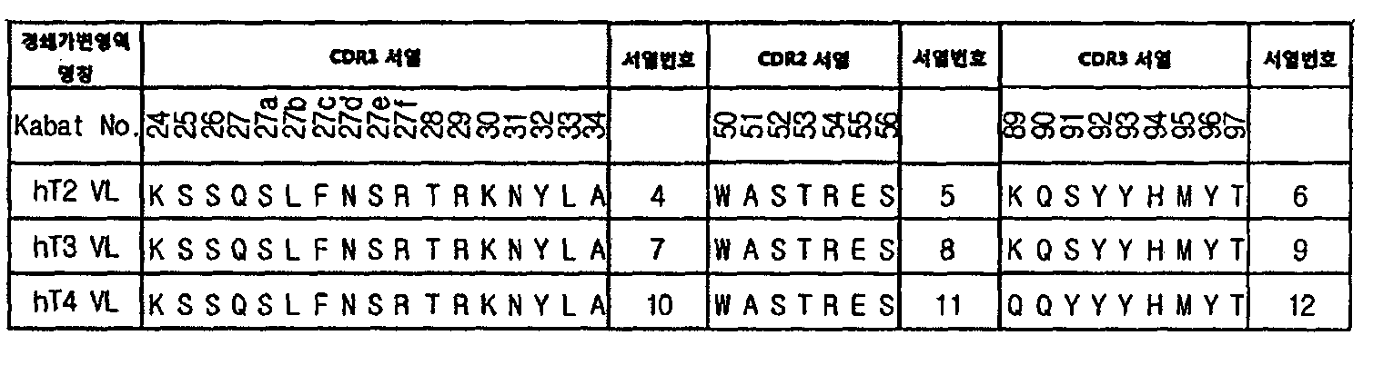
본 발명의 일 양상에서, 세포질 침투능을 가지는 경쇄 가변영역은 서열번호 4, 7 및 10으로 이루어진 군으로부터 선택된 아미노산 서열로 이루어진 CDR1 또는 이와 상동성이 90% 이상인 서열; 및 In one aspect of the present invention, the light chain variable region having cytoplasmic penetration ability is CDR1 consisting of an amino acid sequence selected from the group consisting of SEQ ID NO: 4, 7 and 10 or a sequence having at least 90% homology thereto; And
서열번호 6, 9, 및 12로 이루어진 군으로부터 선택된 아미노산 서열로 이루어진 CDR3 또는 이와 상동성이 90% 이상인 서열을 포함할 수 있다. CDR3 consisting of an amino acid sequence selected from the group consisting of SEQ ID NOs: 6, 9, and 12 or a sequence having at least 90% homology thereto.
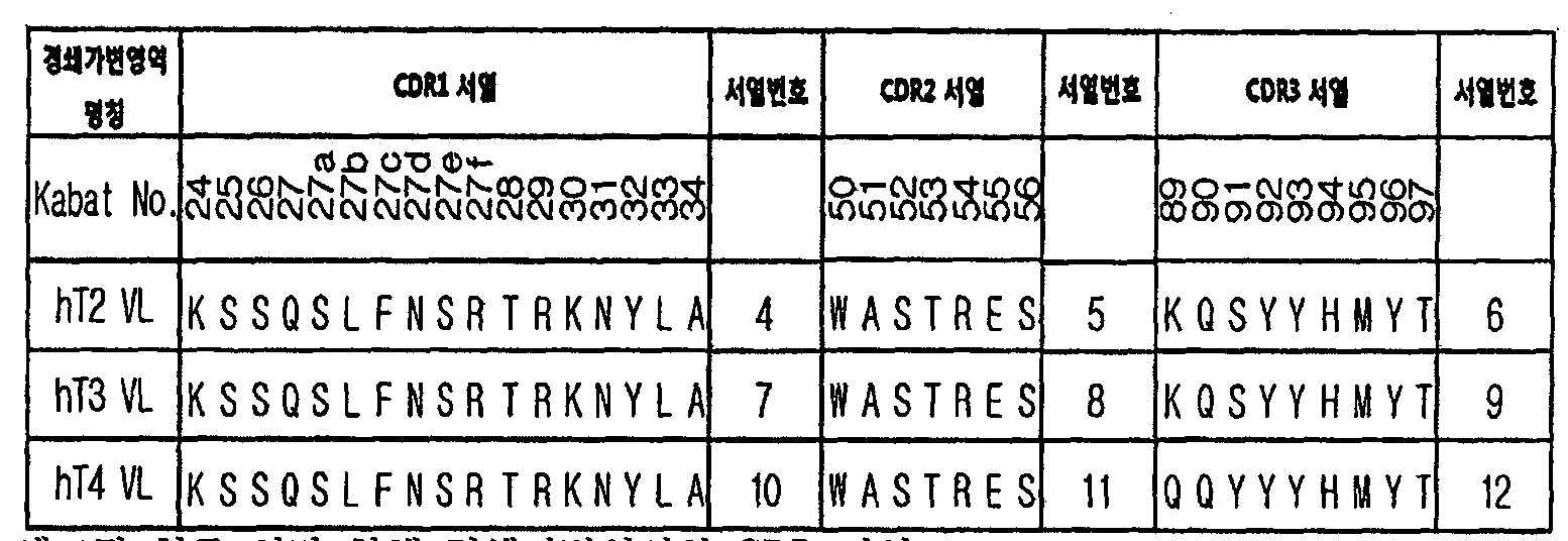
상기 CDR1, CDR2, 및 CDR3의 서열정보는 하기와 같다. Sequence information of the CDR1, CDR2, and CDR3 is as follows.
[규칙 제91조에 의한 정정 11.08.2015] 
Figure WO-DOC-42
[Correction under Rule 91 11.08.2015]
Figure WO-DOC-42
상기 경쇄가변영역은 더욱 바람직하게는 서열번호 5, 8, 및 11로 이루어진 군으로부터 선택된 아미노산 서열로 이루어진 CDR2 또는 이와 상동성이 90% 이상인 서열을 더 포함할 수 있다.More preferably, the light chain variable region may further include a CDR2 consisting of an amino acid sequence selected from the group consisting of SEQ ID NOs: 5, 8, and 11 or a sequence having at least 90% homology thereto.
본 발명의 일 구체예에서 상기 경쇄가변영역은 서열번호 4의 CDR1, 서열번호 5의 CDR2, 및 서열번호 6의 CDR3를 포함한 것일 수 있다.In one embodiment of the present invention, the light chain variable region may include CDR1 of SEQ ID NO: 4, CDR2 of SEQ ID NO: 5, and CDR3 of SEQ ID NO: 6.
또한, 본 발명의 다른 구체예에서 상기 경쇄 가변영역은 서열번호 7의 CDR1, 서열번호 8의 CDR2, 및 서열번호 9의 CDR3를 포함한 것일 수 있다.In another embodiment of the present invention, the light chain variable region may include CDR1 of SEQ ID NO. 7, CDR2 of SEQ ID NO. 8, and CDR3 of SEQ ID NO.
또한, 본 발명의 다른 양상에서 상기 경쇄 가변영역은 서열번호 10의 CDR1, 서열번호 11의 CDR2, 및 서열번호 12의 CDR3를 포함한 것일 수 있다.In another aspect of the present invention, the light chain variable region may include CDR1 of SEQ ID NO: 10, CDR2 of SEQ ID NO: 11, and CDR3 of SEQ ID NO: 12.
본 발명의 일 양상에서, 상기 경쇄가변영역은 경쇄 가변 영역의 N 말단으로부터 2번째 및 4번째 아미노산이 각각 류신 (leucine, L) 및 메티오닌 (Methionine, M)으로 치환된 것일 수 있다.In one aspect of the present invention, the light chain variable region may be one in which the second and fourth amino acids from the N terminus of the light chain variable region are substituted with leucine (L) and methionine (M), respectively.
이는 FR(framework)에 위치하고 있는 CDR 구조결정부위(Vernier zone)에 포함되는 잔기 중 세포질 침투능이 보존된 CDR 구조를 얻기 위해 중요한 2번째, 4번째 잔기를 치환시킨 것이다.This is to replace the important second and fourth residues in order to obtain the CDR structure of the cytoplasmic permeability among the residues included in the CDR (Vernier zone) located in the FR (framework).
또한, 본 발명의 일 양상에서, 상기 경쇄가변영역은 경쇄 가변 영역의 N 말단으로부터 9, 10, 13, 17, 19, 21, 22, 42, 45, 58, 60, 79, 및 85번째 아미노산이 각각 In addition, in one aspect of the present invention, the light chain variable region includes amino acids 9, 10, 13, 17, 19, 21, 22, 42, 45, 58, 60, 79, and 85 from the N terminus of the light chain variable region. each
세린 (serine, S), 세린 (serine, S), 알라닌 (Alanine, A), 발린 (Valine, V), 아스파트 산 (aspartic acid, D), 발린 (Valine, V), 이소류신 (Isoleucine, I), 트리오닌 (Threonine, T), 라이신 (Lysine, K), 라이신 (Lysine, K), 발린 (Valine, V), 세린 (serine, S), 글루타민 (Glutamine, Q) 및 트리오닌 (Threonine, T)으로 치환된 것일 수 있다.Serine (S), Serine (S), Alanine (A), Valine (V), Aspartic acid (D), Valine (V), Isoleucine (Isoleucine, I ), Trionine (T), lysine (Lysine, K), lysine (Lysine, K), valine (V), serine (S), glutamine (Q) and trionine (Threonine, It may be substituted with T).
이는 FDA 승인 후 시장에 판매되고 있는 인간화 치료용 항체 중 안정성이 매우 높으며 VH3소그룹의 중쇄가변영역과 Vκ1소그룹의 경쇄가변영역으로 이루어진 Trastuzumab(Herceptin)의 경쇄가변영역과의 서열 분석을 통해 FR(Framework) 에 위치하고 있는 총 14개의 잔기에 차이가 있음을 확인하여 이를 치환한 것이다. It is very stable among humanized therapeutic antibodies on the market after FDA approval, and FR (Framework) is analyzed through sequence analysis of the light chain variable region of Trastuzumab (Herceptin) consisting of the heavy chain variable region of VH3 subgroup and the light chain variable region of Vκ1 subgroup. A total of 14 residues located at) were found to have been replaced.
또한, 본 발명의 다른 양상에서, 상기 경쇄 가변 영역은 경쇄 가변 영역의 N 말단으로부터 89 번째 및 91번째 아미노산이 각각 글루타민 (Glutamine, Q) 및 타이로신 (Tyrosine, Y)으로 치환된 것일 수 있다.In another aspect of the present invention, the light chain variable region may be one in which the 89th and 91th amino acids are substituted with glutamine (Q) and tyrosine (Yyrosine, Y) from the N terminus of the light chain variable region, respectively.
이는 인간 항체 가변영역 간의 인터페이스(VH-VL interface) 분석을 통해 기존 세포질 침투 경쇄 가변 영역의 마우스 유래 CDR3에 위치한 2개의 잔기가 차이가 있음을 확인하여 이를 치환한 것이다. This was confirmed by substituting the two residues located in the mouse-derived CDR3 of the existing cytoplasmic penetration light chain variable region through analysis of the interface between the human antibody variable region (VH-VL interface).
본 발명의 바람직한 양상에서, 상기 경쇄 가변영역은 서열번호 1, 2, 및 3으로 이루어진 군으로부터 선택된 아미노산으로 이루어진 것일 수 있다.In a preferred aspect of the present invention, the light chain variable region may be composed of amino acids selected from the group consisting of SEQ ID NO: 1, 2, and 3.
상기 서열번호의 서열정보는 하기와 같다. Sequence information of the sequence number is as follows.
Figure PCTKR2015007626-appb-I000002
세포질 침투 인간화 경쇄가변영역(VL) 단일도메인 명칭 및 서열Cytoplasmic Penetration Humanized Light Chain Variable Region (VL) Monodomain Name and Sequence
단, 본 명세서에서 제공하는 서열번호에 명시된 모든 잔기 번호는 Kabat 번호를 사용하였다 (Kabat EA et al., 1991).However, all residue numbers specified in SEQ ID NOs provided herein were used Kabat number (Kabat EA et al., 1991).
본 발명의 일 양상에서, 상기 항체가 막을 투과하여, 세포질에 위치하는 세포는 살아있는 동물 세포일 수 있다. 즉 상기 항체는 상기 항체는 살아있는 동물 세포에 능동적으로 침투하는 것일 수 있다. In one aspect of the invention, the antibody penetrates the membrane, so that the cell located in the cytoplasm may be a live animal cell. That is, the antibody may be one in which the antibody actively penetrates live animal cells.
또한, 본 발명의 일 양상에서, 상기 항체는 세포질뿐 만 아니라 세포질 내에 존재하는 다양한 세포소기관 및 세포내 존재하는 분자들을 표적할 수 있으며, 그 종류를 제한하지 않으나, 예를 들면 세포질, 핵, 미토콘드리아, 소포체, 세포내 고분자 (organelle macromolecule)를 표적할 수 있다.In addition, in one aspect of the present invention, the antibody may target not only the cytoplasm but also various organelles and molecules present in the cell, but not limited to, for example, cytoplasm, nucleus, mitochondria , Endoplasmic reticulum, and intracellular macromolecules.
또한, 본 발명의 일 양상에서, 상기 세포내 고분자는 단백질, 지질, DNA 또는 RNA일 수 있다. 더욱 구체적으로, 상기 단백질은 세포 성장, 세포증식, 세포주기, DNA 수리, DNA 보전, 전사, 복제, 번역 또는 세포내 이동의 제어와 관련된 것 일 수 있으며, 상기 단백질은 인산기, 카르복실산기, 메틸기, 설페이트기, 지질, 수산기, 또는 아미드기로 변형 또는 활성화되거나, 돌연변이된 것 일 수 있다.In addition, in one aspect of the present invention, the intracellular polymer may be a protein, lipid, DNA or RNA. More specifically, the protein may be related to control of cell growth, cell proliferation, cell cycle, DNA repair, DNA preservation, transcription, replication, translation or intracellular migration, wherein the protein is a phosphate group, a carboxylic acid group, a methyl group And may be modified, activated, or mutated by sulfate, lipid, hydroxyl, or amide groups.
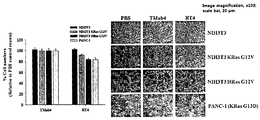
본 발명의 가장 바람직한 구체예에서, 상기 항체는 세포질 내 활성화된 RAS를 표적하여 특이적으로 결합하는 것일 수 있다. 상기 활성화된 RAS는 GTP가 결합된 종양관련인자로서, 상기 RAS는 돌연변이 형태일 수 있다. 상기 RAS의 돌연변이는 질환과 관계되는 다양한 형태로 그 종류에 제한이 없으나, 예를 들면, KRas, HRas, NRas 의 12번 Glycine, 13번 Glycine, 61번 glutamine 잔기들의 치환된 돌연변이들 일 수 있다. In the most preferred embodiment of the invention, the antibody may be to specifically bind to target activated RAS in the cytoplasm. The activated RAS is a tumor-associated factor to which GTP is bound, and the RAS may be in a mutant form. The RAS mutations are various forms related to the disease, but there is no limitation in the type thereof. For example, the RAS mutations may be substituted mutations of Glycine No. 12, Glycine No. 13, glutamine No. 61 of KRas, HRas, and NRas.
본 발명의 일 양상에서, 상기 세포질 내 활성화된 RAS와의 결합력은 항체의 중쇄가변영역 (VH)에 의한 것일 수 있다. In one aspect of the present invention, the binding force to the activated RAS in the cytoplasm may be due to the heavy chain variable region (VH) of the antibody.
본 발명의 일 양상에서, 상기 중쇄가변영역은 In one aspect of the invention, the heavy chain variable region is
서열번호 14의 CDR1 또는 이와 상동성이 90% 이상인 아미노산 서열;The CDR1 of SEQ ID NO: 14 or an amino acid sequence having at least 90% homology with it;
서열번호 15의 CDR2 또는 이와 상동성이 90% 이상인 아미노산서열; 및CDR2 of SEQ ID NO: 15 or an amino acid sequence having a homology of 90% or more; And
서열번호 16의 CDR3 또는 이와 상동성이 90% 이상인 아미노산 서열;을 포함할 수 있다. CDR3 of SEQ ID NO: 16 or an amino acid sequence having 90% or more homology thereto;
상기 서열번호의 서열정보는 하기와 같다. Sequence information of the sequence number is as follows.
Figure PCTKR2015007626-appb-I000003
본 발명의 더욱 바람직한 구체예에서, 중쇄가변영역은 서열번호 13의 아미노산으로 이루어질 수 있다.In a more preferred embodiment of the invention, the heavy chain variable region may consist of the amino acid of SEQ ID NO: 13.
상기 서열번호의 서열정보는 하기와 같다.Sequence information of the sequence number is as follows.
Figure PCTKR2015007626-appb-I000004
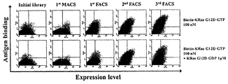
상기 RAS에 특이적으로 결합 및 저해하는 중쇄가변영역은 하기와 같은 방법에 의하여 스크리닝 하였다. The heavy chain variable region that specifically binds to and inhibits the RAS was screened by the following method.
본 발명의 일 구체예에서는 구축되어진 인간 중쇄가변영역(VH)과 중쇄불변영역(CH1)이 융합되어 있는 상태로 CDR1, CDR2, CDR3 지역에 총 18개 잔기에 대해서 인위적인 돌연변이가 유도된 라이브러리를 이용하여 선별하였다.In one embodiment of the present invention, a library in which artificial mutations are induced for a total of 18 residues in CDR1, CDR2, and CDR3 regions while the constructed human heavy chain variable region (VH) and heavy chain constant region (CH1) are fused. By selection.
또한 본 발명의 일 구체예에서는 상기 인간 중쇄가변영역(VH)과 중쇄불변영역(CH1)이 융합되어 있는 라이브러리를 이용하여 세포질 침투 인간화 경쇄가변영역(VL)과 결합된 상태에서도 활성화된(GTP가 결합된) RAS에 대해서 특이적으로 결합이 가능한 중쇄가변영역을 선별하였다. In addition, in one embodiment of the present invention using a library in which the human heavy chain variable region (VH) and the heavy chain constant region (CH1) is fused to the cytoplasmic penetration humanized light chain variable region (VL) is activated even in the state coupled to (GTP is The heavy chain variable region capable of binding specifically to RAS) was selected.
본 발명의 일 구체예에서는 표적분자로서 활성화된(GTP가 결합된) RAS 돌연변이체인 KRas G12D을 사용하였다. 본 발명의 일 구체예에 있어서 발암관련 RAS 돌연변이는 주로 12번, 13번, 61번 잔기에서 발생하며, 12번, 13번 잔기는 RAS 단백질의 P-loop에 위치하고 있으며, RAS 단백질과 결합해 있는 GTP를 가수분해하여 비활성화된 형태로 단백질 구조 변화를 유도하는 GAP(GTPase-activating protein)의 결합에 영향을 준다. 또한 61번 잔기는 GAP의 가수분해 활성 부위와 결합하여 GTP가수분해를 막는 역할을 하므로 다양한 발암관련 RAS 돌연변이는 RAS G12D 돌연변이와 신호전달에 관련된 지역(Switch I, Switch II)이 같으므로 KRas G12D 돌연변이에 한정되지 아니한다.In one embodiment of the present invention, KRas G12D, which is an activated (GTP coupled) RAS mutant, was used as a target molecule. In one embodiment of the present invention, carcinogenesis-related RAS mutations occur mainly at residues 12, 13 and 61, and residues 12 and 13 are located in the P-loop of the RAS protein and are bound to the RAS protein. Hydrolysis of GTP affects the binding of GAP (GTPase-activating protein), which induces protein structural changes in an inactive form. In addition, residue 61 binds to the hydrolytic activity of GAP and prevents GTP hydrolysis. Therefore, various carcinogenic RAS mutations have the same region as the RAS G12D mutation and signal transduction (Switch I, Switch II). It is not limited to.
또한, 상기 일 구체예에 있어서 NRas, HRas는 KRas와 G도메인이라고 불리는 촉매도메인(catalytic domain) 1번부터 165번 잔기까지의 유사도가 85% 이상이며, 이중 하위 신호물질과 결합하는 부위 Switch I(32번 내지 38번), Switch II(59번 내지 67번) 도메인은 100% 일치한다. 다만, 165번부터 189번까지의 C-말단 초가변형 부위는 15 %의 유사도를 지니고 있으나 구조적으로 하위 신호전달에 영향을 주지 않으므로 표적분자로 사용한 활성화된 KRas G12D에 한정되지 아니한다.In addition, in one embodiment, NRas and HRas have 85% or more similarity between residues of the catalytic domain 1 to 165 called KRas and G domain, and the site Switch I (which binds to the lower signaling material) 32-38), Switch II (59-67) domains are 100% identical. However, the C-terminal hypervariable regions 165 to 189 have a similarity of 15% but are not limited to activated KRas G12D used as target molecules because they do not structurally affect downstream signaling.
본 발명의 일 구체예에 있어서 효모세포표면발현 시스템을 이용하여 중쇄가변영역(VH)과 중쇄불변영역(CH1)이 발현된 상태에서 활성화된(GTP가 결합된) RAS에 대해서 초기 선별 이후 세포질 침투 경쇄가변영역(VL)과 경쇄불변영역(CL)을 포함하는 경쇄를 발현 및 분비하는 효모와 효모접합을 통해 이 결합된 Fab형태로 선별하였다. In one embodiment of the present invention using the yeast cell surface expression system cytoplasmic infiltration after the initial screening for RAS activated (GTP coupled) in the state in which the heavy chain variable region (VH) and heavy chain constant region (CH1) is expressed A light chain comprising a light chain variable region (VL) and a light chain constant region (CL) was selected in the form of the bound Fab through yeast and yeast conjugation expressing and secreting a light chain.
또한, 본 발명의 일 양상은 완전한 이뮤노글로불린(immunoglobulin) 형태의 항체가 세포막을 투과하여 세포질에 위치하는 것을 유도하는 경쇄가변영역 (VL)을 제공한다.In addition, one aspect of the present invention provides a light chain variable region (VL) that induces a complete immunoglobulin form of antibody to penetrate the cell membrane and located in the cytoplasm.
본 발명의 일 구체예에서 상기 경쇄가변영역은 서열번호 4, 7 및 10으로 이루어진 군으로부터 선택된 아미노산 서열로 이루어진 CDR1 또는 이와 상동성이 90% 이상인 서열; 및 In one embodiment of the present invention, the light chain variable region is CDR1 consisting of an amino acid sequence selected from the group consisting of SEQ ID NOs: 4, 7, and 10 or a sequence having a homology of 90% or more; And
서열번호 6, 9, 및 12로 이루어진 군으로부터 선택된 아미노산 서열로 이루어진 CDR3 또는 이와 상동성이 90% 이상인 서열을 포함하는 것일 수 있다.CDR3 consisting of an amino acid sequence selected from the group consisting of SEQ ID NOs: 6, 9, and 12, or may include a sequence having at least 90% homology thereto.
또한, 일 구체예에서, 상기 경쇄가변영역 (VL)은 경쇄 가변 영역의 N 말단으로부터 2번째 및 4번째 아미노산이 각각 류신 (leucine, L) 및 메티오닌 (Methionine, M)으로 치환된 것 일 수 있다. In addition, in one embodiment, the light chain variable region (VL) may be a second and fourth amino acid is substituted with leucine (L) and methionine (Methionine, M) from the N terminal of the light chain variable region, respectively. .
또한, 본 발명의 다른 구체예에서 상기 경쇄가변영역은 경쇄 가변 영역의 N 말단으로부터 9, 10, 13, 17, 19, 21, 22, 42, 45, 58, 60, 79, 및 85번째 아미노산이 각각 In another embodiment of the present invention, the light chain variable region includes amino acids 9, 10, 13, 17, 19, 21, 22, 42, 45, 58, 60, 79, and 85 from the N terminus of the light chain variable region. each
세린 (serine, S), 세린 (serine, S), 알라닌 (Alanine, A), 발린 (Valine, V), 아스파트 산 (aspartic acid, D), 발린 (Valine, V), 이소류신 (Isoleucine, I), 트리오닌 (Threonine, T), 라이신 (Lysine, K), 라이신 (Lysine, K), 발린 (Valine, V), 세린 (serine, S), 글루타민 (Glutamine, Q) 및 트리오닌 (Threonine, T)으로 치환된 것일 수 있다.Serine (S), Serine (S), Alanine (A), Valine (V), Aspartic acid (D), Valine (V), Isoleucine (Isoleucine, I ), Trionine (T), lysine (Lysine, K), lysine (Lysine, K), valine (V), serine (S), glutamine (Q) and trionine (Threonine, It may be substituted with T).
본 발명의 다른 구체예에서 상기 경쇄가변영역은 경쇄 가변 영역의 N 말단으로부터 89 번째 및 91번째 아미노산이 각각 글루타민 (Glutamine, Q) 및 타이로신 (Tyrosine, Y)으로 치환된 것 일 수 있다.In another embodiment of the present invention, the light chain variable region may be one in which the 89th and 91th amino acids are substituted with glutamine (Glutamine, Q) and tyrosine (Y) from the N terminus of the light chain variable region, respectively.
또한, 본 발명의 바람직한 구체예에서, 상기 경쇄 가변영역은 서열번호 1, 2, 및 3으로 이루어진 군으로부터 선택된 아미노산 서열로 이루어진 것일 수 있다. In addition, in a preferred embodiment of the present invention, the light chain variable region may be composed of an amino acid sequence selected from the group consisting of SEQ ID NO: 1, 2, and 3.
본 발명에 따른 경쇄 가변영역의 세포 침투능은 세포 내재화(endocytosis)를 통해 침투한 후 엔도좀 탈출에 의한 것 일 수 있다.The cell penetration capacity of the light chain variable region according to the present invention may be due to endosomal escape after infiltrating through endocytosis.
또한, 본 발명의 일 양상은 상기 경쇄가변영역을 포함하는 항체를 제공한다. In addition, an aspect of the present invention provides an antibody comprising the light chain variable region.
본 발명의 구체예에서, 상기 항체는 세포막을 투과하여 세포질에 위치하는 것일 수 있으며, 상기 항체는 키메릭, 인간, 또는 인간화된 항체일 수 있다. 상기 항체는 IgG, IgM, IgA, IgD 및 IgE로 이루어진 군으로부터 선택된 것일 수 있다. 또한, 상기 항체는 세포질, 핵, 미토콘드리아, 소포체, 세포내 고분자 (organelle macromolecule)를 표적하는 것일 수 있으며, 상기 세포내 고분자는 단백질, 지질, DNA 또는 RNA일 수 있다. 상기 단백질은 세포 성장, 세포증식, 세포주기, DNA 수리, DNA 보전, 전사, 복제, 번역 또는 세포내 이동의 제어와 관련된 것 일 수 있다. 본 발명의 바람직한 구체예에서, 상기 항체는 세포질 내 활성화된 RAS에 특이적으로 결합하는 것일 수 있으며, 상기 세포질 내의 활성화된 RAS에 특이적으로 결합하는 중쇄 가변영역 (VH)에 의한 것일 수 있다. 상기 활성화된 RAS는 돌연변이된 형태일 수 있다.In an embodiment of the present invention, the antibody may be located in the cytoplasm through the cell membrane, and the antibody may be a chimeric, human, or humanized antibody. The antibody may be selected from the group consisting of IgG, IgM, IgA, IgD and IgE. In addition, the antibody may be to target the cytoplasm, nucleus, mitochondria, endoplasmic reticulum, intracellular macromolecule (organelle macromolecule), the intracellular polymer may be protein, lipid, DNA or RNA. The protein may be related to control of cell growth, cell proliferation, cell cycle, DNA repair, DNA preservation, transcription, replication, translation or intracellular migration. In a preferred embodiment of the present invention, the antibody may be specifically binding to activated RAS in the cytoplasm, it may be by a heavy chain variable region (VH) that specifically binds to the activated RAS in the cytoplasm. The activated RAS may be in mutated form.
또한, 본 발명의 구체예에 따른 중쇄가변영역은 In addition, the heavy chain variable region according to an embodiment of the present invention
서열번호 14의 CDR1 또는 이와 상동성이 90% 이상인 아미노산 서열;The CDR1 of SEQ ID NO: 14 or an amino acid sequence having at least 90% homology with it;
서열번호 15의 CDR2 또는 이와 상동성이 90% 이상인 아미노산서열; 및CDR2 of SEQ ID NO: 15 or an amino acid sequence having a homology of 90% or more; And
서열번호 16의 CDR3 또는 이와 상동성이 90% 이상인 아미노산 서열;을 포함할 수 있다. CDR3 of SEQ ID NO: 16 or an amino acid sequence having 90% or more homology thereto;
본 발명의 더욱 바람직한 구체예에서, 중쇄가변영역은 서열번호 13의 아미노산으로 이루어질 수 있다.In a more preferred embodiment of the invention, the heavy chain variable region may consist of the amino acid of SEQ ID NO: 13.
또한, 본 발명의 일 양상은 상기 항체에 융합된, 펩타이드, 단백질, 소분자 약물, 나노입자 및 리포좀으로 이루어진 군으로부터 선택된 생체활성분자를 제공한다. In addition, an aspect of the present invention provides a bioactive molecule selected from the group consisting of peptides, proteins, small molecule drugs, nanoparticles and liposomes, fused to the antibody.
상기 단백질은 단백질은 항체, 항체의 단편, 면역 글로불린, 펩타이드, 효소, 성장인자 (growth factor), 사이토카인 (cytokine), 전사인자, 독소, 항원성 펩티드, 호르몬, 운반 단백질, 운동 기능 단백질, 수용체, 신호(signaling) 단백질, 저장 단백질, 막 단백질, 막횡단(transmembrane) 단백질, 내부(internal) 단백질, 외부(external) 단백질, 분비 단백질, 바이러스 단백질, 당 단백질, 절단된 단백질, 단백질 복합체, 또는 화학적으로 개질된 단백질 등일 수 있다. The protein may be an antibody, a fragment of an antibody, an immunoglobulin, a peptide, an enzyme, a growth factor, a cytokine, a transcription factor, a toxin, an antigenic peptide, a hormone, a carrier protein, a motor function protein, a receptor , Signaling proteins, storage proteins, membrane proteins, transmembrane proteins, internal proteins, external proteins, secreted proteins, viral proteins, glycoproteins, truncated proteins, protein complexes, or chemical Modified proteins, and the like.
본 발명의 구체적인 실시예에서는 세포질 침투를 통하여 활성화된(GTP와 결합하는) RAS를 특이적 결합 및 저해하는 완전 이뮤노글로불린 형태 항체의 경쇄가변영역의 N-말단에 RGD4C 펩타이드를 융합한 형태를 제공한다. 상기 본 발명의 일 구체예에 있어서 경쇄가변영역 N-말단과 RGD4C 펩타이드는 (G4S)1 연결자(linker)로 융합되는 것이 바람직하지만 이에 제한 되지 않는다.In a specific embodiment of the present invention provides a form in which the RGD4C peptide is fused to the N-terminus of the light chain variable region of a complete immunoglobulin-type antibody that specifically binds to and inhibits RAS activated (which binds to GTP) through cellular infiltration. do. In one embodiment of the present invention, the light chain variable region N-terminus and the RGD4C peptide are preferably fused to (G 4 S) 1 linker, but are not limited thereto.
본 발명에 있어서 소분자 약물은 약 1000 달톤 미만의 분자량을 지니며 질병의 치료제로서 활성도를 지니는 유기 화합물, 무기 화합물 또는 유기금속 화합물을 나타내는 것으로 본원에서 광범위하게 사용된다. 본원에서 소분자 약물은 올리고펩티드 및 약 1000 달톤 미만의 분자량을 지니는 그 밖의 바이오분자(biomolecule)를 포함한다.Small molecule drugs in the present invention are used broadly herein to denote organic compounds, inorganic compounds or organometallic compounds having a molecular weight of less than about 1000 Daltons and having activity as therapeutic agents for the disease. Small molecule drugs herein include oligopeptides and other biomolecules having a molecular weight of less than about 1000 Daltons.
본 발명에 있어서, 나노입자(nanoparticle)는 직경 1 내지 1000 nm 크기를 갖는 물질들로 이루어진 입자를 의미하며, 상기 나노 입자는 금속 나노 입자, 금속 나노 입자 코어 및 상기 코어를 둘러싸는 금속 쉘로 구성되는 금속/금속 코어쉘 복합체, 금속 나노 입자 코어 및 상기 코어를 둘러싸는 비금속 쉘로 구성되는 금속/비금속 코어쉘 또는 비금속 나노 입자 코어 및 상기 코어를 둘러싸는 금속 쉘로 구성되는 비금속/금속 코어쉘 복합체일 수 있다. 일 구체예에 따르면, 상기 금속은 금, 은, 구리, 알루미늄, 니켈, 팔라듐, 백금, 자성철 및 그의 산화물로부터 선택되는 것일 수 있으나, 이에 한정하지는 않으며, 상기 비금속은 실리카, 폴리스티렌, 라텍스 및 아크릴레이트 계열의 물질로부터 선택되는 것일 수 있으나, 이에 한정하지는 않는다.In the present invention, the nanoparticle (nanoparticle) means a particle made of a material having a diameter of 1 to 1000 nm, the nanoparticle is composed of a metal nanoparticle, a metal nanoparticle core and a metal shell surrounding the core It may be a metal / nonmetal coreshell composed of a metal / metal coreshell composite, a metal nanoparticle core and a nonmetal shell surrounding the core, or a nonmetal / metal coreshell composite composed of a nonmetal nanoparticle core and a metal shell surrounding the core. . According to one embodiment, the metal may be selected from gold, silver, copper, aluminum, nickel, palladium, platinum, magnetic iron and oxides thereof, but is not limited thereto, and the nonmetal may be silica, polystyrene, latex, and acrylic. It may be selected from the rate-based material, but is not limited thereto.
본 발명에 있어서, 리포좀은 자기 스스로 회합할 수 있는, 수성 내부 구획을 둘러싸는 하나 이상의 지질 이중층 막으로 구성된다. 리포좀은 막 타입 및 그 크기에 의하여 특정 수 있다. 작은 유니라멜라 소포(SUV)는 단일막을 갖고 20nm 내지 50nm의 직경을 가질 수 있다. 큰 유니라멜라 소포(LUV)는 50nm이상의 직경을 가질 수 있다. 올리고라멜라 큰 소포 및 멀티라멜라 큰 소포는 다중, 일반적으로 동심원, 막 층을 가지고 직경이 100nm 이상일 수 있다. 여러 비동심원 막을 가진 리포좀, 즉 더 큰 소포 내에서 포함된 여러 작은 소포는 멀티소포성 소포(multivesicular vesicle)라고 한다.In the present invention, liposomes are composed of one or more lipid bilayer membranes surrounding an aqueous internal compartment that can associate themselves. Liposomes can be specified by membrane type and size. Small unilamellar vesicles (SUVs) have a single membrane and may have a diameter of 20 nm to 50 nm. Large uni-lamellar vesicles (LUV) may have a diameter of 50 nm or more. Oligolamella large vesicles and multilamellar large vesicles have multiple, generally concentric, membrane layers and may be 100 nm or more in diameter. Liposomes with multiple asymmetrical membranes, ie several small vesicles contained within larger vesicles, are called multivesicular vesicles.
본 발명에 있어서 “융합”은 기능 또는 구조가 다르거나 같은 두 분자를 일체화하는 것으로, 상기 단백질, 소분자 약물, 나노입자 또는 리포좀에 상기 종양 침투성 펩타이드가 결합할 수 있는 모든 물리, 화학적 또는 생물학적 방법에 의한 융합일 수 있다. 상기 융합은 바람직하게는 연결자 펩타이드에 의할 수 있으며, 이 연결자 펩타이드는 본 발명의 항체 경쇄 가변 영역, 항체, 또는 이의 절편의 다양한 위치에서 상기 생체 활성 분자와의 융합을 중계할 수 있다. In the present invention, "fusion" refers to the integration of two molecules having different or the same function or structure, and any physical, chemical or biological method in which the tumor-penetrating peptide can bind to the protein, small molecule drug, nanoparticle or liposome. By fusion. The fusion may preferably be by a linker peptide, which may relay fusion with the bioactive molecule at various positions in the antibody light chain variable region, antibody, or fragment thereof.
또한, 본 발명은 상기 항체, 또는 이에 융합된 펩타이드, 단백질, 소분자 약물, 나노입자 및 리포좀으로 이루어진 군으로부터 선택된 생체활성분자를 포함하는 암의 예방 또는 치료용 약학적 조성물을 제공한다.The present invention also provides a pharmaceutical composition for preventing or treating cancer, including a bioactive molecule selected from the group consisting of the antibody, or a peptide, a protein, a small molecule drug, a nanoparticle, and a liposome fused thereto.
본 발명에 따른 항체, 또는 이에 융합된 펩타이드, 단백질, 소분자 약물, 나노입자 및 리포좀으로 이루어진 군으로부터 선택된 생체활성분자를 이용하여 인간 항체 중쇄가변영역(VH)이 가지는 항원 고특이성 및 고친화도에 영향주지 않으면서 세포내부로 침투 및 세포질에 잔류하는 특성을 부여할 수 있으며, 이를 통해 현재 소분자 약물을 이용한 질병 치료에 표적물질로 분류되고 있는 세포질 내에 존재하면서 단백질과 단백질 사이에 넓고 평평한 표면을 통해 구조복합성 상호작용을 이루는 종양 및 질환 관련 인자에 대한 치료 및 진단에 높은 효과를 기대할 수 있다. 또한 기존 다양한 종양치료제의 주요 약물 저항성 관련 인자인 KRas 돌연변이를 선택적 저해가 가능하면서 기존 치료제와의 병행치료를 통해 효과적인 항암 활성을 기대할 수 있다.The high specificity and high affinity of the antigen of the human antibody heavy chain variable region (VH) is influenced by using a bioactive molecule selected from the group consisting of an antibody according to the present invention or peptides, proteins, small molecule drugs, nanoparticles, and liposomes fused thereto. It is able to penetrate into the cell and remain in the cytoplasm without being known, and it is present in the cytoplasm that is currently classified as a target for the treatment of diseases using small molecule drugs. A high effect can be expected in the treatment and diagnosis of tumor and disease related factors that make up complex interactions. In addition, while it is possible to selectively inhibit the KRas mutation, which is a major drug resistance-related factor of various existing tumor therapeutic agents, effective anticancer activity can be expected through concurrent treatment with existing therapeutic agents.
상기 암은 편평상피세포암, 소세포폐암, 비소세포폐암, 폐의 선암, 폐의 편평상피암, 복막암, 피부암, 피부 또는 안구내 흑색종, 직장암, 항문부근암, 식도암, 소장암, 내분비선암, 부갑상선암, 부신암, 연조직 육종, 요도암, 만성 또는 급성 백혈병, 림프구 림프종, 간세포암, 위장암, 췌장암, 교아종, 경부암, 난소암, 간암, 방광암, 간종양, 유방암, 결장암, 대장암, 자궁내막 또는 자궁암, 침샘암, 신장암, 간암, 전립선암, 음문암, 갑상선암, 간암 및 두경부암으로 이루어진 군으로부터 선택되는 것일 수 있다.The cancer includes squamous cell carcinoma, small cell lung cancer, non-small cell lung cancer, lung adenocarcinoma, lung squamous cell carcinoma, peritoneal cancer, skin cancer, skin or intraocular melanoma, rectal cancer, anal muscle cancer, esophageal cancer, small intestine cancer, endocrine adenocarcinoma, Parathyroid cancer, adrenal cancer, soft tissue sarcoma, urethral cancer, chronic or acute leukemia, lymphocyte lymphoma, hepatocellular carcinoma, gastrointestinal cancer, pancreatic cancer, glioblastoma, cervical cancer, ovarian cancer, liver cancer, bladder cancer, liver tumor, breast cancer, colon cancer, colon cancer, Endometrial or uterine cancer, salivary gland cancer, kidney cancer, liver cancer, prostate cancer, vulva cancer, thyroid cancer, liver cancer and head and neck cancer.
상기 조성물이 암의 예방 또는 치료용 약학적 조성물로 제조되는 경우, 상기 조성물은 약학적으로 허용되는 담체를 포함할 수 있다. 상기 조성물에 포함되는 약학적으로 허용되는 담체는 제제시에 통상적으로 이용되는 것으로서, 락토오스, 덱스트로오스, 수크로오스, 솔비톨, 만니톨, 전분, 아카시아 고무, 인산 칼슘, 알기네이트, 젤라틴, 규산 칼슘, 미세결정성 셀룰로오스, 폴리비닐피롤리돈, 셀룰로오스, 물, 시럽, 메틸 셀룰로오스, 메틸히드록시벤조에이트, 프로필히드록시벤조에이트, 활석, 스테아르산 마그네슘 및 미네랄 오일 등을 포함하나, 이에 한정되는 것은 아니다. 상기 약학적 조성물은 상기 성분들 이외에 윤활제, 습윤제, 감미제, 향미제, 유화제, 현탁제, 보존제 등을 추가로 포함할 수 있다.When the composition is prepared as a pharmaceutical composition for preventing or treating cancer, the composition may include a pharmaceutically acceptable carrier. Pharmaceutically acceptable carriers included in the composition are conventionally used in the preparation, lactose, dextrose, sucrose, sorbitol, mannitol, starch, acacia rubber, calcium phosphate, alginate, gelatin, calcium silicate, fine Crystalline cellulose, polyvinylpyrrolidone, cellulose, water, syrup, methyl cellulose, methylhydroxybenzoate, propylhydroxybenzoate, talc, magnesium stearate and mineral oil, and the like. The pharmaceutical composition may further include lubricants, wetting agents, sweeteners, flavoring agents, emulsifiers, suspending agents, preservatives, and the like, in addition to the above components.
상기 암의 예방 또는 치료용 약학적 조성물은 경구 또는 비경구로 투여할 수 있다. 비경구 투여인 경우에는 정맥내 주입, 피하 주입, 근육 주입, 복강 주입, 내피 투여, 국소 투여, 비내 투여, 폐내 투여 및 직장내 투여 등으로 투여할 수 있다. 경구 투여시, 단백질 또는 펩타이드는 소화가 되기 때문에 경구용 조성물은 활성 약제를 코팅하거나 위에서의 분해로부터 보호되도록 제형화 되어야 한다. 또한, 상기 조성물은 활성 물질이 표적 세포로 이동할 수 있는 임의의 장치에 의해 투여될 수 있다.The pharmaceutical composition for preventing or treating cancer may be administered orally or parenterally. In the case of parenteral administration, it can be administered by intravenous injection, subcutaneous injection, intramuscular injection, intraperitoneal injection, endothelial administration, topical administration, intranasal administration, pulmonary administration and rectal administration. In oral administration, because proteins or peptides are digested, oral compositions should be formulated to coat the active agent or to protect it from degradation in the stomach. In addition, the composition may be administered by any device in which the active substance may migrate to the target cell.
상기 암의 예방 또는 치료용 약학적 조성물의 적합한 투여량은 제제화 방법, 투여 방식, 환자의 연령, 체중, 성, 병적 상태, 음식, 투여 시간, 투여 경로, 배설 속도 및 반응 감응성과 같은 요인들에 의해 다양하게 처방될 수 있다. 상기 조성물의 바람직한 투여량은 성인 기준으로 0.001-100 ㎎/kg 범위 내이다. 용어 "약학적 유효량"은 암을 예방 또는 치료하는 데, 또는 혈관신생으로 인한 질환의 예방 또는 치료하는 데 충분한 양을 의미한다.Suitable dosages of the pharmaceutical compositions for the prophylaxis or treatment of cancer are dependent on factors such as the formulation method, mode of administration, age, weight, sex, morbidity, food, time of administration, route of administration, rate of excretion and response to response of the patient. It can be prescribed in various ways. Preferred dosages of the compositions are in the range of 0.001-100 mg / kg on an adult basis. The term "pharmaceutically effective amount" means an amount sufficient to prevent or treat cancer or to prevent or treat a disease due to angiogenesis.
상기 조성물은 당해 당업자가 용이하게 실시할 수 있는 방법에 따라, 약학적으로 허용되는 담체 및/또는 부형제를 이용하여 제제화함으로써 단위 용량 형태로 제조되거나 또는 다용량 용기 내에 내입시켜 제조될 수 있다. 이때 제형은 오일 또는 수성 매질중의 용액, 현탁액, 시럽제 또는 유화액 형태이거나 엑스제, 산제, 분말제, 과립제, 정제 또는 캅셀제 형태일 수도 있으며, 분산제 또는 안정화제를 추가적으로 포함할 수 있다. 또한, 상기 조성물은 개별 치료제로 투여하거나 다른 치료제와 병용하여 투여될 수 있고, 종래의 치료제와는 순차적 또는 동시에 투여될 수 있다. 한편, 상기 조성물은 항체 또는 항원 결합 단편을 포함하므로, 면역 리포좀으로 제형화될 수 있다. 항체를 포함하는 리포좀은 당업계에 널리 알려진 방법에 따라 제조될 수 있다. 상기 면역 리포좀은 포스파티딜콜린, 콜레스테롤 및 폴리에틸렌글리콜-유도체화된 포스파티딜에탄올아민을 포함하는 지질 조성물로서 역상 증발법에 의해 제조될 수 있다. 예를 들어, 항체의 Fab' 단편은 디설파이드-교체 반응을 통해 리포좀에 접합될 수 있다. 독소루비신과 같은 화학치료제가 추가로 리포좀 내에 포함될 수 있다.The composition may be prepared in unit dose form or formulated into a multi-dose container by formulating with a pharmaceutically acceptable carrier and / or excipient, according to methods readily available to those skilled in the art. The formulation may be in the form of solutions, suspensions, syrups or emulsions in oils or aqueous media, or in the form of extracts, powders, powders, granules, tablets or capsules, and may further comprise dispersants or stabilizers. In addition, the composition may be administered as a separate therapeutic agent or in combination with other therapeutic agents, and may be administered sequentially or simultaneously with conventional therapeutic agents. On the other hand, since the composition comprises an antibody or antigen-binding fragment, it can be formulated as an immune liposome. Liposomes comprising the antibody can be prepared according to methods well known in the art. The immune liposomes can be prepared by reverse phase evaporation as a lipid composition comprising phosphatidylcholine, cholesterol and polyethyleneglycol-derivatized phosphatidylethanolamine. For example, Fab 'fragments of antibodies can be conjugated to liposomes via disulfide-replacement reactions. Chemotherapeutic agents such as doxorubicin may further be included in the liposomes.
또한, 본 발명은 상기 항체, 또는 이에 융합된 펩타이드, 단백질, 소분자 약물, 나노입자 및 리포좀으로 이루어진 군으로부터 선택된 생체활성분자를 포함하는 암의 진단용 조성물을 제공한다.The present invention also provides a composition for diagnosing cancer comprising a bioactive molecule selected from the group consisting of the antibody, or a peptide, a protein, a small molecule drug, a nanoparticle, and a liposome fused thereto.
본 명세서에서 사용된 용어 "진단"은 병태 생리의 존재 또는 특징을 확인하는 것을 의미한다. 본 발명에서의 진단은 암의 발병 여부 및 경과를 확인하는 것이다.As used herein, the term “diagnosis” refers to identifying the presence or characteristic of pathophysiology. Diagnosis in the present invention is to determine the onset and progress of cancer.
상기 완전한 이뮤노글로불린 형태의 항체 및 이의 절편은 영상을 통하여 암을 진단하기 위하여 분자 영상용 형광체와 결합할 수 있다. The complete immunoglobulin form of the antibody and fragments thereof may be combined with a phosphor for molecular imaging to diagnose cancer through imaging.
상기 분자 영상용 형광체는 형광을 발생시키는 모든 물질을 말하며, 적색이나 근적외선(near-infrared)의 형광을 발광하는 것이 바람직하며, 양자 수득량(quantaum yield)이 높은 형광체가 더욱 바람직하나 이에 한정되지 않는다.The fluorescent material for molecular imaging refers to any material generating fluorescence, and preferably emits red or near-infrared fluorescence, and more preferably, a phosphor having a high quantum yield is more preferred, but is not limited thereto. .
상기 분자 영상용 형광체는 상기 완전한 이뮤노글로불린 형태의 항체 및 이의 절편에 특이적으로 결합하는 종양 침투성 펩타이드와 결합할 수 있는 형광체, 형광 단백질 또는 기타 영상용 물질이 바람직하나 이에 한정되지 않는다.The molecular imaging phosphor is preferably, but not limited to, a phosphor, a fluorescent protein or other imaging material capable of binding to a tumor-penetrating peptide specifically binding to the antibody and fragments thereof of the complete immunoglobulin form.
형광체는 플루오레신(fluorescein), 보디피(BODYPY), 테트라메틸로드아민(Trtramethylrhodamine), 알렉사(Alexa), 시아닌(Cyanine), 알로피코시아닌(allopicocyanine) 또는 이들의 유도체가 바람직하나 이에 한정되지 않는다.The phosphor is preferably fluorescein, BODYPY, tetramethylrhodamine, Alexa, cyanine, allopicocyanine or derivatives thereof, but not limited thereto. Do not.
형광 단백질은 Dronpa 단백질, 형광 발색 유전자(EGFP), 적색 형광 프로테인(red fluorescent protein, DsRFP), 근적외선 형광을 나타내는 시아닌 형광체인 Cy5.5 또는 기타 형광 단백질이 바람직하나 이에 한정되지 않는다.The fluorescent protein is preferably, but not limited to, Dronpa protein, fluorescent color gene (EGFP), red fluorescent protein (DsRFP), Cy5.5 or other fluorescent protein which is a near infrared fluorescence.
기타 영상용 물질은 산화철, 방사성 동위원소 등이 바람직하나 이에 한정되지 않으며, MR, PET과 같은 영상 장비에 응용될 수 있다.Other imaging materials are preferably iron oxide, radioisotopes, etc., but are not limited thereto, and may be applied to imaging equipment such as MR and PET.
또한 본 발명은 상기 경쇄가변영역 또는 이를 포함하는 항체 또는 이의 절편을 코딩하는 폴리뉴클레오티드를 제공한다.In another aspect, the present invention provides a polynucleotide encoding the light chain variable region or an antibody or fragment thereof.
상기 “폴리뉴클레오티드(polynucleotide)”는 단일가닥 또는 이중가닥 형태로 존재하는 디옥시리보뉴클레오티드 또는 리보뉴클레오티드의 중합체이다. RNA 게놈 서열, DNA(gDNA 및 cDNA) 및 이로부터 전사되는 RNA 서열을 포괄하며, 특별하게 다른 언급이 없는 한 천연의 폴리뉴클레오티드의 유사체를 포함한다.The "polynucleotide" is a polymer of deoxyribonucleotides or ribonucleotides present in single- or double-stranded form. It encompasses RNA genomic sequences, DNA (gDNA and cDNA) and RNA sequences transcribed therefrom and includes analogs of natural polynucleotides unless specifically stated otherwise.
상기 폴리뉴클레오티드는 상기 기술한 경쇄 가변영역을 코딩하는 뉴클레오티드 서열뿐만 아니라, 그 서열에 상보적인(complementary) 서열도 포함한다. 상기 상보적인 서열은 완벽하게 상보적인 서열뿐만 아니라, 실질적으로 상보적인 서열도 포함한다. 이는 당업계에 공지된 가혹 조건(stringent conditions) 하에서, 예를 들어, 서열번호 1 내지 3 중 어느 하나의 아미노산 서열을 코딩하는 뉴클레오티드 서열과 혼성화될 수 있는 서열을 의미한다.The polynucleotide includes not only the nucleotide sequence encoding the light chain variable region described above, but also a complementary sequence to the sequence. Such complementary sequences include sequences that are substantially complementary, as well as sequences that are substantially complementary. This means a sequence that can hybridize under stringent conditions known in the art, for example, with a nucleotide sequence encoding the amino acid sequence of any one of SEQ ID NOS: 1-3.
또한 상기 폴리뉴클레오티드는 변형될 수 있다. 상기 변형은 뉴클레오티드의 추가, 결실 또는 비보존적 치환 또는 보존적 치환을 포함한다. 상기 아미노산 서열을 코딩하는 폴리뉴클레오티드는 상기 뉴클레오티드 서열에 대하여 실질적인 동일성을 나타내는 뉴클레오티드 서열도 포함하는 것으로 해석된다. 상기의 실질적인 동일성은, 상기 뉴클레오티드 서열과 임의의 다른 서열을 최대한 대응되도록 얼라인하고, 당업계에서 통상적으로 이용되는 알고리즘을 이용하여 얼라인된 서열을 분석한 경우에, 최소 80%의 상동성, 최소 90%의 상동성 또는 최소 95%의 상동성을 나타내는 서열일 수 있다.The polynucleotide may also be modified. Such modifications include addition, deletion or non-conservative substitutions or conservative substitutions of nucleotides. The polynucleotide encoding the amino acid sequence is to be interpreted to also include a nucleotide sequence showing substantial identity to the nucleotide sequence. The substantial identity is at least 80% homology when the nucleotide sequence is aligned with any other sequence as closely as possible and the aligned sequence is analyzed using algorithms commonly used in the art. A sequence exhibiting at least 90% homology or at least 95% homology.
또한, 본 발명의 일 양상은 항체의 경쇄가변영역을, 살아있는 세포 내부로 침투하여 세포질에 위치하는 특성을 가진 경쇄가변영역으로 치환하는 단계를 포함하는, In addition, an aspect of the present invention includes the step of replacing the light chain variable region of the antibody with a light chain variable region having a characteristic of infiltrating into the living cell and located in the cytoplasm,
살아있는 세포내부로 침투하여 세포질에 위치하는 항체의 제조방법을 제공할 수 있다. It is possible to provide a method for producing an antibody that penetrates into living cells and is located in the cytoplasm.
또한, 본 발명의 일 구체예는 기존의 완전 이뮤노글로불린 형태의 항체의 경쇄가변영역(VL)을 세포질 침투 경쇄가변영역(VL)과 치환하여, 치환된 완전한 이뮤노글로불린 형태의 단일클론항체가 상기 세포질 침투능을 가지는 완전 이뮤노글로불린 형태의 단일클론항체의 세포질 침투와 같은 특성을 갖게 하는 방법을 제공할 수 있다.In addition, one embodiment of the present invention is to replace the light chain variable region (VL) of the existing full immunoglobulin form of the antibody with the cytoplasmic penetration light chain variable region (VL), the monoclonal antibody of the substituted full immunoglobulin form It is possible to provide a method of having the same characteristics as the cellular penetration of a monoclonal antibody in the form of a complete immunoglobulin having the cytoplasmic penetration ability.
본 발명의 구체예에서, 세포질 침투 경쇄가변영역(VL)을 이용한 세포내부로 침투하여 세포질에 분포하는 완전 이뮤노글로불린 형태의 항체 제조방법의 예는 아래와 같다.In an embodiment of the present invention, an example of a method for producing a fully immunoglobulin-type antibody which penetrates into the cell using the cytoplasmic infiltration light chain variable region (VL) and distributes the cytoplasm is as follows.
(1) 상기 인간 경쇄가변영역(VL) 및 인간 경쇄불변영역(CL)을 포함하는 경쇄에서 경쇄가변영역(VL)을 세포질에 침투하는 인간화 경쇄가변영역(VL)으로 치환된 핵산(nucleic acids)을 클로닝한 세포질 침투 경쇄 발현 벡터를 제조하는 단계;(1) a nucleic acid substituted with a humanized light chain variable region (VL) that penetrates the light chain variable region (VL) into the cytoplasm in a light chain including the human light chain variable region (VL) and the human light chain constant region (CL) Preparing a cytoplasmic infiltrating light chain expression vector which has been cloned;
(2) 상기 제조된 경쇄와 상호작용하여 완전한 이뮤노글로불린 형태의 항체 발현을 위한 중쇄가변영역(VH) 및 중쇄불변영역(CH1-hinge-CH2-CH3)이 포함된 중쇄를 코딩하는 핵산(nucleic acides)를 클로닝한 중쇄 발현 벡터를 제조하는 단계;(2) a nucleic acid encoding a heavy chain including a heavy chain variable region (VH) and a heavy chain constant region (CH1-hinge-CH2-CH3) for interacting with the prepared light chain to express the antibody in the form of a complete immunoglobulin preparing a heavy chain expression vector cloned from the acides);
(3) 상기 제조된 경쇄, 중쇄 발현벡터를 단백질 발현용 동물세포에 동시 형질전환하여 세포내부 침투 및 세포질 잔류 인간화 경쇄가변영역(VL)을 포함하는 완전한 이뮤노글로불린 형태의 항체를 발현하는 단계; 및(3) co-transforming the prepared light and heavy chain expression vectors into animal cells for protein expression to express the complete immunoglobulin type antibody including intracellular infiltration and cytoplasmic residual humanized light chain variable region (VL); And
(4) 상기 발현된 세포질 침투능을 가지는 완전한 이뮤노글로불린 형태의 항체를 정제 및 회수하는 단계.(4) purifying and recovering the antibody in the form of a complete immunoglobulin having the expressed cytoplasmic permeability.
상기 방법은 경쇄를 발현하는 벡터와 중쇄를 발현하는 벡터를 발현하여 세포질 침투능을 가지는 완전한 이뮤노글로불린 형태의 항체를 제조할 수 있다. 또한, 세포내부의 특정 단백질을 인지할 수 있는 중쇄가변영역을 포함하는 중쇄를 발현하는 벡터와 함께 형질전환을 통하여, 세포내부로 침투하고 세포질에 분포하여 특정 단백질과 결합할 수 잇는 항체를 발현할 수 있다. 벡터는 경쇄와 중쇄를 하나의 벡터에서 동시에 발현되는 벡터 시스템이거나 또는 각각 별도의 벡터에서 발현시키는 시스템 모두 가능하다. 후자의 경우, 두 벡터는 동시 형질전환(co-transfomation) 및 표적 형질전환(targeted transformation)을 통하여 숙주 세포로 도입될 수 있다.The method can express a vector expressing a light chain and a vector expressing a heavy chain to prepare antibodies in the form of complete immunoglobulins having cytoplasmic penetrating ability. In addition, through transformation with a vector expressing a heavy chain comprising a heavy chain variable region capable of recognizing a specific protein within a cell, the antibody may penetrate into the cell and be distributed in the cytoplasm to express an antibody capable of binding to a specific protein. Can be. The vector can be either a vector system in which the light and heavy chains are simultaneously expressed in one vector or a system in which each is expressed in a separate vector. In the latter case, both vectors can be introduced into the host cell via co-transfomation and targeted transformation.
본 발명에서 사용하는 용어 "벡터(vector)"는 숙주 세포에서 목적 유전자를 발현시키기 위한 수단을 의미한다. 예를 들어, 플라스미드 벡터, 코즈미드 벡터 및 박테리오파아지 벡터, 아데노바이러스 벡터, 레트로바이러스 벡터 및 아데노-연관 바이러스 벡터와 같은 바이러스 벡터를 포함한다. 상기 재조합 벡터로 사용될 수 있는 벡터는 당업계에서 종종 사용되는 플라스미드(예를 들면, pSC101, pGV1106, pACYC177, ColE1, pKT230, pME290, pBR322, pUC8/9, pUC6, pBD9, pHC79, pIJ61, pLAFR1, pHV14, pGEX 시리즈, pET 시리즈 및 pUC19 등), 파지(예를 들면, λgt4λB, λ-Charon, λΔz1 및 M13 등) 또는 바이러스(예를 들면, CMV, SV40 등)를 조작하여 제작될 수 있다.As used herein, the term "vector" means a means for expressing a gene of interest in a host cell. For example, viral vectors such as plasmid vectors, cosmid vectors and bacteriophage vectors, adenovirus vectors, retrovirus vectors, and adeno-associated virus vectors are included. Vectors that can be used as the recombinant vector are plasmids often used in the art (eg, pSC101, pGV1106, pACYC177, ColE1, pKT230, pME290, pBR322, pUC8 / 9, pUC6, pBD9, pHC79, pIJ61, pLAFR1, pHV14). , pGEX series, pET series, pUC19 and the like), phage (e.g., λgt4λB, λ-Charon, λΔz1 and M13, etc.) or viruses (e.g., CMV, SV40, etc.).
상기 재조합 벡터에서 본 발명에서 제공하는 경쇄가변영역, 및 경쇄불변영역(CL), 중쇄가변영역(VH) 및 중쇄불변영역(CH1-hinge-CH2-CH3)은 프로모터에 작동적으로 연결될 수 있다. 용어 "작동적으로 연결된(operatively linked)"은 뉴클레오티드 발현 조절 서열(예를 들면, 프로모터 서열)과 다른 뉴클레오티드 서열 사이의 기능적인 결합을 의미한다. 따라서, 이에 의해 상기 조절 서열은 상기 다른 뉴클레오티드 서열의 전사 및/또는 해독을 조절할 수 있다.In the recombinant vector, the light chain variable region, the light chain constant region (CL), the heavy chain variable region (VH), and the heavy chain constant region (CH1-hinge-CH2-CH3) provided in the present invention may be operably linked to a promoter. The term "operatively linked" means a functional bond between a nucleotide expression control sequence (eg, a promoter sequence) and another nucleotide sequence. Thus, the regulatory sequence can thereby regulate transcription and / or translation of the other nucleotide sequence.
상기 재조합 벡터는, 전형적으로 클로닝을 위한 벡터 또는 발현을 위한 벡터로서 구축될 수 있다. 상기 발현용 벡터는 당업계에서 식물, 동물 또는 미생물에서 외래의 단백질을 발현하는데 사용되는 통상의 것을 사용할 수 있다. 상기 재조합 벡터는 당업계에 공지된 다양한 방법을 통해 구축될 수 있다. The recombinant vector can typically be constructed as a vector for cloning or a vector for expression. The expression vector may be a conventional one used in the art to express foreign proteins in plants, animals or microorganisms. The recombinant vector may be constructed through various methods known in the art.
상기 재조합 벡터는 원핵 세포 또는 진핵 세포를 숙주로 하여 구축될 수 있다. 예를 들어, 사용되는 벡터가 발현 벡터이고, 원핵 세포를 숙주로 하는 경우에는, 전사를 진행시킬 수 있는 강력한 프로모터(예를 들어, pLλ프로모터, trp 프로모터, lac 프로모터, tac 프로모터, T7 프로모터 등), 해독의 개시를 위한 라이보좀 결합 자리 및 전사/해독 종결 서열을 포함하는 것이 일반적이다. 진핵 세포를 숙주로 하는 경우에는, 벡터에 포함되는 진핵 세포에서 작동하는 복제원점은 f1 복제원점, SV40 복제원점, pMB1 복제원점, 아데노 복제원점, AAV 복제원점, CMV 복제원점 및 BBV 복제원점 등을 포함하나, 이에 한정되는 것은 아니다. 또한, 포유동물 세포의 게놈으로부터 유래된 프로모터(예를 들어, 메탈로티오닌 프로모터) 또는 포유동물 바이러스로부터 유래된 프로모터(예를 들어, 아데노바이러스 후기 프로모터, 백시니아 바이러스 7.5K 프로모터, SV40 프로모터, 사이토메갈로바이러스(CMV) 프로모터 및 HSV의 tk 프로모터)가 이용될 수 있으며, 전사 종결 서열로서 폴리아데닐화 서열을 일반적으로 갖는다.The recombinant vector may be constructed using prokaryotic or eukaryotic cells as hosts. For example, when the vector used is an expression vector and the prokaryotic cell is a host, a strong promoter (for example, a pLλ promoter, trp promoter, lac promoter, tac promoter, T7 promoter, etc.) capable of promoting transcription It is common to include ribosome binding sites and transcription / detox termination sequences for initiation of translation. In the case of using a eukaryotic cell as a host, replication origins that operate in eukaryotic cells included in the vector include f1 origin, SV40 origin, pMB1 origin, adeno origin, AAV origin, CMV origin, and BBV origin. Including but not limited to. In addition, promoters derived from the genome of mammalian cells (eg, metallothionine promoters) or promoters derived from mammalian viruses (eg, adenovirus late promoters, vaccinia virus 7.5K promoters, SV40 promoters, Cytomegalovirus (CMV) promoter and tk promoter of HSV) can be used and generally have a polyadenylation sequence as a transcription termination sequence.
본 발명의 또 다른 양상은 상기 재조합 벡터로 형질전환된 숙주세포를 제공할 수 있다.Another aspect of the invention can provide a host cell transformed with the recombinant vector.
숙주 세포는 당업계에 공지된 어떠한 숙주 세포도 이용할 수 있으며, 원핵 세포로는, 예를 들어, E. coli JM109, E. coli BL21, E. coli RR1, E. coli LE392, E. coli B, E. coli X 1776, E. coli W3110, 바실러스 서브틸리스, 바실러스 츄린겐시스와 같은 바실러스 속 균주, 그리고 살모넬라 티피무리움, 세라티아 마르세슨스 및 다양한 슈도모나스 종과 같은 장내균과 균주 등이 있으며, 진핵 세포에 형질 전환시키는 경우에는 숙주 세포로서, 효모(Saccharomyce cerevisiae), 곤충 세포, 식물 세포 및 동물 세포, 예를 들어, SP2/0, CHO(Chinese hamster ovary) K1, CHO DG44, PER.C6, W138, BHK, COS-7, 293, HepG2, Huh7, 3T3, RIN 및 MDCK 세포주 등이 이용될 수 있다.The host cell may use any host cell known in the art, and prokaryotic cells include, for example, E. coli JM109, E. coli BL21, E. coli RR1, E. coli LE392, E. coli B, Strains of the genus Bacillus, such as E. coli X 1776, E. coli W3110, Bacillus subtilis, Bacillus thuringiensis, and enterococci and strains such as Salmonella typhimurium, Serratia marsons and various Pseudomonas species. In the case of transformation into cells, as a host cell, yeast (Saccharomyce cerevisiae), insect cells, plant cells and animal cells, for example, SP2 / 0, Chinese hamster ovary K1, CHO DG44, PER.C6, W138 , BHK, COS-7, 293, HepG2, Huh7, 3T3, RIN and MDCK cell lines and the like can be used.
본 발명의 또 다른 양상은 상기 숙주세포를 배양하는 단계를 포함하는, 세포내부로 침투하여 세포질에 위치하는 완전한 이뮤노글로불린 형태의 항체 제조방법을 제공할 수 있다.Another aspect of the present invention can provide a method for producing a complete immunoglobulin-type antibody in the cytoplasm to penetrate into the cell, comprising culturing the host cell.
상기 재조합 벡터의 숙주 세포 내로의 삽입은, 당업계에 널리 알려진 삽입 방법을 사용할 수 있다. 상기 운반 방법은 예를 들어, 숙주 세포가 원핵 세포인 경우, CaCl2 방법 또는 전기 천공 방법 등을 사용할 수 있고, 숙주 세포가 진핵 세포인 경우에는, 미세 주입법, 칼슘 포스페이트 침전법, 전기 천공법, 리포좀-매개 형질감염법 및 유전자 밤바드먼트 등을 사용할 수 있으나, 이에 한정하지는 않는다. E. coli 등의 미생물을 이용하는 경우 생산성은 동물세포 등에 비하여 높은 편이나 당화(glycosylation) 문제로 인해 인택트(intact)한 Ig 형태의 항체 생산에는 적당하지 않지만, Fab 및 Fv와 같은 항원 결합 단편의 생산에는 사용될 수 있다.Insertion of the recombinant vector into the host cell, can be used insertion methods well known in the art. For example, when the host cell is a prokaryotic cell, a CaCl 2 method or an electroporation method may be used. When the host cell is a eukaryotic cell, a micro-injection method, calcium phosphate precipitation method, electroporation method, Liposome-mediated transfection and gene bombardment may be used, but is not limited thereto. When using microorganisms such as E. coli, productivity is higher than that of animal cells, but is not suitable for the production of intact Ig-type antibodies due to glycosylation problems. However, antigen-binding fragments such as Fab and Fv Can be used for production.
상기 형질 전환된 숙주 세포를 선별하는 방법은 선택 표지에 의해 발현되는 표현형을 이용하여, 당업계에 널리 알려진 방법에 따라 용이하게 실시할 수 있다. 예를 들어, 상기 선택 표지가 특정 항생제 내성 유전자인 경우에는, 상기 항생제가 함유된 배지에서 형질전환체를 배양함으로써 형질전환체를 용이하게 선별할 수 있다.The method of selecting the transformed host cell can be easily carried out according to methods well known in the art using a phenotype expressed by a selection label. For example, when the selection marker is a specific antibiotic resistance gene, the transformant can be easily selected by culturing the transformant in a medium containing the antibiotic.
또한, 본 발명의 일 양상은 상기 살아있는 세포내부로 침투하여 세포질에 분포하는 완전한 이뮤노글로불린 형태의 항체를 이용하여 세포질 침투를 통해 세포질에 위치하고 있는 활성화된(GTP가 결합된) 종양관련인자 RAS에 특이적 결합 및 활성을 저해하는 완전한 이뮤노글로불린 형태의 항체의 제조방법을 제공할 수 있다. In addition, an aspect of the present invention is to the activated (GTP coupled) tumor-associated factor RAS located in the cytoplasm through cytoplasmic infiltration using the antibody of the complete immunoglobulin form that penetrates into the living cell and distributed in the cytoplasm It is possible to provide a method for preparing a complete immunoglobulin form of antibody that inhibits specific binding and activity.
상기 일 양상 구체예에 있어서, 활성화된(GTP가 결합된) RAS 특이적 결합능을 가지고 있는 중쇄가변영역(VH)을 이용한 동물 세포내부로 침투하고 세포질에 분포하여 세포질에 위치하고 있는 활성화된(GTP가 결합된) RAS와 특이적 결합하는 완전 이뮤노글로불린 형태의 항체는 하기와 같은 방법으로 제조될 수 있다.In one embodiment of the above aspect, the activated (GTP conjugated) GRA which is penetrated into the animal cell using the heavy chain variable region (VH) having RAS specific binding ability and distributed in the cytoplasm and located in the cytoplasm Antibodies in the form of fully immunoglobulin that specifically bind to bound RAS can be prepared by the following method.
(1) 활성화된(GTP가 결합된) RAS와 특이적 결합하는 인간 중쇄가변영역(VH)과 중쇄불변영역(CH1-hinge-CH2-CH3)이 포함된 핵산(nucleic acides)를 클로닝한 중쇄 발현 벡터를 제조하는 단계;(1) Heavy chain expression by cloning of nucleic acid containing human heavy chain variable region (VH) and heavy chain constant region (CH1-hinge-CH2-CH3) that specifically bind to activated (GTP-bound) RAS Preparing a vector;
(2) 상기 제조된 중쇄 발현 백터와 세포 침투 경쇄 발현벡터를 단백질 발현용 동물세포에 동시 형질전환하여 살아있는 세포내부로 침투 및 세포질 분포하여 활성화된(GTP가 결합된) RAS를 특이적 인지하는 완전 이뮤노글로불린 형태의 항체를 발현하는 단계; 및(2) The co-transformation of the prepared heavy chain expression vector and the cell infiltration light chain expression vector into animal cells for protein expression infiltrate into living cells and cytoplasmic distribution to completely recognize specific activated RAS (GTP-coupled). Expressing an antibody in an immunoglobulin form; And
(2) 상기 발현된 활성화된(GTP가 결합된) RAS를 특이적 인지하는 세포질 침투능을 가지는 완전한 이뮤노글로불린 형태의 항체를 정제, 회수하는 단계.(2) Purifying and recovering an antibody of a complete immunoglobulin form having cytoplasmic penetrating ability to specifically recognize the expressed activated (GTP bound) RAS.
본 발명에서 제공하는 완전한 이뮤노글로불린(immunoglobulin) 형태의 항체를 살아있는 세포 침투를 통하여 세포질에 위치시키는 방법에 의하는 경우, 특별한 외부 단백질 전달 시스템 없이 항체를 살아있는 세포내부로 침투하여 세포질에 분포시킬 수 있다. According to the method of locating an antibody of the complete immunoglobulin form provided by the present invention in the cytoplasm through living cell infiltration, the antibody can be infiltrated into living cells and distributed in the cytoplasm without a special external protein delivery system. have.
특히, 본 발명에서 안정한 세포질 침투능을 가지는 완전한 이뮤노글로불린 형태의 항체를 구현하기 위해 다양한 인간 중쇄가변영역(VH)과의 상호작용 결합이 용이하면서 세포질 침투를 통해 세포질에 잔류 가능한 특성을 가지는 항체의 경쇄 가변 영역을 제공하고 있으며, 상기 경쇄 가변 영역을 포함하는 완전 이뮤노글로불린 형태의 항체는 세포내부 침투 및 세포질에 분포하며, 세포에 비특이적 세포독성을 보이지 않는다. 상기 항체의 중쇄가변영역(VH)을 활성화된(GTP가 결합된) RAS를 특이적 인지가 가능한 중쇄가변영역(VH)으로 치환을 통해 살아있는 세포내부 세포질에 위치하고 있는 활성화된(GTP가 결합된) RAS를 표적 및 그 활성을 저해할 수 있다.In particular, in the present invention, in order to implement a fully immunoglobulin-type antibody having stable cytoplasmic penetration ability, an antibody having a property of being able to remain in the cytoplasm through cytoplasmic penetration while being easily interacted with various human heavy chain variable regions (VH) It provides a light chain variable region, the antibody of the complete immunoglobulin form comprising the light chain variable region is distributed in the cell and cytoplasm, and does not show nonspecific cytotoxicity to the cell. Activated (GTP-coupled) located in the cytoplasm of the living cell through substitution of the heavy chain variable region (VH) of the antibody to the activated (GTP-coupled) RAS specific heavy chain variable region (VH). RAS can inhibit the target and its activity.
본 발명에 따른 세포질 침투능을 가지는 완전 이뮤노글로불린 형태의 항체를 이용하여 인간 항체 중쇄가변영역(VH)이 가지는 항원 고특이성 및 고친화도에 영향주지 않으면서 살아있는 세포내부로 침투 및 세포질에 잔류하는 특성을 부여할 수 있으며, 이를 통해 현재 소분자 약물을 이용한 질병 치료에 표적물질로 분류되고 있는 세포질 내에 존재하면서 단백질과 단백질 사이에 넓고 평평한 표면을 통해 구조복합성 상호작용을 이루는 종양 및 질환 관련 인자에 대한 치료 및 진단에 높은 효과를 기대할 수 있다. 또한 기존 다양한 종양치료제의 주요 약물 저항성 관련 인자인 KRas 돌연변이를 선택적 저해가 가능하면서 기존 치료제와의 병행치료를 통해 효과적인 항암 활성을 기대할 수 있다.Penetration into the living cells and remaining in the cytoplasm without affecting the antigen specificity and high affinity of the human antibody heavy chain variable region (VH) using the full immunoglobulin type antibody having cytoplasmic penetration ability according to the present invention Treatment of tumors and disease-related factors that exist within the cytoplasm that is currently classified as a target for the treatment of diseases using small molecule drugs, and that form structurally complex interactions between proteins and proteins through broad and flat surfaces. And high effect can be expected in the diagnosis. In addition, while it is possible to selectively inhibit the KRas mutation, which is a major drug resistance-related factor of various existing tumor therapeutic agents, effective anticancer activity can be expected through concurrent treatment with existing therapeutic agents.
도 1은 cytotransmab으로 명명한 세포 침투를 통해서 세포질에 위치하는 완전 이뮤노글로불린 형태의 항체의 개념을 나타낸 모식도이다. 1 is a schematic diagram showing the concept of a fully immunoglobulin-type antibody located in the cytoplasm through cell infiltration named cytotransmab.
도 2a은 마우스 유래 경쇄가변영역 단일도메인인 m3D8 VL로부터, 인간화 항체 중쇄가변영역과 안정한 결합이 이루어지도록 개량한 세포질 침투 인간화 경쇄가변영역 단일도메인 hT3 VL까지의 개량 과정에 이용된 클론을 포함한 서열분석 자료이다.FIG. 2A is a sequence analysis including clones used in the improvement process from m3D8 VL, a mouse-derived light chain variable region monodomain, to cytoplasmic penetration humanized light chain variable region monodomain hT3 VL, which has been improved to achieve stable binding with humanized antibody heavy chain variable region. Data.
도 2b는 m3D8 VL과 인간화 경쇄가변영역 단일도메인 hT0 VL, 이의 돌연변이체 hT2 VL, hT3 VL의 WAM 모델링을 이용한 모델 구조를 구조 중첩(superimposing) 방법을 이용하여 비교한 도이다.Figure 2b is a diagram comparing the model structure using the WAM modeling of m3D8 VL and humanized light chain variable region monodomain hT0 VL, mutant hT2 VL, hT3 VL using a superimposing method.
도 3a는 경쇄가변영역 단일도메인의 세포질 침투능을 공초점 현미경 (confocal microscopy)으로 관찰한 결과이다. Figure 3a is a result of observing the cytoplasmic penetration capacity of the light chain variable region single domain by confocal microscopy (confocal microscopy).
도 3b는 경쇄가변영역 단일도메인의 세포질 침투 메커니즘을 검증하기 위하여, 공초점 현미경으로 관찰한 결과이다. Figure 3b is a result of observation by confocal microscopy to verify the cytoplasmic penetration mechanism of the light chain variable region monodomain.
도 4a는 hT3 VL 의 다양한 인간 항체 중쇄가변영역에 대해서 적용가능 여부를 확인하기 위해 기존의 인간 항체 Adalimumab(Humira)과 인간화 항체 Bevacizumab(Avastin)의 경쇄가변영역(VL)과 아미노산 서열을 분석한 결과이다. Figure 4a is a light chain variable region (VL) and amino acid sequence analysis of the existing human antibody Adalimumab (Humira) and humanized antibody Bevacizumab (Avastin) to confirm the applicability to various human antibody heavy chain variable region of hT3 VL to be.
도 4b는 추가적으로 인간항체 중쇄가변영역과 최적화된 결합을 통해 안정적 cytotransmab 구축을 위한 가변영역 간의 인터페이스 잔기를 분석한 결과이다. Figure 4b is the result of further analysis of the interface residues between the variable region for stable cytotransmab construction through the optimized binding of the human antibody heavy chain variable region.
도 5은 cytotransmab 구축을 위해 세포침투능이 없는 경쇄가변영역을 세포질 침투능을 갖는 인간화 경쇄가변영역을 치환하는 방법에 대한 전반적인 모식도이다. 5 is a general schematic diagram of a method for substituting a humanized light chain variable region having cytoplasmic penetration capacity for a light chain variable region having no cell permeability for cytotransmab construction.
도 6a는 cytotransmab 정제 후, 환원성 또는 비환원성 SDS-PAGE를 통해서 분석한 것이다. Figure 6a is analyzed by reducing or non-reducing SDS-PAGE after cytotransmab purification.
도 6b는 HPLC (high performance liquid chromatography) (The Agilent 1200 Series LC systems and Modules) (Agilent)를 통해서 cytotransmab이 자연적 환경에서 단일체 존재함을 확인하기 위해 크기배제 크로마토그래피 칼럼 (size exclusion chromatography column) (SuperdexTM200 10/300GC) (GE Healthcare)을 실험한 결과이다. FIG. 6B shows a size exclusion chromatography column (Superdex) to confirm that the cytotransmab is monolithic in its natural environment via high performance liquid chromatography (HPLC) (The Agilent 1200 Series LC systems and Modules) (Agilent). TM 200 10 / 300GC) (GE Healthcare).
도 6c는 cytotransmab(TMab4, HuT4, AvaT4)과 IgG 형태의 항체 Bevacizumab(Avastin) Adalimumab(Humira)의 중쇄가변영역에 의한 표적분자에 대한 친화도를 측정하기 위해 ELISA(enzyme linked immunosorbent assay)를 수행한 결과이다. FIG. 6C shows an ELISA (enzyme linked immunosorbent assay) for measuring affinity for a target molecule by a heavy chain variable region of cytotransmab (TMab4, HuT4, AvaT4) and IgG form of antibody Bevacizumab (Avastin) Adalimumab (Humira) The result is.
도 6d는 자가면역 마우스 유래 항체의 CDR을 grafting한 세포침투능을 갖는 인간 경쇄가변영역 (hT4) 치환을 통한 cytotransmab에서의 핵산 가수분해능을 확인하기 위해 아가로스 젤 핵산 가수분해 검증실험을 한 결과이다. Figure 6d is a result of agarose gel nucleic acid hydrolysis verification experiment to confirm the nucleic acid hydrolytic ability in cytotransmab through human light chain variable region (hT4) substitution having a cell penetrating ability grafting the CDRs of autoimmune mouse-derived antibody.
도 7a는 세포질 침투능을 갖는 경쇄가변영역 hT4 VL로 경쇄가변영역이 치환된 cytotransmab들의 세포질 침투능을 검증하기 위하여, 다양한 세포주에서 1~2개의 세포에 초점을 맞추고 공초점 현미경로 관찰한 결과이다.Figure 7a is a light chain variable region hT4 VL having a cytoplasmic penetration ability in order to verify the cytoplasmic penetration ability of the light chain variable region substituted by the cytotransmabs, focusing on one or two cells in various cell lines and observed by confocal microscopy.
도 7b는 상기 도 7a의 공초점 현미경 관찰을 통한 세포질 침투능 검증 실험에서 세포 침투효율을 확인하기 위해 렌즈 배율을 낮추어 여러 개의 세포에 대한 세포질 침투능을 확인한 결과이다.Figure 7b is a result of confirming the cytoplasmic penetration ability to a plurality of cells by lowering the lens magnification in order to confirm the cell penetration efficiency in the experiment to verify cellular penetration through confocal microscopy of Figure 7a.
도 8a는 공초점 현미경으로 TMab4의 농도에 따른 세포 침투 정도를 관찰한 결과이다.Figure 8a is a result of observing the degree of cell penetration according to the concentration of TMab4 under a confocal microscope.
도 8b는 TMab4을 처리하여 시간대에 따른 세포 침투 정도를 공초점 현미경으로 관찰한 결과이다.Figure 8b is the result of observing the degree of cell penetration according to the time zone treated with TMab4 by confocal microscopy.
도 9a는 HeLa와 PANC-1 세포주에서 cytotransmab을 처리하여 세포성장 저해 정도를 in vitro에서 평가하여 그래프로 도식화 한 것이다. Figure 9a is a graphical representation of the evaluation of cell growth inhibition in vitro by treatment with cytotransmab in HeLa and PANC-1 cell lines.
도 9b는 HeLa와 PANC-1 세포주에서 cytotransmab을 처리하여 세포성장 저해 정도를 in vitro에서 확인한 사진을 나타낸 것이다. Figure 9b shows a photograph confirming the extent of cell growth inhibition in vitro by treating cytotransmab in HeLa and PANC-1 cell line.
도 10은 세포 안으로 유입된 TMab4의 수송과정과 안정도를 관찰하기 위해 펄스 추적 실험(pulse-chase)을 진행하여 공초점 현미경으로 관찰한 결과이다. FIG. 10 is a result of observing with a confocal microscope through a pulse-chase experiment to observe the transport process and stability of TMab4 introduced into the cell.
도 11a는 cytotransmab TMab4 또는 HuT4의 세포질로의 이동을 간접적으로 확인하기 위해 calcein을 이용하여 공초점 현미경으로 관찰한 결과이다. Figure 11a is a result of observation with a confocal microscope using calcein to indirectly confirm the transfer of cytotransmab TMab4 or HuT4 into the cytoplasm.
도 11b는 도 11a의 공초점 현미경 사진의 calcein 형광을 정량한 막대그래프이다. FIG. 11B is a bar graph quantifying calcein fluorescence of the confocal micrograph of FIG. 11A.
도 12a는 cytotransmab이 세포질에 위치하는 경우 split-GFP 의 상보적인 결합에 의한 GFP 형광이 관찰되는 과정을 그린 모식도이다.12A is a schematic diagram illustrating the process of observing GFP fluorescence due to complementary binding of split-GFP when cytotransmab is located in the cytoplasm.
도 12b는 구축한 stable cell line 의 streptavidin-GFP1-10의 발현 정도를 웨스턴 블롯으로 확인한 것이다.12B shows Western blot expression of streptavidin-GFP1-10 in the constructed stable cell line.
도 12c는 GFP11-SBP2 융합된 cytotransmab의 split GFP 상보적인 결합에 의한 GFP 형광을 공초점 현미경을 통해 관찰한 결과이다.12C shows the results of observing GFP fluorescence by split GFP complementary binding of GFP11-SBP2 fused cytotransmab through confocal microscopy.
도 13는 세포질 침투능만 보유하고 있는 완전 IgG 포맷 Cytotransmab의 중쇄가변영역(VH)를 GTP가 결합된 KRas에 특이적 결합하는 중쇄가변영역(VH)으로 치환을 통해 항-Ras·GTP iMab 구축을 설명한 모식도이다. Figure 13 illustrates the construction of anti-RasGTP iMab by replacing the heavy chain variable region (VH) of the complete IgG format Cytotransmab with only cytoplasmic permeability with a heavy chain variable region (VH) that specifically binds to GTP-coupled KRas. It is a schematic diagram.
도 14은 세포질 침투능만 보유하고 있는 IgG 포맷 cytotransmab의 응용으로서 중쇄가변영역(VH)를 GTP가 결합된 KRas에 특이적 결합하는 중쇄가변영역(VH)으로 치환한 세포 침투 및 세포내 분포하는 GTP가 결합된 Ras에 특이적으로 결합하는 단일클론항체 (항-Ras·GTP iMab : internalizing & interfering monoclonal antibody)을 이용하여 Ras 돌연변이 세포에 대한 특이적인 세포독성을 유도하는 전략에 대한 모식도이다. Figure 14 shows the application of IgG format cytotransmab with cytoplasmic penetrating ability. Cell penetration and intracellular distribution of GTP in which the heavy chain variable region (VH) is replaced with the heavy chain variable region (VH) that specifically binds to GTP-coupled KRas Schematic diagram of a strategy for inducing specific cytotoxicity against Ras mutant cells using monoclonal antibodies (anti-Ras · GTP iMab: internalizing & interfering monoclonal antibodies) that specifically bind to bound Ras.
도 15는 GTP가 결합된 KRas G12D 단백질에 대해서만 특이적으로 고친화도를 가지는 인간화 항체 중쇄가변영역 단일도메인을 얻기 위한 라이브러리 선별 전략을 나타낸 모식도이다.15 is a schematic diagram illustrating a library selection strategy for obtaining a humanized antibody heavy chain variable region monodomain having high affinity specifically for KRTP G12D protein to which GTP is bound.
도 16은 상기 기술된 GTP가 결합된 KRas G12D에 특이적 고친화도를 얻기 위한 선별과정 단계별 GTP가 결합된 KRas G12D 단독 조건 및 GDP가 결합된 KRas G12D와의 경쟁 조건에서의 결합능을 유세포 분석기로 분석한 자료이다.FIG. 16 is a flow cytometric analysis of binding ability of GTP-coupled KRas G12D alone and competitive conditions with GDP-coupled KRas G12D in the selection step to obtain specific high affinity to the above-described GTP-bound KRas G12D. Data.
도 17은 항-Ras·GTP iMab RT4의 정제 후 환원성 또는 비환원성 조건에서 12 % SDS-PAGE를 통해서 분석한 것이다. FIG. 17 is an analysis of 12% SDS-PAGE in reducing or non-reducing conditions after purification of anti-Ras.GTP iMab RT4.
도 18는 야생성 KRas 및 KRas 돌연변이체 KRas G12D, KRas G12V, KRas G13D의 GTP가 결합된 형태와 GDP가 결합된 형태에서의 친화도를 측정하기 위한 ELISA를 수행한 결과이다. FIG. 18 shows the results of ELISA for measuring affinity between GTP-bound and GDP-bound forms of wild KRas and KRas mutants KRas G12D, KRas G12V, KRas G13D.
도 19는 SPR (BIACORE 2000) (GE healthcare)를 이용하여 KRAS G12D에 GTP가 결합형태에 대한 항-Ras·GTP iMab RT4의 친화도 분석 결과를 나타낸다. 19 shows the results of affinity analysis of anti-Ras.GTP iMab RT4 for GTP binding to KRAS G12D using SPR (BIACORE 2000) (GE healthcare).
도 20는 항-Ras·GTP iMab RT4의 세포질 침투능을 확인하기 위해 공초점 현미경으로 관찰한 결과이다. 20 is a result of observing with confocal microscopy to confirm the cytoplasmic penetration capacity of anti-Ras.GTP iMab RT4.
도 21은 NIH3T3, NIH3T3 KRas G12V, NIH3T3 HRas G12V 세포주에서 항-Ras·GTP iMab RT4를 처리하여 세포성장 저해 정도를 in vitro에서 평가한 결과이다.FIG. 21 shows the results of in vitro evaluation of cell growth inhibition by treatment with anti-Ras.GTP iMab RT4 in NIH3T3, NIH3T3 KRas G12V, and NIH3T3 HRas G12V cell lines.
도 22은 NIH3T3 HRas G12V 세포주에서 비부착성 세포 성장 저해를 평가한 결과이다.22 shows the results of evaluation of non-adherent cell growth inhibition in NIH3T3 HRas G12V cell line.
도 23는 항-Ras·GTP iMab RT4와 세포내 활성화된 HRas G12V 돌연변이와의 중첩 여부를 공초점 현미경으로 확인한 결과이다.FIG. 23 shows confocal microscopy results of overlapping of anti-Ras.GTP iMab RT4 with intracellular activated HRas G12V mutant.
도 24은 항-Ras·GTP iMab와 세포내 GTP가 결합된 KRas G12V 돌연변이와의 중첩 여부를 공초점 현미경으로 확인한 결과이다.FIG. 24 shows the results of overlapping with anti-Ras.GTP iMab and KRas G12V mutants bound to intracellular GTP.
도 25은 HCT116, PANC-1 세포주에서 RGD-TMab4와 RGD-RT4를 처리하여 세포성장 저해 정도를 in vitro에서 평가한 결과이다.FIG. 25 shows the results of in vitro evaluation of cell growth inhibition by treatment of RGD-TMab4 and RGD-RT4 in HCT116 and PANC-1 cell lines.
도 26a는 HCT116 세포주를 이종이식한 쥐에서 RGD가 융합된 항-Ras·GTP iMab RT4의 종양 성장 억제 효과를 확인한 실험 결과이다. Figure 26a is an experimental result confirming the tumor growth inhibitory effect of RGD-fused anti-Ras.GTP iMab RT4 in mice transplanted with HCT116 cell line.
도 26b는 RGD가 융합된 항-Ras·GTP iMab RT4의 비특이적 부작용을 확인하기 위해 쥐의 몸무게를 측정한 그래프이다. Figure 26b is a graph measuring the weight of rats to identify non-specific side effects of RGD fused anti-Ras.GTP iMab RT4.
이하 본 발명을 실시예를 통하여 보다 상세하게 설명한다. 그러나, 이들 실시예는 본 발명을 예시적으로 설명하기 위한 것으로 본 발명의 범위가 이들 실시예에 한정되는 것은 아니다.Hereinafter, the present invention will be described in more detail with reference to Examples. However, these examples are for illustrative purposes only and the scope of the present invention is not limited to these examples.
실시예 1. 세포질 침투 인간화 경쇄가변영역(VL) 단일도메인 개발 논리Example 1 Cellular Infiltration Humanized Light Chain Variable Region (VL) Single Domain Development Logic
도 1은 cytotransmab으로 명명한 세포 침투를 통해서 세포질에 위치하는 완전 IgG 형태의 단일클론항체의 개념을 나타낸 모식도이며, 이를 구현하기 위해 인간화 항체 경쇄 가변영역의 세포질 침투능을 이해하기 위해 기존 마우스 유래 경쇄가변영역 단일 도메인 m3D8 VL의 세포질 침투성과 경쇄가변영역 단편에 속한 CDR들의 상관관계에 대한 연구를 참조하였다 (Lee et al., 2013). Figure 1 is a schematic diagram showing the concept of a monoclonal antibody of the complete IgG form located in the cytoplasm through the cell infiltration named cytotransmab, in order to understand the cytoplasmic penetration of the humanized antibody light chain variable region in order to implement this, the light chain variable derived from the existing mouse Reference was made to the correlation of cytoplasmic permeability of the region single domain m3D8 VL with CDRs belonging to the light chain variable region fragment (Lee et al., 2013).
도 2a은 마우스 유래 경쇄가변영역 단일도메인인 m3D8 VL로부터, 인간화 항체 중쇄가변영역과 안정한 결합이 이루어지도록 개량한 세포질 침투 인간화 경쇄가변영역 단일도메인 hT3 VL까지의 개량 과정에 이용된 클론을 포함한 서열분석 자료이다.FIG. 2A is a sequence analysis including clones used in the improvement process from m3D8 VL, a mouse-derived light chain variable region monodomain, to cytoplasmic penetration humanized light chain variable region monodomain hT3 VL, which has been improved to achieve stable binding with humanized antibody heavy chain variable region. Data.
구체적으로는, 마우스 유래 경쇄가변영역 단일도메인인 m3D8 VL과 이를 CDR-grafting 기술을 이용하여 인간화 시킨 hT0 VL의 세포질 침투성을 비교를 통하여, 경쇄가변영역(VL)의 CDR1 서열이 보존됨에도 불구하고 세포질 침투능을 잃어 버린 특성을 확인하였다. Specifically, the cytoplasmic permeability of m3D8 VL, a mouse-derived light chain variable region monodomain, and hT0 VL humanized using CDR-grafting technology, is compared to the cytoplasmic cytoplasm of the light chain variable region (VL). The characteristics that lost the penetration ability was confirmed.
이에 따라, 인간화 항체 경쇄가변영역 단일도메인의 세포질 침투능을 복원하기 위해 CDR1의 구조를 m3D8 VL의 구조와 유사하게 개량 하기 위해 경쇄가변영역 FR(framework)에서 CDR 구조결정부위(Vernier zone)을 비교 분석하였다. 그 결과 2번, 4번 잔기가 마우스 유래 세포질 침투능을 가지는 m3D8 VL과 차이가 있음을 확인하였다. 특히 2번 4번 잔기는 Vernizer zone이면서 CDR1 구조에 큰 영향을 미치는 Upper core 역할을 하고 있으므로 hT0 VL에 복귀 돌연변이를 통하여 m3D8 VL과 유사한 CDR1 구조를 유도한 hT2 VL을 개발하였다 (도 2a 참조). Accordingly, in order to restore the cytoplasmic penetrating ability of the humanized antibody light chain variable region monodomain, the CDR Vernier zone was compared in the light chain variable region FR (framework) to improve the structure of CDR1 similar to that of m3D8 VL. It was. As a result, it was confirmed that residues 2 and 4 were different from m3D8 VL having mouse-derived cytoplasmic penetration ability. In particular, since residues 2 and 4 play a role in the upper core, which is a Vernizer zone and have a great influence on the CDR1 structure, hT2 VL was induced by inducing a CDR1 structure similar to m3D8 VL through a return mutation to hT0 VL (see FIG. 2A).
이후 안정한 cytotransmab 구축을 위해 다양한 인간항체 중쇄가변영역과 상보적으로 안정적인 결합을 이루며, 세포질 침투능을 보존하는 경쇄 가변영역을 개발하고자 안정적인 항체 중 높은 비율을 차지하고 있는 VH3와 Vκ1 소그룹간의 쌍을 모사하기 위해, VH3와 Vκ1 소그룹을 가지고 있으면서 매우 안정한 인간화 치료용 단일클론항체 Trastuzumab(Herceptin)의 경쇄가변영역 FR(framework)과 hT2 VL의 FR(framework)을 비교 분석하여 차이가 있는 14개의 잔기를 Trastuzumab(Herceptin)의 경쇄가변영역 FR(framework) 서열로 돌연변이 시켜 hT3 VL을 개발하였다 (도 2a 참조).Afterwards, in order to develop a light chain variable region complementary to various human antibody heavy chain variable regions for stable cytotransmab construction, and to preserve cytoplasmic penetrating ability, to simulate pairs between VH3 and Vκ1 subgroups, which occupy a high proportion of stable antibodies. , The comparative analysis of the light chain variable region FR (framework) of the monoclonal antibody Trastuzumab (Herceptin) with the subgroups of VH3 and Vκ1, and the FR (framework) of the hT2 VL was performed to compare the 14 residues with Trastuzumab (Herceptin). HT3 VL was developed by mutating with the light chain variable region FR (framework) sequence (see FIG. 2A).
도 2b는 m3D8 VL과 인간화 경쇄가변영역 단일도메인 hT0 VL, 이의 돌연변이체 hT2 VL, hT3 VL의 WAM 모델링을 이용한 모델 구조를 구조 중첩(superimposing) 방법을 이용하여 비교한 도이다. 상기 기술내용과 같이 hT0 VL의 2번 4번 잔기 복귀 돌연변이를 통해 m3D8 VL과의 CDR1 지역 구조적 차이가 줄어드는 것을 확인하였다.Figure 2b is a diagram comparing the model structure using the WAM modeling of m3D8 VL and humanized light chain variable region monodomain hT0 VL, mutant hT2 VL, hT3 VL using a superimposing method. As described above, it was confirmed that the residue shift mutation of No. 4 and 4 of hT0 VL reduced the CDR1 region structural difference with m3D8 VL.
실시예 2. 세포질 침투능을 가지는 인간화 경쇄가변영역(VL) 단일도메인 발현 및 정제Example 2. Humanized light chain variable region (VL) single domain expression and purification with cytoplasmic penetration
상기 실시예에서 디자인된 hT2 VL, hT3 VL의 실제 세포질 침투능을 비교하기 위하여 인간화 경쇄가변영역(VL) 단일도메인을 정제하였다. Humanized light chain variable region (VL) single domain was purified in order to compare the actual cytoplasmic penetration ability of hT2 VL, hT3 VL designed in the above example.
구체적으로는, 세포질 침투능을 갖는 경쇄가변영역 단일도메인 N-말단에 Pho A 시그널 펩타이드와 C-말단에 protein A 태그가 포함된 pIg20 벡터로 NheI/BamHI 제한효소를 이용하여 클로닝하였고, 단백질 발현용 대장균인 BL21(DE3)plysE에 전기천공법을 이용하여 형질전환하였다. 100 ug/ml 암피실린이 포함된 LBA 배지에서 180 rpm, 37 도 조건에서 흡광도 600 nm에서 0.6~0.8까지 배양한 이후 최종 농도 0.5 mM IPTG (Isoprophy β-D-1-thiogalactopyronoside) 처리 후 23 도에서 20시간 발현하였다. 발현 후 고속원심분리기를 이용하여 8,000 rpm에서 30 분간 원심분리하여 상등액을 취한 후 IgG 세파로오스 수지(GE Healthcare)와 반응시켰다. TBS (Tris-HCl, 137 mM NaCl, 2.7 mM KCl, pH 7.4) 50 ml을 이용하여 수지 세척을 한 이후 5 mM NH4Ac, pH 5.0 버퍼 5ml로 추가적 세척하였다. 이후 0.1 M HAc, pH 3.0 버퍼로 수지에서 단백질을 용출하였고, 투석방법을 이용하여 TBS, pH 7.4 로 버퍼를 교체하였고, BCA(bicinchoninic acid(Pierce))분석 방법을 통해 단백질 농도 측정 및 SDS-PAGE를 통해 단백질의 순도를 확인하였다.Specifically, a pIg20 vector containing a Pho A signal peptide at the N-terminus and a protein A tag at the C-terminus of the light chain variable region single domain having cytoplasmic permeability was cloned using NheI / BamHI restriction enzymes. BL21 (DE3) plysE was transformed using electroporation. After incubation at 600 rpm in absorbance 600 nm at 180 rpm, 37 ° C in LBA medium containing 100 ug / ml ampicillin, the final concentration was 20 at 23 ° C after treatment with 0.5 mM IPTG (Isoprophy β-D-1-thiogalactopyronoside). Time expression. After expression, the supernatant was collected by centrifugation at 8,000 rpm for 30 minutes using a high-speed centrifuge, and then reacted with IgG Sepharose resin (GE Healthcare). The resin was washed with 50 ml of TBS (Tris-HCl, 137 mM NaCl, 2.7 mM KCl, pH 7.4) followed by further washing with 5 ml of 5 mM NH 4 Ac, pH 5.0 buffer. Then, the protein was eluted from the resin with 0.1 M HAc, pH 3.0 buffer, and the buffer was replaced with TBS, pH 7.4 using the dialysis method, and the protein concentration was measured and analyzed through the BCA (bicinchoninic acid (Pierce)) method. Purity of the protein was confirmed through.
실시예 3. 세포질 침투 인간화 경쇄가변영역(VL) 단일도메인의 세포질 침투능과 세포 침투기작 검증.Example 3 Cellular Infiltration Cellular infiltration capacity and cell infiltration mechanism validation of humanized light chain variable region (VL) single domain.
도 3a는 경쇄가변영역 단일도메인의 세포질 침투능을 공초점 현미경 (confocal microscopy)으로 관찰한 결과이다. Figure 3a is a result of observing the cytoplasmic penetration capacity of the light chain variable region single domain by confocal microscopy (confocal microscopy).
구체적으로는, m3D8 VL, hT0 VL, hT2 VL, hT3 VL의 세포질 침투능을 검증하기 위해 24 웰 플레이트에 커버슬립을 넣고 각 웰 당 5x10⁴ 개의 HeLa 세포주를 10 % FBS (Fetal bovine Serum)가 포함된 배지 0.5 ml로 넣어 12 시간 동안 5 % CO2, 37 도 조건에서 배양했다. 세포가 안정화 되면, 각 웰에 새로운 배지 0.5 ml에 m3D8 VL, hT0 VL, hT2 VL 또는 hT3 VL을 10 μM 처리하여 37 도, 5 % CO2 조건에서 6 시간동안 배양했다. 이후, 배지를 제거하고 PBS로 세척한 후, 약산성용액(200 mM glycine, 150 mM NaCl pH 2.5)으로 세포 표면에 붙은 단백질들을 제거하고, PBS 세척 후, 4 % 파라포름알데히드 첨가 후 25 도 조건으로 10 분간 세포를 고정했다. PBS로 세척하고, PBS에 0.1 % 사포닌, 0.1 % 아지드화 나트륨, 1 % BSA가 첨가되어있는 완충액으로 25 도, 10 분간 배양하여 세포막에 구멍을 형성하는 과정을 거쳤다. 다시 PBS로 세척 후, 비특이적 결합을 억제하기 위해 PBS에 2 % BSA가 첨가된 완충액으로 25 도에서 1 시간 동안 반응시켰다. 경쇄가변영역 단일도메인의 Protein A 태그를 인지하는 Rabbit-IgG(Sigma)를 처리하여 25 도에서 2 시간 염색하고, PBS로 3번 세척 후 적색형광(TRITC)이 연결된 항 Rabbit 항체(sigma)를 처리하여 25 도에서 1 시간 반응시켰다. 마지막으로 Hoechst33342를 이용하여 핵을 염색(청색형광)하여 공초점 현미경으로 관찰했다. hT0 VL을 제외한 m3D8 VL, hT2 VL, hT3 VL 모두 세포질 침투능을 확인했다. Specifically, in order to verify the cytoplasmic penetration of m3D8 VL, hT0 VL, hT2 VL, hT3 VL, coverslips were placed in a 24-well plate, and 5 × 10 4 HeLa cell lines in each well were added with 10% FBS (Fetal bovine Serum). 0.5 ml of medium was incubated at 37 ° C for 5% CO 2 for 12 hours. When the cells were stabilized, 10 ml of m3D8 VL, hT0 VL, hT2 VL or hT3 VL in 0.5 ml of fresh medium was incubated for 6 hours at 37 ° C and 5% CO 2 . Then, after removing the medium and washed with PBS, proteins attached to the cell surface with a weak acid solution (200 mM glycine, 150 mM NaCl pH 2.5), and after washing the PBS, at 25 degrees after 4% paraformaldehyde addition The cells were fixed for 10 minutes. The cells were washed with PBS, incubated for 25 minutes in a buffer containing 0.1% saponin, 0.1% sodium azide, and 1% BSA in PBS for 10 minutes to form pores in the cell membrane. After washing with PBS again, it was reacted for 1 hour at 25 degrees with a buffer containing 2% BSA added to PBS to inhibit nonspecific binding. Treating Rabbit-IgG (Sigma) Recognizing Protein A Tag of Light Chain Variable Domain Single Domain 25 After staining for 2 hours in the Figure, washed three times with PBS and treated with anti-red rabbit antibody (sigma) connected with red fluorescence (TRITC) 25 The reaction was carried out for 1 hour. Finally, the nuclei were stained (blue fluorescence) using Hoechst33342 and observed by confocal microscopy. Except for hT0 VL, m3D8 VL, hT2 VL, and hT3 VL all confirmed cellular infiltration.
도 3b는 경쇄가변영역 단일도메인의 세포질 침투 메커니즘을 검증하기 위하여, 공초점 현미경으로 관찰한 결과이다. Figure 3b is a result of observation by confocal microscopy to verify the cytoplasmic penetration mechanism of the light chain variable region monodomain.
구체적으로는 도 3a와 같이 HeLa 세포주를 준비하여 세포가 안정화 되면, 각 웰에 새로운 배지 0.5 ml에 m3D8 VL, hT2 VL 또는 hT3 VL 10 μM과 녹색형광을 띄는 Alexa Fluor 488-transferrin (TF, 녹색형광), FITC-cholera toxin B (Ctx-B, 녹색형광) 또는 Oregon green-dextran (Dextran, 녹색형광) 10 ug/ml로 희석하여 2 시간 동안 37 도, 5 % CO2 조건에서 배양했다. 이후 경쇄가변영역 단일도메인 염색방법은 도3a와 같다. 도 3b에 나타난 바와 같이, 경쇄가변영역 단일도메인 모두 cholera toxin-B와 중첩함으로써, Caveolae에 의해 세포질 침투하는 것을 확인했다. Specifically, when the cells were stabilized by preparing a HeLa cell line as shown in FIG. 3A, Alexa Fluor 488-transferrin (TF, green fluorescence showing green fluorescence with 10 μM of m3D8 VL, hT2 VL, or hT3 VL in 0.5 ml of fresh medium was added to each well. ), FITC-cholera toxin B (Ctx-B, green fluorescence) or Oregon green-dextran (Dextran, green fluorescence) was diluted to 10 ug / ml and incubated at 37 degrees, 5% CO 2 conditions for 2 hours. Since the light chain variable region single domain staining method is shown in Figure 3a. As shown in FIG. 3B, all of the light chain variable region monodomains overlapped with cholera toxin-B to confirm cellular penetration by Caveolae.
실시예 4. 인간 항체 중쇄가변영역과 상호작용 결합이 용이한 세포질 침투 인간화 경쇄가변영역(VL) 단일도메인 개발.Example 4 Development of Humanized Light Chain Variable Region (VL) Single Domain Cellular Penetration Facilitates Interaction with Human Antibody Heavy Chain Variable Regions.
도 4a는 hT3 VL 의 다양한 인간 항체 중쇄가변영역에 대해서 적용가능 여부를 확인하기 위해 기존의 인간 항체 Adalimumab(Humira)과 인간화 항체 Bevacizumab(Avastin)의 경쇄가변영역(VL)과 아미노산 서열을 분석한 결과이다. Figure 4a is a light chain variable region (VL) and amino acid sequence analysis of the existing human antibody Adalimumab (Humira) and humanized antibody Bevacizumab (Avastin) to confirm the applicability to various human antibody heavy chain variable region of hT3 VL to be.
구체적으로는, 중쇄, 경쇄가변영역 간의 결합에 의한 관여하는 인터페이스(VH-VL interface) 잔기를 분석한 결과, VL 도메인의 CDR3에 위치하고 있는 89번째 라이신(Lysine, K), 91번째 서린(Serine, S)이 인간 항체들에서 89번째 글루타민(Glutamine, Q), 91번째 타이로신(Tyrosine, Y)으로 일치함을 확인하였다. Specifically, as a result of analyzing the VH-VL interface residues involved in the binding between the heavy chain and the light chain variable regions, the 89th lysine (K) and the 91st serine (SELIN) located at CDR3 of the VL domain. S) was identified as 89th glutamine (Q) and 91st tyrosine (Y) in human antibodies.
상기 잔기에 대한 개량 전략을 구축하기 위해 인터페이스(VH-VL interface) 잔기 중쇄가변영역 및 경쇄가변영역의 CDR에 대한 영향을 좀더 구체적으로 분석하였다.In order to construct an improvement strategy for the residues, the influence of CDRs of the VH-VL interface residue heavy chain variable region and the light chain variable region was analyzed in more detail.
도 4b는 추가적으로 인간항체 중쇄가변영역과 최적화된 결합을 통해 안정적 cytotransmab 구축을 위한 가변영역 간의 인터페이스 잔기를 분석한 결과이다. Figure 4b is the result of further analysis of the interface residues between the variable region for stable cytotransmab construction through the optimized binding of the human antibody heavy chain variable region.
구체적으로는, 기존 문헌을 통해 인간 항체 가변영역 간의 인터페이스 잔기 위치 및 반대편 가변영역에 위치한 특정 인터페이스 잔기와 결합빈도, 인터페이스 잔기의 인간항체에서의 사용빈도를 자료를 근거로 hT3 VL과 FDA에 승인된 치료용 항체 Bevacizumab (Avastin), Adalimumab (Humira)의 중쇄, 경쇄 가변영역 간의 인터페이스를 분석하였다(Vargas-Madrazo and Paz-Garcia, 2003). 분석 결과 hT3 VL의 마우스 유래 CDR에서 가변영역간의 결합에 관여하는 CDR3에 포함된 89번 91번 잔기가 인간항체 사용빈도가 높은 지역이며, 중쇄가변영역(VH)의 CDR3의 구조에 영향을 줄 수 있음을 확인하였다. 이 두 잔기를 인간항체에서 사용빈도 높은 아미노산으로 돌연변이하여 인간항체 중쇄가변영역과 최적화된 결합을 할 수 있는 hT4 VL을 개발하였다. Specifically, the existing literature has approved hT3 VL and FDA based on data on the location of interface residues between human antibody variable regions, the frequency and binding of specific interface residues located on opposite variable regions, and the frequency of use of interface residues in human antibodies. The interface between the heavy and light chain variable regions of the therapeutic antibodies Bevacizumab (Avastin) and Adalimumab (Humira) was analyzed (Vargas-Madrazo and Paz-Garcia, 2003). As a result of analysis, residues 89 and 91 included in CDR3 involved in binding between variable regions in mouse-derived CDRs of hT3 VL are regions of high human antibody use and may affect the structure of CDR3 of heavy chain variable region (VH). It was confirmed that there is. The two residues were mutated to amino acids that are frequently used in human antibodies to develop hT4 VL that can be optimized for binding to human antibody heavy chain variable regions.
하기 표 1과 2는 디자인된 세포질 침투능을 갖는 인간 항체 경쇄가변영역서열을 나타낸다. 표 1은 인간항체 경쇄가변영역 전체 서열을 Kabat 넘버링에 맞게 나타낸 표이며, 표 2는 상기 표 1의 항체 서열에서 CDR 서열만 따로 표기한 내용이다.Tables 1 and 2 below show human antibody light chain variable region sequences having designed cytoplasmic penetrating ability. Table 1 is a table showing the entire sequence of the human antibody light chain variable region according to Kabat numbering, Table 2 is the content of the CDR sequence in the antibody sequence of Table 1 separately.
[규칙 제91조에 의한 정정 11.08.2015] 
표 1
Figure WO-DOC-TABLE-1
[Correction under Rule 91 11.08.2015]
Table 1
Figure WO-DOC-TABLE-1
세포질 침투 인간 항체 경쇄가변영역 전체 서열.Cytoplasmic Penetration Human Antibody Light Chain Variable Region Complete Sequence.
[규칙 제91조에 의한 정정 11.08.2015] 
표 2
Figure WO-DOC-TABLE-2
[Correction under Rule 91 11.08.2015]
TABLE 2
Figure WO-DOC-TABLE-2
세포질 침투 인간 항체 경쇄가변영역의 CDR 서열.CDR sequences of the cytoplasmic infiltrating human antibody light chain variable region.
실시예 5. 세포질 침투 인간화 경쇄가변영역(VL) 치환을 통한 cytotransmab 개발 및 발현 정제.Example 5 Cytoplasmic Penetration Cytotransmab Development and Expression Purification via Humanized Light Chain Variable Region (VL) Substitution.
도 5은 cytotransmab 구축을 위해 세포침투능이 없는 경쇄가변영역을 세포질 침투능을 갖는 인간화 경쇄가변영역을 치환하는 방법에 대한 전반적인 모식도이다. 5 is a general schematic diagram of a method for substituting a humanized light chain variable region having cytoplasmic penetration capacity for a light chain variable region having no cell permeability for cytotransmab construction.
구체적으로는, 완전 IgG 형태의 단일클론항체 형태로 생산하기 위한 중쇄 발현벡터를 구축하기 위해 5' 말단에 분비 시그널펩타이드를 코딩하는 DNA가 융합된 다양한 항체의 중쇄가변영역(Bevacizumab VH, Adalimumab VH, 인간화 hT0 VH)과 중쇄불변영역(CH1-hinge-CH2-CH3)를 포함하는 중쇄를 코딩하는 DNA를 각각pcDNA3.4 (Invitrogen) 벡터에 NotI/HindIII로 클로닝하였다. 또한 경쇄를 발현하는 벡터를 구축하기 위해 5' 말단에 분비 시그널펩타이드를 코딩하는 DNA가 융합된 세포질 침투 경쇄가변영역(hT4 VL) 또는 모델항체의 경쇄가변영역(Bevacizumab VL, Adalimumab VL)과 경쇄불변영역(CL)을 포함하는 경쇄를 코딩하는 DNA를 각각 pcDNA3.4 (Invitrogen) 벡터에 NotI/HindIII로 클로닝하였다.Specifically, the heavy chain variable regions (Bevacizumab VH, Adalimumab VH, fused with DNA encoding the secretion signal peptide at the 5 'end to construct a heavy chain expression vector for production in the form of monoclonal antibodies in the form of complete IgG) DNA encoding the heavy chain comprising the humanized hT0 VH) and the heavy chain constant region (CH1-hinge-CH2-CH3) was cloned into NotI / HindIII in the pcDNA3.4 (Invitrogen) vector, respectively. In addition, the cytoplasmic infiltration light chain variable region (hT4 VL) or the light chain variable region (Bevacizumab VL, Adalimumab VL) and the light chain constant of the antibody fused with DNA encoding the secretion signal peptide at the 5 'end to construct the vector expressing the light chain. The DNA encoding the light chain comprising the region (CL) was cloned into NotI / HindIII in the pcDNA3.4 (Invitrogen) vector, respectively.
상기 경쇄, 중쇄 발현 벡터를 일시적 트랜스펙션(transient transfection)을 이용하여 단백질을 발현 및 정제하여 수율을 비교하였다. 진탕 플라스크에서, 무혈청 FreeStyle 293 발현 배지(Invitrogen)에서 부유 성장하는 HEK293-F 세포(Invitrogen)를 플라스미드 및 폴리에틸렌이민 (Polyethylenimine, PEI) (Polyscience)의 혼합물로 트랜스펙션하였다. 진탕 플라스크 (Corning)에 200 mL 트랜스펙션 시, HEK293-F 세포를 2.0 × 10⁶ 세포/ml의 밀도로 배지 100ml에 파종하여, 150 rpm, 8 % CO2에서 배양하였다. 각각의 단일클론항체 생산하기 위해 알맞은 중쇄와 경쇄 플라스미드를 10ml FreeStyle 293 발현 배지 (Invitrogen)에 중쇄 125μg, 경쇄 125μg 총 250μg (2.5μg/ml)으로 희석하여, PEI 750 μg (7.5 μg/ml)을 희석한 10ml의 배지와 혼합하여 실온에서 10분 동안 반응시켰다. 그 후, 반응시킨 혼합배지를 앞서 100ml로 파종한 세포에 넣어 4시간 동안 150 rpm, 8% CO2에서 배양 후, 나머지 100 ml의 FreeStyle 293 발현 배지를 추가하여 6일 동안 배양했다. 표준 프로토콜을 참조하여 채취한 세포 배양 상등액으로부터 단백질을 정제하였다. 단백질 A 세파로오스 컬럼 (Protein A Sepharose column) (GE healthcare)에 항체를 적용하고 PBS (pH 7.4)로 세척하였다. 0.1 M 글라이신 완충액을 이용하여 pH 3.0에서 항체를 용리한 후 1M Tris 완충액을 이용하여 샘플을 즉시 중화하였다. 용리한 항체 분획은 투석방법을 통해 PBS (pH7.4)로 완충액을 교환하며 농축을 진행했다. 정제된 단백질은 280nm 파장에서 흡광도와 흡광계수를 이용하여 정량했다.The light and heavy chain expression vectors were transiently transfected to express and purify proteins to compare yields. In shake flasks, HEK293-F cells (Invitrogen) suspended growing in serum-free FreeStyle 293 expression medium (Invitrogen) were transfected with a mixture of plasmid and Polyethylenimine (PEI) (Polyscience). Upon 200 mL transfection in a shake flask (Corning), HEK293-F cells were seeded in 100 ml of medium at a density of 2.0 × 10 6 cells / ml and incubated at 150 rpm, 8% CO 2 . To produce each monoclonal antibody, the appropriate heavy and light chain plasmids were diluted in 125 ml of heavy chain and 125 µg of light chain (250 µg / ml) in 10 ml FreeStyle 293 expression medium (Invitrogen), and 750 µg (7.5 µg / ml) of PEI was added. The mixture was mixed with diluted 10 ml of medium and reacted at room temperature for 10 minutes. Thereafter, the reacted mixed medium was added to the cells seeded with 100 ml, and then cultured at 150 rpm and 8% CO 2 for 4 hours, and then the remaining 100 ml of FreeStyle 293 expression medium was added and cultured for 6 days. Proteins were purified from cell culture supernatants harvested with reference to standard protocols. The antibody was applied to a Protein A Sepharose column (GE healthcare) and washed with PBS (pH 7.4). The antibody was eluted at pH 3.0 with 0.1 M glycine buffer and then immediately neutralized with 1 M Tris buffer. The eluted antibody fraction was concentrated by exchanging buffer with PBS (pH7.4) through dialysis. Purified protein was quantified using absorbance and extinction coefficient at 280 nm wavelength.
하기 표 3은 정제된 cytotransmab 및 단일클론항체의 배양부피 1 리터당 생산되는 단백질의 수율을 나타낸다. 3회 수행하여 얻은 결과를 통계처리하였으며, ±는 표준편차 값을 나타낸다. 얻어진 단백질의 수율은 인간 중쇄가변영역(VH)와 상호작용 결합이 용이하기 위해 개량된 hT4 VL을 포함한 cytotransmab의 경우, 야생형 단일클론항체와 큰 차이가 없었다.Table 3 below shows the yield of protein produced per liter of culture volume of purified cytotransmab and monoclonal antibody. The results obtained three times were statistically processed, and ± represents the standard deviation value. The yield of the obtained protein was not significantly different from that of the wild type monoclonal antibody in the case of cytotransmab including hT4 VL modified to facilitate interaction binding with human heavy chain variable region (VH).
표 3
Figure PCTKR2015007626-appb-T000003
TABLE 3
Figure PCTKR2015007626-appb-T000003
Cytotransmab과 야생형 IgG 형태의 단일클론항체 Adalimumab, Bevacizumab 의 발현 정제 수율 비교.Comparison of Purification Yields of Cytotransmab and Wild-type IgG Monoclonal Antibodies Adalimumab and Bevacizumab.
이를 통해 인터페이스 잔기를 추가적으로 개량한 인간화 경쇄가변영역(hT4 VL)이 인간화 항체 중쇄가변영역과 최적화된 결합을 통해 안정적인 발현 및 정제가 가능함을 확인하였다.Through this, it was confirmed that the humanized light chain variable region (hT4 VL) further improving the interface residue was stably expressed and purified through optimized binding with the humanized antibody heavy chain variable region.
도 6a는 cytotransmab 정제 후, 환원성 또는 비환원성 SDS-PAGE를 통해서 분석한 것이다. Figure 6a is analyzed by reducing or non-reducing SDS-PAGE after cytotransmab purification.
구체적으로는, 비환원성 조건에서 약 150 kDa의 분자량을 확인하였으며, 환원성 조건에서 중쇄 50 kDa의 분자량 및 경쇄 25 kDa의 분자량을 보여주었다. 이는 발현 정제된 cytotransmab 및 단일클론항체들이 용액상태에서 단일체 존재하며, 비자연적 이황화 결합을 통해 이중체 또는 올리고머를 형성하지 않음을 보여준다.Specifically, the molecular weight of about 150 kDa was confirmed under non-reducing conditions, and the molecular weight of the heavy chain 50 kDa and the light chain 25 kDa was shown under the reducing conditions. This shows that expression-purified cytotransmabs and monoclonal antibodies are monolithic in solution and do not form duplexes or oligomers through unnatural disulfide bonds.
도 6b는 HPLC (high performance liquid chromatography) (The Agilent 1200 Series LC systems and Modules) (Agilent)를 통해서 cytotransmab이 자연적 환경에서 단일체 존재함을 확인하기 위해 크기배제 크로마토그래피 칼럼 (size exclusion chromatography column) (SuperdexTM200 10/300GC) (GE Healthcare)을 실험한 결과이다. FIG. 6B shows a size exclusion chromatography column (Superdex) to confirm that the cytotransmab is monolithic in its natural environment via high performance liquid chromatography (HPLC) (The Agilent 1200 Series LC systems and Modules) (Agilent). TM 200 10 / 300GC) (GE Healthcare).
구체적으로는, 염기성 잔기들로 인한 전기적 인력으로 수지와 비특이적 결합을 억제하기 위해 높은 염을 가지고 있는 용출버퍼 (12 mM phosphate, pH 7.4, 500 mM NaCl, 2.7 mM KCl) (SIGMA)를 사용하였으며, 유속은 0.5 ml/min이다. 단백질 크기 마커로 사용된 단백질은 디하이드로지네이즈 (150 kDa), 알부민 (66 kDa), 카보닉 안하이드레이즈 (29 kDa)을 사용하였다. 모든 단일클론항체 및 cytotransmab에서 한 개의 극점이 측정되어 단일체로 존재함을 확인하였다.Specifically, an elution buffer (12 mM phosphate, pH 7.4, 500 mM NaCl, 2.7 mM KCl) (SIGMA) having a high salt was used to suppress nonspecific binding with the resin due to electrical attraction due to basic residues. The flow rate is 0.5 ml / min. Proteins used as protein size markers were dehydrogenase (150 kDa), albumin (66 kDa), carbonic anhydrase (29 kDa). In all monoclonal antibodies and cytotransmabs, one pole was measured and confirmed to exist as a monolith.
실시예 6. Cytotransmab의 중쇄가변영역에 의한 항체 친화도 분석 및 경쇄가변영역(VL)에 의한 DNA 가수분해능 분석Example 6. Analysis of antibody affinity by heavy chain variable region of Cytotransmab and DNA hydrolysis by light chain variable region (VL)
도 6c는 cytotransmab(TMab4, HuT4, AvaT4)과 단일클론항체 Bevacizumab(Avastin) Adalimumab(Humira)의 중쇄가변영역에 의한 표적분자에 대한 친화도를 측정하기 위해 ELISA(enzyme linked immunosorbent assay)를 수행한 결과이다. Figure 6c shows the results of performing an enzyme linked immunosorbent assay (ELISA) to determine the affinity for the target molecules by the heavy chain variable region of cytotransmab (TMab4, HuT4, AvaT4) and monoclonal antibody Bevacizumab (Avastin) Adalimumab (Humira) to be.
구체적으로 표적분자 VEGF-A, TNFα를 96 웰 EIA/RIA 플레이드(COSTAR Corning)에 1 시간동안 37 도에서 결합시킨 후 0.1 % PBST (0.1 % Tween20, pH 7.4, 137 mM NaCl, 12 mM phosphate, 2.7 mM KCl) (SIGMA)로 10 분간 3 회 씻어낸다. 5 % PBSS (5 % Skim milk, pH 7.4, 137 mM NaCl, 12 mM phosphate, 2.7 mM KCl) (SIGMA)로 1 시간 동안 결합한 후 0.1 % PBST로 10 분간 3회 씻어낸다. 이후 cytotransmab 및 단일클론항체 TMab4, Bevacizumab, Adalimumab, AvaT4, HuT4 를 결합시킨 후 0.1 % PBST로 10 분간 3회 씻어낸다. 표지항체로 염소유래 AP가 접합된 항-인간 항체(alkaline phosphatase-conjugated anti-human mAb) (SIGMA)로 결합시킨다. pNPP(p-nitrophenyl palmitate) (SIGMA)로 반응시켜 405 nm 흡광도를 정량하였다. Specifically, the target molecules VEGF-A, TNFα were bound to 96 well EIA / RIA plate (COSTAR Corning) for 1 hour at 37 degrees, and then 0.1% PBST (0.1% Tween20, pH 7.4, 137 mM NaCl, 12 mM phosphate, Wash 3 times with 2.7 mM KCl) (SIGMA) for 10 minutes. After binding for 1 hour with 5% PBSS (5% Skim milk, pH 7.4, 137 mM NaCl, 12 mM phosphate, 2.7 mM KCl) (SIGMA), washed three times for 10 minutes with 0.1% PBST. After combining the cytotransmab and monoclonal antibodies TMab4, Bevacizumab, Adalimumab, AvaT4, HuT4 and washed three times for 10 minutes with 0.1% PBST. The labeling antibody binds to an alkaline phosphatase-conjugated anti-human mAb (SIGMA) conjugated with goat derived AP. 405 nm absorbance was quantified by reaction with p-nitrophenyl palmitate (pNPP) (SIGMA).
도 6c에 나타난 바와 같이, AvaT4, HuT4는 표적분자에 대한 친화도를 잃어버림을 확인하였다. Adalimumab과 TNFα 의 경우, 항원인지부위가 중쇄, 경쇄에 위치하고 있는 CDR 모두가 관여함을 확인하였다 (Shi et al., 2013). 또한 Bevacizumab의 경우, 중쇄가변영역(VH)의 CDR3가 항원과 결합에 중요한 역할을 하고 있으나 도 8b에서 분석했을 때 Bevacizumab의 경우, VH7 소그룹을 가지고 있는 것으로 확인되었으며, 중쇄가변영역(VH) CDR3에 영향을 크게 주는 Bevacizumab의 경쇄가변영역의 96번 잔기가 hT4 VL과 큰 차이를 보임을 확인하였다 (Charlotte et al., 2007).As shown in Figure 6c, AvaT4, HuT4 was confirmed to lose the affinity for the target molecule. In the case of Adalimumab and TNFα, it was confirmed that all of the CDRs located in the heavy and light chains of the antigen recognition site are involved (Shi et al., 2013). In the case of Bevacizumab, CDR3 of the heavy chain variable region (VH) plays an important role in binding to the antigen, but when analyzed in Figure 8b, it was confirmed that Bevacizumab has a VH7 subgroup, and in the heavy chain variable region (VH) CDR3 Residue No. 96 in the light chain variable region of Bevacizumab was significantly different from hT4 VL (Charlotte et al., 2007).
도 6d는 자가면역 마우스 유래 항체의 CDR을 grafting한 세포침투능을 갖는 인간 경쇄가변영역 (hT4) 치환을 통한 cytotransmab에서의 핵산 가수분해능을 확인하기 위해 아가로스 젤 핵산 가수분해 검증실험을 한 결과이다. Figure 6d is a result of agarose gel nucleic acid hydrolysis verification experiment to confirm the nucleic acid hydrolytic ability in cytotransmab through human light chain variable region (hT4) substitution having a cell penetrating ability grafting the CDRs of autoimmune mouse-derived antibody.
구체적으로, 총 혼합물 부피 10 μl에서 정제된 pUC19 기질(2.2 nM)과 핵산가수분해능이 있다고 알려진 m3D8 scFv 단백질(0.5 μM과 0.1 μM) 또는 cytotransmab 및 단일클론항체 TMab4, AvaT4, HuT4(0.1 μM)을 함께 TBS 반응 버퍼 (50 mM Tris-HCl, 50 mM NaCl, pH 7.4) (SIGMA)에 넣어 반응 시킨다. 이때 TBS버퍼에는 2 mM MgCl2를 넣어주고 또 다른 버퍼에는 50 mM EDTA(SIGMA)를 넣어주어 대조군으로 사용하였다. 이렇게 제조한 시료를 37 도에서 반응시키고 1시간 후 결과를 관찰하였다. Specifically, purified purified pUC19 substrate (2.2 nM) and m3D8 scFv protein (0.5 μM and 0.1 μM) or cytotransmab and monoclonal antibodies TMab4, AvaT4, HuT4 (0.1 μM) in 10 μl total mixture volume In addition, the reaction was added to TBS reaction buffer (50 mM Tris-HCl, 50 mM NaCl, pH 7.4) (SIGMA). At this time, 2 mM MgCl 2 was added to the TBS buffer and 50 mM EDTA (SIGMA) was added to the other buffer to be used as a control. The sample thus prepared was reacted at 37 degrees and observed after 1 hour.
도 6d에 나타난 바와 같이, 관찰 결과 TMab4, AvaT4, HuT4는 0.1 μM에서 핵산 가수분해능이 없음을 확인하였다. 이는 세포질 침투능을 통해 세포질 잔류할 경우 비특이적 세포독성을 유발하지 않을 것임을 알 수 있다. As shown in FIG. 6D, it was confirmed that TMab4, AvaT4, and HuT4 had no nucleic acid hydrolytic ability at 0.1 μM. It can be seen that it will not cause nonspecific cytotoxicity if the cytoplasm remains through the cytoplasmic penetrating ability.
실시예 7. Cytotransmab들의 세포질 침투능 검증.Example 7 Verification of Cytotransmabs Cytoplasmic Infiltration Capacity
도 7a는 세포질 침투능을 갖는 경쇄가변영역 hT4 VL로 경쇄가변영역이 치환된 cytotransmab들의 세포질 침투능을 검증하기 위하여, 다양한 세포주에서 1~2개의 세포에 초점을 맞추고 공초점 현미경로 관찰한 결과이다.Figure 7a is a light chain variable region hT4 VL having a cytoplasmic penetration ability in order to verify the cytoplasmic penetration ability of the light chain variable region substituted by the cytotransmabs, focusing on one or two cells in various cell lines and observed by confocal microscopy.
구체적으로는, 24 웰 플레이트에 각 웰 당 5x10⁴ 개의 HeLa, PANC-1, HT29, MCF-7 세포를 10 % FBS가 포함된 배지 0.5 ml로 넣어 12 시간 동안 5 % CO2, 37 도 조건에서 배양했다. 세포가 안정화 되면, 각 웰에 새로운 배지 0.5 ml에 TMab4, Adalimumab(Humira), Bevacizumab(Avastin), HuT4, AvaT4 1 μM로 희석하여 6 시간 동안 37 도, 5 % CO2 조건에서 배양했다. 이후, 배지를 제거하고 PBS로 세척한 후, 약산성용액(200 mM glycine, 150 mM NaCl pH 2.5)으로 세포 표면에 붙은 단백질들을 제거했다. PBS 세척 후, 4 % 파라포름알데히드 첨가 후 25 도 조건으로 10 분간 세포를 고정했다. 이후 PBS로 세척하고, PBS에 0.1 % 사포닌, 0.1 % 아지드화 나트륨, 1 % BSA가 첨가되어있는 완충액으로 25 도, 10 분간 배양하여 세포막에 구멍을 형성하는 과정을 거쳤다. 다시 PBS로 세척 후, 비특이적 결합을 억제하기 위해 PBS에 2 % BSA가 첨가된 완충액으로 25 도에서 1 시간 동안 반응시켰다. 그 다음, FITC(녹색형광)가 결합되어있는 인간 Fc를 특이적으로 인지하는 항체 (Sigma)로 25 도에서 1.5 시간 염색하고, Hoechst33342를 이용하여 핵을 염색(청색형광)하여 공초점 현미경으로 관찰했다. 세포 외부 분비 단백질을 표적하는 IgG 형태의 단일클론항체 Adalimumab과 Bevacizumab과는 달리 TMab4, HuT4 와 AvaT4는 세포 내부에서 녹생형광이 관찰되었다.Specifically, 5 × 10 4 HeLa, PANC-1, HT29, and MCF-7 cells per well were added to 0.5 ml of medium containing 10% FBS in a 24-well plate at 5% CO 2 , 37 ° C for 12 hours. Incubated. When the cells were stabilized, each well was diluted with 1 μM of TMab4, Adalimumab (Humira), Bevacizumab (Avastin), HuT4, and AvaT4 in 0.5 ml of fresh medium and incubated at 37 ° C and 5% CO 2 for 6 hours. Then, after removing the medium and washed with PBS, proteins attached to the cell surface was removed with a weak acid solution (200 mM glycine, 150 mM NaCl pH 2.5). After PBS wash, cells were fixed for 10 min at 25 degrees after 4% paraformaldehyde addition. Thereafter, the cells were washed with PBS, incubated for 25 minutes in a buffer solution containing 0.1% saponin, 0.1% sodium azide, and 1% BSA in PBS to form pores in the cell membrane. After washing with PBS again, it was reacted for 1 hour at 25 degrees with a buffer containing 2% BSA added to PBS to inhibit nonspecific binding. Next, the antibody (Sigma) that specifically recognizes human Fc to which FITC (green fluorescence) is bound is stained at 25 degrees for 1.5 hours, and the nucleus is stained using Hoechst33342 (blue fluorescence) and observed by confocal microscopy. did. Unlike the IgG-type monoclonal antibodies Adalimumab and Bevacizumab, which target the extracellular secreted protein, TMab4, HuT4 and AvaT4 were observed to have fluorescence inside the cells.
도 7b는 상기 도 7a의 공초점 현미경 관찰을 통한 세포질 침투능 검증 실험에서 세포 침투효율을 확인하기 위해 렌즈 배율을 낮추어 여러 개의 세포에 대한 세포질 침투능을 확인한 결과이다.Figure 7b is a result of confirming the cytoplasmic penetration ability to a plurality of cells by lowering the lens magnification in order to confirm the cell penetration efficiency in the experiment to verify cellular penetration through confocal microscopy of Figure 7a.
세포질 침투능을 갖는 인간화 경쇄가변영역이 도입된 cytotransmab의 경우, 모든 세포에서 세포질 침투를 통해 세포질에 분포되는 것을 확인하였다.In the case of the cytotransmab in which the humanized light chain variable region having cytoplasmic penetration ability was introduced, it was confirmed that all cells were distributed in the cytoplasm through cytoplasmic infiltration.
도 8a는 공초점 현미경으로 TMab4의 농도에 따른 세포 침투 정도를 관찰한 결과이다. HeLa 세포주에 TMab4를 10 nM, 50 nM, 100 nM, 500 nM, 1 mM, 2 mM 처리하여 37 도에서 6 시간 배양한다. 상기 기술한 방법과 동일하게 준비하여 공초점 현미경으로 관찰했다. TMab4를 6 시간 배양했을 때, 100 nM부터 녹색형광이 세포내부에서 관찰되었다. 이후 농도가 높아질수록 세포내부의 녹색형광이 증가했다. Figure 8a is a result of observing the degree of cell penetration according to the concentration of TMab4 under a confocal microscope. HeLa cell lines were treated with 10 nM, 50 nM, 100 nM, 500 nM, 1 mM, 2 mM in TMab4 and incubated at 37 degrees for 6 hours. It prepared similarly to the method mentioned above, and observed with the confocal microscope. When TMab4 was incubated for 6 hours, green fluorescence was observed in cells from 100 nM. Since the concentration increased, the green fluorescence inside the cells increased.
도 8b는 TMab4을 처리하여 시간대에 따른 세포 침투 정도를 공초점 현미경으로 관찰한 결과이다. HeLa 세포주에 TMab4 1 mM 처리하여 각 10 분, 30 분, 1 시간, 2 시간, 6 시간, 12 시간 24 시간, 48 시간 37 도에서 배양하여 상기 실시예와 동일한 방법으로 염색하여 공초점 현미경으로 관찰했다. Figure 8b is the result of observing the degree of cell penetration according to the time zone treated with TMab4 by confocal microscopy. HeLa cell line treated with 1 mM TMab4, incubated at 10 minutes, 30 minutes, 1 hour, 2 hours, 6 hours, 12 hours 24 hours, 48 hours and 37 degrees, respectively, and stained in the same manner as in the above example, and observed by confocal microscopy. did.
30 분부터 약하게 세포내부의 TMab4 녹색형광이 보이며, 점점 증가하다 6 시간대에서 가장 형광이 강하게 나타났다. 그 이후 시간대에는 차츰 감소하다 48 시간대에서는 형광이 매우 약해졌다. From 30 minutes, the intracellular TMab4 green fluorescence was visible and gradually increased, showing the strongest fluorescence at 6 hours. After that time, the gradual decreases, and at 48 hours the fluorescence is very weak.
실시예 8. Cytotransmab들의 세포 독성 평가Example 8. Cytotoxicity Evaluation of Cytotransmabs
상기 실시예 10에서 세포질 침투능을 갖고 있는 cytotransmab이 In vitro 상에서, 세포 독성을 가지는 확인하기 위하여 HeLa, PANC-1 세포주에 TMab4, HuT4, Adalimumab, AvaT4, Bevacizumab을 처리하여 세포 성장 저해 정도를 MTT assay (sigma)를 통하여 확인 하였다. In order to confirm that cytotransmab having cytoplasmic infiltration ability in Example 10 had cytotoxicity in vitro, HeLa, PANC-1 cell lines were treated with TMab4, HuT4, Adalimumab, AvaT4, and Bevacizumab for MTT assay ( sigma).
구체적으로는, 96웰 플레이트에 웰 당 1x10⁴ 개의 세포 (HeLa, PANC-1)을 각각 10 % FBS가 포함된 배지 0.1 ml에 희석하여 12 시간, 37 도, 5 % CO2 조건에서 배양하였다. 이후, 1 μM의 TMab4, HuT4, Adalimumab, AvaT4, Bevacizumab을 20 시간 또는 44 시간동안 처리 한 후, MTT 용액 (1 mg/ml PBS) 20 ㎕를 첨가 하였고, 4 시간 동안 더 배양하였다. 형성된 포마잔 (fomazan)을 200 ㎕의 DMSO (Dimethyl Sulfoxide)로 용해시키고, 흡광도계로 595 nm에서 흡광도를 측정함으로써 세포 생존율을 구하였다.Specifically, 1 × 10 4 cells (HeLa, PANC-1) per well in a 96 well plate were diluted in 0.1 ml of medium containing 10% FBS, respectively, and cultured at 12 hours, 37 degrees, and 5% CO 2 conditions. Thereafter, 1 μM of TMab4, HuT4, Adalimumab, AvaT4, and Bevacizumab were treated for 20 hours or 44 hours, and then 20 μl of MTT solution (1 mg / ml PBS) was added and further incubated for 4 hours. Formed fomazan was dissolved in 200 μl of DMSO (Dimethyl Sulfoxide), and cell viability was determined by measuring absorbance at 595 nm with an absorbance meter.
도 9a는 HeLa와 PANC-1 세포주에서 cytotransmab을 처리하여 세포성장 저해 정도를 in vitro에서 평가하여 그래프로 도식화 한 것이다. 도 9b는 HeLa와 PANC-1 세포주에서 cytotransmab을 처리하여 세포성장 저해 정도를 in vitro에서 확인한 사진을 나타낸 것이다. 도 9a, 및 도 9b에서 나타난 바와 같이, 모든 항체들이 세포 독성을 보이지 않음을 확인했다. 상기 실시예 6에서 확인 하였듯이, cytotransmab들이 m3D8 scFv와 다르게 핵산 가수분해 활성이 없기 때문에 세포 독성이 없음을 확인 하였다. Figure 9a is a graphical representation of the evaluation of cell growth inhibition in vitro by treatment with cytotransmab in HeLa and PANC-1 cell lines. Figure 9b shows a photograph confirming the extent of cell growth inhibition in vitro by treating cytotransmab in HeLa and PANC-1 cell line. As shown in FIGS. 9A and 9B, it was confirmed that all the antibodies did not show cytotoxicity. As confirmed in Example 6, it was confirmed that there is no cytotoxicity because the cytotransmabs have no nucleic acid hydrolytic activity unlike m3D8 scFv.
실시예 9. Cytotransmab의 세포내 수송과정 및 분해 기작 검증. Example 9 Verification of Intracellular Transport and Degradation Mechanism of Cytotransmab
도 10은 세포 안으로 유입된 TMab4의 수송과정과 안정도를 관찰하기 위해 펄스 추적 실험(pulse-chase)을 진행하여 공초점 현미경으로 관찰한 결과이다. FIG. 10 is a result of observing with a confocal microscope through a pulse-chase experiment to observe the transport process and stability of TMab4 introduced into the cell.
구체적으로는, 상기 기술한 방법과 동일하게 HeLa 세포주를 준비하여 TMab4 3 mM을 37 도에서 30 분 처리 후, 빠르게 PBS로 세 번 세척하고 배지를 첨가하여 2 시간, 6 시간, 18 시간 37 도에서 배양했다. 상기예와 같은 방법을 통해 PBS와 약산성 용액으로 세척 후, 세포 고정, 세포 천공, 블로킹 과정을 거쳤다. TMab4는 녹색형광(FITC)또는 적색형광(TRITC)이 연결된 인간 Fc를 특이적 인지하는 항체로 염색하였다. 그리고 초기 엔도좀 (early endosome)의 마커인 EEA1(Early Endosome Antigen1)를 표지하는 anti-EEA1, 카베오좀(caveosome)의 마커인 caveolin-1을 표지하는 anti-caveolin-1, 소포체 마커인 calnexin을 표지하는 anti-calnexin 또는 골지체 마커인 58K 골지 단백질을 표지하는 anti-58K Golgi (santa cruz)항체들을 4 도, 12 시간 반응시키고, 적색형광(TRITC)이 연결된 이차 항체를 25 도에서 1 시간 반응시켰다. Lysosome과 transferrin은 세포 고정하기 30 분전에, LysoTracker® Red DND-99 1 mM 또는 Alexa Fluor 488-transferrin을 (10 mg/ml) 배양중인 세포에 직접 처리했다. 염색 과정을 마친 후, 공초점 현미경으로 분석했다. TMab4는 transferrin보다 더 안정적으로 세포내에 존재하며, clathrin에 의해 세포질 침투 되어 2 시간까지 초기 엔도좀에 위치해있다가 이후 리소좀으로 수송되지 않고 어느 세포소기관에도 중첩 되지 않았다.Specifically, HeLa cell lines were prepared in the same manner as described above, TMab4 3 mM was treated at 37 degrees for 30 minutes, and then washed three times with PBS rapidly and medium was added at 2 hours, 6 hours, 18 hours and 37 degrees. Incubated. After washing with PBS and weakly acidic solution through the same method as in the above example, the cells were fixed, perforated, and blocked. TMab4 was stained with an antibody that specifically recognizes human Fc to which green fluorescence (FITC) or red fluorescence (TRITC) is linked. And anti-caveolin-1, an anti-caveolin-1 marker, and a vesicle marker, calnexin, which labels anti-EEA1, which is an early endosome marker (Eearly Endosome Antigen1), and a caveolin-1, which is a marker of a caveosome. Anti-58K Golgi (santa cruz) antibodies that label anti-calnexin or 58K Golgi protein, which is a Golgi marker, were reacted for 4 hours and 12 hours, and a secondary antibody connected with red fluorescent (TRITC) was reacted for 1 hour at 25 degrees. Lysosomes and transferrin were treated directly with cells incubated with LysoTracker® Red DND-99 1 mM or Alexa Fluor 488-transferrin (10 mg / ml) 30 minutes prior to cell immobilization. After completion of the staining process, analysis was carried out by confocal microscopy. TMab4 is more stably present in the cells than transferrin, and is cytoplasmic infiltrated by clathrin and placed in the early endosomes for up to 2 hours, after which it is not transported to lysosomes and does not overlap any organelles.
도 11a는 cytotransmab TMab4 또는 HuT4의 세포질로의 이동을 calcein을 이용하여 공초점 현미경으로 관찰한 결과이다. Figure 11a is the result of observing the transfer of cytotransmab TMab4 or HuT4 into the cytoplasm under confocal microscopy using calcein.
구체적으로는, 상기 기술한 방법과 동일하게 HeLa 세포주를 준비하여 혈청이 없는 배양액에 PBS, TMab4, Adalimumab, HuT4 5 μM을 37 도에서 4 시간 동안 배양했다. 4 시간이 지난 후, PBS 또는 항체가 담겨 있는 웰에 calcein 50 μM을 처리하여 37 도에서 2 시간 배양했다. PBS로 세척 후, 상기 실시예와 동일하게 세포 고정하여 공초점 현미경으로 관찰했다. TMab4와 HuT4에 의해 calcein이 엔도좀에서 벗어나 세포질에 퍼져있는 녹색 calcein 형광을 나타냈다. 반면 adalimumab은 세포질에 퍼진 녹색형광이 관찰되지 않았다.Specifically, HeLa cell lines were prepared in the same manner as described above, and 5 μM of PBS, TMab4, Adalimumab, and HuT4 were cultured in serum-free medium at 37 ° C. for 4 hours. After 4 hours, wells containing PBS or antibody were treated with 50 μM calcein and incubated for 2 hours at 37 ° C. After washing with PBS, the cells were fixed in the same manner as in the above example and observed under a confocal microscope. TMab4 and HuT4 released calcein from the endosomes and showed green calcein fluorescence in the cytoplasm. Adalimumab, on the other hand, did not show green fluorescence in the cytoplasm.
도 11b는 도 11a의 공초점 현미경 사진의 calcein 형광을 정량한 막대그래프이다. FIG. 11B is a bar graph quantifying calcein fluorescence of the confocal micrograph of FIG. 11A.
구체적으로는, Image J software (National Institutes of Health, USA)을 이용하여 각 조건에서 세포 15 개를 각각 지정한 후, 얻은 형광의 평균값(mean)을 도표로 나타내었다.Specifically, 15 cells were designated under each condition using Image J software (National Institutes of Health, USA), and then the mean value of fluorescence obtained was plotted.
실시예 10. GFP 단편 재결합능을 통한 cytotransmab의 세포질 잔류능 확인Example 10. Confirmation of cytoplasmic residual capacity of cytotransmab through GFP fragment recombination ability
도 12a는 cytotransmab이 세포질에 위치하는 경우 split-GFP 의 상보적인 결합에 의한 GFP 형광이 관찰되는 과정을 그린 모식도이다.12A is a schematic diagram illustrating the process of observing GFP fluorescence due to complementary binding of split-GFP when cytotransmab is located in the cytoplasm.
구체적으로는, cytotransmab이 세포질에 위치한다는 것을 직접적으로 확인하기 위해 split-GFP 상보적 시스템을 사용하였다. 녹색형광단백질인 GFP를 1-10번, 11번 단편으로 나누게 되면 형광을 띠는 특성이 제거되고, 만약 두 조각의 거리가 가까워져 결합한다면, 형광을 띠는 특성을 회복할 수 있다(Cabantous et al., 2005). 이러한 특성을 이용하여 GFP 1-10번 조각은 세포질에 발현시키고, GFP 11번 조각은 Cytotransmab의 중쇄 C-말단에 융합시킨다. 따라서, GFP 형광이 관찰된다는 것은 Cytotransmab이 세포질에 위치한다는 것을 반증한다.Specifically, the split-GFP complementary system was used to directly confirm that cytotransmab is located in the cytoplasm. Dividing the green fluorescent protein GFP into fragments 1-10 and 11 eliminates the fluorescent properties, and if the two fragments are closer together, they can restore the fluorescent properties (Cabantous et al. , 2005). Using this property, GFP fragments 1-10 are expressed in the cytoplasm, and GFP fragment 11 is fused to the heavy chain C-terminus of Cytotransmab. Thus, the observation of GFP fluorescence proves that Cytotransmab is located in the cytoplasm.
추가적으로, split GFP의 상보적인 결합을 보조하기 위해 더 높은 친화도를 가진 streptavidin-SBP2(streptavidin binding peptide 2)를 이용하였다. (Barrette-Ng et al., 2013) 크기가 더 작은 SBP2를 GFP 11번 조각 C-말단에 GGGGS 3개의 링커를 사용하여 유전공학적으로 융합하였다. 또한, streptavidin은 GFP 1-10조각의 N-말단에 GGGGS 3개의 링커를 사용하여 유전공학적으로 융합하였고, 상기 시스템을 구현하기 위해 streptavidin-GFP1-10을 발현하는 안정적인 형질전환 세포주를 개발하였다.In addition, streptavidin-SBP2 (streptavidin binding peptide 2) with higher affinity was used to aid complementary binding of split GFP. (Barrette-Ng et al., 2013) Smaller SBP2 was genetically fused using GGGGS three linkers to the C-terminus of GFP 11 fragment. In addition, streptavidin was genetically fused to the N-terminus of GFP 1-10 fragments using GGGGS three linkers, and a stable transformed cell line expressing streptavidin-GFP1-10 was developed to implement the system.
구체적으로는, Streptavidin-GFP1-10을 코딩하는 DNA를 렌티 바이러스(Lenti virus) 벡터인 pLJM1(Addgene) 벡터에 SalI/EcoRI으로 클로닝하였다. 세포 배양 접시에 3 x 10⁶개의 HEK293T 세포를 10 % FBS가 포함된 배지 1 ml로 넣어 12시간 동안 5 % CO2, 37 도 조건에서 배양했다. Opti-MEM media(Gibco) 600 ㎕에 Lipofectamine 2000(Invitrogen, USA) 40㎕을 넣고, 구축한 렌티 바이러스 벡터와 바이러스 패키징 벡터인 pMDL, pRSV, pVSV-G(Addgene)을 조심스럽게 첨가하여 20분 동안 상온에서 반응시킨 후 접시에 첨가하였다. 추가적으로 항생제가 없는 DMEM media 9 ml을 넣고 6시간 동안 37 도, 5% CO2에서 배양 후, 10% FBS가 포함된 DMEM 배지 10 ml 교환한 후, 72시간 동안 배양하였다. 60시간 후 1 x 10⁵개의 HeLa 세포를 10 % FBS가 포함된 배지 1 ml로 넣어 12시간 동안 37 도, 5 % CO2 조건에서 배양했다. 렌티 바이러스 벡터를 일시적 트랜스펙션시킨 배지를 전부 필터하여 배지 속의 바이러스 입자들을 미리 준비해 둔 HeLa 세포가 있는 세포배양 접시에 첨가한다. 항생제 저항성은 퓨로마이신(puromycin) 저항성 유전자를 선택마커로 사용하였다.Specifically, the DNA encoding Streptavidin-GFP1-10 was cloned into SalI / EcoRI into pLJM1 (Addgene) vector, which is a Lenti virus vector. In a cell culture dish, 3 x 10 6 HEK293T cells were placed in 1 ml of medium containing 10% FBS and incubated at 5% CO 2 , 37 ° C for 12 hours. Add 40 μl of Lipofectamine 2000 (Invitrogen, USA) to 600 μl of Opti-MEM media (Gibco) and carefully add the lentiviral vector and viral packaging vectors pMDL, pRSV, and pVSV-G (Addgene) for 20 minutes. After the reaction at room temperature it was added to the dish. In addition, 9 ml of antibiotic-free DMEM media was added and cultured at 37 ° C. for 5 hours at 5% CO 2, followed by exchanging 10 ml of DMEM medium containing 10% FBS, followed by incubation for 72 hours. After 60 hours, 1 × 10 5 HeLa cells were added to 1 ml of medium containing 10% FBS and incubated at 37 ° C. and 5% CO 2 for 12 hours. The medium in which the lentiviral vector is transiently transfected is filtered and added to the cell culture dish containing HeLa cells prepared in advance. Antibiotic resistance was used as a selection marker of the puromycin resistance gene.
도 12b는 구축한 stable cell line 의 streptavidin-GFP1-10의 발현 정도를 웨스턴 블롯으로 확인한 것이다.12B shows Western blot expression of streptavidin-GFP1-10 in the constructed stable cell line.
구체적으로는, 6 웰 플레이트에 플레이트에 각 웰 당 1 x 10⁵개의 HeLa 세포를 10 % FBS가 포함된 배지 1 ml로 넣어 12시간 동안 37 도, 5 % CO2 조건에서 배양했다. 배양 후 세포 용해물을 얻기 위해 용해 버퍼(10 mM Tris-HCl pH 7.4, 100mM NaCl, 1% SDS, 1mM EDTA, Inhibitor cocktail(sigma))를 넣어준다. 세포 용해물은 BCA protein assay kit (Pierce) 를 이용하여 정량하였다. SDS-PAGE를 수행한 겔을 PVDF 막(membrane)에 옮기고 각각 streptavidin와 β-actin을 인지하는 항체(SantaCruz)와 25 도 2시간 반응시키고, HRP가 결합된 이차 항체(SantaCruz)을 25 도 1시간 반응시킨 후 검출하였다. 분석은 ImageQuant LAS4000 mini(GE Healthcare)를 이용하였다.Specifically, 1 x 10 5 HeLa cells per well were plated into 6-well plates in 1 ml of medium containing 10% FBS and incubated at 37 degrees and 5% CO 2 for 12 hours. After incubation, add a lysis buffer (10 mM Tris-HCl pH 7.4, 100 mM NaCl, 1% SDS, 1 mM EDTA, Inhibitor cocktail (sigma)) to obtain cell lysate. Cell lysates were quantified using the BCA protein assay kit (Pierce). The gels subjected to SDS-PAGE were transferred to PVDF membranes and reacted with antibodies (SantaCruz) that recognizes streptavidin and β-actin, respectively, at 25 ° C for 2 hours, and HRP-coupled secondary antibody (SantaCruz) at 25 ° C for 1 hour. After the reaction was detected. Analysis was performed using ImageQuant LAS4000 mini (GE Healthcare).
실시예 11. GFP11-SBP2 융합된 cytotransmab의 발현 및 정제.Example 11 Expression and Purification of GFP11-SBP2 Fused Cytotransmab
GFP11-SBP2 융합된 cytotransmab 동물세포 발현을 위해 GFP11-SBP2를 중쇄 C-말단에 GGGGS 3개의 링커를 사용하여 유전공학적으로 융합하였다. 이후 상기 세포질 침투 경쇄와 엔도좀 탈출능 향상 세포질 침투 경쇄를 코딩하고 있는 동물발현벡터와 GFP11-SBP2가 융합된 중쇄가 코딩된 동물 발현 벡터를 HEK293F 단백질 발현 세포에 동시에 일시적 트랜스펙션(transient transfection)을 하였다. 이후 GFP11-SBP2 융합된 세포질 침투 단일클론항체의 정제는 상기 실시예 5와 동일하게 진행하였다.For expression of GFP11-SBP2 fused cytotransmab animal cells, GFP11-SBP2 was genetically fused to the heavy chain C-terminus using three GGGGS linkers. Afterwards, transient transfection of HEK293F protein-expressing cells simultaneously with the animal expression vector encoding GFP11-SBP2 and the animal expression vector encoding the cytoplasmic infiltration light chain and endosomal escape ability were performed. Was done. After the purification of GFP11-SBP2 fused cytoplasmic penetration monoclonal antibody was carried out in the same manner as in Example 5.
실시예 12. GFP11-SBP2 융합된 cytotransmab의 세포질 위치에 따른 GFP 형광 확인.Example 12. Confirmation of GFP Fluorescence According to Cytoplasmic Location of GFP11-SBP2 Fusion Cytotransmab.
도 12c는 GFP11-SBP2 융합된 cytotransmab의 split GFP 상보적인 결합에 의한 GFP 형광을 공초점 현미경을 통해 관찰한 결과이다.12C shows the results of observing GFP fluorescence by split GFP complementary binding of GFP11-SBP2 fused cytotransmab through confocal microscopy.
구체적으로는, 상기 실시예 7와 동일하게 HeLa 세포주를 준비하여 세포가 안정화되면, PBS, TMab4-GFP11-SBP2 0.2, 0.4, 0.6, 0.8, 1 μM을 37 도에서 6시간 동안 배양했다. 상기 실시예 7와 같은 방법을 통해 PBS와 약산성 용액으로 세척 후, 세포 고정하였다. 그리고 Hoechst33342를 이용하여 핵을 염색(청색형광)하여 공초점 현미경으로 관찰했다. TMab4는 0.8, 1 μM GFP 형광이 관찰됨을 확인하였다.Specifically, in the same manner as in Example 7, HeLa cell lines were prepared, and when the cells were stabilized, PBS, TMab4-GFP11-SBP2 0.2, 0.4, 0.6, 0.8, and 1 μM were incubated at 37 degrees for 6 hours. After washing with PBS and weakly acidic solution in the same manner as in Example 7, cells were fixed. Nuclei were stained (blue fluorescence) using Hoechst33342 and observed with a confocal microscope. TMab4 was confirmed that 0.8, 1 μM GFP fluorescence was observed.
상기 결과를 통해서 cytotransmab TMab4가 세포침투를 통해서 세포질에 위치함을 명확하게 확인하였다.The results clearly confirm that cytotransmab TMab4 is located in the cytoplasm through cell infiltration.
실시예 13. 고다양성 인간 VH 라이브러리를 통한 GTP가 결합된 KRas 특이적 결합하는 중쇄가변영역(VH) 선별Example 13. Selection of KRAS Specific Binding Heavy Chain Variable Region (VH) Bound GTP Through Highly Diversity Human VH Libraries
도 13는 세포질 침투능만 보유하고 있는 완전 IgG 포맷 Cytotransmab의 중쇄가변영역(VH)를 GTP가 결합된 KRas에 특이적 결합하는 중쇄가변영역(VH)으로 치환을 통해 항-Ras·GTP iMab 구축을 설명한 모식도이다. Figure 13 illustrates the construction of anti-RasGTP iMab by replacing the heavy chain variable region (VH) of the complete IgG format Cytotransmab with only cytoplasmic permeability with a heavy chain variable region (VH) that specifically binds to GTP-coupled KRas. It is a schematic diagram.
도 14은 세포질 침투능만 보유하고 있는 IgG 포맷 cytotransmab의 응용으로서 중쇄가변영역(VH)를 GTP가 결합된 KRas에 특이적 결합하는 중쇄가변영역(VH)으로 치환한 세포 침투 및 세포내 분포하는 GTP가 결합된 Ras에 특이적으로 결합하는 단일클론항체 (항-Ras·GTP iMab : internalizing & interfering monoclonal antibody)을 이용하여 Ras 돌연변이 세포에 대한 특이적인 세포독성을 유도하는 전략에 대한 모식도이다. Figure 14 shows the application of IgG format cytotransmab with cytoplasmic penetrating ability. Cell penetration and intracellular distribution of GTP in which the heavy chain variable region (VH) is replaced with the heavy chain variable region (VH) that specifically binds to GTP-coupled KRas Schematic diagram of a strategy for inducing specific cytotoxicity against Ras mutant cells using monoclonal antibodies (anti-Ras · GTP iMab: internalizing & interfering monoclonal antibodies) that specifically bind to bound Ras.
상기 항-Ras·GTP iMab에 도입하기 위한 GTP가 결합된 KRas 특이적으로 결합하는 안정적인 인간화 중쇄가변영역 단일도메인(VH) 항체절편을 선별하기 위해서 이전 연구를 통해서 구축된 효모발현 VH 라이브러리를 사용하였다 (Baek and Kim, 2014).In order to select a stable humanized heavy chain variable domain monodomain (VH) antibody fragment specifically bound to GTP-coupled KRas for introduction into the anti-Ras GTP iMab, a yeast expression VH library constructed from previous studies was used. (Baek and Kim, 2014).
구체적으로 사용한 라이브러리의 FR(framework)은 종래 항체들에서 가장 보편적으로 사용된 V 유전자인 IGHV3-23*04, JH4를 사용하고 있으며, CDR3 길이는 9개의 잔기를 가지는 라이브러리를 사용하였다. 상기 라이브러리의 구축 및 효모 표면 발현 방법은 이미 발표한 논문에서 자세하게 기술되어 있다 (Baek and Kim, 2014).Specifically, FR (framework) of the used library uses IGHV3-23 * 04, J H 4, which are the most commonly used V genes in conventional antibodies, and a library having 9 residues in CDR3 length was used. The construction of the library and yeast surface expression methods are described in detail in a previously published paper (Baek and Kim, 2014).
실시예 14. GTP가 결합된 KRas G12D 단백질 준비Example 14 Preparation of KRas G12D Protein with GTP
라이브러리 선별 및 친화도 분석을 위한 GTP가 결합된 KRas G12D 항원을 준비하기 위해 대장균에서 발현 정제한 과정은 이미 발표한 논문에서 자세하게 기술되어 있다 (Tanaka T et al., 2007). The process of expression purification in Escherichia coli to prepare GTP-linked KRas G12D antigen for library selection and affinity analysis is described in detail in a previously published paper (Tanaka T et al., 2007).
구체적으로는, 야생성 KRas와 돌연변이 빈도가 높은 순으로 3가지의 돌연변이 KRas G12D, KRas G12V, KRas G13D의 CAAX 모티프를 포함한 1번부터 188번을 코딩하는 DNA를 대장균 발현 벡터인 pGEX-3X 벡터에 제한효소 BamHI/EcoRI을 사용하여 클로닝하였다. 이때 발현벡터는 T7 프로모터-GST-KRas를 가지도록 디자인하였다. 모든 KRas 돌연변이는 overlap PCR 기법을 이용하여 돌연변이를 유도하였고, 상기 기술방법을 사용하여 발현 벡터를 구축하였으며, pGEX-3X-KRas 벡터를 대장균에 전기천공법을 이용하여 형질전환하여 선택배지에서 선별하였다. 선별된 대장균을 LB 배지에서 100 μg/ml 의 암피실린 항생제 존재하에 37 도에서 600 nm 에서의 흡광도 0.6까지 배양 후, 단백질 발현을 위해 0.1 mM IPTG 를 첨가한 후, 30 도에서 5 시간을 더 배양하였다. 이후 원심분리기를 이용하여 대장균을 모은 후 초음파를 이용해 (SONICS) 대장균을 분쇄하였다. 원심분리기를 사용해 대장균 분쇄물을 제거한 상등액 만을 얻어 GST 태그가 있는 단백질을 특이적으로 정제하는 Glutathione 수지(Clontech)를 사용하여 정제하였다. Glutathione 수지를 세척하기 위해 세척버퍼 (140 mM NaCl, 2.7 mM KCl, 10 mM NaH2PO4, 1.8 mM KH2PO4, 1mM EDTA, 2 mM MgCl2 pH 7.4) (SIGMA)로 50 ml 세척 후 용출버퍼 (50 mM Tris-HCl pH8.0, 10 mM 환원된 glutathione, 1mM DTT, 2 mM MgCl2) (SIGMA)를 이용하여 단백질을 용출하였다. 용출된 단백질은 투석방법을 이용하여 저장버퍼 (50 mM Tris-HCl pH8.0, 1 mM DTT, 2 mM MgCl2) (SIGMA)로 버퍼를 바꿔주었다. 정제된 단백질은 280nm 파장에서 흡광도와 흡광계수를 이용하여 정량했다. SDS-PAGE 분석을 통해 약 98%이상의 순도를 확인하였다.Specifically, the DNA coding 1 to 188 containing the three mutant KRas G12D, KRas G12V, and KRas G13D CAAX motifs in the order of wild KRas and the high frequency of mutation are restricted to the pGEX-3X vector, an E. coli expression vector. Cloned using the enzyme BamHI / EcoRI. The expression vector was designed to have a T7 promoter-GST-KRas. All KRas mutations were induced by mutations using the overlap PCR technique, expression vectors were constructed using the above technique, and pGEX-3X-KRas vectors were transformed into E. coli by electroporation and selected in selective medium. . Selected Escherichia coli was cultured in LB medium in the presence of 100 μg / ml ampicillin antibiotic to absorbance of 0.6 at 37 to 600 nm, and then 0.1 mM IPTG was added for protein expression, followed by further incubation at 30 degrees for 5 hours. . Thereafter, E. coli was collected using a centrifuge, and then E. coli was pulverized using ultrasonic waves (SONICS). Only the supernatant from which E. coli pulverization was removed using a centrifuge was purified using Glutathione resin (Clontech), which specifically purified GST-tagged proteins. Elution after washing 50 ml with washing buffer (140 mM NaCl, 2.7 mM KCl, 10 mM NaH 2 PO 4 , 1.8 mM KH 2 PO 4 , 1 mM EDTA, 2 mM MgCl 2 pH 7.4) (SIGMA) to wash Glutathione resin The protein was eluted using buffer (50 mM Tris-HCl pH8.0, 10 mM reduced glutathione, 1 mM DTT, 2 mM MgCl 2 ) (SIGMA). The eluted protein was changed to a buffer (50 mM Tris-HCl pH8.0, 1 mM DTT, 2 mM MgCl 2 ) (SIGMA) using a dialysis method. Purified protein was quantified using absorbance and extinction coefficient at 280 nm wavelength. SDS-PAGE analysis confirmed the purity of more than about 98%.
이후 KRas 단백질에 GTPλS(Millipore) 또는 GDP(Millipore) 기질을 결합시키기 위해 KRas와 기질의 분자비율을 1대20로 하여 반응버퍼 (50 mM Tris-HCl pH8.0, 1 mM DTT, 5 mM MgCl2, 15 mM EDTA) (SIGMA)에서 30 도에서 30분 반응시킨 후 반응을 멈추기 위해 60 mM MgCl2 첨가한 이후 -80 도에서 보관한다.Then, to bind the GTPλS (Millipore) or GDP (Millipore) substrate to the KRas protein, the reaction buffer (50 mM Tris-HCl pH8.0, 1 mM DTT, 5 mM MgCl 2 with a ratio of 1: 20 of KRas and the substrate) was used. , 30 mM in 15 mM EDTA) (SIGMA) at 30 degrees and then stored at -80 degrees after the addition of 60 mM MgCl 2 to stop the reaction.
실시예 15. GTP가 결합된 KRas G12D 에 특이적 중쇄가변영역(VH) 선별Example 15. Selection of heavy chain variable region (VH) specific for KRas G12D bound to GTP
도 15는 GTP가 결합된 KRas G12D 단백질에 대해서만 특이적으로 고친화도를 가지는 인간화 항체 중쇄가변영역 단일도메인을 얻기 위한 라이브러리 선별 전략을 나타낸 모식도이다.15 is a schematic diagram illustrating a library selection strategy for obtaining a humanized antibody heavy chain variable region monodomain having high affinity specifically for KRTP G12D protein to which GTP is bound.
구체적으로는, 실시예 14에서 정제한 GTP가 결합된 KRas G12D를 바이오틴화 시킨 후 (EZ-LINKTM Sulfo-NHS-LC-Biotinylation kit (Pierce Inc., USA)) 효모 세포 표면에 발현된 중쇄가변영역 라이브러리와 상온에서 1시간동안 반응시켰다. 바이오틴화 된 GTP가 결합된 KRas G12D와 반응한 효모 세포 표면에 중쇄가변영역 라이브러리를 스트렙타아비딘(Streptavidin) MicrobeadTM (Miltenyi Biotec)와 4 도에서 20분간 반응시킨 후 MACS (magnetic activated cell sorting)을 이용하여 GTP가 결합된 KRAS G12D에 고친화도를 가지는 중쇄가변영역을 발현하는 효모를 부유화하였다 (enrichment). 선별된 라이브러리를 발현하는 효모를 선택배지에서 배양 및 SG-CAA+URA (20 g/L Galactose, 6.7 g/L Yeast nitrogen base without amino acids, 5.4 g/L Na2HPO4, 8.6 g/L NaH2PO4, 5 g/L casamino acids, 0.2 mg/L Uracil) (SIGMA) 배지에서 단백질 발현을 유도한 이후 GTP가 결합된 KRas G12D 단독 또는 GTP가 결합된 KRas G12D보다 10배 높은 농도의 바이오틴화 되지 않고 GDP와 결합된 KRas G12D 항원을 경쟁적으로 라이브러리가 발현된 효모와 1시간동안 상온에서 반응시킨 후 PE가 접합된 스트렙트아비딘 (Streptavidin-R-phycoerythrin conjugate, SA-PE) (Invitrogen)과 반응하여 FACS (Fluorescence activated cell sorting) (FACS Caliber) (BD biosciences)를 통해 부유화를 확인하였다. FACS 분석을 통해 다음 선별조건 선정 이후 상기와 동일한 조건으로 부유화된 라이브러리를 발현하는 효모에 항원을 결합시킨 후 FACS ariaII 기기를 사용하여 부유화시켰다. 1차 MACS, 1차 FACS 선별을 통해 부유화된 인간화 중쇄가변사슬 라이브러리는 세포질 침투 경쇄단일사슬(hT4 VL)을 분비하는 효모와 효모접합하여 Fab 형태로 효모표면에 발현시킨 후 2차 FACS, 3차 FACS 선별과정을 진행하였다. Specifically, heavy chain variable expressed on the surface of yeast cells after biotinylation of the GTP-linked KRas G12D purified in Example 14 (EZ-LINK TM Sulfo-NHS-LC-Biotinylation kit (Pierce Inc., USA)). The reaction was conducted with the zone library at room temperature for 1 hour. The heavy chain variable region library was reacted with the streptavidin Microbead TM (Miltenyi Biotec) for 20 minutes at 4 ° C on the surface of yeast cells reacted with KRas G12D conjugated with biotinylated GTP and then subjected to magnetic activated cell sorting (MACS). The yeast expressing the heavy chain variable region having high affinity to GRAS-bound KRAS G12D was enriched (enrichment). Yeast expressing the selected library was cultured in selective medium and SG-CAA + URA (20 g / L Galactose, 6.7 g / L Yeast nitrogen base without amino acids, 5.4 g / L Na2HPO 4 , 8.6 g / L NaH 2 PO 4 , After inducing protein expression in 5 g / L casamino acids, 0.2 mg / L Uracil) (SIGMA) medium, biotinylation was not 10% higher than that of GTP-linked KRas G12D alone or GTP-linked KRas G12D alone. The conjugated KRas G12D antigen was reacted with a library-expressed yeast at room temperature for 1 hour, followed by reaction with PE conjugated streptavidin (Streptavidin-R-phycoerythrin conjugate (SA-PE) (Invitrogen) to produce FACS (Fluorescence). Suspension was confirmed by activated cell sorting (FACS Caliber) (BD biosciences). After selection of the next screening condition by FACS analysis, antigens were bound to yeast expressing the suspended library under the same conditions as above, and then suspended by FACS ariaII instrument. Humanized heavy chain variable chain libraries suspended by primary MACS and primary FACS screening were expressed on the yeast surface in the form of Fab after yeast conjugation with yeast secreting cytoplasmic infiltration light chain single chain (hT4 VL), followed by secondary FACS, 3 The second FACS screening process was conducted.
구체적으로 중쇄가변영역(VH) 라이브러리와 접합시킬 세포질 침투 경쇄가변영역(VL)을 분비하는 효모를 구축하기 위해 경쇄가변영역 효모 분비 백터인 pYDS-K에 세포질 침투능이 있는 hT4 VL를 코딩하는 DNA를 제한효소 NheI과 BsiWI을 사용하여 클로닝한 pYDS-K-hT4 VL을 전기천공법으로 접합α타입(mating α type)의 효모접합 균주인 YVH10 균주에 형질전환하여 선택배지 SD-CAA+Trp (20 g/L Glucose, 6.7 g/L Yeast nitrogen base without amino acids, 5.4 g/L Na2HPO4, 8.6 g/L NaH2PO4, 5 g/L casamino acids, 0.4 mg/L tryptophan) (SIGMA)에서 선별 배양한 효모와 효모접합하였다.Specifically, in order to construct a yeast secreting cytoplasmic infiltrating light chain variable region (VL) to be conjugated with the heavy chain variable region (VH) library, DNA encoding hT4 VL having cytoplasmic permeability in pYDS-K, a light chain variable region yeast secreting vector, was constructed. PYDS-K-hT4 VL cloned using restriction enzymes NheI and BsiWI was transformed into YVH10 strain, a yeast conjugate strain of the mating α type by electroporation, to select medium SD-CAA + Trp (20 g / L Glucose, 6.7 g / L Yeast nitrogen base without amino acids, 5.4 g / L Na2HPO 4 , 8.6 g / L NaH 2 PO 4 , 5 g / L casamino acids, 0.4 mg / L tryptophan) (SIGMA) One yeast and a yeast junction.
구체적으로 효모 접합의 경우, 600 nm에서 흡광도가 1일 때, 1 X 10⁷ 의 효모가 있다. 배양된 효모 중 GTP가 결합된 KRas G12D에 선별된 중쇄가변영역 라이브러리가 발현된 효모와 hT4 VL을 포함하고 있는 효모를 각각 1.5 X 10⁷ 씩 섞고, YPD (20 g/L Dextrose, 20 g/L peptone, 10 g/L yeast extract, 14.7 g/L sodium citrate, 4.29 g/L citric acid, pH 4.5) (SIGMA)로 3회 세척 후, YPD 100 μl로 재부유(resuspension)하여 YPD 플레이트 위에 퍼지지 않도록 드랍한 이후 건조하여 6시간동안 30 도에서 배양한다. 이후 YPD 배지로 건조된 효모 도말 부위를 3번 세척 이 후 최종 효모농도 1 × 10⁶ 이하가 되도록 선택배지 SD-CAA 배지에서 30 도, 24 시간 배양하여 접합된 효모만 선별한다. 선별된 효모에 대해서 SG-CAA 배지로 인간화 항체 Fab 단편 발현을 유도하고 GTP가 결합된 KRas G12D 100 nM 농도에서 GDP가 결합된 KRas G12D 100배 경쟁하여 2, 3 차 FACS를 통해 부유화 하였다.Specifically, in the case of yeast conjugation, when the absorbance at 600 nm is 1, there is 1 X 10 7 yeast. In yeast cultured yeast expressing the heavy chain variable region library selected in KRas G12D bound GTP and yeast containing hT4 VL each 1.5 X 10 7 and YPD (20 g / L Dextrose, 20 g / L) Wash three times with peptone, 10 g / L yeast extract, 14.7 g / L sodium citrate, 4.29 g / L citric acid, pH 4.5) (SIGMA), and then resuspend with 100 μl of YPD to prevent spreading on the YPD plate. After dropping, dry and incubate at 30 ° C for 6 hours. Then, the yeast smeared area dried with YPD medium was washed three times, and then cultured in selective medium SD-CAA medium at 24 ° C. for 24 hours so as to have a final yeast concentration of 1 × 10 6 or less. SG-CAA medium was used to induce the expression of humanized antibody Fab fragments in the selected yeast, and the cells were suspended in second and third FACS by competing with 100 times GDP-linked KRas G12D at 100 nM concentration of KRas G12D.
도 16은 상기 기술된 GTP가 결합된 KRas G12D에 특이적 고친화도를 얻기 위한 선별과정 단계별 GTP가 결합된 KRas G12D 단독 조건 및 GDP가 결합된 KRas G12D와의 경쟁 조건에서의 결합능을 유세포 분석기로 분석한 자료이다. 이를 통해 중쇄가변영역(VH) 의존적으로 GTP가 결합된 KRas G12D에 특이적 결합이 가능한 라이브러리 선별이 가능함을 확인하였다.FIG. 16 is a flow cytometric analysis of binding ability of GTP-coupled KRas G12D alone and competitive conditions with GDP-coupled KRas G12D in the selection step to obtain specific high affinity to the above-described GTP-bound KRas G12D. Data. Through this, the heavy chain variable region (VH) -dependent library selection capable of specific binding to GTP-coupled KRas G12D was confirmed.
위와 같은 고속선별을 통해 GTP가 결합된 KRas G12D 단백질에 고친화도 및 특이성을 갖는 라이브러리에서 개별클론 분석을 통해 최종적으로 RT4 클론이 선별되었다.RT4 clones were finally selected through individual clone analysis from a library having high affinity and specificity for GTP-coupled KRas G12D protein through high-speed screening.
하기 표 4과 5는 활성화된 RAS와 결합하는 중쇄가변영역 RT4의 서열정보 및 서열번호를 나타낸다. 표 4은 RT4 전체 서열을 Kabat 넘버링에 맞게 나타낸 표이며, 표 5는 상기 표 4의 항체 서열에서 CDR 서열만 따로 표기한 내용이다.Tables 4 and 5 show sequence information and sequence numbers of the heavy chain variable region RT4 binding to the activated RAS. Table 4 is a table showing the entire RT4 sequence according to Kabat numbering, Table 5 is the content of the CDR sequence in the antibody sequence of Table 4 separately.
표 4
Figure PCTKR2015007626-appb-T000004
Table 4
Figure PCTKR2015007626-appb-T000004
활성화된 RAS와 결합하는 중쇄가변영역 RT4의 전체 서열.Full sequence of heavy chain variable region RT4 binding to activated RAS.
표 5
Figure PCTKR2015007626-appb-T000005
Table 5
Figure PCTKR2015007626-appb-T000005
활성화된 RAS와 결합하는 중쇄가변영역 RT4의 CDR서열. CDR sequence of heavy chain variable region RT4 that binds to activated RAS.
실시예 16. 항-Ras·GTP iMab 발현, 정제 및 KRas 돌연변이들과의 친화도 분석Example 16 Anti-Ras.GTP iMab Expression, Purification and Affinity Analysis with KRas Mutations
세포 침투 및 세포질에 위치하는 특성을 갖는 cytotransmab의 중쇄가변영역(VH)을 상기 실시예 13번에서 선별된 RT4 VH로 치환하여 세포 침투를 통해 세포내 GTP가 결합된 Ras를 특이적으로 표적이 가능한 항-Ras·GTP iMab을 동물세포 발현하기 위해 상기 실시예 5와 같이 5' 말단에 분비 시그널펩타이드를 코딩하는 DNA가 융합되며 GTP가 결합된 KRas에 특이적 결합하는 RT4 중쇄가변영역과 중쇄불변영역(CH1-hinge-CH2-CH3)를 포함하는 중쇄를 코딩하는 DNA를 각각 pcDNA3.4 (Invitrogen) 벡터에 NotI/HindIII로 클로닝하였다. 이후 상기 세포질 침투 경쇄를 코딩하고 있는 동물발현벡터와 구축된 GTP가 결합된 KRas에 특이적 결합하는 중쇄가변영역을 포함하는 중쇄가 코딩된 동물 발현 벡터를 HEK293F 단백질 발현 세포에 동시에 일시적 트랜스펙션(transient transfection)을 하였다. 이후 항-Ras·GTP iMab의 정제는 상기 실시예 5와 동일하게 진행하였다.By replacing the heavy chain variable region (VH) of the cytotransmab having the characteristics of cellular infiltration and cytoplasm with RT4 VH selected in Example 13, it is possible to specifically target Ras conjugated to intracellular GTP through cellular infiltration. In order to express the anti-Ras GTP iMab in animal cells, the DNA encoding the secretory signal peptide at the 5 'end is fused as in Example 5, and the RT4 heavy chain variable region and the heavy chain constant region specifically bind to GTP-coupled KRas. DNAs encoding heavy chains containing (CH1-hinge-CH2-CH3) were cloned into NotI / HindIII in pcDNA3.4 (Invitrogen) vectors, respectively. Thereafter, transgenic transfection of HEK293F protein-expressing cells simultaneously with the animal expression vector encoding the cytoplasmic penetration light chain and the heavy-chain encoded animal expression vector comprising a heavy chain variable region that specifically binds to the constructed GTP-coupled KRas. transient transfection). After the purification of anti-Ras.GTP iMab proceeded in the same manner as in Example 5.
도 17은 항-Ras·GTP iMab RT4의 정제 후 환원성 또는 비환원성 조건에서 12% SDS-PAGE를 통해서 분석한 것이다. FIG. 17 is an analysis of 12% SDS-PAGE in reducing or non-reducing conditions after purification of anti-Ras.GTP iMab RT4.
구체적으로는, 비환원성 조건에서 약 150 kDa의 분자량을 확인하였으며, 환원성 조건에서 중쇄 50 kDa의 분자량 및 경쇄 25 kDa의 분자량을 보여주었다. 이는 발현 정제된 항-Ras·GTP iMab이 비공유결합을 제거한 용액상태에서 단일체로 존재하며, 비자연적 이황화 결합을 통해 이중체 또는 올리고머를 형성하지 않음을 보여준다.Specifically, the molecular weight of about 150 kDa was confirmed under non-reducing conditions, and the molecular weight of the heavy chain 50 kDa and the light chain 25 kDa was shown under the reducing conditions. This shows that the expression-purified anti-Ras.GTP iMab exists as a monolith in a non-covalently removed solution, and does not form duplexes or oligomers through unnatural disulfide bonds.
도 18는 야생성 KRas 및 KRas 돌연변이체 KRas G12D, KRas G12V, KRas G13D의 GTP가 결합된 형태와 GDP가 결합된 형태에서의 친화도를 측정하기 위한 ELISA를 수행한 결과이다. FIG. 18 shows the results of ELISA for measuring affinity between GTP-bound and GDP-bound forms of wild KRas and KRas mutants KRas G12D, KRas G12V, KRas G13D.
구체적으로 표적분자 GTP가 결합된 KRas 돌연변이와 GDP가 결합된 KRas 돌연변이를 96 웰 EIA/RIA 플레이드(COSTAR Corning)에 1시간동안 37 도에서 결합시킨 후 0.1 % TBST (0.1 % Tween20, pH 7.4, 137 mM NaCl, 12mM Tris, 2.7 mM KCl, 5 mM MgCl2) (SIGMA)로 10분간 3회 씻어낸다. 4% TBSB (4% BSA, pH7.4, 137 mM NaCl, 12mM Tris, 2.7 mM KCl, 10 mM MgCl2) (SIGMA)로 1시간 동안 결합한 후 0.1% TBST로 10분간 3회 씻어낸다. 이후 항-Ras·GTP iMab RT4를 4 % TBSB로 희석하여 농도별로 결합시킨 후 0.1 % PBST로 10분간 3회 씻어낸다. 표지항체로 염소유래 AP가 접합된 항-인간 항체(alkaline phosphatase-conjugated anti-human mAb) (SIGMA)로 결합시킨다. pNPP(p-nitrophenyl palmitate) (SIGMA)로 반응시켜 405 nm 흡광도를 정량하였다. Specifically, the target molecule GTP-coupled KRas mutant and GDP-coupled KRas mutant were bound to 96 well EIA / RIA plate (COSTAR Corning) at 37 ° C. for 1 hour, and then 0.1% TBST (0.1% Tween20, pH 7.4, Wash three times for 10 min with 137 mM NaCl, 12 mM Tris, 2.7 mM KCl, 5 mM MgCl 2 ) (SIGMA). Combine for 1 hour with 4% TBSB (4% BSA, pH7.4, 137 mM NaCl, 12 mM Tris, 2.7 mM KCl, 10 mM MgCl 2 ) (SIGMA) and wash three times for 10 minutes with 0.1% TBST. Then, anti-Ras.GTP iMab RT4 was diluted with 4% TBSB, bound by concentration, and washed three times for 10 minutes with 0.1% PBST. The labeling antibody binds to an alkaline phosphatase-conjugated anti-human mAb (SIGMA) conjugated with goat derived AP. 405 nm absorbance was quantified by reaction with p-nitrophenyl palmitate (pNPP) (SIGMA).
항-Ras·GTP iMab RT4의 GTP가 결합된 KRas G12D에 대한 결합력을 더 정량적으로 분석하기 위하여 SPR(Surface plasmon resonance)을 수행하였다. Biacore2000 기기(GE healthcare)를 이용하였다.Surface plasmon resonance (SPR) was performed to more quantitatively analyze the binding capacity of anti-Ras.GTP iMab RT4 to GTP-bound KRas G12D. A Biacore2000 instrument (GE healthcare) was used.
구체적으로는, 항-Ras·GTP iMab RT4 를 10 mM Na-아세테이트 완충액(pH 4.0)에 희석하여 CM5 센서칩(GE healthcare)에 약 1100 response units (RU)고정화하였다. Tris 완충액 (20 mM Tris-HCl, pH 8.0, 100 mM NaCl, 5 mM MgCl2, 0.005 % Tween 20)을 30μl/min 유속으로 분석하였으며, GTP가 결합된 KRas G12D를 1000 nM에서 62.5 nM의 농도로 분석하였다. 결합, 해리 분석후 CM5칩의 재생(regeneration)은 완충액(10mM NaOH, 1M NaCl, pH10.0)을 30μl/min 유속으로 1.5분간 흘려주어 시행되었다. 결합 3분, 해리 3분으로 얻어진 각 센서그램(sensorgram)은 공백 칸(Blank cell)과 비교하여 정상화(normalization) 및 절감(Subtraction)하여 친화도를 계산하였다. Specifically, anti-Ras.GTP iMab RT4 was diluted in 10 mM Na-acetate buffer (pH 4.0) and fixed to about 1100 response units (RU) in a CM5 sensor chip (GE healthcare). Tris buffer (20 mM Tris-HCl, pH 8.0, 100 mM NaCl, 5 mM MgCl 2 , 0.005% Tween 20) was analyzed at a flow rate of 30 μl / min, and GTP-coupled KRas G12D at 1000 nM to 62.5 nM Analyzed. After binding and dissociation analysis, regeneration of CM5 chip was performed by flowing a buffer solution (10 mM NaOH, 1M NaCl, pH10.0) at a flow rate of 30 μl / min for 1.5 minutes. Each sensorgram obtained by 3 minutes of association and 3 minutes of dissociation was normalized and subtracted to compare a blank cell to calculate affinity.
도 19는 SPR (BIACORE 2000) (GE healthcare)를 이용하여 KRAS G12D에 GTP가 결합형태에 대한 항-Ras·GTP iMab RT4의 친화도 분석 결과를 나타낸다. 19 shows the results of affinity analysis of anti-Ras.GTP iMab RT4 for GTP binding to KRAS G12D using SPR (BIACORE 2000) (GE healthcare).
실시예 17. 항-Ras·GTP iMab RT4의 세포질 침투성 확인.Example 17. Confirmation of cytoplasmic permeability of anti-Ras.GTP iMab RT4.
도 20는 항-Ras·GTP iMab RT4의 세포질 침투능을 확인하기 위해 공초점 현미경으로 관찰한 결과이다. KRas 돌연변이를 갖는 세포주 (PANC-1, HCT116)와 KRas 야생형을 갖은 세포주를 (HT29, HeLa)에서 항-Ras·GTP iMab RT4의 세포 침투능을 관찰했다. 20 is a result of observing with confocal microscopy to confirm the cytoplasmic penetration capacity of anti-Ras.GTP iMab RT4. The cell penetrating ability of anti-Ras.GTP iMab RT4 was observed in cell lines with KRas mutations (PANC-1, HCT116) and cell lines with KRas wild type (HT29, HeLa).
구체적으로는, 각각의 세포주들을 24 웰 플레이트에 각 웰 당 5x10⁴ 개로 10% FBS가 포함된 배지 0.5 ml로 넣어 12 시간 동안 5 % CO2, 37 도 조건에서 배양했다. 세포가 안정화 되면, 각 웰에 새로운 배지 0.5 ml에 TMab4, RT4를 1 μM로 희석하여 6 시간 동안 37 도, 5 % CO2 조건에서 배양했다. 이후 과정은 상기 실시예 7의 염색과정과 동일하게 했다. 항-Ras·GTP iMab들이 세포내 형광이 관찰 되는 것으로 보아, cytotransmab에서 GTP와 결합된 KRas에 특이적 결합하는 중쇄가변영역과 치환을 한 이후에도 세포질 침투성을 잃지 않는 것을 확인하였다. Specifically, each cell line was placed in 0.5 ml of medium containing 10% FBS at 5 × 10 4 per well in a 24 well plate and incubated at 5% CO 2 , 37 ° C for 12 hours. When the cells were stabilized, each well was diluted with 1 μM of TMab4, RT4 in 0.5 ml of fresh medium and incubated at 37 ° C., 5% CO 2 for 6 hours. After the process was the same as the dyeing process of Example 7. Intracellular fluorescence was observed in the anti-Ras.GTP iMabs, and the cytotransmab did not lose cytoplasmic permeability even after substitution with a heavy chain variable region that specifically binds to GTP-coupled KRas.
실시예 18. 항-Ras·GTP iMab RT4의 세포 독성 평가 Example 18. Cytotoxicity Assessment of Anti-Ras.GTP iMab RT4
(1) 항-Ras·GTP iMab의 부착성 세포 성장 억제 평가(1) Evaluation of Adhesion Cell Growth Inhibition of Anti-RasGTP iMab
도 21은 NIH3T3, NIH3T3 KRas G12V, NIH3T3 HRas G12V 세포주에서 항-Ras·GTP iMab RT4를 처리하여 세포성장 저해 정도를 in vitro에서 평가한 결과이다.FIG. 21 shows the results of in vitro evaluation of cell growth inhibition by treatment with anti-Ras.GTP iMab RT4 in NIH3T3, NIH3T3 KRas G12V, and NIH3T3 HRas G12V cell lines.
구체적으로는, 항-Ras·GTP iMab가 In vitro 상에서, KRas 돌연변이 의존적 세포주에 특이적인 독성을 가지는지 확인 하기 위해, 야생성 KRas인 마우스 섬유아 세포주인 NIH3T3와 Ras 돌연변이를 인위적으로 과발현시킨 NIH3T3 KRas G12V, NIH3T3 HRas G12V 돌연변이 세포주 그리고 KRas G13D 돌연변이 인간 췌장암 세포주인 PANC-1에 TMab4, RT4 1 μM를 각각 처리하여 부착성 세포 성장 저해 정도를 평가하였다.Specifically, NIH3T3 KRas G12V artificially overexpressing the wild-type KRas mouse fibroblast cell line NIH3T3 and Ras mutations to determine whether anti-Ras.GTP iMab has specific toxicity to KRas mutation-dependent cell lines in vitro. Inhibition of adherent cell growth was evaluated by treatment of NIH3T3 HRas G12V mutant cell line and KRas G13D mutant human pancreatic cancer cell line PANC-1 with TMab4 and RT4 1 μM, respectively.
구체적으로는, 24웰 플레이트에 웰당 2x10³ 개의 세포 NIH3T3, PANC-1을 각각 10 % FBS가 포함된 배지 0.5 ml에 희석하여 12 시간, 37 도, 5 % CO2 조건에서 배양하였다. 이후 1 μM의 TMab4, RT4를 72시간씩 두 차례 처리하여 총 144 시간 동안 관찰 한 후, 살아 있는 세포의 개수를 카운팅하여 세포의 성장 정도를 비교하였다. Specifically, 2 × 10 3 cells NIH3T3, PANC-1 per well in a 24-well plate were diluted in 0.5 ml of medium containing 10% FBS, respectively, and incubated at conditions of 12 hours, 37 degrees, and 5% CO 2 . Thereafter, 1 μM TMab4 and RT4 were treated twice for 72 hours and observed for a total of 144 hours, and then the number of living cells was counted to compare the growth of the cells.
도 21에서 나타난 바와 같이, TMab4에서는 독성을 보이지 않는 반면, RT4는 KRas 돌연변이 세포주(NIH3T3 KRas G12V, NIH3T3 HRas G12V)에서만 세포성장을 억제하였고, NIH3T3 세포주에서는 독성을 보이지 않았다. 또한, KRas G13D 돌연변이인 PANC-1 세포주에서 세포 성장 저해를 확인한 결과로서, TMab4는 독성이 없는 반면, RT4는 세포 성장 억제를 보였다. As shown in FIG. 21, while not toxic in TMab4, RT4 inhibited cell growth only in KRas mutant cell lines (NIH3T3 KRas G12V, NIH3T3 HRas G12V), and did not show toxicity in NIH3T3 cell lines. In addition, as a result of confirming the inhibition of cell growth in the KRas G13D mutant PANC-1 cell line, TMab4 was not toxic, while RT4 showed cell growth inhibition.
(2) 항-Ras·GTP iMab RT4의 비부착성 세포 성장 억제 평가(2) Evaluation of Non-adherent Cell Growth Inhibition of Anti-RasGTP iMab RT4
도 22은 NIH3T3 HRas G12V 세포주에서 비부착성 세포 성장 저해를 평가한 결과이다.22 shows the results of evaluation of non-adherent cell growth inhibition in NIH3T3 HRas G12V cell line.
구체적으로는, 항-Ras·GTP iMab가 KRas 돌연변이 세포주에서 비부착성 세포 성장 저해를 일으키는 확인 하기 위해, NIH3T3 HRas G12V 돌연변이 세포주에서 콜로니 형성능을 측정(colony formation assay)을 하였다. 구체적으로, 우선 2x DMEM 배지와 0.5 ml와 1 % 아가로스 용액 0.5 ml을 섞어 12 웰 플레이트에 플레이팅하여 0.5 % 아가로스 겔로 굳혔다. 이후 2x DMEM 배지 0. 4ml, 0.7 % agarose 0.5 ml, NIH3T3 HRas G12V 세포주 1x10³ 개 0.05 ml를 PBS, TMab4, RT4, Lonafarnib (20 μM) 0.05 ml와 섞어 0.5 % 아가로스 겔 위에 플레이팅 하여 굳혔다. 그 후 3일 간격으로 총 21 일 동안, 1x DMEM 배지 0.5 ml에 PBS, TMab4, RT4, Lonafarnib 1 μM로 0.35 % 아가로스 겔 위에 처리 하였다. 21 일에 NBT(nitro-blue tetrazolium) 용액으로 세포를 염색한 후, 콜로니 수를 카운팅 하였다.Specifically, in order to confirm that anti-Ras.GTP iMab causes non-adherent cell growth inhibition in KRas mutant cell line, colony formation assay was measured in NIH3T3 HRas G12V mutant cell line. Specifically, first, 0.5 ml of 2x DMEM medium and 0.5 ml of 1% agarose solution were mixed and plated on a 12 well plate and hardened with 0.5% agarose gel. Thereafter, 0.05 ml of 2x DMEM medium 0.4 ml, 0.7 ml agarose 0.5 ml, and 3 ml of NIH3T3 HRas G12V cell lines 1x10 were mixed with 0.05 ml PBS, TMab4, RT4, and Lonafarnib (20 μM), and plated on a 0.5% agarose gel. Then, on a 0.35% agarose gel with PBS, TMab4, RT4, Lonafarnib 1 μM in 0.5 ml of 1x DMEM medium was treated on a total of 21 days at 3 day intervals. After 21 days, the cells were stained with nitro-blue tetrazolium (NBT) solution, and the colony count was counted.
상기 부착성 세포 성장 저해 실험 결과와 동일하게, RT4는 콜로니 형성이 억제한 반면, TMab4에서는 콜로니 형성 억제를 보이지 않았다. Similar to the results of the adherent cell growth inhibition experiment, RT4 inhibited colony formation, whereas TMab4 did not show colony formation inhibition.
위 결과로, 항-Ras·GTP iMab RT4는 세포질에 있는 Ras 돌연변이에 특이적으로 결합하여 부착성 및 비부착성 세포 성장을 억제하는 것을 확인하였다.As a result, it was confirmed that anti-Ras.GTP iMab RT4 specifically binds to Ras mutations in the cytoplasm and inhibits adherent and non-adherent cell growth.
실시예 19. 항-Ras·GTP iMab RT4의 세포내 GTP가 결합된 KRas와 특이적 결합 확인.Example 19 Confirmation of Specific Binding of KR- with Intra-GTP of Anti-Ras.GTP iMab RT4
도 23는 항-Ras·GTP iMab RT4와 세포내 활성화된 HRas G12V 돌연변이와의 중첩 여부를 공초점 현미경으로 확인한 결과이다. 도 24은 항-Ras·GTP iMab와 세포내 GTP가 결합된 KRas G12V 돌연변이와의 중첩 여부를 공초점 현미경으로 확인한 결과이다.FIG. 23 shows confocal microscopy results of overlapping of anti-Ras.GTP iMab RT4 with intracellular activated HRas G12V mutant. FIG. 24 shows the results of overlapping with anti-Ras.GTP iMab and KRas G12V mutants bound to intracellular GTP.
구체적으로는, 24 웰 플레이트에 fibronectin (sigma)를 코팅한 후, mCherry(적색형광) HRas G12V, mCherry(적색형광) KRas G12V가 발현되는 NIH3T3 세포주를 각각 웰당 2x10⁴ 개를 0.5ml에 희석하여 12 시간, 37 도, 5 % CO2 조건에서 배양한 후, TMab4, RT4 2 μM을 처리하여 37 도, 12 시간 배양하였다. 이후 상기 실시예 7와 동일한 조건으로 염색하여 공초점 현미경으로 관찰하였다. Specifically, after fibronectin (sigma) was coated on a 24-well plate, 2 × 10 4 cells per well were diluted in 0.5 ml of NIH3T3 cell lines expressing mCherry (red fluorescence) HRas G12V and mCherry (red fluorescence) KRas G12V, respectively. After incubation at 37 ° C. for 5% CO 2 , the cells were treated with 2 μM of TMab4 and RT4 for 37 ° C. for 12 hours. After staining under the same conditions as in Example 7 was observed with a confocal microscope.
도 23와 24번에 나타난 바와 같이, 적색형광의 활성화된 Ras가 위치하는 세포내막 부분에 녹색형광의 RT4가 중첩된 반면, TMab는 중첩되지 않았다.As shown in Figs. 23 and 24, the green fluorescent fluorescence RT4 was superimposed on the endothelial portion where the activated fluorescence of red fluorescence was located, whereas the TMab was not superimposed.
상기 실험 결과로 세포내의 GTP가 결합된 Ras와 항-Ras·GTP iMab RT4가 특이적으로 결합하는 것을 확인 하였다.As a result of the experiment, it was confirmed that Ras combined with intracellular GTP and anti-Ras.GTP iMab RT4 specifically bind.
실시예 20. RGD가 융합된 항-Ras·GTP iMab RT4의 세포 독성 평가 Example 20. Cytotoxicity Assessment of RGD-fused Anti-Ras.GTP iMab RT4
In vivo 실험을 위해서는 종양 조직특이성을 부여할 필요가 있다. 기존 cytotransmab의 경우, 세포표면에 HSPG와 결합하며 그 밖의 어떠한 종양조직특이성을 갖지 않고 있기 때문에 in vivo 실험에서 종양에 대한 특이적 생장 저해를 하지 못할 가능성이 높다. 이를 개선하기 위해 신생혈관세포 및 다양한 종양에서 과발현되는 인테그린(Integrin αvβ3)에 특이성을 갖는 RGD4C 펩타이드(CDCRGDCFC, 서열번호 17)를 경쇄 N-말단에 GGGGS 1 개의 링커를 사용하여 유전공학적으로 융합하였다. RGD4C 펩타이드의 경우, 기존 RGD 펩타이드보다 높은 친화도를 가지고 있으며, 유전공학적 융합이 가능하고, N-말단에 융합시에도 RGD 펩타이드의 특정 구조를 유지할 수 있는 특징이 있다 (Koivunen E et al., 1995).For in vivo experimentation, it is necessary to give tumor tissue specificity. Existing cytotransmabs bind to HSPG on the cell surface and do not have any other tumor tissue specificity, and thus are unlikely to inhibit tumor specific growth in vivo. In order to improve this, RGD4C peptide (CDCRGDCFC, SEQ ID NO: 17) having specificity for integrin (Integrin αvβ3) overexpressed in neovascular cells and various tumors was genetically fused to the light chain N-terminus using one GGGGS linker. The RGD4C peptide has a higher affinity than the existing RGD peptide, enables genetic engineering fusion, and maintains a specific structure of the RGD peptide even when fused to the N-terminus (Koivunen E et al., 1995). ).
도 25은 HCT116, PANC-1 세포주에서 RGD-TMab4와 RGD-RT4를 처리하여 세포성장 저해 정도를 in vitro에서 평가한 결과이다.FIG. 25 shows the results of in vitro evaluation of cell growth inhibition by treatment of RGD-TMab4 and RGD-RT4 in HCT116 and PANC-1 cell lines.
In vitro 상에서, RGD-TMab4 와 RGD-RT4 자체가 세포 독성을 가지는지 확인 하기 위해, KRas G13D 돌연변이를 가지고 있는 인간 대장암 세포주인 HCT116과 KRas G12D 돌연변이를 가지고 있는 인간 췌장암 세포주인 PANC-1에 RGD-TMab4와 RGD-RT4를 각각 처리하여 세포 성장 저해 정도를 평가하였다. In vitro, RGD to human pancreatic cancer cell lines HCT116 and KRas G12D mutations carrying the KRas G13D mutation and PANC-1, the human pancreatic cancer cell lines carrying the KRas G13D mutation, to determine whether RGD-TMab4 and RGD-RT4 itself are cytotoxic. Treatment with -TMab4 and RGD-RT4, respectively, evaluated the extent of cell growth inhibition.
구체적으로는, 24 웰 플레이트에 웰당 5x10³ 개의 세포 HCT116, PANC-1을 각각 10 % FBS가 포함된 배지 0.5 ml에 희석하여 12 시간, 37 도, 5 % CO2 조건에서 배양하였다. 이후 1 μM의 RGD-TMab4, RGD-RT4를 72시간씩 두차례 처리하여 총 144 시간 동안 관찰 한 후, 살아 있는 세포의 개수를 카운팅하여 세포의 성장 정도를 비교하였다. Specifically, 5 × 10 3 cells HCT116 and PANC-1 per well in a 24 well plate were diluted in 0.5 ml of a medium containing 10% FBS, respectively, and cultured at 12 hours, 37 degrees, and 5% CO 2 . Thereafter, 1 μM of RGD-TMab4 and RGD-RT4 were treated twice for 72 hours and observed for a total of 144 hours, and then the number of living cells was counted to compare the growth of the cells.
도 25에서 나타난 바와 같이, RGD-TMab4는 HCT116와 PANC-1 세포주에서 각각 약 20 %, 15 % RGD-RT4는 각각 약 40 %, 50 %정도 세포성장을 억제하였다. 기존 연구에서 RGD4C 펩타이드의 경우, 인테그린 αvβ5에 대해서 인테그린 αvβ3와 비교하였을 때 약 3배 정도 친화도가 낮으나 인테그린 αvβ3는 주로 신생혈관세포에 과발현되며, 인테그린 αvβ5는 다양한 종양세포에서 발현되어 RGD4C 펩타이드는 HCT116, PANC-1 세포주의 αvβ5과 결합하여 세포부착을 저해하는 활성을 보인다 (Cao L et al., 2008). As shown in FIG. 25, RGD-TMab4 inhibited cell growth by about 40% and 50% in HCT116 and PANC-1 cell lines, respectively, about 20% and 15% RGD-RT4, respectively. In the previous study, RGD4C peptide was about 3 times lower in affinity than integrin αvβ3 for integrin αvβ5, but integrin αvβ3 was mainly overexpressed in neovascular cells, and integrin αvβ5 was expressed in various tumor cells, resulting in HCT116. , PANC-1 cell line shows αvβ5 activity and inhibits cell adhesion (Cao L et al., 2008).
이를 통해 RGD4C 펩타이드가 융합된 TMab4의 경우 세포독성을 보인다고 확인하기 어렵다. 추가적으로 RGD-TMab4와 RGD-RT4를 비교하였을 때, RGD 펩타이드가 결합되어 있어도, Ras 특이적 세포 성장을 억제 할 수 있음을 간접적으로 확인하였다.Through this, it is difficult to confirm that the TMab4 fused with the RGD4C peptide shows cytotoxicity. In addition, when RGD-TMab4 and RGD-RT4 were compared, it was indirectly confirmed that even if the RGD peptide was bound, Ras-specific cell growth could be inhibited.
실시예 21. RGD가 융합된 항-Ras·GTP iMab의 종양 성장 저해 확인Example 21. Confirmation of Tumor Growth Inhibition of RGD-fused Anti-Ras.GTP iMab
도 26a는 HCT116 세포주를 이종이식한 쥐에서 RGD가 융합된 항-Ras·GTP iMab RT4의 종양 성장 억제 효과를 확인한 실험 결과이다. 도 26b는 RGD가 융합된 항-Ras·GTP iMab RT4의 비특이적 부작용을 확인하기 위해 쥐의 몸무게를 측정한 그래프이다. Figure 26a is an experimental result confirming the tumor growth inhibitory effect of RGD-fused anti-Ras.GTP iMab RT4 in mice transplanted with HCT116 cell line. Figure 26b is a graph measuring the weight of rats to identify non-specific side effects of RGD fused anti-Ras.GTP iMab RT4.
구체적으로는, 상기 실시예 20의 In vitro 결과를 바탕으로, In vivo 상에서 RGD-RT4의 종양 성장 저해를 확인 하기 위하여, Balb/c 누드 마우스에 KRas G13D 돌연변이 인간 대장암 세포주인 HCT116를 마우스 당 5x10⁶ 개의 세포를 피하주사로 주입시켰고, 약 6 일 후 종양의 부피가 약 50 mm3 정도가 되었을 때, 각각 PBS, RGD-TMab4, RGD-RT4를 20 mg/kg으로 정맥 주사하였다. 2일 간격으로 총 9 회 정맥 주사 하였고, 캘리퍼(Caliper)를 이용하여 18 일간 종양 부피를 측정하였다.Specifically, on the basis of the in vitro results of Example 20, in order to confirm tumor growth inhibition of RGD-RT4 in vivo, Balx / c nude mice with HCT116, a KRas G13D mutant human colon cancer cell line, 5x10 per mouse Six cells were injected subcutaneously, and after about 6 days, when the tumor volume reached about 50 mm 3 , PBS, RGD-TMab4 and RGD-RT4 were intravenously injected at 20 mg / kg, respectively. A total of nine intravenous injections were made every two days, and tumor volume was measured for 18 days using a caliper.
도 26a에 나타난 바와 같이, 대조군인 PBS에 비해 RGD-TMab4와 RGD-RT4는 암세포의 성장을 억제하였고, RGD-TMab4 보다 RGD-RT4가 더 효과적으로 종양 성장을 억제함을 확인하였다. 또한, 도26b에서 나타난 바와 같이, RGD-RT4 실험군 마우스의 체중의 변화가 없는 것을 확인하였으며, 이에 따라 다른 독성은 없음을 확인하였다.As shown in Figure 26a, RGD-TMab4 and RGD-RT4 inhibited the growth of cancer cells compared to the control group PBS, it was confirmed that RGD-RT4 inhibits tumor growth more effectively than RGD-TMab4. In addition, as shown in Figure 26b, it was confirmed that there is no change in body weight of the RGD-RT4 experimental group mice, and accordingly confirmed that there is no other toxicity.

Claims (44)

  1. 완전한 이뮤노글로불린(immunoglobulin) 형태의 항체를 세포막을 투과하여 세포질에 위치시키는 방법으로서, 상기 항체는 세포질 침투능을 가지는 경쇄가변영역 (VL)을 포함하는 것인 방법.A method of placing an antibody in the form of a complete immunoglobulin in the cytoplasm through a cell membrane, wherein the antibody comprises a light chain variable region (VL) having cytoplasmic penetrating ability.
  2. 청구항 1에 있어서, 상기 항체는 키메릭, 인간, 또는 인간화된 항체인 것인, 방법. The method of claim 1, wherein the antibody is a chimeric, human, or humanized antibody.
  3. 청구항 1에 있어서 상기 항체는 IgG, IgM, IgA, IgD 및 IgE로 이루어진 군으로부터 선택된 것인, 방법.The method of claim 1, wherein the antibody is selected from the group consisting of IgG, IgM, IgA, IgD and IgE.
  4. 청구항 1에 있어서, 상기 세포질 침투능은 세포 내재화를 통해 침투한 후 엔도좀 탈출에 의한 것인, 방법. The method of claim 1, wherein the cytoplasmic penetrating ability is by endosomal escape after penetrating through cell internalization.
  5. 청구항 1에 있어서, 상기 경쇄 가변영역은 The method according to claim 1, wherein the light chain variable region is
    서열번호 4, 7 및 10으로 이루어진 군으로부터 선택된 아미노산 서열로 이루어진 CDR1 또는 이와 상동성이 90% 이상인 서열; 및 CDR1 consisting of an amino acid sequence selected from the group consisting of SEQ ID NOs: 4, 7 and 10 or a sequence at least 90% homologous thereto; And
    서열번호 6, 9, 및 12로 이루어진 군으로부터 선택된 아미노산 서열로 이루어진 CDR3 또는 이와 상동성이 90% 이상인 서열을 포함하는 것인, 방법. And a CDR3 consisting of an amino acid sequence selected from the group consisting of SEQ ID NOs: 6, 9, and 12, or a sequence at least 90% homologous thereto.
  6. 청구항 1에 있어서, 상기 경쇄가변영역 (VL)은 경쇄 가변 영역의 N 말단으로부터 2번째 및 4번째 아미노산이 각각 류신 (leucine, L) 및 메티오닌 (Methionine, M)으로 치환된 것인, 방법.The method according to claim 1, wherein the light chain variable region (VL), the second and fourth amino acids from the N terminus of the light chain variable region, wherein the leucine (L) and methionine (Methionine, M), respectively.
    (단, 상기 아미노산 위치는 카바트 (Kabat) 번호에 따름.) (However, the amino acid position is according to Kabat number.)
  7. 청구항 1에 있어서, 상기 경쇄 가변 영역은 경쇄 가변 영역의 N 말단으로부터 9, 10, 13, 17, 19, 21, 22, 42, 45, 58, 60, 79, 및 85번째 아미노산이 각각 The light chain variable region of claim 1, wherein the 9, 10, 13, 17, 19, 21, 22, 42, 45, 58, 60, 79, and 85th amino acids from the N terminus of the light chain variable region, respectively.
    세린 (serine, S), 세린 (serine, S), 알라닌 (Alanine, A), 발린 (Valine, V), 아스파트 산 (aspartic acid, D), 발린 (Valine, V), 이소류신 (Isoleucine, I), 트리오닌 (Threonine, T), 라이신 (Lysine, K), 라이신 (Lysine, K), 발린 (Valine, V), 세린 (serine, S), 글루타민 (Glutamine, Q) 및 트리오닌 (Threonine, T)으로 치환된 것인, 방법.Serine (S), Serine (S), Alanine (A), Valine (V), Aspartic acid (D), Valine (V), Isoleucine (Isoleucine, I ), Trionine (T), lysine (Lysine, K), lysine (Lysine, K), valine (V), serine (S), glutamine (Q) and trionine (Threonine, Substituted with T).
    (단, 상기 아미노산 위치는 카바트 (Kabat) 번호에 따름.)(However, the amino acid position is according to Kabat number.)
  8. 청구항 1에 있어서, 상기 경쇄 가변 영역은 경쇄 가변 영역의 N 말단으로부터 89 번째 및 91번째 아미노산이 각각 글루타민 (Glutamine, Q) 및 타이로신 (Tyrosine, Y)으로 치환된 것인, 방법. The method of claim 1, wherein the light chain variable region is where the 89th and 91th amino acids from the N terminus of the light chain variable region are substituted with Glutamine (Q) and Tyrosine (Y), respectively.
    (단, 상기 아미노산 위치는 카바트 (Kabat) 번호에 따름.)(However, the amino acid position is according to Kabat number.)
  9. 청구항 1에 있어서, 상기 경쇄 가변영역은 서열번호 1, 2, 및 3으로 이루어진 군으로부터 선택된 아미노산으로 이루어진 것인, 방법.The method of claim 1, wherein the light chain variable region consists of amino acids selected from the group consisting of SEQ ID NOs: 1, 2, and 3. 6.
  10. 청구항 1에 있어서, 상기 항체는 살아있는 세포에 능동적으로 침투하는 것인, 방법.The method of claim 1, wherein the antibody actively penetrates living cells.
  11. 청구항 1에 있어서, 상기 항체는 세포질, 핵, 미토콘드리아, 소포체, 세포내 고분자 (organelle macromolecule)를 표적하는 것인, 방법.The method of claim 1, wherein the antibody targets the cytoplasm, nucleus, mitochondria, endoplasmic reticulum, organelle macromolecule.
  12. 청구항 11에 있어서, 상기 세포내 고분자는 단백질, 지질, DNA 또는 RNA인 것인, 방법.The method of claim 11, wherein the intracellular polymer is a protein, lipid, DNA or RNA.
  13. 청구항 12에 있어서 상기 단백질은 세포 성장, 세포증식, 세포주기, DNA 수리, DNA 보전, 전사, 복제, 번역 또는 세포내 이동의 제어와 관련된 것인, 방법. The method of claim 12, wherein the protein is associated with control of cell growth, cell proliferation, cell cycle, DNA repair, DNA integrity, transcription, replication, translation, or intracellular migration.
  14. 청구항 12에 있어서, 상기 단백질은 인산기, 카르복실산기, 메틸기, 설페이트기, 지질, 수산기, 또는 아미드기로 변형 또는 활성화되거나, 돌연변이된 것인, 방법.The method of claim 12, wherein the protein is modified or activated or mutated with a phosphoric acid group, a carboxylic acid group, a methyl group, a sulfate group, a lipid, a hydroxyl group, or an amide group.
  15. 청구항 1에 있어서, 상기 항체는 세포질 내 활성화된 RAS에 특이적으로 결합하는 것인 방법. The method of claim 1, wherein the antibody specifically binds to activated RAS in the cytoplasm.
  16. 청구항 15에 있어서, 상기 결합은 세포질 내의 활성화된 RAS에 특이적으로 결합하는 중쇄 가변영역 (VH)에 의한 것인, 방법. The method of claim 15, wherein the binding is by heavy chain variable region (VH) that specifically binds to activated RAS in the cytoplasm.
  17. 청구항 15에 있어서, 상기 활성화된 RAS는 돌연변이된 형태인 것인, 방법. The method of claim 15, wherein the activated RAS is in mutated form.
  18. 청구항 16에 있어서, 상기 중쇄가변영역은 The method according to claim 16, wherein the heavy chain variable region is
    서열번호 14의 CDR1 또는 이와 상동성이 90% 이상인 아미노산 서열;The CDR1 of SEQ ID NO: 14 or an amino acid sequence having at least 90% homology with it;
    서열번호 15의 CDR2 또는 이와 상동성이 90% 이상인 아미노산서열; 및CDR2 of SEQ ID NO: 15 or an amino acid sequence having a homology of 90% or more; And
    서열번호 16의 CDR3 또는 이와 상동성이 90% 이상인 아미노산 서열;을 포함하는 것인, 방법. CDR3 of SEQ ID NO: 16 or an amino acid sequence having at least 90% homology thereto.
  19. 청구항 16에 있어서, 상기 중쇄가변영역은 서열번호 13의 아미노산으로 이루어진 것인, 방법.The method according to claim 16, wherein the heavy chain variable region consists of the amino acid of SEQ ID NO: 13.
  20. 완전한 이뮤노글로불린(immunoglobulin) 형태의 항체가 세포막을 투과하여 세포질에 위치하는 것을 유도하는 경쇄가변영역 (VL).Light chain variable region (VL) that induces the full immunoglobulin form of antibodies to penetrate the cell membrane and be located in the cytoplasm.
  21. 청구항 20에 있어서, 상기 경쇄가변영역은 The method according to claim 20, wherein the light chain variable region is
    서열번호 4, 7 및 10으로 이루어진 군으로부터 선택된 아미노산 서열로 이루어진 CDR1 또는 이와 상동성이 90% 이상인 서열; 및 CDR1 consisting of an amino acid sequence selected from the group consisting of SEQ ID NOs: 4, 7 and 10 or a sequence at least 90% homologous thereto; And
    서열번호 6, 9, 및 12로 이루어진 군으로부터 선택된 아미노산 서열로 이루어진 CDR3 또는 이와 상동성이 90% 이상인 서열;을 포함하는 것인, 경쇄가변영역 (VL).And a CDR3 consisting of an amino acid sequence selected from the group consisting of SEQ ID NOs: 6, 9, and 12 or a sequence at least 90% homologous thereto.
  22. 청구항 20에 있어서, 상기 경쇄가변영역 (VL)은 경쇄 가변 영역의 N 말단으로부터 2번째 및 4번째 아미노산이 각각 류신 (leucine, L) 및 메티오닌 (Methionine, M)으로 치환된 것인, 경쇄가변영역 (VL).The light chain variable region of claim 20, wherein the light chain variable region (VL) is a second and fourth amino acid substituted with leucine (L) and methionine (M), respectively, from the N terminus of the light chain variable region. (VL).
    (단, 상기 아미노산 위치는 카바트 (Kabat) 번호에 따름.) (However, the amino acid position is according to Kabat number.)
  23. 청구항 20에 있어서, 상기 경쇄가변영역은 경쇄 가변 영역의 N 말단으로부터 9, 10, 13, 17, 19, 21, 22, 42, 45, 58, 60, 79, 및 85번째 아미노산이 각각 The method according to claim 20, wherein the light chain variable region is 9, 10, 13, 17, 19, 21, 22, 42, 45, 58, 60, 79, and 85th amino acid from the N terminal of the light chain variable region, respectively
    세린 (serine, S), 세린 (serine, S), 알라닌 (Alanine, A), 발린 (Valine, V), 아스파트 산 (aspartic acid, D), 발린 (Valine, V), 이소류신 (Isoleucine, I), 트리오닌 (Threonine, T), 라이신 (Lysine, K), 라이신 (Lysine, K), 발린 (Valine, V), 세린 (serine, S), 글루타민 (Glutamine, Q) 및 트리오닌 (Threonine, T)으로 치환된 것인, 경쇄가변영역 (VL).Serine (S), Serine (S), Alanine (A), Valine (V), Aspartic acid (D), Valine (V), Isoleucine (Isoleucine, I ), Trionine (T), lysine (Lysine, K), lysine (Lysine, K), valine (V), serine (S), glutamine (Q) and trionine (Threonine, Light chain variable region (VL).
    (단, 상기 아미노산 위치는 카바트 (Kabat) 번호에 따름.)(However, the amino acid position is according to Kabat number.)
  24. 청구항 20에 있어서, 상기 경쇄가변영역은 경쇄 가변 영역의 N 말단으로부터 89 번째 및 91번째 아미노산이 각각 글루타민 (Glutamine, Q) 및 타이로신 (Tyrosine, Y)으로 치환된 것인, 경쇄가변영역 (VL).The light chain variable region (VL) according to claim 20, wherein the light chain variable region is substituted with glutamine (Q) and tyrosine (Y) in the 89th and 91th amino acids from the N terminus of the light chain variable region, respectively. .
    (단, 상기 아미노산 위치는 카바트 (Kabat) 번호에 따름.)(However, the amino acid position is according to Kabat number.)
  25. 청구항 20에 있어서, 상기 경쇄 가변영역은 서열번호 1, 2, 및 3으로 이루어진 군으로부터 선택된 아미노산 서열로 이루어진 것인, 경쇄가변영역 (VL).The light chain variable region (VL) according to claim 20, wherein the light chain variable region consists of an amino acid sequence selected from the group consisting of SEQ ID NOs: 1, 2, and 3.
  26. 청구항 20에 있어서, 상기 세포질 침투능은 세포 내재화를 통해 침투한 후 엔도좀 탈출에 의한 것인, 경쇄가변영역 (VL). The light chain variable region (VL) according to claim 20, wherein the cytoplasmic penetration ability is due to endosomal escape after infiltrating through cell internalization.
  27. 청구항 20 내지 26 중 어느 한 항의 경쇄가변영역을 포함하는 항체. An antibody comprising the light chain variable region of any one of claims 20 to 26.
  28. 청구항 27에 있어서, 상기 항체는 세포막을 투과하여 세포질에 위치하는 것인, 항체The antibody of claim 27, wherein the antibody is located in the cytoplasm through the cell membrane.
  29. 청구항 27에 있어서, 상기 항체는 키메릭, 인간, 또는 인간화된 항체인 것인, 항체.The antibody of claim 27, wherein the antibody is a chimeric, human, or humanized antibody.
  30. 청구항 27에 있어서, 항체는 IgG, IgM, IgA, IgD 및 IgE로 이루어진 군으로부터 선택된 것인, 항체.The antibody of claim 27, wherein the antibody is selected from the group consisting of IgG, IgM, IgA, IgD and IgE.
  31. 청구항 27에 있어서, 상기 항체는 세포질, 핵, 미토콘드리아, 소포체, 세포내 고분자 (organelle macromolecule)를 표적하는 것인, 항체. The antibody of claim 27, wherein the antibody targets the cytoplasm, nucleus, mitochondria, endoplasmic reticulum, organelle macromolecule.
  32. 청구항 31에 있어서, 상기 세포내 고분자는 단백질, 지질, DNA 또는 RNA인 것인, 항체.The antibody of claim 31, wherein the intracellular polymer is a protein, lipid, DNA or RNA.
  33. 청구항 32에 있어서, 상기 단백질은 세포 성장, 세포증식, 세포주기, DNA 수리, DNA 보전, 전사, 복제, 번역 또는 세포내 이동의 제어와 관련된 것인, 항체. The antibody of claim 32, wherein the protein is associated with control of cell growth, cell proliferation, cell cycle, DNA repair, DNA preservation, transcription, replication, translation, or intracellular migration.
  34. 청구항 32에 있어서, 상기 단백질은 인산기, 카르복실산기, 메틸기, 설페이트기, 지질, 수산기, 또는 아미드기로 변형 또는 활성화되거나, 돌연변이된 것인, 항체. The antibody of claim 32, wherein the protein is modified or activated or mutated with a phosphoric acid group, a carboxylic acid group, a methyl group, a sulfate group, a lipid, a hydroxyl group, or an amide group.
  35. 청구항 27에 있어서, 상기 항체는 세포질 내 활성화된 RAS에 특이적으로 결합하는 것인 항체. The antibody of claim 27, wherein the antibody specifically binds to activated RAS in the cytoplasm.
  36. 청구항 35에 있어서, 상기 결합은 세포질 내의 활성화된 RAS에 특이적으로 결합하는 중쇄 가변영역 (VH)에 의한 것인, 항체.The antibody of claim 35, wherein the binding is by heavy chain variable region (VH) that specifically binds to activated RAS in the cytoplasm.
  37. 청구항 35에 있어서, 상기 활성화된 RAS는 돌연변이된 형태인 것인, 항체.The antibody of claim 35, wherein the activated RAS is in mutated form.
  38. 청구항 36에 있어서, 상기 중쇄가변영역은 The method according to claim 36, wherein the heavy chain variable region is
    서열번호 14의 CDR1 또는 이와 상동성이 90% 이상인 아미노산 서열;The CDR1 of SEQ ID NO: 14 or an amino acid sequence having at least 90% homology with it;
    서열번호 15의 CDR2 또는 이와 상동성이 90% 이상인 아미노산서열; 및CDR2 of SEQ ID NO: 15 or an amino acid sequence having a homology of 90% or more; And
    서열번호 16의 CDR3 또는 이와 상동성이 90% 이상인 아미노산 서열;을 포함하는 것인, 항체.The CDR3 of SEQ ID NO: 16 or an amino acid sequence having 90% or more homology thereto;
  39. 청구항 36에 있어서, 상기 중쇄가변영역은 서열번호 13의 아미노산으로 이루어진 것인, 항체.The antibody of claim 36, wherein the heavy chain variable region consists of the amino acid of SEQ ID NO: 13.
  40. 청구항 27 내지 39 중 어느 한 항의 항체에 융합된, 펩타이드, 단백질, 소분자 약물, 나노입자 및 리포좀으로 이루어진 군으로부터 선택된 생체활성분자. A bioactive molecule selected from the group consisting of peptides, proteins, small molecule drugs, nanoparticles, and liposomes, fused to an antibody of any one of claims 27 to 39.
  41. 청구항 27 내지 39 중 어느 한 항의 항체, 또는 이에 융합된 펩타이드, 단백질, 소분자 약물, 나노입자 및 리포좀으로 이루어진 군으로부터 선택된 생체활성분자를 유효성분으로 포함하는, 암의 예방 또는 치료용 약학적 조성물. 40. A pharmaceutical composition for preventing or treating cancer, comprising a bioactive molecule selected from the group consisting of the antibody of any one of claims 27 to 39, or a peptide, a protein, a small molecule drug, a nanoparticle, and a liposome fused thereto.
  42. 청구항 27 내지 39 중 어느 한 항의 항체, 또는 이에 융합된 펩타이드, 단백질, 소분자 약물, 나노입자 및 리포좀으로 이루어진 군으로부터 선택된 생체활성분자를 포함하는, 암의 진단용 조성물. 40. A composition for diagnosing cancer, comprising a bioactive molecule selected from the group consisting of an antibody of any one of claims 27 to 39, or a peptide, protein, small molecule drug, nanoparticle, and liposome fused thereto.
  43. 청구항 20 내지 26 중 어느 한 항의 경쇄가변영역 또는, 청구항 27 내지 39 중 어느 한 항의 항체를 코딩하는 폴리뉴클레오티드. 27. A polynucleotide encoding the light chain variable region of any one of claims 20 to 26 or the antibody of any one of claims 27 to 39.
  44. 항체의 경쇄가변영역을, 살아있는 세포 내부로 침투하여 세포질에 위치하는 특성을 가진 경쇄가변영역으로 치환하는 단계를 포함하는, Replacing the light chain variable region of the antibody with a light chain variable region having the property of infiltrating into the living cell and located in the cytoplasm,
    세포내부로 침투하여 세포질에 위치하는 항체의 제조하는 방법.A method of making an antibody that penetrates into a cell and is located in the cytoplasm.
PCT/KR2015/007626 2014-07-22 2015-07-22 Method for positioning, in cytoplasm, antibody having complete immunoglobulin form by penetrating antibody through cell membrane, and use for same WO2016013870A1 (en)

Priority Applications (11)

Application Number Priority Date Filing Date Title
EP15825418.5A EP3173428B1 (en) 2014-07-22 2015-07-22 Method for positioning, in cytoplasm, antibody having complete immunoglobulin form by penetrating antibody through cell membrane, and use for same
MX2017001010A MX2017001010A (en) 2014-07-22 2015-07-22 Method for positioning, in cytoplasm, antibody having complete immunoglobulin form by penetrating antibody through cell membrane, and use for same.
RU2017103675A RU2017103675A (en) 2014-07-22 2015-07-22 METHOD FOR PLACING ANTIBODIES IN THE CYTOPLASMA IN THE FORM OF FULL IMMUNOGLOBULIN BY PERMISSION OF ANTIBODIES THROUGH A CELLULAR MEMBRANE AND ITS APPLICATION
BR112017001304-5A BR112017001304A2 (en) 2014-07-22 2015-07-22 The complete method for placing the antibody of the immunoglobulin type is transmitted through the cell membrane into the cytoplasm, and use his
US15/327,369 US10844136B2 (en) 2014-07-22 2015-07-22 Method for positioning, in cytoplasm, antibody having complete immunoglobulin form by penetrating antibody through cell membrane, and use for same
CN201580045139.0A CN107001481A (en) 2014-07-22 2015-07-22 By making method that the antibody with completely immune globulin form is positioned in cytoplasm by antibody penetration cell film and application thereof
JP2017525495A JP6595592B2 (en) 2014-07-22 2015-07-22 Method of using intact immunoglobulin form antibody in cytoplasm through cell membrane and use thereof
CA2955272A CA2955272A1 (en) 2014-07-22 2015-07-22 Method for positioning, in cytoplasm, antibody having complete immunoglobulin form by penetrating antibody through cell membrane, and use for same
AU2015292955A AU2015292955B2 (en) 2014-07-22 2015-07-22 Method for positioning, in cytoplasm, antibody having complete immunoglobulin form by penetrating antibody through cell membrane, and use for same
IL250203A IL250203B (en) 2014-07-22 2017-01-19 Method for positioning, in cytoplasm, antibody having complete immunoglobulin form by penetrating antibody through cell membrane, and use for same
ZA2017/00488A ZA201700488B (en) 2014-07-22 2017-01-20 Method for positioning, in cytoplasm, antibody having complete immunoglobulin form by penetrating antibody through cell membrane, and use for same

Applications Claiming Priority (4)

Application Number Priority Date Filing Date Title
KR20140092673 2014-07-22
KR10-2014-0092673 2014-07-22
KR1020150103163A KR101602870B1 (en) 2014-07-22 2015-07-21 Method for Cell-penetrating and cytosol-localizing of intact immunoglobulin antibody, and use thereof
KR10-2015-0103163 2015-07-21

Publications (1)

Publication Number Publication Date
WO2016013870A1 true WO2016013870A1 (en) 2016-01-28

Family

ID=55163334

Family Applications (1)

Application Number Title Priority Date Filing Date
PCT/KR2015/007626 WO2016013870A1 (en) 2014-07-22 2015-07-22 Method for positioning, in cytoplasm, antibody having complete immunoglobulin form by penetrating antibody through cell membrane, and use for same

Country Status (1)

Country Link
WO (1) WO2016013870A1 (en)

Cited By (11)

* Cited by examiner, † Cited by third party
Publication number Priority date Publication date Assignee Title
WO2017204606A1 (en) * 2016-05-27 2017-11-30 오름테라퓨틱 주식회사 Cytosol-penetrating antibody and use thereof
CN109790212A (en) * 2016-05-27 2019-05-21 奥隆制药 Cytosol penetrates antibody and application thereof
KR20190132338A (en) * 2019-11-21 2019-11-27 오름테라퓨틱 주식회사 Cytosol-Penetrating Antibodies and Uses Thereof
WO2019235581A1 (en) 2018-06-06 2019-12-12 国立大学法人大阪大学 METHOD FOR TREATING AND/OR PREVENTING Regnase-1-RELATED DISEASE
WO2019235426A1 (en) 2018-06-04 2019-12-12 中外製薬株式会社 Antigen-binding molecule showing changed half-life in cytoplasm
WO2020047345A1 (en) 2018-08-31 2020-03-05 Yale University Compositions and methods of using cell-penetrating antibodies in combination with immune checkpoint modulators
US10787487B2 (en) 2018-06-21 2020-09-29 Orum Therapeutics Inc. Cell/tissue-specific cell-penetrating antibodies
US10851177B2 (en) 2014-07-22 2020-12-01 Orum Therapeutics Inc. Method for inhibiting intracellular activated RAS using intact immunoglobulin-type antibody having cytosol-penetrating ability and use thereof
US10961301B2 (en) 2011-04-01 2021-03-30 Yale University Cell-penetrating anti-DNA antibodies and uses thereof inhibit DNA repair
US11590242B2 (en) 2016-06-15 2023-02-28 Yale University Antibody-mediated autocatalytic, targeted delivery of nanocarriers to tumors
WO2024081736A2 (en) 2022-10-11 2024-04-18 Yale University Compositions and methods of using cell-penetrating antibodies

Citations (1)

* Cited by examiner, † Cited by third party
Publication number Priority date Publication date Assignee Title
US20130266570A1 (en) * 2012-03-30 2013-10-10 Richard H. Weisbart Targeting Intracellular Target-binding Determinants with Intracellular Antibodies

Patent Citations (1)

* Cited by examiner, † Cited by third party
Publication number Priority date Publication date Assignee Title
US20130266570A1 (en) * 2012-03-30 2013-10-10 Richard H. Weisbart Targeting Intracellular Target-binding Determinants with Intracellular Antibodies

Non-Patent Citations (5)

* Cited by examiner, † Cited by third party
Title
AVRAMEAS ET AL.: "Polyreactive anti-DNA monoclonal antibodies and a derived peptide as vectors for the intracytoplasmic and intranuclear translocation of macromolecules", PROCEEDINGS OF THE NATIONAL ACADEMY OF SCIENCES, vol. 95, no. 10, 1998, pages 5601 - 560, XP002087950 *
KIM ET AL.: "Heavy and light chain variable single domains of an anti-DNA binding antibody hydrolyze both double-and single-stranded DNAs without sequence specificity", JOURNAL OF BIOLOGICAL CHEMISTRY, vol. 281, no. 22, 2006, pages 15287 - 15295, XP008108587 *
KIM ET AL.: "Interfering transbody-mediated Her2 gene silencing induces apoptosis by 00/01 cell cycle arrest in Her2-overexpressing SK - BR -3 breast cancer cells", BIOTECHNOLOGY AND BIOPROCESS ENGINEERING, vol. 17, no. 2, 2012, pages 413 - 419, XP035038822 *
LEE ET AL.: "Gene silencing by cell -penetrating, sequence-selective and nucleic-acid hydrolyzing antibodies", NUCLEIC ACIDS RESEARCH, vol. 38, no. 5, 2010, pages 1596 - 1609, XP008146883, DOI: doi:10.1093/nar/gkp1145 *
See also references of EP3173428A4 *

Cited By (15)

* Cited by examiner, † Cited by third party
Publication number Priority date Publication date Assignee Title
US10961301B2 (en) 2011-04-01 2021-03-30 Yale University Cell-penetrating anti-DNA antibodies and uses thereof inhibit DNA repair
US10851177B2 (en) 2014-07-22 2020-12-01 Orum Therapeutics Inc. Method for inhibiting intracellular activated RAS using intact immunoglobulin-type antibody having cytosol-penetrating ability and use thereof
CN109790212A (en) * 2016-05-27 2019-05-21 奥隆制药 Cytosol penetrates antibody and application thereof
WO2017204606A1 (en) * 2016-05-27 2017-11-30 오름테라퓨틱 주식회사 Cytosol-penetrating antibody and use thereof
EP3466970A4 (en) * 2016-05-27 2020-01-15 Orum Therapeutics Inc. Cytosol-penetrating antibody and use thereof
AU2017271189B2 (en) * 2016-05-27 2020-02-20 Orum Therapeutics Inc. Cytosol-penetrating antibody and use thereof
US11155641B2 (en) 2016-05-27 2021-10-26 Orum Therapeutics Inc. Cytosol-penetrating antibody and use thereof
US11590242B2 (en) 2016-06-15 2023-02-28 Yale University Antibody-mediated autocatalytic, targeted delivery of nanocarriers to tumors
WO2019235426A1 (en) 2018-06-04 2019-12-12 中外製薬株式会社 Antigen-binding molecule showing changed half-life in cytoplasm
WO2019235581A1 (en) 2018-06-06 2019-12-12 国立大学法人大阪大学 METHOD FOR TREATING AND/OR PREVENTING Regnase-1-RELATED DISEASE
US10787487B2 (en) 2018-06-21 2020-09-29 Orum Therapeutics Inc. Cell/tissue-specific cell-penetrating antibodies
WO2020047345A1 (en) 2018-08-31 2020-03-05 Yale University Compositions and methods of using cell-penetrating antibodies in combination with immune checkpoint modulators
KR102091195B1 (en) 2019-11-21 2020-03-19 오름테라퓨틱 주식회사 Cytosol-Penetrating Antibodies and Uses Thereof
KR20190132338A (en) * 2019-11-21 2019-11-27 오름테라퓨틱 주식회사 Cytosol-Penetrating Antibodies and Uses Thereof
WO2024081736A2 (en) 2022-10-11 2024-04-18 Yale University Compositions and methods of using cell-penetrating antibodies

Similar Documents

Publication Publication Date Title
WO2016013870A1 (en) Method for positioning, in cytoplasm, antibody having complete immunoglobulin form by penetrating antibody through cell membrane, and use for same
WO2019098682A1 (en) Anti-her2 antibody or antigen-binding fragment thereof, and chimeric antigen receptor comprising same
WO2019107812A1 (en) Antibody inhibiting activated ras in cell by internalizing into cytosol of cell, and use thereof
WO2014189303A1 (en) Trans-tumoral peptide specific to neuropilin and fusion protein having same peptide fused therein
WO2016153276A1 (en) Neuropilin-1 specific binding peptide, fusion protein fused with same, and use thereof
WO2015005632A1 (en) Novel dual-targeted protein specifically binding to dll4 and vegf, and use thereof
WO2019225777A1 (en) Anti-ror1 antibody and use thereof
WO2016013871A1 (en) Method for suppressing ras activated in cell by using antibody having cytoplasm penetration capacity and complete immunoglobulin form, and use for same
WO2019225787A1 (en) Anti-b7-h3 antibody and use thereof
WO2017204606A1 (en) Cytosol-penetrating antibody and use thereof
WO2014077648A1 (en) Antibody binding specifically to human and mouse l1cam protein, and use therefor
WO2010056043A2 (en) Cell-penetrating, sequence-specific and nucleic acid-hydrolyzing antibody, method for preparing the same and pharmaceutical composition comprising the same
WO2019112347A2 (en) Antibody or antigen binding fragment thereof for specifically recognizing b cell malignancy, chimeric antigen receptor comprising same, and use thereof
WO2022039490A1 (en) Anti-b7-h4/anti-4-1bb bispecific antibodies and use thereof
WO2019050362A2 (en) Antibody against human dlk1 and use thereof
WO2018174544A2 (en) Antibody binding specifically to muc1 and use thereof
WO2019132533A1 (en) Anti-pd-l1 antibody and use thereof
WO2019190206A1 (en) Antibody binding specifically to ecl-2 of claudin 3, fragment thereof, and use thereof
WO2016089126A1 (en) Antibody against neuropilin 1 and use thereof
WO2021101346A1 (en) Anti-ror1/anti-4-1bb bispecific antibodies and uses thereof
WO2017171373A2 (en) Composition for suppressing resistance to egfr-targeting agent
WO2020251316A1 (en) BISPECIFIC ANTIBODY AGAINST α-SYN/IGF1R AND USE THEREOF
WO2020149613A1 (en) Antibody having improved cytosol-penetrating ability
WO2022149837A1 (en) Anti-fgfr3 antibody and use thereof
WO2019132579A2 (en) Immunotoxin comprising ribonuclease fused to cytotransmab

Legal Events

Date Code Title Description
121 Ep: the epo has been informed by wipo that ep was designated in this application

Ref document number: 15825418

Country of ref document: EP

Kind code of ref document: A1

ENP Entry into the national phase

Ref document number: 2955272

Country of ref document: CA

WWE Wipo information: entry into national phase

Ref document number: 15327369

Country of ref document: US

WWE Wipo information: entry into national phase

Ref document number: 250203

Country of ref document: IL

WWE Wipo information: entry into national phase

Ref document number: MX/A/2017/001010

Country of ref document: MX

ENP Entry into the national phase

Ref document number: 2017525495

Country of ref document: JP

Kind code of ref document: A

NENP Non-entry into the national phase

Ref country code: DE

REG Reference to national code

Ref country code: BR

Ref legal event code: B01A

Ref document number: 112017001304

Country of ref document: BR

ENP Entry into the national phase

Ref document number: 2017103675

Country of ref document: RU

Kind code of ref document: A

REEP Request for entry into the european phase

Ref document number: 2015825418

Country of ref document: EP

WWE Wipo information: entry into national phase

Ref document number: 2015825418

Country of ref document: EP

ENP Entry into the national phase

Ref document number: 2015292955

Country of ref document: AU

Date of ref document: 20150722

Kind code of ref document: A

ENP Entry into the national phase

Ref document number: 112017001304

Country of ref document: BR

Kind code of ref document: A2

Effective date: 20170120