WO2017171373A2 - Composition for suppressing resistance to egfr-targeting agent - Google Patents
Composition for suppressing resistance to egfr-targeting agent Download PDFInfo
- Publication number
- WO2017171373A2 WO2017171373A2 PCT/KR2017/003365 KR2017003365W WO2017171373A2 WO 2017171373 A2 WO2017171373 A2 WO 2017171373A2 KR 2017003365 W KR2017003365 W KR 2017003365W WO 2017171373 A2 WO2017171373 A2 WO 2017171373A2
- Authority
- WO
- WIPO (PCT)
- Prior art keywords
- ctx
- egfr
- tpp11
- nrp1
- integrin
- Prior art date
Links
Images


Classifications
-
- A—HUMAN NECESSITIES
- A61—MEDICAL OR VETERINARY SCIENCE; HYGIENE
- A61K—PREPARATIONS FOR MEDICAL, DENTAL OR TOILETRY PURPOSES
- A61K38/00—Medicinal preparations containing peptides
- A61K38/04—Peptides having up to 20 amino acids in a fully defined sequence; Derivatives thereof
- A61K38/08—Peptides having 5 to 11 amino acids
-
- A—HUMAN NECESSITIES
- A61—MEDICAL OR VETERINARY SCIENCE; HYGIENE
- A61K—PREPARATIONS FOR MEDICAL, DENTAL OR TOILETRY PURPOSES
- A61K38/00—Medicinal preparations containing peptides
- A61K38/04—Peptides having up to 20 amino acids in a fully defined sequence; Derivatives thereof
- A61K38/10—Peptides having 12 to 20 amino acids
-
- A—HUMAN NECESSITIES
- A61—MEDICAL OR VETERINARY SCIENCE; HYGIENE
- A61K—PREPARATIONS FOR MEDICAL, DENTAL OR TOILETRY PURPOSES
- A61K38/00—Medicinal preparations containing peptides
- A61K38/16—Peptides having more than 20 amino acids; Gastrins; Somatostatins; Melanotropins; Derivatives thereof
- A61K38/17—Peptides having more than 20 amino acids; Gastrins; Somatostatins; Melanotropins; Derivatives thereof from animals; from humans
- A61K38/1703—Peptides having more than 20 amino acids; Gastrins; Somatostatins; Melanotropins; Derivatives thereof from animals; from humans from vertebrates
- A61K38/1709—Peptides having more than 20 amino acids; Gastrins; Somatostatins; Melanotropins; Derivatives thereof from animals; from humans from vertebrates from mammals
-
- A—HUMAN NECESSITIES
- A61—MEDICAL OR VETERINARY SCIENCE; HYGIENE
- A61K—PREPARATIONS FOR MEDICAL, DENTAL OR TOILETRY PURPOSES
- A61K39/00—Medicinal preparations containing antigens or antibodies
- A61K39/395—Antibodies; Immunoglobulins; Immune serum, e.g. antilymphocytic serum
-
- A—HUMAN NECESSITIES
- A61—MEDICAL OR VETERINARY SCIENCE; HYGIENE
- A61K—PREPARATIONS FOR MEDICAL, DENTAL OR TOILETRY PURPOSES
- A61K39/00—Medicinal preparations containing antigens or antibodies
- A61K39/395—Antibodies; Immunoglobulins; Immune serum, e.g. antilymphocytic serum
- A61K39/39533—Antibodies; Immunoglobulins; Immune serum, e.g. antilymphocytic serum against materials from animals
- A61K39/39558—Antibodies; Immunoglobulins; Immune serum, e.g. antilymphocytic serum against materials from animals against tumor tissues, cells, antigens
-
- A—HUMAN NECESSITIES
- A61—MEDICAL OR VETERINARY SCIENCE; HYGIENE
- A61K—PREPARATIONS FOR MEDICAL, DENTAL OR TOILETRY PURPOSES
- A61K47/00—Medicinal preparations characterised by the non-active ingredients used, e.g. carriers or inert additives; Targeting or modifying agents chemically bound to the active ingredient
- A61K47/50—Medicinal preparations characterised by the non-active ingredients used, e.g. carriers or inert additives; Targeting or modifying agents chemically bound to the active ingredient the non-active ingredient being chemically bound to the active ingredient, e.g. polymer-drug conjugates
-
- C—CHEMISTRY; METALLURGY
- C07—ORGANIC CHEMISTRY
- C07K—PEPTIDES
- C07K14/00—Peptides having more than 20 amino acids; Gastrins; Somatostatins; Melanotropins; Derivatives thereof
- C07K14/435—Peptides having more than 20 amino acids; Gastrins; Somatostatins; Melanotropins; Derivatives thereof from animals; from humans
- C07K14/705—Receptors; Cell surface antigens; Cell surface determinants
- C07K14/71—Receptors; Cell surface antigens; Cell surface determinants for growth factors; for growth regulators
-
- C—CHEMISTRY; METALLURGY
- C07—ORGANIC CHEMISTRY
- C07K—PEPTIDES
- C07K16/00—Immunoglobulins [IGs], e.g. monoclonal or polyclonal antibodies
- C07K16/18—Immunoglobulins [IGs], e.g. monoclonal or polyclonal antibodies against material from animals or humans
- C07K16/28—Immunoglobulins [IGs], e.g. monoclonal or polyclonal antibodies against material from animals or humans against receptors, cell surface antigens or cell surface determinants
- C07K16/2863—Immunoglobulins [IGs], e.g. monoclonal or polyclonal antibodies against material from animals or humans against receptors, cell surface antigens or cell surface determinants against receptors for growth factors, growth regulators
-
- C—CHEMISTRY; METALLURGY
- C07—ORGANIC CHEMISTRY
- C07K—PEPTIDES
- C07K7/00—Peptides having 5 to 20 amino acids in a fully defined sequence; Derivatives thereof
- C07K7/04—Linear peptides containing only normal peptide links
- C07K7/08—Linear peptides containing only normal peptide links having 12 to 20 amino acids
-
- A—HUMAN NECESSITIES
- A61—MEDICAL OR VETERINARY SCIENCE; HYGIENE
- A61K—PREPARATIONS FOR MEDICAL, DENTAL OR TOILETRY PURPOSES
- A61K39/00—Medicinal preparations containing antigens or antibodies
- A61K2039/505—Medicinal preparations containing antigens or antibodies comprising antibodies
-
- A—HUMAN NECESSITIES
- A61—MEDICAL OR VETERINARY SCIENCE; HYGIENE
- A61K—PREPARATIONS FOR MEDICAL, DENTAL OR TOILETRY PURPOSES
- A61K39/00—Medicinal preparations containing antigens or antibodies
- A61K2039/57—Medicinal preparations containing antigens or antibodies characterised by the type of response, e.g. Th1, Th2
- A61K2039/572—Medicinal preparations containing antigens or antibodies characterised by the type of response, e.g. Th1, Th2 cytotoxic response
-
- C—CHEMISTRY; METALLURGY
- C07—ORGANIC CHEMISTRY
- C07K—PEPTIDES
- C07K2317/00—Immunoglobulins specific features
- C07K2317/20—Immunoglobulins specific features characterized by taxonomic origin
- C07K2317/21—Immunoglobulins specific features characterized by taxonomic origin from primates, e.g. man
-
- C—CHEMISTRY; METALLURGY
- C07—ORGANIC CHEMISTRY
- C07K—PEPTIDES
- C07K2317/00—Immunoglobulins specific features
- C07K2317/70—Immunoglobulins specific features characterized by effect upon binding to a cell or to an antigen
- C07K2317/73—Inducing cell death, e.g. apoptosis, necrosis or inhibition of cell proliferation
-
- C—CHEMISTRY; METALLURGY
- C07—ORGANIC CHEMISTRY
- C07K—PEPTIDES
- C07K2317/00—Immunoglobulins specific features
- C07K2317/70—Immunoglobulins specific features characterized by effect upon binding to a cell or to an antigen
- C07K2317/76—Antagonist effect on antigen, e.g. neutralization or inhibition of binding
-
- C—CHEMISTRY; METALLURGY
- C07—ORGANIC CHEMISTRY
- C07K—PEPTIDES
- C07K2317/00—Immunoglobulins specific features
- C07K2317/90—Immunoglobulins specific features characterized by (pharmaco)kinetic aspects or by stability of the immunoglobulin
- C07K2317/92—Affinity (KD), association rate (Ka), dissociation rate (Kd) or EC50 value
-
- C—CHEMISTRY; METALLURGY
- C07—ORGANIC CHEMISTRY
- C07K—PEPTIDES
- C07K2319/00—Fusion polypeptide
-
- C—CHEMISTRY; METALLURGY
- C07—ORGANIC CHEMISTRY
- C07K—PEPTIDES
- C07K2319/00—Fusion polypeptide
- C07K2319/30—Non-immunoglobulin-derived peptide or protein having an immunoglobulin constant or Fc region, or a fragment thereof, attached thereto
-
- C—CHEMISTRY; METALLURGY
- C07—ORGANIC CHEMISTRY
- C07K—PEPTIDES
- C07K2319/00—Fusion polypeptide
- C07K2319/33—Fusion polypeptide fusions for targeting to specific cell types, e.g. tissue specific targeting, targeting of a bacterial subspecies
Definitions
- the present invention relates to a composition that modulates resistance or sensitivity to an Epidermal Growth Factor Receptor (EGFR) target agent through a peptide that specifically binds Neuropilin 1 (NRP1), specifically specific to Neuropilin 1 It relates to a composition for treating cancer by overcoming resistance to an EGFR target agent comprising a peptide that binds to the target.
- EGFR Epidermal Growth Factor Receptor
- NBP1 Neuropilin 1
- EGFR is a member of cellular receptors involved in cellular functions such as cell growth, survival and metastasis, and overexpression or mutation of EGFR causes tumors. Accordingly, many antibodies and small molecule tyrosine kinase inhibitors targeting EGFR have been developed. For example, antibodies targeting EGFR include cetuximab, panitumumab, zalutumumab, nimotuzumab, matuzumab, and small molecule tyrosine kinases. Inhibitors have been developed gefitinib, Erlotinib. These EGFR targeting agents are used for the treatment of rectal cancer, non-small cell lung cancer, and head and neck cancer.
- cancer therapy using these single drugs is effective only in certain types of tumor cells and thus has limitations on the indications, or is resistant to various types or mutant tumors, and thus does not show satisfactory therapeutic effects. Therefore, the development of multiple combination therapies targeting two or more is required for more effective cancer treatment.
- pancreatic cancer is a cancer with a very poor prognosis, with 60-80% of patients showing localized severe or metastatic disease.
- Most pancreatic cancers are known to overexpress EGFR and its ligands in various receptor tyrosine kinases and play a major role in promoting their growth and survival (Oliveira-Cunha et al., 2011; Wheeler et al., 2010).
- EGFR targeted agents are still ineffective in pancreatic cancer. This is because many pancreatic cancers are resistant to EGFR targeting agents.
- EGFR targeted agents have been widely used in the treatment of pancreatic cancer in combination with chemotherapy, gemcitabine, but have the problem of showing significant toxicity (Chong and Janne, 2013; Philip et al., 2010).
- EGFR targeted therapies in pancreatic cancer.
- Lung cancer known worldwide as the leading cause of cancer death, also has EGFR as a major factor in their growth (Sharma et al., 2007; Morgillo Floriana et al., 2016). Therefore, various EGFR targeting agents have been developed for the treatment of lung cancer, and in particular, EGFR target small molecule tyrosine kinase inhibitors Gefitinib and Erlotinib are representative drugs. However, although these targeted agents are very effective drugs, only about 10% of lung cancer patients are actually responsive to these drugs (Socinski Mark A, 2007). Thus, there is an urgent need for new EGFR targeted treatment alternatives in lung cancer as well.
- Cetuximab, Ctx an anti-EGFR antibody in EGFR targeting agents, inhibits the activation of EGFR dependent on ligands (EGF, TGF ⁇ ) and blocks its downstream signaling.
- Ctx has been FDA approved for combination with chemotherapy in rectal and head and neck cancers, but not in pancreatic and lung cancers resistant to EGFR target agents.
- no clear mechanism of resistance of pancreatic and lung cancer cells to EGFR target agents and methods for improving them have yet been developed.
- the resistance mechanisms previously identified in rectal and head and neck cancers are as follows. 1) high copy number of EGFR gene, 2) mutation of EGFR gene, 3) mutation of KRAS gene or BRAF gene (Oliveira-Cunha et al., 2011). To date, it has been reported that resistance mechanisms against EGFR target agents found in pancreatic cancer are associated with the activation of abnormal PI3K-Akt pathways by the EGFR family (EGFR, HER2, HER3), but this mechanism is still unclear (Larbouret). et al., 2012; Wong et al., 2014).
- Neuropilin 1 is a transmembrane glycoprotein, a VEGF family of ligands and a semaphorin 3-family ligand (class 3 semaphorins: Sema3A, Sema3B, Sema3C, Sema3D, Sema3E, Sema3F, and Sema3G) (Guo and Vander Kooi, 2015; Prud'ans and Glinka, 2012). While NRP1 is very poorly expressed in normal cells, it is overexpressed in most tumor vascular endothelial cells, solid cancer cells, and blood tumor cells and plays an important role in tumor growth and metastasis.
- NRP1 is also overexpressed in pancreatic and lung cancers and plays a role in tumor growth.
- NRP1 acts as a co-receptor of various ligands, particularly in pancreatic cancer, which amplifies the signal of integrin ⁇ 1 by binding to integrin ⁇ 1.
- Integrin ⁇ 1 has been reported to induce resistance to erFRinib, an EGFR target small molecule tyrosine kinase inhibitor, mainly in lung cancer by activating signals in the Src / Akt pathway (Kanda et al., 2013).
- no association between the activation of NRP1 / integrin ⁇ 1 and resistance to EGFR target agents in pancreatic cancer has been found, and new therapeutics to improve resistance are also needed.
- the inventors of the present application have identified markers capable of predicting whether they exhibit innate resistance to EGFR target agents in pancreatic cancer, and based on them, have determined resistance to EGFR target agents, in particular neuropilin 1 Peptides that specifically bind to the ability to regulate the expression of resistance-related markers and their mechanisms were identified to overcome the resistance to EGFR target agents.
- the present invention was completed by confirming that not only pancreatic cancer but also lung cancer having resistance to EGFR targeting agent can overcome resistance to EGFR targeting agent through a peptide that specifically binds to neuropilin 1.
- An object of the present invention is to provide a composition that can treat cancer by controlling the resistance or sensitivity to the EGFR target agent.
- Another object of the present invention is to provide an anticancer agent or anticancer agent comprising a composition capable of treating cancer by controlling resistance or sensitivity to the EGFR target agent.
- the present invention comprises a peptide that specifically binds to neurophylline 1, the peptide is a cancer treatment, characterized in that to control the resistance or sensitivity to the EGFR (Epidermal Growth Factor Receptor) target agent It provides a composition for.
- EGFR Epidermal Growth Factor Receptor
- the present invention also provides an anticancer agent comprising the composition.
- the present invention also provides an anticancer adjuvant comprising the composition.
- the present invention further includes a peptide specifically binding to neuropilin 1 and an EGFR (Epidermal Growth Factor Receptor) targeting agent, wherein the peptide is for treating cancer, characterized in that it controls resistance or sensitivity to the EGFR targeting agent.
- EGFR Epidermal Growth Factor Receptor
- Figure 1 shows the characteristics of the pancreatic cancer cell line with or without innate resistance to Ctx.
- FIG. 1A shows (Cetuximab-resistant, Ctx R ) pancreatic cancer cell lines (BxPC-3, PANC-1, Capan-2, SW1990) with and without (Cetuximab-sensitive, Ctx S ) pancreatic cancer cell lines (Miapaca)
- Fig. 2 shows flow cytometry (FACS) analysis of cell surface expression levels of EGFR, NRP1 and integrin ⁇ 1 of AsPC-1).
- Figure 1b is a Western blot analysis of the expression level of the EGFR, NRP1 and integrin ⁇ 1 of the pancreatic cancer cell lines of the stomach and cell surface, that is, the total expression level.
- Figure 1c is a diagram comparing the molecular properties in the Ctx S and Ctx R pancreatic cancer cell line.
- Total expression levels and phosphorylated levels of EGFR, Akt, Src and ERK are the results of Western blot analysis for Ctx-untreated and treated levels.
- RNA 2 is a MTT assay confirming the effects of the respective siRNA (short interfering RNA) and inhibitor (Inhibitor) to determine whether Ctx R is associated with overexpressed integrin ⁇ 1, Src, Akt in the Ctx R pancreatic cancer cell line The result is.
- Figure 2a is a graph confirming cell proliferation after treatment with Ctx R cell lines (BxPC-3, PANC-1, SW1990) treated with control siRNA and integrin ⁇ 1 siRNA.
- Figure 2b is a result confirmed by Western blot that the integrin ⁇ 1 siRNA used in Figure 2a inhibited the expression of integrin ⁇ 1.
- Figure 2c is a graph confirming the cell proliferation of Ctx R cell line by treatment with PI3K-Akt inhibitor (LY294002), Src inhibitor (SU6656), Raf inhibitor (Sorafenib) with Ctx.
- FIG. 3 shows the schematic diagram of the constructed Ctx-TPP11 and the result of confirming the simultaneous binding ability to NRP1 and EGFR.
- 3A is a schematic diagram of Ctx-TPP11 in which the TPP11 peptide is fused to the C-terminus of the heavy chain of Ctx via 15 residues (G 4 S) 3 linker.
- Figure 3b is a result of sandwich ELISA (enzyme linked immunosorbent assay) confirming that Ctx-TPP11 constructed in comparison with Fc-TPP11, Ctx shows the simultaneous binding to EGFR and NRP1-b1b2.
- Ctx-TPP11 can promote proliferation inhibition and apoptosis of Ctx R pancreatic cancer cells expressing NRP1.
- Figure 4a is a result of measuring the cell proliferation according to the concentration-specific treatment of Fc-TPP11, Ctx, Ctx-TPP11 in the Ctx S and Ctx R pancreatic cancer cell line through the MTT assay.
- Figure 4b is a flow cytometer using the Annexin V-FITC apoptosis detection kit to determine the degree of cell death according to the treatment of Fc-TPP11, Ctx, Ctx-TPP11 in Ctx S and Ctx R pancreatic cancer cell line It is analyzed by (FACS).
- Figure 4c is a graph quantifying the degree of cell death with Annexin V-FITC staining in the dot plot shown in Figure 4b.
- Figure 4d is a result showing the effect of NRP1, integrin ⁇ 1, cMet siRNA on the cell proliferation inhibitory ability confirmed in Ctx R pancreatic cancer cell line.
- 4E and 4F show the results of Western blot confirming that NRP1 and cMet siRNA used in FIG. 4D inhibited the expression of NRP1 and cMet.
- Figure 5 is a Western blot result confirming the inhibition signal for the phosphorylation of EGFR, Src, Akt and ERK1 / 2 of Fc-TPP11, Ctx, Ctx-TPP11 according to the control siRNA and integrin ⁇ 1 siRNA.
- Figure 5a is a result of confirming the signal inhibitory effect of Fc-TPP11, Ctx, Ctx-TPP11 after control siRNA treatment in three Ctx R pancreatic cancer cell lines (BxPC-3, PANC-1, SW1990).
- Figure 5b is the result of confirming the signal inhibitory effect of Fc-TPP11, Ctx, Ctx-TPP11 after integrin ⁇ 1 siRNA treatment in three Ctx R pancreatic cancer cell lines (BxPC-3, PANC-1, SW1990).
- FIG. 6 shows that, unlike pancreatic cancer, Fc-TPP11, Ctx, and Ctx-TPP11 inhibit the cell proliferation of Ctx S colon cancer cell lines of KRas and BRaf wild type and colon cancer cell lines resistant to Ctx by KRas and BRaf mutations. This is the result of the MTT assay.
- FIG. 8 shows the results of analyzing the inflow capacity of NRP1, EGFR, active integrin ⁇ 1, and inactive integrin ⁇ 1 according to Fc-TPP11, Ctx, Ctx-TPP11 treatment in Ctx R BxPC-3 by flow cytometry.
- 8A is a histogram graph showing the cell surface expression levels of NRP1, EGFR, active integrin ⁇ 1, and inactive integrin ⁇ 1 following Fc-TPP11, Ctx, and Ctx-TPP11 treatment.
- FIG. 8B is a graph showing Mean Fluorescence Intensity (MFI) of the histogram shown in FIG. 8A.
- FIG. 9 shows the results of analyzing the cellular influx of NRP1, EGFR, active integrin ⁇ 1, and inactive integrin ⁇ 1 according to Fc-TPP11, Ctx, Ctx-TPP11 treatment in Ctx R PANC-1 by flow cytometry (FACS).
- 9A is a histogram graph of a flow cytometer showing the amount of cell surface expression of NRP1, EGFR, active integrin ⁇ 1, and inactive integrin ⁇ 1 following Fc-TPP11, Ctx, and Ctx-TPP11 treatment.
- 9B is a graph showing the average fluorescence intensity of the histogram shown in FIG. 9A.
- Figure 10a is the result of confirming the cell adhesion ability through an optical microscope.
- 10B is a graph quantitatively comparing the number of cells attached to FN as a result of cell adhesion assay.
- 11A and 11B show changes in tumor volume (a), tumors extracted after administration of Ctx, Ctx-TPP11, or Ctx and Fc-TPP11 in Ctx R BxPC-3 and PANC-1 xenograft nude mice It is a graph showing the weight (b).
- Figure 11c is a diagram showing the weight change of the mouse periodically measured in the experiment of FIG.
- Figure 12a Figure 12b shows the change in tumor volume by a combination of Ctx, Ctx-TPP11, or Ctx and Fc-TPP11 in Ctx S AsPC-1 xenograft nude mice (a), tumor weight extracted at the end of administration (b) Is a graph.
- FIG. 12C is a graph showing changes in mouse weight periodically measured in the course of FIG. 12A.
- FIGS. 11 and 12 are a result of comparing the degree of growth markers and apoptosis markers of the tissues through the immunohistochemistry (Immunohistochemistry, IHC) experiments in which tumor suppression is confirmed in FIGS. 11 and 12.
- FIG. 13A is a result of observing the tumors of the experiments of FIGS. 11 and 12 and examining the growth marker Ki-67 and the apoptosis marker TUNEL under confocal microscopy.
- FIG. 13B is a graph quantitatively comparing Ki-67 and TUNEL of FIG. 13A.
- FIG. 14 is a result of Western blot performed by extracting Ctx R tumor tissue whose tumor suppression ability was confirmed in FIG. 11.
- 15A shows two Ctx S lung cancer cell lines (Calu-3, H1975) and 11 Ctx R lung cancer cell lines (H1299, A549, Calu-1, H358, H441, H2009, HCC44, HCC2108, SK-LU-1, H460, H522) is a histogram showing the results of flow cytometry (FACS) analysis of the cell surface expression level of EGFR, NRP1 and integrin ⁇ 1.
- FACS flow cytometry
- 15B is a graph showing the average fluorescence intensity of the histogram shown in FIG. 15A.
- FIG. 16 shows the effect of siRNA on the receptors of NRP1 as a co-receptor among various cell surface receptors in order to know which receptors on the cell surface are related to Ctx resistance in Ctx R lung cancer cell line. This is the result confirmed by Say.
- 16a is a graph showing cell proliferation after treatment with Ctx R lung cancer cell lines (A549, HCC44) treated with control siRNA, NRP1 siRNA, integrin ⁇ 1 siRNA, integrin ⁇ 3 siRNA, cMet siRNA, VEGFR1 siRNA, and TGF ⁇ R2 siRNA, respectively. to be.
- FIG. 16B is a result of Western blot confirming that the siRNA used in FIG. 16A specifically inhibited the expression of each target protein.
- FIG. 16B is a result of Western blot confirming that the siRNA used in FIG. 16A specifically inhibited the expression of each target protein.
- Figure 17 is a graph confirming the cell proliferation of Ctx R cell line according to treatment with PI3K-Akt inhibitor (LY294002), Src inhibitor (SU6656), Raf inhibitor (Sorafenib) with Ctx.
- FIG. 18A shows two Ctx S lung cancer cell lines expressing NRP1 (Calu-3, H1975) and eight Ctx R lung cancer cell lines (H1299, A549, Calu-1, H358, H441, H2009, HCC44, SK-LU-1) ) Cell proliferation according to the concentration-specific treatment of Ctx, Ctx-TPP11 in the WST-1 assay.
- Figure 18b is a result of measuring the cell proliferation according to the concentration-specific treatment of Ctx, Ctx-TPP11 in three Ctx R lung cancer cell lines (HCC2108, H460, H522) that does not express NRP1 through WST-1 assay.
- Figure 18c is a result showing the effect of NRP1 siRNA on the cell proliferation inhibitory ability of Ctx-TPP11 confirmed in Ctx R lung cancer cell line.
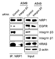
- 19A shows the results of immunoprecipitation assays using NRP1 antibodies in Ctx R lung cancer cell lines HCC44 and A549.
- 19B shows the results of an immunoprecipitation assay using NRP1 antibody in A549 treated with control siRNA and A549 treated with integrin ⁇ 3 siRNA.
- FIG. 20 shows the results of analyzing the inflow capacity of NRP1 and integrin ⁇ 3 according to Fc-TPP11, Ctx, and Ctx-TPP11 treatment in Ctx R HCC44 and A549 by flow cytometry.
- 20A is a histogram graph showing the cell surface expression levels of NRP1 and integrin ⁇ 3 following Fc-TPP11, Ctx, and Ctx-TPP11 treatment.
- FIG. 20B is a graph showing Mean Fluorescence Intensity (MFI) of the histogram shown in FIG. 20A.
- MFI Mean Fluorescence Intensity
- FIG. 21 shows that Panitumumab-TPP11 (Pnm-TPP11), in which TPP11 is fused to panitumumab (Panitumumab, Pnm) among EGFR target antibodies in addition to Ctx, may inhibit proliferation of Pnm R lung cancer cells.
- 21A is a schematic diagram of Pnm-TPP11 in which the TPP11 peptide is fused to the C-terminus of the heavy chain of Pnm via a 15 residue (G 4 S) 3 linker.
- Figure 21b is the result of measuring the cell proliferation according to the concentration-specific treatment of Pnm, Pnm-TPP11 in Pnm S and Pnm R lung cancer cell lines expressing NRP1 through WST-1 assay.
- Figure 21c is a result of measuring the cell proliferation according to the concentration-specific treatment of Pnm, Pnm-TPP11 in Pnm R lung cancer cell line that does not express NRP1 through WST-1 assay.
- 22 is a general schematic diagram showing the mechanism of resistance of Ctx R pancreatic and lung cancer and the mechanism of overcoming Ctx R by Ctx-TPP11.
- 22a is a general schematic diagram showing the mechanism of resistance of Ctx R pancreatic cancer and the mechanism of overcoming Ctx R by Ctx-TPP11.
- 22b is a general schematic diagram showing the mechanism of resistance of Ctx R lung cancer and the mechanism of overcoming Ctx R by Ctx-TPP11.
- the invention comprises a peptide that specifically binds to neurophylline 1, wherein the peptide is a composition for treating cancer, characterized in that to control the resistance or sensitivity to the EGFR (Epidermal Growth Factor Receptor) target agent It is about.
- EGFR Epidermal Growth Factor Receptor
- the present invention also includes administering to a patient a peptide that specifically binds to a therapeutically effective amount of neurophylline 1, wherein the peptide modulates resistance or sensitivity to an Epidermal Growth Factor Receptor (EGFR) target agent.
- EGFR Epidermal Growth Factor Receptor
- the peptide specifically binds to neuropilin 1 and may comprise, for example, one or more sequences selected from the group consisting of SEQ ID NOs: 1 to 3:
- HTPGNSKPTRTPRR (TPP11, SEQ ID NO: 1)
- HTPGIATRTPR (TPP8, SEQ ID NO: 3).
- the peptide i) reduces the amount of active integrin ⁇ 1 expressed on the cell surface, inhibits phosphorylation of Src and Akt, thereby regulating resistance or sensitivity to EGFR targeting agents, or ii) NRP1 expressed on the cell surface. And by regulating the expression of integrin ⁇ 3, the resistance or sensitivity to EGFR targeting agents can be regulated.
- the peptide specifically binds to neurophylline 1 in pancreatic cancer, it was confirmed that the expression of active integrin ⁇ 1 on the cell surface through influx into NRP1 / active integrin ⁇ 1 cells. In addition, it was confirmed that the peptide inhibits the phosphorylation of integrin ⁇ 1-induced FAK, Src and Akt. Accordingly, the peptide specifically binds to neuropilin 1 according to the present invention, thereby overcoming resistance to EGFR target agents such as cetuximab or panitumumab and increasing sensitivity in cancer. It was confirmed that it can be.
- a peptide that specifically binds to neuropilin 1 modulates the expression of NRP1 and integrin ⁇ 3 expressed on the cell surface in lung cancer, thereby EGFR targeting agents such as cetuximab or pani It has been found that it can overcome the resistance to EGFR target antibodies such as tumumab and increase the sensitivity.
- the EGFR targeting agent may be an expression inhibitor or an activity inhibitor of a gene, but is not limited in form, but may be, for example, an EGFR expression inhibitor or an active integrin ⁇ 1 expression and an expression inhibitor of FAK, Src, or Akt.
- the expression inhibitor may be an antisense nucleotide, small hairpin RNA (shRNA), small interfering RNA (siRNA) or ribozyme that complementarily binds to the mRNA of the protein gene, and the activity inhibitor May be an EGFR activity inhibitor or an activity inhibitor of integrin ⁇ 1, FAK, Src, Akt, and may be a compound, peptide, peptide mimetic, substrate analog, aptamer, antibody, or antagonist.
- the EGFR targeting agent may be, for example, a compound that specifically inhibits EGFR activity, or an antibody or fragment thereof that specifically binds EGFR, and the peptide may be present at the C terminus of the antibody or antibody fragment. Can be combined.
- the EGFR targeting agent is, for example, in the group consisting of cetuximab, panitumumab, zalutumumab, nimotuzumab, and matuzumab.
- the antibody fragment refers to each domain of the heavy or light chain of the antibody or fragment thereof, for example, Fc, Fab, heavy chain constant region fragment (CH 1 , CH 2 , or CH 3 ) of the antibody, heavy chain variable region fragment (V H ), light chain constant region fragments (C L ), light chain variable region fragments (V L ), single chain variable fragments (scFv) or fragments thereof.
- the antibody fragment may be a heavy chain constant region (Fc) fragment consisting of hinge-CH 2 -CH 3 of the antibody.
- Fab has one antigen binding site in a structure having the variable region of the light and heavy chains, the constant region of the light chain and the first constant region of the heavy chain (CH 1 ).
- F (ab ') 2 antibodies are produced when the cysteine residues of the hinge region of Fab' form disulfide bonds.
- Fv fragments are antibody fragments containing complete antibody recognition and binding sites. This region consists of a dimer in which one heavy chain variable domain and one light chain variable domain are tightly and covalently associated, for example, with scFv.
- Single-chain Fv or “scFv” antibody fragments comprise the VH and VL domains of an antibody, which domains are present in a single polypeptide chain.
- the Fv polypeptide may further comprise a polypeptide linker between the VH domain and the VL domain that allows the scFv to form the desired structure for antigen binding.
- Single-chain Fv is generally covalently linked to the variable region of the heavy chain and the light chain through a peptide linker or directly connected at the C-terminus.
- Such antibody fragments can be obtained using proteolytic enzymes (e.g., restriction digestion of the entire antibody with papain yields Fab and cleavage with pepsin yields F (ab ') 2 fragments). It can also be produced by recombinant technology.
- the heavy chain constant region may be selected from any one isotype of gamma ( ⁇ ), mu ( ⁇ ), alpha ( ⁇ ), delta ( ⁇ ) or epsilon ( ⁇ ).
- the subclasses include gamma 1 ( ⁇ 1), gamma 2 ( ⁇ 2), gamma 3 ( ⁇ 3), gamma 4 ( ⁇ 4), alpha 1 ( ⁇ 1), and alpha 2 ( ⁇ 2).
- the light chain constant region may be of kappa or lambda type.
- Heavy chain means both the full-length heavy chain and fragments thereof including the variable region domain VH comprising the amino acid sequence having sufficient variable region sequence to confer specificity to the antigen and the three constant region domains CH1, CH2 and CH3 .
- light chain means both the full-length light chain and fragment thereof including the variable region domain VL and the constant region domain CL, including the amino acid sequence having sufficient variable region sequence to confer specificity to the antigen.
- fragments of the antibodies may be monomers, dimers or multimers.
- the peptide specifically binding to neuropilin 1 may be bound to a heavy chain constant region (Fc) fragment of an antibody specific for EGFR, and preferably to the C-terminus of Fc.
- Fc heavy chain constant region
- the NRP1-specific binding peptide TPP11 is bound to the heavy chain C-terminus of the anti-EGFR antibodies cetuximab (Ctx) and panitumumab (Pnm), which are antibodies that specifically bind to EGFR.
- cetuximab cetuximab
- Pnm panitumumab
- the “bond” is to integrate two molecules having the same or different functions or structures, and may be used interchangeably with “fusion”. It can be binding or fusion by any physical, chemical or biological method to which the peptide can bind.
- the peptide may further comprise a linker to be linked to the EGFR target agent, in one embodiment the linker is a peptide linker and comprises a sequence of peptide linkers, eg, (GGGGS) n, to the EGFR target antibody. , n may be linked to an EGFR target antibody by linking a linker of an integer of 1-20.
- the linker is a peptide linker and comprises a sequence of peptide linkers, eg, (GGGGS) n, to the EGFR target antibody.
- n may be linked to an EGFR target antibody by linking a linker of an integer of 1-20.
- composition according to the invention can be prepared in unit dose form or formulated into a multi-dose container by formulating with a pharmaceutically acceptable carrier and / or excipient, according to methods which can be readily carried out by those skilled in the art. have.
- the formulation may be in the form of solutions, suspensions, syrups or emulsions in oils or aqueous media, or may be in the form of extracts, powders, powders, granules, tablets or capsules, and may further comprise dispersants or stabilizers.
- the composition since the composition comprises an antibody or antigen-binding fragment, it can be formulated as an immune liposome. Liposomes comprising the antibody can be prepared according to methods well known in the art.
- the immune liposomes can be prepared by reverse phase evaporation as a lipid composition comprising phosphatidylcholine, cholesterol and polyethyleneglycol-derivatized phosphatidylethanolamine.
- Fab 'fragments of antibodies can be conjugated to liposomes via disulfide-replacement reactions.
- Chemotherapeutic agents such as doxorubicin may further be included in the liposomes.
- the composition according to the present invention may be a pharmaceutical composition and may be administered orally or parenterally.
- parenteral administration it can be administered by intravenous injection, subcutaneous injection, intramuscular injection, intraperitoneal injection, endothelial administration, topical administration, intranasal administration, pulmonary administration and rectal administration.
- oral administration because proteins or peptides are digested, oral compositions should be formulated to coat the active agent or to protect it from degradation in the stomach.
- the composition may be administered by any device in which the active substance may migrate to the target cell.
- the present invention administers to a subject in need thereof a composition comprising a peptide that specifically binds neurophylline 1 to modulate resistance or sensitivity to an Epidermal Growth Factor Receptor (EGFR) target agent. It relates to a cancer treatment method comprising the.
- EGFR Epidermal Growth Factor Receptor
- Suitable dosages of the compositions may be prescribed in various ways, such as by the method of formulation, the mode of administration, the age, weight, sex, morbidity of the patient, food, time of administration, route of administration, rate of excretion and response. Preferred dosages of the compositions are in the range of 0.001-100 mg / kg on an adult basis.
- pharmaceutically effective amount means an amount sufficient to prevent or treat cancer or to prevent or treat a disease due to angiogenesis.
- composition or method of treatment according to the present invention is applied to a cancer, which is a cancer that can be treated by an EGFR target anticancer agent, and does not limit the kind thereof, for example, ACTH producing tumor, acute lymphocytic or lymphoid Constitutive leukemia, acute or chronic lymphocytic leukemia, acute nonlymphocytic leukemia, bladder cancer, brain tumor, breast cancer, cervical cancer, chronic myeloid leukemia, bowel cancer, T-zone lymphoma, endometriosis, esophageal cancer, gall bladder cancer, Ewing's sarcoma sarcoma), head and neck cancer, tongue cancer, Hopkins lymphoma, Kaposi's sarcoma, kidney cancer, liver cancer, lung cancer, mesothelioma, multiple myeloma, neuroblastoma, non-Hopkin's lymphoma, osteosarcoma, ovarian cancer, neuroblastoma, mammary cancer, cervical cancer, prostate cancer
- the cancer may be NRP1 is expressed, in the embodiment of the present invention confirmed the expression of EGFR and NRP1 in tumor cells, NRP1 is not expressed in cell lines that specifically bind to NRP1 It was confirmed that the peptide did not improve the resistance of the EGFR target antibody. Accordingly, it was confirmed that the expression of NRP1 should be premised in order for the peptide specifically binding to NRP1 to improve the resistance of the EGFR target antibody.
- the present invention relates to an anticancer agent or anticancer agent comprising the composition.
- the composition exhibits an anticancer effect directly through a composition comprising a peptide specifically binding to neurophylline 1 according to the present invention (eg, the peptide itself or Fc binding), or other anticancer agent (eg, an EGFR target). It can be used as an anticancer adjuvant to improve the resistance and increase the sensitivity of the antibody (Ctx or Pnm).
- the present invention includes a peptide specifically binding to neuropilin 1 and an EGFR target agent, wherein the peptide modulates resistance or sensitivity to the EGFR target agent. It is about.
- the present invention is a method for treating cancer comprising administering a composition comprising a peptide specifically binding to neuropilin 1 in combination with an EGFR target agent, wherein the peptide modulates resistance or sensitivity to the EGFR target agent. It relates to a cancer treatment method characterized in that.
- the present invention relates to a method of treating cancer exhibiting resistance to an EGFR target agent by administering a composition comprising a peptide that specifically binds neurophylline 1 in combination with an EGFR target agent.
- the peptide By co-administering a peptide specifically binding to the neurophilin 1 and an EGFR target agent, the peptide acts as a sensitizer, thereby inhibiting resistance to the EGFR target agent and improving sensitivity, thereby improving cancer treatment effects. .
- the combination dosage composition includes a peptide that specifically binds to neurophylline 1, and the configuration thereof is the same as the composition included in the composition and the method of treatment described above. The same applies.
- “Combination” means that each of the peptides that specifically bind to neuropilin 1 and the EGFR target agents may be administered simultaneously, sequentially, or in reverse order, and may be administered in a combination of appropriate effective amounts within the scope of those skilled in the art.
- the peptide and the EGFR target agent that specifically bind to neuropilin 1 may be stored in separate containers and then co-administered simultaneously, sequentially or in reverse order.
- Example 1 Characterization according to the presence or absence of innate resistance to Ctx of pancreatic cancer cell line.
- pancreatic cancer cell lines used in the present invention according to the inherent resistance to Ctx was analyzed. This may be an important marker for predicting innate resistance to Ctx in pancreatic cancer.
- FIG. 1A shows the EGFR, NRP1, and 4 types of pancreatic cancer cell lines (BxPC-3, PANC-1, Capan-2, SW1990) and two pancreatic cancer cell lines (Miapaca-2, AsPC-1) having no innate resistance to Ctx; Flow cytometry analysis of cell surface expression levels of integrin ⁇ 1.
- FIG. 1B is a Western blot result confirming EGFR, NRP1 and integrin ⁇ 1 expression levels in the cells of the cell lines used in FIG. 1A.
- the gel subjected to SDS-PAGE was transferred to PVDF membrane and reacted with NRP1, EGFR, integrin ⁇ 1 antibody and ⁇ -actin-recognized antibody (SantaCruz) for 2 hours at 25 ° C., and HRP-bound secondary antibody ( SantaCruz) was detected after reacting at 25 ° C. for 1 hour. Analysis was performed using ImageQuant LAS4000 mini (GE Healthcare).
- Ctx S pancreatic cancer cell lines with reference to FIG 1a, 1b was analyzed, unlike NRP1, EGFR in the entire cell surface and cells in Ctx R pancreatic cell line, it can be confirmed show the expression level of the high ⁇ 1 integrin.
- 1C is a result of Western blot analysis comparing total expression levels and phosphorylated levels of EGFR, FAK, Src, Akt, and ERK after Ctx concentrations in Ctx S and Ctx R pancreatic cancer cell lines.
- Ctx R pancreatic cancer cell lines maintain high phosphorylation of FAK, Src, and Akt even after Ctx treatment.
- phosphorylation of EGFR (Y1173) and ERK1 / 2 showed no difference between Ctx S pancreatic cancer cell line and Ctx R pancreatic cancer cell line.
- Ctx R pancreatic cancer cell lines have higher expression levels of integrin ⁇ 1 and higher phosphorylation levels of Src and Akt than Ctx S cell lines. Indeed, we examined whether Ctx R pancreatic cancer cell lines were associated with overexpressed integrin ⁇ 1, Src and Akt.
- Figure 2a is a control siRNA and integrin ⁇ 1 siRNA-treated Ctx R cell line (cell line BxPC-3, NNC1 expressing cells, PANC-1 and cell line SWI990 not expressing NRP1) to confirm the cell proliferation after treatment with Ctx.
- siRNA is transiently transfected.
- 100 nM of each control siRNA without target ability to transiently transfect and siRNA targeting integrin ⁇ 1 expression were reacted with 500 ⁇ l of Opti-MEM media (Gibco) and 3.5 ⁇ l of RNAiMax (Invitrogen, USA) on a tube for 15 minutes at room temperature. And added to each well.
- 500 ⁇ l of antibiotic-free DMEM media was added and cultured at 37 ° C. for 5 hours at 5% CO 2 , and then exchanged with 1 ml of DMEM medium containing 10% FBS.
- Figure 2b is a result of confirming that after the transient transfection in Figure 2a, cell lysate was obtained, the expression of integrin ⁇ 1 specifically through Western blot.
- Figure 2c confirms the cell proliferation of Ctx R cell line by treatment of PI3K-Akt inhibitor (LY294002), Src inhibitor (SU6656), Raf inhibitor (Sorafenib) with Ctx.
- Table 1 summarizes the results of analyzing the characteristics of the pancreatic cancer cell lines used in the present invention. Cell surface expression levels are shown as classified using the MFI value of the FACS results confirmed in Figure 1a. (+: Low expression level, + +: intermediate expression level, + + +: high expression level)
- Ctx-TPP11 In order to confirm that Ctx-TPP11 can inhibit the proliferation of Ctx R pancreatic cancer cells, Ctx-TPP11 was expressed and purified.
- the DNA of the TPP11 fused portion of the heavy chain constant region CH3 of the antibody obtained by treating BsrGI and HindIII restriction enzymes in a vector for producing a protein fused with the TPP11 peptide and the heavy chain constant region (Fc) of the antibody.
- the sequence was cloned into a vector encoding the wild type Ctx heavy chain (SEQ ID NO: 4 and DNA sequence SEQ ID NO: 5) (AA sequence SEQ ID NO: 6, DNA sequence SEQ ID NO: 7).
- DNA encoding the light chain (AA sequence SEQ ID NO: 8, DNA sequence SEQ ID NO: 9) was used the same wild-type Ctx light chain expression vector.
- HEK293-F cells Invitrogen
- serum-free FreeStyle 293 expression medium Invitrogen
- PEI Polyethylenimine
- the appropriate heavy and light chain plasmids were diluted in 125 ml of heavy chain and 125 ⁇ g of total light chain 125 ⁇ g (2.5 ⁇ g / ml) in 10 ml FreeStyle 293 expression medium (Invitrogen), diluting 750 ⁇ g (7.5 ⁇ g / ml) of PEI. It was mixed with 10 ml of medium and reacted at room temperature for 10 minutes. Thereafter, the reacted mixed medium was added to the cells sown in 100 ml, and then cultured at 150 rpm and 8% CO 2 for 4 hours, and then the remaining 100 ml of FreeStyle 293 expression medium was added and cultured for 7 days.
- FreeStyle 293 expression medium Invitrogen
- Proteins were purified from cell culture supernatants harvested with reference to standard protocols.
- the antibody was applied to a Protein A Sepharose column (GE healthcare) and washed with PBS (pH 7.4).
- the antibody was eluted at pH 3.0 with 0.1 M glycine buffer and then immediately neutralized with 1 M Tris buffer.
- the eluted antibody fraction was concentrated by exchanging buffer with PBS (pH 7.4) through dialysis.
- Purified protein was quantified using absorbance and extinction coefficient at 280 nm wavelength.
- Example 2 Ctx-TPP11 expressed and purified in Example 2 was analyzed to compare the binding ability of EGFR and NRP1-b1b2 with wild-type antibody Ctx.
- 3b is a sandwich ELISA result confirming that Ctx-TPP11 constructed in comparison with Fc-TPP11 and Ctx shows simultaneous binding ability against EGFR and NRP1-b1b2.
- EGFR 0.5 ⁇ g / well
- Fc-TPP11, Ctx and Ctx-TPP11 (10nM) were reacted at 25 ° C. for 1 hour.
- TBST TBS with 0.1% Tween-20
- biotinylated NRP1-b1b2 1 ⁇ M-1 pM was reacted at 25 ° C. for 1 hour, followed by AP-bound goat anti-biotin antibody. was bound at 25 ° C. for 1 h.
- the absorbance at 405 nm was then measured using a microplate reader to detect bound biotinylated protein using a substrate of p -nitrophenyl phosphate (Sigma-Aldrich).
- Table 3 shows the results of surface plasmon resonance (SPR) using a Biacore2000 instrument (GE healthcare) to more quantitatively analyze the binding capacity of Ctx-TPP11 to NRP1-b1b2 and EGFR compared to Fc-TPP11 and Ctx. Indicates.
- each of the Fc-TPP11, Ctx, and Ctx-TPP11 proteins was immobilized at a level of about 1,000 response units (RUs) on a CM5 sensor chip (GE healthcare, USA).
- HBS-EP buffer 10 mM Hepes, 3 mM ethylenediaminetetraacetic acid, 0.005% surfactant P20 (pH 7.6), GE Healthcare] was analyzed at a flow rate of 30 ⁇ l / min and analyzed by treatment with NRP1-b1b2 protein.
- CM5 chip After binding and dissociation analysis, regeneration of CM5 chip was performed by flowing a buffer solution (20 mM NaOH, 1M NaCl, pH10.0) at a flow rate of 30 ⁇ l / min for 2 minutes. Each sensorgram obtained by 3 minutes of association and 6 minutes of dissociation was normalized and subtracted to calculate affinity compared to blank cells.
- the binding capacity for EGFR was the same as that of Ctx-TPP11 fused with TPP11 and wild-type antibody Ctx, and the binding capacity for NRP1-b1b2 was also confirmed at the same level of Ctx-TPP11 and Fc-TPP11.
- ⁇ represents the standard deviation value of the result of the independent test.
- FIG 4a shows the concentration of Fc-TPP11, Ctx, Ctx-TPP11 in Ctx S (Miapaca-2, AsPC-1) and Ctx R (BxPC-3, PANC-1, Capan-2, SW1990) pancreatic cancer cell lines Cell proliferation was measured by MTT assay.
- Ctx S Moapaca-2, AsPC-1) pancreatic cancer cell lines
- Ctx and Ctx-TPP11 showed the same cell growth inhibition
- Ctx R BxPC-3, PANC-1, Capan-2 pancreatic cancer cell lines
- Differently, only Ctx-TPP11, Fc-TPP11, and Ctx showed inhibitory effect on cell growth.
- Ctx-TPP11 showed no efficacy in SW1990, one Ctx R pancreatic cancer cell line that does not express NRP1. This is a result confirming that Ctx-TPP11 is specifically effective for the target NRP1.
- apoptosis assay assay was conducted to determine whether Ctx-TPP11 inhibits the proliferation of Ctx R pancreatic cancer cells due to apoptosis inducing action.
- Annexin V-FITC for cell death according to Fc-TPP11, Ctx, Ctx-TPP11 in Ctx S (Miapaca-2, AsPC-1) and Ctx R (BxPC-3, PANC-1) pancreatic cancer cell lines It is analyzed by the detection kit (Annexin V-FITC apoptosis detection kit, BD Biscience).
- Ctx and Ctx-TPP11 induced apoptosis in Ctx S (Miapaca-2, AsPC-1) pancreatic cancer cell lines, and Ctx R (BxPC-3, PANC-1) pancreatic cancer cell lines, unlike Ctx, Cell death was induced only in the combination treatment group of Fc-TPP11 and Ctx. This result confirms that Ctx-TPP11 inhibits the proliferation of Ctx R pancreatic cancer cells due to apoptosis inducing action.
- Figure 4d is a result showing the effect of NRP1, integrin ⁇ 1, cMet siRNA on the cell proliferation inhibitory ability confirmed in Ctx R pancreatic cancer cell line.
- pancreatic cancer cell lines are prepared, and siRNA is transiently transfected.
- a control siRNA with no target ability to transient transfection and 100 nM of each siRNA targeting NRP1, integrin ⁇ 1, cMet expression inhibition were added on a tube with 500 ⁇ l of Opti-MEM media (Gibco), 3.5 ⁇ l of RNAiMax (Invitrogen, USA). After reacting at room temperature for minutes, each well was added.
- 500 ⁇ l of antibiotic-free DMEM media was added, and cultured at 37 ° C for 5 hours at 5% CO 2 , and then exchanged with 1ml of DMEM medium containing 10% FBS.
- Figure 4e f is obtained by performing a transient transfection in Figure 4d, obtained a cell lysate, Western blot confirmed that NRP1, cMet siRNA inhibited the expression of NRP1 and cMet by Western blot.
- FIG. 5a is Western blot results confirming the signal inhibitory effect of Fc-TPP11, Ctx, Ctx-TPP11 after treatment with control siRNA and integrin ⁇ 1 siRNA in Ctx R pancreatic cancer cell line.
- siRNA is transiently transfected.
- 500 ⁇ l of antibiotic-free DMEM media was added, and cultured at 37 ° C. and 5% CO 2 for 6 hours, and then exchanged with 1 ml of DMEM medium containing 10% FBS.
- Fc-TPP11, Ctx, and Ctx-TPP11 were diluted in 1 ml of a medium containing 10% FBS and incubated at 5% CO 2 , 37 ° C for 24 hours. After incubation, the cells were washed with cold PBS, and lysis buffer (10 mM Tris-HCl pH 7.4, 100 mM NaCl, 1% SDS, 1 mM EDTA, Inhibitor cocktail (sigma)) was added to obtain cell lysates. Thereafter, Western blot analysis was performed in the same manner as in Example 1.
- Ctx R SW1990 cells that did not express NRP1 also had the same Ctx resistance mechanism as BxPC-3 and PANC-1, but showed no effect by Fc-TPP11 and Ctx-TPP11.
- the cell growth inhibitory effect of Ctx-TPP11 inhibits the phosphorylation of integrin ⁇ 1, a Ctx resistance marker identified in Example 1, and its sub-signal factors FAK, Src, and Akt. Inhibition of the expression of integrin ⁇ 1 inhibited the phosphorylation of the resistance markers FAK, Src, Akt.
- integrin ⁇ 1 is an upper molecule of the phosphorylation pathway of FAK, Src, and Akt.
- Ctx-TPP11 did not reduce the overall expression of integrin ⁇ 1 itself.
- Example 7 Ctx R Evaluation of Cell Proliferation Inhibitory Activity of Ctx-TPP11 in Colorectal Cancer Cell Lines.
- FIG. 6 is an MTT language confirming cell proliferation inhibitory ability by Fc-TPP11, Ctx, and Ctx-TPP11 in KRas and BRaf wild-type Ctx S colon cancer cell lines and colon cancer cell lines resistant to Ctx by KRas and BRaf mutations. Essay results.
- cells were prepared in the same manner as in Example 2, and 0.2 ml and 1 ⁇ M of Fc-TPP11, Ctx, and Ctx-TPP11 were diluted in 1 ml of a medium containing 10% FBS, followed by 5% CO 2 , 37 for 72 hours. After incubation at ° C conditions, MTT assay was performed. As a result, the combination of Ctx-TPP11 and Ctx with Fc-TPP11 did not overcome resistance in colon cancer cell lines with KRas and BRaf mutation resistance mechanisms.
- Ctx-TPP11 inhibited the phosphorylation of FAK, Src and Akt, Ctx resistance markers, via NRP1 targets, but did not reduce the total amount of overexpressed integrin ⁇ 1.
- Integrin ⁇ 1 is divided into extended form active integrin ⁇ 1 that can signal and inactive integrin ⁇ 1 that is bent form that can not signal. Therefore, in order to determine whether Ctx-TPP11 downregulates the signal of integrin ⁇ 1 by reducing the expression of active integrin ⁇ 1 that can send the actual signal on the cell surface, the intracellular influx of active integrin ⁇ 1 following Ctx-TPP11 treatment. It was confirmed.
- Figure 7a, b shows the inflow capacity of NRP1, active integrin ⁇ 1, inactive integrin ⁇ 1 according to Fc-TPP11, Ctx, Ctx-TPP11 treatment in Ctx R BxPC-3, PANC-1 by confocal microscopy Observed.
- the cells were washed with PBS, incubated for 10 minutes at 25 ° C. in a buffer containing 0.1% saponin, 0.1% sodium azide, and 1% BSA in PBS to form pores in the cell membrane.
- the reaction was performed at 25 ° C. for 1 hour with a buffer containing 2% BSA added to PBS to inhibit nonspecific binding.
- NRP1 A secondary antibody linked to TRITC (red fluorescence) or FITC (green fluorescence) that recognizes each primary antibody was reacted at 25 ° C for 1 hour, and the nuclei were stained (blue fluorescence) using Hoechst 33342 and observed by confocal microscopy. . Since NRP1 overlaps only active integrin ⁇ 1, it was confirmed that NRP1 specifically binds only to active integrin ⁇ 1.
- Figures 8 and 9 show the flow cytometry analysis of NRP1, EGFR, active integrin ⁇ 1, and inactive integrin ⁇ 1 cells in flow cytometry according to Fc-TPP11, Ctx, Ctx-TPP11 treatment in Ctx R BxPC-3, PANC-1 to be.
- the cells were prepared in the same manner as in Example 1, and when the cells were stabilized, serum deficiency was performed in serum-free medium for 4 hours to eliminate the effects of serum, followed by 2 ⁇ M of Fc-TPP11, Ctx, and Ctx-TPP11.
- serum deficiency was performed in serum-free medium for 4 hours to eliminate the effects of serum, followed by 2 ⁇ M of Fc-TPP11, Ctx, and Ctx-TPP11.
- Cells were prepared.
- FITC-linked secondary antibodies that recognize each primary antibody were reacted for 30 minutes at 4 ° C., washed with PBS, and analyzed by FACS Calibur (BD Bioscience), a flow cytometer. After the analysis, a histogram graph for each sample was obtained, and after intracellular inflow of NRP1, EGFR, active integrin ⁇ 1, and inactive integrin ⁇ 1 according to Fc-TPP11, Ctx, and Ctx-TPP11 treatment using the average fluorescence intensity of the histogram, The amount of receptor on the remaining cell surface is shown quantitatively in FIGS. 8B and 9B.
- Fc-TPP11 and Ctx-TPP11 that bind to NRP1 increased the cellular influx of NRP1 and increased the cellular influx of active integrin ⁇ 1 that binds to NRP1.
- Ctx and Ctx-TPP11 increased the cellular influx of EGFR.
- Ctx-TPP11 binds to NRP1 and selectively reduces the amount of cell surface expression of NRP1 and active integrin ⁇ 1.
- Active integrin ⁇ 1 expressed on the cell surface plays an important role in binding of cells to extracellular matrix (ECM).
- ECM extracellular matrix
- FN has the highest binding capacity. Accordingly, it was analyzed whether Ctx-TPP11 reduced the expression of active integrin ⁇ 1, thereby inhibiting the ability of cells to adhere to FN.
- Figure 10a is a result of confirming the cell adhesion ability to FN according to the Fc-TPP11, Ctx, Ctx-TPP11 treatment in Ctx R BxPC-3, PANC-1 by optical microscopy through the cell attachment assay.
- FN (Sigma) was diluted in 0.5 ml of PBS at a concentration of 10 ⁇ g / ml in a 12 well uncoated plate, and the plate was coated at 37 ° C. for 30 minutes.
- BxPC-3 was incubated for 1 hour and PANC-1 for 6 hours at 37 ° C., and then cells were washed with PBS. After washing, the cells were fixed for 10 minutes at 25 ° C.
- Fc-TPP11 and Ctx-TPP11 unlike Ctx, reduced the adhesion of Ctx R cells to FN. This is because Fc-TPP11 and Ctx-TPP11 decrease the expression of active integrin ⁇ 1 on the cell surface, thereby reducing the binding ability of active integrin ⁇ 1 with FN.
- Example 5 the in vitro cell growth inhibition of Ctx-TPP11 in the Ctx R pancreatic cancer cell line was confirmed. It was confirmed whether the same effect of Ctx-TPP11 appeared in vivo.
- 11 and 12 show the results of measuring tumor growth inhibitory activity of mouse Ctx-TPP11 in vivo.
- BxPC-3 cells (5 ⁇ 10 6 cells / mouse), PANC-1 (1 ⁇ 10 7 cells / mouse), AsPC-1 (3) week old female BALB / c nude mice (NARA Biotech, Korea) 5 ⁇ 10 6 cells / mouse) cells were implanted subcutaneously in a mixture of 150 ⁇ L PBS and 150 ⁇ L Matrigel (BD Biosciences) in a 1: 1 ratio.
- FIG. 13 is a result of comparing the extent of the growth markers and apoptosis markers of the tissue through the immunohistochemistry experiment confirmed the tumor suppression of Ctx-TPP11 of FIGS.
- tumor tissues were extracted 5 hours after the last antibody administration in FIGS. 11 and 12.
- the extracted tumor tissues were fixed for 24 hours at 4 ° C. in 4% paraformaldehyde and placed in 4% at 24 ° C. in 30% sucrose buffer.
- the tumor sections were cut to a thickness of 20 ⁇ m by the frozen section method, and the tumor sections were ki-67 antibody (Abcam) and a TRITC-conjugated secondary antibody that recognizes them at 25 ° C. for 1 hour at a temperature of Ki-. 67 were dyed.
- tumor tissues were stained with DeadEnd TM Colorimetric TUNEL System (Promega), and nuclei were stained (blue fluorescence) using Hoechst 33342 and observed under confocal microscopy.
- the amount of reduced growth marker and increased apoptosis marker was confirmed in the tissue of Ctx-TPP11 which showed tumor growth inhibitory ability.
- FIG. 14 is a result of Western blot performed by extracting Ctx R tumor tissue whose tumor suppression ability was confirmed in FIG. 11.
- tumor tissues were extracted and homogenized using the cell lysis buffer used in Examples, followed by Western blot.
- the treatment groups of Ctx-TPP11 and Ctx and Fc-TPP11 in Ctx R tumor tissues inhibited phosphorylation of CAK resistance markers FAK, Src, and Akt in the same manner as in vitro signal suppression effect. It was confirmed.
- pancreatic cancer shows higher expression levels of integrin ⁇ 1 on the cell surface and throughout the cell in the Ctx R pancreatic cancer cell line compared to the Ctx S pancreatic cancer cell line.
- the cell surface expression levels of EGFR, NRP1 and integrin ⁇ 1 in lung cancer cell lines were analyzed.
- Figure 15A shows Ctx S (Calu-3, H1975) and Ctx R (H1299, A549, Calu-1, H358, H441, H2009, HCC44, HCC2108, SK-LU-1, H460, H522) lung cancer cell lines EGFR, NRP1 And flow cytometry analysis of cell surface expression levels of integrin ⁇ 1.
- lung cancer cell lines were prepared in the same manner as in Example 1, and the cells were washed with PBS, followed by an antibody that recognizes NRP1 (R & D System) and an FITC-bound antibody that recognizes EGFR and integrin ⁇ 1, respectively (e-Bioscience ) Was reacted at 4 ° C for 1 hour. Additionally, NRP1 antibody bound to cells was stained with FITC bound antibody, washed with PBS, and analyzed by FACS Calibur (BD Bioscience), a flow cytometer.
- NRP1 R & D System
- FITC-bound antibody that recognizes EGFR and integrin ⁇ 1, respectively
- 15B is a graph quantitatively showing the average fluorescence intensity of the histogram shown in FIG. 15A.
- lung cancer cell line did not show any correlation between Ctx resistance and cell surface integrin ⁇ 1 expression.
- Example 12 Inhibition of expression of various cell surface receptors and inhibition of phosphorylation of Akt and Src on Ctx R in lung cancer.
- FIG. 16A shows Ctx treatment of Ctx R lung cancer cell lines A549 and HCC44 (Ctx R cell line expressing NRP1) treated with control siRNA, NRP1 siRNA, integrin ⁇ 1 siRNA, integrin ⁇ 3 siRNA, cMet siRNA, VEGFR1 siRNA, and TGF ⁇ R2 siRNA, respectively. After confirming the cell proliferation.
- siRNA is transiently transfected.
- 500 n of Opti-MEM media (Gibco), RNAiMax (Invitrogen) were added to control siRNA 100 nM without target ability to transient transfect and 100 nM of each siRNA targeting NRP1, integrin ⁇ 1, integrin ⁇ 3, cMet, VEGFR1, TGF ⁇ R2 on tubes , USA) was added to each well after reaction at room temperature for 15 minutes with 3.5 ⁇ l.
- FIG. 16B is a result of confirming that the protein expression targeted by each siRNA was specifically inhibited through Western blotting after cell transfusion was performed after transient transfection in FIG. 16A.
- Figure 17 shows the cell proliferation of Ctx R lung cancer cell line by treatment of PI3K-Akt inhibitor (LY294002), Src inhibitor (SU6656), Raf inhibitor (Sorafenib) with Ctx.
- Table 4 summarizes the results of analyzing the characteristics of the lung cancer cell lines used in the present invention.
- Example 13 Evaluation of the inhibition of cell proliferation of Ctx-TPP11 in Ctx R lung cancer cell line.
- Ctx-TPP11 inhibits cell proliferation by Ctx and Ctx-TPP11 in Ctx S lung cancer cell lines and Ctx R lung cancer cell lines to determine whether Ctx-TPP11 overcomes Ctx resistance in lung cancers resistant to Ctx. It was confirmed.
- FIG. 18 performed cell growth assay in 13 lung cancer cell lines to determine whether Ctx-TPP11 can inhibit proliferation of Ctx R lung cancer cells expressing NRP1.
- NRP1 and Ctx S (Calu-3, H1975) and Ctx R (H1299, A549, Calu-1, H358, H441, H2009, HCC44, SK-LU-1) lung cancer cell lines expressing NRP1 Cell proliferation according to the concentration-specific treatment of Ctx and Ctx-TPP11 in non-Ctx R (HCC2108, H460, H522) lung cancer cell lines was measured using the WST-1 assay.
- Ctx S Calu-3, H1975) lung cancer cell lines
- Ctx and Ctx-TPP11 showed the same cell growth inhibitory activity
- 8 types of Ctx R H1299, A549, Calu-1, H358, H441, H2009, HCC44, SK-LU) -1
- Ctx-TPP11 treated group showed cell growth inhibition.
- Ctx-TPP11 did not show efficacy in three Ctx R lung cancer cell lines HCC2108, H460, and H522 that did not express NRP1. This is a result confirming that Ctx-TPP11 is specifically effective for the target NRP1.
- Figure 18c is a result showing the effect of NRP1 siRNA on the cell proliferation inhibitory ability of Ctx-TPP11 confirmed in Ctx R lung cancer cell line.
- cells were prepared as shown in FIG. 17A, and transient transfection was performed, followed by incubating for 12 hours by adding 5 ⁇ 10 3 cells to each well in a 96-well plate.
- Ctx and Ctx-TPP11 were diluted in a medium containing 10% FBS at a concentration of 2 ⁇ M and cultured for 48 hours, and then cell proliferation was confirmed by the WST-1 assay.
- Examples 11-12 it was confirmed that resistance to Ctx was closely related to NRP1, integrin ⁇ 3, and KRAS mutations in lung cancer cell lines, and that Ctx-TPP11 showed specific efficacy in NRP1 in these Ctx R lung cancer cell lines. It was confirmed. Therefore, in the lung cancer cell line, in order to know why Ctx-TPP11 can overcome the resistance to Ctx, immunoprecipitation assay (Immunoprecipitation assay) was confirmed how NRP1 interacts with integrin ⁇ 3 and KRAS.
- Immunoprecipitation assay immunoprecipitation assay
- 19A shows the results of immunoprecipitation assays using NRP1 antibodies in Ctx R lung cancer cell lines HCC44 and A549.
- the cell lysate was obtained by incubating 2 ⁇ 10 6 CtxR lung cancer cell lines HCC44 and A549 (cell lines expressing NRP1 and integrin ⁇ 3) in a 100 mm 3 plate in a medium containing 10% FBS for 12 hours, respectively.
- Hazard lysis buffer 50 mM Tris-HCl pH 7.4, 150 mM NaCl, 1% NP-40, 0.5% SDC, 0.1% SDS, 100x Protease inhibitor was added and reacted at 4 ° C for 30 minutes, after which cell debris was precipitated.
- the cell lysate was quantified using a BCA protein assay kit (Pierce), and then reacted with 0.5 mg of the cell lysate and 5 ⁇ g of the anti-NRP1 antibody (Abacm), respectively, at 4 ° C. for 12 hours. Thereafter, Protein A / G agarose is added and reacted at 4 ° C. for 2 hours, after which the antibody is allowed to settle. Thereafter, Western blot was performed using anti-NRP1 antibody, anti-EGFR antibody, anti-integrin ⁇ 3 antibody, anti-integrin ⁇ 1 antibody, and anti-KRAS antibody.
- 19B shows the results of immunoprecipitation assays using NRP1 antibodies in A549 treated with control siRNA and A549 treated with integrin ⁇ 3 siRNA to determine whether the interaction between NRP1 and KRAS was caused by integrin ⁇ 3.
- control siRNA and the integrin ⁇ 3 siRNA were treated in the same manner as in Example 12. Thereafter, cell lysates were prepared in the same manner as in FIG. 19A, and immunoprecipitation assays were performed. As a result, EGFR, integrin ⁇ 3, and KRAS were observed together with NRP1 in A549 expressing integrin ⁇ 3 treated with the control siRNA.
- Example 8 it was confirmed that Ctx-TPP11 in the pancreatic cancer down-regulates the signal of integrin ⁇ 1 by reducing the expression of active integrin ⁇ 1 on the cell surface through the NRP1 target.
- NRP1 interacts with integrin ⁇ 3. Therefore, the aim of the present study was to determine whether NRP1 target can reduce the amount of integrin ⁇ 3 expression on the cell surface.
- FIG. 20 shows the results of analyzing the inflow capacity of NRP1 and integrin ⁇ 3 according to Fc-TPP11, Ctx, and Ctx-TPP11 treatment in Ctx R HCC44 and A549 by flow cytometry.
- the cells were prepared in the same manner as in Example 8, and when the cells were stabilized, serum deficiency was performed in serum-free medium for 4 hours to eliminate the effects of serum, followed by 2 ⁇ M of Fc-TPP11, Ctx, and Ctx-TPP11.
- serum deficiency was performed in serum-free medium for 4 hours to eliminate the effects of serum, followed by 2 ⁇ M of Fc-TPP11, Ctx, and Ctx-TPP11.
- FITC-linked secondary antibodies that recognize each primary antibody were reacted for 30 minutes at 4 ° C., washed with PBS, and analyzed by FACS Calibur (BD Bioscience), a flow cytometer. After analysis, a histogram graph is obtained for each sample, and after the influx of NRP1 and integrin ⁇ 3 following Fc-TPP11, Ctx and Ctx-TPP11 treatment using the average fluorescence intensity of the histogram, The amount is shown quantitatively in FIG. 20B.
- Fc-TPP11 and Ctx-TPP11 binding to NRP1 increased the cellular influx of NRP1 and increased the cellular influx of integrin ⁇ 3 binding to NRP1.
- Ctx-TPP11 binds to NRP1 and selectively reduces the amount of cell surface expression of NRP1 and integrin ⁇ 3.
- Examples 1-13 cell lines resistant to Ctx among EGFR target antibodies were described.
- Pnm-TPP11 was expressed and purified to confirm whether Pnm-TPP11 fused TPP11 to panitumumab (Pnm) in the EGFR target antibody can inhibit proliferation of Pnm R lung cancer cells.
- 21A is a schematic diagram of Pnm-TPP11 in which the TPP11 peptide is fused to the C-terminus of the heavy chain of Pnm via a 15 residue (G 4 S) 3 linker.
- the heavy chain of Pnm (AA sequence is SEQ ID NO: 12) using C-terminus and (G 4 S) 3 linker, and a reverse primer indicating TPP11 and a forward primer indicating signal peptide
- the polymerase chain reaction was carried out to obtain DNA fragments indicated in the order of signal peptide, Pnm heavy chain, (G 4 S) 3 linker, TPP11 and termination codon.
- DNA was recovered using 1% agarose gel and electrophoresis, and cohesive ends were generated using NotI and BamHI restriction enzymes.
- the vector was cloned into the pcDNA3.4 vector using T4 ligase to construct a vector capable of expressing the Pnm-TPP11 heavy chain in animal cells (AA sequence is SEQ ID NO: 10, DNA sequence SEQ ID NO: 11).
- DNA encoding the light chain (AA sequence SEQ ID NO: 14, DNA sequence SEQ ID NO: 15) was used the same wild-type Pnm light chain expression vector.
- HEK293-F cells Invitrogen
- serum-free FreeStyle 293 expression medium Invitrogen
- PEI Polyethylenimine
- HEK293-F cells Upon 200 mL transfection in a shake flask (Corning), HEK293-F cells were seeded in 100 ml of medium at a density of 2 ⁇ 10 6 cells / ml and incubated at 130 rpm, 8% CO 2 .
- the appropriate heavy and light chain plasmids were diluted in 125 ml of heavy chain and 125 ⁇ g of light chain (250 ⁇ g / ml) in 10 ml FreeStyle 293 expression medium (Invitrogen), diluting 750 ⁇ g (7.5 ⁇ g / ml) of PEI. It was mixed with 10 ml of medium and reacted at room temperature for 10 minutes. Thereafter, the reacted mixed medium was added to the cells seeded with 100 ml of the above, and cultured at 150 rpm and 8% CO 2 for 4 hours, and then the remaining 100 ml of FreeStyle 293 expression medium was added and cultured for 7 days.
- FreeStyle 293 expression medium Invitrogen
- Proteins were purified from cell culture supernatants harvested with reference to standard protocols.
- the antibody was applied to a Protein A Sepharose column (GE healthcare) and washed with PBS (pH 7.4).
- the antibody was eluted at pH 3.0 with 0.1 M glycine buffer and then immediately neutralized with 1 M Tris buffer.
- the eluted antibody fraction was concentrated by exchanging buffer with PBS (pH 7.4) through dialysis.
- Purified protein was quantified using absorbance and extinction coefficient at 280 nm wavelength.
- Pnm-TPP11 Also, like Ctx-TPP11, cell growth assays were conducted in various lung cancer cell lines to determine whether they could inhibit the proliferation of NRP1-expressing Pnm R lung cancer cells.
- 21B, C show Pnm S (Calu-3, A549, Calu-1, HCC44) expressing NRP1 and Pnm R (H441, SK-LU-1, H1299) lung cancer cell lines and Pnm R (H460 not expressing NRP1)
- NRP1 and Pnm R H441, SK-LU-1, H1299
- Pnm R H460 not expressing NRP1
- Cell proliferation according to the concentration-specific treatment of Pnm and Pnm-TPP11 in lung cancer cell lines was measured using the WST-1 assay.
- lung cancer cell lines were prepared in the same manner as in Example 11, when the cells were stabilized, Pnm, Pnm-TPP11 (0, 1, 2, 4 ⁇ M) were incubated for 48 hours, followed by Cyto-X reagent ( LPS Solution) was added to each well, and then reacted at 37 ° C. for 1 hour to 2 hours, and the absorbance at 450 nm was measured using a microplate reader (Molecular Devices).
- Pnm S Calu-3, A549, Calu-1, HCC44
- Pnm and Pnm-TPP11 showed the same cell growth inhibition
- Pnm R H441, SK-LU-1, H1299
- Pnm-TPP11 treated group showed cell growth inhibition.
- Pnm-TPP11 did not show efficacy in H460, a Pnm R lung cancer cell line that does not express NRP1. This is a result confirming that Pnm-TPP11 shows a specific effect on the target NRP1.
- Intracellular influx of NRP1 / active integrin ⁇ 1 by acting on NRP1 of tumor cells by binding a peptide specifically binding to neurophylline 1 (NRP1) with an EGFR target agent or in combination with an EGFR target agent Promotes cell surface activation by reducing the expression of integrin ⁇ 1 and reducing the level of phosphorylation of FAK, Src and Akt induced by integrin ⁇ 1.
- NRP1 neurophylline 1
Abstract
The present invention relates to methods for controlling resistance or sensitivity to an epidermal growth factor receptor (EFGR)-targeting agent through a peptide binding specifically to Neuropilin 1. In addition, the present invention relates to a composition for controlling resistance or sensitivity to an EGFR-targeting agent by co-administering a fusion antibody in which a peptide binding specifically to Neuropilin 1 is fused to an EGFR-targeting antibody, and a constant region of the heavy chain in an antibody in which the EGFR-targeting agent is fused to the peptide. Further, the fusion antibody in which a peptide binding specifically to Neuropilin 1 is fused to an EGFR-targeting antibody in accordance with the present invention allows for overcoming resistance to EGFR-targeting antibodies in pancreatic cancer. Furthermore, the fusion antibody in which a peptide binding specifically to Neuropilin 1 is fused to an EGFR-targeting antibody allows for overcoming resistance to EGFR-targeting antibodies even in the lung cancer that is resistant to EGFR-targeting antibodies. Hence, the peptide binding specifically to Neuropilin 1 in accordance with the present invention is expected to have high effects of treating various tumors resistant to EGFR-targeting agents.
Description
본 발명은 뉴로필린 1(Neuropilin 1, NRP1)에 특이적으로 결합하는 펩타이드를 통해 EGFR (Epidermal Growth Factor Receptor) 표적 제제에 대한 저항성 또는 감수성을 조절하는 조성물에 관한 것으로, 구체적으로 뉴로필린 1에 특이적으로 결합하는 펩타이드를 포함하고, EGFR 표적 제제에 대한 저항성을 극복하여 암을 치료하기 위한 조성물에 관한 것이다. The present invention relates to a composition that modulates resistance or sensitivity to an Epidermal Growth Factor Receptor (EGFR) target agent through a peptide that specifically binds Neuropilin 1 (NRP1), specifically specific to Neuropilin 1 It relates to a composition for treating cancer by overcoming resistance to an EGFR target agent comprising a peptide that binds to the target.
EGFR은 세포의 성장, 생존 및 전이와 같은 세포 기능에 수반되는 세포 수용체의 일원이며, EGFR의 과발현 또는 변이는 종양을 유발한다. 이에 따라, EGFR을 표적하는 항체 및 소분자 타이로신 키나제 저해제들이 다수 개발되고 있다. 예를 들면, EGFR을 표적하는 항체에는, 세툭시맙 (Cetuximab), 파니투무맙 (Panitumumab), 잘루투무맙 (zalutumumab), 니모투주맙 (nimotuzumab), 마투주맙 (matuzumab)이 있고, 소분자 타이로신 키나제 저해제에는, 제피티닙 (Gefitinib), 에르로티닙 (Erlotinib)이 개발되었다. 이러한 EGFR 표적 제제들은 직장암, 비-소세포 폐암, 두경부암의 치료를 위해 쓰이고 있다. 그러나, 이들 단일 약물을 사용하는 암 치료법은 특정 종류의 종양 세포에만 효과를 나타내어 그 적응증에 한계가 있거나, 다양한 종류 또는 변이 종양에서 저항성을 보여 만족할 만한 치료효과를 나타내지 못하고 있는 실정이다. 따라서, 보다 효과적인 암 치료를 위해서는 2 또는 그 이상을 표적으로 하는 다중 병용 치료법의 개발이 요구되고 있다. EGFR is a member of cellular receptors involved in cellular functions such as cell growth, survival and metastasis, and overexpression or mutation of EGFR causes tumors. Accordingly, many antibodies and small molecule tyrosine kinase inhibitors targeting EGFR have been developed. For example, antibodies targeting EGFR include cetuximab, panitumumab, zalutumumab, nimotuzumab, matuzumab, and small molecule tyrosine kinases. Inhibitors have been developed gefitinib, Erlotinib. These EGFR targeting agents are used for the treatment of rectal cancer, non-small cell lung cancer, and head and neck cancer. However, cancer therapy using these single drugs is effective only in certain types of tumor cells and thus has limitations on the indications, or is resistant to various types or mutant tumors, and thus does not show satisfactory therapeutic effects. Therefore, the development of multiple combination therapies targeting two or more is required for more effective cancer treatment.
여러 암 종 중에서도, 췌장암은 매우 나쁜 예후를 나타내는 암이며, 60-80% 환자가 국소적 중증, 또는 전이성 질환을 나타낸다. 대부분의 췌장암들은 다양한 수용체 타이로신 키나제 중 EGFR과 이의 리간드가 과발현되어 그들의 성장 및 생존을 촉진하는데 주요 역할을 수행한다고 알려져 있다 (Oliveira-Cunha et al., 2011; Wheeler et al., 2010). 하지만, 현재까지의 EGFR 표적 제제들은 췌장암에서는 여전히 효과적이지 않다. 이는 다수의 췌장암이 EGFR 표적 제제에 대해 저항성을 나타내기 때문이다. 그에 따라, EGFR 표적 제제들은 항암화학요법인 젬시타빈 (Gemcitabine)과 조합하여 췌장암 치료에 널리 사용되어 왔지만, 상당한 독성을 나타내는 문제점이 있다 (Chong and Janne, 2013; Philip et al., 2010). 따라서, 췌장암에서 효과적인 EGFR 표적 치료법의 개발이 필요하다.Among many carcinomas, pancreatic cancer is a cancer with a very poor prognosis, with 60-80% of patients showing localized severe or metastatic disease. Most pancreatic cancers are known to overexpress EGFR and its ligands in various receptor tyrosine kinases and play a major role in promoting their growth and survival (Oliveira-Cunha et al., 2011; Wheeler et al., 2010). However, to date EGFR targeted agents are still ineffective in pancreatic cancer. This is because many pancreatic cancers are resistant to EGFR targeting agents. Accordingly, EGFR targeted agents have been widely used in the treatment of pancreatic cancer in combination with chemotherapy, gemcitabine, but have the problem of showing significant toxicity (Chong and Janne, 2013; Philip et al., 2010). Thus, there is a need for the development of effective EGFR targeted therapies in pancreatic cancer.
전세계적으로 암 사망의 주원인으로 알려진 폐암 역시 EGFR이 그들의 성장에 주요 인자로 알려져 있다 (Sharma et al., 2007; Morgillo Floriana et al., 2016). 따라서, 폐암을 치료하기 위하여 다양한 EGFR 표적 제제들이 개발되었으며, 특히, EGFR 표적 소분자 타이로신 키나제 저해제인 제피티닙 (Gefitinib)과 에르로티닙 (Erlotinib)이 대표적인 약물이다. 하지만, 이들 표적 제제들이 매우 유효한 약물임에도 불구하고, 실제로 폐암 환자들의 약 10% 만이 이들 약물에 반응성을 보이고 있다 (Socinski Mark A, 2007). 따라서, 폐암에서 역시 새로운 EGFR 표적 치료 대안이 절실히 요구된다.Lung cancer, known worldwide as the leading cause of cancer death, also has EGFR as a major factor in their growth (Sharma et al., 2007; Morgillo Floriana et al., 2016). Therefore, various EGFR targeting agents have been developed for the treatment of lung cancer, and in particular, EGFR target small molecule tyrosine kinase inhibitors Gefitinib and Erlotinib are representative drugs. However, although these targeted agents are very effective drugs, only about 10% of lung cancer patients are actually responsive to these drugs (Socinski Mark A, 2007). Thus, there is an urgent need for new EGFR targeted treatment alternatives in lung cancer as well.
EGFR 표적 제제들 중 항-EGFR 항체인 세툭시맙 (Cetuximab, Ctx)은 리간드들 (EGF, TGFα)에 의존적인 EGFR의 활성화를 억제시키고, 이의 하위 신호전달을 막는다. Ctx은 직장암, 두경부암에서 항암화학요법과의 병용투여로 FDA 승인을 받았지만, EGFR 표적 제제들에 대해 저항성을 가지는 췌장암 및 폐암에서는 승인받지 못하였다. 그러나, EGFR 표적 제제들에 대한 췌장암 및 폐암 세포의 저항성의 명확한 기작 및 이를 개선하기 위한 방안은 아직 개발된 바가 없다. Cetuximab, Ctx, an anti-EGFR antibody in EGFR targeting agents, inhibits the activation of EGFR dependent on ligands (EGF, TGFα) and blocks its downstream signaling. Ctx has been FDA approved for combination with chemotherapy in rectal and head and neck cancers, but not in pancreatic and lung cancers resistant to EGFR target agents. However, no clear mechanism of resistance of pancreatic and lung cancer cells to EGFR target agents and methods for improving them have yet been developed.
기존에 직장암과 두경부암에서 규명된 저항성 기작은 다음과 같다. 1) EGFR 유전자의 높은 copy number, 2) EGFR 유전자의 돌연변이, 3) KRAS 유전자 또는 BRAF 유전자의 돌연변이 등이 있다 (Oliveira-Cunha et al., 2011). 현재까지 췌장암에서 밝혀진 EGFR 표적 제제들에 대한 저항성 기작은 EGFR 패밀리 (EGFR, HER2, HER3)에 의한 비정상적인 PI3K-Akt 경로의 활성화와 연관되어 있다는 보고가 있지만, 이 또한 명확한 기작은 아직 불분명하다 (Larbouret et al., 2012; Wong et al., 2014). 폐암에서 역시, 인테그린 β3가 KRAS 유전자와 결합하여 KRAS-RalB-NFκB 경로의 신호를 활성화시킴으로써, EGFR 표적 제제에 대한 저항성을 유도한다는 것이 최근에서야 보고되고 있는 실정이다 (Laetitia Seguin, 2014). 췌장암 및 폐암에서의 효과적인 치료 약물의 부재는 환자의 높은 사망률과 관련되기 때문에 췌장암 및 폐암에 대한 저항성 기작을 정확히 규명할 필요가 있다. The resistance mechanisms previously identified in rectal and head and neck cancers are as follows. 1) high copy number of EGFR gene, 2) mutation of EGFR gene, 3) mutation of KRAS gene or BRAF gene (Oliveira-Cunha et al., 2011). To date, it has been reported that resistance mechanisms against EGFR target agents found in pancreatic cancer are associated with the activation of abnormal PI3K-Akt pathways by the EGFR family (EGFR, HER2, HER3), but this mechanism is still unclear (Larbouret). et al., 2012; Wong et al., 2014). In lung cancer, it has only recently been reported that integrin β3 binds to the KRAS gene and activates the signal of the KRAS-RalB-NFκB pathway, thereby inducing resistance to EGFR target agents (Laetitia Seguin, 2014). Since the absence of effective therapeutic drugs in pancreatic and lung cancer is associated with a high mortality rate of patients, it is necessary to pinpoint the mechanism of resistance to pancreatic and lung cancer.
뉴로필린1 (Neuropilin 1, NRP1)은 막투과성 당단백질 (transmembrane glycoprotein)으로서, VEGF 패밀리 리간드들과 세마포린 3계역 리간드 (class 3 semaphorins: Sema3A, Sema3B, Sema3C, Sema3D, Sema3E, Sema3F, Sema3G) 리간드와 결합한다 (Guo and Vander Kooi, 2015; Prud'homme and Glinka, 2012). NRP1은 정상세포에 매우 미약하게 발현되는 반면, 대부분의 종양혈관 내피세포, 고형암 세포, 혈액종양 세포에 과발현되어 종양성장 및 전이에 중요한 역할을 한다. 일부 제제, 작은 간섭 RNA, 펩타이드 억제제 또는 NRP1를 표적하는 항체가 NRP1의 기능을 방해함으로써, 암세포의 생장, 혈관생성 및 전이를 감소시킨다는 보고도 있다 (Berge et al., 2010; Hong et al., 2007). Neuropilin 1 (NRP1) is a transmembrane glycoprotein, a VEGF family of ligands and a semaphorin 3-family ligand (class 3 semaphorins: Sema3A, Sema3B, Sema3C, Sema3D, Sema3E, Sema3F, and Sema3G) (Guo and Vander Kooi, 2015; Prud'homme and Glinka, 2012). While NRP1 is very poorly expressed in normal cells, it is overexpressed in most tumor vascular endothelial cells, solid cancer cells, and blood tumor cells and plays an important role in tumor growth and metastasis. Some agents, small interfering RNA, peptide inhibitors or antibodies targeting NRP1 have been reported to interfere with the function of NRP1, thereby reducing the growth, angiogenesis and metastasis of cancer cells (Berge et al., 2010; Hong et al., 2007).
또한, NRP1은 췌장암과 폐암에도 과발현되어 되어 종양 성장과 관련하여 역할을 수행한다. NRP1은 다양한 리간드의 공-수용체로서 역할을 하는데 특히, 췌장암에서는 인테그린 β1 (integrin β1)과 결합함으로써 인테그린 β1의 신호를 증폭시킨다. 인테그린 β1은 주로 Src/Akt 경로의 신호를 활성화시킴으로써 폐암에서 EGFR 표적 소분자 타이로신 키나제 저해제인 에르로티닙에 대한 저항성이 유도된다는 보고가 있다 (Kanda et al., 2013). 하지만, 췌장암에서 NRP1/인테그린 β1의 활성화와 EGFR 표적 제제에 대한 저항성의 관련성은 밝혀진 바가 없고, 저항성을 개선할 새로운 치료제 또한 현저히 필요하다. NRP1 is also overexpressed in pancreatic and lung cancers and plays a role in tumor growth. NRP1 acts as a co-receptor of various ligands, particularly in pancreatic cancer, which amplifies the signal of integrin β1 by binding to integrin β1. Integrin β1 has been reported to induce resistance to erFRinib, an EGFR target small molecule tyrosine kinase inhibitor, mainly in lung cancer by activating signals in the Src / Akt pathway (Kanda et al., 2013). However, no association between the activation of NRP1 / integrin β1 and resistance to EGFR target agents in pancreatic cancer has been found, and new therapeutics to improve resistance are also needed.
이러한 기술적 배경하에서, 본 출원의 발명자들은 췌장암에서 EGFR 표적 제제에 대하여 선천적 저항성을 나타내는지 여부를 예측할 수 있는 마커를 확인하고, 이를 기초로 EGFR 표적 제제에 대한 저항성 여부를 판단하였으며, 특히 뉴로필린 1에 특이적으로 결합하는 펩타이드를 통해 저항성 관련 마커의 발현 조절 가능성 및 이의 기작을 규명하여 EGFR 표적 제제에 대한 저항성을 극복할 수 있음을 확인하였다. 또한, 췌장암뿐 아니라, EGFR 표적 제제에 대해 저항성을 가지는 폐암에서 역시 뉴로필린 1에 특이적으로 결합하는 펩타이드를 통해 EGFR 표적제제에 대한 저항성을 극복할 수 있음을 확인함으로써, 본 발명을 완성하였다.Under this technical background, the inventors of the present application have identified markers capable of predicting whether they exhibit innate resistance to EGFR target agents in pancreatic cancer, and based on them, have determined resistance to EGFR target agents, in particular neuropilin 1 Peptides that specifically bind to the ability to regulate the expression of resistance-related markers and their mechanisms were identified to overcome the resistance to EGFR target agents. In addition, the present invention was completed by confirming that not only pancreatic cancer but also lung cancer having resistance to EGFR targeting agent can overcome resistance to EGFR targeting agent through a peptide that specifically binds to neuropilin 1.
본 배경기술 부분에 기재된 상기 정보는 오직 본 발명의 배경에 대한 이해를 향상시키기 위한 것이며, 이에 본 발명이 속하는 기술분야에서 통상의 지식을 가지는 자에게 있어 이미 알려진 선행기술을 형성하는 정보를 포함하지 않을 수 있다. The above information described in this Background section is only for improving the understanding of the background of the present invention, and therefore does not include information that forms a prior art known to those of ordinary skill in the art. You may not.
발명의 요약Summary of the Invention
본 발명의 목적은 EGFR 표적 제제에 대한 저항성 또는 감수성을 조절하여 암을 치료할 수 있는 조성물을 제공하는 데 있다.An object of the present invention is to provide a composition that can treat cancer by controlling the resistance or sensitivity to the EGFR target agent.
본 발명의 다른 목적은 상기 EGFR 표적 제제에 대한 저항성 또는 감수성을 조절하여 암을 치료할 수 있는 조성물을 포함하는 항암제 또는 항암 보조제를 제공하는 데 있다.Another object of the present invention is to provide an anticancer agent or anticancer agent comprising a composition capable of treating cancer by controlling resistance or sensitivity to the EGFR target agent.
본 발명의 또 다른 목적은 EGFR 표적 제제와 병용하여 EGFR 표적 제제에 대한 저항성 또는 감수성을 조절함으로써 암을 치료할 수 있는 병용 투여 조성물을 제공하는 데 있다. It is another object of the present invention to provide a combination dosage composition that can treat cancer by controlling resistance or sensitivity to EGFR target agents in combination with EGFR target agents.
상기 목적을 달성하기 위하여, 본 발명은 뉴로필린 1에 특이적으로 결합하는 펩타이드를 포함하고, 상기 펩타이드는 EGFR (Epidermal Growth Factor Receptor) 표적 제제에 대한 저항성 또는 감수성을 조절하는 것을 특징으로 하는 암 치료용 조성물을 제공한다.In order to achieve the above object, the present invention comprises a peptide that specifically binds to neurophylline 1, the peptide is a cancer treatment, characterized in that to control the resistance or sensitivity to the EGFR (Epidermal Growth Factor Receptor) target agent It provides a composition for.
본 발명은 또한, 상기 조성물을 포함하는 항암제를 제공한다.The present invention also provides an anticancer agent comprising the composition.
본 발명은 또한, 상기 조성물을 포함하는 항암 보조제를 제공한다.The present invention also provides an anticancer adjuvant comprising the composition.
본 발명은 더욱이, 뉴로필린 1에 특이적으로 결합하는 펩타이드 및 EGFR (Epidermal Growth Factor Receptor) 표적 제제를 포함하고, 상기 펩타이드는 EGFR 표적 제제에 대한 저항성 또는 감수성을 조절하는 것을 특징으로 하는 암 치료용 병용 투여 조성물을 제공한다.The present invention further includes a peptide specifically binding to neuropilin 1 and an EGFR (Epidermal Growth Factor Receptor) targeting agent, wherein the peptide is for treating cancer, characterized in that it controls resistance or sensitivity to the EGFR targeting agent. A combination dosage composition is provided.
도 1은 Ctx에 대한 선천적 저항성 유무에 따른 췌장암 세포주의 특징을 나타낸 것이다.Figure 1 shows the characteristics of the pancreatic cancer cell line with or without innate resistance to Ctx.
도 1a는 Ctx에 대해 선천적 저항성을 가지는 (Cetuximab-resistant, CtxR) 췌장암 세포주 (BxPC-3, PANC-1, Capan-2, SW1990)와 가지지 않는 (Cetuximab-sensitive, CtxS) 췌장암 세포주 (Miapaca-2, AsPC-1)의 EGFR, NRP1 및 인테그린 β1의 세포 표면 발현 수준에 대한 유세포 분석기 (FACS) 분석 결과를 나타낸 도이다. FIG. 1A shows (Cetuximab-resistant, Ctx R ) pancreatic cancer cell lines (BxPC-3, PANC-1, Capan-2, SW1990) with and without (Cetuximab-sensitive, Ctx S ) pancreatic cancer cell lines (Miapaca) Fig. 2 shows flow cytometry (FACS) analysis of cell surface expression levels of EGFR, NRP1 and integrin β1 of AsPC-1).
도 1b는 위의 췌장암 세포주들의 EGFR, NRP1 및 인테그린 β1의 발현 수준을 세포 표면 및 내부, 즉 총 발현 수준을 분석한 웨스턴 블롯 결과이다. Figure 1b is a Western blot analysis of the expression level of the EGFR, NRP1 and integrin β1 of the pancreatic cancer cell lines of the stomach and cell surface, that is, the total expression level.
도 1c는 CtxS and CtxR 췌장암 세포주에서의 분자적 특성을 비교한 도이다. EGFR, Akt, Src 및 ERK의 총 발현 수준 및 인산화된 정도를 Ctx-무처리 및 처리된 수준에 대한 웨스턴 블롯 분석 결과이다. Figure 1c is a diagram comparing the molecular properties in the Ctx S and Ctx R pancreatic cancer cell line. Total expression levels and phosphorylated levels of EGFR, Akt, Src and ERK are the results of Western blot analysis for Ctx-untreated and treated levels.
도 2는 CtxR 췌장암 세포주에서 Ctx에 대한 저항성이 과발현된 인테그린 β1과 Src, Akt와 관련이 있는 지 확인하기 위한 각각의 siRNA (short interfering RNA)와 억제제 (Inhibitor)의 효과를 MTT 어세이로 확인한 결과이다.2 is a MTT assay confirming the effects of the respective siRNA (short interfering RNA) and inhibitor (Inhibitor) to determine whether Ctx R is associated with overexpressed integrin β1, Src, Akt in the Ctx R pancreatic cancer cell line The result is.
도 2a는 대조군 siRNA와 인테그린 β1 siRNA 처리한 CtxR 세포주 (BxPC-3, PANC-1, SW1990)에 Ctx을 처리 후 세포증식을 확인한 그래프이다.Figure 2a is a graph confirming cell proliferation after treatment with Ctx R cell lines (BxPC-3, PANC-1, SW1990) treated with control siRNA and integrin β1 siRNA.
도 2b는 도 2a에서 사용한 인테그린 β1 siRNA가 인테그린 β1의 발현을 억제한 것을 웨스턴 블롯으로 확인한 결과이다.Figure 2b is a result confirmed by Western blot that the integrin β1 siRNA used in Figure 2a inhibited the expression of integrin β1.
도 2c는 PI3K-Akt 억제제 (LY294002), Src 억제제 (SU6656), Raf 억제제 (Sorafenib)을 Ctx과 함께 처리함에 따른 CtxR 세포주의 세포증식을 확인한 그래프이다.Figure 2c is a graph confirming the cell proliferation of Ctx R cell line by treatment with PI3K-Akt inhibitor (LY294002), Src inhibitor (SU6656), Raf inhibitor (Sorafenib) with Ctx.
도 3은 구축된 Ctx-TPP11의 모식도와 NRP1 및 EGFR에 동시 결합능을 확인한 결과이다.3 shows the schematic diagram of the constructed Ctx-TPP11 and the result of confirming the simultaneous binding ability to NRP1 and EGFR.
도 3a는 TPP11 펩타이드가 15개 잔기 (G4S)3링커를 통하여 Ctx의 중쇄의 C-말단에 융합된 형태의 Ctx-TPP11의 모식도이다. 3A is a schematic diagram of Ctx-TPP11 in which the TPP11 peptide is fused to the C-terminus of the heavy chain of Ctx via 15 residues (G 4 S) 3 linker.
도 3b는 Fc-TPP11, Ctx과 비교하여 구축된 Ctx-TPP11이 EGFR과 NRP1-b1b2에 대해 동시 결합능을 보이는 지 확인한 sandwich ELISA (enzyme linked immunosorbent assay)를 결과이다.Figure 3b is a result of sandwich ELISA (enzyme linked immunosorbent assay) confirming that Ctx-TPP11 constructed in comparison with Fc-TPP11, Ctx shows the simultaneous binding to EGFR and NRP1-b1b2.
도 4는 Ctx-TPP11가 NRP1를 발현하는 CtxR 췌장암 세포의 증식 억제 및 세포 사멸 (Apoptosis)을 촉진시킬 수 있음을 확인한 결과이다.4 shows that Ctx-TPP11 can promote proliferation inhibition and apoptosis of Ctx R pancreatic cancer cells expressing NRP1.
도 4a는 CtxS 및 CtxR 췌장암 세포주에서 Fc-TPP11, Ctx, Ctx-TPP11의 농도별 처리에 따른 세포증식을 MTT 어세이를 통해 측정한 결과이다.Figure 4a is a result of measuring the cell proliferation according to the concentration-specific treatment of Fc-TPP11, Ctx, Ctx-TPP11 in the Ctx S and Ctx R pancreatic cancer cell line through the MTT assay.
도 4b는 CtxS 및 CtxR 췌장암 세포주에서 Fc-TPP11, Ctx, Ctx-TPP11의 처리에 따른 세포 사멸의 정도를 아넥신 V-FITC 검출 키트 (Annexin V-FITC apoptosis detection kit)를 이용하여 유세포 분석기 (FACS)로 분석한 결과이다.Figure 4b is a flow cytometer using the Annexin V-FITC apoptosis detection kit to determine the degree of cell death according to the treatment of Fc-TPP11, Ctx, Ctx-TPP11 in Ctx S and Ctx R pancreatic cancer cell line It is analyzed by (FACS).
도 4c는 도 4b에서 나타낸 dot plot에서 아넥신 V-FITC 염색이 된 세포 사멸 정도를 정량화한 그래프이다.Figure 4c is a graph quantifying the degree of cell death with Annexin V-FITC staining in the dot plot shown in Figure 4b.
도 4d는 CtxR 췌장암 세포주에서 확인한 세포증식 억제능에 대한 NRP1, 인테그린 β1, cMet siRNA의 효과를 나타낸 결과이다. Figure 4d is a result showing the effect of NRP1, integrin β1, cMet siRNA on the cell proliferation inhibitory ability confirmed in Ctx R pancreatic cancer cell line.
도 4e 및 4f는 도 4d에서 사용한 NRP1, cMet siRNA가 NRP1과 cMet의 발현을 억제한 것을 웨스턴 블롯으로 확인한 결과이다.4E and 4F show the results of Western blot confirming that NRP1 and cMet siRNA used in FIG. 4D inhibited the expression of NRP1 and cMet.
도 5는 Fc-TPP11, Ctx, Ctx-TPP11의 EGFR, Src, Akt 및 ERK1/2의 인산화에 대한 억제신호를 대조군 siRNA 및 인테그린 β1 siRNA에 따라 확인한 웨스턴 블롯 결과이다.Figure 5 is a Western blot result confirming the inhibition signal for the phosphorylation of EGFR, Src, Akt and ERK1 / 2 of Fc-TPP11, Ctx, Ctx-TPP11 according to the control siRNA and integrin β1 siRNA.
도 5a는 3가지의 CtxR 췌장암 세포주 (BxPC-3, PANC-1, SW1990)에서 대조군 siRNA 처리 후, Fc-TPP11, Ctx, Ctx-TPP11의 신호 억제 효과를 확인한 결과이다. Figure 5a is a result of confirming the signal inhibitory effect of Fc-TPP11, Ctx, Ctx-TPP11 after control siRNA treatment in three Ctx R pancreatic cancer cell lines (BxPC-3, PANC-1, SW1990).
도 5b는 3가지의 CtxR 췌장암 세포주 (BxPC-3, PANC-1, SW1990)에서 인테그린 β1 siRNA 처리 후, Fc-TPP11, Ctx, Ctx-TPP11의 신호 억제 효과를 확인한 결과이다.Figure 5b is the result of confirming the signal inhibitory effect of Fc-TPP11, Ctx, Ctx-TPP11 after integrin β1 siRNA treatment in three Ctx R pancreatic cancer cell lines (BxPC-3, PANC-1, SW1990).
도 6은 췌장암과 다르게, KRas 및 BRaf 야생형인 CtxS 대장암 세포주와 KRas 및 BRaf 돌연변이에 의해 Ctx에 대한 저항성을 가지는 대장암 세포주들에서의 Fc-TPP11, Ctx, Ctx-TPP11에 의한 세포증식 억제능을 확인한 MTT 어세이 결과이다.FIG. 6 shows that, unlike pancreatic cancer, Fc-TPP11, Ctx, and Ctx-TPP11 inhibit the cell proliferation of Ctx S colon cancer cell lines of KRas and BRaf wild type and colon cancer cell lines resistant to Ctx by KRas and BRaf mutations. This is the result of the MTT assay.
도 7는 CtxR BxPC-3, PANC-1에서 Fc-TPP11, Ctx, Ctx-TPP11 처리에 따른 NRP1, 활성 인테그린 β1 (active integrin β1), 비활성 인테그린 β1 (inactive integrin β1)의 세포 내 유입능을 공초점 현미경 (confocal microscopy)으로 관찰한 결과이다.7 shows the cellular influx of NRP1, active integrin β1, and inactive integrin β1 according to Fc-TPP11, Ctx, Ctx-TPP11 treatment in Ctx R BxPC-3 and PANC-1. This is the result of confocal microscopy.
도 7a는 CtxR BxPC-3에서의 관찰 결과이다.7A is an observation result in Ctx R BxPC-3.
도 7b는 CtxR PANC-1에서의 관찰 결과이다.7B is an observation result in Ctx R PANC-1.
도 8은 CtxR BxPC-3에서 Fc-TPP11, Ctx, Ctx-TPP11 처리에 따른 NRP1, EGFR, 활성 인테그린 β1, 비활성 인테그린 β1의 세포 내 유입능을 유세포 분석기로 분석한 결과이다. FIG. 8 shows the results of analyzing the inflow capacity of NRP1, EGFR, active integrin β1, and inactive integrin β1 according to Fc-TPP11, Ctx, Ctx-TPP11 treatment in Ctx R BxPC-3 by flow cytometry.
도 8a는 Fc-TPP11, Ctx, Ctx-TPP11 처리에 따른 NRP1, EGFR, 활성 인테그린 β1, 비활성 인테그린 β1의 세포 표면 발현량을 나타낸 히스토그램 그래프이다.8A is a histogram graph showing the cell surface expression levels of NRP1, EGFR, active integrin β1, and inactive integrin β1 following Fc-TPP11, Ctx, and Ctx-TPP11 treatment.
도 8b는 도 8a에서 나타낸 히스토그램의 평균형광강도 (Mean Fluorescence Intensity, MFI)를 나타낸 그래프이다.FIG. 8B is a graph showing Mean Fluorescence Intensity (MFI) of the histogram shown in FIG. 8A.
도 9은 CtxR PANC-1에서 Fc-TPP11, Ctx, Ctx-TPP11 처리에 따른 NRP1, EGFR, 활성 인테그린 β1, 비활성 인테그린 β1의 세포 내 유입능을 유세포 분석기 (FACS)로 분석한 결과이다. FIG. 9 shows the results of analyzing the cellular influx of NRP1, EGFR, active integrin β1, and inactive integrin β1 according to Fc-TPP11, Ctx, Ctx-TPP11 treatment in Ctx R PANC-1 by flow cytometry (FACS).
도 9a는 Fc-TPP11, Ctx, Ctx-TPP11 처리에 따른 NRP1, EGFR, 활성 인테그린 β1, 비활성 인테그린 β1의 세포 표면 발현양을 나타낸 유세포 분석기의 히스토그램 그래프이다.9A is a histogram graph of a flow cytometer showing the amount of cell surface expression of NRP1, EGFR, active integrin β1, and inactive integrin β1 following Fc-TPP11, Ctx, and Ctx-TPP11 treatment.
도 9b는 도 9a에서 나타낸 히스토그램의 평균형광강도를 나타낸 그래프이다.9B is a graph showing the average fluorescence intensity of the histogram shown in FIG. 9A.
도 10은 CtxR BxPC-3, PANC-1에서 Fc-TPP11, Ctx, Ctx-TPP11 처리에 따른 피브로넥틴 (Fibronectin, FN)에 대한 세포 부착능을 세포 부착 어세이 (Cell adhesion assay)를 통해 확인한 결과이다. 10 is a result of confirming the cell adhesion capacity of fibronectin (Fbronectin, FN) according to Fc-TPP11, Ctx, Ctx-TPP11 treatment in Ctx R BxPC-3, PANC-1 through Cell adhesion assay to be.
도 10a는 세포 부착능을 광학현미경을 통해 확인한 결과이다.Figure 10a is the result of confirming the cell adhesion ability through an optical microscope.
도 10b는 세포 부착 어세이 결과, FN에 부착된 세포의 수를 정량적 비교한 그래프이다.10B is a graph quantitatively comparing the number of cells attached to FN as a result of cell adhesion assay.
도 11은 Ctx-TPP11의 마우스 생체 내에서 CtxR 췌장 종양 성장 억제 활성을 측정한 결과이다:11 shows the results of measuring Ctx R pancreatic tumor growth inhibitory activity in mouse in vivo of Ctx-TPP11:
도 11a, 도 11b는 CtxR BxPC-3, PANC-1 이종이식 누드 마우스에서 Ctx, Ctx-TPP11, 또는 Ctx과 Fc-TPP11의 병용투여에 의한 종양 부피의 변화 (a), 투여 끝에 적출한 종양 무게 (b)를 나타낸 그래프이다.11A and 11B show changes in tumor volume (a), tumors extracted after administration of Ctx, Ctx-TPP11, or Ctx and Fc-TPP11 in Ctx R BxPC-3 and PANC-1 xenograft nude mice It is a graph showing the weight (b).
도 11c는 상기 도 11a의 실험 과정에서 주기적으로 측정된 마우스 체중 변화를 나타낸 도이다. Figure 11c is a diagram showing the weight change of the mouse periodically measured in the experiment of FIG.
도 12는 Ctx-TPP11의 마우스 생체 내에서 CtxS 췌장 종양 성장 억제 활성을 측정한 결과이다: 12 shows the results of measuring Ctx S pancreatic tumor growth inhibitory activity in mice in vivo:
도 12a, 도 12b는 CtxS AsPC-1 이종이식 누드 마우스에서 Ctx, Ctx-TPP11, 또는 Ctx과 Fc-TPP11의 병용투여에 의한 종양 부피의 변화 (a), 투여 끝에 적출한 종양 무게 (b)를 나타낸 그래프이다.Figure 12a, Figure 12b shows the change in tumor volume by a combination of Ctx, Ctx-TPP11, or Ctx and Fc-TPP11 in Ctx S AsPC-1 xenograft nude mice (a), tumor weight extracted at the end of administration (b) Is a graph.
도 12c는 상기 도 12a의 실험 과정에서 주기적으로 측정된 마우스 체중 변화를 나타낸 그래프이다.FIG. 12C is a graph showing changes in mouse weight periodically measured in the course of FIG. 12A.
도 13은 상기 도 11, 도 12에서 종양 억제능이 확인된 종양 조직을 면역조직화학 (Immunohistochemistry, IHC) 실험을 통해 조직의 성장 마커 및 세포사멸 마커의 정도를 비교한 결과이다.13 is a result of comparing the degree of growth markers and apoptosis markers of the tissues through the immunohistochemistry (Immunohistochemistry, IHC) experiments in which tumor suppression is confirmed in FIGS. 11 and 12.
도 13a는 상기 도 11, 12 실험의 종양을 적출하여 성장 마커인 Ki-67 및 세포사멸 마커인 TUNEL를 공초점 현미경으로 관찰한 결과이다.FIG. 13A is a result of observing the tumors of the experiments of FIGS. 11 and 12 and examining the growth marker Ki-67 and the apoptosis marker TUNEL under confocal microscopy.
도 13b는 도 13a의 Ki-67 및 TUNEL을 정량적으로 비교한 그래프이다.FIG. 13B is a graph quantitatively comparing Ki-67 and TUNEL of FIG. 13A.
도 14는 상기 도 11에서 종양 억제능이 확인된 CtxR 종양 조직을 적출하여 수행한 웨스턴 블롯 결과이다.FIG. 14 is a result of Western blot performed by extracting Ctx R tumor tissue whose tumor suppression ability was confirmed in FIG. 11.
도 15는 CtxS and CtxR 폐암 세포주에서의 EGFR, NRP1 및 인테그린 β1의 세포 표면 발현 수준을 비교한 결과이다.15 shows the results of comparing the cell surface expression levels of EGFR, NRP1 and integrin β1 in Ctx S and Ctx R lung cancer cell lines.
도 15a는 CtxS 폐암 세포주 2종 (Calu-3, H1975) 및 CtxR 폐암 세포주 11종 (H1299, A549, Calu-1, H358, H441, H2009, HCC44, HCC2108, SK-LU-1, H460, H522)의 EGFR, NRP1 및 인테그린 β1의 세포 표면 발현 수준에 대한 유세포 분석기 (FACS) 분석 결과를 히스토그램으로 나타낸 도이다. 15A shows two Ctx S lung cancer cell lines (Calu-3, H1975) and 11 Ctx R lung cancer cell lines (H1299, A549, Calu-1, H358, H441, H2009, HCC44, HCC2108, SK-LU-1, H460, H522) is a histogram showing the results of flow cytometry (FACS) analysis of the cell surface expression level of EGFR, NRP1 and integrin β1.
도 15b는 도 15a에서 나타낸 히스토그램의 평균형광강도를 나타낸 그래프이다.15B is a graph showing the average fluorescence intensity of the histogram shown in FIG. 15A.
도 16은 CtxR 폐암 세포주에서 세포 표면의 어떤 수용체가 Ctx에 대한 저항성과 관련이 있는지 알기 위해, 다양항 세포 표면의 수용체 중에서 NRP1이 공-수용체로 작용하는 수용체들의 siRNA의 효과를 WST-1 어세이로 확인한 결과이다. FIG. 16 shows the effect of siRNA on the receptors of NRP1 as a co-receptor among various cell surface receptors in order to know which receptors on the cell surface are related to Ctx resistance in Ctx R lung cancer cell line. This is the result confirmed by Say.
도 16a는 대조군 siRNA와 NRP1 siRNA, 인테그린 β1 siRNA, 인테그린 β3 siRNA, cMet siRNA, VEGFR1 siRNA, TGFβR2 siRNA를 각각 처리한 CtxR 폐암 세포주 2종 (A549, HCC44)에 Ctx을 처리 후 세포증식을 확인한 그래프이다.16a is a graph showing cell proliferation after treatment with Ctx R lung cancer cell lines (A549, HCC44) treated with control siRNA, NRP1 siRNA, integrin β1 siRNA, integrin β3 siRNA, cMet siRNA, VEGFR1 siRNA, and TGFβR2 siRNA, respectively. to be.
도 16b는 도 16a에서 사용한 siRNA가 각각의 표적하는 단백질 발현을 특이적으로 억제한 것을 웨스턴 블롯으로 확인한 결과이다FIG. 16B is a result of Western blot confirming that the siRNA used in FIG. 16A specifically inhibited the expression of each target protein. FIG.
도 17은 PI3K-Akt 억제제 (LY294002), Src 억제제 (SU6656), Raf 억제제 (Sorafenib)을 Ctx과 함께 처리함에 따른 CtxR 세포주의 세포증식을 확인한 그래프이다.Figure 17 is a graph confirming the cell proliferation of Ctx R cell line according to treatment with PI3K-Akt inhibitor (LY294002), Src inhibitor (SU6656), Raf inhibitor (Sorafenib) with Ctx.
도 18은 Ctx-TPP11이 CtxR 폐암 세포의 증식을 억제할 수 있음을 확인한 결과이다.18 is a result confirming that Ctx-TPP11 can inhibit the proliferation of Ctx R lung cancer cells.
도 18a는 NRP1을 발현하고 있는 CtxS 폐암 세포주 2종 (Calu-3, H1975) 및 CtxR 폐암 세포주 8종 (H1299, A549, Calu-1, H358, H441, H2009, HCC44, SK-LU-1)에서 Ctx, Ctx-TPP11의 농도별 처리에 따른 세포증식을 WST-1 어세이를 통해 측정한 결과이다.18A shows two Ctx S lung cancer cell lines expressing NRP1 (Calu-3, H1975) and eight Ctx R lung cancer cell lines (H1299, A549, Calu-1, H358, H441, H2009, HCC44, SK-LU-1) ) Cell proliferation according to the concentration-specific treatment of Ctx, Ctx-TPP11 in the WST-1 assay.
도 18b는 NRP1을 발현하지 않는 CtxR 폐암 세포주 3종 (HCC2108, H460, H522)에서 Ctx, Ctx-TPP11의 농도별 처리에 따른 세포 증식을 WST-1 어세이를 통해 측정한 결과이다. Figure 18b is a result of measuring the cell proliferation according to the concentration-specific treatment of Ctx, Ctx-TPP11 in three Ctx R lung cancer cell lines (HCC2108, H460, H522) that does not express NRP1 through WST-1 assay.
도 18c는 CtxR 폐암 세포주에서 확인한 Ctx-TPP11의 세포증식 억제능에 대한 NRP1 siRNA의 효과를 나타낸 결과이다.Figure 18c is a result showing the effect of NRP1 siRNA on the cell proliferation inhibitory ability of Ctx-TPP11 confirmed in Ctx R lung cancer cell line.
도 19는 CtxR 폐암 세포주에서 NRP1, 인테그린 β3 및 KRAS의 상호관계를 확인하기 위해 면역침강분석 어세이 (Immunoprecipitation assay)를 진행한 결과이다.19 shows the results of an immunoprecipitation assay to confirm the correlation between NRP1, integrin β3, and KRAS in Ctx R lung cancer cell lines.
도 19a는 CtxR 폐암 세포주 HCC44, A549에서 NRP1 항체를 이용하여 면역침강분석 어세이를 수행한 결과이다19A shows the results of immunoprecipitation assays using NRP1 antibodies in Ctx R lung cancer cell lines HCC44 and A549.
도 19b는 대조군 siRNA를 처리한 A549와 인테그린 β3 siRNA을 처리한 A549에서 NRP1 항체를 이용하여 면역침강분석 어세이를 수행한 결과이다.19B shows the results of an immunoprecipitation assay using NRP1 antibody in A549 treated with control siRNA and A549 treated with integrin β3 siRNA.
도 20은 CtxR HCC44, A549에서 Fc-TPP11, Ctx, Ctx-TPP11 처리에 따른 NRP1, 인테그린 β3의 세포 내 유입능을 유세포 분석기로 분석한 결과이다. FIG. 20 shows the results of analyzing the inflow capacity of NRP1 and integrin β3 according to Fc-TPP11, Ctx, and Ctx-TPP11 treatment in Ctx R HCC44 and A549 by flow cytometry.
도 20a는 Fc-TPP11, Ctx, Ctx-TPP11 처리에 따른 NRP1, 인테그린 β3의 세포 표면 발현량을 나타낸 히스토그램 그래프이다.20A is a histogram graph showing the cell surface expression levels of NRP1 and integrin β3 following Fc-TPP11, Ctx, and Ctx-TPP11 treatment.
도 20b는 도 20a에서 나타낸 히스토그램의 평균형광강도 (Mean Fluorescence Intensity, MFI)를 나타낸 그래프이다.FIG. 20B is a graph showing Mean Fluorescence Intensity (MFI) of the histogram shown in FIG. 20A.
도 21는 Ctx 외에 EGFR 표적항체 중 파니투무맙 (Panitumumab, Pnm)에 TPP11을 융합한 Panitumumab-TPP11 (Pnm-TPP11)이 PnmR 폐암 세포의 증식을 억제할 수 있음을 확인한 결과이다.FIG. 21 shows that Panitumumab-TPP11 (Pnm-TPP11), in which TPP11 is fused to panitumumab (Panitumumab, Pnm) among EGFR target antibodies in addition to Ctx, may inhibit proliferation of Pnm R lung cancer cells.
도 21a는 TPP11 펩타이드가 15개 잔기 (G4S)3링커를 통하여 Pnm의 중쇄의 C-말단에 융합된 형태의 Pnm-TPP11의 모식도이다.21A is a schematic diagram of Pnm-TPP11 in which the TPP11 peptide is fused to the C-terminus of the heavy chain of Pnm via a 15 residue (G 4 S) 3 linker.
도 21b는 NRP1을 발현하고 있는 PnmS 및 PnmR 폐암 세포주에서 Pnm, Pnm-TPP11의 농도별 처리에 따른 세포증식을 WST-1 어세이를 통해 측정한 결과이다.Figure 21b is the result of measuring the cell proliferation according to the concentration-specific treatment of Pnm, Pnm-TPP11 in Pnm S and Pnm R lung cancer cell lines expressing NRP1 through WST-1 assay.
도 21c는 NRP1을 발현하지 않는 PnmR 폐암 세포주에서 Pnm, Pnm-TPP11의 농도별 처리에 따른 세포 증식을 WST-1 어세이를 통해 측정한 결과이다.Figure 21c is a result of measuring the cell proliferation according to the concentration-specific treatment of Pnm, Pnm-TPP11 in Pnm R lung cancer cell line that does not express NRP1 through WST-1 assay.
도 22은 CtxR 췌장암 및 폐암의 저항성 기작 및 Ctx-TPP11에 의한 CtxR 극복 기작을 나타낸 전반적인 모식도이다. 22 is a general schematic diagram showing the mechanism of resistance of Ctx R pancreatic and lung cancer and the mechanism of overcoming Ctx R by Ctx-TPP11.
도 22a은 CtxR 췌장암의 저항성 기작 및 Ctx-TPP11에 의한 CtxR 극복 기작을 나타낸 전반적인 모식도이다. 22a is a general schematic diagram showing the mechanism of resistance of Ctx R pancreatic cancer and the mechanism of overcoming Ctx R by Ctx-TPP11.
도 22b는 CtxR 폐암의 저항성 기작 및 Ctx-TPP11에 의한 CtxR 극복 기작을 나타낸 전반적인 모식도이다. 22b is a general schematic diagram showing the mechanism of resistance of Ctx R lung cancer and the mechanism of overcoming Ctx R by Ctx-TPP11.
발명의 상세한 설명 및 바람직한 Detailed description of the invention and preferred
구현예Embodiment
다른 식으로 정의되지 않는 한, 본 명세서에서 사용된 모든 기술적 및 과학적 용어들은 본 발명이 속하는 기술분야에서 숙련된 전문가에 의해서 통상적으로 이해되는 것과 동일한 의미를 갖는다. 일반적으로, 본 명세서에서 사용된 명명법은 본 기술분야에서 잘 알려져 있고 통상적으로 사용되는 것이다.Unless defined otherwise, all technical and scientific terms used herein have the same meaning as commonly understood by one of ordinary skill in the art to which this invention belongs. In general, the nomenclature used herein is well known and commonly used in the art.
본 발명은 일 관점에서, 뉴로필린 1에 특이적으로 결합하는 펩타이드를 포함하고, 상기 펩타이드는 EGFR (Epidermal Growth Factor Receptor) 표적 제제에 대한 저항성 또는 감수성을 조절하는 것을 특징으로 하는 암 치료용 조성물에 관한 것이다. In one aspect, the invention comprises a peptide that specifically binds to neurophylline 1, wherein the peptide is a composition for treating cancer, characterized in that to control the resistance or sensitivity to the EGFR (Epidermal Growth Factor Receptor) target agent It is about.
본 발명은 또한, 치료적 유효량의 뉴로필린 1에 특이적으로 결합하는 펩타이드를 환자에 투여하는 단계를 포함하고, 상기 펩타이드는 EGFR (Epidermal Growth Factor Receptor) 표적 제제에 대한 저항성 또는 감수성을 조절하는 것을 특징으로 하는 암 치료방법, 특히 EGFR 표적 제제에 대한 저항성을 나타내는 암을 치료하는 방법에 관한 것이다.The present invention also includes administering to a patient a peptide that specifically binds to a therapeutically effective amount of neurophylline 1, wherein the peptide modulates resistance or sensitivity to an Epidermal Growth Factor Receptor (EGFR) target agent. Characterized by a method for treating cancer, in particular, a method for treating cancer exhibiting resistance to an EGFR target agent.
상기 펩타이드는 뉴로필린 1에 특이적으로 결합하며, 예를 들어 하기 서열번호 1 내지 3으로 구성된 군에서 선택된 하나 이상의 서열을 포함할 수 있다:The peptide specifically binds to neuropilin 1 and may comprise, for example, one or more sequences selected from the group consisting of SEQ ID NOs: 1 to 3:
HTPGNSKPTRTPRR (TPP11, 서열번호 1),HTPGNSKPTRTPRR (TPP11, SEQ ID NO: 1),
HTPGNSNQFVLTSTRPPR (TPP1, 서열번호 2), 및HTPGNSNQFVLTSTRPPR (TPP1, SEQ ID NO: 2), and
HTPGIATRTPR (TPP8, 서열번호 3).HTPGIATRTPR (TPP8, SEQ ID NO: 3).
상기 펩타이드는 i) 세포 표면에 발현된 활성 인테그린 β1의 발현양을 감소시켜, Src, Akt의 인산화를 억제하여, EGFR 표적제제에 대한 저항성 또는 감수성을 조절하거나, 또는 ii) 세포표면에 발현된 NRP1 및 인테그린 β3의 발현을 조절하여, EGFR 표적제제에 대한 저항성 또는 감수성을 조절할 수 있다. The peptide i) reduces the amount of active integrin β1 expressed on the cell surface, inhibits phosphorylation of Src and Akt, thereby regulating resistance or sensitivity to EGFR targeting agents, or ii) NRP1 expressed on the cell surface. And by regulating the expression of integrin β3, the resistance or sensitivity to EGFR targeting agents can be regulated.
본 발명의 일 실시예에서는 상기 펩타이드가 췌장암에서 뉴로필린 1에 특이적으로 결합함에 따라, NRP1/활성 인테그린 β1 세포 내 유입을 통한 세포 표면의 활성 인테그린 β1의 발현을 감소시키는 것을 확인하였다. 또한, 상기 펩타이드에 의해 인테그린 β1-유도 FAK, Src 및 Akt의 인산화를 억제시킴을 확인하였다. 이에 따라, 본 발명에 따른 뉴로필린 1에 특이적으로 결합하는 펩타이드를 통해 암에서 EGFR 표적 제제, 예를 들어 세툭시맙 또는 파니투무맙과 같은 EGFR 표적 항체에 대한 저항성을 극복하고, 감수성을 증가시킬 수 있음을 확인하였다. In one embodiment of the present invention, as the peptide specifically binds to neurophylline 1 in pancreatic cancer, it was confirmed that the expression of active integrin β1 on the cell surface through influx into NRP1 / active integrin β1 cells. In addition, it was confirmed that the peptide inhibits the phosphorylation of integrin β1-induced FAK, Src and Akt. Accordingly, the peptide specifically binds to neuropilin 1 according to the present invention, thereby overcoming resistance to EGFR target agents such as cetuximab or panitumumab and increasing sensitivity in cancer. It was confirmed that it can be.
본 발명의 다른 실시예에서는 본 발명에 따른 뉴로필린 1에 특이적으로 결합하는 펩타이드가 폐암에서 세포표면에 발현된 NRP1 및 인테그린 β3의 발현을 조절하여 EGFR 표적 제제, 예를 들어 세툭시맙 또는 파니투무맙과 같은 EGFR 표적 항체에 대한 저항성을 극복하고, 감수성을 증가시킬 수 있음을 확인하였다.In another embodiment of the present invention, a peptide that specifically binds to neuropilin 1 according to the present invention modulates the expression of NRP1 and integrin β3 expressed on the cell surface in lung cancer, thereby EGFR targeting agents such as cetuximab or pani It has been found that it can overcome the resistance to EGFR target antibodies such as tumumab and increase the sensitivity.
상기 EGFR 표적 제제는 유전자의 발현 억제제 또는 활성 억제제일 수 있고, 그 형태에 제한이 있는 것은 아니나, 예를 들어 EGFR 발현 억제제 또는 활성 인테그린 β1 발현 및 FAK, Src, Akt의 발현 억제제일 수 있으며, 상기 발현 억제제는 단백질 유전자의 mRNA에 상보적으로 결합하는 안티센스 뉴클레오티드, 짧은 헤어핀 RNA (small hairpin RNA, shRNA), 작은 간섭 RNA (small interfering RNA, siRNA) 또는 리보자임 (ribozyme)일 수 있고, 상기 활성 억제제는 EGFR 활성 억제제 또는 인테그린 β1, FAK, Src, Akt의 활성 억제제일 수 있으며, 화합물, 펩타이드, 펩타이드 모방체, 기질 유사체, 앱타머, 항체, 또는 길항제일 수 있다. The EGFR targeting agent may be an expression inhibitor or an activity inhibitor of a gene, but is not limited in form, but may be, for example, an EGFR expression inhibitor or an active integrin β1 expression and an expression inhibitor of FAK, Src, or Akt. The expression inhibitor may be an antisense nucleotide, small hairpin RNA (shRNA), small interfering RNA (siRNA) or ribozyme that complementarily binds to the mRNA of the protein gene, and the activity inhibitor May be an EGFR activity inhibitor or an activity inhibitor of integrin β1, FAK, Src, Akt, and may be a compound, peptide, peptide mimetic, substrate analog, aptamer, antibody, or antagonist.
일 실시예에서, 상기 EGFR 표적 제제는 예를 들어, EGFR 활성을 특이적으로 저해하는 화합물이거나, EGFR에 특이적으로 결합하는 항체 또는 이의 단편일 수 있으며, 펩타이드는 항체 또는 항체 단편의 C 말단에 결합될 수 있다. In one embodiment, the EGFR targeting agent may be, for example, a compound that specifically inhibits EGFR activity, or an antibody or fragment thereof that specifically binds EGFR, and the peptide may be present at the C terminus of the antibody or antibody fragment. Can be combined.
구체적으로, 상기 EGFR 표적 제제는 예를 들어, 세툭시맙 (Cetuximab), 파니투무맙 (panitumumab), 잘루투무맙 (zalutumumab), 니모투주맙 (nimotuzumab), 및 마투주맙 (matuzumab)으로 구성된 군에서 선택되는 하나 이상의 항체, 또는 겔피티닙 (Gefitinib), 엘로티닙 (erlotinib), 및 라파티닙 (lapatinib)으로 구성된 군에서 선택되는 하나 이상일 수 있으나, 이에 제한되는 것은 아니다. Specifically, the EGFR targeting agent is, for example, in the group consisting of cetuximab, panitumumab, zalutumumab, nimotuzumab, and matuzumab. One or more antibodies selected, or one or more selected from the group consisting of Gefitinib, Erlotinib, and lapatinib, but is not limited thereto.
상기 항체 단편은 항체의 중쇄 또는 경쇄 각 도메인 또는 이의 단편을 의미하며, 예를 들면, Fc, Fab, 항체의 중쇄 불변영역 절편 (CH1, CH2, 또는 CH3), 중쇄 가변영역 절편 (VH), 경쇄 불변영역 절편 (CL), 경쇄 가변영역 절편 (VL), 단일 사슬 항체 절편 (single chain variable fragment(scFv)) 또는 이들의 단편일 수 있다. 바람직하게, 상기 항체 단편은 항체의 경첩(hinge)-CH2-CH3로 이뤄진 중쇄 불변영역 (crystalizable fragment (Fc)) 절편일 수 있다.The antibody fragment refers to each domain of the heavy or light chain of the antibody or fragment thereof, for example, Fc, Fab, heavy chain constant region fragment (CH 1 , CH 2 , or CH 3 ) of the antibody, heavy chain variable region fragment (V H ), light chain constant region fragments (C L ), light chain variable region fragments (V L ), single chain variable fragments (scFv) or fragments thereof. Preferably, the antibody fragment may be a heavy chain constant region (Fc) fragment consisting of hinge-CH 2 -CH 3 of the antibody.
Fab는 경쇄 및 중쇄의 가변영역과 경쇄의 불변영역 및 중쇄의 첫 번째 불변영역(CH1)을 가지는 구조로 1개의 항원 결합 부위를 가진다. Fab'는 중쇄 CH1 도메인의 C-말단에 하나 이상의 시스테인 잔기를 포함하는 힌지 영역(hinge region)을 가진다는 점에서 Fab와 차이가 있다. F(ab')2 항체는 Fab'의 힌지 영역의 시스테인 잔기가 디설파이드 결합을 이루면서 생성된다. Fab has one antigen binding site in a structure having the variable region of the light and heavy chains, the constant region of the light chain and the first constant region of the heavy chain (CH 1 ). Fab 'differs from Fab in that it has a hinge region comprising at least one cysteine residue at the C-terminus of the heavy chain CH1 domain. F (ab ') 2 antibodies are produced when the cysteine residues of the hinge region of Fab' form disulfide bonds.
"Fv" 단편은 완전한 항체 인식 및 결합 부위를 함유하는 항체 단편이다. 이러한 영역은 1개의 중쇄 가변 도메 인과 1개의 경쇄 가변 도메인이, 예를 들어 scFv로 단단하게 사실상 공유적으로 연합된 이량체로 이루어진다."Fv" fragments are antibody fragments containing complete antibody recognition and binding sites. This region consists of a dimer in which one heavy chain variable domain and one light chain variable domain are tightly and covalently associated, for example, with scFv.
"단일쇄 Fv" 또는 "scFv" 항체 단편은 항체의 VH 및 VL 도메인을 포함하는데, 이들 도메인은 단일 폴리펩티드 쇄 내에 존재한다. Fv 폴리펩티드는 scFv가 항원 결합을 위해 목적하는 구조를 형성할 수 있도록 하는 VH 도메인과 VL 도메인 사이에 폴리펩티드 링커를 추가로 포함할 수 있다."Single-chain Fv" or "scFv" antibody fragments comprise the VH and VL domains of an antibody, which domains are present in a single polypeptide chain. The Fv polypeptide may further comprise a polypeptide linker between the VH domain and the VL domain that allows the scFv to form the desired structure for antigen binding.
단쇄 Fv(single-chain Fv, scFv)는 일반적으로 펩타이드 링커를 통하여 중쇄의 가변영역과 경쇄의 가변영역이 공유결합으로 연결되거나 또는 C-말단에서 바로 연결되어 있다. 이러한 항체 단편은 단백질 가수분해 효소를 이용해서 얻을 수 있고(예를 들어, 전체 항체를 파파인으로 제한 절단하면 Fab를 얻을 수 있고 펩신으로 절단하면 F(ab')2 단편을 얻을 수 있다), 유전자 재조합 기술을 통하여 제작할 수도 있다.Single-chain Fv (scFv) is generally covalently linked to the variable region of the heavy chain and the light chain through a peptide linker or directly connected at the C-terminus. Such antibody fragments can be obtained using proteolytic enzymes (e.g., restriction digestion of the entire antibody with papain yields Fab and cleavage with pepsin yields F (ab ') 2 fragments). It can also be produced by recombinant technology.
중쇄 불변영역은 감마(γ), 뮤(μ), 알파(α), 델타(δ) 또는 엡실론(ε) 중의 어느 한 이소타입으로부터 선택될 수 있다. 서브클래스로 감마1(γ1), 감마2(γ2), 감마3(γ3), 감마4(γ4), 알파1(α1) 및 알파2(α2)를 가진다. 경쇄 불변영역은 카파 또는 람다 형일 수 있다.The heavy chain constant region may be selected from any one isotype of gamma (γ), mu (μ), alpha (α), delta (δ) or epsilon (ε). The subclasses include gamma 1 (γ1), gamma 2 (γ2), gamma 3 (γ3), gamma 4 (γ4), alpha 1 (α1), and alpha 2 (α2). The light chain constant region may be of kappa or lambda type.
"중쇄"는 항원에 특이성을 부여하기 위한 충분한 가변영역 서열을 갖는 아미노산 서열을 포함하는 가변영역 도메인 VH 및 3 개의 불변영역 도메인 CH1, CH2 및 CH3을 포함하는 전체길이 중쇄 및 이의 단편을 모두 의미한다. 또한, "경쇄"는 항원에 특이성을 부여하기 위한 충분한 가변영역 서열을 갖는 아미노산 서열을 포함하는 가변영역 도메인 VL 및 불변영역 도메인 CL을 포함하는 전체길이 경쇄 및 이의 단편을 모두 의미한다."Heavy chain" means both the full-length heavy chain and fragments thereof including the variable region domain VH comprising the amino acid sequence having sufficient variable region sequence to confer specificity to the antigen and the three constant region domains CH1, CH2 and CH3 . In addition, “light chain” means both the full-length light chain and fragment thereof including the variable region domain VL and the constant region domain CL, including the amino acid sequence having sufficient variable region sequence to confer specificity to the antigen.
또한, 상기 항체의 단편은 단량체 (monomer), 이량체 (dimer) 또는 다량체일 수 있다. 상기 뉴로필린 1에 특이적으로 결합하는 펩타이드는 EGFR에 특이적인 항체의 중쇄 불변부위(Fc) 단편에 결합될 수 있으며, 바람직하게는 Fc의 C-말단에 결합할 수 있다. In addition, fragments of the antibodies may be monomers, dimers or multimers. The peptide specifically binding to neuropilin 1 may be bound to a heavy chain constant region (Fc) fragment of an antibody specific for EGFR, and preferably to the C-terminus of Fc.
본 발명의 실시예에 따르면, EGFR에 특이적으로 결합하는 항체인 항-EGFR 항체 세툭시맙 (Ctx)과 파니투무맙 (Pnm)의 중쇄 C-말단에 NRP1 특이적 결합 펩타이드인 TPP11이 결합되어, NRP1와 EGFR을 동시 표적하는 전략을 제공함으로써 EGFR 표적 항체에 대한 저항성을 극복할 수 있음을 제시하였다.According to an embodiment of the present invention, the NRP1-specific binding peptide TPP11 is bound to the heavy chain C-terminus of the anti-EGFR antibodies cetuximab (Ctx) and panitumumab (Pnm), which are antibodies that specifically bind to EGFR. We have shown that we can overcome the resistance to EGFR target antibodies by providing a strategy to simultaneously target NRP1 and EGFR.
상기 “결합”은 기능 또는 구조가 다르거나 같은 두 분자를 일체화하는 것으로, “융합”과 혼용하여 사용할 수 있다. 상기 펩타이드가 결합할 수 있는 모든 물리, 화학적 또는 생물학적 방법에 의한 결합 또는 융합일 수 있다. The "bond" is to integrate two molecules having the same or different functions or structures, and may be used interchangeably with "fusion". It can be binding or fusion by any physical, chemical or biological method to which the peptide can bind.
경우에 따라서, 상기 펩타이드는 링커를 추가로 포함하여 EGFR 표적 제제에 연결될 수 있으며, 일 실시예에서 링커는 펩타이드 링커이고, EGFR 표적 항체에 펩타이드 링커 예를 들어, (GGGGS)n의 서열을 포함하고, n은 1-20의 정수인 링커를 연결하여 EGFR 표적 항체와 결합할 수 있다. In some cases, the peptide may further comprise a linker to be linked to the EGFR target agent, in one embodiment the linker is a peptide linker and comprises a sequence of peptide linkers, eg, (GGGGS) n, to the EGFR target antibody. , n may be linked to an EGFR target antibody by linking a linker of an integer of 1-20.
본 발명에 따른 조성물은 당해 당업자가 용이하게 실시할 수 있는 방법에 따라, 약학적으로 허용되는 담체 및/또는 부형제를 이용하여 제제화함으로써 단위 용량 형태로 제조되거나 또는 다용량 용기 내에 내입시켜 제조될 수 있다. 이 때 제형은 오일 또는 수성 매질중의 용액, 현탁액, 시럽제 또는 유화액 형태이거나 엑스제, 산제, 분말제, 과립제, 정제 또는 캅셀제 형태일 수도 있으며, 분산제 또는 안정화제를 추가적으로 포함할 수 있다. 한편, 상기 조성물은 항체 또는 항원 결합 단편을 포함하므로, 면역 리포좀으로 제형화될 수 있다. 항체를 포함하는 리포좀은 당업계에 널리 알려진 방법에 따라 제조될 수 있다. 상기 면역 리포좀은 포스파티딜콜린, 콜레스테롤 및 폴리에틸렌글리콜-유도체화된 포스파티딜에탄올아민을 포함하는 지질 조성물로서 역상 증발법에 의해 제조될 수 있다. 예를 들어, 항체의 Fab' 단편은 디설파이드-교체 반응을 통해 리포좀에 접합될 수 있다. 독소루비신과 같은 화학치료제가 추가로 리포좀 내에 포함될 수 있다.The composition according to the invention can be prepared in unit dose form or formulated into a multi-dose container by formulating with a pharmaceutically acceptable carrier and / or excipient, according to methods which can be readily carried out by those skilled in the art. have. The formulation may be in the form of solutions, suspensions, syrups or emulsions in oils or aqueous media, or may be in the form of extracts, powders, powders, granules, tablets or capsules, and may further comprise dispersants or stabilizers. On the other hand, since the composition comprises an antibody or antigen-binding fragment, it can be formulated as an immune liposome. Liposomes comprising the antibody can be prepared according to methods well known in the art. The immune liposomes can be prepared by reverse phase evaporation as a lipid composition comprising phosphatidylcholine, cholesterol and polyethyleneglycol-derivatized phosphatidylethanolamine. For example, Fab 'fragments of antibodies can be conjugated to liposomes via disulfide-replacement reactions. Chemotherapeutic agents such as doxorubicin may further be included in the liposomes.
본 발명에 따른 조성물은 약학 조성물일 수 있고, 경구 또는 비경구로 투여할 수 있다. 비경구 투여인 경우에는 정맥내 주입, 피하 주입, 근육 주입, 복강 주입, 내피 투여, 국소 투여, 비내 투여, 폐내 투여 및 직장내 투여 등으로 투여할 수 있다. 경구 투여시, 단백질 또는 펩타이드는 소화가 되기 때문에 경구용 조성물은 활성 약제를 코팅하거나 위에서의 분해로부터 보호되도록 제형화 되어야 한다. 또한, 상기 조성물은 활성 물질이 표적 세포로 이동할 수 있는 임의의 장치에 의해 투여될 수 있다.The composition according to the present invention may be a pharmaceutical composition and may be administered orally or parenterally. In the case of parenteral administration, it can be administered by intravenous injection, subcutaneous injection, intramuscular injection, intraperitoneal injection, endothelial administration, topical administration, intranasal administration, pulmonary administration and rectal administration. In oral administration, because proteins or peptides are digested, oral compositions should be formulated to coat the active agent or to protect it from degradation in the stomach. In addition, the composition may be administered by any device in which the active substance may migrate to the target cell.
일 실시예에서, 본 발명은 뉴로필린 1에 특이적으로 결합하는 펩타이드를 포함하는 조성물을 치료를 필요로 하는 개체에 투여하여, EGFR (Epidermal Growth Factor Receptor) 표적 제제에 대한 저항성 또는 감수성을 조절하는 것을 포함하는 암 치료방법에 관한 것이다. In one embodiment, the present invention administers to a subject in need thereof a composition comprising a peptide that specifically binds neurophylline 1 to modulate resistance or sensitivity to an Epidermal Growth Factor Receptor (EGFR) target agent. It relates to a cancer treatment method comprising the.
상기 조성물의 적합한 투여량은 제제화 방법, 투여 방식, 환자의 연령, 체중, 성, 병적 상태, 음식, 투여 시간, 투여 경로, 배설 속도 및 반응 감응성과 같은 요인들에 의해 다양하게 처방될 수 있다. 상기 조성물의 바람직한 투여량은 성인 기준으로 0.001-100㎎/kg 범위 내이다. 용어 "약학적 유효량"은 암을 예방 또는 치료하는 데, 또는 혈관신생으로 인한 질환의 예방 또는 치료하는 데 충분한 양을 의미한다.Suitable dosages of the compositions may be prescribed in various ways, such as by the method of formulation, the mode of administration, the age, weight, sex, morbidity of the patient, food, time of administration, route of administration, rate of excretion and response. Preferred dosages of the compositions are in the range of 0.001-100 mg / kg on an adult basis. The term "pharmaceutically effective amount" means an amount sufficient to prevent or treat cancer or to prevent or treat a disease due to angiogenesis.
본 발명에 따른 조성물 또는 치료방법은 암에 적용되는데, 상기 암은 EGFR 표적 항암제에 의하여 치료될 수 있는 암으로서, 그 종류를 제한하지 않으며, 예를 들어, ACTH 생성 종양, 급성 림프구성 또는 림프아구성 백혈병, 급성 또는 만성의 림포구성 백혈병, 급성 비림프구성 백혈병, 방광암, 뇌종양, 유방암, 경관암, 만성 골수성 백혈병, 장암, T-존 림프종, 자궁내막증, 식도암, 담즙 방광암, 에윙스 육종(Ewing's sarcoma), 두 및 목암, 설암, 홉킨스 림프종, 카포시스 육종, 신장암, 간암, 폐암, 중피종, 다발성 골수종, 신경아세포종, 비홉킨 림프종, 골육종, 난소암, 신경아세포종, 유선암, 경관암, 전립선암, 췌장암, 대장암, 페니스암, 레티노블라스토마, 피부암, 위암, 갑상선암, 자궁암, 고환암, 윌름스 종양, 또는 트로포블라스토마 등의 암일 수 있다. 가장 바람직하게는 상기 암은 췌장암 또는 폐암일 수 있다. The composition or method of treatment according to the present invention is applied to a cancer, which is a cancer that can be treated by an EGFR target anticancer agent, and does not limit the kind thereof, for example, ACTH producing tumor, acute lymphocytic or lymphoid Constitutive leukemia, acute or chronic lymphocytic leukemia, acute nonlymphocytic leukemia, bladder cancer, brain tumor, breast cancer, cervical cancer, chronic myeloid leukemia, bowel cancer, T-zone lymphoma, endometriosis, esophageal cancer, gall bladder cancer, Ewing's sarcoma sarcoma), head and neck cancer, tongue cancer, Hopkins lymphoma, Kaposi's sarcoma, kidney cancer, liver cancer, lung cancer, mesothelioma, multiple myeloma, neuroblastoma, non-Hopkin's lymphoma, osteosarcoma, ovarian cancer, neuroblastoma, mammary cancer, cervical cancer, prostate cancer , Pancreatic cancer, colon cancer, penis cancer, retinoblastoma, skin cancer, gastric cancer, thyroid cancer, uterine cancer, testicular cancer, Wilms' tumor, or tropoblastoma. Most preferably the cancer may be pancreatic cancer or lung cancer.
하나의 실시예에서, 상기 암은 NRP1이 발현되는 것일 수 있으며, 본 발명의 실시예에서는 종양세포에서 EGFR 및 NRP1의 발현을 확인하였으며, NRP1이 발현되지 않은 세포주에서는 상기 NRP1에 특이적으로 결합하는 펩타이드가 EGFR 표적 항체의 저항성을 개선하지 못함을 확인하였다. 이에 따라, NRP1에 특이적으로 결합하는 펩타이드가 EGFR 표적 항체의 저항성을 개선하기 위해서는 NRP1의 발현이 전제되어야 함을 확인하였다. In one embodiment, the cancer may be NRP1 is expressed, in the embodiment of the present invention confirmed the expression of EGFR and NRP1 in tumor cells, NRP1 is not expressed in cell lines that specifically bind to NRP1 It was confirmed that the peptide did not improve the resistance of the EGFR target antibody. Accordingly, it was confirmed that the expression of NRP1 should be premised in order for the peptide specifically binding to NRP1 to improve the resistance of the EGFR target antibody.
다른 관점에서, 본 발명은 상기 조성물을 포함하는 항암제 또는 항암 보조제에 관한 것이다. 상기 조성물은 본 발명에 따른 뉴로필린 1에 특이적으로 결합하는 펩타이드를 포함하는 조성물 (예를 들어, 펩타이드 자체 또는 Fc 결합)을 통해 항암 효과를 직접 나타내거나, 기타 항암제 (예를 들어, EGFR 표적 항체 Ctx 또는 Pnm)의 저항성을 개선하고 감수성을 증가시키는 항암 보조제로 사용될 수 있다. In another aspect, the present invention relates to an anticancer agent or anticancer agent comprising the composition. The composition exhibits an anticancer effect directly through a composition comprising a peptide specifically binding to neurophylline 1 according to the present invention (eg, the peptide itself or Fc binding), or other anticancer agent (eg, an EGFR target). It can be used as an anticancer adjuvant to improve the resistance and increase the sensitivity of the antibody (Ctx or Pnm).
본 발명은 또 다른 관점에서 뉴로필린 1에 특이적으로 결합하는 펩타이드 및 EGFR 표적 제제를 포함하고, 상기 펩타이드는 EGFR 표적 제제에 대한 저항성 또는 감수성을 조절하는 것을 특징으로 하는 암 치료용 병용 투여 조성물에 관한 것이다. 또한, 본 발명은 뉴로필린 1에 특이적으로 결합하는 펩타이드를 포함하는 조성물을 EGFR 표적 제제와 병용 투여하는 단계를 포함하는 암 치료방법으로, 상기 펩타이드에 의해 EGFR 표적 제제에 대한 저항성 또는 감수성을 조절하는 것을 특징으로 하는 암 치료방법에 관한 것이다. 본 발명은 특히, 뉴로필린 1에 특이적으로 결합하는 펩타이드를 포함하는 조성물을 EGFR 표적 제제와 병용 투여하여, EGFR 표적 제제에 대한 저항성을 나타내는 암을 치료하는 방법에 관한 것이다.In another aspect, the present invention includes a peptide specifically binding to neuropilin 1 and an EGFR target agent, wherein the peptide modulates resistance or sensitivity to the EGFR target agent. It is about. In addition, the present invention is a method for treating cancer comprising administering a composition comprising a peptide specifically binding to neuropilin 1 in combination with an EGFR target agent, wherein the peptide modulates resistance or sensitivity to the EGFR target agent. It relates to a cancer treatment method characterized in that. In particular, the present invention relates to a method of treating cancer exhibiting resistance to an EGFR target agent by administering a composition comprising a peptide that specifically binds neurophylline 1 in combination with an EGFR target agent.
상기 뉴로필린 1에 특이적으로 결합하는 펩타이드와 EGFR 표적 제제를 병용 투여함으로써, 상기 펩타이드가 감작의 역할을 함으로써 EGFR 표적 제제에 대한 저항성을 억제하고, 감수성을 향상시킴으로써 암 치료 효과를 개선시킬 수 있다. By co-administering a peptide specifically binding to the neurophilin 1 and an EGFR target agent, the peptide acts as a sensitizer, thereby inhibiting resistance to the EGFR target agent and improving sensitivity, thereby improving cancer treatment effects. .
상기 병용 투여 조성물은 뉴로필린 1에 특이적으로 결합하는 펩타이드를 포함하고, 이와 관련된 구성은 앞서 설명한 조성물 및 치료방법에 포함된 구성과 동일하므로 각 구성에 대한 설명은 병용 투여를 통한 암 치료방법에서도 동일하게 적용된다. The combination dosage composition includes a peptide that specifically binds to neurophylline 1, and the configuration thereof is the same as the composition included in the composition and the method of treatment described above. The same applies.
“병용”은 뉴로필린 1에 특이적으로 결합하는 펩타이드와 EGFR 표적 제제 각각이 동시, 순차적, 또는 역순으로 투여될 수 있음을 의미하는 것으로, 당업자의 범위 내 적절한 유효량의 조합으로 투여될 수 있다. 예를 들어, 뉴로필린 1에 특이적으로 결합하는 펩타이드와 EGFR 표적 제제가 각각 별도의 용기에 보관된 후 동시, 순차적 또는 역순으로 병용 투여될 수 있다.“Combination” means that each of the peptides that specifically bind to neuropilin 1 and the EGFR target agents may be administered simultaneously, sequentially, or in reverse order, and may be administered in a combination of appropriate effective amounts within the scope of those skilled in the art. For example, the peptide and the EGFR target agent that specifically bind to neuropilin 1 may be stored in separate containers and then co-administered simultaneously, sequentially or in reverse order.
실시예Example
이하, 실시예를 통하여 본 발명을 더욱 상세히 설명하고자 한다. 이들 실시예는 오로지 본 발명을 예시하기 위한 것으로서, 본 발명의 범위가 이들 실시예에 의해 제한되는 것으로 해석되지는 않는 것은 당업계에서 통상의 지식을 가진 자에게 있어서 자명할 것이다.Hereinafter, the present invention will be described in more detail with reference to Examples. These examples are only for illustrating the present invention, it will be apparent to those skilled in the art that the scope of the present invention is not to be construed as being limited by these examples.
실시예 1. 췌장암 세포주의 Ctx에 대한 선천적 저항성 유무에 따른 특성 분석.Example 1. Characterization according to the presence or absence of innate resistance to Ctx of pancreatic cancer cell line.
본 발명에서 사용된 췌장암 세포주들의 Ctx에 대한 선천적 저항성 유무에 따른 특징을 분석하였다. 이는 췌장암에서 Ctx에 대한 선천적 저항성 여부를 예측하는 중요한 마커가 될 수 있다.The characteristics of the pancreatic cancer cell lines used in the present invention according to the inherent resistance to Ctx was analyzed. This may be an important marker for predicting innate resistance to Ctx in pancreatic cancer.
도 1a는 Ctx에 대해 선천적 저항성을 가지는 췌장암 세포주 4종 (BxPC-3, PANC-1, Capan-2, SW1990)과 가지지 않는 췌장암 세포주 2종 (Miapaca-2, AsPC-1)의 EGFR, NRP1 및 인테그린 β1의 세포 표면 발현 수준에 대한 유세포분석기 분석 결과이다. 1A shows the EGFR, NRP1, and 4 types of pancreatic cancer cell lines (BxPC-3, PANC-1, Capan-2, SW1990) and two pancreatic cancer cell lines (Miapaca-2, AsPC-1) having no innate resistance to Ctx; Flow cytometry analysis of cell surface expression levels of integrin β1.
구체적으로는, 각 샘플 당 2×10⁵개의 Miapaca-2, AsPC-1, BxPC-3, PANC-1, Capan-2, SW1990 세포를 준비하였다. 세포를 PBS로 세척한 후, NRP1을 인지하는 항체 (R&D System) 및 EGFR, 인테그린 β1을 각각 인지하는 FITC 결합된 항체 (e-Bioscience)를 4℃에서 1시간 반응시켰다. 추가적으로 세포에 결합된 NRP1 항체를 FITC 결합된 항체로 염색한 후, PBS로 세척 후, 유세포 분석기기인 FACS Calibur (BD Bioscience)로 분석하였다. Specifically, 2 × 10 5 Miapaca-2, AsPC-1, BxPC-3, PANC-1, Capan-2, SW1990 cells were prepared for each sample. After washing the cells with PBS, NRP1 antibody (R & D System) and FITC bound antibody (e-Bioscience) that recognizes EGFR, integrin β1, respectively, was reacted at 4 ℃ for 1 hour. Additionally, NRP1 antibody bound to cells was stained with FITC bound antibody, washed with PBS, and analyzed by FACS Calibur (BD Bioscience), a flow cytometer.
도 1b는 도 1a에서 사용한 세포주들의 세포 전체에서의 EGFR, NRP1 및 인테그린 β1 발현 수준을 확인한 웨스턴 블롯 결과이다. FIG. 1B is a Western blot result confirming EGFR, NRP1 and integrin β1 expression levels in the cells of the cell lines used in FIG. 1A.
구체적으로는, 6 웰 플레이트에 각 웰 당 6×10⁵개의 Miapaca-2, AsPC-1, BxPC-3, PANC-1, Capan-2, SW1990 세포를 10% FBS가 포함된 배지 1ml로 넣어 24시간 동안 5 % CO2, 37℃ 조건에서 배양했다. 배양 후 세포 용해물을 얻기 위해 용해 버퍼 (10 mM Tris-HCl pH 7.4, 100mM NaCl, 1% SDS, 1mM EDTA, Inhibitor cocktail(sigma))를 넣어준다. 세포 용해물은 BCA protein assay kit (Pierce) 를 이용하여 정량하였다. SDS-PAGE를 수행한 겔을 PVDF 막(membrane)에 옮기고 각각 NRP1, EGFR, 인테그린 β1 항체 및 β-actin을 인지하는 항체(SantaCruz)와 25℃에서 2시간 반응시키고, HRP가 결합된 이차 항체 (SantaCruz)를 25℃ 1시간 반응시킨 후 검출하였다. 분석은 ImageQuant LAS4000 mini(GE Healthcare)를 이용하였다.Specifically, 6 × 10 5 Miapaca-2, AsPC-1, BxPC-3, PANC-1, Capan-2, SW1990 cells in each well were placed in a 6 well plate in 1 ml of medium containing 10% FBS. It was incubated at 37% condition, 5% CO 2 for hours. After incubation, add a lysis buffer (10 mM Tris-HCl pH 7.4, 100 mM NaCl, 1% SDS, 1 mM EDTA, Inhibitor cocktail (sigma)) to obtain cell lysate. Cell lysates were quantified using the BCA protein assay kit (Pierce). The gel subjected to SDS-PAGE was transferred to PVDF membrane and reacted with NRP1, EGFR, integrin β1 antibody and β-actin-recognized antibody (SantaCruz) for 2 hours at 25 ° C., and HRP-bound secondary antibody ( SantaCruz) was detected after reacting at 25 ° C. for 1 hour. Analysis was performed using ImageQuant LAS4000 mini (GE Healthcare).
분석한 결과, 도 1a, 1b를 통해 CtxS 췌장암 세포주에 비해, CtxR 췌장암 세포주에서 세포 표면 및 세포 전체에서의 NRP1, EGFR과 달리, 높은 인테그린 β1의 발현 수준을 보임을 확인할 수 있다.Compared to the results, Ctx S pancreatic cancer cell lines, with reference to FIG 1a, 1b was analyzed, unlike NRP1, EGFR in the entire cell surface and cells in Ctx R pancreatic cell line, it can be confirmed show the expression level of the high β1 integrin.
도 1c는 CtxS and CtxR 췌장암 세포주에서 Ctx을 농도 별로 처리 후, EGFR, FAK, Src, Akt, 및 ERK의 총 발현 수준 및 인산화된 정도를 비교한 웨스턴 블롯 분석 결과이다. 1C is a result of Western blot analysis comparing total expression levels and phosphorylated levels of EGFR, FAK, Src, Akt, and ERK after Ctx concentrations in Ctx S and Ctx R pancreatic cancer cell lines.
도 1b와 같이, 6 웰 플레이트에 각 웰 당 4×10⁵개의 Miapaca-2, AsPC-1, BxPC-3, PANC-1, SW1990 세포를 10% FBS가 포함된 배지에서 12시간 배양 후, Ctx을 1μM, 0.1μM 농도로 10% FBS가 포함된 배지 1 ml에 희석하여 24시간 동안 5% CO2, 37℃ 조건에서 배양하였다. 배양 후, 차가운 PBS로 세포를 세척한 후, 세포 용해물을 얻기 위해 용해 버퍼 (10mM Tris-HCl pH 7.4, 100mM NaCl, 1% SDS, 1mM EDTA, Inhibitor cocktail(sigma))를 넣어준다. 웨스턴 블롯 분석을 위해, 각각 pEGFR (Y1173), EGFR, pFAK, FAK, pSrc, Src, pAkt, Akt, pERK1/2, ERK1/2 및 β-actin을 인지하는 항체와 4℃에서 12시간동안 반응시키고, HRP가 결합된 이차 항체 (SantaCruz) 을 25℃ 1시간 반응시킨 후 분석하였다.As shown in FIG. 1B, 4 × 10 5 Miapaca-2, AsPC-1, BxPC-3, PANC-1, and SW1990 cells were cultured in 10-well FBS for 12 hours in each well in a 6-well plate, followed by Ctx Was diluted in 1 ml of medium containing 10% FBS at a concentration of 1 μM, 0.1 μM and incubated at 5% CO 2 , 37 ° C for 24 hours. After incubation, the cells are washed with cold PBS, and then lysis buffer (10 mM Tris-HCl pH 7.4, 100 mM NaCl, 1% SDS, 1 mM EDTA, Inhibitor cocktail (sigma)) is added to obtain cell lysates. For Western blot analysis, react with pEGFR (Y1173), EGFR, pFAK, FAK, pSrc, Src, pAkt, Akt, pERK1 / 2, ERK1 / 2 and β-actin, respectively, for 12 hours at 4 ° C. , HRP-bound secondary antibody (SantaCruz) was analyzed after reacting for 1 hour at 25 ℃.
그 결과, CtxS 췌장암 세포주와 달리, CtxR 췌장암 세포주들은 Ctx 처리하여도 높은 FAK, Src, Akt의 인산화가 유지된다. 반면, EGFR (Y1173), ERK1/2의 인산화는 CtxS 췌장암 세포주와 CtxR 췌장암 세포주에서 차이가 없음을 보인다.As a result, unlike Ctx S pancreatic cancer cell lines, Ctx R pancreatic cancer cell lines maintain high phosphorylation of FAK, Src, and Akt even after Ctx treatment. In contrast, phosphorylation of EGFR (Y1173) and ERK1 / 2 showed no difference between Ctx S pancreatic cancer cell line and Ctx R pancreatic cancer cell line.
실시예 2. 인테그린 β1 발현 억제 및 Src, Akt의 인산화를 억제가 췌장암에서 Ctx에 대한 저항성에 미치는 영향 확인.Example 2 Identification of Integrin β1 Expression Inhibition and Src, Akt Inhibition on Influence of Ctx on Pancreatic Cancer.
CtxR 췌장암 세포주들이 CtxS 세포주에 비해, 인테그린 β1 높은 발현 수준과 Src, Akt의 높은 인산화 수준을 가지는 특징이 있음을 확인하였다. 실제로, CtxR 췌장암 세포주에서 Ctx에 대한 저항성이 과발현된 인테그린 β1과 Src, Akt와 관련이 있는지 확인하였다.It was confirmed that Ctx R pancreatic cancer cell lines have higher expression levels of integrin β1 and higher phosphorylation levels of Src and Akt than Ctx S cell lines. Indeed, we examined whether Ctx R pancreatic cancer cell lines were associated with overexpressed integrin β1, Src and Akt.
도 2a는 대조군 siRNA와 인테그린 β1 siRNA 처리한 CtxR 세포주 (NRP1을 발현하고 있는 세포주 BxPC-3, PANC-1 와 NRP1을 발현하고 있지 않은 세포주 SWI990)에 Ctx을 처리 후 세포증식을 확인한 것이다.Figure 2a is a control siRNA and integrin β1 siRNA-treated Ctx R cell line (cell line BxPC-3, NNC1 expressing cells, PANC-1 and cell line SWI990 not expressing NRP1) to confirm the cell proliferation after treatment with Ctx.
구체적으로는, 6 웰 플레이트에 플레이트에 각 웰 당 3×10⁵개의 BxPC-3, PANC-1, SW1990 세포를 배양 후, siRNA를 일시적 트랜스펙션(transient transfection)한다. 일시적 트랜스펙션할 표적능이 없는 대조군 siRNA와 인테그린 β1 발현 억제를 표적하는 siRNA 각각 100nM을 튜브 상에서 Opti-MEM media (Gibco) 500㎕, RNAiMax (Invitrogen, USA) 3.5㎕와 함께 15분 동안 상온에서 반응시킨 후 각 웰에 첨가하였다. 추가적으로 항생제가 없는 DMEM media 500㎕을 넣고 6시간 동안 37도, 5% CO2에서 배양 후, 10% FBS가 포함된 DMEM 배지 1ml로 교환하였다. 24시간 배양 후, 96웰 플레이트에 각 웰 당 7×10³개의 세포를 넣어 12시간 배양했다. 그리고 Ctx을 2μM 농도로 10% FBS가 포함된 배지에 희석하여 48시간동안 배양 후, 세포 증식 assay를 위해 MTT 시약 (Sigma)을 20㎕을 각 웰에 첨가한 후, 37℃에서 2시간 반응시키고, DMSO로 형성된 Formazan을 녹여 570nm에서 흡광도를 마이크로플레이트 리더 (Molecular Devices)를 이용하여 측정하였다.Specifically, after incubating 3 × 10 5 BxPC-3, PANC-1, and SW1990 cells per well in 6-well plates, siRNA is transiently transfected. 100 nM of each control siRNA without target ability to transiently transfect and siRNA targeting integrin β1 expression were reacted with 500 μl of Opti-MEM media (Gibco) and 3.5 μl of RNAiMax (Invitrogen, USA) on a tube for 15 minutes at room temperature. And added to each well. In addition, 500μl of antibiotic-free DMEM media was added and cultured at 37 ° C. for 5 hours at 5% CO 2 , and then exchanged with 1 ml of DMEM medium containing 10% FBS. After incubation for 24 hours, 7 x 10 3 cells per well were put into a 96 well plate and incubated for 12 hours. After diluting Ctx in a medium containing 10% FBS at a concentration of 2 μM for 48 hours, 20 μl of MTT reagent (Sigma) was added to each well for cell proliferation assay, and then reacted at 37 ° C. for 2 hours. Formazan was dissolved in DMSO, and the absorbance at 570 nm was measured using a microplate reader (Molecular Devices).
도 2b는 도 2a에서 일시적 트랜스펙션을 수행 후, 세포 용해물을 얻어, 웨스턴 블롯을 통해 인테그린 β1의 발현이 특이적으로 억제되었음을 확인한 결과이다.Figure 2b is a result of confirming that after the transient transfection in Figure 2a, cell lysate was obtained, the expression of integrin β1 specifically through Western blot.
도 2c는 PI3K-Akt 억제제 (LY294002), Src 억제제 (SU6656), Raf 억제제 (Sorafenib)을 Ctx과 함께 처리함에 따른 CtxR 세포주의 세포증식을 확인한 것이다. Figure 2c confirms the cell proliferation of Ctx R cell line by treatment of PI3K-Akt inhibitor (LY294002), Src inhibitor (SU6656), Raf inhibitor (Sorafenib) with Ctx.
구체적으로는, 96 웰 플레이트에 각 웰 당 7×10³개의 BxPC-3, PANC-1 세포를 10% FBS가 포함된 배지에서 12시간 배양 후, LY294002 50μM, SU6656 5μM, sorafenib 2.5μM 을 각각 Ctx 2μM와 함께 희석하여 72시간동안 배양 후, 세포증식을 MTT 어세이로 확인하였다. Specifically, 7 × 10 3 BxPC-3 and PANC-1 cells per well in a 96 well plate were cultured in a medium containing 10% FBS for 12 hours, and then tx LY294002 50 μM, SU6656 5 μM, and sorafenib 2.5 μM, respectively, were Ctx. After diluting with 2 μM and incubating for 72 hours, cell proliferation was confirmed by MTT assay.
분석 결과, 췌장암에서 NRP1의 발현과 관계없이, 인테그린 β1의 발현을 억제시키거나, Src, Akt의 인산화를 억제시키는 것은 Ctx의 저항성을 극복시킴을 확인할 수 있었다. 반면, Raf의 인산화를 억제시켰을 때는 Ctx의 저항성을 극복시킬 수 없었다. 이러한 확인은 KRas-BRaf의 신호경로와 독립적으로 인테그린 β1와 Src 및 Akt 신호 경로가 Ctx 저항성의 주요 마커가 될 수 있음을 알 수 있다. As a result, it was confirmed that inhibiting the expression of integrin β1 or inhibiting the phosphorylation of Src and Akt overcomes Ctx resistance regardless of the expression of NRP1 in pancreatic cancer. On the other hand, inhibition of Raf phosphorylation could not overcome the resistance of Ctx. These findings indicate that integrin β1 and Src and Akt signaling pathways may be major markers of Ctx resistance, independent of KRas-BRaf signaling pathways.
표 1은 본 발명에서 사용한 췌장암 세포주들의 특성을 분석한 결과를 정리한 표이다. 세포 표면 발현 수준은 도 1a에서 확인한 FACS 결과의 MFI값을 이용하여 분류하여 나타낸 것이다. (+: 낮은 발현 수준, ++: 중간 발현 수준, +++: 높은 발현 수준)Table 1 summarizes the results of analyzing the characteristics of the pancreatic cancer cell lines used in the present invention. Cell surface expression levels are shown as classified using the MFI value of the FACS results confirmed in Figure 1a. (+: Low expression level, + +: intermediate expression level, + + +: high expression level)

실시예 3. Ctx-TPP11의 발현 및 정제. Example 3. Expression and Purification of Ctx-TPP11.
Ctx-TPP11이 CtxR 췌장암 세포의 증식을 억제할 수 있는지 확인하기 위해, Ctx-TPP11을 발현, 정제하였다.In order to confirm that Ctx-TPP11 can inhibit the proliferation of Ctx R pancreatic cancer cells, Ctx-TPP11 was expressed and purified.
구체적으로는, TPP11 펩타이드와 항체의 중쇄불변부위 (Fc)가 융합된 단백질을 생산하기 위한 벡터에서 BsrGI과 HindIII 제한효소를 처리하여 얻은 항체의 중쇄불변부위 CH3에서 TPP11이 융합된 부분의 DNA (AA 서열은 서열번호 4 및 DNA 서열은 서열번호 5)를 야생형 Ctx 중쇄를 인코딩 (AA 서열은 서열번호 6, DNA 서열은 서열번호 7)하는 벡터에 클로닝하였다. 경쇄를 코딩하는 DNA (AA 서열은 서열번호 8, DNA 서열은 서열번호 9)는 야생형 Ctx 경쇄 발현 벡터를 동일하게 사용하였다. Specifically, the DNA of the TPP11 fused portion of the heavy chain constant region CH3 of the antibody obtained by treating BsrGI and HindIII restriction enzymes in a vector for producing a protein fused with the TPP11 peptide and the heavy chain constant region (Fc) of the antibody. The sequence was cloned into a vector encoding the wild type Ctx heavy chain (SEQ ID NO: 4 and DNA sequence SEQ ID NO: 5) (AA sequence SEQ ID NO: 6, DNA sequence SEQ ID NO: 7). DNA encoding the light chain (AA sequence SEQ ID NO: 8, DNA sequence SEQ ID NO: 9) was used the same wild-type Ctx light chain expression vector.
| 구성Configuration | 서열order | 번호number |
| Ctx-TPP11 중쇄 아미노산 서열Ctx-TPP11 heavy chain amino acid sequence | MGWSCIILFLVATATGVHSQVQLKQSGPGLVQPSQSLSITCTVSGFSLTNYGVHWVRQSPGKGLEWLGVIWSGGNTDYNTPFTSRLSINKDNSKSQVFFKMNSLQSNDTAIYYCARALTYYDYEFAYWGQGTLVTVSAASTKGPSVFPLAPSSKSTSGGTAALGCLVKDYFPEPVTVSWNSGALTSGVHTFPAVLQSSGLYSLSSVVTVPSSSLGTQTYICNVNHKPSNTKVDKKVEPKSCDKTHTCPPCPAPELLGGPSVFLFPPKPKDTLMISRTPEVTCVVVDVSHEDPEVKFNWYVDGVEVHNAKTKPREEQYNSTYRVVSVLTVLHQDWLNGKEYKCKVSNKALPAPIEKTISKAKGQPREPQVYTLPPSRDELTKNQVSLTCLVKGFYPSDIAVEWESNGQPENNYKTTPPVLDSDGSFFLYSKLTVDKSRWQQGNVFSCSVMHEALHNHYTQKSLSLSPGKGGGGSGGGGSGGGGSHTPGNSKPTRTPRRMGWSCIILFLVATATGVHSQVQLKQSGPGLVQPSQSLSITCTVSGFSLTNYGVHWVRQSPGKGLEWLGVIWSGGNTDYNTPFTSRLSINKDNSKSQVFFKMNSLQSNDTAIYYCARALTYYDYEFAYWGQGTLVTVSAASTKGPSVFPLAPSSKSTSGGTAALGCLVKDYFPEPVTVSWNSGALTSGVHTFPAVLQSSGLYSLSSVVTVPSSSLGTQTYICNVNHKPSNTKVDKKVEPKSCDKTHTCPPCPAPELLGGPSVFLFPPKPKDTLMISRTPEVTCVVVDVSHEDPEVKFNWYVDGVEVHNAKTKPREEQYNSTYRVVSVLTVLHQDWLNGKEYKCKVSNKALPAPIEKTISKAKGQPREPQVYTLPPSRDELTKNQVSLTCLVKGFYPSDIAVEWESNGQPENNYKTTPPVLDSDGSFFLYSKLTVDKSRWQQGNVFSCSVMHEALHNHYTQKSLSLSPGKGGGGSGGGGSGGGGSHTPGNSKPTRTPRR | 서열번호 4SEQ ID NO: 4 |
| Ctx-TPP11 중쇄 DNA 서열Ctx-TPP11 heavy chain DNA sequence | CCAAAGGGCAGCCCCGAGAACCACAGGTGTACACCCTGCCCCCATCCCGGGATGAGCTGACCAAGAACCAGGTCAGCCTGACCTGCCTGGTCAAAGGCTTCTATCCCAGCGACATCGCCGTGGAGTGGGAGAGCAATGGGCAGCCGGAGAACAACTACAAGACCACGCCTCCCGTGCTGGACTCCGACGGCTCCTTCTTCCTCTACAGCAAGCTCACCGTGGACAAGAGCAGGTGGCAGCAGGGGAACGTCTTCTCATGCTCCGTGATGCATGAGGCTCTGCACAACCACTACACGCAGAAGAGCCTCTCCCTGTCCCCGGGTAAAGGTGGAGGAGGATCTGGAGGAGGAGGAAGTGGAGGTGGAGGATCACATACTCCTGGAAATAGCAAACCAACACGCACACCAAGGCGTCCAAAGGGCAGCCCCGAGAACCACAGGTGTACACCCTGCCCCCATCCCGGGATGAGCTGACCAAGAACCAGGTCAGCCTGACCTGCCTGGTCAAAGGCTTCTATCCCAGCGACATCGCCGTGGAGTGGGAGAGCAATGGGCAGCCGGAGAACAACTACAAGACCACGCCTCCCGTGCTGGACTCCGACGGCTCCTTCTTCCTCTACAGCAAGCTCACCGTGGACAAGAGCAGGTGGCAGCAGGGGAACGTCTTCTCATGCTCCGTGATGCATGAGGCTCTGCACAACCACTACACGCAGAAGAGCCTCTCCCTGTCCCCGGGTAAAGGTGGAGGAGGATCTGGAGGAGGAGGAAGTGGAGGTGGAGGATCACATACTCCTGGAAATAGCAAACCAACACGCACACCAAGGCGT | 서열번호 5SEQ ID NO: 5 |
| Ctx 중쇄 아미노산 서열Ctx heavy chain amino acid sequence | MGWSCIILFLVATATGVHSQVQLKQSGPGLVQPSQSLSITCTVSGFSLTNYGVHWVRQSPGKGLEWLGVIWSGGNTDYNTPFTSRLSINKDNSKSQVFFKMNSLQSNDTAIYYCARALTYYDYEFAYWGQGTLVTVSAASTKGPSVFPLAPSSKSTSGGTAALGCLVKDYFPEPVTVSWNSGALTSGVHTFPAVLQSSGLYSLSSVVTVPSSSLGTQTYICNVNHKPSNTKVDKKVEPKSCDKTHTCPPCPAPELLGGPSVFLFPPKPKDTLMISRTPEVTCVVVDVSHEDPEVKFNWYVDGVEVHNAKTKPREEQYNSTYRVVSVLTVLHQDWLNGKEYKCKVSNKALPAPIEKTISKAKGQPREPQVYTLPPSRDELTKNQVSLTCLVKGFYPSDIAVEWESNGQPENNYKTTPPVLDSDGSFFLYSKLTVDKSRWQQGNVFSCSVMHEALHNHYTQKSLSLSPGKMGWSCIILFLVATATGVHSQVQLKQSGPGLVQPSQSLSITCTVSGFSLTNYGVHWVRQSPGKGLEWLGVIWSGGNTDYNTPFTSRLSINKDNSKSQVFFKMNSLQSNDTAIYYCARALTYYDYEFAYWGQGTLVTVSAASTKGPSVFPLAPSSKSTSGGTAALGCLVKDYFPEPVTVSWNSGALTSGVHTFPAVLQSSGLYSLSSVVTVPSSSLGTQTYICNVNHKPSNTKVDKKVEPKSCDKTHTCPPCPAPELLGGPSVFLFPPKPKDTLMISRTPEVTCVVVDVSHEDPEVKFNWYVDGVEVHNAKTKPREEQYNSTYRVVSVLTVLHQDWLNGKEYKCKVSNKALPAPIEKTISKAKGQPREPQVYTLPPSRDELTKNQVSLTCLVKGFYPSDIAVEWESNGQPENNYKTTPPVLDSDGSFFLYSKLTVDKSRWQQGNVFSCSVMHEALHNHYTQKSLSLSPGK | 서열번호 6SEQ ID NO: 6 |
| Ctx 중쇄 DNA 서열Ctx heavy chain DNA sequence | CCAAAGGGCAGCCCCGAGAACCACAGGTGTACACCCTGCCCCCATCCCGGGATGAGCTGACCAAGAACCAGGTCAGCCTGACCTGCCTGGTCAAAGGCTTCTATCCCAGCGACATCGCCGTGGAGTGGGAGAGCAATGGGCAGCCGGAGAACAACTACAAGACCACGCCTCCCGTGCTGGACTCCGACGGCTCCTTCTTCCTCTACAGCAAGCTCACCGTGGACAAGAGCAGGTGGCAGCAGGGGAACGTCTTCTCATGCTCCGTGATGCATGAGGCTCTGCACAACCACTACACGCAGAAGAGCCTCTCCCTGTCCCCGGGTAAACCAAAGGGCAGCCCCGAGAACCACAGGTGTACACCCTGCCCCCATCCCGGGATGAGCTGACCAAGAACCAGGTCAGCCTGACCTGCCTGGTCAAAGGCTTCTATCCCAGCGACATCGCCGTGGAGTGGGAGAGCAATGGGCAGCCGGAGAACAACTACAAGACCACGCCTCCCGTGCTGGACTCCGACGGCTCCTTCTTCCTCTACAGCAAGCTCACCGTGGACAAGAGCAGGTGGCAGCAGGGGAACGTCTTCTCATGCTCCGTGATGCATGAGGCTCTGCACAACCACTACACGCAGAAGAGCCTCTCCCTGTCCCCGGGTAAA | 서열번호 7SEQ ID NO: 7 |
| Ctx 경쇄 아미노산 서열Ctx light chain amino acid sequence | MGWSCIILFLVATATGVHSDILLTQSPVILSVSPGERVSFSCRASQSIGTNIHWYQQRTNGSPRLLIKYASESISGIPSRFSGSGSGTDFTLSINSVESEDIADYYCQQNNNWPTTFGAGTKLELKRTVAAPSVFIFPPSDEQLKSGTASVVCLLNNFYPREAKVQWKVDNALQSGNSQESVTEQDSKDSTYSLSSTLTLSKADYEKHKVYACEVTHQGLSSPVTKSFNRGECMGWSCIILFLVATATGVHSDILLTQSPVILSVSPGERVSFSCRASQSIGTNIHWYQQRTNGSPRLLIKYASESISGIPSRFSGSGSGTDFTLSINSVESEDIADYYCQQNNNWPTTFGAGTKLELKRTVAAPSVFIFPPSDEQLKSGTASVVCLLNNFYPRISKAL | 서열번호 8SEQ ID NO: 8 |
| Ctx 경쇄 DNA 서열Ctx light chain DNA sequence | GATATTCTGCTGACCCAGAGCCCGGTGATTCTGAGCGTGAGCCCGGGCGAACGCGTGAGCTTTAGCTGCCGCGCGAGCCAGAGCATTGGCACCAACATTCATTGGTATCAGCAGCGCACCAACGGCAGCCCGCGCCTGCTGATTAAATATGCGAGCGAAAGCATTAGCGGCATTCCGAGCCGCTTTAGCGGCAGCGGCAGCGGCACCGATTTTACCCTGAGCATTAACAGCGTGGAAAGCGAAGATATTGCGGATTATTATTGCCAGCAGAACAACAACTGGCCGACCACCTTTGGCGCGGGCACCAAACTGGAACTGAAACGTACGGTGGCTGCACCATCTGTCTTCATCTTCCCGCCATCTGATGAGCAGTTGAAATCTGGAACTGCCTCTGTTGTGTGCCTGCTGAATAACTTCTATCCCAGAGAGGCCAAAGTACAGTGGAAGGTGGATAACGCCCTCCAATCGGGTAACTCCCAGGAGAGTGTCACAGAGCAGGACAGCAAGGACAGCACCTACAGCCTCAGCAGCACCCTGACGCTGAGCAAAGCAGACTACGAGAAACACAAAGTCTACGCCTGCGAAGTCACCCATCAGGGCCTGAGCTCGCCCGTCACAAAGAGCTTCAACAGGGGAGAGTGTTGAGATATTCTGCTGACCCAGAGCCCGGTGATTCTGAGCGTGAGCCCGGGCGAACGCGTGAGCTTTAGCTGCCGCGCGAGCCAGAGCATTGGCACCAACATTCATTGGTATCAGCAGCGCACCAACGGCAGCCCGCGCCTGCTGATTAAATATGCGAGCGAAAGCATTAGCGGCATTCCGAGCCGCTTTAGCGGCAGCGGCAGCGGCACCGATTTTACCCTGAGCATTAACAGCGTGGAAAGCGAAGATATTGCGGATTATTATTGCCAGCAGAACAACAACTGGCCGACCACCTTTGGCGCGGGCACCAAACTGGAACTGAAACGTACGGTGGCTGCACCATCTGTCTTCATCTTCCCGCCATCTGATGAGCAGTTGAAATCTGGAACTGCCTCTGTTGTGTGCCTGCTGAATAACTTCTATCCCAGAGAGGCCAAAGTACAGTGGAAGGTGGATAACGCCCTCCAATCGGGTAACTCCCAGGAGAGTGTCACAGAGCAGGACAGCAAGGACAGCACCTACAGCCTCAGCAGCACCCTGACGCTGAGCAAAGCAGACTACGAGAAACACAAAGTCTACGCCTGCGAAGTCACCCATCAGGGCCTGAGCTCGCCCGTCACAAAGAGCTTCAACAGGGGAGAGTGTTGA | 서열번호 9SEQ ID NO: 9 |
경쇄, 중쇄 발현 벡터를 일시적 트랜스펙션(transient transfection)을 이용하여 단백질을 발현 및 정제하였다. 진탕 플라스크에서, 무혈청 FreeStyle 293 발현 배지(Invitrogen)에서 부유 성장하는 HEK293-F 세포(Invitrogen)를 플라스미드 및 폴리에틸렌이민 (Polyethylenimine, PEI) (Polyscience)의 혼합물로 트랜스펙션하였다. 진탕 플라스크 (Corning)에 200mL 트랜스펙션 시, HEK293-F 세포를 2×10⁶ 세포/ml의 밀도로 배지 100ml에 파종하여, 130rpm, 8% CO2에서 배양하였다. 각각의 단일클론항체 생산하기 위해 알맞은 중쇄와 경쇄 플라스미드를 10ml FreeStyle 293 발현 배지 (Invitrogen)에 중쇄 125μg, 경쇄 125μg 총 250μg (2.5μg/ml)으로 희석하여, PEI 750μg (7.5 μg/ml)을 희석한 10ml의 배지와 혼합하여 실온에서 10분 동안 반응시켰다. 그 후, 반응시킨 혼합배지를 앞서 100ml로 파종한 세포에 넣어 4시간 동안 150rpm, 8% CO2에서 배양 후, 나머지 100ml의 FreeStyle 293 발현 배지를 추가하여 7일동안 배양했다. 표준 프로토콜을 참조하여 채취한 세포 배양 상등액으로부터 단백질을 정제하였다. 단백질 A 세파로오스 컬럼 (Protein A Sepharose column) (GE healthcare)에 항체를 적용하고 PBS (pH 7.4)로 세척하였다. 0.1 M 글라이신 완충액을 이용하여 pH 3.0에서 항체를 용리한 후 1M Tris 완충액을 이용하여 샘플을 즉시 중화하였다. 용리한 항체 분획은 투석방법을 통해 PBS (pH 7.4)로 완충액을 교환하며 농축을 진행하였다. 정제된 단백질은 280nm 파장에서 흡광도와 흡광계수를 이용하여 정량하였다.The light and heavy chain expression vectors were expressed and purified using transient transfection. In shake flasks, HEK293-F cells (Invitrogen) suspended growing in serum-free FreeStyle 293 expression medium (Invitrogen) were transfected with a mixture of plasmid and Polyethylenimine (PEI) (Polyscience). Upon 200 mL transfection in a shake flask, HEK293-F cells were seeded in 100 ml of medium at a density of 2 × 10 6 cells / ml and incubated at 130 rpm, 8% CO 2 . For production of each monoclonal antibody, the appropriate heavy and light chain plasmids were diluted in 125 ml of heavy chain and 125 µg of total light chain 125 µg (2.5 µg / ml) in 10 ml FreeStyle 293 expression medium (Invitrogen), diluting 750 µg (7.5 µg / ml) of PEI. It was mixed with 10 ml of medium and reacted at room temperature for 10 minutes. Thereafter, the reacted mixed medium was added to the cells sown in 100 ml, and then cultured at 150 rpm and 8% CO 2 for 4 hours, and then the remaining 100 ml of FreeStyle 293 expression medium was added and cultured for 7 days. Proteins were purified from cell culture supernatants harvested with reference to standard protocols. The antibody was applied to a Protein A Sepharose column (GE healthcare) and washed with PBS (pH 7.4). The antibody was eluted at pH 3.0 with 0.1 M glycine buffer and then immediately neutralized with 1 M Tris buffer. The eluted antibody fraction was concentrated by exchanging buffer with PBS (pH 7.4) through dialysis. Purified protein was quantified using absorbance and extinction coefficient at 280 nm wavelength.
실시예 4. Ctx-TPP11의 NRP1과 EGFR의 동시 결합능 평가.Example 4 Evaluation of Simultaneous Binding Ability of NRP1 and EGFR of Ctx-TPP11.
실시예 2에서 발현, 정제한 Ctx-TPP11이 EGFR과 NRP1-b1b2에 대한 결합능을 야생형 항체 Ctx과 비교 분석하였다. Ctx-TPP11 expressed and purified in Example 2 was analyzed to compare the binding ability of EGFR and NRP1-b1b2 with wild-type antibody Ctx.
도 3b는 Fc-TPP11, Ctx과 비교하여 구축된 Ctx-TPP11이 EGFR과 NRP1-b1b2에 대해 동시 결합능을 보이는 것을 확인한 sandwich ELISA 결과이다. 3b is a sandwich ELISA result confirming that Ctx-TPP11 constructed in comparison with Fc-TPP11 and Ctx shows simultaneous binding ability against EGFR and NRP1-b1b2.
구체적으로는, 96-웰 플레이트 (SPL, Korea)에 EGFR (0.5μg/well)을 1시간동안 코팅한 후, Fc-TPP11, Ctx 및 Ctx-TPP11 (10nM)를 25℃에서 1시간 동안 반응시켰다. TBST (TBS with 0.1% Tween-20)으로 세척 후, 연속적으로 희석된 비오틴화된 NRP1- b1b2 (1μM-1 pM)를 25℃에서 1시간 동안 반응시킨 후, AP가 결합된 염소 항-비오틴항체를 25℃에서 1시간 동안 결합시켰다. 그 후, p-nitrophenyl phosphate의 기질(Sigma-Aldrich)을 이용하여 결합된 비오틴화된 단백질을 검출하기 위해, 마이크로플레이트 리더를 이용하여 405nm에서의 흡광도를 측정하였다.Specifically, EGFR (0.5 μg / well) was coated on a 96-well plate (SPL, Korea) for 1 hour, and then Fc-TPP11, Ctx and Ctx-TPP11 (10nM) were reacted at 25 ° C. for 1 hour. . After washing with TBST (TBS with 0.1% Tween-20), continuously diluted biotinylated NRP1-b1b2 (1 μM-1 pM) was reacted at 25 ° C. for 1 hour, followed by AP-bound goat anti-biotin antibody. Was bound at 25 ° C. for 1 h. The absorbance at 405 nm was then measured using a microplate reader to detect bound biotinylated protein using a substrate of p -nitrophenyl phosphate (Sigma-Aldrich).
표 3은 Fc-TPP11, Ctx과 비교하여, Ctx-TPP11의 NRP1-b1b2와 EGFR에 대한 결합력을 더 정량적으로 분석하기 위하여 Biacore2000 기기 (GE healthcare)를 이용하여 SPR (Surface plasmon resonance)를 수행한 결과를 나타낸다.Table 3 shows the results of surface plasmon resonance (SPR) using a Biacore2000 instrument (GE healthcare) to more quantitatively analyze the binding capacity of Ctx-TPP11 to NRP1-b1b2 and EGFR compared to Fc-TPP11 and Ctx. Indicates.
구체적으로는, Fc-TPP11, Ctx, Ctx-TPP11 단백질 각각을 CM5 센서칩 (GE healthcare, USA)에 약 1,000 response units (RUs)의 수준으로 고정화하였다. HBS-EP 완충액[10mM Hepes, 3mM ethylenediaminetetraacetic acid, 0.005% surfactant P20(pH 7.6), GE Healthcare]을 30㎕/min 유속으로 분석하였으며, NRP1-b1b2 단백질을 처리하여 분석하였다. 결합, 해리 분석 후 CM5칩의 재생(regeneration)은 완충액(20mM NaOH, 1M NaCl, pH10.0)을 30㎕/min 유속으로 2분간 흘려주어 시행되었다. 결합 3분, 해리 6분으로 얻어진 각 센서그램 (sensorgram)은 공백 칸(Blank cell)과 비교하여 정상화(normalization) 및 절감(Subtraction)하여 친화도를 계산하였다.Specifically, each of the Fc-TPP11, Ctx, and Ctx-TPP11 proteins was immobilized at a level of about 1,000 response units (RUs) on a CM5 sensor chip (GE healthcare, USA). HBS-EP buffer [10 mM Hepes, 3 mM ethylenediaminetetraacetic acid, 0.005% surfactant P20 (pH 7.6), GE Healthcare] was analyzed at a flow rate of 30 μl / min and analyzed by treatment with NRP1-b1b2 protein. After binding and dissociation analysis, regeneration of CM5 chip was performed by flowing a buffer solution (20 mM NaOH, 1M NaCl, pH10.0) at a flow rate of 30 μl / min for 2 minutes. Each sensorgram obtained by 3 minutes of association and 6 minutes of dissociation was normalized and subtracted to calculate affinity compared to blank cells.

표 3에서 나타낸 바와 같이, EGFR에 대한 결합능은 TPP11이 융합된 Ctx-TPP11과 야생형 항체 Ctx과 동일하였고, NRP1-b1b2에 대한 결합능 역시, Ctx-TPP11과 Fc-TPP11이 동일한 수준으로 확인되었다. 분석시, 적어도 5개의 센서그램을 이용하여 분석하였으며, 3회 수행하여 얻은 결과를 통계처리 하였다. ±는 독립적은 실험의 결과의 표준편차 값을 나타내었다.As shown in Table 3, the binding capacity for EGFR was the same as that of Ctx-TPP11 fused with TPP11 and wild-type antibody Ctx, and the binding capacity for NRP1-b1b2 was also confirmed at the same level of Ctx-TPP11 and Fc-TPP11. In the analysis, at least five sensorgrams were analyzed and the results obtained by performing three times were statistically processed. ± represents the standard deviation value of the result of the independent test.
실시예 5. Ctx-TPP11의 CtxR 췌장암 세포주에서의 세포 성장 억제능 평가.Example 5 Evaluation of Cell Growth Inhibition of Ctx-TPP11 in Ctx R Pancreatic Cancer Cell Line.
NRP1과 EGFR을 표적하는 Ctx-TPP11이 NRP1을 발현하는 CtxR 췌장암 세포의 증식을 억제할 수 있는지 여러 세포주에서 세포 성장 어세이를 진행하였다.Cell growth assays were performed in several cell lines to determine whether Ctx-TPP11 targeting NRP1 and EGFR can inhibit proliferation of Ctx R pancreatic cancer cells expressing NRP1.
도 4a는 CtxS (Miapaca-2, AsPC-1) 및 CtxR (BxPC-3, PANC-1, Capan-2, SW1990) 췌장암 세포주에서 Fc-TPP11, Ctx, Ctx-TPP11의 농도별 처리에 따른 세포증식을 MTT 어세이를 통해 측정한 결과이다.Figure 4a shows the concentration of Fc-TPP11, Ctx, Ctx-TPP11 in Ctx S (Miapaca-2, AsPC-1) and Ctx R (BxPC-3, PANC-1, Capan-2, SW1990) pancreatic cancer cell lines Cell proliferation was measured by MTT assay.
구체적으로, 실시예 2와 동일하게 췌장암 세포주를 준비하여 세포가 안정화되면, Fc-TPP11, Ctx, Ctx-TPP11을 농도별로 (0, 1, 2, 4μM) 48시간마다 2번 처리하여 총 96시간 배양하였다. 또한, NRP1와 EGFR의 동시 표적에 의한 것인지 확인하기 위해, Fc-TPP11와 Ctx을 동일한 농도로 병용 처리 후, 배양하였다. 실시예 2와 같은 방법으로 MTT 시약 (Sigma) 20㎕을 각 웰에 첨가한 후, 37℃에서 2시간 반응시키고, DMSO로 형성된 Formazan을 녹여 570nm에서 흡광도를 마이크로플레이트 리더 (Molecular Devices)를 이용하여 측정하였다. CtxS (Miapaca-2, AsPC-1) 췌장암 세포주에서는 Ctx와 Ctx-TPP11이 동일한 세포성장 억제능을 보였고, 3종의 CtxR (BxPC-3, PANC-1, Capan-2) 췌장암 세포주에서는 Ctx와 다르게, Ctx-TPP11와 Fc-TPP11, Ctx의 병용 처리군에서만 세포 성장 억제능을 보였다. 반면, 실시예 1에서 확인하였듯이, NRP1을 발현하고 있지 않은 1종의 CtxR 췌장암 세포주인, SW1990에서는 Ctx-TPP11이 효능을 보이지 않았다. 이는 Ctx-TPP11이 표적하는 NRP1에 특이적으로 효능을 보임을 확인한 결과이다.Specifically, when the cells are stabilized by preparing a pancreatic cancer cell line in the same manner as in Example 2, by treating Fc-TPP11, Ctx, Ctx-TPP11 by concentration (0, 1, 2, 4μM) twice every 48 hours for a total of 96 hours Incubated. In addition, Fc-TPP11 and Ctx were co-treated at the same concentration and cultured to confirm whether they were caused by the simultaneous target of NRP1 and EGFR. 20 μl of MTT reagent (Sigma) was added to each well in the same manner as in Example 2, and then reacted at 37 ° C. for 2 hours. Formazan formed with DMSO was dissolved and absorbed at 570 nm using a microplate reader (Molecular Devices). Measured. In the Ctx S (Miapaca-2, AsPC-1) pancreatic cancer cell lines, Ctx and Ctx-TPP11 showed the same cell growth inhibition, and in the three Ctx R (BxPC-3, PANC-1, Capan-2) pancreatic cancer cell lines, Differently, only Ctx-TPP11, Fc-TPP11, and Ctx showed inhibitory effect on cell growth. On the other hand, as confirmed in Example 1, Ctx-TPP11 showed no efficacy in SW1990, one Ctx R pancreatic cancer cell line that does not express NRP1. This is a result confirming that Ctx-TPP11 is specifically effective for the target NRP1.
또한, Ctx-TPP11이 CtxR 췌장암 세포의 증식을 억제하는 것이 세포 사멸 (Apoptosis) 유도 작용에 의한 것인지 알기 위해, 세포 사멸 분석 어세이를 진행하였다. In addition, apoptosis assay assay was conducted to determine whether Ctx-TPP11 inhibits the proliferation of Ctx R pancreatic cancer cells due to apoptosis inducing action.
도 4b, c는 CtxS (Miapaca-2, AsPC-1) 및 CtxR (BxPC-3, PANC-1) 췌장암 세포주에서 Fc-TPP11, Ctx, Ctx-TPP11에 따른 세포 사멸을 아넥신 V-FITC 검출 키트 (Annexin V-FITC apoptosis detection kit, BD Biscience)를 통해 분석한 결과이다.4b, c shows Annexin V-FITC for cell death according to Fc-TPP11, Ctx, Ctx-TPP11 in Ctx S (Miapaca-2, AsPC-1) and Ctx R (BxPC-3, PANC-1) pancreatic cancer cell lines It is analyzed by the detection kit (Annexin V-FITC apoptosis detection kit, BD Biscience).
구체적으로, 12 웰 플레이트에 플레이트에 각 웰 당 2×10⁵개의 Miapaca-2, AsPC-1, BxPC-3, PANC-1 세포를 배양 후, 세포가 안정화되면, Fc-TPP11, Ctx, Ctx-TPP11을 4μM로 처리하여 총 48시간 배양하였다. 그 후, 차가운 PBS로 세척하고, 각 샘플 당 1×10⁶개의 세포를 준비한 후, 아넥신 V-FITC 5μl와 프로피디엄 아이오다이드 (Propidium iodide, PI) 5μl을 첨가하여 25℃에서 15분간 반응시켰다. 그 후, 각 샘플에 400μl의 1X 결합 버퍼를 첨가한 후, 유세포 분석기기인 FACS Calibur (BD Bioscience)로 분석하였다. 위와 같은 실험 프로토콜은 제조사 프로토콜에 따라 수행하였다. Specifically, after culturing 2 × 10 5 Miapaca-2, AsPC-1, BxPC-3, PANC-1 cells per well on a plate in a 12 well plate, when the cells are stabilized, Fc-TPP11, Ctx, Ctx- TPP11 was treated with 4 μM and incubated for a total of 48 hours. After washing with cold PBS, 1 × 10 6 cells were prepared for each sample, and 5 μl of Annexin V-FITC and 5 μl of Propidium iodide (PI) were added for 15 minutes at 25 ° C. Reacted. Thereafter, 400 μl of 1 × binding buffer was added to each sample and analyzed by FACS Calibur (BD Bioscience), a flow cytometer. The experimental protocol as described above was performed according to the manufacturer's protocol.
분석 후, 각각의 샘플에 대한 dot plot을 도 4b에 나타내었고, dot plot을 통해 아넥신 V만 염색된 사멸된 세포들의 수를 전체 세포 수에 대한 백분율 (%)로 나타낸 그래프를 도 4c에 정량적으로 나타내었다.After analysis, the dot plot for each sample is shown in Figure 4b, the graph showing the number of killed cells stained with Annexin V only as a percentage (%) of the total cell number through the dot plot quantitatively in Figure 4c As shown.
CtxS(Miapaca-2, AsPC-1) 췌장암 세포주에서는 Ctx와 Ctx-TPP11이 동일하게 세포사멸을 유도하였고, CtxR(BxPC-3, PANC-1) 췌장암 세포주에서는 Ctx와 다르게, Ctx-TPP11와 Fc-TPP11, Ctx의 병용 처리군에서만 세포 사멸을 유도하였다. 이는 Ctx-TPP11이 CtxR 췌장암 세포의 증식을 억제하는 것이 세포 사멸 (Apoptosis) 유도 작용에 의한 것임을 확인한 결과이다.Ctx and Ctx-TPP11 induced apoptosis in Ctx S (Miapaca-2, AsPC-1) pancreatic cancer cell lines, and Ctx R (BxPC-3, PANC-1) pancreatic cancer cell lines, unlike Ctx, Cell death was induced only in the combination treatment group of Fc-TPP11 and Ctx. This result confirms that Ctx-TPP11 inhibits the proliferation of Ctx R pancreatic cancer cells due to apoptosis inducing action.
도 4d는 CtxR 췌장암 세포주에서 확인한 세포증식 억제능에 대한 NRP1, 인테그린 β1, cMet siRNA의 효과를 나타낸 결과이다.Figure 4d is a result showing the effect of NRP1, integrin β1, cMet siRNA on the cell proliferation inhibitory ability confirmed in Ctx R pancreatic cancer cell line.
구체적으로는, 실시예 2와 동일하게, 췌장암 세포주를 준비하고, siRNA를 일시적 트랜스펙션(transient transfection)한다. 일시적 트랜스펙션할 표적능이 없는 대조군 siRNA 와 NRP1, 인테그린 β1, cMet 발현 억제를 표적하는 각각의 siRNA 100nM을 튜브 상에서 Opti-MEM media (Gibco) 500㎕, RNAiMax (Invitrogen, USA) 3.5㎕와 함께 15분 동안 상온에서 반응시킨 후 각 웰에 첨가하였다. 추가적으로 항생제가 없는 DMEM media 500㎕ 을 넣고 6시간 동안 37도, 5% CO2에서 배양 후, 10% FBS가 포함된 DMEM 배지 1ml로 교환하였다. 24시간 배양 후, 96 웰 플레이트에 각 웰 당 7×10³개의 세포를 넣어 12시간 배양하였다. 그리고 Fc-TPP11, Ctx, Ctx-TPP11을 2μM 농도로 10% FBS가 포함된 배지에 희석하여 48시간동안 배양 후, 세포 증식 assay를 위해 MTT 시약 (Sigma)을 20㎕을 각 웰에 첨가한 후, 570nm에서 흡광도를 마이크로플레이트 리더 (Molecular Devices)를 이용하여 측정하였다. NRP1 발현을 억제하였을 때, Ctx-TPP11의 세포성장 억제능은 사라지지만, Ctx은 여전히 저항성을 보인다. 또한, cMet 의 발현억제는 Ctx의 저항성을 극복시킬 수 없었다. 반면, 도 2a에서처럼 인테그린 β1의 발현을 억제시키면 Ctx의 저항이 극복되었다. Specifically, similarly to Example 2, pancreatic cancer cell lines are prepared, and siRNA is transiently transfected. A control siRNA with no target ability to transient transfection and 100 nM of each siRNA targeting NRP1, integrin β1, cMet expression inhibition were added on a tube with 500 μl of Opti-MEM media (Gibco), 3.5 μl of RNAiMax (Invitrogen, USA). After reacting at room temperature for minutes, each well was added. In addition, 500μl of antibiotic-free DMEM media was added, and cultured at 37 ° C for 5 hours at 5% CO 2 , and then exchanged with 1ml of DMEM medium containing 10% FBS. After incubation for 24 hours, 7 × 10 3 cells per well were put into a 96 well plate and incubated for 12 hours. After incubation for 48 hours by diluting Fc-TPP11, Ctx and Ctx-TPP11 in a medium containing 10% FBS at a concentration of 2 μM, 20 μl of MTT reagent (Sigma) was added to each well for cell proliferation assay. Absorbance at 570 nm was measured using a microplate reader (Molecular Devices). When NRP1 expression is inhibited, Ctx-TPP11's ability to inhibit cell growth disappears, but Ctx is still resistant. In addition, expression inhibition of cMet could not overcome the resistance of Ctx. On the other hand, suppressing the expression of integrin β1 as in FIG. 2A overcomes the resistance of Ctx.
도 4e, f는 도 4d에서 일시적 트랜스펙션을 수행 후, 세포 용해물을 얻어, 웨스턴 블롯을 통해 NRP1, cMet siRNA가 NRP1과 cMet의 발현을 억제한 것을 웨스턴 블롯으로 확인한 결과이다.Figure 4e, f is obtained by performing a transient transfection in Figure 4d, obtained a cell lysate, Western blot confirmed that NRP1, cMet siRNA inhibited the expression of NRP1 and cMet by Western blot.
실시예 6. Ctx-TPP11의 CtxR 억제 신호 평가Example 6 Evaluation of Ctx R Inhibition Signal of Ctx-TPP11
Ctx-TPP11이 CtxR 극복하는 효능이 실제로 실시예 1에서 밝힌 저항성 마커인 인테그린 β1 발현 및 이의 하위 신호인자인 FAK, Src, Akt의 인산화를 억제하기 때문인지 확인하기 위해, 대조군 siRNA 및 인테그린 β1 siRNA에 따른 Ctx-TPP11에 의한 신호 억제 효과를 확인하였다.To determine whether the effect of Ctx-TPP11 overcoming Ctx R actually inhibits the expression of integrin β1, a resistance marker identified in Example 1, and the phosphorylation of its sub-signals FAK, Src, and Akt, the control siRNA and integrin β1 siRNA The signal suppression effect by Ctx-TPP11 was confirmed.
도 5a, b는 CtxR 췌장암 세포주에서 대조군 siRNA와 인테그린 β1 siRNA 처리 후, Fc-TPP11, Ctx, Ctx-TPP11의 신호 억제 효과를 확인한 웨스턴 블롯 결과이다.Figure 5a, b is Western blot results confirming the signal inhibitory effect of Fc-TPP11, Ctx, Ctx-TPP11 after treatment with control siRNA and integrin β1 siRNA in Ctx R pancreatic cancer cell line.
구체적으로는, 실시예 1와 동일하게 세포를 준비하고, siRNA를 일시적 트랜스펙션(transient transfection)한다. 일시적 트랜스펙션할 표적능이 없는 대조군 siRNA 와 인테그린 β1 발현 억제를 표적하는 각각의 siRNA 100nM을 튜브 상에서 Opti-MEM media (Gibco) 500㎕, RNAiMax (Invitrogen, USA) 3.5㎕와 함께 15분 동안 상온에서 반응시킨 후 각 웰에 첨가하였다. 추가적으로 항생제가 없는 DMEM media 500㎕을 넣고 6시간 동안 37℃, 5% CO2에서 배양 후, 10% FBS가 포함된 DMEM 배지 1ml로 교환하였다. 12시간 배양 후, Fc-TPP11, Ctx, Ctx-TPP11 2μM을 10% FBS가 포함된 배지 1ml에 희석하여 24시간 동안 5% CO2, 37℃ 조건에서 배양했다. 배양 후, 차가운 PBS로 세포를 세척한 후, 세포 용해물을 얻기 위해 용해 버퍼 (10 mM Tris-HCl pH 7.4, 100mM NaCl, 1% SDS, 1mM EDTA, Inhibitor cocktail(sigma))를 넣어주었다. 그 후, 실시예 1에서와 동일하게 웨스턴 블롯 분석을 수행하였다. Specifically, cells are prepared in the same manner as in Example 1, and siRNA is transiently transfected. Control siRNA without target ability to transient transfection and 100 nM of each siRNA targeting integrin β1 expression inhibition with 500 μl of Opti-MEM media (Gibco), 3.5 μl of RNAiMax (Invitrogen, USA) on tubes for 15 minutes at room temperature After the reaction, each well was added. In addition, 500 μl of antibiotic-free DMEM media was added, and cultured at 37 ° C. and 5% CO 2 for 6 hours, and then exchanged with 1 ml of DMEM medium containing 10% FBS. After 12 hours of incubation, 2 μM of Fc-TPP11, Ctx, and Ctx-TPP11 were diluted in 1 ml of a medium containing 10% FBS and incubated at 5% CO 2 , 37 ° C for 24 hours. After incubation, the cells were washed with cold PBS, and lysis buffer (10 mM Tris-HCl pH 7.4, 100 mM NaCl, 1% SDS, 1 mM EDTA, Inhibitor cocktail (sigma)) was added to obtain cell lysates. Thereafter, Western blot analysis was performed in the same manner as in Example 1.
그 결과, NRP1이 발현되지 않은 CtxR SW1990 세포도 BxPC-3, PANC-1와 동일한 Ctx 저항성 기작을 가지지만, Fc-TPP11 및 Ctx-TPP11에 의한 효능은 보이지 않았다. 또한, Ctx-TPP11의 세포 성장 억제효능이 실시예 1에서 밝힌 Ctx 저항성 마커인 인테그린 β1 및 이의 하위신호 인자인 FAK, Src, Akt의 인산화를 억제하기 때문임을 확인하였다. 인테그린 β1의 발현을 억제하였을 때 저항성 마커인 FAK, Src, Akt의 인산화를 억제함을 확인하였다. 이를 통해, 인테그린 β1이 FAK, Src, Akt의 인산화 경로의 상위 분자임을 알 수 있다. 반면, Ctx-TPP11은 인테그린 β1의 전체적인 발현 자체는 감소시키지 못하였다. As a result, Ctx R SW1990 cells that did not express NRP1 also had the same Ctx resistance mechanism as BxPC-3 and PANC-1, but showed no effect by Fc-TPP11 and Ctx-TPP11. In addition, it was confirmed that the cell growth inhibitory effect of Ctx-TPP11 inhibits the phosphorylation of integrin β1, a Ctx resistance marker identified in Example 1, and its sub-signal factors FAK, Src, and Akt. Inhibition of the expression of integrin β1 inhibited the phosphorylation of the resistance markers FAK, Src, Akt. Through this, it can be seen that integrin β1 is an upper molecule of the phosphorylation pathway of FAK, Src, and Akt. In contrast, Ctx-TPP11 did not reduce the overall expression of integrin β1 itself.
실시예 7. CtxR
대장암 세포주에서의 Ctx-TPP11의 세포증식 억제능 평가.Example 7. Ctx R Evaluation of Cell Proliferation Inhibitory Activity of Ctx-TPP11 in Colorectal Cancer Cell Lines.
췌장암과 다르게, 대부분의 대장암에서의 Ctx에 대한 저항성 기작은 KRas 및 BRaf 돌연변이이다. 다른 저항성 기작에 대해서도 Ctx-TPP11이 저항성을 극복하는지 확인하기 위해, Fc-TPP11, Ctx, Ctx-TPP11에 의한 세포증식 억제능을 확인하였다.Unlike pancreatic cancer, the mechanism of resistance to Ctx in most colorectal cancers is the KRas and BRaf mutations. In order to confirm whether Ctx-TPP11 overcomes resistance against other resistance mechanisms, cell proliferation inhibition by Fc-TPP11, Ctx and Ctx-TPP11 was confirmed.
도 6은 KRas 및 BRaf 야생형인 CtxS 대장암 세포주와 KRas 및 BRaf 돌연변이에 의해 Ctx에 대한 저항성을 가지는 대장암 세포주들에서의 Fc-TPP11, Ctx, Ctx-TPP11에 의한 세포증식 억제능을 확인한 MTT 어세이 결과이다.FIG. 6 is an MTT language confirming cell proliferation inhibitory ability by Fc-TPP11, Ctx, and Ctx-TPP11 in KRas and BRaf wild-type Ctx S colon cancer cell lines and colon cancer cell lines resistant to Ctx by KRas and BRaf mutations. Essay results.
구체적으로는, 실시예 2에서와 동일하게 세포를 준비하고, Fc-TPP11, Ctx, Ctx-TPP11 0.2, 1μM을 10% FBS가 포함된 배지 1 ml에 희석하여 72시간동안 5% CO2, 37℃ 조건에서 배양 후, MTT 어세이를 수행하였다. 분석 결과, Ctx-TPP11 및 Ctx와 Fc-TPP11의 병용처리는 KRas 및 BRaf 돌연변이 저항 기작을 가지는 대장암 세포주에서는 저항성을 극복하지 못하였다. Specifically, cells were prepared in the same manner as in Example 2, and 0.2 ml and 1 μM of Fc-TPP11, Ctx, and Ctx-TPP11 were diluted in 1 ml of a medium containing 10% FBS, followed by 5% CO 2 , 37 for 72 hours. After incubation at ° C conditions, MTT assay was performed. As a result, the combination of Ctx-TPP11 and Ctx with Fc-TPP11 did not overcome resistance in colon cancer cell lines with KRas and BRaf mutation resistance mechanisms.
실시예 8. NRP1 표적을 통한 활성화 (active) 인테그린 β1의 하향조절 기작 평가.Example 8 Evaluation of Downregulation Mechanism of Active Integrin β1 Through NRP1 Targets.
Ctx-TPP11은 NRP1 표적을 통해, Ctx 저항성 마커인, FAK, Src, Akt의 인산화는 억제하였지만, 과발현된 인테그린 β1의 전체 양은 감소시키지 못하였다. 인테그린 β1의 경우, 신호를 보낼 수 있는 길게 펴진 형태인 (extended form) 활성 (active) 인테그린 β1과 신호를 보낼 수 없는 구부러진 형태 (bent form)인 비활성 (inactive) 인테그린 β1으로 구분되어 존재한다. 따라서, Ctx-TPP11이 세포 표면의 실제 신호를 보낼 수 있는 활성 인테그린 β1의 발현을 감소시켜 인테그린 β1의 신호를 하향 조절하는 것인지 확인하기 위해, Ctx-TPP11 처리에 따른 활성 인테그린 β1의 세포 내 유입능을 확인하였다. Ctx-TPP11 inhibited the phosphorylation of FAK, Src and Akt, Ctx resistance markers, via NRP1 targets, but did not reduce the total amount of overexpressed integrin β1. Integrin β1 is divided into extended form active integrin β1 that can signal and inactive integrin β1 that is bent form that can not signal. Therefore, in order to determine whether Ctx-TPP11 downregulates the signal of integrin β1 by reducing the expression of active integrin β1 that can send the actual signal on the cell surface, the intracellular influx of active integrin β1 following Ctx-TPP11 treatment. It was confirmed.
도 7a, b는 CtxR BxPC-3, PANC-1에서 Fc-TPP11, Ctx, Ctx-TPP11 처리에 따른 NRP1, 활성 인테그린 β1, 비활성 인테그린 β1의 세포 내 유입 능을 공초점 현미경 (confocal microscopy)으로 관찰한 결과이다.Figure 7a, b shows the inflow capacity of NRP1, active integrin β1, inactive integrin β1 according to Fc-TPP11, Ctx, Ctx-TPP11 treatment in Ctx R BxPC-3, PANC-1 by confocal microscopy Observed.
구체적으로는, 24 웰 플레이트에 커버슬립을 넣고 각 웰 당 2.5×10⁴개의 BxPC-3, PANC-1 세포를 10% FBS가 포함된 배지 0.5ml로 넣어 12시간 동안 5% CO2, 37℃ 조건에서 배양하였다. 세포가 안정화 되면, 혈청에 의한 효과를 없애기 위해 무혈청 배지로 4시간 동안 혈청 결핍을 시킨 후, Fc-TPP11, Ctx, Ctx-TPP11 2μM을 무혈청 배지 0.5ml에 희석하여 1시간 동안 37℃ 조건에서 처리 후, 차가운 PBS로 세 번 세척하고, 4% 파라포름알데히드 첨가 후 25℃ 조건으로 10분간 세포를 고정하였다. 이후 PBS로 세척하고, PBS에 0.1% 사포닌, 0.1% 아지드화 나트륨, 1% BSA가 첨가되어있는 완충액으로 25℃, 10분간 배양하여 세포막에 구멍을 형성하는 과정을 거쳤다. 다시 PBS로 세척 후, 비특이적 결합을 억제하기 위해 PBS에 2% BSA가 첨가된 완충액으로 25℃에서 1시간 동안 반응시켰다. 그 다음, NRP1, 활성 인테그린 β1 (항체 클론명: HUTS-21, BD Bioscience) 및 비활성 인테그린 β1 (항체 클론명: mAb13, BD Bioscience)을 각각 인지하는 일차 항체를 25℃에서 1시간 30분동안 반응시켰다. 각각의 일차 항체를 인지하는 TRITC (적색형광) 또는 FITC (녹색형광)이 연결된 이차 항체를 25℃에서 1시간 반응시키고, Hoechst 33342를 이용하여 핵을 염색(청색형광)하여 공초점 현미경으로 관찰했다. NRP1은 활성 인테그린 β1와만 중첩됨으로써, NRP1은 활성 인테그린 β1과만 특이적으로 결합함을 확인하였다.Specifically, into the cover slip in 24-well plate at 2.5 × 10 4 per each well, BxPC-3, 5% CO 2 , 37 ℃ put PANC-1 cells in a culture medium 0.5ml containing 10% FBS for 12 hours Cultured under conditions. When the cells are stabilized, serum deficiency is performed in serum-free medium for 4 hours to eliminate the effect of serum, and then 2 μM of Fc-TPP11, Ctx, and Ctx-TPP11 are diluted in 0.5 ml of serum-free medium and 37 ° C condition for 1 hour. After treatment in, washed three times with cold PBS, and the cells were fixed for 10 minutes at 25 ℃ after the addition of 4% paraformaldehyde. Thereafter, the cells were washed with PBS, incubated for 10 minutes at 25 ° C. in a buffer containing 0.1% saponin, 0.1% sodium azide, and 1% BSA in PBS to form pores in the cell membrane. After washing with PBS again, the reaction was performed at 25 ° C. for 1 hour with a buffer containing 2% BSA added to PBS to inhibit nonspecific binding. Subsequently, the primary antibody that recognizes NRP1, active integrin β1 (antibody clone name: HUTS-21, BD Bioscience) and inactive integrin β1 (antibody clone name: mAb13, BD Bioscience), respectively, was reacted at 25 ° C. for 1 hour 30 minutes. I was. A secondary antibody linked to TRITC (red fluorescence) or FITC (green fluorescence) that recognizes each primary antibody was reacted at 25 ° C for 1 hour, and the nuclei were stained (blue fluorescence) using Hoechst 33342 and observed by confocal microscopy. . Since NRP1 overlaps only active integrin β1, it was confirmed that NRP1 specifically binds only to active integrin β1.
도 8, 9는 CtxR BxPC-3, PANC-1에서 Fc-TPP11, Ctx, Ctx-TPP11 처리에 따른 NRP1, EGFR, 활성 인테그린 β1, 비활성 인테그린 β1의 세포 내 유입 능을 유세포 분석기로 분석한 결과이다. Figures 8 and 9 show the flow cytometry analysis of NRP1, EGFR, active integrin β1, and inactive integrin β1 cells in flow cytometry according to Fc-TPP11, Ctx, Ctx-TPP11 treatment in Ctx R BxPC-3, PANC-1 to be.
구체적으로는, 실시예 1과 동일하게 세포를 준비하고, 세포가 안정화 되면, 혈청에 의한 효과를 없애기 위해 무혈청 배지로 4시간 동안 혈청 결핍을 시킨 후, Fc-TPP11, Ctx, Ctx-TPP11 2μM을 무혈청 배지 1ml에 희석하여 시간 별로 (0,5, 15, 30, 60분) 37℃ 조건에서 처리 후, 차가운 PBS로 세척하고, 각 샘플 당 2×10⁵개의 BxPC-3, PANC-1 세포를 준비하였다. NRP1, 활성 인테그린 β1 및 비활성 인테그린 β1을 각각 인지하는 일차 항체를 4℃에서 1시간 반응시켰다. 각각의 일차 항체를 인지하는 FITC이 연결된 이차 항체를 4℃에서 30분간 반응시키고 PBS로 세척 후, 유세포 분석기기인 FACS Calibur (BD Bioscience)로 분석하였다. 분석 후, 각각의 샘플에 대한 히스토그램 그래프를 얻고, 히스토그램의 평균형광강도를 이용하여 Fc-TPP11, Ctx, Ctx-TPP11 처리에 따른 NRP1, EGFR, 활성 인테그린 β1, 비활성 인테그린 β1의 세포 내 유입 후, 남아있는 세포 표면의 수용체의 양을 도 8b, 9b에서 정량적으로 나타내었다. Specifically, the cells were prepared in the same manner as in Example 1, and when the cells were stabilized, serum deficiency was performed in serum-free medium for 4 hours to eliminate the effects of serum, followed by 2 μM of Fc-TPP11, Ctx, and Ctx-TPP11. Was diluted in 1 ml of serum-free medium and treated at 37 ° C. conditions by time (0,5, 15, 30, 60 minutes), washed with cold PBS, and 2 × 10 5 BxPC-3, PANC-1 for each sample. Cells were prepared. Primary antibodies that recognize NRP1, active integrin β1, and inactive integrin β1, respectively, were reacted at 4 ° C. for 1 hour. FITC-linked secondary antibodies that recognize each primary antibody were reacted for 30 minutes at 4 ° C., washed with PBS, and analyzed by FACS Calibur (BD Bioscience), a flow cytometer. After the analysis, a histogram graph for each sample was obtained, and after intracellular inflow of NRP1, EGFR, active integrin β1, and inactive integrin β1 according to Fc-TPP11, Ctx, and Ctx-TPP11 treatment using the average fluorescence intensity of the histogram, The amount of receptor on the remaining cell surface is shown quantitatively in FIGS. 8B and 9B.
분석 결과, NRP1에 결합하는 Fc-TPP11과 Ctx-TPP11의 경우, NRP1의 세포 내 유입을 증가시키고, NRP1과 결합하고 있는 활성 인테그린 β1의 세포 내 유입도 함께 증가시켰다. 또한, Ctx과 Ctx-TPP11의 경우, EGFR의 세포 내 유입을 증가시켰다. 따라서, Ctx-TPP11은 NRP1에 결합하여, NRP1과 활성 인테그린 β1의 세포 표면 발현양을 선택적으로 감소시킨다.As a result, Fc-TPP11 and Ctx-TPP11 that bind to NRP1 increased the cellular influx of NRP1 and increased the cellular influx of active integrin β1 that binds to NRP1. In addition, Ctx and Ctx-TPP11 increased the cellular influx of EGFR. Thus, Ctx-TPP11 binds to NRP1 and selectively reduces the amount of cell surface expression of NRP1 and active integrin β1.
실시예 9. Ctx-TPP11의 활성 인테그린 β1의 세포 부착 억제능 평가.Example 9 Evaluation of the Cell Attachment Inhibition Activity of Ctx-TPP11 Active Integrin β1
세포 표면에 발현된 활성 인테그린 β1는 세포가 세포외기질 (Extracellular matrix, ECM)과 결합하는데 중요한 역할을 한다. 특히, 세포외기질 단백질들 중에서 FN과 가장 결합력이 높다. 이에 따라, Ctx-TPP11이 활성 인테그린 β1의 발현을 감소시킴으로써, 세포의 FN에 대한 부착능이 억제되었는지 분석하였다.Active integrin β1 expressed on the cell surface plays an important role in binding of cells to extracellular matrix (ECM). In particular, among the extracellular matrix proteins, FN has the highest binding capacity. Accordingly, it was analyzed whether Ctx-TPP11 reduced the expression of active integrin β1, thereby inhibiting the ability of cells to adhere to FN.
도 10a는 CtxR BxPC-3, PANC-1에서 Fc-TPP11, Ctx, Ctx-TPP11 처리에 따른 FN에 대한 세포 부착능을 세포 부착 어세이를 통해 광학현미경으로 확인한 결과이다.Figure 10a is a result of confirming the cell adhesion ability to FN according to the Fc-TPP11, Ctx, Ctx-TPP11 treatment in Ctx R BxPC-3, PANC-1 by optical microscopy through the cell attachment assay.
구체적으로, 12 웰 무코팅 플레이트에 FN (Sigma)를 10μg/ml 농도로 PBS 0.5 ml에 희석하여 30분간 37℃에서 플레이트를 코팅했다. Fc-TPP11, Ctx, Ctx-TPP11 100nM을 37℃에서 30분간 전처리한 BxPC-3와 PANC-1 세포를 FN이 코팅된 플레이트에 3×10⁵개의 BxPC-3, 1×10⁵개의 PANC-1 세포로 넣었다. BxPC-3는 1시간, PANC-1은 6시간 동안 37℃에서 배양시킨 후, PBS로 세포를 세척했다. 세척 후, 4% 파라포름알데히드 첨가 후 25℃에서 10분간 세포를 고정하고, PBS로 세척하고, 0.5% 크리스탈 바이올렛을 무게/부피로 20% 에탄올에 희석하여 25℃에서 15분간 FN에 부착된 세포를 염색하였다. 염색된 부착세포들을 광학현미경으로 분석하였다. 한 웰 당 10장의 사진을 찍어 부착된 세포의 수를 계산하여 도 10b에 정량적으로 비교하였다. Fc-TPP11과 Ctx-TPP11은 Ctx과 다르게, FN에 대한 CtxR 세포의 접착능을 감소시켰다. 이는 Fc-TPP11과 Ctx-TPP11은 세포 표면의 활성 인테그린 β1의 발현양을 감소시킴으로써 FN과의 활성 인테그린 β1의 결합능을 감소시킨 것이다.Specifically, FN (Sigma) was diluted in 0.5 ml of PBS at a concentration of 10 μg / ml in a 12 well uncoated plate, and the plate was coated at 37 ° C. for 30 minutes. BxPC-3 and PANC-1 cells pre-treated with 100 nM of Fc-TPP11, Ctx and Ctx-TPP11 at 37 ° C. for 30 min on 3 × 10 5 BxPC-3 and 1 × 10 5 PANC-1 Put into cells. BxPC-3 was incubated for 1 hour and PANC-1 for 6 hours at 37 ° C., and then cells were washed with PBS. After washing, the cells were fixed for 10 minutes at 25 ° C. after addition of 4% paraformaldehyde, washed with PBS, diluted with 20% ethanol in 0.5% crystal violet by weight / volume and attached to FN at 25 ° C. for 15 minutes. Was stained. Stained adherent cells were analyzed by light microscopy. Ten pictures were taken per well and the number of attached cells was calculated and compared quantitatively in FIG. 10B. Fc-TPP11 and Ctx-TPP11, unlike Ctx, reduced the adhesion of Ctx R cells to FN. This is because Fc-TPP11 and Ctx-TPP11 decrease the expression of active integrin β1 on the cell surface, thereby reducing the binding ability of active integrin β1 with FN.
실시예 10. Ctx-TPP11의 생체 내 종양성장 억제능 평가Example 10 Evaluation of Tumor Growth Inhibitory Activity of Ctx-TPP11 in Vivo
실시예 5에서 Ctx-TPP11의 CtxR 췌장암 세포주에서의 시험관 내 세포 성장 억제능을 확인하였다. Ctx-TPP11의 동일한 효과가 생체 내에서도 나타나는지 확인하였다.In Example 5, the in vitro cell growth inhibition of Ctx-TPP11 in the Ctx R pancreatic cancer cell line was confirmed. It was confirmed whether the same effect of Ctx-TPP11 appeared in vivo.
도 11, 12은 Ctx-TPP11의 마우스 생체 내에서 종양 성장 억제 활성을 측정한 결과이다.11 and 12 show the results of measuring tumor growth inhibitory activity of mouse Ctx-TPP11 in vivo.
구체적으로, 3주령의 암컷 BALB/c 누드 마우스 (NARA Biotech, Korea)에 BxPC-3 세포 (5×10⁶ 세포/마우스), PANC-1 (1×10⁷ 세포/마우스), AsPC-1 (5×10⁶ 세포/마우스) 세포를 150μL PBS와 150μL 마트리겔 (BD Biosciences)을 1:1로 섞어서 마우스 피하에 이식하였다. 유사한 크기의 종양(평균 부피 100~120mm3)을 가진 마우스를 처리집단으로 무작위로 배정하고, 각각의 항체 (Ctx, Ctx-TPP11, 그리고 Fc-TPP11와 Ctx의 병용투여)를 꼬리정맥을 통하여 정맥주사 하였다. 종양은 일주일에 최소 1회 측정하고, 종양의 부피 (V)를 V=길이×폭2/2로 계산하였다. Specifically, BxPC-3 cells (5 × 10 6 cells / mouse), PANC-1 (1 × 10 7 cells / mouse), AsPC-1 (3) week old female BALB / c nude mice (NARA Biotech, Korea) 5 × 10 6 cells / mouse) cells were implanted subcutaneously in a mixture of 150 μL PBS and 150 μL Matrigel (BD Biosciences) in a 1: 1 ratio. Mice with similarly sized tumors (average volume 100-120 mm 3 ) were randomly assigned to treatment groups and each antibody (Ctx, Ctx-TPP11, and Fc-TPP11 and Ctx in combination) was administered intravenously through the tail vein. Injection. Tumors were measured at least once a week, and calculating the volume (V) of the tumor as V = length × width 2/2.
도 11a, b에서 나타난 바와 같이, 대조군인 PBS에 비해, Ctx-TPP11 투여 또는 Fc-TPP11과 Ctx의 병용 투여 했을 때, CtxR BxPC-3와 PANC-1의 종양 성장을 억제하였다. 반면, Ctx은 저항성을 나타냄을 확인하였다. 도 12a, b에서는 CtxS AsPC-1에서 Ctx와 Ctx-TPP11의 종양 성장 억제능이 유사함을 확인하였다. 또한, 도 11c, 12c에서 PBS와 비교하여 Ctx, Ctx-TPP11 및 Fc-TPP11과 Ctx의 병용 투여 시 마우스의 체중 변화가 거의 없는 것을 확인하였으며, 이에 따라 독성은 없는 것으로 판단되었다.As shown in Figure 11a, b, compared with the control group PBS, when administered with Ctx-TPP11 or a combination of Fc-TPP11 and Ctx, tumor growth of Ctx R BxPC-3 and PANC-1 was inhibited. On the other hand, it was confirmed that Ctx shows resistance. In Figure 12a, b it was confirmed that the tumor growth inhibition of Ctx and Ctx-TPP11 in Ctx S AsPC-1 is similar. In addition, it was confirmed in Figure 11c, 12c compared to the PBS when the combined administration of Ctx, Ctx-TPP11 and Fc-TPP11 and Ctx showed little change in body weight of the mouse, and thus was not toxic.
도 13은 도 11, 12의 Ctx-TPP11의 종양 억제능이 확인된 종양 조직을 면역조직화학 실험을 통해 조직의 성장 마커 및 세포사멸 마커의 정도를 비교한 결과이다. 13 is a result of comparing the extent of the growth markers and apoptosis markers of the tissue through the immunohistochemistry experiment confirmed the tumor suppression of Ctx-TPP11 of FIGS.
구체적으로는, 도 11, 12에서의 마지막 항체 투여 후 5시간 뒤, 종양 조직을 적출하였다. 적출한 종양 조직은 4% 파라포름알데히드로 4℃에서 24시간 고정화하고, 30% 수크로스 버퍼 내에 4℃에서 24시간 동안 넣어두었다. 그 후, 동결 절편 (frozen section) 방법으로 20μm의 두께로 잘라, 종양 절편을 1시간동안 25℃에서 Ki-67 항체(Abcam)와 이를 인지하는 TRITC-접합된 2차 항체로 성장 마커인 Ki-67을 염색했다. 또한, 세포사멸을 확인하기 위하여, 종양 조직을 DeadEnd™ Colorimetric TUNEL System (Promega)으로 염색하고, Hoechst 33342를 이용하여 핵을 염색(청색형광)하여 공초점 현미경으로 관찰했다. 종양 성장 억제능을 보인 Ctx-TPP11의 조직에서 감소된 성장 마커 및 증가된 세포사멸 마커의 양을 확인하였다.Specifically, tumor tissues were extracted 5 hours after the last antibody administration in FIGS. 11 and 12. The extracted tumor tissues were fixed for 24 hours at 4 ° C. in 4% paraformaldehyde and placed in 4% at 24 ° C. in 30% sucrose buffer. Afterwards, the tumor sections were cut to a thickness of 20 μm by the frozen section method, and the tumor sections were ki-67 antibody (Abcam) and a TRITC-conjugated secondary antibody that recognizes them at 25 ° C. for 1 hour at a temperature of Ki-. 67 were dyed. In addition, to confirm cell death, tumor tissues were stained with DeadEnd ™ Colorimetric TUNEL System (Promega), and nuclei were stained (blue fluorescence) using Hoechst 33342 and observed under confocal microscopy. The amount of reduced growth marker and increased apoptosis marker was confirmed in the tissue of Ctx-TPP11 which showed tumor growth inhibitory ability.
도 14는 도 11에서 종양 억제능이 확인된 CtxR 종양 조직을 적출하여 수행한 웨스턴 블롯 결과이다.FIG. 14 is a result of Western blot performed by extracting Ctx R tumor tissue whose tumor suppression ability was confirmed in FIG. 11.
구체적으로는, 도 11, 12에서의 마지막 항체 투여 후 5시간 뒤, 종양 조직을 적출하여 실시예에서 사용한 세포 용해 버퍼를 이용하여 조직을 균질화시킨 후, 웨스턴 블롯을 수행하였다. 도 14에 나타난 바와 같이, CtxR 종양 조직에서 Ctx-TPP11 및 Ctx와 Fc-TPP11의 처리군에서는 시험관 내에서의 신호 억제효과와 동일하게, Ctx 저항성 마커인 FAK, Src, Akt의 인산화를 억제함을 확인하였다.Specifically, 5 hours after the last antibody administration in FIGS. 11 and 12, tumor tissues were extracted and homogenized using the cell lysis buffer used in Examples, followed by Western blot. As shown in FIG. 14, the treatment groups of Ctx-TPP11 and Ctx and Fc-TPP11 in Ctx R tumor tissues inhibited phosphorylation of CAK resistance markers FAK, Src, and Akt in the same manner as in vitro signal suppression effect. It was confirmed.
실시예 11. 폐암 세포주의 EGFR, NRP1 및 인테그린 β1의 세포 표면 발현양 분석.Example 11 Analysis of Cell Surface Expression of EGFR, NRP1 and Integrin β1 in Lung Cancer Cell Lines.
실시예 1에서 나타낸 바와 같이, 췌장암에서는 CtxS 췌장암 세포주에 비해, CtxR 췌장암 세포주에서 세포 표면 및 세포 전체에서의 높은 인테그린 β1의 발현 수준을 보인다. 폐암에서도 Ctx에 대한 저항성 기작이 인테그린 β1의 발현양과 상관관계가 있는지 확인하기 위해, 폐암 세포주의 EGFR, NRP1 및 인테그린 β1의 세포 표면 발현양을 분석하였다. As shown in Example 1, pancreatic cancer shows higher expression levels of integrin β1 on the cell surface and throughout the cell in the Ctx R pancreatic cancer cell line compared to the Ctx S pancreatic cancer cell line. In order to confirm whether the mechanism of resistance to Ctx correlates with the expression level of integrin β1 in lung cancer, the cell surface expression levels of EGFR, NRP1 and integrin β1 in lung cancer cell lines were analyzed.
도 15a는 CtxS (Calu-3, H1975) 및 CtxR (H1299, A549, Calu-1, H358, H441, H2009, HCC44, HCC2108, SK-LU-1, H460, H522) 폐암 세포주의 EGFR, NRP1 및 인테그린 β1의 세포 표면 발현 수준에 대한 유세포분석기 분석 결과이다.Figure 15A shows Ctx S (Calu-3, H1975) and Ctx R (H1299, A549, Calu-1, H358, H441, H2009, HCC44, HCC2108, SK-LU-1, H460, H522) lung cancer cell lines EGFR, NRP1 And flow cytometry analysis of cell surface expression levels of integrin β1.
구체적으로는, 실시예 1과 동일하게 폐암 세포주를 준비하고, 세포를 PBS로 세척한 후, NRP1을 인지하는 항체 (R&D System) 및 EGFR, 인테그린 β1을 각각 인지하는 FITC 결합된 항체 (e-Bioscience)를 4℃에서 1시간 반응시켰다. 추가적으로 세포에 결합된 NRP1 항체를 FITC 결합된 항체로 염색한 후, PBS로 세척 후, 유세포 분석기기인 FACS Calibur (BD Bioscience)로 분석하였다. Specifically, lung cancer cell lines were prepared in the same manner as in Example 1, and the cells were washed with PBS, followed by an antibody that recognizes NRP1 (R & D System) and an FITC-bound antibody that recognizes EGFR and integrin β1, respectively (e-Bioscience ) Was reacted at 4 ° C for 1 hour. Additionally, NRP1 antibody bound to cells was stained with FITC bound antibody, washed with PBS, and analyzed by FACS Calibur (BD Bioscience), a flow cytometer.
도 15b는 도 15a에서 나타낸 히스토그램의 평균형광강도를 정량적으로 나타낸 그래프이다. 15B is a graph quantitatively showing the average fluorescence intensity of the histogram shown in FIG. 15A.
분석한 결과, 췌장암과 달리, 폐암 세포주에서는 Ctx에 대한 저항성과 세포 표면의 인테그린 β1의 발현양이 특별한 상관관계를 보이지 않았다. As a result, unlike pancreatic cancer, lung cancer cell line did not show any correlation between Ctx resistance and cell surface integrin β1 expression.
실시예 12. 다양한 세포표면 수용체들의 발현 억제 및 Akt, Src의 인산화 억제가 폐암에서 CtxR에 미치는 영향 확인.Example 12. Inhibition of expression of various cell surface receptors and inhibition of phosphorylation of Akt and Src on Ctx R in lung cancer.
실시예 11에서 설명하였듯이, 췌장암과 다르게 폐암에서는 인테그린 β1의 발현양이 Ctx에 대한 저항성과 특별한 상관관계를 보이지 않았다. 따라서, 먼저 세포 표면의 어떤 수용체가 Ctx에 대한 저항성과 관련이 있는지 알기 위해, 다양항 세포 표면의 수용체 중에서 NRP1이 공-수용체로 작용하는 수용체들의 siRNA의 효과를 확인하였다. As described in Example 11, unlike pancreatic cancer, integrin β1 expression did not show any correlation with resistance to Ctx in lung cancer. Therefore, to determine which receptors on the cell surface are associated with resistance to Ctx, the effect of siRNA of receptors on which NRP1 acts as a co-receptor among various cell surface receptors was confirmed.
도 16a는 대조군 siRNA와 NRP1 siRNA, 인테그린 β1 siRNA, 인테그린 β3 siRNA, cMet siRNA, VEGFR1 siRNA, TGFβR2 siRNA를 각각 처리한 CtxR 폐암 세포주 A549, HCC44 (NRP1을 발현하고 있는 CtxR 세포주)에 Ctx을 처리 후 세포증식을 확인한 것이다. FIG. 16A shows Ctx treatment of Ctx R lung cancer cell lines A549 and HCC44 (Ctx R cell line expressing NRP1) treated with control siRNA, NRP1 siRNA, integrin β1 siRNA, integrin β3 siRNA, cMet siRNA, VEGFR1 siRNA, and TGFβR2 siRNA, respectively. After confirming the cell proliferation.
구체적으로는, 6웰 플레이트에 플레이트에 각 웰 당 2×10⁵개의 A549, HCC44 세포를 배양 후, siRNA를 일시적 트랜스펙션(transient transfection)한다. 일시적 트랜스펙션할 표적능이 없는 대조군 siRNA 100 nM 및 NRP1, 인테그린 β1, 인테그린 β3, cMet, VEGFR1, TGFβR2을 표적하는 각각의 siRNA 100 nM을 튜브 상에서 Opti-MEM media (Gibco) 500 ㎕, RNAiMax (Invitrogen, USA) 3.5㎕와 함께 15분 동안 상온에서 반응시킨 후 각 웰에 첨가하였다. 추가적으로 항생제가 없는 RPMI 배지 500㎕ 을 넣고 6시간 동안 37도, 5% CO2에서 배양 후, 10% FBS와 1% 항생제가 포함된 RPMI 배지 2ml로 교환하였다. 24시간 배양 후, 96웰 플레이트에 각 웰 당 5×10³개의 세포를 넣어 12시간 배양했다. 그리고 Ctx을 2 μM 농도로 10% FBS가 포함된 배지에 희석하여 48시간동안 배양 후, 세포증식을 WST-1 어세이로 확인하였다. Specifically, 2 × 10 5 A549, HCC44 cells are cultured in each well into a 6-well plate, and then siRNA is transiently transfected. 500 n of Opti-MEM media (Gibco), RNAiMax (Invitrogen) were added to control siRNA 100 nM without target ability to transient transfect and 100 nM of each siRNA targeting NRP1, integrin β1, integrin β3, cMet, VEGFR1, TGFβR2 on tubes , USA) was added to each well after reaction at room temperature for 15 minutes with 3.5 μl. In addition, 500 μl of antibiotic-free RPMI medium was added thereto, followed by 6 hours of incubation at 37 degrees, 5% CO 2 , and then exchanged with 2 ml of RPMI medium containing 10% FBS and 1% antibiotic. After incubation for 24 hours, 5 × 10 3 cells per well were put into a 96 well plate and incubated for 12 hours. Ctx was diluted in a medium containing 10% FBS at a concentration of 2 μM and cultured for 48 hours, and then cell proliferation was confirmed by a WST-1 assay.
도 16b는 도 16a에서 일시적 트랜스펙션을 수행한 후, 세포 용해물을 얻어, 웨스턴 블롯을 통해 각각의 siRNA가 표적하는 단백질 발현이 특이적으로 억제되었음을 확인한 결과이다.FIG. 16B is a result of confirming that the protein expression targeted by each siRNA was specifically inhibited through Western blotting after cell transfusion was performed after transient transfection in FIG. 16A.
분석 결과, 실험한 폐암 세포주 2종에서는 NRP1의 발현 및 인테그린 β3의 발현을 억제시키는 것은 Ctx의 저항성을 극복시킴을 확인할 수 있었다.As a result, it was confirmed that inhibiting the expression of NRP1 and integrin β3 in two lung cancer cell lines overcomes the resistance of Ctx.
또한, NRP1 및 인테그린 β3의 발현 외에 어떤 하위 신호인자가 Ctx에 대한 저항성과 관련이 있는지 알기 위해, PI3K-Akt, Src, Raf 억제제의 효과를 확인하였다.In addition, the effects of PI3K-Akt, Src, and Raf inhibitors were identified to determine which subsignals, in addition to the expression of NRP1 and integrin β3, were associated with resistance to Ctx.
도 17은 PI3K-Akt 억제제 (LY294002), Src 억제제 (SU6656), Raf 억제제 (Sorafenib)을 Ctx과 함께 처리함에 따른 CtxR 폐암 세포주의 세포증식을 확인한 것이다. Figure 17 shows the cell proliferation of Ctx R lung cancer cell line by treatment of PI3K-Akt inhibitor (LY294002), Src inhibitor (SU6656), Raf inhibitor (Sorafenib) with Ctx.
구체적으로는, 96 웰 플레이트에 각 웰 당 5×10³개의 A549, HCC44 세포를 10 % FBS가 포함된 배지에서 12시간 배양 후, LY294002 50, 20, 10μM, SU6656 5, 2, 1μM, sorafenib 5, 2, 1μM 을 각각 Ctx 2μM와 함께 희석하여 48시간동안 배양 후, 세포증식을 WST-1 어세이로 확인하였다. Specifically, 5 × 10 3 A549 and HCC44 cells were cultured in a 96 well plate for 12 hours in a medium containing 10% FBS, followed by LY294002 50, 20, 10 μM, SU6656 5, 2, 1 μM, sorafenib 5 , 2 and 1 μM were diluted with 2 μM of Ctx, respectively, and cultured for 48 hours, and then cell proliferation was confirmed by the WST-1 assay.
분석 결과, 폐암 세포주 HCC44, A549에서는 NRP1의 발현 및 인테그린 β3의 발현을 억제시키거나, Akt, Src의 인산화를 억제시키는 것은 Ctx의 저항성을 극복시킴을 확인할 수 있었다. 반면, 인테그린 β1, cMet, VEGFR1, TGFβR2의 발현을 억제시키거나, Raf의 인산화를 억제시켰을 때는, Ctx의 저항성을 극복시킬 수 없었다.As a result, it was confirmed that inhibiting NRP1 and integrin β3 expression or inhibiting Akt and Src phosphorylation in lung cancer cell lines HCC44 and A549 overcomes Ctx resistance. On the other hand, when the expression of integrin β1, cMet, VEGFR1, TGFβR2 was suppressed or Raf phosphorylation was inhibited, the resistance of Ctx could not be overcome.
표 4는 본 발명에서 사용한 폐암 세포주들의 특성을 분석한 결과를 정리한 표이다. Table 4 summarizes the results of analyzing the characteristics of the lung cancer cell lines used in the present invention.

도 15a와 표 4에서 보면, EGFR이 발현되어 있지 않아, Ctx에 대해 저항성을 보이는 H522를 제외하고는, EGFR 또는 BRAF 돌연변이가 아닌, RAS 돌연변이 여부에 따라 Ctx에 대한 저항성이 관찰된다. 이러한 확인은 췌장암과 다르게 폐암 세포주에서는 Ctx에 대한 저항성이 NRP1, 인테그린 β3 및 KRAS 돌연변이와 밀접한 연관성을 보임을 시사해준다.In FIG. 15A and Table 4, except for H522, which is not expressed in EGFR and thus resistant to Ctx, resistance to Ctx is observed depending on whether the RAS mutation is not an EGFR or BRAF mutation. These findings suggest that, in contrast to pancreatic cancer, resistance to Ctx is closely associated with NRP1, integrin β3, and KRAS mutations in lung cancer cell lines.
실시예 13. CtxR 폐암 세포주에서의 Ctx-TPP11의 세포증식 억제능 평가.Example 13. Evaluation of the inhibition of cell proliferation of Ctx-TPP11 in Ctx R lung cancer cell line.
췌장암에서의 결과와 같이, Ctx에 대해 저항성을 가지는 폐암에서도 Ctx-TPP11이 Ctx에 대한 저항성을 극복하는지 확인하기 위해, CtxS 폐암 세포주와 CtxR 폐암 세포주에서 Ctx, Ctx-TPP11에 의한 세포 증식 억제능을 확인하였다. As with the results in pancreatic cancer, Ctx-TPP11 inhibits cell proliferation by Ctx and Ctx-TPP11 in Ctx S lung cancer cell lines and Ctx R lung cancer cell lines to determine whether Ctx-TPP11 overcomes Ctx resistance in lung cancers resistant to Ctx. It was confirmed.
도 18는 Ctx-TPP11이 NRP1을 발현하는 CtxR 폐암 세포의 증식을 억제할 수 있는지 총 13종의 폐암 세포주에서 세포 성장 어세이를 진행하였다.FIG. 18 performed cell growth assay in 13 lung cancer cell lines to determine whether Ctx-TPP11 can inhibit proliferation of Ctx R lung cancer cells expressing NRP1.
도 18a, b는 NRP1을 발현하는 CtxS (Calu-3, H1975) 및 CtxR (H1299, A549, Calu-1, H358, H441, H2009, HCC44, SK-LU-1) 폐암 세포주와 NRP1을 발현하지 않는 CtxR (HCC2108, H460, H522) 폐암 세포주에서 Ctx, Ctx-TPP11의 농도별 처리에 따른 세포증식을 WST-1 어세이를 통해 측정한 결과이다.18A and B express NRP1 and Ctx S (Calu-3, H1975) and Ctx R (H1299, A549, Calu-1, H358, H441, H2009, HCC44, SK-LU-1) lung cancer cell lines expressing NRP1 Cell proliferation according to the concentration-specific treatment of Ctx and Ctx-TPP11 in non-Ctx R (HCC2108, H460, H522) lung cancer cell lines was measured using the WST-1 assay.
구체적으로, 96 웰 플레이트에 각 웰 당 5×10³개의 폐암 세포주를 10% FBS가 포함된 배지에서 12시간 배양 후, Ctx, Ctx-TPP11을 농도별로 (0, 1, 2, 4 μM) 48시간동안 배양시킨 후, Cyto-X 시약 (LPS Solution)을 10㎕을 각 웰에 첨가한 후, 37℃에서 1시간에서 2시간동안 반응시키고, 450nm에서 흡광도를 마이크로플레이트 리더 (Molecular Devices)를 이용하여 측정하였다. CtxS (Calu-3, H1975) 폐암 세포주에서는 Ctx와 Ctx-TPP11이 동일한 세포성장 억제능을 보였고, 8종의 CtxR (H1299, A549, Calu-1, H358, H441, H2009, HCC44, SK-LU-1) 폐암 세포주에서는 Ctx와 다르게, Ctx-TPP11 처리군만 세포 성장 억제능을 보였다. 반면, NRP1을 발현하고 있지 않은 3종의 CtxR 폐암 세포주인 HCC2108, H460, H522에서는 Ctx-TPP11이 효능을 보이지 않았다. 이는 Ctx-TPP11이 표적하는 NRP1에 특이적으로 효능을 보임을 확인한 결과이다.Specifically, after 12 hours incubation of 5 × 10 3 lung cancer cell lines per well in a medium containing 10% FBS in a 96 well plate, Ctx, Ctx-TPP11 concentration (0, 1, 2, 4 μM) 48 After incubation for 10 hours, 10 μl of Cyto-X reagent (LPS Solution) was added to each well, followed by reaction at 37 ° C. for 1 hour for 2 hours, and absorbance at 450 nm using a microplate reader (Molecular Devices). It was measured by. In Ctx S (Calu-3, H1975) lung cancer cell lines, Ctx and Ctx-TPP11 showed the same cell growth inhibitory activity, and 8 types of Ctx R (H1299, A549, Calu-1, H358, H441, H2009, HCC44, SK-LU) -1) Unlike Ctx in lung cancer cell line, only Ctx-TPP11 treated group showed cell growth inhibition. On the other hand, Ctx-TPP11 did not show efficacy in three Ctx R lung cancer cell lines HCC2108, H460, and H522 that did not express NRP1. This is a result confirming that Ctx-TPP11 is specifically effective for the target NRP1.
도 18c는 CtxR 폐암 세포주에서 확인한 Ctx-TPP11의 세포증식 억제능에 대한 NRP1 siRNA의 효과를 나타낸 결과이다.Figure 18c is a result showing the effect of NRP1 siRNA on the cell proliferation inhibitory ability of Ctx-TPP11 confirmed in Ctx R lung cancer cell line.
구체적으로는, 도 17a와 같이 세포를 준비하여, 일시적 트랜스펙션을 한 후, 96웰 플레이트에 각 웰 당 5×10³개의 세포를 넣어 12시간 배양했다. 그리고 Ctx과 Ctx-TPP11을 2μM 농도로 10% FBS가 포함된 배지에 희석하여 48시간동안 배양 후, 세포증식을 WST-1 어세이로 확인하였다. Specifically, cells were prepared as shown in FIG. 17A, and transient transfection was performed, followed by incubating for 12 hours by adding 5 × 10 3 cells to each well in a 96-well plate. Ctx and Ctx-TPP11 were diluted in a medium containing 10% FBS at a concentration of 2 μM and cultured for 48 hours, and then cell proliferation was confirmed by the WST-1 assay.
그 결과, NRP1 siRNA로 NRP1의 발현을 억제하였을 때, Ctx-TPP11의 세포성장 억제능이 사라짐을 확인하였다. 이는 도 18b의 결과와 같이, Ctx-TPP11이 표적하는 NRP1에 특이적으로 효능을 보임을 확인한 결과이다.As a result, when NRP1 expression was inhibited by NRP1 siRNA, it was confirmed that the cell growth inhibitory ability of Ctx-TPP11 disappeared. This is a result confirming that Ctx-TPP11 is specifically effective for the target NRP1, as shown in Figure 18b.
실시예 14. CtxR 폐암 세포주에서의 NRP1, 인테그린 β3 및 KRAS의 상호관계 확인.Example 14 Confirmation of Correlation of NRP1, Integrin β3, and KRAS in Ctx R Lung Cancer Cell Lines.
실시예 11-12를 통해, 폐암 세포주에서는 Ctx에 대한 저항성이 NRP1, 인테그린 β3 및 KRAS 돌연변이와 밀접한 연관성이 있음을 확인하였고, 이러한 CtxR 폐암 세포주에서 Ctx-TPP11이 NRP1에 특이적으로 효능을 보임을 확인하였다. 따라서, 폐암 세포주에서는 왜 Ctx-TPP11이 Ctx에 대한 저항성을 극복할 수 있는지 알기 위해, 면역침강분석 어세이 (Immunoprecipitation assay)을 통해 NRP1이 인테그린 β3 및 KRAS와 어떤 상호작용을 하고 있는지 확인하였다. In Examples 11-12, it was confirmed that resistance to Ctx was closely related to NRP1, integrin β3, and KRAS mutations in lung cancer cell lines, and that Ctx-TPP11 showed specific efficacy in NRP1 in these Ctx R lung cancer cell lines. It was confirmed. Therefore, in the lung cancer cell line, in order to know why Ctx-TPP11 can overcome the resistance to Ctx, immunoprecipitation assay (Immunoprecipitation assay) was confirmed how NRP1 interacts with integrin β3 and KRAS.
도 19a는 CtxR 폐암 세포주 HCC44, A549에서 NRP1 항체를 이용하여 면역침강분석 어세이를 수행한 결과이다. 19A shows the results of immunoprecipitation assays using NRP1 antibodies in Ctx R lung cancer cell lines HCC44 and A549.
구체적으로는, 100mm3 플레이트에 웰당 2×10⁶개의 CtxR 폐암 세포주 HCC44, A549 (NRP1과 인테그린 β3를 발현하는 세포주)를 각각 10% FBS가 포함된 배지에서 12시간 배양 후, 세포 용해물을 얻기 위해 용해 버퍼 (50 mM Tris-HCl pH 7.4, 150mM NaCl, 1% NP-40, 0.5% SDC, 0.1% SDS, 100x Protease inhibitor)를 넣어, 4℃에서 30분 반응시킨 후, 세포 잔해물을 침전시켜 제거한다. 이후, 세포 용해물은 BCA protein assay kit (Pierce) 를 이용하여 정량 후, 각각 0.5 mg의 세포 용해물과 항 NRP1 항체 (Abacm) 5 μg을 4℃에서 12시간 반응시킨다. 그 후, Protein A/G 아가로스를 첨가하여 4℃에서 2시간 반응시킨 후, 항체를 침강시킨다. 이 후 항 NRP1 항체, 항 EGFR 항체, 항 인테그린 β3 항체, 항 인테그린 β1 항체, 항 KRAS 항체를 이용하여 웨스턴 블롯을 수행하였다. 분석 결과, HCC44, A549에서 NRP1을 면역침강법을 이용하여 침강시켰을 때, EGFR, 인테그린 β3, 그리고 KRAS가 함께 관찰되었다. 이는 CtxR 폐암 세포주 HCC44, A549에서 NRP1이 EGFR, 인테그린 β3 및 KRAS와 상호작용함을 확인하였다. 반면, 인테그린 β1의 경우에는 HCC44에서는 NRP1과의 상호작용이 관찰되었지만, A549에서는 관찰되지 않았다.Specifically, the cell lysate was obtained by incubating 2 × 10 6 CtxR lung cancer cell lines HCC44 and A549 (cell lines expressing NRP1 and integrin β3) in a 100 mm 3 plate in a medium containing 10% FBS for 12 hours, respectively. Hazard lysis buffer (50 mM Tris-HCl pH 7.4, 150 mM NaCl, 1% NP-40, 0.5% SDC, 0.1% SDS, 100x Protease inhibitor) was added and reacted at 4 ° C for 30 minutes, after which cell debris was precipitated. Remove Thereafter, the cell lysate was quantified using a BCA protein assay kit (Pierce), and then reacted with 0.5 mg of the cell lysate and 5 μg of the anti-NRP1 antibody (Abacm), respectively, at 4 ° C. for 12 hours. Thereafter, Protein A / G agarose is added and reacted at 4 ° C. for 2 hours, after which the antibody is allowed to settle. Thereafter, Western blot was performed using anti-NRP1 antibody, anti-EGFR antibody, anti-integrin β3 antibody, anti-integrin β1 antibody, and anti-KRAS antibody. As a result, EGFR, integrin β3, and KRAS were observed when NRP1 was precipitated by immunoprecipitation in HCC44 and A549. This confirmed that NRP1 interacts with EGFR, integrin β3 and KRAS in Ctx R lung cancer cell lines HCC44 and A549. On the other hand, integrin β1 interaction with NRP1 was observed in HCC44, but not in A549.
도 19b는 NRP1과 KRAS의 상호작용이 인테그린 β3에 의한 것인지 알기 위해, 대조군 siRNA을 처리한 A549와 인테그린 β3 siRNA을 처리한 A549에서 NRP1 항체를 이용하여 면역침강분석 어세이를 수행한 결과이다. 19B shows the results of immunoprecipitation assays using NRP1 antibodies in A549 treated with control siRNA and A549 treated with integrin β3 siRNA to determine whether the interaction between NRP1 and KRAS was caused by integrin β3.
구체적으로, 100mm3 플레이트에 웰당 1×10⁶개의 A549 세포주를 각각 10 % FBS가 포함된 배지에서 12시간 배양 후, 대조군 siRNA와 인테그린 β3 siRNA를 실시예 12와 동일하게 처리한다. 그 후, 상기 도 19a의 방법과 동일하게 세포 용해물을 준비하여 면역침강 어세이를 수행하였다. 그 결과, 대조군 siRNA을 처리하여 인테그린 β3를 발현하고 있는 A549에서는 NRP1과 함께 EGFR, 인테그린 β3 및 KRAS이 모두 관찰되었다. 하지만, 인테그린 β3 siRNA을 이용하여 인테그린 β3의 발현이 억제된 A549에서는 NRP1과 함께 EGFR과 인테그린 β3는 관찰되었지만, KRAS는 관찰되지 않았다. 이는 NRP1과 KRAS가 직접적인 상호작용은 하고 있지 않고, 인테그린 β3를 통한 NRP1과 KRAS의 복합체 형성이 이루어짐을 확인한 결과이다.Specifically, after 12 hours of incubation in a medium containing 10% FBS with 1 × 10 6 A549 cell lines per well on a 100 mm 3 plate, the control siRNA and the integrin β3 siRNA were treated in the same manner as in Example 12. Thereafter, cell lysates were prepared in the same manner as in FIG. 19A, and immunoprecipitation assays were performed. As a result, EGFR, integrin β3, and KRAS were observed together with NRP1 in A549 expressing integrin β3 treated with the control siRNA. However, EGFR and integrin β3 with NRP1 were observed in A549 where integrin β3 expression was suppressed using integrin β3 siRNA, but KRAS was not observed. This is a result of confirming that NRP1 and KRAS do not directly interact, and complex formation of NRP1 and KRAS is achieved through integrin β3.
실시예 15. NRP1 표적을 통한 인테그린 β3의 세포 표면 발현양 감소 기작 평가.Example 15 Evaluation of Mechanism of Reduction of Cell Surface Expression of Integrin β3 Through NRP1 Targets.
실시예 8에서 설명하였듯이, 췌장암에서 Ctx-TPP11은 NRP1 표적을 통해, 세포 표면의 활성 인테그린 β1의 발현을 감소시켜 인테그린 β1의 신호를 하향 조절함을 확인하였다. 폐암에서는 NRP1이 인테그린 β3와 상호작용함을 확인하였기에, NRP1 표적을 통해 세포 표면의 인테그린 β3 발현양을 감소시킬 수 있는지 확인하고자 하였다. As described in Example 8, it was confirmed that Ctx-TPP11 in the pancreatic cancer down-regulates the signal of integrin β1 by reducing the expression of active integrin β1 on the cell surface through the NRP1 target. In lung cancer, it was confirmed that NRP1 interacts with integrin β3. Therefore, the aim of the present study was to determine whether NRP1 target can reduce the amount of integrin β3 expression on the cell surface.
도 20은 CtxR HCC44, A549에서 Fc-TPP11, Ctx, Ctx-TPP11 처리에 따른 NRP1, 인테그린 β3의 세포 내 유입 능을 유세포 분석기로 분석한 결과이다. FIG. 20 shows the results of analyzing the inflow capacity of NRP1 and integrin β3 according to Fc-TPP11, Ctx, and Ctx-TPP11 treatment in Ctx R HCC44 and A549 by flow cytometry.
구체적으로는, 실시예 8과 동일하게 세포를 준비하고, 세포가 안정화 되면, 혈청에 의한 효과를 없애기 위해 무혈청 배지로 4시간 동안 혈청 결핍을 시킨 후, Fc-TPP11, Ctx, Ctx-TPP11 2μM을 무혈청 배지 1ml에 희석하여 15분, 37℃ 조건에서 처리 후, 차가운 PBS로 세척하고, 각 샘플 당 1×10⁵개의 HCC44, A549 세포를 준비하였다. NRP1 및 인테그린 β3을 각각 인지하는 일차 항체를 4℃에서 1시간 반응시켰다. 각각의 일차 항체를 인지하는 FITC이 연결된 이차 항체를 4℃에서 30분간 반응시키고 PBS로 세척 후, 유세포 분석기기인 FACS Calibur (BD Bioscience)로 분석하였다. 분석 후, 각각의 샘플에 대한 히스토그램 그래프를 얻고, 히스토그램의 평균형광강도를 이용하여 Fc-TPP11, Ctx, Ctx-TPP11 처리에 따른 NRP1 및 인테그린 β3의 세포 내 유입 후, 남아있는 세포 표면의 수용체의 양을 도 20b에서 정량적으로 나타내었다. Specifically, the cells were prepared in the same manner as in Example 8, and when the cells were stabilized, serum deficiency was performed in serum-free medium for 4 hours to eliminate the effects of serum, followed by 2 μM of Fc-TPP11, Ctx, and Ctx-TPP11. Was diluted in 1 ml of serum-free medium and treated at 37 ° C. for 15 minutes, washed with cold PBS, and 1 × 10 5 HCC44, A549 cells were prepared for each sample. Primary antibodies that recognize NRP1 and integrin β3, respectively, were reacted at 4 ° C. for 1 hour. FITC-linked secondary antibodies that recognize each primary antibody were reacted for 30 minutes at 4 ° C., washed with PBS, and analyzed by FACS Calibur (BD Bioscience), a flow cytometer. After analysis, a histogram graph is obtained for each sample, and after the influx of NRP1 and integrin β3 following Fc-TPP11, Ctx and Ctx-TPP11 treatment using the average fluorescence intensity of the histogram, The amount is shown quantitatively in FIG. 20B.
분석 결과, NRP1에 결합하는 Fc-TPP11과 Ctx-TPP11의 경우, NRP1의 세포 내 유입을 증가시키고, NRP1과 결합하고 있는 인테그린 β3의 세포 내 유입도 함께 증가시켰다. 따라서, Ctx-TPP11은 NRP1에 결합하여, NRP1과 인테그린 β3의 세포 표면 발현양을 선택적으로 감소시킨다.As a result, Fc-TPP11 and Ctx-TPP11 binding to NRP1 increased the cellular influx of NRP1 and increased the cellular influx of integrin β3 binding to NRP1. Thus, Ctx-TPP11 binds to NRP1 and selectively reduces the amount of cell surface expression of NRP1 and integrin β3.
실시예 16. Pnm-TPP11의 발현 및 정제.Example 16 Expression and Purification of Pnm-TPP11.
실시예 1-13에서는 EGFR 표적 항체 중 Ctx에 대해 저항성을 가지는 세포주들에 대해 설명하였다. 추가적으로, EGFR 표적 항체 중 파니투무맙 (Panitumumab, Pnm)에 TPP11을 융합한 Pnm-TPP11이 PnmR 폐암 세포의 증식을 억제할 수 있는지 확인하기 위해, Pnm-TPP11을 발현, 정제하였다.In Examples 1-13, cell lines resistant to Ctx among EGFR target antibodies were described. In addition, Pnm-TPP11 was expressed and purified to confirm whether Pnm-TPP11 fused TPP11 to panitumumab (Pnm) in the EGFR target antibody can inhibit proliferation of Pnm R lung cancer cells.
도 21a는 TPP11 펩타이드가 15개 잔기 (G4S)3링커를 통하여 Pnm의 중쇄의 C-말단에 융합된 형태의 Pnm-TPP11의 모식도이다.21A is a schematic diagram of Pnm-TPP11 in which the TPP11 peptide is fused to the C-terminus of the heavy chain of Pnm via a 15 residue (G 4 S) 3 linker.
구체적으로는, Pnm의 중쇄 (AA 서열은 서열번호 12, DNA 서열은 서열번호 13)의 C말단과 (G4S)3링커 및 TPP11을 지시하는 역방향 프라이머와 신호 펩타이드를 지시하는 정방향 프라이머를 이용하여 중합효소 연쇄 반응(Polymerase chain reaction)을 수행하였으며, 신호 펩타이드, Pnm 중쇄, (G4S)3링커, TPP11 및 종결 코돈의 순서대로 지시하는 DNA 절편을 확보하였다. 그 다음으로 1% 아가로스 젤과 전기영동법을 이용하여 DNA를 회수하였고, NotI과 BamHI 제한효소를 이용하여 외가닥말단 (cohesive end)을 생성되게 하였다. 이후, T4 리가아제를 이용하여 pcDNA3.4 벡터에 클로닝하여 Pnm-TPP11 중쇄를 동물세포에서 발현시킬 수 있는 벡터를 구축하였다 (AA서열은 서열번호 10, DNA서열은 서열번호 11). 경쇄를 코딩하는 DNA (AA 서열은 서열번호 14, DNA 서열은 서열번호 15)는 야생형 Pnm 경쇄 발현 벡터를 동일하게 사용하였다. Specifically, the heavy chain of Pnm (AA sequence is SEQ ID NO: 12, DNA sequence is SEQ ID NO: 13) using C-terminus and (G 4 S) 3 linker, and a reverse primer indicating TPP11 and a forward primer indicating signal peptide The polymerase chain reaction was carried out to obtain DNA fragments indicated in the order of signal peptide, Pnm heavy chain, (G 4 S) 3 linker, TPP11 and termination codon. Then, DNA was recovered using 1% agarose gel and electrophoresis, and cohesive ends were generated using NotI and BamHI restriction enzymes. Then, the vector was cloned into the pcDNA3.4 vector using T4 ligase to construct a vector capable of expressing the Pnm-TPP11 heavy chain in animal cells (AA sequence is SEQ ID NO: 10, DNA sequence SEQ ID NO: 11). DNA encoding the light chain (AA sequence SEQ ID NO: 14, DNA sequence SEQ ID NO: 15) was used the same wild-type Pnm light chain expression vector.
| 구성Configuration | 서열order | 번호number |
| Pnm-TPP11 중쇄 아미노산 서열Pnm-TPP11 heavy chain amino acid sequence | QVQLQESGPGLVKPSETLSLTCTVSGGSVSSGDYYWTWIRQSPGKGLEWIGHIYYSGNTNYNPSLKSRLTISIDTSKTQFSLKLSSVTAADTAIYYCVRDRVTGAFDIWGQGTMVTVSSASTKGPSVFPLAPCSRSTSESTAALGCLVKDYFPEPVTVSWNSGALTSGVHTFPAVLQSSGLYSLSSVVTVPSSNFGTQTYTCNVDHKPSNTKVDKTVERKCCVECPPCPAPPVAGPSVFLFPPKPKDTLMISRTPEVTCVVVDVSHEDPEVQFNWYVDGVEVHNAKTKPREEQFNSTFRVVSVLTVVHQDWLNGKEYKCKVSNKGLPAPIEKTISKTKGQPREPQVYTLPPSREEMTKNQVSLTCLVKGFYPSDIAVEWESNGQPENNYKTTPPMLDSDGSFFLYSKLTVDKSRWQQGNVFSCSVMHEALHNHYTQKSLSLSPGKGGGGSGGGGSGGGGSHTPGNSKPTRTPRRQVQLQESGPGLVKPSETLSLTCTVSGGSVSSGDYYWTWIRQSPGKGLEWIGHIYYSGNTNYNPSLKSRLTISIDTSKTQFSLKLSSVTAADTAIYYCVRDRVTGAFDIWGQGTMVTVSSASTKGPSVFPLAPCSRSTSESTAALGCLVKDYFPEPVTVSWNSGALTSGVHTFPAVLQSSGLYSLSSVVTVPSSNFGTQTYTCNVDHKPSNTKVDKTVERKCCVECPPCPAPPVAGPSVFLFPPKPKDTLMISRTPEVTCVVVDVSHEDPEVQFNWYVDGVEVHNAKTKPREEQFNSTFRVVSVLTVVHQDWLNGKEYKCKVSNKGLPAPIEKTISKTKGQPREPQVYTLPPSREEMTKNQVSLTCLVKGFYPSDIAVEWESNGQPENNYKTTPPMLDSDGSFFLYSKLTVDKSRWQQGNVFSCSVMHEALHNHYTQKSLSLSPGKGGGGSGGGGSGGGGSHTPGNSKPTRTPRR | 서열번호 10SEQ ID NO: 10 |
| Pnm -TPP11 중쇄 DNA 서열Pnm-TPP11 heavy chain DNA sequence | CCCGAGAACCACAGGTGTACACCCTGCCCCCATCCCGGGAGGAGATGACCAAGAACCAGGTCAGCCTGACCTGCCTGGTCAAAGGCTTCTACCCCAGCGACATCGCCGTGGAGTGGGAGAGCAATGGGCAGCCGGAGAACAACTACAAGACCACACCTCCCATGCTGGACTCCGACGGCTCCTTCTTCCTCTACAGCAAGCTCACCGTGGACAAGAGCAGGTGGCAGCAGGGGAACGTCTTCTCATGCTCCGTGATGCATGAGGCTCTGCACAACCACTACACGCAGAAGAGCCTCTCCCTGTCCCCGGGTAAAGGTGGAGGAGGATCTGGAGGAGGAGGAAGTGGAGGTGGAGGATCACATACTCCTGGAAATAGCAAACCAACACGCACACCAAGGCGTCCCGAGAACCACAGGTGTACACCCTGCCCCCATCCCGGGAGGAGATGACCAAGAACCAGGTCAGCCTGACCTGCCTGGTCAAAGGCTTCTACCCCAGCGACATCGCCGTGGAGTGGGAGAGCAATGGGCAGCCGGAGAACAACTACAAGACCACACCTCCCATGCTGGACTCCGACGGCTCCTTCTTCCTCTACAGCAAGCTCACCGTGGACAAGAGCAGGTGGCAGCAGGGGAACGTCTTCTCATGCTCCGTGATGCATGAGGCTCTGCACAACCACTACACGCAGAAGAGCCTCTCCCTGTCCCCGGGTAAAGGTGGAGGAGGATCTGGAGGAGGAGGAAGTGGAGGTGGAGGATCACATACTCCTGGAAATAGCAAACCAACACGCACACCAAGGCGT | 서열번호 11SEQ ID NO: 11 |
| Pnm 중쇄 아미노산 서열Pnm heavy chain amino acid sequence | QVQLQESGPGLVKPSETLSLTCTVSGGSVSSGDYYWTWIRQSPGKGLEWIGHIYYSGNTNYNPSLKSRLTISIDTSKTQFSLKLSSVTAADTAIYYCVRDRVTGAFDIWGQGTMVTVSSASTKGPSVFPLAPCSRSTSESTAALGCLVKDYFPEPVTVSWNSGALTSGVHTFPAVLQSSGLYSLSSVVTVPSSNFGTQTYTCNVDHKPSNTKVDKTVERKCCVECPPCPAPPVAGPSVFLFPPKPKDTLMISRTPEVTCVVVDVSHEDPEVQFNWYVDGVEVHNAKTKPREEQFNSTFRVVSVLTVVHQDWLNGKEYKCKVSNKGLPAPIEKTISKTKGQPREPQVYTLPPSREEMTKNQVSLTCLVKGFYPSDIAVEWESNGQPENNYKTTPPMLDSDGSFFLYSKLTVDKSRWQQGNVFSCSVMHEALHNHYTQKSLSLSPGKQVQLQESGPGLVKPSETLSLTCTVSGGSVSSGDYYWTWIRQSPGKGLEWIGHIYYSGNTNYNPSLKSRLTISIDTSKTQFSLKLSSVTAADTAIYYCVRDRVTGAFDIWGQGTMVTVSSASTKGPSVFPLAPCSRSTSESTAALGCLVKDYFPEPVTVSWNSGALTSGVHTFPAVLQSSGLYSLSSVVTVPSSNFGTQTYTCNVDHKPSNTKVDKTVERKCCVECPPCPAPPVAGPSVFLFPPKPKDTLMISRTPEVTCVVVDVSHEDPEVQFNWYVDGVEVHNAKTKPREEQFNSTFRVVSVLTVVHQDWLNGKEYKCKVSNKGLPAPIEKTISKTKGQPREPQVYTLPPSREEMTKNQVSLTCLVKGFYPSDIAVEWESNGQPENNYKTTPPMLDSDGSFFLYSKLTVDKSRWQQGNVFSCSVMHEALHNHYTQKSLSLSPGK | 서열번호 12SEQ ID NO: 12 |
| Pnm 중쇄DNA 서열Pnm heavy chain DNA sequence | CCCGAGAACCACAGGTGTACACCCTGCCCCCATCCCGGGAGGAGATGACCAAGAACCAGGTCAGCCTGACCTGCCTGGTCAAAGGCTTCTACCCCAGCGACATCGCCGTGGAGTGGGAGAGCAATGGGCAGCCGGAGAACAACTACAAGACCACACCTCCCATGCTGGACTCCGACGGCTCCTTCTTCCTCTACAGCAAGCTCACCGTGGACAAGAGCAGGTGGCAGCAGGGGAACGTCTTCTCATGCTCCGTGATGCATGAGGCTCTGCACAACCACTACACGCAGAAGAGCCTCTCCCTGTCCCCGGGTAAACCCGAGAACCACAGGTGTACACCCTGCCCCCATCCCGGGAGGAGATGACCAAGAACCAGGTCAGCCTGACCTGCCTGGTCAAAGGCTTCTACCCCAGCGACATCGCCGTGGAGTGGGAGAGCAATGGGCAGCCGGAGAACAACTACAAGACCACACCTCCCATGCTGGACTCCGACGGCTCCTTCTTCCTCTACAGCAAGCTCACCGTGGACAAGAGCAGGTGGCAGCAGGGGAACGTCTTCTCATGCTCCGTGATGCATGAGGCTCTGCACAACCACTACACGCAGAAGAGCCTCTCCCTGTCCCCGGGTAAA | 서열번호 13SEQ ID NO: 13 |
| Pnm 경쇄 아미노산 서열Pnm light chain amino acid sequence | DIQMTQSPSSLSASVGDRVTITCQASQDISNYLNWYQQKPGKAPKLLIYDASNLETGVPSRFSGSGSGTDFTFTISSLQPEDIATYFCQHFDHLPLAFGGGTKVEIKRTVAAPSVFIFPPSDEQLKSGTASVVCLLNNFYPREAKVQWKVDNALQSGNSQESVTEQDSKDSTYSLSSTLTLSKADYEKHKVYACEVTHQGLSSPVTKSFNRGECDIQMTQSPSSLSASVGDRVTITCQASQDISNYLNWYQQKPGKAPKLLIYDASNLETGVPSRFSGSGSGTDFTFTISSLQPEDIATYFCQHFDHLPLAFGGGTKVEIKRTVAAPSVFIFPPSDEQLKSGTASVVCLLNFYPESKVQKKKDDNKSL | 서열번호 14SEQ ID NO: 14 |
| Pnm 경쇄 DNA 서열Pnm light chain DNA sequence | GACATCCAGATGACCCAGTCTCCATCCTCCCTGTCTGCATCTGTAGGAGACAGAGTCACCATCACTTGCCAGGCGAGTCAGGACATCAGCAACTATTTAAATTGGTATCAGCAGAAACCAGGGAAAGCCCCTAAACTCCTGATCTACGATGCATCCAATTTGGAAACAGGGGTCCCATCAAGGTTCAGTGGAAGTGGATCTGGGACAGATTTTACTTTCACCATCAGCAGCCTGCAGCCTGAAGATATTGCAACATATTTCTGCCAACACTTTGATCATCTCCCGCTCGCTTTCGGCGGAGGGACCAAGGTGGAGATCAAACGTACGGTGGCTGCACCATCTGTCTTCATCTTCCCGCCATCTGATGAGCAGTTGAAATCTGGAACTGCCTCTGTTGTGTGCCTGCTGAATAACTTCTATCCCAGAGAGGCCAAAGTACAGTGGAAGGTGGATAACGCCCTCCAATCGGGTAACTCCCAGGAGAGTGTCACAGAGCAGGACAGCAAGGACAGCACCTACAGCCTCAGCAGCACCCTGACGCTGAGCAAAGCAGACTACGAGAAACACAAAGTCTACGCCTGCGAAGTCACCCATCAGGGCCTGAGCTCGCCCGTCACAAAGAGCTTCAACAGGGGAGAGTGTGACATCCAGATGACCCAGTCTCCATCCTCCCTGTCTGCATCTGTAGGAGACAGAGTCACCATCACTTGCCAGGCGAGTCAGGACATCAGCAACTATTTAAATTGGTATCAGCAGAAACCAGGGAAAGCCCCTAAACTCCTGATCTACGATGCATCCAATTTGGAAACAGGGGTCCCATCAAGGTTCAGTGGAAGTGGATCTGGGACAGATTTTACTTTCACCATCAGCAGCCTGCAGCCTGAAGATATTGCAACATATTTCTGCCAACACTTTGATCATCTCCCGCTCGCTTTCGGCGGAGGGACCAAGGTGGAGATCAAACGTACGGTGGCTGCACCATCTGTCTTCATCTTCCCGCCATCTGATGAGCAGTTGAAATCTGGAACTGCCTCTGTTGTGTGCCTGCTGAATAACTTCTATCCCAGAGAGGCCAAAGTACAGTGGAAGGTGGATAACGCCCTCCAATCGGGTAACTCCCAGGAGAGTGTCACAGAGCAGGACAGCAAGGACAGCACCTACAGCCTCAGCAGCACCCTGACGCTGAGCAAAGCAGACTACGAGAAACACAAAGTCTACGCCTGCGAAGTCACCCATCAGGGCCTGAGCTCGCCCGTCACAAAGAGCTTCAACAGGGGAGAGTGT | 서열번호 15SEQ ID NO: 15 |
경쇄, 중쇄 발현 벡터를 일시적 트랜스펙션(transient transfection)을 이용하여 단백질을 발현 및 정제하였다. 진탕 플라스크에서, 무혈청 FreeStyle 293 발현 배지(Invitrogen)에서 부유 성장하는 HEK293-F 세포(Invitrogen)를 플라스미드 및 폴리에틸렌이민 (Polyethylenimine, PEI) (Polyscience)의 혼합물로 트랜스펙션하였다. 진탕 플라스크 (Corning)에 200mL 트랜스펙션 시, HEK293-F 세포를 2×10⁶ 세포/ml의 밀도로 배지 100ml에 파종하여, 130 rpm, 8% CO2에서 배양하였다. 각각의 단일클론항체 생산하기 위해 알맞은 중쇄와 경쇄 플라스미드를 10ml FreeStyle 293 발현 배지 (Invitrogen)에 중쇄 125μg, 경쇄 125μg 총 250μg (2.5μg/ml)으로 희석하여, PEI 750μg (7.5μg/ml)을 희석한 10ml의 배지와 혼합하여 실온에서 10분 동안 반응시켰다. 그 후, 반응시킨 혼합배지를 앞서 100ml로 파종한 세포에 넣어 4시간 동안 150 rpm, 8% CO2에서 배양 후, 나머지 100ml의 FreeStyle 293 발현 배지를 추가하여 7일동안 배양했다. 표준 프로토콜을 참조하여 채취한 세포 배양 상등액으로부터 단백질을 정제하였다. 단백질 A 세파로오스 컬럼 (Protein A Sepharose column) (GE healthcare)에 항체를 적용하고 PBS (pH 7.4)로 세척하였다. 0.1 M 글라이신 완충액을 이용하여 pH 3.0에서 항체를 용리한 후 1M Tris 완충액을 이용하여 샘플을 즉시 중화하였다. 용리한 항체 분획은 투석방법을 통해 PBS (pH 7.4)로 완충액을 교환하며 농축을 진행하였다. 정제된 단백질은 280nm 파장에서 흡광도와 흡광계수를 이용하여 정량하였다.The light and heavy chain expression vectors were expressed and purified using transient transfection. In shake flasks, HEK293-F cells (Invitrogen) suspended growing in serum-free FreeStyle 293 expression medium (Invitrogen) were transfected with a mixture of plasmid and Polyethylenimine (PEI) (Polyscience). Upon 200 mL transfection in a shake flask (Corning), HEK293-F cells were seeded in 100 ml of medium at a density of 2 × 10 6 cells / ml and incubated at 130 rpm, 8% CO 2 . For production of each monoclonal antibody, the appropriate heavy and light chain plasmids were diluted in 125 ml of heavy chain and 125 µg of light chain (250 µg / ml) in 10 ml FreeStyle 293 expression medium (Invitrogen), diluting 750 µg (7.5 µg / ml) of PEI. It was mixed with 10 ml of medium and reacted at room temperature for 10 minutes. Thereafter, the reacted mixed medium was added to the cells seeded with 100 ml of the above, and cultured at 150 rpm and 8% CO 2 for 4 hours, and then the remaining 100 ml of FreeStyle 293 expression medium was added and cultured for 7 days. Proteins were purified from cell culture supernatants harvested with reference to standard protocols. The antibody was applied to a Protein A Sepharose column (GE healthcare) and washed with PBS (pH 7.4). The antibody was eluted at pH 3.0 with 0.1 M glycine buffer and then immediately neutralized with 1 M Tris buffer. The eluted antibody fraction was concentrated by exchanging buffer with PBS (pH 7.4) through dialysis. Purified protein was quantified using absorbance and extinction coefficient at 280 nm wavelength.
실시예 17. Pnm-TPP11의 PnmR 폐암 세포주에서의 세포 성장 억제능 평가.Example 17 Evaluation of Cell Growth Inhibition of Pnm-TPP11 in Pnm R Lung Cancer Cell Line.
Pnm-TPP11 또한 Ctx-TPP11과 같이, NRP1을 발현하는 PnmR 폐암 세포의 증식을 억제할 수 있는지 여러 폐암 세포주에서 세포 성장 어세이를 진행하였다.Pnm-TPP11 Also, like Ctx-TPP11, cell growth assays were conducted in various lung cancer cell lines to determine whether they could inhibit the proliferation of NRP1-expressing Pnm R lung cancer cells.
도 21b,c는 NRP1을 발현하는 PnmS (Calu-3, A549, Calu-1, HCC44) 및 PnmR (H441, SK-LU-1, H1299) 폐암 세포주와 NRP1을 발현하지 않는 PnmR (H460) 폐암 세포주에서 Pnm, Pnm-TPP11의 농도별 처리에 따른 세포증식을 WST-1 어세이를 통해 측정한 결과이다.21B, C show Pnm S (Calu-3, A549, Calu-1, HCC44) expressing NRP1 and Pnm R (H441, SK-LU-1, H1299) lung cancer cell lines and Pnm R (H460 not expressing NRP1) Cell proliferation according to the concentration-specific treatment of Pnm and Pnm-TPP11 in lung cancer cell lines was measured using the WST-1 assay.
구체적으로, 실시예 11와 동일하게 폐암 세포주를 준비하여, 세포가 안정화되면, Pnm, Pnm-TPP11을 농도별로 (0, 1, 2, 4 μM) 48시간동안 배양시킨 후, Cyto-X 시약 (LPS Solution)을 10㎕을 각 웰에 첨가한 후, 37℃에서 1시간에서 2시간동안 반응시키고, 450 nm에서 흡광도를 마이크로플레이트 리더 (Molecular Devices)를 이용하여 측정하였다. PnmS (Calu-3, A549, Calu-1, HCC44) 폐암 세포주에서는 Pnm와 Pnm-TPP11이 동일한 세포성장 억제능을 보였고, 3종의 PnmR (H441, SK-LU-1, H1299) 폐암 세포주에서는 Pnm와 다르게, Pnm-TPP11 처리군만 세포 성장 억제능을 보였다. 반면, NRP1을 발현하고 있지 않은 PnmR 폐암 세포주인 H460에서는 Pnm-TPP11이 효능을 보이지 않았다. 이는 Pnm-TPP11이 표적하는 NRP1에 특이적으로 효능을 보임을 확인한 결과이다.Specifically, lung cancer cell lines were prepared in the same manner as in Example 11, when the cells were stabilized, Pnm, Pnm-TPP11 (0, 1, 2, 4 μM) were incubated for 48 hours, followed by Cyto-X reagent ( LPS Solution) was added to each well, and then reacted at 37 ° C. for 1 hour to 2 hours, and the absorbance at 450 nm was measured using a microplate reader (Molecular Devices). In Pnm S (Calu-3, A549, Calu-1, HCC44) lung cancer cell lines, Pnm and Pnm-TPP11 showed the same cell growth inhibition, and in three Pnm R (H441, SK-LU-1, H1299) lung cancer cell lines Unlike Pnm, only Pnm-TPP11 treated group showed cell growth inhibition. On the other hand, Pnm-TPP11 did not show efficacy in H460, a Pnm R lung cancer cell line that does not express NRP1. This is a result confirming that Pnm-TPP11 shows a specific effect on the target NRP1.
본 발명에 따라 뉴로필린 1 (NRP1)에 특이적으로 결합하는 펩타이드를 EGFR 표적 제제와 결합시키거나 또는 EGFR 표적 제제와 병용함으로써, 종양 세포의 NRP1에 작용하여 NRP1/활성 인테그린 β1의 세포 내 유입을 촉진하여 세포표면의 활성화 인테그린 β1의 발현양을 감소, 인테그린 β1에 의해 유도된 FAK, Src과 Akt의 인산화 수준을 감소시킨다. 이를 통해, 본 발명에 따른 조성물 또는 병용 제제를 통해 암에 대한 EGFR 표적하는 제제들의 선천적 저항성을 극복할 수 있는 바, 효과적인 항암제, 또는 항암 보조제 개발에 사용될 수 있다. Intracellular influx of NRP1 / active integrin β1 by acting on NRP1 of tumor cells by binding a peptide specifically binding to neurophylline 1 (NRP1) with an EGFR target agent or in combination with an EGFR target agent Promotes cell surface activation by reducing the expression of integrin β1 and reducing the level of phosphorylation of FAK, Src and Akt induced by integrin β1. Through this, it is possible to overcome the inherent resistance of EGFR targeting agents against cancer through the composition or the combination preparation according to the present invention, it can be used to develop an effective anticancer agent, or anticancer adjuvant.
이상으로 본 발명의 내용의 특정한 부분을 상세히 기술하였는바, 당업계의 통상의 지식을 가진 자에게 있어서, 이러한 구체적 기술은 단지 바람직한 실시양태일 뿐이며, 이에 의해 본 발명의 범위가 제한되는 것이 아닌 점은 명백할 것이다. 따라서, 본 발명의 실질적인 범위는 첨부된 청구항들과 그것들의 등가물에 의하여 정의된다고 할 것이다.As described above in detail a specific part of the content of the present invention, for those skilled in the art, such a specific description is only a preferred embodiment, which is not limited by the scope of the present invention Will be obvious. Thus, the substantial scope of the present invention will be defined by the appended claims and their equivalents.
전자파일 첨부하였음.Electronic file attached.
Claims (16)
- 뉴로필린 1(Neuropilin 1)에 특이적으로 결합하는 펩타이드를 포함하고, 상기 펩타이드는 EGFR (Epidermal Growth Factor Receptor) 표적 제제에 대한 저항성 또는 감수성을 조절하는 것을 특징으로 하는 암 치료용 조성물. A composition for treating cancer, comprising a peptide specifically binding to neuropilin 1, wherein the peptide modulates resistance or sensitivity to an EGFR (Epidermal Growth Factor Receptor) target agent.
- 제1항에 있어서, 상기 펩타이드는 서열번호 1 내지 3으로 구성된 군에서 선택된 하나 이상의 서열을 포함하는 것을 특징으로 하는 조성물. The composition of claim 1, wherein the peptide comprises one or more sequences selected from the group consisting of SEQ ID NOs.
- 제1항에 있어서, 상기 펩타이드는 The method of claim 1, wherein the peptidei) 세포 표면에 발현된 활성 인테그린 β1의 발현양을 감소시켜, Src, Akt의 인산화를 억제하여, EGFR 표적제제에 대한 저항성 또는 감수성을 조절하거나, 또는i) reducing the amount of active integrin β1 expressed on the cell surface, inhibiting phosphorylation of Src and Akt, thereby regulating resistance or sensitivity to EGFR targeting agents, orii) 세포표면에 발현된 NRP1 및 인테그린 β3의 발현양을 감소시켜, EGFR 표적제제에 대한 저항성 또는 감수성을 조절하는 것을 특징으로 하는 조성물.ii) A composition characterized by reducing the expression of NRP1 and integrin β3 expressed on the cell surface, thereby controlling the resistance or sensitivity to the EGFR targeting agent.
- 제1항에 있어서, 상기 EGFR 표적 제제는 세툭시맙 (Cetuximab), 파니투무맙 (panitumumab), 잘루투무맙 (zalutumumab), 니모투주맙 (nimotuzumab), 마투주맙 (matuzumab), 겔피티닙 (Gefitinib), 엘로티닙 (erlotinib), 및 라파티닙 (lapatinib)으로 구성된 군에서 선택되는 것을 특징으로 하는 조성물.The method of claim 1, wherein the EGFR targeting agent is cetuximab, panitumumab, zalutumumab, nimotuzumab, matuzumab, gelfitinib ), Erlotinib, and lapatinib.
- 제1항에 있어서, 상기 암은 췌장암 또는 폐암인 것을 특징으로 하는 조성물.The composition of claim 1, wherein the cancer is pancreatic cancer or lung cancer.
- 제1항에 있어서, 상기 펩타이드는 항체, 항체 단편 또는 EGFR 표적 제제와 결합되는 것을 특징으로 하는 조성물. The composition of claim 1, wherein the peptide is associated with an antibody, antibody fragment or EGFR target agent.
- 제1항에 있어서, 상기 펩타이드는 항체 또는 항체 단편의 C 말단에 결합되는 것을 특징으로 하는 조성물. The composition of claim 1, wherein the peptide is bound to the C terminus of the antibody or antibody fragment.
- 제7항에 있어서, 링커를 추가로 포함하는 것을 특징으로 하는 조성물.8. The composition of claim 7, further comprising a linker.
- 제8항에 있어서, 상기 링커는 (GGGGS)n의 서열을 포함하고, n은 1-20의 정수인 것을 특징으로 하는 조성물.The composition of claim 8, wherein the linker comprises a sequence of (GGGGS) n, where n is an integer from 1-20.
- 제7항에 있어서, 상기 항체 단편은 항체의 Fc, Fab, scFv, VH 또는 VL 인 것을 특징으로 하는 조성물.8. The composition of claim 7, wherein said antibody fragment is an Fc, Fab, scFv, VH or VL of an antibody.
- 제1항에 있어서, 상기 펩타이드는 항체 Fc의 C 말단에 결합되는 것을 특징으로 하는 조성물.The composition of claim 1, wherein the peptide is bound to the C terminus of antibody Fc.
- 제1항 내지 제11항 중 어느 한 항에 따른 조성물을 포함하는 항암제.An anticancer agent comprising the composition according to any one of claims 1 to 11.
- 제1항 내지 제11항 중 어느 한 항에 따른 조성물을 포함하는 항암 보조제.An anticancer adjuvant comprising a composition according to any one of claims 1 to 11.
- 뉴로필린 1에 특이적으로 결합하는 펩타이드 및 EGFR (Epidermal Growth Factor Receptor) 표적 제제를 포함하고, 상기 펩타이드는 EGFR 표적 제제에 대한 저항성 또는 감수성을 조절하는 것을 특징으로 하는 암 치료용 병용 투여 조성물. A peptide and EGFR (Epidermal Growth Factor Receptor) targeting agent that specifically binds to neuropilin 1, wherein the peptide is a combination dosage composition for treatment of cancer, characterized in that it modulates resistance or sensitivity to an EGFR target agent.
- 제14항에 있어서, 상기 EGFR 표적 제제는 세툭시맙 (Cetuximab), 파니투무맙 (panitumumab), 잘루투무맙 (zalutumumab), 니모투주맙 (nimotuzumab), 마투주맙 (matuzumab), 겔피티닙 (Gefitinib), 엘로티닙 (erlotinib), 및 라파티닙 (lapatinib)으로 구성된 군에서 선택되는 것을 특징으로 하는 병용 투여 조성물.The method according to claim 14, wherein the EGFR targeting agent is cetuximab, panitumumab, zalutumumab, nimotuzumab, matuzumab, gelfitinib. ), Erlotinib, and lapatinib.
- 제1항에 있어서, 상기 암은 췌장암 또는 폐암인 것을 특징으로 하는 병용 투여 조성물.The combination dosage composition according to claim 1, wherein the cancer is pancreatic cancer or lung cancer.
Priority Applications (2)
| Application Number | Priority Date | Filing Date | Title |
|---|---|---|---|
| US16/081,342 US11548915B2 (en) | 2016-03-29 | 2017-03-28 | Composition for overcoming resistance to EGFR-targeting agent |
| US18/079,858 US20230257422A1 (en) | 2016-03-29 | 2022-12-12 | Composition for overcoming resistance to egfr-targeting agent |
Applications Claiming Priority (4)
| Application Number | Priority Date | Filing Date | Title |
|---|---|---|---|
| KR20160037876 | 2016-03-29 | ||
| KR10-2016-0037876 | 2016-03-29 | ||
| KR10-2017-0037741 | 2017-03-24 | ||
| KR1020170037741A KR102048474B1 (en) | 2016-03-29 | 2017-03-24 | Composition for Inhibiting Tolerance Against Epidermal Growth Factor Receptor Targeting Inhibitor |
Related Child Applications (2)
| Application Number | Title | Priority Date | Filing Date |
|---|---|---|---|
| US16/081,342 A-371-Of-International US11548915B2 (en) | 2016-03-29 | 2017-03-28 | Composition for overcoming resistance to EGFR-targeting agent |
| US18/079,858 Continuation US20230257422A1 (en) | 2016-03-29 | 2022-12-12 | Composition for overcoming resistance to egfr-targeting agent |
Publications (2)
| Publication Number | Publication Date |
|---|---|
| WO2017171373A2 true WO2017171373A2 (en) | 2017-10-05 |
| WO2017171373A3 WO2017171373A3 (en) | 2018-09-07 |
Family
ID=59966163
Family Applications (1)
| Application Number | Title | Priority Date | Filing Date |
|---|---|---|---|
| PCT/KR2017/003365 WO2017171373A2 (en) | 2016-03-29 | 2017-03-28 | Composition for suppressing resistance to egfr-targeting agent |
Country Status (2)
| Country | Link |
|---|---|
| US (1) | US20230257422A1 (en) |
| WO (1) | WO2017171373A2 (en) |
Cited By (3)
| Publication number | Priority date | Publication date | Assignee | Title |
|---|---|---|---|---|
| WO2023192514A1 (en) * | 2022-03-30 | 2023-10-05 | Pinetree Therapeutics, Inc. | Bispecific antibodies comprising an nrp1 binding domain and methods of use thereof |
| WO2024030341A1 (en) * | 2022-07-30 | 2024-02-08 | Pinetree Therapeutics, Inc. | Compositions for targeted lysosomal degradaton and methods of use thereof |
| WO2024036333A3 (en) * | 2022-08-12 | 2024-03-21 | Epibiologics, Inc. | Degradation of egfr using a bispecific binding agent |
Family Cites Families (3)
| Publication number | Priority date | Publication date | Assignee | Title |
|---|---|---|---|---|
| KR101551299B1 (en) * | 2013-05-23 | 2015-09-10 | 아주대학교산학협력단 | Neuropilin specific tumor penetrating peptide and fusion protein fused with the same |
| KR102194142B1 (en) * | 2014-01-20 | 2020-12-23 | 삼성전자주식회사 | Pharmaceutical composition for combination therapy containing bispecific anti-c-Met/anti-FGFR antibody and c-Src inhibitor |
| KR101551306B1 (en) * | 2015-03-23 | 2015-09-09 | 아주대학교산학협력단 | Neuropilin-1 specific binding peptides and its fusion protein, and use thereof |
-
2017
- 2017-03-28 WO PCT/KR2017/003365 patent/WO2017171373A2/en active Application Filing
-
2022
- 2022-12-12 US US18/079,858 patent/US20230257422A1/en active Pending
Cited By (3)
| Publication number | Priority date | Publication date | Assignee | Title |
|---|---|---|---|---|
| WO2023192514A1 (en) * | 2022-03-30 | 2023-10-05 | Pinetree Therapeutics, Inc. | Bispecific antibodies comprising an nrp1 binding domain and methods of use thereof |
| WO2024030341A1 (en) * | 2022-07-30 | 2024-02-08 | Pinetree Therapeutics, Inc. | Compositions for targeted lysosomal degradaton and methods of use thereof |
| WO2024036333A3 (en) * | 2022-08-12 | 2024-03-21 | Epibiologics, Inc. | Degradation of egfr using a bispecific binding agent |
Also Published As
| Publication number | Publication date |
|---|---|
| WO2017171373A3 (en) | 2018-09-07 |
| US20230257422A1 (en) | 2023-08-17 |
Similar Documents
| Publication | Publication Date | Title |
|---|---|---|
| WO2015005632A1 (en) | Novel dual-targeted protein specifically binding to dll4 and vegf, and use thereof | |
| AU2019229076B2 (en) | Anti-TIGIT antibodies and uses thereof | |
| WO2016153276A1 (en) | Neuropilin-1 specific binding peptide, fusion protein fused with same, and use thereof | |
| WO2014189303A1 (en) | Trans-tumoral peptide specific to neuropilin and fusion protein having same peptide fused therein | |
| WO2016013870A1 (en) | Method for positioning, in cytoplasm, antibody having complete immunoglobulin form by penetrating antibody through cell membrane, and use for same | |
| WO2019004799A1 (en) | Conjugate of vegf-grab protein and drug, and use thereof | |
| WO2019107812A1 (en) | Antibody inhibiting activated ras in cell by internalizing into cytosol of cell, and use thereof | |
| WO2018174544A2 (en) | Antibody binding specifically to muc1 and use thereof | |
| WO2017171373A2 (en) | Composition for suppressing resistance to egfr-targeting agent | |
| WO2013187724A1 (en) | Novel antibody specific for clec14a and uses thereof | |
| WO2020005003A1 (en) | Monoclonal antibody specifically binding to lag-3 and use thereof | |
| WO2017030370A1 (en) | Chimeric antibody receptor to which anti-cotinine antibody is linked, and use thereof | |
| WO2021133036A1 (en) | Anti-lilrb1 antibody and uses thereof | |
| US11548915B2 (en) | Composition for overcoming resistance to EGFR-targeting agent | |
| WO2016013871A1 (en) | Method for suppressing ras activated in cell by using antibody having cytoplasm penetration capacity and complete immunoglobulin form, and use for same | |
| WO2023096126A1 (en) | Method for screening mdsc inhibitor | |
| WO2018199593A1 (en) | Bispecific antibody binding to her3 and cd3 | |
| WO2019132579A2 (en) | Immunotoxin comprising ribonuclease fused to cytotransmab | |
| WO2015130115A1 (en) | Novel antibody specific for tspan8 and uses thereof | |
| WO2020004937A1 (en) | Anti-bcma antibody-drug conjugate and use thereof | |
| WO2010137770A1 (en) | Novel gpcr protein and use of same | |
| WO2014021693A2 (en) | Novel monoclonal antibody which is specifically bound to tm4sf5 protein and use thereof | |
| WO2021033973A1 (en) | Composition for inhibiting myeloid-derived suppressor cell comprising mitf inhibitor as active ingredient | |
| WO2020117017A1 (en) | Anti-c-met agonist antibody and use thereof | |
| WO2020117019A1 (en) | Anti-c-met agonist antibody and use thereof |
Legal Events
| Date | Code | Title | Description |
|---|---|---|---|
| NENP | Non-entry into the national phase |
Ref country code: DE |
|
| 121 | Ep: the epo has been informed by wipo that ep was designated in this application |
Ref document number: 17775797 Country of ref document: EP Kind code of ref document: A2 |
|
| 122 | Ep: pct application non-entry in european phase |
Ref document number: 17775797 Country of ref document: EP Kind code of ref document: A2 |