WO2018221402A1 - 移植用脱細胞化材料の製造方法及び当該材料を含む生体適合性材料からなる移植片組成物 - Google Patents
移植用脱細胞化材料の製造方法及び当該材料を含む生体適合性材料からなる移植片組成物 Download PDFInfo
- Publication number
- WO2018221402A1 WO2018221402A1 PCT/JP2018/020141 JP2018020141W WO2018221402A1 WO 2018221402 A1 WO2018221402 A1 WO 2018221402A1 JP 2018020141 W JP2018020141 W JP 2018020141W WO 2018221402 A1 WO2018221402 A1 WO 2018221402A1
- Authority
- WO
- WIPO (PCT)
- Prior art keywords
- blood vessel
- branch
- transplantation
- decellularized
- treatment
- Prior art date
Links
Images


Classifications
-
- A—HUMAN NECESSITIES
- A61—MEDICAL OR VETERINARY SCIENCE; HYGIENE
- A61L—METHODS OR APPARATUS FOR STERILISING MATERIALS OR OBJECTS IN GENERAL; DISINFECTION, STERILISATION OR DEODORISATION OF AIR; CHEMICAL ASPECTS OF BANDAGES, DRESSINGS, ABSORBENT PADS OR SURGICAL ARTICLES; MATERIALS FOR BANDAGES, DRESSINGS, ABSORBENT PADS OR SURGICAL ARTICLES
- A61L27/00—Materials for grafts or prostheses or for coating grafts or prostheses
- A61L27/36—Materials for grafts or prostheses or for coating grafts or prostheses containing ingredients of undetermined constitution or reaction products thereof, e.g. transplant tissue, natural bone, extracellular matrix
- A61L27/3683—Materials for grafts or prostheses or for coating grafts or prostheses containing ingredients of undetermined constitution or reaction products thereof, e.g. transplant tissue, natural bone, extracellular matrix subjected to a specific treatment prior to implantation, e.g. decellularising, demineralising, grinding, cellular disruption/non-collagenous protein removal, anti-calcification, crosslinking, supercritical fluid extraction, enzyme treatment
- A61L27/3691—Materials for grafts or prostheses or for coating grafts or prostheses containing ingredients of undetermined constitution or reaction products thereof, e.g. transplant tissue, natural bone, extracellular matrix subjected to a specific treatment prior to implantation, e.g. decellularising, demineralising, grinding, cellular disruption/non-collagenous protein removal, anti-calcification, crosslinking, supercritical fluid extraction, enzyme treatment characterised by physical conditions of the treatment, e.g. applying a compressive force to the composition, pressure cycles, ultrasonic/sonication or microwave treatment, lyophilisation
-
- A—HUMAN NECESSITIES
- A61—MEDICAL OR VETERINARY SCIENCE; HYGIENE
- A61F—FILTERS IMPLANTABLE INTO BLOOD VESSELS; PROSTHESES; DEVICES PROVIDING PATENCY TO, OR PREVENTING COLLAPSING OF, TUBULAR STRUCTURES OF THE BODY, e.g. STENTS; ORTHOPAEDIC, NURSING OR CONTRACEPTIVE DEVICES; FOMENTATION; TREATMENT OR PROTECTION OF EYES OR EARS; BANDAGES, DRESSINGS OR ABSORBENT PADS; FIRST-AID KITS
- A61F2/00—Filters implantable into blood vessels; Prostheses, i.e. artificial substitutes or replacements for parts of the body; Appliances for connecting them with the body; Devices providing patency to, or preventing collapsing of, tubular structures of the body, e.g. stents
- A61F2/02—Prostheses implantable into the body
- A61F2/04—Hollow or tubular parts of organs, e.g. bladders, tracheae, bronchi or bile ducts
- A61F2/06—Blood vessels
-
- A—HUMAN NECESSITIES
- A61—MEDICAL OR VETERINARY SCIENCE; HYGIENE
- A61L—METHODS OR APPARATUS FOR STERILISING MATERIALS OR OBJECTS IN GENERAL; DISINFECTION, STERILISATION OR DEODORISATION OF AIR; CHEMICAL ASPECTS OF BANDAGES, DRESSINGS, ABSORBENT PADS OR SURGICAL ARTICLES; MATERIALS FOR BANDAGES, DRESSINGS, ABSORBENT PADS OR SURGICAL ARTICLES
- A61L27/00—Materials for grafts or prostheses or for coating grafts or prostheses
- A61L27/36—Materials for grafts or prostheses or for coating grafts or prostheses containing ingredients of undetermined constitution or reaction products thereof, e.g. transplant tissue, natural bone, extracellular matrix
- A61L27/3604—Materials for grafts or prostheses or for coating grafts or prostheses containing ingredients of undetermined constitution or reaction products thereof, e.g. transplant tissue, natural bone, extracellular matrix characterised by the human or animal origin of the biological material, e.g. hair, fascia, fish scales, silk, shellac, pericardium, pleura, renal tissue, amniotic membrane, parenchymal tissue, fetal tissue, muscle tissue, fat tissue, enamel
- A61L27/3625—Vascular tissue, e.g. heart valves
-
- A—HUMAN NECESSITIES
- A61—MEDICAL OR VETERINARY SCIENCE; HYGIENE
- A61L—METHODS OR APPARATUS FOR STERILISING MATERIALS OR OBJECTS IN GENERAL; DISINFECTION, STERILISATION OR DEODORISATION OF AIR; CHEMICAL ASPECTS OF BANDAGES, DRESSINGS, ABSORBENT PADS OR SURGICAL ARTICLES; MATERIALS FOR BANDAGES, DRESSINGS, ABSORBENT PADS OR SURGICAL ARTICLES
- A61L27/00—Materials for grafts or prostheses or for coating grafts or prostheses
- A61L27/50—Materials characterised by their function or physical properties, e.g. injectable or lubricating compositions, shape-memory materials, surface modified materials
- A61L27/507—Materials characterised by their function or physical properties, e.g. injectable or lubricating compositions, shape-memory materials, surface modified materials for artificial blood vessels
-
- A—HUMAN NECESSITIES
- A61—MEDICAL OR VETERINARY SCIENCE; HYGIENE
- A61F—FILTERS IMPLANTABLE INTO BLOOD VESSELS; PROSTHESES; DEVICES PROVIDING PATENCY TO, OR PREVENTING COLLAPSING OF, TUBULAR STRUCTURES OF THE BODY, e.g. STENTS; ORTHOPAEDIC, NURSING OR CONTRACEPTIVE DEVICES; FOMENTATION; TREATMENT OR PROTECTION OF EYES OR EARS; BANDAGES, DRESSINGS OR ABSORBENT PADS; FIRST-AID KITS
- A61F2/00—Filters implantable into blood vessels; Prostheses, i.e. artificial substitutes or replacements for parts of the body; Appliances for connecting them with the body; Devices providing patency to, or preventing collapsing of, tubular structures of the body, e.g. stents
- A61F2/02—Prostheses implantable into the body
- A61F2/04—Hollow or tubular parts of organs, e.g. bladders, tracheae, bronchi or bile ducts
- A61F2/06—Blood vessels
- A61F2/062—Apparatus for the production of blood vessels made from natural tissue or with layers of living cells
-
- A—HUMAN NECESSITIES
- A61—MEDICAL OR VETERINARY SCIENCE; HYGIENE
- A61L—METHODS OR APPARATUS FOR STERILISING MATERIALS OR OBJECTS IN GENERAL; DISINFECTION, STERILISATION OR DEODORISATION OF AIR; CHEMICAL ASPECTS OF BANDAGES, DRESSINGS, ABSORBENT PADS OR SURGICAL ARTICLES; MATERIALS FOR BANDAGES, DRESSINGS, ABSORBENT PADS OR SURGICAL ARTICLES
- A61L2430/00—Materials or treatment for tissue regeneration
- A61L2430/40—Preparation and treatment of biological tissue for implantation, e.g. decellularisation, cross-linking
Definitions
- the present invention relates to a method for producing a decellularized material that can be used for transplantation, and a graft composition comprising a biocompatible material containing the material.
- Properties required for decellularized materials include (i) strength as a graft, (ii) removal of DNA in a decellularized tissue that causes rejection, and (iii) infiltration of autologous cells after transplantation. It is easy to do. In order to obtain a decellularized material that satisfies these requirements, various production methods have been studied.
- a method using a surfactant for example, see Patent Documents 1 and 2), a method using an enzyme (for example, see Patent Document 3), a method using an oxidizing agent (for example, see Patent Document 4) ), A method by high hydrostatic pressure treatment (for example, see Patent Documents 5 to 7), a method by freeze-thaw treatment (for example, see Patent Documents 8 to 9), and a method for treatment with a hypertonic electrolyte solution (for example, Patent Document 10) For example).
- a surfactant for example, see Patent Documents 1 and 2
- a method using an enzyme for example, see Patent Document 3
- a method using an oxidizing agent for example, see Patent Document 4
- a method by high hydrostatic pressure treatment for example, see Patent Documents 5 to 7
- a method by freeze-thaw treatment for example, see Patent Documents 8 to 9
- a method for treatment with a hypertonic electrolyte solution for example, Patent Document 10.
- JP-A-60-501540 Special table 2003-518981 gazette Japanese translation of PCT publication No. 2002-507907
- Table 2003-52562 gazette Japanese Patent Laid-Open No. 2004-094552 International Publication No. 2008/111530
- Table 2013-502275 gazette JP-A-2005-185507 JP 2005-21480 A JP 2010-2221012 A
- the present inventor has found that, when processing by suturing, which is considered to cause less damage to the decellularized tissue, prevents infiltration of self cells. Based on such results, it is considered important for the graft composition derived from the living tissue to contain the sutured portion within the necessary minimum.
- a decellularized tissue obtained from a blood vessel having a branch (branch vessel)
- a branch branch vessel
- the tube is closed by performing protein denaturation treatment instead of suturing as a means for closing the excision part of the branch, the tissue of the transplanted part is regenerated without hindering the infiltration of the self-cells of the decellularized tissue. I found out.
- the present invention (A) collecting a blood vessel having a branch from a vertebrate (donor); There is provided a method for producing a decellularized material for transplantation, comprising: (b) decellularizing the blood vessel; and (c) adhering a portion where the branch has been excised by protein denaturation treatment and closing the tube.
- the method for producing a decellularized material for transplantation of the present invention may further include a step of excising a branch portion of the blood vessel collected in the step (a).
- the present invention also relates to a biocompatible material comprising a decellularized material for transplantation, comprising a vertebrate (donor) blood vessel in which a branch is excised and having at least one branch excision closed by protein denaturation.
- a biocompatible material comprising a decellularized material for transplantation, comprising a vertebrate (donor) blood vessel in which a branch is excised and having at least one branch excision closed by protein denaturation.
- An implant composition is also provided.
- the production method of the present invention can provide a decellularized material for transplantation for a graft composition in which autologous cells tend to infiltrate after transplantation.
- the graft composition containing at least a portion of the transplanted decellularized material obtained by the production method of the present invention functions as a normal living tissue even after transplantation because the autologous cells after transplantation are easily wetted. be able to.
- FIG. 1 shows a schematic view of a blood vessel with branches.
- 1 is a blood vessel having a branch
- 2 is a branch portion (branch blood vessel)
- a broken line is an example of a position where the branch portion is excised.
- This broken line is preferably 1 mm to 2 mm away from the blood vessel.
- the position of the broken line may be 5 mm to several cm away from the blood vessel.
- 3 of (ii) shows a protein denaturation part.
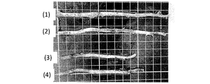
- FIG. 2 is a diagram illustrating an internal thoracic artery fragment in which a branch portion collected from a pig is excised in the embodiment.
- the internal thoracic artery piece (3) in the figure was used as a comparative example.
- ( ⁇ ) is an enlarged photograph of a cross-sectional view of the collected porcine internal thoracic artery piece stained with hematoxylin and eosin. Further, ( ⁇ ) is an enlarged photograph of a sectional view of the transplanted decellularized material obtained by decellularizing the collected porcine inner thoracic artery piece and staining it with hematoxylin and eosin. It can be confirmed that no nucleic acid is present and decellularized. Each scale bar represents 1000 ⁇ m. In the following examples, the internal thoracic artery piece (3) in the figure was used as a comparative example. FIG.
- the figure on the left side is an enlarged photograph of a section of a branch portion obtained by HE staining of a decellularized porcine internal thoracic artery piece which is a decellularization material for transplantation of the present invention before transplantation.
- the figure on the right side is an enlarged photograph of a section of a branch portion stained with HE 1 month after transplantation. Cell infiltration is observed after transplantation.
- Each scale bar represents 500 ⁇ m.
- a blood vessel having a branch is collected from a vertebrate (donor). Collecting here means separating a blood vessel having a branch from a donor.
- the “blood vessel having a branch” means a blood vessel having a branch in the donor, and includes those in which the branch is excised at the time of collection.
- the vertebrate is not particularly limited. However, since it is preferable that blood vessels are easily available, animals other than humans are preferable, and mammal livestock and avian livestock are particularly preferable.
- Mammalian livestock includes cattle, horses, camels, llamas, donkeys, yaks, sheep, pigs, goats, deer, alpaca, dogs, raccoon dogs, weasels, foxes, cats, rabbits, hamsters, guinea pigs, rats, squirrels and raccoons, etc. Is mentioned.
- avian livestock examples include parakeets, parrots, chickens, ducks, turkeys, geese, guinea fowls, pheasants, ostriches, emu and quail. Of these, pigs, rabbits or cows are preferred because of their availability.
- a blood vessel having a branch refers to a blood vessel having at least one branch portion (branch blood vessel) as shown in FIG.
- a blood vessel having a blood vessel with a branched structure has a complicated shape, so it is said that it is not suitable for processing even if it is decellularized, and since it has a branched structure, it is mostly used as a blood vessel for transplantation. It never happened.
- the blood vessel can be used as a blood vessel without a branch by adhering the branch portions of the blood vessel and closing the tube. The significance that a blood vessel having a branch that has been difficult to use in the past can be used as a graft composition is extremely large.
- blood vessels having branches include the internal thoracic artery, abdominal wall artery, gastroepiploic artery, carotid artery, radial artery, intercostal artery, muscular phrenic artery, femoral artery, deep femoral artery, aorta, ulnar artery, and upper arm Artery, anterior tibial artery, posterior tibial artery, mesenteric artery, splenic artery, internal thoracic vein, anterior intercostal vein, odd vein, semi-even vein, jugular vein, intestinal vein, femoral vein, saphenous vein, mesenteric vein, spleen Examples include veins. In consideration of physical properties (elongation, biocompatibility, strength, etc.) as the graft composition, an artery is preferable, and an internal thoracic artery is more preferable.
- a part of the body such as the chest, abdomen, or leg is incised, and the blood vessels having branches to be collected are excised.
- anesthesia method and the slaughter method methods conventionally used by those skilled in the art can be used as they are.
- a scalpel, scissors, etc. which are usually used in animal experiments and surgical operations.
- a scalpel, scissors, or the like that is usually used in animal experiments or surgical operations.
- an ultrasonic scalpel or an electric scalpel These can excise the blood vessel while coagulating the blood by ultrasonic vibration or high-frequency current at the place where the blood vessel is excised.
- an ultrasonic knife since the degree of protein denaturation at the excised portion is moderate, it is preferable to use an ultrasonic knife.
- “ultrasonic knife” and “ultrasonic vibratory knife” are used synonymously.
- Examples of the electrosurgical knife that can be used in the present invention include the Bio series manufactured by Elbe and the SHAPPER series manufactured by Izumi Engineering & Medical Co., Ltd.
- Examples of the ultrasonic scalpel that can be used in the present invention include SONOpet UST-2001 manufactured by Stryker Medtech, and Harmonic Scalpel manufactured by Ethicon End Surgery.
- the blood vessel collected in the step (a) may be in a state where the branch portion has already been excised, or in a state where the branch portion remains with a sufficient length (for example, 5 mm to several cm). If the branch part remains with a sufficient length, it is excised appropriately.
- the length of the branching portion for performing protein denaturation treatment is preferably a position 1 mm to 10 mm away from the branching portion of the blood vessel, more preferably a position 2 mm to 7 mm away, and a position 3 to 5 mm away Is most preferred. Performing protein denaturation treatment within the above range minimizes the effects of denaturation treatment (such as changes in physical properties and blood flow prevention and antithrombogenicity) when using the decellularized material for transplantation of the present invention. Can be suppressed and reorganized.
- Decellularization treatment is performed using a surfactant treatment (Singelyn JM, et al., Biomaterials, 2009, 30, 5409-5416; Singelyn JM, et al., J. Am. Coll. Cardiol., 2012, 59, 751. -763; Sonya B., et al., Sci. Transl. Med., 2013, 5, 173ra25), enzyme treatment, osmotic pressure treatment, freeze-thaw treatment, oxidant treatment, high hydrostatic pressure treatment (Sasaki S., et al. al., Mol.
- the cell is decellularized by high hydrostatic pressure treatment.
- the pressure at which high hydrostatic pressure treatment is performed may be a pressure that can destroy cells and pathogens derived from a vertebrate donor, and can be appropriately selected according to the animal species and blood vessel type of the donor. .
- the hydrostatic pressure is exemplified by 2 to 1,500 MPa.
- the applied hydrostatic pressure is higher than 50 MPa, decellularization from the blood vessel is sufficiently performed. Therefore, it is preferably 50 to 1,500 MPa, more preferably 80 to 1,300 MPa, still more preferably 90 to 1,200 MPa, and most preferably 95 to 1,100 MPa.
- Examples of the medium used for applying the high hydrostatic pressure include water, physiological saline, buffer solution, propylene glycol or an aqueous solution thereof, glycerin or an aqueous solution thereof, and an aqueous saccharide solution.
- Examples of the buffer solution include acetate buffer solution, phosphate buffer solution, citrate buffer solution, borate buffer solution, tartaric acid buffer solution, Tris buffer solution, HEPES buffer solution, and MES buffer solution.
- saccharide in the aqueous saccharide solution examples include erythrose, xylose, arabinose, allose, talose, glucose, mannose, galactose, erythritol, xylitol, mannitol, sorbitol, galactitol, sucrose, lactose, maltose, trehalose, dextran, alginic acid, and hyaluronic acid. Is mentioned.
- the temperature of the medium for high hydrostatic pressure treatment is not particularly limited as long as it does not generate ice and does not damage the tissue due to heat. It is preferably 0 to 45 ° C., more preferably 4 to 40 ° C., still more preferably 10 to 37 ° C., and most preferably 15 to 35 ° C., because the decellularization treatment is performed smoothly and the influence on the tissue is small. is there.
- the time for the high hydrostatic pressure treatment is too short, the decellularization treatment is not sufficiently performed, and if it is long, energy is wasted, so 5 minutes to 12 hours is preferable, and 7 minutes to 5 hours is more preferable. More preferably, min-3 hours.
- the suitability of decellularization can be confirmed by histological staining (hematoxylin-eosin staining) or residual DNA quantification.
- the cleaning liquid may be the same liquid as the medium used for applying the high hydrostatic pressure, may be a different cleaning liquid, or a combination of a plurality of types of cleaning liquids.
- the cleaning liquid preferably contains a nucleolytic enzyme, an organic solvent, or a chelating agent. Nucleolytic enzymes can improve the efficiency of removing nucleic acid components from blood vessels to which hydrostatic pressure is applied, organic solvents are lipids, and chelating agents are calcium ions and magnesium ions in decellularized tissues. By inactivating, calcification can be prevented when decellularized tissue is applied to the affected area.
- the portion where the branch is excised is subjected to protein denaturation treatment and closed.
- the means for denaturing the protein is not particularly limited, but from the viewpoint of ease of work and efficiency, and less damage to the tissue, it is preferable to use an ultrasonic scalpel or an electric scalpel, and damage to the tissue will be reduced. It is preferable to use an ultrasonic scalpel from the viewpoint of reducing the branch stump closing property.
- the ultrasonic scalpel has a structure in which the blade edge is mechanically ultrasonically vibrated, and frictional heat is generated near the living tissue contacted by the blade edge, so that proteins in the living tissue are denatured.
- the part where the branch blood vessel is excised and opened is adhered and closed when the protein is denatured.
- the frequency at the time of performing the treatment using ultrasonic waves as described above is preferably 20 kHz to 100 kHz, more preferably about 30 kHz to about 60 kHz.
- the output is preferably 50 to 500 mA, more preferably 100 to 400 mA, and still more preferably 200 to 300 mA (when 100 VAC is used).
- the sonication time varies depending on the frequency and output and is not particularly limited, but is preferably 0.1 second to 10 minutes, more preferably 1 second to 5 minutes, and still more preferably 3 to 60 seconds.
- the steps (a), (b), and (c) are not limited to the above order.
- the step (c) may be performed after the step (a), and then the step (b) may be performed.
- a branch portion of a blood vessel having a branch is excised before being collected from a vertebrate animal as a donor, and the portion where the branch is excised is subjected to protein denaturation treatment and collected from a donor. Later, it is also possible to decellularize the step (b). That is, the aspect included in order of a process (c), a process (a), and a process (b) is also included by this invention.
- the method may further include a step (d) of cutting the branch portion.
- this step (d) is not particularly limited. For example, it is possible to carry out in the order of step (a), step (d), step (b) and step (c), step (a), step (b), step (d) and step (c). It is.
- step (c) it is possible to close the tube without overlooking the branch vessel, and in the point that the operation becomes more efficient, in the step (c), “removing the branch” and “closing the tube by performing protein denaturation treatment” More preferably, the steps (c) and (a) are performed continuously. Even in this case, step (c) may be performed again before and after step (b).
- a branch of a vertebrate is composed of a biocompatible material including a decellularized material for transplantation which includes a resected blood vessel and has at least one branch resection part closed by protein denaturation.
- An implant composition is also provided.
- the vertebrate in the graft composition of the present invention is not particularly limited, but is preferably an animal other than a human because blood vessels are easily available, and in particular, a mammal livestock and an avian livestock are preferred.
- the mammal livestock and the avian livestock here are the same as the livestock mentioned in the specific examples of the mammalian livestock and the avian livestock used in the method for producing the decellularized material for transplantation of the present invention. Of these, pigs, rabbits or cows are preferred because of their availability.
- the blood vessel from which the branch has been excised means that a branch portion is excised from the blood vessel having the branch used for producing the decellularized material for transplantation of the present invention. It is a blood vessel.
- the blood vessel having a branch the same blood vessel as that used in the above production method can be used, and considering the physical properties (elongation, biocompatibility, strength, etc.) as the graft composition, an artery is preferable. The thoracic artery is more preferred.
- the decellularized material for transplantation used in the graft composition of the present invention has a protein-denatured part.
- the excision site of the branch portion is in an open tube state, and the open tube portion can be blocked by performing protein denaturation treatment.
- the specific method of protein denaturation treatment is not particularly limited, as with the above-described decellularized material for transplantation, but an ultrasonic scalpel or an electric scalpel can be used. It is preferable to use an ultrasonic scalpel because there is little damage to the tissue.
- the conditions regarding the frequency of ultrasonic waves and the treatment time for denaturing proteins are the same as the conditions for the frequency of ultrasonic waves and the treatment time in the above-described method for producing a decellularized material for transplantation. It is.
- the decellularization treatment can be carried out in the same manner as the decellularization treatment described above, and the decellularization treatment can be carried out by a high hydrostatic pressure treatment. It is preferable that it is processed.
- any pressure may be used as long as the cells and pathogens derived from the vertebrate donor are destroyed, and can be performed under the same conditions as the hydrostatic pressure described above.
- Examples of the medium used for applying the high hydrostatic pressure include water, physiological saline, buffer solution, propylene glycol or an aqueous solution thereof, glycerin or an aqueous solution thereof, and an aqueous saccharide solution.
- Specific examples of each solvent include The same solvent as the specific example of said solvent can be used.
- the graft composition of the present invention is made of a specific biocompatible material.
- biocompatible means that the graft is accepted by the transplanted tissue and does not induce toxicity or significant immune rejection.
- the biocompatible material is not particularly limited as long as it has no fluidity and is solid, and examples thereof include non-resorbable polymers, absorbent polymers, metals, glasses, and ceramics.
- non-resorbable polymer examples include polyethylene, polyethylene terephthalate, polybutylene, polybutylene terephthalate, polypropylene, acrylic, polyamide-imide, polyether ether ketone, polyaryl ether ketone, polycarbonate, polyamide, polyvinyl fluoride, and polyvinylidene fluoride. , Polymethyl methacrylate, and combinations and equivalents thereof, but are not limited thereto.
- the absorbent polymer may be a synthetic polymer or a natural polymer.
- Polyamino acids, polyamides, fatty acid polyesters, and natural polymers include collagen, elastin, hyaluronic acid, laminin, gelatin, keratin, chondroitin sulfate And decellularized tissue.
- metals examples include tantalum, tantalum alloy, stainless steel, titanium, titanium alloy, cobalt-chromium alloy, and the like, and biocompatible metals conventionally used in medical devices and the like can be used.
- glass or ceramic examples include tetracalcium phosphate, alpha- and beta-tricalcium phosphate, octacalcium phosphate, hydroxyapatite, substituted apatite, monetite, metaphosphate, pyrophosphate, phosphate glass, etc. Phosphates, calcium and magnesium carbonates, sulfates, and oxides, and combinations thereof, but are not limited thereto.
- the graft composition of the present invention is composed of a biocompatible material containing at least a part of a specific decellularized material for transplantation.
- “included in at least a part” means that the decellularized material for transplantation can be present in one or more and a plurality of biocompatible materials. It is more preferable that the biocompatible material is composed only of a decellularized material in that self cell infiltration is more effectively achieved.
- the graft composition of the present invention can function as a part of living tissue after transplantation.
- it can function as a blood vessel substitute for transplantation.
- FIG. 2 shows the porcine internal thoracic arteries (1) to (4).
- FIG. 3 shows a cross-sectional view (HE staining).
- the porcine internal thoracic artery was treated with an ultrahigh hydrostatic apparatus (Dr. Chef, manufactured by Kobe Steel) using water as a medium.
- the ultra-high hydrostatic pressure treatment was performed under the conditions of an applied pressure of 600 MPa, a pressure increase time of 9 minutes, a pressure maintenance time of 120 minutes, a pressure decrease time of 9 minutes, and a pressure medium temperature of 30 ° C.
- washing with 50 mg / L DNase I solution was performed for 4 days, EtOH solution for 3 days, and citric acid solution for 4 days to complete decellularization.
- HE staining is a method that uses two types of dyes, hematoxylin and eosin, to separate the cell nucleus and tissues and components other than the nucleus. Hematoxylin makes the nucleus blue-blue, and eosin removes cytoplasm, fibers, and red blood cells. Can be dyed pink.
- the pressure strength is measured by using a syringe pump (YSP-101) to feed physiological saline into the porcine internal thoracic artery from a 20 mL syringe at a rate of 3 mL / min, and the pressure at that time is a digital pressure gauge (KDM30). Measured with.
- YSP-101 syringe pump
- KDM30 digital pressure gauge
- the part of the branched blood vessel in which the leakage of physiological saline was confirmed was ligated with 6-0 proline thread. Ligation was performed by passing a suture thread through the adventitia part and then tying up the branched tubular part from the outside.
- the decellularized porcine internal thoracic artery and the peripheral part of the abdominal aorta were anastomosed in the same manner to produce a bypass blood vessel.
- the abdominal aorta between the central anastomosis and the peripheral anastomosis was ligated with 6-0 proline thread to direct blood flow to the bypass vessel.
- the abdomen was closed by suturing the peritoneum with 4-0 PDS thread, the muscle layer with 2-0 PDS, and the skin with 4-0 PDS thread.
- Vitoril (registered trademark) 0.5 mL was administered as an antibiotic, and atipamezole hydrochloride (trade name: Atipame injection 1 ml) was injected subcutaneously into the thigh as a medetomidine hydrochloride antagonist. After confirming wakefulness, the rabbit was transferred to the breeding cage.
- -Necropsy An autopsy was performed on rabbits 1 month or 3 months after transplantation of decellularized porcine internal thoracic artery.
- the abdominal aorta (transplanted decellularized porcine internal thoracic artery) was exposed by the same method as at the time of transplantation without taking an infusion line from the rabbit.
- vascular patency evaluation and thrombus formation evaluation were performed.
- Vascular patency evaluation was performed by cutting the peripheral side of the abdominal aorta and determining whether or not blood was released. All samples confirmed exsanguination and indicated that the blood vessels were patent.
- pentobarbital sodium Somnopentyl (registered trademark) 5 mL was injected into the ear vein and sacrificed. After the heart, breathing stopped, the decellularized porcine internal thoracic artery was removed. The inside of the decellularized porcine internal thoracic artery was washed with physiological saline, then incised in the longitudinal direction, and thrombus formation was visually observed. There was no adhesion of thrombus to the luminal surface of decellularized porcine internal thoracic artery, indicating high antithrombogenicity. “Patent” means that a hollow state is maintained without occluding the lumen of a blood vessel.
- the pathological specimen of the transplanted decellularized porcine internal thoracic artery was produced by the Institute for New Histological Science.
- an ultrasonic scalpel of the decellularized porcine internal thoracic artery or a portion where the tissue structure of the branched blood vessel portion occluded with 6-0 proline thread can be confirmed is an HE-stained section, Elastica van Gieson stain (Elastica van Gieson stain)
- EVG staining Elastica van Gieson stain
- a section is continuously prepared with a thickness of approximately 5 ⁇ m in the longitudinal direction of the blood vessel, and the section is cut at a portion where the tissue structure of the branch blood vessel portion blocked with an ultrasonic knife or 6-0 proline thread can be confirmed. It was created.
- EVG staining is a staining method for identifying elastic fibers, in which elastin in connective tissue can be dyed in black purple and collagen in red purple.
- FIG. 4 An HE-stained image, an EVG-stained image (3 months) of an example sample, and an HE-stained image and an EVG-stained image (3 months) of a comparative example sample are shown below (FIG. 4).
- the cells also infiltrated into the decellularized porcine internal thoracic artery tissue portion stained in dark purple by EVG staining.
- the comparative sample no cell infiltration was observed in the decellularized porcine internal thoracic artery tissue portion stained in dark purple by EVG staining.
- a finding was found in which the step of the branch blood vessel was covered with rabbit cells at one month after transplantation (FIG. 5).
- 1 a blood vessel having a branch
- 2 a branched portion (branched blood vessel)
- 3 a protein denatured portion
Abstract
Description
(a)脊椎動物(ドナー)から分枝を有する血管を採取する工程、
(b)当該血管を脱細胞化する工程、及び
(c)分枝を切除した部分をタンパク質変性処理により接着し閉管する工程
を含む、移植用脱細胞化材料の製造方法を提供する。
本発明は、分枝を有する血管を脊椎動物(ドナー)から採取する。
ここでいう採取とは、分枝を有する血管をドナーから分離することを意味する。また、「分枝を有する血管」はドナーの中で分枝を有していた血管の意であり、採取する際に分枝が切除されているものも含む。
枝分かれした構造の血管を有する血管は複雑な形状であるため、脱細胞化しても加工に適するものではないとされているほか、分枝構造を有するため、移植用血管用途としてもほとんど利用されることはなかった。本発明により、血管の分枝部分を接着させ閉管することで、分枝のない血管として利用できる。従来、利用が困難であった分枝を有する血管を移植片組成物として利用できる意義は極めて大きいものである。
脊椎動物の身体の一部を切開するときは、動物実験や外科用手術で通常用いるメスやハサミ等を使用することが可能である。
なお、本明細書においては「超音波メス」と「超音波振動メス」を同義で用いるものとする。
なお、タンパク質変性処理を行う分枝部分の長さは、血管の枝分れ部分から1mm~10mm離れた位置であることが好ましく、2mm~7mm離れた位置がより好ましく、3~5mm離れた位置が最も好ましい。上記範囲でタンパク質変性処理を行うことで、本発明の移植用脱細胞材料を利用する際の変性処理に伴う影響(物性の変化および血流を妨げないことや抗血栓性など)を最小限に抑え、再組織化を促すことができる。
なお、上記「分枝を切除した部分を、タンパク質変性処理を行って閉管する」とは、「分枝の切除」と「タンパク質変性処理を行って閉管する」ことを同時に行う場合も含むものとする。
(a)脊椎動物(ドナー)から分枝を有する血管を採取する工程、
(b)当該血管を脱細胞化する工程、及び
(c)分枝を切除した部分をタンパク質変性処理により接着し閉管する工程
を含む、移植用脱細胞化材料の製造方法の各工程の順序について説明する。
例えば、工程(a)、工程(b)及び工程(c)をこの順番で行う他に、工程(a)の後に、工程(c)を行い、その後、工程(b)を行ってもよい。また、別の態様として、分枝を有する血管の分枝部分をドナーである脊椎動物から採取する前に切除し、その分枝を切除した部分をタンパク質変性処理して、ドナーから採取し、この後に、工程(b)の脱細胞化を行うことも可能である。つまり、工程(c)、工程(a)、工程(b)の順序で含む態様も本発明に包含される。
例えば、工程(a)、工程(d)、工程(b)及び工程(c)の順序、工程(a)、工程(b)、工程(d)及び工程(c)の順序で行うことも可能である。
本発明においては、分枝血管を見落とすことなく閉管できること、また作業がより効率的となる点で、(c)工程において「分枝の切除」と「タンパク質変性処理を行って閉管する」ことを同時に行い、さらに(c)工程と(a)工程を連続的に行うことがより好ましい。なお、この場合であっても(b)工程の前後に再度工程(c)を行ってもよい。
タンパク質変性処理の具体的な方法は、上記の移植用脱細胞化材料と同様に特に限定されないが、超音波メスや電気メスを使用することができる。組織への損傷が少ないことから超音波メスを使用することが好ましい。
高静水圧処理を行う際の圧力に関し、脊椎動物であるドナー由来の細胞や病原体が破壊される程度の圧力であればよく、上記の静水圧の条件と同じ条件で行うことができる。また、高静水圧の印加に使用する媒体としては、水、生理食塩水、緩衝液、プロピレングリコール又はその水溶液、グリセリン又はその水溶液、及び糖類水溶液等が挙げられ、各溶媒の具体例としては、上記の溶媒の具体例と同じ溶媒を使用することができる。
ブタ内胸動脈の採取(分枝血管の処理)
月齢3~4か月の食用ブタ(SPF(specific pathogen free)ブタ、メス、体重約50キロ)を麻酔下で開胸し内胸動脈を露出させた。超音波メス(ハーモニックスカルペルII、ハーモニックSYNERGY、エチコンエンドサージェリー社)の設定を55500Hz、出力レベル2として分枝血管を接触させ凝固させながら胸壁から内胸動脈を剥離(採取)した。
2頭のブタから4本の内胸動脈を採取した。
採取したブタ内胸動脈4本の長さと内径を測定した。それぞれ(1)長さ:140mm、内径:3.2mm、(2)長さ:135mm、内径:2.6mm、(3)長さ:95mm、内径:2.9mm、(4)長さ:105mm、内径:2.8mmであった。図2に得られたブタ内胸動脈(1)~(4)を示す。また、図3にその断面図(HE染色)を示す。
[使用試薬]
・DNs溶液
DNase I(Roche,Grade II) 50mg/L、MgCl2・6H2O(Wako、 試薬特級)2.55g/Lを含有した大塚生理食塩水
・EtOH溶液
局方エタノールと大塚生理食塩水を体積比で80%となるように調製した溶液
・クエン酸溶液
クエン酸三ナトリウム二水和物(Calbiochem、Molecular Biology Grade)とクエン酸無水物 (Wako、特級)を大塚生理食塩水に加え、pH7.4に調製した溶液
超高圧処理後50mg/L DNaseI溶液で4日、EtOH溶液で3日、クエン酸溶液で4日洗浄を行い、脱細胞化を完了した。
なお、HE染色はヘマトキシリンとエオシンの2種類の色素を用いて、細胞核と核以外の組織、成分を染め分ける方法であり、ヘマトキシリンで核を青藍色に、エオシンで細胞質・線維類や赤血球をピンク色に染めることができる。
脱細胞化後、各ブタ内胸動脈の片端をクランプで挟み反対側から20mLシリンジを用いて生理食塩水を送液して、耐圧強度を測定した。上記の(3)を除く(1)、(2)、(4)のブタ内胸動脈を用いた移植用脱細胞化材料では耐圧強度が150mmHg以上であった。
また、分枝部分の一部が閉塞されていない(3)では生理食塩水の漏れが確認できた。
なお、耐圧強度の測定は、シリンジポンプ(YSP-101)で20mLシリンジから3mL/minの速度で生理食塩水をブタ内胸動脈内に送液し、そのときの圧力をデジタル圧力計(KDM30)で計測した。
以後の実験において、(1)、(2)、(4)を実施例し、(3)を比較例とする。
(3)の移植用脱細胞材料について、生理食塩水の漏れが確認できた分枝血管を切除した部分を6-0プロリン糸で結紮した。結紮は血管外膜部分に縫合糸を通した後、分枝の管状部分を外側から縛り上げるように行った。
・移植方法
体重3.70-4.00kgのウサギ(品種:日本白兎、5ヶ月齢、オス)にペントバルビタールナトリウム塩酸塩(商品名:ソムノペンチル(登録商標)0.2ml)、生理食塩水(1.8ml/ウサギ)、塩酸メデトミジン(商品名:ドルベネ注1ml)を皮下注射した。不動化確認後、腹部皮膚を剃毛した。呼気麻酔としてイソフルラン吸入し(導入時2%、手術時1%)、保定台に保定した後、ポビドンヨード製剤を浸漬させたガーゼで皮膚を消毒した。続いて、生理食塩水3倍希釈リドカイン注射液(商品名:キシロカイン(登録商標))を腹部皮下に注射し、耳の静脈から点滴ラインをとりソリタ(登録商標)T-3を静注した。電気メス、ハサミを使用し、皮膚、腹膜を切開し腹部大動脈を露出させ、ヘパリン1000単位(1000単位/mLヘパリン1mL)を点滴ラインから静注し、その後ヘパリン静注後3分が経過したのを確認し、腹部大動脈中枢側を遮断した。腹部大動脈にシャントチューブを挿入後、血流遮断を解除した。シャントチューブで血流を確保しながら、8-0プロリン糸を使用し端側吻合法で脱細胞化ブタ内胸動脈と腹部大動脈中枢部を吻合した。続いて同様の方法で脱細胞化ブタ内胸動脈と腹部大動脈末梢部を吻合し、バイパス血管を作製した。加えて、バイパス血管へ血流を向かわせるため中枢吻合部と末梢吻合部の間の腹部大動脈を6-0プロリン糸で結紮した。脱細胞化ブタ内胸動脈移植後は、腹膜を4-0PDS糸、筋層を2-0PDS、皮膚を4-0PDS糸で縫合し腹部を閉じた。抗生剤としてバイトリル(登録商標)を0.5mL投与、塩酸メデトミジン拮抗剤として塩酸アチパメゾール(商品名:アチパメ注1ml)を大腿部皮下に注射した。覚醒確認後、飼育ケージにウサギを移した。
脱細胞化ブタ内胸動脈移植後1カ月或いは3ヶ月のウサギについて剖検を行った。ウサギから点滴ラインはとらず、移植時と同様の方法で腹部大動脈を(移植した脱細胞化ブタ内胸動脈)を露出させた。続いて血管開存評価、血栓形成評価を行った。血管開存評価は、腹部大動脈の末梢側を切断し、放血の有無で判断した。すべてのサンプルで放血を確認し、血管が開存していたことが示された。血管開存評価後、耳静脈にペントバルビタールナトリウム(ソムノペンチル(登録商標)5mL)を注射し、サクリファイスした。心臓、呼吸停止後、脱細胞化ブタ内胸動脈を取り出した。脱細胞化ブタ内胸動脈内部を生理食塩水で洗浄した後、長軸方向に切開し血栓形成を目視で観察した。脱細胞化ブタ内胸動脈の内腔面へは血栓の付着はなく抗血栓性が高いことが示された。なお「開存」とは、血管の内腔が閉塞することなく、中空の状態が維持されていることをいう。
移植した脱細胞化ブタ内胸動脈の病理標本の作製は(株)新組織科学研究所が行った。この際、脱細胞化ブタ内胸動脈の超音波メス或いは、6-0プロリン糸で閉塞させた分枝血管部分の組織構造が確認できる部分でHE染色切片、エラスチカ・ワンギーソン染色(Elastica van Gieson染色、以下、EVG染色ということもある)を行い、EVG染色切片の作製を行った。具体的には、血管の長軸方向におよそ5μm厚で連続的に切片を作成し、超音波メス或いは、6-0プロリン糸で閉塞させた分枝血管部分の組織構造が確認できる部分で切片を作成した。
また実施例サンプルでは、移植後1ヶ月の段階で分枝血管の段差がウサギ細胞で覆われる所見をみだした(図5)。
流通食用ブタ由来のブロック肉より、超音波メス(EESジェネレーター、ハーモニック SYNERGY、ハーモニック FOCUS+、<ブルーハンドピース使用>、エチコンエンドサージェリー社)の設定を55500Hz、出力レベル5および2として分枝血管を接触させ凝固させながら胸壁から内胸動脈を剥離(採取)した。分枝血管の凝固は血管の枝分かれ部分から3-5mm離れた位置で行うようにし、5本の内胸動脈を剥離(採取)した。上記「3」記載の方法にて脱細胞化処置を行った後、上記「4」記載の方法にて耐圧強度の測定を行った。その結果、5本のサンプル全てにおいて、耐圧強度は250mmHg以上であることを確認した。
Claims (7)
- (a)脊椎動物(ドナー)から分枝を有する血管を採取する工程、
(b)当該血管を脱細胞化する工程、及び
(c)分枝を切除した部分をタンパク質変性処理により接着し閉管する工程
を含む、移植用脱細胞化材料の製造方法。 - 前記工程(a)の後に、(d)採取した血管の分枝を切除する工程を含む、請求項1に記載の移植用脱細胞化材料の製造方法。
- 前記タンパク質変性処理が超音波振動処理である、請求項1又は2に記載の移植用脱細胞化材料の製造方法。
- 工程(b)の脱細胞化が超高静水圧処理を含む、請求項1~3のいずれかに記載の移植用脱細胞化材料の製造方法。
- 脊椎動物(ドナー)の分枝が切除された血管からなり、かつタンパク質変性により閉管された分枝切除部を少なくとも1以上有する移植用脱細胞化材料を含む生体適合性材料からなる移植片組成物。
- 前記血管が内胸動脈を含む、請求項5に記載の移植片組成物。
- ブタの内胸動脈を含む血管の脱細胞化組織からなり、タンパク質変性により閉管された分枝切除部を有する移植片組成物。
Priority Applications (7)
| Application Number | Priority Date | Filing Date | Title |
|---|---|---|---|
| US16/615,177 US20200222589A1 (en) | 2017-05-30 | 2018-05-25 | Method for producing decellularized material for transplantation and graft composition consisting of biocompatible material including said material |
| CN201880036506.4A CN110740762A (zh) | 2017-05-30 | 2018-05-25 | 移植用脱细胞化材料制造方法及由包含该材料的生物相容性材料组成的移植片组成物 |
| EP18809381.9A EP3632481B1 (en) | 2017-05-30 | 2018-05-25 | Method for producing decellularized material for transplantation and graft composition comprising biocompatible material including said material |
| JP2019522186A JPWO2018221402A1 (ja) | 2017-05-30 | 2018-05-25 | 移植用脱細胞化材料の製造方法及び当該材料を含む生体適合性材料からなる移植片組成物 |
| KR1020197035144A KR20200016226A (ko) | 2017-05-30 | 2018-05-25 | 이식용 탈세포화 재료의 제조 방법 및 당해 재료를 포함하는 생체 적합성 재료로 이루어지는 이식편 조성물 |
| CA3065498A CA3065498A1 (en) | 2017-05-30 | 2018-05-25 | Method for producing decellularized material for transplantation and graft composition consisting of biocompatible material including said material |
| JP2023061981A JP2023076668A (ja) | 2017-05-30 | 2023-04-06 | 移植用脱細胞化材料の製造方法及び当該材料を含む生体適合性材料からなる移植片組成物 |
Applications Claiming Priority (2)
| Application Number | Priority Date | Filing Date | Title |
|---|---|---|---|
| JP2017106400 | 2017-05-30 | ||
| JP2017-106400 | 2017-05-30 |
Publications (1)
| Publication Number | Publication Date |
|---|---|
| WO2018221402A1 true WO2018221402A1 (ja) | 2018-12-06 |
Family
ID=64455364
Family Applications (1)
| Application Number | Title | Priority Date | Filing Date |
|---|---|---|---|
| PCT/JP2018/020141 WO2018221402A1 (ja) | 2017-05-30 | 2018-05-25 | 移植用脱細胞化材料の製造方法及び当該材料を含む生体適合性材料からなる移植片組成物 |
Country Status (8)
| Country | Link |
|---|---|
| US (1) | US20200222589A1 (ja) |
| EP (1) | EP3632481B1 (ja) |
| JP (2) | JPWO2018221402A1 (ja) |
| KR (1) | KR20200016226A (ja) |
| CN (1) | CN110740762A (ja) |
| CA (1) | CA3065498A1 (ja) |
| TW (1) | TWI749233B (ja) |
| WO (1) | WO2018221402A1 (ja) |
Families Citing this family (2)
| Publication number | Priority date | Publication date | Assignee | Title |
|---|---|---|---|---|
| CN218059021U (zh) * | 2022-09-22 | 2022-12-16 | 舩本诚一 | 一种医用材料处理用高压设备 |
| CN115554472B (zh) * | 2022-09-22 | 2023-08-18 | 舩本诚一 | 一种用于移植的生物组织处理方法 |
Citations (14)
| Publication number | Priority date | Publication date | Assignee | Title |
|---|---|---|---|---|
| JPS60501540A (ja) | 1983-06-10 | 1985-09-19 | ユニバ−シテイ パテンツ,インコ−ポレイテイド | 細胞外マトリクスの体移植片並びに該移植片の製造及び使用のための手段及び方法 |
| JP2000505315A (ja) * | 1996-01-24 | 2000-05-09 | オリジン・メッドシステムズ,インコーポレイテッド | 切開プローブをもつ組織分離カニューレ及び方法 |
| JP2002507907A (ja) | 1997-06-27 | 2002-03-12 | バーダー、アウグスチヌス | 生合成移植片及びその製造方法 |
| JP2003518981A (ja) | 1999-12-29 | 2003-06-17 | チルドレンズ メディカル センター コーポレーション | 臓器脱細胞化のための方法および組成物 |
| JP2003525062A (ja) | 1998-09-30 | 2003-08-26 | メドトロニック・インコーポレーテッド | 移植で使用される組織の無機質化を減少させる方法 |
| JP2004094552A (ja) | 2002-08-30 | 2004-03-25 | Sumitomo Chem Co Ltd | 病理組織画像解析方法および病理組織画像解析システム |
| JP2005185507A (ja) | 2003-12-25 | 2005-07-14 | Yoshihiro Takami | 皮膚の分離無細胞化方法、無細胞化真皮マトリックス及びその製造方法並びに無細胞化真皮マトリックスを用いた複合培養皮膚 |
| JP2005211480A (ja) | 2004-01-30 | 2005-08-11 | Yoshihiro Takami | 皮膚の分離無細胞化方法、無細胞化真皮マトリックス及びその製造方法並びに無細胞化真皮マトリックスを用いた複合培養皮膚 |
| JP4092397B2 (ja) | 2002-09-10 | 2008-05-28 | 国立循環器病センター総長 | 超高静水圧印加による移植用生体組織の処理方法 |
| WO2008111530A1 (ja) | 2007-03-09 | 2008-09-18 | National University Corporation, Tokyo Medical And Dental University | 脱細胞化軟組織の調製方法、移植片、及び培養部材 |
| JP2009050297A (ja) | 2007-08-23 | 2009-03-12 | Tokyo Medical & Dental Univ | 脱細胞処理液、脱細胞化組織の調製方法、移植片、及び培養部材 |
| JP2010221012A (ja) | 2009-02-25 | 2010-10-07 | Kobe Univ | 高張電解質溶液による生体組織の脱細胞化処理方法 |
| JP2013502275A (ja) | 2009-08-18 | 2013-01-24 | ライフセル コーポレーション | 組織の処理方法 |
| WO2016194895A1 (ja) * | 2015-06-02 | 2016-12-08 | 株式会社Adeka | 生体由来組織のシート、該シートから得られる管状構造体及び該管状構造体からなる人工血管 |
Family Cites Families (16)
| Publication number | Priority date | Publication date | Assignee | Title |
|---|---|---|---|---|
| JPS63279832A (ja) * | 1987-05-12 | 1988-11-16 | Sugino Mach:Kk | 液体ジェット手術装置 |
| US5026387A (en) * | 1990-03-12 | 1991-06-25 | Ultracision Inc. | Method and apparatus for ultrasonic surgical cutting and hemostatis |
| US5336616A (en) * | 1990-09-12 | 1994-08-09 | Lifecell Corporation | Method for processing and preserving collagen-based tissues for transplantation |
| US6293970B1 (en) * | 1998-06-30 | 2001-09-25 | Lifenet | Plasticized bone and soft tissue grafts and methods of making and using same |
| US6310036B1 (en) * | 1999-01-09 | 2001-10-30 | Last Chance Tissue Adhesives Corporation | High strength, Bio-compatible tissue adhesive and methods for treating vigorously bleeding surfaces |
| IL139708A0 (en) * | 2000-11-15 | 2002-02-10 | Amiel Gilad | Process of decellularizing biological matrices and acellular biological matrices useful in tissue engineering |
| WO2005063316A1 (ja) * | 2003-12-26 | 2005-07-14 | Cardio Incorporated | 移植可能な生体材料およびその作成方法 |
| CN101185770A (zh) * | 2007-12-27 | 2008-05-28 | 南京市儿童医院 | 猪带瓣血管脱细胞的支架的超高压制备方法 |
| KR101335203B1 (ko) * | 2010-03-26 | 2013-11-29 | 숙명여자대학교산학협력단 | 혈관신생촉진용 펩타이드 및 이의 용도 |
| US20130013083A1 (en) * | 2011-01-06 | 2013-01-10 | Humacyte | Tissue-Engineered Constructs |
| CN104411816B (zh) * | 2012-03-16 | 2018-01-02 | 诺瓦赫普有限公司 | 经生物工程改造的同种异体血管 |
| EP2967629B1 (en) * | 2013-03-14 | 2019-05-29 | Saphena Medical, Inc. | Unitary endoscopic vessel harvesting devices |
| CN203802502U (zh) * | 2013-09-06 | 2014-09-03 | 姚建新 | 超声切割止血手术仪 |
| CN104287869B (zh) * | 2014-09-19 | 2017-03-29 | 上海市肺科医院 | 一种用于气管移植的新型纳米纤维膜/纱支架及其制备方法 |
| CN204293229U (zh) * | 2014-11-28 | 2015-04-29 | 徐胜前 | 一种超声手术刀 |
| TWI711455B (zh) * | 2015-05-13 | 2020-12-01 | 臺北榮民總醫院 | 多層視網膜細胞移植物 |
-
2018
- 2018-05-25 EP EP18809381.9A patent/EP3632481B1/en active Active
- 2018-05-25 JP JP2019522186A patent/JPWO2018221402A1/ja active Pending
- 2018-05-25 KR KR1020197035144A patent/KR20200016226A/ko not_active Application Discontinuation
- 2018-05-25 CN CN201880036506.4A patent/CN110740762A/zh active Pending
- 2018-05-25 CA CA3065498A patent/CA3065498A1/en active Pending
- 2018-05-25 WO PCT/JP2018/020141 patent/WO2018221402A1/ja active Application Filing
- 2018-05-25 US US16/615,177 patent/US20200222589A1/en active Pending
- 2018-05-29 TW TW107118285A patent/TWI749233B/zh active
-
2023
- 2023-04-06 JP JP2023061981A patent/JP2023076668A/ja active Pending
Patent Citations (14)
| Publication number | Priority date | Publication date | Assignee | Title |
|---|---|---|---|---|
| JPS60501540A (ja) | 1983-06-10 | 1985-09-19 | ユニバ−シテイ パテンツ,インコ−ポレイテイド | 細胞外マトリクスの体移植片並びに該移植片の製造及び使用のための手段及び方法 |
| JP2000505315A (ja) * | 1996-01-24 | 2000-05-09 | オリジン・メッドシステムズ,インコーポレイテッド | 切開プローブをもつ組織分離カニューレ及び方法 |
| JP2002507907A (ja) | 1997-06-27 | 2002-03-12 | バーダー、アウグスチヌス | 生合成移植片及びその製造方法 |
| JP2003525062A (ja) | 1998-09-30 | 2003-08-26 | メドトロニック・インコーポレーテッド | 移植で使用される組織の無機質化を減少させる方法 |
| JP2003518981A (ja) | 1999-12-29 | 2003-06-17 | チルドレンズ メディカル センター コーポレーション | 臓器脱細胞化のための方法および組成物 |
| JP2004094552A (ja) | 2002-08-30 | 2004-03-25 | Sumitomo Chem Co Ltd | 病理組織画像解析方法および病理組織画像解析システム |
| JP4092397B2 (ja) | 2002-09-10 | 2008-05-28 | 国立循環器病センター総長 | 超高静水圧印加による移植用生体組織の処理方法 |
| JP2005185507A (ja) | 2003-12-25 | 2005-07-14 | Yoshihiro Takami | 皮膚の分離無細胞化方法、無細胞化真皮マトリックス及びその製造方法並びに無細胞化真皮マトリックスを用いた複合培養皮膚 |
| JP2005211480A (ja) | 2004-01-30 | 2005-08-11 | Yoshihiro Takami | 皮膚の分離無細胞化方法、無細胞化真皮マトリックス及びその製造方法並びに無細胞化真皮マトリックスを用いた複合培養皮膚 |
| WO2008111530A1 (ja) | 2007-03-09 | 2008-09-18 | National University Corporation, Tokyo Medical And Dental University | 脱細胞化軟組織の調製方法、移植片、及び培養部材 |
| JP2009050297A (ja) | 2007-08-23 | 2009-03-12 | Tokyo Medical & Dental Univ | 脱細胞処理液、脱細胞化組織の調製方法、移植片、及び培養部材 |
| JP2010221012A (ja) | 2009-02-25 | 2010-10-07 | Kobe Univ | 高張電解質溶液による生体組織の脱細胞化処理方法 |
| JP2013502275A (ja) | 2009-08-18 | 2013-01-24 | ライフセル コーポレーション | 組織の処理方法 |
| WO2016194895A1 (ja) * | 2015-06-02 | 2016-12-08 | 株式会社Adeka | 生体由来組織のシート、該シートから得られる管状構造体及び該管状構造体からなる人工血管 |
Non-Patent Citations (7)
| Title |
|---|
| JOURNAL OF BIOMEDICAL MATERIALS RESEARCH A, vol. 103A, October 2015 (2015-10-01), pages 10 |
| NEGISHI J. ET AL., J. ARTIF. ORGANS, vol. 14, 2011, pages 223 - 231 |
| SASAKI S. ET AL., MOL. VIS., vol. 15, 2009, pages 2022 - 2028 |
| SINGELYN J. M. ET AL., BIOMATERIALS, vol. 30, 2009, pages 5409 - 5416 |
| SINGELYN J. M. ET AL., J. AM. COLL. CARDIOL., vol. 59, 2012, pages 751 - 763 |
| SONYA B. ET AL., SCI. TRANSL. MED., vol. 5, 2013, pages 173ra25 |
| YOSHIHIDE H. ET AL., BIOMATERIALS, vol. 31, 2010, pages 3590 - 3595 |
Also Published As
| Publication number | Publication date |
|---|---|
| TW201906586A (zh) | 2019-02-16 |
| KR20200016226A (ko) | 2020-02-14 |
| CA3065498A1 (en) | 2018-12-06 |
| EP3632481A4 (en) | 2021-03-31 |
| JP2023076668A (ja) | 2023-06-01 |
| TWI749233B (zh) | 2021-12-11 |
| EP3632481A1 (en) | 2020-04-08 |
| CN110740762A (zh) | 2020-01-31 |
| US20200222589A1 (en) | 2020-07-16 |
| JPWO2018221402A1 (ja) | 2020-04-30 |
| EP3632481B1 (en) | 2023-08-23 |
Similar Documents
| Publication | Publication Date | Title |
|---|---|---|
| US8835166B2 (en) | Extracellular matrix material created using non-thermal irreversible electroporation | |
| Pennel et al. | The performance of cross-linked acellular arterial scaffolds as vascular grafts; pre-clinical testing in direct and isolation loop circulatory models | |
| Chaouat et al. | The evaluation of a small-diameter polysaccharide-based arterial graft in rats | |
| Assmann et al. | Development of a growing rat model for the in vivo assessment of engineered aortic conduits | |
| JP2023076668A (ja) | 移植用脱細胞化材料の製造方法及び当該材料を含む生体適合性材料からなる移植片組成物 | |
| AU2013373262B2 (en) | Artificial blood vessel using decellularized blood vessel sheet | |
| WO2017175870A1 (ja) | 肝切除を受けた肝臓の組織再構築用移植材、その製造方法、及び肝切除を受けた肝臓の再構築方法 | |
| US20220323647A1 (en) | Sheet of biological tissue, tubular structure obtained from said sheet, and artificial blood vessel comprising said tubular structure | |
| Shirakigawa et al. | Decellularization of liver and organogenesis in rats | |
| JP2010221012A (ja) | 高張電解質溶液による生体組織の脱細胞化処理方法 | |
| JPS60501540A (ja) | 細胞外マトリクスの体移植片並びに該移植片の製造及び使用のための手段及び方法 | |
| WO2019087880A1 (ja) | シート状脱細胞化材料及び同材料を用いる人工血管 | |
| KR102659840B1 (ko) | 생체 유래 조직의 시트, 상기 시트에서 얻은 관상 구조체 및 상기 관상 구조체로 이루어진 인공 혈관 | |
| Wang | Evaluation of Amnion Membrane Made Vascular Graft in Rat Model and Porcine Model | |
| JP6515429B2 (ja) | 人工血管、および、人工血管の製造方法 | |
| KR101709008B1 (ko) | 생체 적합성이 증진된 진피대체물의 제조방법 | |
| JP2011130989A (ja) | 抗血栓性修飾剤、医療用具、及び、多孔質コラーゲン |
Legal Events
| Date | Code | Title | Description |
|---|---|---|---|
| 121 | Ep: the epo has been informed by wipo that ep was designated in this application |
Ref document number: 18809381 Country of ref document: EP Kind code of ref document: A1 |
|
| WWE | Wipo information: entry into national phase |
Ref document number: 2019522186 Country of ref document: JP |
|
| ENP | Entry into the national phase |
Ref document number: 20197035144 Country of ref document: KR Kind code of ref document: A |
|
| ENP | Entry into the national phase |
Ref document number: 3065498 Country of ref document: CA |
|
| NENP | Non-entry into the national phase |
Ref country code: DE |
|
| ENP | Entry into the national phase |
Ref document number: 2018809381 Country of ref document: EP Effective date: 20200102 |