WO2018097307A1 - Domaine de liaison à l'antigène et polypeptide comprenant une section de transport - Google Patents
Domaine de liaison à l'antigène et polypeptide comprenant une section de transport Download PDFInfo
- Publication number
- WO2018097307A1 WO2018097307A1 PCT/JP2017/042542 JP2017042542W WO2018097307A1 WO 2018097307 A1 WO2018097307 A1 WO 2018097307A1 JP 2017042542 W JP2017042542 W JP 2017042542W WO 2018097307 A1 WO2018097307 A1 WO 2018097307A1
- Authority
- WO
- WIPO (PCT)
- Prior art keywords
- antigen
- antibody
- domain
- binding
- single domain
- Prior art date
Links
- 239000000427 antigen Substances 0.000 title claims abstract description 562
- 108091007433 antigens Proteins 0.000 title claims abstract description 562
- 102000036639 antigens Human genes 0.000 title claims abstract description 562
- 230000027455 binding Effects 0.000 title claims abstract description 556
- 108090000765 processed proteins & peptides Proteins 0.000 title claims abstract description 317
- 102000004196 processed proteins & peptides Human genes 0.000 title claims abstract description 288
- 229920001184 polypeptide Polymers 0.000 title claims abstract description 282
- 108010003723 Single-Domain Antibodies Proteins 0.000 claims abstract description 265
- 238000000034 method Methods 0.000 claims abstract description 111
- 238000012216 screening Methods 0.000 claims abstract description 61
- 238000004519 manufacturing process Methods 0.000 claims abstract description 37
- 230000002401 inhibitory effect Effects 0.000 claims abstract description 21
- 239000008194 pharmaceutical composition Substances 0.000 claims abstract description 7
- 108091005804 Peptidases Proteins 0.000 claims description 226
- 239000004365 Protease Substances 0.000 claims description 224
- 238000003776 cleavage reaction Methods 0.000 claims description 136
- 230000007017 scission Effects 0.000 claims description 136
- 230000004927 fusion Effects 0.000 claims description 100
- 210000004369 blood Anatomy 0.000 claims description 26
- 239000008280 blood Substances 0.000 claims description 26
- 102100037486 Reverse transcriptase/ribonuclease H Human genes 0.000 claims 3
- 102000035195 Peptidases Human genes 0.000 description 223
- 235000019419 proteases Nutrition 0.000 description 200
- 235000001014 amino acid Nutrition 0.000 description 150
- 210000001519 tissue Anatomy 0.000 description 112
- 150000001413 amino acids Chemical class 0.000 description 94
- 210000004027 cell Anatomy 0.000 description 94
- 125000003275 alpha amino acid group Chemical group 0.000 description 71
- 206010028980 Neoplasm Diseases 0.000 description 70
- 201000011510 cancer Diseases 0.000 description 59
- 238000006467 substitution reaction Methods 0.000 description 59
- 102100026120 IgG receptor FcRn large subunit p51 Human genes 0.000 description 54
- 101710177940 IgG receptor FcRn large subunit p51 Proteins 0.000 description 54
- -1 uPA Proteins 0.000 description 54
- 102100037792 Interleukin-6 receptor subunit alpha Human genes 0.000 description 52
- 125000003412 L-alanyl group Chemical group [H]N([H])[C@@](C([H])([H])[H])(C(=O)[*])[H] 0.000 description 47
- 230000000694 effects Effects 0.000 description 47
- 108040006858 interleukin-6 receptor activity proteins Proteins 0.000 description 47
- 238000012360 testing method Methods 0.000 description 31
- 108090000623 proteins and genes Proteins 0.000 description 26
- 230000002757 inflammatory effect Effects 0.000 description 23
- 125000005647 linker group Chemical group 0.000 description 22
- 235000018102 proteins Nutrition 0.000 description 21
- 102000004169 proteins and genes Human genes 0.000 description 21
- NFGXHKASABOEEW-UHFFFAOYSA-N 1-methylethyl 11-methoxy-3,7,11-trimethyl-2,4-dodecadienoate Chemical compound COC(C)(C)CCCC(C)CC=CC(C)=CC(=O)OC(C)C NFGXHKASABOEEW-UHFFFAOYSA-N 0.000 description 17
- MTCFGRXMJLQNBG-REOHCLBHSA-N (2S)-2-Amino-3-hydroxypropansäure Chemical compound OC[C@H](N)C(O)=O MTCFGRXMJLQNBG-REOHCLBHSA-N 0.000 description 16
- KDXKERNSBIXSRK-UHFFFAOYSA-N Lysine Natural products NCCCCC(N)C(O)=O KDXKERNSBIXSRK-UHFFFAOYSA-N 0.000 description 16
- 125000000539 amino acid group Chemical group 0.000 description 16
- 125000000174 L-prolyl group Chemical group [H]N1C([H])([H])C([H])([H])C([H])([H])[C@@]1([H])C(*)=O 0.000 description 15
- 102100037942 Suppressor of tumorigenicity 14 protein Human genes 0.000 description 15
- 238000000338 in vitro Methods 0.000 description 15
- 239000002243 precursor Substances 0.000 description 15
- 241001465754 Metazoa Species 0.000 description 14
- 230000001472 cytotoxic effect Effects 0.000 description 14
- 230000032258 transport Effects 0.000 description 14
- WHUUTDBJXJRKMK-VKHMYHEASA-N L-glutamic acid Chemical compound OC(=O)[C@@H](N)CCC(O)=O WHUUTDBJXJRKMK-VKHMYHEASA-N 0.000 description 13
- DHMQDGOQFOQNFH-UHFFFAOYSA-N Glycine Chemical compound NCC(O)=O DHMQDGOQFOQNFH-UHFFFAOYSA-N 0.000 description 12
- 101000661807 Homo sapiens Suppressor of tumorigenicity 14 protein Proteins 0.000 description 12
- 101000599048 Homo sapiens Interleukin-6 receptor subunit alpha Proteins 0.000 description 11
- 125000002707 L-tryptophyl group Chemical group [H]C1=C([H])C([H])=C2C(C([C@](N([H])[H])(C(=O)[*])[H])([H])[H])=C([H])N([H])C2=C1[H] 0.000 description 11
- 230000014509 gene expression Effects 0.000 description 11
- 239000000523 sample Substances 0.000 description 11
- 238000002965 ELISA Methods 0.000 description 10
- 102000005741 Metalloproteases Human genes 0.000 description 10
- 108010006035 Metalloproteases Proteins 0.000 description 10
- 102000004887 Transforming Growth Factor beta Human genes 0.000 description 10
- 108090001012 Transforming Growth Factor beta Proteins 0.000 description 10
- 210000000170 cell membrane Anatomy 0.000 description 10
- 238000010494 dissociation reaction Methods 0.000 description 10
- 230000005593 dissociations Effects 0.000 description 10
- 239000012530 fluid Substances 0.000 description 10
- 238000012986 modification Methods 0.000 description 10
- 230000004048 modification Effects 0.000 description 10
- ZRKFYGHZFMAOKI-QMGMOQQFSA-N tgfbeta Chemical compound C([C@H](NC(=O)[C@H](C(C)C)NC(=O)CNC(=O)[C@H](CCC(O)=O)NC(=O)[C@H](CCCNC(N)=N)NC(=O)[C@H](CC(N)=O)NC(=O)[C@H](CC(C)C)NC(=O)[C@H]([C@@H](C)O)NC(=O)[C@H](CCC(O)=O)NC(=O)[C@H]([C@@H](C)O)NC(=O)[C@H](CC(C)C)NC(=O)CNC(=O)[C@H](C)NC(=O)[C@H](CO)NC(=O)[C@H](CCC(N)=O)NC(=O)[C@@H](NC(=O)[C@H](C)NC(=O)[C@H](C)NC(=O)[C@@H](NC(=O)[C@H](CC(C)C)NC(=O)[C@@H](N)CCSC)C(C)C)[C@@H](C)CC)C(=O)N[C@@H]([C@@H](C)O)C(=O)N[C@@H](C(C)C)C(=O)N[C@@H](CC=1C=CC=CC=1)C(=O)N[C@@H](C)C(=O)N1[C@@H](CCC1)C(=O)N[C@@H]([C@@H](C)O)C(=O)N[C@@H](CC(N)=O)C(=O)N[C@@H](CCC(O)=O)C(=O)N[C@@H](C)C(=O)N[C@@H](CC=1C=CC=CC=1)C(=O)N[C@@H](CCCNC(N)=N)C(=O)N[C@@H](C)C(=O)N[C@@H](CC(C)C)C(=O)N1[C@@H](CCC1)C(=O)N1[C@@H](CCC1)C(=O)N[C@@H](CCCNC(N)=N)C(=O)N[C@@H](CCC(O)=O)C(=O)N[C@@H](CCCNC(N)=N)C(=O)N[C@@H](CO)C(=O)N[C@@H](CCCNC(N)=N)C(=O)N[C@@H](CC(C)C)C(=O)N[C@@H](CC(C)C)C(O)=O)C1=CC=C(O)C=C1 ZRKFYGHZFMAOKI-QMGMOQQFSA-N 0.000 description 10
- 108010047041 Complementarity Determining Regions Proteins 0.000 description 9
- 102100024598 Tumor necrosis factor ligand superfamily member 10 Human genes 0.000 description 9
- 229940125644 antibody drug Drugs 0.000 description 9
- 230000001629 suppression Effects 0.000 description 9
- 108010059616 Activins Proteins 0.000 description 8
- 102000004190 Enzymes Human genes 0.000 description 8
- 108090000790 Enzymes Proteins 0.000 description 8
- 102100026818 Inhibin beta E chain Human genes 0.000 description 8
- ROHFNLRQFUQHCH-YFKPBYRVSA-N L-leucine Chemical group CC(C)C[C@H](N)C(O)=O ROHFNLRQFUQHCH-YFKPBYRVSA-N 0.000 description 8
- COLNVLDHVKWLRT-QMMMGPOBSA-N L-phenylalanine Chemical group OC(=O)[C@@H](N)CC1=CC=CC=C1 COLNVLDHVKWLRT-QMMMGPOBSA-N 0.000 description 8
- 102100040247 Tumor necrosis factor Human genes 0.000 description 8
- 102100022203 Tumor necrosis factor receptor superfamily member 25 Human genes 0.000 description 8
- 239000000488 activin Substances 0.000 description 8
- 230000002860 competitive effect Effects 0.000 description 8
- 229940088598 enzyme Drugs 0.000 description 8
- 238000011156 evaluation Methods 0.000 description 8
- 238000002415 sodium dodecyl sulfate polyacrylamide gel electrophoresis Methods 0.000 description 8
- 101000611183 Homo sapiens Tumor necrosis factor Proteins 0.000 description 7
- CKLJMWTZIZZHCS-REOHCLBHSA-N L-aspartic acid Chemical compound OC(=O)[C@@H](N)CC(O)=O CKLJMWTZIZZHCS-REOHCLBHSA-N 0.000 description 7
- 108700012411 TNFSF10 Proteins 0.000 description 7
- 230000009471 action Effects 0.000 description 7
- 230000010056 antibody-dependent cellular cytotoxicity Effects 0.000 description 7
- 238000003556 assay Methods 0.000 description 7
- 201000010099 disease Diseases 0.000 description 7
- 208000037265 diseases, disorders, signs and symptoms Diseases 0.000 description 7
- 239000003814 drug Substances 0.000 description 7
- 239000012634 fragment Substances 0.000 description 7
- 102000005962 receptors Human genes 0.000 description 7
- 108020003175 receptors Proteins 0.000 description 7
- 230000002829 reductive effect Effects 0.000 description 7
- 102100027211 Albumin Human genes 0.000 description 6
- 108010088751 Albumins Proteins 0.000 description 6
- 102100032937 CD40 ligand Human genes 0.000 description 6
- 101000679903 Homo sapiens Tumor necrosis factor receptor superfamily member 25 Proteins 0.000 description 6
- 108010021625 Immunoglobulin Fragments Proteins 0.000 description 6
- 102000008394 Immunoglobulin Fragments Human genes 0.000 description 6
- 108090000028 Neprilysin Proteins 0.000 description 6
- 102000003729 Neprilysin Human genes 0.000 description 6
- 102100036922 Tumor necrosis factor ligand superfamily member 13B Human genes 0.000 description 6
- 102000052116 epidermal growth factor receptor activity proteins Human genes 0.000 description 6
- 108700015053 epidermal growth factor receptor activity proteins Proteins 0.000 description 6
- 238000001943 fluorescence-activated cell sorting Methods 0.000 description 6
- 102000052623 human IL6R Human genes 0.000 description 6
- 210000004969 inflammatory cell Anatomy 0.000 description 6
- 239000003446 ligand Substances 0.000 description 6
- YOHYSYJDKVYCJI-UHFFFAOYSA-N n-[3-[[6-[3-(trifluoromethyl)anilino]pyrimidin-4-yl]amino]phenyl]cyclopropanecarboxamide Chemical compound FC(F)(F)C1=CC=CC(NC=2N=CN=C(NC=3C=C(NC(=O)C4CC4)C=CC=3)C=2)=C1 YOHYSYJDKVYCJI-UHFFFAOYSA-N 0.000 description 6
- 210000002381 plasma Anatomy 0.000 description 6
- 102100026802 72 kDa type IV collagenase Human genes 0.000 description 5
- 102000017420 CD3 protein, epsilon/gamma/delta subunit Human genes 0.000 description 5
- 108050005493 CD3 protein, epsilon/gamma/delta subunit Proteins 0.000 description 5
- 102000003886 Glycoproteins Human genes 0.000 description 5
- 108090000288 Glycoproteins Proteins 0.000 description 5
- 241000282412 Homo Species 0.000 description 5
- 101000868215 Homo sapiens CD40 ligand Proteins 0.000 description 5
- 101000851370 Homo sapiens Tumor necrosis factor receptor superfamily member 9 Proteins 0.000 description 5
- 102000001399 Kallikrein Human genes 0.000 description 5
- 108060005987 Kallikrein Proteins 0.000 description 5
- 102100026890 Tumor necrosis factor ligand superfamily member 4 Human genes 0.000 description 5
- 102100028785 Tumor necrosis factor receptor superfamily member 14 Human genes 0.000 description 5
- 102100040403 Tumor necrosis factor receptor superfamily member 6 Human genes 0.000 description 5
- 102100036856 Tumor necrosis factor receptor superfamily member 9 Human genes 0.000 description 5
- 239000000611 antibody drug conjugate Substances 0.000 description 5
- 229940049595 antibody-drug conjugate Drugs 0.000 description 5
- 229940079593 drug Drugs 0.000 description 5
- 238000005259 measurement Methods 0.000 description 5
- 108010068617 neonatal Fc receptor Proteins 0.000 description 5
- 230000009467 reduction Effects 0.000 description 5
- 238000006722 reduction reaction Methods 0.000 description 5
- 230000001225 therapeutic effect Effects 0.000 description 5
- 230000009261 transgenic effect Effects 0.000 description 5
- 108010043324 Amyloid Precursor Protein Secretases Proteins 0.000 description 4
- 102000002659 Amyloid Precursor Protein Secretases Human genes 0.000 description 4
- 108010032595 Antibody Binding Sites Proteins 0.000 description 4
- 102100025475 Carcinoembryonic antigen-related cell adhesion molecule 5 Human genes 0.000 description 4
- 102100024940 Cathepsin K Human genes 0.000 description 4
- 102100026540 Cathepsin L2 Human genes 0.000 description 4
- 102100023126 Cell surface glycoprotein MUC18 Human genes 0.000 description 4
- 102100039498 Cytotoxic T-lymphocyte protein 4 Human genes 0.000 description 4
- 102100025012 Dipeptidyl peptidase 4 Human genes 0.000 description 4
- 101000623903 Homo sapiens Cell surface glycoprotein MUC18 Proteins 0.000 description 4
- 101000798130 Homo sapiens Tumor necrosis factor receptor superfamily member 11B Proteins 0.000 description 4
- 101000801234 Homo sapiens Tumor necrosis factor receptor superfamily member 18 Proteins 0.000 description 4
- 241000701024 Human betaherpesvirus 5 Species 0.000 description 4
- 125000000010 L-asparaginyl group Chemical group O=C([*])[C@](N([H])[H])([H])C([H])([H])C(=O)N([H])[H] 0.000 description 4
- 102100026894 Lymphotoxin-beta Human genes 0.000 description 4
- 102100030412 Matrix metalloproteinase-9 Human genes 0.000 description 4
- 102100038358 Prostate-specific antigen Human genes 0.000 description 4
- 102000014128 RANK Ligand Human genes 0.000 description 4
- 108010025832 RANK Ligand Proteins 0.000 description 4
- 108090000190 Thrombin Proteins 0.000 description 4
- 102100024584 Tumor necrosis factor ligand superfamily member 12 Human genes 0.000 description 4
- 102100024587 Tumor necrosis factor ligand superfamily member 15 Human genes 0.000 description 4
- 102100032236 Tumor necrosis factor receptor superfamily member 11B Human genes 0.000 description 4
- 102100033728 Tumor necrosis factor receptor superfamily member 18 Human genes 0.000 description 4
- 102000003990 Urokinase-type plasminogen activator Human genes 0.000 description 4
- 108090000435 Urokinase-type plasminogen activator Proteins 0.000 description 4
- 238000004458 analytical method Methods 0.000 description 4
- 238000012575 bio-layer interferometry Methods 0.000 description 4
- 239000000872 buffer Substances 0.000 description 4
- UQLDLKMNUJERMK-UHFFFAOYSA-L di(octadecanoyloxy)lead Chemical compound [Pb+2].CCCCCCCCCCCCCCCCCC([O-])=O.CCCCCCCCCCCCCCCCCC([O-])=O UQLDLKMNUJERMK-UHFFFAOYSA-L 0.000 description 4
- 238000010586 diagram Methods 0.000 description 4
- 210000001163 endosome Anatomy 0.000 description 4
- 238000005516 engineering process Methods 0.000 description 4
- 230000001965 increasing effect Effects 0.000 description 4
- 238000002372 labelling Methods 0.000 description 4
- 230000035772 mutation Effects 0.000 description 4
- 102220022568 rs121964982 Human genes 0.000 description 4
- 102220270302 rs1416460262 Human genes 0.000 description 4
- 239000000243 solution Substances 0.000 description 4
- 238000002198 surface plasmon resonance spectroscopy Methods 0.000 description 4
- 229960004072 thrombin Drugs 0.000 description 4
- 229960005356 urokinase Drugs 0.000 description 4
- 108091005663 ADAMTS5 Proteins 0.000 description 3
- 102000051389 ADAMTS5 Human genes 0.000 description 3
- 102100022718 Atypical chemokine receptor 2 Human genes 0.000 description 3
- 102100032367 C-C motif chemokine 5 Human genes 0.000 description 3
- 102100025248 C-X-C motif chemokine 10 Human genes 0.000 description 3
- 102100025277 C-X-C motif chemokine 13 Human genes 0.000 description 3
- 102100025221 CD70 antigen Human genes 0.000 description 3
- 102100027995 Collagenase 3 Human genes 0.000 description 3
- 102100031111 Disintegrin and metalloproteinase domain-containing protein 17 Human genes 0.000 description 3
- 101150029707 ERBB2 gene Proteins 0.000 description 3
- 102000018651 Epithelial Cell Adhesion Molecule Human genes 0.000 description 3
- 108010066687 Epithelial Cell Adhesion Molecule Proteins 0.000 description 3
- 102100020997 Fractalkine Human genes 0.000 description 3
- 102100041003 Glutamate carboxypeptidase 2 Human genes 0.000 description 3
- 102100035379 Growth/differentiation factor 5 Human genes 0.000 description 3
- 101000627872 Homo sapiens 72 kDa type IV collagenase Proteins 0.000 description 3
- 101000678892 Homo sapiens Atypical chemokine receptor 2 Proteins 0.000 description 3
- 101000858064 Homo sapiens C-X-C motif chemokine 13 Proteins 0.000 description 3
- 101000889276 Homo sapiens Cytotoxic T-lymphocyte protein 4 Proteins 0.000 description 3
- 101000764294 Homo sapiens Lymphotoxin-beta Proteins 0.000 description 3
- 101000990902 Homo sapiens Matrix metalloproteinase-9 Proteins 0.000 description 3
- 101001133056 Homo sapiens Mucin-1 Proteins 0.000 description 3
- 101001012157 Homo sapiens Receptor tyrosine-protein kinase erbB-2 Proteins 0.000 description 3
- 101000830596 Homo sapiens Tumor necrosis factor ligand superfamily member 15 Proteins 0.000 description 3
- 101000764263 Homo sapiens Tumor necrosis factor ligand superfamily member 4 Proteins 0.000 description 3
- 101000610604 Homo sapiens Tumor necrosis factor receptor superfamily member 10B Proteins 0.000 description 3
- 101000610602 Homo sapiens Tumor necrosis factor receptor superfamily member 10C Proteins 0.000 description 3
- 101000610609 Homo sapiens Tumor necrosis factor receptor superfamily member 10D Proteins 0.000 description 3
- 101000795167 Homo sapiens Tumor necrosis factor receptor superfamily member 13B Proteins 0.000 description 3
- 101000611023 Homo sapiens Tumor necrosis factor receptor superfamily member 6 Proteins 0.000 description 3
- 101000597785 Homo sapiens Tumor necrosis factor receptor superfamily member 6B Proteins 0.000 description 3
- 101000851376 Homo sapiens Tumor necrosis factor receptor superfamily member 8 Proteins 0.000 description 3
- 102100032818 Integrin alpha-4 Human genes 0.000 description 3
- 108010041012 Integrin alpha4 Proteins 0.000 description 3
- 102100025390 Integrin beta-2 Human genes 0.000 description 3
- 102100034866 Kallikrein-6 Human genes 0.000 description 3
- 125000001176 L-lysyl group Chemical group [H]N([H])[C@]([H])(C(=O)[*])C([H])([H])C([H])([H])C([H])([H])C(N([H])[H])([H])[H] 0.000 description 3
- 125000002842 L-seryl group Chemical group O=C([*])[C@](N([H])[H])([H])C([H])([H])O[H] 0.000 description 3
- 102100030417 Matrilysin Human genes 0.000 description 3
- 108010091175 Matriptase Proteins 0.000 description 3
- 108010061593 Member 14 Tumor Necrosis Factor Receptors Proteins 0.000 description 3
- 108010025020 Nerve Growth Factor Proteins 0.000 description 3
- 102100027347 Neural cell adhesion molecule 1 Human genes 0.000 description 3
- 108010072866 Prostate-Specific Antigen Proteins 0.000 description 3
- 102100030086 Receptor tyrosine-protein kinase erbB-2 Human genes 0.000 description 3
- 241000700584 Simplexvirus Species 0.000 description 3
- 102100021669 Stromal cell-derived factor 1 Human genes 0.000 description 3
- 210000001744 T-lymphocyte Anatomy 0.000 description 3
- 108090000373 Tissue Plasminogen Activator Proteins 0.000 description 3
- 102000003978 Tissue Plasminogen Activator Human genes 0.000 description 3
- 102100024568 Tumor necrosis factor ligand superfamily member 11 Human genes 0.000 description 3
- 101710097155 Tumor necrosis factor ligand superfamily member 12 Proteins 0.000 description 3
- 102100032100 Tumor necrosis factor ligand superfamily member 8 Human genes 0.000 description 3
- 102100040113 Tumor necrosis factor receptor superfamily member 10A Human genes 0.000 description 3
- 102100040112 Tumor necrosis factor receptor superfamily member 10B Human genes 0.000 description 3
- 102100040115 Tumor necrosis factor receptor superfamily member 10C Human genes 0.000 description 3
- 102100040110 Tumor necrosis factor receptor superfamily member 10D Human genes 0.000 description 3
- 102100029675 Tumor necrosis factor receptor superfamily member 13B Human genes 0.000 description 3
- 102100033725 Tumor necrosis factor receptor superfamily member 16 Human genes 0.000 description 3
- 102100033726 Tumor necrosis factor receptor superfamily member 17 Human genes 0.000 description 3
- 102100033760 Tumor necrosis factor receptor superfamily member 19 Human genes 0.000 description 3
- 102100033733 Tumor necrosis factor receptor superfamily member 1B Human genes 0.000 description 3
- 102100040245 Tumor necrosis factor receptor superfamily member 5 Human genes 0.000 description 3
- 102100035284 Tumor necrosis factor receptor superfamily member 6B Human genes 0.000 description 3
- 102100036857 Tumor necrosis factor receptor superfamily member 8 Human genes 0.000 description 3
- 108010053100 Vascular Endothelial Growth Factor Receptor-3 Proteins 0.000 description 3
- 238000001994 activation Methods 0.000 description 3
- 210000004899 c-terminal region Anatomy 0.000 description 3
- 239000012830 cancer therapeutic Substances 0.000 description 3
- 229960005395 cetuximab Drugs 0.000 description 3
- 230000000295 complement effect Effects 0.000 description 3
- 230000008878 coupling Effects 0.000 description 3
- 238000010168 coupling process Methods 0.000 description 3
- 238000005859 coupling reaction Methods 0.000 description 3
- 230000000254 damaging effect Effects 0.000 description 3
- 210000003722 extracellular fluid Anatomy 0.000 description 3
- 229910052739 hydrogen Inorganic materials 0.000 description 3
- 239000001257 hydrogen Substances 0.000 description 3
- 230000002209 hydrophobic effect Effects 0.000 description 3
- 230000001900 immune effect Effects 0.000 description 3
- 230000036039 immunity Effects 0.000 description 3
- 230000003053 immunization Effects 0.000 description 3
- 230000003993 interaction Effects 0.000 description 3
- 230000000670 limiting effect Effects 0.000 description 3
- 210000004072 lung Anatomy 0.000 description 3
- 108091033319 polynucleotide Proteins 0.000 description 3
- 102000040430 polynucleotide Human genes 0.000 description 3
- 239000002157 polynucleotide Substances 0.000 description 3
- 230000008569 process Effects 0.000 description 3
- 230000004044 response Effects 0.000 description 3
- 239000000126 substance Substances 0.000 description 3
- 229940124597 therapeutic agent Drugs 0.000 description 3
- 229960000187 tissue plasminogen activator Drugs 0.000 description 3
- YBJHBAHKTGYVGT-ZKWXMUAHSA-N (+)-Biotin Chemical compound N1C(=O)N[C@@H]2[C@H](CCCCC(=O)O)SC[C@@H]21 YBJHBAHKTGYVGT-ZKWXMUAHSA-N 0.000 description 2
- PJOHVEQSYPOERL-SHEAVXILSA-N (e)-n-[(4r,4as,7ar,12br)-3-(cyclopropylmethyl)-9-hydroxy-7-oxo-2,4,5,6,7a,13-hexahydro-1h-4,12-methanobenzofuro[3,2-e]isoquinoline-4a-yl]-3-(4-methylphenyl)prop-2-enamide Chemical compound C1=CC(C)=CC=C1\C=C\C(=O)N[C@]1(CCC(=O)[C@@H]2O3)[C@H]4CC5=CC=C(O)C3=C5[C@]12CCN4CC1CC1 PJOHVEQSYPOERL-SHEAVXILSA-N 0.000 description 2
- HZAXFHJVJLSVMW-UHFFFAOYSA-N 2-Aminoethan-1-ol Chemical compound NCCO HZAXFHJVJLSVMW-UHFFFAOYSA-N 0.000 description 2
- BGFTWECWAICPDG-UHFFFAOYSA-N 2-[bis(4-chlorophenyl)methyl]-4-n-[3-[bis(4-chlorophenyl)methyl]-4-(dimethylamino)phenyl]-1-n,1-n-dimethylbenzene-1,4-diamine Chemical compound C1=C(C(C=2C=CC(Cl)=CC=2)C=2C=CC(Cl)=CC=2)C(N(C)C)=CC=C1NC(C=1)=CC=C(N(C)C)C=1C(C=1C=CC(Cl)=CC=1)C1=CC=C(Cl)C=C1 BGFTWECWAICPDG-UHFFFAOYSA-N 0.000 description 2
- BKKWZCSSYWYNDS-JEDNCBNOSA-N 2-aminoacetic acid;(2s)-2,6-diaminohexanoic acid Chemical compound NCC(O)=O.NCCCC[C@H](N)C(O)=O BKKWZCSSYWYNDS-JEDNCBNOSA-N 0.000 description 2
- FPQQSJJWHUJYPU-UHFFFAOYSA-N 3-(dimethylamino)propyliminomethylidene-ethylazanium;chloride Chemical compound Cl.CCN=C=NCCCN(C)C FPQQSJJWHUJYPU-UHFFFAOYSA-N 0.000 description 2
- 102100024321 Alkaline phosphatase, placental type Human genes 0.000 description 2
- 102100022749 Aminopeptidase N Human genes 0.000 description 2
- 108050009514 Antigen peptide transporter 1 Proteins 0.000 description 2
- CJLHTKGWEUGORV-UHFFFAOYSA-N Artemin Chemical compound C1CC2(C)C(O)CCC(=C)C2(O)C2C1C(C)C(=O)O2 CJLHTKGWEUGORV-UHFFFAOYSA-N 0.000 description 2
- 206010003445 Ascites Diseases 0.000 description 2
- 208000023275 Autoimmune disease Diseases 0.000 description 2
- 108010008014 B-Cell Maturation Antigen Proteins 0.000 description 2
- 102100038080 B-cell receptor CD22 Human genes 0.000 description 2
- 102100022005 B-lymphocyte antigen CD20 Human genes 0.000 description 2
- 102100023995 Beta-nerve growth factor Human genes 0.000 description 2
- 108010049870 Bone Morphogenetic Protein 7 Proteins 0.000 description 2
- 102100028728 Bone morphogenetic protein 1 Human genes 0.000 description 2
- 108090000654 Bone morphogenetic protein 1 Proteins 0.000 description 2
- 102100022544 Bone morphogenetic protein 7 Human genes 0.000 description 2
- 102100022545 Bone morphogenetic protein 8B Human genes 0.000 description 2
- 102100023701 C-C motif chemokine 18 Human genes 0.000 description 2
- 102100021933 C-C motif chemokine 25 Human genes 0.000 description 2
- 102100021936 C-C motif chemokine 27 Human genes 0.000 description 2
- 102100032366 C-C motif chemokine 7 Human genes 0.000 description 2
- 102100025279 C-X-C motif chemokine 11 Human genes 0.000 description 2
- 102100036153 C-X-C motif chemokine 6 Human genes 0.000 description 2
- 102100036170 C-X-C motif chemokine 9 Human genes 0.000 description 2
- 108010022366 Carcinoembryonic Antigen Proteins 0.000 description 2
- 102000003902 Cathepsin C Human genes 0.000 description 2
- 108090000267 Cathepsin C Proteins 0.000 description 2
- 102000003908 Cathepsin D Human genes 0.000 description 2
- 108090000258 Cathepsin D Proteins 0.000 description 2
- 108090000619 Cathepsin H Proteins 0.000 description 2
- 101710169274 Cathepsin L2 Proteins 0.000 description 2
- 101710177066 Cathepsin O Proteins 0.000 description 2
- 102100035654 Cathepsin S Human genes 0.000 description 2
- 108090000613 Cathepsin S Proteins 0.000 description 2
- 102100026657 Cathepsin Z Human genes 0.000 description 2
- 108010061117 Cathepsin Z Proteins 0.000 description 2
- 102000005600 Cathepsins Human genes 0.000 description 2
- 108010084457 Cathepsins Proteins 0.000 description 2
- 108010055166 Chemokine CCL5 Proteins 0.000 description 2
- 102100035294 Chemokine XC receptor 1 Human genes 0.000 description 2
- 102100022641 Coagulation factor IX Human genes 0.000 description 2
- 102100023804 Coagulation factor VII Human genes 0.000 description 2
- 102000004127 Cytokines Human genes 0.000 description 2
- 108090000695 Cytokines Proteins 0.000 description 2
- 108700022150 Designed Ankyrin Repeat Proteins Proteins 0.000 description 2
- 108010067722 Dipeptidyl Peptidase 4 Proteins 0.000 description 2
- 238000012286 ELISA Assay Methods 0.000 description 2
- 102100037354 Ectodysplasin-A Human genes 0.000 description 2
- 102100038132 Endogenous retrovirus group K member 6 Pro protein Human genes 0.000 description 2
- 102000005593 Endopeptidases Human genes 0.000 description 2
- 108010059378 Endopeptidases Proteins 0.000 description 2
- 108010009900 Endothelial Protein C Receptor Proteins 0.000 description 2
- 102000009839 Endothelial Protein C Receptor Human genes 0.000 description 2
- 101710121417 Envelope glycoprotein Proteins 0.000 description 2
- 102100023688 Eotaxin Human genes 0.000 description 2
- 108010076282 Factor IX Proteins 0.000 description 2
- 108010048049 Factor IXa Proteins 0.000 description 2
- 108010023321 Factor VII Proteins 0.000 description 2
- 108010071241 Factor XIIa Proteins 0.000 description 2
- 108010080805 Factor XIa Proteins 0.000 description 2
- 108010074860 Factor Xa Proteins 0.000 description 2
- 102000009123 Fibrin Human genes 0.000 description 2
- 108010073385 Fibrin Proteins 0.000 description 2
- BWGVNKXGVNDBDI-UHFFFAOYSA-N Fibrin monomer Chemical compound CNC(=O)CNC(=O)CN BWGVNKXGVNDBDI-UHFFFAOYSA-N 0.000 description 2
- 108010088842 Fibrinolysin Proteins 0.000 description 2
- 108090000379 Fibroblast growth factor 2 Proteins 0.000 description 2
- 108090000385 Fibroblast growth factor 7 Proteins 0.000 description 2
- 102000003972 Fibroblast growth factor 7 Human genes 0.000 description 2
- 108090000368 Fibroblast growth factor 8 Proteins 0.000 description 2
- 102100028461 Frizzled-9 Human genes 0.000 description 2
- 102100035184 General transcription and DNA repair factor IIH helicase subunit XPD Human genes 0.000 description 2
- 102000034615 Glial cell line-derived neurotrophic factor Human genes 0.000 description 2
- 108091010837 Glial cell line-derived neurotrophic factor Proteins 0.000 description 2
- BCCRXDTUTZHDEU-VKHMYHEASA-N Gly-Ser Chemical compound NCC(=O)N[C@@H](CO)C(O)=O BCCRXDTUTZHDEU-VKHMYHEASA-N 0.000 description 2
- 102100032530 Glypican-3 Human genes 0.000 description 2
- 102000001398 Granzyme Human genes 0.000 description 2
- 108060005986 Granzyme Proteins 0.000 description 2
- 108010090254 Growth Differentiation Factor 5 Proteins 0.000 description 2
- 102100034221 Growth-regulated alpha protein Human genes 0.000 description 2
- 102100040896 Growth/differentiation factor 15 Human genes 0.000 description 2
- 102100039939 Growth/differentiation factor 8 Human genes 0.000 description 2
- ZRALSGWEFCBTJO-UHFFFAOYSA-N Guanidine Chemical compound NC(N)=N ZRALSGWEFCBTJO-UHFFFAOYSA-N 0.000 description 2
- 102000006354 HLA-DR Antigens Human genes 0.000 description 2
- 108010058597 HLA-DR Antigens Proteins 0.000 description 2
- 101000897405 Homo sapiens B-lymphocyte antigen CD20 Proteins 0.000 description 2
- 101000777558 Homo sapiens C-C chemokine receptor type 10 Proteins 0.000 description 2
- 101000978371 Homo sapiens C-C motif chemokine 18 Proteins 0.000 description 2
- 101000897486 Homo sapiens C-C motif chemokine 25 Proteins 0.000 description 2
- 101000797758 Homo sapiens C-C motif chemokine 7 Proteins 0.000 description 2
- 101000858088 Homo sapiens C-X-C motif chemokine 10 Proteins 0.000 description 2
- 101000934356 Homo sapiens CD70 antigen Proteins 0.000 description 2
- 101000914324 Homo sapiens Carcinoembryonic antigen-related cell adhesion molecule 5 Proteins 0.000 description 2
- 101000761509 Homo sapiens Cathepsin K Proteins 0.000 description 2
- 101000983577 Homo sapiens Cathepsin L2 Proteins 0.000 description 2
- 101000910979 Homo sapiens Cathepsin Z Proteins 0.000 description 2
- 101000804783 Homo sapiens Chemokine XC receptor 1 Proteins 0.000 description 2
- 101000577887 Homo sapiens Collagenase 3 Proteins 0.000 description 2
- 101000777461 Homo sapiens Disintegrin and metalloproteinase domain-containing protein 17 Proteins 0.000 description 2
- 101000880080 Homo sapiens Ectodysplasin-A Proteins 0.000 description 2
- 101000854520 Homo sapiens Fractalkine Proteins 0.000 description 2
- 101001061405 Homo sapiens Frizzled-9 Proteins 0.000 description 2
- 101000876511 Homo sapiens General transcription and DNA repair factor IIH helicase subunit XPD Proteins 0.000 description 2
- 101000892862 Homo sapiens Glutamate carboxypeptidase 2 Proteins 0.000 description 2
- 101001014668 Homo sapiens Glypican-3 Proteins 0.000 description 2
- 101000599951 Homo sapiens Insulin-like growth factor I Proteins 0.000 description 2
- 101001091385 Homo sapiens Kallikrein-6 Proteins 0.000 description 2
- 101000990912 Homo sapiens Matrilysin Proteins 0.000 description 2
- 101000582950 Homo sapiens Platelet factor 4 Proteins 0.000 description 2
- 101001067189 Homo sapiens Plexin-A1 Proteins 0.000 description 2
- 101001117317 Homo sapiens Programmed cell death 1 ligand 1 Proteins 0.000 description 2
- 101000650117 Homo sapiens Protein Wnt-9a Proteins 0.000 description 2
- 101000980827 Homo sapiens T-cell surface glycoprotein CD1a Proteins 0.000 description 2
- 101000716149 Homo sapiens T-cell surface glycoprotein CD1b Proteins 0.000 description 2
- 101000716124 Homo sapiens T-cell surface glycoprotein CD1c Proteins 0.000 description 2
- 101100369992 Homo sapiens TNFSF10 gene Proteins 0.000 description 2
- 101000638154 Homo sapiens Transmembrane protease serine 2 Proteins 0.000 description 2
- 101000798702 Homo sapiens Transmembrane protease serine 4 Proteins 0.000 description 2
- 101000830565 Homo sapiens Tumor necrosis factor ligand superfamily member 10 Proteins 0.000 description 2
- 101000851434 Homo sapiens Tumor necrosis factor ligand superfamily member 13B Proteins 0.000 description 2
- 101000597779 Homo sapiens Tumor necrosis factor ligand superfamily member 18 Proteins 0.000 description 2
- 101000638255 Homo sapiens Tumor necrosis factor ligand superfamily member 8 Proteins 0.000 description 2
- 101000610605 Homo sapiens Tumor necrosis factor receptor superfamily member 10A Proteins 0.000 description 2
- 101000648507 Homo sapiens Tumor necrosis factor receptor superfamily member 14 Proteins 0.000 description 2
- 101000801254 Homo sapiens Tumor necrosis factor receptor superfamily member 16 Proteins 0.000 description 2
- 101000801227 Homo sapiens Tumor necrosis factor receptor superfamily member 19 Proteins 0.000 description 2
- 101000801228 Homo sapiens Tumor necrosis factor receptor superfamily member 1A Proteins 0.000 description 2
- 101000801232 Homo sapiens Tumor necrosis factor receptor superfamily member 1B Proteins 0.000 description 2
- 101000679921 Homo sapiens Tumor necrosis factor receptor superfamily member 21 Proteins 0.000 description 2
- 101000679851 Homo sapiens Tumor necrosis factor receptor superfamily member 4 Proteins 0.000 description 2
- 102000002265 Human Growth Hormone Human genes 0.000 description 2
- 108010000521 Human Growth Hormone Proteins 0.000 description 2
- 239000000854 Human Growth Hormone Substances 0.000 description 2
- 108060003951 Immunoglobulin Proteins 0.000 description 2
- 102000004877 Insulin Human genes 0.000 description 2
- 108090001061 Insulin Proteins 0.000 description 2
- 108090000723 Insulin-Like Growth Factor I Proteins 0.000 description 2
- 102000004218 Insulin-Like Growth Factor I Human genes 0.000 description 2
- 102100037852 Insulin-like growth factor I Human genes 0.000 description 2
- 102100025323 Integrin alpha-1 Human genes 0.000 description 2
- 102100032817 Integrin alpha-5 Human genes 0.000 description 2
- 108010041014 Integrin alpha5 Proteins 0.000 description 2
- 102100037877 Intercellular adhesion molecule 1 Human genes 0.000 description 2
- 108010050904 Interferons Proteins 0.000 description 2
- 102000014150 Interferons Human genes 0.000 description 2
- 102100039064 Interleukin-3 Human genes 0.000 description 2
- 108010002386 Interleukin-3 Proteins 0.000 description 2
- 108010002616 Interleukin-5 Proteins 0.000 description 2
- 102100027612 Kallikrein-11 Human genes 0.000 description 2
- 102100038298 Kallikrein-14 Human genes 0.000 description 2
- 102100034868 Kallikrein-5 Human genes 0.000 description 2
- ODKSFYDXXFIFQN-BYPYZUCNSA-N L-arginine Chemical group OC(=O)[C@@H](N)CCCN=C(N)N ODKSFYDXXFIFQN-BYPYZUCNSA-N 0.000 description 2
- ZDXPYRJPNDTMRX-VKHMYHEASA-N L-glutamine Chemical group OC(=O)[C@@H](N)CCC(N)=O ZDXPYRJPNDTMRX-VKHMYHEASA-N 0.000 description 2
- 125000003338 L-glutaminyl group Chemical group O=C([*])[C@](N([H])[H])([H])C([H])([H])C([H])([H])C(=O)N([H])[H] 0.000 description 2
- 125000002435 L-phenylalanyl group Chemical group O=C([*])[C@](N([H])[H])([H])C([H])([H])C1=C([H])C([H])=C([H])C([H])=C1[H] 0.000 description 2
- OUYCCCASQSFEME-QMMMGPOBSA-N L-tyrosine Chemical group OC(=O)[C@@H](N)CC1=CC=C(O)C=C1 OUYCCCASQSFEME-QMMMGPOBSA-N 0.000 description 2
- 125000003580 L-valyl group Chemical group [H]N([H])[C@]([H])(C(=O)[*])C(C([H])([H])[H])(C([H])([H])[H])[H] 0.000 description 2
- 101000844802 Lacticaseibacillus rhamnosus Teichoic acid D-alanyltransferase Proteins 0.000 description 2
- 108010006444 Leucine-Rich Repeat Proteins Proteins 0.000 description 2
- 102000013519 Lipocalin-2 Human genes 0.000 description 2
- 108010051335 Lipocalin-2 Proteins 0.000 description 2
- 102100029193 Low affinity immunoglobulin gamma Fc region receptor III-A Human genes 0.000 description 2
- 108060001084 Luciferase Proteins 0.000 description 2
- 239000005089 Luciferase Substances 0.000 description 2
- 239000004472 Lysine Substances 0.000 description 2
- 241000282567 Macaca fascicularis Species 0.000 description 2
- 102000002274 Matrix Metalloproteinases Human genes 0.000 description 2
- 108010000684 Matrix Metalloproteinases Proteins 0.000 description 2
- 102100039373 Membrane cofactor protein Human genes 0.000 description 2
- NPPQSCRMBWNHMW-UHFFFAOYSA-N Meprobamate Chemical compound NC(=O)OCC(C)(CCC)COC(N)=O NPPQSCRMBWNHMW-UHFFFAOYSA-N 0.000 description 2
- 102100039364 Metalloproteinase inhibitor 1 Human genes 0.000 description 2
- 102100023174 Methionine aminopeptidase 2 Human genes 0.000 description 2
- 101100222387 Mus musculus Cxcl15 gene Proteins 0.000 description 2
- 108010056852 Myostatin Proteins 0.000 description 2
- 108010069196 Neural Cell Adhesion Molecules Proteins 0.000 description 2
- 102100033174 Neutrophil elastase Human genes 0.000 description 2
- 101710160107 Outer membrane protein A Proteins 0.000 description 2
- 108010011536 PTEN Phosphohydrolase Proteins 0.000 description 2
- 102100032543 Phosphatidylinositol 3,4,5-trisphosphate 3-phosphatase and dual-specificity protein phosphatase PTEN Human genes 0.000 description 2
- 102100035194 Placenta growth factor Human genes 0.000 description 2
- 102100036154 Platelet basic protein Human genes 0.000 description 2
- 102100030304 Platelet factor 4 Human genes 0.000 description 2
- 229920001213 Polysorbate 20 Polymers 0.000 description 2
- 102100025974 Pro-cathepsin H Human genes 0.000 description 2
- 102100022661 Pro-neuregulin-1, membrane-bound isoform Human genes 0.000 description 2
- 102100024216 Programmed cell death 1 ligand 1 Human genes 0.000 description 2
- 101800004937 Protein C Proteins 0.000 description 2
- 102100027503 Protein Wnt-9a Human genes 0.000 description 2
- 108010038036 Receptor Activator of Nuclear Factor-kappa B Proteins 0.000 description 2
- 102000010498 Receptor Activator of Nuclear Factor-kappa B Human genes 0.000 description 2
- 108090000783 Renin Proteins 0.000 description 2
- 102100028255 Renin Human genes 0.000 description 2
- 102100036546 Salivary acidic proline-rich phosphoprotein 1/2 Human genes 0.000 description 2
- 101800001700 Saposin-D Proteins 0.000 description 2
- 102000012479 Serine Proteases Human genes 0.000 description 2
- 108010022999 Serine Proteases Proteins 0.000 description 2
- 101710088580 Stromal cell-derived factor 1 Proteins 0.000 description 2
- 102100035721 Syndecan-1 Human genes 0.000 description 2
- 108091008874 T cell receptors Proteins 0.000 description 2
- 102000016266 T-Cell Antigen Receptors Human genes 0.000 description 2
- 102100036014 T-cell surface glycoprotein CD1c Human genes 0.000 description 2
- 108020005038 Terminator Codon Proteins 0.000 description 2
- 108010000499 Thromboplastin Proteins 0.000 description 2
- 102100030859 Tissue factor Human genes 0.000 description 2
- 102000002689 Toll-like receptor Human genes 0.000 description 2
- 108020000411 Toll-like receptor Proteins 0.000 description 2
- 102100026144 Transferrin receptor protein 1 Human genes 0.000 description 2
- 102100031989 Transmembrane protease serine 2 Human genes 0.000 description 2
- 102100032471 Transmembrane protease serine 4 Human genes 0.000 description 2
- 101710097161 Tumor necrosis factor ligand superfamily member 11 Proteins 0.000 description 2
- 102100024585 Tumor necrosis factor ligand superfamily member 13 Human genes 0.000 description 2
- 101710181056 Tumor necrosis factor ligand superfamily member 13B Proteins 0.000 description 2
- 102100024586 Tumor necrosis factor ligand superfamily member 14 Human genes 0.000 description 2
- 102100035283 Tumor necrosis factor ligand superfamily member 18 Human genes 0.000 description 2
- 102100031988 Tumor necrosis factor ligand superfamily member 6 Human genes 0.000 description 2
- 102100032101 Tumor necrosis factor ligand superfamily member 9 Human genes 0.000 description 2
- 102100029690 Tumor necrosis factor receptor superfamily member 13C Human genes 0.000 description 2
- 102100033732 Tumor necrosis factor receptor superfamily member 1A Human genes 0.000 description 2
- 102100022205 Tumor necrosis factor receptor superfamily member 21 Human genes 0.000 description 2
- 102100022156 Tumor necrosis factor receptor superfamily member 3 Human genes 0.000 description 2
- 102100022153 Tumor necrosis factor receptor superfamily member 4 Human genes 0.000 description 2
- 102000016663 Vascular Endothelial Growth Factor Receptor-3 Human genes 0.000 description 2
- 102100033177 Vascular endothelial growth factor receptor 2 Human genes 0.000 description 2
- 230000002378 acidificating effect Effects 0.000 description 2
- 230000004913 activation Effects 0.000 description 2
- 239000004480 active ingredient Substances 0.000 description 2
- 210000001742 aqueous humor Anatomy 0.000 description 2
- 239000012131 assay buffer Substances 0.000 description 2
- 239000013060 biological fluid Substances 0.000 description 2
- 210000000988 bone and bone Anatomy 0.000 description 2
- 125000003178 carboxy group Chemical group [H]OC(*)=O 0.000 description 2
- 239000006285 cell suspension Substances 0.000 description 2
- 238000006243 chemical reaction Methods 0.000 description 2
- 239000003638 chemical reducing agent Substances 0.000 description 2
- BFPSDSIWYFKGBC-UHFFFAOYSA-N chlorotrianisene Chemical compound C1=CC(OC)=CC=C1C(Cl)=C(C=1C=CC(OC)=CC=1)C1=CC=C(OC)C=C1 BFPSDSIWYFKGBC-UHFFFAOYSA-N 0.000 description 2
- 230000000875 corresponding effect Effects 0.000 description 2
- 238000005520 cutting process Methods 0.000 description 2
- 230000003013 cytotoxicity Effects 0.000 description 2
- 231100000135 cytotoxicity Toxicity 0.000 description 2
- 230000006378 damage Effects 0.000 description 2
- 230000007423 decrease Effects 0.000 description 2
- 238000007865 diluting Methods 0.000 description 2
- 238000001378 electrochemiluminescence detection Methods 0.000 description 2
- 229940066758 endopeptidases Drugs 0.000 description 2
- 230000002708 enhancing effect Effects 0.000 description 2
- 229960004222 factor ix Drugs 0.000 description 2
- 229940012413 factor vii Drugs 0.000 description 2
- 229950003499 fibrin Drugs 0.000 description 2
- MHMNJMPURVTYEJ-UHFFFAOYSA-N fluorescein-5-isothiocyanate Chemical compound O1C(=O)C2=CC(N=C=S)=CC=C2C21C1=CC=C(O)C=C1OC1=CC(O)=CC=C21 MHMNJMPURVTYEJ-UHFFFAOYSA-N 0.000 description 2
- 108010022683 guanidinobenzoate esterase Proteins 0.000 description 2
- 230000036541 health Effects 0.000 description 2
- 229940022353 herceptin Drugs 0.000 description 2
- 102000055755 human PLXNA1 Human genes 0.000 description 2
- 101150026046 iga gene Proteins 0.000 description 2
- 210000002865 immune cell Anatomy 0.000 description 2
- 102000018358 immunoglobulin Human genes 0.000 description 2
- 230000001976 improved effect Effects 0.000 description 2
- 238000001727 in vivo Methods 0.000 description 2
- 239000003112 inhibitor Substances 0.000 description 2
- 230000005764 inhibitory process Effects 0.000 description 2
- 108091022911 insulin-like growth factor binding Proteins 0.000 description 2
- 102000028416 insulin-like growth factor binding Human genes 0.000 description 2
- 102000006495 integrins Human genes 0.000 description 2
- 108010044426 integrins Proteins 0.000 description 2
- 229940079322 interferon Drugs 0.000 description 2
- 210000003734 kidney Anatomy 0.000 description 2
- 230000003902 lesion Effects 0.000 description 2
- 210000004901 leucine-rich repeat Anatomy 0.000 description 2
- 230000000873 masking effect Effects 0.000 description 2
- 238000000691 measurement method Methods 0.000 description 2
- 108091007169 meprins Proteins 0.000 description 2
- 229940053128 nerve growth factor Drugs 0.000 description 2
- 210000000496 pancreas Anatomy 0.000 description 2
- 102000013415 peroxidase activity proteins Human genes 0.000 description 2
- 108040007629 peroxidase activity proteins Proteins 0.000 description 2
- 108010031345 placental alkaline phosphatase Proteins 0.000 description 2
- 229940012957 plasmin Drugs 0.000 description 2
- 229920000642 polymer Polymers 0.000 description 2
- 239000000256 polyoxyethylene sorbitan monolaurate Substances 0.000 description 2
- 235000010486 polyoxyethylene sorbitan monolaurate Nutrition 0.000 description 2
- 235000019833 protease Nutrition 0.000 description 2
- 238000000159 protein binding assay Methods 0.000 description 2
- 229960000856 protein c Drugs 0.000 description 2
- 230000005180 public health Effects 0.000 description 2
- 206010039073 rheumatoid arthritis Diseases 0.000 description 2
- 102220222444 rs1060500365 Human genes 0.000 description 2
- 230000008093 supporting effect Effects 0.000 description 2
- 230000009885 systemic effect Effects 0.000 description 2
- 238000011830 transgenic mouse model Methods 0.000 description 2
- 210000004881 tumor cell Anatomy 0.000 description 2
- 239000013598 vector Substances 0.000 description 2
- ARLKVQYMFRECLV-JSGCOSHPSA-N (2s)-2-[[(2s)-2-amino-3-(1h-indol-3-yl)propanoyl]amino]-4-methylsulfanylbutanamide Chemical compound C1=CC=C2C(C[C@H](N)C(=O)N[C@@H](CCSC)C(N)=O)=CNC2=C1 ARLKVQYMFRECLV-JSGCOSHPSA-N 0.000 description 1
- MZOFCQQQCNRIBI-VMXHOPILSA-N (3s)-4-[[(2s)-1-[[(2s)-1-[[(1s)-1-carboxy-2-hydroxyethyl]amino]-4-methyl-1-oxopentan-2-yl]amino]-5-(diaminomethylideneamino)-1-oxopentan-2-yl]amino]-3-[[2-[[(2s)-2,6-diaminohexanoyl]amino]acetyl]amino]-4-oxobutanoic acid Chemical compound OC[C@@H](C(O)=O)NC(=O)[C@H](CC(C)C)NC(=O)[C@H](CCCN=C(N)N)NC(=O)[C@H](CC(O)=O)NC(=O)CNC(=O)[C@@H](N)CCCCN MZOFCQQQCNRIBI-VMXHOPILSA-N 0.000 description 1
- PXGPLTODNUVGFL-BRIYLRKRSA-N (E,Z)-(1R,2R,3R,5S)-7-(3,5-Dihydroxy-2-((3S)-(3-hydroxy-1-octenyl))cyclopentyl)-5-heptenoic acid Chemical compound CCCCC[C@H](O)C=C[C@H]1[C@H](O)C[C@H](O)[C@@H]1CC=CCCCC(O)=O PXGPLTODNUVGFL-BRIYLRKRSA-N 0.000 description 1
- VKUYLANQOAKALN-UHFFFAOYSA-N 2-[benzyl-(4-methoxyphenyl)sulfonylamino]-n-hydroxy-4-methylpentanamide Chemical compound C1=CC(OC)=CC=C1S(=O)(=O)N(C(CC(C)C)C(=O)NO)CC1=CC=CC=C1 VKUYLANQOAKALN-UHFFFAOYSA-N 0.000 description 1
- 238000005084 2D-nuclear magnetic resonance Methods 0.000 description 1
- ZOOGRGPOEVQQDX-UUOKFMHZSA-N 3',5'-cyclic GMP Chemical compound C([C@H]1O2)OP(O)(=O)O[C@H]1[C@@H](O)[C@@H]2N1C(N=C(NC2=O)N)=C2N=C1 ZOOGRGPOEVQQDX-UUOKFMHZSA-N 0.000 description 1
- 102100021834 3-hydroxyacyl-CoA dehydrogenase Human genes 0.000 description 1
- 108010082808 4-1BB Ligand Proteins 0.000 description 1
- AVPYQKSLYISFPO-UHFFFAOYSA-N 4-chlorobenzaldehyde Chemical compound ClC1=CC=C(C=O)C=C1 AVPYQKSLYISFPO-UHFFFAOYSA-N 0.000 description 1
- KFGOFTHODYBSGM-IJCBKZNRSA-N 6-Keto-prostaglandin F1a Chemical compound CCCCC[C@H](O)C=C[C@H]1[C@H](O)C[C@H](O)[C@@H]1CC(=O)CCCCC(O)=O KFGOFTHODYBSGM-IJCBKZNRSA-N 0.000 description 1
- 101710151806 72 kDa type IV collagenase Proteins 0.000 description 1
- HCAJQHYUCKICQH-VPENINKCSA-N 8-Oxo-7,8-dihydro-2'-deoxyguanosine Chemical compound C1=2NC(N)=NC(=O)C=2NC(=O)N1[C@H]1C[C@H](O)[C@@H](CO)O1 HCAJQHYUCKICQH-VPENINKCSA-N 0.000 description 1
- ZKRFOXLVOKTUTA-KQYNXXCUSA-N 9-(5-phosphoribofuranosyl)-6-mercaptopurine Chemical compound O[C@@H]1[C@H](O)[C@@H](COP(O)(O)=O)O[C@H]1N1C(NC=NC2=S)=C2N=C1 ZKRFOXLVOKTUTA-KQYNXXCUSA-N 0.000 description 1
- 102100027400 A disintegrin and metalloproteinase with thrombospondin motifs 4 Human genes 0.000 description 1
- 102100026007 ADAM DEC1 Human genes 0.000 description 1
- 108091007504 ADAM10 Proteins 0.000 description 1
- 108091007507 ADAM12 Proteins 0.000 description 1
- 108091007505 ADAM17 Proteins 0.000 description 1
- 108091005664 ADAMTS4 Proteins 0.000 description 1
- 101150007969 ADORA1 gene Proteins 0.000 description 1
- 102100031585 ADP-ribosyl cyclase/cyclic ADP-ribose hydrolase 1 Human genes 0.000 description 1
- 208000030507 AIDS Diseases 0.000 description 1
- 102100033793 ALK tyrosine kinase receptor Human genes 0.000 description 1
- 102100025261 AMSH-like protease Human genes 0.000 description 1
- 101710195537 AMSH-like protease Proteins 0.000 description 1
- 101100192359 Acinetobacter johnsonii ptk gene Proteins 0.000 description 1
- 108010075348 Activated-Leukocyte Cell Adhesion Molecule Proteins 0.000 description 1
- 102100034111 Activin receptor type-1 Human genes 0.000 description 1
- 102100034134 Activin receptor type-1B Human genes 0.000 description 1
- 101710173011 Activin receptor type-1B Proteins 0.000 description 1
- 102100034135 Activin receptor type-1C Human genes 0.000 description 1
- 101710173005 Activin receptor type-1C Proteins 0.000 description 1
- 108010054404 Adenylyl-sulfate kinase Proteins 0.000 description 1
- 102100024439 Adhesion G protein-coupled receptor A2 Human genes 0.000 description 1
- 102100026423 Adhesion G protein-coupled receptor E5 Human genes 0.000 description 1
- 102100025677 Alkaline phosphatase, germ cell type Human genes 0.000 description 1
- 102100035248 Alpha-(1,3)-fucosyltransferase 4 Human genes 0.000 description 1
- 102100026882 Alpha-synuclein Human genes 0.000 description 1
- 108010049777 Ankyrins Chemical group 0.000 description 1
- 102000008102 Ankyrins Human genes 0.000 description 1
- 235000002198 Annona diversifolia Nutrition 0.000 description 1
- 102100036523 Anoctamin-6 Human genes 0.000 description 1
- 102100031323 Anthrax toxin receptor 1 Human genes 0.000 description 1
- 102100021253 Antileukoproteinase Human genes 0.000 description 1
- 102000004411 Antithrombin III Human genes 0.000 description 1
- 108090000935 Antithrombin III Proteins 0.000 description 1
- 108010062544 Apoptotic Protease-Activating Factor 1 Proteins 0.000 description 1
- 102100034524 Apoptotic protease-activating factor 1 Human genes 0.000 description 1
- 101100339431 Arabidopsis thaliana HMGB2 gene Proteins 0.000 description 1
- 102100026376 Artemin Human genes 0.000 description 1
- 101710205806 Artemin Proteins 0.000 description 1
- 206010003210 Arteriosclerosis Diseases 0.000 description 1
- 102000030431 Asparaginyl endopeptidase Human genes 0.000 description 1
- 102000004580 Aspartic Acid Proteases Human genes 0.000 description 1
- 108010017640 Aspartic Acid Proteases Proteins 0.000 description 1
- 101000605172 Aspergillus niger (strain CBS 513.88 / FGSC A1513) Probable endopolygalacturonase E Proteins 0.000 description 1
- 101000605171 Aspergillus niger Endopolygalacturonase E Proteins 0.000 description 1
- 101800001288 Atrial natriuretic factor Proteins 0.000 description 1
- 102400001282 Atrial natriuretic peptide Human genes 0.000 description 1
- 101800001890 Atrial natriuretic peptide Proteins 0.000 description 1
- 108090001008 Avidin Proteins 0.000 description 1
- 108010028006 B-Cell Activating Factor Proteins 0.000 description 1
- 101710187595 B-cell receptor CD22 Proteins 0.000 description 1
- 102100024222 B-lymphocyte antigen CD19 Human genes 0.000 description 1
- 101150033765 BAG1 gene Proteins 0.000 description 1
- PCLCDPVEEFVAAQ-UHFFFAOYSA-N BCA 1 Chemical compound CC(CO)CCCC(C)C1=CCC(C)(O)C1CC2=C(O)C(O)CCC2=O PCLCDPVEEFVAAQ-UHFFFAOYSA-N 0.000 description 1
- 108091007065 BIRCs Proteins 0.000 description 1
- 208000008035 Back Pain Diseases 0.000 description 1
- 102100021676 Baculoviral IAP repeat-containing protein 1 Human genes 0.000 description 1
- 102100028239 Basal cell adhesion molecule Human genes 0.000 description 1
- 108010064528 Basigin Proteins 0.000 description 1
- 102000015279 Basigin Human genes 0.000 description 1
- 102100032412 Basigin Human genes 0.000 description 1
- 102100032305 Bcl-2 homologous antagonist/killer Human genes 0.000 description 1
- 206010060999 Benign neoplasm Diseases 0.000 description 1
- 102100031109 Beta-catenin-like protein 1 Human genes 0.000 description 1
- 101710164563 Beta-catenin-like protein 1 Proteins 0.000 description 1
- 102000013585 Bombesin Human genes 0.000 description 1
- 108010051479 Bombesin Proteins 0.000 description 1
- 108010049931 Bone Morphogenetic Protein 2 Proteins 0.000 description 1
- 108010049951 Bone Morphogenetic Protein 3 Proteins 0.000 description 1
- 108010049955 Bone Morphogenetic Protein 4 Proteins 0.000 description 1
- 108010049976 Bone Morphogenetic Protein 5 Proteins 0.000 description 1
- 108010049974 Bone Morphogenetic Protein 6 Proteins 0.000 description 1
- 102100037086 Bone marrow stromal antigen 2 Human genes 0.000 description 1
- 102100024506 Bone morphogenetic protein 2 Human genes 0.000 description 1
- 102100024504 Bone morphogenetic protein 3 Human genes 0.000 description 1
- 102100024505 Bone morphogenetic protein 4 Human genes 0.000 description 1
- 102100022526 Bone morphogenetic protein 5 Human genes 0.000 description 1
- 102100022525 Bone morphogenetic protein 6 Human genes 0.000 description 1
- 102100025423 Bone morphogenetic protein receptor type-1A Human genes 0.000 description 1
- 101710120270 Bone morphogenetic protein receptor type-1A Proteins 0.000 description 1
- 101710120271 Bone morphogenetic protein receptor type-1B Proteins 0.000 description 1
- 102100025422 Bone morphogenetic protein receptor type-2 Human genes 0.000 description 1
- 108050008407 Bone morphogenetic protein receptor type-2 Proteins 0.000 description 1
- 108030001720 Bontoxilysin Proteins 0.000 description 1
- 101000941281 Bos taurus Gastric triacylglycerol lipase Proteins 0.000 description 1
- 108090000715 Brain-derived neurotrophic factor Proteins 0.000 description 1
- 102000004219 Brain-derived neurotrophic factor Human genes 0.000 description 1
- 102100031172 C-C chemokine receptor type 1 Human genes 0.000 description 1
- 101710149814 C-C chemokine receptor type 1 Proteins 0.000 description 1
- 102100031151 C-C chemokine receptor type 2 Human genes 0.000 description 1
- 101710149815 C-C chemokine receptor type 2 Proteins 0.000 description 1
- 102100024167 C-C chemokine receptor type 3 Human genes 0.000 description 1
- 101710149862 C-C chemokine receptor type 3 Proteins 0.000 description 1
- 101710149863 C-C chemokine receptor type 4 Proteins 0.000 description 1
- 102100035875 C-C chemokine receptor type 5 Human genes 0.000 description 1
- 101710149870 C-C chemokine receptor type 5 Proteins 0.000 description 1
- 102100036301 C-C chemokine receptor type 7 Human genes 0.000 description 1
- 102100036305 C-C chemokine receptor type 8 Human genes 0.000 description 1
- 102100025074 C-C chemokine receptor-like 2 Human genes 0.000 description 1
- 102100036841 C-C motif chemokine 1 Human genes 0.000 description 1
- 102100023702 C-C motif chemokine 13 Human genes 0.000 description 1
- 102100023705 C-C motif chemokine 14 Human genes 0.000 description 1
- 102100023703 C-C motif chemokine 15 Human genes 0.000 description 1
- 102100023700 C-C motif chemokine 16 Human genes 0.000 description 1
- 102100023698 C-C motif chemokine 17 Human genes 0.000 description 1
- 102100036842 C-C motif chemokine 19 Human genes 0.000 description 1
- 102100021943 C-C motif chemokine 2 Human genes 0.000 description 1
- 102100036848 C-C motif chemokine 20 Human genes 0.000 description 1
- 102100036846 C-C motif chemokine 21 Human genes 0.000 description 1
- 102100036850 C-C motif chemokine 23 Human genes 0.000 description 1
- 102100036849 C-C motif chemokine 24 Human genes 0.000 description 1
- 102100021935 C-C motif chemokine 26 Human genes 0.000 description 1
- 101710112538 C-C motif chemokine 27 Proteins 0.000 description 1
- 102100021942 C-C motif chemokine 28 Human genes 0.000 description 1
- 102100031092 C-C motif chemokine 3 Human genes 0.000 description 1
- 101710155856 C-C motif chemokine 3 Proteins 0.000 description 1
- 102100034871 C-C motif chemokine 8 Human genes 0.000 description 1
- 102100031658 C-X-C chemokine receptor type 5 Human genes 0.000 description 1
- 102100025618 C-X-C chemokine receptor type 6 Human genes 0.000 description 1
- 101710098275 C-X-C motif chemokine 10 Proteins 0.000 description 1
- 101710098272 C-X-C motif chemokine 11 Proteins 0.000 description 1
- 102100025250 C-X-C motif chemokine 14 Human genes 0.000 description 1
- 102100039398 C-X-C motif chemokine 2 Human genes 0.000 description 1
- 102100036189 C-X-C motif chemokine 3 Human genes 0.000 description 1
- 102100036150 C-X-C motif chemokine 5 Human genes 0.000 description 1
- 101710085504 C-X-C motif chemokine 6 Proteins 0.000 description 1
- 101710085500 C-X-C motif chemokine 9 Proteins 0.000 description 1
- 108010008629 CA-125 Antigen Proteins 0.000 description 1
- 102000007269 CA-125 Antigen Human genes 0.000 description 1
- 102100024217 CAMPATH-1 antigen Human genes 0.000 description 1
- 108091005932 CCKBR Proteins 0.000 description 1
- 101150049756 CCL6 gene Proteins 0.000 description 1
- 101150011672 CCL9 gene Proteins 0.000 description 1
- 102100031168 CCN family member 2 Human genes 0.000 description 1
- 102100032976 CCR4-NOT transcription complex subunit 6 Human genes 0.000 description 1
- 102100024210 CD166 antigen Human genes 0.000 description 1
- 108010059108 CD18 Antigens Proteins 0.000 description 1
- 102100038077 CD226 antigen Human genes 0.000 description 1
- 108010046080 CD27 Ligand Proteins 0.000 description 1
- 102100027207 CD27 antigen Human genes 0.000 description 1
- 108010029697 CD40 Ligand Proteins 0.000 description 1
- 101150013553 CD40 gene Proteins 0.000 description 1
- 102100032912 CD44 antigen Human genes 0.000 description 1
- 108010058905 CD44v6 antigen Proteins 0.000 description 1
- 108010065524 CD52 Antigen Proteins 0.000 description 1
- 108010009575 CD55 Antigens Proteins 0.000 description 1
- 108010084313 CD58 Antigens Proteins 0.000 description 1
- 102100027221 CD81 antigen Human genes 0.000 description 1
- 108010021064 CTLA-4 Antigen Proteins 0.000 description 1
- 229940045513 CTLA4 antagonist Drugs 0.000 description 1
- 108090000835 CX3C Chemokine Receptor 1 Proteins 0.000 description 1
- 102100039196 CX3C chemokine receptor 1 Human genes 0.000 description 1
- 102000000905 Cadherin Human genes 0.000 description 1
- 108050007957 Cadherin Proteins 0.000 description 1
- 102100036364 Cadherin-2 Human genes 0.000 description 1
- 102100036360 Cadherin-3 Human genes 0.000 description 1
- 101100381481 Caenorhabditis elegans baz-2 gene Proteins 0.000 description 1
- 101100289995 Caenorhabditis elegans mac-1 gene Proteins 0.000 description 1
- 102400000113 Calcitonin Human genes 0.000 description 1
- 108060001064 Calcitonin Proteins 0.000 description 1
- 241000189662 Calla Species 0.000 description 1
- 102000007590 Calpain Human genes 0.000 description 1
- 108010032088 Calpain Proteins 0.000 description 1
- 108090000232 Calpain-2 Proteins 0.000 description 1
- 102000003900 Calpain-2 Human genes 0.000 description 1
- 241000282832 Camelidae Species 0.000 description 1
- 241000282836 Camelus dromedarius Species 0.000 description 1
- 102100024423 Carbonic anhydrase 9 Human genes 0.000 description 1
- 102100028892 Cardiotrophin-1 Human genes 0.000 description 1
- 108010059081 Cathepsin A Proteins 0.000 description 1
- 102000005572 Cathepsin A Human genes 0.000 description 1
- 108090000712 Cathepsin B Proteins 0.000 description 1
- 102000004225 Cathepsin B Human genes 0.000 description 1
- 102000004178 Cathepsin E Human genes 0.000 description 1
- 108090000611 Cathepsin E Proteins 0.000 description 1
- 108090000610 Cathepsin F Proteins 0.000 description 1
- 102000004176 Cathepsin F Human genes 0.000 description 1
- 108090000624 Cathepsin L Proteins 0.000 description 1
- 102000004172 Cathepsin L Human genes 0.000 description 1
- 101150075117 Ccl12 gene Proteins 0.000 description 1
- 241000282693 Cercopithecidae Species 0.000 description 1
- 108010082548 Chemokine CCL11 Proteins 0.000 description 1
- 108010078239 Chemokine CX3CL1 Proteins 0.000 description 1
- 241000251730 Chondrichthyes Species 0.000 description 1
- 108010038447 Chromogranin A Proteins 0.000 description 1
- 102000010792 Chromogranin A Human genes 0.000 description 1
- 108010038439 Chromogranin B Proteins 0.000 description 1
- 102000010791 Chromogranin B Human genes 0.000 description 1
- 102100037355 Chromosome alignment-maintaining phosphoprotein 1 Human genes 0.000 description 1
- 102100024539 Chymase Human genes 0.000 description 1
- 108090000227 Chymases Proteins 0.000 description 1
- 108010005939 Ciliary Neurotrophic Factor Proteins 0.000 description 1
- 102100031614 Ciliary neurotrophic factor Human genes 0.000 description 1
- 101000862089 Clarkia lewisii Glucose-6-phosphate isomerase, cytosolic 1A Proteins 0.000 description 1
- 241000193155 Clostridium botulinum Species 0.000 description 1
- 241000193468 Clostridium perfringens Species 0.000 description 1
- 108020004705 Codon Proteins 0.000 description 1
- 206010009900 Colitis ulcerative Diseases 0.000 description 1
- 108010048623 Collagen Receptors Proteins 0.000 description 1
- 108050005238 Collagenase 3 Proteins 0.000 description 1
- 108010053085 Complement Factor H Proteins 0.000 description 1
- 102000016550 Complement Factor H Human genes 0.000 description 1
- 102100025680 Complement decay-accelerating factor Human genes 0.000 description 1
- 108090000056 Complement factor B Proteins 0.000 description 1
- 102000003712 Complement factor B Human genes 0.000 description 1
- 102100032768 Complement receptor type 2 Human genes 0.000 description 1
- 108010002947 Connectin Proteins 0.000 description 1
- 208000011231 Crohn disease Diseases 0.000 description 1
- 108050006400 Cyclin Proteins 0.000 description 1
- 102000005927 Cysteine Proteases Human genes 0.000 description 1
- 108010005843 Cysteine Proteases Proteins 0.000 description 1
- 108010079245 Cystic Fibrosis Transmembrane Conductance Regulator Proteins 0.000 description 1
- 102100035298 Cytokine SCM-1 beta Human genes 0.000 description 1
- SHZGCJCMOBCMKK-UHFFFAOYSA-N D-mannomethylose Natural products CC1OC(O)C(O)C(O)C1O SHZGCJCMOBCMKK-UHFFFAOYSA-N 0.000 description 1
- 108010037897 DC-specific ICAM-3 grabbing nonintegrin Proteins 0.000 description 1
- 101150082208 DIABLO gene Proteins 0.000 description 1
- 102100033215 DNA nucleotidylexotransferase Human genes 0.000 description 1
- 102100035619 DNA-(apurinic or apyrimidinic site) lyase Human genes 0.000 description 1
- 102100027700 DNA-directed RNA polymerase I subunit RPA2 Human genes 0.000 description 1
- 101100203200 Danio rerio shha gene Proteins 0.000 description 1
- 101100317380 Danio rerio wnt4a gene Proteins 0.000 description 1
- 201000004624 Dermatitis Diseases 0.000 description 1
- 206010012438 Dermatitis atopic Diseases 0.000 description 1
- 102100033189 Diablo IAP-binding mitochondrial protein Human genes 0.000 description 1
- LTMHDMANZUZIPE-AMTYYWEZSA-N Digoxin Natural products O([C@H]1[C@H](C)O[C@H](O[C@@H]2C[C@@H]3[C@@](C)([C@@H]4[C@H]([C@]5(O)[C@](C)([C@H](O)C4)[C@H](C4=CC(=O)OC4)CC5)CC3)CC2)C[C@@H]1O)[C@H]1O[C@H](C)[C@@H](O[C@H]2O[C@@H](C)[C@H](O)[C@@H](O)C2)[C@@H](O)C1 LTMHDMANZUZIPE-AMTYYWEZSA-N 0.000 description 1
- 102100029921 Dipeptidyl peptidase 1 Human genes 0.000 description 1
- 101710087078 Dipeptidyl peptidase 1 Proteins 0.000 description 1
- 101800001224 Disintegrin Proteins 0.000 description 1
- 102100039673 Disintegrin and metalloproteinase domain-containing protein 10 Human genes 0.000 description 1
- 102100031107 Disintegrin and metalloproteinase domain-containing protein 11 Human genes 0.000 description 1
- 101710121366 Disintegrin and metalloproteinase domain-containing protein 11 Proteins 0.000 description 1
- 102100031112 Disintegrin and metalloproteinase domain-containing protein 12 Human genes 0.000 description 1
- 102100031113 Disintegrin and metalloproteinase domain-containing protein 15 Human genes 0.000 description 1
- 102100024364 Disintegrin and metalloproteinase domain-containing protein 8 Human genes 0.000 description 1
- 102100024361 Disintegrin and metalloproteinase domain-containing protein 9 Human genes 0.000 description 1
- 101100347633 Drosophila melanogaster Mhc gene Proteins 0.000 description 1
- 101000968267 Drosophila melanogaster Protein dachsous Proteins 0.000 description 1
- 101000708615 Drosophila melanogaster Protein smoothened Proteins 0.000 description 1
- 101100261976 Drosophila melanogaster trk gene Proteins 0.000 description 1
- 102100037024 E3 ubiquitin-protein ligase XIAP Human genes 0.000 description 1
- 108010059397 ENA-VASP proteins Proteins 0.000 description 1
- 102220511268 Early endosome antigen 1_F47W_mutation Human genes 0.000 description 1
- 101150039808 Egfr gene Proteins 0.000 description 1
- 102100040897 Embryonic growth/differentiation factor 1 Human genes 0.000 description 1
- 102100023882 Endoribonuclease ZC3H12A Human genes 0.000 description 1
- 101710112715 Endoribonuclease ZC3H12A Proteins 0.000 description 1
- 102100038083 Endosialin Human genes 0.000 description 1
- 108050001739 Endothelin receptor Proteins 0.000 description 1
- 102000010180 Endothelin receptor Human genes 0.000 description 1
- 101800004490 Endothelin-1 Proteins 0.000 description 1
- 102100031983 Ephrin type-B receptor 4 Human genes 0.000 description 1
- 102100023721 Ephrin-B2 Human genes 0.000 description 1
- 108010044090 Ephrin-B2 Proteins 0.000 description 1
- 102100036725 Epithelial discoidin domain-containing receptor 1 Human genes 0.000 description 1
- 101710131668 Epithelial discoidin domain-containing receptor 1 Proteins 0.000 description 1
- 101100172469 Escherichia coli (strain K12) envZ gene Proteins 0.000 description 1
- 101000759376 Escherichia phage Mu Tail sheath protein Proteins 0.000 description 1
- 102000018389 Exopeptidases Human genes 0.000 description 1
- 108010091443 Exopeptidases Proteins 0.000 description 1
- 101150021185 FGF gene Proteins 0.000 description 1
- 108091008794 FGF receptors Proteins 0.000 description 1
- 108010029144 Factor IIa Proteins 0.000 description 1
- 108010014172 Factor V Proteins 0.000 description 1
- 108010054218 Factor VIII Proteins 0.000 description 1
- 102000001690 Factor VIII Human genes 0.000 description 1
- 108010061932 Factor VIIIa Proteins 0.000 description 1
- 108010054265 Factor VIIa Proteins 0.000 description 1
- 108010074105 Factor Va Proteins 0.000 description 1
- 108010014173 Factor X Proteins 0.000 description 1
- 108010074864 Factor XI Proteins 0.000 description 1
- 108010080865 Factor XII Proteins 0.000 description 1
- 102000000429 Factor XII Human genes 0.000 description 1
- 108010071289 Factor XIII Proteins 0.000 description 1
- 108010000196 Factor XIIIa Proteins 0.000 description 1
- 102000008857 Ferritin Human genes 0.000 description 1
- 108050000784 Ferritin Proteins 0.000 description 1
- 238000008416 Ferritin Methods 0.000 description 1
- 108010049003 Fibrinogen Proteins 0.000 description 1
- 102000008946 Fibrinogen Human genes 0.000 description 1
- 108090000386 Fibroblast Growth Factor 1 Proteins 0.000 description 1
- 102100031706 Fibroblast growth factor 1 Human genes 0.000 description 1
- 102100031734 Fibroblast growth factor 19 Human genes 0.000 description 1
- 102000003974 Fibroblast growth factor 2 Human genes 0.000 description 1
- 102100024785 Fibroblast growth factor 2 Human genes 0.000 description 1
- 102100028043 Fibroblast growth factor 3 Human genes 0.000 description 1
- 102100037680 Fibroblast growth factor 8 Human genes 0.000 description 1
- 101710182396 Fibroblast growth factor receptor 3 Proteins 0.000 description 1
- 102100027842 Fibroblast growth factor receptor 3 Human genes 0.000 description 1
- 102100037362 Fibronectin Human genes 0.000 description 1
- 108010067306 Fibronectins Proteins 0.000 description 1
- 206010016654 Fibrosis Diseases 0.000 description 1
- 108090000652 Flap endonucleases Proteins 0.000 description 1
- 102000004150 Flap endonucleases Human genes 0.000 description 1
- 102000012673 Follicle Stimulating Hormone Human genes 0.000 description 1
- 108010079345 Follicle Stimulating Hormone Proteins 0.000 description 1
- 102100021259 Frizzled-1 Human genes 0.000 description 1
- 102100021261 Frizzled-10 Human genes 0.000 description 1
- 102100021265 Frizzled-2 Human genes 0.000 description 1
- 102100039820 Frizzled-4 Human genes 0.000 description 1
- 102100039818 Frizzled-5 Human genes 0.000 description 1
- 102100039799 Frizzled-6 Human genes 0.000 description 1
- 102100039676 Frizzled-7 Human genes 0.000 description 1
- 102100028466 Frizzled-8 Human genes 0.000 description 1
- PNNNRSAQSRJVSB-SLPGGIOYSA-N Fucose Natural products C[C@H](O)[C@@H](O)[C@H](O)[C@H](O)C=O PNNNRSAQSRJVSB-SLPGGIOYSA-N 0.000 description 1
- 102000004961 Furin Human genes 0.000 description 1
- 108090001126 Furin Proteins 0.000 description 1
- 101150022345 GAS6 gene Proteins 0.000 description 1
- 102100031351 Galectin-9 Human genes 0.000 description 1
- 101710121810 Galectin-9 Proteins 0.000 description 1
- 101000934641 Gallus gallus Bone morphogenetic protein receptor type-1B Proteins 0.000 description 1
- 101100181195 Gallus gallus RPS6KA gene Proteins 0.000 description 1
- 101710115997 Gamma-tubulin complex component 2 Proteins 0.000 description 1
- 102100039289 Glial fibrillary acidic protein Human genes 0.000 description 1
- 101710193519 Glial fibrillary acidic protein Proteins 0.000 description 1
- 102400000321 Glucagon Human genes 0.000 description 1
- 108060003199 Glucagon Proteins 0.000 description 1
- 101710183768 Glutamate carboxypeptidase 2 Proteins 0.000 description 1
- 102100039619 Granulocyte colony-stimulating factor Human genes 0.000 description 1
- 108010017213 Granulocyte-Macrophage Colony-Stimulating Factor Proteins 0.000 description 1
- 102100039620 Granulocyte-macrophage colony-stimulating factor Human genes 0.000 description 1
- 108010090296 Growth Differentiation Factor 1 Proteins 0.000 description 1
- 108010041834 Growth Differentiation Factor 15 Proteins 0.000 description 1
- 108010090293 Growth Differentiation Factor 3 Proteins 0.000 description 1
- 108010090250 Growth Differentiation Factor 6 Proteins 0.000 description 1
- 239000000095 Growth Hormone-Releasing Hormone Substances 0.000 description 1
- 101710194460 Growth/differentiation factor 15 Proteins 0.000 description 1
- 102100035364 Growth/differentiation factor 3 Human genes 0.000 description 1
- 101710204282 Growth/differentiation factor 5 Proteins 0.000 description 1
- 102100035368 Growth/differentiation factor 6 Human genes 0.000 description 1
- 102100035363 Growth/differentiation factor 7 Human genes 0.000 description 1
- 101710204283 Growth/differentiation factor 7 Proteins 0.000 description 1
- 102100030595 HLA class II histocompatibility antigen gamma chain Human genes 0.000 description 1
- 108700010013 HMGB1 Proteins 0.000 description 1
- 101150021904 HMGB1 gene Proteins 0.000 description 1
- 101000691214 Haloarcula marismortui (strain ATCC 43049 / DSM 3752 / JCM 8966 / VKM B-1809) 50S ribosomal protein L44e Proteins 0.000 description 1
- 206010019280 Heart failures Diseases 0.000 description 1
- 102100034051 Heat shock protein HSP 90-alpha Human genes 0.000 description 1
- 102100031573 Hematopoietic progenitor cell antigen CD34 Human genes 0.000 description 1
- 102100024025 Heparanase Human genes 0.000 description 1
- 102400001369 Heparin-binding EGF-like growth factor Human genes 0.000 description 1
- 101800001649 Heparin-binding EGF-like growth factor Proteins 0.000 description 1
- 102100034458 Hepatitis A virus cellular receptor 2 Human genes 0.000 description 1
- 102100031465 Hepatocyte growth factor activator Human genes 0.000 description 1
- 101710085796 Hepatocyte growth factor activator Proteins 0.000 description 1
- 102000004989 Hepsin Human genes 0.000 description 1
- 108090001101 Hepsin Proteins 0.000 description 1
- 101710121996 Hexon protein p72 Proteins 0.000 description 1
- 102100026122 High affinity immunoglobulin gamma Fc receptor I Human genes 0.000 description 1
- 102100037907 High mobility group protein B1 Human genes 0.000 description 1
- 108010000487 High-Molecular-Weight Kininogen Proteins 0.000 description 1
- 101000719904 Homo sapiens ADAM DEC1 Proteins 0.000 description 1
- 101000777636 Homo sapiens ADP-ribosyl cyclase/cyclic ADP-ribose hydrolase 1 Proteins 0.000 description 1
- 101000779641 Homo sapiens ALK tyrosine kinase receptor Proteins 0.000 description 1
- 101000799140 Homo sapiens Activin receptor type-1 Proteins 0.000 description 1
- 101000833358 Homo sapiens Adhesion G protein-coupled receptor A2 Proteins 0.000 description 1
- 101000718243 Homo sapiens Adhesion G protein-coupled receptor E5 Proteins 0.000 description 1
- 101000574440 Homo sapiens Alkaline phosphatase, germ cell type Proteins 0.000 description 1
- 101001022185 Homo sapiens Alpha-(1,3)-fucosyltransferase 4 Proteins 0.000 description 1
- 101000834898 Homo sapiens Alpha-synuclein Proteins 0.000 description 1
- 101000757160 Homo sapiens Aminopeptidase N Proteins 0.000 description 1
- 101000928362 Homo sapiens Anoctamin-6 Proteins 0.000 description 1
- 101000796095 Homo sapiens Anthrax toxin receptor 1 Proteins 0.000 description 1
- 101000615334 Homo sapiens Antileukoproteinase Proteins 0.000 description 1
- 101000884305 Homo sapiens B-cell receptor CD22 Proteins 0.000 description 1
- 101000980825 Homo sapiens B-lymphocyte antigen CD19 Proteins 0.000 description 1
- 101100325746 Homo sapiens BAK1 gene Proteins 0.000 description 1
- 101000935638 Homo sapiens Basal cell adhesion molecule Proteins 0.000 description 1
- 101000798441 Homo sapiens Basigin Proteins 0.000 description 1
- 101000740785 Homo sapiens Bone marrow stromal antigen 2 Proteins 0.000 description 1
- 101000899368 Homo sapiens Bone morphogenetic protein 8B Proteins 0.000 description 1
- 101000766294 Homo sapiens Branched-chain-amino-acid aminotransferase, mitochondrial Proteins 0.000 description 1
- 101000716068 Homo sapiens C-C chemokine receptor type 6 Proteins 0.000 description 1
- 101000716065 Homo sapiens C-C chemokine receptor type 7 Proteins 0.000 description 1
- 101000716063 Homo sapiens C-C chemokine receptor type 8 Proteins 0.000 description 1
- 101000716070 Homo sapiens C-C chemokine receptor type 9 Proteins 0.000 description 1
- 101000713104 Homo sapiens C-C motif chemokine 1 Proteins 0.000 description 1
- 101000978379 Homo sapiens C-C motif chemokine 13 Proteins 0.000 description 1
- 101000978381 Homo sapiens C-C motif chemokine 14 Proteins 0.000 description 1
- 101000978376 Homo sapiens C-C motif chemokine 15 Proteins 0.000 description 1
- 101000978375 Homo sapiens C-C motif chemokine 16 Proteins 0.000 description 1
- 101000978362 Homo sapiens C-C motif chemokine 17 Proteins 0.000 description 1
- 101000713106 Homo sapiens C-C motif chemokine 19 Proteins 0.000 description 1
- 101000897480 Homo sapiens C-C motif chemokine 2 Proteins 0.000 description 1
- 101000713099 Homo sapiens C-C motif chemokine 20 Proteins 0.000 description 1
- 101000713085 Homo sapiens C-C motif chemokine 21 Proteins 0.000 description 1
- 101000713081 Homo sapiens C-C motif chemokine 23 Proteins 0.000 description 1
- 101000713078 Homo sapiens C-C motif chemokine 24 Proteins 0.000 description 1
- 101000897493 Homo sapiens C-C motif chemokine 26 Proteins 0.000 description 1
- 101000897494 Homo sapiens C-C motif chemokine 27 Proteins 0.000 description 1
- 101000897477 Homo sapiens C-C motif chemokine 28 Proteins 0.000 description 1
- 101000797762 Homo sapiens C-C motif chemokine 5 Proteins 0.000 description 1
- 101000946794 Homo sapiens C-C motif chemokine 8 Proteins 0.000 description 1
- 101000922405 Homo sapiens C-X-C chemokine receptor type 5 Proteins 0.000 description 1
- 101000856683 Homo sapiens C-X-C chemokine receptor type 6 Proteins 0.000 description 1
- 101000858060 Homo sapiens C-X-C motif chemokine 11 Proteins 0.000 description 1
- 101000858068 Homo sapiens C-X-C motif chemokine 14 Proteins 0.000 description 1
- 101000889128 Homo sapiens C-X-C motif chemokine 2 Proteins 0.000 description 1
- 101000947193 Homo sapiens C-X-C motif chemokine 3 Proteins 0.000 description 1
- 101000947186 Homo sapiens C-X-C motif chemokine 5 Proteins 0.000 description 1
- 101000947177 Homo sapiens C-X-C motif chemokine 6 Proteins 0.000 description 1
- 101000947172 Homo sapiens C-X-C motif chemokine 9 Proteins 0.000 description 1
- 101100165850 Homo sapiens CA9 gene Proteins 0.000 description 1
- 101000914211 Homo sapiens CASP8 and FADD-like apoptosis regulator Proteins 0.000 description 1
- 101000777550 Homo sapiens CCN family member 2 Proteins 0.000 description 1
- 101000884298 Homo sapiens CD226 antigen Proteins 0.000 description 1
- 101000914511 Homo sapiens CD27 antigen Proteins 0.000 description 1
- 101000868273 Homo sapiens CD44 antigen Proteins 0.000 description 1
- 101000914479 Homo sapiens CD81 antigen Proteins 0.000 description 1
- 101000714537 Homo sapiens Cadherin-2 Proteins 0.000 description 1
- 101000714553 Homo sapiens Cadherin-3 Proteins 0.000 description 1
- 101000741320 Homo sapiens Cathelicidin antimicrobial peptide Proteins 0.000 description 1
- 101000880066 Homo sapiens Chromosome alignment-maintaining phosphoprotein 1 Proteins 0.000 description 1
- 101000856022 Homo sapiens Complement decay-accelerating factor Proteins 0.000 description 1
- 101000941929 Homo sapiens Complement receptor type 2 Proteins 0.000 description 1
- 101000919370 Homo sapiens Cone-rod homeobox protein Proteins 0.000 description 1
- 101000856395 Homo sapiens Cullin-9 Proteins 0.000 description 1
- 101000804771 Homo sapiens Cytokine SCM-1 beta Proteins 0.000 description 1
- 101001137256 Homo sapiens DNA-(apurinic or apyrimidinic site) lyase Proteins 0.000 description 1
- 101000650600 Homo sapiens DNA-directed RNA polymerase I subunit RPA2 Proteins 0.000 description 1
- 101000908391 Homo sapiens Dipeptidyl peptidase 4 Proteins 0.000 description 1
- 101000777455 Homo sapiens Disintegrin and metalloproteinase domain-containing protein 15 Proteins 0.000 description 1
- 101000832767 Homo sapiens Disintegrin and metalloproteinase domain-containing protein 8 Proteins 0.000 description 1
- 101000832769 Homo sapiens Disintegrin and metalloproteinase domain-containing protein 9 Proteins 0.000 description 1
- 101001010541 Homo sapiens Electron transfer flavoprotein subunit alpha, mitochondrial Proteins 0.000 description 1
- 101000884275 Homo sapiens Endosialin Proteins 0.000 description 1
- 101000978392 Homo sapiens Eotaxin Proteins 0.000 description 1
- 101000846394 Homo sapiens Fibroblast growth factor 19 Proteins 0.000 description 1
- 101001060280 Homo sapiens Fibroblast growth factor 3 Proteins 0.000 description 1
- 101000819438 Homo sapiens Frizzled-1 Proteins 0.000 description 1
- 101000819451 Homo sapiens Frizzled-10 Proteins 0.000 description 1
- 101000819477 Homo sapiens Frizzled-2 Proteins 0.000 description 1
- 101000819458 Homo sapiens Frizzled-3 Proteins 0.000 description 1
- 101000885581 Homo sapiens Frizzled-4 Proteins 0.000 description 1
- 101000885585 Homo sapiens Frizzled-5 Proteins 0.000 description 1
- 101000885673 Homo sapiens Frizzled-6 Proteins 0.000 description 1
- 101000885797 Homo sapiens Frizzled-7 Proteins 0.000 description 1
- 101001061408 Homo sapiens Frizzled-8 Proteins 0.000 description 1
- 101000746367 Homo sapiens Granulocyte colony-stimulating factor Proteins 0.000 description 1
- 101001069921 Homo sapiens Growth-regulated alpha protein Proteins 0.000 description 1
- 101001082627 Homo sapiens HLA class II histocompatibility antigen gamma chain Proteins 0.000 description 1
- 101001016865 Homo sapiens Heat shock protein HSP 90-alpha Proteins 0.000 description 1
- 101000777663 Homo sapiens Hematopoietic progenitor cell antigen CD34 Proteins 0.000 description 1
- 101001068133 Homo sapiens Hepatitis A virus cellular receptor 2 Proteins 0.000 description 1
- 101000913074 Homo sapiens High affinity immunoglobulin gamma Fc receptor I Proteins 0.000 description 1
- 101000878602 Homo sapiens Immunoglobulin alpha Fc receptor Proteins 0.000 description 1
- 101001078158 Homo sapiens Integrin alpha-1 Proteins 0.000 description 1
- 101001046686 Homo sapiens Integrin alpha-M Proteins 0.000 description 1
- 101000935043 Homo sapiens Integrin beta-1 Proteins 0.000 description 1
- 101000935040 Homo sapiens Integrin beta-2 Proteins 0.000 description 1
- 101001015004 Homo sapiens Integrin beta-3 Proteins 0.000 description 1
- 101000599852 Homo sapiens Intercellular adhesion molecule 1 Proteins 0.000 description 1
- 101001057504 Homo sapiens Interferon-stimulated gene 20 kDa protein Proteins 0.000 description 1
- 101001055144 Homo sapiens Interleukin-2 receptor subunit alpha Proteins 0.000 description 1
- 101000998120 Homo sapiens Interleukin-3 receptor subunit alpha Proteins 0.000 description 1
- 101001055222 Homo sapiens Interleukin-8 Proteins 0.000 description 1
- 101000605522 Homo sapiens Kallikrein-1 Proteins 0.000 description 1
- 101001008919 Homo sapiens Kallikrein-10 Proteins 0.000 description 1
- 101001008922 Homo sapiens Kallikrein-11 Proteins 0.000 description 1
- 101000605514 Homo sapiens Kallikrein-13 Proteins 0.000 description 1
- 101000605520 Homo sapiens Kallikrein-14 Proteins 0.000 description 1
- 101001091379 Homo sapiens Kallikrein-5 Proteins 0.000 description 1
- 101001091388 Homo sapiens Kallikrein-7 Proteins 0.000 description 1
- 101001091371 Homo sapiens Kallikrein-8 Proteins 0.000 description 1
- 101001034314 Homo sapiens Lactadherin Proteins 0.000 description 1
- 101000945751 Homo sapiens Leukocyte cell-derived chemotaxin-2 Proteins 0.000 description 1
- 101000878605 Homo sapiens Low affinity immunoglobulin epsilon Fc receptor Proteins 0.000 description 1
- 101000917826 Homo sapiens Low affinity immunoglobulin gamma Fc region receptor II-a Proteins 0.000 description 1
- 101000917824 Homo sapiens Low affinity immunoglobulin gamma Fc region receptor II-b Proteins 0.000 description 1
- 101000917858 Homo sapiens Low affinity immunoglobulin gamma Fc region receptor III-A Proteins 0.000 description 1
- 101000917839 Homo sapiens Low affinity immunoglobulin gamma Fc region receptor III-B Proteins 0.000 description 1
- 101001137987 Homo sapiens Lymphocyte activation gene 3 protein Proteins 0.000 description 1
- 101000804764 Homo sapiens Lymphotactin Proteins 0.000 description 1
- 101000764535 Homo sapiens Lymphotoxin-alpha Proteins 0.000 description 1
- 101001106413 Homo sapiens Macrophage-stimulating protein receptor Proteins 0.000 description 1
- 101000961414 Homo sapiens Membrane cofactor protein Proteins 0.000 description 1
- 101000694615 Homo sapiens Membrane primary amine oxidase Proteins 0.000 description 1
- 101000669513 Homo sapiens Metalloproteinase inhibitor 1 Proteins 0.000 description 1
- 101000628547 Homo sapiens Metalloreductase STEAP1 Proteins 0.000 description 1
- 101000979001 Homo sapiens Methionine aminopeptidase 2 Proteins 0.000 description 1
- 101000969087 Homo sapiens Microtubule-associated protein 2 Proteins 0.000 description 1
- 101000946889 Homo sapiens Monocyte differentiation antigen CD14 Proteins 0.000 description 1
- 101000616778 Homo sapiens Myelin-associated glycoprotein Proteins 0.000 description 1
- 101000934338 Homo sapiens Myeloid cell surface antigen CD33 Proteins 0.000 description 1
- 101100405240 Homo sapiens NRG1 gene Proteins 0.000 description 1
- 101000581981 Homo sapiens Neural cell adhesion molecule 1 Proteins 0.000 description 1
- 101000613820 Homo sapiens Osteopontin Proteins 0.000 description 1
- 101000595923 Homo sapiens Placenta growth factor Proteins 0.000 description 1
- 101001091365 Homo sapiens Plasma kallikrein Proteins 0.000 description 1
- 101000947178 Homo sapiens Platelet basic protein Proteins 0.000 description 1
- 101001126417 Homo sapiens Platelet-derived growth factor receptor alpha Proteins 0.000 description 1
- 101001096065 Homo sapiens Plexin domain-containing protein 1 Proteins 0.000 description 1
- 101000611936 Homo sapiens Programmed cell death protein 1 Proteins 0.000 description 1
- 101000684208 Homo sapiens Prolyl endopeptidase FAP Proteins 0.000 description 1
- 101001098833 Homo sapiens Proprotein convertase subtilisin/kexin type 6 Proteins 0.000 description 1
- 101001098868 Homo sapiens Proprotein convertase subtilisin/kexin type 9 Proteins 0.000 description 1
- 101001136592 Homo sapiens Prostate stem cell antigen Proteins 0.000 description 1
- 101000605534 Homo sapiens Prostate-specific antigen Proteins 0.000 description 1
- 101000814371 Homo sapiens Protein Wnt-10a Proteins 0.000 description 1
- 101000770799 Homo sapiens Protein Wnt-10b Proteins 0.000 description 1
- 101000781981 Homo sapiens Protein Wnt-11 Proteins 0.000 description 1
- 101000781950 Homo sapiens Protein Wnt-16 Proteins 0.000 description 1
- 101000804728 Homo sapiens Protein Wnt-2b Proteins 0.000 description 1
- 101000804792 Homo sapiens Protein Wnt-5a Proteins 0.000 description 1
- 101000804804 Homo sapiens Protein Wnt-5b Proteins 0.000 description 1
- 101000855002 Homo sapiens Protein Wnt-6 Proteins 0.000 description 1
- 101000855004 Homo sapiens Protein Wnt-7a Proteins 0.000 description 1
- 101000814380 Homo sapiens Protein Wnt-7b Proteins 0.000 description 1
- 101000814350 Homo sapiens Protein Wnt-8a Proteins 0.000 description 1
- 101000650149 Homo sapiens Protein Wnt-8b Proteins 0.000 description 1
- 101000650119 Homo sapiens Protein Wnt-9b Proteins 0.000 description 1
- 101000781955 Homo sapiens Proto-oncogene Wnt-1 Proteins 0.000 description 1
- 101000954762 Homo sapiens Proto-oncogene Wnt-3 Proteins 0.000 description 1
- 101001099199 Homo sapiens RalA-binding protein 1 Proteins 0.000 description 1
- 101000620814 Homo sapiens Ras and EF-hand domain-containing protein Proteins 0.000 description 1
- 101000738771 Homo sapiens Receptor-type tyrosine-protein phosphatase C Proteins 0.000 description 1
- 101000606506 Homo sapiens Receptor-type tyrosine-protein phosphatase eta Proteins 0.000 description 1
- 101001092206 Homo sapiens Replication protein A 32 kDa subunit Proteins 0.000 description 1
- 101000832248 Homo sapiens STAM-binding protein Proteins 0.000 description 1
- 101001133085 Homo sapiens Sialomucin core protein 24 Proteins 0.000 description 1
- 101000652846 Homo sapiens Single Ig IL-1-related receptor Proteins 0.000 description 1
- 101000708614 Homo sapiens Smoothened homolog Proteins 0.000 description 1
- 101000631760 Homo sapiens Sodium channel protein type 1 subunit alpha Proteins 0.000 description 1
- 101000654356 Homo sapiens Sodium channel protein type 10 subunit alpha Proteins 0.000 description 1
- 101000640020 Homo sapiens Sodium channel protein type 11 subunit alpha Proteins 0.000 description 1
- 101000684826 Homo sapiens Sodium channel protein type 2 subunit alpha Proteins 0.000 description 1
- 101000684820 Homo sapiens Sodium channel protein type 3 subunit alpha Proteins 0.000 description 1
- 101000693993 Homo sapiens Sodium channel protein type 4 subunit alpha Proteins 0.000 description 1
- 101000694017 Homo sapiens Sodium channel protein type 5 subunit alpha Proteins 0.000 description 1
- 101000654381 Homo sapiens Sodium channel protein type 8 subunit alpha Proteins 0.000 description 1
- 101000654386 Homo sapiens Sodium channel protein type 9 subunit alpha Proteins 0.000 description 1
- 101000652359 Homo sapiens Spermatogenesis-associated protein 2 Proteins 0.000 description 1
- 101000617130 Homo sapiens Stromal cell-derived factor 1 Proteins 0.000 description 1
- 101000874179 Homo sapiens Syndecan-1 Proteins 0.000 description 1
- 101000914496 Homo sapiens T-cell antigen CD7 Proteins 0.000 description 1
- 101000934376 Homo sapiens T-cell differentiation antigen CD6 Proteins 0.000 description 1
- 101000934346 Homo sapiens T-cell surface antigen CD2 Proteins 0.000 description 1
- 101000946860 Homo sapiens T-cell surface glycoprotein CD3 epsilon chain Proteins 0.000 description 1
- 101000716102 Homo sapiens T-cell surface glycoprotein CD4 Proteins 0.000 description 1
- 101000934341 Homo sapiens T-cell surface glycoprotein CD5 Proteins 0.000 description 1
- 101000946843 Homo sapiens T-cell surface glycoprotein CD8 alpha chain Proteins 0.000 description 1
- 101000914514 Homo sapiens T-cell-specific surface glycoprotein CD28 Proteins 0.000 description 1
- 101000914484 Homo sapiens T-lymphocyte activation antigen CD80 Proteins 0.000 description 1
- 101000712674 Homo sapiens TGF-beta receptor type-1 Proteins 0.000 description 1
- 101100369999 Homo sapiens TNFSF13 gene Proteins 0.000 description 1
- 101100207070 Homo sapiens TNFSF8 gene Proteins 0.000 description 1
- 101000845170 Homo sapiens Thymic stromal lymphopoietin Proteins 0.000 description 1
- 101000763537 Homo sapiens Toll-like receptor 10 Proteins 0.000 description 1
- 101000831567 Homo sapiens Toll-like receptor 2 Proteins 0.000 description 1
- 101000831496 Homo sapiens Toll-like receptor 3 Proteins 0.000 description 1
- 101000669447 Homo sapiens Toll-like receptor 4 Proteins 0.000 description 1
- 101000669460 Homo sapiens Toll-like receptor 5 Proteins 0.000 description 1
- 101000669406 Homo sapiens Toll-like receptor 6 Proteins 0.000 description 1
- 101000669402 Homo sapiens Toll-like receptor 7 Proteins 0.000 description 1
- 101000800483 Homo sapiens Toll-like receptor 8 Proteins 0.000 description 1
- 101000834948 Homo sapiens Tomoregulin-2 Proteins 0.000 description 1
- 101000637855 Homo sapiens Transmembrane protease serine 11E Proteins 0.000 description 1
- 101000798700 Homo sapiens Transmembrane protease serine 3 Proteins 0.000 description 1
- 101000830603 Homo sapiens Tumor necrosis factor ligand superfamily member 11 Proteins 0.000 description 1
- 101000830598 Homo sapiens Tumor necrosis factor ligand superfamily member 12 Proteins 0.000 description 1
- 101000830600 Homo sapiens Tumor necrosis factor ligand superfamily member 13 Proteins 0.000 description 1
- 101000638161 Homo sapiens Tumor necrosis factor ligand superfamily member 6 Proteins 0.000 description 1
- 101000638251 Homo sapiens Tumor necrosis factor ligand superfamily member 9 Proteins 0.000 description 1
- 101000648505 Homo sapiens Tumor necrosis factor receptor superfamily member 12A Proteins 0.000 description 1
- 101000795169 Homo sapiens Tumor necrosis factor receptor superfamily member 13C Proteins 0.000 description 1
- 101000801255 Homo sapiens Tumor necrosis factor receptor superfamily member 17 Proteins 0.000 description 1
- 101000762805 Homo sapiens Tumor necrosis factor receptor superfamily member 19L Proteins 0.000 description 1
- 101000679907 Homo sapiens Tumor necrosis factor receptor superfamily member 27 Proteins 0.000 description 1
- 101000679857 Homo sapiens Tumor necrosis factor receptor superfamily member 3 Proteins 0.000 description 1
- 101000611185 Homo sapiens Tumor necrosis factor receptor superfamily member 5 Proteins 0.000 description 1
- 101000920026 Homo sapiens Tumor necrosis factor receptor superfamily member EDAR Proteins 0.000 description 1
- 101000772122 Homo sapiens Twisted gastrulation protein homolog 1 Proteins 0.000 description 1
- 101000984551 Homo sapiens Tyrosine-protein kinase Blk Proteins 0.000 description 1
- 101150106931 IFNG gene Proteins 0.000 description 1
- 108010042653 IgA receptor Proteins 0.000 description 1
- 102000009438 IgE Receptors Human genes 0.000 description 1
- 102000006496 Immunoglobulin Heavy Chains Human genes 0.000 description 1
- 108010019476 Immunoglobulin Heavy Chains Proteins 0.000 description 1
- 102100038005 Immunoglobulin alpha Fc receptor Human genes 0.000 description 1
- 206010062016 Immunosuppression Diseases 0.000 description 1
- 208000022559 Inflammatory bowel disease Diseases 0.000 description 1
- 102000002746 Inhibins Human genes 0.000 description 1
- 108010004250 Inhibins Proteins 0.000 description 1
- 102000048143 Insulin-Like Growth Factor II Human genes 0.000 description 1
- 108090001117 Insulin-Like Growth Factor II Proteins 0.000 description 1
- 102100025306 Integrin alpha-IIb Human genes 0.000 description 1
- 101710149643 Integrin alpha-IIb Proteins 0.000 description 1
- 102100022338 Integrin alpha-M Human genes 0.000 description 1
- 102100022297 Integrin alpha-X Human genes 0.000 description 1
- 108010041341 Integrin alpha1 Proteins 0.000 description 1
- 108010055795 Integrin alpha1beta1 Proteins 0.000 description 1
- 102000000507 Integrin alpha2 Human genes 0.000 description 1
- 102000000510 Integrin alpha3 Human genes 0.000 description 1
- 108010041357 Integrin alpha3 Proteins 0.000 description 1
- 108010008212 Integrin alpha4beta1 Proteins 0.000 description 1
- 108010042918 Integrin alpha5beta1 Proteins 0.000 description 1
- 102000000426 Integrin alpha6 Human genes 0.000 description 1
- 108010041100 Integrin alpha6 Proteins 0.000 description 1
- 102100025304 Integrin beta-1 Human genes 0.000 description 1
- 102100032999 Integrin beta-3 Human genes 0.000 description 1
- 102100033016 Integrin beta-7 Human genes 0.000 description 1
- 102000012355 Integrin beta1 Human genes 0.000 description 1
- 108010022222 Integrin beta1 Proteins 0.000 description 1
- 102000008607 Integrin beta3 Human genes 0.000 description 1
- 108010020950 Integrin beta3 Proteins 0.000 description 1
- 108010064593 Intercellular Adhesion Molecule-1 Proteins 0.000 description 1
- 108010064600 Intercellular Adhesion Molecule-3 Proteins 0.000 description 1
- 102100037871 Intercellular adhesion molecule 3 Human genes 0.000 description 1
- 102000008070 Interferon-gamma Human genes 0.000 description 1
- 108010074328 Interferon-gamma Proteins 0.000 description 1
- 108010002352 Interleukin-1 Proteins 0.000 description 1
- 102000000589 Interleukin-1 Human genes 0.000 description 1
- 102000003814 Interleukin-10 Human genes 0.000 description 1
- 108090000174 Interleukin-10 Proteins 0.000 description 1
- 102000013462 Interleukin-12 Human genes 0.000 description 1
- 108010065805 Interleukin-12 Proteins 0.000 description 1
- 102000003816 Interleukin-13 Human genes 0.000 description 1
- 108090000176 Interleukin-13 Proteins 0.000 description 1
- 102000003812 Interleukin-15 Human genes 0.000 description 1
- 108090000172 Interleukin-15 Proteins 0.000 description 1
- 108050003558 Interleukin-17 Proteins 0.000 description 1
- 102000013691 Interleukin-17 Human genes 0.000 description 1
- 102000003810 Interleukin-18 Human genes 0.000 description 1
- 108090000171 Interleukin-18 Proteins 0.000 description 1
- 108010002350 Interleukin-2 Proteins 0.000 description 1
- 102000000588 Interleukin-2 Human genes 0.000 description 1
- 102100026878 Interleukin-2 receptor subunit alpha Human genes 0.000 description 1
- 102100030703 Interleukin-22 Human genes 0.000 description 1
- 102100033493 Interleukin-3 receptor subunit alpha Human genes 0.000 description 1
- 102100021596 Interleukin-31 Human genes 0.000 description 1
- 101710181613 Interleukin-31 Proteins 0.000 description 1
- 108090000978 Interleukin-4 Proteins 0.000 description 1
- 108090001005 Interleukin-6 Proteins 0.000 description 1
- 102000004889 Interleukin-6 Human genes 0.000 description 1
- 102000004890 Interleukin-8 Human genes 0.000 description 1
- 108090001007 Interleukin-8 Proteins 0.000 description 1
- 102100026236 Interleukin-8 Human genes 0.000 description 1
- 108010002335 Interleukin-9 Proteins 0.000 description 1
- 102100026871 Interleukin-9 Human genes 0.000 description 1
- 208000003618 Intervertebral Disc Displacement Diseases 0.000 description 1
- 102100024319 Intestinal-type alkaline phosphatase Human genes 0.000 description 1
- 102100027613 Kallikrein-10 Human genes 0.000 description 1
- 101710115807 Kallikrein-11 Proteins 0.000 description 1
- 102100038315 Kallikrein-13 Human genes 0.000 description 1
- 101710115806 Kallikrein-14 Proteins 0.000 description 1
- 102100038301 Kallikrein-15 Human genes 0.000 description 1
- 101710115873 Kallikrein-15 Proteins 0.000 description 1
- 102100038356 Kallikrein-2 Human genes 0.000 description 1
- 101710176220 Kallikrein-2 Proteins 0.000 description 1
- 102100034872 Kallikrein-4 Human genes 0.000 description 1
- 101710176223 Kallikrein-5 Proteins 0.000 description 1
- 101710176224 Kallikrein-6 Proteins 0.000 description 1
- 102100034870 Kallikrein-8 Human genes 0.000 description 1
- 102220508730 Kelch-like protein 15_H45L_mutation Human genes 0.000 description 1
- 102000011782 Keratins Human genes 0.000 description 1
- 108010076876 Keratins Proteins 0.000 description 1
- 102100035792 Kininogen-1 Human genes 0.000 description 1
- 108010092694 L-Selectin Proteins 0.000 description 1
- 125000000570 L-alpha-aspartyl group Chemical group [H]OC(=O)C([H])([H])[C@]([H])(N([H])[H])C(*)=O 0.000 description 1
- 125000002059 L-arginyl group Chemical group O=C([*])[C@](N([H])[H])([H])C([H])([H])C([H])([H])C([H])([H])N([H])C(=N[H])N([H])[H] 0.000 description 1
- 125000000415 L-cysteinyl group Chemical group O=C([*])[C@@](N([H])[H])([H])C([H])([H])S[H] 0.000 description 1
- SHZGCJCMOBCMKK-DHVFOXMCSA-N L-fucopyranose Chemical compound C[C@@H]1OC(O)[C@@H](O)[C@H](O)[C@@H]1O SHZGCJCMOBCMKK-DHVFOXMCSA-N 0.000 description 1
- 125000002066 L-histidyl group Chemical group [H]N1C([H])=NC(C([H])([H])[C@](C(=O)[*])([H])N([H])[H])=C1[H] 0.000 description 1
- 125000002061 L-isoleucyl group Chemical group [H]N([H])[C@]([H])(C(=O)[*])[C@](C([H])([H])[H])([H])C(C([H])([H])[H])([H])[H] 0.000 description 1
- 125000003440 L-leucyl group Chemical group O=C([*])[C@](N([H])[H])([H])C([H])([H])C(C([H])([H])[H])([H])C([H])([H])[H] 0.000 description 1
- 125000000393 L-methionino group Chemical group [H]OC(=O)[C@@]([H])(N([H])[*])C([H])([H])C(SC([H])([H])[H])([H])[H] 0.000 description 1
- 102100033467 L-selectin Human genes 0.000 description 1
- AYFVYJQAPQTCCC-GBXIJSLDSA-N L-threonine Chemical group C[C@@H](O)[C@H](N)C(O)=O AYFVYJQAPQTCCC-GBXIJSLDSA-N 0.000 description 1
- 125000000769 L-threonyl group Chemical group [H]N([H])[C@]([H])(C(=O)[*])[C@](O[H])(C([H])([H])[H])[H] 0.000 description 1
- 125000003798 L-tyrosyl group Chemical group [H]N([H])[C@]([H])(C(=O)[*])C([H])([H])C1=C([H])C([H])=C(O[H])C([H])=C1[H] 0.000 description 1
- 102000017578 LAG3 Human genes 0.000 description 1
- 102100039648 Lactadherin Human genes 0.000 description 1
- 102000010445 Lactoferrin Human genes 0.000 description 1
- 108010063045 Lactoferrin Proteins 0.000 description 1
- 241000282838 Lama Species 0.000 description 1
- 241000282852 Lama guanicoe Species 0.000 description 1
- 102100027000 Latent-transforming growth factor beta-binding protein 1 Human genes 0.000 description 1
- 101710178954 Latent-transforming growth factor beta-binding protein 1 Proteins 0.000 description 1
- 102100023487 Lens fiber major intrinsic protein Human genes 0.000 description 1
- 108010028275 Leukocyte Elastase Proteins 0.000 description 1
- 102100034762 Leukocyte cell-derived chemotaxin-2 Human genes 0.000 description 1
- 102000019298 Lipocalin Human genes 0.000 description 1
- 108050006654 Lipocalin Proteins 0.000 description 1
- 101710089435 Lipopolysaccharide-binding protein Proteins 0.000 description 1
- 108090001030 Lipoproteins Proteins 0.000 description 1
- 102000004895 Lipoproteins Human genes 0.000 description 1
- 208000008930 Low Back Pain Diseases 0.000 description 1
- 102100038007 Low affinity immunoglobulin epsilon Fc receptor Human genes 0.000 description 1
- 102100029204 Low affinity immunoglobulin gamma Fc region receptor II-a Human genes 0.000 description 1
- 101710099301 Low affinity immunoglobulin gamma Fc region receptor III-A Proteins 0.000 description 1
- 102000009151 Luteinizing Hormone Human genes 0.000 description 1
- 108010073521 Luteinizing Hormone Proteins 0.000 description 1
- 108010064548 Lymphocyte Function-Associated Antigen-1 Proteins 0.000 description 1
- 102100035304 Lymphotactin Human genes 0.000 description 1
- 108010091221 Lymphotoxin beta Receptor Proteins 0.000 description 1
- 102100026238 Lymphotoxin-alpha Human genes 0.000 description 1
- 239000007987 MES buffer Substances 0.000 description 1
- 102000034655 MIF Human genes 0.000 description 1
- 108060004872 MIF Proteins 0.000 description 1
- 108010046938 Macrophage Colony-Stimulating Factor Proteins 0.000 description 1
- 102100028123 Macrophage colony-stimulating factor 1 Human genes 0.000 description 1
- 102100027998 Macrophage metalloelastase Human genes 0.000 description 1
- 101710187853 Macrophage metalloelastase Proteins 0.000 description 1
- 102100021435 Macrophage-stimulating protein receptor Human genes 0.000 description 1
- 101710125418 Major capsid protein Proteins 0.000 description 1
- 108090000855 Matrilysin Proteins 0.000 description 1
- 102000000380 Matrix Metalloproteinase 1 Human genes 0.000 description 1
- 108010016113 Matrix Metalloproteinase 1 Proteins 0.000 description 1
- 108010076557 Matrix Metalloproteinase 14 Proteins 0.000 description 1
- 102100030216 Matrix metalloproteinase-14 Human genes 0.000 description 1
- 102100030201 Matrix metalloproteinase-15 Human genes 0.000 description 1
- 108090000560 Matrix metalloproteinase-15 Proteins 0.000 description 1
- 102100024129 Matrix metalloproteinase-24 Human genes 0.000 description 1
- 108050005214 Matrix metalloproteinase-24 Proteins 0.000 description 1
- 108010015302 Matrix metalloproteinase-9 Proteins 0.000 description 1
- 108010060408 Member 25 Tumor Necrosis Factor Receptors Proteins 0.000 description 1
- 102000018697 Membrane Proteins Human genes 0.000 description 1
- 108010052285 Membrane Proteins Proteins 0.000 description 1
- 102100027159 Membrane primary amine oxidase Human genes 0.000 description 1
- 101100366137 Mesembryanthemum crystallinum SODCC.1 gene Proteins 0.000 description 1
- 208000001145 Metabolic Syndrome Diseases 0.000 description 1
- 102100026712 Metalloreductase STEAP1 Human genes 0.000 description 1
- 206010027480 Metastatic malignant melanoma Diseases 0.000 description 1
- 108090000192 Methionyl aminopeptidases Proteins 0.000 description 1
- 102100025825 Methylated-DNA-protein-cysteine methyltransferase Human genes 0.000 description 1
- 108010047660 Mitochondrial intermediate peptidase Proteins 0.000 description 1
- 102100035877 Monocyte differentiation antigen CD14 Human genes 0.000 description 1
- 102100023123 Mucin-16 Human genes 0.000 description 1
- 108010063954 Mucins Proteins 0.000 description 1
- 102000015728 Mucins Human genes 0.000 description 1
- 101100437777 Mus musculus Bmpr1a gene Proteins 0.000 description 1
- 101000713102 Mus musculus C-C motif chemokine 1 Proteins 0.000 description 1
- 101100335081 Mus musculus Flt3 gene Proteins 0.000 description 1
- 101100175313 Mus musculus Gdf3 gene Proteins 0.000 description 1
- 101100448240 Mus musculus Gdf9 gene Proteins 0.000 description 1
- 101100288960 Mus musculus Lefty1 gene Proteins 0.000 description 1
- 101100153533 Mus musculus Ltbr gene Proteins 0.000 description 1
- 101100346932 Mus musculus Muc1 gene Proteins 0.000 description 1
- 101100153523 Mus musculus Tnfrsf22 gene Proteins 0.000 description 1
- 101100153524 Mus musculus Tnfrsf23 gene Proteins 0.000 description 1
- 101100153526 Mus musculus Tnfrsf26 gene Proteins 0.000 description 1
- 101100207071 Mus musculus Tnfsf8 gene Proteins 0.000 description 1
- 101000866339 Mus musculus Transcription factor E2F6 Proteins 0.000 description 1
- 101000597780 Mus musculus Tumor necrosis factor ligand superfamily member 18 Proteins 0.000 description 1
- 101100264116 Mus musculus Xcl1 gene Proteins 0.000 description 1
- 102100025243 Myeloid cell surface antigen CD33 Human genes 0.000 description 1
- 102100031789 Myeloid-derived growth factor Human genes 0.000 description 1
- 108060008487 Myosin Proteins 0.000 description 1
- 102000003505 Myosin Human genes 0.000 description 1
- 101001055320 Myxine glutinosa Insulin-like growth factor Proteins 0.000 description 1
- NQTADLQHYWFPDB-UHFFFAOYSA-N N-Hydroxysuccinimide Chemical compound ON1C(=O)CCC1=O NQTADLQHYWFPDB-UHFFFAOYSA-N 0.000 description 1
- CHJJGSNFBQVOTG-UHFFFAOYSA-N N-methyl-guanidine Natural products CNC(N)=N CHJJGSNFBQVOTG-UHFFFAOYSA-N 0.000 description 1
- 108010032605 Nerve Growth Factor Receptors Proteins 0.000 description 1
- 102000007072 Nerve Growth Factors Human genes 0.000 description 1
- 102400000054 Neuregulin-3 Human genes 0.000 description 1
- 101800000673 Neuregulin-3 Proteins 0.000 description 1
- 108010006696 Neuronal Apoptosis-Inhibitory Protein Proteins 0.000 description 1
- 108090000742 Neurotrophin 3 Proteins 0.000 description 1
- 102000004230 Neurotrophin 3 Human genes 0.000 description 1
- 108090000099 Neurotrophin-4 Proteins 0.000 description 1
- 102000003683 Neurotrophin-4 Human genes 0.000 description 1
- 108090000095 Neurotrophin-6 Proteins 0.000 description 1
- 102100021584 Neurturin Human genes 0.000 description 1
- 108010015406 Neurturin Proteins 0.000 description 1
- 102100030411 Neutrophil collagenase Human genes 0.000 description 1
- 101710118230 Neutrophil collagenase Proteins 0.000 description 1
- 102100022397 Nitric oxide synthase, brain Human genes 0.000 description 1
- 101710111444 Nitric oxide synthase, brain Proteins 0.000 description 1
- 102100028452 Nitric oxide synthase, endothelial Human genes 0.000 description 1
- 101710090055 Nitric oxide synthase, endothelial Proteins 0.000 description 1
- 102100029438 Nitric oxide synthase, inducible Human genes 0.000 description 1
- 101710089543 Nitric oxide synthase, inducible Proteins 0.000 description 1
- 101710144111 Non-structural protein 3 Proteins 0.000 description 1
- 108091060545 Nonsense suppressor Proteins 0.000 description 1
- 108010042215 OX40 Ligand Proteins 0.000 description 1
- 108090000630 Oncostatin M Proteins 0.000 description 1
- 102100031942 Oncostatin-M Human genes 0.000 description 1
- 102100040557 Osteopontin Human genes 0.000 description 1
- 101150044441 PECAM1 gene Proteins 0.000 description 1
- 101150071808 PTHLH gene Proteins 0.000 description 1
- 101100096142 Panax ginseng SODCC gene Proteins 0.000 description 1
- 102000016387 Pancreatic elastase Human genes 0.000 description 1
- 108010067372 Pancreatic elastase Proteins 0.000 description 1
- 206010033645 Pancreatitis Diseases 0.000 description 1
- 102000003982 Parathyroid hormone Human genes 0.000 description 1
- 108090000445 Parathyroid hormone Proteins 0.000 description 1
- 208000005228 Pericardial Effusion Diseases 0.000 description 1
- 102100026918 Phospholipase A2 Human genes 0.000 description 1
- 101710096328 Phospholipase A2 Proteins 0.000 description 1
- 108010082093 Placenta Growth Factor Proteins 0.000 description 1
- 108090000113 Plasma Kallikrein Proteins 0.000 description 1
- 102100038124 Plasminogen Human genes 0.000 description 1
- 108010051456 Plasminogen Proteins 0.000 description 1
- 108010022233 Plasminogen Activator Inhibitor 1 Proteins 0.000 description 1
- 102000004179 Plasminogen Activator Inhibitor 2 Human genes 0.000 description 1
- 108090000614 Plasminogen Activator Inhibitor 2 Proteins 0.000 description 1
- 102100039418 Plasminogen activator inhibitor 1 Human genes 0.000 description 1
- 102100030485 Platelet-derived growth factor receptor alpha Human genes 0.000 description 1
- 208000002151 Pleural effusion Diseases 0.000 description 1
- 102100037891 Plexin domain-containing protein 1 Human genes 0.000 description 1
- 101710179684 Poly [ADP-ribose] polymerase Proteins 0.000 description 1
- 102100023712 Poly [ADP-ribose] polymerase 1 Human genes 0.000 description 1
- 229920000776 Poly(Adenosine diphosphate-ribose) polymerase Polymers 0.000 description 1
- 102000000434 Pre-B Cell Receptors Human genes 0.000 description 1
- 108010016231 Pre-B Cell Receptors Proteins 0.000 description 1
- 102100033237 Pro-epidermal growth factor Human genes 0.000 description 1
- 101710098940 Pro-epidermal growth factor Proteins 0.000 description 1
- 108010076181 Proinsulin Proteins 0.000 description 1
- 102100036691 Proliferating cell nuclear antigen Human genes 0.000 description 1
- 102100023832 Prolyl endopeptidase FAP Human genes 0.000 description 1
- 102100038567 Properdin Human genes 0.000 description 1
- 108010005642 Properdin Proteins 0.000 description 1
- 102100038946 Proprotein convertase subtilisin/kexin type 6 Human genes 0.000 description 1
- 102100038955 Proprotein convertase subtilisin/kexin type 9 Human genes 0.000 description 1
- 102100036735 Prostate stem cell antigen Human genes 0.000 description 1
- 102100039461 Protein Wnt-10a Human genes 0.000 description 1
- 102100029062 Protein Wnt-10b Human genes 0.000 description 1
- 102100036567 Protein Wnt-11 Human genes 0.000 description 1
- 102100036587 Protein Wnt-16 Human genes 0.000 description 1
- 102100035289 Protein Wnt-2b Human genes 0.000 description 1
- 102100035331 Protein Wnt-5b Human genes 0.000 description 1
- 102100020732 Protein Wnt-6 Human genes 0.000 description 1
- 102100020729 Protein Wnt-7a Human genes 0.000 description 1
- 102100039470 Protein Wnt-7b Human genes 0.000 description 1
- 102100039453 Protein Wnt-8a Human genes 0.000 description 1
- 102100027542 Protein Wnt-8b Human genes 0.000 description 1
- 102100027502 Protein Wnt-9b Human genes 0.000 description 1
- 102100027378 Prothrombin Human genes 0.000 description 1
- 108010094028 Prothrombin Proteins 0.000 description 1
- 102100036385 Protocadherin-12 Human genes 0.000 description 1
- 101710158929 Protocadherin-12 Proteins 0.000 description 1
- 102100031269 Putative peripheral benzodiazepine receptor-related protein Human genes 0.000 description 1
- 108010052562 RELT Proteins 0.000 description 1
- 102000018795 RELT Human genes 0.000 description 1
- 102100038914 RalA-binding protein 1 Human genes 0.000 description 1
- 102100022869 Ras and EF-hand domain-containing protein Human genes 0.000 description 1
- 101100372762 Rattus norvegicus Flt1 gene Proteins 0.000 description 1
- 101100517381 Rattus norvegicus Ntrk1 gene Proteins 0.000 description 1
- 101710100969 Receptor tyrosine-protein kinase erbB-3 Proteins 0.000 description 1
- 101710100963 Receptor tyrosine-protein kinase erbB-4 Proteins 0.000 description 1
- 102100037422 Receptor-type tyrosine-protein phosphatase C Human genes 0.000 description 1
- 102100039808 Receptor-type tyrosine-protein phosphatase eta Human genes 0.000 description 1
- 102400000834 Relaxin A chain Human genes 0.000 description 1
- 101800000074 Relaxin A chain Proteins 0.000 description 1
- 102400000610 Relaxin B chain Human genes 0.000 description 1
- 101710109558 Relaxin B chain Proteins 0.000 description 1
- 101150110386 SLC2A4 gene Proteins 0.000 description 1
- 108091007602 SLC58A1 Proteins 0.000 description 1
- 102100024472 STAM-binding protein Human genes 0.000 description 1
- 101001117144 Saccharomyces cerevisiae (strain ATCC 204508 / S288c) [Pyruvate dehydrogenase (acetyl-transferring)] kinase 1, mitochondrial Proteins 0.000 description 1
- 101100537955 Schizosaccharomyces pombe (strain 972 / ATCC 24843) trk1 gene Proteins 0.000 description 1
- 102100034201 Sclerostin Human genes 0.000 description 1
- 108050006698 Sclerostin Proteins 0.000 description 1
- 102100030053 Secreted frizzled-related protein 3 Human genes 0.000 description 1
- 108050007990 Secreted frizzled-related protein 3 Proteins 0.000 description 1
- MTCFGRXMJLQNBG-UHFFFAOYSA-N Serine Natural products OCC(N)C(O)=O MTCFGRXMJLQNBG-UHFFFAOYSA-N 0.000 description 1
- 102100034136 Serine/threonine-protein kinase receptor R3 Human genes 0.000 description 1
- 101710082813 Serine/threonine-protein kinase receptor R3 Proteins 0.000 description 1
- 102000007562 Serum Albumin Human genes 0.000 description 1
- 108010071390 Serum Albumin Proteins 0.000 description 1
- 102100034258 Sialomucin core protein 24 Human genes 0.000 description 1
- 102000008115 Signaling Lymphocytic Activation Molecule Family Member 1 Human genes 0.000 description 1
- 108010074687 Signaling Lymphocytic Activation Molecule Family Member 1 Proteins 0.000 description 1
- 102100030929 Single Ig IL-1-related receptor Human genes 0.000 description 1
- 102100032799 Smoothened homolog Human genes 0.000 description 1
- VMHLLURERBWHNL-UHFFFAOYSA-M Sodium acetate Chemical compound [Na+].CC([O-])=O VMHLLURERBWHNL-UHFFFAOYSA-M 0.000 description 1
- 102100028910 Sodium channel protein type 1 subunit alpha Human genes 0.000 description 1
- 102100031374 Sodium channel protein type 10 subunit alpha Human genes 0.000 description 1
- 102100033974 Sodium channel protein type 11 subunit alpha Human genes 0.000 description 1
- 102100023150 Sodium channel protein type 2 subunit alpha Human genes 0.000 description 1
- 102100023720 Sodium channel protein type 3 subunit alpha Human genes 0.000 description 1
- 102100027195 Sodium channel protein type 4 subunit alpha Human genes 0.000 description 1
- 102100027198 Sodium channel protein type 5 subunit alpha Human genes 0.000 description 1
- 102100031371 Sodium channel protein type 8 subunit alpha Human genes 0.000 description 1
- 102100031367 Sodium channel protein type 9 subunit alpha Human genes 0.000 description 1
- 102100022831 Somatoliberin Human genes 0.000 description 1
- 101710142969 Somatoliberin Proteins 0.000 description 1
- 102100039024 Sphingosine kinase 1 Human genes 0.000 description 1
- 101000879712 Streptomyces lividans Protease inhibitor Proteins 0.000 description 1
- 102100030416 Stromelysin-1 Human genes 0.000 description 1
- 101710108790 Stromelysin-1 Proteins 0.000 description 1
- 102100028848 Stromelysin-2 Human genes 0.000 description 1
- 101710108792 Stromelysin-2 Proteins 0.000 description 1
- 102100028847 Stromelysin-3 Human genes 0.000 description 1
- 108050005271 Stromelysin-3 Proteins 0.000 description 1
- 108010021188 Superoxide Dismutase-1 Proteins 0.000 description 1
- 102100038836 Superoxide dismutase [Cu-Zn] Human genes 0.000 description 1
- 108090000058 Syndecan-1 Proteins 0.000 description 1
- 108090000054 Syndecan-2 Proteins 0.000 description 1
- 102100026087 Syndecan-2 Human genes 0.000 description 1
- 108090000068 Syndecan-3 Proteins 0.000 description 1
- 102100026084 Syndecan-3 Human genes 0.000 description 1
- 102100037220 Syndecan-4 Human genes 0.000 description 1
- 108010055215 Syndecan-4 Proteins 0.000 description 1
- 101100342402 Synechocystis sp. (strain PCC 6803 / Kazusa) prk gene Proteins 0.000 description 1
- 102100027208 T-cell antigen CD7 Human genes 0.000 description 1
- 102100025131 T-cell differentiation antigen CD6 Human genes 0.000 description 1
- 102100025237 T-cell surface antigen CD2 Human genes 0.000 description 1
- 102100035794 T-cell surface glycoprotein CD3 epsilon chain Human genes 0.000 description 1
- 102100036011 T-cell surface glycoprotein CD4 Human genes 0.000 description 1
- 102100025244 T-cell surface glycoprotein CD5 Human genes 0.000 description 1
- 102100034922 T-cell surface glycoprotein CD8 alpha chain Human genes 0.000 description 1
- 102100027213 T-cell-specific surface glycoprotein CD28 Human genes 0.000 description 1
- 102100027222 T-lymphocyte activation antigen CD80 Human genes 0.000 description 1
- 102100033456 TGF-beta receptor type-1 Human genes 0.000 description 1
- 101150081494 TMPO gene Proteins 0.000 description 1
- 108091007178 TNFRSF10A Proteins 0.000 description 1
- 101150077103 TPO gene Proteins 0.000 description 1
- 102100036497 Telomeric repeat-binding factor 1 Human genes 0.000 description 1
- 102100026966 Thrombomodulin Human genes 0.000 description 1
- 108010079274 Thrombomodulin Proteins 0.000 description 1
- 108010041111 Thrombopoietin Proteins 0.000 description 1
- 102100034195 Thrombopoietin Human genes 0.000 description 1
- 108060008245 Thrombospondin Proteins 0.000 description 1
- 102000002938 Thrombospondin Human genes 0.000 description 1
- 102100031294 Thymic stromal lymphopoietin Human genes 0.000 description 1
- 102000011923 Thyrotropin Human genes 0.000 description 1
- 108010061174 Thyrotropin Proteins 0.000 description 1
- 102100030951 Tissue factor pathway inhibitor Human genes 0.000 description 1
- 108050006955 Tissue-type plasminogen activator Proteins 0.000 description 1
- 102100026260 Titin Human genes 0.000 description 1
- 108010060818 Toll-Like Receptor 9 Proteins 0.000 description 1
- 102100027009 Toll-like receptor 10 Human genes 0.000 description 1
- 102100024333 Toll-like receptor 2 Human genes 0.000 description 1
- 102100024324 Toll-like receptor 3 Human genes 0.000 description 1
- 102100039360 Toll-like receptor 4 Human genes 0.000 description 1
- 102100039357 Toll-like receptor 5 Human genes 0.000 description 1
- 102100039387 Toll-like receptor 6 Human genes 0.000 description 1
- 102100039390 Toll-like receptor 7 Human genes 0.000 description 1
- 102100033110 Toll-like receptor 8 Human genes 0.000 description 1
- 102100033117 Toll-like receptor 9 Human genes 0.000 description 1
- 102100026160 Tomoregulin-2 Human genes 0.000 description 1
- 108010033576 Transferrin Receptors Proteins 0.000 description 1
- 108050003222 Transferrin receptor protein 1 Proteins 0.000 description 1
- 102000009618 Transforming Growth Factors Human genes 0.000 description 1
- 108010009583 Transforming Growth Factors Proteins 0.000 description 1
- 102400001320 Transforming growth factor alpha Human genes 0.000 description 1
- 101800004564 Transforming growth factor alpha Proteins 0.000 description 1
- 108050005134 Translocation protein Sec62 Proteins 0.000 description 1
- 101710166801 Translocator protein Proteins 0.000 description 1
- 102100032001 Transmembrane protease serine 11E Human genes 0.000 description 1
- 102220602924 Transmembrane protein 184A_Y37V_mutation Human genes 0.000 description 1
- 241000219793 Trifolium Species 0.000 description 1
- 101710090322 Truncated surface protein Proteins 0.000 description 1
- 102000001400 Tryptase Human genes 0.000 description 1
- 108060005989 Tryptase Proteins 0.000 description 1
- 108060008682 Tumor Necrosis Factor Proteins 0.000 description 1
- 108010065158 Tumor Necrosis Factor Ligand Superfamily Member 14 Proteins 0.000 description 1
- 108090000138 Tumor necrosis factor ligand superfamily member 15 Proteins 0.000 description 1
- 108050002568 Tumor necrosis factor ligand superfamily member 6 Proteins 0.000 description 1
- 102100028786 Tumor necrosis factor receptor superfamily member 12A Human genes 0.000 description 1
- 101710178300 Tumor necrosis factor receptor superfamily member 13C Proteins 0.000 description 1
- 101710187887 Tumor necrosis factor receptor superfamily member 19 Proteins 0.000 description 1
- 102100026716 Tumor necrosis factor receptor superfamily member 19L Human genes 0.000 description 1
- 101710187830 Tumor necrosis factor receptor superfamily member 1B Proteins 0.000 description 1
- 102100022202 Tumor necrosis factor receptor superfamily member 27 Human genes 0.000 description 1
- 102100030810 Tumor necrosis factor receptor superfamily member EDAR Human genes 0.000 description 1
- 208000035896 Twin-reversed arterial perfusion sequence Diseases 0.000 description 1
- 208000006391 Type 1 Hyper-IgM Immunodeficiency Syndrome Diseases 0.000 description 1
- 201000006704 Ulcerative Colitis Diseases 0.000 description 1
- 102000008790 VE-cadherin Human genes 0.000 description 1
- 108091008605 VEGF receptors Proteins 0.000 description 1
- 238000005411 Van der Waals force Methods 0.000 description 1
- 108010000134 Vascular Cell Adhesion Molecule-1 Proteins 0.000 description 1
- 108010019530 Vascular Endothelial Growth Factors Proteins 0.000 description 1
- 102000005789 Vascular Endothelial Growth Factors Human genes 0.000 description 1
- 102100023543 Vascular cell adhesion protein 1 Human genes 0.000 description 1
- 102100033179 Vascular endothelial growth factor receptor 3 Human genes 0.000 description 1
- 241000251539 Vertebrata <Metazoa> Species 0.000 description 1
- 241001416177 Vicugna pacos Species 0.000 description 1
- 241000700605 Viruses Species 0.000 description 1
- 101150010310 WNT-4 gene Proteins 0.000 description 1
- 101150019524 WNT2 gene Proteins 0.000 description 1
- 102100038258 Wnt inhibitory factor 1 Human genes 0.000 description 1
- 101710194167 Wnt inhibitory factor 1 Proteins 0.000 description 1
- 102000052547 Wnt-1 Human genes 0.000 description 1
- 102000052556 Wnt-2 Human genes 0.000 description 1
- 108700020986 Wnt-2 Proteins 0.000 description 1
- 102000052549 Wnt-3 Human genes 0.000 description 1
- 102000052548 Wnt-4 Human genes 0.000 description 1
- 108700020984 Wnt-4 Proteins 0.000 description 1
- 102000043366 Wnt-5a Human genes 0.000 description 1
- 102000044880 Wnt3A Human genes 0.000 description 1
- 108700013515 Wnt3A Proteins 0.000 description 1
- 108700031544 X-Linked Inhibitor of Apoptosis Proteins 0.000 description 1
- 201000001696 X-linked hyper IgM syndrome Diseases 0.000 description 1
- 101100485099 Xenopus laevis wnt2b-b gene Proteins 0.000 description 1
- HCHKCACWOHOZIP-UHFFFAOYSA-N Zinc Chemical compound [Zn] HCHKCACWOHOZIP-UHFFFAOYSA-N 0.000 description 1
- 201000000690 abdominal obesity-metabolic syndrome Diseases 0.000 description 1
- 229940119059 actemra Drugs 0.000 description 1
- 230000003213 activating effect Effects 0.000 description 1
- 230000009056 active transport Effects 0.000 description 1
- 108010023082 activin A Proteins 0.000 description 1
- 108010023079 activin B Proteins 0.000 description 1
- 108010050122 alpha 1-Antitrypsin Proteins 0.000 description 1
- 102000015395 alpha 1-Antitrypsin Human genes 0.000 description 1
- 229940024142 alpha 1-antitrypsin Drugs 0.000 description 1
- SRHNADOZAAWYLV-XLMUYGLTSA-N alpha-L-Fucp-(1->2)-beta-D-Galp-(1->4)-[alpha-L-Fucp-(1->3)]-beta-D-GlcpNAc Chemical compound O[C@H]1[C@H](O)[C@H](O)[C@H](C)O[C@H]1O[C@H]1[C@H](O[C@H]2[C@@H]([C@@H](NC(C)=O)[C@H](O)O[C@@H]2CO)O[C@H]2[C@H]([C@H](O)[C@H](O)[C@H](C)O2)O)O[C@H](CO)[C@H](O)[C@@H]1O SRHNADOZAAWYLV-XLMUYGLTSA-N 0.000 description 1
- MXKCYTKUIDTFLY-ZNNSSXPHSA-N alpha-L-Fucp-(1->2)-beta-D-Galp-(1->4)-[alpha-L-Fucp-(1->3)]-beta-D-GlcpNAc-(1->3)-D-Galp Chemical compound O[C@H]1[C@H](O)[C@H](O)[C@H](C)O[C@H]1O[C@H]1[C@H](O[C@H]2[C@@H]([C@@H](NC(C)=O)[C@H](O[C@H]3[C@H]([C@@H](CO)OC(O)[C@@H]3O)O)O[C@@H]2CO)O[C@H]2[C@H]([C@H](O)[C@H](O)[C@H](C)O2)O)O[C@H](CO)[C@H](O)[C@@H]1O MXKCYTKUIDTFLY-ZNNSSXPHSA-N 0.000 description 1
- 239000012491 analyte Substances 0.000 description 1
- 239000005557 antagonist Substances 0.000 description 1
- 230000000259 anti-tumor effect Effects 0.000 description 1
- 230000000890 antigenic effect Effects 0.000 description 1
- 229960005348 antithrombin iii Drugs 0.000 description 1
- 239000007864 aqueous solution Substances 0.000 description 1
- 208000011775 arteriosclerosis disease Diseases 0.000 description 1
- 108010055066 asparaginylendopeptidase Proteins 0.000 description 1
- 208000006673 asthma Diseases 0.000 description 1
- 201000008937 atopic dermatitis Diseases 0.000 description 1
- 210000003719 b-lymphocyte Anatomy 0.000 description 1
- 102000055102 bcl-2-Associated X Human genes 0.000 description 1
- 108700000707 bcl-2-Associated X Proteins 0.000 description 1
- DQEPMTIXHXSFOR-UHFFFAOYSA-N benzo[a]pyrene diol epoxide I Chemical compound C1=C2C(C3OC3C(C3O)O)=C3C=C(C=C3)C2=C2C3=CC=CC2=C1 DQEPMTIXHXSFOR-UHFFFAOYSA-N 0.000 description 1
- 230000015572 biosynthetic process Effects 0.000 description 1
- 229960002685 biotin Drugs 0.000 description 1
- 235000020958 biotin Nutrition 0.000 description 1
- 239000011616 biotin Substances 0.000 description 1
- 229960005522 bivatuzumab mertansine Drugs 0.000 description 1
- 230000023555 blood coagulation Effects 0.000 description 1
- 210000004204 blood vessel Anatomy 0.000 description 1
- DNDCVAGJPBKION-DOPDSADYSA-N bombesin Chemical compound C([C@@H](C(=O)N[C@@H](CC(C)C)C(=O)N[C@@H](CCSC)C(N)=O)NC(=O)CNC(=O)[C@@H](NC(=O)[C@H](C)NC(=O)[C@H](CC=1NC2=CC=CC=C2C=1)NC(=O)[C@H](CCC(N)=O)NC(=O)[C@H](CC(N)=O)NC(=O)CNC(=O)[C@H](CC(C)C)NC(=O)[C@H](CCCNC(N)=N)NC(=O)[C@H](CCC(N)=O)NC(=O)[C@H]1NC(=O)CC1)C(C)C)C1=CN=CN1 DNDCVAGJPBKION-DOPDSADYSA-N 0.000 description 1
- 229940053031 botulinum toxin Drugs 0.000 description 1
- 210000004556 brain Anatomy 0.000 description 1
- 239000007853 buffer solution Substances 0.000 description 1
- 108010018828 cadherin 5 Proteins 0.000 description 1
- 229960004015 calcitonin Drugs 0.000 description 1
- BBBFJLBPOGFECG-VJVYQDLKSA-N calcitonin Chemical compound N([C@H](C(=O)N[C@@H](CC(C)C)C(=O)NCC(=O)N[C@@H](CCCCN)C(=O)N[C@@H](CC(C)C)C(=O)N[C@@H](CO)C(=O)N[C@@H](CCC(N)=O)C(=O)N[C@@H](CCC(O)=O)C(=O)N[C@@H](CC(C)C)C(=O)N[C@@H](CC=1NC=NC=1)C(=O)N[C@@H](CCCCN)C(=O)N[C@@H](CC(C)C)C(=O)N[C@@H](CCC(N)=O)C(=O)N[C@@H]([C@@H](C)O)C(=O)N[C@@H](CC=1C=CC(O)=CC=1)C(=O)N1[C@@H](CCC1)C(=O)N[C@@H](CCCNC(N)=N)C(=O)N[C@@H]([C@@H](C)O)C(=O)N[C@@H](CC(N)=O)C(=O)N[C@@H]([C@@H](C)O)C(=O)NCC(=O)N[C@@H](CO)C(=O)NCC(=O)N[C@@H]([C@@H](C)O)C(=O)N1[C@@H](CCC1)C(N)=O)C(C)C)C(=O)[C@@H]1CSSC[C@H](N)C(=O)N[C@@H](CO)C(=O)N[C@@H](CC(N)=O)C(=O)N[C@@H](CC(C)C)C(=O)N[C@@H](CO)C(=O)N[C@@H]([C@@H](C)O)C(=O)N1 BBBFJLBPOGFECG-VJVYQDLKSA-N 0.000 description 1
- 238000004364 calculation method Methods 0.000 description 1
- 150000001720 carbohydrates Chemical class 0.000 description 1
- 235000014633 carbohydrates Nutrition 0.000 description 1
- 108010041776 cardiotrophin 1 Proteins 0.000 description 1
- NSQLIUXCMFBZME-MPVJKSABSA-N carperitide Chemical compound C([C@H]1C(=O)NCC(=O)NCC(=O)N[C@@H](CCCNC(N)=N)C(=O)N[C@@H](CCSC)C(=O)N[C@@H](CC(O)=O)C(=O)N[C@@H](CCCNC(N)=N)C(=O)N[C@H](C(NCC(=O)N[C@@H](C)C(=O)N[C@@H](CCC(N)=O)C(=O)N[C@@H](CO)C(=O)NCC(=O)N[C@@H](CC(C)C)C(=O)NCC(=O)N[C@@H](CSSC[C@@H](C(=O)N1)NC(=O)[C@H](CO)NC(=O)[C@H](CO)NC(=O)[C@H](CCCNC(N)=N)NC(=O)[C@H](CCCNC(N)=N)NC(=O)[C@H](CC(C)C)NC(=O)[C@@H](N)CO)C(=O)N[C@@H](CC(N)=O)C(=O)N[C@@H](CO)C(=O)N[C@@H](CC=1C=CC=CC=1)C(=O)N[C@@H](CCCNC(N)=N)C(=O)N[C@@H](CC=1C=CC(O)=CC=1)C(O)=O)=O)[C@@H](C)CC)C1=CC=CC=C1 NSQLIUXCMFBZME-MPVJKSABSA-N 0.000 description 1
- 210000000845 cartilage Anatomy 0.000 description 1
- 108010027904 cartilage-derived-morphogenetic protein-2 Proteins 0.000 description 1
- 230000015556 catabolic process Effects 0.000 description 1
- 210000001175 cerebrospinal fluid Anatomy 0.000 description 1
- 230000008859 change Effects 0.000 description 1
- 239000003795 chemical substances by application Substances 0.000 description 1
- 230000001684 chronic effect Effects 0.000 description 1
- 239000002299 complementary DNA Substances 0.000 description 1
- 238000012790 confirmation Methods 0.000 description 1
- 239000000562 conjugate Substances 0.000 description 1
- 210000002808 connective tissue Anatomy 0.000 description 1
- 230000002596 correlated effect Effects 0.000 description 1
- 108040004564 crotonyl-CoA reductase activity proteins Proteins 0.000 description 1
- 238000012258 culturing Methods 0.000 description 1
- 210000002726 cyst fluid Anatomy 0.000 description 1
- 108010034075 decysin Proteins 0.000 description 1
- 238000006731 degradation reaction Methods 0.000 description 1
- 108700001680 des-(1-3)- insulin-like growth factor 1 Proteins 0.000 description 1
- 239000003599 detergent Substances 0.000 description 1
- LTMHDMANZUZIPE-PUGKRICDSA-N digoxin Chemical compound C1[C@H](O)[C@H](O)[C@@H](C)O[C@H]1O[C@@H]1[C@@H](C)O[C@@H](O[C@@H]2[C@H](O[C@@H](O[C@@H]3C[C@@H]4[C@]([C@@H]5[C@H]([C@]6(CC[C@@H]([C@@]6(C)[C@H](O)C5)C=5COC(=O)C=5)O)CC4)(C)CC3)C[C@@H]2O)C)C[C@@H]1O LTMHDMANZUZIPE-PUGKRICDSA-N 0.000 description 1
- 229960005156 digoxin Drugs 0.000 description 1
- LTMHDMANZUZIPE-UHFFFAOYSA-N digoxine Natural products C1C(O)C(O)C(C)OC1OC1C(C)OC(OC2C(OC(OC3CC4C(C5C(C6(CCC(C6(C)C(O)C5)C=5COC(=O)C=5)O)CC4)(C)CC3)CC2O)C)CC1O LTMHDMANZUZIPE-UHFFFAOYSA-N 0.000 description 1
- 238000010790 dilution Methods 0.000 description 1
- 239000012895 dilution Substances 0.000 description 1
- SWSQBOPZIKWTGO-UHFFFAOYSA-N dimethylaminoamidine Natural products CN(C)C(N)=N SWSQBOPZIKWTGO-UHFFFAOYSA-N 0.000 description 1
- 230000008034 disappearance Effects 0.000 description 1
- 125000002228 disulfide group Chemical group 0.000 description 1
- 101150007302 dntt gene Proteins 0.000 description 1
- 239000012636 effector Substances 0.000 description 1
- 230000008030 elimination Effects 0.000 description 1
- 238000003379 elimination reaction Methods 0.000 description 1
- 210000002889 endothelial cell Anatomy 0.000 description 1
- 238000006911 enzymatic reaction Methods 0.000 description 1
- 230000005284 excitation Effects 0.000 description 1
- 230000029142 excretion Effects 0.000 description 1
- 229940012414 factor viia Drugs 0.000 description 1
- 229960000301 factor viii Drugs 0.000 description 1
- 229940012444 factor xiii Drugs 0.000 description 1
- 229940012952 fibrinogen Drugs 0.000 description 1
- 210000002950 fibroblast Anatomy 0.000 description 1
- 108010072257 fibroblast activation protein alpha Proteins 0.000 description 1
- 102000052178 fibroblast growth factor receptor activity proteins Human genes 0.000 description 1
- 230000004761 fibrosis Effects 0.000 description 1
- 230000003176 fibrotic effect Effects 0.000 description 1
- 238000000684 flow cytometry Methods 0.000 description 1
- 238000001917 fluorescence detection Methods 0.000 description 1
- 239000007850 fluorescent dye Substances 0.000 description 1
- 229940028334 follicle stimulating hormone Drugs 0.000 description 1
- 210000001035 gastrointestinal tract Anatomy 0.000 description 1
- 238000010353 genetic engineering Methods 0.000 description 1
- 210000004602 germ cell Anatomy 0.000 description 1
- 230000000762 glandular Effects 0.000 description 1
- 210000005046 glial fibrillary acidic protein Anatomy 0.000 description 1
- MASNOZXLGMXCHN-ZLPAWPGGSA-N glucagon Chemical compound C([C@@H](C(=O)N[C@H](C(=O)N[C@@H](CCC(N)=O)C(=O)N[C@@H](CC=1C2=CC=CC=C2NC=1)C(=O)N[C@@H](CC(C)C)C(=O)N[C@@H](CCSC)C(=O)N[C@@H](CC(N)=O)C(=O)N[C@@H]([C@@H](C)O)C(O)=O)C(C)C)NC(=O)[C@H](CC(O)=O)NC(=O)[C@H](CCC(N)=O)NC(=O)[C@H](C)NC(=O)[C@H](CCCNC(N)=N)NC(=O)[C@H](CCCNC(N)=N)NC(=O)[C@H](CO)NC(=O)[C@H](CC(O)=O)NC(=O)[C@H](CC(C)C)NC(=O)[C@H](CC=1C=CC(O)=CC=1)NC(=O)[C@H](CCCCN)NC(=O)[C@H](CO)NC(=O)[C@H](CC=1C=CC(O)=CC=1)NC(=O)[C@H](CC(O)=O)NC(=O)[C@H](CO)NC(=O)[C@@H](NC(=O)[C@H](CC=1C=CC=CC=1)NC(=O)[C@@H](NC(=O)CNC(=O)[C@H](CCC(N)=O)NC(=O)[C@H](CO)NC(=O)[C@@H](N)CC=1NC=NC=1)[C@@H](C)O)[C@@H](C)O)C1=CC=CC=C1 MASNOZXLGMXCHN-ZLPAWPGGSA-N 0.000 description 1
- 229960004666 glucagon Drugs 0.000 description 1
- 125000003630 glycyl group Chemical group [H]N([H])C([H])([H])C(*)=O 0.000 description 1
- 239000003102 growth factor Substances 0.000 description 1
- 108010037536 heparanase Proteins 0.000 description 1
- 239000005556 hormone Substances 0.000 description 1
- 229940088597 hormone Drugs 0.000 description 1
- 229940048921 humira Drugs 0.000 description 1
- 208000026095 hyper-IgM syndrome type 1 Diseases 0.000 description 1
- 210000000987 immune system Anatomy 0.000 description 1
- 230000005847 immunogenicity Effects 0.000 description 1
- 229940072221 immunoglobulins Drugs 0.000 description 1
- 230000001506 immunosuppresive effect Effects 0.000 description 1
- 208000015181 infectious disease Diseases 0.000 description 1
- 208000027866 inflammatory disease Diseases 0.000 description 1
- 208000030603 inherited susceptibility to asthma Diseases 0.000 description 1
- 239000000893 inhibin Substances 0.000 description 1
- ZPNFWUPYTFPOJU-LPYSRVMUSA-N iniprol Chemical compound C([C@H]1C(=O)NCC(=O)NCC(=O)N[C@H]2CSSC[C@H]3C(=O)N[C@@H](CCCCN)C(=O)N[C@@H](C)C(=O)N[C@@H](CCCNC(N)=N)C(=O)N[C@H](C(N[C@H](C(=O)N[C@@H](CCCNC(N)=N)C(=O)N[C@@H](CC=4C=CC(O)=CC=4)C(=O)N[C@@H](CC=4C=CC=CC=4)C(=O)N[C@@H](CC=4C=CC(O)=CC=4)C(=O)N[C@@H](CC(N)=O)C(=O)N[C@@H](C)C(=O)N[C@@H](CCCCN)C(=O)N[C@@H](C)C(=O)NCC(=O)N[C@@H](CC(C)C)C(=O)N[C@@H](CSSC[C@H](NC(=O)[C@H](CC(O)=O)NC(=O)[C@H](CCC(O)=O)NC(=O)[C@H](C)NC(=O)[C@H](CO)NC(=O)[C@H](CCCCN)NC(=O)[C@H](CC=4C=CC=CC=4)NC(=O)[C@H](CC(N)=O)NC(=O)[C@H](CC(N)=O)NC(=O)[C@H](CCCNC(N)=N)NC(=O)[C@H](CCCCN)NC(=O)[C@H](C)NC(=O)[C@H](CCCNC(N)=N)NC2=O)C(=O)N[C@@H](CCSC)C(=O)N[C@@H](CCCNC(N)=N)C(=O)N[C@@H]([C@@H](C)O)C(=O)N[C@@H](CSSC[C@H](NC(=O)[C@H](CC=2C=CC=CC=2)NC(=O)[C@H](CC(O)=O)NC(=O)[C@H]2N(CCC2)C(=O)[C@@H](N)CCCNC(N)=N)C(=O)N[C@@H](CC(C)C)C(=O)N[C@@H](CCC(O)=O)C(=O)N2[C@@H](CCC2)C(=O)N2[C@@H](CCC2)C(=O)N[C@@H](CC=2C=CC(O)=CC=2)C(=O)N[C@@H]([C@@H](C)O)C(=O)NCC(=O)N2[C@@H](CCC2)C(=O)N3)C(=O)NCC(=O)NCC(=O)N[C@@H](C)C(O)=O)C(=O)N[C@@H](CCC(N)=O)C(=O)N[C@H](C(=O)N[C@@H](CC=2C=CC=CC=2)C(=O)N[C@H](C(=O)N1)C(C)C)[C@@H](C)O)[C@@H](C)CC)=O)[C@@H](C)CC)C1=CC=C(O)C=C1 ZPNFWUPYTFPOJU-LPYSRVMUSA-N 0.000 description 1
- 239000007924 injection Substances 0.000 description 1
- 238000002347 injection Methods 0.000 description 1
- 238000003780 insertion Methods 0.000 description 1
- 230000037431 insertion Effects 0.000 description 1
- 108010021315 integrin beta7 Proteins 0.000 description 1
- 229960003130 interferon gamma Drugs 0.000 description 1
- 108040006732 interleukin-1 receptor activity proteins Proteins 0.000 description 1
- 102000014909 interleukin-1 receptor activity proteins Human genes 0.000 description 1
- 108040001304 interleukin-17 receptor activity proteins Proteins 0.000 description 1
- 102000053460 interleukin-17 receptor activity proteins Human genes 0.000 description 1
- 108040002014 interleukin-18 receptor activity proteins Proteins 0.000 description 1
- 102000008625 interleukin-18 receptor activity proteins Human genes 0.000 description 1
- 108040006849 interleukin-2 receptor activity proteins Proteins 0.000 description 1
- 108040001834 interleukin-20 receptor activity proteins Proteins 0.000 description 1
- 108010074108 interleukin-21 Proteins 0.000 description 1
- 108040006852 interleukin-4 receptor activity proteins Proteins 0.000 description 1
- 108040006859 interleukin-5 receptor activity proteins Proteins 0.000 description 1
- 230000009878 intermolecular interaction Effects 0.000 description 1
- 102000008371 intracellularly ATP-gated chloride channel activity proteins Human genes 0.000 description 1
- 108010028309 kalinin Proteins 0.000 description 1
- 108010024383 kallikrein 4 Proteins 0.000 description 1
- CSSYQJWUGATIHM-IKGCZBKSSA-N l-phenylalanyl-l-lysyl-l-cysteinyl-l-arginyl-l-arginyl-l-tryptophyl-l-glutaminyl-l-tryptophyl-l-arginyl-l-methionyl-l-lysyl-l-lysyl-l-leucylglycyl-l-alanyl-l-prolyl-l-seryl-l-isoleucyl-l-threonyl-l-cysteinyl-l-valyl-l-arginyl-l-arginyl-l-alanyl-l-phenylal Chemical compound C([C@H](N)C(=O)N[C@@H](CCCCN)C(=O)N[C@@H](CS)C(=O)N[C@@H](CCCNC(N)=N)C(=O)N[C@@H](CCCNC(N)=N)C(=O)N[C@@H](CC=1C2=CC=CC=C2NC=1)C(=O)N[C@@H](CCC(N)=O)C(=O)N[C@@H](CC=1C2=CC=CC=C2NC=1)C(=O)N[C@@H](CCCNC(N)=N)C(=O)N[C@@H](CCSC)C(=O)N[C@@H](CCCCN)C(=O)N[C@@H](CCCCN)C(=O)N[C@@H](CC(C)C)C(=O)NCC(=O)N[C@@H](C)C(=O)N1CCC[C@H]1C(=O)N[C@@H](CO)C(=O)N[C@@H]([C@@H](C)CC)C(=O)N[C@@H]([C@@H](C)O)C(=O)N[C@@H](CS)C(=O)N[C@@H](C(C)C)C(=O)N[C@@H](CCCNC(N)=N)C(=O)N[C@@H](CCCNC(N)=N)C(=O)N[C@@H](C)C(=O)N[C@@H](CC=1C=CC=CC=1)C(O)=O)C1=CC=CC=C1 CSSYQJWUGATIHM-IKGCZBKSSA-N 0.000 description 1
- 229940078795 lactoferrin Drugs 0.000 description 1
- 235000021242 lactoferrin Nutrition 0.000 description 1
- 150000002605 large molecules Chemical class 0.000 description 1
- 108010012808 leiomyoma-derived growth factor Proteins 0.000 description 1
- 125000001909 leucine group Chemical group [H]N(*)C(C(*)=O)C([H])([H])C(C([H])([H])[H])C([H])([H])[H] 0.000 description 1
- VNYSSYRCGWBHLG-AMOLWHMGSA-N leukotriene B4 Chemical compound CCCCC\C=C/C[C@@H](O)\C=C\C=C\C=C/[C@@H](O)CCCC(O)=O VNYSSYRCGWBHLG-AMOLWHMGSA-N 0.000 description 1
- 108010013555 lipoprotein-associated coagulation inhibitor Proteins 0.000 description 1
- 210000004185 liver Anatomy 0.000 description 1
- 229940040129 luteinizing hormone Drugs 0.000 description 1
- 210000002751 lymph Anatomy 0.000 description 1
- 210000004698 lymphocyte Anatomy 0.000 description 1
- 210000003712 lysosome Anatomy 0.000 description 1
- 230000001868 lysosomic effect Effects 0.000 description 1
- 229920002521 macromolecule Polymers 0.000 description 1
- 230000014759 maintenance of location Effects 0.000 description 1
- 238000013507 mapping Methods 0.000 description 1
- 230000007246 mechanism Effects 0.000 description 1
- 230000001404 mediated effect Effects 0.000 description 1
- 201000001441 melanoma Diseases 0.000 description 1
- 239000012528 membrane Substances 0.000 description 1
- ANZJBCHSOXCCRQ-FKUXLPTCSA-N mertansine Chemical compound CO[C@@H]([C@@]1(O)C[C@H](OC(=O)N1)[C@@H](C)[C@@H]1O[C@@]1(C)[C@@H](OC(=O)[C@H](C)N(C)C(=O)CCS)CC(=O)N1C)\C=C\C=C(C)\CC2=CC(OC)=C(Cl)C1=C2 ANZJBCHSOXCCRQ-FKUXLPTCSA-N 0.000 description 1
- 229960005558 mertansine Drugs 0.000 description 1
- 208000021039 metastatic melanoma Diseases 0.000 description 1
- 108040008770 methylated-DNA-[protein]-cysteine S-methyltransferase activity proteins Proteins 0.000 description 1
- 239000000203 mixture Substances 0.000 description 1
- 239000000178 monomer Substances 0.000 description 1
- 210000004165 myocardium Anatomy 0.000 description 1
- 210000000822 natural killer cell Anatomy 0.000 description 1
- 239000013642 negative control Substances 0.000 description 1
- 210000005036 nerve Anatomy 0.000 description 1
- 239000003900 neurotrophic factor Substances 0.000 description 1
- 229940032018 neurotrophin 3 Drugs 0.000 description 1
- 229940097998 neurotrophin 4 Drugs 0.000 description 1
- 230000007935 neutral effect Effects 0.000 description 1
- 238000006386 neutralization reaction Methods 0.000 description 1
- 230000003472 neutralizing effect Effects 0.000 description 1
- 210000000056 organ Anatomy 0.000 description 1
- 210000004798 organs belonging to the digestive system Anatomy 0.000 description 1
- 201000008482 osteoarthritis Diseases 0.000 description 1
- 108010071584 oxidized low density lipoprotein Proteins 0.000 description 1
- 238000004091 panning Methods 0.000 description 1
- 230000005298 paramagnetic effect Effects 0.000 description 1
- 239000000199 parathyroid hormone Substances 0.000 description 1
- 229960001319 parathyroid hormone Drugs 0.000 description 1
- 230000037361 pathway Effects 0.000 description 1
- QGVYYLZOAMMKAH-UHFFFAOYSA-N pegnivacogin Chemical compound COCCOC(=O)NCCCCC(NC(=O)OCCOC)C(=O)NCCCCCCOP(=O)(O)O QGVYYLZOAMMKAH-UHFFFAOYSA-N 0.000 description 1
- SVHOVVJFOWGYJO-UHFFFAOYSA-N pentabromophenol Chemical compound OC1=C(Br)C(Br)=C(Br)C(Br)=C1Br SVHOVVJFOWGYJO-UHFFFAOYSA-N 0.000 description 1
- LUYQYZLEHLTPBH-UHFFFAOYSA-N perfluorobutanesulfonyl fluoride Chemical compound FC(F)(F)C(F)(F)C(F)(F)C(F)(F)S(F)(=O)=O LUYQYZLEHLTPBH-UHFFFAOYSA-N 0.000 description 1
- 230000002186 photoactivation Effects 0.000 description 1
- 238000006303 photolysis reaction Methods 0.000 description 1
- 230000015843 photosynthesis, light reaction Effects 0.000 description 1
- 230000008884 pinocytosis Effects 0.000 description 1
- 238000002264 polyacrylamide gel electrophoresis Methods 0.000 description 1
- 229940068977 polysorbate 20 Drugs 0.000 description 1
- 239000013641 positive control Substances 0.000 description 1
- 230000003389 potentiating effect Effects 0.000 description 1
- 125000002924 primary amino group Chemical group [H]N([H])* 0.000 description 1
- 238000012545 processing Methods 0.000 description 1
- 230000002035 prolonged effect Effects 0.000 description 1
- 108010087851 prorelaxin Proteins 0.000 description 1
- KAQKFAOMNZTLHT-OZUDYXHBSA-N prostaglandin I2 Chemical compound O1\C(=C/CCCC(O)=O)C[C@@H]2[C@@H](/C=C/[C@@H](O)CCCCC)[C@H](O)C[C@@H]21 KAQKFAOMNZTLHT-OZUDYXHBSA-N 0.000 description 1
- UQOQENZZLBSFKO-POPPZSFYSA-N prostaglandin J2 Chemical compound CCCCC[C@H](O)\C=C\[C@@H]1[C@@H](C\C=C/CCCC(O)=O)C=CC1=O UQOQENZZLBSFKO-POPPZSFYSA-N 0.000 description 1
- 229940039716 prothrombin Drugs 0.000 description 1
- GPTFURBXHJWNHR-UHFFFAOYSA-N protopine Chemical compound C1=C2C(=O)CC3=CC=C4OCOC4=C3CN(C)CCC2=CC2=C1OCO2 GPTFURBXHJWNHR-UHFFFAOYSA-N 0.000 description 1
- NGVDGCNFYWLIFO-UHFFFAOYSA-N pyridoxal 5'-phosphate Chemical compound CC1=NC=C(COP(O)(O)=O)C(C=O)=C1O NGVDGCNFYWLIFO-UHFFFAOYSA-N 0.000 description 1
- 238000010791 quenching Methods 0.000 description 1
- 230000000171 quenching effect Effects 0.000 description 1
- 229940116176 remicade Drugs 0.000 description 1
- 238000011160 research Methods 0.000 description 1
- 230000000241 respiratory effect Effects 0.000 description 1
- 229960004641 rituximab Drugs 0.000 description 1
- 102200092858 rs36084266 Human genes 0.000 description 1
- 230000028327 secretion Effects 0.000 description 1
- 238000000926 separation method Methods 0.000 description 1
- 238000013207 serial dilution Methods 0.000 description 1
- 101150088976 shh gene Proteins 0.000 description 1
- 238000002741 site-directed mutagenesis Methods 0.000 description 1
- 101150017120 sod gene Proteins 0.000 description 1
- 239000001632 sodium acetate Substances 0.000 description 1
- 235000017281 sodium acetate Nutrition 0.000 description 1
- 241000894007 species Species 0.000 description 1
- 238000004611 spectroscopical analysis Methods 0.000 description 1
- 238000010186 staining Methods 0.000 description 1
- 230000000638 stimulation Effects 0.000 description 1
- 239000000758 substrate Substances 0.000 description 1
- 230000004083 survival effect Effects 0.000 description 1
- 208000024891 symptom Diseases 0.000 description 1
- 102000013498 tau Proteins Human genes 0.000 description 1
- 108010026424 tau Proteins Proteins 0.000 description 1
- 230000002381 testicular Effects 0.000 description 1
- XNRNNGPBEPRNAR-JQBLCGNGSA-N thromboxane B2 Chemical compound CCCCC[C@H](O)\C=C\[C@H]1OC(O)C[C@H](O)[C@@H]1C\C=C/CCCC(O)=O XNRNNGPBEPRNAR-JQBLCGNGSA-N 0.000 description 1
- 210000001541 thymus gland Anatomy 0.000 description 1
- 239000003053 toxin Substances 0.000 description 1
- 231100000765 toxin Toxicity 0.000 description 1
- 238000013519 translation Methods 0.000 description 1
- 238000002054 transplantation Methods 0.000 description 1
- 102000009816 urokinase plasminogen activator receptor activity proteins Human genes 0.000 description 1
- 108040001269 urokinase plasminogen activator receptor activity proteins Proteins 0.000 description 1
- 230000002792 vascular Effects 0.000 description 1
- 230000003612 virological effect Effects 0.000 description 1
- 108010047303 von Willebrand Factor Proteins 0.000 description 1
- 102100036537 von Willebrand factor Human genes 0.000 description 1
- 229960001134 von willebrand factor Drugs 0.000 description 1
- XLYOFNOQVPJJNP-UHFFFAOYSA-N water Chemical compound O XLYOFNOQVPJJNP-UHFFFAOYSA-N 0.000 description 1
- 101150068520 wnt3a gene Proteins 0.000 description 1
- 238000002424 x-ray crystallography Methods 0.000 description 1
Images


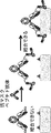
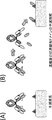
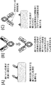

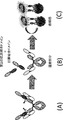


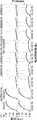
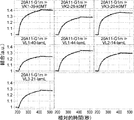
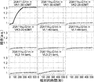


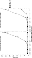


Classifications
-
- G—PHYSICS
- G01—MEASURING; TESTING
- G01N—INVESTIGATING OR ANALYSING MATERIALS BY DETERMINING THEIR CHEMICAL OR PHYSICAL PROPERTIES
- G01N33/00—Investigating or analysing materials by specific methods not covered by groups G01N1/00 - G01N31/00
- G01N33/48—Biological material, e.g. blood, urine; Haemocytometers
- G01N33/50—Chemical analysis of biological material, e.g. blood, urine; Testing involving biospecific ligand binding methods; Immunological testing
- G01N33/68—Chemical analysis of biological material, e.g. blood, urine; Testing involving biospecific ligand binding methods; Immunological testing involving proteins, peptides or amino acids
- G01N33/6803—General methods of protein analysis not limited to specific proteins or families of proteins
- G01N33/6845—Methods of identifying protein-protein interactions in protein mixtures
-
- A—HUMAN NECESSITIES
- A61—MEDICAL OR VETERINARY SCIENCE; HYGIENE
- A61K—PREPARATIONS FOR MEDICAL, DENTAL OR TOILETRY PURPOSES
- A61K38/00—Medicinal preparations containing peptides
-
- C—CHEMISTRY; METALLURGY
- C07—ORGANIC CHEMISTRY
- C07K—PEPTIDES
- C07K16/00—Immunoglobulins [IGs], e.g. monoclonal or polyclonal antibodies
- C07K16/18—Immunoglobulins [IGs], e.g. monoclonal or polyclonal antibodies against material from animals or humans
- C07K16/28—Immunoglobulins [IGs], e.g. monoclonal or polyclonal antibodies against material from animals or humans against receptors, cell surface antigens or cell surface determinants
- C07K16/2866—Immunoglobulins [IGs], e.g. monoclonal or polyclonal antibodies against material from animals or humans against receptors, cell surface antigens or cell surface determinants against receptors for cytokines, lymphokines, interferons
-
- A—HUMAN NECESSITIES
- A61—MEDICAL OR VETERINARY SCIENCE; HYGIENE
- A61K—PREPARATIONS FOR MEDICAL, DENTAL OR TOILETRY PURPOSES
- A61K38/00—Medicinal preparations containing peptides
- A61K38/16—Peptides having more than 20 amino acids; Gastrins; Somatostatins; Melanotropins; Derivatives thereof
-
- A—HUMAN NECESSITIES
- A61—MEDICAL OR VETERINARY SCIENCE; HYGIENE
- A61K—PREPARATIONS FOR MEDICAL, DENTAL OR TOILETRY PURPOSES
- A61K47/00—Medicinal preparations characterised by the non-active ingredients used, e.g. carriers or inert additives; Targeting or modifying agents chemically bound to the active ingredient
- A61K47/50—Medicinal preparations characterised by the non-active ingredients used, e.g. carriers or inert additives; Targeting or modifying agents chemically bound to the active ingredient the non-active ingredient being chemically bound to the active ingredient, e.g. polymer-drug conjugates
- A61K47/51—Medicinal preparations characterised by the non-active ingredients used, e.g. carriers or inert additives; Targeting or modifying agents chemically bound to the active ingredient the non-active ingredient being chemically bound to the active ingredient, e.g. polymer-drug conjugates the non-active ingredient being a modifying agent
- A61K47/62—Medicinal preparations characterised by the non-active ingredients used, e.g. carriers or inert additives; Targeting or modifying agents chemically bound to the active ingredient the non-active ingredient being chemically bound to the active ingredient, e.g. polymer-drug conjugates the non-active ingredient being a modifying agent the modifying agent being a protein, peptide or polyamino acid
- A61K47/64—Drug-peptide, drug-protein or drug-polyamino acid conjugates, i.e. the modifying agent being a peptide, protein or polyamino acid which is covalently bonded or complexed to a therapeutically active agent
-
- A—HUMAN NECESSITIES
- A61—MEDICAL OR VETERINARY SCIENCE; HYGIENE
- A61K—PREPARATIONS FOR MEDICAL, DENTAL OR TOILETRY PURPOSES
- A61K47/00—Medicinal preparations characterised by the non-active ingredients used, e.g. carriers or inert additives; Targeting or modifying agents chemically bound to the active ingredient
- A61K47/50—Medicinal preparations characterised by the non-active ingredients used, e.g. carriers or inert additives; Targeting or modifying agents chemically bound to the active ingredient the non-active ingredient being chemically bound to the active ingredient, e.g. polymer-drug conjugates
- A61K47/51—Medicinal preparations characterised by the non-active ingredients used, e.g. carriers or inert additives; Targeting or modifying agents chemically bound to the active ingredient the non-active ingredient being chemically bound to the active ingredient, e.g. polymer-drug conjugates the non-active ingredient being a modifying agent
- A61K47/62—Medicinal preparations characterised by the non-active ingredients used, e.g. carriers or inert additives; Targeting or modifying agents chemically bound to the active ingredient the non-active ingredient being chemically bound to the active ingredient, e.g. polymer-drug conjugates the non-active ingredient being a modifying agent the modifying agent being a protein, peptide or polyamino acid
- A61K47/65—Peptidic linkers, binders or spacers, e.g. peptidic enzyme-labile linkers
-
- A—HUMAN NECESSITIES
- A61—MEDICAL OR VETERINARY SCIENCE; HYGIENE
- A61P—SPECIFIC THERAPEUTIC ACTIVITY OF CHEMICAL COMPOUNDS OR MEDICINAL PREPARATIONS
- A61P29/00—Non-central analgesic, antipyretic or antiinflammatory agents, e.g. antirheumatic agents; Non-steroidal antiinflammatory drugs [NSAID]
-
- A—HUMAN NECESSITIES
- A61—MEDICAL OR VETERINARY SCIENCE; HYGIENE
- A61P—SPECIFIC THERAPEUTIC ACTIVITY OF CHEMICAL COMPOUNDS OR MEDICINAL PREPARATIONS
- A61P35/00—Antineoplastic agents
-
- A—HUMAN NECESSITIES
- A61—MEDICAL OR VETERINARY SCIENCE; HYGIENE
- A61P—SPECIFIC THERAPEUTIC ACTIVITY OF CHEMICAL COMPOUNDS OR MEDICINAL PREPARATIONS
- A61P37/00—Drugs for immunological or allergic disorders
- A61P37/02—Immunomodulators
- A61P37/06—Immunosuppressants, e.g. drugs for graft rejection
-
- C—CHEMISTRY; METALLURGY
- C07—ORGANIC CHEMISTRY
- C07K—PEPTIDES
- C07K16/00—Immunoglobulins [IGs], e.g. monoclonal or polyclonal antibodies
-
- C—CHEMISTRY; METALLURGY
- C07—ORGANIC CHEMISTRY
- C07K—PEPTIDES
- C07K16/00—Immunoglobulins [IGs], e.g. monoclonal or polyclonal antibodies
- C07K16/005—Immunoglobulins [IGs], e.g. monoclonal or polyclonal antibodies constructed by phage libraries
-
- C—CHEMISTRY; METALLURGY
- C07—ORGANIC CHEMISTRY
- C07K—PEPTIDES
- C07K16/00—Immunoglobulins [IGs], e.g. monoclonal or polyclonal antibodies
- C07K16/18—Immunoglobulins [IGs], e.g. monoclonal or polyclonal antibodies against material from animals or humans
- C07K16/28—Immunoglobulins [IGs], e.g. monoclonal or polyclonal antibodies against material from animals or humans against receptors, cell surface antigens or cell surface determinants
- C07K16/2803—Immunoglobulins [IGs], e.g. monoclonal or polyclonal antibodies against material from animals or humans against receptors, cell surface antigens or cell surface determinants against the immunoglobulin superfamily
- C07K16/2809—Immunoglobulins [IGs], e.g. monoclonal or polyclonal antibodies against material from animals or humans against receptors, cell surface antigens or cell surface determinants against the immunoglobulin superfamily against the T-cell receptor (TcR)-CD3 complex
-
- C—CHEMISTRY; METALLURGY
- C07—ORGANIC CHEMISTRY
- C07K—PEPTIDES
- C07K16/00—Immunoglobulins [IGs], e.g. monoclonal or polyclonal antibodies
- C07K16/18—Immunoglobulins [IGs], e.g. monoclonal or polyclonal antibodies against material from animals or humans
- C07K16/28—Immunoglobulins [IGs], e.g. monoclonal or polyclonal antibodies against material from animals or humans against receptors, cell surface antigens or cell surface determinants
- C07K16/30—Immunoglobulins [IGs], e.g. monoclonal or polyclonal antibodies against material from animals or humans against receptors, cell surface antigens or cell surface determinants from tumour cells
- C07K16/303—Liver or Pancreas
-
- C—CHEMISTRY; METALLURGY
- C12—BIOCHEMISTRY; BEER; SPIRITS; WINE; VINEGAR; MICROBIOLOGY; ENZYMOLOGY; MUTATION OR GENETIC ENGINEERING
- C12N—MICROORGANISMS OR ENZYMES; COMPOSITIONS THEREOF; PROPAGATING, PRESERVING, OR MAINTAINING MICROORGANISMS; MUTATION OR GENETIC ENGINEERING; CULTURE MEDIA
- C12N15/00—Mutation or genetic engineering; DNA or RNA concerning genetic engineering, vectors, e.g. plasmids, or their isolation, preparation or purification; Use of hosts therefor
- C12N15/09—Recombinant DNA-technology
-
- C—CHEMISTRY; METALLURGY
- C40—COMBINATORIAL TECHNOLOGY
- C40B—COMBINATORIAL CHEMISTRY; LIBRARIES, e.g. CHEMICAL LIBRARIES
- C40B30/00—Methods of screening libraries
- C40B30/04—Methods of screening libraries by measuring the ability to specifically bind a target molecule, e.g. antibody-antigen binding, receptor-ligand binding
-
- C—CHEMISTRY; METALLURGY
- C40—COMBINATORIAL TECHNOLOGY
- C40B—COMBINATORIAL CHEMISTRY; LIBRARIES, e.g. CHEMICAL LIBRARIES
- C40B40/00—Libraries per se, e.g. arrays, mixtures
- C40B40/04—Libraries containing only organic compounds
- C40B40/10—Libraries containing peptides or polypeptides, or derivatives thereof
-
- G—PHYSICS
- G01—MEASURING; TESTING
- G01N—INVESTIGATING OR ANALYSING MATERIALS BY DETERMINING THEIR CHEMICAL OR PHYSICAL PROPERTIES
- G01N33/00—Investigating or analysing materials by specific methods not covered by groups G01N1/00 - G01N31/00
- G01N33/48—Biological material, e.g. blood, urine; Haemocytometers
- G01N33/50—Chemical analysis of biological material, e.g. blood, urine; Testing involving biospecific ligand binding methods; Immunological testing
- G01N33/68—Chemical analysis of biological material, e.g. blood, urine; Testing involving biospecific ligand binding methods; Immunological testing involving proteins, peptides or amino acids
- G01N33/6854—Immunoglobulins
-
- A—HUMAN NECESSITIES
- A61—MEDICAL OR VETERINARY SCIENCE; HYGIENE
- A61K—PREPARATIONS FOR MEDICAL, DENTAL OR TOILETRY PURPOSES
- A61K39/00—Medicinal preparations containing antigens or antibodies
- A61K2039/505—Medicinal preparations containing antigens or antibodies comprising antibodies
-
- C—CHEMISTRY; METALLURGY
- C07—ORGANIC CHEMISTRY
- C07K—PEPTIDES
- C07K2317/00—Immunoglobulins specific features
- C07K2317/20—Immunoglobulins specific features characterized by taxonomic origin
- C07K2317/22—Immunoglobulins specific features characterized by taxonomic origin from camelids, e.g. camel, llama or dromedary
-
- C—CHEMISTRY; METALLURGY
- C07—ORGANIC CHEMISTRY
- C07K—PEPTIDES
- C07K2317/00—Immunoglobulins specific features
- C07K2317/30—Immunoglobulins specific features characterized by aspects of specificity or valency
- C07K2317/31—Immunoglobulins specific features characterized by aspects of specificity or valency multispecific
-
- C—CHEMISTRY; METALLURGY
- C07—ORGANIC CHEMISTRY
- C07K—PEPTIDES
- C07K2317/00—Immunoglobulins specific features
- C07K2317/50—Immunoglobulins specific features characterized by immunoglobulin fragments
- C07K2317/56—Immunoglobulins specific features characterized by immunoglobulin fragments variable (Fv) region, i.e. VH and/or VL
- C07K2317/569—Single domain, e.g. dAb, sdAb, VHH, VNAR or nanobody®
-
- C—CHEMISTRY; METALLURGY
- C07—ORGANIC CHEMISTRY
- C07K—PEPTIDES
- C07K2317/00—Immunoglobulins specific features
- C07K2317/60—Immunoglobulins specific features characterized by non-natural combinations of immunoglobulin fragments
- C07K2317/64—Immunoglobulins specific features characterized by non-natural combinations of immunoglobulin fragments comprising a combination of variable region and constant region components
-
- C—CHEMISTRY; METALLURGY
- C07—ORGANIC CHEMISTRY
- C07K—PEPTIDES
- C07K2317/00—Immunoglobulins specific features
- C07K2317/60—Immunoglobulins specific features characterized by non-natural combinations of immunoglobulin fragments
- C07K2317/66—Immunoglobulins specific features characterized by non-natural combinations of immunoglobulin fragments comprising a swap of domains, e.g. CH3-CH2, VH-CL or VL-CH1
-
- C—CHEMISTRY; METALLURGY
- C07—ORGANIC CHEMISTRY
- C07K—PEPTIDES
- C07K2317/00—Immunoglobulins specific features
- C07K2317/70—Immunoglobulins specific features characterized by effect upon binding to a cell or to an antigen
- C07K2317/76—Antagonist effect on antigen, e.g. neutralization or inhibition of binding
-
- C—CHEMISTRY; METALLURGY
- C07—ORGANIC CHEMISTRY
- C07K—PEPTIDES
- C07K2319/00—Fusion polypeptide
- C07K2319/33—Fusion polypeptide fusions for targeting to specific cell types, e.g. tissue specific targeting, targeting of a bacterial subspecies
-
- C—CHEMISTRY; METALLURGY
- C07—ORGANIC CHEMISTRY
- C07K—PEPTIDES
- C07K2319/00—Fusion polypeptide
- C07K2319/50—Fusion polypeptide containing protease site
-
- C—CHEMISTRY; METALLURGY
- C07—ORGANIC CHEMISTRY
- C07K—PEPTIDES
- C07K2319/00—Fusion polypeptide
- C07K2319/70—Fusion polypeptide containing domain for protein-protein interaction
Definitions
- the present invention relates to a polypeptide comprising an antigen-binding domain and a carrier moiety having a suppressive domain that suppresses the antigen-binding activity of the antigen-binding domain, and having a longer half-life than an antigen-binding domain that exists alone, Manufacturing method and screening method, pharmaceutical composition containing the polypeptide, single domain antibody manufacturing method and screening method in which antigen-binding activity is suppressed by associating with specific VL / VH / VHH, and specific VL / VH
- the present invention relates to a library of fusion polypeptides comprising a single domain antibody whose antigen binding activity is suppressed by associating with / VHH.
- Antibodies are attracting attention as pharmaceuticals because of their high stability in plasma and few side effects. Among them, many IgG-type antibody drugs are on the market, and many antibody drugs have been developed (Non-patent Documents 1 and 2).
- Non-patent Document 3 As conventional cancer therapeutic agents using antibody drugs, Rituxan for CD20 antigen, cetuximab for EGFR antigen, Herceptin for HER2 antigen, etc. have been approved (Non-patent Document 3). These antibody molecules bind to antigens expressed in cancer cells and exert damaging activity against cancer cells by ADCC activity or the like. It is known that the cytotoxic activity due to ADCC activity or the like depends on the number of antigens expressed in the target cells of the therapeutic antibody (Non-patent Document 4), and therefore the expression level of the target antigen may be high. It is preferable from the viewpoint of the effect of the therapeutic antibody.
- the antigen targeted by the therapeutic antibody as a cancer therapeutic agent is specifically expressed in cancer cells.
- an antibody molecule against EpCAM antigen known as a cancer antigen
- EpCAM antigen is known to be expressed in the pancreas. It has been reported that by administering an anti-EpCAM antibody, side effects of pancreatitis are observed due to cytotoxic activity against the pancreas (Non-patent Document 5).
- Non-patent Document 6 In response to the success of antibody drugs that exert cytotoxic activity due to ADCC activity, enhancement of ADCC activity by removing the fucose of the N-type sugar chain in the Fc region of natural human IgG1 (Non-patent Document 6), natural human A second generation improved antibody molecule that exhibits strong cytotoxic activity by enhancing ADCC activity by enhancing the binding to Fc ⁇ RIIIa by amino acid substitution in the Fc region of IgG1 (Non-patent Document 7) has been reported.
- ADC Antibody Drug Conjugate
- a drug having potent cytotoxic activity is conjugated with an antibody as an antibody drug that exerts a cytotoxic activity on cancer cells by a mechanism other than the ADCC activity mediated by NK cells described above
- improved antibody molecules that exhibit stronger cytotoxic activity such as low-molecular-weight antibodies (Non-patent Document 9) that exert cytotoxic activity against cancer cells by recruiting T cells to cancer cells have also been reported. .
- Such antibody molecules that exert stronger cytotoxic activity can also exert cytotoxic activity against cancer cells that do not express much antigen, but also against normal tissues with less antigen expression. It exerts cytotoxic activity like cancer cells.
- cetuximab which is a natural human IgG1 against EGFR antigen
- EGFR-BiTE a bispecific antibody against CD3 and EGFR
- EGFR-BiTE a bispecific antibody against CD3 and EGFR
- bivatuzumab mertansine an ADC that binds mertansine to an antibody against CD44v6 that is highly expressed in cancer cells, is also expressed in normal tissues. It is recognized (Non-Patent Document 11).
- the target antigen when using an antibody that can exert strong cytotoxic activity against cancer cells with low antigen expression, the target antigen must be expressed in a very cancer-specific manner, It seems that the number of cancer antigens that are extremely cancer-specifically expressed is limited, such as HER2 that is the target antigen of Herceptin and EGFR that is the target antigen of cetuximab. Therefore, although the cytotoxic activity against cancer can be enhanced, side effects due to the cytotoxic action on normal tissues can be problematic.
- Non-patent Document 12 iplimumab, which enhances tumor immunity by inhibiting CTLA4, which contributes to immunosuppression in cancer, extends Overall survival to metastatic melanoma.
- iplimumab inhibits CTLA4 systemically, it enhances tumor immunity, while it causes serious side effects like autoimmune diseases due to systemic immunity activation.
- Non-patent Document 14 antibody drugs that exhibit therapeutic effects by inhibiting inflammatory cytokines in inflammatory and autoimmune diseases are known as antibody drugs against diseases other than cancer.
- Remicade and Humira which target TNF
- Actemra which targets IL-6R
- side effects of symptom are observed (Non-patent Document 15).
- the antigen-binding site of an antibody is masked with a masking peptide by connecting the masking peptide and the antibody with a linker that is cleaved by a protease expressed at a lesion site such as cancer tissue or inflammatory tissue, and the antibody
- a linker that is cleaved by a protease expressed at a lesion site such as cancer tissue or inflammatory tissue
- the antibody There is a method that inhibits the antigen-binding activity of the antibody, dissociates the mask peptide by cleaving this linker with a protease, restores the antigen-binding activity of the antibody, and allows it to bind to the antigen in the target diseased tissue.
- the cleavage by the protease is irreversible. It was considered that the cleaved antibody may be distributed in normal tissues in the bloodstream and cause side effects.
- the present invention has been made based on such a concept, and one of its purposes is to provide a pharmaceutical composition useful for disease treatment with reduced side effects and an active ingredient thereof. One of the purposes is to provide a method for screening and producing the pharmaceutical composition and the active ingredient.
- the inventors of the present invention have an antigen-binding domain and a transporting portion having an inhibitory domain that suppresses the binding activity of the antigen-binding domain, and have a longer half-life than a single antigen-binding domain.
- a polypeptide was created. By using the polypeptide, it is considered that the antigen-binding activity of the antigen-binding domain is recovered in the diseased tissue and the antigen-binding activity can be exhibited in the diseased tissue.
- the whole body of the activated antigen-binding domain Can be suppressed.
- the present inventors have found that the polypeptide or a pharmaceutical composition containing the polypeptide is useful for treating a disease, and is useful for treating a disease including administering the polypeptide, And the polypeptide was found useful in the manufacture of a medicament for the treatment of disease.
- the present inventors also provide a method for screening and producing the polypeptide, a method for producing and screening a single domain antibody in which antigen-binding activity is suppressed by associating with a specific VL or VH or VHH, and a specific method.
- the present invention was completed by creating a library containing single domain antibodies whose antigen binding activity is suppressed by associating with VL, VH or VHH.
- a polypeptide wherein the polypeptide includes an antigen-binding domain and a transporting part, the transporting part has a suppression domain that suppresses the antigen-binding activity of the antigen-binding domain, and the antigen-binding domain is A polypeptide having a blood half-life that is shorter than the delivery moiety.
- the polypeptide according to (1) wherein the molecular weight of the antigen-binding domain is smaller than the molecular weight of the transport moiety.
- the transporting portion has FcRn binding activity, and the antigen-binding domain has no FcRn binding activity or weaker FcRn binding activity than the transporting portion, according to any one of (1) to (3) Polypeptide.
- the antigen-binding domain is releasable from the polypeptide, and the antigen-binding domain is released from the polypeptide, whereby the antigen-binding activity is higher than that before release, any of (1) to (4)
- polypeptide according to (5) wherein the polypeptide includes a cleavage site, and the antigen-binding domain can be released from the polypeptide by cleavage of the cleavage site.
- the cleavage site comprises a protease cleavage sequence.
- the protease is at least one protease selected from matriptase, urokinase (uPA), and metalloprotease.
- the protease is at least one protease selected from MT-SP1, uPA, MMP2, MMP9, ADAMTS5, MMP7, and MMP13.
- a second movable linker is further added to the other end of the protease cleavage sequence.
- the polypeptide according to (15), wherein the first movable linker is a movable linker composed of a glycine-serine polymer.
- the antigen-binding domain includes a single-domain antibody, or is a single-domain antibody, and the inhibitory domain of the delivery portion suppresses the antigen-binding activity of the single-domain antibody, any of (1) to (18) A polypeptide according to claim 1.
- the single domain antibody is VHH, VH having antigen binding activity in a single domain, or VL having antigen binding activity in a single domain.
- the antigen-binding domain includes a single domain antibody, and the suppressor domain of the transport portion is VHH, or antibody VH, or antibody VL, and the single domain antibody is expressed by the VHH, antibody VH, or antibody VL.
- the antigen-binding domain includes a single domain antibody, the suppression domain of the transporting portion is VHH, or antibody VH, or antibody VL, and the single domain antibody is the VHH, antibody VH, or antibody VL.
- the single domain antibody is VHH or VH having antigen binding activity in a single domain
- the inhibitory domain of the transporting portion is antibody VL
- VHH or VH having antigen binding activity in a single domain is The polypeptide according to any one of (19) to (22), wherein the antigen-binding activity is suppressed by associating with the antibody VL.
- the single domain antibody is VHH, and the VHH has an amino acid substitution at at least one position selected from amino acids No. 37, No. 44, No. 45, and No. 47 (all Kabat numbering). ) To (23).
- the single domain antibody is VHH, and the VHH includes at least one amino acid selected from 37V, 44G, 45L, or 47W (all Kabat numbering) amino acids, and any of (19) to (23) The polypeptide according to 1.
- the single domain antibody is VHH, and the VHH is at least one selected from amino acid substitutions of F37V, Y37V, E44G, Q44G, R45L, H45L, G47W, F47W, L47W, T47W, or S47W (all Kabat numbering).
- the polypeptide according to any one of (19) to (23), comprising one amino acid substitution.
- the single domain antibody is VHH, which is 37/44, 37/45, 37/47, 44/45, 44/47, 45/47 , 37/44/45, 37/44/47, 37/45/47, 44/45/47, 37/44/45/47 (all The polypeptide according to any one of (19) to (23), wherein amino acid substitution is performed in at least one position selected from Kabat numbering.
- the single domain antibody is VHH, which is 37V / 44G, 37V / 45L, 37V / 47W, 44G / 45L, 44G / 47W, 45L / 47W, 37V / 44G / 45L, 37V / 44G / 47W
- the polypolysaccharide according to any one of (19) to (23), comprising at least one set of amino acids selected from 37V / 45L / 47W, 44G / 45L / 47W, 37V / 44G / 45L / 47W (all Kabat numbering) peptide.
- the single domain antibody is VHH, and the VHH comprises at least one set of amino acid substitutions selected from F37V / R45L, F37V / G47W, R45L / G47W, F37V / R45L / G47W (all Kabat numbering), The polypeptide according to any one of 19) to (23).
- the single domain antibody is a VL having antigen binding activity in a single domain, the inhibitory domain of the transport portion is antibody VH, and the VL having antigen binding activity in the single domain is associated with the antibody VH
- the polypeptide according to any one of (19) to (22), wherein the antigen binding activity is suppressed.
- the transporting portion includes an antibody heavy chain constant region, and the antibody heavy chain constant region and the antigen-binding domain are fused via a linker or not via a linker. Polypeptide.
- the transporting portion includes an antibody light chain constant region, and the antibody light chain constant region and the antigen-binding domain are fused via a linker or not via a linker.
- Polypeptide. In the polypeptide, the N-terminus of the antibody heavy chain constant region of the transporting portion and the C-terminus of the antigen-binding domain are fused with or without a linker, and further have a protease cleavage sequence The polypeptide according to (34), wherein the protease cleavage sequence is located in the antigen-binding domain sequence or in the antigen-binding domain side from the amino acid at position 122 (EU numbering) of the heavy chain antibody constant region.
- the N-terminus of the antibody light-chain constant region of the transporting portion and the C-terminus of the antigen-binding domain are fused with or without a linker, and further have a protease cleavage sequence
- the protease cleavage sequence is located in the antigen-binding domain side of the antigen-binding domain or in the light-chain antibody constant region from amino acid No. 113 (EU numbering) (Kabat numbering No. 113), (35) The polypeptide according to 1.
- the N-terminus of the antibody constant region of the transporting portion and the C-terminus of the antigen-binding domain are fused with or without a linker, and the antigen-binding domain is prepared from VH A single domain antibody or VHH, wherein the polypeptide further has a protease cleavage sequence, which is in the sequence of the antibody constant region or the 109 of the single domain antibody of the antigen binding domain (Kabat numbering).
- the polypeptide according to any one of (33) to (35), which is located on the antibody constant region side from the amino acid of ().
- the N-terminus of the antibody constant region of the transporting portion and the C-terminus of the antigen-binding domain are fused with or without a linker, and further have a protease cleavage sequence,
- the N-terminus of the antibody heavy chain constant region of the transporting portion and the C-terminus of the antigen-binding domain are fused with or without a linker, and further have a protease cleavage sequence
- the N-terminus of the antibody light-chain constant region of the transporting portion and the C-terminus of the antigen-binding domain are fused with or without a linker, and further have a protease cleavage sequence
- the antigen-binding domain is a single domain antibody or VHH prepared from VH, and the protease cleavage sequence includes amino acid No. 109 (Kabat numbering) of the single-domain antibody of the antigen-binding domain and the antibody heavy chain constant.
- the antigen-binding domain is a single domain antibody or VHH prepared from VH, and the protease cleavage sequence includes amino acid No. 109 (Kabat numbering) of the single-domain antibody of the antigen-binding domain and the antibody light chain constant.
- the antigen-binding domain is a single-domain antibody prepared from VL, and the protease cleavage sequence includes amino acid No.
- the antigen-binding domain is a single-domain antibody prepared from VL, and the protease cleavage sequence includes amino acid No. 109 (Kabat numbering) of the single-domain antibody of the antigen-binding domain and the antibody light-chain constant region.
- (48) When the measurement is performed using the BLI (Bio-Layer Interferometry) method (Octet) in a state where the antigen-binding domain is not free, the binding between the antigen-binding domain and the antigen is not observed (from (1) to ( 47)
- the polypeptide according to any one of (49) The polypeptide according to any one of (1) to (48), wherein a second antigen-binding domain is further linked to the antigen-binding domain.
- the antigen-binding domain is a single domain antibody
- the second antigen-binding domain is a second single-domain antibody
- the antigen-binding domain and the second antigen-binding domain can be released from the polypeptide
- the single domain antibody and the second single domain antibody form a bispecific antigen-binding molecule in the free state of the antigen-binding domain and the second antigen-binding domain,
- the polypeptide further has an antigen-binding domain different from the antigen-binding domain, and the antigen-binding activity is suppressed by linking the other antigen-binding domain to the transporting part of the polypeptide.
- a pharmaceutical composition comprising the polypeptide according to any one of (1) to (56).
- a method for producing the polypeptide according to any one of (1) to (56). The following steps: (a) obtaining a single domain antibody that binds to a target antigen; (b) A step of forming a polypeptide precursor by linking the single domain antibody and the transporting moiety so that the antigen binding activity of the single domain antibody obtained in step (a) is suppressed by the suppression domain of the transporting moiety.
- the production method according to (58), comprising: (61) The following steps: (a) obtaining a single domain antibody that binds to a target antigen; (b) so that the antigen-binding activity of the single domain antibody obtained in step (a) is suppressed by the inhibitory domain of the carrier moiety, the single domain antibody is linked to the carrier moiety via a protease cleavage sequence to obtain a poly Forming a peptide; The production method according to (58), comprising: (62) Further, the following steps: (d) confirming that the binding activity to the target antigen of the single domain antibody incorporated in the polypeptide or the polypeptide precursor is weakened or lost; The manufacturing method in any one of (59) to (61) containing this.
- step (66) The following steps: (a) obtaining a single domain antibody that binds to a target antigen; (b) In order to suppress the antigen-binding activity of the single domain antibody obtained in step (a), the single domain antibody is associated with VL instead of IgG antibody VH, or the single domain antibody is Forming an IgG antibody-like molecular precursor introduced with the single domain antibody by associating with VH instead of VL; (c) introducing a protease cleavage sequence near the boundary between the single domain antibody and the antibody constant region in the IgG antibody-like molecular precursor; The manufacturing method as described in (64) containing.
- step (67) The following steps: (a) obtaining a single domain antibody that binds to a target antigen; (b) In order to suppress the antigen-binding activity of the single domain antibody obtained in step (a), the single domain antibody is replaced with IgG antibody VH or VL, and the heavy chain constant of the IgG antibody via a protease cleavage sequence is used. Linking to a region or a light chain constant region to form an IgG antibody-like molecule into which the single domain antibody has been introduced; The manufacturing method as described in (64) containing.
- step (70) The following steps: (a) Substituting an amino acid residue involved in association with antibody VH in a single domain antibody, or substituting an amino acid residue involved in association with antibody VL in a single domain antibody, and said single domain antibody Producing a modified single domain antibody that retains the binding activity to the target antigen of (b) by associating the modified single domain antibody with antibody VL or associating the modified single domain antibody with antibody VH so as to suppress the antigen binding activity of the modified single domain antibody prepared in step (a) Forming the IgG antibody-like molecular precursor into which the modified single domain antibody has been introduced; (c) introducing a protease cleavage sequence into the IgG antibody-like molecular precursor into which the modified single domain antibody has been introduced; The manufacturing method as described in (64) containing.
- (76) A vector comprising the polynucleotide according to (75).
- (77) A host cell comprising the polynucleotide according to (75) or the vector according to (76).
- (78) A method for producing the polypeptide according to any one of (1) to (56), comprising a step of culturing the host cell according to (77).
- (79) A method for screening a single domain antibody whose antigen-binding activity is suppressed by associating with a specific VL, associating with a specific VH, or associating with a specific VHH.
- step (98) Further, the following steps: (b) a step of associating the modified single domain antibody prepared in step (a) with the VHH; (c) confirming that the antigen-binding activity of the modified single domain antibody associated with the VHH is weakened or lost compared with that before the association;
- the single domain antibody portion of the fusion polypeptide in the library is a single domain antibody obtained from a camelid or a transgenic animal into which a gene capable of producing a single domain antibody has been introduced, or a humanized antibody thereof, or a camelid Single domain antibody or humanized antibody obtained by immunizing animal or transgenic animal introduced with gene capable of producing single domain antibody, or artificially created single domain starting from human antibody VH or VL
- the library according to (99) comprising an antibody.
- a library comprising a plurality of fusion polypeptides in which a single domain antibody and a first association support domain are linked, wherein the single domain antibody is associated with a specific VL, or its antigen binding activity is suppressed or The library according to (99) or (100), comprising a single domain antibody that is lost.
- a library comprising a plurality of fusion polypeptides in which a single domain antibody and a first association support domain are linked, wherein the single domain antibody is associated with a specific VH, or its antigen binding activity is suppressed or The library according to (99) or (100), comprising a single domain antibody that is lost.
- a library comprising a plurality of fusion polypeptides in which a single domain antibody and a first association support domain are linked, wherein the single domain antibody is associated with a specific VHH, so that antigen binding activity is suppressed or
- a single domain antibody in which antigen-binding activity is suppressed or lost by associating with a specific VL from the library according to (99) or (100), or antigen-binding activity by associating with a specific VH A method for screening a fusion polypeptide comprising a single domain antibody in which antigen is suppressed or lost, or a single domain antibody in which antigen binding activity is suppressed or lost by associating with a specific VHH.
- step (a) in vitro display of the fusion polypeptides of the library; (b) providing a meeting partner in which the specific VL and the second meeting support domain are fused; (c) The fusion polypeptide displayed in step (a) is associated with the association partner prepared in step (b), and the single domain antibody and the VL do not bind to the antigen in the associated state, or antigen binding activity is present.
- a fusion polypeptide that is less than or equal to a certain value (d) From the fusion polypeptide selected in step (c), select a fusion polypeptide that binds to the antigen in a state where the single domain antibody and the VL are not associated with each other or that has an antigen binding activity of a certain value or more.
- the screening method according to (105) comprising: (107) The association partner prepared in the step (b) further includes a protease cleavage sequence. In the step (d), the association partner is cleaved by protease treatment to eliminate the VL association with the single domain antibody.
- the fusion polypeptide of the library further comprises a protease cleavage sequence, and in the step (d), the fusion polypeptide is cleaved by protease treatment to eliminate the VL association with the single domain antibody.
- (113) A method for screening a fusion polypeptide comprising a single domain antibody whose antigen-binding activity is suppressed or lost by associating with a specific VH from the library according to (102).
- (114) The following steps: (a) in vitro display of the fusion polypeptides of the library; (b) providing a meeting partner in which a specific VH and a second meeting supporting domain are fused; (c) The fusion polypeptide displayed in step (a) is associated with the association partner prepared in step (b), and the single domain antibody and the VH are associated with each other and do not bind to the antigen or have a constant antigen-binding activity.
- step (d) From the fusion polypeptide selected in step (c), select a fusion polypeptide that binds to the antigen in a state where the VH is not associated with the contained single domain antibody or has an antigen binding activity of a certain value or more.
- the screening method according to (113) comprising: (115) The association partner prepared in the step (b) further includes a protease cleavage sequence. In the step (d), the association partner is cleaved by protease treatment to eliminate the association between the single domain antibody and the VH.
- the fusion polypeptide of the library further comprises a protease cleavage sequence, and in the step (d), the fusion polypeptide is cleaved by protease treatment to eliminate the VH association with the single domain antibody.
- step (122) The following steps: (a) in vitro display of the fusion polypeptides of the library; (b) providing an association partner in which a specific VHH and a second association support domain are fused; (c) The fusion polypeptide displayed in step (a) is associated with the association partner prepared in step (b) and does not bind to the antigen in a state where the single domain antibody and the specific VHH are associated, or antigen binding activity Selecting a fusion polypeptide wherein is less than or equal to a certain value; (d) From the fusion polypeptides selected in step (c), select a fusion polypeptide that binds to an antigen in a state where the contained single domain antibody and the VHH are not associated with each other, or has an antigen binding activity of a certain value or more.
- the screening method according to (121), comprising: (123)
- the association partner prepared in the step (b) further includes a protease cleavage sequence.
- the association partner is cleaved by protease treatment to eliminate the association between the single domain antibody and the VHH.
- the screening method according to (122). (124)
- the fusion polypeptide of the library further comprises a protease cleavage sequence, and in the step (d), the fusion polypeptide is cleaved by protease treatment to eliminate the association between the single domain antibody and the VHH.
- Screening method (126) The screening method according to (125), wherein the protease cleavage sequence contained in the fusion polypeptide is located near a boundary between the single domain antibody and the first association support domain.
- the full length of the fusion polypeptide selected in the step (c) is again displayed in vitro and binds to the antigen in the state of being associated only with the second association support domain, or antigen binding
- the step of preparing the association partner in the step (b) is a step of simultaneously displaying the association partner and the fusion polypeptide, (106) to (112), (114) to (120), (122 ) To (128).
- the library according to any one of (99) to (103), wherein the first association support domain comprises an IgG antibody CH1 domain or an antibody light chain constant region.
- the first association support domain includes an IgG antibody CH1 domain
- the second association support domain includes an antibody light chain constant region (106) to (112), (114) to (120), (122 ) To (128).
- the first association support domain comprises an antibody light chain constant region
- the second association support domain comprises an IgG antibody CH1 domain (106) to (112), (114) to (120), (122 ) To (128).
- step (133) The following steps: (a) in vitro display of the fusion polypeptides of the library; (b) providing a meeting partner in which the specific VL and the second meeting support domain are fused; (c) selecting a fusion polypeptide in which a single domain antibody contained in the fusion polypeptide binds to an antigen or has an antigen-binding activity of a certain value or more; (d) The fusion polypeptide selected in step (c) is associated with the association partner prepared in step (b) and does not bind to the antigen when the single domain antibody and the VL are associated, or has antigen-binding activity.
- the screening method according to (105), comprising: (134) The screening method according to (129), wherein in the step (d), the fusion polypeptide selected in the step (c) is again displayed in vitro. (135) In the step (c), a single domain contained in the fusion polypeptide in a state where the fusion polypeptide is associated only with the second association support domain, or the fusion polypeptide is associated only with the second association support domain.
- the screening method according to (133) wherein the antigen binding of the antibody is confirmed.
- step (136) The following steps: (a) in vitro display of the fusion polypeptides of the library; (b) providing a meeting partner in which a specific VH and a second meeting supporting domain are fused; (c) selecting a fusion polypeptide in which a single domain antibody contained in the fusion polypeptide binds to an antigen or has an antigen-binding activity of a certain value or more; (d) The fusion polypeptide selected in step (c) is associated with the association partner prepared in step (b), and the single domain antibody and the VH are associated with each other, and do not bind to the antigen or have antigen-binding activity.
- the screening method according to (113), comprising: (137) The screening method according to (136), wherein in the step (d), the fusion polypeptide selected in the step (c) is again displayed in vitro. (138) In the step (c), a single domain contained in the fusion polypeptide in a state in which the fusion polypeptide is associated only with the second association support domain, or the fusion polypeptide is associated only with the second association support domain.
- the screening method according to (136) wherein the antigen binding of the antibody is confirmed.
- step (139) The following steps: (a) in vitro display of the fusion polypeptides of the library; (b) providing an association partner in which a specific VHH and a second association support domain are fused; (c) selecting a fusion polypeptide in which a single domain antibody contained in the fusion polypeptide binds to an antigen or has an antigen-binding activity of a certain value or more; (d) The fusion polypeptide selected in step (c) is associated with the association partner prepared in step (b), and the single domain antibody and the VHH are associated with each other and do not bind to the antigen or have antigen-binding activity.
- the screening method according to (121), comprising: (140) The screening method according to (139), wherein in the step (d), the fusion polypeptide selected in the step (c) is again displayed in vitro. (141) In the step (c), a single domain contained in the fusion polypeptide in a state where the fusion polypeptide is associated only with the second association support domain, or the fusion polypeptide is associated only with the second association support domain.
- the screening method according to (139) wherein the antigen binding of the antibody is confirmed.
- the step of associating the fusion polypeptide and the association partner in the step (d) is a step of simultaneously displaying the association partner and the fusion polypeptide, according to any one of (133) to (141) Screening method.
- Probody technology It is an antibody molecule that inhibits the antigen-binding activity of an antibody by connecting a peptide that masks the antigen-binding site of the antibody to the antibody with a linker that is cleaved by a protease that expresses at the lesion site. It is a figure which shows one cause with which Probody may show a side effect. Activated Probody accumulated in the blood may exert side effects by binding to antigens expressed in normal tissues. It is a figure which shows one cause with which Probody may show a side effect.
- Probody is in an equilibrium state in which the mask peptide linked by the linker is bound to the antigen binding site and dissociated, and the dissociated molecule can bind to the antigen. It is a figure which shows one cause with which Probody may show a side effect.
- the anti-drug antibody against the mask peptide (anti-mask peptide antibody) binds to the mask peptide of the Probody before being activated, and thus may activate the Probody without cleavage by protease. It is a figure which shows the concept of the polypeptide containing an antigen binding domain and a conveyance part.
- a polypeptide in which an antigen-binding domain and a carrier moiety are linked has a long half-life and does not bind to an antigen.
- B The antigen-binding domain is released and bound to the antigen by cleavage of the cleavage site, etc., and the released antigen-binding domain has a short half-life. It is a figure which shows one embodiment of the method of manufacturing the polypeptide of this invention.
- the polypeptide of interest is an IgG antibody-like molecule.
- A Obtain a single domain antibody that binds to the target antigen.
- a single domain antibody is associated with VL instead of IgG antibody VH so that the antigen-binding activity of the single domain antibody is suppressed.
- C A protease cleavage sequence is introduced into an IgG antibody-like molecular precursor into which a single domain antibody has been introduced. It is a figure which shows one embodiment of the polypeptide of this invention.
- the polypeptide is an IgG antibody-like molecule, and antigen-binding domains are provided in portions corresponding to the two variable regions of the IgG antibody.
- the two antigen binding domains may have similar antigen binding specificities or different antigen binding specificities.
- FIG. 1 It is a figure which shows the embodiment by which the 2nd antigen binding domain is further connected with the antigen binding domain of this invention.
- the free antigen binding domain and the second antigen binding domain form a bispecific antigen binding molecule.
- A It is a figure which shows the polypeptide of an unreleased state. The antigen binding activity of the antigen binding domain is suppressed.
- B is a diagram showing the release of a bispecific antigen-binding molecule formed by an antigen-binding domain and a second antigen-binding domain.
- (C) As an example of the bispecific antigen-binding molecule after release, it is a diagram showing, for example, a bispecific antigen-binding molecule for a T cell surface antigen and a cancer cell surface antigen. Screening a fusion polypeptide containing a single domain antibody whose antigen binding activity is reduced or lost by associating with a specific inhibitory domain from a library containing multiple fusion polypeptides linking a single domain antibody and a first association support domain It is a figure which shows an example of the method to do. (1) It is a figure which shows the library containing multiple fusion polypeptide which linked the single domain antibody and the 1st association support domain.
- a Fab-like structure is formed by simultaneously displaying a fusion polypeptide comprising a single-domain antibody and a first association support domain, and an association partner having a protease cleavage sequence introduced between the repression domain and the second association support domain.
- a fusion polypeptide in which a protease cleavage sequence is introduced between a single domain antibody and a first association support domain, and an association partner in which a repression domain and a second association support domain are linked are simultaneously displayed to form a Fab-like structure.
- a protease cleavage sequence is introduced between a single domain antibody and a first association support domain, and an association partner in which a repression domain and a second association support domain are linked are simultaneously displayed to form a Fab-like structure.
- a fusion polypeptide containing a single domain antibody whose antigen binding activity is reduced or lost by associating with a specific inhibitory domain from a library containing multiple fusion polypeptides linking a single domain antibody and a first association support domain It is a figure which shows another example of the method to do.
- a fusion polypeptide comprising a single domain antibody and a first association support domain and an association partner in which a repression domain and a second association support domain are linked are simultaneously displayed to form a Fab-like structure.
- a substance that does not bind to the antigen or has a binding activity to the antigen of a certain value or less is selected; (2) a portion containing a single domain antibody in the Fab-like structure selected in (1) At the same time, display again in a form that does not allow expression, and select a fragment that binds to the antigen or has a certain activity or higher.
- (2 ′) and (2 ′′) are diagrams showing another embodiment of (2) in which a portion containing a single domain antibody is redisplayed in such a manner that the inhibitory domain is not expressed simultaneously.
- the order of (1) and (2) / (2 ′) / (2 ′′) may be (2) / (2 ′) / (2 ′′) to (1), that is, a single domain antibody
- the contained portion is displayed in such a manner that the inhibitory domain is not expressed at the same time, and a fragment having an antigen binding activity of a certain value or more is selected.
- a single domain antibody containing a fragment having a certain binding or more, a fusion polypeptide containing a first association support domain, an association partner in which a repression domain and a second association support domain are linked are simultaneously displayed, and a Fab-like structure is displayed.
- Fab-like structure one that does not bind to the antigen or has a binding activity to the antigen of a certain value or less is selected.
- the time at which the action of the antibody and the antibody-like molecule on which the antigen is immobilized is started is the starting point on the horizontal axis.
- (A) It is a figure which shows the model of the antibody-like molecule
- (B) It is a figure which shows the name of each produced antibody heavy chain, the site
- Antibody-like molecules created by inserting a protease cleavage sequence near the boundary between VHH and the constant region of IL6R90-G1m or IL6R90-G1m were treated with protease (MT-SP1), and the extent of cleavage was evaluated by reducing SDS-PAGE. It is a figure which shows a result. Of the two new bands generated by protease treatment, the band generated at 25 kDa or less is a band derived from VHH, and the band appearing at the position of 25-50 kDa is a band derived from the constant region.
- FIG. 12 is a diagram showing a continuation of FIG. 12-1.
- Protease- is a sensorgram that evaluates the binding between an antigen-like antibody-like molecule and an antigen
- Protease + is a sensorgram that evaluates the binding between a protease-treated antibody-like molecule and an antigen.
- the starting point of the horizontal axis is 30 seconds before the action of the antibody on which the antigen is immobilized and the antibody-like molecule.
- the starting point of the horizontal axis is 60 seconds before the start of the action of the antibody on which the antigen is immobilized and the antibody-like molecule.
- Protease- is a sensorgram that evaluates the binding between an antigen-like antibody-like molecule and an antigen
- Protease + is a sensorgram that evaluates the binding between a protease-treated antibody-like molecule and an antigen.
- the starting point of the horizontal axis is 60 seconds before the action of the antibody and antibody on which the antigen is immobilized. Samples marked as not tested indicate that they have not been measured.
- Anti-human CD3 VHH is in the heavy chain variable region, and the antibody-like molecule prepared by inserting a protease cleavage sequence near the boundary between VHH and heavy chain constant region is treated with protease (MT-SP1) and then reduced by SDS-PAGE. It is a figure which shows the result of having electrophoresed and having evaluated the grade of the cutting
- protease treatment a band generated in the vicinity of 10-15 kDa is a band derived from VHH, and a band generated in the vicinity of 37 kDa is a band derived from the heavy chain constant region.
- Anti-human CD3 VHH in the heavy chain variable region and sample of human antibody CD3ed after protease (MT-SP1) treatment of an antibody-like molecule prepared by inserting a protease cleavage sequence near the boundary between VHH and heavy chain constant region It is a figure which shows the result of having evaluated the coupling
- Protease- is a sensorgram that evaluates the binding between an antigen-like antibody-like molecule and an antigen
- Protease + is a sensorgram that evaluates the binding between a protease-treated antibody-like molecule and an antigen.
- the start point of the horizontal axis is 30 seconds before the action of the antibody and the antibody-like molecule on which the antigen is immobilized.
- Binding is shown when the binding amount (response) before binding the antigen is 0 and the binding amount before the antibody is allowed to act is 100. It is displayed 30 seconds before the antibody is activated.
- a light chain variable region and a light chain constant region of a molecule having IL6R90-G1m as a heavy chain and Vk1-39-k0MT as a light chain, or a molecule having IL6R90-G1m as a heavy chain and Vk1-39-k0MT as a light chain Shows the result of evaluating the degree of cleavage by treating the antibody-like molecule prepared by inserting a protease cleavage sequence near the border of the protein, migrating with protease (MT-SP1), reducing SDS-PAGE, and detecting with CBB FIG.
- Protease treatment produces two bands derived from the light chain, which are cleaved by the protease.
- a light chain variable region and a light chain constant region of a molecule having IL6R90-G1m as a heavy chain and Vk1-39-k0MT as a light chain, or a molecule having IL6R90-G1m as a heavy chain and Vk1-39-k0MT as a light chain It is a figure which shows the result of having evaluated the coupling
- Protease- is a sensorgram that evaluates the binding between an antigen-like antibody-like molecule and an antigen
- Protease + is a sensorgram that evaluates the binding between a protease-treated antibody-like molecule and an antigen.
- An antibody (MRA) that has been confirmed to bind to IL6R is used as a positive control.
- the start point of the horizontal axis is the time when the action of the antibody-like molecule and the sensor on which the antigen is immobilized is started. It is a figure which shows the SDS-PAGE result which evaluated the protease cleavage of the IgG antibody-like molecule
- Protease (+) lane is a sample subjected to protease cleavage treatment
- protease (-) telane is a negative control sample not subjected to protease cleavage treatment. It is a figure which shows the Octet sensorgram which IgG-like molecule
- Protease + is a sample that has undergone protease cleavage treatment
- the concentration of the IgG antibody-like molecule used is shown on the left side of the figure. It is a figure which shows the SDS-PAGE result which evaluated the protease cleavage of the bispecific VHH-VHH containing polypeptide. It is a figure which shows the luciferase activity before and behind protease cleavage. A broken line is a sample without protease treatment, and a solid line is a sample with protease treatment. It is a figure which shows the luciferase activity before and behind protease cleavage. A broken line is a sample without protease treatment, and a solid line is a sample with protease treatment.
- the polypeptide in the present invention usually refers to peptides and proteins having a length of about 4 amino acids or more.
- the polypeptide in the present invention is usually a polypeptide comprising an artificially designed sequence, but is not particularly limited, and may be, for example, a biological polypeptide.
- any of natural polypeptide, synthetic polypeptide, recombinant polypeptide, etc. may be sufficient.
- fragments of the above polypeptides are also included in the polypeptides of the present invention.
- a cell-free translation system (Clover Direct (Protein Express) in which a tRNA with a non-natural amino acid linked to a complementary amber suppressor tRNA of the UAG codon (amber codon), which is one of the stop codons, is also suitable.
- modifications include, but are not limited to, substitution.
- the meaning of the term “and / or” used in representing an amino acid modification site includes any combination in which “and” and “or” are appropriately combined.
- “the 37th, 45th, and / or 47th amino acids are substituted” includes the following amino acid modification variations; (a) 37, (b) 45, (c) 47, (d) 37 and 45, (e) 37 and 47, (f) 45 and 47, (g) 37 And 45 and 47.
- an expression representing an amino acid modification an expression in which a one-letter code or a three-letter code of an amino acid before and after modification is written before and after a numeral representing a specific position may be used as appropriate.
- modification of F37V or Phe37Val used when adding an amino acid substitution contained in an antibody variable region or single domain antibody represents substitution of Phe at position 37 represented by Kabat numbering to Val. That is, the number represents the position of the amino acid represented by Kabat numbering, and the one-letter code or three-letter code of the amino acid described before is the amino acid before substitution, the one-letter code of the amino acid described thereafter or 3
- the letter code represents the amino acid after substitution.
- the modification of P238A or Pro238Ala used when adding an amino acid substitution to the Fc region contained in the antibody constant region represents substitution of Pro at position 238 represented by EU numbering to Ala. That is, the number represents the position of the amino acid represented by EU numbering, and the one-letter code or three-letter code of the amino acid described before it is the amino acid before substitution, the one-letter code of the amino acid described after that or 3 The letter code represents the amino acid after substitution.
- antibody is used in the broadest sense and is not limited thereto as long as it exhibits the desired antigen-binding activity, but is not limited to monoclonal antibodies, polyclonal antibodies, multispecific antibodies (eg, bispecific antibodies). Various antibody structures are included, including bispecific antibodies), single domain antibodies, and antibody fragments.
- Antibody fragment refers to a molecule other than the complete antibody, including a part of the complete antibody that binds to the antigen to which the complete antibody binds.
- Examples of antibody fragments include, but are not limited to, Fv, Fab, Fab ′, Fab′-SH, F (ab ′) 2, diabody, linear antibodies, single chain antibody molecules (eg, scFv) And multispecific antibodies formed from antibody fragments.
- full-length antibody “complete antibody”, and “total antibody” are used interchangeably herein and have a structure substantially similar to or defined herein.
- variable region refers to the heavy or light chain domain of an antibody involved in binding the antibody to an antigen.
- Antibody heavy and light chain variable domains typically have a similar structure, with each domain containing four conserved framework regions (FR) and three complementarity determining regions (CDR). Have (See, for example, Kindt et al. Kuby Immunology, 6th ed., W.H. Freeman and Co., page 91 (2007)).
- One VH or VL domain will be sufficient to confer antigen binding specificity.
- CDR complementarity determining region
- hypervariable loop a structurally defined loop
- antigen contact an antigen Contact residue
- an antibody contains 6 CDRs: 3 in VH (H1, H2, H3) and 3 in VL (L1, L2, L3).
- Exemplary CDRs herein include the following: (a) at amino acid residues 26-32 (L1), 50-52 (L2), 91-96 (L3), 26-32 (H1), 53-55 (H2), and 96-101 (H3)
- the resulting hypervariable loop Chothia and Lesk, J. Mol.
- CDR residues and other residues in the variable domains are numbered herein according to Kabat et al., Supra.
- “Framework” or “FR” refers to variable domain residues other than complementarity determining region (CDR) residues.
- the FR of a variable domain usually consists of four FR domains: FR1, FR2, FR3, and FR4. Accordingly, CDR and FR sequences usually appear in VH (or VL) in the following order: FR1-H1 (L1) -FR2-H2 (L2) -FR3-H3 (L3) -FR4.
- variable region or “constant domain” refers to a part other than the variable region of an antibody.
- an IgG antibody is an approximately 150,000 dalton heterotetrameric glycoprotein composed of two identical light chains and two identical heavy chains that are disulfide bonded, from the N-terminus toward the C-terminus, Each heavy chain has a variable region (VH) ⁇ ⁇ , also called a variable heavy chain domain or heavy chain variable domain, followed by a heavy chain constant region (CH) comprising a CH1 domain, a hinge region, a CH2 domain, and a CH3 domain.
- VH variable region
- CH heavy chain constant region
- each light chain has a variable region (VL) ⁇ ⁇ , also called a variable light chain domain or light chain variable domain, followed by a constant light chain (CL) domain.
- VL variable region
- CL constant light chain
- the light chain of a natural antibody may be assigned to one of two types, called kappa ( ⁇ ) and lambda ( ⁇ ), based on the amino acid sequence of its constant domain.
- Fc region is used herein to define the C-terminal region of an immunoglobulin heavy chain that includes at least a portion of the constant region.
- the term includes native sequence Fc regions and variant Fc regions.
- the heavy chain Fc region extends from Cys226 or from Pro230 to the carboxyl terminus of the heavy chain.
- lysine (Lys447) or glycine-lysine (Gly446-Lys447) at the C-terminal of the Fc region may or may not be present.
- the numbering of amino acid residues in the Fc region or constant region is Kabat et al., Sequences of Proteins of Immunological Interest, 5th Ed. Public Health Service, National Institutes of Health, Bethesda, follow the EU numbering system (also called EU index) described in MD 1991.
- Class of an antibody refers to the type of constant domain or constant region provided in the heavy chain of the antibody.
- IgA immunoglobulin
- IgD immunoglobulin D
- IgE immunoglobulin D
- IgG immunoglobulin G
- IgM immunoglobulin M
- an “antigen-binding domain” is limited only by binding to a target antigen.
- any structure domain can be used as long as it binds to the antigen of interest.
- Examples of such domains include, but are not limited to, antibody heavy chain variable region (VH) and antibody light chain variable region (VL), single domain antibody (sdAb), present in vivo
- an antigen-binding domain capable of exerting an antigen-binding function with a molecule composed only of the antigen-binding domain, or an antigen-binding function alone after being released from other linked peptides
- antigen-binding domains that can be exhibited.
- antigen binding domains include, but are not limited to, single domain antibodies, scFv, Fv, Fab, Fab ′, F (ab ′) 2, and the like.
- antigen-binding domain of the present invention is an antigen-binding domain having a molecular weight of 60 kDa or less.
- antigen binding domains include, but are not limited to, single domain antibodies, scFv, Fab, Fab ′.
- Antigen binding domains with a molecular weight of 60 kDa or less are usually more likely to undergo clearance by the kidney when present in the blood as monomers (J Biol Chem. 1988 Oct 15; 263 (29): 15064-70 reference).
- one preferred example of the antigen-binding domain of the present invention is an antigen-binding domain having a blood half-life of 12 hours or less. Examples of such antigen binding domains include, but are not limited to, single domain antibodies, scFv, Fab, Fab ′ and the like.
- One preferred example of the antigen binding domain of the present invention is a single domain antibody (sdAb).
- single domain antibody is not limited in its structure as long as the domain alone can exert antigen-binding activity.
- Normal antibodies exemplified by IgG antibodies and the like show antigen-binding activity in a state where variable regions are formed by pairing VH and VL, whereas single domain antibodies do not pair with other domains, It is known that the antigen-binding activity can be exerted only by the domain structure of a single domain antibody itself.
- Single domain antibodies usually have a relatively low molecular weight and exist in monomeric form.
- single domain antibodies but not limited to, for example, a camelid VHH, such as shark V NAR, antigen binding molecule lacking congenitally light or all or a portion of the VH domain of an antibody, Alternatively, antibody fragments containing all or part of the VL domain can be mentioned.
- single domain antibodies that are antibody fragments comprising all or part of the VH / VL domain of an antibody include, but are not limited to, human antibody VH or human antibody as described, for example, in US Pat. No. 6,248,516 B1
- a single domain antibody produced artificially starting from VL.
- a single domain antibody has three CDRs (CDR1, CDR2 and CDR3).
- Single domain antibodies can be obtained from animals that can produce single domain antibodies or by immunizing animals that can produce single domain antibodies.
- animals that can produce single domain antibodies include, but are not limited to, camelids and transgenic animals into which genes capable of producing single domain antibodies have been introduced.
- Camelids include camels, llamas, alpaca, dromedaries and guanacos.
- transgenic animals into which a gene capable of producing a single domain antibody has been introduced include, but are not limited to, transgenic animals described in International Publication No. WO2015 / 143414 and US Patent Publication No. US2011 / 0123527 A1.
- a humanized single domain antibody can also be obtained by changing the framework sequence of a single domain antibody obtained from an animal into a human germline sequence or a similar sequence thereto.
- Humanized single domain antibodies eg, humanized VHH
- Single domain antibodies can also be obtained from polypeptide libraries containing single domain antibodies by ELISA, panning, and the like.
- polypeptide libraries containing single domain antibodies include, but are not limited to, for example, naive antibody libraries obtained from various animals or humans (eg, Methods in Molecular Biology 2012 911 (65-78), Biochimica et Biophysica Acta-Proteins and Proteomics 2006 1764: 8 (1307-1319)), antibody libraries obtained by immunizing various animals (eg, Journal of Applied Microbiology 2014 117: 2 (528-536)), or various animal or human antibody genes Synthetic antibody libraries (eg, Journal of Biomolecular Screening 2016 21: 1 (35-43), Journal of Biological Chemistry 2016 291: 24 (12641-12657), AIDS 2016 30:11 (1691-1701)) It is done.
- naive antibody libraries obtained from various animals or humans
- antibody libraries obtained by immunizing various animals eg, Journal of Applied Microbiology 2014 117: 2 (528-536)
- Synthetic antibody libraries eg, Journal of Biomolecular Screening 2016 21: 1 (35-43), Journal of
- antigen is limited only by including an epitope to which an antigen-binding domain binds.
- Suitable examples of antigens include, but are not limited to, peptides, polypeptides and proteins derived from animals or humans.
- Suitable examples of antigens used to treat diseases caused by target tissues include, but are not limited to, for example, molecules expressed on the surface of target cells (eg, cancer cells, inflammatory cells) and target cells Molecules expressed on the surface of other cells in the tissue, molecules expressed on the surface of cells that have an immunological role for the target cell and the tissue containing the target cell, large molecules present in the stroma of the tissue containing the target cell Molecule and the like.
- Antigens include the following molecules: 17-IA, 4-1BB, 4Dc, 6-keto-PGF1a, 8-iso-PGF2a, 8-oxo-dG, A1 adenosine receptor, A33, ACE, ACE-2, Activin, Activin A, Activin AB, Activin B, Activin C, Activin RIA, Activin RIA ALK-2, Activin RIB ALK-4, Activin RIIA, Activin RIIB, ADAM, ADAM10, ADAM12, ADAM15, ADAM17 / TACE, ADAM8, ADAM9 , ADAMTS, ADAMTS4, ADAMTS5, addressin, aFGF, ALCAM, ALK, ALK-1, ALK-7, alpha-1-antitrypsin, alpha-V / beta-1 antagonist, ANG, Ang, APAF-1, APE, APJ , APP, APRIL, AR, ARC,
- receptors are also described, but even when these receptors are present in a soluble form in biological fluid, they can be used as antigens to which the antigen-binding domain of the present invention binds.
- a soluble receptor is, for example, soluble IL-6R as described by Mullberg et al. (J. Immunol. (1994) 152 (10), 4958-4968).
- the protein represented by SEQ ID NO: 35 can be exemplified.
- the antigen examples include membrane-type molecules expressed on the cell membrane and soluble-type molecules secreted extracellularly from the cells.
- the antigen-binding domain of the present invention binds to a soluble molecule secreted from a cell, the antigen-binding domain preferably has a neutralizing activity.
- the soluble molecule can be present in a biological fluid, that is, in all fluids that fill a space between a vascular vessel or a tissue / cell in the living organism.
- the soluble molecule to which the antigen binding domain of the present invention binds can be present in the extracellular fluid.
- extracellular fluid is a component of bone and cartilage, such as plasma, interstitial fluid, lymph, dense connective tissue, cerebrospinal fluid, spinal fluid, puncture fluid, or joint fluid, alveolar fluid (bronchopulmonary fluid)
- Cell permeation fluid such as ascites fluid, ascites, pleural effusion, pericardial effusion, cyst fluid, or aqueous humor (aqueous humor), fluid in various glandular cavities resulting from active transport and secretion of cells, and digestive tract cavity This is a general term for other body cavity fluids).
- an epitope that means an antigenic determinant present in an antigen means a site on the antigen to which an antigen-binding domain disclosed in the present specification binds.
- an epitope can be defined by its structure.
- the epitope can also be defined by the binding activity to the antigen in the antigen-binding domain that recognizes the epitope.
- the antigen is a peptide or polypeptide
- the epitope can be specified by the amino acid residues constituting the epitope.
- the epitope is a sugar chain
- the epitope can be specified by a specific sugar chain structure.
- a linear epitope is an epitope including an epitope whose primary amino acid sequence is recognized.
- Linear epitopes typically include at least 3, and most commonly at least 5, such as about 8 to about 10, 6 to 20 amino acids in a unique sequence.
- a steric epitope is, in contrast to a linear epitope, an epitope in which the primary sequence of the amino acid containing the epitope is not a single defining component of the recognized epitope (eg, an antibody whose primary sequence does not necessarily define the epitope).
- Epitope not recognized by Steric epitopes may include an increased number of amino acids relative to linear epitopes.
- the antigen binding domain recognizes the three-dimensional structure of a peptide or protein. For example, when a protein molecule is folded to form a three-dimensional structure, certain amino acid and / or polypeptide backbones that form a steric epitope are juxtaposed to allow the antibody to recognize the epitope.
- Methods for determining the conformation of an epitope include, but are not limited to, for example, X-ray crystallography, two-dimensional nuclear magnetic resonance spectroscopy, and site-specific spin labeling and electromagnetic paramagnetic resonance spectroscopy. See, for example, Epitope® Mapping® Protocols in Methods Methods in Molecular Biology (1996), Vol. 66, Morris (ed.).
- the structure of an antigen binding domain that binds to an epitope is called a paratope.
- the epitope and paratope are stably bound by hydrogen bond, electrostatic force, van der Waals force, hydrophobic bond, etc. acting between the epitope and the paratope.
- the binding force between this epitope and the paratope is called affinity.
- the sum of the binding forces when a plurality of antigens bind to a plurality of antigen binding domains is called avidity.
- an antibody or the like containing a plurality of antigen-binding domains ie, multivalent
- the antigen binding domains provided herein have a ⁇ 1 ⁇ M, ⁇ 100 nM, ⁇ 10 nM, ⁇ 1 nM, ⁇ 0.1 nM, ⁇ 0.01 nM or ⁇ 0.001 nM (eg, 10 ⁇ 8 M or less, For example, it has a dissociation constant (Kd) of 10 ⁇ 8 M to 10 ⁇ 13 M, such as 10 ⁇ 9 M to 10 ⁇ 13 M.
- Kd dissociation constant
- Examples of methods for confirming binding to an epitope by an antigen-binding domain for IL-6R or a polypeptide containing the antigen-binding domain are exemplified below.
- the method for confirming the binding to an epitope by a peptide can also be appropriately performed according to the following examples.
- the antigen-binding domain for IL-6R recognizes a linear epitope present in the IL-6R molecule.
- a linear peptide comprising an amino acid sequence constituting the extracellular domain of IL-6R is synthesized.
- the peptide can be chemically synthesized.
- it can be obtained by genetic engineering techniques using a region encoding an amino acid sequence corresponding to the extracellular domain in IL-6R cDNA.
- the binding activity between the linear peptide consisting of the amino acid sequence constituting the extracellular domain and the antigen binding domain for IL-6R is evaluated.
- the binding activity of the antigen-binding domain to the peptide can be evaluated by ELISA using an immobilized linear peptide as an antigen.
- the binding activity to the linear peptide can be revealed based on the level of inhibition by the linear peptide in the binding of the antigen binding domain to IL-6R expressing cells. These tests can reveal the binding activity of the antigen binding domain for linear peptides.
- the antigen-binding domain for IL-6R recognizes a steric epitope.
- cells expressing IL-6R are prepared. It consists of an amino acid sequence that constitutes the extracellular domain of IL-6R to which the antigen-binding domain binds strongly to the IL-6R-expressing cell when it contacts IL-6R-expressing cells.
- substantially not binding means 80% or less, usually 50% or less, preferably 30% or less, particularly preferably 15% or less of the binding activity to human IL-6R-expressing cells.
- a method for confirming the antigen binding activity of the antigen binding domain for example, there is a method of measuring the Kd value by a radiolabeled antigen binding assay (RIA).
- RIA is performed using the antigen binding domain of interest and its antigen.
- in-solution binding affinity of an antigen binding domain for an antigen can be achieved by equilibrating the antigen binding domain with a minimal concentration of (125I) labeled antigen in the presence of an increasing series of unlabeled antigen, and then binding the bound antigen to the antigen binding domain.
- in-solution binding affinity of an antigen binding domain for an antigen can be achieved by equilibrating the antigen binding domain with a minimal concentration of (125I) labeled antigen in the presence of an increasing series of unlabeled antigen, and then binding the bound antigen to the antigen binding domain.
- By capturing with a plate coated with See, for example, Chen et al., J. Mol. Biol. 293: 865-881
- Kd is measured by surface plasmon resonance using BIACORE®.
- BIACORE registered trademark
- BIACORE registered trademark
- BIACORE registered trademark
- BIACORE registered trademark
- BIACORE registered trademark
- BIACORE registered trademark
- -3000 BIOAcore, Inc., Piscataway, NJ
- CM5 carboxymethylated dextran biosensor chip
- EDC N-ethyl-N ′-(3-dimethylaminopropyl) -carbodiimide hydrochloride
- N-hydroxysuccinimide Activated with N-hydroxysuccinimide (NHS).
- the antigen is brought to 5 ⁇ g / ml (approximately 0.2 ⁇ M) using 10 mM sodium acetate, pH 4.8 before being injected at a flow rate of 5 ⁇ l / min to achieve approximately 10 reaction units (RU) of protein binding. Diluted. After the injection of antigen, 1M ethanolamine is injected to block unreacted groups.
- the on-rate is measured using a spectrophotometer (eg, a stop flow spectrophotometer (Aviv Instruments) or a 8000 series SLM- Fluorescence emission intensity at 25 ° C.
- a spectrophotometer eg, a stop flow spectrophotometer (Aviv Instruments) or a 8000 series SLM- Fluorescence emission intensity at 25 ° C.
- the antigen-binding activity of the antigen-binding domain can also be measured by a known intermolecular interaction measurement method such as electrochemiluminescence.
- a method for measuring the binding activity of an antigen binding domain to IL-6R to IL-6R expressing cells for example, the method described in Antibodies A Laboratory Manual (Ed Harlow, lowDavid Lane, Cold Spring Harbor Laboratory (1988) 1988359-420) Is mentioned. That is, it can be evaluated according to the principle of ELISA or FACS (fluorescence-activated cell sorting) using IL-6R-expressing cells as antigens.
- the binding activity of the antigen binding domain for IL-6R to IL-6R-expressing cells is quantitatively evaluated by comparing the signal level generated by the enzyme reaction. That is, a test polypeptide aggregate is added to an ELISA plate on which IL-6R-expressing cells are immobilized, and a test antigen-binding domain bound to the cells is detected using an enzyme-labeled antibody that recognizes the test antigen-binding domain.
- the test antigen-binding domain binding activity against IL-6R-expressing cells is compared by preparing a dilution series of test antigen-binding domains and determining the antibody binding titer (titer) for IL-6R-expressing cells. Can be done.
- the binding of the test antigen binding domain to the antigen expressed on the cell surface suspended in a buffer solution or the like can be detected by a flow cytometer.
- a flow cytometer For example, the following devices are known as flow cytometers. FACSCantoTM II FACSAriaTM FACSArrayTM FACSVantageTM SE FACSCaliburTM (Brand name of BD Biosciences) EPICS ALTRA HyPerSort Cytomics FC 500 EPICS XL-MCL ADC EPICS XL ADC Cell Lab Quanta / Cell Lab Quanta SC (both are trade names of Beckman Coulter)
- the following method may be mentioned as an example of a suitable method for measuring the binding activity of an antigen binding domain to IL-6R to an antigen.
- staining is performed with a FITC-labeled secondary antibody that recognizes a test antigen-binding domain reacted with cells expressing IL-6R.
- the antigen-binding domain is adjusted to a desired concentration and used. For example, it can be used at any concentration between 10 ⁇ g / ml and 10 ng / ml.
- fluorescence intensity and cell number are measured by FACSCalibur (BD).
- the binding amount of the antigen binding domain to the cell is reflected in the fluorescence intensity obtained by analysis using CELL
- an antigen-binding domain for IL-6R shares an epitope with a certain antigen-binding domain can be confirmed by competition for the same epitope.
- Competition between antigen binding domains is detected by a cross-blocking assay or the like.
- a competitive ELISA assay is a preferred cross-blocking assay.
- IL-6R protein coated on the wells of a microtiter plate is preincubated in the presence or absence of a candidate competitive antigen binding domain and then the test antigen. A binding domain is added.
- the amount of test antigen binding domain bound to the IL-6R protein in the well is indirectly correlated with the binding ability of candidate competitive antigen binding domains that compete for binding to the same epitope. That is, as the affinity of the competitive antigen-binding domain for the same epitope increases, the binding activity of the test antigen-binding domain to the well coated with IL-6R protein decreases.
- the amount of the test antigen binding domain bound to the well via the IL-6R protein can be easily measured by labeling the antigen binding domain in advance.
- biotin-labeled antigen binding domain is measured by using an avidin peroxidase conjugate and an appropriate substrate.
- a cross-blocking assay using an enzyme label such as peroxidase is particularly referred to as a competitive ELISA assay.
- the antigen binding domain can be labeled with other labeling substances that can be detected or measured. Specifically, radiolabels or fluorescent labels are known.
- the competitive antigen binding domain has at least 20%, preferably at least 20%, preferably binding of the antigen binding domain to IL-6R.
- test antigen-binding domain and the control antigen-binding domain share the epitope. It can be evaluated by comparing the binding activity of both antigen binding domains to the introduced peptide or polypeptide.
- binding activity for example, it can be measured by comparing the binding activity of the test antigen-binding domain and the control antigen-binding domain with respect to a linear peptide into which a mutation has been introduced in the above-mentioned ELISA format.
- the binding activity to the mutant peptide bound to the column is determined, and the antigen-binding domain eluted in the eluate after the test antigen-binding domain and the control antigen-binding domain are allowed to flow through the column is quantified.
- a method for adsorbing a mutant peptide on a column as a fusion peptide with GST, for example, is known.
- the identified epitope is a steric epitope
- cells that express IL-6R and cells that express IL-6R in which a mutation has been introduced into the epitope are prepared.
- a test antigen-binding domain and a control antigen-binding domain are added to a cell suspension in which these cells are suspended in an appropriate buffer such as PBS.
- a FITC-labeled antibody capable of recognizing the test antigen-binding domain and the control antigen-binding domain is added to the cell suspension washed with an appropriate buffer.
- the fluorescence intensity and the number of cells stained with the labeled antibody are measured by FACSCalibur (BD).
- the concentrations of the test antigen binding domain and the control antigen binding domain are adjusted to a desired concentration by appropriately diluting with a suitable buffer and used. For example, it is used at any concentration between 10 ⁇ g / ml and 10 ng / ml.
- the amount of the labeled antibody bound to the cells is reflected in the fluorescence intensity obtained by analysis using CELL
- the confirmation of competition between the antigen-binding domain and the same epitope as another antigen-binding domain is not limited to the above-mentioned ELISA and FACS, in addition to the radiolabeled antigen binding assay (RIA), BIACORE (registered trademark) surface.
- RIA radiolabeled antigen binding assay
- BIACORE registered trademark
- a plasmon resonance assay, an electrochemiluminescence method, or the like can also be used.
- “substantially does not bind to mutant IL-6R-expressing cells” can be determined by the following method. First, a test antigen-binding domain and a control antigen-binding domain that are bound to a cell expressing mutant IL-6R are stained with a labeled antibody. The fluorescence intensity of the cells is then detected. When FACSCalibur is used as flow cytometry for fluorescence detection, the obtained fluorescence intensity can be analyzed using CELL QUEST Software. By calculating this comparison value ( ⁇ Geo-Mean) based on the following formula 1 from the value of Geometric Mean in the presence and absence of the polypeptide aggregate, the rate of increase in fluorescence intensity due to binding of the antigen binding domain Can be requested.
- Geometric Ge Mean comparison value (mutated IL-6R molecule ⁇ Geo-Mean value) that reflects the amount of binding of the test antigen binding domain to the mutant IL-6R expressing cells obtained by the analysis to the IL-6R expressing cells of the test antigen binding domain Compare with the ⁇ Geo-Mean comparison value that reflects the amount of binding.
- concentration of the test antigen-binding domain used in determining the ⁇ Geo-Mean comparison value for the mutant IL-6R-expressing cells and IL-6R-expressing cells may be prepared at the same or substantially the same concentration. Particularly preferred.
- An antigen-binding domain that has been confirmed to recognize an epitope in IL-6R in advance is used as a control antigen-binding domain.
- the ⁇ Geo-Mean comparison value for the test antigen-binding domain mutant IL-6R-expressing cells is at least 80%, preferably 50%, more preferably 30% of the ⁇ Geo-Mean comparison value for the test antigen-binding domain IL-6R-expressing cells. %, Particularly preferably less than 15%, “substantially does not bind to mutant IL-6R-expressing cells”.
- the calculation formula for obtaining the Geo-Mean value (Geometric Mean) is described in CELL QUEST Software User's Guide (BD biosciences). The epitopes of the test antigen binding domain and the control antigen binding domain can be assessed to be the same if it can be substantially equated by comparing the comparison values.
- the term “carrying moiety” refers to a moiety other than an antigen binding domain in a polypeptide.
- the delivery moiety of the present invention is usually a peptide or polypeptide composed of amino acids, and in one specific embodiment, the delivery moiety in the polypeptide is linked to the antigen binding domain via a cleavage site.
- the transport moiety of the present invention may be a series of peptides or polypeptides connected by amide bonds, and a plurality of peptides or polypeptides are covalently bonded such as disulfide bonds or hydrogen bonds, non-covalent such as hydrophobic interactions. It may be a complex formed by bonding.
- the carrying portion of the present invention has a suppression domain that suppresses the antigen binding activity of the antigen binding domain.
- the term “suppression domain” is limited only by suppressing the antigen binding activity of the antigen binding domain.
- any structure domain can be used as long as it can suppress the antigen-binding activity of the antigen-binding domain.
- Examples of such repression domains include, but are not limited to, antibody heavy chain variable region (VH), antibody light chain variable region (VL), pre-B cell receptor, and single domain antibodies. It is done.
- the repression domain may be composed of the entire transport part or a part of the transport part.
- the antigen binding domain is released from the polypeptide, resulting in higher antigen binding activity than before release.
- the antigen binding domain is not released from the polypeptide, its antigen binding activity is suppressed by the suppression domain.
- Methods for confirming that the antigen-binding activity of the antigen-binding domain is suppressed by the repression domain include FACS (fluorescence activated cell sorting), ELISA (Enzyme-Linked ImmunoSorbent Assay), ECL (electrogenerated chemiluminescence), SPR There are methods such as (Surface Plasmon Resonance) method (Biacore) and BLI (Bio-Layer Interferometry) method (Octet).
- the antigen binding activity when the antigen binding domain is released from the polypeptide is doubled, compared to the binding activity when the antigen binding domain is not released from the polypeptide.
- the cleavage of the cleavage site allows the antigen binding domain to be released from the polypeptide, so comparison of antigen binding activity in such embodiments can be achieved by comparing the antigen before and after cleavage of the polypeptide. This can be done by comparing the binding activities.
- the antigen binding activity measured using the cleaved polypeptide is 2 times, 3 times, 4 times, 5 times, 6 times, 7 times that of the antigen binding activity measured using the uncleaved polypeptide. Times, 8 times, 9 times, 10 times, 20 times, 30 times, 40 times, 50 times, 60 times, 70 times, 80 times, 90 times, 100 times, 200 times, 300 times, 400 times, 500 times, The value is 600 times, 700 times, 800 times, 900 times, 1000 times, 2000 times, or 3000 times or more.
- the uncleaved polypeptide does not show binding between the antigen-binding domain and the antigen when the antigen-binding activity is measured by one method selected from the above methods. .
- the cleavage site is cleaved by a protease, so comparison of antigen binding activity in such embodiments can be performed by comparing antigen binding activity before and after protease treatment of the polypeptide. it can.
- the antigen binding activity measured using the polypeptide after protease treatment is 2 times, 3 times, 4 times, 5 times, 6 times, 7 times, 8 times, 9 times, 10 times, 20 times, 30 times, 40 times, 50 times, 60 times, 70 times, 80 times, 90 times, 100 times, 200 times, 300 times, 400 times , 500 times, 600 times, 700 times, 800 times, 900 times, 1000 times, 2000 times, or 3000 times or more.
- the protease-untreated polypeptide exhibits binding between the antigen-binding domain and the antigen when the antigen-binding activity is measured by one method selected from the above methods. Absent.
- a polypeptide comprising an antigen-binding domain and a transport moiety has a longer blood half-life than an antigen-binding domain that exists alone.
- the delivery moiety is designed to have a longer blood half-life. Examples of embodiments that extend the blood half-life of the delivery moiety include, but are not limited to, a high molecular weight of the delivery moiety, or the delivery moiety has FcRn binding, or the delivery moiety has albumin binding Or the carrying part is PEGylated.
- the delivery moiety has a longer blood half-life than the antigen-binding domain (in other words, the antigen-binding domain has a shorter blood half-life than the delivery moiety).
- the half-life of the antigen-binding domain alone and the polypeptide, or the blood half-life of the antigen-binding domain and the carrier moiety is preferable to compare the half-life of the antigen-binding domain alone and the polypeptide, or the blood half-life of the antigen-binding domain and the carrier moiety by the blood half-life in humans.
- a mouse eg, normal mouse, human antigen-expressing transgenic mouse, human FcRn-expressing transgenic mouse, etc.
- monkey eg, cynomolgus monkey
- the blood half-life in humans can be predicted based on the blood half-life in Japan.
- One embodiment of extending the blood half-life of the delivery portion is that the delivery portion has a large molecular weight.
- One embodiment in which the blood half-life of the delivery portion is longer than the blood half-life of the antigen binding domain is to make the molecular weight of the delivery portion greater than the molecular weight of the antigen binding domain.
- One embodiment for extending the blood half-life of the delivery moiety is to have the delivery moiety have FcRn binding properties.
- the FcRn binding region refers to a region having binding property to FcRn, and any structure can be used as long as it has binding property to FcRn.
- the transport moiety containing the FcRn binding region can be reintroduced into the plasma after being taken up into the cell by the FcRn salvage pathway. For example, the retention of IgG molecules in plasma is relatively long (slow disappearance) because FcRn known as a salvage receptor for IgG molecules is functioning.
- the FcRn binding region is preferably a region that directly binds to FcRn. Preferred examples of the FcRn binding region include an antibody Fc region.
- the FcRn binding region in the present invention can be indirectly bound to FcRn via albumin or IgG
- the FcRn binding region in the present invention May be a region that binds to such a polypeptide capable of binding to FcRn.
- the binding activity of the FcRn binding region in the present invention to FcRn, particularly human FcRn, can be measured by a method known to those skilled in the art as described in the section of the binding activity. Can be appropriately determined.
- the binding activity to human FcRn is KD (Dissociationsconstant), apparent KD (Apparent dissociation constant), dissociation rate kd (Dissociation rate), or apparent kd (Apparent). dissociation: apparent dissociation rate).
- KD Dissociationsconstant
- apparent KD Apparent dissociation constant
- dissociation rate kd Dissociation rate
- apparent kd Apparent
- dissociation rate apparent dissociation rate
- the conditions for measuring the binding activity of the FcRn binding region to FcRn can be appropriately selected by those skilled in the art and are not particularly limited. For example, it can be measured under conditions of MES buffer and 37 ° C. as described in WO2009 / 125825.
- the measurement of the binding activity of the FcRn binding region of the present invention to FcRn can be performed by a method known to those skilled in the art, and can be performed using, for example, Biacore (GE Healthcare).
- the FcRn binding region and FcRn binding activity can be measured by using the FcRn binding region or the FcRn binding region containing the FcRn binding region or the FcRn immobilized chip as the analyte. It can be evaluated by flowing.
- the binding affinity between the FcRn binding region and FcRn may be evaluated at any pH from pH 4.0 to pH 6.5.
- a pH between pH 5.8 and pH 6.0 close to the pH in the early endosome in vivo, is used to determine the binding affinity between the FcRn binding region and human FcRn.
- the binding affinity between the FcRn binding region and FcRn may be evaluated at an arbitrary temperature of 10 ° C. to 50 ° C.
- a temperature between 15 ° C. and 40 ° C. is used to determine the binding affinity between the FcRn binding region and human FcRn. More preferably, from 20 ° C.
- 35 ° C. such as any one of 20, 21, 22, 23, 24, 25, 26, 27, 28, 29, 30, 31, 32, 33, 34, and 35 ° C. Is also used to determine the binding affinity between the FcRn binding region and FcRn.
- a temperature of 25 ° C. is a non-limiting example of an embodiment of the present invention.
- FcRn binding region is not limited thereto, and examples thereof include an IgG antibody Fc region.
- the type is not limited, and Fc regions such as IgG1, IgG2, IgG3, and IgG4 can be used.
- a modified Fc region in which one or more amino acids are substituted can be used as long as it has FcRn binding properties.
- At least one amino acid selected from No., 376, 380, 382, 384, 385, 386, 387, 389, 424, 428, 433, 434 and 436 It is possible to use a modified Fc region containing an amino acid sequence substituted with the amino acid.
- Having FcRn binding in the transporting part does not mean that the antigen binding domain does not have FcRn binding.
- the antigen-binding domain has FcRn-binding property, as well as the antigen-binding domain does not have FcRn-binding property.
- the transporting portion has weak FcRn binding properties.
- albumin does not undergo renal excretion and has FcRn binding properties, it has a long blood half-life of 17 to 19 days (J Clin Invest. 1953 Aug; 32 (8): 746-768.). Therefore, proteins bound to albumin become bulky and indirectly bind to FcRn, which has been reported to increase blood half-life (Antibodies 2015, 4 (3), 141-156).
- the delivery portion comprises an antibody Fc region.
- the delivery moiety comprises a CH2 domain and a CH3 domain of a human IgG antibody.
- the delivery portion comprises a portion that extends from the human IgG1 antibody heavy chain Cys226 or from Pro230 to the carboxyl terminus of the heavy chain.
- lysine (Lys447) or glycine-lysine (Gly446-Lys447) at the C-terminal of the Fc region may or may not be present.
- the delivery portion comprises an antibody constant region.
- the delivery moiety comprises an IgG antibody constant region.
- the delivery moiety comprises a human IgG antibody constant region.
- the transport moiety has a region having a structure substantially similar to the antibody heavy chain constant region, and a covalent bond such as a disulfide bond or a hydrogen bond, a hydrophobic interaction, etc. And a region having a structure substantially similar to that of an antibody light chain.
- a “polypeptide comprising an antigen-binding domain and a transport moiety” is usually a series of polypeptides linked by an amide bond or a protein containing a plurality of polypeptides linked by an amide bond.
- the antigen binding domain can be released from the polypeptide, and the antigen binding domain is released from the polypeptide, thereby increasing the antigen binding activity.
- the term “free” refers to the separation of two parts of a polypeptide from each other.
- the release of the antigen binding domain from the polypeptide may be due to the elimination of the interaction between the antigen binding domain and the transport moiety. Since the antigen-binding activity of the antigen-binding domain incorporated in the polypeptide is suppressed, confirming that the antigen-binding domain is released from the polypeptide is measured by measuring the antigen-binding activity of the object and incorporated into the polypeptide. This can be done by comparing with the antigen-binding activity of the antigen-binding domain in the state.
- the polypeptide includes a cleavage site, and the cleavage site is cleaved to release the antigen binding domain from the polypeptide.
- the cleavage site can be cleaved by, for example, an enzyme, can be reduced by a reducing agent, or can be photodegraded.
- the cleavage site may be located at any position in the polypeptide as long as it can release the antigen-binding domain and does not lose the antigen-binding activity of the antigen-binding domain after release.
- another cleavage site may be further contained in the polypeptide.
- the cleavage site contains a protease cleavage sequence and can be cleaved by a protease.
- the term “cleaved” means that the antigen binding domain and the transport moiety are separated after modification of the cleavage site by a protease and / or reduction and / or photoactivation of the cysteine-cysteine disulfide bond at the cleavage site.
- the term “uncut” means in the absence of cleavage of the cleavage site by a protease and / or in the absence of reduction of the cysteine-cysteine disulfide bond at the cleavage site and / or in the absence of light. , Refers to the state where the antigen-binding domain and the transporting moiety are linked.
- Cleavage of the cleavage site should be detected by subjecting the solution containing the cleavage site-containing polypeptide to SDS-PAGE (polyacrylamide gel electrophoresis) and measuring the molecular weight of the fragment or detecting the change in molecular weight before and after cleavage. Can do.
- SDS-PAGE polyacrylamide gel electrophoresis
- the cleavage site is about 0.001-1500 ⁇ 10 4 M ⁇ 1 S ⁇ 1 or at least 0.001, 0.005, 0.01, 0.05, depending on the drug (ie, protease, reducing agent, light) 0.1, 0.5, 1, 2.5, 5, 7.5, 10, 15, 20, 25, 50, 75, 100, 125, 150, 200, 250, 500, 750, 1000, 1250, Alternatively, it can be specifically modified (cleavage, reduction or photolysis) at a rate of 1500 ⁇ 10 4 M ⁇ 1 S ⁇ 1 .
- cleavage site For specific cleavage by protease, contact is made between the protease and the cleavage site or a molecule containing the cleavage site. If in the presence of sufficient enzyme activity, the cleavage site can be cleaved. Sufficient enzyme activity can refer to the ability of an enzyme to contact a cleavage site and effect cleavage.
- protease refers to enzymes such as endopeptidases or exopeptidases that hydrolyze peptide bonds, usually endopeptidases.
- the protease used in the present invention is limited only by the ability to cleave the protease cleavage sequence, and the type thereof is not particularly limited. In some embodiments, a target tissue specific protease is used.
- Target tissue specific proteases are, for example, (1) a protease expressed at a higher level in a target tissue than in a normal tissue; (2) a protease having higher activity in the target tissue than in normal tissue; (3) a protease expressed at a higher level in target cells than in normal cells; (4) a protease having higher activity in target cells than in normal cells; Can point to either.
- cancer tissue specific proteases or inflammatory tissue specific proteases are used.
- target tissue means a tissue containing at least one target cell.
- the target tissue is cancerous tissue.
- the target tissue is an inflammatory tissue.
- cancer tissue means a tissue containing at least one cancer cell.
- all cell types that contribute to the formation of tumor cells and tumor cells containing endothelial cells such as cancer tissue containing cancer cells and blood vessels.
- a tumor refers to a tumor tissue nest (a foci of tumor tissue).
- tumor is generally used to mean a benign or malignant neoplasm.
- examples of the “inflammatory tissue” include the following. ⁇ Joints in rheumatoid arthritis and osteoarthritis ⁇ Lungs (alveoli) in bronchial asthma and COPD ⁇ Digestive organs in inflammatory bowel disease, Crohn's disease, and ulcerative colitis ⁇ Fibrotic tissue in fibrosis in the liver, kidney, and lung ⁇ Tissue that has undergone rejection in organ transplantation ⁇ Vessel and heart in arteriosclerosis and heart failure (Myocardium) -Visceral fat in metabolic syndrome-Skin tissue in atopic dermatitis and other dermatitis-Spinal nerve in disc herniation and chronic low back pain
- Proteases that are specifically expressed or specifically activated in some types of target tissues or that are thought to be associated with disease states of the target tissues are known.
- target tissue specific proteases include J Inflamm (Lond). 2010; 7: 45., Nat Rev Immunol. 2006 Jul; 6 (7): 541-50., Nat Rev Drug Discov. 2014 Dec; 13 (12): 904-27. Respir Res. 2016 Mar 4; 17: 23., Dis Model Mech. 2014 Feb; 7 (2): 193-203., Biochim Biophys Acta.
- proteases are disclosed.
- proteases that are specifically expressed in the target tissue there are also proteases that are specifically activated in the target tissue.
- proteases may be expressed in an inactive form and then become active.
- in many tissues there are substances that inhibit active proteases, and the activity is controlled by the activation process and the presence of inhibitors. (Nat Rev Cancer. 2003 Jul; 3 (7): 489-501.).
- active proteases may escape inhibition and be specifically activated.
- the active protease can be measured by using an antibody that recognizes the activated protease (PNAS 2013 Jan 2; 110 (1): 93-98.) Or by labeling the peptide recognized by the protease with a fluorescent label. Although quenched (quenched), it can be measured using a method of emitting light after cleavage (Nat Rev Drug Discov. 2010 Sep; 9 (9): 690-701. Doi: 10.1038 / nrd3053.).
- target tissue-specific protease (i) a protease expressed at a higher level in the target tissue than in the normal tissue, (ii) a protease having higher activity in the target tissue than in normal tissue, (iii) a protease expressed at a higher level in target cells than in normal cells, (iv) a protease having higher activity in target cells than in normal cells, Can point to either.
- proteases include, but are not limited to, cysteine protease (including cathepsin family B, L, S, etc.), aspartyl protease (cathepsin D, E, K, O, etc.), serine protease (Including matriptase (including MT-SP1), cathepsins A and G, thrombin, plasmin, urokinase (uPA), tissue plasminogen activator (tPA), elastase, proteinase 3, thrombin, kallikrein, tryptase, chymase ), Metalloproteases (metalloproteases (MMP1-28), including both membrane-bound (MMP14-17 and MMP24-25) and secreted (MMP1-13 and MMP18-23 and MMP26-28), A disintegration of proteases) Re And metalloproteases (ADAM), metalloproteases with A disintegrin or thrombospondin motif (ADA
- target tissue specific protease can refer to cancer tissue specific protease or inflammatory tissue specific protease.
- cancer tissue-specific protease include proteases that are specifically expressed in cancer tissues disclosed in International Publication WO2013 / 128194, International Publication WO2010 / 081173, International Publication WO2009 / 025846, and the like.
- the cancer tissue-specific protease is preferably at least 5 times higher than that in normal tissue, more preferably at least 10 times higher, more preferably at least 100 times higher, and more preferably at least 500 times higher. Preferably, it is most preferably 1000 times higher.
- the cancer tissue-specific protease preferably has an activity in cancer tissue that is 2 times or more higher than that in normal tissue, preferably 3 times or more, 4 times or more, 5 times or more, 10 times or more higher. Is more preferably 100 times or more, particularly preferably 500 times or more, and most preferably 1000 times or more.
- the cancer tissue-specific protease may be bound to the cell membrane of cancer cells, or may be secreted outside the cell membrane without being bound to the cell membrane. If the cancer tissue-specific protease is not bound to the cell membrane of the cancer cell, the cancer tissue-specific protease is present in or near the cancer tissue in order for the cytotoxicity by immune cells to be specific to the cancer cell. It is preferable that As used herein, “in the vicinity of cancer tissue” means that the cancer tissue-specific protease cleavage sequence is cleaved and the antigen-binding domain is within the range of exerting antigen-binding activity. However, a range that does not damage normal cells as much as possible is preferable.
- cancer tissue specific proteases are (i) a protease expressed at a higher level in cancer tissue than in normal tissue, (ii) a protease having higher activity in cancer tissue than in normal tissue, (iii) a protease expressed at a higher level in cancer cells than in normal cells; (iv) a protease having higher activity in cancer cells than in normal cells, One of them.
- One type of cancer tissue-specific protease may be used alone, or two or more types may be combined. The number of types of cancer tissue-specific protease can be appropriately set by those skilled in the art in consideration of the cancer type to be treated.
- proteases serine protease and metalloprotease are preferable, and matriptase (including MT-SP1), urokinase (uPA) and metalloprotease are more preferable as the cancer tissue-specific protease.
- MT-SP1 matriptase
- uPA urokinase
- MMP2 metalloprotease
- MMP9 MMP9
- the inflammatory tissue-specific protease is preferably at least 5 times higher than that in normal tissue, more preferably at least 10 times higher, more preferably at least 100 times higher, more preferably at least 500 times higher. Preferably, it is most preferably 1000 times higher.
- the inflammatory tissue-specific protease preferably has an activity in the inflamed tissue that is at least twice as high as that in the normal tissue, is at least 3 times higher, is at least 4 times higher, is at least 5 times higher, is at least 10 times higher.
- the inflammatory tissue-specific protease may be bound to the cell membrane of inflammatory cells, or may be secreted outside the cell membrane without being bound to the cell membrane. If the inflammatory tissue-specific protease is not bound to the cell membrane of the inflammatory cell, the inflammatory tissue-specific protease must be present in or near the inflammatory tissue in order for the immune cell cytotoxicity to be specific to the inflammatory cell.
- “in the vicinity of inflammatory tissue” means that the inflammatory tissue-specific protease cleavage sequence is cleaved and the antigen-binding domain is within the range of exerting antigen-binding activity. However, a range that does not damage normal cells as much as possible is preferable.
- inflammatory tissue-specific proteases are (i) a protease expressed at a higher level in inflamed tissue than in normal tissue; (ii) a protease having higher activity in inflamed tissue than in normal tissue, (iii) a protease expressed at a higher level in inflammatory cells than in normal cells, (iv) a protease having higher activity in inflammatory cells than in normal cells, One of them.
- One type of inflammatory tissue-specific protease may be used alone, or two or more types may be combined. The number of types of inflammatory tissue-specific protease can be appropriately set by those skilled in the art in consideration of the medical condition to be treated.
- the inflammatory tissue-specific protease is preferably a metalloprotease among the proteases exemplified above, and among the metalloproteases, ADAMTS5, MMP2, MMP7, MMP9, and MMP13 are more preferable.
- a protease cleavage sequence is a specific amino acid sequence that is specifically recognized by a target tissue-specific protease when the polypeptide is hydrolyzed by the target tissue-specific protease in aqueous solution.
- the protease cleavage sequence is more specifically expressed in the target tissue / cell to be treated or is more specifically activated in the target tissue / cell to be treated in terms of reducing side effects
- An amino acid sequence that is hydrolyzed with high specificity by a protease is preferred.
- Specific protease cleavage sequences include, for example, proteases that are specifically expressed in the above-described cancer tissues disclosed in International Publication WO2013 / 128194, International Publication WO2010 / 081173, International Publication WO2009 / 025846, etc. And target sequences that are specifically hydrolyzed by inflammatory tissue-specific proteases and the like. Artificially modified sequences such as introducing appropriate amino acid mutations into target sequences that are specifically hydrolyzed by known proteases can also be used.
- the protease cleavage sequence may be one identified by a method known to those skilled in the art as described in Nature Biotechnology 19, 661-667 (2001). Furthermore, a naturally occurring protease cleavage sequence may be used.
- protease cleavage sequences include, but are not limited to, International Publication WO2015 / 116933, International Publication WO2015 / 048329, International Publication WO2016 / 118629, International Publication WO2016 / 179257, International Publication WO2016 / 179285, International Publication WO2016. / 179335, International Publication WO2016 / 179003, International Publication WO2016 / 046778, International Publication WO2016 / 014974, US Patent Publication US2016 / 0289324, United States Patent Publication US2016 / 0311903, PNAS (2000) 97: 7754-7759 ., Biochemical Journal (2010) 426: 219-228., Beilstein J Nanotechnol.
- the protease cleavage sequence is an amino acid sequence that is more specifically hydrolyzed than a suitable target tissue-specific protease, as described above.
- sequences containing the following amino acid sequences are preferred.
- LSGRSDNH Sequence number: 12, MT-SP1
- PLALAG SEQ ID NO: 25
- MMP2 MMP9
- VPLSLTMG SEQ ID NO: 26
- the following sequences can also be used as protease cleavage sequences.
- TSTSGRSANPRG (Sequence number: 74, MT-SP1, can be cut with uPA) ISSGLLSGRSDNH (Sequence number: 75, MT-SP1, can be cleaved by uPA) AVGLLAPPGGLSGRSDNH (SEQ ID NO: 76, MT-SP1, can be cleaved by uPA) GAGVPMSMRGGAG (SEQ ID NO: 77, which can be cleaved by MMP1) GAGIPVSLRSGAG (SEQ ID NO: 78, can be cleaved by MMP2) GPLGIAGQ (SEQ ID NO: 79, can be cleaved by MMP2) GGPLGMLSQS (SEQ ID NO: 80, can be cleaved by MMP2) PLGLWA (SEQ ID NO: 81, can be cleaved by MMP2) GAGRPFSMIMGAG (SEQ ID NO: 82, can be cut by MMP3) GAGVPLSLTM
- a mobile linker is further added to either or both ends of the protease cleavage sequence.
- the movable linker at one end of the protease cleavage sequence can be referred to as the first movable linker, and the movable linker at the other end can be referred to as the second movable linker.
- the protease cleavage sequence and the mobile linker comprise one of the following formulas: (Protease cleavage sequence) (First movable linker)-(protease cleavage sequence) (Protease cleavage sequence)-(Second movable linker) (First movable linker)-(protease cleavage sequence)-(second movable linker)
- the movable linker in this embodiment is preferably a peptide linker.
- the first movable linker and the second movable linker are each independently and optionally present, and are the same or different movable linkers including at least one flexible amino acid (such as Gly).
- a sufficient number of residues (Arg, Ile, Gln, Glu, Cys, Tyr, Trp, Thr, Val, His, Phe, Pro, Met, Lys such that the protease cleavage sequence provides the desired protease accessibility.
- Gly, Ser, Asp, Asn, Ala, etc. amino acids, particularly Gly, Ser, Asp, Asn, Ala, especially Gly and Ser, especially Gly, etc.).
- a movable linker suitable for use at both ends of a protease cleavage sequence is usually one that improves protease access to the protease cleavage sequence and increases protease cleavage efficiency.
- Suitable mobile linkers are easily selectable, from 1 amino acid (eg Gly) to 20 amino acids, 2 amino acids to 15 amino acids, 4 amino acids to 10 amino acids, 5 amino acids to 9 amino acids, 6 amino acids to 8 amino acids or 7 amino acids From 8 amino acids and 3 amino acids to 12 amino acids and the like, suitable ones from different lengths can be selected.
- the movable linker is a 1 to 7 amino acid peptide linker.
- mobile linkers include, but are not limited to, for example, glycine polymer (G) n, glycine-serine polymers (eg, (GS) n, (GSGGS: SEQ ID NO: 27) n and (GGGS: SEQ ID NO: 28) n, where n is an integer of at least 1), glycine-alanine polymers, alanine-serine polymers, and other movable linkers well known in the art.
- G glycine polymer
- GSGGS glycine-serine polymers
- GGGS SEQ ID NO: 28
- Examples of mobile linkers composed of glycine-serine polymers include, but are not limited to: Ser Gly ⁇ Ser (GS) Ser ⁇ Gly (SG) Gly, Gly, Ser (GGS) Gly, Ser, Gly (GSG) Ser ⁇ Gly ⁇ Gly (SGG) Gly, Ser, Ser (GSS) Ser, Ser, Gly (SSG) Ser, Gly, Ser (SGS) Gly, Gly, Gly, Ser (GGGS, SEQ ID NO: 28) Gly, Gly, Ser, Gly (GGSG, SEQ ID NO: 29) Gly, Ser, Gly, Gly (GSGG, SEQ ID NO: 46) Ser, Gly, Gly, Gly (SGGG, SEQ ID NO: 47) Gly, Ser, Ser, Gly (GSSG, SEQ ID NO: 48) Gly, Gly, Gly, Gly, Ser (GGGGS, SEQ ID NO: 49) Gly, Gly, Gly, Ser, Gly (GGGSG,
- association in the present specification can be translated into, for example, a state in which two or more polypeptide regions interact.
- a hydrophobic bond, a hydrogen bond, an ionic bond, etc. are formed between polypeptide regions of interest to form an aggregate.
- VH heavy chain variable region
- VL light chain variable region
- the inhibitory domain of the delivery moiety is associated with the antigen binding domain.
- the repression domain may be a part of the transport part or the entire transport part. From another point of view, the part of the delivery part that associates with the antigen-binding domain can be rephrased as a repression domain.
- an antigen binding domain that is a single domain antibody and an inhibitory domain that is VL, VH, or VHH form an association such as antibody VH and antibody VL.
- the antigen-binding domain that is a single domain antibody and the repression domain that is VL or VH or VHH form an association such as antibody VH and antibody VL, and in the state where the association is formed
- the inhibitory domain three-dimensionally inhibits the binding between the antigen-binding domain and the antigen, or changes the three-dimensional structure of the antigen-binding site of the antigen-binding domain, so that the antigen-binding activity of the single domain antibody becomes the VL or Suppressed by VH or VHH.
- CDR3 which is the main antigen-binding site of VHH, or a site in the vicinity thereof is present at the interface where it associates with the suppressor domain. It is thought that it is inhibited. Further, the association between the repression domain and the antigen binding domain can be eliminated by cleaving the cleavage site, for example. The cancellation of the association can be said in other words that the interaction state of two or more polypeptide regions is canceled. Even if all the interactions of two or more polypeptide regions are eliminated, some of the interactions of two or more polypeptide regions may be eliminated.
- the “interface” usually refers to an association surface at the time of association (interaction), and the amino acid residues forming the interface are usually included in the polypeptide region subjected to the association 1 Alternatively, it refers to a plurality of amino acid residues, and more preferably amino acid residues that approach and participate in the interaction during the association. Specifically, the interaction includes a non-covalent bond such as a case where amino acid residues approaching at the time of association form a hydrogen bond, an electrostatic interaction, or a salt bridge.
- the “amino acid residue forming the interface” means, in detail, an amino acid residue contained in the polypeptide region in the polypeptide region constituting the interface.
- the polypeptide region constituting the interface refers to a polypeptide region responsible for selective binding within or between molecules of an antibody, a ligand, a receptor, a substrate, or the like.
- an antigen binding domain and a repression domain can be exemplified.
- amino acid residues that form an interface include, but are not limited to, amino acid residues that approach during association. The amino acid residues that approach during the association can be found, for example, by analyzing the three-dimensional structure of the polypeptide and examining the amino acid sequence of the polypeptide region that forms the interface during the association of the polypeptides.
- an amino acid residue involved in the association in the antigen binding domain, or an amino acid residue involved in the association in the repression domain can be modified.
- an amino acid residue that forms an interface with the repression domain in the antigen-binding domain or an amino acid residue that forms an interface with the antigen-binding domain in the repression domain can be modified.
- the modification of the amino acid residue forming the interface is a method of introducing a mutation of the amino acid residue into the interface such that two or more amino acid residues forming the interface have different charges. .
- Modification of an amino acid residue that results in a heterogeneous charge is a modification from an amino acid residue having a positive charge to an amino acid residue having a negative charge or an amino acid residue having no charge, or an amino acid residue having a negative charge. Modification from a group to a positively charged amino acid residue or a non-charged amino acid residue, and a non-charged amino acid residue to a positive or negatively charged amino acid residue.
- Such amino acid modification is for promoting association, and the position of amino acid modification and the type of amino acid are not limited as long as the purpose of promoting association is achieved. Modifications include, but are not limited to, substitution.
- the antigen binding domain VHH is associated with the repression domain VL.
- amino acid residues involved in association with VL in VHH can refer to amino acid residues that form the interface between VHH and VL.
- amino acid residues involved in association with VL in VHH include, but are not limited to, amino acid residues at positions 37, 44, 45, and 47 (J. Mol. Biol. (2005) 350, 112-125.). By promoting the association between VHH and VL, the activity of VHH is suppressed.
- examples of amino acid residues involved in association with VHH in VL can refer to amino acid residues that form the interface between VHH and VL.
- amino acid residues involved in the association with VL in VHH can be modified.
- amino acid substitutions include, but are not limited to, F37V, Y37V, E44G, Q44G, R45L, H45L, G47W, F47W, L47W, T47W, and / or S47W.
- VHH can be used as the antigen binding domain
- VH or VHH can be used as the repression domain to associate the antigen binding domain with the repression domain.
- amino acid residues involved in the association with the suppressor domain VH or VHH in the antigen-binding domain VHH In particular, their amino acid residues can be modified. Further, amino acid residues involved in association with VH or antigen-binding domain in VH or VHH that is a repression domain can be identified, and those amino acid residues can be modified.
- amino acid residues involved in the association in the antigen binding domain or the suppression domain are similarly identified and those amino acid residues are Can be modified.
- the transport moiety and the antigen binding domain are fused via a linker.
- the transport moiety and the antigen binding domain are fused via a linker that includes a cleavage site.
- the carrier moiety and the antigen binding domain are fused via a linker, and the fused protein after the fusion contains a cleavage site.
- the transporting portion and the antigen binding domain are fused without using a linker.
- an amino bond is formed between the N-terminal amino acid of the transporting moiety and the C-terminal amino acid of the antigen-binding domain to form a fusion protein.
- the formed fusion protein contains a cleavage site.
- one to several amino acids at the N-terminus of the transport moiety or / and one to several amino acids at the C-terminus of the antigen-binding domain are modified to fuse the N-terminus of the transport moiety with the C-terminus of the antigen-binding domain.
- a cutting site is formed near the fusion position. More specifically, for example, the cleavage site can be formed by converting the four amino acids at the C-terminus of the antigen-binding domain into an LSGR sequence and the four amino acids at the N-terminus of the transport portion into an SDNH sequence.
- the cleavage site of the polypeptide comprising the transport moiety and the antigen binding domain comprises a protease cleavage sequence.
- the protease cleavage sequence may be placed in any part of the polypeptide as long as it does not release the antigen-binding domain when the protease is cleaved and loses the antigen-binding activity of the released antigen-binding domain.
- the delivery moiety comprises an antibody constant region, wherein the N-terminus of the antibody constant region and the C-terminus of the antigen binding domain are fused with or without a linker.
- the protease cleavage sequence is located within an antibody constant region included in the delivery moiety.
- the protease cleavage sequence may be located in the antibody constant region so that the antigen-binding domain can be released when the protease is cleaved.
- the protease cleavage sequence is located within the antibody heavy chain constant region contained in the delivery moiety, and more specifically, the antigen binding domain from amino acid number 140 (EU numbering) in the antibody heavy chain constant region.
- the antigen-binding domain side from the 122 (EU numbering) amino acid in the antibody heavy chain constant region is located within the antibody light chain constant region contained in the delivery moiety, and more specifically, number 130 (EU numbering) (Kabat numbering) in the antibody light chain constant region. No. 130) is located on the antigen binding domain side from the amino acid, preferably on the antigen binding domain side from the amino acid No. 113 (EU numbering) (Kabat numbering No. 113) in the antibody light chain constant region.
- the antigen binding domain is a single domain antibody, wherein the C-terminus of the single domain antibody and the N-terminus of the delivery moiety are fused with or without a linker.
- the protease cleavage sequence is located within a single domain antibody.
- the single domain antibody is a single domain antibody or VHH made from VH, and the protease cleavage sequence is located on the carrying portion side of the single domain antibody, amino acid 35b (Kabat numbering), preferably It is located on the transporting part side from the 95th (Kabat numbering) amino acid of the single domain antibody, more preferably on the transporting part side from the 109th (Kabat numbering) amino acid of the single domain antibody.
- the single domain antibody is a single domain antibody made from VL, and the protease cleavage sequence is located on the transporting portion side of the single domain antibody amino acid number 32 (Kabat numbering), preferably the single domain antibody. It is located on the carrying part side from the 91st (Kabat numbering) amino acid of the domain antibody, more preferably on the carrying part side from the 104th (Kabat numbering) amino acid of the single domain antibody.
- the delivery moiety comprises an antibody constant region
- the antigen binding domain is a single domain antibody
- the antibody constant region and the single domain antibody are fused with or without a linker.
- the N-terminus of the antibody constant region and the C-terminus of the single domain antibody are fused with or without a linker.
- the C-terminus of the antibody constant region and the N-terminus of the single domain antibody are fused with or without a linker.
- the protease cleavage sequence is located within an antibody constant region included in the delivery moiety.
- the protease cleavage sequence is single domain antibody side from the 140th (EU numbering) amino acid in the antibody heavy chain constant region, preferably the 122th (EU numbering) amino acid in the antibody heavy chain constant region. It is located on the single domain antibody side.
- the protease cleavage sequence is located on the antigen binding domain side of amino acid 130 (EU numbering) (Kabat numbering 130) in the antibody light chain constant region, preferably 113 in the antibody light chain constant region. No. (EU numbering) (Kabat numbering No. 113) Located on the antigen-binding domain side from the amino acid.
- the protease cleavage sequence is located within a single domain.
- the single domain antibody is a single domain antibody or VHH made from VH
- the protease cleavage sequence is the antibody constant region side, preferably the amino acid sequence 35b (Kabat numbering) of the single domain antibody, It is located on the antibody constant region side from the 95th (Kabat numbering) amino acid of the single domain antibody, more preferably on the antibody constant region side from the 109th (Kabat numbering) amino acid of the single domain antibody.
- the single domain antibody is a single domain antibody made from VL
- the protease cleavage sequence is on the antibody constant region side of the single domain antibody, amino acid number 32 (Kabat numbering), preferably It is located on the antibody constant region side from the 91st (Kabat numbering) amino acid of the single domain antibody, more preferably on the antibody constant region side from the 104th (Kabat numbering) amino acid of the single domain antibody.
- the protease cleavage sequence is located near the boundary between the antigen binding domain and the transport moiety.
- the vicinity of the boundary between the antigen-binding domain and the transporting part refers to a part that does not significantly affect the secondary structure of the antigen-binding domain before and after the site where the antigen-binding domain and the transporting part are linked.
- the antigen binding domain is linked to an antibody constant region contained in the delivery moiety and the protease cleavage sequence is located near the boundary between the antigen binding domain and the antibody constant region.
- the vicinity of the boundary between the antigen binding domain and the antibody constant region can refer to the vicinity of the boundary between the antigen binding domain and the antibody heavy chain constant region, or the vicinity of the boundary between the antigen binding domain and the antibody light chain constant region.
- the antigen-binding domain is a single-domain antibody or VHH prepared from VH and is linked to the antibody heavy chain constant region
- the vicinity of the boundary between the antigen-binding domain and the antibody constant region is the single domain antibody No. 101 (Kabat numbering) )
- the antigen-binding domain is a single-domain antibody or VHH prepared from VH and is linked to an antibody light chain constant region
- the vicinity of the boundary between the antigen-binding domain and the antibody light chain constant region is the number of single domain antibodies 101 ( It can refer to the region between the amino acid of the antibody light chain constant region 130 (EU numbering) (Kabat numbering 130) and the amino acid chain of the single domain antibody 109 (Kabat numbering). It can refer to between the amino acids of normal region No. 113 (EU numbering) (Kabat numbering No. 113).
- the vicinity of the boundary between the antigen binding domain and the antibody constant region is from single domain antibody 96 (Kabat numbering), preferably single domain antibody 104 (Kabat numbering). Because.
- the polypeptide is an IgG antibody-like molecule.
- An example of such an embodiment includes, but is not limited to, for example, a carrier moiety comprising an IgG antibody constant region, and a single domain antibody that is an antigen binding domain replaces the VH of an IgG antibody, and VL has antigen binding activity.
- the delivery moiety comprises an IgG antibody constant region and a single domain antibody that is an antigen binding domain replaces the VL of the IgG antibody and antigen binding activity is inhibited by VH
- an IgG antibody constant region a single domain antibody that is an antigen binding domain replaces one of IgG antibody VH / VL, and another single domain antibody that suppresses the antigen binding activity of the antigen binding domain is an IgG antibody VH
- An embodiment in which the other of / VL is replaced is mentioned.
- IgG antibody-like molecule refers to a moiety that is substantially similar to the structure of a constant domain or constant region such as an IgG antibody, and the structure of a variable domain or variable region such as an IgG antibody. Used to define a molecule having a structurally similar moiety and a conformation substantially similar to an IgG antibody.
- the “IgG antibody-like molecule” in the present specification is not limited to exhibiting antigen-binding activity while retaining a structure similar to that of an IgG antibody.
- the antigen-binding domain contained in the polypeptide may be one or plural. There may be one or more inhibitory domains that respectively suppress the antigen-binding activities of a plurality of antigen-binding domains. A plurality of antigen-binding domains may each be associated with a repression domain. Multiple antigen binding domains may each be fused to the transport moiety. Multiple antigen binding domains may each be capable of being released from the polypeptide. There may be a plurality of cleavage sites for releasing a plurality of antigen binding domains corresponding to each antigen binding domain.
- the polypeptide is an IgG antibody-like molecule
- an embodiment in which an antigen-binding domain is provided in each of the portions corresponding to the two variable regions of the IgG antibody as shown in FIG. It would be an understandable embodiment.
- the antigen binding domain is further linked to a second antigen binding domain.
- the second antigen-binding domain include, but are not limited to, a single domain antibody, an antibody fragment, and a module called an A domain of about 35 amino acids contained in Avimer, which is a cell membrane protein present in vivo (International Publication WO2004 / 044011, WO2005 / 040229), Adnectin containing the 10Fn3 domain that binds to the protein in fibronectin, a glycoprotein expressed on the cell membrane (International Publication WO2002 / 032925), a bundle of three helices consisting of 58 amino acids of ProteinA Affibody having IgG binding domain constituting (bundle) as scaffold (International Publication WO1995 / 001937), ankyrin repeat having a structure in which a turn containing 33 amino acid residues and two antiparallel helices and loop subunits are repeatedly stacked (DARPins (Designed Ankyrin Repeat
- the second antigen binding domain has an antigen binding specificity that is different from the antigen binding domain.
- the linked antigen binding domain and second antigen binding domain have a molecular weight of 60 kDa or less.
- the antigen binding domain and the second antigen binding domain are single domain antibodies each having different antigen binding specificities, and the linked antigen binding domain and the second antigen binding domain are The polypeptide can be released from the polypeptide, and the antigen-binding domain after release and the second antigen-binding domain form a bispecific antigen-binding molecule.
- bispecific antigen binding molecules include, but are not limited to, for example, an antigen binding domain that specifically binds to a target cell surface antigen and a second antigen binding domain that is specific for an immune cell surface antigen.
- a bispecific antigen-binding molecule wherein the antigen-binding domain and the second antigen-binding domain bind to different subunits of the same antigen, an antigen-binding domain and a second antigen-binding domain Examples include bispecific antigen-binding molecules that bind to different epitopes in the same antigen. Such bispecific antigen-binding molecules are considered useful because they can recruit immune cells to the vicinity of target cells in the treatment of diseases caused by target cells.
- the antigen binding activity of the second antigen binding domain may or may not be suppressed by the transporting portion. Further, the second antigen-binding domain may or may not form an association with a partial structure of the transport portion. In particular, when the antigen-binding domain and the second antigen-binding domain have different antigen-binding specificities, for example, as shown in FIG.
- FIG. 8 illustrates an embodiment in which the antigen binding domain is further linked to the second antigen binding domain.
- the term “specificity” refers to a property in which one molecule of a molecule that specifically binds does not substantially bind to a molecule other than the one or more partner molecules to which the molecule binds. It is also used when the antigen binding domain has specificity for an epitope contained in a particular antigen. Moreover, it is used also when it has specificity with respect to a specific epitope among several epitopes contained in an antigen with an antigen binding domain.
- substantially not binding is determined according to the method described in the section of binding activity, and the binding activity of a specific binding molecule to a molecule other than the partner is 80% of the binding activity to the partner molecule.
- the binding activity is usually 50% or less, preferably 30% or less, particularly preferably 15% or less.
- the present invention also relates to a pharmaceutical composition (drug) comprising the polypeptide of the present invention and a pharmaceutically acceptable carrier.
- treatment is clinical that is intended to alter the natural course of the individual being treated. Means intervention and can be carried out either for prevention or during the course of clinical pathology. Desirable effects of treatment include but are not limited to prevention of disease occurrence or recurrence, reduction of symptoms, attenuation of any direct or indirect pathological effects of disease, prevention of metastasis, progression of disease Includes reduced speed, recovery or alleviation of disease state, and remission or improved prognosis.
- the polypeptides of the invention are used to delay the onset of disease or slow the progression of disease.
- the pharmaceutical composition usually refers to a drug for treatment or prevention of a disease, or examination / diagnosis.
- the term “pharmaceutical composition containing a polypeptide” can be rephrased as “a method for treating a disease comprising administering a polypeptide to a subject to be treated” or “treating a disease”. In other words, the use of the polypeptide in the manufacture of a medicament for.
- the term “pharmaceutical composition comprising a polypeptide” can be rephrased as “use of a polypeptide to treat a disease”.
- the pharmaceutical composition of the present invention can be formulated using methods known to those skilled in the art. For example, it can be used parenterally in the form of sterile solutions with water or other pharmaceutically acceptable liquids, or in the form of suspension injections.
- a pharmacologically acceptable carrier or medium specifically, sterile water or physiological saline, vegetable oil, emulsifier, suspension, surfactant, stabilizer, flavoring agent, excipient, vehicle, preservative Or in combination with binders and the like as appropriate, and can be formulated by mixing in unit dosage forms generally required for accepted pharmaceutical practice.
- the amount of the active ingredient in these preparations is set so as to obtain an appropriate volume within the indicated range.
- a sterile composition for injection can be formulated according to normal pharmaceutical practice using a vehicle such as distilled water for injection.
- a vehicle such as distilled water for injection.
- the aqueous solution for injection include isotonic solutions containing physiological saline, glucose and other adjuvants (for example, D-sorbitol, D-mannose, D-mannitol, sodium chloride).
- Appropriate solubilizers such as alcohol (ethanol, etc.), polyalcohol (propylene glycol, polyethylene glycol, etc.), nonionic surfactants (polysorbate 80 (TM), HCO-50, etc.) can be used in combination.
- oily liquid examples include sesame oil and soybean oil, and benzyl benzoate and / or benzyl alcohol can be used in combination as a solubilizing agent. It can also be formulated with buffers (eg, phosphate buffer and sodium acetate buffer), soothing agents (eg, procaine hydrochloride), stabilizers (eg, benzyl alcohol and phenol), and antioxidants.
- buffers eg, phosphate buffer and sodium acetate buffer
- soothing agents eg, procaine hydrochloride
- stabilizers eg, benzyl alcohol and phenol
- antioxidants antioxidants.
- the prepared injection solution is usually filled into an appropriate ampoule.
- the pharmaceutical composition of the present invention is preferably administered by parenteral administration.
- parenteral administration for example, an injection, nasal administration, pulmonary administration, or transdermal administration composition is administered.
- it can be administered systemically or locally by intravenous injection, intramuscular injection, intraperitoneal injection, subcutaneous injection, and the like.
- the administration method can be appropriately selected depending on the age and symptoms of the patient.
- the dosage of the pharmaceutical composition containing the polypeptide can be set, for example, in the range of 0.0001 mg to 1000 mg per kg of body weight per time. Alternatively, for example, a dose of 0.001 to 100000 mg per patient can be set, but the present invention is not necessarily limited to these values.
- the dose and administration method vary depending on the patient's weight, age, symptoms, etc., but those skilled in the art can set an appropriate dose and administration method in consideration of these conditions.
- the invention also relates to a method for producing a polypeptide comprising a delivery moiety having a repression domain and an antigen binding domain.
- a method for producing the polypeptide of the present invention an antigen-binding domain having antigen-binding activity is obtained, and the antigen-binding domain and the transporting portion are combined so that the antigen-binding activity of the antigen-binding domain is suppressed by the inhibitory domain.
- a polypeptide precursor is formed by ligation and a cleavage site is further inserted into the polypeptide precursor, or a part of the polypeptide precursor is changed to a cleavage site.
- the cleavage site may be introduced into the polypeptide precursor, and the cleavage site may be introduced by either insertion of the cleavage site or partial modification of the polypeptide precursor.
- the modification site can be introduced into the polypeptide precursor by combining both means without departing from the scope of the present invention. Let's go.
- an antigen-binding domain having antigen-binding activity is obtained, and the antigen-binding domain is transported so that the antigen-binding activity of the antigen-binding domain is suppressed by the inhibitory domain.
- cleavage site may be sandwiched between the antigen-binding domain and the transporting part. Then, it may be used as part of the cutting site.
- a method for producing a polypeptide comprising a delivery moiety having a repression domain and an antigen binding domain comprises the following steps: (a) obtaining a single domain antibody that binds to a target antigen; (b) A step of forming a polypeptide precursor by linking the single domain antibody and the transporting moiety so that the antigen-binding activity of the single domain antibody obtained in step (a) is suppressed by the suppression domain of the transporting moiety. ; (c) introducing a protease cleavage sequence into the polypeptide precursor; It is a manufacturing method containing.
- a method for producing a polypeptide comprising a delivery moiety having a repression domain and an antigen binding domain comprises the following steps: (a) obtaining a single domain antibody that binds to a target antigen; (b) A step of forming a polypeptide precursor by linking the single domain antibody and the transporting moiety so that the antigen-binding activity of the single domain antibody obtained in step (a) is suppressed by the suppression domain of the transporting moiety. ; (c) introducing a protease cleavage sequence in the vicinity of the boundary between the single domain antibody and the transporting part; It is a manufacturing method containing.
- a method for producing a polypeptide comprising a delivery moiety having a repression domain and an antigen binding domain comprises the following steps: (a) obtaining a single domain antibody that binds to a target antigen; (b) In order that the antigen-binding activity of the single domain antibody obtained in step (a) is suppressed by the inhibitory domain of the carrier moiety, the single domain antibody is linked to the carrier moiety via a protease cleavage sequence to obtain a poly Forming a peptide; It is a manufacturing method containing.
- the method for producing a polypeptide comprising a delivery moiety having a repression domain and an antigen binding domain further comprises the following steps: (d) confirming that the binding activity to the target antigen of the single domain antibody incorporated in the polypeptide or the polypeptide precursor is weakened or lost; It is a manufacturing method containing.
- “the binding activity is weakened” means that the binding activity to the target antigen is reduced as compared with that before ligation, and the degree of reduction is not limited.
- the method for producing a polypeptide comprising a delivery moiety having a repression domain and an antigen binding domain further comprises the following steps: (e) releasing the single domain antibody by cleaving the protease cleavage sequence with a protease and confirming that the free single domain antibody binds to the antigen; It is a manufacturing method containing.
- a method for producing a polypeptide that is an IgG antibody-like molecule comprising a carrier moiety having a repression domain and an antigen-binding domain comprises the following steps: (a) obtaining a single domain antibody that binds to a target antigen; (b) In order to suppress the antigen-binding activity of the single domain antibody obtained in step (a), the single domain antibody is associated with VL in place of the VH of the IgG antibody, or the single domain antibody is Forming an IgG antibody-like molecular precursor introduced with the single domain antibody by associating with VH instead of VL; (c) introducing a protease cleavage sequence into the IgG antibody-like molecular precursor into which the single domain antibody has been introduced; It is a manufacturing method containing.
- a method for producing a polypeptide that is an IgG antibody-like molecule comprising a carrier moiety having a repression domain and an antigen-binding domain comprises the following steps: (a) obtaining a single domain antibody that binds to a target antigen; (b) In order to suppress the antigen-binding activity of the single domain antibody obtained in step (a), the single domain antibody is associated with VL instead of IgG antibody VH, or the single domain antibody Forming an IgG antibody-like molecular precursor introduced with the single domain antibody by associating with VH instead of VL; (c) introducing a protease cleavage sequence near the boundary between the single domain antibody and the antibody constant region in the IgG antibody-like molecular precursor; It is a manufacturing method containing.
- a method for producing a polypeptide that is an IgG antibody-like molecule comprising a carrier moiety having a repression domain and an antigen-binding domain comprises the following steps: (a) obtaining a single domain antibody that binds to a target antigen; (b) In order to suppress the antigen-binding activity of the single domain antibody obtained in step (a), the single domain antibody is replaced with IgG antibody VH or VL, and the heavy chain constant of the IgG antibody via a protease cleavage sequence is used. Linking to a region or a light chain constant region to form an IgG antibody-like molecule into which the single domain antibody has been introduced; It is a manufacturing method containing.
- the method for producing a polypeptide that is an IgG antibody-like molecule comprising a delivery moiety having a repression domain and an antigen binding domain further comprises the following steps: (d) confirming that the binding activity to the target antigen of the single domain antibody introduced into the IgG antibody-like molecule or the IgG antibody-like molecule precursor is weakened or lost; It is a manufacturing method containing.
- “the binding activity is weakened” means that the binding activity to the target antigen is decreased as compared to before association or before ligation, and the degree of decrease is not limited.
- the method for producing a polypeptide that is an IgG antibody-like molecule comprising a delivery moiety having a repression domain and an antigen binding domain further comprises the following steps: (e) releasing the single domain antibody by cleaving the protease cleavage sequence with a protease and confirming that the free single domain antibody binds to the target antigen; It is a manufacturing method containing.
- VH / VL / VHH When using VH / VL / VHH as the suppression domain, there is a method of associating a single domain antibody with VH / VL / VHH as a method of suppressing the antigen-binding activity of the single domain antibody with the suppression domain of the carrier moiety.
- VH / VL / VHH which suppresses antigen-binding activity of a prepared single domain antibody, associates known VH / VL / VHH with the single-domain antibody and compares the antigen-binding activity of the single-domain antibody before and after the association. Can be screened.
- the amino acid residue involved in the association with VH / VL / VHH in the single domain antibody is substituted.
- a method for producing an IgG antibody-like molecule polypeptide comprising a transport moiety having a repression domain and an antigen binding domain comprises the following steps: (a) Substituting an amino acid residue involved in association with antibody VH in a single domain antibody, or substituting an amino acid residue involved in association with antibody VL in a single domain antibody, and said single domain antibody Producing a modified single domain antibody that retains the binding activity to the target antigen of (b) by associating the modified single domain antibody with antibody VL or associating the modified single domain antibody with antibody VH so as to suppress the antigen-binding activity of the modified single domain antibody prepared in step (a) Forming an IgG antibody-like molecular precursor into which the modified single domain antibody has been introduced; (c) introducing a protease cleavage sequence into the IgG antibody-like molecular precursor into which the modified single domain antibody has been introduced; It is a manufacturing method containing.
- a method for producing a polypeptide that is an IgG antibody-like molecule comprising a carrier moiety having a repression domain and an antigen-binding domain comprises the following steps: (a) Substituting an amino acid residue involved in association with antibody VH in a single domain antibody, or substituting an amino acid residue involved in association with antibody VL in a single domain antibody, and said single domain antibody Producing a modified single domain antibody that retains the binding activity to the target antigen of (b) by associating the modified single domain antibody with antibody VL or associating the modified single domain antibody with antibody VH so as to suppress the antigen-binding activity of the modified single domain antibody prepared in step (a) Forming an IgG antibody-like molecular precursor into which the modified single domain antibody has been introduced; (c) introducing a protease cleavage sequence near the boundary between the modified single domain antibody and the constant region of the IgG antibody-like molecular precursor; It is a manufacturing method containing.
- a method for producing a polypeptide that is an IgG antibody-like molecule comprising a carrier moiety having a repression domain and an antigen-binding domain comprises the following steps: (a) Substituting an amino acid residue involved in association with antibody VH in a single domain antibody, or substituting an amino acid residue involved in association with antibody VL in a single domain antibody, and said single domain antibody Producing a modified single domain antibody that retains the binding activity to the target antigen of (b) The modified single domain antibody is linked to the heavy chain constant region of the IgG antibody via a protease cleavage sequence or the modified so as to suppress the antigen binding activity of the modified single domain antibody prepared in the step (a). Linking a single domain antibody to a light chain constant region of an IgG antibody via a protease cleavage sequence to form an IgG antibody-like molecule into which the modified single domain antibody has been introduced; It is a manufacturing method containing.
- the method for producing a polypeptide that is an IgG antibody-like molecule comprising a delivery moiety having a repression domain and an antigen binding domain further comprises the following steps: (d) confirming that the binding activity to the target antigen of the modified single domain antibody introduced into the IgG antibody-like molecule or the IgG antibody-like molecule precursor is weakened or lost; It is a manufacturing method containing.
- “the binding activity is weakened” means that the binding activity to the target antigen is decreased as compared to before association or before ligation, and the degree of decrease is not limited.
- the method for producing a polypeptide that is an IgG antibody-like molecule comprising a delivery moiety having a repression domain and an antigen binding domain further comprises the following steps: (e) releasing the modified single domain antibody by cleaving the protease cleavage sequence with a protease and confirming that the free modified single domain antibody binds to the target antigen; It is a manufacturing method containing.
- the present invention also relates to a polynucleotide encoding a polypeptide comprising a transport moiety having a repression domain and an antigen binding domain.
- the polynucleotide in the present invention is usually carried (inserted) in an appropriate vector and introduced into a host cell.
- the vector is not particularly limited as long as it stably holds the inserted nucleic acid.
- the cloning vector is preferably a pBluescript vector (Stratagene), but is commercially available.
- Various vectors can be used.
- An expression vector is particularly useful when a vector is used for the purpose of producing the polypeptide of the present invention.
- the expression vector is not particularly limited as long as it is a vector that expresses a polypeptide in vitro, in E. coli, in cultured cells, or in an individual organism.
- pBEST vector manufactured by Promega
- E. coli PET vector manufactured by Invitrogen
- pME18S-FL3 vector GeneBank Accession No. AB009864
- pME18S vector Mol Cell Biol. 8: 466-472 (1988)
- the DNA of the present invention can be inserted into a vector by a conventional method, for example, by a ligase reaction using a restriction enzyme site (Current ⁇ ⁇ ⁇ protocols in Molecular Biology edit. Ausubel et al. (1987) Publish. John Wiley & Sons, Section IV 11.4-11.11.
- the host cell is not particularly limited, and various host cells can be used depending on the purpose.
- Examples of cells for expressing the polypeptide include bacterial cells (eg, Streptococcus, Staphylococcus, E. coli, Streptomyces, Bacillus subtilis), fungal cells (eg, yeast, Aspergillus), insect cells (eg, Drosophila S2). Spodoptera SF9), animal cells (eg, CHO, COS, HeLa, C127, 3T3, BHK, HEK293, BowesB melanoma cells) and plant cells.
- Vector introduction into host cells can be performed by, for example, calcium phosphate precipitation method, electric pulse perforation method (Current protocols in Molecular Biology edit. Ausubel et al. (1987) Publish. John Wiley & Sons.Section 9.1-9.9), lipofectamine method (GIBCO -BRL) and a known method such as a microinjection method.
- an appropriate secretion signal can be incorporated into the polypeptide of interest.
- These signals may be endogenous to the polypeptide of interest or may be heterologous signals.
- the polypeptide is collected when the polypeptide of the present invention is secreted into the medium.
- the polypeptide of the present invention is produced intracellularly, the cell is first lysed, and then the polypeptide is recovered.
- ammonium sulfate or ethanol precipitation acid extraction, anion or cation exchange chromatography, phosphocellulose chromatography, hydrophobic interaction chromatography, affinity chromatography, Known methods including hydroxylapatite chromatography and lectin chromatography can be used.
- the antigen binding domain used in some embodiments of the present invention also includes single domain antibodies, in those embodiments, the single domain antibody is associated with a specific VL or with a specific VH. Antigen-binding activity is suppressed by association or association with specific VHH.
- the invention also relates to a method of screening for such single domain antibodies.
- VL / VH / VHH that suppresses the antigen-binding activity of a single domain antibody may be VL / VH / VHH with a known sequence, for example, a sequence whose sequence is registered in IMGT or Kabat database.
- sequences newly identified from VL / VH / VHH from human antibody libraries or the like can also be used.
- VL / VH / VHH that suppresses the binding activity of a single domain antibody can be selected by preparing a protein by combining these sequences and measuring the binding activity using the method described above.
- those having a human antibody germline sequence can be used as VL / VH / VHH that suppresses the antigen binding activity of a single domain antibody.
- VL having a kappa chain framework sequence and VL having a lamda chain framework sequence can be used.
- a VL having a modified framework sequence such as a framework sequence in which the framework sequence of the kappa chain and the framework sequence of the lamda chain are combined can also be used.
- the following steps (a) obtaining a single domain antibody having target antigen binding activity; (b) a step of associating the single domain antibody obtained in step (a) with a specific VL; (c) confirming that the binding activity to the antigen of the single domain antibody associated with the specific VL in step (b) is weakened or lost; And a method for screening a single domain antibody whose antigen-binding activity is suppressed by associating with a specific VL.
- “the binding activity is weakened” means that the binding activity to the target antigen is reduced as compared with that before the association, and the degree of reduction is not limited.
- the following steps (a) obtaining a single domain antibody having target antigen binding activity; (b) a step of associating the single domain antibody obtained in step (a) with a specific VH; (c) confirming that the binding activity to the antigen of the single domain antibody associated with the specific VH in step (b) is weakened or lost; And a method for screening a single domain antibody whose antigen-binding activity is suppressed by associating with a specific VH.
- “the binding activity is weakened” means that the binding activity to the target antigen is reduced as compared with that before the association, and the degree of reduction is not limited.
- the following steps (a) obtaining a single domain antibody having target antigen binding activity; (b) a step of associating the single domain antibody obtained in step (a) with a specific VHH; (c) confirming that the binding activity to the antigen of the single domain antibody associated with the specific VHH in step (b) is weakened or lost; And a method for screening a single domain antibody whose antigen-binding activity is suppressed by associating with a specific VHH.
- “the binding activity is weakened” means that the binding activity to the target antigen is reduced as compared with that before the association, and the degree of reduction is not limited.
- Examples of methods for associating a single domain antibody with a specific VL / VH / VHH include a complete antibody, Fab, Fab ′, (Fab) 2, etc.
- One of VH and VL in an antibody or antibody fragment containing both VH and VL There is a method of designing a molecule using the sequence of a single domain antibody instead of the sequence of and expressing a polypeptide having the sequence.
- the present invention also relates to a method for producing a single domain antibody whose antigen binding activity is suppressed by promoting its association with.
- the following steps (a) a step of substituting an amino acid residue involved in association with the antibody VL in a single domain antibody to produce a modified single domain antibody that retains the binding activity of the single domain antibody to a target antigen; And a method for producing a single domain antibody whose antigen-binding activity is suppressed by associating with a specific VL.
- the following further steps: (b) a step of associating the modified single domain antibody prepared in step (a) with a specific VL; (c) confirming that the antigen-binding activity of the modified single domain antibody associated with the VL is weakened or lost; And a method for producing a single domain antibody whose antigen-binding activity is suppressed by associating with a specific VL.
- “the binding activity is weakened” means that the binding activity to the target antigen is reduced as compared with that before the association, and the degree of reduction is not limited.
- the following steps (a) a step of substituting amino acid residues involved in association with antibody VH in a single domain antibody to produce a modified single domain antibody that retains the binding activity of the single domain antibody to a target antigen; And a method for producing a single domain antibody whose antigen binding activity is suppressed by associating with a specific VH.
- the following further steps: (b) a step of associating the modified single domain antibody prepared in step (a) with a specific VH; (c) confirming that the antigen-binding activity of the modified single domain antibody associated with the VH is weakened or lost; And a method for producing a single domain antibody whose antigen binding activity is suppressed by associating with a specific VH.
- “the binding activity is weakened” means that the binding activity to the target antigen is reduced as compared with that before the association, and the degree of reduction is not limited.
- the following steps (a) A step of substituting amino acid residues involved in association with VHH in a single domain antibody to produce a modified single domain antibody that retains the binding activity of the single domain antibody to a target antigen; And a method for producing a single domain antibody whose antigen binding activity is suppressed by associating with a specific VHH.
- the following further steps: (b) associating the modified single domain antibody produced in step (a) with a specific VHH; (c) confirming that the antigen-binding activity of the modified single domain antibody associated with the VHH is weakened or lost; And a method for producing a single domain antibody whose antigen binding activity is suppressed by associating with a specific VHH.
- “the binding activity is weakened” means that the binding activity to the target antigen is reduced as compared with that before the association, and the degree of reduction is not limited.
- the step of associating a single domain antibody with a specific VL / VH / VHH is a sequence of one of VH and VL in an antibody or antibody fragment containing both VH and VL, such as a complete antibody, Fab, Fab ′, (Fab) 2, etc. Instead, a method using a single domain antibody sequence is designed and a polypeptide having the sequence is expressed.
- a single domain antibody whose antigen binding activity is suppressed or lost by associating with a specific VL / VH / VHH of the present invention is a single domain antibody and a first associated support domain.
- the “library” in the present specification it is possible to provide a library capable of efficiently obtaining a single domain antibody whose antigen binding activity is suppressed or lost by associating with a specific VL / VH / VHH. .
- library refers to a plurality of fusion polypeptides each having a different sequence, or a set of nucleic acids or polynucleotides encoding these fusion polypeptides.
- the plurality of fusion polypeptides contained in the library are not single sequences, but are fusion polypeptides having different sequences from each other.
- the term “different in sequence from each other” in the description of a plurality of fusion polypeptides having different sequences from each other means that the sequences of individual fusion polypeptides in the library are different from each other. More preferably, it means that the sequences of single domain antibody portions in individual fusion polypeptides in the library are different. That is, the number of different sequences in the library reflects the number of independent clones having different sequences in the library, and is sometimes referred to as “library size”. In a normal phage display library, the number is 10 6 to 10 12 , and the library size can be increased to 10 14 by applying a known technique such as a ribosome display method.
- sequences different from each other in the present invention means that the sequences of individual polypeptides in the library from which the number of library equivalents is excluded are different from each other, more specifically, 10 6 polypeptides having different sequences from each other.
- 10 14 molecules preferably 10 7 to 10 12 molecules.
- the term “plurality” in the description of a library mainly composed of a plurality of fusion polypeptides of the present invention means that, for example, the polypeptide, polynucleotide molecule, vector or virus of the present invention is usually two of its substances. It refers to the set of the above types. For example, if two or more substances are different from each other with respect to a specific trait, it means that there are two or more kinds of the substances. Examples may include variant amino acids that are observed at specific amino acid positions in the amino acid sequence.
- polypeptides of the invention that are substantially the same, preferably the same sequence, except for specific variant amino acids at very diverse amino acid positions exposed on the surface
- polypeptide of the invention there are several.
- two or more polynucleotide molecules of the present invention that are substantially the same, preferably the same sequence, except for the bases that encode a particular variant amino acid at a very diverse amino acid position exposed on the surface. If there are, there are a plurality of polynucleotide molecules of the present invention.
- a panning method using a phage vector is also preferably used.
- a gene encoding a single domain antibody and a gene encoding an IgG antibody CH1 domain or light chain constant region can be linked in an appropriate embodiment to form a fusion polypeptide.
- a phage expressing the fusion polypeptide on the surface can be obtained.
- the DNA encoding the fusion polypeptide having the desired binding activity can be recovered by recovering the phage bound to the antigen. By repeating this operation as necessary, a fusion polypeptide having a desired binding activity can be concentrated.
- a cDNA display method in which a translated protein is covalently bound an mRNA display method, a CIS display method in which a complex of a gene and a translated protein is formed using a protein binding to a nucleic acid, and the like can be used.
- technologies for presenting the fusion polypeptide on the cell or virus surface include the E. coli display method, Gram-positive bacteria display method, yeast display method, mammalian cell display method, virus display method, etc. Can be used.
- an in vitro virus display method by encapsulating genes and translation-related molecules in the emulsion can be used. These methods are already known (Nat Biotechnol.
- first association support domain and second association support domain refer to domains that can interact with each other through bonds such as a hydrophobic bond, a hydrogen bond, and an ionic bond to form an aggregate.
- first association support domain and the second association support domain include, but are not limited to, the antibody light chain constant region (CL) and the heavy chain constant region CH1 domain.
- the first association support domain and the second association support domain interact, and the fusion polypeptide and the association partner can form an association regardless of the degree of association between the single domain antibody and the inhibitory domain.
- a library comprising a plurality of fusion polypeptides linking a single domain antibody and an IgG antibody light chain constant region, wherein the single domain antibody contains a specific VL / VH / VHH.
- a library containing single domain antibodies whose antigen binding activity is suppressed or lost by associating with the antigen, and single domains whose antigen binding activity is suppressed or lost by associating with a specific VL / VH / VHH from the library Methods for screening antibodies are provided.
- (1) The fusion polypeptide in which the single domain antibody and the first association support domain are linked is displayed on the surface of a phage or the like by a display method such as phage display; (2) An association partner in which the repression domain and the second association support domain are linked is prepared, and the fusion polypeptide and the association partner are associated with each other.
- a fusion polypeptide having an antigen binding activity of a certain value or higher is selected.
- a method of eliminating the association between the single domain antibody and the suppressor domain a method of cutting the vicinity of the boundary between the suppressor domain of the association partner and the second association support domain shown in FIG. 9B, the single domain of the fusion polypeptide shown in FIG. 9C A method of cleaving the vicinity of the boundary between the antibody and the first association support domain can be used.
- FIG. 9D instead of comparing the difference in binding activity between the single domain antibody shown in FIGS. 9A to 9C and the single domain antibody in the unresolved / unresolved state of the inhibitory domain, it is shown in FIG. 9D.
- FIG. 9D (1) a single domain antibody and a repression domain are simultaneously expressed to form an association, and include a single domain antibody that does not bind to an antigen in the state or has an antigen binding activity of a certain value or less.
- a fusion polypeptide and, as shown in FIG. 9D (2) / (2 ′) / (2 ′′), express a single domain antibody without simultaneously expressing a suppressor domain and bind to the antigen in that state.
- selecting a fusion polypeptide comprising a single domain antibody having an antigen binding activity of a certain value or more, from a library comprising a plurality of fusion polypeptides in which a single domain antibody and a first association support domain are linked Screening for single domain antibodies whose antigen binding activity is suppressed or lost by associating with a suppressor domain such as VH / VL / VHH It is a function.
- Select a polypeptide containing a single domain antibody that expresses a single domain antibody in a state where the suppressor domain is not expressed at the same time binds to an antigen in that state, or has an antigen binding activity of a certain value or more, and then suppresses it with a single domain antibody
- the method of selecting a polypeptide comprising a single domain antibody that simultaneously expresses a domain and forms an association and does not bind to an antigen in that state, or has an antigen binding activity of a certain value or less, also supports the first domain antibody and the first association.
- a single domain antibody is expressed in a state where the suppressor domains are not expressed simultaneously (only the single domain antibody is expressed, or the single domain is expressed) Expresses only a fusion polypeptide comprising an antibody and a first association support domain, or associates a single domain antibody and a fusion polypeptide comprising a first association domain only with a second association support domain) and binds antigen in that state Alternatively, a fusion polypeptide containing a single domain antibody having an antigen binding activity of a certain value or more is selected, and a single domain antibody and an inhibitory domain as shown in FIG. 9D (1) are simultaneously selected from the selected fusion polypeptide.
- a fusion comprising a single domain antibody that is expressed to form an association and does not bind to the antigen in that state or has an antigen binding activity of a certain value or less.
- Antigen-binding activity by selecting a polypeptide and associating with a specific inhibitory domain such as VH / VL / VHH from a library containing a plurality of fusion polypeptides in which a single domain antibody and a first association support domain are linked. It is possible to screen for single domain antibodies in which is suppressed or lost.
- the “antigen-binding activity is a certain value or less” can refer to an antigen-binding activity that falls below a certain standard, for example, when the antigen-binding activity is measured by the method exemplified in the present specification.
- antigen-binding activity is a certain value or more
- a fusion polypeptide having an antigen binding activity of a certain value or more binds to an antigen more strongly than a fusion polypeptide having an antigen binding activity of a certain value or less.
- the fusion polypeptide selected in (3) above includes a single domain antibody that has no or weak antigen-binding activity in a state associated with a suppressor domain and has an antigen-binding activity or strong in a state not associated with a suppressor domain.
- a fusion polypeptide containing a target single domain antibody can be screened from a library containing a plurality of fusion polypeptides in which a single domain antibody and an IgG antibody CH1 domain are linked.
- a library comprising a plurality of fusion polypeptides linking a single domain antibody and an IgG antibody CH1 domain, wherein the single domain antibody contains a specific VL / VH / VHH and Library containing single domain antibody whose antigen binding activity is suppressed or lost by association, and single domain antibody whose antigen binding activity is suppressed or lost by associating with specific VL / VH / VHH from the library
- a method of screening for a fusion polypeptide comprising:
- a single domain antibody whose antigen binding activity is suppressed or lost by associating with a specific VL from a library comprising a plurality of fusion polypeptides linking a single domain antibody and an IgG antibody CH1 domain.
- Methods are provided for screening for fusion polypeptides comprising.
- step (a) in vitro display of the fusion polypeptide of the library of the present invention; (b) providing an association partner in which a specific VL and IgG antibody light chain constant region are fused; (c) The fusion polypeptide displayed in step (a) is associated with the association partner prepared in step (b), and the single domain antibody and the VL do not bind to the antigen in the associated state, or antigen binding activity is present.
- a method for screening a single domain antibody is provided.
- the association partner prepared in the step (b) further includes a protease cleavage sequence.
- the association between the single domain antibody and the VL is eliminated by protease treatment, and the single domain antibody and the VL do not associate. In this state, it is possible to confirm the antigen binding activity of the single domain antibody.
- the position of the protease cleavage sequence in the association partner is not limited as long as the association between the single domain antibody and VL is resolved upon cleavage.
- Examples of the position of the protease cleavage sequence include, for example, near the boundary between the VL of the association partner and the IgG antibody light chain constant region, preferably from the 96th amino acid (Kabat numbering) amino acid to the antibody light chain constant region 130 (EU numbering) ( Kabat numbering 130), more preferably between amino acid 104 (Kabat numbering) amino acid of VL to antibody light chain constant region 113 (EU numbering) (Kabat numbering 113) amino acid. .
- a protease cleavage sequence is introduced into the fusion polypeptide in the library, and the fusion polypeptide is cleaved into the protease, thereby eliminating the association between the single domain antibody and the VL. It is also possible.
- the position of the protease cleavage sequence in the fusion polypeptide is not limited as long as the association between the single domain antibody and VL is resolved at the time of cleavage, and the antigen-binding activity of the single domain antibody is retained after cleavage.
- the position of the protease cleavage sequence for example, it can be located near the boundary between the single domain antibody and the IgG antibody CH1 domain in the fusion polypeptide.
- step (d) the full length of the fusion polypeptide selected in the step (c) or the portion containing the single domain antibody is displayed again, and the antigen binding activity of the single domain antibody in a state where the single domain antibody and the VL do not associate with each other. It is also possible to confirm.
- a single domain antibody whose antigen binding activity is suppressed or lost by associating with a specific VH from a library comprising a plurality of fusion polypeptides linking a single domain antibody and an IgG antibody light chain constant region There is provided a method of screening for a fusion polypeptide comprising: Specifically, the following steps: (a) in vitro display of the fusion polypeptide of the library of the present invention; (b) providing an association partner in which a specific VH and IgG antibody CH1 domain are fused; (c) The fusion polypeptide displayed in step (a) is associated with the association partner prepared in step (b), and the single domain antibody and the VH are associated with each other, or do not bind to the antigen or have antigen-binding activity.
- a method for screening a fusion polypeptide comprising a single domain antibody comprising is provided.
- the association partner prepared in the step (b) further includes a protease cleavage sequence.
- the association between the single domain antibody and the VH is eliminated by protease treatment, and the single domain antibody and the VH do not associate. In this state, it is possible to confirm the antigen binding activity of the single domain antibody.
- the position of the protease cleavage sequence in the association partner is not limited as long as the association between the single domain antibody and VH is resolved upon cleavage.
- Examples of the position of the protease cleavage sequence include, for example, near the boundary between the VH of the association partner and the IgG antibody CH1 domain, preferably between amino acid 101 (Kabat numbering) amino acid and antibody heavy chain constant region 140 (EU numbering) amino acid of VH. More preferably, it can be located between amino acid 109 (Kabat numbering) amino acid of VH and antibody heavy chain constant region 122 (EU numbering) amino acid.
- a protease cleavage sequence is introduced into the fusion polypeptide in the library, and the fusion polypeptide is cleaved into the protease, thereby eliminating the association between the single domain antibody and VH. It is also possible.
- the position of the protease cleavage sequence in the fusion polypeptide is not limited as long as the association between the single domain antibody and VH is resolved at the time of cleavage, and the antigen binding activity of the single domain antibody is retained after cleavage.
- the position of the protease cleavage sequence for example, it can be located near the boundary between the single domain antibody and the IgG antibody light chain constant region in the fusion polypeptide.
- step (d) the full length of the fusion polypeptide selected in the step (c) or a portion containing the single domain antibody is displayed again, and the antigen binding activity of the single domain antibody in a state where the single domain antibody and the VH are not associated with each other. It is also possible to confirm.
- Amino acids contained in the amino acid sequences described in the present invention may be post-translationally modified (for example, modification to pyroglutamic acid by pyroglutamylation of N-terminal glutamine is a modification well known to those skilled in the art). Of course, even when such amino acids are post-translationally modified, they are included in the amino acid sequences described in the present invention.
- Example 1 Problems of Existing Protease-Activated Antibodies
- a method for producing an antibody that exhibits antigen-binding activity for the first time by being cleaved with a protease expressed at a lesion site such as cancer tissue or inflammatory tissue has been reported.
- the antibody called Probody is an antibody molecule that inhibits the antigen-binding activity of an antibody by connecting a peptide that masks the antigen-binding site of the antibody with a linker that is cleaved by a protease expressed at the lesion site, as shown in FIG. (Non-patent Document 18).
- the linker that composes the Probody is cleaved by the protease expressed at the target pathological site, and the mask peptide is dissociated to generate an antibody molecule that recovers the antigen-binding activity, which can bind to the antigen in the target pathological tissue It becomes.
- Probody is thought to be able to expand the therapeutic window by selectively binding to the antigen at the target pathological site by the mechanism described above.
- the cleavage of the antibody by the protease in the Probody is irreversible, the antibody cleaved at the pathological site can return to the blood again from the pathological site, is distributed in the normal tissue along the bloodstream, It is thought that it may bind to an antigen expressed in normal tissue.
- Probody activated by protease possesses long retention in blood since it possesses Fc region as well as Probody before activation. Therefore, an antibody activated by a protease expressed at a pathological site may stay in the blood for a long time. In addition, even proteases whose expression is elevated at the pathological site are expressed at a low level in normal tissues, and free protease produced at the pathological site leaks into the blood. (The Chinese-German Journal of Clinical Oncology Jun. 2004, Vol. 3, No. 2 P78-P80), Probody can be activated by such a free protease.
- Probody may be activated at sites other than the pathological site, and Probody thus activated stays in the blood for a long time as well.
- Probody is continuously activated in the pathological site, normal tissue, and blood, and if the activated Probody has a long blood retention, it may accumulate in the blood.
- Activated Probody accumulated in the blood may exert side effects by binding to antigens expressed in normal tissues (FIG. 2).
- Probody has its antigen binding activity inhibited by a mask peptide linked to an antibody by a linker, but its antigen binding activity is not completely inhibited.
- Probody is in an equilibrium state where the mask peptide linked by the linker is bound to the antigen binding site and dissociated, and the dissociated molecule can bind to the antigen (FIG. 3).
- the anti-EGFR Probody described in Non-Patent Document 17 has binding activity to EGFR even before the linker is cleaved by a protease. Cleavage of the linker by protease causes a 30-100-fold increase in binding activity, but the Probody before activation also has 1 / 30-1 / 100 binding activity of the activated Probody. If Probody is present at a high concentration, it may cause side effects by binding to antigens expressed in normal tissues.
- Probody also uses artificial peptides to mask the antigen binding sites of antibodies. Since artificial peptides have sequences that do not exist in natural human proteins, they may be immunogenic in humans. Immunogenicity is known to reduce the action of antibody drugs by inducing anti-drug antibodies (Blood. 2016 Mar 31; 127 (13): 1633-41.). Furthermore, as anti-drug antibodies against Probody, anti-drug antibodies against antibody-mask peptide complex (Probody before activation), anti-drug antibodies against mask peptide dissociation (activated Probody), mask peptide ( An anti-drug antibody against the mask peptide dissociated from the activated Probody may be considered.
- anti-drug antibodies against mask peptides may bind to the mask peptide of the Probody before activation, and activate the Probody without cleavage by protease. Yes (Fig. 4).
- Probody activated by an anti-mask peptide antibody may exert side effects by binding to an antigen expressed in normal tissue.
- Example 2 Concept of Protease-Activating Polypeptide Using Single Domain Antibody
- Probody technology has the following problems. 1.
- Probody activated by protease cleavage has a long blood retention. 2.
- Even a Probody before cleavage by a protease has an antigen-binding activity.
- the mask peptide is an artificial non-human sequence and can induce anti-mask peptide antibodies. It is useful to satisfy the following conditions in order to provide an antibody drug that exhibits activity at the pathological site, solving these problems.
- Antigen-binding domain activated by protease cleavage has a short blood half-life. 2.
- numerator shown in FIG. 5 was devised as an example of the polypeptide which satisfy
- the polypeptide in which the antigen-binding domain and the carrier moiety are linked has a long half-life, the antigen-binding activity of the antigen-binding domain is suppressed, and does not bind to the antigen (A). After the antigen-binding domain is released, the antigen-binding activity is restored and the half-life is short (B).
- the polypeptide shown in FIG. 5 has various variations, but when an IgG antibody-like molecule is used, it can be produced by a production method as exemplified in FIG.
- a single domain antibody (eg, VH or VHH) that binds to a target antigen is obtained (A).
- the obtained single domain antibody is exchanged with one of VH and VL of an IgG antibody having a germline sequence, and is associated with the other of VH and VL to form an IgG antibody-like molecule (B).
- a protease cleavage sequence is introduced into the IgG antibody-like molecule (C). Examples of the introduction position include the vicinity of the boundary between the introduced single domain antibody (VH or VHH) and the constant region (CH1 or CL).
- Single domain antibodies have antigen binding activity when present in a single domain, but lose antigen binding activity when they form a variable region with VL / VH / VHH or the like.
- VL / VH is a natural human antibody sequence having a germline sequence
- the risk of immunogenicity is low, and the possibility that an anti-drug antibody that recognizes VL / VH is induced is extremely low.
- humanization of VHH can reduce the risk of immunogenicity and induce anti-drug antibodies that recognize the humanized VHH
- the sex can be lowered.
- the protease cleavage sequence inserted into the IgG antibody-like molecule is cleaved by the protease, thereby releasing the single domain antibody.
- the released single domain antibody has antigen binding activity.
- IgG antibody-like molecules before cleavage by protease have a structure similar to general IgG molecules and thus have a long retention in the blood, whereas single-domain antibodies released by cleavage by protease possess an Fc region. Without disappearing, it has a molecular weight of about 13 kDa and disappears rapidly due to renal excretion.
- the half-life of full-length IgG is about 2-3 weeks (Blood. 2016 Mar 31; 127 (13): 1633-41.), Whereas the half-life of single domain antibodies is about 2 hours ( Antibodies 2015, 4 (3), 141-156).
- antigen-binding molecules activated by proteases have a short blood half-life and are less likely to bind to normal tissue antigens.
- the single domain antibody is VL
- the same concept can be achieved by introducing a protease cleavage sequence near the boundary between VL and CL, for example.
- Example 3 Preparation of protease-activated polypeptide using VHH binding to IL6R 3-1
- polypeptide incorporating VHH that binds to IL6R IL6R90 (SEQ ID NO: 1), which is a VHH having binding and neutralizing activity against human IL6R, described in International Publication No. WO2010 / 115998
- An expression vector encoding IL6R90-G1m (SEQ ID NO: 2) fused to the constant region of human IgG1 (CH1-hinge-CH2-CH3) was prepared by methods known to those skilled in the art.
- VK1-39-k0MT (SEQ ID NO: 3), VK2-28-k0MT (SEQ ID NO: 4), VK3-20-k0MT (variable region-constant region) as light chains of various subclasses having human germline sequences (variable region-constant region) Sequence number: 5), VL1-40-lamL (sequence number: 6), VL1-44-lamL (sequence number: 7), VL2-14-lamL (sequence number: 8), VL3-21-lamL (sequence number) : 9), k0 (SEQ ID NO: 10) and lamL (SEQ ID NO: 11) encoding expression vectors were prepared by methods known to those skilled in the art.
- IL6R90-G1m / VK1-39-k0MT (heavy chain SEQ ID NO: 2, light chain SEQ ID NO: 3), IL6R90-G1m / VK2-28-k0MT (heavy chain SEQ ID NO: 2, light chain) SEQ ID NO: 4), IL6R90-G1m / VK3-20-k0MT (heavy chain SEQ ID NO: 2, light chain SEQ ID NO: 5), IL6R90-G1m / VL1-40-lamL (heavy chain SEQ ID NO: 2, light chain) SEQ ID NO: 6), IL6R90-G1m / VL1-44-lamL (heavy chain SEQ ID NO: 2, light chain SEQ ID NO: 7), IL6R90-G1m / VL2-14-lamL (heavy chain SEQ ID NO: 2, light chain) SEQ ID NO: 8), IL6R90-G1m / VL3-21-lamL (heavy chain SEQ
- hsIL-6R Soluble human IL-6R (hereinafter referred to as hsIL-6R, IL6R or IL-6R) consisting of the amino acid sequence from the 1st to the 357th N-terminal side reported in J. Immunol. 152, 4958-4968 (1994)
- the CHO constant expression strain was constructed by a method known to those skilled in the art and cultured to express hsIL-6R. From the obtained culture supernatant, hsIL-6R was purified by two steps of Blue Sepharose 6 FF column chromatography and gel filtration column chromatography. The fraction eluted as the main peak in the final step was used as the final purified product.
- IL6R90-G1m / k0 and IL6R90-G1m / lamL lacking VL bound to hsIL-6R but IL6R90-G1m / VK1-39-k0MT and IL6R90-G1m / VK2-28-k0MT formed variable regions with VL , IL6R90-G1m / VK3-20-k0MT, IL6R90-G1m / VL1-40-lamL, IL6R90-G1m / VL1-44-lamL, IL6R90-G1m / VL2-14-lamL cannot bind to hsIL-6R It was shown that. From this, it was found that IL6R binding activity can be lost by forming a variable region by associating VHH having binding activity to human IL6R with VL.
- IL6R90H1001 (SEQ ID NO: 13), IL6R90H1002 (SEQ ID NO: 14), IL6R90H1003 (SEQ ID NO: 15), IL6R90H1004 (SEQ ID NO: 16), IL6R90H1005 (SEQ ID NO: 17), IL6R90H1006 (SEQ ID NO: 18) are encoded.
- An expression vector was prepared by a method known to those skilled in the art.
- IgG antibody-like molecule IL6R90H1001 / VK1-39-k0MT (heavy chain SEQ ID NO: 13, light chain SEQ ID NO: 3), IL6R90H1002 / VK1-39-k0MT (heavy chain SEQ ID NO: 14, light chain SEQ ID NO: 3), IL6R90H1003 / VK1-39-k0MT (heavy chain SEQ ID NO: 15, light chain SEQ ID NO: 3), IL6R90H1004 / VK1-39-k0MT (heavy chain SEQ ID NO: 16, light chain SEQ ID NO: 3), IL6R90H1005 / VK1-39-k0MT (heavy chain SEQ ID NO: 17, light chain SEQ ID NO: 3), IL6R90H1006 / VK1-39 -k0MT (heavy chain SEQ ID NO: 18, light chain SEQ ID NO:
- Soluble human IL6R was prepared by methods known to those skilled in the art.
- the prepared soluble human IL6R was biotinylated by a method known to those skilled in the art.
- a biotin ligase downstream of the gene fragment encoding hsIL-6R A gene fragment encoding a specific sequence to which biotin is added by (AviTag sequence, SEQ ID NO: 36) was ligated via a linker.
- a gene fragment encoding a protein in which hsIL-6R and AviTag sequences are linked (hsIL6R-Avitag, SEQ ID NO: 37) is incorporated into an animal cell expression vector, and the constructed plasmid vector is used in FreeStyle293 cells using 293Fectin (Invitrogen) (Invitrogen).
- a gene expressing EBNA1 (SEQ ID NO: 57) and a gene expressing biotin ligase (BirA, SEQ ID NO: 58) were simultaneously introduced, and biotin was added for the purpose of biotin labeling hsIL-6R-Avitag.
- Cells into which the gene was introduced were cultured at 37 ° C.
- the target protein (hsIL-6R-BAP1) was secreted into the culture supernatant.
- the cell culture solution was filtered through a 0.22 ⁇ m bottle top filter to obtain a culture supernatant.
- a column anti-human IL-6R antibody column in which an anti-human IL-6R antibody was immobilized on HiTrap NHS-activated HP (GE Healthcare) was prepared according to the manufacturer's protocol. The culture supernatant was applied to an anti-human IL-6R antibody column equilibrated with TBS, and hsIL-6R adsorbed with 2 M Arginine, pH 4.0 was eluted.
- the anti-human IL-6R antibody column eluate diluted with the same buffer was applied to a SoftLink Avidin column (Promega) equilibrated with TBS, and 5 mM biotin, 50 mM Tris-HCl, pH 8.0 and HsIL-6R-BAP1 was eluted with 2 M Arginine, pH 4.0.
- This eluate was purified by gel filtration chromatography using Superdex200 (GE Healthcare) to remove the hsIL-6R-BAP1 aggregate and purified hsIL-6R- with the buffer replaced with D-PBS, 0.05% CHAPS. BAP1 was obtained.
- FIG. 12 shows the results of evaluation of cleavage by protease by reducing SDS-PAGE.
- the protease cleavage sequence is near the boundary between VHH and the heavy chain constant region. It was confirmed that it was cleaved by protease.
- the binding evaluation between VHH and IL6R released by protease treatment was performed using OctetHTX (ForteBio).
- FIG. 13 shows a sensorgram representing the binding amount over time measured with OctetHTX.
- binding was confirmed in IL6R90H1002 / VK1-39-k0MT, IL6R90H1004 / VK1-39-k0MT, IL6R90H1005 / VK1-39-k0MT, and IL6R90H1006 / VK1-39-k0MT.
- IL6R90-G1m / k0 and IL6R90-G1m / lamL bind with avidity because they bind bivalently, whereas free VHH binds with affinity, so protease treated IL6R90H1002 / VK1-39-k0MT, IL6R90H1004 / VK1-39-k0MT, IL6R90H1005 / VK1-39-k0MT, and IL6R90H1006 / VK1-39-k0MT showed faster dissociation rates from IL6R than IL6R90-G1m / k0 and IL6R90-G1m / lamL.
- VHH has a smaller molecular weight than IL6R90-G1m / k0 and IL6R90-G1m / lamL, the amount of binding (response) is reduced accordingly. From these results, IL6R90H1002 / VK1-39-k0MT, IL6R90H1004 / VK1-39-k0MT, IL6R90H1005 / VK1-39-k0MT, and IL6R90H1006 / VK1-39-k0MT do not show binding activity to IL6R as they are.
- Example 4 Preparation of protease-activated polypeptide using VHH that binds to IL6R by modification 4-1 IL6R Binding Evaluation of Polypeptide Incorporating VHH that Binds IL6R 20A11 (SEQ ID NO: 19), which is a VHH having binding and neutralizing activity against IL6R described in International Publication WO2010 / 115998
- VHH that Binds IL6R 20A11 (SEQ ID NO: 19)
- an expression vector encoding 20A11-G1m (SEQ ID NO: 38) fused to the constant region of human IgG1 (CH1-hinge-CH2-CH3) was prepared by a method known to those skilled in the art.
- 20A11 and the IL6R binding activity of 20A11 were associated with the heavy chain fused with the constant region (CH1-hinge-CH2-CH3) of human germline IgG1. There was nothing to lose. The reason is considered that 20A11 and VL used in this example do not form a stable variable region.
- FIG. 15 shows the results of evaluating the binding at 30 ° C. or 25 ° C. to the above in the same manner as in Example 3.
- protease cleavage sequence (SEQ ID NO: 12) is located near the boundary between 20A11hu and CH1. ) Or heavy chain 20A11huH1001 (SEQ ID NO: 40), 20A11huH1002 (SEQ ID NO: 41), 20A11huH1004 (SEQ ID NO: 42), 20A11huH1006 (SEQ ID NO: 44) into which a protease cleavage sequence (SEQ ID NO: 44) linked to a mobile linker is inserted. : 43).
- polypeptides 20A11huH1001 / VK1-39-k0MT (heavy chain SEQ ID NO: 40, light chain SEQ ID NO: 3), 20A11huH1002 / VK1-39-k0MT (heavy chain SEQ ID NO: 41, light chain SEQ ID NO: 3), 20A11huH1004 / VK1-39-k0MT (heavy chain SEQ ID NO: 42, light chain SEQ ID NO: 3), 20A11huH1006 / VK1-39 -k0MT (heavy chain SEQ ID NO: 43, light chain SEQ ID NO: 3) was expressed and purified in the same manner as in Example 3.
- FIG. 16 shows the results obtained by cleaving with protease in the same manner as in Example 3 and evaluating the degree of cleavage by reducing SDS-PAGE.
- Example 2 in addition to the method of combining VHH obtained in advance with the light chain as in Example 3, the method of combining VHH substituted with an amino acid involved in association with the light chain with the light chain can also be used in Example 2. It was shown that the molecule of the concept described in can be made.
- Example 5 Preparation of protease-activated polypeptide using VHH derived from immune alpaca 5-1. Acquisition of VHH derived from immunized alpaca IL6R, CD3 and PlexinA1 were immunized to alpaca by a method known to those skilled in the art, and PBMCs were collected after 4 and 8 weeks. The VHH gene was amplified from the recovered PBMC with reference to the method described in J. Immunol. Methods (2007) 324, 13. The amplified VHH gene fragment was connected to the gene3 gene and inserted into a phagemid vector. A phagemid vector having the VHH fragment inserted therein was introduced into E.
- coli by electroporation to obtain a phage displaying VHH by a method known to those skilled in the art.
- the binding to IL6R, CD3 or PlexinA1 was evaluated by ELISA, and the sequence of the clone to be bound was analyzed by a method known to those skilled in the art to identify VHH that binds to the antigen.
- VHH binding to human CD3 was identified from the VHH library constructed in Example 5-1.
- a protein human CD3ed-Fc
- Human CD3ed-Fc was prepared as follows. An animal cell expression vector having a gene encoding the amino acid sequence represented by SEQ ID NO: 59, a gene encoding the amino acid sequence represented by SEQ ID NO: 60, and a gene encoding BirA (SEQ ID NO: 58) is used as FreeStyle293 cells (Invitrogen) Introduced.
- NeutrAvidin coated beads FG beads NeutrAvidin
- Streptavidin coated beads Dynabeads MyOne Streptavidin T1 were used as magnetic beads. Specifically, by adding 100 pmol of biotin-labeled antigen to the prepared phage library solution, the phage library solution was brought into contact with the antigen at room temperature for 60 minutes.
- Magnetic beads blocked with BSA were added, and the antigen-phage complex was allowed to bind to the magnetic beads for 15 minutes at room temperature.
- the beads were washed twice with 0.5 mL TBST (TBS containing 0.1% Tween 20, TBS manufactured by TaKaRa), and then further washed once with 0.5 mL TBS. Thereafter, the beads to which 0.5 mL of 1 mg / mL trypsin had been added were suspended at room temperature for 15 minutes, and then the beads were immediately separated using a magnetic stand, and the phage solution was recovered.
- the recovered phage solution was added to 20 mL of E. coli strain ER2738 in the logarithmic growth phase (OD600 0.4-0.5).
- coli was infected with the phage by gently stirring the E. coli at 37 ° C. for 1 hour. Infected E. coli were seeded on 225 mm x 225 mm plates. Next, a phage library solution was prepared by recovering the phages from the seeded E. coli culture solution. This cycle was called panning and was repeated twice in total. In the second panning, the beads were washed three times with TBST and then twice with TBS. In addition, 4 nmol of human Fc was added at the time of binding of human CD3ed-Fc and phage.
- VHH sequence (Table 1) of the human CD3-binding clone obtained from Example 5-1 or 5-2 By the method described in Example 3, it was connected to a nucleotide sequence encoding a protease cleavage site and a constant region, inserted into an animal cell expression vector, and used as a heavy chain of an IgG antibody-like molecule.
- protease-activated IgG antibody-like molecules shown in Table 2 below are expressed by transient expression using FreeStyle293 cell lines (Invitrogen) by methods known to those skilled in the art, and purified by methods known to those skilled in the art using protein A. It was.
- FIG. 18 shows the result evaluated by the above.
- the protease concentration was 25 nM, and OctetRED (ForteBio) was used for the measurement.
- OctetRED FormeBio
- the Octet sensorgram is shown in FIG.
- bC3edL1R1N160H01-G1mISHI01 / VK1-39-k0MT bC3edL1R1N161H01-G1mISHI01 / VK1-39-k0MT
- bC3edL1R1N164H01-G1mISHI01 / VK1-39-k0MT showed no antigen-like antibody before protease treatment. In contrast, antigen binding was confirmed after protease treatment.
- VHH binding to a plurality of CD3 obtained by the same method as VHH described in Table 1 is an IgG-like molecule containing a protease cleavage site similar to the IgG antibody-like molecule described in Table 2.
- binding to the antigen was confirmed by protease treatment. From these results, in addition to the polypeptides shown in Examples 3 and 4, by incorporating a protease cleavage sequence, the protease cleavage sequence was cleaved by protease treatment to release the antigen binding domain, and the released antigen binding domain IgG antibody-like molecules that can bind to the antigen have been shown.
- Example 6 Polypeptide with Protease Cleavage Sequence Introduced into Light Chain VK1-39P-2-Pk0MT (SEQ ID NO: 67), VK1-39P-, in which a protease cleavage sequence is incorporated at each position of the light chain in the same manner as in Example 3.
- IL6R90-G1m / VK1-39-k0MT (heavy chain SEQ ID NO: 2, light chain SEQ ID NO: 3) was used as an IgG antibody-like molecule into which a cleavage sequence was not introduced. Subsequently, the prepared IgG antibody-like molecule was cleaved with protease in the same manner as in Example 3, and the results of evaluation of the degree of cleavage by reducing SDS-PAGE are shown in FIG.
- VK1-39P + 2-Pk0MT SEQ ID NO: 70
- VK1-39P + 3-Pk0MT SEQ ID NO: 71
- VK1-39P + 4-Pk0MT SEQ ID NO: 72
- VK1-39P + In 5-Pk0MT SEQ ID NO: 73
- Example 7 Library containing a heavy chain having an antigen-binding domain and a light chain into which a protease cleavage sequence has been introduced, and obtaining a protease-activating polypeptide from the library by the phage display method As confirmed in Example 6, a protease Even when a protease cleavage sequence is introduced into the light chain of the activation polypeptide, the antigen-binding domain is exposed after light chain cleavage and binds to the antigen. Therefore, a heavy chain containing an antigen binding domain such as a single domain antibody and a light chain introduced with a protease cleavage sequence are incorporated into a phagemid and displayed on the phage.
- an antigen binding domain such as a single domain antibody and a light chain introduced with a protease cleavage sequence
- a plurality of phagemids for phage display containing different types of antigen-binding domains are constructed, and phage production is carried out from E. coli carrying these phagemids.
- a phage library solution is obtained by diluting a population of phage precipitated by adding 2.5 M NaCl / 10% PEG to the culture solution of Escherichia coli in which phage production has been performed, with TBS.
- BSA is added to the phage library solution to a final concentration of 4% BSA.
- a protease activating polypeptide is obtained by panning from the phage library prepared as described above.
- a panning method a general panning method using an antigen immobilized on magnetic beads is referred to (J. Immunol.
- protease added The phage that did not bind to the magnetic beads on which the antigen is immobilized before is collected, and the phage that binds to the magnetic beads on which the antigen is immobilized after addition of the protease is collected.
- NeutrAvidin coated beads (Sera-Mag SpeedBeads NeutrAvidin-coated, FG beads NeutrAvidin) or Streptavidin coated beads (Dynabeads M-280 Streptavidin) are used.
- Clones that bind to the antigen may be selected from the collected phages using the phage ELISA described in the previous section, or the antibody gene is subcloned into an animal expression vector and expressed using animal cells. A binding clone is selected by comparing the activities.
- Example 8 Acquisition of a library comprising a heavy chain and a light chain having an antigen binding domain, and heavy chain whose antigen binding ability is controlled by the light chain by the phage display method from the library
- Chain binding controls the antigen-binding ability of the heavy chain containing the antigen-binding domain. Therefore, a heavy chain that loses antigen-binding ability when associated with a light chain and exhibits antigen-binding ability when presenting only a heavy chain or a heavy chain and light chain constant region is obtained by the phage display method.
- a heavy chain containing an antigen binding domain such as a single domain antibody is incorporated into a phagemid and displayed on the phage.
- a plurality of phagemids for phage display containing different types of antigen-binding domains are constructed, and phage production is carried out from E. coli carrying these phagemids.
- a phage library solution is obtained by diluting a population of phage precipitated by adding 2.5 M NaCl / 10% PEG to the culture solution of Escherichia coli in which phage production has been performed, with TBS.
- BSA is added to the phage library solution to a final concentration of 4% BSA.
- the antigen binding ability is exhibited when only the heavy chain or the heavy and light chain constant regions are displayed from the phage library prepared as described above, and the antigen binding ability is lost when the heavy chain is associated with the light chain variable region.
- the heavy chain is obtained by panning.
- the panning method reference is made to the panning method using the antigen immobilized on the magnetic beads described in Example 5.
- the phage bound to the magnetic beads to which the antigen is immobilized is recovered.
- the recovered phage is infected with E. coli, and a helper phage that expresses the light chain is used to produce a phage that displays the heavy and light chains.
- a phage displaying a heavy chain and a light chain containing an antigen-binding domain can be obtained from the culture solution of E. coli in which phage production has been performed by the above-described method.
- the phage that does not bind to the magnetic beads to which the antigen is immobilized is recovered.
- panning is performed by collecting a phage group displaying only a heavy chain or a heavy chain and a light chain constant region that binds to a magnetic bead on which an antigen is immobilized, and the antigen is immobilized.
- the collection order of phage populations displaying heavy and light chains that do not bind to magnetic beads is reversed.
- the region encoding the light chain is incorporated into the same phagemid as the heavy chain as usual, and the gene encoding only the light chain constant region or the full length of the light chain for each panning. It may be incorporated and used.
- Clones that bind to the antigen may be selected from the collected phages using the phage ELISA described in the previous section, or the antibody gene is subcloned into an animal expression vector and expressed using animal cells. A binding clone is selected by comparing the activities.
- Example 9 Acquisition of VHH whose antigen binding ability is controlled by light chain using phage display method and production of IgG antibody-like molecule containing the same In Example 3, as an alternative to VH in heavy chain by association with light chain It was confirmed that the antigen binding ability of VHH contained was controlled. Therefore, when the antigen-binding ability is lost when associated with a specific light chain, and when only the heavy chain or the heavy chain and light chain constant region are presented, that is, when not associated with the light chain variable region, VHH that exhibits antigen-binding ability is An IgG antibody-like molecule containing VHH was obtained from a phage library on which VHH derived from immune alpaca PBMC and CH1 were linked.
- the helper phage genome contains a promoter, a signal sequence, an antibody light chain variable region and a light chain constant.
- a light chain expression helper phage was constructed by incorporating a normal region gene or a light chain constant region gene. Expression of only the antibody light chain variable region and the light chain constant region or the light chain constant region is possible from E. coli infected with the helper phage.
- genome extraction of helper phage M13KO7TC constructed by the method described in International Publication No. WO2015 / 046554 was performed, and a light chain expression unit was introduced.
- a gene encoding a light chain variable region and a light chain constant region (VK1-39-k0MTdC, SEQ ID NO: 152), or a gene encoding a light chain constant region (k0MTdC, SEQ ID NO: 153) was used.
- the lac promoter-pelB signal sequence-light chain gene was inserted into M13KO7TC / SacI by the above method and introduced into E. coli strain ER2738 by electroporation.
- the obtained Escherichia coli was cultured, 2.5 M NaCl / 10% PEG was added to the culture supernatant, and helper phage was purified by PEG precipitation.
- the resulting helper phages M13KO7TC-Vk1-39-k0MTdC and M13KO7TC-k0MTdC were titered by a general plaque formation method.
- an alpaca is prepared using four types of immunogens: human IL6R extracellular domain, human CD3 ⁇ heterodimer, monkey CD3 ⁇ heterodimer and human PlexinA1 cell domain.
- the PBMCs were collected 4 weeks later.
- CD3 ⁇ heterodimer was prepared with reference to Journal of Molecular Biology (2000) 302: 899-916.
- the VHH gene was amplified from the recovered PBMC with reference to the method described in J. Immunol. Methods (2007) 324, 13.
- the amplified VHH gene fragment was connected to the CH1-gene3 gene and inserted into a phagemid vector to prepare a library containing a plurality of VHH-CH1s obtained by linking VHH and CH1.
- VHH-CH1 expressed from the phagemid vector and full-length light chain expressed from the helper phage Can form a population of phage displaying VHH-CH1 / full-length light chain (VHH-CH1 / Vk1-39-k0MTdC) on the surface of a phagemid that forms a Fab structure and contains a gene encoding VHH-CH1.
- VHH-CH1 / Vk1-39-k0MTdC full-length light chain
- VHH- expressed from the phagemid vector A light chain constant region expressed from CH1 and helper phage forms a structure in which VHH-CH1 and CL are associated, and a phage population displaying VHH-CH1 / light chain constant region (VHH-CH1 / k0MTdC) can be prepared.
- Phage can be purified by PEG precipitation by adding 2.5 M NaCl / 10% PEG to the culture supernatant.
- the obtained phage titer can be confirmed by a general plaque formation method.
- VHH-CH1 containing PlexinA1 VHH that exhibits antigen-binding ability when antigen binding is inhibited by association with the light chain variable region and there is no light chain variable region, from VHH-CH1 phage library
- Example 9- From the VHH-CH1 library prepared in 2, antigen binding was inhibited by association with the light chain variable region, and VHH-CH1 containing VHH showing antigen binding ability when no light chain variable region was present was obtained by panning.
- the biotin-labeled human PlexinA1 prepared in the reference example was used as the antigen.
- Example 9-2 For the VHH-CH1 phage library prepared in Example 9-2, a phage population displaying VHH-CH1 / light chain constant region (VHH-CH1 / k0MTdC) was produced by the method of Example 9-3. (2) Collect the phages bound to the magnetic beads to which the antigen is immobilized. (2) VHH-CH1 / full length light chain (VHH-CH1 / Vk1- A phage population displaying 39-k0MTdC) and collecting phages that do not bind to the magnetic beads to which the antigen is immobilized; (3) Repeat steps (1) and (2) for the recovered phage to recover the desired phage; I went along.
- Another panning method includes the following steps: (1) For the VHH-CH1 phage library prepared in Example 9-2, a phage population displaying the VHH-CH1 / light chain constant region (VHH-CH1 / k0MTdC) was produced by the method of Example 9-3.
- VHH-CH1 / Vk1-39-k0MTdC full-length light chain
- VHH in VHH-CH1 selected by panning can be used to generate IgG antibody-like molecules.
- FIG. 22 shows the results evaluated by the above.
- the protease concentration was 25 nM.
- Fig. 23 shows the Octet sensorgram.
- Example 10 Bispecific VHH-VHH-containing polypeptide 10-1 Production of bispecific VHH-VHH and bispecific VHH-VHH-containing polypeptides that bind to cancer antigen and CD3
- the antigen-binding domain activated by the protease is the first Bispecific antigen binding molecules may be formed with two antigen binding domains.
- VHH HN3 SEQ ID NO: 159 recognizing human glypican 3 and VHH G03 (SEQ ID NO: 160) recognizing CD3 are connected via a linker composed of glycine and serine, and bispecific VHH-VHH HN3G03 is connected.
- the bispecific VHH-VHH-containing heavy chain HN3G03-cF760mnHIF (SEQ ID NO: 162) to which the antibody heavy chain constant region shown in SEQ ID NO: 161 was further connected via a protease cleavage sequence was inserted into the animal expression vector. did.
- VHH HerF07 (SEQ ID NO: 163) recognizing Her2 and VHH G03 (SEQ ID NO: 160) recognizing CD3 are connected via a linker composed of glycine and serine to produce a bispecific VHH-VHH HerF07G03
- a bispecific VHH-VHH-containing heavy chain HerF07G03-cF760mnHIF (SEQ ID NO: 164) in which the antibody heavy chain constant region shown in SEQ ID NO: 161 was connected via a protease cleavage sequence was inserted into the animal expression vector. .
- Each bispecific VHH-VHH-containing heavy chain is divided into a light chain VK1.39-k0MT (SEQ ID NO: 3) and a human constant region sequence VHn-Kn010dGK (SEQ ID NO: 166) from the hinge region to the C-terminus. Each of them was introduced into Expi293 cells (Life technologies) together with the inserted animal expression vector to express a bispecific VHH-VHH-containing polypeptide. Subsequently, the bispecific VHH-VHH-containing polypeptide was purified using MonoSpin ProA 96-well plate type (GL science, Cat No .: 7510-11312) by methods known to those skilled in the art.
- the polypeptide containing bispecific VHH-VHH HN3G03 is HN3G03-cF760mnHIF / VHn-Kn010dGK / VK1.39-k0MT
- the polypeptide containing bispecific VHH-VHH HerF07G03 is HerF07G03-cF760mnHIF / VHnKn010d VK1.39-k0MT.
- uPA Recombinant Human u-Plasminogen Activator, R & D systems
- FIG. 24 shows the result of reducing SDS-PAGE to confirm whether the bispecific VHH-VHH-containing polypeptide that had been cleaved by protease was cleaved as intended. As shown in FIG. 24, it was suggested that the bispecific VHH-VHH was cleaved from the whole by protease cleavage.
- NFAT luc2_jurkat cells The agonist activity to CD3 was evaluated using Jurkat-NFAT reporter cells (NFAT luc2_jurkat cells).
- Jurkat-NFAT reporter cell is a cell line in which NFAT response element and luciferase (luc2P) are fused to human acute T-cell leukemia-derived cells expressing CD3.
- luc2P NFAT response element and luciferase
- luciferase Is expressed When the signal downstream of CD3 is activated, luciferase Is expressed.
- an antibody using GPC3 used was the SK-pca60 cell line established by forcibly expressing human GPC3 in human liver cancer-derived cell line SK-HEP-1.
- HN3G03-cF760mnHIF / VHn-Kn010dGK / VK1.39-k0MT with protease treatment agonist activity against CD3 can be confirmed, and protease cleavage results in HN3G03-cF760mnHIF / VHn-Kn010dGK / VK1.39-k0MT to GPC3 and CD3
- the bispecific VHH-VHH was released and exhibited a CD3 binding activity that was inhibited when uncut.
- NFAT luc2_jurkat cells The agonist activity to CD3 was evaluated using Jurkat-NFAT reporter cells (NFAT luc2_jurkat cells).
- Jurkat-NFAT reporter cells (Effector cells) are cell lines in which NFAT response elements and luciferase (luc2P) are fused to human acute T-cell leukemia-derived cells expressing CD3. When activated, luciferase is expressed.
- the LS1034 cell line was used as the target cell.
- Target cell and Effector cell to each well of White-bottomed, 96-well assay plate (Costar, 3917) at 2.50E + 04 cells / well, 7.50E + 04 cells / well.
- HerF07G03-cF760mnHIF / VHn-Kn010dGK / VK1.39-k0MT with or without protease treatment was added so that the final concentrations were 0.01, 0.1, and 1 nM.
- the amount of luminescence was measured for Luciferase enzyme activity using Bio-Glo luciferase assay system (Promega, G7940) according to the attached protocol.
- HerF07G03-cF760mnHIF / VHn-Kn010dGK / VK1.39-k0MT with protease treatment agonist activity against CD3 can be confirmed, and from protease cleavage, HerF07G03-cF760mnHIF / VHn-Kn010dGK / VK1.39-k0MT to Her2 and CD3
- the bispecific VHH-VHH was released and exhibited a CD3 binding activity that was inhibited when uncut.
- Example 11 Introduction of protease cleavage site into polypeptide incorporating VHH 11-1.
- An expression vector encoding IL6R90-G1T4 (SEQ ID NO: 167) fused with the constant region (CH1-hinge-CH2-CH3) of human IgG1 was prepared by methods known to those skilled in the art.
- IgG antibody-like molecule IL6R90-G1T4 / VK1-39-k0MT (heavy chain SEQ ID NO: 167, light chain SEQ ID NO: 3) was obtained by freestyle293 cells (Invitrogen) or Expi293 cells (Life technologies) by a method known to those skilled in the art. It was expressed by the transient expression used and purified by a method known to those skilled in the art using protein A.
- IL6R90-G1T4 / VK1-39-k0MT heavy chain IL6R90.12aa-G1T4 in which the protease cleavage sequence shown in SEQ ID NO: 178 is inserted near the boundary between VHH and CH1 of the heavy chain of IL6R90-G1T4 / VK1-39-k0MT (SEQ ID NO: 189) was prepared.
- An expression vector for IL6R90.12aa-G1T4 was prepared by a method known to those skilled in the art.
- IgG1 antibody-like molecule IL6R90.12aa-G1T4 / VK1-39-k0MT in which IL6R90.12aa-G1T4 and a light chain represented by SEQ ID NO: 3 are combined and a protease cleavage sequence is inserted near the boundary between VHH and CH1 is known to those skilled in the art. This was expressed by transient expression using FreeStyle293 cells (Invitrogen) or Expi293 cells (Life technologies), and purified by a method known to those skilled in the art using protein A.
- FIG. 27 shows the results of evaluation by cleavage SDS-PAGE of cleavage by protease.
- the IgG antibody-like molecule IL6R90.12aa generated a new band around 37 kDa by protease treatment. That is, it was confirmed that the protease cleavage sequence represented by SEQ ID NO: 178 inserted near the boundary between the IgG antibody-like molecule VHH and CH1 is cleaved by protease. In a similar manner, it was also confirmed that the protease cleavage sequence represented by SEQ ID NO: 178 was cleaved into human uPA and mouse uPA when incorporated into an IgG antibody.
- Example 12 Evaluation of the degree of activation by protease cleavage of an IgG antibody-like molecule having a protease cleavage sequence introduced into the light chain It has binding and neutralizing activity against human IL6R described in International Publication WO2010 / 115998
- An expression vector encoding IL6R75-G1m (SEQ ID NO: 191) in which IL6R75 (SEQ ID NO: 190), a VHH, is fused to the constant region (CH1-hinge-CH2-CH3) of human IgG1 is prepared by a method known to those skilled in the art. did.
- IL6R75hu-G1m (SEQ ID NO: 192) in which an amino acid modification was introduced at the interface between VHH and VL was prepared.
- Light chain VK1-39P + 4-Pk0MT (SEQ ID NO: 72) incorporating a protease cleavage sequence and IL6R90-G1m (SEQ ID NO: 2), 20A11hu-G1m (SEQ ID NO: 39), IL6R75hu-G1m (heavy chain) IgG antibody-like molecule IL6R90-G1m / VK1-39P + 4-Pk0MT (heavy chain SEQ ID NO: 2, light chain SEQ ID NO: 72), 20A11hu-G1m / VK1-39P + 4-Pk0MT Expression and purification of (heavy chain SEQ ID NO: 39, light chain SEQ ID NO: 72), IL6R75hu-G1m / VK1-39P + 4-Pk0MT (heavy chain SEQ ID NO
- IL6R90-G1m / VK1-39P + 4-Pk0MT, 20A11hu-G1m / VK1-39P + 4-Pk0MT, IL6R75hu-G1m / VK1-39P + 4-Pk0MT are cleaved with protease in the same manner as in Example 3, and cleaved.
- the results of evaluating the degree of are shown in FIG.
- the recombinant human matriptase / ST14 catalytic domain (R & D Systems, 3946-SE-010) is used as a protease and reacted for 20 hours under the conditions of protease 50nM, IgG antibody-like molecule 50 ⁇ g / mL, PBS, 37 °C. Subsequently, cleavage by protease was assessed by reducing SDS-PAGE.
- hsIL-6R-BAP1 used in Example 3 was immobilized on a streptavidin-coated 384 well plate (Greiner, 781990), and the cleaved IgG antibody-like molecule was bound at room temperature. After the reaction for 30 minutes, an HRP-labeled anti-human IgG antibody (Sigma, SAB3701362-2MG) was allowed to act at room temperature for 10 minutes to react with TMB Chromogen Solution (life technologies, 002023). After reacting at room temperature for 30 minutes, the reaction was stopped with sulfuric acid, and the absorbance at 450 nm was measured with a Synergy HTX multimode reader (BioTek).
- the ratio of the absorbance of wells that did not or did not solidify the antigen was calculated and used as the S / N ratio.
- the S / N ratio (average value) of ELISA is plotted on the vertical axis
- the concentration of each IgG antibody-like molecule is plotted on the horizontal axis, and the results are shown in FIG. From this result, after the protease treatment of the IgG antibody-like molecule 20A11hu-G1m / VK1-39P + 4-Pk0MT in which a cleavage sequence is introduced into the light chain, the binding activity to IL6R is higher than that of the IgG antibody-like molecule not treated with protease. In the case of IgG antibody-like molecule IL6R90-G1m / VK1-39P + 4-Pk0MT, it was shown that the binding activity to IL6R was 1000 times or more by protease treatment.
- Example 13 Production and Evaluation of IgG Antibody-Like Molecules Introduced with Various Protease Cleavage Sequences 13-1 Preparation of Polypeptides Introduced with Various Protease Cleavage Sequences IgG antibody-like molecules were prepared in the same manner as in Example 3 using recognition sequences for proteases other than urokinase and matriptase.
- peptide sequences known to be cleaved by MMP-2, MMP-7, MMP-9, MMP-13, and their cleavage sequences are located near the boundary between the variable region and constant region of IL6R90-G1m
- a peptide sequence containing a mobile linker consisting of a glycine-serine polymer was inserted. The inserted sequences are shown in Table 4.
- IgG antibody-like molecules are expressed by transient expression using FreeStyle293 cells (Invitrogen) or Expi293 cells (Life technologies) by methods known to those skilled in the art, and purified by methods known to those skilled in the art using protein A. It was.
- MMP-2, MMP-7, MMP-9 and MMP-13 were mixed with 1 MMP-aminophenylmercuric acetate (APMA; abcam, ab112146) and activated at 37 ° C for 1 or 24 hours, respectively.
- FIG. 30A and FIG. 30B show the results of evaluation by protease-reduced SDS-PAGE after reaction for 20 hours under the conditions.
- Tris 35 mM NaCl
- CaCl2 pH 7.2
- Biotinylated plexin A1 (also referred to as biotinylated human Plexin A1) was prepared by a method known to those skilled in the art. Specifically, a gene fragment encoding a specific sequence (AviTag sequence, SEQ ID NO: 36) in which biotin is added by a biotin ligase downstream of a gene fragment encoding the extracellular region of plexin A1 and a FLAG tag sequence ( SEQ ID NO: 199, DYKDDDDK) was ligated via a gene fragment encoding a linker composed of glycine and serine.
- AviTag sequence SEQ ID NO: 36
- a gene fragment encoding a protein (SEQ ID NO: 200) in which plexin A1 and an AviTag sequence and a FLAG tag sequence are linked is incorporated into an animal cell expression vector, and the constructed plasmid vector 293fectin (Invitrogen) is used to make FreeStyle293 cells ( Invitrogen).
- a gene expressing EBNA1 (SEQ ID NO: 57) and a gene expressing biotin ligase (BirA, SEQ ID NO: 201) were simultaneously introduced, and biotin was added for the purpose of labeling plexin A1 with biotin.
- Cells into which the gene had been introduced were cultured at 37 ° C.
- biotinylated plexin A1 was secreted into the culture supernatant.
- the cell culture solution was filtered through a 0.22 ⁇ m bottle top filter to obtain a culture supernatant.
- Anti FLAG M2 agarose (Sigma-Aldrich, # A2220) was packed into a column to prepare a FLAG column.
- the FLAG column was previously equilibrated with D-PBS ( ⁇ ), the culture supernatant was applied, and biotinylated plexin A1 was bound to the column. Subsequently, biotinylated plexin A1 was eluted with a FLAG peptide dissolved in D-PBS ( ⁇ ).
- the aggregate was removed from this eluate by gel filtration chromatography using HiLoad 26/600 Superdex 200 pg, 320 mL (GE healthcare, 28-9893-36) to obtain purified biotinylated plexin A1.
- the antigen-binding domain can be transported in the blood while suppressing the antigen-binding activity of the binding domain.
- the antigen-binding activity of the antigen-binding domain can be specifically exhibited at a disease site. Furthermore, when exhibiting antigen-binding activity, it has a shorter half-life than when transported, which reduces the risk of systemic action and is extremely useful in the treatment of diseases.
- the polypeptide of the present invention can be efficiently produced by screening or producing a single domain antibody whose antigen-binding activity is suppressed by associating with a specific VL, VH or VHH, which is an example of an antigen-binding domain. Is possible. Furthermore, if a library containing a single domain antibody whose antigen-binding activity is suppressed by associating with a specific VL or VH or VHH, which is an example of an antigen-binding domain that can be used in the polypeptide of the present invention, the aforementioned polypeptide It is possible to efficiently obtain an antigen-binding domain that is required when preparing the.
Landscapes
- Health & Medical Sciences (AREA)
- Life Sciences & Earth Sciences (AREA)
- Chemical & Material Sciences (AREA)
- Immunology (AREA)
- Organic Chemistry (AREA)
- Molecular Biology (AREA)
- Medicinal Chemistry (AREA)
- General Health & Medical Sciences (AREA)
- Engineering & Computer Science (AREA)
- Proteomics, Peptides & Aminoacids (AREA)
- Biochemistry (AREA)
- Bioinformatics & Cheminformatics (AREA)
- Genetics & Genomics (AREA)
- Biophysics (AREA)
- Veterinary Medicine (AREA)
- Pharmacology & Pharmacy (AREA)
- Animal Behavior & Ethology (AREA)
- Public Health (AREA)
- Biomedical Technology (AREA)
- General Chemical & Material Sciences (AREA)
- Chemical Kinetics & Catalysis (AREA)
- Urology & Nephrology (AREA)
- Hematology (AREA)
- Physics & Mathematics (AREA)
- Biotechnology (AREA)
- Cell Biology (AREA)
- Nuclear Medicine, Radiotherapy & Molecular Imaging (AREA)
- Microbiology (AREA)
- Epidemiology (AREA)
- General Physics & Mathematics (AREA)
- Pathology (AREA)
- Analytical Chemistry (AREA)
- Food Science & Technology (AREA)
- Gastroenterology & Hepatology (AREA)
- General Engineering & Computer Science (AREA)
- Zoology (AREA)
- Wood Science & Technology (AREA)
- Pain & Pain Management (AREA)
- Bioinformatics & Computational Biology (AREA)
- Rheumatology (AREA)
Abstract
Priority Applications (18)
| Application Number | Priority Date | Filing Date | Title |
|---|---|---|---|
| IL266860A IL266860B1 (en) | 2016-11-28 | 2017-11-28 | A polypeptide bearing a chemical group and containing an antigen-binding complex |
| JP2018553013A JP7001615B2 (ja) | 2016-11-28 | 2017-11-28 | 抗原結合ドメインおよび運搬部分を含むポリペプチド |
| KR1020247002814A KR20240018673A (ko) | 2016-11-28 | 2017-11-28 | 항원 결합 도메인 및 운반 부분을 포함하는 폴리펩티드 |
| CN201780083314.4A CN110177875B (zh) | 2016-11-28 | 2017-11-28 | 包含抗原结合结构域和运送部分的多肽 |
| CA3041279A CA3041279A1 (fr) | 2016-11-28 | 2017-11-28 | Domaine de liaison a l'antigene et polypeptide comprenant une section de transport |
| MX2019005756A MX2019005756A (es) | 2016-11-28 | 2017-11-28 | Dominio de union al antigeno y polipeptido que incluye seccion de transporte. |
| AU2017364817A AU2017364817B2 (en) | 2016-11-28 | 2017-11-28 | Antigen-binding domain, and polypeptide including conveying section |
| RU2019119206A RU2019119206A (ru) | 2016-11-28 | 2017-11-28 | Полипептид, содержащий антигенсвязывающий домен и транспортирующий сегмент |
| KR1020197017805A KR102630923B1 (ko) | 2016-11-28 | 2017-11-28 | 항원 결합 도메인 및 운반 부분을 포함하는 폴리펩티드 |
| EP17873230.1A EP3546574A4 (fr) | 2016-11-28 | 2017-11-28 | Domaine de liaison à l'antigène et polypeptide comprenant une section de transport |
| CN202311627832.1A CN117630386A (zh) | 2016-11-28 | 2017-11-28 | 包含抗原结合结构域和运送部分的多肽 |
| BR112019008727A BR112019008727A2 (pt) | 2016-11-28 | 2017-11-28 | domínio de ligação ao antígeno, e polipeptídeo incluindo seção de transporte |
| US16/463,222 US11168139B2 (en) | 2016-11-28 | 2017-11-28 | Antigen-binding domain, and polypeptide including conveying section |
| US17/477,983 US11932697B2 (en) | 2016-11-28 | 2021-09-17 | Antigen-binding domain, and polypeptide including conveying section |
| JP2021210053A JP7432575B2 (ja) | 2016-11-28 | 2021-12-24 | 抗原結合ドメインおよび運搬部分を含むポリペプチド |
| JP2023191990A JP2023184746A (ja) | 2016-11-28 | 2023-11-10 | 抗原結合ドメインおよび運搬部分を含むポリペプチド |
| US18/393,918 US20240150476A1 (en) | 2016-11-28 | 2023-12-22 | Antigen-binding domain, and polypeptide including conveying section |
| AU2024200345A AU2024200345A1 (en) | 2016-11-28 | 2024-01-18 | Antigen-binding domain, and polypeptide including conveying section |
Applications Claiming Priority (2)
| Application Number | Priority Date | Filing Date | Title |
|---|---|---|---|
| JP2016-229794 | 2016-11-28 | ||
| JP2016229794 | 2016-11-28 |
Related Child Applications (2)
| Application Number | Title | Priority Date | Filing Date |
|---|---|---|---|
| US16/463,222 A-371-Of-International US11168139B2 (en) | 2016-11-28 | 2017-11-28 | Antigen-binding domain, and polypeptide including conveying section |
| US17/477,983 Division US11932697B2 (en) | 2016-11-28 | 2021-09-17 | Antigen-binding domain, and polypeptide including conveying section |
Publications (1)
| Publication Number | Publication Date |
|---|---|
| WO2018097307A1 true WO2018097307A1 (fr) | 2018-05-31 |
Family
ID=62195243
Family Applications (1)
| Application Number | Title | Priority Date | Filing Date |
|---|---|---|---|
| PCT/JP2017/042542 WO2018097307A1 (fr) | 2016-11-28 | 2017-11-28 | Domaine de liaison à l'antigène et polypeptide comprenant une section de transport |
Country Status (13)
| Country | Link |
|---|---|
| US (3) | US11168139B2 (fr) |
| EP (1) | EP3546574A4 (fr) |
| JP (3) | JP7001615B2 (fr) |
| KR (2) | KR102630923B1 (fr) |
| CN (2) | CN110177875B (fr) |
| AU (2) | AU2017364817B2 (fr) |
| BR (1) | BR112019008727A2 (fr) |
| CA (1) | CA3041279A1 (fr) |
| IL (1) | IL266860B1 (fr) |
| MX (3) | MX2019005756A (fr) |
| RU (1) | RU2019119206A (fr) |
| TW (2) | TW202323287A (fr) |
| WO (1) | WO2018097307A1 (fr) |
Cited By (8)
| Publication number | Priority date | Publication date | Assignee | Title |
|---|---|---|---|---|
| JPWO2019107380A1 (ja) * | 2017-11-28 | 2020-11-19 | 中外製薬株式会社 | 抗原結合ドメインおよび運搬部分を含むポリペプチド |
| WO2020246567A1 (fr) * | 2019-06-05 | 2020-12-10 | 中外製薬株式会社 | Substrat de protéase et polypeptide comprenant une séquence de clivage de protéase |
| WO2020246563A1 (fr) | 2019-06-05 | 2020-12-10 | 中外製薬株式会社 | Molécule de liaison à un site de clivage d'anticorps |
| EP3719036A4 (fr) * | 2017-11-28 | 2021-09-08 | Chugai Seiyaku Kabushiki Kaisha | Molécule de liaison à un ligand ayant une activité de liaison à un ligand réglable |
| US11168139B2 (en) | 2016-11-28 | 2021-11-09 | Chugai Seiyaku Kabushiki Kaisha | Antigen-binding domain, and polypeptide including conveying section |
| EP3807312A4 (fr) * | 2018-05-30 | 2022-07-20 | Chugai Seiyaku Kabushiki Kaisha | Polypeptide comprenant un domaine de liaison à l'aggrécane et une fraction de transport |
| EP3802831A4 (fr) * | 2018-05-30 | 2022-07-27 | Chugai Seiyaku Kabushiki Kaisha | Polypeptide comprenant un domaine de liaison de il-1r1 et une fraction de transport |
| KR20240035556A (ko) | 2021-07-19 | 2024-03-15 | 추가이 세이야쿠 가부시키가이샤 | 융합 폴리펩타이드를 이용하는 프로테아제 매개성의 표적 특이적 사이토카인 송달 |
Families Citing this family (2)
| Publication number | Priority date | Publication date | Assignee | Title |
|---|---|---|---|---|
| US11713358B2 (en) | 2015-08-28 | 2023-08-01 | Amunix Pharmaceuticals, Inc. | Chimeric polypeptide assembly and methods of making and using the same |
| CN117031043B (zh) * | 2023-09-28 | 2024-01-02 | 中国中医科学院中药研究所 | 人工虎骨粉检测标志物鉴定方法 |
Citations (26)
| Publication number | Priority date | Publication date | Assignee | Title |
|---|---|---|---|---|
| WO1995001937A1 (fr) | 1993-07-09 | 1995-01-19 | Association Gradient | Procede de traitement de residus de combustion et installation de mise en ×uvre dudit procede |
| US6248516B1 (en) | 1988-11-11 | 2001-06-19 | Medical Research Council | Single domain ligands, receptors comprising said ligands methods for their production, and use of said ligands and receptors |
| WO2002020565A2 (fr) | 2000-09-08 | 2002-03-14 | Universität Zürich | Groupes de proteines a domaines de repetition comprenant des modules de repetition |
| WO2002032925A2 (fr) | 2000-10-16 | 2002-04-25 | Phylos, Inc. | Echafaudages proteiniques internes pour analogues d'anticorps et autres proteines de liaison |
| WO2003029462A1 (fr) | 2001-09-27 | 2003-04-10 | Pieris Proteolab Ag | Muteines de la lipocaline neutrophile humaine associee a la gelatinase et de proteines apparentees |
| WO2004044011A2 (fr) | 2002-11-06 | 2004-05-27 | Avidia Research Institute | Bibliotheques combinatoires de domaines de monomere |
| WO2005040229A2 (fr) | 2003-10-24 | 2005-05-06 | Avidia, Inc. | Multimeres et monomeres comprenant des domaines de recepteur de lipoproteines de basse densite de classe a et egf |
| WO2008016854A2 (fr) | 2006-08-02 | 2008-02-07 | The Uab Research Foundation | Procédés et compositions apparentés à des récepteurs de lymphocytes variables monoclonaux solubles d'une spécificité antigénique définie |
| WO2009025846A2 (fr) | 2007-08-22 | 2009-02-26 | The Regents Of The University Of California | Polypeptides de liaison activables et procédés d'identification et utilisation de ceux-ci |
| WO2009125825A1 (fr) | 2008-04-11 | 2009-10-15 | 中外製薬株式会社 | Molécule de liaison à l’antigène capable de se lier à deux molécules d’antigène ou plus de manière répétée |
| WO2010081173A2 (fr) | 2009-01-12 | 2010-07-15 | Cytomx Therapeutics, Llc | Compositions d’anticorps modifiées et leurs procédés de production et d’utilisation |
| WO2010115998A2 (fr) | 2009-04-10 | 2010-10-14 | Ablynx Nv | Séquences d'acides aminés améliorées directement contre il-6r et polypeptides comprenant celles-ci pour le traitement de maladies et de troubles associés à il-6r |
| US20110123527A1 (en) | 2008-05-23 | 2011-05-26 | Hiroaki Shizuya | Method of generating single vl domain antibodies in transgenic animals |
| WO2013128194A1 (fr) | 2012-02-28 | 2013-09-06 | The University Of Birmingham | Molécules immunothérapeutiques et leurs utilisations |
| WO2015046554A1 (fr) | 2013-09-30 | 2015-04-02 | 中外製薬株式会社 | Procédé de production d'une molécule de liaison à l'antigène dans lequel on utilise un phage auxiliaire modifié |
| WO2015048329A2 (fr) | 2013-09-25 | 2015-04-02 | Cytomx Therapeutics, Inc. | Substrats pour métalloprotéinases matricielles et autres fragments clivables et leurs procédés d'utilisation |
| WO2015116933A2 (fr) | 2014-01-31 | 2015-08-06 | Cytomx Therapeutics, Inc. | Substrats pour matriptase et activateur u-plasminogène et autres fractions clivables, et leurs procédés d'utilisation |
| WO2015143414A2 (fr) | 2014-03-21 | 2015-09-24 | Regeneron Pharmaceuticals, Inc. | Animaux non humains qui produisent des protéines de liaison monodomaine |
| WO2016014974A2 (fr) | 2014-07-25 | 2016-01-28 | Cytomx Therapeutics, Inc. | Anticorps anti-cd3, anticorps anti-cd3 activables, anticorps anti-cd3 multispécifiques, anticorps anti-cd3 activables multispécifiques et procédés d'utilisation de ces anticorps |
| WO2016046778A2 (fr) | 2014-09-25 | 2016-03-31 | Amgen Inc | Protéines bispécifiques pouvant être activées par une protéase |
| WO2016118629A1 (fr) | 2015-01-20 | 2016-07-28 | Cytomx Therapeutics, Inc. | Substrats clivables par métalloprotéase matricielle et clivables par sérine protéase et procédés d'utilisation de ceux-ci |
| US20160311903A1 (en) | 2015-03-13 | 2016-10-27 | Cytomx Therapeutics, Inc. | Anti-pdl1 antibodies, activatable anti-pdl1 antibodies, and methods of use thereof |
| WO2016179285A1 (fr) | 2015-05-04 | 2016-11-10 | Cytomx Therapeutics, Inc. | Anticorps anti-cd166, anticorps anti-cd166 activables, et leurs procédés d'utilisation |
| WO2016179003A1 (fr) | 2015-05-01 | 2016-11-10 | Genentech, Inc. | Anticorps anti-cd3 masqués et leurs procédés d'utilisation |
| WO2016179257A2 (fr) | 2015-05-04 | 2016-11-10 | Cytomx Therapeutics, Inc. | Anticorps anti-cd71, anticorps anti-cd71 activables, et leurs méthodes d'utilisation |
| WO2016179335A1 (fr) | 2015-05-04 | 2016-11-10 | Cytomx Therapeutics, Inc. | Anticorps anti-itga3, anticorps anti-itga3 activables, et leurs méthodes d'utilisation |
Family Cites Families (62)
| Publication number | Priority date | Publication date | Assignee | Title |
|---|---|---|---|---|
| US7067144B2 (en) | 1998-10-20 | 2006-06-27 | Omeros Corporation | Compositions and methods for systemic inhibition of cartilage degradation |
| US8809504B2 (en) | 2002-09-03 | 2014-08-19 | Vit Lauermann | Inhibitor which is deactivatable by a reagent produced by a target cell |
| JP2005168328A (ja) | 2003-12-08 | 2005-06-30 | Hokkaido Univ | シグナル伝達活性を有する細胞質蛋白質の活性制御方法 |
| ATE534734T1 (de) | 2004-04-12 | 2011-12-15 | Catalyst Biosciences Inc | Spaltung von vegf und vegf-rezeptoren durch wildtyp- und mutante mt-sp1 |
| EP2535346B1 (fr) | 2005-08-31 | 2017-08-02 | The Regents of The University of California | Bibliothèques cellulaires de séquences peptidiques (CliPS) et procédés d'utilisation de celles-ci |
| JP2009512844A (ja) | 2005-10-20 | 2009-03-26 | ノルディック・バイオサイエンス・ダイアグノスティクス・アクティーゼルスカブ | アグリカンおよびそのフラグメントの検出または定量 |
| US20070099246A1 (en) | 2005-11-03 | 2007-05-03 | Sandy John D | Antibodies, assays and kits to quantitate cartilage destruction |
| EP1957537A2 (fr) | 2005-12-01 | 2008-08-20 | Domantis Limited | Formats d'anticorps a domaine competitif qui lient le recepteur type 1 d'interleukine 1 |
| CA2632866A1 (fr) | 2005-12-01 | 2007-06-07 | Domantis Limited | Formats d'anticorps a domaine non competitif qui lient le recepteur type 1 d'interleukine 1 |
| TWI417301B (zh) | 2006-02-21 | 2013-12-01 | Wyeth Corp | 對抗人類介白素-22(il-22)之抗體及其用途 |
| EP2046951B1 (fr) | 2006-07-05 | 2011-10-26 | Catalyst Biosciences, Inc. | Procédés de criblage de protéases et protéases identifiées par lesdits procédés |
| CA2688434A1 (fr) | 2007-06-06 | 2008-12-11 | Domantis Limited | Polypeptides, domaines variables d'anticorps et antagonistes |
| EP2158221B1 (fr) * | 2007-06-21 | 2018-08-29 | MacroGenics, Inc. | Di-anticorps covalents et leurs utilisations |
| EP2178914A2 (fr) | 2007-08-15 | 2010-04-28 | Bayer Schering Pharma Aktiengesellschaft | Anticorps monospécifiques et multispécifiques, et procédés d'utilisation |
| AU2016213702C1 (en) * | 2007-08-22 | 2018-11-29 | Cytomx Therapeutics, Inc. | Activatable binding polypeptides and methods of identification and use thereof |
| US20100079580A1 (en) | 2008-09-30 | 2010-04-01 | Waring Iv George O | Apparatus and method for biomedical imaging |
| JP2011026294A (ja) | 2009-06-26 | 2011-02-10 | Canon Inc | 化合物 |
| PE20120630A1 (es) | 2009-08-17 | 2012-05-26 | Roche Glycart Ag | Inmunoconjugados dirigidos |
| US8734774B2 (en) | 2010-04-02 | 2014-05-27 | University Of Rochester | Protease activated cytokines |
| US9193791B2 (en) | 2010-08-03 | 2015-11-24 | City Of Hope | Development of masked therapeutic antibodies to limit off-target effects |
| CA2807269A1 (fr) | 2010-08-24 | 2012-03-01 | Roche Glycart Ag | Anticorps bispecifiques activables |
| WO2012028697A1 (fr) | 2010-09-01 | 2012-03-08 | Eth Zürich, Institute Of Molecular Biology And Biophysics | Système de purification par affinité sur la base d'une complémentation de brin de donneur |
| EP2686020B1 (fr) | 2011-03-17 | 2017-02-22 | The University of Birmingham | Immunothérapie redirigée |
| CN107936121B (zh) | 2011-05-16 | 2022-01-14 | 埃泰美德(香港)有限公司 | 多特异性fab融合蛋白及其使用方法 |
| TW201817745A (zh) * | 2011-09-30 | 2018-05-16 | 日商中外製藥股份有限公司 | 具有促進抗原清除之FcRn結合域的治療性抗原結合分子 |
| JP6283017B2 (ja) | 2012-03-30 | 2018-02-21 | バイエル・ヘルスケア・エルエルシーBayer HealthCare LLC | プロテアーゼ制御抗体 |
| PL3366307T3 (pl) * | 2012-04-06 | 2022-03-07 | Omeros Corporation | Kompozycje i sposoby hamowania masp-1 i/lub masp-2 i/lub masp-3 do leczenia napadowej nocnej hemoglobinurii |
| WO2013176730A1 (fr) | 2012-05-22 | 2013-11-28 | Life Technologies As | Compositions d'anticorps recombinant et leurs procédés d'utilisation |
| EP2863948B1 (fr) | 2012-06-22 | 2018-10-24 | Cytomx Therapeutics Inc. | Anticorps à réaction croisée anti-jagged 1/jagged 2, anticorps anti-jagged activables et leurs procédés d'utilisation |
| WO2014052462A2 (fr) | 2012-09-25 | 2014-04-03 | Cytomx Therapeutics, Inc. | Anticorps activables qui se lient au récepteur d'interleukine-6 et procédés d'utilisation de ces derniers |
| JP6453208B2 (ja) | 2013-02-15 | 2019-01-16 | 国立大学法人京都工芸繊維大学 | 抗体のリフォールディング方法、リフォールディングされた抗体の製造方法、リフォールディングされた抗体、及びこれらの利用 |
| US9540440B2 (en) * | 2013-10-30 | 2017-01-10 | Cytomx Therapeutics, Inc. | Activatable antibodies that bind epidermal growth factor receptor and methods of use thereof |
| US9737623B2 (en) * | 2013-12-11 | 2017-08-22 | Cytomx Therapeutics, Inc. | Antibodies that bind activatable antibodies and methods of use thereof |
| EP3094350B1 (fr) | 2014-01-15 | 2020-03-04 | The U.S.A. as represented by the Secretary, Department of Health and Human Services | Agents ciblant le cartilage et leur utilisation |
| KR20160117463A (ko) | 2014-02-06 | 2016-10-10 | 에프. 호프만-라 로슈 아게 | 인터루킨-10 면역접합체 |
| GB201413357D0 (en) | 2014-07-28 | 2014-09-10 | Philogen Spa | Antibodies for treatment and diagnosis |
| WO2016077505A2 (fr) | 2014-11-11 | 2016-05-19 | Amunix Operating Inc. | Compositions de conjugués xten ciblés et leurs procédés de fabrication |
| WO2016182064A1 (fr) | 2015-05-13 | 2016-11-17 | 中外製薬株式会社 | Molécule de fusion possédant la capacité à se lier à plusieurs antigènes, composition pharmaceutique, méthode d'identification d'épitopes linéaires, et méthode de préparation de la molécule de fusion possédant la capacité à se lier à plusieurs antigènes |
| WO2017025698A1 (fr) | 2015-08-11 | 2017-02-16 | Queen Mary University Of London | Anticorps bispécifiques, clivables |
| WO2017162587A1 (fr) | 2016-03-22 | 2017-09-28 | F. Hoffmann-La Roche Ag | Molécules bispécifiques de cellules t activées par une protéase |
| CN109475603B (zh) | 2016-06-20 | 2023-06-13 | 科马布有限公司 | 抗pd-l1抗体 |
| AU2017355446A1 (en) | 2016-11-03 | 2019-05-02 | Bristol-Myers Squibb Company | Activatable anti-CTLA-4 antibodies and uses thereof |
| JPWO2018097308A1 (ja) | 2016-11-28 | 2019-10-17 | 中外製薬株式会社 | リガンド結合活性が調整可能なリガンド結合分子 |
| TW202323287A (zh) | 2016-11-28 | 2023-06-16 | 日商中外製藥股份有限公司 | 包含抗原結合域與運送部分的多胜肽 |
| WO2018110555A1 (fr) | 2016-12-13 | 2018-06-21 | アステラス製薬株式会社 | Anticorps anti-cd73 humain |
| CA3064318A1 (fr) | 2017-06-02 | 2018-12-06 | Merck Patent Gmbh | Polypeptides se liant a adamts5, mmp13 et a l'aggrecane |
| SG10202113337YA (en) | 2017-06-02 | 2021-12-30 | Ablynx Nv | Aggrecan binding immunoglobulins |
| CA3068841A1 (fr) | 2017-07-03 | 2019-01-10 | Torque Therapeutics, Inc. | Molecules de fusion ciblant des cellules regulatrices immunitaires et leurs utilisations |
| CN107602706B (zh) | 2017-10-16 | 2020-12-04 | 湖北大学 | 一种切割效率增强的hrv 3c蛋白酶底物突变体及其应用 |
| CA3083346A1 (fr) | 2017-11-28 | 2019-06-06 | Chugai Seiyaku Kabushiki Kaisha | Molecule de liaison a un ligand ayant une activite de liaison a un ligand reglable |
| WO2019107380A1 (fr) | 2017-11-28 | 2019-06-06 | 中外製薬株式会社 | Polypeptide comprenant un domaine de liaison à l'antigène et une section de transport |
| KR102020131B1 (ko) | 2017-12-29 | 2019-09-09 | 박성원 | 광경화성 조성물 및 이를 이용하여 제조된 성형품 |
| JP2021515599A (ja) | 2018-03-09 | 2021-06-24 | アスクジーン・ファーマ・インコーポレイテッドAskGene Pharma, Inc. | 新規のサイトカインプロドラッグ |
| CN113840832A (zh) | 2018-05-14 | 2021-12-24 | 狼人治疗公司 | 可活化白介素-2多肽及其使用方法 |
| JP2021523741A (ja) | 2018-05-14 | 2021-09-09 | ウェアウルフ セラピューティクス, インコーポレイテッド | 活性化可能なインターロイキン12ポリペプチド及びその使用方法 |
| EP3802831A4 (fr) | 2018-05-30 | 2022-07-27 | Chugai Seiyaku Kabushiki Kaisha | Polypeptide comprenant un domaine de liaison de il-1r1 et une fraction de transport |
| JPWO2019230868A1 (ja) | 2018-05-30 | 2021-06-24 | 中外製薬株式会社 | 単ドメイン抗体含有リガンド結合分子 |
| JP7414736B2 (ja) | 2018-05-30 | 2024-01-16 | 中外製薬株式会社 | アグリカン結合ドメインおよび運搬部分を含むポリペプチド |
| EP3856764A4 (fr) | 2018-09-27 | 2022-11-02 | Xilio Development, Inc. | Polypeptides de cytokine masqués |
| CA3115096A1 (fr) | 2018-10-03 | 2020-04-09 | Xencor, Inc. | Proteines de fusion fc heterodimeres d'il-12 |
| CN114127277A (zh) * | 2019-06-05 | 2022-03-01 | 中外制药株式会社 | 蛋白酶底物和包含蛋白酶切割序列的多肽 |
| JP2023510994A (ja) | 2020-01-20 | 2023-03-16 | 中外製薬株式会社 | リガンド結合融合タンパク質 |
-
2017
- 2017-11-28 TW TW112106655A patent/TW202323287A/zh unknown
- 2017-11-28 IL IL266860A patent/IL266860B1/en unknown
- 2017-11-28 MX MX2019005756A patent/MX2019005756A/es unknown
- 2017-11-28 AU AU2017364817A patent/AU2017364817B2/en active Active
- 2017-11-28 EP EP17873230.1A patent/EP3546574A4/fr active Pending
- 2017-11-28 RU RU2019119206A patent/RU2019119206A/ru unknown
- 2017-11-28 JP JP2018553013A patent/JP7001615B2/ja active Active
- 2017-11-28 TW TW106141342A patent/TWI797097B/zh active
- 2017-11-28 KR KR1020197017805A patent/KR102630923B1/ko active IP Right Grant
- 2017-11-28 CN CN201780083314.4A patent/CN110177875B/zh active Active
- 2017-11-28 US US16/463,222 patent/US11168139B2/en active Active
- 2017-11-28 WO PCT/JP2017/042542 patent/WO2018097307A1/fr active Application Filing
- 2017-11-28 KR KR1020247002814A patent/KR20240018673A/ko not_active Application Discontinuation
- 2017-11-28 BR BR112019008727A patent/BR112019008727A2/pt unknown
- 2017-11-28 CN CN202311627832.1A patent/CN117630386A/zh active Pending
- 2017-11-28 CA CA3041279A patent/CA3041279A1/fr active Pending
-
2019
- 2019-05-16 MX MX2023010419A patent/MX2023010419A/es unknown
- 2019-05-16 MX MX2023010418A patent/MX2023010418A/es unknown
-
2021
- 2021-09-17 US US17/477,983 patent/US11932697B2/en active Active
- 2021-12-24 JP JP2021210053A patent/JP7432575B2/ja active Active
-
2023
- 2023-11-10 JP JP2023191990A patent/JP2023184746A/ja active Pending
- 2023-12-22 US US18/393,918 patent/US20240150476A1/en active Pending
-
2024
- 2024-01-18 AU AU2024200345A patent/AU2024200345A1/en active Pending
Patent Citations (30)
| Publication number | Priority date | Publication date | Assignee | Title |
|---|---|---|---|---|
| US6248516B1 (en) | 1988-11-11 | 2001-06-19 | Medical Research Council | Single domain ligands, receptors comprising said ligands methods for their production, and use of said ligands and receptors |
| WO1995001937A1 (fr) | 1993-07-09 | 1995-01-19 | Association Gradient | Procede de traitement de residus de combustion et installation de mise en ×uvre dudit procede |
| WO2002020565A2 (fr) | 2000-09-08 | 2002-03-14 | Universität Zürich | Groupes de proteines a domaines de repetition comprenant des modules de repetition |
| WO2002032925A2 (fr) | 2000-10-16 | 2002-04-25 | Phylos, Inc. | Echafaudages proteiniques internes pour analogues d'anticorps et autres proteines de liaison |
| WO2003029462A1 (fr) | 2001-09-27 | 2003-04-10 | Pieris Proteolab Ag | Muteines de la lipocaline neutrophile humaine associee a la gelatinase et de proteines apparentees |
| WO2004044011A2 (fr) | 2002-11-06 | 2004-05-27 | Avidia Research Institute | Bibliotheques combinatoires de domaines de monomere |
| WO2005040229A2 (fr) | 2003-10-24 | 2005-05-06 | Avidia, Inc. | Multimeres et monomeres comprenant des domaines de recepteur de lipoproteines de basse densite de classe a et egf |
| WO2008016854A2 (fr) | 2006-08-02 | 2008-02-07 | The Uab Research Foundation | Procédés et compositions apparentés à des récepteurs de lymphocytes variables monoclonaux solubles d'une spécificité antigénique définie |
| JP2010536370A (ja) * | 2007-08-22 | 2010-12-02 | ザ リージェンツ オブ ザ ユニバーシティ オブ カリフォルニア | 活性化可能な結合ポリペプチドおよびその同定方法ならびに使用 |
| WO2009025846A2 (fr) | 2007-08-22 | 2009-02-26 | The Regents Of The University Of California | Polypeptides de liaison activables et procédés d'identification et utilisation de ceux-ci |
| WO2009125825A1 (fr) | 2008-04-11 | 2009-10-15 | 中外製薬株式会社 | Molécule de liaison à l’antigène capable de se lier à deux molécules d’antigène ou plus de manière répétée |
| US20110123527A1 (en) | 2008-05-23 | 2011-05-26 | Hiroaki Shizuya | Method of generating single vl domain antibodies in transgenic animals |
| JP2012514982A (ja) * | 2009-01-12 | 2012-07-05 | サイトムエックス セラピューティクス,エルエルシー | 改変した抗体組成物、それを作製および使用する方法 |
| WO2010081173A2 (fr) | 2009-01-12 | 2010-07-15 | Cytomx Therapeutics, Llc | Compositions d’anticorps modifiées et leurs procédés de production et d’utilisation |
| WO2010115998A2 (fr) | 2009-04-10 | 2010-10-14 | Ablynx Nv | Séquences d'acides aminés améliorées directement contre il-6r et polypeptides comprenant celles-ci pour le traitement de maladies et de troubles associés à il-6r |
| WO2013128194A1 (fr) | 2012-02-28 | 2013-09-06 | The University Of Birmingham | Molécules immunothérapeutiques et leurs utilisations |
| JP2015509952A (ja) * | 2012-02-28 | 2015-04-02 | ザ ユニバーシティ オブ バーミンガム | 免疫療法分子および使用 |
| WO2015048329A2 (fr) | 2013-09-25 | 2015-04-02 | Cytomx Therapeutics, Inc. | Substrats pour métalloprotéinases matricielles et autres fragments clivables et leurs procédés d'utilisation |
| WO2015046554A1 (fr) | 2013-09-30 | 2015-04-02 | 中外製薬株式会社 | Procédé de production d'une molécule de liaison à l'antigène dans lequel on utilise un phage auxiliaire modifié |
| WO2015116933A2 (fr) | 2014-01-31 | 2015-08-06 | Cytomx Therapeutics, Inc. | Substrats pour matriptase et activateur u-plasminogène et autres fractions clivables, et leurs procédés d'utilisation |
| WO2015143414A2 (fr) | 2014-03-21 | 2015-09-24 | Regeneron Pharmaceuticals, Inc. | Animaux non humains qui produisent des protéines de liaison monodomaine |
| WO2016014974A2 (fr) | 2014-07-25 | 2016-01-28 | Cytomx Therapeutics, Inc. | Anticorps anti-cd3, anticorps anti-cd3 activables, anticorps anti-cd3 multispécifiques, anticorps anti-cd3 activables multispécifiques et procédés d'utilisation de ces anticorps |
| WO2016046778A2 (fr) | 2014-09-25 | 2016-03-31 | Amgen Inc | Protéines bispécifiques pouvant être activées par une protéase |
| WO2016118629A1 (fr) | 2015-01-20 | 2016-07-28 | Cytomx Therapeutics, Inc. | Substrats clivables par métalloprotéase matricielle et clivables par sérine protéase et procédés d'utilisation de ceux-ci |
| US20160289324A1 (en) | 2015-01-20 | 2016-10-06 | Cytomx Therapeutics, Inc. | Matrix metalloprotease-cleavable and serine protease-cleavable substrates and methods of use thereof |
| US20160311903A1 (en) | 2015-03-13 | 2016-10-27 | Cytomx Therapeutics, Inc. | Anti-pdl1 antibodies, activatable anti-pdl1 antibodies, and methods of use thereof |
| WO2016179003A1 (fr) | 2015-05-01 | 2016-11-10 | Genentech, Inc. | Anticorps anti-cd3 masqués et leurs procédés d'utilisation |
| WO2016179285A1 (fr) | 2015-05-04 | 2016-11-10 | Cytomx Therapeutics, Inc. | Anticorps anti-cd166, anticorps anti-cd166 activables, et leurs procédés d'utilisation |
| WO2016179257A2 (fr) | 2015-05-04 | 2016-11-10 | Cytomx Therapeutics, Inc. | Anticorps anti-cd71, anticorps anti-cd71 activables, et leurs méthodes d'utilisation |
| WO2016179335A1 (fr) | 2015-05-04 | 2016-11-10 | Cytomx Therapeutics, Inc. | Anticorps anti-itga3, anticorps anti-itga3 activables, et leurs méthodes d'utilisation |
Non-Patent Citations (74)
| Title |
|---|
| "Current protocols in Molecular Biology", 1987, JOHN WILEY & SONS |
| "Epitope Mapping Protocols in Methods in Molecular Biology", vol. 66, 1996 |
| "GenBank", Database accession no. AB009864 |
| AIDS, vol. 30, no. 11, 2016, pages 1691 - 1701 |
| ALLEY SCOKELEY NMSENTER PD.: "Antibody-drug conjugates: targeted drug delivery for cancer", CURR. OPIN. CHEM. BIOL., vol. 14, no. 4, 2010, pages 529 - 537, XP055042125, DOI: doi:10.1016/j.cbpa.2010.06.170 |
| ALSO, J INFLAMM (LOND)., vol. 7, 2010, pages 45 |
| ANNU. REV. BIOPHYS. BIOMOL. STRUCT., vol. 35, 2006, pages 225 - 249 |
| ANTIBODIES, vol. 4, no. 3, 2015, pages 141 - 156 |
| BAEUERLE PAKUFER PBARGOU R.: "BiTE: Teaching antibodies to engage T-cells for cancer therapy", CURR. OPIN. MOL. THER., vol. 11, no. 1, 2009, pages 22 - 30, XP009151509 |
| BEILSTEIN J NANOTECHNOL., vol. 7, 2016, pages 364 - 373 |
| BIOCHEMICAL JOURNAL, vol. 426, 2010, pages 219 - 228 |
| BIOCHIM BIOPHYS ACTA, vol. 1824, no. 1, January 2012 (2012-01-01), pages 133 - 45 |
| BIOCHIMICA ET BIOPHYSICA ACTA - PROTEINS AND PROTEOMICS, vol. 1764, no. 8, 2006, pages 1307 - 1319 |
| BIOTECHNOL. PROG., vol. 18, no. 2, 2002, pages 212 - 20 |
| BLOOD, vol. 127, no. 13, 31 March 2016 (2016-03-31), pages 1633 - 41 |
| BLOOD., vol. 127, no. 13, 31 March 2016 (2016-03-31), pages 1633 - 41 |
| CHEN ET AL., J. MOL. BIOL., vol. 293, 1999, pages 865 - 881 |
| CHOTHIALESK, J. MOL. BIOL., vol. 196, 1987, pages 901 - 917 |
| DE BONO JSTOLCHER AWFORERO AVANHOVE GFTAKIMOTO CBAUER RJHAMMOND LAPATNAIK AWHITE MLSHEN S: "ING-1, a monoclonal antibody targeting Ep-CAM in patients with advanced adenocarcinomas", CLIN. CANCER RES., vol. 10, no. 22, 2004, pages 7555 - 7565 |
| DESJARLAIS JRLAZAR GAZHUKOVSKY EACHU SY.: "Optimizing engagement of the immune system by anti-tumor antibodies: an engineer's perspective", DRUG DISCOV. TODAY, vol. 12, no. 21-22, 2007, pages 898 - 910, XP022338218, DOI: doi:10.1016/j.drudis.2007.08.009 |
| DESNOYERS LRVASILJEVA ORICHARDSON JHYANG AMENENDEZ EELIANG TWWONG CBESSETTE PHKAMATH KMOORE SJ: "Tumor-specific activation of an EGFR-targeting probody enhances therapeutic index", SCI TRANSL MED., vol. 5, no. 207, 16 October 2013 (2013-10-16), pages 207ra144, XP055296685, DOI: doi:10.1126/scitranslmed.3006682 |
| DIS MODEL MECH., vol. 7, no. 2, February 2014 (2014-02-01), pages 193 - 203 |
| J BIOL CHEM., vol. 263, no. 29, 15 October 1988 (1988-10-15), pages 15064 - 70 |
| J CLIN INVEST., vol. 32, no. 8, August 1953 (1953-08-01), pages 746 - 768 |
| J PHARM SCI., vol. 97, no. 10, October 2008 (2008-10-01), pages 4167 - 83 |
| J. IMMUNOL. METHODS, vol. 247, no. 1-2, 2001, pages 191 - 203 |
| J. IMMUNOL. METHODS, vol. 324, 2007, pages 13 |
| J. IMMUNOL. METHODS., vol. 247, no. 1-2, 2001, pages 191 - 203 |
| J. IMMUNOL. METHODS., vol. 332, no. 1-2, 2008, pages 2 - 9 |
| J. MOL. BIOL., vol. 350, 2005, pages 112 - 125 |
| JANICE M REICHERTCLARK J ROSENSWEIGLAURA B FADENMATTHEW C DEWITZ: "Monoclonal antibody successes in the clinic", NAT. BIOTECHNOL., vol. 23, 2005, pages 1073 - 1078, XP002555770, DOI: doi:10.1038/nbt0905-1073 |
| JOURNAL OF APPLIED MICROBIOLOGY, vol. 117, no. 2, 2014, pages 528 - 536 |
| JOURNAL OF BIOLOGICAL CHEMISTRY, vol. 291, no. 24, 2016, pages 12641 - 12657 |
| JOURNAL OF BIOMOLECULAR SCREENING, vol. 21, no. 1, 2016, pages 35 - 43 |
| JOURNAL OF MOLECULAR BIOLOGY, vol. 302, 2000, pages 899 - 916 |
| JUSZCZAK AGUPTA AKARAVITAKI NMIDDLETON MRGROSSMAN A.: "IPILIMUMAB - A NOVEL IMMUNOMODULATING THERAPY CAUSING AUTOIMMUNE HYPOPHYSITIS: A CASE REPORT AND REVIEW", EUR. J. ENDOCRINOL., 10 April 2012 (2012-04-10) |
| KABAT ET AL.: "Sequences of Proteins of Immunological Interest", 1991, PUBLIC HEALTH SERVICE, NATIONAL INSTITUTES OF HEALTH |
| KIM SJPARK YHONG HJ.: "Antibody engineering for the development of therapeutic antibodies", MOL. CELLS., vol. 20, no. 1, 2005, pages 17 - 29 |
| KUNKEL ET AL., PROC. NATL. ACAD. SCI. USA, vol. 82, 1985, pages 488 - 492 |
| LEWIS GDFIGARI IFENDLY BWONG WLCARTER PGORMAN CSHEPARD HM: "Differential responses of human tumor cell lines to anti-pl 85HER2 monoclonal antibodies", CANCER IMMUNOL. IMMUNOTHERAPY, vol. 37, 1993, pages 255 - 263, XP002961547, DOI: doi:10.1007/BF01518520 |
| LUTTERBUESE RRAUM TKISCHEL RHOFFMANN PMANGOLD SRATTEL BFRIEDRICH MTHOMAS OLORENCZEWSKI GRAU D: "T cell-engaging BiTE antibodies specific for EGFR potently eliminate KRAS- and BRAF-mutated colorectal cancer cells", PROC. NATL. ACAD. SCI. U.S.A., vol. 107, no. 28, 2010, pages 12605 - 12610, XP055067703, DOI: doi:10.1073/pnas.1000976107 |
| MABS., vol. 2, no. 5, September 2010 (2010-09-01), pages 508 - 18 |
| MACCALLUM ET AL., J. MOL. BIOL., vol. 262, 1996, pages 732 - 745 |
| METHODS ENZYMOL., vol. 323, 2000, pages 325 - 40 |
| METHODS IN MOLECULAR BIOLOGY, vol. 911, 2012, pages 65 - 78 |
| METHODS MOL BIOL., vol. 911, 2012, pages 183 - 98 |
| MOL CELL BIOL., vol. 8, 1988, pages 466 - 472 |
| MOL. CELL PROTEOMICS, vol. 2, no. 2, 2003, pages 61 - 9 |
| MULLBERG ET AL., J. IMMUNOL., vol. 152, no. 10, 1994, pages 4958 - 4968 |
| NAM JLWINTHROP KLVAN VOLLENHOVEN RFPAVELKA KVALESINI GHENSOR EMWORTHY GLANDEWE RSMOLEN JSEMERY P: "Current evidence for the management of rheumatoid arthritis with biological disease-modifying antirheumatic drugs: a systematic literature review informing the EULAR recommendations for the management of RA", ANN. RHEUM. DIS., vol. 69, no. 6, 2010, pages 976 - 986 |
| NAT BIOTECHNOL., vol. 18, no. 12, December 2000 (2000-12-01), pages 1287 - 92 |
| NAT REV CANCER, vol. 3, no. 7, July 2003 (2003-07-01), pages 489 - 501 |
| NAT REV DRUG DISCOV, vol. 9, no. 9, September 2010 (2010-09-01), pages 690 - 701 |
| NAT REV DRUG DISCOV., vol. 13, no. 12, December 2014 (2014-12-01), pages 904 - 27 |
| NAT REV IMMUNOL., vol. 6, no. 7, July 2006 (2006-07-01), pages 541 - 50 |
| NATURE BIOTECHNOLOGY, vol. 19, 2001, pages 661 - 667 |
| NUCLEIC ACIDS RES., vol. 34, no. 19, 2006, pages e127 |
| ORAN ERSTER, JERRY M. THOMAS, JULIANA HAMZAH, ABEER M. JABAIAH, ... PATRICK S. DAUGHERTY: "Site-specific targeting of antibody activity in vivo mediated by disease-associated proteases", JOURNAL OF CONTROLLED RELEASE, vol. 161, no. 3, 23 May 2012 (2012-05-23), pages 804 - 812, XP028409044, DOI: 10.1016/j.jconrel.2012.05.035 * |
| PAVLOU AKBELSEY MJ.: "The therapeutic antibodies market to 2008", EUR. J. PHARM. BIOPHARM., vol. 59, no. 3, 2005, pages 389 - 396, XP025317626, DOI: doi:10.1016/j.ejpb.2004.11.007 |
| PNAS, vol. 110, no. 1, 2 January 2013 (2013-01-02), pages 93 - 98 |
| PNAS, vol. 97, 2000, pages 7754 - 7759 |
| POLU KRLOWMAN HB: "Probody therapeutics for targeting antibodies to diseased tissue", EXPERT OPIN BIOL THER., vol. 14, no. 8, August 2014 (2014-08-01), pages 1049 - 53, XP055228738, DOI: doi:10.1517/14712598.2014.920814 |
| PROC NATL ACAD SCI USA, vol. 97, no. 20, 26 September 2000 (2000-09-26), pages 10701 - 5 |
| PROC NATL ACAD SCI USA., vol. 101, no. 25, 22 June 2004 (2004-06-22), pages 9193 - 8 |
| PROC NATL ACAD SCI USA., vol. 101, no. 9, 2 March 2004 (2004-03-02), pages 2806 - 10 |
| PROC. NATL. ACAD. SCI. U.S.A., vol. 100, no. 11, 2003, pages 6353 - 6357 |
| PROTEIN ENG DES SEL., vol. 21, no. 4, April 2008 (2008-04-01), pages 247 - 55 |
| RESPIR RES., vol. 17, 4 March 2016 (2016-03-04), pages 23 |
| RIECHELMANN HSAUTER AGOLZE WHANFT GSCHROEN CHOERMANN KERHARDT TGRONAU S.: "Phase I trial with the CD44v6-targeting immunoconjugate bivatuzumab mertansine in head and neck squamous cell carcinoma", ORAL ONCOL., vol. 44, no. 9, 2008, pages 823 - 829, XP024341160, DOI: doi:10.1016/j.oraloncology.2007.10.009 |
| SATOH MIIDA SSHITARA K.: "Non-fucosylated therapeutic antibodies as next-generation therapeutic antibodies", EXPERT OPIN. BIOL. THER., vol. 6, no. 11, 2006, pages 1161 - 1173, XP008078583, DOI: doi:10.1517/14712598.6.11.1161 |
| TAKEUCHI TKAMEDA H.: "The Japanese experience with biologic therapies for rheumatoid arthritis", NAT. REV. RHEUMATOL., vol. 6, no. 11, 2010, pages 644 - 652 |
| THE CHINESE-GERMAN JOURNAL OF CLINICAL ONCOLOGY, vol. 3, no. 2, June 2004 (2004-06-01), pages 78,80 |
| TRINH VAHWU WJ.: "Ipilimumab in the treatment of melanoma", EXPERT OPIN. BIOL. THER., 14 April 2012 (2012-04-14) |
| WEINER LMSURANA RWANG S.: "Monoclonal antibodies: versatile platforms for cancer immunotherapy", NAT. REV. IMMUNOL., vol. 10, no. 5, 2010, pages 317 - 327, XP055217481, DOI: doi:10.1038/nri2744 |
Cited By (16)
| Publication number | Priority date | Publication date | Assignee | Title |
|---|---|---|---|---|
| US11168139B2 (en) | 2016-11-28 | 2021-11-09 | Chugai Seiyaku Kabushiki Kaisha | Antigen-binding domain, and polypeptide including conveying section |
| US11932697B2 (en) | 2016-11-28 | 2024-03-19 | Chugai Seiyaku Kabushiki Kaisha | Antigen-binding domain, and polypeptide including conveying section |
| JP7432575B2 (ja) | 2016-11-28 | 2024-02-16 | 中外製薬株式会社 | 抗原結合ドメインおよび運搬部分を含むポリペプチド |
| JP2022046634A (ja) * | 2016-11-28 | 2022-03-23 | 中外製薬株式会社 | 抗原結合ドメインおよび運搬部分を含むポリペプチド |
| EP3719036A4 (fr) * | 2017-11-28 | 2021-09-08 | Chugai Seiyaku Kabushiki Kaisha | Molécule de liaison à un ligand ayant une activité de liaison à un ligand réglable |
| JPWO2019107380A1 (ja) * | 2017-11-28 | 2020-11-19 | 中外製薬株式会社 | 抗原結合ドメインおよび運搬部分を含むポリペプチド |
| EP3719035A4 (fr) * | 2017-11-28 | 2021-09-01 | Chugai Seiyaku Kabushiki Kaisha | Polypeptide comprenant un domaine de liaison à l'antigène et une section de transport |
| JP7482630B2 (ja) | 2017-11-28 | 2024-05-14 | 中外製薬株式会社 | 抗原結合ドメインおよび運搬部分を含むポリペプチド |
| EP3807312A4 (fr) * | 2018-05-30 | 2022-07-20 | Chugai Seiyaku Kabushiki Kaisha | Polypeptide comprenant un domaine de liaison à l'aggrécane et une fraction de transport |
| EP3802831A4 (fr) * | 2018-05-30 | 2022-07-27 | Chugai Seiyaku Kabushiki Kaisha | Polypeptide comprenant un domaine de liaison de il-1r1 et une fraction de transport |
| CN113905757A (zh) * | 2019-06-05 | 2022-01-07 | 中外制药株式会社 | 抗体切割位点结合分子 |
| KR20220017430A (ko) | 2019-06-05 | 2022-02-11 | 추가이 세이야쿠 가부시키가이샤 | 항체 절단 부위 결합 분자 |
| CN114127277A (zh) * | 2019-06-05 | 2022-03-01 | 中外制药株式会社 | 蛋白酶底物和包含蛋白酶切割序列的多肽 |
| WO2020246563A1 (fr) | 2019-06-05 | 2020-12-10 | 中外製薬株式会社 | Molécule de liaison à un site de clivage d'anticorps |
| WO2020246567A1 (fr) * | 2019-06-05 | 2020-12-10 | 中外製薬株式会社 | Substrat de protéase et polypeptide comprenant une séquence de clivage de protéase |
| KR20240035556A (ko) | 2021-07-19 | 2024-03-15 | 추가이 세이야쿠 가부시키가이샤 | 융합 폴리펩타이드를 이용하는 프로테아제 매개성의 표적 특이적 사이토카인 송달 |
Also Published As
Similar Documents
| Publication | Publication Date | Title |
|---|---|---|
| JP7432575B2 (ja) | 抗原結合ドメインおよび運搬部分を含むポリペプチド | |
| JP7357616B2 (ja) | Cd3およびcd137に結合する改変された抗体可変領域を含む抗原結合分子 | |
| JP2022191448A (ja) | 改変された抗体可変領域を含む抗原結合分子 | |
| US20210388087A1 (en) | Antigen-binding molecules capable of binding cd3 and cd137 but not simultaneously | |
| JP7482630B2 (ja) | 抗原結合ドメインおよび運搬部分を含むポリペプチド | |
| WO2020246567A1 (fr) | Substrat de protéase et polypeptide comprenant une séquence de clivage de protéase | |
| JP2024045308A (ja) | Il-1r1結合ドメインおよび運搬部分を含むポリペプチド | |
| RU2815452C2 (ru) | Полипептид, включающий антигенсвязывающий домен и транспортирующий сегмент | |
| EA042507B1 (ru) | Антигенсвязывающая молекула, содержащая вариабельную область антитела |
Legal Events
| Date | Code | Title | Description |
|---|---|---|---|
| 121 | Ep: the epo has been informed by wipo that ep was designated in this application |
Ref document number: 17873230 Country of ref document: EP Kind code of ref document: A1 |
|
| ENP | Entry into the national phase |
Ref document number: 3041279 Country of ref document: CA |
|
| ENP | Entry into the national phase |
Ref document number: 2018553013 Country of ref document: JP Kind code of ref document: A |
|
| WWE | Wipo information: entry into national phase |
Ref document number: 122022022731 Country of ref document: BR |
|
| REG | Reference to national code |
Ref country code: BR Ref legal event code: B01A Ref document number: 112019008727 Country of ref document: BR |
|
| ENP | Entry into the national phase |
Ref document number: 2017364817 Country of ref document: AU Date of ref document: 20171128 Kind code of ref document: A |
|
| NENP | Non-entry into the national phase |
Ref country code: DE |
|
| ENP | Entry into the national phase |
Ref document number: 20197017805 Country of ref document: KR Kind code of ref document: A |
|
| ENP | Entry into the national phase |
Ref document number: 2017873230 Country of ref document: EP Effective date: 20190628 |
|
| ENP | Entry into the national phase |
Ref document number: 112019008727 Country of ref document: BR Kind code of ref document: A2 Effective date: 20190429 |