WO2019107380A1 - Polypeptide comprenant un domaine de liaison à l'antigène et une section de transport - Google Patents
Polypeptide comprenant un domaine de liaison à l'antigène et une section de transport Download PDFInfo
- Publication number
- WO2019107380A1 WO2019107380A1 PCT/JP2018/043664 JP2018043664W WO2019107380A1 WO 2019107380 A1 WO2019107380 A1 WO 2019107380A1 JP 2018043664 W JP2018043664 W JP 2018043664W WO 2019107380 A1 WO2019107380 A1 WO 2019107380A1
- Authority
- WO
- WIPO (PCT)
- Prior art keywords
- antibody
- antigen binding
- amino acid
- domain
- polypeptide
- Prior art date
Links
Images


Classifications
-
- C—CHEMISTRY; METALLURGY
- C07—ORGANIC CHEMISTRY
- C07K—PEPTIDES
- C07K16/00—Immunoglobulins [IGs], e.g. monoclonal or polyclonal antibodies
- C07K16/18—Immunoglobulins [IGs], e.g. monoclonal or polyclonal antibodies against material from animals or humans
- C07K16/28—Immunoglobulins [IGs], e.g. monoclonal or polyclonal antibodies against material from animals or humans against receptors, cell surface antigens or cell surface determinants
- C07K16/2866—Immunoglobulins [IGs], e.g. monoclonal or polyclonal antibodies against material from animals or humans against receptors, cell surface antigens or cell surface determinants against receptors for cytokines, lymphokines, interferons
-
- A—HUMAN NECESSITIES
- A61—MEDICAL OR VETERINARY SCIENCE; HYGIENE
- A61P—SPECIFIC THERAPEUTIC ACTIVITY OF CHEMICAL COMPOUNDS OR MEDICINAL PREPARATIONS
- A61P29/00—Non-central analgesic, antipyretic or antiinflammatory agents, e.g. antirheumatic agents; Non-steroidal antiinflammatory drugs [NSAID]
-
- C—CHEMISTRY; METALLURGY
- C07—ORGANIC CHEMISTRY
- C07K—PEPTIDES
- C07K16/00—Immunoglobulins [IGs], e.g. monoclonal or polyclonal antibodies
-
- C—CHEMISTRY; METALLURGY
- C07—ORGANIC CHEMISTRY
- C07K—PEPTIDES
- C07K16/00—Immunoglobulins [IGs], e.g. monoclonal or polyclonal antibodies
- C07K16/005—Immunoglobulins [IGs], e.g. monoclonal or polyclonal antibodies constructed by phage libraries
-
- C—CHEMISTRY; METALLURGY
- C07—ORGANIC CHEMISTRY
- C07K—PEPTIDES
- C07K16/00—Immunoglobulins [IGs], e.g. monoclonal or polyclonal antibodies
- C07K16/18—Immunoglobulins [IGs], e.g. monoclonal or polyclonal antibodies against material from animals or humans
- C07K16/28—Immunoglobulins [IGs], e.g. monoclonal or polyclonal antibodies against material from animals or humans against receptors, cell surface antigens or cell surface determinants
- C07K16/2803—Immunoglobulins [IGs], e.g. monoclonal or polyclonal antibodies against material from animals or humans against receptors, cell surface antigens or cell surface determinants against the immunoglobulin superfamily
- C07K16/2809—Immunoglobulins [IGs], e.g. monoclonal or polyclonal antibodies against material from animals or humans against receptors, cell surface antigens or cell surface determinants against the immunoglobulin superfamily against the T-cell receptor (TcR)-CD3 complex
-
- C—CHEMISTRY; METALLURGY
- C07—ORGANIC CHEMISTRY
- C07K—PEPTIDES
- C07K16/00—Immunoglobulins [IGs], e.g. monoclonal or polyclonal antibodies
- C07K16/18—Immunoglobulins [IGs], e.g. monoclonal or polyclonal antibodies against material from animals or humans
- C07K16/28—Immunoglobulins [IGs], e.g. monoclonal or polyclonal antibodies against material from animals or humans against receptors, cell surface antigens or cell surface determinants
- C07K16/30—Immunoglobulins [IGs], e.g. monoclonal or polyclonal antibodies against material from animals or humans against receptors, cell surface antigens or cell surface determinants from tumour cells
- C07K16/303—Liver or Pancreas
-
- C—CHEMISTRY; METALLURGY
- C07—ORGANIC CHEMISTRY
- C07K—PEPTIDES
- C07K16/00—Immunoglobulins [IGs], e.g. monoclonal or polyclonal antibodies
- C07K16/18—Immunoglobulins [IGs], e.g. monoclonal or polyclonal antibodies against material from animals or humans
- C07K16/32—Immunoglobulins [IGs], e.g. monoclonal or polyclonal antibodies against material from animals or humans against translation products of oncogenes
-
- C—CHEMISTRY; METALLURGY
- C07—ORGANIC CHEMISTRY
- C07K—PEPTIDES
- C07K16/00—Immunoglobulins [IGs], e.g. monoclonal or polyclonal antibodies
- C07K16/42—Immunoglobulins [IGs], e.g. monoclonal or polyclonal antibodies against immunoglobulins
- C07K16/4208—Immunoglobulins [IGs], e.g. monoclonal or polyclonal antibodies against immunoglobulins against an idiotypic determinant on Ig
- C07K16/4241—Immunoglobulins [IGs], e.g. monoclonal or polyclonal antibodies against immunoglobulins against an idiotypic determinant on Ig against anti-human or anti-animal Ig
-
- A—HUMAN NECESSITIES
- A61—MEDICAL OR VETERINARY SCIENCE; HYGIENE
- A61K—PREPARATIONS FOR MEDICAL, DENTAL OR TOILETRY PURPOSES
- A61K39/00—Medicinal preparations containing antigens or antibodies
- A61K2039/505—Medicinal preparations containing antigens or antibodies comprising antibodies
-
- A—HUMAN NECESSITIES
- A61—MEDICAL OR VETERINARY SCIENCE; HYGIENE
- A61P—SPECIFIC THERAPEUTIC ACTIVITY OF CHEMICAL COMPOUNDS OR MEDICINAL PREPARATIONS
- A61P35/00—Antineoplastic agents
-
- A—HUMAN NECESSITIES
- A61—MEDICAL OR VETERINARY SCIENCE; HYGIENE
- A61P—SPECIFIC THERAPEUTIC ACTIVITY OF CHEMICAL COMPOUNDS OR MEDICINAL PREPARATIONS
- A61P37/00—Drugs for immunological or allergic disorders
-
- C—CHEMISTRY; METALLURGY
- C07—ORGANIC CHEMISTRY
- C07K—PEPTIDES
- C07K2317/00—Immunoglobulins specific features
- C07K2317/20—Immunoglobulins specific features characterized by taxonomic origin
- C07K2317/22—Immunoglobulins specific features characterized by taxonomic origin from camelids, e.g. camel, llama or dromedary
-
- C—CHEMISTRY; METALLURGY
- C07—ORGANIC CHEMISTRY
- C07K—PEPTIDES
- C07K2317/00—Immunoglobulins specific features
- C07K2317/30—Immunoglobulins specific features characterized by aspects of specificity or valency
- C07K2317/31—Immunoglobulins specific features characterized by aspects of specificity or valency multispecific
-
- C—CHEMISTRY; METALLURGY
- C07—ORGANIC CHEMISTRY
- C07K—PEPTIDES
- C07K2317/00—Immunoglobulins specific features
- C07K2317/50—Immunoglobulins specific features characterized by immunoglobulin fragments
- C07K2317/515—Complete light chain, i.e. VL + CL
-
- C—CHEMISTRY; METALLURGY
- C07—ORGANIC CHEMISTRY
- C07K—PEPTIDES
- C07K2317/00—Immunoglobulins specific features
- C07K2317/50—Immunoglobulins specific features characterized by immunoglobulin fragments
- C07K2317/52—Constant or Fc region; Isotype
-
- C—CHEMISTRY; METALLURGY
- C07—ORGANIC CHEMISTRY
- C07K—PEPTIDES
- C07K2317/00—Immunoglobulins specific features
- C07K2317/50—Immunoglobulins specific features characterized by immunoglobulin fragments
- C07K2317/56—Immunoglobulins specific features characterized by immunoglobulin fragments variable (Fv) region, i.e. VH and/or VL
- C07K2317/569—Single domain, e.g. dAb, sdAb, VHH, VNAR or nanobody®
-
- C—CHEMISTRY; METALLURGY
- C07—ORGANIC CHEMISTRY
- C07K—PEPTIDES
- C07K2317/00—Immunoglobulins specific features
- C07K2317/60—Immunoglobulins specific features characterized by non-natural combinations of immunoglobulin fragments
- C07K2317/64—Immunoglobulins specific features characterized by non-natural combinations of immunoglobulin fragments comprising a combination of variable region and constant region components
-
- C—CHEMISTRY; METALLURGY
- C07—ORGANIC CHEMISTRY
- C07K—PEPTIDES
- C07K2317/00—Immunoglobulins specific features
- C07K2317/70—Immunoglobulins specific features characterized by effect upon binding to a cell or to an antigen
- C07K2317/76—Antagonist effect on antigen, e.g. neutralization or inhibition of binding
-
- C—CHEMISTRY; METALLURGY
- C07—ORGANIC CHEMISTRY
- C07K—PEPTIDES
- C07K2317/00—Immunoglobulins specific features
- C07K2317/90—Immunoglobulins specific features characterized by (pharmaco)kinetic aspects or by stability of the immunoglobulin
- C07K2317/94—Stability, e.g. half-life, pH, temperature or enzyme-resistance
-
- C—CHEMISTRY; METALLURGY
- C07—ORGANIC CHEMISTRY
- C07K—PEPTIDES
- C07K2319/00—Fusion polypeptide
-
- C—CHEMISTRY; METALLURGY
- C07—ORGANIC CHEMISTRY
- C07K—PEPTIDES
- C07K2319/00—Fusion polypeptide
- C07K2319/50—Fusion polypeptide containing protease site
Definitions
- the present invention relates to a polypeptide comprising an antigen binding domain and a carrier moiety having a suppression domain which suppresses the antigen binding activity of the antigen binding domain, and having a longer half life than the antigen binding domain present alone, Production method and screening method, pharmaceutical composition containing the polypeptide, production method and screening method of single domain antibody whose antigen binding activity is suppressed by associating with specific VL / VH / VHH, and specific VL / VH
- the present invention relates to a library of fusion polypeptides including single domain antibodies whose antigen binding activity is suppressed by associating with / VHH.
- Non-Patent Document 1 Non-Patent Document 2
- Non-patent Document 3 binds to antigens expressed in cancer cells, and exert their damaging activity on cancer cells by ADCC activity or the like. It is known that the cytotoxic activity due to the ADCC activity etc. depends on the number of antigens expressed in the target cells of the therapeutic antibody (Non-patent Document 4), so the expression amount of the target antigen is high It is preferable from the viewpoint of the effect of the therapeutic antibody.
- An antibody molecule that exerts such stronger cytotoxic activity can exert its cytotoxic activity against cancer cells that do not express much of the antigen, while it can also exert against normal tissues with low expression of the antigen. It exerts cytotoxic activity like cancer cells.
- cetuximab a naturally occurring human IgG1 for the EGFR antigen, EGFR-BiTE, a bispecific antibody for CD3 and EGFR, is a potent cell against cancer cells by recruiting T cells to the cancer cells It can exert its damaging activity and exert its antitumor effect.
- Non-patent Document 10 Since EGFR is also expressed in normal tissues, it is also recognized that serious side effects appear when EGFR-BiTE is administered to cynomolgus monkeys (Non-patent Document 10).
- bivatuzumab mertansine which is an ADC in which mertansine is conjugated to an antibody against CD44v6 highly expressed in cancer cells, causes serious skin toxicity and hepatotoxicity in the clinic because CD44v6 is also expressed in normal tissues. It is recognized (nonpatent literature 11).
- Non-patent Document 12 iprimumab, which enhances tumor immunity by inhibiting CTLA4, which contributes to immunosuppression in cancer, prolongs overall survival against metastatic melanoma.
- iprimumab inhibits CTLA4 systemically, tumor immunity is enhanced, but there is a problem that it shows serious side effects like autoimmune disease caused by immunity activation systemically.
- Non-patent Document 14 an antibody drug which exerts a therapeutic effect by inhibiting inflammatory cytokines in inflammatory and autoimmune diseases.
- Remicade or Humira that targets TNF, and Actemra that targets IL-6R exert high therapeutic effects on rheumatoid arthritis, while infection is achieved by neutralizing these cytokines systemically. It is also known that the side effect of the disease is seen (non-patent document 15).
- Non-patent Document 16 there are still few reports on techniques for enabling antibody drugs to specifically act on target tissues for solving the above-mentioned side effects.
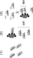
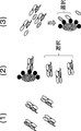
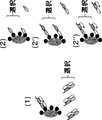
- a masking peptide is linked to an antibody by a linker that is cleaved by a protease that is expressed at a lesion site such as cancer tissue or inflammatory tissue, thereby masking the antigen binding site of the antibody with the masking peptide.
- the inventors of the present invention conducted intensive studies and found that it comprises a carrier portion having an antigen binding domain and a suppression domain that suppresses the binding activity of the antigen binding domain, and has a longer half life than an antigen binding domain present alone. Created a polypeptide. By using the polypeptide, it is considered that the antigen binding activity of the antigen binding domain is restored in the diseased tissue, and the antigen binding activity can be exerted in the diseased tissue.
- the whole body of the antigen binding domain in an activated state Distribution can be suppressed.
- the present inventors have found that the polypeptide or a pharmaceutical composition containing the polypeptide is useful for treating a disease, and is useful for treating a disease including administering the polypeptide. And found that the polypeptide is useful in the manufacture of a medicament for treating a disease.
- the present inventors are screening methods and manufacturing methods of the polypeptide, methods of manufacturing and screening single-domain antibodies whose antigen binding activity is suppressed by associating with specific VL or VH or VHH, and specific methods.
- the present invention was completed by creating a library including single domain antibodies whose antigen binding activity is suppressed by associating with VL or VH or VHH.
- the protease cleavage sequence is a sequence shown in SEQ ID NOs: 12, 25, 34, 35, 70 to 73, 75, 76, 91, 168 to 178, 193 to 195, 833 to 852, 1062 to 1081, a table
- the antigen binding domain comprises a single domain antibody, the suppression domain of the delivery moiety is VHH, or antibody VH, or antibody VL, and the single domain antibody is the VHH, or antibody VH, or The polypeptide according to any one of (1) to (20), wherein the antigen binding activity is suppressed.
- the antigen binding domain comprises a single domain antibody, the suppression domain of the delivery moiety is VHH or antibody VH or antibody VL, and the single domain antibody is the VHH or antibody VH or antibody VL and The polypeptide according to any one of (1) to (21), wherein the antigen binding activity is suppressed by association.
- the single domain antibody is VHH or VH having a single domain and antigen binding activity, and the suppression domain of the transport moiety is an antibody VL, and the VHH or VH having an antigen binding activity in single domain is The polypeptide according to any one of (19) to (22), wherein the antigen binding activity is suppressed by associating with the antibody VL.
- the single domain antibody is VHH, and the VHH is at least one amino acid substitution selected from F37V, Y37V, E44G, Q44G, R45L, H45L, G47W, F47W, L47W, T47W, or S47W (all Kabat numbering)
- the single domain antibody is VHH, and the VHH is 37V / 44G, 37V / 45L, 37V / 47W, 44G / 45L, 44G / 47W, 45L / 47W, 37V / 44G / 45L, 37V / 44G / 47W
- the poly according to any one of (19) to (23), comprising at least one set of amino acids selected from 37V / 45L / 47W, 44G / 45L / 47W, 37V / 44G / 45L / 47W (all Kabat numbering). peptide.
- the single domain antibody is VHH, and the VHH comprises at least one set of amino acid substitutions selected from F37V / R45L, F37V / G47W, R45L / G47W, and F37V / R45L / G47W (all Kabat numbering), 19)
- the single domain antibody is a single domain VL with antigen binding activity
- the suppression domain of the delivery part is an antibody VH
- the single domain VL with antigen binding activity is associated with the antibody VH
- the delivery moiety comprises an antibody constant region.
- the polypeptide according to (32), wherein the antibody constant region of the delivery moiety and the antigen binding domain are fused via a linker or not.
- the polypeptide is fused between the N-terminus of the antibody heavy chain constant region of the carrier and the C-terminus of the antigen binding domain via or without a linker, and further comprises a protease cleavage sequence
- the polypeptide is fused with the N-terminus of the antibody light chain constant region of the carrier moiety and the C-terminus of the antigen binding domain via or without a linker, and further has a protease cleavage sequence (35), wherein the protease cleavage sequence is located in the sequence of the antigen binding domain or on the antigen binding domain side from the amino acid 113 (EU numbering) (Kabat numbering 113) of the light chain antibody constant region, (35) The polypeptide described in 4.
- the antigen binding domain is a single domain antibody or VHH prepared from VH, and the protease cleavage sequence is the amino acid at position 109 (Kabat numbering) of the single domain antibody of the antigen binding domain and the antibody light chain constant.
- the antigen binding domain is a single domain antibody prepared from VL, and the protease cleavage sequence is amino acid number 104 (Kabat numbering) of the single domain antibody of the antigen binding domain and the antibody heavy chain constant region
- the polypeptide according to (40) which is located between amino acids 122 (EU numbering).
- the antigen binding domain is a single domain antibody prepared from VL, and the protease cleavage sequence is amino acid number 109 (Kabat numbering) of the single domain antibody of the antigen binding domain and the antibody light chain constant region
- the polypeptide according to (41) which is located between amino acids 113 (EU numbering) (Kabat numbering 113).
- the antigen binding domain is a single domain antibody
- the second antigen binding domain is a second single domain antibody
- the antigen binding domain and the second antigen binding domain can be released from the polypeptide (51), wherein the single domain antibody and the second single domain antibody form a bispecific antigen binding molecule in the free state of the antigen binding domain and the second antigen binding domain
- (51) The polypeptide described in 4.
- the polypeptide further comprises an antigen binding domain different from the antigen binding domain, and the antigen binding activity is suppressed by linking the other antigen binding domain to the carrier part of the polypeptide.
- a pharmaceutical composition comprising the polypeptide according to any one of (1) to (56).
- step (59) The following steps: (a) obtaining a single domain antibody that binds to a target antigen; (b) forming a polypeptide precursor by linking the single domain antibody and the delivery moiety such that the antigen binding activity of the single domain antibody obtained in step (a) is suppressed by the suppression domain of the delivery moiety ; (c) introducing a protease cleavage sequence into the polypeptide precursor;
- the manufacturing method as described in (58) including.
- step (60) The following steps: (a) obtaining a single domain antibody that binds to a target antigen; (b) forming a polypeptide precursor by linking the single domain antibody and the delivery moiety such that the antigen binding activity of the single domain antibody obtained in step (a) is suppressed by the suppression domain of the delivery moiety ; (c) introducing a protease cleavage sequence near the boundary between the single domain antibody and the carrier portion; The manufacturing method as described in (58) including.
- step (66) The following steps: (a) obtaining a single domain antibody that binds to a target antigen; (b) (a) the single domain antibody is associated with VL as a substitute for the VH of the IgG antibody or the single domain antibody is conjugated to the IgG antibody so that the antigen binding activity of the single domain antibody obtained in step (a) is suppressed Forming an IgG antibody-like molecule precursor into which said single domain antibody has been introduced by associating it with VH as a substitute for VL; (c) introducing a protease cleavage sequence near the boundary between the single domain antibody and the antibody constant region in the IgG antibody-like molecule precursor; (64) The manufacturing method as described in (64) including.
- (70) The following steps: (a) Substituting amino acid residues involved in association with antibody VH in a single domain antibody, or substituting amino acid residues involved in association with antibody VL in a single domain antibody, the single domain antibody Producing a modified single domain antibody that retains the binding activity of the target antigen to the target antigen; (b) by allowing the modified single domain antibody to associate with the antibody VH or causing the modified single domain antibody to associate with the antibody VL so as to suppress the antigen binding activity of the modified single domain antibody prepared in the step (a) Forming the IgG antibody-like molecule precursor into which the modified single domain antibody has been introduced; (c) introducing a protease cleavage sequence into the IgG antibody-like molecule precursor into which the modified single domain antibody has been introduced; (64) The manufacturing method as described in (64) including.
- (76) A vector comprising the polynucleotide according to (75).
- (77) A host cell comprising the polynucleotide of (75) or the vector of (76).
- (78) A method for producing the polypeptide according to any of (1) to (56), which comprises the step of culturing the host cell according to (77).
- (79) A method of screening a single domain antibody whose antigen binding activity is suppressed by associating with a specific VL, or associating with a specific VH, or by associating with a specific VHH.
- step (88) The following steps: (a) associating a single domain antibody with a specific VHH; (b) selecting an assembly of VHH and a single domain antibody in which the binding activity to the antigen of the single domain antibody associated with a specific VHH in the step (a) is absent or below a certain value; (c) Confirming that the binding activity of the single domain antibody in the assembly selected in step (b) to the antigen in the state of not being associated with the specific VHH is stronger than that at the time of association
- the step of The screening method as described in (86) including.
- (89) A method for producing a single domain antibody in which the antigen binding activity is suppressed by associating with a specific VL, or associating with a specific VH, or by associating with a specific VHH.
- (91) The following steps: (a) A step of substituting an amino acid residue involved in association with the antibody VL in a single domain antibody, and producing a modified single domain antibody that retains the binding activity of the single domain antibody to a target antigen; (90).
- step (98) Further following steps: (b) associating the modified single domain antibody prepared in step (a) with the VHH; (c) confirming that the antigen binding activity of the modified single domain antibody associated with the VHH is weakened or lost compared to before the association; The manufacturing method as described in (97) including.
- a library comprising a plurality of fusion polypeptides in which a single domain antibody and a first association support domain are linked, wherein the single domain antibody is suppressed in antigen binding activity by being associated with a specific VL or A single domain antibody which is lost, or a single domain antibody whose antigen binding activity is suppressed or lost by associating with a specific VH, or a single domain whose antigen binding activity is suppressed or lost by associating with a specific VHH Libraries, including antibodies.
- the single domain antibody portion of the fusion polypeptide in the library is a single domain antibody obtained from a camelid or a transgenic animal into which a gene capable of producing a single domain antibody has been introduced or a humanized antibody thereof, or a camelid
- a single domain antibody obtained by immunizing a transgenic animal into which an animal or a gene capable of producing a single domain antibody has been introduced, or a humanized antibody thereof, or a single domain artificially produced starting from a human antibody VH or VL The library according to (99), which comprises an antibody.
- a library comprising a plurality of fusion polypeptides in which a single domain antibody and a first association support domain are linked, wherein the single domain antibody is suppressed in antigen binding activity by being associated with a specific VL or The library according to (99) or (100), which comprises lost single domain antibodies.
- a library comprising a plurality of fusion polypeptides in which a single domain antibody and a first association support domain are linked, wherein the single domain antibody is suppressed in antigen binding activity by being associated with a specific VH or The library according to (99) or (100), which comprises lost single domain antibodies.
- a library comprising a plurality of fusion polypeptides in which a single domain antibody and a first association support domain are linked, wherein the single domain antibody suppresses antigen binding activity by being associated with a specific VHH or
- the library according to (99) or (100), which comprises lost single domain antibodies.
- a single domain antibody whose antigen binding activity is suppressed or lost by being associated with a specific VL, or an antigen binding activity by being associated with a specific VH
- a method of screening a fusion polypeptide comprising a single domain antibody in which the antibody is suppressed or lost, or a single domain antibody in which the antigen binding activity is suppressed or lost by association with a specific VHH.
- (113) A method of screening a fusion polypeptide containing a single domain antibody whose antigen binding activity is suppressed or lost by associating with a specific VH from the library described in (102).
- (114) The following steps: (a) displaying the fusion polypeptide of the library in vitro; (b) providing an association partner in which a specific VH and a second association support domain are fused; (c) The fusion polypeptide displayed in step (a) is associated with the association partner prepared in step (b), and the single domain antibody does not bind to the antigen in a state where the VH is associated, or the antigen binding activity is constant.
- step (d) From the fusion polypeptide selected in step (c), select a fusion polypeptide in which the single domain antibody contained therein does not associate with the above-mentioned VH and the antigen-binding activity is at least a certain value Process;
- the screening method as described in (113) including.
- the association partner prepared in step (b) further comprises a protease cleavage sequence, and in step (d), the association partner is cleaved by protease treatment to disassociate the single domain antibody and the VH. (114).
- the fusion polypeptide of the library further comprises a protease cleavage sequence, and in the step (d), the fusion polypeptide is cleaved by protease treatment to dissolve the VH association with the single domain antibody, (114) The screening method as described in.
- step (122) The following steps: (a) displaying the fusion polypeptide of the library in vitro; (b) providing an association partner in which a specific VHH and a second association support domain are fused; (c) The fusion polypeptide displayed in step (a) is associated with the association partner prepared in step (b), and the single domain antibody does not bind to the antigen in a state where the specific VHH is associated, or an antigen binding activity Selecting a fusion polypeptide having a value less than or equal to a predetermined value; (d) From the fusion polypeptides selected in step (c), select fusion polypeptides in which the single domain antibody contained and the VHH do not associate with each other, or the antigen binding activity is at least a certain value.
- the association partner prepared in the step (b) further comprises a protease cleavage sequence, and in the step (d), the association partner is cleaved by protease treatment to disassociate the single domain antibody and the VHH.
- the screening method according to (122) (124) The screening method according to (123), wherein the protease cleavage sequence of the association partner prepared in the step (b) is located near the boundary between the specific VHH and the second association support domain.
- the fusion polypeptide of the library further comprises a protease cleavage sequence, and in the step (d), the fusion polypeptide is cleaved by protease treatment to disassociate the single domain antibody and the VHH (122 The screening method as described in 4.).
- the screening method according to (125) wherein the protease cleavage sequence contained in the fusion polypeptide is located near the boundary between the single domain antibody and the first association support domain.
- step (d) the full length of the fusion polypeptide selected in the step (c) is displayed again in vitro, and the antigen is bound to the antigen only in association with the second association support domain, or antigen binding (122) the screening method according to (122), wherein a fusion polypeptide having an activity of a predetermined value or more is selected.
- the step of preparing the association partner in the step (b) is a step of simultaneously displaying the association partner and the fusion polypeptide, (106) to (112), (114) to (120), (122) The screening method according to any one of (128) to (128).
- the first association support domain comprises an IgG antibody CH1 domain
- the second association support domain comprises an antibody light chain constant region, (106) to (112), (114) to (120), (122) The screening method according to any one of (128) to (128).
- the first association support domain comprises an antibody light chain constant region
- the second association support domain comprises an IgG antibody CH1 domain, (106) to (112), (114) to (120), (122) The screening method according to any one of (128) to (128).
- step (133) The following steps: (a) displaying the fusion polypeptide of the library in vitro; (b) providing an association partner in which a specific VL and a second association support domain are fused; (c) selecting a fusion polypeptide in which the single domain antibody contained in the fusion polypeptide binds to an antigen, or the antigen binding activity is a predetermined value or more; (d) the fusion polypeptide selected in step (c) is associated with the association partner prepared in step (b), and the single domain antibody does not bind to the antigen in a state where the VL is associated, or the antigen binding activity is Selecting a fusion polypeptide that is below a certain value; (105), the screening method as described in (105).
- step (134) The screening method according to (129), wherein in step (d), the fusion polypeptide selected in step (c) is displayed again in vitro.
- step (c) the fusion polypeptide is associated with only the second association support domain, or a single fragment contained in the fusion polypeptide in the state where the fusion polypeptide is associated with only the second association support domain.
- step (136) The following steps: (a) displaying the fusion polypeptide of the library in vitro; (b) providing an association partner in which a specific VH and a second association support domain are fused; (c) selecting a fusion polypeptide in which the single domain antibody contained in the fusion polypeptide binds to an antigen, or the antigen binding activity is a predetermined value or more; (d) the fusion polypeptide selected in step (c) is associated with the association partner prepared in step (b), and the single domain antibody does not bind to the antigen in a state where the VH is associated, or the antigen binding activity is Selecting a fusion polypeptide that is below a certain value; The screening method as described in (113) including.
- step (137) The screening method according to (136), wherein in step (d), the fusion polypeptide selected in step (c) is displayed again in vitro.
- step (138) In the step (c), the fusion polypeptide is associated with only the second association support domain, or a single fragment contained in the fusion polypeptide in the state where the fusion polypeptide is associated with only the second association support domain.
- the screening method as described in (136) which confirms the antigen binding of a domain antibody.
- step (139) The following steps: (a) displaying the fusion polypeptide of the library in vitro; (b) providing an association partner in which a specific VHH and a second association support domain are fused; (c) selecting a fusion polypeptide in which the single domain antibody contained in the fusion polypeptide binds to an antigen, or the antigen binding activity is a predetermined value or more; (d) the fusion polypeptide selected in step (c) is associated with the association partner prepared in step (b), and the single domain antibody does not bind to the antigen in a state where the VHH is associated, or the antigen binding activity is Selecting a fusion polypeptide that is below a certain value; (121), the screening method as described in (121).
- step (140) The screening method according to (139), wherein in step (d), the fusion polypeptide selected in step (c) is displayed again in vitro.
- step (141) In the step (c), the fusion polypeptide is associated with only the second association support domain, or a single fragment contained in the fusion polypeptide in the state where the fusion polypeptide is associated with only the second association support domain.
- the screening method as described in (139) which confirms the antigen binding of a domain antibody.
- the step of associating the fusion polypeptide with the association partner in the step (d) is a step of simultaneously displaying the association partner and the fusion polypeptide, described in any of (133) to (141) Screening method.
- the invention can also encompass the embodiments specifically described in the following.
- a polypeptide wherein the polypeptide comprises an antigen binding domain and a carrier moiety, wherein the carrier moiety comprises a repression domain that suppresses the antigen binding activity of the antigen binding domain, the polypeptide comprises SEQ ID NO: A polypeptide having a protease cleavage sequence comprising one or more sequences selected from the sequences shown in Table 1 and the sequences shown in Table 1;
- B2 The suppression of the antigen-binding activity of the antigen-binding domain of the suppression domain in a state in which the protease cleavage sequence is cleaved by a protease is the same as that of the antigen-binding domain of the suppression domain in the uncleaved state of the protease cleavage sequence.
- the polypeptide according to (B1) which is weaker than suppression for antigen binding activity.
- B3 The polypeptide according to (B1) or (B2), wherein the antigen binding domain has a blood half life shorter than the carrier moiety.
- B4 The polypeptide according to any one of (B1) to (B3), wherein the molecular weight of the antigen binding domain is smaller than the molecular weight of the carrier moiety.
- B5 The polypeptide according to any one of (B1) to (B4), wherein the molecular weight of the antigen binding domain is 60 kDa or less.
- (B6) Any one of (B1) to (B5), wherein the delivery moiety has FcRn binding activity and the antigen binding domain has no FcRn binding activity or has weaker FcRn binding activity than the delivery moiety
- the polypeptide described in 4. (B7) The antigen binding domain is releasable from the polypeptide, and the antigen binding domain is free from the polypeptide. The antigen binding activity is not released from the polypeptide.
- (B8) The polypeptide according to any one of (B1) to (B7), wherein the antigen binding activity of the antigen binding domain is suppressed by the association of the antigen binding domain and the suppression domain of the carrier moiety. .
- (B9) The polypeptide according to (B7), wherein the protease cleavage sequence is cleaved by a protease to release the antigen binding domain from the polypeptide.
- B10 The polypeptide according to (B8), wherein the protease cleavage sequence is cleaved by a protease to disassociate the antigen binding domain and the suppression domain of the transport moiety.
- (B17) The polypeptide according to (B15) or (B16), wherein a second movable linker is further added to the other end of the protease cleavage sequence.
- (B18) The polypeptide according to (B17), wherein the second movable linker is a movable linker consisting of a glycine-serine polymer.
- (B19) Any of (B1) to (B18), wherein the antigen binding domain comprises a single domain antibody or is a single domain antibody, and the suppression domain of the delivery portion suppresses the antigen binding activity of the single domain antibody. Or the polypeptide according to one.
- the antigen binding domain comprises a single domain antibody, the suppression domain of the delivery moiety is VHH, or antibody VH, or antibody VL, and the single domain antibody is said VHH, or antibody VH, or antibody VL and The polypeptide according to any one of (B1) to (B21), wherein the antigen binding activity is suppressed by association.
- the single domain antibody is VHH, or VH having a single domain and antigen binding activity
- the suppression domain of the transport moiety is an antibody VL
- the VHH or VH having an antigen binding activity of single domain is The polypeptide according to any one of (B19) to (B22), in which antigen binding activity is suppressed by associating with the antibody VL.
- the single domain antibody is VHH, and the VHH is amino acid substituted at at least one position selected from amino acids 37, 44, 45, or 47 (all Kabat numbering) (B19) 2.) The polypeptide according to any one of (B23). (B25) Any one of (B19) to (B23), wherein the single domain antibody is VHH, and the VHH contains at least one amino acid selected from amino acids of 37V, 44G, 45L, or 47W (all Kabat numbering) The polypeptide described in one.
- the single domain antibody is VHH, and the VHH is at least one amino acid substitution selected from F37V, Y37V, E44G, Q44G, R45L, H45L, G47W, F47W, L47W, T47W, or S47W (all Kabat numbering)
- the polypeptide according to any one of (B19) to (B23), comprising two amino acid substitutions.
- the single domain antibody is VHH, and the VHHs are 37/44, 37/45, 37/47, 44/45, 44/47, 45/47.
- the single domain antibody is VHH, and the VHH is 37V / 44G, 37V / 45L, 37V / 47W, 44G / 45L, 44G / 47W, 45L / 47W, 37V / 44G / 45L, 37V / 44G / 47W , At least one set of amino acids selected from 37V / 45L / 47W, 44G / 45L / 47W, 37V / 44G / 45L / 47W (all Kabat numbering), described in any one of (B19) to (B23) Polypeptide.
- the single domain antibody is VHH, and the VHH contains at least one set of amino acid substitutions selected from F37V / R45L, F37V / G47W, R45L / G47W, and F37V / R45L / G47W (all Kabat numbering), The polypeptide according to any one of B19) to (B23).
- the single domain antibody is a single domain VL with antigen binding activity
- the suppression domain of the delivery part is an antibody VH
- the single domain VL with antigen binding activity is associated with the antibody VH
- the polypeptide according to any one of (B19) to (B22), wherein the antigen binding activity is suppressed by (B31)
- B32 The polypeptide according to any one of (B1) to (B31), wherein the delivery moiety comprises an antibody constant region.
- the polypeptide is fused with the N-terminus of the antibody heavy chain constant region of the transport moiety and the C-terminus of the antigen binding domain via a linker or not via a linker, and the protease cleavage sequence comprises The polypeptide according to (B34), which is located closer to the antigen binding domain than the amino acid at position 122 (EU numbering) of the heavy chain antibody constant region or in the sequence of the antigen binding domain.
- the polypeptide is fused with the N-terminus of the antibody light chain constant region of the transport moiety and the C-terminus of the antigen binding domain via a linker or not via a linker, and the protease cleavage sequence comprises The polypeptide according to (B35), which is located closer to the antigen binding domain from the amino acid of the light chain antibody constant region (amino acid number 113 (EU numbering) (Kabat numbering 113)) in the sequence of the antigen binding domain.
- polypeptide is fused with the N-terminus of the antibody constant region of the carrier moiety and the C-terminus of the antigen binding domain via or without a linker, and the antigen binding domain is prepared from VH A single domain antibody or VHH, and the protease cleavage sequence is located within the sequence of the antibody constant region, or closer to the antibody constant region than the amino acid at position 109 (Kabat numbering) of the single domain antibody of the antigen binding domain.
- the polypeptide as described in any one of (B33) to (B36).
- the polypeptide is fused with the N-terminus of the antibody light chain constant region of the carrier moiety and the C-terminus of the antigen binding domain via a linker or without a linker, and the protease cleavage sequence comprises The polypeptide according to (B35), which is located near the boundary between the antigen binding domain and the antibody light chain constant region.
- the antigen binding domain is a single domain antibody or VHH prepared from VH, and the protease cleavage sequence is amino acid number 109 (Kabat numbering) of the single domain antibody of the antigen binding domain and the antibody heavy chain constant.
- the polypeptide according to (B40) which is located between amino acids 122 (EU numbering) of the region.
- the antigen binding domain is a single domain antibody or VHH prepared from VH, and the protease cleavage sequence is an amino acid at position 109 (Kabat numbering) of the single domain antibody of the antigen binding domain and the antibody light chain constant.
- the antigen binding domain is a single domain antibody prepared from VL, and the protease cleavage sequence is amino acid 104 (Kabat numbering) of the single domain antibody of the antigen binding domain and the antibody heavy chain constant region
- the antigen binding domain is a single domain antibody prepared from VL, and the protease cleavage sequence is amino acid number 109 (Kabat numbering) of the single domain antibody of the antigen binding domain and the antibody light chain constant region
- (B46) The polypeptide according to any one of (B32) to (B45), wherein the antibody constant region of the polypeptide is an IgG antibody constant region.
- (B47) The polypeptide according to any one of (B1) to (B46), wherein the polypeptide is an IgG antibody-like molecule.
- (B48) In the state where the antigen binding domain is not released, when measurement is performed using BLI (Bio-Layer Interferometry) method (Octet), no binding between the antigen binding domain and the antigen is observed, from (B1) to (B1) The polypeptide according to any one of B47).
- BLI Bio-Layer Interferometry
- (B49) The polypeptide according to any one of (B1) to (B48), wherein a second antigen binding domain is further linked to the antigen binding domain.
- (B50) The polypeptide according to (B49), wherein the second antigen binding domain has antigen binding specificity different from that of the antigen binding domain.
- (B51) The polypeptide according to (B49) or (B50), wherein the second antigen binding domain comprises a second single domain antibody.
- the antigen binding domain is a single domain antibody
- the second antigen binding domain is a second single domain antibody
- the antigen binding domain and the second antigen binding domain can be released from the polypeptide (B51) in which the single domain antibody and the second single domain antibody form a bispecific antigen binding molecule in the free state of the antigen binding domain and the second antigen binding domain, The polypeptide described in 4.
- the polypeptide further comprises an antigen binding domain other than the antigen binding domain, and the antigen binding activity is suppressed by linking the other antigen binding domain to the carrier part of the polypeptide.
- B55 The polypeptide according to (B54), which has different antigen binding specificity from the another antigen binding domain and the antigen binding domain.
- B56 The polypeptide according to any one of (B1) to (B55), wherein the antigen binding domain is an antigen binding domain that targets Plexin A1, IL-6R or CD3 as a target antigen.
- a pharmaceutical composition comprising the polypeptide according to any one of (B1) to (B56).
- (B58) A method for producing the polypeptide according to any one of (B1) to (B56). (B59) The following steps: (a) obtaining a single domain antibody that binds to a target antigen; (b) forming a polypeptide precursor by linking the single domain antibody and the delivery moiety such that the antigen binding activity of the single domain antibody obtained in step (a) is suppressed by the suppression domain of the delivery moiety ; (c) introducing a protease cleavage sequence into the polypeptide precursor; The manufacturing method as described in (B58) including.
- step (B60) The following steps: (a) obtaining a single domain antibody that binds to a target antigen; (b) forming a polypeptide precursor by linking the single domain antibody and the delivery moiety such that the antigen binding activity of the single domain antibody obtained in step (a) is suppressed by the suppression domain of the delivery moiety ; (c) A protease cleavage sequence comprising one or more sequences selected from the sequences shown in SEQ ID NOS: 833 to 852 and 1062 to 1081 and the sequences described in Table 1 near the boundary between the single domain antibody and the carrier portion Introducing process; The manufacturing method as described in (B58) including.
- (B61) The following steps: (a) obtaining a single domain antibody that binds to a target antigen; (b) Sequences shown by SEQ ID NOS: 833 to 852, 1062 to 1081 such that the antigen binding activity of the single domain antibody obtained in the step (a) is suppressed by the suppression domain of the carrier moiety Linking to the transport moiety via a protease cleavage sequence comprising one or more sequences selected from the sequences set forth in Table 1 to form a polypeptide; The manufacturing method as described in (B58) including.
- (B62) The following steps: (d) confirming that the binding activity of the single domain antibody incorporated into the polypeptide or the polypeptide precursor to the target antigen is weakened or lost; And the method according to any one of (B59) to (B61). (B63) Further the following steps: (e) releasing the single domain antibody by cleaving the protease cleavage sequence with a protease, and confirming that the free single domain antibody binds to an antigen; And the method according to any one of (B59) to (B62). (B64) The method according to (B58), wherein the polypeptide is an IgG antibody-like molecule.
- step (B65) The following steps: (a) obtaining a single domain antibody that binds to a target antigen; (b) (a) the single domain antibody is associated with VL as a substitute for the VH of the IgG antibody or the single domain antibody is conjugated to the IgG antibody so that the antigen binding activity of the single domain antibody obtained in step (a) is suppressed Forming an IgG antibody-like molecule precursor into which said single domain antibody has been introduced by associating it with VH as a substitute for VL; (c) A protease comprising one or more sequences selected from the sequences shown in SEQ ID NOS: 833 to 852 and 1062 to 1081 and sequences listed in Table 1 in the IgG antibody-like molecule precursor into which the single domain antibody has been introduced.
- step (B66) The following steps: (a) obtaining a single domain antibody that binds to a target antigen; (b) (a) the single domain antibody is associated with VL as a substitute for the VH of the IgG antibody or the single domain antibody is conjugated to the IgG antibody so that the antigen binding activity of the single domain antibody obtained in step (a) is suppressed Forming an IgG antibody-like molecule precursor into which said single domain antibody has been introduced by associating it with VH as a substitute for VL; (c) The sequence shown in SEQ ID NOS: 833 to 852, 1062 to 1081, and a sequence selected from the sequences shown in Table 1 near the boundary between the single domain antibody and the antibody constant region in the IgG antibody-like molecule precursor Introducing a protease cleavage sequence comprising one or more of And the method according to (B64).
- (B68) The following steps: (d) confirming that the binding activity to the target antigen of the single domain antibody introduced into the IgG antibody-like molecule or the precursor of the IgG antibody-like molecule is weakened or lost; And the method according to any one of (B65) to (B67). (B69) Further following steps: (e) releasing the single domain antibody by cleaving the protease cleavage sequence with a protease, and confirming that the free single domain antibody binds to the target antigen; And the method according to any one of (B65) to (B68).
- a cell-free translation system (Clover Direct (Protein Express)) or the like in which a tRNA in which a nonnatural amino acid is bound to a complementary amber suppressor tRNA complementary to UAG codon (amber codon) which is one of stop codons is also preferably used.
- alterations include, but are not limited to, substitutions.
- amino acids 37, 45 and / or 47 are substituted includes variations of the following amino acid alterations; (a) 37, (b) 45, (c) 47, (d) 37 and 45, (e) 37 and 47, (f) 45 and 47, (g) 37 And 45 and 47.
- a representation representing amino acid modification a representation in which the one-letter code or the three-letter code of the amino acid before and after modification may be used appropriately before and after a number representing a specific position.
- the modification of F37V or Phe37Val used in adding amino acid substitutions contained in antibody variable regions or single domain antibodies represents substitution of Phe at position 37 with Val, which is represented by Kabat numbering. That is, the numbers indicate the position of the amino acid represented by Kabat numbering, and the one-letter code or three-letter code of the amino acid described earlier is the amino acid before substitution, the one letter code of the amino acid described thereafter or 3
- the letter code represents the amino acid after substitution.
- full-length antibody full-length antibody
- full antibody full antibody
- all antibody are used interchangeably herein and have a structure substantially similar to a naturally occurring antibody structure or as defined herein Antibody having a heavy chain containing an Fc region thereof.
- 262 732-745 (1996)); (d) HVR amino acid residues 46-56 (L2), 47-56 (L2), 48-56 (L2), 49-56 (L2), 26-35 (H1), 26-35b (H1), 49 A combination of (a), (b), and / or (c), including -65 (H2), 93-102 (H3), and 94-102 (H3).
- the CDRs of the single domain antibody illustratively include: (a) Hypervariable loops occurring at amino acid residues 26-32 (CDR1), 50-52 (CDR2), and 91-96 (CDR3) (Chothia and Lesk, J. MoI. Biol. 196: 901-917 ( 1987)); (b) CDRs occurring at amino acid residues 24-34 (CDR1), 50-56 (CDR2) and 89-97 (CDR3) (Kabat et al., Sequences of Proteins of Immunological Interest, 5th Ed.
- variable region or “constant domain” refer to portions other than the variable region of an antibody.
- an IgG antibody is an approximately 150,000 dalton heterotetrameric glycoprotein composed of two identical light chains and two identical heavy chains that are disulfide linked, from the N-terminus to the C-terminus, Each heavy chain has a variable region (VH), also referred to as a variable heavy chain domain or a heavy chain variable domain, followed by a heavy chain constant region (CH) comprising a CH1 domain, a hinge region, a CH2 domain and a CH3 domain.
- VH variable region
- CH heavy chain constant region
- each light chain has a variable region (VL), also referred to as variable light chain domain or light chain variable domain, followed by a constant light chain (CL) domain.
- VL variable region
- CL constant light chain
- the light chain of a naturally occurring antibody may be assigned to one of two types, called kappa ( ⁇ ) and lambda ( ⁇ ), based on the amino acid sequence of its constant domain.
- the "antigen binding domain” is limited only by binding to the target antigen.
- domains of any structure can be used as long as they bind to the target antigen. Examples of such domains include, but are not limited to, for example, antibody heavy chain variable region (VH) and antibody light chain variable region (VL), single domain antibody (sdAb), in vivo A module called A domain of about 35 amino acids contained in the cell membrane protein Avimer (WO 2004/044011, WO 2005/040229), a 10Fn3 domain which is a domain that binds to a protein in fibronectin, a glycoprotein expressed on the cell membrane Adfectin (International Publication WO 2002/032925), Affibody (as International Publication WO 1995/001937) whose scaffold is an IgG binding domain that constitutes a bundle of three amino acids consisting of 58 amino acids of Protein A (International Publication WO 1995/001937), a turn containing 33 amino acid residues Molecular Table of Ankyr
- One suitable example of the antigen binding domain of the present invention is a single domain antibody (sdAb).
- single domain antibodies include, but are not limited to, all or part of an antigen binding molecule that inherently lacks a light chain, such as, for example, a camelid VHH, a shark V NAR , or a VH domain of an antibody Or antibody fragments that contain all or part of the VL domain.
- single domain antibodies which are antibody fragments comprising all or part of the VH / VL domain of antibodies include, but are not limited to, human antibodies VH or human antibodies as described, for example, in US Patent No. 6,248, 516 B1 etc.
- An artificially produced single domain antibody starting from VL can be mentioned.
- one single domain antibody has 3 CDRs (CDR1, CDR2 and CDR3).
- Single domain antibodies can be obtained from animals capable of producing single domain antibodies or by immunizing animals capable of producing single domain antibodies.
- animals that can produce single domain antibodies include, but are not limited to, camelids, transgenic animals into which genes capable of producing single domain antibodies have been introduced.
- the camelids include camels, llamas, alpaca, camel and guanaco and the like.
- transgenic animals into which a gene capable of producing a single domain antibody has been introduced include, but are not limited to, transgenic animals described in International Publication WO 2015/143414 and US Patent Publication US 2011/0123527 A1.
- Humanized single domain antibodies can also be obtained by using the framework sequences of single domain antibodies obtained from animals as human germline sequences or sequences similar thereto.
- Humanized single domain antibodies are also an embodiment of single domain antibodies of the invention.
- “Humanized single domain antibody” refers to a chimeric single domain antibody that comprises amino acid residues from non-human CDRs and amino acid residues from human FR.
- the humanized single domain antibody has all or substantially all of the CDRs correspond to those of non-human antibodies, and all or substantially all of the FRs correspond to those of human antibodies.
- substantially all of the FRs are considered as an example corresponding to those of human antibodies, even if some of the residues in the FRs do not correspond to those of human antibodies.
- single domain antibodies can be obtained from polypeptide libraries containing single domain antibodies by ELISA, panning and the like.
- Antigens include the following molecules: 17-IA, 4-1BB, 4Dc, 6-keto-PGF1a, 8-iso-PGF2a, 8-oxo-dG, A1 adenosine receptor, A33, ACE, ACE-2, Activin, activin A, activin AB, activin B, activin C, activin RIA, activin RIA ALK-2, activin RIB ALK-4, activin RIIA, activin RIIB, ADAM10, ADAM12, ADAM15, ADAM17 / TACE, ADAM8, ADAM9 , ADAMTS, ADAMTS4, ADAMTS5, addressin, aFGF, ALCAM, ALK, ALK-1, ALK-7, alpha-1-antitrypsin, alpha-V / beta-1 antagonist, ANG, Ang, APAF-1, APE, APJ , APP, APRIL, AR, ARC, ART, Artemin, Anti-Id, AS
- T cell receptor eg, T cell receptor alpha / beta
- TdT TECK
- TEM1 TEM5, TEM7, TEM8, TERT
- testicular PLAP-like alkaline phosphatase TfR, TGF, TGF-alpha, TGF-beta, TGF-beta Pan Specific, TGF-beta RI (ALK-5), TGF-beta RII, TGF-beta RIIb, TGF-beta RIII, TGF-beta 1, TGF-beta 2, TGF-beta 3, TGF-beta 4 TGF-beta5, thrombin, thymus Ck-1, thyroid stimulating hormone, Tie, TIMP, TIQ, tissue factor, TMEFF2, Tmpo, TMPRSS2, TNF, alpha, TNF-alpha, TNF-beta2, TNFc, TNF-RI, TNF-RII, TNFRSF10A (TRAIL R1 Apo-2
- receptors are also described in the above-mentioned examples of antigens, these receptors may also be used as antigens to which the antigen binding domain of the present invention binds, when they exist in soluble form in biological fluid.
- soluble receptors soluble IL-6R as described, for example, by Mullberg et al. (J. Immunol. (1994) 152 (10), 4958-4968).
- SEQ ID NO: 35 a protein represented by SEQ ID NO: 35 can be exemplified.
- the soluble molecule is not limited to the solution in which the soluble molecule is present, and the soluble molecule may be present in the biological fluid, that is, in all the fluids filling the vessels or tissues / cells in the living body.
- a soluble molecule to which the antigen binding domain of the invention binds may be present in the extracellular fluid.
- Extracellular fluid refers to components in bone and cartilage such as plasma, interstitial fluid, lymph fluid, dense connective tissue, cerebrospinal fluid, spinal fluid, puncture fluid, or synovial fluid in vertebrates, alveolar fluid (bronchopulmonary fluid) Cell-permeating fluid such as alveolar lavage fluid, ascites fluid, pleural fluid, pericardial fluid, cyst fluid, or aqueous humor (aqueous humor) fluid in various glandular cavities resulting from active transport and secretory activity of cells, and digestive tract cavity Generic term for other body cavity fluid).
- alveolar fluid bronchopulmonary fluid
- Cell-permeating fluid such as alveolar lavage fluid, ascites fluid, pleural fluid, pericardial fluid, cyst fluid, or aqueous humor (aqueous humor) fluid in various glandular cavities resulting from active transport and secretory activity of cells, and digestive tract cavity Generic term for other body cavity fluid).
- an epitope meaning an antigenic determinant present in an antigen means a site on the antigen to which the antigen binding domain disclosed herein binds.
- an epitope can be defined by its structure.
- the epitope can also be defined by the binding activity to the antigen in the antigen binding domain that recognizes the epitope.
- the antigen is a peptide or a polypeptide
- the epitope is a sugar chain, it is also possible to specify the epitope by a specific sugar chain structure.
- a conformational epitope in contrast to a linear epitope, is an epitope in which the primary sequence of amino acids comprising the epitope is not a single defined component of the recognized epitope (eg, an antibody wherein the primary sequence of amino acids necessarily defines the epitope)
- An epitope not recognized by A conformational epitope may encompass an increased number of amino acids relative to a linear epitope.
- the antigen binding domain recognizes the three dimensional structure of the peptide or protein. For example, if the protein molecule folds to form a three-dimensional structure, certain amino acids and / or polypeptide backbones that form a steric epitope will be juxtaposed to allow the antibody to recognize the epitope.
- the structure of the antigen binding domain that binds to the epitope is called the paratope.
- the epitope and paratope are stably bound by the hydrogen bond, electrostatic force, van der Waals force, hydrophobic bond, etc. acting between the epitope and the paratope.
- the avidity between this epitope and the paratope is called affinity.
- the sum of avidity when a plurality of antigens and a plurality of antigen binding domains bind is called avidity.
- avidity is higher than the affinity, because the affinities work synergistically.
- Examples of the method for confirming the binding to an epitope by an antigen binding domain to IL-6R or a polypeptide containing the antigen binding domain are exemplified below, but an antigen binding domain to an antigen other than IL-6R or a poly comprising an antigen binding domain is exemplified.
- the method of confirming the binding of the peptide to the epitope can also be appropriately carried out according to the following example.
- the binding activity of the antigen binding domain to the peptide can be evaluated by ELISA using the immobilized linear peptide as an antigen.
- the binding activity to a linear peptide can be revealed based on the level of inhibition by the linear peptide in the binding of the antigen binding domain to IL-6R expressing cells. These tests can reveal the binding activity of the antigen binding domain to linear peptides.
- the antigen binding domain to IL-6R recognizes a steric epitope.
- cells expressing IL-6R are prepared.
- the antigen binding domain for IL-6R binds strongly to IL-6R-expressing cells when in contact with the cells, while the antigen binding domain consists of an amino acid sequence constituting the extracellular domain of IL-6R immobilized
- the present invention is substantially directed to a linear peptide, or to a linear peptide consisting of an amino acid sequence in which the antigen binding domain is composed of the extracellular domain of IL-6R denatured using a general denaturing agent such as guanidine or the like. And the like.
- substantially non-binding refers to a binding activity of 80% or less, usually 50% or less, preferably 30% or less, particularly preferably 15% or less, of binding activity to human IL-6R-expressing cells.
- the association rate (kon) and the dissociation rate (koff) are calculated by simultaneously fitting the sensorgrams of association and dissociation using a simple one-to-one Langmuir binding model (BIACORE® evaluation software version 3.2) Ru.
- the equilibrium dissociation constant (Kd) is calculated as the koff / kon ratio.
- the antigen binding activity of the antigen binding domain can also be measured by a known method of measuring intermolecular interaction such as electrochemiluminescence.
- a method for measuring the binding activity of the antigen binding domain to IL-6R to IL-6R-expressing cells for example, the method described in Antibodies A Laboratory Manual (Ed Harlow, David Lane, Cold Spring Harbor Laboratory (1988) 359-420) Can be mentioned. That is, it can be evaluated by the principle of ELISA or FACS (fluorescence activated cell sorting) using IL-6R expressing cells as an antigen.
- the following method may be mentioned as an example of a preferred method of measuring the binding activity of an antigen binding domain to IL-6R to an antigen.
- the cells are stained with a FITC-labeled secondary antibody that recognizes a test antigen binding domain reacted with cells expressing IL-6R.
- the antigen binding domain is adjusted to a desired concentration and used by diluting the test antigen binding domain with an appropriate buffer. For example, it can be used at any concentration between 10 ⁇ g / ml and 10 ng / ml.
- the fluorescence intensity and the number of cells are measured by FACSCalibur (BD).
- the binding amount of the antigen binding domain to the cells is reflected in the fluorescence intensity obtained by analysis using CELL QUEST Software (BD), that is, the value of Geometric Mean. That is, by obtaining the value of the Geometric Mean, the binding activity of the test antigen binding domain represented by the binding amount of the test antigen binding domain can be measured.
- BD CELL QUEST Software
- antigen binding domain for IL-6R shares an epitope with an antigen binding domain can be confirmed by competition for the same epitope of both. Competition between antigen binding domains is detected, such as by cross-blocking assays.
- a competitive ELISA assay is a preferred cross blocking assay.
- a test antigen is tested after the IL-6R protein coated on the wells of a microtiter plate is pre-incubated in the presence or absence of a candidate competing antigen binding domain. Binding domain is added.
- the amount of test antigen binding domain bound to the IL-6R protein in the wells is indirectly correlated to the binding ability of the candidate competing antigen binding domain to compete for binding to the same epitope. That is, the greater the affinity of the competitor antigen binding domain for the same epitope, the lower the binding activity of the test antigen binding domain to the well coated with IL-6R protein.
- the amount of test antigen binding domain bound to the wells via the IL-6R protein can be easily measured by pre-labeling the antigen binding domain.
- a biotin labeled antigen binding domain is measured by using an avidin peroxidase conjugate and a suitable substrate.
- Cross-blocking assays utilizing enzyme labels such as peroxidase are particularly referred to as competitive ELISA assays.
- the antigen binding domain can be labeled with another labeled substance that can be detected or measured. Specifically, radiolabels or fluorescent labels are known.
- the competitive antigen binding domain has at least 20%, preferably at least 20%, binding of the antigen binding domain to the IL-6R as compared to the binding activity obtained in a control test performed in the absence of the candidate competitive antigen binding domain.
- the sharing of the epitope between the test antigen binding domain and the control antigen binding domain results in amino acid mutations in the peptide constituting the epitope. It can be evaluated by comparing the binding activities of both antigen binding domains to the introduced peptide or polypeptide.
- the test antigen binding domain and the control antigen binding domain share the epitope can be evaluated by the following method.
- cells expressing IL-6R and cells expressing IL-6R in which a mutation has been introduced into an epitope are prepared.
- the test antigen binding domain and the control antigen binding domain are added to a cell suspension in which these cells are suspended in a suitable buffer such as PBS.
- FITC-labeled antibody capable of recognizing the test antigen binding domain and the control antigen binding domain is added to the cell suspension, which is appropriately washed with buffer.
- the fluorescence intensity and cell number of the cells stained by the labeled antibody are measured by FACSCalibur (BD).
- the concentrations of the test antigen binding domain and the control antigen binding domain are adjusted to the desired concentration by appropriate dilution with a suitable buffer and used. For example, it is used at any concentration between 10 ⁇ g / ml and 10 ng / ml.
- the binding amount of the labeled antibody to the cells is reflected in the fluorescence intensity obtained by analysis using CELL QUEST Software (BD), that is, the value of Geometric Mean. That is, by obtaining the value of the Geometric Mean, it is possible to measure the binding activity of the test antigen binding domain represented by the binding amount of the labeled antibody and the control antigen binding domain.
- BD CELL QUEST Software
- “not substantially bound to a mutant IL-6R-expressing cell” can be determined by the following method. First, the test antigen binding domain and the control antigen binding domain bound to cells expressing mutant IL-6R are stained with a labeled antibody. The fluorescence intensity of the cells is then detected. When FACSCalibur is used as flow cytometry for fluorescence detection, the fluorescence intensity obtained can be analyzed using CELL QUEST Software. By calculating this comparison value ( ⁇ Geo-Mean) from the value of Geometric Mean in the presence and absence of the polypeptide complex based on the following equation 1, the percentage increase in fluorescence intensity due to the binding of the antigen binding domain You can ask for
- the term "carrying moiety” refers to a moiety other than an antigen binding domain in a polypeptide.
- the delivery moiety of the present invention is usually a peptide or polypeptide composed of amino acids, and in a specific embodiment, the delivery moiety in the polypeptide is linked to the antigen binding domain via a cleavage site.
- the delivery moiety of the present invention may be a series of peptides or polypeptides linked by amide bonds, and a plurality of peptides or polypeptides may be noncovalently linked such as covalent bonds such as disulfide bonds or hydrogen bonds, hydrophobic interactions, etc. It may be a complex formed by binding.
- the delivery moiety of the present invention has a repressing domain that represses the antigen binding activity of the antigen binding domain.
- the term "repression domain” is limited only by suppressing the antigen binding activity of the antigen binding domain.
- the suppression domain domains of any structure can be used as long as they can suppress the antigen binding activity of the antigen binding domain. Examples of such repression domains include, but are not limited to, for example, the heavy chain variable domain (VH) of the antibody, the light chain variable domain (VL) of the antibody, pre-B cell receptors, and single domain antibodies.
- VH heavy chain variable domain
- VL light chain variable domain
- pre-B cell receptors pre-B cell receptors
- single domain antibodies Be The suppression domain may be comprised of all of the delivery portion or may be comprised of part of the delivery portion.
- release of the antigen binding domain from the polypeptide results in higher antigen binding activity than before release.
- the antigen binding domain is not released from the polypeptide, its antigen binding activity is suppressed by the repression domain.
- FACS fluorescence activated cell sorting
- ELISA Enzyme-Linked Immunosorbent Assay
- ECL electrogenerated chemiluminescence
- SPR There are methods such as (Surface Plasmon Resonance, surface plasmon resonance) method (Biacore), BLI (Bio-Layer Interferometry) method (Octet) and the like.
- the antigen binding activity in the state in which the antigen binding domain is released from the polypeptide is two-fold, three times that in the state in which the antigen binding domain is not released from the polypeptide. Double, 4 times, 5 times, 6 times, 7 times, 8 times, 9 times, 10 times, 20 times, 30 times, 40 times, 50 times, 60 times, 70 times, 80 times, 90 times, 100 times,
- the value is 200 times, 300 times, 400 times, 500 times, 600 times, 700 times, 800 times, 900 times, 1000 times, 2000 times, or 3000 times or more.
- the antigen binding domain prior to release comprises an antigen binding domain and an antigen binding domain when measuring the antigen binding activity of the antigen binding domain in one of the methods selected from among the methods described above. No binding to antigen is seen.
- cleavage of the cleavage site allows the antigen binding domain to be released from the polypeptide, so comparisons of antigen binding activity in such embodiments can be made by comparing the antigens before and after cleavage of the polypeptide. It can be performed by comparing the binding activity.
- the antigen binding activity measured using the polypeptide after cleavage is twice, three times, four times, five times, six times, 7 times, as compared to the antigen binding activity measured using an uncleaved polypeptide.
- the value is 600 times, 700 times, 800 times, 900 times, 1000 times, 2000 times, or 3000 times or more.
- the uncleaved polypeptide shows no binding between the antigen binding domain and the antigen when the antigen binding activity is measured by one of the methods selected from the above methods. .
- the cleavage site is cleaved by a protease, so comparison of antigen binding activity in such embodiments may be performed by comparing the antigen binding activity before and after protease treatment of the polypeptide. it can. That is, the antigen binding activity measured using the polypeptide after protease treatment is twice, three times, four times, five times, as compared to the antigen binding activity measured using a polypeptide not treated with protease.
- the protease-untreated polypeptide is found to have a binding between the antigen binding domain and the antigen when the antigen binding activity is measured by one of the methods selected from the above methods. Absent.
- a polypeptide comprising an antigen binding domain and a carrier moiety has a longer blood half life, as compared to the antigen binding domain present alone.
- the delivery moiety is designed to have a longer blood half life.
- the molecular weight of the transport moiety is large, or the transport moiety has FcRn binding properties, or the transport moiety has albumin binding properties Or carrying moiety is PEGylated.
- the delivery moiety has a longer blood half life (in other words, the antigen binding domain has a shorter blood half life than the delivery moiety) than the antigen binding domain.
- One embodiment of increasing the blood half-life of the delivery moiety is that the delivery molecule has a high molecular weight. In one embodiment of increasing the blood half-life of the delivery moiety longer than that of the antigen binding domain, the molecular weight of the delivery moiety is greater than that of the antigen binding domain.
- One embodiment of extending the blood half-life of the delivery moiety is to allow the delivery moiety to have FcRn binding properties.
- the FcRn binding region refers to a region having binding to FcRn, and any structure can be used as long as it has binding to FcRn.
- the delivery moiety comprising the FcRn binding region is capable of being returned to plasma again after being taken up into cells by the salvage pathway of FcRn.
- the relatively long retention (slow elimination) of IgG molecules in plasma is due to the function of FcRn, which is known as a salvage receptor of IgG molecules.
- IgG molecules incorporated into endosomes by pinocytosis bind to FcRn expressed in endosomes under acidic conditions in endosomes. IgG molecules that can not bind to FcRn proceed to lysosomes and are degraded there, but IgG molecules bound to FcRn translocate to the cell surface and return to plasma by dissociating from FcRn under neutral conditions in plasma. .
- the FcRn binding region is preferably a region that directly binds to FcRn. As a preferred example of the FcRn binding region, the Fc region of an antibody can be mentioned.
- the FcRn binding region in the present invention May be a region that binds to a polypeptide capable of binding to such FcRn.
- the binding activity of the FcRn-binding region of the present invention to FcRn, particularly human FcRn can be measured by methods known to those skilled in the art as described in the aforementioned section on binding activity, and the conditions for the conditions are described by those skilled in the art. Can be determined appropriately.
- the binding activity to human FcRn may be KD (Dissociation constant: dissociation constant), apparent KD (Apparent dissociation constant: apparent dissociation constant), dissociation rate kd (Dissociation rate: dissociation rate), or apparent kd (Apparent) dissociation: apparent dissociation rate) and the like. These can be measured by methods known to those skilled in the art. For example, Biacore (GE healthcare), Scatchard plots, flow cytometers, etc. may be used.
- Conditions for measuring the binding activity of the FcRn binding region to FcRn can be appropriately selected by those skilled in the art, and are not particularly limited. For example, it can be measured in MES buffer, 37 ° C. conditions as described in WO 2009/125825.
- the binding activity of the FcRn binding region of the present invention to FcRn can be measured by methods known to those skilled in the art, and can be measured, for example, using Biacore (GE Healthcare) or the like.
- Measurement of FcRn binding region and FcRn binding activity can be carried out by using an FcRn binding region or a carrier moiety containing an FcRn binding region or a chip on which FcRn is immobilized, using a carrier moiety containing an FcRn or FcRn binding region or an FcRn binding region as an analyte It can be evaluated by pouring.
- the binding affinity of the FcRn binding region to FcRn may be evaluated at any pH of pH 4.0 to pH 6.5.
- a pH of pH 5.8 to pH 6.0 close to the pH in early endosomes in vivo is used.
- the binding affinity of the FcRn binding region to FcRn may be evaluated at any temperature of 10 ° C. to 50 ° C.
- a temperature of 15 ° C. to 40 ° C. is used to determine the binding affinity of the FcRn binding region to human FcRn. More preferably, from 20 ° C. to 35 ° C.
- any temperature is used to determine the binding affinity of the FcRn binding region to FcRn.
- a temperature of 25 ° C. is a non-limiting example of an embodiment of the present invention.
- an FcRn binding region includes, but is not limited to, for example, an IgG antibody Fc region.
- the Fc region of an IgG antibody the type is not limited, and it is possible to use an Fc region such as IgG1, IgG2, IgG3, and IgG4.
- an Fc region comprising one sequence selected from the amino acid sequences shown in SEQ ID NOs: 21, 22, 23, 24.
- Fc regions of natural IgG antibodies can be used as long as they have FcRn binding ability.
- an amino acid substitution that substitutes Gly of EU numbering 237 in the IgG antibody Fc region to Met An amino acid substitution that replaces Pro at position 238 with Ala, An amino acid substitution that substitutes Lys for Ser at position 239, An amino acid substitution that replaces Lys at position 248 with Ile, An amino acid substitution that substitutes Ala, Phe, Ile, Met, Gln, Ser, Val, Trp, or Tyr for Thr at position 250, An amino acid substitution that substitutes Met at position 252 with Phe, Trp, or Tyr, An amino acid substitution that replaces Ser at position 254 with Thr, An amino acid substitution that replaces Arg at position 255 with Glu, An amino acid substitution that substitutes Asp, Glu, or Gln for Thr at position 256, Amino acid substitution which substitutes 257th Pro with Ala, Gly, Ile, Leu, Met, Asn, Ser, Thr, or Val, An amino acid substitution that replaces Glu at position 258 with His, An amino acid substitution
- Having FcRn binding to the delivery moiety does not mean that the antigen binding domain does not have FcRn binding.
- the antigen binding domain does not have FcRn binding.
- the blood half-life of the carrier moiety is longer than the blood half-life of the antigen binding domain, the antigen binding domain has FcRn binding, as well as the antigen binding domain has no FcRn binding. Also, it may have weaker FcRn binding than the delivery moiety.
- albumin does not undergo renal excretion and has FcRn binding property, its blood half-life is as long as 17 to 19 days (J Clin Invest. 1953 Aug; 32 (8): 746-768.). Therefore, it is reported that the half-life in blood increases because the protein bound to albumin becomes bulky and can indirectly bind to FcRn (Antibodies 2015, 4 (3), 141-156).
- the delivery moiety comprises an antibody Fc region.
- the delivery moiety comprises the CH2 domain and the CH3 domain of a human IgG antibody.
- the delivery moiety comprises a moiety extending from human IgG1 antibody heavy chain Cys226 or from Pro230 to the carboxyl terminus of the heavy chain.
- lysine Lys 447) or glycine-lysine (Gly 446-Lys 447) at the C-terminus of the Fc region may or may not be present.
- the delivery moiety comprises an antibody constant region. In a more preferred embodiment, the delivery moiety comprises an IgG antibody constant region. In a more preferred embodiment, the delivery moiety comprises a human IgG antibody constant region.
- the delivery moiety comprises a region having a structure substantially similar to the antibody heavy chain constant region, and a covalent bond such as a disulfide bond or a hydrogen bond, a hydrophobic interaction, etc. And a region having a substantially similar structure to the antibody light chain.
- a "polypeptide comprising an antigen binding domain and a carrier moiety" is usually a series of polypeptides linked by an amide bond, or a protein comprising a plurality of series of polypeptides linked by an amide bond.
- the antigen binding domain is releasable from the polypeptide, and release of the antigen binding domain from the polypeptide results in high antigen binding activity.
- the term "free” refers to two parts of a polypeptide separating from one another. The liberation of the antigen binding domain from the polypeptide may be attributed to the cancellation of the interaction between the antigen binding domain and the delivery moiety. Since the antigen-binding activity of the antigen-binding domain incorporated into the polypeptide is suppressed, the confirmation that the antigen-binding domain is free from the polypeptide is determined by measuring the antigen-binding activity of the target substance and incorporating it into the polypeptide. This can be done by comparison with the antigen binding activity of the antigen binding domain of
- the polypeptide comprises a cleavage site, and cleavage of the cleavage site releases the antigen binding domain from the polypeptide.
- the cleavage site can, for example, be cleaved by an enzyme, reduced by a reducing agent, or photolyzed.
- the cleavage site may be located at any position in the polypeptide, as long as it can release the antigen binding domain and does not lose the antigen binding activity of the antigen binding domain after release.
- another cleavage site may be further contained in the polypeptide.
- the cleavage site comprises a protease cleavage sequence and can be cleaved by a protease.
- the term “cleaved” refers to cleavage of the cleavage site by protease and / or cleavage of the cysteine-cysteine disulfide bond of the cleavage site and / or cleavage of the antigen-binding domain and the delivery moiety after photoactivation. State.
- the term “uncleaved” means in the absence of cleavage of the cleavage site by the protease and / or in the absence of reduction of the cysteine-cysteine disulfide bond of the cleavage site and / or in the absence of light. , The antigen binding domain and the delivery part are linked.
- Cleavage of the cleavage site is carried out by subjecting a solution containing the cleavage site-containing polypeptide to SDS-PAGE (polyacrylamide gel electrophoresis) and measuring the molecular weight of the fragment or detecting the change in molecular weight before and after cleavage.
- SDS-PAGE polyacrylamide gel electrophoresis
- the cleavage site may be about 0.001 to 1500 ⁇ 10 4 M ⁇ 1 S ⁇ 1 or at least 0.001, 0.005, 0.01, 0.05, depending on the drug (ie, protease, reducing agent, light). 0.1, 0.5, 1, 2.5, 5, 7.5, 10, 15, 20, 25, 50, 75, 100, 125, 150, 200, 250, 500, 750, 1000, 1250, Alternatively, it can be specifically modified (cleavage, reduction or photolysis) at a rate of 1500 ⁇ 10 4 M ⁇ 1 S ⁇ 1 .
- Specific cleavage by a protease involves contacting the protease with a cleavage site or a molecule containing a cleavage site. When in the presence of sufficient enzymatic activity, the cleavage site can be cleaved. Sufficient enzyme activity can refer to the ability of the enzyme to contact the cleavage site to effect cleavage.
- protease refers to an enzyme such as an endopeptidase or exopeptidase that hydrolyzes peptide bonds, usually an endopeptidase.
- the protease used in the present invention is limited only by the ability to cleave the protease cleavage sequence, and the type is not particularly limited. In some embodiments, target tissue specific proteases are used.
- Target tissue specific proteases for example, (1) a protease that is expressed at a target tissue at a higher level than normal tissue, (2) Protease having higher activity than target tissue in target tissue, (3) a protease that expresses at a higher level than normal cells in target cells, (4) protease having higher activity than target cells in target cells, It can point to either.
- a cancer tissue specific protease or an inflammatory tissue specific protease is used.
- target tissue means a tissue comprising at least one target cell.
- the target tissue is a cancerous tissue.
- the target tissue is an inflamed tissue.
- cancer tissue refers to a tissue that comprises at least one cancer cell. Thus, it refers to all cell types that contribute to the formation of a tumor mass, including cancer cells and endothelial cells, such as, for example, cancerous tissue containing cancer cells and blood vessels. As used herein, a tumor mass refers to a tumor tissue.
- tumor is generally used to mean benign or malignant neoplasia.
- the “inflamed tissue” includes, for example, the following. ⁇ Joints in rheumatoid arthritis and osteoarthritis ⁇ Lung (alveolar) in bronchial asthma and COPD Inflammatory bowel disease, digestive tract in Crohn's disease and ulcerative colitis Fibrotic tissue in fibrosis in liver, kidney, and lung Tissue with rejection in organ transplantation Blood vessels in arteriosclerosis or heart failure, heart (Myocardium) -Visceral fat in metabolic syndrome-Skin tissue in atopic dermatitis and other dermatitis-Spinal nerve in disc herniation and chronic low back pain
- proteases that are specifically expressed or activated or that are thought to be associated with disease states of target tissues are known.
- proteases specifically expressed in cancer tissues are disclosed in, for example, International Publication WO2013 / 128194, International Publication WO2010 / 081173, International Publication WO2009 / 025846 and the like.
- proteases are disclosed.
- proteases that are specifically expressed in target tissues there are also proteases that are specifically activated in target tissues.
- proteases may be expressed in an inactive form and then may be in an active form, and in many tissues there are substances that inhibit the active protease and the activity is controlled by the process of activation and the presence of the inhibitor. (Nat Rev Cancer. 2003 Jul; 3 (7): 489-501.).
- active proteases may escape from inhibition and be specifically activated.
- the method of measuring the active protease is a method of using an antibody that recognizes the activated protease (PNAS 2013 Jan 2; 110 (1): 93-98.) Or fluorescently labeling the peptide recognized by the protease, and before cleavage It can be measured using a method of quenching (quenching) but emitting light after cleavage (Nat Rev Drug Discov. 2010 Sep; 9 (9): 690-701. Doi: 10.1038 / nrd 3053.).
- target tissue specific protease (i) proteases expressed at higher levels than normal tissue in the target tissue, (ii) a protease having higher activity than target tissue in target tissue, (iii) proteases expressed at higher levels in target cells than in normal cells, (iv) a protease having higher activity in target cells than in normal cells, It can point to either.
- proteases include cysteine proteases (including cathepsin family B, L, S, etc.), aspartyl proteases (cathepsin D, E, K, O, etc.), serine proteases (Including matriptase (including MT-SP1), cathepsin A and G, thrombin, plasmin, urokinase (uPA), tissue plasminogen activator (tPA), elastase, proteinase 3, thrombin, kallikrein, tryptase, chymase Metalloprotease (MMP 1-28), including both metalloproteases (membrane bound (MMP 14-17 and MMP 24-25) and secreted (MMP 1-13 and MMP 18-23 and MMP 26-28), protease A disintegration The And metalloprotease (ADAM), A disintegrin or metalloprotease with thrombospondin motif (ADAMTS),
- cysteine proteases including cathep
- the target tissue specific protease can refer to a cancer tissue specific protease or an inflammatory tissue specific protease.
- cancer tissue specific proteases include proteases that are specifically expressed in cancer tissues disclosed in International Publication WO 2013/128194, International Publication WO 2010/081173, International Publication WO 2009/025846, and the like.
- the concentration of cancer tissue specific protease is preferably 5 times or more, preferably 10 times or more, more preferably 100 times or more, and more preferably 500 times or more higher than that in normal tissue. Preferably, it is most preferably 1000 times higher.
- the cancer tissue specific protease preferably has an activity in cancer tissue preferably at least twice as high as that in normal tissue, at least three times higher, at least four times higher, at least five times higher, and at least 10 times higher. Is more preferably 100 times higher, more preferably 500 times higher, and most preferably 1000 times higher.
- the sequences shown in Table 1 can also be used as the protease cleavage sequence.
- X10-X11-X1-X2-X3-X4-X5-X6-X7-X8 (SEQ ID NO: 1067)
- X10 is an amino acid selected from I, T and Y
- X11 is S
- X1 is A, D, E, F, G, H, I K, M, N, P, Q, S, T, W and Y are selected amino acids
- X 2 is A, D, E, F, H, K, L, M, P, Q, S, Amino acids selected from T, V, W and Y
- X3 is A, D, E, F, G, H, I, K, L, M, N, P, Q, R, S, T, V W is an amino acid selected from W and Y
- X 4 is R
- X 5 is A, D, E, F, G, I, K, L, M, N, P
- protease cleavage sequences exemplified in Table 1 were newly found by the present inventors.
- the polypeptides containing these protease cleavage sequences are all useful as protease substrates which are hydrolyzed by the action of protease. That is, the present invention provides a protease substrate comprising a sequence selected from the sequences shown in SEQ ID NOs: 833 to 852, 1062 to 1081, and the sequences shown in Table 1.
- the protease substrate of the present invention can be utilized, for example, as a library for selecting one having properties according to the purpose when incorporated into the polypeptide of the present invention.
- mobile linkers include, but are not limited to, glycine polymers (G) n, glycine-serine polymers (eg, (GS) n, (GSGGS: SEQ ID NO: 27) n and (GGGS: SEQ ID NO: 28) and n is an integer of at least 1), glycine-alanine polymer, alanine-serine polymer, and other movable linkers known in the art.
- G glycine polymers
- GSGGS glycine-serine polymers
- association can be reworded to refer to, for example, a state in which two or more polypeptide regions interact. Generally, hydrophobic bonds, hydrogen bonds, ionic bonds and the like are formed between the polypeptide regions of interest to form aggregates. As an example of often seen association, in antibodies represented by natural antibodies, the heavy chain variable region (VH) and the light chain variable region (VL) maintain the pairing structure due to non-covalent bonding between them, etc. It is known.
- association between the suppression domain and the antigen binding domain can be resolved, for example, by cleaving the cleavage site. Disassociation can be said to mean, for example, that the interaction state of two or more polypeptide regions is eliminated. Even if the interaction between two or more polypeptide regions is completely eliminated, part of the interaction between two or more polypeptide regions may be eliminated.
- the term "interface” generally refers to an association surface upon association (interaction), and amino acid residues forming the interface are generally included in the polypeptide region to be subjected to the association. Or a plurality of amino acid residues, more preferably, amino acid residues involved in association and involved in interaction during association. Specifically, the interaction includes non-covalent bonds such as in the case where the adjacent amino acid residues at the time of association form hydrogen bonds, electrostatic interactions, salt bridges, and the like.
- amino acid residues involved in association in the antigen binding domain, or amino acid residues involved in association in the suppression domain, to facilitate association of the antigen binding domain and the repression domain can be modified.
- an amino acid residue in the antigen binding domain that forms an interface with the suppression domain, or an amino acid residue in the suppression domain that forms an interface with the antigen binding domain can be modified .
- the modification of the amino acid residue forming the interface is a method of introducing a mutation of an amino acid residue at the interface such that two or more amino acid residues forming the interface have different charges. .
- Modification of amino acid residues that result in different charges may be modification from positively charged amino acid residues to negatively charged amino acid residues or non-charged amino acid residues, negatively charged amino acid residues Included are modifications from groups to positively charged amino acid residues or non-charged amino acid residues, and non-charged amino acid residues to positively or negatively charged amino acid residues.
- Such amino acid modification is for promoting association, and the position of amino acid modification and the type of amino acid are not limited as long as the purpose of association promotion can be achieved. Modifications include, but are not limited to, substitutions.
- VHH can be used as an antigen binding domain and VH or VHH can be used as a repression domain to associate the antigen binding domain with the repression domain.
- an amino acid residue involved in the association of the suppression domain VH or VHH in the antigen binding domain VHH is used. These amino acid residues can be identified and altered.
- amino acid residues involved in association with the antigen binding domain VHH in the suppression domain VH or VHH can be specified, and those amino acid residues can be modified.
- the delivery moiety comprises an antibody constant region, and the N-terminus of the antibody constant region and the C-terminus of the antigen binding domain are fused via or without a linker.
- the protease cleavage sequence is located within the antibody constant region contained in the delivery moiety. In this case, the protease cleavage sequence may be located within the antibody constant region so as to release the antigen binding domain upon cleavage of the protease.
- the protease cleavage sequence is located within the antibody heavy chain constant region contained in the delivery moiety, and more specifically, from the 140th amino acid (EU numbering) amino acid in the antibody heavy chain constant region, an antigen binding domain It is preferably located on the antigen binding domain side from amino acid 122 (EU numbering) in the antibody heavy chain constant region.
- the protease cleavage sequence is located within the antibody light chain constant region contained in the delivery moiety, more specifically, # 130 (EU numbering) in the antibody light chain constant region (Kabat numbering) It is located on the antigen binding domain side from amino acid 130), preferably on the antigen binding domain side from amino acid 113 (EU numbering) (Kabat numbering 113) in the antibody light chain constant region.
- the antigen binding domain is a single domain antibody, and the C terminus of the single domain antibody and the N terminus of the delivery moiety are fused via or without a linker.
- the protease cleavage sequence is located within a single domain antibody.
- the single domain antibody is a single domain antibody or VHH prepared from VH, and the protease cleavage sequence is on the side of the transfer moiety, preferably, amino acid 35b (Kabat numbering) of the single domain antibody.
- the delivery moiety comprises an antibody constant region and the antigen binding domain is a single domain antibody, and the antibody constant region and the single domain antibody are fused via or without a linker. It is done.
- the N-terminus of the antibody constant region and the C-terminus of the single domain antibody are fused via or without a linker.
- the C-terminus of the antibody constant region and the N-terminus of the single domain antibody are fused via or without a linker.
- the protease cleavage sequence is located within the antibody constant region contained in the delivery moiety.
- the protease cleavage sequence is on the single domain antibody side from amino acid 140 (EU numbering) in the antibody heavy chain constant region, preferably amino acid 122 (EU numbering) in the antibody heavy chain constant region It is located closer to the single domain antibody.
- the protease cleavage sequence is closer to the antigen binding domain than amino acid 130 (EU numbering) (Kabat numbering 130) in the antibody light chain constant region, preferably 113 in the antibody light chain constant region. No. (EU numbering) (Kabat numbering 113) is located closer to the antigen binding domain than the amino acid.
- the protease cleavage sequence is located within a single domain antibody.
- the single domain antibody is a single domain antibody or VHH prepared from VH, and the protease cleavage sequence is closer to the antibody constant region, preferably, amino acid 35b (Kabat numbering) of the single domain antibody. It is located on the antibody constant region side from amino acid 95 (Kabat numbering) of the single domain antibody, more preferably on the antibody constant region side from amino acid 109 (Kabat numbering) on the single domain antibody.
- the antigen binding domain is a single domain antibody prepared from VL
- the vicinity of the boundary between the antigen binding domain and the antibody constant region is from single domain antibody No. 96 (Kabat numbering), preferably single domain antibody No. 104 (Kabat numbering) It is from.
- IgG antibody-like molecule refers to the constant domain or portion substantially similar to the structure of a constant domain such as an IgG antibody, and the structure and parenchyma of a variable domain or variable region such as an IgG antibody It is used to define a molecule having a similar moiety and having a steric structure substantially similar to that of an IgG antibody. Domains similar to antibody CH1 and domains similar to CL in an IgG antibody-like molecule can be used interchangeably with each other, ie, both domains have an interaction like CH1 of an IgG antibody and CL As long as the domain linked to the portion analogous to the antibody hinge region may be the antibody CH1 domain or the antibody CL domain.
- the “IgG antibody-like molecule” in the present specification is not limited to exerting the antigen binding activity while maintaining the similar structure to the IgG antibody.
- the antigen binding domain contained in the polypeptide may be one or more.
- the number of suppression domains that respectively suppress the antigen binding activity of a plurality of antigen binding domains may be one or more.
- Multiple antigen binding domains may each be associated with a repression domain.
- Multiple antigen binding domains may each be fused to the delivery moiety.
- Multiple antigen binding domains may each be capable of being released from the polypeptide.
- the cleavage site for releasing a plurality of antigen binding domains may be plural corresponding to each antigen binding domain.
- the polypeptide is an IgG antibody-like molecule
- the second antigen binding domain has a different antigen binding specificity than the antigen binding domain.
- the molecular weight of the linked antigen binding domain and the second antigen binding domain is 60 kDa or less.
- the antigen binding domain and the second antigen binding domain are single domain antibodies having different antigen binding specificities, and the linked antigen binding domain and the second antigen binding domain are It is releasable from the polypeptide, and the antigen binding domain after release and the second antigen binding domain form a bispecific antigen binding molecule.
- bispecific antigen binding molecules include, but are not limited to, for example, an antigen binding domain specifically binding to a target cell surface antigen and a second antigen binding domain specific to an immune cell surface antigen
- a bispecific antigen-binding molecule that binds to a bispecific antigen-binding molecule, an antigen-binding domain and a second antigen-binding domain bind to different subunits of the same antigen, an antigen-binding domain and a second antigen-binding domain
- Examples include bispecific antigen binding molecules that bind to different epitopes in the same antigen. Such dual specific antigen binding molecules are considered useful because they can recruit immune cells to the vicinity of target cells in the treatment of diseases caused by target cells.
- the antigen binding activity of the second antigen binding domain may or may not be suppressed by the delivery moiety. Also, the second antigen binding domain may or may not form an association with part of the structure of the carrier. In particular, when the antigen binding domain and the second antigen binding domain have different antigen binding specificities, for example, as shown in FIG. 8, even if the antigen binding activity of the second antigen binding domain is not suppressed, Even if the second antigen binding domain does not form an association with part of the structure of the carrier, it can not exert the antigen binding activity of the antigen binding domain without releasing the antigen binding domain. And a second antigen-binding domain are linked, can not perform the function of bispecifically binding to two kinds of antigens.
- FIG. 8 illustrates one embodiment in which the antigen binding domain is further linked to a second antigen binding domain.
- the term "specificity" refers to the property of one molecule of a specifically binding molecule to be substantially non-binding to molecules other than the one or more binding partner molecules. It is also used when the antigen binding domain has specificity for an epitope contained in a particular antigen. It is also used when the antigen binding domain has specificity for a particular epitope among multiple epitopes contained in an antigen.
- substantially non-binding is determined according to the method described in the section on binding activity, and the binding activity of a specific binding molecule to a molecule other than the partner is 80% of the binding activity to the molecule of the partner
- the present invention also relates to a pharmaceutical composition (drug) comprising the polypeptide of the present invention and a pharmaceutically acceptable carrier.
- treatment is a clinical attempt to alter the natural history of the individual being treated. It means intervention and can be implemented for the prevention as well as during the course of a clinical condition.
- the desired effects of treatment include, but are not limited to, preventing the onset or recurrence of the disease, reducing the symptoms, reducing any direct or indirect pathological effects of the disease, preventing metastasis, progression of the disease Includes speed reduction, amelioration or alleviation of the disease state, and remission or improved prognosis.
- the polypeptides of the invention are used to delay the onset of the disease or to slow the progression of the disease.
- the pharmaceutical composition usually refers to a drug for treatment or prevention of a disease, or examination / diagnosis.
- the term “pharmaceutical composition containing a polypeptide” can be reworded as "a method of treating a disease comprising administering a polypeptide to a treatment subject", and “treating a disease” It can be reworded as "the use of a polypeptide in the manufacture of a medicament for Also, the term “pharmaceutical composition comprising a polypeptide” can be rephrased as "use of polypeptide for treating a disease”.
- compositions of the invention may be formulated using methods known to those skilled in the art.
- it can be used parenterally in the form of an injection of a sterile solution or suspension with water or another pharmaceutically acceptable liquid.
- a pharmacologically acceptable carrier or vehicle specifically, sterile water or saline, vegetable oil, emulsifier, suspending agent, surfactant, stabilizer, flavoring agent, excipient, vehicle, preservative
- a pharmacologically acceptable carrier or vehicle specifically, sterile water or saline, vegetable oil, emulsifier, suspending agent, surfactant, stabilizer, flavoring agent, excipient, vehicle, preservative
- the amount of active ingredient in these preparations is set so as to obtain an appropriate volume in the indicated range.
- Sterile compositions for injection can be formulated according to a conventional formulation using a vehicle such as distilled water for injection.
- the aqueous solution for injection includes, for example, physiological saline, isotonic solution containing glucose and other adjuvants (eg, D-sorbitol, D-mannose, D-mannitol, sodium chloride).
- Suitable solubilizers may be used in combination with, for example, alcohols (ethanol etc.), polyalcohols (propylene glycol, polyethylene glycol etc.), nonionic surfactants (polysorbate 80 (TM), HCO-50 etc.).
- the oily liquid includes sesame oil and soybean oil, and benzyl benzoate and / or benzyl alcohol may be used in combination as a solubilizer. It can also be formulated with buffers (eg, phosphate buffer and sodium acetate buffer), soothing agents (eg, procaine hydrochloride), stabilizers (eg, benzyl alcohol and phenol), antioxidants.
- buffers eg, phosphate buffer and sodium acetate buffer
- soothing agents eg, procaine hydrochloride
- stabilizers eg, benzyl alcohol and phenol
- antioxidants eg, benzyl alcohol and phenol
- compositions for injection, intranasal administration, pulmonary administration, and transdermal administration are administered.
- parenteral administration for example, compositions for injection, intranasal administration, pulmonary administration, and transdermal administration are administered.
- it can be administered systemically or locally by intravenous injection, intramuscular injection, intraperitoneal injection, subcutaneous injection and the like.
- the invention also relates to methods of producing a polypeptide comprising a carrier moiety having a repression domain and an antigen binding domain.
- a polypeptide comprising a carrier moiety having a repression domain and an antigen binding domain.
- an antigen binding domain having an antigen binding activity is obtained, and the antigen binding domain and the carrier moiety are controlled so that the antigen binding activity of the antigen binding domain is suppressed by the suppression domain.
- the cleavage site may be introduced into the polypeptide precursor, and the method of introducing the cleavage site may be either insertion of the cleavage site or modification of part of the polypeptide precursor. Furthermore, it will be apparent to those skilled in the art who have been referred to the present specification that it will be possible to introduce modified sites into the polypeptide precursor by combining both means, without departing from the scope of the present invention. I will.
- an antigen binding domain having an antigen binding activity is obtained, and the antigen binding domain is carried so that the antigen binding activity of the antigen binding domain is suppressed by the suppression domain.
- cleavage site there is also a method of linking portions via a cleavage site to form a polypeptide.
- the cleavage site may be sandwiched between the antigen binding domain and the carrier moiety, and part of the antigen binding domain or / and part of the carrier moiety is modified It may be used as part of a cutting site.
- “Inserting” the amino acid sequence A into the amino acid sequence B separates the amino acid sequence B into two parts without deletion, and connects the two parts with the amino acid sequence A (ie “amino acid sequence B first half— Amino acid sequence A--the latter half of amino acid sequence B).
- “Introducing” amino acid sequence A into amino acid sequence B refers to dividing amino acid sequence B into two parts and connecting the two parts with amino acid sequence A, wherein said amino acid sequence A in amino acid sequence B In addition to “insertion” in the amino acid sequence A, one or more amino acid residues including the amino acid residue of the amino acid sequence B adjacent to the amino acid sequence A are deleted and then the two parts are joined with the amino acid sequence A ( That is, it is also possible to replace part of the amino acid sequence B with the amino acid sequence A).
- polypeptide preparation method is described for an embodiment using a single domain antibody as the antigen binding domain and a protease cleavage sequence as the cleavage site.
- a method of producing a polypeptide comprising a carrier moiety having a repression domain and an antigen binding domain comprises the steps of: (a) obtaining a single domain antibody that binds to a target antigen; (b) forming a polypeptide precursor by linking the single domain antibody and the delivery moiety such that the antigen binding activity of the single domain antibody obtained in step (a) is suppressed by the suppression domain of the delivery moiety ; (c) introducing a protease cleavage sequence near the boundary between the single domain antibody and the carrier portion; Manufacturing method.
- a method of producing a polypeptide comprising a carrier moiety having a repression domain and an antigen binding domain comprises the steps of: (a) obtaining a single domain antibody that binds to a target antigen; (b) The single domain antibody is linked to the carrier moiety via a protease cleavage sequence so that the antigen binding activity of the single domain antibody obtained in step (a) is suppressed by the suppression domain of the carrier moiety. Forming a peptide; Manufacturing method.
- the method of producing a polypeptide comprising a delivery moiety having a repression domain and an antigen binding domain further comprises the steps of: (d) confirming that the binding activity of the single domain antibody incorporated into the polypeptide or the polypeptide precursor to the target antigen is weakened or lost; Manufacturing method.
- “weakening of binding activity” means that the binding activity to a target antigen is reduced as compared to that before ligation, and the degree of reduction does not matter.
- the method of producing a polypeptide comprising a delivery moiety having a repression domain and an antigen binding domain further comprises the steps of: (e) releasing the single domain antibody by cleaving the protease cleavage sequence with a protease, and confirming that the free single domain antibody binds to an antigen; Manufacturing method.
- a method for producing a polypeptide which is an IgG antibody-like molecule comprising a carrier moiety having a suppression domain and an antigen binding domain comprises the steps of: (a) obtaining a single domain antibody that binds to a target antigen; (b) (a) the single domain antibody is associated with VL as a substitute for the VH of the IgG antibody or the single domain antibody is conjugated to the IgG antibody so that the antigen binding activity of the single domain antibody obtained in step (a) is suppressed Forming an IgG antibody-like molecule precursor into which said single domain antibody has been introduced by associating it with VH as a substitute for VL; (c) introducing a protease cleavage sequence into the IgG antibody-like molecule precursor into which the single domain antibody has been introduced; Manufacturing method.
- a method for producing a polypeptide which is an IgG antibody-like molecule comprising a carrier moiety having a suppression domain and an antigen binding domain comprises the steps of: (a) obtaining a single domain antibody that binds to a target antigen; (b) (a) the single domain antibody is associated with VL as a substitute for the VH of the IgG antibody or the single domain antibody is conjugated to the IgG antibody so that the antigen binding activity of the single domain antibody obtained in step (a) is suppressed Forming an IgG antibody-like molecule precursor into which said single domain antibody has been introduced by associating it with VH as a substitute for VL; (c) introducing a protease cleavage sequence near the boundary between the single domain antibody and the antibody constant region in the IgG antibody-like molecule precursor; Manufacturing method.
- a method for producing a polypeptide which is an IgG antibody-like molecule comprising a carrier moiety having a suppression domain and an antigen binding domain comprises the steps of: (a) obtaining a single domain antibody that binds to a target antigen; (b) In order to suppress the antigen binding activity of the single domain antibody obtained in step (a), the single domain antibody is used as a substitute for the IgG antibody VH or VL, and the heavy chain constant of the IgG antibody via a protease cleavage sequence Linking with a region or light chain constant region to form an IgG antibody-like molecule into which the single domain antibody has been introduced; Manufacturing method.
- the method for producing a polypeptide that is an IgG antibody-like molecule, comprising a delivery moiety having a repression domain and an antigen binding domain further comprises the following steps: (d) confirming that the binding activity to the target antigen of the single domain antibody introduced into the IgG antibody-like molecule or the precursor of the IgG antibody-like molecule is weakened or lost; Manufacturing method.
- “weakening of binding activity” means that the binding activity to a target antigen is reduced as compared to before association or before ligation, and the degree of reduction is not limited.
- VH / VL / VHH When VH / VL / VHH is used as a suppression domain, there is a method of allowing the single domain antibody to associate with VH / VL / VHH as a method for suppressing the antigen binding activity of the single domain antibody by the suppression domain of the carrier moiety.
- the VH / VL / VHH which suppresses the antigen binding activity of the prepared single domain antibody is made to associate a known VH / VL / VHH with the single domain antibody and compare the antigen binding activity of the single domain antibody before and after the association. It can screen.
- a method for producing an IgG antibody-like molecule polypeptide comprising a carrier moiety having a suppression domain and an antigen binding domain comprises the steps of: (a) Substituting amino acid residues involved in association with antibody VH in a single domain antibody, or substituting amino acid residues involved in association with antibody VL in a single domain antibody, the single domain antibody Producing a modified single domain antibody that retains the binding activity of the target antigen to the target antigen; (b) by allowing the modified single domain antibody to associate with the antibody VH or causing the modified single domain antibody to associate with the antibody VL so as to suppress the antigen binding activity of the modified single domain antibody prepared in the step (a) Forming an IgG antibody-like molecule precursor into which the modified single domain antibody has been introduced; (c) introducing a protease cleavage sequence into the IgG antibody-like molecule precursor into which the modified single domain antibody has been introduced; Manufacturing method.
- a method for producing a polypeptide which is an IgG antibody-like molecule comprising a carrier moiety having a suppression domain and an antigen binding domain comprises the steps of: (a) Substituting amino acid residues involved in association with antibody VH in a single domain antibody, or substituting amino acid residues involved in association with antibody VL in a single domain antibody, the single domain antibody Producing a modified single domain antibody that retains the binding activity of the target antigen to the target antigen; (b) by allowing the modified single domain antibody to associate with the antibody VH or causing the modified single domain antibody to associate with the antibody VL so as to suppress the antigen binding activity of the modified single domain antibody prepared in the step (a) Forming an IgG antibody-like molecule precursor into which the modified single domain antibody has been introduced; (c) introducing a protease cleavage sequence near the boundary between the modified single domain antibody and the constant region of the IgG antibody-like molecule precursor; Manufacturing method.
- a method for producing a polypeptide which is an IgG antibody-like molecule comprising a carrier moiety having a suppression domain and an antigen binding domain comprises the steps of: (a) Substituting amino acid residues involved in association with antibody VH in a single domain antibody, or substituting amino acid residues involved in association with antibody VL in a single domain antibody, the single domain antibody Producing a modified single domain antibody that retains the binding activity of the target antigen to the target antigen; (b) The modified single domain antibody is linked to the heavy chain constant region of the IgG antibody via a protease cleavage sequence so as to suppress the antigen binding activity of the modified single domain antibody prepared in step (a), or the modification Connecting a single domain antibody to a light chain constant region of an IgG antibody via a protease cleavage sequence to form an IgG antibody-like molecule into which the modified single domain antibody has been introduced; Manufacturing method.
- the method for producing a polypeptide that is an IgG antibody-like molecule, comprising a delivery moiety having a repression domain and an antigen binding domain further comprises the following steps: (d) confirming that the binding activity to the target antigen of the modified single domain antibody introduced into the IgG antibody-like molecule or the precursor of the IgG antibody-like molecule is weakened or lost; Manufacturing method.
- “weakening of binding activity” means that the binding activity to a target antigen is reduced as compared to before association or before ligation, and the degree of reduction is not limited.
- the method for producing a polypeptide that is an IgG antibody-like molecule, comprising a delivery moiety having a repression domain and an antigen binding domain further comprises the following steps: (e) releasing the modified single domain antibody by cleaving the protease cleavage sequence with a protease to confirm that the free modified single domain antibody binds to the target antigen; Manufacturing method.
- the invention also relates to a polynucleotide encoding a polypeptide comprising a carrier moiety having a repression domain and an antigen binding domain.
- the polynucleotide in the present invention is usually carried (inserted) into a suitable vector and introduced into a host cell.
- the vector is not particularly limited as long as it stably holds the inserted nucleic acid.
- E. coli is used as a host
- pBluescript vector manufactured by Stratagene
- a variety of vectors are available.
- an expression vector is particularly useful.
- the expression vector is not particularly limited as long as it is a vector that expresses a polypeptide in a test tube, in E.
- Insertion of the DNA of the present invention into a vector can be performed in a conventional manner, for example, by a ligase reaction using a restriction enzyme site (Current protocols in Molecular Biology edit. Ausubel et al. (1987) Publish. John Wiley & Sons.Section 11.4-11.11).
- the host cell is not particularly limited, and various host cells may be used depending on the purpose.
- Examples of cells for expressing a polypeptide include bacterial cells (eg, Streptococcus, Staphylococcus, E. coli, Streptomyces, Bacillus subtilis), fungal cells (eg, yeast, Aspergillus), insect cells (eg, Drosophila S2). , Spodoptera SF9), animal cells (eg, CHO, COS, HeLa, C127, 3T3, BHK, HEK 293, Bowes melanoma cells) and plant cells can be exemplified.
- bacterial cells eg, Streptococcus, Staphylococcus, E. coli, Streptomyces, Bacillus subtilis
- fungal cells eg, yeast, Aspergillus
- insect cells eg, Drosophila S2).
- Spodoptera SF9 animal cells
- animal cells eg, COS, HeLa, C
- Vector introduction into host cells can be carried out, for example, by calcium phosphate precipitation, electroporation (Current protocols in Molecular Biology edit. Ausubel et al. (1987) Publish. John Wiley & Sons. Section 9.1-9.9), Lipofectamine method (GIBCO). It is possible to carry out by a known method such as -BRL) and a microinjection method.
- VL / VH / VHH which suppresses the antigen binding activity of a single domain antibody
- VL / VH / VHH of known sequence for example, those whose sequences are registered in IMGT or Kabat database can be used.
- a sequence in which VL / VH / VHH is newly identified from a human antibody library or the like can also be used.
- those having human antibody germline sequences can be used as VL / VH / VHH to suppress the antigen binding activity of single domain antibodies.
- VL having a framework sequence of kappa chain, and VL having a framework sequence of lambda chain can be used. It is also possible to use a VL having a modified framework sequence, such as a framework sequence combining the framework sequence of kappa chain and the framework sequence of lamda chain.
- the following steps (a) obtaining a single domain antibody having target antigen binding activity; (b) associating the single domain antibody obtained in step (a) with a specific VL; (c) a step of confirming that the binding activity of the single domain antibody associated with a specific VL in the step (b) to the antigen is weakened or lost;
- the present invention provides a method of screening for single domain antibodies whose antigen binding activity is suppressed by associating with a specific VL.
- "weakening of the binding activity” means that the binding activity to the target antigen is reduced as compared to that before the association, and the degree of reduction does not matter.
- the present invention Produces a single domain antibody whose antigen binding activity is suppressed by promoting association with a specific VL, or by promoting association with a specific VH, or by promoting association with a specific VHH. It also relates to how to
- the following steps (a) A step of substituting an amino acid residue involved in association with the antibody VL in a single domain antibody, and producing a modified single domain antibody that retains the binding activity of the single domain antibody to a target antigen; Provides a method for producing a single domain antibody in which antigen binding activity is suppressed by associating with a specific VL.
- the following steps (a) A step of substituting an amino acid residue involved in association with the antibody VH in a single domain antibody, and producing a modified single domain antibody that retains the binding activity of the single domain antibody to a target antigen; Provides a method for producing a single domain antibody in which antigen binding activity is suppressed by associating with a specific VH.
- the following further steps: (b) associating the modified single domain antibody prepared in step (a) with a specific VH; (c) confirming that the antigen binding activity of the modified single domain antibody associated with the VH is weakened or lost; Provides a method for producing a single domain antibody in which antigen binding activity is suppressed by associating with a specific VH.
- "weakening of the binding activity” means that the binding activity to the target antigen is reduced as compared to that before the association, and the degree of reduction does not matter.
- the following steps (a) A step of substituting an amino acid residue involved in association with VHH in a single domain antibody, and producing a modified single domain antibody that retains the binding activity of the single domain antibody to a target antigen;
- the present invention provides a method for producing a single domain antibody whose antigen binding activity is suppressed by associating with a specific VHH, which comprises
- a single domain antibody whose antigen binding activity is suppressed or lost by associating with a specific VL / VH / VHH of the present invention comprises a single domain antibody and a first association support domain Can be obtained from a library containing multiple fusion polypeptides linked together.
- a library capable of efficiently obtaining single domain antibodies whose antigen binding activity is suppressed or lost can be provided by associating with a specific VL / VH / VHH. .
- a "library” refers to a plurality of fusion polypeptides each having a different sequence, or a set of nucleic acids or polynucleotides encoding these fusion polypeptides.
- the plurality of fusion polypeptides contained in the library are not single sequences, but fusion polypeptides with different sequences from one another.
- the term "different sequences from each other" in the description of fusion polypeptides different from each other means that the sequences of individual fusion polypeptides in the library are different from each other. More preferably, it means that the sequences of single domain antibody portions in individual fusion polypeptides in the library are different. That is, the number of different sequences in the library reflects the number of independent clones of different sequences in the library, and may be referred to as "library size". It is 10 6 to 10 12 in a typical phage display library, and it is possible to expand the library size to 10 14 by applying known techniques such as ribosome display method.
- sequences different from each other in the present invention means that the sequences of individual polypeptides in a library whose number of library equivalents is excluded are different from each other, more specifically, 10 6 It is meant that there are 10 14 molecules, preferably 10 7 to 10 12 molecules.
- the term "plurality" in the description of the library consisting mainly of a plurality of fusion polypeptides of the present invention refers to a set of the above types. For example, if two or more substances are different from each other with respect to a specific trait, it means that there are two or more kinds of the substances. Examples may include variant amino acids observed at particular amino acid positions in the amino acid sequence. For example, when there are two or more polypeptides of the invention which are substantially the same, preferably identical, sequences, except for certain variant amino acids at highly diverse amino acid positions exposed to the surface, the polypeptides of the invention There are a plurality of.
- polynucleotide molecules of the invention of substantially the same, preferably identical, sequence except for bases encoding specific variant amino acids at highly diverse amino acid positions exposed on the surface If present, there are a plurality of polynucleotide molecules of the invention.
- a panning method using a phage vector is also suitably used as a screening method for fusion polypeptides using binding activity as an index.
- a gene encoding a single domain antibody and a gene encoding an IgG antibody CH1 domain or a light chain constant region can be linked in an appropriate embodiment to form a fusion polypeptide.
- a phage that expresses the fusion polypeptide on its surface can be obtained.
- DNA bound to the antigen can be recovered to recover DNA encoding a fusion polypeptide having the desired binding activity.
- a fusion polypeptide having a desired binding activity can be enriched.
- phage display As a technique for obtaining a fusion polypeptide by panning using a library, a technique using a cell-free translation system, a technique for displaying a fusion polypeptide on a cell or a virus surface, a technique using an emulsion Etc. are known.
- a ribosome display method in which a complex of mRNA and a translated protein is formed through ribosomes by removal of a stop codon, gene sequencing using a compound such as puromycin and the like
- a cDNA display method in which a translated protein is covalently linked an mRNA display method, a CIS display method in which a complex of a gene and a translated protein is formed using a binding protein for nucleic acid, and the like can be used.
- a technique for displaying the fusion polypeptide on the cell or virus surface besides the phage display method, E.
- coli display method gram positive bacteria display method, yeast display method, mammalian cell display method, virus display method, etc. It can be used.
- an in vitro virus display method and the like by incorporating a gene and a translation related molecule in the emulsion can be used. These methods are already known (Nat Biotechnol. 2000 Dec; 18 (12): 1287-92, Nucleic Acids Res. 2006; 34 (19): e 127, Proc Natl Acad Sci US A. 2004 Mar 2; 101 (9): 2806-10, Proc Natl Acad Sci USA S. 2004 Jun 22; 101 (25): 9193-8, Protein Eng Des Sel.
- first association support domain and second association support domain refer to domains that can interact with each other by a bond such as a hydrophobic bond, a hydrogen bond, or an ionic bond to form an assembly.
- first association support domain and the second association support domain include, but are not limited to, for example, the light chain constant region (CL) of the antibody and the CH1 domain of the heavy chain constant region.
- the first association support domain and the second association support domain interact with each other, and regardless of the degree of association between the single domain antibody and the suppression domain, the fusion polypeptide and the association partner can form an association.
- a library comprising a plurality of fusion polypeptides in which a single domain antibody and an IgG antibody light chain constant region are linked, wherein a specific VL / VH / VHH is contained in the single domain antibody.
- a single domain antibody is expressed without simultaneously expressing a suppression domain, and a polypeptide containing a single domain antibody which binds to an antigen in the state or whose antigen binding activity is a predetermined value or more is selected, and then the single domain antibody and the suppression
- a method of selecting a polypeptide comprising a single domain antibody which expresses a domain simultaneously to form an association and which does not bind to an antigen in the relevant state or whose antigen binding activity is below a certain value also supports the first association with the single domain antibody.
- a single domain antibody is expressed without simultaneously expressing the suppression domain (only a single domain antibody is expressed, or a single domain Express a fusion polypeptide containing only the antibody and the first association support domain, or associate a fusion polypeptide containing a single domain antibody and the first association support domain only with the second association support domain), and bind to the antigen in this state
- 9D (1) from the selected fusion polypeptide It includes a single domain antibody that is simultaneously expressed to form an association and does not bind to the antigen in this state or whose antigen binding activity is below a certain value. Also by selecting a fusion polypeptide, an antigen-binding can be performed by associating with a specific suppression domain such as VH / VL / VHH from a library containing a plurality of fusion polypeptides in which a single domain antibody and a first association support domain are linked. It is possible to screen single domain antibodies whose activity is suppressed or lost.
- a specific suppression domain such as VH / VL / VHH
- the antigen binding activity is below a certain value can refer to an antigen binding activity below a certain standard, for example, when the antigen binding activity is measured by the method exemplified herein. “The antigen binding activity is above a certain value” can also refer to an antigen binding activity exceeding a certain standard, for example, when the antigen binding activity is measured by the method exemplified herein. Fusion polypeptides in which the antigen binding activity is above a certain value bind more strongly to the antigen than fusion polypeptides in which the antigen binding activity is below a certain value.
- the key to a method of screening a fusion polypeptide containing a target single domain antibody using the fusion polypeptide and the association partner is to compare the antigen binding activity in association / non-association state with the suppression domain of the single domain antibody It is. As shown in FIG.
- a library comprising a plurality of fusion polypeptides in which a single domain antibody and an IgG antibody CH1 domain are linked, and in the single domain antibody, a specific VL / VH / VHH
- a library including single domain antibodies whose antigen binding activity is suppressed or lost by association, and a single domain antibody whose antigen binding activity is suppressed or lost by association with a specific VL / VH / VHH from the library
- a single domain antibody in which antigen binding activity is suppressed or lost by association with a specific VL is obtained from a library containing a plurality of fusion polypeptides in which a single domain antibody and an IgG antibody CH1 domain are linked.
- a library containing a plurality of fusion polypeptides in which a single domain antibody and an IgG antibody CH1 domain are linked is obtained from a library containing a plurality of fusion polypeptides in which a single domain antibody and an IgG antibody CH1 domain are linked.
- step (a) displaying the fusion polypeptide of the library in the present invention in vitro; (b) providing an association partner in which a specific VL and an IgG antibody light chain constant region are fused; (c) The fusion polypeptide displayed in step (a) is associated with the association partner prepared in step (b), and the single domain antibody does not bind to the antigen in a state where the VL is associated, or the antigen binding activity is Selecting a fusion polypeptide that is below a certain value; (d) selecting a fusion polypeptide in which a single domain antibody contained in the fusion polypeptide selected in step (c) binds to an antigen in a state where said VL is not associated, or the antigen binding activity is a certain value or more ; A method of screening for single domain antibodies comprising
- the association partner prepared in the step (b) further includes a protease cleavage sequence, and in the step (d), the single domain antibody and the VL are disassociated by protease treatment, and the single domain antibody and the VL are not associated. It is possible to confirm the antigen binding activity of single domain antibodies in a state.
- the position of the protease cleavage sequence in the association partner is not limited as long as it disassociates the single domain antibody from the VL upon cleavage.
- amino acid number 96 (Kabat numbering) of VL to antibody light chain constant region 130 (EU numbering) (EU numbering) Kabat numbering 130 can be located between amino acids, more preferably between amino acid 104 (Kabat numbering) of VL to antibody light chain constant region number 113 (EU numbering) (Kabat numbering 113) amino acids .
- a protease cleavage sequence is introduced into the fusion polypeptide in the library, and the fusion polypeptide is cleaved by the protease to disassociate the single domain antibody and VL. It is also possible.
- the position of the protease cleavage sequence in the fusion polypeptide is not limited as long as the association between the single domain antibody and the VL is resolved upon cleavage, and the antigen binding activity of the single domain antibody is retained after cleavage.
- the location of the protease cleavage sequence it can be located, for example, near the border of the single domain antibody and the IgG antibody CH1 domain in the fusion polypeptide.
- step (d) the portion containing the full-length or single domain antibody of the fusion polypeptide selected in the step (c) is displayed again, and the antigen binding activity of the single domain antibody in a state where the single domain antibody and VL are not associated It is also possible to check
- the association partner prepared in the step (b) further includes a protease cleavage sequence, and in the step (d), the single domain antibody and the VH are disassociated by protease treatment, and the single domain antibody and the VH are not associated. It is possible to confirm the antigen binding activity of single domain antibodies in a state.
- the position of the protease cleavage sequence in the association partner is not limited as long as it disassociates the single domain antibody and VH upon cleavage.
- the position of the protease cleavage sequence for example, near the boundary between the association partner VH and the IgG antibody CH1 domain, preferably between amino acid 101 (Kabat numbering) of VH to antibody heavy chain constant region 140 (EU numbering) amino acids More preferably, it can be located between amino acid 109 (Kabat numbering) amino acid of VH to antibody heavy chain constant region 122 (EU numbering) amino acid.
- a protease cleavage sequence is introduced into the fusion polypeptide in the library, and the fusion polypeptide is cleaved by the protease to dissolve the association between the single domain antibody and the VH.
- the position of the protease cleavage sequence in the fusion polypeptide is not limited as long as the association between the single domain antibody and VH is resolved upon cleavage, and the antigen binding activity of the single domain antibody is retained after cleavage.
- the position of the protease cleavage sequence it can be located, for example, near the boundary between the single domain antibody and the IgG antibody light chain constant region in the fusion polypeptide.
- step (d) the portion containing the full-length or single domain antibody of the fusion polypeptide selected in the step (c) is displayed again, and the antigen binding activity of the single domain antibody in a state where the single domain antibody and VH are not associated It is also possible to check
- Example 1 Problem of Existing Protease-Activated Antibody A method of producing an antibody that exhibits antigen binding activity for the first time by cleaving with a protease expressed at a lesion site such as cancer tissue or inflammatory tissue has been reported. .
- the same antibody, which is called Probody is an antibody molecule that inhibits the antigen binding activity of the antibody by linking it to the antibody with a linker that is cleaved by a protease that expresses the antigen binding site of the antibody as shown in FIG. (Non-Patent Document 18).
- the linker that makes up the Probody is cleaved by the protease that is expressed at the target pathological site, whereby the mask peptide is dissociated to generate an antibody molecule with restored antigen binding activity, and it is possible to bind to the antigen in the target pathological tissue. It becomes. It is considered that Probody can expand the therapeutic window by selectively binding to an antigen at the target pathological site by the mechanism as described above. However, since cleavage of the antibody by protease in the Probody is irreversible, the antibody cleaved at the site of disease can return to the blood again from the site of disease, and is distributed to normal tissues by blood flow. It is considered that there is a possibility of binding to an antigen expressed in normal tissue.
- IL6 R90 H1001 (SEQ ID NO: 13), IL6 R 90 H 1002 (SEQ ID NO: 14), IL6 R 90 H 1003 (SEQ ID NO: 15), IL6 R 90 H 1004 (SEQ ID NO: 16), IL6 R 90 H 1005 (SEQ ID NO: 17), IL6 R 90 H 1006 (SEQ ID NO: 18)
- Expression vectors were constructed by methods known to those skilled in the art.
- VK1-39-k0MT (SEQ ID NO: 3) as a light chain, an IgG antibody-like molecule, IL6R90H1001 / VK1-39-k0MT (heavy chain SEQ ID NO: 13, light chain SEQ ID NO: 3), IL6R90H1002 / VK1-39-k0MT (heavy chain SEQ ID NO: 14, light chain SEQ ID NO: 3), IL6R90H1003 / VK1-39-k0MT (heavy chain SEQ ID NO: 15, light chain SEQ ID NO: 3), IL6R90H1004 / VK1-39-k0MT (heavy chain SEQ ID NO: 16; light chain SEQ ID NO: 3), IL6R90H1005 / VK1-39-k0MT (heavy chain SEQ ID NO: 17; light chain SEQ ID NO: 3), IL6R90H1006 / VK1-39 -k0MT (heavy chain SEQ ID NO: 18, light
- Soluble human IL-6R was prepared by methods known to those skilled in the art.
- the soluble human IL-6R prepared was biotinylated by methods known to those skilled in the art.
- Downstream of the gene fragment encoding hsIL-6R for the purpose of adding biotin to the C-terminus of soluble human IL-6R also called hsIL-6R or soluble human IL-6R, SEQ ID NO: 35
- a gene fragment encoding a specific sequence (AviTag sequence, SEQ ID NO: 36) to which biotin is added by biotin ligase was linked via a linker.
- a gene fragment encoding a protein (hsIL6R-Avitag, SEQ ID NO: 37) in which hsIL-6R and AviTag sequence are linked is incorporated into an animal cell expression vector, and the constructed plasmid vector is Freestyle 293 cells using 293Fectin (Invitrogen) (Invitrogen) was introduced.
- a gene expressing EBNA1 (SEQ ID NO: 57) and a gene expressing biotin ligase (BirA, SEQ ID NO: 58) were simultaneously introduced, and biotin was added for the purpose of biotin labeling of hsIL-6R-Avitag.
- the protease cleavage sequence is near the boundary between VHH and heavy chain constant region. It was confirmed to be cleaved by the protease.
- binding evaluation of VHH released by protease treatment and IL-6R was performed using Octet HTX (ForteBio).
- hsIL-6R-BAP1 was bound to a streptavidin sensor (ForteBio, 18-5021), the cleaved IgG antibody-like molecule was allowed to act, and the binding at 30 ° C. was evaluated.
- a sensorgram representing the amount of binding over time measured by Octet HTX is shown in FIG.
- binding was confirmed in IL6R90H1002 / VK1-39-k0MT, IL6R90H1004 / VK1-39-k0MT, IL6R90H1005 / VK1-39-k0MT, and IL6R90H1006 / VK1-39-k0MT.
- IL6R90-G1m / k0 and IL6R90-G1m / lamL bind bivalently, they bind at avidity, whereas free VHH binds at affinity, and thus protease-treated IL6R90H1002 / VK1-39-k0MT, IL6R90H1004 / VK1-39-k0MT, IL6R90H1005 / VK1-39-k0MT, IL6R90H1006 / VK1-39-k0MT showed a faster dissociation rate from IL-6R compared to IL6R90-G1m / k0, IL6R90-G1m / lamL.
- VHH has a smaller molecular weight than IL6R90-G1m / k0 and IL6R90-G1m / lamL
- the binding amount is correspondingly lower. From these results, IL6R90H1002 / VK1-39-k0MT, IL6R90H1004 / VK1-39-k0MT, IL6R90H1005 / VK1-39-k0MT, and IL6R90H1006 / VK1-39-k0MT show binding activity to IL-6R as it is.
- VK1-39-k0MT SEQ ID NO: 3
- VK2-28-k0MT SEQ ID NO: 4
- VK3-20-k0MT SEQ ID NO: 5
- VL1-40-lamL In the same manner as in Example 3 using SEQ ID NO: 6), VL1-44-lamL (SEQ ID NO: 7), VL2-14-lamL (SEQ ID NO: 8), VL3-21-lamL (SEQ ID NO: 9) 20A11-G1m / VK1-39-k0MT, 20A11-G1m / VK2-28-k0MT, 20A11-G1m / VK3-20-k0MT, 20A11-G1m / VL1-40-lamL, 20A11-G1m / VL1 according to the method of Expression and purification of -44-lamL, 20A11-G1m / VL2-14-lamL, and 20A11-G1m / VL1 according to the method of
- Example 5 Production of Protease-Activated Polypeptide Using Immunological Alpaca-Derived VHH 5-1. Acquisition of immune alpaca-derived VHH IL-6R, CD3 and Plexin A1 were immunized with alpaca by a method known to those skilled in the art, and PBMCs were recovered 4 and 8 weeks later. From the collected PBMCs, the VHH gene was amplified with reference to the method described in J. Immunol. Methods (2007) 324, 13. The amplified VHH gene fragment was connected to the gene 3 gene and inserted into a phagemid vector. The phagemid vector into which the VHH fragment had been inserted was introduced into E.
- an Anti-FLAG M2 column was used to fractionate the CD3 ⁇ heterodimer (referred to as human CD3ed-Fc) to which the antibody constant region is fused.
- gel filtration chromatography Superdex 200, GE Healthcare
- Phage production was carried out from E. coli carrying the constructed phagemid for phage display.
- a phage library solution was obtained by diluting with TBS a population of phage precipitated by adding 2.5 M NaCl / 10% PEG to a culture solution of phage-produced E. coli.
- the phage library solution was brought into contact with the antigen for 60 minutes at room temperature by adding 100 pmol of biotin-labeled antigen to the prepared phage library solution.
- BSA-blocked magnetic beads were added and the antigen-phage complex was allowed to bind to the magnetic beads for 15 minutes at room temperature.
- the beads were washed twice with 0.5 mL TBST (TBS containing 0.1% Tween 20, TBS manufactured by TaKaRa), and then washed once more with 0.5 mL TBS. Thereafter, 0.5 mL of 1 mg / mL trypsin-added beads were suspended at room temperature for 15 minutes, and the beads were immediately separated using a magnetic stand to recover a phage solution.
- the recovered phage solution was added to 20 mL of E. coli strain ER2738 in a logarithmic growth phase (OD600 of 0.4 to 0.5).
- the phage was infected with E. coli by stirring the E. coli gently at 37 ° C. for 1 hour.
- the infected E. coli was plated on 225 mm x 225 mm plates.
- a phage library solution was prepared by recovering the phage from the culture solution of the inoculated E. coli. This cycle was called panning and was repeated twice in all. In the second round of panning, the beads were washed three times with TBST and twice with TBS.
- 4 nmol of human Fc was added at the time of binding of human CD3ed-Fc to phage.
- protease activated IgG antibody-like molecules shown in Table 3 below are expressed by transient expression using FreeStyle 293 cells (Invitrogen) by methods known to those skilled in the art, and purified by methods known to those skilled in the art using Protein A.
- FreeStyle 293 cells Invitrogen
- VHHs binding to a plurality of CD3s obtained by the same method as VHHs described in Table 2 are also IgG-like molecules containing protease cleavage sites similar to the IgG antibody-like molecules described in Table 3.
- binding to the antigen was confirmed by protease treatment.
- the protease cleavage sequence is cleaved by protease treatment to release the antigen binding domain, and the released antigen binding domain
- An IgG antibody-like molecule was shown that is capable of binding to an antigen.
- Example 6 Polypeptide in which a Protease Cleavage Sequence was Introduced into the Light Chain VK 1-39 P-2-Pk 0 MT (SEQ ID NO: 67) in which a protease cleavage sequence was incorporated at each position of the light chain as in Example 3
- VK 1-39 P- 1-Pk0MT SEQ ID NO: 68
- VK 1-39 P-Pk 0 MT SEQ ID NO: 69
- VK 1-39 P + 2-Pk 0 MT SEQ ID NO: 70
- VK 1-39 P + 3-Pk 0 MT SEQ ID NO: 71
- VK1-39P + 4-Pk0MT SEQ ID NO: 72
- VK1-39P + 5-Pk0MT SEQ ID NO: 73
- IgG antibody-like molecule using these light chains and IL6R90-G1m (SEQ ID NO: 2) as a heavy chain were carried out in the same manner as in Example 3.
- the protease concentration was 25 nM.
- IL6R90-G1m / VK1-39-k0MT (heavy chain SEQ ID NO: 2, light chain SEQ ID NO: 3) was used as an IgG antibody-like molecule to which no cleavage sequence had been introduced.
- the prepared IgG antibody-like molecule was cleaved with a protease in the same manner as in Example 3, and the degree of cleavage was evaluated by reducing SDS-PAGE. The results are shown in FIG.
- VK1-39P + 2-Pk0MT SEQ ID NO: 70
- VK1-39P + 3-Pk0MT SEQ ID NO: 71
- VK1-39P + 4-Pk0MT SEQ ID NO: 72
- VK1-39P + In 5-Pk0MT SEQ ID NO: 73
- Example 7 Preparation of a library comprising a heavy chain having an antigen binding domain and a light chain into which a protease cleavage sequence has been introduced, and a protease display polypeptide from the library by phage display method
- a protease Even when a protease cleavage sequence is introduced into the light chain of the activated polypeptide, the antigen binding domain is exposed after light chain cleavage and binds to the antigen. Therefore, a heavy chain containing an antigen binding domain such as a single domain antibody and a light chain into which a protease cleavage sequence has been introduced are incorporated into a phagemid and displayed on a phage.
- a plurality of phage display phagemids containing different types of antigen binding domains are constructed, and phage production is carried out from E. coli carrying these phagemids.
- a phage library solution is obtained by diluting with TBS a population of phages precipitated by adding 2.5 M NaCl / 10% PEG to a culture solution of phage-produced E. coli.
- BSA is added to the phage library solution to a final concentration of 4% BSA.
- a protease activation polypeptide is obtained by panning from the phage library prepared as described above.
- a panning method a panning method using an antigen immobilized on magnetic beads, which is a general method, is referred to (J. Immunol. Methods.
- NeutrAvidin coated beads Sera-Mag SpeedBeads NeutrAvidin-coated, FG beads NeutrAvidin
- Streptavidin coated beads Dynabeads M-280 Streptavidin
- clones may be selected which bind to the antigen by the phage ELISA described in the preceding paragraph, or the antibody gene is subcloned into a vector for animal expression and expressed using animal cells, binding before and after protease treatment The activity is compared to select bound clones.
- Example 8 Preparation of a Library Containing a Heavy Chain and a Light Chain Having an Antigen Binding Domain, and Preparation of a Heavy Chain from which the Antigen Binding Ability is Controlled by the Light Chain by Phage Display Method
- Chain association controls the antigen binding ability of heavy chains comprising the antigen binding domain. Therefore, a heavy chain that loses antigen binding ability when associated with a light chain and exhibits antigen binding ability when displaying only a heavy chain or a heavy chain and a light chain constant region is obtained by phage display method.
- a heavy chain containing an antigen binding domain such as a single domain antibody is incorporated into a phagemid and displayed on a phage.
- the panning method using the antigen immobilized on magnetic beads described in Example 5 is referred to.
- phage bound to magnetic beads on which the antigen is immobilized is recovered.
- the recovered phage is used to infect E. coli, and a helper phage that expresses a light chain is used to produce a phage that displays a heavy chain and a light chain.
- a phage displaying a heavy chain and a light chain containing an antigen binding domain is obtained by the method described above.
- phage that do not bind to the magnetic beads on which the antigen is immobilized are recovered.
- FIG. 9D in panning, recovery of phage populations displaying only heavy chain or heavy chain and light chain constant region binding to magnetic beads to which antigen is immobilized, and antigen is immobilized
- the order of recovery of phage populations displaying heavy and light chains that do not bind to magnetic beads may be reversed.
- a region encoding the light chain is incorporated into the same phagemid as in the heavy chain as usual, and a gene encoding only the light chain constant region or the entire light chain is used for each panning.
- clones may be selected which bind to the antigen by the phage ELISA described in the preceding paragraph, or the antibody gene is subcloned into a vector for animal expression and expressed using animal cells, binding before and after protease treatment The activity is compared to select bound clones.
- a promoter, a signal sequence, an antibody light chain variable region and a light chain can be identified in the helper phage genome.
- a light chain expression helper phage was constructed by incorporating a constant region gene or a light chain constant region gene. From E. coli infected with this helper phage, expression of antibody light chain variable region and light chain constant region, or only light chain constant region is possible. Specifically, genome extraction of helper phage M13KO7TC constructed by the method described in International Publication WO2015 / 046554 was performed to introduce a light chain expression unit.
- a gene encoding a light chain variable region and a light chain constant region (VK1-39-k0MTdC, SEQ ID NO: 152), or a gene encoding a light chain constant region (k0MTdC, SEQ ID NO: 153) as the light chain gene to be introduced was used.
- lac promoter-pelB signal sequence The light chain gene was inserted into M13KO7TC / SacI by the method described above, and introduced into E. coli strain ER2738 by electroporation. The obtained E. coli was cultured, and 2.5 M NaCl / 10% PEG was added to the culture supernatant to purify the helper phage by PEG precipitation.
- the titers of the obtained helper phages M13KO7TC-Vk1-39-k0MTdC and M13KO7TC-k0MTdC were confirmed by a general plaque forming method.
- VHH-CH1 expressed from phagemid vector and full-length light chain expressed from helper phage Forms a Fab structure, and a phage population displaying VHH-CH1 / full-length light chain (VHH-CH1 / Vk1-39-k0MTdC) can be prepared on the surface of a phagemid containing a gene encoding VHH-CH1.
- coli cultured with a phagemid vector into which the gene encoding VHH-CH1 has been inserted is cultured, and the helper phage M13KO7TC-k0MTdC prepared in Example 9-1 is infected to express VHH- expressed from the phagemid vector.
- a light chain constant region expressed from CH1 and helper phage forms a structure in which VHH-CH1 and CL are associated, and a phage population displaying VHH-CH1 / light chain constant region (VHH-CH1 / k0MTdC) can be prepared.
- Phage can be purified by PEG precipitation by adding 2.5 M NaCl / 10% PEG to the culture supernatant.
- the titer of the obtained phage can be confirmed by a general plaque forming method.
- the biotin-labeled human Plexin A1 prepared in the reference example was used as an antigen.
- a phage population displaying VHH-CH1 / light chain constant region (VHH-CH1 / k0MTdC) is prepared by the method of Example 9-3
- the phages bound to the magnetic beads on which the antigen is immobilized are recovered from the inside (2) against the recovered phages by the method of Example 9-3 by the method of VHH-CH1 / full-length light chain (VHH-CH1 / Vk1- Producing a phage population displaying 39-k0MTdC) and recovering phages not bound to the magnetic beads on which the antigen is immobilized
- a phage population displaying VHH-CH1 / light chain constant region (VHH-CH1 / k0MTdC) was prepared by the method of Example 9-3 for the VHH-CH1 phage library prepared in Example 9-2 To recover phages bound to magnetic beads on which the antigen is immobilized;
- a phage population displaying VHH-CH1 / full-length light chain (VHH-CH1 / Vk1-39-k0MTdC) was prepared for the recovered phage by the method of Example 9-3, and the antigen was immobilized from the inside Recover phages that do not bind to the magnetic beads, and further collect phages that bind to the magnetic beads to which the anti-light chain antibody (EY Laboratories, Cat.
- Example 9-6 Activation by Protease Cleavage of Protease Activated IgG Antibody-Like Molecule
- the IgG antibody-like molecule prepared in Example 9-4 is cleaved with a protease in the same manner as in Example 3, and the degree of cleavage is reduced by SDS-PAGE.
- the results of evaluation by are shown in FIG.
- the protease concentration was 25 nM.
- it was confirmed that the protease cleavage sequence in each of the prepared IgG antibody-like molecules was cleaved by the protease.
- binding evaluation of VHH released by protease treatment and human Plexin A1 was performed in the same manner as in Example 3.
- the octet sensorgram is shown in FIG.
- the IgG antibody-like molecule before the protease treatment showed no antigen binding, whereas the antigen binding by the free VHH was confirmed after the protease treatment.
- IgG antibody-like molecule IL6R90-G1T4 / VK1-39-k0MT (heavy chain SEQ ID NO: 167, light chain SEQ ID NO: 3), was used to determine FreeStyle293 cells (Invitrogen) or Expi293 cells (Life technologies) by methods known to those skilled in the art. It was expressed by the transient expression used and purified by a method known to those skilled in the art using protein A. Various protease cleavage sequences were inserted in the vicinity of the VHH-CH1 boundary of the heavy chain of IL6R90-G1T4 / VK1-39-k0MT.
- An expression vector of the protease cleavage sequence shown in Table 5 inserted in the vicinity of the boundary between VHH and CH1 was prepared by a method known to those skilled in the art.
- the VHH-containing heavy chain sequences into which protease cleavage sequences have been inserted are shown in Table 6. These heavy and light chains are combined, and protease cleavage sequences are inserted in the vicinity of the boundary between VHH and CH1.
- the IgG1 antibody-like molecules shown in Table 7 are FreeStyle 293 cells (Invitrogen) or Expi 293 cells (Life technologies) And expression was carried out, and purification was carried out by a method known to those skilled in the art using protein A.
- IL6R75hu-G1m (SEQ ID NO: 192) was prepared in which amino acid modification was introduced into the interface between VHH and VL.
- Light chain VK1-39P + 4-Pk0MT (SEQ ID NO: 72) into which a protease cleavage sequence has been incorporated, and IL6R90-G1m (SEQ ID NO: 2), 20A11hu-G1m (SEQ ID NO: 39) as heavy chain, IL6R75hu-G1m (SEQ ID NO: 39) IgG antibody-like molecule IL6R90-G1m / VK1-39P + 4-Pk0MT (heavy chain SEQ ID NO: 2, light chain SEQ ID NO: 72) using SEQ ID NO: 192), 20A11hu-G1m / VK1-39P + 4-Pk0MT Expression and purification of (heavy chain SEQ ID NO: 39, light chain SEQ ID NO: 72), IL6R75h
- IL6R90-G1m / VK1-39P + 4-Pk0MT 20A11hu-G1m / VK1-39P + 4-Pk0MT
- IL6R75hu-G1m / VK1-39P + 4-Pk0MT was cleaved with a protease in the same manner as in Example 3 and cleaved. The result of having evaluated the degree of is shown in FIG.
- hsIL-6R-BAP1 used in Example 3 was immobilized on a streptavidin-coated 384-well plate (Greiner, 781990), and the cleaved IgG antibody-like molecule was allowed to bind at room temperature. After reaction for 30 minutes, HRP-labeled anti-human IgG antibody (Sigma, SAB 3701362-2MG) was allowed to act for 10 minutes at room temperature, and reacted with TMB Chromogen Solution (life technologies, 002023). After reacting for 30 minutes at room temperature, the reaction was quenched with sulfuric acid, and absorbance at 450 nm was measured with a Synergy HTX multimode reader (BioTek).
- the ratio of the absorbance of the well to which the antigen was immobilized and the well that was not immobilized was calculated as the S / N ratio.
- the S / N ratio (average value) of ELISA is shown on the vertical axis
- the concentration of each IgG antibody-like molecule is shown on the horizontal axis
- the results are shown in FIG. From this result, after protease treatment of the IgG antibody-like molecule 20A11hu-G1m / VK1-39P + 4-Pk0MT in which the cleavage sequence has been introduced into the light chain, it binds to IL-6R compared to the protease-untreated IgG antibody-like molecule.
- the activity was 10 times or more, and it was shown that in the case of the IgG antibody-like molecules IL6R90-G1m / VK1-39P + 4-Pk0MT, the protease treatment increased the binding activity to IL-6R by 1000 times or more.
- Example 13 Preparation and evaluation of IgG antibody-like molecules into which various protease cleavage sequences have been introduced 13-1 Preparation of Polypeptides Into which Various Protease Cleavage Sequences were Introduced An IgG antibody-like molecule was prepared in the same manner as in Example 3 using recognition sequences of proteases other than urokinase and matriptase.
- a heavy chain was designed in which these sequences were inserted near the boundary between the variable region and the constant region of IL6R90-G1m.
- the heavy chain variant 6R90 EIVH EMP 2.1-6 R90 EICH EMP 2.1 G1m (SEQ ID NO: 165), 6R90 EIVH EMP 2.2-6 R 90 EI CHEMP 2.2 G1 m (SEQ ID NO: 202), 6 R 90 EI VHI EMP 2.3-6 R 90 EI CHEMP 2.3 G1 m (Seq.
- Table 9 shows IgG antibody-like molecules in which these heavy chain variants and light chains are combined and a protease cleavage sequence is inserted in the vicinity of the boundary between the variable region and constant region of heavy chain.
- IgG antibody-like molecules are expressed by transient expression using FreeStyle 293 cells (Invitrogen) or Expi 293 cells (Life technologies) by methods known to those skilled in the art, and purification is carried out by methods known to those skilled in the art using Protein A The
- MMP-2, MMP-7, MMP-9 and MMP-13 were mixed with 1 MMP-aminophenylmercuric acetate (APMA; abcam, ab112146) and activated at 37 ° C. for 1 or 24 hours, respectively.
- FIGS. 30A and 30B After reacting for 20 hours under conditions, the results of evaluating the cleavage by the protease by reducing SDS-PAGE are shown in FIGS. 30A and 30B.
- cleavage with protease was performed using assay buffer (MMP Activity Assay Kit (Fluorometric-Green) (ab112146), Component C: Assay Buffer).
- the antibody variant prepared in 14-1 was cleaved by protease treatment or verified by SDS-PAGE.
- the recombinant human u-Plasminogen Activator / Urokinase (human uPA, huPA) (R & D Systems, 1310-SE-010) is used as a protease, and the reaction is carried out under the conditions of 40 nM protease, 100 ⁇ g / mL antibody, PBS at 37 ° C. for 1 hour After that, it was subjected to reducing SDS-PAGE.
- the peak of the uncleaved full-length light chain was detected at around 36 kDa
- the peak of the full-length heavy chain was detected at around 56 kDa. Since the light chain of Mab KL Hn is a kappa chain and is not detected by an anti-human lambda chain antibody, it is possible to evaluate the cleavage efficiency of a light chain into which a protease cleavage sequence has been introduced by using an anti-human lambda chain antibody.
Landscapes
- Health & Medical Sciences (AREA)
- Chemical & Material Sciences (AREA)
- Organic Chemistry (AREA)
- Immunology (AREA)
- Life Sciences & Earth Sciences (AREA)
- General Health & Medical Sciences (AREA)
- Medicinal Chemistry (AREA)
- Biochemistry (AREA)
- Biophysics (AREA)
- Genetics & Genomics (AREA)
- Molecular Biology (AREA)
- Proteomics, Peptides & Aminoacids (AREA)
- Public Health (AREA)
- Pharmacology & Pharmacy (AREA)
- Veterinary Medicine (AREA)
- Animal Behavior & Ethology (AREA)
- Chemical Kinetics & Catalysis (AREA)
- General Chemical & Material Sciences (AREA)
- Nuclear Medicine, Radiotherapy & Molecular Imaging (AREA)
- Oncology (AREA)
- Gastroenterology & Hepatology (AREA)
- Cell Biology (AREA)
- Pain & Pain Management (AREA)
- Rheumatology (AREA)
- Virology (AREA)
- Peptides Or Proteins (AREA)
- Medicines Containing Antibodies Or Antigens For Use As Internal Diagnostic Agents (AREA)
- Preparation Of Compounds By Using Micro-Organisms (AREA)
- Medicinal Preparation (AREA)
- Investigating Or Analysing Biological Materials (AREA)
Abstract
Priority Applications (11)
| Application Number | Priority Date | Filing Date | Title |
|---|---|---|---|
| CN201880087845.5A CN111836828A (zh) | 2017-11-28 | 2018-11-28 | 包括抗原结合结构域和运送部分的多肽 |
| BR112020010450-7A BR112020010450A2 (pt) | 2017-11-28 | 2018-11-28 | polipeptídeo que inclui domínio de ligação a antígeno e seção transportadora |
| KR1020207018000A KR20200089311A (ko) | 2017-11-28 | 2018-11-28 | 항원 결합 도메인 및 운반 부분을 포함하는 폴리펩티드 |
| MX2020005220A MX2020005220A (es) | 2017-11-28 | 2018-11-28 | Polipeptido que incluye dominio de union al antigeno y seccion de transporte. |
| CA3083259A CA3083259A1 (fr) | 2017-11-28 | 2018-11-28 | Polypeptide comprenant un domaine de liaison a l'antigene et une section de transport |
| EP18882684.6A EP3719035A4 (fr) | 2017-11-28 | 2018-11-28 | Polypeptide comprenant un domaine de liaison à l'antigène et une section de transport |
| SG11202004897XA SG11202004897XA (en) | 2017-11-28 | 2018-11-28 | Polypeptide including antigen-binding domain and carrying section |
| JP2019557251A JP7482630B2 (ja) | 2017-11-28 | 2018-11-28 | 抗原結合ドメインおよび運搬部分を含むポリペプチド |
| RU2020120552A RU2815452C2 (ru) | 2017-11-28 | 2018-11-28 | Полипептид, включающий антигенсвязывающий домен и транспортирующий сегмент |
| AU2018377783A AU2018377783A1 (en) | 2017-11-28 | 2018-11-28 | Polypeptide including antigen-binding domain and carrying section |
| US16/767,085 US20200369781A1 (en) | 2017-11-28 | 2018-11-28 | Polypeptide including antigen-binding domain and carrying section |
Applications Claiming Priority (4)
| Application Number | Priority Date | Filing Date | Title |
|---|---|---|---|
| JP2017-227650 | 2017-11-28 | ||
| JP2017227650 | 2017-11-28 | ||
| JP2018103682 | 2018-05-30 | ||
| JP2018-103682 | 2018-05-30 |
Publications (1)
| Publication Number | Publication Date |
|---|---|
| WO2019107380A1 true WO2019107380A1 (fr) | 2019-06-06 |
Family
ID=66664510
Family Applications (1)
| Application Number | Title | Priority Date | Filing Date |
|---|---|---|---|
| PCT/JP2018/043664 WO2019107380A1 (fr) | 2017-11-28 | 2018-11-28 | Polypeptide comprenant un domaine de liaison à l'antigène et une section de transport |
Country Status (12)
| Country | Link |
|---|---|
| US (1) | US20200369781A1 (fr) |
| EP (1) | EP3719035A4 (fr) |
| JP (1) | JP7482630B2 (fr) |
| KR (1) | KR20200089311A (fr) |
| CN (1) | CN111836828A (fr) |
| AU (1) | AU2018377783A1 (fr) |
| BR (1) | BR112020010450A2 (fr) |
| CA (1) | CA3083259A1 (fr) |
| MX (1) | MX2020005220A (fr) |
| SG (1) | SG11202004897XA (fr) |
| TW (2) | TW202348632A (fr) |
| WO (1) | WO2019107380A1 (fr) |
Cited By (5)
| Publication number | Priority date | Publication date | Assignee | Title |
|---|---|---|---|---|
| WO2019230866A1 (fr) * | 2018-05-30 | 2019-12-05 | Chugai Seiyaku Kabushiki Kaisha | Polypeptide comprenant un domaine de liaison de il-1r1 et une fraction de transport |
| WO2020246563A1 (fr) * | 2019-06-05 | 2020-12-10 | 中外製薬株式会社 | Molécule de liaison à un site de clivage d'anticorps |
| US11168139B2 (en) | 2016-11-28 | 2021-11-09 | Chugai Seiyaku Kabushiki Kaisha | Antigen-binding domain, and polypeptide including conveying section |
| WO2022025184A1 (fr) | 2020-07-29 | 2022-02-03 | 中外製薬株式会社 | Procédé de mesure de la pharmacocinétique d'un médicament marqué avec une substance non radioactive |
| KR20240035556A (ko) | 2021-07-19 | 2024-03-15 | 추가이 세이야쿠 가부시키가이샤 | 융합 폴리펩타이드를 이용하는 프로테아제 매개성의 표적 특이적 사이토카인 송달 |
Citations (28)
| Publication number | Priority date | Publication date | Assignee | Title |
|---|---|---|---|---|
| WO1995001937A1 (fr) | 1993-07-09 | 1995-01-19 | Association Gradient | Procede de traitement de residus de combustion et installation de mise en ×uvre dudit procede |
| US6248516B1 (en) | 1988-11-11 | 2001-06-19 | Medical Research Council | Single domain ligands, receptors comprising said ligands methods for their production, and use of said ligands and receptors |
| WO2002020565A2 (fr) | 2000-09-08 | 2002-03-14 | Universität Zürich | Groupes de proteines a domaines de repetition comprenant des modules de repetition |
| WO2002032925A2 (fr) | 2000-10-16 | 2002-04-25 | Phylos, Inc. | Echafaudages proteiniques internes pour analogues d'anticorps et autres proteines de liaison |
| WO2003029462A1 (fr) | 2001-09-27 | 2003-04-10 | Pieris Proteolab Ag | Muteines de la lipocaline neutrophile humaine associee a la gelatinase et de proteines apparentees |
| WO2004044011A2 (fr) | 2002-11-06 | 2004-05-27 | Avidia Research Institute | Bibliotheques combinatoires de domaines de monomere |
| WO2005040229A2 (fr) | 2003-10-24 | 2005-05-06 | Avidia, Inc. | Multimeres et monomeres comprenant des domaines de recepteur de lipoproteines de basse densite de classe a et egf |
| WO2008016854A2 (fr) | 2006-08-02 | 2008-02-07 | The Uab Research Foundation | Procédés et compositions apparentés à des récepteurs de lymphocytes variables monoclonaux solubles d'une spécificité antigénique définie |
| WO2009025846A2 (fr) | 2007-08-22 | 2009-02-26 | The Regents Of The University Of California | Polypeptides de liaison activables et procédés d'identification et utilisation de ceux-ci |
| WO2009125825A1 (fr) | 2008-04-11 | 2009-10-15 | 中外製薬株式会社 | Molécule de liaison à l’antigène capable de se lier à deux molécules d’antigène ou plus de manière répétée |
| WO2010081173A2 (fr) | 2009-01-12 | 2010-07-15 | Cytomx Therapeutics, Llc | Compositions d’anticorps modifiées et leurs procédés de production et d’utilisation |
| WO2010115998A2 (fr) | 2009-04-10 | 2010-10-14 | Ablynx Nv | Séquences d'acides aminés améliorées directement contre il-6r et polypeptides comprenant celles-ci pour le traitement de maladies et de troubles associés à il-6r |
| US20110123527A1 (en) | 2008-05-23 | 2011-05-26 | Hiroaki Shizuya | Method of generating single vl domain antibodies in transgenic animals |
| WO2013128194A1 (fr) | 2012-02-28 | 2013-09-06 | The University Of Birmingham | Molécules immunothérapeutiques et leurs utilisations |
| JP2013538204A (ja) * | 2010-08-24 | 2013-10-10 | ロシュ グリクアート アーゲー | 活性化可能な二重特異性抗体 |
| WO2015046554A1 (fr) | 2013-09-30 | 2015-04-02 | 中外製薬株式会社 | Procédé de production d'une molécule de liaison à l'antigène dans lequel on utilise un phage auxiliaire modifié |
| WO2015046467A1 (fr) | 2013-09-27 | 2015-04-02 | 中外製薬株式会社 | Procédé de production d'un hétéromultimère polypeptidique |
| WO2015048329A2 (fr) | 2013-09-25 | 2015-04-02 | Cytomx Therapeutics, Inc. | Substrats pour métalloprotéinases matricielles et autres fragments clivables et leurs procédés d'utilisation |
| WO2015116933A2 (fr) | 2014-01-31 | 2015-08-06 | Cytomx Therapeutics, Inc. | Substrats pour matriptase et activateur u-plasminogène et autres fractions clivables, et leurs procédés d'utilisation |
| WO2015143414A2 (fr) | 2014-03-21 | 2015-09-24 | Regeneron Pharmaceuticals, Inc. | Animaux non humains qui produisent des protéines de liaison monodomaine |
| WO2016014974A2 (fr) | 2014-07-25 | 2016-01-28 | Cytomx Therapeutics, Inc. | Anticorps anti-cd3, anticorps anti-cd3 activables, anticorps anti-cd3 multispécifiques, anticorps anti-cd3 activables multispécifiques et procédés d'utilisation de ces anticorps |
| WO2016046778A2 (fr) | 2014-09-25 | 2016-03-31 | Amgen Inc | Protéines bispécifiques pouvant être activées par une protéase |
| WO2016118629A1 (fr) | 2015-01-20 | 2016-07-28 | Cytomx Therapeutics, Inc. | Substrats clivables par métalloprotéase matricielle et clivables par sérine protéase et procédés d'utilisation de ceux-ci |
| US20160311903A1 (en) | 2015-03-13 | 2016-10-27 | Cytomx Therapeutics, Inc. | Anti-pdl1 antibodies, activatable anti-pdl1 antibodies, and methods of use thereof |
| WO2016179335A1 (fr) | 2015-05-04 | 2016-11-10 | Cytomx Therapeutics, Inc. | Anticorps anti-itga3, anticorps anti-itga3 activables, et leurs méthodes d'utilisation |
| WO2016179285A1 (fr) | 2015-05-04 | 2016-11-10 | Cytomx Therapeutics, Inc. | Anticorps anti-cd166, anticorps anti-cd166 activables, et leurs procédés d'utilisation |
| WO2016179003A1 (fr) | 2015-05-01 | 2016-11-10 | Genentech, Inc. | Anticorps anti-cd3 masqués et leurs procédés d'utilisation |
| WO2016179257A2 (fr) | 2015-05-04 | 2016-11-10 | Cytomx Therapeutics, Inc. | Anticorps anti-cd71, anticorps anti-cd71 activables, et leurs méthodes d'utilisation |
Family Cites Families (7)
| Publication number | Priority date | Publication date | Assignee | Title |
|---|---|---|---|---|
| US9193791B2 (en) * | 2010-08-03 | 2015-11-24 | City Of Hope | Development of masked therapeutic antibodies to limit off-target effects |
| WO2016182064A1 (fr) * | 2015-05-13 | 2016-11-17 | 中外製薬株式会社 | Molécule de fusion possédant la capacité à se lier à plusieurs antigènes, composition pharmaceutique, méthode d'identification d'épitopes linéaires, et méthode de préparation de la molécule de fusion possédant la capacité à se lier à plusieurs antigènes |
| WO2017162587A1 (fr) * | 2016-03-22 | 2017-09-28 | F. Hoffmann-La Roche Ag | Molécules bispécifiques de cellules t activées par une protéase |
| AU2017355446A1 (en) * | 2016-11-03 | 2019-05-02 | Bristol-Myers Squibb Company | Activatable anti-CTLA-4 antibodies and uses thereof |
| TW202323287A (zh) * | 2016-11-28 | 2023-06-16 | 日商中外製藥股份有限公司 | 包含抗原結合域與運送部分的多胜肽 |
| JPWO2018097308A1 (ja) * | 2016-11-28 | 2019-10-17 | 中外製薬株式会社 | リガンド結合活性が調整可能なリガンド結合分子 |
| CA3083346A1 (fr) * | 2017-11-28 | 2019-06-06 | Chugai Seiyaku Kabushiki Kaisha | Molecule de liaison a un ligand ayant une activite de liaison a un ligand reglable |
-
2018
- 2018-11-28 WO PCT/JP2018/043664 patent/WO2019107380A1/fr unknown
- 2018-11-28 CA CA3083259A patent/CA3083259A1/fr active Pending
- 2018-11-28 EP EP18882684.6A patent/EP3719035A4/fr active Pending
- 2018-11-28 AU AU2018377783A patent/AU2018377783A1/en active Pending
- 2018-11-28 US US16/767,085 patent/US20200369781A1/en active Pending
- 2018-11-28 KR KR1020207018000A patent/KR20200089311A/ko not_active Application Discontinuation
- 2018-11-28 SG SG11202004897XA patent/SG11202004897XA/en unknown
- 2018-11-28 TW TW112115990A patent/TW202348632A/zh unknown
- 2018-11-28 TW TW107142378A patent/TW201936208A/zh unknown
- 2018-11-28 BR BR112020010450-7A patent/BR112020010450A2/pt unknown
- 2018-11-28 JP JP2019557251A patent/JP7482630B2/ja active Active
- 2018-11-28 MX MX2020005220A patent/MX2020005220A/es unknown
- 2018-11-28 CN CN201880087845.5A patent/CN111836828A/zh active Pending
Patent Citations (29)
| Publication number | Priority date | Publication date | Assignee | Title |
|---|---|---|---|---|
| US6248516B1 (en) | 1988-11-11 | 2001-06-19 | Medical Research Council | Single domain ligands, receptors comprising said ligands methods for their production, and use of said ligands and receptors |
| WO1995001937A1 (fr) | 1993-07-09 | 1995-01-19 | Association Gradient | Procede de traitement de residus de combustion et installation de mise en ×uvre dudit procede |
| WO2002020565A2 (fr) | 2000-09-08 | 2002-03-14 | Universität Zürich | Groupes de proteines a domaines de repetition comprenant des modules de repetition |
| WO2002032925A2 (fr) | 2000-10-16 | 2002-04-25 | Phylos, Inc. | Echafaudages proteiniques internes pour analogues d'anticorps et autres proteines de liaison |
| WO2003029462A1 (fr) | 2001-09-27 | 2003-04-10 | Pieris Proteolab Ag | Muteines de la lipocaline neutrophile humaine associee a la gelatinase et de proteines apparentees |
| WO2004044011A2 (fr) | 2002-11-06 | 2004-05-27 | Avidia Research Institute | Bibliotheques combinatoires de domaines de monomere |
| WO2005040229A2 (fr) | 2003-10-24 | 2005-05-06 | Avidia, Inc. | Multimeres et monomeres comprenant des domaines de recepteur de lipoproteines de basse densite de classe a et egf |
| WO2008016854A2 (fr) | 2006-08-02 | 2008-02-07 | The Uab Research Foundation | Procédés et compositions apparentés à des récepteurs de lymphocytes variables monoclonaux solubles d'une spécificité antigénique définie |
| WO2009025846A2 (fr) | 2007-08-22 | 2009-02-26 | The Regents Of The University Of California | Polypeptides de liaison activables et procédés d'identification et utilisation de ceux-ci |
| WO2009125825A1 (fr) | 2008-04-11 | 2009-10-15 | 中外製薬株式会社 | Molécule de liaison à l’antigène capable de se lier à deux molécules d’antigène ou plus de manière répétée |
| US20110123527A1 (en) | 2008-05-23 | 2011-05-26 | Hiroaki Shizuya | Method of generating single vl domain antibodies in transgenic animals |
| WO2010081173A2 (fr) | 2009-01-12 | 2010-07-15 | Cytomx Therapeutics, Llc | Compositions d’anticorps modifiées et leurs procédés de production et d’utilisation |
| WO2010115998A2 (fr) | 2009-04-10 | 2010-10-14 | Ablynx Nv | Séquences d'acides aminés améliorées directement contre il-6r et polypeptides comprenant celles-ci pour le traitement de maladies et de troubles associés à il-6r |
| JP2013538204A (ja) * | 2010-08-24 | 2013-10-10 | ロシュ グリクアート アーゲー | 活性化可能な二重特異性抗体 |
| WO2013128194A1 (fr) | 2012-02-28 | 2013-09-06 | The University Of Birmingham | Molécules immunothérapeutiques et leurs utilisations |
| WO2015048329A2 (fr) | 2013-09-25 | 2015-04-02 | Cytomx Therapeutics, Inc. | Substrats pour métalloprotéinases matricielles et autres fragments clivables et leurs procédés d'utilisation |
| WO2015046467A1 (fr) | 2013-09-27 | 2015-04-02 | 中外製薬株式会社 | Procédé de production d'un hétéromultimère polypeptidique |
| WO2015046554A1 (fr) | 2013-09-30 | 2015-04-02 | 中外製薬株式会社 | Procédé de production d'une molécule de liaison à l'antigène dans lequel on utilise un phage auxiliaire modifié |
| WO2015116933A2 (fr) | 2014-01-31 | 2015-08-06 | Cytomx Therapeutics, Inc. | Substrats pour matriptase et activateur u-plasminogène et autres fractions clivables, et leurs procédés d'utilisation |
| WO2015143414A2 (fr) | 2014-03-21 | 2015-09-24 | Regeneron Pharmaceuticals, Inc. | Animaux non humains qui produisent des protéines de liaison monodomaine |
| WO2016014974A2 (fr) | 2014-07-25 | 2016-01-28 | Cytomx Therapeutics, Inc. | Anticorps anti-cd3, anticorps anti-cd3 activables, anticorps anti-cd3 multispécifiques, anticorps anti-cd3 activables multispécifiques et procédés d'utilisation de ces anticorps |
| WO2016046778A2 (fr) | 2014-09-25 | 2016-03-31 | Amgen Inc | Protéines bispécifiques pouvant être activées par une protéase |
| WO2016118629A1 (fr) | 2015-01-20 | 2016-07-28 | Cytomx Therapeutics, Inc. | Substrats clivables par métalloprotéase matricielle et clivables par sérine protéase et procédés d'utilisation de ceux-ci |
| US20160289324A1 (en) | 2015-01-20 | 2016-10-06 | Cytomx Therapeutics, Inc. | Matrix metalloprotease-cleavable and serine protease-cleavable substrates and methods of use thereof |
| US20160311903A1 (en) | 2015-03-13 | 2016-10-27 | Cytomx Therapeutics, Inc. | Anti-pdl1 antibodies, activatable anti-pdl1 antibodies, and methods of use thereof |
| WO2016179003A1 (fr) | 2015-05-01 | 2016-11-10 | Genentech, Inc. | Anticorps anti-cd3 masqués et leurs procédés d'utilisation |
| WO2016179335A1 (fr) | 2015-05-04 | 2016-11-10 | Cytomx Therapeutics, Inc. | Anticorps anti-itga3, anticorps anti-itga3 activables, et leurs méthodes d'utilisation |
| WO2016179285A1 (fr) | 2015-05-04 | 2016-11-10 | Cytomx Therapeutics, Inc. | Anticorps anti-cd166, anticorps anti-cd166 activables, et leurs procédés d'utilisation |
| WO2016179257A2 (fr) | 2015-05-04 | 2016-11-10 | Cytomx Therapeutics, Inc. | Anticorps anti-cd71, anticorps anti-cd71 activables, et leurs méthodes d'utilisation |
Non-Patent Citations (72)
| Title |
|---|
| "Current protocols in Molecular Biology", 1987, JOHN WILEY & SONS |
| "Epitope Mapping Protocols in Methods in Molecular Biology", vol. 66, 1996 |
| AIDS, vol. 30, no. 11, 2016, pages 1691 - 1701 |
| ALLEY SCOKELEY NMSENTER PD.: "Antibody-drug conjugates: targeted drug delivery for cancer", CURR. OPIN. CHEM. BIOL., vol. 14, no. 4, 2010, pages 529 - 537, XP055042125, DOI: 10.1016/j.cbpa.2010.06.170 |
| ANNU. REV. BIOPHYS. BIOMOL. STRUCT., vol. 35, 2006, pages 225 - 249 |
| ANTIBODIES, vol. 4, no. 3, 2015, pages 141 - 156 |
| BAEUERLE PAKUFER PBARGOU R.: "BiTE: Teaching antibodies to engage T-cells for cancer therapy", CURR. OPIN. MOL. THER., vol. 11, no. 1, 2009, pages 22 - 30, XP009151509 |
| BEILSTEIN J NANOTECHNOL., vol. 7, 2016, pages 364 - 373 |
| BIOCHEMICAL JOURNAL, vol. 426, 2010, pages 219 - 228 |
| BIOCHIM BIOPHYS ACTA, vol. 1824, no. 1, January 2012 (2012-01-01), pages 133 - 45 |
| BIOCHIMICA ET BIOPHYSICA ACTA - PROTEINS AND PROTEOMICS, vol. 1764, no. 8, 2006, pages 1307 - 1319 |
| BIOTECHNOL. PROG., vol. 18, no. 2, 2002, pages 212 - 20 |
| BLOOD, vol. 127, no. 13, 31 March 2016 (2016-03-31), pages 1633 - 41 |
| C VINCKE ET AL., THE JOURNAL OF BIOLOGICAL CHEMISTRY, vol. 284, pages 3273 - 3284 |
| CHEN ET AL., J. MOL. BIOL., vol. 293, 1999, pages 865 - 881 |
| CHOTHIALESK, J. MOL. BIOL., vol. 196, 1987, pages 901 - 917 |
| DE BONO JSTOLCHER AWFORERO AVANHOVE GFTAKIMOTO CBAUER RJHAMMOND LAPATNAIK AWHITE MLSHEN S: "ING-1, a monoclonal antibody targeting Ep-CAM in patients with advanced adenocarcinomas", CLIN. CANCER RES., vol. 10, no. 22, 2004, pages 7555 - 7565 |
| DESJARLAIS JRLAZAR GAZHUKOVSKY EACHU SY.: "Optimizing engagement of the immune system by anti-tumor antibodies: an engineer's perspective", DRUG DISCOV. TODAY, vol. 12, no. 21-22, 2007, pages 898 - 910, XP022338218, DOI: 10.1016/j.drudis.2007.08.009 |
| DESNOYERS LRVASILJEVA ORICHARDSON JHYANG AMENENDEZ EELIANG TWWONG CBESSETTE PHKAMATH KMOORE SJ: "Tumor-specific activation of an EGFR-targeting probody enhances therapeutic index", SCI TRANSL MED., vol. 5, no. 207, 16 October 2013 (2013-10-16), pages 207ra144, XP055296685, DOI: 10.1126/scitranslmed.3006682 |
| DIS MODEL MECH., vol. 7, no. 2, February 2014 (2014-02-01), pages 193 - 203 |
| J BIOL CHEM., vol. 263, no. 29, 15 October 1988 (1988-10-15), pages 15064 - 70 |
| J CLIN INVEST., vol. 32, no. 8, August 1953 (1953-08-01), pages 746 - 768 |
| J INFLAMM (LOND, vol. 7, 2010, pages 45 |
| J PHARM SCI., vol. 97, no. 10, October 2008 (2008-10-01), pages 4167 - 83 |
| J. IMMUNOL. METHODS, vol. 247, no. 1-2, 2001, pages 191 - 203 |
| J. IMMUNOL. METHODS, vol. 324, 2007, pages 13 |
| J. IMMUNOL. METHODS, vol. 332, no. 1-2, 2008, pages 2 - 9 |
| J. MOL. BIOL., vol. 350, 2005, pages 112 - 125 |
| JANICE M REICHERTCLARK J ROSENSWEIGLAURA B FADENMATTHEW C DEWITZ: "Monoclonal antibody successes in the clinic", NAT. BIOTECHNOL., vol. 23, 2005, pages 1073 - 1078, XP002555770, DOI: 10.1038/nbt0905-1073 |
| JOURNAL OF APPLIED MICROBIOLOGY, vol. 117, no. 2, 2014, pages 528 - 536 |
| JOURNAL OF BIOLOGICAL CHEMISTRY, vol. 291, no. 24, 2016, pages 12641 - 12657 |
| JOURNAL OF BIOMOLECULAR SCREENING, vol. 21, no. 1, 2016, pages 35 - 43 |
| JOURNAL OF MOLECULAR BIOLOGY, vol. 302, 2000, pages 899 - 916 |
| JUSZCZAK AGUPTA AKARAVITAKI NMIDDLETON MRGROSSMAN A.: "IPILIMUMAB - A NOVEL IMMUNOMODULATING THERAPY CAUSING AUTOIMMUNE HYPOPHYSITIS: A CASE REPORT AND REVIEW", EUR. J. ENDOCRINOL., 10 April 2012 (2012-04-10) |
| KABAT ET AL.: "Sequences of Proteins of Immunological Interest", 1991, PUBLIC HEALTH SERVICE |
| KIM SJPARK YHONG HJ.: "Antibody engineering for the development of therapeutic antibodies", MOL. CELLS, vol. 20, no. 1, 2005, pages 17 - 29 |
| KUNKEL ET AL., PROC. NATL. ACAD. SCI. USA, vol. 82, 1985, pages 488 - 492 |
| LEWIS GDFIGARI IFENDLY BWONG WLCARTER PGORMAN CSHEPARD HM: "Differential responses of human tumor cell lines to anti-pl85HER2 monoclonal antibodies", CANCER IMMUNOL. IMMUNOTHERAPY, vol. 37, 1993, pages 255 - 263, XP002961547, DOI: 10.1007/BF01518520 |
| LUTTERBUESE RRAUM TKISCHEL RHOFFMANN PMANGOLD SRATTEL BFRIEDRICH M, THOMAS OLORENCZEWSKI GRAU DSCHALLER E: "T cell-engaging BiTE antibodies specific for EGFR potently eliminate KRAS- and BRAF-mutated colorectal cancer cells", PROC. NATL. ACAD. SCI. U.S.A., vol. 107, no. 28, 2010, pages 12605 - 12610, XP055067703, DOI: 10.1073/pnas.1000976107 |
| MABS., vol. 2, no. 5, September 2010 (2010-09-01), pages 508 - 18 |
| MACCALLUM ET AL., J. MOL. BIOL., vol. 262, 1996, pages 732 - 745 |
| METHODS ENZYMOL., vol. 323, 2000, pages 325 - 40 |
| METHODS IN MOLECULAR BIOLOGY, vol. 911, 2012, pages 65 - 78 |
| METHODS MOL BIOL., vol. 911, 2012, pages 183 - 98 |
| MOL CELL BIOL., vol. 8, 1988, pages 466 - 472 |
| MOL. CELL PROTEOMICS, vol. 2, no. 2, 2003, pages 61 - 9 |
| MULLBERG ET AL., J. IMMUNOL., vol. 152, no. 10, 1994, pages 4958 - 4968 |
| NAM JLWINTHROP KLVAN VOLLENHOVEN RFPAVELKA KVALESINI GHENSOR EMWORTHY GLANDEWE RSMOLEN JSEMERY P: "Current evidence for the management of rheumatoid arthritis with biological disease-modifying antirheumatic drugs: a systematic literature review informing the EULAR recommendations for the management of RA", ANN. RHEUM. DIS., vol. 69, no. 6, 2010, pages 976 - 986 |
| NAT BIOTECHNOL., vol. 18, no. 12, December 2000 (2000-12-01), pages 1287 - 92 |
| NAT REV CANCER., vol. 3, no. 7, July 2003 (2003-07-01), pages 489 - 501 |
| NAT REV DRUG DISCOV., vol. 13, no. 12, December 2014 (2014-12-01), pages 904 - 27 |
| NAT REV DRUG DISCOV., vol. 9, no. 9, September 2010 (2010-09-01), pages 690 - 701 |
| NAT REV IMMUNOL., vol. 6, no. 7, July 2006 (2006-07-01), pages 541 - 50 |
| NATURE BIOTECHNOLOGY, vol. 19, 2001, pages 661 - 667 |
| NUCLEIC ACIDS RES., vol. 34, no. 19, 2006, pages e127 |
| PAVLOU AKBELSEY MJ.: "The therapeutic antibodies market to 2008", EUR. J. PHARM. BIOPHARM., vol. 59, no. 3, 2005, pages 389 - 396, XP025317626, DOI: 10.1016/j.ejpb.2004.11.007 |
| PNAS, vol. 110, no. 1, 2 January 2013 (2013-01-02), pages 93 - 98 |
| PNAS, vol. 97, 2000, pages 7754 - 7759 |
| POLU KRLOWMAN HB: "Probody therapeutics for targeting antibodies to diseased tissue", EXPERT OPIN BIOL THER, vol. 14, no. 8, August 2014 (2014-08-01), pages 1049 - 53, XP055228738, DOI: 10.1517/14712598.2014.920814 |
| PROC NATL ACAD SCI USA., vol. 101, no. 25, 22 June 2004 (2004-06-22), pages 9193 - 8 |
| PROC NATL ACAD SCI USA., vol. 101, no. 9, 2 March 2004 (2004-03-02), pages 2806 - 10 |
| PROC NATL ACAD SCI USA., vol. 97, no. 20, 26 September 2000 (2000-09-26), pages 10701 - 5 |
| PROC. NATL. ACAD. SCI. U.S.A., vol. 100, no. 11, 2003, pages 6353 - 6357 |
| PROTEIN ENG DES SEL., vol. 21, no. 4, April 2008 (2008-04-01), pages 247 - 55 |
| RESPIR RES., vol. 17, 4 March 2016 (2016-03-04), pages 23 |
| RIECHELMANN HSAUTER AGOLZE WHANFT GSCHROEN CHOERMANN KERHARDT TGRONAU S.: "Phase I trial with the CD44v6-targeting immunoconjugate bivatuzumab mertansine in head and neck squamous cell carcinoma", ORAL ONCOL., vol. 44, no. 9, 2008, pages 823 - 829, XP024341160, DOI: 10.1016/j.oraloncology.2007.10.009 |
| SATOH MIIDA SSHITARA K.: "Non-fucosylated therapeutic antibodies as next-generation therapeutic antibodies", EXPERT OPIN. BIOL. THER., vol. 6, no. 11, 2006, pages 1161 - 1173, XP008078583, DOI: 10.1517/14712598.6.11.1161 |
| TAKEUCHI TKAMEDA H.: "The Japanese experience with biologic therapies for rheumatoid arthritis", NAT. REV. RHEUMATOL., vol. 6, no. 11, 2010, pages 644 - 652 |
| THE CHINESE-GERMAN JOURNAL OF CLINICAL ONCOLOGY, vol. 3, no. 2, June 2004 (2004-06-01), pages 78 - 80 |
| TRINH VAHWU WJ.: "Ipilimumab in the treatment of melanoma", EXPERT OPIN. BIOL. THER., 14 April 2012 (2012-04-14) |
| TURK B. E. ET AL.: "Determination of protease cleavage site motifs using mixture-based oriented peptide libraries", NAT. BIOTECHNOL., vol. 19, no. 7, 2001, pages 661 - 667, XP002254824, DOI: doi:10.1038/90273 * |
| WEINER LMSURANA RWANG S.: "Monoclonal antibodies: versatile platforms for cancer immunotherapy", NAT. REV. IMMUNOL., vol. 10, no. 5, 2010, pages 317 - 327, XP055217481, DOI: 10.1038/nri2744 |
Cited By (6)
| Publication number | Priority date | Publication date | Assignee | Title |
|---|---|---|---|---|
| US11168139B2 (en) | 2016-11-28 | 2021-11-09 | Chugai Seiyaku Kabushiki Kaisha | Antigen-binding domain, and polypeptide including conveying section |
| US11932697B2 (en) | 2016-11-28 | 2024-03-19 | Chugai Seiyaku Kabushiki Kaisha | Antigen-binding domain, and polypeptide including conveying section |
| WO2019230866A1 (fr) * | 2018-05-30 | 2019-12-05 | Chugai Seiyaku Kabushiki Kaisha | Polypeptide comprenant un domaine de liaison de il-1r1 et une fraction de transport |
| WO2020246563A1 (fr) * | 2019-06-05 | 2020-12-10 | 中外製薬株式会社 | Molécule de liaison à un site de clivage d'anticorps |
| WO2022025184A1 (fr) | 2020-07-29 | 2022-02-03 | 中外製薬株式会社 | Procédé de mesure de la pharmacocinétique d'un médicament marqué avec une substance non radioactive |
| KR20240035556A (ko) | 2021-07-19 | 2024-03-15 | 추가이 세이야쿠 가부시키가이샤 | 융합 폴리펩타이드를 이용하는 프로테아제 매개성의 표적 특이적 사이토카인 송달 |
Also Published As
| Publication number | Publication date |
|---|---|
| BR112020010450A2 (pt) | 2020-11-24 |
| AU2018377783A1 (en) | 2020-06-11 |
| SG11202004897XA (en) | 2020-06-29 |
| KR20200089311A (ko) | 2020-07-24 |
| CA3083259A1 (fr) | 2019-06-06 |
| JPWO2019107380A1 (ja) | 2020-11-19 |
| MX2020005220A (es) | 2020-08-24 |
| RU2020120552A (ru) | 2021-12-29 |
| TW201936208A (zh) | 2019-09-16 |
| EP3719035A1 (fr) | 2020-10-07 |
| TW202348632A (zh) | 2023-12-16 |
| CN111836828A (zh) | 2020-10-27 |
| RU2020120552A3 (fr) | 2022-04-27 |
| EP3719035A4 (fr) | 2021-09-01 |
| US20200369781A1 (en) | 2020-11-26 |
| JP7482630B2 (ja) | 2024-05-14 |
Similar Documents
| Publication | Publication Date | Title |
|---|---|---|
| JP7432575B2 (ja) | 抗原結合ドメインおよび運搬部分を含むポリペプチド | |
| JP7357616B2 (ja) | Cd3およびcd137に結合する改変された抗体可変領域を含む抗原結合分子 | |
| JP2022191448A (ja) | 改変された抗体可変領域を含む抗原結合分子 | |
| JP7482630B2 (ja) | 抗原結合ドメインおよび運搬部分を含むポリペプチド | |
| JP2022501325A (ja) | 改変された抗体可変領域を含む抗原結合分子 | |
| JP2023083522A (ja) | 改変された抗体可変領域を含む抗原結合分子のライブラリ | |
| JP2024045308A (ja) | Il-1r1結合ドメインおよび運搬部分を含むポリペプチド | |
| RU2815452C2 (ru) | Полипептид, включающий антигенсвязывающий домен и транспортирующий сегмент |
Legal Events
| Date | Code | Title | Description |
|---|---|---|---|
| 121 | Ep: the epo has been informed by wipo that ep was designated in this application |
Ref document number: 18882684 Country of ref document: EP Kind code of ref document: A1 |
|
| ENP | Entry into the national phase |
Ref document number: 2019557251 Country of ref document: JP Kind code of ref document: A |
|
| ENP | Entry into the national phase |
Ref document number: 3083259 Country of ref document: CA |
|
| NENP | Non-entry into the national phase |
Ref country code: DE |
|
| ENP | Entry into the national phase |
Ref document number: 2018377783 Country of ref document: AU Date of ref document: 20181128 Kind code of ref document: A |
|
| ENP | Entry into the national phase |
Ref document number: 20207018000 Country of ref document: KR Kind code of ref document: A |
|
| ENP | Entry into the national phase |
Ref document number: 2018882684 Country of ref document: EP Effective date: 20200629 |
|
| REG | Reference to national code |
Ref country code: BR Ref legal event code: B01A Ref document number: 112020010450 Country of ref document: BR |
|
| ENP | Entry into the national phase |
Ref document number: 112020010450 Country of ref document: BR Kind code of ref document: A2 Effective date: 20200526 |