WO2015022883A1 - 光透過性に優れた生物材料を調製するための組成物およびその利用 - Google Patents
光透過性に優れた生物材料を調製するための組成物およびその利用 Download PDFInfo
- Publication number
- WO2015022883A1 WO2015022883A1 PCT/JP2014/070618 JP2014070618W WO2015022883A1 WO 2015022883 A1 WO2015022883 A1 WO 2015022883A1 JP 2014070618 W JP2014070618 W JP 2014070618W WO 2015022883 A1 WO2015022883 A1 WO 2015022883A1
- Authority
- WO
- WIPO (PCT)
- Prior art keywords
- solution
- biological material
- urea
- present
- reagent
- Prior art date
- Legal status (The legal status is an assumption and is not a legal conclusion. Google has not performed a legal analysis and makes no representation as to the accuracy of the status listed.)
- Ceased
Links
Images


Classifications
-
- G—PHYSICS
- G01—MEASURING; TESTING
- G01N—INVESTIGATING OR ANALYSING MATERIALS BY DETERMINING THEIR CHEMICAL OR PHYSICAL PROPERTIES
- G01N1/00—Sampling; Preparing specimens for investigation
- G01N1/28—Preparing specimens for investigation including physical details of (bio-)chemical methods covered elsewhere, e.g. G01N33/50, C12Q
- G01N1/30—Staining; Impregnating ; Fixation; Dehydration; Multistep processes for preparing samples of tissue, cell or nucleic acid material and the like for analysis
Definitions
- the present invention relates to a composition for preparing a biological material excellent in light transmittance and use thereof.
- Non-Patent Documents 1 and 2 disclose a transparency treatment technique using benzylbenzoate / benzylalchol (benzyl group) (BABB method).
- BABB method is a classic tissue clarification method using an organic solvent, and is applied as a method for producing a clarified sample to be applied to a sheet illumination macro zoom microscope (see, for example, Non-Patent Document 3).
- Non-Patent Document 4 discloses a technique (SeeDB method) for adjusting the refractive index using fructose. Since the main purpose is to minimize the change in biological tissue that occurs during processing, the structure at the molecular level, the membrane structure, the shape of the axon, etc. are almost completely maintained. The whole brain level can be observed by using a two-photon microscope and an optimized lens.
- Non-Patent Document 5 discloses a technique (CLARITY method) for performing transparency by a physicochemical method using an electrophoresis apparatus.
- CLARITY method the brain tissue solidified with an acrylamide polymer is physicochemically processed using an electrophoresis apparatus to remove the lipid component, thereby realizing transparency.
- a high degree of transparency can be achieved in about two weeks while maintaining the molecular structure in the tissue. It can be applied to both fluorescent protein labeling and immunostaining.
- Non-Patent Document 6 discloses a technique (Scale method) that has succeeded in minimizing the disappearance of a fluorescent signal by using a water-soluble reagent.
- the resulting clear sample has high transparency, but has a problem that the fluorescence signal rapidly disappears due to dehydration treatment and organic solvent treatment. Yes.
- the BABB method is difficult to make transparent by performing immunostaining with a three-dimensional tissue, and there is a concern in terms of safety because an organic solvent is used.
- Non-Patent Document 4 requires several days for scanning although it can observe the whole brain level.
- the SeeDB method is assumed to use a two-photon microscope, the transparency of the treated tissue (particularly the adult brain) is not so high as to be applicable to a sheet illumination macro zoom microscope.
- Non-Patent Document 5 The CLARITY method described in Non-Patent Document 5 is not suitable for multi-sample application because the process is complicated and a dedicated device is required.
- Non-Patent Document 6 When the Scale method described in Non-Patent Document 6 is used, the transparency (especially the adult brain) is not high enough to be adapted to the sheet illumination system, and the Scale method requires a week to month unit to achieve transparency. Need a period.
- the present invention has been made to solve the above-mentioned problems, and is intended to perform high-throughput observation of gene expression / localization and cell morphology in a tissue at the whole tissue level without using an organic solvent.
- the purpose is to provide the technology.
- the present inventors have intensively studied. As a result, it was newly found that by using a specific compound in a specific combination, the light transmittance of the tissue can be increased to a level that can be adapted to a high-throughput three-dimensional imaging system, and the present invention has been completed. It came to do.
- the reagent composition according to the present invention is characterized by containing an amino alcohol in order to prepare a biological material excellent in light transmittance.
- the reagent composition according to the present invention further comprises at least one compound selected from the group consisting of urea and urea derivatives, a nonionic surfactant, and at least one selected from the group consisting of sucrose. Also good.
- the reagent kit according to the present invention comprises an amino alcohol, at least one compound selected from the group consisting of urea and a urea derivative, a nonionic surfactant, and a non-ionic surfactant, in order to prepare a biological material having excellent light transmittance. And a second solution containing amino alcohol, at least one compound selected from the group consisting of urea and urea derivatives, and sucrose.
- the second solution may further contain a nonionic surfactant.
- composition according to the present invention is characterized by containing an amino alcohol in order to produce the reagent composition or reagent kit described above.
- the kit according to the present invention comprises at least one compound selected from the group consisting of amino alcohol, urea and urea derivatives, a nonionic surfactant, and sucrose. And at least one selected from the group consisting of:
- a method for preparing a biological material with excellent light transmission includes amino alcohol, at least one compound selected from the group consisting of urea and urea derivatives, and a nonionic surfactant. It includes a first infiltration step for infiltrating the biological material with the first solution.
- the method further includes a second infiltration step of infiltrating the biological material with a second solution containing amino alcohol, at least one compound selected from the group consisting of urea and urea derivatives, and sucrose. Also good.
- the second solution may further contain a nonionic surfactant.
- the biological material according to the present invention is characterized by being excellent in light transmittance by being prepared according to the above-described method.
- the immunohistochemical assay according to the present invention is characterized by using a biological material prepared by any of the above methods.
- the method for searching for a tissue site reactive with the autoantibody according to the present invention is characterized by using the above immunohistochemical assay.
- the method for searching for an antigen against an autoantibody according to the present invention is characterized by using the above immunohistochemical assay.
- the biochemical assay according to the present invention is characterized by using a biological material prepared by any of the above methods.
- the present invention is a technology based on chemical processing, and does not involve the use of special devices or complicated procedures. Furthermore, according to the present invention, high transparency can be achieved by simple processing to a level that enables high-throughput analysis of a large number of samples.
- FIG. 3 shows the results of a three-dimensional immunohistochemical assay of whole mouse brain treated with the present invention, for one example of the present invention.
- FIG. 3 shows mouse blood treated with the present invention, for one embodiment of the present invention. It is a figure which shows the result of having confirmed the light transmittance of various mouse
- the present inventors conducted a large-scale screening based on a unique viewpoint, and selected several amino alcohols, nonionic surfactants, and urea, and a first solution for realizing high-throughput tissue imaging. was completed. Moreover, the present inventors completed the 2nd solution containing sucrose based on another original viewpoint. And it confirmed that these solutions provide the effect superior to the effect of a conventionally well-known clearing reagent, and discovered that the effect at the time of using especially combining two types of solutions was very excellent. It was.
- the present invention provides a reagent composition for preparing a biological material excellent in light transmittance.
- the reagent composition according to the present invention is a solution containing an amino alcohol.
- the reagent composition according to the present invention may contain urea or a urea derivative.
- the reagent composition according to the present invention is a composition not containing sucrose (also referred to as a first solution or a first reagent) or a composition further including sucrose (also referred to as a second solution or a second reagent).
- sucrose also referred to as a first solution or a first reagent
- sucrose also referred to as a second solution or a second reagent
- the present invention also provides a reagent kit for preparing a biological material having excellent light transmittance.
- the reagent kit according to the present invention comprises a first solution containing amino alcohol and urea or urea derivative and a nonionic surfactant, and a second solution containing amino alcohol and urea or urea derivative and sucrose. It is characterized by having.
- the second solution may further contain a nonionic surfactant as required.
- the present invention further provides a composition for producing a reagent composition for preparing a biological material having excellent light transmittance.
- the composition according to the present invention is characterized by containing an amino alcohol.
- the present invention also provides a kit for producing a reagent composition for preparing a biological material excellent in light transmittance.
- the kit according to the present invention comprises an amino alcohol, urea or a urea derivative, and, if necessary, a nonionic surfactant and / or sucrose. It may further include a solvent necessary for constituting.
- kit is intended to include a package with a container (eg, bottle, plate, tube, dish, etc.) containing a particular material (eg, a component).
- the kit preferably includes instructions for using each material.
- “provided” is intended to mean a state in which the material is contained in any of the individual containers constituting the kit.
- the kit according to the present invention may be a package in which a plurality of different compositions are packed together, and the composition may be contained in a plurality of different containers in the form of a solution.
- the kit according to the present invention may be provided with a plurality of components mixed in the same container or in separate containers.
- the “instructions” may be written or printed on paper or other media, or may be affixed to electronic media such as magnetic tape, computer readable disk or tape, CD-ROM, etc.
- the procedure for realizing the use of the kit is described.
- the kit according to the present invention may also include a container containing a diluent, a solvent, a washing solution or other reagent.
- the kit concerning this invention may be equipped with the instrument and reagent required in order to perform the procedure for implement
- the term “light transmission” refers to how much transmitted light passes through the incident light, or how much fluorescence by excitation light irradiation passes and the degree of scattering is low.
- the expression “excellent in light transmission” means that the ratio of the light emitted from the observation target (that is, the transmitted light) to the light incident on the observation target is high.
- the ratio of transmitted light to incident light is 0% in the case of a substance that blocks light (for example, black-thick plastic or metal), and in the case of a transparent liquid such as water. It can be expressed as a percentage as 100%.
- the ratio of the transmitted light to the incident light is such that the light transmittance measured using a spectrocolorimeter is 40% or more, preferably 50% or more, It is 60% or more, 70% or more, 80% or more, 90% or more, 95% or more, 98% or more, or 99% or more.
- the procedure for preparing the reagent composition or reagent kit of the present invention using the above-described composition or kit for producing a reagent composition is not particularly limited, and each constituent component may be dissolved in a solvent, and the order of dissolution is as follows. Is also not limited.
- the type of biological material to which the present invention is applied is not particularly limited, but plant-derived materials or animal-derived materials are preferable, and materials derived from animals such as fish, amphibians, reptiles, birds or mammals (mammals) are more preferable.
- a mammal-derived material is preferable.
- the type of mammal is not particularly limited, but laboratory animals such as primates excluding mice, rats, rabbits, guinea pigs and humans; pets such as dogs and cats (pets); domestic animals such as cows and horses; humans; Is mentioned.
- the biological material may be an individual itself (excluding a living human individual itself), an organ, tissue, or body fluid obtained from an individual of a multicellular organism (for example, blood, saliva, serum, plasma, urine, Synovial fluid, CSF, etc.) or cells.
- the organism may be an adult or a fetus.
- the biological material is a tissue or organ derived from a multicellular animal (for example, the whole or a part of the brain, heart, lung, liver or kidney), or The clearing treatment can be applied even to an individual (for example, an embryo) of a multicellular animal excluding a human.
- the biological material described above may be a material that has been immobilized for microscopic observation, or may be a material that has not been immobilized.
- an immobilized material it is preferable to perform a treatment of sufficiently immersing (for example, 24 hours or more) in a 20% (w / v) sucrose / PBS solution after the immobilization treatment.
- the material is preferably embedded in an OCT compound, frozen in liquid nitrogen, thawed in PBS, and fixed again with a 4% (w / v) paraformaldehyde / PBS solution.
- the unit% (w / v) is a percentage of the weight (w (gram)) of the solvent (sucrose or paraformaldehyde) to be used with respect to the volume of the solution (v (milliliter)).
- the biological material is, for example, a biological tissue injected with a fluorescent chemical substance, a biological tissue stained with a fluorescent chemical substance, a biological tissue transplanted with cells expressing a fluorescent protein, or a fluorescent protein expressed It may be a living tissue of a genetically modified animal.
- the light permeability of biological material can be remarkably enhanced, so that high-throughput tissue imaging is possible by combining with the latest microscope technology.
- ⁇ Disappearance of fluorescent protein and fluorescent substance signals is minimized.
- Multiple samples (tens to hundreds) can be processed simultaneously in parallel.
- High transparency is achieved to the extent that it can be used in combination with a sheet illumination microscope. -Minimize the impact on organizational area construction.
- the present invention if used, the light transmittance of the body fluid can be remarkably increased, so that the analysis can be performed while eliminating the influence of the pigment in the body fluid.
- An amino alcohol is a compound having a hydroxyl group and an amino group.
- Amino alcohols are well known to be used as neutralizing agents in water-based paints, but are also used as components of ink compositions or pharmaceutical compositions.
- Japanese Unexamined Patent Application Publication No. 2011-12226 discloses an ink composition for ink jet recording in which amino alcohol is blended as a dispersion stabilizer
- Japanese Unexamined Patent Application Publication No. 2009-212252 discloses an amino acid composition containing an amino alcohol. It is disclosed that the wiping property is improved by containing alcohol.
- Amino alcohol is a water-soluble polymer compound (molecular weight of about 50,000 to 60,000 or more and does not substantially enter the cell), which is an optional component in the Scale reagent. It is not a dry inhibitory ingredient.
- polyhydric alcohols such as glycerin (glycerol), together with amino alcohols, are provided from the viewpoint of preventing drying and imparting wetness. Contained. This indicates that both amino alcohol and glycerin are alcohols and have different functions that cannot be complemented, and even if they are used together, they cannot be said to be replaceable. . And those skilled in the art are fully aware of this.
- amino alcohol is not described or suggested as a composition of the Scale reagent, and is not used in a known clearing reagent. Therefore, a reagent for preparing a biological material having excellent light transmittance.
- the use of aminoalcohols is not easily conceivable by those skilled in the art.
- the amino alcohol used in the present invention may be an alkanolamine (methanolamine, ethanolamine, propanolamine, butanolamine, etc.) or an aromatic aminoalcohol, which are a primary alcohol, secondary alcohol, 3 It is not limited to any grade alcohol.
- alkanolamine methanolamine, ethanolamine, propanolamine, butanolamine, etc.
- aromatic aminoalcohol which are a primary alcohol, secondary alcohol, 3 It is not limited to any grade alcohol.
- Examples of the amino alcohol suitably used in the present invention include monoethanolamine, diethanolamine, triethanolamine, monopropanolamine, dipropanolamine, tripropanolamine, monoisopropanolamine, diisopropanolamine, triisopropanolamine, and methylethanol.
- N, N, N ′, N′-tetrakis (1-hydroxyethyl) ethylenediamine, N, N, N ′, N′-tetrakis (2-hydroxyethyl) ethylenediamine, N, N, N ′, N′— Tetrakis (1-hydroxypropyl) ethylenediamine, N, N, N ′, N′-tetrakis (2-hydroxyprotyl) ethylenediamine and the like are also preferably used in the present invention.
- the amino alcohol is N, N, N ′, N′-tetrakis (2-hydroxypropyl) ethylenediamine, triethanolamine, triisopropanolamine, 2-amino-1,3-propanediol, 3-methyl
- it is amino-1,2-propanediol, 3-amino-1,2-propanediol, or N, N, N ′, N′-tetrakis (2-hydroxyethyl) ethylenediamine, and a fluorescent protein such as GFP Is preferably N, N, N ′, N′-tetrakis (2-hydroxypropyl) ethylenediamine, triethanolamine, triisopropanolamine, or 2-amino-1,3-propanediol.
- N, N, ', N'-tetrakis (2-hydroxypropyl) ethylenediamine, triethanolamine or triisopropanolamine is preferred, and N, N, N', N'-tetrakis (2-hydroxypropyl) ethylenediamine or triethanolamine ( That is, 2,2 ′, 2 ′′ -nitrilotriethanol) is more preferable.
- urea or urea derivative is intended to be at least one of urea and various urea derivatives, and “at least one compound selected from the group consisting of urea and urea derivatives” Is used interchangeably.
- the type of the urea derivative is not particularly limited, but specifically, for example, various ureins or compounds represented by the following general formula (1).
- various ureins or compounds represented by the following general formula (1) In addition, a part of uraine is contained in the compound shown in General formula (1).
- R 1, R 2, R 3 and R 4 are independently hydrogen atoms (however, when all of R 1 to R 4 are hydrogen atoms, they are excluded because they correspond to urea itself).
- a halogen atom or a hydrocarbon group and when there are a plurality of carbon atoms constituting the hydrocarbon group, a part of the carbon atoms is substituted with a heteroatom such as a nitrogen atom, an oxygen atom or a sulfur atom. Also good.
- the hydrocarbon group include a chain hydrocarbon group and a cyclic hydrocarbon group.
- Examples of the chain hydrocarbon group include a chain alkyl group, a chain alkenyl group, and a chain alkynyl group.
- the number of carbon atoms constituting the chain hydrocarbon group is not particularly limited, and examples thereof include a straight chain or branched chain having 6 or less, and preferably an alkyl group having 1 to 3 carbon atoms.
- the chain hydrocarbon group may have a substituent such as a halogen atom, for example.
- Examples of chain alkyl groups include methyl, ethyl, propyl, isopropyl, butyl, iso-butyl, sec-butyl, tert-butyl, hexyl, octyl and the like.
- Examples of the cyclic hydrocarbon group include a cycloalkyl group and a cycloalkenyl group.
- the cyclic hydrocarbon group may have a substituent such as a halogen atom.
- Examples of the cycloalkyl group include those having 3 or more carbon atoms, preferably 6 or less, such as cyclopropyl group, cyclobutyl group, cyclopentyl group, cyclohexyl group, and the like.
- Examples of the cycloalkenyl group include those having 3 or more, preferably 6 or less carbon atoms, such as a cyclohexenyl group.
- halogen atom examples include a fluorine atom, a chlorine atom, a bromine atom, and an iodine atom.
- urea derivative represented by the general formula (1) are as shown in the following 1) to 2).
- Any three groups selected from R1 to R4 are hydrogen atoms, and the remaining one group is a halogen atom or a chain hydrocarbon group having 1 to 6 carbon atoms. More preferably, the remaining one group is an alkyl group having 1 to 3 carbon atoms or 1 to 2 carbon atoms.
- Any two groups selected from R1 to R4 are hydrogen atoms, and the remaining two groups are each independently a halogen atom or a chain hydrocarbon group having 1 to 6 carbon atoms. More preferably, the remaining two groups are each an alkyl group having 1 to 3 carbon atoms or 1 to 2 carbon atoms.
- it is more preferable that one of the two groups to be a hydrogen atom is selected from any of R1 and R2, and the other is selected from any of R3 and R4.
- Urea is a biologically derived component with extremely low toxicity. Therefore, the reagent composition according to the present invention can be used not only for immobilizing biological materials but also for clarifying non-immobilized (living) biological materials, like the Scale reagent.
- the reagent composition according to the present invention is also applicable to observation of biological materials using fluorescent protein, similar to the Scale reagent. Can do.
- urea is extremely inexpensive and easily available, and is excellent in handleability, it is possible to provide a clearing treatment with a very low cost and simple procedure.
- the reagent composition according to the present invention in addition to the above-described advantages, greatly improves the transparency of opaque biological materials with high light scattering properties compared to conventional clearing reagents. Therefore, it is possible to observe various fluorescent proteins and fluorescent substances existing in the ultra deep tissue. Particularly in the brain tissue, the white matter layer, which has been a barrier for deep observation until now, can be made transparent, and the region located deeper than the white matter layer (for example, the corpus callosum) can be observed. Furthermore, the clearing treatment using the reagent composition according to the present invention is reversible to a certain extent as in the case of using the Scale reagent.
- the cleared biological material can be released by simply immersing it in a balanced salt solution and used for storage, immunohistochemical assay, biochemical assay, etc.
- the balanced salt solution is, for example, a balanced salt solution buffered with a phosphate such as PBS or HBSS; a balanced salt solution (TBS) buffered with Tris hydrochloride; an artificial cerebrospinal fluid (ACSF) Basal media for cell culture such as MEM, DMEM, and Ham's F-12; and the like. Since the antigenicity of the protein or the like is preserved before and after the clearing treatment, analysis using ordinary tissue staining and immunostaining techniques is possible.
- the content of “urea” in the reagent composition according to the present invention is not particularly limited as long as the biological material clearing treatment advances, as in the case of the Scale reagent.
- the upper limit of the content of “urea” is determined by the solubility of urea in the solvent used.
- the concentration is preferably in the range of 1M or more and 8.5M or less, and in the range of 3.5M or more and 8.5M or less.
- a concentration is more preferable, and a concentration within a range of 4M or more and 8M or less is particularly preferable.
- a urea derivative or a mixture of urea and a urea derivative instead of urea, it can be set to the same content as urea.
- Nonionic surfactants may also be used in the manufacture of a reagent composition for preparing a biological material with excellent light transmission properties, and the resulting reagent composition can be used to produce light scattering substances in the biological material. Very useful for removal.
- Nonionic surfactants include fatty acid systems such as polyoxyethylene sorbitan monolaurate, polyoxyethylene sorbitan monopalmitate, polyoxyethylene sorbitan monostearate, and polyoxyethylene sorbitan monooleate; polyvinyl alcohol, etc. Higher alcohol type; alkylphenol type surfactants such as polyoxyethylene octylphenyl ether.
- Triton X-100 such as Triton X-100, Triton X-140 series
- Tween such as Tween-20, Tween-40, Tween-60, and Tween-80.
- At least one selected from the group consisting of a series; NP-40 (trade name); may be mentioned, and two or more types may be mixed and used as necessary.
- the content of the nonionic surfactant used in the reagent composition (also referred to as the first reagent) according to the present invention that does not contain sucrose is not particularly limited as long as it is 5% by weight or more, but 6% by weight or more, It is preferably in the range of 25% by weight or less, more preferably in the range of 8% by weight or more and 20% by weight or less, and further preferably in the range of 10% by weight or more and 20% by weight or less. It is particularly preferable that the content be in the range of 12 wt% or more and 20 wt% or less.
- unit weight% is a percentage of the weight (w (gram)) of surfactant to be used with respect to the weight (w (gram)) of a reagent composition.
- the Scale reagent preferably contains a surfactant when clarifying a biological material that is relatively difficult to clear, and also because it gradually improves the penetration of the Scale reagent into the biological tissue.
- the reagent may contain a nonionic surfactant as required. However, the content thereof is mainly used within the range of 0.05 wt% or more and 0.2 wt% or less, and the content of the nonionic surfactant used in the reagent composition according to the present invention is It is very different.
- a nonionic surfactant improves the penetration
- the content of the nonionic surfactant used in the reagent composition according to the present invention is preferably in the range of 0.025 wt% or more and 5 wt% or less, 0.05 wt% or more, and 0.0. More preferably, it is in the range of 5% by weight or less, and particularly preferably in the range of 0.05% by weight or more and 0.2% by weight or less.
- sucrose ⁇ Sucrose may also be used in the manufacture of reagent compositions for preparing biological materials with excellent light transmission properties, and is very useful in obtaining reagents with a refractive index that matches the refractive index of the biological material to be applied. It is. In the present specification, only sucrose is referred to, but any substance exhibiting the same refractive index adjusting effect as sucrose can be used.
- sucrose is generally used to prevent damage to fixed tissue by replacing it with a tissue fixative (eg, paraformaldehyde solution), and its concentration is usually in the range of 10 wt% or more and 30 wt% or less. Is within.
- tissue fixative eg, paraformaldehyde solution
- concentration is usually in the range of 10 wt% or more and 30 wt% or less.
- the reagent composition according to the present invention containing sucrose is preferably used after the fixed tissue is replaced by sucrose, rather than being used to replace the tissue fixing solution. More preferably, the fixed tissue is replaced by sucrose and then used as the second solution after the first solution according to the invention is applied.
- the content of sucrose used in the reagent composition containing sucrose according to the present invention is not particularly limited as long as it is 30% by weight or more, but it is in the range of 30% by weight or more and 80% by weight or less. Is preferably within the range of 30% by weight or more and 70% by weight or less, more preferably within the range of 40% by weight or more and 70% by weight or less, 40% by weight or more, It is particularly preferable that the amount be in the range of 60% by weight or less.
- the unit weight% is a percentage of the weight (w (gram)) of the sucrose used relative to the weight (w (gram)) of the second reagent.
- the type of solvent used in the reagent composition according to the present invention is not particularly limited as long as it can dissolve the above-mentioned active ingredient, but it is preferable to use water as the main solvent, and particularly preferably to use only water as the solvent. .
- “use water as the main solvent” means that the proportion of the volume of water in the total solvent used is the largest compared to other solvents, and preferably the total solvent used. The amount of water used is more than 50% and not more than 100% of the total volume.
- the reagent composition concerning this invention prepared using water as a main solvent is called the reagent as aqueous solution.
- DMSO dimethyl sulfoxide
- water for example, dimethyl sulfoxide (DMSO)
- DMSO dimethyl sulfoxide
- effects such as improvement of the permeability of the reagent to the biological material and promotion of the clearing treatment of the tissue having a keratinous surface are expected.
- the main advantages of using water as the solvent are as follows: 1) Since the active ingredient of the reagent composition according to the present invention is excellent in solubility in water, the preparation of the reagent composition according to the present invention is easy and inexpensive; 2) Compared with the case where an organic solvent is used as the main solvent, the biological material to be treated is not dehydrated during the clarification treatment. Therefore, it is possible to suppress the problem that the biological material contracts; 3) The possibility of damaging the fluorescent protein is significantly reduced compared to the case where an organic solvent is used as the main solvent.
- the biological material that has undergone the clearing treatment can be observed using a fluorescent protein; 4) It is not limited to a fixed material, but can be applied to a transparent treatment of a raw material; 5) As will be described later, the clearing treatment becomes reversible, and the biological sample after the clearing treatment can be returned to the state before the clearing treatment as necessary; 6) The handling safety is higher than when an organic solvent is used as the main solvent.
- the reagent composition according to the present invention may be a buffer solution capable of maintaining a pH suitable for a biological material to be clarified. Furthermore, the reagent composition according to the present invention has its osmotic pressure adjusted to such an extent that the biological material to be transparentized is not deformed and the active ingredient sufficiently penetrates into the biological material. Also good.
- the reagent composition according to the present invention contains at least one compound selected from glycerol, carboxyvinyl polymer, hydroxypropylmethylcellulose, propylene glycol, and macrogol as needed, as necessary.
- the drying inhibitory component prevents the drying of the biological material to be subjected to the clearing treatment.
- the reagent composition according to the present invention includes the above-described reagent composition. It is preferable to include a drying inhibiting component.
- the content is not particularly limited, but it is preferably contained at a concentration in the range of 2.5 wt% or more and 20 wt% or less, and 5 wt% or more. More preferably, it is contained at a concentration within the range of 15% by weight or less, and particularly preferably at a concentration within the range of 8% by weight or more and 12% by weight or less.
- the unit weight% is a percentage of the weight (w (gram)) of the “drying inhibiting component” to be used with respect to the weight (w (gram)) of the reagent composition.
- the reagent composition according to the present invention may contain additives such as a pH adjuster and an osmotic pressure adjuster, if necessary, in addition to the components described above, similarly to the Scale reagent.
- a first solution containing urea or a urea derivative, an amino alcohol, and a nonionic surfactant is prepared.
- the first solution is infiltrated into the biological material by placing the biological material in the prepared first solution.
- the biological material to be infiltrated with the first solution is preferably fixed in advance with a paraformaldehyde solution and then replaced with a sucrose solution.
- the first solution may be prepared using a kit for producing the reagent composition according to the present invention, or may be provided as it is as the reagent composition according to the present invention. That is, in the method according to the present invention, in order to prepare a biological material excellent in light transmittance, a first solution containing urea or a urea derivative, an amino alcohol, and a nonionic surfactant is added to the biological solution. It includes a step of infiltrating the material (that is, a first infiltration step). The biological material infiltrated with the first solution exhibits light transmittance superior to the effect of a conventionally known clearing reagent. This shows that the light scattering material (mainly lipid) in the biological material was removed by the first solution. Thus, the first solution exhibits an excellent effect of realizing transparency of an organ or tissue containing a large amount of lipid.
- the light permeability of the blood mixed with the first solution is remarkably improved. This shows that the pigment (mainly hemoglobin) in the blood was removed by the first solution. As described above, the first solution exhibits an unpredictable excellent effect of realizing the transparency of blood and the transparency of an organ or tissue containing a lot of blood.
- infiltration of the second solution into the biological material is preferably performed.
- a second solution containing urea or a urea derivative, amino alcohol, and sucrose is prepared.
- the biological material is infiltrated into the second solution by placing the biological material in the prepared second solution.
- the biological material to be infiltrated with the second solution is preferably fixed in advance with a paraformaldehyde solution and then replaced with a sucrose solution, and the first solution is sufficiently washed after the first infiltration step is performed. Is more preferable.
- the biological material infiltrated with the second solution exhibits light permeability comparable to that of a conventionally known clearing reagent, and the biological material subjected to both the first infiltration step and the second infiltration step is a conventionally known clearing reagent.
- the light transmittance is much better than the above effect.
- there was a light scattering material that could not be removed by the first solution and the light scattering material was removed by the second solution, or the light scattering in the biological material after the first infiltration process was attenuated. I understand.
- the size of the biological material swollen by performing the 1st infiltration process can be returned to the 2nd infiltration process.
- the second solution remarkably enhances the excellent effect of the first solution to achieve transparency of organs or tissues containing a large amount of lipid.
- the method according to the invention is preferably carried out according to the following procedure: 1) The brain is removed from a mouse perfused and fixed with a 4% paraformaldehyde solution, and the removed brain is fixed with a paraformaldehyde solution for 12 to 24 hours. 2) The fixed brain is replaced with a 20% sucrose / PBS solution. 4) Replace the replaced brain with the first solution 4) Wash the brain infiltrated with the first solution several times for several hours with PBS 5) Replace the washed brain with the second solution 6) The second solution Observe the infiltrated brain. Alternatively, store after washing with PBS.
- the light transmittance of the biological material prepared using N, N, N ′, N′-tetrakis (2-hydroxypropyl) ethylenediamine as the first solution was very excellent.
- the second solution using triethanolamine that is, 2,2 ′, 2 ′′ -nitrilotriethanol
- the light transmittance was further improved.
- these amino alcohols are used, the disappearance of the fluorescence signal of a fluorescent protein such as GFP is very small, and thus it is suitably used for whole brain imaging.
- an immunohistochemical assay is performed using the method according to the present invention, it is preferably performed between 4 and 5, but the present invention is not limited thereto.
- the temperature at the time of performing the above procedure is not particularly limited, but is preferably in the range of 15 ° C. or higher and 45 ° C. or lower.
- the processing time for performing the transparentizing treatment is not particularly limited, but is preferably in the range of 2 hours or more and 6 months or less, and more preferably in the range of 12 hours or more and 7 days or less.
- the present invention provides a biological material excellent in light transmission prepared according to the method of the present invention.
- a mouse brain section having a thickness of 100 ⁇ m or less is used to provide excellent light transmittance that can be observed with a two-photon microscope.
- the biological material prepared in accordance with the method of the present invention (whole brain) As shown in the examples described later, the transmittance of light having a wavelength of 520 nm or more is 60% or more, and the transmittance of light having a wavelength of 560 nm or more is 70% or more even in the first infiltration step.
- the transmissivity of light having a wavelength of 640 nm or longer is 80% or higher.
- the present invention can impart such excellent light transmittance to a biological material that is not a slice, and as a result, the obtained biological material can be observed with a one-photon confocal microscope. it can.
- light having a wavelength of 520 nm or more is preferably used for observing a biological material excellent in light transmittance prepared according to the method of the present invention.
- red wavelength region 740 to 625 nm
- the present invention provides a biological material having excellent transparency to light having a color of orange (wavelength region 625 to 590 nm), yellow (wavelength region 590 to 565 nm), and green (wavelength region 565 to 520 nm).
- the biological material that has been subjected to the clearing treatment is then subjected to an observation step using, for example, an optical microscope. If necessary, the biological material to be subjected to the observation process may be subjected to a visualization process such as staining or marking in advance of the above-described transparency treatment process or after the transparency treatment process and before the observation process. .
- the fluorescent protein gene is introduced into a living biological material before the transparentization processing step to express the fluorescent protein.
- a visualization process step when performing injection of fluorescent chemical substances (excluding fluorescent proteins) into biological materials or staining biological materials using fluorescent chemical substances, although it is preferable to carry out during the said process, it can also carry out after the said transparentization process process. Furthermore, as a visualization process step, staining using a chemical substance other than the fluorescent chemical substance can be performed.
- the observation process can be performed using any kind of optical microscope.
- the observation step can be performed by applying a three-dimensional super-decomposing microscope technique (for example, STED, 3D PALM, FPALM, 3D STORM, and SIM).
- the observation process may be performed by applying a multiphoton excitation type (generally, a two-photon excitation type) optical microscope technique, but it is more preferable to use a one-photon confocal microscope or a sheet illumination macro zoom microscope. preferable.
- the organism providing the biological material may be an adult or a fetus, and is preferably a mammal (mammal).
- Preferred mammals include primates other than humans.
- Class for example, marmoset
- the biological material may be an individual itself (excluding a living human individual itself), but a tissue or organ derived from a multicellular animal (for example, the entire brain or a part thereof) is preferably used in the present invention. Used.
- An autoimmune disease is a disease caused by the immune system producing an antibody (autoantibody) against self (endogenous antigen) and reacting to eliminate the endogenous antigen recognized as non-self.
- autoantibody an antibody against self
- endogenous antigen an antigen recognized as non-self.
- Many autoimmune diseases have been found and many studies have been made so far, but it is difficult to know what part of the tissue within the patient's autoantibody actually reacts, It is difficult to identify the responsible lesion for the disease. In addition, it is difficult to know the specific antigen of the produced autoantibody.
- the present invention provides a method of searching for a tissue site that is reactive with an autoantibody using the immunohistochemical assay.
- the tissue site may be a responsible lesion for diseases caused by autoantibodies.
- the present invention provides a method of searching for an antigen against an autoantibody using the immunohistochemical assay described above.
- the present invention it is possible to confirm the antigen-antibody reaction in a state where the complex structure of the living tissue is maintained, so it is possible to determine what part of the tissue in the patient body the autoantibody actually reacts with. It is possible to search for antigens of the autoantibodies produced. For example, by subjecting a fluorescently labeled autoantibody collected from a patient with an autoimmune disease to the visualization treatment step and subsequently performing the observation step, a site where the autoantibody reacts can be found, and the antigen of the autoantibody Can be found.
- the cleared biological material may then be subjected to, for example, a biochemical assay.
- the biochemical assay is an assay for chemically analyzing proteins, lipids, low-molecular compounds and the like contained in biological materials such as cells and tissues. Biochemical assays are used, for example, as the simplest and most useful information collection means in clinical settings, and the results of analysis are used for various diseases (eg, liver disease, kidney disease, diabetes, hypertension, heart disease, It is used for diagnosis or prognosis determination of hyperuricemia.
- biochemical assays are performed by separating proteins, lipids, low-molecular compounds, etc. contained in biomaterials by chromatography, electrophoresis, etc., and using antigen-antibody reactions and special detectors (mass analyzers, etc.). Accomplished by detecting.
- the biological material to which the present invention is applied is preferably subjected to a biochemical assay.
- the biological material to be subjected to the biochemical assay may be subjected to a desired staining or fluorescent label in advance, if necessary.
- the present invention provides a complex analysis environment in combination with a method for biochemically analyzing components in biological materials using the biochemical assay.
- Biological materials suitably used in the present invention include organs and tissues (eg, brain, heart, lung, kidney or liver) obtained from an individual, body fluids (eg, blood, saliva, serum, plasma, urine, synovial fluid, medulla) Liquid etc.).
- body fluids eg, blood, saliva, serum, plasma, urine, synovial fluid, medulla
- the present invention may be used for various known biochemical analyses. For example, when a site requiring further analysis is found by imaging analysis after clearing, proteomic analysis of the site is performed. For example, proteins, nucleic acids, lipids, sugars, in vivo low molecular weight compounds, and the like can be used, but the measurement targets are not limited to these.
- the mouse skull was removed and the whole brain was carefully removed.
- the removed whole brain was immersed in an ice-cold fixative (4% paraformaldehyde / PBS, pH 7-8) overnight at 4 ° C.
- the fixed whole brain was transferred into a 20% sucrose / PBS solution and infiltrated in a 4 ° C environment.
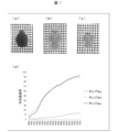
- An optical microscope image of a brain sample (sample a) infiltrated with a 20% sucrose / PBS solution is shown in FIG.
- the light transmittance of the sample a (sample central portion) was measured using a Konica Minolta CM-5 spectrocolorimeter ((d) of FIG. 1).
- the brain replaced with 20% sucrose / PBS solution was treated with 25% N, N, N ′, N′-tetrakis (2-hydroxypropyl) ethylenediamine, 15% Triton X-100 (Nacalai Tesque Cat No. 25987-85), The solution was transferred to a first solution (Sol. PE) containing 25% urea and allowed to infiltrate for 7 days with gentle shaking in an environment of 37 ° C., thereby replacing the first solution.
- An optical microscope image of the brain sample (sample b) infiltrated with the first solution is shown in FIG.
- the light transmittance of the sample b was measured using a Konica Minolta CM-5 spectrocolorimeter ((d) of FIG. 1).
- the brain replaced with the first solution was transferred into PBS and washed by gently shaking 2 to 3 times for 3 to 12 hours at room temperature.
- the washed brain was transferred into a second solution (Sol. CL) containing 10% triethanolamine, 0.1% Triton X-100, 25% urea, 50% sucrose, and gently over 4 days at room temperature.
- the second solution was replaced by shaking.
- An optical microscope image of the brain sample (sample c) infiltrated with the second solution is shown in FIG.
- the light transmittance of sample c was measured using a Konica Minolta CM-5 spectrocolorimeter ((d) of FIG. 1).
- Sample a has a low light transmission in any wavelength region, but sample b greatly improves the light transmission of the sample, and by comparing sample b and sample c, the displacement to the second solution can be reduced. It can be seen that the transparency of white matter was promoted.
- the mouse skull was removed and the whole brain was carefully removed.
- the removed whole brain was immersed in an ice-cold fixative (4% paraformaldehyde / PBS, pH 7-8) overnight at 4 ° C.
- the fixed whole brain was transferred into a 20% sucrose / PBS solution and infiltrated in a 4 ° C environment.
- the brain replaced with 20% sucrose / PBS solution was treated with 25% N, N, N ′, N′-tetrakis (2-hydroxypropyl) ethylenediamine, 15% Triton X-100 (Nacalai Tesque Cat No. 25987-85), And replaced with the first solution by infiltration in a 37 ° C environment for 10 days in a first solution (Sol. PE) containing 25% urea.
- the brain replaced with the first solution was transferred into PBS and washed by gently shaking 2 to 3 times for 3 to 12 hours at room temperature.
- the washed brain was transferred into a second solution (Sol. CL) containing 10% triethanolamine, 0.1% Triton X-100, 25% urea, 50% sucrose, and gently over 4 days at room temperature.
- the second solution was replaced by shaking.
- the brain replaced with the second solution was used as an observation sample.
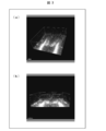
- the observation sample was immersed in a mixed oil of TSF4300 / mineral oil (1: 1) and subjected to whole-brain scanning using a sheet illumination macro zoom microscope (LaVision, Ultramicroscope).
- the acquired data was reconstructed in three dimensions using Imaris (Bitplane) (FIG. 2).
- 2A shows a three-dimensional reconstructed image from the brain bottom side of the sample
- FIG. 2B shows an observation surface observed with a microscope
- FIG. 2C shows a three-dimensional reconstructed image.
- the coronal section of the sample is shown, and (d) shows the sagittal section of the three-dimensionally reconstructed sample.
- the tissue processed using the present invention has been found to be so light transmissive that it can be adapted to a sheet illumination macro zoom microscope.
- the excised brain region tissue was immersed in an ice-cold fixative (4% paraformaldehyde / PBS, pH 7-8) overnight at 4 ° C.
- the fixed tissue was transferred into a 20% sucrose / PBS solution and allowed to invade in a 4 ° C environment.
- the tissue replaced with a 20% sucrose / PBS solution was treated with 25% N, N, N ′, N′-tetrakis (2-hydroxypropyl) ethylenediamine, 15% Triton X-100 (Nacalai Tesque Cat No. 25987-85), And replaced with the first solution by gently shaking for 24 hours in a 4 ° C. environment.
- the tissue replaced with the first solution was transferred into PBS and washed by gently shaking 2 to 3 times for 3 to 12 hours in an environment at room temperature.
- the washed tissue was transferred to a diluted solution (1: 100 and 1: 300, respectively) of anti-Ceptin antibody (Santacruz) and anti-VIP antibody (Immunostar) and reacted for 3 days. After removing the diluted solution, the tissue reacted with the antibody was transferred into 0.1% Triton X-100 / PBS, and gently shaken 2 to 3 times for 3 to 12 hours at room temperature or 37 ° C. And washed.
- a diluted solution (1: 100 and 1: 300, respectively) of anti-Ceptin antibody (Santacruz) and anti-VIP antibody (Immunostar)
- the washed tissue was transferred into a diluted solution (1: 1000) of a secondary antibody bound with Alexa fluorescent dye and allowed to react for 3 days. After removing the diluted solution, the tissue reacted with the secondary antibody is transferred into 0.1% Triton X-100 / PBS, and gently 2-3 hours at room temperature or 37 ° C for 2-3 hours. The mixture was washed by shaking, transferred to PBS, and gently shaken 1-2 times at room temperature for 1-2 hours.
- the washed tissue is transferred into 1% paraformaldehyde / PBS, shaken at room temperature for about 30 minutes to crosslink the antibody, then transferred into PBS, and gently 1 to 3-12 hours at room temperature. Washed by shaking twice. Next, transfer to a second solution (Sol. CL) containing 10% triethanolamine, 0.1% Triton X-100, 25% urea, and 50% sucrose, and gently shake for 2 days or more at room temperature. By replacing with the second solution. The tissue replaced with the second solution was used as an observation sample.
- Sol. CL 10% triethanolamine, 0.1% Triton X-100, 25% urea, and 50% sucrose
- the observational sample was observed with a confocal laser microscope (Carl Zeiss, LSM700) in the suprachiasmatic nucleus region to a depth of about 500 ⁇ m from the bottom of the brain.
- the acquired data was reconstructed in three dimensions using Imaris (Bitplane) (FIG. 2).
- FIG. 3A shows an image obtained by observing the three-dimensionally reconstructed sample from obliquely above
- FIG. 3B shows an image obtained by observing the three-dimensionally reconstructed sample almost from the side.
- the fluorescence image of EGFP but also the immunostained image of Coppin and VIP could be successfully observed in three dimensions.
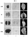
- the extracted tissue was immersed in the first solution for 5 days and then immersed in the second solution for 4 days to be transparent.
- the transparency according to the present invention can be applied to various tissues other than the brain as a biological material, and can be applied to a sheet illumination microscope.
- the present invention has the following advantages: [1] A level of transparency equivalent to the method using an organic solvent was realized with a water-soluble reagent, which enabled observation with a sheet illumination microscope. [2] Compared with the method using an organic solvent, the disappearance of the fluorescence signal was suppressed to a minimum and the safety was dramatically improved. [3] It can be used not only for detection of fluorescent proteins but also for immunohistochemical assays such as antibodies or counterstaining. This enabled immunohistochemistry in three dimensions.
Landscapes
- Health & Medical Sciences (AREA)
- Life Sciences & Earth Sciences (AREA)
- Biochemistry (AREA)
- General Health & Medical Sciences (AREA)
- Molecular Biology (AREA)
- Physics & Mathematics (AREA)
- Chemical & Material Sciences (AREA)
- Analytical Chemistry (AREA)
- Engineering & Computer Science (AREA)
- Biomedical Technology (AREA)
- General Physics & Mathematics (AREA)
- Immunology (AREA)
- Pathology (AREA)
- Investigating Or Analysing Biological Materials (AREA)
- Sampling And Sample Adjustment (AREA)
- Measuring Or Testing Involving Enzymes Or Micro-Organisms (AREA)
Abstract
Description
本発明は、光透過性に優れた生物材料を調製するための試薬組成物を提供する。本発明にかかる試薬組成物は、アミノアルコールを含んでいる溶液であることを特徴としている。本発明にかかる試薬組成物は、尿素または尿素誘導体を含んでいてもよい。本発明にかかる試薬組成物は、スクロースを含まない組成物(第一溶液または第一試薬ともいう。)であっても、スクロースをさらに含む組成物(第二溶液または第二試薬ともいう。)であってもよく、前者の場合、非イオン性界面活性剤をさらに含むことが好ましく、後者の場合、非イオン性界面活性剤をさらに含んでいてもよい。
・蛍光タンパク質、蛍光物質のシグナルの消失が最小限に抑制される。
・多サンプル(数十個~数百個)を同時に並行して処理することができる。
・シート照明顕微鏡との併用が実現する程度に高度な透明化が実現する。
・組織領域の構築に対する影響を最小限に抑制することができる。
アミノアルコールは、水酸基とアミノ基を有する化合物である。アミノアルコールは、水性系ペイントにおける中和剤として使用されることがよく知られているが、それ以外に、インク組成物または医薬組成物の含有成分として用いられている。例えば、特開2011-12226号公報には、分散安定剤としてアミノアルコールを配合してなるインクジェット記録用インク組成物が開示されており、特開2009-221252号公報には、インク組成物にアミノアルコールを含有することで拭き取り性が向上することが開示されている。
本明細書中で使用される場合、「尿素または尿素誘導体」は尿素および種々の尿素誘導体の少なくとも1つであることが意図され、「尿素および尿素誘導体からなる群より選択される少なくとも一種の化合物」と置換可能に用いられる。

1)R1~R4より選択される任意の3つの基が水素原子であり、残りの1つの基がハロゲン原子、または炭素数1~6以下の鎖状炭化水素基である。より好ましくは残りの1つの基が、炭素数1~3、または炭素数が1~2のアルキル基である。
2)R1~R4より選択される任意の2つの基が水素原子であり、残りの2つの基が互いに独立にハロゲン原子、または炭素数1~6以下の鎖状炭化水素基である。より好ましくは残りの2つの基が何れも、炭素数1~3、または炭素数が1~2のアルキル基である。なお、水素原子となる2つの基の一方はR1、R2の何れかから選ばれ、他方はR3、R4の何れかから選ばれることがより好ましい。
非イオン性界面活性剤もまた、光透過性に優れた生物材料を調製するための試薬組成物の製造に使用されてもよく、得られた試薬組成物は、生物材料中の光散乱物質の除去に非常に有用である。
スクロースもまた、光透過性に優れた生物材料を調製するための試薬組成物の製造に使用されてもよく、適用すべき生物材料の屈折率に合った屈折率の試薬を得るに非常に有用である。本明細書においてはスクロースについてのみ言及するが、スクロースと同様の屈折率調整効果を示す任意の物質を使用することができる。
本発明にかかる試薬組成物に使用される溶媒の種類は、上述した有効成分を溶解し得る限り特に限定されないが、水を主溶媒として用いることが好ましく、水のみを溶媒として用いることが特に好ましい。なお、本発明において、「水を主溶媒として用いる」とは、使用される全溶媒に占める水の体積の割合が他の溶媒と比較して最も多いことを指し、好ましくは使用される全溶媒の体積の合計の50%を超え100%以下の量の水を用いることを指す。また、水を主溶媒として用いて調製された本発明にかかる試薬組成物を、水溶液としての試薬と称する。
1)本発明にかかる試薬組成物の有効成分は水への溶解性に優れているため、本発明にかかる試薬組成物の調製が容易かつ低コストとなる;
2)有機溶媒を主溶媒として用いる場合と比較して、透明化処理時に処理対象となる生物材料の脱水を伴わない。そのため、生物材料が収縮するという問題を抑制可能となる;
3)有機溶媒を主溶媒として用いる場合と比較して、蛍光タンパク質に損傷を与える可能性が著しく低減される。そのため、透明化処理を受けた生物材料を、蛍光タンパク質を用いて観察可能となる;
4)固定化された材料に限定されず、生材料の透明化処理に適用可能となる;
5)後述するように透明化処理が可逆的となり、透明化処理後の生体試料を必要に応じて透明化処理前の状態に戻すことができる;
6)有機溶媒を主溶剤として用いる場合と比較して、取り扱いの安全性がより高くなる。
本発明にかかる試薬組成物は、Scale試薬と同様に、必要に応じて、グリセロール、カルボキシビニルポリマー、ヒドロキシプロピルメチルセルロース、プロピレングリコール、ならびにマクロゴールから選択される少なくとも一種の化合物を「乾燥抑制成分」として含むことができる。乾燥抑制成分は、透明化処理の対象となる生物材料の乾燥を防止する。とりわけ、透明化処理後に光学顕微鏡による観察に供されるまでの時間が比較的長い場合、あるいは、光学顕微鏡による長時間観察に供される場合には、本発明にかかる試薬組成物は、上記の乾燥抑制成分を含むことが好ましい。
光透過性に優れた生物材料を調製するために、尿素または尿素誘導体と、アミノアルコールと、非イオン性界面活性剤とを含んでいる第一溶液を調製する。調製した第一溶液中に生物材料を配置することによって、第一溶液を生物材料に浸潤させる。第一溶液を浸潤させるべき生物材料は、予めパラホルムアルデヒド溶液で固定し、次いでスクロース溶液で置換していることが好ましい。
1)4%パラホルムアルデヒド溶液で潅流固定したマウスから脳を摘出し、摘出した脳をパラホルムアルデヒド溶液で12~24時間固定する
2)固定後の脳を、20%スクロース/PBS溶液で置換する
3)置換した脳を第一溶液でさらに置換する
4)第一溶液を浸潤させた脳を、PBSで数時間ずつ数回洗浄する
5)洗浄した脳を第二溶液で置換する
6)第二溶液を浸潤させた脳を観察する。あるいは、PBSで洗浄した後に保存する。
透明化処理された生物材料は、次いで、例えば、光学顕微鏡による観察工程に供される。観察工程に供される生物材料は、必要に応じて、上記透明化処理工程の事前に、または透明化処理工程後で観察工程前に、染色あるいはマーキング等の可視化処理工程が施されてもよい。
生後6ヶ月齢の個体に対して、ペリスタポンプを用いて左心室より室温PBSを灌流した後、室温固定液(4%パラホルムアルデヒド/PBS,pH7~8)を灌流することによって、マウスの全身を完全に固定した。
ROSA26部位にH2B-mCherry(蛍光タンパク質mCherryを融合したヒストンH2Bタンパク質)をノックインしたマウス(Takaya Abe et al., "Establishment of Conditional Reporter Mouse Lines at ROSA26 Locus For Live Cell Imaging" Genesis vol.49: 579-590 (2011))の生後7ヶ月齢の個体に対して、ペリスタポンプを用いて左心室より室温PBSを灌流した後、室温固定液(4%パラホルムアルデヒド/PBS,pH7~8)を灌流することによって、マウスの全身を完全に固定した。
ROSA26部位にH2B-EGFP(蛍光タンパク質EGFPを融合したヒストンH2Bタンパク質)をノックインしたマウス(Takaya Abe et al. 前出)の生後4ヶ月齢の個体に対して、ペリスタポンプを用いて左心室より室温PBSを灌流した後、室温固定液(4%パラホルムアルデヒド/PBS,pH7~8)を灌流することによって、マウスの全身を完全に固定した。
パラホルムアルデヒド固定したマウス血液0.1mLに、0.4mLの処理溶液(PBS溶液、25%尿素を含む第一溶液(Sol.PE)、またはアミノアルコール溶液(N,N,N’,N’-テトラキス(2-ヒドロキシプロピル)エチレンジアミンの25%水溶液))を添加した。混合液を室温で混合した直後に、15000rpmで5分間遠心分離した。上清を分離した後の沈殿に上記処理溶液を0.5mL加え、再び同様に遠心分離し、再度沈殿を上清と分離した。このような沈殿の洗浄操作を3回繰り返した後に、沈殿に上記処理溶液0.5mLを混合した。
蛍光タンパク質mKate2を発現するマウスの生後4ヶ月齢の個体に対して、ペリスタポンプを用いて左心室より室温にてヘパリンPBS溶液10mLを灌流した後、室温固定液(4%パラホルムアルデヒド/PBS,pH7~8)150mLを灌流することによって、マウスの全身を完全に固定し、次いで、20mLのPBS溶液で洗浄し、第一溶液を2倍希釈した溶液20mLでさらに灌流した。
[1]有機溶媒を用いた手法と同等の透明化レベルを水溶性試薬によって実現し、シート照明顕微鏡での観察を可能にした。
[2]有機溶媒を用いた手法と比較して、蛍光シグナルの消失を最小限に抑制するとともに安全性を飛躍的に向上させた。
[3]蛍光タンパク質の検出だけでなく、抗体または対比染色などの免疫組織化学的アッセイに使用可能である。これにより、三次元での免疫組織化学が可能となった。
[4]化合物スクリーニングによる試薬組成の改変および最適化によって、Scale法と比較して処理時間を明らかに短縮させ(月単位から日~週単位へ)、2光子顕微鏡が必要なレベルからシート照明マクロ顕微鏡に適応可能なレベルにまで透明度を向上させた。
[5]スクリーニングした化合物はいずれも安価であったため、低コストにて実用化することが可能である。
[6]特殊なデバイスおよび煩雑な操作を必要としない、簡便なプロトコルを確立したことにより、多サンプルの並行処理を可能とした。
[7]シート照明マクロ顕微鏡に適応した際に、全脳のスキャニングを1色あたり1時間程度で完了することができるので、タイムラグがほとんどない状態でマルチカラーでの画像を取得することが可能であり、三次元で再構成された全脳をデータ化することが可能である。
[8]生体組織の複雑な構造を三次元にて維持した状態で免疫組織化学を行うことが可能なため、自己抗体が被験体内の実際の組織にてどのような部位と反応しているのかを探索することができ、自己抗体の抗原を探索することができる。
Claims (16)
- アミノアルコールを含んでいる溶液であることを特徴とする光透過性に優れた生物材料を調製するための試薬組成物。
- 尿素および尿素誘導体からなる群より選択される少なくとも一種の化合物をさらに含んでいることを特徴とする請求項1に記載の試薬組成物。
- 非イオン性界面活性剤をさらに含んでいることを特徴とする請求項1または2に記載の試薬組成物。
- スクロースをさらに含んでいることを特徴とする請求項1~3のいずれか1項に記載の試薬組成物。
- アミノアルコールと、尿素および尿素誘導体からなる群より選択される少なくとも一種の化合物と、非イオン性界面活性剤とを含んでいる第一溶液、
アミノアルコールと、尿素および尿素誘導体からなる群より選択される少なくとも一種の化合物と、スクロースとを含んでいる第二溶液
を備えていることを特徴とする光透過性に優れた生物材料を調製するための試薬キット。 - 第二溶液が非イオン性界面活性剤をさらに含んでいることを特徴とする請求項5に記載の試薬キット。
- アミノアルコールを含んでいることを特徴とする、光透過性に優れた生物材料を調製するための試薬組成物を製造するための組成物。
- (1)アミノアルコール、ならびに
(2)尿素および尿素誘導体からなる群より選択される少なくとも一種の化合物、非イオン性界面活性剤、ならびにスクロースからなる群より選択される少なくとも1つ
を備えていることを特徴とする、光透過性に優れた生物材料を調製するための試薬組成物を製造するためのキット。 - 光透過性に優れた生物材料を調製する方法であって、
アミノアルコールと、尿素および尿素誘導体からなる群より選択される少なくとも一種の化合物と、非イオン性界面活性剤とを含んでいる第一溶液を、生物材料に浸潤させる第一浸潤工程
を包含することを特徴とする方法。 - アミノアルコールと、尿素および尿素誘導体からなる群より選択される少なくとも一種の化合物と、スクロースとを含んでいる第二溶液を、生物材料に浸潤させる第二浸潤工程をさらに包含することを特徴とする請求項9に記載の方法。
- 第二溶液が非イオン性界面活性剤をさらに含んでいることを特徴とする請求項10に記載の方法。
- 請求項9~11のいずれか1項に記載の方法に従って調製した光透過性に優れた生物材料。
- 請求項9~11のいずれか1項に記載の方法に従って調製した生物材料を用いることを特徴とする免疫組織化学的アッセイ。
- 請求項13に記載の免疫組織化学的アッセイを用いることを特徴とする自己抗体と反応性である組織部位を探索する方法。
- 請求項13に記載の免疫組織化学的アッセイを用いることを特徴とする自己抗体に対する抗原を探索する方法。
- 請求項9~11のいずれか1項に記載の方法に従って調製した生物材料を用いることを特徴とする生化学的アッセイ。
Priority Applications (3)
| Application Number | Priority Date | Filing Date | Title |
|---|---|---|---|
| JP2015531767A JP6433901B2 (ja) | 2013-08-14 | 2014-08-05 | 光透過性に優れた生物材料を調製するための組成物およびその利用 |
| US14/911,778 US10267714B2 (en) | 2013-08-14 | 2014-08-05 | Composition for preparing biomaterial with excellent light-transmitting property, and use thereof |
| EP14836073.8A EP3035026A4 (en) | 2013-08-14 | 2014-08-05 | Composition for preparing biomaterial with excellent light-transmitting property, and use thereof |
Applications Claiming Priority (2)
| Application Number | Priority Date | Filing Date | Title |
|---|---|---|---|
| JP2013168705 | 2013-08-14 | ||
| JP2013-168705 | 2013-08-14 |
Publications (1)
| Publication Number | Publication Date |
|---|---|
| WO2015022883A1 true WO2015022883A1 (ja) | 2015-02-19 |
Family
ID=52468270
Family Applications (1)
| Application Number | Title | Priority Date | Filing Date |
|---|---|---|---|
| PCT/JP2014/070618 Ceased WO2015022883A1 (ja) | 2013-08-14 | 2014-08-05 | 光透過性に優れた生物材料を調製するための組成物およびその利用 |
Country Status (4)
| Country | Link |
|---|---|
| US (1) | US10267714B2 (ja) |
| EP (1) | EP3035026A4 (ja) |
| JP (1) | JP6433901B2 (ja) |
| WO (1) | WO2015022883A1 (ja) |
Cited By (12)
| Publication number | Priority date | Publication date | Assignee | Title |
|---|---|---|---|---|
| JP2017108684A (ja) * | 2015-12-17 | 2017-06-22 | 国立大学法人名古屋大学 | 植物組織透明化剤 |
| WO2017188264A1 (ja) * | 2016-04-28 | 2017-11-02 | 国立研究開発法人理化学研究所 | 光透過性に優れた生物材料を調製するための組成物およびその利用 |
| WO2018051957A1 (ja) * | 2016-09-13 | 2018-03-22 | 国立大学法人東京大学 | 生物試料の透明化方法 |
| KR101849704B1 (ko) | 2017-08-21 | 2018-04-18 | 한국화학연구원 | 생체 조직의 투명화 전처리 방법 및 이를 포함하는 생체 조직의 투명화 방법 |
| US10545075B2 (en) | 2012-08-09 | 2020-01-28 | The Board Of Trustees Of The Leland Stanford Junior University | Methods and compositions for preparing biological specimens for microscopic analysis |
| WO2020019670A1 (zh) * | 2018-07-25 | 2020-01-30 | 中国科学院合肥物质科学研究院 | 处理液组合物、试剂盒和生物器官透明化同时进行免疫标记的方法 |
| US10746981B2 (en) | 2014-05-30 | 2020-08-18 | The Board Of Trustees Of The Leland Stanford Junior University | Methods and devices for imaging large intact tissue samples |
| WO2021039716A1 (ja) * | 2019-08-30 | 2021-03-04 | 株式会社CUBICStars | 生体組織染色試薬、生体組織染色キット及び生体組織染色方法 |
| WO2021060373A1 (ja) * | 2019-09-24 | 2021-04-01 | 国立大学法人大阪大学 | 細胞適合性組織透明化組成物 |
| JP2021512330A (ja) * | 2018-02-05 | 2021-05-13 | コリア リサーチ インスティテュート オブ ケミカル テクノロジー | スフェロイドを透明化するための組成物、これを使用するスフェロイドを透明化するための方法、およびこれを備えるキット |
| US11254974B2 (en) | 2016-02-10 | 2022-02-22 | The Board Of Trustees Of The Leland Stanford Junior University | RNA fixation and detection in clarity-based hydrogel tissue |
| WO2024177108A1 (ja) * | 2023-02-22 | 2024-08-29 | 国立大学法人大阪大学 | 生きた三次元組織の深部観察を可能とする透明化ポリマー |
Families Citing this family (7)
| Publication number | Priority date | Publication date | Assignee | Title |
|---|---|---|---|---|
| WO2018044899A1 (en) * | 2016-08-29 | 2018-03-08 | The United States Of America, As Represented By The Secretary, Department Of Health And Human Services | Method and composition for optical clearing of tissues |
| WO2019190216A1 (ko) * | 2018-03-29 | 2019-10-03 | 주식회사 바이나리 | 트리톤 x-100 및 우레아를 유효성분으로 포함하는 생체조직 침투 증강용 조성물 및 이를 이용한 생체조직 침투 증강 방법 |
| JP7558670B2 (ja) * | 2020-03-19 | 2024-10-01 | 株式会社日本触媒 | 基材に接着されたスフェロイドを観察する方法 |
| CN111474358B (zh) * | 2020-04-16 | 2023-08-18 | 伊莱瑞特(武汉)生物技术有限公司 | 一种3d立体免疫荧光染色试剂盒及其应用 |
| CN113640520A (zh) * | 2021-07-16 | 2021-11-12 | 南方医科大学珠江医院 | 一种组织透明方法和组织学方法联合用于检测肿瘤内细菌的应用 |
| CN115701532A (zh) * | 2021-08-02 | 2023-02-10 | 西湖大学 | 用于生物组织透明化处理的脱脂组合物 |
| CN114636605A (zh) * | 2022-03-21 | 2022-06-17 | 锘海生物科学仪器(上海)有限公司 | 基于水性的生物组织透明化试剂盒及组织透明化方法 |
Citations (11)
| Publication number | Priority date | Publication date | Assignee | Title |
|---|---|---|---|---|
| JPS63157997A (ja) * | 1986-10-03 | 1988-06-30 | ノバルティス アクチェンゲゼルシャフト | 新規リンホカイン関連ペプチド |
| US6472216B1 (en) * | 2001-07-24 | 2002-10-29 | Ann-Shyn Chiang | Aqueous tissue clearing solution |
| JP2003066035A (ja) * | 2001-08-06 | 2003-03-05 | Anse Ko | 水溶性組織清澄溶液 |
| US20040033612A1 (en) * | 2002-01-02 | 2004-02-19 | Vladimir Senatorov | Dark-field microscopy visualization of unstained axonal pathways using oil of wintergreen |
| JP2007051957A (ja) * | 2005-08-18 | 2007-03-01 | Shimadzu Corp | 生体組織の直接質量分析法 |
| JP2008524592A (ja) * | 2004-12-17 | 2008-07-10 | ヴェンタナ メディカル システムズ インコーポレイテッド | 低揮発高温組織コンディショニング |
| JP2009221252A (ja) | 2008-03-13 | 2009-10-01 | Fujifilm Corp | インク組成物及び画像記録方法 |
| JP2011012226A (ja) | 2009-07-06 | 2011-01-20 | Rengo Co Ltd | インクジェット記録用インク組成物 |
| US20130023008A1 (en) * | 2010-03-30 | 2013-01-24 | Technische Universität Wien | Histological method |
| JP2013522590A (ja) * | 2010-03-12 | 2013-06-13 | 独立行政法人理化学研究所 | 生物材料用透明化試薬、及びその利用 |
| WO2013155064A1 (en) * | 2012-04-10 | 2013-10-17 | Rutgers, The State University Of New Jersey | Clearing agent and mounting medium for microscopy |
Family Cites Families (43)
| Publication number | Priority date | Publication date | Assignee | Title |
|---|---|---|---|---|
| US2029525A (en) | 1929-11-14 | 1936-02-04 | Ellis Foster Co | Transparent tissue and process of making same |
| US4578282A (en) | 1982-02-04 | 1986-03-25 | Harrison James S | Composition for diagnostic reagents |
| US4493821A (en) | 1982-02-04 | 1985-01-15 | Harrison James S | Preservative and fixative preparations for biological systems |
| FR2599149B1 (fr) | 1986-05-21 | 1988-08-26 | Univ Nancy | Reactif pour la transparisation de milieux biologiques et ses applications analytiques. |
| GB8803697D0 (en) | 1988-02-17 | 1988-03-16 | Deltanine Research Ltd | Clinical developments using amniotic membrane cells |
| JPH03167472A (ja) | 1989-11-27 | 1991-07-19 | Sumitomo Chem Co Ltd | 実験動物の胎児骨格染色法 |
| US5530100A (en) * | 1990-05-07 | 1996-06-25 | Rhone-Poulenc Rorer Pharmaceuticals Inc. | Methods for purification of recombinantly produced proteins |
| US5057435A (en) | 1990-10-12 | 1991-10-15 | Synermed, Inc. | Reagent and methods for calcium determination |
| CN1031530C (zh) | 1991-08-26 | 1996-04-10 | 广西农学院 | 生物制片透明剂 |
| JPH0774121B2 (ja) | 1992-09-01 | 1995-08-09 | 哲 濱田 | 生物体の内部透視が可能な標本製造方法 |
| IT1269583B (it) * | 1994-04-26 | 1997-04-08 | Bayer Italia Spa | Preparazioni farmaceutiche a base di una soluzione di ketoprofene in capsule di gelatina molle e metodo per la loro produzione |
| US6072086A (en) | 1996-04-12 | 2000-06-06 | Intergen Company | Method and composition for controlling formaldehyde fixation by delayed quenching |
| EP0900367A1 (en) | 1996-04-12 | 1999-03-10 | Oncor, Inc. | Method and composition for controlling formaldehyde fixation by delayed quenching |
| US6793890B2 (en) | 1997-08-20 | 2004-09-21 | The University Of Miami | Rapid tissue processor |
| KR20010023070A (ko) | 1997-08-20 | 2001-03-26 | 더 유니버시티 오브 마이애미 | 고품질의 연속적인 재료 처리, 조직 고정-탈수-지방제거-함침 방법 |
| US6232092B1 (en) | 1998-10-02 | 2001-05-15 | Rogers Imaging Corporation | Method for preparing biological specimens for visual analysis |
| US6462071B1 (en) | 2000-03-02 | 2002-10-08 | Vitreo-Retinal Technologies, Inc. | Agents for intravitreal administration to treat or prevent disorders of the eye |
| US7008960B1 (en) | 1999-03-02 | 2006-03-07 | Vitreo-Retinal Technologies, Inc. | Agents for intravitreal administration to treat or prevent disorders of the eye |
| CU22921A1 (es) | 1999-11-16 | 2004-02-20 | Centro Inmunologia Molecular | Anticuerpos quimérico, humanizado y el fragmento de tipo fv de cadena sencilla que reconoce el antígeno c2. su uso en el diagnóstico y tratamiento de tumores colorrectales |
| DK1238256T3 (da) | 1999-12-14 | 2006-10-30 | Ervin Essenfeld | Indretning til hurtig vævsbehandling |
| US20030199574A1 (en) | 2000-03-02 | 2003-10-23 | Vitreo-Retinal Technologies, Inc. | Treatment of ophthalmic disorders using urea and urea derivatives |
| US7977385B2 (en) | 2000-03-02 | 2011-07-12 | Numoda Biotechnologies, Inc. | Agents for corneal or intrastromal administration to treat or prevent disorders of the eye |
| CN1118566C (zh) | 2000-08-03 | 2003-08-20 | 中国人民解放军兰州军区兰州总医院 | 一种组织石蜡切片脱蜡、透明剂 |
| US20020077697A1 (en) | 2000-12-15 | 2002-06-20 | Ranieri John Paul | Processed ratite carotid arteries as xenogeneic small bore vascular grafts |
| WO2003077898A1 (en) | 2002-03-14 | 2003-09-25 | Vitreo-Retinal Technologies, Inc. | Agents for corneal or intrastromal administration to treat or prevent disorders of the eye |
| US20050137124A1 (en) | 2002-08-09 | 2005-06-23 | Vitreo-Retinal Technologies, Inc. A California Corporation | Agents for intravitreal administration to treat or prevent disorders of the eye |
| JP4092397B2 (ja) | 2002-09-10 | 2008-05-28 | 国立循環器病センター総長 | 超高静水圧印加による移植用生体組織の処理方法 |
| US8377447B2 (en) * | 2003-09-05 | 2013-02-19 | Oregon Health & Science University | Monomeric recombinant MHC molecules useful for manipulation of antigen-specific T cells |
| JP2007070494A (ja) * | 2005-09-07 | 2007-03-22 | Neos Co Ltd | ケイ素含有透明水性液 |
| US8636986B2 (en) | 2005-11-18 | 2014-01-28 | The Forsyth Institute | Treatment and prevention of bone loss using resolvins |
| AR062069A1 (es) | 2006-07-14 | 2008-10-15 | Genentech Inc | Replegado de proteinas recombinantes |
| WO2008039483A2 (en) | 2006-09-26 | 2008-04-03 | Massachusetts Institute Of Technology | Modified self-assembling peptides |
| CN100586958C (zh) | 2006-12-20 | 2010-02-03 | 上海莱士血液制品股份有限公司 | 高纯度载脂蛋白a-i的制备方法 |
| JP5241100B2 (ja) | 2006-12-28 | 2013-07-17 | シスメックス株式会社 | 抗原賦活化液、抗原賦活化方法及び細胞の検出方法 |
| CN100567943C (zh) | 2007-04-27 | 2009-12-09 | 孝感学院 | 一种骨骼肌横纹的切片染色方法 |
| US20090191138A1 (en) | 2008-01-30 | 2009-07-30 | Mediquest Therapeutics, Inc. | Novel topical formulations for improving the appearance of nails |
| US20100008989A1 (en) | 2008-06-12 | 2010-01-14 | Ishay Attar | Process for manufacture of gelatin solutions and products thereof |
| US20110123979A1 (en) | 2008-07-16 | 2011-05-26 | Peter Salmon | Detection of microorganisms |
| GB0812999D0 (en) | 2008-07-16 | 2008-08-20 | Blood Analysis Ltd | Detection of microorganisms |
| CN101871855A (zh) | 2009-04-27 | 2010-10-27 | 上海蓝盎电子科技发展有限公司 | 新型病理切片透明剂 |
| CN101881778B (zh) | 2009-05-06 | 2014-01-29 | 深圳迈瑞生物医疗电子股份有限公司 | 网织红细胞模拟物及其制备方法 |
| CN103492852B (zh) | 2011-04-28 | 2016-08-17 | 独立行政法人理化学研究所 | 生物材料的透明化方法、及其利用 |
| JP5414917B2 (ja) | 2011-07-13 | 2014-02-12 | パナソニック株式会社 | 錠剤検査装置及び錠剤検査方法 |
-
2014
- 2014-08-05 WO PCT/JP2014/070618 patent/WO2015022883A1/ja not_active Ceased
- 2014-08-05 EP EP14836073.8A patent/EP3035026A4/en not_active Withdrawn
- 2014-08-05 US US14/911,778 patent/US10267714B2/en not_active Expired - Fee Related
- 2014-08-05 JP JP2015531767A patent/JP6433901B2/ja active Active
Patent Citations (11)
| Publication number | Priority date | Publication date | Assignee | Title |
|---|---|---|---|---|
| JPS63157997A (ja) * | 1986-10-03 | 1988-06-30 | ノバルティス アクチェンゲゼルシャフト | 新規リンホカイン関連ペプチド |
| US6472216B1 (en) * | 2001-07-24 | 2002-10-29 | Ann-Shyn Chiang | Aqueous tissue clearing solution |
| JP2003066035A (ja) * | 2001-08-06 | 2003-03-05 | Anse Ko | 水溶性組織清澄溶液 |
| US20040033612A1 (en) * | 2002-01-02 | 2004-02-19 | Vladimir Senatorov | Dark-field microscopy visualization of unstained axonal pathways using oil of wintergreen |
| JP2008524592A (ja) * | 2004-12-17 | 2008-07-10 | ヴェンタナ メディカル システムズ インコーポレイテッド | 低揮発高温組織コンディショニング |
| JP2007051957A (ja) * | 2005-08-18 | 2007-03-01 | Shimadzu Corp | 生体組織の直接質量分析法 |
| JP2009221252A (ja) | 2008-03-13 | 2009-10-01 | Fujifilm Corp | インク組成物及び画像記録方法 |
| JP2011012226A (ja) | 2009-07-06 | 2011-01-20 | Rengo Co Ltd | インクジェット記録用インク組成物 |
| JP2013522590A (ja) * | 2010-03-12 | 2013-06-13 | 独立行政法人理化学研究所 | 生物材料用透明化試薬、及びその利用 |
| US20130023008A1 (en) * | 2010-03-30 | 2013-01-24 | Technische Universität Wien | Histological method |
| WO2013155064A1 (en) * | 2012-04-10 | 2013-10-17 | Rutgers, The State University Of New Jersey | Clearing agent and mounting medium for microscopy |
Non-Patent Citations (8)
| Title |
|---|
| HANS-ULRICH DODT ET AL.: "Ultramicroscopy: three-dimensional visualization of neuronal networks in the whole mouse brain", NATURE METHODS, vol. 4, no. 4, 2007, pages 331 - 336 |
| HIROSHI HAMA ET AL.: "Scale: a chemical approach for fluorescence imaging and reconstruction of transparent mouse brain", NATURE NEUROSCIENCE, vol. 14, no. 11, 2011, pages 1481 - 1490 |
| KLAUS BECKER ET AL.: "Chemical Clearing and Dehydration of GFP Expressing Mouse Brains", PLOS ON E, vol. 7, no. 3, 2012, pages E33916 |
| KWANGHUN CHUNG ET AL.: "Structural and molecular interrogation of intact biological systems", NATURE, vol. 497, 2013, pages 332 - 339 |
| MENG-TSEN KE ET AL.: "SeeDB: a simple and morphology-preserving optical clearing agent for neuronal circuit reconstruction", ADVANCE ONLINE PUBLICATION, 23 June 2013 (2013-06-23) |
| PHILIPP J. KELLER ET AL.: "Reconstruction of Zebrafish Early Embryonic Development by Scanned Light Sheet Microscopy", SCIENCE, vol. 322, 2008, pages 1065 - 1069, XP002607760, DOI: doi:10.1126/science.1162493 |
| See also references of EP3035026A4 * |
| TAKAYA ABE ET AL.: "Establishment of Conditional Reporter Mouse Lines at ROSA 26 Locus For Live Cell Imaging", GENESIS, vol. 49, 2011, pages 579 - 590 |
Cited By (28)
| Publication number | Priority date | Publication date | Assignee | Title |
|---|---|---|---|---|
| US10545075B2 (en) | 2012-08-09 | 2020-01-28 | The Board Of Trustees Of The Leland Stanford Junior University | Methods and compositions for preparing biological specimens for microscopic analysis |
| US10746981B2 (en) | 2014-05-30 | 2020-08-18 | The Board Of Trustees Of The Leland Stanford Junior University | Methods and devices for imaging large intact tissue samples |
| JP2017108684A (ja) * | 2015-12-17 | 2017-06-22 | 国立大学法人名古屋大学 | 植物組織透明化剤 |
| US12098418B2 (en) | 2016-02-10 | 2024-09-24 | The Board Of Trustees Of The Leland Stanford Junior University | RNA fixation and detection in CLARITY-based hydrogel tissue |
| US11254974B2 (en) | 2016-02-10 | 2022-02-22 | The Board Of Trustees Of The Leland Stanford Junior University | RNA fixation and detection in clarity-based hydrogel tissue |
| JP7281230B2 (ja) | 2016-04-28 | 2023-05-25 | 国立研究開発法人理化学研究所 | 光透過性に優れた生物材料を調製するための組成物およびその利用 |
| JP2022058560A (ja) * | 2016-04-28 | 2022-04-12 | 国立研究開発法人理化学研究所 | 光透過性に優れた生物材料を調製するための組成物およびその利用 |
| US12292363B2 (en) | 2016-04-28 | 2025-05-06 | Riken | Method for preparing biological material having excellent light-transmitting property |
| JPWO2017188264A1 (ja) * | 2016-04-28 | 2019-04-11 | 国立研究開発法人理化学研究所 | 光透過性に優れた生物材料を調製するための組成物およびその利用 |
| JP7669054B2 (ja) | 2016-04-28 | 2025-04-28 | 国立研究開発法人理化学研究所 | 光透過性に優れた生物材料を調製するための組成物およびその利用 |
| WO2017188264A1 (ja) * | 2016-04-28 | 2017-11-02 | 国立研究開発法人理化学研究所 | 光透過性に優れた生物材料を調製するための組成物およびその利用 |
| JP2023100871A (ja) * | 2016-04-28 | 2023-07-19 | 国立研究開発法人理化学研究所 | 光透過性に優れた生物材料を調製するための組成物およびその利用 |
| EP3450953A4 (en) * | 2016-04-28 | 2020-01-08 | Riken | COMPOSITION FOR THE PRODUCTION OF BIOLOGICAL MATERIAL WITH OUTSTANDING LIGHT TRANSMISSION AND USE OF THE COMPOSITION |
| US11656159B2 (en) | 2016-04-28 | 2023-05-23 | Riken | Composition for preparing biological material having excellent light transmissivity and use of composition |
| JP7219451B2 (ja) | 2016-04-28 | 2023-02-08 | 国立研究開発法人理化学研究所 | 光透過性に優れた生物材料を調製するための組成物およびその利用 |
| WO2018051957A1 (ja) * | 2016-09-13 | 2018-03-22 | 国立大学法人東京大学 | 生物試料の透明化方法 |
| KR101849704B1 (ko) | 2017-08-21 | 2018-04-18 | 한국화학연구원 | 생체 조직의 투명화 전처리 방법 및 이를 포함하는 생체 조직의 투명화 방법 |
| JP7053860B2 (ja) | 2018-02-05 | 2022-04-12 | コリア リサーチ インスティテュート オブ ケミカル テクノロジー | スフェロイドを透明化するための組成物、これを使用するスフェロイドを透明化するための方法、およびこれを備えるキット |
| US11365213B2 (en) | 2018-02-05 | 2022-06-21 | Korea Research Institute Of Chemical Technology | Composition for clearing spheroids, method for clearing spheroids using same, and kit comprising same |
| JP2021512330A (ja) * | 2018-02-05 | 2021-05-13 | コリア リサーチ インスティテュート オブ ケミカル テクノロジー | スフェロイドを透明化するための組成物、これを使用するスフェロイドを透明化するための方法、およびこれを備えるキット |
| WO2020019670A1 (zh) * | 2018-07-25 | 2020-01-30 | 中国科学院合肥物质科学研究院 | 处理液组合物、试剂盒和生物器官透明化同时进行免疫标记的方法 |
| JP7197941B2 (ja) | 2019-08-30 | 2022-12-28 | 株式会社CUBICStars | 生体組織染色試薬、生体組織染色キット及び生体組織染色方法 |
| JPWO2021039716A1 (ja) * | 2019-08-30 | 2021-03-04 | ||
| WO2021039716A1 (ja) * | 2019-08-30 | 2021-03-04 | 株式会社CUBICStars | 生体組織染色試薬、生体組織染色キット及び生体組織染色方法 |
| JPWO2021060373A1 (ja) * | 2019-09-24 | 2021-04-01 | ||
| WO2021060373A1 (ja) * | 2019-09-24 | 2021-04-01 | 国立大学法人大阪大学 | 細胞適合性組織透明化組成物 |
| JP7579578B2 (ja) | 2019-09-24 | 2024-11-08 | 国立大学法人大阪大学 | 細胞適合性組織透明化組成物 |
| WO2024177108A1 (ja) * | 2023-02-22 | 2024-08-29 | 国立大学法人大阪大学 | 生きた三次元組織の深部観察を可能とする透明化ポリマー |
Also Published As
| Publication number | Publication date |
|---|---|
| JP6433901B2 (ja) | 2018-12-05 |
| EP3035026A1 (en) | 2016-06-22 |
| US10267714B2 (en) | 2019-04-23 |
| EP3035026A4 (en) | 2017-04-26 |
| US20160266016A1 (en) | 2016-09-15 |
| JPWO2015022883A1 (ja) | 2017-03-02 |
Similar Documents
| Publication | Publication Date | Title |
|---|---|---|
| JP6433901B2 (ja) | 光透過性に優れた生物材料を調製するための組成物およびその利用 | |
| US20210131926A1 (en) | Clearing reagent for biological material, and use thereof | |
| Richardson et al. | Tissue clearing | |
| US20240142352A1 (en) | Biological-specimen transparentizing agent, system, and use therefor | |
| US20230213415A1 (en) | Method and System for Imaging and Analysis of a Biological Specimen | |
| US20210131925A1 (en) | Method for making biological material transparent and use thereof | |
| EP2711682B1 (en) | Clarifying reagent for biological materials and use thereof | |
| US12031891B2 (en) | Tissue sample preparation system | |
| CN110139922B (zh) | 用于澄清组织的组合物和方法 | |
| US20170108414A1 (en) | High-resolution three-dimensional imaging of mammalian hearts | |
| Handschuh et al. | An optimized workflow for microCT imaging of formalin‐fixed and paraffin‐embedded (FFPE) early equine embryos | |
| CN114402060B (zh) | 用于组织透明化的组合物和方法 | |
| WO2022102623A1 (ja) | 生物材料を透明化する方法およびキット |
Legal Events
| Date | Code | Title | Description |
|---|---|---|---|
| 121 | Ep: the epo has been informed by wipo that ep was designated in this application |
Ref document number: 14836073 Country of ref document: EP Kind code of ref document: A1 |
|
| ENP | Entry into the national phase |
Ref document number: 2015531767 Country of ref document: JP Kind code of ref document: A |
|
| WWE | Wipo information: entry into national phase |
Ref document number: 14911778 Country of ref document: US |
|
| NENP | Non-entry into the national phase |
Ref country code: DE |
|
| WWE | Wipo information: entry into national phase |
Ref document number: 2014836073 Country of ref document: EP |