WO2015016036A1 - 卵子の受精可能性の検査のためのバイオマーカーおよびそれを用いた判定 - Google Patents
卵子の受精可能性の検査のためのバイオマーカーおよびそれを用いた判定 Download PDFInfo
- Publication number
- WO2015016036A1 WO2015016036A1 PCT/JP2014/068506 JP2014068506W WO2015016036A1 WO 2015016036 A1 WO2015016036 A1 WO 2015016036A1 JP 2014068506 W JP2014068506 W JP 2014068506W WO 2015016036 A1 WO2015016036 A1 WO 2015016036A1
- Authority
- WO
- WIPO (PCT)
- Prior art keywords
- peptide
- seq
- amino acid
- fertilization
- egg
- Prior art date
- Legal status (The legal status is an assumption and is not a legal conclusion. Google has not performed a legal analysis and makes no representation as to the accuracy of the status listed.)
- Ceased
Links
Images


Classifications
-
- G—PHYSICS
- G01—MEASURING; TESTING
- G01N—INVESTIGATING OR ANALYSING MATERIALS BY DETERMINING THEIR CHEMICAL OR PHYSICAL PROPERTIES
- G01N33/00—Investigating or analysing materials by specific methods not covered by groups G01N1/00 - G01N31/00
- G01N33/48—Biological material, e.g. blood, urine; Haemocytometers
- G01N33/50—Chemical analysis of biological material, e.g. blood, urine; Testing involving biospecific ligand binding methods; Immunological testing
- G01N33/68—Chemical analysis of biological material, e.g. blood, urine; Testing involving biospecific ligand binding methods; Immunological testing involving proteins, peptides or amino acids
- G01N33/689—Chemical analysis of biological material, e.g. blood, urine; Testing involving biospecific ligand binding methods; Immunological testing involving proteins, peptides or amino acids related to pregnancy or the gonads
Definitions
- the present invention relates to a novel biomarker for examining the fertilization potential of an egg, and a method for examining or determining the fertilization potential of an egg using the same.
- in vitro fertilization In Japan, where there is a declining birthrate and an aging population, 1 in 50 live births, about 20,000 a year, are born by in vitro fertilization, and in vitro fertilization has recently become a popular medical technology that will stop the aging population with a declining birthrate. Have been doing. On the other hand, the success rate of in vitro fertilization depends on the quality or state of an egg to be collected, such as fertility, but there is currently no method for discriminating an egg having a quality or state suitable for fertilization.
- An object of the present invention is to provide a novel biomarker for examining the fertilization potential of an ovum and a method for examining or judging the fertilization potential of an ovum using the same.
- the present inventors have found a specific peptide whose amount in the follicular fluid varies depending on the degree of maturation of the follicle. These peptides were newly identified as markers for the test or determination of fertilization potential of eggs, and the present invention was completed.
- the amount of one or more peptides selected from the peptide group consisting of the amino acid sequences represented by SEQ ID NOs: 1 to 4 in the follicular fluid collected from the subject is measured.
- a test method for fertilization possibility of an egg in the subject is provided.
- subjecting the follicular fluid to mass spectrometry subjecting the follicular fluid to mass spectrometry.
- the amount of peptide obtained by the measurement is compared with a threshold set based on the amount of peptide in the control sample, and the amount of peptide obtained by the measurement is compared to the peptide in the control sample.
- the method further includes determining that the fertilization probability of the egg is high when the variation is statistically significantly compared to a threshold set based on the amount of the egg.
- a biomarker for examining fertilization potential of an egg comprising a peptide selected from the peptide group consisting of the amino acid sequences represented by SEQ ID NOs: 1 to 4.
- a method for examining fertilization potential of an egg which specifically recognizes a peptide selected from a peptide group consisting of amino acid sequences represented by SEQ ID NOs: 1 to 4. Methods using antibodies are provided.
- a determination kit for examining fertilization potential of an egg containing an antibody wherein the antibody comprises a peptide group consisting of amino acid sequences represented by SEQ ID NOs: 1 to 4.
- a determination kit is provided that is an antibody that specifically recognizes the selected peptide.
- an apparatus for immunodiagnosis or determination in which an antibody against a peptide selected from the peptide group consisting of the amino acid sequences represented by SEQ ID NOs: 1 to 4 is fixed.
- a peptide comprising the amino acid sequence represented by SEQ ID NO: 1, SEQ ID NO: 2, SEQ ID NO: 3, or SEQ ID NO: 4.
- the fertility of an egg can be determined quickly and with high accuracy without damaging the egg, so that an egg suitable for fertilization, particularly in vitro fertilization can be distinguished, and as a result, the success rate of in vitro fertilization is greatly improved. Can be made.
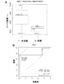
- the AUC value, sensitivity (SN; prevalence rate of prevalence) and specificity (SP; prevalence rate of non-morbidity) are shown in FIG. 3A.
- A Plot diagram of peak intensity between unfertilized group (group 1) and fertilized group (group 2) of biomarker (molecular weight 2862) obtained in Example 1, and
- B ROC curve diagram.
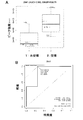
- the AUC value, sensitivity (SN; prevalence rate of prevalence) and specificity (SP; prevalence rate of non-morbidity) are shown in FIG. 4A.
- A Plot diagram of peak intensity between unfertilized group (group 1) and fertilized group (group 2) of biomarker (molecular weight 2941) obtained in Example 1, and (B) ROC curve diagram.
- the AUC value, sensitivity (SN; prevalence rate of disease prevalence) and specificity (SP; prevalence rate of disease prevalence) are shown in the upper part of FIG. 5A.
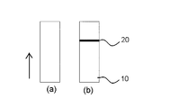
- A (b) Examples of immunochromat chips.
- the present invention provides a novel and useful marker peptide for examining or determining the fertilization potential of an egg.
- Ovulation is the process by which mature oocytes, ie ova, are discharged from the ovarian follicle.
- the present inventors have found that the expression level of each of the following four peptides of the present invention contained in the follicular fluid of the follicle is related to the fertilization potential of the ovum.
- Peptide 1 consisting of an amino acid sequence represented by SEQ ID NO: 1.
- the amino acid sequence corresponds to the 341st to 367th fragments of human ⁇ -2-HS-glycoprotein.
- Peptide 2 consisting of the amino acid sequence represented by SEQ ID NO: 2.
- the amino acid sequence corresponds to the 25th to 48th fragments of human serum albumin.
- Peptide 3 consisting of an amino acid sequence represented by SEQ ID NO: 3.
- the amino acid sequence of SEQ ID NO: 1 is the same as that of 27 amino acids, but in peptide 3, cysteine is further bonded to the 18th cysteine by a disulfide bond.
- Peptide 4 consisting of the amino acid sequence represented by SEQ ID NO: 4. It corresponds to the 25th to 50th fragments of human serum albumin.
- TVVQPSVGAAAGPVVPPCPGRIRHFKV SEQ ID NO: 1
- DAHKSEVAHRFKDLGEENFKALVL SEQ ID NO: 2
- TVVQPSVGAAAGPVVPPCPGRIRHFKV Cysteinylation at position 18 SEQ ID NO: 3
- DAHKSEVAHRFKDLGEENFKALVLIA SEQ ID NO: 4
- the expression levels of peptides 1, 2, and 4 are increased, and the expression level of peptide 3 is low. Therefore, by confirming that the expression levels of peptides 1, 2, and 4 are high and / or that peptide 3 is low, it is possible to determine whether the egg is fertilized or has high fertility.
- Peptides 1 to 4 have average molecular weights of 2743, 2757, 2862, and 2941, respectively, but precise molecular weights of protonated molecules ([M + H + ]) are 2738.52, 2752.43, 2875.52, and 2936.44, respectively (FIG. 1). ).
- the actual measurement value of the molecular weight may slightly vary depending on the measurement method / measurement apparatus used. For example, in the case of a method using a mass spectrometer, it is preferable to measure the peak intensity that appears at the indicated numerical value ⁇ 0.5% (preferably ⁇ 0.3%, more preferably ⁇ 0.1%).
- peptides 1 to 4 are useful as test or determination markers for fertilization of eggs, peptides containing amino acid residues of these four peptides are also used as test or determination markers for fertilization of eggs. It is possible.
- the peptide of the present invention includes a peptide having an amino acid sequence substantially the same as the amino acid sequence of the four peptides in addition to the peptide having the entire amino acid sequence of the amino acid sequences of the four peptides.
- “Substantially identical” means that one or a few amino acids are modified as compared with the amino acid sequences of the above four peptides, but can still be used as a test or determination marker for fertilization of an egg. To do. A small number refers to 1 to 3, preferably 1 to 2, more preferably 1.
- the modification means that the amino acid is deleted, added, or substituted with another amino acid, or the amino sequence is changed by a combination thereof.
- the modification includes addition and phosphorylation of sugar chains and / or fatty acids.
- the detection is performed. It is obvious to those skilled in the art that the amino acid sequence of the peptide should be interpreted as “the amino acid sequence having the polymorphism or allelic variation in each amino acid sequence represented by SEQ ID NOs: 1 to 4.”
- the present invention also provides for fertilization of an ovum in the subject by measuring the amount of one or more peptides of the present invention in a follicular fluid from the subject in need of testing for fertilization of the ovum.
- the peptide to be measured is not particularly limited as long as it is selected from the above four types of peptides of the present invention. Moreover, in order to improve the measurement accuracy, as long as the peptide of the present invention is included, the amount of one or more biological substances that can serve as a test or determination marker for eggs other than the peptide of the present invention may be further measured. .
- the body fluid derived from the subject as the test sample is follicular fluid.
- the follicular fluid is advantageous in that it can be collected from the follicle without damaging the ovum in the case of artificial insemination.
- the peptide of the present invention to be detected may be directly used for inspection or determination without pretreatment such as protein removal of the sample, that is, without purification, and if necessary, using a spin column or the like, It is also possible to lightly separate and remove a high molecular weight protein fraction from the sample in advance.
- the detection of the peptide of the present invention in the follicular fluid can be carried out, for example, by measuring various molecular weights of the follicular fluid, such as gel electrophoresis or various separation and purification methods (eg, ion exchange chromatography, hydrophobic chromatography, affinity).
- various molecular weights of the follicular fluid such as gel electrophoresis or various separation and purification methods (eg, ion exchange chromatography, hydrophobic chromatography, affinity).
- a method of purifying the peptides of the present invention to produce antibodies that recognize them and detecting the peptides by ELISA, RIA, immunochromatography, Western blotting, immunomass spectrometry or various immunoassays can also be preferably used. Furthermore, a hybrid detection method with the above detection method is also effective.
- One of the particularly preferable measurement methods in the inspection method of the present invention is that a test sample is brought into contact with the surface of a plate used for time-of-flight mass spectrometry, and the mass of the component captured on the plate surface is measured.
- the method of measuring by is mentioned.
- a plate compatible with a time-of-flight mass spectrometer has a surface structure (for example, functional group-added glass, Si, Ge, GaAs, GaP, SiO 2 , SiN 4) that can efficiently adsorb the peptide of the present invention to be detected , Modified silicon, various gels or polymer coatings).
- the support used as a plate for mass spectrometry is a substrate coated with a thin layer of polyvinylidene difluoride (PVDF), nitrocellulose or silica gel, particularly preferably PVDF (WO 2004/031759 See).
- PVDF polyvinylidene difluoride
- nitrocellulose or silica gel particularly preferably PVDF (WO 2004/031759 See).
- a substrate is not particularly limited as long as it is used in a plate for mass spectrometry, and examples thereof include insulators, metals, conductive polymers, and composites thereof.
- PVDF polyvinylidene difluoride
- nitrocellulose or silica gel particularly preferably PVDF (WO 2004/031759 See).
- Such a substrate is not particularly limited as long as it is used in a plate for mass spectrometry, and examples thereof include insulators, metals, conductive polymers, and composites thereof.
- Such a plate for mass spectrometry coated with a thin layer with PVDF
- Mass spectrometry using this BLOTCHIP is the world's first peptome analysis method that enables analysis without pretreatment of specimens (body fluids such as blood and tissue extracts).
- body fluids such as blood and tissue extracts.
- test sample-derived follicular fluid from the subject After pretreatment of the test sample-derived follicular fluid from the subject or by simply removing the high molecular weight protein using an antibody column or other method, the test sample is subjected to SDS-polyacrylamide gel electrophoresis or isoelectric focusing. After electrophoresis, the gel is transferred to a plate for mass spectrometry and transferred (blotted).
- the transfer method itself is known and preferably electrotransfer is used.
- a known buffer having a pH of 7 to 9 and a low salt concentration is preferably used (for example, Tris buffer, phosphate buffer, borate buffer, acetate buffer, etc.).
- a peptide adsorbed to a protein is dissociated by electrophoresis in the presence of a surfactant (SDS), transferred to BLOTCHIP®, and then the chip is analyzed at high speed by MALDI-MS.
- SDS surfactant
- MALDI-MS MALDI-MS
- the presence and amount of the peptide of the present invention can be identified from the information on the molecular weight by mass spectrometry of the molecules in the test sample captured on the support surface by the above method. It is also possible to output information from the mass spectrometer as differential information by using an arbitrary program and comparing it with mass spectrometry data in follicular fluid when fertilization is impossible. It will be appreciated that such programs are well known and those skilled in the art can easily construct or modify such programs using known information processing techniques.
- a stable isotope-labeled peptide of the target molecule is synthesized and mixed with a test sample as a known amount of internal standard, and Protocera's BLOTCHIP® system is used. Measurement data with a CV value of 5% or less can be acquired.
- mass spectrometry can be performed by a mass spectrometry system other than the BLOTCHIP (registered trademark) system, and this method can be clinically used as a diagnostic apparatus that does not use an antibody.
- peptides can be identified using a tandem mass spectrometry (MS / MS) method.
- an identification method an amino acid sequence is determined by analyzing an MS / MS spectrum.
- a method of identifying a peptide by performing a database search using partial sequence information (mass tag) contained in the MS / MS spectrum.
- the amino acid sequence of the peptide of the present invention is directly identified, and all or a part of the peptide is synthesized based on the sequence information. It can also be used as
- the present invention includes a method for examining fertilization ability of an egg using an antibody that specifically recognizes a peptide, and a determination kit for examining fertilization ability of an egg containing an antibody containing such an antibody.
- a test method can detect the peptide with high sensitivity and high accuracy without using a special apparatus such as the mass spectrometer, if an optimized immunoassay system is constructed and a kit is prepared. It is useful in that it can.
- the present invention also includes an apparatus for immunodiagnosis or determination using an antibody against a peptide.
- an apparatus for immunodiagnosis or determination using an antibody against a peptide examples include, but are not limited to, an immunochromatography diagnosis or determination chip in which an antibody against a peptide is fixed in a determination region of the chip.
- a preferred embodiment of such a chip is an immunochromatographic diagnostic or determination chip in which an antibody against the peptide of the present invention is immobilized.
- the antibody against the peptide of the present invention can be prepared, for example, by isolating and purifying the peptide of the present invention from a follicular fluid derived from a patient expressing the peptide and immunizing an animal using the peptide as an antigen.
- the peptide can be prepared in a large amount by a well-known genetic engineering technique such as amplification of a cDNA fragment encoding the peptide by RT-PCR.
- the peptide of the present invention can also be obtained using a cell-free transcription / translation system as a template. Further, it can be prepared in a large amount by an organic synthesis method.
- the antibody against the peptide of the present invention may be either a polyclonal antibody or a monoclonal antibody, and can be prepared by a known immunological technique.
- the antibody includes not only a complete antibody molecule but also a fragment thereof, and examples thereof include Fab, F (ab ′) 2, ScFv, and minibody.
- a polyclonal antibody is administered about 2 to 4 times every 2 to 3 weeks subcutaneously or intraperitoneally in an animal together with a commercially available adjuvant (for example, complete or incomplete Freund's adjuvant) using the peptide of the present invention as an antigen, It can be obtained by collecting whole blood after the final immunization and purifying the antiserum.
- adjuvant for example, complete or incomplete Freund's adjuvant
- animals to which the antigen is administered include mammals capable of obtaining the target antibody, such as rats, mice, rabbits, goats, guinea pigs, and hamsters.
- a monoclonal antibody can be prepared by a cell fusion method. Description of techniques for the preparation of a monoclonal antibody, Stites et al, Basic and Clinical Immunology. (Lang Medical Publications Los Altos. CA. 4 th Edition) and references therein,, in particular Koehler, G. & Milstein, C. Nature 256, 495-497 (1975).
- the peptide of the present invention is administered to a mouse subcutaneously or intraperitoneally 2-4 times together with a commercially available adjuvant, and after the final administration, the spleen or lymph node is collected, and white blood cells are collected.
- This leukocyte and myeloma cells are cell-fused to obtain a hybridoma that produces a monoclonal antibody against the peptide.
- a hybridoma producing a desired monoclonal antibody can be selected by detecting an antibody that specifically binds to an antigen from the culture supernatant using a known EIA or RIA method or the like.
- the culture of the hybridoma producing the monoclonal antibody can be performed in vitro or in vivo such as mouse or rat, or preferably mouse ascites, and the antibody can be obtained from the culture supernatant of the hybridoma and the ascites of the animal, respectively. .
- the test method of the present invention using an antibody is not particularly limited, and detects the amount of an antibody, antigen or antibody-antigen complex corresponding to the amount of antigen in a test sample by chemical or physical means, Any measurement method may be used as long as it is a measurement method calculated from a standard curve prepared using a standard solution containing a known amount of antigen. For example, nephrometry, competition method, immunometric method and sandwich method are preferably used.
- the antibody or antigen can be bound with a labeling agent such as a radioisotope, an enzyme, a fluorescent substance, or a luminescent substance.
- a biotin-avidin system can also be used for binding of an antibody or antigen and a labeling agent.
- the follicular fluid is immunoaffinity purified with an antibody, and the fraction bound to the antibody is subjected to mass spectrometry in the second step, and so-called immunomass spectrometry, in which identification and quantification are performed based on the precise molecular weight.
- immunomass spectrometry In contrast to immunomass spectrometry, undegraded proteins and similar peptides are completely separated by a mass spectrometer, and can be quantified with high specificity and sensitivity based on the exact molecular weight of a biomarker.
- the antibody is immobilized on the surface of a chip that can be adapted to a mass spectrometer as described above, and a test sample is applied to the antibody on the chip.
- a test sample is applied to the antibody on the chip. Examples include a method in which a component in a sample captured by the antibody is contacted and subjected to mass spectrometry, and a peak corresponding to the molecular weight of the marker peptide recognized by the antibody is detected.
- the level of the peptide of the present invention in the subject-derived sample measured by any of the above methods varies statistically significantly compared to a threshold set based on the amount of peptide in the control sample. If so, it can be diagnosed or determined that the egg of the subject is highly fertilized.
- the threshold value set based on the amount of peptide in the control sample may be the amount of peptide in the control sample itself, or in the control sample as long as the reliability of the test or determination method of the present invention is ensured. Another reference value set based on the amount of the peptide may be used.
- control sample refers to a sample serving as a reference for comparison, and includes a sample in the case of unfertilized.
- the level of the peptide of the present invention in the control sample may be detected in parallel with the level of the peptide of the present invention in the biological sample of the subject, or the level of the peptide of the present invention in a number of control samples in advance.
- the level may be measured and the standard expression level in the control sample may be determined statistically. Specifically, for example, the average value ⁇ 2 ⁇ standard deviation (S.D.), or the average value ⁇ ⁇ standard deviation (S.D.) or the average value ⁇ 3 ⁇ standard deviation (S.D.) can be used as the standard value.
- the expression level of the peptide of the present invention in the control sample may be set using an ROC curve.
- An ROC curve (receiver operating characteristic curve) is a graph showing detection sensitivity on the vertical axis and false positive rate (that is, “1-specificity”) on the horizontal axis.
- An ROC curve can be obtained by plotting changes in sensitivity and false positive rate when the reference value for determining the expression level of the peptide in the biological sample is continuously changed.
- a standard value statistically set by an ROC curve or the like is also called a cut-off value.
- the level of the peptide of the present invention in the subject-derived sample is compared with the cutoff value. Then, when the level of the peptide of the present invention in the sample derived from the subject varies with respect to the cut-off value, the fertility of the egg is detected or determined.
- the cut-off value is a criterion value that is predicted to have a high risk of suffering from the disease if it is greater than or equal to that value. Determined as negative. If the cut-off value is set low, the sensitivity is high and the specificity is low, and if the cut-off value is set high, the sensitivity is low and the specificity is high.
- the cut-off value is a reference range (95% confidence interval, normal distribution) obtained by measuring the number of individuals of a reference value that is a measurement value in a control sample and obtaining a central value including 95% of the reference value. In this case, the upper limit value of the measurement average value ⁇ 2 standard deviations in the control sample).
- another value may be set as the cut-off value as long as the sensitivity and specificity required by the determination are satisfied.
- the peptide of the invention is either peptide 1, 2, or 4, and the level of the peptide in the sample is statistically significantly increased compared to the peptide level in the control sample And / or if the level of Peptide 3 in the sample is statistically significantly lower than the level of the peptide in the control sample, it is diagnosed or determined that the egg is likely to be fertilized can do.
- the peptide of the present invention more specifically, the four peptides of the present invention can be used alone or as a diagnostic marker for fertilization potential of an egg. (Correct diagnosis rate) and specificity (no disease correct diagnosis rate) can be further increased.
- 2 or 4 peptides of the present invention are determined by a population learning method, or the measured values of each peptide in a sample of a known patient and a control-derived sample in a corresponding number of samples are two-dimensional or three-dimensional.
- a population learning method By plotting the coordinates and creating a scatter plot, it is visualized in which region each of the two groups is distributed, and then the measured values of each peptide in the sample derived from the subject are plotted on the scatter plot.
- the determination can also be made by determining the cutoff value of each peptide based on the scatter diagram and comparing the measured value of each peptide in the subject-derived sample with this.
- Example 1 Profiling Analysis Using BLOTCHIP (Registered Trademark) Eight female patients who visited Fukui University Obstetrics and Gynecology Department and obtained informed consent were used as subjects. From the follicle collected by in vitro fertilization treatment of the subject, the follicular fluid is sucked by pointing to the injection needle, and the follicular fluid containing the eggs that were not fertilized by in vitro fertilization (unfertilized group, 4 samples of 1-F1 to 1-F4) ) And follicular fluid containing the eggs fertilized by in vitro fertilization (fertilization group, 4 samples of 2-F1 to 2-F4). Confirmation of fertilization was confirmed by a conventional method of microscopic observation of the start of cell division in the fertilized egg. The collected follicular fluid was associated with each ovum on a one-to-one basis, immediately frozen at -80 ° C., and used for subsequent profiling analysis.
- Mass spectrometry was performed with a mass spectrometer (Ultra-FlexII manufactured by Bruker Daltnics). The measurement parameters were Detector voltage 1685V, Supression 1000, Laser Intensity 28 to 35 Fuzzy mode, 415 points per chip, 500 times laser irradiation per point, total 207,500 times laser irradiation. Each peak intensity in the obtained spectrum was integrated for each M / z and converted into one integrated spectrum.
- Peptides giving statistically significant difference (P value less than 0.05) by performing differential profiling analysis between follicular fluid of unfertilized group and fertilized group using ClinProTools 2.2 (Bruker Daltonik GmbH) Groups were selected and four peptides with molecular weights of 2743, 2757, 2862, and 2941 were identified as egg test or decision markers (FIG. 1).
- FIG. 2A, FIG. 3A, FIG. 4A and FIG. 5A show the peak intensities at molecular weights 2743, 2757, 2862, and 2941, respectively, between the two groups of the four obtained biomarkers
- FIG. 2B, FIG. 3B, FIG. FIG. 5B shows a plot of peak intensity and an ROC curve of the four biomarkers between 1: fertilized group and 2: fertilized group. From these results, it is understood that the peak intensity at the molecular weights 2743, 2757, and 2941 is significantly higher in the fertilized group and the peak intensity at the molecular weight 2862 is significantly lower than that in the unfertilized group, and the four peptides are unique. It has been shown to be useful as a test or determination marker for fertility of typical eggs.
- Example 2 Identification of peptides by de novo MS / MS analysis on BLOTCHIP Reflector of MALDI-TOF mass spectrometer Ultraflex II (manufactured by Bruker Daltnics) for the fraction spots with peptides shown in FIGS. Monoisotopic mass [M + H] + was measured in mode. The obtained monoisotopic mass [M + H] + was designated as Parent Mass, and MS / MS measurement was performed in Lift mode (PSD). The measurement result was searched with MASCOT Server, the amino acid sequence was determined by MS / MS analysis, and the peptide was identified (FIG. 1).
- peptide 1 consisting of the amino acid sequence represented by SEQ ID NO: 1 corresponding to the 341st to 367th fragments of human ⁇ -2-HS-glycoprotein, and fragments 25 to 48 of human serum albumin
- Corresponding peptide 2 consisting of the amino acid sequence represented by SEQ ID NO: 2, the amino acid sequence is the same as SEQ ID NO: 1, but peptide 18 in which cysteine is further linked to the 18th cysteine by a disulfide bond, from human serum albumin 25 Peptides 4 corresponding to the 50th fragment and consisting of the amino acid sequence represented by SEQ ID NO: 4 were identified.
- FIGS. 6A and 6B are schematic diagrams showing an example of an immunochromat chip that is an apparatus for immunodiagnosis or determination of the present invention.
- the immunochromatography chip 10 is an immunoassay device for analyzing a specimen using an antigen-antibody reaction, also called an immunostrip or an immunoassay strip, and includes a base material and a specimen provided on the base material as a basic structure.
- An absorption pad for absorbing and developing a biological sample (in this application follicular fluid) is provided.
- the base material and the absorbent pad may be made of materials known or known in the art.
- a region 20 is provided along the longitudinal direction of the immunochromatography chip 10 to which an antibody against the peptide of the present invention is bound.
- FIG. 6 (a) shows that there is no line in region 20 and that the egg is unfertilized or not suitable for fertilization.
- FIG. 6 (b) shows that there is a line in the region 20 and that the egg is in a state suitable for fertilization or fertilization.
- the quality or state of the ovum can be determined simply, quickly and accurately.
- the clinical examination method using the novel ovum test or determination marker of the present invention is useful in that the success rate of fertilization, particularly in vitro fertilization, can be greatly improved because it can quickly and accurately determine the fertility of the ovum. is there.
Landscapes
- Life Sciences & Earth Sciences (AREA)
- Health & Medical Sciences (AREA)
- Engineering & Computer Science (AREA)
- Hematology (AREA)
- Chemical & Material Sciences (AREA)
- Urology & Nephrology (AREA)
- Molecular Biology (AREA)
- Biomedical Technology (AREA)
- Immunology (AREA)
- Microbiology (AREA)
- Physics & Mathematics (AREA)
- Biotechnology (AREA)
- Proteomics, Peptides & Aminoacids (AREA)
- Reproductive Health (AREA)
- Pregnancy & Childbirth (AREA)
- Gynecology & Obstetrics (AREA)
- Food Science & Technology (AREA)
- Medicinal Chemistry (AREA)
- Cell Biology (AREA)
- Analytical Chemistry (AREA)
- Biochemistry (AREA)
- General Health & Medical Sciences (AREA)
- General Physics & Mathematics (AREA)
- Pathology (AREA)
- Other Investigation Or Analysis Of Materials By Electrical Means (AREA)
- Peptides Or Proteins (AREA)
- Investigating Or Analysing Biological Materials (AREA)
Priority Applications (1)
| Application Number | Priority Date | Filing Date | Title |
|---|---|---|---|
| JP2015529492A JP6470178B2 (ja) | 2013-07-29 | 2014-07-10 | 卵子の受精可能性の検査のためのバイオマーカーおよびそれを用いた判定 |
Applications Claiming Priority (2)
| Application Number | Priority Date | Filing Date | Title |
|---|---|---|---|
| JP2013156672 | 2013-07-29 | ||
| JP2013-156672 | 2013-07-29 |
Publications (1)
| Publication Number | Publication Date |
|---|---|
| WO2015016036A1 true WO2015016036A1 (ja) | 2015-02-05 |
Family
ID=52431576
Family Applications (1)
| Application Number | Title | Priority Date | Filing Date |
|---|---|---|---|
| PCT/JP2014/068506 Ceased WO2015016036A1 (ja) | 2013-07-29 | 2014-07-10 | 卵子の受精可能性の検査のためのバイオマーカーおよびそれを用いた判定 |
Country Status (2)
| Country | Link |
|---|---|
| JP (2) | JP6470178B2 (https=) |
| WO (1) | WO2015016036A1 (https=) |
Cited By (2)
| Publication number | Priority date | Publication date | Assignee | Title |
|---|---|---|---|---|
| JP2019152671A (ja) * | 2019-04-03 | 2019-09-12 | 京都府公立大学法人 | 大腸がんの検出方法 |
| JP2022070974A (ja) * | 2019-04-03 | 2022-05-13 | 京都府公立大学法人 | 大腸がんの検出方法 |
Citations (5)
| Publication number | Priority date | Publication date | Assignee | Title |
|---|---|---|---|---|
| JPS61503003A (ja) * | 1984-04-19 | 1986-12-25 | ユニバ−シテイ− オブ クイ−ンスランド | 避妊方法、避妊用製剤及び避妊具 |
| JP2001516726A (ja) * | 1997-09-18 | 2001-10-02 | ザ ピコワー インスティテュート フォー メディカル リサーチ | 流産の防止 |
| JP2004506482A (ja) * | 2000-08-24 | 2004-03-04 | コーネル リサーチ ファンデーション インコーポレーテッド | 非ホルモン性腟内避妊用具 |
| JP2012073190A (ja) * | 2010-09-29 | 2012-04-12 | Protosera Inc | 妊娠高血圧症候群マーカーおよびそれを用いた診断 |
| WO2013099376A1 (ja) * | 2011-12-26 | 2013-07-04 | 国立大学法人福井大学 | 体外受精における成熟卵子マーカー及びその使用 |
Family Cites Families (3)
| Publication number | Priority date | Publication date | Assignee | Title |
|---|---|---|---|---|
| EP1946121A2 (en) * | 2005-10-27 | 2008-07-23 | Yale University, Inc. | Urinary proteomic biomarker patterns in preeclampsia |
| MY150687A (en) * | 2006-07-21 | 2014-02-28 | Femalon S P R L | Assay and kit for predicting implantation success in assisted fertilisation |
| WO2009075862A1 (en) * | 2007-12-11 | 2009-06-18 | New York University School Of Medicine | Method of assessing viability of embryos in vitro |
-
2014
- 2014-07-10 JP JP2015529492A patent/JP6470178B2/ja not_active Expired - Fee Related
- 2014-07-10 WO PCT/JP2014/068506 patent/WO2015016036A1/ja not_active Ceased
-
2018
- 2018-08-17 JP JP2018153454A patent/JP6553264B2/ja not_active Expired - Fee Related
Patent Citations (5)
| Publication number | Priority date | Publication date | Assignee | Title |
|---|---|---|---|---|
| JPS61503003A (ja) * | 1984-04-19 | 1986-12-25 | ユニバ−シテイ− オブ クイ−ンスランド | 避妊方法、避妊用製剤及び避妊具 |
| JP2001516726A (ja) * | 1997-09-18 | 2001-10-02 | ザ ピコワー インスティテュート フォー メディカル リサーチ | 流産の防止 |
| JP2004506482A (ja) * | 2000-08-24 | 2004-03-04 | コーネル リサーチ ファンデーション インコーポレーテッド | 非ホルモン性腟内避妊用具 |
| JP2012073190A (ja) * | 2010-09-29 | 2012-04-12 | Protosera Inc | 妊娠高血圧症候群マーカーおよびそれを用いた診断 |
| WO2013099376A1 (ja) * | 2011-12-26 | 2013-07-04 | 国立大学法人福井大学 | 体外受精における成熟卵子マーカー及びその使用 |
Cited By (4)
| Publication number | Priority date | Publication date | Assignee | Title |
|---|---|---|---|---|
| JP2019152671A (ja) * | 2019-04-03 | 2019-09-12 | 京都府公立大学法人 | 大腸がんの検出方法 |
| JP7032764B2 (ja) | 2019-04-03 | 2022-03-09 | 京都府公立大学法人 | 大腸がんの検出方法 |
| JP2022070974A (ja) * | 2019-04-03 | 2022-05-13 | 京都府公立大学法人 | 大腸がんの検出方法 |
| JP7300660B2 (ja) | 2019-04-03 | 2023-06-30 | 京都府公立大学法人 | 大腸がんの検出方法 |
Also Published As
| Publication number | Publication date |
|---|---|
| JP6553264B2 (ja) | 2019-07-31 |
| JP2019007971A (ja) | 2019-01-17 |
| JPWO2015016036A1 (ja) | 2017-03-02 |
| JP6470178B2 (ja) | 2019-02-13 |
Similar Documents
| Publication | Publication Date | Title |
|---|---|---|
| KR101431062B1 (ko) | 유방암 진단용 다중 바이오마커 세트, 이의 검출 방법 및 이에 대한 항체를 포함하는 유방암 진단키트 | |
| JP7032503B2 (ja) | バイオマーカーとしての遊離ヒストンタンパク質 | |
| JPWO2008072676A1 (ja) | 新規疾患マーカーおよびそれを用いた診断 | |
| JPWO2017150681A1 (ja) | シグナルペプチドを指標にした筋萎縮性側索硬化症の診断方法 | |
| JP5363481B2 (ja) | 早産の危険性を評価するためのバイオマーカーの同定および定量化 | |
| JP2008175814A (ja) | 尿中タンパク質分子の検出・定量による糖尿病性腎症の検査方法及びそれに使用するキット | |
| JP5714285B2 (ja) | 妊娠高血圧症候群マーカーおよびそれを用いた診断 | |
| JP6553264B2 (ja) | 卵子の受精可能性の検査のためのバイオマーカーおよびそれを用いた判定 | |
| JPWO2009088022A1 (ja) | 新規癌マーカーおよびそれを用いた診断 | |
| JP2009168646A (ja) | 子宮内膜症に特異的なバイオマーカー | |
| JP2010071953A (ja) | 乳癌マーカーおよびそれを用いた診断 | |
| JPWO2006098464A1 (ja) | 前立腺がんの診断方法 | |
| WO2008054763A2 (en) | Biomarkers for breast cancer | |
| JP2013245960A (ja) | 乳がん患者手術治療後の予後判定方法 | |
| US20160209412A1 (en) | Msia-srm assay for biomarker analysis | |
| JP2014020941A (ja) | 大腸癌のマーカーおよびそれを用いた診断 | |
| KR101431067B1 (ko) | 유방암 진단용 단백질 마커 아포리포단백질 (a), 이의 검출 방법 및 이에 대한 항체를 포함하는 유방암 진단키트 | |
| JP2015108515A (ja) | 大腸癌の診断のための検査方法 | |
| JP7336097B2 (ja) | 乳がんに関するペプチドマーカー | |
| JP7300660B2 (ja) | 大腸がんの検出方法 | |
| JP2014025868A (ja) | がん転移マーカーおよびそれを用いた診断 | |
| KR101819939B1 (ko) | 유방암 진단용 단백질 마커 베타-투 마이크로글로불린 및 이를 검출하는 방법 | |
| JP2024117521A (ja) | 膵臓がんの検出方法 | |
| JP7032764B2 (ja) | 大腸がんの検出方法 | |
| JP2011080775A (ja) | 妊娠高血圧症候群マーカーおよびそれを用いた診断 |
Legal Events
| Date | Code | Title | Description |
|---|---|---|---|
| 121 | Ep: the epo has been informed by wipo that ep was designated in this application |
Ref document number: 14831322 Country of ref document: EP Kind code of ref document: A1 |
|
| ENP | Entry into the national phase |
Ref document number: 2015529492 Country of ref document: JP Kind code of ref document: A |
|
| NENP | Non-entry into the national phase |
Ref country code: DE |
|
| 122 | Ep: pct application non-entry in european phase |
Ref document number: 14831322 Country of ref document: EP Kind code of ref document: A1 |