WO2012176796A1 - Nk細胞の増幅方法 - Google Patents
Nk細胞の増幅方法 Download PDFInfo
- Publication number
- WO2012176796A1 WO2012176796A1 PCT/JP2012/065718 JP2012065718W WO2012176796A1 WO 2012176796 A1 WO2012176796 A1 WO 2012176796A1 JP 2012065718 W JP2012065718 W JP 2012065718W WO 2012176796 A1 WO2012176796 A1 WO 2012176796A1
- Authority
- WO
- WIPO (PCT)
- Prior art keywords
- cells
- cell
- positive
- amplifying
- blood
- Prior art date
- Legal status (The legal status is an assumption and is not a legal conclusion. Google has not performed a legal analysis and makes no representation as to the accuracy of the status listed.)
- Ceased
Links
Images


Classifications
-
- C—CHEMISTRY; METALLURGY
- C12—BIOCHEMISTRY; BEER; SPIRITS; WINE; VINEGAR; MICROBIOLOGY; ENZYMOLOGY; MUTATION OR GENETIC ENGINEERING
- C12N—MICROORGANISMS OR ENZYMES; COMPOSITIONS THEREOF; PROPAGATING, PRESERVING, OR MAINTAINING MICROORGANISMS; MUTATION OR GENETIC ENGINEERING; CULTURE MEDIA
- C12N5/00—Undifferentiated human, animal or plant cells, e.g. cell lines; Tissues; Cultivation or maintenance thereof; Culture media therefor
- C12N5/06—Animal cells or tissues; Human cells or tissues
- C12N5/0602—Vertebrate cells
- C12N5/0634—Cells from the blood or the immune system
- C12N5/0646—Natural killers cells [NK], NKT cells
-
- A—HUMAN NECESSITIES
- A61—MEDICAL OR VETERINARY SCIENCE; HYGIENE
- A61K—PREPARATIONS FOR MEDICAL, DENTAL OR TOILETRY PURPOSES
- A61K35/00—Medicinal preparations containing materials or reaction products thereof with undetermined constitution
- A61K35/12—Materials from mammals; Compositions comprising non-specified tissues or cells; Compositions comprising non-embryonic stem cells; Genetically modified cells
- A61K35/14—Blood; Artificial blood
- A61K35/17—Lymphocytes; B-cells; T-cells; Natural killer cells; Interferon-activated or cytokine-activated lymphocytes
-
- A—HUMAN NECESSITIES
- A61—MEDICAL OR VETERINARY SCIENCE; HYGIENE
- A61K—PREPARATIONS FOR MEDICAL, DENTAL OR TOILETRY PURPOSES
- A61K40/00—Cellular immunotherapy
- A61K40/10—Cellular immunotherapy characterised by the cell type used
- A61K40/15—Natural-killer [NK] cells; Natural-killer T [NKT] cells
-
- A—HUMAN NECESSITIES
- A61—MEDICAL OR VETERINARY SCIENCE; HYGIENE
- A61K—PREPARATIONS FOR MEDICAL, DENTAL OR TOILETRY PURPOSES
- A61K40/00—Cellular immunotherapy
- A61K40/40—Cellular immunotherapy characterised by antigens that are targeted or presented by cells of the immune system
- A61K40/41—Vertebrate antigens
- A61K40/42—Cancer antigens
-
- A—HUMAN NECESSITIES
- A61—MEDICAL OR VETERINARY SCIENCE; HYGIENE
- A61P—SPECIFIC THERAPEUTIC ACTIVITY OF CHEMICAL COMPOUNDS OR MEDICINAL PREPARATIONS
- A61P31/00—Antiinfectives, i.e. antibiotics, antiseptics, chemotherapeutics
-
- A—HUMAN NECESSITIES
- A61—MEDICAL OR VETERINARY SCIENCE; HYGIENE
- A61P—SPECIFIC THERAPEUTIC ACTIVITY OF CHEMICAL COMPOUNDS OR MEDICINAL PREPARATIONS
- A61P35/00—Antineoplastic agents
-
- A—HUMAN NECESSITIES
- A61—MEDICAL OR VETERINARY SCIENCE; HYGIENE
- A61P—SPECIFIC THERAPEUTIC ACTIVITY OF CHEMICAL COMPOUNDS OR MEDICINAL PREPARATIONS
- A61P37/00—Drugs for immunological or allergic disorders
- A61P37/02—Immunomodulators
- A61P37/04—Immunostimulants
-
- C—CHEMISTRY; METALLURGY
- C12—BIOCHEMISTRY; BEER; SPIRITS; WINE; VINEGAR; MICROBIOLOGY; ENZYMOLOGY; MUTATION OR GENETIC ENGINEERING
- C12N—MICROORGANISMS OR ENZYMES; COMPOSITIONS THEREOF; PROPAGATING, PRESERVING, OR MAINTAINING MICROORGANISMS; MUTATION OR GENETIC ENGINEERING; CULTURE MEDIA
- C12N5/00—Undifferentiated human, animal or plant cells, e.g. cell lines; Tissues; Cultivation or maintenance thereof; Culture media therefor
- C12N5/0081—Purging biological preparations of unwanted cells
- C12N5/0087—Purging against subsets of blood cells, e.g. purging alloreactive T cells
-
- C—CHEMISTRY; METALLURGY
- C12—BIOCHEMISTRY; BEER; SPIRITS; WINE; VINEGAR; MICROBIOLOGY; ENZYMOLOGY; MUTATION OR GENETIC ENGINEERING
- C12N—MICROORGANISMS OR ENZYMES; COMPOSITIONS THEREOF; PROPAGATING, PRESERVING, OR MAINTAINING MICROORGANISMS; MUTATION OR GENETIC ENGINEERING; CULTURE MEDIA
- C12N2501/00—Active agents used in cell culture processes, e.g. differentation
- C12N2501/20—Cytokines; Chemokines
- C12N2501/23—Interleukins [IL]
- C12N2501/2302—Interleukin-2 (IL-2)
Definitions
- the present invention relates to a method for amplifying natural killer cells (NK cells) having high cytotoxic activity with high purity and high amplification factor, and a pharmaceutical composition containing NK cells obtained by the method.
- NK cells natural killer cells
- NK cells do not attack normal cells that express MHC class I molecules, but mainly attack cells in which the expression and decrease of MHC class I molecules are reduced.
- GVH Growth-versus-host
- Non-patent Document 3 From a single apheresis of normal adult peripheral blood, about 1 ⁇ 10 10 mononuclear cells can be collected, and assuming that the composition ratio of NK cells in the peripheral blood mononuclear cells is about 7%, 7 ⁇ 10 8 Individual NK cells are obtained (Non-patent Document 3). On the other hand, for transplantation of NK cells, 1 ⁇ 10 5 cells / kg to 2 ⁇ 10 7 cells / kg (Non-patent Document 1) or 5 ⁇ 10 5 cells / kg to 8.1 ⁇ 10 7 cells / kg ( Non-patent document 2) NK cells of the order are used. If a patient weighs 60 kg, 6 ⁇ 10 6 to 4.8 ⁇ 10 9 NK cells are required.
- NK cells obtained from a single apheresis of normal adult peripheral blood.
- the engraftment period of NK cells was not correlated with the number of NK cells administered, and was 2 to 189 days, and the median was only 10 days.
- transplantation of NK cells is frequent. This has to be repeated for the patient.
- NK cells obtained from a donor in a test tube to obtain NK cells sufficient to kill the target cells.
- Patent document 1 cultivated peripheral blood mononuclear cells of healthy individuals for 13 days in the presence of OKT3, which is an agonist antibody against human CD3, IL-2, and anti-CD16 antibody, to purify NK cells. Amplification was 81.2%, 130 times.
- Patent Document 2 describe peripheral blood mononuclear cells of healthy individuals by a method using a medium supplemented with IL-2, IL-15, anti-CD3 antibody, 5% human AB serum, tacrolimus and dalteparin.
- IL-2 peripheral blood mononuclear cells
- IL-15 peripheral blood mononuclear cells
- anti-CD3 antibody 5% human AB serum
- tacrolimus 5% human AB serum
- tacrolimus dalteparin
- the present invention provides a method for amplifying NK cells.
- the method for amplifying NK cells of the present invention comprises preparing a cell population containing NK cells, removing T cells from the cell population containing NK cells, and remaining cells from which the T cells have been removed. Culturing in a medium containing 2500 IU / mL to 2813 IU / mL of IL-2.
- the step of removing the T cells from the cell population containing the NK cells may be achieved by a step of removing CD3 positive cells.
- the NK cell amplification method of the present invention may include a step of removing hematopoietic progenitor cells from the cell population containing the NK cells.
- the step of removing the hematopoietic progenitor cells from the cell population containing the NK cells may be achieved by a step of removing CD34 positive cells.
- the medium may contain autologous serum, AB type serum, and / or serum albumin.
- the step of preparing the cell population containing the NK cells may be achieved by a step of separating mononuclear cells from blood cells collected from a subject.
- the blood cell may be collected from peripheral blood, umbilical cord blood, bone marrow and / or lymph node.
- the blood cell may be collected from peripheral blood by an apheresis method.
- the cell population containing the NK cell is a hematopoietic stem cell derived from any stem cell selected from the group consisting of embryonic stem cells, adult stem cells, and induced pluripotent stem (iPS) cells. And at least one selected from the group consisting of cord blood-derived hematopoietic stem cells, peripheral blood-derived hematopoietic stem cells, bone marrow blood-derived hematopoietic stem cells, cord blood mononuclear cells, and peripheral blood mononuclear cells May be prepared from cells.
- the donor of the cell population containing the NK cells may be derived from the recipient patient himself, a close relative of the patient, or an unrelated patient.
- the NK cells may be derived from a donor in which the recipient's major histocompatibility antigen (MHC) and killer immunoglobulin-like receptor (KIR) do not match.
- MHC major histocompatibility antigen
- KIR killer immunoglobulin-like receptor
- the present invention provides a pharmaceutical composition for cell therapy comprising NK cells prepared by the amplification method of the present invention.
- the pharmaceutical composition of the present invention may contain NK cell precursors, T cells, NKT cells, hematopoietic progenitor cells and the like in addition to the amplified NK cells.
- the pharmaceutical composition of the present invention may be used for treating infectious diseases and / or cancer.
- the pharmaceutical composition of the present invention may be administered to a patient having an HLA genotype different from that of NK cells prepared by the amplification method of the present invention.
- the present invention provides a step of preparing a cell population containing the NK cells, a step of removing T cells from the cell population containing the NK cells, and a remaining cell from which the T cells have been removed, from 2500 IU / mL to 2813 IU.
- a cell therapy comprising culturing in a medium containing / mL of IL-2 and transplanting NK cells amplified from the remaining cells to a patient.
- the cell therapy may include removing hematopoietic progenitor cells from the cell population containing the NK cells.
- the amplified NK cell may be transplanted together with an NK cell precursor, T cell, NKT cell, hematopoietic progenitor cell, and the like.
- the cell therapy of the present invention may be used to treat and / or prevent infection and / or cancer.
- the cell therapy of the present invention may comprise the step of transplanting into a patient having an HLA genotype different from the NK cells prepared by the amplification method of the present invention.
- the step of transplanting the NK cells into a patient may be achieved by administering the pharmaceutical composition of the present invention to the patient.
- the cell population containing the NK cells includes any hematopoietic stem cell derived from any of the group consisting of embryonic stem cells, adult stem cells and induced pluripotent stem (iPS) cells, and umbilical cord Prepared from at least one cell selected from the group consisting of blood-derived hematopoietic stem cells, peripheral blood-derived hematopoietic stem cells, bone marrow blood-derived hematopoietic stem cells, cord blood mononuclear cells, and peripheral blood mononuclear cells May be.
- the donor of the cell population containing the NK cells may be derived from the recipient patient himself, a close relative of the patient, or an unrelated patient.
- the NK cells may be derived from a donor in which the recipient's major histocompatibility antigen (MHC) and killer immunoglobulin-like receptor (KIR) do not match.
- MHC major histocompatibility antigen
- KIR killer immunoglobulin-like receptor
- NK cell refers to a CD3-negative CD56-positive mononuclear cell, and particularly has a cytotoxic activity against cells in which the expression of MHC class I molecules is low or the expression is lost.
- the cell population containing the NK cells can be prepared using various procedures known to those skilled in the art. For example, specific gravity centrifugation can be used when recovering mononuclear cells from blood such as umbilical cord blood and peripheral blood. NK cells can be collected using immunomagnetic beads. Furthermore, the NK cells can be isolated and identified using immunofluorescent staining with a specific antibody against a cell surface marker, using a FACS (Fluorescence activated cell sorter) or a flow cytometer.
- FACS Fluorescence activated cell sorter
- the NK cells may include cell surface antigen CD3 using immunomagnetic beads including, but not limited to, Dynabeads (trademark) manufactured by Dynal and CliniMACS (trademark) manufactured by Miltenyi Biotech.
- it may be prepared by separating and removing cells expressing CD34.
- a specific binding partner for T cells and / or hematopoietic progenitor cells may be used to selectively injure or kill T cells and / or hematopoietic progenitor cells.
- the step of removing the T cells from the mononuclear cells may be a step of removing other cell types such as hematopoietic progenitor cells, B cells and / or NKT cells together with the T cells.
- the step of removing hematopoietic progenitor cells from the mononuclear cells may be a step of removing other cell types such as T cells, B cells and / or NKT cells together with hematopoietic progenitor cells.
- mononuclear cells separated from umbilical cord blood and peripheral blood may be cryopreserved, thawed according to the time of transplantation into a patient, and used for amplification of NK cells.
- the mononuclear cells are frozen either during amplification by the NK cell amplification method of the present invention or after the amplification is completed, and are thawed according to the time of transplantation to the patient, and used for transplantation to the patient. May be. Any method known to those skilled in the art may be used for freezing and thawing blood cells. Any commercially available cell cryopreservation solution may be used for freezing the cells.
- the solution for suspending living NK cells is generally, for example, physiological saline, phosphate buffered saline (PBS), culture medium, serum or the like.
- the solution may contain pharmaceutically acceptable carriers as pharmaceuticals and quasi drugs.
- the NK cell therapy of the present invention can be applied to the treatment and / or prevention of various diseases sensitive to NK cells. Examples of the disease include, but are not limited to, oral cancer, gallbladder cancer, bile duct cancer, lung cancer, liver cancer, colon cancer, kidney cancer, bladder cancer, leukemia, and infections caused by viruses, bacteria, and the like.
- the cell therapy of the present invention may be performed alone or in combination with surgery, chemotherapy, radiation therapy or the like.
- NK cells may be transplanted, for example, by intravenous, arterial, subcutaneous, intraperitoneal administration or the like.
- Cell culture media for preparing NK cells of the present invention include KBM501 medium (Kohjin Bio Co., Ltd.), CellGro SCGM medium (Celgenics, Iwai Chemical Co., Ltd.), X-VIVO15 medium (Lonza, Takara Bio Inc.) Company), IMDM, MEM, DMEM, RPMI-1640 and the like.
- Interleukin-2 may be added to the medium at a concentration that can achieve the object of the present invention.
- the IL-2 concentration may be 2500 IU / mL to 2813 IU / mL.
- the IL-2 preferably has a human amino acid sequence, and is preferably produced by recombinant DNA technology for safety.
- the concentration of IL-2 may be indicated in domestic standard units (JRU) and international units (IU). Since 1 IU is about 0.622 JRU, 1750 JRU / mL is about 2813 IU / mL.
- the medium may be supplemented with the subject's autologous serum, human AB serum available from BioWhittaker and others, or donated human serum albumin available from the Japanese Red Cross.
- the autologous serum and the human type AB serum are preferably added at a concentration of 1 to 10%, and the donated human serum albumin is preferably added at a concentration of 1 to 10%.
- the subject may be a healthy person and a patient suffering from the disease.
- the medium may contain appropriate proteins, cytokines, antibodies, compounds and other components, provided that the amplification effect of NK cells is not impaired.
- the cytokines include interleukin 3 (IL-3), interleukin 7 (IL-7), interleukin 12 (IL-12), interleukin-15 (IL-15), and interleukin-21 (IL-21).
- SCF Stem cell factor
- Flt3L FMS-like tyrosine kinase 3 ligand
- the IL-3, IL-7, IL-12, IL-15, IL-21, SCF and Flt3L preferably have a human amino acid sequence, and are preferably produced by recombinant DNA technology for safety.
- the medium may be replaced at any time after the start of culture, provided that the desired number of NK cells can be obtained, but is preferably every 3-5 days.
- the culture vessel includes, but is not limited to, a commercially available dish, flask, plate, and multiwell plate.
- the culture conditions are not particularly limited as long as they do not impair the amplification effect of NK cells, but culture conditions under 37 ° C., 5% CO 2 and saturated water vapor atmosphere are common. Since the object of the present invention is to prepare a large amount of NK cells, the longer the period of culturing in the medium, the more advantageous NK cells can be obtained.
- the culture period is not particularly limited, provided that NK cells are amplified to the desired number of cells.
- the cell population containing NK cells may contain NK cell precursors, T cells, NKT cells, hematopoietic progenitor cells and the like in addition to NK cells.
- Desired NK cells may be selected after amplification using, for example, specific gravity centrifugation, immunomagnetic beads, FACS, flow cytometry, and the like.
- the NK cells are anti-CD3 antibody, anti-CD16 antibody, anti-CD34 antibody, anti-CD56 antibody, anti-CD69 antibody, anti-CD94 antibody, anti-CD107a antibody, anti-KIR3DL1 antibody, anti-KIR3DL2 antibody, anti-KIR2DL3 antibody, anti-KIR2DL1 antibody.
- the anti-KIR2DS1 antibody, the anti-KIR2DL5 antibody, the anti-NKp46 antibody, the anti-NKp30 antibody, the anti-NKG2D antibody, and the like may be selectively separated from the cell population.
- the antibody may be a monoclonal antibody, a polyclonal antibody, or the like.
- the selection of NK cells may be performed by selectively removing T cells, NKT cells, hematopoietic progenitor cells and other cells.
- the production of the method and pharmaceutical composition of the present invention is preferably carried out under conditions (good manufacturing practice (GMP)) that conform to the manufacturing control and quality control rules of pharmaceuticals and quasi drugs.
- GMP good manufacturing practice
- the cytotoxic activity of the amplified NK cells can be evaluated by methods well known to those skilled in the art.
- the cytotoxic activity is generally quantified by measuring the radiation dose or fluorescence intensity after incubation of the NK cells (effector cells) and target cells labeled with a radioactive substance, a fluorescent dye or the like.
- the target cells may be, but are not limited to, K562 cells, acute myeloid leukemia cells, and chronic myeloid leukemia cells.
- the properties of the amplified NK cells can be examined using RT-PCR, solid-phase hybrid formation, ELISA, Western blot, immunoprecipitation, immunoturbidimetry, FACS, flow cytometry, etc. There is.
- NK cells whole blood of umbilical cord blood and peripheral blood
- preparation of autologous serum preparation of mononuclear cells from the whole blood
- measurement of the number of cells before and after the culture of the mononuclear cells and before and after the culture
- the measurement of the composition ratio of NK cells, T cells, hematopoietic progenitor cells, and other cell types in the mononuclear cells, calculation of amplification factor of NK cells, and statistical analysis of measurement error and significance are those skilled in the art. It may be carried out using any known method.
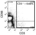
- FIG. 3 is a graph showing the results of double staining with antibodies against CD3 and CD56 and measurement by flow cytometry before removal of CD3-positive cells.
- 3 is a graph showing the results of measuring and averaging the change over time in the composition ratio of NK cells (CD3 negative / CD56 positive) isolated from 5 healthy individuals with respect to the whole cultured cells. Changes in the composition ratio of NK cells (CD3 negative / CD56 positive) isolated from 3 patients with advanced cancer (oral cancer, gallbladder cancer, and bile duct cancer) over time were measured by flow cytometry. Result diagram. Average growth curve of amplification factor of NK cells (CD3 negative / CD56 positive) isolated from 3 patients with advanced cancer (oral cancer, gallbladder cancer and bile duct cancer). The graph which compared the flow cytometry analysis result of CD69.
- NK cells Amplification of NK cells (1) 1. Materials and Methods (1) Blood collection from peripheral blood Peripheral blood was collected from healthy individuals and patients with advanced cancer (oral cancer, gallbladder cancer and bile duct cancer). This experiment was conducted with the approval of the Kyushu University Medical District Department Clinical Research Ethics Review Committee (approval number 22-176, approval date: March 31, 2011). Written consent has been obtained from the healthy person and the patient. Blood collection, cryopreservation, and thawing were performed by methods well known to those skilled in the art.
- the intermediate layer collected from one or two centrifuge tubes was collected in one new centrifuge tube, and the volume was adjusted to 50 mL with the diluent.
- the second centrifugation was performed under conditions of 500 ⁇ g, room temperature, 5 minutes, or 15 minutes.
- the supernatant was removed and the pellet was suspended in 30 mL of the diluent.
- the third centrifugation was performed under the conditions of 280 ⁇ g and room temperature for 10 minutes.
- the supernatant was removed, and the pellet was suspended in PBS supplemented with 2 mM EDTA and 0.1% BSA so that the cell concentration was 1 ⁇ 10 7 cells / mL (hereinafter referred to as “mononuclear”).
- Sphere suspension ”).
- CD3-negative cells The remaining cells in the suspension (hereinafter referred to as “CD3-negative cells”) are cell culture media (KBM501) supplemented with 5% autologous serum. 16025015, Kojin Bio Inc .; containing 1750 JRU / mL of IL-2 (hereinafter referred to as “KBM medium”), diluted to 5 ⁇ 10 5 cells / mL, and a 6-well culture plate (140675, nunc, Thermo Fisher Scientific Co., Ltd.). Cell culture was performed for 21 days at 37 ° C., 5% CO 2 and saturated water vapor atmosphere. Medium exchange was performed on the 5th, 9th, 13th and 17th days of culture. The cells were cultured without feeder cells.
- KBM501 cell culture media
- KBM medium 1750 JRU / mL of IL-2
- the cell number of the peripheral blood mononuclear cells was determined by measuring the number of living cells using a hemocytometer between the start of culture and the 21st day.
- Cell surface markers of the cells include anti-CD3 antibody (317308, BioLegend Japan), anti-CD16 antibody (556618, BD Pharmingen, Nippon Becton Dickinson), anti-CD56 antibody (304607, 318321, BioLegend Japan) , Anti-CD69 antibody (310905, BioLegend Japan), anti-KIR3DL1 / KIR3DL2 antibody (130-095-205, Miltenyi Biotech), anti-KIR2DL3 antibody (FAB2014P, R & D SYSTEMS, Cosmo Bio) KIR2DL1 / KIR2DS1 antibody (339505, BioLegend Japan, Inc.), anti-KIR2DL5 antibody (341003, BioLegend Japan Co., Ltd., anti-NKp46 antibody (331907, Bio
- FIG. 1A shows the experimental results of double staining with antibodies against CD3 and CD56 and measurement by flow cytometry before the removal of CD3-positive cells.
- FIG. 1B shows the experimental results of double staining with antibodies against CD3 and CD56 after removal of CD3 positive cells and measurement by flow cytometry.
- the composition ratio of CD3 positive cells the ratio of CD3 positive cells in the total cultured cells of each experimental group measured by the flow cytometry method is expressed as a percentage.
- the composition ratio (%) of CD3-positive cells was 69.37% before removal of CD3-positive cells, and 0.68% after removal of CD3-positive cells. As is apparent from these results, CD3-positive cells were significantly removed from the mononuclear cell suspension.
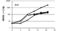
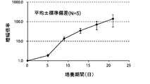
- FIG. 2A is a proliferation curve of the number of CD3-negative cells separated from mononuclear cells in the peripheral blood of five healthy subjects.
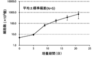
- FIG. 2B is an average growth curve of the number of CD3-negative cells isolated from mononuclear cells in the peripheral blood of five healthy subjects.
- the number of CD3-negative cells per 1 mL of peripheral blood collected from 5 healthy subjects is 5 days after culture, 9 days after culture, 13 days after culture, 17 days after culture and 21 days after culture. Measured. The standard deviation of each experimental condition was calculated from the measured values of experimental results repeated five times under the same conditions.
- CD3 negative cells continued to increase from the beginning of culture until 21st day. The rate of increase continued to increase until the 13th day and decreased after the 13th day.
- the number of CD3-negative cells increased from about 5 ⁇ 10 5 cells at the start of culture to about 700 ⁇ 10 5 cells after 21 days of culture.
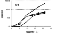
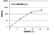
- FIG. 3A is a proliferation curve of each amplification factor of CD3-negative cells separated from mononuclear cells in the peripheral blood of five healthy subjects.
- FIG. 3B is an average growth curve of the magnification of CD3 negative cells isolated from mononuclear cells in the peripheral blood of 5 healthy subjects.
- the amplification factor is calculated by dividing the number of CD3 negative cells after 5 days, 9 days, 13 days, 17 days and 21 days by the number of CD3 negative cells at the start of culture. Calculated as the quotient. The standard deviation of each experimental condition was calculated from the measured values of experimental results repeated five times under the same conditions.
- the amplification factor of CD3 negative cells continued to increase from the beginning of the culture until the 21st day.
- the amplification factor continued to increase significantly until day 13, and increased to about 150 times after 21 days of culture.
- FIG. 4A is a proliferation curve of each amplification factor of NK cells (CD3 negative / CD56 positive) isolated from mononuclear cells in the peripheral blood of five healthy subjects.
- FIG. 4B is an average growth curve of amplification factor of NK cells (CD3 negative / CD56 positive) isolated from mononuclear cells in peripheral blood of five healthy subjects.
- CD3-negative cells were double-stained with antibodies against CD3 and CD56 and analyzed by flow cytometry.
- the amplification factor was calculated as the quotient obtained by dividing the number of NK cells after 7 days, 14 days and 21 days by the number of NK cells at the start of culture.
- the standard deviation of each experimental condition was calculated from the measured values of experimental results repeated five times under the same conditions.
- the amplification factor of NK cells continued to increase from the beginning of culture until the 21st day.
- the amplification factor continued to increase significantly up to day 14, and increased to about 400 times after 21 days of culture.
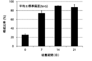
- FIG. 5A shows the results of an experiment in which the change over time in the composition ratio of NK cells (CD3 negative / CD56 positive) isolated from 5 healthy subjects to the whole cultured cells was measured by a flow cytometry method.
- FIG. 5B shows an experiment in which the change over time in the average value of the composition ratio of NK cells (CD3 negative / CD56 positive) isolated from 5 healthy subjects to the whole cultured cells was measured by flow cytometry and averaged. It is a result. 5A and 5B, CD3 negative cells were double-stained with antibodies against CD3 and CD56 and analyzed by flow cytometry.
- the ratio of NK cells in the total cultured cells of each experimental group, measured by flow cytometry, is expressed as a percentage.
- the vertical axis of the graph is the composition ratio (%) of NK cells (CD3 negative / CD56 positive) to the whole cultured cells, and the horizontal axis is the number of culture days.
- the standard deviation of each experimental condition was calculated from the measured values of experimental results repeated five times under the same conditions.
- the composition ratio of NK cells continued to increase from the beginning of culture until the 21st day.
- the composition ratio of the NK cells continued to increase significantly until the 14th day and increased to about 90% after 14 days of culture.
- the present invention has been shown to selectively amplify NK cells over time.
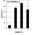
- FIG. 6A shows the composition ratio of NK cells (CD3 negative / CD56 positive) isolated from three patients with advanced cancer (oral cancer, gallbladder cancer and bile duct cancer) to the whole cultured cells. It is the experimental result which measured the change with time by the flow cytometry method.
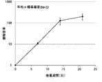
- FIG. 6B is an average growth curve of amplification factor of NK cells (CD3 negative / CD56 positive) isolated from 3 patients with advanced cancer (oral cancer, gallbladder cancer and bile duct cancer).
- the “constituent ratio of NK cells” the ratio of NK cells in the total cultured cells of each experimental group, measured by flow cytometry, is expressed as a percentage.
- FIG. 6A shows the composition ratio of NK cells (CD3 negative / CD56 positive) isolated from three patients with advanced cancer (oral cancer, gallbladder cancer and bile duct cancer) to the whole cultured cells. It is the experimental result which measured the change with time by the flow cytometry method.
- FIG. 6B is an
- the vertical axis represents the composition ratio (%) of NK cells (CD3 negative / CD56 positive) with respect to the whole cultured cells, and the horizontal axis represents the number of culture days.
- “Amplification magnification of NK cells” represents the result of dividing the number of NK cells after amplification by the number of NK cells present in peripheral blood mononuclear cells before amplification.
- the vertical axis represents the amplification factor of NK cells
- the horizontal axis represents the number of culture days. The standard deviation of each experimental condition was calculated from the measured values of the experimental results repeated three times under the same conditions. As shown in FIG.
- the composition ratio of NK cells continued to increase remarkably from the beginning of the culture to the 14th day, and increased to about 85% after the 14-day culture.
- the amplification factor of NK cells continued to increase remarkably from the beginning of the culture to the 14th day, and increased to about 140 times after the 14-day culture.
- the proportion of NK cells decreased due to the proliferation of CD3-positive cells.
- the proliferation of the CD3 positive cells hardly affected the amplification of NK cells. From the above results, it was shown that NK cells isolated from patients with advanced cancer (oral cancer, gallbladder cancer and bile duct cancer) are amplified over time. In addition, it was suggested that the present invention can amplify NK cells isolated from patients suffering from cancer, infectious diseases, etc. over time.
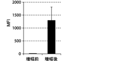
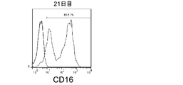
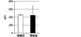
- FIGS. 7, 9 and 11 show graphs comparing the results of flow cytometry analysis of each cell surface marker. Moreover, the graph of the measured value of the mean fluorescence intensity (MFI) which compared the flow cytometry analysis result of CD69 and CD16 to FIG. 8 and 10 is shown. The standard deviation of each experimental condition was calculated from the measured values of the experimental results repeated three times under the same conditions.
- the cells amplified by the method of the present invention strongly expressed CD69, KIR2DL3, KIR2DL1 / KIR2DS1, KIR2DL5, NKp30, and NKG2D as compared to the cells before amplification. .
- the expression of CD69 was about 100%.
- the cells prepared by the method of the present invention were shown to express differentiation markers as NK cells.
- the NK cells have high cytotoxic activity.
- NK cells can be selectively and efficiently amplified by removing CD3-positive cells, ie, T cells, and then culturing them in the KBM medium. It was suggested that a large amount of NK cells can be prepared not only from healthy subjects but also from patients suffering from cancer, infectious diseases and the like. Moreover, it was suggested that the method of the present invention can remarkably amplify not only peripheral blood-derived NK cells but also cells derived from other tissues / organs, particularly umbilical cord blood-derived NK cells.
- NK cells were prepared from healthy individuals according to the method described in Example 1.
- CellGro SCGM 2001, Cellogenics, Iwai Chemicals
- 2500 IU / mL IL-2 AF-200-02-2, PeproTech, Toyobo Co., Ltd.
- 5% autologous serum "CellGro medium”
- the NK cells were amplified in the KBM medium and the CellGro medium according to the method described in Example 1.
- FIG. 12 is a growth curve of amplification factor of NK cells cultured in KBM medium and CellGro medium.
- the amplification factor was calculated as the quotient obtained by dividing the number of NK cells after 7 days, 14 days and 21 days by the number of NK cells at the start of culture. The standard deviation of each experimental condition was calculated from the measured values of the experimental results repeated twice under the same conditions.
- the amplification factor of NK cells continued to increase in the KBM medium and CellGro medium from the start of culture until the 21st day. After 21 days of culture, the amplification factor was about 670 times in the KBM medium and about 140 times in the CellGro medium.
- NK cells are sufficiently amplified in the KBM medium and the CellGro medium.
- NK cells can be amplified in media containing 2500 IU / mL to 2813 IU / mL IL-2 regardless of the type of cell culture media.
- Cytotoxic activity of amplified NK cells Materials and Methods (1) Quantification of cytotoxic activity NK cells were prepared according to the method described in Example 1 and used as effector cells. K562 cells (chronic myeloid leukemia cells) were prepared by methods well known to those skilled in the art and used as target cells. The cytotoxic activity of amplified NK cells and non-amplified NK cells (hereinafter referred to as “non-amplified NK cells”) was quantified by methods well known to those skilled in the art.
- the target cells are cultured in RPMI-1640 medium supplemented with 3-3'-dioctadesiloxacarbocyanine (D4292, Sigma-Aldrich Japan Co., Ltd.) (final concentration: 0.01 mM) for 10 minutes.
- the target cells were washed three times with PBS ( ⁇ ) and serum-free IMDM medium after labeling.
- the effector cells and the target cells were seeded in a round bottom 96-well culture plate and co-cultured in serum-free IMDM medium for 2 hours.
- the ratio of effector cells to target cells (E: T ratio) was adjusted to 3: 1, 2: 1, 1: 1, 1: 5, and 1:10. Cytotoxic activity (%) was quantified by flow cytometry using anti-MHC class I antibody (311409, BioLegend Japan) and 7-amino-actinomycin D (A9400, Sigma-Aldrich Japan). .
- NK cell differentiation marker NK cells were amplified according to the method described in Example 1. At the start of culture, after culturing for 3 days, after culturing for 7 days, after culturing for 14 days and after culturing for 21 days, the NK cells and the K562 cells were co-cultured at a 2: 1 E: T ratio for 2 hours. . Thereafter, the composition ratio of CD107a positive cells in the NK cells was analyzed by flow cytometry using an anti-CD107a antibody (328606, BioLegend Japan).
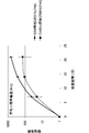
- FIG. 13 is a graph showing the experimental results of examining the cytotoxic activity of peripheral blood-derived NK cells amplified by the method of the present invention against K562.
- the vertical axis represents cytotoxic activity (unit:%).
- the white bar indicates the cytotoxic activity of non-amplified NK cells, and the black bar indicates the cytotoxic activity of amplified NK cells.
- the horizontal axis represents the E: T ratio between amplified NK cells or non-amplified NK cells and K562 cells. When the E: T ratio was 3: 1, the cytotoxic activity was about 30% for unamplified NK cells and about 110% for amplified NK cells.
- the cytotoxic activity was about 20% for unamplified NK cells and about 107% for amplified NK cells.
- the E: T ratio was 1: 1, the cytotoxic activity was about 10% for unamplified NK cells and about 100% for amplified NK cells.
- the E: T ratio was 1: 5 and 1:10, the cytotoxic activity of the amplified NK cells was about 25% and about 15%, respectively.
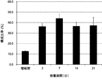
- FIG. 14 shows the results of experiments in which the change over time in the composition ratio of CD107a-positive cells isolated from healthy subjects to the whole cultured cells was measured by flow cytometry. The standard deviation of each experimental condition was calculated from the measured values of experimental results repeated five times under the same conditions.
- “constituent ratio of CD107a positive cells” the ratio of CD107a positive cells in the total cultured cells of each experimental group, measured by flow cytometry, is expressed as a percentage.
- the vertical axis represents the composition ratio (%) of CD107a positive cells to the whole cultured cells, and the horizontal axis represents the number of culture days. The composition ratio of CD107a positive cells was increased to about 35% by the third day from the start of the culture, and the composition ratio was maintained even on the 21st day.
- NK cells amplified according to the present invention have high cytotoxic activity. Therefore, it was shown that the present invention can selectively and efficiently amplify NK cells having high cytotoxic activity without using feeder cells, NK cells transfected with foreign molecules, or the like. It was also suggested that NK cells have high cytotoxic activity when amplified from cells derived from other tissues and organs, particularly cells derived from umbilical cord blood, as well as cells derived from peripheral blood.
- NK cell amplification (3) (repeated removal of CD3 positive cells) After the experiments of Examples 1 to 3, CD3 positive cells increased non-selectively as NK cell amplification experiments were repeated, and the composition ratio of CD3 positive cells to the whole cultured cells was as shown in the results of this example. The knowledge that it may exceed 30% was obtained. The frequency of non-selective increase of CD3-positive cells was about 30% of the experiments in which NK cells were amplified using peripheral blood mononuclear cells collected by apheresis from patients with advanced cancer ( Data is not shown.) Therefore, in order to selectively amplify NK cells, it was attempted to repeat the step of removing CD3-positive cells.
- NK cells were amplified and cell number and cell surface markers were analyzed.
- Mononuclear cell suspensions were prepared from patients with advanced cancer (oral cancer, gallbladder cancer and bile duct cancer). Removal of CD3 positive cells was performed once or twice.
- CD3-negative cells were cultured in the KBM medium for 14 days.
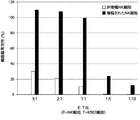
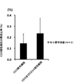
- FIG. 15 is a bar graph showing the composition ratio of NK cells (CD3 negative / CD56 positive) with respect to the whole cultured cells after removal of CD3 positive cells once and twice.
- the error bar for each experimental condition indicates the standard error of the measured value of the experimental result repeated three times under the same condition.
- the composition ratio of NK cells, CD3 positive cells and other cells the percentages of NK cells, CD3 positive cells and other cells in the total cultured cells of each experimental group, measured by flow cytometry, are percentages. expressed.
- the vertical axis of the graph is the composition ratio (%) of NK cells, CD3 positive cells and other cells to the whole cultured cells, and the horizontal axis is the number of removals of CD3 positive cells.
- the composition ratio (%) of the NK cells to the whole cultured cells was about 50% when the CD3 positive cells were removed once and about 65% when the CD3 positive cells were removed twice.
- NK cells were amplified and cell number and cell surface markers were analyzed.
- Mononuclear cell suspensions were prepared from patients with advanced cancer (oral cancer, gallbladder cancer and bile duct cancer). After removal of CD3 positive cells, hematopoietic progenitor cells were removed.
- the removal of the hematopoietic progenitor cells is performed by removing cells expressing CD34 (CD34 positive cells), biotinylated anti-CD34 antibody (343523, BioLegend Japan Co., Ltd.) and magnetic beads (Dynabeads biotin binder, 110-47, Life Technology Japan Co., Ltd.). Briefly, the CD34 positive cells were reacted with the biotinylated anti-CD34 antibody. Thereafter, centrifugation was performed, the supernatant was removed, and a suspension of cells bound with the antibody was prepared. The magnetic beads are washed once with 0.1% BSA was added PBS, the cells 10 7 per 50 ⁇ L is added to the suspension.
- the suspension containing the magnetic beads was stirred with a rotator at 4 ° C. for 30 minutes.
- the magnetic beads were separated from the suspension by a magnet, and CD34 positive cells were removed.
- the remaining cells hereinafter referred to as “CD3 and CD34 negative cells” in the suspension were cultured in the KBM medium for 14 days.
- an anti-CD34 antibody (343505, BioLegend Japan Co., Ltd.) was additionally used.
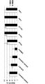
- FIG. 16A is a bar graph showing the composition ratio of CD34 positive cells in CD3 negative cells before amplification and CD3 and CD34 negative cells.
- FIG. 16B is a bar graph showing the composition ratio of CD3 positive cells in CD3 negative cells before amplification and CD3 and CD34 negative cells.
- the error bar for each experimental condition indicates the standard error of the measured value of the experimental result repeated three times under the same condition.
- the composition ratio of CD34 positive cells and CD3 positive cells the ratio of CD34 positive cells and CD3 positive cells in all cells of each experimental group, measured by flow cytometry, is expressed as a percentage.
- the vertical axis of the graph represents the composition ratio (%) of CD34 positive cells and CD3 positive cells before amplification to the whole cells.
- the horizontal axis of the graph indicates the cell type of each experimental group for amplification.
- the composition ratio (%) of CD34 positive cells before amplification was about 0.15% for CD3 negative cells and about 0.02% for CD3 and CD34 negative cells.
- the composition ratio (%) of CD3 positive cells before amplification was about 0.15% for CD3 negative cells and about 0.25% for CD3 and CD34 negative cells.
- FIG. 17 is a bar graph showing the composition ratio of NK cells (CD3 negative / CD56 positive) to the whole cultured cells in the amplified CD3 negative cells and CD3 and CD34 negative cells.
- the error bar for each experimental condition indicates the standard error of the measured value of the experimental result repeated three times under the same condition.
- the composition ratio of NK cells, CD3 positive cells and other cells the percentages of NK cells, CD3 positive cells and other cells in the total cultured cells of each experimental group, measured by flow cytometry, are percentages. expressed.
- the vertical axis of the graph represents the composition ratio (%) of NK cells, CD3 positive cells and other cells to the whole cultured cells.
- the horizontal axis of the graph indicates the cell type of each experimental group used for amplification.
- the composition ratio (%) of NK cells after amplification to the whole cultured cells was about 60% for CD3 negative cells and about 90% for CD3 and CD34 negative cells.
- NK cells CD3 negative / CD56 positive
- the composition ratio of NK cells was remarkably increased by removing CD3 positive cells and CD34 positive cells.
- NK cells are amplified using peripheral blood mononuclear cells collected by apheresis
- NK cells are amplified with high purity by removing CD3-positive cells and CD34-positive cells. It was shown that it can be done.
- NK cells could be prepared in large quantities by removing CD3-positive cells (T cells) from peripheral blood-derived mononuclear cells. Further, the cells amplified by the method of the present invention had a very high cytotoxic activity, as revealed by the experimental results of this example. Furthermore, NK cells could be prepared with high purity by removing CD3-positive cells (T cells) and CD34-positive cells (hematopoietic progenitor cells) from mononuclear cells derived from peripheral blood.
- cytotoxic activity of NK cells is low in the currently reported NK cell amplification methods.
- NK cells derived from peripheral blood of healthy subjects
- Patent Literature 1). Tanaka, J .;
- the purity is 96.8%
- the amplification factor is 277 times
- the present invention is remarkably superior to the prior art because the cytotoxic activity of NK cells is high and there is no risk of the feeder cells being mixed into the final product. Therefore, the present invention is useful for preparing NK cells having high cytotoxic activity in large quantities with high purity from collected blood cells.
Landscapes
- Health & Medical Sciences (AREA)
- Life Sciences & Earth Sciences (AREA)
- Engineering & Computer Science (AREA)
- General Health & Medical Sciences (AREA)
- Chemical & Material Sciences (AREA)
- Organic Chemistry (AREA)
- Biomedical Technology (AREA)
- Veterinary Medicine (AREA)
- Animal Behavior & Ethology (AREA)
- Public Health (AREA)
- Zoology (AREA)
- Biotechnology (AREA)
- Bioinformatics & Cheminformatics (AREA)
- Immunology (AREA)
- Genetics & Genomics (AREA)
- Wood Science & Technology (AREA)
- Epidemiology (AREA)
- Cell Biology (AREA)
- Medicinal Chemistry (AREA)
- Pharmacology & Pharmacy (AREA)
- Hematology (AREA)
- Nuclear Medicine, Radiotherapy & Molecular Imaging (AREA)
- General Chemical & Material Sciences (AREA)
- Chemical Kinetics & Catalysis (AREA)
- Microbiology (AREA)
- Biochemistry (AREA)
- General Engineering & Computer Science (AREA)
- Oncology (AREA)
- Communicable Diseases (AREA)
- Developmental Biology & Embryology (AREA)
- Virology (AREA)
- Molecular Biology (AREA)
- Medicines Containing Material From Animals Or Micro-Organisms (AREA)
- Micro-Organisms Or Cultivation Processes Thereof (AREA)
Priority Applications (7)
| Application Number | Priority Date | Filing Date | Title |
|---|---|---|---|
| US14/129,143 US9404083B2 (en) | 2011-06-24 | 2012-06-20 | Method for amplifying NK cells |
| AU2012274478A AU2012274478B2 (en) | 2011-06-24 | 2012-06-20 | Method for amplifying NK cells |
| KR1020147001579A KR101963920B1 (ko) | 2011-06-24 | 2012-06-20 | Nk 세포의 증폭 방법 |
| HK14103574.2A HK1190432B (en) | 2011-06-24 | 2012-06-20 | Method for amplifying nk cells |
| CN201280031188.5A CN103620022B (zh) | 2011-06-24 | 2012-06-20 | Nk细胞的扩增方法 |
| EP12801859.5A EP2725100B1 (en) | 2011-06-24 | 2012-06-20 | Method for amplifying nk cells |
| CA2840161A CA2840161C (en) | 2011-06-24 | 2012-06-20 | Method for amplifying nk cells |
Applications Claiming Priority (4)
| Application Number | Priority Date | Filing Date | Title |
|---|---|---|---|
| JP2011-140725 | 2011-06-24 | ||
| JP2011140725 | 2011-06-24 | ||
| JP2012-021972 | 2012-02-03 | ||
| JP2012021972A JP5572863B2 (ja) | 2011-06-24 | 2012-02-03 | Nk細胞の増幅方法 |
Publications (1)
| Publication Number | Publication Date |
|---|---|
| WO2012176796A1 true WO2012176796A1 (ja) | 2012-12-27 |
Family
ID=47422628
Family Applications (1)
| Application Number | Title | Priority Date | Filing Date |
|---|---|---|---|
| PCT/JP2012/065718 Ceased WO2012176796A1 (ja) | 2011-06-24 | 2012-06-20 | Nk細胞の増幅方法 |
Country Status (9)
| Country | Link |
|---|---|
| US (1) | US9404083B2 (enExample) |
| EP (1) | EP2725100B1 (enExample) |
| JP (1) | JP5572863B2 (enExample) |
| KR (1) | KR101963920B1 (enExample) |
| CN (1) | CN103620022B (enExample) |
| AU (1) | AU2012274478B2 (enExample) |
| CA (1) | CA2840161C (enExample) |
| MY (1) | MY161389A (enExample) |
| WO (1) | WO2012176796A1 (enExample) |
Cited By (2)
| Publication number | Priority date | Publication date | Assignee | Title |
|---|---|---|---|---|
| WO2018207900A1 (ja) * | 2017-05-12 | 2018-11-15 | 米満 吉和 | 高活性nk細胞、およびその利用 |
| JP2019137696A (ja) * | 2019-05-20 | 2019-08-22 | 米満 吉和 | 高活性nk細胞、およびその利用 |
Families Citing this family (27)
| Publication number | Priority date | Publication date | Assignee | Title |
|---|---|---|---|---|
| JP5511039B1 (ja) * | 2013-05-22 | 2014-06-04 | 国立大学法人九州大学 | Nk細胞の調製方法 |
| JP6164650B2 (ja) * | 2014-01-20 | 2017-07-19 | 国立大学法人九州大学 | Nk細胞の調製方法 |
| WO2015132415A1 (en) * | 2014-03-07 | 2015-09-11 | Emercell Sas | Pooled nk cells from ombilical cord blood and their uses for the treatment of cancer and chronic infectious disease |
| KR101697473B1 (ko) | 2014-11-26 | 2017-01-18 | 주식회사 녹십자랩셀 | T 세포를 이용한 자연살해세포의 배양방법 |
| US12398371B2 (en) | 2015-01-27 | 2025-08-26 | Korea Research Institute Of Bioscience | Method for mass producing natural killer cell and use of natural killer cell obtained by the method as anti-cancer agent |
| WO2016122014A1 (ko) * | 2015-01-27 | 2016-08-04 | 한국생명공학연구원 | 자연살해세포의 대량생산 방법 및 상기 방법으로 수득된 자연살해세포의 항암제로서의 용도 |
| CN106222141B (zh) * | 2016-10-17 | 2018-10-19 | 湖南丰晖生物科技有限公司 | Nk细胞培养液和细胞培养方法 |
| CN110662834B (zh) | 2017-05-26 | 2023-09-12 | Gc细胞治疗 | 使用转化的t细胞培养自然杀伤细胞的方法 |
| KR102765799B1 (ko) * | 2018-02-01 | 2025-02-13 | 주식회사 엔케이맥스 | Cd56+ 자연살해세포를 포함하는 항암용 조성물 |
| EP3746095A4 (en) | 2018-02-01 | 2021-04-21 | Nkmax Co., Ltd. | Method of producing natural killer cells and composition for treating cancer |
| WO2019152663A1 (en) * | 2018-02-01 | 2019-08-08 | Nkmax Co., Ltd. | Method of producing natural killer cells and composition for treating cancer |
| JP6543375B1 (ja) | 2018-03-27 | 2019-07-10 | 株式会社ガイアバイオメディシン | ケモカインレセプターと細胞接着分子を発現するcd3陰性細胞の集団、およびその利用 |
| CN110628714B (zh) * | 2018-06-21 | 2023-03-28 | 精准生技股份有限公司 | 用于体外扩增自然杀手细胞及自然杀手t细胞的无血清细胞培养液 |
| CN109161527A (zh) * | 2018-09-25 | 2019-01-08 | 深圳市五零生命科技有限公司 | 一种高效的nk细胞扩增方法 |
| EP3892721A4 (en) * | 2018-11-14 | 2022-08-31 | GC Cell Corporation | METHOD FOR CULTIVATING NATURAL KILLER CELLS DERIVED FROM UMBILICAL CORD BLOOD USING TRANSFORMED T LYMPHOCYTES |
| WO2020152661A1 (ja) | 2019-01-21 | 2020-07-30 | 株式会社ガイアバイオメディシン | Nk細胞を含む細胞集団の製造方法 |
| JP6838750B2 (ja) | 2019-01-21 | 2021-03-03 | 株式会社ガイアバイオメディシン | Nk細胞を含む細胞集団の製造方法 |
| KR102234394B1 (ko) * | 2019-03-08 | 2021-03-31 | 신지섭 | 타가면역세포배양방법, 그 방법으로 얻어진 면역세포배양액 및 이를 포함하는 면역세포치료제 |
| KR102216710B1 (ko) * | 2019-03-27 | 2021-02-17 | 신지섭 | Nk세포배양배지용 첨가조성물, 상기 첨가조성물을 이용한 nk세포배양방법 및 상기 배양방법으로 얻어진 피부트러블개선용 화장료조성물 |
| CN111154721B (zh) * | 2020-01-14 | 2023-10-17 | 深圳格泰赛尔生物科技有限公司 | Nk细胞扩增方法 |
| US20230040477A1 (en) * | 2020-01-19 | 2023-02-09 | Board Of Regents, The University Of Texas System | T-cell death associated gene 8 (tdag8) modulation to enhance cellular cancer therapies |
| US12486492B2 (en) | 2020-03-05 | 2025-12-02 | Korea Research Institute Of Bioscience | Method for producing memory-like NK cells with ability to express higher levels of NCRs, cytotoxicity, and IFN-γ than NK cells in human peripheral blood |
| EP4263600A1 (en) | 2020-12-18 | 2023-10-25 | Century Therapeutics, Inc. | Chimeric antigen receptor systems with adaptable receptor specificity |
| WO2023081813A1 (en) | 2021-11-05 | 2023-05-11 | St. Jude Children's Research Hospital, Inc. | Zip cytokine receptors |
| EP4536687A1 (en) | 2022-06-08 | 2025-04-16 | St. Jude Children's Research Hospital, Inc. | Disruption of kdm4a in t cells to enhance immunotherapy |
| WO2024059787A1 (en) | 2022-09-16 | 2024-03-21 | St. Jude Children's Research Hospital, Inc. | Disruption of asxl1 in t cells to enhance immunotherapy |
| WO2025022372A1 (ja) * | 2023-07-21 | 2025-01-30 | 株式会社ガイアバイオメディシン | 細胞集団の製造方法 |
Citations (9)
| Publication number | Priority date | Publication date | Assignee | Title |
|---|---|---|---|---|
| WO2007103901A2 (en) * | 2006-03-06 | 2007-09-13 | Government Of The United States Of America, Represented By The Secretary, Department Of Health And Human Services | Autologous natural killer cells and lymphodepleting chemotherapy for the treatment of cancer |
| JP2007297292A (ja) | 2006-04-28 | 2007-11-15 | Kyorin Pharmaceut Co Ltd | 1−アルコキシイソキノリン誘導体又はその水和物の製造方法 |
| WO2008153150A1 (ja) * | 2007-06-15 | 2008-12-18 | Medinet Co., Ltd. | Nk細胞を含む細胞集団の培養方法及び当該細胞集団の利用 |
| WO2010013947A2 (ko) * | 2008-07-29 | 2010-02-04 | 주식회사 녹십자 | 자연살해세포의 증식방법 |
| WO2011030851A1 (ja) * | 2009-09-11 | 2011-03-17 | タカラバイオ株式会社 | ナチュラルキラー細胞の製造方法 |
| JP2011517944A (ja) * | 2008-04-09 | 2011-06-23 | マックスサイト インコーポレーティッド | 新規に単離された細胞の治療組成物の操作および送達 |
| JP2011140504A (ja) | 2006-08-28 | 2011-07-21 | Hisamitsu Pharmaceut Co Inc | 爪用貼付剤 |
| JP2011239701A (ja) * | 2010-05-14 | 2011-12-01 | Tella Inc | 樹状細胞の培養方法 |
| JP2013006793A (ja) | 2011-06-24 | 2013-01-10 | Tella Inc | Nk細胞を増幅するための組成物及び方法 |
Family Cites Families (11)
| Publication number | Priority date | Publication date | Assignee | Title |
|---|---|---|---|---|
| US20030068306A1 (en) * | 2001-09-14 | 2003-04-10 | Dilber Mehmet Sirac | Medium |
| US7544355B2 (en) * | 2002-03-13 | 2009-06-09 | Universita Degli Studi Di Perugia | Methods and compositions for allogeneic transplantation |
| AU2003294930B2 (en) | 2002-12-23 | 2008-12-04 | Innate Pharma | Pharmaceutical compositions having an effect on the proliferation of NK cells and a method using the same |
| US7435596B2 (en) * | 2004-11-04 | 2008-10-14 | St. Jude Children's Research Hospital, Inc. | Modified cell line and method for expansion of NK cell |
| WO2006050270A2 (en) * | 2004-11-02 | 2006-05-11 | The Government Of The United States Of America As Represented By The Secretary Department Of Health & Human Services | Compositions and methods for treating hyperproliferative disorders |
| JP4275680B2 (ja) | 2006-04-28 | 2009-06-10 | 裕 照沼 | リンパ球の活性・増殖に係る培養方法 |
| WO2008023874A1 (en) * | 2006-08-23 | 2008-02-28 | Binex Co., Ltd. | Manufacturing method of activated lymphocytes for immunotherapy |
| AU2008307633C1 (en) * | 2007-09-28 | 2015-04-30 | Celularity Inc. | Tumor suppression using human placental perfusate and human placenta-derived intermediate natural killer cells |
| JP2012521215A (ja) * | 2009-03-26 | 2012-09-13 | アヴァリス・アクチエボラーグ | Nk細胞の増殖 |
| EP2519239B1 (en) | 2009-12-29 | 2017-03-08 | Gamida Cell Ltd. | Methods for enhancing natural killer cell proliferation and activity |
| KR101039843B1 (ko) | 2010-08-30 | 2011-06-09 | 주식회사 엔케이바이오 | 자기활성화 림프구 배양용 배지 조성물 및 이를 이용한 자기활성화 림프구 배양방법 |
-
2012
- 2012-02-03 JP JP2012021972A patent/JP5572863B2/ja active Active
- 2012-06-20 CA CA2840161A patent/CA2840161C/en active Active
- 2012-06-20 KR KR1020147001579A patent/KR101963920B1/ko active Active
- 2012-06-20 CN CN201280031188.5A patent/CN103620022B/zh active Active
- 2012-06-20 EP EP12801859.5A patent/EP2725100B1/en active Active
- 2012-06-20 WO PCT/JP2012/065718 patent/WO2012176796A1/ja not_active Ceased
- 2012-06-20 US US14/129,143 patent/US9404083B2/en active Active
- 2012-06-20 MY MYPI2013004630A patent/MY161389A/en unknown
- 2012-06-20 AU AU2012274478A patent/AU2012274478B2/en active Active
Patent Citations (9)
| Publication number | Priority date | Publication date | Assignee | Title |
|---|---|---|---|---|
| WO2007103901A2 (en) * | 2006-03-06 | 2007-09-13 | Government Of The United States Of America, Represented By The Secretary, Department Of Health And Human Services | Autologous natural killer cells and lymphodepleting chemotherapy for the treatment of cancer |
| JP2007297292A (ja) | 2006-04-28 | 2007-11-15 | Kyorin Pharmaceut Co Ltd | 1−アルコキシイソキノリン誘導体又はその水和物の製造方法 |
| JP2011140504A (ja) | 2006-08-28 | 2011-07-21 | Hisamitsu Pharmaceut Co Inc | 爪用貼付剤 |
| WO2008153150A1 (ja) * | 2007-06-15 | 2008-12-18 | Medinet Co., Ltd. | Nk細胞を含む細胞集団の培養方法及び当該細胞集団の利用 |
| JP2011517944A (ja) * | 2008-04-09 | 2011-06-23 | マックスサイト インコーポレーティッド | 新規に単離された細胞の治療組成物の操作および送達 |
| WO2010013947A2 (ko) * | 2008-07-29 | 2010-02-04 | 주식회사 녹십자 | 자연살해세포의 증식방법 |
| WO2011030851A1 (ja) * | 2009-09-11 | 2011-03-17 | タカラバイオ株式会社 | ナチュラルキラー細胞の製造方法 |
| JP2011239701A (ja) * | 2010-05-14 | 2011-12-01 | Tella Inc | 樹状細胞の培養方法 |
| JP2013006793A (ja) | 2011-06-24 | 2013-01-10 | Tella Inc | Nk細胞を増幅するための組成物及び方法 |
Non-Patent Citations (10)
| Title |
|---|
| "Research Ethics Committee of Departments", MEDICAL FACILITIES OF KYUSHU UNIVERSITY, 31 March 2011 (2011-03-31) |
| ALICI, E. ET AL., BLOOD, vol. 111, 2008, pages 3155 |
| CARLENS, S. ET AL., HUM. IMMUNOL., vol. 62, 2001, pages 1092 |
| CHO, D.; CAMPANA, D., KOREAN J. LAB. MED., 2009, pages 29 - 89 |
| FUJISAKI, H. ET AL., CANCER RES., vol. 69, 2009, pages 4010 |
| KOHJIN BIO CO., LTD.: "KBM501 Soshiki Baiyo Kanren Baichi no Kaihatsu", HANBAI, 28 July 2010 (2010-07-28), XP055139607, Retrieved from the Internet <URL:http://web.archive.org/web/20100728100847/http://www.kohjin-bio.co.jp/products/?id=1269767360-072037> [retrieved on 20120911] * |
| MALE,V. ET AL.: "Immature NK Cells, Capable of Producing IL-22, Are Present in Human Uterine Mucosa", JOURNAL OF IMMUNOLOGY, vol. 185, no. 7, 2010, pages 3913 - 3918, XP055139608 * |
| MILLERS ET AL., BLOOD, vol. 105, 2005, pages 3051 |
| RUBNITZ ET AL., J. CLIN. ONCOL., vol. 28, 2010, pages 955 |
| See also references of EP2725100A4 |
Cited By (6)
| Publication number | Priority date | Publication date | Assignee | Title |
|---|---|---|---|---|
| WO2018207900A1 (ja) * | 2017-05-12 | 2018-11-15 | 米満 吉和 | 高活性nk細胞、およびその利用 |
| JP2018193303A (ja) * | 2017-05-12 | 2018-12-06 | 米満 吉和 | 高活性nk細胞、およびその利用 |
| US11723924B2 (en) | 2017-05-12 | 2023-08-15 | Yoshikazu Yonemitsu | Highly active NK cell and use thereof |
| TWI846668B (zh) * | 2017-05-12 | 2024-07-01 | 米満吉和 | 高活性nk細胞及其利用 |
| US12329817B2 (en) | 2017-05-12 | 2025-06-17 | Yoshikazu Yonemitsu | Highly active NK cell and use thereof |
| JP2019137696A (ja) * | 2019-05-20 | 2019-08-22 | 米満 吉和 | 高活性nk細胞、およびその利用 |
Also Published As
| Publication number | Publication date |
|---|---|
| JP2013027385A (ja) | 2013-02-07 |
| JP5572863B2 (ja) | 2014-08-20 |
| EP2725100A4 (en) | 2015-01-07 |
| US20140120072A1 (en) | 2014-05-01 |
| MY161389A (en) | 2017-04-14 |
| EP2725100A1 (en) | 2014-04-30 |
| US9404083B2 (en) | 2016-08-02 |
| EP2725100B1 (en) | 2019-09-04 |
| AU2012274478A1 (en) | 2014-02-20 |
| CN103620022B (zh) | 2015-09-30 |
| KR20140051263A (ko) | 2014-04-30 |
| CN103620022A (zh) | 2014-03-05 |
| CA2840161A1 (en) | 2012-12-27 |
| KR101963920B1 (ko) | 2019-03-29 |
| AU2012274478B2 (en) | 2017-03-30 |
| AU2012274478A8 (en) | 2016-07-28 |
| HK1190432A1 (en) | 2014-07-04 |
| CA2840161C (en) | 2019-02-12 |
Similar Documents
| Publication | Publication Date | Title |
|---|---|---|
| JP5572863B2 (ja) | Nk細胞の増幅方法 | |
| JP5511039B1 (ja) | Nk細胞の調製方法 | |
| US12329817B2 (en) | Highly active NK cell and use thereof | |
| KR102534472B1 (ko) | 케모카인 리셉터와 세포 접착 분자를 발현하는 cd3 음성 세포의 집단, 및 그 이용 그리고 그 제조 방법 | |
| JP5989016B2 (ja) | Nk細胞の増幅方法 | |
| JP2013006793A (ja) | Nk細胞を増幅するための組成物及び方法 | |
| TWI757709B (zh) | 含有nk細胞之細胞集團之製造方法 | |
| JP6164650B2 (ja) | Nk細胞の調製方法 | |
| JP6697611B2 (ja) | 高活性nk細胞、およびその利用 | |
| JP2020108405A (ja) | 高活性nk細胞、およびその利用 | |
| HK1190432B (en) | Method for amplifying nk cells | |
| JP2023153286A (ja) | Nk細胞を含む細胞集団の製造方法 |
Legal Events
| Date | Code | Title | Description |
|---|---|---|---|
| 121 | Ep: the epo has been informed by wipo that ep was designated in this application |
Ref document number: 12801859 Country of ref document: EP Kind code of ref document: A1 |
|
| DPE1 | Request for preliminary examination filed after expiration of 19th month from priority date (pct application filed from 20040101) | ||
| ENP | Entry into the national phase |
Ref document number: 2840161 Country of ref document: CA |
|
| NENP | Non-entry into the national phase |
Ref country code: DE |
|
| WWE | Wipo information: entry into national phase |
Ref document number: 14129143 Country of ref document: US |
|
| ENP | Entry into the national phase |
Ref document number: 20147001579 Country of ref document: KR Kind code of ref document: A |
|
| ENP | Entry into the national phase |
Ref document number: 2012274478 Country of ref document: AU Date of ref document: 20120620 Kind code of ref document: A |