US10926001B2 - Methods related to minimally polarized functional units - Google Patents
Methods related to minimally polarized functional units Download PDFInfo
- Publication number
- US10926001B2 US10926001B2 US14/954,335 US201514954335A US10926001B2 US 10926001 B2 US10926001 B2 US 10926001B2 US 201514954335 A US201514954335 A US 201514954335A US 10926001 B2 US10926001 B2 US 10926001B2
- Authority
- US
- United States
- Prior art keywords
- compartment
- tissue
- composition
- lgr6
- follicular
- Prior art date
- Legal status (The legal status is an assumption and is not a legal conclusion. Google has not performed a legal analysis and makes no representation as to the accuracy of the status listed.)
- Active
Links
Images


Classifications
-
- C—CHEMISTRY; METALLURGY
- C12—BIOCHEMISTRY; BEER; SPIRITS; WINE; VINEGAR; MICROBIOLOGY; ENZYMOLOGY; MUTATION OR GENETIC ENGINEERING
- C12N—MICROORGANISMS OR ENZYMES; COMPOSITIONS THEREOF; PROPAGATING, PRESERVING, OR MAINTAINING MICROORGANISMS; MUTATION OR GENETIC ENGINEERING; CULTURE MEDIA
- C12N5/00—Undifferentiated human, animal or plant cells, e.g. cell lines; Tissues; Cultivation or maintenance thereof; Culture media therefor
- C12N5/06—Animal cells or tissues; Human cells or tissues
- C12N5/0602—Vertebrate cells
- C12N5/0625—Epidermal cells, skin cells; Cells of the oral mucosa
-
- A01N1/021—
-
- A01N1/0221—
-
- A—HUMAN NECESSITIES
- A01—AGRICULTURE; FORESTRY; ANIMAL HUSBANDRY; HUNTING; TRAPPING; FISHING
- A01N—PRESERVATION OF BODIES OF HUMANS OR ANIMALS OR PLANTS OR PARTS THEREOF; BIOCIDES, e.g. AS DISINFECTANTS, AS PESTICIDES OR AS HERBICIDES; PEST REPELLANTS OR ATTRACTANTS; PLANT GROWTH REGULATORS
- A01N1/00—Preservation of bodies of humans or animals, or parts thereof
- A01N1/10—Preservation of living parts
- A01N1/12—Chemical aspects of preservation
- A01N1/122—Preservation or perfusion media
- A01N1/125—Freeze protecting agents, e.g. cryoprotectants or osmolarity regulators
-
- A—HUMAN NECESSITIES
- A61—MEDICAL OR VETERINARY SCIENCE; HYGIENE
- A61L—METHODS OR APPARATUS FOR STERILISING MATERIALS OR OBJECTS IN GENERAL; DISINFECTION, STERILISATION OR DEODORISATION OF AIR; CHEMICAL ASPECTS OF BANDAGES, DRESSINGS, ABSORBENT PADS OR SURGICAL ARTICLES; MATERIALS FOR BANDAGES, DRESSINGS, ABSORBENT PADS OR SURGICAL ARTICLES
- A61L27/00—Materials for grafts or prostheses or for coating grafts or prostheses
- A61L27/36—Materials for grafts or prostheses or for coating grafts or prostheses containing ingredients of undetermined constitution or reaction products thereof, e.g. transplant tissue, natural bone, extracellular matrix
- A61L27/3604—Materials for grafts or prostheses or for coating grafts or prostheses containing ingredients of undetermined constitution or reaction products thereof, e.g. transplant tissue, natural bone, extracellular matrix characterised by the human or animal origin of the biological material, e.g. hair, fascia, fish scales, silk, shellac, pericardium, pleura, renal tissue, amniotic membrane, parenchymal tissue, fetal tissue, muscle tissue, fat tissue, enamel
- A61L27/362—Skin, e.g. dermal papillae
-
- A—HUMAN NECESSITIES
- A61—MEDICAL OR VETERINARY SCIENCE; HYGIENE
- A61L—METHODS OR APPARATUS FOR STERILISING MATERIALS OR OBJECTS IN GENERAL; DISINFECTION, STERILISATION OR DEODORISATION OF AIR; CHEMICAL ASPECTS OF BANDAGES, DRESSINGS, ABSORBENT PADS OR SURGICAL ARTICLES; MATERIALS FOR BANDAGES, DRESSINGS, ABSORBENT PADS OR SURGICAL ARTICLES
- A61L27/00—Materials for grafts or prostheses or for coating grafts or prostheses
- A61L27/36—Materials for grafts or prostheses or for coating grafts or prostheses containing ingredients of undetermined constitution or reaction products thereof, e.g. transplant tissue, natural bone, extracellular matrix
- A61L27/38—Materials for grafts or prostheses or for coating grafts or prostheses containing ingredients of undetermined constitution or reaction products thereof, e.g. transplant tissue, natural bone, extracellular matrix containing added animal cells
- A61L27/3804—Materials for grafts or prostheses or for coating grafts or prostheses containing ingredients of undetermined constitution or reaction products thereof, e.g. transplant tissue, natural bone, extracellular matrix containing added animal cells characterised by specific cells or progenitors thereof, e.g. fibroblasts, connective tissue cells, kidney cells
- A61L27/3813—Epithelial cells, e.g. keratinocytes, urothelial cells
-
- A—HUMAN NECESSITIES
- A61—MEDICAL OR VETERINARY SCIENCE; HYGIENE
- A61L—METHODS OR APPARATUS FOR STERILISING MATERIALS OR OBJECTS IN GENERAL; DISINFECTION, STERILISATION OR DEODORISATION OF AIR; CHEMICAL ASPECTS OF BANDAGES, DRESSINGS, ABSORBENT PADS OR SURGICAL ARTICLES; MATERIALS FOR BANDAGES, DRESSINGS, ABSORBENT PADS OR SURGICAL ARTICLES
- A61L27/00—Materials for grafts or prostheses or for coating grafts or prostheses
- A61L27/50—Materials characterised by their function or physical properties, e.g. injectable or lubricating compositions, shape-memory materials, surface modified materials
- A61L27/54—Biologically active materials, e.g. therapeutic substances
-
- A—HUMAN NECESSITIES
- A61—MEDICAL OR VETERINARY SCIENCE; HYGIENE
- A61P—SPECIFIC THERAPEUTIC ACTIVITY OF CHEMICAL COMPOUNDS OR MEDICINAL PREPARATIONS
- A61P17/00—Drugs for dermatological disorders
- A61P17/02—Drugs for dermatological disorders for treating wounds, ulcers, burns, scars, keloids, or the like
-
- A—HUMAN NECESSITIES
- A61—MEDICAL OR VETERINARY SCIENCE; HYGIENE
- A61P—SPECIFIC THERAPEUTIC ACTIVITY OF CHEMICAL COMPOUNDS OR MEDICINAL PREPARATIONS
- A61P43/00—Drugs for specific purposes, not provided for in groups A61P1/00-A61P41/00
-
- A—HUMAN NECESSITIES
- A61—MEDICAL OR VETERINARY SCIENCE; HYGIENE
- A61P—SPECIFIC THERAPEUTIC ACTIVITY OF CHEMICAL COMPOUNDS OR MEDICINAL PREPARATIONS
- A61P5/00—Drugs for disorders of the endocrine system
-
- A—HUMAN NECESSITIES
- A61—MEDICAL OR VETERINARY SCIENCE; HYGIENE
- A61L—METHODS OR APPARATUS FOR STERILISING MATERIALS OR OBJECTS IN GENERAL; DISINFECTION, STERILISATION OR DEODORISATION OF AIR; CHEMICAL ASPECTS OF BANDAGES, DRESSINGS, ABSORBENT PADS OR SURGICAL ARTICLES; MATERIALS FOR BANDAGES, DRESSINGS, ABSORBENT PADS OR SURGICAL ARTICLES
- A61L2300/00—Biologically active materials used in bandages, wound dressings, absorbent pads or medical devices
- A61L2300/40—Biologically active materials used in bandages, wound dressings, absorbent pads or medical devices characterised by a specific therapeutic activity or mode of action
- A61L2300/412—Tissue-regenerating or healing or proliferative agents
-
- A—HUMAN NECESSITIES
- A61—MEDICAL OR VETERINARY SCIENCE; HYGIENE
- A61L—METHODS OR APPARATUS FOR STERILISING MATERIALS OR OBJECTS IN GENERAL; DISINFECTION, STERILISATION OR DEODORISATION OF AIR; CHEMICAL ASPECTS OF BANDAGES, DRESSINGS, ABSORBENT PADS OR SURGICAL ARTICLES; MATERIALS FOR BANDAGES, DRESSINGS, ABSORBENT PADS OR SURGICAL ARTICLES
- A61L2430/00—Materials or treatment for tissue regeneration
-
- C—CHEMISTRY; METALLURGY
- C12—BIOCHEMISTRY; BEER; SPIRITS; WINE; VINEGAR; MICROBIOLOGY; ENZYMOLOGY; MUTATION OR GENETIC ENGINEERING
- C12N—MICROORGANISMS OR ENZYMES; COMPOSITIONS THEREOF; PROPAGATING, PRESERVING, OR MAINTAINING MICROORGANISMS; MUTATION OR GENETIC ENGINEERING; CULTURE MEDIA
- C12N2513/00—3D culture
Definitions
- the present invention relates to constructs of micro-aggregate multicellular grafts containing Leucine-rich repeat-containing G-protein coupled Receptor (LGR) expressing cells for wound therapy applications, tissue engineering, cell therapy applications, regenerative medicine applications, medical/therapeutic applications, tissue healing applications, immune therapy applications, and tissue transplant therapy applications. More particularly, the invention provides a deliverable micro-aggregate multi-cellular LGR construct on a delivery vector/substrate/support/scaffold for direct application.
- LGR Leucine-rich repeat-containing G-protein coupled Receptor
- LGR4, LGR5 and LGR6 expressing epithelial stem cell populations are often destroyed following severe full-thickness damage to the skin, leaving tissues incapable of producing a viable and self-sustaining epithelial compartment.
- LGR4, LGR5 and LGR6 expressing epithelial stem cell populations are often destroyed following severe full-thickness damage to the skin, leaving tissues incapable of producing a viable and self-sustaining epithelial compartment.
- remaining tissues are left without the regenerative potential to form a functional epithelium, hair follicle, sweat gland, or the like.
- tissue elements skin, muscle, fat, blood vessels, nerves and bone
- Such injuries and subsequent resulting wounds are also difficult to treat through current wound care methods, surgical interventions with current approved technologies utilizing cells, tissues, devices, biologics, drugs and/or growth factors.
- a common reason for such difficulty is that the tissues remaining in or around a wounded or injured tissue bed are typically devoid of inter-dependent, necessary components: 1) cellular progenitor and/or stem cell populations; 2) extracellular matrix/scaffolding elements and substrates; and 3) a combination of interactions between and among cellular entities and substrates.
- ECM extracellular matrix
- the more robust acellular matrices such as ALLODERM® from LifeCell Corporation, INTEGRA® from Integra LifeSciences Corporation and DERMAMATRIX® a product from Musculoskeletal Transplant Foundation, although excellent reconstructive options, lack those properly placed lineage specific stem cell populations which are necessary to develop functional native tissues.
- LGR5 and LGR6 are markers of both intestinal and epidermal stem cells in mammals.
- Stimulation of the Follicular Bulge LGR5+ and LGR6+ Stem Cells with the Gut-Derived Human Alpha Defensin 5 Results in Decreased Bacterial Presence, Enhanced Wound Healing, and Hair Growth from Tissues Devoid of Adnexal Structures, Plast. Reconstr. Surg.
- LGR Leucine-rich repeat-containing G-protein—coupled receptor
- human alpha defensin 5 peptide significantly enhanced wound healing and reduced basal bacterial load compared with human beta defensin 1 and sulfadiazine.
- Human alpha defensin 5 was the only therapy to induce LGR stem cell migration into the wound bed.
- gene heat mapping showed significant mRNA up-regulation of key wound healing and Wnt pathway transcripts such as Wnt1 and Wisp1. So it was concluded that human alpha defensin 5 could be used for enhanced wound healing due to the observed increase of LGR stem cell migration into wound beds and associated bacterial reduction and hair production through the augmentation of key Wnt and wound healing transcripts.
- this and other work led to the recognition of the potential for using LGR4+, LGR5+ and LGR6+ expressing epithelial stem cells in direct biomedical engineering soft tissue constructs.
- the invention provides in a first embodiment a minimally polarized micro-aggregate multi-cellular composition including isolated living LGR expressing cells and a multi-dimensional support selected from the group consisting of scaffolding, collagen, matrix, particle, and fiber.
- the invention provides in a further embodiment to the previous embodiment a minimally polarized micro-aggregate multi-cellular composition including isolated living LGR expressing cells and a multi-dimensional support selected from the group consisting of scaffolding, collagen, matrix, particle, and fiber where the LGR expressing cells are supplemented with growth factors and where the LGR expressing cells are selected from the group consisting of LGR4, LGR5 and LGR6.
- the invention provides in a further embodiment to any of the previous embodiments a minimally polarized micro-aggregate multi-cellular composition including isolated living LGR expressing cells and a multi-dimensional support selected from the group consisting of scaffolding, collagen, matrix, particle, and fiber where the LGR expressing cells are supplemented with migratory/recruiting analytes and the LGR expressing cells being selected from the group consisting of LGR4, LGR5 and LGR6.
- the invention provides in a further embodiment to any of the previous embodiments a minimally polarized micro-aggregate multi-cellular composition including isolated living LGR expressing cells and a multi-dimensional support selected from the group consisting of scaffolding, collagen, matrix, particle, and fiber where the LGR expressing cells are supplemented with LGR specific binding elements selected from the group consisting of ligand families, R-spondin, EDGF, PDGF, Wnt, VEGF, and antimicrobial peptides and where the LGR expressing cells are selected from the group consisting of LGR4, LGR5 and LGR6.
- the invention provides in a further embodiment to any of the previous embodiments a minimally polarized micro-aggregate multi-cellular composition including isolated living LGR expressing cells and a multi-dimensional support selected from the group consisting of scaffolding, collagen, matrix, particle, and fiber where the composition is used as a therapeutic construct for a select target consisting of a tissue region, wound, void, defect tissue, or blood for alteration of either surrounding adjacent tissues.
- the invention provides in a further embodiment to any of the previous embodiments a minimally polarized micro-aggregate multi-cellular composition characterized by a isolated living LGR expressing cells transplanted to damaged tissue to accelerate healing thereof.
- the invention provides in a further embodiment to any of the previous embodiments a minimally polarized micro-aggregate multi-cellular composition for tissue system repair or restoration throughout the body comprising a support scaffolding with isolated LGR containing cells secured thererto.
- the invention provides in a further embodiment to any of the previous embodiments a tissue graft for application to ectodermal, mesodermal or endodermal-derived tissues systems throughout a mammalian body
- Another embodiment of the invention is characterized by a method for obtaining a minimally polarized micro-aggregate multi-cellular composition characterized by the steps of growing and isolating living LGR expressing cells for transplantation to a select mammalian target tissue.
- the invention provides in a further embodiment to the foregoing method a method for obtaining a minimally polarized micro-aggregate multi-cellular composition characterized by the steps of growing and isolating living LGR expressing cells for transplantation to a select mammalian target tissue further characterized by the step of affixing the isolated living LGR expressing cells to a multi-dimensional support selected from the group consisting of scaffolding, collagen, matrix, particle, and fiber.
- the invention is characterized in still another embodiment by a method for obtaining a minimally polarized micro-aggregate multi-cellular composition characterized by the steps of growing and isolating living LGR expressing cells for transplantation to a select mammalian target tissue further characterized by the step of selecting the LGR expressing cells from the group consisting of LGR4, LGR5 and LGR6.
- the invention provides in a further embodiment to any of the previous method embodiments the step of applying the minimally polarized micro-aggregate multi-cellular to one of the group consisting of epithelial systems, glands, hair, nerves, bone, muscle, fat, tendons, blood vessels, fascia, ocular tissues and peptide secreting cellular elements employing delivery by a technique selected from the group consisting of application, transplantation, implantation, directed seeding, directed migration, directed tracking, in setting, laminating and/or injection of the cellular element generating, regenerating, enhancing and healing.
- the invention provides in a further embodiment to any of the previous method embodiments a method for obtaining a minimally polarized micro-aggregate multi-cellular composition characterized by the steps of growing and isolating living LGR expressing cells for transplantation to a select mammalian target tissue further characterized by the step of applying the minimally polarized micro-aggregate multi-cellular composition directly to a tissue in vivo for tissue restoration.
- the invention provides in a further embodiment to any of the previous method embodiments a method for obtaining a minimally polarized micro-aggregate multi-cellular composition characterized by the steps of growing and isolating living LGR expressing cells for transplantation to a select mammalian target tissue further characterized by the step indirectly applying the minimally polarized micro-aggregate multi-cellular composition via the blood stream for tissue restoration in a body.
- the invention provides in a further embodiment to any of the previous embodiments a media formulation used in obtaining minimally polarized micro-aggregate multi-cellular compositions using cell sustaining media composition for reducing the viability of micro-organisms during transport and processing of tissues, characterized by: a) a mixture of epithelial cells and keratinocytes; b) at least one agent selected from the group consisting of penicillin, streptomycin, and amphotericin B; and c) fibrinogen.
- the invention provides in a further embodiment to the previous embodiment a cell sustaining media composition of for reducing the viability of micro-organisms during transport and processing of tissues, characterized by: a) a mixture of epithelial cells and keratinocytes; b) at least one agent selected from the group consisting of penicillin, streptomycin, and amphotericin B; and c) fibrinogen, where the fibrinogen is human and where the agent includes both an antibiotic and an antimycotic for stabilizing human tissues.
- LGR expressing cells being applied to scaffolding matrix, and/or fiber to thereby establish micro-aggregate multi-cellular grafts for tissue engineering applications, cell therapy applications, regenerative medicine applications, medical/therapeutic applications the grafts being directly applied to tissue or blood for improvement and or alteration of epithelial systems throughout the body.
- a second aspect of the invention is characterized by LGR expressing cells being applied to scaffolding, matrix, and/or fiber or without additional enhancing factors or analytes before or after either being applied to tissue or blood for improvement and or alteration of in vivo epithelial systems.
- a further aspect of the invention is characterized by LGR expressing cells altered by enhancing factors or analytes, being applied as targets within the body, tissue or blood for improvement and/or alteration of epithelial systems through local or distant migration throughout the body and/or to restore gland and hair growth.
- a fourth aspect of the invention is characterized by transplanting LGR expressing cells from tissue, blood or culture for alteration of surrounding adjacent tissues or distant tissues such as but not inclusive of LGR expressing cells applied to scaffolding, matrix, and fiber before or after either being applied to tissue or blood for improvement and or alteration of ectodermal, mesodermal or endodermal-derived tissues systems throughout the body.
- a fifth aspect of the invention is characterized by directly applying LGR expressing cells to a delivery substrate vehicle selected from a group consisting of scaffolding, matrix, and fiber with/or without additional enhancing factors or analytes before or after either being applied to tissue or blood for improvement and or alteration of ectodermal, mesodermal or endodermal-derived tissues systems throughout the body.
- Still another aspect of the invention is characterized by combining LGR expressing cells altered by enhancing factors or analytes, with a delivery support substrate as targets within the body, tissue or blood for improvement and or alteration of ectodermal, mesodermal or endodermal-derived tissues systems throughout the body through local or distant migration throughout the body.
- a further aspect of the invention is characterized by adhering LGR expressing cells to a support substrate for the delivery, application, transplantation, implantation, directed seeding, directed migration, directed tracking, in setting, laminating and/or injection of the cellular element generating, regenerating, enhancing and/or healing epithelial systems, glands, hair, nerves, bone, muscle, fat, tendons, blood vessels, fascia, ocular tissues and peptide secreting cellular elements.
- a final stated aspect of the invention is to generate LGR expressing stem cells as micro-aggregate multi-cellular functional units exhibiting minimal polarization for transplantation and direct application to a target within a mammalian body, tissue or blood to enhance and accelerate tissue generating, regenerating, enhancing and/or healing.
- the invention herein contemplates the transplanting and/or delivery of isolated LGR expressing cells (Leucine-rich repeat-containing G-protein coupled receptor) for the generation, regeneration, recruitment or enhancement of an epithelial system, hair, gland bone.
- isolated LGR expressing cells Leucine-rich repeat-containing G-protein coupled receptor
- the invention also contemplates application to both local/proximate and distant/remote tissue in clinical medicine, bioengineering and/or research constructs using a delivery vehicle in the form of scaffolding, matrix or fiber with or without the supplementation of growth factors migratory/recruiting analytes or LGR specific binding elements such as but not limited to ligand families: R-spondin, EDGF, PDGF, Wnt, VEGF, antimicrobial peptides.
- LGR epithelial stem cells particularly in conjunction with a formed scaffolding substrate, provides full thickness wounds and or voids in epithelial systems with a stem cell enriched tissue substitute.
- MPFU minimally polarized functional cell unit
- the progenitors of the LGR4, LGR5 and LGR6 stem cells also have the ability to generate native anti-microbial peptides that not only reduce the basal level of microorganisms within the wound bed but also augment progenitor cells amplification and differentiation, leading to a reduction in wound and peri-wound infections, faster wound closure, and hair follicle development.
- the invention herein describes the translational applicability of an LGR expressing epithelial stem cell-seeded scaffold in providing an immediate, deliverable and viable tissue barrier that is capable of maintaining a stem cell colony focus with concomitant competent progeny. From these stem cell foci, progeny can undergo migratory proliferative-differentiation in order to stimulate epithelial tissue elements, healing and graft integration. It has been found that the LGR epithelial stem cells can be applied alone, with scaffolding, soluble growth factors and/or additional cell lineage which promote the polarization of the scaffold bound populations as well as intrinsic tissue architecture required in epithelial healing and cellular regenerative efforts.
- the protocol of the invention involves: a) harvesting living human/mammalian tissue; b) processing the tissue element to generate a micro-aggregate multi-cellular functional units which contain LGR expressing cells; c) applying the LGR expressing cell micro-aggregate multi-cellular functional units to a delivery vehicle substrate selected from the group consisting of scaffolding, matrix, particle, cell(s) and fiber to create a construct; d) optionally including selected additional enhancing factors; and e) applying the construct to tissues for generating, regenerating, enhancing and/or healing tissue systems including those related to ectodermal, mesodermal and/or endodermal origin tissues including but not limited to skin, glands, hair, nerves, bone, muscle, fat, tendons, blood vessels, fascia, ocular tissues, bone marrow, lung, heart, nails, gastrointestinal tissues, oral tissues, teeth, taste buds, urogenital tissues, renal tissues, reproductive tissues, lymphatic tissues, immune system tissues/elements and such related appendages and protein
- This invention contemplates the direct delivery of supported LGR expressing epithelial stem cells by application, transplantation, implantation, directed seeding, directed migration, directed tracking, in setting, laminating and/or injection of the cellular element to alter mammalian tissue(s) in therapeutics, devices, biologics, drugs and bio-engineering.
- references to “one embodiment”, “an embodiment”, or “in embodiments” mean that the feature being referred to is included in at least one embodiment of the invention. Moreover, separate references to “one embodiment”, “an embodiment”, or “embodiments” do not necessarily refer to the same embodiment; however, neither are such embodiments mutually exclusive, unless so stated, and except as will be readily apparent to those skilled in the art. Thus, the invention can include any variety of combinations and/or integrations of the embodiments described herein.
- the terminology used herein is for the purpose of describing particular embodiments only and is not intended to be limiting of the invention.
- Bone means the hard connective tissue consisting of cells embedded in a matrix of mineralized ground substance and collagen fibers.
- the fibers are impregnated with inorganic components, including crystals of calcium phosphate, such that using X-ray defraction, they are seen to be organized in a hydroxyapatite pattern (calcium phosphate is 85% by weight) as well as calcium carbonate (10%), and magnesium; by weight, bone is composed of 65-75% inorganic and 25-35% organic material; a portion of osseous tissue of definite shape and size, forming a part of the animal skeleton; in humans there are approximately 200 distinct bones in the skeleton, not including the auditory ossicles of the tympanic cavity or the sesamoid bones other than the two patellae.
- a bone is enveloped by a fibrous membrane, periosteum that covers the bone's entire surface except for the articular cartilage. Beneath the periosteum is a dense layer, compact bone, and beneath that a cancellous layer, spongy bone. The core of a long bone is filled with marrow.
- the terms “comprises,” “comprising,” “includes,” “including,” “has,” “having” or any other variation thereof, are intended to cover a non-exclusive inclusion.
- a process, method, article, or apparatus that comprises a list of features is not necessarily limited only to those features but may include other features not expressly listed or inherent to such process, method, article, or apparatus.
- Epithelium means the cellular layer covering all free surfaces, cutaneous, mucous, and serous, including the glands and other structures derived therefrom.
- GMP means good manufacturing practices.
- Integument means the enveloping membrane of the body; includes, in addition to the epidermis and dermis, all the derivatives of the epidermis, hairs, nails, sudoriferous and sebaceous glands, and mammary glands, as well as the subcutaneous tissue.
- LGR4 means Leucine-Rich Repeat Containing G Protein-Coupled Receptor 4, G protein-coupled receptors (GPCRs) that play key roles in a variety of physiologic functions.
- GPCRs G protein-coupled receptors
- LGR leucine-rich GPCR family, such as GPR48, have multiple N-terminal leucine-rich repeats (LRRs) and a 7-transmembrane domain.
- LGR4 Leucine-Rich Repeat Containing G Protein-Coupled Receptor 4
- LGR4 is a Protein Coding gene. Diseases associated with LGR4 include bone mineral density, low. Among its related pathways are Wnt signaling pathway (KEGG).
- GO annotations related to this gene include G-protein coupled receptor activity and transmembrane signaling receptor activity.
- An important paralog of this gene is LGR6.
- Receptor for R-spondins that potentiates the canonical Wnt signaling pathway and is involved in the formation of various organs.
- R-spondins RSPO1, RSPO2, RSPO3 or RSPO4
- LGR4 does not activate heterotrimeric G-proteins to transduce the signal. Its function as activator of the Wnt signaling pathway is required for the development of various organs, including liver, kidney, intestine, bone, reproductive tract and eye. LGR4 may also act as a receptor for norrin (NDP) and is required during spermatogenesis to activate the Wnt signaling pathway in peritubular myoid cells. Likewise, LGR4 is required for the maintenance of intestinal stem cells and Paneth cell differentiation in postnatal intestinal crypts. LGR4 also acts as a regulator of bone formation and remodeling in addition to being involved in kidney development; required for maintaining the ureteric bud in an undifferentiated state.
- NDP norrin
- LGR4 is involved in the development of the anterior segment of the eye, required during erythropoiesis and also acts as a negative regulator of innate immunity by inhibiting TLR2/TLR4 associated pattern recognition and pro-inflammatory cytokine production.
- LGR plays an important role in regulating the circadian rhythms of plasma lipids, partially through regulating the rhythmic expression of MTTP (By similarity).
- Commonly known aliases for LGR4 include: GPR48; G Protein-Coupled Receptor 48; BNMD17; Leucine-Rich Repeat-Containing G Protein-Coupled Receptor 4; Leucine-Rich Repeat-Containing G-Protein Coupled Receptor 4; and G-Protein Coupled Receptor 48.
- External Database Identifiers for LGR4 include: HGNC: 13299 Entrez Gene: 55366 Ensembl: ENSG00000205213 OMIM: 606666 and UniProtKB: Q9BXB.
- LGR5 means Leucine-Rich Repeat Containing G Protein-Coupled Receptor 5, a Protein Coding gene. Among its related pathways are Wnt signaling pathway (KEGG). GO annotations related to this gene include G-protein coupled receptor activity and transmembrane signaling receptor activity. An important paralog of this gene is LGR6.
- the LGR5 Receptor is for R-spondins that potentiates the canonical Wnt signaling pathway and acts as a stem cell marker of the intestinal epithelium and the hair follicle.
- R-spondins Upon binding to R-spondins (RSPO1, RSPO2, RSPO3 or RSPO4), associates with phosphorylated LRP6 and frizzled receptors that are activated by extracellular Wnt receptors, triggering the canonical Wnt signaling pathway to increase expression of target genes.
- LGR5 does not activate heterotrimeric G-proteins to transduce the signal. Involved in the development and/or maintenance of the adult intestinal stem cells during postembryonic development.
- LGR5 Commonly known aliases for LGR5 include: G-Protein Coupled Receptor HG38; G-Protein Coupled Receptor 49; G-Protein Coupled Receptor 67; GPR67; GPR49 and Leucine-Rich Repeat-Containing G-Protein Coupled Receptor 5.
- External Database Identifiers for LGR5 include HGNC: 4504 Entrez Gene: 8549 Ensembl: ENSG00000139292 OMIM: 606667 and UniProtKB: 075473.
- LGR6 Leucine-Rich Repeat Containing G Protein-Coupled Receptor 6 which is a Protein Coding gene a gene that encodes a member of the leucine-rich repeat-containing subgroup of the G protein-coupled 7-transmembrane protein superfamily.
- the encoded protein is a glycoprotein hormone receptor with a large N-terminal extracellular domain that contains leucine-rich repeats important for the formation of a horseshoe-shaped interaction motif for ligand binding. Alternative splicing of this gene results in multiple transcript variants.
- Diseases associated with LGR6 include myxedema and ovarian cystadenoma.
- Wnt signaling pathway KEGG
- GPCRs Other annotations related to this gene include G-protein coupled receptor activity and transmembrane signaling receptor activity.
- An important paralog of this gene is TSHR.
- Receptor for R-spondins that potentiates the canonical Wnt signaling pathway and acts as a marker of multipotent stem cells in the epidermis.
- R-spondins Upon binding to R-spondins (RSPO1, RSPO2, RSPO3 or RSPO4), associates with phosphorylated LRP6 and frizzled receptors that are activated by extracellular Wnt receptors, triggering the canonical Wnt signaling pathway to increase expression of target genes.
- LGR6 does not activate heterotrimeric G-proteins to transduce the signal and can act as a tumor suppressor.
- Common aliases for LGR6 include: Gonadotropin Receptor; VTS20631 and GPCR.
- External Database Identifiers for LGR6 include HGNC: 19719 Entrez Gene: 59352 Ensembl: ENSG00000133067 OMIM: 606653 and UniProtKB: Q9HBX8.
- Mesenchyme means an aggregation of mesenchymal cells.
- Primordial embryonic connective tissue consisting of mesenchymal cells, usually stellate in form, supported in inter-laminar jelly.
- Muscle means the primary tissue, consisting predominantly of highly specialized contractile cells, which may be classified as skeletal muscle, cardiac muscle, or smooth muscle; microscopically, the latter is lacking in transverse striations characteristic of the other two types; one of the contractile organs of the body by which movements of the various organs and parts are effected; typical muscle is a mass of musculus fibers (venter or belly), attached at each extremity, by means of a tendon, to a bone or other structure; the more proximal or more fixed attachment is called the origin (q.v.), the more distal or more movable attachment is the insertion (q.v.); the narrowing part of the belly that is attached to the tendon of origin is called the caput or head.
- Neural is intended to include any structure composed of nerve cells or their processes, or that on further development will evolve into nerve cells. Referring to the dorsal side of the vertebral bodies or their precursors, where the spinal cord is located, as opposed to hemal.
- Particle herein connotes the largest domain of which is ten micron or less and includes, but is not limited to, nanoparticles, an association of macromolecules, a micelle, a cell ghost, a dendrimer, and the like that can serve as a suitable anchor for a cell micro-aggregate.
- Polarity means the tendency of a cell, tissue(s) and/or organism to develop differentially along an axis.
- Pulse Rescue Media is a formulation of a cell sustaining media mixture including Keratinocyte-SFM (1 ⁇ ), an antibiotic-antimycotic selected from the group consisting of penicillin, streptomycin, and amphotericin B, and fibrinogen where the Keratinocyte-SFM is composed of a mixture of epithelial cells and keratinocytes.
- the reagents are utilized in order to stabilize the primary tissues and reduce the viability of micro-organisms during transport and processing.
- Skin means the membranous protective covering of the body, consisting of the epidermis and dermis (corium).
- Stem cell means any precursor cell; a cell with daughter cells that may differentiate into other cell types; a cell capable of maintaining its own number while exporting progeny to one or more cell lineages.
- substantially As used herein “substantially,” “generally,” and other words of degree are relative modifiers intended to indicate permissible variation from the characteristic so modified. It is not intended to be limited to the absolute value or characteristic which it modifies but rather possessing more of the physical or functional characteristic than its opposite, and preferably, approaching or approximating such a physical or functional characteristic.
- Tissue means a collection of similar cells and the intercellular substances surrounding them.
- tissue There are four basic kinds of tissue in the body: epithelium; connective tissues including adipose tissue, blood, bone, and cartilage; muscle tissue; and nerve tissue.
- epithelium epithelium
- connective tissues including adipose tissue, blood, bone, and cartilage
- muscle tissue including muscle tissue, and nerve tissue.
- nerve tissue The rind, capsule, or covering of any body or part.
- FIG. 1A depicts an example of location of LGR expressing cells of cutaneous origin.
- FIG. 1B is a Fluorescent Activated Cell Sorting graph.
- FIG. 1C are photographs of a spectrum of various acellular supports contemplated for use in connection with the invention.
- FIG. 2A is photograph of a gross cellular construct/de-cellularized collagen scaffold usable for seeding.
- FIG. 2B are immunofluorescent photomicrographs of a collagen construct following seeding with aggregates of partially digested cells.
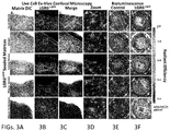
- FIG. 3A-3F present various images by different techniques of an array of different LGR6+ epithelial stem cell seeded substrates.
- FIG. 4A depicts-a time lapse in vivo healing progression of controls and an example of an LGR seeded matrix.
- FIG. 4B is a graphical expression of Cytokeratin-17 transcript expression at day ten.
- FIGS. 5A-E depict an example of a construct with LGR ESCs and stromal vascular fraction cellular isolate populations showing initial form of polarization accompanied by a graphic comparison.
- FIGS. 6A-B depict an example of a construct containing LGR cells with and without stromal vascular fraction cellular entities and the relative production of growth factors.
- FIGS. 7A-H illustrate third degree wound bed induction and verification of the elimination of the LGR stem cell follicular bulge and adnexal structures.
- FIGS. 8A-Q depict time progression of a wound/injury/void with DEFA5 as it relates to bacterial adhesion.
- FIGS. 9A and B are comparative photographs of DEFA5 expressing cellular entities within a wound bed as it relates to augmented healing, tissue and appendage regeneration and subsequent hair growth in treated burn wounds devoid of adnexal structures.
- FIGS. 10A-L illustrate the quantification of wound bed healing kinetics and LGR5 and LGR6 stem cell migration into burn tissue following treatment with topical focal.
- FIGS. 11A and B illustrate RT-PCR quantification and gene heat mapping comparison of wound/injury/tissue voids treated with DEFA5 to SDZ as it relates to augmentation of pro-healing pathways.
- FIGS. 12A-I illustrate LGR6 expression of cells of the hair follicle and fluorescent activated cell sorting of co-expressing LGR6+, CD34+ CD73+ GFP labeled cells for culture expansion.
- FIGS. 13A-D are photomicrographs by confocal microscopy and bioluminescence of a functional singularity unit (aFSU) at the time initial seeding and 1 day later.
- aFSU functional singularity unit
- FIG. 13E is a photomicrograph of a collagen scaffold.
- FIGS. 14A-E depict an example of location LGR cellular varieties as it relates to location, phenotype, interface and polarity within a cutaneous tissue. Isolation and culture of the LGR6+ ESC from the follicular bulge.
- FIGS. 15A-E provides an example of LGR expressing cellular foci as it relates to a method of delivery through placement around and/or within wound/injury/tissue void.
- FIGS. 16A-D depict an example of LGR containing stem cell focus as it relates to delivery into and around wounds via a deliverable vector and subsequent healing, regeneration of tissues and supporting structures.
- FIGS. 17A-D show LGR6+ epithelial stem cell migration and differentiation within full-thickness wound beds 10 days after transplantation.
- FIG. 18 provides an RT-PCR quantification and inset gene heat mapping comparison of a wound/injury/tissue void with the LGR expressing cellular foci.
- FIG. 19 depicts an example of said LGR expressing cellular foci as it relates to delivery into and/or around wound/injury/tissue void and augmentation of wound healing factors.
- FIGS. 20A-F illustrate an example of LGR expressing cellular foci as it relates to the regeneration of bone tissues. Isolated LGR foci can be seeded bone and remain viable.
- FIGS. 1A-C Example of flow cytometry of cell populations that exist around a hair follicle and scaffolds that such cells readily adhere to when seeded. More specifically, FIG. 1A depicts an example of location of said LGR expressing cells of cutaneous origin. Immunofluorescent confocal microscopy at 40 ⁇ magnification depicts the follicular bulge (white arrow), LGR6+ (Green), DNA (Blue). FIG. 1B is a fluorescent activated cell sorting graph with gate analysis indicating exemplary cellular markers. FIG. 1C depicts an array of cells types can be used to seed a spectrum of acellular matrices/substrates/scaffolds/materials according to the invention.
- FIG. 2A is a photographic representation of an example of a gross construct without micro-aggregate multi-cellular functional units containing LGR expressing stem cell foci in accordance with the invention.
- FIG. 2B depicts the construct following seeding of substrate with aggregates of partially digested cells.
- FIG. 3A in columnar format, is an image series by differential interference contrast (DIC) confocal microscopy of LGR seeded substrates from different sources.
- FIG. 3B is a corresponding column by immunofluorescent confocal microscopy at 20 ⁇ magnification of LGR6+ ESC seeded matrices of respective constructs containing LGR expressing cells.
- the inset white boxes represent focal zoom regions indicated in the column FIG. 3D
- FIG. 3C is a column depicting the Digital merge of the respective image of FIG. 3A (DIC) and the immunofluorescent of FIG. 3B indicating matrix contour and boundaries.
- the columns of FIGS. 3E and 3F respectively represent the bioluminescence measured in radiant efficiency of an acellular matrix control and a corresponding LGR6+ ESC seeded matrix at 72 hours post-seeding.
- FIG. 4A-E depict examples of said LGR containing construct placed into living mammalian system. Placement of an LGR6+ GFP ESC Seeded Matrix Augments Healing Hair Follicle Growth.
- FIG. 4A is a 3 ⁇ 3 matrix of photomicrographs of 3 mm full human de-cellularized dermis thickness burn wound beds at days 5, 8 and 10 containing no matrix (burn control), matrix (matrix control) and LGR6+ GFP ESC.
- FIG. 4B graphically depicts the relative expression of Cytokeratin-17 transcript expression at day 10 of the wound beds depicted in FIG. 4A . The percent wound bed healed was determined using quantification analysis of wound bed healing rates as a percent area function within the ImageJ NCBI application. Wound control contains burn wound bed only. Matrix control contains matrix only and LGR6+ GFP contains ADM seeded with LGR6+ GFP ESCs.
- FIG. 4C is a photomicrograph of in vivo bioluminescent imaging in murine full thickness burn wound beds at day 5.
- FIG. 4D are micrographs of human dermis at 100 ⁇ of the controls and LGR6+ GFP containing dermis at 12 hours and 72 hours and after seeding with ESCs. The white arrow indicates the presence of a dermal pore
- FIG. 4E provides images of the controls and the construct of the invention containing human dermis seeded with ESCs with a silicone protective overlay to prevent desiccation.
- the LGR6+ GFP matrix image includes duplicate small black arrows that indicate nascent hair patches from the full thickness Nu/Nu murine wound bed.
- FIGS. 5A-E depict an example of said construct the effect of addition of Stromal vascular fraction (SVF) to LGR6+ ESC Seeded Matrices in promoting tissue polarization and a dual compartment skin-like System.
- FIG. 5A is confocal 20 ⁇ imaging of a 5 ⁇ 10 5 RFP expressing stromal vascular fraction cellular isolate population 24 hours after being seeded on to a representative Adrenomedullin (ADM) (such as that available from Integra LifeSciences Corporation under the name INTEGRA®).
- FIG. 5B is a confocal 20 ⁇ image of a 5 ⁇ 10 5 GFP expressing LGR6 + cellular isolate population 24 hours after being seeded on to a representative ADM (INTEGRA®).
- FIG. 5C depicts confocal 20 ⁇ imaging of a dual seeded representative ADM (INTEGRA®) with 5 ⁇ 10 5 RFP expressing SVF and 5 ⁇ 10 5 GFP expressing LGR6 + isolate populations 24 hours after being co-seeded in culture.
- FIG. 5D is of a co-seeded matrix containing 5 ⁇ 10 5 RFP expressing SVF RFP and 5 ⁇ 10 5 GFP expressing LGR6 + following 5 days of growth in culture.
- the dotted parallel lines indicate epithelial LGR6 +GFP lineage accumulating at the edge of the ADM substrate.
- the small bracket and large bracket indicate the relative locations of the two compartments in correlation with LGR6 +GFP and SVF RFP abundance.
- FIG. 5E is a graphical representation of the proliferation kinetics of a collagen substrate co-seeded with green LGR expressing cells and red SVF expressing cells.
- FIGS. 6A and 6B depict an example of a construct containing LGR cells with and without supportive cellular entities and the relative production of growth factors.
- FIG. 6A graphs relative fold transcript expression ( ⁇ CT) of indicated gene element from total RNA: LGR6 +GFP ESC (black bar), SVF RFP (grey bar), and co-cultured LGR6 +GFP ESC+SVF RFP (white box) on respective scaffold substrate.
- ⁇ CT relative fold transcript expression
- LGR6+SVF indicates the inter-comparison co-cultured LGR6 +GFP ESC+SVF RFP expression vs. singular LGR6 +GFP ESC and SVF RFP expression on indicated scaffolding.
- Average FGF-2 gene expression for co-cultured matrices was higher than the average expression of both singular systems (Scaffold+ LGR6 or scaffold +SVF) except for co-cultured INTEGRA® (INTEGRA®+ LGR6 + SVF).
- Significance below the x-axis (LGR6) or (SVF) indicates the intra-comparison of substrates, while the cellular entity remains constant.
- FIG. 6B graphically represents the relative densitometric unit (RDU) of indicated protein analyte from total protein isolates: LGR6 +GFP ESC (black bar), SVF RFP (grey bar), and co-cultured LGR6 +GFP ESC+SVF RFP (white box) on respective scaffold substrate.
- RDU relative densitometric unit
- FIGS. 7A-H illustrate a wound/injury/void receiving therapy example of enhanced LGR cell migration, proliferation and viability into a wound namely a third degree wound bed induction and verification of the elimination of the LGR stem cell follicular bulge and adnexal structures.
- FIG. 7A depicts a wound bed template marks of 3 mm diameter.
- FIG. 7B depicts the wound bed structure at day 0 (the white scale bar being 1 mm).
- FIG. 7C illustrates an example of a 2 ⁇ 3 3 mm wound bed grid.
- FIG. 7D shows topical application of the re-suspended peptide at the wound site.
- FIG. 7E is a photomicrograph of H&E stain of non-burned, intact Integument/skin with hair follicle and adnexal structures. The arrow indicates the location of the magnified follicle (inset image) where the white scale bar is 500 ⁇ m.
- FIG. 7F is an H&E stain of dorsal murine skin following high temperature cautery depicting removal of epidermal, dermal and hypodermal tissues including the follicular bulge.
- FIG. 7G is DAPI/DNA stain (4′,6-diamidino-2-phenylindole) of non-burned, intact skin with hair follicle and adnexal structures.
- FIG. 7H DAPI/DNA stain of dorsal murine skin following high temperature cautery depicting removal of epidermal, dermal, and hypodermal tissues including the follicular bulge where the white scale bar is 100 ⁇ m.
- FIGS. 8A-Q depict a wound/injury/void with LGR as it relates to antimicrobial behavior over five and ten day time periods.
- 16S rRNA fluorescent oligonucleotide probes in-situ hybridization indicates the presence of bacterial adhesion at the third degree burn wound bed.
- FIG. 8A presents DNA/DAPI labeling of a 3rd degree burn wound bed at day five post burn induction treated daily with SDZ.
- FIG. 8B 5′-Cy3-EUB338 labeled 16s rRNA of 3rd degree burn wound bed bacterial organisms (yellow grains) at day five post burn induction treated daily with SDZ are depicted.
- FIG. 8C is a digitally merged image of FIGS.
- FIG. 8D corresponds to FIG. 8A except at day ten with DNA/DAPI labeling of 3rd degree burn wound bed treated daily with SDZ.
- FIG. 8E is a photomicrograph of the 5′-Cy3-EUB338 labeled 16s rRNA of 3rd degree burn wound bed bacterial organisms (yellow grains) at day ten post burn induction treated daily with SDZ.
- FIG. 8F is a merged image of FIGS. 8D and E.
- FIGS. 8G-8L are images corresponding respectively to the five and ten post burn periods of FIGS. 8A-F but subject to daily treatment using Defensin, alpha 5 (DEFA5) rather than SDZ.
- the arrow in H represents the interface of tissue with overlying fibrinous material where less bacteria is observed in the setting of DEFA5 treatment.
- FIG. 8M with inset 8N demonstrate quantification of white pixel intensity of Cy3 fluorescence grayscale converted image of a wound bed treated with SDZ and containing more 16s rRNA labeling per unit area.
- FIG. 8O and inset 8P correspondingly show quantification of white pixel intensity of Cy3 fluorescence grayscale converted image of (inset image p.) a wound bed treated with DEFA5 and containing a reduced 16s rRNA labeling per unit area.
- the inset graph depicts averaged white pixel intensity of 16s rRNA expressed in both SDZ and DEFA5 treated burn wound beds at day five using grayscale imaging software.
- FIG. 8M with inset 8N demonstrate quantification of white pixel intensity of Cy3 fluorescence grayscale converted image of a wound bed treated with SDZ and containing more 16s rRNA labeling per unit area.
- FIG. 8O and inset 8P correspondingly show quantification of white pixel intensity of Cy3 fluorescence grayscale converted image of (inset image p.)
- FIG. 8Q is a graph to illustrate averaged red channel fluorescence of 16s rRNA expressed in both SDZ and DEFA5 treated burn wound beds at day five.
- the white arrow in FIG. 8H indicates potential film in DEFA5 treated wound beds and the black arrow in FIG. 8M indicates white pixel intensity.
- (*) indicates p-value ⁇ 0.05.
- FIGS. 9A and B are a series of time progression photographs that represents an example of LGR expressing cellular entities within wound as it relates to augmented healing, tissue and appendage regeneration and subsequent hair growth, wound healing kinetics and nascent hair growth in treated burn wounds devoid of adnexal structures.
- the photographic series comprising FIG. 9A are gross imaging using a Leica Wild M680 surgical microscope to image healing of 3rd degree burn wound beds over 10 days while being treated with indicated agents MQH2O, DEFA5, DEFB1, SDZ.
- the white scale bar represents 1 mm.
- 9B again comprises gross imaging using a Leica Wild M680 to track nascent hair growth of 3rd degree burn wound beds over 16 days in a side by side comparison of DEFA5 vs. control treated wound beds.
- the white arrows indicate the growth of new hair.
- the scale bar is 1 mm.
- FIGS. 10A-L comprise an example of said LGR expressing cellular entities within wound/injury/tissue void as it relates to augmented healing, propagation of said entities.
- the Graphs comprising FIGS. 10K and 10L provide evidence of quantification of wound bed healing kinetics and LGR5 and LGR6 stem cell migration into burn tissue following treatment with topical focal agents. Briefly, these tests were used to confirm the quantitative confocal microscopic intensity patterns from imaging LGR5 and LGR6, and based on reverse-transcriptase polymerase chain reaction on burn wound tissues.
- FIG. 10A presents photographs of a wound area with a white scale bar representing 1 mm and the wound area calculation in black.
- FIG. 10B graphically displays the averaged wound healing rate expressed as percent % of wound area remaining over 10 day period of indicated topical focal agent application.
- the asterisk (*) represents a p-value ⁇ 0.05.
- FIGS. 10C-J are LGR5 and LGR6 immunofluorescent antibody labeling of a DEFA5 treated wound bed at day 5 where FIG. 10C is DNA/DAPI/Blue, FIG. 10D is LGR5/FITC/Green FIG. 10E is LGR6/TRITC/Red and FIG. 10F is a merger of 10 C- 10 E.
- FIGS. 10C-J are LGR5 and LGR6 immunofluorescent antibody labeling of a DEFA5 treated wound bed at day 5 where FIG. 10C is DNA/DAPI/Blue, FIG. 10D is LGR5/FITC/Green FIG. 10E is LGR6/TRITC/Red and
- FIG. 10G-I are corresponding LGR5 and LGR6 immunofluorescent antibody labeling of SDZ (sulfadiazine) treated wound bed at day 5 (DNA/DAPI/Blue, LGR5/FITC/Green and LGR6/TRITC/Red).
- FIG. 10J is a merged image of 10 G- 10 I and includes an inset representing averaged LGR5 and LGR6 expression using Green and Red fluorescent intensity per wound bed at day 5.
- the comparative values obtained from Reverse Transcriptase PCR quantification of the fold increase in RNA extracted from replicate wound beds treated with DEFA5 and SDZ is set out.
- the white scale bar 50 ⁇ m and again, the asterisk (*) represents a p-value ⁇ 0.05.
- FIGS. 11A and B illustrate a wound/injury/tissue void with the LGR expressing cellular entities placed within wound as it relates to augmentation of pro-healing pathways.
- the figures respectively represent RT-PCR quantification and gene heat mapping comparison of wound beds treated with DEFA5 to SDZ. These figures show the role of human alpha defensin 5 versus sulfadiazine in augmenting key transcript expression within the wound. The results show that several gene subsets are significantly up-regulated within the wound beds receiving human alpha defensin 5 when compared with sulfadiazine therapy and that certain Wnt pathway gene subsets are significantly up-regulated in response of the LGR stem cell system to Wnt ligands in both the gut and skin.
- FIG. 11A presents an Averaged Wound Healing RT2-PCR Array pathway heat map and corresponding gene map with fold regulation for wound beds comparing DEFA5 to SDZ treated systems.
- FIG. 11B presents an Averaged Wnt RT2-PCR Array healing pathway heat map and corresponding gene map with fold regulation for wound beds comparing DEFA5 to SDZ treated systems. The colors of the heat maps are indicated as red, more expressed in DEFA5 treated burns to green more expressed in SDZ treated burns.
- FIGS. 12A-I represent an example of a micro-aggregate multicellular unit containing LGR expressing stem cell foci as it relates to location, population identity and wound healing capacity.
- LGR6+, CD34+, and CD73+C57BL/6(UBC-GFP) murine cells were isolated for cell culture expansion.
- FIG. 12A depicts LGR6 fluorescent antibody (green) expression of cells on the hair follicle following partial epidermal 10 unit/ ⁇ L dispase digestion. (Worthington Biochemical Corp., Lakewood, N.J.) digestion for 30 minutes at 37° C. on a slow rocker.
- FIG. 12B is of LGR6+ cells expressing additional CD34 and CD73 markers (the arrow indicates population isolated comprising approximately 1 to 3 percent of all cells).
- FIGS. 12C-H are eFluor450 expression histograms of an in vitro wound assay respectively showing periodic intrinsic GFP expression from C57BL/6(UBC-GFP) murine cells, CD34+PE/Cy7 expression, LGR6+ APC expression and CD73+.
- the graph of FIG. 121 sets out the averaged reduction in the distance line over time expressed as a percentage of initial distance following fluorescence sorting where the asterisk (*) represents a p-value ⁇ 0.05.
- FIGS. 13A-D are photomicrographs by confocal microscopy and bioluminescence of an activated functional singularity unit (aFSU) at the time initial seeding and 1 day later showing an example of a micro-aggregate multicellular unit containing LGR expressing stem cell foci while undergoing initial propagation on a collagen matrix, FIG. 13 E.
- aFSU activated functional singularity unit
- FIGS. 14A-E depict an example of location LGR cellular varieties as it relates to location, phenotype, interface and polarity within a cutaneous tissue.
- FIG. 14A shows by Immunofluorescence staining, localized regions of LGR6 (Green/fluorescein isothiocyanate (FITC)) and LGR5 (Red/tetramethyl rhodamine isothiocyanate (TRITC)) expression.
- the scale bar is for 20 ⁇ m.
- FIG. 14B shows fluorescence-activated cell sorting isolation of the LGR6+ GFP epithelial stem cells from C57BL/6(UBCGFP) murine skin with the final sort gate using LGR6+, CD34 and CD73 on the left and individual histograms depicting cellular GFP expression and correlating antibody-conjugate labels: CD73/PE-7, LGR6/Cy5, CD34/eFlour450 on the right.
- FIG. 14C shows differential interference contrast image of LGR6 +GFP epithelial stem cells plated following fluorescence-activated cell sorting isolation.
- FIG. 14D depicts intrinsic GFP expression of the LGR6 +GFP epithelial stem cells and
- FIG. 14E is a merged image of FIGS. 14C and 14 D.
- the scale bar represents 20 ⁇ m.
- FIGS. 15A-E provide an example of LGR expressing cellular foci as it relates to a method of delivery through placement around and/or within wound/injury/tissue void.
- the three images of FIG. 15A depict, respectively, an initial burn template; a full thickness burn on the dorsum on Nu/Nu mouse; and delivery of HYDROGEL® containing 10 5 LGR6 +GFP epithelial stem cells at the base of the wound bed.
- the scale bar for FIG. 15A is 1 mm.
- FIG. 15B is an immunofluorescece image of the injection pocket DNA/DAPI-BLUE at Day 0
- FIG. 15B is an immunofluorescece image of anti-LGR6/TRITC antibody labeling and FIG.
- FIG. 15C is the same for LGR6 +GFP epithelial stem cells.
- FIG. 15 E is a merged image of FIGS. 15B-D and has a scale bar of 20 ⁇ m.
- FIGS. 15A-E show full thickness burn wound bed induction and validation of LGR6+ stem cell engraftment into subsequent soft tissue defect.
- FIGS. 16A-D depict an example of LGR containing stem cell focus as it relates to delivery into and around wounds via a deliverable vector and subsequent healing, regeneration of tissues and supporting structures including but not limited blood vessel angiogenesis and/or angiogenesis.
- Wound healing progression following LGR6+ epithelial stem cells transplantation into full thickness wounds The progression of wound healing is depicted following the injection of HYDROGEL® from BD Biosciences, San Jose, Calif. (control) in FIG. 16A compared with FIG. 16B , LGR6 +GFP epithelial stem cells seeded HYDROGEL® over 15 days.
- the scale bar is 1 mm.
- the white arrow indicates presence of a remaining LGR6 +GFP epithelial stem cells population located within healing wound bed.
- the black arrow indicates the location of the burn wound base free of LGR6 +GFP epithelial stem cells.
- FIGS. 17A-D depicts an example of LGR containing stem cell focus following delivery into and/or around wound with subsequent healing and regeneration of tissues and related appendages such as but not limited to hair follicle and related supportive structures.
- FIG. 17A is a four panel matrix of confocal images of immunofluorescent labeled tissue specimen at day 10 following transplantation of LGR6+ epithelial stem cells migration into the wound bed 10 days.
- the images comprising FIG. 17A include DNA/DAPI-BLUE; anti-LGR6/TRITC; GFP expression of LGR6 +GFP ESC.
- FIG. 17B is a differential interference contrast image merge of all channels.
- the Red arrow designates regions of nascent follicle development. (See also the upper inset image).
- the dotted line shows epithelial polarization overlying nascent hair follicles while the white arrow indicates the location of the graft injection pocket (See also the magnification thereof in the lower inset image for an image of the initial injection pocket cellular population.
- the inset graph of FIG. 17B represents comparative KRT17/cytokeratin 17 gene expression within the indicated wound beds of the control and LGR6+ +GFP treatment.
- the three images are of a Transplant dome used to cover hair follicle study population burn wound beds, an LGR6+ +GFP ESC treated wound bed at day 10 (solid arrow) with nascent hair follicles (clear arrow) follicle cyst formation and a control wound bed at day 10.
- the graph comprising FIG. 17D quantifies the Day 10 wound bed resulting from RT-PCR indicating relative gene fold expression of WNT ligands.
- FIG. 18 provides an RT-PCR quantification and inset gene heat mapping comparison of a wound/injury/tissue void with the LGR expressing cellular foci as it relates to delivery into and/or around wound/injury/tissue void as it relates to augmentation of pro-healing pathways and comparative gene expression of wounds receiving LGR6+ epithelial stem cells against a control.
- the graphs illustrate the relative fold expression of genes for angiogenesis, wound healing and epidermal growth factor. Correlative graphical representation of data comparing wound beds receiving LGR6+ epithelial stem cells and control therapy.
- the inset heat maps the color red indicates greater expression within the LGR6+ epithelial stem cell wound bed while the color green indicates greater expression within the control wound bed.
- FIG. 19 graphically presents the relative protein densitometry of an example of LGR expressing cellular foci as it relates to delivery into and/or around wound/injury/tissue void and augmentation of wound healing factors.
- Comparative angiogenesis analyte expression of wounds receiving LGR6+ ESCs
- Proteomic array comparing common proteins which regulate and augmented angiogenesis.
- the grey columns indicate control wounds and the black columns indicated those wounds that received the LGR6 +GFP ESC.
- the inset image shows example proteome array membranes following development with HRP chemi-luminesce. Brighter colors indicate higher levels of protein expression.
- FIGS. 20A-F illustrate an example of LGR expressing cellular foci as it relates to the regeneration of bone tissues. Isolated LGR foci can be seeded bone and remain viable.
- FIG. 20A is a gross bone image of harvested bone for culture.
- FIG. 20B is a DIC image of bone containing LGR GFP 7 days following seeding.
- FIG. 20C is a 488 nm Green laser confocal image of bone containing LGR6 +GFP 7 days following seeding. It is notable that the LGR foci can undergo osteo-induction in-vitro.
- FIG. 20D depicts LGR foci following 1 week of osteo-induction with supplemental media.
- FIG. 20E is an Alizarin red stain of the LGR foci following osteo-induction which can undergo osteo-induction in-vitro and up regulate key osteogenic genes.
- FIG. 20F is RT-PCR data showing relative fold gene expression where the grey columns represent (control) non-osteo induced LGR and the black columns represent those LGR which received osteo-induction media following 7 days of culture. GAPDH was used as reference standard housekeeping gene.
- a gelatinous support such as an exemplary three dimensional collagen scaffold can be generated by well-known processes as follows:
- Pulse Rescue Media PRM
- the PRM in this embodiment which is direct to humans, is a cell sustaining, serum-free, media mixture Keratinocyte-SFM containing L-glutamine supplied with separately packaged prequalified human recombinant Epidermal Growth Factor 1-53 (EGF 1-53) and Bovine Pituitary Extract (BPE) sold as Keratinocyte-SFM (1 ⁇ ) from Thermo Fisher Scientific to which the antibiotic-antimycotic agents penicillin, streptomycin, and amphotericin B are added along with a GMP ⁇ fibrinogen: human.
- EGF 1-53 Epidermal Growth Factor 1-53
- BPE Bovine Pituitary Extract
- the agent used in one embodiment is GIBCO® Antibiotic-Antimycotic from Thermo Fisher Scientific, a solution containing 10,000 units/mL of penicillin, 10,000 ⁇ g/mL of streptomycin, and 25 ⁇ g/mL of FUNGIZONE® Antimycotic.
- the supplemental reagents are utilized to stabilize the primary tissues and reduce the viability of micro-organisms during transport and processing.
- the following relates specifically to the generation and preservation of LGR expressing epithelial containing stem cell micro-aggregate functional units in accordance with an embodiment of the invention.
- Example 1 concerns a method for extraction of minimally polarized functional units in accordance with an embodiment of the invention. After obtaining a specimen, it is removed from its associated transport container followed by:
- the following relates to secondary processing where the primary cultures are established and functional tissue elements are prepared utilizing enzymatic preparation using conventional CLIA equipment and reagents meeting FDA and/or GMP certification:
- Example 2 is directed to processing of hypodermis and subdermal fat cellular components. Example 2 recites the following steps:
- Example 3 is directed to addition of hypodermis and subdermal fat components to the example of a construct according to an embodiment of the invention.
- the illustrative component addition example involves:
- Example 4 concerns enrichment of the minimally polarized, epithelial stem cell singularity units.
- Example 2 Following Example 1, the MPFUs is placed in pulse rescue media in a 15 ml conical tube and spin/centrifuged into a soft pellet. The material is then subject to the following process of partial digestion:
- Example 5 involves adding the epithelial stem cell functional singularities (ESC aFSUs) obtained from Example 4 to a construct/scaffold.
- the procedure entails:
- Example 6 represents illustrative protocols for quality assurance and construct finalization involving cryopreservation which entails preparation the construct for shipment following defined good manufacturing processes (GMP) for cell therapy applications and include:
- the invention relates to methods for making and methods for using constructs of micro-aggregate multicellular grafts containing isolated Leucine-rich repeat-containing G-protein coupled Receptor (LGR) expressing cells for the delivery, application, transplantation, implantation, directed seeding, directed migration, directed tracking, in setting, laminating and/or injection of the cellular element generating, regenerating, enhancing and/or healing epithelial systems, glands, hair, nerves, bone, muscle, fat, tendons, blood vessels, fascia, ocular tissues and peptide secreting cellular elements for use in wound therapy applications, tissue engineering, cell therapy applications, regenerative medicine applications, medical/therapeutic applications, tissue healing applications, immune therapy applications, and tissue transplant therapy applications.
- LGR Leucine-rich repeat-containing G-protein coupled Receptor
Landscapes
- Health & Medical Sciences (AREA)
- Life Sciences & Earth Sciences (AREA)
- Engineering & Computer Science (AREA)
- Chemical & Material Sciences (AREA)
- Biomedical Technology (AREA)
- General Health & Medical Sciences (AREA)
- Zoology (AREA)
- Medicinal Chemistry (AREA)
- Wood Science & Technology (AREA)
- Dermatology (AREA)
- Veterinary Medicine (AREA)
- Public Health (AREA)
- Animal Behavior & Ethology (AREA)
- Organic Chemistry (AREA)
- Bioinformatics & Cheminformatics (AREA)
- Cell Biology (AREA)
- Chemical Kinetics & Catalysis (AREA)
- Oral & Maxillofacial Surgery (AREA)
- Epidemiology (AREA)
- Transplantation (AREA)
- Biotechnology (AREA)
- Genetics & Genomics (AREA)
- Environmental Sciences (AREA)
- Dentistry (AREA)
- Botany (AREA)
- Urology & Nephrology (AREA)
- Molecular Biology (AREA)
- Pharmacology & Pharmacy (AREA)
- General Chemical & Material Sciences (AREA)
- Nuclear Medicine, Radiotherapy & Molecular Imaging (AREA)
- Microbiology (AREA)
- General Engineering & Computer Science (AREA)
- Biochemistry (AREA)
- Endocrinology (AREA)
- Diabetes (AREA)
- Materials For Medical Uses (AREA)
- Medicines Containing Material From Animals Or Micro-Organisms (AREA)
- Peptides Or Proteins (AREA)
- Micro-Organisms Or Cultivation Processes Thereof (AREA)
- Developmental Biology & Embryology (AREA)
Priority Applications (42)
| Application Number | Priority Date | Filing Date | Title |
|---|---|---|---|
| US14/954,335 US10926001B2 (en) | 2014-12-02 | 2015-11-30 | Methods related to minimally polarized functional units |
| SG11201704502RA SG11201704502RA (en) | 2014-12-02 | 2015-12-01 | Methods for development and use of minimally polarized function cell micro-aggregate units in tissue applications using lgr4, lgr5 and lgr6 expressing epithelial stem cells |
| CN201580075326.3A CN107250348B (zh) | 2014-12-02 | 2015-12-01 | 使用表达lgr4、lgr5和lgr6的上皮干细胞在组织应用中开发和使用最小极性化功能细胞微聚集体单元的方法 |
| GB201902819A GB2569056B (en) | 2014-12-02 | 2015-12-01 | Methods for development and use of minimally polarized functional units in tissue applications using LGR4, LGR5 and LGR6 expressing epithelial stem cells |
| KR1020187036619A KR20180136588A (ko) | 2014-12-02 | 2015-12-01 | Lgr4, lgr5 및 lgr6 발현 상피 줄기 세포를 사용한 조직 응용에서의 최소 극성화 기능 세포 미소응집체 유닛의 개발 및 사용 방법 |
| NZ733433A NZ733433A (en) | 2014-12-02 | 2015-12-01 | Methods for development and use of minimally polarized function cell micro-aggregate units in tissue applications using lgr4, lgr5 and lgr6 expressing epithelial stem cells |
| JP2017530217A JP6791854B2 (ja) | 2014-12-02 | 2015-12-01 | 組織適用におけるlgr4、lgr5およびlgr6発現上皮幹細胞を用いた最小限分極機能性細胞微細凝集塊単位の開発および使用のための方法 |
| MYPI2017701990A MY184931A (en) | 2014-12-02 | 2015-12-01 | Methods for development and use of minimally polarized functional units in tissue applications using lgr4, lgr5 and lgr6 expressing epithelial stem cells |
| SG10201914058QA SG10201914058QA (en) | 2014-12-02 | 2015-12-01 | Methods for development and use of minimally polarized function cell micro-aggregate units in tissue applications using lgr4, lgr5 and lgr6 expressing epithelial stem cells |
| EA201791125A EA201791125A1 (ru) | 2014-12-02 | 2015-12-01 | Способы разработки и применения клеточных микроагрегатных элементов с минимально поляризованной функцией в тканевых приложениях с использованием эпителиальных стволовых клеток, экспрессирующих lgr4, lgr5 и lgr6 |
| HK18104679.0A HK1245327B (en) | 2014-12-02 | 2015-12-01 | Methods for development and use of minimally polarized function cell micro-aggregate units in tissue applications using lgr4, lgr5 and lgr6 expressing epithelial stem cells |
| CA2969707A CA2969707C (en) | 2014-12-02 | 2015-12-01 | Methods for development and use of minimally polarized function cell micro-aggregate units in tissue applications using lgr4, lgr5 and lgr6 expressing epithelial stem cells |
| CR20170296A CR20170296A (es) | 2014-12-02 | 2015-12-01 | Métodos para el desarrollo y uso de unidades de micro-agregados de células funcionales mínimamente polarizadas en aplicaciones de tejidos utilizando células madre epiteliales que expresan lgr4, lgr5 y lgr6 |
| GB1710646.9A GB2549872A (en) | 2014-12-02 | 2015-12-01 | Methods for development and use of minimally polarized function cell micro-aggregate units in tissue applications using LGR4, LGR5 and LGR6 |
| BR112017011808A BR112017011808A2 (pt) | 2014-12-02 | 2015-12-01 | métodos para o desenvolvimento e uso de unidades micro-agregadas de células funcionais minimamente polarizadas em aplicações de tecidos usando lgr4, lgr5 e lgr6 que expressam células tronco epiteliais |
| NZ755260A NZ755260A (en) | 2014-12-02 | 2015-12-01 | Methods for development and use of minimally polarized function cell micro-aggregate units in tissue applications using lgr4, lgr5 and lgr6 expressing epithelial stem cells |
| EP24181160.3A EP4438128A3 (en) | 2014-12-02 | 2015-12-01 | Methods for development and use of minimally polarized function cell micro-aggregate units in tissue applications using lgr4, lgr5 and lgr6 expressing epithelial stem cells |
| PCT/US2015/063114 WO2016089825A1 (en) | 2014-12-02 | 2015-12-01 | Methods for development and use of minimally polarized function cell micro-aggregate units in tissue applications using lgr4, lgr5 and lgr6 expressing epithelial stem cells |
| KR1020187036618A KR20180136587A (ko) | 2014-12-02 | 2015-12-01 | Lgr4, lgr5 및 lgr6 발현 상피 줄기 세포를 사용한 조직 응용에서의 최소 극성화 기능 세포 미소응집체 유닛의 개발 및 사용 방법 |
| EP15865131.5A EP3227431B1 (en) | 2014-12-02 | 2015-12-01 | Methods for development and use of minimally polarized function cell micro-aggregate units in tissue applications using lgr4, lgr5 and lgr6 expressing epithelial stem cells |
| KR1020217010934A KR20210043024A (ko) | 2014-12-02 | 2015-12-01 | Lgr4, lgr5 및 lgr6 발현 상피 줄기 세포를 사용한 조직 응용에서의 최소 극성화 기능 세포 미소응집체 유닛의 개발 및 사용 방법 |
| MX2017007243A MX2017007243A (es) | 2014-12-02 | 2015-12-01 | Metodos para desarrollo y uso de unidades micro-agregadas de celulas funcionales polarizadas minimamente en aplicaciones en tejidos utilizando celulas troncales epiteliales que expresan lgr4, lgr5 y lgr6. |
| AU2015355187A AU2015355187C1 (en) | 2014-12-02 | 2015-12-01 | Methods for development and use of minimally polarized function cell micro-aggregate units in tissue applications using LGR4, LGR5 and LGR6 expressing epithelial stem cells |
| KR1020197028100A KR20190111167A (ko) | 2014-12-02 | 2015-12-01 | Lgr4, lgr5 및 lgr6 발현 상피 줄기 세포를 사용한 조직 응용에서의 최소 극성화 기능 세포 미소응집체 유닛의 개발 및 사용 방법 |
| HK18104680.7A HK1250515A1 (zh) | 2014-12-02 | 2015-12-01 | 使用lgr4,lgr5和lgr6在组织应用中开发和使用最小极化功能细胞微集合体单元的方法 |
| KR1020177017887A KR20170098844A (ko) | 2014-12-02 | 2015-12-01 | Lgr4, lgr5 및 lgr6 발현 상피 줄기 세포를 사용한 조직 응용에서의 최소 극성화 기능 세포 미소응집체 유닛의 개발 및 사용 방법 |
| PH12017501009A PH12017501009B1 (en) | 2014-12-02 | 2017-05-31 | Methods for development and use of minimally polarized function cell micro-aggregate units in tissue applications using lgr4, lgr5 and lgr6 expressing epithelial stem cells |
| IL252613A IL252613B (en) | 2014-12-02 | 2017-06-01 | Methods for the development and use of minimally polarized microaggregate cell units in tissue applications using lgr4, lgr5 and lgr6 expressing epithelial stem cells |
| MX2023001655A MX2023001655A (es) | 2014-12-02 | 2017-06-05 | Composiciones que comprenden celulas troncales que expresan lgr y usos de las mismas. |
| ZA2017/03907A ZA201703907B (en) | 2014-12-02 | 2017-06-07 | Methods for development and use of minimally polarized function cell micro-aggregate units in tissue applications using lgr4, lgr5 and lgr6 expressing epithelial stem cells |
| CONC2017/0006640A CO2017006640A2 (es) | 2014-12-02 | 2017-06-30 | Metodos para el desarrollo y uso de unidades de micro-agregado de funcion celular polarizada en aplicaciones de tejido usando lgr4, lgr5 y lgr 6 que expresan celulas madre epiteliales |
| US15/650,659 US11266765B2 (en) | 2014-12-02 | 2017-07-14 | Methods related to minimally polarized functional units |
| US15/650,656 US20180154043A1 (en) | 2014-12-02 | 2017-07-14 | Methods for Development and Use of Minimally Polarized Function Cell Micro-Aggregate Units in Tissue Applications Using LGR4, LGR5, and LGR6 Expressing Epithelial Stem Cells |
| US16/165,169 US11000629B2 (en) | 2014-12-02 | 2018-10-19 | Methods related to minimally polarized functional units |
| JP2019004921A JP2019088299A (ja) | 2014-12-02 | 2019-01-16 | 組織適用におけるlgr4、lgr5およびlgr6発現上皮幹細胞を用いた最小限極性機能性細胞微細凝集塊単位の開発および使用のための方法 |
| AU2019240603A AU2019240603B2 (en) | 2014-12-02 | 2019-10-02 | Methods for development and use of minimally polarized function cell micro-aggregate units in tissue applications using LGR4, LGR5 and LGR6 expressing epithelial stem cells |
| JP2020129610A JP2020182880A (ja) | 2014-12-02 | 2020-07-30 | 組織適用におけるlgr4、lgr5およびlgr6発現上皮幹細胞を用いた最小限極性機能性細胞微細凝集塊単位の開発および使用のための方法 |
| US17/326,734 US11338060B2 (en) | 2014-12-02 | 2021-05-21 | Methods for development and use of minimally polarized function cell micro-aggregate units in tissue applications using LGR4, LGR5 and LGR6 expressing epithelial stem cells |
| IL287488A IL287488B2 (en) | 2014-12-02 | 2021-10-21 | Methods for the development and use of minimally polarized microaggregate cell units in tissue applications using lgr4, lgr5 and lgr6 expressing epithelial stem cells |
| US17/723,748 US11596714B2 (en) | 2014-12-02 | 2022-04-19 | Methods for development and use of minimally polarized function cell micro-aggregate units in tissue applications using LGR4, LGR5 and LGR6 expressing epithelial stem cells |
| US18/164,308 US12465669B2 (en) | 2014-12-02 | 2023-02-03 | Methods for development and use of minimally polarized function cell micro-aggregate units in tissue applications using LGR4, LGR5, and LGR6 expressing epithelial stem cells |
| AU2023202857A AU2023202857A1 (en) | 2014-12-02 | 2023-05-08 | Methods for development and use of minimally polarized function cell micro-aggregate units in tissue applications using LGR4, LGR5 and LGR6 expressing epithelial stem cells |
Applications Claiming Priority (2)
| Application Number | Priority Date | Filing Date | Title |
|---|---|---|---|
| US201462086526P | 2014-12-02 | 2014-12-02 | |
| US14/954,335 US10926001B2 (en) | 2014-12-02 | 2015-11-30 | Methods related to minimally polarized functional units |
Related Child Applications (3)
| Application Number | Title | Priority Date | Filing Date |
|---|---|---|---|
| US15/650,659 Division US11266765B2 (en) | 2014-12-02 | 2017-07-14 | Methods related to minimally polarized functional units |
| US15/650,656 Division US20180154043A1 (en) | 2014-12-02 | 2017-07-14 | Methods for Development and Use of Minimally Polarized Function Cell Micro-Aggregate Units in Tissue Applications Using LGR4, LGR5, and LGR6 Expressing Epithelial Stem Cells |
| US16/165,169 Division US11000629B2 (en) | 2014-12-02 | 2018-10-19 | Methods related to minimally polarized functional units |
Publications (2)
| Publication Number | Publication Date |
|---|---|
| US20160151540A1 US20160151540A1 (en) | 2016-06-02 |
| US10926001B2 true US10926001B2 (en) | 2021-02-23 |
Family
ID=56078499
Family Applications (7)
| Application Number | Title | Priority Date | Filing Date |
|---|---|---|---|
| US14/954,335 Active US10926001B2 (en) | 2014-12-02 | 2015-11-30 | Methods related to minimally polarized functional units |
| US15/650,656 Abandoned US20180154043A1 (en) | 2014-12-02 | 2017-07-14 | Methods for Development and Use of Minimally Polarized Function Cell Micro-Aggregate Units in Tissue Applications Using LGR4, LGR5, and LGR6 Expressing Epithelial Stem Cells |
| US15/650,659 Active US11266765B2 (en) | 2014-12-02 | 2017-07-14 | Methods related to minimally polarized functional units |
| US16/165,169 Active US11000629B2 (en) | 2014-12-02 | 2018-10-19 | Methods related to minimally polarized functional units |
| US17/326,734 Active US11338060B2 (en) | 2014-12-02 | 2021-05-21 | Methods for development and use of minimally polarized function cell micro-aggregate units in tissue applications using LGR4, LGR5 and LGR6 expressing epithelial stem cells |
| US17/723,748 Active US11596714B2 (en) | 2014-12-02 | 2022-04-19 | Methods for development and use of minimally polarized function cell micro-aggregate units in tissue applications using LGR4, LGR5 and LGR6 expressing epithelial stem cells |
| US18/164,308 Active 2035-12-26 US12465669B2 (en) | 2014-12-02 | 2023-02-03 | Methods for development and use of minimally polarized function cell micro-aggregate units in tissue applications using LGR4, LGR5, and LGR6 expressing epithelial stem cells |
Family Applications After (6)
| Application Number | Title | Priority Date | Filing Date |
|---|---|---|---|
| US15/650,656 Abandoned US20180154043A1 (en) | 2014-12-02 | 2017-07-14 | Methods for Development and Use of Minimally Polarized Function Cell Micro-Aggregate Units in Tissue Applications Using LGR4, LGR5, and LGR6 Expressing Epithelial Stem Cells |
| US15/650,659 Active US11266765B2 (en) | 2014-12-02 | 2017-07-14 | Methods related to minimally polarized functional units |
| US16/165,169 Active US11000629B2 (en) | 2014-12-02 | 2018-10-19 | Methods related to minimally polarized functional units |
| US17/326,734 Active US11338060B2 (en) | 2014-12-02 | 2021-05-21 | Methods for development and use of minimally polarized function cell micro-aggregate units in tissue applications using LGR4, LGR5 and LGR6 expressing epithelial stem cells |
| US17/723,748 Active US11596714B2 (en) | 2014-12-02 | 2022-04-19 | Methods for development and use of minimally polarized function cell micro-aggregate units in tissue applications using LGR4, LGR5 and LGR6 expressing epithelial stem cells |
| US18/164,308 Active 2035-12-26 US12465669B2 (en) | 2014-12-02 | 2023-02-03 | Methods for development and use of minimally polarized function cell micro-aggregate units in tissue applications using LGR4, LGR5, and LGR6 expressing epithelial stem cells |
Country Status (21)
| Country | Link |
|---|---|
| US (7) | US10926001B2 (enExample) |
| EP (2) | EP3227431B1 (enExample) |
| JP (3) | JP6791854B2 (enExample) |
| KR (5) | KR20210043024A (enExample) |
| CN (1) | CN107250348B (enExample) |
| AU (3) | AU2015355187C1 (enExample) |
| BR (1) | BR112017011808A2 (enExample) |
| CA (1) | CA2969707C (enExample) |
| CO (1) | CO2017006640A2 (enExample) |
| CR (1) | CR20170296A (enExample) |
| EA (1) | EA201791125A1 (enExample) |
| GB (2) | GB2549872A (enExample) |
| HK (1) | HK1250515A1 (enExample) |
| IL (2) | IL252613B (enExample) |
| MX (2) | MX2017007243A (enExample) |
| MY (1) | MY184931A (enExample) |
| NZ (2) | NZ755260A (enExample) |
| PH (1) | PH12017501009B1 (enExample) |
| SG (2) | SG10201914058QA (enExample) |
| WO (1) | WO2016089825A1 (enExample) |
| ZA (1) | ZA201703907B (enExample) |
Families Citing this family (6)
| Publication number | Priority date | Publication date | Assignee | Title |
|---|---|---|---|---|
| EP3595440A2 (en) * | 2017-03-14 | 2020-01-22 | Juno Therapeutics, Inc. | Methods for cryogenic storage |
| CR20200366A (es) * | 2018-01-26 | 2020-10-05 | Polarityte Inc | Sustrato acelarante de biomaterial con interfaz compuesta |
| AU2019212976A1 (en) * | 2018-01-26 | 2020-07-23 | Polarityte, Inc. | Complex living interface-coordinated self-assembling materials (CLICSAM) |
| FR3082123B1 (fr) | 2018-06-07 | 2020-10-16 | Urgo Rech Innovation Et Developpement | Pansement cellularise et son procede de fabrication |
| JP2024006681A (ja) * | 2022-07-04 | 2024-01-17 | 学校法人近畿大学 | 生体材料 |
| WO2025166131A1 (en) * | 2024-02-01 | 2025-08-07 | Board Of Regents, The University Of Texas System | Compositions and methods for treating bone and muscle injuries |
Citations (18)
| Publication number | Priority date | Publication date | Assignee | Title |
|---|---|---|---|---|
| WO2006008009A2 (en) | 2004-07-23 | 2006-01-26 | Bayer Healthcare Ag | Diagnostics and therapeutics for diseases associated with leucine-rich repeat-containing gpcr 6 (lgr6) |
| US20060134074A1 (en) | 2004-08-30 | 2006-06-22 | Naughton Gail K | Compositions and methods for promoting hair growth |
| US20060240555A1 (en) | 2002-09-06 | 2006-10-26 | Dfb Pharmaceuticals, Inc. | Fibrin cell supports and method of use thereof |
| US20090012536A1 (en) | 2006-08-03 | 2009-01-08 | Rassman William R | Hair harvesting device and method with localized subsurface dermal fluid insertion |
| US20090280095A1 (en) | 2006-09-12 | 2009-11-12 | Deutsches Krebsforschungszentrum Stiftung des öffentlichen Rechts | Cell culture of keratinocytes under non-differentiating conditions |
| WO2010112678A1 (en) | 2009-04-01 | 2010-10-07 | Helsingin Yliopisto | Regenerative matrix comprising cells activated by nemosis and/or factors released from such cells |
| US20110076303A1 (en) | 2009-06-25 | 2011-03-31 | Tokuro Iwabuchi | Methods for Screening for Anti-Graying Agents on the Basis of AFF-4 |
| EP2412800A1 (en) | 2010-07-29 | 2012-02-01 | Koninklijke Nederlandse Akademie van Wetenschappen | Liver organoid, uses thereof and culture method for obtaining them |
| US20120196312A1 (en) | 2009-02-03 | 2012-08-02 | Toshiro Sato | Culture medium for epithelial stem cells and organoids comprising the stem cells |
| WO2012168930A2 (en) | 2011-06-10 | 2012-12-13 | Koninklijke Nederlandse Akademie Van Wetenschappen (Knaw) | Culture media for stem cells |
| US20130101564A1 (en) | 2010-06-30 | 2013-04-25 | Jinxi Chen | Method for preparing dermis tissue cells aggregation and uses thereof |
| WO2013060899A2 (en) | 2011-10-27 | 2013-05-02 | Universität Leipzig | Method for deriving melanocytes from the hair follicle outer root sheath and preparation for grafting |
| US20140044713A1 (en) | 2011-04-14 | 2014-02-13 | Koninklijke Nederlandse Akademie Van Wetenschappen | Compounds |
| WO2014059027A1 (en) | 2012-10-12 | 2014-04-17 | Mimedx Group, Inc. | Compositions and methods for recruiting and localizing stem cells |
| WO2014159356A1 (en) | 2013-03-14 | 2014-10-02 | The Brigham And Women's Hospital, Inc. | Compositions and methods for epithelial stem cell expansion and culture |
| US9592257B2 (en) | 2013-10-11 | 2017-03-14 | MWV Cell, LLC | Complete human skin organ generated from culture-expanded cells |
| US9655930B2 (en) | 2012-10-01 | 2017-05-23 | Aderans Research Institute, Inc. | Compositions and methods for producing reconstituted skin |
| US9737570B2 (en) | 2014-11-07 | 2017-08-22 | National Taiwan University | Method and composition of inducing hair follicle neogenesis |
Family Cites Families (5)
| Publication number | Priority date | Publication date | Assignee | Title |
|---|---|---|---|---|
| EP1942957B1 (en) * | 2005-10-27 | 2012-07-11 | Coloplast A/S | Biodegradable scaffold with ecm material |
| WO2008008009A1 (en) | 2006-07-13 | 2008-01-17 | St. Jude Medical Ab | Medical information management in a patient information hub system |
| US20110130711A1 (en) | 2009-11-19 | 2011-06-02 | Follica, Inc. | Hair growth treatment |
| JP6186997B2 (ja) | 2012-08-07 | 2017-08-30 | 日油株式会社 | 塗布型帯電防止剤 |
| CN103550828B (zh) * | 2013-10-17 | 2016-01-27 | 中国科学院动物研究所 | 一种基于毛囊干细胞和硅凝胶敷料的皮肤重建方法 |
-
2015
- 2015-11-30 US US14/954,335 patent/US10926001B2/en active Active
- 2015-12-01 EA EA201791125A patent/EA201791125A1/ru unknown
- 2015-12-01 NZ NZ755260A patent/NZ755260A/en unknown
- 2015-12-01 CA CA2969707A patent/CA2969707C/en active Active
- 2015-12-01 GB GB1710646.9A patent/GB2549872A/en not_active Withdrawn
- 2015-12-01 EP EP15865131.5A patent/EP3227431B1/en active Active
- 2015-12-01 KR KR1020217010934A patent/KR20210043024A/ko not_active Ceased
- 2015-12-01 KR KR1020177017887A patent/KR20170098844A/ko not_active Ceased
- 2015-12-01 NZ NZ733433A patent/NZ733433A/en unknown
- 2015-12-01 BR BR112017011808A patent/BR112017011808A2/pt not_active Application Discontinuation
- 2015-12-01 CR CR20170296A patent/CR20170296A/es unknown
- 2015-12-01 KR KR1020187036618A patent/KR20180136587A/ko not_active Ceased
- 2015-12-01 JP JP2017530217A patent/JP6791854B2/ja active Active
- 2015-12-01 AU AU2015355187A patent/AU2015355187C1/en active Active
- 2015-12-01 KR KR1020187036619A patent/KR20180136588A/ko not_active Ceased
- 2015-12-01 CN CN201580075326.3A patent/CN107250348B/zh active Active
- 2015-12-01 MY MYPI2017701990A patent/MY184931A/en unknown
- 2015-12-01 GB GB201902819A patent/GB2569056B/en active Active
- 2015-12-01 WO PCT/US2015/063114 patent/WO2016089825A1/en not_active Ceased
- 2015-12-01 EP EP24181160.3A patent/EP4438128A3/en active Pending
- 2015-12-01 SG SG10201914058QA patent/SG10201914058QA/en unknown
- 2015-12-01 HK HK18104680.7A patent/HK1250515A1/zh unknown
- 2015-12-01 KR KR1020197028100A patent/KR20190111167A/ko not_active Ceased
- 2015-12-01 SG SG11201704502RA patent/SG11201704502RA/en unknown
- 2015-12-01 MX MX2017007243A patent/MX2017007243A/es unknown
-
2017
- 2017-05-31 PH PH12017501009A patent/PH12017501009B1/en unknown
- 2017-06-01 IL IL252613A patent/IL252613B/en unknown
- 2017-06-05 MX MX2023001655A patent/MX2023001655A/es unknown
- 2017-06-07 ZA ZA2017/03907A patent/ZA201703907B/en unknown
- 2017-06-30 CO CONC2017/0006640A patent/CO2017006640A2/es unknown
- 2017-07-14 US US15/650,656 patent/US20180154043A1/en not_active Abandoned
- 2017-07-14 US US15/650,659 patent/US11266765B2/en active Active
-
2018
- 2018-10-19 US US16/165,169 patent/US11000629B2/en active Active
-
2019
- 2019-01-16 JP JP2019004921A patent/JP2019088299A/ja active Pending
- 2019-10-02 AU AU2019240603A patent/AU2019240603B2/en active Active
-
2020
- 2020-07-30 JP JP2020129610A patent/JP2020182880A/ja active Pending
-
2021
- 2021-05-21 US US17/326,734 patent/US11338060B2/en active Active
- 2021-10-21 IL IL287488A patent/IL287488B2/en unknown
-
2022
- 2022-04-19 US US17/723,748 patent/US11596714B2/en active Active
-
2023
- 2023-02-03 US US18/164,308 patent/US12465669B2/en active Active
- 2023-05-08 AU AU2023202857A patent/AU2023202857A1/en active Pending
Patent Citations (21)
| Publication number | Priority date | Publication date | Assignee | Title |
|---|---|---|---|---|
| US20060240555A1 (en) | 2002-09-06 | 2006-10-26 | Dfb Pharmaceuticals, Inc. | Fibrin cell supports and method of use thereof |
| WO2006008009A2 (en) | 2004-07-23 | 2006-01-26 | Bayer Healthcare Ag | Diagnostics and therapeutics for diseases associated with leucine-rich repeat-containing gpcr 6 (lgr6) |
| US20060134074A1 (en) | 2004-08-30 | 2006-06-22 | Naughton Gail K | Compositions and methods for promoting hair growth |
| US20090012536A1 (en) | 2006-08-03 | 2009-01-08 | Rassman William R | Hair harvesting device and method with localized subsurface dermal fluid insertion |
| US20090280095A1 (en) | 2006-09-12 | 2009-11-12 | Deutsches Krebsforschungszentrum Stiftung des öffentlichen Rechts | Cell culture of keratinocytes under non-differentiating conditions |
| US20120196312A1 (en) | 2009-02-03 | 2012-08-02 | Toshiro Sato | Culture medium for epithelial stem cells and organoids comprising the stem cells |
| WO2010112678A1 (en) | 2009-04-01 | 2010-10-07 | Helsingin Yliopisto | Regenerative matrix comprising cells activated by nemosis and/or factors released from such cells |
| US20110076303A1 (en) | 2009-06-25 | 2011-03-31 | Tokuro Iwabuchi | Methods for Screening for Anti-Graying Agents on the Basis of AFF-4 |
| US20130101564A1 (en) | 2010-06-30 | 2013-04-25 | Jinxi Chen | Method for preparing dermis tissue cells aggregation and uses thereof |
| EP2412800A1 (en) | 2010-07-29 | 2012-02-01 | Koninklijke Nederlandse Akademie van Wetenschappen | Liver organoid, uses thereof and culture method for obtaining them |
| US20130189327A1 (en) | 2010-07-29 | 2013-07-25 | Koninkijike Nederlandse Akademie Van Wetenschappen | Liver organoid, uses thereof and culture method for obtaining them |
| US20140044713A1 (en) | 2011-04-14 | 2014-02-13 | Koninklijke Nederlandse Akademie Van Wetenschappen | Compounds |
| WO2012168930A2 (en) | 2011-06-10 | 2012-12-13 | Koninklijke Nederlandse Akademie Van Wetenschappen (Knaw) | Culture media for stem cells |
| JP2014516562A (ja) | 2011-06-10 | 2014-07-17 | コーニンクレッカ ネザーランド アカデミー ヴァン ウェテンシャッペン | 幹細胞のための培養培地 |
| WO2013060899A2 (en) | 2011-10-27 | 2013-05-02 | Universität Leipzig | Method for deriving melanocytes from the hair follicle outer root sheath and preparation for grafting |
| US9655930B2 (en) | 2012-10-01 | 2017-05-23 | Aderans Research Institute, Inc. | Compositions and methods for producing reconstituted skin |
| WO2014059027A1 (en) | 2012-10-12 | 2014-04-17 | Mimedx Group, Inc. | Compositions and methods for recruiting and localizing stem cells |
| US20140106447A1 (en) | 2012-10-12 | 2014-04-17 | Mimedx Group, Inc. | Compositions and methods for recruiting stem cells |
| WO2014159356A1 (en) | 2013-03-14 | 2014-10-02 | The Brigham And Women's Hospital, Inc. | Compositions and methods for epithelial stem cell expansion and culture |
| US9592257B2 (en) | 2013-10-11 | 2017-03-14 | MWV Cell, LLC | Complete human skin organ generated from culture-expanded cells |
| US9737570B2 (en) | 2014-11-07 | 2017-08-22 | National Taiwan University | Method and composition of inducing hair follicle neogenesis |
Non-Patent Citations (114)
| Title |
|---|
| Adanali et al., Aesthetic Plastic Surgery, 26:465-469, 2002 (Year: 2002). * |
| Andi, T., Reddy, S., Gaddapara, T., Millar, S.: "WNT Signals Are Required for the Initiation of air Follicle Development," Department of Dermatology and Cell and Developmental Biology, May 2002, pp. 643-653. vol. 2, Cell Press, Philadelphia. |
| Asakawa et al., "Hair organ regeneration via the bioengineered hair follicular unit transplantation", Scientific Reports, 2:424, published May 28, 2012, 7 pages. |
| Asselineau, D. et al., Human Epidermis Reconstructed by Culture: Is It "Normal"? J Invest Dermatol 86:181-186, 1986. |
| Bakhach et al., "The cryopreservation of composite tissues," Organogenesis, vol. 5, No. 3, 119-126, 2009. |
| Balwierz, A., et al.: "Human adipose tissue stromal vascular fraction cells differentiate depending on distinct types of media", Collegium Medicum Jagiellonian University Journal compilation, 2008, pp. 441-459, 41, Blackwell Publishing Ltd., Poland. |
| Barker, N. e tal., Lgr proteins in epithelial stem cell biology, Development 140:2484-2494, 2013. |
| Barker, N., and Clevers, H. :"Leucine-Rich Repeat-Containing G-Protein-Coupled Receptors as Markers of Adult Stem Cells," Reviews in Basic and Clinical Gastroenterology, 2010, pp. 1681-1969, 138, Netherlands. |
| Bassino et al., Experimental Dermatology, 2015, vol. 24, pp. 388-390, 2015. |
| Bernstein Medical, Center for Hair Restoration, ACell Extracellular Matrix for Cloning (2010) (four (4) pages). |
| Bevins, C., and Salzman, N.:"Paneth cells, antimicrobial peptides and maintenance of intestinal homeostasis", Nature Journal, May 2011, pp. 356-368, vol. 9, Macmillan Publishers Limited, United States. |
| Bick, R., et al.:"Nucelar Localization of HBD-1 in Human Keratinocytes," Journal of Burns and Wounds, Aug. 24, 2007, pp. 25-32, United States. |
| Biedermann et al., Human Eccrine Sweat Gland Cells CAn Reconstitute a Stratified Epidermis, Journal of Investigative Dermatology, 2010, vol. 130, pp. 1996-2009. * |
| Biswas et al., "The Micrograft Concept for Wound Healing: Strategies and Applications," Journal of Diabetes Science and Technology, vol. 4, Issue 4, Jul. 2010, 808-819. |
| Blanpain, C.,:"Skin regeneration and repair," Nature Journal, Apr. 2010, pp. 686-687, vol. 464, Macmillan Publishers Limited, United States. |
| Boyce, S.T., et al., Biologic attachment, growth and differentiation of cultured human epidermal keratinocytes on a graftable collagen and chondroitin-6-sulfate substrate, Surgery 4:421-431 , 1988. |
| Brayfield, C., Mara, J., and Rubin, P.:"Adipose Stem Cells for Soft Tissue Regeneration," Review, 2010, 42, pp. 124-128, Pittsburgh. |
| Castilho, R., et al.: "mTOR Mediates Wnt-Induced Epidermal Stem Cell Exhaustion and Aging," Cell Stem Cell, Sep. 4, 2009, 5(3), pp. 279-289, NIH Public Access. |
| Chang et al., PNAS Mar. 5, 2013 110 (10) E908-E917, published online Feb. 19, 2013, retrieved from the internet Apr. 18, 2019: https://www.pnas.org/content/110/10/E908. * |
| Chapters 1 and 6 of Hair Transplantation, The Art of Follicular Unit Micrografting and Minigrafting, 2nd Ed, (eds. Alfonso Barrera and Carlos Oscar Uebel), 2014. |
| Chaytor et al., "Inhibiting ice recrystallization and optimization of cell viability after cryopreservation," Glycobiology, vol. 22, No. 1, pp. 123-133, 2012. |
| Cherubino, M, et al.: "Adipose-Derived Stem Cells for Wound Healing Applications" Review Article: Annals of Plastic Surgery, Feb. 2011, pp. 210-215, vol. 66, No. 2, Lippincott Williams & Wilkins. |
| Cole, "An Analysis of Follicular Punches, Mechanics, and Dynamics in Follicular Unit Extraction," Facial Plast Surg Clin N Am 21 (2013) 437-447. |
| Cushing, G., and Phillips, L.: "Evidence-Based Medicine: Pressure Sores" Plastic Reconstruction Surgery Journal, 2013, pp. 1720-1732, 132, American Society of Plastic Surgeons. |
| D'Amigo, R. et al.: "A Report of the ASPS Task Force on Regenerative Medicine: Opportunities for Plastic Surgery," Plastic Reconstruction Surgery Journal, 2013, pp. 393-399, 131, American Society of Plastic Surgeons. |
| Dictionary.com/cornification, retrieved from the internet: http://www.dictionary.com/browse/cornification, Jun. 3, 2018. * |
| Driskell et al., "Hair follicle dermal papilla cells at a glance," J. Cell Sci., Apr. 15, 2011; 124(8): 1179-1182. |
| Extended European Search Report for Application No. 15865131.5 dated Dec. 4, 2018. |
| Gabel Hair Restoration, Aug. 16, 2014, https://www.gabelcenter.com/what-is-a-follicular-unit/, accessed May 27, 2020 (five (5) pages). |
| Gabel Hair Restoration, The Hair Follicle Anatomy, Oct. 15, 2016, retrieved from the Internet: www.gabelcenter.com/anatomy-hair-follicle/ (Year: 2016). * |
| Gao et al., Isolation, culture and phenotypic characterization of human sweat gland epithelial cells, International Journal of Moledular Medicine, 34: 997-1003, 2014. * |
| Gho et al., Improved hair restoration method for burns, BURNS 37 (2011) 427-433 (Year: 2011). * |
| Heagebarth, A., and Clevers, H.: "Wnt Signaling, Lgr5, and Stem Cells in the Intestine and Skin," The American Journal of Pathology, Mar. 2009, pp. 715-721, vol. 174, No. 3, American Society for Investigative Pathology, Netherlands. |
| Hirose, K., Shimoda, N., and Kikuchi, Y.: "Expression patterns of lgr4 and lgr6 during zebrafish development," Gene Expression Patterns, 2011, pp. 378-383, 11, Elsevier. |
| Horsley, V.: "Upward bound: follicular stem cell fate decisions," The European Molecular Biology Organization Journal, 2011, pp. 2986-2987, vol. 30, No. 15, European Molecular Biology Organization. |
| Hsu, S., et al.: "Characterization of Two LGR Genes Homologous to Gonadotropin and Thyrotropin Receptors with Extracellular Leucine-Rich Repeats and a G Protein-Coupled, Seven-Transmembrane Region," Journal of Molecular Endocrinology, 1998, pp. 1830-1845, The Endocrine Society. |
| http://encyclopedia.lubopitko-bg.com/structureofskin.html, accessed May 27, 2020 (three (3) pages). |
| International Application No. PCT/US2015/063114, Notification of Transmittal of the International Search Report and the Written Opinion of the International Searching Authority, or the Declaration, dated Mar. 4, 2016. |
| Ishiguro et al., "Influence of fish antifreeze proteins on the freezing of cell suspensions with cryoprotectant penetrating cells," Int J. Heat Mass Transfer, 41(13), 1998. |
| Ito, M., et al.: "Wnt-dependent de novo hair follicle regeneration in adult mouse skin after wounding," Nature Journal, 2007, pp. 316-320, vol. 447, Nature Publishing Group. |
| Jaks et al., "Lgr5 marks cycling, yet long-lived, hair follicle stem cells," Nat Genet, 2008; 40:1291-1299. |
| Jinno, H., et al.: "Convergent Genesis of an Adult Neural Crest-Like Dermal Stem Cell from Distinct Developmental Origins," Stem Cells, 2010, pp. 2027-2040, 28. |
| Klar, A. et al.: "Tissue-engineered derma-epidermal skin grafts prevascularized with adipose-derived cells," Biomatenals, 2014, pp. 5065-5078, 35, Elsevier Limited. |
| Koh-ei Toyoshima et al., "Fully functional hair follicle regeneration through the rearrangement of stem cells and their niches", Nature Communications, 3:784, published Apr. 17, 2012, 12 pages. |
| Kokai, L., Marra, K., and Rubin, J.: "Adipose stem cells: biology and clinical applications for tissue repair and regeneration," 2014, pp. 399-408. |
| Kwon et al., "Hair Follicle Melanocyte Cells as a Renewable Source of Melanocytes for Culture and Transplantation," EPlasty, 2008; 8: e7, pp. 52-60, published online Jan. 9, 2008. |
| Lau et al., "The R-spondin/Lgr5/Rnf43 module: regulator of Wnt signal strength," J Genes & Dev. 28: 305-316 (Feb. 2014). |
| Lee et al., "Growth factor delivery-based tissue engineering: general approaches and a review of recent developments," J. R. Soc. Interface 8(55): 153-170 (2011). |
| Lee et al., Tissue Engineering: Part C, vol. 17, No. 4, 2011, pp. 391-400. * |
| Leushacke, M., and Barker, N.:"Lgr5 and Lgr6 as markers to study adult stem cell roles in self-renewal and cancer," Nature Journal, 2012, pp. 3009-3022, 31, Macmillan Publishers Limited. |
| Levy, V., et al.: "Distinct Stem Cell Populations Regenerate the Follicle and Interfollicular Epidermis," Developmental Cell, Dec. 2005, pp. 855-861, vol. 9, Elsevier Inc. |
| Levy, V., et al.: "Epidermal stem cells arise from the hair follicle after wounding," The FASEB Journal, 2007, pp. 1358-1366. |
| Li, H., et al.: "Adipogenic Potential of Adipose Stem Cell Subpopulations", Plastic Reconstruction Surgery Journal, 2011, pp. 663-672, 128, The American Association of Plastic Surgeons. |
| Limat et al., The Journal of Investigative Dermatology, vol. 87, pp. 485-488, 1986. * |
| Lin, C., et al.: "Defining adipose tissue-derived stem cells in tissue and culture", Histology and Histopathology, 2010, pp. 807-815, No. 25. |
| Lough et al.: "Development of a Chimeric Free Flap Allograft Using the LGR6+ Epithelial Stem Cell," Plastic Reconstructive Surgery, Mar. 2014, vol. 133 (3 Suppl), pp. 120-121, Abstract Only. |
| Lough, D., et al.: "Stimulation of the Follicular Bulge LGR5 and LGR6 Stem Cells with the Gut-Derived Human Alpha Defensin 5 Results in Decreased Bacterial Presence, Enhanced Wound Healing, and Hair Growth from Tissues Devoid of Adnexal Structures", Plastic Reconstruction Surgery Journal, 2013. p. 1159-1171, 132, American Society of Plastic Srugeons. |
| Lough, D., et al.: "Transplantation of the LGR6 Epithelial Stem Cell into Full-Thickness Cutaneous Wounds Results in Enhanced Healing, Nascent Hair Follicle Development, and Augmentation of Angiogenic Analytes", Plastic Reconstruction Surgery Journal, 2014, pp. 579-590, 133, American Society of Plastic Surgeons. |
| Lough, D., et al.: Abnormal CX3CR1 Lamina Propria Myeloid Cells From Intestinal Transplant Recipients with NOD2 Mutations, American Journal of Transplantation, 2012, pp. 992-1003, 12, The American Society of Transplantation and the American Society of Transplant Surgeons. |
| Martinelli et al., "Effect of trehalose on cryopreservation of pure peripheral blood stem cells," Biomedical Reports, vol. 6, 314-318, 2017. |
| Mbinfo et al., What is cell polarity?, retrieved from the internet Apr. 18, 2019: https://www.mechanobio.info/development/what-is-cell-polarity/. * |
| Merriam-Webster Thesaurus, Dissect; retrieved from the internet Oct. 2, 2019: https://www.merriam-webster.com/thesaurus/dissect. |
| Messerli, Mark et al. "Extracellular Electrical Fields Direct Wound Healing and Re-generation," Biol. Bull. 221: 79-92. |
| Milner, S., and Ortega, M.: "Recued antimicrobial peptide expression in human burn wounds", Burns, 1999, pp. 411-413, Elsevier Science Ltd., United States. |
| Morris et al., "Capturing and profiling adult hair follicle stem cells", Nature Biotechnology, vol. 22, No. 4, Apr. 2004, pp. 411-417. |
| Nath, M., Offer, M., and Hummel, M.: "Isolation and in vitro expansion of lgr6-positive multipotent hair follicle stem cells", Cell Tissue Res, 2011, pp. 435-444, 344, Springer-Verlag, Germany. |
| Navsaria et al., "Reepithelialization of a Full-Thickness Burn from Stem Cells of Hair Follicles Micrografted into a Tissue-Engineered Dermal Template (Integra)," Plastic and Reconstructive Surgery, Mar. 2004, pp. 978-981. |
| Niyonsaba, F., and Ogawa, H., "Protective roles of the skin against infection: Implication of naturally occurring human antimicrobial agents [Beta]-defensins, cathelicidin LL-37 and lysozyme", Journal of Dermatological Science, 2005, pp. 157-168, 40, Elsevier Ireland Ltd. |
| Niyonsaba, F., et al.: "Antimicrobial Peptides Human [Beta]-Defensins Stimulate Epidermal Keratinocyte Migration, Proliferation and Production of Proinflammatory Cytokines and Chemokines", Journal of Investigative Dermatology, 2007, pp. 594-604, 127. |
| O'Brien, "Biomaterials and Scaffolds for Tissue Engineering," Materials Today 14(3): 88-95 (2011). |
| Ohyama et al., "Gene Delivery to the Hair Follicle," JID Symposium Proceedings, vol. 8, No. 2, 204-206, Oct. 2003. |
| Ohyama et al., Dermatology, 2007; 214: 342-351. * |
| Oppenheim, J., and Yang, D.: "Alarmins: chemotactic activators of immune responses", Current Opinion in Immunology, 2005, pp. 359-365, 17, Elsevier. |
| Ortega, M., Ganz, T., Milner, S.: "Human beta defensin is absent in burn blister fluid", Burns, 2000, pp. 724-726, 26, Elsevier. |
| Otte, J., et al.: "Human Beta Defensin2 Promotes Intestinal Wound Healing in Vitro", Journal of Cellular Biochemistry, 2008, pp. 2286-2297, 104, Wiley-Liss Inc. |
| Paz et al., "Challenges and Opportunities for Tissue-Engineering Polarized Epithelium," Tissue Engineering: Part B, vol. 20, No. 1, 2014, pp. 56-72. |
| Planat-Bernard, V., et al.: "Plasticity of Human Adipose Lineage Cells Toward Endothelial Cells Physiological and Therapeutic Perspectives", Circulation American Heart Association Journal, 2004, pp. 656-663, 109. |
| Pomahac, B., et al., Tissue Engineering of Skin, Crit Rev Oral Biol Med, 9(3):333-344, 1998. |
| Rubin, P., and Marra, K.: "Soft Tissue Reconstruction", Methods in Molecular Biology, 2011, pp. 395-400, Springer Science and Media, LLC. |
| Sato et al., "Growing Self-Organizing Mini-Guts from a Single Intestinal Stem Cell: Mechanism and Applications", Science, vol. 340, Jun. 7, 2013, pp. 1190-1194. |
| Sato, T., et al.: "Single Lgr5 stem cells build crypt-villus structures in vitro without a mesenchymal niche", Nature, May 14, 2009, p. 262-265, vol. 459, Macmillan Publishers Limited. |
| Schauber, J., and Gallo, R.: "Antimicrobial peptides and the skin immune defense system", Journal of Allergy Clinical Immunology, 2008, pp. 261-266, 122, American Academy of Allergy, Asthma & Immunology. |
| Seager D, "Micrograft Size and Subsequent Survival," American Society for Dermatologic Surgery, Inc. Dermatol Surg 1997; 23:757-762. |
| Shamir et al., "Three-dimensional organotypic culture: experimental models of mammalian biology and disease", Nature Reviews Molecular Cell Biology, vol. 15, Oct. 2014, pp. 647-664. |
| Sheng, L., et al.: "Transplantation of stromal vascular fraction as an alternative for accelerating tissue expansion", The International Journal of Surgical Reconstruction, 2013, pp. 551-557, 66, Elsevier. |
| Sieber-Blum, M., and Grim, M.: "The Adult Hair Follicle: Cradle for Pluripotent Neural Crest Stem Cells", Birth Defects Research (Part C), 2004, pp. 162-172, 72, Wiley-Liss Inc. |
| Singh et al., "Challenging the Conventional Therapy: Emerging Skin Graft Techniques for Wound Healing," Plast. Reconstr. Surg. 2015;136(4):524e-530e. |
| Snippert et al., Lgr6 Marks Stem Cells in the Hair Follicle That Generate All Cell Lineages of the Skin, Science Magazine, Mar. 12, 2010, Supporting Online Material, 23 pages. * |
| Snippert, H., et al.: "Lgr6 Marks Stem Cells in the Hair Follicle That Generate All Cell Lineages of the Skin", Science Magazine, Mar. 12, 2010, p. 1385-1388, 327. |
| Snippert, H.J., et al. Tracking adult stem cells, EMBO Reports, 12(2)113-122. |
| Sorensen, O., et al.: "Injury-induced innate immune response in human skin mediated by transactivation of the epidermal growth factor receptor", The Journal of Clinical Investigation, Jul. 2006, pp. 1878-1885, vol. 116, No. 7. |
| Stenn et al., Current Opinion in Biotechnology, 2005, vol. 16:493-497. * |
| Sugihari et al., Reconstruction of the skin in three-dimensional collagen gel matrix culture, In Vitro Cell. Dev. Biol., 27A:142-146, Feb. 1991. * |
| Svensjo et al., "Autologous Skin Transplantation: Comparison of Minced Skin to Other Technologies," Journal of Surgical Research 103, 19-29 (2002). |
| Takami, Y., et al., Clinical Application and Histological Properties of Autologous Tissue-engineered Skin Equivalents Using an Acellular Dermal Matrix, J Nippon Med Sch, 81:356-363, 2014. |
| Tam et al., "Fractional Skin Harvesting: Autologous Skin Grafting without Donor-site Morbidity," PRS GO, 2013, 1-9. |
| Torres,Initial characterization of murine epidermal Lgr6+ stem cells, 2013, from http://repositorio.educacionsuperior.gob.ec/bitstream/28000/1324/1/T-SENESCYT-00453.pdf,pp. 1-46. |
| Tsuji, W., Rubin, P., and Marra, K.: "Adipose-derived stem cells: Implications in tissue regeneration", World Journal of Stem Cells, Jul. 26, 2014, pp. 312-321, vol. 6, No. 3, Baishideng Publishing Group Inc. |
| Tumbar, T.: "The ontogeny of epithelial skin stem cells", National Institute of Health Public Access, Jul. 2014. |
| Uebel et al., The Punctiform Technique in Hair Transplantation, Seminars in Plastic Surgery, vol. 19, No. 2, 2005, pp. 109-127 (Year: 2005). * |
| Uebel, CO, "The Punctiform Technique," pp. 641-650 in Hair Transplantation, Walter P. Unger and Ronale Shapiro (eds.), 4th ed., 2004. |
| Wang, H., et al.: "Effect of human beta-defensin-3 on the proliferation of fibroblasts on periodontally involved root surfaces", Peptides, 2011, pp. 888-894, No. 32. |
| Weinberg et al., "Report Reconstitution of Hair Follicle Development In Vivo: Determination of Follicle Formation, Hair Growth, and Hair Quality by Dermal Cells," The Journal of Investigative Dermatology; vol. 100, No. 3, Mar. 1993, pp. 229-236. |
| White, S., Wimley, W., and Selsted, M.: "Structure, function, and membrane integration of defensins", Current Opinion in Structural Biology, 1995, pp. 521-527, 5. |
| Wong et al., International Journal of Biomaterials, vol. 2012, Article ID 926059, 8 pages. * |
| Wowk, "How Cryoprotectants Work," Cryonics/Third Quarter, 2007, 3-7. |
| Yang et al., "Review of hair follicle dermal cells," J Dermatol Sci. Jan. 2010; 57(1): 2. |
| Zallen, Jennifer, "Planar Polarity and Tissue Morphogenesis," Cell 129, Jun. 15, 2007, pp. 1051-1063. |
| Zanzottera et al., Journal of Cosmetics, Dermatological Sciences and Applications, 2014, 4, 268-274. * |
| Zasloff, M.: "Antimicrobial Peptides in Health and Disease" The New England Journal of Medicine, Oct. 10, 2002, vol. 347, No. 15, p. 1199-1200, Massachusetts Medical Society. |
| Zheng et al., "Isolation of Mouse Hair Follicle Bulge Stem Cells and Their Functional Analysis in a Reconstitution Assay," (2016) In: Hoffman R. (eds) Multipotent Stem Cells of the Hair Follicle. Methods in Molecular Biology, vol. 1453, p. 57-69. |
| Zheng et al., "Organogenesis From Dissociated Cells: Generation of Mature Cycling Hair Follicles From Skin-Derived Cells," J Invest Dermatol 124: 867-876, 2005. |
| Zografou et al., "Improvement of skin-graft survival after autologous transplantation of adipose-derived stem cells in rats," Journal of Plastic, Reconstructive & Aesthetic Surgery (2011) 64, pp. 1647-1656. |
| Zong, Z., et al.: "Effect of hBD2 Genetically Modified Dermal Multipotent Stem Cells on Repair of Infected Irradiated Wounds", Journal of Radiation Research, 2010, pp. 573-580, 51. |
Also Published As
Similar Documents
| Publication | Publication Date | Title |
|---|---|---|
| US12465669B2 (en) | Methods for development and use of minimally polarized function cell micro-aggregate units in tissue applications using LGR4, LGR5, and LGR6 expressing epithelial stem cells | |
| Moshaverinia et al. | Application of stem cells derived from the periodontal ligament or gingival tissue sources for tendon tissue regeneration | |
| Zhang et al. | Use of extracellular matrix hydrogel from human placenta to restore hair-inductive potential of dermal papilla cells | |
| Bellas et al. | Sustained volume retention in vivo with adipocyte and lipoaspirate seeded silk scaffolds | |
| CN105517587B (zh) | 伤口愈合与组织工程 | |
| US20200078411A1 (en) | Biological Scaffolds, Products Containing Biological Scaffolds and Methods of Using the Same | |
| JP2017538411A5 (enExample) | ||
| CN102552323B (zh) | 加速皮肤修复与再生的药物及其制备方法和应用 | |
| HK1245327B (en) | Methods for development and use of minimally polarized function cell micro-aggregate units in tissue applications using lgr4, lgr5 and lgr6 expressing epithelial stem cells | |
| OA19578A (en) | Methods for development and use of minimally polarized function cell microaggregate units in tissue applications using LGR4, LGR5 and LGR6 expressing epithelial stem cells | |
| OA19580A (en) | Methods for development and use of minimally polarized function cell microaggregate units in tissue applications using LGR4, LGR5 and LGR6 expressing epithelial stem cells | |
| Keyhan et al. | A New Horizon in Facial Plastic Surgery: Skin Tissue-Engineering and Stem Cell Therapy—Reality or Dream? |
Legal Events
| Date | Code | Title | Description |
|---|---|---|---|
| AS | Assignment |
Owner name: POLARITYTE, INC., MARYLAND Free format text: ASSIGNMENT OF ASSIGNORS INTEREST;ASSIGNOR:LOUGH, DENVER M., DR;REEL/FRAME:040403/0037 Effective date: 20161122 |
|
| STPP | Information on status: patent application and granting procedure in general |
Free format text: DOCKETED NEW CASE - READY FOR EXAMINATION |
|
| STPP | Information on status: patent application and granting procedure in general |
Free format text: NON FINAL ACTION MAILED |
|
| STPP | Information on status: patent application and granting procedure in general |
Free format text: RESPONSE TO NON-FINAL OFFICE ACTION ENTERED AND FORWARDED TO EXAMINER |
|
| STPP | Information on status: patent application and granting procedure in general |
Free format text: FINAL REJECTION MAILED |
|
| STPP | Information on status: patent application and granting procedure in general |
Free format text: ADVISORY ACTION MAILED |
|
| STPP | Information on status: patent application and granting procedure in general |
Free format text: ADVISORY ACTION MAILED |
|
| STPP | Information on status: patent application and granting procedure in general |
Free format text: DOCKETED NEW CASE - READY FOR EXAMINATION |
|
| STPP | Information on status: patent application and granting procedure in general |
Free format text: SPECIAL NEW |
|
| STPP | Information on status: patent application and granting procedure in general |
Free format text: NOTICE OF ALLOWANCE MAILED -- APPLICATION RECEIVED IN OFFICE OF PUBLICATIONS |
|
| STPP | Information on status: patent application and granting procedure in general |
Free format text: NOTICE OF ALLOWANCE MAILED -- APPLICATION RECEIVED IN OFFICE OF PUBLICATIONS |
|
| STPP | Information on status: patent application and granting procedure in general |
Free format text: PUBLICATIONS -- ISSUE FEE PAYMENT RECEIVED |
|
| STCF | Information on status: patent grant |
Free format text: PATENTED CASE |
|
| AS | Assignment |
Owner name: GRANDER ACQUISITION LLC, FLORIDA Free format text: ASSIGNMENT OF ASSIGNORS INTEREST;ASSIGNORS:POLARITYTE, INC. (PTE);POLARITYTE MD, INC. (PTE MD);POLARITYTE, INC. (PTE NV);REEL/FRAME:064597/0228 Effective date: 20230815 Owner name: GRANDER ACQUISITION LLC, FLORIDA Free format text: ASSIGNMENT OF ASSIGNOR'S INTEREST;ASSIGNORS:POLARITYTE, INC. (PTE);POLARITYTE MD, INC. (PTE MD);POLARITYTE, INC. (PTE NV);REEL/FRAME:064597/0228 Effective date: 20230815 |
|
| AS | Assignment |
Owner name: AVENUE VENTURE OPPORTUNITIES FUND, L.P., AS AGENT, NEW YORK Free format text: SECURITY INTEREST;ASSIGNOR:GRANDER ACQUSITION LLC;REEL/FRAME:067452/0616 Effective date: 20240517 |
|
| MAFP | Maintenance fee payment |
Free format text: PAYMENT OF MAINTENANCE FEE, 4TH YR, SMALL ENTITY (ORIGINAL EVENT CODE: M2551); ENTITY STATUS OF PATENT OWNER: SMALL ENTITY Year of fee payment: 4 |