EP3227431B1 - Methods for development and use of minimally polarized function cell micro-aggregate units in tissue applications using lgr4, lgr5 and lgr6 expressing epithelial stem cells - Google Patents
Methods for development and use of minimally polarized function cell micro-aggregate units in tissue applications using lgr4, lgr5 and lgr6 expressing epithelial stem cells Download PDFInfo
- Publication number
- EP3227431B1 EP3227431B1 EP15865131.5A EP15865131A EP3227431B1 EP 3227431 B1 EP3227431 B1 EP 3227431B1 EP 15865131 A EP15865131 A EP 15865131A EP 3227431 B1 EP3227431 B1 EP 3227431B1
- Authority
- EP
- European Patent Office
- Prior art keywords
- lgr6
- tissue
- wound
- lgr
- cell
- Prior art date
- Legal status (The legal status is an assumption and is not a legal conclusion. Google has not performed a legal analysis and makes no representation as to the accuracy of the status listed.)
- Active
Links
Images


Classifications
-
- C—CHEMISTRY; METALLURGY
- C12—BIOCHEMISTRY; BEER; SPIRITS; WINE; VINEGAR; MICROBIOLOGY; ENZYMOLOGY; MUTATION OR GENETIC ENGINEERING
- C12N—MICROORGANISMS OR ENZYMES; COMPOSITIONS THEREOF; PROPAGATING, PRESERVING, OR MAINTAINING MICROORGANISMS; MUTATION OR GENETIC ENGINEERING; CULTURE MEDIA
- C12N5/00—Undifferentiated human, animal or plant cells, e.g. cell lines; Tissues; Cultivation or maintenance thereof; Culture media therefor
- C12N5/06—Animal cells or tissues; Human cells or tissues
- C12N5/0602—Vertebrate cells
- C12N5/0625—Epidermal cells, skin cells; Cells of the oral mucosa
-
- A—HUMAN NECESSITIES
- A01—AGRICULTURE; FORESTRY; ANIMAL HUSBANDRY; HUNTING; TRAPPING; FISHING
- A01N—PRESERVATION OF BODIES OF HUMANS OR ANIMALS OR PLANTS OR PARTS THEREOF; BIOCIDES, e.g. AS DISINFECTANTS, AS PESTICIDES OR AS HERBICIDES; PEST REPELLANTS OR ATTRACTANTS; PLANT GROWTH REGULATORS
- A01N1/00—Preservation of bodies of humans or animals, or parts thereof
- A01N1/10—Preservation of living parts
- A01N1/12—Chemical aspects of preservation
- A01N1/122—Preservation or perfusion media
- A01N1/125—Freeze protecting agents, e.g. cryoprotectants or osmolarity regulators
-
- A—HUMAN NECESSITIES
- A61—MEDICAL OR VETERINARY SCIENCE; HYGIENE
- A61L—METHODS OR APPARATUS FOR STERILISING MATERIALS OR OBJECTS IN GENERAL; DISINFECTION, STERILISATION OR DEODORISATION OF AIR; CHEMICAL ASPECTS OF BANDAGES, DRESSINGS, ABSORBENT PADS OR SURGICAL ARTICLES; MATERIALS FOR BANDAGES, DRESSINGS, ABSORBENT PADS OR SURGICAL ARTICLES
- A61L27/00—Materials for grafts or prostheses or for coating grafts or prostheses
- A61L27/36—Materials for grafts or prostheses or for coating grafts or prostheses containing ingredients of undetermined constitution or reaction products thereof, e.g. transplant tissue, natural bone, extracellular matrix
- A61L27/3604—Materials for grafts or prostheses or for coating grafts or prostheses containing ingredients of undetermined constitution or reaction products thereof, e.g. transplant tissue, natural bone, extracellular matrix characterised by the human or animal origin of the biological material, e.g. hair, fascia, fish scales, silk, shellac, pericardium, pleura, renal tissue, amniotic membrane, parenchymal tissue, fetal tissue, muscle tissue, fat tissue, enamel
- A61L27/362—Skin, e.g. dermal papillae
-
- A—HUMAN NECESSITIES
- A61—MEDICAL OR VETERINARY SCIENCE; HYGIENE
- A61L—METHODS OR APPARATUS FOR STERILISING MATERIALS OR OBJECTS IN GENERAL; DISINFECTION, STERILISATION OR DEODORISATION OF AIR; CHEMICAL ASPECTS OF BANDAGES, DRESSINGS, ABSORBENT PADS OR SURGICAL ARTICLES; MATERIALS FOR BANDAGES, DRESSINGS, ABSORBENT PADS OR SURGICAL ARTICLES
- A61L27/00—Materials for grafts or prostheses or for coating grafts or prostheses
- A61L27/36—Materials for grafts or prostheses or for coating grafts or prostheses containing ingredients of undetermined constitution or reaction products thereof, e.g. transplant tissue, natural bone, extracellular matrix
- A61L27/38—Materials for grafts or prostheses or for coating grafts or prostheses containing ingredients of undetermined constitution or reaction products thereof, e.g. transplant tissue, natural bone, extracellular matrix containing added animal cells
- A61L27/3804—Materials for grafts or prostheses or for coating grafts or prostheses containing ingredients of undetermined constitution or reaction products thereof, e.g. transplant tissue, natural bone, extracellular matrix containing added animal cells characterised by specific cells or progenitors thereof, e.g. fibroblasts, connective tissue cells, kidney cells
- A61L27/3813—Epithelial cells, e.g. keratinocytes, urothelial cells
-
- A—HUMAN NECESSITIES
- A61—MEDICAL OR VETERINARY SCIENCE; HYGIENE
- A61L—METHODS OR APPARATUS FOR STERILISING MATERIALS OR OBJECTS IN GENERAL; DISINFECTION, STERILISATION OR DEODORISATION OF AIR; CHEMICAL ASPECTS OF BANDAGES, DRESSINGS, ABSORBENT PADS OR SURGICAL ARTICLES; MATERIALS FOR BANDAGES, DRESSINGS, ABSORBENT PADS OR SURGICAL ARTICLES
- A61L27/00—Materials for grafts or prostheses or for coating grafts or prostheses
- A61L27/50—Materials characterised by their function or physical properties, e.g. injectable or lubricating compositions, shape-memory materials, surface modified materials
- A61L27/54—Biologically active materials, e.g. therapeutic substances
-
- A—HUMAN NECESSITIES
- A61—MEDICAL OR VETERINARY SCIENCE; HYGIENE
- A61P—SPECIFIC THERAPEUTIC ACTIVITY OF CHEMICAL COMPOUNDS OR MEDICINAL PREPARATIONS
- A61P17/00—Drugs for dermatological disorders
- A61P17/02—Drugs for dermatological disorders for treating wounds, ulcers, burns, scars, keloids, or the like
-
- A—HUMAN NECESSITIES
- A61—MEDICAL OR VETERINARY SCIENCE; HYGIENE
- A61P—SPECIFIC THERAPEUTIC ACTIVITY OF CHEMICAL COMPOUNDS OR MEDICINAL PREPARATIONS
- A61P43/00—Drugs for specific purposes, not provided for in groups A61P1/00-A61P41/00
-
- A—HUMAN NECESSITIES
- A61—MEDICAL OR VETERINARY SCIENCE; HYGIENE
- A61P—SPECIFIC THERAPEUTIC ACTIVITY OF CHEMICAL COMPOUNDS OR MEDICINAL PREPARATIONS
- A61P5/00—Drugs for disorders of the endocrine system
-
- A—HUMAN NECESSITIES
- A61—MEDICAL OR VETERINARY SCIENCE; HYGIENE
- A61L—METHODS OR APPARATUS FOR STERILISING MATERIALS OR OBJECTS IN GENERAL; DISINFECTION, STERILISATION OR DEODORISATION OF AIR; CHEMICAL ASPECTS OF BANDAGES, DRESSINGS, ABSORBENT PADS OR SURGICAL ARTICLES; MATERIALS FOR BANDAGES, DRESSINGS, ABSORBENT PADS OR SURGICAL ARTICLES
- A61L2300/00—Biologically active materials used in bandages, wound dressings, absorbent pads or medical devices
- A61L2300/40—Biologically active materials used in bandages, wound dressings, absorbent pads or medical devices characterised by a specific therapeutic activity or mode of action
- A61L2300/412—Tissue-regenerating or healing or proliferative agents
-
- A—HUMAN NECESSITIES
- A61—MEDICAL OR VETERINARY SCIENCE; HYGIENE
- A61L—METHODS OR APPARATUS FOR STERILISING MATERIALS OR OBJECTS IN GENERAL; DISINFECTION, STERILISATION OR DEODORISATION OF AIR; CHEMICAL ASPECTS OF BANDAGES, DRESSINGS, ABSORBENT PADS OR SURGICAL ARTICLES; MATERIALS FOR BANDAGES, DRESSINGS, ABSORBENT PADS OR SURGICAL ARTICLES
- A61L2430/00—Materials or treatment for tissue regeneration
-
- C—CHEMISTRY; METALLURGY
- C12—BIOCHEMISTRY; BEER; SPIRITS; WINE; VINEGAR; MICROBIOLOGY; ENZYMOLOGY; MUTATION OR GENETIC ENGINEERING
- C12N—MICROORGANISMS OR ENZYMES; COMPOSITIONS THEREOF; PROPAGATING, PRESERVING, OR MAINTAINING MICROORGANISMS; MUTATION OR GENETIC ENGINEERING; CULTURE MEDIA
- C12N2513/00—3D culture
Definitions
- the present invention relates to constructs of micro-aggregate multicellular grafts containing Leucine-rich repeat-containing G-protein coupled Receptor (LGR) expressing cells for wound therapy applications, tissue engineering, cell therapy applications, regenerative medicine applications, medical/therapeutic applications, tissue healing applications, immune therapy applications, and tissue transplant therapy applications. More particularly, the invention provides a deliverable micro-aggregate multi-cellular LGR construct on a delivery vector/substrate/support/scaffold for direct application.
- LGR Leucine-rich repeat-containing G-protein coupled Receptor
- LGR4, LGR5 and LGR6 expressing epithelial stem cell populations are often destroyed following severe full-thickness damage to the skin, leaving tissues incapable of producing a viable and self-sustaining epithelial compartment.
- LGR4, LGR5 and LGR6 expressing epithelial stem cell populations are often destroyed following severe full-thickness damage to the skin, leaving tissues incapable of producing a viable and self-sustaining epithelial compartment.
- remaining tissues are left without the regenerative potential to form a functional epithelium, hair follicle, sweat gland, or the like.
- tissue elements skin, muscle, fat, blood vessels, nerves and bone
- Such injuries and subsequent resulting wounds are also difficult to treat through current wound care methods, surgical interventions with current approved technologies utilizing cells, tissues, devices, biologics, drugs and/or growth factors.
- a common reason for such difficulty is that the tissues remaining in or around a wounded or injured tissue bed are typically devoid of inter-dependent, necessary components: 1) cellular progenitor and/or stem cell populations; 2) extracellular matrix/scaffolding elements and substrates; and 3) a combination of interactions between and among cellular entities and substrates.
- ECM extracellular matrix
- the more robust acellular matrices such as Alloderm ® from LifeCell Corporation, Integra ® from Integra LifeSciences Corporation and DermaMatrix ® a product from Musculoskeletal Transplant Foundation, although excellent reconstructive options, lack those properly placed lineage specific stem cell populations which are necessary to develop functional native tissues.
- LGR5 and LGR6 are markers of both intestinal and epidermal stem cells in mammals.
- Stimulation of the Follicular Bulge LGR5+ and LGR6+ Stem Cells with the Gut-Derived Human Alpha Defensin 5 Results in Decreased Bacterial Presence, Enhanced Wound Healing, and Hair Growth from Tissues Devoid of Adnexal Structures, Plast. Reconstr. Surg.
- LGR Leucine-rich repeat-containing G-protein-coupled receptor
- human alpha defensin 5 peptide significantly enhanced wound healing and reduced basal bacterial load compared with human beta defensin 1 and sulfadiazine.
- Human alpha defensin 5 was the only therapy to induce LGR stem cell migration into the wound bed.
- gene heat mapping showed significant mRNA up-regulation of key wound healing and Wnt pathway transcripts such as Wnt1 and Wisp1. So it was concluded that human alpha defensin 5 could be used for enhanced wound healing due to the observed increase of LGR stem cell migration into wound beds and associated bacterial reduction and hair production through the augmentation of key Wnt and wound healing transcripts.
- this and other work led to the recognition of the potential for using LGR4+, LGR5+ and LGR6+ expressing epithelial stem cells in direct biomedical engineering soft tissue constructs.
- references to “one embodiment”, “an embodiment”, or “in embodiments” mean that the feature being referred to is included in at least one embodiment of the invention. Moreover, separate references to “one embodiment”, “an embodiment”, or “embodiments” do not necessarily refer to the same embodiment; however, neither are such embodiments mutually exclusive, unless so stated, and except as will be readily apparent to those skilled in the art. Thus, the invention can include any variety of combinations and/or integrations of the embodiments described herein.
- the terminology used herein is for the purpose of describing particular embodiments only and is not intended to be limiting of the invention.
- Bone means the hard connective tissue consisting of cells embedded in a matrix of mineralized ground substance and collagen fibers.
- the fibers are impregnated with inorganic components, including crystals of calcium phosphate, such that using X-ray defraction, they are seen to be organized in a hydroxyapatite pattern (calcium phosphate is 85% by weight) as well as calcium carbonate (10%), and magnesium; by weight, bone is composed of 65-75% inorganic and 25-35% organic material; a portion of osseous tissue of definite shape and size, forming a part of the animal skeleton; in humans there are approximately 200 distinct bones in the skeleton, not including the auditory ossicles of the tympanic cavity or the sesamoid bones other than the two patellae.
- a bone is enveloped by a fibrous membrane, periosteum that covers the bone's entire surface except for the articular cartilage. Beneath the periosteum is a dense layer, compact bone, and beneath that a cancellous layer, spongy bone. The core of a long bone is filled with marrow.
- the terms “comprises,” “comprising,” “includes,” “including,” “has,” “having” or any other variation thereof, are intended to cover a non-exclusive inclusion.
- a process, method, article, or apparatus that comprises a list of features is not necessarily limited only to those features but may include other features not expressly listed or inherent to such process, method, article, or apparatus.
- Epithelium means the cellular layer covering all free surfaces, cutaneous, mucous, and serous, including the glands and other structures derived therefrom.
- GMP means good manufacturing practices.
- Integument means the enveloping membrane of the body; includes, in addition to the epidermis and dermis, all the derivatives of the epidermis, hairs, nails, sudoriferous and sebaceous glands, and mammary glands, as well as the subcutaneous tissue.
- LGR4 means Leucine-Rich Repeat Containing G Protein-Coupled Receptor 4, G protein-coupled receptors (GPCRs) that play key roles in a variety of physiologic functions.
- GPCRs G protein-coupled receptors
- LGR leucine-rich GPCR family, such as GPR48, have multiple N-terminal leucine-rich repeats (LRRs) and a 7-transmembrane domain.
- LGR4 Leucine-Rich Repeat Containing G Protein-Coupled Receptor 4
- LGR4 is a Protein Coding gene. Diseases associated with LGR4 include bone mineral density, low. Among its related pathways are Wnt signaling pathway (KEGG).
- GO annotations related to this gene include G-protein coupled receptor activity and transmembrane signaling receptor activity.
- An important paralog of this gene is LGR6.
- Receptor for R-spondins that potentiates the canonical Wnt signaling pathway and is involved in the formation of various organs.
- R-spondins RSPO1, RSPO2, RSPO3 or RSPO4
- LGR4 does not activate heterotrimeric G-proteins to transduce the signal. Its function as activator of the Wnt signaling pathway is required for the development of various organs, including liver, kidney, intestine, bone, reproductive tract and eye. LGR4 may also act as a receptor for norrin (NDP) and is required during spermatogenesis to activate the Wnt signaling pathway in peritubular myoid cells. Likewise, LGR4 is required for the maintenance of intestinal stem cells and Paneth cell differentiation in postnatal intestinal crypts. LGR4 also acts as a regulator of bone formation and remodeling in addition to being involved in kidney development; required for maintaining the ureteric bud in an undifferentiated state.
- NDP norrin
- LGR4 is involved in the development of the anterior segment of the eye, required during erythropoiesis and also acts as a negative regulator of innate immunity by inhibiting TLR2/TLR4 associated pattern recognition and pro-inflammatory cytokine production.
- LGR plays an important role in regulating the circadian rhythms of plasma lipids, partially through regulating the rhythmic expression of MTTP (By similarity).
- Commonly known aliases for LGR4 include: GPR48; G Protein-Coupled Receptor 48; BNMD17; Leucine-Rich Repeat-Containing G Protein-Coupled Receptor 4; Leucine-Rich Repeat-Containing G-Protein Coupled Receptor 4; and G-Protein Coupled Receptor 48.
- External Database Identifiers for LGR4 include: HGNC: 13299 Entrez Gene: 55366 Ensembl: ENSG00000205213 OMIM: 606666 and UniProtKB: Q9BXB.
- LGR5 means Leucine-Rich Repeat Containing G Protein-Coupled Receptor 5, a Protein Coding gene. Among its related pathways are Wnt signaling pathway (KEGG). GO annotations related to this gene include G-protein coupled receptor activity and transmembrane signaling receptor activity. An important paralog of this gene is LGR6.
- the LGR5 Receptor is for R-spondins that potentiates the canonical Wnt signaling pathway and acts as a stem cell marker of the intestinal epithelium and the hair follicle.
- R-spondins Upon binding to R-spondins (RSPO1, RSPO2, RSPO3 or RSPO4), associates with phosphorylated LRP6 and frizzled receptors that are activated by extracellular Wnt receptors, triggering the canonical Wnt signaling pathway to increase expression of target genes.
- LGR5 does not activate heterotrimeric G-proteins to transduce the signal. Involved in the development and/or maintenance of the adult intestinal stem cells during postembryonic development.
- LGR5 Commonly known aliases for LGR5 include: G-Protein Coupled Receptor HG38; G-Protein Coupled Receptor 49; G-Protein Coupled Receptor 67; GPR67; GPR49 and Leucine-Rich Repeat-Containing G-Protein Coupled Receptor 5.
- External Database Identifiers for LGR5 include HGNC: 4504 Entrez Gene: 8549 Ensembl: ENSG00000139292 OMIM: 606667 and UniProtKB: O75473.
- LGR6 means Leucine-Rich Repeat Containing G Protein-Coupled Receptor 6 which is a Protein Coding gene a gene that encodes a member of the leucine-rich repeat-containing subgroup of the G protein-coupled 7-transmembrane protein superfamily.
- the encoded protein is a glycoprotein hormone receptor with a large N-terminal extracellular domain that contains leucine-rich repeats important for the formation of a horseshoeshaped interaction motif for ligand binding. Alternative splicing of this gene results in multiple transcript variants.
- Diseases associated with LGR6 include myxedema and ovarian cystadenoma.
- Wnt signaling pathway KEGG
- GPCRs Other annotations related to this gene include G-protein coupled receptor activity and transmembrane signaling receptor activity.
- An important paralog of this gene is TSHR.
- Receptor for R-spondins that potentiates the canonical Wnt signaling pathway and acts as a marker of multipotent stem cells in the epidermis.
- R-spondins Upon binding to R-spondins (RSPO1, RSPO2, RSPO3 or RSPO4), associates with phosphorylated LRP6 and frizzled receptors that are activated by extracellular Wnt receptors, triggering the canonical Wnt signaling pathway to increase expression of target genes.
- LGR6 does not activate heterotrimeric G-proteins to transduce the signal and can act as a tumor suppressor.
- Common aliases for LGR6 include: Gonadotropin Receptor; VTS20631 and GPCR.
- External Database Identifiers for LGR6 include HGNC: 19719 Entrez Gene: 59352 Ensembl: ENSG00000133067 OMIM: 606653 and UniProtKB: Q9HBX8.
- Mesenchyme means an aggregation of mesenchymal cells.
- Primordial embryonic connective tissue consisting of mesenchymal cells, usually stellate in form, supported in inter-laminar jelly.
- Muscle means the primary tissue, consisting predominantly of highly specialized contractile cells, which may be classified as skeletal muscle, cardiac muscle, or smooth muscle; microscopically, the latter is lacking in transverse striations characteristic of the other two types; one of the contractile organs of the body by which movements of the various organs and parts are effected; typical muscle is a mass of musculus fibers (venter or belly), attached at each extremity, by means of a tendon, to a bone or other structure; the more proximal or more fixed attachment is called the origin (q.v.), the more distal or more movable attachment is the insertion (q.v.); the narrowing part of the belly that is attached to the tendon of origin is called the caput or head.
- Neural is intended to include any structure composed of nerve cells or their processes, or that on further development will evolve into nerve cells. Referring to the dorsal side of the vertebral bodies or their precursors, where the spinal cord is located, as opposed to hemal.
- Particle herein connotes the largest domain of which is ten micron or less and includes, but is not limited to, nanoparticles, an association of macromolecules, a micelle, a cell ghost, a dendrimer, and the like that can serve as a suitable anchor for a cell micro-aggregate.
- Polarity means the tendency of a cell, tissue(s) and/or organism to develop differentially along an axis.
- Pulse Rescue Media is a formulation of a cell sustaining media mixture including Keratinocyte-SFM (1X), an antibiotic-antimycotic selected from the group consisting of penicillin, streptomycin, and amphotericin B, and fibrinogen where the Keratinocyte-SFM is composed of a mixture of epithelial cells and keratinocytes.
- the reagents are utilized in order to stabilize the primary tissues and reduce the viability of micro organisms during transport and processing.
- Skin means the membranous protective covering of the body, consisting of the epidermis and dermis (corium).
- Stem cell means any precursor cell; a cell with daughter cells that may differentiate into other cell types; a cell capable of maintaining its own number while exporting progeny to one or more cell lineages.
- substantially As used herein “substantially,” “generally,” and other words of degree are relative modifiers intended to indicate permissible variation from the characteristic so modified. It is not intended to be limited to the absolute value or characteristic which it modifies but rather possessing more of the physical or functional characteristic than its opposite, and preferably, approaching or approximating such a physical or functional characteristic.
- Tissue means a collection of similar cells and the intercellular substances surrounding them.
- tissue There are four basic kinds of tissue in the body: epithelium; connective tissues including adipose tissue, blood, bone, and cartilage; muscle tissue; and nerve tissue.
- epithelium epithelium
- connective tissues including adipose tissue, blood, bone, and cartilage
- muscle tissue including muscle tissue, and nerve tissue.
- nerve tissue The rind, capsule, or covering of any body or part.
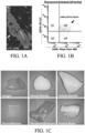
- Figures 1A-C Example of flow cytometry of cell populations that exist around a hair follicle and scaffolds that such cells readily adhere to when seeded. More specifically, Figure 1A depicts an example of location of said LGR expressing cells of cutaneous origin. Immunofluorescent confocal microscopy at 40x magnification depicts the follicular bulge (white arrow), LGR6+ (Green), DNA (Blue). Figure 1B is a fluorescent activated cell sorting graph with gate analysis indicating exemplary cellular markers. Figure 1C depicts an array of cells types can be used to seed a spectrum of acellular matrices/substrates/scaffolds/materials.
- Figure 2A is a photographic representation of an example of a gross construct without micro-aggregate multi-cellular functional units containing LGR expressing stem cell foci.
- Figure 2B depicts the construct following seeding of substrate with micro-aggregate multi-cellular functional units containing LGR expressing stem cell foci.
- Figure 3A in columnar format, is an image series by differential interference contrast (DIC) confocal microscopy of LGR seeded substrates from different sources.
- Figure 3B is a corresponding column by immunofluorescent confocal microscopy at 20x magnification of LGR6+ ESC seeded matrices of respective constructs containing LGR expressing cells.
- the inset white boxes represent focal zoom regions indicated in the column Figure 3D while
- Figure 3C is a column depicting the Digital merge of the respective image of Figure 3A (DIC) and the immunofluorescent of Figure 3B indicating matrix contour and boundaries.
- the columns of Figures 3E and 3F respectively represent the bioluminescence measured in radiant efficiency of an acellular matrix control and a corresponding LGR6+ ESC seeded matrix at 72 hours post-seeding.
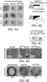
- Figure 4A-E depict examples of said LGR containing construct placed into living mammalian system. Placement of an LGR6+ GFP ESC Seeded Matrix Augments Healing Hair Follicle Growth.
- Figure 4A is a 3x3 matrix of photomicrographs of 3mm full human de-cellularized dermis thickness burn wound beds at days 5, 8 and 10 containing no matrix (burn control), matrix (matrix control) and LGR6+ GFP ESC.
- Figure 4B graphically depicts the relative expression of Cytokeratin-17 transcript expression at day 10 of the wound beds depicted in Figure 4A . The percent wound bed healed was determined using quantification analysis of wound bed healing rates as a percent area function within the ImageJ NCBI application. Wound control contains burn wound bed only. Matrix control contains matrix only and LGR6+ GFP contains ADM seeded with LGR6+ GFP ESCs.
- Figure 4C is a photomicrograph of in vivo bioluminescent imaging in murine full thickness burn wound beds at day 5.
- Figure 4D are micrographs of human dermis at 100x of the controls and LGR6+ GFP containing dermis at 12 hours and 72 hours and after seeding with ESCs.
- the white arrow indicates the presence of a dermal pore
- Figure 4E provides images of the controls and the construct containing human dermis seeded with ESCs with a silicone protective overlay to prevent desiccation.
- the LGR6+ GFP matrix image includes duplicate small black arrows that indicate nascent hair patches from the full thickness Nu/Nu murine wound bed.
- Figures 5A-E depict an example of said construct with LGR and supportive cellular entities following initial form of polarization as well as the effect of addition of Stromal vascular fraction (SVF) to LGR6+ ESC Seeded Matrices in promoting tissue polarization and a dual compartment skin-like System.
- Figure 5A is confocal 20x imaging of a 5x10 5 RFP expressing stromal vascular fraction cellular isolate population 24 hours after being seeded on to a representative Adrenomedullin (ADM) (such as that available from Integra LifeSciences Corporation under the name Integra ® ).
- ADM Adrenomedullin
- Figure 5B is a confocal 20x image of a 5x10 5 FP expressing LGR6+ cellular isolate population 24 hours after being seeded on to a representative ADM (Integra ® ).
- Figure 5C depicts confocal 20x imaging of a dual seeded representative ADM (Integra ® ) with 5x10 5 RFP expressing SVF and 5x10 5 GFP expressing LGR6 + isolate populations 24 hours after being co-seeded in culture.
- Figure 5D is of a co-seeded matrix containing 5x10 5 RFP expressing SVF RFP and 5x10 5 GFP expressing LGR6 + following 5 days of growth in culture.
- the dotted parallel lines indicate epithelial LGR6 +GFP lineage accumulating at the edge of the ADM substrate.
- the small bracket and large bracket indicate the relative locations of the two compartments in correlation with LGR6 +GFP and SVF RFP abundance.
- the arrowed "U" shaped solid line indicates a region containing a pre-seeded pore induced by a 32 gauge sterile needle.
- Figure 5E is a graphical representation of the proliferation kinetics of a collagen substrate co-seeded with green LGR expressing cells and red SVF expressing cells.
- Figures 6A and 6B depict an example of a construct containing LGR cells with and without supportive cellular entities and the relative production of growth factors. Correlative Expression Profiles of Pro-angiogenic Transcripts and Protein Analytes from LGR6 +GFP ESC and SVF RFP Enriched Scaffolding Culture Constructs.
- Figure 6A graphs relative fold transcript expression ( ⁇ CT) of indicated gene element from total RNA: LGR6 +GFP ESC (black bar), SVF RFP (grey bar), and co-cultured LGR6 +GFP ESC + SVF RFP (white box) on respective scaffold substrate.
- ⁇ CT relative fold transcript expression
- LGR6 + SVF indicates the inter-comparison co-cultured LGR6 +GFP ESC + SVF RFP expression vs. singular LGR6 +GFP ESC and SVF RFP expression on indicated scaffolding.
- Average FGF-2 gene expression for co-cultured matrices was higher than the average expression of both singular systems (Scaffold+ LGR6 or Scaffold ® +SVF) except for co-cultured Integra ® (Integra+ LGR6 + SVF).
- Significance below the x-axis (LGR6) or (SVF) indicates the intra-comparison of substrates, while the cellular entity remains constant.
- FIG. 6B graphically represents the relative densitometric unit (RDU) of indicated protein analyte from total protein isolates: LGR6 +GFP ESC (black bar), SVF RFP (grey bar), and co-cultured LGR6 +GFP ESC + SVF RFP (white box) on respective scaffold substrate.
- RDU relative densitometric unit
- Figures 7A-H illustrate a wound/injury/void receiving therapy example of enhanced LGR cell migration, proliferation and viability into a wound namely a third degree wound bed induction and verification of the elimination of the LGR stem cell follicular bulge and adnexal structures.
- Figure 7A depicts a wound bed template marks of 3mm diameter.
- Figure 7B depicts the wound bed structure at day 0 (the white scale bar being lmm).
- Figure 7C illustrates an example of a 2x3 3mm wound bed grid.
- Figure 7D shows topical application of the resuspended peptide at the wound site.
- Figure 7E is a photomicrograph of H&E stain of non-burned, intact Integument/skin with hair follicle and adnexal structures. The arrow indicates the location of the magnified follicle (inset image) where the white scale bar is 500 ⁇ m.
- Figure 7F is an H&E stain of dorsal murine skin following high temperature cautery depicting removal of epidermal, dermal and hypodermal tissues including the follicular bulge.
- Figure 7G is DAPI/DNA stain (4',6-diamidino-2-phenylindole) of non-burned, intact skin with hair follicle and adnexal structures.
- the arrow indicates the magnified follicle with co-labeling of immunofluorescent LGR5 and LGR6 antibodies green and red respectively (inset image).
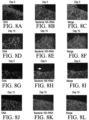
- Figures 8A-Q depict a wound/injury/void with LGR as it relates to antimicrobial behavior over five and ten day time periods.
- in-situ hybridization indicates the presence of bacterial adhesion at the third degree burn wound bed.
- Figure 8A presents DNA/DAPI labeling of a 3rd degree burn wound bed at day five post burn induction treated daily with SDZ.
- Figure 8B 5'- Cy3-EUB338 labeled 16s rRNA of 3rd degree burn wound bed bacterial organisms (yellow grains) at day five post burn induction treated daily with SDZ are depicted.
- Figure 8C is a digitally merged image of Figures 8A and 8B.
- Figure 8D corresponds to Figure 8A except at day ten with DNA/DAPI labeling of 3rd degree burn wound bed treated daily with SDZ.
- Figure 8E is a photomicrograph of the 5'- Cy3-EUB338 labeled 16s rRNA of 3rd degree burn wound bed bacterial organisms (yellow grains) at day ten post burn induction treated daily with SDZ.
- Figure 8F is a merged image of Figures 8D and E.
- Figures 8G-8L are images corresponding respectively to the five and ten post burn periods of Figures 8A-F but subject to daily treatment using Defensin, alpha 5 (DEFA5) rather than SDZ.
- FIG. 8H represents the interface of tissue with overlying fibrinous material where less bacteria is observed in the setting of DEFA5 treatment.
- Figure 8M with inset 8N demonstrate quantification of white pixel intensity of Cy3 fluorescence grayscale converted image of a wound bed treated with SDZ and containing more 16s rRNA labeling per unit area.
- Figure 8O and inset 8P correspondingly show quantification of white pixel intensity of Cy3 fluorescence grayscale converted image of (inset image p.) a wound bed treated with DEFA5 and containing a reduced 16s rRNA labeling per unit area.
- the inset graph depicts averaged white pixel intensity of 16s rRNA expressed in both SDZ and DEFA5 treated burn wound beds at day five using grayscale imaging software.
- Figure 8Q is a graph to illustrate averaged red channel fluorescence of 16s rRNA expressed in both SDZ and DEFA5 treated burn wound beds at day five.
- the white arrow in Figure 8H indicates potential film in DEFA5 treated wound beds and the black arrow in Figure 8M indicates white pixel intensity. Scale bar 100 ⁇ m. (*) indicates p-value ⁇ 0.05.
- Figures 9A and B are a series of time progression photographs that represents an example of LGR expressing cellular entities within wound as it relates to augmented healing, tissue and appendage regeneration and subsequent hair growth, wound healing kinetics and nascent hair growth in treated burn wounds devoid of adnexal structures.
- the photographic series comprising Figure 9A are gross imaging using a Leica Wild M680 surgical microscope to image healing of 3rd degree burn wound beds over 10 days while being treated with indicated agents MQH2O, DEFA5, DEFB1, SDZ.
- the white scale bar represents 1 mm.
- the second photographic series of Figure 9B again comprises gross imaging using a Leica Wild M680 to track nascent hair growth of 3rd degree burn wound beds over 16 days in a side by side comparison of DEFA5 vs. control treated wound beds.
- the white arrows indicate the growth of new hair.
- the scale bar is 1mm.
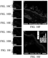
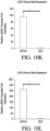
- Figures 10A-L comprise an example of said LGR expressing cellular entities within wound/injury/ tissue void as it relates to augmented healing, propagation of said entities.
- the Graphs comprising Figures 10K and 10L provide evidence of quantification of wound bed healing kinetics and LGR5 and LGR6 stem cell migration into burn tissue following treatment with topical focal agents. Briefly, these tests were used to confirm the quantitative confocal microscopic intensity patterns from imaging LGR5 and LGR6, and based on reverse-transcriptase polymerase chain reaction on burn wound tissues.
- Figure 10A presents photographs of a wound area with a white scale bar representing 1 mm and the wound area calculation in black.
- Figure 10B graphically displays the averaged wound healing rate expressed as percent % of wound area remaining over 10 day period of indicated topical focal agent application.
- the asterisk (*) represents a p-value ⁇ 0.05.
- Figures 10C-J are LGR5 and LGR6 immunofluorescent antibody labeling of a DEFA5 treated wound bed at day 5 where Figure 10C is DNA/DAPI/Blue, Figure 10D is LGR5/FITC/Green Figure 10E is LGR6/TRITC/Red and Figure 10F is a merger of 10C-10E.
- Figures 10G-I are corresponding LGR5 and LGR6 immunofluorescent antibody labeling of SDZ (sulfadiazine) treated wound bed at day 5 (DNA/DAPI/Blue, LGR5/FITC/Green and LGR6/TRITC/Red).
- Figure 10J is a merged image of 10G-101 and includes an inset representing averaged LGR5 and LGR6 expression using Green and Red fluorescent intensity per wound bed at day 5.
- the comparative values obtained from Reverse Transcriptase PCR quantification of the fold increase in RNA extracted from replicate wound beds treated with DEFA5 and SDZ is set out.
- the white scale bar 50 ⁇ m and again, the asterisk (*) represents a p-value ⁇ 0.05.
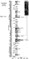
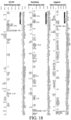
- Figures 11A and B illustrate a wound/injury/tissue void with the LGR expressing cellular entities placed within wound as it relates to augmentation of pro-healing pathways.
- the figures respectively represent RT-PCR quantification and gene heat mapping comparison of wound beds treated with DEFA5 to SDZ. These figures show the role of human alpha defensin 5 versus sulfadiazine in augmenting key transcript expression within the wound. The results show that several gene subsets are significantly up-regulated within the wound beds receiving human alpha defensin 5 when compared with sulfadiazine therapy and that certain Wnt pathway gene subsets are significantly up-regulated in response of the LGR stem cell system to Wnt ligands in both the gut and skin.
- Figure 11A presents an Averaged Wound Healing RT2-PCR Array pathway heat map and corresponding gene map with fold regulation for wound beds comparing DEFA5 to SDZ treated systems.
- Figure 11 B presents an Averaged Wnt RT2-PCR Array healing pathway heat map and corresponding gene map with fold regulation for wound beds comparing DEFA5 to SDZ treated systems. The colors of the heat maps are indicated as red, more expressed in DEFA5 treated burns to green more expressed in SDZ treated burns.
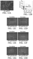
- Figures 12A-I represent an example of a micro-aggregate multicellular unit containing LGR expressing stem cell foci as it relates to location, population identity and wound healing capacity.
- LGR6+, CD34+, and CD73+ C57BL/6(UBC-GFP) murine cells were isolated for cell culture expansion.
- Figure 12A depicts LGR6 fluorescent antibody (green) expression of cells on the hair follicle following partial epidermal 10 unit/ ⁇ L dispase digestion. (Worthington Biochemical Corp., Lakewood, N.J.) digestion for 30 minutes at 37°C on a slow rocker.
- Figure 12B is of LGR6+ cells expressing additional CD34 and CD73 markers (the arrow indicates population isolated comprising approximately 1 to 3 percent of all cells).
- Figures 12C-H are eFluor450 expression histograms of an in vitro wound assay respectively showing periodic intrinsic GFP expression from C57BL/6(UBC-GFP) murine cells, CD34+ PE/Cy7 expression, LGR6+ APC expression and CD73+.
- the graph of Figure 12I sets out the averaged reduction in the distance line over time expressed as a percentage of initial distance following fluorescence sorting where the asterisk (*) represents a p-value ⁇ 0.05.
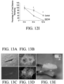
- Figures 13A-D are photomicrographs by confocal microscopy and bioluminescence of an activated functional singularity unit (aFSU) at the time initial seeding and 1 day later showing an example of a micro-aggregate multicellular unit containing LGR expressing stem cell foci while undergoing initial propagation on a collagen matrix, Figure 13 E .
- aFSU activated functional singularity unit
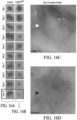
- Figures l4A-E depict an example of location LGR cellular varieties as it relates to location, phenotype, interface and polarity within a cutaneous tissue.
- Figure 14A shows by Immunofluorescence staining, localized regions of LGR6 (Green/fluorescein isothiocyanate (FITC)) and LGR5 (Red/tetramethyl rhodamine isothiocyanate (TRITC)) expression.
- the scale bar is for 20 ⁇ m.
- Figure 14B shows fluorescence-activated cell sorting isolation of the LGR6+ GFP epithelial stem cells from C57BL/6(UBCGFP) murine skin with the final sort gate using LGR6+, CD34 and CD73 on the left and individual histograms depicting cellular GFP expression and correlating antibody-conjugate labels: CD73/PE-7, LGR6/Cy5, CD34/eFlour450 on the right.
- Figure 14C shows differential interference contrast image of LGR6 +GFP epithelial stem cells plated following fluorescence-activated cell sorting isolation.
- Figure 14D depicts intrinsic GFP expression of the LGR6 +GFP epithelial stem cells and
- Figure 14E is a merged image of Figures 14C and 14D .
- the scale bar represents 20 ⁇ m.
- Figures 15A-E provide an example of LGR expressing cellular foci as it relates to a method of delivery through placement around and/or within wound/injury/tissue void.
- the three images of Figure 15A depict, respectively, an initial burn template; a full thickness burn on the dorsum on Nu/Nu mouse; and delivery of Hydrogel ® containing 10 5 LGR6 +GFP epithelial stem cells at the base of the wound bed.
- the scale bar for Figure 15A is 1mm.
- Figure 15B is an immunofluorescence image of the injection pocket DNA/DAPI-BLUE at Day 0
- Figure 15B is an immunofluorescence image of anti-LGR6/TRITC antibody labeling and
- Figure 15C the same for LGR6 +GFP epithelial stem cells.
- Figure 15 E is a merged image of Figures 15B-D and has a scale bar of 20 ⁇ m.
- Figures 15A-E show full thickness burn wound bed induction and validation of LGR6+ stem cell engraf
- FIGs 16A-D depict an example of LGR containing stem cell focus as it relates to delivery into and around wounds via a deliverable vector and subsequent healing, regeneration of tissues and supporting structures including but not limited blood vessel angiogenesis and/or angiogenesis.
- the progression of wound healing is depicted following the injection of Hydrogel ® from BD Biosciences, San Jose, Calif. (control) in Figure 16A compared with Figure 16B , LGR6 +GFP epithelial stem cells seeded Hydrogel ® over 15 days.
- the scale bar is 1mm.
- the white arrow indicates presence of a remaining LGR6 +GFP epithelial stem cells population located within healing wound bed.
- the black arrow indicates the location of the burn wound base free of LGR6 +GFP epithelial stem cells.
- Figures 17A-D depicts an example of LGR containing stem cell focus following delivery into and/or around wound with subsequent healing and regeneration of tissues and related appendages such as but not limited to hair follicle and related supportive structures.
- Figure 17A is a four panel matrix of confocal images of immunofluorescent labeled tissue specimen at day 10 following transplantation of LGR6+ epithelial stem cells migration into the wound bed 10 days.
- the images comprising Figure 17A include DNA/DAPI-BLUE; anti-LGR6/TRITC; GFP expression of LGR6 +GFP ESC.
- Figure 17B is a differential interference contrast image merge of all channels.
- the Red arrow designates regions of nascent follicle development. (See also the upper inset image).
- the dotted line shows epithelial polarization overlying nascent hair follicles while the white arrow indicates the location of the graft injection pocket (See also the magnification thereof in the lower inset image for an image of the initial injection pocket cellular population.
- the inset graph of Figure 17B represents comparative KRT17/ cytokeratin 17 gene expression within the indicated wound beds of the control and LGR6+ +GFP treatment.
- the three images are of a Transplant dome used to cover hair follicle study population burn wound beds, an LGR6+ +GFP ESC treated wound bed at day 10 (solid arrow) with nascent hair follicles (clear arrow) follicle cyst formation and a control wound bed at day 10.
- the graph comprising Figure 17D quantifies the Day 10 wound bed resulting from RT-PCR indicating relative gene fold expression of WNT ligands.
- Figure 18 provides an RT-PCR quantification and inset gene heat mapping comparison of a wound/injury/tissue void with the LGR expressing cellular foci as it relates to delivery into and/or around wound/injury/tissue void as it relates to augmentation of pro-healing pathways and comparative gene expression of wounds receiving LGR6+ epithelial stem cells against a control.
- the graphs illustrate the relative fold expression of genes for angiogenesis, wound healing and epidermal growth factor. Correlative graphical representation of data comparing wound beds receiving LGR6+ epithelial stem cells and control therapy.
- the inset heat maps the color red indicates greater expression within the LGR6+ epithelial stem cell wound bed while the color green indicates greater expression within the control wound bed.
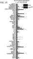
- Figure 19 graphically presents the relative protein densitometry of an example of LGR expressing cellular foci as it relates to delivery into and/or around wound/injury/tissue void and augmentation of wound healing factors.
- Comparative angiogenesis analyte expression of wounds receiving LGR6+ ESCs
- Proteomic array comparing common proteins which regulate and augmented angiogenesis.
- the grey columns indicate control wounds and the black columns indicated those wounds that received the LGR6 +GFP ESC.
- the inset image shows example proteome array membranes following development with HRP chemi-luminesce. Brighter colors indicate higher levels of protein expression.
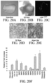
- Figures 20A-F illustrate an example of LGR expressing cellular foci as it relates to the regeneration of bone tissues. Isolated LGR foci can be seeded bone and remain viable.
- Figure 20A is a gross bone image of harvested bone for culture.
- Figure 20B is a DIC image of bone containing LGR GFP 7 days following seeding.
- Figure 20C is a 488 nm Green laser confocal image of bone containing LGR6 +GFP 7 days following seeding. It is notable that the LGR foci can undergo osteo-induction in-vitro.
- Figure 20D depicts LGR foci following 1 week of osteo-induction with supplemental media.
- Figure 20E is an Alizarin red stain of the LGR foci following osteo-induction which can undergo osteo-induction in-vitro and up regulate key osteogenic genes.
- Figure 20F is RT-PCR data showing relative fold gene expression where the grey columns represent (control) non-osteo induced LGR and the black columns represent those LGR which received osteo-induction media following 7 days of culture. GAPDH was used as reference standard housekeeping gene.
- LGR epithelial stem cells particularly in conjunction with a formed scaffolding substrate, provides full thickness wounds and or voids in epithelial systems with a stem cell enriched tissue substitute.
- MPFU minimally polarized functional cell unit
- the progenitors of the LGR4, LGR5 and LGR6 stem cells also have the ability to generate native anti-microbial peptides that not only reduce the basal level of microorganisms within the wound bed but also augment progenitor cells amplification and differentiation, leading to a reduction in wound and peri-wound infections, faster wound closure, and hair follicle development.
- Described herein is the translational applicability of a minimally polarized functional unit in providing an immediate, deliverable and viable tissue barrier that is capable of maintaining a stem cell colony focus with concomitant competent progeny. From these stem cell foci, progeny can undergo migratory proliferative-differentiation in order to stimulate epithelial tissue elements, healing and graft integration. It has been found that the minimally polarized functional unit can be applied alone, with scaffolding, soluble growth factors and/or additional cell lineage which promote the polarization of the scaffold bound populations as well s intrinsic tissue architecture required in epithelial healing and cellular regenerative efforts.
- the disclosed protocol involves: a) harvesting living human/mammalian tissue; b) processing the tissue element to generate a micro-aggregate multi-cellular functional units which contain LGR expressing cells; c) applying the LGR expressing cell micro-aggregate multi-cellular functional units to a delivery vehicle substrate selected from the group consisting of scaffolding, matrix, particle, cell(s) and fiber to create a construct; d) optionally including selected additional enhancing factors; and e) applying the construct to tissues for generating, regenerating, enhancing and/or healing tissue systems including those related to ectodermal, mesodermal and/or endodermal origin tissues including but not limited to skin, glands, hair, nerves, bone, muscle, fat, tendons, blood vessels, fascia, ocular tissues, bone marrow, lung, heart, nails, gastrointestinal tissues, oral tissues, teeth, taste buds, urogenital tissues, renal tissues, reproductive tissues, lymphatic tissues, immune system tissues/elements and such related appendages and protein cellular elements
- a gelatinous support such as an exemplary three dimensional collagen scaffold can be generated by well-known processes as follows:
- the PRM is direct to humans, is a cell sustaining, serum-free, media mixture Keratinocyte-SFM containing L-glutamine supplied with separately packaged prequalified human recombinant Epidermal Growth Factor 1-53 (EGF 1-53) and Bovine Pituitary Extract (BPE) sold as Keratinocyte-SFM (1X) from Thermo Fisher Scientific to which the antibioticantimycotic agents penicillin, streptomycin, and amphotericin B are added along with a GMP- fibrinogen: human.
- EGF 1-53 Epidermal Growth Factor 1-53
- BPE Bovine Pituitary Extract
- the agent used here is Gibco ® Antibiotic-Antimycotic from Thermo Fisher Scientific, a solution containing 10,000 units/mL of penicillin, 10,000 ⁇ g/mL of streptomycin, and 25 ⁇ g/mL of Fungizone ® Antimycotic. Because the PRM is used to transport human tissues, the supplemental reagents are utilized to stabilize the primary tissues and reduce the viability of micro-organisms during transport and processing.
- the following relates specifically to the generation and preservation of LGR expressing epithelial containing stem cell micro-aggregate functional units in accordance with an embodiment of the invention.
- Example 1 concerns a method for extraction of minimally polarized functional units in accordance with an embodiment of the invention. After obtaining a specimen, it is removed from its associated transport container followed by:
- the following relates to secondary processing where the primary cultures are established and functional tissue elements are prepared utilizing enzymatic preparation using conventional CLIA equipment and reagents meeting FDA and/or GMP certification:
- Example 2 is directed to processing of hypodermis and subdermal fat cellular components. Example 2 recites the following steps:
- Example 3 is directed to addition of hypodermis and subdermal fat components to the example of a construct.
- the illustrative component addition example involves:
- Example 4 concerns enrichment of the minimally polarized, epithelial stem cell singularity units. Following Example 1, the MPFUs is placed in pulse rescue media in a 15 ml conical tube and spin/centrifuged into a soft pellet. The material is then subject to the following process of partial digestion:
- Example 5 involves adding the epithelial stem cell functional singularities (ESC aFSUs) obtained from Example 4 to a construct/scaffold.
- the procedure entails:
- Example 6 represents illustrative protocols for quality assurance and construct finalization involving cryopreservation which entails preparation the construct for shipment following defined good manufacturing processes (GMP) for cell therapy applications and include:
- the invention relates to methods for making and methods for using constructs of micro-aggregate multicellular grafts containing isolated Leucine-rich repeat-containing G-protein coupled Receptor (LGR) expressing cells for the delivery, application, transplantation, implantation, directed seeding, directed migration, directed tracking, in setting, laminating and/or injection of the cellular element generating, regenerating, enhancing and/or healing epithelial systems, glands, hair, nerves, bone, muscle, fat, tendons, blood vessels, fascia, ocular tissues and peptide secreting cellular elements for use in wound therapy applications, tissue engineering, cell therapy applications, regenerative medicine applications, medical/therapeutic applications, tissue healing applications, immune therapy applications, and tissue transplant therapy applications.
- LGR Leucine-rich repeat-containing G-protein coupled Receptor
Landscapes
- Health & Medical Sciences (AREA)
- Life Sciences & Earth Sciences (AREA)
- Engineering & Computer Science (AREA)
- Chemical & Material Sciences (AREA)
- Biomedical Technology (AREA)
- General Health & Medical Sciences (AREA)
- Zoology (AREA)
- Medicinal Chemistry (AREA)
- Wood Science & Technology (AREA)
- Dermatology (AREA)
- Veterinary Medicine (AREA)
- Public Health (AREA)
- Animal Behavior & Ethology (AREA)
- Organic Chemistry (AREA)
- Bioinformatics & Cheminformatics (AREA)
- Cell Biology (AREA)
- Chemical Kinetics & Catalysis (AREA)
- Oral & Maxillofacial Surgery (AREA)
- Epidemiology (AREA)
- Transplantation (AREA)
- Biotechnology (AREA)
- Genetics & Genomics (AREA)
- Environmental Sciences (AREA)
- Dentistry (AREA)
- Botany (AREA)
- Urology & Nephrology (AREA)
- Molecular Biology (AREA)
- Pharmacology & Pharmacy (AREA)
- General Chemical & Material Sciences (AREA)
- Nuclear Medicine, Radiotherapy & Molecular Imaging (AREA)
- Microbiology (AREA)
- General Engineering & Computer Science (AREA)
- Biochemistry (AREA)
- Endocrinology (AREA)
- Diabetes (AREA)
- Materials For Medical Uses (AREA)
- Medicines Containing Material From Animals Or Micro-Organisms (AREA)
- Peptides Or Proteins (AREA)
- Micro-Organisms Or Cultivation Processes Thereof (AREA)
- Developmental Biology & Embryology (AREA)
Priority Applications (1)
| Application Number | Priority Date | Filing Date | Title |
|---|---|---|---|
| EP24181160.3A EP4438128A3 (en) | 2014-12-02 | 2015-12-01 | Methods for development and use of minimally polarized function cell micro-aggregate units in tissue applications using lgr4, lgr5 and lgr6 expressing epithelial stem cells |
Applications Claiming Priority (3)
| Application Number | Priority Date | Filing Date | Title |
|---|---|---|---|
| US201462086526P | 2014-12-02 | 2014-12-02 | |
| US14/954,335 US10926001B2 (en) | 2014-12-02 | 2015-11-30 | Methods related to minimally polarized functional units |
| PCT/US2015/063114 WO2016089825A1 (en) | 2014-12-02 | 2015-12-01 | Methods for development and use of minimally polarized function cell micro-aggregate units in tissue applications using lgr4, lgr5 and lgr6 expressing epithelial stem cells |
Related Child Applications (2)
| Application Number | Title | Priority Date | Filing Date |
|---|---|---|---|
| EP24181160.3A Division EP4438128A3 (en) | 2014-12-02 | 2015-12-01 | Methods for development and use of minimally polarized function cell micro-aggregate units in tissue applications using lgr4, lgr5 and lgr6 expressing epithelial stem cells |
| EP24181160.3A Division-Into EP4438128A3 (en) | 2014-12-02 | 2015-12-01 | Methods for development and use of minimally polarized function cell micro-aggregate units in tissue applications using lgr4, lgr5 and lgr6 expressing epithelial stem cells |
Publications (4)
| Publication Number | Publication Date |
|---|---|
| EP3227431A1 EP3227431A1 (en) | 2017-10-11 |
| EP3227431A4 EP3227431A4 (en) | 2019-01-02 |
| EP3227431C0 EP3227431C0 (en) | 2025-01-29 |
| EP3227431B1 true EP3227431B1 (en) | 2025-01-29 |
Family
ID=56078499
Family Applications (2)
| Application Number | Title | Priority Date | Filing Date |
|---|---|---|---|
| EP15865131.5A Active EP3227431B1 (en) | 2014-12-02 | 2015-12-01 | Methods for development and use of minimally polarized function cell micro-aggregate units in tissue applications using lgr4, lgr5 and lgr6 expressing epithelial stem cells |
| EP24181160.3A Pending EP4438128A3 (en) | 2014-12-02 | 2015-12-01 | Methods for development and use of minimally polarized function cell micro-aggregate units in tissue applications using lgr4, lgr5 and lgr6 expressing epithelial stem cells |
Family Applications After (1)
| Application Number | Title | Priority Date | Filing Date |
|---|---|---|---|
| EP24181160.3A Pending EP4438128A3 (en) | 2014-12-02 | 2015-12-01 | Methods for development and use of minimally polarized function cell micro-aggregate units in tissue applications using lgr4, lgr5 and lgr6 expressing epithelial stem cells |
Country Status (21)
| Country | Link |
|---|---|
| US (7) | US10926001B2 (enExample) |
| EP (2) | EP3227431B1 (enExample) |
| JP (3) | JP6791854B2 (enExample) |
| KR (5) | KR20210043024A (enExample) |
| CN (1) | CN107250348B (enExample) |
| AU (3) | AU2015355187C1 (enExample) |
| BR (1) | BR112017011808A2 (enExample) |
| CA (1) | CA2969707C (enExample) |
| CO (1) | CO2017006640A2 (enExample) |
| CR (1) | CR20170296A (enExample) |
| EA (1) | EA201791125A1 (enExample) |
| GB (2) | GB2549872A (enExample) |
| HK (1) | HK1250515A1 (enExample) |
| IL (2) | IL252613B (enExample) |
| MX (2) | MX2017007243A (enExample) |
| MY (1) | MY184931A (enExample) |
| NZ (2) | NZ755260A (enExample) |
| PH (1) | PH12017501009B1 (enExample) |
| SG (2) | SG10201914058QA (enExample) |
| WO (1) | WO2016089825A1 (enExample) |
| ZA (1) | ZA201703907B (enExample) |
Families Citing this family (6)
| Publication number | Priority date | Publication date | Assignee | Title |
|---|---|---|---|---|
| EP3595440A2 (en) * | 2017-03-14 | 2020-01-22 | Juno Therapeutics, Inc. | Methods for cryogenic storage |
| CR20200366A (es) * | 2018-01-26 | 2020-10-05 | Polarityte Inc | Sustrato acelarante de biomaterial con interfaz compuesta |
| AU2019212976A1 (en) * | 2018-01-26 | 2020-07-23 | Polarityte, Inc. | Complex living interface-coordinated self-assembling materials (CLICSAM) |
| FR3082123B1 (fr) | 2018-06-07 | 2020-10-16 | Urgo Rech Innovation Et Developpement | Pansement cellularise et son procede de fabrication |
| JP2024006681A (ja) * | 2022-07-04 | 2024-01-17 | 学校法人近畿大学 | 生体材料 |
| WO2025166131A1 (en) * | 2024-02-01 | 2025-08-07 | Board Of Regents, The University Of Texas System | Compositions and methods for treating bone and muscle injuries |
Family Cites Families (23)
| Publication number | Priority date | Publication date | Assignee | Title |
|---|---|---|---|---|
| US20040126881A1 (en) * | 2002-09-06 | 2004-07-01 | Vincent Ronfard | Fibrin cell supports and methods of use thereof |
| WO2006008009A2 (en) | 2004-07-23 | 2006-01-26 | Bayer Healthcare Ag | Diagnostics and therapeutics for diseases associated with leucine-rich repeat-containing gpcr 6 (lgr6) |
| US20060154365A1 (en) | 2004-08-30 | 2006-07-13 | Anthony Ratcliffe | Cultured three dimensional tissues and uses thereof |
| EP1942957B1 (en) * | 2005-10-27 | 2012-07-11 | Coloplast A/S | Biodegradable scaffold with ecm material |
| WO2008008009A1 (en) | 2006-07-13 | 2008-01-17 | St. Jude Medical Ab | Medical information management in a patient information hub system |
| US8366723B2 (en) | 2006-08-03 | 2013-02-05 | Rassman Licensing, Llc | Hair harvesting device and method with localized subsurface dermal fluid insertion |
| US7998737B2 (en) | 2006-09-12 | 2011-08-16 | Deutsches Krebsforschungszentrum | Cell culture of keratinocytes under non-differentiating conditions |
| GB201111244D0 (en) | 2011-06-30 | 2011-08-17 | Konink Nl Akademie Van Wetenschappen Knaw | Culture media for stem cells |
| US9752124B2 (en) | 2009-02-03 | 2017-09-05 | Koninklijke Nederlandse Akademie Van Wetenschappen | Culture medium for epithelial stem cells and organoids comprising the stem cells |
| EP2412800A1 (en) | 2010-07-29 | 2012-02-01 | Koninklijke Nederlandse Akademie van Wetenschappen | Liver organoid, uses thereof and culture method for obtaining them |
| FI20095355A0 (fi) | 2009-04-01 | 2009-04-01 | Helsingin Yliopisto | Regeneroituva aktiivinen matriisi ja sen käytöt |
| US20110076303A1 (en) | 2009-06-25 | 2011-03-31 | Tokuro Iwabuchi | Methods for Screening for Anti-Graying Agents on the Basis of AFF-4 |
| US20110130711A1 (en) | 2009-11-19 | 2011-06-02 | Follica, Inc. | Hair growth treatment |
| WO2012000180A1 (zh) | 2010-06-30 | 2012-01-05 | Chen Jinxi | 一种皮肤组织细胞聚合物的制备方法及其用途 |
| GB201106395D0 (en) | 2011-04-14 | 2011-06-01 | Hubrecht Inst | Compounds |
| EP2771454B1 (en) | 2011-10-27 | 2019-07-24 | Universität Leipzig | Method for deriving melanocytes from the hair follicle outer root sheath and preparation for grafting |
| JP6186997B2 (ja) | 2012-08-07 | 2017-08-30 | 日油株式会社 | 塗布型帯電防止剤 |
| US9655930B2 (en) | 2012-10-01 | 2017-05-23 | Aderans Research Institute, Inc. | Compositions and methods for producing reconstituted skin |
| US20140106447A1 (en) | 2012-10-12 | 2014-04-17 | Mimedx Group, Inc. | Compositions and methods for recruiting stem cells |
| CA3184040A1 (en) | 2013-03-14 | 2014-10-02 | The Brigham And Women's Hospital, Inc. | Compositions and methods for epithelial stem cell expansion comprising a wnt agonist and a histone deacetylase inhibitor |
| US9592257B2 (en) | 2013-10-11 | 2017-03-14 | MWV Cell, LLC | Complete human skin organ generated from culture-expanded cells |
| CN103550828B (zh) * | 2013-10-17 | 2016-01-27 | 中国科学院动物研究所 | 一种基于毛囊干细胞和硅凝胶敷料的皮肤重建方法 |
| TWI548413B (zh) | 2014-11-07 | 2016-09-11 | 國立臺灣大學 | 誘導毛囊新生的皮膚萃取物、組合物及其用途 |
-
2015
- 2015-11-30 US US14/954,335 patent/US10926001B2/en active Active
- 2015-12-01 EA EA201791125A patent/EA201791125A1/ru unknown
- 2015-12-01 NZ NZ755260A patent/NZ755260A/en unknown
- 2015-12-01 CA CA2969707A patent/CA2969707C/en active Active
- 2015-12-01 GB GB1710646.9A patent/GB2549872A/en not_active Withdrawn
- 2015-12-01 EP EP15865131.5A patent/EP3227431B1/en active Active
- 2015-12-01 KR KR1020217010934A patent/KR20210043024A/ko not_active Ceased
- 2015-12-01 KR KR1020177017887A patent/KR20170098844A/ko not_active Ceased
- 2015-12-01 NZ NZ733433A patent/NZ733433A/en unknown
- 2015-12-01 BR BR112017011808A patent/BR112017011808A2/pt not_active Application Discontinuation
- 2015-12-01 CR CR20170296A patent/CR20170296A/es unknown
- 2015-12-01 KR KR1020187036618A patent/KR20180136587A/ko not_active Ceased
- 2015-12-01 JP JP2017530217A patent/JP6791854B2/ja active Active
- 2015-12-01 AU AU2015355187A patent/AU2015355187C1/en active Active
- 2015-12-01 KR KR1020187036619A patent/KR20180136588A/ko not_active Ceased
- 2015-12-01 CN CN201580075326.3A patent/CN107250348B/zh active Active
- 2015-12-01 MY MYPI2017701990A patent/MY184931A/en unknown
- 2015-12-01 GB GB201902819A patent/GB2569056B/en active Active
- 2015-12-01 WO PCT/US2015/063114 patent/WO2016089825A1/en not_active Ceased
- 2015-12-01 EP EP24181160.3A patent/EP4438128A3/en active Pending
- 2015-12-01 SG SG10201914058QA patent/SG10201914058QA/en unknown
- 2015-12-01 HK HK18104680.7A patent/HK1250515A1/zh unknown
- 2015-12-01 KR KR1020197028100A patent/KR20190111167A/ko not_active Ceased
- 2015-12-01 SG SG11201704502RA patent/SG11201704502RA/en unknown
- 2015-12-01 MX MX2017007243A patent/MX2017007243A/es unknown
-
2017
- 2017-05-31 PH PH12017501009A patent/PH12017501009B1/en unknown
- 2017-06-01 IL IL252613A patent/IL252613B/en unknown
- 2017-06-05 MX MX2023001655A patent/MX2023001655A/es unknown
- 2017-06-07 ZA ZA2017/03907A patent/ZA201703907B/en unknown
- 2017-06-30 CO CONC2017/0006640A patent/CO2017006640A2/es unknown
- 2017-07-14 US US15/650,656 patent/US20180154043A1/en not_active Abandoned
- 2017-07-14 US US15/650,659 patent/US11266765B2/en active Active
-
2018
- 2018-10-19 US US16/165,169 patent/US11000629B2/en active Active
-
2019
- 2019-01-16 JP JP2019004921A patent/JP2019088299A/ja active Pending
- 2019-10-02 AU AU2019240603A patent/AU2019240603B2/en active Active
-
2020
- 2020-07-30 JP JP2020129610A patent/JP2020182880A/ja active Pending
-
2021
- 2021-05-21 US US17/326,734 patent/US11338060B2/en active Active
- 2021-10-21 IL IL287488A patent/IL287488B2/en unknown
-
2022
- 2022-04-19 US US17/723,748 patent/US11596714B2/en active Active
-
2023
- 2023-02-03 US US18/164,308 patent/US12465669B2/en active Active
- 2023-05-08 AU AU2023202857A patent/AU2023202857A1/en active Pending
Also Published As
Similar Documents
| Publication | Publication Date | Title |
|---|---|---|
| US12465669B2 (en) | Methods for development and use of minimally polarized function cell micro-aggregate units in tissue applications using LGR4, LGR5, and LGR6 expressing epithelial stem cells | |
| Moshaverinia et al. | Application of stem cells derived from the periodontal ligament or gingival tissue sources for tendon tissue regeneration | |
| Zhang et al. | Use of extracellular matrix hydrogel from human placenta to restore hair-inductive potential of dermal papilla cells | |
| Bellas et al. | Sustained volume retention in vivo with adipocyte and lipoaspirate seeded silk scaffolds | |
| US20120164116A1 (en) | Compositions and methods for tissue filling and regeneration | |
| US20200078411A1 (en) | Biological Scaffolds, Products Containing Biological Scaffolds and Methods of Using the Same | |
| JP2017538411A5 (enExample) | ||
| HK1245327B (en) | Methods for development and use of minimally polarized function cell micro-aggregate units in tissue applications using lgr4, lgr5 and lgr6 expressing epithelial stem cells | |
| OA19578A (en) | Methods for development and use of minimally polarized function cell microaggregate units in tissue applications using LGR4, LGR5 and LGR6 expressing epithelial stem cells | |
| OA19580A (en) | Methods for development and use of minimally polarized function cell microaggregate units in tissue applications using LGR4, LGR5 and LGR6 expressing epithelial stem cells | |
| CN120051288A (zh) | 人毛囊来源的条件培养基及其制备方法 |
Legal Events
| Date | Code | Title | Description |
|---|---|---|---|
| STAA | Information on the status of an ep patent application or granted ep patent |
Free format text: STATUS: THE INTERNATIONAL PUBLICATION HAS BEEN MADE |
|
| PUAI | Public reference made under article 153(3) epc to a published international application that has entered the european phase |
Free format text: ORIGINAL CODE: 0009012 |
|
| STAA | Information on the status of an ep patent application or granted ep patent |
Free format text: STATUS: REQUEST FOR EXAMINATION WAS MADE |
|
| 17P | Request for examination filed |
Effective date: 20170621 |
|
| AK | Designated contracting states |
Kind code of ref document: A1 Designated state(s): AL AT BE BG CH CY CZ DE DK EE ES FI FR GB GR HR HU IE IS IT LI LT LU LV MC MK MT NL NO PL PT RO RS SE SI SK SM TR |
|
| AX | Request for extension of the european patent |
Extension state: BA ME |
|
| DAV | Request for validation of the european patent (deleted) | ||
| DAX | Request for extension of the european patent (deleted) | ||
| RIC1 | Information provided on ipc code assigned before grant |
Ipc: A61L 27/54 20060101ALI20180629BHEP Ipc: A01N 1/02 20060101ALI20180629BHEP Ipc: A61L 27/38 20060101ALI20180629BHEP Ipc: C12N 5/071 20100101AFI20180629BHEP |
|
| REG | Reference to a national code |
Ref country code: HK Ref legal event code: DE Ref document number: 1245327 Country of ref document: HK |
|
| A4 | Supplementary search report drawn up and despatched |
Effective date: 20181204 |
|
| RIC1 | Information provided on ipc code assigned before grant |
Ipc: C12N 5/071 20100101AFI20181128BHEP Ipc: A61L 27/54 20060101ALI20181128BHEP Ipc: A61L 27/38 20060101ALI20181128BHEP Ipc: A01N 1/02 20060101ALI20181128BHEP |
|
| STAA | Information on the status of an ep patent application or granted ep patent |
Free format text: STATUS: EXAMINATION IS IN PROGRESS |
|
| 17Q | First examination report despatched |
Effective date: 20200107 |
|
| RAP3 | Party data changed (applicant data changed or rights of an application transferred) |
Owner name: POLARITYTE, INC. |
|
| REG | Reference to a national code |
Ref country code: DE Ref legal event code: R079 Free format text: PREVIOUS MAIN CLASS: C12N0005071000 Ref country code: DE Ref legal event code: R079 Ref document number: 602015090948 Country of ref document: DE Free format text: PREVIOUS MAIN CLASS: C12N0005071000 Ipc: A01N0001020000 |
|
| GRAP | Despatch of communication of intention to grant a patent |
Free format text: ORIGINAL CODE: EPIDOSNIGR1 |
|
| STAA | Information on the status of an ep patent application or granted ep patent |
Free format text: STATUS: GRANT OF PATENT IS INTENDED |
|
| INTG | Intention to grant announced |
Effective date: 20231116 |
|
| RIC1 | Information provided on ipc code assigned before grant |
Ipc: A61P 43/00 20060101ALI20231103BHEP Ipc: A61P 17/02 20060101ALI20231103BHEP Ipc: A61P 5/00 20060101ALI20231103BHEP Ipc: C12N 5/071 20100101ALI20231103BHEP Ipc: A61L 27/54 20060101ALI20231103BHEP Ipc: A61L 27/38 20060101ALI20231103BHEP Ipc: A01N 1/02 20060101AFI20231103BHEP |
|
| RAP1 | Party data changed (applicant data changed or rights of an application transferred) |
Owner name: GRANDER ACQUISITION LLC |
|
| GRAS | Grant fee paid |
Free format text: ORIGINAL CODE: EPIDOSNIGR3 |
|
| GRAA | (expected) grant |
Free format text: ORIGINAL CODE: 0009210 |
|
| STAA | Information on the status of an ep patent application or granted ep patent |
Free format text: STATUS: THE PATENT HAS BEEN GRANTED |
|
| AK | Designated contracting states |
Kind code of ref document: B1 Designated state(s): AL AT BE BG CH CY CZ DE DK EE ES FI FR GB GR HR HU IE IS IT LI LT LU LV MC MK MT NL NO PL PT RO RS SE SI SK SM TR |
|
| REG | Reference to a national code |
Ref country code: GB Ref legal event code: FG4D |
|
| REG | Reference to a national code |
Ref country code: CH Ref legal event code: EP |
|
| REG | Reference to a national code |
Ref country code: DE Ref legal event code: R096 Ref document number: 602015090948 Country of ref document: DE |
|
| REG | Reference to a national code |
Ref country code: IE Ref legal event code: FG4D |
|
| U01 | Request for unitary effect filed |
Effective date: 20250129 |
|
| U07 | Unitary effect registered |
Designated state(s): AT BE BG DE DK EE FI FR IT LT LU LV MT NL PT RO SE SI Effective date: 20250205 |
|
| PG25 | Lapsed in a contracting state [announced via postgrant information from national office to epo] |
Ref country code: RS Free format text: LAPSE BECAUSE OF FAILURE TO SUBMIT A TRANSLATION OF THE DESCRIPTION OR TO PAY THE FEE WITHIN THE PRESCRIBED TIME-LIMIT Effective date: 20250429 |
|
| PG25 | Lapsed in a contracting state [announced via postgrant information from national office to epo] |
Ref country code: PL Free format text: LAPSE BECAUSE OF FAILURE TO SUBMIT A TRANSLATION OF THE DESCRIPTION OR TO PAY THE FEE WITHIN THE PRESCRIBED TIME-LIMIT Effective date: 20250129 |
|
| PG25 | Lapsed in a contracting state [announced via postgrant information from national office to epo] |
Ref country code: ES Free format text: LAPSE BECAUSE OF FAILURE TO SUBMIT A TRANSLATION OF THE DESCRIPTION OR TO PAY THE FEE WITHIN THE PRESCRIBED TIME-LIMIT Effective date: 20250129 |
|
| PG25 | Lapsed in a contracting state [announced via postgrant information from national office to epo] |
Ref country code: IS Free format text: LAPSE BECAUSE OF FAILURE TO SUBMIT A TRANSLATION OF THE DESCRIPTION OR TO PAY THE FEE WITHIN THE PRESCRIBED TIME-LIMIT Effective date: 20250529 Ref country code: NO Free format text: LAPSE BECAUSE OF FAILURE TO SUBMIT A TRANSLATION OF THE DESCRIPTION OR TO PAY THE FEE WITHIN THE PRESCRIBED TIME-LIMIT Effective date: 20250429 |
|
| PG25 | Lapsed in a contracting state [announced via postgrant information from national office to epo] |
Ref country code: HR Free format text: LAPSE BECAUSE OF FAILURE TO SUBMIT A TRANSLATION OF THE DESCRIPTION OR TO PAY THE FEE WITHIN THE PRESCRIBED TIME-LIMIT Effective date: 20250129 |
|
| PG25 | Lapsed in a contracting state [announced via postgrant information from national office to epo] |
Ref country code: GR Free format text: LAPSE BECAUSE OF FAILURE TO SUBMIT A TRANSLATION OF THE DESCRIPTION OR TO PAY THE FEE WITHIN THE PRESCRIBED TIME-LIMIT Effective date: 20250430 |
|
| PG25 | Lapsed in a contracting state [announced via postgrant information from national office to epo] |
Ref country code: SM Free format text: LAPSE BECAUSE OF FAILURE TO SUBMIT A TRANSLATION OF THE DESCRIPTION OR TO PAY THE FEE WITHIN THE PRESCRIBED TIME-LIMIT Effective date: 20250129 |
|
| PG25 | Lapsed in a contracting state [announced via postgrant information from national office to epo] |
Ref country code: CZ Free format text: LAPSE BECAUSE OF FAILURE TO SUBMIT A TRANSLATION OF THE DESCRIPTION OR TO PAY THE FEE WITHIN THE PRESCRIBED TIME-LIMIT Effective date: 20250129 |
|
| PG25 | Lapsed in a contracting state [announced via postgrant information from national office to epo] |
Ref country code: SK Free format text: LAPSE BECAUSE OF FAILURE TO SUBMIT A TRANSLATION OF THE DESCRIPTION OR TO PAY THE FEE WITHIN THE PRESCRIBED TIME-LIMIT Effective date: 20250129 |
|
| PLBE | No opposition filed within time limit |
Free format text: ORIGINAL CODE: 0009261 |
|
| STAA | Information on the status of an ep patent application or granted ep patent |
Free format text: STATUS: NO OPPOSITION FILED WITHIN TIME LIMIT |