JP4603823B2 - 放射線撮像装置、放射線撮像方法及びプログラム - Google Patents
放射線撮像装置、放射線撮像方法及びプログラム Download PDFInfo
- Publication number
- JP4603823B2 JP4603823B2 JP2004171226A JP2004171226A JP4603823B2 JP 4603823 B2 JP4603823 B2 JP 4603823B2 JP 2004171226 A JP2004171226 A JP 2004171226A JP 2004171226 A JP2004171226 A JP 2004171226A JP 4603823 B2 JP4603823 B2 JP 4603823B2
- Authority
- JP
- Japan
- Prior art keywords
- radiation
- plane
- point
- frames
- information group
- Prior art date
- Legal status (The legal status is an assumption and is not a legal conclusion. Google has not performed a legal analysis and makes no representation as to the accuracy of the status listed.)
- Expired - Fee Related
Links
- 230000005855 radiation Effects 0.000 title claims description 246
- 238000003384 imaging method Methods 0.000 title claims description 63
- 238000001514 detection method Methods 0.000 claims description 139
- 238000000034 method Methods 0.000 claims description 50
- 238000006243 chemical reaction Methods 0.000 claims description 42
- 230000033001 locomotion Effects 0.000 claims description 32
- 230000029058 respiratory gaseous exchange Effects 0.000 claims description 8
- 239000011159 matrix material Substances 0.000 claims description 7
- 229910021417 amorphous silicon Inorganic materials 0.000 claims description 6
- 239000000463 material Substances 0.000 claims description 5
- 230000008569 process Effects 0.000 claims description 5
- 238000012935 Averaging Methods 0.000 claims 1
- 239000010408 film Substances 0.000 description 36
- 238000010586 diagram Methods 0.000 description 17
- 238000003325 tomography Methods 0.000 description 10
- 239000003990 capacitor Substances 0.000 description 9
- 239000000523 sample Substances 0.000 description 5
- 101710170230 Antimicrobial peptide 1 Proteins 0.000 description 4
- 230000008859 change Effects 0.000 description 4
- 210000004072 lung Anatomy 0.000 description 4
- HODRFAVLXIFVTR-RKDXNWHRSA-N tevenel Chemical compound NS(=O)(=O)C1=CC=C([C@@H](O)[C@@H](CO)NC(=O)C(Cl)Cl)C=C1 HODRFAVLXIFVTR-RKDXNWHRSA-N 0.000 description 4
- 230000005540 biological transmission Effects 0.000 description 3
- 230000036772 blood pressure Effects 0.000 description 3
- 238000003745 diagnosis Methods 0.000 description 3
- 230000007246 mechanism Effects 0.000 description 3
- 229910052760 oxygen Inorganic materials 0.000 description 3
- 239000004065 semiconductor Substances 0.000 description 3
- 239000010409 thin film Substances 0.000 description 3
- OAICVXFJPJFONN-UHFFFAOYSA-N Phosphorus Chemical compound [P] OAICVXFJPJFONN-UHFFFAOYSA-N 0.000 description 2
- 230000017531 blood circulation Effects 0.000 description 2
- XQPRBTXUXXVTKB-UHFFFAOYSA-M caesium iodide Chemical compound [I-].[Cs+] XQPRBTXUXXVTKB-UHFFFAOYSA-M 0.000 description 2
- 238000004519 manufacturing process Methods 0.000 description 2
- 238000005259 measurement Methods 0.000 description 2
- 230000010349 pulsation Effects 0.000 description 2
- 239000011669 selenium Substances 0.000 description 2
- 239000000758 substrate Substances 0.000 description 2
- 230000001360 synchronised effect Effects 0.000 description 2
- JBRZTFJDHDCESZ-UHFFFAOYSA-N AsGa Chemical compound [As]#[Ga] JBRZTFJDHDCESZ-UHFFFAOYSA-N 0.000 description 1
- 208000037656 Respiratory Sounds Diseases 0.000 description 1
- 101100194362 Schizosaccharomyces pombe (strain 972 / ATCC 24843) res1 gene Proteins 0.000 description 1
- BUGBHKTXTAQXES-UHFFFAOYSA-N Selenium Chemical compound [Se] BUGBHKTXTAQXES-UHFFFAOYSA-N 0.000 description 1
- MCVAAHQLXUXWLC-UHFFFAOYSA-N [O-2].[O-2].[S-2].[Gd+3].[Gd+3] Chemical compound [O-2].[O-2].[S-2].[Gd+3].[Gd+3] MCVAAHQLXUXWLC-UHFFFAOYSA-N 0.000 description 1
- 210000003423 ankle Anatomy 0.000 description 1
- 230000008901 benefit Effects 0.000 description 1
- 230000015572 biosynthetic process Effects 0.000 description 1
- 210000004369 blood Anatomy 0.000 description 1
- 239000008280 blood Substances 0.000 description 1
- 210000004204 blood vessel Anatomy 0.000 description 1
- 210000000621 bronchi Anatomy 0.000 description 1
- 238000011976 chest X-ray Methods 0.000 description 1
- 230000007423 decrease Effects 0.000 description 1
- 210000000624 ear auricle Anatomy 0.000 description 1
- 230000000694 effects Effects 0.000 description 1
- 238000005516 engineering process Methods 0.000 description 1
- 229910052731 fluorine Inorganic materials 0.000 description 1
- 230000006870 function Effects 0.000 description 1
- CMIHHWBVHJVIGI-UHFFFAOYSA-N gadolinium(iii) oxide Chemical compound [O-2].[O-2].[O-2].[Gd+3].[Gd+3] CMIHHWBVHJVIGI-UHFFFAOYSA-N 0.000 description 1
- 239000011521 glass Substances 0.000 description 1
- 230000036541 health Effects 0.000 description 1
- XMBWDFGMSWQBCA-UHFFFAOYSA-N hydrogen iodide Chemical compound I XMBWDFGMSWQBCA-UHFFFAOYSA-N 0.000 description 1
- 238000007689 inspection Methods 0.000 description 1
- 230000001678 irradiating effect Effects 0.000 description 1
- 238000000691 measurement method Methods 0.000 description 1
- QKEOZZYXWAIQFO-UHFFFAOYSA-M mercury(1+);iodide Chemical compound [Hg]I QKEOZZYXWAIQFO-UHFFFAOYSA-M 0.000 description 1
- 210000004165 myocardium Anatomy 0.000 description 1
- 210000003928 nasal cavity Anatomy 0.000 description 1
- 210000005259 peripheral blood Anatomy 0.000 description 1
- 239000011886 peripheral blood Substances 0.000 description 1
- 238000003672 processing method Methods 0.000 description 1
- 238000005070 sampling Methods 0.000 description 1
- 229910052711 selenium Inorganic materials 0.000 description 1
- 229910052717 sulfur Inorganic materials 0.000 description 1
- 210000001519 tissue Anatomy 0.000 description 1
- 230000001457 vasomotor Effects 0.000 description 1
- 210000000707 wrist Anatomy 0.000 description 1
Images


Classifications
-
- A—HUMAN NECESSITIES
- A61—MEDICAL OR VETERINARY SCIENCE; HYGIENE
- A61B—DIAGNOSIS; SURGERY; IDENTIFICATION
- A61B6/00—Apparatus for radiation diagnosis, e.g. combined with radiation therapy equipment
- A61B6/44—Constructional features of apparatus for radiation diagnosis
- A61B6/4476—Constructional features of apparatus for radiation diagnosis related to motor-assisted motion of the source unit
-
- G—PHYSICS
- G01—MEASURING; TESTING
- G01N—INVESTIGATING OR ANALYSING MATERIALS BY DETERMINING THEIR CHEMICAL OR PHYSICAL PROPERTIES
- G01N23/00—Investigating or analysing materials by the use of wave or particle radiation, e.g. X-rays or neutrons, not covered by groups G01N3/00 – G01N17/00, G01N21/00 or G01N22/00
- G01N23/02—Investigating or analysing materials by the use of wave or particle radiation, e.g. X-rays or neutrons, not covered by groups G01N3/00 – G01N17/00, G01N21/00 or G01N22/00 by transmitting the radiation through the material
- G01N23/04—Investigating or analysing materials by the use of wave or particle radiation, e.g. X-rays or neutrons, not covered by groups G01N3/00 – G01N17/00, G01N21/00 or G01N22/00 by transmitting the radiation through the material and forming images of the material
- G01N23/046—Investigating or analysing materials by the use of wave or particle radiation, e.g. X-rays or neutrons, not covered by groups G01N3/00 – G01N17/00, G01N21/00 or G01N22/00 by transmitting the radiation through the material and forming images of the material using tomography, e.g. computed tomography [CT]
-
- A—HUMAN NECESSITIES
- A61—MEDICAL OR VETERINARY SCIENCE; HYGIENE
- A61B—DIAGNOSIS; SURGERY; IDENTIFICATION
- A61B6/00—Apparatus for radiation diagnosis, e.g. combined with radiation therapy equipment
- A61B6/44—Constructional features of apparatus for radiation diagnosis
- A61B6/4429—Constructional features of apparatus for radiation diagnosis related to the mounting of source units and detector units
- A61B6/4435—Constructional features of apparatus for radiation diagnosis related to the mounting of source units and detector units the source unit and the detector unit being coupled by a rigid structure
- A61B6/4441—Constructional features of apparatus for radiation diagnosis related to the mounting of source units and detector units the source unit and the detector unit being coupled by a rigid structure the rigid structure being a C-arm or U-arm
-
- A—HUMAN NECESSITIES
- A61—MEDICAL OR VETERINARY SCIENCE; HYGIENE
- A61B—DIAGNOSIS; SURGERY; IDENTIFICATION
- A61B6/00—Apparatus for radiation diagnosis, e.g. combined with radiation therapy equipment
- A61B6/50—Clinical applications
- A61B6/507—Clinical applications involving determination of haemodynamic parameters, e.g. perfusion CT
-
- G—PHYSICS
- G01—MEASURING; TESTING
- G01N—INVESTIGATING OR ANALYSING MATERIALS BY DETERMINING THEIR CHEMICAL OR PHYSICAL PROPERTIES
- G01N2223/00—Investigating materials by wave or particle radiation
- G01N2223/40—Imaging
- G01N2223/419—Imaging computed tomograph
-
- G—PHYSICS
- G01—MEASURING; TESTING
- G01N—INVESTIGATING OR ANALYSING MATERIALS BY DETERMINING THEIR CHEMICAL OR PHYSICAL PROPERTIES
- G01N2223/00—Investigating materials by wave or particle radiation
- G01N2223/60—Specific applications or type of materials
- G01N2223/612—Specific applications or type of materials biological material
Description

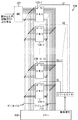
2:X線源
3:被検知体
4:位置制御装置
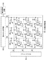
101:光電変換基板
102:シフトレジスタ
103:読み出し用回路
104:信号処理回路
105:シフトレジスタ
106:バッファアンプ
107:ADコンバータ回路
108−1〜108−n:メモリ
109:中央演算処理装置
Claims (10)
- 放射線発生手段と、
前記放射線発生手段からの放射線を電気信号に変換する複数の放射線検出素子がマトリクス状に配列されて構成され、放射線の検出を行う放射線検出手段と、
複数フレームにわたって前記放射線検出手段から電気信号を1フレーム毎に読み出す読み出し手段と、
前記読み出し手段が読み出した電気信号に対応したデータを、1フレーム毎に1群の情報群として、mフレーム分の情報群を記憶する記憶する記憶手段と、
mフレームにわたって前記放射線発生手段及び前記放射線検出手段が移動して前記放射線発生手段と前記放射線検出手段との間に配置される被検出体を透過した放射線の検出を前記放射線検出手段が行う時に、前記複数の放射線検出素子のうちの第1の放射線検出素子が位置する点と前記放射線発生手段が位置する点とを結ぶ直線と前記被検出体内に設定された第1の平面との交点が実質的に定点となっているように、前記第1の平面から距離D離間した第2の平面において前記放射線発生手段を移動量L X (m)移動させ、且つ、前記第1の平面から距離d離間した第3の面において前記放射線検出手段を移動させる位置制御手段と、
前記mフレーム分の情報群に基づいて前記第1の平面から前記放射線発生手段側に距離h離間した第4の平面の断層像を得るための処理を行う信号処理手段と、
を有し、
前記信号処理手段は、前記mフレーム分の情報群のうちの第1フレームの情報群における前記第1の放射線撮像素子のデータと、前記mフレーム分の情報群のうちの第mフレームの情報群における前記第1の放射線検出素子からZ X (m)ずれた位置に最も近い放射線検出素子のデータと、を用いて処理を行い、
- 前記記憶手段は、前記読み出し手段が読み出した電気信号をフレーム毎に記憶することを特徴とする請求項1に記載の放射線撮像装置。
- 前記記憶手段は、前記放射線検出手段による1フレーム分の放射線の検出で前記複数の放射線検出素子から得られた電気信号を1群の情報群として記憶し、
前記信号処理手段は、前記記憶手段に記憶された複数フレーム分の情報群に基づいて、前記平面又はこれに平行な平面の断層像を得ることを特徴とする請求項1又は2に記載の放射線撮像装置。 - 前記信号処理手段は、前記第1フレームの情報群における前記第1の放射線撮像素子のデータと、前記第mフレームの情報群における前記第1の放射線検出素子からZ X (m)ずれた位置に最も近い放射線検出素子のデータと、を用いて平均化処理を行うことを特徴とする請求項1乃至3のいずれか1項に記載の放射線撮像装置。
- 前記放射線発生手段及び前記放射線検出手段の移動は、前記放射線発生手段による放射線の発生及び前記放射線検出素子による放射線の電気信号への変換の度に停止することを特徴とする請求項1乃至4のいずれか1項に記載の放射線撮像装置。
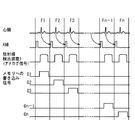
- 前記被検出体が動物である場合に、前記放射線発生手段は、前記動物の心臓の拍動及び呼吸の少なくとも一方に同期させてパルス状に放射線を発生することを特徴とする請求項1乃至5のいずれか1項に記載の放射線撮像装置。
- 前記放射線検出素子は、放射線を可視光に変換する波長変換体と、前記可視光を受光して電気信号に変換する光電変換体と、前記電気信号を転送するスイッチ素子と、を有することを特徴とする請求項1乃至6のいずれか1項に記載の放射線撮像装置。
- 前記光電変換体は、アモルファスシリコンを主材料として構成されていることを特徴とする請求項7に記載の放射線撮像装置。
- 放射線発生手段と、
前記放射線発生手段からの放射線を電気信号に変換する複数の放射線検出素子がマトリクス状に配列されて構成され、放射線の検出を行う放射線検出手段と、
前記放射線検出手段から電気信号を読み出す読み出し手段と、
前記読み出し手段が読み出した電気信号を記憶する記憶手段と、
前記記憶手段に記憶された電気信号の処理を行う信号処理手段と、
前記放射線発生手段と前記放射線検出手段の位置を制御する位置制御手段と、を有する放射線撮像装置を用いた放射線撮像方法であって、
mフレームにわたって前記放射線発生手段及び前記放射線検出手段が移動して前記放射線発生手段と前記放射線検出手段との間に配置される被検出体を透過した放射線の検出を前記放射線検出手段が行う時に、前記複数の放射線検出素子のうちの第1の放射線検出素子が位置する点と前記放射線発生手段が位置する点とを結ぶ直線と前記被検出体内に設定された第1の平面との交点が実質的に定点となっているように、前記位置制御手段を用いて、前記第1の平面から距離D離間した第2の平面において前記放射線発生手段を移動量L X (m)移動させ、且つ、前記第1の平面から距離d離間した第3の面において前記放射線検出手段を移動させ、
前記読み出し手段を用いて、mフレームにわたって、前記放射線検出素子が生成した電気信号を1フレーム毎に読み出し、
前記記憶手段を用いて、前記読み出し手段が読み出した電気信号に対応したデータを、1フレーム毎に1群の情報群として、mフレーム分の情報群を記憶し、
前記信号処理手段を用いて、前記mフレーム分の情報群に基づいて前記第1の平面から前記放射線発生手段側に距離h離間した第4の平面の断層像を得るために、前記mフレーム分の情報群のうちの第1フレームの情報群における前記第1の放射線撮像素子のデータと、前記mフレーム分の情報群のうちの第mフレームの情報群における前記第1の放射線検出素子からZ X (m)ずれた位置に最も近い放射線検出素子のデータと、を用いて処理を行い、
- 放射線発生手段と、
前記放射線発生手段からの放射線を電気信号に変換する複数の放射線検出素子がマトリクス状に配列されて構成され、放射線の検出を行う放射線検出手段と、
前記放射線検出手段から電気信号を読み出す読み出し手段と、
前記読み出し手段が読み出した電気信号を記憶する記憶手段と、
前記記憶手段に記憶された電気信号の処理を行う信号処理手段と、
前記放射線発生手段と前記放射線検出手段の位置を制御する位置制御手段と、を有する放射線撮像装置をコンピュータに制御させるためのプログラムであって、
mフレームにわたって前記放射線発生手段及び前記放射線検出手段が移動して前記放射線発生手段と前記放射線検出手段との間に配置される被検出体を透過した放射線の検出を前記放射線検出手段が行う時に、前記複数の放射線検出素子のうちの第1の放射線検出素子が位置する点と前記放射線発生手段が位置する点とを結ぶ直線と前記被検出体内に設定された第1の平面との交点が実質的に定点となっているように、前記位置制御手段を用いて、前記第1の平面から距離D離間した第2の平面において前記放射線発生手段を移動量L X (m)移動させ、且つ、前記第1の平面から距離d離間した第3の面において前記放射線検出手段を移動させる手順と、
前記読み出し手段を用いて、mフレームにわたって、前記放射線検出素子が生成した電気信号を1フレーム毎に読み出す手順と、
前記記憶手段を用いて、前記読み出し手段が読み出した電気信号に対応したデータを、1フレーム毎に1群の情報群として、mフレーム分の情報群を記憶する手順と、
前記信号処理手段を用いて、前記mフレーム分の情報群に基づいて前記第1の平面から前記放射線発生手段側に距離h離間した第4の平面の断層像を得るために、前記mフレーム分の情報群のうちの第1フレームの情報群における前記第1の放射線撮像素子のデータと、前記mフレーム分の情報群のうちの第mフレームの情報群における前記第1の放射線検出素子からZ X (m)ずれた位置に最も近い放射線検出素子のデータと、を用いて処理を行う手順と、
をコンピュータに実行させ、
Priority Applications (2)
| Application Number | Priority Date | Filing Date | Title |
|---|---|---|---|
| JP2004171226A JP4603823B2 (ja) | 2003-10-14 | 2004-06-09 | 放射線撮像装置、放射線撮像方法及びプログラム |
| US10/961,082 US7313219B2 (en) | 2003-10-14 | 2004-10-12 | Radiation image pick-up device, radiation image pick-up method and program |
Applications Claiming Priority (2)
| Application Number | Priority Date | Filing Date | Title |
|---|---|---|---|
| JP2003354158 | 2003-10-14 | ||
| JP2004171226A JP4603823B2 (ja) | 2003-10-14 | 2004-06-09 | 放射線撮像装置、放射線撮像方法及びプログラム |
Publications (3)
| Publication Number | Publication Date |
|---|---|
| JP2005137878A JP2005137878A (ja) | 2005-06-02 |
| JP2005137878A5 JP2005137878A5 (ja) | 2007-07-26 |
| JP4603823B2 true JP4603823B2 (ja) | 2010-12-22 |
Family
ID=34425385
Family Applications (1)
| Application Number | Title | Priority Date | Filing Date |
|---|---|---|---|
| JP2004171226A Expired - Fee Related JP4603823B2 (ja) | 2003-10-14 | 2004-06-09 | 放射線撮像装置、放射線撮像方法及びプログラム |
Country Status (2)
| Country | Link |
|---|---|
| US (1) | US7313219B2 (ja) |
| JP (1) | JP4603823B2 (ja) |
Families Citing this family (16)
| Publication number | Priority date | Publication date | Assignee | Title |
|---|---|---|---|---|
| DE102006023211A1 (de) * | 2006-05-17 | 2007-11-22 | Siemens Ag | Röntgenvorrichtung mit einem Röntgenstrahler und einem Röntgendetektor |
| WO2008018510A1 (en) * | 2006-08-08 | 2008-02-14 | Shimadzu Corporation | Radiation imaging device |
| EP2053972B1 (en) * | 2006-08-17 | 2013-09-11 | Koninklijke Philips Electronics N.V. | Computed tomography image acquisition |
| DE102007020642A1 (de) * | 2007-04-30 | 2008-11-06 | Dürr Dental GmbH & Co. KG | Röntgengerät sowie Sensoreinheit für ein Röntgengerät |
| JP2009130818A (ja) * | 2007-11-27 | 2009-06-11 | Canon Inc | 画像処理装置、画像処理装置の制御方法、プログラム及びコンピュータ記憶媒体 |
| JP5293920B2 (ja) * | 2007-12-28 | 2013-09-18 | 株式会社島津製作所 | X線断層撮影装置 |
| JP5283415B2 (ja) * | 2008-03-28 | 2013-09-04 | 富士フイルム株式会社 | 撮影装置及び露出制御方法 |
| WO2010070527A2 (en) * | 2008-12-15 | 2010-06-24 | Koninklijke Philips Electronics N. V. | Semicircular inversed offset scanning for enlarged field of view 3d |
| EP2378280A4 (en) * | 2008-12-22 | 2013-06-05 | Omron Tateisi Electronics Co | X-RAY INSPECTION METHOD AND X-RAY INSPECTION APPARATUS |
| WO2010074030A1 (ja) * | 2008-12-22 | 2010-07-01 | オムロン株式会社 | X線検査方法およびx線検査装置 |
| JP2010158299A (ja) | 2009-01-06 | 2010-07-22 | Fujifilm Corp | 断層撮影装置及び断層撮影方法 |
| JP5460106B2 (ja) * | 2009-04-03 | 2014-04-02 | キヤノン株式会社 | X線撮影装置及びその制御方法、コンピュータプログラム |
| JP2011062276A (ja) * | 2009-09-16 | 2011-03-31 | Fujifilm Corp | 放射線撮影装置 |
| JP5437001B2 (ja) * | 2009-09-28 | 2014-03-12 | 富士フイルム株式会社 | 放射線撮影装置 |
| JP5948275B2 (ja) * | 2013-03-29 | 2016-07-06 | 富士フイルム株式会社 | 放射線撮影装置及び放射線撮影方法、並びに放射線撮影制御プログラム |
| EP3420722B1 (en) * | 2016-02-23 | 2022-04-13 | Koninklijke Philips N.V. | Driving of an x-ray detector to compensate for cross scatter in an x-ray imaging apparatus |
Citations (16)
| Publication number | Priority date | Publication date | Assignee | Title |
|---|---|---|---|---|
| JPS4848571U (ja) * | 1971-10-05 | 1973-06-26 | ||
| JPS63102748A (ja) * | 1986-10-20 | 1988-05-07 | 株式会社 日立メデイコ | テレビ断層撮影装置 |
| US4903204A (en) * | 1987-12-01 | 1990-02-20 | Duke University | Matrix inversion tomosynthesis improvements in longitudinal X-ray slice imaging |
| JPH07303628A (ja) * | 1994-05-12 | 1995-11-21 | Toshiba Corp | ラミノグラフ |
| JPH10295680A (ja) * | 1997-04-25 | 1998-11-10 | Toshiba Corp | X線断層撮影装置 |
| JP2000024320A (ja) * | 1998-07-10 | 2000-01-25 | Namco Ltd | ゲームシステム及び情報記憶媒体 |
| JP2000079119A (ja) * | 1998-09-07 | 2000-03-21 | Shimadzu Corp | X線断層撮影装置 |
| JP2000157526A (ja) * | 1998-11-25 | 2000-06-13 | Picker Internatl Inc | 断層撮影システム |
| JP2000515046A (ja) * | 1996-07-23 | 2000-11-14 | ザ ジェネラル ホスピタル コーポレイション | 胸部撮像のための断層合成装置 |
| US6324249B1 (en) * | 2001-03-21 | 2001-11-27 | Agilent Technologies, Inc. | Electronic planar laminography system and method |
| JP2002136509A (ja) * | 2000-08-23 | 2002-05-14 | Shimadzu Corp | 断層撮影装置 |
| WO2002046729A1 (en) * | 2000-12-06 | 2002-06-13 | Teradyne, Inc. | Off-center tomosynthesis |
| JP3319905B2 (ja) * | 1995-03-24 | 2002-09-03 | 株式会社モリタ製作所 | デジタルx線撮影装置 |
| JP2003047610A (ja) * | 2001-08-07 | 2003-02-18 | Shimadzu Corp | X線ct装置 |
| JP2003052680A (ja) * | 2001-08-13 | 2003-02-25 | Shimadzu Corp | X線撮影装置 |
| JP2003135441A (ja) * | 2001-07-27 | 2003-05-13 | General Electric Co <Ge> | マンモグラフィ画像の高分解能3d描画のための方法及びシステム |
Family Cites Families (10)
| Publication number | Priority date | Publication date | Assignee | Title |
|---|---|---|---|---|
| US3626932A (en) * | 1968-10-11 | 1971-12-14 | Hal C Becker | Ekg synchronized x-ray double pulse exposure apparatus and method |
| US4387722A (en) * | 1978-11-24 | 1983-06-14 | Kearns Kenneth L | Respiration monitor and x-ray triggering apparatus |
| US5526442A (en) * | 1993-10-04 | 1996-06-11 | Hitachi Medical Corporation | X-ray radiography method and system |
| JP3066944B2 (ja) | 1993-12-27 | 2000-07-17 | キヤノン株式会社 | 光電変換装置、その駆動方法及びそれを有するシステム |
| US5573012A (en) * | 1994-08-09 | 1996-11-12 | The Regents Of The University Of California | Body monitoring and imaging apparatus and method |
| US5877501A (en) * | 1996-11-26 | 1999-03-02 | Picker International, Inc. | Digital panel for x-ray image acquisition |
| US6643536B2 (en) * | 2000-12-29 | 2003-11-04 | Ge Medical Systems Global Technology Company, Llc | System and method for synchronization of the acquisition of images with the cardiac cycle for dual energy imaging |
| JP3639826B2 (ja) * | 2002-04-03 | 2005-04-20 | キヤノン株式会社 | 放射線撮影装置、プログラム、コンピュータ可読記憶媒体、及び放射線撮影システム |
| US6970531B2 (en) * | 2002-10-07 | 2005-11-29 | General Electric Company | Continuous scan RAD tomosynthesis system and method |
| US6904121B2 (en) * | 2003-06-25 | 2005-06-07 | General Electric Company | Fourier based method, apparatus, and medium for optimal reconstruction in digital tomosynthesis |
-
2004
- 2004-06-09 JP JP2004171226A patent/JP4603823B2/ja not_active Expired - Fee Related
- 2004-10-12 US US10/961,082 patent/US7313219B2/en not_active Expired - Fee Related
Patent Citations (16)
| Publication number | Priority date | Publication date | Assignee | Title |
|---|---|---|---|---|
| JPS4848571U (ja) * | 1971-10-05 | 1973-06-26 | ||
| JPS63102748A (ja) * | 1986-10-20 | 1988-05-07 | 株式会社 日立メデイコ | テレビ断層撮影装置 |
| US4903204A (en) * | 1987-12-01 | 1990-02-20 | Duke University | Matrix inversion tomosynthesis improvements in longitudinal X-ray slice imaging |
| JPH07303628A (ja) * | 1994-05-12 | 1995-11-21 | Toshiba Corp | ラミノグラフ |
| JP3319905B2 (ja) * | 1995-03-24 | 2002-09-03 | 株式会社モリタ製作所 | デジタルx線撮影装置 |
| JP2000515046A (ja) * | 1996-07-23 | 2000-11-14 | ザ ジェネラル ホスピタル コーポレイション | 胸部撮像のための断層合成装置 |
| JPH10295680A (ja) * | 1997-04-25 | 1998-11-10 | Toshiba Corp | X線断層撮影装置 |
| JP2000024320A (ja) * | 1998-07-10 | 2000-01-25 | Namco Ltd | ゲームシステム及び情報記憶媒体 |
| JP2000079119A (ja) * | 1998-09-07 | 2000-03-21 | Shimadzu Corp | X線断層撮影装置 |
| JP2000157526A (ja) * | 1998-11-25 | 2000-06-13 | Picker Internatl Inc | 断層撮影システム |
| JP2002136509A (ja) * | 2000-08-23 | 2002-05-14 | Shimadzu Corp | 断層撮影装置 |
| WO2002046729A1 (en) * | 2000-12-06 | 2002-06-13 | Teradyne, Inc. | Off-center tomosynthesis |
| US6324249B1 (en) * | 2001-03-21 | 2001-11-27 | Agilent Technologies, Inc. | Electronic planar laminography system and method |
| JP2003135441A (ja) * | 2001-07-27 | 2003-05-13 | General Electric Co <Ge> | マンモグラフィ画像の高分解能3d描画のための方法及びシステム |
| JP2003047610A (ja) * | 2001-08-07 | 2003-02-18 | Shimadzu Corp | X線ct装置 |
| JP2003052680A (ja) * | 2001-08-13 | 2003-02-25 | Shimadzu Corp | X線撮影装置 |
Also Published As
| Publication number | Publication date |
|---|---|
| US20050078785A1 (en) | 2005-04-14 |
| JP2005137878A (ja) | 2005-06-02 |
| US7313219B2 (en) | 2007-12-25 |
Similar Documents
| Publication | Publication Date | Title |
|---|---|---|
| JP4603823B2 (ja) | 放射線撮像装置、放射線撮像方法及びプログラム | |
| US7386089B2 (en) | Radiographic imaging apparatus, control method thereof, and radiographic imaging system | |
| JP6122522B2 (ja) | 放射線撮影システムおよびその作動方法、並びに放射線画像検出装置 | |
| JP5460666B2 (ja) | 放射線撮影システムおよび放射線撮影システムの長尺撮影方法 | |
| US20050129298A1 (en) | Image pasting system using a digital detector | |
| US7953207B2 (en) | Radiation conversion panel and method of capturing radiation image therewith | |
| JP2004049887A (ja) | X線撮影装置およびx線撮影方法、並びに光電変換素子 | |
| JP2008206971A (ja) | 放射線撮像装置、その制御方法及び放射線撮像システム | |
| JP5902186B2 (ja) | 放射線撮影システム及び放射線撮影方法 | |
| JP2014028281A (ja) | X線検出器において3dゴーストアーチファクトを低減させる方法 | |
| JP4739060B2 (ja) | 放射線撮像装置、放射線撮像システム、及びその制御方法 | |
| JP5792569B2 (ja) | 放射線撮影システムおよび放射線撮影システムの長尺撮影方法 | |
| JPH10234724A (ja) | X線ct装置 | |
| JP2006346011A (ja) | 放射線撮像装置及びその制御方法 | |
| JP2006158728A (ja) | 放射線撮像装置及びその制御方法 | |
| WO2019049456A1 (ja) | 放射線撮像装置、その制御方法、及び、放射線撮像システム | |
| JP2005296340A (ja) | コーンビームx線ct撮影装置とそれを用いた画像取得方法 | |
| JP4227348B2 (ja) | X線発生装置の制御方法、プログラム、及びコンピュータ可読記憶媒体 | |
| JP2005270297A (ja) | 放射線ct撮影装置及び放射線ct撮影システム及びそれを用いた放射線ct撮影方法 | |
| JP6467148B2 (ja) | 放射線撮像装置および放射線撮像システム | |
| JP2003088519A (ja) | 歯科用x線撮影装置 | |
| JP4007607B2 (ja) | 放射線ct撮影装置及び放射線ct撮影システム及びそれを用いた放射線ct撮影方法 | |
| JP6701392B2 (ja) | 放射線撮像装置および放射線撮像システム | |
| JP2003250790A (ja) | 放射線撮影装置、放射線撮影方法、プログラム、及びコンピュータ可読記憶媒体 | |
| WO2013047069A1 (ja) | 放射線画像撮影システム、方法及び放射線画像撮影制御プログラム |
Legal Events
| Date | Code | Title | Description |
|---|---|---|---|
| A521 | Request for written amendment filed |
Free format text: JAPANESE INTERMEDIATE CODE: A523 Effective date: 20070607 |
|
| A621 | Written request for application examination |
Free format text: JAPANESE INTERMEDIATE CODE: A621 Effective date: 20070607 |
|
| A977 | Report on retrieval |
Free format text: JAPANESE INTERMEDIATE CODE: A971007 Effective date: 20100204 |
|
| A131 | Notification of reasons for refusal |
Free format text: JAPANESE INTERMEDIATE CODE: A131 Effective date: 20100223 |
|
| A521 | Request for written amendment filed |
Free format text: JAPANESE INTERMEDIATE CODE: A523 Effective date: 20100423 |
|
| TRDD | Decision of grant or rejection written | ||
| A01 | Written decision to grant a patent or to grant a registration (utility model) |
Free format text: JAPANESE INTERMEDIATE CODE: A01 Effective date: 20100928 |
|
| A01 | Written decision to grant a patent or to grant a registration (utility model) |
Free format text: JAPANESE INTERMEDIATE CODE: A01 |
|
| A61 | First payment of annual fees (during grant procedure) |
Free format text: JAPANESE INTERMEDIATE CODE: A61 Effective date: 20101004 |
|
| FPAY | Renewal fee payment (event date is renewal date of database) |
Free format text: PAYMENT UNTIL: 20131008 Year of fee payment: 3 |
|
| R150 | Certificate of patent or registration of utility model |
Free format text: JAPANESE INTERMEDIATE CODE: R150 |
|
| LAPS | Cancellation because of no payment of annual fees |