JP2005199062A - フルオロスコピー・トモシンセシス・システム及び方法 - Google Patents
フルオロスコピー・トモシンセシス・システム及び方法 Download PDFInfo
- Publication number
- JP2005199062A JP2005199062A JP2004369093A JP2004369093A JP2005199062A JP 2005199062 A JP2005199062 A JP 2005199062A JP 2004369093 A JP2004369093 A JP 2004369093A JP 2004369093 A JP2004369093 A JP 2004369093A JP 2005199062 A JP2005199062 A JP 2005199062A
- Authority
- JP
- Japan
- Prior art keywords
- image
- fluoroscopic
- detector
- interest
- images
- Prior art date
- Legal status (The legal status is an assumption and is not a legal conclusion. Google has not performed a legal analysis and makes no representation as to the accuracy of the status listed.)
- Pending
Links
- 238000000034 method Methods 0.000 title claims abstract description 58
- 238000003384 imaging method Methods 0.000 claims description 33
- 238000002594 fluoroscopy Methods 0.000 claims description 31
- 230000005855 radiation Effects 0.000 claims description 14
- 239000002872 contrast media Substances 0.000 claims description 4
- 238000002347 injection Methods 0.000 claims description 3
- 239000007924 injection Substances 0.000 claims description 3
- 239000000470 constituent Substances 0.000 claims 1
- 230000033001 locomotion Effects 0.000 description 14
- 238000009877 rendering Methods 0.000 description 14
- 210000001519 tissue Anatomy 0.000 description 14
- 230000008569 process Effects 0.000 description 13
- 238000012545 processing Methods 0.000 description 11
- 230000000875 corresponding effect Effects 0.000 description 9
- 238000013480 data collection Methods 0.000 description 7
- 210000003484 anatomy Anatomy 0.000 description 4
- 210000000988 bone and bone Anatomy 0.000 description 4
- 238000002583 angiography Methods 0.000 description 3
- 238000002059 diagnostic imaging Methods 0.000 description 3
- 230000009977 dual effect Effects 0.000 description 3
- 230000006870 function Effects 0.000 description 3
- 239000003550 marker Substances 0.000 description 3
- 230000003287 optical effect Effects 0.000 description 3
- 210000004872 soft tissue Anatomy 0.000 description 3
- 230000008901 benefit Effects 0.000 description 2
- 238000004891 communication Methods 0.000 description 2
- 238000004590 computer program Methods 0.000 description 2
- 238000000354 decomposition reaction Methods 0.000 description 2
- 230000002526 effect on cardiovascular system Effects 0.000 description 2
- 230000000694 effects Effects 0.000 description 2
- 238000001914 filtration Methods 0.000 description 2
- 238000003780 insertion Methods 0.000 description 2
- 230000037431 insertion Effects 0.000 description 2
- 238000007689 inspection Methods 0.000 description 2
- 238000012986 modification Methods 0.000 description 2
- 230000004048 modification Effects 0.000 description 2
- 230000008439 repair process Effects 0.000 description 2
- 230000000007 visual effect Effects 0.000 description 2
- 238000012935 Averaging Methods 0.000 description 1
- 230000004913 activation Effects 0.000 description 1
- 229910052788 barium Inorganic materials 0.000 description 1
- DSAJWYNOEDNPEQ-UHFFFAOYSA-N barium atom Chemical compound [Ba] DSAJWYNOEDNPEQ-UHFFFAOYSA-N 0.000 description 1
- 210000004204 blood vessel Anatomy 0.000 description 1
- 210000001124 body fluid Anatomy 0.000 description 1
- 239000010839 body fluid Substances 0.000 description 1
- 238000012790 confirmation Methods 0.000 description 1
- 238000012937 correction Methods 0.000 description 1
- 230000002596 correlated effect Effects 0.000 description 1
- 230000008878 coupling Effects 0.000 description 1
- 238000010168 coupling process Methods 0.000 description 1
- 238000005859 coupling reaction Methods 0.000 description 1
- 238000011161 development Methods 0.000 description 1
- 230000018109 developmental process Effects 0.000 description 1
- 238000003745 diagnosis Methods 0.000 description 1
- 238000010586 diagram Methods 0.000 description 1
- 230000002708 enhancing effect Effects 0.000 description 1
- 238000011156 evaluation Methods 0.000 description 1
- 210000001035 gastrointestinal tract Anatomy 0.000 description 1
- 230000006872 improvement Effects 0.000 description 1
- 230000002452 interceptive effect Effects 0.000 description 1
- 230000007246 mechanism Effects 0.000 description 1
- 230000002107 myocardial effect Effects 0.000 description 1
- 230000000399 orthopedic effect Effects 0.000 description 1
- 230000008447 perception Effects 0.000 description 1
- 238000002601 radiography Methods 0.000 description 1
- 230000009467 reduction Effects 0.000 description 1
- 230000011218 segmentation Effects 0.000 description 1
- 230000001629 suppression Effects 0.000 description 1
- 238000011477 surgical intervention Methods 0.000 description 1
- 238000001356 surgical procedure Methods 0.000 description 1
- 230000036962 time dependent Effects 0.000 description 1
- 238000013519 translation Methods 0.000 description 1
- 238000007487 urography Methods 0.000 description 1
- 230000002792 vascular Effects 0.000 description 1
- 238000012800 visualization Methods 0.000 description 1
Images
Classifications
-
- A—HUMAN NECESSITIES
- A61—MEDICAL OR VETERINARY SCIENCE; HYGIENE
- A61B—DIAGNOSIS; SURGERY; IDENTIFICATION
- A61B6/00—Apparatus for radiation diagnosis, e.g. combined with radiation therapy equipment
- A61B6/50—Clinical applications
- A61B6/504—Clinical applications involving diagnosis of blood vessels, e.g. by angiography
-
- A—HUMAN NECESSITIES
- A61—MEDICAL OR VETERINARY SCIENCE; HYGIENE
- A61B—DIAGNOSIS; SURGERY; IDENTIFICATION
- A61B6/00—Apparatus for radiation diagnosis, e.g. combined with radiation therapy equipment
- A61B6/02—Devices for diagnosis sequentially in different planes; Stereoscopic radiation diagnosis
- A61B6/025—Tomosynthesis
-
- G—PHYSICS
- G06—COMPUTING; CALCULATING OR COUNTING
- G06T—IMAGE DATA PROCESSING OR GENERATION, IN GENERAL
- G06T11/00—2D [Two Dimensional] image generation
- G06T11/003—Reconstruction from projections, e.g. tomography
- G06T11/006—Inverse problem, transformation from projection-space into object-space, e.g. transform methods, back-projection, algebraic methods
-
- A—HUMAN NECESSITIES
- A61—MEDICAL OR VETERINARY SCIENCE; HYGIENE
- A61B—DIAGNOSIS; SURGERY; IDENTIFICATION
- A61B6/00—Apparatus for radiation diagnosis, e.g. combined with radiation therapy equipment
- A61B6/48—Diagnostic techniques
- A61B6/481—Diagnostic techniques involving the use of contrast agents
-
- G—PHYSICS
- G06—COMPUTING; CALCULATING OR COUNTING
- G06T—IMAGE DATA PROCESSING OR GENERATION, IN GENERAL
- G06T2211/00—Image generation
- G06T2211/40—Computed tomography
- G06T2211/436—Limited angle
Abstract
【解決手段】 本技法は、関心領域に対して様々な透視点でのフルオロスコピー画像の収集が可能とする。次いで、1つ又はそれ以上の3次元画像をフルオロスコピー投影画像から再構成することができる。次に、フルオロスコピー応用のための解剖学的コンテキストを提供するよう3次元画像を表示することができる。最新のフルオロスコピー画像などのフルオロスコピー投影画像は、必要であれば、3次元画像上に重畳することができる。
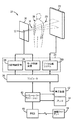
【選択図】 図1
Description
1.線源並びに検出器構成及び軌道
フルオロスコピー画像データの収集中、関心領域の画像は、スキャン軌道に沿って収集することができ、該スキャン軌道は、ある時間期間にわたって患者18に対する様々な透視点又は向きで線源12及び/又は検出器22を位置決めする。スキャン軌道は、線源の移動として最も容易に概念化され4るが、一般的には線源12、患者18、及び検出器22の互いに対する移動によって定義される。一般に、スキャン軌道は、様々なビュー角度でのフルオロスコピー画像データの収集を可能にし、これにより3次元画像の生成が可能になる。
連続するフルオロスコピー画像のタイミングすなわちフレーム・レート及び位置の選択は、様々な要因によって決定することができる。最も単純な場合、位置及びタイミングは、実行される処置の標準的な臨床診療に従ったものとすることができる。しかしながら、他の利用可能な情報もまた考慮することができる。
解剖学的関心領域が比較的小さい場合、又は十分に強力なコンピュータ及びプロセッサ出力が利用可能である場合、検出器22の最大分解能が画像収集のために用いることができる。最大よりも小さい検出器分解能を利用可能又は所望される別の場合には、観察領域、画像データ量、及び/又は3次元再構成及び観察に必要な時間を様々な手段によって低減することができる。
上述のようにフルオロスコピー画像データの主収集の前に関心領域の事前スキャンを行うことができる。かかる事前スキャンは、その後に生成される3次元画像の画質の改善を助けることができる。更に、事前スキャンは、結果として得られた3次元画像における重なり組織の影響を更に低減することができる。当業者には理解されるように、所望であれば、フルオロスコピー又は記録モードを事前スキャンのために用いることができる。次に、結果として得られた3次元画像は、以下でより詳細に説明されるように、臨床応用(例えばカテーテル挿入、骨修復など)に用いられることができ、その後追加の3次元画像を収集して、処置の成功を確実にすることができる。
収集されたフルオロスコピー画像データから3次元画像又はボリュームへの再構成は、様々なプロセスによって達成することができる。例えば再構成は、逆投影、フィルタ補正逆投影、代数的再構成法(ART)、或いは当該技術分野で公知の任意の他の再構成法によって達成することができる。投影画像は、利用可能であれば標準マーカから、又は画像内に存在する解剖学的マーカから位置合わせすることができる(又は各フレームの収集幾何形状が求められる)。再構成法の実施は、用途及び/又はオペレータの選択に応じて変えることができる。
再構成された3次元画像は、ボリューム・レンダリング法によって表示装置42上に表示することができ、関心のある任意の方向から観察することができる。更に3次元画像は、最大分解能又は低い分解能で表示することができる。前述のように、3次元画像は、現在のフルオロスコピーフレームを重畳した状態又は重畳しない状態で観察することができる。フルオロスコピー画像が3次元画像上に重畳される場合、通常、3次元画像と同じ分解能で表示され、2つの画像の位置合わせが容易となる。表示装置42は、多色表示装置又はグレイスケール表示装置とすることができ、これにより色又はグレイスケール強度のそれぞれを使用して骨と軟組織、又は造影剤と軟組織、或いはボリューム・レンダリングから最新のフルオロスコピー画像を区別することが可能となる。更に、フルオロスコピー画像シーケンスは、最大分解能で第2の表示装置42上に、必要であれば3次元構造体を重畳せずに表示することができ、これにより現今の臨床診療との一貫性が確実になる。
12 X線源
14 平面
16 X線ビーム
18 患者
20 放射線の一部
22 検出器
24 システム制御装置
26 モータ制御装置
28 X線制御装置
30 データ収集システム
36 コンピュータ
40 オペレータ・ワークステーション
42 表示装置
44 プリンタ
46 PACS(医用画像保管管理システム)
48 遠隔クライアント
Claims (10)
- 3次元画像を生成する方法であって、
関心領域に対して異なるビュー角度で2つ又はそれ以上のフルオロスコピー投影画像を収集する段階と、
前記2つ又はそれ以上のフルオロスコピー投影画像から少なくとも1つの3次元画像を再構成する段階と、
を含む方法。 - 前記2つ又はそれ以上のフルオロスコピー投影画像を収集する段階が、
心電図信号、自動造影剤注入、画像収集経過時間、表示された3次元画像に基づくフィードバック、侵襲的デバイスに関連する位置決めセンサ、照射パラメータを設定するためのプレショット、及びビデオ画像の少なくとも1つに基づいてフルオロスコピー投影画像収集の時間及び位置決めの内の少なくとも1つを確定する段階を含む請求項1に記載の方法。 - フルオロスコピー投影画像及び3次元画像の内の少なくとも1つを侵襲的デバイスに関連する電磁位置決め信号と相関付ける段階を含む請求項1に記載の方法。
- 前記少なくとも1つの3次元画像を再構成する段階が、
少なくとも1つの最新のフルオロスコピー投影画像を用いて既存の3次元画像を更新する段階を含む請求項1に記載の方法。 - 前記少なくとも1つの3次元画像を再構成する段階が、
心電図信号、収集幾何形状、及び3次元画像への再構成に用いられる投影の構成数の内の少なくとも1つに基づいて再構成のために2つ又はそれ以上のフルオロスコピー投影画像を選択する段階を含む請求項1に記載の方法。 - フルオロスコピー・イメージング・システム(10)であって、
関心領域に対して異なる位置から関心ボリュームを透過して放射線流(16、20)を放出するように構成されたX線源(12)と、
複数の検出器素子を含む検出器(22)であって、各検出器素子が前記それぞれの放射線流(16、20)に応答して1つ又はそれ以上の信号を発生することができる検出器(22)と、
X線源(12)を制御し、前記複数の検出器素子からの1つ又はそれ以上の信号を収集するように構成されたシステム制御装置(24)と、
前記1つ又はそれ以上の信号からの異なる透視点で関心領域の2つ又はそれ以上のフルオロスコピー投影画像を生成して、前記2つ又はそれ以上のフルオロスコピー投影画像から少なくとも1つの3次元画像を再構成するように構成されたコンピュータ・システム(36)と、
少なくとも1つの3次元画像を表示するように構成されたオペレータ・ワークステーション(40)と、
を備えるシステム。 - 前記X線源(12)が、1次元空間軌道、2次元空間軌道、又は3次元空間軌道の少なくとも1つで移動するように構成された請求項6に記載のフルオロスコピーイメージング・システム(10)。
- 前記システム制御装置(24)が前記検出器(22)の一部分だけを読み出すように構成され、前記一部分が関心領域に対応することを特徴とする請求項6に記載のフルオロスコピーイメージング・システム(10)。
- 前記システム制御装置(24)が、前記関心領域に関連する前記検出器(22)の第1の部分を第1の分解能で読み出し、前記関心領域に関連しない前記検出器(22)の第2の部分を第2の分解能で読み出すように構成され、該第2の分解能が前記第1の分解能より低いことを特徴とする請求項6に記載のフルオロスコピーイメージング・システム(10)。
- 前記オペレータ・ワークステーション(40)が、現在のフルオロスコピー投影画像をレンダリングされた3次元画像上に重畳するように構成された請求項6に記載のフルオロスコピーイメージング・システム(10)。
Applications Claiming Priority (1)
| Application Number | Priority Date | Filing Date | Title |
|---|---|---|---|
| US10/745,315 US7103136B2 (en) | 2003-12-22 | 2003-12-22 | Fluoroscopic tomosynthesis system and method |
Publications (2)
| Publication Number | Publication Date |
|---|---|
| JP2005199062A true JP2005199062A (ja) | 2005-07-28 |
| JP2005199062A5 JP2005199062A5 (ja) | 2009-12-24 |
Family
ID=34634527
Family Applications (1)
| Application Number | Title | Priority Date | Filing Date |
|---|---|---|---|
| JP2004369093A Pending JP2005199062A (ja) | 2003-12-22 | 2004-12-21 | フルオロスコピー・トモシンセシス・システム及び方法 |
Country Status (3)
| Country | Link |
|---|---|
| US (1) | US7103136B2 (ja) |
| JP (1) | JP2005199062A (ja) |
| FR (1) | FR2864301B1 (ja) |
Cited By (9)
| Publication number | Priority date | Publication date | Assignee | Title |
|---|---|---|---|---|
| JP2008006083A (ja) * | 2006-06-29 | 2008-01-17 | Toshiba Corp | 3次元画像生成装置 |
| JP2008068032A (ja) * | 2006-09-15 | 2008-03-27 | Toshiba Corp | 画像表示装置 |
| JP2009022754A (ja) * | 2007-07-19 | 2009-02-05 | General Electric Co <Ge> | 放射線画像の位置揃えを補正する方法 |
| JP2012011255A (ja) * | 2011-10-20 | 2012-01-19 | Toshiba Corp | 画像表示装置 |
| JP2012050476A (ja) * | 2010-08-31 | 2012-03-15 | Fujifilm Corp | 放射線撮影装置および方法並びにプログラム |
| WO2014174857A1 (ja) * | 2013-04-23 | 2014-10-30 | 株式会社日立メディコ | X線透視装置 |
| JP2017077455A (ja) * | 2015-08-04 | 2017-04-27 | パイ メディカル イメージング ビー ヴイPie Medical Imaging B.V. | 3d+時間再構成を改良する方法および装置 |
| JP2017205503A (ja) * | 2016-05-16 | 2017-11-24 | 東芝メディカルシステムズ株式会社 | X線診断装置、x線診断装置制御方法、x線診断装置制御プログラム、及びx線診断システム |
| JP2019524231A (ja) * | 2016-08-08 | 2019-09-05 | アダプティックス リミテッド | 空間的、時間的に重複するx線から3次元画像を再構成する方法及びシステム |
Families Citing this family (29)
| Publication number | Priority date | Publication date | Assignee | Title |
|---|---|---|---|---|
| US7868884B2 (en) * | 2004-08-31 | 2011-01-11 | General Electric Company | System and method for generating a digital image of an internal anatomy of a person |
| DE102005014286B4 (de) * | 2005-03-24 | 2006-12-21 | Siemens Ag | Diagnostikeinrichtung mit einem Röntgensystem und einem Ortungssystem für Katheder sowie Verfahren zu deren Betrieb |
| DE102005022538A1 (de) * | 2005-05-17 | 2006-11-30 | Siemens Ag | Vorrichtung und Verfahren zur Bedienung mehrerer medizinischer Geräte |
| DE102005030646B4 (de) * | 2005-06-30 | 2008-02-07 | Siemens Ag | Verfahren zur Kontur-Visualisierung von zumindest einer interessierenden Region in 2D-Durchleuchtungsbildern |
| US20070066911A1 (en) * | 2005-09-21 | 2007-03-22 | Klaus Klingenbeck-Regn | Integrated electrophysiology lab |
| JP4567064B2 (ja) * | 2005-10-17 | 2010-10-20 | 株式会社モリタ製作所 | 医療用デジタルx線撮影装置 |
| US7466790B2 (en) * | 2006-03-02 | 2008-12-16 | General Electric Company | Systems and methods for improving a resolution of an image |
| US8331635B2 (en) * | 2006-07-06 | 2012-12-11 | University Of South Florida | Cartesian human morpho-informatic system |
| JP4903660B2 (ja) * | 2007-09-27 | 2012-03-28 | 富士フイルム株式会社 | 放射線画像処理装置及び処理方法 |
| JP2011000369A (ja) * | 2009-06-22 | 2011-01-06 | Toshiba Corp | X線診断装置 |
| DE102009043421A1 (de) * | 2009-09-29 | 2011-04-07 | Siemens Aktiengesellschaft | Verfahren und Vorrichtung |
| CA2809002C (en) | 2010-08-20 | 2017-11-21 | Amei Technologies, Inc. | Method and system for roentgenography-based modeling |
| DE102010040963A1 (de) * | 2010-09-17 | 2012-03-22 | Siemens Aktiengesellschaft | Verfahren und Röntgengerät zur Erzeugung eines Röntgen-Projektionsbildes |
| US9536314B2 (en) * | 2010-10-20 | 2017-01-03 | Siemens Medical Solutions Usa, Inc. | Image reconstruction |
| US8737562B2 (en) * | 2010-12-03 | 2014-05-27 | Shimadzu Corporation | Body section radiographic apparatus, and a noise removing method for the body section radiographic apparatus |
| DE102012200246A1 (de) * | 2012-01-10 | 2013-07-11 | Sirona Dental Systems Gmbh | Verfahren zur Darstellung eines Teilvolumens eines Gesamtvolumens |
| EP3474044B1 (en) * | 2014-01-27 | 2020-08-12 | Epica International Inc. | Radiological imaging device with improved functioning |
| US9247920B2 (en) | 2014-02-27 | 2016-02-02 | General Electric Company | System and method for performing bi-plane tomographic acquisitions |
| US9408581B2 (en) * | 2014-03-07 | 2016-08-09 | Elwha Llc | Systems, devices, and methods for lowering dental x-ray dosage including feedback sensors |
| US9898840B2 (en) * | 2014-05-15 | 2018-02-20 | General Electric Company | Systems and methods for continuous motion breast tomosynthesis |
| US9532757B2 (en) | 2014-06-30 | 2017-01-03 | General Electric Company | C-arm system and C-arm spin acquisition trajectories for dynamic imaging and improved image quality and method of use |
| CN104200500B (zh) * | 2014-07-29 | 2017-06-06 | 沈阳东软医疗系统有限公司 | 一种心脏图像的重建方法及装置 |
| US9526468B2 (en) * | 2014-09-09 | 2016-12-27 | General Electric Company | Multiple frame acquisition for exposure control in X-ray medical imagers |
| US20170294033A1 (en) * | 2016-04-06 | 2017-10-12 | Varex Imaging Corporation | Dose efficient x-ray detector and method |
| US10973479B2 (en) * | 2016-05-16 | 2021-04-13 | Canon Medical Systems Corporation | X-ray diagnosis apparatus, X-ray diagnosis apparatus controlling method, and X-ray diagnosis system |
| JP2019013672A (ja) * | 2017-07-10 | 2019-01-31 | キヤノン株式会社 | 放射線撮影装置および放射線撮影システム |
| US10893842B2 (en) | 2018-02-08 | 2021-01-19 | Covidien Lp | System and method for pose estimation of an imaging device and for determining the location of a medical device with respect to a target |
| US10779791B2 (en) * | 2018-03-16 | 2020-09-22 | General Electric Company | System and method for mobile X-ray imaging |
| AU2019314380A1 (en) * | 2018-07-30 | 2021-02-18 | Xenselab Llc | System and methods for x-ray imaging and a contrast agent |
Citations (4)
| Publication number | Priority date | Publication date | Assignee | Title |
|---|---|---|---|---|
| JPH10295680A (ja) * | 1997-04-25 | 1998-11-10 | Toshiba Corp | X線断層撮影装置 |
| WO2002056770A1 (en) * | 2000-12-28 | 2002-07-25 | Ge Medical Systems Global Technology Company, Llc | Fluoroscopic x-ray tomography imaging |
| JP2003290192A (ja) * | 2002-03-11 | 2003-10-14 | Siemens Ag | 患者の検査領域に導入された医療器具の画像描出方法 |
| US20030220555A1 (en) * | 2002-03-11 | 2003-11-27 | Benno Heigl | Method and apparatus for image presentation of a medical instrument introduced into an examination region of a patent |
Family Cites Families (10)
| Publication number | Priority date | Publication date | Assignee | Title |
|---|---|---|---|---|
| US5175754A (en) * | 1992-05-11 | 1992-12-29 | General Electric Company | Gantry position reference for tomographic scanners |
| US5530935A (en) * | 1993-09-20 | 1996-06-25 | U.S. Philips Corporation | X-ray examination apparatus |
| US6396898B1 (en) * | 1999-12-24 | 2002-05-28 | Kabushiki Kaisha Toshiba | Radiation detector and x-ray CT apparatus |
| US6307910B1 (en) * | 2000-01-07 | 2001-10-23 | Ge Medical Systems Global Technology Company, Llc | Methods and apparatus for reduced radiation coronary computed tomography imaging |
| US6484049B1 (en) * | 2000-04-28 | 2002-11-19 | Ge Medical Systems Global Technology Company, Llc | Fluoroscopic tracking and visualization system |
| JP2002119502A (ja) * | 2000-10-17 | 2002-04-23 | Toshiba Corp | 医用装置 |
| US6895077B2 (en) * | 2001-11-21 | 2005-05-17 | University Of Massachusetts Medical Center | System and method for x-ray fluoroscopic imaging |
| FR2838043B1 (fr) * | 2002-04-05 | 2005-03-11 | Jean Noel Vallee | Systeme d'aide a la navigation en temps reel pour dispositif de radiographie |
| US20050053200A1 (en) * | 2003-08-07 | 2005-03-10 | Predrag Sukovic | Intra-operative CT scanner |
| US8571639B2 (en) * | 2003-09-05 | 2013-10-29 | Varian Medical Systems, Inc. | Systems and methods for gating medical procedures |
-
2003
- 2003-12-22 US US10/745,315 patent/US7103136B2/en active Active
-
2004
- 2004-12-21 FR FR0413665A patent/FR2864301B1/fr not_active Expired - Fee Related
- 2004-12-21 JP JP2004369093A patent/JP2005199062A/ja active Pending
Patent Citations (4)
| Publication number | Priority date | Publication date | Assignee | Title |
|---|---|---|---|---|
| JPH10295680A (ja) * | 1997-04-25 | 1998-11-10 | Toshiba Corp | X線断層撮影装置 |
| WO2002056770A1 (en) * | 2000-12-28 | 2002-07-25 | Ge Medical Systems Global Technology Company, Llc | Fluoroscopic x-ray tomography imaging |
| JP2003290192A (ja) * | 2002-03-11 | 2003-10-14 | Siemens Ag | 患者の検査領域に導入された医療器具の画像描出方法 |
| US20030220555A1 (en) * | 2002-03-11 | 2003-11-27 | Benno Heigl | Method and apparatus for image presentation of a medical instrument introduced into an examination region of a patent |
Cited By (11)
| Publication number | Priority date | Publication date | Assignee | Title |
|---|---|---|---|---|
| JP2008006083A (ja) * | 2006-06-29 | 2008-01-17 | Toshiba Corp | 3次元画像生成装置 |
| JP2008068032A (ja) * | 2006-09-15 | 2008-03-27 | Toshiba Corp | 画像表示装置 |
| JP2009022754A (ja) * | 2007-07-19 | 2009-02-05 | General Electric Co <Ge> | 放射線画像の位置揃えを補正する方法 |
| JP2012050476A (ja) * | 2010-08-31 | 2012-03-15 | Fujifilm Corp | 放射線撮影装置および方法並びにプログラム |
| JP2012011255A (ja) * | 2011-10-20 | 2012-01-19 | Toshiba Corp | 画像表示装置 |
| WO2014174857A1 (ja) * | 2013-04-23 | 2014-10-30 | 株式会社日立メディコ | X線透視装置 |
| JP2017077455A (ja) * | 2015-08-04 | 2017-04-27 | パイ メディカル イメージング ビー ヴイPie Medical Imaging B.V. | 3d+時間再構成を改良する方法および装置 |
| JP2017205503A (ja) * | 2016-05-16 | 2017-11-24 | 東芝メディカルシステムズ株式会社 | X線診断装置、x線診断装置制御方法、x線診断装置制御プログラム、及びx線診断システム |
| JP7066332B2 (ja) | 2016-05-16 | 2022-05-13 | キヤノンメディカルシステムズ株式会社 | X線診断装置及びx線診断装置制御方法 |
| JP2019524231A (ja) * | 2016-08-08 | 2019-09-05 | アダプティックス リミテッド | 空間的、時間的に重複するx線から3次元画像を再構成する方法及びシステム |
| JP7072556B2 (ja) | 2016-08-08 | 2022-05-20 | アダプティックス リミテッド | 空間的、時間的に重複するx線から3次元画像を再構成する方法及びシステム |
Also Published As
| Publication number | Publication date |
|---|---|
| FR2864301A1 (fr) | 2005-06-24 |
| US7103136B2 (en) | 2006-09-05 |
| US20050135558A1 (en) | 2005-06-23 |
| FR2864301B1 (fr) | 2011-06-24 |
Similar Documents
| Publication | Publication Date | Title |
|---|---|---|
| US7103136B2 (en) | Fluoroscopic tomosynthesis system and method | |
| JP4859446B2 (ja) | 回転血管撮影のための血管撮影x線診断装置 | |
| JP4054402B2 (ja) | X線断層撮影装置 | |
| US9044190B2 (en) | C-arm computerized tomography system | |
| US7180976B2 (en) | Rotational angiography based hybrid 3-D reconstruction of coronary arterial structure | |
| JP4854137B2 (ja) | 医用画像診断装置 | |
| US8447078B2 (en) | X-ray diagnostic device | |
| Strobel et al. | 3D imaging with flat-detector C-arm systems | |
| JP4327801B2 (ja) | X線断層撮影装置 | |
| EP1785092A1 (en) | X-ray angiography apparatus | |
| JP4537129B2 (ja) | トモシンセシス用途における対象物を走査するためのシステム | |
| CN102335004B (zh) | 用于进行血管造影检查的方法和计算机断层造影设备 | |
| JP2006055645A (ja) | 遺構空間情報を用いて三次元x線画像再生における金属遺構を低減する方法及び装置 | |
| JP5438984B2 (ja) | X線画像診断装置及びx線画像処理方法 | |
| US20060291615A1 (en) | X-ray CT apparatus | |
| JP2002083281A (ja) | 実時間三次元再構成による容積の高品質表示を提供するイメージング装置および方法 | |
| US20170135654A1 (en) | Automatic or assisted region of interest positioning in x-ray diagnostics and interventions | |
| JP4236666B2 (ja) | X線断層撮影装置 | |
| JP5097355B2 (ja) | 放射線断層撮影装置 | |
| GB2408343A (en) | Image reconstruction with projected images aquired in a non-circular arc | |
| Badea et al. | A 3D imaging system for dental imaging based on digital tomosynthesis and cone beam CT | |
| CN116269452A (zh) | 用于定位c形臂的方法和系统 | |
| Gupta et al. | Full-Field Cardiac Imaging using Ultra-High Resolution Flat-Panel Volume-Computed Tomography Mannudeep K Kalra, and Thomas J Brady |
Legal Events
| Date | Code | Title | Description |
|---|---|---|---|
| A521 | Request for written amendment filed |
Free format text: JAPANESE INTERMEDIATE CODE: A523 Effective date: 20071220 |
|
| A621 | Written request for application examination |
Free format text: JAPANESE INTERMEDIATE CODE: A621 Effective date: 20071220 |
|
| A521 | Request for written amendment filed |
Free format text: JAPANESE INTERMEDIATE CODE: A523 Effective date: 20091106 |
|
| RD02 | Notification of acceptance of power of attorney |
Free format text: JAPANESE INTERMEDIATE CODE: A7422 Effective date: 20091106 |
|
| RD04 | Notification of resignation of power of attorney |
Free format text: JAPANESE INTERMEDIATE CODE: A7424 Effective date: 20091106 |
|
| A131 | Notification of reasons for refusal |
Free format text: JAPANESE INTERMEDIATE CODE: A131 Effective date: 20110105 |
|
| A601 | Written request for extension of time |
Free format text: JAPANESE INTERMEDIATE CODE: A601 Effective date: 20110324 |
|
| A602 | Written permission of extension of time |
Free format text: JAPANESE INTERMEDIATE CODE: A602 Effective date: 20110329 |
|
| A521 | Request for written amendment filed |
Free format text: JAPANESE INTERMEDIATE CODE: A523 Effective date: 20110331 |
|
| A131 | Notification of reasons for refusal |
Free format text: JAPANESE INTERMEDIATE CODE: A131 Effective date: 20111004 |
|
| A602 | Written permission of extension of time |
Free format text: JAPANESE INTERMEDIATE CODE: A602 Effective date: 20111208 |
|
| A521 | Request for written amendment filed |
Free format text: JAPANESE INTERMEDIATE CODE: A523 Effective date: 20120309 |
|
| A02 | Decision of refusal |
Free format text: JAPANESE INTERMEDIATE CODE: A02 Effective date: 20121002 |