EP4449978A2 - Gewebeintegrierende sensoren - Google Patents
Gewebeintegrierende sensoren Download PDFInfo
- Publication number
- EP4449978A2 EP4449978A2 EP24158962.1A EP24158962A EP4449978A2 EP 4449978 A2 EP4449978 A2 EP 4449978A2 EP 24158962 A EP24158962 A EP 24158962A EP 4449978 A2 EP4449978 A2 EP 4449978A2
- Authority
- EP
- European Patent Office
- Prior art keywords
- tissue
- integrating
- scaffold
- sensing
- sensor
- Prior art date
- Legal status (The legal status is an assumption and is not a legal conclusion. Google has not performed a legal analysis and makes no representation as to the accuracy of the status listed.)
- Granted
Links
Images


Classifications
-
- A—HUMAN NECESSITIES
- A61—MEDICAL OR VETERINARY SCIENCE; HYGIENE
- A61B—DIAGNOSIS; SURGERY; IDENTIFICATION
- A61B5/00—Measuring for diagnostic purposes; Identification of persons
- A61B5/145—Measuring characteristics of blood in vivo, e.g. gas concentration or pH-value ; Measuring characteristics of body fluids or tissues, e.g. interstitial fluid or cerebral tissue
- A61B5/1455—Measuring characteristics of blood in vivo, e.g. gas concentration or pH-value ; Measuring characteristics of body fluids or tissues, e.g. interstitial fluid or cerebral tissue using optical sensors, e.g. spectral photometrical oximeters
- A61B5/1459—Measuring characteristics of blood in vivo, e.g. gas concentration or pH-value ; Measuring characteristics of body fluids or tissues, e.g. interstitial fluid or cerebral tissue using optical sensors, e.g. spectral photometrical oximeters invasive, e.g. introduced into the body by a catheter
-
- A—HUMAN NECESSITIES
- A61—MEDICAL OR VETERINARY SCIENCE; HYGIENE
- A61B—DIAGNOSIS; SURGERY; IDENTIFICATION
- A61B5/00—Measuring for diagnostic purposes; Identification of persons
-
- A—HUMAN NECESSITIES
- A61—MEDICAL OR VETERINARY SCIENCE; HYGIENE
- A61B—DIAGNOSIS; SURGERY; IDENTIFICATION
- A61B5/00—Measuring for diagnostic purposes; Identification of persons
- A61B5/0002—Remote monitoring of patients using telemetry, e.g. transmission of vital signals via a communication network
- A61B5/0004—Remote monitoring of patients using telemetry, e.g. transmission of vital signals via a communication network characterised by the type of physiological signal transmitted
-
- A—HUMAN NECESSITIES
- A61—MEDICAL OR VETERINARY SCIENCE; HYGIENE
- A61B—DIAGNOSIS; SURGERY; IDENTIFICATION
- A61B5/00—Measuring for diagnostic purposes; Identification of persons
- A61B5/0002—Remote monitoring of patients using telemetry, e.g. transmission of vital signals via a communication network
- A61B5/0015—Remote monitoring of patients using telemetry, e.g. transmission of vital signals via a communication network characterised by features of the telemetry system
- A61B5/0017—Remote monitoring of patients using telemetry, e.g. transmission of vital signals via a communication network characterised by features of the telemetry system transmitting optical signals
-
- A—HUMAN NECESSITIES
- A61—MEDICAL OR VETERINARY SCIENCE; HYGIENE
- A61B—DIAGNOSIS; SURGERY; IDENTIFICATION
- A61B5/00—Measuring for diagnostic purposes; Identification of persons
- A61B5/0002—Remote monitoring of patients using telemetry, e.g. transmission of vital signals via a communication network
- A61B5/0031—Implanted circuitry
-
- A—HUMAN NECESSITIES
- A61—MEDICAL OR VETERINARY SCIENCE; HYGIENE
- A61B—DIAGNOSIS; SURGERY; IDENTIFICATION
- A61B5/00—Measuring for diagnostic purposes; Identification of persons
- A61B5/07—Endoradiosondes
- A61B5/076—Permanent implantation
-
- A—HUMAN NECESSITIES
- A61—MEDICAL OR VETERINARY SCIENCE; HYGIENE
- A61B—DIAGNOSIS; SURGERY; IDENTIFICATION
- A61B5/00—Measuring for diagnostic purposes; Identification of persons
- A61B5/145—Measuring characteristics of blood in vivo, e.g. gas concentration or pH-value ; Measuring characteristics of body fluids or tissues, e.g. interstitial fluid or cerebral tissue
- A61B5/14503—Measuring characteristics of blood in vivo, e.g. gas concentration or pH-value ; Measuring characteristics of body fluids or tissues, e.g. interstitial fluid or cerebral tissue invasive, e.g. introduced into the body by a catheter or needle or using implanted sensors
-
- A—HUMAN NECESSITIES
- A61—MEDICAL OR VETERINARY SCIENCE; HYGIENE
- A61B—DIAGNOSIS; SURGERY; IDENTIFICATION
- A61B5/00—Measuring for diagnostic purposes; Identification of persons
- A61B5/145—Measuring characteristics of blood in vivo, e.g. gas concentration or pH-value ; Measuring characteristics of body fluids or tissues, e.g. interstitial fluid or cerebral tissue
- A61B5/14532—Measuring characteristics of blood in vivo, e.g. gas concentration or pH-value ; Measuring characteristics of body fluids or tissues, e.g. interstitial fluid or cerebral tissue for measuring glucose, e.g. by tissue impedance measurement
-
- A—HUMAN NECESSITIES
- A61—MEDICAL OR VETERINARY SCIENCE; HYGIENE
- A61B—DIAGNOSIS; SURGERY; IDENTIFICATION
- A61B5/00—Measuring for diagnostic purposes; Identification of persons
- A61B5/145—Measuring characteristics of blood in vivo, e.g. gas concentration or pH-value ; Measuring characteristics of body fluids or tissues, e.g. interstitial fluid or cerebral tissue
- A61B5/1468—Measuring characteristics of blood in vivo, e.g. gas concentration or pH-value ; Measuring characteristics of body fluids or tissues, e.g. interstitial fluid or cerebral tissue using chemical or electrochemical methods, e.g. by polarographic means
- A61B5/1473—Measuring characteristics of blood in vivo, e.g. gas concentration or pH-value ; Measuring characteristics of body fluids or tissues, e.g. interstitial fluid or cerebral tissue using chemical or electrochemical methods, e.g. by polarographic means invasive, e.g. introduced into the body by a catheter
- A61B5/14735—Measuring characteristics of blood in vivo, e.g. gas concentration or pH-value ; Measuring characteristics of body fluids or tissues, e.g. interstitial fluid or cerebral tissue using chemical or electrochemical methods, e.g. by polarographic means invasive, e.g. introduced into the body by a catheter comprising an immobilised reagent
-
- A—HUMAN NECESSITIES
- A61—MEDICAL OR VETERINARY SCIENCE; HYGIENE
- A61B—DIAGNOSIS; SURGERY; IDENTIFICATION
- A61B5/00—Measuring for diagnostic purposes; Identification of persons
- A61B5/145—Measuring characteristics of blood in vivo, e.g. gas concentration or pH-value ; Measuring characteristics of body fluids or tissues, e.g. interstitial fluid or cerebral tissue
- A61B5/1495—Calibrating or testing of in-vivo probes
-
- A—HUMAN NECESSITIES
- A61—MEDICAL OR VETERINARY SCIENCE; HYGIENE
- A61B—DIAGNOSIS; SURGERY; IDENTIFICATION
- A61B5/00—Measuring for diagnostic purposes; Identification of persons
- A61B5/68—Arrangements of detecting, measuring or recording means, e.g. sensors, in relation to patient
- A61B5/6801—Arrangements of detecting, measuring or recording means, e.g. sensors, in relation to patient specially adapted to be attached to or worn on the body surface
- A61B5/6802—Sensor mounted on worn items
- A61B5/681—Wristwatch-type devices
-
- A—HUMAN NECESSITIES
- A61—MEDICAL OR VETERINARY SCIENCE; HYGIENE
- A61B—DIAGNOSIS; SURGERY; IDENTIFICATION
- A61B5/00—Measuring for diagnostic purposes; Identification of persons
- A61B5/72—Signal processing specially adapted for physiological signals or for diagnostic purposes
- A61B5/7271—Specific aspects of physiological measurement analysis
- A61B5/7278—Artificial waveform generation or derivation, e.g. synthesizing signals from measured signals
Definitions
- the present disclosure is in the field of biosensors.
- U.S. Patent No. 7,228,159 describes a senor comprising a plurality of non-biodegradable sensing particles embedded in a biodegradable matrix for injection into the dermis. However, as the matrix degrades, the sensing particles are ingested by macrophages and removed from the implant site.
- U.S. Patent No. 6,671,527 describes a sensor which is injected into epidermis and is ejected over time due to the normal sloughing of skin.
- U.S. Patent Application No. 2009/0131773 describes a carbohydrate (e.g ., glucose) sensor made up of at least two different variants of an appropriate competitive binding assay.
- sensors As sensors become encapsulated by avascular tissue, they lose ability to accurately sense blood borne analytes and as they become engulfed by phagocytic cells (small particles), they lose contact with interstitial fluid, which is the compartment necessary to be sensed for components such as glucose. Therefore, current sensing technologies typically fail after only a short time in the body ( e.g., 2-7 days for commercially available sensors).
- tissue-integrating sensors systems comprising these sensors and methods of using these sensors and systems for the measurement of various analytes.
- tissue-integrating sensor for detecting an analyte
- the sensor comprising a tissue-integrating scaffold; and one or more sensing moieties, wherein the sensing moieties produce a detectable signal in the presence of the analyte; and further wherein the sensor provides detection of the analyte when placed ( e.g ., implanted) into the tissue of a subject.
- the tissue-integrating sensors as described herein can provide long-term detection of the analyte(s).
- the tissue-integrating scaffold consists of the one or more sensing moieties (e.g ., polymeric sensing moieties formed into a scaffold).
- the tissue-integrating sensors may comprise one or more polymers, for example one or more hydrogels.
- the sensing moieties may be embedded and/or attached to the exterior of the scaffold or may form the scaffold itself.
- the scaffold is porous and further wherein at least two of the pores are interconnected.
- the sensing moieties comprise microspheres or nanospheres. Any of the sensors described herein may include one or more layers (with sensing moieties in one or more of the layers) and/or one or more fibers.
- any of the sensors described herein may further comprise additional components, for example, a coating on the exterior of the sensor and/or one or more additional reference (calibration) moieties, for example for calibrating the signal detected from the sensing moieties.
- additional components for example, a coating on the exterior of the sensor and/or one or more additional reference (calibration) moieties, for example for calibrating the signal detected from the sensing moieties.
- a system for detecting an analyte comprising one or more of the tissue-integrating sensors as described herein; and an interrogator that generates (e.g ., light that causes the sensing moieties to emit light) and/or measures the signal produced by the sensing moieties.
- the system further includes one or more of the following: a detector, a signal receiver, a signal transmitter, a signal processing component, an energy storage component, a data storage component, a data transmitter, a data display device, a data processing component and combinations thereof.
- provided herein are methods of making and using the sensors and systems as described herein.
- a method for detection of an analyte in a tissue of a subject comprising integrating one or more sensors as described herein into the tissue and detecting the presence of the analyte.
- tissue-integrating sensors useful for accurate and optionally long term measurements of analytes in vivo. Also described herein are methods of using these sensors for optical detection of various biochemical analytes. Using reversible binding ligands and/or chemistries, the implantable sensors, systems and methods described herein provide for continuous or semi-continuous collection of data of various biochemical analytes, optionally without the use of implantable hardware of any type and/or enzymatic and electrochemical detection methods.
- tissue-integrating sensors that are the subject of this invention remain in good contact (close proximity) to blood vessels and have direct access to measurements of interstitial fluid.
- the tissue-integrating scaffold encourages capillary growth into and/or nearby the sensing media.
- the sensing media is devoid of electronics, making the sensing media seem less foreign to the body than implants that contain electronics. Additionally the tissue-integrating sensing media may have a modulus closer to the texture of tissue, thus enhancing the integration in the tissue.
- the sensors described herein allow capillaries to grow in close proximity to all regions of the sensor ( e.g ., on the surface and inside), which results in accurate analyte measurements, including over long term.
- Embedding, attaching or forming scaffolds out of nano-sized sensing elements results in tissue-integrating sensing media that allows in-growth, including of tissue and capillaries, in and/or around the sensors.
- Tissue integrating sensors minimize the foreign body response and/or promote vascularization.
- Capillary growth directly into and throughout the sensor allows unencumbered access to analytes of interest in the blood (e.g. glucose, lactate, pyruvate, cortisol, ions, proteins, nucleic acids, alcohols, urea, etc.).
- the level of tissue integration and proximity of capillaries to all regions of the sensor will provide a close, stable relationship between the analyte concentration in the blood and in the tissue surrounding the sensing media.
- Advantages of the device and methods described herein include, but are not limited to: (1) providing devices that integrate into the subject (e . g ., through tissue and/or capillary in-growth; (2) providing devices which can be implanted through syringe injection, meaning that no surgery is required to put the sensing media in place in the body; (3) providing devices that do not include sensor electronics in the body; (4) providing devices comprising material(s) having properties more similar to actual tissue (e.g ., modulus that is more similar to tissue's modulus and hydrogel water content) to allow a better acceptance into the tissue; (5) providing devices that accurately assess analyte(s) for long periods of time ( e . g ., greater than a week, typically weeks, months or years) and/or (6) providing devices of small dimensions which will give result in increased patent comfort and better acceptance by the body.
- tissue integrating refers to a material (e.g ., scaffold) which, when integrated into living tissue remains in close proximity with the blood vessels of the tissue (e.g ., capillaries).
- close proximity is meant that the average distance from any point within the material (scaffold) implanted into the tissue to the nearest blood vessel is no greater than 100 microns more than the average distance from any point in the native (original) tissue to the nearest blood vessel.
- long-term is meant that the implant senses the analyte for greater than about 7 days, for example weeks, months, or years.
- biodegradable or “bioabsorbable” is meant that the material is capable of being broken down by the subject's body over a period of time, ranging from days to weeks to months or years.
- biodegradable materials may include water-soluble biomaterials.
- hydrogel is meant a material that absorbs a solvent (e.g. water), undergoes rapid swelling without discernible dissolution, and maintains three-dimensional networks capable of reversible deformation.
- a solvent e.g. water
- the sensors are made up of tissue-integrating scaffolds and at least one sensing moiety.
- the sensors described herein typically comprise a tissue-integrating scaffold (also referred to as a matrix) material.
- the tissue-integrating scaffold of the invention may be constructed with materials and/or micro-architecture such that the scaffold promotes tissue-integration and/or vascularization.
- porous scaffolds provide tissue biomaterial anchoring and promote in-growth throughout the pores. The resulting "hallway" or “channel” pattern of tissue growth are healthy, space-filling masses that persist over time and promote host cell integration.
- Most or all of the pores of the biomaterials described herein are preferably interconnected (co-continuous).
- tissue integrating scaffolds include fibers ( e.g., 1 to 10 or more microns in diameter, such as 5, 6, 7, 8, 9, 10 or more microns), which may be arranged in non-random or random configuration. Tissue-integrating scaffolds (in any configuration) can also be formed by multiphoton polymerization techniques. Kaehr et al. (2008) Proc. Nat'l. Acad. Sci. USA 105(26):8850-8854 ; Nielson et al. (2009) Small 1:120-125 ; Kasprzak, Doctoral Dissertation, Georgia Institute of Technology, May 2009 .
- the tissue-integrating scaffold of the invention may comprise any material, including but not limited to synthetic polymers, naturally-occurring substances, or mixtures thereof.
- synthetic polymers include, but are not limited to polyethylene glycol (PEG), 2-hydroxyethyl methacrylate (HEMA), silicone rubber, poly([epsilon]-caprolactone) dimethylacrylate, polysulfone, (poly)methy methacrylate (PMMA), soluble Teflon-AF, (poly) ethylenetetrapthalate (PET, Dacron), Nylon, polyvinyl alcohol, polyacrylamide, polyurethane, and mixtures thereof.
- PEG polyethylene glycol
- HEMA 2-hydroxyethyl methacrylate
- silicone rubber silicone rubber
- PMMA poly([epsilon]-caprolactone) dimethylacrylate
- PMMA poly([epsilon]-caprolactone) dimethyl
- Exemplary naturally-occurring materials include, but are not limited to, fibrous or globular proteins, complex carbohydrates, glycosaminoglycans, extracellular matrix, or mixtures thereof.
- the polymer scaffold may include collagens of all types, elastin, hyaluronic acid, alginic acid, desmin, versican, matricelluar proteins such as SPARC (osteonectin), osteopontin, thrombospondin 1 and 2, fibrin, fibronectin, vitronectin, albumin, chitosan etc. Natural polymers may be used as the scaffold or as an additive.
- the tissue-integrating scaffold comprises a hydrogel.
- the polymer scaffold may comprise a hydrogel, for example by reacting hydroxyethyl methacrylate (HEMA), poly (hydroxyethyl methacrylate), pHEMA.
- HEMA hydroxyethyl methacrylate
- poly hydroxyethyl methacrylate
- pHEMA pHEMA
- various comonomers can be used in combination to alter the hydrophilicity, mechanical and swelling properties of the hydrogel (e.g. PEG, NVP, MAA).
- Non-limiting examples of polymers include 2-Hydroxyethyl methacrylate, polyacrylamide, N-vinylpyrrolidone, N,N-Dimethylacrylamide, poly(ethylene glycol) monomethacrylate (of varying molecular weights), diethylene glycol methacrylate, N-(2-hydroxypropyl)methacrylamide, glycerol monomethacrylate, 2,3-dihydroxypropyl methacrylate and combinations thereof.
- Non-limiting examples of cross-linkers include tetraethylene glycol dimethacrylate, poly(ethylene glycol) (n) diacrylate (of varying molecular weights), ethoxylated trimethylolpropane triacrylate, bisacrylamide and combinations thereof.
- Non-limiting examples of initiators include irgacure Series (UV), Azobisisobutyronitrile (AIBN) (thermal), Ammonium Persulfate (APS) (thermal).
- the tissue-integrating scaffold may be a sphere-templated hydrogel, for instance an inverse colloid crystal, for example as described in U.S. Patent Publication No. 2008/0075752 to Ratner, et al. or other tissue integrating materials.
- the scaffold may be degradable, either by the body (biodegradable) or by the application of an external initiator to start or speed up the degradation process (e.g. UV, ultrasonics, radio frequency, or other exogenous sources to initiate degradation.).
- the tissue-integrating scaffold may be comprised of any biodegradable or bioresorbable polymers, including but not limited to degradable forms of alginates, poly(lactic acid), poly(vinyl alcohol), polyanhydrides, poly(glycolic acid), microporous polyesters, microporous polyethers and cross-linked collagen.
- UV-photopolymerization of poly(ethylene glycol)-diacrylate and acrylated protease-degradable peptides and VEGF as described by Phelps, et al (2010) Proc. Nat'l. Acad. Sci. USA 107(8):3323-3328 .
- the tissue-integrating scaffold of the invention is constructed such that tissue response modifiers are released from the scaffold material to promote or enhance tissue-integration and vascularization.
- the tissue-integrating scaffold of the invention may be constructed such that it has conduits, pores or pockets that are hollow or filled with degradable, angiogenic, or other substances (e.g . stem cells).
- degradable, angiogenic, or other substances e.g . stem cells.
- the biodegradation of the material filling the conduits, pores or pockets creates space for tissue, including capillaries to integrate with the material.
- the degradable material that initially fills the conduits, pores or pockets may enhance vessel growth or tissue growth within the scaffold. This architecture promotes new vessel formation and maintains healthy viable tissue within and around the implant.
- the tissue-integrating scaffold of the invention may be constructed such that it is permeable to analytes of interest (e.g. glucose can diffuse into a tissue-integrating hydrogel scaffold and reach the sensing moieties that are embedded within the hydrogel matrix).
- analytes of interest e.g. glucose can diffuse into a tissue-integrating hydrogel scaffold and reach the sensing moieties that are embedded within the hydrogel matrix.
- FIG. 1 depicts an exemplary embodiment of a porous tissue-integrating implants described herein.
- the device as a whole is generally designated 10 and is shown in cross-section in a three-dimensional block.
- FIG. 1 shows an embodiment in which all of the pores 5 are interconnected. The pores 5 are within the solid scaffold portions 15.
- FIG. 2A depicts an exemplary embodiment of a porous tissue-integrating implant as described herein following implantation and tissue in-growth.
- the scaffold 15 is shown following growth of blood vessels 45, cells 50 and extracellular matrix material 55 (e.g., collagen) in and around the implant after implantation.
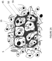
- FIGs. 2B and 2C show histology photographs of tissue (rat skin) including an integrated implant 15 as described herein.
- FIG. 2B shows the implant in the tissue 1 week following implantation and
- FIG. 2C shows the implant 1 month following implantation into Sprague-Dawley rats.
- the tissue 19 grows into the implant, keeping the implant in close proximity to the blood vessels of the tissue and without a significant foreign body response.
- FIG. 2D and 2E are reproductions of photographs showing immunohistochemistry staining for vasculature (using CD31 antibodies) 1 week ( FIG. 2D ) and 1 month ( FIG. 2E ) following implantation into skin (subcutaneous) of Sprague-Dawley rats.
- the approximate boundaries of the scaffold 16 are shown as well as capillary ingrowth 18 into the implanted scaffold.
- the tissue-integrating scaffold is made up solely or primarily of sensing moieties ( see, e.g., FIGs. 5 and 6 ).
- sensing particles can be bonded together using any suitable method (chemical, adhesive, thermal, etc.).
- the sensing particles comprise a polymer, for example PEG-coated particles ( e.g ., microspheres).
- the scaffold comprises a polymer that itself is composed of sensing moieties. See , FIG. 6 .
- the tissue integrating implant can be of any suitable form, including, but not limited to block-like (or any thickness), cube-like, disk-shaped, cylindrical, oval, round, random or non-random configurations of fibers and the like.
- the sensor comprises one or more fibers, which may be organized in a non-random fashion (e.g., grid, layered grid, etc., see , FIG. 9E ) or in a random fashion ( see, e.g., FIG. 9F ).
- tissue-integrating scaffolds described herein are typically combined with (or made up of) sensing moieties that detect one or more analytes.
- Non-limiting examples of analytes that may be detected by the sensing moieties include oxygen, reactive oxygen species, glucose, lactate, pyruvate, cortisol, creatinine, urea, sodium, magnesium, calcium, potassium, vasopressin, hormones (e.g ., Luteinizing hormone), pH, cytokines, chemokines, eicosanoids, insulin, leptins, small molecule drugs, ethanol, myoglobin, nucleic acids (RNAs, DNAs), fragments, polypeptides, single amino acids and the like.
- any suitable moiety can be used to sense the analyte of interest, including not limited to analyte binding molecules (e.g. glucose binding proteins), competitive binding molecules (e.g. phenylboronic acid based chemistries), analyte specific enzymes (e.g. glucose oxidase), ion sensitive materials, or other analyte sensitive molecules (e.g. oxygen sensitive dyes such as porphyrins).
- the sensing moieties may be in any form, for example, microspheres, nanospheres, fibers, etc.
- a single implant typically includes a plurality of sensing moieties. In certain embodiments, the sensing moieties are all the same while in other embodiments, a mixture of two or more sensing moieties is used.
- sensing molecules may be labeled with a reporter (e.g ., one or more fluorophores, one or more gold particles, one or more quantum dots and/or one or more single-walled carbon nanotubes). Sensing molecules may also create a signal through swelling, optical diffraction, change in absorbance FRET, quenching.
- a reporter e.g ., one or more fluorophores, one or more gold particles, one or more quantum dots and/or one or more single-walled carbon nanotubes.
- Sensing molecules may also create a signal through swelling, optical diffraction, change in absorbance FRET, quenching.
- Non-limiting examples of suitable sensing molecules include but are not limited to dye labeled Concanavalin A with glycodendrimer or dextran ( see, e.g., Ballerstedt et al. (1997) Anal. Chim. Acta 345:203-212 ) and alcohol sensitive oxo-bacteriochlorin derivative fluorescent binding protein developed by Takano, et al (2010) The Analyst 135:2334-2339 as well as Vladimir et al. (2004) Clinical Chemistry 50:2353-2360 ; Aslan et al. (2005) Chem. 1;77(7):2007-14 ; Ballerstadt et al. (1997) Anal. Chim. Acta 345:203-212 (1997 ); Billingsley et al. (2010) Anal.
- the sensing moiety element may comprise other molecules besides sensing molecules, such as carrier molecules/polymers (e.g. the sensing moiety element may comprise PEG nanospheres, alginate particles or other carrier materials that contain sensing molecules).
- the sensing moiety element may also contain reference molecules or stabilizing molecules that do not sense any analytes, but that serves as calibrators (e.g ., a reference dye or any substance that provides a reference signal to which the signal modulated by the analyte of interest may be compared for calibration) or stabilizer (e.g. catalayse, any free-radical scavenger which helps preserve the sensing moieties or other stabilizer).
- the sensing moiety element may be thermally responsive material, pressure-responsive material or materials that swell, shrink, change optical properties, or change other measurable properties in response to a stimulus.
- tissue-integrating scaffold with the analyte sensing moieties may be termed implantable sensing media, sensing media, tissue integrating sensor, tissue-integrating biosensor, tissue-integrating sensing media or variations thereof.
- the analyte sensing moieties may be combined with the tissue-integrating scaffolds in a variety of ways to produce tissue-integrating sensors.
- the sensing moieties are physically entrapped or chemically bound within the scaffold.
- the sensing moieties are attached directly ( e.g., via covalent or noncovalent linkages) to the surface of the tissue- integrating scaffold and may optionally be covered by an exterior coating.
- the purpose of the exterior coating is described as, but not limited to the following: to hold the sensing moieties in place, to protect the sensing moieties from external forces, to limit/impede diffusion of various molecules and/or to provide a desired exterior surface, and to conduct or transduce the sensing signal from the chemistry to the scaffold and/or external detector.
- the tissue-integrating scaffold itself is composed of sensing moieties where the sensing moieties are in the form of particles (spherical or other shapes) that are bonded together (e.g. chemically, thermally, pressure, etc) or where the polymer itself provides the sensing capability (e.g. stimuli-sensitive polymers).
- the tissue-integrating scaffold is composed of distinct layers where sensing moieties are physically entrapped or chemically bound to or within specific layers of the scaffold, and other layers provide other features such as mechanical strength, elasticity, conductivity or other properties.
- the tissue-integrating scaffold is composed of a polymer that swells or shrinks in response to a stimulus (e.g. concentration of an analyte of interest, temperature, or other stimuli).
- a stimulus e.g. concentration of an analyte of interest, temperature, or other stimuli.
- the shrinking or swelling may cause optical change (e.g. due to light diffraction, change in distances between gold nanoparticles contained within the matrix, or other interaction (Aleexev et al and Aslan, et al)).
- Table 1 below provides a matrix showing how sensing moieties can be combined with tissue-integrating scaffolds in a variety of ways to tissue-integrating sensing media.
- Sensing Media/Scaffold Matrix Sensing Moieties ⁇ Tissue-integrating Scaffolds ⁇ Sensing particles (e.g. PEG microspheres containing ConA with glycodendrimer, alginate nanospheres containing ApoGox with reported dye.)
- Sensing chemistry e.g. boronic acid based chemistry, sensing chemistry attached to quantum dots or gold nano-rods
- Any other fluorescent sensing assay e.g.
- Multi-layer fibers e.g. sensing layer, biocompatibility layer, stabilizing or structural layer, voids or cellular conduits
- Multi-layer fibers e.g. sensing layer, biocompatibilit y layer, stabilizing or structural layer, voids or cellular conduits
- Multi-layer fibers e.g. sensing layer, biocompatibilit y layer, stabilizing or structural layer, voids or cellular conduits
- Multi-layer fibers e.g. sensing layer, biocompatibilit y layer, stabilizing or structural layer, voids or cellular conduits
- Multi-layer fibers e.g. sensing layer, biocompatibilit y layer, stabilizing or structural layer, voids or cellular conduits
- the implant further comprises additional moieties (e.g ., non-sensing or additional sensing moieties different from the sensing moieties), for example reference (or calibration) moieties.
- additional moieties e.g ., non-sensing or additional sensing moieties different from the sensing moieties
- reference or calibration moieties include, but are not limited to, dyes, fluorescent particles, lanthanides, nanoparticles, microspheres, quantum dots or other additives or elements of the implant whose signal does not change due to the presences of the analyte (e.g ., glucose). See, e.g., Chaudhary et al. (2009) Biotechnology and Bioengineering 104(6):1075-1085 .
- Fluctuations in the reference (calibration) signal(s) can be used to correct or calibrate the sensing signal(s).
- Reference signals might fluctuate due to changes in the amount of light reaching the implant (ambient light changes, fluctuating LED or laser source). Sensing signals would also be subject to fluctuations in the amount of light reaching the implant; however it is desirable that the signal of interest only fluctuates based on analyte (e.g ., glucose) fluctuations. Therefore the reference signal is used to correct or calibrate the sensing signal when it fluctuates due to influences other than changes in glucose concentration.
- Reference signals might also fluctuate due to changes in the reference moiety itself ( e.g. photodegratation, chemical degradation).
- the sensing signal(s) would have the same degradation or a rate of degradation that is relatable to the reference to allow for correction or calibration by the reference.
- Reference signals might also fluctuate due to physiological fluctuations that alter the light propagation through tissue (e.g . dehydration, oxygenation, blood flow).
- Sensing signals would be affected in the same way or in a way that is relatable to the reference fluctuations thereby permitting correction or calibration of the sensing signal by the one or more references.
- the sensing signal can be calibrated by reference to the signal(s) obtained from the calibration (reference) moieties.
- the sensing moieties detect glucose and the reference moiety comprises a molecule that measures (produces a detectable signal in the presence of) oxygen (O 2 ).
- the sensing moieties can comprise an enzyme, for example glucose oxidase which is specific for the substrate glucose. The reaction of glucose oxidase causes the substrate glucose to be converted to D-glucono-1,5-lactone, which then hydrolyzes to gluconic acid. Oxygen is consumed and converted to H 2 O 2 . The reduction of O 2 in the vicinity of the enzyme can be measured by using an O 2 -sensitive fluorescent dye, such as a porphyrin dye.
- the concentration of O 2 in the tissue can also vary physiologically, thereby changing or limiting the reaction of the enzyme in the sensing moieties. Therefore, the O 2 concentration in the sensor can be measured independent of the glucose concentration. Such a reference measurement of O 2 would allow corrections to be made to the glucose-specific signal from the sensing moieties.
- an analyte-specific enzyme that causes a change in pH would require the use of a separate pH-sensitive fluorescent dye with an emission spectral peak different and distinguishable from the analyte-specific dye reporting on the activity of the analyte-specific enzyme, for example when the sensing moieties comprise, urease used for measuring urea.
- the sensing moieties comprise a first fluorescent dye and the reference molecule comprises a second (different) fluorescent dye.
- the sensing moieties may utilize an analyte-specific chemistry that includes a ligand receptor moiety and an analyte analogue moiety.
- One of the binding members is labeled with a fluorescent dye and the other binding member is labeled with a dye that quenches the fluorescent dye when the analyte analogue moiety binds to the ligand receptor moiety.
- Non-limiting examples include glycodendrimer, which binds to Concanavalin A, wherein the Concanavalin A is labeled with Alexafluor 647 and the glycodendrimer is labeled with QDY21 dark quencher.
- Concanavalin A binds to glucose and the glycodendrimer competes with glucose for the binding to Concanavalin A.
- the chemistry is immobilized as described in this invention within the tissue-integrating scaffold and implanted into the dermis or subcutaneous tissue.
- the tissue-integrating scaffold is illuminated from a patch reader on top of the skin above the implant with 650nm light at desired intervals over the long-term life of the implant (e.g ., every 5-60 minutes over a period of 90 days or more).
- the amount of fluorescent signal e.g ., from a molecule such as Alexafluor 647 detected is proportional to the concentration of glucose in the tissue.
- the dye can photobleach, i.e., the amount of fluorescent signal emitted back through the skin at a given glucose concentration is diminished.
- a reduction of fluorescence due to photobleaching can make it appear that analyte is at a lower concentration than it really is.
- the separate internal control is a second fluorescent dye, different from the fluorescent molecule included in the sensing moieties (e.g ., Alexafluor 750 in the reference moieties when the sensing moieties comprise Alexafluor 647), which included immobilized in the scaffold.
- the fluorescence of reference moieties is not affected by the concentration of glucose, and both the first (e.g., Alexafluor 647) and second (e.g., Alexafluor 750) fluorescent dyes have predictable and well-characterized photobleaching rates.
- the fluorescence is measured for both dyes.
- the fluorescence value of the dye in the reference moieties can then be used to correct for any photobleaching of the dye in the sensing moieties.
- tissue-integrating implanted biosensor typically resides 3-4 mm under the surface of the scan. It is well known that in skin excitation light and emitted fluorescent light in the near infrared range are highly scattered as the light traverses the tissue between the reader patch and the implant. The extent of absorption and scattering is affected by physical properties such as temperature or by tissue composition, including but not limited to variations in blood perfusion, hydration, and melanin concentration. Skin variations can occur between users or between different time points for a single patient, and these variations can affect the fluorescence excitation and emissions signals causing in accurate signals for the analyte-specific signal.
- a separate fluorescence molecule with emission spectra distinguishable from the analyte-specific fluorescence can be immobilized into the scaffold.
- the fluorescence from the molecule can be measured separately from the analyte-specific fluorescence to measure a signal that informs about variations in tissue composition.
- the dye selected is based on having a similar response to tissue variations as the analyte-specific dye. Dyes such as Alexafluor 750, various quantum dots (QD's), or lanthanide dye nanocrystals all can provide this capability.
- FIGs. 3 to 8 depict cross-sections of exemplary tissue integrating implants as described herein.
- the implant is depicted (e.g ., boxed area of Figure 1 ) and the pore 5 is depicted as a void.
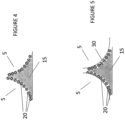
- FIG. 3 depicts a cross-section of an exemplary tissue-integrating implant as described herein in which sensing moieties 20 are embedded within the solid scaffold portions 15.
- the sensing moieties 20 may be physically entrapped and/or chemically bound within the solid scaffold portions 15.
- FIG. 4 depicts a cross-section of an exemplary tissue-integrating implant as described herein in which sensing moieties 20 are attached to the surface of the solid scaffold portions 15 (sensing moieties are within pores 5 ).
- FIG. 5 depicts the exemplary embodiment shown in FIG. 4 and further comprising an exterior coating 30 surrounding the sensing moieties.
- FIG. 6 depicts a cross-section of an exemplary tissue-integrating implant as described herein in which solid scaffold portions 15 are made from sensing moieties 20 in the form of particles bonded together.
- FIG. 7 depicts a cross-section of a solid scaffold portion 15 made from a polymer in which the polymer is composed of sensing materials.
- FIG. 8 depicts a cross-section of an exemplary tissue-integrating implant as shown in FIG. 3 and further including additional moieties 40 embedded in the solid portion 15 of the scaffold.
- the additional moieties 40 can be, for example, reference particles for calibration, including but not limited to particles that provide a stable signal (e.g ., optical, magnetic, electrochemical, electrical, temperature, pressure, ultrasound, acoustic, radiation) to which the analyte sensing signals may be compared for calibration purposes.
- a stable signal e.g ., optical, magnetic, electrochemical, electrical, temperature, pressure, ultrasound, acoustic, radiation
- one or more different types of additional (reference) moieties can be used.
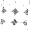
- FIGs. 9A-F , 10A and 10B are overviews and cross-sections of exemplary tissue-integrating sensors as described herein that are cylindrically shaped.
- FIG. 9A shows an embodiment that comprises a single layered cylindrical tissue scaffold (or individual fiber) 15 with sensing moieties 20 and additional moieties 40 embedded in the scaffold 15.
- FIG. 9B shows an embodiment that comprises a single layered cylindrical tissue scaffold (or individual fiber) 15 with sensing moieties 20 attached to the surface of the scaffold 15.
- FIG. 9C shows an embodiment in which the sensing moieties 20 are attached to the surface and embedded within the scaffold 15.
- FIG. 9D is a cross section of the exemplary sensors with sensing moieties embedded in the scaffold.
- FIG. 10A shows an embodiment that comprises multiple (two) layers of scaffold material 15 with sensing moieties 20 and additional moieties 40 embedded in the innermost layer of the scaffold 15.
- FIG. 10B shows an embodiment comprising a hollow interior 17 with an outer layer of scaffold material 15 with sensing moieties 20 and additional moieties 40 embedded in the outer layer. It will be apparent that any number of layers can be used (composed of the same or different materials) and that the sensing moieties (and optional additional moieties) may be present in one, some or all of the layers (and/or on the surface of the scaffold).
- FIG. 11 shows a cross-section of an exemplary sensing media as shown in FIG. 9A , including sensing moieties 20 embedded in the tissue-integrating scaffold 15.
- FIG. 12 is a cross-section of an exemplary sensing media as shown in FIG. 9B and
- FIG. 13 is a cross-section of an exemplary sensing media as shown in FIG. 12 further including a coating 30 exterior to the sensing moieties 20 attached to the surface of the scaffold 15.
- FIG. 14 depicts a cross-section of an exemplary cylindrically shaped sensor implant (whole device) or a portion of an implant ( e.g ., individual fiber) in which the scaffold 15 is made from polymer where the polymer itself is composed of sensing moieties 20.
- FIG. 15 is a cross-section of an exemplary multi-layered cylindrical sensor implant (or individual fiber of an implant) including two layers of scaffold 15, 16 with sensing moieties 20 embedded in the inner layer 15.
- the inner 15 and outer 16 layers may be made of the same or different polymers.
- FIG. 16 is a cross-section of an exemplary multi-layered cylindrical sensor implant including two layers of scaffold 15, 16 with sensing moieties 20 embedded in the outer layer 16.
- the inner 15 and outer 16 layers may be made of the same or different polymers.
- FIG. 17 is a cross-section of an exemplary hollow cylindrical sensor implant including a scaffold 15 surrounding a hollow core 17 with sensing moieties 20 embedded in the scaffold 15. Additional layers, without or without sensing moieties, can also be present and may be made of the same or different materials.
- Tissue-integrating sensors comprised of one or more cylindrical shaped elements (e.g. , fibers) eliminate or greatly reduce the foreign body response as compared to currently available implants. Moreover, the average diffusion distances from the capillary supply to all parts of the sensing media are comparable to native tissue, unlike other known sensors.
- the implant will be between about .001 mm to 2 mm in thickness (or any value therebetween) and between 1 mm and 1 cm in diameter (or an equivalent cross sectional area of a non-circular shape, for example length/width) and 15 mm in length or less, for example a disk shaped sensor that is 2mm or less thick and 10 mm or less in diameter.
- the approximate sensor size is approximately 100-1000 microns in diameter and the length is between 0.25 mm and 10 mm.
- the size of the tissue-integrating sensing media in disk form is typically 2mm or less thick and 10 mm or less in diameter.
- the injected sensing media may be a single piece of tissue-integrating material, or it may be several pieces or particles of tissue-integrating sensing material. It may be injected with a carrier substance (e.g. saline, PBS with anti-inflammatory drugs or other tissue-response modifiers). Furthermore, the sensing media may be implanted into any part of the subject, including, for example, shoulder, arm, leg, abdomen, etc. Preferably, the sensing media is implanted into the skin, for example, the epidermis, the dermis and or the subcutaneous layer of skin.
- a carrier substance e.g. saline, PBS with anti-inflammatory drugs or other tissue-response modifiers.
- the sensing media may be implanted into any part of the subject, including, for example, shoulder, arm, leg, abdomen, etc.
- the sensing media is implanted into the skin, for example, the epidermis, the dermis and or the subcutaneous layer of skin.
- a biosensor system as described herein comprises the tissue-integrating biosensor (described above).
- Other components include one or more of the following: interrogator, illuminator, detector, signal receiver, signal transmitter, signal processing component, energy storage component, data storage component, data transmitter, data display, data processing component and combinations thereof.
- interrogator illuminator
- detector signal receiver
- signal transmitter signal processing component
- energy storage component data storage component
- data transmitter data display
- data processing component data processing component
- One or more of these other components may be incorporate into a wearable patch that resides over the sensor to detect the sensor signal, or they may be integrated into a hand held or other device, which is periodically held over the implanted sensor to take the measurement. See, FIG. 18 .
- FIG. 19 shows exemplary embodiments of a system including an interrogator.
- Figure 19A shows a patch 85 including an interrogator and/or detector that may be worn continuously above implanted sensor.
- FIG. 19B shows a module 90 that can be placed above implanted sensor as desired to interrogate and/or detect continuous or discrete measurements.
- Non-limiting examples of such modules include hand-held devices such as wands and the like.
- FIG. 19C depicts how a field 95 that can be used to interrogate (monitor) the subject remotely.
- any of the systems described herein may further include an additional component 97 that delivers one or more therapeutics (e.g ., analytes) to the subject based on the measurements obtained from the sensor (e.g ., an insulin pump that delivers glucose to form a closed loop artificial pancreas).
- the delivery device 97 may be incorporated into the system (e.g. , interrogator and/or detector).
- the delivery device 97 may be controlled by the operator based on the measurements from the sensor or may be controlled by the data reader directly ( e.g., smart phone) or remotely ( e.g., telemedicine).
- tissue-integrating scaffold combined with (or comprised of) the one or more sensing moieties are the necessary elements in the tissue-integrating sensor system.
- the combination of analyte sensing moieties with tissue-integrating scaffolds comprises the tissue-integrating sensor that is implanted in the body.
- This tissue-integrating sensor is one component of the biosensor system for continuous monitoring or long-term use within the mammalian body.
- Other components including, for example, means to read the signal coming from the tissue-integrating biosensor, show, collect and/or transmit the signal coming from the implanted biosensor. In certain embodiments, the signal is read directly by the human eye.
- the signal reader comprises one or more of the following: a hand-held device that detects biosensor signals; a removable patch that resides over the area of the tissue integrating biosensor to continuous or semi-continuous detection of biosensor signals; an implant near, but not touching the tissue-integrating sensing media and/or an implant near and touching a portion of the tissue-integrating sensing media.
- the implant may send signal to a watch, a cell phone, a hand-held device, a computer or other data collection and/or read-out device, either directly or, alternatively, via the signal reader.
- the data may or may not first be processed before sending to these devices and/or these devices may process data received.
- Data may further be relayed to a database, an email account a cell phone or other storage, processing or display.
- the invention works by means of chemical, physical and biological interactions.
- the tissue-integrating scaffold promotes capillary in-growth into or nearby the sensing scaffold ( FIG. 2 ).
- Small molecules that diffuse in the interstitial space e.g. glucose, urea, lactate, pyruvate, glycerol, glutamate, cortisol, acetone, ethanol and other molecules
- the tissue integrating scaffold is composed of a biomaterial that has sensing moieties contained and/or attached on the exterior of the scaffold.
- the tissue integrating scaffold is composed of a polymer with mesh size large enough to permit molecules of interest to diffuse inside the scaffold.
- the sensing moieties are contained within the polymer scaffold.
- the tissue-integrating scaffold is composed of a polymer with mesh size large enough to permit molecules of interest to diffuse inside the scaffold.
- the sensing moieties compose the polymer scaffold.
- a measurable signal is produced (e.g. fluorescence), which is the measured by a detector that is inside or outside the body, but not immediately touching the tissue-integrating biosensor.
- analytes can be assayed, and that these analytes are selected by the operator, for example, based on the recommendation of medical personnel, based on interest of monitoring of health and well-being, based on specific biological threats, or based on any other rationale for which the subject has interest to monitor analytes continually or periodically.
- the subject would inject, have injected, implant or have implanted the tissue-integrating biosensor or biosensors for the specific analyte or analytes of interest in the tissue to be monitored.
- the implant can be placed anywhere in the subject.
- the sensing media is injected into the skin (e.g., the dermis or subcutaneously).
- the sensor is integrated into alternative spots, including, but not limited to, muscle, visceral fat, peritoneal cavity, gums, cheek, eye, etc.
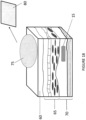
- FIG. 18 is a schematic cross-section of a skin sample showing an exemplary embodiment in which the sensing media (tissue integrating implant) 15 is implanted into the subcutaneous tissue 70 of a subject's skin. Also shown are the epidermis 60, the dermis 65 and an optional signal reader 75, depicting as a patch on the surface of the skin. In this embodiment, the detector patch sends interrogation light to the tissue integrating sensing media.
- the sensing moieties contained in the tissue-integrating sensing media 15, provide a measurable signal (e.g ., fluorescence, luminescence, magnetic, etc.) in a manner dependent on the concentration of the analyte(s) of interest.
- the signal (e.g ., fluorescent light) is detected by the detector patch (signal receiver) 75.
- the detector patch (signal receiver) 75 Also shown in FIG. 18 is optional data reader device 80 that can receive process and/or display information received from the signal reader 75 (e.g., patch).
- data readers include cell phones, smart phones, watches, computers, and the like. Data may be further relayed to a database, an email account, a cell phone or other storage, processing or display.
- the data obtained with the tissue-integrating biosensor system is used by persons to better understand and manage body chemistries (e.g ., glucose in the case of diabetics, urea in the case of dialysis patients) and health status.
- body chemistries e.g ., glucose in the case of diabetics, urea in the case of dialysis patients
- in vivo monitoring can provide the clinician with feedback upon which to make adjustments to dosing to assure proper concentrations are achieved and maintained.
- Constant monitoring of food additives, sodium, alcohol, caffeine, nicotine, vitamin levels, lead, pesticides and a variety of other substances can help individuals and caregivers understand their intake and exposure to certain chemicals and to take control of their own health.
- the tissue-integrating biosensors can be used in the for personal monitoring, physician monitoring of patients, clinical research, animal research studies, and veterinary health for continuously or semi-continuously monitoring analyte concentrations inside a living body.
- uses of the sensors include for monitoring of diabetic health, dehydration, hemodialysis, acute respiratory distress, stress assessment, congestive heart failure, metabolism status, lifestyle, fitness and training, peripheral vascular resistance, hyonatramia, acute decompensated heart failure, fertility status ( e.g ., ovulation cycle), cancer detection (early, recurrent, etc.), inflammatory responses (various types), therapeutic drug, including drug concentrations or drug response indicators, ethanol for example for alcoholism treatment, infection disease monitoring, pesticide monitoring, heavy metal monitoring and the like.
- In vivo tissue-integrating biosensors for endogenous and exogenous analytes can be used day and night at home and during daily activities (work, exercise, meals, etc). They can also be used in a care giving setting (e.g. hospital). They may be used in a continuous or intermittent fashion.
- the sensors also termed sensing media
- the tissue integrating sensing scaffold promotes capillary growth directly into the sensor itself unlike all other marketed sensors or sensors in development (that are known to the authors at the time of submitting this patent).
- tissue-integrating sensors Another aspect of this invention is a method for making tissue-integrating sensors.
- the method(s) for creating a tissue-integrating sensor comprises a process for combining the sensing moieties and the tissue-integrating scaffold in a manner that preserves the integrity of the sensing moieties sufficiently such that they produce measurable signal(s) in response to the analyte of interest.
- the relative amounts of scaffold, sensing moieties and/or reference moieties in the sensor will depend on the polymers and sensing moieties used.
- the sensor will be made with between about 2-95% vol/vol of a monomer or polymer (e.g ., 2-85% vol/vol HEMA).
- the amount of cross-linker used will depend on the polymer, for example typically about .1 and 10% vol/vol of TEGDMA may be used.
- Water and or other solvents may be present in any amount ( e.g ., 5-95% vol/vol water or polyethylene glycol).
- Initiators may also present in any amount, for example 0.35 to 5% vol/vol of Irgacure.
- Sensing moieties may be present in any suitable amount, for example, oxygen sensing porphyrins (PdP) may be included at a concentration of about 200 nM to 1 nM. See, also, Example 1.
- the methods of the invention involve a tissue-integrating sensor that is formed by embedding or containing the sensing moieties within the tissue-integrating scaffold.
- the process may begin with combining the sensing moieties and the scaffold precursor (e.g . monomer, polymer beads, etc.), followed by the formation of the scaffold (e.g . polymerization around template beads, multiphoton polymerization, electrospinning, micro- and nano-printing fabrication techniques, polymer foaming, salt leaching, etc.) and the removal of any residuals (e.g. dissolution of template beads, removal of unpolymerized monomers, etc.).
- the scaffold precursor e.g . monomer, polymer beads, etc.
- the scaffold e.g . polymerization around template beads, multiphoton polymerization, electrospinning, micro- and nano-printing fabrication techniques, polymer foaming, salt leaching, etc.
- any residuals e.g. dissolution of template beads, removal of unpolymerized monomers, etc.
- Non-limiting exemplary methods for embedding or containing the sensing moieties within the tissue-integrating scaffold include (but are not limited to): polymerization around template beads with or without subsequent dissolution, matrix or other structure, polymerization of a three-dimensional structure using multiphoton polymerization or 3D printing, electrospinning of small fibers, sintering or melting scaffold precursor structures, or swelling scaffold to permit entry of sensing moieties followed by shrinking of scaffold.
- the method comprises polymerizing glucose sensing moieties (nanospheres) into an inverted crystal colloid (ICC) scaffold.
- ICC inverted crystal colloid
- glucose-sensing nanospheres are mixed with ICC scaffold pre-polymer during polymerization, causing the nanospheres to be integrated into the pHEMA scaffold as detailed in EXAMPLE 1.
- the tissue-integrating sensor is formed by immobilizing (conjugation or physical entrapment) the sensing moieties on (or to) the surface of the tissue-integrating scaffold.
- the process begins with an existing scaffold (e.g. extracellular matrix) or the forming of a scaffold (e.g. ICC, synthetic or processed ECM or PoreX Medpore), followed by the attachment of the sensing moieties to the scaffold.
- the method may also include a coating step that protects or holds in place (e.g. physical entrapment) the sensing moieties to the scaffold.
- the coating may have the added benefit(s) of (1) protecting the surface chemistry from degradation (e.g.
- the method may also include step(s) for the sterilization of the tissue-integrating sensor prior to implantation (e.g. ethylene oxide gas, radiation) or in vitro use.
- exemplary methods for immobilizing the sensing moieties on the tissue-integrating scaffold include, but are not limited to: conjugation chemistry, adsorption, electrostatics and covering with a continuous coating.
- Exemplary coatings include PEG, pHEMA and chitosan.
- the tissue-integrating sensor is formed by constructing a tissue-integrating scaffold made of the sensing moieties.
- the procedure begins with the sensing moieties of some physical dimension smaller than the desired scaffold features that are then processed into the tissue-integrating material or tissue-integrating precursor.
- Sensing particles may be bonded together in a tissue-integrating structure through heat or chemical bonds.
- Pre-polymer solution composed of the sensing moieties may be crosslinked in the desired scaffold structure.
- Exemplary methods for constructing a tissue-integrating scaffold made OF the sensing moieties include, but are not limited to: bonding the sensing particles using heat, pressure or polymerization; electrospinning, thermal or UV initiated crosslinking of sensing polymers into a tissue integrating structure, including multiphoton polymerization.
- a sensing media as described herein is formed by tissue-integrating scaffold particles.
- the process begins with deconstructing a tissue-integrating scaffold into particles that maintain their tissue-integrating properties.
- the particles are mixed with the sensing moieties and then reconstructed into desirable scaffold form and function.
- One example is the particulation, e . g ., extraction and powdering, of extracellular matrix (ECM) to create particles.
- ECM extracellular matrix
- the ECM particles are then combined with selected sensing moieties.
- the mixture may be injected as is or may be combined with a crosslinking agent or polymer (e.g. pHEMA) to add mechanically stability.
- a crosslinking agent or polymer e.g. pHEMA
- a sensor that is formed by constructing simple or multi-layer fiber(s) implants is formed by constructing simple or multi-layer fiber(s) implants.
- the sensing moiety is part of one or more of the base materials from which the fiber scaffold is created or the sensing moiety(ies) are contained or compose one of the layers of sequential building up layers.
- the methods may also include step(s) for the sterilization of the tissue-integrating sensor prior to implantation (e.g. ethylene oxide gas) or in vitro use.
- step(s) for the sterilization of the tissue-integrating sensor prior to implantation e.g. ethylene oxide gas
- in vitro use e.g. ethylene oxide gas
- Example 1 Production of an oxygen sensing media with oxygen sensitive dye immobilized in a hydrogel scaffold
- the following describes one proposed method for making a tissue-integrating sensor as described herein.
- This method involves the use of non-crosslinked PMMA templating microspheres and pHEMA as the scaffold material.
- the PMMA microsphere template was prepared using sieved PMMA spheres (36 um with a CV less than 5%) and placing the template beads between two glass slides with Teflon spacers.
- the sintering process included sonicating for at least 10 minutes (one or more times) to closely pack the beads.
- the template is heated to a sufficient temperature for a sufficient time to fuse the beads (for example, heat to approximately 177°C for 24 hours).
- HEMA oxygen sensing poly(2-hydroxyethyl methacrylate)
- the pre-polymer solution was filled into the PMMA.
- the solution was placed under vacuum to remove any bubbles and completely infiltrate the PMMA-mold and then polymerized by exposing the mold to UV for 5-10 minutes.
- the PMMA microspheres were dissolved by frequent exchange of dichloromethane or other solvent system for 24-48 hours using a Soxhlet extractor or frequent volume changes.
- Implants comprising reference moieties were also prepared as described above except instead of porphyrins, qtracker 800 quantum dots (Invitrogen, 50-800 nM) were included in the scaffold.
- the oxygen sensing media and reference moieties were injected with a trocar approximately 2 mm under the surface of mice skin (in different locations on the animal). Mice were imaged with Caliper whole animal imaging system (IVIS TM ) with an excitation of 535 nm and emission light was collected at 760nm under oxygenated and deoxygenated conditions.
- IVIS TM Caliper whole animal imaging system
- both the oxygen sensing implant (“O2”) and the reference moieties (“QD”) produced a signal under oxygenated conditions ( FIG. 20A ). However, under deoxygenated conditions, only the reference moieties produced a detectable signal ( FIG. 20B ).
- Example 2 Production of a glucose sensing media with glucose sensitive assay immobilized in a hydrogel scaffold
- This method involves the use of non-crosslinked PMMA templating microspheres and pHEMA as the scaffold material.
- the PMMA microsphere template was prepared using sieved PMMA spheres (36 um with a CV less than 5%) and placing the template beads between two glass slides with Teflon spacers.
- the sintering process included sonicating for at least 10 minutes (to closely pack the beads), then heating the template to 177°C for 24 hours to fuse the beads (the conditions will vary for different ovens and may also vary for different batches of beads).
- the preparation of glucose sensing poly(2-hydroxyethyl methacrylate) (pHEMA) scaffolds was done as follows.
- the polymer precursor solution was prepared by mixing HEMA 2-hydroxyethyl methacrylate (57.1% %vol/vol), TEGDMA(triethyleneglycol-dimethacrylate) (2.9 v%vol/vol), ethylene glycol (14.8 %vol/vol) water (25.1 %vol/vol) and the photoinitiator Irgacure 651 (0.2 % vol/vol).
- the dye/enzyme solution was prepared by adding 5 mg of glucose oxidase enzyme (GOx) and equimolar catalyze in 100 uL of DI water and then adding 100uL of 1.5mM Pd(II) meso-Tetra(4-carboxyphenyl)porphine (PdP) in DMSO.
- the polymer precursor solution and the dye/enzyme solution were combined in a 1:1 ratio for a 39uM final concentration of GOx and 375 uM PdP.
- the pre-polymer solution was filled into the mold and placed under vacuum to remove any bubbles and completely infiltrate the PMMA-mold and then polymerized by exposing to UV for 5-10 minutes.
- the PMMA microspheres were dissolved by frequent exchange of dichloromethane or other solvent system for 24-48 hrs using a Soxhlet extractor or frequent volume changes.
- Disk of the glucose sensor scaffold material were punched from the rectangular pieces (microscope slide-shape) and fixed inside an automated flow-through system with a built in flourimeter.
- Glucose solutions (in PBS) of various concentrations were flowed over the sensor scaffold discs and fluorescence and lifetime readings were collected at various glucose concentrations over successive runs (e.g., PdP emission was measured as a function of glucose concentration).
- the signal emitted from the sensor modulated in response to glucose concentration.
- Example 3 Production of an analyte sensing media with analyte sensitive dye immobilized in a hydrogel scaffold
- the following describes one proposed method for making a tissue-integrating sensor as described herein.
- This method involves the use of non-crosslinked PMMA templating microspheres and pHEMA as the scaffold material.
- the PMMA microsphere template is prepared using sieved PMMA spheres (36 um with a CV less than 5%) and placing the template beads between two glass slides with Teflon spacers.
- the sintering process includes sonicating for 10 minutes (to closely pack the beads), then heating the template to 177°C for 24 hours to fuse the beads (the conditions will vary for different ovens and may also vary for different batches of beads).
- Polymer pre-cursor that will form the hydrogel scaffold is then prepared.
- the general preparation of poly(2-hydroxyethyl methacrylate) (pHEMA) scaffold is as follows: In separate vials, two solutions are prepared: 0.89 ml of a 20% solution of APS (ammonium persulfate) in water and 0.3 ml of a 15% solution TEMED (tetramethylethylenediamine) in water. To a third vial the HEMA 2-hydroxyehtyl methacrylate (9.26 ml), TEGDMA(triethyleneglycol-dimethacrylate) (0.46 ml), ethylene glycol (2.6 ml) and water (2.68 ml) are added by volume measurement and mixed.
- APS ammonium persulfate
- TEMED tetramethylethylenediamine
- the TEMED solution is added to the main pre-polymer vial.
- Sensing nanospheres ranging from 2-95% volume of the total reactant volume (e.g. 5 ml of 100-200 nm alginate nanospheres containing fluorescent glucose sensing chemistry) are mixed with the pre-polymer solution.
- the pre-polymer solution is filled into the mold and then the APS solution added.
- the solution is placed under vacuum to remove any bubbles and completely infiltrate the PMMM-mold and then polymerized at room temperature for one hour.
- the PMMA microspheres are dissolved by frequent exchange of dichloromethane or other solvent system for 24-48 hrs using a Soxhlet extractor or frequent volume changes.
- a tissue integrating sensor produced in rods that are 300-500 um in diameter and 5 mm long are placed in a 19-23 Gauge insertion needle, trochar, modified biopsy device or other devices engineered for injection under the skin.
- the sensor is optionally dehydrated or compressed before insertion to allow for the use of a smaller insertion needle.
- Insertion site may include any subcutaneous area, typically the abdomen, arm and thigh.
- Raw data is converted to an analyte concentration or some non-quantitative representation of the analyte concentration (e.g. high, low, within range). Values at any given point in time or trends (graphs over time) or summary statistics over a period of time are provided. An indication of the quality of the data is optionally provided.
Landscapes
- Health & Medical Sciences (AREA)
- Life Sciences & Earth Sciences (AREA)
- Physics & Mathematics (AREA)
- Engineering & Computer Science (AREA)
- General Health & Medical Sciences (AREA)
- Molecular Biology (AREA)
- Pathology (AREA)
- Biophysics (AREA)
- Biomedical Technology (AREA)
- Heart & Thoracic Surgery (AREA)
- Medical Informatics (AREA)
- Veterinary Medicine (AREA)
- Surgery (AREA)
- Animal Behavior & Ethology (AREA)
- Public Health (AREA)
- Optics & Photonics (AREA)
- Emergency Medicine (AREA)
- Computer Networks & Wireless Communication (AREA)
- Chemical & Material Sciences (AREA)
- General Chemical & Material Sciences (AREA)
- Chemical Kinetics & Catalysis (AREA)
- Physiology (AREA)
- Artificial Intelligence (AREA)
- Computer Vision & Pattern Recognition (AREA)
- Psychiatry (AREA)
- Signal Processing (AREA)
- Spectroscopy & Molecular Physics (AREA)
- Measurement Of The Respiration, Hearing Ability, Form, And Blood Characteristics Of Living Organisms (AREA)
- Investigating, Analyzing Materials By Fluorescence Or Luminescence (AREA)
- Investigating Or Analysing Biological Materials (AREA)
- Measuring And Recording Apparatus For Diagnosis (AREA)
Applications Claiming Priority (3)
| Application Number | Priority Date | Filing Date | Title |
|---|---|---|---|
| US39025210P | 2010-10-06 | 2010-10-06 | |
| PCT/US2011/055157 WO2012048150A1 (en) | 2010-10-06 | 2011-10-06 | Tissue-integrating sensors |
| EP11831627.2A EP2624744A4 (de) | 2010-10-06 | 2011-10-06 | In gewebe integrierbare sensoren |
Related Parent Applications (1)
| Application Number | Title | Priority Date | Filing Date |
|---|---|---|---|
| EP11831627.2A Division EP2624744A4 (de) | 2010-10-06 | 2011-10-06 | In gewebe integrierbare sensoren |
Publications (3)
| Publication Number | Publication Date |
|---|---|
| EP4449978A2 true EP4449978A2 (de) | 2024-10-23 |
| EP4449978A3 EP4449978A3 (de) | 2025-01-08 |
| EP4449978B1 EP4449978B1 (de) | 2026-03-18 |
Family
ID=45928132
Family Applications (2)
| Application Number | Title | Priority Date | Filing Date |
|---|---|---|---|
| EP24158962.1A Active EP4449978B1 (de) | 2010-10-06 | 2011-10-06 | Gewebeintegrierende sensoren |
| EP11831627.2A Withdrawn EP2624744A4 (de) | 2010-10-06 | 2011-10-06 | In gewebe integrierbare sensoren |
Family Applications After (1)
| Application Number | Title | Priority Date | Filing Date |
|---|---|---|---|
| EP11831627.2A Withdrawn EP2624744A4 (de) | 2010-10-06 | 2011-10-06 | In gewebe integrierbare sensoren |
Country Status (9)
| Country | Link |
|---|---|
| US (5) | US10463287B2 (de) |
| EP (2) | EP4449978B1 (de) |
| JP (3) | JP5827999B2 (de) |
| KR (1) | KR101690535B1 (de) |
| CN (3) | CN105147300B (de) |
| AU (4) | AU2011311889C1 (de) |
| BR (1) | BR112013008154A2 (de) |
| CA (3) | CA3184858A1 (de) |
| WO (1) | WO2012048150A1 (de) |
Families Citing this family (56)
| Publication number | Priority date | Publication date | Assignee | Title |
|---|---|---|---|---|
| CA2723291C (en) * | 2008-05-14 | 2017-08-15 | Becton, Dickinson And Company | Separatable infusion set with cleanable interface and straight line attachment |
| CA3149758C (en) | 2009-01-12 | 2026-01-13 | Becton, Dickinson And Company | Infusion set and/or patch pump having at least one of an in-dwelling rigid catheter with flexible features and/or a flexible catheter attachment |
| US9517023B2 (en) | 2009-06-01 | 2016-12-13 | Profusa, Inc. | Method and system for directing a localized biological response to an implant |
| US10010272B2 (en) | 2010-05-27 | 2018-07-03 | Profusa, Inc. | Tissue-integrating electronic apparatus |
| EP4449978B1 (de) | 2010-10-06 | 2026-03-18 | Profusa, Inc. | Gewebeintegrierende sensoren |
| US9950109B2 (en) | 2010-11-30 | 2018-04-24 | Becton, Dickinson And Company | Slide-activated angled inserter and cantilevered ballistic insertion for intradermal drug infusion |
| US8784383B2 (en) | 2010-11-30 | 2014-07-22 | Becton, Dickinson And Company | Insulin pump dermal infusion set having partially integrated mechanized cannula insertion with disposable activation portion |
| US8814831B2 (en) | 2010-11-30 | 2014-08-26 | Becton, Dickinson And Company | Ballistic microneedle infusion device |
| US8795230B2 (en) | 2010-11-30 | 2014-08-05 | Becton, Dickinson And Company | Adjustable height needle infusion device |
| US8795234B2 (en) | 2010-11-30 | 2014-08-05 | Becton, Dickinson And Company | Integrated spring-activated ballistic insertion for drug infusion device |
| EP2685884B1 (de) | 2011-03-15 | 2020-09-23 | Senseonics, Incorporated | Integrierter katalytischer schutz oxidationsempfindlicher materialien |
| USD688784S1 (en) | 2012-04-13 | 2013-08-27 | Becton, Dickinson And Company | Infusion device |
| USD685083S1 (en) | 2012-04-13 | 2013-06-25 | Becton, Dickinson And Company | Infusion device |
| USD687140S1 (en) | 2012-04-13 | 2013-07-30 | Becton, Dickinson And Company | Infusion device |
| USD687536S1 (en) | 2012-04-13 | 2013-08-06 | Becton, Dickinson And Company | Infusion device |
| USD685084S1 (en) | 2012-04-13 | 2013-06-25 | Becton, Dickinson And Company | Infusion device |
| USD687141S1 (en) | 2012-04-13 | 2013-07-30 | Becton, Dickinson And Company | Infusion device |
| US9517128B2 (en) * | 2013-03-08 | 2016-12-13 | The Trustees Of Princeton University | Multi-functional hybrid devices/structures using 3D printing |
| US10820860B2 (en) * | 2013-03-14 | 2020-11-03 | One Drop Biosensor Technologies, Llc | On-body microsensor for biomonitoring |
| CA2904031A1 (en) * | 2013-03-14 | 2014-10-02 | Profusa, Inc. | Method and device for correcting optical signals |
| BR112015022763A8 (pt) * | 2013-03-14 | 2019-11-26 | Profusa Inc | sensor para detectar um analito |
| US9182368B2 (en) | 2013-03-14 | 2015-11-10 | Sano Intelligence, Inc. | Method of manufacturing a sensor for sensing analytes |
| CN105307559B (zh) | 2013-06-06 | 2020-06-05 | 普罗菲尤萨股份有限公司 | 用于探测来自植入传感器的光信号的设备和方法 |
| WO2015056802A1 (ja) * | 2013-10-18 | 2015-04-23 | 独立行政法人科学技術振興機構 | 分離フィルタ材料並びに血液および組織間質液成分分析センサチップ |
| US20170021149A9 (en) * | 2014-05-02 | 2017-01-26 | National Institute Of Standards And Technology | Biological sampling platform and processes for making and using same |
| WO2015200723A1 (en) * | 2014-06-25 | 2015-12-30 | Hunter William L | Polymers, systems and methods for using and monitoring polymers for use in medical polymers, implants, and procedures |
| WO2016014987A2 (en) | 2014-07-24 | 2016-01-28 | Thomas Jefferson University | Long-term implantable monitoring system & methods of use |
| AU2015302025B2 (en) | 2014-08-11 | 2021-08-19 | The Regents Of The University Of California | Continuous analyte sensor |
| MA40518A (fr) * | 2014-09-15 | 2016-03-24 | Klox Tech Inc | Matrices polymères émissives |
| WO2017210841A1 (en) | 2016-06-06 | 2017-12-14 | University Of Washington | Nanoparticle transducer sensors and methods of use thereof |
| JP2019526794A (ja) | 2016-08-22 | 2019-09-19 | ラモット・アット・テル・アビブ・ユニバーシテイ・リミテッドRamot At Tel Aviv University Ltd. | 生物検体の検出のための方法およびシステム |
| KR20190038660A (ko) * | 2016-08-22 | 2019-04-08 | 라모트 앳 텔-아비브 유니버시티 리미티드 | 피하 센싱을 위한 방법 및 시스템 |
| KR102649881B1 (ko) | 2016-11-03 | 2024-03-20 | 아리조나 보드 오브 리전츠 온 비해프 오브 더 유니버시티 오브 아리조나 | 캡슐화 장치에서 이식 전과 이식 후 세포를 실시간 평가하기 위한 방법 및 시스템 |
| EP3534793B1 (de) * | 2016-11-03 | 2025-12-31 | Arizona Board of Regents on behalf of the University of Arizona | Verfahren, systeme und implantierbare vorrichtungen zur verbesserten regulierung des blutzuckerspiegels |
| KR102460406B1 (ko) | 2016-11-03 | 2022-10-31 | 아리조나 보드 오브 리전츠 온 비해프 오브 더 유니버시티 오브 아리조나 | 산소 전달을 수반 또는 비-수반하는 적층형 조직 캡슐화 장치 시스템 |
| KR20190106996A (ko) | 2016-11-03 | 2019-09-18 | 아리조나 보드 오브 리전츠 온 비해프 오브 더 유니버시티 오브 아리조나 | 외부 산소 전달을 수반 또는 비-수반하는 산소 센서가 구비된 캡슐화 장치 시스템 |
| CN109923419B (zh) * | 2016-11-15 | 2022-05-10 | 西奈工作室 | 表面分析贴片 |
| WO2018119204A1 (en) | 2016-12-21 | 2018-06-28 | Profusa, Inc. | Polymerizable near-ir dyes |
| US11331018B2 (en) | 2016-12-22 | 2022-05-17 | Profusa, Inc. | System and single-channel biosensor for and method of determining analyte value |
| CA3045485A1 (en) | 2016-12-27 | 2018-07-05 | Profusa, Inc. | Near-ir glucose sensors |
| US20180256108A1 (en) * | 2017-03-13 | 2018-09-13 | Profusa, Inc. | Inserter and method of inserting an implant under the skin |
| US11096610B2 (en) * | 2017-03-28 | 2021-08-24 | Covidien Lp | Surgical implants including sensing fibers |
| US10413658B2 (en) | 2017-03-31 | 2019-09-17 | Capillary Biomedical, Inc. | Helical insertion infusion device |
| CN115644863A (zh) * | 2017-05-03 | 2023-01-31 | 雅培糖尿病护理公司 | 具有传感器数据的基于持续期的调整的系统、装置及方法 |
| AU2018291393A1 (en) | 2017-06-29 | 2020-01-02 | Profusa, Inc. | Multi-analyte sensing tissue-integrating sensors |
| WO2019194875A2 (en) | 2017-12-28 | 2019-10-10 | Profusa, Inc. | Oxidase-based sensors and methods of using |
| JP7072072B2 (ja) | 2018-02-09 | 2022-05-19 | ダブリュ.エル.ゴア アンド アソシエイツ,インコーポレイティド | インプラント可能なアクセスチャンバ及び関連する使用方法 |
| US11284816B2 (en) * | 2018-02-13 | 2022-03-29 | PercuSense, Inc. | Multi-analyte continuous glucose monitoring |
| AU2019293250A1 (en) | 2018-06-27 | 2021-01-07 | Profusa, Inc. | Near-IR glucose sensors |
| US12357751B2 (en) | 2018-11-08 | 2025-07-15 | Tandem Diabetes Care, Inc. | Linear insertion device with rotational drive |
| EP4017363A4 (de) | 2019-08-20 | 2023-08-02 | Profusa, Inc. | Optische filtervorrichtung, system und verfahren zur verbesserten optischen unterdrückung von out-of-band-wellenlängen |
| WO2022047199A1 (en) | 2020-08-28 | 2022-03-03 | Capillary Biomedical, Inc. | Insulin infusion set |
| KR20230117119A (ko) | 2020-10-30 | 2023-08-07 | 아리조나 보드 오브 리전츠 온 비해프 오브 더 유니버시티 오브 아리조나 | 세포 및 물질을 수용하기 위한 캡슐화 장치에 대한 방법 및 시스템 |
| AU2022261713B2 (en) | 2021-04-19 | 2025-11-27 | W. L. Gore & Associates, Inc. | Electrochemical analyte biosensors, associated compositions of matter, and methods for making and using same |
| CN116222810A (zh) * | 2023-03-08 | 2023-06-06 | 北京大学 | 一种基于薄膜热电偶的活体动物体内原位温度测量系统 |
| WO2025049603A1 (en) * | 2023-08-29 | 2025-03-06 | The Board Of Trustees Of The Leland Stanford Junior University | Methods and compositions for reducing foreign body response |
Citations (6)
| Publication number | Priority date | Publication date | Assignee | Title |
|---|---|---|---|---|
| US4703756A (en) | 1986-05-06 | 1987-11-03 | The Regents Of The University Of California | Complete glucose monitoring system with an implantable, telemetered sensor module |
| US6013122A (en) | 1998-08-18 | 2000-01-11 | Option Technologies, Inc. | Tattoo inks |
| US6671527B2 (en) | 2000-10-13 | 2003-12-30 | Precisense A/S | Optical sensor for in situ measurement of analytes |
| US7228159B2 (en) | 2001-07-10 | 2007-06-05 | Precisense A/S | Optical sensor containing particles for in situ measurement of analytes |
| US20080075752A1 (en) | 2003-10-01 | 2008-03-27 | University Of Washington | Novel Porous Biomaterials |
| US20090131773A1 (en) | 2004-12-07 | 2009-05-21 | Precisense A/S | Sensor for Detection of Carbohydrate |
Family Cites Families (231)
| Publication number | Priority date | Publication date | Assignee | Title |
|---|---|---|---|---|
| US5001054A (en) | 1986-06-26 | 1991-03-19 | Becton, Dickinson And Company | Method for monitoring glucose |
| US5512246A (en) | 1989-09-21 | 1996-04-30 | Anthony P. Russell | Method and means for detecting polyhydroxyl compounds |
| US5342789A (en) | 1989-12-14 | 1994-08-30 | Sensor Technologies, Inc. | Method and device for detecting and quantifying glucose in body fluids |
| US6040194A (en) | 1989-12-14 | 2000-03-21 | Sensor Technologies, Inc. | Methods and device for detecting and quantifying substances in body fluids |
| US5161532A (en) | 1990-04-19 | 1992-11-10 | Teknekron Sensor Development Corporation | Integral interstitial fluid sensor |
| US5094958A (en) | 1990-08-30 | 1992-03-10 | Fiberchem Inc. | Method of self-compensating a fiber optic chemical sensor |
| US5284140A (en) | 1992-02-11 | 1994-02-08 | Eli Lilly And Company | Acrylic copolymer membranes for biosensors |
| IL107396A (en) | 1992-11-09 | 1997-02-18 | Boehringer Mannheim Gmbh | Method and apparatus for analytical determination of glucose in a biological matrix |
| US5462880A (en) | 1993-09-13 | 1995-10-31 | Optical Sensors Incorporated | Ratiometric fluorescence method to measure oxygen |
| US5882494A (en) | 1995-03-27 | 1999-03-16 | Minimed, Inc. | Polyurethane/polyurea compositions containing silicone for biosensor membranes |
| US6104945A (en) | 1995-08-01 | 2000-08-15 | Medispectra, Inc. | Spectral volume microprobe arrays |
| US6002954A (en) | 1995-11-22 | 1999-12-14 | The Regents Of The University Of California | Detection of biological molecules using boronate-based chemical amplification and optical sensors |
| DE69633573T2 (de) | 1995-11-22 | 2005-10-06 | Medtronic MiniMed, Inc., Northridge | Detektion von biologischen molekülen unter verwendung von chemischer amplifikation und optischem sensor |
| US5711861A (en) | 1995-11-22 | 1998-01-27 | Ward; W. Kenneth | Device for monitoring changes in analyte concentration |
| US6766183B2 (en) | 1995-11-22 | 2004-07-20 | Medtronic Minimed, Inc. | Long wave fluorophore sensor compounds and other fluorescent sensor compounds in polymers |
| DE59707650D1 (de) | 1996-01-26 | 2002-08-08 | Roche Diagnostics Gmbh | Verfahren und vorrichtung zur bestimmung eines analyten in einer streuenden matrix |
| AUPN814496A0 (en) | 1996-02-19 | 1996-03-14 | Monash University | Dermal penetration enhancer |
| GB9616896D0 (en) | 1996-08-12 | 1996-09-25 | British Tech Group | Pharmaceutical compositions |
| US6544193B2 (en) * | 1996-09-04 | 2003-04-08 | Marcio Marc Abreu | Noninvasive measurement of chemical substances |
| WO1998022820A1 (en) | 1996-11-21 | 1998-05-28 | Lawrence Livermore National Laboratory | Detection of biological molecules using boronate-based chemical amplification and optical sensors |
| US6043437A (en) | 1996-12-20 | 2000-03-28 | Alfred E. Mann Foundation | Alumina insulation for coating implantable components and other microminiature devices |
| US6543110B1 (en) | 1997-02-07 | 2003-04-08 | Sri International | Electroactive polymer fabrication |
| US6812624B1 (en) | 1999-07-20 | 2004-11-02 | Sri International | Electroactive polymers |
| US6376971B1 (en) | 1997-02-07 | 2002-04-23 | Sri International | Electroactive polymer electrodes |
| US8527026B2 (en) | 1997-03-04 | 2013-09-03 | Dexcom, Inc. | Device and method for determining analyte levels |
| US6741877B1 (en) | 1997-03-04 | 2004-05-25 | Dexcom, Inc. | Device and method for determining analyte levels |
| US7192450B2 (en) | 2003-05-21 | 2007-03-20 | Dexcom, Inc. | Porous membranes for use with implantable devices |
| US6001067A (en) | 1997-03-04 | 1999-12-14 | Shults; Mark C. | Device and method for determining analyte levels |
| WO1999006821A1 (de) | 1997-08-01 | 1999-02-11 | Presens Precision Sensing Gmbh | Verfahren und vorrichtung zur referenzierung von fluoreszenzintensitätssignalen |
| US5895658A (en) | 1997-09-17 | 1999-04-20 | Fossel; Eric T. | Topical delivery of L-arginine to cause tissue warming |
| US6175752B1 (en) | 1998-04-30 | 2001-01-16 | Therasense, Inc. | Analyte monitoring device and methods of use |
| US8465425B2 (en) | 1998-04-30 | 2013-06-18 | Abbott Diabetes Care Inc. | Analyte monitoring device and methods of use |
| US8346337B2 (en) | 1998-04-30 | 2013-01-01 | Abbott Diabetes Care Inc. | Analyte monitoring device and methods of use |
| GB9814506D0 (en) | 1998-07-03 | 1998-09-02 | Stanley Christopher J | Optical sensor for insitu measurement of analytes |
| AU4634799A (en) | 1998-07-04 | 2000-01-24 | Whitland Research Limited | Non-invasive measurement of blood analytes |
| AU5004299A (en) * | 1998-07-21 | 2000-02-14 | Altea Technologies, Inc. | System and method for continuous analyte monitoring |
| US6485703B1 (en) | 1998-07-31 | 2002-11-26 | The Texas A&M University System | Compositions and methods for analyte detection |
| CA2340652C (en) | 1998-08-20 | 2013-09-24 | Cook Incorporated | Coated implantable medical device comprising paclitaxel |
| JP4689825B2 (ja) * | 1998-08-26 | 2011-05-25 | センサーズ・フォー・メデセン・アンド・サイエンス・インコーポレーテッド | 光学式検知装置 |
| US6304766B1 (en) * | 1998-08-26 | 2001-10-16 | Sensors For Medicine And Science | Optical-based sensing devices, especially for in-situ sensing in humans |
| US6602678B2 (en) | 1998-09-04 | 2003-08-05 | Powderject Research Limited | Non- or minimally invasive monitoring methods |
| IL141774A0 (en) | 1998-09-04 | 2002-03-10 | Powderject Res Ltd | Monitoring methods using particle delivery methods |
| DE69918159T2 (de) | 1998-11-20 | 2005-03-17 | The University Of Connecticut, Farmington | Verfahren und vorrichtung zur steuerung der gewebeimplantat-interaktionen |
| US20030099682A1 (en) | 1998-11-20 | 2003-05-29 | Francis Moussy | Apparatus and method for control of tissue/implant interactions |
| US6783937B1 (en) | 1999-02-25 | 2004-08-31 | Pall Corporation | Negatively charged membrane |
| US8346363B2 (en) | 1999-03-05 | 2013-01-01 | Metacure Limited | Blood glucose level control |
| US6475750B1 (en) | 1999-05-11 | 2002-11-05 | M-Biotech, Inc. | Glucose biosensor |
| PT2264801E (pt) | 1999-07-20 | 2012-12-10 | Stanford Res Inst Int | Polímeros electroactivos |
| US6366793B1 (en) * | 1999-09-10 | 2002-04-02 | Beckman Coulter, Inc. | Minimally invasive methods for measuring analtes in vivo |
| US20050119737A1 (en) | 2000-01-12 | 2005-06-02 | Bene Eric A. | Ocular implant and methods for making and using same |
| WO2001059453A2 (en) | 2000-02-11 | 2001-08-16 | The Texas A & M University System | Biosensor compositions and methods of use |
| DE10011284B4 (de) | 2000-03-08 | 2007-06-28 | Disetronic Licensing Ag | Vorrichtung für eine In-vivo Messung der Konzentration eines Inhaltsstoffs einer Körperflüssigkeit |
| CA2403278A1 (en) | 2000-03-16 | 2001-09-20 | Subramanian Venkat Shastri | Microlaboratory devices and methods |
| WO2001075450A2 (en) * | 2000-04-04 | 2001-10-11 | The Regents Of The University Of California | Fluorescent lifetime assays for non-invasive quantification of analytes |
| US6565960B2 (en) | 2000-06-01 | 2003-05-20 | Shriners Hospital Of Children | Polymer composite compositions |
| US20020048577A1 (en) | 2000-08-01 | 2002-04-25 | University Of Washington | Methods and devices to modulate the wound response |
| WO2002012251A1 (en) | 2000-08-04 | 2002-02-14 | Sensors For Medicine And Science, Inc. | Detection of analytes in aqueous environments |
| DE10054382A1 (de) | 2000-10-27 | 2002-05-08 | Attomol Gmbh Molekulare Diagno | Verfahren und Testkit zum Nachweis von Analyten in einer Probe |
| US6879162B2 (en) | 2000-11-07 | 2005-04-12 | Sri International | System and method of micro-fluidic handling and dispensing using micro-nozzle structures |
| US6642015B2 (en) | 2000-12-29 | 2003-11-04 | Minimed Inc. | Hydrophilic polymeric material for coating biosensors |
| US6927246B2 (en) | 2001-02-15 | 2005-08-09 | Medtronic Minimed, Inc. | Polymers functionalized with fluorescent boronate motifs and methods for making them |
| US8069254B2 (en) | 2001-02-28 | 2011-11-29 | Sharp Laboratories Of America, Inc. | Communication period management in a communication system |
| US6379622B1 (en) | 2001-04-11 | 2002-04-30 | Motorola, Inc. | Sensor incorporating a quantum dot as a reference |
| US7521019B2 (en) | 2001-04-11 | 2009-04-21 | Lifescan, Inc. | Sensor device and methods for manufacture |
| US6694158B2 (en) | 2001-04-11 | 2004-02-17 | Motorola, Inc. | System using a portable detection device for detection of an analyte through body tissue |
| DK1385423T3 (da) | 2001-04-27 | 2008-03-25 | Eyesense Ag | Udstyr til måling af glukosekoncentrationer i blod |
| FI20010898A0 (fi) | 2001-04-30 | 2001-04-30 | Ylae Herttuala Seppo | Ekstrasellulaarinen superoksididismutaasi (EC-SOD) geeniterapia restenoosoin ehkäisemiseksi |
| WO2002087681A2 (en) | 2001-04-30 | 2002-11-07 | Medtronic,Inc. | Implantable medical device and patch system |
| US6844028B2 (en) | 2001-06-26 | 2005-01-18 | Accelr8 Technology Corporation | Functional surface coating |
| US6702857B2 (en) | 2001-07-27 | 2004-03-09 | Dexcom, Inc. | Membrane for use with implantable devices |
| US7045361B2 (en) | 2001-09-12 | 2006-05-16 | Medtronic Minimed, Inc. | Analyte sensing via acridine-based boronate biosensors |
| US20050043606A1 (en) | 2001-09-25 | 2005-02-24 | Eliahu Pewzner | Multiparametric apparatus for monitoring multiple tissue vitality parameters |
| EP1519678A2 (de) * | 2001-10-02 | 2005-04-06 | Alfred E. Mann Institute for Biomedical Engineering at the University of Southern California | Interne biochemische messeinrichtung |
| WO2004049287A1 (en) | 2001-10-31 | 2004-06-10 | Sri International | System and method of micro-fluidic handling and dispensing using micro-nozzle structures |
| CN1301757C (zh) | 2001-11-27 | 2007-02-28 | 多喜兰株式会社 | 植入物材料及其生产方法 |
| TWI293363B (en) | 2001-12-11 | 2008-02-11 | Sensors For Med & Science Inc | High performance fluorescent optical sensor |
| US7202947B2 (en) | 2001-12-19 | 2007-04-10 | Wisconsin Alumni Research Foundation | Depth-resolved fluorescence instrument with angled excitation |
| US20030153026A1 (en) | 2002-01-04 | 2003-08-14 | Javier Alarcon | Entrapped binding protein as biosensors |
| US7613491B2 (en) | 2002-05-22 | 2009-11-03 | Dexcom, Inc. | Silicone based membranes for use in implantable glucose sensors |
| US8010174B2 (en) | 2003-08-22 | 2011-08-30 | Dexcom, Inc. | Systems and methods for replacing signal artifacts in a glucose sensor data stream |
| GB0204640D0 (en) | 2002-02-27 | 2002-04-10 | Torsana Diabetes Diagnostics A | Injection apparatus |
| US7863038B2 (en) * | 2002-03-29 | 2011-01-04 | Board Of Regents, The University Of Texas System | Implantable biosensor from stratified nanostructured membranes |
| US7153265B2 (en) | 2002-04-22 | 2006-12-26 | Medtronic Minimed, Inc. | Anti-inflammatory biosensor for reduced biofouling and enhanced sensor performance |
| US7813780B2 (en) | 2005-12-13 | 2010-10-12 | Medtronic Minimed, Inc. | Biosensors and methods for making and using them |
| US20030208166A1 (en) | 2002-05-06 | 2003-11-06 | Schwartz Anthony H. | Implantable device with free-flowing exit and uses thereof |
| US7343188B2 (en) * | 2002-05-09 | 2008-03-11 | Lifescan, Inc. | Devices and methods for accessing and analyzing physiological fluid |
| WO2003094926A1 (en) | 2002-05-14 | 2003-11-20 | North Carolina State University | Fluorescent sensor compounds for detecting saccharides |
| AU2003241515A1 (en) | 2002-05-20 | 2003-12-12 | Orbus Medical Technologies Inc. | Drug eluting implantable medical device |
| US7226978B2 (en) | 2002-05-22 | 2007-06-05 | Dexcom, Inc. | Techniques to improve polyurethane membranes for implantable glucose sensors |
| EP1524937B1 (de) * | 2002-07-24 | 2016-03-16 | General Electric Company | Atemstromsensor |
| US20050118726A1 (en) | 2002-08-26 | 2005-06-02 | Schultz Jerome S. | System and method for detecting bioanalytes and method for producing a bioanalyte sensor |
| US7285424B2 (en) | 2002-08-27 | 2007-10-23 | Kimberly-Clark Worldwide, Inc. | Membrane-based assay devices |
| US7162289B2 (en) | 2002-09-27 | 2007-01-09 | Medtronic Minimed, Inc. | Method and apparatus for enhancing the integrity of an implantable sensor device |
| US20040106951A1 (en) | 2002-11-22 | 2004-06-03 | Edman Carl Frederick | Use of electric fields to minimize rejection of implanted devices and materials |
| US20040143221A1 (en) | 2002-12-27 | 2004-07-22 | Shadduck John H. | Biomedical implant for sustained agent release |
| US7811231B2 (en) | 2002-12-31 | 2010-10-12 | Abbott Diabetes Care Inc. | Continuous glucose monitoring system and methods of use |
| US20040133079A1 (en) | 2003-01-02 | 2004-07-08 | Mazar Scott Thomas | System and method for predicting patient health within a patient management system |
| US20040161853A1 (en) | 2003-02-13 | 2004-08-19 | Zhongping Yang | Implantable chemical sensor with rugged optical coupler |
| EP1599131B1 (de) | 2003-02-14 | 2007-07-25 | EyeSense AG | Vorrichtung zur messung der konzentration eines analyts in einer augenflüssigkeit |
| ATE440112T1 (de) | 2003-02-28 | 2009-09-15 | Eyesense Ag | Biomoleküle enthaltende copolymere |
| WO2005016396A1 (en) | 2003-08-13 | 2005-02-24 | Poly-Med, Inc. | Biocompatible controlled release coatings for medical devices and related methods |
| US6965791B1 (en) | 2003-03-26 | 2005-11-15 | Sorenson Medical, Inc. | Implantable biosensor system, apparatus and method |
| US6828572B2 (en) | 2003-04-01 | 2004-12-07 | Axcelis Technologies, Inc. | Ion beam incident angle detector for ion implant systems |
| US7134999B2 (en) | 2003-04-04 | 2006-11-14 | Dexcom, Inc. | Optimized sensor geometry for an implantable glucose sensor |
| US20070010702A1 (en) | 2003-04-08 | 2007-01-11 | Xingwu Wang | Medical device with low magnetic susceptibility |
| US20040234962A1 (en) | 2003-05-02 | 2004-11-25 | Javier Alarcon | Multicoated or multilayer entrapment matrix for protein biosensor |
| US7186789B2 (en) | 2003-06-11 | 2007-03-06 | Advanced Cardiovascular Systems, Inc. | Bioabsorbable, biobeneficial polyester polymers for use in drug eluting stent coatings |
| US20040259270A1 (en) | 2003-06-19 | 2004-12-23 | Wolf David E. | System, device and method for exciting a sensor and detecting analyte |
| US8288544B2 (en) * | 2003-07-01 | 2012-10-16 | Roche Diagnostics Operations, Inc. | Electrochemical affinity biosensor system and methods |
| WO2005015184A1 (en) | 2003-07-30 | 2005-02-17 | Novartis Ag | Reflection hologram sensor in contact lens |
| US20050027175A1 (en) | 2003-07-31 | 2005-02-03 | Zhongping Yang | Implantable biosensor |
| US7591801B2 (en) | 2004-02-26 | 2009-09-22 | Dexcom, Inc. | Integrated delivery device for continuous glucose sensor |
| WO2005015237A1 (en) | 2003-08-07 | 2005-02-17 | Novartis Ag | Ophthalmic sensor |
| CN100998499B (zh) | 2003-10-28 | 2013-07-24 | 薇拉莱特公司 | 使用组织荧光确定某一糖化终产物或疾病状态 |
| US20050095174A1 (en) | 2003-10-31 | 2005-05-05 | Wolf David E. | Semipermeable sensors for detecting analyte |
| EP1694200B1 (de) | 2003-11-03 | 2010-09-29 | MicroCHIPS, Inc. | Medizinprodukt zum messen von glucose |
| CA2536192A1 (en) | 2003-11-20 | 2005-06-09 | Angiotech International Ag | Soft tissue implants and anti-scarring agents |
| US20050148003A1 (en) * | 2003-11-26 | 2005-07-07 | Steven Keith | Methods of correcting a luminescence value, and methods of determining a corrected analyte concentration |
| US7496392B2 (en) | 2003-11-26 | 2009-02-24 | Becton, Dickinson And Company | Fiber optic device for sensing analytes |
| US8423114B2 (en) | 2006-10-04 | 2013-04-16 | Dexcom, Inc. | Dual electrode system for a continuous analyte sensor |
| ATE480761T1 (de) | 2003-12-05 | 2010-09-15 | Dexcom Inc | Kalibrationsmethoden für einen kontinuierlich arbeitenden analytsensor |
| GB0329161D0 (en) | 2003-12-16 | 2004-01-21 | Precisense As | Reagant for detecting an analyte |
| EP3175780A1 (de) | 2003-12-18 | 2017-06-07 | Metronom Health, Inc. | Implantierbarer biosensor und verfahren zur verwendung davon |
| GB0329849D0 (en) | 2003-12-23 | 2004-01-28 | Precisense As | Fluorometers |
| US20050148910A1 (en) * | 2003-12-24 | 2005-07-07 | Gregory Skover | Apparatus having a skin-contactable element containing an agent |
| WO2005065241A2 (en) | 2003-12-24 | 2005-07-21 | Argose, Inc. | Smmr (small molecule metabolite reporters) for use as in vivo glucose biosensors |
| US20060002969A1 (en) | 2004-01-27 | 2006-01-05 | University Of Washington | Methods for reducing the foreign body reaction |
| WO2005079970A1 (en) | 2004-02-23 | 2005-09-01 | Eyesense Ag | Process for production of ionically crosslinked polysaccharide microspheres |
| WO2005089409A2 (en) * | 2004-03-17 | 2005-09-29 | University Of Hawaii | Sensor constructs and detection methods |
| US7450980B2 (en) | 2004-03-31 | 2008-11-11 | Terumo Kabushiki Kaisha | Intracorporeal substance measuring assembly |
| BRPI0510093B8 (pt) | 2004-04-20 | 2022-12-27 | Dendritic Nanotechnologies Inc | polímeros dendríticos |
| US20050245799A1 (en) | 2004-05-03 | 2005-11-03 | Dexcom, Inc. | Implantable analyte sensor |
| EP1747043A4 (de) * | 2004-05-04 | 2008-04-23 | Univ Rochester | Implantierbare bioelektro-physiologische schnittstellenmatrix |
| CN101087630A (zh) * | 2004-05-04 | 2007-12-12 | 罗切斯特大学 | 可植入生物电生理界面基质 |
| GB0411162D0 (en) * | 2004-05-19 | 2004-06-23 | Precisense As | Optical sensor for in vivo detection of analyte |
| WO2006004595A2 (en) | 2004-05-28 | 2006-01-12 | Georgia Tech Research Corporation | Methods and devices for thermal treatment |
| CA2572191A1 (en) | 2004-06-02 | 2005-12-22 | Carl Frederick Edman | Use of electric fields to minimize rejection of implanted devices and materials |
| US20080249381A1 (en) | 2004-06-14 | 2008-10-09 | Eyesense Ag | Combined Apparatus For Measuring the Blood Glucose Level From an Ocular Fluid |
| US20070004046A1 (en) | 2005-07-01 | 2007-01-04 | Platypus Technologies, Llc | Detection of analytes |
| US7968085B2 (en) | 2004-07-05 | 2011-06-28 | Ascendis Pharma A/S | Hydrogel formulations |
| DE102004033317A1 (de) * | 2004-07-09 | 2006-02-09 | Roche Diagnostics Gmbh | Analytisches Testelement |
| GB0416732D0 (en) | 2004-07-27 | 2004-09-01 | Precisense As | A method and apparatus for measuring the phase shift induced in a light signal by a sample |
| US7534330B2 (en) | 2004-08-24 | 2009-05-19 | University Of South Florida | Epoxy enhanced polymer membrane to increase durability of biosensors |
| US7166680B2 (en) | 2004-10-06 | 2007-01-23 | Advanced Cardiovascular Systems, Inc. | Blends of poly(ester amide) polymers |
| US7248907B2 (en) | 2004-10-23 | 2007-07-24 | Hogan Josh N | Correlation of concurrent non-invasively acquired signals |
| GB0426823D0 (en) | 2004-12-07 | 2005-01-12 | Precisense As | Sensor for detection of glucose |
| US20060270919A1 (en) | 2005-05-11 | 2006-11-30 | Mytek, Llc | Biomarkers sensing |
| JP2006322939A (ja) * | 2005-05-19 | 2006-11-30 | Agilent Technol Inc | 結合したリガンドを有するエバネッセント波センサー |
| AU2006252736A1 (en) | 2005-05-27 | 2006-12-07 | Johnson & Johnson Consumer Companies, Inc. | Discrete patch for viral lesions |
| US7725148B2 (en) | 2005-09-23 | 2010-05-25 | Medtronic Minimed, Inc. | Sensor with layered electrodes |
| US7704704B2 (en) | 2005-09-28 | 2010-04-27 | The Texas A&M University System | Implantable system for glucose monitoring using fluorescence quenching |
| DK1955072T3 (da) | 2005-12-07 | 2011-10-10 | Medtronic Minimed Inc | Fleksibel carbohydratbærende polymer |
| US8515518B2 (en) | 2005-12-28 | 2013-08-20 | Abbott Diabetes Care Inc. | Analyte monitoring |
| US8114269B2 (en) | 2005-12-30 | 2012-02-14 | Medtronic Minimed, Inc. | System and method for determining the point of hydration and proper time to apply potential to a glucose sensor |
| US20100202966A1 (en) | 2006-03-28 | 2010-08-12 | Yossi Gross | Implantable Sensor |
| AU2007240316A1 (en) | 2006-04-20 | 2007-11-01 | Becton, Dickinson And Company | Thermostable proteins and methods of making and using thereof |
| US7809441B2 (en) | 2006-05-17 | 2010-10-05 | Cardiac Pacemakers, Inc. | Implantable medical device with chemical sensor and related methods |
| CN105327393A (zh) | 2006-06-22 | 2016-02-17 | 南佛罗里达大学 | 胶原支架、具有其的医用植入物以及使用方法 |
| GB0612834D0 (en) | 2006-06-28 | 2006-08-09 | Glysure Ltd | Sensor calibration |
| JP4836699B2 (ja) | 2006-07-20 | 2011-12-14 | 株式会社東芝 | 光学式グルコースセンサチップおよびその製造方法 |
| ATE413836T1 (de) | 2006-07-24 | 2008-11-15 | Eyesense Ag | Vorrichtung zur messung eines analyten in einer augenflüssigkeit |
| WO2008143651A2 (en) * | 2006-12-07 | 2008-11-27 | The Ohio State University Research Foundation | A system for in vivo biosensing based on the optical response of electronic polymers |
| US20080139903A1 (en) | 2006-12-08 | 2008-06-12 | Isense Corporation | Method and apparatus for insertion of a sensor using an introducer |
| WO2008082362A1 (en) * | 2006-12-28 | 2008-07-10 | Agency For Science, Technology And Research | Encapsulated device with integrated gas permeation sensor |
| KR101519038B1 (ko) | 2007-01-17 | 2015-05-11 | 더 보오드 오브 트러스티스 오브 더 유니버시티 오브 일리노이즈 | 프린팅기반 어셈블리에 의해 제조되는 광학 시스템 |
| DE102007003341B4 (de) | 2007-01-17 | 2018-01-04 | Eyesense Ag | Okularsensor und Messsystem zum Nachweis eines Analyten in einer Augenflüssigkeit |
| US8738107B2 (en) | 2007-05-10 | 2014-05-27 | Medtronic Minimed, Inc. | Equilibrium non-consuming fluorescence sensor for real time intravascular glucose measurement |
| US7585252B2 (en) * | 2007-05-10 | 2009-09-08 | Sony Ericsson Mobile Communications Ab | Personal training device using multi-dimensional spatial audio |
| DE102007024642A1 (de) | 2007-05-24 | 2008-11-27 | Eyesense Ag | Hydrogel-Implantat für Sensorik von Metaboliten am Auge |
| EP2171471B1 (de) * | 2007-06-08 | 2015-11-18 | The Charles Stark Draper Laboratory, Inc. | Sensoren zum nachweis von diolen und kohlenhydraten mit verwendung von boronsäurechelatoren für glucose |
| GB0715590D0 (en) | 2007-08-09 | 2007-09-19 | Glysure Ltd | Sensing apparatus |
| GB0717150D0 (en) | 2007-09-04 | 2007-10-17 | Univ Warwick | Apparatus and method |
| EP2052677A1 (de) | 2007-10-23 | 2009-04-29 | Sensile Pat AG | Medizinische Vorrichtung zur Glucoseüberwachung oder -regulierung |
| EP2217316A4 (de) | 2007-11-21 | 2013-01-16 | Glumetrics Inc | Verwendung eines intravaskulären equilibrium-sensors für enge glykämische kontrolle |
| GB0800278D0 (en) | 2008-01-08 | 2008-02-13 | Glysure Ltd | Surface functionalisation |
| GB0803492D0 (en) | 2008-02-26 | 2008-04-02 | Glysure Ltd | Fibre optic sensor |
| US8080821B2 (en) | 2008-03-18 | 2011-12-20 | The University Of Connecticut | Thyristor radiation detector array and applications thereof |
| WO2009129186A2 (en) | 2008-04-17 | 2009-10-22 | Glumetrics, Inc. | Sensor for percutaneous intravascular deployment without an indwelling cannula |
| US8452402B2 (en) | 2008-04-23 | 2013-05-28 | Medtronic, Inc. | Optical sensing device for use in a medical device |
| US8986208B2 (en) | 2008-09-30 | 2015-03-24 | Abbott Diabetes Care Inc. | Analyte sensor sensitivity attenuation mitigation |
| ATE507768T1 (de) | 2008-10-02 | 2011-05-15 | Eyesense Ag | Implantationsvorrichtung für metabolitsensoren |
| ES2530450T3 (es) * | 2008-10-02 | 2015-03-03 | Eyesense Ag | Elemento sensor implantable |
| US8385998B2 (en) * | 2008-10-24 | 2013-02-26 | Jin Zhang | Contact lens integrated with a biosensor for the detection of glucose and other components in tears |
| US20100160749A1 (en) | 2008-12-24 | 2010-06-24 | Glusense Ltd. | Implantable optical glucose sensing |
| CA2647953A1 (en) | 2008-12-29 | 2010-06-29 | Sqi Diagnostics Systems Inc. | Multiplex analyte detection |
| US8394463B1 (en) | 2009-01-23 | 2013-03-12 | Medtronic Minimed, Inc. | Crosslinking compounds at negative pressures and materials made by such methods |
| GB0906318D0 (en) | 2009-04-09 | 2009-05-20 | Glysure Ltd | Fluorophore and fluorescent sensor compound containing same |
| US8368556B2 (en) | 2009-04-29 | 2013-02-05 | Abbott Diabetes Care Inc. | Method and system for providing data communication in continuous glucose monitoring and management system |
| ES2571135T3 (es) | 2009-05-18 | 2016-05-24 | Lightship Medical Ltd | Método de calibración de sensor de glucosa |
| US9517023B2 (en) | 2009-06-01 | 2016-12-13 | Profusa, Inc. | Method and system for directing a localized biological response to an implant |
| US20110028806A1 (en) | 2009-07-29 | 2011-02-03 | Sean Merritt | Reflectance calibration of fluorescence-based glucose measurements |
| EP2314608A1 (de) | 2009-10-26 | 2011-04-27 | EyeSense AG | Stabilisierung von Biosensoren für in vivo Anwendungen |
| AU2010325158B2 (en) | 2009-11-25 | 2014-09-04 | Healionics Corporation | Granules of porous biocompatible materials |
| US8666471B2 (en) | 2010-03-17 | 2014-03-04 | The Board Of Trustees Of The University Of Illinois | Implantable biomedical devices on bioresorbable substrates |
| JP2013519101A (ja) | 2010-02-08 | 2013-05-23 | グルメトリクス, インコーポレイテッド | 化学センサーの抗酸化剤保護 |
| US9151764B2 (en) | 2010-02-16 | 2015-10-06 | Lightship Medical Limited | Barrier layer for glucose sensor |
| US20130084649A1 (en) | 2010-02-19 | 2013-04-04 | Lightship Medical Limited | Fluorescence measurement |
| AU2011217066A1 (en) | 2010-02-19 | 2012-09-06 | Lightship Medical Limited | Indicator system for fibre optic sensor |
| AU2011217067A1 (en) | 2010-02-19 | 2012-09-06 | Lightship Medical Limited | Subcutaneous glucose sensor |
| US8579879B2 (en) | 2010-02-19 | 2013-11-12 | Medtronic Minimed, Inc. | Closed-loop glucose control startup |
| US20130072768A1 (en) | 2010-02-19 | 2013-03-21 | Barry Colin Crane | Intravascular glucose sensor |
| DE112011101279T5 (de) | 2010-04-07 | 2013-03-14 | Cornell University | Kovalente organische Gerüste und Verfahren zu deren Herstellung |
| WO2011127395A1 (en) | 2010-04-08 | 2011-10-13 | Healionics Corporation | Implantable medical devices having microporous surface layers and method for reducing foreign body response to the same |
| US10010272B2 (en) | 2010-05-27 | 2018-07-03 | Profusa, Inc. | Tissue-integrating electronic apparatus |
| US8543354B2 (en) | 2010-06-23 | 2013-09-24 | Medtronic Minimed, Inc. | Glucose sensor signal stability analysis |
| EP4449978B1 (de) | 2010-10-06 | 2026-03-18 | Profusa, Inc. | Gewebeintegrierende sensoren |
| US20120123276A1 (en) | 2010-11-16 | 2012-05-17 | Assaf Govari | Catheter with optical contact sensing |
| US9131136B2 (en) | 2010-12-06 | 2015-09-08 | Apple Inc. | Lens arrays for pattern projection and imaging |
| CN103328980B (zh) | 2010-12-17 | 2016-01-20 | 视觉股份公司 | 具有提高的敏感性的竞争性生物传感器 |
| CA2820843C (en) | 2010-12-17 | 2020-03-24 | Eyesense Ag | Use of hydrogels for biosensors having elevated sensitivity |
| US20120172692A1 (en) | 2011-01-05 | 2012-07-05 | Janet Tamada | Sensing Fluid Concentration for Continuous Glucose Monitoring |
| US20120245445A1 (en) | 2011-03-21 | 2012-09-27 | Michael Darryl Black | Glucose Monitoring System |
| EP2517619B1 (de) | 2011-04-27 | 2013-05-22 | Istar Medical | Verbesserungen an und im Zusammenhang mit dem Management und der Behandlung von Glaukomen |
| US9008744B2 (en) | 2011-05-06 | 2015-04-14 | Medtronic Minimed, Inc. | Method and apparatus for continuous analyte monitoring |
| US9037205B2 (en) | 2011-06-30 | 2015-05-19 | Glusense, Ltd | Implantable optical glucose sensing |
| EP2744399B1 (de) | 2011-08-15 | 2017-07-12 | University of Connecticut | Bekämpfung von biobewuchs in implantierbaren biosensoren |
| US20130060106A1 (en) | 2011-09-06 | 2013-03-07 | Medtronic Minimed, Inc. | Optical sensing systems and methods |
| JP2013103094A (ja) | 2011-11-16 | 2013-05-30 | Sony Corp | 測定装置、測定方法、プログラム及び記録媒体 |
| US20130158413A1 (en) | 2011-12-15 | 2013-06-20 | Nellcor Puritan Bennett Llc | Optical measurement of physiological blood parameters |
| US9649056B2 (en) | 2011-12-29 | 2017-05-16 | Optica, Inc. | Analyte sensor with extended range of detection |
| US8891088B2 (en) | 2012-01-20 | 2014-11-18 | The Curators Of The University Of Missouri | Total internal reflection photoacoustic spectroscopy |
| US9693714B2 (en) | 2012-02-10 | 2017-07-04 | Senseonics, Incorporated | Digital ASIC sensor platform |
| US8869588B2 (en) | 2012-02-21 | 2014-10-28 | Symbol Technologies, Inc. | Ultrasonic positioning system with reverberation and flight time compensation |
| CN104159500B (zh) | 2012-03-08 | 2018-08-07 | 皇家飞利浦有限公司 | 用于确定组织的特性的装置 |
| US9493807B2 (en) | 2012-05-25 | 2016-11-15 | Medtronic Minimed, Inc. | Foldover sensors and methods for making and using them |
| CA2904031A1 (en) | 2013-03-14 | 2014-10-02 | Profusa, Inc. | Method and device for correcting optical signals |
| BR112015022763A8 (pt) | 2013-03-14 | 2019-11-26 | Profusa Inc | sensor para detectar um analito |
| CN105307559B (zh) | 2013-06-06 | 2020-06-05 | 普罗菲尤萨股份有限公司 | 用于探测来自植入传感器的光信号的设备和方法 |
| BR112017028113A2 (pt) | 2015-06-25 | 2018-08-28 | Profusa Inc | leitora transcutânea para uso com sensores de analito transplantáveis |
| US11331018B2 (en) | 2016-12-22 | 2022-05-17 | Profusa, Inc. | System and single-channel biosensor for and method of determining analyte value |
| AU2018291393A1 (en) | 2017-06-29 | 2020-01-02 | Profusa, Inc. | Multi-analyte sensing tissue-integrating sensors |
-
2011
- 2011-10-06 EP EP24158962.1A patent/EP4449978B1/de active Active
- 2011-10-06 CN CN201510471187.8A patent/CN105147300B/zh not_active Expired - Fee Related
- 2011-10-06 CA CA3184858A patent/CA3184858A1/en active Pending
- 2011-10-06 CA CA3012355A patent/CA3012355C/en active Active
- 2011-10-06 EP EP11831627.2A patent/EP2624744A4/de not_active Withdrawn
- 2011-10-06 CN CN201910726005.5A patent/CN110604585B/zh not_active Expired - Fee Related
- 2011-10-06 CN CN201180057627.5A patent/CN103260501B/zh not_active Expired - Fee Related
- 2011-10-06 AU AU2011311889A patent/AU2011311889C1/en not_active Ceased
- 2011-10-06 US US13/267,741 patent/US10463287B2/en active Active
- 2011-10-06 JP JP2013532954A patent/JP5827999B2/ja not_active Expired - Fee Related
- 2011-10-06 BR BR112013008154A patent/BR112013008154A2/pt not_active Application Discontinuation
- 2011-10-06 WO PCT/US2011/055157 patent/WO2012048150A1/en not_active Ceased
- 2011-10-06 CA CA2813041A patent/CA2813041C/en active Active
- 2011-10-06 KR KR1020137010584A patent/KR101690535B1/ko not_active Expired - Fee Related
-
2015
- 2015-02-20 AU AU2015200893A patent/AU2015200893B2/en not_active Ceased
- 2015-10-19 JP JP2015205520A patent/JP2016047252A/ja active Pending
-
2016
- 2016-03-31 US US15/087,514 patent/US10117613B2/en active Active
-
2017
- 2017-05-26 US US15/606,666 patent/US20170325722A1/en not_active Abandoned
- 2017-11-20 AU AU2017264987A patent/AU2017264987B2/en not_active Ceased
-
2018
- 2018-12-28 JP JP2018247880A patent/JP2019088807A/ja active Pending
-
2019
- 2019-11-04 US US16/673,279 patent/US20200305770A1/en not_active Abandoned
-
2020
- 2020-04-22 AU AU2020202697A patent/AU2020202697B2/en not_active Ceased
-
2023
- 2023-06-26 US US18/341,633 patent/US20240206780A1/en active Pending
Patent Citations (6)
| Publication number | Priority date | Publication date | Assignee | Title |
|---|---|---|---|---|
| US4703756A (en) | 1986-05-06 | 1987-11-03 | The Regents Of The University Of California | Complete glucose monitoring system with an implantable, telemetered sensor module |
| US6013122A (en) | 1998-08-18 | 2000-01-11 | Option Technologies, Inc. | Tattoo inks |
| US6671527B2 (en) | 2000-10-13 | 2003-12-30 | Precisense A/S | Optical sensor for in situ measurement of analytes |
| US7228159B2 (en) | 2001-07-10 | 2007-06-05 | Precisense A/S | Optical sensor containing particles for in situ measurement of analytes |
| US20080075752A1 (en) | 2003-10-01 | 2008-03-27 | University Of Washington | Novel Porous Biomaterials |
| US20090131773A1 (en) | 2004-12-07 | 2009-05-21 | Precisense A/S | Sensor for Detection of Carbohydrate |
Non-Patent Citations (38)
| Title |
|---|
| ALEXEEV ET AL., ANAL. CHEM., vol. 75, 2003, pages 2316 - 2323 |
| ASLAN ET AL., CHEM., vol. 77, no. 7, 2005, pages 2007 - 14 |
| BADYLAK ET AL., SEMINARS IN IMMUNOLOGY, vol. 20, 2008, pages 109 - 116 |
| BALLERSTADT ET AL., ANAL. CHIM. ACTA, vol. 345, 1997, pages 203 - 212 |
| BILLINGSLEY ET AL., ANAL. CHEM, vol. 82, no. 9, 2010, pages 3707 - 3713 |
| BILLINGSLEY ET AL., ANAL. CHEM., vol. 82, no. 9, 2010, pages 3707 - 3713 |
| BRASUEL ET AL., ANAL. CHEM, vol. 73, no. 10, 2001, pages 2221 - 2228 |
| BRASUEL, THE ANALYST, vol. 128, no. 10, 2003, pages 1262 - 1267 |
| CHAUDHARY ET AL., BIOTECHNOLOGY AND BIOENGINEERING, vol. 104, no. 6, 2009, pages 1075 - 1085 |
| HORGAN ET AL., BIOSENSORS AND BIOELECTRONICS, 2006, pages 211838 - 1845 |
| ISENHATH ET AL., RESEARCH, vol. 83A, 2007, pages 915 - 922 |
| KAEHR ET AL., PROC. NAT'L. ACAD. SCI. USA, vol. 105, no. 26, 2008, pages 8850 - 8854 |
| KASPRZAK, DOCTORAL DISSERTATION, GEORGIA INSTITUTE OF TECHNOLOGY, May 2009 (2009-05-01) |
| KLOXIN ET AL., SCIENCE, vol. 324, 2009, pages 59 - 63 |
| MANSOURISCHULTZ, BIO/TECHNOLOGY, vol. 23, 1984, pages 885 - 890 |
| MCSHANE ET AL., IEEE ENGINEERING IN MEDICINE AND BIOLOGY MAGAZINE, vol. 19, 2000, pages 36 - 45 |
| NIELSEN ET AL., J. DIABETES SCIENCE AND TECHNOLOGY, vol. 3, no. 1, 2009, pages 98 - 109 |
| NIELSEN ET AL., JOURNAL OF DIABETES SCIENCE AND TECHNOLOGY, vol. 3, no. 1, 2009, pages 98 - 109 |
| NIELSON ET AL., SMALL, vol. 1, 2009, pages 120 - 125 |
| OSTENDORFCHICHKOV: "Two Photon Polymerization: A New Approach to MicroMachining", PHOTONICS SPECTRA, 2006 |
| OZDEMIR ET AL.: "Experimental and Clinical Research", PLAST. RECONSTR. SURG., vol. 115, 2005, pages 183 |
| PHELPS ET AL., PROC NAT'L ACAD SCI US A, vol. 107, no. 8, 2010, pages 3323 - 8 |
| PHELPS ET AL., PROC. NAT'L. ACAD. SCI. USA, vol. 107, no. 8, 2010, pages 3323 - 3328 |
| ROUNDS ET AL., JOURNAL OF FLUORESCENCE, vol. 17, no. 1, 2007, pages 57 - 63 |
| RUSSELL ET AL., ANALYTICAL CHEMISTRY, vol. 71, no. 15, 1999, pages 3126 - 3132 |
| SANDERS ET AL., BIOMATERIALS, vol. 26, 2005, pages 813 - 818 |
| SANDERS ET AL., JOURNAL OF BIOMEDICAL MATERIALS RESEARCH PART A, vol. 67A, no. 4, 2003, pages 1181 - 1187 |
| SANDERS ET AL., JOURNAL OF BIOMEDICAL MATERIALS RESEARCH PART A, vol. 72, no. 3, 2005, pages 335 - 342 |
| SANDERS ET AL., JOURNAL OF BIOMEDICAL MATERIALS RESEARCH, vol. 52, no. 1, 2000, pages 231 - 237 |
| SANDERS ET AL., JOURNAL OF BIOMEDICAL MATERIALS RESEARCH, vol. 62, no. 2, 2002, pages 222 - 227 |
| SANDERS ET AL., JOURNAL OF BIOMEDICAL MATERIALS RESEARCH, vol. 67, no. 4, 2003, pages 1412 - 1416 |
| SCHULTZ ET AL., DIABETES CARE, vol. 5, 1982, pages 245 - 253 |
| SRIVASTAVA ET AL., BIOTECHNOLOGY AND BIOENGINEERING, vol. 91, no. 1, 2005, pages 124 - 131 |
| SRIVASTAVAMCSHANE, JOURNAL OF MICROENCAPSULATION, vol. 22, no. 4, 2005, pages 397 - 411 |
| TAKANO ET AL., THE ANALYST, vol. 135, 2010, pages 2334 - 2339 |
| THEY ET AL., ANAL CHEM, vol. 77, 2005, pages 7039 - 7046 |
| VLADIMIR ET AL., CLINICAL CHEMISTRY, vol. 50, 2004, pages 2353 - 2360 |
| YOUNG MIN JU, J BIOMED MATER RES, vol. 87 A, 2008, pages 136 - 146 |
Also Published As
Similar Documents
| Publication | Publication Date | Title |
|---|---|---|
| US20240206780A1 (en) | Tissue-integrating sensors | |
| US10010272B2 (en) | Tissue-integrating electronic apparatus | |
| JP6923601B2 (ja) | 酸素センサ | |
| Nichols et al. | Biocompatible materials for continuous glucose monitoring devices | |
| AU2002214016B2 (en) | Optical sensor for in situ measurement of analytes | |
| Sun et al. | A theranostic microneedle array patch for integrated glycemia sensing and self-regulated release of insulin | |
| WO2019194875A2 (en) | Oxidase-based sensors and methods of using | |
| Pradhan et al. | Insertable biomaterial-based multianalyte barcode sensor toward continuous monitoring of glucose and oxygen | |
| O'Neal et al. | Implantable biosensors: analysis of fluorescent light propagation through skin | |
| Gant | Development of a" Self-Cleaning" Encapsulation Technology for Implantable Glucose Monitoring |
Legal Events
| Date | Code | Title | Description |
|---|---|---|---|
| STAA | Information on the status of an ep patent application or granted ep patent |
Free format text: STATUS: UNKNOWN |
|
| PUAI | Public reference made under article 153(3) epc to a published international application that has entered the european phase |
Free format text: ORIGINAL CODE: 0009012 |
|
| STAA | Information on the status of an ep patent application or granted ep patent |
Free format text: STATUS: THE APPLICATION HAS BEEN PUBLISHED |
|
| AC | Divisional application: reference to earlier application |
Ref document number: 2624744 Country of ref document: EP Kind code of ref document: P |
|
| AK | Designated contracting states |
Kind code of ref document: A2 Designated state(s): AL AT BE BG CH CY CZ DE DK EE ES FI FR GB GR HR HU IE IS IT LI LT LU LV MC MK MT NL NO PL PT RO RS SE SI SK SM TR |
|
| REG | Reference to a national code |
Ref country code: DE Ref legal event code: R079 Free format text: PREVIOUS MAIN CLASS: A61B0005000000 Ipc: A61B0005145000 Ref document number: 602011075648 Country of ref document: DE |
|
| PUAL | Search report despatched |
Free format text: ORIGINAL CODE: 0009013 |
|
| AK | Designated contracting states |
Kind code of ref document: A3 Designated state(s): AL AT BE BG CH CY CZ DE DK EE ES FI FR GB GR HR HU IE IS IT LI LT LU LV MC MK MT NL NO PL PT RO RS SE SI SK SM TR |
|
| RIC1 | Information provided on ipc code assigned before grant |
Ipc: A61B 5/00 20060101ALI20241202BHEP Ipc: A61B 5/145 20060101AFI20241202BHEP |
|
| STAA | Information on the status of an ep patent application or granted ep patent |
Free format text: STATUS: REQUEST FOR EXAMINATION WAS MADE |
|
| GRAP | Despatch of communication of intention to grant a patent |
Free format text: ORIGINAL CODE: EPIDOSNIGR1 |
|
| STAA | Information on the status of an ep patent application or granted ep patent |
Free format text: STATUS: GRANT OF PATENT IS INTENDED |
|
| 17P | Request for examination filed |
Effective date: 20250918 |
|
| INTG | Intention to grant announced |
Effective date: 20251017 |
|
| GRAS | Grant fee paid |
Free format text: ORIGINAL CODE: EPIDOSNIGR3 |
|
| GRAA | (expected) grant |
Free format text: ORIGINAL CODE: 0009210 |
|
| STAA | Information on the status of an ep patent application or granted ep patent |
Free format text: STATUS: THE PATENT HAS BEEN GRANTED |
|
| AC | Divisional application: reference to earlier application |
Ref document number: 2624744 Country of ref document: EP Kind code of ref document: P |
|
| AK | Designated contracting states |
Kind code of ref document: B1 Designated state(s): AL AT BE BG CH CY CZ DE DK EE ES FI FR GB GR HR HU IE IS IT LI LT LU LV MC MK MT NL NO PL PT RO RS SE SI SK SM TR |
|
| REG | Reference to a national code |
Ref country code: CH Ref legal event code: F10 Free format text: ST27 STATUS EVENT CODE: U-0-0-F10-F00 (AS PROVIDED BY THE NATIONAL OFFICE) Effective date: 20260318 Ref country code: GB Ref legal event code: FG4D |
|
| REG | Reference to a national code |
Ref country code: IE Ref legal event code: FG4D |
|
| REG | Reference to a national code |
Ref country code: DE Ref legal event code: R096 Ref document number: 602011075648 Country of ref document: DE |