EP3974537A1 - Mikroskopfreie bildgebung - Google Patents
Mikroskopfreie bildgebung Download PDFInfo
- Publication number
- EP3974537A1 EP3974537A1 EP21183689.5A EP21183689A EP3974537A1 EP 3974537 A1 EP3974537 A1 EP 3974537A1 EP 21183689 A EP21183689 A EP 21183689A EP 3974537 A1 EP3974537 A1 EP 3974537A1
- Authority
- EP
- European Patent Office
- Prior art keywords
- stranded
- barcoded
- nucleic acid
- region
- probe
- Prior art date
- Legal status (The legal status is an assumption and is not a legal conclusion. Google has not performed a legal analysis and makes no representation as to the accuracy of the status listed.)
- Pending
Links
Images











Classifications
-
- C—CHEMISTRY; METALLURGY
- C12—BIOCHEMISTRY; BEER; SPIRITS; WINE; VINEGAR; MICROBIOLOGY; ENZYMOLOGY; MUTATION OR GENETIC ENGINEERING
- C12Q—MEASURING OR TESTING PROCESSES INVOLVING ENZYMES, NUCLEIC ACIDS OR MICROORGANISMS; COMPOSITIONS OR TEST PAPERS THEREFOR; PROCESSES OF PREPARING SUCH COMPOSITIONS; CONDITION-RESPONSIVE CONTROL IN MICROBIOLOGICAL OR ENZYMOLOGICAL PROCESSES
- C12Q1/00—Measuring or testing processes involving enzymes, nucleic acids or microorganisms; Compositions therefor; Processes of preparing such compositions
- C12Q1/68—Measuring or testing processes involving enzymes, nucleic acids or microorganisms; Compositions therefor; Processes of preparing such compositions involving nucleic acids
- C12Q1/6813—Hybridisation assays
- C12Q1/6841—In situ hybridisation
-
- C—CHEMISTRY; METALLURGY
- C12—BIOCHEMISTRY; BEER; SPIRITS; WINE; VINEGAR; MICROBIOLOGY; ENZYMOLOGY; MUTATION OR GENETIC ENGINEERING
- C12Q—MEASURING OR TESTING PROCESSES INVOLVING ENZYMES, NUCLEIC ACIDS OR MICROORGANISMS; COMPOSITIONS OR TEST PAPERS THEREFOR; PROCESSES OF PREPARING SUCH COMPOSITIONS; CONDITION-RESPONSIVE CONTROL IN MICROBIOLOGICAL OR ENZYMOLOGICAL PROCESSES
- C12Q1/00—Measuring or testing processes involving enzymes, nucleic acids or microorganisms; Compositions therefor; Processes of preparing such compositions
- C12Q1/68—Measuring or testing processes involving enzymes, nucleic acids or microorganisms; Compositions therefor; Processes of preparing such compositions involving nucleic acids
- C12Q1/6804—Nucleic acid analysis using immunogens
-
- G—PHYSICS
- G01—MEASURING; TESTING
- G01N—INVESTIGATING OR ANALYSING MATERIALS BY DETERMINING THEIR CHEMICAL OR PHYSICAL PROPERTIES
- G01N33/00—Investigating or analysing materials by specific methods not covered by groups G01N1/00 - G01N31/00
- G01N33/48—Biological material, e.g. blood, urine; Haemocytometers
- G01N33/50—Chemical analysis of biological material, e.g. blood, urine; Testing involving biospecific ligand binding methods; Immunological testing
- G01N33/53—Immunoassay; Biospecific binding assay; Materials therefor
- G01N33/5308—Immunoassay; Biospecific binding assay; Materials therefor for analytes not provided for elsewhere, e.g. nucleic acids, uric acid, worms, mites
-
- G—PHYSICS
- G01—MEASURING; TESTING
- G01N—INVESTIGATING OR ANALYSING MATERIALS BY DETERMINING THEIR CHEMICAL OR PHYSICAL PROPERTIES
- G01N2458/00—Labels used in chemical analysis of biological material
- G01N2458/10—Oligonucleotides as tagging agents for labelling antibodies
Definitions
- arc methods of imaging molecules without a microscope or other specialized equipment Such methods are referred to herein as “microscope-free imaging (MFI)” methods.
- MFI microscope-free imaging
- Current imaging technologies use microscopes in a “top-down” approach to probe molecular targets.
- “molecular instruments” e . g ., DNA-based and protein-based molecules
- Methods of the present disclosure provide for higher spatial resolution (e . g ., to visualize biological structures as well as true-connectivity and dynamics data within individual networks), greater multiplexing capabilities and higher throughput analyses relative to current microscopy imaging techniques.
- methods of the present disclosure provide in situ access to molecular targets in their native states.
- nucleic acid barcoded probes that include a nucleic acid arranged to form a hairpin structure having a partially-double-stranded primer-binding region, a double-stranded barcode region, a double-stranded palindromic region, and a single-stranded loop region containing a target-binding moiety, wherein a synthetic non-DNA linker that terminates polymerization is located between the double-stranded palindromic region and the loop region.
- nucleic acid barcoded probes that include one or more nucleic acid strands arranged into (a) a double-stranded palindromic region, (b) a double-stranded barcode region, and (c) a primer-binding region.
- a nucleic acid barcoded probe comprises a first nucleic acid strand comprising a first palindromic sequence, a first barcode sequence and a primer-binding sequence, and a second nucleic acid strand comprising a second palindromic sequence that is complementary to and binds to the first palindromic sequence and a second barcode sequence that is complementary to and binds to the first barcode sequence, wherein the first nucleic acid strand and the second nucleic acid strand are attached to each other through a linker (e.g., a non-nucleic acid linker, or a contiguous stretch of nucleic acids) and are arranged into a double-stranded palindromic region, a double-stranded barcode region, and a partially double stranded (or single-stranded) primer-binding region (e . g ., forms a hairpin loop structure, such as the structure shown in Fig. 5 ).
- a linker e.g
- a primer-binding region is single-stranded. In some embodiments, a primer-binding region is partially double-stranded.
- a double-stranded palindromic region has a length of 4 to 10 nucleotide base pairs.
- a double-stranded barcode region has a length of 2 to 100 nucleotide base pairs.
- a primer-binding region has a length of 4 to 40 nucleotides.
- a barcoded probe further comprises adjacent to the double-stranded palindromic region a molecule or modification that terminates polymerization.
- Two nucleic acid regions are considered “adjacent” to each other if they within 0 to 10 nucleotides of each other. In some embodiments, two regions are considered “immediately adjacent” to each other if there are no nucleotides between the two regions, e.g., the two regions are contiguous with one another.
- a barcoded probe further comprises adjacent to the double-stranded palindromic region a synthetic non-DNA linker that terminates polymerization.
- a barcoded probe may further comprise adjacent to the double-stranded palindromic region a triethylene glycol spacer that terminates polymerization.
- a barcoded probe comprises a double-stranded displacement region adjacent to the molecule or modification that terminates polymerization.
- a double-stranded displacement region may have, for example, a length of 2 to 10 nucleotide base pairs.
- a barcoded probe is arranged to form a hairpin structure comprising a single-stranded loop region.
- a single-stranded loop region may have, for example, a length of 3 to 50 nucleotides.
- a barcoded probe further comprises a target-binding moiety.
- a target-binding moiety in some embodiments, is attached to the single-stranded loop region.
- a target-binding moiety is selected from the group consisting of: biotin (e . g ., as a binding partner to avidin or streptavidin), an antibody (or an antibody fragment, e.g., Fc fragment), an aptamer, a nanobody, a nucleic acid, a drug and an atom.
- biotin e. g ., as a binding partner to avidin or streptavidin
- an antibody or an antibody fragment, e.g., Fc fragment
- an aptamer is oligonucleic acid or peptide molecule that binds to a specific molecular target.
- a DNA, RNA or XNA aptamer typically includes a short (e.g., 5 to 30 nucleotides) strands of oligonucleotides.
- a peptide aptamer typically includes a short variable peptide domain, attached at both ends to a protein scaffold.
- a "nanobody” is a single-domain antibody (also referred to as antibody fragment) that includes a single monomeric variable antibody domain and is able to bind selectively to a molecular target.
- a barcoded probe comprises at least one locked nucleic acid (LNA) nucleotide.
- LNA locked nucleic acid
- a LNA may be located, for example, in or adjacent to the double-stranded barcoded region.
- a barcoded probe further comprises a single-stranded poly-T (or poly-A) end sequence ( e . g ., at the 3' end or the 5' end).
- a barcoded probe is bound to a molecular target through a target-binding moiety.
- Some aspects of the present disclosure provide pluralities of nucleic acid barcoded probes, as provided herein.
- the double-stranded palindromic region is the same for each probe of the plurality.
- the double-stranded barcode region is unique to each probe of the plurality.
- a plurality comprises subsets of barcoded probes, each subset comprising a unique barcode region.
- the primer-binding region is the same for each probe of a plurality.
- the primer-binding region is unique for each subset of barcoded probes of a plurality.
- each probe of a plurality is bound to a molecular target through a target-binding moiety.
- compositions that include a plurality of barcoded probes, as provided herein, and primer that is at least partially complementary to a primer-binding region of a probe of the plurality.
- a primer comprises at least one nucleotide mismatch relative to the primer-binding region.
- a primer comprises at least one artificial linker that is not complementary to and/or does not bind to the primer-binding region.
- compositions further comprise a strand-displacement polymerase.
- compositions may comprise a strand-displacement polymerase selected from the group consisting of: Bsu DNA polymerase, large fragment; phi29 polymerase; Deep VentR polymerase; Klenow fragment polymerase; and modified Taq polymerase.
- compositions further comprise a helper nucleic acid strand that is partially complementary to the single-stranded primer binding region, is partially complementary to a single-stranded region adjacent to the primer binding region, and binds transiently to the single-stranded region adjacent to the primer binding region.
- a helper strand has a length of 3 to 20 nucleotides.
- the methods include the steps of (a) combining in a single reaction the plurality of nucleic acid barcoded probes with (i) a primer complementary to the primer-binding region of a probe of the plurality and (ii) a strand-displacement polymerase, and (b) incubating the reaction under conditions that result in production of barcoded records.
- the barcoded records are double-stranded.
- the step of (b) comprises incubating the sample at physiological conditions.
- physiological conditions encompasses a temperature range of 20-40 degrees Celsius (° C) (e.g., 20-25° C, 20-30° C, 20-35° C, 25-40° C, 25-35° C, 25-30° C, 30-40° C, 35-40° C, or 37 °C), pH of 6-8 (e.g., pH 6, 6.5, 7, 7.5 or 8), sodium concentrations of 0-1 M (typically 145 mM), and magnesium concentration of 0-20 mM (e.g., 1-2 mM).
- the step of (b) comprises incubating the sample at a temperature of 37° C in a buffer containing magnesium at a concentration of 2 mM.
- the buffer is commercially available, e.g., as "1x Thermo Pol Buffer” (New England Biolabs) or similar.
- the step of (b) comprises incubating the sample at a temperature of 37 °C (e.g., in a buffer containing 2 mM magnesium) for a time of 0.5 to 3.0 hours.
- a temperature of 37 °C e.g., in a buffer containing 2 mM magnesium
- nucleic acid barcoded probes of the plurality are regenerated following production of the double-stranded barcoded records.
- methods further comprise collecting barcoded records from the sample.
- the methods further comprise purifying barcoded records collected from the sample.
- the methods further comprise sequencing barcoded records collected from the sample, thereby producing sequencing data.
- the methods further comprise reconstructing from the sequencing data an image of molecular target interactions.
- the methods further comprise attaching the barcoded records to sequence-specific adapters.
- attaching the barcoded records to sequence-specific adapters comprises (i) dissociating double-stranded barcoded records into single-stranded barcoded records, (ii) self-annealing each single-stranded barcoded record to form a hairpin structure, and (iii) ligating each hairpin structure to an adapter sequence, thereby forming adaptor-barcoded records.
- the methods further comprise amplifying the adaptor-barcode records by polymerase chain reaction (PCR), thereby producing copies of the adaptor-barcode records.
- PCR polymerase chain reaction
- the methods further comprise purifying the copies of the adaptor-barcode records, thereby producing purified copies of the adaptor-barcode records.
- the methods further comprise sequencing the purified copies of the adaptor-barcode records, thereby determining the sequence of the barcoded records.
- the methods further comprise computationally processing the sequence of the barcoded records to produce a representative network of molecular target interactions.
- "Computationally processing” in the context of the present disclosure refers to the process by which nucleic acid sequences ( e . g ., of barcoded records) are processed using a computer and a model, e.g ., understood and expressed as an algorithm or protocol.
- a primer include a first nucleic acid strand comprising a first sequence complementary to the single-stranded primer-binding region and a second sequence; and a second nucleic acid strand comprising a third sequence complementary to the single-stranded primer-binding region and a fourth sequence complementary to and bound to the second sequence, wherein the first and second nucleic acid strands are arranged into a double-stranded region flanked by single-stranded primer regions.
- the double-stranded region contains a barcode sequence.
- the primer further comprises at least one sequencing site or attachment site.
- the sample is a biological sample.
- the biological sample may be a cell or cell lysate.
- pairs of nucleic acid barcoded probes that include: (a) a first nucleic acid barcoded probe arranged into (i) a double-stranded barcode region, and (ii) a single-stranded primer-binding region, and (b) a second single-stranded barcoded probe comprising a barcode region and a primer region complementary to the single-stranded primer-binding region.
- the double-stranded barcode region of the first nucleic acid barcoded probe has a length of 5 to 50 nucleotide base pairs.
- the single-stranded primer-binding region of the first nucleic acid barcoded probe has a length of 4 to 50 nucleotides.
- the barcode region of the second nucleic acid barcoded probe has a length of 5 to 50 nucleotides.
- the primer region of the second nucleic acid barcoded probe has a length of 4 to 50 nucleotides.
- the first nucleic acid barcoded probe further comprises adjacent to the double-stranded barcode region a molecule or modification that terminates polymerization.
- the first nucleic acid barcoded probe may further comprise adjacent to the double-stranded barcode region a synthetic non-DNA linker that terminates polymerization.
- the first nucleic acid barcoded probe comprises a double-stranded displacement region adjacent to the molecule or modification that terminates polymerization.
- the double-stranded displacement region has a length of 2 to 10 nucleotide base pairs.
- the first nucleic acid barcoded probe is arranged to form a hairpin structure comprising a single-stranded loop region.
- the single-stranded loop region has a length of 3 to 50 nucleotides.
- the single-stranded loop region contains the single-stranded primer-binding region of (ii).
- the first nucleic acid barcoded probe and/or the second nucleic acid barcoded probe further comprise(s) a target-binding moiety.
- the target-binding moiety is located at an end distal to the single-stranded primer-binding region of the first nucleic acid barcoded probe and/or at an end distal to the primer region of the second nucleic acid barcoded probe.
- the target-binding moiety is selected from the group consisting of: biotin, an antibody, an aptamer, a nanobody and a nucleic acid.
- the first nucleic acid barcoded probe and the second nucleic acid barcoded probe each is bound to a molecular target through a target-binding moiety.
- compositions that include a pair of nucleic acid barcoded probes, as provided herein, and a third nucleic acid barcoded probe arranged into a double-stranded barcode region, and a single-stranded primer-binding region, wherein the single-stranded primer-binding region is complementary to and binds to the primer region of the second nucleic acid barcoded probe.
- the third nucleic acid barcoded probe further comprises a target-binding moiety. In some embodiments, the third nucleic acid barcoded probe is bound to a molecular target through a target-binding moiety is bound to a molecular target.
- compositions that include a pair of nucleic acid barcoded probes, as provided herein, and a strand-displacement polymerase.
- the strand-displacement polymerase selected from the group consisting of: Bst large fragment polymerase, phi 29 polymerase, Deep VentR polymerase, Klenow fragment polymerase, and modified Taq polymerase.
- Some aspects of the present disclosure provide methods of detecting molecular target interactions that includes the steps of (a) combining in a single reaction a pair of nucleic acid barcoded probes, as provided herein, and a strand-displacement polymerase, and (b) incubating the reaction under conditions that result in production of single-stranded barcoded records.
- Some aspects of the present disclosure provide methods of detecting molecular target interactions that includes the steps of combining in a single reaction (a) two single-stranded nucleic acid barcoded probes, each comprising a palindromic sequence, a barcode sequence and a primer-binding sequence, wherein the barcodes sequences are different from each other, and wherein each barcoded probe is attached to a molecular target, (b) a partially double-stranded primer arranged into a double-stranded region flanked by 3' single-stranded flanking regions that each contain a primer complementary to the primer-binding sequence, wherein the double-stranded region contains a reversible covalent binding site; and a strand-displacing polymerase; incubating (a) and (b) under conditions sufficient to permit binding of the primers to the primer-binding sites and extension of each 3' flanking region of the primers; and heating the reaction to a temperature of at least 50 °C to permit dissociation of the primer
- Imaging provides a natural and direct way to investigate such systems and has emerged as a central method for biological study.
- An ideal way to study such systems is to simultaneously visualize all the relevant molecules in their native state with single molecule resolution.
- One way to visualize biological systems is by microscope, although using microscopes to image the molecular world poses several challenges, including "blurred vision, " partial “color blindness,” limited throughput, and restricted sample access.
- MFI microscope-free imaging
- the distance between two targets proximate to each other may be 0 nm to 5 nm, 0 nm to 10 nm, 0 nm to 20 nm, 0 nm to 30 nm, 0 nm to 40 nm, or 0 nm to 50 nm.
- the present methods and compositions provide, in some embodiments, a platform to image at high spatial resolution, with multiplexing capability, dynamic processes of single molecular targets (or, alternatively, a single species of molecular targets) in situ in a massively parallel manner, enabling high-throughput molecular imaging.
- microscope-free imaging avoids harsh and damaging sample processing while permitting ultra-high resolution, precise computational production of images of molecular structures, digital molecular calculations, analyses of interaction stoichiometry and state distribution, and true connectivity and dynamics data obtained from individual molecular networks.
- a nucleic acid e.g., a DNA, such as a random DNA
- barcode to uniquely label each molecular target of interest
- a nucleic acid e.g., a DNA, such as a random DNA
- a record encoding the identities of both barcodes is generated autonomously.
- no record is generated ( Fig. 2B (1)).
- a record unique to the pair is generate ( Fig.
- Each molecular target in a sample is labeled with a unique barcode, repeat and continuous molecular reconfigurations (e . g ., associations/interactions) autonomously produce records, and the records are read using high-throughput sequencing, followed by image production by computational reconstruction of the dynamic molecular target reconfigurations.
- the terms "reconfigure” and "reconfiguration,” as used herein, refer to a change in positions of molecular targets relative to each other over time. For example, at time 1, three molecular targets may be arranged into a linear configuration, and at time 2, the three molecular targets may be arranged in a triangular configuration. The three molecules have thus "reconfigured" at time 2, relative to time 1.
- Microscope-free imaging of the present disclosure is based on an "autocycle" reaction.
- This repetitive autocycling e . g ., repeated probe and target generation
- aberrantly-produced records e . g ., those formed in solution
- the reaction takes place at 37 °C in the presence of a displacing polymerase.
- the barcodes used to identify single molecular targets are incorporated into nucleic acid probes (referred to herein as “nucleic acid barcoded probes” or simply “barcoded probes”), which are linked to molecular targets, in some embodiments, through a target binding moiety ( e .
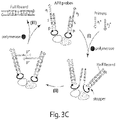
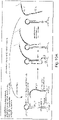
- Fig. 3A depicts an example of a molecular mechanism underlying methods of the present disclosure.
- a soluble universal u ⁇ primer binds each probe at a common single-stranded primer-binding u region
- a displacing polymerase extends the primer through the barcode ( i or j ) region and a palindromic p region to a molecule or modification that terminates polymerization (e.g., a synthetic non-DNA linker), thereby generating a "half-record," which refers to a newly generated nucleic acid strand containing a universal u ⁇ primer, a barcode ( i or j ) and a palindromic p ⁇ sequence ( e.g., u ⁇ - i ⁇ - p ⁇ or u ⁇ - j ⁇ - p ⁇ ).
- step (2) half-records are partially displaced from the barcoded probe by a "strand displacement” mechanism (see, e.g., Yurke et al., Nature 406: 605-608, 2000 ; and Zhang et al. Nature Chemistry 3: 103-113, 2011 , each of which is incorporated by reference herein), and proximate half-records hybridize to each other through the 3' palindromic regions p ⁇ .
- step (3) the half-records are extended through the barcode ( i and j ) regions and primer-binding u regions, releasing soluble, full records that encode both barcode ( i and j ) probes.
- the barcoded probes are "regenerated” and able to undergo additional cycles in the same or other molecular target pairings.
- records are collected, prepared, and sequenced by, for example, massively parallel next generation sequencing techniques. Sequence data represent spatial configurations and, in some instances, connectivities/interactions, of molecular targets, ready for statistical analysis and image reconstruction.
- “Strand displacement” refers to the mechanism by which two nucleic acid strands with identical sequences, when proximate to a single complementary nuclei acid strand (or segment of a strand), undergo relatively rapid (e . g ., timescale ⁇ Is) competition for that complement strand, 'displacing' each other from the complement presumably by a 'random-walk' mechanism.
- Nucleic acid barcoded probes of the present disclosure comprise one or more nucleic acid strands arranged into a double-stranded palindromic region, a double-stranded barcode region, and a primer-binding region.
- the barcoded probes are arranged to form a hairpin structure, which is a single stretch of contiguous nucleotides that folds and forms a double-stranded region, referred to as a "stem,” and a single-stranded region, referred to as a "loop.”
- the double-stranded region is formed when nucleotides of two regions of the same nucleic acid base pair with each other (intramolecular base pairing).
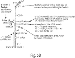
- An example of a barcoded nucleic acid hairpin is depicted in Fig. 5 .
- Nucleic acid barcoded probes of the present disclosure comprise a two parallel nucleic acid strands (e . g ., as two separate nucleic acids or as a contiguous folded hairpin).
- One of the strands is referred to as a "complementary strand,” and the other strand is referred to as a "displacement strand.”
- the complementary strand typically contains the primer-binding region, or at least a single-stranded segment of the primer-binding region, where the primer binds ( e . g ., hybridizes).
- the complementary strand and the displacement strand are bound to each other at least through a double-stranded barcoded region and through a double-stranded palindromic region.
- the "displacement strand” is the strand that is initially displaced by a newly-generated half-record, as described herein, and, in turn, displaces the newly-generated half-record as the displacement strand "re-binds" to the complementary strand.
- Two nucleic acids or two nucleic acid regions are “complementary” to one another if they base-pair, or bind, to each other to form a double-stranded nucleic acid molecule via Watson-Crick interactions (also referred to as hybridization).
- binding refers to an association between at least two molecules due to, for example, electrostatic, hydrophobic, ionic and/or hydrogen-bond interactions under physiological conditions.
- a "double-stranded region" of a nucleic acid refers to a region of a nucleic acid (e.g., DNA or RNA) containing two parallel nucleic acid strands bound to each other by hydrogen bonds between complementary purines (e.g., adenine and guanine) and pyrimidines (e.g., thymine, cytosine and uracil), thereby forming a double helix.
- the two parallel nucleic acid strands forming the double-stranded region are part of a contiguous nucleic acid strand.
- the present disclosure provides nucleic acid barcoded probes in the form of hairpin structures ( e . g ., Fig. 5 ).
- a "single-stranded region" of a nucleic acid refers to a region of a nucleic acid containing a single nucleic acid strand, unbound to (unpaired with) a second nucleic acid strand. It should be understood that a barcoded probe in the form of a hairpin structure contains both a double-stranded region (a paired region), referred to as the "stem,” and a single-stranded region (an unpaired region), referred to as the "loop,” as discussed above.
- a "double-stranded palindromic region” refers to a region of a nucleic acid (e.g., DNA or RNA) barcoded probe that is the same sequence of nucleotides whether read 5' (five-prime) to 3' (three prime) on one strand or 5' to 3' on the complementary strand with which it forms a double helix.
- a palindromic sequence shown in Fig. 5 , is considered a palindromic sequences: ACCGGT.
- a double-stranded palindromic region containing the foregoing sequence is arranged, as follows:
- palindromic (p ⁇ ) sequences permit joining of barcoded probes (and indirectly, barcoded targets) that arc proximate to each other.
- Polymerase extension of a primer bound to the primer-binding region produces a "half-record,” which refers to the newly generated nucleic acid strand.
- This displacement strand displaces a portion of the half record (by binding to its “complementary strand"), starting at the 3' end, enabling the 3' end of the half record, containing the palindromic sequence, to bind to another half record similarly displaced from a proximate barcoded nucleic acid.
- a double-stranded palindromic region has a length of 4 to 10 nucleotide base pairs. That is, in some embodiments, a double-stranded palindromic region may comprise 4 to 10 contiguous nucleotides bound to 4 to 10 respectively complementary nucleotides. For example, a double-stranded palindromic region may have a length of 4, 5, 6, 7, 8, 9 or 10 nucleotide base pairs. In some embodiments, a double-stranded palindromic region may have a length of 5 to 6 nucleotide base pairs. In some embodiments, the double-stranded palindromic region is longer than 10 nucleotide base pairs.
- the double-stranded palindromic region may have a length of 4 to 50 nucleotide base pairs. In some embodiments, the double-stranded palindromic region has a length of 4 to 40, 4 to 30, or 4 to 20 nucleotide base pairs. In some embodiments, the palindromic region may be replaced with an arbitrary sequence complementary to that produced by another probe. In such embodiments, the probes would be able to pair only with probes having complementary 3' end sequences.
- a double-stranded palindromic region may comprise guanine (G), cytosine (C), adenine (A) and/or thymine (T).
- G guanine
- C cytosine
- A adenine
- T thymine
- the percentage of G and C nucleotide base pairs (G/C) relative to A and T nucleotide base pairs (A/T) is greater than 50%.
- the percentage of G/C relative to A/T of a double-stranded palindromic region may be 50% to 100%.
- the percentage of G/C relative to A/T is greater than 60%, 65%, 70%, 75%, 80%, 85%, 90%, 95%.
- a double-stranded palindromic region may include an even number of nucleotide base pairs, although double-stranded palindromic region of the present disclosure are not so limited.
- a double-stranded palindromic region may include 4, 6, 8 or 10 nucleotide base pairs.
- a double-stranded palindromic region may include 5, 7 or 9 nucleotide base pairs.
- the double-stranded palindromic regions are the same for each probe of the plurality such that any two probes proximate to each other are able to bind to each other through generated half-records containing the palindromic sequence.
- the double-stranded palindromic regions may be the same only among a subset of barcoded probes of the plurality such that two different subsets contain two different double-stranded palindromic regions.
- a “double-stranded barcoded region” refers to a double-stranded region of a nucleic acid (e.g., DNA or RNA) barcoded probe that identifies the probe as belonging to a particular molecular target or species of molecular target.
- a double-stranded barcoded region may comprise any combination of nucleotides in random or rationally-designed order.
- a double-stranded barcoded region has a length of 2 to 100 nucleotide base pairs. That is, in some embodiments, a double-stranded barcoded region may comprise 2 to 100 contiguous nucleotides bound to 2 to 100 respectively complementary nucleotides.
- a double-stranded barcoded region may have a length of 2, 3, 4, 5, 6, 7, 8, 9, 10, 11, 12, 13, 14, 15, 16, 17, 18, 19 or 20 nucleotide base pairs.
- a double-stranded barcoded region may have a length of 2 to 5, 2 to 10, 2 to 15, 2 to 20, 2 to 25, 2 to 30, 2 to 35, 2 to 40, 2 to 45, or 2 to 50 nucleotide base pairs.
- a double-stranded barcoded region may have a length of 35 to 50, 35 to 60, 35 to70, 35 to 80, 35 to 90, or 35 to 100 nucleotide base pairs.
- a double-stranded barcoded region is longer than 100 nucleotide base pairs.
- a double-stranded barcoded region may have a length of 2 to 200 nucleotide base pairs.
- a double-stranded barcoded region has a length of 2 to 190, 2 to 180, 2 to 170, 2 to 160, 2 to 150, 2 to 140, 2 to 130, 2 to 120, or 2 to 110 nucleotide base pairs.
- nucleic acid barcoded probe is considered “unique” or “specific” to a molecular target if the barcoded region of the probe is associated only with that molecular target and can be used to identify only that molecular target among a population of molecules, including other molecular targets with their own unique barcoded probes.
- nucleic acid barcoded probe is considered “unique” or “specific” to a species of molecular target, if the barcoded region of the probe is associated only with that of molecular target and can be used to identify only that species of molecular target among a population of molecules.
- a “primer-binding region” refers to a region of a nucleic acid (e.g., DNA or RNA) barcoded probe where a single-stranded primer (e.g., DNA or RNA primer) binds to start replication.
- a primer-binding region may be a single stranded region or a partially double stranded region, which refers to a region containing both a single-stranded segment and a double-stranded segment.
- An example of a partially double-stranded primer-binding region is shown in Fig.
- a primer-binding region may comprise any combination of nucleotides in random or rationally-designed order.
- a primer-binding region has a length of 4 to 40 nucleotides (or nucleotide base pairs, or a combination of nucleotides and nucleotide base pairs, depending the single- and/or double-stranded nature of the primer-binding region).
- a primer-binding region may have a length of 4, 5, 6, 7, 8, 9, 10, 11, 12, 13, 14, 15, 16, 17, 18, 19, 20, 21, 22, 23, 24, 25, 26, 27, 28, 29, 30, 31, 32, 33, 34, 35, 36, 37, 38, 39 or 40 nucleotides (and/or nucleotide base pairs).
- a primer-binding region may have a length of 4 to 10, 4 to 15, 4 to 20, 4 to 25, 4 to 30, 4 to 35, or 4 to 40 nucleotides (and/or nucleotide base pairs).
- a primer-binding region is longer than 40 nucleotides.
- a primer-binding region may have a length of 4 to 100 nucleotides.
- a primer-binding region has a length of 4 to 90, 4 to 80, 4 to 70, 4to 60, or 4 to 50 nucleotides.
- a primer-binding region is designed to accommodate binding of more than one (e.g., 2 or 3 different) primers.
- nucleic acid barcoded probes of the present disclosure comprise a molecule or modification that terminates polymerization.
- a molecule or modification that terminates polymerization (“stopper” or “blocker”) is typically located in a double-stranded region of a barcoded probe, adjacent to the double-stranded palindromic region, such that polymerization terminates extension of the primer through the double-stranded palindromic region.
- a molecule or modification that terminates polymerization may be located between the double-stranded palindromic region and the hairpin loop, as shown in Fig. 5 ("spacer 9").
- the molecule that terminate polymerization is a synthetic non-DNA linker, for example, a triethylene glycol spacer, such as the Int Spacer 9 (iSp9) or Spacer 18 (Integrated DNA Technologies (IDT)). It should be understood that any non-native linker that terminates polymerization by a polymerase may be used as provided herein.
- Non-limiting examples of such molecules and modifications include a three-carbon linkage (/iSpC3/) (IDT), ACRYDITE TM (IDT), adenylation, azide, digoxigenin (NHS ester), cholesteryl-TEG (IDT), I-LINKER TM (IDT), and 3-cyanovinylcarbazole (CNVK) and variants thereof.
- IDTT three-carbon linkage
- ACRYDITE TM IDT
- IDT adenylation, azide, digoxigenin (NHS ester), cholesteryl-TEG (IDT), I-LINKER TM (IDT), and 3-cyanovinylcarbazole (CNVK) and variants thereof.
- CNVK 3-cyanovinylcarbazole
- short linkers e.g., iSp9 lead to faster reaction times.
- the molecule that terminates polymerization is a single or paired non-natural nucleotide sequence, such as iso-dG and iso-dC (IDT), which are chemical variants of cytosine and guanine, respectively.
- Iso-dC will base pair (hydrogen bond) with Iso-dG but not with dG.
- Iso-dG will base pair with Iso-dC but not with dC.
- the efficiency of performance of a "stopper” or “blocker” modification be improved by lowering dNTP concentrations (e . g ., from 200 ⁇ m) in a reaction to 100 ⁇ m, 10 ⁇ m, 1 ⁇ m, or less.
- barcoded probes are designed to include, opposite the molecule or modification, a single nucleotide (e.g., thymine), at least two of same nucleotide (e.g., a thymine dimer (TT) or trimer (TTT)), or an non-natural modification.
- a single nucleotide e.g., thymine
- TT thymine dimer
- TTT trimer
- a poly-T sequence e.g., a sequence of 2, 3, 4, 5, 7, 8, 9 or 10 thymine nucleotides
- a synthetic base e.g. , an inverted dT
- Other termination molecules molecules that prevent extension of a 3' end not intended to be extended
- a half record displaces one of the strands of the barcoded probe.
- This displaced strand in turn, displaces a portion of the half record, starting at the 3' end.
- This displacement of the half-record is facilitated, in some embodiments, by a "double-stranded displacement region" adjacent to the molecule or modification that terminates polymerization (see, e.g., Fig. 5 , "DS6").
- the double-stranded displacement region may be located between the molecule or modification that terminates polymerization and the hairpin loop (see, e.g., Fig. 5 ).
- a double-stranded displacement region may comprise any combination of nucleotides in random or rationally-designed order.
- a double-stranded displacement region has a length of 2 to 10 nucleotide base pairs.
- a double-stranded displacement region may have a length of 2, 3, 4, 5, 6, 7, 8, 9 or 10 nucleotide base pairs.
- a double-stranded palindromic region may have a length of 5 to 6 nucleotide base pairs.
- a double-stranded palindromic region may contain only a combination of C and G nucleotides.
- Displacement of the half-record may also be facilitated, in some embodiments, by modifying the reaction conditions.
- some auto-cyclic reactions may include, instead of natural, soluble dNTPs for new strand generation, phosphorothioate nucleotides (2'-Deoxynucleoside Alpha-Thiol 2'-Deoxynucleoside Alpha-Thiol Triphosphate Set, Trilink Biotechnologies). These are less stable in hybridization that natural dNTPs, and result in a weakened interaction between half record and stem. They may be used in any combination ( e . g ., phosphorothioate A with natural T, C, and G bases, or other combinations or ratios of mixtures). Other such chemical modifications may be made to weaken the half record pairing and facilitate displacement.
- the probe itself may be modified, in some embodiments, with unnatural nucleotides that serve instead to strengthen the hairpin stem.
- the displacing polymerase that generates the half record can still open and copy the stem, but, during strand displacement, stem sequence re-hybridization is energetically favorable over half-record hybridization with stem template.
- Non-limiting examples of unnatural nucleotides include 5-methyl dC (5-methyl deoxycytidine; when substituted for dC, this molecule increase the melting temperature of nucleic acid by as much as 5 °C per nucleotide insertion), 2,6-diaminopurine (this molecule can increase the melting temperature by as much as 1-2 °C per insertion), Super T (5-hydroxybutynl-2'-deoxyuridine also increases melting temperature of nucleic acid), and/or locked nucleic acids (LNAs). They may occur in either or both strands of the hairpin stem.
- unnatural nucleotides may be used to introduce mismatches between new half record sequence and the stem. For example, if an isoG nucleotide existed in the template strand of the stem, a polymerase, in some cases, will mistakenly add one of the soluble nucleotides available to extend the half record, and in doing so create a 'bulge' between the new half record and the stem template strand, much like the bulge (included in the primer) of Fig 5A . It will serve the same purpose of weakening half-record-template interaction and encourage displacement.
- nucleic acid barcoded probes of the present disclosure are arranged to form a hairpin structure, which is a single stretch of contiguous nucleotides that folds and forms a double-stranded region, referred to as a "stem," and a single-stranded region, referred to as a "loop.”
- the single-stranded loop region has a length of 3 to 50 nucleotides.
- the single-stranded loop region may have a length of 3, 4, 5, 6, 7, 8, 9 or 10 nucleotides.
- the single-stranded loop region has a length of 3 to 10, 3 to 15, 3 to 20, 3 to 25, 3 to 30, 3 to 35, 3 to 40, 3 to 45, or 3 to 50 nucleotides. In some embodiments, the single-stranded loop region is longer than 50 nucleotides. For example, the single-stranded loop region may have a length of 3 to 200 nucleotides. In some embodiments, the single-stranded loop region has a length of 3 to 175, 3 to 150, 3 to 100, or 3 to 75 nucleotides. In some embodiments, a loop region includes smaller regions of intramolecular base pairing.
- a hairpin loop in some embodiments permits flexibility in the orientation of the barcoded probe relative to a target binding-moiety. That is, the loop typically allows the barcoded probe to occupy a variety of positions and angles with respect to the target-binding moiety, thereby permitting interactions with a multitude of nearby probes ( e . g ., attached to other targets) in succession.
- Nucleic acid barcoded probes of the present disclosure are typically attached to molecular target through a target-binding moiety.
- the target-binding moiety is attached to an end of the barcoded probe (e.g., the end distal to the primer-binding region).
- the target-binding moiety is attached to the single-stranded loop region of a barcoded probe arranged in the form of a hairpin (see, e.g. , Fig. 5 ).
- target-binding moieties for use as provided herein include, without limitation, biotin, antibodies, aptamers, nanobodies, nucleic acids, a drugs (e.g., small molecule drugs) and atoms (e.g., Li). Other targeting molecules are contemplated.
- a molecular target may be attached to a barcoded probe through hybridization or "click chemistry.” See, e.g., Kolb H.C., et al. Angewandte Chemie International Edition 2001, 40 (11): 2004-2021 ; and Evans R.A. Australian Journal of Chemistry, 2007, 60 (6): 384-395 .
- Nucleic acid barcoded probes of the present disclosure comprise at least one locked nucleic acid (LNA) nucleotides or other modified base. Pairs of LNAs, or other modified bases, can serve as stronger (or weaker) base pairs in double-stranded regions of barcoded probes, thus biasing the strand displacement reaction.
- at least one LNA molecule is located on a complementary stranded of a barcoded probe, between a double-stranded barcoded region and a single-stranded primer-binding region.
- Nucleic acid barcoded probes of the present disclosure are bound to a molecular target.
- a "molecular target” is any molecule that one wishes to observe or quantitate. Examples of molecular targets include, without limitation, proteins, saccharides (e.g., polysaccharides), lipids, nucleic acids (e.g., DNA, RNA, microRNAs), and small molecules.
- Molecular target may be DNA or RNA.
- molecular target are RNA interference molecules, such as short-interfering RNAs (siRNAs) or micro RNAs (microRNAs).
- molecular target are antisense molecules, such as DNA antisense synthetic oligonucleotides (ASOs).
- a molecular target is a biomolecule.
- a biomolecule is any molecule that is produced by a living organism, including large macromolecules such as proteins, polysaccharides, lipids and nucleic acids (e.g. , DNA and RNA such as mRNA), as well as small molecules such as primary metabolites, secondary metabolites, and natural products.
- molecular targets specifically biomolecules
- DNA DNA, RNA, cDNA, or the DNA product of RNA subjected to reverse transcription, A23187 (Calcimycin, Calcium Ionophore), Abamectine, Abietic acid, Acetic acid, Acetylcholine, Actin, Actinomycin D, Adenosine, Adenosine diphosphate (ADP), Adenosine monophosphate (AMP), Adenosine triphosphate (ATP), Adenylate cyclase, Adonitol, Adrenaline, epinephrine, Adrenocorticotropic hormone (ACTH), Aequorin, Aflatoxin, Agar, Alamethicin, Alanine, Albumins, Aldosterone, Aleurone, Alpha-amanitin, Allantoin, Allethrin, ⁇ -Amanatin, Amino acid, Amylase, Anabolic steroid, Anethole, Angiotensin
- a molecular target is a protein target such as, for example, proteins of a cellular environment (e . g ., intracellular or membrane proteins).
- proteins include, without limitation, fibrous proteins such as cytoskeletal proteins (e . g ., actin, arp2/3, coronin, dystrophin, FtsZ, keratin, myosin, nebulin, spectrin, tau, titin, tropomyosin, tubulin and collagen) and extracellular matrix proteins (e.g., collagen, elastin, f-spondin, pikachurin, and fibronectin); globular proteins such as plasma proteins ( e .
- coagulation factors e . g ., complement proteins,C1-inhibitor and C3-convertase, Factor VIII, Factor XIII, fibrin, Protein C, Protein S, Protein Z, Protein Z-related protease inhibitor, thrombin, Von Willebrand Factor
- acute phase proteins such as C-reactive protein; hemoproteins; cell adhesion proteins ( e . g ., cadherin, ependymin, integrin, Ncam and selectin); transmembrane transport proteins ( e . g ., CFTR, glycophorin D and scramblase) such as ion channels ( e .
- ligand-gated ion channels such nicotinic acetylcholine receptors and GABAa receptors, and voltage-gated ion channels such as potassium, calcium and sodium channels), synport/antiport proteins (e . g ., glucose transporter); hormones and growth factors (e . g ., epidermal growth factor (EGF), fibroblast growth factor (FGF), vascular endothelial growth factor (VEGF), peptide hormones such as insulin, insulin-like growth factor and oxytocin, and steroid hormones such as androgens, estrogens and progesterones); receptors such as transmembrane receptors ( e .
- EGF epidermal growth factor
- FGF fibroblast growth factor
- VEGF vascular endothelial growth factor
- peptide hormones such as insulin, insulin-like growth factor and oxytocin
- steroid hormones such as androgens, estrogens and progesterones
- receptors
- G-protein-coupled receptor rhodopsin
- intracellular receptors e.g., estrogen receptor
- DNA-binding proteins e.g., histones, protamines, CI protein
- transcription regulators e.g., c-myc, FOXP2, FOXP3, MyoD and P53
- immune system proteins e . g ., immunoglobulins, major histocompatibility antigens and T cell receptors
- nutrient storage/transport proteins e . g ., ferritin
- chaperone proteins e.g ., enzymes.
- the target protein is prostate-specific antigen (PSA).
- PSA also referred to as gamma-seminoprotein or kallikrein-3
- PSA is a glycoprotein encoded in humans by the KLK3 gene.
- PSA is a member of the kallikrein-related peptidase family and is secreted by the epithelial cells of the prostate gland.
- Nucleic acid barcoded probes of the present disclosure may be DNA such as D-form DNA and L-form DNA and RNA, as well as various modifications thereof.
- Nucleic acid modifications include base modifications, sugar modifications, and backbone modifications. Non-limiting examples of such modifications are provided below.
- modified nucleic acids e.g., DNA variants
- L-DNA the backbone enantiomer of DNA, known in the literature
- PNA peptide nucleic acids
- LNA locked nucleic acid
- co-nucleic acids of the above such as DNA-LNA co-nucleic acids.
- the present disclosure contemplates nanostructures that comprise DNA, RNA, LNA, PNA or combinations thereof. It is to be understood that the nucleic acids used in methods and compositions of the present disclosure may be homogeneous or heterogeneous in nature.
- nucleic acids may be completely DNA in nature or they may be comprised of DNA and non-DNA (e . g ., LNA) monomers or sequences.
- any combination of nucleic acid elements may be used.
- the nucleic acid modification may render the nucleic acid more stable and/or less susceptible to degradation under certain conditions.
- nucleic acids are nuclease-resistant.
- a "plurality” comprises at least two nucleic acid barcoded probes.
- a plurality comprises 2 to 2 million nucleic acid barcoded probes ( e . g ., unique barcoded probes).
- a plurality may comprise 100, 500, 1000, 5000, 10000, 100000, 1000000, or more, nucleic acid barcoded probes. This present disclosure is not limited in this aspect.
- a pair of nucleic acid barcoded probes comprises (a) a first nucleic acid barcoded probe arranged into (i) a double-stranded barcode region, and (ii) a primer-binding region (e.g., a single-stranded primer-binding region), referred to for simplicity as "the first probe,” and (b) a second single-stranded barcoded probe comprising a barcode region and a primer region complementary to the single-stranded primer-binding region, referred to as "the second probe" (see, e.g., Fig.
- each probe is attached to a molecular target through a target binding moiety.
- palindromic regions are unnecessary because the second probe-the single-stranded probe-includes a primer that binds directly to the primer-binding region of the first probe.
- the second barcoded probe contains a primer at one end (e.g., 3' end and/or distal to the end that is attached to a molecular target), which is complementary to and binds to the primer-binding region of the first probe.
- the primer of the second probe is polymerized through the barcoded region of the first probe, thereby generating a record of its association with the first probe-the second probe now contains two different barcodes.
- the displacing strand of the first probe (which was displaced during polymerization of the primer) partially displaces the second probe when it "re-binds" to its complementary strand on the first probe.
- the partially-displaced second probe then spontaneously dissociates from the first probe.
- the second probe, now containing a record of its association with the first probe is free to bind to another probe and "record" that second association (see, e.g., Fig. 10A ).
- any two barcoded probes can be attached respectively to two molecular targets.
- two barcoded probes may be attached to different epitopes of the same molecular target (e.g., a protein such as an antibody), for example, in order to generate records that represent the presence of that molecular target in solutionmany copies of the same record are created when two such barcoded probes are proximate to each other) (see, e.g., Fig. 18 ). In some embodiments, this permits digital 'counting' of the number of target molecules in solution.
- Fig. 18 shows an example in which auto-cyclically generated DNA sequences report antibody co-localization on single-molecule level.
- random barcode sequences in this particular figure referred to as unique molecular identifiers (UMI) for individual antibody (Ab) molecule and barcodes for different Ab types
- UMI unique molecular identifiers
- Ab antibody
- barcodes for different Ab types
- primer pairs with UMIs and sample barcodes bind each hairpin probe at the 3' overhang respectively.
- Polymerase extends the primers up to a synthetic "stopper" site, copying the Ab UMI (1, 2) and Ab barcode.
- the extended primers are then partially released from the hairpins by strand displacement and pair their 3' Ab barcode segments (unique for the target protein).
- Microscope-free imaging systems comprise nucleic acid barcoded probes, primers designed to bind to those probes and displacing polymerase.
- a "primer” is a single-stranded nucleic acid that serves as a starting point for nucleic acid synthesis.
- a polymerase adds nucleotides to a primer to generate a new nucleic acid strand.
- Primers of the present disclosure are designed to be complementary to and to bind to the primer-binding region of a nucleic acid barcoded probe.
- primer length and composition e . g ., nucleotide composition
- a primer has a length of 4 to 40 nucleotides.
- a primer may have a length of 4, 5, 6, 7, 8, 9, 10, 11, 12, 13, 14, 15, 16, 17, 18, 19, 20, 21, 22, 23, 24, 25, 26, 27, 28, 29, 30, 31, 32, 33, 34, 35, 36, 37, 38, 39 or 40 nucleotides.
- a primer may have a length of 4 to 10, 4 to 15, 4 to 20, 4 to 25, 4 to 30, 4 to 35, or 4 to 40 nucleotides.
- Primers may exist attached in pairs or other combinations (e . g ., triplets or more, in any geometry) for the purpose, for example, of restricting probe binding to those meeting their geometric criteria (see, e.g. , Fig. 12 ).
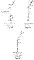
- the rigid, double-stranded linkage shown enforces both a minimum and a maximum distance between barcoded probes.
- the double-stranded "ruler" domain may be any length (e.g., 2 to 100 nucleotides, or more) and may optionally include a barcode itself that links the two halves by information content, should they become separated during processing.
- a double stranded ruler domain which enforces a typical distance between probes at which records may be generated, is a complex structure, such as a 2-, 3-, or 4-DNA helix bundle, DNA nanostructure, such as a DNA origami structure, or other structure that adds or modifies the stiffness/rigidity of the ruler.
- a "strand-displacing polymerase” refers to a polymerase that is capable of displacing downstream nucleic acid (e.g., DNA) encountered during nucleic acid synthesis.
- Different polymerases can have varying degrees of displacement activity.
- Examples of strand-displacing polymerases include, without limitation, Bst large fragment polymerase (e . g ., New England Biolabs (NEB) #M0275), phi 29 polymerase ( e . g ., NEB #M0269), Deep VentR polymerase, Klenow fragment polymerase, and modified Taq polymerase.
- Bst large fragment polymerase e . g ., New England Biolabs (NEB) #M0275
- phi 29 polymerase e . g ., NEB #M0269
- Deep VentR polymerase Klenow fragment polymerase
- modified Taq polymerase ed.
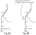
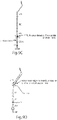
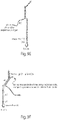
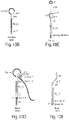
- a primer comprises at least one nucleotide mismatch relative to the single-stranded primer-binding region. Such a mismatch may be used facilitate displacement of a half-record from the complementary strand of a barcoded probe (see, e.g., Fig. 9D ).
- a primer comprises at least one artificial linker.
- the "cycling rate" of a microscope-free imaging reaction refers to the rate at which a full-record (as opposed to half-record) is produced, resulting from the cyclic interaction of two proximate barcoded molecular targets.
- a primer comprising a mismatch or an artificial linker increases the cycling rate by 5-fold to 10-fold, or more.
- microscope-free imaging systems of the present disclosure further comprise a helper nucleic acid strand that is partially complementary to the single-stranded primer binding region, is partially complementary to a single-stranded region adjacent to the primer binding region, and binds transiently to the single-stranded region adjacent to the primer binding region (see, e.g., Fig. 9F ).
- a "helper strand” permits initiation of polymerization by the polymerase while also permitting the presence of a mismatch, as described above.
- a helper strand has a length of 3 to 20 nucleotides.
- a helper strand may have a length of 3, 4, 5, 6, 7, 8, 9, 10, 11, 12, 13, 14, 15, 16, 17, 18, 19 or 20 nucleotides.
- the methods comprise the steps of (a) contacting, with at least one (e.g., a plurality) of the nucleic acid barcoded probes as provided herein, a sample containing at least one molecular target, a primer complementary to a single-stranded primer-binding region of a probe of the plurality, and a strand-displacement polymerase, and (b) incubating the sample under conditions that permit production of barcoded records.
- at least one e.g., a plurality
- the methods comprise the steps of (a) contacting, with at least one (e.g., a plurality) of the nucleic acid barcoded probes as provided herein, a sample containing at least one molecular target, a primer complementary to a single-stranded primer-binding region of a probe of the plurality, and a strand-displacement polymerase, and (b) incubating the sample under conditions that permit production of barcoded records.
- the methods comprise the steps of contacting, with a pair of nucleic acid barcoded probes as provided herein, a sample containing at least two molecular targets, and a strand-displacement polymerase, and (b) incubating the sample under conditions that permit production of barcoded records (e.g., under conditions that permit nucleic acid replication).
- a “sample” may comprise cells (or a cell), tissue, or bodily fluid such as blood (serum and/or plasma), urine, semen, lymphatic fluid, cerebrospinal fluid or amniotic fluid.
- a sample may be obtained from (or derived from) any source including, without limitation, humans, animals, bacteria, viruses, microbes and plants.
- a sample is a cell lysate or a tissue lysate.
- a sample may also contain mixtures of material from one source or different sources.
- a sample may be a spatial area or volume (e.g., a grid on an array, or a well in a plate or dish).
- a sample is a single cell, such as a rare cell.
- a rare cell include, without limitation, circulating tumor cells, epithelial progenitor and stem cells, mesenchymal cells, and fetal cells, for example, circulating in the blood stream.
- Cons that permit production of barcoded records may be physiological conditions (e . g ., a temperature of 20-40 degrees Celsius, atmospheric pressure of 1, and/or a pH value of 6-8).
- step (b) is performed at a temperature of 20 to 40 degrees Celsius (°C).
- step (b) may be performed at a temperature of 20 °C, 21 °C, 22 °C, 23 °C, 24 °C, 25 °C, 26 °C, 27 °C, 28 °C, 29 °C, 31 °C, 32 °C, 33 °C, 34 °C, 35 °C, 36 °C, 37 °C, 38 °C, 39 °C or 40 °C.
- step (b) is performed for a time of 10 minutes (min) to 24 hours, or more.
- step (b) may be performed for a time of 10 min to 3 hours (hr), 10 min to 12 hr, 10 min to 18 hr, or 10 min to 24 hr.
- step (b) is performed for a time of 10 min, 15 min, 20 min, 25 min, 30 min, 35 min, 40 min, 45 min, 50 min, 55 min, 60 min, 65 min, 70 min, 75 min, 80 min, 85 min, 90 min, 95 min, 100 min, 105 min, 110 min, 115 min, 120 min, 125 min, 130 min, 135 min, 140 min, 145 min, 150 min, 155 min, 160 min, 165 min, 170 min, 175 min or 180 min.
- Microscope-free imaging reaction may, in some embodiments, have a salt concentration of 0.25-15 mM Mg and/or 50-250 mM Na.
- Microscope-free imaging reaction may, in some embodiments, have reaction dNTPs concentrations of 0.05-5 mM (e.g., 0.05 mM, 0.10 mM, 0.15 mM, 0.20 mM, 0.25 mM, 0.30 mM, 0.35 mM, 0.40 mM, 0.45 mM, 0.50 mM, 1.0 mM, 1.5 mM, 2.0 mM, 2.5 mM, 3.0 mM, 3.5 mM, 4.0 mM, 4.5 mM or 5.0 mM.
- reaction dNTPs concentrations of 0.05-5 mM (e.g., 0.05 mM, 0.10 mM, 0.15 mM, 0.20 mM, 0.25 mM, 0.30 mM, 0.35 mM, 0.40 mM, 0.45 mM, 0.50 mM, 1.0 mM, 1.5 mM, 2.0 mM, 2.5 mM, 3.0 m
- Buffers that may be used in microscope-free imaging reaction include, without limitation, "Thermo-Pol Buffer” (New England Biolabs), phosphate buffered saline (with or without Mg or Na supplementation), any commercial or laboratory-prepared cell media, water or any pH-buffered solution supplemented with cationic salts sufficient for DNA hybridization and polymerase operation.
- “Thermo-Pol Buffer” New England Biolabs
- phosphate buffered saline with or without Mg or Na supplementation
- any commercial or laboratory-prepared cell media water or any pH-buffered solution supplemented with cationic salts sufficient for DNA hybridization and polymerase operation.
- the cycling rate of a microscope-free imaging reaction is 1 full-record per 10 minutes per pair of probes, but may be as rapid as 1 full record per second or as slow as 1 full record per 10 hours under certain (e.g., more restrictive) conditions.
- nucleic acid records (referred to herein simply as “records") of the spatial configuration of barcoded targets are produced (see, e.g., Fig. 3A ).
- the records are double-stranded.
- the records are single-stranded.
- the length of the records may vary.
- a barcoded record may have a length of 30 to 500 nucleotides (or nucleotide base pairs).
- a barcoded record has a length of 30 to 100, 30 to 200, 30 to 300, 30 to 400, 50 to 100, 50 to 200, 50 to 300, 50 to 400 or 50 to 500 nucleotides (or nucleotide base pairs).
- a barcoded record has a length of 80 to 100 nucleotides (or nucleotide base pairs), or 90 nucleotides (or nucleotide base pairs).
- Records may be "released” from probes via polymerase-mediated mechanisms or via spontaneous release of an extended primer from the primer-binding region on a probe.
- the 3' end of an extended primer can be engineered to fold back on itself and extended off of the probe. Because microscope-free imaging systems of the present disclosure do not typically copy the paired barcode onto the same record strand, an additional barcode may be added to a ruler-like primer that links pairs of records together in informational content.
- barcoded records are generated, they are collected and, in some embodiments, purified. For example, records may be collected in the supernatant of the reaction or by collecting the all the contents of the reaction vessel. Further preparation of the records for sequencing may sequencing platform-specific. Some platforms may require no further preparation, but often the records must have a combination of (1) a sequencing-specific 'adapter' or other oligonucleotides added to their ends, (2) undergo 'amplification' reactions ( e .
- PCR polymerase chain reaction
- identical or nearly-identical e.g., 99%, 98%, 95%, 90%, 80% identical
- purification from other sequences, proteins, or reaction components that may interfere with preparation or sequencing.
- adapter sequences may be ligated to the records using a common 'A-Tailing' technique, followed by gel electrophoresis purification, and finally PCR amplification.
- some embodiments may allow for PCR amplification of records directly, possibly adding adapter sequences through long DNA primers, or followed by adapter ligation and gel purification.
- two or more types of recording primers may be used to, for example, facilitate later PCR amplification or preparation for sequencing.
- Each primer may have the same probe-binding sequence, but vary in an additional 5' sequence not involved in the recording reaction.
- an average of half of all resultant records will have different, non-complementary 5' and 3' ends.
- These may be used for further amplification ( e . g ., by allowing the ends to more easily hybridize with soluble PCR primers instead of forming a hairpin stem), ligation or other processing. If ten types of such recording primers are used, for example, then an average of 90% of all records will have different, non-complementary 5' and 3' ends, further facilitating end-specific amplification and processing.
- barcoded records are "decoded" by direct observation by gel electrophoresis or amplification and quantification by PCR (see, e.g., Fig 19C ). This example relies on specific amplification of particular records based on different sequences in their recording primers or barcodes. Massively parallel sequencing is also encompassed by the present disclosure and may be used to decode precise, individual molecular arrangements in large systems (see, e.g., Fig 3F ).
- collected barcoded records are then sequenced.
- the records are sequenced using next-generation sequencing technologies.
- Sanger sequencing is used as well as "post-next-generation sequencing” technologies under development, such as "nanopore”-based sequencing (e . g ., Oxford Nanopore Technologies, nanoporetech.com).
- electrophoretic gels may be used to detect combinations of barcodes within a record by differentiating by the length of record produced (see, e . g . , Fig. 3b ), or standard resolution or super-resolution microscopy may be used to visually detect sequences of probes by fluorescent in situ hybridization or similar approaches.
- nucleic acid microarrays e . g ., Agilent Technologies
- Agilent Technologies may be used to detect records in a sequence-specific manner.
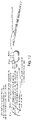
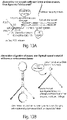
- Figs. 13A-13B show examples of ligation and amplification methods of the present disclosure, used for processing barcoded records.
- records may be cut with restriction endonucleases, then ligated via "sticky" ends ( Fig. 13A ).
- Excess non-ligated record material may be cut with one or more endonucleases ( Fig. 13B )
- the process of ligating sequencing adapters to the records may be performed by blunt end, "A-tailing," or restriction site-mediated ligation. Ligation may be performed on raw, double-stranded records or after annealing into hairpins.
- primers may be added not as a single species but as any number of species differentiated by a 5' extension that does not participate actively in the record-generating reaction per se , but allows for the two ends of most records to be different (not shown). This in turn allows for PCR amplification with different primers at each end, perhaps incorporating the sequencing adapters, and this amplification may be done before or after ligation.
- exonucleases as single enzymes or in combination, e . g ., T7 exonuclease in combination with exonuclease I, III, or T
- T7 exonuclease in combination with exonuclease I, III, or T
- the spatial arrangement of barcoded molecular targets, relative to one another can be determined.
- the sequencing data may be processed computationally to produce a representative network of molecular target interactions.
- Such computer code may read sequencing data files, create a digitally-encoded network of associated barcodes (e . g ., 'barcode A connected to B' derived from reading record #1, 'B-C' from record #2, and 'C-A' from record #3).
- Any number of commercially available (e . g ., Mathematica) or independently-written codes are then available for transforming this non-geometric digital representation into one describing relative spatial positioning (known mathematically as aspects of 'graph theory').
- the records imply a triangle-like positioning of targets associated with A, B, and C.
- programs could calculate statistics of interactions of proteins like the one associated with barcode A by analyzing the interactions of all barcodes associated with that protein (known a priori because the targeting moieties can be linked to specific barcode sets before targeting and reaction are carried out).
- the time-dependence of interactions may be determined from these data by analyzing different record sets taken at different times or provided with molecular time-stamps ( e . g ., primers with slight differences added in the latter half of a reaction).
- Some aspects of the present disclosure provide methods that comprise combining in a single reaction (a) two single-stranded nucleic acid barcoded probes, each comprising a palindromic sequence, a barcode sequence and a primer-binding sequence, wherein the barcodes sequences are different from each other, and wherein each barcoded probe is attached to a molecular target, (b) a partially double-stranded primer arranged into a double-stranded region flanked by 3' single-stranded flanking regions that each contain a primer complementary to the primer-binding sequence, wherein the double-stranded region contains a reversible covalent binding site (see, e.g., Fig. 11 ), and a strand-displacing polymerase.
- the reversible covalent binding site contains a CNV K modification (see, e.g., U.S. Patent No. 8, 481,714 ) such that two strands can be covalently bound in the presence of ultraviolet light and later release in the presence of a difference wavelength of light.
- CNV K modification see, e.g., U.S. Patent No. 8, 481,714
- Other temperature-stable, reversible linkages may be used, as provided herein.
- the methods comprise incubating (a) and (b) under conditions sufficient to permit binding of the primers to the primer-binding sites and extension of each 3' flanking region of the primers.
- the methods comprise heating the reaction to a temperature of at least 50 °C (e.g., 50 °C to 100 °C) to permit dissociation of the primers from the two single-stranded nucleic acid barcoded probes, thereby regenerating (a) and (b) for further temperature-drive cycling.
- a temperature of at least 50 °C e.g., 50 °C to 100 °C
- Fig. 11 shows an example of temperature-dependent auto-cycling mechanism of the present disclosure. Records are cyclically created, each record from two separate primers on two unique probes. The nascent "half-records" are not spontaneously driven from the probes, but are instead "melted off' by an elevated temperature. Probes and primers are shown in Fig. 11 with primers as double 3'-headed structures held together by UV-reversible, covalent cross-linking CNV K moieties. The primers bind any pair of probes, are extended at low (e.g. , 25 °C) temperature to copy barcodes, and are released when the temperature is raised above the melting temperature of the strands (e.g., 70 °C).
- low e.g. 25 °C
- the polymerase in solution may complete the copying of the strands linked through binding of palindromic sequences, in some embodiments, aided by the release of the CNV K linkage.
- the system is driven synchronously with temperature cycling.
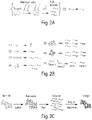
- Fig. 3 shows data from an experiment demonstrating an example of a proximity-based, autocyclic, full-record generation method.
- Lanes 1-4 of the electrophoretic gel each contain combinations of barcoded probes of different lengths and localization (depicted above the gel), together with the primer and polymerase (not depicted).
- short and long barcoded probes exist unconnected in solution. Cy5-labeled primers (in large excess) were converted to short or long half-records, but no full-records were generated because probes were not proximate to each other.
- short barcoded probes were co-localized on streptavidin molecules, and short half-records, and subsequently short full-records, were produced.
- This functional auto-cycling reaction permits repeated barcoded probe reading at constant physiologic temperatures and conditions.
- Auto-cycling in combination with unique barcode labeling that allows differentiation of individual molecules, enables true network elucidation and imaging ( Fig. 3 ) with a precision that microscopy and common pairwise proximity methods cannot match.
- Recording using common pairwise proximity techniques e.g., co-Immuno-precipitation, proximity ligation
- Fig. 2B elucidation of individual networks
- Synthetic DNA nanostructures are engineered to display DNA barcodes at arbitrary user-specified positions with nanometer precision, thus serving as a "workbench” for evaluating the performance of the auto-cycler and subsequent image reconstruction method in a rigorous and precise fashion.
- Sample handling techniques e . g ., microfluidic devices
- computational algorithms and software tools for imaging reconstruction based on full-record sequences are used.
- Protein receptor clusters on cell surfaces are then imaged, first in fixed cells and then in live cells. The labeling of the surface protein clusters is achieved using antibodies and smaller binders, such as nanobodies or aptamers.
- the Microscope-Free Imaging (MFI) results are compared with data obtained using ultra-high resolution microscope-based methods.
- MFI is then applied to another application: high-throughput, high resolution imaging of nucleome organization.
- a diffusion-based molecular recorder is developed, which can repeatedly generate molecular records for targets separated over long range, further expanding the capacity of MFI.
- Methods and composition of the present disclosure may be used to image and elucidate protein clusters in cell plasma membranes.
- the four similar, membrane-bound EGF receptors of the ErbB family form a part of complex, modular, heterogeneous network with 13 other polypeptide ligands on the surface of cells.
- the network mediates cell proliferation and survival as well as migration and adhesion. They dimerize in 10 combinations and appear to form higher-order clusters of up to 150 nm across, implying the co-localization of hundreds of receptors within clusters ( Abulrob et al., J Biol Chem 285(5): 3145-56, 2010 , incorporated by reference herein).
- MFI Microscope-Free Imaging
- Microscope-free-imaging methods of the present disclosure reveals many aspects of cluster protein content and behavior ( Fig.. 15 ).
- a static (fixed) cell unique labeling of all proteins of interest enables visualization of full network content and connectivity, including association stoichiometry and distribution of protein states within or between cells.
- MFI methods can follow slow (e.g., over tens-to-hundreds of seconds) cluster reconfiguration, and monitor faster processes.
- Data obtained from MFI methods offers molecular-scale resolution, true complex network connectivity, and the ability to track dynamic molecular interactions.
- a CNV K-based probe Yoshimura et al. Org. Lett. 10(15): 3227-3230, 2008 , incorporated by reference
- Identification of regions of interest under a scanning confocal microscope with an appropriate laser can disable all probes in a sample except for those in the regions of interest, allowing sampling from tissues in situ.
- barcoded primers with "time stamps,” e.g., time stamp 1 at condition A, followed by later barcoded primer with time stamp 2 at condition B permits a coarse time-resolved state changes following, for example, drug delivery for drug screening applications.
- Microscope-Free Imaging (MFI) methods of the present disclosure are used to elucidate nucleome organization ( Fig. 16 ).
- An OligoPaints method ( Beliveau et al., PNAS USA. 109(52): 21301-21306, 2012 , incorporated by reference) is applied to target hundreds of thousands of arbitrary genome segments by in situ hybridization, employing the MFI methods, as provided herein, at each target. Because target regions are not limited by restriction enzyme sequences, and the cycling nature of the recording reaction allows for a probe to participate in many record pairs, the spatial resolution of the final map can be higher than the kilobase-scale resolution offered by 4C and far higher than that of Hi-C.
- mapping pluripotency or differentiation factor binding and genome organization of embryonic stem (ES) or induced pluripotent stem (iPS) cells is achieved using the methods of the present disclosure.
- Knowledge of the affiliation and co-affiliation of multiple transcription factors to stem-cell regulation enables further de-differentiation and more complete target differentiation.
- MFI methods of the present disclosure permit mapping of unassembled sequences to the genome. Repetitive sequences, or those surrounded by repeats, can be difficult to localize, but repetitive fluorescence in situ hybridization (FISH) probes with unique barcodes each, followed by proximity-based record generation, ties together sequences that are difficult to read in one reaction.
- FISH fluorescence in situ hybridization
- MFI of the present disclosure can be adapted to map targets separated by longer distances.
- Such long-range data is useful for more comprehensive molecular elucidation.
- the relative positioning among independent clusters on a cell may be important, and these data can be registered with that of individual cluster measurements if taken by the same mechanism.
- the chromosome position application may benefit from additional network data spanning sequences farther than those in direct contact.
- the mechanism for this intermediate-range (5-25+ nm) acquisition relies on diminishing concentration gradients of released half-records. Short-length primers bind transiently to existing probes are extended into half-records ( FIG. 17 ).
- the strand displacement reaction occasionally dissociates the half-record to the point of only primer hybridization, and then the sufficiently weak primer hybridization spontaneously dissociates and the half-record diffuses away. A short distance away, it encounters either a bound half-record or another soluble one, the palindromic sites bind each other, and the full-record is polymerized.
- An optimized balance between reasonable primer binding and spontaneous dissociation was found at a primer length of 7 nucleotides (data not shown).
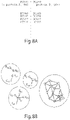
- Fig. 19B indicates the relative recording rate for three probe designs, containing 0, 12, and 18 nt spacer domains. Each probe pair was tested every 6nm for separations of 6 to 48nm, and record generation rate mathematical fits were normalized to a common maximum. Zero, 12, and 18 nt probes all produced records near maximum rates when closest to one another, suggesting the local concentration effect was a dominant driver of rate. Rates were reduced to half at 13, 20, and 25nm, respectively, and had a maximum reach (near zero rate) at 18, 42, and 48nm. In absolute terms, the 12 nt spacer probe produced records at the fastest rate, approximately twice as fast as the 0 and 18 nt probes (not shown).
- the maximum expected distance is twice the sum of the probe length (19 nt of double-stranded DNA, at 0.34 nm per base pair,13 ⁇ 6.5nm) and Half Record length (11 nt of single-stranded DNA minus 3 nt palindrome overlap, at the maximum 0.58 nm per base, 13 -4.6 nm), totaling ⁇ 22nm (geometry as in Fig. 2c , after step ii).
- the 12 nt and 18 nt spacer probes have a maximum expected reach of -36 and -43 nm, respectively. Estimates using worm-like chain models are similar.
- the three possible orders of the linear arrangement each produce the appropriate records ( Fig. 19C , panels (ii)-(iv)), demonstrating an ability to record geometric information from three molecular-scale targets in arbitrary configuration.
- This method can be applied to more than three simultaneous targets simply by designing additional probes of orthogonal primer sequence.
- inventive embodiments are presented by way of example only and that, within the scope of the appended claims and equivalents thereto, inventive embodiments may be practiced otherwise than as specifically described and claimed.
- inventive embodiments of the present disclosure are directed to each individual feature, system, article, material, kit, and/or method described herein.
- the phrase "at least one,” in reference to a list of one or more elements, should be understood to mean at least one element selected from any one or more of the elements in the list of elements, but not necessarily including at least one of each and every element specifically listed within the list of elements and not excluding any combinations of elements in the list of elements.
- This definition also allows that elements may optionally be present other than the elements specifically identified within the list of elements to which the phrase "at least one" refers, whether related or unrelated to those elements specifically identified.
- the current disclosure contains, inter alia, the following items:
Landscapes
- Chemical & Material Sciences (AREA)
- Life Sciences & Earth Sciences (AREA)
- Health & Medical Sciences (AREA)
- Organic Chemistry (AREA)
- Engineering & Computer Science (AREA)
- Immunology (AREA)
- Zoology (AREA)
- Molecular Biology (AREA)
- Wood Science & Technology (AREA)
- Proteomics, Peptides & Aminoacids (AREA)
- Analytical Chemistry (AREA)
- General Health & Medical Sciences (AREA)
- Biotechnology (AREA)
- Microbiology (AREA)
- Biochemistry (AREA)
- Physics & Mathematics (AREA)
- Genetics & Genomics (AREA)
- Bioinformatics & Cheminformatics (AREA)
- General Engineering & Computer Science (AREA)
- Biophysics (AREA)
- Urology & Nephrology (AREA)
- Biomedical Technology (AREA)
- Pathology (AREA)
- Hematology (AREA)
- Tropical Medicine & Parasitology (AREA)
- Food Science & Technology (AREA)
- Medicinal Chemistry (AREA)
- General Physics & Mathematics (AREA)
- Cell Biology (AREA)
- Measuring Or Testing Involving Enzymes Or Micro-Organisms (AREA)
- Chemical Kinetics & Catalysis (AREA)
Applications Claiming Priority (3)
| Application Number | Priority Date | Filing Date | Title |
|---|---|---|---|
| US201562110050P | 2015-01-30 | 2015-01-30 | |
| PCT/US2016/015503 WO2016123419A1 (en) | 2015-01-30 | 2016-01-29 | Microscope-free imaging |
| EP16744150.0A EP3250716B1 (de) | 2015-01-30 | 2016-01-29 | Mikroskopfreie bildgebung |
Related Parent Applications (1)
| Application Number | Title | Priority Date | Filing Date |
|---|---|---|---|
| EP16744150.0A Division EP3250716B1 (de) | 2015-01-30 | 2016-01-29 | Mikroskopfreie bildgebung |
Publications (1)
| Publication Number | Publication Date |
|---|---|
| EP3974537A1 true EP3974537A1 (de) | 2022-03-30 |
Family
ID=56544362
Family Applications (2)
| Application Number | Title | Priority Date | Filing Date |
|---|---|---|---|
| EP21183689.5A Pending EP3974537A1 (de) | 2015-01-30 | 2016-01-29 | Mikroskopfreie bildgebung |
| EP16744150.0A Active EP3250716B1 (de) | 2015-01-30 | 2016-01-29 | Mikroskopfreie bildgebung |
Family Applications After (1)
| Application Number | Title | Priority Date | Filing Date |
|---|---|---|---|
| EP16744150.0A Active EP3250716B1 (de) | 2015-01-30 | 2016-01-29 | Mikroskopfreie bildgebung |
Country Status (5)
| Country | Link |
|---|---|
| US (2) | US11639522B2 (de) |
| EP (2) | EP3974537A1 (de) |
| CN (1) | CN107208160B (de) |
| ES (1) | ES2891085T3 (de) |
| WO (1) | WO2016123419A1 (de) |
Cited By (1)
| Publication number | Priority date | Publication date | Assignee | Title |
|---|---|---|---|---|
| WO2024069235A3 (en) * | 2022-09-30 | 2024-06-06 | Sixfold Bioscience Ltd. | Compositions containing oligonucleotides with theranostic applications |
Families Citing this family (16)
| Publication number | Priority date | Publication date | Assignee | Title |
|---|---|---|---|---|
| AU2011319755B2 (en) | 2010-10-27 | 2017-02-23 | President And Fellows Of Harvard College | Compositions of toehold primer duplexes and methods of use |
| EP2633080B1 (de) | 2010-10-29 | 2018-12-05 | President and Fellows of Harvard College | Verfahren zur detektion von zielmolekülen durch fluoreszensmarkierte sonden bestehend aus nukleinsäure-nanotuben |
| EP3417078B1 (de) * | 2016-02-17 | 2021-05-05 | President and Fellows of Harvard College | Molekulare programmierwerkzeuge |
| WO2017205719A1 (en) * | 2016-05-27 | 2017-11-30 | President And Fellows Of Harvard College | Conditional primer extension for single-molecule detection |
| WO2017216270A1 (de) | 2016-06-15 | 2017-12-21 | Grabmayr Heinrich | Einzelmolekülnachweis bzw. -quantifizierung durch dna-nanotechnologie |
| EP3488018B1 (de) * | 2016-07-22 | 2026-02-18 | President and Fellows of Harvard College | Verfahren zur proteinidentifikation |
| WO2018057502A2 (en) | 2016-09-20 | 2018-03-29 | President And Fellows Harvard College | Molecular verification systems |
| US11492661B2 (en) * | 2017-01-10 | 2022-11-08 | President And Fellows Of Harvard College | Multiplexed signal amplification |
| US20230183791A1 (en) * | 2017-03-24 | 2023-06-15 | National University Of Singapore | Methods For Multiplex Detection Of Molecules |
| EP3743525A4 (de) | 2018-01-26 | 2021-09-29 | President and Fellows of Harvard College | Näherungsdetektionsverfahren und -zusammensetzungen |
| US11841371B2 (en) | 2018-03-13 | 2023-12-12 | The Broad Institute, Inc. | Proteomics and spatial patterning using antenna networks |
| AU2019240244A1 (en) | 2018-03-22 | 2020-11-19 | President And Fellows Of Harvard College | Methods and compositions for molecular authentication |
| CN109033745A (zh) * | 2018-06-08 | 2018-12-18 | 中国农业科学院农业基因组研究所 | 一种消除三维基因组学技术噪音的方法及应用 |
| CN108804872A (zh) * | 2018-06-08 | 2018-11-13 | 中国农业科学院农业基因组研究所 | 利用外切酶组合消除三维基因组学技术噪音的方法及应用 |
| EP4051810B1 (de) | 2019-10-28 | 2024-08-28 | Pixelgen Technologies Ab | Verfahren zur kartierung von rolling-circle-amplifikationsprodukten |
| CA3213800A1 (en) | 2021-03-30 | 2022-10-06 | Simon Fredriksson | Spatial mapping by serial primer extension |
Citations (8)
| Publication number | Priority date | Publication date | Assignee | Title |
|---|---|---|---|---|
| WO2001094625A2 (en) * | 2000-06-06 | 2001-12-13 | Tm Bioscience Corporation | Capture moieties for nucleic acids and uses thereof |
| WO2007117256A1 (en) * | 2005-05-31 | 2007-10-18 | Applera Corporation | Multiplexed amplification of short nucleic acids |
| WO2010107416A1 (en) * | 2009-03-19 | 2010-09-23 | Massachusetts Institute Of Technology | Parallel proximity ligation event analysis |
| WO2012112804A1 (en) * | 2011-02-18 | 2012-08-23 | Raindance Technoligies, Inc. | Compositions and methods for molecular labeling |
| US8481714B2 (en) | 2007-11-19 | 2013-07-09 | Japan Advanced Institute Of Science And Technology | Light-responsive artificial nucleotide having photo-crosslinking ability |
| WO2014071361A1 (en) * | 2012-11-05 | 2014-05-08 | Rubicon Genomics | Barcoding nucleic acids |
| WO2014130388A1 (en) * | 2013-02-20 | 2014-08-28 | Emory University | Methods of sequencing nucleic acids in mixtures and compositions related thereto |
| US20190285644A1 (en) * | 2018-03-13 | 2019-09-19 | The Broad Institute, Inc. | Proteomics and Spatial Patterning Using Antenna Networks |
Family Cites Families (79)
| Publication number | Priority date | Publication date | Assignee | Title |
|---|---|---|---|---|
| US5030566A (en) | 1989-01-04 | 1991-07-09 | Board Of Regents, The University Of Texas System | Concatemeric DNA length standards |
| US5543507A (en) | 1992-03-05 | 1996-08-06 | Isis Pharmaceuticals, Inc. | Covalently cross-linked oligonucleotides |
| US5681697A (en) | 1993-12-08 | 1997-10-28 | Chiron Corporation | Solution phase nucleic acid sandwich assays having reduced background noise and kits therefor |
| US6143495A (en) | 1995-11-21 | 2000-11-07 | Yale University | Unimolecular segment amplification and sequencing |
| CA2255774C (en) * | 1996-05-29 | 2008-03-18 | Cornell Research Foundation, Inc. | Detection of nucleic acid sequence differences using coupled ligase detection and polymerase chain reactions |
| US5866336A (en) | 1996-07-16 | 1999-02-02 | Oncor, Inc. | Nucleic acid amplification oligonucleotides with molecular energy transfer labels and methods based thereon |
| US6485901B1 (en) | 1997-10-27 | 2002-11-26 | Boston Probes, Inc. | Methods, kits and compositions pertaining to linear beacons |
| WO2000034527A2 (en) * | 1998-12-11 | 2000-06-15 | The Regents Of The University Of California | Targeted molecular bar codes |
| EP2253717B1 (de) | 2000-04-07 | 2014-04-30 | Eiken Kagaku Kabushiki Kaisha | Methode zur Amplifizierung von Nukleinsäuren mit Hilfe von dopplesträngigen Nukleinsäuren als Templat |
| WO2001077392A2 (en) * | 2000-04-10 | 2001-10-18 | Matthew Ashby | Methods for the survey and genetic analysis of populations |
| JP2003535600A (ja) | 2000-06-02 | 2003-12-02 | バイエル コーポレイション | インサイチュ分枝dnaハイブリダイゼーションを用いた高感度遺伝子検出および位置決定 |
| US20040161741A1 (en) | 2001-06-30 | 2004-08-19 | Elazar Rabani | Novel compositions and processes for analyte detection, quantification and amplification |
| US20050045063A1 (en) | 2001-11-02 | 2005-03-03 | Matthias Niggemann | Marking solution for counterfeit-resistant identification of a valuable object, marking produced by the marking solution and method for marking a valuable object |
| DE60229890D1 (de) * | 2002-09-30 | 2008-12-24 | Hoffmann La Roche | Oligonukleotide zur genotypisierung des thymidylat-synthase gens |
| AU2003290937A1 (en) | 2002-11-15 | 2004-06-15 | Trustees Of Boston University | Cis/trans riboregulators |
| WO2005007815A2 (en) | 2003-07-07 | 2005-01-27 | One Cell Systems, Inc. | Hairpin-labeled probes and methods of use |
| US7354706B2 (en) * | 2003-09-09 | 2008-04-08 | The Regents Of The University Of Colorado, A Body Corporate | Use of photopolymerization for amplification and detection of a molecular recognition event |
| EP2284259A3 (de) * | 2003-10-10 | 2011-06-15 | Novozymes A/S | Proteasen |
| WO2005059157A2 (en) | 2003-12-11 | 2005-06-30 | The Board Of Trustees Of The Leland Stanford Junior University | METHODS AND COMPOSITIONS FOR USE IN PREPARING HAIRPIN RNAs |
| CN101048505A (zh) | 2004-10-25 | 2007-10-03 | 德福根有限公司 | 用于递送双链rna至有害生物的包含至少一个适配子的多结构域rna分子 |
| WO2006074222A2 (en) | 2005-01-03 | 2006-07-13 | The Government Of The United States Of America As Represented By The Secretary Of The Department Of Health And Human Services, Centers For Disease Control And Prevention | Primer for nucleic acid detection |
| US8407013B2 (en) * | 2005-06-07 | 2013-03-26 | Peter K. Rogan | AB initio generation of single copy genomic probes |
| WO2007092538A2 (en) * | 2006-02-07 | 2007-08-16 | President And Fellows Of Harvard College | Methods for making nucleotide probes for sequencing and synthesis |
| EP2029774B1 (de) | 2006-06-01 | 2015-04-01 | Third Wave Technologies, Inc. | Nachweis von nukleinsäuren |
| DE102006031015A1 (de) | 2006-07-03 | 2008-01-10 | Identif Gmbh | Verfahren und Vorrichtung zur Authentifizierung von mit einer Markierung versehenen Gegenständen |
| WO2010012464A1 (en) * | 2008-08-01 | 2010-02-04 | Roche Diagnostics Gmbh | Improved lysis and reverse transcription for mrna quantification |
| CN102317471A (zh) | 2008-12-11 | 2012-01-11 | 加利福尼亚大学董事会 | 用于单个dna分子的直接测序的方法和系统 |
| EP2230312A1 (de) * | 2009-03-19 | 2010-09-22 | Helmholtz-Zentrum für Infektionsforschung GmbH | Sondenverbindung zum Nachweisen und Isolieren von Enzymen sowie Mittel und Verfahren zu ihrer Verwendung |
| GB0910302D0 (en) | 2009-06-15 | 2009-07-29 | Lumora Ltd | Nucleic acid amplification |
| US9493822B2 (en) * | 2009-09-01 | 2016-11-15 | Koninklijke Philips Electronics N.V. | Devices and methods for microarray selection |
| WO2011027345A1 (en) * | 2009-09-04 | 2011-03-10 | Freespace Materials Ltd. | Barium containing granules for sorption applications |
| WO2011066330A2 (en) | 2009-11-24 | 2011-06-03 | Life Technologies Corporation | Selective amplification of polynucleotide sequences |
| CA2786916A1 (en) | 2010-01-15 | 2011-07-21 | The University Of British Columbia | Multiplex amplification for the detection of nucleic acid variations |
| JP5901046B2 (ja) * | 2010-02-19 | 2016-04-06 | 国立大学法人 千葉大学 | OATP1B3mRNAの新規な選択的スプライシングバリアント |
| US9476101B2 (en) | 2010-06-07 | 2016-10-25 | Firefly Bioworks, Inc. | Scanning multifunctional particles |
| US8962241B2 (en) | 2010-07-20 | 2015-02-24 | California Institute Of Technology | Triggered molecular geometry based bioimaging probes |
| US9834439B2 (en) | 2010-07-20 | 2017-12-05 | California Institute Of Technology | Biomolecular self-assembly |
| US20130225623A1 (en) * | 2010-10-27 | 2013-08-29 | Mount Sinai School Of Medicine | Methods of Treating Psychiatric or Neurological Disorders with MGLUR Antagonists |
| AU2011319755B2 (en) | 2010-10-27 | 2017-02-23 | President And Fellows Of Harvard College | Compositions of toehold primer duplexes and methods of use |
| EP2633080B1 (de) | 2010-10-29 | 2018-12-05 | President and Fellows of Harvard College | Verfahren zur detektion von zielmolekülen durch fluoreszensmarkierte sonden bestehend aus nukleinsäure-nanotuben |
| EP2633081B1 (de) | 2010-10-29 | 2017-01-11 | Olink Bioscience AB | Näherungsligationstechnologie für western-blot-anwendungen |
| US9295965B2 (en) | 2010-11-12 | 2016-03-29 | Gen9, Inc. | Methods and devices for nucleic acid synthesis |
| WO2012071428A2 (en) | 2010-11-22 | 2012-05-31 | Solulink, Inc. | Methods and/or use of oligonucleotide conjugates for assays and detections |
| KR101350919B1 (ko) | 2011-03-14 | 2014-01-14 | (주)바이오니아 | 핵산을 포함하는 물체의 식별 방법 |
| US8759036B2 (en) * | 2011-03-21 | 2014-06-24 | Affymetrix, Inc. | Methods for synthesizing pools of probes |
| WO2013012434A1 (en) | 2011-07-15 | 2013-01-24 | University Of Miami | Bioluminescent stem-loop probes, compositions containing the same and methods utilizing the same |
| US9796749B2 (en) | 2011-08-05 | 2017-10-24 | President And Fellows Of Harvard College | Compositions and methods relating to nucleic acid nano- and micro-technology |
| EP2987870B1 (de) | 2011-10-21 | 2020-02-19 | Oxford Nanopore Technologies Limited | Verfahren zur kennzeichnung eines zielpolynukleotids mit einer transmembranpore und eines molekularen motors |
| PL2828399T3 (pl) | 2012-03-22 | 2017-07-31 | Lgc Genomics Limited | Układ wykrywający do reakcji łańcuchowej polimerazy z użyciem oligonukleotydów zawierających grupę tiofosforanową |
| AU2013202354A1 (en) | 2012-06-18 | 2014-01-16 | Speedx Pty Ltd | Target detection and signal amplification |
| JP6596336B2 (ja) | 2012-07-24 | 2019-10-23 | プレジデント アンド フェローズ オブ ハーバード カレッジ | 核酸ナノ構造の自己集合 |
| US9266370B2 (en) | 2012-10-10 | 2016-02-23 | Apdn (B.V.I) Inc. | DNA marking of previously undistinguished items for traceability |
| US9975916B2 (en) | 2012-11-06 | 2018-05-22 | President And Fellows Of Harvard College | Compositions and methods relating to complex nucleic acid nanostructures |
| US9243283B2 (en) | 2012-11-19 | 2016-01-26 | Src, Inc. | System and method for authentication and tamper detection using nucleic acid taggants |
| CN103014168A (zh) | 2012-12-28 | 2013-04-03 | 北京大学 | 基于DNA hairpin和RCA的核酸检测方法 |
| BR112015021788B1 (pt) * | 2013-03-08 | 2023-02-28 | Oxford Nanopore Technologies Plc | Métodos para mover uma ou mais helicases imobilizadas, para controlar o movimento de um polinucleotídeo alvo, para caracterizar um polinucleotídeo alvo e para controlar o carregamento de uma ou mais helicases em um polinucleotídeo alvo, uso de um poro de transmembrana e de um potencial aplicado e de um ou mais espaçadores, complexo, e, kit |
| US9273349B2 (en) * | 2013-03-14 | 2016-03-01 | Affymetrix, Inc. | Detection of nucleic acids |
| US9809848B2 (en) * | 2013-03-15 | 2017-11-07 | Fluidigm Corporation | Simultaneous detection of target protein and target nucleic acids in a single cell |
| DK3013983T3 (da) * | 2013-06-25 | 2023-03-06 | Prognosys Biosciences Inc | Spatialt kodede biologiske assays ved brug af en mikrofluidisk anordning |
| CA2938082A1 (en) | 2013-12-15 | 2015-06-18 | President And Fellows Of Harvard College | Methods and compositions relating to optical super-resolution patterning |
| WO2015095633A1 (en) | 2013-12-20 | 2015-06-25 | Board Of Regents, The University Of Texas System | Methods and compositions related to nucleic acid circuits and signal transducers |
| JP6739339B2 (ja) | 2014-01-15 | 2020-08-12 | アボット・ラボラトリーズAbbott Laboratories | カバーされた配列変換dnaおよび検出方法 |
| US20170051343A1 (en) | 2014-02-14 | 2017-02-23 | The Board Of Regents Of The University Of Texas System | Strand exchange hairpin primers that give high allelic discrimination |
| CN104164488B (zh) | 2014-07-09 | 2016-11-02 | 青岛艾菲生物技术有限公司 | 一种单引物引发的核酸恒温扩增方法 |
| WO2016011089A1 (en) | 2014-07-14 | 2016-01-21 | President And Fellows Of Harvard College | Compositions comprising riboregulators and methods of use thereof |
| AU2015307229A1 (en) | 2014-08-28 | 2017-03-16 | Apdn (B.V.I.) Inc. | In-field dna extraction, detection and authentication methods and systems therefor |
| WO2016144755A1 (en) | 2015-03-07 | 2016-09-15 | President And Fellows Of Harvard College | Single-stranded dna nanostructures |
| ES2961374T3 (es) | 2015-04-24 | 2024-03-11 | Atila Biosystems Incorporated | Amplificación con cebadores de composición de nucleótidos limitada |
| EP3417078B1 (de) | 2016-02-17 | 2021-05-05 | President and Fellows of Harvard College | Molekulare programmierwerkzeuge |
| GB201609221D0 (en) * | 2016-05-25 | 2016-07-06 | Oxford Nanopore Tech Ltd | Method |
| WO2017205719A1 (en) | 2016-05-27 | 2017-11-30 | President And Fellows Of Harvard College | Conditional primer extension for single-molecule detection |
| AU2017278350B2 (en) | 2016-06-06 | 2023-10-19 | Redvault Biosciences, Lp | Target reporter constructs and uses thereof |
| US10697006B2 (en) | 2016-09-15 | 2020-06-30 | Agilent Technologies, Inc. | Hairpin-mediated amplification method |
| WO2018057502A2 (en) | 2016-09-20 | 2018-03-29 | President And Fellows Harvard College | Molecular verification systems |
| US11492661B2 (en) | 2017-01-10 | 2022-11-08 | President And Fellows Of Harvard College | Multiplexed signal amplification |
| WO2018175296A2 (en) | 2017-03-20 | 2018-09-27 | President And Fellows Of Harvard College | Programmable nucleic acid synthesis cascade |
| EP3743525A4 (de) | 2018-01-26 | 2021-09-29 | President and Fellows of Harvard College | Näherungsdetektionsverfahren und -zusammensetzungen |
| AU2019240244A1 (en) | 2018-03-22 | 2020-11-19 | President And Fellows Of Harvard College | Methods and compositions for molecular authentication |
| US12286621B2 (en) | 2018-09-28 | 2025-04-29 | California Institute Of Technology | Efficient combinatorial bead barcoding |
-
2016
- 2016-01-29 EP EP21183689.5A patent/EP3974537A1/de active Pending
- 2016-01-29 ES ES16744150T patent/ES2891085T3/es active Active
- 2016-01-29 US US15/542,953 patent/US11639522B2/en active Active
- 2016-01-29 WO PCT/US2016/015503 patent/WO2016123419A1/en not_active Ceased
- 2016-01-29 EP EP16744150.0A patent/EP3250716B1/de active Active
- 2016-01-29 CN CN201680008079.XA patent/CN107208160B/zh active Active
-
2023
- 2023-03-28 US US18/191,818 patent/US20230366011A1/en active Pending
Patent Citations (8)
| Publication number | Priority date | Publication date | Assignee | Title |
|---|---|---|---|---|
| WO2001094625A2 (en) * | 2000-06-06 | 2001-12-13 | Tm Bioscience Corporation | Capture moieties for nucleic acids and uses thereof |
| WO2007117256A1 (en) * | 2005-05-31 | 2007-10-18 | Applera Corporation | Multiplexed amplification of short nucleic acids |
| US8481714B2 (en) | 2007-11-19 | 2013-07-09 | Japan Advanced Institute Of Science And Technology | Light-responsive artificial nucleotide having photo-crosslinking ability |
| WO2010107416A1 (en) * | 2009-03-19 | 2010-09-23 | Massachusetts Institute Of Technology | Parallel proximity ligation event analysis |
| WO2012112804A1 (en) * | 2011-02-18 | 2012-08-23 | Raindance Technoligies, Inc. | Compositions and methods for molecular labeling |
| WO2014071361A1 (en) * | 2012-11-05 | 2014-05-08 | Rubicon Genomics | Barcoding nucleic acids |
| WO2014130388A1 (en) * | 2013-02-20 | 2014-08-28 | Emory University | Methods of sequencing nucleic acids in mixtures and compositions related thereto |
| US20190285644A1 (en) * | 2018-03-13 | 2019-09-19 | The Broad Institute, Inc. | Proteomics and Spatial Patterning Using Antenna Networks |
Non-Patent Citations (10)
| Title |
|---|
| ABULROB ET AL., J BIOL CHEM, vol. 285, no. 5, 2010, pages 3145 - 56 |
| BELIVEAU ET AL., PNAS USA, vol. 109, no. 52, 2012, pages 21301 - 21306 |
| CITRIYARDEN, NAT REV MOL CELL BIOL., vol. 7, no. 7, 2006, pages 505 - 16 |
| EVANS R.A., AUSTRALIAN JOURNAL OF CHEMISTRY, vol. 60, no. 6, 2007, pages 384 - 395 |
| FREDRIKSSON S ET AL: "Protein detection using proximity-dependent DNA ligation assays", NATURE BIOTECHNOLOGY, GALE GROUP INC, vol. 20, 1 May 2002 (2002-05-01), pages 473 - 477, XP002375040, ISSN: 1087-0156, DOI: 10.1038/NBT0502-473 * |
| KOLB H.C. ET AL., ANGEWANDTE CHEMIE INTERNATIONAL EDITION, vol. 40, no. 11, 2001, pages 2004 - 2021 |
| THOMAS E. SCHAUS ET AL: "A DNA nanoscope via auto-cycling proximity recording", NATURE COMMUNICATIONS, vol. 8, no. 1, 25 September 2017 (2017-09-25), XP055480024, DOI: 10.1038/s41467-017-00542-3 * |
| YOSHIMURA ET AL., ORG. LETT., vol. 10, no. 15, 2008, pages 3227 - 3230 |
| YURKE ET AL., NATURE, vol. 406, 2000, pages 605 - 608 |
| ZHANG ET AL., NATURE CHEMISTRY, vol. 3, 2011, pages 103 - 113 |
Cited By (1)
| Publication number | Priority date | Publication date | Assignee | Title |
|---|---|---|---|---|
| WO2024069235A3 (en) * | 2022-09-30 | 2024-06-06 | Sixfold Bioscience Ltd. | Compositions containing oligonucleotides with theranostic applications |
Also Published As
| Publication number | Publication date |
|---|---|
| ES2891085T3 (es) | 2022-01-26 |
| US11639522B2 (en) | 2023-05-02 |
| EP3250716A4 (de) | 2018-10-24 |
| EP3250716A1 (de) | 2017-12-06 |
| EP3250716B1 (de) | 2021-07-07 |
| CN107208160B (zh) | 2023-02-17 |
| CN107208160A (zh) | 2017-09-26 |
| WO2016123419A1 (en) | 2016-08-04 |
| US20230366011A1 (en) | 2023-11-16 |
| US20180010174A1 (en) | 2018-01-11 |
Similar Documents
| Publication | Publication Date | Title |
|---|---|---|
| US20230366011A1 (en) | Microscope-free imaging | |
| US20250207176A1 (en) | Reagents and Methods for Molecular Barcoding of Nucleic Acids of Single Cells | |
| AU2020239730B2 (en) | High-throughput and highly multiplexed imaging with programmable nucleic acid probes | |
| US11841371B2 (en) | Proteomics and spatial patterning using antenna networks | |
| JP2021533781A5 (de) | ||
| JP7026248B2 (ja) | 二本鎖dnaを増幅するための方法およびキット | |
| CN103052718B (zh) | 通过杂交的dna测序方法 | |
| IL287528B2 (en) | A multivalent binder for nucleic acid analysis | |
| JP7684984B2 (ja) | ローリングサークル増幅産物をマッピングするための方法 | |
| EP3488018B1 (de) | Verfahren zur proteinidentifikation | |
| Jia et al. | Oligoarginine peptides slow strand annealing and assist non-enzymatic RNA replication | |
| US20240336959A1 (en) | Method for making a physical map of a population of barcoded particles | |
| Svensen et al. | Peptide Synthesis on a Next‐Generation DNA Sequencing Platform | |
| JP2023547394A (ja) | オリゴハイブリダイゼーションおよびpcrベースの増幅による核酸検出方法 | |
| WO2017100251A1 (en) | Target detection using barcode conjugates and nucleic acid nanoarrays | |
| JPWO2015076356A1 (ja) | 短鎖rnaの検出方法 | |
| US20260078365A1 (en) | Reagents and Methods for Molecular Barcoding | |
| Rajwar et al. | Evolutionary selection of DNA nanostructures for cellular uptake | |
| Rusk | The true RNA-seq | |
| CN116635536A (zh) | 用于制作条形码化颗粒群的物理图谱的方法 | |
| Seol et al. | Sensitivity of DNA-hairpins dynamics to the mechanism of force feedback probed using a surface-coupled passive force clamp | |
| Eisenstein | Protein detection goes down to the wire | |
| Smolina | Application of PNA Openers for Fluorescence-Based Detection of Bacterial DNA | |
| Herskowitz et al. | Proof of principle for shotgun DNA mapping by unzipping | |
| Elenko et al. | Use Of Single-Molecule Imaging To Analyze The Distribution Of Binding Ability In RNA Aptamer Populations |
Legal Events
| Date | Code | Title | Description |
|---|---|---|---|
| PUAI | Public reference made under article 153(3) epc to a published international application that has entered the european phase |
Free format text: ORIGINAL CODE: 0009012 |
|
| STAA | Information on the status of an ep patent application or granted ep patent |
Free format text: STATUS: REQUEST FOR EXAMINATION WAS MADE |
|
| 17P | Request for examination filed |
Effective date: 20210730 |
|
| AC | Divisional application: reference to earlier application |
Ref document number: 3250716 Country of ref document: EP Kind code of ref document: P |
|
| AK | Designated contracting states |
Kind code of ref document: A1 Designated state(s): AL AT BE BG CH CY CZ DE DK EE ES FI FR GB GR HR HU IE IS IT LI LT LU LV MC MK MT NL NO PL PT RO RS SE SI SK SM TR |
|
| STAA | Information on the status of an ep patent application or granted ep patent |
Free format text: STATUS: EXAMINATION IS IN PROGRESS |
|
| 17Q | First examination report despatched |
Effective date: 20241120 |