EP3729958B1 - Identification and monitoring of monoclonal immunoglobulins by molecular mass - Google Patents
Identification and monitoring of monoclonal immunoglobulins by molecular mass Download PDFInfo
- Publication number
- EP3729958B1 EP3729958B1 EP20174164.2A EP20174164A EP3729958B1 EP 3729958 B1 EP3729958 B1 EP 3729958B1 EP 20174164 A EP20174164 A EP 20174164A EP 3729958 B1 EP3729958 B1 EP 3729958B1
- Authority
- EP
- European Patent Office
- Prior art keywords
- sample
- immunoglobulin
- mass spectrometry
- immunoglobulins
- mass
- Prior art date
- Legal status (The legal status is an assumption and is not a legal conclusion. Google has not performed a legal analysis and makes no representation as to the accuracy of the status listed.)
- Active
Links
Images


Classifications
-
- G—PHYSICS
- G01—MEASURING; TESTING
- G01N—INVESTIGATING OR ANALYSING MATERIALS BY DETERMINING THEIR CHEMICAL OR PHYSICAL PROPERTIES
- G01N33/00—Investigating or analysing materials by specific methods not covered by groups G01N1/00 - G01N31/00
- G01N33/48—Biological material, e.g. blood, urine; Haemocytometers
- G01N33/50—Chemical analysis of biological material, e.g. blood, urine; Testing involving biospecific ligand binding methods; Immunological testing
- G01N33/68—Chemical analysis of biological material, e.g. blood, urine; Testing involving biospecific ligand binding methods; Immunological testing involving proteins, peptides or amino acids
- G01N33/6803—General methods of protein analysis not limited to specific proteins or families of proteins
- G01N33/6848—Methods of protein analysis involving mass spectrometry
-
- G—PHYSICS
- G01—MEASURING; TESTING
- G01N—INVESTIGATING OR ANALYSING MATERIALS BY DETERMINING THEIR CHEMICAL OR PHYSICAL PROPERTIES
- G01N30/00—Investigating or analysing materials by separation into components using adsorption, absorption or similar phenomena or using ion-exchange, e.g. chromatography or field flow fractionation
- G01N30/02—Column chromatography
- G01N30/62—Detectors specially adapted therefor
- G01N30/72—Mass spectrometers
- G01N30/7233—Mass spectrometers interfaced to liquid or supercritical fluid chromatograph
-
- G—PHYSICS
- G01—MEASURING; TESTING
- G01N—INVESTIGATING OR ANALYSING MATERIALS BY DETERMINING THEIR CHEMICAL OR PHYSICAL PROPERTIES
- G01N33/00—Investigating or analysing materials by specific methods not covered by groups G01N1/00 - G01N31/00
- G01N33/48—Biological material, e.g. blood, urine; Haemocytometers
- G01N33/50—Chemical analysis of biological material, e.g. blood, urine; Testing involving biospecific ligand binding methods; Immunological testing
- G01N33/53—Immunoassay; Biospecific binding assay; Materials therefor
- G01N33/575—Immunoassay; Biospecific binding assay; Materials therefor for cancer
- G01N33/57505—Immunoassay; Biospecific binding assay; Materials therefor for cancer of the blood, e.g. leukaemia
-
- G—PHYSICS
- G01—MEASURING; TESTING
- G01N—INVESTIGATING OR ANALYSING MATERIALS BY DETERMINING THEIR CHEMICAL OR PHYSICAL PROPERTIES
- G01N33/00—Investigating or analysing materials by specific methods not covered by groups G01N1/00 - G01N31/00
- G01N33/48—Biological material, e.g. blood, urine; Haemocytometers
- G01N33/50—Chemical analysis of biological material, e.g. blood, urine; Testing involving biospecific ligand binding methods; Immunological testing
- G01N33/68—Chemical analysis of biological material, e.g. blood, urine; Testing involving biospecific ligand binding methods; Immunological testing involving proteins, peptides or amino acids
- G01N33/6803—General methods of protein analysis not limited to specific proteins or families of proteins
- G01N33/6848—Methods of protein analysis involving mass spectrometry
- G01N33/6851—Methods of protein analysis involving laser desorption ionisation mass spectrometry
-
- G—PHYSICS
- G01—MEASURING; TESTING
- G01N—INVESTIGATING OR ANALYSING MATERIALS BY DETERMINING THEIR CHEMICAL OR PHYSICAL PROPERTIES
- G01N33/00—Investigating or analysing materials by specific methods not covered by groups G01N1/00 - G01N31/00
- G01N33/48—Biological material, e.g. blood, urine; Haemocytometers
- G01N33/50—Chemical analysis of biological material, e.g. blood, urine; Testing involving biospecific ligand binding methods; Immunological testing
- G01N33/68—Chemical analysis of biological material, e.g. blood, urine; Testing involving biospecific ligand binding methods; Immunological testing involving proteins, peptides or amino acids
- G01N33/6854—Immunoglobulins
- G01N33/6857—Antibody fragments
-
- H—ELECTRICITY
- H01—ELECTRIC ELEMENTS
- H01J—ELECTRIC DISCHARGE TUBES OR DISCHARGE LAMPS
- H01J49/00—Particle spectrometers or separator tubes
- H01J49/0027—Methods for using particle spectrometers
-
- H—ELECTRICITY
- H01—ELECTRIC ELEMENTS
- H01J—ELECTRIC DISCHARGE TUBES OR DISCHARGE LAMPS
- H01J49/00—Particle spectrometers or separator tubes
- H01J49/02—Details
- H01J49/10—Ion sources; Ion guns
- H01J49/16—Ion sources; Ion guns using surface ionisation, e.g. field-, thermionic- or photo-emission
- H01J49/161—Ion sources; Ion guns using surface ionisation, e.g. field-, thermionic- or photo-emission using photoionisation, e.g. by laser
- H01J49/164—Laser desorption/ionisation, e.g. matrix-assisted laser desorption/ionisation [MALDI]
-
- G—PHYSICS
- G01—MEASURING; TESTING
- G01N—INVESTIGATING OR ANALYSING MATERIALS BY DETERMINING THEIR CHEMICAL OR PHYSICAL PROPERTIES
- G01N2800/00—Detection or diagnosis of diseases
- G01N2800/52—Predicting or monitoring the response to treatment, e.g. for selection of therapy based on assay results in personalised medicine; Prognosis
Definitions
- This disclosure relates to methods and materials for identifying and quantifying a monoclonal immunoglobulin present above the polyclonal background in a sample, such as a biological sample.
- Human immunoglobulins contain two identical heavy chain polypeptides (each about 54 kilodaltons in MW) and two identical light chain polypeptides (each about 24 kilodaltons in molecular weight) which are bound together by disulfide bonds. Each light chain and each heavy chain include a constant region and a variable region. In healthy individuals, each plasma cell produces a single immunoglobulin having its own unique protein sequence contained within the variable regions of the fragment antigen binding (Fab) portion of the immunoglobulin. When examined in terms of molecular weight distribution, the mass spectrum of immunoglobulins or fragments containing the variable region(s) forms a normal distribution in a healthy individual.
- Fab fragment antigen binding
- the abnormally expanded plasma cells all produce the same particular immunoglobulin, resulting in an overexpression of that immunoglobulin in the patient.
- a patient with such abnormality is at risk for developing serious diseases which collectively are known as monoclonal gammopathies.
- Serum protein gel electrophoresis SPEP
- immunofixation electrophoresis IFE
- urine protein gel electrophoresis UPEP
- immunonephelometry are routine methods performed in clinical laboratories to confirm the presence of an abnormally high monoclonal immunoglobulin often referred to as an M-spike or M-protein-spike.
- M-spike M-protein-spike.
- the fundamental method of detection of these methods relies on either the differences in charge between immunoglobulins and interaction of specific antibodies with the immunoglobulins which are less specific properties than its mass.
- each of the two light chain polypeptides and each of the five heavy chain polypeptides contains two regions: the variable region and the constant region.
- the constant regions of the two types of light chains and five types of heavy chains have different amino acid sequences, and can be used to identify the isotype of the heavy or light chain.
- Current methods use antibody-based techniques to identify the isotype of the heavy or light chain. These antibodies are specific for each isotype only and hence do not directly detect clonality.
- Lavatelli F et al. describe in Biochimica et Biophysica Acta, vol. 1814, no. 3, 28 December 2010, on pages 409-419 a novel approach for the purification and proteomic analysis of pathogenic immunoglobulin free light chains from serum.
- the present disclosure is based, at least in part, on the development of new mass spectrometry-based methods for determining whether or not a monoclonal immunoglobulin is present above the polyclonal background level, and in some embodiments for identifying and quantifying the same in a sample, and methods for determining whether the monoclonal immunoglobulin contains a kappa or lambda light chain; or a gamma, alpha, mu, epsilon, or delta heavy chain.
- mass over charge ratio (m/z)
- antibody interaction techniques such as SPEP, IFE and immunoassays
- this disclosure features an in vitro method of determining whether or not a light chain immunoglobulin is present as one or more peaks having an ion intensity greater than the background level in a sample, indicating the presence of an over-expressed monoclonal immunoglobulin, the method including: (a) isolating total immunoglobulins from the sample; (b) treating total immunoglobulins with proteases to generate Fabs of immunoglobulins; and (c) subjecting the Fabs of immunoglobulin to a mass spectrometry technique to determine the presence or absence of an M-protein peak.
- this method can further include determining the presence or absence of monoclonal gammopathy in the sample based on the presence or absence of an M-protein peak in the mass spectrum.
- this disclosure features an in vitro method of diagnosing monoclonal gammopathy in a subject, the method including: (a) isolating total immunoglobulins from a sample from the subject; (b) treating total immunoglobulins with proteases to generate Fabs of immunoglobulins; (c) subjecting the Fabs of immunoglobulins to a mass spectrometry technique to determine the presence or absence of an M-protein peak; and (d) determining whether or not the subject has monoclonal gammopathy based on the presence or absence of an M-protein peak.
- this disclosure features an in vitro method of monitoring a treatment of monoclonal gammopathy in a subject, the method including: (a) isolating total immunoglobulins from a first sample from the subject before the treatment and a second sample from the subject during or after the treatment; (b) treating total immunoglobulins with proteases to generate a first and a second Fabs samples; (c) subjecting the first and the second Fabs samples to a mass spectrometry technique to determine a first level of an immunoglobulin in the first Fab sample, and a second level of the immunoglobulin in the second Fab sample; and (d) comparing the first level and the second level.
- the Fabs are decoupled prior to subjecting to the mass spectrometry technique.
- the M protein peak or immunoglobulin identified by the mass spectrometry technique is quantified.
- the mass spectrometry technique used in these methods can include a liquid chromatography-mass spectrometry (LC-MS).
- LC-MS liquid chromatography-mass spectrometry
- the mass spectrometry technique used in these methods can include a matrix assisted laser desorption ionization-mass spectrometry (MALDI-MS).
- MALDI-MS matrix assisted laser desorption ionization-mass spectrometry
- the sample can be a whole blood, serum, plasma, or urine sample, or a man-made reagent.
- isolating total immunoglobulins from the sample can include purification by chemical-based fractionation or by affinity purification.
- these methods can further include screening the sample by electrophoresis.
- sample can be any biological sample, such as a tissue (e.g., adipose, liver, kidney, heart, muscle, bone, or skin tissue) or biological fluid (e.g., blood, serum, plasma, urine, lachrymal fluid, or saliva) sample, or a man-made reagent.
- tissue e.g., adipose, liver, kidney, heart, muscle, bone, or skin tissue
- biological fluid e.g., blood, serum, plasma, urine, lachrymal fluid, or saliva
- a "subject” is an animal such as a mammal, e.g. a human, dog, cat, primate, rodent, pig, sheep, cow, or horse.
- variable region-containing immunoglobulins can be intact immunoglobulins or portions of immunoglobulins containing the variable regions, e.g., immunoglobulin light chains, immunoglobulin heavy chains, antigen binding fragments (Fabs) of immunoglobulins, and mixtures thereof.
- immunoglobulin light chains e.g., immunoglobulin light chains
- immunoglobulin heavy chains e.g., immunoglobulin heavy chains
- Fabs antigen binding fragments
- the present disclosure is based, at least in part, on the development of new mass spectrometry-based methods for determining whether or not an immunoglobulin is present in a biological sample, and methods for determining whether the immunoglobulin contains a kappa or lambda light chain.
- mass over charge m/z
- one or more antibody interaction techniques such as SPEP, IFE and immunoassays
- SPEP antibody interaction technique
- IFE immunoassays
- LC-MS liquid chromatography mass spectrometry
- MS/MS tandem mass spectrometry
- MALDI-TOF MS matrix assisted laser desorption ionization- time of flight mass spectrometry
- a sample for analysis can be any biological sample, such as a tissue (e.g., adipose, liver, kidney, heart, muscle, bone, or skin tissue) or biological fluid (e.g., blood, serum, plasma, urine, lachrymal fluid, or saliva) sample, or a man-made reagent.
- the biological sample can be from a subject that has immunoglobulins, which includes but is not limited to a mammal, e.g. a human, dog, cat, primate, rodent, pig, sheep, cow, horse, bird, reptile, or fish.
- a sample can be treated to remove components that could interfere with the mass spectrometry technique.
- a variety of techniques known to those having skill in the art can be used based on the sample type. Solid and/or tissue samples can be ground and extracted to free the analytes of interest from interfering components. In such cases, a sample can be centrifuged, filtered, and/or subjected to chromatographic techniques to remove interfering components (e.g., cells or tissue fragments). In yet other cases, reagents known to precipitate or bind the interfering components can be added. For example, whole blood samples can be treated using conventional clotting techniques to remove red and white blood cells and platelets. A sample can be deproteinized. For example, a plasma sample can have serum proteins precipitated using conventional reagents such as acetonitrile, KOH, NaOH, or others known to those having ordinary skill in the art, optionally followed by centrifugation of the sample.
- Immunoglobulins can be isolated from the samples using standard methods known in the art.
- the immunoglobulins can be purified by chemical-based fractionation, e.g., Melon Gel Chromatography (Thermo Scientific), where Melon Gel resins bind to non-immunoglobulin proteins in a sample and allow immunoglobulins to be collected in the flow-through fraction; or by affinity purification, e.g., by Protein A, Protein G, or Protein L purification, where immunoglobulins are bound by those proteins at physiologic pH and then released from the proteins by lowering the pH.
- chemical-based fractionation e.g., Melon Gel Chromatography (Thermo Scientific)
- affinity purification e.g., by Protein A, Protein G, or Protein L purification, where immunoglobulins are bound by those proteins at physiologic pH and then released from the proteins by lowering the pH.
- a sample such as a 10 - 250 ul sample, e.g., a 50 ⁇ l
- a sample can be directly subjected to Melon Gel, Protein A, Protein G, or Protein L purification.
- a urine sample can be buffered, e.g., a 50 ⁇ l urine sample can be diluted first with 50 ⁇ l of 50 mM ammonium bicarbonate.
- Intact immunoglobulins can be further processed to reduce their overall mass while retaining the unique variable region of the immunoglobulin.
- the light chains in a total immunoglobulin sample can be decoupled from the heavy chain immunoglobulins. Decoupling can be achieved by treating the total immunoglobulins with a reducing agent, such as dithiothreitol, tris(2-carboxyethyl)phosphine, or 2-mercaptoethanol.
- the reducing step is performed at elevated temperature, e.g., in a range from about 30 °C to about 65 °C, such as about 55 °C, in order to denature the proteins.
- the sample is further treated, e.g., by modifying the pH of the sample or buffering the sample.
- the sample can be acidified.
- the antigen binding fragments (Fab) of immunoglobulins can be cleaved from the intact immunoglobulins using proteases such as pepsin. Excess reagents and salts can be removed from the samples using methods known to those having ordinary skill in the art.
- an immunoglobulin sample such as an intact, decoupled light chain or Fab immunoglobulin sample
- a mass spectrometry (MS) technique can be subjected to a mass spectrometry (MS) technique, either directly or after separation on a high performance liquid chromatography column (HPLC).
- MS mass spectrometry
- HPLC high performance liquid chromatography column
- LC-MS liquid chromatography mass spectrometry
- LC-MS liquid chromatography mass spectrometry
- LC-MS is an analytical technique that combines the physical separation capabilities of liquid chromatography with the mass analysis capabilities of mass spectrometry, and is suitable for detection and potential identification of chemicals in a complex mixture.
- any LC-MS machine can be used, e.g., the ABSciex 5600 Mass Spectrometer.
- the ion mass spectrum can be analyzed for one or more peaks having an intensity greater than the intensity of the background ion levels, e.g., the ions resulting from non-overexpressed immunoglobulins.
- one or more ion peaks e.g., an ion peak of the highest intensity, can be examined to determine if the one or more ion peaks has an ion intensity greater than the background intensity.
- the ion intensity of the one or more peaks is at least two standard deviations greater than the background intensity; in some cases, at least 50% greater, at least 75% greater, or at least 100% greater, or at least 3-fold higher, 5-fold higher, 6-fold, 7-fold, 8-fold, 9-fold, 10-fold higher, 25-fold higher, 50-fold higher, 75-fold higher, 100-fold higher, or more.
- the presence of one or more peaks having an ion intensity greater than the background level is considered as an M-protein peak or M-spike, indicating the presence of a monoclonal immunoglobulin above the polyclonal background.
- matrix assisted laser desorption ionization- time of flight mass spectrometry can be used to analyze the mass spectrum of an immunoglobulin sample, e.g., the mass spectrum of the +1 charge state of the molecules in the sample, i.e., the intact, light chain or Fab immunoglobulin sample.
- Matrix-assisted laser desorption/ionization mass spectrometry uses a soft ionization technique to obtain large ions in the gas phase, and is suitable for analyzing fragile biomolecules (such as DNA, proteins, peptides and sugars) and large organic molecules, which tend to be fragmented when ionized by conventional ionization methods.
- the time-of-flight (TOF) analyzer uses an electric field to accelerate the ions through the same potential, and then measures the time they take to reach the detector. If the particles all have the same charge, the kinetic energies are identical, and their velocities depend only on their masses. Lighter ions reach the detector first.
- Samples can be prepared using a dried droplet method for MALDI-TOF MS.
- the advantages of using MALDI-TOF MS include: 1) lower instrument costs, 2) higher throughput, 3) easy sample preparation, 4) easy to use instrumentation, and 5) lower charge states.
- Any MALDI-TOF mass spectrometer can be used, e.g., the Biflex III MALDI-TOF Mass Spectrometer (Bruker Daltonics).
- the mass spectrum e.g., the mass spectrum of +1 intact light chain polypeptide ions, can be analyzed to identify one or more peaks having an ion intensity greater than the background ion intensity and at an appropriate mass/charge expected for a light chain or Fab immunoglobulin fragment, e.g., about 21,000 to about 26,000 m/z, or about 22,000 to about 24,500 m/z, or about 23,000 to about 24,000 m/z for light chains; or about 40,000 to about 65,000 m/z, or about 45,000 to about 62,000 m/z; or about 50,000 to about 60,000 m/z for Fab immunoglobulin fragments.
- a light chain or Fab immunoglobulin fragment e.g., about 21,000 to about 26,000 m/z, or about 22,000 to about 24,500 m/z, or about 23,000 to about 24,000 m/z for light chains; or about 40,000 to about 65,000 m/z, or about 45,000 to about 62,000 m/z; or
- the one or more peaks has an ion intensity at least two standard deviations greater than background ion intensity; or in some embodiments, at least 50% greater, at least 75% greater, or at least 100% greater, or at least 3-fold higher, 5-fold higher, 6-fold, 7-fold, 8-fold, 9-fold, 10-fold higher, 25-fold higher, 50-fold higher, 75-fold higher, 100-fold higher, or more, than the background ion intensity.
- tandem mass spectrometry can be used to determine whether an immunoglobulin contains a kappa light chain.
- two rounds of MS can be performed.
- the sample e.g., a decoupled immunoglobulin light chain sample
- LC-MS e.g., a light chain ion fragment mass spectrum
- the most intense ion peak is identified and selected as the precursor ion for the second round of mass spectrometry, LC-MS/MS.
- the LC-MS/MS method allows the quadrupole portion of the mass spectrometer to select for that specific ion.
- this precursor ion is fragmented using collision-induced dissociation (CID), which involves the collision of an ion with a neutral atom or molecule in the gas phase and subsequent dissociation of the ion.
- CID collision-induced dissociation
- the fragment ions produced during CID can then be detected using the time-of-flight (TOF) portion of the mass spectrometer.
- TOF time-of-flight
- One or more of the m/z's (e.g., one, two, three, four, five, six, seven, eight or more) of the resulting distribution of fragment ion peaks can be compared to a list of one or more expected m/z's for protein fragment ions, e.g., protein fragment +1 ions, that would be expected to result from a light chain's C-terminal constant region.
- Light chain constant region amino assay sequence is available on public databases. Such ions are referred to as y-ions for the C-terminal constant region of the kappa light chain.
- the immunoglobulin light chain is determined to be a kappa light chain.
- tandem mass spectrometry can be used to determine whether an immunoglobulin contains a lambda light chain.
- two rounds of MS can be performed, and during the first round, the sample, e.g., a decoupled immunoglobulin light chain sample, is subjected to LC-MS, and a light chain ion fragment mass spectrum is generated, e.g., a distribution of light chain immunoglobulin fragments having +1 m/z's, as described above.
- a precursor ion is selected for the second round LC-MS/MS.
- the precursor ion is selected from the most abundant ion within the mass range for precursor ion selection.
- the LC-MS/MS method allows the quadrupole portion of the mass spectrometer to select for that specific ion.
- this precursor ion is fragmented using collision-induced dissociation (CID), which involves the collision of an ion with a neutral atom or molecule in the gas phase and subsequent dissociation of the ion.
- CID collision-induced dissociation
- the fragment ions produced during CID can then be detected using the time-of-flight (TOF) portion of the mass spectrometer.
- TOF time-of-flight
- One or more of the m/z's (e.g., one, two, three, four, five, six, seven, eight or more) of the resulting distribution of fragment ion peaks can be compared to a list of one or more expected m/z's for protein fragment ions, e.g., protein fragment +1 ions, that would be expected to result from a light chain's N-terminal portion of the constant region.
- protein fragment ions e.g., protein fragment +1 ions
- Such ions are referred to as b-ions for the N-terminal portion of lambda constant region.
- the light chain constant region amino assay sequence is available on public databases.
- the comparison can be performed using commercially available software, e.g. ProSight PTM 2.0.
- the immunoglobulin light chain is determined to be a lambda light chain.
- the mass spectrometry based methods disclosed herein can be used to determine whether or not a particular immunoglobulin is present in a biological sample.
- Immunoglobulins can be isolated from the biological sample and subjected to a mass spectrometry technique to determine whether or not an immunoglobulin is present above the polyclonal background.
- intact immunoglobulins can be subjected to the mass spectrometry assays.
- the immunoglobulins can be processed to reduce their mass while retaining the unique variable regions of the immunoglobulins before subjecting to the mass spectrometry technique. In those cases, portions of immunoglobulins containing the variable regions are subjected to mass spectrometry.
- the immunoglobulin light chains can be decoupled from the immunoglobulin heavy chains, and subjected to the mass spectrometry based methods disclosed herein.
- the antigen binding fragments (Fab) of immunoglobulins can also be cleaved from the total immunoglobulin using enzymes such as pepsin, and subjected to the mass spectrometry based methods disclosed herein.
- the mass spectrometry based methods disclosed herein when coupled with a fast and effective immunoglobulin isolation method can be used to screen patient samples, e.g. serum, plasma, whole blood, or urine samples, for monoclonal gammopathies in a clinical laboratory setting.
- a fast and effective immunoglobulin isolation method e.g., Melon Gel Chromatography
- the mass spectrometry based screening methods disclosed herein show superior speed, sensitivity, resolution, and robustness than the conventional laboratory tests in screening for elevated monoclonal immunoglobulin expression in biological samples.
- the mass spectrometry based methods disclosed herein can be used for diagnosing and monitoring monoclonal gammopathy in a subject.
- a sample from the subject can be subjected to mass spectrometry based screening methods described above to provide a diagnosis of the presence or absence of the monoclonal gammopathy.
- the methods disclosed herein can further be used to monitor a treatment of monoclonal gammopathy.
- Such methods include providing a first sample of the subject before the treatment and a second sample of the subject during or after the treatment.
- Immunoglobulins are isolated from the first and second samples, and subjected to a mass spectrometry technique.
- Level of an immunoglobulin is determined before and after the treatment and compared. A decrease in its level indicates that the treatment may be effective for the subject; while an increase or no change in its level indicates that the treatment may be ineffective for the subject.
- Example 1 Detection of monoclonal immunoglobulins using LC-MS based methods in serum samples from patients with documented IgG, IgA, or IgM monoclonal gammopathy.
- Total immunoglobulins were isolated by subjecting a 50 ⁇ l serum sample from a subject to Melon Gel (Thermo Scientific) purification according to manufactures insert.
- the light chain immunoglobulins were decoupled from heavy chain immunoglobulins by reducing and denaturing the total immunoglobulins with 50 mM dithiothreitol (DTT) for 1 hour at 55 °C.
- the decoupled immunoglobulin sample was diluted in water and excess reagents were removed using a 3kDa filter tube.
- the decoupled immunoglobulin sample was acidified with 0.1% formic acid, and then examined by liquid chromatography-mass spectrometry (LC-MS) using ABSciex 5600 mass spectrometer equipped with a C8 reverse phase column.
- LC-MS liquid chromatography-mass spectrometry
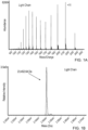
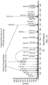
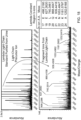
- FIGs. 1A-1D show results from the analysis of a serum sample from a patient with IgG kappa multiple myeloma using methods described herein.
- FIG. 1A is a mass spectrum showing a set of multiply charged ions that are converted to a molecular mass of 23 452.64 Da ( FIG. 1B ), which is within the expected mass range for an IgG light chain.
- FIG. 1C shows another set of multiply charged ions that are converted to a molecular mass of 51 596.07 and 51 758.27 Da ( FIG. 1D ), which is within the expected mass range for an IgG heavy chain.
- the difference between the molecular mass of series 2 and series 3 is 162.20 Da, which closely matches the mass of a hexose subunit.
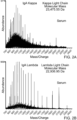
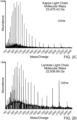
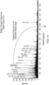
- FIGs. 2A-2D show mass spectra of the serum ( FIGs. 2A and 2B ) and urine samples ( FIGs. 2C and 2D ) from two patients one with a with an IgA kappa monoclonal gammopathy ( FIGs. 2A and 2C ) and one with an IgA lambda monoclonal gammopathy ( FIGs. 2B and 2D ), measured by LC-MS.
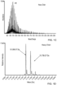
- FIG. 3 shows a light chain mass spectrum of a serum sample from a patient with an IgM monoclonal gammopathy, measured by LC-MS.
- FIG. 4 shows a light chain mass spectrum of a serum sample from a healthy individual without monoclonal gammopathy, measured by LC-MS.
- Example 2 Detection of immunoglobulin light chains in serum or urine samples by MALDI-TOF MS.
- MALDI-TOF MS Matrix Assisted Laser Desorption Ionization- Time of Flight Mass Spectrometry
- HUMIRA ® Melon Gel purified adalimumab
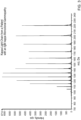
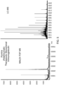
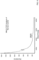
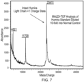
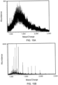
- normal serum controls were used to get initial results using MALDI-TOF MS ( FIGs. 5-7 ).
- the most abundant peak representing the immunoglobulin light chain in FIG. 5 is roughly 3000 counts for the adalimumab, while the background level of immunoglobulin light chains in the normal control sample in FIG. 6 is roughly 50 counts.
- the adalimumab was diluted tenfold and spiked into a normal control serum sample to simulate the presence of a monoclonal immunoglobulin in a patient serum sample and analyzed by MALDI-TOF MS. As shown in FIG. 7 , adalimumab was detected in the normal serum sample by MALDI-TOF MS.
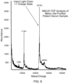
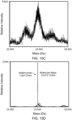
- MALDI-TOF MS was then used to identify monoclonal antibodies in serum samples from a patient with multiple myeloma. Serum sample was prepared as describe above in Example 1 and analyzed by MALDI-TOF MS. The monoclonal light chain antibody associated with the malignant clone was clearly seen at Mass/Charge 22,783 ( FIG. 8 ).
- MALDI-TOF MS The ability of MALDI-TOF MS to identify the presence of monoclonal free light chains (> 3mg/mL) in urine sample from patients with multiple myeloma was also evaluated.
- a 50 ⁇ l urine sample from the patient was diluted first with 50 ⁇ l of 50 mM ammonium bicarbonate and then reduced with 100 mM DTT for 30 minutes at 55 °C.
- the reduced sample was acidified with formic acid then examined by MALDI-TOF MS.
- the results from the MALDI-TOF MS analysis of a urine sample from a patient with known lambda free light chains were shown in FIG. 9 , and a peak at 23,327 Mass/Charge indicates the presence of the lambda free light chain in the urine sample.
- Example 3 Determination of whether an immunoglobulin light chain is lambda or kappa light chain by tandem mass spectrometry.
- FIG. 10 is a mass spectrum of a serum sample from a patient with IgG kappa monoclonal gammopathy, measured by LC-MS, with highlights of the peak at 1229.9 Mass/Charge which is selected as the precursor ion for LC-MS/MS.
- an LC-MS/MS method was selected that allows the quadrupole portion of the mass spectrometer to select that specific ion.
- This precursor ion was then fragmented using collision-induced dissociation (CID) in the collision cell portion of the mass spectrometer.
- the fragment ions produced were then analyzed using the time-of-flight (TOF) portion of the mass spectrometer.
- CID collision-induced dissociation
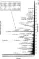
- FIG. 11 showed the fragment ion spectrum for the precursor ion labeled in FIG. 10 .
- On the right hand side of FIG. 11 was a list of C-terminal y-ion fragments and their masses from the constant region for kappa light chain.
- FIG. 11 also showed an arrow pointing from the list at P (proline) residue 11 with a mass of 1237.5994, to a peak in the fragment ion spectrum at mass 1237.6263.
- Serum sample from several multiple myeloma patients with known M-spike kappa light chains were prepared as described above in Example 1.
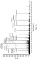
- FIG. 12 showed the M-spike light chain mass spectra of the serum samples from different multiple myeloma patients with known M-spike kappa light chains. Each spectrum showed a different set of multiply charged ions from each patient's unique light chain. These results clearly showed that LC-MS/MS of the intact light chain can be used to determine if it is a kappa light chain.
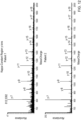
- FIG. 13 showed the M-spike light chain mass spectrum from a patient urine sample with known M-spike lambda light chains.
- the spectrum at the top of FIG. 13 showed the multiply charged ions from lambda free light chains (FLCs) found to be present in the patient's urine using a standard immunoassay.
- Urine samples from patient with known kappa free light chains were tested using the same method. The results from these experiments were shown in FIG. 14 .
- the top of FIG. 14 showed the spectrum for the intact kappa free light chain in the urine sample. The spectrum was similar to those observed for serum.
- the bottom of FIG. 14 showed the fragment ion spectrum for the intact multiply charged ion at 1060.6605 Mass/Charge. Fragment ions that match the expected ions from the constant region of the C-terminus were highlighted. The fragment ions observed were the same as those observed in serum samples from patients with a kappa light chain (see FIG. 11 ). This observation confirmed that kappa light chains can be identified in urine and serum samples.
- Adalimumab is an anti-TNF therapeutic monoclonal immunoglobulin that is widely prescribed for downregulating the inflammatory response in patients with autoimmune disorders.
- Therapeutic monoclonal immunoglobulins such as adalimumab are ideal surrogate standards for simulating a monoclonal immunoglobulin in serum because they are readily available in high purity and typically have a large body of literature on their structural properties.
- FIG. 15 shows the mass spectra for normal serum and serum spiked with 0.5 g/dL (30 ⁇ M) of adalimumab. Each mass spectrum represents the spectra summed together over the adalimumab light chain elution time.
- the mass spectrum from normal serum in section A shows a broad unresolved peak with a maximum relative abundance of 300 counts per second (cps).
- the mass spectrum from the serum spiked with adalimumab in section B shows a distinct series of peaks from multiply charged protein ions with a maximum relative abundance of 6000 cps.
- the converted molecular mass for the normal serum in panel C shows a set of broad distribution of masses with no single mass higher in abundance than the background. This is in sharp contrast to the converted molecular mass for the normal serum spiked with 0.5 g/dL of adalimumab in panel D, which displays a single peak with the observed molecular mass of 23 412.19 Da.
- adalimumab was spiked into 50 mM ammonium bicarbonate buffer, normal serum, and normal urine. Ten different standard concentrations were used ranging from 0.005 to 5.0 g/dL. Standard curves made in serum used Melon Gel to enrich for immunoglobulins, whereas curves made in urine and buffer were reduced and analyzed without Melon Gel purification. Linearity and linear dynamic range values in the table are split according to the two quantification techniques.
- the first approach labeled "deconvolution peak area” uses the peak area found after deconvolution of the multiply charged ions to molecular mass
- the second approach labeled “extracted ion peak area” refers to using the peak areas obtained from a selected set of extracted ions.
- the table demonstrates that the standard curves have a linear dynamic range within the concentration range needed in clinical practice.
- the interassay precision of 10 replicate Melon Gel preparations of adalimumab spiked into normal serum at 0.1 g/dL was examined and found the CV for the peak area of the light chain to be 6.2%, whereas the CV for the heavy chain was 11%.
- the limit of quantification as defined by a CV ⁇ 20% for 10 replicates using the deconvolution peak areas was 0.005 g/dL for the light chain and 0.025 g/dL for the heavy chain of adalimumab spiked into normal serum.
- Example 5 Monitoring a Monoclonal Immunoglobulin in a Patient with Multiple Myeloma
- FIG. 1 A series of samples from a patient diagnosed with IgG kappa multiple myeloma was examined.
- the mass spectrum from a serum sample is shown in FIG. 1 .
- the spectrum in FIG. 1A represents a portion of the summed mass spectra across the immunoglobulin LC peak and shows a series of multiply charged ions.
- the converted molecular mass is shown in FIG. 1B and was calculated to be 23 452.64 Da, representing the proposed molecular mass of the kappa light chain portion of the M-protein.
- the spectrum in panel C shows another portion of the summed immunoglobulin LC peak and displays a different series of multiply charged ions.
- FIG. 16 shows the result using mass spectrometry for a sample taken after the patient had been treated for multiple myeloma and was found to be negative by PEL, IFE, and the quantitative FLC immunoassay. However, multiply charged ions from the light chain are clearly evident in the mass spectrum shown in FIG. 16A .
- Table 1 lists a summary of the results of monitoring the M-protein in serum by PEL, IFE, and microLC-ESI-Q-TOF MS and shows that the light chain is observed throughout the sampling dates, including all of the dates where PEL and IFE were negative. Also, the molecular mass of the light chain remains consistent with an average value of 23 452.54 Da and a standard deviation of 0.86 Da for molecular mass calculations over the 7 year sample period. The heavy chain was observed in the PEL and IFE positive samples, and in those samples, the molecular mass calculations were consistent over the 7 year period.
- Fragment ions are labeled with their monoisotopic masses, which closely match the calculated monoisotopic masses for y ions from the constant region of the kappa light chain.
- top-down MS A set of 20 patients positive for an IgG lambda light chain was analyzed by top-down MS fragment, and the b ions matching the N-terminal portion of the lambda constant region were observed in each patient.
- IFE positive urine samples were analyzed by top-down MS and found that lambda-positive samples had lambda-specific fragment ions and kappa-positive samples had kappa-specific fragment ions.
- the results shown here provide the empirical evidence to substantiate the utility of mass spectrometry as a tool to monitor an M-protein in patients with a monoclonal gammopathy.
- the molecular mass of the monoclonal immunoglobulin represents a sensitive and specific marker of immunoglobulin-secreting plasma cell clones.
- the methodology can readily identify a monoclonal immunoglobulin present above the polyclonal background, providing exceptionally detailed information about the status of patient-specific plasma cell clones.
- mass spectrometry might play an important role in the quantitation and monitoring of immunoglobulins in human health and disease.
Landscapes
- Health & Medical Sciences (AREA)
- Life Sciences & Earth Sciences (AREA)
- Engineering & Computer Science (AREA)
- Molecular Biology (AREA)
- Immunology (AREA)
- Physics & Mathematics (AREA)
- Chemical & Material Sciences (AREA)
- Hematology (AREA)
- Urology & Nephrology (AREA)
- Biomedical Technology (AREA)
- Analytical Chemistry (AREA)
- Bioinformatics & Computational Biology (AREA)
- Bioinformatics & Cheminformatics (AREA)
- General Health & Medical Sciences (AREA)
- Pathology (AREA)
- General Physics & Mathematics (AREA)
- Biochemistry (AREA)
- Medicinal Chemistry (AREA)
- Microbiology (AREA)
- Food Science & Technology (AREA)
- Biotechnology (AREA)
- Cell Biology (AREA)
- Proteomics, Peptides & Aminoacids (AREA)
- Spectroscopy & Molecular Physics (AREA)
- Optics & Photonics (AREA)
- Biophysics (AREA)
- Plasma & Fusion (AREA)
- Other Investigation Or Analysis Of Materials By Electrical Means (AREA)
- Preparation Of Compounds By Using Micro-Organisms (AREA)
Applications Claiming Priority (4)
| Application Number | Priority Date | Filing Date | Title |
|---|---|---|---|
| US201361792944P | 2013-03-15 | 2013-03-15 | |
| PCT/US2014/022475 WO2014150170A1 (en) | 2013-03-15 | 2014-03-10 | Identification and monitoring of monoclonal immunoglobulins by molecular mass |
| EP14770418.3A EP2966985B1 (en) | 2013-03-15 | 2014-03-10 | Identification and monitoring of monoclonal immunoglobulins by molecular mass |
| EP18174068.9A EP3387898B1 (en) | 2013-03-15 | 2014-03-10 | Identification and monitoring of monoclonal immunoglobulins by molecular mass |
Related Parent Applications (2)
| Application Number | Title | Priority Date | Filing Date |
|---|---|---|---|
| EP14770418.3A Division EP2966985B1 (en) | 2013-03-15 | 2014-03-10 | Identification and monitoring of monoclonal immunoglobulins by molecular mass |
| EP18174068.9A Division EP3387898B1 (en) | 2013-03-15 | 2014-03-10 | Identification and monitoring of monoclonal immunoglobulins by molecular mass |
Publications (2)
| Publication Number | Publication Date |
|---|---|
| EP3729958A1 EP3729958A1 (en) | 2020-10-28 |
| EP3729958B1 true EP3729958B1 (en) | 2023-05-03 |
Family
ID=51580707
Family Applications (3)
| Application Number | Title | Priority Date | Filing Date |
|---|---|---|---|
| EP20174164.2A Active EP3729958B1 (en) | 2013-03-15 | 2014-03-10 | Identification and monitoring of monoclonal immunoglobulins by molecular mass |
| EP18174068.9A Active EP3387898B1 (en) | 2013-03-15 | 2014-03-10 | Identification and monitoring of monoclonal immunoglobulins by molecular mass |
| EP14770418.3A Active EP2966985B1 (en) | 2013-03-15 | 2014-03-10 | Identification and monitoring of monoclonal immunoglobulins by molecular mass |
Family Applications After (2)
| Application Number | Title | Priority Date | Filing Date |
|---|---|---|---|
| EP18174068.9A Active EP3387898B1 (en) | 2013-03-15 | 2014-03-10 | Identification and monitoring of monoclonal immunoglobulins by molecular mass |
| EP14770418.3A Active EP2966985B1 (en) | 2013-03-15 | 2014-03-10 | Identification and monitoring of monoclonal immunoglobulins by molecular mass |
Country Status (7)
| Country | Link |
|---|---|
| US (1) | US12546782B2 (pl) |
| EP (3) | EP3729958B1 (pl) |
| DK (2) | DK3387898T3 (pl) |
| ES (3) | ES2951899T3 (pl) |
| PL (1) | PL3387898T3 (pl) |
| PT (1) | PT3387898T (pl) |
| WO (1) | WO2014150170A1 (pl) |
Families Citing this family (14)
| Publication number | Priority date | Publication date | Assignee | Title |
|---|---|---|---|---|
| WO2014150170A1 (en) | 2013-03-15 | 2014-09-25 | Mayo Foundation For Medical Education And Research | Identification and monitoring of monoclonal immunoglobulins by molecular mass |
| WO2015131169A2 (en) * | 2014-02-28 | 2015-09-03 | H. Lee Moffitt Cancer Center And Research Institute, Inc. | Personalized myeloma detection |
| US10267806B2 (en) | 2014-04-04 | 2019-04-23 | Mayo Foundation For Medical Education And Research | Isotyping immunoglobulins using accurate molecular mass |
| WO2016018978A1 (en) | 2014-07-29 | 2016-02-04 | Mayo Foundation For Medical Education And Research | Quantifying monoclonal antibody therapeutics by lc-ms/ms |
| JP6968058B2 (ja) * | 2015-09-24 | 2021-11-17 | メイヨ・ファウンデーション・フォー・メディカル・エデュケーション・アンド・リサーチ | 質量分析法による免疫グロブリン遊離軽鎖の同定 |
| CN109863395B (zh) | 2016-09-07 | 2023-05-23 | 梅约医学教育与研究基金会 | 分子量法鉴定和监测裂解免疫球蛋白 |
| GB2567793B (en) | 2017-04-13 | 2023-03-22 | Micromass Ltd | A method of fragmenting and charge reducing biomolecules |
| EP3681528B1 (en) | 2017-09-13 | 2025-07-23 | Mayo Foundation for Medical Education and Research | Identification and monitoring of apoptosis inhibitor of macrophage |
| WO2019055631A1 (en) | 2017-09-13 | 2019-03-21 | Mayo Foundation For Medical Education And Research | IDENTIFICATION AND MONITORING OF ACID HYDROLYSIS PRODUCTS OF IMMUNOGLOBULIN HEAVY CHAINS |
| WO2019055632A1 (en) | 2017-09-13 | 2019-03-21 | Mayo Foundation For Medical Education And Research | IDENTIFICATION AND MONITORING OF IMMUNOGLOBULIN CHAINS J |
| GB201808529D0 (en) | 2018-05-24 | 2018-07-11 | Binding Site Group Ltd | Identification of immunoglobulins usong mass spectrometry |
| US20230042129A1 (en) * | 2019-12-11 | 2023-02-09 | Erasmus University Medical Center Rotterdam | Method for Monitoring of Deep Remissions in Multiple Myeloma and Other Plasma Cell Dyscrasias |
| CN115684606B (zh) * | 2022-10-21 | 2023-11-28 | 南方医科大学珠江医院 | 一种m蛋白检测的方法 |
| CN117849159B (zh) * | 2024-01-09 | 2025-01-07 | 融智生物科技(青岛)有限公司 | M蛋白的检测方法、电子设备及存储介质 |
Family Cites Families (115)
| Publication number | Priority date | Publication date | Assignee | Title |
|---|---|---|---|---|
| US1075394A (en) * | 1912-03-29 | 1913-10-14 | Charles Penruddocke Band | Detachable tab for collars. |
| JPH0449299A (ja) | 1990-06-14 | 1992-02-18 | Tosoh Corp | 免疫グロブリンフラグメントの精製法 |
| US5567282A (en) * | 1994-01-25 | 1996-10-22 | Wang; Hann-Ping | On-capillary electrophoretic immunosubtraction for classification and typing of M-proteins |
| US20030027216A1 (en) * | 2001-07-02 | 2003-02-06 | Kiernan Urban A. | Analysis of proteins from biological fluids using mass spectrometric immunoassay |
| US5922184A (en) * | 1997-07-21 | 1999-07-13 | Bio-Rad Laboratories, Inc. | Computer-directed detection of paraproteins |
| US5846735A (en) | 1996-04-18 | 1998-12-08 | University Of Iowa Research Foundation | Hepatitis C virus Fc-binding function |
| AU2001292312A1 (en) | 2000-09-29 | 2002-04-08 | Chugai Seiyaku Kabushiki Kaisha | Method of analyzing antibody molecule structure |
| EP1356121A2 (en) * | 2001-02-01 | 2003-10-29 | Ciphergen Biosystems, Inc. | Improved methods for protein identification, characterization and sequencing by tandem mass spectrometry |
| AU2002309423B2 (en) | 2001-06-06 | 2007-11-22 | Fujirebio Diagnostics Ab | Method to measure gene expression ratio of key genes |
| WO2003046148A2 (en) | 2001-11-29 | 2003-06-05 | Thermo Finnigan Llc | Polypeptide quantitation |
| ATE327512T1 (de) * | 2001-12-08 | 2006-06-15 | Micromass Ltd | Massenspektrometrie-verfahren |
| AU2002357547A1 (en) * | 2001-12-13 | 2003-06-23 | Zeptosens Ag | Optically transparent substrate for a maldi measuring system and the use thereof |
| US20120208295A1 (en) * | 2004-01-13 | 2012-08-16 | Wuxi WeiYi Zhinengkeji, Inc. | Methods and compositions for mass spectrometry analysis |
| WO2004085455A1 (en) * | 2003-03-24 | 2004-10-07 | The University Of Hong Kong | A diagnostic assay for the human virus causing severe acute respiratory syndrome (sars) |
| CA2522709A1 (en) * | 2003-04-17 | 2004-11-04 | Ciphergen Biosystems, Inc. | Polypeptides related to natriuretic peptides and methods of their identification and use |
| US20060177870A1 (en) * | 2003-04-28 | 2006-08-10 | Ciphergen Biosystems, Inc | Immunoassays |
| US7709610B2 (en) * | 2003-05-08 | 2010-05-04 | Facet Biotech Corporation | Therapeutic use of anti-CS1 antibodies |
| WO2005071421A1 (en) * | 2004-01-16 | 2005-08-04 | Ciphergen Biosystems, Inc. | Specific detection of troponin and modified forms of troponin |
| DE102004007005A1 (de) * | 2004-02-12 | 2005-09-08 | Bruker Daltonik Gmbh | Massenspektrometrische Häufigkeitsbestimmungen von Proteinen |
| ES2549077T3 (es) | 2004-04-07 | 2015-10-22 | Genentech, Inc. | Espectrometría de masas de conjugados de anticuerpos |
| CN101044242B (zh) * | 2004-06-10 | 2013-10-30 | 维文蒂阿生物技术股份有限公司 | 肿瘤特异性抗体 |
| KR101319848B1 (ko) * | 2004-07-20 | 2013-10-18 | 심포젠 에이/에스 | 재조합 폴리클로날 단백질 또는 폴리클로날 세포주의구조적 특성화 방법 |
| GB0501741D0 (en) * | 2005-01-27 | 2005-03-02 | Binding Site The Ltd | Antibody |
| WO2006119435A2 (en) | 2005-05-04 | 2006-11-09 | Invitrogen Corporation | Identification of cancer biomarkers and phosphorylated proteins |
| MX2007015416A (es) * | 2005-06-08 | 2008-02-19 | Millennium Pharm Inc | Metodos para la identificacion, evaluacion y tratamiento de pacientes con terapia contra cancer. |
| US20110177492A1 (en) | 2005-06-16 | 2011-07-21 | 3M Innovative Properties Company | Method of classifying chemically crosslinked cellular samples using mass spectra |
| JP2007024631A (ja) | 2005-07-14 | 2007-02-01 | Human Science Shinko Zaidan | 同位体標識法 |
| TWI338779B (en) | 2005-07-21 | 2011-03-11 | Academia Sinica | Methods,compositions and systems for assaying at least one target analyte in a sample |
| CA2619825A1 (en) * | 2005-08-19 | 2007-03-01 | Centocor, Inc. | Proteolysis resistant antibody preparations |
| DE102005042100A1 (de) * | 2005-09-05 | 2007-03-08 | Sirs-Lab Gmbh | Verfahren zur in vitro Diagnose von Sepsis mit Haptoglobin-Related Protein |
| DE102005042132A1 (de) * | 2005-09-05 | 2007-03-08 | Sirs-Lab Gmbh | Verfahren zur Diagnose von Sepsis mit IgM |
| EP1973940A2 (en) * | 2006-01-17 | 2008-10-01 | Biolex Therapeutics, Inc. | Compositions and methods for humanization and optimization of n-glycans in plants |
| JP5420396B2 (ja) | 2006-04-28 | 2014-02-19 | シンガポール ヘルス サービシーズ ピーティーイー リミテッド | 粘膜乾燥状態に関する検討 |
| WO2007130875A2 (en) * | 2006-05-02 | 2007-11-15 | Genentech, Inc. | Microwave assisted deglycosylation of proteins for molecular weight determination by mass spectrometry |
| EP2038413A4 (en) | 2006-06-28 | 2010-01-06 | Lineagen Inc | DEVICES, COMPOSITIONS AND METHODS FOR MONITORING THE PROGRESS OF CHRONIC OBSTRUCTIVE LUNG DISORDERS UNDER QUICK OR SLOW DEGRADING CONDITIONS |
| KR100862904B1 (ko) * | 2006-07-20 | 2008-10-13 | 주식회사 서린바이오사이언스 | 단백질 고정화용 마이크로칩 |
| US9500656B2 (en) * | 2006-08-10 | 2016-11-22 | Millennium Pharmaceuticals, Inc. | Methods for the identification, assessment, and treatment of patients with cancer therapy |
| US20090203602A1 (en) * | 2006-09-01 | 2009-08-13 | Cohava Gelber | Compositions and methods for diagnosis and treatment of type 2 diabetes |
| US20080317745A1 (en) * | 2006-09-15 | 2008-12-25 | Boruchov Adam M | Methods of diagnosing, treating, or preventing plasma cell disorders |
| WO2008057083A1 (en) | 2006-11-09 | 2008-05-15 | Wyeth | Methods of analyzing glycomolecules |
| US7462489B2 (en) | 2006-11-09 | 2008-12-09 | Ley Klaus F | Methods for identifying and analyzing biomarkers from plasma-derived microparticles |
| ES2374403T3 (es) | 2006-12-21 | 2012-02-16 | Novartis Ag | Cuantificación de anticuerpos. |
| GB0704764D0 (en) | 2007-03-12 | 2007-04-18 | Electrophoretics Ltd | Isobarically labelled reagents and methods of their use |
| FI20075278A0 (fi) | 2007-04-20 | 2007-04-20 | Biotie Therapies Corp | Uudet täysin ihmisperäiset anti-VAP-1 monoklonaaliset vasta-aineet |
| US8501907B2 (en) | 2007-08-10 | 2013-08-06 | Janssen Biotech, Inc. | Immunoglobulin cleavage fragments as disease indicators and compositions for detecting and binding such |
| KR20150059813A (ko) | 2007-08-10 | 2015-06-02 | 센토코 오르토 바이오테크 인코포레이티드 | 질환 표시자로서의 면역글로불린 절단 단편, 및 그의 검출 및 결합을 위한 조성물 |
| NZ584684A (en) * | 2007-11-22 | 2012-10-26 | Symphogen As | A method for characterization of a recombinant polyclonal protein |
| US20100261216A1 (en) * | 2007-12-21 | 2010-10-14 | Bianca Eser | Stability testing of antibodies |
| CN101354379B (zh) * | 2008-01-18 | 2012-09-19 | 许洋 | 用质谱检测多发性骨髓瘤特征蛋白的试剂 |
| WO2009100390A2 (en) | 2008-02-08 | 2009-08-13 | Mayo Foundation For Medical Education And Research | Classifying amyloidosis |
| WO2009148528A2 (en) * | 2008-05-30 | 2009-12-10 | Millennium Pharmaceuticals, Inc. | Assessment of chromosomal alterations to predict clinical outcome of bortezomib treatment |
| WO2010002911A2 (en) * | 2008-06-30 | 2010-01-07 | H. Lee Moffitt Cancer Center And Research Institute, Inc. | Methods and materials for monitoring myeloma using quantitative mass spetrometry |
| WO2010085345A1 (en) * | 2009-01-22 | 2010-07-29 | Ludwig Institute For Cancer Research Ltd. | Methods and compositions for diagnosis and treatment of malignant and non-malignant gammopathies |
| US20100190652A1 (en) | 2009-01-27 | 2010-07-29 | Srinivasa Nagalla | Biomarkers for Detection of Neonatal Sepsis in Biological Fluid |
| SG173589A1 (en) | 2009-02-09 | 2011-09-29 | Roche Glycart Ag | Immunoglobulin glycosylation pattern analysis |
| EP2233502A1 (en) | 2009-03-27 | 2010-09-29 | Deutsches Rheuma-Forschungszentrum Berlin | Sialylated antigen-specific antibodies for treatment or prophylaxis of unwanted inflammatory immune reactions and methods of producing them |
| WO2010119295A1 (en) | 2009-04-16 | 2010-10-21 | Cambridge Enterprise Limited | Biomarkers |
| JP5379616B2 (ja) | 2009-09-09 | 2013-12-25 | 株式会社日立製作所 | 粥状硬化性動脈硬化のマーカー因子と用途 |
| WO2011072197A2 (en) | 2009-12-11 | 2011-06-16 | Purdue Research Foundation | Detection of oxidized polypeptides |
| GB0922240D0 (en) | 2009-12-21 | 2010-02-03 | Cambridge Entpr Ltd | Biomarkers |
| EP2521916A4 (en) | 2010-01-04 | 2013-12-04 | Lineagen Inc | BIOMARKER FOR THE LUNGS FUNCTION |
| US8643274B2 (en) | 2010-01-26 | 2014-02-04 | Scinopharm Taiwan, Ltd. | Methods for Chemical Equivalence in characterizing of complex molecules |
| JP5337096B2 (ja) | 2010-04-28 | 2013-11-06 | 株式会社日立製作所 | 動脈硬化の評価法 |
| GB201008340D0 (en) | 2010-05-19 | 2010-07-07 | Cambridge Entpr Ltd | Biomarkers |
| CN103299192B (zh) | 2010-09-21 | 2016-05-11 | 普罗蒂阿米克斯国际有限公司 | 与糖尿病前期、糖尿病以及糖尿病相关病症相关的生物标记 |
| GB201018056D0 (en) | 2010-10-26 | 2010-12-08 | Cambridge Entpr Ltd | Biomarkers |
| EP2447980B1 (en) * | 2010-11-02 | 2019-05-22 | Thermo Fisher Scientific (Bremen) GmbH | Method of generating a mass spectrum having improved resolving power |
| ES2530175T3 (es) | 2011-02-17 | 2015-02-26 | Nestec S.A. | Ensayos para la detección de autoanticuerpos contra fármacos anti-TNF |
| WO2012149299A2 (en) | 2011-04-29 | 2012-11-01 | Celgene Corporaiton | Methods for the treatment of cancer and inflammatory diseases using cereblon as a predictor |
| CA2833212C (en) * | 2011-05-12 | 2020-06-09 | Genentech, Inc. | Multiple reaction monitoring lc-ms/ms method to detect therapeutic antibodies in animal samples using framework signature peptides |
| CA2840687C (en) * | 2011-07-01 | 2020-04-28 | Dana-Farber Cancer Institute, Inc. | Discovery of a somatic mutation in myd88 gene in lymphoplasmacytic lymphoma |
| EP2732423A4 (en) * | 2011-07-13 | 2014-11-26 | Multiple Myeloma Res Foundation Inc | METHOD FOR DETECTING AND DISTRIBUTING DATA |
| GB2495113A (en) * | 2011-09-29 | 2013-04-03 | Bioinvent Int Ab | Anti-ICAM-1 antibody for treating multiple-myeloma-related disorders |
| CA2848520C (en) * | 2011-09-29 | 2019-11-26 | Seattle Genetics, Inc. | Intact mass determination of protein conjugated agent compounds |
| EP2783214A2 (en) | 2011-11-23 | 2014-10-01 | The Board of Regents of The University of Texas System | Proteomic identification of antibodies |
| AU2012359020B2 (en) | 2011-12-19 | 2017-04-13 | The Washington University | Methods for diagnosing Alzheimer's disease |
| GB201203938D0 (en) * | 2012-03-06 | 2012-04-18 | Binding Site Group The Ltd | Assay system |
| WO2013166594A1 (en) | 2012-05-10 | 2013-11-14 | Zymeworks Inc. | Heteromultimer constructs of immunoglobulin heavy chains with mutations in the fc domain |
| WO2013181576A2 (en) | 2012-06-01 | 2013-12-05 | Momenta Pharmaceuticals, Inc. | Methods of evaluating and making biologics |
| WO2013185180A1 (en) | 2012-06-14 | 2013-12-19 | Central Adelaide Local Health Network Inc. | Method for identifying a diagnostic biomarker for an antibody of interest |
| WO2013191908A1 (en) * | 2012-06-20 | 2013-12-27 | The University Of North Carolina At Chapel Hill | Integrated sample processing for electrospray ionization devices |
| EP2893353A2 (en) | 2012-09-10 | 2015-07-15 | Koninklijke Philips N.V. | Analysis of saliva proteome for biomarkers of gingivitis and periodontitis using ft-icr-ms/ms |
| WO2014078374A2 (en) | 2012-11-13 | 2014-05-22 | Presage Biosciences, Inc. | Methods for multiplexed drug evaluation |
| US20140186332A1 (en) | 2012-12-28 | 2014-07-03 | NX Pharmagen | Biomarkers of preterm birth |
| WO2014109927A1 (en) | 2013-01-11 | 2014-07-17 | The Regents Of The University Of Michigan | Synthesis and isolation of dendrimer based imaging systems |
| CN103217489B (zh) | 2013-01-15 | 2016-03-09 | 珠海市丽珠单抗生物技术有限公司 | 一种测定蛋白纯化工艺过程中样品的糖基化和末端修饰情况的方法 |
| CN103217499B (zh) | 2013-01-15 | 2015-12-02 | 珠海市丽珠单抗生物技术有限公司 | 一种测定免疫球蛋白电荷异构体的糖基化和末端修饰情况的方法 |
| WO2014121031A1 (en) | 2013-01-31 | 2014-08-07 | Excelimmune, Inc. | Characterization of antibody mixtures by mass spectrometry |
| CA2902841A1 (en) | 2013-03-13 | 2014-10-02 | Creatics Llc | Methods and compositions for detecting pancreatic cancer |
| US20160047819A1 (en) | 2013-03-15 | 2016-02-18 | Alfa Wassermann S.P.A. | Method for diagnosing vaginal infections |
| WO2014150170A1 (en) | 2013-03-15 | 2014-09-25 | Mayo Foundation For Medical Education And Research | Identification and monitoring of monoclonal immunoglobulins by molecular mass |
| EP3043806A4 (en) | 2013-09-11 | 2017-05-17 | University Of Southern California | A composition of stem cells having highly expressed fas ligand |
| GB201316744D0 (en) | 2013-09-20 | 2013-11-06 | Genovis Ab | Method |
| CN103792315B (zh) | 2014-01-23 | 2015-07-08 | 中国计量科学研究院 | 一种人血清蛋白无机质谱联用技术的定量方法 |
| WO2015120036A1 (en) | 2014-02-04 | 2015-08-13 | University Of Virginia Patent Foundation | Compositions and methods for analysis of protein sequences and post-translational modifications |
| WO2015131169A2 (en) | 2014-02-28 | 2015-09-03 | H. Lee Moffitt Cancer Center And Research Institute, Inc. | Personalized myeloma detection |
| US9618524B2 (en) | 2014-03-26 | 2017-04-11 | Plaxgen Inc. | Method, composition, isolation and identification of a plaque particle and related biomarker |
| US10267806B2 (en) | 2014-04-04 | 2019-04-23 | Mayo Foundation For Medical Education And Research | Isotyping immunoglobulins using accurate molecular mass |
| EP2975401B1 (en) | 2014-07-18 | 2019-12-25 | Hexal AG | Improved method of mapping glycans of glycoproteins in serum samples |
| WO2016018978A1 (en) | 2014-07-29 | 2016-02-04 | Mayo Foundation For Medical Education And Research | Quantifying monoclonal antibody therapeutics by lc-ms/ms |
| WO2016134365A1 (en) | 2015-02-20 | 2016-08-25 | The Johns Hopkins University | Biomarkers of myocardial injury |
| KR102000862B1 (ko) | 2015-03-09 | 2019-07-16 | 가부시키가이샤 시마즈세이사쿠쇼 | 질량 분석을 사용한 모노클로날 항체의 검출 방법 |
| CA2980189A1 (en) | 2015-04-24 | 2016-10-27 | Genentech, Inc. | Multispecific antigen-binding proteins |
| US12404552B2 (en) | 2015-07-17 | 2025-09-02 | Allele Biotechnology & Pharmaceuticals, Inc. | Methods of selecting antibodies and antibody fragments |
| JP6666916B2 (ja) | 2015-08-06 | 2020-03-18 | 積水メディカル株式会社 | 腎疾患の検査方法 |
| JP6968058B2 (ja) | 2015-09-24 | 2021-11-17 | メイヨ・ファウンデーション・フォー・メディカル・エデュケーション・アンド・リサーチ | 質量分析法による免疫グロブリン遊離軽鎖の同定 |
| US12006530B2 (en) | 2016-02-04 | 2024-06-11 | Genovis Ab | Streptococcal proteases |
| WO2017144903A1 (en) | 2016-02-25 | 2017-08-31 | The Binding Site Group Limited | Antibodies |
| WO2017180735A1 (en) | 2016-04-12 | 2017-10-19 | Biocrypton Inc. | Compositions and methods for screening, monitoring and treating gastrointestinal diseases |
| US10309968B2 (en) | 2016-05-18 | 2019-06-04 | Bioinformatics Solutions Inc. | Methods and systems for assembly of protein sequences |
| US11067583B2 (en) | 2016-05-25 | 2021-07-20 | University Of Maryland, Baltimore | Methods of making active antibodies from biological fluids |
| CN109863395B (zh) | 2016-09-07 | 2023-05-23 | 梅约医学教育与研究基金会 | 分子量法鉴定和监测裂解免疫球蛋白 |
| WO2019055632A1 (en) | 2017-09-13 | 2019-03-21 | Mayo Foundation For Medical Education And Research | IDENTIFICATION AND MONITORING OF IMMUNOGLOBULIN CHAINS J |
| WO2019055631A1 (en) | 2017-09-13 | 2019-03-21 | Mayo Foundation For Medical Education And Research | IDENTIFICATION AND MONITORING OF ACID HYDROLYSIS PRODUCTS OF IMMUNOGLOBULIN HEAVY CHAINS |
| EP3681528B1 (en) | 2017-09-13 | 2025-07-23 | Mayo Foundation for Medical Education and Research | Identification and monitoring of apoptosis inhibitor of macrophage |
-
2014
- 2014-03-10 WO PCT/US2014/022475 patent/WO2014150170A1/en not_active Ceased
- 2014-03-10 PT PT181740689T patent/PT3387898T/pt unknown
- 2014-03-10 ES ES20174164T patent/ES2951899T3/es active Active
- 2014-03-10 DK DK18174068.9T patent/DK3387898T3/da active
- 2014-03-10 EP EP20174164.2A patent/EP3729958B1/en active Active
- 2014-03-10 DK DK20174164.2T patent/DK3729958T3/da active
- 2014-03-10 ES ES18174068T patent/ES2802805T3/es active Active
- 2014-03-10 EP EP18174068.9A patent/EP3387898B1/en active Active
- 2014-03-10 EP EP14770418.3A patent/EP2966985B1/en active Active
- 2014-03-10 ES ES14770418T patent/ES2702183T3/es active Active
- 2014-03-10 PL PL18174068T patent/PL3387898T3/pl unknown
- 2014-03-10 US US14/777,236 patent/US12546782B2/en active Active
Also Published As
| Publication number | Publication date |
|---|---|
| DK3729958T3 (da) | 2023-08-07 |
| US12546782B2 (en) | 2026-02-10 |
| PL3387898T3 (pl) | 2020-10-19 |
| PT3387898T (pt) | 2020-06-17 |
| US20160041184A1 (en) | 2016-02-11 |
| EP3729958A1 (en) | 2020-10-28 |
| ES2802805T3 (es) | 2021-01-21 |
| EP3387898A1 (en) | 2018-10-17 |
| WO2014150170A1 (en) | 2014-09-25 |
| EP2966985A1 (en) | 2016-01-20 |
| EP2966985B1 (en) | 2018-09-19 |
| DK3387898T3 (da) | 2020-08-03 |
| EP2966985A4 (en) | 2017-03-15 |
| ES2702183T3 (es) | 2019-02-27 |
| EP3387898B1 (en) | 2020-05-13 |
| ES2951899T3 (es) | 2023-10-25 |
Similar Documents
| Publication | Publication Date | Title |
|---|---|---|
| EP3729958B1 (en) | Identification and monitoring of monoclonal immunoglobulins by molecular mass | |
| US12099065B2 (en) | Isotyping immunoglobulins using accurate molecular mass | |
| AU2017325022B2 (en) | Identification and monitoring of cleaved immunoglobulins by molecular mass | |
| JP2010515020A (ja) | 抗体定量法 | |
| JP7561032B2 (ja) | 質量分析を用いる免疫グロブリンの同定 | |
| US11946937B2 (en) | Identification and monitoring of apoptosis inhibitor of macrophage | |
| US12153052B2 (en) | Identification and monitoring of immunoglobulin J chains | |
| US20200271663A1 (en) | Identification and monitoring of acid hydrolysis products of immunoglobulin heavy chains | |
| WO2015074048A1 (en) | Measurement of gamma-carboxylation of proteins | |
| CN118598986A (zh) | 一组用于多发性骨髓瘤个体化诊断的m蛋白完整轻链标志物及其应用 |
Legal Events
| Date | Code | Title | Description |
|---|---|---|---|
| PUAI | Public reference made under article 153(3) epc to a published international application that has entered the european phase |
Free format text: ORIGINAL CODE: 0009012 |
|
| STAA | Information on the status of an ep patent application or granted ep patent |
Free format text: STATUS: REQUEST FOR EXAMINATION WAS MADE |
|
| 17P | Request for examination filed |
Effective date: 20200512 |
|
| AC | Divisional application: reference to earlier application |
Ref document number: 3387898 Country of ref document: EP Kind code of ref document: P Ref document number: 2966985 Country of ref document: EP Kind code of ref document: P |
|
| AK | Designated contracting states |
Kind code of ref document: A1 Designated state(s): AL AT BE BG CH CY CZ DE DK EE ES FI FR GB GR HR HU IE IS IT LI LT LU LV MC MK MT NL NO PL PT RO RS SE SI SK SM TR |
|
| STAA | Information on the status of an ep patent application or granted ep patent |
Free format text: STATUS: EXAMINATION IS IN PROGRESS |
|
| 17Q | First examination report despatched |
Effective date: 20210521 |
|
| REG | Reference to a national code |
Ref country code: DE Ref legal event code: R079 Ref document number: 602014086871 Country of ref document: DE Free format text: PREVIOUS MAIN CLASS: A01H0001060000 Ipc: G01N0033574000 |
|
| RIC1 | Information provided on ipc code assigned before grant |
Ipc: G01N 33/68 20060101ALI20220819BHEP Ipc: G01N 33/574 20060101AFI20220819BHEP |
|
| GRAP | Despatch of communication of intention to grant a patent |
Free format text: ORIGINAL CODE: EPIDOSNIGR1 |
|
| STAA | Information on the status of an ep patent application or granted ep patent |
Free format text: STATUS: GRANT OF PATENT IS INTENDED |
|
| INTG | Intention to grant announced |
Effective date: 20221006 |
|
| GRAS | Grant fee paid |
Free format text: ORIGINAL CODE: EPIDOSNIGR3 |
|
| GRAA | (expected) grant |
Free format text: ORIGINAL CODE: 0009210 |
|
| STAA | Information on the status of an ep patent application or granted ep patent |
Free format text: STATUS: THE PATENT HAS BEEN GRANTED |
|
| AC | Divisional application: reference to earlier application |
Ref document number: 2966985 Country of ref document: EP Kind code of ref document: P Ref document number: 3387898 Country of ref document: EP Kind code of ref document: P |
|
| AK | Designated contracting states |
Kind code of ref document: B1 Designated state(s): AL AT BE BG CH CY CZ DE DK EE ES FI FR GB GR HR HU IE IS IT LI LT LU LV MC MK MT NL NO PL PT RO RS SE SI SK SM TR |
|
| REG | Reference to a national code |
Ref country code: GB Ref legal event code: FG4D |
|
| REG | Reference to a national code |
Ref country code: DE Ref legal event code: R096 Ref document number: 602014086871 Country of ref document: DE |
|
| REG | Reference to a national code |
Ref country code: AT Ref legal event code: REF Ref document number: 1565037 Country of ref document: AT Kind code of ref document: T Effective date: 20230515 Ref country code: CH Ref legal event code: EP |
|
| REG | Reference to a national code |
Ref country code: IE Ref legal event code: FG4D |
|
| P01 | Opt-out of the competence of the unified patent court (upc) registered |
Effective date: 20230612 |
|
| REG | Reference to a national code |
Ref country code: DK Ref legal event code: T3 Effective date: 20230802 |
|
| REG | Reference to a national code |
Ref country code: NL Ref legal event code: FP |
|
| REG | Reference to a national code |
Ref country code: LT Ref legal event code: MG9D |
|
| REG | Reference to a national code |
Ref country code: AT Ref legal event code: MK05 Ref document number: 1565037 Country of ref document: AT Kind code of ref document: T Effective date: 20230503 |
|
| REG | Reference to a national code |
Ref country code: ES Ref legal event code: FG2A Ref document number: 2951899 Country of ref document: ES Kind code of ref document: T3 Effective date: 20231025 |
|
| PG25 | Lapsed in a contracting state [announced via postgrant information from national office to epo] |
Ref country code: SE Free format text: LAPSE BECAUSE OF FAILURE TO SUBMIT A TRANSLATION OF THE DESCRIPTION OR TO PAY THE FEE WITHIN THE PRESCRIBED TIME-LIMIT Effective date: 20230503 Ref country code: PT Free format text: LAPSE BECAUSE OF FAILURE TO SUBMIT A TRANSLATION OF THE DESCRIPTION OR TO PAY THE FEE WITHIN THE PRESCRIBED TIME-LIMIT Effective date: 20230904 Ref country code: NO Free format text: LAPSE BECAUSE OF FAILURE TO SUBMIT A TRANSLATION OF THE DESCRIPTION OR TO PAY THE FEE WITHIN THE PRESCRIBED TIME-LIMIT Effective date: 20230803 Ref country code: AT Free format text: LAPSE BECAUSE OF FAILURE TO SUBMIT A TRANSLATION OF THE DESCRIPTION OR TO PAY THE FEE WITHIN THE PRESCRIBED TIME-LIMIT Effective date: 20230503 |
|
| PG25 | Lapsed in a contracting state [announced via postgrant information from national office to epo] |
Ref country code: RS Free format text: LAPSE BECAUSE OF FAILURE TO SUBMIT A TRANSLATION OF THE DESCRIPTION OR TO PAY THE FEE WITHIN THE PRESCRIBED TIME-LIMIT Effective date: 20230503 Ref country code: PL Free format text: LAPSE BECAUSE OF FAILURE TO SUBMIT A TRANSLATION OF THE DESCRIPTION OR TO PAY THE FEE WITHIN THE PRESCRIBED TIME-LIMIT Effective date: 20230503 Ref country code: LV Free format text: LAPSE BECAUSE OF FAILURE TO SUBMIT A TRANSLATION OF THE DESCRIPTION OR TO PAY THE FEE WITHIN THE PRESCRIBED TIME-LIMIT Effective date: 20230503 Ref country code: LT Free format text: LAPSE BECAUSE OF FAILURE TO SUBMIT A TRANSLATION OF THE DESCRIPTION OR TO PAY THE FEE WITHIN THE PRESCRIBED TIME-LIMIT Effective date: 20230503 Ref country code: IS Free format text: LAPSE BECAUSE OF FAILURE TO SUBMIT A TRANSLATION OF THE DESCRIPTION OR TO PAY THE FEE WITHIN THE PRESCRIBED TIME-LIMIT Effective date: 20230903 Ref country code: HR Free format text: LAPSE BECAUSE OF FAILURE TO SUBMIT A TRANSLATION OF THE DESCRIPTION OR TO PAY THE FEE WITHIN THE PRESCRIBED TIME-LIMIT Effective date: 20230503 Ref country code: GR Free format text: LAPSE BECAUSE OF FAILURE TO SUBMIT A TRANSLATION OF THE DESCRIPTION OR TO PAY THE FEE WITHIN THE PRESCRIBED TIME-LIMIT Effective date: 20230804 |
|
| PG25 | Lapsed in a contracting state [announced via postgrant information from national office to epo] |
Ref country code: FI Free format text: LAPSE BECAUSE OF FAILURE TO SUBMIT A TRANSLATION OF THE DESCRIPTION OR TO PAY THE FEE WITHIN THE PRESCRIBED TIME-LIMIT Effective date: 20230503 |
|
| PG25 | Lapsed in a contracting state [announced via postgrant information from national office to epo] |
Ref country code: SK Free format text: LAPSE BECAUSE OF FAILURE TO SUBMIT A TRANSLATION OF THE DESCRIPTION OR TO PAY THE FEE WITHIN THE PRESCRIBED TIME-LIMIT Effective date: 20230503 |
|
| PG25 | Lapsed in a contracting state [announced via postgrant information from national office to epo] |
Ref country code: SM Free format text: LAPSE BECAUSE OF FAILURE TO SUBMIT A TRANSLATION OF THE DESCRIPTION OR TO PAY THE FEE WITHIN THE PRESCRIBED TIME-LIMIT Effective date: 20230503 Ref country code: SK Free format text: LAPSE BECAUSE OF FAILURE TO SUBMIT A TRANSLATION OF THE DESCRIPTION OR TO PAY THE FEE WITHIN THE PRESCRIBED TIME-LIMIT Effective date: 20230503 Ref country code: RO Free format text: LAPSE BECAUSE OF FAILURE TO SUBMIT A TRANSLATION OF THE DESCRIPTION OR TO PAY THE FEE WITHIN THE PRESCRIBED TIME-LIMIT Effective date: 20230503 Ref country code: EE Free format text: LAPSE BECAUSE OF FAILURE TO SUBMIT A TRANSLATION OF THE DESCRIPTION OR TO PAY THE FEE WITHIN THE PRESCRIBED TIME-LIMIT Effective date: 20230503 |
|
| REG | Reference to a national code |
Ref country code: DE Ref legal event code: R097 Ref document number: 602014086871 Country of ref document: DE |
|
| PLBE | No opposition filed within time limit |
Free format text: ORIGINAL CODE: 0009261 |
|
| STAA | Information on the status of an ep patent application or granted ep patent |
Free format text: STATUS: NO OPPOSITION FILED WITHIN TIME LIMIT |
|
| 26N | No opposition filed |
Effective date: 20240206 |
|
| PG25 | Lapsed in a contracting state [announced via postgrant information from national office to epo] |
Ref country code: SI Free format text: LAPSE BECAUSE OF FAILURE TO SUBMIT A TRANSLATION OF THE DESCRIPTION OR TO PAY THE FEE WITHIN THE PRESCRIBED TIME-LIMIT Effective date: 20230503 |
|
| PG25 | Lapsed in a contracting state [announced via postgrant information from national office to epo] |
Ref country code: SI Free format text: LAPSE BECAUSE OF FAILURE TO SUBMIT A TRANSLATION OF THE DESCRIPTION OR TO PAY THE FEE WITHIN THE PRESCRIBED TIME-LIMIT Effective date: 20230503 |
|
| PG25 | Lapsed in a contracting state [announced via postgrant information from national office to epo] |
Ref country code: BG Free format text: LAPSE BECAUSE OF FAILURE TO SUBMIT A TRANSLATION OF THE DESCRIPTION OR TO PAY THE FEE WITHIN THE PRESCRIBED TIME-LIMIT Effective date: 20230503 |
|
| PG25 | Lapsed in a contracting state [announced via postgrant information from national office to epo] |
Ref country code: LU Free format text: LAPSE BECAUSE OF NON-PAYMENT OF DUE FEES Effective date: 20240310 |
|
| PG25 | Lapsed in a contracting state [announced via postgrant information from national office to epo] |
Ref country code: MC Free format text: LAPSE BECAUSE OF FAILURE TO SUBMIT A TRANSLATION OF THE DESCRIPTION OR TO PAY THE FEE WITHIN THE PRESCRIBED TIME-LIMIT Effective date: 20230503 |
|
| PG25 | Lapsed in a contracting state [announced via postgrant information from national office to epo] |
Ref country code: MC Free format text: LAPSE BECAUSE OF FAILURE TO SUBMIT A TRANSLATION OF THE DESCRIPTION OR TO PAY THE FEE WITHIN THE PRESCRIBED TIME-LIMIT Effective date: 20230503 Ref country code: LU Free format text: LAPSE BECAUSE OF NON-PAYMENT OF DUE FEES Effective date: 20240310 Ref country code: BG Free format text: LAPSE BECAUSE OF FAILURE TO SUBMIT A TRANSLATION OF THE DESCRIPTION OR TO PAY THE FEE WITHIN THE PRESCRIBED TIME-LIMIT Effective date: 20230503 |
|
| PGFP | Annual fee paid to national office [announced via postgrant information from national office to epo] |
Ref country code: NL Payment date: 20250227 Year of fee payment: 12 |
|
| PGFP | Annual fee paid to national office [announced via postgrant information from national office to epo] |
Ref country code: DE Payment date: 20250225 Year of fee payment: 12 |
|
| PGFP | Annual fee paid to national office [announced via postgrant information from national office to epo] |
Ref country code: DK Payment date: 20250317 Year of fee payment: 12 |
|
| PGFP | Annual fee paid to national office [announced via postgrant information from national office to epo] |
Ref country code: IE Payment date: 20250224 Year of fee payment: 12 |
|
| PGFP | Annual fee paid to national office [announced via postgrant information from national office to epo] |
Ref country code: BE Payment date: 20250227 Year of fee payment: 12 |
|
| PGFP | Annual fee paid to national office [announced via postgrant information from national office to epo] |
Ref country code: CZ Payment date: 20250228 Year of fee payment: 12 Ref country code: FR Payment date: 20250224 Year of fee payment: 12 |
|
| PGFP | Annual fee paid to national office [announced via postgrant information from national office to epo] |
Ref country code: IT Payment date: 20250226 Year of fee payment: 12 Ref country code: GB Payment date: 20250220 Year of fee payment: 12 |
|
| PGFP | Annual fee paid to national office [announced via postgrant information from national office to epo] |
Ref country code: ES Payment date: 20250414 Year of fee payment: 12 |
|
| PGFP | Annual fee paid to national office [announced via postgrant information from national office to epo] |
Ref country code: CH Payment date: 20250401 Year of fee payment: 12 |
|
| PG25 | Lapsed in a contracting state [announced via postgrant information from national office to epo] |
Ref country code: CY Free format text: LAPSE BECAUSE OF FAILURE TO SUBMIT A TRANSLATION OF THE DESCRIPTION OR TO PAY THE FEE WITHIN THE PRESCRIBED TIME-LIMIT; INVALID AB INITIO Effective date: 20140310 |
|
| PG25 | Lapsed in a contracting state [announced via postgrant information from national office to epo] |
Ref country code: HU Free format text: LAPSE BECAUSE OF FAILURE TO SUBMIT A TRANSLATION OF THE DESCRIPTION OR TO PAY THE FEE WITHIN THE PRESCRIBED TIME-LIMIT; INVALID AB INITIO Effective date: 20140310 |
|
| REG | Reference to a national code |
Ref country code: DE Ref legal event code: R079 Ref document number: 602014086871 Country of ref document: DE Free format text: PREVIOUS MAIN CLASS: G01N0033574000 Ipc: G01N0033575000 |
|
| PG25 | Lapsed in a contracting state [announced via postgrant information from national office to epo] |
Ref country code: TR Free format text: LAPSE BECAUSE OF FAILURE TO SUBMIT A TRANSLATION OF THE DESCRIPTION OR TO PAY THE FEE WITHIN THE PRESCRIBED TIME-LIMIT Effective date: 20230503 |