EP3689370A1 - Anticorps anti-gdf15 - Google Patents
Anticorps anti-gdf15 Download PDFInfo
- Publication number
- EP3689370A1 EP3689370A1 EP20156883.9A EP20156883A EP3689370A1 EP 3689370 A1 EP3689370 A1 EP 3689370A1 EP 20156883 A EP20156883 A EP 20156883A EP 3689370 A1 EP3689370 A1 EP 3689370A1
- Authority
- EP
- European Patent Office
- Prior art keywords
- seq
- hu01g06
- variable region
- chain variable
- acid sequence
- Prior art date
- Legal status (The legal status is an assumption and is not a legal conclusion. Google has not performed a legal analysis and makes no representation as to the accuracy of the status listed.)
- Pending
Links
Images


Classifications
-
- C—CHEMISTRY; METALLURGY
- C07—ORGANIC CHEMISTRY
- C07K—PEPTIDES
- C07K16/00—Immunoglobulins [IGs], e.g. monoclonal or polyclonal antibodies
- C07K16/18—Immunoglobulins [IGs], e.g. monoclonal or polyclonal antibodies against material from animals or humans
- C07K16/22—Immunoglobulins [IGs], e.g. monoclonal or polyclonal antibodies against material from animals or humans against growth factors ; against growth regulators
-
- A—HUMAN NECESSITIES
- A61—MEDICAL OR VETERINARY SCIENCE; HYGIENE
- A61K—PREPARATIONS FOR MEDICAL, DENTAL OR TOILETRY PURPOSES
- A61K39/00—Medicinal preparations containing antigens or antibodies
- A61K39/395—Antibodies; Immunoglobulins; Immune serum, e.g. antilymphocytic serum
- A61K39/39533—Antibodies; Immunoglobulins; Immune serum, e.g. antilymphocytic serum against materials from animals
- A61K39/3955—Antibodies; Immunoglobulins; Immune serum, e.g. antilymphocytic serum against materials from animals against proteinaceous materials, e.g. enzymes, hormones, lymphokines
-
- A—HUMAN NECESSITIES
- A61—MEDICAL OR VETERINARY SCIENCE; HYGIENE
- A61K—PREPARATIONS FOR MEDICAL, DENTAL OR TOILETRY PURPOSES
- A61K45/00—Medicinal preparations containing active ingredients not provided for in groups A61K31/00 - A61K41/00
- A61K45/06—Mixtures of active ingredients without chemical characterisation, e.g. antiphlogistics and cardiaca
-
- A—HUMAN NECESSITIES
- A61—MEDICAL OR VETERINARY SCIENCE; HYGIENE
- A61P—SPECIFIC THERAPEUTIC ACTIVITY OF CHEMICAL COMPOUNDS OR MEDICINAL PREPARATIONS
- A61P3/00—Drugs for disorders of the metabolism
- A61P3/02—Nutrients, e.g. vitamins, minerals
-
- A—HUMAN NECESSITIES
- A61—MEDICAL OR VETERINARY SCIENCE; HYGIENE
- A61K—PREPARATIONS FOR MEDICAL, DENTAL OR TOILETRY PURPOSES
- A61K39/00—Medicinal preparations containing antigens or antibodies
- A61K2039/505—Medicinal preparations containing antigens or antibodies comprising antibodies
-
- A—HUMAN NECESSITIES
- A61—MEDICAL OR VETERINARY SCIENCE; HYGIENE
- A61P—SPECIFIC THERAPEUTIC ACTIVITY OF CHEMICAL COMPOUNDS OR MEDICINAL PREPARATIONS
- A61P35/00—Antineoplastic agents
-
- C—CHEMISTRY; METALLURGY
- C07—ORGANIC CHEMISTRY
- C07K—PEPTIDES
- C07K2317/00—Immunoglobulins specific features
- C07K2317/20—Immunoglobulins specific features characterized by taxonomic origin
- C07K2317/24—Immunoglobulins specific features characterized by taxonomic origin containing regions, domains or residues from different species, e.g. chimeric, humanized or veneered
-
- C—CHEMISTRY; METALLURGY
- C07—ORGANIC CHEMISTRY
- C07K—PEPTIDES
- C07K2317/00—Immunoglobulins specific features
- C07K2317/70—Immunoglobulins specific features characterized by effect upon binding to a cell or to an antigen
- C07K2317/76—Antagonist effect on antigen, e.g. neutralization or inhibition of binding
-
- C—CHEMISTRY; METALLURGY
- C07—ORGANIC CHEMISTRY
- C07K—PEPTIDES
- C07K2317/00—Immunoglobulins specific features
- C07K2317/90—Immunoglobulins specific features characterized by (pharmaco)kinetic aspects or by stability of the immunoglobulin
- C07K2317/92—Affinity (KD), association rate (Ka), dissociation rate (Kd) or EC50 value
-
- C—CHEMISTRY; METALLURGY
- C07—ORGANIC CHEMISTRY
- C07K—PEPTIDES
- C07K2319/00—Fusion polypeptide
- C07K2319/30—Non-immunoglobulin-derived peptide or protein having an immunoglobulin constant or Fc region, or a fragment thereof, attached thereto
-
- C—CHEMISTRY; METALLURGY
- C12—BIOCHEMISTRY; BEER; SPIRITS; WINE; VINEGAR; MICROBIOLOGY; ENZYMOLOGY; MUTATION OR GENETIC ENGINEERING
- C12N—MICROORGANISMS OR ENZYMES; COMPOSITIONS THEREOF; PROPAGATING, PRESERVING, OR MAINTAINING MICROORGANISMS; MUTATION OR GENETIC ENGINEERING; CULTURE MEDIA
- C12N15/00—Mutation or genetic engineering; DNA or RNA concerning genetic engineering, vectors, e.g. plasmids, or their isolation, preparation or purification; Use of hosts therefor
- C12N15/09—Recombinant DNA-technology
- C12N15/10—Processes for the isolation, preparation or purification of DNA or RNA
-
- C—CHEMISTRY; METALLURGY
- C12—BIOCHEMISTRY; BEER; SPIRITS; WINE; VINEGAR; MICROBIOLOGY; ENZYMOLOGY; MUTATION OR GENETIC ENGINEERING
- C12N—MICROORGANISMS OR ENZYMES; COMPOSITIONS THEREOF; PROPAGATING, PRESERVING, OR MAINTAINING MICROORGANISMS; MUTATION OR GENETIC ENGINEERING; CULTURE MEDIA
- C12N15/00—Mutation or genetic engineering; DNA or RNA concerning genetic engineering, vectors, e.g. plasmids, or their isolation, preparation or purification; Use of hosts therefor
- C12N15/09—Recombinant DNA-technology
- C12N15/63—Introduction of foreign genetic material using vectors; Vectors; Use of hosts therefor; Regulation of expression
-
- C—CHEMISTRY; METALLURGY
- C12—BIOCHEMISTRY; BEER; SPIRITS; WINE; VINEGAR; MICROBIOLOGY; ENZYMOLOGY; MUTATION OR GENETIC ENGINEERING
- C12N—MICROORGANISMS OR ENZYMES; COMPOSITIONS THEREOF; PROPAGATING, PRESERVING, OR MAINTAINING MICROORGANISMS; MUTATION OR GENETIC ENGINEERING; CULTURE MEDIA
- C12N5/00—Undifferentiated human, animal or plant cells, e.g. cell lines; Tissues; Cultivation or maintenance thereof; Culture media therefor
- C12N5/10—Cells modified by introduction of foreign genetic material
Definitions
- the field of the invention is molecular biology, immunology, cachexia and cachexia-like disorders, and oncology. More particularly, the field is therapeutic antibodies.
- Involuntary weight loss can be categorized into three primary etiologies that include, cachexia, sarcopenia and starvation.
- Cachexia is a debilitating metabolic syndrome associated with numerous diseases, including cancer, AIDS, chronic heart failure (also known as congestive heart failure), chronic obstructive pulmonary disease (COPD), chronic kidney disease, tuberculosis, sepsis and other forms of systemic inflammation.
- Cachexia varies in its manifestations, but generally involves involuntary loss of skeletal muscle mass and some form of underlying illness ( Evans et al. (2008) CLIN. NUTR. 27:793-799 ).
- Cachexia is a wasting disorder involving involuntary weight loss and may be associated with systemic inflammation and/or an acute inflammatory response. Thomas (2007) CLIN. NUTRITION 26:389-399 . Loss of fat mass as well as fat-free mass, such as muscle mass, often is a prominent clinical feature of cachexia. In many but not all cases, cachexia progresses through stages that have been designated precachexia, cachexia and refractory cachexia ( Fearon et al. (2011) LANCET ONC. 12:489-495 ).
- GDF15 also known as MIC-1, PLAB, PDF and NAG-1
- MIC-1 MIC-1
- PLAB MIC-1
- NAG-1 a member of the TGF- ⁇ superfamily
- Monoclonal antibodies against GDF15 have been recognized as potential anti-cachexia therapeutic agents. See, e.g., U.S. Patent No. 8,192,735 .
- Sarcopenia is a clinical condition related to cachexia that is characterized by loss of skeletal muscle mass and muscle strength. The decrease in muscle mass can lead to functional impairment, with loss of strength, increased likelihood of falls, and loss of autonomy. Respiratory function may also be impaired with a reduced vital capacity. During metabolic stress, muscle protein is rapidly mobilized in order to provide the immune system, liver and gut with amino acids, particularly glutamine. Sarcopenia often is a disease of the elderly; however, its development may also be associated with muscle disuse and malnutrition, and may coincide with cachexia. Sarcopenia can be diagnosed based upon functional observations such as low muscle weight and low gait speed. See, e.g., Muscaritoli et al. (2010) CLIN. NUTRITION 29:154-159 .
- Starvation typically results in a loss of body fat and non-fat mass due to inadequate diet and/or nutritional uptake (Thomas (2007) supra ). The effects of starvation often are reversed by improving diet and nutritional, for example, protein, uptake.
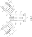
- Naturally occurring antibodies are multimeric proteins that contain four polypeptide chains ( FIG. 1 ). Two of the polypeptide chains are called heavy chains (H chains), and two of the polypeptide chains are called light chains (L chains).
- the immunoglobulin heavy and light chains are connected by an interchain disulfide bond.
- the immunoglobulin heavy chains are connected by interchain disulfide bonds.
- a light chain consists of one variable region (V L in FIG. 1 ) and one constant region (C L in FIG. 1 ).
- the heavy chain consists of one variable region (V H in FIG. 1 ) and at least three constant regions (C H1 , C H2 and C H3 in FIG. 1 ). The variable regions determine the specificity of the antibody.
- Each variable region comprises three hypervariable regions also known as complementarity determining regions (CDRs) flanked by four relatively conserved framework regions (FRs).
- CDRs complementarity determining regions
- FRs relatively conserved framework regions
- the three CDRs referred to as CDR 1 , CDR 2 , and CDR 3 , contribute to the antibody binding specificity.
- Naturally occurring antibodies have been used as starting material for engineered antibodies, such as chimeric antibodies and humanized antibodies.
- the invention is based, in part, upon the discovery of a family of antibodies that specifically bind human GDF15 (hGDF15).
- the antibodies contain hGDF15 binding sites based on the CDRs of the antibodies.
- the antibodies can be used as therapeutic agents.
- the antibodies are engineered, e.g., humanized, to reduce or eliminate an immune response when administered to a human patient.
- the disclosed antibodies prevent or inhibit the activity of ( i.e ., neutralize) hGDF15.
- the antibodies can inhibit the loss of muscle mass, for example, the loss of muscle mass associated with an underlying disease.
- the underlying disease may be selected from the group consisting of cancer, chronic heart failure, chronic kidney disease, COPD, AIDS, multiple sclerosis, rheumatoid arthritis, sepsis, and tuberculosis.
- the loss of muscle mass may be accompanied by a loss of fat mass.
- the disclosed antibodies can also be used to inhibit involuntary weight loss in a mammal.
- the disclosed antibodies may also be used to inhibit the loss of organ mass. Further, a method of treating cachexia and/or sarcopenia in a mammal comprising administering an effective amount of one of at least one of the disclosed antibodies to a mammal in need thereof is disclosed.
- Also disclosed is a method for establishing a steady-state level of mature recombinant human GDF15 (rhGDF15) in plasma or serum in a mammal comprising administering a rhGDF15-immunoglobulin Fc (Fc-rhGDF15) fusion protein to the mammal.
- the Fc-rhGDF15 can be a mouse Fc mature recombinant human GDF15 (mFc-rhGDF15).
- the mammal is a rodent, e.g., a mouse.
- a method of treating obesity in a mammal comprising administering a therapeutically effective amount of Fc-rhGDF15, e.g., a human Fc mature recombinant human GDF15 (hFc-rhGDF15), to the mammal in need thereof, is disclosed.
- a therapeutically effective amount of Fc-rhGDF15 e.g., a human Fc mature recombinant human GDF15 (hFc-rhGDF15)
- hFc-rhGDF15 human Fc mature recombinant human GDF15
- Pharmaceutical compositions comprising an Fc-rhGDF15 fusion protein and a pharmaceutically acceptable carrier are also disclosed.
- antibody 01G06, 03G05, 04F08, 06C11, 08G01, 14F11, or 17B11 means antibody 01G06, 03G05, 04F08, 06C11, 08G01, 14F11, or 17B11, or humanized variants thereof.
- the anti-GDF 15 antibodies disclosed herein are based on the antigen binding sites of certain monoclonal antibodies that have been selected on the basis of binding and neutralization of human GDF15 (hGDF15).
- the antibodies contain immunoglobulin variable region CDR sequences that define a binding site for hGDF15.
- the antibodies are useful for treating cachexia and/or sarcopenia.
- the antibodies can be engineered to minimize or eliminate an immune response when administered to a human patient.
- Cachexia means a metabolic syndrome associated with underlying disease and characterized by involuntary loss of muscle mass. Cachexia is often accompanied by involuntary weight loss, loss of fat mass, anorexia, inflammation, insulin resistance, fatigue, weakness, significant loss of appetite, and/or increased muscle protein breakdown. Cachexia is distinct from starvation, age-related loss of muscle mass, malabsorption, and hyperthyroidism. Underlying diseases associated with cachexia include cancer, chronic heart failure, chronic kidney disease, COPD, AIDS, multiple sclerosis, rheumatoid arthritis, sepsis, and tuberculosis.
- sarcopenia is understood to be a condition characterized primarily by loss of skeletal muscle mass and muscle strength. Sarcopenia is frequently associated with aging. See, Ruegg and Glass (2011) ANNUAL REV. PHARMACOL. TOXICOL. 51:373-395 .
- sarcopenia can be identified in a subject if a value of the appendicular skeletal muscle mass of a subject divided by the height of the subject in meters is more than two standard deviations below the young normal mean. (Thomas (2007) supra; see also Baumgartner et al. (1999) MECH. AGEING DEV. 147:755-763 ).
- antibody means an intact antibody (e.g., an intact monoclonal antibody) or antigen-binding fragment of an antibody, including an intact antibody or antigen-binding fragment that has been modified or engineered, or that is a human antibody.
- antibodies that have been modified or engineered are chimeric antibodies, humanized antibodies, and multispecific antibodies (e.g., bispecific antibodies).
- antigen-binding fragments include Fab, Fab', F(ab') 2 , Fv, single chain antibodies (e.g., scFv), minibodies and diabodies.
- the antibodies disclosed herein comprise: (a) an immunoglobulin heavy chain variable region comprising the structure CDR H1 -CDR H2 -CDR H3 and (b) an immunoglobulin light chain variable region comprising the structure CDR L1 -CDR L2 -CDR L3 , wherein the heavy chain variable region and the light chain variable region together define a single binding site for binding hGDF15 protein.
- the antibody comprises: (a) an immunoglobulin heavy chain variable region comprising the structure CDR H1 -CDR H2 -CDR H3 and (b) an immunoglobulin light chain variable region, wherein the heavy chain variable region and the light chain variable region together define a single binding site for binding hGDF15.
- a CDR H1 comprises an amino acid sequence selected from the group consisting of SEQ ID NO:1 (01G06, 08G01, Ch01G06 Chimeric, Hu01G06 IGHV1-18, Hu01G06 IGHV1-69, Sh01G06 IGHV1-18 M69L, Sh01G06 IGHV1-18 M69L K64Q G44S, Sh01G06 IGHV1-18 M69L K64Q, Sh01G06 IGHV1-69 T30S I69L, Sh01G06 IGHV1-69 T30S K64Q I69L, Hu01G06 IGHV1-18 F1, Hu01G06 IGHV1-18 F2, Hu01G06 IGHV1-69 F1, Hu01G06 IGHV1-69 F2), SEQ ID NO:2 (03G05), SEQ ID NO:3 (04F08), SEQ ID NO:4 (06C11, Ch06C11 Chimeric, HE LM 06C11 IGHV2-70, Hu06C11 IGHV2-5), SEQ ID NO
- the antibody comprises an immunoglobulin heavy chain variable region comprising a CDR H1 comprising the amino acid sequence of SEQ ID NO:1 (01G06, Ch01G06 Chimeric, Hu01G06 IGHV1-18, Hu01G06 IGHV1-69, Sh01G06 IGHV1-69 T30S I69L, Hu01G06 IGHV1-18 F1, Hu01G06 IGHV1-18 F2, Hu01G06 IGHV1-69 F1, Hu01G06 IGHV1-69 F2), a CDR H2 comprising the amino acid sequence of SEQ ID NO:7 (01G06, Ch01G06 Chimeric, Hu01G06 IGHV1-18, Hu01G06 IGHV1-69, Sh01G06 IGHV1-69 T30S I69L), and a CDR H3 comprising the amino acid sequence of SEQ ID NO:15 (01G06, Ch01G06 Chimeric, Hu01G06 IGHV1-18, Hu01G06 IGHV1-69, Sh01G06 I
- the antibody comprises an immunoglobulin heavy chain variable region comprising a CDR H1 comprising the amino acid sequence of SEQ ID NO:2 (03G05), a CDR H2 comprising the amino acid sequence of SEQ ID NO:8 (03G05), and a CDR H3 comprising the amino acid sequence of SEQ ID NO:16 (03G05).
- the antibody comprises an immunoglobulin heavy chain variable region comprising a CDR H1 comprising the amino acid sequence of SEQ ID NO:3 (04F08), a CDR H2 comprising the amino acid sequence of SEQ ID NO:9 (04F08), and a CDR H3 comprising the amino acid sequence of SEQ ID NO:17 (04F08).
- the antibody comprises an immunoglobulin heavy chain variable region comprising a CDR H1 comprising the amino acid sequence of SEQ ID NO:4 (06C11, Ch06C11 Chimeric, Hu06C11 IGHV2-5), a CDR H2 comprising the amino acid sequence of SEQ ID NO:9 (06C11, Ch06C11 Chimeric, Hu06C11 IGHV2-5), and a CDR H3 comprising the amino acid sequence of SEQ ID NO:18 (06C11, Ch06C11 Chimeric, Hu06C11 IGHV2-5).
- the antibody comprises an immunoglobulin heavy chain variable region comprising a CDR H1 comprising the amino acid sequence of SEQ ID NO:1 (08G01), a CDR H2 comprising the amino acid sequence of SEQ ID NO:10 (08G01), and a CDR H3 comprising the amino acid sequence of SEQ ID NO:15 (08G01).
- the antibody comprises an immunoglobulin heavy chain variable region comprising a CDR H1 comprising the amino acid sequence of SEQ ID NO:5 (14F11, Ch14F11 Chimeric, Sh14F11 IGHV2-5, Sh14F11 IGHV2-70), a CDR H2 comprising the amino acid sequence of SEQ ID NO:11 (14F11, Ch14F11 Chimeric, Sh14F11 IGHV2-5, Sh14F11 IGHV2-70), and a CDR H3 comprising the amino acid sequence of SEQ ID NO:19 (14F11, Ch14F11 Chimeric, Sh14F11 IGHV2-5, Sh14F11 IGHV2-70).
- the antibody comprises an immunoglobulin heavy chain variable region comprising a CDR H1 comprising the amino acid sequence of SEQ ID NO:6 (17B11), a CDR H2 comprising the amino acid sequence of SEQ ID NO:12 (17B11), and a CDR H3 comprising the amino acid sequence of SEQ ID NO:20 (17B11).
- the antibody comprises an immunoglobulin heavy chain variable region comprising a CDR H1 comprising the amino acid sequence of SEQ ID NO:1 (Sh01G06 IGHV1-18 M69L K64Q G44S, Sh01G06 IGHV1-18 M69L K64Q, Sh01G06 IGHV1-69 T30S K64Q I69L, Hu01G06 IGHV1-18 F1, Hu01G06 IGHV1-18 F2, Hu01G06 IGHV1-69 F1, Hu01G06 IGHV1-69 F2), a CDR H2 comprising the amino acid sequence of SEQ ID NO:13 (Sh01G06 IGHV1-18 M69L K64Q G44S, Sh01G06 IGHV1-18 M69L K64Q, Sh01G06 IGHV1-69 T30S K64Q I69L), and a CDR H3 comprising the amino acid sequence of SEQ ID NO:15 (Sh01G06 IGHV1-18 M69L K64Q G44S, Sh01G
- the antibody comprises an immunoglobulin heavy chain variable region comprising a CDR H1 comprising the amino acid sequence of SEQ ID NO:1 (Sh01G06 IGHV1-18 M69L K64Q G44S, Sh01G06 IGHV1-18 M69L K64Q, Sh01G06 IGHV1-69 T30S K64Q I69L, Hu01G06 IGHV1-18 F1, Hu01G06 IGHV1-18 F2, Hu01G06 IGHV1-69 F1, Hu01G06 IGHV1-69 F2), a CDR H2 comprising the amino acid sequence of SEQ ID NO:236 (Hu01G06 IGHV1-18 F1), and a CDR H3 comprising the amino acid sequence of SEQ ID NO:15 (Sh01G06 IGHV1-18 M69L K64Q G44S, Sh01G06 IGHV1-18 M69L K64Q, Sh01G06 IGHV1-69 T30S K64Q I69L, Hu01G06 IGHV1-18
- the antibody comprises an immunoglobulin heavy chain variable region comprising a CDR H1 comprising the amino acid sequence of SEQ ID NO:1 (Sh01G06 IGHV1-18 M69L K64Q G44S, Sh01G06 IGHV1-18 M69L K64Q, Sh01G06 IGHV1-69 T30S K64Q I69L, Hu01G06 IGHV1-18 F1, Hu01G06 IGHV1-18 F2, Hu01G06 IGHV1-69 F1, Hu01G06 IGHV1-69 F2), a CDR H2 comprising the amino acid sequence of SEQ ID NO:237 (Hu01G06 IGHV1-18 F2), and a CDR H3 comprising the amino acid sequence of SEQ ID NO:15 (Sh01G06 IGHV1-18 M69L K64Q G44S, Sh01G06 IGHV1-18 M69L K64Q, Sh01G06 IGHV1-69 T30S K64Q I69L, Hu01G06 IGHV1-18
- the antibody comprises an immunoglobulin heavy chain variable region comprising a CDR H1 comprising the amino acid sequence of SEQ ID NO:1 (Sh01G06 IGHV1-18 M69L K64Q G44S, Sh01G06 IGHV1-18 M69L K64Q, Sh01G06 IGHV1-69 T30S K64Q I69L, Hu01G06 IGHV1-18 F1, Hu01G06 IGHV1-18 F2, Hu01G06 IGHV1-69 F1, Hu01G06 IGHV1-69 F2), a CDR H2 comprising the amino acid sequence of SEQ ID NO:238 (Hu01G06 IGHV1-69 F1), and a CDR H3 comprising the amino acid sequence of SEQ ID NO:15 (Sh01G06 IGHV1-18 M69L K64Q G44S, Sh01G06 IGHV1-18 M69L K64Q, Sh01G06 IGHV1-69 T30S K64Q I69L, Hu01G06 IGHV1-18
- the antibody comprises an immunoglobulin heavy chain variable region comprising a CDR H1 comprising the amino acid sequence of SEQ ID NO:1 (Sh01G06 IGHV1-18 M69L K64Q G44S, Sh01G06 IGHV1-18 M69L K64Q, Sh01G06 IGHV1-69 T30S K64Q I69L, Hu01G06 IGHV1-18 F1, Hu01G06 IGHV1-18 F2, Hu01G06 IGHV1-69 F1, Hu01G06 IGHV1-69 F2), a CDR H2 comprising the amino acid sequence of SEQ ID NO:239 (Hu01G06 IGHV1-69 F2), and a CDR H3 comprising the amino acid sequence of SEQ ID NO:15 (Sh01G06 IGHV1-18 M69L K64Q G44S, Sh01G06 IGHV1-18 M69L K64Q, Sh01G06 IGHV1-69 T30S K64Q I69L, Hu01G06 IGHV1-18
- the antibody comprises an immunoglobulin heavy chain variable region comprising a CDR H1 comprising the amino acid sequence of SEQ ID NO:4 (HE LM 06C11 IGHV2-70), a CDR H2 comprising the amino acid sequence of SEQ ID NO:14 (HE LM 06C11 IGHV2-70), and a CDR H3 comprising the amino acid sequence of SEQ ID NO:18 (HE LM 06C11 IGHV2-70).
- the CDR H1 , CDR H2 , and CDR H3 sequences are interposed between fully human or humanized immunoglobulin FR sequences.
- the antibody comprises (a) an immunoglobulin light chain variable region comprising the structure CDR L1 -CDR L2 -CDR L3 , and (b) an immunoglobulin heavy chain variable region, wherein the immunoglobulin light chain variable region and the immunoglobulin heavy chain variable region together define a single binding site for binding hGDF15.
- a CDR L1 comprises an amino acid sequence selected from the group consisting of SEQ ID NO:21 (01G06, Ch01G06 Chimeric, Hu01G06 IGKV1-39, Hu01G06 IGKV1-39 S43A V48I, Hu01G06 IGKV1-39 V48I, Hu01G06 IGKV1-39 F1, Hu01G06 IGKV1-39 F2), SEQ ID NO:22 (03G05), SEQ ID NO:23 (04F08, 06C11, Ch06C11 Chimeric, Sh06C11 IGKV1-16, 14F11, Ch14F11 Chimeric, Hu14F11 IGKV1-16), SEQ ID NO:24 (08G01), and SEQ ID NO:25 (17B11);
- a CDR L2 comprises an amino acid sequence selected from the group consisting of SEQ ID NO:26 (01G06, Ch01G06 Chimeric, Hu01G06 IGKV1-39, Hu01G06 IGKV1-39 S43A V48
- the antibody comprises an immunoglobulin light chain variable region comprising a CDR L1 comprising the amino acid sequence of SEQ ID NO:21 (01G06, Ch01G06 Chimeric, Hu01G06 IGKV1-39, Hu01G06 IGKV1-39 S43A V48I, Hu01G06 IGKV1-39 V48I, Hu01G06 IGKV1-39 F1, Hu01G06 IGKV1-39 F2), a CDR L2 comprising the amino acid sequence of SEQ ID NO:26 (01G06, Ch01G06 Chimeric, Hu01G06 IGKV1-39, Hu01G06 IGKV1-39 S43A V48I, Hu01G06 IGKV1-39 V48I, Hu01G06 IGKV1-39 F1, Hu01G06 IGKV1-39 F2), and a CDR L3 comprising the amino acid sequence of SEQ ID NO:32 (01G06, Ch01G06 Chimeric, Hu01G06 IGKV
- the antibody comprises an immunoglobulin light chain variable region comprising a CDR L1 comprising the amino acid sequence of SEQ ID NO:21 (01G06, Ch01G06 Chimeric, Hu01G06 IGKV1-39, Hu01G06 IGKV1-39 S43A V48I, Hu01G06 IGKV1-39 V48I, Hu01G06 IGKV1-39 F1, Hu01G06 IGKV1-39 F2), a CDR L2 comprising the amino acid sequence of SEQ ID NO:26 (01G06, Ch01G06 Chimeric, Hu01G06 IGKV1-39, Hu01G06 IGKV1-39 S43A V48I, Hu01G06 IGKV1-39 V48I, Hu01G06 IGKV1-39 F1, Hu01G06 IGKV1-39 F2), and a CDR L3 comprising the amino acid sequence of SEQ ID NO:244 (Hu01G06 IGKV1-39 F2).
- a CDR L1 comprising
- the antibody comprises an immunoglobulin light chain variable region comprising a CDR L1 comprising the amino acid sequence of SEQ ID NO:22 (03G05), a CDR L2 comprising the amino acid sequence of SEQ ID NO:27 (03G05), and a CDR L3 comprising the amino acid sequence of SEQ ID NO:33 (03G05).
- the antibody comprises an immunoglobulin light chain variable region comprising a CDR L1 comprising the amino acid sequence of SEQ ID NO:23 (04F08), a CDR L2 comprising the amino acid sequence of SEQ ID NO:28 (04F08), and a CDR L3 comprising the amino acid sequence of SEQ ID NO:34 (04F08).
- the antibody comprises an immunoglobulin light chain variable region comprising a CDR L1 comprising the amino acid sequence of SEQ ID NO:23 (06C11, Ch06C11 Chimeric, Sh06C11 IGKV1-16), a CDR L2 comprising the amino acid sequence of SEQ ID NO:28 (06C11, Ch06C11 Chimeric, Sh06C11 IGKV1-16), and a CDR L3 comprising the amino acid sequence of SEQ ID NO:35 (06C11, Ch06C11 Chimeric, Sh06C11 IGKV1-16).
- the antibody comprises an immunoglobulin light chain variable region comprising a CDR L1 comprising the amino acid sequence of SEQ ID NO:24 (08G01), a CDR L2 comprising the amino acid sequence of SEQ ID NO:29 (08G01), and a CDR L3 comprising the amino acid sequence of SEQ ID NO:32 (08G01).
- the antibody comprises an immunoglobulin light chain variable region comprising a CDR L1 comprising the amino acid sequence of SEQ ID NO:23 (14F11, Ch14F11 Chimeric, Hu14F11 IGKV1-16), a CDR L2 comprising the amino acid sequence of SEQ ID NO:30 (14F11, Ch14F11 Chimeric, Hu14F11 IGKV1-16), and a CDR L3 comprising the amino acid sequence of SEQ ID NO:36 (14F11, Ch14F11 Chimeric, Hu14F11 IGKV1-16).
- the antibody comprises an immunoglobulin light chain variable region comprising a CDR L1 comprising the amino acid sequence of SEQ ID NO:25 (17B11), a CDR L2 comprising the amino acid sequence of SEQ ID NO:31 (17B11), and a CDR L3 comprising the amino acid sequence of SEQ ID NO:37 (17B11).
- the CDR L1 , CDR L2 , and CDR L3 sequences are interposed between fully human or humanized immunoglobulin FR sequences.
- the antibody comprises: (a) an immunoglobulin heavy chain variable region comprising the structure CDR H1 -CDR H2 -CDR H3 and (b) an immunoglobulin light chain variable region comprising the structure CDR L1 -CDR L2 -CDR L3 , wherein the heavy chain variable region and the light chain variable region together define a single binding site for binding hGDF15.
- the CDR H1 is an amino acid sequence selected from the group consisting of SEQ ID NO:1 (01G06, 08G01, Ch01G06 Chimeric, Hu01G06 IGHV1-18, Hu01G06 IGHV1-69, Sh01G06 IGHV1-18 M69L, Sh01G06 IGHV1-18 M69L K64Q G44S, Sh01G06 IGHV1-18 M69L K64Q, Sh01G06 IGHV1-69 T30S I69L, Sh01G06 IGHV1-69 T30S K64Q I69L, Hu01G06 IGHV1-18 F1, Hu01G06 IGHV1-18 F2, Hu01G06 IGHV1-69 F1, Hu01G06 IGHV1-69 F2), SEQ ID NO:2 (03G05), SEQ ID NO:3 (04F08), SEQ ID NO:4 (06C11, Ch06C11 Chimeric, HE LM 06C11 IGHV2-70, Hu06C11 IGHV2-5), SEQ ID NO
- the CDR L1 is an amino acid sequence selected from the group consisting of SEQ ID NO:21 (01G06, Ch01G06 Chimeric, Hu01G06 IGKV1-39, Hu01G06 IGKV1-39 S43A V48I, Hu01G06 IGKV1-39 V48I, Hu01G06 IGKV1-39 F1, Hu01G06 IGKV1-39 F2), SEQ ID NO:22 (03G05), SEQ ID NO:23 (04F08, 06C11, Ch06C11 Chimeric, Sh06C11 IGKV1-16, 14F11, Ch14F11 Chimeric, Hu14F11 IGKV1-16), SEQ ID NO:24 (08G01), and SEQ ID NO:25 (17B11);
- the CDR L2 is an amino acid sequence selected from the group consisting of SEQ ID NO:26 (01G06, Ch01G06 Chimeric, Hu01G06 IGKV1-39, Hu01G06 IGKV1-39 S43A V48I
- the antibody comprises an immunoglobulin heavy chain variable region comprising a CDR H1 comprising an amino acid sequence selected from the group consisting of SEQ ID NO:1 and SEQ ID NO:38 (Hu01G06 IGHV1-18 F1), a CDR H2 comprising an amino acid sequence selected from the group consisting of SEQ ID NO:236 and SEQ ID NO:240 (Hu01G06 IGHV1-18 F1), and a CDR H3 comprising the amino acid sequence of SEQ ID NO:15 (Hu01G06 IGHV1-18 F1) ; and an immunoglobulin light chain variable region comprising a CDR L1 comprising the amino acid sequence of SEQ ID NO:21 (Hu01G06 IGKV1-39 F1), a CDR L2 comprising the amino acid sequence of SEQ ID NO:26 (Hu01G06 IGKV1-39 F1), and a CDR L3 comprising the amino acid sequence of SEQ ID NO:32 (Hu01G06 IGKV1-39 F1).
- the antibody comprises an immunoglobulin heavy chain variable region comprising a CDR H1 comprising an amino acid sequence selected from the group consisting of SEQ ID NO:1 and SEQ ID NO:38 (Hu01G06 IGHV1-18 F2), a CDR H2 comprising an amino acid sequence selected from the group consisting of SEQ ID NO:237 and SEQ ID NO:241 (Hu01G06 IGHV1-18 F2), and a CDR H3 comprising the amino acid sequence of SEQ ID NO:15 (Hu01G06 IGHV1-18 F2); and an immunoglobulin light chain variable region comprising a CDR L1 comprising the amino acid sequence of SEQ ID NO:21 (Hu01G06 IGKV1-39 F2), a CDR L2 comprising the amino acid sequence of SEQ ID NO:26 (Hu01G06 IGKV1-39 F2), and a CDR L3 comprising the amino acid sequence of SEQ ID NO:244 (Hu01G06 IGKV1-39 F
- the antibody comprises an immunoglobulin heavy chain variable region comprising a CDR H1 comprising an amino acid sequence selected from the group consisting of SEQ ID NO:1 and SEQ ID NO:234 (Hu01G06 IGHV1-69 F1), a CDR H2 comprising an amino acid sequence selected from the group consisting of SEQ ID NO:238 and SEQ ID NO:241 (Hu01G06 IGHV1-69 F1), and a CDR H3 comprising the amino acid sequence of SEQ ID NO:15 (Hu01G06 IGHV1-69 F1) ; and an immunoglobulin light chain variable region comprising a CDR L1 comprising the amino acid sequence of SEQ ID NO:21 (Hu01G06 IGKV1-39 F1), a CDR L2 comprising the amino acid sequence of SEQ ID NO:26 (Hu01G06 IGKV1-39 F1) , and a CDR L3 comprising the amino acid sequence of SEQ ID NO:32 (Hu01G06 IGKV1-3
- the antibody comprises an immunoglobulin heavy chain variable region comprising a CDR H1 comprising an amino acid sequence selected from the group consisting of SEQ ID NO:1 and SEQ ID NO:234 (Hu01G06 IGHV1-69 F2), a CDR H2 comprising an amino acid sequence selected from the group consisting of SEQ ID NO:239 and SEQ ID NO:240 (Hu01G06 IGHV1-69 F2), and a CDR H3 comprising the amino acid sequence of SEQ ID NO:15 (Hu01G06 IGHV1-69 F2); and an immunoglobulin light chain variable region comprising a CDR L1 comprising the amino acid sequence of SEQ ID NO:21 (Hu01G06 IGKV1-39 F1), a CDR L2 comprising the amino acid sequence of SEQ ID NO:26 (Hu01G06 IGKV1-39 F1), and a CDR L3 comprising the amino acid sequence of SEQ ID NO:32 (Hu01G06 IGKV1-39 F1).
- the antibody comprises an immunoglobulin heavy chain variable region comprising a CDR H1 comprising an amino acid sequence selected from the group consisting of SEQ ID NO:1 and SEQ ID NO:234 (Hu01G06 IGHV1-69 F2), a CDR H2 comprising an amino acid sequence selected from the group consisting of SEQ ID NO:239 and SEQ ID NO:240 (Hu01G06 IGHV1-69 F2), and a CDR H3 comprising the amino acid sequence of SEQ ID NO:15 (Hu01G06 IGHV1-69 F2); and an immunoglobulin light chain variable region comprising a CDR L1 comprising the amino acid sequence of SEQ ID NO:21 (Hu01G06 IGKV1-39 F2), a CDR L2 comprising the amino acid sequence of SEQ ID NO:26 (Hu01G06 IGKV1-39 F2), and a CDR L3 comprising the amino acid sequence of SEQ ID NO:244 (Hu01G06 IGKV1-39 F
- the antibodies disclosed herein comprise an immunoglobulin heavy chain variable region and an immunoglobulin light chain variable region.
- the antibody comprises an immunoglobulin heavy chain variable region selected from the group consisting of SEQ ID NO:40 (01G06, Ch01G06 Chimeric), SEQ ID NO:42 (03G05), SEQ ID NO:44 (04F08), SEQ ID NO:46 (06C11, Ch06C11 Chimeric), SEQ ID NO:48 (08G01), SEQ ID NO:50 (14F11, Ch14F11 Chimeric), SEQ ID NO:52 (17B11), SEQ ID NO:54 (Hu01G06 IGHV1-18) , SEQ ID NO:56 (Hu01G06 IGHV1-69) , SEQ ID NO:58 (Sh01G06 IGHV1-18 M69L), SEQ ID NO:60 (Sh01G06 IGHV1-18 M69L K64Q G44S), SEQ ID NO:62 (Sh01G06 IGHV1-18 M69L K64Q), SEQ ID
- the antibody comprises an immunoglobulin light chain variable region selected from the group consisting of SEQ ID NO:76 (01G06, Ch01G06 Chimeric), SEQ ID NO:78 (03G05), SEQ ID NO:80 (04F08), SEQ ID NO:82 (06C11, Ch06C11 Chimeric), SEQ ID NO:84 (08G01), SEQ ID NO:86 (14F11, Ch14F11 Chimeric), SEQ ID NO:88 (17B11), SEQ ID NO:90 (Hu01G06 IGKV1-39), SEQ ID NO:92 (Hu01G06 IGKV1-39 S43A V48I or Hu01G06 IGKV1-39 F1), SEQ ID NO:94 (Hu01G06 IGKV1-39 V48I), SEQ ID NO:96 (Sh06C11 IGKV1-16), SEQ ID NO:254 (Hu01G06 IGKV1-39 F2), and SEQ ID NO:98 (Hu14F11 IGKV1-16),
- the antibody comprises an immunoglobulin heavy chain variable region selected from the group consisting of SEQ ID NO:40 (01G06, Ch01G06 Chimeric), SEQ ID NO:42 (03G05), SEQ ID NO:44 (04F08), SEQ ID NO:46 (06C11, Ch06C11 Chimeric), SEQ ID NO:48 (08G01), SEQ ID NO:50 (14F11, Ch14F11 Chimeric), SEQ ID NO:52 (17B11), SEQ ID NO:54 (Hu01G06 IGHV1-18), SEQ ID NO:56 (Hu01G06 IGHV1-69), SEQ ID NO:58 (Sh01G06 IGHV1-18 M69L), SEQ ID NO:60 (Sh01G06 IGHV1-18 M69L K64Q G44S), SEQ ID NO:62 (Sh01G06 IGHV1-18 M69L K64Q), SEQ ID NO:64 (Sh01G06 IGHV1-69 T30S I69L), SEQ ID NO:66 (Sh
- the antibody comprises an immunoglobulin heavy chain variable region comprising the amino acid sequence of SEQ ID NO:40 (01G06, Ch01G06 Chimeric), and an immunoglobulin light chain variable region comprising the amino acid sequence of SEQ ID NO:76 (01G06, Ch01G06 Chimeric).
- the antibody comprises an immunoglobulin heavy chain variable region comprising the amino acid sequence of SEQ ID NO:42 (03G05), and an immunoglobulin light chain variable region comprising the amino acid sequence of SEQ ID NO:78 (03G05).
- the antibody comprises an immunoglobulin heavy chain variable region comprising the amino acid sequence of SEQ ID NO:44 (04F08), and an immunoglobulin light chain variable region comprising the amino acid sequence of SEQ ID NO:80 (04F08).
- the antibody comprises an immunoglobulin heavy chain variable region comprising the amino acid sequence of SEQ ID NO:46 (06C11), and an immunoglobulin light chain variable region comprising the amino acid sequence of SEQ ID NO:82 (06C11).
- the antibody comprises an immunoglobulin heavy chain variable region comprising the amino acid sequence of SEQ ID NO:48 (08G01), and an immunoglobulin light chain variable region comprising the amino acid sequence of SEQ ID NO:84 (08G01).
- the antibody comprises an immunoglobulin heavy chain variable region comprising the amino acid sequence of SEQ ID NO:50 (14F11), and an immunoglobulin light chain variable region comprising the amino acid sequence of SEQ ID NO:86 (14F11).
- the antibody comprises an immunoglobulin heavy chain variable region comprising the amino acid sequence of SEQ ID NO:52 (17B11), and an immunoglobulin light chain variable region comprising the amino acid sequence of SEQ ID NO:88 (17B11).
- the antibody comprises an immunoglobulin heavy chain variable region comprising the amino acid sequence of SEQ ID NO:54 (Hu01G06 IGHV1-18), and an immunoglobulin light chain variable region comprising the amino acid sequence of SEQ ID NO:76 (Ch01G06 Chimeric).
- the antibody comprises an immunoglobulin heavy chain variable region comprising the amino acid sequence of SEQ ID NO:56 (Hu01G06 IGHV1-69), and an immunoglobulin light chain variable region comprising the amino acid sequence of SEQ ID NO:76 (Ch01G06 Chimeric).
- the antibody comprises an immunoglobulin heavy chain variable region comprising the amino acid sequence of SEQ ID NO:58 (Sh01G06 IGHV1-18 M69L), and an immunoglobulin light chain variable region comprising the amino acid sequence of SEQ ID NO:76 (Ch01G06 Chimeric).
- the antibody comprises an immunoglobulin heavy chain variable region comprising the amino acid sequence of SEQ ID NO:60 (Sh01G06 IGHV1-18 M69L K64Q G44S), and an immunoglobulin light chain variable region comprising the amino acid sequence of SEQ ID NO:76 (Ch01G06 Chimeric).
- the antibody comprises an immunoglobulin heavy chain variable region comprising the amino acid sequence of SEQ ID NO:62 (Sh01G06 IGHV1-18 M69L K64Q), and an immunoglobulin light chain variable region comprising the amino acid sequence of SEQ ID NO:76 (Ch01G06 Chimeric).
- the antibody comprises an immunoglobulin heavy chain variable region comprising the amino acid sequence of SEQ ID NO:64 (Sh01G06 IGHV1-69 T30S I69L), and an immunoglobulin light chain variable region comprising the amino acid sequence of SEQ ID NO:76 (Ch01G06 Chimeric).
- the antibody comprises an immunoglobulin heavy chain variable region comprising the amino acid sequence of SEQ ID NO:66 (Sh01G06 IGHV1-69 T30S K64Q I69L), and an immunoglobulin light chain variable region comprising the amino acid sequence of SEQ ID NO:76 (Ch01G06 Chimeric).
- the antibody comprises an immunoglobulin heavy chain variable region comprising the amino acid sequence of SEQ ID NO:40 (Ch01G06 Chimeric), and an immunoglobulin light chain variable region comprising the amino acid sequence of SEQ ID NO:90 (Hu01G06 IGKV1-39).
- the antibody comprises an immunoglobulin heavy chain variable region comprising the amino acid sequence of SEQ ID NO:54 (Hu01G06 IGHV1-18), and an immunoglobulin light chain variable region comprising the amino acid sequence of SEQ ID NO:90 (Hu01G06 IGKV1-39).
- the antibody comprises an immunoglobulin heavy chain variable region comprising the amino acid sequence of SEQ ID NO:56 (Hu01G06 IGHV1-69), and an immunoglobulin light chain variable region comprising the amino acid sequence of SEQ ID NO:90 (Hu01G06 IGKV1-39).
- the antibody comprises an immunoglobulin heavy chain variable region comprising the amino acid sequence of SEQ ID NO:58 (Sh01G06 IGHV1-18 M69L), and an immunoglobulin light chain variable region comprising the amino acid sequence of SEQ ID NO:90 (Hu01G06 IGKV1-39).
- the antibody comprises an immunoglobulin heavy chain variable region comprising the amino acid sequence of SEQ ID NO:60 (Sh01G06 IGHV1-18 M69L K64Q G44S), and an immunoglobulin light chain variable region comprising the amino acid sequence of SEQ ID NO:90 (Hu01G06 IGKV1-39).
- the antibody comprises an immunoglobulin heavy chain variable region comprising the amino acid sequence of SEQ ID NO:62 (Sh01G06 IGHV1-18 M69L K64Q), and an immunoglobulin light chain variable region comprising the amino acid sequence of SEQ ID NO:90 (Hu01G06 IGKV1-39).
- the antibody comprises an immunoglobulin heavy chain variable region comprising the amino acid sequence of SEQ ID NO:64 (Sh01G06 IGHV1-69 T30S I69L), and an immunoglobulin light chain variable region comprising the amino acid sequence of SEQ ID NO:90 (Hu01G06 IGKV1-39).
- the antibody comprises an immunoglobulin heavy chain variable region comprising the amino acid sequence of SEQ ID NO:66 (Sh01G06 IGHV1-69 T30S K64Q I69L), and an immunoglobulin light chain variable region comprising the amino acid sequence of SEQ ID NO:90 (Hu01G06 IGKV1-39).
- the antibody comprises an immuoglobulin heavy chain variable region comprising the amino acid sequence of SEQ ID NO:40 (Ch01G06 Chimeric), and an immunoglobulin light chain variable region comprising the amino acid sequence of SEQ ID NO:92 (Hu01G06 IGKV1-39 S43A V48I).
- the antibody comprises an immunoglobulin heavy chain variable region comprising the amino acid sequence of SEQ ID NO:54 (Hu01G06 IGHV1-18), and an immunoglobulin light chain variable region comprising the amino acid sequence of SEQ ID NO:92 (Hu01G06 IGKV1-39 S43A V48I).
- the antibody comprises an immunoglobulin heavy chain variable region comprising the amino acid sequence of SEQ ID NO:56 (Hu01G06 IGHV1-69), and an immunoglobulin light chain variable region comprising the amino acid sequence of SEQ ID NO:92 (Hu01G06 IGKV1-39 S43A V48I).
- the antibody comprises an immunoglobulin heavy chain variable region comprising the amino acid sequence of SEQ ID NO:58 (Sh01G06 IGHV1-18 M69L), and an immunoglobulin light chain variable region comprising the amino acid sequence of SEQ ID NO:92 (Hu01G06 IGKV1-39 S43A V48I).
- the antibody comprises an immunoglobulin heavy chain variable region comprising the amino acid sequence of SEQ ID NO:60 (Sh01G06 IGHV1-18 M69L K64Q G44S), and an immunoglobulin light chain variable region comprising the amino acid sequence of SEQ ID NO:92 (Hu01G06 IGKV1-39 S43A V48I).
- the antibody comprises an immunoglobulin heavy chain variable region comprising the amino acid sequence of SEQ ID NO:62 (Sh01G06 IGHV1-18 M69L K64Q), and an immunoglobulin light chain variable region comprising the amino acid sequence of SEQ ID NO:92 (Hu01G06 IGKV1-39 S43A V48I).
- the antibody comprises an immunoglobulin heavy chain variable region comprising the amino acid sequence of SEQ ID NO:64 (Sh01G06 IGHV1-69 T30S I69L), and an immunoglobulin light chain variable region comprising the amino acid sequence of SEQ ID NO:92 (Hu01G06 IGKV1-39 S43A V48I).
- the antibody comprises an immunoglobulin heavy chain variable region comprising the amino acid sequence of SEQ ID NO:66 (Sh01G06 IGHV1-69 T30S K64Q I69L), and an immunoglobulin light chain variable region comprising the amino acid sequence of SEQ ID NO:92 (Hu01G06 IGKV1-39 S43A V48I).
- the antibody comprises an immunoglobulin heavy chain variable region comprising the amino acid sequence of SEQ ID NO:40 (Ch01G06 Chimeric), and an immunoglobulin light chain variable region comprising the amino acid sequence of SEQ ID NO:94 (Hu01G06 IGKV1-39 V48I).
- the antibody comprises an immunoglobulin heavy chain variable region comprising the amino acid sequence of SEQ ID NO:54 (Hu01G06 IGHV1-18), and an immunoglobulin light chain variable region comprising the amino acid sequence of SEQ ID NO:94 (Hu01G06 IGKV1-39 V48I).
- the antibody comprises an immunoglobulin heavy chain variable region comprising the amino acid sequence of SEQ ID NO:56 (Hu01G06 IGHV1-69), and an immunoglobulin light chain variable region comprising the amino acid sequence of SEQ ID NO:94 (Hu01G06 IGKV1-39 V48I).
- the antibody comprises an immunoglobulin heavy chain variable region comprising the amino acid sequence of SEQ ID NO:58 (Sh01G06 IGHV1-18 M69L), and an immunoglobulin light chain variable region comprising the amino acid sequence of SEQ ID NO:94 (Hu01G06 IGKV1-39 V48I).
- the antibody comprises an immunoglobulin heavy chain variable region comprising the amino acid sequence of SEQ ID NO:60 (Sh01G06 IGHV1-18 M69L K64Q G44S), and an immunoglobulin light chain variable region comprising the amino acid sequence of SEQ ID NO:94 (Hu01G06 IGKV1-39 V48I).
- the antibody comprises an immunoglobulin heavy chain variable region comprising the amino acid sequence of SEQ ID NO:62 (Sh01G06 IGHV1-18 M69L K64Q), and an immunoglobulin light chain variable region comprising the amino acid sequence of SEQ ID NO:94 (Hu01G06 IGKV1-39 V48I).
- the antibody comprises an immunoglobulin heavy chain variable region comprising the amino acid sequence of SEQ ID NO:64 (Sh01G06 IGHV1-69 T30S I69L), and an immunoglobulin light chain variable region comprising the amino acid sequence of SEQ ID NO:94 (Hu01G06 IGKV1-39 V48I).
- the antibody comprises an immunoglobulin heavy chain variable region comprising the amino acid sequence of SEQ ID NO:66 (Sh01G06 IGHV1-69 T30S K64Q I69L), and an immunoglobulin light chain variable region comprising the amino acid sequence of SEQ ID NO:94 (Hu01G06 IGKV1-39 V48I).
- the antibody comprises an immunoglobulin heavy chain variable region comprising the amino acid sequence of SEQ ID NO:246 (Hu01G06 IGHV1-18 F1), and an immunoglobulin light chain variable region comprising the amino acid sequence of SEQ ID NO:92 (Hu01G06 IGKV1-39 F1).
- the antibody comprises an immunoglobulin heavy chain variable region comprising the amino acid sequence of SEQ ID NO:248 (Hu01G06 IGHV1-18 F2), and an immunoglobulin light chain variable region comprising the amino acid sequence of SEQ ID NO:254 (Hu01G06 IGKV1-39 F2).
- the antibody comprises an immunoglobulin heavy chain variable region comprising the amino acid sequence of SEQ ID NO:250 (Hu01G06 IGHV1-69 F1), and an immunoglobulin light chain variable region comprising the amino acid sequence of SEQ ID NO:92 (Hu01G06 IGKV1-39 F1).
- the antibody comprises an immunoglobulin heavy chain variable region comprising the amino acid sequence of SEQ ID NO:252 (Hu01G06 IGHV1-69 F2), and an immunoglobulin light chain variable region comprising the amino acid sequence of SEQ ID NO:92 (Hu01G06 IGKV1-39 F1).
- the antibody comprises an immunoglobulin heavy chain variable region comprising the amino acid sequence of SEQ ID NO:252 (Hu01G06 IGHV1-69 F2), and an immunoglobulin light chain variable region comprising the amino acid sequence of SEQ ID NO:254 (Hu01G06 IGKV1-39 F2).
- the antibody comprises an immunoglobulin heavy chain variable region comprising the amino acid sequence of SEQ ID NO:68 (HE LM 06C11 IGHV2-70), and an immunoglobulin light chain variable region comprising the amino acid sequence of SEQ ID NO:82 (Ch06C11 Chimeric).
- the antibody comprises an immunoglobulin heavy chain variable region comprising the amino acid sequence of SEQ ID NO:70 (Hu06C11 IGHV2-5), and an immunoglobulin light chain variable region comprising the amino acid sequence of SEQ ID NO:82 (Ch06C11 Chimeric).
- the antibody comprises an immunoglobulin heavy chain variable region comprising the amino acid sequence of SEQ ID NO:46 (Ch06C11 Chimeric), and an immunoglobulin light chain variable region comprising the amino acid sequence of SEQ ID NO:96 (Sh06C11 IGKV1-16) .
- the antibody comprises an immunoglobulin heavy chain variable region comprising the amino acid sequence of SEQ ID NO:68 (HE LM 06C11 IGHV2-70), and an immunoglobulin light chain variable region comprising the amino acid sequence of SEQ ID NO:96 (Sh06C11 IGKV1-16).
- the antibody comprises an immunoglobulin heavy chain variable region comprising the amino acid sequence of SEQ ID NO:70 (Hu06C11 IGHV2-5), and an immunoglobulin light chain variable region comprising the amino acid sequence of SEQ ID NO:96 (Sh06C11 IGKV1-16) .
- the antibody comprises an immunoglobulin heavy chain variable region comprising the amino acid sequence of SEQ ID NO:72 (Sh14F11 IGHV2-5), and an immunoglobulin light chain variable region comprising the amino acid sequence of SEQ ID NO:86 (Ch14F11 Chimeric).
- the antibody comprises an immunoglobulin heavy chain variable region comprising the amino acid sequence of SEQ ID NO:74 (Sh14F11 IGHV2-70), and an immunoglobulin light chain variable region comprising the amino acid sequence of SEQ ID NO:86 (Ch14F11 Chimeric).
- the antibody comprises an immunoglobulin heavy chain variable region comprising the amino acid sequence of SEQ ID NO:50 (Ch14F11 Chimeric), and an immunoglobulin light chain variable region comprising the amino acid sequence of SEQ ID NO:98 (Hu14F11 IGKV1-16) .
- the antibody comprises an immunoglobulin heavy chain variable region comprising the amino acid sequence of SEQ ID NO:72 (Sh14F11 IGHV2-5), and an immunoglobulin light chain variable region comprising the amino acid sequence of SEQ ID NO:98 (Hu14F11 IGKV1-16).
- the antibody comprises an immunoglobulin heavy chain variable region comprising the amino acid sequence of SEQ ID NO:74 (Sh14F11 IGHV2-70), and an immunoglobulin light chain variable region comprising the amino acid sequence of SEQ ID NO:98 (Hu14F11 IGKV1-16) .
- the antibody comprises an immunoglobulin heavy chain variable region comprising the amino acid sequence of SEQ ID NO:46 (Ch06C11 Chimeric), and an immunoglobulin light chain variable region comprising the amino acid sequence of SEQ ID NO:80 (04F08).
- the antibody comprises an immunoglobulin heavy chain variable region comprising the amino acid sequence of SEQ ID NO:50 (Ch14F11 Chimeric), and an immunoglobulin light chain variable region comprising the amino acid sequence of SEQ ID NO:80 (04F08).
- the antibody comprises an immunoglobulin heavy chain variable region comprising the amino acid sequence of SEQ ID NO:44 (04F08), and an immunoglobulin light chain variable region comprising the amino acid sequence of SEQ ID NO:82 (Ch06C11 Chimeric).
- the antibody comprises an immunoglobulin heavy chain variable region comprising the amino acid sequence of SEQ ID NO:50 (Ch14F11 Chimeric), and an immunoglobulin light chain variable region comprising the amino acid sequence of SEQ ID NO:82 (Ch06C11 Chimeric).
- the antibody comprises an immunoglobulin heavy chain variable region comprising the amino acid sequence of SEQ ID NO:44 (04F08), and an immunoglobulin light chain variable region comprising the amino acid sequence of SEQ ID NO:86 (Ch14F11 Chimeric).
- the antibody comprises an immunoglobulin heavy chain variable region comprising the amino acid sequence of SEQ ID NO:46 (Ch06C11 Chimeric), and an immunoglobulin light chain variable region comprising the amino acid sequence of SEQ ID NO:86 (Ch14F11 Chimeric).
- the antibody comprises an immunoglobulin heavy chain variable region comprising the amino acid sequence of SEQ ID NO:48 (08G01), and an immunoglobulin light chain variable region comprising the amino acid sequence of SEQ ID NO:76 (Ch01G06 Chimeric).
- the antibody comprises an immunoglobulin heavy chain variable region comprising the amino acid sequence of SEQ ID NO:40 (Ch01G06 Chimeric), and an immunoglobulin light chain variable region comprising the amino acid sequence of SEQ ID NO:84 (08G01).
- the antibodies disclosed herein comprise an immunoglobulin heavy chain and an immunoglobulin light chain.
- the antibody comprises an immunoglobulin heavy chain selected from the group consisting of SEQ ID NO:100 (01G06), SEQ ID NO:104 (03G05), SEQ ID NO:108 (04F08), SEQ ID NO:112 (06C11), SEQ ID NO:116 (08G01), SEQ ID NO:120 (14F11), SEQ ID NO:124 (17B11), SEQ ID NO:176 (Ch01G06 Chimeric), SEQ ID NO:178 (Hu01G06 IGHV1-18), SEQ ID NO:180 (Hu01G06 IGHV1-69), SEQ ID NO:182 (Sh01G06 IGHV1-18 M69L), SEQ ID NO:184 (Sh01G06 IGHV1-18 M69L K64Q G44S), SEQ ID NO:186 (Sh01G06 IGHV1-18 M69L K64Q), SEQ ID NO:188 (Sh01G06
- the antibody comprises an immunoglobulin light chain selected from the group consisting of SEQ ID NO:102 (01G06), SEQ ID NO:106 (03G05), SEQ ID NO:110 (04F08), SEQ ID NO:114 (06C11), SEQ ID NO:118 (08G01), SEQ ID NO:122 (14F11), SEQ ID NO:126 (17B11), SEQ ID NO:204 (Ch01G06 Chimeric), SEQ ID NO:206 (Hu01G06 IGKV1-39), SEQ ID NO:208 (Hu01G06 IGKV1-39 S43A V48I or Hu01G06 IGKV1-39 F1), SEQ ID NO:210 (Hu01G06 IGKV1-39 V48I), SEQ ID NO:264 (Hu01G06 IGKV1-39 F2), SEQ ID NO:212 (Ch06C11 Chimeric), SEQ ID NO:214 (Sh06C11 IGKV1-16), SEQ ID NO:216 (Ch14F11 Chimeric), and
- the antibody comprises (i) an immunoglobulin heavy chain selected from the group consisting of SEQ ID NO:100 (01G06), SEQ ID NO:104 (03G05), SEQ ID NO:108 (04F08), SEQ ID NO:112 (06C11), SEQ ID NO:116 (08G01), SEQ ID NO:120 (14F11), SEQ ID NO:124 (17B11), SEQ ID NO:176 (Ch01G06 Chimeric), SEQ ID NO:178 (Hu01G06 IGHV1-18), SEQ ID NO:180 (Hu01G06 IGHV1-69), SEQ ID NO:182 (Sh01G06 IGHV1-18 M69L), SEQ ID NO:184 (Sh01G06 IGHV1-18 M69L K64Q G44S), SEQ ID NO:186 (Sh01G06 IGHV1-18 M69L K64Q), SEQ ID NO:188 (Sh01G06 IGHV1-69 T30S I69L), SEQ ID NO:190 (Sh01G06 Iglobulin heavy
- the antibody comprises an immunoglobulin heavy chain comprising the amino acid sequence of SEQ ID NO:176 (Ch01G06 Chimeric), and an immunoglobulin light chain comprising the amino acid sequence of SEQ ID NO:204 (Ch01G06 Chimeric).
- the antibody comprises an immunoglobulin heavy chain comprising the amino acid sequence of SEQ ID NO:192 (Ch06C11 Chimeric), and an immunoglobulin light chain comprising the amino acid sequence of SEQ ID NO:212 (Ch06C11 Chimeric).
- the antibody comprises an immunoglobulin heavy chain comprising the amino acid sequence of SEQ ID NO:198 (Ch14F11 Chimeric), and an immunoglobulin light chain comprising the amino acid sequence of SEQ ID NO:216 (Ch14F11 Chimeric).
- the antibody comprises an immunoglobulin heavy chain comprising the amino acid sequence of SEQ ID NO:178 (Hu01G06 IGHV1-18), and an immunoglobulin light chain comprising the amino acid sequence of SEQ ID NO:206 (Hu01G06 1GKV1-39)
- the antibody comprises an immunoglobulin heavy chain comprising the amino acid sequence of SEQ ID NO:180 (Hu01G06 IGHV1-69), and an immunoglobulin light chain comprising the amino acid sequence of SEQ ID NO:206 (Hu01G06 1GKV1-39)
- the antibody comprises an immunoglobulin heavy chain comprising the amino acid sequence of SEQ ID NO:184 (Sh01G06 IGHV1-18 M69L K64Q G44S), and an immunoglobulin light chain comprising the amino acid sequence of SEQ ID NO:210 (Hu01G06 IGKV1-39 V48I).
- the antibody comprises an immunoglobulin heavy chain comprising the amino acid sequence of SEQ ID NO:188 (Sh01G06 IGHV1-69 T30S I69L), and an immunoglobulin light chain comprising the amino acid sequence of SEQ ID NO:210 (Hu01G06 IGKV1-39 V48I).
- the antibody comprises an immunoglobulin heavy chain comprising the amino acid sequence of SEQ ID NO:184 (Sh01G06 IGHV1-18 M69L K64Q G44S), and an immunoglobulin light chain comprising the amino acid sequence of SEQ ID NO:208 (Hu01G06 IGKV1-39 S43A V48I).
- the antibody comprises an immunoglobulin heavy chain comprising the amino acid sequence of SEQ ID NO:188 (Sh01G06 IGHV1-69 T30S I69L), and an immunoglobulin light chain comprising the amino acid sequence of SEQ ID NO:208 (Hu01G06 IGKV1-39 S43A V48I).
- the antibody comprises an immunoglobulin heavy chain comprising the amino acid sequence of SEQ ID NO:256 (Hu01G06 IGHV1-18 F1), and an immunoglobulin light chain comprising the amino acid sequence of SEQ ID NO:208 (Hu01G06 IGKV1-39 F1).
- the antibody comprises an immunoglobulin heavy chain comprising the amino acid sequence of SEQ ID NO:258 (Hu01G06 IGHV1-18 F2), and an immunoglobulin light chain comprising the amino acid sequence of SEQ ID NO:264 (Hu01G06 IGKV1-39 F2).
- the antibody comprises an immunoglobulin heavy chain comprising the amino acid sequence of SEQ ID NO:260 (Hu01G06 IGHV1-69 F1), and an immunoglobulin light chain comprising the amino acid sequence of SEQ ID NO:208 (Hu01G06 IGKV1-39 F1).
- the antibody comprises an immunoglobulin heavy chain comprising the amino acid sequence of SEQ ID NO:262 (Hu01G06 IGHV1-69 F2), and an immunoglobulin light chain comprising the amino acid sequence of SEQ ID NO:208 (Hu01G06 IGKV1-39 F1).
- the antibody comprises an immunoglobulin heavy chain comprising the amino acid sequence of SEQ ID NO:262 (Hu01G06 IGHV1-69 F2), and an immunoglobulin light chain comprising the amino acid sequence of SEQ ID NO:264 (Hu01G06 IGKV1-39 F2).
- the antibody comprises an immunoglobulin heavy chain comprising the amino acid sequence of SEQ ID NO:194 (HE LM 06C11 IGHV2-70), and an immunoglobulin light chain comprising the amino acid sequence of SEQ ID NO:214 (Sh06C11 IGKV1-16) .
- the antibody comprises an immunoglobulin heavy chain comprising the amino acid sequence of SEQ ID NO:196 (Hu06C11 IGHV2-5), and an immunoglobulin light chain comprising the amino acid sequence of SEQ ID NO:214 (Sh06C11 IGKV1-16) .
- the antibody comprises an immunoglobulin heavy chain comprising the amino acid sequence of SEQ ID NO:200 (Sh14F11 IGHV2-5), and an immunoglobulin light chain comprising the amino acid sequence of SEQ ID NO:218 (Hu14F11 IGKV1-16)
- the antibody comprises an immunoglobulin heavy chain comprising the amino acid sequence of SEQ ID NO:202 (Sh14F11 IGHV2-70), and an immunoglobulin light chain comprising the amino acid sequence of SEQ ID NO:218 (Hu14F11 IGKV1-16).
- an isolated antibody that binds HGDF15 comprises an immunoglobulin heavy chain variable region comprising an amino acid sequence that is at least 70%, 75%, 80%, 85%, 90%, 95%, 98%, or 99% identical to the entire variable region or the FR sequence of SEQ ID NO:40 (01G06, Ch01G06 Chimeric), SEQ ID NO:42 (03G05), SEQ ID NO:44 (04F08), SEQ ID NO:46 (06C11, Ch06C11 Chimeric), SEQ ID NO:48 (08G01), SEQ ID NO:50 (14F11, Ch14F11 Chimeric), SEQ ID NO:52 (17B11), SEQ ID NO:54 (Hu01G06 IGHV1-18), SEQ ID NO:56 (Hu01G06 IGHV1-69), SEQ ID NO:58 (Sh01G06 IGHV1-18 M69L), SEQ ID NO:60 (Sh01G06 IGHV1-18 M69L K64Q G44S), SEQ ID NO:62 (Sh
- an isolated antibody that binds HGDF15 comprises an immunoglobulin light chain variable region comprising an amino acid sequence that is at least 70%, 75%, 80%, 85%, 90%, 95%, 98%, or 99% identical to the entire variable region or the FR sequence of SEQ ID NO:76 (01G06, Ch01G06 Chimeric), SEQ ID NO:78 (03G05), SEQ ID NO:80 (04F08), SEQ ID NO:82 (06C11, Ch06C11 Chimeric), SEQ ID NO:84 (08G01), SEQ ID NO:86 (14F11, Ch14F11 Chimeric), SEQ ID NO:88 (17B11), SEQ ID NO:90 (Hu01G06 IGKV1-39), SEQ ID NO:92 (Hu01G06 IGKV1-39 S43A V48I or Hu01G06 IGKV1-39 F1), SEQ ID NO:94 (Hu01G06 IGKV1-39 V48I), SEQ ID NO:254 (Hu01G06
- Sequence identity may be determined in various ways that are within the skill of a person skilled in the art, e.g., using publicly available computer software such as BLAST, BLAST-2, ALIGN or Megalign (DNASTAR) software.
- BLAST Basic Local Alignment Search Tool
- analysis using the algorithm employed by the programs blastp, blastn, blastx, tblastn and tblastx ( Karlin et al., (1990) PROC. NATL. ACAD. SCI. USA 87:2264-2268 ; Altschul, (1993) J. MOL. EVOL. 36:290-300 ; Altschul et al., (1997) NUCLEIC ACIDS RES.
- 25:3389-3402 are tailored for sequence similarity searching.
- sequence similarity searching For a discussion of basic issues in searching sequence databases see Altschul et al., (1994) NATURE GENETICS 6:119-129 , which is fully incorporated by reference herein.
- Those skilled in the art can determine appropriate parameters for measuring alignment, including any algorithms needed to achieve maximal alignment over the full length of the sequences being compared.
- the search parameters for histogram, descriptions, alignments, expect i.e., the statistical significance threshold for reporting matches against database sequences
- cutoff, matrix and filter are at the default settings.
- blastn The default scoring matrix used by blastp, blastx, tblastn, and tblastx is the BLOSUM62 matrix ( Henikoff et al., (1992) PROC. NATL. ACAD. SCI. USA 89:10915-10919 , fully incorporated by reference herein).
- immunoglobulin heavy chain variable region sequences and/or light chain variable region sequences that together bind human GDF15 may contain amino acid alterations (e.g., at least 1, 2, 3, 4, 5, or 10 amino acid substitutions, deletions, or additions) in the framework regions of the heavy and/or light chain variable regions.
- the antibody binds hGDF15 with a K D of about 300 pM, 250 pM, 200 pM, 190 pM, 180 pM, 170 pM, 160 pM, 150 pM, 140 pM, 130 pM, 120 pM, 110 pM, 100 pM, 90 pM, 80 pM, 70 pM, 60 pM, 50 pM, 40 pM, 30 pM, 20 pM, or 10 pM, or lower.
- K D values are determined by surface plasmon resonance methods or biolayer interferometry under the conditions described in Examples 8, 14, and 15.
- a monoclonal antibody binds to the same epitope on hGDF15 (e.g., mature hGDF15 or cleaved rhGDF15) bound by one or more of the antibodies disclosed herein (e . g ., antibodies 01G06, 03G05, 04F08, 06C11, 08G01, 14F11, or 17B11).
- a monoclonal antibody competes for binding to hGDF15 with one or more of the antibodies disclosed herein (e.g., antibody 01G06, 03G05, 04F08, 06C11, 08G01, 14F11, or 17B11).
- Competition assays for determining whether an antibody binds to the same epitope as, or competes for binding with, an anti-GDF 15 antibody disclosed herein are known in the art.
- Exemplary competition assays include immunoassays (e.g., ELISA assays, RIA assays), surface plasmon resonance analysis (e.g., using a BIAcoreTM instrument), biolayer interferometry and flow cytometry.
- a competition assay involves the use of an antigen (e.g., a hGDF15 protein or fragment thereof) bound to a solid surface or expressed on a cell surface, a test anti-GDF15-binding antibody and a reference antibody (e . g ., antibody 01G06, 03G05, 04F08, 06C11, 08G01, 14F11, or 17B11).
- the reference antibody is labeled and the test antibody is unlabeled.
- Competitive inhibition is measured by determining the amount of labeled reference antibody bound to the solid surface or cells in the presence of the test antibody.
- the test antibody is present in excess ( e.g., 1x, 5x, 10x, 20x or 100x).
- Antibodies identified by competition assay include antibodies binding to the same epitope, or similar (e.g., overlapping) epitopes, as the reference antibody, and antibodies binding to an adjacent epitope sufficiently proximal to the epitope bound by the reference antibody for steric hindrance to occur.
- a reference anti-GDF15 antibody e.g., antibody 01G06, 03G05, 04F08, 06C11, 08G01, 14F11, or 17B11
- the biotinylated reference antibody is mixed with serial dilutions of the test antibody or unlabeled reference antibody (self-competition control) resulting in a mixture of various molar ratios ( e.g., 1x, 5x, 10x, 20x or 100x) of test antibody (or unlabeled reference antibody) to labeled reference antibody.
- the antibody mixture is added to a hGDF15 polypeptide coated-ELISA plate.
- HRP horseradish peroxidase
- the plate is then washed, and horseradish peroxidase (HRP)-strepavidin is added to the plate as the detection reagent.
- HRP horseradish peroxidase
- the amount of labeled reference antibody bound to the target antigen is detected following addition of a chromogenic substrate (e.g., TMB (3,3',5,5'-tetramethylbenzidine) or ABTS (2,2"-azino-di-(3-ethylbenzthiazoline-6-sulfonate)), which are known in the art.
- Optical density readings (OD units) are measured using a SpectraMax® M2 spectrometer (Molecular Devices). OD units corresponding to zero percent inhibition are determined from wells without any competing antibody.
- % inhibition (1- (OD units - 100% inhibition)/(0% inhibition - 100% inhibition)) ⁇ 100.
- a competition assay may be conducted in both directions to ensure that the presence of the label does not interfere or otherwise inhibit binding. For example, in the first direction the reference antibody is labeled and the test antibody is unlabeled, and in the second direction, the test antibody is labeled and the reference antibody is unlabeled.
- test antibody competes with the reference antibody for specific binding to the antigen if an excess of one antibody (e.g., 1x, 5x, 10x, 20x or 100x) inhibits binding of the other antibody, e.g., by at least 50%, 75%, 90%, 95% or 99%, as measured in a competitive binding assay.
- an excess of one antibody e.g., 1x, 5x, 10x, 20x or 100x
- inhibits binding of the other antibody e.g., by at least 50%, 75%, 90%, 95% or 99%, as measured in a competitive binding assay.
- Two antibodies bind to the same epitope if essentially all amino acid mutations in the antigen that reduce or eliminate binding of one antibody reduce or eliminate binding of the other.
- Two antibodies bind to overlapping epitopes if only a subset of the amino acid mutations that reduce or eliminate binding of one antibody reduce or eliminate binding of the other.
- DNA molecules encoding light chain variable regions and/or heavy chain variable regions can be chemically synthesized using the sequence information provided herein.
- Synthetic DNA molecules can be ligated to other appropriate nucleotide sequences, including, e.g., constant region coding sequences, and expression control sequences, to produce conventional gene expression constructs encoding the desired antibodies. Production of defined gene constructs is within routine skill in the art.
- sequences provided herein can be cloned out of hybridomas by conventional hybridization techniques or polymerase chain reaction (PCR) techniques, using synthetic nucleic acid probes whose sequences are based on sequence information provided herein, or prior art sequence information regarding genes encoding the heavy and light chains of murine antibodies in hybridoma cells.
- PCR polymerase chain reaction
- Nucleic acids encoding desired antibodies can be incorporated (ligated) into expression vectors, which can be introduced into host cells through conventional transfection or transformation techniques.
- Exemplary host cells are E.coli cells, Chinese hamster ovary (CHO) cells, human embryonic kidney 293 (HEK 293) cells, HeLa cells, baby hamster kidney (BHK) cells, monkey kidney cells (COS), human hepatocellular carcinoma cells ( e.g., Hep G2), and myeloma cells that do not otherwise produce IgG protein.
- Transformed host cells can be grown under conditions that permit the host cells to express the genes that encode the immunoglobulin light and/or heavy chain variable regions.
- a gene is to be expressed in E. coli, it is first cloned into an expression vector by positioning the engineered gene downstream from a suitable bacterial promoter, e.g., Trp or Tac, and a prokaryotic signal sequence.
- a suitable bacterial promoter e.g., Trp or Tac
- the expressed secreted protein accumulates in refractile or inclusion bodies, and can be harvested after disruption of the cells by French press or sonication.
- the refractile bodies then are solubilized, and the proteins refolded and cleaved by methods known in the art.
- the engineered gene is to be expressed in eukayotic host cells, e.g., CHO cells, it is first inserted into an expression vector containing a suitable eukaryotic promoter, a secretion signal, a poly A sequence, and a stop codon.
- the vector or gene construct may contain enhancers and introns.
- This expression vector optionally contains sequences encoding all or part of a constant region, enabling an entire, or a part of, a heavy or light chain to be expressed.
- the gene construct can be introduced into eukaryotic host cells using conventional techniques.
- the host cells express V L or V H fragments, V L -V H heterodimers, V H -V L or V L -V H single chain polypeptides, complete heavy or light immunoglobulin chains, or portions thereof, each of which may be attached to a moiety having another function (e.g., cytotoxicity).
- a host cell is transfected with a single vector expressing a polypeptide expressing an entire, or part of, a heavy chain (e.g., a heavy chain variable region) or a light chain (e.g., a light chain variable region).
- a host cell is transfected with a single vector encoding (a) a polypeptide comprising a heavy chain variable region and a polypeptide comprising a light chain variable region, or (b) an entire immunoglobulin heavy chain and an entire immunoglobulin light chain.
- a host cell is co-transfected with more than one expression vector (e.g., one expression vector expressing a polypeptide comprising an entire, or part of, a heavy chain or heavy chain variable region, and another expression vector expressing a polypeptide comprising an entire, or part of, a light chain or light chain variable region).
- a polypeptide comprising an immunoglobulin heavy chain variable region or light chain variable region can be produced by growing (culturing) a host cell transfected with an expression vector encoding such a variable region, under conditions that permit expression of the polypeptide. Following expression, the polypeptide can be harvested and purified or isolated using techniques known in the art, e.g., affinity tags such as glutathione-S-transferase (GST) or histidine tags.
- GST glutathione-S-transferase
- a monoclonal antibody that binds hGDF15, or an antigen-binding fragment of the antibody can be produced by growing (culturing) a host cell transfected with: (a) an expression vector that encodes a complete or partial immunoglobulin heavy chain, and a separate expression vector that encodes a complete or partial immunoglobulin light chain; or (b) a single expression vector that encodes both chains ( e.g., complete or partial heavy and light chains), under conditions that permit expression of both chains.
- the intact antibody (or antigen-binding fragment) can be harvested and purified or isolated using techniques known in the art, e.g., Protein A, Protein G, affinity tags such as glutathione-S-transferase (GST) or histidine tags. It is within ordinary skill in the art to express the heavy chain and the light chain from a single expression vector or from two separate expression vectors.
- each humanized antibody has the same or substantially the same affinity for the antigen as the non-humanized mouse antibody from which it was derived.
- chimeric proteins are created in which mouse immunoglobulin constant regions are replaced with human immunoglobulin constant regions. See, e.g., Morrison et al., 1984, PROC. NAT. ACAD. SCI. 81:6851-6855 , Neuberger et al., 1984, NATURE 312:604-608 ; U.S. Patent Nos. 6,893,625 (Robinson ); 5,500,362 (Robinson ); and 4,816,567 (Cabilly ).
- the CDRs of the light and heavy chain variable regions are grafted into frameworks from another species.
- murine CDRs can be grafted into human FRs.
- the CDRs of the light and heavy chain variable regions of an anti-GDF 15 antibody are grafted into human FRs or consensus human FRs.
- consensus human FRs FRs from several human heavy chain or light chain amino acid sequences are aligned to identify a consensus amino acid sequence. CDR grafting is described in U.S. Patent Nos.
- human CDR sequences are chosen from human germline genes, based on the structural similarity of the human CDRs to those of the mouse antibody to be humanized. See, e.g., U.S. Patent No. 6,881,557 (Foote ); and Tan et al., 2002, J. IMMUNOL. 169:1119-1125 .
- ACTIVMABTM technology Vaccinex, Inc., Rochester, NY
- ACTIVMABTM technology Vaccinex, Inc., Rochester, NY
- High levels of combinatorial diversity of IgG heavy and light chains are said to be produced. See, e.g., U.S. Patent Nos. 6,706,477 (Zauderer ); 6,800,442 (Zauderer ); and 6,872,518 (Zauderer ).
- HUMAN ENGINEERINGTM technology Another approach for modifying a mouse antibody into a form suitable for medical use in humans is HUMAN ENGINEERINGTM technology, which is practiced commercially by XOMA (US) LLC. See, e.g., PCT Publication No. WO 93/11794 and U.S. Patent Nos. 5,766,886 (Studnicka ); 5,770,196 (Studnicka ); 5,821,123 (Studnicka ); and 5,869,619 (Studnicka ).
- Any suitable approach including any of the above approaches, can be used to reduce or eliminate human immunogenicity of an antibody.
- Fully human mAbs lacking any non-human sequences can be prepared from human immunoglobulin transgenic mice by techniques referenced in, e.g., Lonberg et al., NATURE 368:856-859, 1994 ; Fishwild et al., NATURE BIOTECHNOLOGY 14:845-851, 1996 ; and Mendez et al., NATURE GENETICS 15:146-156, 1997 .
- Fully human mAbs can also be prepared and optimized from phage display libraries by techniques referenced in, e.g., Knappik et al., J. MOL. BIOL. 296:57-86, 2000 ; and Krebs et al., J. Immunol. Meth. 254:67-84 2001 ).
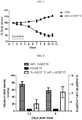
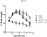
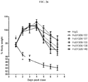
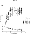
- the antibodies disclosed herein can be used to treat a variety of disorders, for example, cachexia and/or sarcopenia.
- the antibodies disclosed herein e.g., 01G06, 03G05, 04F08, 06C11, 08G01, 14F11, or 17B11
- the antibodies disclosed herein are used to inhibit the loss of muscle mass, for example, the loss of muscle mass associated with an underlying disease.
- Underlying diseases associated with cachexia include, but are not limited to, cancer, chronic heart failure, chronic kidney disease, COPD, AIDS, multiple sclerosis, rheumatoid arthritis, sepsis, and tuberculosis.
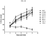
- the disclosed antibodies inhibit loss of muscle mass by at least 40%, 50%, 60%, 70%, 80%, 90%, 95%, 98%, 99%, or 100%.
- a loss of muscle mass is accompanied by a loss of fat mass.
- the antibodies disclosed herein e.g., 01G06, 03G05, 04F08, 06C11, 08G01, 14F11, or 17B11
- the antibodies disclosed herein are used to treat one or more features accompanying cachexia and/or sarcopenia, e.g., involuntary body weight loss.
- the antibodies revert involuntary body weight loss by at least 2%, 5%, 10%, 15%, 20%, 25%, 30% or 35%.
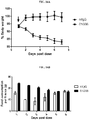
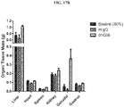
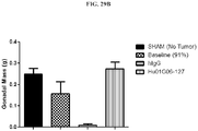
- the antibodies disclosed herein are used to inhibit loss of organ mass, for example, loss of organ mass associated with an underlying disease.
- Underlying diseases associated with cachexia include, but are not limited to, cancer, chronic heart failure, chronic kidney disease, COPD, AIDS, multiple sclerosis, rheumatoid arthritis, sepsis, and tuberculosis.
- the disclosed antibodies inhibit loss of organ mass by at least 40%, 50%, 60%, 70%, 80%, 90%, 95%, 98%, 99%, or 100%.
- loss of organ mass is observed in heart, liver, kidney, and/or spleen.
- the loss of organ mass in accompanied by a loss of muscle mass, a loss of fat mass and/or involuntary weight loss.
- Antibody 01G06, 03G05, 04F08, 06C11, 08G01, 14F11, or 17B11 can be used in therapy.
- antibody 01G06, 03G05, 04F08, 06C11, 08G01, 14F11, or 17B11 can be used to treat cachexia and/or sarcopenia.
- Use of antibody 01G06, 03G05, 04F08, 06C11, 08G01, 14F11, or 17B11 to treat cachexia and/or sarcopenia in a mammal comprises administering to the mammal a therapeutically effective amount of the antibody.
- one or more of the anti-GDF antibodies of the invention can be used to treat a subject suffering from, or who has been diagnosed with, sarcopenia, a muscle wasting disorder and/or significant muscle weight loss, whether or not the subject has, or has been diagnosed with, cachexia or decreased appetite.
- Such a method comprises administering a therapeutically effective amount of one or more antibodies of the invention to the subject in need thereof.
- the Fc-rhGDF15 fusion proteins disclosed herein can be used to treat obesity.
- the hFc-rhGDF15 fusion proteins disclosed herein are used to inhibit weight gain or to reduce body weight by at least 5%, 10%, 15%, 20%, 25%, 30%, 35%, 40%, 45% or 50%.
- Use of an hFc-hGDF15 fusion protein to treat obesity in a mammal comprises administering to the mammal a therapeutically effective amount of the fusion protein.
- treat means the treatment of a disease in a mammal, e.g., in a human. This includes: (a) inhibiting the disease, i.e., arresting its development; and (b) relieving the disease, i.e., causing regression of the disease state.
- a therapeutically effective amount of an active component is in the range of 0.1 mg/kg to 100 mg/kg, e.g., 1 mg/kg to 100 mg/kg, e.g., 1 mg/kg to 10 mg/kg, e.g., 2.0 mg/kg to 10 mg/kg.
- the amount administered will depend on variables such as the type and extent of disease or indication to be treated, the overall health of the patient, the in vivo potency of the antibody or fusion protein, the pharmaceutical formulation, the serum half-life of the antibody or fusion protein, and the route of administration.
- the initial dosage can be increased beyond the upper level in order to rapidly achieve the desired blood-level or tissue level.
- the initial dosage can be smaller than the optimum, and the dosage may be progressively increased during the course of treatment.
- Human dosage can be optimized, e.g., in a conventional Phase I dose escalation study designed to run from 0.5 mg/kg to 20 mg/kg.
- Dosing frequency can vary, depending on factors such as route of administration, dosage amount, serum half-life of the antibody or fusion protein, and the disease being treated. Exemplary dosing frequencies are once per day, once per week and once every two weeks. In some embodiments, dosing is once every two weeks.
- a preferred route of administration is parenteral, e.g., intravenous infusion. Formulation of monoclonal antibody-based drugs and fusion protein-based drugs are within ordinary skill in the art.
- the antibody or fusion protein is lyophilized, and then reconstituted in buffered saline, at the time of administration.
- a second active agent for example, an anti-cancer agent or the other agents discussed below, will also follow the principles discussed hereinabove and will be chosen so as to elicit the required therapeutic benefit in the patient.
- an antibody preferably is combined with a pharmaceutically acceptable carrier.
- pharmaceutically acceptable carrier means buffers, carriers, and excipients suitable for use in contact with the tissues of human beings and animals without excessive toxicity, irritation, allergic response, or other problem or complication, commensurate with a reasonable benefit/risk ratio.
- the carrier(s) should be “acceptable” in the sense of being compatible with the other ingredients of the formulations and not deleterious to the recipient.
- Pharmaceutically acceptable carriers include buffers, solvents, dispersion media, coatings, isotonic and absorption delaying agents, and the like, that are compatible with pharmaceutical administration. The use of such media and agents for pharmaceutically active substances is known in the art.
- compositions containing antibodies or fusion proteins can be presented in a dosage unit form and can be prepared by any suitable method.
- a pharmaceutical composition should be formulated to be compatible with its intended route of administration. Examples of routes of administration are intravenous (IV), intradermal, inhalation, transdermal, topical, transmucosal, and rectal administration. A preferred route of administration for monoclonal antibodies is IV infusion.
- Useful formulations can be prepared by methods known in the pharmaceutical art. For example, see Remington's Pharmaceutical Sciences, 18th ed. (Mack Publishing Company, 1990 ).
- Formulation components suitable for parenteral administration include a sterile diluent such as water for injection, saline solution, fixed oils, polyethylene glycols, glycerine, propylene glycol or other synthetic solvents; antibacterial agents such as benzyl alcohol or methyl paraben; antioxidants such as ascorbic acid or sodium bisulfite; chelating agents such as EDTA; buffers such as acetates, citrates or phosphates; and agents for the adjustment of tonicity such as sodium chloride or dextrose.
- a sterile diluent such as water for injection, saline solution, fixed oils, polyethylene glycols, glycerine, propylene glycol or other synthetic solvents
- antibacterial agents such as benzyl alcohol or methyl paraben
- antioxidants such as ascorbic acid or sodium bisulfite
- chelating agents such as EDTA
- buffers such as acetates, citrates or phosphates
- suitable carriers include physiological saline, bacteriostatic water, Cremophor ELTM (BASF, Parsippany, NJ) or phosphate buffered saline (PBS).
- the carrier should be stable under the conditions of manufacture and storage, and should be preserved against microorganisms.
- the carrier can be a solvent or dispersion medium containing, for example, water, ethanol, polyol (for example, glycerol, propylene glycol, and liquid polyetheylene glycol), and suitable mixtures thereof.
- compositions preferably are sterile. Sterilization can be accomplished, for example, by filtration through sterile filtration membranes. Where the composition is lyophilized, filter sterilization can be conducted prior to or following lyophilization and reconstitution.
- cytokines implicated in cachexia include Activin A and IL-6. Increased activin levels have been associated with cancer-associated cachexia and gonadal tumors. See, e.g., Marino et al. (2013) CYTOKINE & GROWTH FACTOR REV. 24:477-484 .
- Activin A is a member of the TGF-beta family, and is a ligand of the activin type 2 receptor, ActRIIB. See, e.g., Zhou et al. (2010) CELL 142:531-543 . Circulating levels of IL-6 have been shown to correlate with weight loss in cancer patients, as well as with reduced survival. See, e.g., Fearon et al. (2012) CELL METABOLISM 16:153-166 .
- one or more inhibitors of Activin-A or the Activin-A receptor, ActRIIB, IL-6 or the IL-6 receptor (IL-6R) may be administered in combination with (for example, administered at the same time as, administered before, or administered after) an antibody of the present invention that inhibits GDF-15 activity.
- Exemplary inhibitors of Activin A or ActRIIB include, for example, an anti-Activin-A antibody or an antigen binding fragment thereof, an anti-ActRIIB antibody or an antigen binding fragment thereof, a small molecule inhibitor of Activin-A, a small molecule inhibitor of ActRIIB, and a 'decoy' receptor of ActRIIB, such as a soluble ActRIIB receptor and a fusion of the soluble ActRIIB receptor with an Fc molecule (ActRIIB-Fc). See, for example, Zhou et al. (2010), supra.
- Suitable inhibitors of IL-6 or IL-6R include an anti-IL-6 antibody or an antigen binding fragment thereof, an anti-IL-6R antibody or an antigen binding fragment thereof, a small molecule inhibitor of IL-6, a small molecule inhibitor of IL-6R, and a 'decoy' receptor of IL-6R, such as a soluble IL-6 receptor and a fusion of the soluble IL-6 receptor with an Fc molecule (IL6R-Fc).
- IL6R-Fc Fc molecule
- Suitable inhibitors of IL-6 or IL-6R may include, for example, Tocilizumab (Actemra®, Hoffmann-LaRoche), a humanized anti-IL-6R monoclonal antibody approved for treatment of rheumatoid arthritis, and Sarilumab/REGN88 (Regeneron), a humanized anti-IL6R antibody in clinical development for treatment of rheumatoid arthritis; and Selumetinib/ AZD6244 (AstraZeneca), an allosteric inhibitor of MEK, which has been shown to inhibit IL-6 production. Prado et al. (2012) BRITISH J. CANCER 106:1583-1586 .
- TNF ⁇ and IL-1 are cytokines known to be involved in mediation of the proinflammatory response, which are also implicated in muscle depletion, anorexia and cachexia. Increased circulating levels of TNF ⁇ appear to inhibit myogenesis. TNF ⁇ , also known as "cachectin,” stimulates interleukin-1 secretion and is implicated in the induction of cachexia. IL-1 is a potent trigger of the acute-phase inflammatory response, and it has been shown that infusion of IL-1 can lead to marked weight loss and appetite loss. IL-1 has been shown to contribute to the initiation of cancer cachexia in mice bearing a murine colon-26 adenocarcinoma ( Strassmann et al. (1993) J. IMMUNOL.
- TNF ⁇ inhibitors and IL-1 inhibitors that are used in the treatment of rheumatoid arthritis may also be useful in the treatment of cachexia.
- one or more inhibitors of TNF ⁇ or IL-1 may be administered in combination with (for example, administered at the same time as, administered before, or administered after) an antibody of the present invention that inhibits GDF-15 activity.
- Suitable inhibitors of TNF ⁇ or IL-1 include an anti-TNFa antibody or an antigen binding fragment thereof, an anti-IL-1 antibody or an antigen binding fragment thereof, a small molecule inhibitor of TNF ⁇ or IL-1, and a 'decoy' receptor of TNF ⁇ or IL-1, such as a soluble TNF ⁇ or IL-1 receptor and a fusion of the soluble form of TNF ⁇ or IL-1 with an Fc molecule.
- Suitable inhibitors of TNF ⁇ include for example, etanercept (Enbrel®, Pfizer/Amgen), infliximab (Remicade®, Janssen Biotech), adalimumab (Humira®, Abbvie), golimumab (Simponi®, Johnson and Johnson/Merck), and certolizumab pegol (Cimzia®, UCB).
- Suitable IL-1 inhibitors include, for example, Xilonix® antibody that targets IL-1 ⁇ (XBiotech), anikinra (Kinaret®, Amgen), canakinumab (Ilaris®, Novartis), and rilonacept (Arcalyst®, Regeneron).
- the TNF ⁇ inhibitor or IL-1 inhibitor which is typically administered systemically for the treatment of rheumatoid arthritis may be administered locally and directly to the tumor site.
- Myostatin also known as GDF-8, is a member of the TGF- ⁇ family of peptides that is a negative regulator of muscle mass, as shown by increased muscle mass in myostatin deficient mammals.
- Myostatin is a ligand of the activin type 2 receptor, ActRIIB. Accordingly, in certain embodiments of the present invention, one or more inhibitors of myostatin or its receptor may be administered in combination with (for example, administered at the same time as, administered before, or administered after) an antibody of the invention that inhibits GDF-15 activity.
- Suitable inhibitors of myostatin or ActRIIB include an anti-myostatin antibody or an antigen binding fragment thereof, an anti-ActRIIB antibody or an antigen binding fragment thereof, a small molecule inhibitor of myostatin, a small molecule inhibitor of ActRIIB, and a 'decoy' receptor of GDF-8, such as a soluble ActRIIB and a fusion of the soluble form of ActRIIB with an Fc molecule. See, e.g., Lokireddy et al. (2012) BIOCHEM. J. 446(1):23-26 .
- Myostatin inhibitors that may be suitable for the present invention include REGN1033 (Regeneron); see Bauerlein et al. (2013) J.
- Ghrelin or ghrelin mimetics may be useful for increasing food intake and body weight in humans.
- GHS-R1a growth hormone secretagogues
- Suitable ghrelin mimetics include anamorelin (Helsinn, Lugano, CH); See Temel et al. (2013) J.
- GHS molecules can be identified, for example, using the growth hormone secretagogue receptor Ghrelin competition assay described in PCT Publication Nos. WO2011/117254 and WO2012/113103 .
- Agonists of the androgen receptor including small molecules and other selective androgen receptor modulators (SARMs) may be useful in treating cachexia and/or sarcopenia. See, e.g., Mohler et al. (2009) J. MED. CHEM. 52:3597-3617 ; Nagata et al. (2011) BIOORGANIC AND MED. CHEM. LETTERS 21:1744-1747 ; and Chen et al. (2005) MOL. INTERV. 5:173-188 .
- SARMs should act as full agonists, like testosterone, in anabolic target tissues, such as muscle and bone, but should demonstrate only partial or pure androgen receptor antagonistic activities on prostate tissue.
- Suitable SARMs can be identified, for example, by use of the methods and assays described in Zhang et al. (2006) BIOORG. MED. CHEM. LETT. 16:5763-5766 ; and Zhang et al. (2007) BIOORG. MED. CHEM. LETT. 17:439-443 .
- Suitable SARMs include, for example, GTx-024 (enobosarm, Ostarine®, GTx, Inc.), a SARM in phase II clinical development by GTx, Inc. See also, Dalton et al. (2011) J.
- SARMs include 2-(2,2,2)-trifluoroethyl-benzimidazoles ( Ng et al. (2007) BIOORG. MED. CHEM. LETT. 17:1784-1787 ) and JNJ-26146900 ( Allan et al. (2007) J. STEROID BIOCHEM. & MOL. BIOL. 103:76-83 ).
- Beta-blocker MT-102 (PsiOxus Therapeutics, Ltd.) has been evaluated in a phase 2 clinical trial for subjects with cancer cachexia. See Coats et al. (2011) J. CACHEXIA SARCOPENIA MUSCLE 2:201-207 .
- one or more inhibitors of a melanocortin peptide or a melanocortin receptor may be administered in combination (for example, administered at the same time as, administered before, or administered after) with an antibody of the invention that inhibits GDF-15 activity.
- Suitable inhibitors of melanocortins or melanocortin receptors include an anti-melanocortin peptide antibody or an antigen binding fragment thereof, an anti-melanocortin receptor antibody or an antigen binding fragment thereof, a small molecule inhibitor of a melanocortin peptide, a small molecule inhibitor of a melanocortin receptor, and a 'decoy' receptor of a melanocortin receptor, such as soluble melanocortin receptor and a fusion of a soluble melanocortin receptor with an Fc molecule.
- Suitable melacortin receptor inhibitors include, for example, the melanocortin receptor antagonist agouri-related peptide (AgRP(83-132)), which has been demonstrated to prevent cachexia-related symptoms in a mouse model of cancer-related cachexia ( Joppa et al. (2007) PEPTIDES 28:636-642 ).
- AgRP(83-132) the melanocortin receptor antagonist agouri-related peptide
- Anti-cancer agents especially those that can cause cachexia and elevate GDF-15 levels, such as cisplatin, may be used in methods of the present invention in combination with (for example, administered at the same time as, administered before, or administered after) an anti-GDF-15 antibody of the invention.
- Many cancer patients are weakened by harsh courses of radio- and/or chemotherapy, which can limit the ability of the patient to tolerate such therapies, and hence restrict the dosage regimen.
- Certain cancer agents themselves, such as fluorouracil, Adriamycin, methotrexate and cisplatin may contribute to cachexia, for example by inducing severe gastrointestinal complications. See, e.g., Inui (2002) CANCER J. FOR CLINICIANS 52:72-91 .
- an anti-cancer agent is administered in combination with an anti-GDF-15 antibody of the invention, it is possible to decrease the incidence and/or severity of cachexia, and ultimately increase the maximum tolerated dose of such an anti-cancer agent. Accordingly, efficacy of treatment with anti-cancer agents that may cause cachexia can be improved by reducing the incidence of cachexia as a dose-limiting adverse effect, and by allowing administration of higher doses of a given anti-cancer agent.
- the present invention includes pharmaceutical compositions comprising an anti-GDF-15 antibody of the present invention in combination with an agent selected from the group consisting of: an inhibitor of Activin-A, an inhibitor of ActRIIB, an inhibitor of IL-6 or an inhibitor of IL-6R, a ghrelin, a ghrelin mimetic or a GHS-R1a agonist, a SARM, a TNF ⁇ inhibitor, an IL-1 ⁇ inhibitor, a myostatin inhibitor, a beta-blocker, a melanocortin peptide inhibitor, a melanocortin receptor inhibitor, and an anti-cancer agent.
- an agent selected from the group consisting of: an inhibitor of Activin-A, an inhibitor of ActRIIB, an inhibitor of IL-6 or an inhibitor of IL-6R, a ghrelin, a ghrelin mimetic or a GHS-R1a agonist, a SARM, a TNF ⁇ inhibitor, an IL-1 ⁇ inhibitor,
- the present invention also includes methods of treating, preventing or minimizing cachexia and/or sarcopenia in a mammal comprising administering to a mammal in need thereof a pharmaceutical composition or compositions comprising an effective amount of an anti-GDF-15 antibody of the invention in combination with an effective amount of an inhibitor of Activin-A, an inhibitor of ActRIIB, an inhibitor of IL-6 or an inhibitor of IL-6R, a ghrelin, a ghrelin mimetic or a GHS-R1a agonist, a SARM, a TNF ⁇ inhibitor, an IL-1 ⁇ inhibitor, a myostatin inhibitor, a beta-blocker, a melanocortin peptide inhibitor, or a melanocortin receptor inhibitor.
- the invention comprises a method of inhibiting loss of muscle mass associated with an underlying disease comprising administering to a mammal in need thereof a pharmaceutical composition or compositions comprising an effective amount of an anti-GDF-15 antibody of the invention in combination with an effective amount of an inhibitor of Activin-A, an inhibitor of ActRIIB, an inhibitor of IL-6 or an inhibitor of IL-6R, a ghrelin, a ghrelin mimetic or a GHS-R1a agonist, a SARM, a TNF ⁇ inhibitor, an IL-1 ⁇ inhibitor, a myostatin inhibitor, a beta-blocker, a melanocortin peptide inhibitor, or a melanocortin receptor inhibitor to prevent or reduce loss of muscle mass.
- the underlying disease may be selected from the group consisting of cancer, chronic heart failure, chronic kidney disease, COPD, AIDS, multiple sclerosis, rheumatoid arthritis, sepsis, and tuberculosis. Additionally, in certain embodiments, the loss of muscle mass is accompanied by a loss of fat mass.
- the present invention comprises a method of inhibiting or reducing involuntary weight loss in a mammal comprising administering to a mammal in need thereof a pharmaceutical composition or pharmaceutical compositions comprising an effective amount of an anti-GDF-15 antibody of the invention in combination with an effective amount of an inhibitor of Activin-A, an inhibitor of ActRIIB, an inhibitor of IL-6 or an inhibitor of IL-6R, a ghrelin, a ghrelin mimetic or a GHS-R1a agonist, a SARM, a TNF ⁇ inhibitor, a IL-1 ⁇ inhibitor, a myostatin inhibitor, a beta-blocker, a melanocortin peptide inhibitor, or a melanocortin receptor inhibitor.
- the present invention comprises a method of treating cancer, while preventing, minimizing or reducing the occurrence, frequency or severity of cachexia, sarcopenia, or muscle wasting, bone wasting or involuntary loss of body weight in a mammal, comprising administering to a mammal in need thereof a pharmaceutical composition comprising an effective amount of an anti-GDF-15 antibody of the present invention in combination with with one or more anti-cancer agents.
- the invention comprises a method of treating cancer, while preventing, minimizing or reducing the occurrence, frequency or severity of cachexia, sarcopenia or muscle wasting, bone wasting or involuntary loss of body weight in a mammal, comprising administering to a mammal in need thereof a pharmaceutical composition comprising an effective amount of an anti-GDF-15 antibody of the invention in combination with one or more anti-cancer agents known to cause or increase the occurrence, frequency or severity of cachexia, sarcopenia, or muscle wasting, bone wasting or involuntary loss of body weight in a mammal.
- the amount of hGDF15 in the serum of mice bearing various xenograft tumors was measured. Serum was collected from three mice for each of the following tumor xenograft models: Chago, RPMI7951, PC3, TOV21G, HT-1080, K-562, and LS1034. Serum was also collected from three na ⁇ ve mice as a control. Human GDF15 serum levels were determined by ELISA (R&D Systems, Cat. No. DY957E).
- mice bearing human xenograft tumors that induce cachexia had serum levels of hGDF15 above 2 ng/mL, while mice bearing human xenograft tumors that do not induce cachexia had serum levels of hGDF15 below 1ng/mL ( FIG. 2 ). Naive mice had no detectable hGDF15 (control). These results indicate that a serum level of approximately 2 ng/mL GDF15 is a threshold for inducing cachexia in this mouse model. Similar levels of hGDF15 were also observed in plasma when determined by ELISA.
- rhGDF15 corresponds to amino acids 197 to 308 of hGDF15 protein.
- Mature rhGDF15 can be produced in the yeast Pichia pastoris as described in Fairlie et al. (2000) GENE 254:67-76 ).
- Cleaved-rhGDF15 corresponds to amino acids 197 to 308 of hGDF15 protein released from an Fc-rhGDF15 fusion protein.
- cleaved-rhGDF15 was produced by enzymatic digestion of mFc-rhGDF15 fusion protein with Factor Xa, and subsequent purification, prior to injection in mice.
- cleaved-rhGDF15 plasma was collected from a group of three mice after single dose of cleaved-rhGDF15 (1 ⁇ g/g) at different time points (2, 5, 8, 11, and 23 hours).
- Human GDF15 plasma levels were determined by ELISA (R&D Systems, Cat. No. DY957E).
- cleaved-rhGDF 15 was rapidly cleared from the plasma following injection. Eleven hours post-injection, the amount of cleaved-rhGDF15 in the plasma was below 10 ng/mL, and, within 23-hours, cleaved-rhGDF15 was almost completely cleared from the plasma.
- mice Eight-week old female ICR-SCID mice were randomized into two groups of ten mice each. Mice were dosed subcutaneously into the flank every eight hours for three days (a total of nine doses) with one of the following treatments: PBS (control) or cleaved-rhGDF15 at 1 ⁇ g/g. Body weight was measured daily. Statistical analyses were performed using a two-way ANOVA.
- cleaved-rhGDF15 induced body weight loss. After nine doses over a three day period, percent body weight dropped to 88% at day 4 (p ⁇ 0.001), but approximately 24 hours after the last dose the mice began to gain weight. On day 6, the last day of the experiment, percent body weight increased to 94.8 percent (p ⁇ 0.001). These results indicate that weight loss induced by cleaved-rhGDF 15 is not sustained over long periods of time.
- the activity observed with cleaved-rhGDF 15 described herein was similar to that observed with mature rhGDF15 in the existing mouse model (Johnen et al., supra ).
- mice treated with multiple doses of PBS demonstrated a body weight drop due to the stress of repeated dosing and handling.
- rhGDF 15 In view of the large amounts of mature rhGDF 15 (or cleaved-rhGDF 15) and the labor intensity required to induce non-tumor bearing cachexia mouse models (as well as the resulting limitations of these models), we investigated alternate forms of rhGDF 15 to induce a cachetic phenotype in mice.
- This Example describes the construction and production of two fusion proteins consisting of GDF15 and an immunoglobulin Fc fragment, designated mFc-rhGDF15 (mouse IgG1 Fc fused to the amino terminus of mature human GDF15) and rFc-rmGDF15 (rabbit IgG1 Fc fused to the amino terminus of mature mouse GDF15).
- the GDF15 fusion proteins were designed using methods known in the art.
- the mFc-rhGDF15 DNA sequences were constructed from fragments using overlap extension PCR to include (in the following order): 5' HindIII restriction site, Kozak consensus sequence, amino terminal signal sequence, mouse IgG1 Fc, Factor Xa cleavage site, a polypeptide linker (GGGGS) (SEQ ID NO: 139), mature hGDF15, stop codon, and a 3' EcoRI restriction site.
- the rFc-rmGDF15 amino acid sequences were converted to codon-optimized DNA sequences and synthesized to include (in the following order): 5' HindIII restriction site, Kozak consensus sequence, amino terminal signal sequence, rabbit IgG1 Fc, a polypeptide linker (GGGG) (SEQ ID NO: 265), mature mouse GDF15, stop codon, and a 3' EcoRI restriction site.
- the GDF15 fusion proteins were subcloned into the mammalian expression vector pEE14.4 (Lonza, Basel, Switzerland) via HindIII and EcoRI sites using In-FusionTM PCR cloning (Clontech, Mountain View, CA). GDF15 fusion proteins were stably expressed in CHOK1SV cells using the GS SystemTM (Lonza Biologics) in order to produce large quantities of purified protein. Each expression vector was linearized and transfected into CHOK1SV cells. Stable clones were selected in the presence of methionine sulfoximine. Secreted proteins produced by CHOK1SV stably transfected cell lines were purified by Protein A and size exclusion chromatography.
- mFc-rhGDF15 contains mouse IgG1 Fc from amino acids 1-222, Factor Xa cleavage site from amino acids 223-228, an artificial linker sequence from amino acids 229-233, and mature hGDF15 from amino acids 234-345.
- This Example describes the generation of an Fc-GDF15-induced cachexia model in mice.
- Immune-competent (Balb/C) and immune-incompetent (CB17-Scid) mice were randomized into three groups of ten mice each. Each group received one of the following treatments: PBS (control), mFc-rhGDF15 (as described in Example 3), or rFc-rmGDF15 (as described in Example 3) at 1 ⁇ g/g.
- Eight-week old female mice were dosed subcutaneously into the flank for three days (Balb/C) or once (CB17-Scid). Body weight was measured daily.
- fusion proteins mFc-rhGDF15 and rFc-rmGDF15
- ICR-SCID immune-incompetent mice strains.
- the administration of mFc-rhGDF15 or rFc-rmGDF15 induced cachexia, as measured by body weight loss. Similar results were obtained regardless of whether mFc-rhGDF15 was dosed subcutaneously or intraperitoneally.
- mice of different ages 7, 13 and 25 weeks old were divided into two groups of ten and treated with three doses per day of mFc-rhGDF15 or PBS (0.8 ⁇ g/g, 7 week old mice; 0.6 ⁇ g/g, 13 week old mice; or 0.4 ⁇ g/g, 25 week old mice).
- mFc-rhGDF15-induced weight loss was observed in all three mice age populations. In each age population, the mice lost approximately 10% of their body weight following treatment with mFc-rhGDF15 measured at ten days post treatment.
- mFc-rhGDF15 induction of cachexia was investigated by measuring the loss of body weight, the loss of muscle mass, the loss of fat mass, and the expression levels of two molecular markers indicative of muscle degradation (i.e., mMuRF 1 and mAtrogin).
- MuRF1 and Atrogin are E3-ubquitin ligases that are upregulated in multiple models of muscle atrophy and cachexia ( Glass, D. (2010) CURR. OPIN. CLIN. NUTR. MET. CARE 13 :225-229 ).
- mice Eight-week old female ICR-SCID mice were randomly divided into ten groups of ten mice each. Five groups (ten mice each) were dosed subcutaneously in the flank with PBS (control) and five groups (ten mice each) were dosed subcutaneously in the flank with mFc-rhGDF15 at 1.6 ⁇ g/g on day one. Body weight was measured daily for up to 17 days. One control group and one treatment group were sacrificed at different time points (0, 1, 3, 7 and 16 days post dose). Gonadal fat and gastrocnemius muscles were removed surgically from each group of mice at the indicated sacrifice time, and weighed. Tissues were snap frozen in liquid nitrogen, and RNA was isolated from the gastrocnemius muscle samples. Levels of mMuRF1 and mAtrogin mRNA were measured by qRT-PCR in samples corresponding to groups collected after 1, 7, and 16 days post dose. Statistical analyses were performed using a two-way ANOVA.
- mFc-rhGDF15 induced body weight loss in ICR-SCID mice. Percent body weight was 79.4 percent when measured after 16 days following one dose of mFc-rhGDF15 (p ⁇ 0.001). mFc-rhGDF15 also induced loss of fat (adipose tissue), as observed by the loss of gonadal fat ( FIG. 6B ; p ⁇ 0.01 at day 7 and p ⁇ 0.001 at day 16) and loss of muscle, as observed by the loss of gastrocnemius muscle ( FIG. 6C ; p ⁇ 0.05 at days 1 and 3, and p ⁇ 0.0001 at days 7 and 16).
- mFc-rhGDF15 also elevated gene expression of two enzymes associated with muscle degradation and cachexia, mMuRF1 ( FIG. 6D ; 6 ⁇ 0.001 at days 1, 7, and 16) and mAtrogin ( FIG. 6E ; p ⁇ 0.001 at days 1 and 7, and p ⁇ 0.01 at day 16).
- the serum hGDF15 levels were measured following administration of mFc-rhGDF15, to determine the half-life of rhGDF15 in this model.
- Eight-week old female Balb/C nude mice were randomly divided into two groups of twelve mice each. Mice were dosed subcutaneously in the flank every twelve hours for three days (a total of six doses) with one of the following treatments: PBS (control) or mFc-rhGDF15 at 1.33 ⁇ g/g.
- Body weight was measured daily.
- mFc-rhGDF15 induced sustained body weight loss for at least one week after the final injection.
- hGDF15 serum levels were measured 0.2, 5, and 8 days after the last dose of mFc-rhGDF15. Mice were sacrificed at the indicated time, and sera were collected. Human GDF15 serum levels were determined by ELISA (R&D Systems, Cat. No. DY957E). Table 1 provides the serum levels ( ⁇ g/mL) for each mouse in the study.
- Serum samples from day 0.2 and day 5 after the last dose were also analyzed by Western blot (reducing gel; blot with an antibody against hGDF15 (R&D Systems, Cat. No AF957)) and quantified by Licor to determine the stability of mFc-rhGDF15 in the serum. Unexpectedly, two bands were observed. The upper band was approximately 40 kDa, and appeared to be mFc-rhGDF15. The lower band was approximately 15 kDa, and appeared to be cleaved mature rhGDF15. This indicated that mature rhGDF15 was released from mFc-rhGDF15 in the serum.
- Quantification of the two bands showed that about 90% of the rhGDF15 present in the serum was in the form of mFc-rhGDF15, with about 10% of the total rhGDF15 in the serum being present as the cleaved mature form ( FIG. 8 ). Quantification showed a slight decrease in mFc-rhGDF15 in the serum samples collected five days after the last dose, but, surprisingly, a constant level of mature rhGDF15 remained in the serum. The ratio of mature rhGDF 15 to mFc-rhGDF15 slightly increased over time, as a result of a decrease in mFc-rhGDF15 in the serum. Similar results were observed when rFc-rmGDF15 was injected into mice.
- a single dose of mFc-rhGDF15 at 0.1 ⁇ g/g was sufficient to induce significant body weight loss for at least eight days ( FIG. 9A ; ten ICR-SCID mice dosed intrapertioneally with 0.1 ⁇ g/g on day 1).
- the data in Table 1 revealed that rhGDF15 serum levels were stable for at least eight days, when rhGDF15 was administered as an mFc-rhGDF15 fusion protein.
- mice should be losing weight, not gaining weight (i.e., 3.3 ng/mL total rhGDF 15 is above the threshold of approximately 2 ng/mL hGDF15).
- mice would be losing weight, not gaining weight because 3.3 ng/mL mFc-rhGDF15 is above the threshold of approximately 2 ng/mL hGDF15.
- the mice dosed with 0.1 ⁇ g/g mFc-rhGDF15 served as an internal control, because these mice had sustained body weight loss indicating that at least one of the two forms must be active.
- a calculation of 10% of 70 ng/mL total rhGDF 15 in these mice is 7 ng/mL released mature rhGDF15. This amount is consistent with inducing the observed body weight loss and the threshold observed in FIG. 2 .
- the data indicate that the mFc-rhGDF15 is not an active form of the protein, and only the mature rhGDF 15 is active.
- mFc-rhGDF15 sustains a cachetic phenotype by slowly releasing mature rhGDF15 into the serum. These results further indicate that a steady state level of mature rhGDF 15 in the plasma or serum can be achieved in a non-tumor bearing mouse by administering mFc-rhGDF15 to the mouse. Therefore, administration of mFc-rhGDF15 to non-tumor bearing mice is particularly useful as a mouse model of cachexia with a robust and sustained loss of muscle mass, loss of fat mass, and body weight loss (see FIGS. 6A-C ).
- This Example describes the production of anti-GDF15 monoclonal antibodies. Immunizations, fusions, and primary screens were conducted using conventional methods following the Repetitive Immunization Multiple Sites (RIMMS) protocol. Five AJ mice and five Balb/c mice were immunized with 6XHis (SEQ ID NO: 266) tagged recombinant human GDF15 (His-rhGDF15) (R&D Systems, Inc., Minneapolis, MN). Two Balb/c mice with sera displaying the highest anti-GDF15 activity by Enzyme Linked Immunosorbent Assay (ELISA) were chosen for subsequent fusion. Spleens and lymph nodes from the appropriate mice were harvested. B-cells were harvested and fused with a myeloma line.
- 6XHis SEQ ID NO: 266
- His-rhGDF15 tagged recombinant human GDF15
- ELISA Enzyme Linked Immunosorbent Assay
- Fusion products were serially diluted onto forty 96-well plates to near clonality. Two AJ mice with sera displaying the highest anti-GDF15 activity by ELISA were chosen for subsequent fusion. Spleens and lymph nodes from the appropriate mice were harvested. B-cells were harvested and fused with a myeloma line. Fusion products were serially diluted onto forty 96-well plates to near clonality.
- the light chain isotype and heavy chain isotype of each monoclonal antibody in Example 6 was determined using the IsoStripTM Mouse Monoclonal Antibody Isotyping Kit according the kit vendor's instructions (Roche Applied Science, Indianapolis, IN). All antibodies were found to be kappa light chain, and IgG1 or IgG2b heavy chain.
- the heavy and light chain variable regions of the mouse monoclonal antibodies were sequenced using 5' RACE (Rapid Amplification of cDNA Ends).
- Total RNA was extracted from each monoclonal hybridoma cell line using the RNeasy® Miniprep kit according to the kit vendor's instructions (Qiagen, Valencia, CA).
- Full-length first strand cDNA containing 5' ends was generated using the SMARTerTM RACE cDNA Amplification Kit (Clontech, Mountain View, CA) according to the kit vendor's instructions for 5' RACE.
- variable regions of the light (kappa) and heavy (IgG1 or IgG2b) chains were amplified by PCR using KOD Hot Start Polymerase (EMD Chemicals, Gibbstown, NJ) according to the kit vendor's instructions.
- KOD Hot Start Polymerase EMD Chemicals, Gibbstown, NJ
- the Universal Primer Mix A primer (Clontech), a mix of: 5' CTAATACGACTCACTATAGGGCAAGCAGTGGTATCAACGCAGAGT 3' (SEQ ID NO:233) and 5' CTAATACGACTCACTATAGGGC 3' (SEQ ID NO:225), was used as a 5' primer.
- Heavy chain variable regions were amplified using the above 5' primers and a 3' IgG1 constant region specific primer, 5' TATGCAAGGCTTACAACCACA 3' (SEQ ID NO:226), or a 3' IgG2b constant region specific primer, 5' AGGACAGGGGTTGATTGTTGA 3' (SEQ ID NO:227).
- Kappa chain variable regions were first amplified with the above 5' primers and a 3' kappa constant region specific primer, 5' CTCATTCCTGTTGAAGCTCTTGACAAT 3' (SEQ ID NO:228).
- the light chains were subjected to a second, nested, round of PCR using the Nested Universal Primer A (Clontech) 5' primer, 5' AAGCAGTGGTATCAACGCAGAGT 3' (SEQ ID NO:229) and a nested 3' kappa constant region specific primer, 5' CGACTGAGGCACCTCCAGATGTT 3' (SEQ ID NO:230).
- Individual PCR products were either purified using the Qiaquick® PCR Purification kit or isolated by agarose gel electrophoresis and purified using the Qiaquick® Gel Purification kit according to the kit vendor's instructions (Qiagen).
- PCR products were subsequently cloned into the pCR®4Blunt plasmid using the Zero Blunt® TOPO® PCR Cloning according to the kit vendor's instructions (Invitrogen) and transformed into DH5- ⁇ bacteria (Invitrogen) through standard molecular biology techniques. Plasmid DNA isolated from transformed bacterial clones was sequenced using M13 Forward (5' GTAAAACGACGGCCAGT 3') (SEQ ID NO:231) and M13 Reverse primers (5' CAGGAAACAGCTATGACC 3') (SEQ ID NO:232) by Beckman Genomics (Danvers, MA), using standard dideoxy DNA sequencing methods to identify the sequence of the variable region sequences. The sequences were analyzed using Vector NTI software (Invitrogen) and the IMGT/V-Quest web server (imgt.cines.fr) to identify and confirm variable region sequences.
- M13 Forward 5' GTAAAACGACGGCCAGT 3'
- M13 Reverse primers 5
- nucleic acid sequences encoding and the protein sequences defining variable regions of the murine monoclonal antibodies are shown below (amino terminal signal peptide sequences are not shown).
- CDR sequences are indicated by bold font and underlining in the amino acid sequences.
- FIG. 10 The amino acid sequences defining the immunoglobulin heavy chain variable regions for the antibodies produced in Example 6 are aligned in FIG. 10 . Amino terminal signal peptide sequences (for expression/secretion) are not shown. CDR 1 , CDR 2 , and CDR 3 (Kabat definition) are identified by boxes. FIG. 11 shows an alignment of the separate CDR 1 , CDR 2 , and CDR 3 sequences for each antibody.
- FIG. 12 The amino acid sequences defining the immunoglobulin light chain variable regions of the antibodies in Example 6 are aligned in FIG. 12 . Amino terminal signal peptide sequences (for expression/secretion) are not shown. CDR 1 , CDR 2 and CDR 3 are identified by boxes. FIG. 13 shows an alignment of the separate CDR 1 , CDR 2 , and CDR 3 sequences for each antibody.
- Table 2 shows the SEQ ID NO. of each sequence discussed in this Example.
- Table 2 SEQ. ID NO. Nucleic Acid or Protein 39 01G06 Heavy Chain Variable Region-nucleic acid 40 01G06 Heavy Chain Variable Region-protein 75 01G06 Light (kappa) Chain Variable Region-nucleic acid 76 01G06 Light (kappa) Chain Variable Region-protein 1 01G06 Heavy Chain CDR 1 7 01G06 Heavy Chain CDR 2 15 01G06 Heavy Chain CDR 3 21 01G06 Light (kappa) Chain CDR 1 26 01G06 Light (kappa) Chain CDR 2 32 01G06 Light (kappa) Chain CDR 3 41 03G05 Heavy Chain Variable Region-nucleic acid 42 03G05 Heavy Chain Variable Region-protein 77 03G05 Light (kappa) Chain Variable Region-nucleic acid 78 03G05 Light (kappa) Chain Variable Region-protein 2 03G05 Heavy Chain CDR 1 8 03G05 Heavy Chain CDR 2 16 03G05 Heavy Chain C
- each variable sequence above is combined with its respective constant region.
- a complete heavy chain comprises a heavy variable sequence followed by the murine IgG1 or IgG2b heavy chain constant sequence
- a complete kappa chain comprises a kappa variable sequence followed by the murine kappa light chain constant sequence.
- sequences represent the actual or contemplated full length heavy and light chain sequence (i.e., containing both the variable and constant regions sequences) for each antibody described in this Example.
- Signal sequences for proper secretion of the antibodies e.g., signal sequences at the 5' end of the DNA sequences or the amino terminal end of the protein sequences
- stop codons for termination of translation required at the 3' end of the DNA sequences. It is within ordinary skill in the art to select a signal sequence and/or a stop codon for expression of the disclosed full length immunoglobulin heavy chain and light chain sequences.
- the variable region sequences can be ligated to other constant region sequences to produce active full length immunoglobulin heavy and light chains.
- Table 5 shows the correspondence between the full-length sequences of the antibodies discussed in this Example with those presented in the Sequence Listing.
- Table 5 SEQ ID NO. Nucleic Acid or Protein 99 01G06_ Heavy Variable + IgG1 Constant-nucleic acid 100 01G06_ Heavy Variable + IgG1 Constant-protein 101 01G06_ Kappa Variable + Constant-nucleic acid 102 01G06_ Kappa Variable + Constant-protein 103 03G05 Heavy Variable + IgG1 Constant-nucleic acid 104 03G05 Heavy Variable + IgG1 Constant-protein 105 03G05 Kappa Variable + Constant-nucleic acid 106 03G05 Kappa Variable + Constant-protein 107 04F08 Heavy Variable + IgG1 Constant-nucleic acid 108 04F08 Heavy Variable + IgG1 Constant-protein 109 04F08 Kappa Variable + Constant-nucleic acid 110 04F08 Kappa Variable
- Rabbit anti-mouse IgGs were immobilized on carboxymethylated dextran CM4 sensor chips (GE Healthcare) by amine coupling, according to a standard protocol. Analyses were performed at 37°C using PBS containing 0.05% surfactant P20 as running buffer. The antibodies were captured in individual flow cells at a flow rate of 10 ⁇ L/minute. Injection time was varied for each antibody to yield an Rmax between 30 and 60 RU. 250 ⁇ g/mL mouse Fc (Jackson ImmunoResearch, West Grove, PA) was injected at 30 ⁇ L/minute for 120 seconds to block non-specific binding of capture antibodies to mouse Fc portion of the recombinant GDF15 protein when needed.
- Buffer, mFc-rhGDF15, cleaved-rhGDF15, His-rhGDF15, or rhGDF15 diluted in running buffer was injected sequentially over a reference surface (no antibody captured) and the active surface (antibody to be tested) for 240 seconds at 60 ⁇ L/minute.
- the dissociation phase was monitored for up to 1500 seconds.
- the surface was then regenerated with two 60-second injections of 10 mM Glycine-HCl, pH 1.7, at a flow rate of 30 ⁇ L/minute.
- the GDF15 concentration range tested was 30 nM to 0.625 nM.
- Kinetic parameters were determined using the kinetic function of the BIAevaluation software (GE Healthcare) with double reference subtraction. Kinetic parameters for each antibody, k a (association rate constant), k d (dissociation rate constant), and K D (equilibrium dissociation constant) were determined. Kinetic values of the monoclonal antibodies on mFc-rhGDF15, cleaved rhGDF15, His-rhGDF15, or rhGDF15 are summarized in Tables 6, 7, 8, and 9, respectively.
- the data in Table 6 demonstrate that antibodies bind mFc-rhGDF15 with a K D of about 250 pM or less, 200 pM or less, 150 pM or less, 100 pM or less, 75 pM or less, or 50 pM or less.
- This Example demonstrates the reversal of cachexia (as indicated by body weight loss) by antibody 01G06, 03G05, 04F08, 06C11, 14F11, or 17B11 in an mFc-rhGDF15-induced cachexia model.
- mFc-rhGDF15 (2 ⁇ g/g) was administered subcutaneously into the flank of 8-week old female ICR-SCID mice. Body weight was measured daily. When body weight reached 93%, the mice were randomized into seven groups of ten mice each. Each group received one of the following treatments: murine IgG control, 01G06, 03G05, 04F08, 06C11, 14F11, or 17B11 at 10 mg/kg. Treatment was administered once by intra-peritoneal injection.
- the data in FIG. 14 and Table 10 indicate that the disclosed anti-GDF15 antibodies can reverse cachexia in an mFc-rhGDF15-induced mouse model (i.e., a non-tumor bearing mouse model).
- Example 10 Reversal of Cachexia in an HT-1080 Xenograft Tumor Model
- This Example demonstrates the reversal of cachexia (as indicated by body weight loss) by antibody 01G06, 03G05, 04F08, 06C11, 08G01, 14F11 or 17B11 in an HT-1080 fibrosarcoma xenograft model.
- HT-1080 cells were grown in culture at 37°C in an atmosphere containing 5% CO 2 , using Eagle's Minimum Essential Medium (ATCC, Catalog No. 30-2003) containing 10% FBS. Cells were inoculated subcutaneously into the flank of 8-week old female ICR SCID mice with 5 x 10 6 cells per mouse in 50% matrigel. Body weight was measured daily. When body weight reached 93%, the mice were randomized into eight groups of ten mice each.
- the data in FIG. 15 and Table 11 indicate that the disclosed anti-GDF15 antibodies can reverse cachexia in an HT-1080 fibrosarcoma xenograft model.
- HT-1080 cells were grown and inoculated subcutaneously into the flank of 8-week old female ICR SCID mice as described above. When body weight reached 93%, the mice were randomized into two groups of ten mice each. Each group received one of the following treatments: murine IgG control or 01G06 at 10 mg/kg. Treatment was administered once by intra-peritoneal injection. As shown in FIG. 16A , treatment with antibody 01G06 resulted in body weight increase to initial weight or 100% (p ⁇ 0.001) ( FIG. 16A ).
- Water consumption was determined by weighing the water supply given to the mice daily. No significant change in water consumption was observed between groups.
- mice were sacrificed at the time of the dose (baseline or 93% body weight, without treatment) and at the end of study (seven days post dose, either mIgG or 01G06).
- Gonadal fat and the gastrocnemius muscles were removed surgically and weighed as described above in Example 4 and tissues were snap frozen in liquid nitrogen.
- RNA was isolated from the gastrocnemius muscle samples to determine the levels of mMuRF1 and mAtrogin mRNA by RT-PCR, as described in Example 4.
- mice treated with mIgG displayed significant gastrocnemius muscle loss compared to the baseline group, while the group of mice treated with antibody 01G06 did not ( FIG. 16D ).
- the levels of muscular degradation markers, mMuRF1 and mAtrogin were significantly higher in the mIgG group compared to the 01G06 group ( FIG. 16E ).
- Example 11 Reversal of Cachexia in an HT-1080 Xenograft Tumor Model
- This Example demonstrates the reversal of cachexia (as indicated by body weight loss) by antibody 01G06 in an HT-1080 fibrosarcoma xenograft model.
- HT-1080 cells were grown in culture at 37°C in an atmosphere containing 5% CO 2 , using Eagle's Minimum Essential Medium (ATCC, Catalog No. 30-2003) containing 10% FBS. Cells were inoculated subcutaneously into the flank of 8-week old female ICR SCID mice with 5 x 10 6 cells per mouse in 50% matrigel. Body weight was measured daily. When body weight reached 80%, the mice were randomized into two groups of five mice each. Each group received one of the following treatments: murine IgG control, 01G06 dosed at 2 mg/kg on day 1 and day 7.
- Treatment was administered by intra-peritoneal injection. Treatment with antibody 01G06 resulted in body weight increase relative to initial weight or about 100% (p ⁇ 0.001) ( FIG. 17A and Table 12 ).
- Table 12 Treatment % Body weight ANOVA Analysis (compared to mIgG) Gr. Agent mg/kg 1 mIgG 2 66.4 NA 2 01G06 2 97.16 p ⁇ 0.001
- FIGS. 17A-B and Table 12 indicate that the disclosed anti-GDF15 antibodies can reverse cachexia in an HT-1080 fibrosarcoma xenograft model.
- mice were sacrificed at the time of dosing (baseline or 80% body weight loss, without treatment) and at the end of study (seven days post dose, either mIgG or 01G06). Liver, heart, slpeen, kidney, gonadal fat and the gastrocnemius muscles were removed surgically and weighed. As shown in FIG. 17B , a significant loss in liver, heart, spleen, kidney, gonadal fat and gastrocnemius muscle mass was observed seven days post dose with mIgG, but not in the group treated with antibody 01G06. In addition, mice treated with antibody 01G06 displayed significant liver and gonadal muscle gain compared to the baseline group ( FIG. 17B ).
- Example 12 Reversal of Cachexia in a K-562 Xenograft Tumor Model
- This Example demonstrates the reversal of cachexia (as indicated by body weight loss) by antibody 01G06, 03G05, 04F08, 06C11, 08G01, 14F11 or 17B11 in a K-562 leukemia xenograft model.
- K-562 cells were grown in culture at 37°C in an atmosphere containing 5% CO 2 , using Iscove's Modified Dulbecco's Medium (ATCC Catalog No. 30-2005) containing 10% FBS. Cells were inoculated subcutaneously into the flank of 8-week old female CB17SCRFMF mice with 2.5 x 10 6 cells per mouse in 50% matrigel. Body weight was measured daily. When body weight reached 93%, the mice were randomly distributed into eight groups of ten mice each.
- Antibody 01G06 was tested in additional tumor xenograft models including the TOV-21G ovarian xenograft model and the LS1034 colon xenograft model. In each model, antibody 01G06 reversed body weight loss compared to a PBS control (p ⁇ 0.001 for the TOV-21G model and p ⁇ 0.01 for the LS1034 model).
- This Example describes the humanization and chimerization of three murine antibodies, designated 01G06, 06C11, and 14F11, and the characterization of the resulting humanized antibodies.
- the humanized anti-GDF15 antibodies were designed, affinity matured by targeted CDR mutagenesis, and optimized using methods known in the art.
- the amino acid sequences were converted to codon-optimized DNA sequences and synthesized to include (in the following order): 5' HindIII restriction site, Kozak consensus sequence, amino terminal signal sequence, humanized variable region, human IgG1 or Kappa constant region, stop codon, and a 3' EcoRI restriction site.
- Chimeric (murine variable region and human constant region) 01G06, 06C11, and 14F11 heavy (human IgG1) and light (human Kappa) chains were also constructed.
- the murine variable regions were fused to the human constant region, and codon-optimized DNA sequences were synthesized, including (in the following order): 5' HindIII restriction site, Kozak consensus sequence, amino terminal signal sequence, mouse variable region, human IgG1 or Kappa constant region, stop codon, and 3' EcoRI restriction site.
- the humanized and chimeric heavy chains were subcloned into pEE6.4 (Lonza, Basel, Switzerland) via HindIII and EcoRI sites using In-FusionTM PCR cloning (Clontech, Mountain View, CA).
- the humanized and chimeric Kappa light chains were subcloned into pEE14.4 (Lonza) via HindIII and EcoRI sites using In-FusionTM PCR cloning.
- Humanized antibody chains or chimeric antibody chains were transiently transfected into 293T cells to produce antibody.
- Antibody was either purified or used in cell culture media supernatant for subsequent in vitro analysis. Binding of the chimeric and humanized antibodies to human GDF15 was measured as described below. The results are summarized in Tables 24-27.
- Table 14 Antibody Name Light Chain Variable Region Heavy Chain Variable Region Hu01G06-1 Ch01G06 Chimeric Kappa (SEQ ID NO:76) Ch01G06 Chimeric Heavy (SEQ ID NO:40) Hu01G06-14 Ch01G06 Chimeric Kappa (SEQ ID NO:76) Hu01G06 IGHV1-18 Heavy (SEQ ID NO:54) Hu01G06-15 Ch01G06 Chimeric Kappa (SEQ ID NO:76) Hu01G06 IGHV1-69 Heavy (SEQ ID NO:56) Hu01G06-147 Ch01G06 Chimeric Kappa (SEQ ID NO:76) Sh01G06 IGHV1-18 M69L Heavy (SEQ ID NO:58) Hu01G06-148 Ch01G06 Chimeric Kappa (SEQ ID NO:76) Sh01G06 IGHV1-18 M69L K64Q G44S
- Table 15 Antibody Name Light Chain Variable Region Heavy Chain Variable Region Hu06C11-1 Ch06C11 Chimeric Kappa (SEQ ID NO:82) Ch06C11 Chimeric Heavy (SEQ ID NO:46) Hu06C11-7 Ch06C11 Chimeric Kappa (SEQ ID NO:82) HE LM 06C11 IGHV2-70 Heavy (SEQ ID NO:68) Hu06C11-10 Ch06C11 Chimeric Kappa (SEQ ID NO:82) Hu06C11 IGHV2-5 Heavy (SEQ ID NO:70) Hu06C11-12 Sh06C11 IGKV1-16 Kappa (SEQ ID NO:96) Ch06C11 Chimeric Heavy (SEQ ID NO:46) Hu06C11-27 Sh06C11 IGKV1-16 Kappa (SEQ ID NO:96) HE LM 06C11 IGHV2-70 Heavy (SEQ ID NO:82) Ch06C11 Chimeric Heavy (SEQ ID NO:46) Hu06C11-27 Sh06C11 IGKV1
- Table 16 Antibody Name Light Chain Variable Region Heavy Chain Variable Region Hu14F11-1 Ch14F11 Chimeric Kappa (SEQ ID NO:86) Ch14F11 Chimeric Heavy (SEQ ID NO:50) Hu14F11-14 Ch14F11 Chimeric Kappa (SEQ ID NO:86) Sh14F11 IGHV2-5 Heavy (SEQ ID NO:72) Hu14F11-15 Ch14F11 Chimeric Kappa (SEQ ID NO:86) Sh14F11 IGHV2-70 Heavy (SEQ ID NO:74) Hu14F11-11 Hu14F11 IGKV1-16 Kappa (SEQ ID NO:98) Ch14F11 Chimeric Heavy (SEQ ID NO:50) Hu14F11-39 Hu14F11 IGKV1-16 Kappa (SEQ ID NO:98 Sh14F11 IGHV2-5 Heavy (SEQ ID NO:72)
- nucleic acid sequences and the encoded protein sequences defining variable regions of the chimeric and humanized 01G06, 06C11, and 14F11 antibodies are summarized below (amino terminal signal peptide sequences are not shown).
- CDR sequences (Kabat definition) are shown in bold and are underlined in the amino acid sequences.
- FIG. 19 The amino acid sequences defining the immunoglobulin heavy chain variable regions for the antibodies produced in Example 13 are aligned in FIG. 19 . Amino terminal signal peptide sequences (for proper expression/secretion) are not shown. CDR 1 , CDR 2 , and CDR 3 (Kabat definition) are identified by boxes. FIG. 20 show an alignment of the separate CDR 1 , CDR 2 , and CDR 3 sequences for each of the variable region sequences shown in FIG. 19 .
- FIG. 21 The amino acid sequences defining the immunoglobulin light chain variable regions for the antibodies in Example 13 are aligned in FIG. 21 . Amino terminal signal peptide sequences (for proper expression/secretion) are not shown. CDR 1 , CDR 2 and CDR 3 are identified by boxes. FIG. 22 shows an alignment of the separate CDR 1 , CDR 2 , and CDR 3 sequences for each of the variable region sequences shown in FIG. 21 .
- Table 19 is a concordance chart showing the SEQ ID NO. of each sequence discussed in this Example.
- Table 19 SEQ. ID NO. Nucleic Acid or Protein 127 Ch01G06 Chimeric Heavy Chain Variable Region-nucleic acid 40 Ch01G06 Chimeric Heavy Chain Variable Region-protein 1 Ch01G06 Chimeric Heavy Chain CDR 1 7 Ch01G06 Chimeric Heavy Chain CDR 2 15 Ch01G06 Chimeric Heavy Chain CDR 3 53 Hu01G06 IGHV1-18 Heavy Chain Variable Region-nucleic acid 54 Hu01G06 IGHV1-18 Heavy Chain Variable Region-protein 1 Hu01G06 IGHV1-18 Heavy Chain CDR 1 7 Hu01G06 IGHV1-18 Heavy Chain CDR 2 15 Hu01G06 IGHV1-18 Heavy Chain CDR 3 55 Hu01G06 IGHV1-69 Heavy Chain Variable Region-nucleic acid 56 Hu01G06 IGHV1-69 Heavy Chain Variable Region-protein 1 Hu01G06 IGHV1-69 Heavy Chain CDR 1 7 Hu01G06 IGHV1-6
- Table 20 Kabat CDR1 CDR2 CDR3 Variable Region SEQ ID NO: Ch01G06 Chimeric DYNMD (SEQ ID NO:1) QINPNNGGIFFNQKFKG (SEQ ID NO:7) EAITTVGAMDY (SEQ ID NO:15) 40 Hu01G06 IGHV1-18 DYNMD (SEQ ID NO:1) QINPNNGGIFFNQKFKG (SEQ ID NO:7) EAITTVGAMDY (SEQ ID NO:15) 54 Hu01G06 IGHV1-69 DYNMD (SEQ ID NO:1) QINPNNGGIFFNQKFKG (SEQ ID NO:7) EAITTVGAMDY (SEQ ID NO:15) 56 Sh01G06 IGHV1-18 M69L DYNMD (SEQ ID NO:1) QINPNNGGIFFNQKFKG (SEQ ID NO:7) EAITTVGAMDY (SEQ ID NO:15) 56 Sh01G06 IGHV1-18 M69L DYNMD (SEQ ID NO
- each variable sequence above is combined with its respective human constant region.
- a complete heavy chain comprises a heavy variable sequence followed by a human IgG1 heavy chain constant sequence.
- a complete kappa chain comprises a kappa variable sequence followed by the human kappa light chain constant sequence.
- sequences represent the actual or contemplated full length heavy and light chain sequence (i.e., containing both the variable and constant regions sequences) for each antibody described in this Example.
- Signal sequences for proper secretion of the antibodies e.g., signal sequences at the 5' end of the DNA sequences or the amino terminal end of the protein sequences
- stop codons for termination of translation required at the 3' end of the DNA sequences. It is within ordinary skill in the art to select a signal sequence and/or a stop codon for expression of the disclosed full length immunoglobulin heavy chain and light chain sequences.
- the variable region sequences can be ligated to other constant region sequences to produce active full length immunoglobulin heavy and light chains.
- Table 22 is a concordance chart showing the SEQ ID NO. of each sequence discussed in this Example.
- Table 22 SEQ ID NO. Nucleic Acid or Protein 171 Human IgG1 constant-nucleic acid 172 Human IgG1 constant-protein 173 Human Kappa constant-nucleic acid 174 Human Kappa constant-protein 175 Humanized Ch01G06 Chimeric Heavy Human Variable + Human IgG1 constant-nucleic acid 176 Humanized Ch01G06 Chimeric Heavy Human Variable + Human IgG1 constant-protein 177 Humanized Hu01G06 IGHV1-18 Heavy Human Variable + Human IgG1 constant-nucleic acid 178 Humanized Hu01G06 IGHV1-18 Heavy Human Variable + Human IgG1 constant-protein 179 Humanized Hu01G06 IGHV1-69 Heavy Human Variable + Human IgG1 constant-nucleic acid 180 Humanized Hu01G06 IGHV1-69 Heavy Human Variable + Human IgG1 constant-protein 181 Humanized Sh01G06
- Table 23 below shows antibodies containing chimeric immunoglobulin heavy and light chains and exemplary combinations of the full-length chimeric or humanized immunoglobulin heavy and light chains.
- Table 23 Antibody Name Light Chain Heavy Chain Hu01G06-1 Humanized Ch01G06 Chimeric Human Variable + Human Kappa constant (SEQ ID NO:204) Humanized Ch01G06 Chimeric Heavy Human Variable + Human IgG1 constant (SEQ ID NO:176) Hu01G06-46 Humanized Hu01G06 IGKV1-39 Human Variable + Human Kappa constant (SEQ ID NO:206) Humanized Hu01G06 IGHV1-18 Heavy Human Variable + Human IgG1 constant (SEQ ID NO:178) Hu01G06-52 Humanized Hu01G06 IGKV1-39 Human Variable + Human Kappa constant (SEQ ID NO:206) Humanized Hu01G06 IGHV1-69 Heavy Human Variable + Human IgG1 constant (SEQ ID NO:180) Hu01G06-100 Humanized Hu01G06 IG
- the antibody constructs containing the full length chimeric heavy and light chains are designated below:
- binding affinities and kinetics of binding of chimeric and humanized antibodies to mFc-rhGDF15 were measured by surface plasmon resonance, using a BIAcore® T100 instrument (GE Healthcare, Piscataway, NJ).
- Goat anti-human IgGs (Fc fragment specific, Jackson ImmunoResearch, West Grove, PA) were immobilized on carboxymethylated dextran CM4 sensor chips by amine coupling, according to a standard protocol. Analyses were performed at 37°C using PBS containing 0.05% surfactant P20 as running buffer. The antibodies were captured in individual flow cells at a flow rate of 10 ⁇ L/minute. Injection time was varied for each antibody to yield an Rmax between 30 and 60 RU. Buffer or mFc-rhGDF15 diluted in running buffer was injected sequentially over a reference surface (no antibody captured) and the active surface (antibody to be tested) for 240 seconds at 60 ⁇ L/minute.
- the dissociation phase was monitored for up to 1200 seconds.
- the surface was then regenerated with two 60-second injections of 10 mM Glycine-HCl, pH 2.25, at a flow rate of 30 ⁇ L/minute.
- the GDF15 concentration range tested was 20 nM to 0.625 nM.
- Kinetic parameters were determined using the kinetic function of the BIAevaluation software (GE Healthcare) with double reference subtraction. Kinetic parameters for each antibody, k a (association rate constant), k d (dissociation rate constant), and K D (equilibrium dissociation constant) were determined. Kinetic values of purified monoclonal antibodies on mFc-rhGDF15 are summarized in Table 24.
- the results in Table 24 demonstrate that the chimeric and each of the humanized antibodies, have fast association rates (k a ), very slow disassociation rates (k d ) and very high affinities (K D ).
- the antibodies have affinities ranging from about 65 pM to about 200 pM.
- Binding affinities and binding kinetics of mFc-rhGDF15 with chimeric 14F11 (Hu14F11-1), chimeric light 14F11 with chimeric heavy 06C11 (Hu14F11-23), and chimeric light 06C11 with chimeric heavy 14F11 (Hu14F11-24) heavy monoclonal antibody variants (in supernatant) were measured using biolayer interferometry (BLI) on an OctetTM QK instrument (ForteBio, Inc., Menlo Park, CA). The Octet analysis was performed at 30°C using IX Kinetics Buffer (ForteBio, Inc.) as assay buffer.
- Anti-human IgG Fc Capture (AHC) biosensors (ForteBio, Inc.) were used to capture human antibodies onto the sensors. Sensors were saturated in assay buffer for at 300 seconds before the assay. Antibodies were loaded onto sensors by dipping the sensors into antibody supernatant solution for 220 seconds, which typically resulted in capture levels of 1.5-2 nm. Baseline was established by dipping the sensors into 1x assay buffer for 200 seconds. Next, association was monitored for 220 seconds in 400 nM mFc-rhGDF15 protein, and dissociation was followed for 600 seconds in buffer alone.
- AHC Anti-human IgG Fc Capture
- Hu14F11-23 and Hu14F11-24 i.e., antibodies that consist of one chimeric 06C11 chain (heavy or light) mixed with one chimeric 14F11 chain (heavy or light)
- these antibodies have high affinities ranging from about 180 pM to about 310 pM.
- the binding affinities and kinetics of binding of chimeric and humanized antibodies to mFc-rhGDF15, cleaved-rhGDF15, rabbit Fc mature recombinant mouse GDF15 (rFc-rmGDF15), and mouse Fc mature recombinant cynomolgus monkey GDF15 (mFc-rcGDF15) were measured by surface plasmon resonance, using a BIAcore® T100 instrument (GE Healthcare, Piscataway, NJ).
- Goat anti-human IgGs Fc fragment specific, Jackson ImmunoResearch, West Grove, PA
- carboxymethylated dextran CM4 sensor chips by amine coupling, according to a standard protocol.
- Analyses were performed at 37°C using PBS containing 0.05% surfactant P20 as running buffer.
- the antibodies were captured in individual flow cells at a flow rate of 10 ⁇ L/minute. Injection time was varied for each antibody to yield an Rmax between 30 and 60 RU.
- Buffer, mFc-rhGDF15, cleaved-rhGDF15, rFc-rmGDF15, or mFc-rcGDF15 diluted in running buffer was injected sequentially over a reference surface (no antibody captured) and the active surface (antibody to be tested) for 240 seconds at 60 ⁇ L/minute.
- the dissociation phase was monitored for up to 1500 seconds.
- the surface was then regenerated with two 60-second injections of 10 mM Glycine-HCl, pH 2.25, at a flow rate of 30 ⁇ L/minute.
- the GDF15 concentration range tested for each GDF15 protein was 5 nM to 0.3125 nM (two-fold dilutions).
- Kinetic parameters were determined using the kinetic function of the BIAevaluation software (GE Healthcare) with double reference subtraction. Kinetic parameters for each antibody, k a (association rate constant), k d (dissociation rate constant), and K D (equilibrium dissociation constant) were determined. Kinetic values of purified monoclonal antibodies on mFc-rhGDF15, mature human GDF15, rFc-rmGDF15, and mFc-rcGDF15 are summarized in Table 27.
- the results in Table 27 demonstrate that the chimeric and each of the humanized antibodies, have fast association rates (k a ), very slow disassociation rates (k d ) and very high affinities (K D ).
- the antibodies have affinities ranging from less than 5 pM ( e.g., about 4.5 pM) to about 65 pM.
- Example 17 Reversal of Cachexia in an HT-1080 Fibroscarcoma Xenograft Model
- This Example demonstrates the reversal of cachexia (as indicated by body weight loss) by humanized 01G06, 06C11, 14F11 antibodies in an HT-1080 fibrosarcoma xenograft model.
- HT-1080 cells were grown in culture at 37°C and inoculated subcutaneously into the flank of 8-week old female ICR-SCID mice as described above in Example 10. Body weight was measured daily. When body weight reached 93%, the mice were randomly divided into groups of ten mice each. Each group received one of the following treatments: murine IgG control, 01G06, 06C11, 14F11, and their respective humanized versions at 2 mg/kg. Treatment was administered once a day by intra-peritoneal injection.
- Antibody treatment with humanized 01G06 antibodies i.e ., antibodies Hu01G06-122, Hu01G06-127, Hu01G06-135, Hu01G06-138 and Hu01G06-146) resulted in body weight increase relative to initial weight or about 100% (p ⁇ 0.001) ( FIG. 26 ).
- Statistical analysis was performed using ANOVA. Treatment with human IgG (hIgG) was used as a control. Results for reversal of body weights in the HT-1080 model are shown in FIG. 26 and Table 31. Table 31 Gr.
- Treatment % Body weight ANOVA Analysis (compared to hIgG) Agent mg/kg 1 hIgG 2 84.2 NA 2 Hu01G06-122 2 96.3 p ⁇ 0.001 3 Hu01G06-127 2 96.1 p ⁇ 0.001 4 Hu01G06-135 2 93.5 p ⁇ 0.001 5 Hu01G06-138 2 91.9 p ⁇ 0.001 6 Hu01G06-146 2 92.7 p ⁇ 0.001
- This Example demonstrates the reversal of cachexia (as indicated by body weight loss) by humanized 01G06 antibodies (i.e ., antibody Hu01G06-122, Hu01G06-127, Hu01G06-135, Hu01G06-138, or Hu01G06-146) in an mFc-rhGDF15-induced cachexia model.
- mFc-rhGDF15 (1 ⁇ g/g) was administered subcutaneously into the flank of 8-week old female ICR-SCID mice. Body weight was measured daily. When body weight reached 93%, the mice were randomly divided into six groups of ten mice each.
- Treatment % Boby weight ANOVA Analysis (compared to hIgG) Agent mg/kg 1 hIgG 2 70.6 NA 2 Hu01G06-122 2 101.7 p ⁇ 0.001 3 Hu01G06-127 2 103.2 p ⁇ 0.001 4 Hu01G06-135 2 102.5 p ⁇ 0.001 5 Hu01G06-138 2 101.8 p ⁇ 0.001 6 Hu01G06-146 2 102.5 p ⁇ 0.001
- the data in FIG. 27 and Table 32 indicate that the disclosed anti-GDF15 antibodies can reverse cachexia in an mFc-rhGDF15-induced mouse model (i.e., a non-tumor bearing mouse model).
- Example 19 Dose Response Reversal of Cachexia in an HT-1080 Fibroscarcoma Xenograft Model
- This Example demonstrates the dose response reversal of cachexia (as indicated by body weight loss) by humanized Hu01G06-127 and Hu01G06-135 antibodies in an HT-1080 fibrosarcoma xenograft model.
- HT-1080 cells were grown in culture at 37°C and inoculated subcutaneously into the flank of 8-week old female ICR-SCID mice as described above in Example 10. Body weight was measured daily. When body weight reached 93%, the mice were randomly divided into groups of ten mice each.
- Example 20 Reversal of muscle and fat loss in an HT-1080 Xenograft Tumor Model
- This Example demonstrates the reversal of cachexia (as indicated by body weight loss, muscle mass loss and fat mass loss) by antibody 01G06 in an HT-1080 fibrosarcoma xenograft model.
- HT-1080 cells were grown in culture at 37°C in an atmosphere containing 5% CO 2 , using Eagle's Minimum Essential Medium (ATCC, Catalog No. 30-2003) containing 10% FBS. Cells were inoculated subcutaneously into the flank of 8-week old female ICR SCID mice with 5 x 10 6 cells per mouse in 50% matrigel. A cohort of ten 8-week old female ICR SCID mice with the same body weight was selected for subcutaneous inoculation into the flank with matrigel, as a non tumor (SHAM) control arm.
- SHAM non tumor
- Body weight was measured daily. When body weight reached 91% in the tumor bearing mice, the mice were randomly divided into two groups of ten mice each. Each group received one of the following treatments: human IgG control (hIgG) or Hu01G06-127 10 mg/kg on day 1, day 3 and day 6. Treatment was administered by intra-peritoneal injection. Treatment with antibody Hu01G06-127 resulted in body weight increase to 105% of initial weight compared to non-tumor bearing control mice (SHAM; p ⁇ 0.001) ( FIG. 29A and Table 34 ). Table 34 Gr. Treatment % Body weight ANOVA Analysis (compared to hIgG) Agent mg/kg 1 hIgG 10 84.3 NA 2 Hu01G06-127 10 105.4 p ⁇ 0.001 2 SHAM no tumor control none 101.9 p ⁇ 0.001
- the data in FIG. 29A and Table 34 indicate that the disclosed anti-GDF15 antibody can completely reverse cachexia in an HT-1080 fibrosarcoma xenograft model.
- mice were sacrificed at the time of dosing (baseline or 91% body weight loss, without treatment) and at the end of the study (eight days post dose, either hIgG or Hu01G06-127 as well SHAM non tumor control mice). Gonadal fat and the gastrocnemius muscles were removed surgically and weighed. As shown in FIG. 29B , significant gonadal fat mass loss was observed seven days post dose with hIgG, but not in the group treated with antibody Hu01G06-127 compared to baseline control (91% body weight loss).
Landscapes
- Health & Medical Sciences (AREA)
- Chemical & Material Sciences (AREA)
- Medicinal Chemistry (AREA)
- Life Sciences & Earth Sciences (AREA)
- General Health & Medical Sciences (AREA)
- Organic Chemistry (AREA)
- Animal Behavior & Ethology (AREA)
- Public Health (AREA)
- Veterinary Medicine (AREA)
- Pharmacology & Pharmacy (AREA)
- Immunology (AREA)
- Epidemiology (AREA)
- Engineering & Computer Science (AREA)
- Bioinformatics & Cheminformatics (AREA)
- Biochemistry (AREA)
- Biophysics (AREA)
- Proteomics, Peptides & Aminoacids (AREA)
- Molecular Biology (AREA)
- Genetics & Genomics (AREA)
- Obesity (AREA)
- Hematology (AREA)
- General Chemical & Material Sciences (AREA)
- Nuclear Medicine, Radiotherapy & Molecular Imaging (AREA)
- Chemical Kinetics & Catalysis (AREA)
- Nutrition Science (AREA)
- Diabetes (AREA)
- Microbiology (AREA)
- Mycology (AREA)
- Endocrinology (AREA)
- Peptides Or Proteins (AREA)
- Medicines Containing Antibodies Or Antigens For Use As Internal Diagnostic Agents (AREA)
- Micro-Organisms Or Cultivation Processes Thereof (AREA)
- Preparation Of Compounds By Using Micro-Organisms (AREA)
- Medicines That Contain Protein Lipid Enzymes And Other Medicines (AREA)
Applications Claiming Priority (4)
| Application Number | Priority Date | Filing Date | Title |
|---|---|---|---|
| US201261745508P | 2012-12-21 | 2012-12-21 | |
| US201361827325P | 2013-05-24 | 2013-05-24 | |
| PCT/US2013/077139 WO2014100689A1 (fr) | 2012-12-21 | 2013-12-20 | Anticorps anti-gdf15 |
| EP13827058.2A EP2934584B1 (fr) | 2012-12-21 | 2013-12-20 | Anticorps anti-gdf15 |
Related Parent Applications (1)
| Application Number | Title | Priority Date | Filing Date |
|---|---|---|---|
| EP13827058.2A Division EP2934584B1 (fr) | 2012-12-21 | 2013-12-20 | Anticorps anti-gdf15 |
Publications (1)
| Publication Number | Publication Date |
|---|---|
| EP3689370A1 true EP3689370A1 (fr) | 2020-08-05 |
Family
ID=50069279
Family Applications (2)
| Application Number | Title | Priority Date | Filing Date |
|---|---|---|---|
| EP13827058.2A Active EP2934584B1 (fr) | 2012-12-21 | 2013-12-20 | Anticorps anti-gdf15 |
| EP20156883.9A Pending EP3689370A1 (fr) | 2012-12-21 | 2013-12-20 | Anticorps anti-gdf15 |
Family Applications Before (1)
| Application Number | Title | Priority Date | Filing Date |
|---|---|---|---|
| EP13827058.2A Active EP2934584B1 (fr) | 2012-12-21 | 2013-12-20 | Anticorps anti-gdf15 |
Country Status (14)
| Country | Link |
|---|---|
| US (5) | US9175076B2 (fr) |
| EP (2) | EP2934584B1 (fr) |
| JP (3) | JP6758832B2 (fr) |
| KR (1) | KR102208861B1 (fr) |
| CN (1) | CN105073133B (fr) |
| AR (1) | AR094271A1 (fr) |
| AU (1) | AU2013364133B2 (fr) |
| CA (1) | CA2896076C (fr) |
| DK (1) | DK2934584T3 (fr) |
| EA (1) | EA038645B1 (fr) |
| ES (1) | ES2791183T3 (fr) |
| HK (1) | HK1215670A1 (fr) |
| MX (1) | MX370720B (fr) |
| WO (1) | WO2014100689A1 (fr) |
Families Citing this family (69)
| Publication number | Priority date | Publication date | Assignee | Title |
|---|---|---|---|---|
| EP4342995A3 (fr) | 2006-03-31 | 2024-05-15 | Chugai Seiyaku Kabushiki Kaisha | Procédés de régulation de la pharmacocinétique sanguine d'anticorps |
| DK2202245T3 (en) | 2007-09-26 | 2016-11-21 | Chugai Pharmaceutical Co Ltd | A method of modifying an antibody isoelectric point VIA amino acid substitution in CDR |
| EP3056513A1 (fr) | 2008-04-11 | 2016-08-17 | Chugai Seiyaku Kabushiki Kaisha | Molécule se liant à des antigènes capable de se lier à deux ou plusieurs molécules d'antigène de manière répétée |
| RU2658504C9 (ru) | 2010-11-30 | 2018-08-21 | Чугаи Сейяку Кабусики Кайся | Антигенсвязывающая молекула, способная многократно связываться с множеством антигенных молекул |
| AU2012240102C1 (en) | 2011-04-08 | 2016-09-08 | Amgen Inc. | Method of treating or ameliorating metabolic disorders using growth differentiation factor 15 (GDF-15) |
| WO2013113008A1 (fr) | 2012-01-26 | 2013-08-01 | Amgen Inc. | Polypeptides du facteur de croissance et de différenciation 15 (gdf-15) |
| US9550819B2 (en) | 2012-03-27 | 2017-01-24 | Ngm Biopharmaceuticals, Inc. | Compositions and methods of use for treating metabolic disorders |
| EP2889376A4 (fr) | 2012-08-24 | 2016-11-02 | Chugai Pharmaceutical Co Ltd | ANTICORPS Fc SPÉCIFIQUE À FcyRII DE SOURIS |
| TW202237660A (zh) | 2012-08-24 | 2022-10-01 | 日商中外製藥股份有限公司 | FcγRIIb特異性Fc區域變異體 |
| HUE044363T2 (hu) | 2012-09-26 | 2019-10-28 | Univ Wuerzburg J Maximilians | Növekedési és differenciálódási faktor 15 (GDF-15) elleni monoklonális antitestek |
| AU2013364133B2 (en) | 2012-12-21 | 2018-10-11 | Aveo Pharmaceuticals, Inc. | Anti-GDF15 antibodies |
| KR101993714B1 (ko) | 2013-01-30 | 2019-06-28 | 엔지엠 바이오파마슈티컬스, 아이엔씨. | 대사 장애를 치료하는데 이용하기 위한 조성물과 방법 |
| US9161966B2 (en) | 2013-01-30 | 2015-10-20 | Ngm Biopharmaceuticals, Inc. | GDF15 mutein polypeptides |
| WO2014163101A1 (fr) | 2013-04-02 | 2014-10-09 | 中外製薬株式会社 | Variant de région fc |
| US9862752B2 (en) | 2013-07-31 | 2018-01-09 | Amgen Inc. | Growth differentiation factor 15 (GDF-15) constructs |
| US10261094B2 (en) * | 2013-10-31 | 2019-04-16 | Regeneron Pharmaceuticals, Inc. | Competitive ligand binding assay for detecting neutralizing antibodies |
| WO2015077540A2 (fr) | 2013-11-21 | 2015-05-28 | The Brigham And Women's Hospital, Inc. | Compositions et procédés de traitement de l'hypertension pulmonaire |
| JP2017518958A (ja) | 2014-03-19 | 2017-07-13 | マッカイ メディカル ファウンデイション ザ プレスビテリアン チャーチ イン タイワン マッカイ メモリアル ホスピタルMackay Medical Foundation The Presbyterian Church In Taiwan Mackay Memorial Hospital | 免疫原性グリコペプチドに対する抗体、それを含む組成物、及びそれらの使用 |
| GB2524553C (en) * | 2014-03-26 | 2017-07-19 | Julius-Maximilians-Universitãt Würzburg | Monoclonal antibodies to growth and differentiation factor 15 (GDF-15), and uses thereof for treating cancer cachexia |
| GB2524552B (en) * | 2014-03-26 | 2017-07-12 | Julius-Maximilians-Universitãt Wurzburg | Monoclonal antibodies to growth and differentiation factor 15 (GDF-15), and use thereof for treating cancer |
| PT3122775T (pt) | 2014-03-26 | 2019-12-26 | Univ Wuerzburg J Maximilians | Anticorpos monoclonais do fator de crescimento e diferenciação 15 (gdf-15) e suas utilizações para o tratamento da caquexia cancerosa e do cancro |
| US20170137505A1 (en) * | 2014-06-20 | 2017-05-18 | Aveo Pharmaceuticals, Inc. | Treatment of congestive heart failure and other cardiac dysfunction using a gdf15 modulator |
| MX2016016826A (es) | 2014-06-20 | 2017-08-28 | Aveo Pharmaceuticals Inc | Tratamiento de la enfermedad renal cronica y otra disfuncion renal utilizando un modulador gdf15. |
| US10588980B2 (en) | 2014-06-23 | 2020-03-17 | Novartis Ag | Fatty acids and their use in conjugation to biomolecules |
| PE20170771A1 (es) | 2014-07-30 | 2017-07-04 | Ngm Biopharmaceuticals Inc | Composiciones y metodos de uso para tratar trastornos metabolicos |
| WO2016019368A1 (fr) * | 2014-08-01 | 2016-02-04 | The Brigham And Women's Hospital, Inc. | Méthodes et compositions relatives au traitement de l'hypertension artérielle pulmonaire |
| EP3197493B1 (fr) | 2014-09-25 | 2021-03-10 | Aveo Pharmaceuticals Inc. | Procédés d'inversion de la cachexie et de prolongation de la survie, comprenant l'administration d'un modulateur de gdf15 et d'un agent anticancéreux |
| US20170290885A1 (en) * | 2014-09-30 | 2017-10-12 | Universiteit Gent | A method of treating joint disease |
| AU2015339134B2 (en) | 2014-10-30 | 2021-06-17 | Acceleron Pharma Inc. | Methods and compositions using GDF15 polypeptides for increasing red blood cells |
| MX357994B (es) | 2014-10-31 | 2018-08-01 | Ngm Biopharmaceuticals Inc | Composiciones y metodos de uso para tratar trastornos metabolicos. |
| KR20180054923A (ko) | 2014-12-19 | 2018-05-24 | 추가이 세이야쿠 가부시키가이샤 | 항-마이오스타틴 항체, 변이체 Fc 영역을 함유하는 폴리펩타이드, 및 사용 방법 |
| EP3816179A3 (fr) | 2015-02-05 | 2021-08-04 | Chugai Seiyaku Kabushiki Kaisha | Variant de région fc comprenant un domaine de liaison fcrn modifié |
| EP4368641A2 (fr) * | 2015-04-23 | 2024-05-15 | HaemaLogiX Ltd. | Récepteurs antigéniques chimériques d'antigène de myélome kappa et leurs utilisations |
| CN106349388B (zh) * | 2015-07-17 | 2021-04-02 | 上海佳文英莉生物技术有限公司 | 一种促细胞程序性坏死抗体及其应用 |
| GB201512733D0 (en) * | 2015-07-20 | 2015-08-26 | Genagon Therapeutics Ab | Therapeutic agents for treating conditions associated with elevated GDF15 |
| FI3356827T3 (fi) | 2015-10-02 | 2024-01-03 | Univ Wuerzburg J Maximilians | GDF-15 diagnostisena markkerina immuunivasteen vapauttajilla annetun hoidon hoitovasteen ennustamiseksi |
| CN108463246A (zh) | 2015-10-02 | 2018-08-28 | 尤利乌斯·马克西米利安维尔茨堡大学 | 使用人生长分化因子15(gdf-15)抑制剂和免疫检查点阻断剂的联合治疗 |
| RU2018119317A (ru) | 2015-10-28 | 2019-12-04 | Йейл Юниверсити | Гуманизированное антитело к dkk2 и его применение |
| WO2017110981A1 (fr) | 2015-12-25 | 2017-06-29 | Chugai Seiyaku Kabushiki Kaisha | Anticorps anti-myostatine et procédés d'utilisation |
| US11492386B2 (en) * | 2016-02-01 | 2022-11-08 | Eli Lilly And Company | Parathyroid hormone-anti-RANKL antibody fusion compounds |
| WO2017147742A1 (fr) * | 2016-02-29 | 2017-09-08 | Eli Lilly And Company | Traitements impliquant le récepteur gfral |
| AU2017228489A1 (en) * | 2016-03-04 | 2018-09-06 | Ngm Biopharmaceuticals, Inc. | Compositions and methods for modulating body weight |
| IL261666B1 (en) | 2016-03-31 | 2024-05-01 | Ngm Biopharmaceuticals Inc | Related proteins and methods of using them |
| JP7138567B2 (ja) * | 2016-04-27 | 2022-09-16 | ノバルティス アーゲー | 成長分化因子15に対する抗体およびそれらの使用 |
| US10336812B2 (en) * | 2016-05-10 | 2019-07-02 | Janssen Biotech, Inc. | GDF15 fusion proteins and uses thereof |
| NL2017267B1 (en) * | 2016-07-29 | 2018-02-01 | Aduro Biotech Holdings Europe B V | Anti-pd-1 antibodies |
| US11773162B2 (en) * | 2016-08-05 | 2023-10-03 | Allakos, Inc. | Anti-Siglec-7 antibodies for the treatment of cancer |
| TW202300168A (zh) | 2016-08-05 | 2023-01-01 | 日商中外製藥股份有限公司 | Il-8相關疾病之治療用或預防用組成物 |
| WO2018102854A1 (fr) * | 2016-12-06 | 2018-06-14 | St Vincent's Hospital Sydney Limited | Traitement de l'obésité et des troubles alimentaires |
| GB201700567D0 (en) | 2017-01-12 | 2017-03-01 | Genagon Therapeutics Ab | Therapeutic agents |
| MX2020000595A (es) | 2017-07-17 | 2020-09-10 | Janssen Biotech Inc | Regiones de unión al antígeno contra dominios de fibronectina tipo iii y métodos para su uso. |
| WO2019140273A1 (fr) * | 2018-01-11 | 2019-07-18 | Allakos, Inc. | Anticorps anti-siglec-7 ayant une fonction effectrice réduite |
| TWI724392B (zh) * | 2018-04-06 | 2021-04-11 | 美商美國禮來大藥廠 | 生長分化因子15促效劑化合物及其使用方法 |
| JOP20200258A1 (ar) | 2018-04-09 | 2020-10-11 | Amgen Inc | البروتينات الاندماجية لعامل تمايز النمو 15 |
| US20210340263A1 (en) * | 2018-08-09 | 2021-11-04 | The Wistar Institute | Anti-Follicule Stimulating Hormone Receptor Antibodies |
| WO2020039321A2 (fr) * | 2018-08-20 | 2020-02-27 | Pfizer Inc. | Anticorps anti-gdf15, compositions et procédés d'utilisation |
| KR102238927B1 (ko) * | 2018-08-30 | 2021-04-12 | 서울대학교산학협력단 | 세포 내에서 nag-1 단백질의 이동을 조절하는 펩티드 및 이의 용도 |
| US20220048995A1 (en) * | 2018-09-10 | 2022-02-17 | INSERM (Institut National de la Santé et de la Recherche Médicale) | Use of an inhibitor of ntsr1 activation or expression for preventing weight loss, muscle loss, and protein blood level decrease in subjects in need thereof |
| WO2021111636A1 (fr) * | 2019-12-06 | 2021-06-10 | 大塚製薬株式会社 | Anticorps anti-gdf15 |
| CN111393526B (zh) * | 2020-03-30 | 2022-04-12 | 中国人民解放军第四军医大学 | 抗gdf15中和性单克隆抗体及其应用 |
| AU2021284131A1 (en) * | 2020-06-04 | 2023-02-09 | Daegu Gyeongbuk Institute Of Science And Technology | GFRAL-antagonistic antibody and use thereof |
| CN116997357A (zh) * | 2021-03-08 | 2023-11-03 | 免疫医疗有限责任公司 | 针对gdf-15的抗体 |
| CA3215737A1 (fr) | 2021-03-31 | 2022-10-06 | Cambridge Enterprise Limited | Inhibiteurs therapeutiques de la signalisation gdf15 |
| CA3228576A1 (fr) | 2021-08-10 | 2023-02-16 | Byomass Inc. | Anticorps anti-gdf15, compositions et leurs utilisations |
| WO2023079430A1 (fr) | 2021-11-02 | 2023-05-11 | Pfizer Inc. | Méthodes de traitement de myopathies mitochondriales à l'aide d'anticorps anti-gdf15 |
| WO2023122213A1 (fr) | 2021-12-22 | 2023-06-29 | Byomass Inc. | Ciblage d'une référence croisée de la voie gdf15-gfral vers applications associées |
| WO2023141815A1 (fr) * | 2022-01-26 | 2023-08-03 | 康源博创生物科技(北京)有限公司 | Molécule d'anticorps dirigée contre le facteur 15 de différentiation et de croissance et son utilisation |
| WO2023217068A1 (fr) * | 2022-05-09 | 2023-11-16 | 舒泰神(北京)生物制药股份有限公司 | Anticorps reconnaissant de manière spécifique gdf15 et son utilisation |
| CN116444667B (zh) * | 2023-06-13 | 2023-09-01 | 上海驯鹿生物技术有限公司 | 一种靶向gdf15的全人源抗体及其应用 |
Citations (19)
| Publication number | Priority date | Publication date | Assignee | Title |
|---|---|---|---|---|
| US4816567A (en) | 1983-04-08 | 1989-03-28 | Genentech, Inc. | Recombinant immunoglobin preparations |
| WO1993011794A1 (fr) | 1991-12-13 | 1993-06-24 | Xoma Corporation | Procedes et matieres de preparation de domaines variables d'anticorps modifies et leurs utilisations therapeutiques |
| US5500362A (en) | 1987-01-08 | 1996-03-19 | Xoma Corporation | Chimeric antibody with specificity to human B cell surface antigen |
| US5530101A (en) | 1988-12-28 | 1996-06-25 | Protein Design Labs, Inc. | Humanized immunoglobulins |
| US5565332A (en) | 1991-09-23 | 1996-10-15 | Medical Research Council | Production of chimeric antibodies - a combinatorial approach |
| US5639641A (en) | 1992-09-09 | 1997-06-17 | Immunogen Inc. | Resurfacing of rodent antibodies |
| US5859205A (en) | 1989-12-21 | 1999-01-12 | Celltech Limited | Humanised antibodies |
| US5869619A (en) | 1991-12-13 | 1999-02-09 | Xoma Corporation | Modified antibody variable domains |
| US6054297A (en) | 1991-06-14 | 2000-04-25 | Genentech, Inc. | Humanized antibodies and methods for making them |
| US6072035A (en) | 1992-09-25 | 2000-06-06 | Novartis Corporation | Reshaped monoclonal antibodies against an immunoglobulin isotype |
| US6706477B2 (en) | 1997-09-22 | 2004-03-16 | University Of Rochester | Methods for producing polynucleotide libraries in vaccinia virus |
| US6881557B2 (en) | 2001-07-12 | 2005-04-19 | Arrowsmith Technologies Llp | Super humanized antibodies |
| US6893625B1 (en) | 1986-10-27 | 2005-05-17 | Royalty Pharma Finance Trust | Chimeric antibody with specificity to human B cell surface antigen |
| US6982321B2 (en) | 1986-03-27 | 2006-01-03 | Medical Research Council | Altered antibodies |
| WO2009021293A1 (fr) * | 2007-08-16 | 2009-02-19 | St Vincent's Hospital Sydney Limited | Agents et procédés de modulation d'une activité de cytokine inhibitrice des macrophages (mic-1) |
| WO2011117254A1 (fr) | 2010-03-22 | 2011-09-29 | Prosidion Limited | Pipéridine-amides en tant que modulateurs du récepteur de la ghréline |
| US20110262444A1 (en) * | 2010-03-03 | 2011-10-27 | Detroit R&D, Inc. | Form-specific antibodies for nag-1 (mic-1, gdf-15), h6d and other tgf-beta subfamily and heart disease and cancer diagnoses |
| US8192735B2 (en) | 2004-04-13 | 2012-06-05 | St Vincent's Hospital Sydney Limited | Method for modulating appetite |
| WO2012113103A1 (fr) | 2011-02-25 | 2012-08-30 | Helsinn Healthcare S.A. | Urées asymétriques et leurs utilisations médicales |
Family Cites Families (30)
| Publication number | Priority date | Publication date | Assignee | Title |
|---|---|---|---|---|
| WO1994003599A1 (fr) | 1992-08-04 | 1994-02-17 | Sagami Chemical Research Center | ADNc HUMAIN ET PROTEINE POUR LAQUELLE CODE CET ADNc |
| US6180602B1 (en) | 1992-08-04 | 2001-01-30 | Sagami Chemical Research Center | Human novel cDNA, TGF-beta superfamily protein encoded thereby and the use of immunosuppressive agent |
| WO1996018730A1 (fr) | 1994-12-15 | 1996-06-20 | Human Genome Sciences, Inc. | Facteur de croissance prostatique |
| US6521227B1 (en) | 1999-11-18 | 2003-02-18 | Peter L. Hudson | Polynucleotides encoding prostatic growth factor and process for producing prostatic growth factor polypeptides |
| US5994102A (en) | 1994-12-15 | 1999-11-30 | Human Genome Sciences, Inc. | Polynucleotides encoding prostatic growth factor and process for producing prostatic growth factor polypeptides |
| CA2225377C (fr) | 1995-06-22 | 2013-08-13 | St. Vincent's Hospital Sydney Limited | Nouvelle cytokine de type facteur de croissance transformant-.beta. (tgf-.beta.) |
| AU8591198A (en) | 1997-07-31 | 1999-02-22 | Johns Hopkins University School Of Medicine, The | Growth differentiation factor-15 |
| EP2128260A3 (fr) | 1998-10-07 | 2010-06-16 | STRYKER CORPORATION (a Michigan corporation) | Protéines de surperfamille à TGF-béta modifié |
| US6465181B2 (en) | 1999-03-25 | 2002-10-15 | Abbott Laboratories | Reagents and methods useful for detecting diseases of the prostate |
| EP1179067B1 (fr) | 1999-05-17 | 2006-12-13 | BIOPHARM Gesellschaft zur biotechnologischen Entwicklung und zum Vertrieb von Pharmaka mbH | Proprietes neuroprotectrices du gdf-15, un membre de la superfamille de tgf-beta |
| US7514221B2 (en) | 2000-04-20 | 2009-04-07 | St. Vincent's Hospital Sydney Limited | Diagnostic assay and method of treatment involving macrophage inhibitory cytokine-1 (MIC-1) |
| US7141661B2 (en) | 2000-09-08 | 2006-11-28 | The United States Of America As Represented By The Department Of Health And Human Services | Non-steroidal anti-inflammatory drug activated gene with anti-tumorigenic properties |
| US7919084B2 (en) | 2002-06-17 | 2011-04-05 | St. Vincent's Hospital Sydney Limited | Methods of diagnosis, prognosis and treatment of cardiovascular disease |
| US7157235B2 (en) | 2002-06-17 | 2007-01-02 | St. Vincent's Hospital Sydney Limited | Methods of diagnosis, prognosis and treatment of cardiovascular disease |
| EP1578367A4 (fr) | 2002-11-01 | 2012-05-02 | Genentech Inc | Compositions et methodes pour le traitement de maladies liees au systeme immunitaire |
| WO2004043385A2 (fr) | 2002-11-08 | 2004-05-27 | Barnes-Jewish Hospital | Procedes et compositions favorisant la differenciation des cellules epitheliales prostatiques |
| US20060188889A1 (en) | 2003-11-04 | 2006-08-24 | Christopher Burgess | Use of differentially expressed nucleic acid sequences as biomarkers for cancer |
| US7411438B2 (en) | 2003-10-10 | 2008-08-12 | That Corporation | Low-power integrated-circuit signal processor with wide dynamic range |
| JP4829780B2 (ja) | 2004-03-26 | 2011-12-07 | 武田薬品工業株式会社 | 呼吸器疾患の予防・治療剤 |
| CA2534871C (fr) | 2006-02-15 | 2015-08-04 | The Ohio State University Research Foundation | Test a trois genes pour differencier les nodules malins des nodules benins au niveau de la thyroide |
| US20070237713A1 (en) | 2006-04-04 | 2007-10-11 | Fan Rong A | PCan065 Antibody Compositions and Methods of Use |
| MX2009000914A (es) | 2006-08-04 | 2009-06-18 | Hannover Med Hochschule | Medios y metodos para evaluar el riesgo de intervenciones cardiacas basadas en el factor 15 de diferenciacion del crecimiento. |
| EP1884777A1 (fr) | 2006-08-04 | 2008-02-06 | Medizinische Hochschule Hannover | Moyens et procédés d'évaluation du risque d'interventions cardiaques basées sur GDF-15 |
| US20100266707A1 (en) | 2007-10-09 | 2010-10-21 | Samuel Norbert Breit | Method of treating cachexia with the removal or inactivation of macrophage inhibitory cytokine-1 |
| EP2103943A1 (fr) | 2008-03-20 | 2009-09-23 | F. Hoffman-la Roche AG | GDF-15 pour évaluer un risque cardiovasculaire en respectant l'administration des médicaments anti-inflammatoires |
| WO2009141357A1 (fr) | 2008-05-20 | 2009-11-26 | Roche Diagnostics Gmbh | Facteur gdf-15 utilisé comme biomarqueur dans le diabète de type 1 |
| GB0902793D0 (en) * | 2009-02-20 | 2009-04-08 | Univ Gent | GDF15 as molecular tool to monitor and enhance phenotypic stability of articular chondrocytes |
| AU2013364133B2 (en) | 2012-12-21 | 2018-10-11 | Aveo Pharmaceuticals, Inc. | Anti-GDF15 antibodies |
| US20170137505A1 (en) | 2014-06-20 | 2017-05-18 | Aveo Pharmaceuticals, Inc. | Treatment of congestive heart failure and other cardiac dysfunction using a gdf15 modulator |
| MX2016016826A (es) | 2014-06-20 | 2017-08-28 | Aveo Pharmaceuticals Inc | Tratamiento de la enfermedad renal cronica y otra disfuncion renal utilizando un modulador gdf15. |
-
2013
- 2013-12-20 AU AU2013364133A patent/AU2013364133B2/en active Active
- 2013-12-20 WO PCT/US2013/077139 patent/WO2014100689A1/fr active Application Filing
- 2013-12-20 AR ARP130105017A patent/AR094271A1/es active IP Right Grant
- 2013-12-20 US US14/137,415 patent/US9175076B2/en active Active
- 2013-12-20 EP EP13827058.2A patent/EP2934584B1/fr active Active
- 2013-12-20 EA EA201591198A patent/EA038645B1/ru unknown
- 2013-12-20 MX MX2015007751A patent/MX370720B/es active IP Right Grant
- 2013-12-20 CN CN201380073317.1A patent/CN105073133B/zh active Active
- 2013-12-20 ES ES13827058T patent/ES2791183T3/es active Active
- 2013-12-20 CA CA2896076A patent/CA2896076C/fr active Active
- 2013-12-20 KR KR1020157019881A patent/KR102208861B1/ko active IP Right Grant
- 2013-12-20 JP JP2015549810A patent/JP6758832B2/ja active Active
- 2013-12-20 EP EP20156883.9A patent/EP3689370A1/fr active Pending
- 2013-12-20 DK DK13827058.2T patent/DK2934584T3/da active
-
2015
- 2015-09-24 US US14/863,870 patent/US9725505B2/en active Active
-
2016
- 2016-03-30 HK HK16103611.5A patent/HK1215670A1/zh unknown
-
2017
- 2017-07-20 US US15/655,263 patent/US10597444B2/en active Active
-
2019
- 2019-01-21 JP JP2019007928A patent/JP2019056018A/ja not_active Withdrawn
-
2020
- 2020-03-19 US US16/824,034 patent/US11725047B2/en active Active
-
2021
- 2021-01-25 JP JP2021009528A patent/JP2021061864A/ja not_active Withdrawn
-
2023
- 2023-06-16 US US18/336,713 patent/US20240166734A1/en active Pending
Patent Citations (29)
| Publication number | Priority date | Publication date | Assignee | Title |
|---|---|---|---|---|
| US4816567A (en) | 1983-04-08 | 1989-03-28 | Genentech, Inc. | Recombinant immunoglobin preparations |
| US6982321B2 (en) | 1986-03-27 | 2006-01-03 | Medical Research Council | Altered antibodies |
| US6893625B1 (en) | 1986-10-27 | 2005-05-17 | Royalty Pharma Finance Trust | Chimeric antibody with specificity to human B cell surface antigen |
| US5500362A (en) | 1987-01-08 | 1996-03-19 | Xoma Corporation | Chimeric antibody with specificity to human B cell surface antigen |
| US5693761A (en) | 1988-12-28 | 1997-12-02 | Protein Design Labs, Inc. | Polynucleotides encoding improved humanized immunoglobulins |
| US7022500B1 (en) | 1988-12-28 | 2006-04-04 | Protein Design Labs, Inc. | Humanized immunoglobulins |
| US5693762A (en) | 1988-12-28 | 1997-12-02 | Protein Design Labs, Inc. | Humanized immunoglobulins |
| US5530101A (en) | 1988-12-28 | 1996-06-25 | Protein Design Labs, Inc. | Humanized immunoglobulins |
| US6180370B1 (en) | 1988-12-28 | 2001-01-30 | Protein Design Labs, Inc. | Humanized immunoglobulins and methods of making the same |
| US5585089A (en) | 1988-12-28 | 1996-12-17 | Protein Design Labs, Inc. | Humanized immunoglobulins |
| US5859205A (en) | 1989-12-21 | 1999-01-12 | Celltech Limited | Humanised antibodies |
| US6054297A (en) | 1991-06-14 | 2000-04-25 | Genentech, Inc. | Humanized antibodies and methods for making them |
| US5565332A (en) | 1991-09-23 | 1996-10-15 | Medical Research Council | Production of chimeric antibodies - a combinatorial approach |
| US5821123A (en) | 1991-12-13 | 1998-10-13 | Xoma Corporation | Modified antibody variable domains |
| US5766886A (en) | 1991-12-13 | 1998-06-16 | Xoma Corporation | Modified antibody variable domains |
| US5770196A (en) | 1991-12-13 | 1998-06-23 | Xoma Corporation | Modified antibody variable domains and therapeutic uses thereof |
| US5869619A (en) | 1991-12-13 | 1999-02-09 | Xoma Corporation | Modified antibody variable domains |
| WO1993011794A1 (fr) | 1991-12-13 | 1993-06-24 | Xoma Corporation | Procedes et matieres de preparation de domaines variables d'anticorps modifies et leurs utilisations therapeutiques |
| US5639641A (en) | 1992-09-09 | 1997-06-17 | Immunogen Inc. | Resurfacing of rodent antibodies |
| US6072035A (en) | 1992-09-25 | 2000-06-06 | Novartis Corporation | Reshaped monoclonal antibodies against an immunoglobulin isotype |
| US6706477B2 (en) | 1997-09-22 | 2004-03-16 | University Of Rochester | Methods for producing polynucleotide libraries in vaccinia virus |
| US6872518B2 (en) | 1997-09-22 | 2005-03-29 | University Of Rochester | Methods for selecting polynucleotides encoding T cell epitopes |
| US6800442B2 (en) | 1997-09-22 | 2004-10-05 | University Of Rochester | Methods of selecting polynucleotides encoding antigens |
| US6881557B2 (en) | 2001-07-12 | 2005-04-19 | Arrowsmith Technologies Llp | Super humanized antibodies |
| US8192735B2 (en) | 2004-04-13 | 2012-06-05 | St Vincent's Hospital Sydney Limited | Method for modulating appetite |
| WO2009021293A1 (fr) * | 2007-08-16 | 2009-02-19 | St Vincent's Hospital Sydney Limited | Agents et procédés de modulation d'une activité de cytokine inhibitrice des macrophages (mic-1) |
| US20110262444A1 (en) * | 2010-03-03 | 2011-10-27 | Detroit R&D, Inc. | Form-specific antibodies for nag-1 (mic-1, gdf-15), h6d and other tgf-beta subfamily and heart disease and cancer diagnoses |
| WO2011117254A1 (fr) | 2010-03-22 | 2011-09-29 | Prosidion Limited | Pipéridine-amides en tant que modulateurs du récepteur de la ghréline |
| WO2012113103A1 (fr) | 2011-02-25 | 2012-08-30 | Helsinn Healthcare S.A. | Urées asymétriques et leurs utilisations médicales |
Non-Patent Citations (62)
| Title |
|---|
| "Remington's Pharmaceutical Sciences", 1990, MACK PUBLISHING COMPANY |
| A PHASE 2 STUDY OF LY2495655 IN PARTICIPANTS WITH PANCREATIC CANCER, Retrieved from the Internet <URL:clinicaltrials.gov/ct2/NCT01505530> |
| ALLAN ET AL., J. STEROID BIOCHEM. & MOL. BIOL., vol. 103, pages 76 - 83 |
| ALTSCHUL ET AL., NATURE GENETICS, vol. 6, 1994, pages 119 - 129 |
| ALTSCHUL ET AL., NUCLEIC ACIDS RES., vol. 25, 1997, pages 3389 - 3402 |
| ALTSCHUL, J. MOL. EVOL., vol. 36, 1993, pages 290 - 300 |
| ARGILES ET AL., EUR. J. PHARMACOL., vol. 668, 2011, pages S81 - S86 |
| BAUERLEIN ET AL., J. CACHEXIA SARCOPENIA MUSCLE: ABSTRACTS OF THE 7TH CACHEXIA CONFERENCE, 2013 |
| BAUMGARTNER ET AL., MECH. AGEING DEV., vol. 147, 1999, pages 755 - 763 |
| BAUSKIN ET AL., CANCER RES., vol. 66, 2006, pages 4983 - 4986 |
| BOVEE ET AL., J. STEROID BIOCHEM. & MOL. BIOL., vol. 118, 2010, pages 85 - 92 |
| BREIT SAMUEL N ET AL: "The TGF-[beta] superfamily cytokine, MIC-1/GDF15: a pleotrophic cytokine with roles in inflammation, cancer and metabolism", GROWTH FACTORS, HARWOOD ACADEMIC PUBLISHERS GMBH, XX, vol. 29, no. 5, 1 October 2011 (2011-10-01), pages 187 - 195, XP008152690, ISSN: 0897-7194 * |
| CHEN ET AL., MOL. INTERV., vol. 5, 2005, pages 173 - 188 |
| DALTON ET AL., J. CACHEXIA SARCOPENIA MUSCLE, vol. 2, 2011, pages 201 - 207 |
| DEBOERMARKS, TRENDS IN ENDOCRINOLOGY AND METABOLISM, vol. 17, 2006, pages 199 - 204 |
| EIICHI HINOI ET AL: "Positive regulation of osteoclastic differentiation by growth differentiation factor 15 upregulated in osteocytic cells under hypoxia", JOURNAL OF BONE AND MINERAL RESEARCH, vol. 27, no. 4, 1 April 2012 (2012-04-01), pages 938 - 949, XP055106394, ISSN: 0884-0431, DOI: 10.1002/jbmr.1538 * |
| ENOMOTO ET AL., BIOCHEM. AND BIOPHYS. RES. COMM., vol. 323, 2004, pages 1096 - 1102 |
| EVANS ET AL., CLIN. NUTR., vol. 27, 2008, pages 793 - 799 |
| FAIRLIE ET AL., GENE, vol. 254, 2000, pages 67 - 76 |
| FEARON ET AL., CELL METABOLISM, vol. 16, 2012, pages 153 - 166 |
| FEARON ET AL., LANCET ONC., vol. 12, 2011, pages 489 - 495 |
| FISHWILD ET AL., NATURE BIOTECHNOLOGY, vol. 14, 1996, pages 845 - 851 |
| FONG ET AL., AM. J. PHYSIOL. - REGULATORY, INTEGRATIVE AND COMPARATIVE PHYSIOL., vol. 256, 1989, pages R659 - R665 |
| GLASS, D., CURR. OPIN. CLIN. NUTR. MET. CARE, vol. 13, 2010, pages 225 - 229 |
| HENIKOFF ET AL., PROC. NATL. ACAD. SCI. USA, vol. 89, 1992, pages 10915 - 10919 |
| HRYNIEWICZ ET AL., J. CARDIAC FAILURE, vol. 9, 2003, pages 464 - 468 |
| INUI, CANCER J. FOR CLINICIANS, vol. 52, 2002, pages 72 - 91 |
| JOHNEN ET AL., NAT. MED., vol. 13, 1997, pages 1333 - 1340 |
| JOHNEN ET AL., NAT. MED., vol. 13, 2007, pages 1333 - 1340 |
| JONES ET AL., NATURE, vol. 321, 1986, pages 522 - 525 |
| JOPPA ET AL., PEPTIDES, vol. 28, 2007, pages 636 - 642 |
| KARLIN ET AL., PROC. NATL. ACAD. SCI. USA, vol. 87, 1990, pages 2264 - 2268 |
| KNAPPIK ET AL., J. MOL. BIOL., vol. 296, 2000, pages 57 - 86 |
| KREBS ET AL., J. IMMUNOL. METH., vol. 254, 2001, pages 67 - 84 |
| LOKIREDDY ET AL., BIOCHEM. J., vol. 446, no. l, 2012, pages 23 - 26 |
| LONBERG ET AL., NATURE, vol. 368, 1994, pages 856 - 859 |
| MARINO ET AL., CYTOKINE & GROWTH FACTOR REV., vol. 24, 2013, pages 477 - 484 |
| MATHYSBILLIAU, NUTRITION, vol. 13, 1997, pages 763 - 770 |
| MENDEZ ET AL., NATURE GENETICS, vol. 15, 1997, pages 146 - 156 |
| MOHLER ET AL., J. MED. CHEM., vol. 52, 2009, pages 3597 - 3617 |
| MORRISON ET AL., PROC. NAT. ACAD. SCI., vol. 81, 1984, pages 6851 - 6855 |
| MUSCARITOLI ET AL., CLIN. NUTRITION, vol. 29, 2010, pages 154 - 159 |
| NAGATA ET AL., BIOORGANIC AND MED. CHEM. LETTERS, vol. 21, 2011, pages 1744 - 1747 |
| NEUBERGER ET AL., NATURE, vol. 312, 1984, pages 604 - 608 |
| P. ROTH ET AL: "GDF-15 Contributes to Proliferation and Immune Escape of Malignant Gliomas", CLINICAL CANCER RESEARCH, vol. 16, no. 15, 9 June 2010 (2010-06-09), pages 3851 - 3859, XP055106395, ISSN: 1078-0432, DOI: 10.1158/1078-0432.CCR-10-0705 * |
| PRADO ET AL., BRITISH J. CANCER, vol. 106, 2012, pages 1583 - 1586 |
| RIECHMANN ET AL., NATURE, vol. 332, 1988, pages 323 - 327 |
| ROGUSKA ET AL., PROT. ENGINEER, vol. 9, 1996, pages 895 - 904 |
| RUEGGGLASS, ANNUAL REV. PHARMACOL. TOXICOL., vol. 51, 2011, pages 373 - 395 |
| STRASSMANN ET AL., J. IMMUNOL., vol. 150, 1993, pages 2341 |
| TAN ET AL., J. IMMUNOL., vol. 169, 2002, pages 1119 - 1125 |
| TEMEL ET AL., J. CACHEXIA SARCOPENIA MUSCLE: ABSTRACTS OF THE 7TH CACHEXIA CONFERENCE, vol. 92, 2013 |
| THOMAS, CLIN. NUTRITION, vol. 26, 2007, pages 389 - 399 |
| TISDALE, NAT. REV. CANCER, vol. 2, 2002, pages 862 - 871 |
| TSAI ET AL., J. CACHEXIA SARCOPENIA MUSCLE, vol. 3, 2012, pages 239 - 243 |
| TUCA ET AL., ONCOLOGY/HEMATOLOGY, vol. 88, 2013, pages 625 - 636 |
| VASWAMI ET AL., ANNALS OF ALLERGY, ASTHMA, & IMMUNOL., vol. 81, 1998, pages 105 |
| VERHOEYEN ET AL., SCIENCE, vol. 239, 1988, pages 1534 - 1536 |
| WINTER, FEBS LETT, vol. 430, 1998, pages 92 - 94 |
| ZHANG ET AL., BIOORG. MED. CHEM. LETT., vol. 16, 2006, pages 5763 - 5766 |
| ZHANG ET AL., BIOORG. MED. CHEM. LETT., vol. 17, 2007, pages 1784 - 1787 |
| ZHOU ET AL., CELL, vol. 142, 2010, pages 531 - 543 |
Also Published As
Similar Documents
| Publication | Publication Date | Title |
|---|---|---|
| EP2934584B1 (fr) | Anticorps anti-gdf15 | |
| AU2015232352B2 (en) | Antibody-fynomer conjugates | |
| JP7474817B2 (ja) | オンコスタチンm受容体抗原結合タンパク質 | |
| EP2964258B1 (fr) | Anticorps qui se lient à il-23 | |
| IL266770B1 (en) | Antagonist to 33-il in combination with antagonist to 4r-il for the treatment of diseases or inflammatory disorders in the airway or lungs | |
| JP2013529076A (ja) | 抗fgfr2抗体 | |
| WO2021063350A1 (fr) | Protéine de fusion et application de celle-ci | |
| JP2022514693A (ja) | Muc18に特異的な抗体 | |
| ES2903412T3 (es) | Anticuerpos anti-IL-22R | |
| EP4292611A1 (fr) | Anticorps anti-cd112r et son utilisation | |
| JP2022514786A (ja) | Muc18に特異的な抗体 | |
| KR102598319B1 (ko) | 항-il-25 항체 및 그의 용도 | |
| RU2800370C2 (ru) | Антитело к gipr и его слитый с glp-1 белок, а также фармацевтическая композиция на его основе и его применение | |
| US20230064703A1 (en) | Anti-gitr antibodies and uses thereof | |
| CN112203685A (zh) | 使用结合白细胞介素-17a(il-17a)的抗体的自身免疫紊乱和炎性紊乱的治疗 | |
| BR112015014768B1 (pt) | Anticorpos anti-gdf15, seu método de produção, seu uso, e composição farmacêutica compreendendo os mesmos |
Legal Events
| Date | Code | Title | Description |
|---|---|---|---|
| PUAI | Public reference made under article 153(3) epc to a published international application that has entered the european phase |
Free format text: ORIGINAL CODE: 0009012 |
|
| STAA | Information on the status of an ep patent application or granted ep patent |
Free format text: STATUS: THE APPLICATION HAS BEEN PUBLISHED |
|
| AC | Divisional application: reference to earlier application |
Ref document number: 2934584 Country of ref document: EP Kind code of ref document: P |
|
| AK | Designated contracting states |
Kind code of ref document: A1 Designated state(s): AL AT BE BG CH CY CZ DE DK EE ES FI FR GB GR HR HU IE IS IT LI LT LU LV MC MK MT NL NO PL PT RO RS SE SI SK SM TR |
|
| RIN1 | Information on inventor provided before grant (corrected) |
Inventor name: LIU, QING Inventor name: POLING, LAURA Inventor name: CHEN, TING Inventor name: LERNER, LORENA Inventor name: CHIU, MARIA, ISABEL Inventor name: ABBOTT, SANDRA Inventor name: WEILER, SOLLY Inventor name: GYURIS, JENO Inventor name: TAO, NIANJUN Inventor name: WENG, ZHIGANG Inventor name: BAI, AILIN Inventor name: WINSTON, WILLIAM, M. |
|
| REG | Reference to a national code |
Ref country code: HK Ref legal event code: DE Ref document number: 40028061 Country of ref document: HK |
|
| STAA | Information on the status of an ep patent application or granted ep patent |
Free format text: STATUS: REQUEST FOR EXAMINATION WAS MADE |
|
| 17P | Request for examination filed |
Effective date: 20210204 |
|
| RBV | Designated contracting states (corrected) |
Designated state(s): AL AT BE BG CH CY CZ DE DK EE ES FI FR GB GR HR HU IE IS IT LI LT LU LV MC MK MT NL NO PL PT RO RS SE SI SK SM TR |
|
| STAA | Information on the status of an ep patent application or granted ep patent |
Free format text: STATUS: EXAMINATION IS IN PROGRESS |
|
| 17Q | First examination report despatched |
Effective date: 20210701 |
|
| RAP3 | Party data changed (applicant data changed or rights of an application transferred) |
Owner name: AVEO PHARMACEUTICALS, INC. |