EP3688155B1 - A universal platform for preparing an inhibitory chimeric antigen receptor (icar) - Google Patents
A universal platform for preparing an inhibitory chimeric antigen receptor (icar) Download PDFInfo
- Publication number
- EP3688155B1 EP3688155B1 EP18803487.0A EP18803487A EP3688155B1 EP 3688155 B1 EP3688155 B1 EP 3688155B1 EP 18803487 A EP18803487 A EP 18803487A EP 3688155 B1 EP3688155 B1 EP 3688155B1
- Authority
- EP
- European Patent Office
- Prior art keywords
- hla
- tumor
- gene
- icar
- acar
- Prior art date
- Legal status (The legal status is an assumption and is not a legal conclusion. Google has not performed a legal analysis and makes no representation as to the accuracy of the status listed.)
- Active
Links
- 108010019670 Chimeric Antigen Receptors Proteins 0.000 title claims description 41
- 230000002401 inhibitory effect Effects 0.000 title claims description 17
- 108090000623 proteins and genes Proteins 0.000 claims description 175
- 206010028980 Neoplasm Diseases 0.000 claims description 100
- 238000000034 method Methods 0.000 claims description 25
- 108700028369 Alleles Proteins 0.000 claims description 23
- 102000004169 proteins and genes Human genes 0.000 claims description 22
- -1 CD86 Proteins 0.000 claims description 16
- 102100028972 HLA class I histocompatibility antigen, A alpha chain Human genes 0.000 claims description 14
- 108010075704 HLA-A Antigens Proteins 0.000 claims description 14
- 230000002759 chromosomal effect Effects 0.000 claims description 14
- 239000012636 effector Substances 0.000 claims description 14
- 230000004913 activation Effects 0.000 claims description 13
- 238000012217 deletion Methods 0.000 claims description 10
- 230000037430 deletion Effects 0.000 claims description 10
- 210000002865 immune cell Anatomy 0.000 claims description 9
- 102100022436 CMRF35-like molecule 8 Human genes 0.000 claims description 8
- 102100036242 HLA class II histocompatibility antigen, DQ alpha 2 chain Human genes 0.000 claims description 8
- 101000990055 Homo sapiens CMRF35-like molecule 1 Proteins 0.000 claims description 8
- 101000901669 Homo sapiens CMRF35-like molecule 8 Proteins 0.000 claims description 8
- 208000026310 Breast neoplasm Diseases 0.000 claims description 7
- 102100028976 HLA class I histocompatibility antigen, B alpha chain Human genes 0.000 claims description 7
- 102100028971 HLA class I histocompatibility antigen, C alpha chain Human genes 0.000 claims description 7
- 102100028967 HLA class I histocompatibility antigen, alpha chain G Human genes 0.000 claims description 7
- 108010058607 HLA-B Antigens Proteins 0.000 claims description 7
- 108010052199 HLA-C Antigens Proteins 0.000 claims description 7
- 108010024164 HLA-G Antigens Proteins 0.000 claims description 7
- 101000945351 Homo sapiens Killer cell immunoglobulin-like receptor 3DL1 Proteins 0.000 claims description 7
- 208000008839 Kidney Neoplasms Diseases 0.000 claims description 7
- 102100033627 Killer cell immunoglobulin-like receptor 3DL1 Human genes 0.000 claims description 7
- 230000001681 protective effect Effects 0.000 claims description 7
- 102100037293 Atrial natriuretic peptide-converting enzyme Human genes 0.000 claims description 6
- 206010006187 Breast cancer Diseases 0.000 claims description 6
- 102100024153 Cadherin-15 Human genes 0.000 claims description 6
- 102100022480 Cadherin-20 Human genes 0.000 claims description 6
- 108010083068 Dual Oxidases Proteins 0.000 claims description 6
- 102100028970 HLA class I histocompatibility antigen, alpha chain E Human genes 0.000 claims description 6
- 102100028966 HLA class I histocompatibility antigen, alpha chain F Human genes 0.000 claims description 6
- 101000689696 Homo sapiens Alpha-1D adrenergic receptor Proteins 0.000 claims description 6
- 101000762242 Homo sapiens Cadherin-15 Proteins 0.000 claims description 6
- 101000899410 Homo sapiens Cadherin-19 Proteins 0.000 claims description 6
- 101000899459 Homo sapiens Cadherin-20 Proteins 0.000 claims description 6
- 101000986085 Homo sapiens HLA class I histocompatibility antigen, alpha chain E Proteins 0.000 claims description 6
- 101000986080 Homo sapiens HLA class I histocompatibility antigen, alpha chain F Proteins 0.000 claims description 6
- 101001137094 Homo sapiens Olfactory receptor 2T3 Proteins 0.000 claims description 6
- 101000591236 Homo sapiens Receptor-type tyrosine-protein phosphatase R Proteins 0.000 claims description 6
- 101000798702 Homo sapiens Transmembrane protease serine 4 Proteins 0.000 claims description 6
- 102100035486 Nectin-4 Human genes 0.000 claims description 6
- 102100035541 Olfactory receptor 2T3 Human genes 0.000 claims description 6
- 206010061535 Ovarian neoplasm Diseases 0.000 claims description 6
- 102100036393 Protocadherin-16 Human genes 0.000 claims description 6
- 102100034101 Receptor-type tyrosine-protein phosphatase R Human genes 0.000 claims description 6
- 102100032471 Transmembrane protease serine 4 Human genes 0.000 claims description 6
- 208000020816 lung neoplasm Diseases 0.000 claims description 6
- 201000001441 melanoma Diseases 0.000 claims description 6
- 102100033049 G-protein coupled receptor 42 Human genes 0.000 claims description 5
- 101001027081 Homo sapiens Killer cell immunoglobulin-like receptor 2DL1 Proteins 0.000 claims description 5
- 101000945333 Homo sapiens Killer cell immunoglobulin-like receptor 2DL3 Proteins 0.000 claims description 5
- 101000945331 Homo sapiens Killer cell immunoglobulin-like receptor 2DL4 Proteins 0.000 claims description 5
- 101000945490 Homo sapiens Killer cell immunoglobulin-like receptor 3DL2 Proteins 0.000 claims description 5
- 101000945493 Homo sapiens Killer cell immunoglobulin-like receptor 3DL3 Proteins 0.000 claims description 5
- 101000984189 Homo sapiens Leukocyte immunoglobulin-like receptor subfamily B member 2 Proteins 0.000 claims description 5
- 101000984192 Homo sapiens Leukocyte immunoglobulin-like receptor subfamily B member 3 Proteins 0.000 claims description 5
- 101000984186 Homo sapiens Leukocyte immunoglobulin-like receptor subfamily B member 4 Proteins 0.000 claims description 5
- 101000984185 Homo sapiens Leukocyte immunoglobulin-like receptor subfamily B member 5 Proteins 0.000 claims description 5
- 101000931590 Homo sapiens Prostaglandin F2 receptor negative regulator Proteins 0.000 claims description 5
- 102100037363 Killer cell immunoglobulin-like receptor 2DL1 Human genes 0.000 claims description 5
- 102100033634 Killer cell immunoglobulin-like receptor 2DL3 Human genes 0.000 claims description 5
- 102100033633 Killer cell immunoglobulin-like receptor 2DL4 Human genes 0.000 claims description 5
- 102100034840 Killer cell immunoglobulin-like receptor 3DL2 Human genes 0.000 claims description 5
- 102100034834 Killer cell immunoglobulin-like receptor 3DL3 Human genes 0.000 claims description 5
- 102100025584 Leukocyte immunoglobulin-like receptor subfamily B member 1 Human genes 0.000 claims description 5
- 102100025583 Leukocyte immunoglobulin-like receptor subfamily B member 2 Human genes 0.000 claims description 5
- 102100025582 Leukocyte immunoglobulin-like receptor subfamily B member 3 Human genes 0.000 claims description 5
- 102100025578 Leukocyte immunoglobulin-like receptor subfamily B member 4 Human genes 0.000 claims description 5
- 102100025577 Leukocyte immunoglobulin-like receptor subfamily B member 5 Human genes 0.000 claims description 5
- 102100020864 Prostaglandin F2 receptor negative regulator Human genes 0.000 claims description 5
- 201000005969 Uveal melanoma Diseases 0.000 claims description 5
- 102100038282 V-type immunoglobulin domain-containing suppressor of T-cell activation Human genes 0.000 claims description 5
- 238000003780 insertion Methods 0.000 claims description 5
- 230000037431 insertion Effects 0.000 claims description 5
- 208000014018 liver neoplasm Diseases 0.000 claims description 5
- 102100029822 B- and T-lymphocyte attenuator Human genes 0.000 claims description 4
- 102100038080 B-cell receptor CD22 Human genes 0.000 claims description 4
- 102100032912 CD44 antigen Human genes 0.000 claims description 4
- 108010062802 CD66 antigens Proteins 0.000 claims description 4
- 102100024423 Carbonic anhydrase 9 Human genes 0.000 claims description 4
- 102100024533 Carcinoembryonic antigen-related cell adhesion molecule 1 Human genes 0.000 claims description 4
- 208000001333 Colorectal Neoplasms Diseases 0.000 claims description 4
- 102100031547 HLA class II histocompatibility antigen, DO alpha chain Human genes 0.000 claims description 4
- 102100029966 HLA class II histocompatibility antigen, DP alpha 1 chain Human genes 0.000 claims description 4
- 102100031618 HLA class II histocompatibility antigen, DP beta 1 chain Human genes 0.000 claims description 4
- 102100036241 HLA class II histocompatibility antigen, DQ beta 1 chain Human genes 0.000 claims description 4
- 102100036117 HLA class II histocompatibility antigen, DQ beta 2 chain Human genes 0.000 claims description 4
- 102100028640 HLA class II histocompatibility antigen, DR beta 5 chain Human genes 0.000 claims description 4
- 102100040485 HLA class II histocompatibility antigen, DRB1 beta chain Human genes 0.000 claims description 4
- 108010093061 HLA-DPA1 antigen Proteins 0.000 claims description 4
- 108010045483 HLA-DPB1 antigen Proteins 0.000 claims description 4
- 108010086786 HLA-DQA1 antigen Proteins 0.000 claims description 4
- 108010081606 HLA-DQA2 antigen Proteins 0.000 claims description 4
- 108010065026 HLA-DQB1 antigen Proteins 0.000 claims description 4
- 108010039343 HLA-DRB1 Chains Proteins 0.000 claims description 4
- 108010016996 HLA-DRB5 Chains Proteins 0.000 claims description 4
- 102100031573 Hematopoietic progenitor cell antigen CD34 Human genes 0.000 claims description 4
- 206010019695 Hepatic neoplasm Diseases 0.000 claims description 4
- 102100034458 Hepatitis A virus cellular receptor 2 Human genes 0.000 claims description 4
- 101000864344 Homo sapiens B- and T-lymphocyte attenuator Proteins 0.000 claims description 4
- 101000884305 Homo sapiens B-cell receptor CD22 Proteins 0.000 claims description 4
- 101000868273 Homo sapiens CD44 antigen Proteins 0.000 claims description 4
- 101000866278 Homo sapiens HLA class II histocompatibility antigen, DO alpha chain Proteins 0.000 claims description 4
- 101000930799 Homo sapiens HLA class II histocompatibility antigen, DQ beta 2 chain Proteins 0.000 claims description 4
- 101000777663 Homo sapiens Hematopoietic progenitor cell antigen CD34 Proteins 0.000 claims description 4
- 101000994365 Homo sapiens Integrin alpha-6 Proteins 0.000 claims description 4
- 101001078143 Homo sapiens Integrin alpha-IIb Proteins 0.000 claims description 4
- 101000984206 Homo sapiens Leukocyte immunoglobulin-like receptor subfamily A member 6 Proteins 0.000 claims description 4
- 101000934338 Homo sapiens Myeloid cell surface antigen CD33 Proteins 0.000 claims description 4
- 101000581981 Homo sapiens Neural cell adhesion molecule 1 Proteins 0.000 claims description 4
- 101001103036 Homo sapiens Nuclear receptor ROR-alpha Proteins 0.000 claims description 4
- 101000874179 Homo sapiens Syndecan-1 Proteins 0.000 claims description 4
- 101000716102 Homo sapiens T-cell surface glycoprotein CD4 Proteins 0.000 claims description 4
- 101001103033 Homo sapiens Tyrosine-protein kinase transmembrane receptor ROR2 Proteins 0.000 claims description 4
- 102100032816 Integrin alpha-6 Human genes 0.000 claims description 4
- 102100025306 Integrin alpha-IIb Human genes 0.000 claims description 4
- 102100025553 Leukocyte immunoglobulin-like receptor subfamily A member 6 Human genes 0.000 claims description 4
- 102100025243 Myeloid cell surface antigen CD33 Human genes 0.000 claims description 4
- 102100027347 Neural cell adhesion molecule 1 Human genes 0.000 claims description 4
- 206010061902 Pancreatic neoplasm Diseases 0.000 claims description 4
- 206010039491 Sarcoma Diseases 0.000 claims description 4
- 102100035721 Syndecan-1 Human genes 0.000 claims description 4
- 102100036011 T-cell surface glycoprotein CD4 Human genes 0.000 claims description 4
- 102100039616 Tyrosine-protein kinase transmembrane receptor ROR2 Human genes 0.000 claims description 4
- 208000020990 adrenal cortex carcinoma Diseases 0.000 claims description 4
- 208000024447 adrenal gland neoplasm Diseases 0.000 claims description 4
- 208000007128 adrenocortical carcinoma Diseases 0.000 claims description 4
- 230000008859 change Effects 0.000 claims description 4
- 201000010240 chromophobe renal cell carcinoma Diseases 0.000 claims description 4
- 208000037841 lung tumor Diseases 0.000 claims description 4
- PJOHVEQSYPOERL-SHEAVXILSA-N (e)-n-[(4r,4as,7ar,12br)-3-(cyclopropylmethyl)-9-hydroxy-7-oxo-2,4,5,6,7a,13-hexahydro-1h-4,12-methanobenzofuro[3,2-e]isoquinoline-4a-yl]-3-(4-methylphenyl)prop-2-enamide Chemical compound C1=CC(C)=CC=C1\C=C\C(=O)N[C@]1(CCC(=O)[C@@H]2O3)[C@H]4CC5=CC=C(O)C3=C5[C@]12CCN4CC1CC1 PJOHVEQSYPOERL-SHEAVXILSA-N 0.000 claims description 3
- 102100033400 4F2 cell-surface antigen heavy chain Human genes 0.000 claims description 3
- 102100036321 5-hydroxytryptamine receptor 2A Human genes 0.000 claims description 3
- 102100032645 7-alpha-hydroxycholest-4-en-3-one 12-alpha-hydroxylase Human genes 0.000 claims description 3
- BSFODEXXVBBYOC-UHFFFAOYSA-N 8-[4-(dimethylamino)butan-2-ylamino]quinolin-6-ol Chemical compound C1=CN=C2C(NC(CCN(C)C)C)=CC(O)=CC2=C1 BSFODEXXVBBYOC-UHFFFAOYSA-N 0.000 claims description 3
- 101150092476 ABCA1 gene Proteins 0.000 claims description 3
- 108091005560 ADGRG3 Proteins 0.000 claims description 3
- 102100022911 ADP-ribosylation factor-like protein 17 Human genes 0.000 claims description 3
- 102000017907 ADRA1D Human genes 0.000 claims description 3
- 102000017920 ADRB1 Human genes 0.000 claims description 3
- 102000017919 ADRB2 Human genes 0.000 claims description 3
- 102100033793 ALK tyrosine kinase receptor Human genes 0.000 claims description 3
- 108700005241 ATP Binding Cassette Transporter 1 Proteins 0.000 claims description 3
- 102100028161 ATP-binding cassette sub-family C member 2 Human genes 0.000 claims description 3
- 102100028162 ATP-binding cassette sub-family C member 3 Human genes 0.000 claims description 3
- 102100033106 ATP-binding cassette sub-family G member 5 Human genes 0.000 claims description 3
- 102100022781 ATP-sensitive inward rectifier potassium channel 15 Human genes 0.000 claims description 3
- 102100028249 Acetyl-coenzyme A transporter 1 Human genes 0.000 claims description 3
- 102100022725 Acetylcholine receptor subunit beta Human genes 0.000 claims description 3
- 102100040966 Acetylcholine receptor subunit gamma Human genes 0.000 claims description 3
- 108010075348 Activated-Leukocyte Cell Adhesion Molecule Proteins 0.000 claims description 3
- 102100024439 Adhesion G protein-coupled receptor A2 Human genes 0.000 claims description 3
- 102100024438 Adhesion G protein-coupled receptor A3 Human genes 0.000 claims description 3
- 102100032599 Adhesion G protein-coupled receptor B3 Human genes 0.000 claims description 3
- 102100026439 Adhesion G protein-coupled receptor E1 Human genes 0.000 claims description 3
- 102100026402 Adhesion G protein-coupled receptor E2 Human genes 0.000 claims description 3
- 102100026425 Adhesion G protein-coupled receptor E3 Human genes 0.000 claims description 3
- 102100026423 Adhesion G protein-coupled receptor E5 Human genes 0.000 claims description 3
- 102100031933 Adhesion G protein-coupled receptor F5 Human genes 0.000 claims description 3
- 102100040037 Adhesion G protein-coupled receptor G3 Human genes 0.000 claims description 3
- 102100039736 Adhesion G protein-coupled receptor L1 Human genes 0.000 claims description 3
- 102100036792 Adhesion G protein-coupled receptor L4 Human genes 0.000 claims description 3
- 102100026441 Adhesion G-protein coupled receptor D1 Human genes 0.000 claims description 3
- 102100026438 Adhesion G-protein coupled receptor D2 Human genes 0.000 claims description 3
- 102100026443 Adhesion G-protein coupled receptor F1 Human genes 0.000 claims description 3
- 102100031931 Adhesion G-protein coupled receptor F2 Human genes 0.000 claims description 3
- 102100031927 Adhesion G-protein coupled receptor F3 Human genes 0.000 claims description 3
- 102100031934 Adhesion G-protein coupled receptor G1 Human genes 0.000 claims description 3
- 102100040036 Adhesion G-protein coupled receptor G4 Human genes 0.000 claims description 3
- 102100040023 Adhesion G-protein coupled receptor G6 Human genes 0.000 claims description 3
- 102100039732 Adhesion G-protein coupled receptor G7 Human genes 0.000 claims description 3
- 102100036799 Adhesion G-protein coupled receptor V1 Human genes 0.000 claims description 3
- 102100023809 Adipocyte plasma membrane-associated protein Human genes 0.000 claims description 3
- 102100033657 All-trans retinoic acid-induced differentiation factor Human genes 0.000 claims description 3
- 102100024401 Alpha-1D adrenergic receptor Human genes 0.000 claims description 3
- 102100037242 Amiloride-sensitive sodium channel subunit alpha Human genes 0.000 claims description 3
- 102100037232 Amiloride-sensitive sodium channel subunit beta Human genes 0.000 claims description 3
- 102100022531 Amiloride-sensitive sodium channel subunit delta Human genes 0.000 claims description 3
- 102100022749 Aminopeptidase N Human genes 0.000 claims description 3
- 102100031890 Aminopeptidase NAALADL1 Human genes 0.000 claims description 3
- 102100030793 Ammonium transporter Rh type B Human genes 0.000 claims description 3
- 102100038343 Ammonium transporter Rh type C Human genes 0.000 claims description 3
- 102100040038 Amyloid beta precursor like protein 2 Human genes 0.000 claims description 3
- 102100022014 Angiopoietin-1 receptor Human genes 0.000 claims description 3
- 102100030988 Angiotensin-converting enzyme Human genes 0.000 claims description 3
- 101710185050 Angiotensin-converting enzyme Proteins 0.000 claims description 3
- 102100022992 Anoctamin-1 Human genes 0.000 claims description 3
- 102100022989 Anoctamin-10 Human genes 0.000 claims description 3
- 102100023005 Anoctamin-3 Human genes 0.000 claims description 3
- 102100036525 Anoctamin-4 Human genes 0.000 claims description 3
- 102100031325 Anthrax toxin receptor 2 Human genes 0.000 claims description 3
- 102100031313 Anthrax toxin receptor-like Human genes 0.000 claims description 3
- 102100034156 Apical endosomal glycoprotein Human genes 0.000 claims description 3
- 102100021893 Apoptosis facilitator Bcl-2-like protein 14 Human genes 0.000 claims description 3
- 108090001004 Aquaporin 1 Proteins 0.000 claims description 3
- 102000004888 Aquaporin 1 Human genes 0.000 claims description 3
- 102100023649 Aquaporin-10 Human genes 0.000 claims description 3
- 102100029406 Aquaporin-7 Human genes 0.000 claims description 3
- 102100030823 Armadillo-like helical domain-containing protein 4 Human genes 0.000 claims description 3
- 102100026293 Asialoglycoprotein receptor 2 Human genes 0.000 claims description 3
- 102100027708 Astrotactin-1 Human genes 0.000 claims description 3
- 102100027705 Astrotactin-2 Human genes 0.000 claims description 3
- 102100039339 Atrial natriuretic peptide receptor 1 Human genes 0.000 claims description 3
- 102100034605 Atrial natriuretic peptide receptor 3 Human genes 0.000 claims description 3
- 101710133555 Atrial natriuretic peptide-converting enzyme Proteins 0.000 claims description 3
- 102100027936 Attractin Human genes 0.000 claims description 3
- 102100027935 Attractin-like protein 1 Human genes 0.000 claims description 3
- 102100022718 Atypical chemokine receptor 2 Human genes 0.000 claims description 3
- 102100025218 B-cell differentiation antigen CD72 Human genes 0.000 claims description 3
- 102000017915 BDKRB2 Human genes 0.000 claims description 3
- 102100028239 Basal cell adhesion molecule Human genes 0.000 claims description 3
- 102100039885 Beta-1,3-galactosyl-O-glycosyl-glycoprotein beta-1,6-N-acetylglucosaminyltransferase 7 Human genes 0.000 claims description 3
- 102100020683 Beta-klotho Human genes 0.000 claims description 3
- 101800001382 Betacellulin Proteins 0.000 claims description 3
- 102100021961 Bis(5'-adenosyl)-triphosphatase ENPP4 Human genes 0.000 claims description 3
- 208000003174 Brain Neoplasms Diseases 0.000 claims description 3
- 102100027140 Butyrophilin subfamily 1 member A1 Human genes 0.000 claims description 3
- 102100027157 Butyrophilin subfamily 2 member A1 Human genes 0.000 claims description 3
- 102100027156 Butyrophilin subfamily 2 member A2 Human genes 0.000 claims description 3
- 102100027138 Butyrophilin subfamily 3 member A1 Human genes 0.000 claims description 3
- 102100027155 Butyrophilin subfamily 3 member A2 Human genes 0.000 claims description 3
- 102100025429 Butyrophilin-like protein 2 Human genes 0.000 claims description 3
- 102100025430 Butyrophilin-like protein 3 Human genes 0.000 claims description 3
- 102100025371 Butyrophilin-like protein 8 Human genes 0.000 claims description 3
- 102100025374 Butyrophilin-like protein 9 Human genes 0.000 claims description 3
- 101710098191 C-4 methylsterol oxidase ERG25 Proteins 0.000 claims description 3
- 102100025074 C-C chemokine receptor-like 2 Human genes 0.000 claims description 3
- 102100036166 C-X-C chemokine receptor type 1 Human genes 0.000 claims description 3
- 102100025618 C-X-C chemokine receptor type 6 Human genes 0.000 claims description 3
- 102100039396 C-X-C motif chemokine 16 Human genes 0.000 claims description 3
- 102100032529 C-type lectin domain family 1 member B Human genes 0.000 claims description 3
- 102100032532 C-type lectin domain family 10 member A Human genes 0.000 claims description 3
- 102100026094 C-type lectin domain family 12 member A Human genes 0.000 claims description 3
- 102100032556 C-type lectin domain family 14 member A Human genes 0.000 claims description 3
- 102100026193 C-type lectin domain family 2 member A Human genes 0.000 claims description 3
- 102100028668 C-type lectin domain family 4 member C Human genes 0.000 claims description 3
- 102100028665 C-type lectin domain family 4 member F Human genes 0.000 claims description 3
- 102100028681 C-type lectin domain family 4 member K Human genes 0.000 claims description 3
- 102100040843 C-type lectin domain family 4 member M Human genes 0.000 claims description 3
- 102100040840 C-type lectin domain family 7 member A Human genes 0.000 claims description 3
- 102100023458 C-type lectin-like domain family 1 Human genes 0.000 claims description 3
- 102100025351 C-type mannose receptor 2 Human genes 0.000 claims description 3
- 108091058539 C10orf54 Proteins 0.000 claims description 3
- 102100032957 C5a anaphylatoxin chemotactic receptor 1 Human genes 0.000 claims description 3
- 102000014816 CACNA1D Human genes 0.000 claims description 3
- 102000014835 CACNA1H Human genes 0.000 claims description 3
- 102000014836 CACNA1I Human genes 0.000 claims description 3
- 102000014832 CACNA1S Human genes 0.000 claims description 3
- 101150052962 CACNA1S gene Proteins 0.000 claims description 3
- 108091005932 CCKBR Proteins 0.000 claims description 3
- 108010056102 CD100 antigen Proteins 0.000 claims description 3
- 108010017009 CD11b Antigen Proteins 0.000 claims description 3
- 108010049990 CD13 Antigens Proteins 0.000 claims description 3
- 108010009992 CD163 antigen Proteins 0.000 claims description 3
- 102100025710 CD164 sialomucin-like 2 protein Human genes 0.000 claims description 3
- 102100024210 CD166 antigen Human genes 0.000 claims description 3
- 102100024220 CD180 antigen Human genes 0.000 claims description 3
- 102100027207 CD27 antigen Human genes 0.000 claims description 3
- 102100038078 CD276 antigen Human genes 0.000 claims description 3
- 102100025240 CD320 antigen Human genes 0.000 claims description 3
- 108010045374 CD36 Antigens Proteins 0.000 claims description 3
- 101150013553 CD40 gene Proteins 0.000 claims description 3
- 102100027217 CD82 antigen Human genes 0.000 claims description 3
- 102100035793 CD83 antigen Human genes 0.000 claims description 3
- 102100029390 CMRF35-like molecule 1 Human genes 0.000 claims description 3
- 102100029380 CMRF35-like molecule 2 Human genes 0.000 claims description 3
- 102100029382 CMRF35-like molecule 6 Human genes 0.000 claims description 3
- 102100022444 CMRF35-like molecule 9 Human genes 0.000 claims description 3
- 102100040750 CUB and sushi domain-containing protein 1 Human genes 0.000 claims description 3
- 102100040785 CUB and sushi domain-containing protein 2 Human genes 0.000 claims description 3
- 102100040807 CUB and sushi domain-containing protein 3 Human genes 0.000 claims description 3
- 102100035350 CUB domain-containing protein 1 Human genes 0.000 claims description 3
- 108090000835 CX3C Chemokine Receptor 1 Proteins 0.000 claims description 3
- 102100039196 CX3C chemokine receptor 1 Human genes 0.000 claims description 3
- 102100022443 CXADR-like membrane protein Human genes 0.000 claims description 3
- 102100025659 Cadherin EGF LAG seven-pass G-type receptor 1 Human genes 0.000 claims description 3
- 102100035680 Cadherin EGF LAG seven-pass G-type receptor 2 Human genes 0.000 claims description 3
- 102100035671 Cadherin EGF LAG seven-pass G-type receptor 3 Human genes 0.000 claims description 3
- 102100024155 Cadherin-11 Human genes 0.000 claims description 3
- 102100024156 Cadherin-12 Human genes 0.000 claims description 3
- 102100024151 Cadherin-16 Human genes 0.000 claims description 3
- 102100024152 Cadherin-17 Human genes 0.000 claims description 3
- 102100022481 Cadherin-22 Human genes 0.000 claims description 3
- 102100022509 Cadherin-23 Human genes 0.000 claims description 3
- 102100029758 Cadherin-4 Human genes 0.000 claims description 3
- 102100029761 Cadherin-5 Human genes 0.000 claims description 3
- 102100022511 Cadherin-like protein 26 Human genes 0.000 claims description 3
- 102100035344 Cadherin-related family member 1 Human genes 0.000 claims description 3
- 102100035351 Cadherin-related family member 2 Human genes 0.000 claims description 3
- 102100035355 Cadherin-related family member 3 Human genes 0.000 claims description 3
- 102100035357 Cadherin-related family member 4 Human genes 0.000 claims description 3
- 102100039319 Calcium release-activated calcium channel protein 1 Human genes 0.000 claims description 3
- 108010050543 Calcium-Sensing Receptors Proteins 0.000 claims description 3
- 102100039532 Calcium-activated chloride channel regulator 2 Human genes 0.000 claims description 3
- 102100023074 Calcium-activated potassium channel subunit beta-1 Human genes 0.000 claims description 3
- 102100023243 Calcium-activated potassium channel subunit beta-3 Human genes 0.000 claims description 3
- 102100020671 Calcium-transporting ATPase type 2C member 2 Human genes 0.000 claims description 3
- 102100028801 Calsyntenin-1 Human genes 0.000 claims description 3
- 102100028797 Calsyntenin-2 Human genes 0.000 claims description 3
- 102100028802 Calsyntenin-3 Human genes 0.000 claims description 3
- 102100035439 Carcinoembryonic antigen-related cell adhesion molecule 19 Human genes 0.000 claims description 3
- 102100024531 Carcinoembryonic antigen-related cell adhesion molecule 21 Human genes 0.000 claims description 3
- 102100025466 Carcinoembryonic antigen-related cell adhesion molecule 3 Human genes 0.000 claims description 3
- 102100025472 Carcinoembryonic antigen-related cell adhesion molecule 4 Human genes 0.000 claims description 3
- 108020002739 Catechol O-methyltransferase Proteins 0.000 claims description 3
- 102100040999 Catechol O-methyltransferase Human genes 0.000 claims description 3
- 102100028012 Cation channel sperm-associated auxiliary subunit delta Human genes 0.000 claims description 3
- 102100030242 Cation channel sperm-associated auxiliary subunit epsilon Human genes 0.000 claims description 3
- 102100030250 Cation channel sperm-associated auxiliary subunit gamma Human genes 0.000 claims description 3
- 102100028061 Cation channel sperm-associated protein 3 Human genes 0.000 claims description 3
- 102100028014 Cation channel sperm-associated protein 4 Human genes 0.000 claims description 3
- 102100031667 Cell adhesion molecule-related/down-regulated by oncogenes Human genes 0.000 claims description 3
- 102100034231 Cell surface A33 antigen Human genes 0.000 claims description 3
- 102100021396 Cell surface glycoprotein CD200 receptor 1 Human genes 0.000 claims description 3
- 102100023126 Cell surface glycoprotein MUC18 Human genes 0.000 claims description 3
- 102100037677 Cell surface hyaluronidase Human genes 0.000 claims description 3
- 102100028487 Checkpoint protein HUS1 Human genes 0.000 claims description 3
- 102100035954 Choline transporter-like protein 2 Human genes 0.000 claims description 3
- 102100039496 Choline transporter-like protein 4 Human genes 0.000 claims description 3
- 102100028757 Chondroitin sulfate proteoglycan 4 Human genes 0.000 claims description 3
- 102100028758 Chondroitin sulfate proteoglycan 5 Human genes 0.000 claims description 3
- 102100028682 Claudin-11 Human genes 0.000 claims description 3
- 102100040835 Claudin-18 Human genes 0.000 claims description 3
- 102100040838 Claudin-19 Human genes 0.000 claims description 3
- 102100026096 Claudin-8 Human genes 0.000 claims description 3
- 102100040993 Collagen alpha-1(XIII) chain Human genes 0.000 claims description 3
- 102100028256 Collagen alpha-1(XVII) chain Human genes 0.000 claims description 3
- 102100030781 Collagen alpha-1(XXIII) chain Human genes 0.000 claims description 3
- 102100024330 Collectin-12 Human genes 0.000 claims description 3
- 102100025877 Complement component C1q receptor Human genes 0.000 claims description 3
- 102100030886 Complement receptor type 1 Human genes 0.000 claims description 3
- 102100032768 Complement receptor type 2 Human genes 0.000 claims description 3
- 102100040501 Contactin-associated protein 1 Human genes 0.000 claims description 3
- 102100040499 Contactin-associated protein-like 2 Human genes 0.000 claims description 3
- 102100040498 Contactin-associated protein-like 3 Human genes 0.000 claims description 3
- 102100022053 Contactin-associated protein-like 3B Human genes 0.000 claims description 3
- 102100040497 Contactin-associated protein-like 4 Human genes 0.000 claims description 3
- 102100040495 Contactin-associated protein-like 5 Human genes 0.000 claims description 3
- 102000021421 Copper Transporter 1 Human genes 0.000 claims description 3
- 108010022637 Copper-Transporting ATPases Proteins 0.000 claims description 3
- 102100027587 Copper-transporting ATPase 1 Human genes 0.000 claims description 3
- 102100027591 Copper-transporting ATPase 2 Human genes 0.000 claims description 3
- 102100029141 Cyclic nucleotide-gated cation channel beta-1 Human genes 0.000 claims description 3
- 102100036219 Cyclic nucleotide-gated olfactory channel Human genes 0.000 claims description 3
- SXVPOSFURRDKBO-UHFFFAOYSA-N Cyclododecanone Chemical compound O=C1CCCCCCCCCCC1 SXVPOSFURRDKBO-UHFFFAOYSA-N 0.000 claims description 3
- 102100032759 Cysteine-rich motor neuron 1 protein Human genes 0.000 claims description 3
- 102100039061 Cytokine receptor common subunit beta Human genes 0.000 claims description 3
- 102100027816 Cytotoxic and regulatory T-cell molecule Human genes 0.000 claims description 3
- 102100029808 D(3) dopamine receptor Human genes 0.000 claims description 3
- 102100036727 Deformed epidermal autoregulatory factor 1 homolog Human genes 0.000 claims description 3
- 102100026662 Delta and Notch-like epidermal growth factor-related receptor Human genes 0.000 claims description 3
- 102100036462 Delta-like protein 1 Human genes 0.000 claims description 3
- 102100036466 Delta-like protein 3 Human genes 0.000 claims description 3
- 102100021790 Delta-sarcoglycan Human genes 0.000 claims description 3
- 102100024230 Dendritic cell-specific transmembrane protein Human genes 0.000 claims description 3
- 102100021202 Desmocollin-1 Human genes 0.000 claims description 3
- 102100034579 Desmoglein-1 Human genes 0.000 claims description 3
- 102100034577 Desmoglein-3 Human genes 0.000 claims description 3
- 102100036966 Dipeptidyl aminopeptidase-like protein 6 Human genes 0.000 claims description 3
- 102100027023 Discoidin, CUB and LCCL domain-containing protein 1 Human genes 0.000 claims description 3
- 102100027043 Discoidin, CUB and LCCL domain-containing protein 2 Human genes 0.000 claims description 3
- 102100031098 Disintegrin and metalloproteinase domain-containing protein 18 Human genes 0.000 claims description 3
- 102100031116 Disintegrin and metalloproteinase domain-containing protein 19 Human genes 0.000 claims description 3
- 102100024346 Disintegrin and metalloproteinase domain-containing protein 21 Human genes 0.000 claims description 3
- 102100022820 Disintegrin and metalloproteinase domain-containing protein 28 Human genes 0.000 claims description 3
- 102100025984 Disintegrin and metalloproteinase domain-containing protein 30 Human genes 0.000 claims description 3
- 102100025978 Disintegrin and metalloproteinase domain-containing protein 32 Human genes 0.000 claims description 3
- 102100025979 Disintegrin and metalloproteinase domain-containing protein 33 Human genes 0.000 claims description 3
- 102100024362 Disintegrin and metalloproteinase domain-containing protein 7 Human genes 0.000 claims description 3
- 102100024364 Disintegrin and metalloproteinase domain-containing protein 8 Human genes 0.000 claims description 3
- 102100024361 Disintegrin and metalloproteinase domain-containing protein 9 Human genes 0.000 claims description 3
- 102100037713 Down syndrome cell adhesion molecule Human genes 0.000 claims description 3
- 102100037712 Down syndrome cell adhesion molecule-like protein 1 Human genes 0.000 claims description 3
- 102100021218 Dual oxidase 1 Human genes 0.000 claims description 3
- 102100021217 Dual oxidase 2 Human genes 0.000 claims description 3
- 102100028549 Dynactin-associated protein Human genes 0.000 claims description 3
- 102100025709 Dyslexia-associated protein KIAA0319 Human genes 0.000 claims description 3
- 102100023471 E-selectin Human genes 0.000 claims description 3
- 102100026245 E3 ubiquitin-protein ligase RNF43 Human genes 0.000 claims description 3
- 102100034983 E3 ubiquitin-protein ligase ZNRF4 Human genes 0.000 claims description 3
- 102000012804 EPCAM Human genes 0.000 claims description 3
- 101150084967 EPCAM gene Proteins 0.000 claims description 3
- 101150076616 EPHA2 gene Proteins 0.000 claims description 3
- 102100021650 ER membrane protein complex subunit 1 Human genes 0.000 claims description 3
- 102100029722 Ectonucleoside triphosphate diphosphohydrolase 1 Human genes 0.000 claims description 3
- 102100036515 Ectonucleoside triphosphate diphosphohydrolase 8 Human genes 0.000 claims description 3
- 102100038285 Endogenous retroviral envelope protein HEMO Human genes 0.000 claims description 3
- 102100021632 Endogenous retrovirus group V member 1 Env polyprotein Human genes 0.000 claims description 3
- 102100021862 Endogenous retrovirus group V member 2 Env polyprotein Human genes 0.000 claims description 3
- 102100038566 Endomucin Human genes 0.000 claims description 3
- 102100038083 Endosialin Human genes 0.000 claims description 3
- 102100034237 Endosome/lysosome-associated apoptosis and autophagy regulator 1 Human genes 0.000 claims description 3
- 102100034236 Endosome/lysosome-associated apoptosis and autophagy regulator family member 2 Human genes 0.000 claims description 3
- 102100029112 Endothelin-converting enzyme 1 Human genes 0.000 claims description 3
- 102100029727 Enteropeptidase Human genes 0.000 claims description 3
- 108010055211 EphA1 Receptor Proteins 0.000 claims description 3
- 101150025643 Epha5 gene Proteins 0.000 claims description 3
- 102100030322 Ephrin type-A receptor 1 Human genes 0.000 claims description 3
- 102100021600 Ephrin type-A receptor 10 Human genes 0.000 claims description 3
- 102100030340 Ephrin type-A receptor 2 Human genes 0.000 claims description 3
- 102100021605 Ephrin type-A receptor 5 Human genes 0.000 claims description 3
- 102100021604 Ephrin type-A receptor 6 Human genes 0.000 claims description 3
- 102100021606 Ephrin type-A receptor 7 Human genes 0.000 claims description 3
- 102100021601 Ephrin type-A receptor 8 Human genes 0.000 claims description 3
- 102100031982 Ephrin type-B receptor 3 Human genes 0.000 claims description 3
- 102100031984 Ephrin type-B receptor 6 Human genes 0.000 claims description 3
- 102100021472 Equilibrative nucleoside transporter 3 Human genes 0.000 claims description 3
- 102100036908 Equilibrative nucleoside transporter 4 Human genes 0.000 claims description 3
- 241000402754 Erythranthe moschata Species 0.000 claims description 3
- 102100036825 Erythroid membrane-associated protein Human genes 0.000 claims description 3
- 102100035650 Extracellular calcium-sensing receptor Human genes 0.000 claims description 3
- 102100037122 Extracellular matrix organizing protein FRAS1 Human genes 0.000 claims description 3
- 102100035441 FRAS1-related extracellular matrix protein 2 Human genes 0.000 claims description 3
- 102100040834 FXYD domain-containing ion transport regulator 5 Human genes 0.000 claims description 3
- 102100031381 Fc receptor-like A Human genes 0.000 claims description 3
- 102100031512 Fc receptor-like protein 3 Human genes 0.000 claims description 3
- 102100031513 Fc receptor-like protein 4 Human genes 0.000 claims description 3
- 102100031507 Fc receptor-like protein 5 Human genes 0.000 claims description 3
- 102100031508 Fc receptor-like protein 6 Human genes 0.000 claims description 3
- 101150051800 Fcrl1 gene Proteins 0.000 claims description 3
- 101150032879 Fcrl5 gene Proteins 0.000 claims description 3
- 101150093535 Fcrl6 gene Proteins 0.000 claims description 3
- 101150032412 Fcrla gene Proteins 0.000 claims description 3
- 102100029104 Ferroxidase HEPHL1 Human genes 0.000 claims description 3
- 102100023600 Fibroblast growth factor receptor 2 Human genes 0.000 claims description 3
- 101710182389 Fibroblast growth factor receptor 2 Proteins 0.000 claims description 3
- 102100027844 Fibroblast growth factor receptor 4 Human genes 0.000 claims description 3
- 102100026149 Fibroblast growth factor receptor-like 1 Human genes 0.000 claims description 3
- 102100032596 Fibrocystin Human genes 0.000 claims description 3
- 102100030831 Fibrocystin-L Human genes 0.000 claims description 3
- 102100020714 Fragile X mental retardation 1 neighbor protein Human genes 0.000 claims description 3
- 102100040136 Free fatty acid receptor 3 Human genes 0.000 claims description 3
- 102100039799 Frizzled-6 Human genes 0.000 claims description 3
- 102100028466 Frizzled-8 Human genes 0.000 claims description 3
- 102100033837 G-protein coupled receptor 135 Human genes 0.000 claims description 3
- 102100023416 G-protein coupled receptor 15 Human genes 0.000 claims description 3
- 102100041035 G-protein coupled receptor 151 Human genes 0.000 claims description 3
- 102100041016 G-protein coupled receptor 157 Human genes 0.000 claims description 3
- 102100030279 G-protein coupled receptor 35 Human genes 0.000 claims description 3
- 102100031183 G-protein coupled receptor 37-like 1 Human genes 0.000 claims description 3
- 102100030280 G-protein coupled receptor 39 Human genes 0.000 claims description 3
- 102100021197 G-protein coupled receptor family C group 5 member D Human genes 0.000 claims description 3
- 102100021195 G-protein coupled receptor family C group 6 member A Human genes 0.000 claims description 3
- 102000017691 GABRA6 Human genes 0.000 claims description 3
- 102000017703 GABRG2 Human genes 0.000 claims description 3
- 102000016406 GABRR1 Human genes 0.000 claims description 3
- 102000016404 GABRR3 Human genes 0.000 claims description 3
- 102100040304 GDNF family receptor alpha-like Human genes 0.000 claims description 3
- 102100035212 Gamma-aminobutyric acid type B receptor subunit 1 Human genes 0.000 claims description 3
- 102100021792 Gamma-sarcoglycan Human genes 0.000 claims description 3
- 102100039416 Gap junction beta-4 protein Human genes 0.000 claims description 3
- 102100039399 Gap junction beta-7 protein Human genes 0.000 claims description 3
- 102100025251 Gap junction gamma-3 protein Human genes 0.000 claims description 3
- 102100036016 Gastrin/cholecystokinin type B receptor Human genes 0.000 claims description 3
- 208000032612 Glial tumor Diseases 0.000 claims description 3
- 206010018338 Glioma Diseases 0.000 claims description 3
- 102100039992 Gliomedin Human genes 0.000 claims description 3
- 102100040890 Glucagon receptor Human genes 0.000 claims description 3
- 108010086246 Glucagon-Like Peptide-1 Receptor Proteins 0.000 claims description 3
- 108010024044 Glucagon-Like Peptide-2 Receptor Proteins 0.000 claims description 3
- 102100032882 Glucagon-like peptide 1 receptor Human genes 0.000 claims description 3
- 102100032879 Glucagon-like peptide 2 receptor Human genes 0.000 claims description 3
- 108090000369 Glutamate Carboxypeptidase II Proteins 0.000 claims description 3
- 102100041003 Glutamate carboxypeptidase 2 Human genes 0.000 claims description 3
- 102100038942 Glutamate receptor ionotropic, NMDA 3A Human genes 0.000 claims description 3
- 102100038958 Glutamate receptor ionotropic, NMDA 3B Human genes 0.000 claims description 3
- 102100022193 Glutamate receptor ionotropic, delta-1 Human genes 0.000 claims description 3
- 102100022192 Glutamate receptor ionotropic, delta-2 Human genes 0.000 claims description 3
- 102100022765 Glutamate receptor ionotropic, kainate 4 Human genes 0.000 claims description 3
- 102100025783 Glutamyl aminopeptidase Human genes 0.000 claims description 3
- 102100033366 Glutathione hydrolase 1 proenzyme Human genes 0.000 claims description 3
- 102100034062 Glutathione hydrolase 5 proenzyme Human genes 0.000 claims description 3
- 102100034063 Glutathione hydrolase 7 Human genes 0.000 claims description 3
- 102100040736 Glycerophosphodiester phosphodiesterase domain-containing protein 4 Human genes 0.000 claims description 3
- 102100040782 Glycerophosphodiester phosphodiesterase domain-containing protein 5 Human genes 0.000 claims description 3
- 102100035716 Glycophorin-A Human genes 0.000 claims description 3
- 102100036430 Glycophorin-B Human genes 0.000 claims description 3
- 102100023849 Glycophorin-C Human genes 0.000 claims description 3
- 102100036300 Golgi-associated olfactory signaling regulator Human genes 0.000 claims description 3
- 102100039622 Granulocyte colony-stimulating factor receptor Human genes 0.000 claims description 3
- 102100033365 Growth hormone-releasing hormone receptor Human genes 0.000 claims description 3
- 108010010378 HLA-DP Antigens Proteins 0.000 claims description 3
- 108010062347 HLA-DQ Antigens Proteins 0.000 claims description 3
- 108010058597 HLA-DR Antigens Proteins 0.000 claims description 3
- 102000017678 HTR3B Human genes 0.000 claims description 3
- 102000017677 HTR3C Human genes 0.000 claims description 3
- 102000017676 HTR3D Human genes 0.000 claims description 3
- 102000017675 HTR3E Human genes 0.000 claims description 3
- 102000048988 Hemochromatosis Human genes 0.000 claims description 3
- 108700022944 Hemochromatosis Proteins 0.000 claims description 3
- 108010007712 Hepatitis A Virus Cellular Receptor 1 Proteins 0.000 claims description 3
- 108010007707 Hepatitis A Virus Cellular Receptor 2 Proteins 0.000 claims description 3
- 102100034459 Hepatitis A virus cellular receptor 1 Human genes 0.000 claims description 3
- 102100031188 Hephaestin Human genes 0.000 claims description 3
- 101150065637 Hfe gene Proteins 0.000 claims description 3
- 102100038030 High affinity immunoglobulin alpha and immunoglobulin mu Fc receptor Human genes 0.000 claims description 3
- 102100038006 High affinity immunoglobulin epsilon receptor subunit alpha Human genes 0.000 claims description 3
- 102100026119 High affinity immunoglobulin gamma Fc receptor IB Human genes 0.000 claims description 3
- 102100035108 High affinity nerve growth factor receptor Human genes 0.000 claims description 3
- 102100023605 Homer protein homolog 2 Human genes 0.000 claims description 3
- 101000783617 Homo sapiens 5-hydroxytryptamine receptor 2A Proteins 0.000 claims description 3
- 101000964058 Homo sapiens 5-hydroxytryptamine receptor 3B Proteins 0.000 claims description 3
- 101000964063 Homo sapiens 5-hydroxytryptamine receptor 3C Proteins 0.000 claims description 3
- 101000964062 Homo sapiens 5-hydroxytryptamine receptor 3D Proteins 0.000 claims description 3
- 101000964061 Homo sapiens 5-hydroxytryptamine receptor 3E Proteins 0.000 claims description 3
- 101000974511 Homo sapiens ADP-ribosylation factor-like protein 17 Proteins 0.000 claims description 3
- 101000779641 Homo sapiens ALK tyrosine kinase receptor Proteins 0.000 claims description 3
- 101000986633 Homo sapiens ATP-binding cassette sub-family C member 3 Proteins 0.000 claims description 3
- 101000722210 Homo sapiens ATP-dependent DNA helicase DDX11 Proteins 0.000 claims description 3
- 101001047184 Homo sapiens ATP-sensitive inward rectifier potassium channel 15 Proteins 0.000 claims description 3
- 101000678746 Homo sapiens Acetylcholine receptor subunit beta Proteins 0.000 claims description 3
- 101000965219 Homo sapiens Acetylcholine receptor subunit gamma Proteins 0.000 claims description 3
- 101000833358 Homo sapiens Adhesion G protein-coupled receptor A2 Proteins 0.000 claims description 3
- 101000833357 Homo sapiens Adhesion G protein-coupled receptor A3 Proteins 0.000 claims description 3
- 101000796801 Homo sapiens Adhesion G protein-coupled receptor B3 Proteins 0.000 claims description 3
- 101000718225 Homo sapiens Adhesion G protein-coupled receptor E1 Proteins 0.000 claims description 3
- 101000718211 Homo sapiens Adhesion G protein-coupled receptor E2 Proteins 0.000 claims description 3
- 101000718235 Homo sapiens Adhesion G protein-coupled receptor E3 Proteins 0.000 claims description 3
- 101000718243 Homo sapiens Adhesion G protein-coupled receptor E5 Proteins 0.000 claims description 3
- 101000775045 Homo sapiens Adhesion G protein-coupled receptor F5 Proteins 0.000 claims description 3
- 101000959588 Homo sapiens Adhesion G protein-coupled receptor L1 Proteins 0.000 claims description 3
- 101000928172 Homo sapiens Adhesion G protein-coupled receptor L4 Proteins 0.000 claims description 3
- 101000718219 Homo sapiens Adhesion G-protein coupled receptor D1 Proteins 0.000 claims description 3
- 101000718223 Homo sapiens Adhesion G-protein coupled receptor D2 Proteins 0.000 claims description 3
- 101000718228 Homo sapiens Adhesion G-protein coupled receptor F1 Proteins 0.000 claims description 3
- 101000775031 Homo sapiens Adhesion G-protein coupled receptor F2 Proteins 0.000 claims description 3
- 101000775048 Homo sapiens Adhesion G-protein coupled receptor F3 Proteins 0.000 claims description 3
- 101000775042 Homo sapiens Adhesion G-protein coupled receptor G1 Proteins 0.000 claims description 3
- 101000959604 Homo sapiens Adhesion G-protein coupled receptor G4 Proteins 0.000 claims description 3
- 101000959602 Homo sapiens Adhesion G-protein coupled receptor G6 Proteins 0.000 claims description 3
- 101000959592 Homo sapiens Adhesion G-protein coupled receptor G7 Proteins 0.000 claims description 3
- 101000928167 Homo sapiens Adhesion G-protein coupled receptor V1 Proteins 0.000 claims description 3
- 101000684373 Homo sapiens Adipocyte plasma membrane-associated protein Proteins 0.000 claims description 3
- 101000733623 Homo sapiens All-trans retinoic acid-induced differentiation factor Proteins 0.000 claims description 3
- 101000689685 Homo sapiens Alpha-1A adrenergic receptor Proteins 0.000 claims description 3
- 101000740448 Homo sapiens Amiloride-sensitive sodium channel subunit alpha Proteins 0.000 claims description 3
- 101000740426 Homo sapiens Amiloride-sensitive sodium channel subunit beta Proteins 0.000 claims description 3
- 101000822355 Homo sapiens Amiloride-sensitive sodium channel subunit delta Proteins 0.000 claims description 3
- 101001128146 Homo sapiens Aminopeptidase NAALADL1 Proteins 0.000 claims description 3
- 101000703292 Homo sapiens Ammonium transporter Rh type B Proteins 0.000 claims description 3
- 101000666627 Homo sapiens Ammonium transporter Rh type C Proteins 0.000 claims description 3
- 101000890401 Homo sapiens Amyloid beta precursor like protein 2 Proteins 0.000 claims description 3
- 101000753291 Homo sapiens Angiopoietin-1 receptor Proteins 0.000 claims description 3
- 101000757261 Homo sapiens Anoctamin-1 Proteins 0.000 claims description 3
- 101000757257 Homo sapiens Anoctamin-10 Proteins 0.000 claims description 3
- 101000757285 Homo sapiens Anoctamin-3 Proteins 0.000 claims description 3
- 101000928366 Homo sapiens Anoctamin-4 Proteins 0.000 claims description 3
- 101000796085 Homo sapiens Anthrax toxin receptor 2 Proteins 0.000 claims description 3
- 101000796062 Homo sapiens Anthrax toxin receptor-like Proteins 0.000 claims description 3
- 101000780564 Homo sapiens Apical endosomal glycoprotein Proteins 0.000 claims description 3
- 101000971069 Homo sapiens Apoptosis facilitator Bcl-2-like protein 14 Proteins 0.000 claims description 3
- 101000684465 Homo sapiens Aquaporin-10 Proteins 0.000 claims description 3
- 101000771402 Homo sapiens Aquaporin-7 Proteins 0.000 claims description 3
- 101000792899 Homo sapiens Armadillo-like helical domain-containing protein 4 Proteins 0.000 claims description 3
- 101000785948 Homo sapiens Asialoglycoprotein receptor 2 Proteins 0.000 claims description 3
- 101000936741 Homo sapiens Astrotactin-1 Proteins 0.000 claims description 3
- 101000936743 Homo sapiens Astrotactin-2 Proteins 0.000 claims description 3
- 101000961044 Homo sapiens Atrial natriuretic peptide receptor 1 Proteins 0.000 claims description 3
- 101000924488 Homo sapiens Atrial natriuretic peptide receptor 3 Proteins 0.000 claims description 3
- 101000952934 Homo sapiens Atrial natriuretic peptide-converting enzyme Proteins 0.000 claims description 3
- 101000697936 Homo sapiens Attractin Proteins 0.000 claims description 3
- 101000697938 Homo sapiens Attractin-like protein 1 Proteins 0.000 claims description 3
- 101000678892 Homo sapiens Atypical chemokine receptor 2 Proteins 0.000 claims description 3
- 101000934359 Homo sapiens B-cell differentiation antigen CD72 Proteins 0.000 claims description 3
- 101000695703 Homo sapiens B2 bradykinin receptor Proteins 0.000 claims description 3
- 101000935638 Homo sapiens Basal cell adhesion molecule Proteins 0.000 claims description 3
- 101000892264 Homo sapiens Beta-1 adrenergic receptor Proteins 0.000 claims description 3
- 101000887710 Homo sapiens Beta-1,3-galactosyl-O-glycosyl-glycoprotein beta-1,6-N-acetylglucosaminyltransferase 7 Proteins 0.000 claims description 3
- 101000959437 Homo sapiens Beta-2 adrenergic receptor Proteins 0.000 claims description 3
- 101001139095 Homo sapiens Beta-klotho Proteins 0.000 claims description 3
- 101000897056 Homo sapiens Bis(5'-adenosyl)-triphosphatase ENPP4 Proteins 0.000 claims description 3
- 101000766294 Homo sapiens Branched-chain-amino-acid aminotransferase, mitochondrial Proteins 0.000 claims description 3
- 101000984929 Homo sapiens Butyrophilin subfamily 1 member A1 Proteins 0.000 claims description 3
- 101000984926 Homo sapiens Butyrophilin subfamily 2 member A1 Proteins 0.000 claims description 3
- 101000984925 Homo sapiens Butyrophilin subfamily 2 member A2 Proteins 0.000 claims description 3
- 101000984934 Homo sapiens Butyrophilin subfamily 3 member A1 Proteins 0.000 claims description 3
- 101000984917 Homo sapiens Butyrophilin subfamily 3 member A2 Proteins 0.000 claims description 3
- 101000934738 Homo sapiens Butyrophilin-like protein 2 Proteins 0.000 claims description 3
- 101000934741 Homo sapiens Butyrophilin-like protein 3 Proteins 0.000 claims description 3
- 101000934742 Homo sapiens Butyrophilin-like protein 8 Proteins 0.000 claims description 3
- 101000934743 Homo sapiens Butyrophilin-like protein 9 Proteins 0.000 claims description 3
- 101000934394 Homo sapiens C-C chemokine receptor-like 2 Proteins 0.000 claims description 3
- 101000947174 Homo sapiens C-X-C chemokine receptor type 1 Proteins 0.000 claims description 3
- 101000856683 Homo sapiens C-X-C chemokine receptor type 6 Proteins 0.000 claims description 3
- 101000889133 Homo sapiens C-X-C motif chemokine 16 Proteins 0.000 claims description 3
- 101000942284 Homo sapiens C-type lectin domain family 1 member B Proteins 0.000 claims description 3
- 101000942296 Homo sapiens C-type lectin domain family 10 member A Proteins 0.000 claims description 3
- 101000912622 Homo sapiens C-type lectin domain family 12 member A Proteins 0.000 claims description 3
- 101000942280 Homo sapiens C-type lectin domain family 14 member A Proteins 0.000 claims description 3
- 101000912608 Homo sapiens C-type lectin domain family 2 member A Proteins 0.000 claims description 3
- 101000766907 Homo sapiens C-type lectin domain family 4 member C Proteins 0.000 claims description 3
- 101000766920 Homo sapiens C-type lectin domain family 4 member F Proteins 0.000 claims description 3
- 101000766965 Homo sapiens C-type lectin domain family 4 member K Proteins 0.000 claims description 3
- 101000749311 Homo sapiens C-type lectin domain family 4 member M Proteins 0.000 claims description 3
- 101000749325 Homo sapiens C-type lectin domain family 7 member A Proteins 0.000 claims description 3
- 101000906643 Homo sapiens C-type lectin-like domain family 1 Proteins 0.000 claims description 3
- 101000576898 Homo sapiens C-type mannose receptor 2 Proteins 0.000 claims description 3
- 101000867983 Homo sapiens C5a anaphylatoxin chemotactic receptor 1 Proteins 0.000 claims description 3
- 101100222250 Homo sapiens CATSPER3 gene Proteins 0.000 claims description 3
- 101100222252 Homo sapiens CATSPER4 gene Proteins 0.000 claims description 3
- 101000983880 Homo sapiens CD164 sialomucin-like 2 protein Proteins 0.000 claims description 3
- 101000980829 Homo sapiens CD180 antigen Proteins 0.000 claims description 3
- 101000914511 Homo sapiens CD27 antigen Proteins 0.000 claims description 3
- 101000884279 Homo sapiens CD276 antigen Proteins 0.000 claims description 3
- 101000934335 Homo sapiens CD320 antigen Proteins 0.000 claims description 3
- 101000914469 Homo sapiens CD82 antigen Proteins 0.000 claims description 3
- 101000946856 Homo sapiens CD83 antigen Proteins 0.000 claims description 3
- 101000990046 Homo sapiens CMRF35-like molecule 2 Proteins 0.000 claims description 3
- 101000990034 Homo sapiens CMRF35-like molecule 6 Proteins 0.000 claims description 3
- 101000901716 Homo sapiens CMRF35-like molecule 9 Proteins 0.000 claims description 3
- 101100275686 Homo sapiens CR2 gene Proteins 0.000 claims description 3
- 101000892017 Homo sapiens CUB and sushi domain-containing protein 1 Proteins 0.000 claims description 3
- 101000892047 Homo sapiens CUB and sushi domain-containing protein 2 Proteins 0.000 claims description 3
- 101000892045 Homo sapiens CUB and sushi domain-containing protein 3 Proteins 0.000 claims description 3
- 101000737742 Homo sapiens CUB domain-containing protein 1 Proteins 0.000 claims description 3
- 101000901723 Homo sapiens CXADR-like membrane protein Proteins 0.000 claims description 3
- 101000914155 Homo sapiens Cadherin EGF LAG seven-pass G-type receptor 1 Proteins 0.000 claims description 3
- 101000715674 Homo sapiens Cadherin EGF LAG seven-pass G-type receptor 2 Proteins 0.000 claims description 3
- 101000715671 Homo sapiens Cadherin EGF LAG seven-pass G-type receptor 3 Proteins 0.000 claims description 3
- 101000762236 Homo sapiens Cadherin-11 Proteins 0.000 claims description 3
- 101000762238 Homo sapiens Cadherin-12 Proteins 0.000 claims description 3
- 101000762246 Homo sapiens Cadherin-16 Proteins 0.000 claims description 3
- 101000762247 Homo sapiens Cadherin-17 Proteins 0.000 claims description 3
- 101000899455 Homo sapiens Cadherin-22 Proteins 0.000 claims description 3
- 101000899442 Homo sapiens Cadherin-23 Proteins 0.000 claims description 3
- 101000714553 Homo sapiens Cadherin-3 Proteins 0.000 claims description 3
- 101000794580 Homo sapiens Cadherin-4 Proteins 0.000 claims description 3
- 101000794587 Homo sapiens Cadherin-5 Proteins 0.000 claims description 3
- 101000935111 Homo sapiens Cadherin-7 Proteins 0.000 claims description 3
- 101000899450 Homo sapiens Cadherin-like protein 26 Proteins 0.000 claims description 3
- 101000737767 Homo sapiens Cadherin-related family member 1 Proteins 0.000 claims description 3
- 101000737811 Homo sapiens Cadherin-related family member 2 Proteins 0.000 claims description 3
- 101000737802 Homo sapiens Cadherin-related family member 3 Proteins 0.000 claims description 3
- 101000737806 Homo sapiens Cadherin-related family member 4 Proteins 0.000 claims description 3
- 101000888580 Homo sapiens Calcium-activated chloride channel regulator 2 Proteins 0.000 claims description 3
- 101001049849 Homo sapiens Calcium-activated potassium channel subunit beta-1 Proteins 0.000 claims description 3
- 101001049846 Homo sapiens Calcium-activated potassium channel subunit beta-3 Proteins 0.000 claims description 3
- 101000785236 Homo sapiens Calcium-transporting ATPase type 2C member 2 Proteins 0.000 claims description 3
- 101000916423 Homo sapiens Calsyntenin-1 Proteins 0.000 claims description 3
- 101000916406 Homo sapiens Calsyntenin-2 Proteins 0.000 claims description 3
- 101000916414 Homo sapiens Calsyntenin-3 Proteins 0.000 claims description 3
- 101000910338 Homo sapiens Carbonic anhydrase 9 Proteins 0.000 claims description 3
- 101000868788 Homo sapiens Carboxypeptidase D Proteins 0.000 claims description 3
- 101000737655 Homo sapiens Carcinoembryonic antigen-related cell adhesion molecule 19 Proteins 0.000 claims description 3
- 101000981110 Homo sapiens Carcinoembryonic antigen-related cell adhesion molecule 21 Proteins 0.000 claims description 3
- 101000914337 Homo sapiens Carcinoembryonic antigen-related cell adhesion molecule 3 Proteins 0.000 claims description 3
- 101000914325 Homo sapiens Carcinoembryonic antigen-related cell adhesion molecule 4 Proteins 0.000 claims description 3
- 101000859043 Homo sapiens Cation channel sperm-associated auxiliary subunit delta Proteins 0.000 claims description 3
- 101000726713 Homo sapiens Cation channel sperm-associated auxiliary subunit epsilon Proteins 0.000 claims description 3
- 101000726694 Homo sapiens Cation channel sperm-associated auxiliary subunit gamma Proteins 0.000 claims description 3
- 101000777781 Homo sapiens Cell adhesion molecule-related/down-regulated by oncogenes Proteins 0.000 claims description 3
- 101000996823 Homo sapiens Cell surface A33 antigen Proteins 0.000 claims description 3
- 101000969553 Homo sapiens Cell surface glycoprotein CD200 receptor 1 Proteins 0.000 claims description 3
- 101000623903 Homo sapiens Cell surface glycoprotein MUC18 Proteins 0.000 claims description 3
- 101000880605 Homo sapiens Cell surface hyaluronidase Proteins 0.000 claims description 3
- 101000839968 Homo sapiens Checkpoint protein HUS1 Proteins 0.000 claims description 3
- 101000916485 Homo sapiens Chondroitin sulfate proteoglycan 5 Proteins 0.000 claims description 3
- 101000766989 Homo sapiens Claudin-11 Proteins 0.000 claims description 3
- 101000749329 Homo sapiens Claudin-18 Proteins 0.000 claims description 3
- 101000749327 Homo sapiens Claudin-19 Proteins 0.000 claims description 3
- 101000912659 Homo sapiens Claudin-8 Proteins 0.000 claims description 3
- 101000749004 Homo sapiens Collagen alpha-1(XIII) chain Proteins 0.000 claims description 3
- 101000860679 Homo sapiens Collagen alpha-1(XVII) chain Proteins 0.000 claims description 3
- 101000920176 Homo sapiens Collagen alpha-1(XXIII) chain Proteins 0.000 claims description 3
- 101000909528 Homo sapiens Collectin-12 Proteins 0.000 claims description 3
- 101000933665 Homo sapiens Complement component C1q receptor Proteins 0.000 claims description 3
- 101000727061 Homo sapiens Complement receptor type 1 Proteins 0.000 claims description 3
- 101000749872 Homo sapiens Contactin-associated protein 1 Proteins 0.000 claims description 3
- 101000749877 Homo sapiens Contactin-associated protein-like 2 Proteins 0.000 claims description 3
- 101000749881 Homo sapiens Contactin-associated protein-like 3 Proteins 0.000 claims description 3
- 101000900791 Homo sapiens Contactin-associated protein-like 3B Proteins 0.000 claims description 3
- 101000749880 Homo sapiens Contactin-associated protein-like 4 Proteins 0.000 claims description 3
- 101000749883 Homo sapiens Contactin-associated protein-like 5 Proteins 0.000 claims description 3
- 101000936280 Homo sapiens Copper-transporting ATPase 2 Proteins 0.000 claims description 3
- 101000771075 Homo sapiens Cyclic nucleotide-gated cation channel beta-1 Proteins 0.000 claims description 3
- 101000875054 Homo sapiens Cyclic nucleotide-gated olfactory channel Proteins 0.000 claims description 3
- 101000942095 Homo sapiens Cysteine-rich motor neuron 1 protein Proteins 0.000 claims description 3
- 101001033280 Homo sapiens Cytokine receptor common subunit beta Proteins 0.000 claims description 3
- 101000865224 Homo sapiens D(3) dopamine receptor Proteins 0.000 claims description 3
- 101000929421 Homo sapiens Deformed epidermal autoregulatory factor 1 homolog Proteins 0.000 claims description 3
- 101001054266 Homo sapiens Delta and Notch-like epidermal growth factor-related receptor Proteins 0.000 claims description 3
- 101000928537 Homo sapiens Delta-like protein 1 Proteins 0.000 claims description 3
- 101000928513 Homo sapiens Delta-like protein 3 Proteins 0.000 claims description 3
- 101000616408 Homo sapiens Delta-sarcoglycan Proteins 0.000 claims description 3
- 101000832060 Homo sapiens Dendritic cell-specific transmembrane protein Proteins 0.000 claims description 3
- 101000968043 Homo sapiens Desmocollin-1 Proteins 0.000 claims description 3
- 101000880960 Homo sapiens Desmocollin-3 Proteins 0.000 claims description 3
- 101000924316 Homo sapiens Desmoglein-1 Proteins 0.000 claims description 3
- 101000924311 Homo sapiens Desmoglein-3 Proteins 0.000 claims description 3
- 101000804935 Homo sapiens Dipeptidyl aminopeptidase-like protein 6 Proteins 0.000 claims description 3
- 101000911798 Homo sapiens Discoidin, CUB and LCCL domain-containing protein 1 Proteins 0.000 claims description 3
- 101000911787 Homo sapiens Discoidin, CUB and LCCL domain-containing protein 2 Proteins 0.000 claims description 3
- 101000777467 Homo sapiens Disintegrin and metalloproteinase domain-containing protein 18 Proteins 0.000 claims description 3
- 101000777464 Homo sapiens Disintegrin and metalloproteinase domain-containing protein 19 Proteins 0.000 claims description 3
- 101000689659 Homo sapiens Disintegrin and metalloproteinase domain-containing protein 21 Proteins 0.000 claims description 3
- 101000756756 Homo sapiens Disintegrin and metalloproteinase domain-containing protein 28 Proteins 0.000 claims description 3
- 101000720046 Homo sapiens Disintegrin and metalloproteinase domain-containing protein 30 Proteins 0.000 claims description 3
- 101000720047 Homo sapiens Disintegrin and metalloproteinase domain-containing protein 32 Proteins 0.000 claims description 3
- 101000720049 Homo sapiens Disintegrin and metalloproteinase domain-containing protein 33 Proteins 0.000 claims description 3
- 101000832771 Homo sapiens Disintegrin and metalloproteinase domain-containing protein 7 Proteins 0.000 claims description 3
- 101000832767 Homo sapiens Disintegrin and metalloproteinase domain-containing protein 8 Proteins 0.000 claims description 3
- 101000832769 Homo sapiens Disintegrin and metalloproteinase domain-containing protein 9 Proteins 0.000 claims description 3
- 101000880945 Homo sapiens Down syndrome cell adhesion molecule Proteins 0.000 claims description 3
- 101000880951 Homo sapiens Down syndrome cell adhesion molecule-like protein 1 Proteins 0.000 claims description 3
- 101000837994 Homo sapiens Dynactin-associated protein Proteins 0.000 claims description 3
- 101000622123 Homo sapiens E-selectin Proteins 0.000 claims description 3
- 101000692702 Homo sapiens E3 ubiquitin-protein ligase RNF43 Proteins 0.000 claims description 3
- 101000802410 Homo sapiens E3 ubiquitin-protein ligase ZNRF4 Proteins 0.000 claims description 3
- 101000896333 Homo sapiens ER membrane protein complex subunit 1 Proteins 0.000 claims description 3
- 101001012447 Homo sapiens Ectonucleoside triphosphate diphosphohydrolase 1 Proteins 0.000 claims description 3
- 101000852000 Homo sapiens Ectonucleoside triphosphate diphosphohydrolase 8 Proteins 0.000 claims description 3
- 101001033183 Homo sapiens Endogenous retroviral envelope protein HEMO Proteins 0.000 claims description 3
- 101000898547 Homo sapiens Endogenous retrovirus group V member 1 Env polyprotein Proteins 0.000 claims description 3
- 101000897984 Homo sapiens Endogenous retrovirus group V member 2 Env polyprotein Proteins 0.000 claims description 3
- 101001030622 Homo sapiens Endomucin Proteins 0.000 claims description 3
- 101000884275 Homo sapiens Endosialin Proteins 0.000 claims description 3
- 101000925880 Homo sapiens Endosome/lysosome-associated apoptosis and autophagy regulator 1 Proteins 0.000 claims description 3
- 101000925877 Homo sapiens Endosome/lysosome-associated apoptosis and autophagy regulator family member 2 Proteins 0.000 claims description 3
- 101000841259 Homo sapiens Endothelin-converting enzyme 1 Proteins 0.000 claims description 3
- 101001012451 Homo sapiens Enteropeptidase Proteins 0.000 claims description 3
- 101000898673 Homo sapiens Ephrin type-A receptor 10 Proteins 0.000 claims description 3
- 101000898696 Homo sapiens Ephrin type-A receptor 6 Proteins 0.000 claims description 3
- 101000898708 Homo sapiens Ephrin type-A receptor 7 Proteins 0.000 claims description 3
- 101000898676 Homo sapiens Ephrin type-A receptor 8 Proteins 0.000 claims description 3
- 101001064458 Homo sapiens Ephrin type-B receptor 3 Proteins 0.000 claims description 3
- 101001064451 Homo sapiens Ephrin type-B receptor 6 Proteins 0.000 claims description 3
- 101000851733 Homo sapiens Erythroid membrane-associated protein Proteins 0.000 claims description 3
- 101000866286 Homo sapiens Excitatory amino acid transporter 1 Proteins 0.000 claims description 3
- 101001029168 Homo sapiens Extracellular matrix organizing protein FRAS1 Proteins 0.000 claims description 3
- 101000877894 Homo sapiens FRAS1-related extracellular matrix protein 2 Proteins 0.000 claims description 3
- 101000893718 Homo sapiens FXYD domain-containing ion transport regulator 5 Proteins 0.000 claims description 3
- 101000846910 Homo sapiens Fc receptor-like protein 3 Proteins 0.000 claims description 3
- 101000846909 Homo sapiens Fc receptor-like protein 4 Proteins 0.000 claims description 3
- 101000988798 Homo sapiens Ferroxidase HEPHL1 Proteins 0.000 claims description 3
- 101000917134 Homo sapiens Fibroblast growth factor receptor 4 Proteins 0.000 claims description 3
- 101000912518 Homo sapiens Fibroblast growth factor receptor-like 1 Proteins 0.000 claims description 3
- 101000730595 Homo sapiens Fibrocystin Proteins 0.000 claims description 3
- 101000583237 Homo sapiens Fibrocystin-L Proteins 0.000 claims description 3
- 101000932499 Homo sapiens Fragile X mental retardation 1 neighbor protein Proteins 0.000 claims description 3
- 101000890662 Homo sapiens Free fatty acid receptor 3 Proteins 0.000 claims description 3
- 101000885673 Homo sapiens Frizzled-6 Proteins 0.000 claims description 3
- 101001061408 Homo sapiens Frizzled-8 Proteins 0.000 claims description 3
- 101000996783 Homo sapiens G-protein coupled receptor 135 Proteins 0.000 claims description 3
- 101000829794 Homo sapiens G-protein coupled receptor 15 Proteins 0.000 claims description 3
- 101001039308 Homo sapiens G-protein coupled receptor 151 Proteins 0.000 claims description 3
- 101001039303 Homo sapiens G-protein coupled receptor 157 Proteins 0.000 claims description 3
- 101001009545 Homo sapiens G-protein coupled receptor 35 Proteins 0.000 claims description 3
- 101001066101 Homo sapiens G-protein coupled receptor 37-like 1 Proteins 0.000 claims description 3
- 101001009541 Homo sapiens G-protein coupled receptor 39 Proteins 0.000 claims description 3
- 101000871098 Homo sapiens G-protein coupled receptor 42 Proteins 0.000 claims description 3
- 101001040713 Homo sapiens G-protein coupled receptor family C group 5 member D Proteins 0.000 claims description 3
- 101001040710 Homo sapiens G-protein coupled receptor family C group 6 member A Proteins 0.000 claims description 3
- 101001038371 Homo sapiens GDNF family receptor alpha-like Proteins 0.000 claims description 3
- 101001001400 Homo sapiens Gamma-aminobutyric acid receptor subunit alpha-6 Proteins 0.000 claims description 3
- 101000926813 Homo sapiens Gamma-aminobutyric acid receptor subunit gamma-2 Proteins 0.000 claims description 3
- 101000822386 Homo sapiens Gamma-aminobutyric acid receptor subunit rho-1 Proteins 0.000 claims description 3
- 101000822408 Homo sapiens Gamma-aminobutyric acid receptor subunit rho-3 Proteins 0.000 claims description 3
- 101000616435 Homo sapiens Gamma-sarcoglycan Proteins 0.000 claims description 3
- 101000889130 Homo sapiens Gap junction beta-7 protein Proteins 0.000 claims description 3
- 101000858078 Homo sapiens Gap junction gamma-3 protein Proteins 0.000 claims description 3
- 101000876511 Homo sapiens General transcription and DNA repair factor IIH helicase subunit XPD Proteins 0.000 claims description 3
- 101000886916 Homo sapiens Gliomedin Proteins 0.000 claims description 3
- 101001040075 Homo sapiens Glucagon receptor Proteins 0.000 claims description 3
- 101000603180 Homo sapiens Glutamate receptor ionotropic, NMDA 3A Proteins 0.000 claims description 3
- 101000603185 Homo sapiens Glutamate receptor ionotropic, NMDA 3B Proteins 0.000 claims description 3
- 101000900493 Homo sapiens Glutamate receptor ionotropic, delta-1 Proteins 0.000 claims description 3
- 101000900499 Homo sapiens Glutamate receptor ionotropic, delta-2 Proteins 0.000 claims description 3
- 101000903333 Homo sapiens Glutamate receptor ionotropic, kainate 4 Proteins 0.000 claims description 3
- 101000719019 Homo sapiens Glutamyl aminopeptidase Proteins 0.000 claims description 3
- 101000997558 Homo sapiens Glutathione hydrolase 1 proenzyme Proteins 0.000 claims description 3
- 101000926237 Homo sapiens Glutathione hydrolase 5 proenzyme Proteins 0.000 claims description 3
- 101000926240 Homo sapiens Glutathione hydrolase 7 Proteins 0.000 claims description 3
- 101001038734 Homo sapiens Glycerophosphodiester phosphodiesterase domain-containing protein 4 Proteins 0.000 claims description 3
- 101001038855 Homo sapiens Glycerophosphodiester phosphodiesterase domain-containing protein 5 Proteins 0.000 claims description 3
- 101001074244 Homo sapiens Glycophorin-A Proteins 0.000 claims description 3
- 101001071776 Homo sapiens Glycophorin-B Proteins 0.000 claims description 3
- 101000905336 Homo sapiens Glycophorin-C Proteins 0.000 claims description 3
- 101001074839 Homo sapiens Golgi-associated olfactory signaling regulator Proteins 0.000 claims description 3
- 101000746364 Homo sapiens Granulocyte colony-stimulating factor receptor Proteins 0.000 claims description 3
- 101000997535 Homo sapiens Growth hormone-releasing hormone receptor Proteins 0.000 claims description 3
- 101000993183 Homo sapiens Hephaestin Proteins 0.000 claims description 3
- 101000878580 Homo sapiens High affinity immunoglobulin alpha and immunoglobulin mu Fc receptor Proteins 0.000 claims description 3
- 101000878611 Homo sapiens High affinity immunoglobulin epsilon receptor subunit alpha Proteins 0.000 claims description 3
- 101000913077 Homo sapiens High affinity immunoglobulin gamma Fc receptor IB Proteins 0.000 claims description 3
- 101000596894 Homo sapiens High affinity nerve growth factor receptor Proteins 0.000 claims description 3
- 101001048464 Homo sapiens Homer protein homolog 2 Proteins 0.000 claims description 3
- 101001041120 Homo sapiens Hyaluronidase-4 Proteins 0.000 claims description 3
- 101001019455 Homo sapiens ICOS ligand Proteins 0.000 claims description 3
- 101001076297 Homo sapiens IGF-like family receptor 1 Proteins 0.000 claims description 3
- 101000878602 Homo sapiens Immunoglobulin alpha Fc receptor Proteins 0.000 claims description 3
- 101001034663 Homo sapiens Immunoglobulin superfamily DCC subclass member 4 Proteins 0.000 claims description 3
- 101000635408 Homo sapiens Inactive N-acetylated-alpha-linked acidic dipeptidase-like protein 2 Proteins 0.000 claims description 3
- 101000967820 Homo sapiens Inactive dipeptidyl peptidase 10 Proteins 0.000 claims description 3
- 101000606465 Homo sapiens Inactive tyrosine-protein kinase 7 Proteins 0.000 claims description 3
- 101000975401 Homo sapiens Inositol 1,4,5-trisphosphate receptor type 3 Proteins 0.000 claims description 3
- 101000680180 Homo sapiens Insulin-like growth factor-binding protein 3 receptor Proteins 0.000 claims description 3
- 101001066074 Homo sapiens Integral membrane protein GPR137C Proteins 0.000 claims description 3
- 101001078158 Homo sapiens Integrin alpha-1 Proteins 0.000 claims description 3
- 101001078149 Homo sapiens Integrin alpha-10 Proteins 0.000 claims description 3
- 101001078151 Homo sapiens Integrin alpha-11 Proteins 0.000 claims description 3
- 101001078133 Homo sapiens Integrin alpha-2 Proteins 0.000 claims description 3
- 101000994378 Homo sapiens Integrin alpha-3 Proteins 0.000 claims description 3
- 101000994375 Homo sapiens Integrin alpha-4 Proteins 0.000 claims description 3
- 101000994322 Homo sapiens Integrin alpha-8 Proteins 0.000 claims description 3
- 101001035232 Homo sapiens Integrin alpha-9 Proteins 0.000 claims description 3
- 101001046687 Homo sapiens Integrin alpha-E Proteins 0.000 claims description 3
- 101001046683 Homo sapiens Integrin alpha-L Proteins 0.000 claims description 3
- 101001046677 Homo sapiens Integrin alpha-V Proteins 0.000 claims description 3
- 101001046668 Homo sapiens Integrin alpha-X Proteins 0.000 claims description 3
- 101000935043 Homo sapiens Integrin beta-1 Proteins 0.000 claims description 3
- 101000935040 Homo sapiens Integrin beta-2 Proteins 0.000 claims description 3
- 101001015004 Homo sapiens Integrin beta-3 Proteins 0.000 claims description 3
- 101001015059 Homo sapiens Integrin beta-5 Proteins 0.000 claims description 3
- 101001015037 Homo sapiens Integrin beta-7 Proteins 0.000 claims description 3
- 101000852870 Homo sapiens Interferon alpha/beta receptor 1 Proteins 0.000 claims description 3
- 101001034838 Homo sapiens Interferon-induced transmembrane protein 10 Proteins 0.000 claims description 3
- 101001083151 Homo sapiens Interleukin-10 receptor subunit alpha Proteins 0.000 claims description 3
- 101001003142 Homo sapiens Interleukin-12 receptor subunit beta-1 Proteins 0.000 claims description 3
- 101001003140 Homo sapiens Interleukin-15 receptor subunit alpha Proteins 0.000 claims description 3
- 101001019602 Homo sapiens Interleukin-17 receptor C Proteins 0.000 claims description 3
- 101001019588 Homo sapiens Interleukin-17 receptor D Proteins 0.000 claims description 3
- 101001019590 Homo sapiens Interleukin-17 receptor E Proteins 0.000 claims description 3
- 101001055144 Homo sapiens Interleukin-2 receptor subunit alpha Proteins 0.000 claims description 3
- 101001055145 Homo sapiens Interleukin-2 receptor subunit beta Proteins 0.000 claims description 3
- 101001044893 Homo sapiens Interleukin-20 receptor subunit alpha Proteins 0.000 claims description 3
- 101001044883 Homo sapiens Interleukin-22 receptor subunit alpha-1 Proteins 0.000 claims description 3
- 101000853012 Homo sapiens Interleukin-23 receptor Proteins 0.000 claims description 3
- 101001037246 Homo sapiens Interleukin-27 receptor subunit alpha Proteins 0.000 claims description 3
- 101001043817 Homo sapiens Interleukin-31 receptor subunit alpha Proteins 0.000 claims description 3
- 101001033312 Homo sapiens Interleukin-4 receptor subunit alpha Proteins 0.000 claims description 3
- 101000960936 Homo sapiens Interleukin-5 receptor subunit alpha Proteins 0.000 claims description 3
- 101000599056 Homo sapiens Interleukin-6 receptor subunit beta Proteins 0.000 claims description 3
- 101001043809 Homo sapiens Interleukin-7 receptor subunit alpha Proteins 0.000 claims description 3
- 101001033697 Homo sapiens Interphotoreceptor matrix proteoglycan 2 Proteins 0.000 claims description 3
- 101000960200 Homo sapiens Intraflagellar transport protein 140 homolog Proteins 0.000 claims description 3
- 101001078205 Homo sapiens Izumo sperm-egg fusion protein 3 Proteins 0.000 claims description 3
- 101001050318 Homo sapiens Junctional adhesion molecule-like Proteins 0.000 claims description 3
- 101000613882 Homo sapiens Keratinocyte-associated transmembrane protein 2 Proteins 0.000 claims description 3
- 101001049181 Homo sapiens Killer cell lectin-like receptor subfamily B member 1 Proteins 0.000 claims description 3
- 101000971538 Homo sapiens Killer cell lectin-like receptor subfamily F member 1 Proteins 0.000 claims description 3
- 101000971532 Homo sapiens Killer cell lectin-like receptor subfamily F member 2 Proteins 0.000 claims description 3
- 101001139016 Homo sapiens Kin of IRRE-like protein 2 Proteins 0.000 claims description 3
- 101000945500 Homo sapiens Kin of IRRE-like protein 3 Proteins 0.000 claims description 3
- 101000604876 Homo sapiens Kremen protein 1 Proteins 0.000 claims description 3
- 101000663639 Homo sapiens Kunitz-type protease inhibitor 2 Proteins 0.000 claims description 3
- 101001018097 Homo sapiens L-selectin Proteins 0.000 claims description 3
- 101001004623 Homo sapiens Lactase-like protein Proteins 0.000 claims description 3
- 101000941892 Homo sapiens Leucine-rich repeat and calponin homology domain-containing protein 4 Proteins 0.000 claims description 3
- 101001039236 Homo sapiens Leucine-rich repeat and fibronectin type-III domain-containing protein 2 Proteins 0.000 claims description 3
- 101000981677 Homo sapiens Leucine-rich repeat and immunoglobulin-like domain-containing nogo receptor-interacting protein 3 Proteins 0.000 claims description 3
- 101000941871 Homo sapiens Leucine-rich repeat neuronal protein 1 Proteins 0.000 claims description 3
- 101000941866 Homo sapiens Leucine-rich repeat neuronal protein 2 Proteins 0.000 claims description 3
- 101000941865 Homo sapiens Leucine-rich repeat neuronal protein 3 Proteins 0.000 claims description 3
- 101000941869 Homo sapiens Leucine-rich repeat neuronal protein 4 Proteins 0.000 claims description 3
- 101000893526 Homo sapiens Leucine-rich repeat transmembrane protein FLRT2 Proteins 0.000 claims description 3
- 101000893530 Homo sapiens Leucine-rich repeat transmembrane protein FLRT3 Proteins 0.000 claims description 3
- 101001063463 Homo sapiens Leucine-rich repeat-containing G-protein coupled receptor 4 Proteins 0.000 claims description 3
- 101000981765 Homo sapiens Leucine-rich repeat-containing G-protein coupled receptor 6 Proteins 0.000 claims description 3
- 101001039113 Homo sapiens Leucine-rich repeat-containing protein 15 Proteins 0.000 claims description 3
- 101000972548 Homo sapiens Leucine-rich repeat-containing protein 37A Proteins 0.000 claims description 3
- 101000972549 Homo sapiens Leucine-rich repeat-containing protein 37A2 Proteins 0.000 claims description 3
- 101000972550 Homo sapiens Leucine-rich repeat-containing protein 37A3 Proteins 0.000 claims description 3
- 101000967545 Homo sapiens Leucine-rich repeat-containing protein 37B Proteins 0.000 claims description 3
- 101001043557 Homo sapiens Leucine-rich repeat-containing protein 52 Proteins 0.000 claims description 3
- 101000619640 Homo sapiens Leucine-rich repeats and immunoglobulin-like domains protein 1 Proteins 0.000 claims description 3
- 101000619642 Homo sapiens Leucine-rich repeats and immunoglobulin-like domains protein 2 Proteins 0.000 claims description 3
- 101001137975 Homo sapiens Leucyl-cystinyl aminopeptidase Proteins 0.000 claims description 3
- 101001042362 Homo sapiens Leukemia inhibitory factor receptor Proteins 0.000 claims description 3
- 101000984198 Homo sapiens Leukocyte immunoglobulin-like receptor subfamily A member 1 Proteins 0.000 claims description 3
- 101000984197 Homo sapiens Leukocyte immunoglobulin-like receptor subfamily A member 2 Proteins 0.000 claims description 3
- 101000984199 Homo sapiens Leukocyte immunoglobulin-like receptor subfamily A member 4 Proteins 0.000 claims description 3
- 101001138062 Homo sapiens Leukocyte-associated immunoglobulin-like receptor 1 Proteins 0.000 claims description 3
- 101000608935 Homo sapiens Leukosialin Proteins 0.000 claims description 3
- 101001017969 Homo sapiens Leukotriene B4 receptor 2 Proteins 0.000 claims description 3
- 101000917826 Homo sapiens Low affinity immunoglobulin gamma Fc region receptor II-a Proteins 0.000 claims description 3
- 101000917824 Homo sapiens Low affinity immunoglobulin gamma Fc region receptor II-b Proteins 0.000 claims description 3
- 101000917858 Homo sapiens Low affinity immunoglobulin gamma Fc region receptor III-A Proteins 0.000 claims description 3
- 101001051093 Homo sapiens Low-density lipoprotein receptor Proteins 0.000 claims description 3
- 101001065840 Homo sapiens Low-density lipoprotein receptor class A domain-containing protein 2 Proteins 0.000 claims description 3
- 101000984624 Homo sapiens Low-density lipoprotein receptor-related protein 11 Proteins 0.000 claims description 3
- 101000984620 Homo sapiens Low-density lipoprotein receptor-related protein 1B Proteins 0.000 claims description 3
- 101001043562 Homo sapiens Low-density lipoprotein receptor-related protein 2 Proteins 0.000 claims description 3
- 101001043596 Homo sapiens Low-density lipoprotein receptor-related protein 3 Proteins 0.000 claims description 3
- 101001043598 Homo sapiens Low-density lipoprotein receptor-related protein 4 Proteins 0.000 claims description 3
- 101001043594 Homo sapiens Low-density lipoprotein receptor-related protein 5 Proteins 0.000 claims description 3
- 101001039199 Homo sapiens Low-density lipoprotein receptor-related protein 6 Proteins 0.000 claims description 3
- 101001039207 Homo sapiens Low-density lipoprotein receptor-related protein 8 Proteins 0.000 claims description 3
- 101001039035 Homo sapiens Lutropin-choriogonadotropic hormone receptor Proteins 0.000 claims description 3
- 101001018034 Homo sapiens Lymphocyte antigen 75 Proteins 0.000 claims description 3
- 101001005464 Homo sapiens LysM and putative peptidoglycan-binding domain-containing protein 4 Proteins 0.000 claims description 3
- 101000742901 Homo sapiens Lysophosphatidylserine lipase ABHD12 Proteins 0.000 claims description 3
- 101000997845 Homo sapiens Lysophospholipase D GDPD3 Proteins 0.000 claims description 3
- 101000941071 Homo sapiens Lysosomal cobalamin transport escort protein LMBD1 Proteins 0.000 claims description 3
- 101000616881 Homo sapiens MANSC domain-containing protein 1 Proteins 0.000 claims description 3
- 101001057283 Homo sapiens MANSC domain-containing protein 4 Proteins 0.000 claims description 3
- 101000991061 Homo sapiens MHC class I polypeptide-related sequence B Proteins 0.000 claims description 3
- 101000916644 Homo sapiens Macrophage colony-stimulating factor 1 receptor Proteins 0.000 claims description 3
- 101001018258 Homo sapiens Macrophage receptor MARCO Proteins 0.000 claims description 3
- 101000969688 Homo sapiens Macrophage-expressed gene 1 protein Proteins 0.000 claims description 3
- 101001106413 Homo sapiens Macrophage-stimulating protein receptor Proteins 0.000 claims description 3
- 101001024131 Homo sapiens Magnesium transporter NIPA4 Proteins 0.000 claims description 3
- 101000575454 Homo sapiens Major facilitator superfamily domain-containing protein 8 Proteins 0.000 claims description 3
- 101000866855 Homo sapiens Major histocompatibility complex class I-related gene protein Proteins 0.000 claims description 3
- 101001055977 Homo sapiens Mas-related G-protein coupled receptor MRG Proteins 0.000 claims description 3
- 101001029024 Homo sapiens Mas-related G-protein coupled receptor member E Proteins 0.000 claims description 3
- 101001029028 Homo sapiens Mas-related G-protein coupled receptor member F Proteins 0.000 claims description 3
- 101001029072 Homo sapiens Mas-related G-protein coupled receptor member X2 Proteins 0.000 claims description 3
- 101000986598 Homo sapiens Mas-related G-protein coupled receptor member X3 Proteins 0.000 claims description 3
- 101000986597 Homo sapiens Mas-related G-protein coupled receptor member X4 Proteins 0.000 claims description 3
- 101000978471 Homo sapiens Mast cell-expressed membrane protein 1 Proteins 0.000 claims description 3
- 101001011906 Homo sapiens Matrix metalloproteinase-14 Proteins 0.000 claims description 3
- 101001011884 Homo sapiens Matrix metalloproteinase-15 Proteins 0.000 claims description 3
- 101000627854 Homo sapiens Matrix metalloproteinase-26 Proteins 0.000 claims description 3
- 101000636206 Homo sapiens Matrix remodeling-associated protein 8 Proteins 0.000 claims description 3
- 101000581402 Homo sapiens Melanin-concentrating hormone receptor 1 Proteins 0.000 claims description 3
- 101000720986 Homo sapiens Melanopsin Proteins 0.000 claims description 3
- 101001116395 Homo sapiens Melatonin receptor type 1B Proteins 0.000 claims description 3
- 101000961414 Homo sapiens Membrane cofactor protein Proteins 0.000 claims description 3
- 101000989653 Homo sapiens Membrane frizzled-related protein Proteins 0.000 claims description 3
- 101000694615 Homo sapiens Membrane primary amine oxidase Proteins 0.000 claims description 3
- 101000823485 Homo sapiens Membrane protein FAM174A Proteins 0.000 claims description 3
- 101000823449 Homo sapiens Membrane protein FAM174B Proteins 0.000 claims description 3
- 101000956317 Homo sapiens Membrane-spanning 4-domains subfamily A member 4A Proteins 0.000 claims description 3
- 101000956320 Homo sapiens Membrane-spanning 4-domains subfamily A member 6A Proteins 0.000 claims description 3
- 101000991619 Homo sapiens Meprin A subunit alpha Proteins 0.000 claims description 3
- 101000991618 Homo sapiens Meprin A subunit beta Proteins 0.000 claims description 3
- 101000576800 Homo sapiens Mesothelin-like protein Proteins 0.000 claims description 3
- 101001071429 Homo sapiens Metabotropic glutamate receptor 2 Proteins 0.000 claims description 3
- 101001032837 Homo sapiens Metabotropic glutamate receptor 6 Proteins 0.000 claims description 3
- 101001027295 Homo sapiens Metabotropic glutamate receptor 8 Proteins 0.000 claims description 3
- 101000993455 Homo sapiens Metal transporter CNNM2 Proteins 0.000 claims description 3
- 101000598335 Homo sapiens Metalloprotease TIKI1 Proteins 0.000 claims description 3
- 101000598337 Homo sapiens Metalloprotease TIKI2 Proteins 0.000 claims description 3
- 101001122476 Homo sapiens Mu-type opioid receptor Proteins 0.000 claims description 3
- 101001133056 Homo sapiens Mucin-1 Proteins 0.000 claims description 3
- 101000623897 Homo sapiens Mucin-12 Proteins 0.000 claims description 3
- 101000623905 Homo sapiens Mucin-15 Proteins 0.000 claims description 3
- 101000623901 Homo sapiens Mucin-16 Proteins 0.000 claims description 3
- 101000623904 Homo sapiens Mucin-17 Proteins 0.000 claims description 3
- 101001133088 Homo sapiens Mucin-21 Proteins 0.000 claims description 3
- 101001133087 Homo sapiens Mucin-22 Proteins 0.000 claims description 3
- 101001030609 Homo sapiens Mucin-like protein 3 Proteins 0.000 claims description 3
- 101001059662 Homo sapiens Mucosal addressin cell adhesion molecule 1 Proteins 0.000 claims description 3
- 101000955275 Homo sapiens Multiple epidermal growth factor-like domains protein 10 Proteins 0.000 claims description 3
- 101000955255 Homo sapiens Multiple epidermal growth factor-like domains protein 11 Proteins 0.000 claims description 3
- 101000955249 Homo sapiens Multiple epidermal growth factor-like domains protein 8 Proteins 0.000 claims description 3
- 101000955254 Homo sapiens Multiple epidermal growth factor-like domains protein 9 Proteins 0.000 claims description 3
- 101000616778 Homo sapiens Myelin-associated glycoprotein Proteins 0.000 claims description 3
- 101001115699 Homo sapiens Myelin-oligodendrocyte glycoprotein Proteins 0.000 claims description 3
- 101001030182 Homo sapiens Myogenesis-regulating glycosidase Proteins 0.000 claims description 3
- 101001128148 Homo sapiens N-acetylated-alpha-linked acidic dipeptidase 2 Proteins 0.000 claims description 3
- 101001109503 Homo sapiens NKG2-C type II integral membrane protein Proteins 0.000 claims description 3
- 101001109470 Homo sapiens NKG2-E type II integral membrane protein Proteins 0.000 claims description 3
- 101001109472 Homo sapiens NKG2-F type II integral membrane protein Proteins 0.000 claims description 3
- 101000604005 Homo sapiens NPC1-like intracellular cholesterol transporter 1 Proteins 0.000 claims description 3
- 101000589301 Homo sapiens Natural cytotoxicity triggering receptor 1 Proteins 0.000 claims description 3
- 101000589305 Homo sapiens Natural cytotoxicity triggering receptor 2 Proteins 0.000 claims description 3
- 101000884270 Homo sapiens Natural killer cell receptor 2B4 Proteins 0.000 claims description 3
- 101001023705 Homo sapiens Nectin-4 Proteins 0.000 claims description 3
- 101000978730 Homo sapiens Nephrin Proteins 0.000 claims description 3
- 101000672316 Homo sapiens Netrin receptor UNC5B Proteins 0.000 claims description 3
- 101000841744 Homo sapiens Netrin receptor UNC5C Proteins 0.000 claims description 3
- 101000581984 Homo sapiens Neural cell adhesion molecule 2 Proteins 0.000 claims description 3
- 101001112222 Homo sapiens Neural cell adhesion molecule L1-like protein Proteins 0.000 claims description 3
- 101000969980 Homo sapiens Neurexin-2 Proteins 0.000 claims description 3
- 101000969975 Homo sapiens Neurexin-2-beta Proteins 0.000 claims description 3
- 101001023793 Homo sapiens Neurofascin Proteins 0.000 claims description 3
- 101000782865 Homo sapiens Neuronal acetylcholine receptor subunit alpha-2 Proteins 0.000 claims description 3
- 101000822103 Homo sapiens Neuronal acetylcholine receptor subunit alpha-7 Proteins 0.000 claims description 3
- 101000726901 Homo sapiens Neuronal acetylcholine receptor subunit beta-2 Proteins 0.000 claims description 3
- 101000678747 Homo sapiens Neuronal acetylcholine receptor subunit beta-4 Proteins 0.000 claims description 3
- 101000634565 Homo sapiens Neuropeptide FF receptor 1 Proteins 0.000 claims description 3
- 101000577224 Homo sapiens Neuropeptide S receptor Proteins 0.000 claims description 3
- 101000603411 Homo sapiens Neuropeptides B/W receptor type 2 Proteins 0.000 claims description 3
- 101000578062 Homo sapiens Nicastrin Proteins 0.000 claims description 3
- 101000583239 Homo sapiens Nicotinate-nucleotide pyrophosphorylase [carboxylating] Proteins 0.000 claims description 3
- 101000578351 Homo sapiens Nodal modulator 1 Proteins 0.000 claims description 3
- 101000578354 Homo sapiens Nodal modulator 3 Proteins 0.000 claims description 3
- 101000896414 Homo sapiens Nuclear nucleic acid-binding protein C1D Proteins 0.000 claims description 3
- 101000897042 Homo sapiens Nucleotide pyrophosphatase Proteins 0.000 claims description 3
- 101000812677 Homo sapiens Nucleotide pyrophosphatase Proteins 0.000 claims description 3
- 101001098352 Homo sapiens OX-2 membrane glycoprotein Proteins 0.000 claims description 3
- 101000968747 Homo sapiens Olfactory receptor 10A2 Proteins 0.000 claims description 3
- 101000968754 Homo sapiens Olfactory receptor 10A5 Proteins 0.000 claims description 3
- 101000968755 Homo sapiens Olfactory receptor 10A6 Proteins 0.000 claims description 3
- 101000968738 Homo sapiens Olfactory receptor 10AD1 Proteins 0.000 claims description 3
- 101000968740 Homo sapiens Olfactory receptor 10C1 Proteins 0.000 claims description 3
- 101000594414 Homo sapiens Olfactory receptor 10G4 Proteins 0.000 claims description 3
- 101000594418 Homo sapiens Olfactory receptor 10G7 Proteins 0.000 claims description 3
- 101000594423 Homo sapiens Olfactory receptor 10G8 Proteins 0.000 claims description 3
- 101000594421 Homo sapiens Olfactory receptor 10G9 Proteins 0.000 claims description 3
- 101000594425 Homo sapiens Olfactory receptor 10H1 Proteins 0.000 claims description 3
- 101000594424 Homo sapiens Olfactory receptor 10H2 Proteins 0.000 claims description 3
- 101000594427 Homo sapiens Olfactory receptor 10H3 Proteins 0.000 claims description 3
- 101000594426 Homo sapiens Olfactory receptor 10H4 Proteins 0.000 claims description 3
- 101000594438 Homo sapiens Olfactory receptor 10J1 Proteins 0.000 claims description 3
- 101000594442 Homo sapiens Olfactory receptor 10J4 Proteins 0.000 claims description 3
- 101000594443 Homo sapiens Olfactory receptor 10K1 Proteins 0.000 claims description 3
- 101000594445 Homo sapiens Olfactory receptor 10P1 Proteins 0.000 claims description 3
- 101000594447 Homo sapiens Olfactory receptor 10Q1 Proteins 0.000 claims description 3
- 101000594370 Homo sapiens Olfactory receptor 10R2 Proteins 0.000 claims description 3
- 101001122101 Homo sapiens Olfactory receptor 10S1 Proteins 0.000 claims description 3
- 101001122100 Homo sapiens Olfactory receptor 10T2 Proteins 0.000 claims description 3
- 101001122141 Homo sapiens Olfactory receptor 10X1 Proteins 0.000 claims description 3
- 101001122138 Homo sapiens Olfactory receptor 11G2 Proteins 0.000 claims description 3
- 101001122137 Homo sapiens Olfactory receptor 11H1 Proteins 0.000 claims description 3
- 101001122130 Homo sapiens Olfactory receptor 11H12 Proteins 0.000 claims description 3
- 101001122136 Homo sapiens Olfactory receptor 11H6 Proteins 0.000 claims description 3
- 101001122129 Homo sapiens Olfactory receptor 11L1 Proteins 0.000 claims description 3
- 101001122126 Homo sapiens Olfactory receptor 12D2 Proteins 0.000 claims description 3
- 101001138802 Homo sapiens Olfactory receptor 12D3 Proteins 0.000 claims description 3
- 101001138798 Homo sapiens Olfactory receptor 13C2 Proteins 0.000 claims description 3
- 101001138797 Homo sapiens Olfactory receptor 13C3 Proteins 0.000 claims description 3
- 101001138795 Homo sapiens Olfactory receptor 13C5 Proteins 0.000 claims description 3
- 101001138790 Homo sapiens Olfactory receptor 13C8 Proteins 0.000 claims description 3
- 101001138788 Homo sapiens Olfactory receptor 13C9 Proteins 0.000 claims description 3
- 101001138786 Homo sapiens Olfactory receptor 13D1 Proteins 0.000 claims description 3
- 101001138784 Homo sapiens Olfactory receptor 13F1 Proteins 0.000 claims description 3
- 101000594782 Homo sapiens Olfactory receptor 14A16 Proteins 0.000 claims description 3
- 101000594780 Homo sapiens Olfactory receptor 14I1 Proteins 0.000 claims description 3
- 101000594784 Homo sapiens Olfactory receptor 14J1 Proteins 0.000 claims description 3
- 101000594785 Homo sapiens Olfactory receptor 14K1 Proteins 0.000 claims description 3
- 101000586091 Homo sapiens Olfactory receptor 1A2 Proteins 0.000 claims description 3
- 101000586093 Homo sapiens Olfactory receptor 1B1 Proteins 0.000 claims description 3
- 101000611343 Homo sapiens Olfactory receptor 1D2 Proteins 0.000 claims description 3
- 101001086432 Homo sapiens Olfactory receptor 1G1 Proteins 0.000 claims description 3
- 101001086424 Homo sapiens Olfactory receptor 1I1 Proteins 0.000 claims description 3
- 101001086426 Homo sapiens Olfactory receptor 1J2 Proteins 0.000 claims description 3
- 101001086428 Homo sapiens Olfactory receptor 1K1 Proteins 0.000 claims description 3
- 101001086427 Homo sapiens Olfactory receptor 1L1 Proteins 0.000 claims description 3
- 101001086429 Homo sapiens Olfactory receptor 1L3 Proteins 0.000 claims description 3
- 101001086419 Homo sapiens Olfactory receptor 1L6 Proteins 0.000 claims description 3
- 101001086422 Homo sapiens Olfactory receptor 1L8 Proteins 0.000 claims description 3
- 101001086382 Homo sapiens Olfactory receptor 1N1 Proteins 0.000 claims description 3
- 101001086380 Homo sapiens Olfactory receptor 1N2 Proteins 0.000 claims description 3
- 101001086383 Homo sapiens Olfactory receptor 1Q1 Proteins 0.000 claims description 3
- 101001086389 Homo sapiens Olfactory receptor 1S1 Proteins 0.000 claims description 3
- 101000982246 Homo sapiens Olfactory receptor 2A1/2A42 Proteins 0.000 claims description 3
- 101000594408 Homo sapiens Olfactory receptor 2A12 Proteins 0.000 claims description 3
- 101000594459 Homo sapiens Olfactory receptor 2A14 Proteins 0.000 claims description 3
- 101000982245 Homo sapiens Olfactory receptor 2A2 Proteins 0.000 claims description 3
- 101000594458 Homo sapiens Olfactory receptor 2A25 Proteins 0.000 claims description 3
- 101000982241 Homo sapiens Olfactory receptor 2A7 Proteins 0.000 claims description 3
- 101000594461 Homo sapiens Olfactory receptor 2AE1 Proteins 0.000 claims description 3
- 101000594460 Homo sapiens Olfactory receptor 2AG1 Proteins 0.000 claims description 3
- 101000594463 Homo sapiens Olfactory receptor 2AG2 Proteins 0.000 claims description 3
- 101000594465 Homo sapiens Olfactory receptor 2AK2 Proteins 0.000 claims description 3
- 101000594464 Homo sapiens Olfactory receptor 2AP1 Proteins 0.000 claims description 3
- 101000982242 Homo sapiens Olfactory receptor 2B2 Proteins 0.000 claims description 3
- 101000982237 Homo sapiens Olfactory receptor 2B6 Proteins 0.000 claims description 3
- 101000982235 Homo sapiens Olfactory receptor 2C1 Proteins 0.000 claims description 3
- 101000982234 Homo sapiens Olfactory receptor 2C3 Proteins 0.000 claims description 3
- 101000982236 Homo sapiens Olfactory receptor 2D2 Proteins 0.000 claims description 3
- 101001121108 Homo sapiens Olfactory receptor 2F2 Proteins 0.000 claims description 3
- 101001121104 Homo sapiens Olfactory receptor 2G2 Proteins 0.000 claims description 3
- 101001121103 Homo sapiens Olfactory receptor 2G3 Proteins 0.000 claims description 3
- 101001121102 Homo sapiens Olfactory receptor 2J1 Proteins 0.000 claims description 3
- 101001121148 Homo sapiens Olfactory receptor 2L2 Proteins 0.000 claims description 3
- 101001137098 Homo sapiens Olfactory receptor 2M7 Proteins 0.000 claims description 3
- 101001137097 Homo sapiens Olfactory receptor 2S2 Proteins 0.000 claims description 3
- 101001137096 Homo sapiens Olfactory receptor 2T1 Proteins 0.000 claims description 3
- 101000594470 Homo sapiens Olfactory receptor 2T12 Proteins 0.000 claims description 3
- 101000594469 Homo sapiens Olfactory receptor 2T27 Proteins 0.000 claims description 3
- 101000594472 Homo sapiens Olfactory receptor 2T29 Proteins 0.000 claims description 3
- 101000594471 Homo sapiens Olfactory receptor 2T33 Proteins 0.000 claims description 3
- 101000594474 Homo sapiens Olfactory receptor 2T34 Proteins 0.000 claims description 3
- 101000594473 Homo sapiens Olfactory receptor 2T35 Proteins 0.000 claims description 3
- 101001137093 Homo sapiens Olfactory receptor 2T4 Proteins 0.000 claims description 3
- 101001137092 Homo sapiens Olfactory receptor 2T5 Proteins 0.000 claims description 3
- 101001137091 Homo sapiens Olfactory receptor 2T6 Proteins 0.000 claims description 3
- 101001137090 Homo sapiens Olfactory receptor 2T7 Proteins 0.000 claims description 3
- 101001137089 Homo sapiens Olfactory receptor 2T8 Proteins 0.000 claims description 3
- 101001137088 Homo sapiens Olfactory receptor 2V1 Proteins 0.000 claims description 3
- 101001137086 Homo sapiens Olfactory receptor 2W1 Proteins 0.000 claims description 3
- 101001137085 Homo sapiens Olfactory receptor 2W3 Proteins 0.000 claims description 3
- 101001137082 Homo sapiens Olfactory receptor 2Y1 Proteins 0.000 claims description 3
- 101001137081 Homo sapiens Olfactory receptor 2Z1 Proteins 0.000 claims description 3
- 101000599004 Homo sapiens Olfactory receptor 3A1 Proteins 0.000 claims description 3
- 101000599013 Homo sapiens Olfactory receptor 3A2 Proteins 0.000 claims description 3
- 101001008881 Homo sapiens Olfactory receptor 4A15 Proteins 0.000 claims description 3
- 101001008863 Homo sapiens Olfactory receptor 4A47 Proteins 0.000 claims description 3
- 101001122441 Homo sapiens Olfactory receptor 4A5 Proteins 0.000 claims description 3
- 101001122443 Homo sapiens Olfactory receptor 4A8 Proteins 0.000 claims description 3
- 101001122434 Homo sapiens Olfactory receptor 4C11 Proteins 0.000 claims description 3
- 101001122436 Homo sapiens Olfactory receptor 4C13 Proteins 0.000 claims description 3
- 101001122435 Homo sapiens Olfactory receptor 4C15 Proteins 0.000 claims description 3
- 101001122432 Homo sapiens Olfactory receptor 4C16 Proteins 0.000 claims description 3
- 101001122437 Homo sapiens Olfactory receptor 4C3 Proteins 0.000 claims description 3
- 101001008865 Homo sapiens Olfactory receptor 4C46 Proteins 0.000 claims description 3
- 101001122440 Homo sapiens Olfactory receptor 4C5 Proteins 0.000 claims description 3
- 101001122431 Homo sapiens Olfactory receptor 4D1 Proteins 0.000 claims description 3
- 101001122430 Homo sapiens Olfactory receptor 4D2 Proteins 0.000 claims description 3
- 101000721069 Homo sapiens Olfactory receptor 4D6 Proteins 0.000 claims description 3
- 101000721070 Homo sapiens Olfactory receptor 4D9 Proteins 0.000 claims description 3
- 101001008868 Homo sapiens Olfactory receptor 4F21 Proteins 0.000 claims description 3
- 101000721119 Homo sapiens Olfactory receptor 4F4 Proteins 0.000 claims description 3
- 101000721112 Homo sapiens Olfactory receptor 4K1 Proteins 0.000 claims description 3
- 101000721127 Homo sapiens Olfactory receptor 4K15 Proteins 0.000 claims description 3
- 101000721124 Homo sapiens Olfactory receptor 4K5 Proteins 0.000 claims description 3
- 101000721129 Homo sapiens Olfactory receptor 4L1 Proteins 0.000 claims description 3
- 101000611364 Homo sapiens Olfactory receptor 4M2 Proteins 0.000 claims description 3
- 101000611363 Homo sapiens Olfactory receptor 4N2 Proteins 0.000 claims description 3
- 101000614002 Homo sapiens Olfactory receptor 4N4 Proteins 0.000 claims description 3
- 101000614006 Homo sapiens Olfactory receptor 4N5 Proteins 0.000 claims description 3
- 101000614009 Homo sapiens Olfactory receptor 4S2 Proteins 0.000 claims description 3
- 101000614008 Homo sapiens Olfactory receptor 4X1 Proteins 0.000 claims description 3
- 101000721756 Homo sapiens Olfactory receptor 51E1 Proteins 0.000 claims description 3
- 101000721769 Homo sapiens Olfactory receptor 51L1 Proteins 0.000 claims description 3
- 101000982753 Homo sapiens Olfactory receptor 52A1 Proteins 0.000 claims description 3
- 101000982739 Homo sapiens Olfactory receptor 52E1 Proteins 0.000 claims description 3
- 101000982737 Homo sapiens Olfactory receptor 52E2 Proteins 0.000 claims description 3
- 101000982743 Homo sapiens Olfactory receptor 52E4 Proteins 0.000 claims description 3
- 101000982733 Homo sapiens Olfactory receptor 52E6 Proteins 0.000 claims description 3
- 101000982735 Homo sapiens Olfactory receptor 52I1 Proteins 0.000 claims description 3
- 101000982887 Homo sapiens Olfactory receptor 52I2 Proteins 0.000 claims description 3
- 101000990764 Homo sapiens Olfactory receptor 52J3 Proteins 0.000 claims description 3
- 101000990747 Homo sapiens Olfactory receptor 52L1 Proteins 0.000 claims description 3
- 101000990746 Homo sapiens Olfactory receptor 52N1 Proteins 0.000 claims description 3
- 101000990743 Homo sapiens Olfactory receptor 52N2 Proteins 0.000 claims description 3
- 101000990744 Homo sapiens Olfactory receptor 52N4 Proteins 0.000 claims description 3
- 101000990749 Homo sapiens Olfactory receptor 52W1 Proteins 0.000 claims description 3
- 101000990732 Homo sapiens Olfactory receptor 56B1 Proteins 0.000 claims description 3
- 101001138482 Homo sapiens Olfactory receptor 56B4 Proteins 0.000 claims description 3
- 101000613965 Homo sapiens Olfactory receptor 5A1 Proteins 0.000 claims description 3
- 101000613964 Homo sapiens Olfactory receptor 5A2 Proteins 0.000 claims description 3
- 101001138481 Homo sapiens Olfactory receptor 5AC1 Proteins 0.000 claims description 3
- 101001138479 Homo sapiens Olfactory receptor 5AK2 Proteins 0.000 claims description 3
- 101001138474 Homo sapiens Olfactory receptor 5AR1 Proteins 0.000 claims description 3
- 101000586098 Homo sapiens Olfactory receptor 5B17 Proteins 0.000 claims description 3
- 101000586096 Homo sapiens Olfactory receptor 5B3 Proteins 0.000 claims description 3
- 101000586102 Homo sapiens Olfactory receptor 5D14 Proteins 0.000 claims description 3
- 101000586101 Homo sapiens Olfactory receptor 5D16 Proteins 0.000 claims description 3
- 101000586103 Homo sapiens Olfactory receptor 5D18 Proteins 0.000 claims description 3
- 101000586105 Homo sapiens Olfactory receptor 5F1 Proteins 0.000 claims description 3
- 101000586107 Homo sapiens Olfactory receptor 5H1 Proteins 0.000 claims description 3
- 101001138471 Homo sapiens Olfactory receptor 5H14 Proteins 0.000 claims description 3
- 101001138470 Homo sapiens Olfactory receptor 5H15 Proteins 0.000 claims description 3
- 101000586109 Homo sapiens Olfactory receptor 5H6 Proteins 0.000 claims description 3
- 101000586111 Homo sapiens Olfactory receptor 5I1 Proteins 0.000 claims description 3
- 101000586068 Homo sapiens Olfactory receptor 5K2 Proteins 0.000 claims description 3
- 101000586070 Homo sapiens Olfactory receptor 5K3 Proteins 0.000 claims description 3
- 101000992277 Homo sapiens Olfactory receptor 5K4 Proteins 0.000 claims description 3
- 101000992275 Homo sapiens Olfactory receptor 5L2 Proteins 0.000 claims description 3
- 101000992269 Homo sapiens Olfactory receptor 5M11 Proteins 0.000 claims description 3
- 101000992273 Homo sapiens Olfactory receptor 5M3 Proteins 0.000 claims description 3
- 101000992258 Homo sapiens Olfactory receptor 5P2 Proteins 0.000 claims description 3
- 101000992265 Homo sapiens Olfactory receptor 5T2 Proteins 0.000 claims description 3
- 101000992262 Homo sapiens Olfactory receptor 5T3 Proteins 0.000 claims description 3
- 101000992263 Homo sapiens Olfactory receptor 5V1 Proteins 0.000 claims description 3
- 101000992260 Homo sapiens Olfactory receptor 5W2 Proteins 0.000 claims description 3
- 101000598887 Homo sapiens Olfactory receptor 6A2 Proteins 0.000 claims description 3
- 101000598903 Homo sapiens Olfactory receptor 6B2 Proteins 0.000 claims description 3
- 101000598902 Homo sapiens Olfactory receptor 6C1 Proteins 0.000 claims description 3
- 101000598906 Homo sapiens Olfactory receptor 6C2 Proteins 0.000 claims description 3
- 101000598907 Homo sapiens Olfactory receptor 6C3 Proteins 0.000 claims description 3
- 101000598904 Homo sapiens Olfactory receptor 6C4 Proteins 0.000 claims description 3
- 101000598905 Homo sapiens Olfactory receptor 6C6 Proteins 0.000 claims description 3
- 101001138466 Homo sapiens Olfactory receptor 6C74 Proteins 0.000 claims description 3
- 101001138464 Homo sapiens Olfactory receptor 6C76 Proteins 0.000 claims description 3
- 101000598909 Homo sapiens Olfactory receptor 6F1 Proteins 0.000 claims description 3
- 101000598908 Homo sapiens Olfactory receptor 6K2 Proteins 0.000 claims description 3
- 101000598913 Homo sapiens Olfactory receptor 6K3 Proteins 0.000 claims description 3
- 101001086374 Homo sapiens Olfactory receptor 6K6 Proteins 0.000 claims description 3
- 101001086376 Homo sapiens Olfactory receptor 6N1 Proteins 0.000 claims description 3
- 101001086361 Homo sapiens Olfactory receptor 6P1 Proteins 0.000 claims description 3
- 101001086363 Homo sapiens Olfactory receptor 6T1 Proteins 0.000 claims description 3
- 101001086368 Homo sapiens Olfactory receptor 6V1 Proteins 0.000 claims description 3
- 101001086351 Homo sapiens Olfactory receptor 6X1 Proteins 0.000 claims description 3
- 101001086350 Homo sapiens Olfactory receptor 6Y1 Proteins 0.000 claims description 3
- 101001086394 Homo sapiens Olfactory receptor 7A10 Proteins 0.000 claims description 3
- 101001086395 Homo sapiens Olfactory receptor 7C1 Proteins 0.000 claims description 3
- 101001121135 Homo sapiens Olfactory receptor 7D4 Proteins 0.000 claims description 3
- 101001138328 Homo sapiens Olfactory receptor 7E24 Proteins 0.000 claims description 3
- 101001121138 Homo sapiens Olfactory receptor 7G1 Proteins 0.000 claims description 3
- 101001121137 Homo sapiens Olfactory receptor 7G2 Proteins 0.000 claims description 3
- 101001121132 Homo sapiens Olfactory receptor 7G3 Proteins 0.000 claims description 3
- 101001121120 Homo sapiens Olfactory receptor 8A1 Proteins 0.000 claims description 3
- 101001137118 Homo sapiens Olfactory receptor 8B12 Proteins 0.000 claims description 3
- 101001121123 Homo sapiens Olfactory receptor 8B2 Proteins 0.000 claims description 3
- 101001121122 Homo sapiens Olfactory receptor 8B3 Proteins 0.000 claims description 3
- 101001121117 Homo sapiens Olfactory receptor 8B4 Proteins 0.000 claims description 3
- 101001137117 Homo sapiens Olfactory receptor 8D1 Proteins 0.000 claims description 3
- 101001137116 Homo sapiens Olfactory receptor 8D2 Proteins 0.000 claims description 3
- 101001137112 Homo sapiens Olfactory receptor 8H1 Proteins 0.000 claims description 3
- 101001137111 Homo sapiens Olfactory receptor 8H2 Proteins 0.000 claims description 3
- 101001137110 Homo sapiens Olfactory receptor 8H3 Proteins 0.000 claims description 3
- 101001137109 Homo sapiens Olfactory receptor 8I2 Proteins 0.000 claims description 3
- 101001137108 Homo sapiens Olfactory receptor 8J1 Proteins 0.000 claims description 3
- 101001137107 Homo sapiens Olfactory receptor 8J2 Proteins 0.000 claims description 3
- 101001137106 Homo sapiens Olfactory receptor 8J3 Proteins 0.000 claims description 3
- 101001137105 Homo sapiens Olfactory receptor 8K1 Proteins 0.000 claims description 3
- 101001137104 Homo sapiens Olfactory receptor 8K3 Proteins 0.000 claims description 3
- 101001137103 Homo sapiens Olfactory receptor 8K5 Proteins 0.000 claims description 3
- 101001137102 Homo sapiens Olfactory receptor 8S1 Proteins 0.000 claims description 3
- 101001137101 Homo sapiens Olfactory receptor 8U1 Proteins 0.000 claims description 3
- 101000992267 Homo sapiens Olfactory receptor 8U3 Proteins 0.000 claims description 3
- 101000982220 Homo sapiens Olfactory receptor 9G1 Proteins 0.000 claims description 3
- 101000982219 Homo sapiens Olfactory receptor 9G4 Proteins 0.000 claims description 3
- 101000982216 Homo sapiens Olfactory receptor 9K2 Proteins 0.000 claims description 3
- 101000982218 Homo sapiens Olfactory receptor 9Q2 Proteins 0.000 claims description 3
- 101000586302 Homo sapiens Oncostatin-M-specific receptor subunit beta Proteins 0.000 claims description 3
- 101000720966 Homo sapiens Opsin-3 Proteins 0.000 claims description 3
- 101000986786 Homo sapiens Orexin/Hypocretin receptor type 1 Proteins 0.000 claims description 3
- 101000982530 Homo sapiens Osteoclast stimulatory transmembrane protein Proteins 0.000 claims description 3
- 101001131829 Homo sapiens P protein Proteins 0.000 claims description 3
- 101000622137 Homo sapiens P-selectin Proteins 0.000 claims description 3
- 101000873418 Homo sapiens P-selectin glycoprotein ligand 1 Proteins 0.000 claims description 3
- 101000614332 Homo sapiens P2X purinoceptor 3 Proteins 0.000 claims description 3
- 101001098179 Homo sapiens P2X purinoceptor 4 Proteins 0.000 claims description 3
- 101001098170 Homo sapiens P2X purinoceptor 6 Proteins 0.000 claims description 3
- 101001098175 Homo sapiens P2X purinoceptor 7 Proteins 0.000 claims description 3
- 101000986821 Homo sapiens P2Y purinoceptor 4 Proteins 0.000 claims description 3
- 101001129851 Homo sapiens Paired immunoglobulin-like type 2 receptor alpha Proteins 0.000 claims description 3
- 101001129850 Homo sapiens Paired immunoglobulin-like type 2 receptor beta Proteins 0.000 claims description 3
- 101001125854 Homo sapiens Peptidase inhibitor 16 Proteins 0.000 claims description 3
- 101001038051 Homo sapiens Phlorizin hydrolase Proteins 0.000 claims description 3
- 101000591234 Homo sapiens Phosphatidylinositol phosphatase PTPRQ Proteins 0.000 claims description 3
- 101001074625 Homo sapiens Phosphatidylinositol-glycan biosynthesis class X protein Proteins 0.000 claims description 3
- 101001096022 Homo sapiens Phospholipase B1, membrane-associated Proteins 0.000 claims description 3
- 101000929663 Homo sapiens Phospholipid-transporting ATPase ABCA7 Proteins 0.000 claims description 3
- 101001129789 Homo sapiens Piezo-type mechanosensitive ion channel component 1 Proteins 0.000 claims description 3
- 101000620348 Homo sapiens Plasmalemma vesicle-associated protein Proteins 0.000 claims description 3
- 101001002063 Homo sapiens Plasminogen receptor (KT) Proteins 0.000 claims description 3
- 101000606878 Homo sapiens Platelet endothelial aggregation receptor 1 Proteins 0.000 claims description 3
- 101001070790 Homo sapiens Platelet glycoprotein Ib alpha chain Proteins 0.000 claims description 3
- 101001033026 Homo sapiens Platelet glycoprotein V Proteins 0.000 claims description 3
- 101001033020 Homo sapiens Platelet glycoprotein VI Proteins 0.000 claims description 3
- 101001126417 Homo sapiens Platelet-derived growth factor receptor alpha Proteins 0.000 claims description 3
- 101001064779 Homo sapiens Plexin domain-containing protein 2 Proteins 0.000 claims description 3
- 101001067187 Homo sapiens Plexin-A2 Proteins 0.000 claims description 3
- 101001067184 Homo sapiens Plexin-A3 Proteins 0.000 claims description 3
- 101001067178 Homo sapiens Plexin-A4 Proteins 0.000 claims description 3
- 101001067174 Homo sapiens Plexin-B1 Proteins 0.000 claims description 3
- 101001067170 Homo sapiens Plexin-B2 Proteins 0.000 claims description 3
- 101001067168 Homo sapiens Plexin-B3 Proteins 0.000 claims description 3
- 101001094868 Homo sapiens Plexin-D1 Proteins 0.000 claims description 3
- 101000595198 Homo sapiens Podocalyxin Proteins 0.000 claims description 3
- 101000600766 Homo sapiens Podoplanin Proteins 0.000 claims description 3
- 101000586618 Homo sapiens Poliovirus receptor Proteins 0.000 claims description 3
- 101000605625 Homo sapiens Polycystic kidney disease 2-like 1 protein Proteins 0.000 claims description 3
- 101000691792 Homo sapiens Polycystic kidney disease and receptor for egg jelly-related protein Proteins 0.000 claims description 3
- 101000730580 Homo sapiens Polycystic kidney disease protein 1-like 1 Proteins 0.000 claims description 3
- 101000730585 Homo sapiens Polycystic kidney disease protein 1-like 2 Proteins 0.000 claims description 3
- 101001074444 Homo sapiens Polycystin-1 Proteins 0.000 claims description 3
- 101000595375 Homo sapiens Porimin Proteins 0.000 claims description 3
- 101001072749 Homo sapiens Post-GPI attachment to proteins factor 6 Proteins 0.000 claims description 3
- 101001047096 Homo sapiens Potassium voltage-gated channel subfamily G member 4 Proteins 0.000 claims description 3
- 101000753535 Homo sapiens Potassium-transporting ATPase subunit beta Proteins 0.000 claims description 3
- 101000606310 Homo sapiens Pre T-cell antigen receptor alpha Proteins 0.000 claims description 3
- 101001109792 Homo sapiens Pro-neuregulin-2, membrane-bound isoform Proteins 0.000 claims description 3
- 101001109765 Homo sapiens Pro-neuregulin-3, membrane-bound isoform Proteins 0.000 claims description 3
- 101001109767 Homo sapiens Pro-neuregulin-4, membrane-bound isoform Proteins 0.000 claims description 3
- 101000887427 Homo sapiens Probable G-protein coupled receptor 142 Proteins 0.000 claims description 3
- 101000887485 Homo sapiens Probable G-protein coupled receptor 148 Proteins 0.000 claims description 3
- 101001039359 Homo sapiens Probable G-protein coupled receptor 158 Proteins 0.000 claims description 3
- 101001071353 Homo sapiens Probable G-protein coupled receptor 27 Proteins 0.000 claims description 3
- 101001069617 Homo sapiens Probable G-protein coupled receptor 63 Proteins 0.000 claims description 3
- 101001033058 Homo sapiens Probable G-protein coupled receptor 88 Proteins 0.000 claims description 3
- 101000887203 Homo sapiens Probable cation-transporting ATPase 13A4 Proteins 0.000 claims description 3
- 101001117312 Homo sapiens Programmed cell death 1 ligand 2 Proteins 0.000 claims description 3
- 101000583199 Homo sapiens Prokineticin receptor 1 Proteins 0.000 claims description 3
- 101001123448 Homo sapiens Prolactin receptor Proteins 0.000 claims description 3
- 101001123492 Homo sapiens Prolactin-releasing peptide receptor Proteins 0.000 claims description 3
- 101000610548 Homo sapiens Proline-rich protein 4 Proteins 0.000 claims description 3
- 101000577704 Homo sapiens Proline-rich transmembrane protein 3 Proteins 0.000 claims description 3
- 101001043564 Homo sapiens Prolow-density lipoprotein receptor-related protein 1 Proteins 0.000 claims description 3
- 101000706243 Homo sapiens Prominin-2 Proteins 0.000 claims description 3
- 101001072081 Homo sapiens Proprotein convertase subtilisin/kexin type 5 Proteins 0.000 claims description 3
- 101000692650 Homo sapiens Prostacyclin receptor Proteins 0.000 claims description 3
- 101000981455 Homo sapiens Prostate androgen-regulated mucin-like protein 1 Proteins 0.000 claims description 3
- 101000684673 Homo sapiens Protein APCDD1 Proteins 0.000 claims description 3
- 101000776608 Homo sapiens Protein CFAP95 Proteins 0.000 claims description 3
- 101001062790 Homo sapiens Protein FAM171A2 Proteins 0.000 claims description 3
- 101001048933 Homo sapiens Protein FAM187B Proteins 0.000 claims description 3
- 101001062776 Homo sapiens Protein FAM234A Proteins 0.000 claims description 3
- 101000872736 Homo sapiens Protein HEG homolog 1 Proteins 0.000 claims description 3
- 101001046896 Homo sapiens Protein HIDE1 Proteins 0.000 claims description 3
- 101000726148 Homo sapiens Protein crumbs homolog 1 Proteins 0.000 claims description 3
- 101000726110 Homo sapiens Protein crumbs homolog 2 Proteins 0.000 claims description 3
- 101000928535 Homo sapiens Protein delta homolog 1 Proteins 0.000 claims description 3
- 101000930501 Homo sapiens Protein dispatched homolog 3 Proteins 0.000 claims description 3
- 101000994437 Homo sapiens Protein jagged-1 Proteins 0.000 claims description 3
- 101000994434 Homo sapiens Protein jagged-2 Proteins 0.000 claims description 3
- 101000685271 Homo sapiens Protein sel-1 homolog 2 Proteins 0.000 claims description 3
- 101000825475 Homo sapiens Protein shisa-2 homolog Proteins 0.000 claims description 3
- 101000652803 Homo sapiens Protein shisa-7 Proteins 0.000 claims description 3
- 101000716310 Homo sapiens Protein sidekick-2 Proteins 0.000 claims description 3
- 101000642195 Homo sapiens Protein turtle homolog A Proteins 0.000 claims description 3
- 101000845257 Homo sapiens Protein tweety homolog 2 Proteins 0.000 claims description 3
- 101000844010 Homo sapiens Protein tweety homolog 3 Proteins 0.000 claims description 3
- 101001098557 Homo sapiens Proteinase-activated receptor 3 Proteins 0.000 claims description 3
- 101000686031 Homo sapiens Proto-oncogene tyrosine-protein kinase ROS Proteins 0.000 claims description 3
- 101000579425 Homo sapiens Proto-oncogene tyrosine-protein kinase receptor Ret Proteins 0.000 claims description 3
- 101000824318 Homo sapiens Protocadherin Fat 1 Proteins 0.000 claims description 3
- 101000824299 Homo sapiens Protocadherin Fat 2 Proteins 0.000 claims description 3
- 101000824415 Homo sapiens Protocadherin Fat 3 Proteins 0.000 claims description 3
- 101000848199 Homo sapiens Protocadherin Fat 4 Proteins 0.000 claims description 3
- 101001116939 Homo sapiens Protocadherin alpha-1 Proteins 0.000 claims description 3
- 101001116941 Homo sapiens Protocadherin alpha-2 Proteins 0.000 claims description 3
- 101001116937 Homo sapiens Protocadherin alpha-4 Proteins 0.000 claims description 3
- 101001116922 Homo sapiens Protocadherin alpha-8 Proteins 0.000 claims description 3
- 101001134943 Homo sapiens Protocadherin alpha-9 Proteins 0.000 claims description 3
- 101000610013 Homo sapiens Protocadherin beta-10 Proteins 0.000 claims description 3
- 101000610019 Homo sapiens Protocadherin beta-11 Proteins 0.000 claims description 3
- 101000610022 Homo sapiens Protocadherin beta-13 Proteins 0.000 claims description 3
- 101000613389 Homo sapiens Protocadherin beta-14 Proteins 0.000 claims description 3
- 101000613393 Homo sapiens Protocadherin beta-15 Proteins 0.000 claims description 3
- 101000613391 Homo sapiens Protocadherin beta-16 Proteins 0.000 claims description 3
- 101001134801 Homo sapiens Protocadherin beta-2 Proteins 0.000 claims description 3
- 101001134799 Homo sapiens Protocadherin beta-3 Proteins 0.000 claims description 3
- 101001134788 Homo sapiens Protocadherin beta-4 Proteins 0.000 claims description 3
- 101000610003 Homo sapiens Protocadherin beta-5 Proteins 0.000 claims description 3
- 101000610001 Homo sapiens Protocadherin beta-6 Proteins 0.000 claims description 3
- 101000613332 Homo sapiens Protocadherin gamma-A1 Proteins 0.000 claims description 3
- 101000988245 Homo sapiens Protocadherin gamma-A4 Proteins 0.000 claims description 3
- 101000602015 Homo sapiens Protocadherin gamma-B4 Proteins 0.000 claims description 3
- 101000601855 Homo sapiens Protocadherin-1 Proteins 0.000 claims description 3
- 101001072245 Homo sapiens Protocadherin-12 Proteins 0.000 claims description 3
- 101001072259 Homo sapiens Protocadherin-15 Proteins 0.000 claims description 3
- 101001072237 Homo sapiens Protocadherin-16 Proteins 0.000 claims description 3
- 101000735376 Homo sapiens Protocadherin-8 Proteins 0.000 claims description 3
- 101001069691 Homo sapiens Protogenin Proteins 0.000 claims description 3
- 101001120091 Homo sapiens Putative P2Y purinoceptor 10 Proteins 0.000 claims description 3
- 101000968741 Homo sapiens Putative olfactory receptor 10D3 Proteins 0.000 claims description 3
- 101001092166 Homo sapiens RPE-retinal G protein-coupled receptor Proteins 0.000 claims description 3
- 101000994790 Homo sapiens Ras GTPase-activating-like protein IQGAP2 Proteins 0.000 claims description 3
- 101000584593 Homo sapiens Receptor activity-modifying protein 3 Proteins 0.000 claims description 3
- 101000831949 Homo sapiens Receptor for retinol uptake STRA6 Proteins 0.000 claims description 3
- 101001012157 Homo sapiens Receptor tyrosine-protein kinase erbB-2 Proteins 0.000 claims description 3
- 101000932478 Homo sapiens Receptor-type tyrosine-protein kinase FLT3 Proteins 0.000 claims description 3
- 101000738771 Homo sapiens Receptor-type tyrosine-protein phosphatase C Proteins 0.000 claims description 3
- 101000606545 Homo sapiens Receptor-type tyrosine-protein phosphatase F Proteins 0.000 claims description 3
- 101000606546 Homo sapiens Receptor-type tyrosine-protein phosphatase H Proteins 0.000 claims description 3
- 101000738765 Homo sapiens Receptor-type tyrosine-protein phosphatase N2 Proteins 0.000 claims description 3
- 101000591240 Homo sapiens Receptor-type tyrosine-protein phosphatase S Proteins 0.000 claims description 3
- 101000694802 Homo sapiens Receptor-type tyrosine-protein phosphatase T Proteins 0.000 claims description 3
- 101000738769 Homo sapiens Receptor-type tyrosine-protein phosphatase alpha Proteins 0.000 claims description 3
- 101000738772 Homo sapiens Receptor-type tyrosine-protein phosphatase beta Proteins 0.000 claims description 3
- 101000606537 Homo sapiens Receptor-type tyrosine-protein phosphatase delta Proteins 0.000 claims description 3
- 101000606506 Homo sapiens Receptor-type tyrosine-protein phosphatase eta Proteins 0.000 claims description 3
- 101000606548 Homo sapiens Receptor-type tyrosine-protein phosphatase gamma Proteins 0.000 claims description 3
- 101000591205 Homo sapiens Receptor-type tyrosine-protein phosphatase mu Proteins 0.000 claims description 3
- 101000695844 Homo sapiens Receptor-type tyrosine-protein phosphatase zeta Proteins 0.000 claims description 3
- 101000869654 Homo sapiens Relaxin receptor 2 Proteins 0.000 claims description 3
- 101001110356 Homo sapiens Relaxin-3 receptor 2 Proteins 0.000 claims description 3
- 101000579226 Homo sapiens Renin receptor Proteins 0.000 claims description 3
- 101000899806 Homo sapiens Retinal guanylyl cyclase 1 Proteins 0.000 claims description 3
- 101001009847 Homo sapiens Retinal guanylyl cyclase 2 Proteins 0.000 claims description 3
- 101000801643 Homo sapiens Retinal-specific phospholipid-transporting ATPase ABCA4 Proteins 0.000 claims description 3
- 101001132524 Homo sapiens Retinoic acid early transcript 1E Proteins 0.000 claims description 3
- 101000733752 Homo sapiens Retroviral-like aspartic protease 1 Proteins 0.000 claims description 3
- 101000650697 Homo sapiens Roundabout homolog 2 Proteins 0.000 claims description 3
- 101000650588 Homo sapiens Roundabout homolog 3 Proteins 0.000 claims description 3
- 101000654479 Homo sapiens SID1 transmembrane family member 1 Proteins 0.000 claims description 3
- 101000633784 Homo sapiens SLAM family member 7 Proteins 0.000 claims description 3
- 101000633792 Homo sapiens SLAM family member 9 Proteins 0.000 claims description 3
- 101000835988 Homo sapiens SLIT and NTRK-like protein 3 Proteins 0.000 claims description 3
- 101000835984 Homo sapiens SLIT and NTRK-like protein 6 Proteins 0.000 claims description 3
- 101000879761 Homo sapiens Sarcospan Proteins 0.000 claims description 3
- 101000693970 Homo sapiens Scavenger receptor class A member 3 Proteins 0.000 claims description 3
- 101000684887 Homo sapiens Scavenger receptor class A member 5 Proteins 0.000 claims description 3
- 101000663183 Homo sapiens Scavenger receptor class F member 1 Proteins 0.000 claims description 3
- 101000663187 Homo sapiens Scavenger receptor class F member 2 Proteins 0.000 claims description 3
- 101000983888 Homo sapiens Scavenger receptor cysteine-rich type 1 protein M160 Proteins 0.000 claims description 3
- 101000821449 Homo sapiens Secreted and transmembrane protein 1 Proteins 0.000 claims description 3
- 101000873629 Homo sapiens Secretory carrier-associated membrane protein 5 Proteins 0.000 claims description 3
- 101001126009 Homo sapiens Secretory phospholipase A2 receptor Proteins 0.000 claims description 3
- 101000863845 Homo sapiens Sedoheptulokinase Proteins 0.000 claims description 3
- 101000685296 Homo sapiens Seizure 6-like protein Proteins 0.000 claims description 3
- 101000685293 Homo sapiens Seizure 6-like protein 2 Proteins 0.000 claims description 3
- 101000864751 Homo sapiens Seizure protein 6 homolog Proteins 0.000 claims description 3
- 101000650820 Homo sapiens Semaphorin-4A Proteins 0.000 claims description 3
- 101000650822 Homo sapiens Semaphorin-4B Proteins 0.000 claims description 3
- 101000654697 Homo sapiens Semaphorin-5A Proteins 0.000 claims description 3
- 101000654679 Homo sapiens Semaphorin-5B Proteins 0.000 claims description 3
- 101000654674 Homo sapiens Semaphorin-6A Proteins 0.000 claims description 3
- 101000654677 Homo sapiens Semaphorin-6C Proteins 0.000 claims description 3
- 101000739671 Homo sapiens Semaphorin-6D Proteins 0.000 claims description 3
- 101000705953 Homo sapiens Serine protease 55 Proteins 0.000 claims description 3
- 101000836954 Homo sapiens Sialic acid-binding Ig-like lectin 10 Proteins 0.000 claims description 3
- 101000836877 Homo sapiens Sialic acid-binding Ig-like lectin 11 Proteins 0.000 claims description 3
- 101000836875 Homo sapiens Sialic acid-binding Ig-like lectin 12 Proteins 0.000 claims description 3
- 101000709472 Homo sapiens Sialic acid-binding Ig-like lectin 15 Proteins 0.000 claims description 3
- 101000863900 Homo sapiens Sialic acid-binding Ig-like lectin 5 Proteins 0.000 claims description 3
- 101000863880 Homo sapiens Sialic acid-binding Ig-like lectin 6 Proteins 0.000 claims description 3
- 101000863884 Homo sapiens Sialic acid-binding Ig-like lectin 8 Proteins 0.000 claims description 3
- 101000863883 Homo sapiens Sialic acid-binding Ig-like lectin 9 Proteins 0.000 claims description 3
- 101000868472 Homo sapiens Sialoadhesin Proteins 0.000 claims description 3
- 101000709256 Homo sapiens Signal-regulatory protein beta-1 Proteins 0.000 claims description 3
- 101000709188 Homo sapiens Signal-regulatory protein beta-1 isoform 3 Proteins 0.000 claims description 3
- 101000835928 Homo sapiens Signal-regulatory protein gamma Proteins 0.000 claims description 3
- 101000652846 Homo sapiens Single Ig IL-1-related receptor Proteins 0.000 claims description 3
- 101000693993 Homo sapiens Sodium channel protein type 4 subunit alpha Proteins 0.000 claims description 3
- 101000694025 Homo sapiens Sodium channel protein type 7 subunit alpha Proteins 0.000 claims description 3
- 101000684813 Homo sapiens Sodium channel subunit beta-1 Proteins 0.000 claims description 3
- 101000829138 Homo sapiens Somatostatin receptor type 3 Proteins 0.000 claims description 3
- 101000823931 Homo sapiens Spatacsin Proteins 0.000 claims description 3
- 101000637373 Homo sapiens Sperm acrosome membrane-associated protein 3 Proteins 0.000 claims description 3
- 101000693265 Homo sapiens Sphingosine 1-phosphate receptor 1 Proteins 0.000 claims description 3
- 101000832225 Homo sapiens Stabilin-1 Proteins 0.000 claims description 3
- 101000832213 Homo sapiens Stabilin-2 Proteins 0.000 claims description 3
- 101000716298 Homo sapiens Stress-responsive DNAJB4-interacting membrane protein 1 Proteins 0.000 claims description 3
- 101000600912 Homo sapiens Substance-K receptor Proteins 0.000 claims description 3
- 101000661807 Homo sapiens Suppressor of tumorigenicity 14 protein Proteins 0.000 claims description 3
- 101000648544 Homo sapiens Sushi domain-containing protein 2 Proteins 0.000 claims description 3
- 101000648552 Homo sapiens Sushi domain-containing protein 5 Proteins 0.000 claims description 3
- 101000876149 Homo sapiens Synapse differentiation-inducing gene protein 1-like Proteins 0.000 claims description 3
- 101000584382 Homo sapiens Synaptic vesicle glycoprotein 2C Proteins 0.000 claims description 3
- 101000839339 Homo sapiens Synaptotagmin-8 Proteins 0.000 claims description 3
- 101000820777 Homo sapiens Syncytin-1 Proteins 0.000 claims description 3
- 101000692109 Homo sapiens Syndecan-2 Proteins 0.000 claims description 3
- 101000692107 Homo sapiens Syndecan-3 Proteins 0.000 claims description 3
- 101000934376 Homo sapiens T-cell differentiation antigen CD6 Proteins 0.000 claims description 3
- 101000669511 Homo sapiens T-cell immunoglobulin and mucin domain-containing protein 4 Proteins 0.000 claims description 3
- 101000980827 Homo sapiens T-cell surface glycoprotein CD1a Proteins 0.000 claims description 3
- 101000716124 Homo sapiens T-cell surface glycoprotein CD1c Proteins 0.000 claims description 3
- 101000934341 Homo sapiens T-cell surface glycoprotein CD5 Proteins 0.000 claims description 3
- 101000946833 Homo sapiens T-cell surface glycoprotein CD8 beta chain Proteins 0.000 claims description 3
- 101000596234 Homo sapiens T-cell surface protein tactile Proteins 0.000 claims description 3
- 101000835550 Homo sapiens T-cell-interacting, activating receptor on myeloid cells protein 1 Proteins 0.000 claims description 3
- 101001018021 Homo sapiens T-lymphocyte surface antigen Ly-9 Proteins 0.000 claims description 3
- 101000659765 Homo sapiens Taste receptor type 1 member 2 Proteins 0.000 claims description 3
- 101000892439 Homo sapiens Taste receptor type 2 member 10 Proteins 0.000 claims description 3
- 101000714920 Homo sapiens Taste receptor type 2 member 13 Proteins 0.000 claims description 3
- 101000714926 Homo sapiens Taste receptor type 2 member 14 Proteins 0.000 claims description 3
- 101000674775 Homo sapiens Taste receptor type 2 member 16 Proteins 0.000 claims description 3
- 101000674784 Homo sapiens Taste receptor type 2 member 20 Proteins 0.000 claims description 3
- 101000674777 Homo sapiens Taste receptor type 2 member 30 Proteins 0.000 claims description 3
- 101000787841 Homo sapiens Taste receptor type 2 member 31 Proteins 0.000 claims description 3
- 101000626629 Homo sapiens Taste receptor type 2 member 4 Proteins 0.000 claims description 3
- 101000596268 Homo sapiens Taste receptor type 2 member 40 Proteins 0.000 claims description 3
- 101000838279 Homo sapiens Taste receptor type 2 member 42 Proteins 0.000 claims description 3
- 101000838273 Homo sapiens Taste receptor type 2 member 43 Proteins 0.000 claims description 3
- 101000838287 Homo sapiens Taste receptor type 2 member 46 Proteins 0.000 claims description 3
- 101000652668 Homo sapiens Taste receptor type 2 member 7 Proteins 0.000 claims description 3
- 101000653435 Homo sapiens Tectonic-3 Proteins 0.000 claims description 3
- 101000800633 Homo sapiens Teneurin-2 Proteins 0.000 claims description 3
- 101000800616 Homo sapiens Teneurin-3 Proteins 0.000 claims description 3
- 101000666331 Homo sapiens Teneurin-4 Proteins 0.000 claims description 3
- 101001122914 Homo sapiens Testicular acid phosphatase Proteins 0.000 claims description 3
- 101000794153 Homo sapiens Tetraspanin-15 Proteins 0.000 claims description 3
- 101000794187 Homo sapiens Tetraspanin-18 Proteins 0.000 claims description 3
- 101000847107 Homo sapiens Tetraspanin-8 Proteins 0.000 claims description 3
- 101000763314 Homo sapiens Thrombomodulin Proteins 0.000 claims description 3
- 101000654935 Homo sapiens Thrombospondin type-1 domain-containing protein 7A Proteins 0.000 claims description 3
- 101000598715 Homo sapiens Thrombospondin type-1 domain-containing protein 7B Proteins 0.000 claims description 3
- 101000763579 Homo sapiens Toll-like receptor 1 Proteins 0.000 claims description 3
- 101000763537 Homo sapiens Toll-like receptor 10 Proteins 0.000 claims description 3
- 101000669447 Homo sapiens Toll-like receptor 4 Proteins 0.000 claims description 3
- 101000669460 Homo sapiens Toll-like receptor 5 Proteins 0.000 claims description 3
- 101000669406 Homo sapiens Toll-like receptor 6 Proteins 0.000 claims description 3
- 101000800483 Homo sapiens Toll-like receptor 8 Proteins 0.000 claims description 3
- 101000834948 Homo sapiens Tomoregulin-2 Proteins 0.000 claims description 3
- 101000788239 Homo sapiens Trace amine-associated receptor 2 Proteins 0.000 claims description 3
- 101000788363 Homo sapiens Trafficking regulator of GLUT4 1 Proteins 0.000 claims description 3
- 101000837849 Homo sapiens Trans-Golgi network integral membrane protein 2 Proteins 0.000 claims description 3
- 101000835093 Homo sapiens Transferrin receptor protein 1 Proteins 0.000 claims description 3
- 101001004924 Homo sapiens Transforming growth factor beta activator LRRC32 Proteins 0.000 claims description 3
- 101001004913 Homo sapiens Transforming growth factor beta activator LRRC33 Proteins 0.000 claims description 3
- 101000837866 Homo sapiens Transforming growth factor-beta receptor type 3-like protein Proteins 0.000 claims description 3
- 101000844510 Homo sapiens Transient receptor potential cation channel subfamily M member 1 Proteins 0.000 claims description 3
- 101000844504 Homo sapiens Transient receptor potential cation channel subfamily M member 4 Proteins 0.000 claims description 3
- 101000844521 Homo sapiens Transient receptor potential cation channel subfamily M member 5 Proteins 0.000 claims description 3
- 101000658577 Homo sapiens Transmembrane 4 L6 family member 20 Proteins 0.000 claims description 3
- 101000638096 Homo sapiens Transmembrane channel-like protein 4 Proteins 0.000 claims description 3
- 101000638105 Homo sapiens Transmembrane channel-like protein 5 Proteins 0.000 claims description 3
- 101000851518 Homo sapiens Transmembrane channel-like protein 7 Proteins 0.000 claims description 3
- 101000904724 Homo sapiens Transmembrane glycoprotein NMB Proteins 0.000 claims description 3
- 101000598055 Homo sapiens Transmembrane protease serine 11A Proteins 0.000 claims description 3
- 101000598054 Homo sapiens Transmembrane protease serine 11B Proteins 0.000 claims description 3
- 101000637855 Homo sapiens Transmembrane protease serine 11E Proteins 0.000 claims description 3
- 101000637853 Homo sapiens Transmembrane protease serine 11F Proteins 0.000 claims description 3
- 101000798715 Homo sapiens Transmembrane protease serine 12 Proteins 0.000 claims description 3
- 101000638154 Homo sapiens Transmembrane protease serine 2 Proteins 0.000 claims description 3
- 101000798700 Homo sapiens Transmembrane protease serine 3 Proteins 0.000 claims description 3
- 101000798704 Homo sapiens Transmembrane protease serine 5 Proteins 0.000 claims description 3
- 101000798696 Homo sapiens Transmembrane protease serine 6 Proteins 0.000 claims description 3
- 101000798698 Homo sapiens Transmembrane protease serine 7 Proteins 0.000 claims description 3
- 101000798710 Homo sapiens Transmembrane protease serine 9 Proteins 0.000 claims description 3
- 101000655133 Homo sapiens Transmembrane protein 102 Proteins 0.000 claims description 3
- 101000852860 Homo sapiens Transmembrane protein 108 Proteins 0.000 claims description 3
- 101000598051 Homo sapiens Transmembrane protein 119 Proteins 0.000 claims description 3
- 101000640723 Homo sapiens Transmembrane protein 131-like Proteins 0.000 claims description 3
- 101000640731 Homo sapiens Transmembrane protein 132B Proteins 0.000 claims description 3
- 101000787972 Homo sapiens Transmembrane protein 132C Proteins 0.000 claims description 3
- 101000787964 Homo sapiens Transmembrane protein 132D Proteins 0.000 claims description 3
- 101000787968 Homo sapiens Transmembrane protein 132E Proteins 0.000 claims description 3
- 101000655151 Homo sapiens Transmembrane protein 156 Proteins 0.000 claims description 3
- 101000596317 Homo sapiens Transmembrane protein 161A Proteins 0.000 claims description 3
- 101000626577 Homo sapiens Transmembrane protein 178A Proteins 0.000 claims description 3
- 101000763433 Homo sapiens Transmembrane protein 204 Proteins 0.000 claims description 3
- 101000851591 Homo sapiens Transmembrane protein 213 Proteins 0.000 claims description 3
- 101000655165 Homo sapiens Transmembrane protein 225 Proteins 0.000 claims description 3
- 101000801298 Homo sapiens Transmembrane protein 44 Proteins 0.000 claims description 3
- 101000648693 Homo sapiens Transmembrane protein 81 Proteins 0.000 claims description 3
- 101000662967 Homo sapiens Transmembrane protein 91 Proteins 0.000 claims description 3
- 101000837854 Homo sapiens Transport and Golgi organization protein 1 homolog Proteins 0.000 claims description 3
- 101000797340 Homo sapiens Trem-like transcript 1 protein Proteins 0.000 claims description 3
- 101000797332 Homo sapiens Trem-like transcript 2 protein Proteins 0.000 claims description 3
- 101000795107 Homo sapiens Triggering receptor expressed on myeloid cells 1 Proteins 0.000 claims description 3
- 101000680666 Homo sapiens Tripartite motif-containing protein 5 Proteins 0.000 claims description 3
- 101000830565 Homo sapiens Tumor necrosis factor ligand superfamily member 10 Proteins 0.000 claims description 3
- 101000830598 Homo sapiens Tumor necrosis factor ligand superfamily member 12 Proteins 0.000 claims description 3
- 101000638251 Homo sapiens Tumor necrosis factor ligand superfamily member 9 Proteins 0.000 claims description 3
- 101000610604 Homo sapiens Tumor necrosis factor receptor superfamily member 10B Proteins 0.000 claims description 3
- 101000795169 Homo sapiens Tumor necrosis factor receptor superfamily member 13C Proteins 0.000 claims description 3
- 101000648507 Homo sapiens Tumor necrosis factor receptor superfamily member 14 Proteins 0.000 claims description 3
- 101000801227 Homo sapiens Tumor necrosis factor receptor superfamily member 19 Proteins 0.000 claims description 3
- 101000801228 Homo sapiens Tumor necrosis factor receptor superfamily member 1A Proteins 0.000 claims description 3
- 101000801232 Homo sapiens Tumor necrosis factor receptor superfamily member 1B Proteins 0.000 claims description 3
- 101000679907 Homo sapiens Tumor necrosis factor receptor superfamily member 27 Proteins 0.000 claims description 3
- 101000920026 Homo sapiens Tumor necrosis factor receptor superfamily member EDAR Proteins 0.000 claims description 3
- 101000727826 Homo sapiens Tyrosine-protein kinase RYK Proteins 0.000 claims description 3
- 101000606129 Homo sapiens Tyrosine-protein kinase receptor TYRO3 Proteins 0.000 claims description 3
- 101000753253 Homo sapiens Tyrosine-protein kinase receptor Tie-1 Proteins 0.000 claims description 3
- 101000863873 Homo sapiens Tyrosine-protein phosphatase non-receptor type substrate 1 Proteins 0.000 claims description 3
- 101000939424 Homo sapiens UDP-glucuronosyltransferase 3A1 Proteins 0.000 claims description 3
- 101000607316 Homo sapiens UL-16 binding protein 5 Proteins 0.000 claims description 3
- 101000888392 Homo sapiens Uncharacterized protein C11orf24 Proteins 0.000 claims description 3
- 101000878916 Homo sapiens Uncharacterized protein C17orf80 Proteins 0.000 claims description 3
- 101000709986 Homo sapiens Uncharacterized protein C7orf50 Proteins 0.000 claims description 3
- 101000749631 Homo sapiens Uromodulin-like 1 Proteins 0.000 claims description 3
- 101000808114 Homo sapiens Uroplakin-1b Proteins 0.000 claims description 3
- 101000805941 Homo sapiens Usherin Proteins 0.000 claims description 3
- 101000782255 Homo sapiens V-set and immunoglobulin domain-containing protein 10 Proteins 0.000 claims description 3
- 101000854800 Homo sapiens V-set and immunoglobulin domain-containing protein 10-like Proteins 0.000 claims description 3
- 101000743488 Homo sapiens V-set and immunoglobulin domain-containing protein 4 Proteins 0.000 claims description 3
- 101000803718 Homo sapiens V-set and transmembrane domain-containing protein 2B Proteins 0.000 claims description 3
- 101000667337 Homo sapiens V-set and transmembrane domain-containing protein 4 Proteins 0.000 claims description 3
- 101000622304 Homo sapiens Vascular cell adhesion protein 1 Proteins 0.000 claims description 3
- 101000954157 Homo sapiens Vasopressin V1a receptor Proteins 0.000 claims description 3
- 101000666934 Homo sapiens Very low-density lipoprotein receptor Proteins 0.000 claims description 3
- 101000867817 Homo sapiens Voltage-dependent L-type calcium channel subunit alpha-1D Proteins 0.000 claims description 3
- 101000932804 Homo sapiens Voltage-dependent T-type calcium channel subunit alpha-1H Proteins 0.000 claims description 3
- 101000932785 Homo sapiens Voltage-dependent T-type calcium channel subunit alpha-1I Proteins 0.000 claims description 3
- 101000740759 Homo sapiens Voltage-dependent calcium channel subunit alpha-2/delta-2 Proteins 0.000 claims description 3
- 101000740762 Homo sapiens Voltage-dependent calcium channel subunit alpha-2/delta-3 Proteins 0.000 claims description 3
- 101000740765 Homo sapiens Voltage-dependent calcium channel subunit alpha-2/delta-4 Proteins 0.000 claims description 3
- 101000965724 Homo sapiens Volume-regulated anion channel subunit LRRC8B Proteins 0.000 claims description 3
- 101000743544 Homo sapiens Vomeronasal type-1 receptor 2 Proteins 0.000 claims description 3
- 101000818877 Homo sapiens Zona pellucida sperm-binding protein 1 Proteins 0.000 claims description 3
- 101000976442 Homo sapiens Zona pellucida sperm-binding protein 3 Proteins 0.000 claims description 3
- 101000976428 Homo sapiens Zona pellucida sperm-binding protein 4 Proteins 0.000 claims description 3
- 101000964863 Homo sapiens Zona pellucida-like domain-containing protein 1 Proteins 0.000 claims description 3
- 101000818502 Homo sapiens Zonadhesin Proteins 0.000 claims description 3
- 101001072037 Homo sapiens cAMP and cAMP-inhibited cGMP 3',5'-cyclic phosphodiesterase 10A Proteins 0.000 claims description 3
- 101000883219 Homo sapiens cGMP-gated cation channel alpha-1 Proteins 0.000 claims description 3
- 101000818522 Homo sapiens fMet-Leu-Phe receptor Proteins 0.000 claims description 3
- 102100021081 Hyaluronidase-4 Human genes 0.000 claims description 3
- 102100034980 ICOS ligand Human genes 0.000 claims description 3
- 102100025958 IGF-like family receptor 1 Human genes 0.000 claims description 3
- 101150112877 IGSF11 gene Proteins 0.000 claims description 3
- 101150018316 Igsf3 gene Proteins 0.000 claims description 3
- 101150020047 Igsf5 gene Proteins 0.000 claims description 3
- 102100038005 Immunoglobulin alpha Fc receptor Human genes 0.000 claims description 3
- 102100039724 Immunoglobulin superfamily DCC subclass member 4 Human genes 0.000 claims description 3
- 102100021032 Immunoglobulin superfamily member 11 Human genes 0.000 claims description 3
- 102100022516 Immunoglobulin superfamily member 2 Human genes 0.000 claims description 3
- 102100022519 Immunoglobulin superfamily member 3 Human genes 0.000 claims description 3
- 102100022535 Immunoglobulin superfamily member 5 Human genes 0.000 claims description 3
- 102100031009 Inactive N-acetylated-alpha-linked acidic dipeptidase-like protein 2 Human genes 0.000 claims description 3
- 102100040449 Inactive dipeptidyl peptidase 10 Human genes 0.000 claims description 3
- 102100039813 Inactive tyrosine-protein kinase 7 Human genes 0.000 claims description 3
- 102100024035 Inositol 1,4,5-trisphosphate receptor type 3 Human genes 0.000 claims description 3
- 102100022214 Insulin-like growth factor-binding protein 3 receptor Human genes 0.000 claims description 3
- 102100031110 Integral membrane protein GPR137C Human genes 0.000 claims description 3
- 102100025323 Integrin alpha-1 Human genes 0.000 claims description 3
- 102100025310 Integrin alpha-10 Human genes 0.000 claims description 3
- 102100025320 Integrin alpha-11 Human genes 0.000 claims description 3
- 102100025305 Integrin alpha-2 Human genes 0.000 claims description 3
- 102100032819 Integrin alpha-3 Human genes 0.000 claims description 3
- 102100032818 Integrin alpha-4 Human genes 0.000 claims description 3
- 102100032832 Integrin alpha-7 Human genes 0.000 claims description 3
- 102100032825 Integrin alpha-8 Human genes 0.000 claims description 3
- 102100039903 Integrin alpha-9 Human genes 0.000 claims description 3
- 102100022341 Integrin alpha-E Human genes 0.000 claims description 3
- 102100022339 Integrin alpha-L Human genes 0.000 claims description 3
- 102100022338 Integrin alpha-M Human genes 0.000 claims description 3
- 102100022337 Integrin alpha-V Human genes 0.000 claims description 3
- 102100022297 Integrin alpha-X Human genes 0.000 claims description 3
- 102100025304 Integrin beta-1 Human genes 0.000 claims description 3
- 102100025390 Integrin beta-2 Human genes 0.000 claims description 3
- 102100032999 Integrin beta-3 Human genes 0.000 claims description 3
- 102100033010 Integrin beta-5 Human genes 0.000 claims description 3
- 102100033016 Integrin beta-7 Human genes 0.000 claims description 3
- 108010064600 Intercellular Adhesion Molecule-3 Proteins 0.000 claims description 3
- 102100037871 Intercellular adhesion molecule 3 Human genes 0.000 claims description 3
- 102100036714 Interferon alpha/beta receptor 1 Human genes 0.000 claims description 3
- 102100036157 Interferon gamma receptor 2 Human genes 0.000 claims description 3
- 102100040025 Interferon-induced transmembrane protein 10 Human genes 0.000 claims description 3
- 108700003107 Interleukin-1 Receptor-Like 1 Proteins 0.000 claims description 3
- 102100036706 Interleukin-1 receptor-like 1 Human genes 0.000 claims description 3
- 102100030236 Interleukin-10 receptor subunit alpha Human genes 0.000 claims description 3
- 102100020790 Interleukin-12 receptor subunit beta-1 Human genes 0.000 claims description 3
- 102100020789 Interleukin-15 receptor subunit alpha Human genes 0.000 claims description 3
- 102100035012 Interleukin-17 receptor C Human genes 0.000 claims description 3
- 102100035015 Interleukin-17 receptor D Human genes 0.000 claims description 3
- 102100035016 Interleukin-17 receptor E Human genes 0.000 claims description 3
- 102100026878 Interleukin-2 receptor subunit alpha Human genes 0.000 claims description 3
- 102100026879 Interleukin-2 receptor subunit beta Human genes 0.000 claims description 3
- 102100022706 Interleukin-20 receptor subunit alpha Human genes 0.000 claims description 3
- 102100022723 Interleukin-22 receptor subunit alpha-1 Human genes 0.000 claims description 3
- 102100036672 Interleukin-23 receptor Human genes 0.000 claims description 3
- 102100040066 Interleukin-27 receptor subunit alpha Human genes 0.000 claims description 3
- 102100021594 Interleukin-31 receptor subunit alpha Human genes 0.000 claims description 3
- 102100039078 Interleukin-4 receptor subunit alpha Human genes 0.000 claims description 3
- 102100039881 Interleukin-5 receptor subunit alpha Human genes 0.000 claims description 3
- 102100037795 Interleukin-6 receptor subunit beta Human genes 0.000 claims description 3
- 102100021593 Interleukin-7 receptor subunit alpha Human genes 0.000 claims description 3
- 102100039092 Interphotoreceptor matrix proteoglycan 2 Human genes 0.000 claims description 3
- 102100039927 Intraflagellar transport protein 140 homolog Human genes 0.000 claims description 3
- 108020003285 Isocitrate lyase Proteins 0.000 claims description 3
- 102100025322 Izumo sperm-egg fusion protein 3 Human genes 0.000 claims description 3
- 102100023437 Junctional adhesion molecule-like Human genes 0.000 claims description 3
- 102000017791 KCNJ12 Human genes 0.000 claims description 3
- 101150037139 KCNJ12 gene Proteins 0.000 claims description 3
- 101710038303 KIAA0319 Proteins 0.000 claims description 3
- 101710023482 KIAA2013 Proteins 0.000 claims description 3
- 102100040538 Keratinocyte-associated transmembrane protein 2 Human genes 0.000 claims description 3
- 102100034845 KiSS-1 receptor Human genes 0.000 claims description 3
- 102100023678 Killer cell lectin-like receptor subfamily B member 1 Human genes 0.000 claims description 3
- 102100021458 Killer cell lectin-like receptor subfamily F member 1 Human genes 0.000 claims description 3
- 102100021456 Killer cell lectin-like receptor subfamily F member 2 Human genes 0.000 claims description 3
- 102100020690 Kin of IRRE-like protein 2 Human genes 0.000 claims description 3
- 102100034831 Kin of IRRE-like protein 3 Human genes 0.000 claims description 3
- 108010076800 Kisspeptin-1 Receptors Proteins 0.000 claims description 3
- 102100038173 Kremen protein 1 Human genes 0.000 claims description 3
- 102100039020 Kunitz-type protease inhibitor 2 Human genes 0.000 claims description 3
- 102100033467 L-selectin Human genes 0.000 claims description 3
- 102100025640 Lactase-like protein Human genes 0.000 claims description 3
- 102100031775 Leptin receptor Human genes 0.000 claims description 3
- 102100040698 Leucine-rich repeat and fibronectin type-III domain-containing protein 2 Human genes 0.000 claims description 3
- 102100024104 Leucine-rich repeat and immunoglobulin-like domain-containing nogo receptor-interacting protein 3 Human genes 0.000 claims description 3
- 102100032655 Leucine-rich repeat neuronal protein 1 Human genes 0.000 claims description 3
- 102100032653 Leucine-rich repeat neuronal protein 2 Human genes 0.000 claims description 3
- 102100032657 Leucine-rich repeat neuronal protein 3 Human genes 0.000 claims description 3
- 102100032654 Leucine-rich repeat neuronal protein 4 Human genes 0.000 claims description 3
- 102100040899 Leucine-rich repeat transmembrane protein FLRT2 Human genes 0.000 claims description 3
- 102100040900 Leucine-rich repeat transmembrane protein FLRT3 Human genes 0.000 claims description 3
- 102100031035 Leucine-rich repeat-containing G-protein coupled receptor 4 Human genes 0.000 claims description 3
- 102100024140 Leucine-rich repeat-containing G-protein coupled receptor 6 Human genes 0.000 claims description 3
- 102100040645 Leucine-rich repeat-containing protein 15 Human genes 0.000 claims description 3
- 102100022672 Leucine-rich repeat-containing protein 37A Human genes 0.000 claims description 3
- 102100022675 Leucine-rich repeat-containing protein 37A2 Human genes 0.000 claims description 3
- 102100022674 Leucine-rich repeat-containing protein 37A3 Human genes 0.000 claims description 3
- 102100040480 Leucine-rich repeat-containing protein 37B Human genes 0.000 claims description 3
- 102100021932 Leucine-rich repeat-containing protein 52 Human genes 0.000 claims description 3
- 102100022170 Leucine-rich repeats and immunoglobulin-like domains protein 1 Human genes 0.000 claims description 3
- 102100022173 Leucine-rich repeats and immunoglobulin-like domains protein 2 Human genes 0.000 claims description 3
- 102100020872 Leucyl-cystinyl aminopeptidase Human genes 0.000 claims description 3
- 102100021747 Leukemia inhibitory factor receptor Human genes 0.000 claims description 3
- 108010017736 Leukocyte Immunoglobulin-like Receptor B1 Proteins 0.000 claims description 3
- 102100025587 Leukocyte immunoglobulin-like receptor subfamily A member 1 Human genes 0.000 claims description 3
- 102100025586 Leukocyte immunoglobulin-like receptor subfamily A member 2 Human genes 0.000 claims description 3
- 102100025555 Leukocyte immunoglobulin-like receptor subfamily A member 4 Human genes 0.000 claims description 3
- 102100020943 Leukocyte-associated immunoglobulin-like receptor 1 Human genes 0.000 claims description 3
- 102100033375 Leukotriene B4 receptor 2 Human genes 0.000 claims description 3
- 102100035135 Limbin Human genes 0.000 claims description 3
- 108050003065 Limbin Proteins 0.000 claims description 3
- 102100029204 Low affinity immunoglobulin gamma Fc region receptor II-a Human genes 0.000 claims description 3
- 102100029205 Low affinity immunoglobulin gamma Fc region receptor II-b Human genes 0.000 claims description 3
- 102100029193 Low affinity immunoglobulin gamma Fc region receptor III-A Human genes 0.000 claims description 3
- 102100024640 Low-density lipoprotein receptor Human genes 0.000 claims description 3
- 102100032093 Low-density lipoprotein receptor class A domain-containing protein 2 Human genes 0.000 claims description 3
- 102100027119 Low-density lipoprotein receptor-related protein 11 Human genes 0.000 claims description 3
- 102100027121 Low-density lipoprotein receptor-related protein 1B Human genes 0.000 claims description 3
- 102100021922 Low-density lipoprotein receptor-related protein 2 Human genes 0.000 claims description 3
- 102100021917 Low-density lipoprotein receptor-related protein 3 Human genes 0.000 claims description 3
- 102100021926 Low-density lipoprotein receptor-related protein 5 Human genes 0.000 claims description 3
- 102100040704 Low-density lipoprotein receptor-related protein 6 Human genes 0.000 claims description 3
- 102100040705 Low-density lipoprotein receptor-related protein 8 Human genes 0.000 claims description 3
- 102100040788 Lutropin-choriogonadotropic hormone receptor Human genes 0.000 claims description 3
- 102100033486 Lymphocyte antigen 75 Human genes 0.000 claims description 3
- 206010025323 Lymphomas Diseases 0.000 claims description 3
- 102100025174 LysM and putative peptidoglycan-binding domain-containing protein 4 Human genes 0.000 claims description 3
- 102100038056 Lysophosphatidylserine lipase ABHD12 Human genes 0.000 claims description 3
- 102100033440 Lysophospholipase D GDPD3 Human genes 0.000 claims description 3
- 102100031335 Lysosomal cobalamin transport escort protein LMBD1 Human genes 0.000 claims description 3
- 102100021835 MANSC domain-containing protein 1 Human genes 0.000 claims description 3
- 102100027231 MANSC domain-containing protein 4 Human genes 0.000 claims description 3
- 102100030301 MHC class I polypeptide-related sequence A Human genes 0.000 claims description 3
- 102100030300 MHC class I polypeptide-related sequence B Human genes 0.000 claims description 3
- 101150078127 MUSK gene Proteins 0.000 claims description 3
- 102100028198 Macrophage colony-stimulating factor 1 receptor Human genes 0.000 claims description 3
- 102100025354 Macrophage mannose receptor 1 Human genes 0.000 claims description 3
- 102100033272 Macrophage receptor MARCO Human genes 0.000 claims description 3
- 102100021285 Macrophage-expressed gene 1 protein Human genes 0.000 claims description 3
- 102100021435 Macrophage-stimulating protein receptor Human genes 0.000 claims description 3
- 102100035378 Magnesium transporter NIPA4 Human genes 0.000 claims description 3
- 102100025613 Major facilitator superfamily domain-containing protein 8 Human genes 0.000 claims description 3
- 102100031328 Major histocompatibility complex class I-related gene protein Human genes 0.000 claims description 3
- 108010031099 Mannose Receptor Proteins 0.000 claims description 3
- 102100026065 Mas-related G-protein coupled receptor MRG Human genes 0.000 claims description 3
- 102100037117 Mas-related G-protein coupled receptor member E Human genes 0.000 claims description 3
- 102100037120 Mas-related G-protein coupled receptor member F Human genes 0.000 claims description 3
- 102100037125 Mas-related G-protein coupled receptor member X2 Human genes 0.000 claims description 3
- 102100028178 Mas-related G-protein coupled receptor member X3 Human genes 0.000 claims description 3
- 102100028179 Mas-related G-protein coupled receptor member X4 Human genes 0.000 claims description 3
- 102100023725 Mast cell-expressed membrane protein 1 Human genes 0.000 claims description 3
- 102100030216 Matrix metalloproteinase-14 Human genes 0.000 claims description 3
- 102100030201 Matrix metalloproteinase-15 Human genes 0.000 claims description 3
- 102100024128 Matrix metalloproteinase-26 Human genes 0.000 claims description 3
- 102100030777 Matrix remodeling-associated protein 8 Human genes 0.000 claims description 3
- 102100027375 Melanin-concentrating hormone receptor 1 Human genes 0.000 claims description 3
- 102100025912 Melanopsin Human genes 0.000 claims description 3
- 102100024970 Melatonin receptor type 1B Human genes 0.000 claims description 3
- 108010090837 Member 5 Subfamily G ATP Binding Cassette Transporter Proteins 0.000 claims description 3
- 102100039373 Membrane cofactor protein Human genes 0.000 claims description 3
- 102100029357 Membrane frizzled-related protein Human genes 0.000 claims description 3
- 102100027159 Membrane primary amine oxidase Human genes 0.000 claims description 3
- 102100022634 Membrane protein FAM174A Human genes 0.000 claims description 3
- 102100022625 Membrane protein FAM174B Human genes 0.000 claims description 3
- 102100037258 Membrane-associated transporter protein Human genes 0.000 claims description 3
- 102100038556 Membrane-spanning 4-domains subfamily A member 4A Human genes 0.000 claims description 3
- 102100038555 Membrane-spanning 4-domains subfamily A member 6A Human genes 0.000 claims description 3
- 102100030882 Meprin A subunit alpha Human genes 0.000 claims description 3
- 102100030876 Meprin A subunit beta Human genes 0.000 claims description 3
- 102100025099 Mesothelin-like protein Human genes 0.000 claims description 3
- 102100036837 Metabotropic glutamate receptor 2 Human genes 0.000 claims description 3
- 102100038300 Metabotropic glutamate receptor 6 Human genes 0.000 claims description 3
- 102100038294 Metabotropic glutamate receptor 7 Human genes 0.000 claims description 3
- 102100037636 Metabotropic glutamate receptor 8 Human genes 0.000 claims description 3
- 102100032280 Metal cation symporter ZIP14 Human genes 0.000 claims description 3
- 102100031677 Metal transporter CNNM2 Human genes 0.000 claims description 3
- 102100036941 Metalloprotease TIKI1 Human genes 0.000 claims description 3
- 102100036944 Metalloprotease TIKI2 Human genes 0.000 claims description 3
- 102100037227 Mitochondrial sodium/calcium exchanger protein Human genes 0.000 claims description 3
- 102100021425 Monocarboxylate transporter 10 Human genes 0.000 claims description 3
- 102100027861 Monocarboxylate transporter 9 Human genes 0.000 claims description 3
- 102100028647 Mu-type opioid receptor Human genes 0.000 claims description 3
- 102100034256 Mucin-1 Human genes 0.000 claims description 3
- 102100023143 Mucin-12 Human genes 0.000 claims description 3
- 102100023128 Mucin-15 Human genes 0.000 claims description 3
- 102100023123 Mucin-16 Human genes 0.000 claims description 3
- 102100023125 Mucin-17 Human genes 0.000 claims description 3
- 102100034260 Mucin-21 Human genes 0.000 claims description 3
- 102100034259 Mucin-22 Human genes 0.000 claims description 3
- 102100038572 Mucin-like protein 3 Human genes 0.000 claims description 3
- 102100028793 Mucosal addressin cell adhesion molecule 1 Human genes 0.000 claims description 3
- 108010066419 Multidrug Resistance-Associated Protein 2 Proteins 0.000 claims description 3
- 102100039007 Multiple epidermal growth factor-like domains protein 10 Human genes 0.000 claims description 3
- 102100039008 Multiple epidermal growth factor-like domains protein 11 Human genes 0.000 claims description 3
- 102100038990 Multiple epidermal growth factor-like domains protein 8 Human genes 0.000 claims description 3
- 102100038989 Multiple epidermal growth factor-like domains protein 9 Human genes 0.000 claims description 3
- 101100275687 Mus musculus Cr2 gene Proteins 0.000 claims description 3
- 101100068858 Mus musculus Glra4 gene Proteins 0.000 claims description 3
- 102100038168 Muscle, skeletal receptor tyrosine-protein kinase Human genes 0.000 claims description 3
- 102100021831 Myelin-associated glycoprotein Human genes 0.000 claims description 3
- 102100023302 Myelin-oligodendrocyte glycoprotein Human genes 0.000 claims description 3
- 102100038899 Myogenesis-regulating glycosidase Human genes 0.000 claims description 3
- 102100031895 N-acetylated-alpha-linked acidic dipeptidase 2 Human genes 0.000 claims description 3
- 108010082695 NADPH Oxidase 5 Proteins 0.000 claims description 3
- 102100021871 NADPH oxidase 5 Human genes 0.000 claims description 3
- 102100022683 NKG2-C type II integral membrane protein Human genes 0.000 claims description 3
- 102100022701 NKG2-E type II integral membrane protein Human genes 0.000 claims description 3
- 102100022700 NKG2-F type II integral membrane protein Human genes 0.000 claims description 3
- 102100038441 NPC1-like intracellular cholesterol transporter 1 Human genes 0.000 claims description 3
- 102100032870 Natural cytotoxicity triggering receptor 1 Human genes 0.000 claims description 3
- 102100032851 Natural cytotoxicity triggering receptor 2 Human genes 0.000 claims description 3
- 102100038082 Natural killer cell receptor 2B4 Human genes 0.000 claims description 3
- 102100023195 Nephrin Human genes 0.000 claims description 3
- 108010032605 Nerve Growth Factor Receptors Proteins 0.000 claims description 3
- 102000007339 Nerve Growth Factor Receptors Human genes 0.000 claims description 3
- 102100040289 Netrin receptor UNC5B Human genes 0.000 claims description 3
- 102100029514 Netrin receptor UNC5C Human genes 0.000 claims description 3
- 102100030467 Neural cell adhesion molecule 2 Human genes 0.000 claims description 3
- 102100023616 Neural cell adhesion molecule L1-like protein Human genes 0.000 claims description 3
- 102000048238 Neuregulin-1 Human genes 0.000 claims description 3
- 108090000556 Neuregulin-1 Proteins 0.000 claims description 3
- 102100021772 Neurexin-2 Human genes 0.000 claims description 3
- 102100035414 Neurofascin Human genes 0.000 claims description 3
- 102100035585 Neuronal acetylcholine receptor subunit alpha-2 Human genes 0.000 claims description 3
- 102100021511 Neuronal acetylcholine receptor subunit alpha-7 Human genes 0.000 claims description 3
- 102100030912 Neuronal acetylcholine receptor subunit beta-2 Human genes 0.000 claims description 3
- 102100022728 Neuronal acetylcholine receptor subunit beta-4 Human genes 0.000 claims description 3
- 102100029049 Neuropeptide FF receptor 1 Human genes 0.000 claims description 3
- 102100025258 Neuropeptide S receptor Human genes 0.000 claims description 3
- 102100038843 Neuropeptides B/W receptor type 2 Human genes 0.000 claims description 3
- 108090000772 Neuropilin-1 Proteins 0.000 claims description 3
- 108090000770 Neuropilin-2 Proteins 0.000 claims description 3
- 102100028056 Nicastrin Human genes 0.000 claims description 3
- 102100030830 Nicotinate-nucleotide pyrophosphorylase [carboxylating] Human genes 0.000 claims description 3
- 102100027968 Nodal modulator 1 Human genes 0.000 claims description 3
- 102100027966 Nodal modulator 3 Human genes 0.000 claims description 3
- 102000001759 Notch1 Receptor Human genes 0.000 claims description 3
- 108010029755 Notch1 Receptor Proteins 0.000 claims description 3
- 102000001756 Notch2 Receptor Human genes 0.000 claims description 3
- 108010029751 Notch2 Receptor Proteins 0.000 claims description 3
- 102000001760 Notch3 Receptor Human genes 0.000 claims description 3
- 108010029756 Notch3 Receptor Proteins 0.000 claims description 3
- 102000001753 Notch4 Receptor Human genes 0.000 claims description 3
- 108010029741 Notch4 Receptor Proteins 0.000 claims description 3
- 102100021969 Nucleotide pyrophosphatase Human genes 0.000 claims description 3
- 102100039306 Nucleotide pyrophosphatase Human genes 0.000 claims description 3
- 108700027851 ORAI1 Proteins 0.000 claims description 3
- 102100037589 OX-2 membrane glycoprotein Human genes 0.000 claims description 3
- 102100021047 Olfactory receptor 10A2 Human genes 0.000 claims description 3
- 102100021093 Olfactory receptor 10A5 Human genes 0.000 claims description 3
- 102100021094 Olfactory receptor 10A6 Human genes 0.000 claims description 3
- 102100021051 Olfactory receptor 10AD1 Human genes 0.000 claims description 3
- 102100021053 Olfactory receptor 10C1 Human genes 0.000 claims description 3
- 102100035598 Olfactory receptor 10G4 Human genes 0.000 claims description 3
- 102100035612 Olfactory receptor 10G7 Human genes 0.000 claims description 3
- 102100035615 Olfactory receptor 10G8 Human genes 0.000 claims description 3
- 102100035614 Olfactory receptor 10G9 Human genes 0.000 claims description 3
- 102100035609 Olfactory receptor 10H1 Human genes 0.000 claims description 3
- 102100035608 Olfactory receptor 10H2 Human genes 0.000 claims description 3
- 102100035611 Olfactory receptor 10H3 Human genes 0.000 claims description 3
- 102100035610 Olfactory receptor 10H4 Human genes 0.000 claims description 3
- 102100035511 Olfactory receptor 10J1 Human genes 0.000 claims description 3
- 102100035506 Olfactory receptor 10J4 Human genes 0.000 claims description 3
- 102100035507 Olfactory receptor 10K1 Human genes 0.000 claims description 3
- 102100035504 Olfactory receptor 10P1 Human genes 0.000 claims description 3
- 102100035518 Olfactory receptor 10Q1 Human genes 0.000 claims description 3
- 102100035649 Olfactory receptor 10R2 Human genes 0.000 claims description 3
- 102100027083 Olfactory receptor 10S1 Human genes 0.000 claims description 3
- 102100027273 Olfactory receptor 10T2 Human genes 0.000 claims description 3
- 102100027060 Olfactory receptor 10X1 Human genes 0.000 claims description 3
- 102100027080 Olfactory receptor 11G2 Human genes 0.000 claims description 3
- 102100027079 Olfactory receptor 11H1 Human genes 0.000 claims description 3
- 102100027072 Olfactory receptor 11H12 Human genes 0.000 claims description 3
- 102100027074 Olfactory receptor 11H6 Human genes 0.000 claims description 3
- 102100027071 Olfactory receptor 11L1 Human genes 0.000 claims description 3
- 102100027089 Olfactory receptor 12D2 Human genes 0.000 claims description 3
- 102100020827 Olfactory receptor 12D3 Human genes 0.000 claims description 3
- 102100020829 Olfactory receptor 13C2 Human genes 0.000 claims description 3
- 102100020831 Olfactory receptor 13C3 Human genes 0.000 claims description 3
- 102100020832 Olfactory receptor 13C5 Human genes 0.000 claims description 3
- 102100020839 Olfactory receptor 13C8 Human genes 0.000 claims description 3
- 102100020838 Olfactory receptor 13C9 Human genes 0.000 claims description 3
- 102100020841 Olfactory receptor 13D1 Human genes 0.000 claims description 3
- 102100020840 Olfactory receptor 13F1 Human genes 0.000 claims description 3
- 102100036097 Olfactory receptor 14A16 Human genes 0.000 claims description 3
- 102100036095 Olfactory receptor 14I1 Human genes 0.000 claims description 3
- 102100036322 Olfactory receptor 14J1 Human genes 0.000 claims description 3
- 102100036323 Olfactory receptor 14K1 Human genes 0.000 claims description 3
- 102100030029 Olfactory receptor 1A2 Human genes 0.000 claims description 3
- 102100030025 Olfactory receptor 1B1 Human genes 0.000 claims description 3
- 102100040776 Olfactory receptor 1D2 Human genes 0.000 claims description 3
- 102100032674 Olfactory receptor 1G1 Human genes 0.000 claims description 3
- 102100032720 Olfactory receptor 1I1 Human genes 0.000 claims description 3
- 102100032722 Olfactory receptor 1J2 Human genes 0.000 claims description 3
- 102100032672 Olfactory receptor 1K1 Human genes 0.000 claims description 3
- 102100032671 Olfactory receptor 1L1 Human genes 0.000 claims description 3
- 102100032673 Olfactory receptor 1L3 Human genes 0.000 claims description 3
- 102100032738 Olfactory receptor 1L6 Human genes 0.000 claims description 3
- 102100032734 Olfactory receptor 1L8 Human genes 0.000 claims description 3
- 102100032717 Olfactory receptor 1N1 Human genes 0.000 claims description 3
- 102100032716 Olfactory receptor 1N2 Human genes 0.000 claims description 3
- 102100032718 Olfactory receptor 1Q1 Human genes 0.000 claims description 3
- 102100032713 Olfactory receptor 1S1 Human genes 0.000 claims description 3
- 102100026689 Olfactory receptor 2A1/2A42 Human genes 0.000 claims description 3
- 102100035597 Olfactory receptor 2A12 Human genes 0.000 claims description 3
- 102100035521 Olfactory receptor 2A14 Human genes 0.000 claims description 3
- 102100026694 Olfactory receptor 2A2 Human genes 0.000 claims description 3
- 102100035520 Olfactory receptor 2A25 Human genes 0.000 claims description 3
- 102100026695 Olfactory receptor 2A7 Human genes 0.000 claims description 3
- 102100035515 Olfactory receptor 2AE1 Human genes 0.000 claims description 3
- 102100035514 Olfactory receptor 2AG1 Human genes 0.000 claims description 3
- 102100035517 Olfactory receptor 2AG2 Human genes 0.000 claims description 3
- 102100035502 Olfactory receptor 2AK2 Human genes 0.000 claims description 3
- 102100035501 Olfactory receptor 2AP1 Human genes 0.000 claims description 3
- 102100026696 Olfactory receptor 2B2 Human genes 0.000 claims description 3
- 102100026698 Olfactory receptor 2B6 Human genes 0.000 claims description 3
- 102100026700 Olfactory receptor 2C1 Human genes 0.000 claims description 3
- 102100026699 Olfactory receptor 2C3 Human genes 0.000 claims description 3
- 102100026697 Olfactory receptor 2D2 Human genes 0.000 claims description 3
- 102100026618 Olfactory receptor 2F2 Human genes 0.000 claims description 3
- 102100026612 Olfactory receptor 2G2 Human genes 0.000 claims description 3
- 102100026615 Olfactory receptor 2G3 Human genes 0.000 claims description 3
- 102100026614 Olfactory receptor 2J1 Human genes 0.000 claims description 3
- 102100026500 Olfactory receptor 2L2 Human genes 0.000 claims description 3
- 102100035540 Olfactory receptor 2M7 Human genes 0.000 claims description 3
- 102100035539 Olfactory receptor 2S2 Human genes 0.000 claims description 3
- 102100035538 Olfactory receptor 2T1 Human genes 0.000 claims description 3
- 102100035500 Olfactory receptor 2T12 Human genes 0.000 claims description 3
- 102100035499 Olfactory receptor 2T27 Human genes 0.000 claims description 3
- 102100035495 Olfactory receptor 2T29 Human genes 0.000 claims description 3
- 102100035494 Olfactory receptor 2T33 Human genes 0.000 claims description 3
- 102100035686 Olfactory receptor 2T34 Human genes 0.000 claims description 3
- 102100035496 Olfactory receptor 2T35 Human genes 0.000 claims description 3
- 102100035532 Olfactory receptor 2T4 Human genes 0.000 claims description 3
- 102100035531 Olfactory receptor 2T5 Human genes 0.000 claims description 3
- 102100035560 Olfactory receptor 2T6 Human genes 0.000 claims description 3
- 102100035564 Olfactory receptor 2T7 Human genes 0.000 claims description 3
- 102100035563 Olfactory receptor 2T8 Human genes 0.000 claims description 3
- 102100035561 Olfactory receptor 2V1 Human genes 0.000 claims description 3
- 102100035554 Olfactory receptor 2W1 Human genes 0.000 claims description 3
- 102100035575 Olfactory receptor 2W3 Human genes 0.000 claims description 3
- 102100035572 Olfactory receptor 2Y1 Human genes 0.000 claims description 3
- 102100035578 Olfactory receptor 2Z1 Human genes 0.000 claims description 3
- 102100037784 Olfactory receptor 3A1 Human genes 0.000 claims description 3
- 102100037786 Olfactory receptor 3A2 Human genes 0.000 claims description 3
- 102100027758 Olfactory receptor 4A15 Human genes 0.000 claims description 3
- 102100027781 Olfactory receptor 4A47 Human genes 0.000 claims description 3
- 102100027130 Olfactory receptor 4A5 Human genes 0.000 claims description 3
- 102100027136 Olfactory receptor 4A8 Human genes 0.000 claims description 3
- 102100027145 Olfactory receptor 4C11 Human genes 0.000 claims description 3
- 102100027128 Olfactory receptor 4C13 Human genes 0.000 claims description 3
- 102100027146 Olfactory receptor 4C15 Human genes 0.000 claims description 3
- 102100027147 Olfactory receptor 4C16 Human genes 0.000 claims description 3
- 102100027129 Olfactory receptor 4C3 Human genes 0.000 claims description 3
- 102100027761 Olfactory receptor 4C46 Human genes 0.000 claims description 3
- 102100027133 Olfactory receptor 4C5 Human genes 0.000 claims description 3
- 102100027143 Olfactory receptor 4D1 Human genes 0.000 claims description 3
- 102100027144 Olfactory receptor 4D2 Human genes 0.000 claims description 3
- 102100025911 Olfactory receptor 4D6 Human genes 0.000 claims description 3
- 102100025943 Olfactory receptor 4D9 Human genes 0.000 claims description 3
- 102100027764 Olfactory receptor 4F21 Human genes 0.000 claims description 3
- 102100025163 Olfactory receptor 4F4 Human genes 0.000 claims description 3
- 102100025147 Olfactory receptor 4K1 Human genes 0.000 claims description 3
- 102100025155 Olfactory receptor 4K15 Human genes 0.000 claims description 3
- 102100025162 Olfactory receptor 4K5 Human genes 0.000 claims description 3
- 102100025202 Olfactory receptor 4L1 Human genes 0.000 claims description 3
- 102100040741 Olfactory receptor 4M2 Human genes 0.000 claims description 3
- 102100040740 Olfactory receptor 4N2 Human genes 0.000 claims description 3
- 102100040575 Olfactory receptor 4N4 Human genes 0.000 claims description 3
- 102100040572 Olfactory receptor 4N5 Human genes 0.000 claims description 3
- 102100040567 Olfactory receptor 4S2 Human genes 0.000 claims description 3
- 102100040570 Olfactory receptor 4X1 Human genes 0.000 claims description 3
- 102100025127 Olfactory receptor 51E1 Human genes 0.000 claims description 3
- 102100025091 Olfactory receptor 51L1 Human genes 0.000 claims description 3
- 102100026995 Olfactory receptor 52A1 Human genes 0.000 claims description 3
- 102100026988 Olfactory receptor 52E1 Human genes 0.000 claims description 3
- 102100026998 Olfactory receptor 52E2 Human genes 0.000 claims description 3
- 102100026986 Olfactory receptor 52E4 Human genes 0.000 claims description 3
- 102100026927 Olfactory receptor 52E6 Human genes 0.000 claims description 3
- 102100026999 Olfactory receptor 52I1 Human genes 0.000 claims description 3
- 102100026972 Olfactory receptor 52I2 Human genes 0.000 claims description 3
- 102100030583 Olfactory receptor 52J3 Human genes 0.000 claims description 3
- 102100030604 Olfactory receptor 52L1 Human genes 0.000 claims description 3
- 102100030605 Olfactory receptor 52N1 Human genes 0.000 claims description 3
- 102100030601 Olfactory receptor 52N2 Human genes 0.000 claims description 3
- 102100030600 Olfactory receptor 52N4 Human genes 0.000 claims description 3
- 102100030575 Olfactory receptor 52W1 Human genes 0.000 claims description 3
- 102100030475 Olfactory receptor 56B1 Human genes 0.000 claims description 3
- 102100020804 Olfactory receptor 56B4 Human genes 0.000 claims description 3
- 102100040593 Olfactory receptor 5A1 Human genes 0.000 claims description 3
- 102100040592 Olfactory receptor 5A2 Human genes 0.000 claims description 3
- 102100020803 Olfactory receptor 5AC1 Human genes 0.000 claims description 3
- 102100020805 Olfactory receptor 5AK2 Human genes 0.000 claims description 3
- 102100020812 Olfactory receptor 5AR1 Human genes 0.000 claims description 3
- 102100030036 Olfactory receptor 5B17 Human genes 0.000 claims description 3
- 102100030022 Olfactory receptor 5B3 Human genes 0.000 claims description 3
- 102100030039 Olfactory receptor 5D14 Human genes 0.000 claims description 3
- 102100030040 Olfactory receptor 5D16 Human genes 0.000 claims description 3
- 102100030038 Olfactory receptor 5D18 Human genes 0.000 claims description 3
- 102100030033 Olfactory receptor 5F1 Human genes 0.000 claims description 3
- 102100030080 Olfactory receptor 5H1 Human genes 0.000 claims description 3
- 102100020816 Olfactory receptor 5H14 Human genes 0.000 claims description 3
- 102100020815 Olfactory receptor 5H15 Human genes 0.000 claims description 3
- 102100030078 Olfactory receptor 5H6 Human genes 0.000 claims description 3
- 102100030084 Olfactory receptor 5I1 Human genes 0.000 claims description 3
- 102100030042 Olfactory receptor 5K2 Human genes 0.000 claims description 3
- 102100030027 Olfactory receptor 5K3 Human genes 0.000 claims description 3
- 102100031823 Olfactory receptor 5K4 Human genes 0.000 claims description 3
- 102100031824 Olfactory receptor 5L2 Human genes 0.000 claims description 3
- 102100031844 Olfactory receptor 5M11 Human genes 0.000 claims description 3
- 102100031847 Olfactory receptor 5M3 Human genes 0.000 claims description 3
- 102100031861 Olfactory receptor 5P2 Human genes 0.000 claims description 3
- 102100031851 Olfactory receptor 5T2 Human genes 0.000 claims description 3
- 102100031859 Olfactory receptor 5T3 Human genes 0.000 claims description 3
- 102100031858 Olfactory receptor 5V1 Human genes 0.000 claims description 3
- 102100031860 Olfactory receptor 5W2 Human genes 0.000 claims description 3
- 102100037776 Olfactory receptor 6A2 Human genes 0.000 claims description 3
- 102100037747 Olfactory receptor 6B2 Human genes 0.000 claims description 3
- 102100037773 Olfactory receptor 6C1 Human genes 0.000 claims description 3
- 102100037748 Olfactory receptor 6C2 Human genes 0.000 claims description 3
- 102100037743 Olfactory receptor 6C3 Human genes 0.000 claims description 3
- 102100037746 Olfactory receptor 6C4 Human genes 0.000 claims description 3
- 102100037749 Olfactory receptor 6C6 Human genes 0.000 claims description 3
- 102100020819 Olfactory receptor 6C74 Human genes 0.000 claims description 3
- 102100020757 Olfactory receptor 6C76 Human genes 0.000 claims description 3
- 102100037745 Olfactory receptor 6F1 Human genes 0.000 claims description 3
- 102100037742 Olfactory receptor 6K2 Human genes 0.000 claims description 3
- 102100037741 Olfactory receptor 6K3 Human genes 0.000 claims description 3
- 102100032809 Olfactory receptor 6K6 Human genes 0.000 claims description 3
- 102100032715 Olfactory receptor 6N1 Human genes 0.000 claims description 3
- 102100032625 Olfactory receptor 6P1 Human genes 0.000 claims description 3
- 102100032626 Olfactory receptor 6T1 Human genes 0.000 claims description 3
- 102100032622 Olfactory receptor 6V1 Human genes 0.000 claims description 3
- 102100032629 Olfactory receptor 6X1 Human genes 0.000 claims description 3
- 102100032628 Olfactory receptor 6Y1 Human genes 0.000 claims description 3
- 102100032697 Olfactory receptor 7A10 Human genes 0.000 claims description 3
- 102100032698 Olfactory receptor 7C1 Human genes 0.000 claims description 3
- 102100026600 Olfactory receptor 7D4 Human genes 0.000 claims description 3
- 102100020763 Olfactory receptor 7E24 Human genes 0.000 claims description 3
- 102100026573 Olfactory receptor 7G1 Human genes 0.000 claims description 3
- 102100026572 Olfactory receptor 7G2 Human genes 0.000 claims description 3
- 102100026603 Olfactory receptor 7G3 Human genes 0.000 claims description 3
- 102100026597 Olfactory receptor 8A1 Human genes 0.000 claims description 3
- 102100035635 Olfactory receptor 8B12 Human genes 0.000 claims description 3
- 102100026599 Olfactory receptor 8B2 Human genes 0.000 claims description 3
- 102100026598 Olfactory receptor 8B3 Human genes 0.000 claims description 3
- 102100026594 Olfactory receptor 8B4 Human genes 0.000 claims description 3
- 102100035641 Olfactory receptor 8D1 Human genes 0.000 claims description 3
- 102100035640 Olfactory receptor 8D2 Human genes 0.000 claims description 3
- 102100035643 Olfactory receptor 8H1 Human genes 0.000 claims description 3
- 102100035642 Olfactory receptor 8H2 Human genes 0.000 claims description 3
- 102100035659 Olfactory receptor 8H3 Human genes 0.000 claims description 3
- 102100035658 Olfactory receptor 8I2 Human genes 0.000 claims description 3
- 102100035665 Olfactory receptor 8J1 Human genes 0.000 claims description 3
- 102100035664 Olfactory receptor 8J2 Human genes 0.000 claims description 3
- 102100035663 Olfactory receptor 8J3 Human genes 0.000 claims description 3
- 102100035662 Olfactory receptor 8K1 Human genes 0.000 claims description 3
- 102100035667 Olfactory receptor 8K3 Human genes 0.000 claims description 3
- 102100035666 Olfactory receptor 8K5 Human genes 0.000 claims description 3
- 102100035657 Olfactory receptor 8S1 Human genes 0.000 claims description 3
- 102100035536 Olfactory receptor 8U1 Human genes 0.000 claims description 3
- 102100031846 Olfactory receptor 8U3 Human genes 0.000 claims description 3
- 102100026646 Olfactory receptor 9G1 Human genes 0.000 claims description 3
- 102100026645 Olfactory receptor 9G4 Human genes 0.000 claims description 3
- 102100026647 Olfactory receptor 9K2 Human genes 0.000 claims description 3
- 102100026648 Olfactory receptor 9Q2 Human genes 0.000 claims description 3
- 102100030098 Oncostatin-M-specific receptor subunit beta Human genes 0.000 claims description 3
- 102100025909 Opsin-3 Human genes 0.000 claims description 3
- 102100028141 Orexin/Hypocretin receptor type 1 Human genes 0.000 claims description 3
- 102100026727 Osteoclast stimulatory transmembrane protein Human genes 0.000 claims description 3
- 102100025386 Oxidized low-density lipoprotein receptor 1 Human genes 0.000 claims description 3
- 102100034574 P protein Human genes 0.000 claims description 3
- 102100023472 P-selectin Human genes 0.000 claims description 3
- 102100034925 P-selectin glycoprotein ligand 1 Human genes 0.000 claims description 3
- 102100040460 P2X purinoceptor 3 Human genes 0.000 claims description 3
- 102100037601 P2X purinoceptor 4 Human genes 0.000 claims description 3
- 102100037606 P2X purinoceptor 6 Human genes 0.000 claims description 3
- 102100037602 P2X purinoceptor 7 Human genes 0.000 claims description 3
- 102100028070 P2Y purinoceptor 4 Human genes 0.000 claims description 3
- 101150095279 PIGR gene Proteins 0.000 claims description 3
- 108091059809 PVRL4 Proteins 0.000 claims description 3
- 102100031651 Paired immunoglobulin-like type 2 receptor alpha Human genes 0.000 claims description 3
- 102100031652 Paired immunoglobulin-like type 2 receptor beta Human genes 0.000 claims description 3
- 108010071083 Patched-2 Receptor Proteins 0.000 claims description 3
- 102100029324 Peptidase inhibitor 16 Human genes 0.000 claims description 3
- 102100040402 Phlorizin hydrolase Human genes 0.000 claims description 3
- 102100036247 Phosphatidylinositol-glycan biosynthesis class X protein Human genes 0.000 claims description 3
- 102100037883 Phospholipase B1, membrane-associated Human genes 0.000 claims description 3
- 102100033616 Phospholipid-transporting ATPase ABCA1 Human genes 0.000 claims description 3
- 102100036620 Phospholipid-transporting ATPase ABCA7 Human genes 0.000 claims description 3
- 102100031693 Piezo-type mechanosensitive ion channel component 1 Human genes 0.000 claims description 3
- 102100022427 Plasmalemma vesicle-associated protein Human genes 0.000 claims description 3
- 102100035967 Plasminogen receptor (KT) Human genes 0.000 claims description 3
- 102100039710 Platelet endothelial aggregation receptor 1 Human genes 0.000 claims description 3
- 102100034173 Platelet glycoprotein Ib alpha chain Human genes 0.000 claims description 3
- 102100038411 Platelet glycoprotein V Human genes 0.000 claims description 3
- 102100038394 Platelet glycoprotein VI Human genes 0.000 claims description 3
- 108010051742 Platelet-Derived Growth Factor beta Receptor Proteins 0.000 claims description 3
- 102100030485 Platelet-derived growth factor receptor alpha Human genes 0.000 claims description 3
- 102100026547 Platelet-derived growth factor receptor beta Human genes 0.000 claims description 3
- 102100031889 Plexin domain-containing protein 2 Human genes 0.000 claims description 3
- 102100034381 Plexin-A2 Human genes 0.000 claims description 3
- 102100034386 Plexin-A3 Human genes 0.000 claims description 3
- 102100034385 Plexin-A4 Human genes 0.000 claims description 3
- 102100034384 Plexin-B1 Human genes 0.000 claims description 3
- 102100034383 Plexin-B2 Human genes 0.000 claims description 3
- 102100034390 Plexin-B3 Human genes 0.000 claims description 3
- 102100035380 Plexin-D1 Human genes 0.000 claims description 3
- 102100036031 Podocalyxin Human genes 0.000 claims description 3
- 102100037265 Podoplanin Human genes 0.000 claims description 3
- 102100029740 Poliovirus receptor Human genes 0.000 claims description 3
- 102100038330 Polycystic kidney disease 2-like 1 protein Human genes 0.000 claims description 3
- 102100026129 Polycystic kidney disease and receptor for egg jelly-related protein Human genes 0.000 claims description 3
- 102100032580 Polycystic kidney disease protein 1-like 1 Human genes 0.000 claims description 3
- 102100032597 Polycystic kidney disease protein 1-like 2 Human genes 0.000 claims description 3
- 102100036143 Polycystin-1 Human genes 0.000 claims description 3
- 102100035187 Polymeric immunoglobulin receptor Human genes 0.000 claims description 3
- 102100036026 Porimin Human genes 0.000 claims description 3
- 102100036591 Post-GPI attachment to proteins factor 6 Human genes 0.000 claims description 3
- 102100022809 Potassium voltage-gated channel subfamily G member 4 Human genes 0.000 claims description 3
- 102100021944 Potassium-transporting ATPase subunit beta Human genes 0.000 claims description 3
- 102100039824 Pre T-cell antigen receptor alpha Human genes 0.000 claims description 3
- 101710098940 Pro-epidermal growth factor Proteins 0.000 claims description 3
- 102100022668 Pro-neuregulin-2, membrane-bound isoform Human genes 0.000 claims description 3
- 102100022659 Pro-neuregulin-3, membrane-bound isoform Human genes 0.000 claims description 3
- 102100022658 Pro-neuregulin-4, membrane-bound isoform Human genes 0.000 claims description 3
- 102100039861 Probable G-protein coupled receptor 142 Human genes 0.000 claims description 3
- 102100039878 Probable G-protein coupled receptor 148 Human genes 0.000 claims description 3
- 102100041031 Probable G-protein coupled receptor 158 Human genes 0.000 claims description 3
- 102100036938 Probable G-protein coupled receptor 27 Human genes 0.000 claims description 3
- 102100033862 Probable G-protein coupled receptor 63 Human genes 0.000 claims description 3
- 102100038404 Probable G-protein coupled receptor 88 Human genes 0.000 claims description 3
- 102100039915 Probable cation-transporting ATPase 13A4 Human genes 0.000 claims description 3
- 102100029837 Probetacellulin Human genes 0.000 claims description 3
- 102100024213 Programmed cell death 1 ligand 2 Human genes 0.000 claims description 3
- 102100030364 Prokineticin receptor 1 Human genes 0.000 claims description 3
- 102100029000 Prolactin receptor Human genes 0.000 claims description 3
- 102100029002 Prolactin-releasing peptide receptor Human genes 0.000 claims description 3
- 102100028835 Proline-rich transmembrane protein 3 Human genes 0.000 claims description 3
- 102100021923 Prolow-density lipoprotein receptor-related protein 1 Human genes 0.000 claims description 3
- 102100031190 Prominin-2 Human genes 0.000 claims description 3
- 102100036365 Proprotein convertase subtilisin/kexin type 5 Human genes 0.000 claims description 3
- 102100026476 Prostacyclin receptor Human genes 0.000 claims description 3
- 102100024055 Prostate androgen-regulated mucin-like protein 1 Human genes 0.000 claims description 3
- 102100023735 Protein APCDD1 Human genes 0.000 claims description 3
- 102100031195 Protein CFAP95 Human genes 0.000 claims description 3
- 102100030535 Protein FAM171A2 Human genes 0.000 claims description 3
- 102100023839 Protein FAM187B Human genes 0.000 claims description 3
- 102100030560 Protein FAM234A Human genes 0.000 claims description 3
- 102100034735 Protein HEG homolog 1 Human genes 0.000 claims description 3
- 102100022876 Protein HIDE1 Human genes 0.000 claims description 3
- 102100027331 Protein crumbs homolog 1 Human genes 0.000 claims description 3
- 102100027317 Protein crumbs homolog 2 Human genes 0.000 claims description 3
- 102100036467 Protein delta homolog 1 Human genes 0.000 claims description 3
- 102100035625 Protein dispatched homolog 3 Human genes 0.000 claims description 3
- 102100032702 Protein jagged-1 Human genes 0.000 claims description 3
- 102100032733 Protein jagged-2 Human genes 0.000 claims description 3
- 102100023068 Protein kinase C-binding protein NELL1 Human genes 0.000 claims description 3
- 102100034433 Protein kinase C-binding protein NELL2 Human genes 0.000 claims description 3
- 102100023156 Protein sel-1 homolog 2 Human genes 0.000 claims description 3
- 102100022938 Protein shisa-2 homolog Human genes 0.000 claims description 3
- 102100030906 Protein shisa-7 Human genes 0.000 claims description 3
- 102100021005 Protein sidekick-2 Human genes 0.000 claims description 3
- 102100033219 Protein turtle homolog A Human genes 0.000 claims description 3
- 102100031076 Protein tweety homolog 2 Human genes 0.000 claims description 3
- 102100032186 Protein tweety homolog 3 Human genes 0.000 claims description 3
- 102100037133 Proteinase-activated receptor 3 Human genes 0.000 claims description 3
- 102100023347 Proto-oncogene tyrosine-protein kinase ROS Human genes 0.000 claims description 3
- 102100028286 Proto-oncogene tyrosine-protein kinase receptor Ret Human genes 0.000 claims description 3
- 102100022095 Protocadherin Fat 1 Human genes 0.000 claims description 3
- 102100022093 Protocadherin Fat 2 Human genes 0.000 claims description 3
- 102100022134 Protocadherin Fat 3 Human genes 0.000 claims description 3
- 102100034547 Protocadherin Fat 4 Human genes 0.000 claims description 3
- 102100024258 Protocadherin alpha-1 Human genes 0.000 claims description 3
- 102100024264 Protocadherin alpha-2 Human genes 0.000 claims description 3
- 102100024261 Protocadherin alpha-4 Human genes 0.000 claims description 3
- 102100024274 Protocadherin alpha-8 Human genes 0.000 claims description 3
- 102100033413 Protocadherin alpha-9 Human genes 0.000 claims description 3
- 102100040146 Protocadherin beta-10 Human genes 0.000 claims description 3
- 102100040142 Protocadherin beta-11 Human genes 0.000 claims description 3
- 102100040143 Protocadherin beta-13 Human genes 0.000 claims description 3
- 102100040929 Protocadherin beta-14 Human genes 0.000 claims description 3
- 102100040928 Protocadherin beta-15 Human genes 0.000 claims description 3
- 102100040927 Protocadherin beta-16 Human genes 0.000 claims description 3
- 102100033437 Protocadherin beta-2 Human genes 0.000 claims description 3
- 102100033436 Protocadherin beta-3 Human genes 0.000 claims description 3
- 102100033435 Protocadherin beta-4 Human genes 0.000 claims description 3
- 102100039153 Protocadherin beta-5 Human genes 0.000 claims description 3
- 102100039152 Protocadherin beta-6 Human genes 0.000 claims description 3
- 102100040876 Protocadherin gamma-A1 Human genes 0.000 claims description 3
- 102100029260 Protocadherin gamma-A4 Human genes 0.000 claims description 3
- 102100037551 Protocadherin-1 Human genes 0.000 claims description 3
- 102100036385 Protocadherin-12 Human genes 0.000 claims description 3
- 102100036382 Protocadherin-15 Human genes 0.000 claims description 3
- 102100034958 Protocadherin-8 Human genes 0.000 claims description 3
- 102100033834 Protogenin Human genes 0.000 claims description 3
- 102100036918 Proton-coupled amino acid transporter 3 Human genes 0.000 claims description 3
- 102100026173 Putative P2Y purinoceptor 10 Human genes 0.000 claims description 3
- 102100021054 Putative olfactory receptor 10D3 Human genes 0.000 claims description 3
- 102100023104 Putative solute carrier family 22 member 31 Human genes 0.000 claims description 3
- 102100027230 Putative solute carrier organic anion transporter family member 1B7 Human genes 0.000 claims description 3
- 102100036276 RING finger protein 150 Human genes 0.000 claims description 3
- 108091007332 RNF150 Proteins 0.000 claims description 3
- 102100035582 Ral-GDS-related protein Human genes 0.000 claims description 3
- 102100034418 Ras GTPase-activating-like protein IQGAP2 Human genes 0.000 claims description 3
- 108010038036 Receptor Activator of Nuclear Factor-kappa B Proteins 0.000 claims description 3
- 102100030711 Receptor activity-modifying protein 3 Human genes 0.000 claims description 3
- 102100024235 Receptor for retinol uptake STRA6 Human genes 0.000 claims description 3
- 102100030086 Receptor tyrosine-protein kinase erbB-2 Human genes 0.000 claims description 3
- 102100020718 Receptor-type tyrosine-protein kinase FLT3 Human genes 0.000 claims description 3
- 102100037422 Receptor-type tyrosine-protein phosphatase C Human genes 0.000 claims description 3
- 102100039663 Receptor-type tyrosine-protein phosphatase F Human genes 0.000 claims description 3
- 102100039664 Receptor-type tyrosine-protein phosphatase H Human genes 0.000 claims description 3
- 102100037404 Receptor-type tyrosine-protein phosphatase N2 Human genes 0.000 claims description 3
- 102100034102 Receptor-type tyrosine-protein phosphatase S Human genes 0.000 claims description 3
- 102100028645 Receptor-type tyrosine-protein phosphatase T Human genes 0.000 claims description 3
- 102100037405 Receptor-type tyrosine-protein phosphatase alpha Human genes 0.000 claims description 3
- 102100037424 Receptor-type tyrosine-protein phosphatase beta Human genes 0.000 claims description 3
- 102100039666 Receptor-type tyrosine-protein phosphatase delta Human genes 0.000 claims description 3
- 102100039808 Receptor-type tyrosine-protein phosphatase eta Human genes 0.000 claims description 3
- 102100039661 Receptor-type tyrosine-protein phosphatase gamma Human genes 0.000 claims description 3
- 102100034090 Receptor-type tyrosine-protein phosphatase mu Human genes 0.000 claims description 3
- 102100028508 Receptor-type tyrosine-protein phosphatase zeta Human genes 0.000 claims description 3
- 102100029753 Reduced folate transporter Human genes 0.000 claims description 3
- 102100032445 Relaxin receptor 2 Human genes 0.000 claims description 3
- 102100022100 Relaxin-3 receptor 2 Human genes 0.000 claims description 3
- 102100028254 Renin receptor Human genes 0.000 claims description 3
- 102100022663 Retinal guanylyl cyclase 1 Human genes 0.000 claims description 3
- 102100030847 Retinal guanylyl cyclase 2 Human genes 0.000 claims description 3
- 102100033617 Retinal-specific phospholipid-transporting ATPase ABCA4 Human genes 0.000 claims description 3
- 102100033964 Retinoic acid early transcript 1E Human genes 0.000 claims description 3
- 102100033717 Retroviral-like aspartic protease 1 Human genes 0.000 claims description 3
- 108010093560 Rezafungin Proteins 0.000 claims description 3
- 102100027739 Roundabout homolog 2 Human genes 0.000 claims description 3
- 102100027488 Roundabout homolog 3 Human genes 0.000 claims description 3
- 102000004940 SCARA5 Human genes 0.000 claims description 3
- 102100031454 SID1 transmembrane family member 1 Human genes 0.000 claims description 3
- 102100029198 SLAM family member 7 Human genes 0.000 claims description 3
- 102100029196 SLAM family member 9 Human genes 0.000 claims description 3
- 108091006615 SLC10A5 Proteins 0.000 claims description 3
- 108091006594 SLC15A1 Proteins 0.000 claims description 3
- 108091006608 SLC16A10 Proteins 0.000 claims description 3
- 108091006606 SLC16A9 Proteins 0.000 claims description 3
- 108091006778 SLC19A1 Proteins 0.000 claims description 3
- 102000012977 SLC1A3 Human genes 0.000 claims description 3
- 108091006744 SLC22A1 Proteins 0.000 claims description 3
- 108091006755 SLC22A10 Proteins 0.000 claims description 3
- 108091006746 SLC22A14 Proteins 0.000 claims description 3
- 108091006759 SLC22A31 Proteins 0.000 claims description 3
- 108091006737 SLC22A4 Proteins 0.000 claims description 3
- 108091006736 SLC22A5 Proteins 0.000 claims description 3
- 108091006693 SLC23A1 Proteins 0.000 claims description 3
- 108091006694 SLC23A3 Proteins 0.000 claims description 3
- 108091006697 SLC24A1 Proteins 0.000 claims description 3
- 108091006699 SLC24A3 Proteins 0.000 claims description 3
- 108091006702 SLC24A4 Proteins 0.000 claims description 3
- 108091006701 SLC24A5 Proteins 0.000 claims description 3
- 108091006512 SLC26A11 Proteins 0.000 claims description 3
- 108091006530 SLC28A1 Proteins 0.000 claims description 3
- 108091006546 SLC29A3 Proteins 0.000 claims description 3
- 108091006545 SLC29A4 Proteins 0.000 claims description 3
- 108091006307 SLC2A10 Proteins 0.000 claims description 3
- 108091006302 SLC2A14 Proteins 0.000 claims description 3
- 108091006304 SLC2A7 Proteins 0.000 claims description 3
- 108091006565 SLC31A1 Proteins 0.000 claims description 3
- 108091006570 SLC33A1 Proteins 0.000 claims description 3
- 108091006909 SLC36A3 Proteins 0.000 claims description 3
- 108091006922 SLC38A4 Proteins 0.000 claims description 3
- 108091006942 SLC39A12 Proteins 0.000 claims description 3
- 108091006944 SLC39A14 Proteins 0.000 claims description 3
- 108091006947 SLC39A4 Proteins 0.000 claims description 3
- 108091006313 SLC3A2 Proteins 0.000 claims description 3
- 108091007001 SLC44A2 Proteins 0.000 claims description 3
- 108091007561 SLC44A4 Proteins 0.000 claims description 3
- 108091007563 SLC45A2 Proteins 0.000 claims description 3
- 108091006267 SLC4A11 Proteins 0.000 claims description 3
- 108091006264 SLC4A7 Proteins 0.000 claims description 3
- 108091006281 SLC5A10 Proteins 0.000 claims description 3
- 108091006280 SLC5A11 Proteins 0.000 claims description 3
- 108091006279 SLC5A12 Proteins 0.000 claims description 3
- 108091006273 SLC5A5 Proteins 0.000 claims description 3
- 108091006272 SLC5A6 Proteins 0.000 claims description 3
- 108091006274 SLC5A8 Proteins 0.000 claims description 3
- 108091006276 SLC5A9 Proteins 0.000 claims description 3
- 102000005031 SLC6A15 Human genes 0.000 claims description 3
- 108060007754 SLC6A15 Proteins 0.000 claims description 3
- 108060007757 SLC6A18 Proteins 0.000 claims description 3
- 102000005026 SLC6A18 Human genes 0.000 claims description 3
- 108060007758 SLC6A19 Proteins 0.000 claims description 3
- 102000005025 SLC6A19 Human genes 0.000 claims description 3
- 108091006237 SLC7A6 Proteins 0.000 claims description 3
- 108091006239 SLC7A9 Proteins 0.000 claims description 3
- 108091006254 SLC8B1 Proteins 0.000 claims description 3
- 108091006687 SLCO1A2 Proteins 0.000 claims description 3
- 108091006731 SLCO1B1 Proteins 0.000 claims description 3
- 108091006733 SLCO1B7 Proteins 0.000 claims description 3
- 108091006725 SLCO1C1 Proteins 0.000 claims description 3
- 108091006686 SLCO2B1 Proteins 0.000 claims description 3
- 108091006682 SLCO5A1 Proteins 0.000 claims description 3
- 108091006684 SLCO6A1 Proteins 0.000 claims description 3
- 102100025497 SLIT and NTRK-like protein 3 Human genes 0.000 claims description 3
- 102100025504 SLIT and NTRK-like protein 6 Human genes 0.000 claims description 3
- 108060009345 SORL1 Proteins 0.000 claims description 3
- 102100037329 Sarcospan Human genes 0.000 claims description 3
- 102100027192 Scavenger receptor class A member 3 Human genes 0.000 claims description 3
- 102100037081 Scavenger receptor class F member 1 Human genes 0.000 claims description 3
- 102100037076 Scavenger receptor class F member 2 Human genes 0.000 claims description 3
- 102100025831 Scavenger receptor cysteine-rich type 1 protein M130 Human genes 0.000 claims description 3
- 102100025830 Scavenger receptor cysteine-rich type 1 protein M160 Human genes 0.000 claims description 3
- 102100021853 Secreted and transmembrane protein 1 Human genes 0.000 claims description 3
- 102100035894 Secretory carrier-associated membrane protein 5 Human genes 0.000 claims description 3
- 102100029392 Secretory phospholipase A2 receptor Human genes 0.000 claims description 3
- 102100029990 Sedoheptulokinase Human genes 0.000 claims description 3
- 102100023160 Seizure 6-like protein Human genes 0.000 claims description 3
- 102100023161 Seizure 6-like protein 2 Human genes 0.000 claims description 3
- 102100030057 Seizure protein 6 homolog Human genes 0.000 claims description 3
- 102100027718 Semaphorin-4A Human genes 0.000 claims description 3
- 102100027717 Semaphorin-4B Human genes 0.000 claims description 3
- 102100027744 Semaphorin-4D Human genes 0.000 claims description 3
- 102100032782 Semaphorin-5A Human genes 0.000 claims description 3
- 102100032780 Semaphorin-5B Human genes 0.000 claims description 3
- 102100032795 Semaphorin-6A Human genes 0.000 claims description 3
- 102100032797 Semaphorin-6C Human genes 0.000 claims description 3
- 102100037548 Semaphorin-6D Human genes 0.000 claims description 3
- 102100031054 Serine protease 55 Human genes 0.000 claims description 3
- 102100027164 Sialic acid-binding Ig-like lectin 10 Human genes 0.000 claims description 3
- 102100027125 Sialic acid-binding Ig-like lectin 11 Human genes 0.000 claims description 3
- 102100027093 Sialic acid-binding Ig-like lectin 12 Human genes 0.000 claims description 3
- 102100034361 Sialic acid-binding Ig-like lectin 15 Human genes 0.000 claims description 3
- 102100029957 Sialic acid-binding Ig-like lectin 5 Human genes 0.000 claims description 3
- 102100029947 Sialic acid-binding Ig-like lectin 6 Human genes 0.000 claims description 3
- 102100029964 Sialic acid-binding Ig-like lectin 8 Human genes 0.000 claims description 3
- 102100029965 Sialic acid-binding Ig-like lectin 9 Human genes 0.000 claims description 3
- 102100032855 Sialoadhesin Human genes 0.000 claims description 3
- 102100032770 Signal-regulatory protein beta-1 isoform 3 Human genes 0.000 claims description 3
- 102100025795 Signal-regulatory protein gamma Human genes 0.000 claims description 3
- 101000873420 Simian virus 40 SV40 early leader protein Proteins 0.000 claims description 3
- 102100030929 Single Ig IL-1-related receptor Human genes 0.000 claims description 3
- 208000000453 Skin Neoplasms Diseases 0.000 claims description 3
- 102000013380 Smoothened Receptor Human genes 0.000 claims description 3
- 101710090597 Smoothened homolog Proteins 0.000 claims description 3
- 102100027195 Sodium channel protein type 4 subunit alpha Human genes 0.000 claims description 3
- 102100027190 Sodium channel protein type 7 subunit alpha Human genes 0.000 claims description 3
- 102100023732 Sodium channel subunit beta-1 Human genes 0.000 claims description 3
- 102000006633 Sodium-Bicarbonate Symporters Human genes 0.000 claims description 3
- 102100027215 Sodium-coupled monocarboxylate transporter 1 Human genes 0.000 claims description 3
- 102100037203 Sodium-coupled monocarboxylate transporter 2 Human genes 0.000 claims description 3
- 102100033869 Sodium-coupled neutral amino acid transporter 4 Human genes 0.000 claims description 3
- 102100027046 Sodium-dependent multivitamin transporter Human genes 0.000 claims description 3
- 102100030109 Sodium-independent sulfate anion transporter Human genes 0.000 claims description 3
- 102100021731 Sodium/bile acid cotransporter 5 Human genes 0.000 claims description 3
- 102100027214 Sodium/glucose cotransporter 4 Human genes 0.000 claims description 3
- 102100027204 Sodium/glucose cotransporter 5 Human genes 0.000 claims description 3
- 102100020886 Sodium/iodide cotransporter Human genes 0.000 claims description 3
- 102100037202 Sodium/myo-inositol cotransporter 2 Human genes 0.000 claims description 3
- 102100023116 Sodium/nucleoside cotransporter 1 Human genes 0.000 claims description 3
- 102100036947 Sodium/potassium/calcium exchanger 1 Human genes 0.000 claims description 3
- 102100032070 Sodium/potassium/calcium exchanger 3 Human genes 0.000 claims description 3
- 102100032003 Sodium/potassium/calcium exchanger 4 Human genes 0.000 claims description 3
- 102100032079 Sodium/potassium/calcium exchanger 5 Human genes 0.000 claims description 3
- 102100039670 Solute carrier family 2, facilitated glucose transporter member 10 Human genes 0.000 claims description 3
- 102100039672 Solute carrier family 2, facilitated glucose transporter member 14 Human genes 0.000 claims description 3
- 102100030937 Solute carrier family 2, facilitated glucose transporter member 7 Human genes 0.000 claims description 3
- 102100032416 Solute carrier family 22 member 1 Human genes 0.000 claims description 3
- 102100035282 Solute carrier family 22 member 10 Human genes 0.000 claims description 3
- 102100021476 Solute carrier family 22 member 14 Human genes 0.000 claims description 3
- 102100036928 Solute carrier family 22 member 4 Human genes 0.000 claims description 3
- 102100036924 Solute carrier family 22 member 5 Human genes 0.000 claims description 3
- 102100034251 Solute carrier family 23 member 1 Human genes 0.000 claims description 3
- 102100034248 Solute carrier family 23 member 3 Human genes 0.000 claims description 3
- 102100032846 Solute carrier organic anion transporter family member 1A2 Human genes 0.000 claims description 3
- 102100027233 Solute carrier organic anion transporter family member 1B1 Human genes 0.000 claims description 3
- 102100027229 Solute carrier organic anion transporter family member 1C1 Human genes 0.000 claims description 3
- 102100027264 Solute carrier organic anion transporter family member 2B1 Human genes 0.000 claims description 3
- 102100021990 Solute carrier organic anion transporter family member 5A1 Human genes 0.000 claims description 3
- 102100021991 Solute carrier organic anion transporter family member 6A1 Human genes 0.000 claims description 3
- 102100023803 Somatostatin receptor type 3 Human genes 0.000 claims description 3
- 102100023801 Somatostatin receptor type 4 Human genes 0.000 claims description 3
- 102100025639 Sortilin-related receptor Human genes 0.000 claims description 3
- 102100022077 Spatacsin Human genes 0.000 claims description 3
- 102100032147 Sperm acrosome membrane-associated protein 3 Human genes 0.000 claims description 3
- 102100025750 Sphingosine 1-phosphate receptor 1 Human genes 0.000 claims description 3
- 102100024471 Stabilin-1 Human genes 0.000 claims description 3
- 102100024470 Stabilin-2 Human genes 0.000 claims description 3
- 108010058254 Steroid 12-alpha-Hydroxylase Proteins 0.000 claims description 3
- 102100021006 Stress-responsive DNAJB4-interacting membrane protein 1 Human genes 0.000 claims description 3
- 102100037342 Substance-K receptor Human genes 0.000 claims description 3
- 102100037942 Suppressor of tumorigenicity 14 protein Human genes 0.000 claims description 3
- 102100028854 Sushi domain-containing protein 2 Human genes 0.000 claims description 3
- 102100028859 Sushi domain-containing protein 5 Human genes 0.000 claims description 3
- 102100036043 Synapse differentiation-inducing gene protein 1-like Human genes 0.000 claims description 3
- 102100030637 Synaptic vesicle glycoprotein 2C Human genes 0.000 claims description 3
- 102100028201 Synaptotagmin-8 Human genes 0.000 claims description 3
- 102100021696 Syncytin-1 Human genes 0.000 claims description 3
- 102100026087 Syndecan-2 Human genes 0.000 claims description 3
- 102100026084 Syndecan-3 Human genes 0.000 claims description 3
- 102100025131 T-cell differentiation antigen CD6 Human genes 0.000 claims description 3
- 102100039367 T-cell immunoglobulin and mucin domain-containing protein 4 Human genes 0.000 claims description 3
- 102100024219 T-cell surface glycoprotein CD1a Human genes 0.000 claims description 3
- 102100036014 T-cell surface glycoprotein CD1c Human genes 0.000 claims description 3
- 102100025244 T-cell surface glycoprotein CD5 Human genes 0.000 claims description 3
- 102100034928 T-cell surface glycoprotein CD8 beta chain Human genes 0.000 claims description 3
- 102100035268 T-cell surface protein tactile Human genes 0.000 claims description 3
- 102100026628 T-cell-interacting, activating receptor on myeloid cells protein 1 Human genes 0.000 claims description 3
- 102100033447 T-lymphocyte surface antigen Ly-9 Human genes 0.000 claims description 3
- 101150057140 TACSTD1 gene Proteins 0.000 claims description 3
- 108700012457 TACSTD2 Proteins 0.000 claims description 3
- 108091007178 TNFRSF10A Proteins 0.000 claims description 3
- 102000003617 TRPM1 Human genes 0.000 claims description 3
- 102000003615 TRPM2 Human genes 0.000 claims description 3
- 101150095096 TRPM2 gene Proteins 0.000 claims description 3
- 102000003618 TRPM4 Human genes 0.000 claims description 3
- 102000003609 TRPM5 Human genes 0.000 claims description 3
- 102000003568 TRPV3 Human genes 0.000 claims description 3
- 102100035948 Taste receptor type 1 member 2 Human genes 0.000 claims description 3
- 102100040649 Taste receptor type 2 member 10 Human genes 0.000 claims description 3
- 102100036737 Taste receptor type 2 member 13 Human genes 0.000 claims description 3
- 102100036720 Taste receptor type 2 member 14 Human genes 0.000 claims description 3
- 102100021234 Taste receptor type 2 member 16 Human genes 0.000 claims description 3
- 102100021214 Taste receptor type 2 member 20 Human genes 0.000 claims description 3
- 102100021235 Taste receptor type 2 member 30 Human genes 0.000 claims description 3
- 102100025866 Taste receptor type 2 member 31 Human genes 0.000 claims description 3
- 102100024854 Taste receptor type 2 member 4 Human genes 0.000 claims description 3
- 102100035240 Taste receptor type 2 member 40 Human genes 0.000 claims description 3
- 102100029030 Taste receptor type 2 member 42 Human genes 0.000 claims description 3
- 102100029031 Taste receptor type 2 member 43 Human genes 0.000 claims description 3
- 102100029029 Taste receptor type 2 member 46 Human genes 0.000 claims description 3
- 102100030842 Taste receptor type 2 member 7 Human genes 0.000 claims description 3
- 102100030785 Tectonic-3 Human genes 0.000 claims description 3
- 102100033227 Teneurin-2 Human genes 0.000 claims description 3
- 102100033191 Teneurin-3 Human genes 0.000 claims description 3
- 102100038123 Teneurin-4 Human genes 0.000 claims description 3
- 102100028526 Testicular acid phosphatase Human genes 0.000 claims description 3
- 102100030163 Tetraspanin-15 Human genes 0.000 claims description 3
- 102100030175 Tetraspanin-18 Human genes 0.000 claims description 3
- 102100032802 Tetraspanin-8 Human genes 0.000 claims description 3
- 101150050472 Tfr2 gene Proteins 0.000 claims description 3
- 102100026966 Thrombomodulin Human genes 0.000 claims description 3
- 102100032612 Thrombospondin type-1 domain-containing protein 7A Human genes 0.000 claims description 3
- 102100037766 Thrombospondin type-1 domain-containing protein 7B Human genes 0.000 claims description 3
- 102100027188 Thyroid peroxidase Human genes 0.000 claims description 3
- 101710113649 Thyroid peroxidase Proteins 0.000 claims description 3
- 108010060818 Toll-Like Receptor 9 Proteins 0.000 claims description 3
- 102100027010 Toll-like receptor 1 Human genes 0.000 claims description 3
- 102100027009 Toll-like receptor 10 Human genes 0.000 claims description 3
- 102100039360 Toll-like receptor 4 Human genes 0.000 claims description 3
- 102100039357 Toll-like receptor 5 Human genes 0.000 claims description 3
- 102100039387 Toll-like receptor 6 Human genes 0.000 claims description 3
- 102100033110 Toll-like receptor 8 Human genes 0.000 claims description 3
- 102100033117 Toll-like receptor 9 Human genes 0.000 claims description 3
- 102100026160 Tomoregulin-2 Human genes 0.000 claims description 3
- 102100025205 Trace amine-associated receptor 2 Human genes 0.000 claims description 3
- 102100025212 Trafficking regulator of GLUT4 1 Human genes 0.000 claims description 3
- 102100028621 Trans-Golgi network integral membrane protein 2 Human genes 0.000 claims description 3
- 102100026144 Transferrin receptor protein 1 Human genes 0.000 claims description 3
- 102100026143 Transferrin receptor protein 2 Human genes 0.000 claims description 3
- 102100025946 Transforming growth factor beta activator LRRC32 Human genes 0.000 claims description 3
- 102100025954 Transforming growth factor beta activator LRRC33 Human genes 0.000 claims description 3
- 102100028568 Transforming growth factor-beta receptor type 3-like protein Human genes 0.000 claims description 3
- 102100034903 Transmembrane 4 L6 family member 20 Human genes 0.000 claims description 3
- 102100032041 Transmembrane channel-like protein 4 Human genes 0.000 claims description 3
- 102100031994 Transmembrane channel-like protein 5 Human genes 0.000 claims description 3
- 102100036766 Transmembrane channel-like protein 7 Human genes 0.000 claims description 3
- 102100023935 Transmembrane glycoprotein NMB Human genes 0.000 claims description 3
- 102100037022 Transmembrane protease serine 11A Human genes 0.000 claims description 3
- 102100037023 Transmembrane protease serine 11B Human genes 0.000 claims description 3
- 102100032001 Transmembrane protease serine 11E Human genes 0.000 claims description 3
- 102100032006 Transmembrane protease serine 11F Human genes 0.000 claims description 3
- 102100032469 Transmembrane protease serine 12 Human genes 0.000 claims description 3
- 102100031989 Transmembrane protease serine 2 Human genes 0.000 claims description 3
- 102100032472 Transmembrane protease serine 5 Human genes 0.000 claims description 3
- 102100032452 Transmembrane protease serine 6 Human genes 0.000 claims description 3
- 102100032453 Transmembrane protease serine 7 Human genes 0.000 claims description 3
- 102100032468 Transmembrane protease serine 9 Human genes 0.000 claims description 3
- 102100033026 Transmembrane protein 102 Human genes 0.000 claims description 3
- 102100036709 Transmembrane protein 108 Human genes 0.000 claims description 3
- 102100037029 Transmembrane protein 119 Human genes 0.000 claims description 3
- 102100033853 Transmembrane protein 131-like Human genes 0.000 claims description 3
- 102100033854 Transmembrane protein 132B Human genes 0.000 claims description 3
- 102100025916 Transmembrane protein 132C Human genes 0.000 claims description 3
- 102100025898 Transmembrane protein 132D Human genes 0.000 claims description 3
- 102100025899 Transmembrane protein 132E Human genes 0.000 claims description 3
- 102100033035 Transmembrane protein 156 Human genes 0.000 claims description 3
- 102100035059 Transmembrane protein 161A Human genes 0.000 claims description 3
- 102100024892 Transmembrane protein 178A Human genes 0.000 claims description 3
- 102100027027 Transmembrane protein 204 Human genes 0.000 claims description 3
- 102100036749 Transmembrane protein 213 Human genes 0.000 claims description 3
- 102100033032 Transmembrane protein 225 Human genes 0.000 claims description 3
- 102100033738 Transmembrane protein 44 Human genes 0.000 claims description 3
- 102100028837 Transmembrane protein 81 Human genes 0.000 claims description 3
- 102100037638 Transmembrane protein 91 Human genes 0.000 claims description 3
- 102100028569 Transport and Golgi organization protein 1 homolog Human genes 0.000 claims description 3
- 102100032885 Trem-like transcript 1 protein Human genes 0.000 claims description 3
- 102100032990 Trem-like transcript 2 protein Human genes 0.000 claims description 3
- 102100029681 Triggering receptor expressed on myeloid cells 1 Human genes 0.000 claims description 3
- 102100022405 Tripartite motif-containing protein 5 Human genes 0.000 claims description 3
- 101150043371 Trpv3 gene Proteins 0.000 claims description 3
- 108010065158 Tumor Necrosis Factor Ligand Superfamily Member 14 Proteins 0.000 claims description 3
- 102100024598 Tumor necrosis factor ligand superfamily member 10 Human genes 0.000 claims description 3
- 102100024584 Tumor necrosis factor ligand superfamily member 12 Human genes 0.000 claims description 3
- 102100024586 Tumor necrosis factor ligand superfamily member 14 Human genes 0.000 claims description 3
- 102100032101 Tumor necrosis factor ligand superfamily member 9 Human genes 0.000 claims description 3
- 102100040113 Tumor necrosis factor receptor superfamily member 10A Human genes 0.000 claims description 3
- 102100040112 Tumor necrosis factor receptor superfamily member 10B Human genes 0.000 claims description 3
- 102100028787 Tumor necrosis factor receptor superfamily member 11A Human genes 0.000 claims description 3
- 102100029690 Tumor necrosis factor receptor superfamily member 13C Human genes 0.000 claims description 3
- 102100028785 Tumor necrosis factor receptor superfamily member 14 Human genes 0.000 claims description 3
- 102100033760 Tumor necrosis factor receptor superfamily member 19 Human genes 0.000 claims description 3
- 102100033732 Tumor necrosis factor receptor superfamily member 1A Human genes 0.000 claims description 3
- 102100033733 Tumor necrosis factor receptor superfamily member 1B Human genes 0.000 claims description 3
- 102100022202 Tumor necrosis factor receptor superfamily member 27 Human genes 0.000 claims description 3
- 102100040245 Tumor necrosis factor receptor superfamily member 5 Human genes 0.000 claims description 3
- 102100030810 Tumor necrosis factor receptor superfamily member EDAR Human genes 0.000 claims description 3
- 102100027212 Tumor-associated calcium signal transducer 2 Human genes 0.000 claims description 3
- 102100022356 Tyrosine-protein kinase Mer Human genes 0.000 claims description 3
- 102100029759 Tyrosine-protein kinase RYK Human genes 0.000 claims description 3
- 102100039127 Tyrosine-protein kinase receptor TYRO3 Human genes 0.000 claims description 3
- 102100022007 Tyrosine-protein kinase receptor Tie-1 Human genes 0.000 claims description 3
- 102100029948 Tyrosine-protein phosphatase non-receptor type substrate 1 Human genes 0.000 claims description 3
- 102100040215 UDP-glucuronosyltransferase 2A1 Human genes 0.000 claims description 3
- 101710199217 UDP-glucuronosyltransferase 2A1 Proteins 0.000 claims description 3
- 102100029787 UDP-glucuronosyltransferase 3A1 Human genes 0.000 claims description 3
- 102100040010 UL-16 binding protein 5 Human genes 0.000 claims description 3
- 102100039293 Uncharacterized protein C11orf24 Human genes 0.000 claims description 3
- 102100037950 Uncharacterized protein C17orf80 Human genes 0.000 claims description 3
- 102100034425 Uncharacterized protein C7orf50 Human genes 0.000 claims description 3
- 102100022852 Uncharacterized protein KIAA2013 Human genes 0.000 claims description 3
- 102100040616 Uromodulin-like 1 Human genes 0.000 claims description 3
- 102100038853 Uroplakin-1b Human genes 0.000 claims description 3
- 102100037930 Usherin Human genes 0.000 claims description 3
- 102100035822 V-set and immunoglobulin domain-containing protein 10 Human genes 0.000 claims description 3
- 102100020801 V-set and immunoglobulin domain-containing protein 10-like Human genes 0.000 claims description 3
- 102100038296 V-set and immunoglobulin domain-containing protein 4 Human genes 0.000 claims description 3
- 102100035157 V-set and transmembrane domain-containing protein 2B Human genes 0.000 claims description 3
- 102100039764 V-set and transmembrane domain-containing protein 4 Human genes 0.000 claims description 3
- 108010053099 Vascular Endothelial Growth Factor Receptor-2 Proteins 0.000 claims description 3
- 108010053100 Vascular Endothelial Growth Factor Receptor-3 Proteins 0.000 claims description 3
- 102100023543 Vascular cell adhesion protein 1 Human genes 0.000 claims description 3
- 102100033177 Vascular endothelial growth factor receptor 2 Human genes 0.000 claims description 3
- 102100033179 Vascular endothelial growth factor receptor 3 Human genes 0.000 claims description 3
- 102100038388 Vasoactive intestinal polypeptide receptor 1 Human genes 0.000 claims description 3
- 101710137655 Vasoactive intestinal polypeptide receptor 1 Proteins 0.000 claims description 3
- 102100037187 Vasopressin V1a receptor Human genes 0.000 claims description 3
- 102100039066 Very low-density lipoprotein receptor Human genes 0.000 claims description 3
- 102100037058 Voltage-dependent calcium channel subunit alpha-2/delta-2 Human genes 0.000 claims description 3
- 102100037054 Voltage-dependent calcium channel subunit alpha-2/delta-3 Human genes 0.000 claims description 3
- 102100037053 Voltage-dependent calcium channel subunit alpha-2/delta-4 Human genes 0.000 claims description 3
- 102100040982 Volume-regulated anion channel subunit LRRC8B Human genes 0.000 claims description 3
- 102100038344 Vomeronasal type-1 receptor 2 Human genes 0.000 claims description 3
- 102100032803 Y+L amino acid transporter 2 Human genes 0.000 claims description 3
- 102100035257 Zinc transporter ZIP12 Human genes 0.000 claims description 3
- 102100023140 Zinc transporter ZIP4 Human genes 0.000 claims description 3
- 102100021401 Zona pellucida sperm-binding protein 1 Human genes 0.000 claims description 3
- 102100023634 Zona pellucida sperm-binding protein 3 Human genes 0.000 claims description 3
- 102100023598 Zona pellucida sperm-binding protein 4 Human genes 0.000 claims description 3
- 102100040697 Zona pellucida-like domain-containing protein 1 Human genes 0.000 claims description 3
- 102100021142 Zonadhesin Human genes 0.000 claims description 3
- 125000003275 alpha amino acid group Chemical group 0.000 claims description 3
- 102100021298 b(0,+)-type amino acid transporter 1 Human genes 0.000 claims description 3
- 108010018804 c-Mer Tyrosine Kinase Proteins 0.000 claims description 3
- 102100036377 cAMP and cAMP-inhibited cGMP 3',5'-cyclic phosphodiesterase 10A Human genes 0.000 claims description 3
- 102100038623 cGMP-gated cation channel alpha-1 Human genes 0.000 claims description 3
- 108010072917 class-I restricted T cell-associated molecule Proteins 0.000 claims description 3
- 108010005226 connexin 30.3 Proteins 0.000 claims description 3
- 229940127276 delta-like ligand 3 Drugs 0.000 claims description 3
- 102100021145 fMet-Leu-Phe receptor Human genes 0.000 claims description 3
- 238000000249 far-infrared magnetic resonance spectroscopy Methods 0.000 claims description 3
- 108010092830 integrin alpha7beta1 Proteins 0.000 claims description 3
- 108010085650 interferon gamma receptor Proteins 0.000 claims description 3
- 108010019813 leptin receptors Proteins 0.000 claims description 3
- 208000032839 leukemia Diseases 0.000 claims description 3
- 108010038449 metabotropic glutamate receptor 7 Proteins 0.000 claims description 3
- 239000010445 mica Substances 0.000 claims description 3
- 229910052618 mica group Inorganic materials 0.000 claims description 3
- DAZSWUUAFHBCGE-KRWDZBQOSA-N n-[(2s)-3-methyl-1-oxo-1-pyrrolidin-1-ylbutan-2-yl]-3-phenylpropanamide Chemical compound N([C@@H](C(C)C)C(=O)N1CCCC1)C(=O)CCC1=CC=CC=C1 DAZSWUUAFHBCGE-KRWDZBQOSA-N 0.000 claims description 3
- 101150060735 orai1 gene Proteins 0.000 claims description 3
- 201000002528 pancreatic cancer Diseases 0.000 claims description 3
- 108091005418 scavenger receptor class E Proteins 0.000 claims description 3
- 108010064556 somatostatin receptor subtype-4 Proteins 0.000 claims description 3
- 108010090953 subunit 1 GABA type B receptor Proteins 0.000 claims description 3
- 229940126836 transmembrane protease serine 6 synthesis reducer Drugs 0.000 claims description 3
- 102000015789 HLA-DP Antigens Human genes 0.000 claims description 2
- 206010029098 Neoplasm skin Diseases 0.000 claims description 2
- 208000006265 Renal cell carcinoma Diseases 0.000 claims description 2
- 208000007097 Urinary Bladder Neoplasms Diseases 0.000 claims description 2
- 208000023958 prostate neoplasm Diseases 0.000 claims description 2
- 238000006467 substitution reaction Methods 0.000 claims description 2
- 208000024719 uterine cervix neoplasm Diseases 0.000 claims description 2
- 102100024222 B-lymphocyte antigen CD19 Human genes 0.000 claims 1
- 102000049320 CD36 Human genes 0.000 claims 1
- 102000009024 Epidermal Growth Factor Human genes 0.000 claims 1
- 101000980825 Homo sapiens B-lymphocyte antigen CD19 Proteins 0.000 claims 1
- 101000916489 Homo sapiens Chondroitin sulfate proteoglycan 4 Proteins 0.000 claims 1
- 102000046014 Peptide Transporter 1 Human genes 0.000 claims 1
- 102100036894 Protein patched homolog 2 Human genes 0.000 claims 1
- 102100021475 Solute carrier family 4 member 11 Human genes 0.000 claims 1
- 102000052116 epidermal growth factor receptor activity proteins Human genes 0.000 claims 1
- 108700015053 epidermal growth factor receptor activity proteins Proteins 0.000 claims 1
- YOHYSYJDKVYCJI-UHFFFAOYSA-N n-[3-[[6-[3-(trifluoromethyl)anilino]pyrimidin-4-yl]amino]phenyl]cyclopropanecarboxamide Chemical compound FC(F)(F)C1=CC=CC(NC=2N=CN=C(NC=3C=C(NC(=O)C4CC4)C=CC=3)C=2)=C1 YOHYSYJDKVYCJI-UHFFFAOYSA-N 0.000 claims 1
- 210000000349 chromosome Anatomy 0.000 description 86
- 210000004027 cell Anatomy 0.000 description 80
- 239000000427 antigen Substances 0.000 description 66
- 108091007433 antigens Proteins 0.000 description 66
- 102000036639 antigens Human genes 0.000 description 66
- 210000001519 tissue Anatomy 0.000 description 50
- 230000003213 activating effect Effects 0.000 description 24
- 210000001744 T-lymphocyte Anatomy 0.000 description 22
- 235000018102 proteins Nutrition 0.000 description 19
- 210000004881 tumor cell Anatomy 0.000 description 19
- 201000011510 cancer Diseases 0.000 description 18
- 230000014509 gene expression Effects 0.000 description 16
- 239000000047 product Substances 0.000 description 14
- 230000000694 effects Effects 0.000 description 11
- 230000027455 binding Effects 0.000 description 10
- 108090000765 processed proteins & peptides Proteins 0.000 description 10
- 235000001014 amino acid Nutrition 0.000 description 9
- 150000001413 amino acids Chemical class 0.000 description 9
- 239000002773 nucleotide Substances 0.000 description 9
- 238000003776 cleavage reaction Methods 0.000 description 8
- 230000006870 function Effects 0.000 description 8
- 125000003729 nucleotide group Chemical group 0.000 description 8
- 102000005962 receptors Human genes 0.000 description 8
- 108020003175 receptors Proteins 0.000 description 8
- 230000007017 scission Effects 0.000 description 8
- 108700039887 Essential Genes Proteins 0.000 description 7
- 238000013459 approach Methods 0.000 description 7
- 210000000822 natural killer cell Anatomy 0.000 description 7
- 229920001184 polypeptide Polymers 0.000 description 7
- 102000004196 processed proteins & peptides Human genes 0.000 description 7
- 230000011664 signaling Effects 0.000 description 7
- 102000018697 Membrane Proteins Human genes 0.000 description 6
- 108010052285 Membrane Proteins Proteins 0.000 description 6
- 230000006044 T cell activation Effects 0.000 description 6
- 201000010099 disease Diseases 0.000 description 6
- 208000037265 diseases, disorders, signs and symptoms Diseases 0.000 description 6
- 230000003834 intracellular effect Effects 0.000 description 6
- 239000003446 ligand Substances 0.000 description 6
- 238000012552 review Methods 0.000 description 6
- 230000001225 therapeutic effect Effects 0.000 description 6
- 102100039498 Cytotoxic T-lymphocyte protein 4 Human genes 0.000 description 5
- 102000037982 Immune checkpoint proteins Human genes 0.000 description 5
- 108091008036 Immune checkpoint proteins Proteins 0.000 description 5
- 108700025716 Tumor Suppressor Genes Proteins 0.000 description 5
- 102000044209 Tumor Suppressor Genes Human genes 0.000 description 5
- 230000009471 action Effects 0.000 description 5
- 210000000170 cell membrane Anatomy 0.000 description 5
- 238000009826 distribution Methods 0.000 description 5
- 230000003993 interaction Effects 0.000 description 5
- 239000000463 material Substances 0.000 description 5
- 108020004999 messenger RNA Proteins 0.000 description 5
- 230000019491 signal transduction Effects 0.000 description 5
- 108010021064 CTLA-4 Antigen Proteins 0.000 description 4
- 229940045513 CTLA4 antagonist Drugs 0.000 description 4
- 108091007741 Chimeric antigen receptor T cells Proteins 0.000 description 4
- 102000001301 EGF receptor Human genes 0.000 description 4
- 108060006698 EGF receptor Proteins 0.000 description 4
- 102100040678 Programmed cell death protein 1 Human genes 0.000 description 4
- 239000000969 carrier Substances 0.000 description 4
- 229940079593 drug Drugs 0.000 description 4
- 239000003814 drug Substances 0.000 description 4
- 208000005017 glioblastoma Diseases 0.000 description 4
- 230000005764 inhibitory process Effects 0.000 description 4
- 239000000203 mixture Substances 0.000 description 4
- 230000009257 reactivity Effects 0.000 description 4
- 210000000225 synapse Anatomy 0.000 description 4
- 102100020873 Interleukin-2 Human genes 0.000 description 3
- 108010002350 Interleukin-2 Proteins 0.000 description 3
- 208000031671 Large B-Cell Diffuse Lymphoma Diseases 0.000 description 3
- 108091028043 Nucleic acid sequence Proteins 0.000 description 3
- 108091008874 T cell receptors Proteins 0.000 description 3
- 102000016266 T-Cell Antigen Receptors Human genes 0.000 description 3
- 230000000890 antigenic effect Effects 0.000 description 3
- 210000003719 b-lymphocyte Anatomy 0.000 description 3
- 210000004369 blood Anatomy 0.000 description 3
- 239000008280 blood Substances 0.000 description 3
- 210000000481 breast Anatomy 0.000 description 3
- 238000002619 cancer immunotherapy Methods 0.000 description 3
- 230000001413 cellular effect Effects 0.000 description 3
- 208000029742 colonic neoplasm Diseases 0.000 description 3
- 239000002299 complementary DNA Substances 0.000 description 3
- 230000006378 damage Effects 0.000 description 3
- 238000011161 development Methods 0.000 description 3
- 230000018109 developmental process Effects 0.000 description 3
- 206010012818 diffuse large B-cell lymphoma Diseases 0.000 description 3
- 239000013604 expression vector Substances 0.000 description 3
- 230000002068 genetic effect Effects 0.000 description 3
- 210000004698 lymphocyte Anatomy 0.000 description 3
- 230000007246 mechanism Effects 0.000 description 3
- 239000012528 membrane Substances 0.000 description 3
- 230000035772 mutation Effects 0.000 description 3
- 210000003819 peripheral blood mononuclear cell Anatomy 0.000 description 3
- 239000008194 pharmaceutical composition Substances 0.000 description 3
- 102000040430 polynucleotide Human genes 0.000 description 3
- 108091033319 polynucleotide Proteins 0.000 description 3
- 239000002157 polynucleotide Substances 0.000 description 3
- 235000013855 polyvinylpyrrolidone Nutrition 0.000 description 3
- 229920000036 polyvinylpyrrolidone Polymers 0.000 description 3
- 210000002307 prostate Anatomy 0.000 description 3
- 230000001105 regulatory effect Effects 0.000 description 3
- 239000000523 sample Substances 0.000 description 3
- 230000028327 secretion Effects 0.000 description 3
- 238000013518 transcription Methods 0.000 description 3
- 230000035897 transcription Effects 0.000 description 3
- 238000012546 transfer Methods 0.000 description 3
- 102000035160 transmembrane proteins Human genes 0.000 description 3
- 108091005703 transmembrane proteins Proteins 0.000 description 3
- 108091032973 (ribonucleotides)n+m Proteins 0.000 description 2
- 208000031261 Acute myeloid leukaemia Diseases 0.000 description 2
- 208000010507 Adenocarcinoma of Lung Diseases 0.000 description 2
- 206010052747 Adenocarcinoma pancreas Diseases 0.000 description 2
- 108091008875 B cell receptors Proteins 0.000 description 2
- 241001598984 Bromius obscurus Species 0.000 description 2
- 238000011357 CAR T-cell therapy Methods 0.000 description 2
- 102000053028 CD36 Antigens Human genes 0.000 description 2
- 201000009030 Carcinoma Diseases 0.000 description 2
- 208000017897 Carcinoma of esophagus Diseases 0.000 description 2
- 108091033380 Coding strand Proteins 0.000 description 2
- 206010009944 Colon cancer Diseases 0.000 description 2
- 108091035707 Consensus sequence Proteins 0.000 description 2
- 238000002965 ELISA Methods 0.000 description 2
- 108010066687 Epithelial Cell Adhesion Molecule Proteins 0.000 description 2
- 102100031940 Epithelial cell adhesion molecule Human genes 0.000 description 2
- 108090000045 G-Protein-Coupled Receptors Proteins 0.000 description 2
- 208000032320 Germ cell tumor of testis Diseases 0.000 description 2
- 201000010915 Glioblastoma multiforme Diseases 0.000 description 2
- 101150118346 HLA-A gene Proteins 0.000 description 2
- 101000945371 Homo sapiens Killer cell immunoglobulin-like receptor 2DL2 Proteins 0.000 description 2
- 101000945337 Homo sapiens Killer cell immunoglobulin-like receptor 2DL5A Proteins 0.000 description 2
- 101000945335 Homo sapiens Killer cell immunoglobulin-like receptor 2DL5B Proteins 0.000 description 2
- 101000984190 Homo sapiens Leukocyte immunoglobulin-like receptor subfamily B member 1 Proteins 0.000 description 2
- 101000643024 Homo sapiens Stimulator of interferon genes protein Proteins 0.000 description 2
- 101000914514 Homo sapiens T-cell-specific surface glycoprotein CD28 Proteins 0.000 description 2
- 101000666896 Homo sapiens V-type immunoglobulin domain-containing suppressor of T-cell activation Proteins 0.000 description 2
- 108010043610 KIR Receptors Proteins 0.000 description 2
- 102000002698 KIR Receptors Human genes 0.000 description 2
- 102100033599 Killer cell immunoglobulin-like receptor 2DL2 Human genes 0.000 description 2
- 102100033629 Killer cell immunoglobulin-like receptor 2DL5A Human genes 0.000 description 2
- 102100033628 Killer cell immunoglobulin-like receptor 2DL5B Human genes 0.000 description 2
- OUYCCCASQSFEME-QMMMGPOBSA-N L-tyrosine Chemical compound OC(=O)[C@@H](N)CC1=CC=C(O)C=C1 OUYCCCASQSFEME-QMMMGPOBSA-N 0.000 description 2
- 102000017578 LAG3 Human genes 0.000 description 2
- 206010027406 Mesothelioma Diseases 0.000 description 2
- 101100226902 Mus musculus Fcrlb gene Proteins 0.000 description 2
- 208000033776 Myeloid Acute Leukemia Diseases 0.000 description 2
- 102000005650 Notch Receptors Human genes 0.000 description 2
- 108010070047 Notch Receptors Proteins 0.000 description 2
- 206010030155 Oesophageal carcinoma Diseases 0.000 description 2
- 102000007497 Patched-2 Receptor Human genes 0.000 description 2
- 102100033237 Pro-epidermal growth factor Human genes 0.000 description 2
- 208000000236 Prostatic Neoplasms Diseases 0.000 description 2
- 102000016681 SLC4A Proteins Human genes 0.000 description 2
- 102100021491 Solute carrier family 15 member 1 Human genes 0.000 description 2
- 208000000102 Squamous Cell Carcinoma of Head and Neck Diseases 0.000 description 2
- 102100035533 Stimulator of interferon genes protein Human genes 0.000 description 2
- 102100027213 T-cell-specific surface glycoprotein CD28 Human genes 0.000 description 2
- 102000018679 Tacrolimus Binding Proteins Human genes 0.000 description 2
- 108010027179 Tacrolimus Binding Proteins Proteins 0.000 description 2
- 208000033781 Thyroid carcinoma Diseases 0.000 description 2
- 208000024770 Thyroid neoplasm Diseases 0.000 description 2
- 102100022153 Tumor necrosis factor receptor superfamily member 4 Human genes 0.000 description 2
- 108010073929 Vascular Endothelial Growth Factor A Proteins 0.000 description 2
- 108010019530 Vascular Endothelial Growth Factors Proteins 0.000 description 2
- 102100039037 Vascular endothelial growth factor A Human genes 0.000 description 2
- 230000002411 adverse Effects 0.000 description 2
- 238000003556 assay Methods 0.000 description 2
- 238000001574 biopsy Methods 0.000 description 2
- 206010005084 bladder transitional cell carcinoma Diseases 0.000 description 2
- 201000001528 bladder urothelial carcinoma Diseases 0.000 description 2
- 201000007983 brain glioma Diseases 0.000 description 2
- 238000004422 calculation algorithm Methods 0.000 description 2
- 208000011892 carcinosarcoma of the corpus uteri Diseases 0.000 description 2
- 210000003855 cell nucleus Anatomy 0.000 description 2
- 239000003795 chemical substances by application Substances 0.000 description 2
- 208000006990 cholangiocarcinoma Diseases 0.000 description 2
- 108010039524 chondroitin sulfate proteoglycan 4 Proteins 0.000 description 2
- 230000004186 co-expression Effects 0.000 description 2
- 201000010897 colon adenocarcinoma Diseases 0.000 description 2
- 230000000295 complement effect Effects 0.000 description 2
- 230000004940 costimulation Effects 0.000 description 2
- 210000004405 cytokine-induced killer cell Anatomy 0.000 description 2
- 230000001461 cytolytic effect Effects 0.000 description 2
- 239000002552 dosage form Substances 0.000 description 2
- 230000009977 dual effect Effects 0.000 description 2
- 238000005516 engineering process Methods 0.000 description 2
- 201000005619 esophageal carcinoma Diseases 0.000 description 2
- 230000028023 exocytosis Effects 0.000 description 2
- 210000002950 fibroblast Anatomy 0.000 description 2
- 238000009472 formulation Methods 0.000 description 2
- 239000012634 fragment Substances 0.000 description 2
- 201000006585 gastric adenocarcinoma Diseases 0.000 description 2
- 239000008187 granular material Substances 0.000 description 2
- 201000000459 head and neck squamous cell carcinoma Diseases 0.000 description 2
- 210000005260 human cell Anatomy 0.000 description 2
- 229940124644 immune regulator Drugs 0.000 description 2
- 210000000987 immune system Anatomy 0.000 description 2
- 210000000428 immunological synapse Anatomy 0.000 description 2
- 238000009169 immunotherapy Methods 0.000 description 2
- 230000002779 inactivation Effects 0.000 description 2
- 108091008042 inhibitory receptors Proteins 0.000 description 2
- 210000003734 kidney Anatomy 0.000 description 2
- 210000000265 leukocyte Anatomy 0.000 description 2
- 230000000670 limiting effect Effects 0.000 description 2
- 201000005249 lung adenocarcinoma Diseases 0.000 description 2
- 201000005243 lung squamous cell carcinoma Diseases 0.000 description 2
- HQKMJHAJHXVSDF-UHFFFAOYSA-L magnesium stearate Chemical compound [Mg+2].CCCCCCCCCCCCCCCCCC([O-])=O.CCCCCCCCCCCCCCCCCC([O-])=O HQKMJHAJHXVSDF-UHFFFAOYSA-L 0.000 description 2
- 230000008774 maternal effect Effects 0.000 description 2
- 210000001616 monocyte Anatomy 0.000 description 2
- 230000000869 mutational effect Effects 0.000 description 2
- 238000007481 next generation sequencing Methods 0.000 description 2
- 102000039446 nucleic acids Human genes 0.000 description 2
- 108020004707 nucleic acids Proteins 0.000 description 2
- 150000007523 nucleic acids Chemical class 0.000 description 2
- 210000000056 organ Anatomy 0.000 description 2
- 230000008520 organization Effects 0.000 description 2
- 201000010302 ovarian serous cystadenocarcinoma Diseases 0.000 description 2
- 201000002094 pancreatic adenocarcinoma Diseases 0.000 description 2
- 230000008775 paternal effect Effects 0.000 description 2
- 239000000546 pharmaceutical excipient Substances 0.000 description 2
- 201000005825 prostate adenocarcinoma Diseases 0.000 description 2
- 201000001281 rectum adenocarcinoma Diseases 0.000 description 2
- 230000004044 response Effects 0.000 description 2
- QFJCIRLUMZQUOT-HPLJOQBZSA-N sirolimus Chemical class C1C[C@@H](O)[C@H](OC)C[C@@H]1C[C@@H](C)[C@H]1OC(=O)[C@@H]2CCCCN2C(=O)C(=O)[C@](O)(O2)[C@H](C)CC[C@H]2C[C@H](OC)/C(C)=C/C=C/C=C/[C@@H](C)C[C@@H](C)C(=O)[C@H](OC)[C@H](O)/C(C)=C/[C@@H](C)C(=O)C1 QFJCIRLUMZQUOT-HPLJOQBZSA-N 0.000 description 2
- 229960002930 sirolimus Drugs 0.000 description 2
- 230000008685 targeting Effects 0.000 description 2
- 208000002918 testicular germ cell tumor Diseases 0.000 description 2
- 238000002560 therapeutic procedure Methods 0.000 description 2
- 208000008732 thymoma Diseases 0.000 description 2
- 201000002510 thyroid cancer Diseases 0.000 description 2
- 208000013077 thyroid gland carcinoma Diseases 0.000 description 2
- 230000001052 transient effect Effects 0.000 description 2
- 230000014616 translation Effects 0.000 description 2
- 238000013519 translation Methods 0.000 description 2
- 210000003171 tumor-infiltrating lymphocyte Anatomy 0.000 description 2
- OUYCCCASQSFEME-UHFFFAOYSA-N tyrosine Natural products OC(=O)C(N)CC1=CC=C(O)C=C1 OUYCCCASQSFEME-UHFFFAOYSA-N 0.000 description 2
- 201000005290 uterine carcinosarcoma Diseases 0.000 description 2
- 201000003701 uterine corpus endometrial carcinoma Diseases 0.000 description 2
- 239000013598 vector Substances 0.000 description 2
- UPLPHRJJTCUQAY-WIRWPRASSA-N 2,3-thioepoxy madol Chemical compound C([C@@H]1CC2)[C@@H]3S[C@@H]3C[C@]1(C)[C@@H]1[C@@H]2[C@@H]2CC[C@](C)(O)[C@@]2(C)CC1 UPLPHRJJTCUQAY-WIRWPRASSA-N 0.000 description 1
- 102100031585 ADP-ribosyl cyclase/cyclic ADP-ribose hydrolase 1 Human genes 0.000 description 1
- 102000009346 Adenosine receptors Human genes 0.000 description 1
- 108050000203 Adenosine receptors Proteins 0.000 description 1
- GUBGYTABKSRVRQ-XLOQQCSPSA-N Alpha-Lactose Chemical compound O[C@@H]1[C@@H](O)[C@@H](O)[C@@H](CO)O[C@H]1O[C@@H]1[C@@H](CO)O[C@H](O)[C@H](O)[C@H]1O GUBGYTABKSRVRQ-XLOQQCSPSA-N 0.000 description 1
- WSVLPVUVIUVCRA-KPKNDVKVSA-N Alpha-lactose monohydrate Chemical compound O.O[C@@H]1[C@@H](O)[C@@H](O)[C@@H](CO)O[C@H]1O[C@@H]1[C@@H](CO)O[C@H](O)[C@H](O)[C@H]1O WSVLPVUVIUVCRA-KPKNDVKVSA-N 0.000 description 1
- 102100026882 Alpha-synuclein Human genes 0.000 description 1
- 102000002659 Amyloid Precursor Protein Secretases Human genes 0.000 description 1
- 108010043324 Amyloid Precursor Protein Secretases Proteins 0.000 description 1
- 241000416162 Astragalus gummifer Species 0.000 description 1
- 102000006942 B-Cell Maturation Antigen Human genes 0.000 description 1
- 108010008014 B-Cell Maturation Antigen Proteins 0.000 description 1
- 102100022005 B-lymphocyte antigen CD20 Human genes 0.000 description 1
- 108090000342 C-Type Lectins Proteins 0.000 description 1
- 102000003930 C-Type Lectins Human genes 0.000 description 1
- 108700012439 CA9 Proteins 0.000 description 1
- 108010058905 CD44v6 antigen Proteins 0.000 description 1
- 208000005623 Carcinogenesis Diseases 0.000 description 1
- 102000000844 Cell Surface Receptors Human genes 0.000 description 1
- 108010001857 Cell Surface Receptors Proteins 0.000 description 1
- 206010008342 Cervix carcinoma Diseases 0.000 description 1
- 108091062157 Cis-regulatory element Proteins 0.000 description 1
- 208000030808 Clear cell renal carcinoma Diseases 0.000 description 1
- 108091026890 Coding region Proteins 0.000 description 1
- 229920002261 Corn starch Polymers 0.000 description 1
- 108020004414 DNA Proteins 0.000 description 1
- 102100031594 DNA-directed RNA polymerase I subunit RPA12 Human genes 0.000 description 1
- 241000702421 Dependoparvovirus Species 0.000 description 1
- 101800001224 Disintegrin Proteins 0.000 description 1
- 241001379910 Ephemera danica Species 0.000 description 1
- 102000018651 Epithelial Cell Adhesion Molecule Human genes 0.000 description 1
- 108050001049 Extracellular proteins Proteins 0.000 description 1
- 229920001917 Ficoll Polymers 0.000 description 1
- 102000010451 Folate receptor alpha Human genes 0.000 description 1
- 108050001931 Folate receptor alpha Proteins 0.000 description 1
- 108010010803 Gelatin Proteins 0.000 description 1
- 102100030595 HLA class II histocompatibility antigen gamma chain Human genes 0.000 description 1
- 101150000578 HLA-B gene Proteins 0.000 description 1
- 101150035071 HLA-C gene Proteins 0.000 description 1
- 101150074628 HLA-E gene Proteins 0.000 description 1
- 101150111673 HLA-F gene Proteins 0.000 description 1
- 101150024418 HLA-G gene Proteins 0.000 description 1
- 101000777636 Homo sapiens ADP-ribosyl cyclase/cyclic ADP-ribose hydrolase 1 Proteins 0.000 description 1
- 101000897405 Homo sapiens B-lymphocyte antigen CD20 Proteins 0.000 description 1
- 101000761938 Homo sapiens CD160 antigen Proteins 0.000 description 1
- 101000983523 Homo sapiens Caspase-9 Proteins 0.000 description 1
- 101000889276 Homo sapiens Cytotoxic T-lymphocyte protein 4 Proteins 0.000 description 1
- 101000729452 Homo sapiens DNA-directed RNA polymerase I subunit RPA12 Proteins 0.000 description 1
- 101000920667 Homo sapiens Epithelial cell adhesion molecule Proteins 0.000 description 1
- 101001082627 Homo sapiens HLA class II histocompatibility antigen gamma chain Proteins 0.000 description 1
- 101001068133 Homo sapiens Hepatitis A virus cellular receptor 2 Proteins 0.000 description 1
- 101000998120 Homo sapiens Interleukin-3 receptor subunit alpha Proteins 0.000 description 1
- 101001137987 Homo sapiens Lymphocyte activation gene 3 protein Proteins 0.000 description 1
- 101000971513 Homo sapiens Natural killer cells antigen CD94 Proteins 0.000 description 1
- 101000610551 Homo sapiens Prominin-1 Proteins 0.000 description 1
- 101000914496 Homo sapiens T-cell antigen CD7 Proteins 0.000 description 1
- 101000801234 Homo sapiens Tumor necrosis factor receptor superfamily member 18 Proteins 0.000 description 1
- 101000851376 Homo sapiens Tumor necrosis factor receptor superfamily member 8 Proteins 0.000 description 1
- 229940076838 Immune checkpoint inhibitor Drugs 0.000 description 1
- 108060003951 Immunoglobulin Proteins 0.000 description 1
- 102100039615 Inactive tyrosine-protein kinase transmembrane receptor ROR1 Human genes 0.000 description 1
- 102000037984 Inhibitory immune checkpoint proteins Human genes 0.000 description 1
- 108091008026 Inhibitory immune checkpoint proteins Proteins 0.000 description 1
- 102100033493 Interleukin-3 receptor subunit alpha Human genes 0.000 description 1
- 108091092195 Intron Proteins 0.000 description 1
- GUBGYTABKSRVRQ-QKKXKWKRSA-N Lactose Natural products OC[C@H]1O[C@@H](O[C@H]2[C@H](O)[C@@H](O)C(O)O[C@@H]2CO)[C@H](O)[C@@H](O)[C@H]1O GUBGYTABKSRVRQ-QKKXKWKRSA-N 0.000 description 1
- 101150030213 Lag3 gene Proteins 0.000 description 1
- 241000713666 Lentivirus Species 0.000 description 1
- 206010058467 Lung neoplasm malignant Diseases 0.000 description 1
- 235000019759 Maize starch Nutrition 0.000 description 1
- 241000124008 Mammalia Species 0.000 description 1
- 102000012750 Membrane Glycoproteins Human genes 0.000 description 1
- 108010090054 Membrane Glycoproteins Proteins 0.000 description 1
- 102000005741 Metalloproteases Human genes 0.000 description 1
- 108010006035 Metalloproteases Proteins 0.000 description 1
- 241001465754 Metazoa Species 0.000 description 1
- 229920000168 Microcrystalline cellulose Polymers 0.000 description 1
- WHNWPMSKXPGLAX-UHFFFAOYSA-N N-Vinyl-2-pyrrolidone Chemical compound C=CN1CCCC1=O WHNWPMSKXPGLAX-UHFFFAOYSA-N 0.000 description 1
- 102000010648 Natural Killer Cell Receptors Human genes 0.000 description 1
- 108010077854 Natural Killer Cell Receptors Proteins 0.000 description 1
- 102100021462 Natural killer cells antigen CD94 Human genes 0.000 description 1
- 102000048850 Neoplasm Genes Human genes 0.000 description 1
- 108700019961 Neoplasm Genes Proteins 0.000 description 1
- 206010061309 Neoplasm progression Diseases 0.000 description 1
- 102000003729 Neprilysin Human genes 0.000 description 1
- 108090000028 Neprilysin Proteins 0.000 description 1
- 108010012255 Neural Cell Adhesion Molecule L1 Proteins 0.000 description 1
- 102100024964 Neural cell adhesion molecule L1 Human genes 0.000 description 1
- 206010033128 Ovarian cancer Diseases 0.000 description 1
- 206010061332 Paraganglion neoplasm Diseases 0.000 description 1
- 206010033799 Paralysis Diseases 0.000 description 1
- 102100040120 Prominin-1 Human genes 0.000 description 1
- 206010060862 Prostate cancer Diseases 0.000 description 1
- 101710194807 Protective antigen Proteins 0.000 description 1
- 238000003559 RNA-seq method Methods 0.000 description 1
- 108010006700 Receptor Tyrosine Kinase-like Orphan Receptors Proteins 0.000 description 1
- 206010038389 Renal cancer Diseases 0.000 description 1
- 108010017324 STAT3 Transcription Factor Proteins 0.000 description 1
- 102000012479 Serine Proteases Human genes 0.000 description 1
- 108010022999 Serine Proteases Proteins 0.000 description 1
- 102100024040 Signal transducer and activator of transcription 3 Human genes 0.000 description 1
- VYPSYNLAJGMNEJ-UHFFFAOYSA-N Silicium dioxide Chemical compound O=[Si]=O VYPSYNLAJGMNEJ-UHFFFAOYSA-N 0.000 description 1
- 241000700584 Simplexvirus Species 0.000 description 1
- DBMJMQXJHONAFJ-UHFFFAOYSA-M Sodium laurylsulphate Chemical compound [Na+].CCCCCCCCCCCCOS([O-])(=O)=O DBMJMQXJHONAFJ-UHFFFAOYSA-M 0.000 description 1
- 239000004141 Sodium laurylsulphate Substances 0.000 description 1
- 208000034254 Squamous cell carcinoma of the cervix uteri Diseases 0.000 description 1
- 229920002472 Starch Polymers 0.000 description 1
- 230000005867 T cell response Effects 0.000 description 1
- 102100027208 T-cell antigen CD7 Human genes 0.000 description 1
- 102000006601 Thymidine Kinase Human genes 0.000 description 1
- 108020004440 Thymidine kinase Proteins 0.000 description 1
- 229920001615 Tragacanth Polymers 0.000 description 1
- 102000040945 Transcription factor Human genes 0.000 description 1
- 108091023040 Transcription factor Proteins 0.000 description 1
- 108010078814 Tumor Suppressor Protein p53 Proteins 0.000 description 1
- 102100033728 Tumor necrosis factor receptor superfamily member 18 Human genes 0.000 description 1
- 101710165473 Tumor necrosis factor receptor superfamily member 4 Proteins 0.000 description 1
- 102100036857 Tumor necrosis factor receptor superfamily member 8 Human genes 0.000 description 1
- 102100036856 Tumor necrosis factor receptor superfamily member 9 Human genes 0.000 description 1
- 208000006105 Uterine Cervical Neoplasms Diseases 0.000 description 1
- 101150117115 V gene Proteins 0.000 description 1
- 241000700605 Viruses Species 0.000 description 1
- 230000001594 aberrant effect Effects 0.000 description 1
- 230000002159 abnormal effect Effects 0.000 description 1
- 230000035508 accumulation Effects 0.000 description 1
- 238000009825 accumulation Methods 0.000 description 1
- 239000013543 active substance Substances 0.000 description 1
- 239000012082 adaptor molecule Substances 0.000 description 1
- 239000002671 adjuvant Substances 0.000 description 1
- 235000010443 alginic acid Nutrition 0.000 description 1
- 239000000783 alginic acid Substances 0.000 description 1
- 229920000615 alginic acid Polymers 0.000 description 1
- 229960001126 alginic acid Drugs 0.000 description 1
- 150000004781 alginic acids Chemical class 0.000 description 1
- 230000003321 amplification Effects 0.000 description 1
- 238000004458 analytical method Methods 0.000 description 1
- 230000003466 anti-cipated effect Effects 0.000 description 1
- 230000000259 anti-tumor effect Effects 0.000 description 1
- 230000001363 autoimmune Effects 0.000 description 1
- 230000005784 autoimmunity Effects 0.000 description 1
- 230000008901 benefit Effects 0.000 description 1
- 239000011230 binding agent Substances 0.000 description 1
- 230000004071 biological effect Effects 0.000 description 1
- 230000031018 biological processes and functions Effects 0.000 description 1
- 230000015572 biosynthetic process Effects 0.000 description 1
- 210000000601 blood cell Anatomy 0.000 description 1
- 238000010322 bone marrow transplantation Methods 0.000 description 1
- 210000004556 brain Anatomy 0.000 description 1
- 230000036952 cancer formation Effects 0.000 description 1
- 231100000504 carcinogenesis Toxicity 0.000 description 1
- 230000024245 cell differentiation Effects 0.000 description 1
- 230000022534 cell killing Effects 0.000 description 1
- 230000006037 cell lysis Effects 0.000 description 1
- 239000002771 cell marker Substances 0.000 description 1
- 201000010881 cervical cancer Diseases 0.000 description 1
- 201000006612 cervical squamous cell carcinoma Diseases 0.000 description 1
- 206010073251 clear cell renal cell carcinoma Diseases 0.000 description 1
- 229940075614 colloidal silicon dioxide Drugs 0.000 description 1
- 150000001875 compounds Chemical class 0.000 description 1
- 235000008504 concentrate Nutrition 0.000 description 1
- 239000012141 concentrate Substances 0.000 description 1
- 230000000139 costimulatory effect Effects 0.000 description 1
- 230000008878 coupling Effects 0.000 description 1
- 238000010168 coupling process Methods 0.000 description 1
- 238000005859 coupling reaction Methods 0.000 description 1
- 230000001186 cumulative effect Effects 0.000 description 1
- 208000030381 cutaneous melanoma Diseases 0.000 description 1
- 231100000433 cytotoxic Toxicity 0.000 description 1
- 230000001472 cytotoxic effect Effects 0.000 description 1
- 230000002939 deleterious effect Effects 0.000 description 1
- 238000013461 design Methods 0.000 description 1
- 238000003745 diagnosis Methods 0.000 description 1
- 238000010586 diagram Methods 0.000 description 1
- 230000004069 differentiation Effects 0.000 description 1
- 239000003085 diluting agent Substances 0.000 description 1
- 210000003162 effector t lymphocyte Anatomy 0.000 description 1
- 238000004520 electroporation Methods 0.000 description 1
- 201000003683 endocervical adenocarcinoma Diseases 0.000 description 1
- 230000001747 exhibiting effect Effects 0.000 description 1
- 238000000605 extraction Methods 0.000 description 1
- 230000002349 favourable effect Effects 0.000 description 1
- 238000001943 fluorescence-activated cell sorting Methods 0.000 description 1
- 230000004927 fusion Effects 0.000 description 1
- 239000008273 gelatin Substances 0.000 description 1
- 229920000159 gelatin Polymers 0.000 description 1
- 229940014259 gelatin Drugs 0.000 description 1
- 235000019322 gelatine Nutrition 0.000 description 1
- 235000011852 gelatine desserts Nutrition 0.000 description 1
- 238000001476 gene delivery Methods 0.000 description 1
- 238000001415 gene therapy Methods 0.000 description 1
- 210000003128 head Anatomy 0.000 description 1
- 208000014829 head and neck neoplasm Diseases 0.000 description 1
- 206010073071 hepatocellular carcinoma Diseases 0.000 description 1
- 231100000844 hepatocellular carcinoma Toxicity 0.000 description 1
- 238000005734 heterodimerization reaction Methods 0.000 description 1
- 230000001900 immune effect Effects 0.000 description 1
- 230000036737 immune function Effects 0.000 description 1
- 230000028993 immune response Effects 0.000 description 1
- 239000012274 immune-checkpoint protein inhibitor Substances 0.000 description 1
- 230000003053 immunization Effects 0.000 description 1
- 238000002649 immunization Methods 0.000 description 1
- 230000002163 immunogen Effects 0.000 description 1
- 102000018358 immunoglobulin Human genes 0.000 description 1
- 230000001506 immunosuppresive effect Effects 0.000 description 1
- 238000000338 in vitro Methods 0.000 description 1
- 238000001727 in vivo Methods 0.000 description 1
- 238000010348 incorporation Methods 0.000 description 1
- 230000001965 increasing effect Effects 0.000 description 1
- 238000001802 infusion Methods 0.000 description 1
- 239000004615 ingredient Substances 0.000 description 1
- 230000004068 intracellular signaling Effects 0.000 description 1
- 238000007918 intramuscular administration Methods 0.000 description 1
- 238000007912 intraperitoneal administration Methods 0.000 description 1
- 238000007913 intrathecal administration Methods 0.000 description 1
- 230000002601 intratumoral effect Effects 0.000 description 1
- 238000001990 intravenous administration Methods 0.000 description 1
- 208000024312 invasive carcinoma Diseases 0.000 description 1
- 230000002427 irreversible effect Effects 0.000 description 1
- 238000002955 isolation Methods 0.000 description 1
- 201000010982 kidney cancer Diseases 0.000 description 1
- 239000008101 lactose Substances 0.000 description 1
- 229960001375 lactose Drugs 0.000 description 1
- 229960001021 lactose monohydrate Drugs 0.000 description 1
- 239000002502 liposome Substances 0.000 description 1
- 210000004185 liver Anatomy 0.000 description 1
- 201000007270 liver cancer Diseases 0.000 description 1
- 239000000314 lubricant Substances 0.000 description 1
- 201000005202 lung cancer Diseases 0.000 description 1
- 210000005073 lymphatic endothelial cell Anatomy 0.000 description 1
- 210000004324 lymphatic system Anatomy 0.000 description 1
- 208000019420 lymphoid neoplasm Diseases 0.000 description 1
- 229920002521 macromolecule Polymers 0.000 description 1
- 210000002540 macrophage Anatomy 0.000 description 1
- 235000019359 magnesium stearate Nutrition 0.000 description 1
- 238000012423 maintenance Methods 0.000 description 1
- 230000036210 malignancy Effects 0.000 description 1
- 208000015486 malignant pancreatic neoplasm Diseases 0.000 description 1
- 230000001404 mediated effect Effects 0.000 description 1
- 208000037819 metastatic cancer Diseases 0.000 description 1
- 208000011575 metastatic malignant neoplasm Diseases 0.000 description 1
- 206010061289 metastatic neoplasm Diseases 0.000 description 1
- 235000019813 microcrystalline cellulose Nutrition 0.000 description 1
- 229940016286 microcrystalline cellulose Drugs 0.000 description 1
- 239000008108 microcrystalline cellulose Substances 0.000 description 1
- 238000007479 molecular analysis Methods 0.000 description 1
- 230000003990 molecular pathway Effects 0.000 description 1
- 210000003739 neck Anatomy 0.000 description 1
- 208000002154 non-small cell lung carcinoma Diseases 0.000 description 1
- 210000004882 non-tumor cell Anatomy 0.000 description 1
- 238000003199 nucleic acid amplification method Methods 0.000 description 1
- 210000004940 nucleus Anatomy 0.000 description 1
- 238000011275 oncology therapy Methods 0.000 description 1
- 210000000496 pancreas Anatomy 0.000 description 1
- 208000008443 pancreatic carcinoma Diseases 0.000 description 1
- 208000007312 paraganglioma Diseases 0.000 description 1
- 230000002688 persistence Effects 0.000 description 1
- 208000028591 pheochromocytoma Diseases 0.000 description 1
- 230000001766 physiological effect Effects 0.000 description 1
- 239000013612 plasmid Substances 0.000 description 1
- 229920000642 polymer Polymers 0.000 description 1
- 102000054765 polymorphisms of proteins Human genes 0.000 description 1
- 229920001282 polysaccharide Polymers 0.000 description 1
- 239000005017 polysaccharide Substances 0.000 description 1
- 150000004804 polysaccharides Chemical class 0.000 description 1
- 239000001267 polyvinylpyrrolidone Substances 0.000 description 1
- 230000003389 potentiating effect Effects 0.000 description 1
- 229940069328 povidone Drugs 0.000 description 1
- 238000002360 preparation method Methods 0.000 description 1
- 238000012913 prioritisation Methods 0.000 description 1
- 230000008569 process Effects 0.000 description 1
- 238000004393 prognosis Methods 0.000 description 1
- 201000001514 prostate carcinoma Diseases 0.000 description 1
- ZAHRKKWIAAJSAO-UHFFFAOYSA-N rapamycin Natural products COCC(O)C(=C/C(C)C(=O)CC(OC(=O)C1CCCCN1C(=O)C(=O)C2(O)OC(CC(OC)C(=CC=CC=CC(C)CC(C)C(=O)C)C)CCC2C)C(C)CC3CCC(O)C(C3)OC)C ZAHRKKWIAAJSAO-UHFFFAOYSA-N 0.000 description 1
- 108700042226 ras Genes Proteins 0.000 description 1
- 238000009877 rendering Methods 0.000 description 1
- 230000002441 reversible effect Effects 0.000 description 1
- 201000000849 skin cancer Diseases 0.000 description 1
- 201000003708 skin melanoma Diseases 0.000 description 1
- 208000000587 small cell lung carcinoma Diseases 0.000 description 1
- 150000003384 small molecules Chemical class 0.000 description 1
- 235000019333 sodium laurylsulphate Nutrition 0.000 description 1
- 210000001082 somatic cell Anatomy 0.000 description 1
- 230000009870 specific binding Effects 0.000 description 1
- 238000001228 spectrum Methods 0.000 description 1
- 239000008107 starch Substances 0.000 description 1
- 229940032147 starch Drugs 0.000 description 1
- 235000019698 starch Nutrition 0.000 description 1
- 210000000130 stem cell Anatomy 0.000 description 1
- 238000007920 subcutaneous administration Methods 0.000 description 1
- 239000004094 surface-active agent Substances 0.000 description 1
- 208000024891 symptom Diseases 0.000 description 1
- 238000003786 synthesis reaction Methods 0.000 description 1
- 230000009885 systemic effect Effects 0.000 description 1
- 230000002123 temporal effect Effects 0.000 description 1
- 238000004448 titration Methods 0.000 description 1
- 230000000699 topical effect Effects 0.000 description 1
- 230000001988 toxicity Effects 0.000 description 1
- 231100000419 toxicity Toxicity 0.000 description 1
- 238000011222 transcriptome analysis Methods 0.000 description 1
- 238000011282 treatment Methods 0.000 description 1
- 230000001960 triggered effect Effects 0.000 description 1
- 230000005747 tumor angiogenesis Effects 0.000 description 1
- 230000005751 tumor progression Effects 0.000 description 1
- 230000002476 tumorcidal effect Effects 0.000 description 1
- 230000004222 uncontrolled growth Effects 0.000 description 1
- 241000701161 unidentified adenovirus Species 0.000 description 1
- 241001430294 unidentified retrovirus Species 0.000 description 1
- 238000011144 upstream manufacturing Methods 0.000 description 1
- 238000002255 vaccination Methods 0.000 description 1
- 230000002792 vascular Effects 0.000 description 1
- 210000003556 vascular endothelial cell Anatomy 0.000 description 1
- 239000003981 vehicle Substances 0.000 description 1
- 108700026220 vif Genes Proteins 0.000 description 1
- 238000007482 whole exome sequencing Methods 0.000 description 1
Images
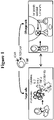
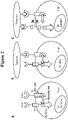


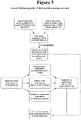

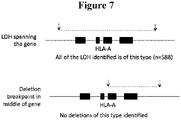

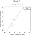

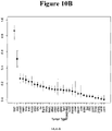


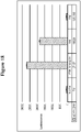


Classifications
-
- C—CHEMISTRY; METALLURGY
- C12—BIOCHEMISTRY; BEER; SPIRITS; WINE; VINEGAR; MICROBIOLOGY; ENZYMOLOGY; MUTATION OR GENETIC ENGINEERING
- C12Q—MEASURING OR TESTING PROCESSES INVOLVING ENZYMES, NUCLEIC ACIDS OR MICROORGANISMS; COMPOSITIONS OR TEST PAPERS THEREFOR; PROCESSES OF PREPARING SUCH COMPOSITIONS; CONDITION-RESPONSIVE CONTROL IN MICROBIOLOGICAL OR ENZYMOLOGICAL PROCESSES
- C12Q1/00—Measuring or testing processes involving enzymes, nucleic acids or microorganisms; Compositions therefor; Processes of preparing such compositions
- C12Q1/68—Measuring or testing processes involving enzymes, nucleic acids or microorganisms; Compositions therefor; Processes of preparing such compositions involving nucleic acids
- C12Q1/6806—Preparing nucleic acids for analysis, e.g. for polymerase chain reaction [PCR] assay
-
- A—HUMAN NECESSITIES
- A61—MEDICAL OR VETERINARY SCIENCE; HYGIENE
- A61K—PREPARATIONS FOR MEDICAL, DENTAL OR TOILETRY PURPOSES
- A61K35/00—Medicinal preparations containing materials or reaction products thereof with undetermined constitution
- A61K35/12—Materials from mammals; Compositions comprising non-specified tissues or cells; Compositions comprising non-embryonic stem cells; Genetically modified cells
-
- C—CHEMISTRY; METALLURGY
- C12—BIOCHEMISTRY; BEER; SPIRITS; WINE; VINEGAR; MICROBIOLOGY; ENZYMOLOGY; MUTATION OR GENETIC ENGINEERING
- C12N—MICROORGANISMS OR ENZYMES; COMPOSITIONS THEREOF; PROPAGATING, PRESERVING, OR MAINTAINING MICROORGANISMS; MUTATION OR GENETIC ENGINEERING; CULTURE MEDIA
- C12N15/00—Mutation or genetic engineering; DNA or RNA concerning genetic engineering, vectors, e.g. plasmids, or their isolation, preparation or purification; Use of hosts therefor
- C12N15/09—Recombinant DNA-technology
- C12N15/10—Processes for the isolation, preparation or purification of DNA or RNA
- C12N15/1034—Isolating an individual clone by screening libraries
- C12N15/1093—General methods of preparing gene libraries, not provided for in other subgroups
-
- A—HUMAN NECESSITIES
- A61—MEDICAL OR VETERINARY SCIENCE; HYGIENE
- A61K—PREPARATIONS FOR MEDICAL, DENTAL OR TOILETRY PURPOSES
- A61K35/00—Medicinal preparations containing materials or reaction products thereof with undetermined constitution
-
- A—HUMAN NECESSITIES
- A61—MEDICAL OR VETERINARY SCIENCE; HYGIENE
- A61K—PREPARATIONS FOR MEDICAL, DENTAL OR TOILETRY PURPOSES
- A61K39/00—Medicinal preparations containing antigens or antibodies
- A61K39/46—Cellular immunotherapy
- A61K39/461—Cellular immunotherapy characterised by the cell type used
- A61K39/4611—T-cells, e.g. tumor infiltrating lymphocytes [TIL], lymphokine-activated killer cells [LAK] or regulatory T cells [Treg]
-
- A—HUMAN NECESSITIES
- A61—MEDICAL OR VETERINARY SCIENCE; HYGIENE
- A61K—PREPARATIONS FOR MEDICAL, DENTAL OR TOILETRY PURPOSES
- A61K39/00—Medicinal preparations containing antigens or antibodies
- A61K39/46—Cellular immunotherapy
- A61K39/463—Cellular immunotherapy characterised by recombinant expression
- A61K39/4631—Chimeric Antigen Receptors [CAR]
-
- A—HUMAN NECESSITIES
- A61—MEDICAL OR VETERINARY SCIENCE; HYGIENE
- A61K—PREPARATIONS FOR MEDICAL, DENTAL OR TOILETRY PURPOSES
- A61K39/00—Medicinal preparations containing antigens or antibodies
- A61K39/46—Cellular immunotherapy
- A61K39/464—Cellular immunotherapy characterised by the antigen targeted or presented
- A61K39/4643—Vertebrate antigens
- A61K39/4644—Cancer antigens
- A61K39/464402—Receptors, cell surface antigens or cell surface determinants
- A61K39/464411—Immunoglobulin superfamily
- A61K39/464412—CD19 or B4
-
- A—HUMAN NECESSITIES
- A61—MEDICAL OR VETERINARY SCIENCE; HYGIENE
- A61K—PREPARATIONS FOR MEDICAL, DENTAL OR TOILETRY PURPOSES
- A61K39/00—Medicinal preparations containing antigens or antibodies
- A61K39/46—Cellular immunotherapy
- A61K39/464—Cellular immunotherapy characterised by the antigen targeted or presented
- A61K39/4643—Vertebrate antigens
- A61K39/4644—Cancer antigens
- A61K39/464402—Receptors, cell surface antigens or cell surface determinants
- A61K39/464424—CD20
-
- A—HUMAN NECESSITIES
- A61—MEDICAL OR VETERINARY SCIENCE; HYGIENE
- A61P—SPECIFIC THERAPEUTIC ACTIVITY OF CHEMICAL COMPOUNDS OR MEDICINAL PREPARATIONS
- A61P35/00—Antineoplastic agents
-
- C—CHEMISTRY; METALLURGY
- C07—ORGANIC CHEMISTRY
- C07K—PEPTIDES
- C07K14/00—Peptides having more than 20 amino acids; Gastrins; Somatostatins; Melanotropins; Derivatives thereof
- C07K14/435—Peptides having more than 20 amino acids; Gastrins; Somatostatins; Melanotropins; Derivatives thereof from animals; from humans
- C07K14/705—Receptors; Cell surface antigens; Cell surface determinants
- C07K14/70503—Immunoglobulin superfamily
- C07K14/7051—T-cell receptor (TcR)-CD3 complex
-
- C—CHEMISTRY; METALLURGY
- C07—ORGANIC CHEMISTRY
- C07K—PEPTIDES
- C07K14/00—Peptides having more than 20 amino acids; Gastrins; Somatostatins; Melanotropins; Derivatives thereof
- C07K14/435—Peptides having more than 20 amino acids; Gastrins; Somatostatins; Melanotropins; Derivatives thereof from animals; from humans
- C07K14/705—Receptors; Cell surface antigens; Cell surface determinants
- C07K14/70503—Immunoglobulin superfamily
- C07K14/70521—CD28, CD152
-
- C—CHEMISTRY; METALLURGY
- C07—ORGANIC CHEMISTRY
- C07K—PEPTIDES
- C07K14/00—Peptides having more than 20 amino acids; Gastrins; Somatostatins; Melanotropins; Derivatives thereof
- C07K14/435—Peptides having more than 20 amino acids; Gastrins; Somatostatins; Melanotropins; Derivatives thereof from animals; from humans
- C07K14/705—Receptors; Cell surface antigens; Cell surface determinants
- C07K14/70578—NGF-receptor/TNF-receptor superfamily, e.g. CD27, CD30, CD40, CD95
-
- C—CHEMISTRY; METALLURGY
- C07—ORGANIC CHEMISTRY
- C07K—PEPTIDES
- C07K16/00—Immunoglobulins [IGs], e.g. monoclonal or polyclonal antibodies
- C07K16/18—Immunoglobulins [IGs], e.g. monoclonal or polyclonal antibodies against material from animals or humans
- C07K16/28—Immunoglobulins [IGs], e.g. monoclonal or polyclonal antibodies against material from animals or humans against receptors, cell surface antigens or cell surface determinants
- C07K16/2803—Immunoglobulins [IGs], e.g. monoclonal or polyclonal antibodies against material from animals or humans against receptors, cell surface antigens or cell surface determinants against the immunoglobulin superfamily
-
- C—CHEMISTRY; METALLURGY
- C12—BIOCHEMISTRY; BEER; SPIRITS; WINE; VINEGAR; MICROBIOLOGY; ENZYMOLOGY; MUTATION OR GENETIC ENGINEERING
- C12N—MICROORGANISMS OR ENZYMES; COMPOSITIONS THEREOF; PROPAGATING, PRESERVING, OR MAINTAINING MICROORGANISMS; MUTATION OR GENETIC ENGINEERING; CULTURE MEDIA
- C12N15/00—Mutation or genetic engineering; DNA or RNA concerning genetic engineering, vectors, e.g. plasmids, or their isolation, preparation or purification; Use of hosts therefor
- C12N15/09—Recombinant DNA-technology
- C12N15/10—Processes for the isolation, preparation or purification of DNA or RNA
- C12N15/1034—Isolating an individual clone by screening libraries
- C12N15/1055—Protein x Protein interaction, e.g. two hybrid selection
-
- A—HUMAN NECESSITIES
- A61—MEDICAL OR VETERINARY SCIENCE; HYGIENE
- A61K—PREPARATIONS FOR MEDICAL, DENTAL OR TOILETRY PURPOSES
- A61K39/00—Medicinal preparations containing antigens or antibodies
- A61K2039/505—Medicinal preparations containing antigens or antibodies comprising antibodies
- A61K2039/507—Comprising a combination of two or more separate antibodies
-
- A—HUMAN NECESSITIES
- A61—MEDICAL OR VETERINARY SCIENCE; HYGIENE
- A61K—PREPARATIONS FOR MEDICAL, DENTAL OR TOILETRY PURPOSES
- A61K39/00—Medicinal preparations containing antigens or antibodies
- A61K2039/51—Medicinal preparations containing antigens or antibodies comprising whole cells, viruses or DNA/RNA
- A61K2039/515—Animal cells
- A61K2039/5156—Animal cells expressing foreign proteins
-
- A—HUMAN NECESSITIES
- A61—MEDICAL OR VETERINARY SCIENCE; HYGIENE
- A61K—PREPARATIONS FOR MEDICAL, DENTAL OR TOILETRY PURPOSES
- A61K2239/00—Indexing codes associated with cellular immunotherapy of group A61K39/46
- A61K2239/27—Indexing codes associated with cellular immunotherapy of group A61K39/46 characterized by targeting or presenting multiple antigens
- A61K2239/29—Multispecific CARs
-
- A—HUMAN NECESSITIES
- A61—MEDICAL OR VETERINARY SCIENCE; HYGIENE
- A61K—PREPARATIONS FOR MEDICAL, DENTAL OR TOILETRY PURPOSES
- A61K2239/00—Indexing codes associated with cellular immunotherapy of group A61K39/46
- A61K2239/31—Indexing codes associated with cellular immunotherapy of group A61K39/46 characterized by the route of administration
-
- A—HUMAN NECESSITIES
- A61—MEDICAL OR VETERINARY SCIENCE; HYGIENE
- A61K—PREPARATIONS FOR MEDICAL, DENTAL OR TOILETRY PURPOSES
- A61K2239/00—Indexing codes associated with cellular immunotherapy of group A61K39/46
- A61K2239/46—Indexing codes associated with cellular immunotherapy of group A61K39/46 characterised by the cancer treated
- A61K2239/48—Blood cells, e.g. leukemia or lymphoma
-
- A—HUMAN NECESSITIES
- A61—MEDICAL OR VETERINARY SCIENCE; HYGIENE
- A61K—PREPARATIONS FOR MEDICAL, DENTAL OR TOILETRY PURPOSES
- A61K35/00—Medicinal preparations containing materials or reaction products thereof with undetermined constitution
- A61K35/12—Materials from mammals; Compositions comprising non-specified tissues or cells; Compositions comprising non-embryonic stem cells; Genetically modified cells
- A61K35/14—Blood; Artificial blood
- A61K35/17—Lymphocytes; B-cells; T-cells; Natural killer cells; Interferon-activated or cytokine-activated lymphocytes
-
- C—CHEMISTRY; METALLURGY
- C07—ORGANIC CHEMISTRY
- C07K—PEPTIDES
- C07K16/00—Immunoglobulins [IGs], e.g. monoclonal or polyclonal antibodies
- C07K16/18—Immunoglobulins [IGs], e.g. monoclonal or polyclonal antibodies against material from animals or humans
- C07K16/28—Immunoglobulins [IGs], e.g. monoclonal or polyclonal antibodies against material from animals or humans against receptors, cell surface antigens or cell surface determinants
- C07K16/2803—Immunoglobulins [IGs], e.g. monoclonal or polyclonal antibodies against material from animals or humans against receptors, cell surface antigens or cell surface determinants against the immunoglobulin superfamily
- C07K16/2833—Immunoglobulins [IGs], e.g. monoclonal or polyclonal antibodies against material from animals or humans against receptors, cell surface antigens or cell surface determinants against the immunoglobulin superfamily against MHC-molecules, e.g. HLA-molecules
-
- C—CHEMISTRY; METALLURGY
- C07—ORGANIC CHEMISTRY
- C07K—PEPTIDES
- C07K16/00—Immunoglobulins [IGs], e.g. monoclonal or polyclonal antibodies
- C07K16/18—Immunoglobulins [IGs], e.g. monoclonal or polyclonal antibodies against material from animals or humans
- C07K16/28—Immunoglobulins [IGs], e.g. monoclonal or polyclonal antibodies against material from animals or humans against receptors, cell surface antigens or cell surface determinants
- C07K16/30—Immunoglobulins [IGs], e.g. monoclonal or polyclonal antibodies against material from animals or humans against receptors, cell surface antigens or cell surface determinants from tumour cells
-
- C—CHEMISTRY; METALLURGY
- C07—ORGANIC CHEMISTRY
- C07K—PEPTIDES
- C07K2317/00—Immunoglobulins specific features
- C07K2317/30—Immunoglobulins specific features characterized by aspects of specificity or valency
- C07K2317/33—Crossreactivity, e.g. for species or epitope, or lack of said crossreactivity
-
- C—CHEMISTRY; METALLURGY
- C07—ORGANIC CHEMISTRY
- C07K—PEPTIDES
- C07K2317/00—Immunoglobulins specific features
- C07K2317/60—Immunoglobulins specific features characterized by non-natural combinations of immunoglobulin fragments
- C07K2317/62—Immunoglobulins specific features characterized by non-natural combinations of immunoglobulin fragments comprising only variable region components
- C07K2317/622—Single chain antibody (scFv)
-
- C—CHEMISTRY; METALLURGY
- C07—ORGANIC CHEMISTRY
- C07K—PEPTIDES
- C07K2317/00—Immunoglobulins specific features
- C07K2317/70—Immunoglobulins specific features characterized by effect upon binding to a cell or to an antigen
- C07K2317/73—Inducing cell death, e.g. apoptosis, necrosis or inhibition of cell proliferation
-
- C—CHEMISTRY; METALLURGY
- C07—ORGANIC CHEMISTRY
- C07K—PEPTIDES
- C07K2317/00—Immunoglobulins specific features
- C07K2317/70—Immunoglobulins specific features characterized by effect upon binding to a cell or to an antigen
- C07K2317/75—Agonist effect on antigen
-
- C—CHEMISTRY; METALLURGY
- C07—ORGANIC CHEMISTRY
- C07K—PEPTIDES
- C07K2317/00—Immunoglobulins specific features
- C07K2317/70—Immunoglobulins specific features characterized by effect upon binding to a cell or to an antigen
- C07K2317/76—Antagonist effect on antigen, e.g. neutralization or inhibition of binding
-
- C—CHEMISTRY; METALLURGY
- C07—ORGANIC CHEMISTRY
- C07K—PEPTIDES
- C07K2319/00—Fusion polypeptide
-
- C—CHEMISTRY; METALLURGY
- C07—ORGANIC CHEMISTRY
- C07K—PEPTIDES
- C07K2319/00—Fusion polypeptide
- C07K2319/01—Fusion polypeptide containing a localisation/targetting motif
- C07K2319/02—Fusion polypeptide containing a localisation/targetting motif containing a signal sequence
-
- C—CHEMISTRY; METALLURGY
- C07—ORGANIC CHEMISTRY
- C07K—PEPTIDES
- C07K2319/00—Fusion polypeptide
- C07K2319/01—Fusion polypeptide containing a localisation/targetting motif
- C07K2319/03—Fusion polypeptide containing a localisation/targetting motif containing a transmembrane segment
-
- C—CHEMISTRY; METALLURGY
- C07—ORGANIC CHEMISTRY
- C07K—PEPTIDES
- C07K2319/00—Fusion polypeptide
- C07K2319/33—Fusion polypeptide fusions for targeting to specific cell types, e.g. tissue specific targeting, targeting of a bacterial subspecies
-
- C—CHEMISTRY; METALLURGY
- C12—BIOCHEMISTRY; BEER; SPIRITS; WINE; VINEGAR; MICROBIOLOGY; ENZYMOLOGY; MUTATION OR GENETIC ENGINEERING
- C12Q—MEASURING OR TESTING PROCESSES INVOLVING ENZYMES, NUCLEIC ACIDS OR MICROORGANISMS; COMPOSITIONS OR TEST PAPERS THEREFOR; PROCESSES OF PREPARING SUCH COMPOSITIONS; CONDITION-RESPONSIVE CONTROL IN MICROBIOLOGICAL OR ENZYMOLOGICAL PROCESSES
- C12Q2531/00—Reactions of nucleic acids characterised by
- C12Q2531/10—Reactions of nucleic acids characterised by the purpose being amplify/increase the copy number of target nucleic acid
- C12Q2531/113—PCR
-
- C—CHEMISTRY; METALLURGY
- C12—BIOCHEMISTRY; BEER; SPIRITS; WINE; VINEGAR; MICROBIOLOGY; ENZYMOLOGY; MUTATION OR GENETIC ENGINEERING
- C12Q—MEASURING OR TESTING PROCESSES INVOLVING ENZYMES, NUCLEIC ACIDS OR MICROORGANISMS; COMPOSITIONS OR TEST PAPERS THEREFOR; PROCESSES OF PREPARING SUCH COMPOSITIONS; CONDITION-RESPONSIVE CONTROL IN MICROBIOLOGICAL OR ENZYMOLOGICAL PROCESSES
- C12Q2535/00—Reactions characterised by the assay type for determining the identity of a nucleotide base or a sequence of oligonucleotides
- C12Q2535/131—Allele specific probes
-
- C—CHEMISTRY; METALLURGY
- C12—BIOCHEMISTRY; BEER; SPIRITS; WINE; VINEGAR; MICROBIOLOGY; ENZYMOLOGY; MUTATION OR GENETIC ENGINEERING
- C12Q—MEASURING OR TESTING PROCESSES INVOLVING ENZYMES, NUCLEIC ACIDS OR MICROORGANISMS; COMPOSITIONS OR TEST PAPERS THEREFOR; PROCESSES OF PREPARING SUCH COMPOSITIONS; CONDITION-RESPONSIVE CONTROL IN MICROBIOLOGICAL OR ENZYMOLOGICAL PROCESSES
- C12Q2600/00—Oligonucleotides characterized by their use
- C12Q2600/156—Polymorphic or mutational markers
Definitions
- the invention relates to the field of cancer immunotherapy by adoptive cell transfer, employing activating chimeric antigen receptors (aCARs) recognizing antigens expressed on the surface of tumor cells, inhibitory CARs (iCARs) and protective CARs (pCARs) directed at allelic variants of the same or other cell surface antigens expressed by normal cells but not by the tumor due to loss of heterozygosity (LOH).
- aCARs activating chimeric antigen receptors
- iCARs inhibitory CARs
- pCARs protective CARs
- T cells are capable of eradicating tumor cells.
- These approaches employ bone marrow transplantation with donor lymphocyte infusion, adoptive transfer of tumor-infiltrating lymphocytes (TILs), treatment with T cells genetically redirected at preselected antigens via CARs (Gross and Eshhar, 2016a) or T cell receptors (TCRs), the use of immune checkpoint inhibitors or active vaccination.
- TILs tumor-infiltrating lymphocytes
- CARs Gross and Eshhar, 2016a
- TCRs T cell receptors
- chimeric antigen receptors or CARs
- T cells or other killer cells of the immune system such as natural killer (NK) cells and cytokine-induced killer cells
- NK natural killer
- cytokine-induced killer cells chimeric antigen receptors
- scFv extracellular single-chain antibody variable fragment
- CARs are being examined in dozens of clinical trials and have so far shown exceptionally high efficacy in B cell malignancies (Dotti et al., 2014; Gill and June, 2015; Gross and Eshhar, 2016a).
- the safety of CAR-T cell therapy is determined, in large, by its ability to discriminate between the tumor and healthy tissue.
- a major risk and the direct cause for adverse autoimmune effects that have been reported in clinical and preclinical studies is off-tumor, on-target toxicity resulting from extra-tumor expression of the target antigen (dealt with in detail in our recent review (Gross and Eshhar, 2016b) and (Klebanoff et al., 2016)).
- Concerning this risk, shared, non-mutated cell surface antigens which are currently tested clinically or pre-clinically for CAR therapy can be generally divided into a number of categories according to their tissue distribution and mode of expression:
- TAAs which are presently evaluated in preclinical and clinical studies are overexpressed by tumors but are also present, usually at lower level, on essential normal tissue.
- Reactive measures include those which seek to eliminate, or suppress transferred T cells once damage is already evident (reactive measures) and those that aim at preventing potential damage in the first place (proactive measures) (Gross and Eshhar, 2016a).
- Reactive approaches often use suicide genes such as herpes simplex virus thymidine kinase (HSV-tk) and iC9, a fusion polypeptide comprising a truncated human caspase 9 and a mutated FK506-binding protein.
- suicide genes such as herpes simplex virus thymidine kinase (HSV-tk) and iC9, a fusion polypeptide comprising a truncated human caspase 9 and a mutated FK506-binding protein.
- Inhibitory CARs Off-tumor reactivity occurs when the target antigen of CAR-redirected killer cells is shared with normal tissue. If this normal tissue expresses another surface antigen not present on the tumor, then co-expressing in the gene-modified cells an additional CAR targeting this non-shared antigen, which harbors an inhibitory signaling moiety, can prevent T-cell activation by the normal tissue.
- an iCAR possesses a signaling domain derived from an inhibitory receptor which can antagonize T cell activation, such as CTLA-4, PD-1 or an NK inhibitory receptor. If the normal tissue which shares the candidate aCAR antigen with the tumor expresses another surface antigen not shared with the tumor, an iCAR expressed by the same T cell which targets this non-shared antigen can protect the normal tissue ( Fig. 1 ).
- NK cells do not express antigen-specific receptors. Instead, NK cells express an array of germline-encoded activating and inhibitory receptors which respectively recognize multiple activating and inhibitory ligands at the cell surface of infected and healthy cells.
- the protective capacity of an iCAR based on NK inhibitory receptors such as KIR3DL1 has been described ( US 9,745,368 ). KIR3DL1 and other NK inhibitory receptors function by dismantling the immunological synapse in a rapid and comprehensive manner.
- Tissue distribution of the antigens targeted by the iCAR and aCAR dictates the optimal mode of action of the iCAR required for conferring maximal safety without compromising clinical efficacy. For example, if the anatomical sites of the tumor and the normal tissue(s) to be protected do not intersect, transient inhibition (CTLA-4- or PD-1-like) will likely suffice. Yet, if these sites do overlap, only synapse-confined inhibition (e.g., an NK mode of action) will prevent constant paralysis of the therapeutic cells and allow their effective tumoricidal activity.
- CTLA-4- or PD-1-like transient inhibition
- synapse-confined inhibition e.g., an NK mode of action
- Next generation sequencing allows the determination of the DNA sequence of all protein-coding genes ( ⁇ 1% of the entire genome) in a given tumor biopsy and the comparison of the cancer 'exome' to that of a healthy tissue (usually from white blood cells) of the same patient. Exome sequencing can be completed within several days post-biopsy removal and at relatively low cost.
- transcriptome analysis RNA-seq
- RNA-seq transcriptome analysis
- the tumor cell can potentially present a private set of neopeptides to the patient's immune system on one or more of his or her HLA products.
- neoantigen-based T cell immunotherapies are more likely to be effective in cancers displaying higher mutational load, such as melanoma and lung cancers, but may often fail to show benefit in most cancers with fewer mutations (Savage, 2014; Schumacher and Schreiber, 2015).
- considerable intratumoral heterogeneity (Burrell et al., 2013) entails the simultaneous co-targeting of several antigens so as to avoid emergence of mutation-loss variants, a task which becomes increasingly demanding in view of the scarcity of useful immunogenic neopeptides.
- the present invention provides a method of identifying a target for preparing an inhibitory chimeric antigen receptor (iCAR) or a protective chimeric antigen receptor (pCAR) capable of preventing or attenuating undesired activation of an effector immune cell, wherein the target is identified by a method comprising:
- the LOH position is selected from the group consisting of a substitution, deletion, and insertion. In some embodiments, the LOH position is a SNP. In some embodiments, the gene comprising the extracellular polymorphic epitope is an HLA gene.
- the gene comprising the extracellular polymorphic epitope is an HLA-A, HLA-B, HLA-C, HLA-G, HLA-E, HLA-F, HLA-K, HLA-L, HLA-DM, HLA-DO, HLA-DP, HLA_DQ, or HLA-DR gene.
- the gene comprising the extracellular polymorphic epitope is an HLA-A gene.
- the gene comprising the extracellular polymorphic epitope is an HLA-B gene.
- the gene comprising the extracellular polymorphic epitope is an HLA-C gene.
- the gene comprising the extracellular polymorphic epitope is an HLA-G gene.
- the gene comprising the extracellular polymorphic epitope is an HLA-E gene. In some embodiments, the gene comprising the extracellular polymorphic epitope is an HLA-F gene. In some embodiments, the gene comprising the extracellular polymorphic epitope is an HLA-K gene. In some embodiments, the gene comprising the extracellular polymorphic epitope is an HLA-L gene. In some embodiments, the gene comprising the extracellular polymorphic epitope is an HLA-DM gene. In some embodiments, the gene comprising the extracellular polymorphic epitope is an HLA-DO gene. In some embodiments, the extracellular polymorphic epitope is an HLA-DP gene. In some embodiments, the extracellular polymorphic epitope is an HLA_DQ gene. In some embodiments, the extracellular polymorphic epitope is an HLA-DR gene.
- the gene comprising the extracellular polymorphic epitope is located on chromosome 1.
- the gene comprising the extracellular polymorphic epitope is selected from the group consisting of ABCA4, ADAM30, AQP10, ASTN1, C1orf101, CACNA1S, CATSPER4, CD101, CD164L2, CD1A, CD1C, CD244, CD34, CD46, CELSR2, CHRNB2, CLCA2, CLDN19, CLSTN1, CR1, CR2, CRB1, CSF3R, CSMD2, ECE1, ELTD1, EMC1, EPHA10, EPHA2, EPHA8, ERMAP, FCAMR, FCER1A, FCGR1B, FCGR2A, FCGR2B, FCGR3A, FCRL1, FCRL3, FCRL4, FCRL5, FCRL6, GJB4, GPA33, GPR157, GPR37L1, GPR88, HCRTR1, IGSF3, IGSF9, IL22RA1, IL23
- the gene comprising the extracellular polymorphic epitope is located on chromosome 2.
- the gene comprising the extracellular polymorphic epitope is selected from the group consisting of ABCG5, ALK, ASPRV1, ATRAID, CD207, CD8B, CHRNG, CLEC4F, CNTNAP5, CRIM1, CXCR1, DNER, DPP10, EDAR, EPCAM, GPR113, GPR148, GPR35, GPR39, GYPC, IL1RL1, ITGA4, ITGA6, ITGAV, LCT, LHCGR, LRP1B, LRP2, LY75, MARCO, MERTK, NRP2, OR6B2, PLA2R1, PLB1, PROKR1, PROM2, SCN7A, SDC1, SLC23A3, SLC5A6, TGOLN2, THSD7B, TM4SF20, TMEFF2, TMEM178A, TPO, and TRABD2A.
- the gene comprising the extracellular polymorphic epitope is located on chromosome 3. In some embodiments, the gene comprising the extracellular polymorphic epitope is selected from the group consisting of ACKR2, ALCAM, ANO10, ATP13A4, BTLA, CACNA1D, CACNA2D2, CACNA2D3, CASR, CCRL2, CD200, CD200R1, CD86, CD96, CDCP1, CDHR4, CELSR3, CHL1, CLDN11, CLDN18, CLSTN2, CSPG5, CX3CR1, CXCR6, CYP8B1, DCBLD2, DRD3, EPHA6, EPHB3, GABRR3, GP5, GPR128, GPR15, GPR27, GRM2, GRM7, HEG1, HTR3C, HTR3D, HTR3E, IGSF11, IL17RC, IL17RD, IL17RE, IL5RA, IMPG2, ITGA9, ITGB5, KCNMB3, LRIG1,
- the gene comprising the extracellular polymorphic epitope is located on chromosome 4. In some embodiments, the gene comprising the extracellular polymorphic epitope is selected from the group consisting of ANTXR2, BTC, CNGA1, CORIN, EGF, EMCN, ENPEP, EPHA5, ERVMER34-1, EVC2, FAT1, FAT4, FGFRL1, FRAS1, GPR125, GRID2, GYPA, GYPB, KDR, KIAA0922, KLB, MFSD8, PARM1, PDGFRA, RNF150, TENM3, TLR10, TLR1, TLR6, TMEM156, TMPRSS11A, TMPRSS11B, TMPRSS11E, TMPRSS11F, UGT2A1, and UNC5C.
- the gene comprising the extracellular polymorphic epitope is located on chromosome 5.
- the gene comprising the extracellular polymorphic epitope is selected from the group consisting of ADAM19, ADRB2, BTNL3, BTNL8, BTNL9, C5orf15, CATSPER3, CD180, CDH12, CDHR2, COL23A1, CSF1R, F2RL2, FAM174A, FAT2, FGFR4, FLT4, GABRA6, GABRG2, GPR151, GPR98, GRM6, HAVCR1, HAVCR2, IL31RA, IL6ST, IL7R, IQGAP2, ITGA1, ITGA2, KCNMB1, LIFR, LNPEP, MEGF10, NIPAL4, NPR3, NRG2, OR2V1, OR2Y1, OSMR, PCDH12, PCDH1, PCDHA1, PCDHA2, PCDHA4, PCDHA8, PCDHA9, PCDHB10, PCDHB11, PCDHB13, PCDHB
- the gene comprising the extracellular polymorphic epitope is located on chromosome 6. In some embodiments, the gene comprising the extracellular polymorphic epitope is selected from the group consisting of BAI3, BTN1A1, BTN2A1, BTN2A2, BTN3A1, BTN3A2, BTNL2, CD83, DCBLD1, DLL1, DPCR1, ENPP1, ENPP3, ENPP4, EPHA7, GABBR1, GABRR1, GCNT6, GFRAL, GJB7, GLP1R, GPR110, GPR111, GPR116, GPR126, GPR63, GPRC6A, HFE, HLA-A, HLA-B, HLA-C, HLA-DOA, HLA-DPA1, HLA-DPB1, HLA-DQA1, HLA-DQA2, HLA-DQB1, HLA-DQB2, HLA-DRB1, HLA-DRB5, HLA-E, HLA-F, HLA-G, IL20RA
- the gene comprising the extracellular polymorphic epitope is located on chromosome 7. In some embodiments, the gene comprising the extracellular polymorphic epitope is selected from the group consisting of AQP1, C7orf50, CD36, CDHR3, CNTNAP2, DPP6, EGFR, EPHA1, EPHB6, ERVW-1, GHRHR, GJC3, GPNMB, GRM8, HUS1, HYAL4, KIAA1324L, LRRN3, MET, MUC12, MUC17, NPC1L1, NPSR1, OR2A12, OR2A14, OR2A25, OR2A42, OR2A7, OR2A2, OR2AE1, OR2F2, OR6V1, PILRA, PILRB, PKD1L1, PLXNA4, PODXL, PTPRN2, PTPRZ1, RAMP3, SLC29A4, SMO, TAS2R16, TAS2R40, TAS2R4, TFR2, THSD7A,
- the gene comprising the extracellular polymorphic epitope is located on chromosome 8. In some embodiments, the gene comprising the extracellular polymorphic epitope is selected from the group consisting of ADAM18, ADAM28, ADAM32, ADAM7, ADAM9, ADRA1A, CDH17, CHRNA2, CSMD1, CSMD3, DCSTAMP, FZD6, GPR124, NRG1, OR4F21, PKHD1L1, PRSS55, SCARA3, SCARA5, SDC2, SLC10A5, SLC39A14, SLC39A4, SLCO5A1, TNFRSF10A, and TNFRSF10B.
- the gene comprising the extracellular polymorphic epitope is located on chromosome 9.
- the gene comprising the extracellular polymorphic epitope is selected from the group consisting of ABCA1, AQP7, ASTN2, C9orf135, CA9, CD72, CNTNAP3B, CNTNAP3, CRB2, ENTPD8, GPR144, GRIN3A, IZUMO3, KIAA1161, MAMDC4, MEGF9, MUSK, NOTCH1, OR13C2, OR13C3, OR13C5, OR13C8, OR13C9, OR13D1, OR13F1, OR1B1, OR1J2, OR1K1, OR1L1, OR1L3, OR1L6, OR1L8, OR1N1, OR1N2, OR1Q1, OR2S2, PCSK5, PDCD1LG2, PLGRKT, PTPRD, ROR2, SEMA4D, SLC31A1, TEK, TLR4, TMEM2, and VLDLR.
- the gene comprising the extracellular polymorphic epitope is located on chromosome 10. In some embodiments, the gene comprising the extracellular polymorphic epitope is selected from the group consisting of ABCC2, ADAM8, ADRB1, ANTXRL, ATRNL1, C10orf54, CDH23, CDHR1, CNNM2, COL13A1, COL17A1, ENTPD1, FZD8, FGFR2, GPR158, GRID1, IL15RA, IL2RA, ITGA8, ITGB1, MRC1, NRG3, NPFFR1, NRP1, OPN4, PCDH15, PKD2L1, PLXDC2, PRLHR, RET, RGR, SLC16A9, SLC29A3, SLC39A12, TACR2, TCTN3, TSPAN15, UNC5B, and VSTM4.
- the gene comprising the extracellular polymorphic epitope is located on chromosome 11.
- the gene comprising the extracellular polymorphic epitope is selected from the group consisting of AMICA1, ANO1, ANO3, APLP2, C11orf24, CCKBR, CD248, CD44, CD5, CD6, CD82, CDON, CLMP, CRTAM, DCHS1, DSCAML1, FAT3, FOLH1, GDPD4, GDPD5, GRIK4, HEPHL1, HTR3B, IFITM10, IL10RA, KIRREL3, LGR4, LRP4, LRP5, LRRC32, MCAM, MFRP, MMP26, MPEG1, MRGPRE, MRGPRF, MRGPRX2, MRGPRX3, MRGPRX4, MS4A4A, MS4A6A, MTNR1B, MUC15, NAALAD2, NAALADL1, NCAM1, NRXN2, OR10A2, OR10A5, OR10A6, OR10D3, OR
- the gene comprising the extracellular polymorphic epitope is located on chromosome 12. In some embodiments, the gene comprising the extracellular polymorphic epitope is selected from the group consisting of ANO4, AVPR1A, BCL2L14, CACNA2D4, CD163, CD163L1, CD27, CD4, CLEC12A, CLEC1B, CLEC2A, CLEC4C, CLEC7A, CLECL1, CLSTN3, GPR133, GPRC5D, ITGA7, ITGB7, KLRB1, KLRC2, KLRC3, KLRC4, KLRF1, KLRF2, LRP1, LRP6, MANSC1, MANSC4, OLR1, OR10AD1, OR10P1, OR2AP1, OR6C1, OR6C2, OR6C3, OR6C4, OR6C6, OR6C74, OR6C76, OR8S1, OR9K2, ORAI1, P2RX4, P2RX7, PRR4, PTPRB, PTPRQ, P
- the gene comprising the extracellular polymorphic epitope is located on chromosome 13. In some embodiments, the gene comprising the extracellular polymorphic epitope is selected from the group consisting of ATP4B, ATP7B, FLT3, FREM2, HTR2A, KL, PCDH8, RXFP2, SGCG, SHISA2, SLC15A1, SLITRK6, and TNFRSF19.
- the gene comprising the extracellular polymorphic epitope is located on chromosome 14. In some embodiments, the gene comprising the extracellular polymorphic epitope is selected from the group consisting of ADAM21, BDKRB2, C14orf37, CLEC14A, DLK1, FLRT2, GPR135, GPR137C, JAG2, LTB4R2, MMP14, OR11G2, OR11H12, OR11H6, OR4K1, OR4K15, OR4K5, OR4L1, OR4N2, OR4N5, SLC24A4, and SYNDIG1L.
- the gene comprising the extracellular polymorphic epitope is located on chromosome 15. In some embodiments, the gene comprising the extracellular polymorphic epitope is selected from the group consisting of ANPEP, CD276, CHRNA7, CHRNB4, CSPG4, DUOX1, DUOX2, FAM174B, GLDN, IGDCC4, ITGA11, LCTL, LTK, LYSMD4, MEGF11, NOX5, NRG4, OCA2, OR4F4, OR4M2, OR4N4, PRTG, RHCG, SCAMP5, SEMA4B, SEMA6D, SLC24A1, SLC24A5, SLC28A1, SPG11, STRA6, TRPM1, and TYRO3.
- the gene comprising the extracellular polymorphic epitope is located on chromosome 16. In some embodiments, the gene comprising the extracellular polymorphic epitope is selected from the group consisting of ATP2C2, CACNA1H, CD19, CDH11, CDH15, CDH16, CDH3, CDH5, CNGB1, CNTNAP4, GDPD3, GPR56, GPR97, IFT140, IL4R, ITFG3, ITGAL, ITGAM, ITGAX, KCNG4, MMP15, MSLNL, NOMO1, NOMO3, OR2C1, PIEZO1, PKD1, PKD1L2, QPRT, SCNN1B, SEZ6L2, SLC22A31, SLC5A11, SLC7A6, SPN, TMC5, TMC7, TMEM204, TMEM219, and TMEM8A.
- the gene comprising the extracellular polymorphic epitope is located on chromosome 17.
- the gene comprising the extracellular polymorphic epitope is selected from the group consisting of ABCC3, ACE, AOC3, ARL17B, ASGR2, C17orf80, CD300A, CD300C, CD300E, CD300LF, CD300LG, CHRNB1, CLEC10A, CNTNAP1, CPD, CXCL16, ERBB2, FAM171A2, GCGR, GLP2R, GP1BA, GPR142, GUCY2D, ITGA2B, ITGA3, ITGAE, ITGB3, KCNJ12, LRRC37A2, LRRC37A3, LRRC37A, LRRC37B, MRC2, NGFR, OR1A2, OR1D2, OR1G1, OR3A1, OR3A2, OR4D1, OR4D2, RNF43, SCARF1, SCN4A, SDK2, SECTM1, SEZ6, SHPK
- the gene comprising the extracellular polymorphic epitope is located on chromosome 18. In some embodiments, the gene comprising the extracellular polymorphic epitope is selected from the group consisting of APCDD1, CDH19, CDH20, CDH7, COLEC12, DCC, DSC1, DSG1, DSG3, DYNAP, MEP1B, PTPRM, SIGLEC15, and TNFRSF11A.
- the gene comprising the extracellular polymorphic epitope is located on chromosome 19. In some embodiments, the gene comprising the extracellular polymorphic epitope is selected from the group consisting of ABCA7, ACPT, BCAM, C19orf38, C19orf59, C5AR1, CATSPERD, CATSPERG, CD22, CD320, CD33, CD97, CEACAM19, CEACAM1, CEACAM21, CEACAM3, CEACAM4, CLEC4M, DLL3, EMR1, EMR2, EMR3, ERVV-1, ERVV-2, FAM187B, FCAR, FFAR3, FPR1, FXYD5, GFY, GP6, GPR42, GRIN3B, ICAM3, IGFLR1, IL12RB1, IL27RA, KIR2DL1, KIR2DL3, KIR2DL4, KIR3DL1, KIR3DL2, KIR3DL3, KIRREL2, KISS1R, LAIR1, LDLR, LILRA
- the gene comprising the extracellular polymorphic epitope is located on chromosome 20. In some embodiments, the gene comprising the extracellular polymorphic epitope is selected from the group consisting of ABHD12, ADAM33, ADRA1D, APMAP, ATRN, CD40, CD93, CDH22, CDH26, CDH4, FLRT3, GCNT7, GGT7, JAG1, LRRN4, NPBWR2, OCSTAMP, PTPRA, PTPRT, SEL1L2, SIGLEC1, SIRPA, SIRPB1, SIRPG, SLC24A3, SLC2A10, SLC4A11, SSTR4, and THBD.
- the gene comprising the extracellular polymorphic epitope is located on chromosome 21. In some embodiments, the gene comprising the extracellular polymorphic epitope is selected from the group consisting of CLDN8, DSCAM, ICOSLG, IFNAR1, IFNGR2, IGSF5, ITGB2, KCNJ15, NCAM2, SLC19A1, TMPRSS15, TMPRSS2, TMPRSS3, TRPM2, and UMODL1.
- the gene comprising the extracellular polymorphic epitope is located on chromosome 22. In some embodiments, the gene comprising the extracellular polymorphic epitope is selected from the group consisting of CACNA1I, CELSR1, COMT, CSF2RB, GGT1, GGT5, IL2RB, KREMEN1, MCHR1, OR11H1, P2RX6, PKDREJ, PLXNB2, SCARF2, SEZ6L, SSTR3, SUSD2, TMPRSS6, and TNFRSF13C.
- the gene comprising the extracellular polymorphic epitope is located on chromosome X. In some embodiments, the gene comprising the extracellular polymorphic epitope is selected from the group consisting of ATP6AP2, ATP7A, CNGA2, EDA2R, FMR1NB, GLRA4, GPR112, GUCY2F, HEPH, P2RY10, P2RY4, PLXNA3, PLXNB3, TLR8, VSIG4, and XG.
- the tumor is selected from the group consisting of a breast tumor, a prostate tumor, an ovarian tumor, a cervical tumor, a skin tumor, a pancreatic tumor, a colorectal tumor, a renal tumor, a liver tumor, a brain tumor, a lymphoma, a leukemia, a lung tumor, and a glioma.
- the tumor is selected from the group consisting of an adrenal gland tumor, a kidney tumor, a melanoma, DLBC, a breast tumor, a sarcoma, an ovary tumor, a lung tumor, a bladder tumor, and a liver tumor.
- the adrenal gland tumor is an adrenocortical carcinoma.
- the kidney tumor is a chromophobe renal cell carcinoma.
- the melanoma is uveal melanoma.
- TSGs tumor suppressor genes
- a tumor could lose the maternal chromosome 8, for example, while duplicating the paternal chromosome 8, leaving the cell with a normal chromosome 8 karyotype but an abnormal chromosome 8 'allelotype'.
- the 'average' cancer of the colon, breast, pancreas or prostate may lose 25% of its alleles and it is not unusual for a tumor to have lost over half of its alleles.”
- LOH is irreversible and events can only accumulate, so that tumor heterogeneity reflects the accumulation of losses throughout tumor progression. While tumor subclones can develop which differ in later LOH events, the existence of a minimal LOH signature that is shared by premalignant cells, putative tumor stem cells and all tumor subclones in a given patient, is expected to be the rule. Branches stemming from this 'trunk' LOH pattern would still create a limited set of partially overlapping signatures which, together, cover all tumor cells in the same patient
- Bausch-Fluck et al. (Bausch-Fluck et al., 2015) applied their Chemoproteomic Cell Surface Capture technology to identify a combined set of 1492 cell surface glycoproteins in 41 human cell types. A large fraction of the surfaceome is expected to be expressed by any given tumor, each exhibiting a distinctive profile. Genes encoding cell surface proteins were found to be slightly enriched for single-nucleotide polymorphisms (SNPs) in their coding regions than all other genes (Da Cunha et al., 2009).
- SNPs single-nucleotide polymorphisms
- Polymorphic in-frame insertions and deletions which are rarer, further contribute to the number of variants and likely exert more robust structural effects on the polypeptide products than peptide sequence-altering (nonsynonymous) SNPs.
- a typical genome contains 10,000 to 12,000 sites with nonsynonymous variants and 190-210 in-frame insertions/deletions (Abecasis et al., 2010; Auton et al., 2015).
- variants are not evenly distributed throughout the genome as highly polymorphic genes such as the HLA locus (http://www.ebi.ac.uk/imgt/hla/stats.html) or certain G-protein-coupled receptor (GPCR) genes (Lee et al., 2003; Rana et al., 2001) create distinct variant 'hotspots'.
- HLA locus http://www.ebi.ac.uk/imgt/hla/stats.html
- GPCR G-protein-coupled receptor
- LOH-related hotspots stems from the frequent loss of certain chromosomes, or chromosome arms in different cancers (e.g., 3p and 17p in small-cell lung carcinoma (Lindblad-Toh et al., 2000), 17p and 18q in colorectal cancer (Vogelstein et al., 1989), 17q and 19 in breast cancer (Li et al., 2014; Wang et al., 2004) 9p in melanoma (Stark and Hayward, 2007), 10q in glioblastoma (Ohgaki et al., 2004) and more)
- cancers e.g., 3p and 17p in small-cell lung carcinoma (Lindblad-Toh et al., 2000), 17p and 18q in colorectal cancer (Vogelstein et al., 1989), 17q and 19 in breast cancer (Li et al., 2014; Wang et al., 2004) 9p in mela
- allelic variations in surface proteins would affect the extracellular portion of the respective gene products, potentially creating distinct allele-restricted epitopes which, in principle, can be recognized and distinguished from other variants by highly-specific mAbs. It is well documented that mAbs can be isolated that discriminate between two variants of the same protein which differ in a single amino acid only (see, for example, an early example of mAbs that recognize point mutation products of the Ras oncogene with extraordinar specificity (Carney et al., 1986)). Interestingly, it was shown that two mAbs specific to a single amino acid interchange in a protein epitope can use structurally distinct variable regions from their heavy and light chain V gene pools (Stark and Caton, 1991).
- the present invention tackles this challenge through the co-expression in each therapeutic killer cell of a single pair of genes.
- One partner in this pair encodes an activating CAR (aCAR) and the other encodes a protecting CAR (pCAR) or an inhibitory CAR (iCAR)
- nucleic acid molecule refers to a DNA or RNA molecule.
- encoding refers to the inherent property of specific sequences of nucleotides in a polynucleotide, such as a gene, a cDNA, or an mRNA, to serve as templates for synthesis of other polymers and macromolecules in biological processes having either a defined sequence of nucleotides (e . g ., rRNA, tRNA and mRNA) or a defined sequence of amino acids and the biological properties resulting therefrom.
- a gene encodes a protein if transcription and translation of mRNA corresponding to that gene produces the protein in a cell or other biological system.
- Both the coding strand the nucleotide sequence of which is identical to the mRNA sequence and is usually provided in sequence listings, and the non-coding strand, used as the template for transcription of a gene or cDNA, can be referred to as encoding the protein or other product of that gene or cDNA.
- nucleotide sequence encoding an amino acid sequence includes all nucleotide sequences that are degenerate versions of each other and that encode the same amino acid sequence. Nucleotide sequences that encode proteins and RNA may include introns.
- endogenous refers to any material from or produced inside an organism, cell, tissue or system.
- exogenous refers to any material introduced from or produced outside an organism, cell, tissue or system.
- expression is defined as the transcription and/or translation of a particular nucleotide sequence driven by its promoter.
- “Expression vector” refers to a vector comprising a recombinant polynucleotide comprising expression control sequences operatively linked to a nucleotide sequence to be expressed.
- An expression vector comprises sufficient cis-acting elements for expression; other elements for expression can be supplied by the host cell or in an in vitro expression system.
- Expression vectors include all those known in the art, such as cosmids, plasmids (e.g., naked or contained in liposomes) and viruses (e.g., lentiviruses, retroviruses, adenoviruses, and adeno-associated viruses) that incorporate the recombinant polynucleotide.
- genomic variant refers to a change of at least one nucleotide at the genomic level in a sequenced sample compared to the reference or consensus sequence at the same genomic position.
- corresponding reference allele as used herein with reference to a variant means the reference or consensus sequence or nucleotide at the same genomic position as the variant.
- extracellular domain as used herein with reference to a protein means a region of the protein which is outside of the cell membrane.
- LHO loss of heterozygosity
- chromosomal materials such as a complete chromosome or a part thereof, in one copy of the two chromosomes in a somatic cell.
- sequence region as used herein with reference to a variant or a reference allele means a sequence starting upstream and ending downstream from the position of the variant, which can be translated into an "epitope peptide” that can be recognized by an antibody.
- CAR refers to a chimeric polypeptide that shares structural and functional properties with a cell immune-function receptor or adaptor molecule, from e.g., a T cell or a NK cell.
- CARs include TCARs and NKR-CARs.
- a CAR Upon binding to cognate antigen, a CAR can activate or inactivate the cytotoxic cell in which it is disposed, or modulate the cell's antitumor activity or otherwise modulate the cells immune response.
- specific binding refers to the relative binding of the scFv to one allelic variant and its failure to bind to the corresponding different allelic variant of the same polymorphic cell surface epitope.
- the specific scFv would provide a significant signal in an ELISA against the single allelic variant of a polymorphic cell surface epitope to which it is specific or cells transfected with a CAR displaying the scFv would be clearly labeled with the single allelic variant of a polymorphic cell surface epitope in a FACS assay, while the same assays using the corresponding different allelic variant of the same polymorphic cell surface epitope would not give any detectable signal.
- treating refers to means of obtaining a desired physiological effect.
- the effect may be therapeutic in terms of partially or completely curing a disease and/or symptoms attributed to the disease.
- the term refers to inhibiting the disease, e.g., arresting its development; or ameliorating the disease, e.g., causing regression of the disease.
- the terms "subject” or “individual” or “animal” or “patient” or “mammal,” refers to any subject, particularly a mammalian subject, for whom diagnosis, prognosis, or therapy is desired, for example, a human.
- safe effector immune cell or “safe effector cell” includes those cells described by the invention that express at least one iCAR or pCAR as described herein.
- the "safe effector immune cell” or “safe effector cell” is capable of adminsitraiton to a subject.
- the "safe effector immune cell” or “safe effector cell” further expresses an aCAR as described herein.
- the "safe effector immune cell” or “safe effector cell” further expresses an iCAR or a pCAR as described herein.
- the "safe effector immune cell” or “safe effector cell” further expresses an iCAR or a pCAR as described herein and an aCAR as described herein.
- compositions for use in accordance with the present invention may be formulated in conventional manner using one or more physiologically acceptable carriers or excipients.
- the carrier(s) must be "acceptable” in the sense of being compatible with the other ingredients of the composition and not deleterious to the recipient thereof.
- phrases "effective amount” or “therapeutically effective amount” are used interchangeably herein, and refer to an amount of a compound, formulation, material, or composition, as described herein effective to achieve a particular biological result.
- Methods of administration include, but are not limited to, parenteral, e.g., intravenous, intraperitoneal, intramuscular, subcutaneous, mucosal (e.g., oral, intranasal, buccal, vaginal, rectal, intraocular), intrathecal, topical and intradermal routes. Administration can be systemic or local. In some embodiments, the pharmaceutical composition is adapted for oral administration.
- carrier refers to a diluent, adjuvant, excipient, or vehicle with which the active agent is administered.
- the carriers in the pharmaceutical composition may comprise a binder, such as microcrystalline cellulose, polyvinylpyrrolidone (polyvidone or povidone), gum tragacanth, gelatin, starch, lactose or lactose monohydrate; a disintegrating agent, such as alginic acid, maize starch and the like; a lubricant or surfactant, such as magnesium stearate, or sodium lauryl sulphate; and a glidant, such as colloidal silicon dioxide.
- a binder such as microcrystalline cellulose, polyvinylpyrrolidone (polyvidone or povidone), gum tragacanth, gelatin, starch, lactose or lactose monohydrate
- a disintegrating agent such as alginic acid, maize starch and the like
- a lubricant or surfactant such as
- PBMC peripheral blood mononuclear cell
- PBMC peripheral blood mononuclear cell
- Methods for isolating PBMCs from blood are readily apparent to those skilled in the art.
- a non-limiting example is the extraction of these cells from whole blood using ficoll, a hydrophilic polysaccharide that separates layers of blood, with monocytes and lymphocytes forming a buffy coat under a layer of plasma or by leukapheresis, the preparation of leukocyte concentrates with the return of red cells and leukocyte-poor plasma to the donor.
- cancer as used herein is defined as disease characterized by the rapid and uncontrolled growth of aberrant cells. Cancer cells can spread locally or through the bloodstream and lymphatic system to other parts of the body. Examples of various cancers include but are not limited to, breast cancer, prostate cancer, ovarian cancer, cervical cancer, skin cancer, pancreatic cancer, colorectal cancer, renal cancer, liver cancer, brain cancer, lymphoma, leukemia, lung cancer, glioma, and the like.
- CAR-T SYSTEM iCARs, pCARs, and aCARs
- the present invention provides a new avenue enabling specific targeting of tumor cells while keeping the normal cells secure.
- the concept presented herein provides for the identification of new targets for iCARs (or pCARs or protective CARs), these targets defined as comprising single allelic variants of polymorphic cell surface epitopes, which are lost from tumor cells due to LOH of the chromosomal region they reside in, while remaining expressed on normal tissue. Because of the polymorphic variation, it is possible to distinguish the two alleles and target only the allele missing in the tumor cells.
- the target antigen may not necessarily itself be a tumor suppressor gene, or a gene predicted to be involved with cancer, since it is chosen for being in a region lost by LOH and could therefore simply be linked to such genes.
- This is conceptually different from the methods employed or suggested to date in cancer therapy, which target tumor associated antigens or antigens downregulated at tumors regardless of polymorphism.
- the present methods also provide for broadening the selection of aCAR beyond tumor associated anitgens, by conferring protection of normal cells through the co-expression of the iCAR and/or pCAR as described herein.
- aCARs and the iCARs are well-known and can be easily prepared using methods well-known in the art as taught for example, in WO 2015/142314 and in US 9,745,368 , both incorporated by reference as if fully disclosed herein.
- the two CARs in every given pair specifically recognize the product of a different allelic variant of the same target gene for which the patient is heterozygous.
- the basic principle is as follows: the aCAR targets an allelic variant of a selected cell surface protein that is expressed by the given tumor cells and is not affected by LOH while the pCAR or iCAR targets the product encoded by the allelic variant of the same gene that has been lost from these tumor cells due to LOH.
- both alleles are present and are known to be equally functional, that is, expression is biallelic in all tissues (in contrast to other genes which may exhibit random monoallelic expression (Chess, 2012; Savova et al., 2016).
- the two CARs target two related epitopes residing at the same location on the protein product, which differ by one, or only few amino acids.
- the aCAR targets a non-polymorphic epitope on the same protein while the pCAR or iCAR is allele-specific. In this case the density of the aCAR epitope on normal cells would generally be two-fold higher than that of the iCAR or pCAR one.
- a single nucleic acid vector encodes both the aCAR and iCAR or pCAR.
- pCAR or iCAR targets the protein products of housekeeping genes. Since, by definition, these genes are expressed on all cells in the body, they are safe targets for pCAR or iCARs. That is, if the pCAR or iCAR targets a membrane product of a housekeeping gene for which the given patient is heterozygous, all cells in the body, except the tumor cells which have lost this allele due to LOH, will be protected.
- This strategy allows for the uncoupling of the aCAR target gene product from the pCAR or iCAR one. In fact, the aCAR target can then be any non-polymorphic epitope expressed by the tumor.
- a variation of this strategy would be to utilize a known aCAR targeted to a non-polymorphic tumor-associated antigen, e.g., an aCAR in clinical use or under examination in clinical trials, in combination with an iCAR or pCAR directed against a membrane product of a gene for which the given patient is heterozygous and which is expressed in at least the tissue of origin of the tumor and preferably in additional vital normal tissues in which aCAR target antigen is expressed.
- a known aCAR targeted to a non-polymorphic tumor-associated antigen e.g., an aCAR in clinical use or under examination in clinical trials
- an iCAR or pCAR directed against a membrane product of a gene for which the given patient is heterozygous and which is expressed in at least the tissue of origin of the tumor and preferably in additional vital normal tissues in which aCAR target antigen is expressed.
- the iCAR and/or pCAR be the product of any gene the expression pattern of which is sufficiently wide so as to protect vital normal tissues expressing the aCAR target antigen in addition to the tumor.
- the aCAR antigen can be, as argued for housekeeping genes, any non-polymorphic epitope expressed by the tumor, not restricted to known 'tumor-associated antigens', a consideration which can vastly expand the list of candidate aCAR targets.
- the identitiy of such normal vital tissues and level of expression would serve as important criteria in the prioritization of such candidate aCAR targets
- the iCAR technology may be based on immune checkpoints.
- the demonstration (Fedorov et al., 2013b; WO 2015/142314 ) that the regulatory elements of PD-1 and CTLA-4 possess a potent T cell inhibitory capacity when incorporated as iCAR signaling components is encouraging but the generality of these observations was recently questioned (Chicaybam and Bonamino, 2014, 2015).
- the precise molecular pathways triggered by these checkpoint proteins are not fully understood, their engagement dampens T-cell activation through both proximal and distal mechanisms, rendering T cells unresponsive to concomitant activating stimuli (Nirschl and Drake, 2013).
- the strategy based on the control asserted by iCARs depends on the dominance of the iCAR activity over the aCAR activity as explained above.
- the present invention provides this type of iCAR, termed here a pCAR (for 'protective CAR, see Fig. 2 ), designed to operate in CAR T cells in a synapse-selective manner and guarantee full dominance over the co-expressed aCAR.
- the iCAR provided by the present invention is this particular type of iCAR referred to herein as a protective CAR (pCAR).
- the pCAR of the present invention integrates two technological feats.
- the pCAR allows for uncoupling the activating moiety of the aCAR (FcR ⁇ /CD3- ⁇ ) from the recognition unit and the co-stimulatory element (e.g. , CD28, 4-1BB, CD134 (OX40, GITR, IL2R ⁇ and STAT3 binding motif (YXXQ)) by genetically placing them on two different polypeptide products.
- Recoupling of these elements which is mandatory for the aCAR function, will only take place by the addition of a heterodimerizing drug which can bridge the respective binding sites incorporated onto each of the polypeptides separately ( Fig. 2B ).
- This drug possess 1000-fold less immunosuppressive activity compared to rapamycin (Bayle et al., 2006; Graef et al., 1997; Liberles et al., 1997) and is commercially available (ARGENTTM, Regulated Heterodimerization Kit, ARIAD). In some emodiments, the drug is administered orally.
- engrafting the pCAR recognition unit and the missing activating domain, respectively, onto the two surfaces of the transmembrane domain of a RIP-controlled receptor which contains the two intramembrane cleavage sites ( Fig. 2A ). Binding of the pCAR to its antigen will trigger dual cleavage of the encoded polypeptide first by a member of the extracellular disintegrin and metalloproteinase (ADAM) family which removes the ectodomain and then by intracellular ⁇ -secretase, which liberates the intracellular domain of the pCAR.
- ADAM extracellular disintegrin and metalloproteinase
- This first cleavage event is predicted to disrupt the ability of the truncated aCAR to gain access to a functional, membrane-anchored configuration of its missing activating element, thus acquiring an operative mode ( Fig. 2C ).
- This principle was recently exploited in the development of new genetic switches designed to limit CAR T cell activity to simultaneous recognition of two different antigens on the tumor cell, applying either the Notch receptor (Morsut et al., 2016; Roybal et al., 2016b) or Epithelial cell adhesion molecule (EpCAM, Pizem, Y., M.Sc. thesis under the supervision of the Inventor), two well-studied receptors functioning through RIP.
- binding of the RIP-based CAR to one antigen releases a genetically-engineered intracellular domain which translocates to the cell nucleus where it turns on the expression of the second CAR.
- the first cleavage event disrupts the ability of the truncated aCAR to gain access to a functional, membrane-anchored configuration of its missing activating element, thus acquiring an operative mode.
- the proposed mode of action described above is predicted to exert local effects so that only aCARs which reside in the same synapse are affected and are no more able to bind their antigen productively and form an immunological synapse.
- aCAR aCAR with large numbers of non-tumor cells are likely to take place, they are only expected to be transient and nonfunctional so that the cells are fully capable of further interactions.
- Dominance of the pCARs over their aCARs counterparts is inherent to this system as function of the aCARs utterly depends on presence of the pCARs. Relative shortage of pCARs in a given T cell would render the aCARs non-functional due to lack of an activating domain. In some embodiments, a shortage of pCARs in a given T cell renders the aCARs non-functional due to lack of an activating domain.
- the second cleavage which detaches the activating domain from the plasma membrane, would render this domain nonfunctional and prevent unwanted cellular activation.
- the recognition domain and the activating one are localized to the plasma membrane.
- the second cleavage detaches the activating domain from the plasma membrane and renders this domain nonfunctional and prevents unwanted cellular activation.
- the aCAR and pCAR are designed to function via mutually exclusive mechanisms.
- the ability of the pCAR to undergo cleavage does not depend on the strength of inhibitory signaling so no completion on signaling outcome will take place.
- the aCARs cannot function, regardless of relative avidity of their interactions with their respective antigens, a scenario which secures another crucial level of safety.
- the mammalian tissue is human tissue and in other embodiments the related mammalian normal tissue is normal tissue from which the tumor developed.
- the effector immune cell is a T cell, a natural killer cell or a cytokine-induced killer cell.
- the at least one signal transduction element capable of inhibiting an effector immune cell is homologous to a signal transduction element of an immune checkpoint protein, such as an immune checkpoint protein selected from the group consisting of PD1; CTLA4; BTLA; 2B4; CD160; CEACAM, such as CEACAM1; KIRs, such as KIR2DL1, KIR2DL2, KIR2DL3, KIR2DL4, KIR2DL5A, KIR2DL5B, KIR3DL1, KIR3DL2, KIR3DL3, LIR1, LIR2, LIR3, LIR5, LIR8 and CD94-NKG2A; LAG3; TIM3; V-domain Ig suppressor of T cell activation (VISTA); STimulator of INterferon Genes (STING); immunoreceptor tyrosine-based inhibitory motif (ITIM)-containing proteins, T cell immunoglobulin and ITIM domain (TIGIT), and adenosine receptor (e.g., A2
- ITIM
- immune checkpoint protein is a natural killer cell inhibitory receptor, e.g., KIRs, such as KIR2DL1, KIR2DL2, KIR2DL3, KIR2DL4, KIR2DL5A, KIR2DL5B, KIR3DL1, KIR3DL2, KIR3DL3; or a Leukocyte Ig-like receptor, such as LIR1, LIR2, LIR3, LIR5, LIR8; and CD94-NKG2A, a C-type lectin receptor which forms heterodimers with CD94 and contains 2 ITIMs.
- KIRs such as KIR2DL1, KIR2DL2, KIR2DL3, KIR2DL4, KIR2DL5A, KIR2DL5B, KIR3DL1, KIR3DL2, KIR3DL3
- Leukocyte Ig-like receptor such as LIR1, LIR2, LIR3, LIR5, LIR8; and CD94-NKG2A, a C-type lectin receptor which forms heterodimers with CD94 and contains 2
- the extracellular domain of any one of the above embodiments is fused through a flexible hinge and transmembrane canonic motif to said intracellular domain.
- TARGET IDENTIFICATION aCAR, iCAR and pCAR
- the present invention provides methods for identification of aCAR, iCAR and/or pCAR targets based identification of candidate genes having extracellular polymorphic epitopes.
- the aCAR can be directed at any extracellular protein expressed on the tumor tissue.
- aCAR target is further expressed on non-tumor tissues and the iCAR taeget is also expressed on non-tumor tissues but is not expressed on tumor tissues.
- the method of identificaiton of candidate genes includes first determining that the gene encodes a transmembrane protein comprsing an extracellular polymorphic epitope. In some embodiments, the method of identificaiton of candidate genes further includes determining that the gene has at least two expressed alleles. In some embodiments, these alleles exhibit at least one allelic variation. In some embodiments, the allelic variation includes, for example, the presence of one or more SNPs, insertions, and/or deletions. In some embodiments, the allelic variation found for the gene causes an amino acid change relative to the reference sequence in an extracellular region of the protein.
- the gene is located in a chromosomal region which undergoes loss of heterozygosity (LOH). In some embodiments, the gene is located in a chromosomal region which undergoes loss of heterozygosity (LOH) in cancer. In some embodiments, the gene is expressed in a tissue-of-origin of a tumor type in which the corresponding region was found to undergo LOH. In some embodiments, the gene is expressed at least in one or more tissues that the aCAR is expressed in. In some embodiments, the iCAR or pCAR target is expressed in vital organ cells the aCAR is expressed in.
- LOH heterozygosity
- the target for use in the iCAR and/or pCAR is selected based on identification of a gene having at least one extracellular polymorphic epitope and wherein said gene has at least two expressed alleles. In some embodiments, the target for use in the iCAR and/or pCAR is selected based on identification of a gene having located in a chromosomal region which undergoes loss of heterozygosity. In some embodiments, the target for use in the iCAR and/or pCAR is selected based on identification of a gene having located in a chromosomal region which undergoes loss of heterozygosity in cancer. In some embodiments, the score for a theoretical SNP is calculated and a threshold limit determined.
- this LOH intend score can be employed as one method for determining if a candidate gene is a suitable iCAR or pCAR target.
- the target can be selected based on this LOH score.
- the candidate gene is a determined to be suiteable as an iCAR or pCAR target. LOH candidates based on an LOH candidate score of greater than 0.4.
- the target for use in the iCAR and/or pCAR is selected from a gene having at least one extracellular polymorphic epitope.
- the target is a gene is located on chromosome 1, chromosome 2, chromosome 3, chromosome 4, chromosome 5, chromosome 6, chromosome 7, chromosome 8, chromosome 9, chromosome 10, chromosome 11, chromosome 12, chromosome 13, chromosome 14, chromosome 15, chromosome 16, chromosome 17, chromosome 18, chromosome 19, chromosome 20, chromosome 21, chromosome 22, or chromosome X.
- the gene comprising the extracellular polymorphic epitope is located on chromosome 1.
- the target for use in the iCAR and/or pCAR is selected from the group consisting of ABCA4, ADAM30, AQP10, ASTN1, C1orf101, CACNA1S, CATSPER4, CD101, CD164L2, CD1A, CD1C, CD244, CD34, CD46, CELSR2, CHRNB2, CLCA2, CLDN19, CLSTN1, CR1, CR2, CRB1, CSF3R, CSMD2, ECE1, ELTD1, EMC1, EPHA10, EPHA2, EPHA8, ERMAP, FCAMR, FCER1A, FCGR1B, FCGR2A, FCGR2B, FCGR3A, FCRL1, FCRL3, FCRL4, FCRL5, FCRL6, GJB4, GPA33, GPR157, GPR37L1, GPR88, HCRTR1, IGSF3, IGSF9, IL22RA
- the gene comprising the extracellular polymorphic epitope is located on chromosome 2.
- the target for use in the iCAR and/or pCAR is selected from the group consisting of ABCG5, ALK, ASPRV1, ATRAID, CD207, CD8B, CHRNG, CLEC4F, CNTNAP5, CRIM1, CXCR1, DNER, DPP10, EDAR, EPCAM, GPR113, GPR148, GPR35, GPR39, GYPC, IL1RL1, ITGA4, ITGA6, ITGAV, LCT, LHCGR, LRP1B, LRP2, LY75, MARCO, MERTK, NRP2, OR6B2, PLA2R1, PLB1, PROKR1, PROM2, SCN7A, SDC1, SLC23A3, SLC5A6, TGOLN2, THSD7B, TM4SF20, TMEFF2, TMEM178A, TPO, and TRABD2A.
- the gene comprising the extracellular polymorphic epitope is located on chromosome 3.
- the target for use in the iCAR and/or pCAR is selected from the group consisting of ACKR2, ALCAM, ANO10, ATP13A4, BTLA, CACNA1D, CACNA2D2, CACNA2D3, CASR, CCRL2, CD200, CD200R1, CD86, CD96, CDCP1, CDHR4, CELSR3, CHL1, CLDN11, CLDN18, CLSTN2, CSPG5, CX3CR1, CXCR6, CYP8B1, DCBLD2, DRD3, EPHA6, EPHB3, GABRR3, GP5, GPR128, GPR15, GPR27, GRM2, GRM7, HEG1, HTR3C, HTR3D, HTR3E, IGSF11, IL17RC, IL17RD, IL17RE, IL5RA, IMPG2, ITGA9, ITGB5, KCNMB3,
- the gene comprising the extracellular polymorphic epitope is located on chromosome 4.
- the target for use in the iCAR and/or pCAR is selected from the group consisting of ANTXR2, BTC, CNGA1, CORIN, EGF, EMCN, ENPEP, EPHA5, ERVMER34-1, EVC2, FAT1, FAT4, FGFRL1, FRAS1, GPR125, GRID2, GYPA, GYPB, KDR, KIAA0922, KLB, MFSD8, PARM1, PDGFRA, RNF150, TENM3, TLR10, TLR1, TLR6, TMEM156, TMPRSS11A, TMPRSS11B, TMPRSS11E, TMPRSS11F, UGT2A1, and UNC5C.
- the gene comprising the extracellular polymorphic epitope is located on chromosome 5.
- the target for use in the iCAR and/or pCAR is selected from the group consisting of ADAM19, ADRB2, BTNL3, BTNL8, BTNL9, C5orf15, CATSPER3, CD180, CDH12, CDHR2, COL23A1, CSF1R, F2RL2, FAM174A, FAT2, FGFR4, FLT4, GABRA6, GABRG2, GPR151, GPR98, GRM6, HAVCR1, HAVCR2, IL31RA, IL6ST, IL7R, IQGAP2, ITGA1, ITGA2, KCNMB1, LIFR, LNPEP, MEGF10, NIPAL4, NPR3, NRG2, OR2V1, OR2Y1, OSMR, PCDH12, PCDH1, PCDHA1, PCDHA2, PCDHA4, PCDHA8, PCDHA9, PCDHB10, PCDHB11, PCDHB13
- the gene comprising the extracellular polymorphic epitope is located on chromosome 6.
- the target for use in the iCAR and/or pCAR is selected from the group consisting of BAI3, BTN1A1, BTN2A1, BTN2A2, BTN3A1, BTN3A2, BTNL2, CD83, DCBLD1, DLL1, DPCR1, ENPP1, ENPP3, ENPP4, EPHA7, GABBR1, GABRR1, GCNT6, GFRAL, GJB7, GLP1R, GPR110, GPR111, GPR116, GPR126, GPR63, GPRC6A, HFE, HLA-A, HLA-B, HLA-C, HLA-DOA, HLA-DPA1, HLA-DPB1, HLA-DQA1, HLA-DQA2, HLA-DQB1, HLA-DQB2, HLA-DRB1, HLA-DRB5, HLA-E, HLA-F, HLA-G,
- the gene comprising the extracellular polymorphic epitope is located on chromosome 6 and comprises an HLA target.
- the target for use in the iCAR and/or pCAR is HLA-A, HLA-B, HLA-C, HLA-DOA, HLA-DPA1, HLA-DPB1, HLA-DQA1, HLA-DQA2, HLA-DQB1, HLA-DQB2, HLA-DRB1, HLA-DRB5, HLA-E, HLA-F, HLA-G.
- the target for use in the iCAR and/or pCAR is HLA-A2,
- the gene comprising the extracellular polymorphic epitope is located on chromosome 7.
- the target for use in the iCAR and/or pCAR is selected from the group consisting of AQP1, C7orf50, CD36, CDHR3, CNTNAP2, DPP6, EGFR, EPHA1, EPHB6, ERVW-1, GHRHR, GJC3, GPNMB, GRM8, HUS1, HYAL4, KIAA1324L, LRRN3, MET, MUC12, MUC17, NPC1L1, NPSR1, OR2A12, OR2A14, OR2A25, OR2A42, OR2A7, OR2A2, OR2AE1, OR2F2, OR6V1, PILRA, PILRB, PKD1L1, PLXNA4, PODXL, PTPRN2, PTPRZ1, RAMP3, SLC29A4, SMO, TAS2R16, TAS2R40, TAS2R4, TFR2, TH
- the gene comprising the extracellular polymorphic epitope is located on chromosome 8.
- the target for use in the iCAR and/or pCAR is selected from the group consisting of ADAM18, ADAM28, ADAM32, ADAM7, ADAM9, ADRA1A, CDH17, CHRNA2, CSMD1, CSMD3, DCSTAMP, FZD6, GPR124, NRG1, OR4F21, PKHD1L1, PRSS55, SCARA3, SCARA5, SDC2, SLC10A5, SLC39A14, SLC39A4, SLCO5A1, TNFRSF10A, and TNFRSF10B.
- the gene comprising the extracellular polymorphic epitope is located on chromosome 9.
- the target for use in the iCAR and/or pCAR is selected from the group consisting of ABCA1, AQP7, ASTN2, C9orf135, CA9, CD72, CNTNAP3B, CNTNAP3, CRB2, ENTPD8, GPR144, GRIN3A, IZUMO3, KIAA1161, MAMDC4, MEGF9, MUSK, NOTCH1, OR13C2, OR13C3, OR13C5, OR13C8, OR13C9, OR13D1, OR13F1, OR1B1, OR1J2, OR1K1, OR1L1, OR1L3, OR1L6, OR1L8, OR1N1, OR1N2, OR1Q1, OR2S2, PCSK5, PDCD1LG2, PLGRKT, PTPRD, ROR2, SEMA4D, SLC31A1, TEK, TLR4, TMEM2, and VLDLR.
- the gene comprising the extracellular polymorphic epitope is located on chromosome 10.
- the target for use in the iCAR and/or pCAR is selected from the group consisting of ABCC2, ADAM8, ADRB1, ANTXRL, ATRNL1, C10orf54, CDH23, CDHR1, CNNM2, COL13A1, COL17A1, ENTPD1, FZD8, FGFR2, GPR158, GRID1, IL15RA, IL2RA, ITGA8, ITGB1, MRC1, NRG3, NPFFR1, NRP1, OPN4, PCDH15, PKD2L1, PLXDC2, PRLHR, RET, RGR, SLC16A9, SLC29A3, SLC39A12, TACR2, TCTN3, TSPAN15, UNC5B, and VSTM4.
- the gene comprising the extracellular polymorphic epitope is located on chromosome 11.
- the target for use in the iCAR and/or pCAR is selected from the group consisting of AMICA1, ANO1, ANO3, APLP2, C11orf24, CCKBR, CD248, CD44, CD5, CD6, CD82, CDON, CLMP, CRTAM, DCHS1, DSCAML1, FAT3, FOLH1, GDPD4, GDPD5, GRIK4, HEPHL1, HTR3B, IFITM10, IL10RA, KIRREL3, LGR4, LRP4, LRP5, LRRC32, MCAM, MFRP, MMP26, MPEG1, MRGPRE, MRGPRF, MRGPRX2, MRGPRX3, MRGPRX4, MS4A4A, MS4A6A, MTNR1B, MUC15, NAALAD2, NAALADL1, NCAM1, NRXN2, OR10A2, OR10A5, OR10A6, OR
- the gene comprising the extracellular polymorphic epitope is located on chromosome 12.
- the target for use in the iCAR and/or pCAR is selected from the group consisting of ANO4, AVPR1A, BCL2L14, CACNA2D4, CD163, CD163L1, CD27, CD4, CLEC12A, CLEC1B, CLEC2A, CLEC4C, CLEC7A, CLECL1, CLSTN3, GPR133, GPRC5D, ITGA7, ITGB7, KLRB1, KLRC2, KLRC3, KLRC4, KLRF1, KLRF2, LRP1, LRP6, MANSC1, MANSC4, OLR1, OR10AD1, OR10P1, OR2AP1, OR6C1, OR6C2, OR6C3, OR6C4, OR6C6, OR6C74, OR6C76, OR8S1, OR9K2, ORAI1, P2RX4, P2RX7, PRR4, PTPRB, PT
- the gene comprising the extracellular polymorphic epitope is located on chromosome 13.
- the target for use in the iCAR and/or pCAR is selected from the group consisting of ATP4B, ATP7B, FLT3, FREM2, HTR2A, KL, PCDH8, RXFP2, SGCG, SHISA2, SLC15A1, SLITRK6, and TNFRSF19.
- the gene comprising the extracellular polymorphic epitope is located on chromosome 14.
- the target for use in the iCAR and/or pCAR is selected from the group consisting of ADAM21, BDKRB2, C14orf37, CLEC14A, DLK1, FLRT2, GPR135, GPR137C, JAG2, LTB4R2, MMP14, OR11G2, OR11H12, OR11H6, OR4K1, OR4K15, OR4K5, OR4L1, OR4N2, OR4N5, SLC24A4, and SYNDIG1L.
- the gene comprising the extracellular polymorphic epitope is located on chromosome 15.
- the target for use in the iCAR and/or pCAR is selected from the group consisting of ANPEP, CD276, CHRNA7, CHRNB4, CSPG4, DUOX1, DUOX2, FAM174B, GLDN, IGDCC4, ITGA11, LCTL, LTK, LYSMD4, MEGF11, NOX5, NRG4, OCA2, OR4F4, OR4M2, OR4N4, PRTG, RHCG, SCAMP5, SEMA4B, SEMA6D, SLC24A1, SLC24A5, SLC28A1, SPG11, STRA6, TRPM1, and TYRO3.
- the gene comprising the extracellular polymorphic epitope is located on chromosome 16.
- the target for use in the iCAR and/or pCAR is selected from the group consisting of ATP2C2, CACNA1H, CD19, CDH11, CDH15, CDH16, CDH3, CDH5, CNGB1, CNTNAP4, GDPD3, GPR56, GPR97, IFT140, IL4R, ITFG3, ITGAL, ITGAM, ITGAX, KCNG4, MMP15, MSLNL, NOMO1, NOMO3, OR2C1, PIEZO1, PKD1, PKD1L2, QPRT, SCNN1B, SEZ6L2, SLC22A31, SLC5A11, SLC7A6, SPN, TMC5, TMC7, TMEM204, TMEM219, and TMEM8A.
- the gene comprising the extracellular polymorphic epitope is located on chromosome 17.
- the target for use in the iCAR and/or pCAR is selected from the group consisting of ABCC3, ACE, AOC3, ARL17B, ASGR2, C17orf80, CD300A, CD300C, CD300E, CD300LF, CD300LG, CHRNB1, CLEC10A, CNTNAP1, CPD, CXCL16, ERBB2, FAM171A2, GCGR, GLP2R, GP1BA, GPR142, GUCY2D, ITGA2B, ITGA3, ITGAE, ITGB3, KCNJ12, LRRC37A2, LRRC37A3, LRRC37A, LRRC37B, MRC2, NGFR, OR1A2, OR1D2, OR1G1, OR3A1, OR3A2, OR4D1, OR4D2, RNF43, SCARF1, SCN4A, SDK2, SECTM1, S
- the gene comprising the extracellular polymorphic epitope is located on chromosome 18.
- the target for use in the iCAR and/or pCAR is selected from the group consisting of APCDD1, CDH19, CDH20, CDH7, COLEC12, DCC, DSC1, DSG1, DSG3, DYNAP, MEP1B, PTPRM, SIGLEC15, and TNFRSF11A.
- the gene comprising the extracellular polymorphic epitope is located on chromosome 19.
- the target for use in the iCAR and/or pCAR is selected from the group consisting of ABCA7, ACPT, BCAM, C19orf38, C19orf59, C5AR1, CATSPERD, CATSPERG, CD22, CD320, CD33, CD97, CEACAM19, CEACAM1, CEACAM21, CEACAM3, CEACAM4, CLEC4M, DLL3, EMR1, EMR2, EMR3, ERVV-1, ERVV-2, FAM187B, FCAR, FFAR3, FPR1, FXYD5, GFY, GP6, GPR42, GRIN3B, ICAM3, IGFLR1, IL12RB1, IL27RA, KIR2DL1, KIR2DL3, KIR2DL4, KIR3DL1, KIR3DL2, KIR3DL3, KIRREL2, KISS1R, LAIR1, LDLR
- the gene comprising the extracellular polymorphic epitope is located on chromosome 20.
- the target for use in the iCAR and/or pCAR is selected from the group consisting of ABHD12, ADAM33, ADRA1D, APMAP, ATRN, CD40, CD93, CDH22, CDH26, CDH4, FLRT3, GCNT7, GGT7, JAG1, LRRN4, NPBWR2, OCSTAMP, PTPRA, PTPRT, SEL1L2, SIGLEC1, SIRPA, SIRPB1, SIRPG, SLC24A3, SLC2A10, SLC4A11, SSTR4, and THBD.
- the gene comprising the extracellular polymorphic epitope is located on chromosome 21.
- the target for use in the iCAR and/or pCAR is selected from the group consisting of CLDN8, DSCAM, ICOSLG, IFNAR1, IFNGR2, IGSF5, ITGB2, KCNJ15, NCAM2, SLC19A1, TMPRSS15, TMPRSS2, TMPRSS3, TRPM2, and UMODL1.
- the gene comprising the extracellular polymorphic epitope is located on chromosome 22.
- the target for use in the iCAR and/or pCAR is selected from the group consisting of CACNA1I, CELSR1, COMT, CSF2RB, GGT1, GGT5, IL2RB, KREMEN1, MCHR1, OR11H1, P2RX6, PKDREJ, PLXNB2, SCARF2, SEZ6L, SSTR3, SUSD2, TMPRSS6, and TNFRSF13C.
- the gene comprising the extracellular polymorphic epitope is located on chromosome X.
- the target for use in the iCAR and/or pCAR is selected from the group consisting of ATP6AP2, ATP7A, CNGA2, EDA2R, FMR1NB, GLRA4, GPR112, GUCY2F, HEPH, P2RY10, P2RY4, PLXNA3, PLXNB3, TLR8, VSIG4, and XG.
- the aCAR used to treat the cancer is directed against or specifically binds to any membrane protein which is expressed on the tumor tissue as long as the iCAR is expressed on every normal tissue in which the targeted protein is expressed.
- the aCAR can specifically bind or be directed to a tumor associated protein, tumor associated antigen and/or antigens in clinical trials, a CAR target as listed in Table 1, as well as any cell surface protein that is expressed in a tumor tissue to which an iCAR can be matched or paired with regard to target binding, according to the criteria listed in the application.
- the aCAR can be any expressed protein with an extracellular domain, as long as the iCAR is expressed in the same tissues as the aCAR or in any vital tissues, but is lost in the tumor cells.
- the aCAR used to treat the cancer such as any one of the cancer types recited above, is directed against or specifically binds to, a non-polymorphic cell surface epitope selected from the antigens listed in Table 1 , such as CD19.
- the aCAR, iCAR, and/or pCAR target is any target with an extracellular domain.
- the aCAR used to treat the cancer is directed against or specifically binds to, a non-polymorphic cell surface epitope selected from but not limited to the following list of antigens: CD19, CD20, CD22,CD10, CD7, CD49f, CD56, CD74, CAIX Ig ⁇ , ROR1, ROR2, CD30, LewisY, CD33, CD34,CD38, CD123, CD28, CD44v6, CD44, CD41, CD133, CD138, NKG2D-L, CD139, BCMA, GD2,GD3 ,hTERT, FBP, EGP-2, EGP-40, FR- ⁇ , L1-CAM, ErbB2,3,4, EGFRvIII, VEGFR-2, IL-13R ⁇ 2, FAP, Mesothelin, c-MET, PSMA, CEA, kRas, MAGE-A1, MUC1MUC16, PDL1, PSCA, EpCAM, FSHR, AFP, AXL, CD
- the present invention also provides for recognition moieties designed to provide specific binding to the target.
- the recognition moiety allows for directing the specific and targeted binding of the aCAR, iCAR and/or pCAR.
- the recognition moiety designed to provide specific binding to the target provides specific binding to an extracellular polymorphic epitope.
- the recognition moiety is part of an extracellular domain of the aCAR, iCAR and/or pCAR
- the extracellular domain comprises an antibody, derivative or fragment thereof, such as a humanized antibody; a human antibody; a functional fragment of an antibody; a single-domain antibody, such as a Nanobody; a recombinant antibody; and a single chain variable fragment (ScFv).
- the extracellular domain comprises an antibody mimetic, such as an affibody molecule; an affilin; an affimer; an affitin; an alphabody; an anticalin; an avimer; a DARPin; a fynomer; a Kunitz domain peptide; and a monobody.
- the extracellular domain comprises an aptamer.
- recognition moieties comprising this iCAR-aCAR Library may be derived from a master recognition moiety pool ideally selected from a combinatorial display library, so that:
- a given allelic product can become a potential pCAR or iCAR target in one patient and a useful aCAR target in another patient harboring the same allele, depending on the particular LOH pattern in each case.
- each will be engrafted onto both a pCAR or an iCAR and an aCAR gene scaffold. It is therefore desirable that all recognition moieties directed at allelic variants of the same gene possess binding affinities of a similar range.
- all possible combinations of pCAR-aCAR or iCAR-aCAR pairs can be pre-assembled so as to assure the highest coverage of potential allelic compositions of that gene in the entire population.
- the patient is heterozygous for the major allele and a minor one, the products of which differ in a single position along the encoded polypeptide as a result of a nonsynonymous SNP or, less frequently, an indel.
- a patient is heterozygous for two minor alleles which differ from the major one in two separate positions.
- a given variant epitope can serve as an iCAR target in one patient and an aCAR target in another.
- the variant epitope that can serve as an iCAR target is not the major allele variant.
- the variant epitope that can serve as the iCAR target is a minor allele.
- a variant-specific mAb (say, a mAb specific to the epitope encoded by the minor allele 'a') is well known in the art and is similar, in principle, to the identification of a mAb against any conventional antigenic determinant, and can usually best be accomplished via high throughput screening of a recombinant antibody scFv library, utilizing, for example, phage (Barbas et al., 2004), ribosome (Hanes et al., 1997) or yeast (Chao et al., 2006) display technologies.
- the antigen employed for library screening can either be a synthetic peptide spanning the position of variation between the two alleles (typically 15-20 amino acid in length or more), a recombinant full-length polypeptide which can either be commercially available or tailor-synthesized by one of the many companies operating in this field, or even entire cells expressing the said allelic variant at high level by virtue of gene transfection (e.g., electroporation of mRNA encoding the full-length cDNA cloned as template for in-vitro mRNA transcription in the pGEM4Z/A64 vector (Boczkowski et al., 2000)), following a subtraction step performed on the same cells not expressing this allele.
- gene transfection e.g., electroporation of mRNA encoding the full-length cDNA cloned as template for in-vitro mRNA transcription in the pGEM4Z/A64 vector (Boczkowski et al.,
- the corresponding epitope (at the same position) which is encoded by the major allele ('A'), creates a unique antigenic determinant that differs from that created by 'a' in the identity of a single amino acid (SNP) or length (indel; for example, insertion or deletion).
- SNP single amino acid
- Indel length
- This determinant can, in principle, be recognized by a different set of mAbs identified by the same, or other, antibody display screening technology.
- the ability of distinct members in each of the two sets of identified mAbs to distinguish between the two epitopes or variants, for example, an antibody from the first set binds the product of allele 'a' but not of 'A' and an Ab from the second set reciprocally binds 'A' but not 'a' can be determined using conventional binding assays such as ELISA or flow cytometry (Skora et al., 2015) or other technique for cell staining.
- a computational method can potentially be used to predict the sequence of a 'complementary' antibody scFv which binds 'A' but not 'a'. For such a computational method see, for example (Sela-Culang et al., 2015a,b).
- HLA-class I locus genes HLA-A, HLA-B, and HLA-C as the target genes, there are numerous allele-specific monoclonal antibodies available, for example, but not limited to, the antibodies listed in Example 3.
- the target for use in generation of a recognition moiety comprises at least one extracellular polymorphic epitope.
- the target is the product of a gene that is located on chromosome 1, chromosome 2, chromosome 3, chromosome 4, chromosome 5, chromosome 6, chromosome 7, chromosome 8, chromosome 9, chromosome 10, chromosome 11, chromosome 12, chromosome 13, chromosome 14, chromosome 15, chromosome 16, chromosome 17, chromosome 18, chromosome 19, chromosome 20, chromosome 21, chromosome 22, or chromosome X.
- the recognition moiety for use in the aCAR provides specifity to at least one extracellular polymorphic epitope in a gene product from a gene selected from chromosome 1. In some embodiments, the recognition moiety for use in the aCAR provides specifity to at least one extracellular polymorphic epitope in a gene product from a gene selected from the group consisting of ABCA4, ADAM30, AQP10, ASTN1, C1orf101, CACNA1S, CATSPER4, CD101, CD164L2, CD1A, CD1C, CD244, CD34, CD46, CELSR2, CHRNB2, CLCA2, CLDN19, CLSTN1, CR1, CR2, CRB1, CSF3R, CSMD2, ECE1, ELTD1, EMC1, EPHA10, EPHA2, EPHA8, ERMAP, FCAMR, FCER1A, FCGR1B, FCGR2A, FCGR2B, FCGR3A, FCRL1, FCRL3, FCRL4, FC
- the recognition moiety for use in the iCAR or pCAR provides specifity to at least one extracellular polymorphic epitope in a gene product from a gene selected from chromosome 1. In some embodiments, the recognition moiety for use in the iCAR or pCAR provides specifity to at least one extracellular polymorphic epitope in a gene product from a gene selected from the group consisting of ABCA4, ADAM30, AQP10, ASTN1, C1orf101, CACNA1S, CATSPER4, CD101, CD164L2, CD1A, CD1C, CD244, CD34, CD46, CELSR2, CHRNB2, CLCA2, CLDN19, CLSTN1, CR1, CR2, CRB1, CSF3R, CSMD2, ECE1, ELTD1, EMC1, EPHA10, EPHA2, EPHA8, ERMAP, FCAMR, FCER1A, FCGR1B, FCGR2A, FCGR2B, FCGR3A,
- the recognition moiety for use in the aCAR provides specifity to at least one extracellular polymorphic epitope in a gene product from a gene selected from chromosome 2. In some embodiments, the recognition moiety for use in the aCAR provides specifity to at least one extracellular polymorphic epitope in a gene product from a gene selected from the group consisting of ABCG5, ALK, ASPRV1, ATRAID, CD207, CD8B, CHRNG, CLEC4F, CNTNAP5, CRIM1, CXCR1, DNER, DPP10, EDAR, EPCAM, GPR113, GPR148, GPR35, GPR39, GYPC, IL1RL1, ITGA4, ITGA6, ITGAV, LCT, LHCGR, LRP1B, LRP2, LY75, MARCO, MERTK, NRP2, OR6B2, PLA2R1, PLB1, PROKR1, PROM2, SCN7A, SDC1, SLC23A3, SLC5
- the recognition moiety for use in the iCAR or pCAR provides specifity to at least one extracellular polymorphic epitope in a gene product from a gene selected from chromosome 2. In some embodiments, the recognition moiety for use in the iCAR or pCAR provides specifity to at least one extracellular polymorphic epitope in a gene product from a gene selected from the group consisting of ABCG5, ALK, ASPRV1, ATRAID, CD207, CD8B, CHRNG, CLEC4F, CNTNAP5, CRIM1, CXCR1, DNER, DPP10, EDAR, EPCAM, GPR113, GPR148, GPR35, GPR39, GYPC, IL1RL1, ITGA4, ITGA6, ITGAV, LCT, LHCGR, LRP1B, LRP2, LY75, MARCO, MERTK, NRP2, OR6B2, PLA2R1, PLB1, PROKR1, PROM2, SCN7A, S
- the recognition moiety for use in the aCAR provides specifity to at least one extracellular polymorphic epitope in a gene product from a gene selected from chromosome 3. In some embodiments, the recognition moiety for use in the aCAR provides specifity to at least one extracellular polymorphic epitope in a gene product from a gene selected from the group consisting of ACKR2, ALCAM, ANO10, ATP13A4, BTLA, CACNA1D, CACNA2D2, CACNA2D3, CASR, CCRL2, CD200, CD200R1, CD86, CD96, CDCP1, CDHR4, CELSR3, CHL1, CLDN11, CLDN18, CLSTN2, CSPG5, CX3CR1, CXCR6, CYP8B1, DCBLD2, DRD3, EPHA6, EPHB3, GABRR3, GP5, GPR128, GPR15, GPR27, GRM2, GRM7, HEG1, HTR3C, HTR3D, HTR
- the recognition moiety for use in the iCAR or pCAR provides specifity to at least one extracellular polymorphic epitope in a gene product from a gene selected from chromosome 3. In some embodiments, the recognition moiety for use in the iCAR or pCAR provides specifity to at least one extracellular polymorphic epitope in a gene product from a gene selected from the group consisting of ACKR2, ALCAM, ANO10, ATP13A4, BTLA, CACNA1D, CACNA2D2, CACNA2D3, CASR, CCRL2, CD200, CD200R1, CD86, CD96, CDCP1, CDHR4, CELSR3, CHL1, CLDN11, CLDN18, CLSTN2, CSPG5, CX3CR1, CXCR6, CYP8B1, DCBLD2, DRD3, EPHA6, EPHB3, GABRR3, GP5, GPR128, GPR15, GPR27, GRM2, GRM7, HEG1, HTR
- the recognition moiety for use in the aCAR provides specifity to at least one extracellular polymorphic epitope in a gene product from a gene selected from chromosome 4. In some embodiments, the recognition moiety for use in the aCAR provides specifity to at least one extracellular polymorphic epitope in a gene product from a gene selected from the group consisting of ANTXR2, BTC, CNGA1, CORIN, EGF, EMCN, ENPEP, EPHA5, ERVMER34-1, EVC2, FAT1, FAT4, FGFRL1, FRAS1, GPR125, GRID2, GYPA, GYPB, KDR, KIAA0922, KLB, MFSD8, PARM1, PDGFRA, RNF150, TENM3, TLR10, TLR1, TLR6, TMEM156, TMPRSS11A, TMPRSS11B, TMPRSS11E, TMPRSS11F, UGT2A1, and UNC5C.
- the recognition moiety for use in the iCAR or pCAR provides specifity to at least one extracellular polymorphic epitope in a gene product from a gene selected from chromosome 4. In some embodiments, the recognition moiety for use in the iCAR or pCAR provides specifity to at least one extracellular polymorphic epitope in a gene product from a gene selected from the group consisting of ANTXR2, BTC, CNGA1, CORIN, EGF, EMCN, ENPEP, EPHA5, ERVMER34-1, EVC2, FAT1, FAT4, FGFRL1, FRAS1, GPR125, GRID2, GYPA, GYPB, KDR, KIAA0922, KLB, MFSD8, PARM1, PDGFRA, RNF150, TENM3, TLR10, TLR1, TLR6, TMEM156, TMPRSS11A, TMPRSS11B, TMPRSS11E, TMPRSS11F, U
- the recognition moiety for use in the aCAR provides specifity to at least one extracellular polymorphic epitope in a gene product from a gene selected from chromosome 5. In some embodiments, the recognition moiety for use in the aCAR provides specifity to at least one extracellular polymorphic epitope in a gene product from a gene selected from the group consisting of ADAM19, ADRB2, BTNL3, BTNL8, BTNL9, C5orf15, CATSPER3, CD180, CDH12, CDHR2, COL23A1, CSF1R, F2RL2, FAM174A, FAT2, FGFR4, FLT4, GABRA6, GABRG2, GPR151, GPR98, GRM6, HAVCR1, HAVCR2, IL31RA, IL6ST, IL7R, IQGAP2, ITGA1, ITGA2, KCNMB1, LIFR, LNPEP, MEGF10, NIPAL4, NPR3, NRG2, OR2V1, OR2Y1, OSMR
- the recognition moiety for use in the iCAR or pCAR provides specifity to at least one extracellular polymorphic epitope in a gene product from a gene selected from chromosome 5. In some embodiments, the recognition moiety for use in the iCAR or pCAR provides specifity to at least one extracellular polymorphic epitope in a gene product from a gene selected from the group consisting of ADAM19, ADRB2, BTNL3, BTNL8, BTNL9, C5orf15, CATSPER3, CD180, CDH12, CDHR2, COL23A1, CSF1R, F2RL2, FAM174A, FAT2, FGFR4, FLT4, GABRA6, GABRG2, GPR151, GPR98, GRM6, HAVCR1, HAVCR2, IL31RA, IL6ST, IL7R, IQGAP2, ITGA1, ITGA2, KCNMB1, LIFR, LNPEP, MEGF10, NIPAL4, NPR3, NRG2,
- the recognition moiety for use in the aCAR provides specifity to at least one extracellular polymorphic epitope in a gene product from a gene selected from chromosome 6. In some embodiments, the recognition moiety for use in the aCAR provides specifity to at least one extracellular polymorphic epitope in a gene product from a gene selected from the group consisting of BAI3, BTN1A1, BTN2A1, BTN2A2, BTN3A1, BTN3A2, BTNL2, CD83, DCBLD1, DLL1, DPCR1, ENPP1, ENPP3, ENPP4, EPHA7, GABBR1, GABRR1, GCNT6, GFRAL, GJB7, GLP1R, GPR110, GPR111, GPR116, GPR126, GPR63, GPRC6A, HFE, HLA-A, HLA-B, HLA-C, HLA-DOA, HLA-DPA1, HLA-DPB1, HLA-DQA1, HLA-D
- the recognition moiety for use in the iCAR or pCAR provides specifity to at least one extracellular polymorphic epitope in a gene product from a gene selected from chromosome 6. In some embodiments, the recognition moiety for use in the iCAR or pCAR provides specifity to at least one extracellular polymorphic epitope in a gene product from a gene selected from the group consisting of BAI3, BTN1A1, BTN2A1, BTN2A2, BTN3A1, BTN3A2, BTNL2, CD83, DCBLD1, DLL1, DPCR1, ENPP1, ENPP3, ENPP4, EPHA7, GABBR1, GABRR1, GCNT6, GFRAL, GJB7, GLP1R, GPR110, GPR111, GPR116, GPR126, GPR63, GPRC6A, HFE, HLA-A, HLA-B, HLA-C, HLA-DOA, HLA-DPA1, HLA-DPB1,
- the recognition moiety for use in the aCAR provides specifity to at least one extracellular polymorphic epitope in a gene product from a gene selected from chromosome 7. In some embodiments, the recognition moiety for use in the aCAR provides specifity to at least one extracellular polymorphic epitope in a gene product from a gene selected from the group consisting of AQP1, C7orf50, CD36, CDHR3, CNTNAP2, DPP6, EGFR, EPHA1, EPHB6, ERVW-1, GHRHR, GJC3, GPNMB, GRM8, HUS1, HYAL4, KIAA1324L, LRRN3, MET, MUC12, MUC17, NPC1L1, NPSR1, OR2A12, OR2A14, OR2A25, OR2A42, OR2A7, OR2A2, OR2AE1, OR2F2, OR6V1, PILRA, PILRB, PKD1L1, PLXNA4, PODXL
- the recognition moiety for use in the iCAR or pCAR provides specifity to at least one extracellular polymorphic epitope in a gene product from a gene selected from chromosome 7. In some embodiments, the recognition moiety for use in the iCAR or pCAR provides specifity to at least one extracellular polymorphic epitope in a gene product from a gene selected from the group consisting of AQP1, C7orf50, CD36, CDHR3, CNTNAP2, DPP6, EGFR, EPHA1, EPHB6, ERVW-1, GHRHR, GJC3, GPNMB, GRM8, HUS1, HYAL4, KIAA1324L, LRRN3, MET, MUC12, MUC17, NPC1L1, NPSR1, OR2A12, OR2A14, OR2A25, OR2A42, OR2A7, OR2A2, OR2AE1, OR2F2, OR6V1, PILRA, PILRB, PKD1L
- the recognition moiety for use in the aCAR provides specifity to at least one extracellular polymorphic epitope in a gene product from a gene selected from chromosome 8. In some embodiments, the recognition moiety for use in the aCAR provides specifity to at least one extracellular polymorphic epitope in a gene product from a gene selected from the group consisting of ADAM18, ADAM28, ADAM32, ADAM7, ADAM9, ADRA1A, CDH17, CHRNA2, CSMD1, CSMD3, DCSTAMP, FZD6, GPR124, NRG1, OR4F21, PKHD1L1, PRSS55, SCARA3, SCARA5, SDC2, SLC10A5, SLC39A14, SLC39A4, SLCO5A1, TNFRSF10A, and TNFRSF10B.
- the recognition moiety for use in the iCAR or pCAR provides specifity to at least one extracellular polymorphic epitope in a gene product from a gene selected from chromosome 8. In some embodiments, the recognition moiety for use in the iCAR or pCAR provides specifity to at least one extracellular polymorphic epitope in a gene product from a gene selected from the group consisting of ADAM18, ADAM28, ADAM32, ADAM7, ADAM9, ADRA1A, CDH17, CHRNA2, CSMD1, CSMD3, DCSTAMP, FZD6, GPR124, NRG1, OR4F21, PKHD1L1, PRSS55, SCARA3, SCARA5, SDC2, SLC10A5, SLC39A14, SLC39A4, SLCO5A1, TNFRSF10A, and TNFRSF10B.
- the recognition moiety for use in the aCAR provides specifity to at least one extracellular polymorphic epitope in a gene product from a gene selected from chromosome 9. In some embodiments, the recognition moiety for use in the aCAR provides specifity to at least one extracellular polymorphic epitope in a gene product from a gene selected from the group consisting of ABCA1, AQP7, ASTN2, C9orf135, CA9, CD72, CNTNAP3B, CNTNAP3, CRB2, ENTPD8, GPR144, GRIN3A, IZUMO3, KIAA1161, MAMDC4, MEGF9, MUSK, NOTCH1, OR13C2, OR13C3, OR13C5, OR13C8, OR13C9, OR13D1, OR13F1, OR1B1, OR1J2, OR1K1, OR1L1, OR1L3, OR1L6, OR1L8, OR1N1, OR1N2, OR1Q1, OR2S2, PCSK5, PD
- the recognition moiety for use in the iCAR or pCAR provides specifity to at least one extracellular polymorphic epitope in a gene product from a gene selected from chromosome 9. In some embodiments, the recognition moiety for use in the iCAR or pCAR provides specifity to at least one extracellular polymorphic epitope in a gene product from a gene selected from the group consisting of ABCA1, AQP7, ASTN2, C9orf135, CA9, CD72, CNTNAP3B, CNTNAP3, CRB2, ENTPD8, GPR144, GRIN3A, IZUMO3, KIAA1161, MAMDC4, MEGF9, MUSK, NOTCH1, OR13C2, OR13C3, OR13C5, OR13C8, OR13C9, OR13D1, OR13F1, OR1B1, OR1J2, OR1K1, OR1L1, OR1L3, OR1L6, OR1L8, OR1N1, OR1N2, OR1Q
- the recognition moiety for use in the aCAR provides specifity to at least one extracellular polymorphic epitope in a gene product from a gene selected from chromosome 10. In some embodiments, the recognition moiety for use in the aCAR provides specifity to at least one extracellular polymorphic epitope in a gene product from a gene selected from the group consisting of ABCC2, ADAM8, ADRB1, ANTXRL, ATRNL1, C10orf54, CDH23, CDHR1, CNNM2, COL13A1, COL17A1, ENTPD1, FZD8, FGFR2, GPR158, GRID1, IL15RA, IL2RA, ITGA8, ITGB1, MRC1, NRG3, NPFFR1, NRP1, OPN4, PCDH15, PKD2L1, PLXDC2, PRLHR, RET, RGR, SLC16A9, SLC29A3, SLC39A12, TACR2, TCTN3, TSPAN15, UNC5B,
- the recognition moiety for use in the iCAR or pCAR provides specifity to at least one extracellular polymorphic epitope in a gene product from a gene selected from chromosome 10. In some embodiments, the recognition moiety for use in the iCAR or pCAR provides specifity to at least one extracellular polymorphic epitope in a gene product from a gene selected from the group consisting of ABCC2, ADAM8, ADRB1, ANTXRL, ATRNL1, C10orf54, CDH23, CDHR1, CNNM2, COL13A1, COL17A1, ENTPD1, FZD8, FGFR2, GPR158, GRID1, IL15RA, IL2RA, ITGA8, ITGB1, MRC1, NRG3, NPFFR1, NRP1, OPN4, PCDH15, PKD2L1, PLXDC2, PRLHR, RET, RGR, SLC16A9, SLC29A3, SLC39A12, TACR2, TCTN3,
- the recognition moiety for use in the aCAR provides specifity to at least one extracellular polymorphic epitope in a gene product from a gene selected from chromosome 11. In some embodiments, the recognition moiety for use in the aCAR provides specifity to at least one extracellular polymorphic epitope in a gene product from a gene selected from the group consisting of AMICA1, ANO1, ANO3, APLP2, C11orf24, CCKBR, CD248, CD44, CD5, CD6, CD82, CDON, CLMP, CRTAM, DCHS1, DSCAML1, FAT3, FOLH1, GDPD4, GDPD5, GRIK4, HEPHL1, HTR3B, IFITM10, IL10RA, KIRREL3, LGR4, LRP4, LRP5, LRRC32, MCAM, MFRP, MMP26, MPEG1, MRGPRE, MRGPRF, MRGPRX2, MRGPRX3, MRGPRX4, MS4A4A, MS4
- the recognition moiety for use in the iCAR or pCAR provides specifity to at least one extracellular polymorphic epitope in a gene product from a gene selected from chromosome 11. In some embodiments, the recognition moiety for use in the iCAR or pCAR provides specifity to at least one extracellular polymorphic epitope in a gene product from a gene selected from the group consisting of AMICA1, ANO1, ANO3, APLP2, C11orf24, CCKBR, CD248, CD44, CD5, CD6, CD82, CDON, CLMP, CRTAM, DCHS1, DSCAML1, FAT3, FOLH1, GDPD4, GDPD5, GRIK4, HEPHL1, HTR3B, IFITM10, IL10RA, KIRREL3, LGR4, LRP4, LRP5, LRRC32, MCAM, MFRP, MMP26, MPEG1, MRGPRE, MRGPRF, MRGPRX2, MRGPRX3, MRGPR
- the recognition moiety for use in the aCAR provides specifity to at least one extracellular polymorphic epitope in a gene product from a gene selected from chromosome 12. In some embodiments, the recognition moiety for use in the aCAR provides specifity to at least one extracellular polymorphic epitope in a gene product from a gene selected from the group consisting of ANO4, AVPR1A, BCL2L14, CACNA2D4, CD163, CD163L1, CD27, CD4, CLEC12A, CLEC1B, CLEC2A, CLEC4C, CLEC7A, CLECL1, CLSTN3, GPR133, GPRC5D, ITGA7, ITGB7, KLRB1, KLRC2, KLRC3, KLRC4, KLRF1, KLRF2, LRP1, LRP6, MANSC1, MANSC4, OLR1, OR10AD1, OR10P1, OR2AP1, OR6C1, OR6C2, OR6C3, OR6C4, OR6
- the recognition moiety for use in the iCAR or pCAR provides specifity to at least one extracellular polymorphic epitope in a gene product from a gene selected from chromosome 12. In some embodiments, the recognition moiety for use in the iCAR or pCAR provides specifity to at least one extracellular polymorphic epitope in a gene product from a gene selected from the group consisting of ANO4, AVPR1A, BCL2L14, CACNA2D4, CD163, CD163L1, CD27, CD4, CLEC12A, CLEC1B, CLEC2A, CLEC4C, CLEC7A, CLECL1, CLSTN3, GPR133, GPRC5D, ITGA7, ITGB7, KLRB1, KLRC2, KLRC3, KLRC4, KLRF1, KLRF2, LRP1, LRP6, MANSC1, MANSC4, OLR1, OR10AD1, OR10P1, OR2AP1, OR6C1, OR6C2,
- the recognition moiety for use in the aCAR provides specifity to at least one extracellular polymorphic epitope in a gene product from a gene selected from chromosome 13. In some embodiments, the recognition moiety for use in the aCAR provides specifity to at least one extracellular polymorphic epitope in a gene product from a gene selected from the group consisting of ATP4B, ATP7B, FLT3, FREM2, HTR2A, KL, PCDH8, RXFP2, SGCG, SHISA2, SLC15A1, SLITRK6, and TNFRSF19.
- the recognition moiety for use in the iCAR or pCAR provides specifity to at least one extracellular polymorphic epitope in a gene product from a gene selected from chromosome 13. In some embodiments, the recognition moiety for use in the iCAR or pCAR provides specifity to at least one extracellular polymorphic epitope in a gene product from a gene selected from the group consisting of ATP4B, ATP7B, FLT3, FREM2, HTR2A, KL, PCDH8, RXFP2, SGCG, SHISA2, SLC15A1, SLITRK6, and TNFRSF19.
- the recognition moiety for use in the aCAR provides specifity to at least one extracellular polymorphic epitope in a gene product from a gene selected from chromosome 14. In some embodiments, the recognition moiety for use in the aCAR provides specifity to at least one extracellular polymorphic epitope in a gene product from a gene selected from the group consisting of ADAM21, BDKRB2, C14orf37, CLEC14A, DLK1, FLRT2, GPR135, GPR137C, JAG2, LTB4R2, MMP14, OR11G2, OR11H12, OR11H6, OR4K1, OR4K15, OR4K5, OR4L1, OR4N2, OR4N5, SLC24A4, and SYNDIG1L.
- the recognition moiety for use in the iCAR or pCAR provides specifity to at least one extracellular polymorphic epitope in a gene product from a gene selected from chromosome 14. In some embodiments, the recognition moiety for use in the iCAR or pCAR provides specifity to at least one extracellular polymorphic epitope in a gene product from a gene selected from the group consisting of ADAM21, BDKRB2, C14orf37, CLEC14A, DLK1, FLRT2, GPR135, GPR137C, JAG2, LTB4R2, MMP14, OR11G2, OR11H12, OR11H6, OR4K1, OR4K15, OR4K5, OR4L1, OR4N2, OR4N5, SLC24A4, and SYNDIG1L.
- the recognition moiety for use in the aCAR provides specifity to at least one extracellular polymorphic epitope in a gene product from a gene selected from chromosome 15. In some embodiments, the recognition moiety for use in the aCAR provides specifity to at least one extracellular polymorphic epitope in a gene product from a gene selected from the group consisting of ANPEP, CD276, CHRNA7, CHRNB4, CSPG4, DUOX1, DUOX2, FAM174B, GLDN, IGDCC4, ITGA11, LCTL, LTK, LYSMD4, MEGF11, NOX5, NRG4, OCA2, OR4F4, OR4M2, OR4N4, PRTG, RHCG, SCAMP5, SEMA4B, SEMA6D, SLC24A1, SLC24A5, SLC28A1, SPG11, STRA6, TRPM1, and TYRO3.
- the recognition moiety for use in the iCAR or pCAR provides specifity to at least one extracellular polymorphic epitope in a gene product from a gene selected from chromosome 15. In some embodiments, the recognition moiety for use in the iCAR or pCAR provides specifity to at least one extracellular polymorphic epitope in a gene product from a gene selected from the group consisting of ANPEP, CD276, CHRNA7, CHRNB4, CSPG4, DUOX1, DUOX2, FAM174B, GLDN, IGDCC4, ITGA11, LCTL, LTK, LYSMD4, MEGF11, NOX5, NRG4, OCA2, OR4F4, OR4M2, OR4N4, PRTG, RHCG, SCAMP5, SEMA4B, SEMA6D, SLC24A1, SLC24A5, SLC28A1, SPG11, STRA6, TRPM1, and TYRO3.
- the recognition moiety for use in the aCAR provides specifity to at least one extracellular polymorphic epitope in a gene product from a gene selected from chromosome 16. In some embodiments, the recognition moiety for use in the aCAR provides specifity to at least one extracellular polymorphic epitope in a gene product from a gene selected from the group consisting of ATP2C2, CACNA1H, CD19, CDH11, CDH15, CDH16, CDH3, CDH5, CNGB1, CNTNAP4, GDPD3, GPR56, GPR97, IFT140, IL4R, ITFG3, ITGAL, ITGAM, ITGAX, KCNG4, MMP15, MSLNL, NOMO1, NOMO3, OR2C1, PIEZO1, PKD1, PKD1L2, QPRT, SCNN1B, SEZ6L2, SLC22A31, SLC5A11, SLC7A6, SPN, TMC5, TMC7, TM
- the recognition moiety for use in the iCAR or pCAR provides specifity to at least one extracellular polymorphic epitope in a gene product from a gene selected from chromosome 16. In some embodiments, the recognition moiety for use in the iCAR or pCAR provides specifity to at least one extracellular polymorphic epitope in a gene product from a gene selected from the group consisting of ATP2C2, CACNA1H, CD19, CDH11, CDH15, CDH16, CDH3, CDH5, CNGB1, CNTNAP4, GDPD3, GPR56, GPR97, IFT140, IL4R, ITFG3, ITGAL, ITGAM, ITGAX, KCNG4, MMP15, MSLNL, NOMO1, NOMO3, OR2C1, PIEZO1, PKD1, PKD1L2, QPRT, SCNN1B, SEZ6L2, SLC22A31, SLC5A11, SLC7A6, SPN
- the recognition moiety for use in the aCAR provides specifity to at least one extracellular polymorphic epitope in a gene product from a gene selected from chromosome 17. In some embodiments, the recognition moiety for use in the aCAR provides specifity to at least one extracellular polymorphic epitope in a gene product from a gene selected from the group consisting of ABCC3, ACE, AOC3, ARL17B, ASGR2, C17orf80, CD300A, CD300C, CD300E, CD300LF, CD300LG, CHRNB1, CLEC10A, CNTNAP1, CPD, CXCL16, ERBB2, FAM171A2, GCGR, GLP2R, GP1BA, GPR142, GUCY2D, ITGA2B, ITGA3, ITGAE, ITGB3, KCNJ12, LRRC37A2, LRRC37A3, LRRC37A, LRRC37B, MRC2, NGFR, OR1A2, OR1
- the recognition moiety for use in the iCAR or pCAR provides specifity to at least one extracellular polymorphic epitope in a gene product from a gene selected from chromosome 17. In some embodiments, the recognition moiety for use in the iCAR or pCAR provides specifity to at least one extracellular polymorphic epitope in a gene product from a gene selected from the group consisting of ABCC3, ACE, AOC3, ARL17B, ASGR2, C17orf80, CD300A, CD300C, CD300E, CD300LF, CD300LG, CHRNB1, CLEC10A, CNTNAP1, CPD, CXCL16, ERBB2, FAM171A2, GCGR, GLP2R, GP1BA, GPR142, GUCY2D, ITGA2B, ITGA3, ITGAE, ITGB3, KCNJ12, LRRC37A2, LRRC37A3, LRRC37A, LRRC37B, MRC2,
- the recognition moiety for use in the aCAR provides specifity to at least one extracellular polymorphic epitope in a gene product from a gene selected from chromosome 18. In some embodiments, the recognition moiety for use in the aCAR provides specifity to at least one extracellular polymorphic epitope in a gene product from a gene selected from the group consisting of APCDD1, CDH19, CDH20, CDH7, COLEC12, DCC, DSC1, DSG1, DSG3, DYNAP, MEP1B, PTPRM, SIGLEC15, and TNFRSF11A.
- the recognition moiety for use in the iCAR or pCAR provides specifity to at least one extracellular polymorphic epitope in a gene product from a gene selected from chromosome 18. In some embodiments, the recognition moiety for use in the iCAR or pCAR provides specifity to at least one extracellular polymorphic epitope in a gene product from a gene selected from the group consisting of APCDD1, CDH19, CDH20, CDH7, COLEC12, DCC, DSC1, DSG1, DSG3, DYNAP, MEP1B, PTPRM, SIGLEC15, and TNFRSF11A.
- the recognition moiety for use in the aCAR provides specifity to at least one extracellular polymorphic epitope in a gene product from a gene selected from chromosome 19. In some embodiments, the recognition moiety for use in the aCAR provides specifity to at least one extracellular polymorphic epitope in a gene product from a gene selected from the group consisting of ABCA7, ACPT, BCAM, C19orf38, C19orf59, C5AR1, CATSPERD, CATSPERG, CD22, CD320, CD33, CD97, CEACAM19, CEACAM1, CEACAM21, CEACAM3, CEACAM4, CLEC4M, DLL3, EMR1, EMR2, EMR3, ERVV-1, ERVV-2, FAM187B, FCAR, FFAR3, FPR1, FXYD5, GFY, GP6, GPR42, GRIN3B, ICAM3, IGFLR1, IL12RB1, IL27RA, KIR2
- the recognition moiety for use in the the iCAR or pCAR provides specifity to at least one extracellular polymorphic epitope in a gene product from a gene selected from chromosome 19. In some embodiments, the recognition moiety for use in the the iCAR or pCAR provides specifity to at least one extracellular polymorphic epitope in a gene product from a gene selected from the group consisting of ABCA7, ACPT, BCAM, C19orf38, C19orf59, C5AR1, CATSPERD, CATSPERG, CD22, CD320, CD33, CD97, CEACAM19, CEACAM1, CEACAM21, CEACAM3, CEACAM4, CLEC4M, DLL3, EMR1, EMR2, EMR3, ERVV-1, ERVV-2, FAM187B, FCAR, FFAR3, FPR1, FXYD5, GFY, GP6, GPR42, GRIN3B, ICAM3, IGFLR1,
- the recognition moiety for use in the aCAR provides specifity to at least one extracellular polymorphic epitope in a gene product from a gene selected from chromosome 20. In some embodiments, the recognition moiety for use in the aCAR provides specifity to at least one extracellular polymorphic epitope in a gene product from a gene selected from the group consisting of ABHD12, ADAM33, ADRA1D, APMAP, ATRN, CD40, CD93, CDH22, CDH26, CDH4, FLRT3, GCNT7, GGT7, JAG1, LRRN4, NPBWR2, OCSTAMP, PTPRA, PTPRT, SEL1L2, SIGLEC1, SIRPA, SIRPB1, SIRPG, SLC24A3, SLC2A10, SLC4A11, SSTR4, and THBD.
- the recognition moiety for use in the iCAR or pCAR provides specifity to at least one extracellular polymorphic epitope in a gene product from a gene selected from chromosome 20. In some embodiments, the recognition moiety for use in the iCAR or pCAR provides specifity to at least one extracellular polymorphic epitope in a gene product from a gene selected from the group consisting of ABHD12, ADAM33, ADRA1D, APMAP, ATRN, CD40, CD93, CDH22, CDH26, CDH4, FLRT3, GCNT7, GGT7, JAG1, LRRN4, NPBWR2, OCSTAMP, PTPRA, PTPRT, SEL1L2, SIGLEC1, SIRPA, SIRPB1, SIRPG, SLC24A3, SLC2A10, SLC4A11, SSTR4, and THBD.
- the recognition moiety for use in the aCAR provides specifity to at least one extracellular polymorphic epitope in a gene product from a gene selected from chromosome 21. In some embodiments, the recognition moiety for use in the aCAR provides specifity to at least one extracellular polymorphic epitope in a gene product from a gene selected from the group consisting of CLDN8, DSCAM, ICOSLG, IFNAR1, IFNGR2, IGSF5, ITGB2, KCNJ15, NCAM2, SLC19A1, TMPRSS15, TMPRSS2, TMPRSS3, TRPM2, and UMODL1.
- the recognition moiety for use in the iCAR or pCAR provides specifity to at least one extracellular polymorphic epitope in a gene product from a gene selected from chromosome 21. In some embodiments, the recognition moiety for use in the iCAR or pCAR provides specifity to at least one extracellular polymorphic epitope in a gene product from a gene selected from the group consisting of CLDN8, DSCAM, ICOSLG, IFNAR1, IFNGR2, IGSF5, ITGB2, KCNJ15, NCAM2, SLC19A1, TMPRSS15, TMPRSS2, TMPRSS3, TRPM2, and UMODL1.
- the recognition moiety for use in the aCAR provides specifity to at least one extracellular polymorphic epitope in a gene product from a gene selected from chromosome 22. In some embodiments, the recognition moiety for use in the aCAR provides specifity to at least one extracellular polymorphic epitope in a gene product from a gene selected from the group consisting of CACNA1I, CELSR1, COMT, CSF2RB, GGT1, GGT5, IL2RB, KREMEN1, MCHR1, OR11H1, P2RX6, PKDREJ, PLXNB2, SCARF2, SEZ6L, SSTR3, SUSD2, TMPRSS6, and TNFRSF13C.
- the recognition moiety for use in the iCAR or pCAR provides specifity to at least one extracellular polymorphic epitope in a gene product from a gene selected from chromosome 22. In some embodiments, the recognition moiety for use in the iCAR or pCAR provides specifity to at least one extracellular polymorphic epitope in a gene product from a gene selected from the group consisting of CACNA1I, CELSR1, COMT, CSF2RB, GGT1, GGT5, IL2RB, KREMEN1, MCHR1, OR11H1, P2RX6, PKDREJ, PLXNB2, SCARF2, SEZ6L, SSTR3, SUSD2, TMPRSS6, and TNFRSF13C.
- the recognition moiety for use in the aCAR provides specifity to at least one extracellular polymorphic epitope in a gene product from a gene selected from chromosome X. In some embodiments, the recognition moiety for use in the aCAR provides specifity to at least one extracellular polymorphic epitope in a gene product from a gene selected from the group consisting of ATP6AP2, ATP7A, CNGA2, EDA2R, FMR1NB, GLRA4, GPR112, GUCY2F, HEPH, P2RY10, P2RY4, PLXNA3, PLXNB3, TLR8, VSIG4, and XG.
- the recognition moiety for use in the iCAR or pCAR provides specifity to at least one extracellular polymorphic epitope in a gene product from a gene selected from chromosome X. In some embodiments, the recognition moiety for use in the iCAR or pCAR provides specifity to at least one extracellular polymorphic epitope in a gene product from a gene selected from the group consisting of ATP6AP2, ATP7A, CNGA2, EDA2R, FMR1NB, GLRA4, GPR112, GUCY2F, HEPH, P2RY10, P2RY4, PLXNA3, PLXNB3, TLR8, VSIG4, and XG.
- sequences encoding the variable regions of these antibodies can easily be cloned from the relevant hybridoma and used for constructing genes encoding scFvs against any desired target, including for example, scFvs against specific HLA Class-I allelic epitope variants, and which would be suitable for incorporation into a CAR construct using tools widely available as disclosed e.g., in Molecular Cloning: A Laboratory Manual (Fourth Edition) Green and Sambrook, Cold Spring Harbor Laboratory Press ; Antibodies: A Laboratory Manual (Second Edition), Edited by Edward A. Greenfield, 2012 CSH laboratory press ; Using Antibodies, A laboratory manual by Ed Harlow and David Lane, 1999 CSH laboratory press .
- the present invention provides a database comprising DNA sequences of polymorphic variants lost in tumor cells due to LOH, and that encode cell-surface products, wherein the variation at the DNA sequence results in a variation at the amino acid sequence in an extracellular domain of the encoded protein.
- the information was retrieved from several databases open to the general public, such as TCGA, available on the public National Institute of Health TCGA data portal (https://gdc.cancer.gov/ ).
- Human genomics 2015 which includes tissue expression data for genes; and databases providing structural information of proteins, such as the Human Protein Atlas (Uhlen et al., 2015); the Cell Surface Protein Atlas (Bausch-Fluck et al., 2015), a mass-spectrometry based database of N-glycosylated cell-surface proteins, and the UniProt database (www.uniprot.org/downloads ).
- the present invention further provides a method for genome-wide identification of genes that encode expressed cell-surface proteins that undergo LOH.
- the identified genes must meet the following criteria: 1) The gene encodes a transmembrane protein - therefore having a portion expressed on the cell surface to allow the iCAR or pCAR binding; 2) The gene has at least two expressed alleles (in at least one ethnic population checked); 3) The allelic variation found for that gene causes an amino acid change relative to the reference sequence in an extracellular region of the protein; 4) The gene is located in a chromosomal region which undergoes LOH in cancer; 5) The gene is expressed in a tissue-of-origin of a tumor type in which the corresponding region was found to undergo LOH.
- suitable to encode targets for iCAR or pCAR binding may be identified by any method known in the art, and not only by database mining.
- LOH LOH information for specific genes, chromosomes, or genomic/chromosomal regions in specific tumors has already been published in the literature and candidate genes can therefore be derived from the available publications.
- candidate genes can therefore be derived from the available publications.
- such information can be found by whole genome hybridizations with chromosomal markers such as microsatellite probes ( Medintz et al., 2000, Genome Res. 2000 Aug; 10(8): 1211-1218 ) or by any other suitable method ( Ramos and Amorim, 2015, J. Bras. Patol. Med. Lab. 51(3):198-196 ).
- allelic variants is publicly available in various databases, and can also be easily obtained for a personalized case by genomic sequencing of a suspected region. Also, information regarding protein structure and expression pattern is publicly available and easily accessible as described above.
- the main novelty of the application is using LOH as a criterion for choosing a target for iCAR or pCAR recognition, and the concept of personalizing treatment based on a specific allele lost in a specific patient.
- HLA genes including non-classical HLA-I and HLA-II genes (e.g., HLA-A, HLA-B HLA-C, HLA-E, HLA-F, HLA-G, HLA-DM, HLA-DO, HLA-DP, HLA-DQ, HLA-DR HLA-K andor HLA-L) LOH, at varying frequencies, is a relatively frequent event in many tumor types (see Figs. 10A-C), which would make these genes good candidates to be used as targets for iCAR/pCAR recognition for the purpose of the present invention.
- HLA genes including non-classical HLA-I and HLA-II genes (e.g., HLA-A, HLA-B HLA-C, HLA-E, HLA-F, HLA-G, HLA-DM, HLA-DO, HLA-DP, HLA-DQ, HLA-DR HLA-K andor HLA-L) LOH
- pCAR-aCAR or iCAR-aCAR pairs constitutes a fail-safe activation switch, as: i) cells not expressing the selected gene (in case the aCAR and the pCAR or iCAR target different products of the same gene) will not be targeted due to absence of the aCAR target antigen; ii) normal cells expressing this same gene will co-express both alleles and will not be targeted owing to the dominance of the pCAR or iCAR; iii) in case the pCAR or iCAR targets the product of a polymorphic housekeeping gene, all cells in the body will be protected; and iv) only tumor cells which express the aCAR target but not the pCAR or iCAR one will be attacked.
- the recognition of the aCAR target on normal cells in any healthy essential tissue in the absence of the pCAR or iCAR target would be detrimental.
- cells not expressing the selected gene in case the aCAR and the pCAR or iCAR target different products of the same gene
- normal cells expressing this same gene will co-express both alleles and will not be targeted owing to the dominance of the pCAR or iCAR.
- when the pCAR or iCAR targets the product of a polymorphic housekeeping gene all cells in the body will be protected.
- tumor cells which express the aCAR target but not the pCAR or iCAR one will be attacked.
- cells that express both the aCAR/iCAR pair targets or both aCAR/pCAR pair tarets will be protected.
- Another option would be engeeniring the killer cells to express both aCAR and iCAR or pCAR by transfecting or transducing the killer cell with two independent constructs, each construct coding for either aCAR or iCAR/pCAR.
- the inhibitory signal is dominant over the activating signal.
- the aCAR and iCAR or pCAR are expressed simultaneously in the same cell.
- Another attractive option for assuring iCAR dominance is detaching the aCAR recognition moiety from its activating/costimulatory portion so that both entities can only be assembled into one functional receptor in the presence of a heterodimerizing small molecule.
- the ability to tightly control the operative state of such split receptors by precise timing, dosage and location was recently demonstrated in the context of antitumor CARs (Wu et al., 2015).
- the expected dominance is also likely to be intrinsic to the particular composition of the iCAR signaling elements incorporated into the intracellular portion in the selected iCAR design that should 'compete' with the signaling strength of the chosen aCAR platform.
- This capacity will also be influenced by the relative affinities of the two recognition moieties for their respective target epitopes (which was dealt with above) and the overall avidities of their interactions. Concerning the latter, the proposed strategy secures both a favorable iCAR/aCAR stoichiometry and a balanced distribution of their respective target epitopes on normal cells. Again, this is not an issue when using a pCAR-aCAR gene pair.
- the present invention provides a nucleic acid molecule comprising a nucleotide sequence encoding an inhibitory chimeric antigen receptor (iCAR) capable of preventing or attenuating undesired activation of an effector immune cell, wherein the iCAR comprises an extracellular domain that specifically binds to a single allelic variant of a polymorphic cell surface epitope absent from mammalian tumor cells due to loss of heterozygosity (LOH) but present at least on all cells of related mammalian normal tissue, or on vital organs the aCAR is expressed in; and an intracellular domain comprising at least one signal transduction element that inhibits an effector immune cell.
- LH heterozygosity
- the polymorphic cell surface epitope is part of an antigen encoded by a tumor suppressor gene or a gene genetically linked to a tumor suppressor gene, since such genes are likely to be lost due to LOH in tumors. Additionally, the polymorphic cell surface epitope may be part of an antigen encoded by a gene normally residing on a chromosome or chromosomal arm that often undergo LOH in cancer cells such as, but not limited to, chromosomal arms 3p, 6p, 9p, 10q, 17p, 17q, or 18q, or chromosome 19. These epitopes can readily be identified in the relevant databases as described herein.
- the polymorphic cell surface epitope is of a housekeeping gene product, such as the unclassified AP2S1, CD81, GPAA1, LGALS9, MGAT2, MGAT4B, VAMP3; the cell adhesion proteins CTNNA1 NM_001903, CTNNB1, CTNNBIP1 NM_020248, CTNNBL1 NM_030877, CTNND1 NM_001085458 delta catenin; the channels and transporters ABCB10 NM_012089, ABCB7 NM_004299, ABCD3 NM_002857, ABCE1 NM_002939, ABCF1 NM_001090, ABCF2 NM_005692, ABCF3 NM_018358, CALM1[1][7] Calmodulin grasps calcium ions, MFSD11 NM_024311 similar to MSFD10 aka TETRAN or tetracycline transporter-like protein[1], MFSD12 NM_17498
- the housekeeping gene is an HLA type I, a G-protein-coupled receptor (GPCR), an ion channel or a receptor tyrosine kinase, preferably an HLA-A, HLA-B, HLA-C.
- the housekeeping gene is HLA-A.
- the housekeeping gene is HLA-B.
- the housekeeping gene is HLA-C.
- the extracellular domain comprises (i) an antibody, derivative or fragment thereof, such as a humanized antibody; a human antibody; a functional fragment of an antibody; a single-domain antibody, such as a Nanobody; a recombinant antibody; and a single chain variable fragment (ScFv); (ii) an antibody mimetic, such as an affibody molecule; an affilin; an affimer; an affitin; an alphabody; an anticalin; an avimer; a DARPin; a fynomer; a Kunitz domain peptide; and a monobody; or (iii) an aptamer.
- the extracellular domain comprises an ScFv.
- the aCAR comprising an extracellular domain that specifically binds to a non-polymorphic cell surface epitope of an antigen or a single allelic variant of a polymorphic cell surface epitope.
- the aCAR extracellular domain binds to an epitope that is a tumor-associated antigen epitope.
- the aCAR extracellular domain binds to an epitope that is a tumor-associated antigen is shared at least by cells of related tumor and normal tissue, and an intracellular domain comprising at least one signal transduction element that activates and/or co-stimulates an effector immune cell.
- the aCAR used to treat the cancer is directed against or specifically binds to any membrane protein which is expressed on the tumor tissue as long as the iCAR target is expressed on every normal tissue in which the targeted aCAR protein is expressed.
- the aCAR is directed against or specifically binds to, a non-polymorphic cell surface epitope selected from but not limited to the following list of antigens: CD19, CD20, CD22, CD10, CD7, CD49f, CD56, CD74, CAIX Ig ⁇ , ROR1, ROR2, CD30, LewisY, CD33, CD34,CD38, CD123, CD28, CD44v6, CD44, CD41, CD133, CD138, NKG2D-L, CD139, BCMA, GD2,GD3, hTERT, FBP, EGP-2, EGP-40, FR- ⁇ , L1-CAM, ErbB2,3,4, EGFRvIII, VEGFR-2, IL-13R ⁇ 2, FAP, Meso
- the iCAR is directed against or specifically binds to a single allelic variant of an antigen not including the ephrin receptors (e.g., EPHA 7) and claudins. In some embodiments, the iCAR is directed against or specifically binds to an epitope encoded by a single allelic variant of an HLA gene (HLA-A gene, HLA-B gene or HLA-C gene.
- an HLA gene HLA-A gene, HLA-B gene or HLA-C gene.
- the present invention also provides for intracellular domains as part of the aCAR, iCAR, and/or pCAR.
- the intracellular domain comprises at least one signal transduction element.
- the intracellular domain comprises at least one signal transduction element that inhibits an effector immune cell.
- any relevant technology may be used to engineer a signal transduction element that confers to the aCARs and pCAR or iCARs the ability to induce a cellular function, including for example, the ability to inhibit an effector immune cell or to activate or co-stimulate an effector immune cell.
- the at least one signal transduction element is capable of inhibiting an effector immune cell. In some embodiments, the at least one signal transduction element capable of inhibiting an effector immune cell is homologous to a signal transduction element of an immune checkpoint protein.
- the immune checkpoint protein is selected from the group consisting of PD1, CTLA4, BTLA, 2B4, CD160, CEACAM (including for example, CEACAM1), KIRs (including for example KIR2DL1, KIR2DL2, KIR2DL3, KIR2DL4, KIR2DL5A, KIR2DL5B, KIR3DL1, KIR3DL2, KIR3DL3, LIR1, LIR2, LIR3, LIR5, LIR8 and CD94), NKG2A; LAG3; TIM3; V-domain Ig suppressor of T cell activation (VISTA); STimulator of INterferon Genes (STING); immunoreceptor tyrosine-based inhibitory motif (ITIM)-containing proteins, T cell immunoglobulin and ITIM domain (TIGIT), and adenosine receptor (e.g. A2aR).
- the immune checkpoint protein is a negative immune regulator.
- the negative immune regulatorr is selected from the group consisting
- the signal transduction element is capbale of activating or co-stimulating an effector immune cell.
- the signal transduction element is an activating domain.
- the signal transduction element is a co-stimulatory domain.
- the signal transduction element that activates or co-stimulates an effector immune cell is homologous to an immunoreceptor tyrosine-based activation motif (ITAM), an activating killer cell immunoglobulin-like receptor, or an adaptor molecule, and/or a costimulatory signal transduction element.
- ITAM immunoreceptor tyrosine-based activation motif
- the signal transduction element that activates or co-stimulates an effector immune cell is homologous to an immunoreceptor tyrosine-based activation motif (ITAM).
- ITAM immunoreceptor tyrosine-based activation motif
- the ITAM is from a protein including but not limited to CD3 ⁇ or FcRy chains.
- the signal transduction element that activates or co-stimulates an effector immune cell is homologous to an an activating killer cell immunoglobulin-like receptor (KIR).
- the KIR includes, for example, but is not limited to KIR2DS and KIR3DS.
- the signal transduction element that activates or co-stimulates an effector immune cell is homologous to an adaptor molecule.
- the adaptor molecule includes, for example, but is not limited to DAP12.
- the signal transduction element that activates or co-stimulates an effector immune cell is homologous to a co-stimulatory signal transduction element.
- the co-stimulatory signal transduction element is from a protein including but not limited to CD27, CD28, ICOS, CD137 (4-1BB), CD134 (OX40), and/or GITR.
- the aCAR comprise a signal transduction element.
- the extracellular domain is fused through a flexible hinge and transmembrane canonic motif to said intracellular domain.
- the use of a pCAR allows for uncoupling for uncoupling the activating moiety of the aCAR (FcR ⁇ /CD3- ⁇ ) from the recognition unit and the co-stimulatory element (e.g., CD28, 4-IBB).
- these elements are genetically placed on two different polypeptide products.
- recoupling of these elements which is mandatory for the aCAR function, will only take place by the addition of a heterodimerizing drug which can bridge the respective binding sites incorporated onto each of the polypeptides separately.
- an iCAR possesses a signaling domain derived from an inhibitory receptor which can antagonize T cell activation.
- the iCAR possesses a signaling domain derived from an inhibitory receptor which can antagonize T cell activation.
- the iCAR signaling domain is derived from an inhibitory receptor, including for example but not limited to, a CTLA-4, a PD-1 or an NK inhibitory receptor.
- the aCAR is encoded by a first nucleic acid vector and the iCAR or pCAR is encoded by a second nucleic acid vector. In some embodiments, the aCAR is encoded by a first nucleic acid vector and the iCAR or pCAR is encoded by a second nucleic acid vector. In some embodiments, the aCAR is encoded by a first nucleic acid vector and the iCAR or pCAR is encoded by a second nucleic acid vector. In some embodiments, the nucleotide sequence encoding for the iCAR or pCAR is on a second vector.
- the present invention provides a vector comprising a nucleic acid molecule of the invention as defined in any one of the above embodiments, and at least one control element, such as a promoter, operably linked to the nucleic acid molecule.
- the vector is a lentiviral (LV) vector.
- the LV vector is a commercially available LV vector.
- the LV vector includes but is not limited to pLVX-Puro, pLVX-IRES-Puro/Neo/Hygro, pLVx-EF1a-IRES (TAKARA), and/or pcLV-EF1a (Sirion).
- the LV vector is pLVX-Puro.
- the LV vector is pLVX-IRES-Puro/Neo/Hygro.
- the LV vector is pLVx-EF1a-IRES (TAKARA).
- the LV vector is pcLV-EF1a (Sirion).
- the vector comprises an EF1 promoter. In some embodiments, the vector comprises a CMV promoter. In some embodiments, the vector comprises an PGK promoter. In some embodiments, the vector comprises a CD8 hinge. In some embodiments, the vector comprises a CD28 TM and 41BB costimulatory domain.
- the vector further comprises a nucleic acid molecule comprising a nucleotide sequence encoding an aCAR comprising an extracellular domain specifically binding a non-polymorphic cell surface epitope of an antigen or a single allelic variant of a polymorphic cell surface epitope, wherein said epitope is a tumor-associated antigen or is shared at least by cells of related tumor and normal tissue, and an intracellular domain comprising at least one signal transduction element that activates and/or co-stimulates an effector immune cell.
- the extracellular domain of the aCAR encoded by the nucleic acid comprised in the vector specifically binds to a non-polymorphic cell surface epitope of an antigen and the extracellular domain of the iCAR specifically binds a single allelic variant of a polymorphic cell surface epitope of a different antigen than that to which the extracellular domain of said aCAR binds.
- the extracellular domain of the iCAR encoded by the nucleic acid comprised in the vector is directed against or specifically binds to a single allelic variant of HLA genes, including for example, HLA-A gene, HLA-B gene or HLA-C gene; or against a single allelic variant of a gene listed Table 8.
- the extracellular domain of the aCAR encoded by the nucleic acid comprised in the vector is directed against or specifically binds to, a non-polymorphic cell surface epitope selected from the antigens listed in Table 1, such as CD19.
- the aCAR target is any target with an extracellular domain.
- the extracellular domain of the iCAR encoded by the nucleic acid comprised in the vector is directed against or specifically binds to a single allelic variant of HLA genes, including for example, HLA-A gene, HLA-B gene or HLA-C gene or against a single allelic variant of a gene listed Table 8; and the extracellular domain of the aCAR encoded by the nucleic acid comprised in the vector, is directed against or specifically binds to, a non-polymorphic cell surface epitope selected from the antigens listed in Table 1, such as CD19.
- the aCAR target is any target with an extracellular domain.
- the at least one signal transduction element of the aCAR that activates or co-stimulates an effector immune cell is homologous to an immunoreceptor tyrosine-based activation motif (ITAM) of for example CD3 ⁇ or FcRy chains; a transmembrane domain of an activating killer cell immunoglobulin-like receptor (KIR) comprising a positively charged amino acid residue, or a positively charged side chain or an activating KIR transmembrane domain of e.g., KIR2DS and KIR3DS, or an adaptor molecule such as DAP12; or a co-stimulatory signal transduction element of for example CD27, CD28, ICOS, CD137 (4-1BB) or CD134 (OX40).
- ITAM immunoreceptor tyrosine-based activation motif
- KIR activating killer cell immunoglobulin-like receptor
- DAP12 an adaptor molecule
- the iCAR or pCAR is expressed by a first vector and the aCAR is expressed by a second vector. In some embodiments, the iCAR or pCAR and the aCAR are both expressed by the same vector.
- the nucleotide sequence of the vector comprises an internal ribosome entry site (IRES) between the nucleotide sequence encoding for the aCAR and the nucleotide sequence encoding for the iCAR.
- IRS internal ribosome entry site
- the nucleotide sequence encoding for the aCAR and the nucleotide sequence encoding for the iCAR can be in any sequential order, but in particular embodiments, the nucleotide sequence encoding for the aCAR is downstream of the nucleotide sequence encoding for the iCAR.
- the nucleotide sequences encoding for the aCAR iand the iCAR are encoded on a single vector.
- the vector comprises an internal ribosome entry site (IRES) between the nucleotide sequence encoding for the aCAR and the nucleotide sequence encoding for the iCAR.
- the nucleotide sequence encoding for the aCAR is downstream of the nucleotide sequence encoding for the iCAR.
- the nucleotide sequence comprises a viral self-cleaving 2A peptide located between the nucleotide sequence encoding for the aCAR and the nucleotide sequence encoding for the iCAR.
- the nucleotide sequence of the vector comprises a viral self-cleaving 2A peptide between the nucleotide sequence encoding for the aCAR and the nucleotide sequence encoding for the iCAR.
- the viral self-cleaving 2A peptide includes but is not limited to T2A from Thosea asigna virus (TaV), F2A from Foot-and-mouth disease virus (FMDV), E2A from Equine rhinitis A virus (ERAV) and/or P2A from Porcine teschovirus-1 (PTV1).
- the viral self-cleaving 2A peptide is T2A from Thosea asigna virus (TaV).
- the viral self-cleaving 2A peptide is F2A from Foot-and-mouth disease virus (FMDV).
- the viral self-cleaving 2A peptide is E2A from Equine rhinitis A virus (ERAV).
- the viral self-cleaving 2A peptide is P2A from Porcine teschovirus-1 (PTV1).
- the vector comprises a nucleotide sequence encoding the constitutive aCAR linked via a flexible linker to said iCAR.
- the immune cells may be transfected with the appropriate nucleic acid molecule described herein by e.g., RNA transfection or by incorporation in a plasmid fit for replication and/or transcription in a eukaryotic cell or a viral vector.
- the vector is selected from a retroviral or lentiviral vector.
- Combinations of retroviral vector and an appropriate packaging line can also be used, where the capsid proteins will be functional for infecting human cells.
- amphotropic virus-producing cell lines include PA12 ( Miller, et al. (1985) Mol. Cell. Biol. 5:431-437 ); PA317 ( Miller, et al. (1986) Mol. Cell. Bioi. 6:2895-2902 ); and CRIP ( Danos, et ai. (1988) Proc. Nati. Acad. Sci. USA 85:6460-6464 ).
- non-amphotropic particles can be used, such as, particles pseudotyped with VSVG, RD 114 or GAL V envelope.
- Cells can further be transduced by direct coculture with producer cells, e.g., by the method of Bregni, et ai. (1992) Blood 80: 1418-1422 , or culturing with viral supernatant alone or concentrated vector stocks, e.g., by the method of Xu, et ai. (1994) Exp. Hemat. 22:223-230 ; and Hughes, et ai. (1992) J Clin. Invest. 89: 1817 .
- the present invention provides a method of preparing an inhibitory chimeric antigen receptor (iCAR) capable of preventing or attenuating undesired activation of an effector immune cell, according to the present invention as defined above, the method comprising: (i) retrieving a list of human genomic variants of protein-encoding genes from at least one database of known variants; (ii) filtering the list of variants retrieved in (i) by: (a) selecting variants resulting in an amino acid sequence variation in the protein encoded by the respective gene as compared with its corresponding reference allele, (b) selecting variants of genes wherein the amino acid sequence variation is in an extracellular domain of the encoded protein, (c) selecting variants of genes that undergo loss of heterozygosity (LOH) at least in one tumor, and (d) selecting variants of genes that are expressed at least in a tissue of origin of the at least one tumor in which they undergo LOH according to (c), thereby obtaining a list of variants having an amino acid sequence variation in an
- the candidate variants of genes that are selected undergo LOH in at least 2%, 5%, 10%, 20%, 30%, 40%, 50%, 60%, 70%, 80%, 90% or 100% in a certain tumor type.
- the minor allele frequency for each variant selected equals or exceeds 1, 2, 3, 4 or 5% in at least one population.
- the present invention is directed to a combination of two or more nucleic acid molecules, each one comprising a nucleotide sequence encoding a different member of a controlled effector immune cell activating system, said nucleic acid molecules being part of or forming a single continues nucleic acid molecule, or comprising two or more separate nucleic acid molecules, wherein the controlled effector immune activating system directs effector immune cells to kill tumor cells that have lost one or more chromosomes or fractions thereof due to Loss of Heterozygosity (LOH) and spares cells of related normal tissue, and wherein (a) the first member comprises an activating chimeric antigen receptor (aCAR) polypeptide comprising a first extracellular domain that specifically binds to a non-polymorphic cell surface epitope of an antigen or to a single allelic variant of a different polymorphic cell surface epitope and said non-polymorphic or polymorphic cell surface epitope is a tumor-associated antigen or is
- the first member is selected from: (a) a constitutive aCAR further comprising an intracellular domain comprising at least one signal transduction element that activates and/or co-stimulates an effector immune cell; and (b) a conditional aCAR further comprising an intracellular domain comprising a first member of a binding site for a heterodimerizing small molecule and optionally at least one co-stimulatory signal transduction element, but lacking an activating signal transduction element; and the second member is: (c) an inhibiting chimeric antigen receptor (iCAR) further comprising an intracellular domain comprising at least one signal transduction element that inhibits an effector immune cell; or (d) a protective chimeric antigen receptor (pCAR) further comprising an extracellular regulatory region comprising a substrate for a sheddase; a transmembrane canonic motif comprising a substrate for an intramembrane-cleaving protease; and an intracellular domain, said intracellular domain comprising at least
- the extracellular domain of the iCAR or pCAR specifically binds a single allelic variant of a polymorphic cell surface epitope of an antigen, which is a different antigen than that to which the extracellular domain of the aCAR binds; (ii) the extracellular domain of said pCAR or iCAR specifically binds a single allelic variant of a different polymorphic cell surface epitope of the same antigen to which the extracellular domain of said aCAR binds; or (iii) the extracellular domain of said pCAR or iCAR specifically binds a different single allelic variant of the same polymorphic cell surface epitope to which the extracellular domain of said aCAR binds.
- the substrate for a sheddase is a substrate for a disintegrin and metalloproteinase (ADAM) or a beta-secretase 1 (BACE1).
- ADAM disintegrin and metalloproteinase
- BACE1 beta-secretase 1
- the substrate forms part of the extracellular domain and comprises Lin 12/Notch repeats and an ADAM protease cleavage site.
- the ADAM substrate sequences are those of amyloid precursor protein, BTC, CD23, Collagen, DII-1, Ebola glycoprotein, E-cadherin, EGF, Epiregulin, Fas Ligand, growth hormone receptor, HB-EGF, type II interleukin-1 receptor, IL-6 receptor, L-selectin, N-cadherin, Notch, p55 TNF receptor, p75 TNF receptor, Pmel17, Prion protein, receptortype protein tyrosine phosphatase Z, TGF- ⁇ , TNF or TR (Caescu et al., 2009).
- ADAM10 cleavage sequence in the pCAR of the present invention because ADAM 10 is constitutively present at comparably high levels on e.g., lymphocytes.
- ADAM10 the close relative TACE/ADAM17 is detected at only low levels on unstimulated cells.
- ADAM17 surface expression on T cell blasts is rapidly induced by stimulation (Ebsen et al., 2013).
- Hemming et al. (Hemming et al., 2009) report that no consistent sequence motif predicting BACE1 cleavage has been identified in substrates versus nonsubstrates, but discloses in Table 1 a large number of BACE1 substrates having BAC1 cleavage sequences, which are hereby incorporated by reference as if fully disclosed herein, and which may serve as a substrate for BACE1 in the pCAR of the present invention.
- the substrate for an intramembrane-cleaving protease is a substrate for an SP2, a ⁇ -secretase, a signal peptide peptidase (spp), a spplike protease or a rhomboid protease.
- SP2 substrates have at least one type 2 membrane-spanning helix and include a helix-destabilizing motif, such as an Asp-Pro motif in a SP2 substrate.
- This paper discloses in Table 1 a number of SP2 substrates having SP2-cleavage sequences, which are hereby incorporated by reference as if fully disclosed herein, and which may serve as a substrate for SP2 in the pCAR of the present invention.
- Haapasalo and Kovacs teach that amyloid- ⁇ protein precursor (A ⁇ PP) is a substrate for presenilin (PS)-dependent ⁇ -secretase (PS/ ⁇ -secretase), and that at least 90 additional proteins have been found to undergo similar proteolysis by this enzyme complex.
- ⁇ -secretase substrates have some common features: most substrate proteins are type-I transmembrane proteins; the PS/y-secretase-mediated ⁇ -like cleavage (corresponding to the & - cleavage in A ⁇ PP, which releases AICD) takes place at or near the boundary of the transmembrane and cytoplasmic domains.
- &-like cleavage site flanks a stretch of hydrophobic amino acid sequence rich in lysine and/or arginine residues. It appears that PS/ ⁇ -secretase cleavage is not dependent on a specific amino acid target sequence at or adjacent to the cleavage site, but rather perhaps on the conformational state of the transmembrane domain.
- Haapasalo and Kovacs disclose in Table 1 a list of ⁇ -secretase substrates, the cleavage sequences of which are hereby incorporated by reference as if fully disclosed herein, and which may serve as a substrate for ⁇ -secretases in the pCAR of the present invention.
- Voss et al. (Voss et al., 2013) teach that so far no consensus cleavage site based on primary sequence elements within the substrate has been described for GxGD aspartyl proteases (spps).
- Transmembrane domains of membrane proteins preferentially adopt an ⁇ -helical confirmation in which their peptide bonds are hardly accessible to proteases.
- spps GxGD aspartyl proteases
- polar residues within the h-region of signal peptides may influence cleavage by SPP, as for instance serine and cysteine residues within the signal peptide of various HCV strains are critical for SPP cleavage. Whether these polar residues also simply affect the helical content of the signal peptides or the hydroxyl or sulfhydryl group in particular is required to trigger cleavage by SPP is not yet fully understood. Similarly, cleavage of the Bri2 transmembrane domain by SPPL2b is significantly increased when the ⁇ -helical content of the Bri2 transmembrane domainis reduced.
- Table 1 a list of spp and spplike substrates, the cleavage sequences of which are hereby incorporated by reference as if fully disclosed herein, and which may serve as a substrate for spp in the pCAR of the present invention.
- rhomboid proteases primarily recognize a specific sequence surrounding the cleavage site, and that transmembrane helix-destabilizing residues are a secondary feature required for some substrates only.
- the specific sequence has not yet been identified.
- Bergbold et al. disclose in Table 3 a list of rhomboid protease substrates, the cleavage sequences of which are hereby incorporated by reference as if fully disclosed herein, and which may serve as a substrate for rhomboid proteases in the pCAR of the present invention.
- the pCAR may comprise an amino acid sequence identified as such and may further comprise transmembrane helix-destabilizing residues.
- the substrate forms part of the transmembrane canonic motif and is homologous to/derived from a transmembrane domain of Notch, ErbB4, E-cadherin, N-cadherin, ephrin-B2, amyloid precursor protein or CD44.
- the comprises a nucleotide sequence encoding an extracellular domain and an intracellular domain of said conditional aCAR as separate proteins, wherein each domain is independently fused to a transmembrane canonic motif and comprises a different member of a binding site for a heterodimerizing small molecule.
- the each one of the first and second member of the binding site for a heterodimerizing small molecule is derived from a protein selected from: (i) Tacrolimus (FK506) binding protein (FKBP) and FKBP; (ii) FKBP and calcineurin catalytic subunit A (CnA); (iii) FKBP and cyclophilin; (iv) FKBP and FKBP-rapamycin associated protein (FRB); (v) gyrase B (GyrB) and GyrB; (vi) dihydrofolate reductase (DHFR) and DHFR; (vii) DmrB homodimerization domain (DmrB) and DmrB; (viii) a PYL protein (a.k.a.
- GAI Arabidopsis thaliana protein
- GAI Gibberellic Acid Insensitive and DELLA protein
- GID1 Arabidopsis thaliana protein
- the present invention provides a method for preparing a safe effector immune cell comprising: (i) transfecting a TCR-engineered effector immune cell directed to a tumor-associated antigen with a nucleic acid molecule comprising a nucleotide sequence encoding an iCAR or pCAR as defined herein above or transducing the cells with a vector or (ii) transfecting a naive effector immune cell with a nucleic acid molecule comprising a nucleotide sequence encoding an iCAR or pCAR as defined herein above and a nucleic acid molecule comprising a nucleotide sequence encoding an aCAR as defined herein above; or transducing an effector immune cell with a vector as defined herein above.
- the immune cell for use in engineering includes but is not limited to a T-cell, a natural killer cell, or a cytokine-induced killer cell. In some embodiments, the immune cell for use in engineering includes but is not limited to a Jurkat T-cell, a Jurkat-NFAT T-cell, and/or a peripheral blood mononuclear cell (PBMC).
- PBMC peripheral blood mononuclear cell
- the present invention provides a safe effector immune cell obtained by the method of the present invention as described above.
- the safe effector immune cell may be a redirected T cell expressing an exogenous T cell receptor (TCR) and an iCAR or pCAR, wherein the exogenous TCR is directed to a non-polymorphic cell surface epitope of an antigen or a single allelic variant of a polymorphic cell surface epitope, wherein said epitope is a tumor-associated antigen or is shared at least by cells of related tumor and normal tissue, and the iCAR or pCAR is as defined above; or the safe effector immune cell is a redirected effector immune cell such as a natural killer cell or a T cell expressing an iCAR or pCAR and an aCAR as defined above.
- the safe effector immune cell expresses on its surface an aCAR comprising an extracellular domain that specifically binds to a non-polymorphic cell surface epitope of an antigen and an iCAR or pCAR comprising an extracellular domain that specifically binds a single allelic variant of a polymorphic cell surface epitope of a different antigen to which the extracellular domain of said aCAR binds.
- the extracellular domain of the iCAR or pCAR specifically binds a single allelic variant of a different polymorphic cell surface epitope are of the same antigen to which the extracellular domain of said aCAR binds; or the extracellular domain of the iCAR or pCAR specifically binds a different single allelic variant of the same polymorphic cell surface epitope area to which the extracellular domain of said aCAR binds.
- the extracellular domain of the aCAR expressed on the cell surface specifically binds to a non-polymorphic cell surface epitope selected from the antigens listed in Table 1, such as CD19.
- the target is any target with an extracellular domain.
- the extracellular domain of the iCAR and or pCAR expressed on the cell surface is directed against or specifically binds to a single allelic variant of an HLA-A, HLA-B, HLA-C, HLA-G, HLA-E, HLA-F, HLA-K, HLA-L, HLA-DM, HLA-DO, HLA-DP, HLA DQ, or HLA-DR gene or against a single allelic variant of a gene listed Table 8.
- the extracellular domain of the iCAR and/or pCAR expressed on the cell surface is directed against or specifically binds to a single allelic variant of an HLA-A gene, HLA-B gene or HLA-C gene or against a single allelic variant of a gene listed Table 8; and the extracellular domain of the aCAR expressed on the cell surface is directed against or specifically binds to, a non-polymorphic cell surface epitope selected from the antigens listed in Table 1, such as, for example, but not limited to, CD19.
- the aCAR target is any target with an extracellular domain.
- the aCAR and the iCAR are present on the cell surface as separate proteins.
- the expression level on the cell surface of the nucleotide sequence encoding the iCAR is greater than or equal to the expression level of the nucleotide sequence encoding the aCAR
- the extracellular domain of the iCAR and/or pCAR expressed on the cell surface is directed against or specifically binds to a single allelic variant of an at least one extracellular polymorphic epitope.
- the extracellular domain of the iCAR and/or pCAR expressed on the cell surface is directed against or specifically binds to a single allelic variant of a gene selected from the group consisting of ABCA4, ADAM30, AQP10, ASTN1, Clorf101, CACNA1S, CATSPER4, CD101, CD164L2, CD1A, CD1C, CD244, CD34, CD46, CELSR2, CHRNB2, CLCA2, CLDN19, CLSTN1, CR1, CR2, CRB1, CSF3R, CSMD2, ECE1, ELTD1, EMC1, EPHA10, EPHA2, EPHA8, ERMAP, FCAMR, FCER1A, FCGR1B, FCGR2A, FCGR2B, FCGR3A, FCRL1, FCRL3, FCRL4, FCRL5, FCRL6, GJB4, GPA33, GPR157, GPR37L1, GPR88, HCRTR1, IGSF3, IGSF9, IL22RA1, IL23
- the extracellular domain of the iCAR and/or pCAR expressed on the cell surface is directed against or specifically binds to a single allelic variant of a gene selected from the group consisting of ABCG5, ALK, ASPRV1, ATRAID, CD207, CD8B, CHRNG, CLEC4F, CNTNAP5, CRIM1, CXCR1, DNER, DPP10, EDAR, EPCAM, GPR113, GPR148, GPR35, GPR39, GYPC, IL1RL1, ITGA4, ITGA6, ITGAV, LCT, LHCGR, LRP1B, LRP2, LY75, MARCO, MERTK, NRP2, OR6B2, PLA2R1, PLB1, PROKR1, PROM2, SCN7A, SDC1, SLC23A3, SLC5A6, TGOLN2, THSD7B, TM4SF20, TMEFF2, TMEM178A, TPO, and TRABD2A.
- a gene selected from the group consisting
- the extracellular domain of the iCAR and/or pCAR expressed on the cell surface is directed against or specifically binds to a single allelic variant of a gene selected from the group consisting of ACKR2, ALCAM, ANO10, ATP13A4, BTLA, CACNA1D, CACNA2D2, CACNA2D3, CASR, CCRL2, CD200, CD200R1, CD86, CD96, CDCP1, CDHR4, CELSR3, CHL1, CLDN11, CLDN18, CLSTN2, CSPG5, CX3CR1, CXCR6, CYP8B1, DCBLD2, DRD3, EPHA6, EPHB3, GABRR3, GP5, GPR128, GPR15, GPR27, GRM2, GRM7, HEG1, HTR3C, HTR3D, HTR3E, IGSF11, IL17RC, IL17RD, IL17RE, IL5RA, IMPG2, ITGA9, ITGB5, KCNMB3, LRIG
- the extracellular domain of the iCAR and/or pCAR expressed on the cell surface is directed against or specifically binds to a single allelic variant of a gene selected from the group consisting of ANTXR2, BTC, CNGA1, CORIN, EGF, EMCN, ENPEP, EPHA5, ERVMER34-1, EVC2, FAT1, FAT4, FGFRL1, FRAS1, GPR125, GRID2, GYPA, GYPB, KDR, KIAA0922, KLB, MFSD8, PARM1, PDGFRA, RNF150, TENM3, TLR10, TLR1, TLR6, TMEM156, TMPRSS11A, TMPRSS11B, TMPRSS11E, TMPRSS11F, UGT2A1, and UNC5C.
- a gene selected from the group consisting of ANTXR2, BTC, CNGA1, CORIN, EGF, EMCN, ENPEP, EPHA5, ERVMER34-1, E
- the extracellular domain of the iCAR and/or pCAR expressed on the cell surface is directed against or specifically binds to a single allelic variant of a gene selected from the group consisting of ADAM19, ADRB2, BTNL3, BTNL8, BTNL9, C5orf15, CATSPER3, CD180, CDH12, CDHR2, COL23A1, CSF1R, F2RL2, FAM174A, FAT2, FGFR4, FLT4, GABRA6, GABRG2, GPR151, GPR98, GRM6, HAVCR1, HAVCR2, IL31RA, IL6ST, IL7R, IQGAP2, ITGA1, ITGA2, KCNMB1, LIFR, LNPEP, MEGF10, NIPAL4, NPR3, NRG2, OR2V1, OR2Y1, OSMR, PCDH12, PCDH1, PCDHA1, PCDHA2, PCDHA4, PCDHA8, PCDHA9, PCDHB10, PCDHB11, PCDHB13, PCDH
- the extracellular domain of the iCAR and/or pCAR expressed on the cell surface is directed against or specifically binds to a single allelic variant of a gene selected from the group consisting of BAI3, BTN1A1, BTN2A1, BTN2A2, BTN3A1, BTN3A2, BTNL2, CD83, DCBLD1, DLL1, DPCR1, ENPP1, ENPP3, ENPP4, EPHA7, GABBR1, GABRR1, GCNT6, GFRAL, GJB7, GLP1R, GPR110, GPR111, GPR116, GPR126, GPR63, GPRC6A, HFE, HLA-A, HLA-B, HLA-C, HLA-DOA, HLA-DPA1, HLA-DPB1, HLA-DQA1, HLA-DQA2, HLA-DQB1, HLA-DQB2, HLA-DRB1, HLA-DRB5, HLA-E, HLA-F, HLA-G, IL20
- the extracellular domain of the iCAR and/or pCAR expressed on the cell surface is directed against or specifically binds to a single allelic variant of a gene selected from the group consisting of AQP1, C7orf50, CD36, CDHR3, CNTNAP2, DPP6, EGFR, EPHA1, EPHB6, ERVW-1, GHRHR, GJC3, GPNMB, GRM8, HUS1, HYAL4, KIAA1324L, LRRN3, MET, MUC12, MUC17, NPC1L1, NPSR1, OR2A12, OR2A14, OR2A25, OR2A42, OR2A7, OR2A2, OR2AE1, OR2F2, OR6V1, PILRA, PILRB, PKD1L1, PLXNA4, PODXL, PTPRN2, PTPRZ1, RAMP3, SLC29A4, SMO, TAS2R16, TAS2R40, TAS2R4, TFR2, THSD7A
- the extracellular domain of the iCAR and/or pCAR expressed on the cell surface is directed against or specifically binds to a single allelic variant of a gene selected from the group consisting of ADAM18, ADAM28, ADAM32, ADAM7, ADAM9, ADRA1A, CDH17, CHRNA2, CSMD1, CSMD3, DCSTAMP, FZD6, GPR124, NRG1, OR4F21, PKHD1L1, PRSS55, SCARA3, SCARA5, SDC2, SLC10A5, SLC39A14, SLC39A4, SLCO5A1, TNFRSF10A, and TNFRSF10B.
- a gene selected from the group consisting of ADAM18, ADAM28, ADAM32, ADAM7, ADAM9, ADRA1A, CDH17, CHRNA2, CSMD1, CSMD3, DCSTAMP, FZD6, GPR124, NRG1, OR4F21, PKHD1L1, PRSS55, SCARA3, SCARA5, SDC
- the extracellular domain of the iCAR and/or pCAR expressed on the cell surface is directed against or specifically binds to a single allelic variant of a gene selected from the group consisting of ABCA1, AQP7, ASTN2, C9orf135, CA9, CD72, CNTNAP3B, CNTNAP3, CRB2, ENTPD8, GPR144, GRIN3A, IZUMO3, KIAA1161, MAMDC4, MEGF9, MUSK, NOTCH1, OR13C2, OR13C3, OR13C5, OR13C8, OR13C9, OR13D1, OR13F1, OR1B1, OR1J2, OR1K1, OR1L1, OR1L3, OR1L6, OR1L8, OR1N1, OR1N2, OR1Q1, OR2S2, PCSK5, PDCD1LG2, PLGRKT, PTPRD, ROR2, SEMA4D, SLC31A1, TEK, TLR4, TMEM2, and VLDLR.
- a gene selected from the
- the extracellular domain of the iCAR and/or pCAR expressed on the cell surface is directed against or specifically binds to a single allelic variant of a gene selected from the group consisting of ABCC2, ADAM8, ADRB1, ANTXRL, ATRNL1, C10orf54, CDH23, CDHR1, CNNM2, COL13A1, COL17A1, ENTPD1, FZD8, FGFR2, GPR158, GRID1, IL15RA, IL2RA, ITGA8, ITGB1, MRC1, NRG3, NPFFR1, NRP1, OPN4, PCDH15, PKD2L1, PLXDC2, PRLHR, RET, RGR, SLC16A9, SLC29A3, SLC39A12, TACR2, TCTN3, TSPAN15, UNC5B, and VSTM4.
- a gene selected from the group consisting of ABCC2, ADAM8, ADRB1, ANTXRL, ATRNL1, C10orf54, CDH23, CDHR1, CNNM
- the extracellular domain of the iCAR and/or pCAR expressed on the cell surface is directed against or specifically binds to a single allelic variant of a gene selected from the group consisting of AMICA1, ANO1, ANO3, APLP2, C11orf24, CCKBR, CD248, CD44, CD5, CD6, CD82, CDON, CLMP, CRTAM, DCHS1, DSCAML1, FAT3, FOLH1, GDPD4, GDPD5, GRIK4, HEPHL1, HTR3B, IFITM10, IL10RA, KIRREL3, LGR4, LRP4, LRP5, LRRC32, MCAM, MFRP, MMP26, MPEG1, MRGPRE, MRGPRF, MRGPRX2, MRGPRX3, MRGPRX4, MS4A4A, MS4A6A, MTNR1B, MUC15, NAALAD2, NAALADL1, NCAM1, NRXN2, OR10A2, OR10A5, OR10A6, OR10D3,
- the extracellular domain of the iCAR and/or pCAR expressed on the cell surface is directed against or specifically binds to a single allelic variant of a gene selected from the group consisting of ANO4, AVPR1A, BCL2L14, CACNA2D4, CD163, CD163L1, CD27, CD4, CLEC12A, CLEC1B, CLEC2A, CLEC4C, CLEC7A, CLECL1, CLSTN3, GPR133, GPRC5D, ITGA7, ITGB7, KLRB1, KLRC2, KLRC3, KLRC4, KLRF1, KLRF2, LRP1, LRP6, MANSC1, MANSC4, OLR1, OR10AD1, OR10P1, OR2AP1, OR6C1, OR6C2, OR6C3, OR6C4, OR6C6, OR6C74, OR6C76, OR8S1, OR9K2, ORAI1, P2RX4, P2RX7, PRR4, PTPRB, PTPRQ,
- the extracellular domain of the iCAR and/or pCAR expressed on the cell surface is directed against or specifically binds to a single allelic variant of a gene selected from the group consisting of ATP4B, ATP7B, FLT3, FREM2, HTR2A, KL, PCDH8, RXFP2, SGCG, SHISA2, SLC15A1, SLITRK6, and TNFRSF19.
- the recognition moiety for use in the aCAR, iCAR and/or pCAR provides specifity to at least one extracellular polymorphic epitope in a gene product from a gene selected from the group consisting of ADAM21, BDKRB2, C14orf37, CLEC14A, DLK1, FLRT2, GPR135, GPR137C, JAG2, LTB4R2, MMP14, OR11G2, OR11H12, OR11H6, OR4K1, OR4K15, OR4K5, OR4L1, OR4N2, OR4N5, SLC24A4, and SYNDIG1L.
- the extracellular domain of the iCAR and/or pCAR expressed on the cell surface is directed against or specifically binds to a single allelic variant of a gene selected from the group consisting of ANPEP, CD276, CHRNA7, CHRNB4, CSPG4, DUOX1, DUOX2, FAM174B, GLDN, IGDCC4, ITGA11, LCTL, LTK, LYSMD4, MEGF11, NOX5, NRG4, OCA2, OR4F4, OR4M2, OR4N4, PRTG, RHCG, SCAMP5, SEMA4B, SEMA6D, SLC24A1, SLC24A5, SLC28A1, SPG11, STRA6, TRPM1, and TYRO3.
- the extracellular domain of the iCAR and/or pCAR expressed on the cell surface is directed against or specifically binds to a single allelic variant of a gene selected from the group consisting of ATP2C2, CACNA1H, CD19, CDH11, CDH15, CDH16, CDH3, CDH5, CNGB1, CNTNAP4, GDPD3, GPR56, GPR97, IFT140, IL4R, ITFG3, ITGAL, ITGAM, ITGAX, KCNG4, MMP15, MSLNL, NOMO1, NOMO3, OR2C1, PIEZO1, PKD1, PKD1L2, QPRT, SCNN1B, SEZ6L2, SLC22A31, SLC5A11, SLC7A6, SPN, TMC5, TMC7, TMEM204, TMEM219, and TMEM8A.
- a gene selected from the group consisting of ATP2C2, CACNA1H, CD19, CDH11, CDH15,
- the extracellular domain of the iCAR and/or pCAR expressed on the cell surface is directed against or specifically binds to a single allelic variant of a gene selected from the group consisting of ABCC3, ACE, AOC3, ARL17B, ASGR2, C17orf80, CD300A, CD300C, CD300E, CD300LF, CD300LG, CHRNB1, CLEC10A, CNTNAP1, CPD, CXCL16, ERBB2, FAM171A2, GCGR, GLP2R, GP1BA, GPR142, GUCY2D, ITGA2B, ITGA3, ITGAE, ITGB3, KCNJ12, LRRC37A2, LRRC37A3, LRRC37A, LRRC37B, MRC2, NGFR, OR1A2, OR1D2, OR1G1, OR3A1, OR3A2, OR4D1, OR4D2, RNF43, SCARF1, SCN4A, SDK2, SECTM1, SEZ6, SH
- the extracellular domain of the iCAR and/or pCAR expressed on the cell surface is directed against or specifically binds to a single allelic variant of a gene selected from the group consisting of APCDD1, CDH19, CDH20, CDH7, COLEC12, DCC, DSC1, DSG1, DSG3, DYNAP, MEP1B, PTPRM, SIGLEC15, and TNFRSF11A.
- the extracellular domain of the iCAR and/or pCAR expressed on the cell surface is directed against or specifically binds to a single allelic variant of a gene selected from the group consisting of ABCA7, ACPT, BCAM, C19orf38, C19orf59, C5AR1, CATSPERD, CATSPERG, CD22, CD320, CD33, CD97, CEACAM19, CEACAM1, CEACAM21, CEACAM3, CEACAM4, CLEC4M, DLL3, EMR1, EMR2, EMR3, ERVV-1, ERVV-2, FAM187B, FCAR, FFAR3, FPR1, FXYD5, GFY, GP6, GPR42, GRIN3B, ICAM3, IGFLR1, IL12RB1, IL27RA, KIR2DL1, KIR2DL3, KIR2DL4, KIR3DL1, KIR3DL2, KIR3DL3, KIRREL2, KISS1R, LAIR1, LDLR, LIL
- the extracellular domain of the iCAR and/or pCAR expressed on the cell surface is directed against or specifically binds to a single allelic variant of a gene selected from the group consisting of ABHD12, ADAM33, ADRA1D, APMAP, ATRN, CD40, CD93, CDH22, CDH26, CDH4, FLRT3, GCNT7, GGT7, JAG1, LRRN4, NPBWR2, OCSTAMP, PTPRA, PTPRT, SEL1L2, SIGLEC1, SIRPA, SIRPB1, SIRPG, SLC24A3, SLC2A10, SLC4A11, SSTR4, and THBD.
- a gene selected from the group consisting of ABHD12, ADAM33, ADRA1D, APMAP, ATRN, CD40, CD93, CDH22, CDH26, CDH4, FLRT3, GCNT7, GGT7, JAG1, LRRN4, NPBWR2, OCSTAMP, PTPRA, PTPRT,
- the extracellular domain of the iCAR and/or pCAR expressed on the cell surface is directed against or specifically binds to a single allelic variant of a gene selected from the group consisting of CLDN8, DSCAM, ICOSLG, IFNAR1, IFNGR2, IGSF5, ITGB2, KCNJ15, NCAM2, SLC19A1, TMPRSS15, TMPRSS2, TMPRSS3, TRPM2, and UMODL1.
- a gene selected from the group consisting of CLDN8, DSCAM, ICOSLG, IFNAR1, IFNGR2, IGSF5, ITGB2, KCNJ15, NCAM2, SLC19A1, TMPRSS15, TMPRSS2, TMPRSS3, TRPM2, and UMODL1.
- the extracellular domain of the iCAR and/or pCAR expressed on the cell surface is directed against or specifically binds to a single allelic variant of a gene selected from the group consisting of CACNAH, CELSR1, COMT, CSF2RB, GGT1, GGT5, IL2RB, KREMEN1, MCHR1, OR11H1, P2RX6, PKDREJ, PLXNB2, SCARF2, SEZ6L, SSTR3, SUSD2, TMPRSS6, and TNFRSF13C.
- a gene selected from the group consisting of CACNAH, CELSR1, COMT, CSF2RB, GGT1, GGT5, IL2RB, KREMEN1, MCHR1, OR11H1, P2RX6, PKDREJ, PLXNB2, SCARF2, SEZ6L, SSTR3, SUSD2, TMPRSS6, and TNFRSF13C.
- the extracellular domain of the iCAR and/or pCAR expressed on the cell surface is directed against or specifically binds to a single allelic variant of a gene selected from the group consisting of ATP6AP2, ATP7A, CNGA2, EDA2R, FMR1NB, GLRA4, GPR112, GUCY2F, HEPH, P2RY10, P2RY4, PLXNA3, PLXNB3, TLR8, VSIG4, and XG.
- a gene selected from the group consisting of ATP6AP2, ATP7A, CNGA2, EDA2R, FMR1NB, GLRA4, GPR112, GUCY2F, HEPH, P2RY10, P2RY4, PLXNA3, PLXNB3, TLR8, VSIG4, and XG.
- the extracellular domain of the iCAR and/or pCAR expressed on the cell surface is directed against or specifically binds to a single allelic variant of HLA-A2. In some embodiments, the extracellular domain of the iCAR and/or pCAR expressed on the cell surface is directed against or specifically binds to a single allelic variant of CD20. In some embdoiments, the iCAR will be directed toward HLA-A2. In some embodiments, the iCAR will be directed toward CD20. In some embodiments, the aCAR with be directed toward CD19. In some embodiments, the iCAR/aCAR set will be HLA-A2 and CD19 respectively. In some embodiments, the iCAR/aCAR set will include CD20 and CD19 respectively.
- the target cells are prepared and tested in an in vitro system.
- an in vitro recombinant system will be established for testing the functionality of the iCAR and/or pCAR constructs in inhibiting the activity of the aCAR towards the off-target cells.
- target cells expressing the aCAR epitope, iCAR epitope or both will be produced.
- target cells expressing the aCAR epitope, pCAR epitope or both will be produced.
- the recombinant cells expressing the aCAR epitope will represent the on-target 'on-tumor' cells while the cells expressing both aCAR and iCAR epitopes would represent the on target 'off-tumor' healthy cells.
- the iCAR/aCAR set will be HLA-A2 and CD19 respectively, recombinant cells expressing HLA-A2, CD19 or both will be produced by transfecting cell line (e.g. , Hela, Hela-Luciferase or Raji) with expression vector coding for these genes.
- transfecting cell line e.g. , Hela, Hela-Luciferase or Raji
- both genes will be fused to a protein tag (e.g. , HA or Flag or Myc etc).
- the iCAR/aCAR target set will be CD20/CD19 and the recombinant cells will express CD19, CD20 or both.
- the expression vector comprising the iCAR/aCAR target set is transfected into a cell. In some embodiments, the expression vector is transfected into a cell to produce the target and off-tumor effects.
- the expression vector codes for a gene selected from the group consisting of ABCA4, ADAM30, AQP10, ASTN1, C1orf101, CACNA1S, CATSPER4, CD101, CD164L2, CD1A, CD1C, CD244, CD34, CD46, CELSR2, CHRNB2, CLCA2, CLDN19, CLSTN1, CR1, CR2, CRB1, CSF3R, CSMD2, ECE1, ELTD1, EMC1, EPHA10, EPHA2, EPHA8, ERMAP, FCAMR, FCER1A, FCGR1B, FCGR2A, FCGR2B, FCGR3A, FCRL1, FCRL3, FCRL4, FCRL5, FCRL6, GJB4, GPA33, GPR157, GPR37L1, GPR88, HCRTR1, IGSF3, IGSF9, IL22RA1, IL23R, ITGA10, KIAA1324, KIAA2013, LDLRAD2, LEPR, LGR6, LRIG2, L
- the expression vector codes for a gene selected from the group consisting of ABCG5, ALK, ASPRV1, ATRAID, CD207, CD8B, CHRNG, CLEC4F, CNTNAP5, CRIM1, CXCR1, DNER, DPP10, EDAR, EPCAM, GPR113, GPR148, GPR35, GPR39, GYPC, IL1RL1, ITGA4, ITGA6, ITGAV, LCT, LHCGR, LRP1B, LRP2, LY75, MARCO, MERTK, NRP2, OR6B2, PLA2R1, PLB1, PROKR1, PROM2, SCN7A, SDC1, SLC23A3, SLC5A6, TGOLN2, THSD7B, TM4SF20, TMEFF2, TMEM178A, TPO, and TRABD2AD2A.
- a gene selected from the group consisting of ABCG5, ALK, ASPRV1, ATRAID, CD207, CD8B, CHRNG, CLEC4F,
- the expression vector codes for a gene selected from the group consisting of ACKR2, ALCAM, ANO10, ATP13A4, BTLA, CACNA1D, CACNA2D2, CACNA2D3, CASR, CCRL2, CD200, CD200R1, CD86, CD96, CDCP1, CDHR4, CELSR3, CHL1, CLDN 11, CEDN18, CLSTN2, CSPG5, CX3CR1, CXCR6, CYP8B1, DCBLD2, DRD3, EPHA6, EPHB3, GABRR3, GP5, GPR128, GPR15, GPR27, GRM2, GRM7, HEG1, HTR3C, HTR3D, HTR3E, IGSF11, IL17RC, IL17RD, IL17RE, IL5RA, IMPG2, ITGA9, ITGB5, KCNMB3, LRIG1, LRRC15, LRRN1, MST1R, NAALADL2, NRROS, OR5AC1, OR5H1,
- the expression vector codes for a gene selected from the group consisting of ANTXR2, BTC, CNGA1, CORIN, EGF, EMCN, ENPEP, EPHA5, ERVMER34-1, EVC2, FAT1, FAT4, FGFRL1, FRAS1, GPR125, GRID2, GYPA, GYPB, KDR, KIAA0922, KLB, MFSD8, PARM1, PDGFRA, RNF150, TENM3, TLR10, TLR1, TLR6, TMEM156, TMPRSS11A, TMPRSS11B, TMPRSS11E, TMPRSS11F, UGT2A1, and UNC5C.
- a gene selected from the group consisting of ANTXR2, BTC, CNGA1, CORIN, EGF, EMCN, ENPEP, EPHA5, ERVMER34-1, EVC2, FAT1, FAT4, FGFRL1, FRAS1, GPR125, GRID2, GYPA, GYPB, K
- the expression vector codes for a gene selected from the group consisting of ADAM19, ADRB2, BTNL3, BTNL8, BTNL9, C5orf15, CATSPER3, CD180, CDH12, CDHR2, COL23A1, CSF1R, F2RL2, FAM174A, FAT2, FGFR4, FLT4, GABRA6, GABRG2, GPR151, GPR98, GRM6, HAVCR1, HAVCR2, IL31RA, IL6ST, IL7R, IQGAP2, ITGA1, ITGA2, KCNMB1, LIFR, LNPEP, MEGF10, NIPAL4, NPR3, NRG2, OR2V1, OR2Y1, OSMR, PCDH12, PCDH1, PCDHA1, PCDHA2, PCDHA4, PCDHA8, PCDHA9, PCDHB10, PCDHB11, PCDHB13, PCDHB14, PCDHB15, PCDHB16, PCDHB2, PCDHB3, PCDHB4, PCDHB5, PC
- the expression vector codes for a gene selected from the group consisting of BAI3, BTN1A1, BTN2A1, BTN2A2, BTN3A1, BTN3A2, BTNL2, CD83, DCBLD1, DLL1, DPCR1, ENPP1, ENPP3, ENPP4, EPHA7, GABBR1, GABRR1, GCNT6, GFRAL, GJB7, GLP1R, GPR110, GPR111, GPR116, GPR126, GPR63, GPRC6A, HFE, HLA-A, HLA-B, HLA-C, HLA-DOA, HLA-DPA1, HLA-DPB1, HLA-DQA1, HLA-DQA2, HLA-DQB1, HLA-DQB2, HLA-DRB1, HLA-DRB5, HLA-E, HLA-F, HLA-G, IL20RA, ITPR3, KIAA0319, LMBRD1, LRFN2, LRP11, MAS1L, MEP
- the expression vector codes for a gene selected from the group consisting of AQP1, C7orf50, CD36, CDHR3, CNTNAP2, DPP6, EGFR, EPHA1, EPHB6, ERVW-1, GHRHR, GJC3, GPNMB, GRM8, HUS1, HYAL4, KIAA1324L, LRRN3, MET, MUC12, MUC17, NPC1L1, NPSR1, OR2A12, OR2A14, OR2A25, OR2A42, OR2A7, OR2A2, OR2AE1, OR2F2, OR6V1, PILRA, PILRB, PKD1L1, PLXNA4, PODXL, PTPRN2, PTPRZ1, RAMP3, SLC29A4, SMO, TAS2R16, TAS2R40, TAS2R4, TFR2, THSD7A, TMEM213, TTYH3, ZAN, and ZP3.
- the expression vector codes for a gene selected from the group consisting of ADAM18, ADAM28, ADAM32, ADAM7, ADAM9, ADRA1A, CDH17, CHRNA2, CSMD1, CSMD3, DCSTAMP, FZD6, GPR124, NRG1, OR4F21, PKHD1L1, PRSS55, SCARA3, SCARA5, SDC2, SLC10A5, SLC39A14, SLC39A4, SLCO5A1, TNFRSF10A, and TNFRSF10B.
- the expression vector codes for a gene selected from the group consisting of ABCA1, AQP7, ASTN2, C9orf135, CA9, CD72, CNTNAP3B, CNTNAP3, CRB2, ENTPD8, GPR144, GRIN3A, IZUMO3, KIAA1161, MAMDC4, MEGF9, MUSK, NOTCH1, OR13C2, OR13C3, OR13C5, OR13C8, OR13C9, OR13D1, OR13F1, OR1B1, OR1J2, OR1K1, OR1L1, OR1L3, OR1L6, OR1L8, OR1N1, OR1N2, OR1Q1, OR2S2, PCSK5, PDCD1LG2, PLGRKT, PTPRD, ROR2, SEMA4D, SLC31A1, TEK, TLR4, TMEM2, and VLDLR.
- the expression vector codes for a gene selected from the group consisting of ABCC2, ADAM8, ADRB1, ANTXRL, ATRNL1, C10orf54, CDH23, CDHR1, CNNM2, COL13A1, COL17A1, ENTPD1, FZD8, FGFR2, GPR158, GRID1, IL15RA, IL2RA, ITGA8, ITGB1, MRC1, NRG3, NPFFR1, NRP1, OPN4, PCDH15, PKD2L1, PLXDC2, PRLHR, RET, RGR, SLC16A9, SLC29A3, SLC39A12, TACR2, TCTN3, TSPAN15, UNC5B, and VSTM4.
- the expression vector codes for a gene selected from the group consisting of AMICA1, ANO1, ANO3, APLP2, C11orf24, CCKBR, CD248, CD44, CD5, CD6, CD82, CDON, CLMP, CRTAM, DCHS1, DSCAML1, FAT3, FOLH1, GDPD4, GDPD5, GRIK4, HEPHL1, HTR3B, IFITM10, IL10RA, KIRREL3, LGR4, LRP4, LRP5, LRRC32, MCAM, MFRP, MMP26, MPEG1, MRGPRE, MRGPRF, MRGPRX2, MRGPRX3, MRGPRX4, MS4A4A, MS4A6A, MTNR1B, MUC15, NAALAD2, NAALADL1, NCAM1, NRXN2, OR10A2, OR10A5, OR10A6, OR10D3, OR10G4, OR10G7, OR10G8, OR10G9, OR10Q1, OR10S1, OR1S1,
- the expression vector codes for a gene selected from the group consisting of ANO4, AVPR1A, BCL2L14, CACNA2D4, CD163, CD163L1, CD27, CD4, CLEC12A, CLEC1B, CLEC2A, CLEC4C, CLEC7A, CLECL1, CLSTN3, GPR133, GPRC5D, ITGA7, ITGB7, KLRB1, KLRC2, KLRC3, KLRC4, KLRF1, KLRF2, LRP1, LRP6, MANSC1, MANSC4, OLR1, OR10AD1, OR10P1, OR2AP1, OR6C1, OR6C2, OR6C3, OR6C4, OR6C6, OR6C74, OR6C76, OR8S1, OR9K2, ORAI1, P2RX4, P2RX7, PRR4, PTPRB, PTPRQ, PTPRR, SCNN1A, SELPLG, SLC2A14, SLC38A4, SLC5A8,
- the expression vector codes for a gene selected from the group consisting of ATP4B, ATP7B, FLT3, FREM2, HTR2A, KL, PCDH8, RXFP2, SGCG, SHISA2, SLC15A1, SLITRK6, and TNFRSF19.
- the expression vector codes for a gene selected from the group consisting of ADAM21, BDKRB2, C14orf37, CLEC14A, DLK1, FLRT2, GPR135, GPR137C, JAG2, LTB4R2, MMP14, OR11G2, OR11H12, OR11H6, OR4K1, OR4K15, OR4K5, OR4L1, OR4N2, OR4N5, SLC24A4, and SYNDIG1L.
- the expression vector codes for a gene selected from the group consisting of ANPEP, CD276, CHRNA7, CHRNB4, CSPG4, DUOX1, DUOX2, FAM174B, GLDN, IGDCC4, ITGA11, LCTL, LTK, LYSMD4, MEGF11, NOX5, NRG4, OCA2, OR4F4, OR4M2, OR4N4, PRTG, RHCG, SCAMP5, SEMA4B, SEMA6D, SLC24A1, SLC24A5, SLC28A1, SPG11, STRA6, TRPM1, and TYRO3.
- the expression vector codes for a gene selected from the group consisting of ATP2C2, CACNA1H, CD19, CDH11, CDH15, CDH16, CDH3, CDH5, CNGB1, CNTNAP4, GDPD3, GPR56, GPR97, IFT140, IL4R, ITFG3, ITGAL, ITGAM, ITGAX, KCNG4, MMP15, MSLNL, NOMO1, NOMO3, OR2C1, PIEZO1, PKD1, PKD1L2, QPRT, SCNN1B, SEZ6L2, SLC22A31, SLC5A11, SLC7A6, SPN, TMC5, TMC7, TMEM204, TMEM219, and TMEM8A.
- the expression vector codes for a gene selected from the group consisting of ABCC3, ACE, AOC3, ARL17B, ASGR2, C17orf80, CD300A, CD300C, CD300E, CD300LF, CD300LG, CHRNB1, CLEC10A, CNTNAP1, CPD, CXCL16, ERBB2, FAM171A2, GCGR, GLP2R, GP1BA, GPR142, GUCY2D, ITGA2B, ITGA3, ITGAE, ITGB3, KCNJ12, LRRC37A2, LRRC37A3, LRRC37A, LRRC37B, MRC2, NGFR, OR1A2, OR1D2, OR1G1, OR3A1, OR3A2, OR4D1, OR4D2, RNF43, SCARF1, SCN4A, SDK2, SECTM1, SEZ6, SHPK, SLC26A11, SLC5A10, SPACA3, TMEM102, TMEM132E, TNF
- the expression vector codes for a gene selected from the group consisting of APCDD1, CDH19, CDH20, CDH7, COLEC12, DCC, DSC1, DSG1, DSG3, DYNAP, MEP1B, PTPRM, SIGLEC15, and TNFRSF11A.
- the expression vector codes for a gene selected from the group consisting of ABCA7, ACPT, BCAM, C19orf38, C19orf59, C5AR1, CATSPERD, CATSPERG, CD22, CD320, CD33, CD97, CEACAM19, CEACAM1, CEACAM21, CEACAM3, CEACAM4, CLEC4M, DLL3, EMR1, EMR2, EMR3, ERVV-1, ERVV-2, FAM187B, FCAR, FFAR3, FPR1, FXYD5, GFY, GP6, GPR42, GRIN3B, ICAM3, IGFLR1, IL12RB1, IL27RA, KIR2DL1, KIR2DL3, KIR2DL4, KIR3DL1, KIR3DL2, KIR3DL3, KIRREL2, KISS1R, LAIR1, LDLR, LILRA1, LILRA2, LILRA4, LILRA6, LILRB1, LILRB2, LILRB3, LILRB4,
- the expression vector codes for a gene selected from the group consisting of ABHD12, ADAM33, ADRA1D, APMAP, ATRN, CD40, CD93, CDH22, CDH26, CDH4, FLRT3, GCNT7, GGT7, JAG1, LRRN4, NPBWR2, OCSTAMP, PTPRA, PTPRT, SEL1L2, SIGLEC1, SIRPA, SIRPB1, SIRPG, SLC24A3, SLC2A10, SLC4A11, SSTR4, and THBD.
- the expression vector codes for a gene selected from the group consisting of CLDN8, DSCAM, ICOSLG, IFNAR1, IFNGR2, IGSF5, ITGB2, KCNJ15, NCAM2, SLC19A1, TMPRSS15, TMPRSS2, TMPRSS3, TRPM2, and UMODL1.
- the expression vector codes for a gene selected from the group consisting of CACNA1I, CELSR1, COMT, CSF2RB, GGT1, GGT5, IL2RB, KREMEN1, MCHR1, OR11H1, P2RX6, PKDREJ, PLXNB2, SCARF2, SEZ6L, SSTR3, SUSD2, TMPRSS6, and TNFRSF13C.
- the expression vector codes for a gene selected from the group consisting of ATP6AP2, ATP7A, CNGA2, EDA2R, FMR1NB, GLRA4, GPR112, GUCY2F, HEPH, P2RY10, P2RY4, PLXNA3, PLXNB3, TLR8, VSIG4, and XG.
- the safe effector immune cells used for treating cancer as defined above express on their surface an aCAR comprising an extracellular domain that specifically binds to a tumor-associated antigen or a cell surface epitope of an antigen and an iCAR comprising an extracellular domain that specifically binds a single allelic variant of a polymorphic cell surface epitope of an antigen expressed at least in a tissue of origin of the tumor, such as any of those listed above, which is a different antigen than that to which the extracellular domain of said aCAR binds.
- the iCAR is expressed in the same tissue as the aCAR is expressed in.
- the aCAR and iCAR are different alleles of the same gene.
- the aCAR and iCAR are different proteins, and hence are different alleles.
- the iCAR and/or pCAR will be tested for activity in effects, including effectiveness and ability to inhibit, using a variety of assays.
- the inhibitory effect of the iCAR and/or pCAR will be tested in-vitro and/or in-vivo.
- the inhibitory effect of the iCAR and/or pCAR will be tested in-vitro.
- the inhibitory effect of the iCAR and/or pCAR will be tested in-vivo.
- the in vitro assays measure cytokine secretion and/or cytotoxicity effects.
- the in vivo assays will evaluate the iCAR and/or pCAR inhibition and protection to on-target off tumor xenografts. In some embodiments, the the in vivo assays will evaluate the iCAR and/or pCAR inhibition and protection to on-target off tumor tissue and/or viral organs.
- the iCAR and/or pCAR are evaluated using a luciferase cytotoxitiy assay.
- a luciferase cytotoxic assay recombinant target cells (which can be referred to as "T") are engineered to express firefly luciferase.
- commercial Hela-Luc cells can be transfected with DNA coding for the target proteins.
- the in vitro luciferase assay can be performed according to the Bright-Glo Luciferase assay (commercially available from Promega or BPS Biosciences or other commercial vendors).
- Transduced effector (E) T cells (which have been transduced with both iCAR or pCAR and aCAR or aCAR or mock CAR) can be incubated for 24-48 hrs with recombinant target cells expressing HLA-A2, CD19 or both CD19 and HLA-A2, or CD20, or both CD20 and CD19 to be tested in different effector to target ratios.
- the iCAR/aCAR or pCAR/aCAR pair comprises any of aCAR, pCAR and/or iCAR with the components as described above.
- the iCAR/aCAR pair comprises an HLA-A2 targeted iCAR and a CD19 targeted aCAR.
- the iCAR/aCAR pair comprises a CD20 targeted iCAR and a CD19 targeted aCAR.
- Cell killing will be quantified indirectly by estimating the number of live cells with the Bright-Glo Luciferase system.
- the 'off-tumor' cytotoxicity can be optimized by sorting transduced T cell populations according to iCAR/aCAR expression level or by selecting sub population of recombinant target cells according to their target expression, including for example, expression of the gene product encoding for at least one extracellular polymorphic epitope.
- the aCAR, iCAR, and/or pCAR target is any target with an extracellular domain.
- the sorting is based on CD19 or HLA-A2 expression level.
- the iCAR and/or pCAR is examined to determine whether the iCAR transduced T cells can discriminate between the 'on-tumor' cells (e.g., tumor cells) and 'off-tumor' cells (e.g., non-tumor cells) in vitro. Generally, this is tested by examining the killing effect of transduced T cells incubated with a mix of 'on-tumor' and 'off-tumor' cells at a ratio of 1:1. In some embodiments, the ratio is 1:2, 1:3, 1:4, 1:5, 1:6, 1:7, or 1:8.
- the on tumor recombinant cells can be distinguished from the 'off-tumor' recombinant cells by luciferase expression in embodiments where only one cell population will be engineered to express the luciferase gene at a time). Killing can be quantified after 24-48 hrs of co-incubation using the Bright-Glo Luciferase assay (Promega).
- the iCAR/aCAR and/or pCAR/aCAR transduced T cells exhibit about 10%, about 20%, about 30%, about 40%, about 50%, about 60%, about 70%, about 80%, about 90%, and/or about 95% less off-tumor cell killing as compared to T cells transduced with aCAR but not transduced with the iCAR and/or pCAR.
- the iCAR/aCAR and/or pCAR/aCAR transduced T cells exhibit about 1-fold, about 2-fold, about 3-fold, about 4-fold, about 5-fold, or about 10-fold less off-tumor cell killing as compared to T cells transduced with aCAR but not transduced with the iCAR and/or pCAR
- caspase 3-detection assays are employed to examine the iCAR and/or pCAR to determine the level of apoptis of the 'on-tumor' cells (e.g., tumor cells) and 'off-tumor' cells (e.g., non-tumor cells) in vitro.
- caspase_3-detection of cytotoxic lymphocyte (CTL) induced apoptosis by an antibody to activated cleaved caspase 3 is examined.
- CTL cytotoxic lymphocyte
- the CASP3 protein is a member of the cysteine-aspartic acid protease (caspase) family.
- caspase cysteine-aspartic acid protease
- sequential activation of caspases plays a significant role in the execution-phase of cell apoptosis and as such, cleavage of pro-caspase 3 to caspase 3 results in conformational change and expression of catalytic activity.
- the cleaved activated form of caspase 3 can be recognized specifically by a monoclonal antibody.
- transduced T cells can be incubated with either 'on-tumor' (e.g., mimicking tumor) and 'off-tumor' cells (e.g., mimicking non-tumor) recombinant cells.
- the 'on-tumor' (e.g., tumor) and 'off-tumor' cells (e.g., non-tumor) recombinant cells have been previously labeled with CFSE ((5(6)-Carboxyfluorescein N-hydroxysuccinimidyl ester)) or other cell tracer dye (e.g., CellTrace Violet).
- CFSE ((5(6)-Carboxyfluorescein N-hydroxysuccinimidyl ester)
- other cell tracer dye e.g., CellTrace Violet
- target cell apoptosis is quantified by flow cytometry.
- Cells can be permeabilized and fixed by an inside staining kit (Miltenyi or BD bioscience) and stained with an antibody for activated caspase 3 (BD bioscience).
- the iCAR/aCAR and/or pCAR/aCAR transduced T cells induce about 10%, about 20%, about 30%, about 40%, about 50%, about 60%, about 70%, about 80%, about 90%, and/or about 95% less off-tumor cell apoptosis as compared to T cells transduced with aCAR but not transduced with the iCAR and/or pCAR.
- the aCAR/iCAR and/or aCAR/pCAR transduced T cells induce about 1-fold, about 2-fold, about 3-fold, about 4-fold, about 5-fold, or about 10-fold less off-tumor cell apoptosis as compared to T cells transduced with aCAR but not transduced with the iCAR and/or pCAR
- Time lapse microscopy of the iCAR and/or pCAR transduced T cells can be employed in order to discern target binding.
- target cells will be labeled with a reporter gene (for example but not limited to a fluorescent proten such as mCherry).
- transduced T cells are incubated with either 'on-tumor' or 'off-tumor' cells for up to 5 days.
- time lapse microscopy can be used to visualize killing.
- flow cytometry analysis using viable cell number staining and CountBright beads (Invitrogen) for determining target cell number at end-point time will be conducted.
- each recombinant target cells ('on-tumor' or 'off-tumor') is labeled with a different reporter protein (for example GFP and mCherry).
- a different reporter protein for example GFP and mCherry.
- any report protein pair would work, so long as the reporter pair contains two reporters which are easily distinguishable.
- transduced T cells (Effector cells) will be co-incubated with the recombinant cells (target cells) at a 1:1 ratio of E/T.
- the ration of effector to target includes but is not limited to 16:1, 12:1, 10:1, 8:1, 6:1, 4:1, 2:1, or 1:1.
- the cell fate is then examined by microscopy imaging.
- Cytkine release can be examined in order to determine T cells activation.
- iCAR/aCAR and/or pCAR/aCAR transduced T cells are incubated with the recombinant target cells and cytokine production for one or more cytokines is quantified, for example, either by measuring cytokine secretion in cell culture supernatant according to BioLegend's ELISA MAXTM Deluxe Set kit or by flow cytometry analysis of the percentage of T cells producing cytokines.
- a Golgi stop is generally employed to prevent the secretion of the cytokines.
- T cells following a 6 hour and 18 hour to 24 hour incubation of the transduced T cells with target cells, T cells will be permeabilized and fixed by an inside staining kit (Miltenyi) and stained with antibodies for the T cell markers (CD3 and CD8) and for one or more cytokines.
- the cytokines include but are not limited to IL-2, INE ⁇ , and/or TNF ⁇ .
- Staining for CD107a can also be examined in order to determine cytolytic activity of the transduced T cells.
- degranulating of T cells can be identified by the surface expression of CD 107a, a lysosomal associated membrane protein (LAMP-1), and surface expression of LAMP-1 has been shown to correlate with CD8 T cell cytotoxicity. Furthter, this molecule is located on the luminal side of lysosomes.
- LAMP-1 lysosomal associated membrane protein
- CD107a is transferred to the cell membrane surface of activated lymphocytes.
- CD107a is expressed on the cell surface transiently and is rapidly re-internalized via the endocytic pathway. Therefore, while not being bound by theory, CD107a detection is maximized by antibody staining during cell stimulation and by the addition of monensin (for example, to prevent acidification and subsequent degradation of endocytosed CD107a antibody complexes).
- the aCAR/iCAR and/or aCAR/pCAR transduced transduced T cells are incubated with the target cells for about 6 ours to about 24 hrs and CD107a expression on the CD8 T cells is examined.
- the target cells expresso only one target protein recognized by aCAR (as in tumor cells) or target cells expressing both target proteins recognized by aCAR and iCAR (as in normal cells).
- the iCAR and/or pCAR transduced transduced T cells are incubated with the target cells for about 6 ours to about 24 hrs in the presence of monensin and CD107a expression on the CD8 T cells is followed by flow cytometry using conjugated antibodies against the T cell surface markers (for example, CD3 and CD8) and a conjugated antibody for CD107a.
- cytokine ELISA cytokine ELISA.
- the cytokine is selected from the group consisting of IL-2, INT ⁇ and/or TNF ⁇ .
- the cytokine is selected from the group consisting of IL-2.
- the cytokine is selected from the group consisting of INF ⁇ .
- the cytokine is selected from the group consisting of TNF ⁇ .
- a decrease of about 20%, about 25%, about 30%, about 35%, about 40%, about 45%, about 50%, about 55%, about 60%, about 65%, about 70%, about 75%, about 80%, about 85%, about 90%, about 95%, or about 99% is demonstrated with dual CAR (aCAR/iCAR) transduced cells.
- Cytometric Bead Array is used to measure a variety of soluble and intracellular proteins, including cytokines, chemokines and growth factors.
- T-cells primary T-cells or Jurkat cells
- aCAR or both aCAR and iCAR constructs are stimulated with modified target cells expressing both iCAR and aCAR or aCAR or iCAR target antigens on their cell surface.
- the effector to target ratio ranges from 20:1 up to 1:1.
- the effector to target ratio ranges from 20:1, 19:1, 18:1, 17:1, 16:1, 15:1, 14:1, 13:1, 12:1, 11:1, 10:1, 9:1, 8:1, 7:1, 6:1, 5:1, 4:1, 3:1,2:1, or 1:1.
- the effector cells following several hours of co-incubation the effector cells produce and secrete cytokines which indicate their effector state.
- the supernatant of the reaction is collected, and secreted IL-2 was measured and quantified by multiplex CBA assay.
- a decrease of about 20%, about 25%, about 30%, about 35%, about 40%, about 45%, about 50%, about 55%, about 60%, about 65%, about 70%, about 75%, about 80%, about 85%, about 90%, about 95%, or about 99% is demonstrated with dual CAR (aCAR/iCAR) transduced cells were co-incubated with target cells expressing both target antigens as compared to IL-2 secretion resulted from co-incubation of the same effector cells with target cells expressing only one target.
- a decrease of about 20%, about 25%, about 30%, about 35%, about 40%, about 45%, about 50%, about 55%, about 60%, about 65%, about 70%, about 75%, about 80%, about 85%, about 90%, about 95%, or about 99% in IL-2 secretion was demonstrated when dual CAR (aCAR/iCAR) transduced cells were co-incubated with target cells expressing both target antigens as compared to IL-2 secretion resulted from co-incubation of the same effector cells with target cells expressing only one target.
- the aCAR is a CD19 aCAR.
- the iCAR is an HLA-A2 iCAR. In some embodiments, the iCAR is a CD20 iCAR. In some embodiments, the aCAR/iCAR pair is CD19 aCAR and HLA-A2 iCAR. In some embodiments, the aCAR/iCAR pair is CD19 aCAR and a CD20 iCAR
- degranulating of T cells can be identified by the surface expression of CD107a, a lysosomal associated membrane protein (LAMP-1).
- LAMP-1 a lysosomal associated membrane protein
- surface expression of LAMP-1 has been shown to correlate with CD8 T cell cytotoxicity.
- granulation (CD107a) is a marker for killing potential.
- the iCAR/aCAR and/or iCAR/pCAR pairs are tested for effectiveness in vivo.
- NOD/SCID/yc- or similar mice are inoculated intravenously with tumor cells.
- the tumor cells are CD19 positive NALM 6 (ATCC, human B-ALL cell line) cells that are engineered to express firefly luciferase.
- NALM 6 can be engineered to express the iCAR and/or pCAR epitope thereby representing the healthy cells.
- the iCAR and/or pCAR epitope comprises at least one extracellular polymorphic epitope. In some embodiments, the iCAR and/or pCAR epitope is from HLA-A2 or CD20. Other cells that could be employed in these assays include but are not limited to Raji or any other recombinant cell lines. In some embodiemnts, such assays can be in a PDX (patient derived xenograft) model.
- PDX patient derived xenograft
- mice will be divided into study groups; one group will be injected with the NALM 6 cells while the other will be injected with the NALM-6 expressing the iCAR epitope.
- mice will be infused intravenously with T cells transduced with aCAR, aCAR/iCAR and a control group of untransduced T cells or no T cells. Mice will be sacrificed and tumor burden will be quantified according to total flux.
- mice in order to test whether the T cells expressing the iCAR and/or pCAR construct could discriminate between the target cells and off target cells in vivo within the same organism, mice are injected with a 1:1 mixture of the 'on-tumor'/'off-tumor NALM-6 cells, followed by injection of transduced T cells expressing either the aCAR alone or both aCAR and iCAR.
- mice upon sacrifice of the mice the presence of the 'on-tumor' and 'off-tumor cells in the spleen and bone marrow will be analyzed by flow cytometry for the two markers, CD19 and the iCAR epitope.
- T-cells expressing both aCAR and iCAR constructs discriminate between the target cells and 'off-target' cells within the same organism and effectively kill the target cells while sparing the 'off-target' cells will be assessed by an in-vivo CTL assay.
- transduced T-cells with iCAR or aCAR or both iCAR and aCAR will be injected i.v. to na ⁇ ve NOD/SCID/ ⁇ c- or similar mice and up to several hours later, target cells expressing iCAR, aCAR or both will be injected.
- these targets will be labeled with either CFSE / CPDE or similar cell trace dye in different concentrations (high, medium and low) which will allow further discrimination between them.
- percentage of specific killing will be calculated, as described in Example 5.
- the tumor cells express either the iCAR target, aCAR target or both.
- an aCAR tumor cell line could be the CD 19 positive NALM 6 (ATCC, human BALL cell line).
- tumor cells that express both the aCAR and iCAR are NALM 6 engineered to express the iCAR epitope (for example HLA- A2) thereby representing the healthy cells.
- NALM 6 and NAlM 6-HLA-A2 can also be engineered to express a reporter gene (e.g. firefly luciferase), for easy detection.
- monitoring will be conducted by measuring tumor volume by mechanical means (caliper) and also by using in-vivo imaging systems (IVIS).
- tumor burden can be quantified, and infiltrating T-cell populations can be analyzed by FACS.
- transgenic mice that express the human aCAR and iCAR targets will also be used to determine the efficacy of the transduced T-cells.
- system will allow us to monitor efficacy and toxicity issues.
- the present invention provides a method of selecting a personalized biomarker for a subject having a tumor characterized by LOH, the method comprising (i) obtaining a tumor biopsy from the subject; (ii) obtaining a sample of normal tissue from the subject, e.g., PBMCs; and (iii) identifying a single allelic variant of a polymorphic cell surface epitope that is not expressed by cells of the tumor due to LOH, but that is expressed by the cells of the normal tissue, thereby identifying a personalized biomarker for the subject.
- the biomarker is used to customize a treatment of the subject, so the method further comprises the steps of treating cancer in a patient having a tumor characterized by LOH, comprising administering to the patient an effector immune cell as defined above, wherein the iCAR is directed to the single allelic variant identified in (iii).
- the present invention provides a method of selecting a personalized biomarker for a subject having a tumor characterized by LOH, the method comprising (i) obtaining a tumor biopsy from the subject; (ii) obtaining a sample of normal tissue from the subject, e.g.
- PBMCs PBMCs; (iii) identifying a single allelic variant of a polymorphic cell surface epitope that is not expressed by cells of the tumor due to LOH, but that is expressed by the cells of the normal tissue, based on the LOH candidate score, wherein an allelic variant is identified as a personalized biomarker for the subject.
- the present invention provides a method for treating cancer in a patient having a tumor characterized by LOH, comprising administering to the patient an effector immune cell as defined above, wherein the iCAR is directed to a single allelic variant encoding a polymorphic cell surface epitope absent from cells of the tumor due to loss of heterozygosity (LOH) but present at least on all cells of related mammalian normal tissue of the patient.
- LOH heterozygosity
- the present invention provides a method of reducing tumor burden in a subject having a tumor characterized by LOH, comprising administering to the patient an effector immune cell as defined above, wherein the iCAR is directed to a single allelic variant encoding a polymorphic cell surface epitope absent from cells of the tumor due to loss of heterozygosity (LOH) but present at least on all cells of related mammalian normal tissue of the patient or at least on vital tissues the aCAR is expressed in.
- LOH heterozygosity
- the present invention is directed to a safe effector immune cell as defined above for use in treating, reducing tumor burden in, or increasing survival of, a patient having a tumor characterized by LOH, wherein the iCAR is directed to a single allelic variant encoding a polymorphic cell surface epitope absent from cells of the tumor due to loss of heterozygosity (LOH) but present at least on all cells of related mammalian normal tissue of the patient.
- LOH heterozygosity
- the present invention is directed to a method for treating cancer in a patient having a tumor characterized by LOH comprising: (i) identifying or receiving information identifying a single allelic variant of a polymorphic cell surface epitope that is not expressed by cells of the tumor due to LOH, but that is expressed by the cells of the normal tissue, (ii) identifying or receiving information identifying a non-polymorphic cell surface epitope of an antigen or a single allelic variant of a polymorphic cell surface epitope, wherein said epitope is a tumor-associated antigen or is shared by cells at least of related tumor and normal tissue in said cancer patient; (iii) selecting or receiving at least one nucleic acid molecule defining an iCAR as defined herein above and at least one nucleic acid molecule comprising a nucleotide sequence encoding an aCAR as defined herein above, or at least one vector as defined herein above, wherein the iCAR comprises an extracellular domain that specifically
- the present invention provides at least one population of safe redirected immune effector cells for treating cancer in a patient having a tumor characterized by LOH, wherein the safe redirected immune cells are obtained by (i) identifying or receiving information identifying a single allelic variant of a polymorphic cell surface epitope that is not expressed by cells of the tumor due to LOH, but that is expressed by the cells of the normal tissue, (ii) identifying or receiving information identifying a non-polymorphic cell surface epitope of an antigen or a single allelic variant of a polymorphic cell surface epitope, wherein said epitope is a tumor-associated antigen or is shared by cells at least of related tumor and normal tissue in said cancer patient; (iii) selecting or receiving at least one nucleic acid molecule defining an iCAR as defined herein above and at least one nucleic acid molecule comprising a nucleotide sequence encoding an aCAR as defined herein above, or at least one vector as defined here
- the extracellular domain of the iCAR specifically binds a single allelic variant of a polymorphic cell surface epitope of an antigen, which is a different antigen than that to which the extracellular domain of the aCAR binds; (ii) the extracellular domain of said iCAR specifically binds a single allelic variant of a different polymorphic cell surface epitope of the same antigen to which the extracellular domain of said aCAR binds; or (iii) the extracellular domain of said iCAR specifically binds a different single allelic variant of the same polymorphic cell surface epitope to which the extracellular domain of said aCAR binds.
- the treating results in reduced on-target, off-tumor reactivity, as compared with a treatment comprising administering to the cancer patient at least one population of immune effector cells expressing an aCAR of (iii) but lacking and iCAR of (iii).
- the safe effector immune cells used for treating cancer as defined above express on their surface an aCAR comprising an extracellular domain that specifically binds to a tumor-associated antigen or a non-polymorphic cell surface epitope of an antigen and an iCAR comprising an extracellular domain that specifically binds a single allelic variant of a polymorphic cell surface epitope of an antigen expressed at least in a tissue of origin of the tumor or of a housekeeping protein, which is a different antigen than that to which the extracellular domain of said aCAR binds.
- the safe effector immune cells used for treating cancer as defined above express on their surface an aCAR comprising an extracellular domain that specifically binds to a tumor-associated antigen or a non-polymorphic cell surface epitope of an antigen and an iCAR comprising an extracellular domain that specifically binds a single allelic variant of a polymorphic cell surface epitope of an antigen expressed at least in a tissue of origin of the tumor or of a housekeeping protein, such as an HLA genes (including for example, HLA-A, HLA-B, HLA-C, HLA-G, HLA-E, HLA-F, HLA-K, HLA-L, HLA-DM, HLA-DO, HLA-DP, HLA-DQ, or HLA-DR) which is a different antigen than that to which the extracellular domain of said aCAR binds.
- an HLA genes including for example, HLA-A, HLA-B, HLA-C, HLA-G, HLA-E
- the safe effector immune cells used for treating cancer as defined above express on their surface an aCAR comprising an extracellular domain that specifically binds to a tumor-associated antigen or a non-polymorphic cell surface epitope of an antigen and an iCAR comprising an extracellular domain that specifically binds a single allelic variant of a polymorphic cell surface epitope of an antigen expressed at least in a tissue of origin of the tumor, such as an HLA-A, which is a different antigen than that to which the extracellular domain of said aCAR binds.
- an aCAR comprising an extracellular domain that specifically binds to a tumor-associated antigen or a non-polymorphic cell surface epitope of an antigen
- an iCAR comprising an extracellular domain that specifically binds a single allelic variant of a polymorphic cell surface epitope of an antigen expressed at least in a tissue of origin of the tumor, such as an HLA-A, which is a different antigen
- more than one population of immune effector cells are administered, and the different populations express different pairs of aCARs and iCARs having specific binding to cell surface epitopes of different gene products.
- the safe effector immune cells used in the method of treating cancer are selected from T cells, natural killer cells or cytokine-induced killer cells.
- the safe effector immune cell is autologous or universal (allogeneic) effector cells.
- the iCAR used in any one of the methods of treating cancer defined above is directed to all tissues of the patient on which the target-antigen of the aCAR is present, wherein the target antigen of the aCAR is a non-polymorphic cell surface epitope of an antigen or a single allelic variant of a polymorphic cell surface epitope is present, and said epitope is a tumor-associated antigen or is shared at least by cells of related tumor and normal tissue.
- the cancer is selected from Acute Myeloid Leukemia [LAML], Adrenocortical carcinoma [ACC], Bladder Urothelial Carcinoma [BLCA], Brain Lower Grade Glioma [LGG], Breast invasive carcinoma [BRCA], Cervical squamous cell carcinoma and endocervical adenocarcinoma [CESC], Cholangiocarcinoma [CHOL], Colon adenocarcinoma [COAD], Esophageal carcinoma [ESCA], Glioblastoma multiforme [GBM], Head and Neck squamous cell carcinoma [HNSC], Kidney Chromophobe [KICH], Kidney renal clear cell carcinoma [KIRC], Kidney renal papillary cell carcinoma [KIRP], Liver hepatocellular carcinoma [LIHC], Lung adenocarcinoma [LUAD], Lung squamous cell carcinoma [LUSC], Lymphoid Neoplasm Diffuse Large B-cell Lymphoma [DLBC], Mes, Mesmo
- the iCAR and/or pCAR for use in the treatment of cancer is any iCAR and/or pCAR described herein.
- the iCAR and/or pCAR used to treat the cancer is directed against or specifically binds to a single allelic variant of an HLA genes (including for example, HLA-A, HLA-B, HLA-C, HLA-G, HLA-E, HLA-F, HLA-K, HLA-L, HLA-DM, HLA-DO, HLA-DP, HLA-DQ, or HLA-DR, HLA-B gene or HLA-C gene or against a single allelic variant of a gene listed Table 8
- the iCAR used to treat the cancer is directed against or specifically binds to a single allelic variant of an HLA-A gene, HLA-B gene or HLA
- the pharmaceutical preparation may be in liquid form, for example, solutions, syrups or suspensions, or may be presented as a drug product for reconstitution with water or other suitable vehicle before use.
- Such liquid preparations may be prepared by conventional means with pharmaceutically acceptable additives such as suspending agents (e.g., sorbitol syrup, cellulose derivatives or hydrogenated edible fats); emulsifying agents (e.g., lecithin or acacia); non-aqueous vehicles (e.g., almond oil, oily esters, or fractionated vegetable oils); and preservatives (e.g., methyl or propyl-p-hydroxybenzoates or sorbic acid).
- suspending agents e.g., sorbitol syrup, cellulose derivatives or hydrogenated edible fats
- emulsifying agents e.g., lecithin or acacia
- non-aqueous vehicles e.g., almond oil, oily esters, or fractionated vegetable oils
- preservatives e.g
- compositions may take the form of, for example, tablets or capsules prepared by conventional means with pharmaceutically acceptable excipients such as binding agents (e.g., pregelatinized maize starch, polyvinyl pyrrolidone or hydroxypropyl methylcellulose); fillers (e.g., lactose, microcrystalline cellulose or calcium hydrogen phosphate); lubricants (e.g., magnesium stearate, talc or silica); disintegrants (e.g., potato starch or sodium starch glycolate); or wetting agents (e.g., sodium lauryl sulphate).
- binding agents e.g., pregelatinized maize starch, polyvinyl pyrrolidone or hydroxypropyl methylcellulose
- fillers e.g., lactose, microcrystalline cellulose or calcium hydrogen phosphate
- lubricants e.g., magnesium stearate, talc or silica
- disintegrants e.g., potato starch
- Preparations for oral administration may be suitably formulated to give controlled release of the active compound.
- compositions may take the form of tablets or lozenges formulated in conventional manner.
- compositions may be formulated for parenteral administration by injection, e.g., by bolus injection or continuous infusion.
- Formulations for injection may be presented in unit dosage form, e.g., in ampoules or in multidose containers, with an added preservative.
- the compositions may take such forms as suspensions, solutions or emulsions in oily or aqueous vehicles, and may contain formulatory agents such as suspending, stabilizing and/or dispersing agents.
- the active ingredient may be in powder form for constitution with a suitable vehicle, e.g., sterile pyrogen free water, before use.
- compositions may also be formulated in rectal compositions such as suppositories or retention enemas, e.g., containing conventional suppository bases such as cocoa butter or other glycerides.
- compositions for use according to the present invention are conveniently delivered in the form of an aerosol spray presentation from pressurized packs or a nebulizer, with the use of a suitable propellant, e.g., dichlorodifluoromethane, trichlorofluoromethane, dichlorotetrafluoroethane, carbon dioxide or other suitable gas.
- a suitable propellant e.g., dichlorodifluoromethane, trichlorofluoromethane, dichlorotetrafluoroethane, carbon dioxide or other suitable gas.
- a suitable propellant e.g., dichlorodifluoromethane, trichlorofluoromethane, dichlorotetrafluoroethane, carbon dioxide or other suitable gas.
- a suitable propellant e.g., dichlorodifluoromethane, trichlorofluoromethane, dichlorotetrafluoroethane, carbon dioxide or
- chromosome When the term chromosome is employed, this generally refers to the chromosome the SNP lies on.
- position refers to the genomic position of the SNP (assembly GRCh37.p13).
- snp_id when used refers to the dbSNP rs ID, where one exists.
- quality refers to the quality score from Exome Aggregation Consortium (ExAC).
- filter_status refers to filter information from ExAC.
- allele_frequency refers to the global allele frequency from ExAC.
- maximum_allele_frequency refers to the global allele frequency of most common alternative allele (generally, this is only relevant when the SNP has more than two alternative alleles at the same site, and this can often mean sequencing errors anyway).
- hetero_allele_count refers to the number of participants in ExAC who were heterozygotes.
- AFR_AF refers to minor allele frequency from African genomes.
- AMR_AF refers to minor allele frequency in Latino genomes.
- EAS_AF refers to minor allele frequency in East Asian genomes.
- FIN_AF refers to minor allele frequency in Finnish genomes.
- NFE_AF refers to minor allele frequency in Non-Finnish-European genomes.
- OTH_AF refers to minor allele frequency in Other genomes.
- SAS_AF refers to minor allele frequency in South Asian genomes.
- maximum_AF refers to maximum minor allele frequency amongst the populations categorized in ExAC (0.5 is maximum allowable allele frequency).
- gene refers to the HUGO symbol of the gene in which the SNP falls.
- hgnc_ID refers to the HUGO Gene Nomenclature Committee numeric ID of the gene in which the SNP falls.
- sequence refers to the impact of the SNP on the translated protein product. Can be one of several, including: missense_variant, frameshift variant, inframe_deletion, stop_gained.
- protein_consequence reports the amino acid substitution and the location thereof on the reference protein transcript (e.g. p.Arg482Gln).
- aa _affected refers to the numeric location of the affected amino acid on the consensus protein transcript.
- allele_1 refers to the amino acid encoded by the reference allele.
- allele_2 refers to the amino acid encoded by the alternative allele.
- score_score refers to the score and interpretation of the predicted functional effect of the amino acid substitution by the SIFT algorithm. Uses version sift5.2.2. Scores range from 0-1. A low score means than an amino acid substitution is more likely to be tolerated.
- polyphen_score refers to the score and interpretation of the predicted functional effect of the amino acid substitution by the polyphen algorithm. Uses PolyPhen (v2.2.2). Scores range from 0-1. A low score means than an amino acid substitution is more likely to be deleterious.
- polyphen_numeric refers to the extracted numeric only score from the polyphen algorithm.
- protein_domains_affected refers to the predicted protein domains based on the following algorithms: Gene3D, hmmpanther, Prosite.
- BLOSUM_score refers to the score for the amino acid substitution based on the BLOSUM62 matrix from https://www.ncbi.nlm.nih.gov/IEB/ToolBox/C_DOC/lxr/source/data/BLOSUM62. A negative score indicates an amino acid substitution that has occurred less frequently over time in evolution (more likely to affect protein function).
- allele_1_one _letter refers to the one letter amino acid code of the reference amino acid allele.
- allele_2_one_letter refers to the one letter amino acid code of the alternative amino acid allele.
- the term "mono_allelic_expression” refers to whether or not the gene that the SNP falls in undergoes mono-allelic expression in humans.
- the database established by Savova et al. was used for this annotation 7 .
- a 1 in this column indicates that the gene displays mono-allelic expression.
- a 0 in this column indicates that the gene did not display mono-allelic expression in the Savova et al. database.
- An NA in this column means that the gene was not annotated in the Savova et al. paper.
- extracellular refers to whether or not the SNP falls in an extracellular domain of the affected protein.
- a 1 in this column indicates that the SNP is in an extracellular domain and a 0 indicates that it is not. Uniprot was used for annotation of protein domains.
- Pdb_id refers to the protein databank ID of the affected protein if it exists. In the case where many protein databank entries exist for one protein, only the first ID is included.
- aa_context_21aa_allele_1 refers to A 21 amino acid window surrounding the SNP amino acid on the consensus protein sequence. The sequence consists of the 10 amino acids from the preceding part of the consensus protein sequence. A check was made to ensure that the reference amino acid matched the consensus protein sequence at the affected position. If these two amino acids were not the same, then the entry reads "discrepancy with uniprot fasta based on consensus isoform".
- cell_surface_protein_atlas A binary marker for whether or not the protein was annotated as a membrane protein in the cell surface protein atlas (wlab.ethz.ch/cspa/). A 1 indicates that the gene was annotated as a membrane protein in this database.
- human_protein_atlas_membrane_proteins A binary marker for whether or not the protein was annotated as a membrane protein in the human protein atlas (https://www.proteinatlas.org/). A 1 indicates that the gene was annotated as a membrane protein in this database.
- subcellular_map_proteome_membrane_proteins A binary marker for whether or not the protein was annotated as a membrane protein in the subcellular map of the proteome (http://science.sciencemag.org/content/early/2017/01/10/science.aal3321/). A 1 indicates that the gene was annotated as a membrane protein in this database.
- membrane_protein_call A textual interpretation of the number of membrane databases that the included the gene. If the gene was included in one database, then the call is a "low-confidence” membrane protein. If the gene was included in two databases, then the call is a "medium-confidence” membrane protein. If the gene was included in three databases, then the call is a "high-confidence” membrane protein.
- tissue_expression_in-tissue The mean gene expression in the tissue analyzed. Several tissue categorizations may map onto a single TCGA tumor type. The mapping from tissues in GTEX to TCGA tumor types is given in the file "tcga_disease_tissue_lookup.txt". A representative sample is given below: tcga disease gtex_tissues acc Adrenal. Gland blca Bladder brca Breast...Mammary.Tissue cesc Cervix...Endocervix,Cervix...Ectocervix
- mean_expression in_other_tissues The mean gene expression in all other tissues except for the tissue analyzed. For example, if the gene being analyzed was PSMA (a prostate specific gene), then this value would be very low when the tumor type analyzed was PRAD (prostate adenocarcinoma).
- PSMA prostate specific gene
- portion_w_LOH_relative The proportion of tumors in the tumor type analyzed that display evidence of LOH.
- the threshold for calling a genomic segment suggesting LOH was -0.1 (in relative copy number units).
- the relative copy number of a segment was the log of the copy number signal in the tumor divided by the copy number signal in the matched normal.
- CI_95_low_relative The lower boundary of the 95% confidence interval on the proportion of tumors undergoing LOH at this locus.
- the prop.test function in R was used for this calculation. This function calculates a binomial confidence interval with Yates' continuity correction.
- CI_95_high_relative The upper boundary of the 95% confidence interval on the proportion of tumors undergoing LOH at this locus.
- the prop.test function in R was used for this calculation. This function calculates a binomial confidence interval with Yates' continuity correction.
- mutsig_hits_on_chr The genes on the same chromosome as the SNP that pass statistical significance (q-value ⁇ 0.25) for being drivers in cancer.
- tumor suppressor gene A binary indicator variable for whether or not one of the genes passing statistically significance from mutsig is a tumor suppressor gene.
- the list of tumor suppressor genes used for this annotation was the list from the table published by Vogelstein et al 9 . A 1 in this column indicates that the gene is annotated as a tumor suppressor gene.
- hallmark_tsg_on_chr_mutated_in_disease A binary indicator variable for whether any of the genes identified as significantly mutated in the tumor type analyzed and on the same chromosome as the SNP are "hallmark” tumor suppressor genes.
- "Hallmark” tumor suppressor genes are a small list of very-well validated tumor suppressor genes that are more likely to be mutated early in tumor development. These genes were: TP53, PTEN, APC, MLL3, MLL2, VHL, CDKN2A, and RB1. A 1 in this column indicates that one of these hallmark TSGs exists on the same chromosome as the SNP in question and is significantly mutated in the tumor type analyzed.
- GISTIC peaks The number of GISTIC peaks on the chromosome on which the SNP falls. A higher number suggests (loosely) that there are more selective forces driving loss of genetic material on this chromosome.
- GISTIC q-value The lowest GISTIC q-value for genomic loss on the chromosome on which the SNP falls. A very low q-value suggests that there is a significant selective pressure to lose genomic material somewhere on the chromosome.
- proportion_of_patients_eligible The estimated proportion of patients who would have i) germline heterozygosity of the SNP and ii) LOH of the SNP in tumor.
- proportion_of_patients_eligible_max_ethnicity _targeted The estimated proportion of patients who would have i) germline heterozygosity of the SNP and ii) LOH of the SNP in tumor.
- Cumulative_score A score that quantifies the degree to which a SNP is a good candidate for an iCAR target. Scores range from 0 to theoretical 1. For more information on the calculation of this score, please see the section titled “Cumulative score to rank candidate SNPs.”
- a therapeutic strategy is proposed to address vulnerabilities incurred by genomic loss in cancer cells.
- the proposed strategy uses a combination of activating-CAR T-cells (aCAR) and inhibitory-CAR T-cells (iCAR) to more safely target tumors that have lost genomic segments encoding cell-membrane proteins heterozygous for the maternal and paternal alleles ( i.e. , with polymorphic protein coding changes).
- iCARs can decrease off-tumor toxicity of CAR-T therapy without decreasing anti-tumor efficacy if the target of the iCAR is expressed only by non-tumor tissues.
- One such scenario in which iCAR targets are expressed only by non-tumor cells occurs when the iCAR antigen is encoded by a portion of the genome that has been deleted in tumor cells.
- One gene family that is highly polymorphic and known to be expressed on all cells is HLA.
- HLA proteins are nearly universally expressed by mammalian cells to allow for the presentation of non-self antigens to cells of the immune system. HLA genes also tend to be quantitatively highly expressed, making them more amenable to therapeutic targeting.
- the RNA expression of the HLA genes is higher than 99.3 percent of other protein coding genes in the genome ( Figure 4 ).
- the mean tissue expression of HLA genes and their genomic locations is included in Table 3 as well as the lengthy table provided herewith on CD-ROM.
- the goal of this section is to identify cancer types in which the HLA gene undergoes frequent deletion. Secondary analyses include attempts to identify drivers of genomic loss at the HLA locus.
- ABSOLUTE segmented copy-number data were downloaded from ( https://www.svnapse.org/#!Synapse:syn1710464.2 ) 1 .
- the ABSOLUTE algorithm outputs the integer copy level of each allelic segment within a single cancer genome. In the case of loss of a single copy of chromosome 6 (harboring the HLA locus), then the allelic copy numbers would be: 1 for the retained segment and 0 for the segment that was lost.
- segment 321 is said to have undergone copy-loss and because we lack direct allelic information, it is said to have undergone LOH as well.
- the portion of patients that had LOH of HLA-A was computed for all 32 tumors available from TCGA ( Figure 10A ; COAD and READ were analyzed together).
- the tumor with the highest rate of HLA-A LOH was kidney chromophobe cancer.
- the tumor with the lowest rate of HLA-A LOH was uveal melanoma (Table 6). To ensure that the rate of LOH we had derived in these analyses was robust to small perturbation of genomic position, we analyzed the rate of LOH of the upstream and downstream genes of HLA-A to see if their rate of HLA-LOH was similar to HLA-A.
- Intratumoral genomic heterogeneity is a recently appreciated feature of nearly all human cancers analyzed to date 2, 3 .
- Therapies targeted to genetic alterations only present in a fraction of tumor cells may only affect the tumor cells harboring said alterations.
- An iCAR strategy that targets antigens not present on tumor cells may protect some tumor cells from aCAR attack if the antigen is not clonally deleted.
- We therefore sought to identify tumors in which HLA genes were likely to undergo clonal LOH. LOH that occurs early in evolution is likely to be driven by selective forces in tumor initiation and/or maintenance.
- DAXX encodes a histone chaperone, mutations of which are associated with longer telomeres in adrenocortical carcinoma 6 .
- TNFAIP3 encodes a negative regulator of NF-kappaB signaling.
- HLA region LOH is a common event in many tumors, however and the percentage of LOH varies between tumor types. Therefore, HLA genes are good candidates for iCAR targets.
- Inhibitory-CAR-T cells can decrease off-tumor toxicity of CAR-T therapy without decreasing anti-tumor efficacy if the target of the iCAR is expressed only by non-tumor tissues.
- One such scenario in which iCAR targets will be expressed only by non-tumor cells is where the iCAR antigen is encoded by a portion of the genome that has been deleted in tumor cells. The goal of this section of the workflow is to identify such alleles.
- ExAC Exome Aggregation Consortium
- the ExAC database is a compilation of exomes from various population-level sequencing studies totaling 60,706 exomes 1 .
- ExAC contains information about each variant including the number of counts of the reference allele compared to the alternative allele (allelic frequency).
- allelic frequency information is extended to subpopulations within the database as detailed in Table 7.
- Source http://exac.broadinstitute.org/faq.
- Population ancestry Population Abbreviation Number of Individuals African AFR 5,203 Latino AMR 5,789 East Asian EAS 4,327 Finnish FIN 3,307 Non-Finnish European NFE 33,370 South Asian SAS 8,256 Other OTH 454
- the variant must affect the amino acid composition of the encoded protein
- the variant must have a minor allele frequency of greater than 0.05 (5%) in at least one of the populations in Table 6.
- the analysis corrected for scenarios where the minor allele had an allele fraction greater than 0.5 (50%). If more than three alleles at a site were observed, then the most prevalent substitution was used (these sites are often sites of sequencing error and should be interpreted with caution).
- a SNP was counted as having an impact on the composition of the protein if any of the SNP produced any of the following variant classes: 'missense_variant', 'inframe_deletion', 'start_lost','stop_gained', 'inframe insertion', 'stop_retained_variant', 'frameshift_variant', 'stop_lost', 'coding_sequence_variant', 'protein_altering_variant'.
- the analysis started with 9,362,319 variants and 29,904 variants passed these two filters. These variants fell in 10,302 genes. All alleles matching these two filters were included in the analysis.
- GTEX Genotype-Tissue Expression
- v6p dbGaP Accession phs000424.v6.p1
- the GTEX database consists of RNA-sequencing of 8,555 human samples from diverse healthy tissue types. Several annotations were obtained from this database. First, we determined the average expression of each gene across all tissues. The mean expression for each gene was calculated by taking the per-tissue median expression data and computing the mean of these values across tissues.
- the mean expression of each gene corresponding to each tumor type was also included.
- a mapping of tumor types to corresponding normal tissues For example, the pancreatic cancer TCGA data would be annotated with pancreas tissue from GTEX. In some cases, the mapping was more approximate.
- the glioblastomas expression data were mapped from all tissues annotated as brain in GTEX.
- a table with these mappings (titled tcga_disease_tissue_lookup.txt) is attached Several measures were computed to assess the homogeneity or overexpression of each gene in each tissue/tumor type. For each tumor type, a cohen's-D score was computed to establish possible over-expression of the gene.
- Genes overexpressed in particular tissues are likely to be good aCAR targets. Conversely, we measured the standard deviation of gene expression across tissues and compared this to the mean expression across all tissues. When this ratio was low, the gene is evenly expressed across all tissues. Genes with even expression across all tissues are likely to be better iCAR targets.
- a gene was called "universally expressed" if it met the following criteria: (i) the mean express across tissues was greater than 10 RPKM. (ii) The tissues with the least expression had an RPKM greater than 1. (iii) The ratio of the standard deviation in median RPKM across tissues compared to the mean RPKM was less than 1. Only 1,092 genes were annotated as universally expressed.
- Table 8 presents a list of 1167 good candidate genes identified by the above method having extracellular polymorphic epitopes sorted according to chromosome location.
- the protein's structure out to be sufficiently different based on which allele is encoded.
- Several measures were taken to quantify the effect of each SNP on the resulting protein.
- the reported SNP variant class e.g. missense, nonsense
- the effect on the consensus protein translation was included in the 'protein_consequence' (e.g., p.Arg482Gln) column.
- the SIFT algorithm attempts to predict whether a protein variant will have an effect on the protein structure, and therefore function 6 .
- the score can range from 0 (deleterious) to 1 (benign).
- SIFT scores (version sift5.2.2) were included for every SNP for which a score was available. Scores are not available for frameshift mutations for example. PolyPhen (v2.2.2) was also used to make prediction on the possibility that a variant may affect protein structure and function. The Polyphen algorithm reports scores in the opposite manner of SIFT, with a score of 0 corresponding to benign and a score of 1 corresponding to deleterious.
- the peptide context of the alleles analyzed will likely matter when trying to generate antibodies that recognize these sequences.
- the uniprot database was used for the consensus amino acid sequence. We annotate any conflicts where the uniprot database sequence did not match the amino acid encoded by either SNP at the predicted position, so as not to include any false sequences.
- These 21 amino acid sequences could be useful as input to B-cell epitope prediction programs such as Bepipred.
- Finding patients whose tumors could benefit from the proposed therapy would require an iCAR target would be a SNP that undergoes loss of heterozygosity (LOH) in a large fraction of tumors.
- LOH heterozygosity
- TSGs tumor suppressor genes
- the score consists of the product of the percentile rank of each of the following:
- KICH cancer was chosen as the first tumor type for wet verification of the HLA LOH prediction.
- the aim was to identify the HLA genotype for each pateinet based on DNA derived from normal tissue, and then to analyse the HLA allotype in the cancer tisse in an attempt to identify loss of one of the HLA alleles.
- HLA allotype was determined for DNA derived from 6 frozen matche KICH samples (Normal and Cancer) RC-001-RC003, TNEABA1l, TNEABNWE, 2rDFRAUB, 2RDFRNQG, IOWT5AVJ, IOWT5N74.
- two DNA matched samples OG-001-OG-002 were also analysed.
- a DNA library was prepared sequence analysis was conducted in order to identify the sample's HLA typing. DNA was extracted from 6 frozen matched KICH samples (Normal and tumor) and a library was prepared as described below.
- TruSight HLA Sequencing libraries were prepared using TruSight ® HLA v2 Sequencing Panel (Illumina, San Diego, California, U.S.A.) at Genotypic Technology Pvt. Ltd., Bangalore, India.
- HLA Amplicons were generated using the primers provided in the TruSight HLA Sequencing Kit. Amplicons were confirmed on Agarose Gel followed by cleanup of the amplicons using Aample Purification Beads provided in the kit. Amplicons were normalized and fragmented by Tagmentation reaction. Post Tagmentation different amplicons of each individual sample were pooled and proceeded for enrichment PCR. Barcoding of the samples was done during enrichment PCR using Nextera XT Index Kit v2 (Illumina). Final PCR product was purified using Sample Purification Beads followed by quality control check of the libraries. Libraries were quantified by Qubit fluorometer (Thermo Fisher Scientific, MA, USA) and its fragment size distribution was analyzed on Agilent Bioanalyzer.
- patient #RC001 exhibits loss of HLA-A3 0 allele in the tumor samples and becomes hemizygout to HLA-32; patient #RC003 lost HLA-1 in the tumor sample and becomes hemizygout to HLA-30.
- the identified lost allele will determine the relevant iCAR for each patient. Cases where tumor samples were contaminated with normal cells, could exhibit clear HLA allele loss in this method.
- Illumina paired end raw reads (150X2, HiSeq) were quality checked using FastQC.
- Illumina raw reads were processed by Trim Galore software for adapter clipping and low quality base trimming using parameters of minimum read length 50bp and minimum base quality 30.
- the processed reads were aligned to the reference human genome (hg19) using Bowtie2. Then aligned .bam files for each of the samples were processed to get the final PCR duplicate removed .bam files and alignment quality was checked using Qualimap.
- Variants were identified using SAMtools and BCFtools. In this case, joint genotyping is done to identify variants in each pair of samples (each normal and tumor pair). Therefore, for each pair a merged .vcf is generated. Potential variants are identified from each of these merged .vcf files using read depth threshold >20 and mapping quality >30. From each pair of the filtered merged .vcf, sample-wise .vcf files were generated. The filtered variants were further annotated for genes, protein change and the impact of the variations using Variant Studio.
- RC001, RC002 and RC003 exhibit extensive chromosome loss including chromosome 6 which codes for HLA genes, hence, for these samples, HLA can be used as iCAR target, in addition to many other targets coded on chromosomes 1, 2, 3, 4 (for RC002), 5, 6, 8 (for RC003), 9 (RC001, RC002), 10 (RC001, RC003), 11 (RC003), 13 (RC001, RC003), 14 (RC002), 17 (RC001, RC003), 19 (RC001), 21 (RC001, RC003), 22(RC001, RC002).
- Fig. 14 depict the loss of a chromosomal region adjacent to the tumor suppressor protein TP53, coded on chromosome 17.
- Genes coded on chromosome 17 which were identified as iCAR targets can be used to treat patient RC001.
- ADP adenosine diphosphate
- ALL acute lymphoblastic leukemia
- AML acute myelogenous leukemia
- APRIL a proliferation-inducing ligand
- BAFF B cell activation factor of the TNF family
- BCMA B cell maturation antigen
- BCR B cell receptor
- BM bone marrow
- CAIX carbonic anhydrase IX
- CAR chimeric antigen receptor
- CEA carcinoembryonic antigen
- CLL chronic lymphocytic leukemia
- CNS central nervous system
- CSPG4 chondroitin sulfate proteoglycan 4
- DC dendritic cell
- ECM extracellular matrix
- EGFR epidermal growth factor receptor
- EGFRvIII variant III of the EGFR
- EphA2 erythropoietin-producing hepatocellular carcinoma A2
- FAP fibroblast activation protein
- FR- ⁇ folate receptor-
- Rapamycin analogs with differential binding specificity permit orthogonal control of protein activity. Chem. Biol. 13, 99-107 .
- PD-1- and CTLA-4-based inhibitory chimeric antigen receptors divert off-target immunotherapy responses.
- iCARs inhibitory chimeric antigen receptors
- Chromosome loss is the most frequent mechanism contributing to HLA haplotype loss in human tumors. Int. J. Cancer 83, 91-97 .
- HLA class I altered phenotypes in laryngeal carcinomas high frequency of HLA haplotype loss associated with loss of heterozygosity in chromosome region 6p21. Cancer Immunol. Immunother. 51, 389-396 .
- HLA class I altered phenotypes in colorectal carcinomas: high frequency of HLA haplotype loss associated with loss of heterozygosity in chromosome region 6p21. Immunogenetics 56, 244-253 .
- Antibodies that are specific for a single amino acid interchange in a protein epitope use structurally distinct variable regions. J. Exp. Med. 174, 613- 624 .
- Thul PJ et al. A subcellular map of the human proteome. Science. 2017;356 .
- LOH in the HLA class I region at 6p21 is associated with shorter survival in newly diagnosed adult glioblastoma. Clin. Cancer Res. 19, 1816-1826 .
- LOH can be detected at the protein level by differential staining of normal vs. tumor cell samples using allele specific antibodies.
- verification of HLA-LOH in cancer samples can be done using commercial HLA antibodies specific to the patient's HLA allotype. Table 11 below details an example for available allele-specific antibodies, which can be used.
- Frozen tissues are often fixed in a formalin-based solution, and embedded in OCT (Optimal Cutting Temperature compound), that enables cryosectioning of the sample. Tissues in OCT are kept frozen at -80oC. Frozen blocks are removed from - 80oC prior to sectioning, equilibrated in cryostat chamber, and cut to thin sections (often 5-15 ⁇ m thick). Sections are mounted on a histological slide. Slides can be stored at -20oC to -80oC. Prior to
- Tissues are embedded in a Formaldehyde Fixative Solution.
- tissues Prior to addition of the paraffin wax, tissues are dehydrated by gradual immersion in increasing concentrations of ethanol (70%, 90%, 100%) and xylene for specific times and durations at RT. Then, the tissues are embedded in paraffin wax.
- paraffin-embedded tissues are cut in a microtome to a 5-15 ⁇ m thick sections, floated in a 56oC water bath, and mounted on a histological slide. Slides can be kept at RT.
- paraffin-embedded sections Prior to IHC staining, paraffin-embedded sections require a rehydration step.
- REHYDRATION - sections are rehydrated by immersion in xylene (2 X 10min), followed by decreasing concentrations of ethanol - 100% X2, each for 10min 95% ethanol - 5min 70% ethanol - 5min 50% ethanol - 5min Rinsing in dH2O.
- the purpose of the study is to create a synthetic receptor which will inhibit the on-target 'off-tumor' effect of CAR-T therapy. To that extent a library of CAR constructs composed of activating and inhibitory CARs was established.
- the first set of constructs included an inhibitory CAR directed at HLA type I sequence (HLA-A2) and an activating CAR directed at tumor antigen (CD19).
- the next set of constructs to be used for the sake of proof of concept includes activating CAR sequences directed at CD19 and an inhibitory CAR sequences directed at CD20. Additional constructs directed at target antigens identified by future bioinformatics analysis will be constructed. Target candidates will be prioritized according to set forth criteria (exemplary criteria include but are not limited to, target expression pattern, target expression level, antigenicity and more).
- a CD19 aCAR, CD20 iCAR, and HLA-A2 iCAR have been constructed, as described in Figures 15 and 21 .
- iCAR constructs were designed and synthesized using commercial DNA synthesis.
- the transmembrane and intracellular domains up to the first annotated extracellular domain of PD-1 (amino acid 145-288) was fused downstream to HLA-A2 scFv (DNA sequence coding for HLA-A2, was retrieved from hybridoma BB7.2, (ATCC cat #: HB-82), producing anti HLA-A2).
- CTLA4 amino acids 161-223
- additional negative immune regulators for example 2B4, LAG-3 and BTLA-4
- a reporter gene e.g., eGFP
- IRES sequences IRES sequences
- antibiotic resistance gene i.e., hygromycin
- CD19 scFV was fused to 2nd generation CAR construct composed of CD8 hinge sequence followed by CD28 transmembrane and 41BB co-stimulation 1 and CD3 ⁇ .
- Additional aCAR constructs composed of other signaling or structural element will also be designed and constructed (e.g. CD28 hinge, CD28 signaling domain or both CD28 and 41BB signaling domains).
- RFP a reporter gene was integrated downstream to the aCAR sequence via IRES sequences followed by antibiotic resistance gene (Puromycin resistance) separated by P2A sequence ( Figure 15 ).
- Both aCAR and iCAR sequences were cloned into lentivirus transfer vector and then used for viral particle production using HEK-293T packaging cells.
- Jurkat effector cells were constructed as detailed in Table 12 below.
- Jurkat ATCC TIB 152
- CD4+ T-cell line a CD4+ T-cell line
- Jurkat-NFAT a Jurkat cell-line purchased from BPS Biosciences, engineered to express a firefly luciferase protein, under the control of NFAT response elements
- Takara retronectin-coated lentiviral vector bound plates or in the presence of polybrene.
- Transduced cells were further subjected to antibiotic selection to yield the cell-lines described in Table 12.
- activated T-cells derived from peripheral blood obtained from healthy donors will be transduced with viral particles coding for aCAR, iCAR or both, at different multiplicity of infection (MOI).
- FACS selection based on reporter gene expression will be used for sorting and selection of cell population expressing different level of aCAR, iCAR or both.
- An in-vitro recombinant system was established for testing the functionality of iCAR constructs in inhibiting the activity of the aCAR towards off-target cells.
- target cells expressing the aCAR epitope, iCAR epitope or both were produced.
- the recombinant cells expressing the aCAR epitope represent the 'on-target' 'on-tumor' cells, while the cells expressing both aCAR and iCAR epitopes represent the 'on-target' 'off-tumor' healthy cells.
- iCAR/aCAR set is based on HLA-A2 and CD19 respectively
- recombinant cells expressing HLA-A2 or CD19 or both were produced, by transfecting cell line (e.g., Hela, ATCC CRM-CCL-2,Hela-Luciferase- GenTarget SCO32-Bsd or Raji- ATCC CCL-86) with expression vectors coding for these genes.
- transfecting cell line e.g., Hela, ATCC CRM-CCL-2,Hela-Luciferase- GenTarget SCO32-Bsd or Raji- ATCC CCL-86
- iCAR's inhibitory effect will be tested both in-vitro and in-vivo.
- Assay will be performed using Hela-Luc recombinant target cells described above, engineered to express firefly luciferase and one or two CAR target antigens. In-vitro luciferase assay will be performed according to the Bright-Glo Luciferase assay manufacture's protocol (Promega) and bioluminescence as a readout.
- T-cells (transduced with both iCAR and pCAR or iCAR and aCAR or aCAR or mock CAR) will be incubated for 24-48 hrs. with the recombinant target cells expressing HLA-A2 or CD19 or both HLA-A2 and CD19 or CD20 or both CD20 and CD 19 in different effector to target ratios. Cell killing will be quantified with the Bright-Glo Luciferase system.
- the 'off-tumor' cytotoxicity is optimized by sorting transduced T-cells population according to iCAR/aCAR expression level or by selecting sub population of recombinant target cells according to their CD19, HLA-A2 or CD20 expression level.
- iCAR transduced T-cells can discriminate between the 'on- tumor' and 'off-tumor' cells in vitro, we will test the killing effect of transduced T-cells incubated with a mix of 'on-tumor' and 'off-tumor' cells at a ratio of 1:1 and more.
- the 'on-tumor' recombinant cells will be distinguished from the 'off-tumor' recombinant cells by Luciferase expression (only one cell population will be engineered to express the luciferase gene at a given time). Killing will be quantified after 24-48 hrs of co-incubation.
- caspases One of the pathways by which cytotoxic T-cells kill target cells is by inducing apoptosis through the Fas ligand. Sequential activation of caspases plays a significant role in the execution-phase of cell apoptosis. Cleavage of pro-caspase 3 to caspase 3 results in conformational change and expression of catalytic activity. The cleaved activated form of caspase 3 can be specifically recognized by a monoclonal antibody.
- Transduced T-cells will be co-cultured for 2-4 hrs with either 'on-tumor' or 'off-tumor' recombinant cells, previously labeled with CFSE or other cell tracer dye (e.g. CellTrace Violet).
- CFSE cell tracer dye
- activated CASP3 will be detected by specific antibody staining (BD bioscience), and apoptotic target cells will be detected and quantified by flow cytometry.
- Transduced T-cells will be incubated with either 'on-tumor' or 'off-tumor' cells for up to 5 days. Time lapse microscopy will be used to visualize killing. Alternatively, flow cytometry analysis using viable cell number staining and CountBright beads (Invitrogen) for determining target cells number at end-point time will be conducted.
- each recombinant target cells ('on-tumor' or 'off-tumor') is labeled with a different reporter protein (e.g. GFP and mCherry).
- Transduced T-cells (Effector cells) will be co-incubated with a mix of recombinant cells expressing one or two target antigens (Target cells) at different E/T ratios. Each cell-line's fate will be followed by microscopy imaging.
- the cells secrete cytokines which can be quantified and used for evaluating T-cell activation and inhibition.
- Cytokines can be detected intracellularly by flow cytometry or by measurement of the secreted proteins in the medium by ELISA or Cytometric Bead Array (CBA).
- transduced T-cells Jurkat, or primary T- cells
- iCAR or aCAR or both aCA and iCAR with modified target cells, expressing iCAR or aCAR or both aCAR and iCAR antigens on their cell surface
- conditioned medium will be collected, and cytokine's concentration will be measured by cytokine ELISA (IL-2, INF ⁇ and or TNF ⁇ ) according to the manufacture instruction (e.g. BioLegened or similar), and by Cytometric Bead Array (Miltenyi or similar).
- Jurkat CD19 aCAR and Jurkat CD19 aCAR/HLA-A2 iCAR effector cells were co-cultured with Raji, Raji-HLA-A2 and Thp1 target cells and the corresponding supernatants were collected for IL-2 measurement by ELISA, as illustrated in Figure 16A.
- Incubation of Jurkat CD19-aCAR/HLA-A2-iCAR with Raji target cells ('tumor') expressing CD19 showed IL-2 secretion, however incubation of these effector cells with Raji-HLA-A2 target cells expressing both CD19 and HLA-A2 ('off-tumor') resulted in more than 80% inhibition of IL-2 secretion.
- Transduced T-cells (Jurkat, or primary T- cells) expressing iCAR or aCAR or both aCAR and iCAR co-cultured for 6-24 hrs. with recombinant target cells, expressing iCAR or aCAR or both aCAR and iCAR target antigens on their cell surface, will be subjected to Golgi transport blocker (e.g. Brefeldin A, monensin) to enable cytokine intracellular accumulation. T-cells will then be permed and fixed by an inside staining kit (e.g. Miltenyi) and stained with anti CD3 and CD8 and for IL-2 and or INF ⁇ and or TNF ⁇ .
- Golgi transport blocker e.g. Brefeldin A, monensin
- Cytometric Bead Array is used to measure a variety of soluble and intracellular proteins, including cytokines, chemokines and growth factors.
- T-cells primary T-cells or Jurkat cells
- aCAR or both aCAR and iCAR constructs effector cells
- modified target cells expressing both iCAR and aCAR or aCAR or iCAR target antigens on their cell surface ( Figure 17A ).
- the effector cells produce and secrete cytokines which indicate their effector state.
- the supernatant of the reaction was collected, and secreted IL-2 was measured and quantified by multiplex CBA assay.
- aCAR/iCAR transduced Jurkat T-cells co-cultured with target cells expressing both target antigens As shown in the Figure 17B , a specific inhibition of IL-2 secretion was demonstrated for aCAR/iCAR transduced Jurkat T-cells co-cultured with target cells expressing both target antigens. A decrease of 86% in IL-2 secretion was demonstrated when dual CAR (aCAR/iCAR) transduced cells were co-incubated with target cells expressing both target antigens as compared to IL-2 secretion resulted from co-incubation of the same effector cells with target cells expressing only one target.
- Jurkat-NFAT cells were transduced with different combinations of aCAR and iCAR, as detailed in Table 12. Effector Jurkat-NFAT cell-lines, expressing CD19 aCAR, HLA-A2 iCAR or both, were cocultured with target cells expressing either CD19 (Raji cells-'on-target') both CD19 and HLA-A2 (Raji-HLA-A2 'off-tumor') or HLA-A2 (Thp1 'off tumor') as described in Table 13. As a positive control, effector cells were stimulated in the presence of PMA and Ionomycin, which trigger calcium release required for NFAT signaling. Following 16 hrs.
- luciferase was quantified using BPS Biosciences kit "One step luciferase assay system" according to the manufacturer's instructions.
- Jurkat NFAT cell-line expressing the CD19-CAR construct were specifically activated in the presence of Raji cell-line expressing CD19, while, no activation was shown when these cells were co-cultured with Thp1 cell-line which does not express CD19 ( Figure 18 ).
- HLA-A2 iCAR The inhibitory effect of HLA-A2 iCAR on CD19 aCAR induced NFAT activation can be seen in Figure 21 .
- Jurkat-NFAT-cell line expressing both CD19 aCAR and HLA-A2 iCAR was specifically inhibited when co-incubated with Raji-HLA-A2, expressing CD19 and HLA-A2 as compared to the activation induced by Raji cells expressing CD19 only.
- Raji-NFAT cell-line expressing only CD19-CAR was similarly activated by both Raji and Raji-A2 cell-lines. Under these conditions, the inhibition of NFAT activation was calculated as ⁇ 30% ( Figure 19 ).
- Jurkat-NFAT-cell line expressing both CD19 aCAR and HLA-A2 iCAR was specifically inhibited when co-incubated with Raji-HLA-A2 expressing CD19 and HLA-A2 proteins, however, no inhibition was detected when this cell-line was co-cultured with Raji cell-line expressing CD19 only.
- Jurkat-NFAT cell line expressing CD19 aCAR was equally activated regardless of the CD19 expressing target cell line it was co-cultured with (Raji or Raji-HLA-A2).
- Degranulating of T cells can be identified by the surface expression of CD107a, a lysosomal associated membrane protein (LAMP-1).
- LAMP-1 a lysosomal associated membrane protein
- This molecule is located on the luminal side of lysosomes.
- CD107a is transferred to the cell membrane surface of activated lymphocytes.
- CD107a is expressed on the cell surface transiently and is rapidly re-internalized via the endocytic pathway. Therefore, CD107a detection is maximized by antibody staining during cell stimulation and by the addition of monensin (to prevent acidification and subsequent degradation of endocytosed CD107a antibody complexes).
- the transduced T cells will be incubated with the target cells for 6-24 hrs in the presence of monensin and will follow CD107a expression on the CD8 T cells by flow cytometry using conjugated antibodies against the T cell surface markers (CD3,CD8) and a conjugated antibody for CD107a.
- CD107a Granulation (CD107a) as a marker for the killing potential.
- the most critical function of cytolytic T cells is the ability to kill target cells.
- Cytotoxic CD8+ T lymphocytes mediate the killing of target cells via two major pathways: perforin-granzyme-mediated activation of apoptosis and fas-fas ligand-mediated induction of apoptosis. Induction of these pathways depends on the release of cytolytic granules from the responding CD8+ T cells. Degranulation is a prerequisite to perforin-granzyme-mediated killing and is required for immediate lytic function mediated by responding antigen-specific CD8+ T cells.
- Cytotoxicity does not require de novo synthesis of proteins by the effector CD8+ T cell; instead, pre-formed lytic granules located within the cytoplasm are released in a polarized fashion toward the target cell.
- the lytic granules are membrane-bound secretory lysosomes that contain a dense core composed of various proteins, including perforin and granzymes.
- the granule core is surrounded by a lipid bilayer containing numerous lysosomal-associated membrane glycoproteins (LAMPs), including CD107a (LAMP-1), CD107b (LAMP-2), and CD63 (LAMP-3).
- the lytic granule membrane merges with the plasma membrane of the activated CD8+ T cell and the contents of the granule are then released into the immunological synapse between the CD8+ T cell and the target cell.
- the granular membrane including CD107a, CD107b, and CD63 glycoproteins therein, is incorporated into the plasma membrane of the responding CD8+ T cell.
- High-level expression of CD107a and b on the cell surface of activated T cells requires degranulation, because degranulation inhibitors, such as colchicine, dramatically reduce cell-surface expression of CD107a and b.
- these proteins are rarely found on the surface of resting T lymphocytes.
- labeling responding cells with antibodies to CD107a and b and measuring their expression by flow cytometry can directly identify degranulating CD8+ T cells (Betts and Koup, 2004).
- PBMC's transduced with iCAR+aCAR / aCAR constructs are stimulated with either PMA+Ionomycin (Positive Control) or modified target cells that express iCAR+aCAR/ aCAR/ iCAR antigens on their cell surface.
- PMA+Ionomycin Positive Control
- modified target cells that express iCAR+aCAR/ aCAR/ iCAR antigens on their cell surface.
- CD107a can be detected on the cell surface. This expression is transient and the CD107a is rapidly re-internalized via the endocytic pathway. Therefore, CD107a detection is maximized by antibody staining during cell stimulation and by the addition of monensin (to prevent acidification and subsequent degradation of endocytosed CD107a antibody complexes).
- BFA is required for optimal cytokine expression.
- T-cells expressing both aCAR and iCAR constructs could discriminate between the target cells and 'off-target' cells within the same organism and effectively kill the target cells while sparing the 'off-target' cells will be assessed by an in-vivo CTL assay.
- Transduced T-cells with iCAR or aCAR or both iCAR and aCAR will be injected i.v. to na ⁇ ve NOD/SCID/ ⁇ c- or similar mice.
- target cells expressing iCAR, aCAR or both will be injected.
- These targets will be labeled with either CFSE / CPDE or similar cell trace dye in different concentrations (high, medium and low) which will allow further discrimination between them. 18hrs following targets injection, mice will be sacrificed, spleens will be harvested, and the elimination of the specific target will be assessed by FACS. Percentage of specific killing will be calculated according to the formula below: 1 ⁇ % pop high day 1 % pop high day 0 ⁇ % pop medium day 1 % pop medium day 0 ⁇ 100
- NOD/SCID/ ⁇ c- or similar mice will be inoculated with tumor cells. Inoculation can be i.p / i.v. or s.c.
- the tumor cells will express either the iCAR target, aCAR target or both.
- An example for one possible aCAR tumor cell line could be the CD19 positive NALM 6 (ATCC, human BALL cell line).
- Example of tumor cells that express both the aCAR and iCAR i.e. , 'off-tumor' cells
- NALM 6 engineered to express the iCAR epitope for example HLA- A2
- NALM 6 and NALM 6-HLA-A2 can also be engineered to express a reporter gene (e.g.
- mice will be divided into several study groups inoculated with all possible combinations of target cells. As an example, one group will be injected with the NALM 6 cells while the other will be injected with the NALM-6 expressing the iCAR epitope. Several days later, while the tumor has already been established, mice will be infused intravenously with T-cells transduced with aCAR, or aCAR/iCAR, or iCAR. In addition, control groups of untransduced T-cells, no T-cells or T-cells transduced without a signaling domain will also be included. Mice will be monitored until tumor reaches the experimental end point i.e. the maximal allowed tumor volume.
- mice will be sacrificed, tumor burden will be quantified, and infiltrating T-cell populations will be analyzed by FACS.
- FACS field-effector analysis
- mice Upon sacrifice of the mice the presence of the 'on-tumor' and 'off-tumor cells in the spleen and bone marrow will be analyzed by flow cytometry for the two markers, CD19 and the iCAR epitope.
- mice that express the human aCAR and iCAR targets will also be used to determine the efficacy of the transduced T-cells. Under these settings the mice have a fully functional immune system, and the potential toxicity of the iCAR/ aCAR transduced T-cells can be evaluated.
- the CAR construct will contain scFv that matches the human antigens, while the signaling domains will be modified to activate or inhibit murine T-cells.
- One example for such a model is the HHD-HLA-A2 mice that express only human HLA-A2 molecule while all other proteins are solely murine.
- the scFv of the CD19 aCAR will be directed in this case to the murine CD19 homolog.
- Human target cells lacking HLA molecules e.g. LCL 721.221 cells or C1R-neoATCC ® CRL-2369 TM or similar
- the targets will be modified to express the murine CD19. This system will allow monitoring of efficacy and toxicity issues.
Landscapes
- Health & Medical Sciences (AREA)
- Life Sciences & Earth Sciences (AREA)
- Chemical & Material Sciences (AREA)
- Immunology (AREA)
- Organic Chemistry (AREA)
- General Health & Medical Sciences (AREA)
- Genetics & Genomics (AREA)
- Medicinal Chemistry (AREA)
- Zoology (AREA)
- Molecular Biology (AREA)
- Biophysics (AREA)
- Biochemistry (AREA)
- Engineering & Computer Science (AREA)
- Cell Biology (AREA)
- Microbiology (AREA)
- Proteomics, Peptides & Aminoacids (AREA)
- Animal Behavior & Ethology (AREA)
- Public Health (AREA)
- Veterinary Medicine (AREA)
- Pharmacology & Pharmacy (AREA)
- Biotechnology (AREA)
- Wood Science & Technology (AREA)
- Biomedical Technology (AREA)
- General Engineering & Computer Science (AREA)
- Bioinformatics & Cheminformatics (AREA)
- Epidemiology (AREA)
- Mycology (AREA)
- Gastroenterology & Hepatology (AREA)
- Toxicology (AREA)
- Physics & Mathematics (AREA)
- Oncology (AREA)
- Plant Pathology (AREA)
- Crystallography & Structural Chemistry (AREA)
- Bioinformatics & Computational Biology (AREA)
- Chemical Kinetics & Catalysis (AREA)
- Nuclear Medicine, Radiotherapy & Molecular Imaging (AREA)
- General Chemical & Material Sciences (AREA)
- Analytical Chemistry (AREA)
- Virology (AREA)
- Developmental Biology & Embryology (AREA)
Applications Claiming Priority (3)
| Application Number | Priority Date | Filing Date | Title |
|---|---|---|---|
| US201762564454P | 2017-09-28 | 2017-09-28 | |
| US201862649429P | 2018-03-28 | 2018-03-28 | |
| PCT/US2018/053583 WO2019068007A1 (en) | 2017-09-28 | 2018-09-28 | UNIVERSAL PLATFORM FOR PREPARING AN INHIBITORY CHIMERIC ANTIGEN RECEPTOR (ICAR) |
Publications (2)
| Publication Number | Publication Date |
|---|---|
| EP3688155A1 EP3688155A1 (en) | 2020-08-05 |
| EP3688155B1 true EP3688155B1 (en) | 2023-01-04 |
Family
ID=64308791
Family Applications (1)
| Application Number | Title | Priority Date | Filing Date |
|---|---|---|---|
| EP18803487.0A Active EP3688155B1 (en) | 2017-09-28 | 2018-09-28 | A universal platform for preparing an inhibitory chimeric antigen receptor (icar) |
Country Status (17)
| Country | Link |
|---|---|
| US (3) | US11660315B2 (es) |
| EP (1) | EP3688155B1 (es) |
| JP (2) | JP2020535814A (es) |
| KR (1) | KR20200071740A (es) |
| CN (1) | CN111465693A (es) |
| AU (1) | AU2018338975A1 (es) |
| BR (1) | BR112020006106A2 (es) |
| CA (1) | CA3077174A1 (es) |
| DK (1) | DK3688155T3 (es) |
| ES (1) | ES2941966T3 (es) |
| FI (1) | FI3688155T3 (es) |
| HU (1) | HUE061502T2 (es) |
| IL (1) | IL273598A (es) |
| MX (1) | MX2020003526A (es) |
| PL (1) | PL3688155T3 (es) |
| PT (1) | PT3688155T (es) |
| WO (1) | WO2019068007A1 (es) |
Families Citing this family (30)
| Publication number | Priority date | Publication date | Assignee | Title |
|---|---|---|---|---|
| CN111465693A (zh) | 2017-09-28 | 2020-07-28 | 依姆派特生物有限公司 | 用于制备抑制性嵌合抗原受体(iCAR)的通用平台 |
| KR20210088559A (ko) * | 2018-09-28 | 2021-07-14 | 임팩트-바이오 리미티드. | 암 요법에 사용하기 위한 활성화 항원 수용체 (aCAR)/저해성 키메라 항원 수용체 (iCAR) 쌍의 식별 방법 |
| GB201820157D0 (en) * | 2018-12-11 | 2019-01-23 | Imperial Innovations Ltd | Method of treatment |
| SG11202111130SA (en) * | 2019-04-30 | 2021-11-29 | Senti Biosciences Inc | Chimeric receptors and methods of use thereof |
| CN114585645A (zh) * | 2019-08-09 | 2022-06-03 | A2生物治疗公司 | 对杂合性丧失作出响应的细胞表面受体 |
| CA3148611A1 (en) | 2019-08-12 | 2021-02-18 | Regeneron Pharmaceuticals, Inc. | Macrophage stimulating 1 receptor (mst1r) variants and uses thereof |
| US20220389075A1 (en) | 2019-11-12 | 2022-12-08 | A2 Biotherapeutics, Inc. | Engineered t cell receptors and uses thereof |
| JP2023506184A (ja) * | 2019-12-11 | 2023-02-15 | エイ2・バイオセラピューティクス・インコーポレイテッド | Lilrb1ベースのキメラ抗原受容体 |
| JP2023515471A (ja) * | 2020-02-20 | 2023-04-13 | センティ バイオサイエンシズ インコーポレイテッド | 抑制性キメラ受容体構造物 |
| EP4107175A4 (en) * | 2020-02-20 | 2024-03-20 | Senti Biosciences Inc | ARCHITECTURES OF INHIBITORY CHIMERIC RECEPTORS |
| CN115151268A (zh) * | 2020-02-24 | 2022-10-04 | 加利福尼亚大学董事会 | 使用脑特异性抗原归巢,阻断和向大脑提供基于细胞的治疗 |
| WO2021252635A1 (en) * | 2020-06-11 | 2021-12-16 | A2 Biotherapeutics, Inc. | Compositions and methods for treating cancers |
| CN114058589B (zh) * | 2020-07-30 | 2023-05-12 | 华东师范大学 | 具有嵌合抗原受体修饰的免疫细胞、制备方法和药物 |
| AU2021329375A1 (en) * | 2020-08-20 | 2023-04-20 | A2 Biotherapeutics, Inc. | Compositions and methods for treating ceacam positive cancers |
| MX2023002104A (es) * | 2020-08-20 | 2023-07-10 | A2 Biotherapeutics Inc | Composiciones y métodos para tratar cánceres positivos para egfr. |
| JP2023538114A (ja) * | 2020-08-20 | 2023-09-06 | エー2 バイオセラピューティクス, インコーポレイテッド | メソテリン陽性がんを治療するための組成物及び方法 |
| CN112194726B (zh) * | 2020-09-02 | 2023-06-23 | 沣潮医药科技(上海)有限公司 | 用于病理性蛋白聚集物清除的嵌合抗原受体及其应用 |
| CN112194728B (zh) * | 2020-09-22 | 2023-07-11 | 沣潮医药科技(上海)有限公司 | 用于内毒素清除的嵌合抗原受体及其应用 |
| US20230390393A1 (en) * | 2020-11-04 | 2023-12-07 | Miltenyi Biotec B.V. & Co Kg | Chimeric antigen receptor comprising an antigen binding domain specific for msln having a specificity for tumor cells |
| IL302874A (en) | 2020-11-18 | 2023-07-01 | Pionyr Immunotherapeutics Inc | Anti-Marco antibodies and their uses |
| CN114832105B (zh) * | 2021-02-01 | 2023-08-04 | 复旦大学附属中山医院 | 一种C7orf50基因或蛋白的抑制剂的用途 |
| KR20230147109A (ko) | 2021-02-16 | 2023-10-20 | 에이투 바이오쎄라퓨틱스, 인크. | Her2 양성 암을 치료하기 위한 조성물 및 방법 |
| WO2022187182A1 (en) * | 2021-03-02 | 2022-09-09 | The Trustees Of The University Of Pennsylvania | Targeting t regulatory cells to islet cells to stall or reverse type 1 diabetes |
| CN113122543B (zh) * | 2021-04-01 | 2024-05-17 | 厦门大学 | 唾液酸结合性免疫球蛋白样凝集素-15蛋白的核酸适配体及其应用 |
| WO2023076912A2 (en) * | 2021-10-26 | 2023-05-04 | ImmPACT Bio USA Inc. | Cd4+ and/or cd8+ cell populations comprising icars for use in treatment therapies |
| CN114517185B (zh) * | 2022-01-17 | 2023-04-28 | 华东师范大学 | 嵌合抗原受体nk细胞及其制备方法和应用 |
| WO2023150768A2 (en) * | 2022-02-07 | 2023-08-10 | Oregon Health & Science University | Biomarkers for acute myeloid leukemia and uses thereof |
| WO2023172954A2 (en) * | 2022-03-08 | 2023-09-14 | A2 Biotherapeutics, Inc. | Engineered receptors specific to hla-e and methods of use |
| CN116773790B (zh) * | 2023-08-18 | 2023-11-28 | 南京普恩瑞生物科技有限公司 | 一种肿瘤组织her2梯度检测产品的制备方法和应用 |
| CN117567651B (zh) * | 2024-01-15 | 2024-03-26 | 中国人民解放军东部战区总医院 | 协同表达血管内皮黏附分子vcam1的嵌合抗原受体及其应用 |
Family Cites Families (17)
| Publication number | Priority date | Publication date | Assignee | Title |
|---|---|---|---|---|
| EP2546365B1 (en) * | 2008-09-03 | 2016-11-09 | The Johns Hopkins University | Genetic alterations in isocitrate dehydrogenase and other genes in malignant glioma |
| EP3623380A1 (en) | 2013-03-15 | 2020-03-18 | Michael C. Milone | Targeting cytotoxic cells with chimeric receptors for adoptive immunotherapy |
| EP4098275A1 (en) | 2013-03-15 | 2022-12-07 | Memorial Sloan-Kettering Cancer Center | Compositions and methods for immunotherapy |
| EP3858378A1 (en) | 2013-11-21 | 2021-08-04 | Autolus Limited | Cell |
| JP6663350B2 (ja) * | 2014-01-16 | 2020-03-11 | クロヴィス・オンコロジー,インコーポレーテッド | ヘテロ接合性の喪失を示す乳癌または卵巣癌の患者を治療するためのparp阻害剤の使用 |
| CA2967222C (en) | 2014-11-12 | 2023-10-31 | Rinat Neuroscience Corp. | Inhibitory chimeric antigen receptors |
| CA2969384A1 (en) | 2014-12-17 | 2016-06-23 | Cellectis | Inhibitory chimeric antigen receptor (icar or n-car) expressing non-t cell transduction domain |
| US11161907B2 (en) | 2015-02-02 | 2021-11-02 | Novartis Ag | Car-expressing cells against multiple tumor antigens and uses thereof |
| MX2017010721A (es) | 2015-02-24 | 2018-08-28 | Univ California | Interruptores transcripcionales activados mediante unión y métodos para su uso. |
| GB201515321D0 (en) * | 2015-08-28 | 2015-10-14 | Immatics Biotechnologies Gmbh | Novel peptides, combination of peptides and scaffolds for use in immunotherapeutic treatment of various cancers |
| EP3263595A1 (en) | 2016-06-30 | 2018-01-03 | Medizinische Hochschule Hannover | Fusion protein for use in the treatment of hvg disease |
| US20190248869A1 (en) * | 2016-09-28 | 2019-08-15 | Gavish-Galilee Bio Applications Ltd. | A universal platform for car therapy targeting a novel antigenic signature of cancer |
| WO2018204546A2 (en) | 2017-05-02 | 2018-11-08 | Prothena Biosciences Limited | Antibodies recognizing tau |
| GB201707779D0 (en) * | 2017-05-15 | 2017-06-28 | Autolus Ltd | Cell |
| CN111465693A (zh) | 2017-09-28 | 2020-07-28 | 依姆派特生物有限公司 | 用于制备抑制性嵌合抗原受体(iCAR)的通用平台 |
| KR20210088559A (ko) | 2018-09-28 | 2021-07-14 | 임팩트-바이오 리미티드. | 암 요법에 사용하기 위한 활성화 항원 수용체 (aCAR)/저해성 키메라 항원 수용체 (iCAR) 쌍의 식별 방법 |
| AU2019369498A1 (en) | 2018-10-31 | 2021-05-20 | Delinia, Inc. | Multivalent regulatory T cell modulators |
-
2018
- 2018-09-28 CN CN201880076601.7A patent/CN111465693A/zh active Pending
- 2018-09-28 PT PT188034870T patent/PT3688155T/pt unknown
- 2018-09-28 DK DK18803487.0T patent/DK3688155T3/da active
- 2018-09-28 MX MX2020003526A patent/MX2020003526A/es unknown
- 2018-09-28 ES ES18803487T patent/ES2941966T3/es active Active
- 2018-09-28 PL PL18803487.0T patent/PL3688155T3/pl unknown
- 2018-09-28 EP EP18803487.0A patent/EP3688155B1/en active Active
- 2018-09-28 HU HUE18803487A patent/HUE061502T2/hu unknown
- 2018-09-28 AU AU2018338975A patent/AU2018338975A1/en active Pending
- 2018-09-28 KR KR1020207012453A patent/KR20200071740A/ko not_active Application Discontinuation
- 2018-09-28 BR BR112020006106-9A patent/BR112020006106A2/pt unknown
- 2018-09-28 CA CA3077174A patent/CA3077174A1/en active Pending
- 2018-09-28 US US16/652,019 patent/US11660315B2/en active Active
- 2018-09-28 JP JP2020518009A patent/JP2020535814A/ja active Pending
- 2018-09-28 FI FIEP18803487.0T patent/FI3688155T3/fi active
- 2018-09-28 WO PCT/US2018/053583 patent/WO2019068007A1/en unknown
-
2020
- 2020-03-25 IL IL273598A patent/IL273598A/en unknown
-
2021
- 2021-02-12 US US17/175,571 patent/US20210244759A1/en not_active Abandoned
-
2023
- 2023-05-11 JP JP2023078811A patent/JP2023104959A/ja not_active Withdrawn
-
2024
- 2024-01-10 US US18/408,949 patent/US20240139238A1/en active Pending
Also Published As
| Publication number | Publication date |
|---|---|
| AU2018338975A1 (en) | 2020-04-16 |
| BR112020006106A2 (pt) | 2020-11-17 |
| CA3077174A1 (en) | 2019-04-04 |
| JP2020535814A (ja) | 2020-12-10 |
| PT3688155T (pt) | 2023-04-11 |
| ES2941966T3 (es) | 2023-05-29 |
| IL273598A (en) | 2020-05-31 |
| FI3688155T3 (fi) | 2023-03-30 |
| US11660315B2 (en) | 2023-05-30 |
| US20200261499A1 (en) | 2020-08-20 |
| US20210244759A1 (en) | 2021-08-12 |
| KR20200071740A (ko) | 2020-06-19 |
| MX2020003526A (es) | 2020-09-14 |
| HUE061502T2 (hu) | 2023-07-28 |
| WO2019068007A1 (en) | 2019-04-04 |
| CN111465693A (zh) | 2020-07-28 |
| DK3688155T3 (da) | 2023-04-03 |
| PL3688155T3 (pl) | 2023-09-11 |
| JP2023104959A (ja) | 2023-07-28 |
| US20240139238A1 (en) | 2024-05-02 |
| EP3688155A1 (en) | 2020-08-05 |
Similar Documents
| Publication | Publication Date | Title |
|---|---|---|
| EP3688155B1 (en) | A universal platform for preparing an inhibitory chimeric antigen receptor (icar) | |
| EP3856236B1 (en) | Methods for identifying activating antigen receptor (acar)/inhibitory chimeric antigen receptor (icar) pairs for use in cancer therapies | |
| US20210230251A1 (en) | Universal platform for car therapy targeting a novel antigenic signature of cancer | |
| Guo et al. | cBAF complex components and MYC cooperate early in CD8+ T cell fate | |
| ES2959683T3 (es) | Células manipuladas para terapia celular adoptiva | |
| US20200149009A1 (en) | Methods and compositions for modulating cytotoxic lymphocyte activity | |
| Sandberg et al. | A carcinoembryonic antigen-specific cell therapy selectively targets tumor cells with HLA loss of heterozygosity in vitro and in vivo | |
| US20220282333A1 (en) | Methods for predicting outcomes of checkpoint inhibition and treatment thereof | |
| CA3213002A1 (en) | Transmembrane neoantigenic peptides | |
| WO2020072126A2 (en) | Modulating ptpn2 to increase immune responses and perturbing gene expression in hematopoietic stem cell lineages | |
| WO2019183610A9 (en) | Tissue resident memory cell profiles, and uses thereof | |
| US20210102168A1 (en) | Methods and compositions for the modulation of cell interactions and signaling in the tumor microenvironment | |
| WO2023180552A1 (en) | Immunotherapy targeting tumor transposable element derived neoantigenic peptides in glioblastoma | |
| Bricard | Development of a sensitive workflow to analyse T cell phenotype and specificity in patients with cancer or autoimmune diseases. |
Legal Events
| Date | Code | Title | Description |
|---|---|---|---|
| STAA | Information on the status of an ep patent application or granted ep patent |
Free format text: STATUS: UNKNOWN |
|
| STAA | Information on the status of an ep patent application or granted ep patent |
Free format text: STATUS: THE INTERNATIONAL PUBLICATION HAS BEEN MADE |
|
| PUAI | Public reference made under article 153(3) epc to a published international application that has entered the european phase |
Free format text: ORIGINAL CODE: 0009012 |
|
| STAA | Information on the status of an ep patent application or granted ep patent |
Free format text: STATUS: REQUEST FOR EXAMINATION WAS MADE |
|
| 17P | Request for examination filed |
Effective date: 20200416 |
|
| AK | Designated contracting states |
Kind code of ref document: A1 Designated state(s): AL AT BE BG CH CY CZ DE DK EE ES FI FR GB GR HR HU IE IS IT LI LT LU LV MC MK MT NL NO PL PT RO RS SE SI SK SM TR |
|
| AX | Request for extension of the european patent |
Extension state: BA ME |
|
| DAV | Request for validation of the european patent (deleted) | ||
| DAX | Request for extension of the european patent (deleted) | ||
| STAA | Information on the status of an ep patent application or granted ep patent |
Free format text: STATUS: EXAMINATION IS IN PROGRESS |
|
| 17Q | First examination report despatched |
Effective date: 20210330 |
|
| GRAP | Despatch of communication of intention to grant a patent |
Free format text: ORIGINAL CODE: EPIDOSNIGR1 |
|
| STAA | Information on the status of an ep patent application or granted ep patent |
Free format text: STATUS: GRANT OF PATENT IS INTENDED |
|
| INTG | Intention to grant announced |
Effective date: 20211217 |
|
| GRAJ | Information related to disapproval of communication of intention to grant by the applicant or resumption of examination proceedings by the epo deleted |
Free format text: ORIGINAL CODE: EPIDOSDIGR1 |
|
| STAA | Information on the status of an ep patent application or granted ep patent |
Free format text: STATUS: EXAMINATION IS IN PROGRESS |
|
| INTC | Intention to grant announced (deleted) | ||
| GRAP | Despatch of communication of intention to grant a patent |
Free format text: ORIGINAL CODE: EPIDOSNIGR1 |
|
| STAA | Information on the status of an ep patent application or granted ep patent |
Free format text: STATUS: GRANT OF PATENT IS INTENDED |
|
| INTG | Intention to grant announced |
Effective date: 20220718 |
|
| GRAS | Grant fee paid |
Free format text: ORIGINAL CODE: EPIDOSNIGR3 |
|
| GRAA | (expected) grant |
Free format text: ORIGINAL CODE: 0009210 |
|
| STAA | Information on the status of an ep patent application or granted ep patent |
Free format text: STATUS: THE PATENT HAS BEEN GRANTED |
|
| AK | Designated contracting states |
Kind code of ref document: B1 Designated state(s): AL AT BE BG CH CY CZ DE DK EE ES FI FR GB GR HR HU IE IS IT LI LT LU LV MC MK MT NL NO PL PT RO RS SE SI SK SM TR |
|
| REG | Reference to a national code |
Ref country code: GB Ref legal event code: FG4D |
|
| REG | Reference to a national code |
Ref country code: DE Ref legal event code: R096 Ref document number: 602018045089 Country of ref document: DE |
|
| REG | Reference to a national code |
Ref country code: CH Ref legal event code: EP |
|
| REG | Reference to a national code |
Ref country code: AT Ref legal event code: REF Ref document number: 1541955 Country of ref document: AT Kind code of ref document: T Effective date: 20230115 |
|
| REG | Reference to a national code |
Ref country code: IE Ref legal event code: FG4D |
|
| REG | Reference to a national code |
Ref country code: NL Ref legal event code: FP |
|
| REG | Reference to a national code |
Ref country code: DK Ref legal event code: T3 Effective date: 20230327 |
|
| REG | Reference to a national code |
Ref country code: PT Ref legal event code: SC4A Ref document number: 3688155 Country of ref document: PT Date of ref document: 20230411 Kind code of ref document: T Free format text: AVAILABILITY OF NATIONAL TRANSLATION Effective date: 20230403 |
|
| REG | Reference to a national code |
Ref country code: NO Ref legal event code: T2 Effective date: 20230104 |
|
| REG | Reference to a national code |
Ref country code: SE Ref legal event code: TRGR |
|
| REG | Reference to a national code |
Ref country code: LT Ref legal event code: MG9D |
|
| REG | Reference to a national code |
Ref country code: ES Ref legal event code: FG2A Ref document number: 2941966 Country of ref document: ES Kind code of ref document: T3 Effective date: 20230529 |
|
| REG | Reference to a national code |
Ref country code: GR Ref legal event code: EP Ref document number: 20230400536 Country of ref document: GR Effective date: 20230510 |
|
| P01 | Opt-out of the competence of the unified patent court (upc) registered |
Effective date: 20230502 |
|
| REG | Reference to a national code |
Ref country code: HU Ref legal event code: AG4A Ref document number: E061502 Country of ref document: HU |
|
| PG25 | Lapsed in a contracting state [announced via postgrant information from national office to epo] |
Ref country code: RS Free format text: LAPSE BECAUSE OF FAILURE TO SUBMIT A TRANSLATION OF THE DESCRIPTION OR TO PAY THE FEE WITHIN THE PRESCRIBED TIME-LIMIT Effective date: 20230104 Ref country code: LV Free format text: LAPSE BECAUSE OF FAILURE TO SUBMIT A TRANSLATION OF THE DESCRIPTION OR TO PAY THE FEE WITHIN THE PRESCRIBED TIME-LIMIT Effective date: 20230104 Ref country code: LT Free format text: LAPSE BECAUSE OF FAILURE TO SUBMIT A TRANSLATION OF THE DESCRIPTION OR TO PAY THE FEE WITHIN THE PRESCRIBED TIME-LIMIT Effective date: 20230104 Ref country code: HR Free format text: LAPSE BECAUSE OF FAILURE TO SUBMIT A TRANSLATION OF THE DESCRIPTION OR TO PAY THE FEE WITHIN THE PRESCRIBED TIME-LIMIT Effective date: 20230104 |
|
| PGFP | Annual fee paid to national office [announced via postgrant information from national office to epo] |
Ref country code: DK Payment date: 20230627 Year of fee payment: 6 |
|
| PGFP | Annual fee paid to national office [announced via postgrant information from national office to epo] |
Ref country code: NL Payment date: 20230816 Year of fee payment: 6 |
|
| REG | Reference to a national code |
Ref country code: DE Ref legal event code: R097 Ref document number: 602018045089 Country of ref document: DE |
|
| PG25 | Lapsed in a contracting state [announced via postgrant information from national office to epo] |
Ref country code: SM Free format text: LAPSE BECAUSE OF FAILURE TO SUBMIT A TRANSLATION OF THE DESCRIPTION OR TO PAY THE FEE WITHIN THE PRESCRIBED TIME-LIMIT Effective date: 20230104 Ref country code: RO Free format text: LAPSE BECAUSE OF FAILURE TO SUBMIT A TRANSLATION OF THE DESCRIPTION OR TO PAY THE FEE WITHIN THE PRESCRIBED TIME-LIMIT Effective date: 20230104 Ref country code: EE Free format text: LAPSE BECAUSE OF FAILURE TO SUBMIT A TRANSLATION OF THE DESCRIPTION OR TO PAY THE FEE WITHIN THE PRESCRIBED TIME-LIMIT Effective date: 20230104 |
|
| PGFP | Annual fee paid to national office [announced via postgrant information from national office to epo] |
Ref country code: TR Payment date: 20230926 Year of fee payment: 6 Ref country code: NO Payment date: 20230911 Year of fee payment: 6 Ref country code: MC Payment date: 20230828 Year of fee payment: 6 Ref country code: LU Payment date: 20230914 Year of fee payment: 6 Ref country code: IT Payment date: 20230810 Year of fee payment: 6 Ref country code: IE Payment date: 20230809 Year of fee payment: 6 Ref country code: GB Payment date: 20230810 Year of fee payment: 6 Ref country code: FI Payment date: 20230912 Year of fee payment: 6 Ref country code: CZ Payment date: 20230919 Year of fee payment: 6 Ref country code: AT Payment date: 20230825 Year of fee payment: 6 |
|
| PLBE | No opposition filed within time limit |
Free format text: ORIGINAL CODE: 0009261 |
|
| STAA | Information on the status of an ep patent application or granted ep patent |
Free format text: STATUS: NO OPPOSITION FILED WITHIN TIME LIMIT |
|
| PG25 | Lapsed in a contracting state [announced via postgrant information from national office to epo] |
Ref country code: SK Free format text: LAPSE BECAUSE OF FAILURE TO SUBMIT A TRANSLATION OF THE DESCRIPTION OR TO PAY THE FEE WITHIN THE PRESCRIBED TIME-LIMIT Effective date: 20230104 |
|
| PGFP | Annual fee paid to national office [announced via postgrant information from national office to epo] |
Ref country code: SE Payment date: 20230810 Year of fee payment: 6 Ref country code: PT Payment date: 20230927 Year of fee payment: 6 Ref country code: PL Payment date: 20230712 Year of fee payment: 6 Ref country code: IS Payment date: 20230905 Year of fee payment: 6 Ref country code: HU Payment date: 20230815 Year of fee payment: 6 Ref country code: GR Payment date: 20230811 Year of fee payment: 6 Ref country code: FR Payment date: 20230808 Year of fee payment: 6 Ref country code: DE Payment date: 20230802 Year of fee payment: 6 Ref country code: BE Payment date: 20230818 Year of fee payment: 6 |
|
| 26N | No opposition filed |
Effective date: 20231005 |
|
| PGFP | Annual fee paid to national office [announced via postgrant information from national office to epo] |
Ref country code: MT Payment date: 20230915 Year of fee payment: 6 |
|
| PGFP | Annual fee paid to national office [announced via postgrant information from national office to epo] |
Ref country code: ES Payment date: 20231006 Year of fee payment: 6 |
|
| PG25 | Lapsed in a contracting state [announced via postgrant information from national office to epo] |
Ref country code: SI Free format text: LAPSE BECAUSE OF FAILURE TO SUBMIT A TRANSLATION OF THE DESCRIPTION OR TO PAY THE FEE WITHIN THE PRESCRIBED TIME-LIMIT Effective date: 20230104 |
|
| PGFP | Annual fee paid to national office [announced via postgrant information from national office to epo] |
Ref country code: CH Payment date: 20231001 Year of fee payment: 6 |
|
| REG | Reference to a national code |
Ref country code: AT Ref legal event code: UEP Ref document number: 1541955 Country of ref document: AT Kind code of ref document: T Effective date: 20230104 |