EP3481867B1 - Programmed death 1 ligand 1 (pd-l1) binding proteins and methods of use thereof - Google Patents
Programmed death 1 ligand 1 (pd-l1) binding proteins and methods of use thereof Download PDFInfo
- Publication number
- EP3481867B1 EP3481867B1 EP17740873.9A EP17740873A EP3481867B1 EP 3481867 B1 EP3481867 B1 EP 3481867B1 EP 17740873 A EP17740873 A EP 17740873A EP 3481867 B1 EP3481867 B1 EP 3481867B1
- Authority
- EP
- European Patent Office
- Prior art keywords
- seq
- binding protein
- protein
- scfv
- cdr
- Prior art date
- Legal status (The legal status is an assumption and is not a legal conclusion. Google has not performed a legal analysis and makes no representation as to the accuracy of the status listed.)
- Active
Links
Images


Classifications
-
- A—HUMAN NECESSITIES
- A61—MEDICAL OR VETERINARY SCIENCE; HYGIENE
- A61K—PREPARATIONS FOR MEDICAL, DENTAL OR TOILETRY PURPOSES
- A61K35/00—Medicinal preparations containing materials or reaction products thereof with undetermined constitution
- A61K35/12—Materials from mammals; Compositions comprising non-specified tissues or cells; Compositions comprising non-embryonic stem cells; Genetically modified cells
- A61K35/14—Blood; Artificial blood
- A61K35/17—Lymphocytes; B-cells; T-cells; Natural killer cells; Interferon-activated or cytokine-activated lymphocytes
-
- A—HUMAN NECESSITIES
- A61—MEDICAL OR VETERINARY SCIENCE; HYGIENE
- A61P—SPECIFIC THERAPEUTIC ACTIVITY OF CHEMICAL COMPOUNDS OR MEDICINAL PREPARATIONS
- A61P35/00—Antineoplastic agents
-
- A—HUMAN NECESSITIES
- A61—MEDICAL OR VETERINARY SCIENCE; HYGIENE
- A61P—SPECIFIC THERAPEUTIC ACTIVITY OF CHEMICAL COMPOUNDS OR MEDICINAL PREPARATIONS
- A61P35/00—Antineoplastic agents
- A61P35/02—Antineoplastic agents specific for leukemia
-
- C—CHEMISTRY; METALLURGY
- C07—ORGANIC CHEMISTRY
- C07K—PEPTIDES
- C07K16/00—Immunoglobulins [IGs], e.g. monoclonal or polyclonal antibodies
- C07K16/18—Immunoglobulins [IGs], e.g. monoclonal or polyclonal antibodies against material from animals or humans
- C07K16/28—Immunoglobulins [IGs], e.g. monoclonal or polyclonal antibodies against material from animals or humans against receptors, cell surface antigens or cell surface determinants
- C07K16/2803—Immunoglobulins [IGs], e.g. monoclonal or polyclonal antibodies against material from animals or humans against receptors, cell surface antigens or cell surface determinants against the immunoglobulin superfamily
- C07K16/2818—Immunoglobulins [IGs], e.g. monoclonal or polyclonal antibodies against material from animals or humans against receptors, cell surface antigens or cell surface determinants against the immunoglobulin superfamily against CD28 or CD152
-
- C—CHEMISTRY; METALLURGY
- C07—ORGANIC CHEMISTRY
- C07K—PEPTIDES
- C07K16/00—Immunoglobulins [IGs], e.g. monoclonal or polyclonal antibodies
- C07K16/18—Immunoglobulins [IGs], e.g. monoclonal or polyclonal antibodies against material from animals or humans
- C07K16/28—Immunoglobulins [IGs], e.g. monoclonal or polyclonal antibodies against material from animals or humans against receptors, cell surface antigens or cell surface determinants
- C07K16/2803—Immunoglobulins [IGs], e.g. monoclonal or polyclonal antibodies against material from animals or humans against receptors, cell surface antigens or cell surface determinants against the immunoglobulin superfamily
- C07K16/2827—Immunoglobulins [IGs], e.g. monoclonal or polyclonal antibodies against material from animals or humans against receptors, cell surface antigens or cell surface determinants against the immunoglobulin superfamily against B7 molecules, e.g. CD80, CD86
-
- C—CHEMISTRY; METALLURGY
- C12—BIOCHEMISTRY; BEER; SPIRITS; WINE; VINEGAR; MICROBIOLOGY; ENZYMOLOGY; MUTATION OR GENETIC ENGINEERING
- C12N—MICROORGANISMS OR ENZYMES; COMPOSITIONS THEREOF; PROPAGATING, PRESERVING, OR MAINTAINING MICROORGANISMS; MUTATION OR GENETIC ENGINEERING; CULTURE MEDIA
- C12N5/00—Undifferentiated human, animal or plant cells, e.g. cell lines; Tissues; Cultivation or maintenance thereof; Culture media therefor
- C12N5/06—Animal cells or tissues; Human cells or tissues
- C12N5/0602—Vertebrate cells
- C12N5/0634—Cells from the blood or the immune system
- C12N5/0636—T lymphocytes
- C12N5/0638—Cytotoxic T lymphocytes [CTL] or lymphokine activated killer cells [LAK]
-
- C—CHEMISTRY; METALLURGY
- C12—BIOCHEMISTRY; BEER; SPIRITS; WINE; VINEGAR; MICROBIOLOGY; ENZYMOLOGY; MUTATION OR GENETIC ENGINEERING
- C12N—MICROORGANISMS OR ENZYMES; COMPOSITIONS THEREOF; PROPAGATING, PRESERVING, OR MAINTAINING MICROORGANISMS; MUTATION OR GENETIC ENGINEERING; CULTURE MEDIA
- C12N5/00—Undifferentiated human, animal or plant cells, e.g. cell lines; Tissues; Cultivation or maintenance thereof; Culture media therefor
- C12N5/06—Animal cells or tissues; Human cells or tissues
- C12N5/0602—Vertebrate cells
- C12N5/0634—Cells from the blood or the immune system
- C12N5/0646—Natural killers cells [NK], NKT cells
-
- A—HUMAN NECESSITIES
- A61—MEDICAL OR VETERINARY SCIENCE; HYGIENE
- A61K—PREPARATIONS FOR MEDICAL, DENTAL OR TOILETRY PURPOSES
- A61K39/00—Medicinal preparations containing antigens or antibodies
- A61K2039/505—Medicinal preparations containing antigens or antibodies comprising antibodies
-
- C—CHEMISTRY; METALLURGY
- C07—ORGANIC CHEMISTRY
- C07K—PEPTIDES
- C07K2317/00—Immunoglobulins specific features
- C07K2317/60—Immunoglobulins specific features characterized by non-natural combinations of immunoglobulin fragments
- C07K2317/62—Immunoglobulins specific features characterized by non-natural combinations of immunoglobulin fragments comprising only variable region components
- C07K2317/622—Single chain antibody (scFv)
-
- C—CHEMISTRY; METALLURGY
- C07—ORGANIC CHEMISTRY
- C07K—PEPTIDES
- C07K2317/00—Immunoglobulins specific features
- C07K2317/70—Immunoglobulins specific features characterized by effect upon binding to a cell or to an antigen
- C07K2317/76—Antagonist effect on antigen, e.g. neutralization or inhibition of binding
-
- C—CHEMISTRY; METALLURGY
- C07—ORGANIC CHEMISTRY
- C07K—PEPTIDES
- C07K2317/00—Immunoglobulins specific features
- C07K2317/90—Immunoglobulins specific features characterized by (pharmaco)kinetic aspects or by stability of the immunoglobulin
- C07K2317/92—Affinity (KD), association rate (Ka), dissociation rate (Kd) or EC50 value
-
- C—CHEMISTRY; METALLURGY
- C12—BIOCHEMISTRY; BEER; SPIRITS; WINE; VINEGAR; MICROBIOLOGY; ENZYMOLOGY; MUTATION OR GENETIC ENGINEERING
- C12N—MICROORGANISMS OR ENZYMES; COMPOSITIONS THEREOF; PROPAGATING, PRESERVING, OR MAINTAINING MICROORGANISMS; MUTATION OR GENETIC ENGINEERING; CULTURE MEDIA
- C12N2510/00—Genetically modified cells
Definitions
- Adoptive Cell Transfer is a treatment approach in which a patient's autologous T-cells are expanded, manipulated ex vivo, and then re-introduced into the patient to exert a response, e.g., an anti-tumor response.
- Tumor Infiltrating Lymphocytes generally refers to a heterogeneous population of lymphocytes which can be found in the tumor microenvironment.
- the general rationale of ACT therapy using TILs is that the anti-tumor immune response can be enhanced by removing cells with anti-tumor potential from the immunosuppressive tumor microenvironment, expanding the cells in vitro, and then returning the expanded population of cells to tumor sites to kill tumor cells and possibly other cell targets that sustain the tumor, such as vascular endothelial cells.
- Programmed Death 1 is a well described inhibitory receptor expressed on activated human T-cells that, in cooperation with its ligands, Programmed Death 1 Ligand 1 (PD-L1) and Programmed Death 1 Ligand 2 (PD-L2), acts as checkpoint factor limiting T-cell mediated anti-tumor activity in a variety human cancers including melanoma.
- PD-1 is expressed by TILs and as such, expression of PD-L1 in the tumor microenvironment is inhibitory to cancer disease progression, including, for example, tumor growth. E.R.
- any reference in the description to methods of treatment or in-vivo diagnosis refer to the compounds, pharmaceutical compositions and medicaments of the present invention for use in method of treatment of the human or animal body by therapy or for in-vivo diagnosis.
- the present disclosure provides proteins, such as antibodies, that include an antigen binding portion that specifically binds to Programmed Death 1 Ligand 1 (PD-L1). Also provided are nucleic acids encoding the proteins, and cells ( e.g., genetically modified cytotoxic lymphocytes) that include such nucleic acids.
- a subject method may include reducing the interaction between PD-L1 on a first-cell and PD-1 on a second cell.
- the methods and compositions provided can be used in the treatment of viral infection and cancer, such as the treatment of solid tumors via ACT or via administration of a subject protein that specifically binds to PD-L1.
- compositions and methods of the present disclosure can allow genetically modified cytotoxic lymphocytes, specifically TILs, to be propagated and infused as a cell therapy which allows for the secretion of a PD-L1 binding protein (e.g., an scFV, a maxibody) in the genetically modified cytotoxic lymphocytes to be administered to the subject.
- a PD-L1 binding protein e.g., an scFV, a maxibody
- the cytotoxic lymphocytes of the present disclosure are able to "relieve" themselves of the inhibitory effect of the PD-1 checkpoint, providing for an improved anti-cancer effect in the subject to which the genetically modified cytotoxic lymphocytes are administered.
- Specific binding proteins of the disclosure e.g., anti-PD-L1 antibodies
- a population of cytotoxic lymphocytes for use in the treatment of a tumor in a subject, wherein the population of cytotoxic lymphocytes is a population of tumor infiltrating lymphocytes (TILs) and obtainable from a method comprising:
- the genetically modified cytotoxic lymphocyte constitutively expresses the protein that specifically binds to PD-L1 or wherein the genetically modified cytotoxic lymphocyte inducibly expresses the protein that specifically binds to PD-L1.
- the nucleic acid integrates into the cytotoxic lymphocyte's genome.
- the protein is a humanized antibody or a single-chain antibody (scFv), wherein the single-chain antibody comprises the first and second polypeptides fused directly or via a linker to one another.
- scFv single-chain antibody
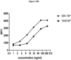
- the scFv comprises the amino acid sequence set forth in SEQ ID NO: 17 or SEQ ID NO: 19.
- the protein is a maxibody comprising an immunoglobulin Fc domain fused directly or via a linker to the antigen binding portion and further wherein the immunoglobulin Fc domain is an IgG1 Fc domain or an IgG4 Fc domain.
- the protein comprises the amino acid sequence set forth in SEQ ID NO: 18 or SEQ ID N0:20.
- the population of cytotoxic lymphocytes is for use in the treatment of a solid tumor in a subject.
- a protein that specifically binds to PD-L1 and comprises an antigen binding portion that comprises: (a) a first polypeptide comprising the 3 CDR amino acid sequences set forth in SEQ ID NOs:2-4, and a second polypeptide comprising the 3 CDR amino acid sequences set forth in SEQ ID NOs:6-8; or (b) a first polypeptide comprising the 3 CDR amino acid sequences set forth in SEQ ID NOs: 10-12, and a second polypeptide comprising the 3 CDR amino acid sequences set forth in SEQ ID NOs: 14-16, with the exception that each of the three CDR amino acid sequences of the first and/or second polypeptide comprises two or less conservative amino acid substitutions relative to the specified SEQ ID number.
- the antigen binding portion comprises a first polypeptide comprising the 3 CDR amino acid sequences set forth in SEQ ID NOs:2-4, and a second polypeptide comprising the 3 CDR amino acid sequences set forth in SEQ ID NOs:6-8.
- the first polypeptide comprises the amino acid sequence set forth in SEQ ID NO:1
- the second polypeptide comprises the amino acid sequence set forth in SEQ ID NO:5.
- the antigen binding portion comprises a first polypeptide comprising the 3 CDR amino acid sequences set forth in SEQ ID NOs: 10-12, and a second polypeptide comprising the 3 CDR amino acid sequences set forth in SEQ ID NOs: 14-16.
- the first polypeptide comprises the amino acid sequence set forth in SEQ ID NO:9
- the second polypeptide comprises the amino acid sequence set forth in SEQ ID NO: 13.
- the first polypeptide is a light chain
- the second polypeptide is a heavy chain
- the protein that specifically binds to PD-L1 is a single-chain antibody (scFv) and the first and second polypeptides are fused directly or via a linker to one another.
- the scFv comprises the amino acid sequence set forth in SEQ ID NO: 17 or SEQ ID NO: 19.
- the protein that specifically binds to PD-L1 is a maxibody comprising an immunoglobulin Fc domain fused directly or via a linker to the antigen binding portion.
- the immunoglobulin Fc domain is an IgG1 Fc domain.
- the protein comprises the amino acid sequence set forth in SEQ ID NO: 18 or SEQ ID NO:20.
- the immunoglobulin Fc domain is an IgG4 Fc domain.
- the protein that specifically binds to PD-L1 is a humanized antibody.
- nucleic acid comprising a nucleotide sequence encoding the protein that specifically binds to PD-L1, as discussed herein.
- the nucleic acid comprises a promoter that is operably linked to the nucleotide sequence encoding the protein.
- the promoter is a constitutive promoter.
- the promoter is an inducible promoter.
- a cell comprising the nucleic acid that encodes the protein that specifically binds to PD-L1.
- the nucleic acid is integrated into the cell's genome.
- the cell is a cytotoxic lymphocyte genetically modified to express and secrete the protein.
- the cytotoxic lymphocyte is a T-cell.
- the T-cell is a CD8+ T-cell.
- the T-cell is a CD4+ T-helper cell.
- the T-cell is derived from peripheral blood.
- the cytotoxic lymphocyte is a natural killer (NK) cell.
- the NK is derived from peripheral blood.
- the cytotoxic lymphocyte is a tumor infiltrating lymphocyte (TIL) derived from a tumor from a subject.
- TIL tumor infiltrating lymphocyte
- the TIL comprises a receptor specific for an antigen from the tumor.
- the cytotoxic lymphocyte exhibits an increased level of expression of one or more activation antigens relative to a naive T-cell.
- the one or more activation antigens are selected from CD25, CD26, CD27, CD28, CD38, CD40L, CD69, CD134, CD137, BTLA, PD-1, HVEM, LIGHT, and HLA-DR.
- the cytotoxic lymphocyte comprises a T-cell receptor specific for a tumor associated antigen.
- a method comprising: genetically modifying a cytotoxic lymphocyte isolated from a tumor of a subject by introducing into the cytotoxic lymphocyte the nucleic acid encoding a protein that specifically binds to PD-L1, wherein the genetically modified cytotoxic lymphocyte expresses and secretes the protein that specifically binds to PD-L1; expanding the genetically modified cytotoxic lymphocyte to generate a population of genetically modified cytotoxic lymphocytes; and administering the population of genetically modified cytotoxic lymphocytes to the subject to treat the tumor.
- the genetically modified cytotoxic lymphocyte constitutively expresses the protein that specifically binds to PD-L1. In some disclosures provided herein, the genetically modified cytotoxic lymphocyte inducibly expresses the protein that specifically binds to PD-L1. In some disclosures provided herein, the nucleic acid integrates into the cytotoxic lymphocyte's genome.
- the cytotoxic lymphocyte is a T-cell. In some disclosures provided herein, the T-cell is a CD8+ T-cell. In some disclosures provided herein, the T-cell is a CD4+ T-helper cell. In some disclosures provided herein, the cytotoxic lymphocyte is a natural killer (NK) cell.
- NK natural killer
- the genetically modified cytotoxic lymphocyte comprises a receptor specific for an antigen from the tumor.
- the method comprises isolating the cytotoxic lymphocyte from the subject prior to genetically modifying.
- the protein that specifically binds to PD-L1 and comprises an antigen binding portion that comprises: (a) a first polypeptide comprising the 3 CDR amino acid sequences set forth in SEQ ID NOs:2-4, and a second polypeptide comprising the 3 CDR amino acid sequences set forth in SEQ ID NOs:6-8; or (b) a first polypeptide comprising the 3 CDR amino acid sequences set forth in SEQ ID NOs: 10-12, and a second polypeptide comprising the 3 CDR amino acid sequences set forth in SEQ ID NOs: 14-16, with the exception that each of the three CDR amino acid sequences of the first and/or second polypeptide comprises two or less conservative amino acid substitutions relative to the specified SEQ ID number.
- the antigen binding portion comprises a first polypeptide comprising the 3 CDR amino acid sequences set forth in SEQ ID NOs:2-4, and a second polypeptide comprising the 3 CDR amino acid sequences set forth in SEQ ID NOs:6-8.
- the first polypeptide comprises the amino acid sequence set forth in SEQ ID NO:1
- the second polypeptide comprises the amino acid sequence set forth in SEQ ID NO:5.
- the antigen binding portion comprises a first polypeptide comprising the 3 CDR amino acid sequences set forth in SEQ ID NOs: 10-12, and a second polypeptide comprising the 3 CDR amino acid sequences set forth in SEQ ID NOs: 14-16.
- the first polypeptide comprises the amino acid sequence set forth in SEQ ID NO:9
- the second polypeptide comprises the amino acid sequence set forth in SEQ ID NO: 13.
- the first polypeptide is a light chain
- the second polypeptide is a heavy chain
- the protein is a single-chain antibody (scFv) and the first and second polypeptides are fused directly or via a linker to one another.
- scFv single-chain antibody
- the scFv comprises the amino acid sequence set forth in SEQ ID NO: 17 or SEQ ID NO: 19.
- the protein is a maxibody comprising an immunoglobulin Fc domain fused directly or via a linker to the antigen binding portion.
- the immunoglobulin Fc domain is an IgG1 Fc domain.
- the protein comprises the amino acid sequence set forth in SEQ ID NO: 18 or SEQ ID NO:20.
- a method of making a genetically modified cytotoxic lymphocyte comprising: genetically modifying a cytotoxic lymphocyte isolated from a subject having or suspected of having cancer by introducing into the cytotoxic lymphocyte the nucleic acid encoding for the protein that specifically binds to PD-L1, wherein the genetically modified cytotoxic lymphocyte expresses and secretes the protein that specifically binds to PD-L1.
- the genetically modified cytotoxic lymphocyte constitutively expresses the protein that specifically binds to PD-L1.
- the genetically modified cytotoxic lymphocyte inducibly expresses the protein that specifically binds to PD-L1.
- the method comprises expanding the cytotoxic lymphocyte in vitro to provide an expanded population of genetically modified cytotoxic lymphocytes.
- the method comprises isolating the cytotoxic lymphocyte from the subject prior to the genetically modifying.
- the isolating comprises isolating the cytotoxic lymphocyte from a tumor of the subject. In some disclosures provided herein, the isolating comprises isolating the cytotoxic lymphocyte from peripheral blood of the subject.
- the cytotoxic lymphocyte is a T-cell.
- the T-cell is a CD8+ T-cell.
- the T-cell is a CD4+ T-helper cell.
- the cytotoxic lymphocyte is a natural killer (NK) cell.
- the nucleic acid integrates into the cytotoxic lymphocyte's genome.
- the cytotoxic lymphocyte exhibits an increased level of expression of one or more activation antigens relative to a naive T-cell.
- the one or more activation antigens are selected from CD25, CD26, CD27, CD28, CD38, CD40L, CD69, CD134, CD137, BTLA, PD-1, HVEM, LIGHT, and HLA-DR.
- the genetically modified cytotoxic lymphocyte comprises a T-cell receptor specific for an antigen from a tumor of the subject.
- a method of treating an individual who has or is suspected of having cancer comprising: administering the protein that specifically binds to PD-L1 to the individual (e.g., subject or patient, including for example a mammal such as a human).
- the administering comprises introducing into the subject a nucleic acid encoding the protein that specifically binds to PD-L1.
- administering comprises introducing into the subject a genetically modified cytotoxic lymphocyte that expresses and secretes the protein that specifically binds to PD-L1.
- the genetically modified cytotoxic lymphocyte constitutively expresses the protein that specifically binds to PD-L1. In some disclosures provided herein of the method, the genetically modified cytotoxic lymphocyte inducibly expresses the protein that specifically binds to PD-L1. In some disclosures provided herein, the method, comprises inducing expression of the protein that specifically binds to PD-L1.
- the cytotoxic lymphocyte is a T-cell. In some methods disclosed herein, the T-cell is a CD8+ T-cell. In some disclosures provided herein of the method, the T-cell is a CD4+ T-helper cell. In some disclosures provided herein of the method, the cytotoxic lymphocyte is a natural killer (NK) cell.
- NK natural killer
- the cytotoxic lymphocyte exhibits an increased level of expression of one or more activation antigens relative to a naive T-cell.
- the one or more activation antigens are selected from CD25, CD26, CD27, CD28, CD38, CD40L, CD69, CD134, CD137, BTLA, PD-1, HVEM, LIGHT, and HLA-DR.
- the genetically modified cytotoxic lymphocyte comprises a T-cell receptor specific for an antigen from a tumor of the subject.
- a method of reducing the interaction between PD-L1 on a first-cell and PD-1 on a second cell comprising: contacting PD-L1 on the first-cell with the protein that specifically binds to PD-L1.
- the first and second cells are introduced to an individual, and the contacting comprises administering the protein that specifically binds to PD-L1.
- introducing comprises systemic administration.
- the introducing comprises local administration.
- the local administration comprises intratumoral administration.
- the individual has cancer.
- the individual has a solid tumor.
- the present disclosure provides proteins, such as antibodies, that include an antigen binding portion that specifically binds to Programmed Death 1 Ligand 1 (PD-L1). Also provided are nucleic acids encoding the proteins, and cells ( e.g ., genetically modified cytotoxic lymphocytes) that include such nucleic acids.
- a subject method includes reducing the interaction between PD-L1 on a first-cell and PD-1 on a second cell.
- the methods and compositions provided can be used in the treatment of viral infections and cancer, such as the treatment of solid tumors via ACT or via administration of a subject protein that specifically binds to PD-L1.
- compositions and methods of the present disclosure provide genetically modified cytotoxic lymphocytes, specifically TILs, that can be propagated and infused as a cell therapy, resulting in the secretion of a PD-L1 binding protein (e.g. an scFV, a maxibody, and the like) in the genetically modified cytotoxic lymphocytes infused into the subject.
- a PD-L1 binding protein e.g. an scFV, a maxibody, and the like
- the cytotoxic lymphocytes of the present disclosure are able to "relieve" themselves of the inhibitory effect of the PD-1 checkpoint, providing for an improved anti-cancer effect.
- Specific binding proteins of the disclosure e.g ., anti-PD-L1 antibodies
- an element means one element or more than one element.
- “About” as used herein when referring to a measurable value such as an amount, a temporal duration, and the like, is meant to encompass variations of ⁇ 20% or ⁇ 10%, more preferably ⁇ 5%, even more preferably ⁇ 1%, and still more preferably ⁇ 0.1% from the specified value, as such variations are appropriate to perform the disclosed methods.
- anti-tumor effect refers to a biological effect which can be manifested by a decrease in tumor volume, a decrease in the number of tumor cells, a decrease in the number of metastases, an increase in life expectancy, or amelioration of various physiological symptoms associated with the cancerous condition.
- An "anti-tumor effect” can also be manifested by the ability of the disclosed compositions and methods to prevent the occurrence of tumor in the first place.
- antibody is used in the broadest sense and includes, for example, an intact immunoglobulin or an antigen binding portion thereof that competes with the intact antibody for specific binding, unless otherwise specified. Antigen binding portions may be produced by recombinant DNA techniques or by enzymatic or chemical cleavage of intact antibodies.
- Antigen binding portions include Fab, Fab', F(ab')2, Fd, Fv, and domain antibodies (dAbs), and complementarity determining region (CDR) fragments, single-chain antibodies (single-chain variable domain fragment; scFv), diabodies, triabodies, tetrabodies, and polypeptides that contain at least a portion of an immunoglobulin that is sufficient to confer specific antigen binding to the polypeptide.
- Antibody includes a human antibody, a humanized antibody, chimeric antibody, a monoclonal antibody, a polyclonal antibody, a recombinant antibody, an antigen-binding antibody fragment, a single chain antibody, a maxibody (scFv fused by a linker or direct attachment to an Fc or an Fc fragment), a diabody, a triabody, a tetrabody, a Fab fragment, an F(fa')x fragment, a domain antibody, an IgD antibody, an IgE antibody, and IgM antibody, and IgG1 antibody, and IgG2 antibody, and IgG3 antibody, and IgG4 antibody, and IgG4 antibody having at least one mutation in the hinge region that alleviates a tendency to for intra H-chain disulfide bonds.
- a "Fab fragment” is a monovalent fragment having the VL, VH, CL and CH1 domains; a F(ab')2 fragment is a bivalent fragment having two Fab fragments linked by a disulfide bridge at the hinge region; a Fd fragment has the VH and CH1 domains; an Fv fragment has the VL and VH domains of a single arm of an antibody; and a dAb fragment has a VH domain, a VL domain, or an antigen-binding fragment of a VH or VL domain.
- a "single-chain antibody” is an antibody in which a VL and a VH region are fused directly or joined via a linker (e.g., a synthetic sequence of amino acid residues) to form a continuous protein chain wherein the linker is long enough to allow the protein chain to fold back on itself and form a monovalent antigen binding site (see, e.g., Bird et al., 1988, Science 242:423-26 and Huston et al, 1988, Proc. Natl. Acad. Sci. USA 85:5879-83 ).
- a linker e.g., a synthetic sequence of amino acid residues
- Diabodies are bivalent antibodies comprising two polypeptide chains, wherein each polypeptide chain comprises VH and VL domains joined by a linker that is too short to allow for pairing between two domains on the same chain, thus allowing each domain to pair with a complementary domain on another polypeptide chain (see, e.g., Holliger et al., 1993, Proc. Natl. Acad. Sci. USA 90:6444-48 , and Poljak et al., 1994, Structure 2:1121-23 ). If the two polypeptide chains of a diabody are identical, then a diabody resulting from their pairing will have two identical antigen binding sites.
- Polypeptide chains having different sequences can be used to make a diabody with two different antigen binding sites.
- tribodies and tetrabodies are antibodies comprising three and four polypeptide chains, respectively, and forming three and four antigen binding sites, respectively, which can be the same or different.
- Complementarity determining regions (CDRs) and framework regions (FR) of a given antibody may be identified using the system described by Kabat et al. in Sequences of Proteins of Immunological Interest, 5th Ed., US Dept. of Health and Human Services, PHS, NIH, NIH Publication no. 91-3242, 1991 .
- One or more CDRs may be incorporated into a molecule either covalently or noncovalently to make it an antigen binding protein.
- An antigen binding protein may incorporate the CDR(s) as part of a larger polypeptide chain, may covalently link the CDR(s) to another polypeptide chain, or may incorporate the CDR(s) noncovalently.
- the CDRs permit the antigen binding protein to specifically bind to a particular antigen of interest.
- human antibody includes all antibodies that have one or more variable and constant regions derived from human immunoglobulin sequences. In one embodiment, all of the variable and constant domains are derived from human immunoglobulin sequences (a fully human antibody).
- Human antibodies may be prepared in a variety of ways, including immunization of a mouse that is genetically modified to express human antibodies. One can engineer mouse strains deficient in mouse antibody production with large fragments of the human Ig loci in anticipation that such mice would produce human antibodies in the absence of mouse antibodies. Large human Ig fragments may preserve the large variable gene diversity as well as the proper regulation of antibody production and expression.
- human antibody repertoire in these mouse strains may yield high affinity fully human antibodies against any antigen of interest, including human antigens.
- antigen-specific human MAbs with the desired specificity may be produced and selected.
- Human antibodies can also be prepared by panning human antibody libraries expressed on phage, phagemids, ribosomes, or other particles.
- a “humanized antibody” has a sequence that differs from the sequence of an antibody derived from a non-human species by one or more amino acid substitutions, deletions, and/or additions, such that the humanized antibody is less likely to induce an immune response, and/or induces a less severe immune response, as compared to the non-human species antibody, when it is administered to a human subject.
- certain amino acids in the framework and constant domains of the heavy and/or light chains of the non-human species antibody are mutated to produce the humanized antibody.
- the constant domain(s) from a human antibody are fused to the variable domain(s) of a non-human species.
- One or more amino acid residues in one or more CDR sequences of a non-human antibody can be changed to reduce the likely immunogenicity of the non-human antibody when it is administered to a human subject, wherein the changed amino acid residues either are not critical for immunospecific binding of the antibody to its antigen, or the changes to the amino acid sequence that are made are conservative changes, such that the binding of the humanized antibody to the antigen is not significantly worse than the binding of the non-human antibody to the antigen. Examples of methods for making humanized antibodies may be found in U.S. Pat. Nos. 6,054,297 , 5,886,152 and 5,877,293 .
- chimeric antibody refers to an antibody that contains one or more regions from one antibody and one or more regions from one or more other antibodies.
- a “CDR grafted antibody” is an antibody comprising one or more CDRs derived from an antibody of a particular species or isotype and the framework of another antibody of the same or different species or isotype.
- K assoc or "K a ", as used herein, is intended to refer to the association rate of a particular antibody-antigen interaction
- K dis or "K d ,” as used herein, is intended to refer to the dissociation rate of a particular antibody-antigen interaction
- K D is intended to refer to the dissociation constant, which is obtained from the ratio of K d to K a ( i.e. , K d /K a ) and is expressed as a molar concentration (M).
- K D values for antibodies can be determined using methods well established in the art. The method for determining the K D of an antibody may be by using surface plasmon resonance, for example, by using a biosensor system such as a Biacore ® system.
- high affinity for an IgG antibody refers to an antibody having a K D of 10 -8 M or less, in some embodiments, 10 -9 M or less, and in some embodiments, 10 -10 M or less for a target antigen.
- high affinity binding can vary for other antibody isotypes.
- “high affinity” binding for an IgM isotype refers to an antibody having a K D of 10 -7 M or less, in some embodiments, 10 -8 M or less, and in some embodiments, 10 -9 M or less.
- Constant expression includes a state in which a gene product is produced in a living cell under most or all physiological conditions of the cell.
- a "coding region" of a gene includes the nucleotide residues of the coding strand of the gene and the nucleotides of the non-coding strand of the gene which are homologous with or complementary to, respectively, the coding region of an mRNA molecule which is produced by transcription of the gene.
- a "coding region” of a mRNA molecule also includes the nucleotide residues of the mRNA molecule which are matched with an anti-codon region of a transfer RNA molecule during translation of the mRNA molecule or which encode a stop codon.
- the coding region may thus include nucleotide residues comprising codons for amino acid residues which are not present in the mature protein encoded by the mRNA molecule ( e.g., amino acid residues in a protein export signal sequence).
- TILs tumor infiltrating lymphocytes
- TILs include, but are not limited to, CD8+ cytotoxic T-cells (lymphocytes), Th1 and Th17 CD4+ T-cells, natural killer cells, dendritic cells, and M1 macrophages.
- TILs include both primary and secondary TILs.
- Primary TILs are those that are obtained from patient tissue samples as outlined herein (sometimes referred to as “freshly harvested")
- secondary TILs are any TIL cell populations that have been expanded or proliferated as discussed herein, including, but not limited to bulk TILs and expanded TILs (“REP TILs”) as discussed herein.
- TILs can generally be defined either biochemically, using cell surface markers, or functionally, by their ability to infiltrate tumors and effect treatment.
- TILs can be generally categorized by expressing one or more of the following biomarkers: CD4, CD8, TCR ⁇ , CD27, CD28, CD56, CCR7, CD45Ra, CD95, PD-1, and CD25. Additionally and alternatively, TILs can be functionally defined by their ability to infiltrate solid tumors upon reintroduction into a patient.
- cytotoxic lymphocyte includes cytotoxic T (CTL) cells (including CD8 + cytotoxic T lymphocytes and CD4 + T-helper lymphocytes), natural killer T (NKT) cells and natural killer (NK) cells.
- CTL cytotoxic T
- NKT natural killer T
- NK natural killer cells
- Cytotoxic lymphocytes can include, for example, peripheral blood-derived ⁇ TCR-positive or ⁇ TCR-positive T-cells activated by tumor associated antigens and/or transduced with tumor specific chimeric antigen receptors or T-cell receptors, and tumor-infiltrating lymphocytes (TELs). Cytotoxic lymphocyte generally kill cancer cells, cells that are infected (particularly with viruses), or cells that are otherwise damaged or defective.
- a cytotoxic lymphocyte can also be referred to as a cytotoxic T-cell, TC, cytotoxic T lymphocyte, CTL, T-killer cell, cytolytic T-cell, CD8+ T-cell or killer T-cell is a T lymphocyte (a type of white blood cell).
- fragmenting includes mechanical fragmentation methods such as crushing, slicing, dividing, and morcellating tumor tissue as well as any other method for disrupting the physical structure of tumor tissue.
- in vivo refers to an event that takes place in a subject's body.
- in vitro refers to an event that takes places outside of a subject's body.
- in vitro assays encompass cell-based assays in which cells alive or dead are employed and may also encompass a cell-free assay in which no intact-cells are employed.
- anti-CD3 antibody refers to an antibody or variant thereof, e.g., a monoclonal antibody and including human, humanized, chimeric or murine antibodies which are directed against the CD3 receptor in the T-cell antigen receptor of mature T-cells.
- Anti-CD3 antibodies include OKT-3, also known as muromonab.
- Other anti-CD3 antibodies include, for example, otelixizumab, teplizumab, and visilizumab.
- OKT-3 refers to a monoclonal antibody or biosimilar or variant thereof, including human, humanized, chimeric, or murine antibodies, directed against the CD3 receptor in the T-cell antigen receptor of mature T-cells, and includes commercially-available forms such as OKT-3 (30 ng/mL, MACS GMP CD3 pure, Miltenyi Biotech, Inc., San Diego, CA, USA) and muromonab or variants, conservative amino acid substitutions, glycoforms, or biosimilars thereof.
- the amino acid sequences of the heavy and light chains of muromonab are given in Table 1 (SEQ ID NO:27 and SEQ ID NO:28).
- a hybridoma capable of producing OKT-3 is deposited with the American Type Culture Collection and assigned the ATCC accession number CRL 8001.
- a hybridoma capable of producing OKT-3 is also deposited with European Collection of Authenticated Cell Cultures (ECACC) and assigned Catalogue No. 86022706.
- ECACC European Collection of Authenticated Cell Cultures
- TABLE 1 Amino acid sequences of muromonab. Identifier Sequence (One-Letter Amino Acid Symbols) SEQ ID NO:27 Muromonab heavy chain SEQ ID NO:28 Muromonab light chain
- IL-2 refers to the T-cell growth factor known as interleukin-2, and includes all forms of IL-2 including human and mammalian forms, conservative amino acid substitutions, glycoforms, biosimilars, and variants thereof.
- IL-2 is described, e.g., in Nelson, J. Immunol. 2004, 172, 3983-88 and Malek, Annu. Rev. Immunol. 2008, 26, 453-79 .
- the amino acid sequence of recombinant human IL-2 suitable for use in the invention is given in Table 2 (SEQ ID NO:3).
- IL-2 encompasses human, recombinant forms of IL-2 such as aldesleukin (PROLEUKIN, available commercially from multiple suppliers in 22 million ill per single use vials), as well as the form of recombinant IL-2 commercially supplied by CellGenix, Inc., Portsmouth, NH, USA (CELLGRO GMP) or ProSpec-Tany TechnoGene Ltd., East Brunswick, NJ, USA (Cat. No. CYT-209-b) and other commercial equivalents from other vendors.
- Aldesleukin (des-alanyl-1, serine-125 human IL-2) is a nonglycosylated human recombinant form of IL-2 with a molecular weight of approximately 15 kDa.
- IL-2 also encompasses pegylated forms of IL-2, as described herein, including the pegylated IL2 prodrug NKTR-214, available from Nektar Therapeutics, South San Francisco, CA, USA.
- NKTR-214 and pegylated IL-2 suitable for use in the invention is described in U.S. Patent Application Publication No. US 2014/0328791 A1 and International Patent Application Publication No. WO 2012/065086 A1 .
- Alternative forms of conjugated IL-2 suitable for use in the invention are described in U.S. Patent Nos. 4,766,106, 5,206,344 , 5,089,261 and 4902,502 .
- Formulations of IL-2 suitable for use in the invention are described in U.S. Patent No. 6,706,289 .
- interleukins include IL-2, IL-4, IL-7, IL-15, and IL-21. Exemplary sequences for these interleukins are provided in Table 2 below. TABLE 2. Amino acid sequences of interleukins. Identifier Sequence (One-Letter Amino Acid Symbols) SEQ ID NO:29 recombinant human IL-2 (rhIL-2) SEQ ID NO:30 Aldesleukin (Proleukin ® ) SEQ ID NO:31 recombinant human IL-4 (rhIL-4) SEQ ID NO:32 recombinant human IL-7 (rhIL-7) SEQ ID NO:33 recombinant human IL-15 (rhIL-15) SEQ ID NO:34 recombinant human IL-21 (rhIL-21)
- IL-4" refers to the cytokine known as interleukin 4, which is produced by Th2 T-cells and by eosinophils, basophils, and mast-cells.
- IL-4 regulates the differentiation of naive helper T-cells (Th0 cells) to Th2 T-cells. Steinke and Borish, Respir. Res. 2001, 2, 66-70 .
- Th2 T-cells Upon activation by IL-4, Th2 T-cells subsequently produce additional IL-4 in a positive feedback loop.
- IL-4 also stimulates B cell proliferation and class II MHC expression, and induces class switching to IgE and IgG1 expression from B cells.
- Recombinant human IL-4 suitable for use in the invention is commercially available from multiple suppliers, including ProSpec-Tany TechnoGene Ltd., East Brunswick, NJ, USA (Cat. No. CYT-211) and ThermoFisher Scientific, Inc., Waltham, MA, USA (human IL-15 recombinant protein, Cat. No. Gibco CTP0043).
- the amino acid sequence of recombinant human IL-4 suitable for use in the invention is given in Table 2 (SEQ ID NO:31).
- IL-7 refers to a glycosylated tissue-derived cytokine known as interleukin 7, which may be obtained from stromal and epithelial cells, as well as from dendritic cells. Fry and Mackall, Blood 2002, 99, 3892-904 .
- IL-7 can stimulate the development of T-cells.
- IL-7 binds to the IL-7 receptor, a heterodimer consisting of IL-7 receptor alpha and common gamma chain receptor, which in a series of signals important for T-cell development within the thymus and survival within the periphery.
- Recombinant human IL-4 suitable for use in the invention is commercially available from multiple suppliers, including ProSpec-Tany TechnoGene Ltd., East Brunswick, NJ, USA (Cat. No. CYT-254) and ThermoFisher Scientific, Inc., Waltham, MA, USA (human IL-15 recombinant protein, Cat. No. Gibco PHC0071).
- the amino acid sequence of recombinant human IL-7 suitable for use in the invention is given in Table 2 (SEQ ID NO:32).
- IL-15 refers to the T-cell growth factor known as interleukin-15, and includes all forms of IL-2 including human and mammalian forms, conservative amino acid substitutions, glycoforms, biosimilars, and variants thereof.
- IL-15 is described, e.g., in Fehniger and Caligiuri, Blood 2001, 97, 14-32 .
- IL-15 shares ⁇ and ⁇ signaling receptor subunits with IL-2.
- Recombinant human IL-15 is a single, non-glycosylated polypeptide chain containing 114 amino acids (and an N-terminal methionine) with a molecular mass of 12.8 kDa.
- Recombinant human IL-15 is commercially available from multiple suppliers, including ProSpec-Tany TechnoGene Ltd., East Brunswick, NJ, USA (Cat. No. CYT-230-b) and ThermoFisher Scientific, Inc., Waltham, MA, USA (human IL-15 recombinant protein, Cat. No. 34-8159-82).
- the amino acid sequence of recombinant human IL-15 suitable for use in the invention is given in Table 2 (SEQ ID NO:33).
- IL-21 refers to the pleiotropic cytokine protein known as interleukin-21, and includes all forms of II,-21 including human and mammalian forms, conservative amino acid substitutions, glycoforms, biosimilars, and variants thereof. IL-21 is described, e.g., in Spolski and Leonard, Nat. Rev. Drug. Disc. 2014, 13, 379-95 . IL-21 is primarily produced by natural killer T-cells and activated human CD4+ T-cells. Recombinant human IL-21 is a single, non-glycosylated polypeptide chain containing 132 amino acids with a molecular mass of 15.4 kDa.
- Recombinant human IL-21 is commercially available from multiple suppliers, including ProSpec-Tany TechnoGene Ltd., East Brunswick, NJ, USA (Cat. No. CYT-408-b) and ThermoFisher Scientific, Inc., Waltham, MA, USA (human IL-21 recombinant protein, Cat. No. 14-8219-80).
- the amino acid sequence of recombinant human IL-21 suitable for use in the invention is given in Table 2 (SEQ ID NO:34).
- a "disease” includes a state of health of an animal wherein the animal cannot maintain homeostasis, and wherein if the disease is not ameliorated then the animal's health continues to deteriorate.
- a "disorder" in an animal includes a state of health in which the animal is able to maintain homeostasis, but in which the animal's state of health is less favorable than it would be in the absence of the disorder. Left untreated, a disorder does not necessarily cause a further decrease in the animal's state of health.
- a disease or disorder is "alleviated” if the severity of a symptom of the disease or disorder, the frequency with which such a symptom is experienced by a patient, or both, is reduced.
- the term "tumor” or “cancer” can be any cancer, including any of acute lymphocytic cancer, acute myeloid leukemia, alveolar rhabdomyosarcoma, bone cancer, brain cancer, breast cancer, cancer of the anus, anal canal, or anorectum, cancer of the eye, cancer of the intrahepatic bile duct, cancer of the joints, cancer of the neck, gallbladder, or pleura, cancer of the nose, nasal cavity, or middle ear, cancer of the vulva, chronic lymphocytic leukemia, chronic myeloid cancer, cervical cancer, glioma, Hodgkin's lymphoma, hypopharynx cancer, kidney cancer, larynx cancer, liver cancer, lung cancer, malignant mesothelioma, melanoma, multiple myeloma, nasopharynx cancer, non-Hodgkin's lymphoma, ovarian cancer, peritoneum
- the term "mammal” refers to any mammal, including, but not limited to, mammals of the order Rodentia, such as mice and hamsters, and mammals of the order Logomorpha, such as rabbits.
- the mammals are from the order Carnivora, including felines (cats) and canines (dogs).
- the mammals are from the order Artiodactyla, including bovines (cows) and swines (pigs) or of the order Perssodactyla, including Equines (horses). It is most preferred that the mammals are of the order Primates, Ceboids, or Simoids (monkeys) or of the order Anthropoids (humans and apes).
- the mammal is the human.
- regression does not necessarily imply 100% or complete regression. Rather, there are varying degrees of regression of which one of ordinary skill in the art recognizes as having a potential benefit or therapeutic effect.
- the inventive methods can provide any amount of any level of regression of cancer in a mammal.
- the regression provided by the inventive method can include regression of one or more conditions or symptoms of the disease, e.g., cancer.
- regression can encompass delaying the onset of the disease, or delaying the onset of a symptom and/or delaying the onset of a condition thereof.
- an “effective amount” or “therapeutically effective amount” of a composition includes that amount of the composition which is sufficient to provide a beneficial effect to the subject to which the composition is administered.
- An “effective amount” of a delivery vehicle includes that amount sufficient to effectively bind or deliver a composition.
- Encoding includes the inherent property of specific sequences of nucleotides in a polynucleotide, such as a gene, a cDNA, or an mRNA, to serve as templates for synthesis of other polymers and macromolecules in biological processes having either a defined sequence of nucleotides (i.e., rRNA, tRNA and mRNA) or a defined sequence of amino acids and the biological properties resulting therefrom.
- a gene encodes a protein if, for example, transcription and translation of mRNA corresponding to that gene produces the protein in a cell or other biological system.
- Both the coding strand the nucleotide sequence of which is identical to the mRNA sequence and is usually provided in sequence listings, and the non-coding strand, used as the template for transcription of a gene or cDNA, can be referred to as encoding the protein or other product of that gene or cDNA.
- endogenous includes any material from or produced inside an organism, cell, tissue or system.
- exogenous includes any material introduced from or produced outside an organism, cell, tissue or system.
- An “expression cassette” includes any nucleic acid construct capable of directing the expression of a gene/coding sequence of interest, which is operably linked to a promoter of the expression cassette. Such cassettes can be constructed into a “vector,” “vector construct,” “expression vector,” or “gene transfer vector,” in order to transfer the expression cassette into target-cells.
- vector vector construct
- vector vector
- gene transfer vector gene transfer vector
- fragment includes a subsequence of a larger nucleic acid or polypeptide.
- a “fragment” of a nucleic acid can be at least about 15 nucleotides in length; for example, at least about 50 nucleotides to about 100 nucleotides; at least about 100 to about 500 nucleotides, at least about 500 to about 1000 nucleotides, at least about 1000 nucleotides to about 1500 nucleotides; or about 1500 nucleotides to about 2500 nucleotides; or about 2500 nucleotides (and any integer value in between).
- a “fragment" of a polypeptide can be at least about 15 amino acids in length; for example, at least about 50 amino acids to about 100 amino acids; at least about 100 to about 500 amino acids, at least about 500 to about 1000 amino acids, at least about 1000 amino acids to about 1500 amino acids; or about 1500 amino acids to about 2500 amino acids; or about 2500 amino acids (and any integer value in between).
- the terms "gene” and “recombinant gene” includes nucleic acid molecules comprising an open reading frame encoding a polypeptide.
- Such natural allelic variations can typically result in 1-5% variance in the nucleotide sequence of a given gene.
- Alternative alleles can be identified by sequencing the gene of interest in a number of different individuals. This can be readily carried out by using hybridization probes to identify the same genetic locus in a variety of individuals. Any and all such nucleotide variations and resulting amino acid polymorphisms or variations that are the result of natural allelic variation and that do not alter the functional activity are intended to be within the scope of the invention.
- Homologous includes the subunit sequence similarity between two polymeric molecules, e.g. between two nucleic acid molecules, e.g., two DNA molecules or two RNA molecules, or between two polypeptide molecules. When a subunit position in both of the two molecules is occupied by the same monomeric subunit, e.g., if a position in each of two DNA molecules is occupied by adenine, then they are homologous at that position. The homology between two sequences is a direct function of the number of matching or homologous positions, e.g.
- the two sequences are 50% homologous, if 90% of the positions, e.g. 9 of 10, are matched or homologous, the two sequences share 90% homology.
- the DNA sequences 5'-ATTGCC-3' and 5'-TATGGC-3' share 50% homology.
- “Inducible” expression includes a state in which a gene product is produced in a living cell in response to the presence of a signal in the cell.
- an "instructional material” includes a publication, a recording, a diagram, or any other medium of expression which can be used to communicate the usefulness and/or use of a compound, composition, vector, or delivery system of the present disclosure in a kit according to the present disclosure, e.g., a kit for effecting alleviation of the various diseases or disorders recited herein.
- the instructional material can describe one or more methods of alleviating the diseases or disorders in a cell or a tissue of a mammal.
- the instructional material of the kit disclosed herein can, for example, be affixed to a container which contains the identified compound, composition, vector, or delivery system of the invention or be shipped together with a container which contains the identified compound, composition, vector, or delivery system.
- the instructional material can be shipped separately from the container with the intention that the instructional material and the compound be used cooperatively by the recipient.
- nucleic acid includes RNA or DNA molecules having more than one nucleotide in any form including single-stranded, double-stranded, oligonucleotide or polynucleotide.
- nucleotide sequence includes the ordering of nucleotides in an oligonucleotide or polynucleotide in a single-stranded form of nucleic acid.
- nucleic acid construct it is meant a nucleic acid sequence that has been constructed to comprise one or more functional units not found together in nature. Examples include circular, linear, double-stranded, extrachromosomal DNA molecules (plasmids), cosmids (plasmids containing COS sequences from lambda phage), viral genomes including non-native nucleic acid sequences, and the like.
- plasmids extrachromosomal DNA molecules
- cosmids plasmids containing COS sequences from lambda phage
- viral genomes including non-native nucleic acid sequences, and the like.
- operably linked includes a polynucleotide in functional relationship with a second polynucleotide, e.g. a single-stranded or double-stranded nucleic acid moiety comprising the two polynucleotides arranged within the nucleic acid moiety in such a manner that at least one of the two polynucleotides is able to exert a physiological effect by which it is characterized, upon the other.
- a promoter operably linked to the coding region of a gene is able to promote transcription of the coding region. The order specified when indicating operably linkage is not important.
- the phrases: "the promoter is operably linked to the nucleotide sequence” and “the nucleotide sequence is operably linked to the promoter” are used interchangeably herein and are considered equivalent.
- the nucleic acid encoding the desired protein further comprises a promoter/regulatory sequence
- the promoter/regulatory sequence is positioned at the 5' end of the desired protein coding sequence such that it drives expression of the desired protein in a cell.
- oligonucleotide refers to a polymeric forms of nucleotides of any length, either ribonucleotides or deoxyribonucleotides.
- this term includes, but is not limited to, single-, double-, or multi-stranded DNA or RNA, genomic DNA, cDNA, DNA-RNA hybrids, or a polymer comprising purine and pyrimidine bases or other natural, chemically or biochemically modified, non-natural, or derivatized nucleotide bases.
- the backbone of the polynucleotide can comprise sugars and phosphate groups (as may typically be found in RNA or DNA), or modified or substituted sugar or phosphate groups.
- the backbone of the polynucleotide can comprise a polymer of synthetic subunits such as phosphoramidites, and/or phosphorothioates, and thus can be an oligodeoxynucleoside phosphoramidate or a mixed phosphoramidate-phosphodiester oligomer. Peyrottes et al. (1996) Nucl. Acids Res. 24:1841-1848 ; Chaturvedi et al. (1996) Nucl. Acids Res. 24:2318-2323 .
- the polynucleotide may comprise one or more L-nucleosides.
- a polynucleotide may comprise modified nucleotides, such as methylated nucleotides and nucleotide analogs, and may be interrupted by non-nucleotide components. If present, modifications to the nucleotide structure may be imparted before or after assembly of the polymer.
- a polynucleotide may comprise modified nucleotides, such as methylated nucleotides and nucleotide analogs, uracyl, other sugars, and linking groups such as fluororibose and thioate, and nucleotide branches. The sequence of nucleotides may be interrupted by non-nucleotide components.
- a polynucleotide may be further modified after polymerization, such as by conjugation with a labeling component.
- modifications included in this definition are caps, substitution of one or more of the naturally occurring nucleotides with an analog, and introduction of means for attaching the polynucleotide to proteins, metal ions, labeling components, other polynucleotides, or a solid support.
- a polynucleotide or oligonucleotide is represented by a sequence of letters (upper or lower case), such as "ATGCCTG,” it will be understood that the nucleotides are in 5' ⁇ 3' order from left to right and that "A” denotes deoxyadenosine, “C” denotes deoxycytidine, “G” denotes deoxyguanosine, and “T” denotes thymidine, “I” denotes deoxyinosine, "U” denotes uridine, unless otherwise indicated or obvious from context.
- Polynucleotides can be single, double, or triplex, linear or circular, and can be of any length. In discussing polynucleotides, a sequence or structure of a particular polynucleotide may be described herein according to the convention of providing the sequence in the 5' to 3' direction.
- polynucleotide as applied to a polynucleotide means the polynucleotide is the product of various combinations of cloning, restriction or ligation steps, and other procedures resulting in a construct distinct and/or different from a polynucleotide found in nature. The terms respectively include replicates of the original polynucleotide construct and progeny of the original virus construct.
- P-L1 binding protein refers to a polypeptide (e.g., a fusion protein, an scFV, a maxibody, an antibody, and the like), which is capable of specifically binding to Programmed Death 1 Ligand 1 (PD-L1) protein (a.k.a. CD274 or B7-H1) expressed on the surface of a cell.
- a polypeptide e.g., a fusion protein, an scFV, a maxibody, an antibody, and the like
- PD-L1 protein a.k.a. CD274 or B7-H1
- polypeptide refers to a polymeric form of amino acids of any length, which can include coded and non-coded amino acids, chemically or biochemically modified or derivatized amino acids, and polypeptides having modified peptide backbones.
- the term includes polypeptide chains modified or derivatized in any manner, including, but not limited to, glycosylation, formylation, cyclization, acetylation, phosphorylation, and the like.
- the term includes naturally-occurring peptides, synthetic peptides, and peptides comprising one or more amino acid analogs.
- fusion proteins including, but not limited to, fusion proteins with a heterologous amino acid sequence, fusions with heterologous and homologous leader sequences, with or without N-terminal methionine residues; immunologically tagged proteins; and the like.
- tumor-associated antigen is a term well understood in the art, and refers to molecules that are differentially over-expressed in tumor cells relative to non-cancerous cells of the same cell type.
- tumor-associated antigen includes not only complete tumor-associated antigens that can be expressed on the cell surface, but also epitope-comprising portions (fragments) thereof that are recognized by T-cells.
- a tumor-associated antigen (TAA) may be one found in nature, or may be a synthetic version of a TAA found in nature, or may be a variant of a naturally-occurring TAA, e.g., a variant which has enhanced immunogenic properties.
- the TAA may be a naturally occurring over-expressed protein or a mutated protein expressed only in tumor cells or other transformed cells in tumors.
- promoter includes a DNA sequence operably linked to a nucleic acid sequence to be transcribed such as a nucleic acid sequence encoding a desired molecule.
- a promoter is generally positioned upstream of a nucleic acid sequence to be transcribed and provides a site for specific binding by RNA polymerase and other transcription factors.
- polypeptide As used interchangeably herein to refer to polymers of amino acids of any length.
- polypeptides As with naturally occurring proteins, as well as functional subsequences, modified forms or sequence variants so long as the subsequence, modified form, or variant retains some degree of functionality of the native full-length protein.
- polypeptides, proteins, and peptides encoded by the polynucleotide sequences can be but are not required to be identical to the defective endogenous protein, or whose expression is insufficient, or deficient in the treated mammal.
- the terms also encompass a modified amino acid polymer; for example, disulfide bond formation, glycosylation, lipidation, phosphorylation, methylation, carboxylation, deamidation, acetylation, or conjugation with a labeling component.
- Polypeptides such as anti-angiogenic polypeptides, neuroprotective polypeptides, and the like, when discussed in the context of delivering a gene product to a mammalian subject, and compositions therefor, refer to the respective intact polypeptide, or any fragment or genetically engineered derivative thereof, retaining the desired biochemical function of the intact protein.
- a "recombinant polypeptide” includes one which is produced upon expression of a recombinant polynucleotide.
- the term "specifically binds,” as used herein, e.g., with respect to an antibody/antigen binding region, includes an antibody/antigen binding region which recognizes a specific antigen, but does not substantially recognize or bind other molecules in a sample.
- an antibody e.g., an scFV
- an antibody that specifically binds to an antigen from one species may also bind to that antigen from one or more other species. But, such cross-species reactivity does not itself alter the classification of an antibody as specific.
- an antibody that specifically binds to an antigen may also bind to different allelic forms of the antigen. However, such cross reactivity does not itself alter the classification of an antibody as specific.
- the terms “specific binding” or “specifically binding”, can be used in reference to the interaction of an antibody, a protein, or a peptide with a second chemical species, to mean that the interaction is dependent upon the presence of a particular structure (e.g., an antigenic determinant or epitope) on the chemical species; for example, an antibody recognizes and binds to a specific protein structure rather than to proteins generally. If an antibody is specific for epitope "A”, the presence of a molecule containing epitope A (or free, unlabeled A), in a reaction containing labeled "A” and the antibody, will reduce the amount of labeled A bound to the antibody.
- a particular structure e.g., an antigenic determinant or epitope
- synthetic antibody as used herein includes an antibody which is generated using recombinant DNA technology, such as, for example, an antibody expressed by a bacteriophage.
- the term should also be construed to mean an antibody which has been generated by the synthesis of a DNA molecule encoding the antibody and which DNA molecule expresses an antibody protein, or an amino acid sequence specifying the antibody, wherein the DNA or amino acid sequence has been obtained using synthetic DNA or amino acid sequence technology which is available and well known in the art.
- Variant includes a nucleic acid sequence or a peptide sequence that differs in sequence from a reference nucleic acid sequence or peptide sequence respectively, but retains essential biological properties of the reference molecule. Changes in the sequence of a nucleic acid variant may not alter the amino acid sequence of a peptide encoded by the reference nucleic acid, or may result in amino acid substitutions, additions, deletions, fusions, and truncations. Changes in the sequence of peptide variants are typically limited or conservative, so that the sequences of the reference peptide and the variant are closely similar overall and, in many regions, identical.
- a variant and reference peptide can differ in amino acid sequence by one or more substitutions, additions, deletions in any combination.
- a variant of a nucleic acid or peptide can be a naturally occurring such as an allelic variant, or can be a variant that is not known to occur naturally. Non-naturally occurring variants of nucleic acids and peptides may be made by mutagenesis techniques or by direct synthesis.
- substitution results from the replacement of one or more amino acids or nucleotides by different amino acids or nucleotides, respectively as compared to an amino acid sequence or nucleotide sequence of a polypeptide. If a substitution is conservative, the amino acid that is substituted into a polypeptide has similar structural or chemical properties (e.g., charge, polarity, hydrophobicity, and the like) to the amino acid that it is substituting.
- amino acid substitutions usually result in a substitution of a first amino acid with second amino acid from the same group as the first amino acid, where examples of amino acid groups are as follows: (1) acidic (negatively charged) amino acids such as aspartic acid and glutamic acid; (2) basic (positively charged) amino acids such as arginine, histidine, and lysine; (3) neutral polar amino acids such as glycine, serine, threonine, cysteine, tyrosine, asparagine, and glutamine; and (4) neutral non-polar amino acids such as alanine, leucine, isoleucine, valine, proline, phenylalanine, tryptophan, and methionine.
- Polypeptide variants may have "non-conservative" changes, where the substituted amino acid differs in structural and/or chemical properties.
- a “deletion” is defined as a change in either amino acid or nucleotide sequence in which one or more amino acid or nucleotide residues, respectively, are absent as compared to an amino acid sequence or nucleotide sequence of a naturally occurring polypeptide or polynucleotide.
- a deletion can involve deletion of 2, 5, 10, up to 20, up to 30, or up to 50 or more amino acids or nucleotide residues, taking into account the length of the polypeptide or polynucleotide sequence being modified.
- an “insertion” or “addition” is that change in an amino acid or nucleotide sequence which has resulted in the addition of one or more amino acid or nucleotide residues, respectively, as compared to an amino acid sequence or nucleotide sequence of a naturally occurring polypeptide or polynucleotide.
- Insertion generally refers to addition of one or more amino acid residues within an amino acid sequence of a polypeptide (or nucleotide residues within a polynucleotide), while “addition” can be an insertion or refer to amino acid residues added at the N- or C-termini of a polypeptide (or nucleotide residues added at the 5' or 3' end of a polynucleotide).
- an insertion or addition may be of up to 10, up to 20, up to 30, or up to 50 or more amino acids (or nucleotide residues).
- an "isolated" plasmid, nucleic acid, vector, or other substance refers to a preparation of the substance devoid of at least some of the other components present where the substance or a similar substance naturally occurs or from which it is initially prepared.
- an isolated substance may be prepared by using a purification technique to enrich it from a source mixture. Enrichment can be measured on an absolute basis, such as weight per volume of solution, or it can be measured in relation to a second, potentially interfering substance present in the source mixture. Increasing enrichments of the embodiments of this invention are increasingly more isolated.
- An isolated plasmid, nucleic acid, vector, or other substance is in some embodiments purified, e.g., from about 80% to about 90% pure, at least about 90% pure, at least about 95% pure, at least about 98% pure, or at least about 99%, or more, pure.
- a “vector” is capable of transferring gene sequences to target-cells.
- vector construct means any nucleic acid construct capable of directing the expression of a gene of interest and which can transfer gene sequences to target-cells, which can be accomplished by genomic integration of all or a portion of the vector, or transient or inheritable maintenance of the vector as an extrachromosomal element.
- vector transfer vector means any nucleic acid construct capable of directing the expression of a gene of interest and which can transfer gene sequences to target-cells, which can be accomplished by genomic integration of all or a portion of the vector, or transient or inheritable maintenance of the vector as an extrachromosomal element.
- the term includes cloning, and expression vehicles, as well as integrating vectors.
- regulatory element includes a nucleotide sequence which controls some aspect of the expression of nucleic acid sequences.
- regulatory elements illustratively include an enhancer, an internal ribosome entry site (IRES), an intron, an origin of replication, a polyadenylation signal (pA), a promoter, an enhancer, a transcription termination sequence, and an upstream regulatory domain, which contribute to the replication, transcription, and/or post-transcriptional processing of a nucleic acid sequence.
- regulatory elements can also include cis-regulatory DNA elements as well as transposable elements (TEs). Those of ordinary skill in the art are capable of selecting and using these and other regulatory elements in an expression construct with no more than routine experimentation. Expression constructs can be generated using a genetic recombinant approach or synthetically using well-known methodology.
- control element or "control sequence” is a nucleotide sequence involved in an interaction of molecules contributing to the functional regulation of a polynucleotide, including replication, duplication, transcription, splicing, translation, or degradation of the polynucleotide. The regulation may affect the frequency, speed, or specificity of the process, and may be enhancing or inhibitory in nature.
- Control elements known in the art include, for example, transcriptional regulatory sequences such as promoters and enhancers.
- a promoter is a DNA region capable under certain conditions of binding RNA polymerase and initiating transcription of a coding region usually located downstream (in the 3' direction) from the promoter.
- “Operatively linked” or “operably linked” refers to a juxtaposition of genetic elements, wherein the elements are in a relationship permitting them to operate in the expected manner. For instance, a promoter is operatively linked to a coding region if the promoter helps initiate transcription of the coding sequence. There may be intervening residues between the promoter and coding region so long as this functional relationship is maintained.
- cancer neoplasm
- tumor tumor
- tumor tumor-derived tumor
- cancer tumor-derived tumor
- cancer tumor-derived tumor
- cancer tumor-derived tumor
- cancer tumor-derived tumor
- cancer tumor-derived tumor
- cancer tumor-derived tumor
- cancer tumor-derived tumor
- cancer tumor-derived tumor
- cancer tumor-derived tumor
- cancer tumor-derived tumor
- cancer tumor-derived tumor
- cancer tumor-derived tumor
- cancer tumor-derived tumor
- cancer tumor cells which exhibits which they exhibit an aberrant growth phenotype characterized by a significant loss of control of cell proliferation.
- cancerous cells can be benign or malignant. Examples of various cancers include but are not limited to, breast cancer, prostate cancer, ovarian cancer, cervical cancer, skin cancer, pancreatic cancer, colorectal cancer, renal cancer, liver cancer, brain cancer, lymphoma, leukemia, lung cancer and the like.
- a tumor is meant any tumor composed of tissue of the same type from which it develops.
- a tumor can be homologous to TILs derived from said tumor.
- a melanoma tumor can be homologous to TILs derived from the melanoma tumor and/or TILs derived from a melanoma tumor can be used to treat the homologous melanoma tumor from which they were derived.
- TII,s derived from the homologous tumor can be used to treat the homologous tumor.
- subject or “patient” is meant any mammalian subject for whom diagnosis, treatment, or therapy is desired, particularly humans. Other subjects may include cattle, dogs, cats, guinea pigs, rabbits, rats, mice, horses, and so on.
- isolated when used in the context of an isolated compound, refers to a compound of interest that is in an environment different from that in which the compound naturally occurs. "Isolated” is meant to include compounds that are within samples that are substantially enriched for the compound of interest and/or in which the compound of interest is partially or substantially purified.
- substantially pure refers to a compound that is removed from its natural environment and is at least 60% free, 75% free, or 90% free from other components with which it is naturally associated.
- sequence identity refers to an exact nucleotide-to-nucleotide or amino acid-to-amino acid correspondence of two polynucleotides or polypeptide sequences, respectively.
- Two or more sequences can be compared by determining their "percent identity.”
- the percent identity of two sequences, whether nucleic acid or amino acid sequences is the number of exact matches between two aligned sequences divided by the length of the shorter sequences and multiplied by 100.
- An approximate alignment for nucleic acid sequences is provided by the local homology algorithm of Smith and Waterman, Advances in Applied Mathematics, 2:482-489 (1981 ). This algorithm can be applied to amino acid sequences by using the scoring matrix developed by Dayhoff, Atlas of Protein Sequences and Structure, M.O. Dayhoff ed., 5 suppl. 3:353-358, National Biomedical Research Foundation, Washington, D.C., US A, and normalized by Gribskov, Nucl. Acids Res. 14(6):6745-6763 (1986 ).
- the Smith-Waterman algorithm can be employed where default parameters are used for the scoring table (for example, gap open penalty of 12, gap extension penalty of one, and a gap of six). From the data generated the "Match" value reflects "sequence identity.”
- Other suitable programs for calculating the percent identity or similarity between sequences are generally known in the art, for example, another alignment program is BLAST, used with default parameters.
- homology can be determined by hybridization of polynucleotides under conditions that form stable duplexes between homologous regions, followed by digestion with single-stranded-specific nuclease(s), and size determination of the digested fragments.
- Two DNA, or two polypeptide sequences are "substantially homologous" to each other when the sequences exhibit at least about 80%-85%, at least about 85%-90%, at least about 90%-95%, or at least about 95%-98% sequence identity over a defined length of the molecules, as determined using the methods above.
- substantially homologous also refers to sequences showing complete identity to the specified DNA or polypeptide sequence.
- DNA sequences that are substantially homologous can be identified in a Southern hybridization experiment under, for example, stringent conditions, as defined for that particular system. Defining appropriate hybridization conditions is within the skill of the art. See, e.g., Sambrook and Russel, Molecular Cloning: A Laboratory Manual Third Edition, (2001) Cold Spring Harbor Laboratory Press, Cold Spring Harbor, NY .
- a first polynucleotide is "derived from" a second polynucleotide if it has the same or substantially the same nucleotide sequence as a region of the second polynucleotide, its cDNA, complements thereof, or if it displays sequence identity as described above. This term is not meant to require or imply the polynucleotide must be obtained from the origin cited (although such is encompassed), but rather can be made by any suitable method.
- a first polypeptide (or peptide) is "derived from" a second polypeptide (or peptide) if it is (i) encoded by a first polynucleotide derived from a second polynucleotide, or (ii) displays sequence identity to the second polypeptides as described above. This term is not meant to require or imply the polypeptide must be obtained from the origin cited (although such is encompassed), but rather can be made by any suitable method.
- a first therapy is administered during the entire course of administration of a second therapy; where the first therapy is administered for a period of time that is overlapping with the administration of the second therapy, e.g.
- administering begins before the administration of the second therapy and the administration of the first therapy ends before the administration of the second therapy ends; where the administration of the second therapy begins before the administration of the first therapy and the administration of the second therapy ends before the administration of the first therapy ends; where the administration of the first therapy begins before administration of the second therapy begins and the administration of the second therapy ends before the administration of the first therapy ends; where the administration of the second therapy begins before administration of the first therapy begins and the administration of the first therapy ends before the administration of the second therapy ends.
- “in combination” can also refer to regimen involving administration of two or more therapies.
- “In combination with” as used herein also refers to administration of two or more therapies which may be administered in the same or different formulations, by the same or different routes, and in the same or different dosage form type.
- treatment refers to obtaining a desired pharmacologic and/or physiologic effect.
- the effect may be prophylactic in terms of completely or partially preventing a disease or symptom thereof and/or may be therapeutic in terms of a partial or complete cure for a disease and/or adverse effect attributable to the disease.
- Treatment covers any treatment of a disease in a mammal, particularly in a human, and includes: (a) preventing the disease from occurring in a subject which may be predisposed to the disease but has not yet been diagnosed as having it; (b) inhibiting the disease, i.e., arresting its development or progression; and (c) relieving the disease, i.e., causing regression of the disease and/or relieving one or more disease symptoms.
- Treatment is also meant to encompass delivery of an agent in order to provide for a pharmacologic effect, even in the absence of a disease or condition.
- treatment encompasses delivery of a composition that can elicit an immune response or confer immunity in the absence of a disease condition, e.g., in the case of a vaccine.
- Proteins e.g., antibodies that specifically bind to PD-L1
- proteins e.g., antibodies
- proteins that specifically binds to PD-L1
- nucleic acids encoding the proteins
- cells that include a subject protein and/or a nucleic acid encoding a subject protein.
- a "protein that specifically binds to PD-L1” that includes an antigen binding region as described herein is also referred to as a "PD-L1 binding protein.”
- PD-L1 Human PD-L1, NCBI Accession NP_054862 (Version NP_054862.1 GL7661534), is a 290 aa protein having the amino acid sequence (which includes a putative signal peptide at aa 1-18 and an ectodomain at amino acids 19-238): PD-L1 is also known as CD274 and B7-H1, and is expressed on the surface of a cell, e.g., a tumor cell.
- a subject protein that specifically binds to PD-L1 has an antigen binding portion that includes a first polypeptide that includes the 3 CDR amino acid sequences set forth in SEQ ID NOs:2-4 (light chain CDRs L1, L2, and L3, respectively of scFV 38A1), and a second polypeptide that includes the 3 CDR amino acid sequences set forth in SEQ ID NOs:6-8 (heavy chain CDRs H1, H2, and H3, respectively of scFV 38A1).
- a subject protein that specifically binds to PD-L1 has an antigen binding portion that includes a first polypeptide that includes the 3 CDR amino acid sequences set forth in SEQ ID NOs:2-4 (light chain CDRs L1, L2, and L3, respectively of scFV 38A1), and a second polypeptide that includes the 3 CDR amino acid sequences set forth in SEQ ID NOs:6-8 (heavy chain CDRs H1, H2, and H3, respectively of scFV 38A1).
- the first polypeptide comprises the amino acid sequence set forth in SEQ ID NO:1 (38A1 light chain), and the second polypeptide comprises the amino acid sequence set forth in SEQ ID NO:5 (38A1 heavy chain) ( e.g., see Figure 7 ).
- the first polypeptide comprises the amino acid sequence set forth in SEQ ID NO:1
- the second polypeptide comprises the amino acid sequence set forth in SEQ ID NO:5.
- the first polypeptide comprises the CDRs recited in SEQ ID NOs:2, 3, and 4 and wherein the protein specifically binds to PD-L1.
- the second polypeptide comprises the CDRs recited in SEQ ID NOs:6, 7, and 8 and wherein the protein specifically binds to PD-L1.
- the PD-L1 binding protein exhibits greater or increased binding capacity in Jurkat cells as compared to 19H9.
- the 38A1 protein exhibits greater or increased binding capacity in Jurkat cells as compared to 19H9.
- the PD-L1 binding protein exhibits greater or increased binding capacity in melanoma as compared to 19H9.
- the 38A1 protein exhibits greater or increased binding capacity in melanoma as compared to 19H9.
- the PD-L1 binding protein exhibits greater or increased biological function in Jurkat cells as compared to 19H9. In some embodiments, the 38A1 protein exhibits greater or increased biological function in Jurkat cells as compared to 19H9. In some embodiments, biological function includes blockade of the engagement of PD-1 and PD-L1. In some embodiments, biological function includes inhibition of the PD-1 and PD-L1 signaling pathway.
- a subject protein that specifically binds to PD-L1 has an antigen binding portion that includes a first polypeptide that includes the 3 CDR amino acid sequences set forth in SEQ ID NOs: 10-12 (light chain CDRs L1, L2, and L3, respectively of scFV 19H9), and a second polypeptide that includes the 3 CDR amino acid sequences set forth in SEQ ID NOs: 14-16 (heavy chain CDRs H1, H2, and H3, respectively of scFV 19H9).
- a subject protein that specifically binds to PD-L1 has an antigen binding portion that includes a first polypeptide that includes the 3 CDR amino acid sequences set forth in SEQ ID NOs: 10-12 (light chain CDRs L1, L2, and L3, respectively of scFV 19H9), and a second polypeptide that includes the 3 CDR amino acid sequences set forth in SEQ ID NOs: 14-16 (heavy chain CDRs H1, H2, and H3, respectively of scFV 19H9).
- the first polypeptide comprises the CDRs recited in SEQ ID NOs:10, 11, and 12 and wherein the protein specifically binds to PD-L1.
- the second polypeptide comprises the CDRs recited in SEQ ID NOs:14, 15, and 16 and wherein the protein specifically binds to PD-L1.
- the PD-L1 binding protein exhibits greater or increased secretion capacity in Jurkat cells as compared to 38A1.
- the 19H9 protein exhibits greater or increased secretion capacity in Jurkat cells as compared to 38A1.
- the first polypeptide comprises the amino acid sequence set forth in SEQ ID NO:9 (19H9 light chain), and the second polypeptide comprises the amino acid sequence set forth in SEQ ID NO: 13 (19H9 heavy chain) ( e.g., see Figure 7 ).
- the first polypeptide comprises the amino acid sequence set forth in SEQ ID NO:9
- the second polypeptide comprises the amino acid sequence set forth in SEQ ID NO: 13.
- a subject protein that specifically binds to PD-L1 is a single-chain antibody (scFv) (discussed above).
- a subject protein that specifically binds to PD-L1 is an scFv, and the first and second polypeptides of the antigen binding portion, as described above ( e.g. in this section), are fused to one another (and are therefore part of the same polypeptide).
- suitable scFvs include, but are not limited to those set forth in SEQ ID NOs: 17 and 19 (see Figure 7 ).
- the first and second polypeptides of an scFv are separated from one another via a linker (e.g., a flexible linker).
- a linker e.g., a flexible linker
- a flexible linker may include, for example, an amino acid sequence for a hinge region derived from an immunoglobulin heavy chain.
- a flexible linker can be positioned, for example, between an antigen binding portion (an antigen binding domain) of an antibody (e.g., an scFv) and an immunoglobulin Fc domain.
- a variety of flexible linkers are known in the art, including, e.g., flexible linkers including one or more Gly, Ser, Asn, and/or Asp amino acids.
- the flexible linker is GGGGS.
- the l flexible inker is (GGGGS) n (SEQ ID NO:35), wherein n is an integer between 1 and 10.
- the flexible linker is GGGGS.
- the flexible linker is GGGGSGGGGS (SEQ ID NO:36).
- the flexible linker is GGGGSGGGGSGGGGS (SEQ ID NO:37).
- the flexible linker is GGGGSGGGGSGGGGSGGGGS (SEQ ID NO:38).
- the flexible linker is GGGGSGGGGSGGGGSGGGGS (SEQ ID NO:39).
- the flexible linker comprises a c-terminal Ala and Pro (AP).
- the flexible linker is GGGSGG GGSGGGGSGAP (SEQ ID NO:40).
- a subject protein that specifically binds to PD-L1 includes an amino acid sequence having 80% or more sequence identity (e.g., 85% or more, 90% or more, 95% or more, 98% or more, 99% or more, or 100% sequence identity) with the amino acid sequence set forth in any of SEQ ID NOs: 17 and 19 (e.g., see Figure 7 ).
- the scFv comprises the following sequence:
- the scFv comprises the following sequence:
- a subject protein that specifically binds to PD-L1 is fused directly or via a linker to an Fc domain (or a fragment thereof) (e.g., an IgG1 Fc, an IgG2 Fc, an IgG3 Fc, an IgG4 Fc, or a fragment thereof) to provide a maxibody.
- an Fc domain e.g., an IgG1 Fc, an IgG2 Fc, an IgG3 Fc, an IgG4 Fc, or a fragment thereof
- the Fc is an IgG1 Fc domain.
- an IgG1 FC domain has the sequence:
- the Fc is an IgG4 Fc domain.
- an IgG4 FC domain has the sequence:
- a subject protein that specifically binds to PD-L1 is a humanized antibody.
- a subject protein that specifically binds to PD-L1 is an scFv (e.g., as described above) fused to an Fc domain (or a fragment thereof) (e.g., an IgG1 Fc, an IgG2 Fc, an IgG3 Fc, an IgG4 Fc, or a fragment thereof).
- a subject protein that specifically binds to PD-L1 includes an amino acid sequence having 80% or more sequence identity (e.g., 85% or more, 90% or more, 95% or more, 98% or more, 99% or more, or 100% sequence identity) with the amino acid sequence set forth in any one of SEQ ID NOs: 18 and 20 ( e.g., see Figure 7 ).
- the subject protein that specifically binds to PD-L1 comprises:
- the subject protein that specifically binds to PD-L1 comprises:
- a subject PD-L1 binding protein e.g., an scFV
- a fusion partner e.g., scFV
- fusion partners for a subject PD-L1 binding protein include but are not limited to: (a) a ligand for an NK activation receptor (e.g., ectodomain ULBP1), (b) the Fc domain of any human immunoglobulin (e.g. IgG4 or IgG1) and their variants (e.g.
- a self-dimerizing protein e.g., leucine zipper protein
- human CD4 domains 3 and 4 3/4
- CTP Carboxy-Terminal Peptide
- Example sequences of the above fusion partners are as follows, and include sel-dimerizing leucine zipper proteins, human CD4 domains 3 and 4 (3/4), and Carboxy-Terminal Peptide (CTP) of Human Chorionic Gonadotropin (hCG).
- the self-dimerizing leucine zipper protein comprises: RSGSSRMetKQIEDKIEEILSKIYHIENEIARIKKLIGERGTSSRG (SEQ ID NO:24)
- the human CD4 domains 3 and 4 (3/4) comprises
- the Carboxy-Terminal Peptide (CTP) of Human Chorionic Gonadotropin (hCG) comprises: SSSSKAPPPSLPSPSRLPGPSDTPILPQ (SEQ ID NO:26)
- compositions and methods described include a PD-1 inhibitor that binds human PD-1 with a K D of about 100 pM or lower, binds human PD-1 with a K D of about 90 pM or lower, binds human PD-1 with a K D of about 80 pM or lower, binds human PD-1 with a K D of about 70 pM or lower, binds human PD-1 with a K D of about 60 pM or lower, binds human PD-1 with a K D of about 50 pM or lower, binds human PD-1 with a K D of about 40 pM or lower, binds human PD-1 with a K D of about 30 pM or lower, binds human PD-1 with a K D of about 20 pM or lower, binds human PD-1 with a K D of about 10 pM or lower, or binds human PD-1 with a K D of about 1 pM or lower.
- compositions and methods described include a PD-1 inhibitor that binds to human PD-1 with a k assoc of about 7.5 ⁇ 10 5 l/M ⁇ s or faster, binds to human PD-1 with a k assoc of about 7.5 ⁇ 10 5 1/M ⁇ s or faster, binds to human PD-1 with a k assoc of about 8 ⁇ 10 5 1/M ⁇ s or faster, binds to human PD-1 with a k assoc of about 8.5 ⁇ 10 5 1/M ⁇ s or faster, binds to human PD-1 with a k assoc of about 9 ⁇ 10 5 1/M ⁇ s or faster, binds to human PD-1 with a k assoc of about 9.5 ⁇ 10 5 1/M ⁇ s or faster, or binds to human PD-1 with a k assoc of about 1 ⁇ 10 6 1/M ⁇ s or faster.
- compositions and methods described include a PD-1 inhibitor that binds to human PD-1 with a k dissoc of about 2 ⁇ 10 -5 1/s or slower, binds to human PD-1 with a k dissoc of about 2.1 ⁇ 10 -5 1/s or slower, binds to human PD-1 with a k dissoc of about 2.2 ⁇ 10 -5 1/s or slower, binds to human PD-1 with a k dissoc of about 2.3 ⁇ 10 -5 1/s or slower, binds to human PD-1 with a k dissoc of about 2.4 ⁇ 10 -5 1/s or slower, binds to human PD-1 with a k dissoc of about 2.5 ⁇ 10 -5 1/s or slower, binds to human PD-1 with a k dissoc of about 2.6 ⁇ 10 -5 1/s or slower or binds to human PD-1 with a k dissoc of about
- compositions and methods described include a PD-1 inhibitor that blocks or inhibits binding of human PD-L1 or human PD-L2 to human PD-1 with an IC 50 of about 10 nM or lower, blocks or inhibits binding of human PD-L1 or human PD-L2 to human PD-1 with an IC 50 of about 9 nM or lower, blocks or inhibits binding of human PD-L1 or human PD-L2 to human PD-1 with an IC 50 of about 8 nM or lower, blocks or inhibits binding of human PD-L1 or human PD-L2 to human PD-1 with an IC 50 of about 7 nM or lower, blocks or inhibits binding of human PD-L1 or human PD-L2 to human PD-1 with an IC 50 of about 6 nM or lower, blocks or inhibits binding of human PD-L1 or human PD-L2 to human PD-1 with an IC 50 of about 5 nM or lower, blocks or inhibits binding of human PD-L1 or
- the PD-L1 binding protein is a PD-L1 antibody.
- the PD-L1 antibody is a high affinity antibody.
- the PD-L1 antibody is a human PD-L1 antibody.
- the PD-L1 antibody is a murine antibody, a chimeric antibody, or humanized antibody.
- the PD-L1 antibody is a monoclonal antibody.
- the PD-L1 antibody binds to human PD-1 with a K D of 5 ⁇ 10 -8 M or less, binds to human PD-1 with a K D of 1 ⁇ 10 -8 M or less, binds to human PD-1 with a K D of 5 ⁇ 10 -9 M or less, or binds to human PD-1 with a K D of between 1 ⁇ 10 -8 M and 1 ⁇ 10 -10 M. In some embodiments, the PD-L1 antibody binds to human PD-1 with a KD of 1 ⁇ 10 -7 M or less.
- nucleic acids that encode a subject protein that specifically binds to PD-L1.
- a subject protein can be encoded by one or more nucleic acids.
- the first and second polypeptides of the antigen binding portion are separate polypeptides ( i.e., are not fused to one another)
- the first and second polypeptides can be encoded on the same or on different nucleic acids.
- the one or more nucleic acids that encode a subject PD-L1 binding protein e.g., antibody, scFv, a maxibody, etc.
- the first and second polypeptides are encoded on the same vector.
- the first and second polypeptides are encoded on different vectors.
- the first polypeptide is encoded on a first vector.
- the second polypeptide is encoded on a second vector.
- the viral vector is a lentiviral based vector.
- the viral vector is pLV4301G PDLV scFV 38A1 (SEQ ID NO:37; Figure 11A , Figure 16A ).
- the viral vector is pL V4301G PDL V scFV 19H9 (SEQ ID NO:38; Figure 11B , Figure 16B ).
- the viral vector is a pLEV based viral vector.
- the viral vector is a gammaretroviral based vector. In some embodiments, the viral vector is a pMSCIV based vector. In some embodiments, the viral vector encodes 19H9. In some embodiments, the viral vector is a lentiviral based vector encoding 19H9. In some embodiments, the viral vector is a pLEV based viral vector encoding 19H9. In some embodiments, the viral vector encodes 38A1. In some embodiments, the viral vector is a lentiviral based vector encoding 38A1. In some embodiments, the viral vector is a pLEV based viral vector encoding 38A1. In some embodiments, the viral vector encodes the PD-L1 binding protein.
- the PD-L1 binding protein comprises an antigen binding portion.
- the 38A1 protein exhibits greater or increased binding capacity in Jurkat cells as compared to 19H9.
- the PD-L1 binding protein exhibits greater or increased binding capacity in melanoma as compared to 19H9.
- the 38A1 protein exhibits greater or increased binding capacity in melanoma as compared to 19H9.
- the increased binding capacity is measured by (i) determining the percentage of PD-L1 positive cells detected in a flow cytometry assay using a PD-L1 binding protein labeled with or detectable by a fluorescent label, and (ii) comparing the number of PD-L1 positive cells obtained with the PD-L1 binding protein to a control antibody or comparing to 19H9, wherein an increase in the percentage of PD-L1 positive cells detected by flow cytometry as compared to a control antibody or 19H9 indicates an increase in binding capacity ( see, for example, the assay in Example 3).
- the increased binding capacity is measured by (i) determining the mean fluorescence intensity (MFI) of PD-L1 positive cells in a flow cytometry assay which have been labeled with a PD-L1 binding protein labeled with or detectable by a fluorescent label and (ii) comparing the MFI obtained with the PD-L1 binding protein to a control antibody or comparing to 19H9, wherein an increase in the MFI of the total cell population by flow cytometry with the PD-L1 binding protein as compared to a control antibody or 19H9 indicates an increase in binding capacity ( see, for example, the assay in Example 3).
- MFI mean fluorescence intensity
- the PD-L1 binding protein exhibits greater or increased biological function in Jurkat cells as compared to 19H9. In some embodiments, the 38A1 protein exhibits greater or increased biological function in Jurkat cells as compared to 19H9. In some embodiments, biological function includes blockade of the engagement of PD-1 and PD-L1. In some embodiments, biological function includes inhibition of the PD-1 and PD-L1 signaling pathway. In some embodiments, the PD-L1 binding protein exhibits greater or increased secretion capacity in Jurkat cells as compared to 38A1. In some embodiments, the 19H9 protein exhibits greater or increased secretion capacity in Jurkat cells as compared to 38A1. In some embodiments, the heavy and light chains are encoded on the same vector.
- the heavy and light chains are encoded on different vectors.
- the heavy chain is encoded on a first vector.
- the light chain is encoded on a second vector.
- the PD-L1 binding protein is an antibody or fragment thereof comprising a heavy chain and a light chain, wherein said light chain comprises SEQ ID NO: 1 and said heavy chain comprises SEQ ID NO:5.
- the PD-L1 binding protein is an antibody or fragment thereof comprising a light chain and a heavy chain, wherein said light chain comprises CDRs of SEQ ID NO:2 (CDR-L1), SEQ ID NO:3 (CDR-L2), and SEQ ID NO:4 (CDR-L3) and said heavy chain comprises CDRs of SEQ ID NO:6 (CDR-H1), SEQ ID NO:7 (CDR-H2), and SEQ ID NO:8 (CDR-H3) .
- the PD-L1 binding protein is an antibody or fragment thereof comprising a heavy chain and a light chain, wherein said light chain comprises SEQ ID NO:9 and said heavy chain comprises SEQ ID NO: 13.
- the PD-L1 binding protein is an antibody or fragment thereof comprising a light chain and a heavy chain, wherein said light chain comprises CDRs of SEQ ID NO: 10 (CDR-L1), SEQ ID NO: 11 (CDR-L2), and SEQ ID NO: 12 (CDR-L3) and said heavy chain comprises CDRs comprising SEQ ID NO: 14 (CDR-H1), SEQ ID NO: 15 (CDR-H2), and SEQ ID NO: 16 (CDR-H3).
- the PD-L1 binding protein is a single-chain Fv (scFv) comprising a light chain and a heavy chain, wherein said light chain comprises SEQ ID NO:1 and said heavy chain comprises SEQ ID NO:5.
- the PD-L1 binding protein is a single-chain Fv (scFv) comprising SEQ ID NO: 17.
- the PD-L1 binding protein is a single-chain Fv (scFv) further comprising an IgG1, IgG2, IgG3, or IgG4 sequence ( i.e. , conjugated to an IgG1, IgG2, IgG3, or IgG4 sequence).
- the PD-L1 binding protein is a single-chain Fv (scFv) further comprising an IgG1 sequence ( i.e. , conjugated to an IgG1 sequence).
- the PD-L1 binding protein is a single-chain Fv (scFv) comprising SEQ ID NO: 18.
- conjugation is direct.
- conjugation is via a linker.
- the PD-L1 binding protein is a single-chain Fv (scFv) comprising a light chain and a heavy chain, wherein said light chain comprises SEQ ID NO:9 and said heavy chain comprises SEQ ID NO: 13.
- the PD-L1 binding protein is a single-chain Fv (scFv) comprising SEQ ID NO: 19.
- the PD-L1 binding protein is a single-chain Fv (scFv) further comprising an IgG1, IgG2, IgG3, or IgG4 sequence ( i.e., conjugated to an IgG1, IgG2, IgG3, or IgG4 sequence).
- the PD-L1 binding protein is a single-chain Fv (scFv) further comprising an IgG1 sequence ( i.e., conjugated to an IgG1 sequence).
- the PD-L1 binding protein is a single-chain Fv (scFv) comprising SEQ ID NO:20.
- conjugation is direct. In some embodiments, conjugation is via a linker.
- Vectors derived from retroviruses such as the lentivirus are suitable tools to achieve long-term gene transfer since they allow long-term, stable integration of a transgene and its propagation in daughter cells.
- Lentiviral vectors have the added advantage over vectors derived from onco-retroviruses such as murine leukemia viruses in that they can transduce non-proliferating cells, such as hepatocytes. They also have the added advantage of low immunogenicity.
- the expression vector may be provided to a cell ( e.g., introduced into a cell) in the form of a viral vector.
- Viral vector technology is well known in the art and is described, for example, in Sambrook et al, (2001, Molecular Cloning: A Laboratory Manual, Cold Spring Harbor Laboratory, New York ), and in other virology and molecular biology manuals.
- Viruses, which are useful as vectors include, but are not limited to, retroviruses, adenoviruses, adeno-associated viruses, herpes viruses, and lentiviruses.
- a suitable vector contains an origin of replication functional in at least one organism, a promoter sequence, convenient restriction endonuclease sites, and one or more selectable markers.
- retroviruses provide a convenient platform for gene delivery systems.
- a selected gene can be inserted into a vector and packaged in retroviral particles using techniques known in the art.
- the recombinant virus can then be isolated and delivered to the desired cells.
- the present disclosure also provides expression vectors in which a nucleic acid construct of the present disclosure is inserted.
- an inducible expression system may be utilized.
- inducible expression a number of vectors facilitating such inducible expression are available, including, but not limited to, tetracycline- and tetracycline analogue- inducible vectors, tamoxifen-inducible vectors, as well as other inducible transcription system vectors known in the art.
- the nucleic acid construct can be operably linked to control elements directing the transcription or expression thereof in the nucleotide sequence in vivo.
- control elements can comprise control sequences normally associated with the selected gene (e.g., endogenous cellular control elements).
- heterologous control sequences can be employed.
- Useful heterologous control sequences generally include those derived from sequences encoding mammalian or viral genes.
- Examples include, but are not limited to, the SV40 early promoter, mouse mammary tumor virus long terminal repeat (LTR) promoter; adenovirus major late promoter (Ad MLP); a herpes simplex virus (HSV) promoter, an endogenous cellular promoter heterologous to the gene of interest, a cytomegalovirus (CMV) promoter such as the CMV immediate early promoter region (CMVIE), a rous sarcoma virus (RSV) promoter, synthetic promoters, hybrid promoters, and the like.
- CMV cytomegalovirus
- CMVIE CMV immediate early promoter region
- RSV rous sarcoma virus
- synthetic promoters hybrid promoters, and the like.
- sequences derived from nonviral genes such as the murine metallothionein gene, can also be used.
- Such promoter sequences are commercially available from, e.g., Stratagene (San Diego, Calif.).
- a cell type-specific or a tissue-specific promoter can be operably linked to nucleic acid insert encoding the heterologous gene product, and allowing for selectively or preferentially producing a gene product in a particular cell type(s) or tissue(s), for example expression in cytotoxic lymphocytes.
- an inducible promoter can be operably linked to the heterologous nucleic acid.
- the vectors disclosed herein can also include conventional control elements operably linked to the nucleic acid insert (also referred to as a heterologous nucleotide sequence) in a manner permitting transcription, translation and/or expression in a cell transfected with the vector produced according to the present invention.
- "operably linked" sequences include both expression control sequences that are contiguous with the gene of interest and expression control sequences that act in trans or at a distance to control the gene of interest.
- Expression control sequences include appropriate transcription initiation, termination, promoter and enhancer sequences; efficient RNA processing signals such as splicing and polyadenylation (polyA) signals; sequences that stabilize cytoplasmic mRNA; sequences that enhance translation efficiency (i.e. , Kozak consensus sequence); sequences that enhance protein stability; and when desired, sequences that enhance secretion of the encoded product.
- RNA processing signals such as splicing and polyadenylation (polyA) signals
- sequences that stabilize cytoplasmic mRNA sequences that enhance translation efficiency (i.e. , Kozak consensus sequence); sequences that enhance protein stability; and when desired, sequences that enhance secretion of the encoded product.
- polyA polyadenylation
- a great number of expression control sequences including promoters selected from native, constitutive, inducible and/or tissue-specific, are known in the art and may be utilized.
- constitutive promoters include, without limitation, the retroviral Rous sarcoma virus (RSV) LTR promoter (optionally with the RSV enhancer), murine cell virus (MSCV) promoter, the cytomegalovirus (CMV) promoter (optionally with the CMV enhancer) (see, e.g., Boshart et al., Cell, 41:521-530 (1985 )), the SV40 promoter, the dihydrofolate reductase promoter, the beta-actin promoter, the phosphoglycerol kinase (PGK) promoter, and the EF1 promoter (Invitrogen).
- RSV Rous sarcoma virus
- MSCV murine cell virus
- CMV cytomegalovirus
- PGK phosphoglycerol kinase
- Inducible promoters allow regulation of gene expression and can be regulated by exogenously supplied compounds, environmental factors such as temperature, or the presence of a specific physiological state, e.g., acute phase, a particular differentiation state of the cell, or in replicating cells only.
- Inducible promoters and inducible systems are available from a variety of commercial sources, including, without limitation, Invitrogen, Clonetech and Ariad. Many other systems have been described and can be readily selected by one of skill in the art.
- inducible promoters regulated by exogenously supplied compounds include, the zinc-inducible sheep metallothionine (MT) promoter, the dexamethasone (Dex)-inducible mouse mammary tumor virus (MMTV) promoter, the T7 polymerase promoter system ( WO 98/10088 ); the ecdysone insect promoter ( No et al., (1996) Proc. Natl. Acad. Sci. USA, 93:3346-3351 ), the tetracycline-repressible system ( Gossen et al., (1992) Proc. Natl. Acad. Sci.
- MT zinc-inducible sheep metallothionine
- Dex dexamethasone
- MMTV mouse mammary tumor virus
- T7 polymerase promoter system WO 98/10088
- ecdysone insect promoter No et al., (1996) Proc. Natl. Acad. Sci. USA, 93
- inducible promoters useful in this context are those regulated by a specific physiological state, e.g., temperature, acute phase, a particular differentiation state of the cell, or in replicating cells only.
- a lentiviral vector containing a murine cell virus (MSCV) promoter or a human elongation factor-1 alpha (EF-1 alpha) promoter is utilized to express a nucleic acid encoding a subject PD-L1 binding protein as described herein, e.g., in connection with any embodiment described herein.
- the native promoter for the nucleic acid insert will be used.
- the native promoter may be preferred when it is desired that expression of the nucleic acid insert should mimic the native expression.
- the native promoter may be used when expression of the nucleic acid insert must be regulated temporally or developmentally, or in a tissue-specific manner, or in response to specific transcriptional stimuli.
- other native expression control elements such as enhancer elements, polyadenylation sites or Kozak consensus sequences may also be used to mimic the native expression.
- nucleic acid includes a gene operably linked to a tissue-specific promoter. For instance, if expression in T-cells and/or cytotoxic lymphocytes is desired, a promoter active in T-cells and/or cytotoxic lymphocytes should be employed.
- the vector carrying one or more nucleic acid inserts also include selectable markers or reporter genes, e.g., sequences encoding geneticin, hygromycin or puromycin resistance, among others.
- Selectable reporters or marker genes can be used to signal the presence of the plasmids/vectors in bacterial cells, including, for example, examining ampicillin resistance.
- Other components of the plasmid may include an origin of replication. Selection of these and other promoters and vector elements are conventional and many such sequences are available ( see, e.g., Sambrook et al., and references cited therein).
- a subject nucleic acid includes a promoter that is operably linked to the nucleotide sequence encoding the subject PD-L1 binding protein.
- Suitable promoters include constitutive promoters (e.g., CMV promoter, EF1alpha promoter, MSCV, and the like) as well as inducible promoters (e.g., Tetracycline- and tetracycline analogue- inducible promoters, tamoxifen-inducible promoters and the like) and conditional promoters such as the NR4A1 promoter.
- the vector comprises transposons.
- the vector is pLEV having a nucleotide sequence as provided in Figure 10 .
- the pLEV vector containing the PD-L1 binding protein is provided in Figure 11 .
- the vector is SEQ ID NO:41 ( Figure 11A ).
- the vector is SEQ ID NO:42 ( Figure 11B ).
- the vector is vector shown in Figure 16A .
- the vector is shown in Figure 16B .
- the vector is a retroviral vector, such as a gammaretroviral vector.
- a subject nucleic acid encoding a PD-L1 binding protein is included in a plasmid, e.g., for transient expression in a eukaryotic cell, for genomic integration of an expression cassette (which includes the nucleotide sequence encoding the PD-L1 binding protein operably linked to a promoter) in a eukaryotic cell, for propagation in a bacterial cell, and the like.
- an expression cassette which includes the nucleotide sequence encoding the PD-L1 binding protein operably linked to a promoter
- the present disclosure provides cells that include a subject protein that specifically binds to PD-L1, and/or include a nucleic acid (e.g., as discussed above) encoding the protein.
- the nucleic acid can be maintained episomally (e.g., can be a plasmid), or the nucleic acid can be integrated into the genome.
- the first and second polypeptides are encoded on the same nucleic acid.
- the first and second polypeptides are encoded on different nucleic acids.
- the first polypeptide is encoded on a nucleic acid.
- the second polypeptide is encoded on a nucleic acid.
- the nucleic acid is a lentiviral based vector.
- the viral vector is pLV4301GPDLV scFV 38A1 (SEQ ID NO:37; Figure 11A , Figure 16A ).
- the viral vector is pLV4301G PDLV scFV 19H9 (SEQ ID NO:38; Figure 11B , Figure 16B ).
- the viral vector is a pLEV based viral vector.
- the viral vector is a gammaretroviral based vector.
- the viral vector is a pMSCIV based vector.
- the viral vector encodes 19H9. In some embodiments, the viral vector is a lentiviral based vector encoding 19H9. In some embodiments, the viral vector is a pLEV based viral vector encoding 19H9. In some embodiments, the viral vector encodes 38A1. In some embodiments, the viral vector is a lentiviral based vector encoding 38A1. In some embodiments, the viral vector is a pLEV based viral vector encoding 38A1. In some embodiments the nucleic acid encodes a PD-L1 binding protein. In some embodiments the PD-L1 binding protein comprises an antigen binding portion. In some embodiments, the 38A1 protein exhibits greater or increased binding capacity in Jurkat cells as compared to 19H9.
- the PD-L1 binding protein exhibits greater or increased binding capacity in melanoma as compared to 19H9.
- the 38A1 protein exhibits greater or increased binding capacity in melanoma as compared to 19H9.
- the increased binding capacity is measured by (i) determining the percentage of PD-L1 positive cells detected in a flow cytometry assay using a PD-L1 binding protein labeled with or detectable by a fluorescent label, and (ii) comparing the number of PD-L1 positive cells obtained with the PD-L1 binding protein to a control antibody or comparing to 19H9, wherein an increase in the percentage of PD-L1 positive cells detected by flow cytometry as compared to a control antibody or 19H9 indicates an increase in binding capacity ( see, for example, the assay in Example 3).
- the increased binding capacity is measured by (i) determining the mean fluorescence intensity (MFI) of PD-L1 positive cells in a flow cytometry assay which have been labeled with a PD-L1 binding protein labeled with or detectable by a fluorescent label and (ii) comparing the MFI obtained with the PD-L1 binding protein to a control antibody or comparing to 19H9, wherein an increase in the MFI of the total cell population by flow cytometry with the PD-L1 binding protein as compared to a control antibody or 19H9 indicates an increase in binding capacity ( see, for example, the assay in Example 3).
- MFI mean fluorescence intensity
- the PD-L1 binding protein exhibits greater or increased biological function in Jurkat cells as compared to 19H9. In some embodiments, the 38A1 protein exhibits greater or increased biological function in Jurkat cells as compared to 19H9. In some embodiments, biological function includes blockade of the engagement of PD-1 and PD-L1. In some embodiments, biological function includes inhibition of the PD-1 and PD-L1 signaling pathway. In some embodiments, the PD-L1 binding protein exhibits greater or increased secretion capacity in Jurkat cells as compared to 38A1. In some embodiments, the 19H9 protein exhibits greater or increased secretion capacity in Jurkat cells as compared to 38A1. In some embodiments, the heavy and light chains are encoded on the same vector.
- the heavy and light chains are encoded on different vectors.
- the heavy chain is encoded on a first vector.
- the light chain is encoded on a second vector.
- the PD-L1 binding protein is an antibody or fragment thereof comprising a heavy chain and a light chain, wherein said light chain comprises SEQ ID NO: 1 and said heavy chain comprises SEQ ID NO:5.
- the PD-L1 binding protein is an antibody or fragment thereof comprising a light chain and a heavy chain, wherein said light chain comprises CDRs of SEQ ID NO:2 (CDR-L1), SEQ ID NO:3 (CDR-L2), and SEQ ID NO:4 (CDR-L3) and said heavy chain comprises CDRs of SEQ ID NO:6 (CDR-H1), SEQ ID NO:7 (CDR-H2), and SEQ ID NO:8 (CDR-H3).
- the PD-L1 binding protein is an antibody or fragment thereof comprising a heavy chain and a light chain, wherein said light chain comprises SEQ ID NO:9 and said heavy chain comprises SEQ ID NO: 13.
- the PD-L1 binding protein is an antibody or fragment thereof comprising a light chain and a heavy chain, wherein said light chain comprises CDRs of SEQ ID NO: 10 (CDR-L1), SEQ ID NO: 11 (CDR-L2), and SEQ ID NO: 12 (CDR-L3) and said heavy chain comprises CDRs comprising SEQ ID NO: 14 (CDR-H1), SEQ ID NO: 15 (CDR-H2), and SEQ ID NO: 16 (CDR-H3).
- the PD-L1 binding protein is a single-chain Fv (scFv) comprising a light chain and a heavy chain, wherein said light chain comprises SEQ ID NO:1 and said heavy chain comprises SEQ ID NO:5.
- the PD-L1 binding protein is a single-chain Fv (scFv) comprising SEQ ID NO: 17. In some embodiments, the PD-L1 binding protein is a single-chain Fv (scFv) further comprising an IgG1, IgG2, IgG3, or IgG4 sequence ( i.e., conjugated to an IgG1, IgG2, IgG3, or IgG4 sequence). In some embodiments, the PD-L1 binding protein is a single-chain Fv (scFv) further comprising an IgG1 sequence ( i.e., conjugated to an IgG1 sequence).
- the PD-L1 binding protein is a single-chain Fv (scFv) comprising SEQ ID NO: 18.
- conjugation is direct.
- conjugation is via a linker.
- the PD-L1 binding protein is a single-chain Fv (scFv) comprising a light chain and a heavy chain, wherein said light chain comprises SEQ ID NO:9 and said heavy chain comprises SEQ ID NO: 13.
- the PD-L1 binding protein is a single-chain Fv (scFv) comprising SEQ ID NO: 19.
- the PD-L1 binding protein is a single-chain Fv (scFv) further comprising an IgG1, IgG2, IgG3, or IgG4 sequence ( i.e., conjugated to an IgG1, IgG2, IgG3, or IgG4 sequence).
- the PD-L1 binding protein is a single-chain Fv (scFv) further comprising an IgG1 sequence ( i.e., conjugated to an IgG1 sequence).
- the PD-L1 binding protein is a single-chain Fv (scFv) comprising SEQ ID NO:20.
- conjugation is direct. In some embodiments, conjugation is via a linker.
- the cell is a cytotoxic lymphocyte, specifically a TIL.
- the cell is a cytotoxic lymphocyte and can be referred to as a cytotoxic lymphocyte that is genetically modified to express and secrete a soluble Programmed Death 1 Ligand (PD-L1) binding protein.
- PD-L1 soluble Programmed Death 1 Ligand
- the cell is a cytotoxic lymphocyte that is genetically modified to express and secrete a soluble Programmed Death 1 Ligand (PD-L1) binding protein, where the genetic modification is a genome modification (i.e ., a nucleic acid encoding a subject protein is integrated into the genome of the cell.
- PD-L1 soluble Programmed Death 1 Ligand
- Cytotoxic lymphocytes which may be genetically modified to express and secrete a subject PD-L1 binding protein as described herein, e.g., in connection with any embodiment described herein, include cytotoxic T (CTL) cells, natural killer T (NKT) cells and natural killer (NK) cells.
- Cytotoxic lymphocytes of the present invention are tumor-infiltrating lymphocytes (TILs).
- TILs tumor-infiltrating lymphocytes
- the cytotoxic lymphocyte has been modified to express and secrete a subject PD-L1 binding protein selected from the group consisting of 38A1 and 19H9.
- the cytotoxic lymphocyte has been modified to express and secrete 38A1.
- the cytotoxic lymphocyte has been modified to express and secrete 19H9.
- a cytotoxic T-cell according to the present disclosure may be a CD8 + T-cell.
- a cytotoxic T-cell according to the present disclosure may be a CD4 + T-helper cell.
- a cytotoxic T-cell according to the present disclosure may be a CD8 + T-cell modified to express and secrete a subject PD-L1 binding protein as described herein.
- a cytotoxic T-cell according to the present disclosure may be a CD4 + T-helper cell modified to express and secrete a subject PD-L1 binding protein as described herein.
- a cytotoxic T-cell according to the present disclosure may be a CD8 + T-cell modified to express and secrete 38A1.
- a cytotoxic T-cell according to the present disclosure may be a CD4 + T-helper cell modified to express and secrete 19H9.
- a T-cell according to the present disclosure may be a CD8 + T-cell modified to express and secrete a PD-L1 binding protein.
- a cytotoxic T-cell according to the present disclosure may be a CD4 + T-helper cell modified to express and secrete a PD-L1 binding protein.
- the PD-L1 binding protein is 38A1 or 19H9.
- the PD-L1 binding protein is 38A1.
- the PD-L1 binding protein is 19H9.
- the PD-L1 binding protein comprises an antigen binding portion.
- the 38A1 protein exhibits greater or increased binding capacity in Jurkat cells as compared to 19H9. In some embodiments, the PD-L1 binding protein exhibits greater or increased binding capacity in melanoma as compared to 19H9. In some embodiments, the 38A1 protein exhibits greater or increased binding capacity in melanoma as compared to 19H9.
- the increased binding capacity is measured by (i) determining the percentage of PD-L1 positive cells detected in a flow cytometry assay using a PD-L1 binding protein labeled with or detectable by a fluorescent label, and (ii) comparing the number of PD-L1 positive cells obtained with the PD-L1 binding protein to a control antibody or comparing to 19H9, wherein an increase in the percentage of PD-L1 positive cells detected by flow cytometry as compared to a control antibody or 19H9 indicates an increase in binding capacity ( see, for example, the assay in Example 3).
- the increased binding capacity is measured by (i) determining the mean fluorescence intensity (MFI) of PD-L1 positive cells in a flow cytometry assay which have been labeled with a PD-L1 binding protein labeled with or detectable by a fluorescent label and (ii) comparing the MFI obtained with the PD-L1 binding protein to a control antibody or comparing to 19H9, wherein an increase in the MFI of the total cell population by flow cytometry with the PD-L1 binding protein as compared to a control antibody or 19H9 indicates an increase in binding capacity ( see, for example, the assay in Example 3).
- MFI mean fluorescence intensity
- the PD-L1 binding protein exhibits greater or increased biological function in Jurkat cells as compared to 19H9. In some embodiments, the 38A1 protein exhibits greater or increased biological function in Jurkat cells as compared to 19H9. In some embodiments, biological function includes blockade of the engagement of PD-1 and PD-L1. In some embodiments, biological function includes inhibition of the PD-1 and PD-L1 signaling pathway. In some embodiments, the PD-L1 binding protein exhibits greater or increased secretion capacity in Jurkat cells as compared to 38A1. In some embodiments, the 19H9 protein exhibits greater or increased secretion capacity in Jurkat cells as compared to 38A1.
- the PD-L1 binding protein is an antibody or fragment thereof comprising a heavy chain and a light chain, wherein said light chain comprises SEQ ID NO: 1 and said heavy chain comprises SEQ ID NO:5.
- the PD-L1 binding protein is an antibody or fragment thereof comprising a light chain and a heavy chain, wherein said light chain comprises CDRs of SEQ ID NO:2 (CDR-L1), SEQ ID NO:3 (CDR-L2), and SEQ ID NO:4 (CDR-L3) and said heavy chain comprises CDRs of SEQ ID NO:6 (CDR-H1), SEQ ID NO:7 (CDR-H2), and SEQ ID NO:8 (CDR-H3).
- the PD-L1 binding protein is an antibody or fragment thereof comprising a heavy chain and a light chain, wherein said light chain comprises SEQ ID NO:9 and said heavy chain comprises SEQ ID NO: 13.
- the PD-L1 binding protein is an antibody or fragment thereof comprising a light chain and a heavy chain, wherein said light chain comprises CDRs of SEQ ID NO: 10 (CDR-L1), SEQ ID NO: 11 (CDR-L2), and SEQ ID NO: 12 (CDR-L3) and said heavy chain comprises CDRs comprising SEQ ID NO: 14 (CDR-H1), SEQ ID NO: 15 (CDR-H2), and SEQ ID NO: 16 (CDR-H3).
- the PD-L1 binding protein is a single-chain Fv (scFv) comprising a light chain and a heavy chain, wherein said light chain comprises SEQ ID NO:1 and said heavy chain comprises SEQ ID NO:5.
- the PD-L1 binding protein is a single-chain Fv (scFv) comprising SEQ ID NO: 17.
- the PD-L1 binding protein is a single-chain Fv (scFv) further comprising an IgG1, IgG2, IgG3, or IgG4 sequence ( i.e., conjugated to an IgG1, IgG2, IgG3, or IgG4 sequence).
- the PD-L1 binding protein is a single-chain Fv (scFv) further comprising an IgG1 sequence ( i.e., conjugated to an IgG1 sequence).
- the PD-L1 binding protein is a single-chain Fv (scFv) comprising SEQ ID NO: 18.
- conjugation is direct.
- conjugation is via a linker.
- the PD-L1 binding protein is a single-chain Fv (scFv) comprising a light chain and a heavy chain, wherein said light chain comprises SEQ ID NO:9 and said heavy chain comprises SEQ ID NO: 13.
- the PD-L1 binding protein is a single-chain Fv (scFv) comprising SEQ ID NO: 19. In some embodiments, the PD-L1 binding protein is a single-chain Fv (scFv) further comprising an IgG1, IgG2, IgG3, or IgG4 sequence ( i.e., conjugated to an IgG1, IgG2, IgG3, or IgG4 sequence). In some embodiments, the PD-L1 binding protein is a single-chain Fv (scFv) further comprising an IgG1 sequence ( i.e., conjugated to an IgG1 sequence). In some embodiments, the PD-L1 binding protein is a single-chain Fv (scFv) comprising SEQ ID NO:20. In some embodiments, conjugation is direct. In some embodiments, conjugation is via a linker.
- a cytotoxic lymphocyte which may be genetically modified to express and secrete a subject PD-L1 binding protein as described herein, e.g., in connection with any embodiment described herein, may express one or more activation antigens.
- a cytotoxic lymphocyte according to the present disclosure may exhibit an increased level of expression of one or more activation antigens relative to a naive T-cell.
- the one or more activation antigens may be selected from, e.g., CD25, CD26, CD27, CD28, CD38, CD40L, CD69, CD134, CD137, BTLA, PD-1, HVEM, LIGHT, and HLA-DR.
- the PD-L1 binding protein is 38A1 or 19H9.
- the PD-L1 binding protein is 38A1. In some embodiments, the PD-L1 binding protein is 19H9. In some embodiments the PD-L1 binding protein comprises an antigen binding portion. In some embodiments, the 38A1 protein exhibits greater or increased binding capacity in Jurkat cells as compared to 19H9. In some embodiments, the PD-L1 binding protein exhibits greater or increased binding capacity in melanoma as compared to 19H9. In some embodiments, the 38A1 protein exhibits greater or increased binding capacity in melanoma as compared to 19H9.
- the increased binding capacity is measured by (i) determining the percentage of PD-L1 positive cells detected in a flow cytometry assay using a PD-L1 binding protein labeled with or detectable by a fluorescent label, and (ii) comparing the number of PD-L1 positive cells obtained with the PD-L1 binding protein to a control antibody or comparing to 19H9, wherein an increase in the percentage of PD-L1 positive cells detected by flow cytometry as compared to a control antibody or 19H9 indicates an increase in binding capacity ( see, for example, the assay in Example 3).
- the increased binding capacity is measured by (i) determining the mean fluorescence intensity (MFI) of PD-L1 positive cells in a flow cytometry assay which have been labeled with a PD-L1 binding protein labeled with or detectable by a fluorescent label and (ii) comparing the MFI obtained with the PD-L1 binding protein to a control antibody or comparing to 19H9, wherein an increase in the MFI of the total cell population by flow cytometry with the PD-L1 binding protein as compared to a control antibody or 19H9 indicates an increase in binding capacity ( see, for example, the assay in Example 3).
- MFI mean fluorescence intensity
- the PD-L1 binding protein exhibits greater or increased biological function in Jurkat cells as compared to 19H9. In some embodiments, the 38A1 protein exhibits greater or increased biological function in Jurkat cells as compared to 19H9. In some embodiments, biological function includes blockade of the engagement of PD-1 and PD-L1. In some embodiments, biological function includes inhibition of the PD-1 and PD-L1 signaling pathway. In some embodiments, the PD-L1 binding protein exhibits greater or increased secretion capacity in Jurkat cells as compared to 38A1. In some embodiments, the 19H9 protein exhibits greater or increased secretion capacity in Jurkat cells as compared to 38A1. 5.
- the PD-L1 binding protein is an antibody or fragment thereof comprising a heavy chain and a light chain, wherein said light chain comprises SEQ ID NO: 1 and said heavy chain comprises SEQ ID NO:5.
- the PD-L1 binding protein is an antibody or fragment thereof comprising a light chain and a heavy chain, wherein said light chain comprises CDRs of SEQ ID NO:2 (CDR-L1), SEQ ID NO:3 (CDR-L2), and SEQ ID NO:4 (CDR-L3) and said heavy chain comprises CDRs of SEQ ID NO:6 (CDR-H1), SEQ ID NO:7 (CDR-H2), and SEQ ID NO:8 (CDR-H3).
- the PD-L1 binding protein is an antibody or fragment thereof comprising a heavy chain and a light chain, wherein said light chain comprises SEQ ID NO:9 and said heavy chain comprises SEQ ID NO: 13.
- the PD-L1 binding protein is an antibody or fragment thereof comprising a light chain and a heavy chain, wherein said light chain comprises CDRs of SEQ ID NO: 10 (CDR-L1), SEQ ID NO: 11 (CDR-L2), and SEQ ID NO: 12 (CDR-L3) and said heavy chain comprises CDRs comprising SEQ ID NO: 14 (CDR-H1), SEQ ID NO: 15 (CDR-H2), and SEQ ID NO: 16 (CDR-H3).
- the PD-L1 binding protein is a single-chain Fv (scFv) comprising a light chain and a heavy chain, wherein said light chain comprises SEQ ID NO:1 and said heavy chain comprises SEQ ID NO:5.
- the PD-L1 binding protein is a single-chain Fv (scFv) comprising SEQ ID NO: 17. In some embodiments, the PD-L1 binding protein is a single-chain Fv (scFv) further comprising an IgG1, IgG2, IgG3, or IgG4 sequence ( i.e., conjugated to an IgG1, IgG2, IgG3, or IgG4 sequence). In some embodiments, the PD-L1 binding protein is a single-chain Fv (scFv) further comprising an IgG1 sequence ( i.e., conjugated to an IgG1 sequence).
- the PD-L1 binding protein is a single-chain Fv (scFv) comprising SEQ ID NO: 18. In some embodiments, conjugation is direct. In some embodiments, the PD-L1 binding protein is a single-chain Fv (scFv) comprising a light chain and a heavy chain, wherein said light chain comprises SEQ ID NO:9 and said heavy chain comprises SEQ ID NO: 13. In some embodiments, the PD-L1 binding protein is a single-chain Fv (scFv) comprising SEQ ID NO: 19.
- the PD-L1 binding protein is a single-chain Fv (scFv) further comprising an IgG1, IgG2, IgG3, or IgG4 sequence ( i.e., conjugated to an IgG1, IgG2, IgG3, or IgG4 sequence).
- the PD-L1 binding protein is a single-chain Fv (scFv) further comprising an IgG1 sequence ( i.e., conjugated to an IgG1 sequence).
- the PD-L1 binding protein is a single-chain Fv (scFv) comprising SEQ ID NO:20.
- conjugation is direct. In some embodiments, conjugation is via a linker.
- a cytotoxic lymphocyte which may be genetically modified to express and secrete a subject PD-L1 binding protein as described herein, e.g., in connection with any embodiment described herein, includes or is genetically modified to include a receptor specific for a tumor associated antigen, e.g., a tumor associated antigen from a tumor of a subject to be treated with a genetically modified cytotoxic lymphocyte according to the present disclosure.
- the PD-L1 binding protein is 38A1 or 19H9.
- the PD-L1 binding protein is 38A1.
- the PD-L1 binding protein is 19H9.
- the PD-L1 binding protein comprises an antigen binding portion.
- the 38A1 protein exhibits greater or increased binding capacity in Jurkat cells as compared to 19H9. In some embodiments, the PD-L1 binding protein exhibits greater or increased binding capacity in melanoma as compared to 19H9. In some embodiments, the 38A1 protein exhibits greater or increased binding capacity in melanoma as compared to 19H9.
- the increased binding capacity is measured by (i) determining the percentage of PD-L1 positive cells detected in a flow cytometry assay using a PD-L1 binding protein labeled with or detectable by a fluorescent label, and (ii) comparing the number of PD-L1 positive cells obtained with the PD-L1 binding protein to a control antibody or comparing to 19H9, wherein an increase in the percentage of PD-L1 positive cells detected by flow cytometry as compared to a control antibody or 19H9 indicates an increase in binding capacity (see, for example, the assay in Example 3).
- the increased binding capacity is measured by (i) determining the mean fluorescence intensity (MFI) of PD-L1 positive cells in a flow cytometry assay which have been labeled with a PD-L1 binding protein labeled with or detectable by a fluorescent label and (ii) comparing the MFI obtained with the PD-L1 binding protein to a control antibody or comparing to 19H9, wherein an increase in the MFI of the total cell population by flow cytometry with the PD-L1 binding protein as compared to a control antibody or 19H9 indicates an increase in binding capacity (see, for example, the assay in Example 3).
- MFI mean fluorescence intensity
- the PD-L1 binding protein exhibits greater or increased biological function in Jurkat cells as compared to 19H9. In some embodiments, the 38A1 protein exhibits greater or increased biological function in Jurkat cells as compared to 19H9. In some embodiments, biological function includes blockade of the engagement of PD-1 and PD-L1. In some embodiments, biological function includes inhibition of the PD-1 and PD-L1 signaling pathway. In some embodiments, the PD-L1 binding protein exhibits greater or increased secretion capacity in Jurkat cells as compared to 38A1. In some embodiments, the 19H9 protein exhibits greater or increased secretion capacity in Jurkat cells as compared to 38A1.
- the PD-L1 binding protein is an antibody or fragment thereof comprising a heavy chain and a light chain, wherein said light chain comprises SEQ ID NO: 1 and said heavy chain comprises SEQ ID NO:5.
- the PD-L1 binding protein is an antibody or fragment thereof comprising a light chain and a heavy chain, wherein said light chain comprises CDRs of SEQ ID NO:2 (CDR-L1), SEQ ID NO:3 (CDR-L2), and SEQ ID NO:4 (CDR-L3) and said heavy chain comprises CDRs of SEQ ID NO:6 (CDR-H1), SEQ ID NO:7 (CDR-H2), and SEQ ID NO:8 (CDR-H3).
- the PD-L1 binding protein is an antibody or fragment thereof comprising a heavy chain and a light chain, wherein said light chain comprises SEQ ID NO:9 and said heavy chain comprises SEQ ID NO: 13.
- the PD-L1 binding protein is an antibody or fragment thereof comprising a light chain and a heavy chain, wherein said light chain comprises CDRs of SEQ ID NO: 10 (CDR-L1), SEQ ID NO: 11 (CDR-L2), and SEQ ID NO: 12 (CDR-L3) and said heavy chain comprises CDRs comprising SEQ ID NO: 14 (CDR-H1), SEQ ID NO: 15 (CDR-H2), and SEQ ID NO: 16 (CDR-H3).
- the PD-L1 binding protein is a single-chain Fv (scFv) comprising a light chain and a heavy chain, wherein said light chain comprises SEQ ID NO:1 and said heavy chain comprises SEQ ID NO:5.
- the PD-L1 binding protein is a single-chain Fv (scFv) comprising SEQ ID NO:17.
- the PD-L1 binding protein is a single-chain Fv (scFv) further comprising an IgG1, IgG2, IgG3, or IgG4 sequence (i.e., conjugated to an IgG1, IgG2, IgG3, or IgG4 sequence).
- the PD-L1 binding protein is a single-chain Fv (scFv) further comprising an IgG1 sequence ( i.e., conjugated to an IgG1 sequence).
- the PD-L1 binding protein is a single-chain Fv (scFv) comprising SEQ ID NO:18.
- conjugation is direct. In some embodiments, conjugation is via a linker.
- the PD-L1 binding protein is a single-chain Fv (scFv) comprising a light chain and a heavy chain, wherein said light chain comprises SEQ ID NO:9 and said heavy chain comprises SEQ ID NO:13.
- the PD-L1 binding protein is a single-chain Fv (scFv) comprising SEQ ID NO:19. In some embodiments, the PD-L1 binding protein is a single-chain Fv (scFv) further comprising an IgG1, IgG2, IgG3, or IgG4 sequence ( i.e., conjugated to an IgG1, IgG2, IgG3, or IgG4 sequence). In some embodiments, the PD-L1 binding protein is a single-chain Fv (scFv) further comprising an IgG1 sequence ( i.e., conjugated to an IgG1 sequence). In some embodiments, the PD-L1 binding protein is a single-chain Fv (scFv) comprising SEQ ID NO:20. In some embodiments, conjugation is direct. In some embodiments, conjugation is via a linker.
- a cytotoxic lymphocyte which may be genetically modified to express and secrete a subject PD-L1 binding protein as described herein, e.g., in connection with any embodiment described herein, may be obtained from any suitable tissue or fluid of a subject.
- a cytotoxic lymphocyte may be obtained from peripheral blood of a subject.
- a cytotoxic lymphocyte which may be genetically modified to express and secrete a subject PD-L1 binding protein as described herein, e.g., in connection with any embodiment described herein is a TIL derived from a tumor of a subject.
- the PD-L1 binding protein is 38A1 or 19H9. In some embodiments, the PD-L1 binding protein is 38A1.
- the PD-L1 binding protein is 19H9. In some embodiments the PD-L1 binding protein comprises an antigen binding portion. In some embodiments, the 38A1 protein exhibits greater or increased binding capacity in Jurkat cells as compared to 19H9. In some embodiments, the PD-L1 binding protein exhibits greater or increased binding capacity in melanoma as compared to 19H9. In some embodiments, the 38A1 protein exhibits greater or increased binding capacity in melanoma as compared to 19H9.
- the increased binding capacity is measured by (i) determining the percentage of PD-L1 positive cells detected in a flow cytometry assay using a PD-L1 binding protein labeled with or detectable by a fluorescent label, and (ii) comparing the number of PD-L1 positive cells obtained with the PD-L1 binding protein to a control antibody or comparing to 19H9, wherein an increase in the percentage of PD-L1 positive cells detected by flow cytometry as compared to a control antibody or 19H9 indicates an increase in binding capacity (see, for example, the assay in Example 3).
- the increased binding capacity is measured by (i) determining the mean fluorescence intensity (MFI) of PD-L1 positive cells in a flow cytometry assay which have been labeled with a PD-L1 binding protein labeled with or detectable by a fluorescent label and (ii) comparing the MFI obtained with the PD-L1 binding protein to a control antibody or comparing to 19H9, wherein an increase in the MFI of the total cell population by flow cytometry with the PD-L1 binding protein as compared to a control antibody or 19H9 indicates an increase in binding capacity (see, for example, the assay in Example 3).
- MFI mean fluorescence intensity
- the PD-L1 binding protein exhibits greater or increased biological function in Jurkat cells as compared to 19H9. In some embodiments, the 38A1 protein exhibits greater or increased biological function in Jurkat cells as compared to 19H9. In some embodiments, biological function includes blockade of the engagement of PD-1 and PD-L1. In some embodiments, biological function includes inhibition of the PD-1 and PD-L1 signaling pathway. In some embodiments, the PD-L1 binding protein exhibits greater or increased secretion capacity in Jurkat cells as compared to 38A1. In some embodiments, the 19H9 protein exhibits greater or increased secretion capacity in Jurkat cells as compared to 38A1.
- the PD-L1 binding protein is an antibody or fragment thereof comprising a heavy chain and a light chain, wherein said light chain comprises SEQ ID NO: 1 and said heavy chain comprises SEQ ID NO:5.
- the PD-L1 binding protein is an antibody or fragment thereof comprising a light chain and a heavy chain, wherein said light chain comprises CDRs of SEQ ID NO:2 (CDR-L1), SEQ ID NO:3 (CDR-L2), and SEQ ID NO:4 (CDR-L3) and said heavy chain comprises CDRs of SEQ ID NO:6 (CDR-H1), SEQ ID NO:7 (CDR-H2), and SEQ ID NO:8 (CDR-H3).
- the PD-L1 binding protein is an antibody or fragment thereof comprising a heavy chain and a light chain, wherein said light chain comprises SEQ ID NO:9 and said heavy chain comprises SEQ ID NO: 13.
- the PD-L1 binding protein is an antibody or fragment thereof comprising a light chain and a heavy chain, wherein said light chain comprises CDRs of SEQ ID NO: 10 (CDR-L1), SEQ ID NO: 11 (CDR-L2), and SEQ ID NO: 12 (CDR-L3) and said heavy chain comprises CDRs comprising SEQ ID NO: 14 (CDR-H1), SEQ ID NO: 15 (CDR-H2), and SEQ ID NO: 16 (CDR-H3).
- the PD-L1 binding protein is a single-chain Fv (scFv) comprising a light chain and a heavy chain, wherein said light chain comprises SEQ ID NO:1 and said heavy chain comprises SEQ ID NO:5.
- the PD-L1 binding protein is a single-chain Fv (scFv) comprising SEQ ID NO: 17.
- the PD-L1 binding protein is a single-chain Fv (scFv) further comprising an IgG1, IgG2, IgG3, or IgG4 sequence ( i.e., conjugated to an IgG1, IgG2, IgG3, or IgG4 sequence).
- the PD-L1 binding protein is a single-chain Fv (scFv) further comprising an IgG1 sequence ( i.e., conjugated to an IgG1 sequence).
- the PD-L1 binding protein is a single-chain Fv (scFv) comprising SEQ ID NO: 18.
- conjugation is direct.
- conjugation is via a linker.
- the PD-L1 binding protein is a single-chain Fv (scFv) comprising a light chain and a heavy chain, wherein said light chain comprises SEQ ID NO:9 and said heavy chain comprises SEQ ID NO: 13.
- the PD-L1 binding protein is a single-chain Fv (scFv) comprising SEQ ID NO: 19. In some embodiments, the PD-L1 binding protein is a single-chain Fv (scFv) further comprising an IgG1, IgG2, IgG3, or IgG4 sequence ( i.e., conjugated to an IgG1, IgG2, IgG3, or IgG4 sequence). In some embodiments, the PD-L1 binding protein is a single-chain Fv (scFv) further comprising an IgG1 sequence ( i.e., conjugated to an IgG1 sequence). In some embodiments, the PD-L1 binding protein is a single-chain Fv (scFv) comprising SEQ ID NO:20. In some embodiments, conjugation is direct. In some embodiments, conjugation is via a linker.
- a subject protein that specifically binds to PD-L1 may include one or more detectable labels.
- detectable labels are known in the art including, but not limited to, radioactive isotopes, fluorescers, chemiluminescers, chromophores, enzymes, enzyme substrates, enzyme cofactors, enzyme inhibitors, chromophores, dyes, metal ions, metal sols, ligands ( e.g., biotin, avidin, strepavidin or haptens) and the like.
- fluorescer refers to a substance or a portion thereof which is capable of exhibiting fluorescence in the detectable range.
- detectable labels suitable for use as components of subject PD-L1 binding proteins include affinity tags and fluorescent proteins (e.g., GFP, YFP, RFP, CFP, and the like).
- the PD-L1 binding protein is 38A1 or 19H9.
- the PD-L1 binding protein is 38A1.
- the PD-L1 binding protein is 19H9.
- the PD-L1 binding protein comprises an antigen binding portion.
- the 38A1 protein exhibits greater or increased binding capacity in Jurkat cells as compared to 19H9.
- the PD-L1 binding protein exhibits greater or increased binding capacity in melanoma as compared to 19H9.
- the 38A1 protein exhibits greater or increased binding capacity in melanoma as compared to 19H9.
- the increased binding capacity is measured by (i) determining the percentage of PD-L1 positive cells detected in a flow cytometry assay using a PD-L1 binding protein labeled with or detectable by a fluorescent label, and (ii) comparing the number of PD-L1 positive cells obtained with the PD-L1 binding protein to a control antibody or comparing to 19H9, wherein an increase in the percentage of PD-L1 positive cells detected by flow cytometry as compared to a control antibody or 19H9 indicates an increase in binding capacity (see, for example, the assay in Example 3).
- the increased binding capacity is measured by (i) determining the mean fluorescence intensity (MFI) of PD-L1 positive cells in a flow cytometry assay which have been labeled with a PD-L1 binding protein labeled with or detectable by a fluorescent label and (ii) comparing the MFI obtained with the PD-L1 binding protein to a control antibody or comparing to 19H9, wherein an increase in the MFI of the total cell population by flow cytometry with the PD-L1 binding protein as compared to a control antibody or 19H9 indicates an increase in binding capacity (see, for example, the assay in Example 3).
- MFI mean fluorescence intensity
- the PD-L1 binding protein exhibits greater or increased biological function in Jurkat cells as compared to 19H9. In some embodiments, the 38A1 protein exhibits greater or increased biological function in Jurkat cells as compared to 19H9. In some embodiments, biological function includes blockade of the engagement of PD-1 and PD-L1. In some embodiments, biological function includes inhibition of the PD-1 and PD-L1 signaling pathway. In some embodiments, the PD-L1 binding protein exhibits greater or increased secretion capacity in Jurkat cells as compared to 38A1. In some embodiments, the 19H9 protein exhibits greater or increased secretion capacity in Jurkat cells as compared to 38A1.
- the PD-L1 binding protein is an antibody or fragment thereof comprising a heavy chain and a light chain, wherein said light chain comprises SEQ ID NO: 1 and said heavy chain comprises SEQ ID NO:5.
- the PD-L1 binding protein is an antibody or fragment thereof comprising a light chain and a heavy chain, wherein said light chain comprises CDRs of SEQ ID NO:2 (CDR-L1), SEQ ID NO:3 (CDR-L2), and SEQ ID NO:4 (CDR-L3) and said heavy chain comprises CDRs of SEQ ID NO:6 (CDR-H1), SEQ ID NO:7 (CDR-H2), and SEQ ID NO:8 (CDR-H3).
- the PD-L1 binding protein is an antibody or fragment thereof comprising a heavy chain and a light chain, wherein said light chain comprises SEQ ID NO:9 and said heavy chain comprises SEQ ID NO: 13.
- the PD-L1 binding protein is an antibody or fragment thereof comprising a light chain and a heavy chain, wherein said light chain comprises CDRs of SEQ ID NO: 10 (CDR-L1), SEQ ID NO: 11 (CDR-L2), and SEQ ID NO: 12 (CDR-L3) and said heavy chain comprises CDRs comprising SEQ ID NO: 14 (CDR-H1), SEQ ID NO: 15 (CDR-H2), and SEQ ID NO: 16 (CDR-H3).
- the PD-L1 binding protein is a single-chain Fv (scFv) comprising a light chain and a heavy chain, wherein said light chain comprises SEQ ID NO:1 and said heavy chain comprises SEQ ID NO:5.
- the PD-L1 binding protein is a single-chain Fv (scFv) comprising SEQ ID NO:17.
- the PD-L1 binding protein is a single-chain Fv (scFv) further comprising an IgG1, IgG2, IgG3, or IgG4 sequence ( i.e., conjugated to an IgG1, IgG2, IgG3, or IgG4 sequence).
- the PD-L1 binding protein is a single-chain Fv (scFv) further comprising an IgG1 sequence ( i.e., conjugated to an IgG1 sequence).
- the PD-L1 binding protein is a single-chain Fv (scFv) comprising SEQ ID NO: 18.
- conjugation is direct.
- conjugation is via a linker.
- the PD-L1 binding protein is a single-chain Fv (scFv) comprising a light chain and a heavy chain, wherein said light chain comprises SEQ ID NO:9 and said heavy chain comprises SEQ ID NO:13.
- the PD-L1 binding protein is a single-chain Fv (scFv) comprising SEQ ID NO:19. In some embodiments, the PD-L1 binding protein is a single-chain Fv (scFv) further comprising an IgG1, IgG2, IgG3, or IgG4 sequence ( i.e., conjugated to an IgG1, IgG2, IgG3, or IgG4 sequence). In some embodiments, the PD-L1 binding protein is a single-chain Fv (scFv) further comprising an IgG1 sequence ( i.e., conjugated to an IgG1 sequence). In some embodiments, the PD-L1 binding protein is a single-chain Fv (scFv) comprising SEQ ID NO:20. In some embodiments, conjugation is direct. In some embodiments, conjugation is via a linker.
- affinity tag is used herein to denote a peptide segment, e.g., a heterologous peptide segment that can be incorporated into a subject PD-L1 binding protein and detected using a molecule that binds the affinity tag and provides a detectable signal (e.g., a fluorescent compound or protein).
- a detectable signal e.g., a fluorescent compound or protein.
- any peptide or protein for which an antibody or other specific binding agent is available can be used as an affinity tag.
- affinity tags suitable for use include, but are not limited to, a monocytic adaptor protein (MONA) binding peptide, a T7 binding peptide, a streptavidin binding peptide, a polyhistidine tract, protein A ( Nilsson et al., EMBO J. 4:1075 (1985 ); Nilsson et al., Methods Enzymol. 198:3 (1991 )), glutathione S transferase ( Smith and Johnson, Gene 67:31 (1988 )), Glu-Glu affinity tag ( Grussenmeyer et al., Proc. Natl. Acad. Sci.
- MONA monocytic adaptor protein
- the PD-L1 binding protein is 38A1 or 19H9. In some embodiments, the PD-L1 binding protein is 38A1. In some embodiments, the PD-L1 binding protein is 19H9. In some embodiments the PD-L1 binding protein comprises an antigen binding portion.
- the 38A1 protein exhibits greater or increased binding capacity in Jurkat cells as compared to 19H9. In some embodiments, the PD-L1 binding protein exhibits greater or increased binding capacity in melanoma as compared to 19H9. In some embodiments, the 38A1 protein exhibits greater or increased binding capacity in melanoma as compared to 19H9.
- the increased binding capacity is measured by (i) determining the percentage of PD-L1 positive cells detected in a flow cytometry assay using a PD-L1 binding protein labeled with or detectable by a fluorescent label, and (ii) comparing the number of PD-L1 positive cells obtained with the PD-L1 binding protein to a control antibody or comparing to 19H9, wherein an increase in the percentage of PD-L1 positive cells detected by flow cytometry as compared to a control antibody or 19H9 indicates an increase in binding capacity (see, for example, the assay in Example 3).
- the increased binding capacity is measured by (i) determining the mean fluorescence intensity (MFI) of PD-L1 positive cells in a flow cytometry assay which have been labeled with a PD-L1 binding protein labeled with or detectable by a fluorescent label and (ii) comparing the MFI obtained with the PD-L1 binding protein to a control antibody or comparing to 19H9, wherein an increase in the MFI of the total cell population by flow cytometry with the PD-L1 binding protein as compared to a control antibody or 19H9 indicates an increase in binding capacity (see, for example, the assay in Example 3).
- MFI mean fluorescence intensity
- the PD-L1 binding protein exhibits greater or increased biological function in Jurkat cells as compared to 19H9. In some embodiments, the 38A1 protein exhibits greater or increased biological function in Jurkat cells as compared to 19H9. In some embodiments, biological function includes blockade of the engagement of PD-1 and PD-L1. In some embodiments, biological function includes inhibition of the PD-1 and PD-L1 signaling pathway. In some embodiments, the PD-L1 binding protein exhibits greater or increased secretion capacity in Jurkat cells as compared to 38A1. In some embodiments, the 19H9 protein exhibits greater or increased secretion capacity in Jurkat cells as compared to 38A1.
- the PD-L1 binding protein is an antibody or fragment thereof comprising a heavy chain and a light chain, wherein said light chain comprises SEQ ID NO: 1 and said heavy chain comprises SEQ ID NO:5.
- the PD-L1 binding protein is an antibody or fragment thereof comprising a light chain and a heavy chain, wherein said light chain comprises CDRs of SEQ ID NO:2 (CDR-L1), SEQ ID NO:3 (CDR-L2), and SEQ ID NO:4 (CDR-L3) and said heavy chain comprises CDRs of SEQ ID NO:6 (CDR-H1), SEQ ID NO:7 (CDR-H2), and SEQ ID NO:8 (CDR-H3).
- the PD-L1 binding protein is an antibody or fragment thereof comprising a heavy chain and a light chain, wherein said light chain comprises SEQ ID NO:9 and said heavy chain comprises SEQ ID NO: 13.
- the PD-L1 binding protein is an antibody or fragment thereof comprising a light chain and a heavy chain, wherein said light chain comprises CDRs of SEQ ID NO: 10 (CDR-L1), SEQ ID NO: 11 (CDR-L2), and SEQ ID NO: 12 (CDR-L3) and said heavy chain comprises CDRs comprising SEQ ID NO: 14 (CDR-H1), SEQ ID NO: 15 (CDR-H2), and SEQ ID NO: 16 (CDR-H3).
- the PD-L1 binding protein is a single-chain Fv (scFv) comprising a light chain and a heavy chain, wherein said light chain comprises SEQ ID NO:1 and said heavy chain comprises SEQ ID NO:5.
- the PD-L1 binding protein is a single-chain Fv (scFv) comprising SEQ ID NO: 17.
- the PD-L1 binding protein is a single-chain Fv (scFv) further comprising an IgG1, IgG2, IgG3, or IgG4 sequence ( i.e., conjugated to an IgG1, IgG2, IgG3, or IgG4 sequence).
- the PD-L1 binding protein is a single-chain Fv (scFv) further comprising an IgG1 sequence ( i.e., conjugated to an IgG1 sequence).
- the PD-L1 binding protein is a single-chain Fv (scFv) comprising SEQ ID NO: 18.
- conjugation is direct.
- conjugation is via a linker.
- the PD-L1 binding protein is a single-chain Fv (scFv) comprising a light chain and a heavy chain, wherein said light chain comprises SEQ ID NO:9 and said heavy chain comprises SEQ ID NO: 13.
- the PD-L1 binding protein is a single-chain Fv (scFv) comprising SEQ ID NO: 19. In some embodiments, the PD-L1 binding protein is a single-chain Fv (scFv) further comprising an IgG1, IgG2, IgG3, or IgG4 sequence ( i.e., conjugated to an IgG1, IgG2, IgG3, or IgG4 sequence). In some embodiments, the PD-L1 binding protein is a single-chain Fv (scFv) further comprising an IgG1 sequence ( i.e., conjugated to an IgG1 sequence). In some embodiments, the PD-L1 binding protein is a single-chain Fv (scFv) comprising SEQ ID NO:20. In some embodiments, conjugation is direct. In some embodiments, conjugation is via a linker.
- any fluorescent polypeptide also referred to herein as a fluorescent label
- a suitable fluorescent polypeptide will be one that can be expressed in a desired hosT-cell, such as a bacterial cell or a mammalian cell, and will readily provide a detectable signal that can be assessed qualitatively (positive/negative) and quantitatively ( e.g., comparative degree of fluorescence).
- fluorescent polypeptides include, but are not limited to, yellow fluorescent protein (YFP), cyan fluorescent protein (CFP), GFP, mRFP, RFP (tdimer2), HCRED, etc., or any mutant (e.g., fluorescent proteins modified to provide for enhanced fluorescence or a shifted emission spectrum), analog, or derivative thereof. Further suitable fluorescent polypeptides, as well as specific examples of those listed herein, are provided in the art and are well known.
- the PD-L1 binding protein is 38A1 or 19H9. In some embodiments, the PD-L1 binding protein is 38A1. In some embodiments, the PD-L1 binding protein is 19H9. In some embodiments the PD-L1 binding protein comprises an antigen binding portion.
- the 38A1 protein exhibits greater or increased binding capacity in Jurkat cells as compared to 19H9. In some embodiments, the PD-L1 binding protein exhibits greater or increased binding capacity in melanoma as compared to 19H9. In some embodiments, the 38A1 protein exhibits greater or increased binding capacity in melanoma as compared to 19H9.
- the increased binding capacity is measured by (i) determining the percentage of PD-L1 positive cells detected in a flow cytometry assay using a PD-L1 binding protein labeled with or detectable by a fluorescent label, and (ii) comparing the number of PD-L1 positive cells obtained with the PD-L1 binding protein to a control antibody or comparing to 19H9, wherein an increase in the percentage of PD-L1 positive cells detected by flow cytometry as compared to a control antibody or 19H9 indicates an increase in binding capacity (see, for example, the assay in Example 3).
- the increased binding capacity is measured by (i) determining the mean fluorescence intensity (MFI) of PD-L1 positive cells in a flow cytometry assay which have been labeled with a PD-L1 binding protein labeled with or detectable by a fluorescent label and (ii) comparing the MFI obtained with the PD-L1 binding protein to a control antibody or comparing to 19H9, wherein an increase in the MFI of the total cell population by flow cytometry with the PD-L1 binding protein as compared to a control antibody or 19H9 indicates an increase in binding capacity (see, for example, the assay in Example 3).
- MFI mean fluorescence intensity
- the PD-L1 binding protein exhibits greater or increased biological function in Jurkat cells as compared to 19H9. In some embodiments, the 38A1 protein exhibits greater or increased biological function in Jurkat cells as compared to 19H9. In some embodiments, biological function includes blockade of the engagement of PD-1 and PD-L1. In some embodiments, biological function includes inhibition of the PD-1 and PD-L1 signaling pathway. In some embodiments, the PD-L1 binding protein exhibits greater or increased secretion capacity in Jurkat cells as compared to 38A1. In some embodiments, the 19H9 protein exhibits greater or increased secretion capacity in Jurkat cells as compared to 38A1.
- the PD-L1 binding protein is an antibody or fragment thereof comprising a heavy chain and a light chain, wherein said light chain comprises SEQ ID NO: 1 and said heavy chain comprises SEQ ID NO:5.
- the PD-L1 binding protein is an antibody or fragment thereof comprising a light chain and a heavy chain, wherein said light chain comprises CDRs of SEQ ID NO:2 (CDR-L1), SEQ ID NO:3 (CDR-L2), and SEQ ID NO:4 (CDR-L3) and said heavy chain comprises CDRs of SEQ ID NO:6 (CDR-H1), SEQ ID NO:7 (CDR-H2), and SEQ ID NO:8 (CDR-H3).
- the PD-L1 binding protein is an antibody or fragment thereof comprising a heavy chain and a light chain, wherein said light chain comprises SEQ ID NO:9 and said heavy chain comprises SEQ ID NO: 13.
- the PD-L1 binding protein is an antibody or fragment thereof comprising a light chain and a heavy chain, wherein said light chain comprises CDRs of SEQ ID NO: 10 (CDR-L1), SEQ ID NO: 11 (CDR-L2), and SEQ ID NO: 12 (CDR-L3) and said heavy chain comprises CDRs comprising SEQ ID NO: 14 (CDR-H1), SEQ ID NO: 15 (CDR-H2), and SEQ ID NO: 16 (CDR-H3).
- the PD-L1 binding protein is a single-chain Fv (scFv) comprising a light chain and a heavy chain, wherein said light chain comprises SEQ ID NO:1 and said heavy chain comprises SEQ ID NO:5.
- the PD-L1 binding protein is a single-chain Fv (scFv) comprising SEQ ID NO: 17.
- the PD-L1 binding protein is a single-chain Fv (scFv) further comprising an IgG1, IgG2, IgG3, or IgG4 sequence ( i.e., conjugated to an IgG1, IgG2, IgG3, or IgG4 sequence).
- the PD-L1 binding protein is a single-chain Fv (scFv) further comprising an IgG1 sequence ( i.e., conjugated to an IgG1 sequence).
- the PD-L1 binding protein is a single-chain Fv (scFv) comprising SEQ ID NO: 18.
- conjugation is direct.
- conjugation is via a linker.
- the PD-L1 binding protein is a single-chain Fv (scFv) comprising a light chain and a heavy chain, wherein said light chain comprises SEQ ID NO:9 and said heavy chain comprises SEQ ID NO: 13.
- the PD-L1 binding protein is a single-chain Fv (scFv) comprising SEQ ID NO: 19. In some embodiments, the PD-L1 binding protein is a single-chain Fv (scFv) further comprising an IgG1, IgG2, IgG3, or IgG4 sequence ( i.e., conjugated to an IgG1, IgG2, IgG3, or IgG4 sequence). In some embodiments, the PD-L1 binding protein is a single-chain Fv (scFv) further comprising an IgG1 sequence (i.e., conjugated to an IgG1 sequence). In some embodiments, the PD-L1 binding protein is a single-chain Fv (scFv) comprising SEQ ID NO:20. In some embodiments, conjugation is direct. In some embodiments, conjugation is via a linker.
- Biotin-based labels may also find use in the subject PD-L1 binding proteins disclosed herein.
- Biotinylation of molecules and substrates is well known, for example, a large number of biotinylation agents are known, including amine-reactive and thiol-reactive agents, for the biotinylation of proteins, nucleic acids, carbohydrates, carboxylic acids; see, e.g., chapter 4, Molecular Probes Catalog, Haugland, 6th Ed. 1996.
- a biotinylated substrate can be detected by binding of a detectably labeled biotin binding partner, such as avidin or streptavidin.
- haptenylation reagents are also known.
- the PD-L1 binding protein is 38A1 or 19H9. In some embodiments, the PD-L1 binding protein is 38A1. In some embodiments, the PD-L1 binding protein is 19H9. In some embodiments the PD-L1 binding protein comprises an antigen binding portion. In some embodiments, the 38A1 protein exhibits greater or increased binding capacity in Jurkat cells as compared to 19H9. In some embodiments, the PD-L1 binding protein exhibits greater or increased binding capacity in melanoma as compared to 19H9. In some embodiments, the 38A1 protein exhibits greater or increased binding capacity in melanoma as compared to 19H9.
- the increased binding capacity is measured by (i) determining the percentage of PD-L1 positive cells detected in a flow cytometry assay using a PD-L1 binding protein labeled with or detectable by a fluorescent label, and (ii) comparing the number of PD-L1 positive cells obtained with the PD-L1 binding protein to a control antibody or comparing to 19H9, wherein an increase in the percentage of PD-L1 positive cells detected by flow cytometry as compared to a control antibody or 19H9 indicates an increase in binding capacity (see, for example, the assay in Example 3).
- the increased binding capacity is measured by (i) determining the mean fluorescence intensity (MFI) of PD-L1 positive cells in a flow cytometry assay which have been labeled with a PD-L1 binding protein labeled with or detectable by a fluorescent label and (ii) comparing the MFI obtained with the PD-L1 binding protein to a control antibody or comparing to 19H9, wherein an increase in the MFI of the total cell population by flow cytometry with the PD-L1 binding protein as compared to a control antibody or 19H9 indicates an increase in binding capacity (see, for example, the assay in Example 3).
- MFI mean fluorescence intensity
- the PD-L1 binding protein exhibits greater or increased biological function in Jurkat cells as compared to 19H9. In some embodiments, the 38A1 protein exhibits greater or increased biological function in Jurkat cells as compared to 19H9. In some embodiments, biological function includes blockade of the engagement of PD-1 and PD-L1. In some embodiments, biological function includes inhibition of the PD-1 and PD-L1 signaling pathway. In some embodiments, the PD-L1 binding protein exhibits greater or increased secretion capacity in Jurkat cells as compared to 38A1. In some embodiments, the 19H9 protein exhibits greater or increased secretion capacity in Jurkat cells as compared to 38A1.
- the PD-L1 binding protein is an antibody or fragment thereof comprising a heavy chain and a light chain, wherein said light chain comprises SEQ ID NO: 1 and said heavy chain comprises SEQ ID NO:5.
- the PD-L1 binding protein is an antibody or fragment thereof comprising a light chain and a heavy chain, wherein said light chain comprises CDRs of SEQ ID NO:2 (CDR-L1), SEQ ID NO:3 (CDR-L2), and SEQ ID NO:4 (CDR-L3) and said heavy chain comprises CDRs of SEQ ID NO:6 (CDR-H1), SEQ ID NO:7 (CDR-H2), and SEQ ID NO:8 (CDR-H3).
- the PD-L1 binding protein is an antibody or fragment thereof comprising a heavy chain and a light chain, wherein said light chain comprises SEQ ID NO:9 and said heavy chain comprises SEQ ID NO: 13.
- the PD-L1 binding protein is an antibody or fragment thereof comprising a light chain and a heavy chain, wherein said light chain comprises CDRs of SEQ ID NO: 10 (CDR-L1), SEQ ID NO: 11 (CDR-L2), and SEQ ID NO: 12 (CDR-L3) and said heavy chain comprises CDRs comprising SEQ ID NO: 14 (CDR-H1), SEQ ID NO: 15 (CDR-H2), and SEQ ID NO: 16 (CDR-H3).
- the PD-L1 binding protein is a single-chain Fv (scFv) comprising a light chain and a heavy chain, wherein said light chain comprises SEQ ID NO:1 and said heavy chain comprises SEQ ID NO:5.
- the PD-L1 binding protein is a single-chain Fv (scFv) comprising SEQ ID NO: 17.
- the PD-L1 binding protein is a single-chain Fv (scFv) further comprising an IgG1, IgG2, IgG3, or IgG4 sequence ( i.e., conjugated to an IgG1, IgG2, IgG3, or IgG4 sequence).
- the PD-L1 binding protein is a single-chain Fv (scFv) further comprising an IgG1 sequence ( i.e., conjugated to an IgG1 sequence).
- the PD-L1 binding protein is a single-chain Fv (scFv) comprising SEQ ID NO: 18.
- conjugation is direct.
- the PD-L1 binding protein is a single-chain Fv (scFv) comprising a light chain and a heavy chain, wherein said light chain comprises SEQ ID NO:9 and said heavy chain comprises SEQ ID NO: 13.
- the PD-L1 binding protein is a single-chain Fv (scFv) comprising SEQ ID NO: 19. In some embodiments, the PD-L1 binding protein is a single-chain Fv (scFv) further comprising an IgG1, IgG2, IgG3, or IgG4 sequence ( i.e., conjugated to an IgG1, IgG2, IgG3, or IgG4 sequence). In some embodiments, the PD-L1 binding protein is a single-chain Fv (scFv) further comprising an IgG1 sequence ( i.e., conjugated to an IgG1 sequence). In some embodiments, the PD-L1 binding protein is a single-chain Fv (scFv) comprising SEQ ID NO:20. In some embodiments, conjugation is direct. In some embodiments, conjugation is via a linker.
- Adoptive cell transfer is a very effective form of immunotherapy and involves the transfer of immune cells with antitumor activity into cancer patients.
- ACT is a treatment approach that involves the identification, in vitro, of lymphocytes with antitumor activity, the in vitro expansion of these cells to large numbers and their infusion into the cancer-bearing host.
- Lymphocytes used for adoptive transfer can be derived from the stroma of resected tumors (tumor infiltrating lymphocytes or TILs).
- TCRs antitumor T-cell receptors
- CARs chimeric antigen receptors
- MLTCs mixed lymphocyte tumor cell cultures
- ACT in which the lymphocytes originate from the cancer-bearing host to be infused is termed autologous ACT.
- U.S. Publication No. 2011/0052530 relates to a method for performing adoptive cell therapy to promote cancer regression, primarily for treatment of patients suffering from metastatic melanoma.
- the cytotoxic lymphocytes are TILs.
- TILs and/or cytotoxic lymphocytes may be provided for administration in a single dose. Such administration may be by injection, e.g., intravenous injection.
- TILs and/or cytotoxic lymphocytes may be provided for administration in multiple doses. Dosing may be once, twice, three times, four times, five times, six times, or more than six times per year. Dosing may be once a month, once every two weeks, once a week, or once every other day. TILs and/or cytotoxic lymphocytes may continue to be provided for administration for as long as necessary.
- an effective dosage of TILs and/or cytotoxic lymphocytes is about 1 ⁇ 10 6 , about 2 ⁇ 10 6 , about 3 ⁇ 10 6 , about 4 ⁇ 10 6 , about 5 ⁇ 10 6 , about 6 ⁇ 10 6 , about 7 ⁇ 10 6 , about 8 ⁇ 10 6 about 9 ⁇ 10 6 , about 1 ⁇ 10 7 , about 2 ⁇ 10 7 , about 3 ⁇ 10 7 , about 4 ⁇ 10 7 , about 5 ⁇ 10 7 , about 6 ⁇ 10 7 , about 7 ⁇ 10 7 , about 8 ⁇ 10 7 about 9 ⁇ 10 7 , about 1 ⁇ 10 8 , about 2 ⁇ 10 8 , about 3 ⁇ 10 8 , about 4 ⁇ 10 8 , about 5 ⁇ 10 8 , about 6 ⁇ 10 8 , about 7 ⁇ 10 8 , about 8 ⁇ 10 8 , about 9 ⁇ 10 8 , about 1 ⁇ 10 9 , about 2 ⁇ 10 9 , about 3 ⁇ 10 9 , about 4 ⁇ 10 9 , about 5 ⁇ 10 9 , about 6 ⁇ 10 8 , about 7 ⁇ 10 8 , about 8 ⁇
- an effective dosage of TILs and/or cytotoxic lymphocytes is in the range of about 1 ⁇ 10 6 to about 5 ⁇ 10 6 , about 5 ⁇ 10 6 to about 1 ⁇ 10 7 , about 1 ⁇ 10 7 to about 5 ⁇ 10 7 , about 5 ⁇ 10 7 to about 1 ⁇ 10 8 , about 1 ⁇ 10 8 to about 5 ⁇ 10 8 , about 5 ⁇ 10 8 to about 1 ⁇ 10 9 , about 1 ⁇ 10 9 to about 5 ⁇ 10 9 , about 5 ⁇ 10 9 to about 1 ⁇ 10 10 , about 1 ⁇ 10 10 to about 5 ⁇ 10 10 , about 5 ⁇ 10 10 to about 1 ⁇ 10 11 , about 5 ⁇ 10 11 to about 1 ⁇ 10 12 , about 1 ⁇ 10 12 to about 5 ⁇ 10 12 , and about 5 ⁇ 10 12 to about 1 ⁇ 10 13 .
- the TILs and/or cytotoxic lymphocytes are genetically modified to express and secrete 19H9 or 38A1. In some embdiments, the TILs and/or cytotoxic lymphocytes are genetically modified to express and secrete 19H9. In some embodiments, the TILs and/or cytotoxic lymphocytes are genetically modified to express and secrete 38A1. In some embdiments, the TILs and/or cytotoxic lymphocytes are genetically modified to express and secrete 19H9 from pLV4301GPDLV scFV 19H9 (SEQ ID NO:38; Figure 11B , Figure 16B ).
- the TILs and/or cytotoxic lymphocytes are genetically modified to express and secrete 38A1 from pLV4301GPDLV scFV 38A1 (SEQ ID NO:37; Figure 11A , Figure 16A ).
- the TILs and/or cytotoxic lymphocytes are genetically modified to express and secrete a PD-L1 binding protein.
- the TILs and/or cytotoxic lymphocytes are genetically modified with a lentiviral vector to express and secrete a PD-L1 binding protein.
- the PD-L1 binding protein comprises an antigen binding portion.
- the 38A1 protein exhibits greater or increased binding capacity in Jurkat cells as compared to 19H9. In some embodiments, the PD-L1 binding protein exhibits greater or increased binding capacity in melanoma as compared to 19H9. In some embodiments, the 38A1 protein exhibits greater or increased binding capacity in melanoma as compared to 19H9.
- the increased binding capacity is measured by (i) determining the percentage of PD-L1 positive cells detected in a flow cytometry assay using a PD-L1 binding protein labeled with or detectable by a fluorescent label, and (ii) comparing the number of PD-L1 positive cells obtained with the PD-L1 binding protein to a control antibody or comparing to 19H9, wherein an increase in the percentage of PD-L1 positive cells detected by flow cytometry as compared to a control antibody or 19H9 indicates an increase in binding capacity (see, for example, the assay in Example 3).
- the increased binding capacity is measured by (i) determining the mean fluorescence intensity (MFI) of PD-L1 positive cells in a flow cytometry assay which have been labeled with a PD-L1 binding protein labeled with or detectable by a fluorescent label and (ii) comparing the MFI obtained with the PD-L1 binding protein to a control antibody or comparing to 19H9, wherein an increase in the MFI of the total cell population by flow cytometry with the PD-L1 binding protein as compared to a control antibody or 19H9 indicates an increase in binding capacity (see, for example, the assay in Example 3).
- MFI mean fluorescence intensity
- the PD-L1 binding protein exhibits greater or increased biological function in Jurkat cells as compared to 19H9. In some embodiments, the 38A1 protein exhibits greater or increased biological function in Jurkat cells as compared to 19H9. In some embodiments, biological function includes blockade of the engagement of PD-1 and PD-L1. In some embodiments, biological function includes inhibition of the PD-1 and PD-L1 signaling pathway. In some embodiments, the PD-L1 binding protein exhibits greater or increased secretion capacity in Jurkat cells as compared to 38A1. In some embodiments, the 19H9 protein exhibits greater or increased secretion capacity in Jurkat cells as compared to 38A1..
- the heavy and light chains are encoded on the same vector or nucleic acid. In some embodiments, the heavy and light chains are encoded on different vectors or nucleic acids. In some embodiments, the heavy chain is encoded on a first vector or nucleic acid. In some embodiments, the light chain is encoded on a second vector or nucleic acid. In some embodiments the PD-L1 binding protein is an antibody or fragment thereof comprising a heavy chain and a light chain, wherein said light chain comprises SEQ ID NO:1 and said heavy chain comprises SEQ ID NO:5.
- the PD-L1 binding protein is an antibody or fragment thereof comprising a light chain and a heavy chain, wherein said light chain comprises CDRs of SEQ ID NO:2 (CDR-L1), SEQ ID NO:3 (CDR-L2), and SEQ ID NO:4 (CDR-L3) and said heavy chain comprises CDRs of SEQ ID NO:6 (CDR-H1), SEQ ID NO:7 (CDR-H2), and SEQ ID NO:8 (CDR-H3).
- the PD-L1 binding protein is an antibody or fragment thereof comprising a heavy chain and a light chain, wherein said light chain comprises SEQ ID NO:9 and said heavy chain comprises SEQ ID NO:13.
- the PD-L1 binding protein is an antibody or fragment thereof comprising a light chain and a heavy chain, wherein said light chain comprises CDRs of SEQ ID NO:10 (CDR-L1), SEQ ID NO:11 (CDR-L2), and SEQ ID NO:12 (CDR-L3) and said heavy chain comprises CDRs comprising SEQ ID NO:14 (CDR-H1), SEQ ID NO:15 (CDR-H2), and SEQ ID NO:16 (CDR-H3).
- the PD-L1 binding protein is a single-chain Fv (scFv) comprising a light chain and a heavy chain, wherein said light chain comprises SEQ ID NO:1 and said heavy chain comprises SEQ ID NO:5.
- the PD-L1 binding protein is a single-chain Fv (scFv) comprising SEQ ID NO:17. In some embodiments, the PD-L1 binding protein is a single-chain Fv (scFv) further comprising an IgG1, IgG2, IgG3, or IgG4 sequence ( i.e., conjugated to an IgG1, IgG2, IgG3, or IgG4 sequence). In some embodiments, the PD-L1 binding protein is a single-chain Fv (scFv) further comprising an IgG1 sequence ( i.e., conjugated to an IgG1 sequence).
- the PD-L1 binding protein is a single-chain Fv (scFv) comprising SEQ ID NO:18.
- conjugation is direct. In some embodiments, conjugation is via a linker.
- the PD-L1 binding protein is a single-chain Fv (scFv) comprising a light chain and a heavy chain, wherein said light chain comprises SEQ ID NO:9 and said heavy chain comprises SEQ ID NO:13.
- the PD-L1 binding protein is a single-chain Fv (scFv) comprising SEQ ID NO:19.
- the PD-L1 binding protein is a single-chain Fv (scFv) further comprising an IgG1, IgG2, IgG3, or IgG4 sequence ( i.e., conjugated to an IgG1, IgG2, IgG3, or IgG4 sequence).
- the PD-L1 binding protein is a single-chain Fv (scFv) further comprising an IgG1 sequence (i.e., conjugated to an IgG1 sequence).
- the PD-L1 binding protein is a single-chain Fv (scFv) comprising SEQ ID NO:20.
- conjugation is direct. In some embodiments, conjugation is via a linker.
- a first-cell e.g., a cancer cell such as a tumor cell
- PD-1 on another cell e.g., an immune cell such as a T-cell
- Such methods can include contacting PD-L1 on the first-cell with a subject PD-L1 binding protein (e.g., anti-PD-L1 antibody, anti-PD-L1 scFV, anti-PD-L1 maxibody, etc.). Any amount of reduction can be useful in some cases (depending on context).
- a subject PD-L1 binding protein e.g., anti-PD-L1 antibody, anti-PD-L1 scFV, anti-PD-L1 maxibody, etc.
- the interaction between PD-L1 and PD-1 is reduced by 10% or more (e.g., 20% or more, 30 % or more, 50% or more, 60% or more, 75% or more, 80% or more, 85% or more, 90% or more, 95% or more, or 100%).
- the PD-L1 binding protein is 38A1 or 19H9.
- the PD-L1 binding protein is 38A1.
- the PD-L1 binding protein is 19H9.
- the PD-L1 binding protein comprises an antigen binding portion.
- the 38A1 protein exhibits greater or increased binding capacity in Jurkat cells as compared to 19H9.
- the PD-L1 binding protein exhibits greater or increased binding capacity in melanoma as compared to 19H9.
- the 38A1 protein exhibits greater or increased binding capacity in melanoma as compared to 19H9.
- the increased binding capacity is measured by (i) determining the percentage of PD-L1 positive cells detected in a flow cytometry assay using a PD-L1 binding protein labeled with or detectable by a fluorescent label, and (ii) comparing the number of PD-L1 positive cells obtained with the PD-L1 binding protein to a control antibody or comparing to 19H9, wherein an increase in the percentage of PD-L1 positive cells detected by flow cytometry as compared to a control antibody or 19H9 indicates an increase in binding capacity (see, for example, the assay in Example 3).
- the increased binding capacity is measured by (i) determining the mean fluorescence intensity (MFI) of PD-L1 positive cells in a flow cytometry assay which have been labeled with a PD-L1 binding protein labeled with or detectable by a fluorescent label and (ii) comparing the MFI obtained with the PD-L1 binding protein to a control antibody or comparing to 19H9, wherein an increase in the MFI of the total cell population by flow cytometry with the PD-L1 binding protein as compared to a control antibody or 19H9 indicates an increase in binding capacity (see, for example, the assay in Example 3).
- MFI mean fluorescence intensity
- the PD-L1 binding protein exhibits greater or increased biological function in Jurkat cells as compared to 19H9. In some embodiments, the 38A1 protein exhibits greater or increased biological function in Jurkat cells as compared to 19H9. In some embodiments, biological function includes blockade of the engagement of PD-1 and PD-L1. In some embodiments, biological function includes inhibition of the PD-1 and PD-L1 signaling pathway. In some embodiments, the PD-L1 binding protein exhibits greater or increased secretion capacity in Jurkat cells as compared to 38A1. In some embodiments, the 19H9 protein exhibits greater or increased secretion capacity in Jurkat cells as compared to 38A1.
- the PD-L1 binding protein is an antibody or fragment thereof comprising a heavy chain and a light chain, wherein said light chain comprises SEQ ID NO: 1 and said heavy chain comprises SEQ ID NO:5.
- the PD-L1 binding protein is an antibody or fragment thereof comprising a light chain and a heavy chain, wherein said light chain comprises CDRs of SEQ ID NO:2 (CDR-L1), SEQ ID NO:3 (CDR-L2), and SEQ ID NO:4 (CDR-L3) and said heavy chain comprises CDRs of SEQ ID NO:6 (CDR-H1), SEQ ID NO:7 (CDR-H2), and SEQ ID NO:8 (CDR-H3).
- the PD-L1 binding protein is an antibody or fragment thereof comprising a heavy chain and a light chain, wherein said light chain comprises SEQ ID NO:9 and said heavy chain comprises SEQ ID NO: 13.
- the PD-L1 binding protein is an antibody or fragment thereof comprising a light chain and a heavy chain, wherein said light chain comprises CDRs of SEQ ID NO: 10 (CDR-L1), SEQ ID NO:11 (CDR-L2), and SEQ ID NO: 12 (CDR-L3) and said heavy chain comprises CDRs comprising SEQ ID NO:14 (CDR-H1), SEQ ID NO:15 (CDR-H2), and SEQ ID NO:16 (CDR-H3).
- the PD-L1 binding protein is a single-chain Fv (scFv) comprising a light chain and a heavy chain, wherein said light chain comprises SEQ ID NO:1 and said heavy chain comprises SEQ ID NO:5.
- the PD-L1 binding protein is a single-chain Fv (scFv) comprising SEQ ID NO: 17.
- the PD-L1 binding protein is a single-chain Fv (scFv) further comprising an IgG1, IgG2, IgG3, or IgG4 sequence ( i.e., conjugated to an IgG1, IgG2, IgG3, or IgG4 sequence).
- the PD-L1 binding protein is a single-chain Fv (scFv) further comprising an IgG1 sequence ( i.e., conjugated to an IgG1 sequence).
- the PD-L1 binding protein is a single-chain Fv (scFv) comprising SEQ ID NO: 18.
- conjugation is direct.
- conjugation is via a linker.
- the PD-L1 binding protein is a single-chain Fv (scFv) comprising a light chain and a heavy chain, wherein said light chain comprises SEQ ID NO:9 and said heavy chain comprises SEQ ID NO: 13.
- the PD-L1 binding protein is a single-chain Fv (scFv) comprising SEQ ID NO: 19. In some embodiments, the PD-L1 binding protein is a single-chain Fv (scFv) further comprising an IgG1, IgG2, IgG3, or IgG4 sequence ( i.e., conjugated to an IgG1, IgG2, IgG3, or IgG4 sequence). In some embodiments, the PD-L1 binding protein is a single-chain Fv (scFv) further comprising an IgG1 sequence (i.e., conjugated to an IgG1 sequence). In some embodiments, the PD-L1 binding protein is a single-chain Fv (scFv) comprising SEQ ID NO:20. In some embodiments, conjugation is direct. In some embodiments, conjugation is via a linker.
- any convenient assay can be used to measure the amount of reduction and such assays will be known to one of ordinary skill in the art.
- assays for measuring the amount of reduction can include but are not limited to: measuring the inhibition of cytokine production in mixed lymphocyte reaction assays (MLR assays, e.g., allo MLR assays), measuring interactions between tumor infiltrating lymphocytes (TILs) and a homologous tumor, measuring cytotoxicity (e.g., cytotoxicity assays such as assays to measure cytotoxic T-cell activity toward a PD-L1 expressing cell such as a cancer cell), measuring NFkB translocation to the nucleus, and measuring signaling activity/output of the PI3
- MLR assays mixed lymphocyte reaction assays
- TILs tumor infiltrating lymphocytes
- cytotoxicity e.g., cytotoxicity assays such as assays to measure cytotoxic T-cell activity toward a PD-L1 expressing cell
- the contacting can take place in vivo.
- contacting includes introducing into a cell (e.g., the cell expressing PD-L1, the cell expressing PD-1, or a third cell) a nucleic acid encoding a subject PD-L1 binding protein, and the cell into which the nucleic acid is introduced produces and secretes the PD-L1 binding protein.
- the third cell can then be used to provide the PD-L1 binding protein via secretion.
- the third cell can then be used to provide to the individual or subject in need thereof the PD-L1 binding protein via secretion.
- the PD-L1 binding protein is 38A1 or 19H9. In some embodiments, the PD-L1 binding protein is 38A1. In some embodiments, the PD-L1 binding protein is 19H9. In some embodiments the PD-L1 binding protein comprises an antigen binding portion. In some embodiments, the 38A1 protein exhibits greater or increased binding capacity in Jurkat cells as compared to 19H9. In some embodiments, the PD-L1 binding protein exhibits greater or increased binding capacity in melanoma as compared to 19H9. In some embodiments, the 38A1 protein exhibits greater or increased binding capacity in melanoma as compared to 19H9.
- the increased binding capacity is measured by (i) determining the percentage of PD-L1 positive cells detected in a flow cytometry assay using a PD-L1 binding protein labeled with or detectable by a fluorescent label, and (ii) comparing the number of PD-L1 positive cells obtained with the PD-L1 binding protein to a control antibody or comparing to 19H9, wherein an increase in the percentage of PD-L1 positive cells detected by flow cytometry as compared to a control antibody or 19H9 indicates an increase in binding capacity (see, for example, the assay in Example 3).
- the increased binding capacity is measured by (i) determining the mean fluorescence intensity (MFI) of PD-L1 positive cells in a flow cytometry assay which have been labeled with a PD-L1 binding protein labeled with or detectable by a fluorescent label and (ii) comparing the MFI obtained with the PD-L1 binding protein to a control antibody or comparing to 19H9, wherein an increase in the MFI of the total cell population by flow cytometry with the PD-L1 binding protein as compared to a control antibody or 19H9 indicates an increase in binding capacity (see, for example, the assay in Example 3).
- MFI mean fluorescence intensity
- the PD-L1 binding protein exhibits greater or increased biological function in Jurkat cells as compared to 19H9. In some embodiments, the 38A1 protein exhibits greater or increased biological function in Jurkat cells as compared to 19H9. In some embodiments, biological function includes blockade of the engagement of PD-1 and PD-L1. In some embodiments, biological function includes inhibition of the PD-1 and PD-L1 signaling pathway. In some embodiments, the PD-L1 binding protein exhibits greater or increased secretion capacity in Jurkat cells as compared to 38A1. In some embodiments, the 19H9 protein exhibits greater or increased secretion capacity in Jurkat cells as compared to 38A1.
- the PD-L1 binding protein is an antibody or fragment thereof comprising a heavy chain and a light chain, wherein said light chain comprises SEQ ID NO:1 and said heavy chain comprises SEQ ID NO:5.
- the PD-L1 binding protein is an antibody or fragment thereof comprising a light chain and a heavy chain, wherein said light chain comprises CDRs of SEQ ID NO:2 (CDR-L1), SEQ ID NO:3 (CDR-L2), and SEQ ID NO:4 (CDR-L3) and said heavy chain comprises CDRs of SEQ ID NO:6 (CDR-H1), SEQ ID NO:7 (CDR-H2), and SEQ ID NO:8 (CDR-H3).
- the PD-L1 binding protein is an antibody or fragment thereof comprising a heavy chain and a light chain, wherein said light chain comprises SEQ ID NO:9 and said heavy chain comprises SEQ ID NO: 13.
- the PD-L1 binding protein is an antibody or fragment thereof comprising a light chain and a heavy chain, wherein said light chain comprises CDRs of SEQ ID NO: 10 (CDR-L1), SEQ ID NO: 11 (CDR-L2), and SEQ ID NO: 12 (CDR-L3) and said heavy chain comprises CDRs comprising SEQ ID NO: 14 (CDR-H1), SEQ ID NO: 15 (CDR-H2), and SEQ ID NO: 16 (CDR-H3).
- the PD-L1 binding protein is a single-chain Fv (scFv) comprising a light chain and a heavy chain, wherein said light chain comprises SEQ ID NO:1 and said heavy chain comprises SEQ ID NO:5.
- the PD-L1 binding protein is a single-chain Fv (scFv) comprising SEQ ID NO: 17.
- the PD-L1 binding protein is a single-chain Fv (scFv) further comprising an IgG1, IgG2, IgG3, or IgG4 sequence ( i.e., conjugated to an IgG1, IgG2, IgG3, or IgG4 sequence).
- the PD-L1 binding protein is a single-chain Fv (scFv) further comprising an IgG1 sequence ( i.e., conjugated to an IgG1 sequence).
- the PD-L1 binding protein is a single-chain Fv (scFv) comprising SEQ ID NO: 18.
- conjugation is direct.
- conjugation is via a linker.
- the PD-L1 binding protein is a single-chain Fv (scFv) comprising a light chain and a heavy chain, wherein said light chain comprises SEQ ID NO:9 and said heavy chain comprises SEQ ID NO: 13.
- the PD-L1 binding protein is a single-chain Fv (scFv) comprising SEQ ID NO: 19. In some embodiments, the PD-L1 binding protein is a single-chain Fv (scFv) further comprising an IgG1, IgG2, IgG3, or IgG4 sequence ( i.e., conjugated to an IgG1, IgG2, IgG3, or IgG4 sequence). In some embodiments, the PD-L1 binding protein is a single-chain Fv (scFv) further comprising an IgG1 sequence ( i.e., conjugated to an IgG1 sequence). In some embodiments, the PD-L1 binding protein is a single-chain Fv (scFv) comprising SEQ ID NO:20. In some embodiments, conjugation is direct. In some embodiments, conjugation is via a linker.
- Therapeutic formulations comprising one or more PD-L1 binding proteins of the disclosure can be prepared for storage by mixing the PD-L1 binding protein having the desired degree of purity with optional physiologically acceptable carriers, excipients or stabilizers (Remington's Pharmaceutical Sciences 16th edition, Osol, A. Ed. (1980)), in the form of lyophilized formulations or aqueous solutions.
- PD-L1 binding protein composition can be formulated, dosed, and administered in a fashion consistent with good medical practice. Factors for consideration in this context include the particular disorder being treated, the particular mammal being treated, the clinical condition of the individual patient, the cause of the disorder, the site of delivery of the agent, the method of administration, the scheduling of administration, and other factors known to medical practitioners.
- the "therapeutically effective amount" of the PD-L1 binding protein to be administered can be governed by such considerations, and is the minimum amount necessary to treat the patient's disease (e.g., cancer, chronic infection).
- the PD-L1 binding protein is 38A1 or 19H9.
- the PD-L1 binding protein is 38A1.
- the PD-L1 binding protein is 19H9.
- the PD-L1 binding protein comprises an antigen binding portion.
- the 38A1 protein exhibits greater or increased binding capacity in Jurkat cells as compared to 19H9.
- the PD-L1 binding protein exhibits greater or increased binding capacity in melanoma as compared to 19H9.
- the 38A1 protein exhibits greater or increased binding capacity in melanoma as compared to 19H9.
- the increased binding capacity is measured by (i) determining the percentage of PD-L1 positive cells detected in a flow cytometry assay using a PD-L1 binding protein labeled with or detectable by a fluorescent label, and (ii) comparing the number of PD-L1 positive cells obtained with the PD-L1 binding protein to a control antibody or comparing to 19H9, wherein an increase in the percentage of PD-L1 positive cells detected by flow cytometry as compared to a control antibody or 19H9 indicates an increase in binding capacity (see, for example, the assay in Example 3).
- the increased binding capacity is measured by (i) determining the mean fluorescence intensity (MFI) of PD-L1 positive cells in a flow cytometry assay which have been labeled with a PD-L1 binding protein labeled with or detectable by a fluorescent label and (ii) comparing the MFI obtained with the PD-L1 binding protein to a control antibody or comparing to 19H9, wherein an increase in the MFI of the total cell population by flow cytometry with the PD-L1 binding protein as compared to a control antibody or 19H9 indicates an increase in binding capacity (see, for example, the assay in Example 3).
- MFI mean fluorescence intensity
- the PD-L1 binding protein exhibits greater or increased biological function in Jurkat cells as compared to 19H9. In some embodiments, the 38A1 protein exhibits greater or increased biological function in Jurkat cells as compared to 19H9. In some embodiments, biological function includes blockade of the engagement of PD-1 and PD-L1. In some embodiments, biological function includes inhibition of the PD-1 and PD-L1 signaling pathway. In some embodiments, the PD-L1 binding protein exhibits greater or increased secretion capacity in Jurkat cells as compared to 38A1. In some embodiments, the 19H9 protein exhibits greater or increased secretion capacity in Jurkat cells as compared to 38A1.
- the PD-L1 binding protein is an antibody or fragment thereof comprising a heavy chain and a light chain, wherein said light chain comprises SEQ ID NO: 1 and said heavy chain comprises SEQ ID NO:5.
- the PD-L1 binding protein is an antibody or fragment thereof comprising a light chain and a heavy chain, wherein said light chain comprises CDRs of SEQ ID NO:2 (CDR-L1), SEQ ID NO:3 (CDR-L2), and SEQ ID NO:4 (CDR-L3) and said heavy chain comprises CDRs of SEQ ID NO:6 (CDR-H1), SEQ ID NO:7 (CDR-H2), and SEQ ID NO:8 (CDR-H3).
- the PD-L1 binding protein is an antibody or fragment thereof comprising a heavy chain and a light chain, wherein said light chain comprises SEQ ID NO:9 and said heavy chain comprises SEQ ID NO: 13.
- the PD-L1 binding protein is an antibody or fragment thereof comprising a light chain and a heavy chain, wherein said light chain comprises CDRs of SEQ ID NO: 10 (CDR-L1), SEQ ID NO: 11 (CDR-L2), and SEQ ID NO: 12 (CDR-L3) and said heavy chain comprises CDRs comprising SEQ ID NO:14 (CDR-H1), SEQ ID NO:15 (CDR-H2), and SEQ ID NO:16 (CDR-H3).
- the PD-L1 binding protein is a single-chain Fv (scFv) comprising a light chain and a heavy chain, wherein said light chain comprises SEQ ID NO:1 and said heavy chain comprises SEQ ID NO:5.
- the PD-L1 binding protein is a single-chain Fv (scFv) comprising SEQ ID NO:17.
- the PD-L1 binding protein is a single-chain Fv (scFv) further comprising an IgG1, IgG2, IgG3, or IgG4 sequence ( i.e., conjugated to an IgG1, IgG2, IgG3, or IgG4 sequence).
- the PD-L1 binding protein is a single-chain Fv (scFv) further comprising an IgG1 sequence ( i.e., conjugated to an IgG1 sequence).
- the PD-L1 binding protein is a single-chain Fv (scFv) comprising SEQ ID NO: 18.
- conjugation is direct.
- conjugation is via a linker.
- the PD-L1 binding protein is a single-chain Fv (scFv) comprising a light chain and a heavy chain, wherein said light chain comprises SEQ ID NO:9 and said heavy chain comprises SEQ ID NO:13.
- the PD-L1 binding protein is a single-chain Fv (scFv) comprising SEQ ID NO:19. In some embodiments, the PD-L1 binding protein is a single-chain Fv (scFv) further comprising an IgG1, IgG2, IgG3, or IgG4 sequence ( i.e., conjugated to an IgG1, IgG2, IgG3, or IgG4 sequence). In some embodiments, the PD-L1 binding protein is a single-chain Fv (scFv) further comprising an IgG1 sequence ( i.e., conjugated to an IgG1 sequence). In some embodiments, the PD-L1 binding protein is a single-chain Fv (scFv) comprising SEQ ID NO:20. In some embodiments, conjugation is direct. In some embodiments, conjugation is via a linker.
- the therapeutic dose may be at least 0.01 mg/kg body weight, at least 0.05 mg/kg body weight; at least 0.1 mg/kg body weight, at least 0.5 mg/kg body weight, at least 1 mg/kg body weight, at least 2 mg/kg body weight, at least 2.5 mg/kg body weight, at least 5 mg/kg body weight, at least 7.5 mg/kg body weight, at least 10 mg/kg body weight, at least 15 mg/kg body weight, at least 20 mg/kg body weight, at least 25 mg/kg body weight, at least 30 mg/kg body weight, at least 35 mg/kg body weight, at least 40 mg/kg body weight, at least 45 mg/kg body weight, at least 50 mg/kg body weight, at least 55 mg/kg body weight, at least 60 mg/kg body weight, at least 65 mg/kg body weight, at least 70 mg/kg body weight, at least 75 mg/kg body weight, at least 80 mg/kg body weight, at least 85 mg/kg body weight, at least 90 mg/kg body weight, at least 95 mg/kg body weight, at
- the dosage is not more than 500 mg/kg body weight. It will be understood by one of skill in the art that such guidelines will be adjusted for the molecular weight of the active agent, e.g. in the use of antibody fragments, or in the use of antibody conjugates.
- the dosage may also be varied for localized administration, e.g. intranasal, inhalation, etc., or for systemic administration, e.g . i.m., i.p., i.v., and the like.
- the antibody is for administration is 19H9 or 38A1, or a combination thereof. In some embodiments, the antibody for administration is 19H9. In some embodiments, a PD-L1 binding protein is for administration.
- the PD-L1 binding protein comprises an antigen binding portion.
- the antibody is for administration is 38A1.
- the PD-L1 binding protein is an antibody or fragment thereof comprising a heavy chain and a light chain, wherein said light chain comprises SEQ ID NO:1 and said heavy chain comprises SEQ ID NO:5.
- the PD-L1 binding protein is an antibody or fragment thereof comprising a light chain and a heavy chain, wherein said light chain comprises CDRs of SEQ ID NO:2 (CDR-L1), SEQ ID NO:3 (CDR-L2), and SEQ ID NO:4 (CDR-L3) and said heavy chain comprises CDRs of SEQ ID NO:6 (CDR-H1), SEQ ID NO:7 (CDR-H2), and SEQ ID NO:8 (CDR-H3).
- the PD-L1 binding protein is an antibody or fragment thereof comprising a heavy chain and a light chain, wherein said light chain comprises SEQ ID NO:9 and said heavy chain comprises SEQ ID NO: 13.
- the PD-L1 binding protein is an antibody or fragment thereof comprising a light chain and a heavy chain, wherein said light chain comprises CDRs of SEQ ID NO: 10 (CDR-L1), SEQ ID NO: 11 (CDR-L2), and SEQ ID NO: 12 (CDR-L3) and said heavy chain comprises CDRs comprising SEQ ID NO: 14 (CDR-H1), SEQ ID NO: 15 (CDR-H2), and SEQ ID NO: 16 (CDR-H3).
- the PD-L1 binding protein is a single-chain Fv (scFv) comprising a light chain and a heavy chain, wherein said light chain comprises SEQ ID NO:1 and said heavy chain comprises SEQ ID NO:5.
- the PD-L1 binding protein is a single-chain Fv (scFv) comprising SEQ ID NO: 17.
- the PD-L1 binding protein is a single-chain Fv (scFv) further comprising an IgG1, IgG2, IgG3, or IgG4 sequence (i.e., conjugated to an IgG1, IgG2, IgG3, or IgG4 sequence).
- the PD-L1 binding protein is a single-chain Fv (scFv) further comprising an IgG1 sequence (i.e., conjugated to an IgG1 sequence).
- the PD-L1 binding protein is a single-chain Fv (scFv) comprising SEQ ID NO: 18.
- conjugation is direct.
- conjugation is via a linker.
- the PD-L1 binding protein is a single-chain Fv (scFv) comprising a light chain and a heavy chain, wherein said light chain comprises SEQ ID NO:9 and said heavy chain comprises SEQ ID NO: 13.
- the PD-L1 binding protein is a single-chain Fv (scFv) comprising SEQ ID NO: 19.
- the PD-L1 binding protein is a single-chain Fv (scFv) further comprising an IgG1, IgG2, IgG3, or IgG4 sequence ( i.e., conjugated to an IgG1, IgG2, IgG3, or IgG4 sequence).
- the PD-L1 binding protein is a single-chain Fv (scFv) further comprising an IgG1 sequence ( i.e., conjugated to an IgG1 sequence).
- the PD-L1 binding protein is a single-chain Fv (scFv) comprising SEQ ID NO:20.
- conjugation is direct. In some embodiments, conjugation is via a linker.
- the PD-L1 binding protein need not be, but is optionally formulated with one or more agents that potentiate activity, or that otherwise increase the therapeutic effect. These are generally used in the same dosages and with administration routes as used hereinbefore or about from 1 to 99% of the heretofore employed dosages.
- the PD-L1 binding protein is 38A1 or 19H9.
- the PD-L1 binding protein is 38A1.
- the PD-L1 binding protein is 19H9.
- the PD-L1 binding protein comprises an antigen binding portion.
- the 38A1 protein exhibits greater or increased binding capacity in Jurkat cells as compared to 19H9.
- the PD-L1 binding protein exhibits greater or increased binding capacity in melanoma as compared to 19H9.
- the 38A1 protein exhibits greater or increased binding capacity in melanoma as compared to 19H9.
- the increased binding capacity is measured by (i) determining the percentage of PD-L1 positive cells detected in a flow cytometry assay using a PD-L1 binding protein labeled with or detectable by a fluorescent label, and (ii) comparing the number of PD-L1 positive cells obtained with the PD-L1 binding protein to a control antibody or comparing to 19H9, wherein an increase in the percentage of PD-L1 positive cells detected by flow cytometry as compared to a control antibody or 19H9 indicates an increase in binding capacity (see, for example, the assay in Example 3).
- the increased binding capacity is measured by (i) determining the mean fluorescence intensity (MFI) of PD-L1 positive cells in a flow cytometry assay which have been labeled with a PD-L1 binding protein labeled with or detectable by a fluorescent label and (ii) comparing the MFI obtained with the PD-L1 binding protein to a control antibody or comparing to 19H9, wherein an increase in the MFI of the total cell population by flow cytometry with the PD-L1 binding protein as compared to a control antibody or 19H9 indicates an increase in binding capacity (see, for example, the assay in Example 3).
- MFI mean fluorescence intensity
- the PD-L1 binding protein exhibits greater or increased biological function in Jurkat cells as compared to 19H9. In some embodiments, the 38A1 protein exhibits greater or increased biological function in Jurkat cells as compared to 19H9. In some embodiments, biological function includes blockade of the engagement of PD-1 and PD-L1. In some embodiments, biological function includes inhibition of the PD-1 and PD-L1 signaling pathway. In some embodiments, the PD-L1 binding protein exhibits greater or increased secretion capacity in Jurkat cells as compared to 38A1. In some embodiments, the 19H9 protein exhibits greater or increased secretion capacity in Jurkat cells as compared to 38A1.
- the PD-L1 binding protein is an antibody or fragment thereof comprising a heavy chain and a light chain, wherein said light chain comprises SEQ ID NO: 1 and said heavy chain comprises SEQ ID NO:5.
- the PD-L1 binding protein is an antibody or fragment thereof comprising a light chain and a heavy chain, wherein said light chain comprises CDRs of SEQ ID NO:2 (CDR-L1), SEQ ID NO:3 (CDR-L2), and SEQ ID NO:4 (CDR-L3) and said heavy chain comprises CDRs of SEQ ID NO:6 (CDR-H1), SEQ ID NO:7 (CDR-H2), and SEQ ID NO:8 (CDR-H3).
- the PD-L1 binding protein is an antibody or fragment thereof comprising a heavy chain and a light chain, wherein said light chain comprises SEQ ID NO:9 and said heavy chain comprises SEQ ID NO: 13.
- the PD-L1 binding protein is an antibody or fragment thereof comprising a light chain and a heavy chain, wherein said light chain comprises CDRs of SEQ ID NO: 10 (CDR-L1), SEQ ID NO: 11 (CDR-L2), and SEQ ID NO: 12 (CDR-L3) and said heavy chain comprises CDRs comprising SEQ ID NO:14 (CDR-H1), SEQ ID NO:15 (CDR-H2), and SEQ ID NO:16 (CDR-H3).
- the PD-L1 binding protein is a single-chain Fv (scFv) comprising a light chain and a heavy chain, wherein said light chain comprises SEQ ID NO:1 and said heavy chain comprises SEQ ID NO:5.
- the PD-L1 binding protein is a single-chain Fv (scFv) comprising SEQ ID NO: 17.
- the PD-L1 binding protein is a single-chain Fv (scFv) further comprising an IgG1, IgG2, IgG3, or IgG4 sequence (i.e., conjugated to an IgG1, IgG2, IgG3, or IgG4 sequence).
- the PD-L1 binding protein is a single-chain Fv (scFv) further comprising an IgG1 sequence (i.e., conjugated to an IgG1 sequence).
- the PD-L1 binding protein is a single-chain Fv (scFv) comprising SEQ ID NO: 18.
- conjugation is direct.
- conjugation is via a linker.
- the PD-L1 binding protein is a single-chain Fv (scFv) comprising a light chain and a heavy chain, wherein said light chain comprises SEQ ID NO:9 and said heavy chain comprises SEQ ID NO:13.
- the PD-L1 binding protein is a single-chain Fv (scFv) comprising SEQ ID NO:19. In some embodiments, the PD-L1 binding protein is a single-chain Fv (scFv) further comprising an IgG1, IgG2, IgG3, or IgG4 sequence ( i.e., conjugated to an IgG1, IgG2, IgG3, or IgG4 sequence). In some embodiments, the PD-L1 binding protein is a single-chain Fv (scFv) further comprising an IgG1 sequence ( i.e., conjugated to an IgG1 sequence). In some embodiments, the PD-L1 binding protein is a single-chain Fv (scFv) comprising SEQ ID NO:20. In some embodiments, conjugation is direct. In some embodiments, conjugation is via a linker.
- Acceptable carriers, excipients, or stabilizers are non-toxic to recipients at the dosages and concentrations employed, and include buffers such as phosphate, citrate, and other organic acids; antioxidants including ascorbic acid and methionine; preservatives (such as octadecyidimethylbenzyl ammonium chloride; hexamethonium chloride; benzalkonium chloride, benzethonium chloride; phenol, butyl or benzyl alcohol; alkyl parabens such as methyl or propyl paraben; catechol; resorcinol; cyclohexanol; 3-pentanol; and m-cresol); low molecular weight (less than 10 residues) polypeptides; proteins, such as serum albumin, gelatin, or immunoglobulins; hydrophilic polymers such as polyvinylpyrrolidone; amino acids such as glycine, glutamine, asparagine, histidine,
- the PD-L1 binding protein is 38A1 or 19H9. In some embodiments, the PD-L1 binding protein is 38A1. In some embodiments, the PD-L1 binding protein is 19H9. In some embodiments the PD-L1 binding protein comprises an antigen binding portion. In some embodiments, the 38A1 protein exhibits greater or increased binding capacity in Jurkat cells as compared to 19H9. In some embodiments, the PD-L1 binding protein exhibits greater or increased binding capacity in melanoma as compared to 19H9. In some embodiments, the 38A1 protein exhibits greater or increased binding capacity in melanoma as compared to 19H9.
- the increased binding capacity is measured by (i) determining the percentage of PD-L1 positive cells detected in a flow cytometry assay using a PD-L1 binding protein labeled with or detectable by a fluorescent label, and (ii) comparing the number of PD-L1 positive cells obtained with the PD-L1 binding protein to a control antibody or comparing to 19H9, wherein an increase in the percentage of PD-L1 positive cells detected by flow cytometry as compared to a control antibody or 19H9 indicates an increase in binding capacity (see, for example, the assay in Example 3).
- the increased binding capacity is measured by (i) determining the mean fluorescence intensity (MFI) of PD-L1 positive cells in a flow cytometry assay which have been labeled with a PD-L1 binding protein labeled with or detectable by a fluorescent label and (ii) comparing the MFI obtained with the PD-L1 binding protein to a control antibody or comparing to 19H9, wherein an increase in the MFI of the total cell population by flow cytometry with the PD-L1 binding protein as compared to a control antibody or 19H9 indicates an increase in binding capacity (see, for example, the assay in Example 3).
- MFI mean fluorescence intensity
- the PD-L1 binding protein exhibits greater or increased biological function in Jurkat cells as compared to 19H9. In some embodiments, the 38A1 protein exhibits greater or increased biological function in Jurkat cells as compared to 19H9. In some embodiments, biological function includes blockade of the engagement of PD-1 and PD-L1. In some embodiments, biological function includes inhibition of the PD-1 and PD-L1 signaling pathway. In some embodiments, the PD-L1 binding protein exhibits greater or increased secretion capacity in Jurkat cells as compared to 38A1. In some embodiments, the 19H9 protein exhibits greater or increased secretion capacity in Jurkat cells as compared to 38A1.
- the PD-L1 binding protein is an antibody or fragment thereof comprising a heavy chain and a light chain, wherein said light chain comprises SEQ ID NO: 1 and said heavy chain comprises SEQ ID NO:5.
- the PD-L1 binding protein is an antibody or fragment thereof comprising a light chain and a heavy chain, wherein said light chain comprises CDRs of SEQ ID NO:2 (CDR-L1), SEQ ID NO:3 (CDR-L2), and SEQ ID NO:4 (CDR-L3) and said heavy chain comprises CDRs of SEQ ID NO:6 (CDR-H1), SEQ ID NO:7 (CDR-H2), and SEQ ID NO:8 (CDR-H3).
- the PD-L1 binding protein is an antibody or fragment thereof comprising a heavy chain and a light chain, wherein said light chain comprises SEQ ID NO:9 and said heavy chain comprises SEQ ID NO: 13.
- the PD-L1 binding protein is an antibody or fragment thereof comprising a light chain and a heavy chain, wherein said light chain comprises CDRs of SEQ ID NO: 10 (CDR-L1), SEQ ID NO: 11 (CDR-L2), and SEQ ID NO: 12 (CDR-L3) and said heavy chain comprises CDRs comprising SEQ ID NO:14 (CDR-H1), SEQ ID NO:15 (CDR-H2), and SEQ ID NO:16 (CDR-H3).
- the PD-L1 binding protein is a single-chain Fv (scFv) comprising a light chain and a heavy chain, wherein said light chain comprises SEQ ID NO:1 and said heavy chain comprises SEQ ID NO:5.
- the PD-L1 binding protein is a single-chain Fv (scFv) comprising SEQ ID NO: 17.
- the PD-L1 binding protein is a single-chain Fv (scFv) further comprising an IgG1, IgG2, IgG3, or IgG4 sequence ( i.e., conjugated to an IgG1, IgG2, IgG3, or IgG4 sequence).
- the PD-L1 binding protein is a single-chain Fv (scFv) further comprising an IgG1 sequence ( i.e., conjugated to an IgG1 sequence).
- the PD-L1 binding protein is a single-chain Fv (scFv) comprising SEQ ID NO: 18.
- conjugation is direct.
- conjugation is via a linker.
- the PD-L1 binding protein is a single-chain Fv (scFv) comprising a light chain and a heavy chain, wherein said light chain comprises SEQ ID NO:9 and said heavy chain comprises SEQ ID NO: 13.
- the PD-L1 binding protein is a single-chain Fv (scFv) comprising SEQ ID NO: 19. In some embodiments, the PD-L1 binding protein is a single-chain Fv (scFv) further comprising an IgG1, IgG2, IgG3, or IgG4 sequence ( i.e., conjugated to an IgG1, IgG2, IgG3, or IgG4 sequence). In some embodiments, the PD-L1 binding protein is a single-chain Fv (scFv) further comprising an IgG1 sequence ( i.e., conjugated to an IgG1 sequence). In some embodiments, the PD-L1 binding protein is a single-chain Fv (scFv) comprising SEQ ID NO:20. In some embodiments, conjugation is direct. In some embodiments, conjugation is via a linker.
- the appropriate dosage of PD-L1 binding protein can depend on the type of disease to be treated, as defined above, the severity and course of the disease, whether the PD-L1 binding protein is administered for preventive purposes, previous therapy, the patient's clinical history and response to the PD-L1 binding protein, and the discretion of the attending physician.
- the PD-L1 binding protein can be administered to the patient at one time or over a series of treatments.
- the PD-L1 binding protein is 38A1 or 19H9.
- the PD-L1 binding protein is 38A1.
- the PD-L1 binding protein is 19H9.
- the PD-L1 binding protein comprises an antigen binding portion.
- the 38A1 protein exhibits greater or increased binding capacity in Jurkat cells as compared to 19H9. In some embodiments, the PD-L1 binding protein exhibits greater or increased binding capacity in melanoma as compared to 19H9. In some embodiments, the 38A1 protein exhibits greater or increased binding capacity in melanoma as compared to 19H9.
- the increased binding capacity is measured by (i) determining the percentage of PD-L1 positive cells detected in a flow cytometry assay using a PD-L1 binding protein labeled with or detectable by a fluorescent label, and (ii) comparing the number of PD-L1 positive cells obtained with the PD-L1 binding protein to a control antibody or comparing to 19H9, wherein an increase in the percentage of PD-L1 positive cells detected by flow cytometry as compared to a control antibody or 19H9 indicates an increase in binding capacity (see, for example, the assay in Example 3).
- the increased binding capacity is measured by (i) determining the mean fluorescence intensity (MFI) of PD-L1 positive cells in a flow cytometry assay which have been labeled with a PD-L1 binding protein labeled with or detectable by a fluorescent label and (ii) comparing the MFI obtained with the PD-L1 binding protein to a control antibody or comparing to 19H9, wherein an increase in the MFI of the total cell population by flow cytometry with the PD-L1 binding protein as compared to a control antibody or 19H9 indicates an increase in binding capacity (see, for example, the assay in Example 3).
- MFI mean fluorescence intensity
- the PD-L1 binding protein exhibits greater or increased biological function in Jurkat cells as compared to 19H9. In some embodiments, the 38A1 protein exhibits greater or increased biological function in Jurkat cells as compared to 19H9. In some embodiments, biological function includes blockade of the engagement of PD-1 and PD-L1. In some embodiments, biological function includes inhibition of the PD-1 and PD-L1 signaling pathway. In some embodiments, the PD-L1 binding protein exhibits greater or increased secretion capacity in Jurkat cells as compared to 38A1. In some embodiments, the 19H9 protein exhibits greater or increased secretion capacity in Jurkat cells as compared to 38A1.
- the PD-L1 binding protein is an antibody or fragment thereof comprising a heavy chain and a light chain, wherein said light chain comprises SEQ ID NO: 1 and said heavy chain comprises SEQ ID NO:5.
- the PD-L1 binding protein is an antibody or fragment thereof comprising a light chain and a heavy chain, wherein said light chain comprises CDRs of SEQ ID NO:2 (CDR-L1), SEQ ID NO:3 (CDR-L2), and SEQ ID NO:4 (CDR-L3) and said heavy chain comprises CDRs of SEQ ID NO:6 (CDR-H1), SEQ ID NO:7 (CDR-H2), and SEQ ID NO:8 (CDR-H3).
- the PD-L1 binding protein is an antibody or fragment thereof comprising a heavy chain and a light chain, wherein said light chain comprises SEQ ID NO:9 and said heavy chain comprises SEQ ID NO: 13.
- the PD-L1 binding protein is an antibody or fragment thereof comprising a light chain and a heavy chain, wherein said light chain comprises CDRs of SEQ ID NO: 10 (CDR-L1), SEQ ID NO: 11 (CDR-L2), and SEQ ID NO: 12 (CDR-L3) and said heavy chain comprises CDRs comprising SEQ ID NO: 14 (CDR-H1), SEQ ID NO: 15 (CDR-H2), and SEQ ID NO: 16 (CDR-H3).
- the PD-L1 binding protein is a single-chain Fv (scFv) comprising a light chain and a heavy chain, wherein said light chain comprises SEQ ID NO:1 and said heavy chain comprises SEQ ID NO:5.
- the PD-L1 binding protein is a single-chain Fv (scFv) comprising SEQ ID NO:17.
- the PD-L1 binding protein is a single-chain Fv (scFv) further comprising an IgG1, IgG2, IgG3, or IgG4 sequence ( i.e., conjugated to an IgG1, IgG2, IgG3, or IgG4 sequence).
- the PD-L1 binding protein is a single-chain Fv (scFv) further comprising an IgG1 sequence ( i.e., conjugated to an IgG1 sequence).
- the PD-L1 binding protein is a single-chain Fv (scFv) comprising SEQ ID NO:18.
- conjugation is direct. In some embodiments, conjugation is via a linker.
- the PD-L1 binding protein is a single-chain Fv (scFv) comprising a light chain and a heavy chain, wherein said light chain comprises SEQ ID NO:9 and said heavy chain comprises SEQ ID NO:13.
- the PD-L1 binding protein is a single-chain Fv (scFv) comprising SEQ ID NO:19. In some embodiments, the PD-L1 binding protein is a single-chain Fv (scFv) further comprising an IgG1, IgG2, IgG3, or IgG4 sequence ( i.e., conjugated to an IgG1, IgG2, IgG3, or IgG4 sequence). In some embodiments, the PD-L1 binding protein is a single-chain Fv (scFv) further comprising an IgG1 sequence ( i.e., conjugated to an IgG1 sequence). In some embodiments, the PD-L1 binding protein is a single-chain Fv (scFv) comprising SEQ ID NO:20. In some embodiments, conjugation is direct. In some embodiments, conjugation is via a linker.
- Cytotoxic lymphocytes which express and secrete a subject PD-L1 binding protein as described herein, e.g., in connection with any embodiment described herein, find use in methods of adoptive cell transfer, e.g., in the context of treating cancer or chronic infection.
- suitable methods according to the present disclosure include, for example, isolating cytotoxic lymphocytes, specifically TILs, as described herein, e.g., in connection with any embodiment described herein, from a subject, e.g., from a tumor.
- suitable methods according to the present disclosure include, for example, genetically modifying the cytotoxic lymphocytes by introducing into the cytotoxic lymphocytes a nucleic acid encoding a subject PD-L1 binding protein as described herein, e.g., in connection with any embodiment described herein, wherein the genetically modified cytotoxic lymphocytes express and secrete the subject PD-L1 binding protein as described herein, e.g., in connection with any embodiment described herein.
- the PD-L1 binding protein is 38A1 or 19H9.
- the PD-L1 binding protein is 38A1.
- the PD-L1 binding protein is 19H9.
- the PD-L1 binding protein comprises an antigen binding portion.
- the 38A1 protein exhibits greater or increased binding capacity in Jurkat cells as compared to 19H9.
- the PD-L1 binding protein exhibits greater or increased binding capacity in melanoma as compared to 19H9.
- the 38A1 protein exhibits greater or increased binding capacity in melanoma as compared to 19H9.
- the increased binding capacity is measured by (i) determining the percentage of PD-L1 positive cells detected in a flow cytometry assay using a PD-L1 binding protein labeled with or detectable by a fluorescent label, and (ii) comparing the number of PD-L1 positive cells obtained with the PD-L1 binding protein to a control antibody or comparing to 19H9, wherein an increase in the percentage of PD-L1 positive cells detected by flow cytometry as compared to a control antibody or 19H9 indicates an increase in binding capacity (see, for example, the assay in Example 3).
- the increased binding capacity is measured by (i) determining the mean fluorescence intensity (MFI) of PD-L1 positive cells in a flow cytometry assay which have been labeled with a PD-L1 binding protein labeled with or detectable by a fluorescent label and (ii) comparing the MFI obtained with the PD-L1 binding protein to a control antibody or comparing to 19H9, wherein an increase in the MFI of the total cell population by flow cytometry with the PD-L1 binding protein as compared to a control antibody or 19H9 indicates an increase in binding capacity (see, for example, the assay in Example 3).
- MFI mean fluorescence intensity
- the PD-L1 binding protein exhibits greater or increased biological function in Jurkat cells as compared to 19H9. In some embodiments, the 38A1 protein exhibits greater or increased biological function in Jurkat cells as compared to 19H9. In some embodiments, biological function includes blockade of the engagement of PD-1 and PD-L1. In some embodiments, biological function includes inhibition of the PD-1 and PD-L1 signaling pathway. In some embodiments, the PD-L1 binding protein exhibits greater or increased secretion capacity in Jurkat cells as compared to 38A1. In some embodiments, the 19H9 protein exhibits greater or increased secretion capacity in Jurkat cells as compared to 38A1.
- the PD-L1 binding protein is an antibody or fragment thereof comprising a heavy chain and a light chain, wherein said light chain comprises SEQ ID NO:1 and said heavy chain comprises SEQ ID NO:5.
- the PD-L1 binding protein is an antibody or fragment thereof comprising a light chain and a heavy chain, wherein said light chain comprises CDRs of SEQ ID NO:2 (CDR-L1), SEQ ID NO:3 (CDR-L2), and SEQ ID NO:4 (CDR-L3) and said heavy chain comprises CDRs of SEQ ID NO:6 (CDR-H1), SEQ ID NO:7 (CDR-H2), and SEQ ID NO:8 (CDR-H3).
- the PD-L1 binding protein is an antibody or fragment thereof comprising a heavy chain and a light chain, wherein said light chain comprises SEQ ID NO:9 and said heavy chain comprises SEQ ID NO: 13.
- the PD-L1 binding protein is an antibody or fragment thereof comprising a light chain and a heavy chain, wherein said light chain comprises CDRs of SEQ ID NO: 10 (CDR-L1), SEQ ID NO: 11 (CDR-L2), and SEQ ID NO: 12 (CDR-L3) and said heavy chain comprises CDRs comprising SEQ ID NO: 14 (CDR-H1), SEQ ID NO: 15 (CDR-H2), and SEQ ID NO: 16 (CDR-H3).
- the PD-L1 binding protein is a single-chain Fv (scFv) comprising a light chain and a heavy chain, wherein said light chain comprises SEQ ID NO:1 and said heavy chain comprises SEQ ID NO:5.
- the PD-L1 binding protein is a single-chain Fv (scFv) comprising SEQ ID NO: 17.
- the PD-L1 binding protein is a single-chain Fv (scFv) further comprising an IgG1, IgG2, IgG3, or IgG4 sequence ( i.e., conjugated to an IgG1, IgG2, IgG3, or IgG4 sequence).
- the PD-L1 binding protein is a single-chain Fv (scFv) further comprising an IgG1 sequence ( i.e., conjugated to an IgG1 sequence).
- the PD-L1 binding protein is a single-chain Fv (scFv) comprising SEQ ID NO: 18.
- conjugation is direct.
- conjugation is via a linker.
- the PD-L1 binding protein is a single-chain Fv (scFv) comprising a light chain and a heavy chain, wherein said light chain comprises SEQ ID NO:9 and said heavy chain comprises SEQ ID NO: 13.
- the PD-L1 binding protein is a single-chain Fv (scFv) comprising SEQ ID NO: 19. In some embodiments, the PD-L1 binding protein is a single-chain Fv (scFv) further comprising an IgG1, IgG2, IgG3, or IgG4 sequence (i.e., conjugated to an IgG1, IgG2, IgG3, or IgG4 sequence). In some embodiments, the PD-L1 binding protein is a single-chain Fv (scFv) further comprising an IgG1 sequence ( i.e., conjugated to an IgG1 sequence). In some embodiments, the PD-L1 binding protein is a single-chain Fv (scFv) comprising SEQ ID NO:20. In some embodiments, conjugation is direct. In some embodiments, conjugation is via a linker.
- suitable methods according to the present disclosure include, for example, expanding the genetically modified cytotoxic lymphocyte, specifically TILs, to provide a population of genetically modified cytotoxic lymphocytes.
- the TILs and/or cytotoxic lymphocytes are genetically modified to express and secrete 19H9 or 38A1.
- the TILs and/or cytotoxic lymphocytes are genetically modified to express and secrete 19H9.
- the TILs and/or cytotoxic lymphocytes are genetically modified to express and secrete 38A1.
- the TILs and/or cytotoxic lymphocytes are genetically modified to express and secrete 19H9 from pLV4301GPDLV scFV 19H9 (SEQ ID NO:38; Figure 11B , Figure 16B ) .
- the TILs and/or cytotoxic lymphocytes are genetically modified to express and secrete 38A1 from pLV4301G PDLV scFV 38A1 (SEQ ID NO:37; Figure 11A , Figure 16A ) .
- the TILs and/or cytotoxic lymphocytes are genetically modified to express and secrete a PD-L1 binding protein.
- the TILs and/or cytotoxic lymphocytes are genetically modified with a lentiviral vector to express and secrete a PD-L1 binding protein.
- the PD-L1 binding protein comprises an antigen binding portion.
- the 38A1 protein exhibits greater or increased binding capacity in Jurkat cells as compared to 19H9.
- the PD-L1 binding protein exhibits greater or increased binding capacity in melanoma as compared to 19H9.
- the 38A1 protein exhibits greater or increased binding capacity in melanoma as compared to 19H9.
- the increased binding capacity is measured by (i) determining the percentage of PD-L1 positive cells detected in a flow cytometry assay using a PD-L1 binding protein labeled with or detectable by a fluorescent label, and (ii) comparing the number of PD-L1 positive cells obtained with the PD-L1 binding protein to a control antibody or comparing to 19H9, wherein an increase in the percentage of PD-L1 positive cells detected by flow cytometry as compared to a control antibody or 19H9 indicates an increase in binding capacity (see, for example, the assay in Example 3).
- the increased binding capacity is measured by (i) determining the mean fluorescence intensity (MFI) of PD-L1 positive cells in a flow cytometry assay which have been labeled with a PD-L1 binding protein labeled with or detectable by a fluorescent label and (ii) comparing the MFI obtained with the PD-L1 binding protein to a control antibody or comparing to 19H9, wherein an increase in the MFI of the total cell population by flow cytometry with the PD-L1 binding protein as compared to a control antibody or 19H9 indicates an increase in binding capacity (see, for example, the assay in Example 3).
- MFI mean fluorescence intensity
- the PD-L1 binding protein exhibits greater or increased biological function in Jurkat cells as compared to 19H9. In some embodiments, the 38A1 protein exhibits greater or increased biological function in Jurkat cells as compared to 19H9. In some embodiments, biological function includes blockade of the engagement of PD-1 and PD-L1. In some embodiments, biological function includes inhibition of the PD-1 and PD-L1 signaling pathway. In some embodiments, the PD-L1 binding protein exhibits greater or increased secretion capacity in Jurkat cells as compared to 38A1. In some embodiments, the 19H9 protein exhibits greater or increased secretion capacity in Jurkat cells as compared to 38A1.
- the heavy and light chains are encoded on the same vector or nucleic acid. In some embodiments, the heavy and light chains are encoded on different vectors or nucleic acids. In some embodiments, the heavy chain is encoded on a first vector or nucleic acid. In some embodiments, the light chain is encoded on a second vector or nucleic acid. In some embodiments the PD-L1 binding protein is an antibody or fragment thereof comprising a heavy chain and a light chain, wherein said light chain comprises SEQ ID NO: 1 and said heavy chain comprises SEQ ID NO:5.
- the PD-L1 binding protein is an antibody or fragment thereof comprising a light chain and a heavy chain, wherein said light chain comprises CDRs of SEQ ID NO:2 (CDR-L1), SEQ ID NO:3 (CDR-L2), and SEQ ID NO:4 (CDR-L3) and said heavy chain comprises CDRs of SEQ ID NO:6 (CDR-H1), SEQ ID NO:7 (CDR-H2), and SEQ ID NO:8 (CDR-H3).
- the PD-L1 binding protein is an antibody or fragment thereof comprising a heavy chain and a light chain, wherein said light chain comprises SEQ ID NO:9 and said heavy chain comprises SEQ ID NO: 13.
- the PD-L1 binding protein is an antibody or fragment thereof comprising a light chain and a heavy chain, wherein said light chain comprises CDRs of SEQ ID NO: 10 (CDR-L1), SEQ ID NO: 11 (CDR-L2), and SEQ ID NO: 12 (CDR-L3) and said heavy chain comprises CDRs comprising SEQ ID NO: 14 (CDR-H1), SEQ ID NO: 15 (CDR-H2), and SEQ ID NO: 16 (CDR-H3).
- the PD-L1 binding protein is a single-chain Fv (scFv) comprising a light chain and a heavy chain, wherein said light chain comprises SEQ ID NO:1 and said heavy chain comprises SEQ ID NO:5.
- the PD-L1 binding protein is a single-chain Fv (scFv) comprising SEQ ID NO: 17. In some embodiments, the PD-L1 binding protein is a single-chain Fv (scFv) further comprising an IgG1, IgG2, IgG3, or IgG4 sequence ( i.e., conjugated to an IgG1, IgG2, IgG3, or IgG4 sequence). In some embodiments, the PD-L1 binding protein is a single-chain Fv (scFv) further comprising an IgG1 sequence ( i.e., conjugated to an IgG1 sequence).
- the PD-L1 binding protein is a single-chain Fv (scFv) comprising SEQ ID NO: 18.
- conjugation is direct.
- conjugation is via a linker.
- the PD-L1 binding protein is a single-chain Fv (scFv) comprising a light chain and a heavy chain, wherein said light chain comprises SEQ ID NO:9 and said heavy chain comprises SEQ ID NO: 13.
- the PD-L1 binding protein is a single-chain Fv (scFv) comprising SEQ ID NO: 19.
- the PD-L1 binding protein is a single-chain Fv (scFv) further comprising an IgG1, IgG2, IgG3, or IgG4 sequence ( i.e., conjugated to an IgG1, IgG2, IgG3, or IgG4 sequence).
- the PD-L1 binding protein is a single-chain Fv (scFv) further comprising an IgG1 sequence ( i.e., conjugated to an IgG1 sequence).
- the PD-L1 binding protein is a single-chain Fv (scFv) comprising SEQ ID NO:20.
- conjugation is direct. In some embodiments, conjugation is via a linker.
- suitable methods according to the present disclosure include, for example, providing the population of genetically modified cytotoxic lymphocytes for administration to the subject to treat a disease or disorder of the subject, e.g., a cancer or chronic infection.
- the TILs and/or cytotoxic lymphocytes are genetically modified to express and secrete 19H9 or 38A1.
- the TILs and/or cytotoxic lymphocytes are genetically modified to express and secrete 19H9.
- the TILs and/or cytotoxic lymphocytes are genetically modified to express and secrete 38A1.
- the TILs and/or cytotoxic lymphocytes are genetically modified to express and secrete 19H9 from pLV4301G PDLV scFV 19H9 (SEQ ID NO:38; Figure 11B , Figure 16B ) .
- the TILs and/or cytotoxic lymphocytes are genetically modified to express and secrete 38A1 from pLV4301G PDLV scFV 38A1 (SEQ ID NO:37; Figure 11A , Figure 16A ).
- the TILs and/or cytotoxic lymphocytes are genetically modified to express and secrete a PD-L1 binding protein.
- the TILs and/or cytotoxic lymphocytes are genetically modified with a lentiviral vector to express and secrete a PD-L1 binding protein.
- the PD-L1 binding protein comprises an antigen binding portion.
- the 38A1 protein exhibits greater or increased binding capacity in Jurkat cells as compared to 19H9.
- the PD-L1 binding protein exhibits greater or increased binding capacity in melanoma as compared to 19H9.
- the 38A1 protein exhibits greater or increased binding capacity in melanoma as compared to 19H9.
- the increased binding capacity is measured by (i) determining the percentage of PD-L1 positive cells detected in a flow cytometry assay using a PD-L1 binding protein labeled with or detectable by a fluorescent label, and (ii) comparing the number of PD-L1 positive cells obtained with the PD-L1 binding protein to a control antibody or comparing to 19H9, wherein an increase in the percentage of PD-L1 positive cells detected by flow cytometry as compared to a control antibody or 19H9 indicates an increase in binding capacity (see, for example, the assay in Example 3).
- the increased binding capacity is measured by (i) determining the mean fluorescence intensity (MFI) of PD-L1 positive cells in a flow cytometry assay which have been labeled with a PD-L1 binding protein labeled with or detectable by a fluorescent label and (ii) comparing the MFI obtained with the PD-L1 binding protein to a control antibody or comparing to 19H9, wherein an increase in the MFI of the total cell population by flow cytometry with the PD-L1 binding protein as compared to a control antibody or 19H9 indicates an increase in binding capacity (see, for example, the assay in Example 3).
- MFI mean fluorescence intensity
- the PD-L1 binding protein exhibits greater or increased biological function in Jurkat cells as compared to 19H9. In some embodiments, the 38A1 protein exhibits greater or increased biological function in Jurkat cells as compared to 19H9. In some embodiments, biological function includes blockade of the engagement of PD-1 and PD-L1. In some embodiments, biological function includes inhibition of the PD-1 and PD-L1 signaling pathway. In some embodiments, the PD-L1 binding protein exhibits greater or increased secretion capacity in Jurkat cells as compared to 38A1. In some embodiments, the 19H9 protein exhibits greater or increased secretion capacity in Jurkat cells as compared to 38A1..
- the heavy and light chains are encoded on the same vector or nucleic acid. In some embodiments, the heavy and light chains are encoded on different vectors or nucleic acids. In some embodiments, the heavy chain is encoded on a first vector or nucleic acid. In some embodiments, the light chain is encoded on a second vector or nucleic acid. In some embodiments the PD-L1 binding protein is an antibody or fragment thereof comprising a heavy chain and a light chain, wherein said light chain comprises SEQ ID NO:1 and said heavy chain comprises SEQ ID NO:5.
- the PD-L1 binding protein is an antibody or fragment thereof comprising a light chain and a heavy chain, wherein said light chain comprises CDRs of SEQ ID NO:2 (CDR-L1), SEQ ID NO:3 (CDR-L2), and SEQ ID NO:4 (CDR-L3) and said heavy chain comprises CDRs of SEQ ID NO:6 (CDR-H1), SEQ ID NO:7 (CDR-H2), and SEQ ID NO:8 (CDR-H3).
- the PD-L1 binding protein is an antibody or fragment thereof comprising a heavy chain and a light chain, wherein said light chain comprises SEQ ID NO:9 and said heavy chain comprises SEQ ID NO:13.
- the PD-L1 binding protein is an antibody or fragment thereof comprising a light chain and a heavy chain, wherein said light chain comprises CDRs of SEQ ID NO:10 (CDR-L1), SEQ ID NO:11 (CDR-L2), and SEQ ID NO:12 (CDR-L3) and said heavy chain comprises CDRs comprising SEQ ID NO:14 (CDR-H1), SEQ ID NO:15 (CDR-H2), and SEQ ID NO:16 (CDR-H3).
- the PD-L1 binding protein is a single-chain Fv (scFv) comprising a light chain and a heavy chain, wherein said light chain comprises SEQ ID NO:1 and said heavy chain comprises SEQ ID NO:5.
- the PD-L1 binding protein is a single-chain Fv (scFv) comprising SEQ ID NO:17. In some embodiments, the PD-L1 binding protein is a single-chain Fv (scFv) further comprising an IgG1, IgG2, IgG3, or IgG4 sequence ( i.e., conjugated to an IgG1, IgG2, IgG3, or IgG4 sequence). In some embodiments, the PD-L1 binding protein is a single-chain Fv (scFv) further comprising an IgG1 sequence ( i.e., conjugated to an IgG1 sequence).
- the PD-L1 binding protein is a single-chain Fv (scFv) comprising SEQ ID NO:18.
- conjugation is direct. In some embodiments, conjugation is via a linker. 13.
- the PD-L1 binding protein is a single-chain Fv (scFv) comprising a light chain and a heavy chain, wherein said light chain comprises SEQ ID NO:9 and said heavy chain comprises SEQ ID NO:13.
- the PD-L1 binding protein is a single-chain Fv (scFv) comprising SEQ ID NO:19.
- the PD-L1 binding protein is a single-chain Fv (scFv) further comprising an IgG1, IgG2, IgG3, or IgG4 sequence (i.e., conjugated to an IgG1, IgG2, IgG3, or IgG4 sequence).
- the PD-L1 binding protein is a single-chain Fv (scFv) further comprising an IgG1 sequence ( i.e., conjugated to an IgG1 sequence).
- the PD-L1 binding protein is a single-chain Fv (scFv) comprising SEQ ID NO:20.
- conjugation is direct. In some embodiments, conjugation is via a linker.
- cytotoxic lymphocytes from a subject, e.g., a human subject, and any convenient method can be used.
- tumor infiltrating lymphocytes as described herein e.g., in connection with any embodiment described herein, may be isolated from fresh patient biopsy specimens.
- cytotoxic lymphocytes may be obtained from peripheral blood of a subject. The methods described herein are described primarily in the context of autologous cytotoxic lymphocytes used for ACT.
- Genetically modifying the cytotoxic lymphocytes to express and secrete a subject PD-L1 binding protein as described herein, e.g., in connection with any embodiment described herein, can include introducing into the cytotoxic lymphocytes a nucleic acid encoding a subject PD-L1 binding protein.
- the PD-L1 binding protein comprises a first and second polypeptide.
- the first and second polypeptides are encoded on the same vector.
- the first and second polypeptides are encoded on different vectors.
- the first polypeptide is encoded on a first vector.
- any convenient method can be used (e.g., using any convenient vector or vector combination, e.g., a retroviral vector, e.g., a lentiviral vector, an adenoviral vector, or other vectors, e.g., together with suitable packaging and envelope plasmids).
- a retroviral vector e.g., a lentiviral vector, an adenoviral vector, or other vectors, e.g., together with suitable packaging and envelope plasmids.
- Regulatory elements for lentiviral vectors e.g., promoters, may be selected specifically for stable expression in cytotoxic lymphocytes. See, e.g., Jones et al. Hum Gene Ther.
- the PD-L1 binding protein encoded by the vector is 38A1 or 19H9. In some embodiments, the PD-L1 binding protein encoded by the vector is 38A1. In some embodiments, the PD-L1 binding protein encoded by the vector is 19H9. In some embodiments the PD-L1 binding protein encoded by the vector comprises an antigen binding portion.
- the PD-L1 binding protein encoded by the one or more vectors is an antibody or fragment thereof comprising a heavy chain and a light chain, wherein said light chain comprises SEQ ID NO:1 and said heavy chain comprises SEQ ID NO:5.
- the PD-L1 binding protein encoded by the one or more vectors is an antibody or fragment thereof comprising a light chain and a heavy chain, wherein said light chain comprises CDRs of SEQ ID NO:2 (CDR-L1), SEQ ID NO:3 (CDR-L2), and SEQ ID NO:4 (CDR-L3) and said heavy chain comprises CDRs of SEQ ID NO:6 (CDR-H1), SEQ ID NO:7 (CDR-H2), and SEQ ID NO:8 (CDR-H3).
- the PD-L1 binding protein encoded by the one or more vectors is an antibody or fragment thereof comprising a heavy chain and a light chain, wherein said light chain comprises SEQ ID NO:9 and said heavy chain comprises SEQ ID NO: 13.
- the PD-L1 binding protein encoded by the one or more vectors is an antibody or fragment thereof comprising a light chain and a heavy chain, wherein said light chain comprises CDRs of SEQ ID NO: 10 (CDR-L1), SEQ ID NO: 11 (CDR-L2), and SEQ ID NO: 12 (CDR-L3) and said heavy chain comprises CDRs comprising SEQ ID NO: 14 (CDR-H1), SEQ ID NO: 15 (CDR-H2), and SEQ ID NO: 16 (CDR-H3).
- the PD-L1 binding protein is a single-chain Fv (scFv) comprising a light chain and a heavy chain, wherein said light chain comprises SEQ ID NO:1 and said heavy chain comprises SEQ ID NO:5.
- the PD-L1 binding protein is a single-chain Fv (scFv) comprising SEQ ID NO: 17.
- the PD-L1 binding protein is a single-chain Fv (scFv) further comprising an IgG1, IgG2, IgG3, or IgG4 sequence ( i.e., conjugated to an IgG1, IgG2, IgG3, or IgG4 sequence).
- the PD-L1 binding protein is a single-chain Fv (scFv) further comprising an IgG1 sequence (i.e., conjugated to an IgG1 sequence).
- the PD-L1 binding protein is a single-chain Fv (scFv) comprising SEQ ID NO: 18.
- conjugation is direct.
- conjugation is via a linker.
- the PD-L1 binding protein is a single-chain Fv (scFv) comprising a light chain and a heavy chain, wherein said light chain comprises SEQ ID NO:9 and said heavy chain comprises SEQ ID NO: 13.
- the PD-L1 binding protein is a single-chain Fv (scFv) comprising SEQ ID NO: 19. In some embodiments, the PD-L1 binding protein is a single-chain Fv (scFv) further comprising an IgG1, IgG2, IgG3, or IgG4 sequence ( i.e., conjugated to an IgG1, IgG2, IgG3, or IgG4 sequence). In some embodiments, the PD-L1 binding protein is a single-chain Fv (scFv) further comprising an IgG1 sequence ( i.e., conjugated to an IgG1 sequence). In some embodiments, the PD-L1 binding protein is a single-chain Fv (scFv) comprising SEQ ID NO:20. In some embodiments, conjugation is direct. In some embodiments, conjugation is via a linker.
- the subject PD-L1 binding protein gene can be part of a larger gene expression cassette introduced into the cytotoxic lymphocytes that allows for an inducible expression of the subject PD-L1 binding protein, e.g., using a cell membrane permeant drug that activates the promoter elements driving the subject PD-L1 gene. Withdrawal of the drug would shut-down the gene expression allowing for a regulatable on/off subject PD-L1 binding protein expression.
- the PD-L1 binding protein is 38A1 or 19H9. In some embodiments, the PD-L1 binding protein is 38A1. In some embodiments, the PD-L1 binding protein is 19H9. In some embodiments the PD-L1 binding protein comprises an antigen binding portion. In some embodiments, the 38A1 protein exhibits greater or increased binding capacity in Jurkat cells as compared to 19H9.
- the PD-L1 binding protein exhibits greater or increased binding capacity in melanoma as compared to 19H9.
- the 38A1 protein exhibits greater or increased binding capacity in melanoma as compared to 19H9.
- the increased binding capacity is measured by (i) determining the percentage of PD-L1 positive cells detected in a flow cytometry assay using a PD-L1 binding protein labeled with or detectable by a fluorescent label, and (ii) comparing the number of PD-L1 positive cells obtained with the PD-L1 binding protein to a control antibody or comparing to 19H9, wherein an increase in the percentage of PD-L1 positive cells detected by flow cytometry as compared to a control antibody or 19H9 indicates an increase in binding capacity (see, for example, the assay in Example 3).
- the increased binding capacity is measured by (i) determining the mean fluorescence intensity (MFI) of PD-L1 positive cells in a flow cytometry assay which have been labeled with a PD-L1 binding protein labeled with or detectable by a fluorescent label and (ii) comparing the MFI obtained with the PD-L1 binding protein to a control antibody or comparing to 19H9, wherein an increase in the MFI of the total cell population by flow cytometry with the PD-L1 binding protein as compared to a control antibody or 19H9 indicates an increase in binding capacity (see, for example, the assay in Example 3).
- MFI mean fluorescence intensity
- the PD-L1 binding protein exhibits greater or increased biological function in Jurkat cells as compared to 19H9. In some embodiments, the 38A1 protein exhibits greater or increased biological function in Jurkat cells as compared to 19H9. In some embodiments, biological function includes blockade of the engagement of PD-1 and PD-L1. In some embodiments, biological function includes inhibition of the PD-1 and PD-L1 signaling pathway. In some embodiments, the PD-L1 binding protein exhibits greater or increased secretion capacity in Jurkat cells as compared to 38A1. In some embodiments, the 19H9 protein exhibits greater or increased secretion capacity in Jurkat cells as compared to 38A1.
- the PD-L1 binding protein is an antibody or fragment thereof comprising a heavy chain and a light chain, wherein said light chain comprises SEQ ID NO:1 and said heavy chain comprises SEQ ID NO:5.
- the PD-L1 binding protein is an antibody or fragment thereof comprising a light chain and a heavy chain, wherein said light chain comprises CDRs of SEQ ID NO:2 (CDR-L1), SEQ ID NO:3 (CDR-L2), and SEQ ID NO:4 (CDR-L3) and said heavy chain comprises CDRs of SEQ ID NO:6 (CDR-H1), SEQ ID NO:7 (CDR-H2), and SEQ ID NO:8 (CDR-H3).
- the PD-L1 binding protein is an antibody or fragment thereof comprising a heavy chain and a light chain, wherein said light chain comprises SEQ ID NO:9 and said heavy chain comprises SEQ ID NO:13.
- the PD-L1 binding protein is an antibody or fragment thereof comprising a light chain and a heavy chain, wherein said light chain comprises CDRs of SEQ ID NO:10 (CDR-L1), SEQ ID NO:11 (CDR-L2), and SEQ ID NO:12 (CDR-L3) and said heavy chain comprises CDRs comprising SEQ ID NO:14 (CDR-H1), SEQ ID NO:15 (CDR-H2), and SEQ ID NO:16 (CDR-H3).
- the PD-L1 binding protein is a single-chain Fv (scFv) comprising a light chain and a heavy chain, wherein said light chain comprises SEQ ID NO:1 and said heavy chain comprises SEQ ID NO:5.
- the PD-L1 binding protein is a single-chain Fv (scFv) comprising SEQ ID NO:17.
- the PD-L1 binding protein is a single-chain Fv (scFv) further comprising an IgG1, IgG2, IgG3, or IgG4 sequence ( i.e., conjugated to an IgG1, IgG2, IgG3, or IgG4 sequence).
- the PD-L1 binding protein is a single-chain Fv (scFv) further comprising an IgG1 sequence ( i.e., conjugated to an IgG1 sequence).
- the PD-L1 binding protein is a single-chain Fv (scFv) comprising SEQ ID NO:18.
- conjugation is direct.
- conjugation is via a linker.
- the PD-L1 binding protein is a single-chain Fv (scFv) comprising a light chain and a heavy chain, wherein said light chain comprises SEQ ID NO:9 and said heavy chain comprises SEQ ID NO:13.
- the PD-L1 binding protein is a single-chain Fv (scFv) comprising SEQ ID NO:19. In some embodiments, the PD-L1 binding protein is a single-chain Fv (scFv) further comprising an IgG1, IgG2, IgG3, or IgG4 sequence ( i.e., conjugated to an IgG1, IgG2, IgG3, or IgG4 sequence). In some embodiments, the PD-L1 binding protein is a single-chain Fv (scFv) further comprising an IgG1 sequence ( i.e., conjugated to an IgG1 sequence). In some embodiments, the PD-L1 binding protein is a single-chain Fv (scFv) comprising SEQ ID NO:20. In some embodiments, conjugation is direct. In some embodiments, conjugation is via a linker.
- FIG. 8 A schematic of an example of an expression vector (e.g., lentivector) encoding an inducible promoter is provided in Figure 8 .
- the basic design includes two promoters. One promoter is constitutively active and drives the transcription of a conformational transcription factor (CTF) which changes conformation upon binding to a soluble molecule (SM). The second promoter binds the CTF and drives the gene of interest. There are two possibilities. In both cases, binding of the CTF to its promoter mediates transcription. In case one, addition of the SM releases the CTF from the inducible promoter, stopping transcription. In the second case, addition of SM induces binding of the CTF to the inducible promoter and initiation of transcription.
- CTF conformational transcription factor
- SM soluble molecule
- FIG. 9 A schematic of another example of an expression vector (e.g., lentivector) is provided in Figure 9 , which depicts schematically an inhibitable expression vector coding for a conformational repressor protein.
- the basic design includes two promoters. Both promoters are constitutively active. One drives the transcription of a conformational repressor protein (CRP) which changes conformation upon binding to a soluble molecule (SM).
- CRP conformational repressor protein
- SM soluble molecule
- the CRP binds to a CRP binding site (CRP-BS) and inhibits transcription of the second constitutive promoter driving transcription of the gene of interest. Addition of the soluble molecule releases the CRP from the CRP-BS "releasing" transcription of the gene of interest.
- Figures 16 provides additional schematics of exemplary viral vectors for use with the present methods, including pLV4301G PDLV scFV 38A1 and pLV4301G PDLV scFV 19H9.
- the viral vector is pLV4301G PDLV scFV 38A1 (SEQ ID NO:37; Figure 11A , Figure 16A ) .
- the viral vector is pLV4301G PDLV scFV 19H9 (SEQ ID NO 38; Figure 11B , Figure 16B ) .
- the soluble molecule utilized as described above with the inducible expression vectors can be any suitable molecule known in the art for use with such inducible expression vectors, e.g., tetracycline (or analogs thereof) or tamoxifen.
- Such soluble molecules may be administered to a subject in accordance with one or more of the methods described herein, e.g., prior to, concurrently with, or subsequent to the administration of genetically modified cytotoxic lymphocytes as described herein, e.g., in connection with any embodiment described herein, to a subject.
- expression vectors may be utilized which include elements responsive to conditions and/or molecules present at a desired site of action in a subject, e.g., a tumor microenvironment.
- expression vectors including a hypoxia response element may be used to selectively induce expression in the hypoxic microenvironment of a solid tumor. See, e.g., Wang et al. Cancer Gene Ther. 2005 Mar;12(3):276-83 .
- Additional transfection methods may be utilized as appropriate to introduce nucleic acids encoding subject PD-L1 binding proteins into cytotoxic lymphocytes.
- Methods which may be utilized to facilitate delivery into cytotoxic lymphocytes can include, e.g., electroporation, particle bombardment (gene gun), sonoporation, magnetofection, hydrodynamic delivery, nanoparticle delivery, lipofection, etc.
- the PD-L1 binding protein encoded is 38A1 or 19H9.
- the PD-L1 binding protein encoded is 38A1.
- the PD-L1 binding protein encoded is 19H9.
- the PD-L1 binding protein comprises an antigen binding portion.
- the 38A1 protein exhibits greater or increased binding capacity in Jurkat cells as compared to 19H9. In some embodiments, the PD-L1 binding protein exhibits greater or increased binding capacity in melanoma as compared to 19H9. In some embodiments, the 38A1 protein exhibits greater or increased binding capacity in melanoma as compared to 19H9.
- the increased binding capacity is measured by (i) determining the percentage of PD-L1 positive cells detected in a flow cytometry assay using a PD-L1 binding protein labeled with or detectable by a fluorescent label, and (ii) comparing the number of PD-L1 positive cells obtained with the PD-L1 binding protein to a control antibody or comparing to 19H9, wherein an increase in the percentage of PD-L1 positive cells detected by flow cytometry as compared to a control antibody or 19H9 indicates an increase in binding capacity (see, for example, the assay in Example 3).
- the increased binding capacity is measured by (i) determining the mean fluorescence intensity (MFI) of PD-L1 positive cells in a flow cytometry assay which have been labeled with a PD-L1 binding protein labeled with or detectable by a fluorescent label and (ii) comparing the MFI obtained with the PD-L1 binding protein to a control antibody or comparing to 19H9, wherein an increase in the MFI of the total cell population by flow cytometry with the PD-L1 binding protein as compared to a control antibody or 19H9 indicates an increase in binding capacity (see, for example, the assay in Example 3).
- MFI mean fluorescence intensity
- the PD-L1 binding protein exhibits greater or increased biological function in Jurkat cells as compared to 19H9. In some embodiments, the 38A1 protein exhibits greater or increased biological function in Jurkat cells as compared to 19H9. In some embodiments, biological function includes blockade of the engagement of PD-1 and PD-L1. In some embodiments, biological function includes inhibition of the PD-1 and PD-L1 signaling pathway. In some embodiments, the PD-L1 binding protein exhibits greater or increased secretion capacity in Jurkat cells as compared to 38A1. In some embodiments, the 19H9 protein exhibits greater or increased secretion capacity in Jurkat cells as compared to 38A1.
- the PD-L1 binding protein encoded by one or more vectors is an antibody or fragment thereof, wherein the light chain is encoded by a first vector and the heavy chain is encoded by a second vector. In some embodiments the PD-L1 binding protein encoded by one or more vectors is an antibody or fragment thereof comprising a heavy chain and a light chain, wherein said light chain comprises SEQ ID NO: 1 and said heavy chain comprises SEQ ID NO:5.
- the PD-L1 binding protein is an antibody or fragment thereof comprising a light chain and a heavy chain, wherein said light chain comprises CDRs of SEQ ID NO:2 (CDR-L1), SEQ ID NO:3 (CDR-L2), and SEQ ID NO:4 (CDR-L3) and said heavy chain comprises CDRs of SEQ ID NO:6 (CDR-H1), SEQ ID NO:7 (CDR-H2), and SEQ ID NO:8 (CDR-H3).
- the PD-L1 binding protein encoded by one or more vectors is an antibody or fragment thereof comprising a heavy chain and a light chain, wherein said light chain comprises SEQ ID NO:9 and said heavy chain comprises SEQ ID NO: 13.
- the PD-L1 binding protein is an antibody or fragment thereof comprising a light chain and a heavy chain, wherein said light chain comprises CDRs of SEQ ID NO: 10 (CDR-L1), SEQ ID NO: 11 (CDR-L2), and SEQ ID NO: 12 (CDR-L3) and said heavy chain comprises CDRs comprising SEQ ID NO: 14 (CDR-H1), SEQ ID NO: 15 (CDR-H2), and SEQ ID NO: 16 (CDR-H3).
- the PD-L1 binding protein is a single-chain Fv (scFv) comprising a light chain and a heavy chain, wherein said light chain comprises SEQ ID NO:1 and said heavy chain comprises SEQ ID NO:5.
- the PD-L1 binding protein is a single-chain Fv (scFv) comprising SEQ ID NO: 17. In some embodiments, the PD-L1 binding protein is a single-chain Fv (scFv) further comprising an IgG1, IgG2, IgG3, or IgG4 sequence ( i.e., conjugated to an IgG1, IgG2, IgG3, or IgG4 sequence). In some embodiments, the PD-L1 binding protein is a single-chain Fv (scFv) further comprising an IgG1 sequence ( i.e., conjugated to an IgG1 sequence).
- the PD-L1 binding protein is a single-chain Fv (scFv) comprising SEQ ID NO: 18.
- conjugation is direct.
- conjugation is via a linker.
- the PD-L1 binding protein is a single-chain Fv (scFv) comprising a light chain and a heavy chain, wherein said light chain comprises SEQ ID NO:9 and said heavy chain comprises SEQ ID NO: 13.
- the PD-L1 binding protein is a single-chain Fv (scFv) comprising SEQ ID NO: 19.
- the PD-L1 binding protein is a single-chain Fv (scFv) further comprising an IgG1, IgG2, IgG3, or IgG4 sequence ( i.e., conjugated to an IgG1, IgG2, IgG3, or IgG4 sequence).
- the PD-L1 binding protein is a single-chain Fv (scFv) further comprising an IgG1 sequence ( i.e., conjugated to an IgG1 sequence).
- the PD-L1 binding protein is a single-chain Fv (scFv) comprising SEQ ID NO:20.
- conjugation is direct. In some embodiments, conjugation is via a linker.
- the viral vector for transfection is produced using any methods well known in the art.
- the viral vector is a lentiviral based vector.
- the viral vector is pLV4301GPDLV scFV 38A1 (SEQ ID NO:37; Figure 11A , Figure 16A ).
- the viral vector is pLV4301GPDLV scFV 19H9 (SEQ ID NO:38; Figure 11B , Figure 16B ).
- the viral vector is a pLEV based viral vector.
- the viral vector is a gammaretroviral based vector.
- the viral vector is a pMSCIV based vector.
- the viral vector encodes 19H9. In some embodiments, the viral vector is a lentiviral based vector encoding 19H9. In some embodiments, the viral vector is a pLEV based viral vector encoding 19H9. In some embodiments, the viral vector encodes 38A1. In some embodiments, the viral vector is a lentiviral based vector encoding 38A1. In some embodiments, the viral vector is a pLEV based viral vector encoding 38A1. In some embodiments, the viral vector is pLV4301G PDLV scFV 38A1 (SEQ ID NO:37; Figure 11A , Figure 16A ).
- the viral vector is pLV4301GPDLV scFV 19H9 (SEQ ID NO:38; Figure 11B , Figure 16B ). In some embodiments, the viral vector is a pLEV based viral vector. In some embodiments, the viral vector is a gammaretroviral based vector. In some embodiments, the viral vector is a pMSCIV based vector. In some embodiments, the viral vector encodes 19H9. In some embodiments, the viral vector is a lentiviral based vector encoding 19H9. In some embodiments, the viral vector is a pLEV based viral vector encoding 19H9. In some embodiments, the viral vector encodes 38A1.
- the viral vector is a lentiviral based vector encoding 38A1. In some embodiments, the viral vector is a pLEV based viral vector encoding 38A1. In some embodiments the PD-L1 binding protein comprises an antigen binding portion. In some embodiments, the 38A1 protein exhibits greater or increased binding capacity in Jurkat cells as compared to 19H9. In some embodiments, the PD-L1 binding protein exhibits greater or increased binding capacity in melanoma as compared to 19H9. In some embodiments, the 38A1 protein exhibits greater or increased binding capacity in melanoma as compared to 19H9.
- the increased binding capacity is measured by (i) determining the percentage of PD-L1 positive cells detected in a flow cytometry assay using a PD-L1 binding protein labeled with or detectable by a fluorescent label, and (ii) comparing the number of PD-L1 positive cells obtained with the PD-L1 binding protein to a control antibody or comparing to 19H9, wherein an increase in the percentage of PD-L1 positive cells detected by flow cytometry as compared to a control antibody or 19H9 indicates an increase in binding capacity (see, for example, the assay in Example 3).
- the increased binding capacity is measured by (i) determining the mean fluorescence intensity (MFI) of PD-L1 positive cells in a flow cytometry assay which have been labeled with a PD-L1 binding protein labeled with or detectable by a fluorescent label and (ii) comparing the MFI obtained with the PD-L1 binding protein to a control antibody or comparing to 19H9, wherein an increase in the MFI of the total cell population by flow cytometry with the PD-L1 binding protein as compared to a control antibody or 19H9 indicates an increase in binding capacity (see, for example, the assay in Example 3).
- MFI mean fluorescence intensity
- the PD-L1 binding protein exhibits greater or increased biological function in Jurkat cells as compared to 19H9. In some embodiments, the 38A1 protein exhibits greater or increased biological function in Jurkat cells as compared to 19H9. In some embodiments, biological function includes blockade of the engagement of PD-1 and PD-L1. In some embodiments, biological function includes inhibition of the PD-1 and PD-L1 signaling pathway. In some embodiments, the PD-L1 binding protein exhibits greater or increased secretion capacity in Jurkat cells as compared to 38A1. In some embodiments, the 19H9 protein exhibits greater or increased secretion capacity in Jurkat cells as compared to 38A1.. In some embodiments, the heavy and light chains are encoded on the same vector.
- the heavy and light chains are encoded on different vectors.
- the heavy chain is encoded on a first vector.
- the light chain is encoded on a second vector.
- the PD-L1 binding protein is an antibody or fragment thereof comprising a heavy chain and a light chain, wherein said light chain comprises SEQ ID NO:1 and said heavy chain comprises SEQ ID NO:5.
- the PD-L1 binding protein is an antibody or fragment thereof comprising a light chain and a heavy chain, wherein said light chain comprises CDRs of SEQ ID NO:2 (CDR-L1), SEQ ID NO:3 (CDR-L2), and SEQ ID NO:4 (CDR-L3) and said heavy chain comprises CDRs of SEQ ID NO:6 (CDR-H1), SEQ ID NO:7 (CDR-H2), and SEQ ID NO:8 (CDR-H3).
- the PD-L1 binding protein is an antibody or fragment thereof comprising a heavy chain and a light chain, wherein said light chain comprises SEQ ID NO:9 and said heavy chain comprises SEQ ID NO:13.
- the PD-L1 binding protein is an antibody or fragment thereof comprising a light chain and a heavy chain, wherein said light chain comprises CDRs of SEQ ID NO:10 (CDR-L1), SEQ ID NO:11 (CDR-L2), and SEQ ID NO:12 (CDR-L3) and said heavy chain comprises CDRs comprising SEQ ID NO:14 (CDR-H1), SEQ ID NO:15 (CDR-H2), and SEQ ID NO:16 (CDR-H3).
- the PD-L1 binding protein is a single-chain Fv (scFv) comprising a light chain and a heavy chain, wherein said light chain comprises SEQ ID NO:1 and said heavy chain comprises SEQ ID NO:5.
- the PD-L1 binding protein is a single-chain Fv (scFv) comprising SEQ ID NO:17. In some embodiments, the PD-L1 binding protein is a single-chain Fv (scFv) further comprising an IgG1, IgG2, IgG3, or IgG4 sequence ( i.e., conjugated to an IgG1, IgG2, IgG3, or IgG4 sequence). In some embodiments, the PD-L1 binding protein is a single-chain Fv (scFv) further comprising an IgG1 sequence (i.e., conjugated to an IgG1 sequence).
- the PD-L1 binding protein is a single-chain Fv (scFv) comprising SEQ ID NO:18.
- conjugation is direct. In some embodiments, conjugation is via a linker.
- the PD-L1 binding protein is a single-chain Fv (scFv) comprising a light chain and a heavy chain, wherein said light chain comprises SEQ ID NO:9 and said heavy chain comprises SEQ ID NO:13.
- the PD-L1 binding protein is a single-chain Fv (scFv) comprising SEQ ID NO:19.
- the PD-L1 binding protein is a single-chain Fv (scFv) further comprising an IgG1, IgG2, IgG3, or IgG4 sequence ( i.e ., conjugated to an IgG1, IgG2, IgG3, or IgG4 sequence).
- the PD-L1 binding protein is a single-chain Fv (scFv) further comprising an IgG1 sequence ( i.e. , conjugated to an IgG1 sequence).
- the PD-L1 binding protein is a single-chain Fv (scFv) comprising SEQ ID NO:20.
- conjugation is direct. In some embodiments, conjugation is via a linker.
- the viral vector is produced for transfection using a method comprising plating 5 ⁇ 10 6 phoenix cells in 10 mm tissue culture plate containing 5 ml cell culture media.
- the virus is produced for transfection using a method further comprising transfecting cells with 10 ⁇ g pLEV (lentiviral vector) containing anti-PD-1 scFV and 6 ⁇ g pVSV-G using lipofectamine.
- the virus is produced for transfection using a method further comprising collecting the supernatant and filter with 70 ⁇ M strainer into a 15 ml tube after 60 minutes
- the virus is produced for transfection using a method comprising the following steps: 1) plating 5 ⁇ 10 6 phoenix cells in 10 mm tissue culture plate containing 5 ml cell culture media; 2) transfecting cells with 10 ⁇ g pLEV (lentiviral vector) containing anti-PD-1 scFV and 6 ⁇ g pVSV-G using lipofectamine; and 3) after 60 hours, collecting the supernatant and filter with 70 ⁇ M strainer into a 15 ml tube.
- the cytotoxic lymphocytes are TILs.
- the cytotoxic lymphocytes, (i.e., the TILs), are transduced with the viral vector ( i.e., genetically modified with the viral vector).
- the cytotoxic lymphocytes are genetically modified cytotoxic lymphocytes.
- the cytotoxic lymphocytes are genetically modified to express and secrete a PD-L1 binding protein.
- the viral vector is a lentiviral based vector.
- the viral vector is pLV4301G PDLV scFV 38A1 (SEQ ID NO:37; Figure 11A , Figure 16A ).
- the viral vector is pLV4301G PDLV scFV 19H9 (SEQ ID NO:38; Figure 11B , Figure 16B ). In some embodiments, the viral vector is a pLEV based viral vector. In some embodiments, the viral vector is a gammaretroviral based vector. In some embodiments, the viral vector is a pMSCIV based vector. In some embodiments, the viral vector encodes 19H9. In some embodiments, the viral vector is a lentiviral based vector encoding 19H9. In some embodiments, the viral vector is a pLEV based viral vector encoding 19H9. In some embodiments, the viral vector encodes 38A1.
- the viral vector is a lentiviral based vector encoding 38A1. In some embodiments, the viral vector is a pLEV based viral vector encoding 38A1. In some embodiments, the viral vector is a pMSCIV based vector encoding a PD-L1 binding protein.
- TILs and/or cytotoxic lymphocytes are genetically modified to express and secrete 19H9. In some embodiments, the TILs and/or cytotoxic lymphocytes are genetically modified to express and secrete 38A1. In some embodiments, the TILs and/or cytotoxic lymphocytes are genetically modified to express and secrete a PD-L1 binding protein.
- the PD-L1 binding protein comprises an antigen binding portion.
- the 38A1 protein exhibits greater or increased binding capacity in Jurkat cells as compared to 19H9.
- the PD-L1 binding protein exhibits greater or increased binding capacity in melanoma as compared to 19H9.
- the 38A1 protein exhibits greater or increased binding capacity in melanoma as compared to 19H9.
- the increased binding capacity is measured by (i) determining the percentage of PD-L1 positive cells detected in a flow cytometry assay using a PD-L1 binding protein labeled with or detectable by a fluorescent label, and (ii) comparing the number of PD-L1 positive cells obtained with the PD-L1 binding protein to a control antibody or comparing to 19H9, wherein an increase in the percentage of PD-L1 positive cells detected by flow cytometry as compared to a control antibody or 19H9 indicates an increase in binding capacity (see, for example, the assay in Example 3).
- the increased binding capacity is measured by (i) determining the mean fluorescence intensity (MFI) of PD-L1 positive cells in a flow cytometry assay which have been labeled with a PD-L1 binding protein labeled with or detectable by a fluorescent label and (ii) comparing the MFI obtained with the PD-L1 binding protein to a control antibody or comparing to 19H9, wherein an increase in the MFI of the total cell population by flow cytometry with the PD-L1 binding protein as compared to a control antibody or 19H9 indicates an increase in binding capacity (see, for example, the assay in Example 3).
- MFI mean fluorescence intensity
- the PD-L1 binding protein exhibits greater or increased biological function in Jurkat cells as compared to 19H9. In some embodiments, the 38A1 protein exhibits greater or increased biological function in Jurkat cells as compared to 19H9. In some embodiments, biological function includes blockade of the engagement of PD-1 and PD-L1. In some embodiments, biological function includes inhibition of the PD-1 and PD-L1 signaling pathway. In some embodiments, the PD-L1 binding protein exhibits greater or increased secretion capacity in Jurkat cells as compared to 38A1. In some embodiments, the 19H9 protein exhibits greater or increased secretion capacity in Jurkat cells as compared to 38A1. In some embodiments, the heavy and light chains are encoded on the same vector.
- the heavy and light chains are encoded on different vectors.
- the heavy chain is encoded on a first vector.
- the light chain is encoded on a second vector.
- the PD-L1 binding protein is an antibody or fragment thereof comprising a heavy chain and a light chain, wherein said light chain comprises SEQ ID NO:1 and said heavy chain comprises SEQ ID NO:5.
- the PD-L1 binding protein is an antibody or fragment thereof comprising a light chain and a heavy chain, wherein said light chain comprises CDRs of SEQ ID NO:2 (CDR-L1), SEQ ID NO:3 (CDR-L2), and SEQ ID NO:4 (CDR-L3) and said heavy chain comprises CDRs of SEQ ID NO:6 (CDR-H1), SEQ ID NO:7 (CDR-H2), and SEQ ID NO:8 (CDR-H3).
- the PD-L1 binding protein is an antibody or fragment thereof comprising a heavy chain and a light chain, wherein said light chain comprises SEQ ID NO:9 and said heavy chain comprises SEQ ID NO: 13.
- the PD-L1 binding protein is an antibody or fragment thereof comprising a light chain and a heavy chain, wherein said light chain comprises CDRs of SEQ ID NO: 10 (CDR-L1), SEQ ID NO: 11 (CDR-L2), and SEQ ID NO: 12 (CDR-L3) and said heavy chain comprises CDRs comprising SEQ ID NO: 14 (CDR-H1), SEQ ID NO: 15 (CDR-H2), and SEQ ID NO: 16 (CDR-H3).
- the PD-L1 binding protein is a single-chain Fv (scFv) comprising a light chain and a heavy chain, wherein said light chain comprises SEQ ID NO:1 and said heavy chain comprises SEQ ID NO:5.
- the PD-L1 binding protein is a single-chain Fv (scFv) comprising SEQ ID NO: 17. In some embodiments, the PD-L1 binding protein is a single-chain Fv (scFv) further comprising an IgG1, IgG2, IgG3, or IgG4 sequence ( i.e., conjugated to an IgG1, IgG2, IgG3, or IgG4 sequence). In some embodiments, the PD-L1 binding protein is a single-chain Fv (scFv) further comprising an IgG1 sequence ( i.e., conjugated to an IgG1 sequence).
- the PD-L1 binding protein is a single-chain Fv (scFv) comprising SEQ ID NO: 18.
- conjugation is direct.
- conjugation is via a linker.
- the PD-L1 binding protein is a single-chain Fv (scFv) comprising a light chain and a heavy chain, wherein said light chain comprises SEQ ID NO:9 and said heavy chain comprises SEQ ID NO: 13.
- the PD-L1 binding protein is a single-chain Fv (scFv) comprising SEQ ID NO: 19.
- the PD-L1 binding protein is a single-chain Fv (scFv) further comprising an IgG1, IgG2, IgG3, or IgG4 sequence ( i . e ., conjugated to an IgG1, IgG2, IgG3, or IgG4 sequence).
- the PD-L1 binding protein is a single-chain Fv (scFv) further comprising an IgG1 sequence ( i . e ., conjugated to an IgG1 sequence).
- the PD-L1 binding protein is a single-chain Fv (scFv) comprising SEQ ID NO:20.
- conjugation is direct. In some embodiments, conjugation is via a linker.
- the T-cells are transduced using a method comprising activating 1 ⁇ 10 6 TIL with anti-CD3 at concentration of 300 ng/ml in 2 ml TIL culture media in 12-well-plate.
- the transduction method further comprises splitting the TIL into two well and adding 1 ml of supernatant (from step 3 in viral production) into each well together with polybrene at a concentration of 8 ⁇ g/ml after 48 hours.
- the transduction method further comprises centrifuging the cells for 30 minutes at 800 ⁇ g at 32 C.
- the transduction method further comprises removing virus containing medium and resuspending cell pellet with 2 ml of fresh complete culture media, and incubating the cells for 24 hours.
- the transduction method further comprise propagating the cells using the Expanding the Genetically Modified Cytotoxic Lymphocytes provide below, as well as any other method know in the art for expanding TILs that could be applied to expanding cytotoxic lymphocytes within the TIL population.
- the cytotoxic lymphocytes are TILs.
- the method comprises providing a tumor tissue sample obtained from the mammal, wherein the tumor sample comprises TILs containing the cytotoxic lymphocytes.
- the tumor tissue sample can be obtained from numerous sources, including but not limited to tumor biopsy or necropsy.
- the tumor tissue sample may be obtained from any cancer, including but not limited to any of the cancers described herein.
- the cancer is melanoma.
- the tumor tissue sample may be obtained from any mammal.
- the tumor tissue sample is obtained from a human.
- the tumor tissue sample may be a tumor tissue fragment.
- the tumor tissue sample may be fragmented, e .
- the tumor tissue sample may, optionally, be enzymatically or mechanically digested. Suitable enzymes for fragmenting the tumor tissue sample include, but are not limited to, collagenase.
- the tumor tissue sample is fragmented without digestion.
- the tumor tissue fragment may be any suitable size. Preferably, the tumor tissue fragment has a size of about 1 mm 3 or less to about 8 mm 3 or larger, about 1 mm 3 to about 4 mm 3 , about 1 mm 3 to about 2 mm 3 , or about 1 mm 3 .
- the cancer treated by the disclosed compositions and methods can in some aspects be any solid tumor for which TILs can be produced.
- the cancer can also be metastatic and/or recurrent.
- the cancer can be any cancer, including any of acute lymphocytic cancer, acute myeloid leukemia, alveolar rhabdomyosarcoma, bone cancer, brain cancer, breast cancer, cancer of the anus, anal canal, or anorectum, cancer of the eye, cancer of the intrahepatic bile duct, cancer of the joints, cancer of the neck, gallbladder, or pleura, cancer of the nose, nasal cavity, or middle ear, cancer of the oral cavity, cancer of the vulva, chronic lymphocytic leukemia, chronic myeloid cancer, colon cancer, esophageal cancer, cervical cancer, digestive tract cancer, gastric cancer, gastrointestinal carcinoid tumor, glioma, hepatobiliary cancer, Hodgkin's lymphoma, hypopharynx cancer, kidney
- the term "mammal” refers to any mammal, including, but not limited to, mammals of the order Rodentia, such as mice and hamsters, and mammals of the order Logomorpha, such as rabbits. It is preferred that the mammals are from the order Carnivora, including Felines (cats) and Canines (dogs). It is more preferred that the mammals are from the order Artiodactyla, including Bovines (cows) and Swines (pigs) or of the order Perssodactyla, including Equines (horses). It is most preferred that the mammals are of the order Primates, Ceboids, or Simoids (monkeys) or of the order Anthropoids (humans and apes). In some embodiments, the mammal is the human.
- Tumor-infiltrating lymphocyte (TIL) containing cytotoxic lymphocyte production is generally a 2-step process: 1) the pre-REP (Rapid Expansion) stage where you the grow the cells in standard lab media such as RPMI and treat the TILs w/reagents such as irradiated feeder cells, and anti-CD3 antibodies to achieve the desired effect; and 2) the REP stage where you expand the TIL containing cytotoxic lymphocyte in a large enough culture amount for treating the patients.
- the REP stage requires cGMP (current good manufacturing procedures) grade reagents and 30-40 L of culture medium.
- the pre-REP stage can utilize lab grade reagents (under the assumption that the lab grade reagents get diluted out during the REP stage), making it easier to incorporate alternative strategies for improving TIL production. Therefore, in some embodiments, the disclosed TLR agonist and/or peptide or peptidomimetics can be included in the culture medium during the pre-REP stage.
- the pre-REP culture can in some embodiments, include IL-2.
- ACT can be performed by (i) providing autologous TIL containing cytotoxic lymphocytes obtained from a mammal, (ii) culturing (including, for example, REP) the autologous TIL containing cytotoxic lymphocytes to produce expanded lymphocytes, and (ii) providing the expanded TIL containing cytotoxic lymphocytes for administration to the mammal.
- the TIL containing cytotoxic lymphocytes are tumor-derived, i . e ., they are TILs containing cytotoxic, and are isolated from the mammal to be treated, i.e. autologous transfer.
- the autologous TIL containing cytotoxic lymphocytes are genetically modified cytotoxic lymphocytes.
- the autologous cytotoxic lymphocytes are genetically modified to express and secrete a PD-L1 binding protein.
- the autologous lymphocytes are genetically modified cytotoxic lymphocytes using a viral vector.
- the autologous lymphocytes are genetically modified before or after culturing, but prior to providing for their administration to the mammal.
- the cytotoxic lymphocytes are genetically modified cytotoxic lymphocytes.
- the cytotoxic lymphocytes are genetically modified to express and secrete a PD-L1 binding protein.
- the viral vector is a lentiviral based vector.
- the viral vector is pLV4301G PDLV scFV 38A1 (SEQ ID NO:37; Figure 11A , Figure 16A ).
- the viral vector is pL V4301G PDL V scFV 19H9 (SEQ ID NO:38; Figure 11B , Figure 16B ).
- the viral vector is a pLEV based viral vector.
- the viral vector is a gammaretroviral based vector.
- the viral vector is a pMSCIV based vector.
- the viral vector encodes 19H9.
- the viral vector is a lentiviral based vector encoding 19H9. In some embodiments, the viral vector is a pLEV based viral vector encoding 19H9. In some embodiments, the viral vector encodes 38A1. In some embodiments, the viral vector is a lentiviral based vector encoding 38A1. In some embodiments, the viral vector is a pLEV based viral vector encoding 38A1. In some embodiments, the TILs and/or cytotoxic lymphocytes are genetically modified to express and secrete 19H9. In some embodiments, the TILs and/or cytotoxic lymphocytes are genetically modified to express and secrete 38A1.
- the TILs and/or cytotoxic lymphocytes are genetically modified to express and secrete a PD-L1 binding protein.
- the PD-L1 binding protein comprises an antigen binding portion.
- the 38A1 protein exhibits greater or increased binding capacity in Jurkat cells as compared to 19H9. In some embodiments, the PD-L1 binding protein exhibits greater or increased binding capacity in melanoma as compared to 19H9. In some embodiments, the 38A1 protein exhibits greater or increased binding capacity in melanoma as compared to 19H9.
- the increased binding capacity is measured by (i) determining the percentage of PD-L1 positive cells detected in a flow cytometry assay using a PD-L1 binding protein labeled with or detectable by a fluorescent label, and (ii) comparing the number of PD-L1 positive cells obtained with the PD-L1 binding protein to a control antibody or comparing to 19H9, wherein an increase in the percentage of PD-L1 positive cells detected by flow cytometry as compared to a control antibody or 19H9 indicates an increase in binding capacity ( see , for example, the assay in Example 3).
- the increased binding capacity is measured by (i) determining the mean fluorescence intensity (MFI) of PD-L1 positive cells in a flow cytometry assay which have been labeled with a PD-L1 binding protein labeled with or detectable by a fluorescent label and (ii) comparing the MFI obtained with the PD-L1 binding protein to a control antibody or comparing to 19H9, wherein an increase in the MFI of the total cell population by flow cytometry with the PD-L1 binding protein as compared to a control antibody or 19H9 indicates an increase in binding capacity ( see , for example, the assay in Example 3).
- MFI mean fluorescence intensity
- the PD-L1 binding protein exhibits greater or increased biological function in Jurkat cells as compared to 19H9. In some embodiments, the 38A1 protein exhibits greater or increased biological function in Jurkat cells as compared to 19H9. In some embodiments, biological function includes blockade of the engagement of PD-1 and PD-L1. In some embodiments, biological function includes inhibition of the PD-1 and PD-L1 signaling pathway. In some embodiments, the PD-L1 binding protein exhibits greater or increased secretion capacity in Jurkat cells as compared to 38A1. In some embodiments, the 19H9 protein exhibits greater or increased secretion capacity in Jurkat cells as compared to 38A1. In some embodiments, the heavy and light chains are encoded on the same vector.
- the heavy and light chains are encoded on different vectors.
- the heavy chain is encoded on a first vector.
- the light chain is encoded on a second vector.
- the PD-L1 binding protein is an antibody or fragment thereof comprising a heavy chain and a light chain, wherein said light chain comprises SEQ ID NO: 1 and said heavy chain comprises SEQ ID NO:5.
- the PD-L1 binding protein is an antibody or fragment thereof comprising a light chain and a heavy chain, wherein said light chain comprises CDRs of SEQ ID NO:2 (CDR-L1), SEQ ID NO:3 (CDR-L2), and SEQ ID NO:4 (CDR-L3) and said heavy chain comprises CDRs of SEQ ID NO:6 (CDR-H1), SEQ ID NO:7 (CDR-H2), and SEQ ID NO:8 (CDR-H3).
- the PD-L1 binding protein is an antibody or fragment thereof comprising a heavy chain and a light chain, wherein said light chain comprises SEQ ID NO:9 and said heavy chain comprises SEQ ID NO:13.
- the PD-L1 binding protein is an antibody or fragment thereof comprising a light chain and a heavy chain, wherein said light chain comprises CDRs of SEQ ID NO:10 (CDR-L1), SEQ ID NO:11 (CDR-L2), and SEQ ID NO:12 (CDR-L3) and said heavy chain comprises CDRs comprising SEQ ID NO:14 (CDR-H1), SEQ ID NO:15 (CDR-H2), and SEQ ID NO:16 (CDR-H3).
- the PD-L1 binding protein is a single-chain Fv (scFv) comprising a light chain and a heavy chain, wherein said light chain comprises SEQ ID NO:1 and said heavy chain comprises SEQ ID NO:5.
- the PD-L1 binding protein is a single-chain Fv (scFv) comprising SEQ ID NO:17. In some embodiments, the PD-L1 binding protein is a single-chain Fv (scFv) further comprising an IgG1, IgG2, IgG3, or IgG4 sequence ( i . e ., conjugated to an IgG1, IgG2, IgG3, or IgG4 sequence). In some embodiments, the PD-L1 binding protein is a single-chain Fv (scFv) further comprising an IgG1 sequence ( i . e ., conjugated to an IgG1 sequence).
- the PD-L1 binding protein is a single-chain Fv (scFv) comprising SEQ ID NO:18.
- conjugation is direct. In some embodiments, conjugation is via a linker.
- the PD-L1 binding protein is a single-chain Fv (scFv) comprising a light chain and a heavy chain, wherein said light chain comprises SEQ ID NO:9 and said heavy chain comprises SEQ ID NO:13.
- the PD-L1 binding protein is a single-chain Fv (scFv) comprising SEQ ID NO:19.
- the PD-L1 binding protein is a single-chain Fv (scFv) further comprising an IgG1, IgG2, IgG3, or IgG4 sequence ( i . e ., conjugated to an IgG1, IgG2, IgG3, or IgG4 sequence).
- the PD-L1 binding protein is a single-chain Fv (scFv) further comprising an IgG1 sequence ( i . e ., conjugated to an IgG1 sequence).
- the PD-L1 binding protein is a single-chain Fv (scFv) comprising SEQ ID NO:20.
- conjugation is direct. In some embodiments, conjugation is via a linker.
- autologous ACT as described herein can also be performed by (i) culturing autologous TIL containing cytotoxic lymphocytes to produce expanded lymphocytes; (ii) optionally providing nonmyeloablative lymphodepleting chemotherapy for administration to the mammal; and (iii) after optionally providing nonmyeloablative lymphodepleting chemotherapy for administration, providing the expanded TIL containing cytotoxic lymphocytes for administration to the mammal.
- Autologous TILs may be obtained from the stroma of resected tumors. For this, tumor samples are obtained from patients and a single cell suspension is obtained. The single cell suspension can be obtained in any suitable manner, e .
- the autologous cytotoxic lymphocytes are genetically modified to express and secrete a PD-L1 binding protein.
- the autologous lymphocytes are genetically modified cytotoxic lymphocytes using a viral vector.
- the autologous lymphocytes are genetically modified before or after culturing, but prior to providing for their administration to the mammal.
- the cytotoxic lymphocytes are genetically modified cytotoxic lymphocytes. In some embodiments, the cytotoxic lymphocytes are genetically modified to express and secrete a PD-L1 binding protein.
- the viral vector is a lentiviral based vector. In some embodiments, the viral vector is pLV4301G PDLV scFV 38A1 (SEQ ID NO:37; Figure 11A , Figure 16A ). In some embodiments, the viral vector is pLV4301G PDLV scFV 19H9 (SEQ ID NO:38; Figure 11B , Figure 16B ). In some embodiments, the viral vector is a pLEV based viral vector.
- the viral vector is a gammaretroviral based vector. In some embodiments, the viral vector is a pMSCIV based vector. In some embodiments, the viral vector encodes 19H9. In some embodiments, the viral vector is a lentiviral based vector encoding 19H9. In some embodiments, the viral vector is a pLEV based viral vector encoding 19H9. In some embodiments, the viral vector encodes 38A1. In some embodiments, the viral vector is a lentiviral based vector encoding 38A1. In some embodiments, the viral vector is a pLEV based viral vector encoding 38A1.
- the TILs and/or cytotoxic lymphocytes are genetically modified to express and secrete 19H9. In some embodiments, the TILs and/or cytotoxic lymphocytes are genetically modified to express and secrete 38A1. In some embodiments, the TILs and/or cytotoxic lymphocytes are genetically modified to express and secrete a PD-L1 binding protein. In some embodiments the PD-L1 binding protein comprises an antigen binding portion. In some embodiments, the 38A1 protein exhibits greater or increased binding capacity in Jurkat cells as compared to 19H9. In some embodiments, the PD-L1 binding protein exhibits greater or increased binding capacity in melanoma as compared to 19H9.
- the 38A1 protein exhibits greater or increased binding capacity in melanoma as compared to 19H9.
- the increased binding capacity is measured by (i) determining the percentage of PD-L1 positive cells detected in a flow cytometry assay using a PD-L1 binding protein labeled with or detectable by a fluorescent label, and (ii) comparing the number of PD-L1 positive cells obtained with the PD-L1 binding protein to a control antibody or comparing to 19H9, wherein an increase in the percentage of PD-L1 positive cells detected by flow cytometry as compared to a control antibody or 19H9 indicates an increase in binding capacity ( see , for example, the assay in Example 3).
- the increased binding capacity is measured by (i) determining the mean fluorescence intensity (MFI) of PD-L1 positive cells in a flow cytometry assay which have been labeled with a PD-L1 binding protein labeled with or detectable by a fluorescent label and (ii) comparing the MFI obtained with the PD-L1 binding protein to a control antibody or comparing to 19H9, wherein an increase in the MFI of the total cell population by flow cytometry with the PD-L1 binding protein as compared to a control antibody or 19H9 indicates an increase in binding capacity ( see , for example, the assay in Example 3).
- MFI mean fluorescence intensity
- the PD-L1 binding protein exhibits greater or increased biological function in Jurkat cells as compared to 19H9. In some embodiments, the 38A1 protein exhibits greater or increased biological function in Jurkat cells as compared to 19H9. In some embodiments, biological function includes blockade of the engagement of PD-1 and PD-L1. In some embodiments, biological function includes inhibition of the PD-1 and PD-L1 signaling pathway. In some embodiments, the PD-L1 binding protein exhibits greater or increased secretion capacity in Jurkat cells as compared to 38A1. In some embodiments, the 19H9 protein exhibits greater or increased secretion capacity in Jurkat cells as compared to 38A1. In some embodiments, the heavy and light chains are encoded on the same vector.
- the heavy and light chains are encoded on different vectors.
- the heavy chain is encoded on a first vector.
- the light chain is encoded on a second vector.
- the PD-L1 binding protein is an antibody or fragment thereof comprising a heavy chain and a light chain, wherein said light chain comprises SEQ ID NO:1 and said heavy chain comprises SEQ ID NO:5.
- the PD-L1 binding protein is an antibody or fragment thereof comprising a light chain and a heavy chain, wherein said light chain comprises CDRs of SEQ ID NO:2 (CDR-L1), SEQ ID NO:3 (CDR-L2), and SEQ ID NO:4 (CDR-L3) and said heavy chain comprises CDRs of SEQ ID NO:6 (CDR-H1), SEQ ID NO:7 (CDR-H2), and SEQ ID NO:8 (CDR-H3).
- the PD-L1 binding protein is an antibody or fragment thereof comprising a heavy chain and a light chain, wherein said light chain comprises SEQ ID NO:9 and said heavy chain comprises SEQ ID NO:13.
- the PD-L1 binding protein is an antibody or fragment thereof comprising a light chain and a heavy chain, wherein said light chain comprises CDRs of SEQ ID NO: 10 (CDR-L1), SEQ ID NO: 11 (CDR-L2), and SEQ ID NO: 12 (CDR-L3) and said heavy chain comprises CDRs comprising SEQ ID NO: 14 (CDR-H1), SEQ ID NO: 15 (CDR-H2), and SEQ ID NO: 16 (CDR-H3).
- the PD-L1 binding protein is a single-chain Fv (scFv) comprising a light chain and a heavy chain, wherein said light chain comprises SEQ ID NO:1 and said heavy chain comprises SEQ ID NO:5.
- the PD-L1 binding protein is a single-chain Fv (scFv) comprising SEQ ID NO: 17. In some embodiments, the PD-L1 binding protein is a single-chain Fv (scFv) further comprising an IgG1, IgG2, IgG3, or IgG4 sequence ( i . e ., conjugated to an IgG1, IgG2, IgG3, or IgG4 sequence). In some embodiments, the PD-L1 binding protein is a single-chain Fv (scFv) further comprising an IgG1 sequence ( i . e ., conjugated to an IgG1 sequence).
- the PD-L1 binding protein is a single-chain Fv (scFv) comprising SEQ ID NO: 18.
- conjugation is direct.
- conjugation is via a linker.
- the PD-L1 binding protein is a single-chain Fv (scFv) comprising a light chain and a heavy chain, wherein said light chain comprises SEQ ID NO:9 and said heavy chain comprises SEQ ID NO: 13.
- the PD-L1 binding protein is a single-chain Fv (scFv) comprising SEQ ID NO: 19.
- the PD-L1 binding protein is a single-chain Fv (scFv) further comprising an IgG1, IgG2, IgG3, or IgG4 sequence ( i . e ., conjugated to an IgG1, IgG2, IgG3, or IgG4 sequence).
- the PD-L1 binding protein is a single-chain Fv (scFv) further comprising an IgG1 sequence ( i . e ., conjugated to an IgG1 sequence).
- the PD-L1 binding protein is a single-chain Fv (scFv) comprising SEQ ID NO:20.
- conjugation is direct. In some embodiments, conjugation is via a linker.
- T-cells can be rapidly expanded using non-specific T-cell receptor stimulation in the presence of feeder lymphocytes and interleukin-2 (IL-2), IL-7, IL-15, IL-21, or combinations thereof.
- the non-specific T-cell receptor stimulus can e.g. include around 30 ng/ml of OKT3, a mouse monoclonal anti-CD3 antibody (available from Ortho-McNeil RTM , Raritan, N.J. or Miltenyi Biotec, Bergisch Gladbach, Germany).
- T-cells can be rapidly expanded by stimulation of peripheral blood mononuclear cells (PBMC) in vitro with one or more antigens (including antigenic portions thereof, such as epitope(s), or a cell of the cancer, which can be optionally expressed from a vector, such as an human leukocyte antigen A2 (HLA-A2) binding peptide, e . g ., approximately 0.3 ⁇ M MART-1:26-35 (27 L) or gp100:209-217 (210M)), in the presence of a T-cell growth factor, such as around 200-400 ⁇ l/ml, such as 300 IU/ml IL-2 or IL-15, with IL-2 being preferred.
- a vector such as an human leukocyte antigen A2 (HLA-A2) binding peptide, e . g ., approximately 0.3 ⁇ M MART-1:26-35 (27 L) or gp100:209-217 (210M)
- HLA-A2 human le
- the in vitro-induced T-cells are rapidly expanded by re-stimulation with the same antigen(s) of the cancer pulsed onto HLA-A2-expressing antigen-presenting cells.
- the T-cells can be re-stimulated with irradiated, autologous lymphocytes or with irradiated HLA-A2+ allogeneic lymphocytes and IL-2, for example. See, for example, International Patent Publication WO 2015/143328 .
- the autologous ACT method comprises enriching cultured TILs for CD8+ T-cells prior to rapid expansion of the cells. Following culture of the TILs in IL-2, the T-cells are depleted of CD4+ cells and enriched for CD8+ cells using, for example, a CD8 microbead separation ( e . g ., using a CliniMACS ⁇ plus>CD8 microbead system (Miltenyi Biotec)).
- a CD8 microbead separation e . g ., using a CliniMACS ⁇ plus>CD8 microbead system (Miltenyi Biotec)
- a T-cell growth factor that promotes the growth and activation of the autologous T-cells is provided for administration to the mammal either concomitantly with the autologous T-cells or subsequently to the autologous T-cells.
- the T-cell growth factor can be any suitable growth factor that promotes the growth and activation of the autologous T-cells.
- T-cell growth factors examples include interleukin (IL)-2, IL-7, IL-15, IL-12 and IL-21, which can be used alone or in various combinations, such as IL-2 and IL-7, IL-2 and IL-15, IL-7 and IL-15, IL-2, IL-7 and IL-15, IL-12 and IL-7, IL-12 and IL-15, or IL-12 and IL2.
- IL-2 and IL-7 interleukin
- 4-1BB co-stimulation enhancement can be employed to increase active TIL expansion for adoptive cell therapy.
- Expanding the TIL containing cytotoxic lymphocytes and/or expanding (i.e ., REP) the genetically modified cytotoxic lymphocytes to provide a population of genetically modified cytotoxic lymphocytes includes culturing the TIL containing cytotoxic lymphocytes ex vivo to provide a population of TIL containing cytotoxic lymphocytes, e . g ., for reintroduction to the subject (including, for example, a mammal).
- cytotoxic lymphocytes are cultured in suitable media containing irradiated peripheral blood mononuclear cells (PBMCs) (feeder cells), isolated by leukapheresis from the subject or from allogeneic donors, and suitable reagents such as Interleukin 2 (IL-2), e . g ., human IL-2, and OKT3 (anti-CD3 antibody).
- PBMCs peripheral blood mononuclear cells
- IL-2 Interleukin 2
- OKT3 anti-CD3 antibody
- K562 cells genetically modified to express the human Fc receptors CD32 and CD64, such that the K562 cells can bind and present ⁇ CD3 and ⁇ CD28 monoclonal antibodies.
- Such artificial antigen presenting cells including artificial antigen presenting cells expressing a variety of costimulatory molecules, are described, for example, in Turtle and Riddell Cancer J. 2010 Jul-Aug; 16(4): 374-381 .
- one or more selection steps can be performed to enrich for TILs and/or cytotoxic lymphocytes of interest (e . g ., cytotoxic lymphocytes expressing receptors for one or more tumor associated antigens), e . g ., via one or more immunological assays.
- cytotoxic lymphocytes of interest e . g ., cytotoxic lymphocytes expressing receptors for one or more tumor associated antigens
- tumor associated antigens e. g .
- cytotoxic lymphocytes, macrophages, monocytes, and natural killer (NK) cells may also be obtained from the tumor tissue sample, cultured, and expanded as described herein for TIL. Accordingly, the method may also comprise providing macrophages, monocytes, and natural killer (NK) cells for administration to the mammal. In some embodiments, the methods can be employed for expanding cytotoxic lymphocytes, for example, including REPexpansion step(s).
- the TILs and/or cytotoxic lymphocytes are expanded.
- the method of propagating the TILs containing cytotoxic lymphocytes and/or genetically modified cytotoxic lymphocytes comprises (i) culturing ( i . e ., pre-REP) the TIL containing cytotoxic lymphocytes, including using IL-2; (ii) expanding ( i .
- the cultured cytotoxic lymphocytes using OKT3 antibody, IL-2, and feeder lymphocytes, wherein the cultured cytotoxic lymphocytes are enriched for CD8+ T-cells prior to expansion of the cytotoxic lymphocytes; (iii) optionally providing nonmyeloablative lymphodepleting chemotherapy for administration to the mammal; and (iv) after optionally providing administration of nonmyeloablative lymphodepleting chemotherapy for administration to the mammal, providing for administration to the mammal the expanded cytotoxic lymphocytes, wherein the cytotoxic lymphocytes provided for administration to the mammal are about 19 to about 35 days old and have not been screened for specific tumor reactivity, whereupon the regression of the cancer in the mammal is promoted.
- the cytotoxic lymphocytes provided for administration are less than about 35 days old, e.g. , about 19 to about 26 days old. See , for example those methods described in U.S. Patent No. 8,383,099 , as well as the methods provided herein.
- TILs and/or cytotoxic lymphocytes are genetically modified after obtaining a tumor tissue sample from the mammal. In some embodiments, TILs and/or cytotoxic lymphocytes are genetically modified before or after culturing the TIL containing cytotoxic lymphocytes. In some embodiments, TILs and/or cytotoxic lymphocytes are genetically modified before or after expanding the cultured cytotoxic lymphocytes using OKT3 antibody, IL-2, and feeder lymphocytes, wherein the cultured cytotoxic lymphocytes are enriched for CD8+ T-cells prior to expansion of the TILs and/or cytotoxic lymphocytes.
- TILs and/or cytotoxic lymphocytes are genetically modified before providing for their administration to the mammal.
- the cytotoxic lymphocytes are genetically modified to express and secrete a PD-L1 binding protein.
- the cytotoxic lymphocytes are genetically modified using a viral vector.
- the first and second polypeptides are encoded on the same vector.
- the first and second polypeptides are encoded on different vectors.
- the first polypeptide is encoded on a first vector.
- the second polypeptide is encoded on a second vector.
- the viral vector is a lentiviral based vector.
- the viral vector is pLV4301G PDLV scFV 38A1 (SEQ ID NO:37; Figure 11A , Figure 16A ). In some embodiments, the viral vector is pLV4301G PDLV scFV 19H9 (SEQ ID NO:38; Figure 11B , Figure 16B ). In some embodiments, the viral vector is a pLEV based viral vector. In some embodiments, the viral vector is a gammaretroviral based vector. In some embodiments, the viral vector is a pMSCIV based vector. In some embodiments, the viral vector encodes 19H9. In some embodiments, the viral vector is a lentiviral based vector encoding 19H9.
- the viral vector is a pLEV based viral vector encoding 19H9. In some embodiments, the viral vector encodes 38A1. In some embodiments, the viral vector is a lentiviral based vector encoding 38A1. In some embodiments, the viral vector is a pLEV based viral vector encoding 38A1. In some embodiments, the TILs and/or cytotoxic lymphocytes are genetically modified to express and secrete 19H9. In some embodiments, the TILs and/or cytotoxic lymphocytes are genetically modified to express and secrete 38A1. In some embdiments, the TILs and/or cytotoxic lymphocytes are genetically modified to express and secrete a PD-L1 binding protein.
- the PD-L1 binding protein comprises an antigen binding portion.
- the 38A1 protein exhibits greater or increased binding capacity in Jurkat cells as compared to 19H9.
- the PD-L1 binding protein exhibits greater or increased binding capacity in melanoma as compared to 19H9.
- the 38A1 protein exhibits greater or increased binding capacity in melanoma as compared to 19H9.
- the increased binding capacity is measured by (i) determining the percentage of PD-L1 positive cells detected in a flow cytometry assay using a PD-L1 binding protein labeled with or detectable by a fluorescent label, and (ii) comparing the number of PD-L1 positive cells obtained with the PD-L1 binding protein to a control antibody or comparing to 19H9, wherein an increase in the percentage of PD-L1 positive cells detected by flow cytometry as compared to a control antibody or 19H9 indicates an increase in binding capacity ( see , for example, the assay in Example 3).
- the increased binding capacity is measured by (i) determining the mean fluorescence intensity (MFI) of PD-L1 positive cells in a flow cytometry assay which have been labeled with a PD-L1 binding protein labeled with or detectable by a fluorescent label and (ii) comparing the MFI obtained with the PD-L1 binding protein to a control antibody or comparing to 19H9, wherein an increase in the MFI of the total cell population by flow cytometry with the PD-L1 binding protein as compared to a control antibody or 19H9 indicates an increase in binding capacity ( see , for example, the assay in Example 3).
- MFI mean fluorescence intensity
- the PD-L1 binding protein exhibits greater or increased biological function in Jurkat cells as compared to 19H9. In some embodiments, the 38A1 protein exhibits greater or increased biological function in Jurkat cells as compared to 19H9. In some embodiments, biological function includes blockade of the engagement of PD-1 and PD-L1. In some embodiments, biological function includes inhibition of the PD-1 and PD-L1 signaling pathway. In some embodiments, the PD-L1 binding protein exhibits greater or increased secretion capacity in Jurkat cells as compared to 38A1. In some embodiments, the 19H9 protein exhibits greater or increased secretion capacity in Jurkat cells as compared to 38A1. In some embodiments, the heavy and light chains are encoded on the same vector.
- the heavy and light chains are encoded on different vectors.
- the heavy chain is encoded on a first vector.
- the light chain is encoded on a second vector.
- the PD-L1 binding protein is an antibody or fragment thereof comprising a heavy chain and a light chain, wherein said light chain comprises SEQ ID NO:1 and said heavy chain comprises SEQ ID NO:5.
- the PD-L1 binding protein is an antibody or fragment thereof comprising a light chain and a heavy chain, wherein said light chain comprises CDRs of SEQ ID NO:2 (CDR-L1), SEQ ID NO:3 (CDR-L2), and SEQ ID NO:4 (CDR-L3) and said heavy chain comprises CDRs of SEQ ID NO:6 (CDR-H1), SEQ ID NO:7 (CDR-H2), and SEQ ID NO:8 (CDR-H3).
- the PD-L1 binding protein is an antibody or fragment thereof comprising a heavy chain and a light chain, wherein said light chain comprises SEQ ID NO:9 and said heavy chain comprises SEQ ID NO: 13.
- the PD-L1 binding protein is an antibody or fragment thereof comprising a light chain and a heavy chain, wherein said light chain comprises CDRs of SEQ ID NO: 10 (CDR-L1), SEQ ID NO: 11 (CDR-L2), and SEQ ID NO: 12 (CDR-L3) and said heavy chain comprises CDRs comprising SEQ ID NO: 14 (CDR-H1), SEQ ID NO: 15 (CDR-H2), and SEQ ID NO: 16 (CDR-H3).
- the PD-L1 binding protein is a single-chain Fv (scFv) comprising a light chain and a heavy chain, wherein said light chain comprises SEQ ID NO:1 and said heavy chain comprises SEQ ID NO:5.
- the PD-L1 binding protein is a single-chain Fv (scFv) comprising SEQ ID NO: 17.
- the PD-L1 binding protein is a single-chain Fv (scFv) further comprising an IgG1, IgG2, IgG3, or IgG4 sequence ( i.e. , conjugated to an IgG1, IgG2, IgG3, or IgG4 sequence).
- the PD-L1 binding protein is a single-chain Fv (scFv) further comprising an IgG1 sequence ( i . e ., conjugated to an IgG1 sequence).
- the PD-L1 binding protein is a single-chain Fv (scFv) comprising SEQ ID NO: 18.
- conjugation is direct.
- conjugation is via a linker.
- the PD-L1 binding protein is a single-chain Fv (scFv) comprising a light chain and a heavy chain, wherein said light chain comprises SEQ ID NO:9 and said heavy chain comprises SEQ ID NO: 13.
- the PD-L1 binding protein is a single-chain Fv (scFv) comprising SEQ ID NO: 19.
- the PD-L1 binding protein is a single-chain Fv (scFv) further comprising an IgG1, IgG2, IgG3, or IgG4 sequence ( i . e ., conjugated to an IgG1, IgG2, IgG3, or IgG4 sequence).
- the PD-L1 binding protein is a single-chain Fv (scFv) further comprising an IgG1 sequence ( i . e ., conjugated to an IgG1 sequence).
- the PD-L1 binding protein is a single-chain Fv (scFv) comprising SEQ ID NO:20.
- conjugation is direct. In some embodiments, conjugation is via a linker.
- cytotoxic lymphocytes that are about 19 to about 35 days old are believed to provide improved in vivo proliferation, survival, and antitumor activity as compared to cytotoxic lymphocytes that are about 44 days old or older.
- inventive methods can advantageously be used to treat patients that would not be eligible for treatments that involve total body irradiation (TBI) such as, for example, patients that have already undergone myeloablative therapy, e . g ., radiotherapy, prior to treatment; patients with comorbid conditions; and patients with less than 2 ⁇ 10 6 CD34 + cells/kg.
- TBI total body irradiation
- the period of time required to generate cytotoxic lymphocytes for adoptive cell therapy may be shortened from an average of about 44 days to a range of about 19 to about 35 days (or less than about 35 days, e . g ., about 19 to about 29 days, or about 19 to about 26 days). Accordingly, more patients may be treated before their disease burden progresses to a stage in which administration of ACT may no longer be safe or possible.
- methods do not require in vitro testing of specific antigen reactivity prior to providing the cytotoxic lymphocytes for administration, the inventive methods reduce the time, expense, and labor associated with the treatment of patients. Additionally, the inventive methods can employ providing for administration cytotoxic lymphocytes that are pooled from bulk cultures instead of those derived from microcultures.
- An embodiment of the method comprises culturing autologous cytotoxic lymphocytes.
- Tumor samples are obtained from patients and a single cell suspension is obtained.
- the single cell suspension can be obtained in any suitable manner, e . g ., mechanically (disaggregating the tumor using, e . g ., a gentleMACS TM Dissociator, Miltenyi Biotec, Auburn, Calif.) or enzymatically ( e . g ., collagenase or DNase).
- Single-cell suspensions of tumor enzymatic digests are cultured in interleukin-2 (IL-2), e . g ., in multiple wells. The cells are cultured until confluence ( e .
- IL-2 interleukin-2
- the cells may be cultured from 5 days, 5.5 days, or 5.8 days to 21 days, 21.5 days, or 21.8 days, preferably from 10 days, 10.5 days, or 10.8 days to 14 days, 14.5 days, or 14.8 days.
- the method comprises expanding cultured TILs containing cytotoxic lymphocytes.
- the cultured TILs containing cytotoxic lymphocytes are pooled and rapidly expanded. Rapid expansion provides an increase in the number of antigen-specific T-cells of at least about 50-fold ( e . g ., 50-fold, 60-fold, 70-fold, 80-fold, 90-fold, or 100-fold, or greater) over a period of about 10 days to about 14 days, and in some embodiments, about 14 days.
- rapid expansion provides an increase of at least about 200-fold (e.g., 200-fold, 300-fold, 400-fold, 500-fold, 600-fold, 700-fold, 800-fold, 900-fold, or greater) over a period of about 10 to about 14 days, in some embodiments, about 14 days. In some embodiments, rapid expansion provides an increase of at least about 1000-fold over a period of about 10 to about 14 days, in some embodiments, about 14 days. In some embodiments, rapid expansion provides an increase of about 1000-fold over a period of about 14 days. In some embodiments, the cultured TILs containing cytotoxic lymphocytes are genetically modified. In some embodiments, the cytotoxic lymphocytes are genetically modified cytotoxic lymphocytes.
- the cytotoxic lymphocytes are genetically modified to express and secrete a PD-L1 binding protein.
- the cultured TILs containing cytotoxic lymphocytes are genetically modified using a viral vector.
- the viral vector is a lentiviral based vector.
- the viral vector is pLV4301G PDLV scFV 38A1 (SEQ ID NO:37; Figure 11A , Figure 16A ).
- the viral vector is pLV4301G PDLV scFV 19H9 (SEQ ID NO:38; Figure 11B , Figure 16B ).
- the viral vector is a pLEV based viral vector.
- the viral vector is a gammaretroviral based vector. In some embodiments, the viral vector is a pMSCIV based vector. In some embodiments, the viral vector encodes 19H9. In some embodiments, the viral vector is a lentiviral based vector encoding 19H9. In some embodiments, the viral vector is a pLEV based viral vector encoding 19H9. In some embodiments, the viral vector encodes 38A1. In some embodiments, the viral vector is a lentiviral based vector encoding 38A1. In some embodiments, the viral vector is a pLEV based viral vector encoding 38A1.
- the TILs and/or cytotoxic lymphocytes are genetically modified to express and secrete 19H9. In some embodiments, the TILs and/or cytotoxic lymphocytes are genetically modified to express and secrete 38A1. In some embodiments, the TILs and/or cytotoxic lymphocytes are genetically modified to express and secrete a PD-L1 binding protein. In some embodiments the PD-L1 binding protein comprises an antigen binding portion. In some embodiments, the 38A1 protein exhibits greater or increased binding capacity in Jurkat cells as compared to 19H9. In some embodiments, the PD-L1 binding protein exhibits greater or increased binding capacity in melanoma as compared to 19H9.
- the 38A1 protein exhibits greater or increased binding capacity in melanoma as compared to 19H9.
- the increased binding capacity is measured by (i) determining the percentage of PD-L1 positive cells detected in a flow cytometry assay using a PD-L1 binding protein labeled with or detectable by a fluorescent label, and (ii) comparing the number of PD-L1 positive cells obtained with the PD-L1 binding protein to a control antibody or comparing to 19H9, wherein an increase in the percentage of PD-L1 positive cells detected by flow cytometry as compared to a control antibody or 19H9 indicates an increase in binding capacity ( see , for example, the assay in Example 3).
- the increased binding capacity is measured by (i) determining the mean fluorescence intensity (MFI) of PD-L1 positive cells in a flow cytometry assay which have been labeled with a PD-L1 binding protein labeled with or detectable by a fluorescent label and (ii) comparing the MFI obtained with the PD-L1 binding protein to a control antibody or comparing to 19H9, wherein an increase in the MFI of the total cell population by flow cytometry with the PD-L1 binding protein as compared to a control antibody or 19H9 indicates an increase in binding capacity ( see , for example, the assay in Example 3).
- MFI mean fluorescence intensity
- the PD-L1 binding protein exhibits greater or increased biological function in Jurkat cells as compared to 19H9. In some embodiments, the 38A1 protein exhibits greater or increased biological function in Jurkat cells as compared to 19H9. In some embodiments, biological function includes blockade of the engagement of PD-1 and PD-L1. In some embodiments, biological function includes inhibition of the PD-1 and PD-L1 signaling pathway. In some embodiments, the PD-L1 binding protein exhibits greater or increased secretion capacity in Jurkat cells as compared to 38A1. In some embodiments, the 19H9 protein exhibits greater or increased secretion capacity in Jurkat cells as compared to 38A1.
- the PD-L1 binding protein is an antibody or fragment thereof comprising a heavy chain and a light chain, wherein said light chain comprises SEQ ID NO: 1 and said heavy chain comprises SEQ ID NO:5. Also disclosed the PD-L1 binding protein is an antibody or fragment thereof comprising a light chain and a heavy chain, wherein said light chain comprises CDRs of SEQ ID NO:2 (CDR-L1), SEQ ID NO:3 (CDR-L2), and SEQ ID NO:4 (CDR-L3) and said heavy chain comprises CDRs of SEQ ID NO:6 (CDR-H1), SEQ ID NO:7 (CDR-H2), and SEQ ID NO:8 (CDR-H3).
- the PD-L1 binding protein is an antibody or fragment thereof comprising a heavy chain and a light chain, wherein said light chain comprises SEQ ID NO:9 and said heavy chain comprises SEQ ID NO:13. Also disclosed the PD-L1 binding protein is an antibody or fragment thereof comprising a light chain and a heavy chain, wherein said light chain comprises CDRs of SEQ ID NO:10 (CDR-L1), SEQ ID NO:11 (CDR-L2), and SEQ ID NO:12 (CDR-L3) and said heavy chain comprises CDRs comprising SEQ ID NO:14 (CDR-H1), SEQ ID NO:15 (CDR-H2), and SEQ ID NO:16 (CDR-H3).
- the PD-L1 binding protein is a single-chain Fv (scFv) comprising a light chain and a heavy chain, wherein said light chain comprises SEQ ID NO:1 and said heavy chain comprises SEQ ID NO:5.
- the PD-L1 binding protein is a single-chain Fv (scFv) comprising SEQ ID NO:17.
- the PD-L1 binding protein is a single-chain Fv (scFv) further comprising an IgG1, IgG2, IgG3, or IgG4 sequence ( i . e ., conjugated to an IgG1, IgG2, IgG3, or IgG4 sequence).
- the PD-L1 binding protein is a single-chain Fv (scFv) further comprising an IgG1 sequence ( i . e ., conjugated to an IgG1 sequence).
- the PD-L1 binding protein is a single-chain Fv (scFv) comprising SEQ ID NO:18.
- conjugation is direct.
- conjugation is via a linker.
- the PD-L1 binding protein is a single-chain Fv (scFv) comprising a light chain and a heavy chain, wherein said light chain comprises SEQ ID NO:9 and said heavy chain comprises SEQ ID NO:13.
- the PD-L1 binding protein is a single-chain Fv (scFv) comprising SEQ ID NO:19. In some embodiments, the PD-L1 binding protein is a single-chain Fv (scFv) further comprising an IgG1, IgG2, IgG3, or IgG4 sequence ( i . e ., conjugated to an IgG1, IgG2, IgG3, or IgG4 sequence). In some embodiments, the PD-L1 binding protein is a single-chain Fv (scFv) further comprising an IgG1 sequence ( i . e ., conjugated to an IgG1 sequence). In some embodiments, the PD-L1 binding protein is a single-chain Fv (scFv) comprising SEQ ID NO:20. In some embodiments, conjugation is direct. In some embodiments, conjugation is via a linker.
- cytotoxic lymphocytes can be rapidly expanded using non-specific T-cell receptor stimulation in the presence of feeder lymphocytes and either interleukin-2 (IL-2) or interleukin-15 (IL-15), with IL-2 being preferred.
- the non-specific T-cell receptor stimulus can include around 30 ng/ml of OKT3, a mouse monoclonal anti-CD3 antibody (available from Ortho-McNeil RTM , Raritan, N.J.).
- cytotoxic lymphocytes can be rapidly expanded by stimulation of peripheral blood mononuclear cells (PBMC) in vitro with one or more antigens (including antigenic portions thereof, such as epitope(s), or a cell) of the cancer, which can be optionally expressed from a vector, such as an human leukocyte antigen A2 (HLA-A2) binding peptide, e.g. , 0.3 ⁇ M MART-1:26-35 (27 L) or gp100:209-217 (210M), in the presence of a T-cell growth factor, such as 300 IU/ml IL-2 or IL-15.
- the T-cell growth factor is IL-2.
- the in vitro -induced cytotoxic lymphocytes are rapidly expanded by re-stimulation with the same antigen(s) of the cancer pulsed onto HLA-A2-expressing antigen-presenting cells.
- the cytotoxic lymphocytes can be re-stimulated with irradiated, autologous lymphocytes or with irradiated HLA-A2+ allogeneic lymphocytes and IL-2, for example.
- a T-cell growth factor that promotes the growth and activation of the TILs and/or cytotoxic lymphocytes is provided for administration to the mammal either concomitantly with the TIL or subsequently to the TIL.
- the T-cell growth factor can be any suitable growth factor that promotes the growth and activation of the TIL.
- suitable T-cell growth factors include interleukin (IL)-2, IL-7, IL-15, and IL-12, which can be used alone or in various combinations, such as IL-2 and IL-7, IL-2 and IL-15, IL-7 and IL-15, IL-2, IL-7 and IL-15, IL-12 and IL-7, IL-12 and IL-15, or IL-12 and IL-2.
- IL-2 is a preferred T-cell growth factor.
- the TILs and/or cytotoxic lymphocytes are further modified to express a T-cell growth factor that promotes the growth and activation of the TILs and/or cytotoxic lymphocytes.
- Suitable T-cell growth factors include, for example, any of those described above. Suitable methods of modification are known in the art. See, for instance, Sambrook et al., Molecular Cloning: A Laboratory Manual, 3 rd ed., Cold Spring Harbor Press, Cold Spring Harbor, N.Y. 2001; and Ausubel et al., Current Protocols in Molecular Biology, Greene Publishing Associates and John Wiley & Sons, NY, 1994.
- modified TILs and/or cytotoxic lymphocytes express the T-cell growth factor at high levels.
- T-cell growth factor coding sequences such as that of IL-12, are readily available in the art, as are promoters, the operable linkage of which to a T-cell growth factor coding sequence promote high-level expression.
- the TILs and/or cytotoxic lymphocytes may be modified to express IL-12 as described in World Intellectual Property Organization Patent Application Publication No. WO 2010/126766 .
- two cytokines can be more effective than a single cytokine, and, in some embodiments, three cytokines, e . g ., IL-2, IL-7 and IL-15, can be more effective than any two cytokines. It is believed that IL-15 enhances a tumor-specific CD8 + T-cell response. In this regard, the administration of IL-15-cultured cells with IL-2 (such as a bolus injection) can be particularly efficacious. In some embodiments, TILs and/or cytotoxic lymphocytes modified to express IL-15 may be provided for administration with IL-2 as a bolus injection.
- the T-cell growth factor can be provided for administration by any suitable route. In embodiments where more than one T-cell growth factor is provided for administration, they can be provided for administration simultaneously or sequentially, in any order, and by the same route or different routes. In some embodiments, the T-cell growth factor, such as IL-2, is provided for administration intravenously as a bolus injection. In some embodiments, the dosage of the T-cell growth factor, such as IL-2, is what is considered by those of ordinary skill in the art to be high. In some embodiments, a dose of about 720,000 IU/kg of IL-2 is provided for administration three times daily until tolerance, particularly when the cancer is melanoma. In some embodiments, about 5 to about 15 doses of IL-2 are provide for administration, with an average of around 8 doses.
- TILs and/or cytotoxic lymphocytes can recognize any of the unique antigens produced as a result of the estimated 10,000 genetic mutations encoded by each tumor cell genome.
- the antigen need not be unique.
- TILs and/or cytotoxic lymphocytes can recognize one or more antigens of a cancer, including an antigenic portion of one or more antigens, such as an epitope, or a cell of the cancer.
- An "antigen of a cancer” and an "antigen of the cancer” are intended to encompass all of the aforementioned antigens.
- the TILs and/or cytotoxic lymphocytes can recognize MART-1 (such as MART-1:26-35 (27L)), gp100 (such as gp100:209-217 (210M)), or a "unique" or patient-specific antigen derived from a tumor-encoded mutation.
- MART-1 such as MART-1:26-35 (27L)
- gp100 such as gp100:209-217 (210M)
- a "unique" or patient-specific antigen derived from a tumor-encoded mutation can include, but are not limited to, tyrosinase, tyrosinase related protein (TRP)1, TRP2, and MAGE.
- TIL can also recognize antigens such as, for example, NY-ESO-1, telomerase, p53, HER2/neu, carcinoembryonic antigen, or prostate-specific antigen, for treatment of lung carcinoma, breast cancer, colon cancer, prostate cancer, and the like.
- antigens such as, for example, NY-ESO-1, telomerase, p53, HER2/neu, carcinoembryonic antigen, or prostate-specific antigen, for treatment of lung carcinoma, breast cancer, colon cancer, prostate cancer, and the like.
- the method comprises optionally providing for administration to the mammal nonmyeloablative lymphodepleting chemotherapy.
- the nonmyeloablative lymphodepleting chemotherapy can be any suitable such therapy, which can be provided for administration by any suitable route.
- the nonmyeloablative lymphodepleting chemotherapy can comprise, for example, providing for administration cyclophosphamide and fludarabine, particularly if the cancer is melanoma, which can be metastatic.
- the route of administering cyclophosphamide and fludarabine is intravenously.
- any suitable dose of cyclophosphamide and fludarabine can be provided for administration.
- around 60 mg/kg of cyclophosphamide is provided for administration for two days after which around 25 mg/m 2 fludarabine is administered for five days. In some embodiments, around 60 mg/kg of cyclophosphamide is provided for administration for two days after which around 25 mg/m 2 fludarabine is provided for administration for five days and the cancer/tumor is melanoma.
- the method comprises, after optionally providing for administration the nonmyeloablative lymphodepleting chemotherapy, providing for administration to the mammal the expanded cytotoxic lymphocytes, wherein the cytotoxic lymphocytes provided for administration to the mammal are about 19 to about 35 days old.
- the cytotoxic lymphocytes for administration may be 19 days, 19.5 days, or 19.8 days to 35 days, 35.5 days, or 35.8 days old.
- the cytotoxic lymphocytes for administration to the mammal are about 19 days to about 29 days or about 23 days to about 29 days old, or about 26 days old.
- the cytotoxic lymphocytes for administration may be 19 days, 19.5 days, or 19.8 days to 29, 29.5, or 29.8 days old; 23, 23.5, or 23.8 to 29, 29.5, or 29.8 days old; or 26, 26.5, or 26.8 days old.
- the cytotoxic lymphocytes that are provided for administration to the mammal are "young" cytotoxic lymphocytes, i.e., minimally cultured cytotoxic lymphocytes or cytotoxic lymphocytes between 19 days to about 29 days or about 23 days to about 29 days old, or about 26 days old.
- Young cytotoxic lymphocytes cultures that are provided for administration to the mammal in accordance with an embodiment of the invention advantageously have features associated with in vivo persistence, proliferation, and antitumor activity.
- young cytotoxic lymphocytes cultures have a higher expression of CD27 and/or CD28 than cytotoxic lymphocytes that are about 44 days old.
- CD27 and CD28 are associated with proliferation, in vivo persistence, and a less differentiated state of cytotoxic lymphocytes (while not being bound by theory, the increased differentiation of cytotoxic lymphocytes is believed to negatively affect the capacity of cytotoxic lymphocytes to function in vivo).
- Cytotoxic lymphocytes expressing higher levels of CD27 are believed to have better antitumor activity than CD27-low cells.
- young cytotoxic lymphocyte cultures e . g ., 19 days to about 29 days or about 23 days to about 29 days old, or about 26 days old
- young cytotoxic lymphocytes cultures have a mean telomere length that is longer than that of cytotoxic lymphocytes that are about 44 days old. Without being bound to a particular theory, it is believed that cytotoxic lymphocytes lose an estimated telomere length of 0.8 kb per week in culture, and that young cytotoxic lymphocytes cultures have telomeres that are about 1.4 kb longer than cytotoxic lymphocytes that are about 44 days old. Without being bound to a particular theory, it is believed that longer telomere lengths are associated with positive objective clinical responses in subjects and persistence of the cells in vivo.
- the cytotoxic lymphocytes are not tested for specific tumor reactivity to identify tumor reactive cytotoxic lymphocytes prior to providing for their administration to the patient.
- Specific tumor reactivity can be tested by any method known in the art, e.g. , by measuring cytokine release ( e . g ., interferon- ⁇ ) following co-culture with tumor cells.
- the inventive methods advantageously make it possible to promote regression of cancer in a mammal by providing cytotoxic lymphocytes for administration to the mammal without the necessity of prior screening for specific tumor recognition.
- Embodiments of the methods may, if desired, include testing the cytotoxic lymphocytes for potency in a non-antigen-specific manner prior to providing the cytotoxic lymphocytes for administration to the mammal.
- cytotoxic lymphocyte potency may be optionally tested, e . g ., by a non-specific potency assay measuring cytokine release following OKT3 stimulation.
- T-cells may be considered potent if, for example, interferon (IFN) release is greater than about 50 pg/mL, greater than about 100 pg/mL, greater than about 150 pg/mL, or greater than about 200 pg/mL.
- a less desired embodiment of the method comprises testing the expanded cytotoxic lymphocytes for specific tumor reactivity to identify tumor-reactive cytotoxic lymphocytes.
- the invention provides a method of promoting regression of a cancer in a mammal comprising (i) culturing ( i . e ., pre-REP) autologous cytotoxic lymphocytes and/or genetically modified cytotoxic lymphocytes (in some embodiments, autologous genetically modified cytotoxic lymphocytes), including using IL-2; (ii) expanding the cultured cytotoxic lymphocytes using OKT3 antibody, IL-2, and feeder lymphocytes, wherein the cultured cytotoxic lymphocytes are enriched for CD8+ T-cells prior to expansion of the cytotoxic lymphocytes; (iii) optionally providing for administration to the mammal nonmyeloablative lymphodepleting chemotherapy; and (iv) after optionally providing for administration nonmyeloablative lymphodepleting chemotherapy, providing for administration to the mammal the expanded cytotoxic lymphocytes, wherein the cytotoxic lymphocytes provided for administration to the mammal are about 19 days to about 29 days
- the cytotoxic lymphocytes provided for administration to the mammal may be 19 days to about 29 days or about 23 days to about 29 days old. In some embodiments, about 19 days to about 29 days, or about 23 days to about 29 days old, or about 26 days old.
- the cultured TILs containing cytotoxic lymphocytes are genetically modified. In some embodiments, the cytotoxic lymphocytes are genetically modified cytotoxic lymphocytes. The cytotoxic lymphocytes are genetically modified to express and secrete a PD-L1 binding protein. In some embodiments, the cultured TILs containing cytotoxic lymphocytes are genetically modified using a viral vector. In some embodiments, the viral vector is a lentiviral based vector.
- the viral vector is pLV4301G PDLV scFV 38A1 (SEQ ID NO:37; Figure 11A , Figure 16A ). In some embodiments, the viral vector is pLV4301G PDLV scFV 19H9 (SEQ ID NO:38; Figure 11B , Figure 16B ). In some embodiments, the viral vector is a pLEV based viral vector. In some embodiments, the viral vector is a gammaretroviral based vector. In some embodiments, the viral vector is a pMSCIV based vector. In some embodiments, the viral vector encodes 19H9. In some embodiments, the viral vector is a lentiviral based vector encoding 19H9.
- the viral vector is a pLEV based viral vector encoding 19H9. In some embodiments, the viral vector encodes 38A1. In some embodiments, the viral vector is a lentiviral based vector encoding 38A1. In some embodiments, the viral vector is a pLEV based viral vector encoding 38A1. In some embodiments, the TILs and/or cytotoxic lymphocytes are genetically modified to express and secrete 19H9. In some embodiments, the TILs and/or cytotoxic lymphocytes are genetically modified to express and secrete 38A1. In some embodiments, the cultured TILs containing cytotoxic lymphocytes are genetically modified before or after culturing.
- the cultured TILs containing cytotoxic lymphocytes are genetically modified before or after expanding the cultured TILs containing cytotoxic lymphocytes using OKT3 antibody, IL-2, and feeder lymphocytes, wherein the cultured cytotoxic lymphocytes are enriched for CD8+ T-cells prior to expansion of the cytotoxic lymphocytes.
- the TILs and/or cytotoxic lymphocytes are genetically modified to express and secrete a PD-L1 binding protein.
- the PD-L1 binding protein comprises an antigen binding portion.
- the 38A1 protein exhibits greater or increased binding capacity in Jurkat cells as compared to 19H9.
- the PD-L1 binding protein exhibits greater or increased binding capacity in melanoma as compared to 19H9.
- the 38A1 protein exhibits greater or increased binding capacity in melanoma as compared to 19H9.
- the increased binding capacity is measured by (i) determining the percentage of PD-L1 positive cells detected in a flow cytometry assay using a PD-L1 binding protein labeled with or detectable by a fluorescent label, and (ii) comparing the number of PD-L1 positive cells obtained with the PD-L1 binding protein to a control antibody or comparing to 19H9, wherein an increase in the percentage of PD-L1 positive cells detected by flow cytometry as compared to a control antibody or 19H9 indicates an increase in binding capacity ( see , for example, the assay in Example 3).
- the increased binding capacity is measured by (i) determining the mean fluorescence intensity (MFI) of PD-L1 positive cells in a flow cytometry assay which have been labeled with a PD-L1 binding protein labeled with or detectable by a fluorescent label and (ii) comparing the MFI obtained with the PD-L1 binding protein to a control antibody or comparing to 19H9, wherein an increase in the MFI of the total cell population by flow cytometry with the PD-L1 binding protein as compared to a control antibody or 19H9 indicates an increase in binding capacity ( see , for example, the assay in Example 3).
- MFI mean fluorescence intensity
- the PD-L1 binding protein exhibits greater or increased biological function in Jurkat cells as compared to 19H9. In some embodiments, the 38A1 protein exhibits greater or increased biological function in Jurkat cells as compared to 19H9. In some embodiments, biological function includes blockade of the engagement of PD-1 and PD-L1. In some embodiments, biological function includes inhibition of the PD-1 and PD-L1 signaling pathway. In some embodiments, the PD-L1 binding protein exhibits greater or increased secretion capacity in Jurkat cells as compared to 38A1. In some embodiments, the 19H9 protein exhibits greater or increased secretion capacity in Jurkat cells as compared to 38A1.
- the PD-L1 binding protein is an antibody or fragment thereof comprising a heavy chain and a light chain, wherein said light chain comprises SEQ ID NO: 1 and said heavy chain comprises SEQ ID NO:5. Also disclosed the PD-L1 binding protein is an antibody or fragment thereof comprising a light chain and a heavy chain, wherein said light chain comprises CDRs of SEQ ID NO:2 (CDR-L1), SEQ ID NO:3 (CDR-L2), and SEQ ID NO:4 (CDR-L3) and said heavy chain comprises CDRs of SEQ ID NO:6 (CDR-H1), SEQ ID NO:7 (CDR-H2), and SEQ ID NO:8 (CDR-H3).
- the PD-L1 binding protein is an antibody or fragment thereof comprising a heavy chain and a light chain, wherein said light chain comprises SEQ ID NO:9 and said heavy chain comprises SEQ ID NO:13. Also disclosed the PD-L1 binding protein is an antibody or fragment thereof comprising a light chain and a heavy chain, wherein said light chain comprises CDRs of SEQ ID NO:10 (CDR-L1), SEQ ID NO:11 (CDR-L2), and SEQ ID NO:12 (CDR-L3) and said heavy chain comprises CDRs comprising SEQ ID NO:14 (CDR-H1), SEQ ID NO:15 (CDR-H2), and SEQ ID NO:16 (CDR-H3).
- the PD-L1 binding protein is a single-chain Fv (scFv) comprising a light chain and a heavy chain, wherein said light chain comprises SEQ ID NO:1 and said heavy chain comprises SEQ ID NO:5.
- the PD-L1 binding protein is a single-chain Fv (scFv) comprising SEQ ID NO:17.
- the PD-L1 binding protein is a single-chain Fv (scFv) further comprising an IgG1, IgG2, IgG3, or IgG4 sequence ( i . e ., conjugated to an IgG1, IgG2, IgG3, or IgG4 sequence).
- the PD-L1 binding protein is a single-chain Fv (scFv) further comprising an IgG1 sequence ( i . e ., conjugated to an IgG1 sequence).
- the PD-L1 binding protein is a single-chain Fv (scFv) comprising SEQ ID NO:18.
- conjugation is direct. In some embodiments, conjugation is via a linker.
- the PD-L1 binding protein is a single-chain Fv (scFv) comprising a light chain and a heavy chain, wherein said light chain comprises SEQ ID NO:9 and said heavy chain comprises SEQ ID NO:13.
- the PD-L1 binding protein is a single-chain Fv (scFv) comprising SEQ ID NO:19. In some embodiments, the PD-L1 binding protein is a single-chain Fv (scFv) further comprising an IgG1, IgG2, IgG3, or IgG4 sequence ( i . e ., conjugated to an IgG1, IgG2, IgG3, or IgG4 sequence). In some embodiments, the PD-L1 binding protein is a single-chain Fv (scFv) further comprising an IgG1 sequence ( i . e ., conjugated to an IgG1 sequence). In some embodiments, the PD-L1 binding protein is a single-chain Fv (scFv) comprising SEQ ID NO:20. In some embodiments, conjugation is direct. In some embodiments, conjugation is via a linker.
- the method comprises culturing (for example, pre-REP) autologous T-cells as described herein from about 19 days to about 29 days, or about 23 days to about 29 days, or about 26 days.
- the method further comprises expanding (for example, REP) the cultured cytotoxic lymphocytes and optionally providing for administration to the mammal nonmyeloablative lymphodepleting chemotherapy as described herein.
- the method comprises providing for administration to the mammal the expanded cytotoxic lymphocytes as described herein, whereupon the regression of the cancer in the mammal is promoted.
- the cytotoxic lymphocytes provided for administration have not been screened for specific tumor reactivity.
- the expanded cytotoxic lymphocytes provided for administration to the mammal are about 19 days to about 29 days or about 23 days to about 29 days old, or about 26 days old.
- the cytotoxic lymphocytes provided for administration may be 19 days, 19.5 days, or 19.8 days to 29, 29.5, or 29.8 days old; 23, 23.5, or 23.8 to 29, 29.5, or 29.8 days old; or 26, 26.5, or 26.8 days old.
- the method comprises enriching cultured T-cells containing the cytotoxic lymphocytes for CD8 + T-cells prior to rapid expansion of the cells.
- the TIL containing cytotoxic lymphocytes are depleted of CD4 + cells and enriched for CD8 + cells using, for example, a CD8 microbead separation (e . g ., using a CliniMACS ® Plus CD8 microbead system (commercially available from Miltenyi Biotec)).
- CD4 + and CD25 + regulatory T-cells can impede anti-tumor responses.
- enriching cultured cytotoxic lymphocytes for CD8 + cytotoxic lymphocytes and reducing or eliminating CD4 + cells may improve the impact of adoptively transferred anti-tumor CD8 + cells, improve the response rates in patients, and/or reduce the toxicities seen by production of cytokines by CD4 + cells.
- CD8 + enrichment of some T-cell cultures reveals in vitro tumor recognition that may not be evident in the bulk culture, and improved in vitro recognition of tumor in other cultures.
- the enriched CD8 + young cytotoxic lymphocytes can function more reliably and predictably in clinical scale rapid expansions than the bulk T-cells.
- a method of promoting regression of cancer in a mammal comprising obtaining a tumor tissue sample from the mammal; culturing (i . e ., pre-REP) the tumor tissue sample in a first gas permeable container containing cell medium therein; obtaining cytotoxic lymphocytes from the tumor tissue sample; expanding ( i . e ., REP) the number of cytotoxic lymphocytes in a second gas permeable container containing cell medium therein using irradiated allogeneic feeder cells and/or irradiated autologous feeder cells; and administering the expanded number of cytotoxic lymphocytes to the mammal.
- the cytotoxic lymphocytes are genetically modified after isolation from the tumor tissue sample, but prior to culturing in said first gas permeable container. In some embodiments, the cytotoxic lymphocytes are genetically modified after culturing in said first gas permeable container but prior to culturing in said a second gas permeable container. In some embodiments, the cytotoxic lymphocytes are genetically modified prior to administration of the expanded number of cytotoxic lymphocytes for administration to the mammal. See , for example, the methods described in U.S. Patent Publication 2017/0152478 , as well as those described herein. In some embodiments, the cytotoxic lymphocytes are genetically modified using a vector.
- the viral vector is a lentiviral based vector.
- the viral vector is pLV4301GPDLV scFV 38A1 (SEQ ID NO:37; Figure 11A , Figure 16A ).
- the viral vector is pLV4301G PDLV scFV 19H9 (SEQ ID NO:38; Figure 11B , Figure 16B ).
- the viral vector is a pLEV based viral vector.
- the viral vector is a gammaretroviral based vector.
- the viral vector is a pMSCIV based vector.
- the viral vector encodes 19H9.
- the viral vector is a lentiviral based vector encoding 19H9. In some embodiments, the viral vector is a pLEV based viral vector encoding 19H9. In some embodiments, the viral vector encodes 38A1. In some embodiments, the viral vector is a lentiviral based vector encoding 38A1. In some embodiments, the viral vector is a pLEV based viral vector encoding 38A1. In some embodiments, the TILs and/or cytotoxic lymphocytes are genetically modified to express and secrete 19H9. In some embodiments, the TILs and/or cytotoxic lymphocytes are genetically modified to express and secrete 38A1.
- the TILs and/or cytotoxic lymphocytes are genetically modified to express and secrete a PD-L1 binding protein.
- the PD-L1 binding protein comprises an antigen binding portion.
- the 38A1 protein exhibits greater or increased binding capacity in Jurkat cells as compared to 19H9. In some embodiments, the PD-L1 binding protein exhibits greater or increased binding capacity in melanoma as compared to 19H9. In some embodiments, the 38A1 protein exhibits greater or increased binding capacity in melanoma as compared to 19H9.
- the increased binding capacity is measured by (i) determining the percentage of PD-L1 positive cells detected in a flow cytometry assay using a PD-L1 binding protein labeled with or detectable by a fluorescent label, and (ii) comparing the number of PD-L1 positive cells obtained with the PD-L1 binding protein to a control antibody or comparing to 19H9, wherein an increase in the percentage of PD-L1 positive cells detected by flow cytometry as compared to a control antibody or 19H9 indicates an increase in binding capacity ( see , for example, the assay in Example 3).
- the increased binding capacity is measured by (i) determining the mean fluorescence intensity (MFI) of PD-L1 positive cells in a flow cytometry assay which have been labeled with a PD-L1 binding protein labeled with or detectable by a fluorescent label and (ii) comparing the MFI obtained with the PD-L1 binding protein to a control antibody or comparing to 19H9, wherein an increase in the MFI of the total cell population by flow cytometry with the PD-L1 binding protein as compared to a control antibody or 19H9 indicates an increase in binding capacity ( see , for example, the assay in Example 3).
- MFI mean fluorescence intensity
- the PD-L1 binding protein exhibits greater or increased biological function in Jurkat cells as compared to 19H9. In some embodiments, the 38A1 protein exhibits greater or increased biological function in Jurkat cells as compared to 19H9. In some embodiments, biological function includes blockade of the engagement of PD-1 and PD-L1. In some embodiments, biological function includes inhibition of the PD-1 and PD-L1 signaling pathway. In some embodiments, the PD-L1 binding protein exhibits greater or increased secretion capacity in Jurkat cells as compared to 38A1. In some embodiments, the 19H9 protein exhibits greater or increased secretion capacity in Jurkat cells as compared to 38A1. In some embodiments, the heavy and light chains are encoded on the same vector.
- the heavy and light chains are encoded on different vectors.
- the heavy chain is encoded on a first vector.
- the light chain is encoded on a second vector.
- the PD-L1 binding protein is an antibody or fragment thereof comprising a heavy chain and a light chain, wherein said light chain comprises SEQ ID NO:1 and said heavy chain comprises SEQ ID NO:5.
- the PD-L1 binding protein is an antibody or fragment thereof comprising a light chain and a heavy chain, wherein said light chain comprises CDRs of SEQ ID NO:2 (CDR-L1), SEQ ID NO:3 (CDR-L2), and SEQ ID NO:4 (CDR-L3) and said heavy chain comprises CDRs of SEQ ID NO:6 (CDR-H1), SEQ ID NO:7 (CDR-H2), and SEQ ID NO:8 (CDR-H3).
- the PD-L1 binding protein is an antibody or fragment thereof comprising a heavy chain and a light chain, wherein said light chain comprises SEQ ID NO:9 and said heavy chain comprises SEQ ID NO: 13.
- the PD-L1 binding protein is an antibody or fragment thereof comprising a light chain and a heavy chain, wherein said light chain comprises CDRs of SEQ ID NO: 10 (CDR-L1), SEQ ID NO: 11 (CDR-L2), and SEQ ID NO: 12 (CDR-L3) and said heavy chain comprises CDRs comprising SEQ ID NO: 14 (CDR-H1), SEQ ID NO: 15 (CDR-H2), and SEQ ID NO: 16 (CDR-H3).
- the PD-L1 binding protein is a single-chain Fv (scFv) comprising a light chain and a heavy chain, wherein said light chain comprises SEQ ID NO:1 and said heavy chain comprises SEQ ID NO:5.
- the PD-L1 binding protein is a single-chain Fv (scFv) comprising SEQ ID NO: 17.
- the PD-L1 binding protein is a single-chain Fv (scFv) further comprising an IgG1, IgG2, IgG3, or IgG4 sequence ( i.e. , conjugated to an IgG1, IgG2, IgG3, or IgG4 sequence).
- the PD-L1 binding protein is a single-chain Fv (scFv) further comprising an IgG1 sequence ( i.e. , conjugated to an IgG1 sequence).
- the PD-L1 binding protein is a single-chain Fv (scFv) comprising SEQ ID NO: 18.
- conjugation is direct. In some embodiments, conjugation is via a linker.
- the PD-L1 binding protein is a single-chain Fv (scFv) comprising a light chain and a heavy chain, wherein said light chain comprises SEQ ID NO:9 and said heavy chain comprises SEQ ID NO:13.
- the PD-L1 binding protein is a single-chain Fv (scFv) comprising SEQ ID NO:19.
- the PD-L1 binding protein is a single-chain Fv (scFv) further comprising an IgG1, IgG2, IgG3, or IgG4 sequence ( i . e ., conjugated to an IgG1, IgG2, IgG3, or IgG4 sequence).
- the PD-L1 binding protein is a single-chain Fv (scFv) further comprising an IgG1 sequence ( i . e ., conjugated to an IgG1 sequence).
- the PD-L1 binding protein is a single-chain Fv (scFv) comprising SEQ ID NO:20.
- conjugation is direct. In some embodiments, conjugation is via a linker.
- the genetically modified cytotoxic lymphocytes for use in promoting regression of cancer may be simpler, less labor-intensive, use less reagents, and can be performed using simpler equipment than procedures using non-gas permeable containers (e . g ., T-flasks or T-175 flasks, bags, and multi-well plates).
- gas permeable containers can in some embodiments protect the cells from microbial contamination more effectively than non-gas permeable containers which may be "open" systems.
- methods using gas permeable containers can in some embodiments, reduce the number of containers that are used compared to methods using non-gas permeable containers, thereby reducing the amount of labor necessary to carry out the methods and also reducing the risk of microbial contamination.
- producing cells in gas permeable containers may be more suitable for compliance with the current good manufacturing practice (cGMP) conditions that are required for, e . g ., Phase III clinical trials.
- methods using gas-permeable containers reduce the final culture volume to lower than that obtained with non-gas permeable containers, which can lower the incubator capacity required to grow the cells, reduces the amount of reagents ( e .
- the cells may be fed less frequently in gas-permeable containers ( e . g ., about every three to four days) than in non-gas permeable containers ( e . g ., every other day), particularly when the cells and/or tumor tissue sample are cultured submerged under at least about 1.3 cm of cell culture medium in a gas permeable container.
- gas permeable containers can be handled less frequently than cells in non-gas permeable containers ( e . g ., bags), which may minimize disturbance of the tumor fragment and provide more reproducible cytotoxic lymphocyte growth.
- various portions of the method can be automated.
- the use of gas permeable flasks allows for generation of sufficient numbers of cytotoxic lymphocytes allowing for treatment of subjects who previously may not have been successfully treated because sufficient numbers of cytotoxic lymphocytes were not generated due to the technical and logistical complexities of previous methods that do not use gas permeable flasks.
- the method further comprises culturing the tumor tissue sample in a first gas permeable container containing cell medium therein.
- the tumor tissue sample is cultured directly on the gas permeable material in the gas permeable container without digestion.
- an enzymatically or mechanically digested tumor tissue sample may be cultured directly on the gas permeable material.
- Any suitable cell medium may be used.
- the cell culture medium may further comprise any suitable T-cell growth factor such as, e . g ., interleukin (IL)-2.
- the cell culture medium may optionally further comprise human AB serum.
- the tumor tissue sample may contain TILs that are autologous to the subject.
- culturing the tumor tissue sample includes culturing the TILs present in the tumor sample.
- the method also comprises providing TIL obtained from the tumor tissue sample.
- the tumor tissue sample comprises TIL.
- TIL present in the tumor tissue sample also begin to grow in the gas permeable container, e . g ., on the gas permeable material.
- TIL may be obtained from the tumor tissue sample in any suitable manner.
- the first gas permeable container may be any suitable gas permeable container.
- the first gas permeable container comprises a base, sides, and a cap.
- the container comprises a gas permeable support and a gas permeable material, e . g ., a gas permeable membrane.
- the base of the container comprises a gas permeable support and a gas permeable material, e . g ., a gas permeable membrane.
- the gas permeable material may be positioned inside the container directly on the gas permeable support which comprises openings ( e .
- the cap may comprise a vent and/or a port ( e . g ., an access port).
- the access port may have an opening greater than about 1 mm to about 1 cm ( e . g ., greater than about 1 mm or greater than about 1 cm).
- An access port with an opening greater than about 1 mm to about 1 cm may advantageously eliminate or reduce disturbance of the TIL.
- the gas permeable container comprises a vent or a vented port, which can be advantageous in the event that the temperature in the container drops during handling.
- the first gas permeable container is a gas permeable container as described in U.S. Patent Application Publication No. 2005/0106717 and is commercially available from Wilson Wolf Manufacturing Corporation ( e . g ., G-Rex10, GP200, G-Rex100, GP2000 containers) (New Brighton, Minn.).
- the first gas permeable container has any suitable cell medium volume capacity.
- the first gas permeable container can have a medium volume capacity of about 40 mL or more; about 200 mL or more; about 500 mL or more; about 2,000 mL or more; or about 5,000 mL or more.
- the tumor tissue sample and/or TIL containing cytotoxic lymphocytes can be cultured in any suitable volume of medium.
- the tumor tissue sample and/or TIL containing cytotoxic lymphocytes are cultured submerged under a height of at least about 1.3 cm of cell culture medium.
- the tumor tissue sample and/or TIL containing cytotoxic lymphocytes are cultured submerged under a height of at least about 2.0 cm of cell culture medium.
- Tumor tissue samples and/or TIL cultured on a gas permeable material submerged under a height of at least about 1.3 cm or a height of at least about 2.0 cm of medium may, advantageously, be handled and fed less frequently.
- the first gas permeable container can provide any suitable surface area for the growth of the TIL containing cytotoxic lymphocytes.
- the gas permeable container can have a surface area for growth of the TIL of about 10 cm 2 or more; about 100 cm 2 or more; or about 650 cm 2 or more.
- the tumor tissue sample and/or TIL containing cytotoxic lymphocytes are cultured inside the first gas permeable container in contact with the gas permeable material and submerged under a suitable volume of culture medium. Culturing the tumor tissue sample and/or TIL in contact with the gas permeable material facilitates gas exchange between the cells and the ambient air. Facilitating gas exchange between the cells and the ambient air facilitates the respiration, growth, and viability of the cells.
- the gas exchange across the gas permeable material can facilitate circulation of the medium ( e . g ., by convection and diffusion) within the container, which facilitates feeding of the TIL containing cytotoxic lymphocytes.
- the cytotoxic lymphocytes are genetically modified before or after culturing in the firs gas permeable container. In some embodiments, the cytotoxic lymphocytes are genetically modified using a vector.
- the viral vector is a lentiviral based vector. In some embodiments, the viral vector is pLV4301G PDLV scFV 38A1 (SEQ ID NO:37; Figure 11A , Figure 16A ). In some embodiments, the viral vector is pLV4301G PDLV scFV 19H9 (SEQ ID NO:38; Figure 11B , Figure 16B ). In some embodiments, the viral vector is a pLEV based viral vector.
- the viral vector is a gammaretroviral based vector. In some embodiments, the viral vector is a pMSCIV based vector. In some embodiments, the viral vector encodes 19H9. In some embodiments, the viral vector is a lentiviral based vector encoding 19H9. In some embodiments, the viral vector is a pLEV based viral vector encoding 19H9. In some embodiments, the viral vector encodes 38A1. In some embodiments, the viral vector is a lentiviral based vector encoding 38A1. In some embodiments, the viral vector is a pLEV based viral vector encoding 38A1.
- the TILs and/or cytotoxic lymphocytes are genetically modified to express and secrete 19H9. In some embodiments, the TILs and/or cytotoxic lymphocytes are genetically modified to express and secrete 38A1. In some embodiments, the TILs and/or cytotoxic lymphocytes are genetically modified to express and secrete a PD-L1 binding protein. In some embodiments the PD-L1 binding protein comprises an antigen binding portion. In some embodiments, the 38A1 protein exhibits greater or increased binding capacity in Jurkat cells as compared to 19H9. In some embodiments, the PD-L1 binding protein exhibits greater or increased binding capacity in melanoma as compared to 19H9.
- the 38A1 protein exhibits greater or increased binding capacity in melanoma as compared to 19H9.
- the increased binding capacity is measured by (i) determining the percentage of PD-L1 positive cells detected in a flow cytometry assay using a PD-L1 binding protein labeled with or detectable by a fluorescent label, and (ii) comparing the number of PD-L1 positive cells obtained with the PD-L1 binding protein to a control antibody or comparing to 19H9, wherein an increase in the percentage of PD-L1 positive cells detected by flow cytometry as compared to a control antibody or 19H9 indicates an increase in binding capacity ( see , for example, the assay in Example 3).
- the increased binding capacity is measured by (i) determining the mean fluorescence intensity (MFI) of PD-L1 positive cells in a flow cytometry assay which have been labeled with a PD-L1 binding protein labeled with or detectable by a fluorescent label and (ii) comparing the MFI obtained with the PD-L1 binding protein to a control antibody or comparing to 19H9, wherein an increase in the MFI of the total cell population by flow cytometry with the PD-L1 binding protein as compared to a control antibody or 19H9 indicates an increase in binding capacity ( see , for example, the assay in Example 3).
- MFI mean fluorescence intensity
- the PD-L1 binding protein exhibits greater or increased biological function in Jurkat cells as compared to 19H9. In some embodiments, the 38A1 protein exhibits greater or increased biological function in Jurkat cells as compared to 19H9. In some embodiments, biological function includes blockade of the engagement of PD-1 and PD-L1. In some embodiments, biological function includes inhibition of the PD-1 and PD-L1 signaling pathway. In some embodiments, the PD-L1 binding protein exhibits greater or increased secretion capacity in Jurkat cells as compared to 38A1. In some embodiments, the 19H9 protein exhibits greater or increased secretion capacity in Jurkat cells as compared to 38A1. In some embodiments, the heavy and light chains are encoded on the same vector.
- the heavy and light chains are encoded on different vectors.
- the heavy chain is encoded on a first vector.
- the light chain is encoded on a second vector.
- the PD-L1 binding protein is an antibody or fragment thereof comprising a heavy chain and a light chain, wherein said light chain comprises SEQ ID NO:1 and said heavy chain comprises SEQ ID NO:5.
- the PD-L1 binding protein is an antibody or fragment thereof comprising a light chain and a heavy chain, wherein said light chain comprises CDRs of SEQ ID NO:2 (CDR-L1), SEQ ID NO:3 (CDR-L2), and SEQ ID NO:4 (CDR-L3) and said heavy chain comprises CDRs of SEQ ID NO:6 (CDR-H1), SEQ ID NO:7 (CDR-H2), and SEQ ID NO:8 (CDR-H3).
- the PD-L1 binding protein is an antibody or fragment thereof comprising a heavy chain and a light chain, wherein said light chain comprises SEQ ID NO:9 and said heavy chain comprises SEQ ID NO: 13.
- the PD-L1 binding protein is an antibody or fragment thereof comprising a light chain and a heavy chain, wherein said light chain comprises CDRs of SEQ ID NO: 10 (CDR-L1), SEQ ID NO: 11 (CDR-L2), and SEQ ID NO: 12 (CDR-L3) and said heavy chain comprises CDRs comprising SEQ ID NO: 14 (CDR-H1), SEQ ID NO: 15 (CDR-H2), and SEQ ID NO: 16 (CDR-H3).
- the PD-L1 binding protein is a single-chain Fv (scFv) comprising a light chain and a heavy chain, wherein said light chain comprises SEQ ID NO:1 and said heavy chain comprises SEQ ID NO:5.
- the PD-L1 binding protein is a single-chain Fv (scFv) comprising SEQ ID NO: 17. In some embodiments, the PD-L1 binding protein is a single-chain Fv (scFv) further comprising an IgG1, IgG2, IgG3, or IgG4 sequence ( i . e ., conjugated to an IgG1, IgG2, IgG3, or IgG4 sequence). In some embodiments, the PD-L1 binding protein is a single-chain Fv (scFv) further comprising an IgG1 sequence ( i . e ., conjugated to an IgG1 sequence).
- the PD-L1 binding protein is a single-chain Fv (scFv) comprising SEQ ID NO: 18.
- conjugation is direct.
- conjugation is via a linker.
- the PD-L1 binding protein is a single-chain Fv (scFv) comprising a light chain and a heavy chain, wherein said light chain comprises SEQ ID NO:9 and said heavy chain comprises SEQ ID NO: 13.
- the PD-L1 binding protein is a single-chain Fv (scFv) comprising SEQ ID NO: 19.
- the PD-L1 binding protein is a single-chain Fv (scFv) further comprising an IgG1, IgG2, IgG3, or IgG4 sequence ( i.e. , conjugated to an IgG1, IgG2, IgG3, or IgG4 sequence).
- the PD-L1 binding protein is a single-chain Fv (scFv) further comprising an IgG1 sequence ( i . e ., conjugated to an IgG1 sequence).
- the PD-L1 binding protein is a single-chain Fv (scFv) comprising SEQ ID NO:20.
- conjugation is direct. In some embodiments, conjugation is via a linker.
- the method further comprises expanding the number of TIL containing cytotoxic lymphocytes in a second gas permeable container containing cell medium therein using irradiated allogeneic feeder cells and/or irradiated autologous feeder cells.
- the number of TIL containing cytotoxic lymphocytes is expanded using a ratio of about 1 TIL to at least about 20 feeder cells, about 1 TIL to at least about 25 feeder cells, about 1 TIL to at least about 50 feeder cells, about 1 TIL to at least about 100 feeder cells, about 1 TIL to at least about 200 feeder cells, e .
- the second gas permeable container can be as described for the first container.
- the cultured TIL containing the cytotoxic lymphocytes are expanded. In some embodiments, the cultured TIL containing the cytotoxic lymphocytes are rapidly expanded. Rapid expansion provides an increase in the number of TIL containing the cytotoxic lymphocytes of at least about 50-fold (or 60-fold, 70-fold, 80-fold, 90-fold, or 100-fold, or greater) over a period of about 10 days to about 14 days, and in some embodiments, over about 14 days.
- the rapid expansion provides an increase of at least about 200-fold (or 300-fold, 400-fold, 500-fold, 600-fold, 700-fold, 800-fold, 900-fold, or greater) over a period of about 10 days to about 14 days, and in some embodiments, over about 14 days. In some embodiments, the rapid expansion provides an increase of at least about 1000-fold over a period of about 10 to about 14 days, and in some embodiments, about 14 days. In some embodiments, rapid expansion provides an increase of about 1000-fold to about 2000-fold, e.g. , about 1000-fold, about 1500-fold, or about 2,000-fold over a period of about 14 days. In some embodiments, the cytotoxic lymphocytes are genetically modified before or after expanding.
- the cytotoxic lymphocytes are genetically modified using a vector.
- the viral vector is a lentiviral based vector.
- the viral vector is pLV4301G PDLV scFV 38A1 (SEQ ID NO:37; Figure 11A , Figure 16A ).
- the viral vector is pLV4301G PDLV scFV 19H9 (SEQ ID NO:38; Figure 11B , Figure 16B ).
- the viral vector is a pLEV based viral vector.
- the viral vector is a gammaretroviral based vector.
- the viral vector is a pMSCIV based vector.
- the viral vector encodes 19H9. In some embodiments, the viral vector is a lentiviral based vector encoding 19H9. In some embodiments, the viral vector is a pLEV based viral vector encoding 19H9. In some embodiments, the viral vector encodes 38A1. In some embodiments, the viral vector is a lentiviral based vector encoding 38A1. In some embodiments, the viral vector is a pLEV based viral vector encoding 38A1. In some embodiments, the TILs and/or cytotoxic lymphocytes are genetically modified to express and secrete 19H9. In some embodiments, the TILs and/or cytotoxic lymphocytes are genetically modified to express and secrete 38A1.
- the TILs and/or cytotoxic lymphocytes are genetically modified to express and secrete a PD-L1 binding protein.
- the PD-L1 binding protein comprises an antigen binding portion.
- the 38A1 protein exhibits greater or increased binding capacity in Jurkat cells as compared to 19H9. In some embodiments, the PD-L1 binding protein exhibits greater or increased binding capacity in melanoma as compared to 19H9. In some embodiments, the 38A1 protein exhibits greater or increased binding capacity in melanoma as compared to 19H9.
- the increased binding capacity is measured by (i) determining the percentage of PD-L1 positive cells detected in a flow cytometry assay using a PD-L1 binding protein labeled with or detectable by a fluorescent label, and (ii) comparing the number of PD-L1 positive cells obtained with the PD-L1 binding protein to a control antibody or comparing to 19H9, wherein an increase in the percentage of PD-L1 positive cells detected by flow cytometry as compared to a control antibody or 19H9 indicates an increase in binding capacity ( see , for example, the assay in Example 3).
- the increased binding capacity is measured by (i) determining the mean fluorescence intensity (MFI) of PD-L1 positive cells in a flow cytometry assay which have been labeled with a PD-L1 binding protein labeled with or detectable by a fluorescent label and (ii) comparing the MFI obtained with the PD-L1 binding protein to a control antibody or comparing to 19H9, wherein an increase in the MFI of the total cell population by flow cytometry with the PD-L1 binding protein as compared to a control antibody or 19H9 indicates an increase in binding capacity ( see , for example, the assay in Example 3).
- MFI mean fluorescence intensity
- the PD-L1 binding protein exhibits greater or increased biological function in Jurkat cells as compared to 19H9. In some embodiments, the 38A1 protein exhibits greater or increased biological function in Jurkat cells as compared to 19H9. In some embodiments, biological function includes blockade of the engagement of PD-1 and PD-L1. In some embodiments, biological function includes inhibition of the PD-1 and PD-L1 signaling pathway. In some embodiments, the PD-L1 binding protein exhibits greater or increased secretion capacity in Jurkat cells as compared to 38A1. In some embodiments, the 19H9 protein exhibits greater or increased secretion capacity in Jurkat cells as compared to 38A1. In some embodiments, the heavy and light chains are encoded on the same vector.
- the heavy and light chains are encoded on different vectors.
- the heavy chain is encoded on a first vector.
- the light chain is encoded on a second vector.
- the PD-L1 binding protein is an antibody or fragment thereof comprising a heavy chain and a light chain, wherein said light chain comprises SEQ ID NO:1 and said heavy chain comprises SEQ ID NO:5.
- the PD-L1 binding protein is an antibody or fragment thereof comprising a light chain and a heavy chain, wherein said light chain comprises CDRs of SEQ ID NO:2 (CDR-L1), SEQ ID NO:3 (CDR-L2), and SEQ ID NO:4 (CDR-L3) and said heavy chain comprises CDRs of SEQ ID NO:6 (CDR-H1), SEQ ID NO:7 (CDR-H2), and SEQ ID NO:8 (CDR-H3).
- the PD-L1 binding protein is an antibody or fragment thereof comprising a heavy chain and a light chain, wherein said light chain comprises SEQ ID NO:9 and said heavy chain comprises SEQ ID NO:13.
- the PD-L1 binding protein is an antibody or fragment thereof comprising a light chain and a heavy chain, wherein said light chain comprises CDRs of SEQ ID NO:10 (CDR-L1), SEQ ID NO:11 (CDR-L2), and SEQ ID NO:12 (CDR-L3) and said heavy chain comprises CDRs comprising SEQ ID NO:14 (CDR-H1), SEQ ID NO:15 (CDR-H2), and SEQ ID NO:16 (CDR-H3).
- the PD-L1 binding protein is a single-chain Fv (scFv) comprising a light chain and a heavy chain, wherein said light chain comprises SEQ ID NO:1 and said heavy chain comprises SEQ ID NO:5.
- the PD-L1 binding protein is a single-chain Fv (scFv) comprising SEQ ID NO:17. In some embodiments, the PD-L1 binding protein is a single-chain Fv (scFv) further comprising an IgG1, IgG2, IgG3, or IgG4 sequence ( i . e ., conjugated to an IgG1, IgG2, IgG3, or IgG4 sequence). In some embodiments, the PD-L1 binding protein is a single-chain Fv (scFv) further comprising an IgG1 sequence ( i . e ., conjugated to an IgG1 sequence).
- the PD-L1 binding protein is a single-chain Fv (scFv) comprising SEQ ID NO:18. In some embodiments, conjugation is direct. In some embodiments, the PD-L1 binding protein is a single-chain Fv (scFv) comprising a light chain and a heavy chain, wherein said light chain comprises SEQ ID NO:9 and said heavy chain comprises SEQ ID NO:13. In some embodiments, the PD-L1 binding protein is a single-chain Fv (scFv) comprising SEQ ID NO:19.
- the PD-L1 binding protein is a single-chain Fv (scFv) further comprising an IgG1, IgG2, IgG3, or IgG4 sequence ( i . e ., conjugated to an IgG1, IgG2, IgG3, or IgG4 sequence).
- the PD-L1 binding protein is a single-chain Fv (scFv) further comprising an IgG1 sequence ( i.e. , conjugated to an IgG1 sequence).
- the PD-L1 binding protein is a single-chain Fv (scFv) comprising SEQ ID NO:20.
- conjugation is direct. In some embodiments, conjugation is via a linker.
- expansion i . e ., REP
- TIL containing cytotoxic lymphocytes can be rapidly expanded using non-specific T-cell receptor stimulation in the presence of feeder cells (e . g ., irradiated allogeneic feeder cells, irradiated autologous feeder cells, and/or artificial antigen presenting cells (e . g ., K562 leukemia cells transduced with nucleic acids encoding CD3 and/or CD8)) and either interleukin-2 (IL-2) or interleukin-15 (IL-15), and in some embodiments, IL-2.
- feeder cells e . g ., irradiated allogeneic feeder cells, irradiated autologous feeder cells, and/or artificial antigen presenting cells (e . g ., K562 leukemia cells transduced with nucleic acids encoding CD3 and/or CD8)
- IL-2 interleukin-2
- IL-15 interleukin-15
- expanding the number of TIL containing cytotoxic lymphocytes uses about 1 ⁇ 10 9 to about 4 ⁇ 10 9 allogeneic feeder cells and/or autologous feeder cells, preferably about 2 ⁇ 10 9 to about 3 ⁇ 10 9 allogeneic feeder cells and/or autologous feeder cells.
- the non-specific T-cell receptor stimulus can include, for example, about 30 ng/ml of OKT3, a mouse monoclonal anti-CD3 antibody (available from ORTHO-MCNEIL, Raritan, N.J. or MILTENYI BIOTECH, Auburn, Calif.).
- TIL containing cytotoxic lymphocytes can be rapidly expanded by, for example, stimulation of the TIL containing cytotoxic lymphocytes in vitro with an antigen (one or more, including antigenic portions thereof, such as epitope(s), or a cell) of the cancer, which can be optionally expressed from a vector, such as an human leukocyte antigen A2 (HLA-A2) binding peptide, e.g. , 0.3 ⁇ M MART-1:26-35 (27L) or gp100:209-217 (210M), in the presence of a T-cell growth factor, such as 300 IU/ml IL-2 or IL-15, and in some embodiments with IL-2.
- HLA-A2 human leukocyte antigen A2
- T-cell growth factor such as 300 IU/ml IL-2 or IL-15, and in some embodiments with IL-2.
- suitable antigens may include, e .
- TIL g ., NY-ESO-1, TRP-1, TRP-2, tyrosinase cancer antigen, MAGE-A3, SSX-2, and VEGFR2, or antigenic portions thereof.
- the in vitro-induced TIL are rapidly expanded by re-stimulation with the same antigen(s) of the cancer pulsed onto HLA-A2-expressing antigen-presenting cells.
- the TIL can be re-stimulated with, for example, irradiated, autologous lymphocytes or with irradiated HLA-A2+ allogeneic lymphocytes and IL-2, for example.
- expanding the number of TIL can comprise using about 5,000 mL to about 10,000 mL of cell medium, preferably about 5,800 mL to about 8,700 mL of cell medium.
- expanding the number of TIL uses no more than one type of cell culture medium.
- Any suitable cell culture medium may be used, e . g ., AIM-V cell medium (L-glutamine, 50 ⁇ g/ml streptomycin sulfate, and 10 ⁇ g/ml gentamicin sulfate) cell culture medium (Invitrogen, Carlsbad Calif.).
- the inventive methods advantageously reduce the amount of medium and the number of types of medium required to expand the number of TIL containing cytotoxic lymphocytes.
- expanding the number of TIL containing cytotoxic lymphocytes can comprise feeding the cells no more frequently than every third or fourth day. Expanding the number of cells in a gas permeable container advantageously simplifies the procedures necessary to expand the number of cells by reducing the feeding frequency necessary to expand the cells.
- the cell medium in the first and/or second gas permeable container is unfiltered.
- particulate serum components present in some cell medium supplements e . g ., AB serum
- the use of unfiltered cell medium may, advantageously, simplify the procedures necessary to expand the number of cells.
- the cell medium in the first and/or second gas permeable container lacks beta-mercaptoethanol (BME).
- BME beta-mercaptoethanol
- the absence of BME from the cell medium can be advantageously more compliant with cGMP (current good manufacturing procedures) and, thus, can advantageously make it easier to gain regulatory approval.
- the duration of the method comprising obtaining a tumor tissue sample from the mammal; culturing ( i . e ., pre-REP) the tumor tissue sample in a first gas permeable container containing cell medium therein; obtaining TIL containing cytotoxic lymphocytes from the tumor tissue sample; expanding ( i . e ., REP) the number of TIL containing cytotoxic lymphocytes in a second gas permeable container containing cell medium therein using irradiated allogeneic feeder cells and/or irradiated autologous feeder cells may be about 28 to about 42 days, e . g ., about 28 days.
- the cytotoxic lymphocytes are genetically modified before or after culturing, before or after obtaining the TIL-containing cytotoxic lymphocytes, or before or after expanding.
- the cytotoxic lymphocytes are genetically modified using a vector.
- the viral vector is a lentiviral based vector.
- the viral vector is pLV4301G PDLV scFV 38A1 (SEQ ID NO:37; Figure 11A , Figure 16A ).
- the viral vector is pLV4301G PDLV scFV 19H9 (SEQ ID NO:38; Figure 11B , Figure 16B ).
- the viral vector is a pLEV based viral vector. In some embodiments, the viral vector is a gammaretroviral based vector. In some embodiments, the viral vector is a pMSCIV based vector. In some embodiments, the viral vector encodes 19H9. In some embodiments, the viral vector is a lentiviral based vector encoding 19H9. In some embodiments, the viral vector is a pLEV based viral vector encoding 19H9. In some embodiments, the viral vector encodes 38A1. In some embodiments, the viral vector is a lentiviral based vector encoding 38A1. In some embodiments, the viral vector is a pLEV based viral vector encoding 38A1.
- the TILs and/or cytotoxic lymphocytes are genetically modified to express and secrete 19H9. In some embodiments, the TILs and/or cytotoxic lymphocytes are genetically modified to express and secrete 38A1. In some embodiments, the TILs and/or cytotoxic lymphocytes are genetically modified to express and secrete a PD-L1 binding protein. In some embodiments the PD-L1 binding protein comprises an antigen binding portion. In some embodiments, the 38A1 protein exhibits greater or increased binding capacity in Jurkat cells as compared to 19H9. In some embodiments, the PD-L1 binding protein exhibits greater or increased binding capacity in melanoma as compared to 19H9.
- the 38A1 protein exhibits greater or increased binding capacity in melanoma as compared to 19H9.
- the increased binding capacity is measured by (i) determining the percentage of PD-L1 positive cells detected in a flow cytometry assay using a PD-L1 binding protein labeled with or detectable by a fluorescent label, and (ii) comparing the number of PD-L1 positive cells obtained with the PD-L1 binding protein to a control antibody or comparing to 19H9, wherein an increase in the percentage of PD-L1 positive cells detected by flow cytometry as compared to a control antibody or 19H9 indicates an increase in binding capacity ( see , for example, the assay in Example 3).
- the increased binding capacity is measured by (i) determining the mean fluorescence intensity (MFI) of PD-L1 positive cells in a flow cytometry assay which have been labeled with a PD-L1 binding protein labeled with or detectable by a fluorescent label and (ii) comparing the MFI obtained with the PD-L1 binding protein to a control antibody or comparing to 19H9, wherein an increase in the MFI of the total cell population by flow cytometry with the PD-L1 binding protein as compared to a control antibody or 19H9 indicates an increase in binding capacity ( see , for example, the assay in Example 3).
- MFI mean fluorescence intensity
- the PD-L1 binding protein exhibits greater or increased biological function in Jurkat cells as compared to 19H9. In some embodiments, the 38A1 protein exhibits greater or increased biological function in Jurkat cells as compared to 19H9. In some embodiments, biological function includes blockade of the engagement of PD-1 and PD-L1. In some embodiments, biological function includes inhibition of the PD-1 and PD-L1 signaling pathway. In some embodiments, the PD-L1 binding protein exhibits greater or increased secretion capacity in Jurkat cells as compared to 38A1. In some embodiments, the 19H9 protein exhibits greater or increased secretion capacity in Jurkat cells as compared to 38A1. In some embodiments, the heavy and light chains are encoded on the same vector.
- the heavy and light chains are encoded on different vectors.
- the heavy chain is encoded on a first vector.
- the light chain is encoded on a second vector.
- the PD-L1 binding protein is an antibody or fragment thereof comprising a heavy chain and a light chain, wherein said light chain comprises SEQ ID NO: 1 and said heavy chain comprises SEQ ID NO:5.
- the PD-L1 binding protein is an antibody or fragment thereof comprising a light chain and a heavy chain, wherein said light chain comprises CDRs of SEQ ID NO:2 (CDR-L1), SEQ ID NO:3 (CDR-L2), and SEQ ID NO:4 (CDR-L3) and said heavy chain comprises CDRs of SEQ ID NO:6 (CDR-H1), SEQ ID NO:7 (CDR-H2), and SEQ ID NO:8 (CDR-H3).
- the PD-L1 binding protein is an antibody or fragment thereof comprising a heavy chain and a light chain, wherein said light chain comprises SEQ ID NO:9 and said heavy chain comprises SEQ ID NO: 13.
- the PD-L1 binding protein is an antibody or fragment thereof comprising a light chain and a heavy chain, wherein said light chain comprises CDRs of SEQ ID NO: 10 (CDR-L1), SEQ ID NO: 11 (CDR-L2), and SEQ ID NO: 12 (CDR-L3) and said heavy chain comprises CDRs comprising SEQ ID NO: 14 (CDR-H1), SEQ ID NO: 15 (CDR-H2), and SEQ ID NO: 16 (CDR-H3).
- the PD-L1 binding protein is a single-chain Fv (scFv) comprising a light chain and a heavy chain, wherein said light chain comprises SEQ ID NO:1 and said heavy chain comprises SEQ ID NO:5.
- the PD-L1 binding protein is a single-chain Fv (scFv) comprising SEQ ID NO: 17. In some embodiments, the PD-L1 binding protein is a single-chain Fv (scFv) further comprising an IgG1, IgG2, IgG3, or IgG4 sequence ( i . e ., conjugated to an IgG1, IgG2, IgG3, or IgG4 sequence). In some embodiments, the PD-L1 binding protein is a single-chain Fv (scFv) further comprising an IgG1 sequence ( i . e ., conjugated to an IgG1 sequence).
- the PD-L1 binding protein is a single-chain Fv (scFv) comprising SEQ ID NO: 18.
- conjugation is direct.
- conjugation is via a linker.
- the PD-L1 binding protein is a single-chain Fv (scFv) comprising a light chain and a heavy chain, wherein said light chain comprises SEQ ID NO:9 and said heavy chain comprises SEQ ID NO: 13.
- the PD-L1 binding protein is a single-chain Fv (scFv) comprising SEQ ID NO: 19.
- the PD-L1 binding protein is a single-chain Fv (scFv) further comprising an IgG1, IgG2, IgG3, or IgG4 sequence ( i . e ., conjugated to an IgG1, IgG2, IgG3, or IgG4 sequence).
- the PD-L1 binding protein is a single-chain Fv (scFv) further comprising an IgG1 sequence ( i . e ., conjugated to an IgG1 sequence).
- the PD-L1 binding protein is a single-chain Fv (scFv) comprising SEQ ID NO:20.
- conjugation is direct. In some embodiments, conjugation is via a linker.
- the method comprises providing the expanded TIL containing cytotoxic lymphocytes for administration to the mammal.
- the TIL containing cytotoxic lymphocytes can be provided for administration by any suitable route as known in the art.
- the TILs and/or cytotoxic lymphocytes are provided for administration as an intra-arterial or intravenous infusion, which preferably lasts about 30 to about 60 minutes.
- Other examples of routes of administration include intraperitoneal, intrathecal and intralymphatic.
- the cytotoxic lymphocytes are genetically modified prior to providing for their administration.
- the cytotoxic lymphocytes are genetically modified using a vector.
- the viral vector is a lentiviral based vector.
- the viral vector is pLV4301GPDLV scFV 38A1 (SEQ ID NO:37; Figure 11A , Figure 16A ). In some embodiments, the viral vector is pLV4301GPDLV scFV 19H9 (SEQ ID NO:38; Figure 11B , Figure 16B ). In some embodiments, the viral vector is a pLEV based viral vector. In some embodiments, the viral vector is a gammaretroviral based vector. In some embodiments, the viral vector is a pMSCIV based vector. In some embodiments, the viral vector encodes 19H9. In some embodiments, the viral vector is a lentiviral based vector encoding 19H9.
- the viral vector is a pLEV based viral vector encoding 19H9. In some embodiments, the viral vector encodes 38A1. In some embodiments, the viral vector is a lentiviral based vector encoding 38A1. In some embodiments, the viral vector is a pLEV based viral vector encoding 38A1. In some embodiments, the TILs and/or cytotoxic lymphocytes are genetically modified to express and secrete 19H9. In some embodiments, the TILs and/or cytotoxic lymphocytes are genetically modified to express and secrete 38A1. In some embodiments, the TILs and/or cytotoxic lymphocytes are genetically modified to express and secrete a PD-L1 binding protein.
- the PD-L1 binding protein comprises an antigen binding portion.
- the 38A1 protein exhibits greater or increased binding capacity in Jurkat cells as compared to 19H9.
- the PD-L1 binding protein exhibits greater or increased binding capacity in melanoma as compared to 19H9.
- the 38A1 protein exhibits greater or increased binding capacity in melanoma as compared to 19H9.
- the increased binding capacity is measured by (i) determining the percentage of PD-L1 positive cells detected in a flow cytometry assay using a PD-L1 binding protein labeled with or detectable by a fluorescent label, and (ii) comparing the number of PD-L1 positive cells obtained with the PD-L1 binding protein to a control antibody or comparing to 19H9, wherein an increase in the percentage of PD-L1 positive cells detected by flow cytometry as compared to a control antibody or 19H9 indicates an increase in binding capacity ( see , for example, the assay in Example 3).
- the increased binding capacity is measured by (i) determining the mean fluorescence intensity (MFI) of PD-L1 positive cells in a flow cytometry assay which have been labeled with a PD-L1 binding protein labeled with or detectable by a fluorescent label and (ii) comparing the MFI obtained with the PD-L1 binding protein to a control antibody or comparing to 19H9, wherein an increase in the MFI of the total cell population by flow cytometry with the PD-L1 binding protein as compared to a control antibody or 19H9 indicates an increase in binding capacity ( see , for example, the assay in Example 3).
- MFI mean fluorescence intensity
- the PD-L1 binding protein exhibits greater or increased biological function in Jurkat cells as compared to 19H9. In some embodiments, the 38A1 protein exhibits greater or increased biological function in Jurkat cells as compared to 19H9. In some embodiments, biological function includes blockade of the engagement of PD-1 and PD-L1. In some embodiments, biological function includes inhibition of the PD-1 and PD-L1 signaling pathway. In some embodiments, the PD-L1 binding protein exhibits greater or increased secretion capacity in Jurkat cells as compared to 38A1. In some embodiments, the 19H9 protein exhibits greater or increased secretion capacity in Jurkat cells as compared to 38A1. In some embodiments, the heavy and light chains are encoded on the same vector.
- the heavy and light chains are encoded on different vectors.
- the heavy chain is encoded on a first vector.
- the light chain is encoded on a second vector.
- the PD-L1 binding protein is an antibody or fragment thereof comprising a heavy chain and a light chain, wherein said light chain comprises SEQ ID NO:1 and said heavy chain comprises SEQ ID NO:5.
- the PD-L1 binding protein is an antibody or fragment thereof comprising a light chain and a heavy chain, wherein said light chain comprises CDRs of SEQ ID NO:2 (CDR-L1), SEQ ID NO:3 (CDR-L2), and SEQ ID NO:4 (CDR-L3) and said heavy chain comprises CDRs of SEQ ID NO:6 (CDR-H1), SEQ ID NO:7 (CDR-H2), and SEQ ID NO:8 (CDR-H3).
- the PD-L1 binding protein is an antibody or fragment thereof comprising a heavy chain and a light chain, wherein said light chain comprises SEQ ID NO:9 and said heavy chain comprises SEQ ID NO: 13.
- the PD-L1 binding protein is an antibody or fragment thereof comprising a light chain and a heavy chain, wherein said light chain comprises CDRs of SEQ ID NO: 10 (CDR-L1), SEQ ID NO: 11 (CDR-L2), and SEQ ID NO: 12 (CDR-L3) and said heavy chain comprises CDRs comprising SEQ ID NO: 14 (CDR-H1), SEQ ID NO: 15 (CDR-H2), and SEQ ID NO: 16 (CDR-H3).
- the PD-L1 binding protein is a single-chain Fv (scFv) comprising a light chain and a heavy chain, wherein said light chain comprises SEQ ID NO:1 and said heavy chain comprises SEQ ID NO:5.
- the PD-L1 binding protein is a single-chain Fv (scFv) comprising SEQ ID NO: 17.
- the PD-L1 binding protein is a single-chain Fv (scFv) further comprising an IgG1, IgG2, IgG3, or IgG4 sequence ( i .Ie., conjugated to an IgG1, IgG2, IgG3, or IgG4 sequence).
- the PD-L1 binding protein is a single-chain Fv (scFv) further comprising an IgG1 sequence ( i . e ., conjugated to an IgG1 sequence).
- the PD-L1 binding protein is a single-chain Fv (scFv) comprising SEQ ID NO: 18.
- conjugation is direct.
- conjugation is via a linker.
- the PD-L1 binding protein is a single-chain Fv (scFv) comprising a light chain and a heavy chain, wherein said light chain comprises SEQ ID NO:9 and said heavy chain comprises SEQ ID NO: 13.
- the PD-L1 binding protein is a single-chain Fv (scFv) comprising SEQ ID NO: 19.
- the PD-L1 binding protein is a single-chain Fv (scFv) further comprising an IgG1, IgG2, IgG3, or IgG4 sequence ( i . e ., conjugated to an IgG1, IgG2, IgG3, or IgG4 sequence).
- the PD-L1 binding protein is a single-chain Fv (scFv) further comprising an IgG1 sequence ( i . e ., conjugated to an IgG1 sequence).
- the PD-L1 binding protein is a single-chain Fv (scFv) comprising SEQ ID NO:20.
- conjugation is direct. In some embodiments, conjugation is via a linker.
- cytotoxic lymphocytes, macrophages, monocytes, and natural killer (NK) cells may also be obtained from the tumor tissue sample, cultured, and expanded as described herein for TIL. Accordingly, the method may also comprise providing macrophages, monocytes, and natural killer (NK) cells for administration to the mammal. In some embodiments, the methods can be employed for expanding cytotoxic lymphocytes.
- a T-cell growth factor that promotes the growth and activation of the TILs and/or cytotoxic lymphocytes is provided for administration to the mammal either concomitantly with the TIL or subsequently to the TIL.
- the T-cell growth factor can be any suitable growth factor that promotes the growth and activation of the TIL.
- suitable T-cell growth factors include interleukin (IL)-2, IL-7, IL-15, and IL-12, which can be used alone or in various combinations, such as IL-2 and IL-7, IL-2 and IL-15, IL-7 and IL-15, IL-2, IL-7 and IL-15, IL-12 and IL-7, IL-12 and IL-15, or IL-12 and IL-2.
- IL-2 is a preferred T-cell growth factor.
- the TILs and/or cytotoxic lymphocytes are modified to express a T-cell growth factor that promotes the growth and activation of the TILs and/or cytotoxic lymphocytes.
- Suitable T-cell growth factors include, for example, any of those described above. Suitable methods of modification are known in the art. See, for instance, Sambrook et al., Molecular Cloning: A Laboratory Manual, 3rd ed., Cold Spring Harbor Press, Cold Spring Harbor, N.Y. 2001 ; and Ausubel et al., Current Protocols in Molecular Biology, Greene Publishing Associates and John Wiley & Sons, NY, 1994 .
- modified TILs and/or cytotoxic lymphocytes express the T-cell growth factor at high levels.
- T-cell growth factor coding sequences such as that of IL-12, are readily available in the art, as are promoters, the operable linkage of which to a T-cell growth factor coding sequence promote high-level expression.
- the TILs and/or cytotoxic lymphocytes may be modified to express IL-12 as described in World Intellectual Property Organization Patent Application Publication No. WO 2010/126766 .
- two cytokines can be more effective than a single cytokine, and, in some embodiments, three cytokines, e.g ., IL-2, IL-7 and IL-15, can be more effective than any two cytokines. It is believed that IL-15 enhances a tumor-specific CD8 + T-cell response. In this regard, the administration of IL-15-cultured cells with IL-2 (such as a bolus injection) can be particularly efficacious. In some embodiments, TILs and/or cytotoxic lymphocytes modified to express IL-15 may be provided for administration with IL-2 as a bolus injection.
- the T-cell growth factor can be administered by any suitable route. Where more than one T-cell growth factor is administered, they can be administered simultaneously or sequentially, in any order, and by the same route or different routes.
- the T-cell growth factor, such as IL-2 is provided for administration intravenously as a bolus injection.
- the dosage of the T-cell growth factor, such as IL-2 is what is considered by those of ordinary skill in the art to be high.
- a dose of about 720,000 IU/kg of IL-2 is provided for administration three times daily until tolerance, particularly when the cancer is melanoma.
- about 5 to about 15 doses of IL-2 are provided for administration, with an average of around 8 doses.
- TILs and/or cytotoxic lymphocytes can recognize any of the unique antigens produced as a result of the estimated 10,000 genetic mutations encoded by each tumor cell genome.
- the antigen need not be unique.
- TILs and/or cytotoxic lymphocytes can recognize one or more antigens of a cancer, including an antigenic portion of one or more antigens, such as an epitope, or a cell of the cancer.
- An "antigen of a cancer” and an "antigen of the cancer” are intended to encompass all of the aforementioned antigens.
- the TILs and/or cytotoxic lymphocytes can recognize MART-1 (such as MART-1:26-35 (27L)), gp100 (such as gp100:209-217 (210M)), or a "unique" or patient-specific antigen derived from a tumor-encoded mutation.
- MART-1 such as MART-1:26-35 (27L)
- gp100 such as gp100:209-217 (210M)
- a "unique" or patient-specific antigen derived from a tumor-encoded mutation can include, but are not limited to, tyrosinase, tyrosinase related protein (TRP)1, TRP2, and MAGE.
- TII can also recognize antigens such as, for example, NY-ESO-1, telomerase, p53, HER2/neu, carcinoembryonic antigen, or prostate-specific antigen, for treatment of lung carcinoma, breast cancer, colon cancer, prostate cancer, and the like.
- antigens such as, for example, NY-ESO-1, telomerase, p53, HER2/neu, carcinoembryonic antigen, or prostate-specific antigen, for treatment of lung carcinoma, breast cancer, colon cancer, prostate cancer, and the like.
- the method provided is a method of obtaining an expanded number of TILs and/or cytotoxic lymphocytes from a mammal for adoptive cell immunotherapy comprising providing a tumor tissue sample obtained from the mammal; culturing the tumor tissue sample in a first gas permeable container containing cell medium therein; obtaining TILs and/or cytotoxic lymphocytes from the tumor tissue sample; expanding the number of TILs and/or cytotoxic lymphocytes in a second gas permeable container containing cell medium therein using irradiated allogeneic feeder cells and/or irradiated autologous feeder cells.
- the TILs and/or cytotoxic lymphocytes are genetically modified.
- the cytotoxic lymphocytes are genetically modified cytotoxic lymphocytes. In some embodiments, the cytotoxic lymphocytes are genetically modified to express and secrete a PD-L1 binding protein. In some embodiments, the TILs and/or cytotoxic lymphocytes are genetically modified with a viral vector. In some embodiments, the viral vector is a lentiviral based vector. In some embodiments, the viral vector is pLV4301G PDLV scFV 38A1 (SEQ ID NO:37; Figure 11A , Figure 16A ). In some embodiments, the viral vector is pLV4301G PDLV scFV 19H9 (SEQ ID NO:38; Figure 11B , Figure 16B ).
- the viral vector is a pLEV based viral vector. In some embodiments, the viral vector is a gammaretroviral based vector. In some embodiments, the viral vector is a pMSCIV based vector. In some embodiments, the viral vector encodes 19H9. In some embodiments, the viral vector is a lentiviral based vector encoding 19H9. In some embodiments, the viral vector is a pLEV based viral vector encoding 19H9. In some embodiments, the viral vector encodes 38A1. In some embodiments, the viral vector is a lentiviral based vector encoding 38A1. In some embodiments, the viral vector is a pLEV based viral vector encoding 38A1.
- the TILs and/or cytotoxic lymphocytes are genetically modified to express and secrete 19H9. In some embodiments, the TILs and/or cytotoxic lymphocytes are genetically modified to express and secrete 38A1. In some embodiments, the TILs and/or cytotoxic lymphocytes are genetically modified to express and secrete a PD-L1 binding protein. In some embodiments the PD-L1 binding protein comprises an antigen binding portion. In some embodiments, the 38A1 protein exhibits greater or increased binding capacity in Jurkat cells as compared to 19H9. In some embodiments, the PD-L1 binding protein exhibits greater or increased binding capacity in melanoma as compared to 19H9.
- the 38A1 protein exhibits greater or increased binding capacity in melanoma as compared to 19H9.
- the increased binding capacity is measured by (i) determining the percentage of PD-L1 positive cells detected in a flow cytometry assay using a PD-L1 binding protein labeled with or detectable by a fluorescent label, and (ii) comparing the number of PD-L1 positive cells obtained with the PD-L1 binding protein to a control antibody or comparing to 19H9, wherein an increase in the percentage of PD-L1 positive cells detected by flow cytometry as compared to a control antibody or 19H9 indicates an increase in binding capacity ( see , for example, the assay in Example 3).
- the increased binding capacity is measured by (i) determining the mean fluorescence intensity (MFI) of PD-L1 positive cells in a flow cytometry assay which have been labeled with a PD-L1 binding protein labeled with or detectable by a fluorescent label and (ii) comparing the MFI obtained with the PD-L1 binding protein to a control antibody or comparing to 19H9, wherein an increase in the MFI of the total cell population by flow cytometry with the PD-L1 binding protein as compared to a control antibody or 19H9 indicates an increase in binding capacity ( see , for example, the assay in Example 3).
- MFI mean fluorescence intensity
- the PD-L1 binding protein exhibits greater or increased biological function in Jurkat cells as compared to 19H9. In some embodiments, the 38A1 protein exhibits greater or increased biological function in Jurkat cells as compared to 19H9. In some embodiments, biological function includes blockade of the engagement of PD-1 and PD-L1. In some embodiments, biological function includes inhibition of the PD-1 and PD-L1 signaling pathway. In some embodiments, the PD-L1 binding protein exhibits greater or increased secretion capacity in Jurkat cells as compared to 38A1. In some embodiments, the 19H9 protein exhibits greater or increased secretion capacity in Jurkat cells as compared to 38A1.
- the PD-L1 binding protein is an antibody or fragment thereof comprising a heavy chain and a light chain, wherein said light chain comprises SEQ ID NO: 1 and said heavy chain comprises SEQ ID NO:5.
- the PD-L1 binding protein is an antibody or fragment thereof comprising a light chain and a heavy chain, wherein said light chain comprises CDRs of SEQ ID NO:2 (CDR-L1), SEQ ID NO:3 (CDR-L2), and SEQ ID NO:4 (CDR-L3) and said heavy chain comprises CDRs of SEQ ID NO:6 (CDR-H1), SEQ ID NO:7 (CDR-H2), and SEQ ID NO:8 (CDR-H3).
- the PD-L1 binding protein is an antibody or fragment thereof comprising a heavy chain and a light chain, wherein said light chain comprises SEQ ID NO:9 and said heavy chain comprises SEQ ID NO: 13.
- the PD-L1 binding protein is an antibody or fragment thereof comprising a light chain and a heavy chain, wherein said light chain comprises CDRs of SEQ ID NO: 10 (CDR-L1), SEQ ID NO: 11 (CDR-L2), and SEQ ID NO: 12 (CDR-L3) and said heavy chain comprises CDRs comprising SEQ ID NO: 14 (CDR-H1), SEQ ID NO: 15 (CDR-H2), and SEQ ID NO: 16 (CDR-H3).
- the PD-L1 binding protein is a single-chain Fv (scFv) comprising a light chain and a heavy chain, wherein said light chain comprises SEQ ID NO:1 and said heavy chain comprises SEQ ID NO:5.
- the PD-L1 binding protein is a single-chain Fv (scFv) comprising SEQ ID NO: 17.
- the PD-L1 binding protein is a single-chain Fv (scFv) further comprising an IgG1, IgG2, IgG3, or IgG4 sequence ( i . e ., conjugated to an IgG1, IgG2, IgG3, or IgG4 sequence).
- the PD-L1 binding protein is a single-chain Fv (scFv) further comprising an IgG1 sequence ( i . e ., conjugated to an IgG1 sequence).
- the PD-L1 binding protein is a single-chain Fv (scFv) comprising SEQ ID NO: 18.
- conjugation is direct.
- conjugation is via a linker.
- the PD-L1 binding protein is a single-chain Fv (scFv) comprising a light chain and a heavy chain, wherein said light chain comprises SEQ ID NO:9 and said heavy chain comprises SEQ ID NO: 13.
- the PD-L1 binding protein is a single-chain Fv (scFv) comprising SEQ ID NO: 19. In some embodiments, the PD-L1 binding protein is a single-chain Fv (scFv) further comprising an IgG1, IgG2, IgG3, or IgG4 sequence ( i . e ., conjugated to an IgG1, IgG2, IgG3, or IgG4 sequence). In some embodiments, the PD-L1 binding protein is a single-chain Fv (scFv) further comprising an IgG1 sequence ( i . e ., conjugated to an IgG1 sequence). In some embodiments, the PD-L1 binding protein is a single-chain Fv (scFv) comprising SEQ ID NO:20. In some embodiments, conjugation is direct. In some embodiments, conjugation is via a linker.
- TILs and/or cytotoxic lymphocytes are genetically modified after obtaining a tumor tissue sample from the mammal.
- TILs and/or cytotoxic lymphocytes are genetically modified before or after culturing ( i . e ., pre-REP) the tumor tissue sample in a first gas permeable container containing cell medium therein.
- TILs and/or cytotoxic lymphocytes are genetically modified after obtaining the TILs and/or cytotoxic lymphocytes from the tumor tissue sample.
- TILs and/or cytotoxic lymphocytes are genetically modified after expanding ( i .
- the cytotoxic lymphocytes are genetically modified cytotoxic lymphocytes.
- the cytotoxic lymphocytes are genetically modified to express and secrete a PD-L1 binding protein.
- the TILs and/or cytotoxic lymphocytes are genetically modified with a viral vector.
- the viral vector is a lentiviral based vector.
- the viral vector is pLV4301G PDLV scFV 38A1 (SEQ ID NO:37; Figure 11A , Figure 16A ). In some embodiments, the viral vector is pLV4301G PDLV scFV 19H9 (SEQ ID NO:38; Figure 11B , Figure 16B ). In some embodiments, the viral vector is a pLEV based viral vector. In some embodiments, the viral vector is a gammaretroviral based vector. In some embodiments, the viral vector is a pMSCIV based vector. In some embodiments, the viral vector encodes 19H9. In some embodiments, the viral vector is a lentiviral based vector encoding 19H9.
- the viral vector is a pLEV based viral vector encoding 19H9. In some embodiments, the viral vector encodes 38A1. In some embodiments, the viral vector is a lentiviral based vector encoding 38A1. In some embodiments, the viral vector is a pLEV based viral vector encoding 38A1. In some embodiments, the TILs and/or cytotoxic lymphocytes are genetically modified to express and secrete 19H9. In some embodiments, the TILs and/or cytotoxic lymphocytes are genetically modified to express and secrete 38A1. In some embodiments, the TILs and/or cytotoxic lymphocytes are genetically modified to express and secrete a PD-L1 binding protein.
- the PD-L1 binding protein comprises an antigen binding portion.
- the 38A1 protein exhibits greater or increased binding capacity in Jurkat cells as compared to 19H9.
- the PD-L1 binding protein exhibits greater or increased binding capacity in melanoma as compared to 19H9.
- the 38A1 protein exhibits greater or increased binding capacity in melanoma as compared to 19H9.
- the increased binding capacity is measured by (i) determining the percentage of PD-L1 positive cells detected in a flow cytometry assay using a PD-L1 binding protein labeled with or detectable by a fluorescent label, and (ii) comparing the number of PD-L1 positive cells obtained with the PD-L1 binding protein to a control antibody or comparing to 19H9, wherein an increase in the percentage of PD-L1 positive cells detected by flow cytometry as compared to a control antibody or 19H9 indicates an increase in binding capacity ( see , for example, the assay in Example 3).
- the increased binding capacity is measured by (i) determining the mean fluorescence intensity (MFI) of PD-L1 positive cells in a flow cytometry assay which have been labeled with a PD-L1 binding protein labeled with or detectable by a fluorescent label and (ii) comparing the MFI obtained with the PD-L1 binding protein to a control antibody or comparing to 19H9, wherein an increase in the MFI of the total cell population by flow cytometry with the PD-L1 binding protein as compared to a control antibody or 19H9 indicates an increase in binding capacity ( see , for example, the assay in Example 3).
- the 38A1 protein exhibits greater or increased biological function in Jurkat cells as compared to 19H9.
- biological function includes blockade of the engagement of PD-1 and PD-L1. In some embodiments, biological function includes inhibition of the PD-1 and PD-L1 signaling pathway. In some embodiments, the PD-L1 binding protein exhibits greater or increased secretion capacity in Jurkat cells as compared to 38A1. In some embodiments, the 19H9 protein exhibits greater or increased secretion capacity in Jurkat cells as compared to 38A1. In some embodiments the PD-L1 binding protein is an antibody or fragment thereof comprising a heavy chain and a light chain, wherein said light chain comprises SEQ ID NO: 1 and said heavy chain comprises SEQ ID NO:5.
- the PD-L1 binding protein is an antibody or fragment thereof comprising a light chain and a heavy chain, wherein said light chain comprises CDRs of SEQ ID NO:2 (CDR-L1), SEQ ID NO:3 (CDR-L2), and SEQ ID NO:4 (CDR-L3) and said heavy chain comprises CDRs of SEQ ID NO:6 (CDR-H1), SEQ ID NO:7 (CDR-H2), and SEQ ID NO:8 (CDR-H3).
- the PD-L1 binding protein is an antibody or fragment thereof comprising a heavy chain and a light chain, wherein said light chain comprises SEQ ID NO:9 and said heavy chain comprises SEQ ID NO: 13.
- the PD-L1 binding protein is an antibody or fragment thereof comprising a light chain and a heavy chain, wherein said light chain comprises CDRs of SEQ ID NO: 10 (CDR-L1), SEQ ID NO: 11 (CDR-L2), and SEQ ID NO: 12 (CDR-L3) and said heavy chain comprises CDRs comprising SEQ ID NO: 14 (CDR-H1), SEQ ID NO: 15 (CDR-H2), and SEQ ID NO: 16 (CDR-H3).
- the PD-L1 binding protein is a single-chain Fv (scFv) comprising a light chain and a heavy chain, wherein said light chain comprises SEQ ID NO:1 and said heavy chain comprises SEQ ID NO:5.
- the PD-L1 binding protein is a single-chain Fv (scFv) comprising SEQ ID NO: 17. In some embodiments, the PD-L1 binding protein is a single-chain Fv (scFv) further comprising an IgG1, IgG2, IgG3, or IgG4 sequence ( i . e ., conjugated to an IgG1, IgG2, IgG3, or IgG4 sequence). In some embodiments, the PD-L1 binding protein is a single-chain Fv (scFv) further comprising an IgG1 sequence ( i . e ., conjugated to an IgG1 sequence).
- the PD-L1 binding protein is a single-chain Fv (scFv) comprising SEQ ID NO: 18.
- conjugation is direct.
- conjugation is via a linker.
- the PD-L1 binding protein is a single-chain Fv (scFv) comprising a light chain and a heavy chain, wherein said light chain comprises SEQ ID NO:9 and said heavy chain comprises SEQ ID NO: 13.
- the PD-L1 binding protein is a single-chain Fv (scFv) comprising SEQ ID NO: 19.
- the PD-L1 binding protein is a single-chain Fv (scFv) further comprising an IgG1, IgG2, IgG3, or IgG4 sequence ( i . e ., conjugated to an IgG1, IgG2, IgG3, or IgG4 sequence).
- the PD-L1 binding protein is a single-chain Fv (scFv) further comprising an IgG1 sequence ( i.e. , conjugated to an IgG1 sequence).
- the PD-L1 binding protein is a single-chain Fv (scFv) comprising SEQ ID NO:20.
- conjugation is direct. In some embodiments, conjugation is via a linker.
- the method comprises providing a tumor tissue sample obtained from the mammal.
- the tumor tissue sample may be obtained as described herein with respect to any embodiments of the invention.
- the method comprises culturing the tumor tissue sample in a first gas permeable container containing cell medium therein.
- the tumor tissue sample may be cultured in a first gas permeable container as described herein with respect to any embodiments of the invention.
- the method comprises obtaining TIL containing cytotoxic lymphocytes from the tumor tissue sample.
- the TIL containing cytotoxic lymphocytes may be obtained from the tumor tissue sample as described herein with respect to any embodiments of the invention.
- the method comprises expanding the number of TILs and/or cytotoxic lymphocytes in a second gas permeable container containing cell medium therein using irradiated allogeneic feeder cells and/or irradiated autologous feeder cells.
- the number of TILs and/or cytotoxic lymphocytes may be expanded as described herein with respect to any embodiments of the invention.
- the method involves obtaining an expanded number of TILs and/or cytotoxic lymphocytes from a mammal for adoptive cell immunotherapy, wherein the method comprises providing a tumor tissue sample obtained from the mammal; obtaining TIL containing cytotoxic lymphocytes from the tumor tissue sample; expanding the number of TILs and/or cytotoxic lymphocytes in a gas permeable container containing cell medium therein using irradiated allogeneic feeder cells and/or irradiated autologous feeder cells.
- the gas permeable container is a first gas permeable container.
- Providing a tumor tissue sample obtained from the mammal, obtaining TILs and/or cytotoxic lymphocytes from the tumor tissue sample, and expanding the number of TILs and/or cytotoxic lymphocytes in a second gas permeable container containing cell medium therein using irradiated allogeneic feeder cells and/or irradiated autologous feeder cells may be carried out as described herein with respect to any embodiments of the invention.
- the cytotoxic lymphocytes are genetically modified cytotoxic lymphocytes.
- the cytotoxic lymphocytes are genetically modified to express and secrete a PD-L1 binding protein.
- the TILs and/or cytotoxic lymphocytes are genetically modified with a viral vector.
- the viral vector is a lentiviral based vector.
- the viral vector is pLV4301G PDLV scFV 38A1 (SEQ ID NO:37; Figure 11A , Figure 16A ).
- the viral vector is pLV4301G PDLV scFV 19H9 (SEQ ID NO:38; Figure 11B , Figure 16B ).
- the viral vector is a pLEV based viral vector.
- the viral vector is a gammaretroviral based vector.
- the viral vector is a pMSCIV based vector.
- the viral vector encodes 19H9.
- the viral vector is a lentiviral based vector encoding 19H9. In some embodiments, the viral vector is a pLEV based viral vector encoding 19H9. In some embodiments, the viral vector encodes 38A1. In some embodiments, the viral vector is a lentiviral based vector encoding 38A1. In some embodiments, the viral vector is a pLEV based viral vector encoding 38A1. In some embodiments, the TILs and/or cytotoxic lymphocytes are genetically modified to express and secrete 19H9. In some embodiments, the TILs and/or cytotoxic lymphocytes are genetically modified to express and secrete 38A1.
- the TILs and/or cytotoxic lymphocytes are genetically modified to express and secrete a PD-L1 binding protein.
- the PD-L1 binding protein comprises an antigen binding portion.
- the 38A1 protein exhibits greater or increased binding capacity in Jurkat cells as compared to 19H9. In some embodiments, the PD-L1 binding protein exhibits greater or increased binding capacity in melanoma as compared to 19H9. In some embodiments, the 38A1 protein exhibits greater or increased binding capacity in melanoma as compared to 19H9.
- the increased binding capacity is measured by (i) determining the percentage of PD-L1 positive cells detected in a flow cytometry assay using a PD-L1 binding protein labeled with or detectable by a fluorescent label, and (ii) comparing the number of PD-L1 positive cells obtained with the PD-L1 binding protein to a control antibody or comparing to 19H9, wherein an increase in the percentage of PD-L1 positive cells detected by flow cytometry as compared to a control antibody or 19H9 indicates an increase in binding capacity ( see , for example, the assay in Example 3).
- the increased binding capacity is measured by (i) determining the mean fluorescence intensity (MFI) of PD-L1 positive cells in a flow cytometry assay which have been labeled with a PD-L1 binding protein labeled with or detectable by a fluorescent label and (ii) comparing the MFI obtained with the PD-L1 binding protein to a control antibody or comparing to 19H9, wherein an increase in the MFI of the total cell population by flow cytometry with the PD-L1 binding protein as compared to a control antibody or 19H9 indicates an increase in binding capacity ( see , for example, the assay in Example 3).
- MFI mean fluorescence intensity
- the PD-L1 binding protein exhibits greater or increased biological function in Jurkat cells as compared to 19H9. In some embodiments, the 38A1 protein exhibits greater or increased biological function in Jurkat cells as compared to 19H9. In some embodiments, biological function includes blockade of the engagement of PD-1 and PD-L1. In some embodiments, biological function includes inhibition of the PD-1 and PD-L1 signaling pathway. In some embodiments, the PD-L1 binding protein exhibits greater or increased secretion capacity in Jurkat cells as compared to 38A1. In some embodiments, the 19H9 protein exhibits greater or increased secretion capacity in Jurkat cells as compared to 38A1.
- the PD-L1 binding protein is an antibody or fragment thereof comprising a heavy chain and a light chain, wherein said light chain comprises SEQ ID NO:1 and said heavy chain comprises SEQ ID NO:5.
- the PD-L1 binding protein is an antibody or fragment thereof comprising a light chain and a heavy chain, wherein said light chain comprises CDRs of SEQ ID NO:2 (CDR-L1), SEQ ID NO:3 (CDR-L2), and SEQ ID NO:4 (CDR-L3) and said heavy chain comprises CDRs of SEQ ID NO:6 (CDR-H1), SEQ ID NO:7 (CDR-H2), and SEQ ID NO:8 (CDR-H3).
- the PD-L1 binding protein is an antibody or fragment thereof comprising a heavy chain and a light chain, wherein said light chain comprises SEQ ID NO:9 and said heavy chain comprises SEQ ID NO: 13.
- the PD-L1 binding protein is an antibody or fragment thereof comprising a light chain and a heavy chain, wherein said light chain comprises CDRs of SEQ ID NO: 10 (CDR-L1), SEQ ID NO: 11 (CDR-L2), and SEQ ID NO: 12 (CDR-L3) and said heavy chain comprises CDRs comprising SEQ ID NO: 14 (CDR-H1), SEQ ID NO: 15 (CDR-H2), and SEQ ID NO: 16 (CDR-H3).
- the PD-L1 binding protein is a single-chain Fv (scFv) comprising a light chain and a heavy chain, wherein said light chain comprises SEQ ID NO:1 and said heavy chain comprises SEQ ID NO:5.
- the PD-L1 binding protein is a single-chain Fv (scFv) comprising SEQ ID NO: 17.
- the PD-L1 binding protein is a single-chain Fv (scFv) further comprising an IgG1, IgG2, IgG3, or IgG4 sequence ( i . e ., conjugated to an IgG1, IgG2, IgG3, or IgG4 sequence).
- the PD-L1 binding protein is a single-chain Fv (scFv) further comprising an IgG1 sequence ( i . e ., conjugated to an IgG1 sequence).
- the PD-L1 binding protein is a single-chain Fv (scFv) comprising SEQ ID NO: 18.
- conjugation is direct.
- conjugation is via a linker.
- the PD-L1 binding protein is a single-chain Fv (scFv) comprising a light chain and a heavy chain, wherein said light chain comprises SEQ ID NO:9 and said heavy chain comprises SEQ ID NO: 13.
- the PD-L1 binding protein is a single-chain Fv (scFv) comprising SEQ ID NO: 19. In some embodiments, the PD-L1 binding protein is a single-chain Fv (scFv) further comprising an IgG1, IgG2, IgG3, or IgG4 sequence ( i . e ., conjugated to an IgG1, IgG2, IgG3, or IgG4 sequence). In some embodiments, the PD-L1 binding protein is a single-chain Fv (scFv) further comprising an IgG1 sequence ( i.e. , conjugated to an IgG1 sequence). In some embodiments, the PD-L1 binding protein is a single-chain Fv (scFv) comprising SEQ ID NO:20. In some embodiments, conjugation is direct. In some embodiments, conjugation is via a linker.
- the method provided is a method of promoting regression of cancer in a mammal comprising providing a tumor tissue sample obtained from the mammal; obtaining TIL containing cytotoxic lymphocytes from the tumor tissue sample; expanding the number of TILs and/or cytotoxic lymphocytes in a first gas permeable container and a second gas permeable container containing cell medium therein using irradiated allogeneic feeder cells and/or irradiated autologous feeder cells; and providing the expanded number of TILs and/or cytotoxic lymphocytes for administration to the mammal.
- providing a tumor tissue sample obtained from the mammal, obtaining TILs from the tumor tissue sample, expanding the number of TILs and/or cytotoxic lymphocytes in a gas permeable container containing cell medium therein using irradiated allogeneic feeder cells and/or irradiated autologous feeder cells, and providing the expanded number of TILs and/or cytotoxic lymphocytes for administration to the mammal can be carried out as described herein with respect to any embodiments described herein.
- the method further comprises selecting TILs and/or cytotoxic lymphocytes capable of lysing cancer cells. The TILs and/or cytotoxic lymphocytes may be selected as described herein with respect to any embodiments of the invention.
- the TILs and/or cytotoxic lymphocytes are genetically modified. In some embodiments, the cytotoxic lymphocytes are genetically modified cytotoxic lymphocytes. In some embodiments, the cytotoxic lymphocytes are genetically modified to express and secrete a PD-L1 binding protein. In some embodiments, the TILs and/or cytotoxic lymphocytes are genetically modified with a viral vector. In some embodiments, the viral vector is a lentiviral based vector. In some embodiments, the viral vector is pLV4301G PDLV scFV 38A1 (SEQ ID NO:37; Figure 11A , Figure 16A ).
- the viral vector is pLV4301G PDLV scFV 19H9 (SEQ ID NO:38; Figure 11B , Figure 16B ). In some embodiments, the viral vector is a pLEV based viral vector. In some embodiments, the viral vector is a gammaretroviral based vector. In some embodiments, the viral vector is a pMSCIV based vector. In some embodiments, the viral vector encodes 19H9. In some embodiments, the viral vector is a lentiviral based vector encoding 19H9. In some embodiments, the viral vector is a pLEV based viral vector encoding 19H9. In some embodiments, the viral vector encodes 38A1.
- the viral vector is a lentiviral based vector encoding 38A1. In some embodiments, the viral vector is a pLEV based viral vector encoding 38A1. In some embodiments, the TILs and/or cytotoxic lymphocytes are genetically modified to express and secrete 19H9. In some embodiments, the TILs and/or cytotoxic lymphocytes are genetically modified to express and secrete 38A1. In some embodiments, the TILs and/or cytotoxic lymphocytes are genetically modified to express and secrete a PD-L1 binding protein. In some embodiments the PD-L1 binding protein comprises an antigen binding portion.
- the 38A1 protein exhibits greater or increased binding capacity in Jurkat cells as compared to 19H9. In some embodiments, the PD-L1 binding protein exhibits greater or increased binding capacity in melanoma as compared to 19H9. In some embodiments, the 38A1 protein exhibits greater or increased binding capacity in melanoma as compared to 19H9.
- the increased binding capacity is measured by (i) determining the percentage of PD-L1 positive cells detected in a flow cytometry assay using a PD-L1 binding protein labeled with or detectable by a fluorescent label, and (ii) comparing the number of PD-L1 positive cells obtained with the PD-L1 binding protein to a control antibody or comparing to 19H9, wherein an increase in the percentage of PD-L1 positive cells detected by flow cytometry as compared to a control antibody or 19H9 indicates an increase in binding capacity (see, for example, the assay in Example 3).
- the increased binding capacity is measured by (i) determining the mean fluorescence intensity (MFI) of PD-L1 positive cells in a flow cytometry assay which have been labeled with a PD-L1 binding protein labeled with or detectable by a fluorescent label and (ii) comparing the MFI obtained with the PD-L1 binding protein to a control antibody or comparing to 19H9, wherein an increase in the MFI of the total cell population by flow cytometry with the PD-L1 binding protein as compared to a control antibody or 19H9 indicates an increase in binding capacity (see, for example, the assay in Example 3).
- MFI mean fluorescence intensity
- the PD-L1 binding protein exhibits greater or increased biological function in Jurkat cells as compared to 19H9. In some embodiments, the 38A1 protein exhibits greater or increased biological function in Jurkat cells as compared to 19H9. In some embodiments, biological function includes blockade of the engagement of PD-1 and PD-L1. In some embodiments, biological function includes inhibition of the PD-1 and PD-L1 signaling pathway. In some embodiments, the PD-L1 binding protein exhibits greater or increased secretion capacity in Jurkat cells as compared to 38A1. In some embodiments, the 19H9 protein exhibits greater or increased secretion capacity in Jurkat cells as compared to 38A1.
- the PD-L1 binding protein is an antibody or fragment thereof comprising a heavy chain and a light chain, wherein said light chain comprises SEQ ID NO:1 and said heavy chain comprises SEQ ID NO:5.
- the PD-L1 binding protein is an antibody or fragment thereof comprising a light chain and a heavy chain, wherein said light chain comprises CDRs of SEQ ID NO:2 (CDR-L1), SEQ ID NO:3 (CDR-L2), and SEQ ID NO:4 (CDR-L3) and said heavy chain comprises CDRs of SEQ ID NO:6 (CDR-H1), SEQ ID NO:7 (CDR-H2), and SEQ ID NO:8 (CDR-H3).
- the PD-L1 binding protein is an antibody or fragment thereof comprising a heavy chain and a light chain, wherein said light chain comprises SEQ ID NO:9 and said heavy chain comprises SEQ ID NO: 13.
- the PD-L1 binding protein is an antibody or fragment thereof comprising a light chain and a heavy chain, wherein said light chain comprises CDRs of SEQ ID NO: 10 (CDR-L1), SEQ ID NO: 11 (CDR-L2), and SEQ ID NO: 12 (CDR-L3) and said heavy chain comprises CDRs comprising SEQ ID NO: 14 (CDR-H1), SEQ ID NO: 15 (CDR-H2), and SEQ ID NO: 16 (CDR-H3).
- the PD-L1 binding protein is a single-chain Fv (scFv) comprising a light chain and a heavy chain, wherein said light chain comprises SEQ ID NO:1 and said heavy chain comprises SEQ ID NO:5.
- the PD-L1 binding protein is a single-chain Fv (scFv) comprising SEQ ID NO: 17.
- the PD-L1 binding protein is a single-chain Fv (scFv) further comprising an IgG1, IgG2, IgG3, or IgG4 sequence ( i.e., conjugated to an IgG1, IgG2, IgG3, or IgG4 sequence).
- the PD-L1 binding protein is a single-chain Fv (scFv) further comprising an IgG1 sequence ( i.e., conjugated to an IgG1 sequence).
- the PD-L1 binding protein is a single-chain Fv (scFv) comprising SEQ ID NO: 18.
- conjugation is direct.
- conjugation is via a linker.
- the PD-L1 binding protein is a single-chain Fv (scFv) comprising a light chain and a heavy chain, wherein said light chain comprises SEQ ID NO:9 and said heavy chain comprises SEQ ID NO: 13.
- the PD-L1 binding protein is a single-chain Fv (scFv) comprising SEQ ID NO: 19. In some embodiments, the PD-L1 binding protein is a single-chain Fv (scFv) further comprising an IgG1, IgG2, IgG3, or IgG4 sequence ( i.e., conjugated to an IgG1, IgG2, IgG3, or IgG4 sequence). In some embodiments, the PD-L1 binding protein is a single-chain Fv (scFv) further comprising an IgG1 sequence ( i.e., conjugated to an IgG1 sequence). In some embodiments, the PD-L1 binding protein is a single-chain Fv (scFv) comprising SEQ ID NO:20. In some embodiments, conjugation is direct. In some embodiments, conjugation is via a linker.
- the TILs and/or cytotoxic lymphocytes are genetically modified after obtaining a tumor tissue sample from the mammal. In some embodiments, the TILs and/or cytotoxic lymphocytes are genetically modified before or after isolating the TIL containing cytotoxic lymphocytes from the tumor tissue sample. In some embodiments, the TILs and/or cytotoxic lymphocytes are genetically modified before or after expanding the number of TILs and/or cytotoxic lymphocytes in a second gas permeable container containing cell medium therein using irradiated allogeneic feeder cells and/or irradiated autologous feeder cells. In some embodiments, the TILs and/or cytotoxic lymphocytes are genetically modified prior to providing the expanded number of TILs and/or cytotoxic lymphocytes for administration to the mammal.
- the cytotoxic lymphocytes are not tested for specific tumor reactivity to identify tumor reactive cytotoxic lymphocytes prior to providing for their administration to the patient.
- Specific tumor reactivity can be tested by any method known in the art, e.g., by measuring cytokine release (e.g ., interferon- ⁇ ) following co-culture with tumor cells.
- the inventive methods advantageously make it possible to promote regression of cancer in a mammal by providing cytotoxic lymphocytes for administration to the mammal without the necessity of prior screening for specific tumor recognition.
- Embodiments of the methods may, if desired, include testing the cytotoxic lymphocytes for potency in a non-antigen-specific manner prior to providing the cytotoxic lymphocytes for administration to the mammal.
- cytotoxic lymphocyte potency may be optionally tested, e.g., by a non-specific potency assay measuring cytokine release following OKT3 stimulation.
- T-cells may be considered potent if, for example, interferon (IFN) release is greater than about 50 pg/mL, greater than about 100 pg/mL, greater than about 150 pg/mL, or greater than about 200 pg/mL.
- a less desired embodiment of the method comprises testing the expanded cytotoxic lymphocytes for specific tumor reactivity to identify tumor-reactive cytotoxic lymphocytes.
- the method can further comprise culturing the tumor tissue by any suitable method that facilitates the obtaining of TILs and/or cytotoxic lymphocytes from the tumor tissue sample.
- culturing the tumor tissue may comprise establishing multiple independent cultures, e.g., microcultures.
- culturing the tumor tissue may comprise culturing tumor fragments in plates, e.g., 24-well plates.
- the tumor tissue is cultured without a gas permeable container.
- the method further comprises selecting TILs and/or cytotoxic lymphocytes capable of lysing cancer cells while in other embodiments, the method does not include selecting TILs capable of lysing cancer cells.
- TILs capable of lysing cancer cells may be selected by identifying TILs having any suitable trait associated with the lysis of cancer cells and/or the regression of cancer.
- Exemplary suitable TIL traits that may serve as the basis for selecting TILs may include any one or more of IFN- ⁇ release upon co-culture with autologous tumor cells; cell surface expression of one or more of CD8, CD27, and CD28; and telomere length.
- telomere lengths are associated with positive objective clinical responses in patients and persistence of the cells in vivo.
- the trait is IFN- ⁇ release upon co-culture with autologous tumor cells.
- selected TIL release about 200 pg/ml or more of IFN- ⁇ upon co-culture with tumor cells.
- selecting TILs capable of lysing cancer cells comprises testing individual cultures for presence of the trait and identifying TILs possessing the trait. Methods of testing cultures for the presence of any one or more of IFN- ⁇ release upon co-culture with autologous tumor cells; cell surface expression of one or more of CD8, CD27, and CD28; and telomere length (longer telomeres being associated with regression of cancer) are known in the art.
- any number of cultures can be selected. For example, one, two, three, four, five, or more cultures may be selected. In embodiments in which two or more cultures are selected, the selected cultures may be combined and the number of TILs expanded in one (or more) gas permeable containers. In embodiments in which two or more cultures are selected, each selected culture is separately expanded in separate containers, including gas permeable containers when such containers are employed.
- the method can further comprise expanding the number of TILs and/or cytotoxic lymphocytes in an identified culture in a second gas permeable container containing cell medium therein using irradiated allogeneic feeder cells and/or irradiated autologous feeder cells as described herein with respect to any embodiments of the invention.
- the cytotoxic lymphocytes that are expanded in a second gas permeable container are genetically modified cytotoxic lymphocytes.
- the cytotoxic lymphocytes are genetically modified to express and secrete a PD-L1 binding protein.
- the TILs and/or cytotoxic lymphocytes are genetically modified with a viral vector.
- the viral vector is a lentiviral based vector.
- the viral vector is pLV4301G PDLV scFV 38A1 (SEQ ID NO:37; Figure 11A , Figure 16A ).
- the viral vector is pLV4301G PDLV scFV 19H9 (SEQ ID NO:38; Figure 11B , Figure 16B ).
- the viral vector is a pLEV based viral vector.
- the viral vector is a gammaretroviral based vector.
- the viral vector is a pMSCIV based vector.
- the viral vector encodes 19H9.
- the viral vector is a lentiviral based vector encoding 19H9. In some embodiments, the viral vector is a pLEV based viral vector encoding 19H9. In some embodiments, the viral vector encodes 38A1. In some embodiments, the viral vector is a lentiviral based vector encoding 38A1. In some embodiments, the viral vector is a pLEV based viral vector encoding 38A1. In some embodiments, the TILs and/or cytotoxic lymphocytes are genetically modified to express and secrete 19H9. In some embodiments, the TILs and/or cytotoxic lymphocytes are genetically modified to express and secrete 38A1.
- the TILs and/or cytotoxic lymphocytes are genetically modified to express and secrete a PD-L1 binding protein.
- the PD-L1 binding protein comprises an antigen binding portion.
- the 38A1 protein exhibits greater or increased binding capacity in Jurkat cells as compared to 19H9. In some embodiments, the PD-L1 binding protein exhibits greater or increased binding capacity in melanoma as compared to 19H9. In some embodiments, the 38A1 protein exhibits greater or increased binding capacity in melanoma as compared to 19H9.
- the increased binding capacity is measured by (i) determining the percentage of PD-L1 positive cells detected in a flow cytometry assay using a PD-L1 binding protein labeled with or detectable by a fluorescent label, and (ii) comparing the number of PD-L1 positive cells obtained with the PD-L1 binding protein to a control antibody or comparing to 19H9, wherein an increase in the percentage of PD-L1 positive cells detected by flow cytometry as compared to a control antibody or 19H9 indicates an increase in binding capacity (see, for example, the assay in Example 3).
- the increased binding capacity is measured by (i) determining the mean fluorescence intensity (MFI) of PD-L1 positive cells in a flow cytometry assay which have been labeled with a PD-L1 binding protein labeled with or detectable by a fluorescent label and (ii) comparing the MFI obtained with the PD-L1 binding protein to a control antibody or comparing to 19H9, wherein an increase in the MFI of the total cell population by flow cytometry with the PD-L1 binding protein as compared to a control antibody or 19H9 indicates an increase in binding capacity (see, for example, the assay in Example 3).
- MFI mean fluorescence intensity
- the PD-L1 binding protein exhibits greater or increased biological function in Jurkat cells as compared to 19H9. In some embodiments, the 38A1 protein exhibits greater or increased biological function in Jurkat cells as compared to 19H9. In some embodiments, biological function includes blockade of the engagement of PD-1 and PD-L1. In some embodiments, biological function includes inhibition of the PD-1 and PD-L1 signaling pathway. In some embodiments, the PD-L1 binding protein exhibits greater or increased secretion capacity in Jurkat cells as compared to 38A1. In some embodiments, the 19H9 protein exhibits greater or increased secretion capacity in Jurkat cells as compared to 38A1.
- the PD-L1 binding protein is an antibody or fragment thereof comprising a heavy chain and a light chain, wherein said light chain comprises SEQ ID NO:1 and said heavy chain comprises SEQ ID NO:5.
- the PD-L1 binding protein is an antibody or fragment thereof comprising a light chain and a heavy chain, wherein said light chain comprises CDRs of SEQ ID NO:2 (CDR-L1), SEQ ID NO:3 (CDR-L2), and SEQ ID NO:4 (CDR-L3) and said heavy chain comprises CDRs of SEQ ID NO:6 (CDR-H1), SEQ ID NO:7 (CDR-H2), and SEQ ID NO:8 (CDR-H3).
- the PD-L1 binding protein is an antibody or fragment thereof comprising a heavy chain and a light chain, wherein said light chain comprises SEQ ID NO:9 and said heavy chain comprises SEQ ID NO: 13.
- the PD-L1 binding protein is an antibody or fragment thereof comprising a light chain and a heavy chain, wherein said light chain comprises CDRs of SEQ ID NO: 10 (CDR-L1), SEQ ID NO: 11 (CDR-L2), and SEQ ID NO: 12 (CDR-L3) and said heavy chain comprises CDRs comprising SEQ ID NO: 14 (CDR-H1), SEQ ID NO: 15 (CDR-H2), and SEQ ID NO: 16 (CDR-H3).
- the PD-L1 binding protein is a single-chain Fv (scFv) comprising a light chain and a heavy chain, wherein said light chain comprises SEQ ID NO:1 and said heavy chain comprises SEQ ID NO:5.
- the PD-L1 binding protein is a single-chain Fv (scFv) comprising SEQ ID NO: 17.
- the PD-L1 binding protein is a single-chain Fv (scFv) further comprising an IgG1, IgG2, IgG3, or IgG4 sequence ( i.e., conjugated to an IgG1, IgG2, IgG3, or IgG4 sequence).
- the PD-L1 binding protein is a single-chain Fv (scFv) further comprising an IgG1 sequence ( i.e., conjugated to an IgG1 sequence).
- the PD-L1 binding protein is a single-chain Fv (scFv) comprising SEQ ID NO: 18.
- conjugation is direct.
- conjugation is via a linker.
- the PD-L1 binding protein is a single-chain Fv (scFv) comprising a light chain and a heavy chain, wherein said light chain comprises SEQ ID NO:9 and said heavy chain comprises SEQ ID NO: 13.
- the PD-L1 binding protein is a single-chain Fv (scFv) comprising SEQ ID NO: 19. In some embodiments, the PD-L1 binding protein is a single-chain Fv (scFv) further comprising an IgG1, IgG2, IgG3, or IgG4 sequence ( i.e., conjugated to an IgG1, IgG2, IgG3, or IgG4 sequence). In some embodiments, the PD-L1 binding protein is a single-chain Fv (scFv) further comprising an IgG1 sequence ( i.e., conjugated to an IgG1 sequence). In some embodiments, the PD-L1 binding protein is a single-chain Fv (scFv) comprising SEQ ID NO:20. In some embodiments, conjugation is direct. In some embodiments, conjugation is via a linker.
- the population of genetically modified cytotoxic lymphocytes may be administered, e.g., infused, to the subject using any suitable method and/or device known in the art.
- the cytotoxic lymphocytes are genetically modified to express and secrete a PD-L1 binding protein.
- the TILs and/or cytotoxic lymphocytes are genetically modified with a viral vector.
- the viral vector is a lentiviral based vector.
- the viral vector is pLV4301G PDLV scFV 38A1 (SEQ ID NO:37; Figure 11A , Figure 16A ).
- the viral vector is pLV4301G PDLV scFV 19H9 (SEQ ID NO:38; Figure 11B , Figure 16B ). In some embodiments, the viral vector is a pLEV based viral vector. In some embodiments, the viral vector is a gammaretroviral based vector. In some embodiments, the viral vector is a pMSCIV based vector. In some embodiments, the viral vector encodes 19H9. In some embodiments, the viral vector is a lentiviral based vector encoding 19H9. In some embodiments, the viral vector is a pLEV based viral vector encoding 19H9. In some embodiments, the viral vector encodes 38A1.
- the viral vector is a lentiviral based vector encoding 38A1. In some embodiments, the viral vector is a pLEV based viral vector encoding 38A1. In some embodiments, the TILs and/or cytotoxic lymphocytes are genetically modified to express and secrete 19H9. In some embodiments, the TILs and/or cytotoxic lymphocytes are genetically modified to express and secrete 38A1. In some embodiments, the TILs and/or cytotoxic lymphocytes are genetically modified to express and secrete a PD-L1 binding protein. In some embodiments the PD-L1 binding protein comprises an antigen binding portion.
- the 38A1 protein exhibits greater or increased binding capacity in Jurkat cells as compared to 19H9. In some embodiments, the PD-L1 binding protein exhibits greater or increased binding capacity in melanoma as compared to 19H9. In some embodiments, the 38A1 protein exhibits greater or increased binding capacity in melanoma as compared to 19H9.
- the increased binding capacity is measured by (i) determining the percentage of PD-L1 positive cells detected in a flow cytometry assay using a PD-L1 binding protein labeled with or detectable by a fluorescent label, and (ii) comparing the number of PD-L1 positive cells obtained with the PD-L1 binding protein to a control antibody or comparing to 19H9, wherein an increase in the percentage of PD-L1 positive cells detected by flow cytometry as compared to a control antibody or 19H9 indicates an increase in binding capacity (see, for example, the assay in Example 3).
- the increased binding capacity is measured by (i) determining the mean fluorescence intensity (MFI) of PD-L1 positive cells in a flow cytometry assay which have been labeled with a PD-L1 binding protein labeled with or detectable by a fluorescent label and (ii) comparing the MFI obtained with the PD-L1 binding protein to a control antibody or comparing to 19H9, wherein an increase in the MFI of the total cell population by flow cytometry with the PD-L1 binding protein as compared to a control antibody or 19H9 indicates an increase in binding capacity (see, for example, the assay in Example 3).
- MFI mean fluorescence intensity
- the PD-L1 binding protein exhibits greater or increased biological function in Jurkat cells as compared to 19H9. In some embodiments, the 38A1 protein exhibits greater or increased biological function in Jurkat cells as compared to 19H9. In some embodiments, biological function includes blockade of the engagement of PD-1 and PD-L1. In some embodiments, biological function includes inhibition of the PD-1 and PD-L1 signaling pathway. In some embodiments, the PD-L1 binding protein exhibits greater or increased secretion capacity in Jurkat cells as compared to 38A1. In some embodiments, the 19H9 protein exhibits greater or increased secretion capacity in Jurkat cells as compared to 38A1.
- the PD-L1 binding protein is an antibody or fragment thereof comprising a heavy chain and a light chain, wherein said light chain comprises SEQ ID NO:1 and said heavy chain comprises SEQ ID NO:5.
- the PD-L1 binding protein is an antibody or fragment thereof comprising a light chain and a heavy chain, wherein said light chain comprises CDRs of SEQ ID NO:2 (CDR-L1), SEQ ID NO:3 (CDR-L2), and SEQ ID NO:4 (CDR-L3) and said heavy chain comprises CDRs of SEQ ID NO:6 (CDR-H1), SEQ ID NO:7 (CDR-H2), and SEQ ID NO:8 (CDR-H3).
- the PD-L1 binding protein is an antibody or fragment thereof comprising a heavy chain and a light chain, wherein said light chain comprises SEQ ID NO:9 and said heavy chain comprises SEQ ID NO:13.
- the PD-L1 binding protein is an antibody or fragment thereof comprising a light chain and a heavy chain, wherein said light chain comprises CDRs of SEQ ID NO:10 (CDR-L1), SEQ ID NO:11 (CDR-L2), and SEQ ID NO:12 (CDR-L3) and said heavy chain comprises CDRs comprising SEQ ID NO:14 (CDR-H1), SEQ ID NO:15 (CDR-H2), and SEQ ID NO:16 (CDR-H3).
- the PD-L1 binding protein is a single-chain Fv (scFv) comprising a light chain and a heavy chain, wherein said light chain comprises SEQ ID NO:1 and said heavy chain comprises SEQ ID NO:5.
- the PD-L1 binding protein is a single-chain Fv (scFv) comprising SEQ ID NO:17.
- the PD-L1 binding protein is a single-chain Fv (scFv) further comprising an IgG1, IgG2, IgG3, or IgG4 sequence (i.e., conjugated to an IgG1, IgG2, IgG3, or IgG4 sequence).
- the PD-L1 binding protein is a single-chain Fv (scFv) further comprising an IgG1 sequence (i.e., conjugated to an IgG1 sequence).
- the PD-L1 binding protein is a single-chain Fv (scFv) comprising SEQ ID NO: 18.
- conjugation is direct.
- conjugation is via a linker.
- the PD-L1 binding protein is a single-chain Fv (scFv) comprising a light chain and a heavy chain, wherein said light chain comprises SEQ ID NO:9 and said heavy chain comprises SEQ ID NO: 13.
- the PD-L1 binding protein is a single-chain Fv (scFv) comprising SEQ ID NO: 19. In some embodiments, the PD-L1 binding protein is a single-chain Fv (scFv) further comprising an IgG1, IgG2, IgG3, or IgG4 sequence ( i.e., conjugated to an IgG1, IgG2, IgG3, or IgG4 sequence). In some embodiments, the PD-L1 binding protein is a single-chain Fv (scFv) further comprising an IgG1 sequence ( i.e., conjugated to an IgG1 sequence). In some embodiments, the PD-L1 binding protein is a single-chain Fv (scFv) comprising SEQ ID NO:20. In some embodiments, conjugation is direct. In some embodiments, conjugation is via a linker.
- compositions of the present disclosure may be provided for administration either alone, or as a pharmaceutical composition in combination with diluents and/or with other components such as IL-2 or other cytokines or cell populations.
- pharmaceutical compositions of the present disclosure may include a population of genetically modified cytotoxic lymphocytes as described herein, e.g., in connection with any embodiment described herein, in combination with one or more pharmaceutically or physiologically acceptable carriers, diluents or excipients.
- compositions may comprise buffers such as neutral buffered saline, phosphate buffered saline and the like; carbohydrates such as glucose, mannose, sucrose or dextrans, mannitol; proteins; polypeptides or amino acids such as glycine; antioxidants; chelating agents such as EDTA or glutathione; adjuvants ( e.g., aluminum hydroxide); and preservatives.
- buffers such as neutral buffered saline, phosphate buffered saline and the like
- carbohydrates such as glucose, mannose, sucrose or dextrans, mannitol
- proteins polypeptides or amino acids such as glycine
- antioxidants such as glycine
- chelating agents such as EDTA or glutathione
- adjuvants e.g., aluminum hydroxide
- preservatives e.g., aluminum hydroxide
- compositions of the present invention may be provided for administration in a manner appropriate to the disease to be treated (or prevented).
- the quantity and frequency of administration will be determined by such factors as the condition of the patient, and the type and severity of the patient's disease, although appropriate dosages may be determined by clinical trials.
- an anti-tumor effective amount When “an anti-tumor effective amount”, “an tumor-inhibiting effective amount”, or “therapeutic amount” is indicated, the precise amount of the compositions of the present invention to be administered can be determined by a physician with consideration of individual differences in age, weight, tumor size, extent of infection or metastasis, and condition of the patient (subject).
- a pharmaceutical composition comprising the genetically modified cytotoxic lymphocytes described herein may be administered at a dosage of 10 4 to 10 11 cells/kg body weight (e.g ., 10 5 to 10 6 , 10 5 to 10 10 , 10 5 to 10 11 , 10 6 to 10 10 , 10 6 to 10 11 , 10 7 to 10 11 , 10 7 to 10 10 , 10 8 to 10 11 , 10 8 to 10 10 , 10 9 to 10 11 , or 10 9 to 10 10 cells/kg body weight), including all integer values within those ranges. Genetically modified cytotoxic lymphocytes compositions may also be administered multiple times at these dosages.
- the genetically modified cytotoxic lymphocytes can be administered by using infusion techniques that are commonly known in immunotherapy (see, e.g., Rosenberg et al., New Eng. J. of Med. 319: 1676, 1988 ).
- the optimal dosage and treatment regime for a particular patient can readily be determined by one skilled in the art of medicine by monitoring the patient for signs of disease and adjusting the treatment accordingly.
- compositions described herein may be provided for administration to a patient systemically or locally ( e.g., at the site of a tumor).
- the compositions described herein may be provided for administration to a patient subcutaneously, intradermally, intratumorally, intranodally, intramedullary, intramuscularly, by intravenous (i.v.) injection, or intraperitoneally.
- the compositions of the present disclosure are provided for administration to a patient by intradermal or subcutaneous injection.
- the compositions of the present disclosure are provided for administration by i.v. injection.
- the compositions of the present disclosure may be provided for injection directly into a tumor, lymph node, or site of infection.
- the expanded TIL containing cytotoxic lymphocytes, cytotoxic lymphocytes, and/or genetically modified cytotoxic lymphocytes produced by the disclosed methods are provided for administration as an intra-arterial or intravenous infusion. In some embodiments, administration lasts about 30 to about 60 minutes. In some embodiments, routes of administration include intraperitoneal, intrathecal and intralymphatic. In some embodiments, TILs and/or cytotoxic lymphocytes are genetically modified after obtaining a tumor tissue sample from the mammal, anytime during expansion, but prior to providing for their administration to a mammal. In some embodiments, the cytotoxic lymphocytes are genetically modified cytotoxic lymphocytes.
- the cytotoxic lymphocytes are genetically modified to express and secrete a PD-L1 binding protein.
- TILs and/or cytotoxic lymphocytes are genetically modified using a viral vector.
- the viral vector is a lentiviral based vector.
- the viral vector is pLV4301G PDLV scFV 38A1 (SEQ ID NO:37; Figure 11A , Figure 16A ).
- the viral vector is pLV4301G PDLV scFV 19H9 (SEQ ID NO:38; Figure 11B , Figure 16B ).
- the viral vector is a pLEV based viral vector.
- the viral vector is a gammaretroviral based vector. In some embodiments, the viral vector is a pMSCIV based vector. In some embodiments, the viral vector encodes 19H9. In some embodiments, the viral vector is a lentiviral based vector encoding 19H9. In some embodiments, the viral vector is a pLEV based viral vector encoding 19H9. In some embodiments, the viral vector encodes 38A1. In some embodiments, the viral vector is a lentiviral based vector encoding 38A1. In some embodiments, the viral vector is a pLEV based viral vector encoding 38A1.
- the TILs and/or cytotoxic lymphocytes are genetically modified to express and secrete 19H9. In some embodiments, the TILs and/or cytotoxic lymphocytes are genetically modified to express and secrete 38A1. In some embodiments, the TILs and/or cytotoxic lymphocytes are genetically modified to express and secrete a PD-L1 binding protein. In some embodiments the PD-L1 binding protein comprises an antigen binding portion. In some embodiments, the 38A1 protein exhibits greater or increased binding capacity in Jurkat cells as compared to 19H9. In some embodiments, the PD-L1 binding protein exhibits greater or increased binding capacity in melanoma as compared to 19H9.
- the 38A1 protein exhibits greater or increased binding capacity in melanoma as compared to 19H9.
- the increased binding capacity is measured by (i) determining the percentage of PD-L1 positive cells detected in a flow cytometry assay using a PD-L1 binding protein labeled with or detectable by a fluorescent label, and (ii) comparing the number of PD-L1 positive cells obtained with the PD-L1 binding protein to a control antibody or comparing to 19H9, wherein an increase in the percentage of PD-L1 positive cells detected by flow cytometry as compared to a control antibody or 19H9 indicates an increase in binding capacity (see, for example, the assay in Example 3).
- the increased binding capacity is measured by (i) determining the mean fluorescence intensity (MFI) of PD-L1 positive cells in a flow cytometry assay which have been labeled with a PD-L1 binding protein labeled with or detectable by a fluorescent label and (ii) comparing the MFI obtained with the PD-L1 binding protein to a control antibody or comparing to 19H9, wherein an increase in the MFI of the total cell population by flow cytometry with the PD-L1 binding protein as compared to a control antibody or 19H9 indicates an increase in binding capacity (see, for example, the assay in Example 3).
- MFI mean fluorescence intensity
- the PD-L1 binding protein exhibits greater or increased biological function in Jurkat cells as compared to 19H9. In some embodiments, the 38A1 protein exhibits greater or increased biological function in Jurkat cells as compared to 19H9. In some embodiments, biological function includes blockade of the engagement of PD-1 and PD-L1. In some embodiments, biological function includes inhibition of the PD-1 and PD-L1 signaling pathway. In some embodiments, the PD-L1 binding protein exhibits greater or increased secretion capacity in Jurkat cells as compared to 38A1. In some embodiments, the 19H9 protein exhibits greater or increased secretion capacity in Jurkat cells as compared to 38A1.
- the PD-L1 binding protein is an antibody or fragment thereof comprising a heavy chain and a light chain, wherein said light chain comprises SEQ ID NO:1 and said heavy chain comprises SEQ ID NO:5.
- the PD-L1 binding protein is an antibody or fragment thereof comprising a light chain and a heavy chain, wherein said light chain comprises CDRs of SEQ ID NO:2 (CDR-L1), SEQ ID NO:3 (CDR-L2), and SEQ ID NO:4 (CDR-L3) and said heavy chain comprises CDRs of SEQ ID NO:6 (CDR-H1), SEQ ID NO:7 (CDR-H2), and SEQ ID NO:8 (CDR-H3).
- the PD-L1 binding protein is an antibody or fragment thereof comprising a heavy chain and a light chain, wherein said light chain comprises SEQ ID NO:9 and said heavy chain comprises SEQ ID NO: 13.
- the PD-L1 binding protein is an antibody or fragment thereof comprising a light chain and a heavy chain, wherein said light chain comprises CDRs of SEQ ID NO: 10 (CDR-L1), SEQ ID NO: 11 (CDR-L2), and SEQ ID NO: 12 (CDR-L3) and said heavy chain comprises CDRs comprising SEQ ID NO: 14 (CDR-H1), SEQ ID NO: 15 (CDR-H2), and SEQ ID NO: 16 (CDR-H3).
- the PD-L1 binding protein is a single-chain Fv (scFv) comprising a light chain and a heavy chain, wherein said light chain comprises SEQ ID NO:1 and said heavy chain comprises SEQ ID NO:5.
- the PD-L1 binding protein is a single-chain Fv (scFv) comprising SEQ ID NO: 17.
- the PD-L1 binding protein is a single-chain Fv (scFv) further comprising an IgG1, IgG2, IgG3, or IgG4 sequence ( i.e., conjugated to an IgG1, IgG2, IgG3, or IgG4 sequence).
- the PD-L1 binding protein is a single-chain Fv (scFv) further comprising an IgG1 sequence ( i.e., conjugated to an IgG1 sequence).
- the PD-L1 binding protein is a single-chain Fv (scFv) comprising SEQ ID NO: 18.
- conjugation is direct.
- conjugation is via a linker.
- the PD-L1 binding protein is a single-chain Fv (scFv) comprising a light chain and a heavy chain, wherein said light chain comprises SEQ ID NO:9 and said heavy chain comprises SEQ ID NO: 13.
- the PD-L1 binding protein is a single-chain Fv (scFv) comprising SEQ ID NO: 19. In some embodiments, the PD-L1 binding protein is a single-chain Fv (scFv) further comprising an IgG1, IgG2, IgG3, or IgG4 sequence ( i.e., conjugated to an IgG1, IgG2, IgG3, or IgG4 sequence). In some embodiments, the PD-L1 binding protein is a single-chain Fv (scFv) further comprising an IgG1 sequence ( i.e., conjugated to an IgG1 sequence). In some embodiments, the PD-L1 binding protein is a single-chain Fv (scFv) comprising SEQ ID NO:20. In some embodiments, conjugation is direct. In some embodiments, conjugation is via a linker.
- nonmyeloablative lymphodepleting chemotherapy is administered to the mammal prior to administering to the mammal the expanded cytotoxic lymphocytes obtained from the tumor-infiltrating lymphocytes.
- the purpose of lymphodepletion is to make room for the infused lymphocytes, in particular by eliminating regulatory T-cells and other non-specific T-cells which compete for homeostatic cytokines.
- Nonmyeloablative lymphodepleting chemotherapy can be any suitable such therapy, which can be administered by any suitable route known to a person of skill.
- the nonmyeloablative lymphodepleting chemotherapy can comprise, for example, the administration of cyclophosphamide and fludarabine, particularly if the cancer is melanoma, which can be metastatic.
- the route of administering cyclophosphamide and fludarabine is intravenously.
- any suitable dose of cyclophosphamide and fludarabine can be administered.
- around 40-80 mg/kg, such as around 60 mg/kg of cyclophosphamide is administered for approximately two days after which around 15-35 mg/m 2 , such as around 25 mg/m 2 fludarabine is administered for around five days, particularly if the cancer is melanoma.
- nonmyeloablative lymphodepleting chemotherapy is administered to the mammal prior to administering to the mammal the cytotoxic lymphocytes that are genetically modified.
- the cytotoxic lymphocytes are genetically modified to express and secrete a PD-L1 binding protein.
- the TIL and/or cytotoxic lymphocytes are genetically modified with a viral vector.
- the viral vector is a lentiviral based vector.
- the viral vector is pLV4301G PDLV scFV 38A1 (SEQ ID NO:37; Figure 11A , Figure 16A ).
- the viral vector is pLV4301G PDLV scFV 19H9 (SEQ ID NO:38; Figure 11B , Figure 16B ). In some embodiments, the viral vector is a pLEV based viral vector. In some embodiments, the viral vector is a gammaretroviral based vector. In some embodiments, the viral vector is a pMSCIV based vector. In some embodiments, the viral vector encodes 19H9. In some embodiments, the viral vector is a lentiviral based vector encoding 19H9. In some embodiments, the viral vector is a pLEV based viral vector encoding 19H9. In some embodiments, the viral vector encodes 38A1.
- the viral vector is a lentiviral based vector encoding 38A1. In some embodiments, the viral vector is a pLEV based viral vector encoding 38A1. In some embodiments, the TILs and/or cytotoxic lymphocytes are genetically modified to express and secrete 19H9. In some embodiments, the TILs and/or cytotoxic lymphocytes are genetically modified to express and secrete 38A1. In some embodiments, the TILs and/or cytotoxic lymphocytes are genetically modified to express and secrete a PD-L1 binding protein. In some embodiments the PD-L1 binding protein comprises an antigen binding portion.
- the 38A1 protein exhibits greater or increased binding capacity in Jurkat cells as compared to 19H9. In some embodiments, the PD-L1 binding protein exhibits greater or increased binding capacity in melanoma as compared to 19H9. In some embodiments, the 38A1 protein exhibits greater or increased binding capacity in melanoma as compared to 19H9.
- the increased binding capacity is measured by (i) determining the percentage of PD-L1 positive cells detected in a flow cytometry assay using a PD-L1 binding protein labeled with or detectable by a fluorescent label, and (ii) comparing the number of PD-L1 positive cells obtained with the PD-L1 binding protein to a control antibody or comparing to 19H9, wherein an increase in the percentage of PD-L1 positive cells detected by flow cytometry as compared to a control antibody or 19H9 indicates an increase in binding capacity (see, for example, the assay in Example 3).
- the increased binding capacity is measured by (i) determining the mean fluorescence intensity (MFI) of PD-L1 positive cells in a flow cytometry assay which have been labeled with a PD-L1 binding protein labeled with or detectable by a fluorescent label and (ii) comparing the MFI obtained with the PD-L1 binding protein to a control antibody or comparing to 19H9, wherein an increase in the MFI of the total cell population by flow cytometry with the PD-L1 binding protein as compared to a control antibody or 19H9 indicates an increase in binding capacity (see, for example, the assay in Example 3).
- MFI mean fluorescence intensity
- the PD-L1 binding protein exhibits greater or increased biological function in Jurkat cells as compared to 19H9. In some embodiments, the 38A1 protein exhibits greater or increased biological function in Jurkat cells as compared to 19H9. In some embodiments, biological function includes blockade of the engagement of PD-1 and PD-L1. In some embodiments, biological function includes inhibition of the PD-1 and PD-L1 signaling pathway. In some embodiments, the PD-L1 binding protein exhibits greater or increased secretion capacity in Jurkat cells as compared to 38A1. In some embodiments, the 19H9 protein exhibits greater or increased secretion capacity in Jurkat cells as compared to 38A1.
- the PD-L1 binding protein is an antibody or fragment thereof comprising a heavy chain and a light chain, wherein said light chain comprises SEQ ID NO: 1 and said heavy chain comprises SEQ ID NO:5.
- the PD-L1 binding protein is an antibody or fragment thereof comprising a light chain and a heavy chain, wherein said light chain comprises CDRs of SEQ ID NO:2 (CDR-L1), SEQ ID NO:3 (CDR-L2), and SEQ ID NO:4 (CDR-L3) and said heavy chain comprises CDRs of SEQ ID NO:6 (CDR-H1), SEQ ID NO:7 (CDR-H2), and SEQ ID NO:8 (CDR-H3).
- the PD-L1 binding protein is an antibody or fragment thereof comprising a heavy chain and a light chain, wherein said light chain comprises SEQ ID NO:9 and said heavy chain comprises SEQ ID NO: 13.
- the PD-L1 binding protein is an antibody or fragment thereof comprising a light chain and a heavy chain, wherein said light chain comprises CDRs of SEQ ID NO: 10 (CDR-L1), SEQ ID NO: 11 (CDR-L2), and SEQ ID NO: 12 (CDR-L3) and said heavy chain comprises CDRs comprising SEQ ID NO: 14 (CDR-H1), SEQ ID NO: 15 (CDR-H2), and SEQ ID NO: 16 (CDR-H3).
- the PD-L1 binding protein is a single-chain Fv (scFv) comprising a light chain and a heavy chain, wherein said light chain comprises SEQ ID NO:1 and said heavy chain comprises SEQ ID NO:5.
- the PD-L1 binding protein is a single-chain Fv (scFv) comprising SEQ ID NO: 17.
- the PD-L1 binding protein is a single-chain Fv (scFv) further comprising an IgG1, IgG2, IgG3, or IgG4 sequence ( i.e., conjugated to an IgG1, IgG2, IgG3, or IgG4 sequence).
- the PD-L1 binding protein is a single-chain Fv (scFv) further comprising an IgG1 sequence ( i.e., conjugated to an IgG1 sequence).
- the PD-L1 binding protein is a single-chain Fv (scFv) comprising SEQ ID NO: 18.
- conjugation is direct.
- conjugation is via a linker.
- the PD-L1 binding protein is a single-chain Fv (scFv) comprising a light chain and a heavy chain, wherein said light chain comprises SEQ ID NO:9 and said heavy chain comprises SEQ ID NO: 13.
- the PD-L1 binding protein is a single-chain Fv (scFv) comprising SEQ ID NO: 19. In some embodiments, the PD-L1 binding protein is a single-chain Fv (scFv) further comprising an IgG1, IgG2, IgG3, or IgG4 sequence ( i.e., conjugated to an IgG1, IgG2, IgG3, or IgG4 sequence). In some embodiments, the PD-L1 binding protein is a single-chain Fv (scFv) further comprising an IgG1 sequence ( i.e., conjugated to an IgG1 sequence). In some embodiments, the PD-L1 binding protein is a single-chain Fv (scFv) comprising SEQ ID NO:20. In some embodiments, conjugation is direct. In some embodiments, conjugation is via a linker.
- a suitable therapy may include exposing a subject to one or more lymphodepletion measures prior to administration of the genetically modified cytotoxic lymphocytes of the present disclosure.
- Lymphodepletion measures may take the form of chemoor radiotherapy.
- Suitable chemotherapy measures may include, e.g., treatment with cyclophosphamide and/or fludarabine.
- cells expanded using the methods described herein, e.g., in connection with any embodiment described herein" or other methods known in the art where lymphocytes are expanded to therapeutic levels are provided for administration to a patient in conjunction with ( e.g ., before, simultaneously or following) any number of relevant treatment modalities, including but not limited to treatment with agents such as antiviral therapy, cidofovir and interleukin-2, Cytarabine (also known as ARA-C) or natalizumab treatment for MS patients or efalizumab treatment for psoriasis patients or other treatments for PMI, patients.
- agents such as antiviral therapy, cidofovir and interleukin-2, Cytarabine (also known as ARA-C) or natalizumab treatment for MS patients or efalizumab treatment for psoriasis patients or other treatments for PMI, patients.
- the genetically modified cytotoxic lymphocytes of the invention may be used in combination with chemotherapy, radiation, immunosuppressive agents, such as cyclosporin, azathioprine, methotrexate, mycophenolate, and FK506, antibodies, or other immunoablative agents such as CAMPATH, anti-CD3 antibodies or other antibody therapies, cytoxin, fludaribine, cyclosporin, FK506, rapamycin, mycophenolic acid, steroids, FR901228 , cytokines, and irradiation, These drugs inhibit either the calcium dependent phosphatase calcineurin (cyclosporine and FK506) or inhibit the p70S6 kinase that is important for growth factor induced signaling (rapamycin) ( Liu et al., Cell 66:807-815, 1991 ; Henderson et al., Immun.
- immunosuppressive agents such as cyclosporin, azathioprine, methotrexate, mycophenolate, and
- the cell compositions of the present disclosure are provided for administration to a patient in conjunction with ( e.g., before, simultaneously or following) bone marrow transplantation, T-cell ablative therapy using either chemotherapy agents such as, fludarabine, external-beam radiation therapy (XRT), cyclophosphamide, or antibodies such as OKT3 or CAMPATH.
- the cell compositions of the present disclosure are provided for administration following B-cell ablative therapy such as agents that react with CD20, e.g., Rituxan.
- subjects may undergo standard treatment with high dose chemotherapy followed by peripheral blood stem cell transplantation. Following the transplant, subjects may receive an infusion of the expanded genetically modified cytotoxic lymphocytes of the present disclosure.
- expanded cells are provided for administration before or following surgery.
- the expanded cells provided for administration before or following surgery are genetically modified.
- the cytotoxic lymphocytes are genetically modified to express and secrete a PD-L1 binding protein.
- the TILs and/or cytotoxic lymphocytes are genetically modified with a viral vector.
- the viral vector is a lentiviral based vector.
- the viral vector is pLV4301G PDLV scFV 38A1 (SEQ ID NO:37; Figure 11A , Figure 16A ).
- the viral vector is pLV4301G PDLV scFV 19H9 (SEQ ID NO:38; Figure 11B , Figure 16B ).
- the viral vector is a pLEV based viral vector. In some embodiments, the viral vector is a gammaretroviral based vector. In some embodiments, the viral vector is a pMSCIV based vector. In some embodiments, the viral vector encodes 19H9. In some embodiments, the viral vector is a lentiviral based vector encoding 19H9. In some embodiments, the viral vector is a pLEV based viral vector encoding 19H9. In some embodiments, the viral vector encodes 38A1. In some embodiments, the viral vector is a lentiviral based vector encoding 38A1. In some embodiments, the viral vector is a pLEV based viral vector encoding 38A1.
- the TILs and/or cytotoxic lymphocytes are genetically modified to express and secrete 19H9. In some embodiments, the TILs and/or cytotoxic lymphocytes are genetically modified to express and secrete 38A1. In some embodiments, the TILs and/or cytotoxic lymphocytes are genetically modified to express and secrete a PD-L1 binding protein. In some embodiments the PD-L1 binding protein comprises an antigen binding portion. In some embodiments, the 38A1 protein exhibits greater or increased binding capacity in Jurkat cells as compared to 19H9. In some embodiments, the PD-L1 binding protein exhibits greater or increased binding capacity in melanoma as compared to 19H9.
- the 38A1 protein exhibits greater or increased binding capacity in melanoma as compared to 19H9.
- the increased binding capacity is measured by (i) determining the percentage of PD-L1 positive cells detected in a flow cytometry assay using a PD-L1 binding protein labeled with or detectable by a fluorescent label, and (ii) comparing the number of PD-L1 positive cells obtained with the PD-L1 binding protein to a control antibody or comparing to 19H9, wherein an increase in the percentage of PD-L1 positive cells detected by flow cytometry as compared to a control antibody or 19H9 indicates an increase in binding capacity (see, for example, the assay in Example 3).
- the increased binding capacity is measured by (i) determining the mean fluorescence intensity (MFI) of PD-L1 positive cells in a flow cytometry assay which have been labeled with a PD-L1 binding protein labeled with or detectable by a fluorescent label and (ii) comparing the MFI obtained with the PD-L1 binding protein to a control antibody or comparing to 19H9, wherein an increase in the MFI of the total cell population by flow cytometry with the PD-L1 binding protein as compared to a control antibody or 19H9 indicates an increase in binding capacity (see, for example, the assay in Example 3).
- MFI mean fluorescence intensity
- the PD-L1 binding protein exhibits greater or increased biological function in Jurkat cells as compared to 19H9. In some embodiments, the 38A1 protein exhibits greater or increased biological function in Jurkat cells as compared to 19H9. In some embodiments, biological function includes blockade of the engagement of PD-1 and PD-L1. In some embodiments, biological function includes inhibition of the PD-1 and PD-L1 signaling pathway. In some embodiments, the PD-L1 binding protein exhibits greater or increased secretion capacity in Jurkat cells as compared to 38A1. In some embodiments, the 19H9 protein exhibits greater or increased secretion capacity in Jurkat cells as compared to 38A1.
- the PD-L1 binding protein is an antibody or fragment thereof comprising a heavy chain and a light chain, wherein said light chain comprises SEQ ID NO:1 and said heavy chain comprises SEQ ID NO:5.
- the PD-L1 binding protein is an antibody or fragment thereof comprising a light chain and a heavy chain, wherein said light chain comprises CDRs of SEQ ID NO:2 (CDR-L1), SEQ ID NO:3 (CDR-L2), and SEQ ID NO:4 (CDR-L3) and said heavy chain comprises CDRs of SEQ ID NO:6 (CDR-H1), SEQ ID NO:7 (CDR-H2), and SEQ ID NO:8 (CDR-H3).
- the PD-L1 binding protein is an antibody or fragment thereof comprising a heavy chain and a light chain, wherein said light chain comprises SEQ ID NO:9 and said heavy chain comprises SEQ ID NO: 13.
- the PD-L1 binding protein is an antibody or fragment thereof comprising a light chain and a heavy chain, wherein said light chain comprises CDRs of SEQ ID NO: 10 (CDR-L1), SEQ ID NO: 11 (CDR-L2), and SEQ ID NO: 12 (CDR-L3) and said heavy chain comprises CDRs comprising SEQ ID NO: 14 (CDR-H1), SEQ ID NO: 15 (CDR-H2), and SEQ ID NO: 16 (CDR-H3).
- the PD-L1 binding protein is a single-chain Fv (scFv) comprising a light chain and a heavy chain, wherein said light chain comprises SEQ ID NO:1 and said heavy chain comprises SEQ ID NO:5.
- the PD-L1 binding protein is a single-chain Fv (scFv) comprising SEQ ID NO: 17.
- the PD-L1 binding protein is a single-chain Fv (scFv) further comprising an IgG1, IgG2, IgG3, or IgG4 sequence ( i.e., conjugated to an IgG1, IgG2, IgG3, or IgG4 sequence).
- the PD-L1 binding protein is a single-chain Fv (scFv) further comprising an IgG1 sequence ( i.e., conjugated to an IgG1 sequence).
- the PD-L1 binding protein is a single-chain Fv (scFv) comprising SEQ ID NO: 18.
- conjugation is direct.
- conjugation is via a linker.
- the PD-L1 binding protein is a single-chain Fv (scFv) comprising a light chain and a heavy chain, wherein said light chain comprises SEQ ID NO:9 and said heavy chain comprises SEQ ID NO: 13.
- the PD-L1 binding protein is a single-chain Fv (scFv) comprising SEQ ID NO: 19. In some embodiments, the PD-L1 binding protein is a single-chain Fv (scFv) further comprising an IgG1, IgG2, IgG3, or IgG4 sequence ( i.e., conjugated to an IgG1, IgG2, IgG3, or IgG4 sequence). In some embodiments, the PD-L1 binding protein is a single-chain Fv (scFv) further comprising an IgG1 sequence ( i.e., conjugated to an IgG1 sequence). In some embodiments, the PD-L1 binding protein is a single-chain Fv (scFv) comprising SEQ ID NO:20. In some embodiments, conjugation is direct. In some embodiments, conjugation is via a linker.
- genetically modified cytotoxic lymphocytes are provided for administration simultaneously with a standard anti-cancer drug or therapy.
- Numerous anti-cancer drugs and therapies are available for combination with the present methods and compositions.
- the following is a non-exhaustive lists of anti-cancer (anti-neoplastic) drugs that can be used in conjunction with irradiation: Acivicin; Aclarubicin; Acodazole Hydrochloride; AcrQnine; Adozelesin; Aldesleukin; Altretamine; Ambomycin; Ametantrone Acetate; Aminoglutethimide; Amsacrine; Anastrozole; Anthramycin; Asparaginase; Asperlin; Azacitidine; Azetepa; Azotomycin; Batimastat; Benzodepa; Bicalutamide; Bisantrene Hydrochloride; Bisnafide Dimesylate; Bizelesin; Bleomycin Sulfate; Brequinar
- Subjects suitable for treatment with the disclosed methods and compositions include, but are not limited to, those having ( e.g., diagnosed as having) or at risk of having cancer and/or a chronic viral infection.
- Types of cancer which may be amenable to treatment via the methods and compositions disclosed herein include, but are not limited to, metastatic melanoma, lymphoma, leukemia, and solid tumors of pancreatic or brain origin.
- Types of chronic viral infection which may be amenable to treatment via the methods and compositions disclosed herein include, but are not limited to, post-transplant lymphoproliferative diseases (PTLD) associated with Epstein-Barr virus (EBV) infection, human papillomavirus (HPV), cytomegalovirus, and adenovirus infection.
- PTLD post-transplant lymphoproliferative diseases associated with Epstein-Barr virus (EBV) infection
- HPV human papillomavirus
- cytomegalovirus cytomegalovirus
- adenovirus infection adenovirus infection.
- Subjects infected with hepatitis C and B viruses may also be amendable to treatment via the methods and compositions disclosed herein.
- Cancers that may be treated include tumors that are not vascularized, or not yet substantially vascularized, as well as vascularized tumors.
- the cancers may comprise non-solid tumors (such as hematological tumors, for example, leukemias and lymphomas) or may comprise solid tumors.
- Types of cancers to be treated with the methods and compositions of the present disclosure o include, but are not limited to, carcinoma, blastoma, and sarcoma, and certain leukemia or lymphoid malignancies, benign and malignant tumors, and malignancies e.g., sarcomas, carcinomas, and melanomas.
- sarcomas e.g., sarcomas, carcinomas, and melanomas.
- Adult tumors/cancers and pediatric tumors/cancers are also included.
- Hematologic cancers are cancers of the blood or bone marrow.
- hematological (or hematogenous) cancers include leukemias, including acute leukemias (such as acute lymphocytic leukemia, acute myelocytic leukemia, acute myelogenous leukemia and myeloblastic, promyeiocytic, myelomonocytic, monocytic and erythroleukemia), chronic leukemias (such as chronic myelocytic (granulocytic) leukemia, chronic myelogenous leukemia, and chronic lymphocytic leukemia), polycythemia vera, lymphoma, Hodgkin's disease, non-Hodgkin's lymphoma (indolent and high grade forms), multiple myeloma, Waldenstrom's macroglobulinemia, heavy chain disease, myeiodysplastic syndrome, hairy cell leukemia and myelodysplasia.
- Solid tumors are abnormal masses of tissue that usually do not contain cysts or liquid areas. Solid tumors can be benign or malignant. Different types of solid tumors are named for the type of cells that form them (such as sarcomas, carcinomas, and lymphomas). Examples of solid tumors, such as sarcomas and carcinomas, include fibrosarcoma, myxosarcoma, liposarcoma, chondrosarcoma, osteosarcoma, and other sarcomas, synovioma, mesothelioma, Ewing's tumor, leiomyosarcoma, rhabdomyosarcoma, colon carcinoma, lymphoid malignancy, pancreatic cancer, breast cancer, lung cancers, ovarian cancer, prostate cancer, hepatocellular carcinoma, squamous cell carcinoma, basal cell carcinoma, adenocarcinoma, sweat gland carcinoma, medullary thyroid carcinoma, papillary thyroid carcinoma, pheochromocytomas se
- the methods disclosed herein may promote cancer regression. Promoting regression of cancer in a mammal can comprise treating or preventing cancer in the mammal.
- the terms “treat,” “prevent,” and “regression,” as well as words stemming therefrom, as used herein, does not necessarily imply 100% or complete regression. Rather, there are varying degrees of treatment, prevention, and regression of which one of ordinary skill in the art recognizes as having a potential benefit or therapeutic effect.
- the methods disclosed herein can provide any amount of any level of treatment, prevention, or regression of cancer in a mammal.
- the treatment, prevention, or regression provided by the inventive method can include treatment, prevention, or regression of one or more conditions or symptoms of the disease, e.g., cancer.
- “treatment,” “prevention,” and “regression” can encompass delaying the onset of the disease, or a symptom or condition thereof.
- cytotoxic lymphocytes are provided for administration as part of the treatment regimen.
- a PD-L1 binding protein is provided for administration as part of the treatment regimen.
- cytotoxic lymphocytes in combination with a PD-L1 binding protein are provided for administration as part of the treatment regimen.
- genetically modified cytotoxic lymphocytes are provided for administration as part of the treatment regimen.
- the cytotoxic lymphocytes are genetically modified cytotoxic lymphocytes.
- the cytotoxic lymphocytes are genetically modified to express and secrete a PD-L1 binding protein.
- the TILs and/or cytotoxic lymphocytes are genetically modified with a viral vector.
- the viral vector is a lentiviral based vector.
- the viral vector is pLV4301G PDLV scFV 38A1 (SEQ ID NO:37; Figure 11A , Figure 16A ).
- the viral vector is pLV4301G PDLV scFV 19H9 (SEQ ID NO:38; Figure 11B , Figure 16B ).
- the viral vector is a pLEV based viral vector.
- the viral vector is a gammaretroviral based vector.
- the viral vector is a pMSCIV based vector.
- the viral vector encodes 19H9.
- the viral vector is a lentiviral based vector encoding 19H9. In some embodiments, the viral vector is a pLEV based viral vector encoding 19H9. In some embodiments, the viral vector encodes 38A1. In some embodiments, the viral vector is a lentiviral based vector encoding 38A1. In some embodiments, the viral vector is a pLEV based viral vector encoding 38A1. In some embodiments, the TILs and/or cytotoxic lymphocytes are genetically modified to express and secrete 19H9. In some embodiments, the TILs and/or cytotoxic lymphocytes are genetically modified to express and secrete 38A1.
- the TILs and/or cytotoxic lymphocytes are genetically modified to express and secrete a PD-L1 binding protein.
- the PD-L1 binding protein comprises an antigen binding portion.
- the 38A1 protein exhibits greater or increased binding capacity in Jurkat cells as compared to 19H9. In some embodiments, the PD-L1 binding protein exhibits greater or increased binding capacity in melanoma as compared to 19H9. In some embodiments, the 38A1 protein exhibits greater or increased binding capacity in melanoma as compared to 19H9.
- the increased binding capacity is measured by (i) determining the percentage of PD-L1 positive cells detected in a flow cytometry assay using a PD-L1 binding protein labeled with or detectable by a fluorescent label, and (ii) comparing the number of PD-L1 positive cells obtained with the PD-L1 binding protein to a control antibody or comparing to 19H9, wherein an increase in the percentage of PD-L1 positive cells detected by flow cytometry as compared to a control antibody or 19H9 indicates an increase in binding capacity (see, for example, the assay in Example 3).
- the increased binding capacity is measured by (i) determining the mean fluorescence intensity (MFI) of PD-L1 positive cells in a flow cytometry assay which have been labeled with a PD-L1 binding protein labeled with or detectable by a fluorescent label and (ii) comparing the MFI obtained with the PD-L1 binding protein to a control antibody or comparing to 19H9, wherein an increase in the MFI of the total cell population by flow cytometry with the PD-L1 binding protein as compared to a control antibody or 19H9 indicates an increase in binding capacity (see, for example, the assay in Example 3).
- MFI mean fluorescence intensity
- the PD-L1 binding protein exhibits greater or increased biological function in Jurkat cells as compared to 19H9. In some embodiments, the 38A1 protein exhibits greater or increased biological function in Jurkat cells as compared to 19H9. In some embodiments, biological function includes blockade of the engagement of PD-1 and PD-L1. In some embodiments, biological function includes inhibition of the PD-1 and PD-L1 signaling pathway. In some embodiments, the PD-L1 binding protein exhibits greater or increased secretion capacity in Jurkat cells as compared to 38A1. In some embodiments, the 19H9 protein exhibits greater or increased secretion capacity in Jurkat cells as compared to 38A1.
- the PD-L1 binding protein is an antibody or fragment thereof comprising a heavy chain and a light chain, wherein said light chain comprises SEQ ID NO:1 and said heavy chain comprises SEQ ID NO:5.
- the PD-L1 binding protein is an antibody or fragment thereof comprising a light chain and a heavy chain, wherein said light chain comprises CDRs of SEQ ID NO:2 (CDR-L1), SEQ ID NO:3 (CDR-L2), and SEQ ID NO:4 (CDR-L3) and said heavy chain comprises CDRs of SEQ ID NO:6 (CDR-H1), SEQ ID NO:7 (CDR-H2), and SEQ ID NO:8 (CDR-H3).
- the PD-L1 binding protein is an antibody or fragment thereof comprising a heavy chain and a light chain, wherein said light chain comprises SEQ ID NO:9 and said heavy chain comprises SEQ ID NO: 13.
- the PD-L1 binding protein is an antibody or fragment thereof comprising a light chain and a heavy chain, wherein said light chain comprises CDRs of SEQ ID NO: 10 (CDR-L1), SEQ ID NO: 11 (CDR-L2), and SEQ ID NO: 12 (CDR-L3) and said heavy chain comprises CDRs comprising SEQ ID NO: 14 (CDR-H1), SEQ ID NO: 15 (CDR-H2), and SEQ ID NO: 16 (CDR-H3).
- the PD-L1 binding protein is a single-chain Fv (scFv) comprising a light chain and a heavy chain, wherein said light chain comprises SEQ ID NO: 1 and said heavy chain comprises SEQ ID NO:5.
- the PD-L1 binding protein is a single-chain Fv (scFv) comprising SEQ ID NO: 17.
- the PD-L1 binding protein is a single-chain Fv (scFv) further comprising an IgG1, IgG2, IgG3, or IgG4 sequence ( i.e., conjugated to an IgG1, IgG2, IgG3, or IgG4 sequence).
- the PD-L1 binding protein is a single-chain Fv (scFv) further comprising an IgG1 sequence ( i.e., conjugated to an IgG 1 sequence).
- the PD-L1 binding protein is a single-chain Fv (scFv) comprising SEQ ID NO: 18.
- conjugation is direct.
- conjugation is via a linker.
- the PD-L1 binding protein is a single-chain Fv (scFv) comprising a light chain and a heavy chain, wherein said light chain comprises SEQ ID NO:9 and said heavy chain comprises SEQ ID NO: 13.
- the PD-L1 binding protein is a single-chain Fv (scFv) comprising SEQ ID NO: 19. In some embodiments, the PD-L1 binding protein is a single-chain Fv (scFv) further comprising an IgG1, IgG2, IgG3, or IgG4 sequence (i.e., conjugated to an IgG1, IgG2, IgG3, or IgG4 sequence). In some embodiments, the PD-L1 binding protein is a single-chain Fv (scFv) further comprising an IgG1 sequence (i.e., conjugated to an IgG1 sequence). In some embodiments, the PD-L1 binding protein is a single-chain Fv (scFv) comprising SEQ ID NO:20. In some embodiments, conjugation is direct. In some embodiments, conjugation is via a linker.
- Methods of making genetically modified cytotoxic lymphocytes according to the present disclosure may include one or more of the isolating, genetically modifying, and expanding steps described above.
- the genetically cytotoxic lymphocytes are genetically modified to express and secrete a PD-L1 binding protein.
- the genetically modified cytotoxic lymphocytes made according to the present invention have been genetically modified using viral vector.
- the viral vector is a lentiviral based vector.
- the viral vector is pLV4301G PDLV scFV 38A1 (SEQ ID NO:37; Figure 11A , Figure 16A ).
- the viral vector is pLV4301G PDLV scFV 19H9 (SEQ ID NO:38; Figure 11B , Figure 16B ).
- the viral vector is a pLEV based viral vector.
- the viral vector is a gammaretroviral based vector. In some embodiments, the viral vector is a pMSCIV based vector. In some embodiments, the viral vector encodes 19H9. In some embodiments, the viral vector is a lentiviral based vector encoding 19H9. In some embodiments, the viral vector is a pLEV based viral vector encoding 19H9. In some embodiments, the viral vector encodes 38A1. In some embodiments, the viral vector is a lentiviral based vector encoding 38A1. In some embodiments, the viral vector is a pLEV based viral vector encoding 38A1.
- the genetically modified cytotoxic lymphocytes are genetically modified to express and secrete 19H9. In some embdiments, the genetically modified cytotoxic lymphocytes are genetically modified to express and secrete 38A1. In some embdiments, the TILs and/or cytotoxic lymphocytes are genetically modified to express and secrete a PD-L1 binding protein. In some embodiments the PD-L1 binding protein comprises an antigen binding portion. In some embodiments, the 38A1 protein exhibits greater or increased binding capacity in Jurkat cells as compared to 19H9. In some embodiments, the PD-L1 binding protein exhibits greater or increased binding capacity in melanoma as compared to 19H9.
- the 38A1 protein exhibits greater or increased binding capacity in melanoma as compared to 19H9.
- the increased binding capacity is measured by (i) determining the percentage of PD-L1 positive cells detected in a flow cytometry assay using a PD-L1 binding protein labeled with or detectable by a fluorescent label, and (ii) comparing the number of PD-L1 positive cells obtained with the PD-L1 binding protein to a control antibody or comparing to 19H9, wherein an increase in the percentage of PD-L1 positive cells detected by flow cytometry as compared to a control antibody or 19H9 indicates an increase in binding capacity (see, for example, the assay in Example 3).
- the increased binding capacity is measured by (i) determining the mean fluorescence intensity (MFI) of PD-L1 positive cells in a flow cytometry assay which have been labeled with a PD-L1 binding protein labeled with or detectable by a fluorescent label and (ii) comparing the MFI obtained with the PD-L1 binding protein to a control antibody or comparing to 19H9, wherein an increase in the MFI of the total cell population by flow cytometry with the PD-L1 binding protein as compared to a control antibody or 19H9 indicates an increase in binding capacity (see, for example, the assay in Example 3).
- MFI mean fluorescence intensity
- the PD-L1 binding protein exhibits greater or increased biological function in Jurkat cells as compared to 19H9. In some embodiments, the 38A1 protein exhibits greater or increased biological function in Jurkat cells as compared to 19H9. In some embodiments, biological function includes blockade of the engagement of PD-1 and PD-L1. In some embodiments, biological function includes inhibition of the PD-1 and PD-L1 signaling pathway. In some embodiments, the PD-L1 binding protein exhibits greater or increased secretion capacity in Jurkat cells as compared to 38A1. In some embodiments, the 19H9 protein exhibits greater or increased secretion capacity in Jurkat cells as compared to 38A1.
- the PD-L1 binding protein is an antibody or fragment thereof comprising a heavy chain and a light chain, wherein said light chain comprises SEQ ID NO:1 and said heavy chain comprises SEQ ID NO:5.
- the PD-L1 binding protein is an antibody or fragment thereof comprising a light chain and a heavy chain, wherein said light chain comprises CDRs of SEQ ID NO:2 (CDR-L1), SEQ ID NO:3 (CDR-L2), and SEQ ID NO:4 (CDR-L3) and said heavy chain comprises CDRs of SEQ ID NO:6 (CDR-H1), SEQ ID NO:7 (CDR-H2), and SEQ ID NO:8 (CDR-H3).
- the PD-L1 binding protein is an antibody or fragment thereof comprising a heavy chain and a light chain, wherein said light chain comprises SEQ ID NO:9 and said heavy chain comprises SEQ ID NO: 13.
- the PD-L1 binding protein is an antibody or fragment thereof comprising a light chain and a heavy chain, wherein said light chain comprises CDRs of SEQ ID NO: 10 (CDR-L1), SEQ ID NO: 11 (CDR-L2), and SEQ ID NO: 12 (CDR-L3) and said heavy chain comprises CDRs comprising SEQ ID NO: 14 (CDR-H1), SEQ ID NO: 15 (CDR-H2), and SEQ ID NO: 16 (CDR-H3).
- the PD-L1 binding protein is a single-chain Fv (scFv) comprising a light chain and a heavy chain, wherein said light chain comprises SEQ ID NO:1 and said heavy chain comprises SEQ ID NO:5.
- the PD-L1 binding protein is a single-chain Fv (scFv) comprising SEQ ID NO: 17.
- the PD-L1 binding protein is a single-chain Fv (scFv) further comprising an IgG1, IgG2, IgG3, or IgG4 sequence ( i.e ., conjugated to an IgG1, IgG2, IgG3, or IgG4 sequence).
- the PD-L1 binding protein is a single-chain Fv (scFv) further comprising an IgG1 sequence ( i.e ., conjugated to an IgG1 sequence).
- the PD-L1 binding protein is a single-chain Fv (scFv) comprising SEQ ID NO: 18.
- conjugation is direct.
- conjugation is via a linker.
- the PD-L1 binding protein is a single-chain Fv (scFv) comprising a light chain and a heavy chain, wherein said light chain comprises SEQ ID NO:9 and said heavy chain comprises SEQ ID NO: 13.
- the PD-L1 binding protein is a single-chain Fv (scFv) comprising SEQ ID NO: 19. In some embodiments, the PD-L1 binding protein is a single-chain Fv (scFv) further comprising an IgG1, IgG2, IgG3, or IgG4 sequence ( i.e ., conjugated to an IgG1, IgG2, IgG3, or IgG4 sequence). In some embodiments, the PD-L1 binding protein is a single-chain Fv (scFv) further comprising an IgG1 sequence ( i.e ., conjugated to an IgG1 sequence). In some embodiments, the PD-L1 binding protein is a single-chain Fv (scFv) comprising SEQ ID NO:20. In some embodiments, conjugation is direct. In some embodiments, conjugation is via a linker.
- kits including one or more compositions of the present disclosure, e.g., selected from: (i) a subject PD-L1 binding protein (e.g., scFV, maxibody, and the like), e.g., in connection with any embodiment described herein, (ii) one or more nucleic acids (e.g ., vectors) including a nucleotide sequence(s) encoding a subject PD-L1 binding protein according to the present disclosure, e.g ., in connection with any embodiment described herein, and (iii) one or more genetically modified cells (e.g ., a cytotoxic lymphocyte) ( e.g ., in a composition including one or more pharmaceutically acceptable excipients) according to the present disclosure, e.g ., in connection with any embodiment described herein.
- a subject PD-L1 binding protein e.g., scFV, maxibody, and the like
- nucleic acids e.g .
- Cytotoxic lymphocytes may be included in the kit.
- a PD-L1 binding protein may be included in the kit.
- cytotoxic lymphocytes in combination with a PD-L1 binding protein are included in the kit.
- Genetically modified cytotoxic lymphocytes may be in the kit.
- the cytotoxic lymphocytes may be genetically modified cytotoxic lymphocytes.
- the cytotoxic lymphocytes are genetically modified to express and secrete a PD-L1 binding protein.
- the TILs and/or cytotoxic lymphocytes are genetically modified with a viral vector.
- the viral vector is a lentiviral based vector.
- the viral vector is pLV4301G PDLV scFV 38A1 (SEQ ID NO:37; Figure 11A , Figure 16A ). In some embodiments, the viral vector is pLV4301G PDLV scFV 19H9 (SEQ ID NO:38; Figure 11B , Figure 16B ). In some embodiments, the viral vector is a pLEV based viral vector. In some embodiments, the viral vector is a gammaretroviral based vector. In some embodiments, the viral vector is a pMSCIV based vector. In some embodiments, the viral vector encodes 19H9. In some embodiments, the viral vector is a lentiviral based vector encoding 19H9.
- the viral vector is a pLEV based viral vector encoding 19H9. In some embodiments, the viral vector encodes 38A1. In some embodiments, the viral vector is a lentiviral based vector encoding 38A1. In some embodiments, the viral vector is a pLEV based viral vector encoding 38A1. In some embodiments, the TILs and/or cytotoxic lymphocytes are genetically modified to express and secrete 19H9. In some embodiments, the TILs and/or cytotoxic lymphocytes are genetically modified to express and secrete 38A1. In some embodiments, the TILs and/or cytotoxic lymphocytes are genetically modified to express and secrete a PD-L1 binding protein.
- the PD-L1 binding protein comprises an antigen binding portion.
- the 38A1 protein exhibits greater or increased binding capacity in Jurkat cells as compared to 19H9.
- the PD-L1 binding protein exhibits greater or increased binding capacity in melanoma as compared to 19H9.
- the 38A1 protein exhibits greater or increased binding capacity in melanoma as compared to 19H9.
- the increased binding capacity is measured by (i) determining the percentage of PD-L1 positive cells detected in a flow cytometry assay using a PD-L1 binding protein labeled with or detectable by a fluorescent label, and (ii) comparing the number of PD-L1 positive cells obtained with the PD-L1 binding protein to a control antibody or comparing to 19H9, wherein an increase in the percentage of PD-L1 positive cells detected by flow cytometry as compared to a control antibody or 19H9 indicates an increase in binding capacity (see, for example, the assay in Example 3).
- the increased binding capacity is measured by (i) determining the mean fluorescence intensity (MFI) of PD-L1 positive cells in a flow cytometry assay which have been labeled with a PD-L1 binding protein labeled with or detectable by a fluorescent label and (ii) comparing the MFI obtained with the PD-L1 binding protein to a control antibody or comparing to 19H9, wherein an increase in the MFI of the total cell population by flow cytometry with the PD-L1 binding protein as compared to a control antibody or 19H9 indicates an increase in binding capacity (see, for example, the assay in Example 3).
- MFI mean fluorescence intensity
- the PD-L1 binding protein exhibits greater or increased biological function in Jurkat cells as compared to 19H9. In some embodiments, the 38A1 protein exhibits greater or increased biological function in Jurkat cells as compared to 19H9. In some embodiments, biological function includes blockade of the engagement of PD-1 and PD-L1. In some embodiments, biological function includes inhibition of the PD-1 and PD-L1 signaling pathway. In some embodiments, the PD-L1 binding protein exhibits greater or increased secretion capacity in Jurkat cells as compared to 38A1. In some embodiments, the 19H9 protein exhibits greater or increased secretion capacity in Jurkat cells as compared to 38A1.
- the PD-L1 binding protein is an antibody or fragment thereof comprising a heavy chain and a light chain, wherein said light chain comprises SEQ ID NO:1 and said heavy chain comprises SEQ ID NO:5.
- the PD-L1 binding protein is an antibody or fragment thereof comprising a light chain and a heavy chain, wherein said light chain comprises CDRs of SEQ ID NO:2 (CDR-L1), SEQ ID NO:3 (CDR-L2), and SEQ ID NO:4 (CDR-L3) and said heavy chain comprises CDRs of SEQ ID NO:6 (CDR-H1), SEQ ID NO:7 (CDR-H2), and SEQ ID NO:8 (CDR-H3).
- the PD-L1 binding protein is an antibody or fragment thereof comprising a heavy chain and a light chain, wherein said light chain comprises SEQ ID NO:9 and said heavy chain comprises SEQ ID NO: 13.
- the PD-L1 binding protein is an antibody or fragment thereof comprising a light chain and a heavy chain, wherein said light chain comprises CDRs of SEQ ID NO: 10 (CDR-L1), SEQ ID NO: 11 (CDR-L2), and SEQ ID NO: 12 (CDR-L3) and said heavy chain comprises CDRs comprising SEQ ID NO: 14 (CDR-H1), SEQ ID NO: 15 (CDR-H2), and SEQ ID NO: 16 (CDR-H3).
- the PD-L1 binding protein is a single-chain Fv (scFv) comprising a light chain and a heavy chain, wherein said light chain comprises SEQ ID NO:1 and said heavy chain comprises SEQ ID NO:5.
- the PD-L1 binding protein is a single-chain Fv (scFv) comprising SEQ ID NO: 17.
- the PD-L1 binding protein is a single-chain Fv (scFv) further comprising an IgG1, IgG2, IgG3, or IgG4 sequence (i.e., conjugated to an IgG1, IgG2, IgG3, or IgG4 sequence).
- the PD-L1 binding protein is a single-chain Fv (scFv) further comprising an IgG1 sequence ( i.e., conjugated to an IgG1 sequence).
- the PD-L1 binding protein is a single-chain Fv (scFv) comprising SEQ ID NO: 18.
- conjugation is direct.
- conjugation is via a linker.
- the PD-L1 binding protein is a single-chain Fv (scFv) comprising a light chain and a heavy chain, wherein said light chain comprises SEQ ID NO:9 and said heavy chain comprises SEQ ID NO: 13.
- the PD-L1 binding protein is a single-chain Fv (scFv) comprising SEQ ID NO: 19. In some embodiments, the PD-L1 binding protein is a single-chain Fv (scFv) further comprising an IgG1, IgG2, IgG3, or IgG4 sequence ( i.e ., conjugated to an IgG1, IgG2, IgG3, or IgG4 sequence). In some embodiments, the PD-L1 binding protein is a single-chain Fv (scFv) further comprising an IgG1 sequence ( i.e ., conjugated to an IgG1 sequence). In some embodiments, the PD-L1 binding protein is a single-chain Fv (scFv) comprising SEQ ID NO:20. In some embodiments, conjugation is direct. In some embodiments, conjugation is via a linker.
- a subject kit includes suitable instructional material, e.g., to practice methods as described herein, e.g., in connection with any embodiment described herein.
- molecular weight is weight average molecular weight
- temperature is in degrees Celsius
- pressure is at or near atmospheric.
- Standard abbreviations may be used, e.g., bp, base pair(s); kb, kilobase(s); pl, picoliter(s); s or sec, second(s); min, minute(s); h or hr, hour(s); aa, amino acid(s); nt, nucleotide(s); and the like.
- Example 1 PD-L1 binding proteins with an antigen binding portion
- Recombinant antibodies specific for PD-L1 (anti-PD-L1 scFV fused to the human Fc domain of human IgG) were generated.
- a scFV phage display library (Viva Biotechnologies)
- several scFVs specific for PD-L1 were isolated.
- 38A1-scFV, 19H9-scFV and FMC63-scFV were fused to the Fc domain of human IgG1 (38A1-scFV-Fc, 19H9-scFV-Fc and FMC63-scFV-Fc).
- These were constructed in lentiviral and retroviral plasmids using multisite lambda phage "Gateway" recombination ( e.g ., see Figure 1 ).
- Each scFV sequence was flanked by the recombination sites attL1 and attR5, synthesized by Geneart (Life Technologies) and subcloned into pMK.
- IgG1 Fc The sequence for IgG1 Fc was flanked by the recombination sites attL5 and attL2, synthesized by Geneart (Life Technologies) and subcloned into pMK.
- Each scFV was recombined with IgG1-Fc in either a lentiviral plasmid (pLVU3pIRESeGFP; Figure 2 ) or a retroviral plasmid (pMSCVIRESeGFP; Figure 3 ).
- the recombined constructs consist of a fusion of the scFV (38A1, 19H9 or FMC63) fused to IgG1-Fc via an attB5 recombination site.
- Jurak T-cells were transduced with lentivirus encoding either 38A1-scFV-Fc, 19H9-scFV-Fc or FMC63-scFV-Fc.
- 293-PD-L1 were co-cultured at a ratio of 1:1 for 16 hours with Jurkat T-cells stably secreting 38A1-scFV-Fc, 19H9-scFV-Fc or FMC63-scFV-Fc.
- 38A1-scFV-Fc, 19H9-scFV-Fc but not FMC63-scFV-Fc bound to 293-PD-L1.
- TIL lines M1034 and M1015 were transduced with lentivirus encoding either 38A1-scFV-Fc, 19H9-scFV-Fc or FMC63-scFV-Fc. After sorting for the eGFP co-reporter and expanding in-vitro, supernatants were collected from TIL secreting FMC63-scFV-Fc and 38A1-scFV-Fc, concentrated ten-fold and used to stain 293-PD-L1. Compared to the negative control maxibody (FMC63-scFV-Fc), we observed greater than a 50 fold in increase binding of 38A1-scFV-Fc to 293-PD-L1 ( Figure 6 ).
- 19H9 exhibits greater secretion capacity.
- a comparison of the secretion capacity of Jurkat cells overexpressing anti-PD-L1 ScFV clone 19H9 versus 38A1 is provided in Figure 12 .
- Supernatant from Jurkat cells overexpressing anti-PD-L1 ScFV clone 19H9 and 38A1 were harvested and concentrated for IgG ELISA assay to determine the anti-PD-L1 ScFV concentration. It was found that Jurkat cells clone 19H9 had greater secretion capacity as compared with 38A1 (9.82 versus 6.75 ⁇ g/cells).
- 38A1 exhibits greater binding capacity in Jurkat cells overexpressing PD-L1.
- the binding affinity of anti-PD-L1 ScFV clone 19H9 and 38A1 using EL4 PD-L1 cells was examined ( Figure 13 ).
- EL4 PD-L1 were incubated with either anti-PD-L1 ScFV clone 19H9 or 38A1 at concentration of 1, 3, 10, 30, 100, and 300 ng/ml, and stained with Amcyan and goat anti-human FITC. The cells were washed with FACs buffer and analyzed by flow cytometry.
- Clone 38A1 exhibited greater binding capacity in Jurkat-cells overexpressing PD-L1 in both the percentage of PD-L1 positive cells ( Figure 13A ) and Mean of Fluorescence Intensity (MFI) ( Figure 13B ).
- Clone 38A1 exhibited greater binding capacity in Jurkat cells overexpressing PD-L1 in both the percentage of PD-L1 positive cells ( Figure 14A ) and Mean of Fluorescence Intensity (MFI) ( Figure 14B ).
- MFI Mean of Fluorescence Intensity
- three melanoma tumor cells were treated with IFN-gamma (100 ng/ml) to enhance PD-L1 expression. After three days, the cells were harvested with cell dissociation buffer, incubated with either anti-PD-L1 ScFV clone 19H9 or 38A1 at concentration of 100 ng/ml, and stained with goat anti-human FITC. The cells were washed with FACs buffer and analyzed by flow cytometry. We found that clone 38A1 exhibited greater binding affinity in both the percentage of PD-L1 positive cells and MFI. Overall, clone 38A1 has greater binding affinity, but slightly less secretion capacity when compared to Clone 19H9.
- 38A1 exhibits greater biological function than 19H9.
- a PD-L1 blockade assay was conducted, which can be used to determine the potency of either anti PD-1 or PD-L1 antibody that block the engagement of PD-1 and PD-L1 interaction ( Figure 15 ).
- the assay consists of two genetically engineered cell lines: PD-1 effector cell (Jurkat T-cells stably expressing human PD-1 and NFAT-induced luciferase) and PD-L1 aAPC/CHO-K1 Cells stably expressing human PD-L1 with a cell surface protein activating cognate TCRs.
- anti-PD-L1 ScFV clone 38A1 which previously shown to have greater binding affinity, provided greater biological function due to highly increased in luciferase signal in both purified ScFV (P) and non-purified (NP) settings ( Figure 15B ).
Landscapes
- Health & Medical Sciences (AREA)
- Life Sciences & Earth Sciences (AREA)
- Immunology (AREA)
- Chemical & Material Sciences (AREA)
- Engineering & Computer Science (AREA)
- Organic Chemistry (AREA)
- Biomedical Technology (AREA)
- Genetics & Genomics (AREA)
- General Health & Medical Sciences (AREA)
- Zoology (AREA)
- Biotechnology (AREA)
- Wood Science & Technology (AREA)
- Bioinformatics & Cheminformatics (AREA)
- Cell Biology (AREA)
- Biochemistry (AREA)
- Hematology (AREA)
- Medicinal Chemistry (AREA)
- Microbiology (AREA)
- General Engineering & Computer Science (AREA)
- Pharmacology & Pharmacy (AREA)
- Animal Behavior & Ethology (AREA)
- Veterinary Medicine (AREA)
- Public Health (AREA)
- Proteomics, Peptides & Aminoacids (AREA)
- Molecular Biology (AREA)
- Biophysics (AREA)
- Nuclear Medicine, Radiotherapy & Molecular Imaging (AREA)
- Chemical Kinetics & Catalysis (AREA)
- General Chemical & Material Sciences (AREA)
- Developmental Biology & Embryology (AREA)
- Virology (AREA)
- Epidemiology (AREA)
- Oncology (AREA)
- Medicines Containing Material From Animals Or Micro-Organisms (AREA)
- Micro-Organisms Or Cultivation Processes Thereof (AREA)
- Medicines That Contain Protein Lipid Enzymes And Other Medicines (AREA)
- Peptides Or Proteins (AREA)
- Medicines Containing Antibodies Or Antigens For Use As Internal Diagnostic Agents (AREA)
Applications Claiming Priority (2)
| Application Number | Priority Date | Filing Date | Title |
|---|---|---|---|
| US201662359612P | 2016-07-07 | 2016-07-07 | |
| PCT/US2017/041241 WO2018009894A1 (en) | 2016-07-07 | 2017-07-07 | Programmed death 1 ligand 1 (pd-l1) binding proteins and methods of use thereof |
Publications (2)
| Publication Number | Publication Date |
|---|---|
| EP3481867A1 EP3481867A1 (en) | 2019-05-15 |
| EP3481867B1 true EP3481867B1 (en) | 2024-09-04 |
Family
ID=59366537
Family Applications (1)
| Application Number | Title | Priority Date | Filing Date |
|---|---|---|---|
| EP17740873.9A Active EP3481867B1 (en) | 2016-07-07 | 2017-07-07 | Programmed death 1 ligand 1 (pd-l1) binding proteins and methods of use thereof |
Country Status (17)
| Country | Link |
|---|---|
| US (2) | US11141434B2 (OSRAM) |
| EP (1) | EP3481867B1 (OSRAM) |
| JP (1) | JP7093771B2 (OSRAM) |
| KR (1) | KR102630017B1 (OSRAM) |
| CN (1) | CN109843921B (OSRAM) |
| AU (1) | AU2017292889B2 (OSRAM) |
| BR (1) | BR112019000185A2 (OSRAM) |
| CA (1) | CA3030156A1 (OSRAM) |
| DK (1) | DK3481867T3 (OSRAM) |
| EA (1) | EA201990230A1 (OSRAM) |
| ES (1) | ES3000676T3 (OSRAM) |
| IL (1) | IL264104B2 (OSRAM) |
| MA (1) | MA45595A (OSRAM) |
| MX (1) | MX2019000149A (OSRAM) |
| NZ (1) | NZ750005A (OSRAM) |
| SG (1) | SG11201900138TA (OSRAM) |
| WO (1) | WO2018009894A1 (OSRAM) |
Families Citing this family (30)
| Publication number | Priority date | Publication date | Assignee | Title |
|---|---|---|---|---|
| US9950282B2 (en) | 2012-03-15 | 2018-04-24 | Flodesign Sonics, Inc. | Electronic configuration and control for acoustic standing wave generation |
| US10704021B2 (en) | 2012-03-15 | 2020-07-07 | Flodesign Sonics, Inc. | Acoustic perfusion devices |
| US9458450B2 (en) | 2012-03-15 | 2016-10-04 | Flodesign Sonics, Inc. | Acoustophoretic separation technology using multi-dimensional standing waves |
| US10967298B2 (en) | 2012-03-15 | 2021-04-06 | Flodesign Sonics, Inc. | Driver and control for variable impedence load |
| CN105792883B (zh) | 2013-12-05 | 2019-11-05 | 飞慕控股有限责任公司 | 利用射频电学膜击穿(rf-emb)的癌症免疫疗法 |
| CN105939767B (zh) | 2014-01-08 | 2018-04-06 | 弗洛设计声能学公司 | 具有双声电泳腔的声电泳装置 |
| GB201403775D0 (en) | 2014-03-04 | 2014-04-16 | Kymab Ltd | Antibodies, uses & methods |
| EP3250142A4 (en) | 2015-01-30 | 2018-11-21 | Rfemb Holdings LLC | Radio-frequency electrical membrane breakdown for the treatment of tissues |
| US11021699B2 (en) | 2015-04-29 | 2021-06-01 | FioDesign Sonics, Inc. | Separation using angled acoustic waves |
| US11708572B2 (en) | 2015-04-29 | 2023-07-25 | Flodesign Sonics, Inc. | Acoustic cell separation techniques and processes |
| US11377651B2 (en) | 2016-10-19 | 2022-07-05 | Flodesign Sonics, Inc. | Cell therapy processes utilizing acoustophoresis |
| US11474085B2 (en) | 2015-07-28 | 2022-10-18 | Flodesign Sonics, Inc. | Expanded bed affinity selection |
| US11459540B2 (en) | 2015-07-28 | 2022-10-04 | Flodesign Sonics, Inc. | Expanded bed affinity selection |
| IL257105B (en) | 2015-07-31 | 2022-09-01 | Univ Minnesota | Modified cells and methods of therapy |
| JP7611642B2 (ja) * | 2016-01-15 | 2025-01-10 | アールエフイーエムビー ホールディングス リミテッド ライアビリティ カンパニー | がんの免疫学的処置 |
| US11085035B2 (en) | 2016-05-03 | 2021-08-10 | Flodesign Sonics, Inc. | Therapeutic cell washing, concentration, and separation utilizing acoustophoresis |
| US11214789B2 (en) | 2016-05-03 | 2022-01-04 | Flodesign Sonics, Inc. | Concentration and washing of particles with acoustics |
| WO2017220989A1 (en) | 2016-06-20 | 2017-12-28 | Kymab Limited | Anti-pd-l1 and il-2 cytokines |
| US9567399B1 (en) | 2016-06-20 | 2017-02-14 | Kymab Limited | Antibodies and immunocytokines |
| JP7181862B2 (ja) * | 2016-10-18 | 2022-12-01 | リージェンツ オブ ザ ユニバーシティ オブ ミネソタ | 腫瘍浸潤リンパ球および治療の方法 |
| KR20190127655A (ko) | 2016-10-19 | 2019-11-13 | 프로디자인 소닉스, 인크. | 음향학에 의한 친화성 세포 추출 |
| EP3534947A1 (en) | 2016-11-03 | 2019-09-11 | Kymab Limited | Antibodies, combinations comprising antibodies, biomarkers, uses & methods |
| GB201700621D0 (en) | 2017-01-13 | 2017-03-01 | Guest Ryan Dominic | Method,device and kit for the aseptic isolation,enrichment and stabilsation of cells from mammalian solid tissue |
| BR112019025188A2 (pt) | 2017-06-01 | 2020-06-23 | Cytomx Therapeutics, Inc. | Anticorpos anti-pdl1 ativáveis e métodos de uso dos mesmos |
| EP3725092A4 (en) | 2017-12-14 | 2021-09-22 | FloDesign Sonics, Inc. | DRIVE AND CONTROL UNIT FOR ACOUSTIC CONVERTER |
| JP7558563B2 (ja) | 2018-03-15 | 2024-10-01 | ケーエスキュー セラピューティクス, インコーポレイテッド | 免疫療法の改善のための遺伝子調節組成物及び遺伝子調節方法 |
| JP2023507432A (ja) * | 2019-12-20 | 2023-02-22 | インスティル バイオ (ユーケイ) リミテッド | 腫瘍浸潤リンパ球を単離するためのデバイスおよび方法ならびにそれらの使用 |
| KR20230017786A (ko) | 2020-04-28 | 2023-02-06 | 라이엘 이뮤노파마, 인크. | 세포 배양 방법 |
| WO2024040138A1 (en) | 2022-08-17 | 2024-02-22 | Purdue Research Foundation | Universal natural killer cells derived from human pluripotent stem cells and method of use |
| EP4658310A1 (en) | 2023-01-30 | 2025-12-10 | Kymab Limited | Antibodies |
Family Cites Families (43)
| Publication number | Priority date | Publication date | Assignee | Title |
|---|---|---|---|---|
| US5206344A (en) | 1985-06-26 | 1993-04-27 | Cetus Oncology Corporation | Interleukin-2 muteins and polymer conjugation thereof |
| US4766106A (en) | 1985-06-26 | 1988-08-23 | Cetus Corporation | Solubilization of proteins for pharmaceutical compositions using polymer conjugation |
| US4902502A (en) | 1989-01-23 | 1990-02-20 | Cetus Corporation | Preparation of a polymer/interleukin-2 conjugate |
| US5089261A (en) | 1989-01-23 | 1992-02-18 | Cetus Corporation | Preparation of a polymer/interleukin-2 conjugate |
| GB9014932D0 (en) | 1990-07-05 | 1990-08-22 | Celltech Ltd | Recombinant dna product and method |
| WO1994004679A1 (en) | 1991-06-14 | 1994-03-03 | Genentech, Inc. | Method for making humanized antibodies |
| JPH05244982A (ja) | 1991-12-06 | 1993-09-24 | Sumitomo Chem Co Ltd | 擬人化b−b10 |
| EP0931158A1 (en) | 1996-09-06 | 1999-07-28 | The Trustees Of The University Of Pennsylvania | An inducible method for production of recombinant adeno-associated viruses utilizing t7 polymerase |
| CN101584867A (zh) | 2000-10-31 | 2009-11-25 | 瑟莫迪克斯药品公司 | 提高生物活性分子传递的方法和组合物 |
| CA2497552C (en) | 2002-09-06 | 2014-05-27 | The Government Of The United States Of America, Represented By The Secretary, Department Of Health And Human Services | Immunotherapy with in vitro-selected antigen-specific lymphocytes after nonmyeloablative lymphodepleting chemotherapy |
| WO2005035728A2 (en) | 2003-10-08 | 2005-04-21 | Wilson Wolf Manufacturing Corporation | Cell culture methods and devices utilizing gas permeable materials |
| EP2439273B1 (en) | 2005-05-09 | 2019-02-27 | Ono Pharmaceutical Co., Ltd. | Human monoclonal antibodies to programmed death 1(PD-1) and methods for treating cancer using anti-PD-1 antibodies alone or in combination with other immunotherapeutics |
| KR101888321B1 (ko) | 2005-07-01 | 2018-08-13 | 이. 알. 스퀴부 앤드 선즈, 엘.엘.씨. | 예정 사멸 리간드 1 (피디-엘1)에 대한 인간 모노클로날 항체 |
| CN101104640A (zh) * | 2006-07-10 | 2008-01-16 | 苏州大学 | 抗人pd-l1单克隆抗体制备及应用 |
| MX2011003195A (es) * | 2008-09-26 | 2011-08-12 | Dana Farber Cancer Inst Inc | Anticuerpos anti-pd-1, pd-l1 y pd-l2 humanos y usos de los mismos. |
| EP2424887B1 (en) | 2009-04-30 | 2015-09-30 | The U.S.A. as represented by the Secretary, Department of Health and Human Services | Inducible interleukin-12 |
| US8383099B2 (en) | 2009-08-28 | 2013-02-26 | The United States Of America, As Represented By The Secretary, Department Of Health And Human Services | Adoptive cell therapy with young T cells |
| US8956860B2 (en) | 2009-12-08 | 2015-02-17 | Juan F. Vera | Methods of cell culture for adoptive cell therapy |
| EP2510086A4 (en) | 2009-12-08 | 2013-05-22 | Wolf Wilson Mfg Corp | IMPROVED METHODS FOR CELL CULTURES FOR ADOPTIVE CELL THERAPIES |
| US20130115617A1 (en) | 2009-12-08 | 2013-05-09 | John R. Wilson | Methods of cell culture for adoptive cell therapy |
| RS61854B1 (sr) | 2010-11-12 | 2021-06-30 | Nektar Therapeutics | Konjugati il-2 dela i polimera |
| US20120244133A1 (en) | 2011-03-22 | 2012-09-27 | The United States of America, as represented by the Secretary, Department of Health and | Methods of growing tumor infiltrating lymphocytes in gas-permeable containers |
| HUE031348T2 (hu) | 2011-10-21 | 2017-07-28 | Cell Medica Ltd | Eszköz sejtek aszeptikus expandálásához |
| GB201121308D0 (en) | 2011-12-12 | 2012-01-25 | Cell Medica Ltd | Process |
| CA2872504A1 (en) | 2012-05-18 | 2013-11-21 | Wilson Wolf Manufacturing Corporation | A static animal cell culture and cell recovery |
| JP2015519080A (ja) | 2012-06-11 | 2015-07-09 | ウィルソン ウォルフ マニュファクチャリング コーポレイションWilson Wolf Manufacturing Corporation | 養子細胞療法のための改良型細胞培養方法 |
| US20160010058A1 (en) | 2013-03-01 | 2016-01-14 | The United States Of America, As Represented By The Secretary, Department Of Health And Human Serv | Methods of producing enriched populations of tumor-reactive t cells from tumor |
| CN111643524B (zh) | 2013-03-01 | 2023-09-05 | 美国卫生和人力服务部 | 从外周血中产生肿瘤反应性t细胞富集群的方法 |
| CN103242448B (zh) * | 2013-05-27 | 2015-01-14 | 郑州大学 | 一种全人源化抗pd-1单克隆抗体及其制备方法和应用 |
| CN118562610A (zh) | 2013-06-24 | 2024-08-30 | 威尔逊沃夫制造公司 | 用于透气性细胞培养过程的封闭系统装置和方法 |
| US10233425B2 (en) | 2013-09-16 | 2019-03-19 | The Trustees Of The University Of Pennsylvania | CD137 enrichment for efficient tumor infiltrating lymphocyte selection |
| TWI680138B (zh) * | 2014-01-23 | 2019-12-21 | 美商再生元醫藥公司 | 抗pd-l1之人類抗體 |
| EA036386B1 (ru) | 2014-03-20 | 2020-11-03 | Х. Ли Моффитт Кэнсер Сентер Энд Рисёч Инститьют, Инк. | Проникающие в опухоль лимфоциты для адоптивной клеточной терапии |
| ES2980788T3 (es) | 2014-04-10 | 2024-10-03 | H Lee Moffitt Cancer Ct & Res | Expansión mejorada de linfocitos infiltrantes de tumores para terapia celular adoptiva |
| UA123821C2 (uk) | 2014-06-11 | 2021-06-09 | Полібіосепт Гмбх | Композиція для експансії лімфоцитів in vitro |
| HUE051193T2 (hu) * | 2014-09-16 | 2021-03-01 | Innate Pharma | Gátlási reakcióút semlegesítése limfocitákban |
| ES2924709T3 (es) | 2014-10-02 | 2022-10-10 | Us Health | Métodos de aislamiento de receptores de células T que tienen especificidad antigénica para una mutación específica de cáncer |
| EP3034092A1 (en) | 2014-12-17 | 2016-06-22 | Université de Lausanne | Adoptive immunotherapy for treating cancer |
| JP2018504894A (ja) | 2014-12-19 | 2018-02-22 | デイナ ファーバー キャンサー インスティチュート,インコーポレイテッド | キメラ抗原受容体およびその使用方法 |
| JP2019532652A (ja) | 2016-10-26 | 2019-11-14 | アイオバンス バイオセラピューティクス,インコーポレイテッド | 凍結保存腫瘍浸潤リンパ球の再刺激 |
| CA3049163A1 (en) | 2017-01-06 | 2018-07-12 | Iovance Biotherapeutics, Inc. | Expansion of tumor infiltrating lymphocytes (tils) with tumor necrosis factor receptor superfamily (tnfrsf) agonists and therapeutic combinations of tils and tnfrsf agonists |
| EP3622055A1 (en) | 2017-05-10 | 2020-03-18 | Iovance Biotherapeutics, Inc. | Expansion of tumor infiltrating lymphocytes from liquid tumors and therapeutic uses thereof |
| US11819517B2 (en) | 2017-06-05 | 2023-11-21 | Iovance Biotherapeutics, Inc. | Methods of using tumor infiltrating lymphocytes in double-refractory melanoma |
-
2017
- 2017-07-07 MA MA045595A patent/MA45595A/fr unknown
- 2017-07-07 AU AU2017292889A patent/AU2017292889B2/en active Active
- 2017-07-07 CN CN201780054791.8A patent/CN109843921B/zh active Active
- 2017-07-07 BR BR112019000185-9A patent/BR112019000185A2/pt unknown
- 2017-07-07 IL IL264104A patent/IL264104B2/en unknown
- 2017-07-07 NZ NZ750005A patent/NZ750005A/en unknown
- 2017-07-07 DK DK17740873.9T patent/DK3481867T3/da active
- 2017-07-07 EP EP17740873.9A patent/EP3481867B1/en active Active
- 2017-07-07 MX MX2019000149A patent/MX2019000149A/es unknown
- 2017-07-07 EA EA201990230A patent/EA201990230A1/ru unknown
- 2017-07-07 US US16/316,019 patent/US11141434B2/en active Active
- 2017-07-07 JP JP2019520935A patent/JP7093771B2/ja active Active
- 2017-07-07 SG SG11201900138TA patent/SG11201900138TA/en unknown
- 2017-07-07 KR KR1020197003606A patent/KR102630017B1/ko active Active
- 2017-07-07 ES ES17740873T patent/ES3000676T3/es active Active
- 2017-07-07 WO PCT/US2017/041241 patent/WO2018009894A1/en not_active Ceased
- 2017-07-07 CA CA3030156A patent/CA3030156A1/en active Pending
-
2021
- 2021-09-10 US US17/472,552 patent/US12023355B2/en active Active
Non-Patent Citations (1)
| Title |
|---|
| ELOAH RABELLO SUAREZ ET AL: "Chimeric antigen receptor T cells secreting anti-PD-L1 antibodies more effectively regress renal cell carcinoma in a humanized mouse model", ONCOTARGET, 29 April 2016 (2016-04-29), XP055417080, Retrieved from the Internet <URL:https://www.ncbi.nlm.nih.gov/pmc/articles/PMC5085160/pdf/oncotarget-07-34341.pdf> [retrieved on 20171019], DOI: 10.18632/oncotarget.9114 * |
Also Published As
| Publication number | Publication date |
|---|---|
| EP3481867A1 (en) | 2019-05-15 |
| SG11201900138TA (en) | 2019-02-27 |
| MX2019000149A (es) | 2019-09-23 |
| EA201990230A1 (ru) | 2019-07-31 |
| BR112019000185A2 (pt) | 2019-04-16 |
| CN109843921A (zh) | 2019-06-04 |
| CN109843921B (zh) | 2023-05-26 |
| AU2017292889B2 (en) | 2024-09-12 |
| US20220025052A1 (en) | 2022-01-27 |
| JP2019525771A (ja) | 2019-09-12 |
| US20190298770A1 (en) | 2019-10-03 |
| KR20190038825A (ko) | 2019-04-09 |
| WO2018009894A1 (en) | 2018-01-11 |
| CA3030156A1 (en) | 2018-01-11 |
| US12023355B2 (en) | 2024-07-02 |
| NZ750005A (en) | 2023-06-30 |
| KR102630017B1 (ko) | 2024-01-25 |
| AU2017292889A1 (en) | 2019-02-14 |
| DK3481867T3 (da) | 2024-11-11 |
| ES3000676T3 (en) | 2025-03-03 |
| JP7093771B2 (ja) | 2022-06-30 |
| IL264104A (en) | 2019-01-31 |
| US11141434B2 (en) | 2021-10-12 |
| IL264104B1 (en) | 2024-04-01 |
| MA45595A (fr) | 2019-05-15 |
| IL264104B2 (en) | 2024-08-01 |
Similar Documents
| Publication | Publication Date | Title |
|---|---|---|
| US12023355B2 (en) | Programmed death 1 ligand 1 (PD-L1) binding proteins and methods of use thereof | |
| US20230406953A1 (en) | Chimeric antigen receptors targeting tumor antigens | |
| TWI840351B (zh) | T細胞受體及表現其之工程化細胞 | |
| EP2884999B2 (en) | Method and compositions for cellular immunotherapy | |
| JP6865688B2 (ja) | Ror1に特異的な抗体およびキメラ抗原受容体 | |
| JP2019536437A (ja) | Hpv特異的結合分子 | |
| US20230416392A1 (en) | Anti-cd20 antibody and uses thereof | |
| IL305393A (en) | Recombinant vectors comprising polycistronic expression cassettes and methods of use thereof | |
| WO2022206994A1 (en) | Engineered immune cells and uses thereof | |
| HK40008962A (en) | Programmed death 1 ligand 1 (pd-l1) binding proteins and methods of use thereof | |
| HK40008962B (en) | Programmed death 1 ligand 1 (pd-l1) binding proteins and methods of use thereof | |
| EA040856B1 (ru) | Белки, связывающие лиганд 1 запрограммированной смерти 1 (pd-l1), и способы их использования | |
| WO2023078431A1 (zh) | 一种特异性结合间皮素的合成t细胞受体抗原受体及其应用 | |
| HK40051816B (en) | Method and compositions for cellular immunotherapy | |
| HK40009925B (en) | Programmed death 1 ligand 1 (pd-l1) binding proteins and methods of use thereof | |
| HK40009925A (en) | Programmed death 1 ligand 1 (pd-l1) binding proteins and methods of use thereof | |
| HK40051816A (en) | Method and compositions for cellular immunotherapy | |
| HK1212205B (en) | Method and compositions for cellular immunotherapy |
Legal Events
| Date | Code | Title | Description |
|---|---|---|---|
| STAA | Information on the status of an ep patent application or granted ep patent |
Free format text: STATUS: UNKNOWN |
|
| STAA | Information on the status of an ep patent application or granted ep patent |
Free format text: STATUS: THE INTERNATIONAL PUBLICATION HAS BEEN MADE |
|
| PUAI | Public reference made under article 153(3) epc to a published international application that has entered the european phase |
Free format text: ORIGINAL CODE: 0009012 |
|
| STAA | Information on the status of an ep patent application or granted ep patent |
Free format text: STATUS: REQUEST FOR EXAMINATION WAS MADE |
|
| 17P | Request for examination filed |
Effective date: 20190129 |
|
| AK | Designated contracting states |
Kind code of ref document: A1 Designated state(s): AL AT BE BG CH CY CZ DE DK EE ES FI FR GB GR HR HU IE IS IT LI LT LU LV MC MK MT NL NO PL PT RO RS SE SI SK SM TR |
|
| AX | Request for extension of the european patent |
Extension state: BA ME |
|
| STAA | Information on the status of an ep patent application or granted ep patent |
Free format text: STATUS: EXAMINATION IS IN PROGRESS |
|
| 17Q | First examination report despatched |
Effective date: 20200103 |
|
| REG | Reference to a national code |
Ref country code: HK Ref legal event code: DE Ref document number: 40008962 Country of ref document: HK |
|
| RAP3 | Party data changed (applicant data changed or rights of an application transferred) |
Owner name: IOVANCE BIOTHERAPEUTICS, INC. |
|
| P01 | Opt-out of the competence of the unified patent court (upc) registered |
Effective date: 20230513 |
|
| GRAP | Despatch of communication of intention to grant a patent |
Free format text: ORIGINAL CODE: EPIDOSNIGR1 |
|
| STAA | Information on the status of an ep patent application or granted ep patent |
Free format text: STATUS: GRANT OF PATENT IS INTENDED |
|
| INTG | Intention to grant announced |
Effective date: 20240229 |
|
| GRAS | Grant fee paid |
Free format text: ORIGINAL CODE: EPIDOSNIGR3 |
|
| GRAA | (expected) grant |
Free format text: ORIGINAL CODE: 0009210 |
|
| STAA | Information on the status of an ep patent application or granted ep patent |
Free format text: STATUS: THE PATENT HAS BEEN GRANTED |
|
| AK | Designated contracting states |
Kind code of ref document: B1 Designated state(s): AL AT BE BG CH CY CZ DE DK EE ES FI FR GB GR HR HU IE IS IT LI LT LU LV MC MK MT NL NO PL PT RO RS SE SI SK SM TR |
|
| REG | Reference to a national code |
Ref country code: GB Ref legal event code: FG4D |
|
| REG | Reference to a national code |
Ref country code: CH Ref legal event code: EP |
|
| REG | Reference to a national code |
Ref country code: IE Ref legal event code: FG4D |
|
| REG | Reference to a national code |
Ref country code: DE Ref legal event code: R096 Ref document number: 602017084630 Country of ref document: DE |
|
| REG | Reference to a national code |
Ref country code: DK Ref legal event code: T3 Effective date: 20241106 |
|
| REG | Reference to a national code |
Ref country code: NL Ref legal event code: FP |
|
| REG | Reference to a national code |
Ref country code: LT Ref legal event code: MG9D |
|
| PG25 | Lapsed in a contracting state [announced via postgrant information from national office to epo] |
Ref country code: NO Free format text: LAPSE BECAUSE OF FAILURE TO SUBMIT A TRANSLATION OF THE DESCRIPTION OR TO PAY THE FEE WITHIN THE PRESCRIBED TIME-LIMIT Effective date: 20241204 |
|
| PG25 | Lapsed in a contracting state [announced via postgrant information from national office to epo] |
Ref country code: PL Free format text: LAPSE BECAUSE OF FAILURE TO SUBMIT A TRANSLATION OF THE DESCRIPTION OR TO PAY THE FEE WITHIN THE PRESCRIBED TIME-LIMIT Effective date: 20240904 Ref country code: GR Free format text: LAPSE BECAUSE OF FAILURE TO SUBMIT A TRANSLATION OF THE DESCRIPTION OR TO PAY THE FEE WITHIN THE PRESCRIBED TIME-LIMIT Effective date: 20241205 Ref country code: FI Free format text: LAPSE BECAUSE OF FAILURE TO SUBMIT A TRANSLATION OF THE DESCRIPTION OR TO PAY THE FEE WITHIN THE PRESCRIBED TIME-LIMIT Effective date: 20240904 |
|
| PG25 | Lapsed in a contracting state [announced via postgrant information from national office to epo] |
Ref country code: BG Free format text: LAPSE BECAUSE OF FAILURE TO SUBMIT A TRANSLATION OF THE DESCRIPTION OR TO PAY THE FEE WITHIN THE PRESCRIBED TIME-LIMIT Effective date: 20240904 |
|
| PG25 | Lapsed in a contracting state [announced via postgrant information from national office to epo] |
Ref country code: LV Free format text: LAPSE BECAUSE OF FAILURE TO SUBMIT A TRANSLATION OF THE DESCRIPTION OR TO PAY THE FEE WITHIN THE PRESCRIBED TIME-LIMIT Effective date: 20240904 |
|
| PG25 | Lapsed in a contracting state [announced via postgrant information from national office to epo] |
Ref country code: HR Free format text: LAPSE BECAUSE OF FAILURE TO SUBMIT A TRANSLATION OF THE DESCRIPTION OR TO PAY THE FEE WITHIN THE PRESCRIBED TIME-LIMIT Effective date: 20240904 |
|
| PG25 | Lapsed in a contracting state [announced via postgrant information from national office to epo] |
Ref country code: RS Free format text: LAPSE BECAUSE OF FAILURE TO SUBMIT A TRANSLATION OF THE DESCRIPTION OR TO PAY THE FEE WITHIN THE PRESCRIBED TIME-LIMIT Effective date: 20241204 |
|
| PG25 | Lapsed in a contracting state [announced via postgrant information from national office to epo] |
Ref country code: RS Free format text: LAPSE BECAUSE OF FAILURE TO SUBMIT A TRANSLATION OF THE DESCRIPTION OR TO PAY THE FEE WITHIN THE PRESCRIBED TIME-LIMIT Effective date: 20241204 Ref country code: PL Free format text: LAPSE BECAUSE OF FAILURE TO SUBMIT A TRANSLATION OF THE DESCRIPTION OR TO PAY THE FEE WITHIN THE PRESCRIBED TIME-LIMIT Effective date: 20240904 Ref country code: NO Free format text: LAPSE BECAUSE OF FAILURE TO SUBMIT A TRANSLATION OF THE DESCRIPTION OR TO PAY THE FEE WITHIN THE PRESCRIBED TIME-LIMIT Effective date: 20241204 Ref country code: LV Free format text: LAPSE BECAUSE OF FAILURE TO SUBMIT A TRANSLATION OF THE DESCRIPTION OR TO PAY THE FEE WITHIN THE PRESCRIBED TIME-LIMIT Effective date: 20240904 Ref country code: HR Free format text: LAPSE BECAUSE OF FAILURE TO SUBMIT A TRANSLATION OF THE DESCRIPTION OR TO PAY THE FEE WITHIN THE PRESCRIBED TIME-LIMIT Effective date: 20240904 Ref country code: GR Free format text: LAPSE BECAUSE OF FAILURE TO SUBMIT A TRANSLATION OF THE DESCRIPTION OR TO PAY THE FEE WITHIN THE PRESCRIBED TIME-LIMIT Effective date: 20241205 Ref country code: FI Free format text: LAPSE BECAUSE OF FAILURE TO SUBMIT A TRANSLATION OF THE DESCRIPTION OR TO PAY THE FEE WITHIN THE PRESCRIBED TIME-LIMIT Effective date: 20240904 Ref country code: BG Free format text: LAPSE BECAUSE OF FAILURE TO SUBMIT A TRANSLATION OF THE DESCRIPTION OR TO PAY THE FEE WITHIN THE PRESCRIBED TIME-LIMIT Effective date: 20240904 |
|
| REG | Reference to a national code |
Ref country code: ES Ref legal event code: FG2A Ref document number: 3000676 Country of ref document: ES Kind code of ref document: T3 Effective date: 20250303 |
|
| PG25 | Lapsed in a contracting state [announced via postgrant information from national office to epo] |
Ref country code: PT Free format text: LAPSE BECAUSE OF FAILURE TO SUBMIT A TRANSLATION OF THE DESCRIPTION OR TO PAY THE FEE WITHIN THE PRESCRIBED TIME-LIMIT Effective date: 20250106 Ref country code: IS Free format text: LAPSE BECAUSE OF FAILURE TO SUBMIT A TRANSLATION OF THE DESCRIPTION OR TO PAY THE FEE WITHIN THE PRESCRIBED TIME-LIMIT Effective date: 20250104 |
|
| PG25 | Lapsed in a contracting state [announced via postgrant information from national office to epo] |
Ref country code: RO Free format text: LAPSE BECAUSE OF FAILURE TO SUBMIT A TRANSLATION OF THE DESCRIPTION OR TO PAY THE FEE WITHIN THE PRESCRIBED TIME-LIMIT Effective date: 20240904 Ref country code: SM Free format text: LAPSE BECAUSE OF FAILURE TO SUBMIT A TRANSLATION OF THE DESCRIPTION OR TO PAY THE FEE WITHIN THE PRESCRIBED TIME-LIMIT Effective date: 20240904 |
|
| PG25 | Lapsed in a contracting state [announced via postgrant information from national office to epo] |
Ref country code: EE Free format text: LAPSE BECAUSE OF FAILURE TO SUBMIT A TRANSLATION OF THE DESCRIPTION OR TO PAY THE FEE WITHIN THE PRESCRIBED TIME-LIMIT Effective date: 20240904 |
|
| PG25 | Lapsed in a contracting state [announced via postgrant information from national office to epo] |
Ref country code: CZ Free format text: LAPSE BECAUSE OF FAILURE TO SUBMIT A TRANSLATION OF THE DESCRIPTION OR TO PAY THE FEE WITHIN THE PRESCRIBED TIME-LIMIT Effective date: 20240904 |
|
| PG25 | Lapsed in a contracting state [announced via postgrant information from national office to epo] |
Ref country code: SK Free format text: LAPSE BECAUSE OF FAILURE TO SUBMIT A TRANSLATION OF THE DESCRIPTION OR TO PAY THE FEE WITHIN THE PRESCRIBED TIME-LIMIT Effective date: 20240904 |
|
| REG | Reference to a national code |
Ref country code: DE Ref legal event code: R097 Ref document number: 602017084630 Country of ref document: DE |
|
| VS25 | Lapsed in a validation state [announced via postgrant information from nat. office to epo] |
Ref country code: MD Free format text: LAPSE BECAUSE OF FAILURE TO SUBMIT A TRANSLATION OF THE DESCRIPTION OR TO PAY THE FEE WITHIN THE PRESCRIBED TIME-LIMIT Effective date: 20240904 |
|
| PLBE | No opposition filed within time limit |
Free format text: ORIGINAL CODE: 0009261 |
|
| STAA | Information on the status of an ep patent application or granted ep patent |
Free format text: STATUS: NO OPPOSITION FILED WITHIN TIME LIMIT |
|
| REG | Reference to a national code |
Ref country code: AT Ref legal event code: UEP Ref document number: 1720319 Country of ref document: AT Kind code of ref document: T Effective date: 20240904 |
|
| 26N | No opposition filed |
Effective date: 20250605 |
|
| PGFP | Annual fee paid to national office [announced via postgrant information from national office to epo] |
Ref country code: NL Payment date: 20250726 Year of fee payment: 9 |
|
| PG25 | Lapsed in a contracting state [announced via postgrant information from national office to epo] |
Ref country code: SE Free format text: LAPSE BECAUSE OF FAILURE TO SUBMIT A TRANSLATION OF THE DESCRIPTION OR TO PAY THE FEE WITHIN THE PRESCRIBED TIME-LIMIT Effective date: 20240904 |
|
| PGFP | Annual fee paid to national office [announced via postgrant information from national office to epo] |
Ref country code: ES Payment date: 20250801 Year of fee payment: 9 |
|
| PGFP | Annual fee paid to national office [announced via postgrant information from national office to epo] |
Ref country code: DK Payment date: 20250725 Year of fee payment: 9 Ref country code: DE Payment date: 20250729 Year of fee payment: 9 |
|
| PGFP | Annual fee paid to national office [announced via postgrant information from national office to epo] |
Ref country code: IT Payment date: 20250721 Year of fee payment: 9 |
|
| PGFP | Annual fee paid to national office [announced via postgrant information from national office to epo] |
Ref country code: BE Payment date: 20250728 Year of fee payment: 9 Ref country code: GB Payment date: 20250728 Year of fee payment: 9 |
|
| PGFP | Annual fee paid to national office [announced via postgrant information from national office to epo] |
Ref country code: FR Payment date: 20250725 Year of fee payment: 9 Ref country code: AT Payment date: 20250729 Year of fee payment: 9 |
|
| PGFP | Annual fee paid to national office [announced via postgrant information from national office to epo] |
Ref country code: CH Payment date: 20250801 Year of fee payment: 9 |