EP3472206B1 - Lag-3 binding members - Google Patents
Lag-3 binding members Download PDFInfo
- Publication number
- EP3472206B1 EP3472206B1 EP17734668.1A EP17734668A EP3472206B1 EP 3472206 B1 EP3472206 B1 EP 3472206B1 EP 17734668 A EP17734668 A EP 17734668A EP 3472206 B1 EP3472206 B1 EP 3472206B1
- Authority
- EP
- European Patent Office
- Prior art keywords
- lag
- seq
- domain
- antibody molecule
- fragment
- Prior art date
- Legal status (The legal status is an assumption and is not a legal conclusion. Google has not performed a legal analysis and makes no representation as to the accuracy of the status listed.)
- Active
Links
Images


Classifications
-
- C—CHEMISTRY; METALLURGY
- C07—ORGANIC CHEMISTRY
- C07K—PEPTIDES
- C07K16/00—Immunoglobulins [IGs], e.g. monoclonal or polyclonal antibodies
- C07K16/18—Immunoglobulins [IGs], e.g. monoclonal or polyclonal antibodies against material from animals or humans
- C07K16/28—Immunoglobulins [IGs], e.g. monoclonal or polyclonal antibodies against material from animals or humans against receptors, cell surface antigens or cell surface determinants
- C07K16/2803—Immunoglobulins [IGs], e.g. monoclonal or polyclonal antibodies against material from animals or humans against receptors, cell surface antigens or cell surface determinants against the immunoglobulin superfamily
-
- A—HUMAN NECESSITIES
- A61—MEDICAL OR VETERINARY SCIENCE; HYGIENE
- A61K—PREPARATIONS FOR MEDICAL, DENTAL OR TOILETRY PURPOSES
- A61K45/00—Medicinal preparations containing active ingredients not provided for in groups A61K31/00 - A61K41/00
- A61K45/06—Mixtures of active ingredients without chemical characterisation, e.g. antiphlogistics and cardiaca
-
- A—HUMAN NECESSITIES
- A61—MEDICAL OR VETERINARY SCIENCE; HYGIENE
- A61K—PREPARATIONS FOR MEDICAL, DENTAL OR TOILETRY PURPOSES
- A61K47/00—Medicinal preparations characterised by the non-active ingredients used, e.g. carriers or inert additives; Targeting or modifying agents chemically bound to the active ingredient
- A61K47/50—Medicinal preparations characterised by the non-active ingredients used, e.g. carriers or inert additives; Targeting or modifying agents chemically bound to the active ingredient the non-active ingredient being chemically bound to the active ingredient, e.g. polymer-drug conjugates
- A61K47/51—Medicinal preparations characterised by the non-active ingredients used, e.g. carriers or inert additives; Targeting or modifying agents chemically bound to the active ingredient the non-active ingredient being chemically bound to the active ingredient, e.g. polymer-drug conjugates the non-active ingredient being a modifying agent
- A61K47/68—Medicinal preparations characterised by the non-active ingredients used, e.g. carriers or inert additives; Targeting or modifying agents chemically bound to the active ingredient the non-active ingredient being chemically bound to the active ingredient, e.g. polymer-drug conjugates the non-active ingredient being a modifying agent the modifying agent being an antibody, an immunoglobulin or a fragment thereof, e.g. an Fc-fragment
- A61K47/6801—Drug-antibody or immunoglobulin conjugates defined by the pharmacologically or therapeutically active agent
- A61K47/6803—Drugs conjugated to an antibody or immunoglobulin, e.g. cisplatin-antibody conjugates
- A61K47/6811—Drugs conjugated to an antibody or immunoglobulin, e.g. cisplatin-antibody conjugates the drug being a protein or peptide, e.g. transferrin or bleomycin
- A61K47/6813—Drugs conjugated to an antibody or immunoglobulin, e.g. cisplatin-antibody conjugates the drug being a protein or peptide, e.g. transferrin or bleomycin the drug being a peptidic cytokine, e.g. an interleukin or interferon
-
- A—HUMAN NECESSITIES
- A61—MEDICAL OR VETERINARY SCIENCE; HYGIENE
- A61K—PREPARATIONS FOR MEDICAL, DENTAL OR TOILETRY PURPOSES
- A61K51/00—Preparations containing radioactive substances for use in therapy or testing in vivo
- A61K51/02—Preparations containing radioactive substances for use in therapy or testing in vivo characterised by the carrier, i.e. characterised by the agent or material covalently linked or complexing the radioactive nucleus
- A61K51/04—Organic compounds
- A61K51/08—Peptides, e.g. proteins, carriers being peptides, polyamino acids, proteins
- A61K51/10—Antibodies or immunoglobulins; Fragments thereof, the carrier being an antibody, an immunoglobulin or a fragment thereof, e.g. a camelised human single domain antibody or the Fc fragment of an antibody
- A61K51/1093—Antibodies or immunoglobulins; Fragments thereof, the carrier being an antibody, an immunoglobulin or a fragment thereof, e.g. a camelised human single domain antibody or the Fc fragment of an antibody conjugates with carriers being antibodies
-
- A—HUMAN NECESSITIES
- A61—MEDICAL OR VETERINARY SCIENCE; HYGIENE
- A61P—SPECIFIC THERAPEUTIC ACTIVITY OF CHEMICAL COMPOUNDS OR MEDICINAL PREPARATIONS
- A61P35/00—Antineoplastic agents
-
- A—HUMAN NECESSITIES
- A61—MEDICAL OR VETERINARY SCIENCE; HYGIENE
- A61P—SPECIFIC THERAPEUTIC ACTIVITY OF CHEMICAL COMPOUNDS OR MEDICINAL PREPARATIONS
- A61P35/00—Antineoplastic agents
- A61P35/02—Antineoplastic agents specific for leukemia
-
- C—CHEMISTRY; METALLURGY
- C07—ORGANIC CHEMISTRY
- C07K—PEPTIDES
- C07K14/00—Peptides having more than 20 amino acids; Gastrins; Somatostatins; Melanotropins; Derivatives thereof
- C07K14/435—Peptides having more than 20 amino acids; Gastrins; Somatostatins; Melanotropins; Derivatives thereof from animals; from humans
- C07K14/52—Cytokines; Lymphokines; Interferons
-
- C—CHEMISTRY; METALLURGY
- C07—ORGANIC CHEMISTRY
- C07K—PEPTIDES
- C07K16/00—Immunoglobulins [IGs], e.g. monoclonal or polyclonal antibodies
- C07K16/18—Immunoglobulins [IGs], e.g. monoclonal or polyclonal antibodies against material from animals or humans
- C07K16/28—Immunoglobulins [IGs], e.g. monoclonal or polyclonal antibodies against material from animals or humans against receptors, cell surface antigens or cell surface determinants
- C07K16/2803—Immunoglobulins [IGs], e.g. monoclonal or polyclonal antibodies against material from animals or humans against receptors, cell surface antigens or cell surface determinants against the immunoglobulin superfamily
- C07K16/2818—Immunoglobulins [IGs], e.g. monoclonal or polyclonal antibodies against material from animals or humans against receptors, cell surface antigens or cell surface determinants against the immunoglobulin superfamily against CD28 or CD152
-
- C—CHEMISTRY; METALLURGY
- C07—ORGANIC CHEMISTRY
- C07K—PEPTIDES
- C07K16/00—Immunoglobulins [IGs], e.g. monoclonal or polyclonal antibodies
- C07K16/18—Immunoglobulins [IGs], e.g. monoclonal or polyclonal antibodies against material from animals or humans
- C07K16/28—Immunoglobulins [IGs], e.g. monoclonal or polyclonal antibodies against material from animals or humans against receptors, cell surface antigens or cell surface determinants
- C07K16/2866—Immunoglobulins [IGs], e.g. monoclonal or polyclonal antibodies against material from animals or humans against receptors, cell surface antigens or cell surface determinants against receptors for cytokines, lymphokines, interferons
-
- C—CHEMISTRY; METALLURGY
- C07—ORGANIC CHEMISTRY
- C07K—PEPTIDES
- C07K16/00—Immunoglobulins [IGs], e.g. monoclonal or polyclonal antibodies
- C07K16/18—Immunoglobulins [IGs], e.g. monoclonal or polyclonal antibodies against material from animals or humans
- C07K16/28—Immunoglobulins [IGs], e.g. monoclonal or polyclonal antibodies against material from animals or humans against receptors, cell surface antigens or cell surface determinants
- C07K16/2896—Immunoglobulins [IGs], e.g. monoclonal or polyclonal antibodies against material from animals or humans against receptors, cell surface antigens or cell surface determinants against molecules with a "CD"-designation, not provided for elsewhere
-
- A—HUMAN NECESSITIES
- A61—MEDICAL OR VETERINARY SCIENCE; HYGIENE
- A61K—PREPARATIONS FOR MEDICAL, DENTAL OR TOILETRY PURPOSES
- A61K39/00—Medicinal preparations containing antigens or antibodies
- A61K2039/505—Medicinal preparations containing antigens or antibodies comprising antibodies
-
- A—HUMAN NECESSITIES
- A61—MEDICAL OR VETERINARY SCIENCE; HYGIENE
- A61K—PREPARATIONS FOR MEDICAL, DENTAL OR TOILETRY PURPOSES
- A61K39/00—Medicinal preparations containing antigens or antibodies
- A61K2039/51—Medicinal preparations containing antigens or antibodies comprising whole cells, viruses or DNA/RNA
- A61K2039/515—Animal cells
- A61K2039/5152—Tumor cells
-
- C—CHEMISTRY; METALLURGY
- C07—ORGANIC CHEMISTRY
- C07K—PEPTIDES
- C07K2317/00—Immunoglobulins specific features
- C07K2317/30—Immunoglobulins specific features characterized by aspects of specificity or valency
- C07K2317/31—Immunoglobulins specific features characterized by aspects of specificity or valency multispecific
-
- C—CHEMISTRY; METALLURGY
- C07—ORGANIC CHEMISTRY
- C07K—PEPTIDES
- C07K2317/00—Immunoglobulins specific features
- C07K2317/30—Immunoglobulins specific features characterized by aspects of specificity or valency
- C07K2317/33—Crossreactivity, e.g. for species or epitope, or lack of said crossreactivity
-
- C—CHEMISTRY; METALLURGY
- C07—ORGANIC CHEMISTRY
- C07K—PEPTIDES
- C07K2317/00—Immunoglobulins specific features
- C07K2317/30—Immunoglobulins specific features characterized by aspects of specificity or valency
- C07K2317/34—Identification of a linear epitope shorter than 20 amino acid residues or of a conformational epitope defined by amino acid residues
-
- C—CHEMISTRY; METALLURGY
- C07—ORGANIC CHEMISTRY
- C07K—PEPTIDES
- C07K2317/00—Immunoglobulins specific features
- C07K2317/50—Immunoglobulins specific features characterized by immunoglobulin fragments
- C07K2317/52—Constant or Fc region; Isotype
- C07K2317/526—CH3 domain
-
- C—CHEMISTRY; METALLURGY
- C07—ORGANIC CHEMISTRY
- C07K—PEPTIDES
- C07K2317/00—Immunoglobulins specific features
- C07K2317/50—Immunoglobulins specific features characterized by immunoglobulin fragments
- C07K2317/52—Constant or Fc region; Isotype
- C07K2317/53—Hinge
-
- C—CHEMISTRY; METALLURGY
- C07—ORGANIC CHEMISTRY
- C07K—PEPTIDES
- C07K2317/00—Immunoglobulins specific features
- C07K2317/70—Immunoglobulins specific features characterized by effect upon binding to a cell or to an antigen
- C07K2317/73—Inducing cell death, e.g. apoptosis, necrosis or inhibition of cell proliferation
-
- C—CHEMISTRY; METALLURGY
- C07—ORGANIC CHEMISTRY
- C07K—PEPTIDES
- C07K2317/00—Immunoglobulins specific features
- C07K2317/70—Immunoglobulins specific features characterized by effect upon binding to a cell or to an antigen
- C07K2317/74—Inducing cell proliferation
-
- C—CHEMISTRY; METALLURGY
- C07—ORGANIC CHEMISTRY
- C07K—PEPTIDES
- C07K2317/00—Immunoglobulins specific features
- C07K2317/70—Immunoglobulins specific features characterized by effect upon binding to a cell or to an antigen
- C07K2317/76—Antagonist effect on antigen, e.g. neutralization or inhibition of binding
-
- C—CHEMISTRY; METALLURGY
- C07—ORGANIC CHEMISTRY
- C07K—PEPTIDES
- C07K2317/00—Immunoglobulins specific features
- C07K2317/90—Immunoglobulins specific features characterized by (pharmaco)kinetic aspects or by stability of the immunoglobulin
- C07K2317/92—Affinity (KD), association rate (Ka), dissociation rate (Kd) or EC50 value
-
- C—CHEMISTRY; METALLURGY
- C07—ORGANIC CHEMISTRY
- C07K—PEPTIDES
- C07K2318/00—Antibody mimetics or scaffolds
- C07K2318/20—Antigen-binding scaffold molecules wherein the scaffold is not an immunoglobulin variable region or antibody mimetics
Definitions
- the present invention relates to specific binding members which bind to lymphocyte-activation gene 3 (LAG-3).
- the specific binding members comprise a LAG-3 antigen-binding site which is located in two or more structural loops of a CH3 domain of the specific binding member.
- the specific binding members of the invention find application, for example, in cancer therapy.
- a specific binding member according to the present invention is an antibody molecule or fragment thereof.
- the LAG-3 gene is located on human chromosome 12, adjacent to the CD4 gene, and spans 8 exons.
- the full-length transcript encodes a 525 amino acid protein with a molecular weight of 70 kDa and has functional activity, while the soluble form appears not to bind MHC class II molecules and its function is unknown.
- Human full-length LAG-3 protein has 93% sequence identity to Macaca fascicularis (cynomolgus monkey) LAG-3 and 70% sequence identity to Mus musculus (house mouse) LAG-3.
- LAG-3 is a transmembrane protein with four extracellular Ig-like domains (D1-D4), and a cytoplasmic portion responsible for LAG-3 signalling.
- the cytoplasmic domain has an EP (glutamic acid/proline) motif that associates with LAG-3-associated protein (LAP) as well as a KIEELE motif thought to be required for LAG-3 modulation of T cell function. Reports on the role of the EP motif suggest that it may be responsible for trafficking of LAG-3 to the T cell surface membrane (Bae et al, 2014), or may be directly responsible for modulating downstream signalling of STAT5 during T cell activation (Durham et al, 2014), or possibly both.
- EP glutamic acid/proline
- LAG-3 on T cells The immuno-suppressive mechanism of LAG-3 on T cells is thought to be driven by cross-linking of LAG-3 on activated T cells resulting in decreased calcium flux and IL-2 release during T cell activation (Huard et al, 1997).
- APCs Antigen Presenting Cells
- binding to MHC II molecules by LAG-3 positive regulatory T cells causes decreased IL-12 secretion and down regulation of CD86 (Liang et al, 2008), a "secondary signal" of activation, resulting in T cell anergy from improper activation and/or reduced antigen presentation by the APCs.
- LAG-3 knock out mouse models are viable, with only mild lympho-hyperproliferation (Workman et al, 2003), indicating that LAG-3 acts as a modest immune "brake”.
- Tregs supress the immune response either by release of suppressive cytokines (such as IL-10 and TGF ⁇ ), manipulation of inflammatory metabolism (such as CD73 catabolised adenosine), regulating APC maturation, or direct interaction between regulatory T cells and effector T cells.
- suppressive cytokines such as IL-10 and TGF ⁇
- manipulation of inflammatory metabolism such as CD73 catabolised adenosine
- regulating APC maturation or direct interaction between regulatory T cells and effector T cells.
- MHC class II positive Tregs are more suppressive than MHC class II negative Tregs (Baecher-Allen et al, 2006) and actively suppress the immune response through direct interaction with LAG-3 expressed on effector T cells.
- LAG-3 negative Tregs can suppress conventional T cell proliferation
- LAG-3 negative CD4 and CD8 T cells are resistant to Treg immune suppression. This process was described to occur between human T cells through a process known as trogocytosis (Sega et al, 2014) whereby Tregs not only prevent APC maturation but also acquire MHC class II to suppress primed LAG-3 positive CD4 T cells.
- LAG-3 expression is also a marker of repeated antigen stimulation.
- T cells commonly adopt an "exhausted" phenotype, involving expression of immuno-suppressors such as PD-1, CTLA-4, TIM-3, and LAG-3 (Wherry et al, 2011), where the cells have a general inability to properly proliferate and secrete chemokines in response to antigen. Inhibition of these immune-suppressors lowers the immune threshold and (re-)enables a proper anti-cancer response by the T cells. In preclinical models, this has been borne out using antagonist antibodies against LAG-3, CTLA-4 and PD-1 where a decrease in tumour burden was seen.
- LAG-3 inhibition by antagonistic antibodies is thought to reactivate the immune response in the tumour microenvironment, where expression of LAG-3 on CD4 positive T cells and CD8 positive T cells is associated with an exhausted phenotype, and LAG-3 expression on Tregs is associated with potent immuno-suppressive capabilities.
- Antibodies blocking LAG-3 increase T effector cell proliferation, cytokine production, cytotoxicity, and decrease Treg suppressor activity leading to a decrease in tumour growth.
- tumour-infiltrating lymphocytes TILs
- melanomas and lymphomas TILs
- melanomas and lymphomas TILs
- LAG-3 is also closely correlated with T cell dysfunction in patients with chronic viral infection (Workman et al, 2005) and cancer (Workman et al, 2003).
- LAG-3 has also been identified as a surface marker for tumour-infiltrating Tregs in a variety of human cancers (Camisachi et al. 2010; briefly et al, 2006).
- Monoclonal antibodies to human LAG-3 are in clinical development to abrogate immune suppression and potentially enhance antigen presentation in cancers (solid and haematological malignancies).
- LAG-525 and IMP-701 are human antibodies against LAG-3 and have advanced to Phase II and I clinical studies, respectively, in kidney cancer (Renal Cell Cancer); Non-Small Cell Lung Cancer (NSCLC); Nasopharyngeal Cancer; Colorectal Cancer; Melanoma; Gastric Cancer and Adenocarcinoma of the Gastroesophageal Junction.
- Anti-LAG-3 antibody BMS-986016 (Bristol-Myers Squibb Company), is currently in Phase I clinical testing for Ovarian Cancer; NSCLC; Colorectal Cancer; Cervical Cancer; Melanoma; Gastric Cancer; Bladder Cancer; Head And Neck Cancer Squamous Cell Carcinoma; Renal Cell Carcinoma and in Phase II studies in NSCLC; Relapsed Chronic Lymphocytic Leukemia (CLL); Refractory Chronic Lymphocytic Leukemia (CLL); Melanoma; Non-Hodgkin Lymphoma; Hodgkin Lymphoma; Diffuse Large B-Cell Lymphoma; Indolent Lymphoma; Mantle Cell Lymphoma; Refractory Multiple Myeloma; and Relapsed Multiple Myeloma as either monotherapy or as part of combination therapies.
- the present inventors were able to identify ten specific binding members comprising a binding site specific for LAG-3 in the CH3 domain of the molecule. These molecules were shown to have a high affinity for both human and cynomolgus LAG-3.
- the high affinity for human LAG-3 is expected to be advantageous in the treatment of e.g. cancers containing tumour-infiltrating lymphocytes (TILs) expressing LAG-3 in human patients, while the high affinity for cynomolgus LAG-3, which is comparable to the affinity for human LAG-3, is expected to be useful in the evaluation of the properties of the specific binding members in cynomolgus monkey disease models.
- TILs tumour-infiltrating lymphocytes
- the specific binding members were also shown to have high activity in a T cell activation assay, which is expected to be predictive of improved efficacy in human patients through enhanced inhibition of LAG-3.
- Surrogate murine versions of the specific binding members which bind to murine LAG-3 were also prepared by the inventors and shown to be capable of significantly inhibiting tumour growth in a syngeneic mouse model of cancer when the specific binding member further comprised a CDR-based antigen-binding site for a second tumour antigen. Based on the similar mechanism of action of mouse and human LAG-3 in the tumour environment, murine studies that show efficacy in diminishing tumour burden are expected to translate into clinical therapeutic benefits in human cancer patients. Based on these data, it is therefore expected that the specific binding members will find application in methods of treating cancers expressing LAG-3 in human patients.
- the present invention provides a specific binding member comprising a LAG-3 antigen-binding site located in a CH3 domain of the specific binding member, as set out in the claims.
- the LAG-3 binding site comprises the amino acid sequences WDEPWGED (SEQ ID NO: 1) and PYDRWVWPDE (SEQ ID NO: 3).
- the amino acid sequence WDEPWGED is located in the AB loop of the CH3 domain of the specific binding member and the amino acid sequence PYDRWVWPDE is located in the EF loop of the CH3 domain.
- amino acid sequence set forth in SEQ ID NO: 1 is preferably located at residues 11 to 18 of the CH3 domain; and/or the amino acid sequence set forth in SEQ ID NO: 3 is located at residues 92 to 101 of the CH3 domain; wherein the amino acid residue numbering is according to the ImMunoGeneTics IMGT) numbering scheme.
- the LAG-3 antigen-binding site of the specific binding member may further comprise one of the following sequences, preferably in the CD loop of the CH3 domain of the specific binding member:
- the LAG-3 antigen-binding site of the specific binding member further comprises one of the following sequences, preferably in the CD loop of the CH3 domain of the specific binding member: the amino acid sequence set forth in SEQ ID NO: 2, 28, or 38 in the CD loop of the CH3 domain. More preferably, the LAG-3 antigen-binding site of the specific binding member further comprises the amino acid sequence set forth in SEQ ID NO: 2 in the CD loop of the CH3 domain.
- amino acid sequence set forth in SEQ ID NO: 2, 8, 13, 18, 23, 28, 33, 38, 43, or 48 is preferably located at residues 43 to 78 of the CH3 domain of the specific binding member, wherein the residues are numbered according to the IMGT numbering scheme.
- CH3 domain is a human immunoglobulin G domain, such as a human IgG1, IgG2, IgG3, or IgG4 CH3 domain, most preferably a human IgG1 CH3 domain.
- the sequences of human IgG1, IgG2, IgG3, or IgG4 CH3 domains are known in the art.
- the specific binding member comprises the CH3 domain set forth in SEQ ID NO: 5, 10, 15, 20, 25, 30, 35, 40, 45, or 50, more preferably the CH3 domain set forth in SEQ ID NO: 5, 30, or 40, most preferably the CH3 domain set forth in SEQ ID NO: 5.
- the specific binding member may comprise a CH3 domain with an amino acid sequence which has at least 70%, at least 75%, at least 80%, at least 85%, at least 90%, at least 95%, at least 96%, at least 97%, at least 98%, or at least 99% sequence identity to SEQ ID NO: 5, 10, 15, 20, 25, 30, 35, 40, 45, or 50, preferably SEQ ID NO: 5, 30, or 40, more preferably SEQ ID NO: 5.
- the specific binding member may further comprise a CH2 domain.
- the CH2 domain is preferably located at the N-terminus of the CH3 domain, as in the case in a human IgG molecule.
- the CH2 domain of the specific binding member is preferably the CH2 domain of human IgG1, IgG2, IgG3, or IgG4, more preferably the CH2 domain of human IgG1.
- the sequences of human IgG domains are known in the art.
- the specific binding member comprises an IgG CH2 domain with the sequence set forth in SEQ ID NO: 53, or a CH2 domain with an amino acid sequence which has at least 70%, at least 75%, at least 80%, at least 85%, at least 90%, at least 95%, at least 96%, at least 97%, at least 98%, or at least 99% sequence identity to SEQ ID NO: 53.
- the specific binding member comprises the sequence set forth in SEQ ID NO: 6, 7, 11, 12, 16, 17, 21, 22, 26, 27, 31, 32, 36, 37, 41, 42, 46, 47, 51, or 52, or a sequence which has at least 70%, at least 75%, at least 80%, at least 85%, at least 90%, at least 95%, at least 96%, at least 97%, at least 98%, or at least 99% sequence identity to the sequence set forth in SEQ ID NO: 6, 7, 11, 12, 16, 17, 21, 22, 26, 27, 31, 32, 36, 37, 41, 42, 46, 47, 51, or 52.
- the specific binding member comprises the sequence set forth in SEQ ID NO: 6, 7, 31, 32, 41, or 42, or a sequence which has at least 70%, at least 75%, at least 80%, at least 85%, at least 90%, at least 95%, at least 96%, at least 97%, at least 98%, or at least 99% sequence identity to the sequence set forth in SEQ ID NO: 6, 7, 31, 32, 41, or 42.
- the specific binding member comprises the sequence set forth in SEQ ID NO: 6 or 7, or a sequence which has at least 70%, at least 75%, at least 80%, at least 85%, at least 90%, at least 95%, at least 96%, at least 97%, at least 98%, or at least 99% sequence identity to the sequence set forth in SEQ ID NO: 6 or 7.
- the specific binding member comprises an immunoglobulin hinge region, or part thereof, at the N-terminus of the CH2 domain.
- the immunoglobulin hinge region allows the two CH2-CH3 domain sequences to associate and form a dimer.
- the hinge region, or part thereof is a human IgG1, IgG2, IgG3 or IgG4 hinge region, or part thereof. More preferably, the hinge region, or part thereof, is an IgG1 hinge region, or part thereof.
- the sequence of the human IgG1 hinge region is shown in SEQ ID NO: 57.
- a suitable truncated hinge region which may form part of specific binding member is shown in SEQ ID NO: 58.
- the specific binding member preferably comprises an immunoglobulin hinge region, or part thereof, at the N-terminus of the CH2 domain, wherein the hinge region has the sequence set forth in SEQ ID NO: 57 or SEQ ID NO: 58, or wherein the hinge region has an amino acid sequence which has at least 70%, at least 75%, at least 80%, at least 85%, at least 90%, at least 95%, at least 96%, at least 97%, at least 98%, or at least 99% sequence identity to the sequence set forth in SEQ ID NO: 57 or 58.
- the specific binding member may comprises an immunoglobulin hinge region, or part thereof, at the N-terminus of the CH2 domain, wherein the hinge region comprises the sequence set forth in SEQ ID NO: 57, or a fragment thereof, wherein said fragment comprises at least five, at least six, at least seven, at least eight, at least nine or more, at least ten, at least eleven, at least twelve, at least thirteen, or at least fourteen of the amino acid residues of SEQ ID NO: 57.
- the specific binding member may further comprise one or more additional antigen-binding sites to create a bi- or multi-specific molecule.
- the specific binding member comprises a CDR-based antigen-binding site.
- CDR-based antigen binding sites are found in naturally-occurring immunoglobulin molecules and their structure is well-known in the art.
- the specific binding member comprises a CDR-based antigen binding site
- the specific binding member is preferably an antibody molecule.
- the antibody molecule is not particularly limited, provided that it comprises a CH3 domain as herein defined and a CDR-based antigen binding site.
- the antibody molecule is a human immunoglobulin G molecule, such as a human IgG1, IgG2, IgG3 or IgG4 molecule, more preferably a human IgG1 molecule.
- human immunoglobulin G molecules such as a human IgG1, IgG2, IgG3 or IgG4 molecule, more preferably a human IgG1 molecule.
- sequences of human immunoglobulin G molecules are known in the art and introducing a CH3 domain or CH3 domain sequence as disclosed here into such a molecule would not present any difficulty to the skilled person.
- the CDR-based antigen binding site preferably binds to a molecule which is an immune system modulator.
- immune system modulators include immunomodulatory receptors and ligands of immunomodulatory receptors.
- the CDR-based antigen binding site binds to an immune system inhibitor or activator, most preferably an immune system inhibitor.
- preferred immune system inhibitors are: cytotoxic T-lymphocyte-associated protein 4 (CTLA-4), T cell immunoglobulin and mucin-domain containing-3 (TIM-3), CD73, and Colony stimulating factor 1 receptor (CSF-1R).
- CTL-4 cytotoxic T-lymphocyte-associated protein 4
- TIM-3 T cell immunoglobulin and mucin-domain containing-3
- CD73 Colony stimulating factor 1 receptor
- CSF-1R Colony stimulating factor 1 receptor
- the CDR-based antigen-binding site is not a binding site for PD-L1.
- the specific binding member may further be conjugated to an immune system modulator, cytotoxic molecule, radioisotope, or detectable label.
- the immune system modulator may be cytotoxic molecule is a cytokine.
- the present invention also provides a nucleic acid encoding a specific binding member of the invention, as well as a vector comprising such a nucleic acid, as set out in the claims.
- a recombinant host cell comprising a nucleic acid or the vector of the invention is also provided. Such a recombinant host cell may be used to poduce a specific binding member of the invention.
- a method of producing a specific binding member of the invention comprising culturing the recombinant host cell under conditions for production of the specific binding member, as set out in the claims.
- the method may further comprise a step of isolating and/or purifying the specific binding member.
- the specific binding members and antibodies of the present invention are expected to find application in therapeutic applications, in particular therapeutic applications in humans, such as cancer treatment.
- a pharmaceutical composition comprising a specific binding member according to the invention and a pharmaceutically acceptable excipient.
- the present invention also provides a specific binding member of the invention, for use in a method of treating cancer in a patient, as set out in the claims.
- a patient as referred to herein, is preferably a human patient.
- the treatment may further comprise administering an anti-tumour vaccine and/or a chemotherapeutic agent to the patient.
- the present inventors have shown that treatment of TILs with a specific binding member according to the present invention comprising a LAG-3 antigen-binding site located in a CH3 domain of the specific binding member, in combination with an anti-PD-L1 antibody results in reduced LAG-3 expression by the TILs.
- the reduction in LAG-3 expression is expected to reduce the inhibitory effect of LAG-3 and thereby allow the TILs to overcome exhaustion.
- the specific binding member of the invention may therefore be administered to a patient in combination with a second specific binding member which binds to PD-L1, such as an antibody molecule which binds to PD-L1.
- the present invention relates to a specific binding member of the invention, for use in a method of treating cancer in a patient, wherein the CDR-based antigen-binding site of the specific binding member binds to a molecule which is an immune system modulator, and wherein the method comprises administering the specific binding member of the invention, and a second specific binding member which binds to PD-L1 to the patient.
- the specific binding member of the invention and the specific binding member which binds to PD-L1 may be administered to the patient simultaneously, separately, or sequentially.
- the specific binding member of the invention may not comprise a CDR-based antigen binding site for a second antigen.
- the specific binding member of the invention may therefore only bind to LAG-3.
- the specific binding member which binds PD-L1 may be antibody molecule, or fragment thereof.
- Antibody molecules which bind PD-L1 are known in the art.
- the antibody molecule may be human or humanised.
- the antibody molecule is preferably a monoclonal antibody molecule.
- Examples of antibody molecules are the immunoglobulin isotypes, such as immunoglobulin G, and their isotypic subclasses, such as IgG1, IgG2, IgG3 and IgG4, as well as fragments thereof.
- the specific binding member which binds PD-L1 does not comprise a LAG-3 antigen-binding site.
- the present invention relates to specific binding members which bind to LAG-3.
- the specific binding members of the present invention comprise a LAG-3 antigen binding site located in a CH3 domain of the specific binding member, as set out in the claims.
- the term “LAG-3” may refer to human LAG-3, murine LAG-3, and/or cynomolgus monkey LAG-3, unless the context requires otherwise.
- the term “LAG-3” refers to human LAG-3.
- the term "specific binding member” describes an immunoglobulin, or fragment thereof, comprising a constant domain, preferably a CH3 domain, comprising a LAG-3 antigen-binding site.
- the specific binding member comprises a CH2 and CH3 domain, wherein the CH2 or CH3 domain, preferably the CH3 domain, comprises a LAG-3 antigen-binding site.
- the specific binding member further comprises an immunoglobulin hinge region, or part thereof, at the N-terminus of the CH2 domain.
- Such a molecule is also referred to as an antigen-binding Fc fragment, or Fcab TM , herein.
- the specific binding member may be partly or wholly synthetically produced.
- specific binding member thus includes fragments, provided said fragments comprise a LAG-3 antigen binding site located in a CH3 domain of the specific binding member. Unless the context requires otherwise, the term “specific binding member”, as used herein, is thus equivalent to "specific binding member or fragment thereof".
- the specific binding member is an antibody molecule or fragment thereof.
- the antibody molecule may be human or humanised.
- the antibody molecule is preferably a monoclonal antibody molecule.
- Examples of antibody molecules are the immunoglobulin isotypes, such as immunoglobulin G, and their isotypic subclasses, such as IgG1, IgG2, IgG3 and IgG4, as well as fragments thereof.
- An example of an antibody fragment comprising a CH3 domain is an Fc domain of an antibody.
- An example of an antibody fragment comprising both CDR sequences and CH3 domain is a minibody, which comprises an scFv joined to a CH3 domain ( Hu et al. (1996), Cancer Res., 56(13):3055-61 ).
- the specific binding member of the present invention binds to LAG-3. Binding in this context may refer to specific binding.
- the term “specific” may refer to the situation in which the specific binding member will not show any significant binding to molecules other than its specific binding partner(s), here LAG-3.
- the term “specific” is also applicable where the specific binding member is specific for particular epitopes, such as epitopes on LAG-3, that are carried by a number of antigens in which case the specific binding member will be able to bind to the various antigens carrying the epitope.
- LAG-3 shares 40% sequence identity with CD4, its most closely related protein.
- the present inventors tested the FS18-7-9 Fcab, which comprises the amino acid sequences set forth in SEQ ID NOs 1 to 3, for binding to CD4.
- the FS18-7-9 Fcab showed no binding to CD4, demonstrating that the specific binding member binds LAG-3 specifically.
- the LAG-3 binding site of a specific binding member of the present invention does not bind, or does not show any significant binding, to CD4.
- a specific binding member of the invention comprises a LAG-3 antigen binding site, as set out in the claims.
- the LAG-3 antigen binding site is located in a CH3 domain.
- the LAG-3 binding site comprises the amino acid sequences WDEPWGED (SEQ ID NO: 1) and PYDRWVWPDE (SEQ ID NO: 3). These sequences were present in all of the lead anti-LAG-3 Fcab clones identified by the present inventors following an extensive screening and characterisation program as described in the examples.
- amino acid sequences set forth in SEQ ID NOs 1 and 2 are located in structural loops of the constant domain of the specific binding member, as set out in the claims.
- the introduction of sequences into the structural loop regions of antibody constant domains to create new antigen-binding sites is described, for example, in WO2006/072620 and WO2009/132876 .
- the structural loops of antibody constant domains include the AB, CD and EF loops.
- the AB, CD, and EF loops are located at residues 11-18, 43-78 and 92-101 of the CH3 domain, where the amino acid residue numbering is according to the ImMunoGeneTics (IMGT) numbering scheme.
- the amino acid sequence set forth in SEQ ID NO: 1 is preferably located in the AB loop of the constant domain.
- the amino acid sequence set forth in SEQ ID NO: 3 is preferably located in the EF loop of the constant domain.
- amino acid sequence set forth in SEQ ID NO: 1 is located at residues 11 to 18 of the CH3 domain; and/or the amino acid sequence set forth in SEQ ID NO: 3 is located at residues 92 to 101 of the CH3 domain, wherein the amino acid residue numbering is according to the IMGT numbering scheme.
- the specific binding member preferably comprises the amino acid sequence set forth in SEQ ID NO: 2, 8, 13, 18, 23, 28, 33, 38, 43, or 48, more preferably SEQ ID NO: 2, 28, or 38, yet more preferably SEQ ID NO: 2, in a structural loop of a constant domain of the specific binding member.
- the structural loop is preferably the CD loop and the constant domain is preferably the CH3 domain.
- the amino acid sequence set forth in SEQ ID NO: 2, 8, 13, 18, 23, 28, 33, 38, 43, or 48 is preferably located at residues 43 to 78 of the CH3 domain, wherein the amino acid residue numbering is according to the IMGT numbering scheme.
- a specific binding member of the invention may further comprise a glutamic acid residue (E) at position 36 and/or a tyrosine residue (Y) at position 85.2 of the CH3 domain (as shown in Figure 1A ), wherein the amino acid residue numbering is according to the IMGT numbering scheme.
- a specific binding member which comprises the CD structural loop region set forth in SEQ ID NO: 8 preferably further comprises a glutamic acid residue (E) at position 36 of the CH3 domain.
- a specific binding member which comprises the CD structural loop region set forth in SEQ ID NO: 18 preferably further comprises a tyrosine residue (Y) at position 85.2 of the CH3 domain.
- the specific binding member of the invention comprises a CH3 domain which comprises, has, or consists of the sequence set forth in SEQ ID NO: 5, 10, 15, 20, 25, 30, 35, 40, 45, or 50, preferably a CH3 domain with the sequence set forth in SEQ ID NO: 5, 30, or 40, more preferably, a CH3 domain with the sequence set forth in SEQ ID NO: 5.
- the specific binding member of the invention may comprise a CH3 domain which comprises, has, or consists of the sequence set forth in SEQ ID NO: 5, 10, 15, 20, 25, 30, 35, 40, 45, or 50, wherein the CH3 domain sequence further comprises a lysine residue (K) at the immediate C-terminus of the sequence shown in SEQ ID NO: 5, 10, 15, 20, 25, 30, 35, 40, 45, or 50.
- the specific binding member of the invention may comprise a CH3 domain which comprises, has, or consists of the sequence set forth in SEQ ID NO: 5 with a lysine residue at the C-terminus of the sequence shown in SEQ ID NO: 5.
- the sequence of such a CH3 domain would then be as follows:
- the specific binding member of the invention may comprise a CH2 domain of an immunoglobulin G molecule, such as a CH2 domain of an IgG1, IgG2, IgG3, or IgG4 molecule.
- the specific binding member of the invention comprises a CH2 domain of an IgG1 molecule.
- the CH2 domain may have the sequence set forth in SEQ ID NO: 53.
- the CH2 domain of the specific binding member may comprise a mutation to reduce or abrogate binding of the CH2 domain to one or more Fc y receptors, such as Fc ⁇ RI, Fc ⁇ Rlla, Fc ⁇ RIIb, Fc ⁇ RIII and/or to complement.
- Fc y receptors such as Fc ⁇ RI, Fc ⁇ Rlla, Fc ⁇ RIIb, Fc ⁇ RIII and/or to complement.
- CH2 domains of human IgG domains normally bind to Fc y receptors and complement and the inventors postulate that reduced binding to Fc y receptors will reduce the antibody-dependent cell-mediated cytotoxicity (ADCC) and reduced binding to complement will reduce the complement-dependent cytotoxicity (CDC) activity of the specific binding member.
- ADCC antibody-dependent cell-mediated cytotoxicity
- CDC complement-dependent cytotoxicity
- the specific binding member may comprise a CH2 domain, wherein the CH2 domain comprises alanine residues at positions 4 and 5 of the CH2 domain, wherein the numbering is according to the IMGT numbering scheme.
- the specific binding member comprises an IgG1 CH2 domain which comprises, has, or consists of the sequence set forth in SEQ ID NO: 54.
- a specific binding member according to the present invention may comprise a second antigen-binding site, preferably a CDR-based antigen binding site.
- CDR-based antigen binding site refers to the antigen-binding site of a specific binding member variable region which is composed of six CDR residues.
- the second antigen-binding site is preferably specific for a tumour antigen. More preferably, the second antigen-binding site may bind to a molecule which is an immune system modulator, such as an immunomodulatory receptor or a ligand for an immunomodulatory receptor. For example, the second antigen-binding site may bind to a molecule which is an immune system inhibitor or activator, preferably an immune system inhibitor.
- immune system inhibitors include cytotoxic T-lymphocyte-associated protein 4 (CTLA-4), T cell immunoglobulin and mucin-domain containing-3 (TIM-3), and Colony stimulating factor 1 receptor (CSF1R).
- the antibody molecules against a given antigen such as a tumour antigen, and determination of the CDR sequences of such an antibody molecule, is well within the capabilities of the skilled person and many suitable techniques are known in the art. Furthermore, antibodies, including the CDR sequences, against various immune system modulators are known in the art. Thus, the skilled person would have no difficulty in preparing a specific binding member comprising in addition to a LAG-3 binding site as described herein a CDR-based antigen-binding site for a second antigen.
- the specific binding members of the present invention may also comprise variants of the structural loop, CH3 domain, CH2 domain, CH2 and CH3 domain, CDR, VH domain, VL domain, light chain or heavy chain sequences disclosed herein, provided these are within the scope of the claims. Suitable variants can be obtained by means of methods of sequence alteration, or mutation, and screening.
- a specific binding member comprising one or more variant sequences retains one or more of the functional characteristics of the parent specific binding member, such as binding specificity and/or binding affinity for LAG-3.
- a specific binding member comprising one or more variant sequences preferably binds to LAG-3 with the same affinity, or a higher affinity, than the (parent) specific binding member.
- the parent specific binding member is a specific binding member which does not comprise the amino acid substitution(s), deletion(s), and/or insertion(s) which have been incorporated into the variant specific binding member.
- a specific binding member of the invention may comprise a structural loop, CH3 domain, CH2 domain, CH2 and CH3 domain, CDR, VH domain, VL domain, light chain or heavy chain sequence which has at least 70%, at least 75%, at least 80%, at least 85%, at least 90%, at least 95%, at least 96%, at least 97%, at least 98%, at least 99%, at least 99.1%, at least 99.2%, at least 99.3%, at least 99.4%, at least 99.5%, at least 99.6%, at least 99.7%, at least 99.8%, or at least 99.9% sequence identity to a structural loop, CH3 domain, CH2 domain, CH2 and CH3 domain, CDR, VH domain, VL domain, light chain or heavy chain sequence disclosed herein, provided it is within the scope of the claims.
- the specific binding member of the invention comprises a CH3 domain sequence which has at least 97%, at least 98%, at least 99%, at least 99.1%, at least 99.2%, at least 99.3%, at least 99.4%, at least 99.5%, at least 99.6%, at least 99.7%, at least 99.8%, or at least 99.9% sequence identity to the CH3 domain sequence set forth in SEQ ID NO: 4, 5, or 98.
- the specific binding member of the invention comprises a CH3 and CH2 domain sequence, which has at least 97%, at least 98%, at least 99%, at least 99.1%, at least 99.2%, at least 99.3%, at least 99.4%, at least 99.5%, at least 99.6%, at least 99.7%, at least 99.8%, or at least 99.9% sequence identity to the CH2 and CH3 domain sequence set forth in SEQ ID NO: 6 or 7.
- GAP Garnier GAP (1990) J. Mol. Biol. 215: 405-410 ), FASTA (which uses the method of Pearson and Lipman (1988) PNAS USA 85: 2444-2448 ), or the Smith-Waterman algorithm ( Smith and Waterman (1981) J. Mol Biol.
- a specific binding member of the invention may also comprise a structural loop, CH3 domain, CH2 domain, CH2 and CH3 domain, CDR, VH domain, VL domain, light chain or heavy chain sequence which has one or more amino acid sequence alterations (addition, deletion, substitution and/or insertion of an amino acid residue), preferably 20 alterations or fewer, 15 alterations or fewer, 10 alterations or fewer, 5 alterations or fewer, 4 alterations or fewer, 3 alterations or fewer, 2 alterations or fewer, or 1 alteration compared with a structural loop, CH3 domain, CH2 domain, CH2 and CH3 domain, CDR, VH domain, VL domain, light chain or heavy chain sequence disclosed herein, provided it is within the scope of the claims.
- alterations may be made in one or more framework regions of the specific binding member.
- the specific binding member of the invention may comprise a CH3 domain sequence with one or more amino acid sequence alterations (addition, deletion, substitution and/or insertion of an amino acid residue), preferably 20 alterations or fewer, 15 alterations or fewer, 10 alterations or fewer, 5 alterations or fewer, 4 alterations or fewer, 3 alterations or fewer, 2 alterations or fewer, or 1 alteration compared with the CH3 domain sequence set forth in SEQ ID NO: 4, 5, or 98.
- the specific binding member of the invention comprises a CH3 and CH2 domain sequence, with one or more amino acid sequence alterations (addition, deletion, substitution and/or insertion of an amino acid residue), preferably 20 alterations or fewer, 15 alterations or fewer, 10 alterations or fewer, 5 alterations or fewer, 4 alterations or fewer, 3 alterations or fewer, 2 alterations or fewer, or 1 alteration compared with the CH2 and CH3 domain sequence set forth in SEQ ID NO: 6 or 7.
- the specific binding member of the invention preferably binds to LAG-3 with an affinity (K D ) of 1 ⁇ 10 -9 M or an affinity which is greater.
- the specific binding member of the invention may bind to LAG-3 with an affinity (K D ) of 8 ⁇ 10 -10 M, or an affinity which is greater.
- the binding affinity of a specific binding member to a cognate antigen, such as LAG-3 can be determined by surface plasmon resonance (SPR), for example.
- SPR surface plasmon resonance
- the binding affinity of a specific binding member to a cognate antigen, such as LAG-3, expressed on a cell surface can be determined by flow cytometry.
- Fcabs have a smaller binding interface than monoclonal antibodies as the binding sites of Fcabs form a relatively compact antibody fragment with two binding sites situated in close proximity.
- the Fab arms of a typical mAb are separated by a flexible hinge region.
- the two antigen binding sites of an Fcab are also spatially close to each other, as compared with those of a typical mAb. Based on this smaller binding interface and reduced flexibility of the two binding sites it was surprising that the anti-LAG-3 Fcabs were able to bind to and inhibit LAG-3 with similar affinity and potency as a monoclonal antibody benchmark.
- the specific binding member of the present invention is preferably capable of binding to LAG-3 expressed on the surface of a cell.
- the cell is preferably a cancer cell.
- the specific binding member comprises a second antigen-binding site, such as CDR-based antigen binding site, specific for a second antigen
- the specific binding member is preferably capable of simultaneously binding to LAG-3 and the second antigen.
- the specific binding member is capable of simultaneously binding to LAG-3 and the second antigen, wherein the LAG-3 and the second antigen are expressed on the surface of a single cell, or on the surface of two separate cells.
- the specific binding member of the invention may bind to human LAG-3, murine LAG-3, and/or cynomolgus monkey LAG-3. Preferably, the specific binding member of the invention binds to human LAG-3.
- the specific binding member of the invention is not a specific binding member, such as an antibody molecule, which comprises an antigen binding site, such as a CDR-based antigen-binding site, for PD-L1.
- the specific binding member of the invention is not a specific binding member, such as an antibody molecule, which comprises (i) a CDR-based antigen binding site for PD-L1; and (ii) a LAG-3 antigen binding site located in a CH3 domain of the specific binding member.
- the specific binding member of the invention is not a specific binding member, such as an antibody molecule, which binds to PD-L1 and LAG-3, wherein the antibody molecule comprises:
- the specific binding member of the present invention may be conjugated to a therapeutic agent or detectable label.
- the specific binding member may be referred to as a conjugate.
- the specific binding member may be conjugated to an immune system modulator, cytotoxic molecule, radioisotope, or detectable label.
- the immune system modulator or cytotoxic molecule may be a cytokine.
- the detectable label may be a radioisotope, e.g. a non-therapeutic radioisotope.
- the specific binding member may be conjugated to the therapeutic agent or detectable label, by means of a peptide bond or linker, i.e. within a fusion polypeptide comprising said therapeutic agent or detectable label and the specific binding member or a polypeptide chain component thereof.
- Other means for conjugation include chemical conjugation, especially cross-linking using a bifunctional reagent (e.g. employing DOUBLE-REAGENTS TM Cross-linking Reagents Selection Guide, Pierce).
- the specific binding member and the therapeutic agent or detectable label may thus be connected to each other directly, for example through any suitable chemical bond or through a linker, for example a peptide linker.
- the peptide linker may be a short (2-20, preferably 2-15, residue stretch of amino acids). Suitable examples of peptide linker sequences are known in the art. One or more different linkers may be used. The linker may be about 5 amino acids in length.
- the chemical bond may be, for example, a covalent or ionic bond.
- covalent bonds include peptide bonds (amide bonds) and disulphide bonds.
- the specific binding member and therapeutic or diagnostic agent may be covalently linked.
- amide bonds peptide bonds
- the specific binding member and therapeutic or diagnostic agent may be produced (secreted) as a single chain polypeptide.
- the invention also provides isolated nucleic acids encoding the antibodies molecules of the invention.
- An isolated nucleic acid may be used to express the specific binding member of the invention, for example, by expression in a bacterial, yeast, insect or mammalian host cell.
- a preferred host cell is a mammalian cell such as a CHO, HEK or NS0 cell.
- the nucleic acid will generally be provided in the form of a recombinant vector for expression.
- the isolated nucleic acid may, for example, comprise the sequence set forth in SEQ ID NO: 99, 4, 9, 14, 19, 24, 29, 34, 39, 44, or 49, which encode the CH3 domains of FS18-7-9 (CHO codon optimised nucleotide sequence), FS18-7-9 (HEK293-expressed nucleotide sequence), FS18-7-32, FS18-7-33, FS18-7-36, FS18-7-58, FS18-7-62, FS18-7-65, FS18-7-78, FS18-7-88, and FS18-7-95, respectively.
- FS18-7-9 CHO codon optimised nucleotide sequence
- FS18-7-9 HEK293-expressed nucleotide sequence
- In vitro host cells comprising such nucleic acids and vectors are part of the invention, as is their use for expressing the specific binding members of the invention, which may subsequently be purified from cell culture and optionally formulated into a pharmaceutical composition.
- the present invention thus further provides a method of producing the specific binding member of the invention, comprising culturing the recombinant host cell of the invention under conditions for production of the specific binding member. Methods for culturing suitable host cells as mentioned above are well-known in the art.
- the method may further comprise isolating and/or purifying the specific binding member.
- the method may also comprise formulating the specific binding member into a pharmaceutical composition, optionally with a pharmaceutically acceptable excipient or other substance as described below.
- LAG-3 is known to be expressed on cells of the immune system.
- LAG-3 is known to be expressed on exhausted T cells within the tumour environment, and a limited number of cancer cells.
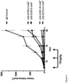
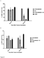
- the present inventors have shown that the use of a specific binding member which binds to LAG-3 is effective in suppressing tumour growth in syngeneic mouse models of cancer.
- a specific binding member of the invention may be used in a method of treating cancer in a patient.
- the patient is preferably a human patient. Methods of treatment do not form part of the present invention.
- Cells of the cancer to be treated using the specific binding member of the invention may express LAG-3, e.g. on their cell surface.
- cells of the cancer to be treated may have been determined to express LAG-3, e.g. on their cell surface.
- B cell lymphomas have been shown to express LAG-3 on their cell surface.
- tumours of the cancer to be treated using the specific binding members of the invention may comprise LAG-3 expressing immune cells.
- LAG-3 expressing immune cells such as LAG-3 expressing TILs, are present between tumour cells in many cancers.
- tumours of the cancer to be treated using the specific binding member of the invention have been determined to contain LAG-3 expressing immune cells. Methods for determining the presence of LAG-3 expressing immune cells in a tumour or in the periphery of the tumour are known in the art.
- a cancer to be treated using a specific binding member of the invention may be selected from the group consisting of Hodgkin's lymphoma, non-Hodgkin's lymphoma (such as diffuse large B-cell lymphoma, follicular lymphoma, indolent non-Hodgkin's lymphoma, mantle cell lymphoma), ovarian cancer, prostate cancer, colorectal cancer, fibrosarcoma, renal cell carcinoma, melanoma, pancreatic cancer, breast cancer, glioblastoma multiforme, lung cancer (such as non-small cell lung cancer), head and neck cancer (such as head and neck squamous cell carcinoma), stomach cancer (gastric cancer), bladder cancer, cervical cancer, uterine cancer, vulvar cancer, testicular cancer, penile cancer, leukemia (such as chronic lymphocytic leukemia, myeloid leukemia, acute lymphoblastoid leukaemia, or chronic lymphoblastoid leukaemia),
- lung cancer such as non-small cell lung cancer
- nasopharyngeal cancer colorectal cancer
- melanoma stomach cancer (gastric cancer)
- esophageal cancer such as adenocarcinoma of the gastroesophageal junction
- ovarian cancer cervical cancer, bladder cancer, head and neck cancer (such as head and neck squamous cell carcinoma)
- leukemia such as chronic lymphocytic leukemia, Hodgkin's lymphoma, non-Hodgkin's lymphoma (such as diffuse large B-cell lymphoma, indolent non-Hodgkin's lymphoma, mantle cell lymphoma), and multiple myeloma using anti-LAG-3 antibodies
- leukemia such as chronic lymphocytic leukemia
- Hodgkin's lymphoma non-Hodgkin's lymphoma
- non-Hodgkin's lymphoma such as diffuse large B-cell
- the cancer to be treated using the specific binding members of the present invention may be a renal cell carcinoma, lung cancer (such as non-small cell lung cancer), nasopharyngeal cancer, colorectal cancer, melanoma, stomach cancer (gastric cancer), esophageal cancer (such as adenocarcinoma of the gastroesophageal junction), ovarian cancer, cervical cancer, bladder cancer, head and neck cancer (such as head and neck squamous cell carcinoma), leukemia (such as chronic lymphocytic leukemia, Hodgkin's lymphoma, non-Hodgkin's lymphoma (such as diffuse large B-cell lymphoma, indolent non-Hodgkin's lymphoma, mantle cell lymphoma), or multiple myeloma.
- lung cancer such as non-small cell lung cancer
- nasopharyngeal cancer colorectal cancer
- melanoma stomach cancer
- stomach cancer gastric cancer
- Preferred cancers for treatment using the specific binding members of the present invention are lung cancer (such as non-small-cell lung cancer), bladder cancer, head and neck cancer (squamous cell carcinoma of the head and neck), diffuse large B cell lymphoma, gastric cancer, pancreatic cancer and hepatocellular carcinoma.
- Tumours of these cancers are known to comprise LAG-3 expressing immune cells and to express PD-L1 either on their cell surface or to comprise immune cells expressing PD-L1.
- cancer refers to a particular type of cancer, such as breast cancer
- a cancer which originates from malignant transformation of a different tissue, e.g. ovarian tissue, may result in metastatic lesions in another location in the body, such as the breast, but is not thereby a breast cancer as referred to herein but an ovarian cancer.
- the cancer may be a primary or secondary cancer.
- the specific binding member of the present invention may be for use in a method of treating cancer in a patient, wherein the CDR-based antigen-binding site of the specific binding member binds to a molecule which is an immune system modulator, and wherein the cancer is a primary tumour and/or a tumour metastasis.
- the specific binding members of the invention are designed to be used in methods of treatment of patients, preferably human patients.
- Specific binding members will usually be administered in the form of a pharmaceutical composition, which may comprise at least one component in addition to the specific binding member, such as a pharmaceutically acceptable excipient.
- a pharmaceutical composition of the present invention comprises, in addition to active ingredient, a pharmaceutically acceptable excipient.
- the specific binding member may be administered intravenously, or subcutaneously.
- Liquid pharmaceutical compositions generally comprise a liquid carrier such as water, petroleum, animal or vegetable oils, mineral oil or synthetic oil.
- a liquid carrier such as water, petroleum, animal or vegetable oils, mineral oil or synthetic oil.
- Physiological saline solution, dextrose or other saccharide solution or glycols such as ethylene glycol, propylene glycol or polyethylene glycol may be included.
- the specific binding member, or pharmaceutical composition comprising the specific binding member is preferably in the form of a parenterally acceptable aqueous solution which is pyrogen-free and has suitable pH, isotonicity and stability.
- a parenterally acceptable aqueous solution which is pyrogen-free and has suitable pH, isotonicity and stability.
- isotonic vehicles such as Sodium Chloride Injection, Ringer's Injection, Lactated Ringer's Injection.
- Preservatives, stabilizers, buffers, antioxidants and/or other additives may be employed, as required.
- Many methods for the preparation of pharmaceutical formulations are known to those skilled in the art. See e.g. Robinson ed., Sustained and Controlled Release Drug Delivery Systems, Marcel Dekker, Inc., New York, 1978 .
- a composition comprising a specific binding members according to the present invention may be administered alone or in combination with other treatments, concurrently or sequentially or as a combined preparation with another therapeutic agent or agents, dependent upon the condition to be treated.
- a specific binding member of the invention may be administered in combination with an existing therapeutic agent for the disease to be treated, e.g. a cancer as mentioned above.
- a specific binding member of the present invention may be administered to the patient in combination with a second anti-cancer therapy, such as chemotherapy, anti-tumour vaccination (also referred to as a cancer vaccination), radiotherapy, immunotherapy, an oncolytic virus, chimeric antigen receptor (CAR) T-cell therapy, or hormone therapy.
- a second anti-cancer therapy such as chemotherapy, anti-tumour vaccination (also referred to as a cancer vaccination), radiotherapy, immunotherapy, an oncolytic virus, chimeric antigen receptor (CAR) T-cell therapy, or hormone therapy.
- the specific binding member of the invention may act as an adjuvant in anti-cancer therapy, such as chemotherapy, anti-tumour vaccination, or radiotherapy.
- anti-cancer therapy such as chemotherapy, anti-tumour vaccination, or radiotherapy.
- administration of the specific binding member to the patient as part of chemotherapy, anti-tumour vaccination, or radiotherapy will trigger a greater immune response against the cancer associated antigen LAG-3, than is achieved with chemotherapy, anti-tumour vaccination, or radiotherapy alone.
- anti-LAG-3 therapies have shown good efficacy in treating viral based pathologies in mice ( Blackburn SD, et al., 2009, Nature Immunology 10 (1): 29-37 ).
- a method of treating cancer in a patient may thus comprise administering to the patient a therapeutically effective amount of a specific binding member according to the present invention in combination with a chemotherapeutic agent, anti-tumour vaccine, radionuclide, immunotherapeutic agent, oncolytic virus, CAR-T cell, or agent for hormone therapy.
- a chemotherapeutic agent, anti-tumour vaccine, radionuclide, immunotherapeutic agent, oncolytic virus, CAR-T cell, or agent for hormone therapy is preferably a chemotherapeutic agent, anti-tumour vaccine, radionuclide, immunotherapeutic agent, oncolytic virus, CAR-T cell, or agent for hormone therapy for the cancer in question, i.e.
- a chemotherapeutic agent, anti-tumour vaccine, radionuclide, immunotherapeutic agent, oncolytic virus, CAR-T cell, or agent for hormone therapy which has been shown to be effective in the treatment of the cancer in question.
- the selection of a suitable chemotherapeutic agent, anti-tumour vaccine, radionuclide, immunotherapeutic agent, oncolytic virus, CAR-T cell, or agent for hormone therapy which have been shown to be effective for the cancer in question is well within the capabilities of the skilled practitioner.
- the method comprises administering to the patient a therapeutically effective amount of a specific binding member according to the present invention in combination with a chemotherapeutic agent
- the chemotherapeutic agent may be selected from the group consisting of: taxanes, cyctotoxic antibiotics, tyrosine kinase inhibitors, PARP inhibitors, B_RAF enzyme inhibitors, alkylating agents, platinum analogs, nucleoside analogs, thalidomide derivatives, antineoplastic chemotherapeutic agents and others.
- Taxanes include docetaxel, paclitaxel and nab-paclitaxel; cytotoxic antibiotics include actinomycin, bleomycin, anthracyclines, doxorubicin and valrubicin; tyrosine kinase inhibitors include erlotinib, gefitinib, axitinib, PLX3397, imatinib, cobemitinib and trametinib; PARP inhibitors include piraparib; B-Raf enzyme inhibitors include vemurafenib and dabrafenib; alkylating agents include dacarbazine, cyclophosphamide, temozolomide; platinum analogs include carboplatin, cisplatin and oxaliplatin; nucleoside analogs include gemcitabine and azacitidine; antineoplastics include fludarabine.
- chemotherapeutic agents suitable for use in the present invention include methotrexate, defactinib, entinostat, pemetrexed, capecitabine, eribulin, irinotecan, fluorouracil, and vinblastine.
- Vaccination strategies for the treatment of cancers has been both implemented in the clinic and discussed in detail within scientific literature (such as Rosenberg, S. 2000 Development of Cancer Vaccines ). This mainly involves strategies to prompt the immune system to respond to various cellular markers expressed by autologous or allogenic cancer cells by using those cells as a vaccination method, both with or without granulocyte-macrophage colony-stimulating factor (GM-CSF). GM-CSF provokes a strong response in antigen presentation and works particularly well when employed with said strategies.
- GM-CSF granulocyte-macrophage colony-stimulating factor
- Administration may be in a "therapeutically effective amount", this being sufficient to show benefit to a patient. Such benefit may be at least amelioration of at least one symptom.
- treatment of a specified disease refers to amelioration of at least one symptom.
- the actual amount administered, and rate and time-course of administration will depend on the nature and severity of what is being treated, the particular patient being treated, the clinical condition of the individual patient, the cause of the disorder, the site of delivery of the composition, the type of specific binding member, the method of administration, the scheduling of administration and other factors known to medical practitioners. Prescription of treatment, e.g. decisions on dosage etc., is within the responsibility of general practitioners and other medical doctors, and may depend on the severity of the symptoms and/or progression of a disease being treated.
- Treatments may be repeated at daily, twice-weekly, weekly or monthly intervals, at the discretion of the physician. Treatment may be given before, and/or after surgery, and may be administered or applied directly at the anatomical site of surgical treatment.
- Naive phage libraries displaying the CH3 domain of human IgG1 (IMGT numbering 1.4-130) with randomisation within the AB (residues 14-18) and EF (residues 92-101) loops were used for selection with recombinant Fc-tagged human LAG-3 (LAG-3 Fc) antigen (R&D systems, 2319-L3-050).
- the libraries were selected in three rounds using antigen captured on Protein A (Life Technologies, 10002D) or Protein G (Life Technologies, 10004D) beads.
- the outputs were screened by ELISA and positive binders sub-cloned and expressed as soluble Fcabs (containing a truncated hinge) in Pichia pastoris using EasySelect Pichia Expression Kit (Life Technologies, K1740-01). The Fcabs were then screened for binding to recombinant human LAG-3 Fc on the Biacore 3000 (GE Healthcare). Briefly, LAG-3 Fc (R&D systems, 2319-L3-050) was coupled at a density of 7200 RU to a CM5 chip (GE Healthcare, BR-100012) using amine coupling (GE Healthcare, BR-1000-50).
- Fcabs were diluted in HBS-P (GE Healthcare, BR100368) buffer and injected at 250 nM, 500 nM and 1000 nM for 3 min and then allowed to dissociate in buffer for 5 min.
- Reference subtracted data (LAG-3 Fc flow cell 2 -blank flow cell) was analyzed using BIAevaluation 3.2 software to identify binding. Fcabs were then tested for binding to HEK cell-expressed human LAG-3 (LAG-3 cloned into pcDNA5FRT vector [Life Technologies, V6010-20] [See section 1.4.5 for methodology]).
- HEK 293 cells overexpressing human LAG-3 grown in DMEM (Life Technologies, 61965-026) containing 10% FBS (Life Technologies, 10270-1-6), 100 ⁇ g/ml Hygromycin B (Melford Laboratories Ltd, Z2475), 15 ⁇ g/ml Blasticidin (Melford Laboratories Ltd, B1105) and 1 ⁇ g/ml Doxycyclin (Sigma, D9891) were detached from tissue culture flasks using cell dissociation buffer (Life Technologies, 13151-014) and seeded in V-bottom 96-well plates at 2 ⁇ 10 5 cells/well. Fcabs were incubated with the cells at 5 ⁇ M in a 100 ⁇ l volume for 1 h at 4 °C.
- the plates were washed the secondary antibody (Anti-human Fc-488, Jackson ImmunoResearch, 109-546-098) was diluted 1:1000 in PBS and 100 ⁇ l was added to the cells and incubated for 30 min at 4 °C. The plates were washed and the cells were resuspended in 100 ⁇ l PBS containing 1 ⁇ g/ml DAPI (Biotium, 40043). The plate was read on a BD FACSCanto II cytometer (BD Biosciences) and the data analysed using FlowJoX. The Fcabs were then expressed in mammalian cells by transformation using lipofectamine (Life Technologies, 11668-019) into Flp-In T-Rex 293 cells (Life Technologies, R780-07).
- lipofectamine Life Technologies, 11668-019
- Flp-In T-Rex 293 cells Life Technologies, R780-07
- LAG-3 binding Fcabs were tested for inhibition of binding of human MHC class II on A375 cells (ATCC, CRL-1619) to recombinant LAG-3 Fc (using the methodology in example 1.6).
- 54 unique Fcab sequences were identified from three rounds of phage selection, and 12 of these Fcabs were determined to bind to LAG-3 Fc by BIAcore analysis and/or bind to LAG-3 expressing HEK cells.
- Three of the selected Fcabs were also able to inhibit the interaction of LAG-3 with MHC class II and were selected for affinity maturation. The three Fcabs were termed FS18-3, FS18-7 and FS18-21.
- phage display affinity maturation libraries were constructed by randomising five residues in the AB loop (residues 14-18) and either five (residues 92-94 and 97-98) or eight (residues 92-94 and 97-101) residues in the EF loop of each of the three Fcabs identified using the naive selection process described above.
- the affinity maturation libraries were selected using recombinant human LAG-3 Fc (R&D systems, 2319-L3-050) and HEK cells expressing human LAG-3 (as described above).
- the outputs were screened by phage ELISA, the positive binders were subcloned and expressed as soluble Fcabs (containing a truncated hinge) in HEK Expi293 cells (Fcabs cloned into pTT5 vector [National Research Council of Canada] transfected using ExpiFectamine 293 Transfection kit [Life Technologies, A14524] into Expi293F cells [Life technologies, A14527]).
- the HEK expressed soluble Fcabs were then screened for binding to cell expressed human LAG-3, binding to cell expressed cynomolgus LAG-3 (methodology as example 1.4.3), and the ability to block MHC class II binding to recombinant LAG-3 Fc (methodology as in example 1.6).
- the blocking Fcabs were further tested to determine whether they were able to reverse LAG-3 induced inhibition of IL-2 secretion in a T cell activation assay (methodology as in example 2.1).
- 61 unique anti-LAG-3 Fcabs were identified from the six affinity maturation libraries using these screening methods. Affinity matured Fcabs from the FS18-7 lineage were shown to have the highest level of cross-reactivity with cynomolgus monkey LAG-3.
- the three Fcabs from this lineage with the strongest binding to cynomolgus monkey LAG-3 Fc and the highest activity in the T cell activation assay were selected for further affinity maturation. These three Fcabs were also shown to block the interaction of LAG-3 Fc with cell expressed MHC class II.
- a pool of the three Fcabs (FS18-7-7, FS18-7-9, and FS18-7-11) from the first affinity maturation was used to create further affinity maturation libraries.
- the CD loop was hard randomized using randomized primers from ELLA Biotech.
- a portion of amino acid positions in the CD loop (residues 45.1-78) was randomized using an equimolar distribution of amino acids excluding cysteine. Error prone PCR was also carried out across the entire CH3 domain sequence to introduce additional mutations that might enhance binding.
- the affinity maturation libraries were generated in phage and selections performed against biotinylated recombinant LAG-3 avi-Fc (BPS Bioscience, 71147) and HEK hLAG-3 cells and screened for binding to recombinant LAG-3 Fc (R&D systems, 2319-L3-050) by phage ELISA.
- 86 unique Fcabs (containing a truncated hinge) were expressed in HEK293F cells. Selected Fcabs were also screened for activity in a T cell activation assay as described above.
- the nine Fcabs identified during the second affinity maturation with the highest activity in the T cell activation assay (FS18-7-32; FS18-7-33; FS18-7-36; FS18-7-58; FS18-7-62; FS18-7-65; FS18-7-78; FS18-7-88; and FS18-7-95), as well as the parental Fcab clone, FS18-7-9, were then further characterised as described below.
- a sequence alignment of these nine Fcabs against the parental Fcab clone, FS18-7-9 is shown in Figure 1A .
- Figure 1B details the percentage sequence identity of each of the nine Fcab clones to the parental Fcab clone, FS18-7-9.
- Fcabs originating from affinity maturation of the two other parental Fcab clones, FS18-7-7 and FS18-7-11 were not as promising candidates as those originating from affinity maturation of FS18-7-9 and were therefore not pursued further.
- Fcab FS18-7 which was selected using the naive selection protocol described above, was used to generate phage libraries to select against mouse LAG-3. Two rounds of affinity maturation were performed, and Fcab clones FS18-7-108-29 and FS18-7-108-35, which showed high-affinity, specific binding to mouse LAG-3 were selected following affinity maturation. The ability of FS18-7-108-29 and FS18-7-108-35 to inhibit mouse LAG-3 in a T cell activation assay was confirmed. Epitope mapping using the Octet (Forteo Bio) showed that the anti-mouse LAG-3 Fcabs compete with the anti-human LAG-3 Fcabs (selected following the second affinity maturation as described above) for binding to human LAG-3.
- “mock” mAb 2 comprising the lead anti-human LAG-3 and anti-mouse LAG-3 Fcabs identified in 1.1 and 1.2 above were prepared in order to allow the characterisation of these Fcabs in mAb 2 format.
- These mock mAb 2 were prepared from the anti-LAG-3 Fcabs and the variable regions of anti-FITC antibody 4420 (see SEQ ID NO: 83, SEQ ID NO: 84, and SEQ ID NO: 85 for details) (Bedzyk, W. D., et al. 1989 and Bedzyk, W. D., et al. 1990).
- the mock mAb 2 were prepared both with (SEQ ID NO: 63, 65, 67, 69, 71, 73, 75, 77, 79, and 81) and without (SEQ ID NO: 64, 66, 68, 70, 72, 74, 76, 78, 80, and 82) the LALA mutation in the CH2 domain of the heavy chain (see section 1.5 below for details) and further comprised the light chain of the anti-FITC mAb 4420 (SEQ ID NO: 85).
- the mock mAb 2 were produced by transient expression in HEK293-6E cells and purified using mAb Select SuRe protein A columns.
- a BIAcore T200 (GE Healthcare) was used to measure the affinity of the anti-human LAG-3 Fcabs in the mock mAb 2 format for human LAG-3.
- Flow cell 4 of a CM5 sensor chip (GE Healthcare, BR1005-30) was immobilised with human LAG-3-Fc (R&D Systems, 2319-L3-050), and flow cell 3 was immobilised with buffer for reference using the amine coupling kit (GE Healthcare, BR-1000-50).
- LAG-3-Fc was diluted to 5 ⁇ g/ml in sodium acetate pH5 (ForteoBio, 18-1069) and injected at a flow rate of 10 ⁇ l/min for 12 seconds followed by deactivation of the surface by injection of ethanolamine for 420 sec.
- the Immobilisation level was 158 RU.
- the mock mAb 2 (or control anti-human LAG-3 mAb, 25F7) were diluted in HBS-P buffer (GE Healthcare, BR-1003-68) in a 2-fold dilution series from 4 ⁇ g/ml.
- the control mAb/mock mAb 2 were injected with an association time of 240 seconds at 30 ⁇ l/min, and a dissociation time 300 seconds at 30 ⁇ l/min.
- the surface was regenerated using 25mM NaOH for 30 seconds at 100 ⁇ l/min.
- the data was double reference subtracted and analysed using the BIAevaluation 3.2 software to calculate kinetic constants.
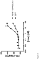
- the Fcabs in mock mAb 2 format had affinities for human LAG-3 in the range of 0.8 - 1.1 nM ( Table 1 ), which is similar to the affinity of the benchmark anti-human LAG-3 mAb 25F7.
- Table 1 The affinity of the benchmark anti-human LAG-3 mAb 25F7.
- Fcabs have a smaller binding interface than monoclonal antibodies as the binding sites of Fcabs form a relatively compact antibody fragment with two binding sites situated in close proximity.
- the Fab arms of a typical mAb are separated by a flexible hinge region. Based on this smaller binding interface and the associated reduced flexibility of the two binding sites in the Fc region, it was unexpected that the anti-LAG-3 Fcabs were able to bind to and inhibit LAG-3 with similar affinity and potency as the benchmark antibody 25F7.
- Table 1 Binding affinity of LAG-3 specific Fcabs in mock mAb 2 format to human LAG-3 Anti-human LAG-3 Fcab in mock mAb 2 format and benchmark anti-human LAG-3 mAb, 25F7 K D (M) FS18-7-9 8.3 ⁇ 10 -10 FS 18-7-62 9.5 ⁇ 10 -10 FS 18-7-78 8.4 ⁇ 10 -10 FS18-7-32 8.6 ⁇ 10 -10 FS 18-7-36 8.9 ⁇ 10 -10 FS18-7-65 1.1 ⁇ 10 -9 25F7 3.2 ⁇ 10 -10
- a Biacore 3000 (GE Healthcare) was used to measure the affinity of the surrogate Fcabs specific for mouse LAG-3 to mouse LAG-3.
- Amine coupling (amine coupling kit, GE Healthcare, BR-1000-50) was used to coat mLAG-3 Fc (R&D Systems, 3328-L3-050) diluted in 10 mM sodium acetate pH 5.0 (ForteBio, 18-1069) directly to a CM5 chip (GE Healthcare, BR-1000-12).
- Flow cell 1 was coated with Mouse Fc (SinoBiological, 51094-MNAH), and flow cell 2 was coated with mLAG-3 Fc at 950 RU.
- Fcabs were diluted in HBS-P buffer (GE Healthcare, BR-1003-68) and injected at various concentrations (fourfold dilutions from 100 nM) for 3 min at 20 ⁇ l/min and then allowed to dissociate in buffer for 12 min.
- the chip was regenerated by injection of 10 mM glycine pH 2.5 for 30 s at 30 ⁇ l/min. Data was double reference subtracted and analyzed using BIAevaluation 3.2 software to calculate kinetic constants.
- the tested surrogate Fcabs bound to mouse LAG-3 with single digit nanomolar affinity as set out in Table 2.
- Lentiviral transduction methodology was used to generate DO11.10 cells (National Jewish Health) over-expressing human, cynomolgus or mouse LAG-3 using the Lenti-X HTX Packaging System (Clontech, Cat. No 631249).
- Lenti-X expression vector (pLVX) (Clontech, Cat. No 631253), containing the mouse LAG-3 cDNA (SEQ ID NO: 96), human LAG-3 cDNA (SEQ ID NO: 95) or cynomolgus LAG-3 cDNA (SEQ ID NO: 97), was co-transfected with a Lenti-X HTX Packaging Mix into the Lenti-X 293T Cell Line (Clontech, Cat. No 632180) to generate virus.
- the DO11.10 cell line was transduced using the lentiviral vectors produced with the Lenti-X HTX Packaging System.
- the affinity of the anti-human LAG-3 Fcabs in mock mAb 2 format to cells expressing human LAG-3 (DO11.10 cell line transfected with human LAG-3) was measured using flow cytometry.
- mAb 2 and control mAb dilutions (2 ⁇ final concentration) were prepared in triplicate in 1 ⁇ DPBS (Gibco, 14190-094).
- D011.10:LAG-3 cell suspensions were prepared in PBS+2%BSA (Sigma, A7906) and seeded at 4 ⁇ 10 -6 cell/ml with 50 ⁇ l/well in V-bottomed 96-well plates (Costar, 3897).
- mAb 2 or control mAb anti human LAG-3 mAb, 25F7 dilutions were added to the wells containing cells (final volume 100 ⁇ l) and incubated at 4°C for 1 hour.
- the plates were washed and 100 ⁇ l/well of secondary antibody (anti-human Fc-488 antibody, Jackson ImmunoResearch, 109-546-098) diluted 1:1000 in PBS+2% BSA was then added and incubated for 30 mins at 4°C in the dark.
- the plates were washed and resuspended in 100 ⁇ l of PBS containing DAPI (Biotium, 40043) at 1 mg/ml. The plates were read using Canto II flow cytometer (BD Bioscience).
- Dead cells were excluded and the fluorescence in the FITC channel (488nm/530/30) was measured.
- the data was fitted using log (agonist) vs response in GraphPad Prism Software. All tested Fcabs in mock mAb 2 format and the benchmark anti-human LAG-3 mAb, 25F7, bound human LAG-3 with similar affinity (EC 50 ), in the range of 1.2 - 2.1 nM as set out in Table 3.
- Table 3 Binding affinity of anti-human LAG-3 Fcabs in mock mAb 2 format to DO11.10 cells expressing human LAG-3 as determined by flow cytometry Anti-human LAG-3 Fcab in mock mAb 2 format and benchmark anti-human LAG-3 mAb, 25F7 EC 50 (nM) FS 18-7-9 1.2 FS 18-7-32 1.6 FS 18-7-33 1.5 FS 18-7-36 1.5 FS 18-7-62 2.1 FS 18-7-65 1.6 FS 18-7-78 1.7 25F7 2.1
- the affinity of the anti-human LAG-3 Fcabs in mock mAb 2 format to cells expressing cynomolgus LAG-3 was measured using flow cytometry.
- mAb 2 and control mAb dilutions (2 ⁇ final concentration) were prepared in triplicate in 1 ⁇ DPBS (Gibco, 14190-094).
- DO11.10:LAG-3 cell suspensions were prepared in PBS+2%BSA (Sigma, A7906) and seeded at 4 ⁇ 10 -6 cell/ml with 50 ⁇ l/well in V-bottomed 96-well plates (Costar, 3897).
- mAb 2 or control mAb anti human LAG-3 mAb, 25F7 dilutions were added to the wells containing cells (final volume 100 ⁇ l) and incubated at 4°C for 1 hour.
- the plates were washed and 100 ⁇ l/well of secondary antibody (anti-human Fc-488 antibody, Jackson ImmunoResearch, 109-546-098) diluted 1:1000 in PBS+2% BSA was then added and incubated for 30 mins at 4°C in the dark.
- the plates were washed and resuspended in 100 ⁇ l of PBS containing DAPI (Biotium, 40043) at 1 mg/ml. The plates were read using Canto II flow cytometer (BD Bioscience).
- the dead cells were excluded and the fluorescence in the FITC channel (488nm/530/30) was measured.
- the data was fit using log (agonist) vs response in GraphPad Prism Software.
- the tested Fcabs in mock mAb 2 format bound to cynomolgus LAG-3 with 0.5-0.6 nM affinity indicating that toxicology studies in cynomolgus monkeys would be expected to be predictive of effects seen in humans (see Table 4 ).
- Table 4 Binding affinity of anti-LAG-3 Fcabs to DO11.10 cells expressing cynomolgus LAG-3 by flow cytometry Anti-human LAG-3 Fcab in mock mAb 2 format and benchmark anti-human LAG-3 mAb, 25F7 EC 50 (nM) FS18-7-9 0.6 FS18-7-62 0.5 FS 18-7-78 0.5 25F7 9.0
- the mouse LAG-3 sequence (SEQ ID NO: 96) was subcloned into pcDNA5FRT vector (Life Technologies, V6010-20) using Kpnl (NEB, R0142) and NotI (NEB, R0146) restriction digestion. The vector was then transformed into Flp-In T-REx 293 HEK cell line (Life Technologies, R780-07) using Lipofectamine 2000 (Life Technologies, 11668-019).
- Transformed Flp-In T-REx 293 cells were grown in DMEM (Life Technologies, 61965-026) containing 10% FBS (Life Technologies, 10270-1-6), 100 ⁇ g/ml Hygromycin B (Melford Laboratories Ltd, Z2475), 15 ⁇ g/ml Blasticidin (Melford Laboratories Ltd, B1105) for 3-4 weeks until colonies of stably transformed cells were apparent. These colonies were amplified in the presence of 1 ⁇ g/ml Doxycyclin (Sigma, D9891) and tested for mouse LAG-3 expression using PE conjugated anti-mouse LAG-3 (clone C9B7W, BD Biosciences, 552380).
- HEK cells expressing mLAG-3 grown in DMEM (Life Technologies, 61965-026) containing 10% FBS (Life Technologies, 10270-1-6), 100 ⁇ g/ml Hygromycin B (Melford Laboratories Ltd, Z2475), 15 ⁇ g/ml Blasticidin (Melford Laboratories Ltd, B1105) and 1 ⁇ g/ml Doxycyclin (Sigma, D9891) were detached from tissue culture flasks using cell dissociation buffer (Life Technologies, 13151-014) and seeded in V-bottom 96-well plates (Costar, 3897) at 2 ⁇ 10 5 cells/well.
- the plates were centrifuged at 1500 rpm for 3 min at 4°C to pellet the cells.
- a dilution series of the Fcabs (or control mAb) were incubated with the cells in a 100 ⁇ l volume for 1 h at 4 °C.
- the plates were washed and secondary antibody (Anti-human Fc-488, Jackson ImmunoResearch, 109-546-098 for Fcabs or Anti-Rat IgG (H+L), Alexa Fluor 488 Conjugate, ThermoFisher, A-11006 for C9B7W) was diluted 1:1000 in PBS and 100 ⁇ l was added to the cells for 30 min at 4 °C (plates were kept in the dark).
- the plates were then washed and the cells resuspended in 100 ⁇ l PBS containing 1 ⁇ g/ml DAPI (Biotium, 40043). The plates were read using Canto II flow cytometer (BD Bioscience). Dead cells were excluded and the fluorescence in the FITC channel (488nm/530/30) was measured. The data was fit using log (agonist) vs response in GraphPad Prism Software.
- the tested Fcabs bound to mouse LAG-3 with similar affinity (see Table 5 ).
- the benchmark LAG-3 mAb, C9B7W (2B Scientific, BE0174-50MG) binds mouse LAG-3 with 17-fold poorer affinity (EC 50 ) than the Fcabs ( Table 5 ).
- Table 5 Binding affinity of surrogate anti-mouse LAG-3 Fcabs to HEK cells expressing mouse LAG-3 by flow cytometry Anti-mouse LAG-3 Fcabs and benchmark anti-mouse LAG-3 mAb, C9B7W EC 50 (nM) FS18-7-108-29 4.5 FS18-7-108-35 4.5 C9B7W 79
- HEK cells expressing mLAG-3 grown in DMEM (Life Technologies, 61965-026) containing 10% FBS (Life Technologies, 10270-1-6), 100 ⁇ g/ml Hygromycin B (Melford Laboratories Ltd, Z2475), 15 ⁇ g/ml Blasticidin (Melford Laboratories Ltd, B1105) and 1 ⁇ g/ml Doxycyclin (Sigma, D9891) were detached from tissue culture flasks using cell dissociation buffer (Life Technologies, 13151-014).
- the plates were then washed and the cells resuspended in 60 ⁇ l PBS containing 1 ⁇ g/ml DAPI (Biotium, 40043). The plates were read using Canto II flow cytometer (BD Bioscience). Dead cells were excluded and the fluorescence in the FITC channel (488nm/530/30) was measured. The data was fitted using log (agonist) vs response in GraphPad Prism Software.
- the human mAb, 25F7 does not show any detectable binding to mouse LAG-3, indicating that the human LAG-3 Fcab, FS18-7-9, has a different binding epitope on LAG-3 than that of 25F7.
- the introduction of the LALA mutation in the CH2 domain of human IgG1 is known to reduce Fc y receptor binding (Bruhns, P., et al. (2009) and Xu, D. et al. (2000)).
- BIAcore was used to confirm that the LALA mutation had reduced the binding affinity of the Fcabs (in mock mAb 2 format) to Fcy receptors.
- the human FcyR binding assay was performed on a Biacore T200 instrument (GE Healthcare) using the Fcabs in the mock mAb 2 format.
- Human FcyRs (R&D Systems, 1257-FC, 1330-CD, 1875-CD, 4325-FC) were immobilized using amine coupling (amine coupling kit, GE Healthcare, BR-1000-50) onto a Series S CM5 chip (GE Healthcare, BR-1005-30) to a surface density of 370 RU for Fc ⁇ RI, 264 RU for Fc ⁇ RIII (high affinity human FcyRs) and 500 RU for Fc ⁇ Rlla and Fc ⁇ RIIb (low affinity human FcyRs). For each immobilized chip a flow cell was left blank for background subtraction.
- amine coupling amine coupling kit, GE Healthcare, BR-1000-50
- Series S CM5 chip GE Healthcare, BR-1005-30
- FcyR were immobilized using a concentration of 5 ⁇ g/ml in sodium acetate pH5 (ForteBio, 18-1069) and injected at a flow rate of 10 ⁇ l/min in 15 second cycles until the required immobilization level was reached.
- the positive control was a wild type IgG1 isotype mAb, which was compared to controls LALA IgG1 mAb and monoclonal IgG2 and IgG4 isotype mAbs to irrelevant targets.
- the flow cells were regenerated by injecting 10 mM sodium hydroxide (VWR, 28244.262) at a flow rate of 100 ⁇ l/min for 30 seconds.
- the data analysis was performed with BiaEvaluation software version 3.2 RC1 by double referencing against the blank flow cell (without immobilized FcyR) and subtracting a buffer cycle from test mAb 2 . The results are shown in Table 7.
- Table 7 Binding response of anti-human LAG-3 Fcabs in mock mAb 2 format (comprising LALA mutation as detailed above) to human Fcy receptors by SPR mAb/mock mAb 2 Binding response at end of association (RU) Fc ⁇ RI Fc ⁇ RIII Fc ⁇ RIIa Fc ⁇ RIIb FS18-7-9 1.4 6.6 -9.8 -8.5 FS18-7-62 -0.9 0.7 -10 -8.5 FS18-7-78 -0.3 4.0 -10.7 -9.2 mock mAb LALA 2 8.0 -12.7 -9.6 IgG2 0 1.9 9.7 7.4 IgG4 9 3.1 4.3 15.1 mock mAb IgG1 26 44 13 17.7
- Fcabs containing the truncated hinge; SEQ ID NO: 58
- SEQ ID NO: 58 The ability of the Fcabs (containing the truncated hinge; SEQ ID NO: 58) to block the interaction between recombinant human or mouse LAG-3 Fc and human MHC Class II was studied by measuring binding of LAG-3 Fc to A375 cells, a melanoma cell line that expresses human MHC Class II.
- A375 ATCC, CRL-1619 cells grown in DMEM (Life Technologies, 61965-026) containing 10% FBS (Life Technologies, 10270-106) were detached from cell culture flasks using cell dissociation buffer (Life Technologies, 13151-014) and seeded in V-bottom 96-well plates (Costar, 3897) at 2 ⁇ 10 5 cells/well.
- the plates were centrifuged at 1500 rpm for 3 min at 4 °C to pellet the cells.
- the relevant concentrations of Fcab or control mAb were incubated with 1 ⁇ g/ml LAG-3 Fc (human LAG-3-Fc R&D Systems, 2319-L3-050 or mouse LAG-3 Fc R&D Systems, 3328-L3-050) in 100 ⁇ l DMEM containing 10% FBS for 1 h at 4 °C.
- LAG-3/Fcab mix was added to the A375 cells and incubated for 1 h at 4 °C. Cells were washed.
- the anti-human LAG-3 Fcabs tested were also able to inhibit the interaction of human MHC class II with human LAG-3 with a similar potency as the control anti-human LAG-3 mAb (25F7).
- Table 9 Anti-human LAG-3 Fcabs inhibit binding of human LAG-3 to MHC class II
- the panel of lead Fcabs (with a truncated hinge; SEQ ID NO: 58) containing the LALA mutation were tested in a DO11.10 based T cell activation assay.
- Dilutions of Fcabs or benchmark mAb were prepared in 200 ⁇ l experimental media.
- Fcabs were mixed 1:1 (170 ⁇ l + 170 ⁇ l) with 4 ⁇ 10 5 /ml DO11.10 LAG-3 cells in experimental media and incubated at 37° C, 5% CO 2 for 1 hour.
- 2 ⁇ 10 5 A20 cells/ml experimental media were incubated with 1 ⁇ M OVA peptide for 30 min.
- 360 ⁇ l of the A20 cells + OVA mixture were added to 360 ⁇ l of the DO11.10 LAG-3 cell/Fcab mix.
- the cells were then mixed in a deep well plate and cultured in a 96-round bottom plate with 200 ⁇ l of the mix/well.
- the assay was run in triplicate.
- the human lead Fcabs show significant activity in the T cell activation assay with potencies in the range of 1-2 nM.
- the Fcabs have slightly improved potency than the benchmark anti human-LAG-3 mAb 25F7.
- Improved potency in the T cell activation assay is expected to be predictive of improved efficacy in human patients through enhanced inhibition of LAG-3.
- the surrogate anti-mouse LAG-3 Fcabs (with a truncated hinge; SEQ ID NO: 58) containing the LALA mutation were tested in a DO11.10 based T cell activation assay.
- Fcabs or benchmark mAb Dilutions of Fcabs or benchmark mAb were prepared in 200 ⁇ l experimental media.
- Fcabs were mixed 1:1 (170 ⁇ l + 170 ⁇ l) with 4 ⁇ 10 5 /ml DO11.10 LAG-3 cells in experimental media and incubated at 37°C, 5% CO2 for 1 hour.
- 2 ⁇ 10 5 A20 cells/ml experimental media were incubated with 1 ⁇ M OVA peptide for 30 min.
- 360 ⁇ l of the A20 cells + OVA mixture were added to 360 ⁇ l of the DO11.10 LAG-3 cell/Fcab mix.
- the cells were then mixed in a deep well plate and cultured in a 96-round bottom plate with 200 ⁇ l of the mix/well. The screen was assayed in triplicates.
- Table 11 EC 50 values and maximal IL-2 release of surrogate anti-LAG-3 Fcabs in DO11.10 T cell activation assay Surrogate Fcabs specific for mouse LAG-3 (with LALA mutation) and benchmark anti-mouse LAG-3 mAb, C9B7W EC 50 (nM) Maximal IL-2 release (pg/ml) FS18-7-108-29 1.9 205 FS18-7-108-35 1.8 176 C9B7W 5.1 48
- the mouse surrogate anti-mouse LAG-3 Fcabs showed significant activity in the T cell activation assay with potencies in the range of 2 nM.
- the surrogate anti-mouse LAG-3 Fcabs had higher potency than the benchmark anti mouse-LAG-3 antibody as is evident from the improved EC 50 and the 4-fold higher maximal activation of IL-2 release.
- Improved potency and maximal activation of these Fcabs compared to the benchmark is expected to result in improved activity in murine efficacy studies compared to the benchmark through improved inhibition of LAG-3.
- Dilutions of the FS18-7-9 Fcab in mock mAb 2 format (FS18-7-9/4420LALA) or benchmark mAb were prepared in experimental media.
- DO11.10 cells 0.3 ⁇ 10 6 cells/ml
- Antibodies and cells were incubated at 37° C, 5% CO 2 for 1 hour.
- LK 35.2 cells were incubated at 3 ⁇ 10 5 cells/ml experimental media with the OVA peptide at 1.5 ⁇ M for 30 min.
- 70 ⁇ l of LK 35.2 cells + OVA were added to 140 ⁇ l of the DO11.10/antibody. Cells were incubated at 37°C, 5% CO 2 for 24 hours.
- mice IL-2 ELISA kit (eBioscience, 88-7024-88 or R&D systems, SM2000) following the manufacturer's instructions. Plates were read at 450 nm using the plate reader with the Gen5 Software, BioTek. Absorbance values of 570 nm were subtracted from those of 450 nm (Correction). The standard curve for calculation of cytokine concentration was based on four parameter logistic curve fit (Gen5 Software, BioTek). The concentration of mIL-2 was plotted vs the log concentration of Fcab in mock mAb 2 format or benchmark mAb and the resulting curves were fitted using the log (agonist) vs response equation in GraphPad Prism.
- Table 12 EC 50 values and maximal IL-2 release of anti-LAG-3 Fcabs in cynomolgus DO11.10 T cell activation assay FS18-7-9 Fcab in mock mAb 2 format or benchmark anti-human LAG-3 mAb EC 50 (nM) Maximal IL-2 release (pg/ml) FS18-7-9/4420LALA 5.6 608 25F7 11.4 430
- the FS18-7-9 Fcab in mock mAb 2 format showed significant activity in the T cell activation assay with a potency of 5.6 nM. Specifically, the FS18-7-9 Fcab in mock mAb 2 format had higher potency against cynomolgus LAG-3 than the benchmark anti human-LAG-3 antibody, as is evident from the improved EC 50 and the higher maximal activation of IL-2 release.
- the EC 50 and maximal activation of the Fcab in mock mAb 2 format on human and cyno-LAG-3, as determined using a T cell activation assay is more similar (the benchmark has lower EC 50 on cyno-LAG-3 than the Fcab but has a similar EC 50 on human-LAG-3 in T-cell activation assay). It is therefore expected that the results of studies in cynomolgus monkeys using the Fcabs, e.g. in mock mAb 2 format, will be more predictive of the response in human patients.
- mAb 2 molecules comprising the surrogate anti-mouse LAG-3 Fcab, FS18-7-108-29, were prepared.
- the mAb 2 molecules comprised a Fab region specific for murine CD73 (TY11.8), murine TIM-3 (RMT3-23), murine CSF-1R (AFS98) or murine CLTA-4 (9D9), and were tested for in vivo anti-tumour activity using a MC38 syngeneic mouse tumour growth model.
- the Fab sequences were sourced as follows:
- variable heavy regions from each of the above sources were joined to the human IgG1 (G1m17) constant regions
- variable light regions from each of the above sources was joined to the human constant region (Km1) via human kappa J- region 1 (except 9D9 where mouse kappa J-region 1 was used).
- the mAb 2 for the in vivo studies were generated by substituting the CH3 domains of the reformatted constructs described above with FS18-7-108-29.
- the MC38 syngeneic tumour model was used in this experiment as MC38 tumours are known to be highly immunogenic resulting in increased LAG-3 expression on immune cells in the tumour environment and tumour periphery.
- mice C57BL/6 female mice (Charles River) aged 8-10 weeks and weighing 20-25 g each were rested for one week prior to the study start. All animals were micro-chipped and given a unique identifier. Each cohort had 10 mice.
- the MC38 colon carcinoma cell line (S. Rosenberg, NIH) was initially expanded, stored, and then pre-screened by IDEXX Bioresearch for pathogens using the IMPACT I protocol and shown to be pathogen free. MC38 cells were thawed from -150 °C storage and added to 20 ml DMEM (Gibco, 61965-026) with 10% FCS (Gibco, 10270-106) in a T175 tissue culture flask.
- mice received 2 ⁇ 10 6 cells injected subcutaneously in the left flank. 7-8 days following tumour cell inoculation, mice which did not have tumours at this point were removed from the study. All of the mAb 2 molecules and the control antibody were analysed within 24 hours prior to injection by SEC-HPLC profiling and checked for impurities.
- CTLA-4 inhibition results in increased T cell priming in the lymph nodes. Once primed, the T cell migrates to the tumour microenvironment where blockade of the PD-1/PD-L1 axis enhances activation of the primed T cell, resulting in synergistic anti-tumour effects of the anti-CTLA-4 and anti-PD-1/PD-L1 combination. This is reflected in the increased efficacy of the PD-1 and CTLA-4 combination in the clinic. It is hypothesised that the profound tumour inhibition caused by LAG-3/CTLA-4 mAb 2 in the syngeneic MC38 tumour model is through a similar synergy; inhibition of CTLA-4 increasing T cell priming and inhibition of LAG-3 increasing T cell activation at the tumour site.
- LAG-3 and TIM-3 suppressing exhausted T cells with a similar mode of action. These are secondary signals that are expressed after PD-1 is on T cells. With PD-1 inhibition still retained, inhibiting TIM-3 and LAG-3 can only achieve mild results.
- Macrophages are critical in maintaining an immunosuppressive tumour environment. Targeting of CSF-1R results in a decrease in tumour associated macrophages due to inhibition of macrophage differentiation and elimination of survival signals. It is hypothesised that the profound tumour inhibition caused by LAG-3/CSF-1R mAb 2 in the syngeneic MC38 tumour model is through synergy resulting from a release of the macrophage-induced immmunosupression in the tumour environment that allows LAG-3 antibodies to increase T cell activation at the tumour site. Combinations of CSF-1R and checkpoint inhibitors are already being evaluated in the clinic, which will help inform the viability of combining CSF-1R with LAG-3 inhibition.
- Blockade of CD73 also results in inhibition of macrophages however the intermediate tumour suppressive effect of CD73/LAG-3 mAb 2 suggests that anti-CD73 may be a less potent macrophage inhibitor.
- the surrogate anti-mouse LAG-3 Fcabs are so closely related in sequence to those of the anti-human Fcabs (and were derived from the same parental anti-human LAG-3 Fcab), they both bind to a very similar epitope of LAG-3 (mouse and human, respectively) despite the difference in homology between human and murine LAG-3. Consequently, it is expected that the results observed in mice following their treatment with mAb 2 comprising the surrogate anti-mouse LAG-3 Fcab are predictive of treatment of human patients with mAb 2 comprising the anti-human LAG-3 Fcab.
- FS18-29/4420 The effect of the LAG-3/mock mAb 2 , FS18-7-108-29/4420 containing the LALA mutation (SEQ ID NOs: 132 and 85), referred to as FS18-29/4420, the benchmark PD-L1 mAb S1 containing the LALA mutation (SEQ ID NOs: 122 and 119) and a combination of FS18-29/4420 and S1, on TIL LAG-3 expression was tested.
- MC38.OVA cells were harvested during log phase growth (Confluency -75%) and resuspended in PBS at a concentration of 1 ⁇ 10 7 cells/mL. Tumours were initiated by first anesthetizing each animal with isoflurane, then subcutaneously implanting 1 ⁇ 10 6 MC38.OVA cells (0.1 mL suspension) into the left flank of each test animal. Eleven days after tumour cell implantation animals were randomised, using a deterministic randomisation method, into five groups with individual tumour volumes of 32 to 62.5 mm 3 .
- TILs were examined for LAG-3 expression after the third dose of antibody/mock mAb 2 had been administered, when a separation in the growth of the tumour between control and non-control treatments is seen but before there is a large difference between tumour sizes which might skew results.
- LAG-3 expression on TILs was found to be markedly decreased in animals treated with the combination of FS18-29/4420 and S1.
- LAG-3 expression on CD8, CD4 and FoxP3 tumour infiltrating lymphocytes (TILs) was decreased after treatment with a combination of FS18-29/4420 and S1 by day 23, while treatment with FS18-29/4420 or S1 administered individually had little to no effect on LAG-3 expression on TILs.
Landscapes
- Health & Medical Sciences (AREA)
- Chemical & Material Sciences (AREA)
- Immunology (AREA)
- Organic Chemistry (AREA)
- Life Sciences & Earth Sciences (AREA)
- General Health & Medical Sciences (AREA)
- Medicinal Chemistry (AREA)
- Proteomics, Peptides & Aminoacids (AREA)
- Molecular Biology (AREA)
- Genetics & Genomics (AREA)
- Biophysics (AREA)
- Biochemistry (AREA)
- Pharmacology & Pharmacy (AREA)
- Public Health (AREA)
- Veterinary Medicine (AREA)
- Animal Behavior & Ethology (AREA)
- Bioinformatics & Cheminformatics (AREA)
- Engineering & Computer Science (AREA)
- Chemical Kinetics & Catalysis (AREA)
- Nuclear Medicine, Radiotherapy & Molecular Imaging (AREA)
- General Chemical & Material Sciences (AREA)
- Epidemiology (AREA)
- Gastroenterology & Hepatology (AREA)
- Physics & Mathematics (AREA)
- Optics & Photonics (AREA)
- Zoology (AREA)
- Toxicology (AREA)
- Oncology (AREA)
- Hematology (AREA)
- Peptides Or Proteins (AREA)
- Micro-Organisms Or Cultivation Processes Thereof (AREA)
- Medicines Containing Antibodies Or Antigens For Use As Internal Diagnostic Agents (AREA)
- Preparation Of Compounds By Using Micro-Organisms (AREA)
- Medicines Containing Material From Animals Or Micro-Organisms (AREA)
- Medicinal Preparation (AREA)
- Medicines That Contain Protein Lipid Enzymes And Other Medicines (AREA)
Applications Claiming Priority (2)
| Application Number | Priority Date | Filing Date | Title |
|---|---|---|---|
| US201662352470P | 2016-06-20 | 2016-06-20 | |
| PCT/EP2017/065052 WO2017220555A1 (en) | 2016-06-20 | 2017-06-20 | Lag -3 binding members |
Publications (2)
| Publication Number | Publication Date |
|---|---|
| EP3472206A1 EP3472206A1 (en) | 2019-04-24 |
| EP3472206B1 true EP3472206B1 (en) | 2025-01-15 |
Family
ID=59269995
Family Applications (1)
| Application Number | Title | Priority Date | Filing Date |
|---|---|---|---|
| EP17734668.1A Active EP3472206B1 (en) | 2016-06-20 | 2017-06-20 | Lag-3 binding members |
Country Status (15)
| Country | Link |
|---|---|
| US (2) | US11214618B2 (OSRAM) |
| EP (1) | EP3472206B1 (OSRAM) |
| JP (2) | JP7085708B2 (OSRAM) |
| KR (1) | KR20190022632A (OSRAM) |
| CN (1) | CN109311993B (OSRAM) |
| AU (1) | AU2017281157B2 (OSRAM) |
| BR (1) | BR112018076525A2 (OSRAM) |
| CA (1) | CA3027302A1 (OSRAM) |
| ES (1) | ES3010640T3 (OSRAM) |
| IL (1) | IL263715A (OSRAM) |
| MX (1) | MX2018016344A (OSRAM) |
| RU (1) | RU2018142573A (OSRAM) |
| SG (1) | SG11201811184UA (OSRAM) |
| TW (1) | TW201831513A (OSRAM) |
| WO (1) | WO2017220555A1 (OSRAM) |
Families Citing this family (66)
| Publication number | Priority date | Publication date | Assignee | Title |
|---|---|---|---|---|
| US11214618B2 (en) | 2016-06-20 | 2022-01-04 | F-Star Therapeutics Limited | LAG-3 binding members |
| CN109563171B (zh) | 2016-06-20 | 2023-09-19 | F-星治疗有限公司 | 结合pd-l1和lag-3的结合分子 |
| GB201612520D0 (en) | 2016-07-19 | 2016-08-31 | F-Star Beta Ltd | Binding molecules |
| BR112019007369A2 (pt) | 2016-10-11 | 2019-07-16 | Agenus Inc | anticorpos anti-lag-3 e métodos de uso dos mesmos |
| EP3630842A2 (en) | 2017-05-30 | 2020-04-08 | Bristol-Myers Squibb Company | Compositions comprising a combination of an anti-lag-3 antibody, a pd-1 pathway inhibitor, and an immunotherapeutic agent |
| US11723975B2 (en) | 2017-05-30 | 2023-08-15 | Bristol-Myers Squibb Company | Compositions comprising an anti-LAG-3 antibody or an anti-LAG-3 antibody and an anti-PD-1 or anti-PD-L1 antibody |
| IL322104A (en) | 2017-05-30 | 2025-09-01 | Bristol Myers Squibb Co | Treatment of lag-3 positive tumors |
| JP7489316B2 (ja) | 2017-12-19 | 2024-05-23 | エフ-スター セラピューティクス リミテッド | Pd-li抗原結合部位を有するfc結合断片 |
| KR20210028155A (ko) * | 2018-05-17 | 2021-03-11 | 이뮤놈 인코포레이티드 | Ch3 도메인 에피토프 태그 |
| GB201811410D0 (en) | 2018-07-12 | 2018-08-29 | F Star Beta Ltd | OX40 Binding molecules |
| GB201811450D0 (en) | 2018-07-12 | 2018-08-29 | F Star Delta Ltd | Mesothelin and CD137 binding molecules |
| GB201811415D0 (en) | 2018-07-12 | 2018-08-29 | F Star Beta Ltd | Anti-Mesothelin Anti bodies |
| IL280002B2 (en) | 2018-07-12 | 2025-05-01 | F Star Beta Ltd | Antibody molecules that bind CD137 and OX40 |
| TWI861005B (zh) | 2018-07-12 | 2024-11-11 | 英商英沃克斯製藥有限公司 | 結合pd-l1及cd137的抗體分子 |
| GB201811403D0 (en) | 2018-07-12 | 2018-08-29 | F Star Beta Ltd | Antibody molecules |
| GB201811408D0 (en) | 2018-07-12 | 2018-08-29 | F Star Beta Ltd | CD137 Binding Molecules |
| US20210238287A1 (en) | 2018-07-26 | 2021-08-05 | Bristol-Myers Squibb Company | LAG-3 Combination Therapy for the Treatment of Cancer |
| SG11202102864XA (en) | 2018-10-19 | 2021-05-28 | Bristol Myers Squibb Co | Combination therapy for melanoma |
| WO2020243568A1 (en) | 2019-05-30 | 2020-12-03 | Bristol-Myers Squibb Company | Methods of identifying a subject suitable for an immuno-oncology (i-o) therapy |
| US20220363760A1 (en) | 2019-05-30 | 2022-11-17 | Bristol-Myers Squibb Company | Multi-tumor gene signature for suitability to immuno-oncology therapy |
| KR20220016157A (ko) | 2019-05-30 | 2022-02-08 | 브리스톨-마이어스 스큅 컴퍼니 | 세포 국재화 시그너쳐 및 조합 요법 |
| WO2021024020A1 (en) | 2019-08-06 | 2021-02-11 | Astellas Pharma Inc. | Combination therapy involving antibodies against claudin 18.2 and immune checkpoint inhibitors for treatment of cancer |
| AU2020350795A1 (en) | 2019-09-22 | 2022-03-31 | Bristol-Myers Squibb Company | Quantitative spatial profiling for LAG-3 antagonist therapy |
| WO2021092221A1 (en) | 2019-11-06 | 2021-05-14 | Bristol-Myers Squibb Company | Methods of identifying a subject with a tumor suitable for a checkpoint inhibitor therapy |
| WO2021092220A1 (en) | 2019-11-06 | 2021-05-14 | Bristol-Myers Squibb Company | Methods of identifying a subject with a tumor suitable for a checkpoint inhibitor therapy |
| EP4054723A1 (en) | 2019-11-08 | 2022-09-14 | Bristol-Myers Squibb Company | Lag-3 antagonist therapy for melanoma |
| EP4100426A1 (en) | 2020-02-06 | 2022-12-14 | Bristol-Myers Squibb Company | Il-10 and uses thereof |
| CA3182579A1 (en) | 2020-07-07 | 2022-01-13 | Ugur Sahin | Therapeutic rna for hpv-positive cancer |
| EP4204095A1 (en) | 2020-08-28 | 2023-07-05 | Bristol-Myers Squibb Company | Lag-3 antagonist therapy for hepatocellular carcinoma |
| WO2022047412A1 (en) | 2020-08-31 | 2022-03-03 | Bristol-Myers Squibb Company | Cell localization signature and immunotherapy |
| WO2022087402A1 (en) | 2020-10-23 | 2022-04-28 | Bristol-Myers Squibb Company | Lag-3 antagonist therapy for lung cancer |
| WO2022120179A1 (en) | 2020-12-03 | 2022-06-09 | Bristol-Myers Squibb Company | Multi-tumor gene signatures and uses thereof |
| TW202245808A (zh) | 2020-12-21 | 2022-12-01 | 德商拜恩迪克公司 | 用於治療癌症之治療性rna |
| WO2022135666A1 (en) | 2020-12-21 | 2022-06-30 | BioNTech SE | Treatment schedule for cytokine proteins |
| WO2022135667A1 (en) | 2020-12-21 | 2022-06-30 | BioNTech SE | Therapeutic rna for treating cancer |
| US20220233693A1 (en) | 2020-12-28 | 2022-07-28 | Bristol-Myers Squibb Company | Antibody Compositions and Methods of Use Thereof |
| WO2022146948A1 (en) | 2020-12-28 | 2022-07-07 | Bristol-Myers Squibb Company | Subcutaneous administration of pd1/pd-l1 antibodies |
| EP4284950A4 (en) | 2021-01-29 | 2024-12-25 | Board of Regents, The University of Texas System | METHODS OF TREATMENT OF CANCER USING KINASE INHIBITORS |
| US20240181052A1 (en) | 2021-03-29 | 2024-06-06 | Juno Therapeutics, Inc. | Methods for dosing and treatment with a combination of a checkpoint inhibitor therapy and a car t cell therapy |
| US20240279225A1 (en) | 2021-04-08 | 2024-08-22 | Board Of Regents, The University Of Texas System | Compounds and methods for theranostic targeting of parp activity |
| WO2022240741A1 (en) | 2021-05-12 | 2022-11-17 | Dana-Farber Cancer Institute, Inc. | Lag3 and gal3 inhibitory agents, xbp1, cs1, and cd138 peptides, and methods of use thereof |
| JP2024525758A (ja) | 2021-07-13 | 2024-07-12 | ビオンテック・ソシエタス・エウロパエア | がんのための併用療法におけるcd40およびcd137に対する多重特異性結合剤 |
| IL311738A (en) * | 2021-09-29 | 2024-05-01 | Akeso Biopharma Inc | ANTI-LAG3 antibody, drug composition and use |
| TW202333802A (zh) | 2021-10-11 | 2023-09-01 | 德商拜恩迪克公司 | 用於肺癌之治療性rna(二) |
| WO2023077090A1 (en) | 2021-10-29 | 2023-05-04 | Bristol-Myers Squibb Company | Lag-3 antagonist therapy for hematological cancer |
| CN118765285A (zh) | 2022-01-26 | 2024-10-11 | 百时美施贵宝公司 | 用于肝细胞癌的组合疗法 |
| CA3251366A1 (en) | 2022-02-25 | 2023-08-31 | Bristol-Myers Squibb Company | POLYTHERAPY AGAINST COLORECTAL CARCINOMA |
| WO2023168404A1 (en) | 2022-03-04 | 2023-09-07 | Bristol-Myers Squibb Company | Methods of treating a tumor |
| AU2023230110A1 (en) | 2022-03-08 | 2024-10-24 | Alentis Therapeutics Ag | Use of anti-claudin-1 antibodies to increase t cell availability |
| JP2025509749A (ja) | 2022-03-18 | 2025-04-11 | ブリストル-マイヤーズ スクイブ カンパニー | ポリペプチドの単離方法 |
| JP2025518785A (ja) | 2022-06-02 | 2025-06-19 | ブリストル-マイヤーズ スクイブ カンパニー | 抗体組成物及びその使用方法 |
| CN114807054B (zh) * | 2022-06-24 | 2022-09-13 | 北京索莱宝科技有限公司 | 小鼠抗人IgG单抗杂交瘤细胞株、抗体、抗体组合物及试剂盒 |
| KR20250113508A (ko) | 2022-12-01 | 2025-07-25 | 메디뮨 리미티드 | 항-pd-l1 및 항-cd73 항체를 포함하는 암 치료를 위한 병용 요법 |
| WO2024126457A1 (en) | 2022-12-14 | 2024-06-20 | Astellas Pharma Europe Bv | Combination therapy involving bispecific binding agents binding to cldn18.2 and cd3 and immune checkpoint inhibitors |
| CN120731228A (zh) | 2022-12-21 | 2025-09-30 | 百时美施贵宝公司 | 肺癌的组合疗法 |
| WO2024163477A1 (en) | 2023-01-31 | 2024-08-08 | University Of Rochester | Immune checkpoint blockade therapy for treating staphylococcus aureus infections |
| WO2024196952A1 (en) | 2023-03-20 | 2024-09-26 | Bristol-Myers Squibb Company | Tumor subtype assessment for cancer therapy |
| WO2025038763A1 (en) | 2023-08-15 | 2025-02-20 | Bristol-Myers Squibb Company | Ceramic hydroxyapatite chromatography flow through method |
| CN119971023A (zh) * | 2023-11-02 | 2025-05-13 | 正大天晴(广州)医药有限公司 | 含抗lag-3抗体的药物组合物及其用途 |
| WO2025121445A1 (en) | 2023-12-08 | 2025-06-12 | Astellas Pharma Inc. | Combination therapy involving bispecific binding agents binding to cldn18.2 and cd3 and agents stabilizing or increasing expression of cldn18.2 |
| WO2025120867A1 (en) | 2023-12-08 | 2025-06-12 | Astellas Pharma Inc. | Combination therapy involving bispecific binding agents binding to cldn18.2 and cd3 and anti-vegfr2 antibodies |
| WO2025120866A1 (en) | 2023-12-08 | 2025-06-12 | Astellas Pharma Inc. | Combination therapy involving bispecific binding agents binding to cldn18.2 and cd3 and agents stabilizing or increasing expression of cldn18.2 |
| US20250215087A1 (en) | 2023-12-29 | 2025-07-03 | Bristol-Myers Squibb Company | Combination therapy of kras inhibitor and treg depleting agent |
| WO2025184211A1 (en) | 2024-02-27 | 2025-09-04 | Bristol-Myers Squibb Company | Anti-ceacam5 antibody drug conjugates |
| WO2025184208A1 (en) | 2024-02-27 | 2025-09-04 | Bristol-Myers Squibb Company | Anti-ceacam5 antibodies and uses thereof |
| WO2025245489A1 (en) | 2024-05-24 | 2025-11-27 | Bristol-Myers Squibb Company | Treatment of tumors in subjects having fgl-1 positive samples |
Citations (1)
| Publication number | Priority date | Publication date | Assignee | Title |
|---|---|---|---|---|
| WO2017220569A1 (en) * | 2016-06-20 | 2017-12-28 | F-Star Delta Limited | Binding molecules binding pd-l1 and lag-3 |
Family Cites Families (108)
| Publication number | Priority date | Publication date | Assignee | Title |
|---|---|---|---|---|
| DE2245404C3 (de) | 1972-09-15 | 1978-08-31 | Robert Bosch Gmbh, 7000 Stuttgart | Massewiderstand, insbesondere für Zündkerzen, sowie Verfahren zur Herstellung desselben |
| JPS531908B2 (OSRAM) | 1973-11-12 | 1978-01-23 | ||
| JPS5746634B2 (OSRAM) | 1974-05-10 | 1982-10-04 | ||
| JPS5146628A (ja) | 1974-10-17 | 1976-04-21 | Nippon Denso Co | Teikoirisupaakupuragu |
| JPS61134325A (ja) | 1984-12-04 | 1986-06-21 | Teijin Ltd | ハイブリツド抗体遺伝子の発現方法 |
| GB8607679D0 (en) | 1986-03-27 | 1986-04-30 | Winter G P | Recombinant dna product |
| US5595756A (en) | 1993-12-22 | 1997-01-21 | Inex Pharmaceuticals Corporation | Liposomal compositions for enhanced retention of bioactive agents |
| CA2318576C (en) | 1997-12-01 | 2009-04-14 | The Government Of The United States Of America, Represented By The Secretary, Department Of Health And Human Services | Antibodies, including fv molecules, and immunoconjugates having high binding affinity for mesothelin and methods for their use |
| DE19818214A1 (de) | 1998-04-24 | 1999-10-28 | Bosch Gmbh Robert | Zündkerze für eine Brennkraftmaschine |
| MXPA01011950A (es) | 1999-05-27 | 2002-05-06 | Gov Health & Human Serv | Inmunoconjugados que tienen alta afinidad de enlace. |
| PT2857516T (pt) | 2000-04-11 | 2017-08-28 | Genentech Inc | Anticorpos multivalentes e utilizações dos mesmos |
| JP4578025B2 (ja) | 2001-07-06 | 2010-11-10 | 日本特殊陶業株式会社 | スパークプラグ |
| US7288638B2 (en) | 2003-10-10 | 2007-10-30 | Bristol-Myers Squibb Company | Fully human antibodies against human 4-1BB |
| ATE414718T1 (de) | 2005-01-05 | 2008-12-15 | F Star Biotech Forsch & Entw | Synthetische immunoglobulindomänen mit in regionen des moleküls, die sich von den die komplementarität bestimmenden bereichen unterscheiden, modifizierten bindeeigenschaften |
| WO2006088447A1 (en) | 2005-02-15 | 2006-08-24 | Gtc Biotherapeutics, Inc. | An anti-cd137 antibody as an agent in the treatement of cancer and glycosylation variants thereof |
| ES2429340T3 (es) | 2005-03-10 | 2013-11-14 | Morphotek, Inc. | Anticuerpos anti-mesotelina |
| KR101888321B1 (ko) | 2005-07-01 | 2018-08-13 | 이. 알. 스퀴부 앤드 선즈, 엘.엘.씨. | 예정 사멸 리간드 1 (피디-엘1)에 대한 인간 모노클로날 항체 |
| US7612181B2 (en) | 2005-08-19 | 2009-11-03 | Abbott Laboratories | Dual variable domain immunoglobulin and uses thereof |
| EP2013348A4 (en) | 2006-03-30 | 2009-09-02 | Univ California | METHOD AND COMPOSITIONS FOR LOCALIZED SECRETION OF ANTI-CTLA-4 ANTIBODIES |
| AT503889B1 (de) | 2006-07-05 | 2011-12-15 | Star Biotechnologische Forschungs Und Entwicklungsges M B H F | Multivalente immunglobuline |
| GB0624500D0 (en) | 2006-12-07 | 2007-01-17 | Istituto Superiore Di Sanito | A novel passive vaccine for candida infections |
| HUE066143T2 (hu) | 2007-06-26 | 2024-07-28 | F Star Therapeutics Ltd | Kötõágensek megjelenítése |
| PL2215121T3 (pl) | 2007-11-26 | 2016-07-29 | Bayer Ip Gmbh | Przeciwciała przeciwko mezotelinie i ich zastosowania |
| EP2242771B1 (en) | 2007-12-14 | 2013-07-17 | Bristol-Myers Squibb Company | Binding molecules to the human ox40 receptor |
| RS51975B (sr) | 2008-04-11 | 2012-02-29 | Emergent Product Development Seattle Llc. | Cd37 imunoterapeutski proizvod i njegova kombinacija sa bifunkcionalnim hemoterapeutskim sredstvom |
| EP2113255A1 (en) * | 2008-05-02 | 2009-11-04 | f-star Biotechnologische Forschungs- und Entwicklungsges.m.b.H. | Cytotoxic immunoglobulin |
| AR072999A1 (es) | 2008-08-11 | 2010-10-06 | Medarex Inc | Anticuerpos humanos que se unen al gen 3 de activacion linfocitaria (lag-3) y los usos de estos |
| EP2358390A1 (en) | 2008-11-13 | 2011-08-24 | Emergent Product Development Seattle, LLC | Cd37 immunotherapeutic combination therapies and uses thereof |
| US8217149B2 (en) | 2008-12-09 | 2012-07-10 | Genentech, Inc. | Anti-PD-L1 antibodies, compositions and articles of manufacture |
| US8460660B2 (en) | 2009-03-24 | 2013-06-11 | The United States Of America As Represented By The Secretary Of The Department Of Health And Human Services | Anti-mesothelin antibodies |
| AR076284A1 (es) | 2009-04-29 | 2011-06-01 | Bayer Schering Pharma Ag | Inmunoconjugados de antimesotelina y usos de los mismos |
| LT3279215T (lt) | 2009-11-24 | 2020-04-10 | Medimmune Limited | Tiksliniai surišantys agentai prieš b7-h1 |
| EP2407487A1 (en) | 2010-07-14 | 2012-01-18 | F-Star Biotechnologische Forschungs - und Entwicklungsges. M.B.H. | Multispecific modular antibody |
| CA2810359C (en) | 2010-09-09 | 2021-06-22 | Pfizer Inc. | 4-1bb binding molecules |
| TWI477513B (zh) | 2010-12-20 | 2015-03-21 | 建南德克公司 | 抗間皮素(mesothelin)抗體及免疫接合物 |
| SI2691417T2 (sl) | 2011-03-29 | 2025-05-30 | Roche Glycart Ag | FC variante protitelesa |
| EP2546268A1 (en) | 2011-07-13 | 2013-01-16 | F-Star Biotechnologische Forschungs - und Entwicklungsges. M.B.H. | Internalising immunoglobulin |
| LT2785375T (lt) | 2011-11-28 | 2020-11-10 | Merck Patent Gmbh | Anti-pd-l1 antikūnai ir jų panaudojimas |
| US9175082B2 (en) | 2012-05-31 | 2015-11-03 | Sorrento Therapeutics, Inc. | Antigen binding proteins that bind PD-L1 |
| US20140004121A1 (en) | 2012-06-27 | 2014-01-02 | Amgen Inc. | Anti-mesothelin binding proteins |
| UY34887A (es) | 2012-07-02 | 2013-12-31 | Bristol Myers Squibb Company Una Corporacion Del Estado De Delaware | Optimización de anticuerpos que se fijan al gen de activación de linfocitos 3 (lag-3) y sus usos |
| JP5276742B1 (ja) | 2012-08-09 | 2013-08-28 | 日本特殊陶業株式会社 | 点火プラグ |
| WO2014052064A1 (en) | 2012-09-27 | 2014-04-03 | The United States Of America, As Represented By The Secretary, Department Of Health And Human Services | Mesothelin antibodies and methods for eliciting potent antitumor activity |
| SMT201900242T1 (it) | 2012-12-03 | 2019-05-10 | Bristol Myers Squibb Co | Incremento dell'attivita' anti-cancro di proteine di fuzione a fc immunomodulatrici |
| EP2970484B2 (en) | 2013-03-15 | 2022-09-21 | Amgen Inc. | Heterodimeric bispecific antibodies |
| SI2970464T1 (sl) | 2013-03-15 | 2020-08-31 | Glaxosmithkline Intellectual Propety Development Limited | Anti-LAG-3 vezavni proteini |
| EP3049442A4 (en) | 2013-09-26 | 2017-06-28 | Costim Pharmaceuticals Inc. | Methods for treating hematologic cancers |
| GB201317622D0 (en) | 2013-10-04 | 2013-11-20 | Star Biotechnology Ltd F | Cancer biomarkers and uses thereof |
| US10899840B2 (en) | 2014-02-04 | 2021-01-26 | Pfizer Inc. | Combination of a PD-1 antagonist and a 4-1BB agonist for treating cancer |
| CA2936962C (en) | 2014-03-14 | 2024-03-05 | Novartis Ag | Antibody molecules to lag-3 and uses thereof |
| JP5902757B2 (ja) | 2014-06-24 | 2016-04-13 | 日本特殊陶業株式会社 | スパークプラグ |
| WO2015198312A1 (en) * | 2014-06-24 | 2015-12-30 | Ccam Therapeutics Ltd. | Compositions comprising antibodies to ceacam-1 and lag-3 for cancer therapy |
| TWI693232B (zh) | 2014-06-26 | 2020-05-11 | 美商宏觀基因股份有限公司 | 與pd-1和lag-3具有免疫反應性的共價結合的雙抗體和其使用方法 |
| JP6665160B2 (ja) | 2014-08-10 | 2020-03-13 | フェデラル−モーグル・イグニション・リミテッド・ライアビリティ・カンパニーFederal−Mogul Ignition Llc | 改良されたシールを有するスパークプラグ |
| JO3663B1 (ar) | 2014-08-19 | 2020-08-27 | Merck Sharp & Dohme | الأجسام المضادة لمضاد lag3 وأجزاء ربط الأنتيجين |
| CA2960824A1 (en) | 2014-09-13 | 2016-03-17 | Novartis Ag | Combination therapies of alk inhibitors |
| TW201619200A (zh) | 2014-10-10 | 2016-06-01 | 麥迪紐有限責任公司 | 人類化抗-ox40抗體及其用途 |
| CA2874083C (en) | 2014-12-05 | 2024-01-02 | Universite Laval | Tdp-43-binding polypeptides useful for the treatment of neurodegenerative diseases |
| GB201500319D0 (en) | 2015-01-09 | 2015-02-25 | Agency Science Tech & Res | Anti-PD-L1 antibodies |
| US10259881B2 (en) | 2015-02-22 | 2019-04-16 | Sorrento Therapeutics, Inc. | Antibody therapeutics that bind CD137 |
| TW201642897A (zh) | 2015-04-08 | 2016-12-16 | F 星生物科技有限公司 | Her2結合劑治療 |
| AU2016258977C1 (en) | 2015-05-04 | 2022-07-14 | Pieris Pharmaceuticals Gmbh | Anti-cancer fusion polypeptide |
| CN107709367A (zh) | 2015-05-21 | 2018-02-16 | 鳄鱼生物科学公司 | 新型多肽 |
| TWI773646B (zh) | 2015-06-08 | 2022-08-11 | 美商宏觀基因股份有限公司 | 結合lag-3的分子和其使用方法 |
| JP6025921B1 (ja) | 2015-06-22 | 2016-11-16 | 日本特殊陶業株式会社 | スパークプラグ |
| AU2016293101B2 (en) | 2015-07-15 | 2022-06-16 | Pieris Pharmaceuticals Gmbh | Novel proteins specific for LAG-3 |
| CA2993177A1 (en) | 2015-07-22 | 2017-01-26 | Sorrento Therapeutics, Inc. | Antibody therapeutics that bind lag3 |
| SI3328419T1 (sl) | 2015-07-30 | 2021-11-30 | Macrogenics, Inc. | PD-1-vezavne molekule in postopki uporabe le-teh |
| CA2994631A1 (en) | 2015-08-07 | 2017-02-16 | Pieris Pharmaceuticals Gmbh | Novel fusion polypeptide specific for lag-3 and pd-1 |
| CN108026169B (zh) | 2015-09-22 | 2021-05-28 | 苏州丁孚靶点生物技术有限公司 | 抗人cd137的完全人抗体及其应用 |
| KR101782487B1 (ko) | 2015-09-24 | 2017-09-27 | 재단법인 목암생명과학연구소 | 신규 항-메소텔린 항체 및 이를 포함하는 조성물 |
| KR20180053674A (ko) | 2015-10-02 | 2018-05-23 | 에프. 호프만-라 로슈 아게 | 공자극 tnf 수용체에 특이적인 이중특이성 항체 |
| TWI756187B (zh) | 2015-10-09 | 2022-03-01 | 美商再生元醫藥公司 | 抗lag3抗體及其用途 |
| GB201519481D0 (en) | 2015-11-04 | 2015-12-16 | Cancer Rec Tech Ltd | Immunomodulatory antibodies |
| CA3132021C (en) | 2015-11-18 | 2024-03-12 | Merck Sharp & Dohme Corp. | Pd1 and/or lag3 binders |
| WO2017087901A2 (en) | 2015-11-19 | 2017-05-26 | Sutro Biopharma, Inc. | Anti-lag3 antibodies, compositions comprising anti-lag3 antibodies and methods of making and using anti-lag3 antibodies |
| SG10201912405TA (en) | 2016-01-11 | 2020-02-27 | Inhibrx Inc | Multivalent and multispecific 41bb-binding fusion proteins |
| CN108602371B (zh) | 2016-03-18 | 2020-03-24 | 大日本印刷株式会社 | 中间转印介质、中间转印介质与热转印片的组合、以及印刷物的形成方法 |
| AU2017252233A1 (en) | 2016-04-22 | 2018-11-15 | Alligator Bioscience Ab | Novel bispecific polypeptides against CD137 |
| AR108377A1 (es) | 2016-05-06 | 2018-08-15 | Medimmune Llc | Proteínas de unión biespecíficas y sus usos |
| CA3025345A1 (en) | 2016-05-27 | 2017-11-30 | Abbvie Biotherapeutics Inc. | Bispecific binding proteins binding an immunomodulatory protein and a tumor antigen |
| US20190106494A1 (en) | 2016-06-13 | 2019-04-11 | Askgene Pharma Inc. | PD-L1 Specific Monoclonal Antibodies for Disease Treatment and Diagnosis |
| CN107523546A (zh) | 2016-06-20 | 2017-12-29 | 上海细胞治疗研究院 | 一种高效稳定表达抗体的nk细胞及其用途 |
| US9567399B1 (en) | 2016-06-20 | 2017-02-14 | Kymab Limited | Antibodies and immunocytokines |
| WO2017220989A1 (en) | 2016-06-20 | 2017-12-28 | Kymab Limited | Anti-pd-l1 and il-2 cytokines |
| US11214618B2 (en) * | 2016-06-20 | 2022-01-04 | F-Star Therapeutics Limited | LAG-3 binding members |
| GB201612520D0 (en) | 2016-07-19 | 2016-08-31 | F-Star Beta Ltd | Binding molecules |
| US11746152B2 (en) | 2016-07-20 | 2023-09-05 | Stcube, Inc. | Methods of cancer treatment and therapy using a combination of antibodies that bind glycosylated PD-L1 |
| EP4342912A3 (en) | 2016-09-23 | 2024-05-22 | Merus N.V. | Binding molecules that modulate a biological activity expressed by a cell |
| GB201616699D0 (en) | 2016-09-30 | 2016-11-16 | Mab Designs Ltd | Antibodies |
| GB201619648D0 (en) | 2016-11-21 | 2017-01-04 | Alligator Bioscience Ab | Novel antibodies and uses thereof |
| WO2018115859A1 (en) | 2016-12-20 | 2018-06-28 | Kymab Limited | Multispecific antibody with combination therapy for immuno-oncology |
| GB201700345D0 (en) | 2017-01-09 | 2017-02-22 | F-Star Beta Ltd | Conditional agonists of immune responses |
| EP3630167A1 (en) | 2017-05-26 | 2020-04-08 | NovImmune SA | Anti-cd47 x anti-mesothelin antibodies and methods of use thereof |
| EP3630842A2 (en) | 2017-05-30 | 2020-04-08 | Bristol-Myers Squibb Company | Compositions comprising a combination of an anti-lag-3 antibody, a pd-1 pathway inhibitor, and an immunotherapeutic agent |
| AU2018309339C1 (en) | 2017-08-04 | 2025-08-21 | BioNTech SE | Binding agents binding to PD-L1 and CD137 and use thereof |
| EP3470426A1 (en) | 2017-10-10 | 2019-04-17 | Numab Therapeutics AG | Multispecific antibody |
| WO2019122882A1 (en) | 2017-12-19 | 2019-06-27 | Kymab Limited | Bispecific antibody for icos and pd-l1 |
| JP7489316B2 (ja) | 2017-12-19 | 2024-05-23 | エフ-スター セラピューティクス リミテッド | Pd-li抗原結合部位を有するfc結合断片 |
| GB201811408D0 (en) | 2018-07-12 | 2018-08-29 | F Star Beta Ltd | CD137 Binding Molecules |
| GB201811404D0 (en) | 2018-07-12 | 2018-08-29 | F Star Beta Ltd | Anti-CD137 Antibodies |
| IL280002B2 (en) | 2018-07-12 | 2025-05-01 | F Star Beta Ltd | Antibody molecules that bind CD137 and OX40 |
| TWI861005B (zh) | 2018-07-12 | 2024-11-11 | 英商英沃克斯製藥有限公司 | 結合pd-l1及cd137的抗體分子 |
| GB201811403D0 (en) | 2018-07-12 | 2018-08-29 | F Star Beta Ltd | Antibody molecules |
| GB201811415D0 (en) | 2018-07-12 | 2018-08-29 | F Star Beta Ltd | Anti-Mesothelin Anti bodies |
| GB201811410D0 (en) | 2018-07-12 | 2018-08-29 | F Star Beta Ltd | OX40 Binding molecules |
| GB201811450D0 (en) | 2018-07-12 | 2018-08-29 | F Star Delta Ltd | Mesothelin and CD137 binding molecules |
| WO2020229626A1 (en) | 2019-05-14 | 2020-11-19 | F-Star Delta Limited | Dosage regimes for the administration of a lag-3/pd-l1 bispecific antibody |
-
2017
- 2017-06-20 US US16/311,596 patent/US11214618B2/en active Active
- 2017-06-20 EP EP17734668.1A patent/EP3472206B1/en active Active
- 2017-06-20 ES ES17734668T patent/ES3010640T3/es active Active
- 2017-06-20 CA CA3027302A patent/CA3027302A1/en active Pending
- 2017-06-20 BR BR112018076525-2A patent/BR112018076525A2/pt not_active Application Discontinuation
- 2017-06-20 KR KR1020197001120A patent/KR20190022632A/ko not_active Withdrawn
- 2017-06-20 MX MX2018016344A patent/MX2018016344A/es unknown
- 2017-06-20 WO PCT/EP2017/065052 patent/WO2017220555A1/en not_active Ceased
- 2017-06-20 SG SG11201811184UA patent/SG11201811184UA/en unknown
- 2017-06-20 TW TW106120621A patent/TW201831513A/zh unknown
- 2017-06-20 JP JP2019518145A patent/JP7085708B2/ja active Active
- 2017-06-20 CN CN201780038556.1A patent/CN109311993B/zh active Active
- 2017-06-20 AU AU2017281157A patent/AU2017281157B2/en not_active Expired - Fee Related
- 2017-06-20 RU RU2018142573A patent/RU2018142573A/ru not_active Application Discontinuation
-
2018
- 2018-12-16 IL IL263715A patent/IL263715A/en unknown
-
2021
- 2021-11-23 US US17/534,315 patent/US12187798B2/en active Active
-
2022
- 2022-03-28 JP JP2022051878A patent/JP7394162B2/ja active Active
Patent Citations (1)
| Publication number | Priority date | Publication date | Assignee | Title |
|---|---|---|---|---|
| WO2017220569A1 (en) * | 2016-06-20 | 2017-12-28 | F-Star Delta Limited | Binding molecules binding pd-l1 and lag-3 |
Non-Patent Citations (1)
| Title |
|---|
| LEFRANC MARIE-PAULE ET AL: "IMGT unique numbering for immunoglobulin and T cell receptor constant domains and Ig superfamily C-like domains", DEVELOPMENTAL AND COMPARATIVE IMMUNOLOGY, vol. 29, no. 3, 1 January 2005 (2005-01-01), US, pages 185 - 203, XP093138816, ISSN: 0145-305X, DOI: 10.1016/j.dci.2004.07.003 * |
Also Published As
| Publication number | Publication date |
|---|---|
| US20220185890A1 (en) | 2022-06-16 |
| AU2017281157A1 (en) | 2019-01-31 |
| JP7085708B2 (ja) | 2022-06-17 |
| ES3010640T3 (en) | 2025-04-04 |
| JP2019522490A (ja) | 2019-08-15 |
| CN109311993A (zh) | 2019-02-05 |
| JP2022089856A (ja) | 2022-06-16 |
| CN109311993B (zh) | 2022-12-20 |
| EP3472206A1 (en) | 2019-04-24 |
| AU2017281157B2 (en) | 2024-07-25 |
| KR20190022632A (ko) | 2019-03-06 |
| US20190330344A1 (en) | 2019-10-31 |
| TW201831513A (zh) | 2018-09-01 |
| WO2017220555A1 (en) | 2017-12-28 |
| BR112018076525A2 (pt) | 2019-04-02 |
| CA3027302A1 (en) | 2017-12-28 |
| US11214618B2 (en) | 2022-01-04 |
| RU2018142573A (ru) | 2020-07-21 |
| MX2018016344A (es) | 2019-05-30 |
| IL263715A (en) | 2019-01-31 |
| US12187798B2 (en) | 2025-01-07 |
| SG11201811184UA (en) | 2019-01-30 |
| JP7394162B2 (ja) | 2023-12-07 |
Similar Documents
| Publication | Publication Date | Title |
|---|---|---|
| US12187798B2 (en) | LAG-3 binding members | |
| JP7464644B2 (ja) | Pd-l1及びlag-3に結合する結合分子 | |
| JP7360441B2 (ja) | メソテリン及びcd137結合分子 | |
| HK40001083A (en) | Lag-3 binding members | |
| HK40001083B (en) | Lag-3 binding members | |
| HK40057934A (en) | Binding molecules binding pd-l1 and lag-3 | |
| HK40001116A (en) | Binding molecules binding pd-l1 and lag-3 | |
| HK40001116B (en) | Binding molecules binding pd-l1 and lag-3 |
Legal Events
| Date | Code | Title | Description |
|---|---|---|---|
| STAA | Information on the status of an ep patent application or granted ep patent |
Free format text: STATUS: UNKNOWN |
|
| STAA | Information on the status of an ep patent application or granted ep patent |
Free format text: STATUS: THE INTERNATIONAL PUBLICATION HAS BEEN MADE |
|
| PUAI | Public reference made under article 153(3) epc to a published international application that has entered the european phase |
Free format text: ORIGINAL CODE: 0009012 |
|
| STAA | Information on the status of an ep patent application or granted ep patent |
Free format text: STATUS: REQUEST FOR EXAMINATION WAS MADE |
|
| 17P | Request for examination filed |
Effective date: 20181212 |
|
| AK | Designated contracting states |
Kind code of ref document: A1 Designated state(s): AL AT BE BG CH CY CZ DE DK EE ES FI FR GB GR HR HU IE IS IT LI LT LU LV MC MK MT NL NO PL PT RO RS SE SI SK SM TR |
|
| AX | Request for extension of the european patent |
Extension state: BA ME |
|
| DAV | Request for validation of the european patent (deleted) | ||
| DAX | Request for extension of the european patent (deleted) | ||
| STAA | Information on the status of an ep patent application or granted ep patent |
Free format text: STATUS: EXAMINATION IS IN PROGRESS |
|
| REG | Reference to a national code |
Ref country code: HK Ref legal event code: DE Ref document number: 40001083 Country of ref document: HK |
|
| 17Q | First examination report despatched |
Effective date: 20200207 |
|
| RAP1 | Party data changed (applicant data changed or rights of an application transferred) |
Owner name: F-STAR THERAPEUTICS LIMITED |
|
| P01 | Opt-out of the competence of the unified patent court (upc) registered |
Effective date: 20240116 |
|
| GRAP | Despatch of communication of intention to grant a patent |
Free format text: ORIGINAL CODE: EPIDOSNIGR1 |
|
| STAA | Information on the status of an ep patent application or granted ep patent |
Free format text: STATUS: GRANT OF PATENT IS INTENDED |
|
| RIC1 | Information provided on ipc code assigned before grant |
Ipc: A61P 35/02 20060101ALI20240301BHEP Ipc: A61K 39/00 20060101ALI20240301BHEP Ipc: A61P 35/00 20060101ALI20240301BHEP Ipc: C07K 16/28 20060101AFI20240301BHEP |
|
| INTG | Intention to grant announced |
Effective date: 20240320 |
|
| GRAJ | Information related to disapproval of communication of intention to grant by the applicant or resumption of examination proceedings by the epo deleted |
Free format text: ORIGINAL CODE: EPIDOSDIGR1 |
|
| STAA | Information on the status of an ep patent application or granted ep patent |
Free format text: STATUS: EXAMINATION IS IN PROGRESS |
|
| INTC | Intention to grant announced (deleted) | ||
| GRAP | Despatch of communication of intention to grant a patent |
Free format text: ORIGINAL CODE: EPIDOSNIGR1 |
|
| STAA | Information on the status of an ep patent application or granted ep patent |
Free format text: STATUS: GRANT OF PATENT IS INTENDED |
|
| INTG | Intention to grant announced |
Effective date: 20240910 |
|
| RAP1 | Party data changed (applicant data changed or rights of an application transferred) |
Owner name: INVOX PHARMA LIMITED |
|
| GRAS | Grant fee paid |
Free format text: ORIGINAL CODE: EPIDOSNIGR3 |
|
| GRAA | (expected) grant |
Free format text: ORIGINAL CODE: 0009210 |
|
| STAA | Information on the status of an ep patent application or granted ep patent |
Free format text: STATUS: THE PATENT HAS BEEN GRANTED |
|
| AK | Designated contracting states |
Kind code of ref document: B1 Designated state(s): AL AT BE BG CH CY CZ DE DK EE ES FI FR GB GR HR HU IE IS IT LI LT LU LV MC MK MT NL NO PL PT RO RS SE SI SK SM TR |
|
| REG | Reference to a national code |
Ref country code: CH Ref legal event code: EP Ref country code: GB Ref legal event code: FG4D |
|
| REG | Reference to a national code |
Ref country code: DE Ref legal event code: R096 Ref document number: 602017087338 Country of ref document: DE |
|
| P02 | Opt-out of the competence of the unified patent court (upc) changed |
Free format text: CASE NUMBER: APP_68583/2024 Effective date: 20241230 |
|
| REG | Reference to a national code |
Ref country code: IE Ref legal event code: FG4D |
|
| REG | Reference to a national code |
Ref country code: NL Ref legal event code: FP |
|
| REG | Reference to a national code |
Ref country code: ES Ref legal event code: FG2A Ref document number: 3010640 Country of ref document: ES Kind code of ref document: T3 Effective date: 20250404 |
|
| PG25 | Lapsed in a contracting state [announced via postgrant information from national office to epo] |
Ref country code: RS Free format text: LAPSE BECAUSE OF FAILURE TO SUBMIT A TRANSLATION OF THE DESCRIPTION OR TO PAY THE FEE WITHIN THE PRESCRIBED TIME-LIMIT Effective date: 20250415 |
|
| PG25 | Lapsed in a contracting state [announced via postgrant information from national office to epo] |
Ref country code: FI Free format text: LAPSE BECAUSE OF FAILURE TO SUBMIT A TRANSLATION OF THE DESCRIPTION OR TO PAY THE FEE WITHIN THE PRESCRIBED TIME-LIMIT Effective date: 20250115 |
|
| PG25 | Lapsed in a contracting state [announced via postgrant information from national office to epo] |
Ref country code: PL Free format text: LAPSE BECAUSE OF FAILURE TO SUBMIT A TRANSLATION OF THE DESCRIPTION OR TO PAY THE FEE WITHIN THE PRESCRIBED TIME-LIMIT Effective date: 20250115 |
|
| PGFP | Annual fee paid to national office [announced via postgrant information from national office to epo] |
Ref country code: DE Payment date: 20250618 Year of fee payment: 9 |
|
| PGFP | Annual fee paid to national office [announced via postgrant information from national office to epo] |
Ref country code: GB Payment date: 20250618 Year of fee payment: 9 |
|
| REG | Reference to a national code |
Ref country code: LT Ref legal event code: MG9D |
|
| PG25 | Lapsed in a contracting state [announced via postgrant information from national office to epo] |
Ref country code: NO Free format text: LAPSE BECAUSE OF FAILURE TO SUBMIT A TRANSLATION OF THE DESCRIPTION OR TO PAY THE FEE WITHIN THE PRESCRIBED TIME-LIMIT Effective date: 20250415 Ref country code: IS Free format text: LAPSE BECAUSE OF FAILURE TO SUBMIT A TRANSLATION OF THE DESCRIPTION OR TO PAY THE FEE WITHIN THE PRESCRIBED TIME-LIMIT Effective date: 20250515 |
|
| PGFP | Annual fee paid to national office [announced via postgrant information from national office to epo] |
Ref country code: NL Payment date: 20250618 Year of fee payment: 9 Ref country code: BE Payment date: 20250618 Year of fee payment: 9 |
|
| REG | Reference to a national code |
Ref country code: AT Ref legal event code: MK05 Ref document number: 1759838 Country of ref document: AT Kind code of ref document: T Effective date: 20250115 |
|
| PG25 | Lapsed in a contracting state [announced via postgrant information from national office to epo] |
Ref country code: HR Free format text: LAPSE BECAUSE OF FAILURE TO SUBMIT A TRANSLATION OF THE DESCRIPTION OR TO PAY THE FEE WITHIN THE PRESCRIBED TIME-LIMIT Effective date: 20250115 |
|
| PG25 | Lapsed in a contracting state [announced via postgrant information from national office to epo] |
Ref country code: PT Free format text: LAPSE BECAUSE OF FAILURE TO SUBMIT A TRANSLATION OF THE DESCRIPTION OR TO PAY THE FEE WITHIN THE PRESCRIBED TIME-LIMIT Effective date: 20250515 Ref country code: LV Free format text: LAPSE BECAUSE OF FAILURE TO SUBMIT A TRANSLATION OF THE DESCRIPTION OR TO PAY THE FEE WITHIN THE PRESCRIBED TIME-LIMIT Effective date: 20250115 |
|
| PGFP | Annual fee paid to national office [announced via postgrant information from national office to epo] |
Ref country code: FR Payment date: 20250625 Year of fee payment: 9 |
|
| PG25 | Lapsed in a contracting state [announced via postgrant information from national office to epo] |
Ref country code: GR Free format text: LAPSE BECAUSE OF FAILURE TO SUBMIT A TRANSLATION OF THE DESCRIPTION OR TO PAY THE FEE WITHIN THE PRESCRIBED TIME-LIMIT Effective date: 20250416 Ref country code: BG Free format text: LAPSE BECAUSE OF FAILURE TO SUBMIT A TRANSLATION OF THE DESCRIPTION OR TO PAY THE FEE WITHIN THE PRESCRIBED TIME-LIMIT Effective date: 20250115 |
|
| PG25 | Lapsed in a contracting state [announced via postgrant information from national office to epo] |
Ref country code: AT Free format text: LAPSE BECAUSE OF FAILURE TO SUBMIT A TRANSLATION OF THE DESCRIPTION OR TO PAY THE FEE WITHIN THE PRESCRIBED TIME-LIMIT Effective date: 20250115 |
|
| PGFP | Annual fee paid to national office [announced via postgrant information from national office to epo] |
Ref country code: IE Payment date: 20250618 Year of fee payment: 9 |
|
| PG25 | Lapsed in a contracting state [announced via postgrant information from national office to epo] |
Ref country code: SE Free format text: LAPSE BECAUSE OF FAILURE TO SUBMIT A TRANSLATION OF THE DESCRIPTION OR TO PAY THE FEE WITHIN THE PRESCRIBED TIME-LIMIT Effective date: 20250115 |
|
| PG25 | Lapsed in a contracting state [announced via postgrant information from national office to epo] |
Ref country code: SM Free format text: LAPSE BECAUSE OF FAILURE TO SUBMIT A TRANSLATION OF THE DESCRIPTION OR TO PAY THE FEE WITHIN THE PRESCRIBED TIME-LIMIT Effective date: 20250115 |
|
| PGFP | Annual fee paid to national office [announced via postgrant information from national office to epo] |
Ref country code: ES Payment date: 20250728 Year of fee payment: 9 |
|
| PG25 | Lapsed in a contracting state [announced via postgrant information from national office to epo] |
Ref country code: DK Free format text: LAPSE BECAUSE OF FAILURE TO SUBMIT A TRANSLATION OF THE DESCRIPTION OR TO PAY THE FEE WITHIN THE PRESCRIBED TIME-LIMIT Effective date: 20250115 |
|
| PGFP | Annual fee paid to national office [announced via postgrant information from national office to epo] |
Ref country code: IT Payment date: 20250624 Year of fee payment: 9 |
|
| REG | Reference to a national code |
Ref country code: DE Ref legal event code: R097 Ref document number: 602017087338 Country of ref document: DE |
|
| PGFP | Annual fee paid to national office [announced via postgrant information from national office to epo] |
Ref country code: CH Payment date: 20250701 Year of fee payment: 9 |
|
| PG25 | Lapsed in a contracting state [announced via postgrant information from national office to epo] |
Ref country code: EE Free format text: LAPSE BECAUSE OF FAILURE TO SUBMIT A TRANSLATION OF THE DESCRIPTION OR TO PAY THE FEE WITHIN THE PRESCRIBED TIME-LIMIT Effective date: 20250115 Ref country code: CZ Free format text: LAPSE BECAUSE OF FAILURE TO SUBMIT A TRANSLATION OF THE DESCRIPTION OR TO PAY THE FEE WITHIN THE PRESCRIBED TIME-LIMIT Effective date: 20250115 |
|
| PG25 | Lapsed in a contracting state [announced via postgrant information from national office to epo] |
Ref country code: RO Free format text: LAPSE BECAUSE OF FAILURE TO SUBMIT A TRANSLATION OF THE DESCRIPTION OR TO PAY THE FEE WITHIN THE PRESCRIBED TIME-LIMIT Effective date: 20250115 |
|
| PG25 | Lapsed in a contracting state [announced via postgrant information from national office to epo] |
Ref country code: SK Free format text: LAPSE BECAUSE OF FAILURE TO SUBMIT A TRANSLATION OF THE DESCRIPTION OR TO PAY THE FEE WITHIN THE PRESCRIBED TIME-LIMIT Effective date: 20250115 |
|
| PLBE | No opposition filed within time limit |
Free format text: ORIGINAL CODE: 0009261 |
|
| STAA | Information on the status of an ep patent application or granted ep patent |
Free format text: STATUS: NO OPPOSITION FILED WITHIN TIME LIMIT |