EP3344654B1 - Anti-lag-3-antikörper - Google Patents
Anti-lag-3-antikörper Download PDFInfo
- Publication number
- EP3344654B1 EP3344654B1 EP16759785.5A EP16759785A EP3344654B1 EP 3344654 B1 EP3344654 B1 EP 3344654B1 EP 16759785 A EP16759785 A EP 16759785A EP 3344654 B1 EP3344654 B1 EP 3344654B1
- Authority
- EP
- European Patent Office
- Prior art keywords
- seq
- amino acid
- acid sequence
- antibody
- antigen
- Prior art date
- Legal status (The legal status is an assumption and is not a legal conclusion. Google has not performed a legal analysis and makes no representation as to the accuracy of the status listed.)
- Active
Links
- 125000003275 alpha amino acid group Chemical group 0.000 claims description 873
- 210000004027 cell Anatomy 0.000 claims description 228
- 239000000427 antigen Substances 0.000 claims description 224
- 108091007433 antigens Proteins 0.000 claims description 224
- 102000036639 antigens Human genes 0.000 claims description 224
- 230000027455 binding Effects 0.000 claims description 222
- 101001137987 Homo sapiens Lymphocyte activation gene 3 protein Proteins 0.000 claims description 203
- 239000012634 fragment Substances 0.000 claims description 182
- 102100035360 Cerebellar degeneration-related antigen 1 Human genes 0.000 claims description 165
- 210000001744 T-lymphocyte Anatomy 0.000 claims description 98
- 241000282414 Homo sapiens Species 0.000 claims description 91
- 150000007523 nucleic acids Chemical class 0.000 claims description 84
- 230000006052 T cell proliferation Effects 0.000 claims description 73
- 108090000623 proteins and genes Proteins 0.000 claims description 62
- 150000001413 amino acids Chemical class 0.000 claims description 58
- 229940056913 eftilagimod alfa Drugs 0.000 claims description 53
- 230000005764 inhibitory process Effects 0.000 claims description 52
- 230000010056 antibody-dependent cellular cytotoxicity Effects 0.000 claims description 45
- 108020004707 nucleic acids Proteins 0.000 claims description 43
- 102000039446 nucleic acids Human genes 0.000 claims description 43
- 102000004169 proteins and genes Human genes 0.000 claims description 39
- 230000004913 activation Effects 0.000 claims description 38
- 230000004540 complement-dependent cytotoxicity Effects 0.000 claims description 34
- 102000043131 MHC class II family Human genes 0.000 claims description 29
- 108091054438 MHC class II family Proteins 0.000 claims description 29
- 230000006044 T cell activation Effects 0.000 claims description 27
- 208000037265 diseases, disorders, signs and symptoms Diseases 0.000 claims description 25
- 238000011282 treatment Methods 0.000 claims description 25
- 239000008194 pharmaceutical composition Substances 0.000 claims description 23
- 239000002773 nucleotide Substances 0.000 claims description 20
- 125000003729 nucleotide group Chemical group 0.000 claims description 20
- 238000004458 analytical method Methods 0.000 claims description 17
- FWMNVWWHGCHHJJ-SKKKGAJSSA-N 4-amino-1-[(2r)-6-amino-2-[[(2r)-2-[[(2r)-2-[[(2r)-2-amino-3-phenylpropanoyl]amino]-3-phenylpropanoyl]amino]-4-methylpentanoyl]amino]hexanoyl]piperidine-4-carboxylic acid Chemical compound C([C@H](C(=O)N[C@H](CC(C)C)C(=O)N[C@H](CCCCN)C(=O)N1CCC(N)(CC1)C(O)=O)NC(=O)[C@H](N)CC=1C=CC=CC=1)C1=CC=CC=C1 FWMNVWWHGCHHJJ-SKKKGAJSSA-N 0.000 claims description 16
- 210000000612 antigen-presenting cell Anatomy 0.000 claims description 16
- 201000010099 disease Diseases 0.000 claims description 15
- 108010002350 Interleukin-2 Proteins 0.000 claims description 14
- 230000001404 mediated effect Effects 0.000 claims description 14
- 239000003814 drug Substances 0.000 claims description 13
- 210000001616 monocyte Anatomy 0.000 claims description 13
- 102000017578 LAG3 Human genes 0.000 claims description 12
- 238000010494 dissociation reaction Methods 0.000 claims description 12
- 230000005593 dissociations Effects 0.000 claims description 12
- 208000026278 immune system disease Diseases 0.000 claims description 12
- 239000013598 vector Substances 0.000 claims description 12
- 208000023275 Autoimmune disease Diseases 0.000 claims description 11
- 230000001270 agonistic effect Effects 0.000 claims description 11
- 208000027866 inflammatory disease Diseases 0.000 claims description 8
- 239000000546 pharmaceutical excipient Substances 0.000 claims description 8
- 108020001507 fusion proteins Proteins 0.000 claims description 7
- 102000037865 fusion proteins Human genes 0.000 claims description 7
- 239000003085 diluting agent Substances 0.000 claims description 6
- 230000001419 dependent effect Effects 0.000 claims description 5
- 206010052779 Transplant rejections Diseases 0.000 claims description 4
- 239000003937 drug carrier Substances 0.000 claims description 4
- 208000024827 Alzheimer disease Diseases 0.000 claims description 2
- 201000001320 Atherosclerosis Diseases 0.000 claims description 2
- 206010003827 Autoimmune hepatitis Diseases 0.000 claims description 2
- 208000008439 Biliary Liver Cirrhosis Diseases 0.000 claims description 2
- 208000033222 Biliary cirrhosis primary Diseases 0.000 claims description 2
- 208000006386 Bone Resorption Diseases 0.000 claims description 2
- 208000006545 Chronic Obstructive Pulmonary Disease Diseases 0.000 claims description 2
- 208000015943 Coeliac disease Diseases 0.000 claims description 2
- 206010009900 Colitis ulcerative Diseases 0.000 claims description 2
- 208000011231 Crohn disease Diseases 0.000 claims description 2
- 206010012438 Dermatitis atopic Diseases 0.000 claims description 2
- 208000009329 Graft vs Host Disease Diseases 0.000 claims description 2
- 206010072579 Granulomatosis with polyangiitis Diseases 0.000 claims description 2
- 208000003807 Graves Disease Diseases 0.000 claims description 2
- 208000015023 Graves' disease Diseases 0.000 claims description 2
- 208000035895 Guillain-Barré syndrome Diseases 0.000 claims description 2
- 201000009794 Idiopathic Pulmonary Fibrosis Diseases 0.000 claims description 2
- 206010021245 Idiopathic thrombocytopenic purpura Diseases 0.000 claims description 2
- 208000010159 IgA glomerulonephritis Diseases 0.000 claims description 2
- 206010021263 IgA nephropathy Diseases 0.000 claims description 2
- 208000022559 Inflammatory bowel disease Diseases 0.000 claims description 2
- 208000016604 Lyme disease Diseases 0.000 claims description 2
- 208000027530 Meniere disease Diseases 0.000 claims description 2
- 206010049567 Miller Fisher syndrome Diseases 0.000 claims description 2
- 208000001132 Osteoporosis Diseases 0.000 claims description 2
- 206010033645 Pancreatitis Diseases 0.000 claims description 2
- 208000029082 Pelvic Inflammatory Disease Diseases 0.000 claims description 2
- 241000721454 Pemphigus Species 0.000 claims description 2
- 208000004362 Penile Induration Diseases 0.000 claims description 2
- 208000020758 Peyronie disease Diseases 0.000 claims description 2
- 208000012654 Primary biliary cholangitis Diseases 0.000 claims description 2
- 201000004681 Psoriasis Diseases 0.000 claims description 2
- 206010039710 Scleroderma Diseases 0.000 claims description 2
- 208000006011 Stroke Diseases 0.000 claims description 2
- 208000031981 Thrombocytopenic Idiopathic Purpura Diseases 0.000 claims description 2
- 208000031737 Tissue Adhesions Diseases 0.000 claims description 2
- 206010067584 Type 1 diabetes mellitus Diseases 0.000 claims description 2
- 201000006704 Ulcerative Colitis Diseases 0.000 claims description 2
- 206010047115 Vasculitis Diseases 0.000 claims description 2
- 206010003246 arthritis Diseases 0.000 claims description 2
- 208000006673 asthma Diseases 0.000 claims description 2
- 201000008937 atopic dermatitis Diseases 0.000 claims description 2
- 201000003710 autoimmune thrombocytopenic purpura Diseases 0.000 claims description 2
- 201000004982 autoimmune uveitis Diseases 0.000 claims description 2
- 230000024279 bone resorption Effects 0.000 claims description 2
- 210000003169 central nervous system Anatomy 0.000 claims description 2
- 230000015271 coagulation Effects 0.000 claims description 2
- 238000005345 coagulation Methods 0.000 claims description 2
- 206010014599 encephalitis Diseases 0.000 claims description 2
- 208000020694 gallbladder disease Diseases 0.000 claims description 2
- 208000024908 graft versus host disease Diseases 0.000 claims description 2
- 208000019622 heart disease Diseases 0.000 claims description 2
- 208000000509 infertility Diseases 0.000 claims description 2
- 230000036512 infertility Effects 0.000 claims description 2
- 231100000535 infertility Toxicity 0.000 claims description 2
- 208000014674 injury Diseases 0.000 claims description 2
- 230000000302 ischemic effect Effects 0.000 claims description 2
- 206010025135 lupus erythematosus Diseases 0.000 claims description 2
- 201000011475 meningoencephalitis Diseases 0.000 claims description 2
- 201000006417 multiple sclerosis Diseases 0.000 claims description 2
- 208000010125 myocardial infarction Diseases 0.000 claims description 2
- 201000008482 osteoarthritis Diseases 0.000 claims description 2
- 201000001245 periodontitis Diseases 0.000 claims description 2
- 210000001428 peripheral nervous system Anatomy 0.000 claims description 2
- 208000005069 pulmonary fibrosis Diseases 0.000 claims description 2
- 206010039073 rheumatoid arthritis Diseases 0.000 claims description 2
- 201000000306 sarcoidosis Diseases 0.000 claims description 2
- 238000001356 surgical procedure Methods 0.000 claims description 2
- 201000000596 systemic lupus erythematosus Diseases 0.000 claims description 2
- 230000008733 trauma Effects 0.000 claims description 2
- 101150030213 Lag3 gene Proteins 0.000 claims 7
- 230000000781 anti-lymphocytic effect Effects 0.000 claims 1
- 230000006698 induction Effects 0.000 claims 1
- 102100020862 Lymphocyte activation gene 3 protein Human genes 0.000 description 151
- 101000716102 Homo sapiens T-cell surface glycoprotein CD4 Proteins 0.000 description 112
- 102100036011 T-cell surface glycoprotein CD4 Human genes 0.000 description 112
- 101100112922 Candida albicans CDR3 gene Proteins 0.000 description 93
- 108010047041 Complementarity Determining Regions Proteins 0.000 description 92
- 235000001014 amino acid Nutrition 0.000 description 91
- 241000699666 Mus <mouse, genus> Species 0.000 description 70
- 230000000694 effects Effects 0.000 description 65
- 229940024606 amino acid Drugs 0.000 description 59
- 108090000765 processed proteins & peptides Proteins 0.000 description 59
- 239000000203 mixture Substances 0.000 description 54
- 238000000034 method Methods 0.000 description 53
- 210000003819 peripheral blood mononuclear cell Anatomy 0.000 description 53
- 102000004196 processed proteins & peptides Human genes 0.000 description 52
- 238000006467 substitution reaction Methods 0.000 description 49
- 108091028043 Nucleic acid sequence Proteins 0.000 description 48
- -1 polyethylene Polymers 0.000 description 47
- HNDVDQJCIGZPNO-YFKPBYRVSA-N L-histidine Chemical compound OC(=O)[C@@H](N)CC1=CN=CN1 HNDVDQJCIGZPNO-YFKPBYRVSA-N 0.000 description 39
- 238000007792 addition Methods 0.000 description 39
- 230000014509 gene expression Effects 0.000 description 39
- 230000035755 proliferation Effects 0.000 description 37
- 235000018102 proteins Nutrition 0.000 description 37
- 238000012217 deletion Methods 0.000 description 36
- 230000037430 deletion Effects 0.000 description 36
- 210000004602 germ cell Anatomy 0.000 description 35
- 229920001223 polyethylene glycol Polymers 0.000 description 32
- 229920001184 polypeptide Polymers 0.000 description 31
- 210000004978 chinese hamster ovary cell Anatomy 0.000 description 28
- 125000000539 amino acid group Chemical group 0.000 description 25
- 238000003556 assay Methods 0.000 description 24
- 210000002966 serum Anatomy 0.000 description 23
- 238000009472 formulation Methods 0.000 description 22
- 229960002885 histidine Drugs 0.000 description 22
- 229920001213 Polysorbate 20 Polymers 0.000 description 21
- 239000000256 polyoxyethylene sorbitan monolaurate Substances 0.000 description 21
- 235000010486 polyoxyethylene sorbitan monolaurate Nutrition 0.000 description 21
- 101001057504 Homo sapiens Interferon-stimulated gene 20 kDa protein Proteins 0.000 description 20
- 101001055144 Homo sapiens Interleukin-2 receptor subunit alpha Proteins 0.000 description 20
- 108060003951 Immunoglobulin Proteins 0.000 description 20
- 102100026878 Interleukin-2 receptor subunit alpha Human genes 0.000 description 20
- 241000699670 Mus sp. Species 0.000 description 20
- 239000003795 chemical substances by application Substances 0.000 description 20
- 102000018358 immunoglobulin Human genes 0.000 description 20
- 229920000642 polymer Polymers 0.000 description 20
- FAPWRFPIFSIZLT-UHFFFAOYSA-M Sodium chloride Chemical compound [Na+].[Cl-] FAPWRFPIFSIZLT-UHFFFAOYSA-M 0.000 description 19
- 239000013642 negative control Substances 0.000 description 19
- NFGXHKASABOEEW-UHFFFAOYSA-N 1-methylethyl 11-methoxy-3,7,11-trimethyl-2,4-dodecadienoate Chemical compound COC(C)(C)CCCC(C)CC=CC(C)=CC(=O)OC(C)C NFGXHKASABOEEW-UHFFFAOYSA-N 0.000 description 18
- 230000004048 modification Effects 0.000 description 17
- 238000012986 modification Methods 0.000 description 17
- 239000013604 expression vector Substances 0.000 description 15
- 230000002998 immunogenetic effect Effects 0.000 description 15
- 238000012360 testing method Methods 0.000 description 15
- VDABVNMGKGUPEY-UHFFFAOYSA-N 6-carboxyfluorescein succinimidyl ester Chemical compound C=1C(O)=CC=C2C=1OC1=CC(O)=CC=C1C2(C1=C2)OC(=O)C1=CC=C2C(=O)ON1C(=O)CCC1=O VDABVNMGKGUPEY-UHFFFAOYSA-N 0.000 description 14
- 108020004414 DNA Proteins 0.000 description 14
- 230000006870 function Effects 0.000 description 14
- 230000035772 mutation Effects 0.000 description 14
- 235000002639 sodium chloride Nutrition 0.000 description 14
- 241000894007 species Species 0.000 description 14
- 102000000588 Interleukin-2 Human genes 0.000 description 13
- 239000000556 agonist Substances 0.000 description 13
- 239000012636 effector Substances 0.000 description 13
- 238000012384 transportation and delivery Methods 0.000 description 13
- 241000124008 Mammalia Species 0.000 description 12
- 230000005867 T cell response Effects 0.000 description 12
- 210000003719 b-lymphocyte Anatomy 0.000 description 12
- 238000000684 flow cytometry Methods 0.000 description 12
- 230000013595 glycosylation Effects 0.000 description 12
- 238000006206 glycosylation reaction Methods 0.000 description 12
- 239000000243 solution Substances 0.000 description 12
- 210000001266 CD8-positive T-lymphocyte Anatomy 0.000 description 11
- PEDCQBHIVMGVHV-UHFFFAOYSA-N Glycerine Chemical compound OCC(O)CO PEDCQBHIVMGVHV-UHFFFAOYSA-N 0.000 description 11
- 102100032741 SET-binding protein Human genes 0.000 description 11
- 230000000295 complement effect Effects 0.000 description 11
- 238000013270 controlled release Methods 0.000 description 11
- 230000000779 depleting effect Effects 0.000 description 11
- 230000002401 inhibitory effect Effects 0.000 description 11
- 230000028327 secretion Effects 0.000 description 11
- 239000007787 solid Substances 0.000 description 11
- 239000003981 vehicle Substances 0.000 description 11
- 108010021625 Immunoglobulin Fragments Proteins 0.000 description 10
- 102000008394 Immunoglobulin Fragments Human genes 0.000 description 10
- 241001465754 Metazoa Species 0.000 description 10
- 230000001472 cytotoxic effect Effects 0.000 description 10
- 208000035475 disorder Diseases 0.000 description 10
- 230000004927 fusion Effects 0.000 description 10
- 230000002209 hydrophobic effect Effects 0.000 description 10
- 239000003550 marker Substances 0.000 description 10
- 230000004044 response Effects 0.000 description 10
- 239000011780 sodium chloride Substances 0.000 description 10
- 239000000126 substance Substances 0.000 description 10
- 239000004971 Cross linker Substances 0.000 description 9
- LFQSCWFLJHTTHZ-UHFFFAOYSA-N Ethanol Chemical compound CCO LFQSCWFLJHTTHZ-UHFFFAOYSA-N 0.000 description 9
- CZMRCDWAGMRECN-UGDNZRGBSA-N Sucrose Chemical compound O[C@H]1[C@H](O)[C@@H](CO)O[C@@]1(CO)O[C@@H]1[C@H](O)[C@@H](O)[C@H](O)[C@@H](CO)O1 CZMRCDWAGMRECN-UGDNZRGBSA-N 0.000 description 9
- 229930006000 Sucrose Natural products 0.000 description 9
- 230000000890 antigenic effect Effects 0.000 description 9
- 239000000872 buffer Substances 0.000 description 9
- 210000004408 hybridoma Anatomy 0.000 description 9
- 239000007924 injection Substances 0.000 description 9
- 238000002347 injection Methods 0.000 description 9
- 238000004519 manufacturing process Methods 0.000 description 9
- 239000005720 sucrose Substances 0.000 description 9
- 239000000829 suppository Substances 0.000 description 9
- 208000024891 symptom Diseases 0.000 description 9
- 239000006146 Roswell Park Memorial Institute medium Substances 0.000 description 8
- 239000005557 antagonist Substances 0.000 description 8
- 230000037396 body weight Effects 0.000 description 8
- 239000002552 dosage form Substances 0.000 description 8
- 230000002496 gastric effect Effects 0.000 description 8
- 230000009467 reduction Effects 0.000 description 8
- 239000007790 solid phase Substances 0.000 description 8
- HDTRYLNUVZCQOY-UHFFFAOYSA-N α-D-glucopyranosyl-α-D-glucopyranoside Natural products OC1C(O)C(O)C(CO)OC1OC1C(O)C(O)C(O)C(CO)O1 HDTRYLNUVZCQOY-UHFFFAOYSA-N 0.000 description 7
- 102100037850 Interferon gamma Human genes 0.000 description 7
- 108010074328 Interferon-gamma Proteins 0.000 description 7
- QIVBCDIJIAJPQS-VIFPVBQESA-N L-tryptophane Chemical compound C1=CC=C2C(C[C@H](N)C(O)=O)=CNC2=C1 QIVBCDIJIAJPQS-VIFPVBQESA-N 0.000 description 7
- 108060001084 Luciferase Proteins 0.000 description 7
- 239000005089 Luciferase Substances 0.000 description 7
- 241001529936 Murinae Species 0.000 description 7
- 239000002202 Polyethylene glycol Substances 0.000 description 7
- HDTRYLNUVZCQOY-WSWWMNSNSA-N Trehalose Natural products O[C@@H]1[C@@H](O)[C@@H](O)[C@@H](CO)O[C@@H]1O[C@@H]1[C@H](O)[C@@H](O)[C@@H](O)[C@@H](CO)O1 HDTRYLNUVZCQOY-WSWWMNSNSA-N 0.000 description 7
- QIVBCDIJIAJPQS-UHFFFAOYSA-N Tryptophan Natural products C1=CC=C2C(CC(N)C(O)=O)=CNC2=C1 QIVBCDIJIAJPQS-UHFFFAOYSA-N 0.000 description 7
- HDTRYLNUVZCQOY-LIZSDCNHSA-N alpha,alpha-trehalose Chemical compound O[C@@H]1[C@@H](O)[C@H](O)[C@@H](CO)O[C@@H]1O[C@@H]1[C@H](O)[C@@H](O)[C@H](O)[C@@H](CO)O1 HDTRYLNUVZCQOY-LIZSDCNHSA-N 0.000 description 7
- 239000011324 bead Substances 0.000 description 7
- 229940079593 drug Drugs 0.000 description 7
- 230000036541 health Effects 0.000 description 7
- 238000002649 immunization Methods 0.000 description 7
- 238000001727 in vivo Methods 0.000 description 7
- 210000004698 lymphocyte Anatomy 0.000 description 7
- 239000000463 material Substances 0.000 description 7
- 238000011533 pre-incubation Methods 0.000 description 7
- 239000003755 preservative agent Substances 0.000 description 7
- 239000004094 surface-active agent Substances 0.000 description 7
- 238000003786 synthesis reaction Methods 0.000 description 7
- 230000001225 therapeutic effect Effects 0.000 description 7
- YXHLJMWYDTXDHS-IRFLANFNSA-N 7-aminoactinomycin D Chemical compound C[C@H]1OC(=O)[C@H](C(C)C)N(C)C(=O)CN(C)C(=O)[C@@H]2CCCN2C(=O)[C@@H](C(C)C)NC(=O)[C@H]1NC(=O)C1=C(N)C(=O)C(C)=C2OC(C(C)=C(N)C=C3C(=O)N[C@@H]4C(=O)N[C@@H](C(N5CCC[C@H]5C(=O)N(C)CC(=O)N(C)[C@@H](C(C)C)C(=O)O[C@@H]4C)=O)C(C)C)=C3N=C21 YXHLJMWYDTXDHS-IRFLANFNSA-N 0.000 description 6
- 108700012813 7-aminoactinomycin D Proteins 0.000 description 6
- 241000282693 Cercopithecidae Species 0.000 description 6
- 238000002965 ELISA Methods 0.000 description 6
- 102000004190 Enzymes Human genes 0.000 description 6
- 108090000790 Enzymes Proteins 0.000 description 6
- WQZGKKKJIJFFOK-GASJEMHNSA-N Glucose Natural products OC[C@H]1OC(O)[C@H](O)[C@@H](O)[C@@H]1O WQZGKKKJIJFFOK-GASJEMHNSA-N 0.000 description 6
- DHMQDGOQFOQNFH-UHFFFAOYSA-N Glycine Chemical compound NCC(O)=O DHMQDGOQFOQNFH-UHFFFAOYSA-N 0.000 description 6
- 101001079285 Homo sapiens Immunoglobulin heavy joining 1 Proteins 0.000 description 6
- 102100028078 Immunoglobulin heavy joining 1 Human genes 0.000 description 6
- 241000235648 Pichia Species 0.000 description 6
- DNIAPMSPPWPWGF-UHFFFAOYSA-N Propylene glycol Chemical compound CC(O)CO DNIAPMSPPWPWGF-UHFFFAOYSA-N 0.000 description 6
- 101150117115 V gene Proteins 0.000 description 6
- 239000002671 adjuvant Substances 0.000 description 6
- 150000001412 amines Chemical class 0.000 description 6
- WQZGKKKJIJFFOK-VFUOTHLCSA-N beta-D-glucose Chemical compound OC[C@H]1O[C@@H](O)[C@H](O)[C@@H](O)[C@@H]1O WQZGKKKJIJFFOK-VFUOTHLCSA-N 0.000 description 6
- 238000004166 bioassay Methods 0.000 description 6
- 230000015572 biosynthetic process Effects 0.000 description 6
- 150000001720 carbohydrates Chemical class 0.000 description 6
- 230000003915 cell function Effects 0.000 description 6
- 229920002678 cellulose Polymers 0.000 description 6
- 235000010980 cellulose Nutrition 0.000 description 6
- 230000007423 decrease Effects 0.000 description 6
- 229940088598 enzyme Drugs 0.000 description 6
- 230000001900 immune effect Effects 0.000 description 6
- 230000003993 interaction Effects 0.000 description 6
- 210000004962 mammalian cell Anatomy 0.000 description 6
- 230000001575 pathological effect Effects 0.000 description 6
- 238000000746 purification Methods 0.000 description 6
- 230000010076 replication Effects 0.000 description 6
- 238000011160 research Methods 0.000 description 6
- 229960001153 serine Drugs 0.000 description 6
- 210000004988 splenocyte Anatomy 0.000 description 6
- 239000003381 stabilizer Substances 0.000 description 6
- XLYOFNOQVPJJNP-UHFFFAOYSA-N water Substances O XLYOFNOQVPJJNP-UHFFFAOYSA-N 0.000 description 6
- GUBGYTABKSRVRQ-XLOQQCSPSA-N Alpha-Lactose Chemical compound O[C@@H]1[C@@H](O)[C@@H](O)[C@@H](CO)O[C@H]1O[C@@H]1[C@@H](CO)O[C@H](O)[C@H](O)[C@H]1O GUBGYTABKSRVRQ-XLOQQCSPSA-N 0.000 description 5
- 241000283690 Bos taurus Species 0.000 description 5
- 241000282472 Canis lupus familiaris Species 0.000 description 5
- 241000283086 Equidae Species 0.000 description 5
- 241000282326 Felis catus Species 0.000 description 5
- WHUUTDBJXJRKMK-VKHMYHEASA-N L-glutamic acid Chemical compound OC(=O)[C@@H](N)CCC(O)=O WHUUTDBJXJRKMK-VKHMYHEASA-N 0.000 description 5
- GUBGYTABKSRVRQ-QKKXKWKRSA-N Lactose Natural products OC[C@H]1O[C@@H](O[C@H]2[C@H](O)[C@@H](O)C(O)O[C@@H]2CO)[C@H](O)[C@@H](O)[C@H]1O GUBGYTABKSRVRQ-QKKXKWKRSA-N 0.000 description 5
- 241001494479 Pecora Species 0.000 description 5
- 239000004793 Polystyrene Substances 0.000 description 5
- 239000004473 Threonine Substances 0.000 description 5
- 230000009471 action Effects 0.000 description 5
- 230000003042 antagnostic effect Effects 0.000 description 5
- 238000013459 approach Methods 0.000 description 5
- 239000001913 cellulose Substances 0.000 description 5
- 229920001577 copolymer Polymers 0.000 description 5
- 125000000151 cysteine group Chemical group N[C@@H](CS)C(=O)* 0.000 description 5
- 238000005516 engineering process Methods 0.000 description 5
- MHMNJMPURVTYEJ-UHFFFAOYSA-N fluorescein-5-isothiocyanate Chemical compound O1C(=O)C2=CC(N=C=S)=CC=C2C21C1=CC=C(O)C=C1OC1=CC(O)=CC=C21 MHMNJMPURVTYEJ-UHFFFAOYSA-N 0.000 description 5
- HNDVDQJCIGZPNO-UHFFFAOYSA-N histidine Natural products OC(=O)C(N)CC1=CN=CN1 HNDVDQJCIGZPNO-UHFFFAOYSA-N 0.000 description 5
- 230000002757 inflammatory effect Effects 0.000 description 5
- 238000001990 intravenous administration Methods 0.000 description 5
- 239000008101 lactose Substances 0.000 description 5
- 239000012528 membrane Substances 0.000 description 5
- 238000002703 mutagenesis Methods 0.000 description 5
- 231100000350 mutagenesis Toxicity 0.000 description 5
- 230000037361 pathway Effects 0.000 description 5
- 238000003752 polymerase chain reaction Methods 0.000 description 5
- 229920002223 polystyrene Polymers 0.000 description 5
- 238000002360 preparation method Methods 0.000 description 5
- 102000005962 receptors Human genes 0.000 description 5
- 108020003175 receptors Proteins 0.000 description 5
- 230000009870 specific binding Effects 0.000 description 5
- 238000010561 standard procedure Methods 0.000 description 5
- 238000002560 therapeutic procedure Methods 0.000 description 5
- 125000003396 thiol group Chemical group [H]S* 0.000 description 5
- 229960002898 threonine Drugs 0.000 description 5
- 229920003169 water-soluble polymer Polymers 0.000 description 5
- YBJHBAHKTGYVGT-ZKWXMUAHSA-N (+)-Biotin Chemical compound N1C(=O)N[C@@H]2[C@H](CCCCC(=O)O)SC[C@@H]21 YBJHBAHKTGYVGT-ZKWXMUAHSA-N 0.000 description 4
- MTCFGRXMJLQNBG-REOHCLBHSA-N (2S)-2-Amino-3-hydroxypropansäure Chemical compound OC[C@H](N)C(O)=O MTCFGRXMJLQNBG-REOHCLBHSA-N 0.000 description 4
- 241000283707 Capra Species 0.000 description 4
- FBPFZTCFMRRESA-KVTDHHQDSA-N D-Mannitol Chemical compound OC[C@@H](O)[C@@H](O)[C@H](O)[C@H](O)CO FBPFZTCFMRRESA-KVTDHHQDSA-N 0.000 description 4
- 241000588724 Escherichia coli Species 0.000 description 4
- 101000843762 Homo sapiens Immunoglobulin heavy diversity 1-1 Proteins 0.000 description 4
- 101001091242 Homo sapiens Immunoglobulin kappa joining 1 Proteins 0.000 description 4
- 102000012745 Immunoglobulin Subunits Human genes 0.000 description 4
- 108010079585 Immunoglobulin Subunits Proteins 0.000 description 4
- 102100030644 Immunoglobulin heavy diversity 1-1 Human genes 0.000 description 4
- 102100034892 Immunoglobulin kappa joining 1 Human genes 0.000 description 4
- 101710099301 Low affinity immunoglobulin gamma Fc region receptor III-A Proteins 0.000 description 4
- 102100029193 Low affinity immunoglobulin gamma Fc region receptor III-A Human genes 0.000 description 4
- 229930195725 Mannitol Natural products 0.000 description 4
- 102100038895 Myc proto-oncogene protein Human genes 0.000 description 4
- 101710135898 Myc proto-oncogene protein Proteins 0.000 description 4
- ISWSIDIOOBJBQZ-UHFFFAOYSA-N Phenol Chemical compound OC1=CC=CC=C1 ISWSIDIOOBJBQZ-UHFFFAOYSA-N 0.000 description 4
- 206010035226 Plasma cell myeloma Diseases 0.000 description 4
- WCUXLLCKKVVCTQ-UHFFFAOYSA-M Potassium chloride Chemical compound [Cl-].[K+] WCUXLLCKKVVCTQ-UHFFFAOYSA-M 0.000 description 4
- 102000009661 Repressor Proteins Human genes 0.000 description 4
- 108010034634 Repressor Proteins Proteins 0.000 description 4
- 240000004808 Saccharomyces cerevisiae Species 0.000 description 4
- 235000014680 Saccharomyces cerevisiae Nutrition 0.000 description 4
- MTCFGRXMJLQNBG-UHFFFAOYSA-N Serine Natural products OCC(N)C(O)=O MTCFGRXMJLQNBG-UHFFFAOYSA-N 0.000 description 4
- 101710150448 Transcriptional regulator Myc Proteins 0.000 description 4
- 239000002253 acid Substances 0.000 description 4
- 239000000654 additive Substances 0.000 description 4
- 239000000443 aerosol Substances 0.000 description 4
- 230000009830 antibody antigen interaction Effects 0.000 description 4
- 235000014633 carbohydrates Nutrition 0.000 description 4
- 239000000969 carrier Substances 0.000 description 4
- 230000008859 change Effects 0.000 description 4
- KRKNYBCHXYNGOX-UHFFFAOYSA-N citric acid Chemical compound OC(=O)CC(O)(C(O)=O)CC(O)=O KRKNYBCHXYNGOX-UHFFFAOYSA-N 0.000 description 4
- 150000001875 compounds Chemical class 0.000 description 4
- 239000002872 contrast media Substances 0.000 description 4
- 235000018417 cysteine Nutrition 0.000 description 4
- 210000001151 cytotoxic T lymphocyte Anatomy 0.000 description 4
- 230000003247 decreasing effect Effects 0.000 description 4
- 239000008121 dextrose Substances 0.000 description 4
- 238000010790 dilution Methods 0.000 description 4
- 239000012895 dilution Substances 0.000 description 4
- 239000000539 dimer Substances 0.000 description 4
- 150000002148 esters Chemical class 0.000 description 4
- 238000002474 experimental method Methods 0.000 description 4
- 238000001943 fluorescence-activated cell sorting Methods 0.000 description 4
- 229960002989 glutamic acid Drugs 0.000 description 4
- 235000011187 glycerol Nutrition 0.000 description 4
- 239000005090 green fluorescent protein Substances 0.000 description 4
- 238000003384 imaging method Methods 0.000 description 4
- 230000005847 immunogenicity Effects 0.000 description 4
- 229940072221 immunoglobulins Drugs 0.000 description 4
- 238000011534 incubation Methods 0.000 description 4
- 239000007788 liquid Substances 0.000 description 4
- 238000004020 luminiscence type Methods 0.000 description 4
- RLSSMJSEOOYNOY-UHFFFAOYSA-N m-cresol Chemical compound CC1=CC=CC(O)=C1 RLSSMJSEOOYNOY-UHFFFAOYSA-N 0.000 description 4
- 235000010355 mannitol Nutrition 0.000 description 4
- 239000000594 mannitol Substances 0.000 description 4
- 210000004379 membrane Anatomy 0.000 description 4
- 229940126619 mouse monoclonal antibody Drugs 0.000 description 4
- 201000000050 myeloid neoplasm Diseases 0.000 description 4
- 230000025020 negative regulation of T cell proliferation Effects 0.000 description 4
- 238000010647 peptide synthesis reaction Methods 0.000 description 4
- 229920001983 poloxamer Polymers 0.000 description 4
- 229920000136 polysorbate Polymers 0.000 description 4
- 239000013641 positive control Substances 0.000 description 4
- 230000001105 regulatory effect Effects 0.000 description 4
- 238000007920 subcutaneous administration Methods 0.000 description 4
- 238000010254 subcutaneous injection Methods 0.000 description 4
- 235000000346 sugar Nutrition 0.000 description 4
- 150000008163 sugars Chemical class 0.000 description 4
- 239000006228 supernatant Substances 0.000 description 4
- 239000000725 suspension Substances 0.000 description 4
- 229920001059 synthetic polymer Polymers 0.000 description 4
- JKMHFZQWWAIEOD-UHFFFAOYSA-N 2-[4-(2-hydroxyethyl)piperazin-1-yl]ethanesulfonic acid Chemical compound OCC[NH+]1CCN(CCS([O-])(=O)=O)CC1 JKMHFZQWWAIEOD-UHFFFAOYSA-N 0.000 description 3
- 235000014469 Bacillus subtilis Nutrition 0.000 description 3
- 229920002261 Corn starch Polymers 0.000 description 3
- 241000699802 Cricetulus griseus Species 0.000 description 3
- 229920002307 Dextran Polymers 0.000 description 3
- 238000012286 ELISA Assay Methods 0.000 description 3
- 108010087819 Fc receptors Proteins 0.000 description 3
- 102000009109 Fc receptors Human genes 0.000 description 3
- 238000012413 Fluorescence activated cell sorting analysis Methods 0.000 description 3
- 108700028146 Genetic Enhancer Elements Proteins 0.000 description 3
- WHUUTDBJXJRKMK-UHFFFAOYSA-N Glutamic acid Natural products OC(=O)C(N)CCC(O)=O WHUUTDBJXJRKMK-UHFFFAOYSA-N 0.000 description 3
- 239000004471 Glycine Substances 0.000 description 3
- 108010043121 Green Fluorescent Proteins Proteins 0.000 description 3
- 102000004144 Green Fluorescent Proteins Human genes 0.000 description 3
- HVLSXIKZNLPZJJ-TXZCQADKSA-N HA peptide Chemical compound C([C@@H](C(=O)N[C@@H](CC(O)=O)C(=O)N[C@@H](C(C)C)C(=O)N1[C@@H](CCC1)C(=O)N[C@@H](CC(O)=O)C(=O)N[C@@H](CC=1C=CC(O)=CC=1)C(=O)N[C@@H](C)C(O)=O)NC(=O)[C@H]1N(CCC1)C(=O)[C@@H](N)CC=1C=CC(O)=CC=1)C1=CC=C(O)C=C1 HVLSXIKZNLPZJJ-TXZCQADKSA-N 0.000 description 3
- 101710154606 Hemagglutinin Proteins 0.000 description 3
- 241000282412 Homo Species 0.000 description 3
- 108700005091 Immunoglobulin Genes Proteins 0.000 description 3
- QNAYBMKLOCPYGJ-REOHCLBHSA-N L-alanine Chemical compound C[C@H](N)C(O)=O QNAYBMKLOCPYGJ-REOHCLBHSA-N 0.000 description 3
- AGPKZVBTJJNPAG-WHFBIAKZSA-N L-isoleucine Chemical compound CC[C@H](C)[C@H](N)C(O)=O AGPKZVBTJJNPAG-WHFBIAKZSA-N 0.000 description 3
- ROHFNLRQFUQHCH-YFKPBYRVSA-N L-leucine Chemical compound CC(C)C[C@H](N)C(O)=O ROHFNLRQFUQHCH-YFKPBYRVSA-N 0.000 description 3
- FFEARJCKVFRZRR-BYPYZUCNSA-N L-methionine Chemical compound CSCC[C@H](N)C(O)=O FFEARJCKVFRZRR-BYPYZUCNSA-N 0.000 description 3
- COLNVLDHVKWLRT-QMMMGPOBSA-N L-phenylalanine Chemical compound OC(=O)[C@@H](N)CC1=CC=CC=C1 COLNVLDHVKWLRT-QMMMGPOBSA-N 0.000 description 3
- AYFVYJQAPQTCCC-GBXIJSLDSA-N L-threonine Chemical compound C[C@@H](O)[C@H](N)C(O)=O AYFVYJQAPQTCCC-GBXIJSLDSA-N 0.000 description 3
- 125000002707 L-tryptophyl group Chemical group [H]C1=C([H])C([H])=C2C(C([C@](N([H])[H])(C(=O)[*])[H])([H])[H])=C([H])N([H])C2=C1[H] 0.000 description 3
- OUYCCCASQSFEME-QMMMGPOBSA-N L-tyrosine Chemical compound OC(=O)[C@@H](N)CC1=CC=C(O)C=C1 OUYCCCASQSFEME-QMMMGPOBSA-N 0.000 description 3
- ROHFNLRQFUQHCH-UHFFFAOYSA-N Leucine Natural products CC(C)CC(N)C(O)=O ROHFNLRQFUQHCH-UHFFFAOYSA-N 0.000 description 3
- 241000282560 Macaca mulatta Species 0.000 description 3
- 241000829100 Macaca mulatta polyomavirus 1 Species 0.000 description 3
- 101001137986 Mus musculus Lymphocyte activation gene 3 protein Proteins 0.000 description 3
- 108091034117 Oligonucleotide Proteins 0.000 description 3
- 241000283973 Oryctolagus cuniculus Species 0.000 description 3
- 101710093908 Outer capsid protein VP4 Proteins 0.000 description 3
- 101710135467 Outer capsid protein sigma-1 Proteins 0.000 description 3
- 229920003171 Poly (ethylene oxide) Polymers 0.000 description 3
- 241000288906 Primates Species 0.000 description 3
- ONIBWKKTOPOVIA-UHFFFAOYSA-N Proline Natural products OC(=O)C1CCCN1 ONIBWKKTOPOVIA-UHFFFAOYSA-N 0.000 description 3
- 101710176177 Protein A56 Proteins 0.000 description 3
- AYFVYJQAPQTCCC-UHFFFAOYSA-N Threonine Natural products CC(O)C(N)C(O)=O AYFVYJQAPQTCCC-UHFFFAOYSA-N 0.000 description 3
- 206010053613 Type IV hypersensitivity reaction Diseases 0.000 description 3
- 241000251539 Vertebrata <Metazoa> Species 0.000 description 3
- 235000004279 alanine Nutrition 0.000 description 3
- WQZGKKKJIJFFOK-PHYPRBDBSA-N alpha-D-galactose Chemical compound OC[C@H]1O[C@H](O)[C@H](O)[C@@H](O)[C@H]1O WQZGKKKJIJFFOK-PHYPRBDBSA-N 0.000 description 3
- 230000004075 alteration Effects 0.000 description 3
- 150000001540 azides Chemical class 0.000 description 3
- 230000001580 bacterial effect Effects 0.000 description 3
- 229960000686 benzalkonium chloride Drugs 0.000 description 3
- CADWTSSKOVRVJC-UHFFFAOYSA-N benzyl(dimethyl)azanium;chloride Chemical compound [Cl-].C[NH+](C)CC1=CC=CC=C1 CADWTSSKOVRVJC-UHFFFAOYSA-N 0.000 description 3
- 230000008827 biological function Effects 0.000 description 3
- 239000006172 buffering agent Substances 0.000 description 3
- 125000003178 carboxy group Chemical group [H]OC(*)=O 0.000 description 3
- 230000032823 cell division Effects 0.000 description 3
- 238000001516 cell proliferation assay Methods 0.000 description 3
- 230000003833 cell viability Effects 0.000 description 3
- 239000008120 corn starch Substances 0.000 description 3
- 230000003013 cytotoxicity Effects 0.000 description 3
- 231100000135 cytotoxicity Toxicity 0.000 description 3
- 239000003405 delayed action preparation Substances 0.000 description 3
- 210000004443 dendritic cell Anatomy 0.000 description 3
- 238000012377 drug delivery Methods 0.000 description 3
- 230000003628 erosive effect Effects 0.000 description 3
- 230000001747 exhibiting effect Effects 0.000 description 3
- 238000013265 extended release Methods 0.000 description 3
- 239000007850 fluorescent dye Substances 0.000 description 3
- 229930182830 galactose Natural products 0.000 description 3
- 239000011521 glass Substances 0.000 description 3
- 235000013922 glutamic acid Nutrition 0.000 description 3
- 239000004220 glutamic acid Substances 0.000 description 3
- 210000002443 helper t lymphocyte Anatomy 0.000 description 3
- 239000000185 hemagglutinin Substances 0.000 description 3
- 150000002463 imidates Chemical class 0.000 description 3
- 238000003780 insertion Methods 0.000 description 3
- 230000037431 insertion Effects 0.000 description 3
- 229960000310 isoleucine Drugs 0.000 description 3
- AGPKZVBTJJNPAG-UHFFFAOYSA-N isoleucine Natural products CCC(C)C(N)C(O)=O AGPKZVBTJJNPAG-UHFFFAOYSA-N 0.000 description 3
- 239000003446 ligand Substances 0.000 description 3
- 239000002502 liposome Substances 0.000 description 3
- 239000012669 liquid formulation Substances 0.000 description 3
- 239000012931 lyophilized formulation Substances 0.000 description 3
- 229920001427 mPEG Polymers 0.000 description 3
- 210000002540 macrophage Anatomy 0.000 description 3
- 239000002609 medium Substances 0.000 description 3
- 229930182817 methionine Natural products 0.000 description 3
- 235000006109 methionine Nutrition 0.000 description 3
- 229960004452 methionine Drugs 0.000 description 3
- 125000002496 methyl group Chemical group [H]C([H])([H])* 0.000 description 3
- 210000000822 natural killer cell Anatomy 0.000 description 3
- 238000007911 parenteral administration Methods 0.000 description 3
- 230000006320 pegylation Effects 0.000 description 3
- COLNVLDHVKWLRT-UHFFFAOYSA-N phenylalanine Natural products OC(=O)C(N)CC1=CC=CC=C1 COLNVLDHVKWLRT-UHFFFAOYSA-N 0.000 description 3
- 229920003023 plastic Polymers 0.000 description 3
- 239000004033 plastic Substances 0.000 description 3
- 229920000747 poly(lactic acid) Polymers 0.000 description 3
- 108091033319 polynucleotide Proteins 0.000 description 3
- 102000040430 polynucleotide Human genes 0.000 description 3
- 239000002157 polynucleotide Substances 0.000 description 3
- 230000002035 prolonged effect Effects 0.000 description 3
- 108010054624 red fluorescent protein Proteins 0.000 description 3
- 238000012552 review Methods 0.000 description 3
- 230000019491 signal transduction Effects 0.000 description 3
- 230000011664 signaling Effects 0.000 description 3
- 239000002904 solvent Substances 0.000 description 3
- 238000002198 surface plasmon resonance spectroscopy Methods 0.000 description 3
- 230000009885 systemic effect Effects 0.000 description 3
- 239000003826 tablet Substances 0.000 description 3
- 239000012929 tonicity agent Substances 0.000 description 3
- 230000000699 topical effect Effects 0.000 description 3
- 230000014616 translation Effects 0.000 description 3
- 230000005951 type IV hypersensitivity Effects 0.000 description 3
- 208000027930 type IV hypersensitivity disease Diseases 0.000 description 3
- OUYCCCASQSFEME-UHFFFAOYSA-N tyrosine Natural products OC(=O)C(N)CC1=CC=C(O)C=C1 OUYCCCASQSFEME-UHFFFAOYSA-N 0.000 description 3
- 229960004441 tyrosine Drugs 0.000 description 3
- 239000013603 viral vector Substances 0.000 description 3
- 210000005253 yeast cell Anatomy 0.000 description 3
- 108091005957 yellow fluorescent proteins Proteins 0.000 description 3
- FDKWRPBBCBCIGA-REOHCLBHSA-N (2r)-2-azaniumyl-3-$l^{1}-selanylpropanoate Chemical compound [Se]C[C@H](N)C(O)=O FDKWRPBBCBCIGA-REOHCLBHSA-N 0.000 description 2
- FQVLRGLGWNWPSS-BXBUPLCLSA-N (4r,7s,10s,13s,16r)-16-acetamido-13-(1h-imidazol-5-ylmethyl)-10-methyl-6,9,12,15-tetraoxo-7-propan-2-yl-1,2-dithia-5,8,11,14-tetrazacycloheptadecane-4-carboxamide Chemical compound N1C(=O)[C@@H](NC(C)=O)CSSC[C@@H](C(N)=O)NC(=O)[C@H](C(C)C)NC(=O)[C@H](C)NC(=O)[C@@H]1CC1=CN=CN1 FQVLRGLGWNWPSS-BXBUPLCLSA-N 0.000 description 2
- VILFTWLXLYIEMV-UHFFFAOYSA-N 1,5-difluoro-2,4-dinitrobenzene Chemical compound [O-][N+](=O)C1=CC([N+]([O-])=O)=C(F)C=C1F VILFTWLXLYIEMV-UHFFFAOYSA-N 0.000 description 2
- OWEGMIWEEQEYGQ-UHFFFAOYSA-N 100676-05-9 Natural products OC1C(O)C(O)C(CO)OC1OCC1C(O)C(O)C(O)C(OC2C(OC(O)C(O)C2O)CO)O1 OWEGMIWEEQEYGQ-UHFFFAOYSA-N 0.000 description 2
- SXGZJKUKBWWHRA-UHFFFAOYSA-N 2-(N-morpholiniumyl)ethanesulfonate Chemical compound [O-]S(=O)(=O)CC[NH+]1CCOCC1 SXGZJKUKBWWHRA-UHFFFAOYSA-N 0.000 description 2
- DVLFYONBTKHTER-UHFFFAOYSA-N 3-(N-morpholino)propanesulfonic acid Chemical compound OS(=O)(=O)CCCN1CCOCC1 DVLFYONBTKHTER-UHFFFAOYSA-N 0.000 description 2
- CFKMVGJGLGKFKI-UHFFFAOYSA-N 4-chloro-m-cresol Chemical compound CC1=CC(O)=CC=C1Cl CFKMVGJGLGKFKI-UHFFFAOYSA-N 0.000 description 2
- 241000251468 Actinopterygii Species 0.000 description 2
- 108010011170 Ala-Trp-Arg-His-Pro-Gln-Phe-Gly-Gly Proteins 0.000 description 2
- 102100034035 Alcohol dehydrogenase 1A Human genes 0.000 description 2
- 229920000856 Amylose Polymers 0.000 description 2
- 239000004475 Arginine Substances 0.000 description 2
- CIWBSHSKHKDKBQ-JLAZNSOCSA-N Ascorbic acid Chemical compound OC[C@H](O)[C@H]1OC(=O)C(O)=C1O CIWBSHSKHKDKBQ-JLAZNSOCSA-N 0.000 description 2
- IJGRMHOSHXDMSA-UHFFFAOYSA-N Atomic nitrogen Chemical compound N#N IJGRMHOSHXDMSA-UHFFFAOYSA-N 0.000 description 2
- 244000063299 Bacillus subtilis Species 0.000 description 2
- 102000005701 Calcium-Binding Proteins Human genes 0.000 description 2
- 108010045403 Calcium-Binding Proteins Proteins 0.000 description 2
- 102000001326 Chemokine CCL4 Human genes 0.000 description 2
- 108010055165 Chemokine CCL4 Proteins 0.000 description 2
- 241000938605 Crocodylia Species 0.000 description 2
- 241000701022 Cytomegalovirus Species 0.000 description 2
- FDKWRPBBCBCIGA-UWTATZPHSA-N D-Selenocysteine Natural products [Se]C[C@@H](N)C(O)=O FDKWRPBBCBCIGA-UWTATZPHSA-N 0.000 description 2
- SRBFZHDQGSBBOR-IOVATXLUSA-N D-xylopyranose Chemical compound O[C@@H]1COC(O)[C@H](O)[C@H]1O SRBFZHDQGSBBOR-IOVATXLUSA-N 0.000 description 2
- KCXVZYZYPLLWCC-UHFFFAOYSA-N EDTA Chemical compound OC(=O)CN(CC(O)=O)CCN(CC(O)=O)CC(O)=O KCXVZYZYPLLWCC-UHFFFAOYSA-N 0.000 description 2
- 229920000219 Ethylene vinyl alcohol Polymers 0.000 description 2
- XZWYTXMRWQJBGX-VXBMVYAYSA-N FLAG peptide Chemical compound NCCCC[C@@H](C(O)=O)NC(=O)[C@H](CC(O)=O)NC(=O)[C@H](CC(O)=O)NC(=O)[C@H](CC(O)=O)NC(=O)[C@H](CC(O)=O)NC(=O)[C@H](CCCCN)NC(=O)[C@@H](NC(=O)[C@@H](N)CC(O)=O)CC1=CC=C(O)C=C1 XZWYTXMRWQJBGX-VXBMVYAYSA-N 0.000 description 2
- PXGOKWXKJXAPGV-UHFFFAOYSA-N Fluorine Chemical compound FF PXGOKWXKJXAPGV-UHFFFAOYSA-N 0.000 description 2
- 241000287828 Gallus gallus Species 0.000 description 2
- 108010010803 Gelatin Proteins 0.000 description 2
- 101000892220 Geobacillus thermodenitrificans (strain NG80-2) Long-chain-alcohol dehydrogenase 1 Proteins 0.000 description 2
- SXRSQZLOMIGNAQ-UHFFFAOYSA-N Glutaraldehyde Chemical compound O=CCCCC=O SXRSQZLOMIGNAQ-UHFFFAOYSA-N 0.000 description 2
- 239000007995 HEPES buffer Substances 0.000 description 2
- 101000780443 Homo sapiens Alcohol dehydrogenase 1A Proteins 0.000 description 2
- 101000946843 Homo sapiens T-cell surface glycoprotein CD8 alpha chain Proteins 0.000 description 2
- 238000012450 HuMAb Mouse Methods 0.000 description 2
- XEEYBQQBJWHFJM-UHFFFAOYSA-N Iron Chemical compound [Fe] XEEYBQQBJWHFJM-UHFFFAOYSA-N 0.000 description 2
- XUJNEKJLAYXESH-REOHCLBHSA-N L-Cysteine Chemical compound SC[C@H](N)C(O)=O XUJNEKJLAYXESH-REOHCLBHSA-N 0.000 description 2
- ONIBWKKTOPOVIA-BYPYZUCNSA-N L-Proline Chemical compound OC(=O)[C@@H]1CCCN1 ONIBWKKTOPOVIA-BYPYZUCNSA-N 0.000 description 2
- ODKSFYDXXFIFQN-BYPYZUCNSA-P L-argininium(2+) Chemical compound NC(=[NH2+])NCCC[C@H]([NH3+])C(O)=O ODKSFYDXXFIFQN-BYPYZUCNSA-P 0.000 description 2
- CKLJMWTZIZZHCS-REOHCLBHSA-N L-aspartic acid Chemical compound OC(=O)[C@@H](N)CC(O)=O CKLJMWTZIZZHCS-REOHCLBHSA-N 0.000 description 2
- KDXKERNSBIXSRK-YFKPBYRVSA-N L-lysine Chemical compound NCCCC[C@H](N)C(O)=O KDXKERNSBIXSRK-YFKPBYRVSA-N 0.000 description 2
- ZFOMKMMPBOQKMC-KXUCPTDWSA-N L-pyrrolysine Chemical compound C[C@@H]1CC=N[C@H]1C(=O)NCCCC[C@H]([NH3+])C([O-])=O ZFOMKMMPBOQKMC-KXUCPTDWSA-N 0.000 description 2
- KZSNJWFQEVHDMF-BYPYZUCNSA-N L-valine Chemical compound CC(C)[C@H](N)C(O)=O KZSNJWFQEVHDMF-BYPYZUCNSA-N 0.000 description 2
- KDXKERNSBIXSRK-UHFFFAOYSA-N Lysine Natural products NCCCCC(N)C(O)=O KDXKERNSBIXSRK-UHFFFAOYSA-N 0.000 description 2
- 239000004472 Lysine Substances 0.000 description 2
- TWRXJAOTZQYOKJ-UHFFFAOYSA-L Magnesium chloride Chemical compound [Mg+2].[Cl-].[Cl-] TWRXJAOTZQYOKJ-UHFFFAOYSA-L 0.000 description 2
- CSNNHWWHGAXBCP-UHFFFAOYSA-L Magnesium sulfate Chemical compound [Mg+2].[O-][S+2]([O-])([O-])[O-] CSNNHWWHGAXBCP-UHFFFAOYSA-L 0.000 description 2
- GUBGYTABKSRVRQ-PICCSMPSSA-N Maltose Natural products O[C@@H]1[C@@H](O)[C@H](O)[C@@H](CO)O[C@@H]1O[C@@H]1[C@@H](CO)OC(O)[C@H](O)[C@H]1O GUBGYTABKSRVRQ-PICCSMPSSA-N 0.000 description 2
- YNLCVAQJIKOXER-UHFFFAOYSA-N N-[tris(hydroxymethyl)methyl]-3-aminopropanesulfonic acid Chemical compound OCC(CO)(CO)NCCCS(O)(=O)=O YNLCVAQJIKOXER-UHFFFAOYSA-N 0.000 description 2
- PXHVJJICTQNCMI-UHFFFAOYSA-N Nickel Chemical compound [Ni] PXHVJJICTQNCMI-UHFFFAOYSA-N 0.000 description 2
- 239000000020 Nitrocellulose Substances 0.000 description 2
- 239000004677 Nylon Substances 0.000 description 2
- 230000004989 O-glycosylation Effects 0.000 description 2
- 241000282520 Papio Species 0.000 description 2
- RVGRUAULSDPKGF-UHFFFAOYSA-N Poloxamer Chemical compound C1CO1.CC1CO1 RVGRUAULSDPKGF-UHFFFAOYSA-N 0.000 description 2
- 239000004698 Polyethylene Substances 0.000 description 2
- 239000004743 Polypropylene Substances 0.000 description 2
- ATUOYWHBWRKTHZ-UHFFFAOYSA-N Propane Chemical compound CCC ATUOYWHBWRKTHZ-UHFFFAOYSA-N 0.000 description 2
- 108010029485 Protein Isoforms Proteins 0.000 description 2
- 102000001708 Protein Isoforms Human genes 0.000 description 2
- 108090000951 RNA polymerase sigma 70 Proteins 0.000 description 2
- 241000700159 Rattus Species 0.000 description 2
- 102000018210 Recoverin Human genes 0.000 description 2
- 108010076570 Recoverin Proteins 0.000 description 2
- 108700008625 Reporter Genes Proteins 0.000 description 2
- 241000283984 Rodentia Species 0.000 description 2
- 241000235070 Saccharomyces Species 0.000 description 2
- 241000700584 Simplexvirus Species 0.000 description 2
- DBMJMQXJHONAFJ-UHFFFAOYSA-M Sodium laurylsulphate Chemical compound [Na+].CCCCCCCCCCCCOS([O-])(=O)=O DBMJMQXJHONAFJ-UHFFFAOYSA-M 0.000 description 2
- 102100034922 T-cell surface glycoprotein CD8 alpha chain Human genes 0.000 description 2
- KZSNJWFQEVHDMF-UHFFFAOYSA-N Valine Natural products CC(C)C(N)C(O)=O KZSNJWFQEVHDMF-UHFFFAOYSA-N 0.000 description 2
- 241000700605 Viruses Species 0.000 description 2
- FJWGYAHXMCUOOM-QHOUIDNNSA-N [(2s,3r,4s,5r,6r)-2-[(2r,3r,4s,5r,6s)-4,5-dinitrooxy-2-(nitrooxymethyl)-6-[(2r,3r,4s,5r,6s)-4,5,6-trinitrooxy-2-(nitrooxymethyl)oxan-3-yl]oxyoxan-3-yl]oxy-3,5-dinitrooxy-6-(nitrooxymethyl)oxan-4-yl] nitrate Chemical compound O([C@@H]1O[C@@H]([C@H]([C@H](O[N+]([O-])=O)[C@H]1O[N+]([O-])=O)O[C@H]1[C@@H]([C@@H](O[N+]([O-])=O)[C@H](O[N+]([O-])=O)[C@@H](CO[N+]([O-])=O)O1)O[N+]([O-])=O)CO[N+](=O)[O-])[C@@H]1[C@@H](CO[N+]([O-])=O)O[C@@H](O[N+]([O-])=O)[C@H](O[N+]([O-])=O)[C@H]1O[N+]([O-])=O FJWGYAHXMCUOOM-QHOUIDNNSA-N 0.000 description 2
- 150000007513 acids Chemical class 0.000 description 2
- 239000004480 active ingredient Substances 0.000 description 2
- 230000002411 adverse Effects 0.000 description 2
- 125000001931 aliphatic group Chemical group 0.000 description 2
- 125000002355 alkine group Chemical group 0.000 description 2
- 150000005215 alkyl ethers Chemical class 0.000 description 2
- 239000013011 aqueous formulation Substances 0.000 description 2
- 239000007864 aqueous solution Substances 0.000 description 2
- ODKSFYDXXFIFQN-UHFFFAOYSA-N arginine Natural products OC(=O)C(N)CCCNC(N)=N ODKSFYDXXFIFQN-UHFFFAOYSA-N 0.000 description 2
- 235000003704 aspartic acid Nutrition 0.000 description 2
- IVRMZWNICZWHMI-UHFFFAOYSA-N azide group Chemical group [N-]=[N+]=[N-] IVRMZWNICZWHMI-UHFFFAOYSA-N 0.000 description 2
- MSWZFWKMSRAUBD-UHFFFAOYSA-N beta-D-galactosamine Natural products NC1C(O)OC(CO)C(O)C1O MSWZFWKMSRAUBD-UHFFFAOYSA-N 0.000 description 2
- OQFSQFPPLPISGP-UHFFFAOYSA-N beta-carboxyaspartic acid Natural products OC(=O)C(N)C(C(O)=O)C(O)=O OQFSQFPPLPISGP-UHFFFAOYSA-N 0.000 description 2
- GUBGYTABKSRVRQ-QUYVBRFLSA-N beta-maltose Chemical compound OC[C@H]1O[C@H](O[C@H]2[C@H](O)[C@@H](O)[C@H](O)O[C@@H]2CO)[C@H](O)[C@@H](O)[C@@H]1O GUBGYTABKSRVRQ-QUYVBRFLSA-N 0.000 description 2
- 239000011230 binding agent Substances 0.000 description 2
- 229920000249 biocompatible polymer Polymers 0.000 description 2
- 230000004071 biological effect Effects 0.000 description 2
- 229960002685 biotin Drugs 0.000 description 2
- 235000020958 biotin Nutrition 0.000 description 2
- 239000011616 biotin Substances 0.000 description 2
- 210000000601 blood cell Anatomy 0.000 description 2
- 210000004556 brain Anatomy 0.000 description 2
- 239000002775 capsule Substances 0.000 description 2
- 125000002915 carbonyl group Chemical group [*:2]C([*:1])=O 0.000 description 2
- 239000001768 carboxy methyl cellulose Substances 0.000 description 2
- 230000010001 cellular homeostasis Effects 0.000 description 2
- 239000002738 chelating agent Substances 0.000 description 2
- 239000003153 chemical reaction reagent Substances 0.000 description 2
- 235000013330 chicken meat Nutrition 0.000 description 2
- OSASVXMJTNOKOY-UHFFFAOYSA-N chlorobutanol Chemical compound CC(C)(O)C(Cl)(Cl)Cl OSASVXMJTNOKOY-UHFFFAOYSA-N 0.000 description 2
- 238000010367 cloning Methods 0.000 description 2
- 239000013599 cloning vector Substances 0.000 description 2
- 230000024203 complement activation Effects 0.000 description 2
- 230000004154 complement system Effects 0.000 description 2
- 239000002131 composite material Substances 0.000 description 2
- 230000001268 conjugating effect Effects 0.000 description 2
- 239000000356 contaminant Substances 0.000 description 2
- 238000007796 conventional method Methods 0.000 description 2
- 230000008878 coupling Effects 0.000 description 2
- 238000010168 coupling process Methods 0.000 description 2
- 238000005859 coupling reaction Methods 0.000 description 2
- 239000003431 cross linking reagent Substances 0.000 description 2
- 238000012258 culturing Methods 0.000 description 2
- XUJNEKJLAYXESH-UHFFFAOYSA-N cysteine Natural products SCC(N)C(O)=O XUJNEKJLAYXESH-UHFFFAOYSA-N 0.000 description 2
- 229960002433 cysteine Drugs 0.000 description 2
- 230000009089 cytolysis Effects 0.000 description 2
- 230000006378 damage Effects 0.000 description 2
- 230000003111 delayed effect Effects 0.000 description 2
- 239000003599 detergent Substances 0.000 description 2
- BFMYDTVEBKDAKJ-UHFFFAOYSA-L disodium;(2',7'-dibromo-3',6'-dioxido-3-oxospiro[2-benzofuran-1,9'-xanthene]-4'-yl)mercury;hydrate Chemical compound O.[Na+].[Na+].O1C(=O)C2=CC=CC=C2C21C1=CC(Br)=C([O-])C([Hg])=C1OC1=C2C=C(Br)C([O-])=C1 BFMYDTVEBKDAKJ-UHFFFAOYSA-L 0.000 description 2
- VYFYYTLLBUKUHU-UHFFFAOYSA-N dopamine Chemical compound NCCC1=CC=C(O)C(O)=C1 VYFYYTLLBUKUHU-UHFFFAOYSA-N 0.000 description 2
- 231100000673 dose–response relationship Toxicity 0.000 description 2
- 239000000975 dye Substances 0.000 description 2
- 239000003995 emulsifying agent Substances 0.000 description 2
- 230000001804 emulsifying effect Effects 0.000 description 2
- 239000003623 enhancer Substances 0.000 description 2
- 230000007613 environmental effect Effects 0.000 description 2
- 230000007717 exclusion Effects 0.000 description 2
- 239000000194 fatty acid Substances 0.000 description 2
- 239000012530 fluid Substances 0.000 description 2
- 108091006047 fluorescent proteins Proteins 0.000 description 2
- 102000034287 fluorescent proteins Human genes 0.000 description 2
- 239000011737 fluorine Substances 0.000 description 2
- 229910052731 fluorine Inorganic materials 0.000 description 2
- 229920000159 gelatin Polymers 0.000 description 2
- 235000019322 gelatine Nutrition 0.000 description 2
- 235000011852 gelatine desserts Nutrition 0.000 description 2
- 239000008103 glucose Substances 0.000 description 2
- ZDXPYRJPNDTMRX-UHFFFAOYSA-N glutamine Natural products OC(=O)C(N)CCC(N)=O ZDXPYRJPNDTMRX-UHFFFAOYSA-N 0.000 description 2
- RWSXRVCMGQZWBV-WDSKDSINSA-N glutathione Chemical compound OC(=O)[C@@H](N)CCC(=O)N[C@@H](CS)C(=O)NCC(O)=O RWSXRVCMGQZWBV-WDSKDSINSA-N 0.000 description 2
- 150000004676 glycans Chemical class 0.000 description 2
- 239000008187 granular material Substances 0.000 description 2
- 239000001963 growth medium Substances 0.000 description 2
- 239000012642 immune effector Substances 0.000 description 2
- 230000028993 immune response Effects 0.000 description 2
- 229940121354 immunomodulator Drugs 0.000 description 2
- 238000000338 in vitro Methods 0.000 description 2
- 230000001939 inductive effect Effects 0.000 description 2
- 230000004054 inflammatory process Effects 0.000 description 2
- 239000007972 injectable composition Substances 0.000 description 2
- 230000003834 intracellular effect Effects 0.000 description 2
- 238000007918 intramuscular administration Methods 0.000 description 2
- 238000007913 intrathecal administration Methods 0.000 description 2
- 239000007951 isotonicity adjuster Substances 0.000 description 2
- 238000002372 labelling Methods 0.000 description 2
- 229920000126 latex Polymers 0.000 description 2
- 239000004816 latex Substances 0.000 description 2
- 230000021633 leukocyte mediated immunity Effects 0.000 description 2
- 230000000670 limiting effect Effects 0.000 description 2
- 150000002632 lipids Chemical class 0.000 description 2
- 230000005923 long-lasting effect Effects 0.000 description 2
- HQKMJHAJHXVSDF-UHFFFAOYSA-L magnesium stearate Chemical compound [Mg+2].CCCCCCCCCCCCCCCCCC([O-])=O.CCCCCCCCCCCCCCCCCC([O-])=O HQKMJHAJHXVSDF-UHFFFAOYSA-L 0.000 description 2
- 230000005291 magnetic effect Effects 0.000 description 2
- 239000011159 matrix material Substances 0.000 description 2
- 235000010270 methyl p-hydroxybenzoate Nutrition 0.000 description 2
- 238000002156 mixing Methods 0.000 description 2
- 239000000178 monomer Substances 0.000 description 2
- 210000002850 nasal mucosa Anatomy 0.000 description 2
- 230000010807 negative regulation of binding Effects 0.000 description 2
- 229920001220 nitrocellulos Polymers 0.000 description 2
- 229910052757 nitrogen Inorganic materials 0.000 description 2
- 229920001778 nylon Polymers 0.000 description 2
- 229920002113 octoxynol Polymers 0.000 description 2
- 150000007524 organic acids Chemical class 0.000 description 2
- 230000003204 osmotic effect Effects 0.000 description 2
- 210000001672 ovary Anatomy 0.000 description 2
- WVDDGKGOMKODPV-ZQBYOMGUSA-N phenyl(114C)methanol Chemical compound O[14CH2]C1=CC=CC=C1 WVDDGKGOMKODPV-ZQBYOMGUSA-N 0.000 description 2
- 239000013612 plasmid Substances 0.000 description 2
- 229960000502 poloxamer Drugs 0.000 description 2
- 229920001200 poly(ethylene-vinyl acetate) Polymers 0.000 description 2
- 229920001281 polyalkylene Polymers 0.000 description 2
- 239000004417 polycarbonate Substances 0.000 description 2
- 229920000515 polycarbonate Polymers 0.000 description 2
- 229920000728 polyester Polymers 0.000 description 2
- 229920000573 polyethylene Polymers 0.000 description 2
- 229920001155 polypropylene Polymers 0.000 description 2
- 229920002635 polyurethane Polymers 0.000 description 2
- 239000004814 polyurethane Substances 0.000 description 2
- 239000004800 polyvinyl chloride Substances 0.000 description 2
- 229920000915 polyvinyl chloride Polymers 0.000 description 2
- 239000001103 potassium chloride Substances 0.000 description 2
- 235000011164 potassium chloride Nutrition 0.000 description 2
- 229920001592 potato starch Polymers 0.000 description 2
- 239000000843 powder Substances 0.000 description 2
- 125000002924 primary amino group Chemical group [H]N([H])* 0.000 description 2
- 239000000047 product Substances 0.000 description 2
- 210000001236 prokaryotic cell Anatomy 0.000 description 2
- 235000010232 propyl p-hydroxybenzoate Nutrition 0.000 description 2
- 229960004063 propylene glycol Drugs 0.000 description 2
- 235000013772 propylene glycol Nutrition 0.000 description 2
- 125000006239 protecting group Chemical group 0.000 description 2
- 238000011084 recovery Methods 0.000 description 2
- 230000002441 reversible effect Effects 0.000 description 2
- 239000012146 running buffer Substances 0.000 description 2
- 150000003839 salts Chemical class 0.000 description 2
- 238000012216 screening Methods 0.000 description 2
- ZKZBPNGNEQAJSX-UHFFFAOYSA-N selenocysteine Natural products [SeH]CC(N)C(O)=O ZKZBPNGNEQAJSX-UHFFFAOYSA-N 0.000 description 2
- 229940055619 selenocysteine Drugs 0.000 description 2
- 235000016491 selenocysteine Nutrition 0.000 description 2
- DUIOPKIIICUYRZ-UHFFFAOYSA-N semicarbazide group Chemical group NNC(=O)N DUIOPKIIICUYRZ-UHFFFAOYSA-N 0.000 description 2
- 238000002741 site-directed mutagenesis Methods 0.000 description 2
- 238000002415 sodium dodecyl sulfate polyacrylamide gel electrophoresis Methods 0.000 description 2
- 238000001179 sorption measurement Methods 0.000 description 2
- 210000000952 spleen Anatomy 0.000 description 2
- 238000010186 staining Methods 0.000 description 2
- 230000002459 sustained effect Effects 0.000 description 2
- 238000013268 sustained release Methods 0.000 description 2
- 239000012730 sustained-release form Substances 0.000 description 2
- 238000013518 transcription Methods 0.000 description 2
- 230000035897 transcription Effects 0.000 description 2
- 238000001890 transfection Methods 0.000 description 2
- 238000013519 translation Methods 0.000 description 2
- 238000011269 treatment regimen Methods 0.000 description 2
- 241000701161 unidentified adenovirus Species 0.000 description 2
- 241001430294 unidentified retrovirus Species 0.000 description 2
- 239000004474 valine Substances 0.000 description 2
- 229960004295 valine Drugs 0.000 description 2
- 229920002554 vinyl polymer Polymers 0.000 description 2
- 230000003442 weekly effect Effects 0.000 description 2
- 239000000080 wetting agent Substances 0.000 description 2
- KIUKXJAPPMFGSW-DNGZLQJQSA-N (2S,3S,4S,5R,6R)-6-[(2S,3R,4R,5S,6R)-3-Acetamido-2-[(2S,3S,4R,5R,6R)-6-[(2R,3R,4R,5S,6R)-3-acetamido-2,5-dihydroxy-6-(hydroxymethyl)oxan-4-yl]oxy-2-carboxy-4,5-dihydroxyoxan-3-yl]oxy-5-hydroxy-6-(hydroxymethyl)oxan-4-yl]oxy-3,4,5-trihydroxyoxane-2-carboxylic acid Chemical compound CC(=O)N[C@H]1[C@H](O)O[C@H](CO)[C@@H](O)[C@@H]1O[C@H]1[C@H](O)[C@@H](O)[C@H](O[C@H]2[C@@H]([C@@H](O[C@H]3[C@@H]([C@@H](O)[C@H](O)[C@H](O3)C(O)=O)O)[C@H](O)[C@@H](CO)O2)NC(C)=O)[C@@H](C(O)=O)O1 KIUKXJAPPMFGSW-DNGZLQJQSA-N 0.000 description 1
- LDDMACCNBZAMSG-BDVNFPICSA-N (2r,3r,4s,5r)-3,4,5,6-tetrahydroxy-2-(methylamino)hexanal Chemical compound CN[C@@H](C=O)[C@@H](O)[C@H](O)[C@H](O)CO LDDMACCNBZAMSG-BDVNFPICSA-N 0.000 description 1
- XMQUEQJCYRFIQS-YFKPBYRVSA-N (2s)-2-amino-5-ethoxy-5-oxopentanoic acid Chemical compound CCOC(=O)CC[C@H](N)C(O)=O XMQUEQJCYRFIQS-YFKPBYRVSA-N 0.000 description 1
- KYBXNPIASYUWLN-WUCPZUCCSA-N (2s)-5-hydroxypyrrolidine-2-carboxylic acid Chemical compound OC1CC[C@@H](C(O)=O)N1 KYBXNPIASYUWLN-WUCPZUCCSA-N 0.000 description 1
- 125000003088 (fluoren-9-ylmethoxy)carbonyl group Chemical group 0.000 description 1
- 108091032973 (ribonucleotides)n+m Proteins 0.000 description 1
- MSWZFWKMSRAUBD-GASJEMHNSA-N 2-amino-2-deoxy-D-galactopyranose Chemical compound N[C@H]1C(O)O[C@H](CO)[C@H](O)[C@@H]1O MSWZFWKMSRAUBD-GASJEMHNSA-N 0.000 description 1
- MSWZFWKMSRAUBD-IVMDWMLBSA-N 2-amino-2-deoxy-D-glucopyranose Chemical compound N[C@H]1C(O)O[C@H](CO)[C@@H](O)[C@@H]1O MSWZFWKMSRAUBD-IVMDWMLBSA-N 0.000 description 1
- 125000000979 2-amino-2-oxoethyl group Chemical group [H]C([*])([H])C(=O)N([H])[H] 0.000 description 1
- GOJUJUVQIVIZAV-UHFFFAOYSA-N 2-amino-4,6-dichloropyrimidine-5-carbaldehyde Chemical group NC1=NC(Cl)=C(C=O)C(Cl)=N1 GOJUJUVQIVIZAV-UHFFFAOYSA-N 0.000 description 1
- BFSVOASYOCHEOV-UHFFFAOYSA-N 2-diethylaminoethanol Chemical compound CCN(CC)CCO BFSVOASYOCHEOV-UHFFFAOYSA-N 0.000 description 1
- JMUAKWNHKQBPGJ-UHFFFAOYSA-N 3-(pyridin-2-yldisulfanyl)-n-[4-[3-(pyridin-2-yldisulfanyl)propanoylamino]butyl]propanamide Chemical compound C=1C=CC=NC=1SSCCC(=O)NCCCCNC(=O)CCSSC1=CC=CC=N1 JMUAKWNHKQBPGJ-UHFFFAOYSA-N 0.000 description 1
- OSJPPGNTCRNQQC-UWTATZPHSA-N 3-phospho-D-glyceric acid Chemical compound OC(=O)[C@H](O)COP(O)(O)=O OSJPPGNTCRNQQC-UWTATZPHSA-N 0.000 description 1
- QFVHZQCOUORWEI-UHFFFAOYSA-N 4-[(4-anilino-5-sulfonaphthalen-1-yl)diazenyl]-5-hydroxynaphthalene-2,7-disulfonic acid Chemical compound C=12C(O)=CC(S(O)(=O)=O)=CC2=CC(S(O)(=O)=O)=CC=1N=NC(C1=CC=CC(=C11)S(O)(=O)=O)=CC=C1NC1=CC=CC=C1 QFVHZQCOUORWEI-UHFFFAOYSA-N 0.000 description 1
- UHPMCKVQTMMPCG-UHFFFAOYSA-N 5,8-dihydroxy-2-methoxy-6-methyl-7-(2-oxopropyl)naphthalene-1,4-dione Chemical compound CC1=C(CC(C)=O)C(O)=C2C(=O)C(OC)=CC(=O)C2=C1O UHPMCKVQTMMPCG-UHFFFAOYSA-N 0.000 description 1
- 229940117976 5-hydroxylysine Drugs 0.000 description 1
- ZCYVEMRRCGMTRW-UHFFFAOYSA-N 7553-56-2 Chemical compound [I] ZCYVEMRRCGMTRW-UHFFFAOYSA-N 0.000 description 1
- 206010069754 Acquired gene mutation Diseases 0.000 description 1
- 229920000936 Agarose Polymers 0.000 description 1
- 108010088751 Albumins Proteins 0.000 description 1
- 102000009027 Albumins Human genes 0.000 description 1
- 102000007698 Alcohol dehydrogenase Human genes 0.000 description 1
- 108010021809 Alcohol dehydrogenase Proteins 0.000 description 1
- 102100036826 Aldehyde oxidase Human genes 0.000 description 1
- 239000012103 Alexa Fluor 488 Substances 0.000 description 1
- 102000002260 Alkaline Phosphatase Human genes 0.000 description 1
- 108020004774 Alkaline Phosphatase Proteins 0.000 description 1
- 102100034044 All-trans-retinol dehydrogenase [NAD(+)] ADH1B Human genes 0.000 description 1
- 101710193111 All-trans-retinol dehydrogenase [NAD(+)] ADH4 Proteins 0.000 description 1
- 241000242757 Anthozoa Species 0.000 description 1
- 101100490659 Arabidopsis thaliana AGP17 gene Proteins 0.000 description 1
- DCXYFEDJOCDNAF-UHFFFAOYSA-N Asparagine Natural products OC(=O)C(N)CC(N)=O DCXYFEDJOCDNAF-UHFFFAOYSA-N 0.000 description 1
- 241000351920 Aspergillus nidulans Species 0.000 description 1
- 241000228245 Aspergillus niger Species 0.000 description 1
- 240000006439 Aspergillus oryzae Species 0.000 description 1
- 235000002247 Aspergillus oryzae Nutrition 0.000 description 1
- 241000713826 Avian leukosis virus Species 0.000 description 1
- 108090001008 Avidin Proteins 0.000 description 1
- 241000304886 Bacilli Species 0.000 description 1
- 241000193830 Bacillus <bacterium> Species 0.000 description 1
- 101100404144 Bacillus subtilis (strain 168) nasD gene Proteins 0.000 description 1
- 241000894006 Bacteria Species 0.000 description 1
- 241000151861 Barnettozyma salicaria Species 0.000 description 1
- 102100026189 Beta-galactosidase Human genes 0.000 description 1
- 229920002799 BoPET Polymers 0.000 description 1
- 241000701822 Bovine papillomavirus Species 0.000 description 1
- 206010006187 Breast cancer Diseases 0.000 description 1
- CPELXLSAUQHCOX-UHFFFAOYSA-M Bromide Chemical compound [Br-] CPELXLSAUQHCOX-UHFFFAOYSA-M 0.000 description 1
- 125000001433 C-terminal amino-acid group Chemical group 0.000 description 1
- 101150085381 CDC19 gene Proteins 0.000 description 1
- 101100327917 Caenorhabditis elegans chup-1 gene Proteins 0.000 description 1
- 101100454807 Caenorhabditis elegans lgg-1 gene Proteins 0.000 description 1
- 102000016838 Calbindin 1 Human genes 0.000 description 1
- 108010028310 Calbindin 1 Proteins 0.000 description 1
- 108010028326 Calbindin 2 Proteins 0.000 description 1
- 102000004631 Calcineurin Human genes 0.000 description 1
- 108010042955 Calcineurin Proteins 0.000 description 1
- OYPRJOBELJOOCE-UHFFFAOYSA-N Calcium Chemical compound [Ca] OYPRJOBELJOOCE-UHFFFAOYSA-N 0.000 description 1
- 102000000584 Calmodulin Human genes 0.000 description 1
- 108010041952 Calmodulin Proteins 0.000 description 1
- 108010032088 Calpain Proteins 0.000 description 1
- 102000007590 Calpain Human genes 0.000 description 1
- 102100021849 Calretinin Human genes 0.000 description 1
- 241000222122 Candida albicans Species 0.000 description 1
- 229920002134 Carboxymethyl cellulose Polymers 0.000 description 1
- 241000700198 Cavia Species 0.000 description 1
- 229920000298 Cellophane Polymers 0.000 description 1
- DQEFEBPAPFSJLV-UHFFFAOYSA-N Cellulose propionate Chemical compound CCC(=O)OCC1OC(OC(=O)CC)C(OC(=O)CC)C(OC(=O)CC)C1OC1C(OC(=O)CC)C(OC(=O)CC)C(OC(=O)CC)C(COC(=O)CC)O1 DQEFEBPAPFSJLV-UHFFFAOYSA-N 0.000 description 1
- 229920002284 Cellulose triacetate Polymers 0.000 description 1
- 108010012236 Chemokines Proteins 0.000 description 1
- 102000019034 Chemokines Human genes 0.000 description 1
- 229920002101 Chitin Polymers 0.000 description 1
- 241000195597 Chlamydomonas reinhardtii Species 0.000 description 1
- 241001674013 Chrysosporium lucknowense Species 0.000 description 1
- KRKNYBCHXYNGOX-UHFFFAOYSA-K Citrate Chemical compound [O-]C(=O)CC(O)(CC([O-])=O)C([O-])=O KRKNYBCHXYNGOX-UHFFFAOYSA-K 0.000 description 1
- 108010035532 Collagen Proteins 0.000 description 1
- 102000008186 Collagen Human genes 0.000 description 1
- 208000035473 Communicable disease Diseases 0.000 description 1
- 229920001651 Cyanoacrylate Polymers 0.000 description 1
- FBPFZTCFMRRESA-FSIIMWSLSA-N D-Glucitol Natural products OC[C@H](O)[C@H](O)[C@@H](O)[C@H](O)CO FBPFZTCFMRRESA-FSIIMWSLSA-N 0.000 description 1
- FBPFZTCFMRRESA-JGWLITMVSA-N D-glucitol Chemical compound OC[C@H](O)[C@@H](O)[C@H](O)[C@H](O)CO FBPFZTCFMRRESA-JGWLITMVSA-N 0.000 description 1
- WQZGKKKJIJFFOK-QTVWNMPRSA-N D-mannopyranose Chemical compound OC[C@H]1OC(O)[C@@H](O)[C@@H](O)[C@@H]1O WQZGKKKJIJFFOK-QTVWNMPRSA-N 0.000 description 1
- 102000053602 DNA Human genes 0.000 description 1
- 230000006820 DNA synthesis Effects 0.000 description 1
- 102000016928 DNA-directed DNA polymerase Human genes 0.000 description 1
- 108010014303 DNA-directed DNA polymerase Proteins 0.000 description 1
- 241000702421 Dependoparvovirus Species 0.000 description 1
- 201000004624 Dermatitis Diseases 0.000 description 1
- 239000004338 Dichlorodifluoromethane Substances 0.000 description 1
- 108090000204 Dipeptidase 1 Proteins 0.000 description 1
- 239000006144 Dulbecco’s modified Eagle's medium Substances 0.000 description 1
- 229910052692 Dysprosium Inorganic materials 0.000 description 1
- LVGKNOAMLMIIKO-UHFFFAOYSA-N Elaidinsaeure-aethylester Natural products CCCCCCCCC=CCCCCCCCC(=O)OCC LVGKNOAMLMIIKO-UHFFFAOYSA-N 0.000 description 1
- 241000588921 Enterobacteriaceae Species 0.000 description 1
- 241000991587 Enterovirus C Species 0.000 description 1
- 101000686777 Escherichia phage T7 T7 RNA polymerase Proteins 0.000 description 1
- 241000701959 Escherichia virus Lambda Species 0.000 description 1
- JOYRKODLDBILNP-UHFFFAOYSA-N Ethyl urethane Chemical compound CCOC(N)=O JOYRKODLDBILNP-UHFFFAOYSA-N 0.000 description 1
- 108010008177 Fd immunoglobulins Proteins 0.000 description 1
- 102000009123 Fibrin Human genes 0.000 description 1
- 108010073385 Fibrin Proteins 0.000 description 1
- BWGVNKXGVNDBDI-UHFFFAOYSA-N Fibrin monomer Chemical compound CNC(=O)CNC(=O)CN BWGVNKXGVNDBDI-UHFFFAOYSA-N 0.000 description 1
- 102000008946 Fibrinogen Human genes 0.000 description 1
- 108010049003 Fibrinogen Proteins 0.000 description 1
- 108090000331 Firefly luciferases Proteins 0.000 description 1
- KRHYYFGTRYWZRS-UHFFFAOYSA-M Fluoride anion Chemical compound [F-] KRHYYFGTRYWZRS-UHFFFAOYSA-M 0.000 description 1
- 229930091371 Fructose Natural products 0.000 description 1
- RFSUNEUAIZKAJO-ARQDHWQXSA-N Fructose Chemical compound OC[C@H]1O[C@](O)(CO)[C@@H](O)[C@@H]1O RFSUNEUAIZKAJO-ARQDHWQXSA-N 0.000 description 1
- 239000005715 Fructose Substances 0.000 description 1
- 241000223218 Fusarium Species 0.000 description 1
- 241001149959 Fusarium sp. Species 0.000 description 1
- 241000567178 Fusarium venenatum Species 0.000 description 1
- 230000005526 G1 to G0 transition Effects 0.000 description 1
- 101150094690 GAL1 gene Proteins 0.000 description 1
- 101150038242 GAL10 gene Proteins 0.000 description 1
- 229910052688 Gadolinium Inorganic materials 0.000 description 1
- 102100028501 Galanin peptides Human genes 0.000 description 1
- 102100024637 Galectin-10 Human genes 0.000 description 1
- 102100039555 Galectin-7 Human genes 0.000 description 1
- GYHNNYVSQQEPJS-UHFFFAOYSA-N Gallium Chemical compound [Ga] GYHNNYVSQQEPJS-UHFFFAOYSA-N 0.000 description 1
- 108700007698 Genetic Terminator Regions Proteins 0.000 description 1
- 108010024636 Glutathione Proteins 0.000 description 1
- 102100031181 Glyceraldehyde-3-phosphate dehydrogenase Human genes 0.000 description 1
- 229920002527 Glycogen Polymers 0.000 description 1
- 101150009006 HIS3 gene Proteins 0.000 description 1
- 102100029966 HLA class II histocompatibility antigen, DP alpha 1 chain Human genes 0.000 description 1
- 208000032843 Hemorrhage Diseases 0.000 description 1
- 241000238631 Hexapoda Species 0.000 description 1
- 108010050763 Hippocalcin Proteins 0.000 description 1
- 101000928314 Homo sapiens Aldehyde oxidase Proteins 0.000 description 1
- 101100121078 Homo sapiens GAL gene Proteins 0.000 description 1
- 101000608772 Homo sapiens Galectin-7 Proteins 0.000 description 1
- 101000864089 Homo sapiens HLA class II histocompatibility antigen, DP alpha 1 chain Proteins 0.000 description 1
- 101000930802 Homo sapiens HLA class II histocompatibility antigen, DQ alpha 1 chain Proteins 0.000 description 1
- 101000968032 Homo sapiens HLA class II histocompatibility antigen, DR beta 3 chain Proteins 0.000 description 1
- 101000917858 Homo sapiens Low affinity immunoglobulin gamma Fc region receptor III-A Proteins 0.000 description 1
- 101001074571 Homo sapiens PIN2/TERF1-interacting telomerase inhibitor 1 Proteins 0.000 description 1
- 101001073025 Homo sapiens Peroxisomal targeting signal 1 receptor Proteins 0.000 description 1
- 101000579123 Homo sapiens Phosphoglycerate kinase 1 Proteins 0.000 description 1
- 108010001336 Horseradish Peroxidase Proteins 0.000 description 1
- 241000725303 Human immunodeficiency virus Species 0.000 description 1
- 101100273566 Humulus lupulus CCL10 gene Proteins 0.000 description 1
- 108010054477 Immunoglobulin Fab Fragments Proteins 0.000 description 1
- 102000001706 Immunoglobulin Fab Fragments Human genes 0.000 description 1
- 108010067060 Immunoglobulin Variable Region Proteins 0.000 description 1
- 102000017727 Immunoglobulin Variable Region Human genes 0.000 description 1
- 241001138401 Kluyveromyces lactis Species 0.000 description 1
- 241000170280 Kluyveromyces sp. Species 0.000 description 1
- 241000235058 Komagataella pastoris Species 0.000 description 1
- LKDRXBCSQODPBY-AMVSKUEXSA-N L-(-)-Sorbose Chemical compound OCC1(O)OC[C@H](O)[C@@H](O)[C@@H]1O LKDRXBCSQODPBY-AMVSKUEXSA-N 0.000 description 1
- AHLPHDHHMVZTML-BYPYZUCNSA-N L-Ornithine Chemical compound NCCC[C@H](N)C(O)=O AHLPHDHHMVZTML-BYPYZUCNSA-N 0.000 description 1
- DCXYFEDJOCDNAF-REOHCLBHSA-N L-asparagine Chemical compound OC(=O)[C@@H](N)CC(N)=O DCXYFEDJOCDNAF-REOHCLBHSA-N 0.000 description 1
- ZDXPYRJPNDTMRX-VKHMYHEASA-N L-glutamine Chemical compound OC(=O)[C@@H](N)CCC(N)=O ZDXPYRJPNDTMRX-VKHMYHEASA-N 0.000 description 1
- 241000186610 Lactobacillus sp. Species 0.000 description 1
- 240000007472 Leucaena leucocephala Species 0.000 description 1
- 235000010643 Leucaena leucocephala Nutrition 0.000 description 1
- 108090000362 Lymphotoxin-beta Proteins 0.000 description 1
- 101150068888 MET3 gene Proteins 0.000 description 1
- 239000007993 MOPS buffer Substances 0.000 description 1
- 241000282567 Macaca fascicularis Species 0.000 description 1
- 101710175625 Maltose/maltodextrin-binding periplasmic protein Proteins 0.000 description 1
- 102000018697 Membrane Proteins Human genes 0.000 description 1
- 108010052285 Membrane Proteins Proteins 0.000 description 1
- 101100200099 Methanopyrus kandleri (strain AV19 / DSM 6324 / JCM 9639 / NBRC 100938) rps13 gene Proteins 0.000 description 1
- 101000686985 Mouse mammary tumor virus (strain C3H) Protein PR73 Proteins 0.000 description 1
- 241000714177 Murine leukemia virus Species 0.000 description 1
- 101000969137 Mus musculus Metallothionein-1 Proteins 0.000 description 1
- 101100370002 Mus musculus Tnfsf14 gene Proteins 0.000 description 1
- 241000713883 Myeloproliferative sarcoma virus Species 0.000 description 1
- 239000005041 Mylar™ Substances 0.000 description 1
- 102000016349 Myosin Light Chains Human genes 0.000 description 1
- 108010067385 Myosin Light Chains Proteins 0.000 description 1
- QPCDCPDFJACHGM-UHFFFAOYSA-N N,N-bis{2-[bis(carboxymethyl)amino]ethyl}glycine Chemical compound OC(=O)CN(CC(O)=O)CCN(CC(=O)O)CCN(CC(O)=O)CC(O)=O QPCDCPDFJACHGM-UHFFFAOYSA-N 0.000 description 1
- OVRNDRQMDRJTHS-CBQIKETKSA-N N-Acetyl-D-Galactosamine Chemical compound CC(=O)N[C@H]1[C@@H](O)O[C@H](CO)[C@H](O)[C@@H]1O OVRNDRQMDRJTHS-CBQIKETKSA-N 0.000 description 1
- MBLBDJOUHNCFQT-UHFFFAOYSA-N N-acetyl-D-galactosamine Natural products CC(=O)NC(C=O)C(O)C(O)C(O)CO MBLBDJOUHNCFQT-UHFFFAOYSA-N 0.000 description 1
- 230000004988 N-glycosylation Effects 0.000 description 1
- SSURCGGGQUWIHH-UHFFFAOYSA-N NNON Chemical compound NNON SSURCGGGQUWIHH-UHFFFAOYSA-N 0.000 description 1
- 229930193140 Neomycin Natural products 0.000 description 1
- 206010028980 Neoplasm Diseases 0.000 description 1
- 108010077960 Neurocalcin Proteins 0.000 description 1
- 102000010751 Neurocalcin Human genes 0.000 description 1
- 102100028669 Neuron-specific calcium-binding protein hippocalcin Human genes 0.000 description 1
- 241000221961 Neurospora crassa Species 0.000 description 1
- 101100234604 Neurospora crassa (strain ATCC 24698 / 74-OR23-1A / CBS 708.71 / DSM 1257 / FGSC 987) ace-8 gene Proteins 0.000 description 1
- 101100022915 Neurospora crassa (strain ATCC 24698 / 74-OR23-1A / CBS 708.71 / DSM 1257 / FGSC 987) cys-11 gene Proteins 0.000 description 1
- 101100049938 Neurospora crassa (strain ATCC 24698 / 74-OR23-1A / CBS 708.71 / DSM 1257 / FGSC 987) exr-1 gene Proteins 0.000 description 1
- 229920002292 Nylon 6 Polymers 0.000 description 1
- 229920002302 Nylon 6,6 Polymers 0.000 description 1
- 241000320412 Ogataea angusta Species 0.000 description 1
- 241001452677 Ogataea methanolica Species 0.000 description 1
- 241000489470 Ogataea trehalophila Species 0.000 description 1
- 241000826199 Ogataea wickerhamii Species 0.000 description 1
- AHLPHDHHMVZTML-UHFFFAOYSA-N Orn-delta-NH2 Natural products NCCCC(N)C(O)=O AHLPHDHHMVZTML-UHFFFAOYSA-N 0.000 description 1
- UTJLXEIPEHZYQJ-UHFFFAOYSA-N Ornithine Natural products OC(=O)C(C)CCCN UTJLXEIPEHZYQJ-UHFFFAOYSA-N 0.000 description 1
- KJWZYMMLVHIVSU-IYCNHOCDSA-N PGK1 Chemical compound CCCCC[C@H](O)\C=C\[C@@H]1[C@@H](CCCCCCC(O)=O)C(=O)CC1=O KJWZYMMLVHIVSU-IYCNHOCDSA-N 0.000 description 1
- 101150012394 PHO5 gene Proteins 0.000 description 1
- 102100036257 PIN2/TERF1-interacting telomerase inhibitor 1 Human genes 0.000 description 1
- 229910019142 PO4 Inorganic materials 0.000 description 1
- 239000002033 PVDF binder Substances 0.000 description 1
- 101150093629 PYK1 gene Proteins 0.000 description 1
- 241000282579 Pan Species 0.000 description 1
- 108090000526 Papain Proteins 0.000 description 1
- 102000001675 Parvalbumin Human genes 0.000 description 1
- 108060005874 Parvalbumin Proteins 0.000 description 1
- 102000057297 Pepsin A Human genes 0.000 description 1
- 108090000284 Pepsin A Proteins 0.000 description 1
- 241000530350 Phaffomyces opuntiae Species 0.000 description 1
- 241000529953 Phaffomyces thermotolerans Species 0.000 description 1
- 102100028251 Phosphoglycerate kinase 1 Human genes 0.000 description 1
- NBIIXXVUZAFLBC-UHFFFAOYSA-N Phosphoric acid Chemical compound OP(O)(O)=O NBIIXXVUZAFLBC-UHFFFAOYSA-N 0.000 description 1
- 108091000080 Phosphotransferase Proteins 0.000 description 1
- 241000235062 Pichia membranifaciens Species 0.000 description 1
- 241000235061 Pichia sp. Species 0.000 description 1
- 241000288935 Platyrrhini Species 0.000 description 1
- 229920001244 Poly(D,L-lactide) Polymers 0.000 description 1
- 239000004952 Polyamide Substances 0.000 description 1
- 229920002732 Polyanhydride Polymers 0.000 description 1
- 229920002535 Polyethylene Glycol 1500 Polymers 0.000 description 1
- 229920000954 Polyglycolide Polymers 0.000 description 1
- 229920000331 Polyhydroxybutyrate Polymers 0.000 description 1
- 239000004642 Polyimide Substances 0.000 description 1
- 229920002367 Polyisobutene Polymers 0.000 description 1
- 229920001710 Polyorthoester Polymers 0.000 description 1
- 229920001328 Polyvinylidene chloride Polymers 0.000 description 1
- 241001415846 Procellariidae Species 0.000 description 1
- 239000004365 Protease Substances 0.000 description 1
- 229940124158 Protease/peptidase inhibitor Drugs 0.000 description 1
- 108010076504 Protein Sorting Signals Proteins 0.000 description 1
- 241000589516 Pseudomonas Species 0.000 description 1
- 108020005067 RNA Splice Sites Proteins 0.000 description 1
- 239000012979 RPMI medium Substances 0.000 description 1
- 239000012980 RPMI-1640 medium Substances 0.000 description 1
- MUPFEKGTMRGPLJ-RMMQSMQOSA-N Raffinose Natural products O(C[C@H]1[C@@H](O)[C@H](O)[C@@H](O)[C@@H](O[C@@]2(CO)[C@H](O)[C@@H](O)[C@@H](CO)O2)O1)[C@@H]1[C@H](O)[C@@H](O)[C@@H](O)[C@@H](CO)O1 MUPFEKGTMRGPLJ-RMMQSMQOSA-N 0.000 description 1
- 229920000297 Rayon Polymers 0.000 description 1
- 108020004511 Recombinant DNA Proteins 0.000 description 1
- 102000007056 Recombinant Fusion Proteins Human genes 0.000 description 1
- 108010008281 Recombinant Fusion Proteins Proteins 0.000 description 1
- 108091027981 Response element Proteins 0.000 description 1
- 101100394989 Rhodopseudomonas palustris (strain ATCC BAA-98 / CGA009) hisI gene Proteins 0.000 description 1
- 241000714474 Rous sarcoma virus Species 0.000 description 1
- 102000013674 S-100 Human genes 0.000 description 1
- 108700021018 S100 Proteins 0.000 description 1
- 102000012738 S100 Calcium Binding Protein G Human genes 0.000 description 1
- 108010079423 S100 Calcium Binding Protein G Proteins 0.000 description 1
- 102000014400 SH2 domains Human genes 0.000 description 1
- 108050003452 SH2 domains Proteins 0.000 description 1
- 101100434411 Saccharomyces cerevisiae (strain ATCC 204508 / S288c) ADH1 gene Proteins 0.000 description 1
- 101100386089 Saccharomyces cerevisiae (strain ATCC 204508 / S288c) MET17 gene Proteins 0.000 description 1
- 241000235088 Saccharomyces sp. Species 0.000 description 1
- 241000607142 Salmonella Species 0.000 description 1
- 101100406813 Salmonella typhimurium (strain LT2 / SGSC1412 / ATCC 700720) pagC gene Proteins 0.000 description 1
- 206010039491 Sarcoma Diseases 0.000 description 1
- 206010039509 Scab Diseases 0.000 description 1
- 101100022918 Schizosaccharomyces pombe (strain 972 / ATCC 24843) sua1 gene Proteins 0.000 description 1
- 229920002684 Sepharose Polymers 0.000 description 1
- 101710142113 Serine protease inhibitor A3K Proteins 0.000 description 1
- 241000607720 Serratia Species 0.000 description 1
- 102000007562 Serum Albumin Human genes 0.000 description 1
- 108010071390 Serum Albumin Proteins 0.000 description 1
- XUIMIQQOPSSXEZ-UHFFFAOYSA-N Silicon Chemical compound [Si] XUIMIQQOPSSXEZ-UHFFFAOYSA-N 0.000 description 1
- BQCADISMDOOEFD-UHFFFAOYSA-N Silver Chemical compound [Ag] BQCADISMDOOEFD-UHFFFAOYSA-N 0.000 description 1
- 108010003723 Single-Domain Antibodies Proteins 0.000 description 1
- 108020004682 Single-Stranded DNA Proteins 0.000 description 1
- VMHLLURERBWHNL-UHFFFAOYSA-M Sodium acetate Chemical compound [Na+].CC([O-])=O VMHLLURERBWHNL-UHFFFAOYSA-M 0.000 description 1
- 241000713896 Spleen necrosis virus Species 0.000 description 1
- 229920002472 Starch Polymers 0.000 description 1
- 108010090804 Streptavidin Proteins 0.000 description 1
- 241000282887 Suidae Species 0.000 description 1
- 108091008874 T cell receptors Proteins 0.000 description 1
- 102000016266 T-Cell Antigen Receptors Human genes 0.000 description 1
- 210000000662 T-lymphocyte subset Anatomy 0.000 description 1
- 239000004809 Teflon Substances 0.000 description 1
- 229920006362 Teflon® Polymers 0.000 description 1
- 239000004098 Tetracycline Substances 0.000 description 1
- 102000002933 Thioredoxin Human genes 0.000 description 1
- 102000006601 Thymidine Kinase Human genes 0.000 description 1
- 108020004440 Thymidine kinase Proteins 0.000 description 1
- 101001023030 Toxoplasma gondii Myosin-D Proteins 0.000 description 1
- 241000499912 Trichoderma reesei Species 0.000 description 1
- 239000007983 Tris buffer Substances 0.000 description 1
- 102000013534 Troponin C Human genes 0.000 description 1
- 108010028230 Trp-Ser- His-Pro-Gln-Phe-Glu-Lys Proteins 0.000 description 1
- MUPFEKGTMRGPLJ-UHFFFAOYSA-N UNPD196149 Natural products OC1C(O)C(CO)OC1(CO)OC1C(O)C(O)C(O)C(COC2C(C(O)C(O)C(CO)O2)O)O1 MUPFEKGTMRGPLJ-UHFFFAOYSA-N 0.000 description 1
- 241000700618 Vaccinia virus Species 0.000 description 1
- 241000775914 Valdivia <angiosperm> Species 0.000 description 1
- 208000012886 Vertigo Diseases 0.000 description 1
- 102100038287 Visinin-like protein 1 Human genes 0.000 description 1
- 101710194459 Visinin-like protein 1 Proteins 0.000 description 1
- 241000370136 Wickerhamomyces pijperi Species 0.000 description 1
- 101000979710 Xenopus laevis Neuronal calcium sensor 1 Proteins 0.000 description 1
- NNLVGZFZQQXQNW-ADJNRHBOSA-N [(2r,3r,4s,5r,6s)-4,5-diacetyloxy-3-[(2s,3r,4s,5r,6r)-3,4,5-triacetyloxy-6-(acetyloxymethyl)oxan-2-yl]oxy-6-[(2r,3r,4s,5r,6s)-4,5,6-triacetyloxy-2-(acetyloxymethyl)oxan-3-yl]oxyoxan-2-yl]methyl acetate Chemical compound O([C@@H]1O[C@@H]([C@H]([C@H](OC(C)=O)[C@H]1OC(C)=O)O[C@H]1[C@@H]([C@@H](OC(C)=O)[C@H](OC(C)=O)[C@@H](COC(C)=O)O1)OC(C)=O)COC(=O)C)[C@@H]1[C@@H](COC(C)=O)O[C@@H](OC(C)=O)[C@H](OC(C)=O)[C@H]1OC(C)=O NNLVGZFZQQXQNW-ADJNRHBOSA-N 0.000 description 1
- 238000010521 absorption reaction Methods 0.000 description 1
- 239000008351 acetate buffer Substances 0.000 description 1
- DPXJVFZANSGRMM-UHFFFAOYSA-N acetic acid;2,3,4,5,6-pentahydroxyhexanal;sodium Chemical compound [Na].CC(O)=O.OCC(O)C(O)C(O)C(O)C=O DPXJVFZANSGRMM-UHFFFAOYSA-N 0.000 description 1
- 125000002777 acetyl group Chemical group [H]C([H])([H])C(*)=O 0.000 description 1
- 230000002378 acidificating effect Effects 0.000 description 1
- 229920006243 acrylic copolymer Polymers 0.000 description 1
- 229920000122 acrylonitrile butadiene styrene Polymers 0.000 description 1
- 229920001893 acrylonitrile styrene Polymers 0.000 description 1
- 239000013543 active substance Substances 0.000 description 1
- 238000005917 acylation reaction Methods 0.000 description 1
- 101150102866 adc1 gene Proteins 0.000 description 1
- 230000002776 aggregation Effects 0.000 description 1
- 238000004220 aggregation Methods 0.000 description 1
- 230000008484 agonism Effects 0.000 description 1
- 150000001299 aldehydes Chemical class 0.000 description 1
- HAXFWIACAGNFHA-UHFFFAOYSA-N aldrithiol Chemical compound C=1C=CC=NC=1SSC1=CC=CC=N1 HAXFWIACAGNFHA-UHFFFAOYSA-N 0.000 description 1
- 150000001336 alkenes Chemical class 0.000 description 1
- 150000001345 alkine derivatives Chemical class 0.000 description 1
- 229920000180 alkyd Polymers 0.000 description 1
- 125000000217 alkyl group Chemical group 0.000 description 1
- 238000005804 alkylation reaction Methods 0.000 description 1
- 150000001408 amides Chemical class 0.000 description 1
- 125000002344 aminooxy group Chemical group [H]N([H])O[*] 0.000 description 1
- 238000012870 ammonium sulfate precipitation Methods 0.000 description 1
- 239000003708 ampul Substances 0.000 description 1
- 125000000129 anionic group Chemical group 0.000 description 1
- 239000003242 anti bacterial agent Substances 0.000 description 1
- 230000007503 antigenic stimulation Effects 0.000 description 1
- 239000003963 antioxidant agent Substances 0.000 description 1
- 235000006708 antioxidants Nutrition 0.000 description 1
- PYMYPHUHKUWMLA-UHFFFAOYSA-N arabinose Natural products OCC(O)C(O)C(O)C=O PYMYPHUHKUWMLA-UHFFFAOYSA-N 0.000 description 1
- 210000001367 artery Anatomy 0.000 description 1
- 125000003118 aryl group Chemical group 0.000 description 1
- 235000010323 ascorbic acid Nutrition 0.000 description 1
- 229960005070 ascorbic acid Drugs 0.000 description 1
- 239000011668 ascorbic acid Substances 0.000 description 1
- 229960001230 asparagine Drugs 0.000 description 1
- 235000009582 asparagine Nutrition 0.000 description 1
- 125000000613 asparagine group Chemical group N[C@@H](CC(N)=O)C(=O)* 0.000 description 1
- 238000003149 assay kit Methods 0.000 description 1
- 125000004429 atom Chemical group 0.000 description 1
- 229910052788 barium Inorganic materials 0.000 description 1
- DSAJWYNOEDNPEQ-UHFFFAOYSA-N barium atom Chemical compound [Ba] DSAJWYNOEDNPEQ-UHFFFAOYSA-N 0.000 description 1
- 159000000009 barium salts Chemical class 0.000 description 1
- 230000009286 beneficial effect Effects 0.000 description 1
- 230000008901 benefit Effects 0.000 description 1
- SRBFZHDQGSBBOR-UHFFFAOYSA-N beta-D-Pyranose-Lyxose Natural products OC1COC(O)C(O)C1O SRBFZHDQGSBBOR-UHFFFAOYSA-N 0.000 description 1
- 108010005774 beta-Galactosidase Proteins 0.000 description 1
- 102000006635 beta-lactamase Human genes 0.000 description 1
- 238000005415 bioluminescence Methods 0.000 description 1
- 230000029918 bioluminescence Effects 0.000 description 1
- 150000001621 bismuth Chemical class 0.000 description 1
- 230000000740 bleeding effect Effects 0.000 description 1
- 210000004369 blood Anatomy 0.000 description 1
- 239000008280 blood Substances 0.000 description 1
- 230000036760 body temperature Effects 0.000 description 1
- 210000005013 brain tissue Anatomy 0.000 description 1
- 239000008366 buffered solution Substances 0.000 description 1
- DQXBYHZEEUGOBF-UHFFFAOYSA-N but-3-enoic acid;ethene Chemical compound C=C.OC(=O)CC=C DQXBYHZEEUGOBF-UHFFFAOYSA-N 0.000 description 1
- 239000001273 butane Substances 0.000 description 1
- 229910052791 calcium Inorganic materials 0.000 description 1
- 239000011575 calcium Substances 0.000 description 1
- 239000001506 calcium phosphate Substances 0.000 description 1
- 229910000389 calcium phosphate Inorganic materials 0.000 description 1
- 235000011010 calcium phosphates Nutrition 0.000 description 1
- BPKIGYQJPYCAOW-FFJTTWKXSA-I calcium;potassium;disodium;(2s)-2-hydroxypropanoate;dichloride;dihydroxide;hydrate Chemical compound O.[OH-].[OH-].[Na+].[Na+].[Cl-].[Cl-].[K+].[Ca+2].C[C@H](O)C([O-])=O BPKIGYQJPYCAOW-FFJTTWKXSA-I 0.000 description 1
- 108010068032 caltractin Proteins 0.000 description 1
- 201000011510 cancer Diseases 0.000 description 1
- 229940095731 candida albicans Drugs 0.000 description 1
- 150000001718 carbodiimides Chemical class 0.000 description 1
- 229910052799 carbon Inorganic materials 0.000 description 1
- 235000010948 carboxy methyl cellulose Nutrition 0.000 description 1
- 239000008112 carboxymethyl-cellulose Substances 0.000 description 1
- 210000001715 carotid artery Anatomy 0.000 description 1
- 125000002091 cationic group Chemical group 0.000 description 1
- 210000004970 cd4 cell Anatomy 0.000 description 1
- 230000020411 cell activation Effects 0.000 description 1
- 230000030833 cell death Effects 0.000 description 1
- 230000005889 cellular cytotoxicity Effects 0.000 description 1
- 230000001413 cellular effect Effects 0.000 description 1
- 229920002301 cellulose acetate Polymers 0.000 description 1
- 229920006217 cellulose acetate butyrate Polymers 0.000 description 1
- 229920001727 cellulose butyrate Polymers 0.000 description 1
- 229920003086 cellulose ether Polymers 0.000 description 1
- 229920006218 cellulose propionate Polymers 0.000 description 1
- 238000005119 centrifugation Methods 0.000 description 1
- 238000012512 characterization method Methods 0.000 description 1
- 238000012412 chemical coupling Methods 0.000 description 1
- 238000006243 chemical reaction Methods 0.000 description 1
- VDQQXEISLMTGAB-UHFFFAOYSA-N chloramine T Chemical compound [Na+].CC1=CC=C(S(=O)(=O)[N-]Cl)C=C1 VDQQXEISLMTGAB-UHFFFAOYSA-N 0.000 description 1
- 229960004926 chlorobutanol Drugs 0.000 description 1
- 239000013611 chromosomal DNA Substances 0.000 description 1
- 230000001886 ciliary effect Effects 0.000 description 1
- 239000007979 citrate buffer Substances 0.000 description 1
- 229960004106 citric acid Drugs 0.000 description 1
- 235000015165 citric acid Nutrition 0.000 description 1
- 239000011248 coating agent Substances 0.000 description 1
- 238000000576 coating method Methods 0.000 description 1
- 229940110456 cocoa butter Drugs 0.000 description 1
- 235000019868 cocoa butter Nutrition 0.000 description 1
- 229920001436 collagen Polymers 0.000 description 1
- 239000000084 colloidal system Substances 0.000 description 1
- 238000004440 column chromatography Methods 0.000 description 1
- 239000002299 complementary DNA Substances 0.000 description 1
- 238000005094 computer simulation Methods 0.000 description 1
- 230000021615 conjugation Effects 0.000 description 1
- 238000010276 construction Methods 0.000 description 1
- 230000001276 controlling effect Effects 0.000 description 1
- 238000004132 cross linking Methods 0.000 description 1
- 230000009260 cross reactivity Effects 0.000 description 1
- NLCKLZIHJQEMCU-UHFFFAOYSA-N cyano prop-2-enoate Chemical class C=CC(=O)OC#N NLCKLZIHJQEMCU-UHFFFAOYSA-N 0.000 description 1
- 230000001351 cycling effect Effects 0.000 description 1
- 238000004163 cytometry Methods 0.000 description 1
- 231100000433 cytotoxic Toxicity 0.000 description 1
- 238000002784 cytotoxicity assay Methods 0.000 description 1
- 231100000263 cytotoxicity test Toxicity 0.000 description 1
- 230000034994 death Effects 0.000 description 1
- YSMODUONRAFBET-UHFFFAOYSA-N delta-DL-hydroxylysine Natural products NCC(O)CCC(N)C(O)=O YSMODUONRAFBET-UHFFFAOYSA-N 0.000 description 1
- 238000010511 deprotection reaction Methods 0.000 description 1
- 230000000368 destabilizing effect Effects 0.000 description 1
- 238000001514 detection method Methods 0.000 description 1
- 238000011161 development Methods 0.000 description 1
- 230000018109 developmental process Effects 0.000 description 1
- PXBRQCKWGAHEHS-UHFFFAOYSA-N dichlorodifluoromethane Chemical compound FC(F)(Cl)Cl PXBRQCKWGAHEHS-UHFFFAOYSA-N 0.000 description 1
- 235000019404 dichlorodifluoromethane Nutrition 0.000 description 1
- 235000014113 dietary fatty acids Nutrition 0.000 description 1
- 230000029087 digestion Effects 0.000 description 1
- 150000002016 disaccharides Chemical class 0.000 description 1
- 238000006073 displacement reaction Methods 0.000 description 1
- 229960003638 dopamine Drugs 0.000 description 1
- 238000007876 drug discovery Methods 0.000 description 1
- 239000013583 drug formulation Substances 0.000 description 1
- KBQHZAAAGSGFKK-UHFFFAOYSA-N dysprosium atom Chemical compound [Dy] KBQHZAAAGSGFKK-UHFFFAOYSA-N 0.000 description 1
- 210000003162 effector t lymphocyte Anatomy 0.000 description 1
- 229920001971 elastomer Polymers 0.000 description 1
- 239000003792 electrolyte Substances 0.000 description 1
- 238000004520 electroporation Methods 0.000 description 1
- 230000008030 elimination Effects 0.000 description 1
- 238000003379 elimination reaction Methods 0.000 description 1
- 108010048367 enhanced green fluorescent protein Proteins 0.000 description 1
- 230000002255 enzymatic effect Effects 0.000 description 1
- 239000003822 epoxy resin Substances 0.000 description 1
- YSMODUONRAFBET-UHNVWZDZSA-N erythro-5-hydroxy-L-lysine Chemical compound NC[C@H](O)CC[C@H](N)C(O)=O YSMODUONRAFBET-UHNVWZDZSA-N 0.000 description 1
- UFRKOOWSQGXVKV-UHFFFAOYSA-N ethene;ethenol Chemical compound C=C.OC=C UFRKOOWSQGXVKV-UHFFFAOYSA-N 0.000 description 1
- 150000002170 ethers Chemical class 0.000 description 1
- LVGKNOAMLMIIKO-QXMHVHEDSA-N ethyl oleate Chemical compound CCCCCCCC\C=C/CCCCCCCC(=O)OCC LVGKNOAMLMIIKO-QXMHVHEDSA-N 0.000 description 1
- 229940093471 ethyl oleate Drugs 0.000 description 1
- 239000005038 ethylene vinyl acetate Substances 0.000 description 1
- 239000004715 ethylene vinyl alcohol Substances 0.000 description 1
- 229920006213 ethylene-alphaolefin copolymer Polymers 0.000 description 1
- 229920005680 ethylene-methyl methacrylate copolymer Polymers 0.000 description 1
- 210000003527 eukaryotic cell Anatomy 0.000 description 1
- 238000011156 evaluation Methods 0.000 description 1
- 229930195729 fatty acid Natural products 0.000 description 1
- 229950003499 fibrin Drugs 0.000 description 1
- 229940012952 fibrinogen Drugs 0.000 description 1
- 239000010408 film Substances 0.000 description 1
- 238000001914 filtration Methods 0.000 description 1
- 239000000796 flavoring agent Substances 0.000 description 1
- 238000009459 flexible packaging Methods 0.000 description 1
- 238000003682 fluorination reaction Methods 0.000 description 1
- 239000006260 foam Substances 0.000 description 1
- 235000013355 food flavoring agent Nutrition 0.000 description 1
- 238000004108 freeze drying Methods 0.000 description 1
- 238000002825 functional assay Methods 0.000 description 1
- UIWYJDYFSGRHKR-UHFFFAOYSA-N gadolinium atom Chemical compound [Gd] UIWYJDYFSGRHKR-UHFFFAOYSA-N 0.000 description 1
- 229910052733 gallium Inorganic materials 0.000 description 1
- 238000001502 gel electrophoresis Methods 0.000 description 1
- 238000012817 gel-diffusion technique Methods 0.000 description 1
- 239000008273 gelatin Substances 0.000 description 1
- 238000010353 genetic engineering Methods 0.000 description 1
- 229960002442 glucosamine Drugs 0.000 description 1
- 229960002743 glutamine Drugs 0.000 description 1
- 235000003969 glutathione Nutrition 0.000 description 1
- 229960003180 glutathione Drugs 0.000 description 1
- 108020004445 glyceraldehyde-3-phosphate dehydrogenase Proteins 0.000 description 1
- 125000005456 glyceride group Chemical group 0.000 description 1
- 229960002449 glycine Drugs 0.000 description 1
- 229940096919 glycogen Drugs 0.000 description 1
- 150000002334 glycols Chemical class 0.000 description 1
- 230000002414 glycolytic effect Effects 0.000 description 1
- PCHJSUWPFVWCPO-UHFFFAOYSA-N gold Chemical compound [Au] PCHJSUWPFVWCPO-UHFFFAOYSA-N 0.000 description 1
- 230000012010 growth Effects 0.000 description 1
- 229940093915 gynecological organic acid Drugs 0.000 description 1
- 101150118163 h gene Proteins 0.000 description 1
- 125000005179 haloacetyl group Chemical group 0.000 description 1
- LNEPOXFFQSENCJ-UHFFFAOYSA-N haloperidol Chemical compound C1CC(O)(C=2C=CC(Cl)=CC=2)CCN1CCCC(=O)C1=CC=C(F)C=C1 LNEPOXFFQSENCJ-UHFFFAOYSA-N 0.000 description 1
- 244000038280 herbivores Species 0.000 description 1
- 238000004128 high performance liquid chromatography Methods 0.000 description 1
- 125000000487 histidyl group Chemical group [H]N([H])C(C(=O)O*)C([H])([H])C1=C([H])N([H])C([H])=N1 0.000 description 1
- 239000012510 hollow fiber Substances 0.000 description 1
- 239000005556 hormone Substances 0.000 description 1
- 229940088597 hormone Drugs 0.000 description 1
- 210000005260 human cell Anatomy 0.000 description 1
- 229920002674 hyaluronan Polymers 0.000 description 1
- 229960003160 hyaluronic acid Drugs 0.000 description 1
- 238000009396 hybridization Methods 0.000 description 1
- OAKJQQAXSVQMHS-UHFFFAOYSA-N hydrazine group Chemical group NN OAKJQQAXSVQMHS-UHFFFAOYSA-N 0.000 description 1
- 239000000017 hydrogel Substances 0.000 description 1
- 239000001257 hydrogen Substances 0.000 description 1
- 229910052739 hydrogen Inorganic materials 0.000 description 1
- 125000004435 hydrogen atom Chemical group [H]* 0.000 description 1
- XMBWDFGMSWQBCA-UHFFFAOYSA-N hydrogen iodide Chemical compound I XMBWDFGMSWQBCA-UHFFFAOYSA-N 0.000 description 1
- 229920001600 hydrophobic polymer Polymers 0.000 description 1
- 125000002349 hydroxyamino group Chemical group [H]ON([H])[*] 0.000 description 1
- 239000000819 hypertonic solution Substances 0.000 description 1
- 239000000815 hypotonic solution Substances 0.000 description 1
- 239000012729 immediate-release (IR) formulation Substances 0.000 description 1
- 230000003100 immobilizing effect Effects 0.000 description 1
- 210000000987 immune system Anatomy 0.000 description 1
- 230000000984 immunochemical effect Effects 0.000 description 1
- 230000001771 impaired effect Effects 0.000 description 1
- 238000000099 in vitro assay Methods 0.000 description 1
- 238000012606 in vitro cell culture Methods 0.000 description 1
- 238000005462 in vivo assay Methods 0.000 description 1
- 238000011065 in-situ storage Methods 0.000 description 1
- 229910052738 indium Inorganic materials 0.000 description 1
- APFVFJFRJDLVQX-UHFFFAOYSA-N indium atom Chemical compound [In] APFVFJFRJDLVQX-UHFFFAOYSA-N 0.000 description 1
- 208000015181 infectious disease Diseases 0.000 description 1
- 238000001802 infusion Methods 0.000 description 1
- 230000000977 initiatory effect Effects 0.000 description 1
- 230000010354 integration Effects 0.000 description 1
- 230000017730 intein-mediated protein splicing Effects 0.000 description 1
- 210000000936 intestine Anatomy 0.000 description 1
- 238000001361 intraarterial administration Methods 0.000 description 1
- 238000007917 intracranial administration Methods 0.000 description 1
- 238000007912 intraperitoneal administration Methods 0.000 description 1
- 239000011630 iodine Substances 0.000 description 1
- 229910052740 iodine Inorganic materials 0.000 description 1
- 238000005342 ion exchange Methods 0.000 description 1
- 239000003456 ion exchange resin Substances 0.000 description 1
- 229920003303 ion-exchange polymer Polymers 0.000 description 1
- 229910052742 iron Inorganic materials 0.000 description 1
- SZVJSHCCFOBDDC-UHFFFAOYSA-N iron(II,III) oxide Inorganic materials O=[Fe]O[Fe]O[Fe]=O SZVJSHCCFOBDDC-UHFFFAOYSA-N 0.000 description 1
- 230000002427 irreversible effect Effects 0.000 description 1
- 230000007794 irritation Effects 0.000 description 1
- SBUJHOSQTJFQJX-NOAMYHISSA-N kanamycin Chemical compound O[C@@H]1[C@@H](O)[C@H](O)[C@@H](CN)O[C@@H]1O[C@H]1[C@H](O)[C@@H](O[C@@H]2[C@@H]([C@@H](N)[C@H](O)[C@@H](CO)O2)O)[C@H](N)C[C@@H]1N SBUJHOSQTJFQJX-NOAMYHISSA-N 0.000 description 1
- 229960000318 kanamycin Drugs 0.000 description 1
- 229930027917 kanamycin Natural products 0.000 description 1
- 229930182823 kanamycin A Natural products 0.000 description 1
- BQINXKOTJQCISL-GRCPKETISA-N keto-neuraminic acid Chemical compound OC(=O)C(=O)C[C@H](O)[C@@H](N)[C@@H](O)[C@H](O)[C@H](O)CO BQINXKOTJQCISL-GRCPKETISA-N 0.000 description 1
- 210000003734 kidney Anatomy 0.000 description 1
- 230000002147 killing effect Effects 0.000 description 1
- 239000007791 liquid phase Substances 0.000 description 1
- 239000006193 liquid solution Substances 0.000 description 1
- 239000006194 liquid suspension Substances 0.000 description 1
- 230000007774 longterm Effects 0.000 description 1
- 239000000314 lubricant Substances 0.000 description 1
- 238000003670 luciferase enzyme activity assay Methods 0.000 description 1
- 210000001165 lymph node Anatomy 0.000 description 1
- 238000012792 lyophilization process Methods 0.000 description 1
- 229920002521 macromolecule Polymers 0.000 description 1
- 229910001629 magnesium chloride Inorganic materials 0.000 description 1
- 235000019359 magnesium stearate Nutrition 0.000 description 1
- 229910052943 magnesium sulfate Inorganic materials 0.000 description 1
- 238000012423 maintenance Methods 0.000 description 1
- 230000014759 maintenance of location Effects 0.000 description 1
- 230000035800 maturation Effects 0.000 description 1
- 238000002483 medication Methods 0.000 description 1
- 210000003071 memory t lymphocyte Anatomy 0.000 description 1
- 229910052751 metal Inorganic materials 0.000 description 1
- 239000002184 metal Substances 0.000 description 1
- 239000002923 metal particle Substances 0.000 description 1
- 230000000813 microbial effect Effects 0.000 description 1
- 244000005700 microbiome Species 0.000 description 1
- 239000003094 microcapsule Substances 0.000 description 1
- 239000012982 microporous membrane Substances 0.000 description 1
- 230000003278 mimic effect Effects 0.000 description 1
- 150000002772 monosaccharides Chemical class 0.000 description 1
- 210000004400 mucous membrane Anatomy 0.000 description 1
- IJDNQMDRQITEOD-UHFFFAOYSA-N n-butane Chemical compound CCCC IJDNQMDRQITEOD-UHFFFAOYSA-N 0.000 description 1
- OFBQJSOFQDEBGM-UHFFFAOYSA-N n-pentane Natural products CCCCC OFBQJSOFQDEBGM-UHFFFAOYSA-N 0.000 description 1
- 229940097496 nasal spray Drugs 0.000 description 1
- 239000007922 nasal spray Substances 0.000 description 1
- 230000007896 negative regulation of T cell activation Effects 0.000 description 1
- 230000011234 negative regulation of signal transduction Effects 0.000 description 1
- 229960004927 neomycin Drugs 0.000 description 1
- CERZMXAJYMMUDR-UHFFFAOYSA-N neuraminic acid Natural products NC1C(O)CC(O)(C(O)=O)OC1C(O)C(O)CO CERZMXAJYMMUDR-UHFFFAOYSA-N 0.000 description 1
- 238000006386 neutralization reaction Methods 0.000 description 1
- 229910052759 nickel Inorganic materials 0.000 description 1
- 101150044129 nirB gene Proteins 0.000 description 1
- 239000002736 nonionic surfactant Substances 0.000 description 1
- 230000009871 nonspecific binding Effects 0.000 description 1
- 231100000252 nontoxic Toxicity 0.000 description 1
- 230000003000 nontoxic effect Effects 0.000 description 1
- 239000000346 nonvolatile oil Substances 0.000 description 1
- 235000015097 nutrients Nutrition 0.000 description 1
- 239000003921 oil Substances 0.000 description 1
- 239000002674 ointment Substances 0.000 description 1
- 244000054334 omnivore Species 0.000 description 1
- 235000020912 omnivore Nutrition 0.000 description 1
- 238000005580 one pot reaction Methods 0.000 description 1
- 230000003287 optical effect Effects 0.000 description 1
- 235000005985 organic acids Nutrition 0.000 description 1
- 150000002895 organic esters Chemical class 0.000 description 1
- 229960003104 ornithine Drugs 0.000 description 1
- 150000003891 oxalate salts Chemical class 0.000 description 1
- 239000003002 pH adjusting agent Substances 0.000 description 1
- 239000006179 pH buffering agent Substances 0.000 description 1
- 229940055729 papain Drugs 0.000 description 1
- 235000019834 papain Nutrition 0.000 description 1
- 230000005298 paramagnetic effect Effects 0.000 description 1
- 230000036961 partial effect Effects 0.000 description 1
- 244000052769 pathogen Species 0.000 description 1
- 230000001717 pathogenic effect Effects 0.000 description 1
- 230000007918 pathogenicity Effects 0.000 description 1
- 239000008188 pellet Substances 0.000 description 1
- 229960003330 pentetic acid Drugs 0.000 description 1
- 229940111202 pepsin Drugs 0.000 description 1
- 239000000137 peptide hydrolase inhibitor Substances 0.000 description 1
- 230000000737 periodic effect Effects 0.000 description 1
- 238000002823 phage display Methods 0.000 description 1
- 239000000825 pharmaceutical preparation Substances 0.000 description 1
- 230000003285 pharmacodynamic effect Effects 0.000 description 1
- 230000000144 pharmacologic effect Effects 0.000 description 1
- NBIIXXVUZAFLBC-UHFFFAOYSA-K phosphate Chemical compound [O-]P([O-])([O-])=O NBIIXXVUZAFLBC-UHFFFAOYSA-K 0.000 description 1
- 239000010452 phosphate Substances 0.000 description 1
- 239000008363 phosphate buffer Substances 0.000 description 1
- DCWXELXMIBXGTH-QMMMGPOBSA-N phosphonotyrosine Chemical compound OC(=O)[C@@H](N)CC1=CC=C(OP(O)(O)=O)C=C1 DCWXELXMIBXGTH-QMMMGPOBSA-N 0.000 description 1
- 229910052698 phosphorus Inorganic materials 0.000 description 1
- 102000020233 phosphotransferase Human genes 0.000 description 1
- 238000000053 physical method Methods 0.000 description 1
- 229920001993 poloxamer 188 Polymers 0.000 description 1
- 229920001308 poly(aminoacid) Polymers 0.000 description 1
- 229920001490 poly(butyl methacrylate) polymer Polymers 0.000 description 1
- 229920006211 poly(glycolic acid-co-trimethylene carbonate) Polymers 0.000 description 1
- 239000005015 poly(hydroxybutyrate) Substances 0.000 description 1
- 229920001849 poly(hydroxybutyrate-co-valerate) Polymers 0.000 description 1
- 229920000218 poly(hydroxyvalerate) Polymers 0.000 description 1
- 229920001606 poly(lactic acid-co-glycolic acid) Polymers 0.000 description 1
- 239000002745 poly(ortho ester) Substances 0.000 description 1
- 229920002463 poly(p-dioxanone) polymer Polymers 0.000 description 1
- 229920002627 poly(phosphazenes) Polymers 0.000 description 1
- 229920002432 poly(vinyl methyl ether) polymer Polymers 0.000 description 1
- 229920002401 polyacrylamide Polymers 0.000 description 1
- 229920000058 polyacrylate Polymers 0.000 description 1
- 229920002239 polyacrylonitrile Polymers 0.000 description 1
- 230000008488 polyadenylation Effects 0.000 description 1
- 229920002859 polyalkenylene Polymers 0.000 description 1
- 229920001515 polyalkylene glycol Polymers 0.000 description 1
- 229920002647 polyamide Polymers 0.000 description 1
- 229920001610 polycaprolactone Polymers 0.000 description 1
- 239000004632 polycaprolactone Substances 0.000 description 1
- 239000000622 polydioxanone Substances 0.000 description 1
- 229920000647 polyepoxide Polymers 0.000 description 1
- 229920000570 polyether Polymers 0.000 description 1
- 229920001721 polyimide Polymers 0.000 description 1
- 229920005862 polyol Polymers 0.000 description 1
- 229920000098 polyolefin Polymers 0.000 description 1
- 150000003077 polyols Chemical class 0.000 description 1
- 239000000244 polyoxyethylene sorbitan monooleate Substances 0.000 description 1
- 235000010482 polyoxyethylene sorbitan monooleate Nutrition 0.000 description 1
- 229920002503 polyoxyethylene-polyoxypropylene Polymers 0.000 description 1
- 229920006324 polyoxymethylene Polymers 0.000 description 1
- 229920005606 polypropylene copolymer Polymers 0.000 description 1
- 229920001451 polypropylene glycol Polymers 0.000 description 1
- 229920001282 polysaccharide Polymers 0.000 description 1
- 239000005017 polysaccharide Substances 0.000 description 1
- 150000004804 polysaccharides Polymers 0.000 description 1
- 229920001296 polysiloxane Polymers 0.000 description 1
- 229940068977 polysorbate 20 Drugs 0.000 description 1
- 229940068968 polysorbate 80 Drugs 0.000 description 1
- 229920000053 polysorbate 80 Polymers 0.000 description 1
- 229920000166 polytrimethylene carbonate Polymers 0.000 description 1
- 229920002689 polyvinyl acetate Polymers 0.000 description 1
- 239000011118 polyvinyl acetate Substances 0.000 description 1
- 229920002451 polyvinyl alcohol Polymers 0.000 description 1
- 229920006216 polyvinyl aromatic Polymers 0.000 description 1
- 229920001290 polyvinyl ester Polymers 0.000 description 1
- 229920001289 polyvinyl ether Polymers 0.000 description 1
- 229920006215 polyvinyl ketone Polymers 0.000 description 1
- 239000005033 polyvinylidene chloride Substances 0.000 description 1
- 229920002981 polyvinylidene fluoride Polymers 0.000 description 1
- 229920006214 polyvinylidene halide Polymers 0.000 description 1
- 230000008092 positive effect Effects 0.000 description 1
- 230000004481 post-translational protein modification Effects 0.000 description 1
- 238000001556 precipitation Methods 0.000 description 1
- 244000062645 predators Species 0.000 description 1
- 230000002335 preservative effect Effects 0.000 description 1
- 230000002265 prevention Effects 0.000 description 1
- 210000004986 primary T-cell Anatomy 0.000 description 1
- 150000003141 primary amines Chemical class 0.000 description 1
- 230000008569 process Effects 0.000 description 1
- 238000012545 processing Methods 0.000 description 1
- SCUZVMOVTVSBLE-UHFFFAOYSA-N prop-2-enenitrile;styrene Chemical compound C=CC#N.C=CC1=CC=CC=C1 SCUZVMOVTVSBLE-UHFFFAOYSA-N 0.000 description 1
- 239000001294 propane Substances 0.000 description 1
- 239000003380 propellant Substances 0.000 description 1
- 230000000069 prophylactic effect Effects 0.000 description 1
- 238000000159 protein binding assay Methods 0.000 description 1
- 238000001742 protein purification Methods 0.000 description 1
- 238000001243 protein synthesis Methods 0.000 description 1
- 230000001003 psychopharmacologic effect Effects 0.000 description 1
- 230000002285 radioactive effect Effects 0.000 description 1
- MUPFEKGTMRGPLJ-ZQSKZDJDSA-N raffinose Chemical compound O[C@H]1[C@H](O)[C@@H](CO)O[C@@]1(CO)O[C@@H]1[C@H](O)[C@@H](O)[C@H](O)[C@@H](CO[C@@H]2[C@@H]([C@@H](O)[C@@H](O)[C@@H](CO)O2)O)O1 MUPFEKGTMRGPLJ-ZQSKZDJDSA-N 0.000 description 1
- 101150101384 rat1 gene Proteins 0.000 description 1
- 239000002964 rayon Substances 0.000 description 1
- 238000010188 recombinant method Methods 0.000 description 1
- 230000008929 regeneration Effects 0.000 description 1
- 238000011069 regeneration method Methods 0.000 description 1
- 230000003252 repetitive effect Effects 0.000 description 1
- 229920005989 resin Polymers 0.000 description 1
- 239000011347 resin Substances 0.000 description 1
- 230000000717 retained effect Effects 0.000 description 1
- 230000001177 retroviral effect Effects 0.000 description 1
- PYWVYCXTNDRMGF-UHFFFAOYSA-N rhodamine B Chemical compound [Cl-].C=12C=CC(=[N+](CC)CC)C=C2OC2=CC(N(CC)CC)=CC=C2C=1C1=CC=CC=C1C(O)=O PYWVYCXTNDRMGF-UHFFFAOYSA-N 0.000 description 1
- 101150049069 rpsM gene Proteins 0.000 description 1
- QSHGUCSTWRSQAF-FJSLEGQWSA-N s-peptide Chemical compound C([C@@H](C(=O)N[C@@H](CC(O)=O)C(=O)N[C@@H]([C@@H](C)CC)C(=O)N[C@@H](CC(N)=O)C(=O)N[C@@H](CC=1C=CC(OS(O)(=O)=O)=CC=1)C(=O)N[C@@H](CC=1C=CC(O)=CC=1)C(=O)N[C@@H]([C@@H](C)O)C(=O)N[C@@H](CO)C(=O)N[C@@H](CCC(O)=O)C(=O)N1[C@@H](CCC1)C(=O)N[C@@H](CO)C(=O)N[C@@H](CCC(N)=O)C(=O)N[C@@H](CCCCN)C(O)=O)NC(=O)[C@@H](NC(=O)[C@H]1N(CCC1)C(=O)[C@H](CO)NC(=O)[C@H](CO)NC(=O)[C@@H](NC(=O)[C@H](CCC(N)=O)NC(=O)[C@H](CC=1C=CC(O)=CC=1)NC(=O)[C@H](CC(O)=O)NC(=O)[C@@H](N)CCSC)C(C)C)[C@@H](C)CC)C1=CC=C(OS(O)(=O)=O)C=C1 QSHGUCSTWRSQAF-FJSLEGQWSA-N 0.000 description 1
- 239000012266 salt solution Substances 0.000 description 1
- 239000012679 serum free medium Substances 0.000 description 1
- 229910052709 silver Inorganic materials 0.000 description 1
- 239000004332 silver Substances 0.000 description 1
- 239000011734 sodium Substances 0.000 description 1
- 229910052708 sodium Inorganic materials 0.000 description 1
- 239000001632 sodium acetate Substances 0.000 description 1
- 235000017281 sodium acetate Nutrition 0.000 description 1
- 235000019812 sodium carboxymethyl cellulose Nutrition 0.000 description 1
- 229920001027 sodium carboxymethylcellulose Polymers 0.000 description 1
- 238000010532 solid phase synthesis reaction Methods 0.000 description 1
- 230000037439 somatic mutation Effects 0.000 description 1
- 239000000600 sorbitol Substances 0.000 description 1
- 235000010356 sorbitol Nutrition 0.000 description 1
- 125000006850 spacer group Chemical group 0.000 description 1
- 238000009987 spinning Methods 0.000 description 1
- 239000008107 starch Substances 0.000 description 1
- 235000019698 starch Nutrition 0.000 description 1
- 239000007858 starting material Substances 0.000 description 1
- 239000008174 sterile solution Substances 0.000 description 1
- 239000008223 sterile water Substances 0.000 description 1
- 239000007929 subcutaneous injection Substances 0.000 description 1
- 239000000758 substrate Substances 0.000 description 1
- KDYFGRWQOYBRFD-UHFFFAOYSA-N succinic acid Chemical compound OC(=O)CCC(O)=O KDYFGRWQOYBRFD-UHFFFAOYSA-N 0.000 description 1
- 231100000617 superantigen Toxicity 0.000 description 1
- 239000013589 supplement Substances 0.000 description 1
- 230000009469 supplementation Effects 0.000 description 1
- 239000000375 suspending agent Substances 0.000 description 1
- 230000002194 synthesizing effect Effects 0.000 description 1
- 238000010189 synthetic method Methods 0.000 description 1
- 239000006188 syrup Substances 0.000 description 1
- 235000020357 syrup Nutrition 0.000 description 1
- 238000012385 systemic delivery Methods 0.000 description 1
- 239000000454 talc Substances 0.000 description 1
- 229910052623 talc Inorganic materials 0.000 description 1
- 229910052713 technetium Inorganic materials 0.000 description 1
- GKLVYJBZJHMRIY-UHFFFAOYSA-N technetium atom Chemical compound [Tc] GKLVYJBZJHMRIY-UHFFFAOYSA-N 0.000 description 1
- 229960002180 tetracycline Drugs 0.000 description 1
- 229930101283 tetracycline Natural products 0.000 description 1
- 235000019364 tetracycline Nutrition 0.000 description 1
- 150000003522 tetracyclines Chemical class 0.000 description 1
- VUYXVWGKCKTUMF-UHFFFAOYSA-N tetratriacontaethylene glycol monomethyl ether Chemical compound COCCOCCOCCOCCOCCOCCOCCOCCOCCOCCOCCOCCOCCOCCOCCOCCOCCOCCOCCOCCOCCOCCOCCOCCOCCOCCOCCOCCOCCOCCOCCOCCOCCOCCO VUYXVWGKCKTUMF-UHFFFAOYSA-N 0.000 description 1
- MPLHNVLQVRSVEE-UHFFFAOYSA-N texas red Chemical compound [O-]S(=O)(=O)C1=CC(S(Cl)(=O)=O)=CC=C1C(C1=CC=2CCCN3CCCC(C=23)=C1O1)=C2C1=C(CCC1)C3=[N+]1CCCC3=C2 MPLHNVLQVRSVEE-UHFFFAOYSA-N 0.000 description 1
- 229940124597 therapeutic agent Drugs 0.000 description 1
- 239000010409 thin film Substances 0.000 description 1
- 150000003573 thiols Chemical class 0.000 description 1
- 108060008226 thioredoxin Proteins 0.000 description 1
- 229940094937 thioredoxin Drugs 0.000 description 1
- 125000000341 threoninyl group Chemical group [H]OC([H])(C([H])([H])[H])C([H])(N([H])[H])C(*)=O 0.000 description 1
- 210000001519 tissue Anatomy 0.000 description 1
- 230000005030 transcription termination Effects 0.000 description 1
- 230000002103 transcriptional effect Effects 0.000 description 1
- 230000001131 transforming effect Effects 0.000 description 1
- 150000003626 triacylglycerols Chemical class 0.000 description 1
- QORWJWZARLRLPR-UHFFFAOYSA-H tricalcium bis(phosphate) Chemical compound [Ca+2].[Ca+2].[Ca+2].[O-]P([O-])([O-])=O.[O-]P([O-])([O-])=O QORWJWZARLRLPR-UHFFFAOYSA-H 0.000 description 1
- LENZDBCJOHFCAS-UHFFFAOYSA-N tris Chemical compound OCC(N)(CO)CO LENZDBCJOHFCAS-UHFFFAOYSA-N 0.000 description 1
- 229960004799 tryptophan Drugs 0.000 description 1
- 229960001005 tuberculin Drugs 0.000 description 1
- 108010072106 tumstatin (74-98) Proteins 0.000 description 1
- 229960005486 vaccine Drugs 0.000 description 1
- 235000013311 vegetables Nutrition 0.000 description 1
- 210000003501 vero cell Anatomy 0.000 description 1
- 230000035899 viability Effects 0.000 description 1
- 125000000391 vinyl group Chemical group [H]C([*])=C([H])[H] 0.000 description 1
- 108010079528 visinin Proteins 0.000 description 1
- 238000009736 wetting Methods 0.000 description 1
- 230000004572 zinc-binding Effects 0.000 description 1
Images

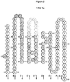
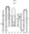


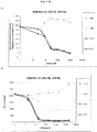

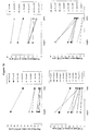

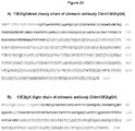
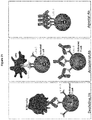


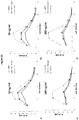
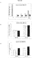

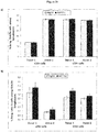
Classifications
-
- A—HUMAN NECESSITIES
- A61—MEDICAL OR VETERINARY SCIENCE; HYGIENE
- A61K—PREPARATIONS FOR MEDICAL, DENTAL OR TOILETRY PURPOSES
- A61K39/00—Medicinal preparations containing antigens or antibodies
- A61K39/395—Antibodies; Immunoglobulins; Immune serum, e.g. antilymphocytic serum
-
- A—HUMAN NECESSITIES
- A61—MEDICAL OR VETERINARY SCIENCE; HYGIENE
- A61P—SPECIFIC THERAPEUTIC ACTIVITY OF CHEMICAL COMPOUNDS OR MEDICINAL PREPARATIONS
- A61P29/00—Non-central analgesic, antipyretic or antiinflammatory agents, e.g. antirheumatic agents; Non-steroidal antiinflammatory drugs [NSAID]
-
- A—HUMAN NECESSITIES
- A61—MEDICAL OR VETERINARY SCIENCE; HYGIENE
- A61P—SPECIFIC THERAPEUTIC ACTIVITY OF CHEMICAL COMPOUNDS OR MEDICINAL PREPARATIONS
- A61P37/00—Drugs for immunological or allergic disorders
- A61P37/02—Immunomodulators
- A61P37/06—Immunosuppressants, e.g. drugs for graft rejection
-
- C—CHEMISTRY; METALLURGY
- C07—ORGANIC CHEMISTRY
- C07K—PEPTIDES
- C07K16/00—Immunoglobulins [IGs], e.g. monoclonal or polyclonal antibodies
- C07K16/18—Immunoglobulins [IGs], e.g. monoclonal or polyclonal antibodies against material from animals or humans
- C07K16/28—Immunoglobulins [IGs], e.g. monoclonal or polyclonal antibodies against material from animals or humans against receptors, cell surface antigens or cell surface determinants
- C07K16/2896—Immunoglobulins [IGs], e.g. monoclonal or polyclonal antibodies against material from animals or humans against receptors, cell surface antigens or cell surface determinants against molecules with a "CD"-designation, not provided for elsewhere
-
- C—CHEMISTRY; METALLURGY
- C07—ORGANIC CHEMISTRY
- C07K—PEPTIDES
- C07K2317/00—Immunoglobulins specific features
- C07K2317/20—Immunoglobulins specific features characterized by taxonomic origin
- C07K2317/24—Immunoglobulins specific features characterized by taxonomic origin containing regions, domains or residues from different species, e.g. chimeric, humanized or veneered
-
- C—CHEMISTRY; METALLURGY
- C07—ORGANIC CHEMISTRY
- C07K—PEPTIDES
- C07K2317/00—Immunoglobulins specific features
- C07K2317/50—Immunoglobulins specific features characterized by immunoglobulin fragments
- C07K2317/56—Immunoglobulins specific features characterized by immunoglobulin fragments variable (Fv) region, i.e. VH and/or VL
- C07K2317/565—Complementarity determining region [CDR]
-
- C—CHEMISTRY; METALLURGY
- C07—ORGANIC CHEMISTRY
- C07K—PEPTIDES
- C07K2317/00—Immunoglobulins specific features
- C07K2317/50—Immunoglobulins specific features characterized by immunoglobulin fragments
- C07K2317/56—Immunoglobulins specific features characterized by immunoglobulin fragments variable (Fv) region, i.e. VH and/or VL
- C07K2317/567—Framework region [FR]
-
- C—CHEMISTRY; METALLURGY
- C07—ORGANIC CHEMISTRY
- C07K—PEPTIDES
- C07K2317/00—Immunoglobulins specific features
- C07K2317/70—Immunoglobulins specific features characterized by effect upon binding to a cell or to an antigen
-
- C—CHEMISTRY; METALLURGY
- C07—ORGANIC CHEMISTRY
- C07K—PEPTIDES
- C07K2317/00—Immunoglobulins specific features
- C07K2317/70—Immunoglobulins specific features characterized by effect upon binding to a cell or to an antigen
- C07K2317/73—Inducing cell death, e.g. apoptosis, necrosis or inhibition of cell proliferation
-
- C—CHEMISTRY; METALLURGY
- C07—ORGANIC CHEMISTRY
- C07K—PEPTIDES
- C07K2317/00—Immunoglobulins specific features
- C07K2317/70—Immunoglobulins specific features characterized by effect upon binding to a cell or to an antigen
- C07K2317/75—Agonist effect on antigen
-
- C—CHEMISTRY; METALLURGY
- C07—ORGANIC CHEMISTRY
- C07K—PEPTIDES
- C07K2317/00—Immunoglobulins specific features
- C07K2317/90—Immunoglobulins specific features characterized by (pharmaco)kinetic aspects or by stability of the immunoglobulin
- C07K2317/92—Affinity (KD), association rate (Ka), dissociation rate (Kd) or EC50 value
Definitions
- This invention relates to antibodies, or antigen-binding fragments thereof, that bind to Lymphocyte-activation gene-3 (LAG-3), especially antibodies, or antigen-binding fragments thereof, that are agonists of LAG-3, and to use of the antibodies or fragments as medicaments, in particular for the treatment of conditions associated with proliferation and/or activation of CD4 + and/or CD8 + T cells, in particular inflammatory and autoimmune disorders.
- LAG-3 Lymphocyte-activation gene-3
- the lymphocyte activation gene 3 (LAG-3) is a CD4 homolog type I membrane protein with four extracellular lg superfamily domains. Similar to CD4, LAG-3 oligomerizes at the surfaces of T cells and binds to MHC class II molecules on antigen-presenting cells (APCs) but with significantly higher affinity than CD4. LAG-3 is expressed on activated CD4-positive and CD8-positive T lymphocytes where it associates with the CD3-TCR complex at the cell surface and negatively regulates signal transduction. As a consequence, it negatively regulates T cell proliferation, function, and homeostasis.
- APCs antigen-presenting cells
- LAG-3 also encodes an alternative splice variant that is translated to a soluble form of LAG-3 (sLAG-3).
- LAG-3 activates antigen-presenting cells (APCs) through MHC class II signalling, leading to increased antigen-specific T-cell responses in vivo ( Triebel, Trends Immunol., 2003, 24: 619-622 ).
- the amino acid sequence of human and murine LAG-3 protein is provided in Figure 1 of Huard et al (Proc. Natl. Acad. Sci. USA, 11: 5744-5749, 1997 ).
- the sequence of human LAG-3 protein is repeated in Figure 1 below (SEQ ID NO: 27).
- the amino acid sequences of the four extracellular Ig superfamily domains (D1, D2, D3, and D4) of human LAG-3 are at amino acid residues: 1-149 (D1) (SEQ ID NO:28); 150-239 (D2) (SEQ ID NO:29); 240-330 (D3) (SEQ ID NO:39); and 331-412 (D4) (SEQ ID NO:51).
- Baixeras, et al. J. Exp. Med., 1992, Vol. 176: 327-337 ) describes production of 17B4, a mouse monoclonal antibody (isotype lgG1) to human LAG-3 protein. This antibody recognizes a 30 amino acid extra-loop of the first N-terminal D1 domain of human LAG-3.
- 17B4 inhibits LAG-3/MHC class interactions, and increases T cell proliferation as a LAG-3 signalling antagonist ( Huard at al, Eur J Immunol. 1996;26:1180-6 ). 17B4 mAb has no agonist activity, as determined by its inability to induce intracellular free calcium elevation into T cells in the absence of a secondary cross-linking reagent ( Hannier et al, J Immunol. 1998;161:4058-65 .).
- Agents capable of modulating the activation and/or effector functions of CD8-positive and CD4-positive T cells are highly desirable.
- many autoimmune disorders are known to involve autoreactive T cells and autoantibodies.
- agents that are capable of inhibiting or eliminating autoreactive lymphocytes without compromising the immune system's ability to defend against pathogens.
- US2011/070238 describes a chimeric version of mAb A9H12 (designated IMP731) comprising a human IgG1 Fc region and shows that this chimeric antibody, like its murine counterpart, suppresses DTH in baboons.
- mAb A9H12 designated IMP731
- this document explicitly states that the antibodies described therein can be used in the treatment of transplant rejection and autoimmune disease.
- LAG-3 agonists will negatively regulate T cell proliferation and/or function without depleting the T cells, and that such agonists can also be used to treat inflammatory or autoimmune disorders.
- the applicant has been able to produce monoclonal anti-LAG-3 agonistic antibodies. These antibodies inhibit antigen-induced proliferation of CD4-positive and CD8-positive T cells. Such antibodies, and antigen-binding fragments thereof, can be used for the treatment of immune disorders, in particular T-cell-mediated inflammatory and autoimmune disorders.
- an isolated agonistic anti-LAG-3 antibody or an antigen-binding fragment thereof, that binds to LAG-3 and inhibits: antigen-induced CD4 + and/or CD8 + T cell proliferation; and/or antigen-induced CD4 + and/or CD8 + T cell activation; which comprises:
- the antibody is a monoclonal agonistic anti-LAG-3 antibody, or an antigen-binding fragment thereof.
- LAG-3 refers to Lymphocyte Activation Gene-3.
- LAG-3 includes variants, isoforms, homologs, orthologs and paralogs.
- antibodies specific for a human LAG-3 protein may, in certain cases, cross-react with a LAG-3 protein from a species other than human.
- the antibodies specific for a human LAG-3 protein may be completely specific for the human LAG-3 protein and may not exhibit species or other types of cross-reactivity, or may cross-react with LAG-3 from certain other species but not all other species (e.g., cross-react with monkey LAG-3 but not mouse LAG-3).
- human LAG-3 refers to human sequence LAG-3, such as the complete amino acid sequence of human LAG-3 having Genbank Accession No. NP 002277 (SEQ ID NO: 38), or the amino acid sequence of human LAG-3 protein given in Figure 1 (SEQ ID NO: 27).
- mouse LAG-3 refers to mouse sequence LAG-3, such as the complete amino acid sequence of mouse LAG-3 having Genbank Accession No. NP_032505.
- LAG-3 is also known in the art as, for example, CD223.
- the human LAG-3 sequence may differ from human LAG-3 of Genbank Accession No.
- NP_002277 by having, e.g., conserved mutations or mutations in non-conserved regions and the LAG-3 has substantially the same biological function as the human LAG-3 of Genbank Accession No. NP_002277.
- a biological function of human LAG-3 is having an epitope in the extracellular domain of LAG-3 that is specifically bound by an antibody of the instant disclosure or a biological function of human LAG-3 is binding to MHC Class II molecules.
- monkey LAG-3 is intended to encompass LAG-3 proteins expressed by Old World and New World monkeys, including but not limited to cynomolgus monkey LAG-3 and rhesus monkey LAG-3.
- a representative amino acid sequence for monkey LAG-3 is the rhesus monkey LAG-3 amino acid sequence which is also deposited as Genbank Accession No. XM_001108923.
- Another representative amino acid sequence for monkey LAG-3 is the alternative rhesus monkey sequence of clone pa23-5 as described in US 2011/0150892 A1 . This alternative rhesus sequence exhibits a single amino acid difference, at position 419, as compared to the Genbank-deposited sequence.
- a particular human LAG-3 sequence will generally be at least 90% identical in amino acid sequence to human LAG-3 of Genbank Accession No. NP_002277 and contains amino acid residues that identify the amino acid sequence as being human when compared to LAG-3 amino acid sequences of other species (e.g., murine).
- a human LAG-3 can be at least 95%, or even at least 96%, 97%, 98%, or 99% identical in amino acid sequence to LAG-3 of Genbank Accession No. NP_002277.
- a human LAG-3 sequence will display no more than 10 amino acid differences from the LAG-3 sequence of Genbank Accession No. NP_002277.
- the human LAG-3 can display no more than 5, or even no more than 4, 3, 2, or 1 amino acid difference from the LAG-3 sequence of Genbank Accession No. NP_002277. Percent identity can be determined as described herein.
- agonistic is used herein interchangeably with the term “agonist”.
- the antibody, or antigen-binding fragment thereof inhibits antigen-induced CD4 + T cell proliferation, and/or antigen-induced CD8 + T cell proliferation. In some embodiments, the antibody, or antigen-binding fragment thereof, inhibits antigen-induced CD4 + T cell proliferation, and antigen-induced CD8 + T cell proliferation. In particular embodiments, the antibody, or antigen-binding fragment thereof, inhibits antigen-induced CD8 + T cell proliferation more than antigen-induced CD4 + T cell proliferation.
- Figure 21 illustrates the differences between depleting anti-LAG-3 antibodies, antagonist anti-LAG-3 antibodies, and antibodies of the invention (i.e. agonistic anti-LAG-3 antibodies, and antibodies that bind to LAG-3 and inhibit antigen-induced CD4 + and/or CD8 + T cell proliferation, or antigen-induced CD4 + and/or CD8 + T cell activation).
- a depleting anti-LAG-3 antibody causes depletion of activated T cells by binding to LAG-3 expressed on the surface of the cells. Depletion can occur by antibody-dependent cell-mediated cytotoxicity (ADCC), or by complement-dependent cytotoxicity (CDC).
- ADCC antibody-dependent cell-mediated cytotoxicity
- CDC complement-dependent cytotoxicity
- the Fc region of the depleting antibody binds to Fc receptors (FcyRs) on the surface of immune effector cells, such as natural killers and macrophages, leading to lysis of the targeted cells.
- FcyRs Fc receptors
- CDC the Fc region of the depleting antibody binds to the C1q component of complement, and the targeted cell is killed by triggering the complement cascade at the cell surface.
- depleting anti-LAG-3 antibodies inhibit T cell-mediated immune responses. It will be appreciated that the effects of depleting anti-LAG-3 antibodies are long-lasting, and irreversible because they cause the destruction of activated T cells.
- An antagonist anti-LAG-3 antibody binds to LAG-3 on the surface of activated T cells, and prevents interaction of LAG-3 with MHC class II molecules on the surface of antigen-presenting cells (APCs). This blocks the negative regulation of signal transduction that occurs when APCs bind to LAG-3 on activated T cells. Consequently, antagonist anti-LAG-3 antibodies prevent the negative regulation of T cell proliferation, function and homeostasis normally mediated by LAG-3. Such antibodies are useful, for example, for the treatment of cancer and infectious disease.
- Antibodies of the invention bind to LAG-3 on the surface of activated T cells, and negatively regulate signal transduction through agonism of LAG-3, causing negative regulation of T cell proliferation and/or activation.
- antibodies of the invention inhibit T cell-mediated immune responses, in particular by inhibiting antigen-induced CD4 + and/or CD8 + T cell proliferation, and/or antigen-induced CD4 + and/or CD8 + T cell activation.
- the effects of such antibodies are reversible, and may be less long-lasting than the effects of depleting anti-LAG-3 antibodies since they do not cause the destruction of activated T cells. It will be appreciated that the length of time for which an antibody of the invention is effective will depend on the plasma half-life of the antibody.
- Inhibition of antigen-induced CD4 + and/or CD8 + T cell proliferation may be determined by any suitable method known to the skilled person.
- An example of a suitable method is by measuring the proliferation of CD4 + and/or CD8 + T cells induced by antigenic peptides in the presence of the antibody or fragment, compared with the corresponding proliferation in the presence of a negative control antibody of the same isotype.
- the CD4 + and CD8 + T cells may be present, for example, in a sample of peripheral blood mononuclear cells (PBMCs) obtained from a healthy donor.
- Proliferation of the cells may be induced by any suitable antigenic peptides, such as a pool of peptides covering the sequence of CMV pp35.
- Proliferation of the cells may be measured by labelling the cells, for example with a fluorescent cell staining dye, such as carboxyfluorescein succinimidyl ester (CFSE).
- CFSE carboxyfluorescein succinimidyl ester
- the percentage inhibition of antigen-induced CD4 + and/or CD8 + T cell proliferation may be determined as a percentage inhibition of the proliferation index (PI), calculated as the sum of: the percentage of CD4 + and/or CD8 + T cells under each division peak (assessed by FACS), multiplied by the division number, as described in more detail in Example 10 below.
- PI percentage inhibition of the proliferation index
- an antibody of the invention inhibits antigen-induced CD4 + T cell proliferation by at least 10%, 20%, 30%, 40%, 50%, 60%, 70%, 80%, or 90%, compared to antigen-induced CD4 + T cell proliferation in the absence of the antibody or fragment.
- an antibody of the invention inhibits antigen-induced CD8 + T cell proliferation by at least 10%, 20%, 30%, 40%, 50%, 60%, 70%, 80%, or 90%, compared to antigen-induced CD8 + T cell proliferation in the absence of the antibody or fragment.
- an antibody of the invention inhibits antigen-induced CD4 + T cell proliferation, and antigen-induced CD8 + T cell proliferation, each by at least 10%, 20%, 30%, 40%, 50%, 60%, 70%, 80%, or 90%, compared to antigen-induced CD4 + T cell proliferation, and antigen-induced CD8 + T cell proliferation, respectively, in the absence of the antibody or fragment.
- an antibody of the invention inhibits antigen-induced CD4 + T cell proliferation by at least 20% compared to antigen-induced CD4 + T cell proliferation in the absence of the antibody or fragment, and inhibits antigen-induced CD8 + T cell proliferation by at least 30% compared to and antigen-induced CD8 + T cell proliferation in the absence of the antibody or fragment.
- Inhibition of antigen-induced CD4+ and/or CD8+ T cell proliferation by an antibody of the invention, or fragment thereof, may be compared to antigen-induced CD4+ and/or CD8+ T cell proliferation in the presence of a negative control antibody of the same isotype, or fragment thereof.
- an antibody of the invention inhibits antigen-induced CD8 + T cell proliferation by at least 10%, 20%, 30%, 40%, 50%, 60%, 70%, 80%, or 90%, more than the antibody or fragment inhibits antigen-induced CD4 + T cell proliferation.
- the inhibition of antigen-induced CD8 + T cell proliferation is LAG-3 dependent and IL-2 independent.
- an antibody of the invention, or antigen-binding fragment thereof inhibits antigen-induced CD4 + and/or CD8 + T cell activation. In certain embodiments, an antibody of the invention, or antigen-binding fragment thereof, inhibits antigen-induced CD4 + and/or CD8 + T cell proliferation and antigen-induced CD4 + and/or CD8 + T cell activation.
- an antibody of the invention, or antigen-binding fragment thereof may bind to LAG-3 and inhibit antigen-induced CD4 + and/or CD8 + T cell activation.
- an antibody of the invention, or antigen-binding fragment thereof binds to LAG-3 and inhibits antigen-induced CD4 + T cell activation and antigen-induced CD8 + T cell activation.
- Inhibition of activation of CD4 + and/or CD8 + T cells may be determined by any suitable method known to the skilled person.
- An example of a suitable method is by measuring the effect of the antibody, or fragment, on CD4 + and/or CD8 + T cell activation marker expression, or T cell activation marker secretion.
- CD8 + T cell activation may be measured by measuring the expression of CD25, as an activation marker, on CD8 + T cells induced by antigenic peptides in the presence of the antibody or fragment, compared with the corresponding CD25 expression in the presence of a negative control antibody of the same isotype.
- T cell activation may be measured by measuring the secretion of IFN- ⁇ in cell supernatant of T cells induced by antigenic peptides in the presence of the antibody or fragment, compared with the corresponding secretion in the presence of a negative control antibody of the same isotype.
- the T cells may be present, for example, in a sample of PBMCs obtained from a healthy donor. Activation of the cells may be induced by any suitable antigenic peptides, such as a pool of peptides covering the sequence of CMV pp35.
- An example of a method for determining inhibition of antigen-induced T cell activation by measuring T cell activation marker secretion is described in more detail in Example 15 below.
- An example of a method for determining inhibition of antigen-induced CD8 + T cell activation by measuring CD8 + T cell activation marker expression is described in more detail in Example 16 below.
- an antibody of the invention inhibits antigen-induced CD4 + and/or CD8 + T cell activation by at least 10%, 20%, 30%, 40%, 50%, 60%, 70%, 80%, or 90%, compared to antigen-induced CD4 + and/or CD8 + T cell activation in the absence of the antibody or fragment.
- Inhibition of antigen-induced CD4+ and/or CD8+ T cell activation by an antibody of the invention, or fragment thereof, may be compared to antigen-induced CD4+ and/or CD8+ T cell activation in the presence of a negative control antibody of the same isotype, or fragment thereof.
- an antibody of the invention inhibits binding of IMP321 to MHC class II-positive cells.
- IMP321 (also referred to as "LAG-3Ig” below) is a recombinant soluble human LAG-3Ig fusion protein.
- the fusion protein is obtained as a 200-kDa dimer produced in Chinese hamster ovary (CHO) cells transfected with a plasmid encoding for the extracellular domain of human LAG-3 fused to the human IgG1 Fc.
- the sequence of IMP321 is provided in SEQ ID NO: 17 of US Patent Application No. 2011/0008331 .
- Binding of IMP321 to MHC class II-positive cells may be determined by measuring binding of an IMP321-label conjugate (for example an IMP321-Alex 488 conjugate) to Raji cells (these are MHC class II-positive B cells), for example as described in Example 8 below.
- an IMP321-label conjugate for example an IMP321-Alex 488 conjugate
- Raji cells these are MHC class II-positive B cells
- an antibody of the invention inhibits binding of IMP321 to MHC class II-positive cells by at least 10%, at least 20%, at least 30%, at least 40%, at least 50%, at least 60%, at least 70%, at least 80%, or at least 90% compared to the binding of IMP321 to MHC class II-positive cells in the absence of the antibody or fragment.
- an antibody of the invention inhibits binding of IMP321 to MHC class II-positive cells by at least 30%, compared to the binding of IMP321 to MHC class II-positive cells in the absence of the antibody or fragment, wherein the ratio of the concentration of the antibody, or fragment, to IMP321 is 0.1:1.
- an antibody of the invention inhibits binding of IMP321 to MHC class II-positive cells by at least 80%, compared to the binding of IMP321 to MHC class II-positive cells in the absence of the antibody or fragment, wherein the ratio of the concentration of the antibody, or fragment, to IMP321 is 0.3:1 or 1:1.
- an antibody of the invention inhibits IMP321-induced monocyte activation.
- IMP321 is able to activate cells of the human monocytic cell line THP-1. Activation of THP-1 cells can be determined by the level of secretion of chemokine ligand 4 (CCL4, also known as Macrophage inflammatory protein-1 ⁇ , MIP-1 ⁇ ) by the THP-1 cells. Pre-incubation of an antibody, or fragment, of the invention with IMP321 prior to incubation of the mixture with THP-1 cells can be used to determine whether the antibody, or fragment, inhibits IMP321-induced monocyte activation. A method for determining inhibition of IMP321-induced monocyte activation is described in more detail in Example 9 below.
- an antibody of the invention inhibits IMP321-induced monocyte activation by at least 10%, at least 20%, at least 30%, at least 40%, at least 50%, at least 60%, at least 70%, at least 80%, or at least 90% compared to the amount of IMP321-induced monocyte activation in the absence of the antibody or fragment.
- an antibody of the invention inhibits IMP321-induced monocyte activation by at least 70% compared to the amount of IMP321-induced monocyte activation in the absence of the antibody or fragment, wherein the ratio of the concentration of the antibody, or fragment, to IMP321 is 1:1.
- Huard et al (Proc. Natl. Acad. Sci. USA, 11: 5744-5749, 1997 ) describes characterization of the MHC class II binding site on LAG-3 protein. Many of the residues essential for binding MHC class II proteins are clustered at the base of a large 30 amino acid extra-loop structure in the LAG-3 D1 domain.
- the amino acid sequence of the extra-loop structure of the D1 domain of human LAG-3 protein is GPPAAAPGHPLAPGPHPAAPSSWGPRPRRY (SEQ ID NO: 40), the underlined sequence in Figure 1 .
- An antibody of the invention may bind to an epitope of human LAG-3 that overlaps with the MHC class II binding site on LAG-3.
- An antibody of the invention may bind to an epitope that overlaps with the 30 amino acid extra-loop of the first N-terminal D1 domain of human LAG-3.
- an antibody of the invention does not bind to the 30 amino acid extra-loop sequence (SEQ ID NO: 40) of the first N-terminal D1 domain of human LAG-3 protein.
- An antibody of the invention may inhibit binding of LAG-3 to MHC class II molecules in vivo.
- an antibody of the invention may antagonise the MHC class II-activating signal into antigen-presenting cells (APCs).
- APCs antigen-presenting cells
- an antibody of the invention may inhibit LAG-3-induced APC activation, for example dendritic cell activation, for example LAG-3-induced monocyte or macrophage activation.
- an antibody of the invention inhibits binding of LAG-3 to MHC class II-positive cells by at least 10%, at least 20%, at least 30%, at least 40%, at least 50%, at least 60%, at least 70%, at least 80%, or at least 90% compared to the binding of LAG-3 to MHC class II-positive cells in the absence of the antibody or fragment.
- an antibody of the invention inhibits LAG-3-induced APC activation by at least 10%, at least 20%, at least 30%, at least 40%, at least 50%, at least 60%, at least 70%, at least 80%, or at least 90% compared to the amount of LAG-3-induced APC activation in the absence of the antibody or fragment.
- a monoclonal antibody of the invention comprises three complementarity determining regions (CDRs) of an antibody heavy chain variable (VH) region comprising amino acid sequence SEQ ID NO:7, and three CDRs of an antibody light chain variable (VL) region comprising amino acid sequence SEQ ID NO:8.
- CDRs complementarity determining regions
- an anti-LAG-3 antibody or antigen-binding fragment thereof, which comprises three complementarity determining regions (CDRs) of an antibody heavy chain variable (VH) region comprising amino acid sequence SEQ ID NO:7, and three CDRs of an antibody light chain variable (VL) region comprising amino acid sequence SEQ ID NO:8.
- CDRs complementarity determining regions
- the CDRs of the antibody VH region may be CDRs of amino acid sequence SEQ ID NO:1, 2, and 3, and the CDRs of the antibody VL region may be CDRs of amino acid sequence SEQ ID NO:4, 5, and 6.
- An antibody of the invention may comprise an antibody VH region with CDRs comprising amino acid sequences of SEQ ID NOs:1, 2, and 3, andr an antibody VL region with CDRs comprising amino acid sequences of SEQ ID NOs:4, 5, and 6.
- the CDRs of amino acid sequence SEQ ID NOs:1, 2, and 3 may be present in any order in the VH region, and the CDRs of amino acid sequence SEQ ID NOs:4, 5, and 6 may be present in any order in the VL region.
- the antibody, or fragment thereof comprises CDR-H1 having amino acid sequence SEQ ID NO:1, CDR-H2 having amino acid sequence SEQ ID NO:2, and CDR-H3 having amino acid sequence SEQ ID NO:3, and/or CDR-L1 having amino acid sequence SEQ ID NO:4, CDR-L2 having amino acid sequence SEQ ID NO:5, and CDR-L3 having amino acid sequence SEQ ID NO:6.
- the CDRs of the antibody VH region may be CDRs of amino acid sequence SEQ ID NO:21, 22, and 23, and the CDRs of the antibody VL region may be CDRs of amino acid sequence SEQ ID NO:24, 25, and 26.
- An antibody of the invention may comprise an antibody VH region with CDRs comprising amino acid sequences of SEQ ID NOs:21, 22, and 23, and an antibody VL region with CDRs comprising amino acid sequences of SEQ ID NOs:24, 25, and 26.
- the CDRs of amino acid sequence SEQ ID NOs:21, 22, and 23 may be present in any order in the VH region, and the CDRs of amino acid sequence SEQ ID NOs:24, 25, and 26 may be present in any order in the VL region.
- the antibody, or fragment thereof comprises CDR-H1 having amino acid sequence SEQ ID NO:21, CDR-H2 having amino acid sequence SEQ ID NO:22, and CDR-H3 having amino acid sequence SEQ ID NO:23, and/or CDR-L1 having amino acid sequence SEQ ID NO:24, CDR-L2 having amino acid sequence SEQ ID NO:25, and CDR-L3 having amino acid sequence SEQ ID NO:26.
- an antibody, or antigen-binding fragment thereof comprises an antibody VH region comprising a VH CDR1, a VH CDR2, and a VH CDR3, wherein the VH CDR1 has an amino acid sequence selected from SEQ ID NO:1 and 21, and/or the VH CDR2 has an amino acid sequence selected from SEQ ID NO:2 and 22, and/or the VH CDR3 has an amino acid sequence selected from SEQ ID NO:3 and 23.
- an antibody, or antigen-binding fragment thereof comprises an antibody VL region comprising a VL CDR1, a VL CDR2, and a VL CDR3, wherein the VL CDR1 has an amino acid sequence selected from SEQ ID NO:4 and 24, and/or the VL CDR2 has an amino acid sequence selected from SEQ ID NO:5 and 25, and/or the VL CDR3 has an amino acid sequence selected from SEQ ID NO:6 and 26.
- an antibody, or antigen-binding fragment thereof comprises: an antibody VH region comprising: a VH CDR1 with an amino acid sequence selected from SEQ ID NO:1 and 21; a VH CDR2 with an amino acid sequence selected from SEQ ID NO:2 and 22; and a VH CDR3 with an amino acid sequence selected from SEQ ID NO:3 and 23; and an antibody VL region comprising: a VL CDR1 with an amino acid sequence selected from SEQ ID NO:4 and 24; a VL CDR2 with an amino acid sequence selected from SEQ ID NO:5 and 25; and a VL CDR3 with an amino acid sequence selected from SEQ ID NO:6 and 26.
- An antibody of the invention, or antigen-binding fragment thereof may comprise an antibody VH region comprising amino acid sequence that is at least 60%, 70%, 80%, 81%, 82%, 83%, 84%, 85%, 86%, 87%, 88%, 89%, 90%, 91%, 92%, 93%, 94%, 95%, 96%, 97%, 98%, or 99% identical to the amino acid sequence of SEQ ID NO:7, and an antibody VL region comprising amino acid sequence that is at least 60%, 70%, 80%, 81%, 82%, 83%, 84%, 85%, 86%, 87%, 88%, 89%, 90%, 91%, 92%, 93%, 94%, 95%, 96%, 97%, 98%, or 99% identical to the amino acid sequence of SEQ ID NO:8.
- an antibody of the invention comprises an antibody VH region comprising amino acid sequence SEQ ID NO:7, and an antibody VL region comprising amino acid sequence SEQ ID NO:8.
- An antibody of the invention may comprise an antibody VH region and/or an antibody VL region comprising amino acid sequence that is at least 60%, 70%, 80%, 81%, 82%, 83%, 84%, 85%, 86%, 87%, 88%, 89%, 90%, 91%, 92%, 93%, 94%, 95%, 96%, 97%, 98%, or 99% identical, or that is identical, to the amino acid sequence of the VH and/or VL regions of mouse monoclonal anti-LAG-3 antibody 13E2, described herein in Examples 1, 2 and 3 (13E2 VH amino acid sequence: SEQ ID NO:7; 13E2 VL amino acid sequence: SEQ ID NO:8).
- an antibody, or antigen-binding fragment thereof that competes for binding to LAG-3 with an antibody that comprises an antibody VH region comprising amino acid sequence SEQ ID NO:7, and an antibody VL region comprising amino acid sequence SEQ ID NO:8.
- a monoclonal antibody, or antigen-binding fragment thereof may comprise one, two, or three complementarity determining regions (CDRs) of an antibody heavy chain variable (VH) region comprising amino acid sequence SEQ ID NO:17, and/or one, two, or three CDRs of an antibody light chain variable (VL) region comprising amino acid sequence SEQ ID NO:18.
- CDRs complementarity determining regions
- an anti-LAG-3 antibody or antigen-binding fragment thereof, which comprises one, two, or three complementarity determining regions (CDRs) of an antibody heavy chain variable (VH) region comprising amino acid sequence SEQ ID NO:17, and/or one, two, or three CDRs of an antibody light chain variable (VL) region comprising amino acid sequence SEQ ID NO:18.
- CDRs complementarity determining regions
- the CDRs of the antibody VH region may be CDRs of amino acid sequence SEQ ID NO:11, 12, and 13 and the CDRs of the antibody VL region may be CDRs of amino acid sequence SEQ ID NO:14, 15, and 16.
- An antibody, or antigen-binding fragment thereof may comprise an antibody VH region with CDRs comprising amino acid sequences of SEQ ID NOs:11, 12, and 13, and/or an antibody VL region with CDRs comprising amino acid sequences of SEQ ID NOs:14, 15, and 16.
- the CDRs of amino acid sequence SEQ ID NOs:11, 12, and 13 may be present in any order in the VH region, and the CDRs of amino acid sequence SEQ ID NOs:14, 15, and 16 may be present in any order in the VL region.
- the antibody, or fragment thereof comprises CDR-H1 having amino acid sequence SEQ ID NO:11, CDR-H2 having amino acid sequence SEQ ID NO:12, and CDR-H3 having amino acid sequence SEQ ID NO:13, and/or CDR-L1 having amino acid sequence SEQ ID NO:14, CDR-L2 having amino acid sequence SEQ ID NO:15, and CDR-L3 having amino acid sequence SEQ ID NO:16.
- the CDRs of the antibody VH region may be CDRs of amino acid sequence SEQ ID NO:31, 32, and 33
- the CDRs of the antibody VL region may be CDRs of amino acid sequence SEQ ID NO:34, 35, and 36.
- An antibody, or antigen-binding fragment thereof may comprise an antibody VH region with CDRs comprising amino acid sequences of SEQ ID NOs:31, 32, and 33, and/or an antibody VL region with CDRs comprising amino acid sequences of SEQ ID NOs:34, 35, and 36.
- the CDRs of amino acid sequence SEQ ID NOs:31, 32, and 33 may be present in any order in the VH region, and the CDRs of amino acid sequence SEQ ID NOs:34, 35, and 36 may be present in any order in the VL region.
- the antibody, or fragment thereof comprises CDR-H1 having amino acid sequence SEQ ID NO:31, CDR-H2 having amino acid sequence SEQ ID NO:32, and CDR-H3 having amino acid sequence SEQ ID NO:33, and/or CDR-L1 having amino acid sequence SEQ ID NO:34, CDR-L2 having amino acid sequence SEQ ID NO:35, and CDR-L3 having amino acid sequence SEQ ID NO:36.
- the CDRs of the antibody VH region are selected from CDRs of amino acid sequence SEQ ID NO:11, 12, 13, 31, 32, and 33, and the CDRs of the antibody VL region are selected from CDRs of amino acid sequence SEQ ID NO:14, 15, 16, 34, 35, and 36.
- an antibody, or antigen-binding fragment thereof comprises an antibody VH region comprising a VH CDR1, a VH CDR2, and a VH CDR3, wherein the VH CDR1 has an amino acid sequence selected from SEQ ID NO:11 and 31, and/or the VH CDR2 has an amino acid sequence selected from SEQ ID NO:12 and 32, and/or the VH CDR3 has an amino acid sequence selected from SEQ ID NO:13 and 33.
- an antibody, or antigen-binding fragment thereof comprises an antibody VL region comprising a VL CDR1, a VL CDR2, and a VL CDR3, wherein the VL CDR1 has an amino acid sequence selected from SEQ ID NO:14 and 34, and/or the VL CDR2 has an amino acid sequence selected from SEQ ID NO:15 and 35, and/or the VL CDR3 has an amino acid sequence selected from SEQ ID NO:16 and 36.
- an antibody, or antigen-binding fragment thereof comprises: an antibody VH region comprising: a VH CDR1 with an amino acid sequence selected from SEQ ID NO:11 and 31, a VH CDR2 with an amino acid sequence selected from SEQ ID NO:12 and 32, and a VH CDR3 with an amino acid sequence selected from SEQ ID NO:13 and 33; and an antibody VL region comprising: a VL CDR1 with an amino acid sequence selected from SEQ ID NO:14 and 34, a VL CDR2 with an amino acid sequence selected from SEQ ID NO:15 and 35, and a VL CDR3 with an amino acid sequence selected from SEQ ID NO:16 and 36.
- An antibody, or antigen-binding fragment thereof may comprise an antibody VH region comprising amino acid sequence that is at least 60%, 70%, 80%, 81%, 82%, 83%, 84%, 85%, 86%, 87%, 88%, 89%, 90%, 91%, 92%, 93%, 94%, 95%, 96%, 97%, 98%, or 99% identical to the amino acid sequence of SEQ ID NO:17, and/or an antibody VL region comprising amino acid sequence that is at least 60%, 70%, 80%, 81%, 82%, 83%, 84%, 85%, 86%, 87%, 88%, 89%, 90%, 91%, 92%, 93%, 94%, 95%, 96%, 97%, 98%, or 99% identical to the amino acid sequence of SEQ ID NO:18.
- an antibody, or antigen-binding fragment thereof comprises an antibody VH region comprising amino acid sequence SEQ ID NO:17, and/or an antibody VL region comprising amino acid sequence SEQ ID NO:18.
- An antibody, or antigen-binding fragment thereof may comprise an antibody VH region and an antibody VL region comprising amino acid sequence that is at least 60%, 70%, 80%, 81%, 82%, 83%, 84%, 85%, 86%, 87%, 88%, 89%, 90%, 91%, 92%, 93%, 94%, 95%, 96%, 97%, 98%, or 99% identical, or that is identical, to the amino acid sequence of the VH and VL regions of mouse monoclonal anti-LAG-3 antibody 34F4, described herein in Examples 4, 5 and 6 (34F4 VH amino acid sequence: SEQ ID NO:17; 34F4 VL amino acid sequence: SEQ ID NO:18).
- an antibody, or antigen-binding fragment thereof that competes for binding to LAG-3 with an antibody that comprises an antibody VH region comprising amino acid sequence SEQ ID NO:17, and an antibody VL region comprising amino acid sequence SEQ ID NO:18.
- antibodies of the invention comprise a heavy chain variable region comprising CDR1, CDR2 and CDR3 sequences, and a light chain variable region comprising CDR1, CDR2, and CDR3 sequences, which differ from those of antibody 13E2 by one or more conservative modifications, for example up to five conservative modifications. It is understood in the art that certain conservative sequence modifications can be made which do not remove antigen binding. See, e.g., Brummell et al. (1993) Biochem 32:1180-8 ; de Wildt et al. (1997) Prot. Eng. 10:835-41 ; Komissarov et al. (1997) J. Biol. Chem. 272:26864-26870 ; Hall et al. (1992) J.
- conservative sequence modifications refers to amino acid modifications that do not significantly affect or alter the binding characteristics of the antibody containing the amino acid sequence. Such conservative modifications include amino acid substitutions, additions and deletions. Modifications can be introduced into an antibody of the invention by standard techniques known in the art, such as site-directed mutagenesis and PCR-mediated mutagenesis. Conservative amino acid substitutions are ones in which the amino acid residue is replaced with an amino acid residue having a similar side chain. Families of amino acid residues having similar side chains have been defined in the art.
- amino acids with basic side chains e.g., lysine, arginine, histidine
- acidic side chains e.g., aspartic acid, glutamic acid
- uncharged polar side chains e.g., glycine, asparagine, glutamine, serine, threonine, tyrosine, cysteine, tryptophan
- nonpolar side chains e.g., alanine, valine, leucine, isoleucine, proline, phenylalanine, methionine
- beta-branched side chains e.g., threonine, valine, isoleucine
- aromatic side chains e.g., tyrosine, phenylalanine, tryptophan, histidine
- amino acid residues within the CDR regions of an antibody of the invention can be replaced with other amino acid residues from the same side chain family and the altered antibody can be tested for retained function (i.e., the functions set forth above) using the functional assays described herein.
- Antibodies of the invention can be prepared using an antibody having one or more of the VH and/or VL sequences of 13E2 as starting material to engineer a modified antibody.
- An antibody can be engineered by modifying one or more residues within one or both variable regions (i.e., VH and/or VL), for example within one or more CDR regions and/or within one or more framework regions. Additionally or alternatively, an antibody can be engineered by modifying residues within the constant region(s), for example to alter the effector function(s) of the antibody.
- CDR grafting can be used to engineer variable regions of antibodies.
- Antibodies interact with target antigens predominantly through amino acid residues that are located in the six heavy and light chain complementarity determining regions (CDRs). For this reason, the amino acid sequences within CDRs are more diverse between individual antibodies than sequences outside of CDRs. Because CDR sequences are responsible for most antibody-antigen interactions, it is possible to express recombinant antibodies that mimic the properties of specific naturally occurring antibodies by constructing expression vectors that include CDR sequences from the specific naturally occurring antibody grafted onto framework sequences from a different antibody with different properties (see, e.g., Riechmann et al. (1998) Nature 332:323-327 ; Jones et al.
- an antibody of the invention for example a monoclonal antibody, or antigen-binding fragment thereof, comprising CDR1, CDR2, and CDR3 of an antibody heavy chain variable region comprising amino acid sequence SEQ ID NO:7, and comprising CDR1, CDR2, and CDR3 of an antibody light chain variable region comprising amino acid sequence SEQ ID NO:8 (i.e., the CDRs of 13E2). While such antibodies contain the VH and VL CDR sequences of monoclonal antibody 13E2, they can contain differing framework sequences.
- an antibody, or antigen-binding fragment thereof for example a monoclonal antibody, or antigen-binding fragment thereof, comprising CDR1, CDR2, and CDR3 of an antibody heavy chain variable region comprising amino acid sequence SEQ ID NO:17, and/or comprising CDR1, CDR2, and CDR3 of an antibody light chain variable region comprising amino acid sequence SEQ ID NO:18 (i.e., the CDRs of 34F4). While such antibodies contain the VH and VL CDR sequences of monoclonal antibody 34F4, they can contain differing framework sequences.
- Such framework sequences can be obtained from public DNA databases or published references that include germline antibody gene sequences.
- germline DNA sequences for human heavy and light chain variable region genes can be found in the "VBase" human germline sequence database (available on the Internet at www.mrc-cpe.cam.ac.uk/vbase), as well as in Kabat et al. (1991), cited supra; Tomlinson et al. (1992) "The Repertoire of Human Germline VH Sequences Reveals about Fifty Groups of VH Segments with Different Hypervariable Loops” J. Mol. Biol. 227:776-798 ; and Cox et al.
- the germline DNA sequences for human heavy and light chain variable region genes can be found in the Genbank database.
- the following heavy chain germline sequences found in the HCo7 HuMAb mouse are available in the accompanying Genbank Accession Nos.: 1-69 (NG_0010109, NT_024637 & BC070333), 3-33 (NG_0010109 & NT_024637) and 3-7 (NG_0010109 & NT_024637).
- Antibody protein sequences are compared against a compiled protein sequence database using one of the sequence similarity searching methods called the Gapped BLAST (Altschul et al. (1997), supra ), which is well known to those skilled in the art.
- Preferred framework sequences for use in antibodies of the invention are those that are structurally similar to the framework sequences of the 13E2 or 34F4 antibodies.
- Sequences showing significant alignment with nucleic acid sequence encoding the VH domain of monoclonal antibody 13E2 include the following germline genes: IGHV8-8*01, IGHV8-11*01, IGHV8-12*01, IGHD2-12*01, IGHD1-1*01, IGHJ1*01, IGHJ1*02, IGHJ1*03.
- Sequences showing significant alignment with nucleic acid sequence encoding the VL domain of monoclonal antibody 13E2 include the following germline genes: IGKV6-17*01, IGKV6-25*01, IGKV6-23*01, IGKJ2*01, IGKJ2*03, IGKJ2*02.
- Sequences showing significant alignment with nucleic acid sequence encoding the VH domain of monoclonal antibody 34F4 include the following germline genes: IGHV8-8*01, IGHV8-12*01, IGHV8-11*01, IGHD1-1*01, IGHD1-2*01, IGHD2-3*01, IGHJ2*01, IGHJ2*02, IGHJ2*03.
- Sequences showing significant alignment with nucleic acid sequence encoding the VL domain of monoclonal antibody 34F4 include the following germline genes: IGKV6-17*01, IGKV6-25*01, IGKV6-23*01, IGKJ1*01, IGKJ1*02, IGKJ2*01.
- Preferred heavy chain framework sequences for use in antibodies of the invention are those that are structurally similar to the framework sequences encoded by germline V gene IGHV8-8*01, IGHV8-11*01, or IGHV8-12*01, especially IGHV8-8*01.
- Preferred light chain framework sequences for use in antibodies of the invention are those that are structurally similar to the framework sequences encoded by germline V gene IGKV6-17*01, IGKV6-25*01, or IGKV6-23*01, especially IGKV6-17*01.
- the VH CDR1, CDR2, and CDR3 sequences, and the VL CDR1, CDR2, and CDR3 sequences can be grafted onto framework regions that have the identical sequence as that found in the germline immunoglobulin gene from which the framework sequence derive, or the CDR sequences can be grafted onto framework regions that contain one or more mutations as compared to the germline sequences. For example, it has been found that in certain instances it is beneficial to mutate residues within the framework regions to maintain or enhance the antigen binding ability of the antibody (see e.g., U.S. Pat. Nos. 5,530,101 ; 5,585,089 ; 5,693,762 and 6,180,370 ).
- variable region modification is to mutate amino acid residues within the VH and/or VL CDR1, CDR2 and/or CDR3 regions to thereby improve one or more binding properties (e.g., affinity) of the antibody of interest.
- Site-directed mutagenesis or PCR-mediated mutagenesis can be performed to introduce the mutation(s) and the effect on antibody binding, or other functional property of interest, can be evaluated in in vitro or in vivo assays as described herein and provided in the Examples.
- Preferably conservative modifications are introduced.
- the mutations can be amino acid substitutions, additions or deletions, but are preferably substitutions.
- typically no more than one, two, three, four or five residues within a CDR region are altered. In some embodiments, no more than one, two, three, four or five residues are altered in total for all six CDR regions.
- an antibody, or antigen-binding fragment thereof comprises:
- an antibody, or antigen-binding fragment thereof comprises:
- an anti-LAG-3 monoclonal antibody, or antigen-binding fragment thereof comprising a heavy chain variable region comprising: (a) a VH CDR1 region comprising SEQ ID NO: 1, or an amino acid sequence having one, two, three, four or five amino acid substitutions, deletions or additions as compared to SEQ ID NO: 1 ; (b) a VH CDR2 region comprising SEQ ID NO: 2, or an amino acid sequence having one, two, three, four or five amino acid substitutions, deletions or additions as compared to SEQ ID NO: 2; and (c) a VH CDR3 region comprising SEQ ID NO: 3, or an amino acid sequence having one, two, three, four or five amino acid substitutions, deletions or additions as compared to SEQ ID NO: 3; and/or a light chain variable region comprising: (a) a VL CDR1 region comprising SEQ ID NO: 4, or an amino acid sequence having one, two, three, four or five amino acid substitution
- an anti-LAG-3 monoclonal antibody, or antigen-binding fragment thereof comprising a heavy chain variable region comprising: (a) a VH CDR1 region comprising SEQ ID NO: 11, or an amino acid sequence having one, two, three, four or five amino acid substitutions, deletions or additions as compared to SEQ ID NO: 11; (b) a VH CDR2 region comprising SEQ ID NO: 12, or an amino acid sequence having one, two, three, four or five amino acid substitutions, deletions or additions as compared to SEQ ID NO: 12; and (c) a VH CDR3 region comprising SEQ ID NO: 13, or an amino acid sequence having one, two, three, four or five amino acid substitutions, deletions or additions as compared to SEQ ID NO: 13; and/or a light chain variable region comprising: (a) a VL CDR1 region comprising SEQ ID NO: 14, or an amino acid sequence having one, two, three, four or five amino acid
- an anti-LAG-3 monoclonal antibody, or antigen-binding fragment thereof comprising a heavy chain variable region comprising: (a) a VH CDR1 region comprising SEQ ID NO: 21, or an amino acid sequence having one, two, three, four or five amino acid substitutions, deletions or additions as compared to SEQ ID NO: 21 ; (b) a VH CDR2 region comprising SEQ ID NO: 22, or an amino acid sequence having one, two, three, four or five amino acid substitutions, deletions or additions as compared to SEQ ID NO: 22; and (c) a VH CDR3 region comprising SEQ ID NO: 23, or an amino acid sequence having one, two, three, four or five amino acid substitutions, deletions or additions as compared to SEQ ID NO: 23; and/or a light chain variable region comprising: (a) a VL CDR1 region comprising SEQ ID NO: 24, or an amino acid sequence having one, two, three, four or five amino acid substitution
- an anti-LAG-3 monoclonal antibody, or antigen-binding fragment thereof comprising a heavy chain variable region comprising: (a) a VH CDR1 region comprising SEQ ID NO: 31, or an amino acid sequence having one, two, three, four or five amino acid substitutions, deletions or additions as compared to SEQ ID NO: 31; (b) a VH CDR2 region comprising SEQ ID NO: 32, or an amino acid sequence having one, two, three, four or five amino acid substitutions, deletions or additions as compared to SEQ ID NO: 32; and (c) a VH CDR3 region comprising SEQ ID NO: 33, or an amino acid sequence having one, two, three, four or five amino acid substitutions, deletions or additions as compared to SEQ ID NO: 33; and/or a light chain variable region comprising: (a) a VL CDR1 region comprising SEQ ID NO: 34, or an amino acid sequence having one, two, three, four or
- Engineered antibodies of the invention include those in which modifications have been made to framework residues within VH and/or VL, e.g. to improve the properties of the antibody. Typically such framework modifications are made to decrease the immunogenicity of the antibody. For example, one approach is to "backmutate" one or more framework residues to the corresponding germline sequence. More specifically, an antibody that has undergone somatic mutation can contain framework residues that differ from the germline sequence from which the antibody is derived. Such residues can be identified by comparing the antibody framework sequences to the germline sequences from which the antibody is derived.
- Another type of framework modification involves mutating one or more residues within the framework region, or even within one or more CDR regions, to remove T cell epitopes to thereby reduce the potential immunogenicity of the antibody. This approach is also referred to as "deimmunization" and is described in further detail in U.S. Patent Publication No. 20030153043 .
- antibodies of the invention can be engineered to include modifications within the Fc region, typically to alter one or more functional properties of the antibody, such as serum half-life, complement fixation, Fc receptor binding, and/or antigen-dependent cellular cytotoxicity.
- an antibody of the invention can be chemically modified (e.g., one or more chemical moieties can be attached to the antibody) or be modified to alter its glycosylation, again to alter one or more functional properties of the antibody.
- the hinge region of CF11 is modified such that the number of cysteine residues in the hinge region is altered, e.g., increased or decreased. This approach is described further in U.S. Pat. No. 5,677,425 .
- the number of cysteine residues in the hinge region of CF11 is altered to, for example, facilitate assembly of the light and heavy chains or to increase or decrease the stability of the antibody.
- the Fc hinge region of an antibody is mutated to decrease the biological half-life of the antibody. More specifically, one or more amino acid mutations are introduced into the CF12-CF13 domain interface region of the Fc-hinge fragment such that the antibody has impaired Staphylococcyl protein A (SpA) binding relative to native Fc-hinge domain SpA binding.
- SpA Staphylococcyl protein A
- the antibody is modified to increase its biological half-life.
- the IgG class is the most stable and has a serum half-life of 20 days, whereas IgM and IgA persist for only 5-8 days ( Brekke & Sandlie, 2003, Nature Reviews Drug Discovery 2, 52-62 ).
- Various approaches are possible. For example, one or more of the following mutations can be introduced: T252L, T254S, T256F, as described in U.S. Pat. No. 6,277,375 .
- the antibody can be altered within the CF11 or CL region to contain a salvage receptor binding epitope taken from two loops of a CF12 domain of an Fc region of an IgG, as described in U.S. Pat. Nos. 5,869,046 and 6,121,022 .
- An antibody of the invention should lack antibody-dependent cell-mediated cytotoxicity (ADCC) and complement-dependent cytotoxicity (CDC) so that the antibody or fragment can be used to negatively regulate T cell proliferation and/or function without depleting T cells as a result of ADCC or CDC.
- ADCC antibody-dependent cell-mediated cytotoxicity
- CDC complement-dependent cytotoxicity
- ADCC the Fc region of an antibody binds to Fc receptors (FcyRs) on the surface of immune effector cells, such as natural killers and macrophages, leading to lysis of the targeted cells.
- FcyRs Fc receptors
- CDC the Fc region binds to the C1q component of complement, and the targeted cell is killed by triggering the complement cascade at the cell surface.
- the ADCC and CDC activity of an antibody depends on its isotype. Both IgM and IgG can mediate complement fixation, whereas only IgG can promote antibody-dependent cellular cytotoxicity (ADCC).
- IgG isoforms exhibit different levels of CDC and ADCC: IgG: CDC (hIgG3>hIgG1>hIgG2>hIgG4; mIgG2a>mIgG1); ADCC (hIgG1 ⁇ hIgG3>hIgG2 ⁇ IgG4; mIgG2a>mIgG1)
- an antibody of the invention comprises a mouse IgG1, or human IgG4 Fc portion to ensure that the antibody or fragment lacks ADCC and CDC activity.
- ADCC and CDC activity of an antibody of the invention, or antigen-binding fragment thereof may be determined by any suitable method known to the skilled person. Examples of suitable assays for CDC and ADCC activity are described in WO 2008/132601 , and below.
- An anti-LAG-3 antibody exhibiting CDC activity will consistently kill LAG-3 + cells in the presence of complement, compared with its isotypic control.
- the target cells used to assess an anti-LAG-3 antibody may be a LAG-3-transfected cell line, or primary T cells activated to induce expression of LAG-3.
- the target cells are LAG-3+ CHO cells compared to wt CHO cells. Both types of cells (i.e. cells expressing LAG-3, and equivalent cells not expressing LAG-3) are incubated for 1 hour at 37°C with either the anti-LAG-3 test antibody, or its isotype-matched negative control antibody, and rabbit serum containing active complement.
- 7-Amino-Actinomycin D 7-Amino-Actinomycin D
- 7-AAD 7-Amino-Actinomycin D
- the percentage of 7-AAD-positive CHO cells is determined by flow cytometry analysis.
- An antibody exhibiting CDC activity will only kill LAG-3+ cells (for example, LAG-3+ CHO cells) in the presence of complement.
- the anti-LAG-3 Ab may be titered down to determine the efficacy of the antibody to activate CDC at low concentration of antibody.
- a CDC assay may also be performed on PBMCs stimulated with the superantigen SEB.
- the cytotoxicity of test antibody is analysed on both activated (namely CD25+/LAG-3+ cells) and non-activated (namely CD25-/LAG-3- cells) CD4+ helper T and CD8+ cytotoxic T cells. Only activated CD4+ and CD8+ T cells are specifically killed by an antibody exhibiting CDC.
- test anti-LAG-3 antibody if effector cells (PBMCs) are able to kill a significant percentage of LAG-3-expressing cells (for example, LAG-3+ CHO cells), compared with an isotype-matched negative control antibody, the test antibody exhibits ADCC. This effect should increase with the number of effector cells.
- the test antibody may be titered down to determine the efficacy of the antibody to induce ADCC at low concentration of antibody.
- test anti-LAG-3 antibody can be considered not to exhibit CDC or ADCC when it kills less than twice as many LAG-3+ cells as an isotype-matched negative control antibody in any of the assays described above.
- ADCC bioassays which use donor peripheral blood mononuclear cells (PBMCs) or the natural killer (NK) cell subpopulation as effector cells.
- PBMCs peripheral blood mononuclear cells
- NK natural killer
- An ADCC Reporter Bioassay available from Promega uses an alternative readout at an earlier point in ADCC pathway activation: the activation of gene transcription through the NFAT (nuclear factor of activated T-cells) pathway in the effector cell.
- NFAT nuclear factor of activated T-cells
- the ADCC Reporter Bioassay uses engineered Jurkat cells stably expressing the Fc ⁇ RIIIa receptor, V158 (high affinity) variant, and an NFAT response element driving expression of firefly luciferase as effector cells.
- Antibody biological activity in ADCC is quantified through the luciferase produced as a result of NFAT pathway activation; luciferase activity in the effector cell is quantified with luminescence readout.
- signal is high, and assay background is low.
- a good assay response is only obtained when target cells with the correct surface antigen, the correct specific antibody, and effector cells expressing Fc ⁇ RIIIa are present. If any one of these is missing, there is no response.
- the tested antibody When evaluating the ADCC activity of an antibody using an ADCC Reporter Bioassay, the tested antibody can be considered not to exhibit ADCC when the measured increase in bioluminescence is less than twice that of its isotype-matched negative control antibody.
- the Fc region comprises a mutant human IgG4 Fc sequence with an S228P mutation to abolish Fab arm exchange (as shown in Figure 20(A) for chimeric antibody Chim13E2IgG4 comprising heavy chain sequence 13E2IgG4mut).
- the Fc region comprises a wild-type human Ig kappa (IgK) chain C portion (13E2IgK) (as shown in Figure 20(B) for chimeric antibody Chim13E2IgG4 comprising light chain sequence 13E2IgK).
- the numbering of residues in the Fc region used for the human IgG4 Fc mutant described above is the standard numbering of the Eu index as in Kabat ( Kabat, E.A. et al., Sequences of proteins of immunological interest. 5th Edition - US Department of Health and Human Services, NIH publication n° 91-3242, pp 662,680,689 (1991 )).
- the glycosylation of an antibody is modified.
- an aglycosylated antibody can be made (i.e., the antibody lacks glycosylation).
- Glycosylation can be altered to, for example, increase the affinity of the antibody for antigen.
- carbohydrate modifications can be accomplished by, for example, altering one or more sites of glycosylation within the antibody sequence.
- one or more amino acid substitutions can be made that result in elimination of one or more variable region framework glycosylation sites to thereby eliminate glycosylation at that site.
- Such aglycosylation may increase the affinity of the antibody for antigen. See, e.g., U.S. Pat. Nos. 5,714,350 and 6,350,861 .
- An antibody can be pegylated to, for example, increase the biological (eg., serum) half-life of the antibody.
- the antibody, or fragment thereof typically is reacted with polyethylene glycol (PEG), such as a reactive ester or aldehyde derivative of PEG, under conditions in which one or more PEG groups become attached to the antibody or antibody fragment.
- PEG polyethylene glycol
- the pegylation is carried out via an acylation reaction or an alkylation reaction with a reactive PEG molecule (or an analogous reactive water-soluble polymer).
- polyethylene glycol is intended to encompass any of the forms of PEG that have been used to derivatize other proteins, such as mono (CI-C10) alkoxy- or aryloxy-polyethylene glycol or polyethylene glycol-maleimide.
- the antibody to be pegylated is an aglycosylated antibody. Methods for pegylating proteins are known in the art and can be applied to the antibodies of the invention. See, eg., EP 0 154 316 and EP 0 401 384 .
- An antibody of the invention may be a monoclonal antibody, or an antigen-binding fragment thereof.
- antibody and “immunoglobulin” include antibodies or immunoglobulins of any isotype, fragments of antibodies that retain specific binding to antigen, including, but not limited to, Fab, Fv, scFv, and Fd fragments, chimeric antibodies, humanized antibodies, single-chain antibodies (scAb), single domain antibodies (dAb), single domain heavy chain antibodies, a single domain light chain antibodies, bi-specific antibodies, multi-specific antibodies, and fusion proteins comprising an antigen-binding (also referred to herein as antigen binding) portion of an antibody and a non-antibody protein.
- Fab single-chain antibodies
- dAb single domain antibodies
- dAb single domain heavy chain antibodies
- bi-specific antibodies bi-specific antibodies
- multi-specific antibodies and fusion proteins comprising an antigen-binding (also referred to herein as antigen binding) portion of an antibody and a non-antibody protein.
- Fab', Fv, F(ab') 2 and or other antibody fragments that retain specific binding to antigen, and monoclonal antibodies.
- An antibody can be monovalent or bivalent.
- An antibody can be an lg monomer, which is a "Y-shaped" molecule that consists of four polypeptide chains: two heavy chains and two light chains connected by disulfide bonds.
- the antibodies can be detectably labeled, e.g., with a radioisotope, an enzyme that generates a detectable product, a fluorescent protein, and the like.
- the antibodies can be further conjugated to other moieties, such as members of specific binding pairs, e.g., biotin (member of biotin-avidin specific binding pair), and the like.
- the antibodies can also be bound to a solid support, including, but not limited to, polystyrene plates or beads, and the like.
- Antibody fragments comprise a portion of an intact antibody, for example, the antigen binding or variable region of the intact antibody.
- antibody fragments include Fab, Fab', F(ab') 2 , and Fv fragments; diabodies; linear antibodies ( Zapata et al., Protein Eng. 8(10): 1057-1062 (1995 )); domain antibodies (dAb; Holt et al. (2003) Trends Biotechnol. 21:484 ); single-chain antibody molecules; and multi-specific antibodies formed from antibody fragments.
- Papain digestion of antibodies produces two identical antigen-binding fragments, called “Fab” fragments, each with a single antigen-binding site, and a residual "Fc” fragment, a designation reflecting the ability to crystallize readily.
- Pepsin treatment yields an F(ab') 2 fragment that has two antigen combining sites and is still capable of cross-linking antigen.
- Fv is the minimum antibody fragment that contains a complete antigen-recognition and - binding site. This region consists of a dimer of one heavy- and one light-chain variable domain in tight, non-covalent association. It is in this configuration that the three CDRs of each variable domain interact to define an antigen-binding site on the surface of the V H -V L dimer. Collectively, the six CDRs confer antigen-binding specificity to the antibody. However, even a single variable domain (or half of an Fv comprising only three CDRs specific for an antigen) has the ability to recognize and bind antigen, although at a lower affinity than the entire binding site.
- the "Fab” fragment also contains the constant domain of the light chain and the first constant domain (CH 1 ) of the heavy chain.
- Fab fragments differ from Fab' fragments by the addition of a few residues at the carboxyl terminus of the heavy chain CH 1 domain including one or more cysteines from the antibody hinge region.
- Fab'-SH is the designation herein for Fab' in which the cysteine residue(s) of the constant domains bear a free thiol group.
- F(ab') 2 antibody fragments originally were produced as pairs of Fab' fragments which have hinge cysteines between them. Other chemical couplings of antibody fragments are also known.
- immunoglobulins can be assigned to different classes. There are five major classes of immunoglobulins: IgA, IgD, IgE, IgG, and IgM, and several of these classes can be further divided into subclasses (isotypes), e.g., IgG1, IgG2, IgG3, IgG4, IgA, and IgA2. The subclasses can be further divided into types, e.g., IgG2a and IgG2b.
- Single-chain Fv or “sFv” or “scFv” antibody fragments comprise the V H and V L domains of antibody, wherein these domains are present in a single polypeptide chain.
- the Fv polypeptide further comprises a polypeptide linker between the V H and V L domains, which enables the sFv to form the desired structure for antigen binding.
- diabodies refers to small antibody fragments with two antigen-binding sites, which fragments comprise a heavy-chain variable domain (V H ) connected to a light-chain variable domain (V L ) in the same polypeptide chain (V H -V L ).
- V H heavy-chain variable domain
- V L light-chain variable domain
- the domains are forced to pair with the complementary domains of another chain and create two antigen-binding sites.
- Diabodies are described more fully in, for example, EP 404,097 ; WO 93/11161 ; and Hollinger et al. (1993) Proc. Natl. Acad. Sci. USA 90:6444-6448 .
- a "monoclonal antibody” is an antibody produced by a group of identical cells, all of which were produced from a single cell by repetitive cellular replication. That is, the clone of cells only produces a single antibody species. While a monoclonal antibody can be produced using hybridoma production technology, other production methods known to those skilled in the art can also be used (e.g., antibodies derived from antibody phage display libraries).
- CDR complementarity determining region
- CDRs have been described by Kabat et al., J. Biol. Chem. 252:6609-6616 (1977 ); Kabat et al., U.S. Dept. of Health and Human Services, "Sequences of proteins of immunological interest” (1991 ) (also referred to herein as Kabat 1991); by Chothia et al., J. Mol. Biol. 196:901-917 (1987 ) (also referred to herein as Chothia 1987); and by MacCallum et al., J. Mol.
- CDR-L1 refers, respectively, to the first, second, and third CDRs in a light chain variable region.
- CDR-H1 refers, respectively, to the first, second, and third CDRs in a heavy chain variable region.
- CDR-1 refers, respectively, to the first, second and third CDRs of either chain's variable region.
- affinity refers to the equilibrium constant for the reversible binding of two agents (e.g., an antibody and an antigen) and is expressed as a dissociation constant (K D ).
- Affinity can be at least 1-fold greater, at least 2-fold greater, at least 3-fold greater, at least 4-fold greater, at least 5-fold greater, at least 6-fold greater, at least 7-fold greater, at least 8-fold greater, at least 9-fold greater, at least 10-fold greater, at least 20-fold greater, at least 30-fold greater, at least 40-fold greater, at least 50-fold greater, at least 60-fold greater, at least 70-fold greater, at least 80-fold greater, at least 90-fold greater, at least 100-fold greater, or at least 1,000-fold greater, or more, than the affinity of an antibody for unrelated amino acid sequences.
- Affinity of an antibody to a target protein can be, for example, from about 100 nanomolar (nM) to about 0.1 nM, from about 100 nM to about 1 picomolar (pM), or from about 100 nM to about 1 femtomolar (fM) or more.
- nM nanomolar
- pM picomolar
- fM femtomolar
- the term “avidity” refers to the resistance of a complex of two or more agents to dissociation after dilution.
- the terms “immunoreactive” and “preferentially binds” are used interchangeably herein with respect to antibodies and/or antigen-binding fragments.
- binding refers to a direct association between two molecules, due to, for example, covalent, electrostatic, hydrophobic, and ionic and/or hydrogen-bond interactions, including interactions such as salt bridges and water bridges.
- An antibody of the invention binds specifically to an epitope within a LAG-3 protein, particularly a human LAG-3 protein.
- Specific binding refers to binding with an affinity of at least about 5x 10 -7 M or greater, e.g., 10 -7 M, 5 x 10 -8 M, 10 -8 M, or greater.
- Non-specific binding refers to binding with an affinity of less than about 10 -7 M, e.g., binding with an affinity of 10 -6 M, 10 -5 M, 10 -4 M, etc.
- an antibody that "specifically binds human LAG-3" is intended to refer to an antibody that binds to human LAG-3 protein (and possibly a LAG-3 protein from one or more non-human species) but does not substantially bind to non-LAG-3 proteins.
- the antibody binds to a human LAG-3 protein with "high affinity", namely with a of K D of 1x10 -7 M or less, more preferably 1x10 -1 M or less, more preferably 5x10 -9 M or less, more preferably 1x10 -9 M or less.
- does not substantially bind to a protein or cells, as used herein, means does not bind or does not bind with a high affinity to the protein or cells, i.e. binds to the protein or cells with a K D of 1x10 -6 M or more, more preferably 1 x 10 -5 M or more, more preferably 1x10 -4 M or more, more preferably 1x10 -3 M or more, even more preferably 1x10 -2 M or more.
- K assoc or "K a ", as used herein, is intended to refer to the association rate of a particular antibody-antigen interaction
- K dis or "K d ,” as used herein, is intended to refer to the dissociation rate of a particular antibody-antigen interaction
- K D is intended to refer to the dissociation constant, which is obtained from the ratio of K d to K a . (i.e., K d /K a ) and is expressed as a molar concentration (M).
- high affinity for an IgG antibody refers to an antibody having a K D of 1x10 -7 M or less, more preferably 5x10 -8 M or less, even more preferably1x10 -8 M or less, even more preferably 5x10 -9 M or less and even more preferably 1x10 -9 M or less for a target antigen.
- high affinity binding can vary for other antibody isotypes.
- “high affinity” binding for an IgM isotype refers to an antibody having a K D of 10 -6 M or less, more preferably 10 -7 M or less, even more preferably 10 -8 M or less.
- an antibody of the invention binds to a human LAG-3 protein with higher affinity (i.e. with a lower dissociation constant) than the antagonistic anti-LAG-3 monoclonal antibody 17B4 ( Baixeras, et al., J. Exp. Med., 1992, Vol. 176: 327-337 ).
- Example 7 describes the results of Biacore analysis of binding of 17B4 antibody to human LAG-3Ig protein. The results show that the dissociation constant of 17B4 for human LAG-3Ig was 3.69 nM.
- an antibody of the present invention binds a human LAG-3 protein (or a human LAG-3Ig protein) with a dissociation constant (K D ) of less than 3.69 nM, for example as determined by Biacore analysis.
- an antibody of the present invention binds a human LAG-3 protein (or a derivative thereof, such as a human LAG-3Ig protein) with a dissociation constant (K D ) of no more than 3.5 nM, no more than 2.5 nM, no more than 2 nM, no more than 1 nM, no more than 0.9 nM, no more than 0.8 nM, no more than 0.7 nM, no more than 0.6 nM, no more than 0.5 nM, no more than 0.4 nM, no more than 0.3 nM, no more than 0.2 nM, no more than 0.1 nM.
- K D dissociation constant
- an anti-LAG-3 antibody of the present disclosure binds a human LAG-3 protein with a K D of no more than 90 pM, no more than 80 pM, no more than 70 pM, no more than 60 pM, no more than 50 pM, no more than 40 pM, no more than 30 pM, no more than 20 pM, no more than 10 pM, no more than 9 pM, no more than 8 pM, no more than 7 pM, no more than 6 pM, no more than 5 pM, no more than 4 pM, no more than 3 pM, no more than 2 pM, or no more than 1 pM.
- Example 20 below describes the results of Biacore analysis of binding of chimeric 13E2 IgG4 antibody (described below), and of humanized 13E2 IgG4 (described below), to human LAG-3Ig protein.
- the results show that the dissociation constant of chimeric 13E2 IgG4 antibody for human LAG-3Ig was 21.9 pM, and of humanized 13E2 IgG4 for human LAG-3Ig was 22.8 pM.
- an antibody of the present invention binds a human LAG-3 protein (or a human LAG-3Ig protein) with a dissociation constant (K D ) of no more than 100pM, no more than 90 pM, no more than 80 pM, no more than 70 pM, no more than 60 pM, no more than 50 pM, no more than 40 pM, no more than 30 pM, or no more than 25 pM, for example as determined by Biacore analysis.
- K D dissociation constant
- the affinity of an antibody of the invention, or antigen-binding fragment thereof may be at least 10%, 20%, 30%, 40%, 50%, 60%, 70%, 80%, or 90% higher than the affinity of 17B4 for a human LAG-3 protein. In some embodiments, an antibody of the invention, or antigen-binding fragment thereof, may be at least 1.5, 2, 2.5, 3, or 3.5 times the affinity of 17B4 for a human LAG-3 protein.
- the affinity of an antibody to a human LAG-3 protein can be determined by one skilled in the art, suitably by surface plasmon resonance, for example using a biosensor system, such as a Biacore system ( Murphy et al, Using Biacore to measure the binding kinetics of an antibody-antigen interaction; Curr Protoc Protein Sci. 2006 Sep;Chapter 19:Unit 19.14 ).
- Biacore analysis can be used to determine the dissociation constant between an antibody of the invention and a human LAG-3 protein.
- Binding to human LAG-3 can be assessed using one or more other techniques also well established in the art.
- an antibody can be tested by a flow cytometry assay in which the antibody is reacted with a cell line that expresses human LAG-3, such as CHO cells that have been transfected to express human LAG-3 on their cell surface.
- a cell line that expresses human LAG-3 such as CHO cells that have been transfected to express human LAG-3 on their cell surface.
- Other suitable cells for use in flow cytometry assays include SEB-stimulated PBMCs, or anti-CD3-stimulated CD4 + activated T cells, which express native LAG-3.
- Still other suitable binding assays include ELISA assays, for example, using a recombinant LAG-3 protein.
- An antibody of the invention is an isolated antibody.
- An "isolated” antibody is one that has been identified and separated and/or recovered from a component of its natural environment. Contaminant components of its natural environment are materials that would interfere with diagnostic or therapeutic uses for the antibody, and can include enzymes, hormones, and other proteinaceous or nonproteinaceous solutes.
- the antibody will be purified (1) to greater than 90%, greater than 95%, or greater than 98%, by weight of antibody as determined by the Lowry method, for example, more than 99% by weight, (2) to a degree sufficient to obtain at least 15 residues of N-terminal or internal amino acid sequence by use of a spinning cup sequenator, or (3) to homogeneity by sodium dodecyl sulfate-polyacrylamide gel electrophoresis (SDS-PAGE) under reducing or non-reducing conditions using Coomassie blue or silver stain.
- Isolated antibody includes the antibody in situ within recombinant cells since at least one component of the antibody's natural environment will not be present. In some instances, isolated antibody will be prepared by at least one purification step.
- an antibody of the invention is a humanized antibody, or antigen-binding fragment thereof, particularly, a humanized monoclonal antibody, or antigen-binding fragment thereof.
- a humanized antibody of the invention, or antigen-binding fragment thereof may comprise a humanized light chain framework region and/or a humanized heavy chain framework region.
- humanized antibody refers to an immunoglobulin comprising portions of immunoglobulins of different origin, wherein at least one portion comprises amino acid sequences of human origin.
- the humanized antibody can comprise portions derived from an immunoglobulin of nonhuman origin with the requisite specificity, such as a mouse, and from immunoglobulin sequences of human origin (e.g., immunoglobulin), joined together chemically by conventional techniques (e.g., synthetic) or prepared as a contiguous polypeptide using genetic engineering techniques (e.g., DNA encoding the protein portions of the chimeric antibody can be expressed to produce a contiguous polypeptide chain).
- humanized immunoglobulin is an immunoglobulin containing one or more immunoglobulin chains comprising a CDR derived from an antibody of nonhuman origin and a framework region derived from a light and/or heavy chain of human origin (e.g., CDR-grafted antibodies with or without framework changes). Chimeric or CDR-grafted single chain antibodies are also encompassed by the term humanized immunoglobulin. See, e.g., Cabilly et al., U.S. Pat. No. 4,816,567 ; Cabilly et al., European Patent No. 0,125,023 B1 ; Boss et al., U.S. Pat. No.
- variable region amino acid sequence may be identical to the variable region amino sequence of the species from which it is derived (for example, mouse sequence), or may be at least 60%, 70%, 80%, 81%, 82%, 83%, 84%, 85%, 86%, 87%, 88%, 89%, 90%, 91%, 92%, 93%, 94%, 95%, 96%, 97%, 98%, or 99% identical to that variable region sequence.
- variable region amino acid sequence of a chimeric antibody of the invention may comprise one or more amino acid deletions, substitutions, or additions (for example one, two, three, four, five, six, seven, eight, nine, or ten amino acid deletions, substitutions, or additions) compared with the variable region amino acid sequence of the species from which it is derived.
- the constant region amino acid sequence may be identical to the constant region amino acid sequence of the species from which it is derived (for example, human sequence), or may be at least 60%, 70%, 80%, 81%, 82%, 83%, 84%, 85%, 86%, 87%, 88%, 89%, 90%, 91%, 92%, 93%, 94%, 95%, 96%, 97%, 98%, or 99% identical to that constant region amino acid sequence.
- the constant region amino acid sequence of a chimeric antibody of the invention may comprise one or more amino acid deletions, substitutions, or additions (for example one, two, three, four, five, six, seven, eight, nine, or ten amino acid deletions, substitutions, or additions) compared with the constant region amino acid sequence of the species from which it is derived.
- the amino acid sequence of the Fc hinge region of a chimeric antibody may be mutated to decrease the biological half-life of the antibody, or the amino acid sequence of the Fc region may be mutated to increase the biological half-life of the chimeric antibody.
- the heavy chain variable region sequence comprises, or is derived from a mouse antibody, and the heavy chain constant region sequence comprises, or is derived from IgG4 Fc sequence.
- the light chain variable region sequence comprises, or is derived from a mouse antibody, and the light chain constant region sequence comprises, or is derived from human Ig kappa (IgK) chain C sequence.
- the Fc region comprises a mutant human IgG4 Fc sequence with an S228P mutation to abolish Fab arm exchange (as shown in Figure 20(A) for chimeric antibody Chim13E2IgG4 comprising heavy chain sequence 13E2IgG4mut).
- the Fc region comprises a wild-type human lg kappa (IgK) chain C portion (13E2IgK) (as shown in Figure 20(B) for chimeric antibody Chim13E2IgG4 comprising light chain sequence 13E2IgK).
- the numbering of residues in the Fc region used for the human IgG4 Fc mutant described above is the standard numbering of the Eu index as in Kabat ( Kabat, E.A. et al., Sequences of proteins of immunological interest. 5th Edition - US Department of Health and Human Services, NIH publication n° 91-3242, pp 662,680,689 (1991 )).
- Humanized antibodies may be produced using synthetic and/or recombinant nucleic acids to prepare genes (e.g., cDNA) encoding the desired humanized chain.
- genes e.g., cDNA
- nucleic acid (e.g., DNA) sequences coding for humanized variable regions can be constructed using PCR mutagenesis methods to alter DNA sequences encoding a human or humanized chain, such as a DNA template from a previously humanized variable region (see e.g., Kamman, M., et al., Nucl. Acids Res., 17: 5404 (1989 )); Sato, K., et al., Cancer Research, 53: 851-856 (1993 ); Daugherty, B. L.
- variants can also be readily produced.
- cloned variable regions can be mutagenized, and sequences encoding variants with the desired specificity can be selected (e.g., from a phage library; see e.g., Krebber et al., U.S. Pat. No. 5,514,548 ; Hoogenboom et al., WO 93/06213, published Apr. 1, 1993 )).
- variable region when used in reference to an antibody variable region is intended to mean all amino acid residues outside the CDR regions within the variable region of an antibody.
- a variable region framework is generally a discontinuous amino acid sequence between about 100-120 amino acids in length but is intended to reference only those amino acids outside of the CDRs.
- framework region is intended to mean each domain of the framework that is separated by the CDRs.
- Humanization of a framework region(s) reduces the risk of the antibody eliciting a human-anti-mouse-antibody (HAMA) response in humans.
- Art-recognized methods of determining immune response can be performed to monitor a HAMA response in a particular patient or during clinical trials. Patients administered humanized antibodies can be given an immunogenicity assessment at the beginning and throughout the administration of the therapy.
- the HAMA response is measured, for example, by detecting antibodies to the humanized therapeutic reagent, in serum samples from the patient using a method known to one in the art, including surface plasmon resonance technology (BIACORE) and/or solid-phase enzyme-linked immunosorbent assay (ELISA) analysis.
- BIACORE surface plasmon resonance technology
- ELISA solid-phase enzyme-linked immunosorbent assay
- Certain amino acids from the human variable region framework residues are selected for substitution based on their possible influence on CDR conformation and/or binding antigen.
- the unnatural juxtaposition of murine CDR regions with human variable framework region can result in unnatural conformational restraints, which, unless corrected by substitution of certain amino acid residues, lead to loss of binding affinity.
- the selection of amino acid residues for substitution can be determined, in part, by computer modeling.
- Computer hardware and software for producing three-dimensional images of immunoglobulin molecules are known in the art.
- molecular models are produced starting from solved structures for immunoglobulin chains or domains thereof.
- the chains to be modeled are compared for amino acid sequence similarity with chains or domains of solved three-dimensional structures, and the chains or domains showing the greatest sequence similarity is/are selected as starting points for construction of the molecular model.
- Chains or domains sharing at least 50% sequence identity are selected for modeling, e.g., those sharing at least 60%, at least 70%, at least 80%, at least 90% sequence identity or more are selected for modeling.
- the solved starting structures are modified to allow for differences between the actual amino acids in the immunoglobulin chains or domains being modeled, and those in the starting structure.
- the modified structures are then assembled into a composite immunoglobulin.
- the model is refined by energy minimization and by verifying that all atoms are within appropriate distances from one another and that bond lengths and angles are within chemically acceptable limits.
- CDR and framework regions may be as defined by Kabat, Sequences of Proteins of Immunological Interest (National Institutes of Health, Bethesda, Md., 1987 and 1991 ).
- An alternative structural definition has been proposed by Chothia et al., J. Mol. Biol. 196:901 (1987 ); Nature 342:878 (1989 ); and J. Mol. Biol. 186:651 (1989 ) (collectively referred to as "Chothia”).
- framework residues as defined by Kabat, supra, constitute structural loop residues as defined by Chothia, supra, the amino acids present in the mouse antibody can be selected for substitution into the humanized antibody.
- Residues that are "adjacent to a CDR region" include amino acid residues in positions immediately adjacent to one or more of the CDRs in the primary sequence of the humanized immunoglobulin chain, for example, in positions immediately adjacent to a CDR as defined by Kabat, or a CDR as defined by Chothia (See e.g., Chothia and Lesk JMB 196:901 (1987 )). These amino acids are particularly likely to interact with the amino acids in the CDRs and, if chosen from the acceptor, to distort the donor CDRs and reduce affinity.
- CDR and framework regions may be as defined by MacCallum et al., or Lefranc et al. ( supra - see Table 1).
- a humanized V H framework or V L framework is a consensus humanized framework.
- a consensus humanized framework can represent the most commonly occurring amino acid residue in a selection of human immunoglobulin V L or V H framework sequences.
- an antibody of the invention, or fragment thereof comprises one or more humanized framework regions (FRs).
- a subject anti-LAG-3 antibody comprises a light chain variable region comprising one, two, three, or four light chain FRs that have been humanized.
- a subject antibody comprises a light chain variable region comprising, in order from N-terminus to C-terminus: a humanized light chain FR1; a CDR-L1 as set forth herein; a humanized light chain FR2; a CDR-L2 as set forth herein; a humanized light chain FR3; a CDR-L3 as set forth herein; and a humanized light chain FR4.
- the respective amino acid sequences of CDR-L1, CDR-L2, and CDR-L3 are: SEQ ID NO:1, SEQ ID NO:2, and SEQ ID NO:3.
- a subject antibody can comprise a light chain variable region that comprises, in order from N-terminus to C-terminus: a humanized light chain FR1; a CDR-L1 comprising amino acid sequence SEQ ID NO:1; a humanized light chain FR2; a CDR-L2 comprising amino acid sequence SEQ ID NO:2; a humanized light chain FR3; a CDR-L3 comprising amino acid sequence SEQ ID NO:3; and a humanized light chain FR4.
- the respective amino acid sequences of CDR-L1, CDR-L2, and CDR-L3 are: SEQ ID NO:4, SEQ ID NO:5, and SEQ ID NO:6.
- a subject antibody can comprise a light chain variable region that comprises, in order from N-terminus to C-terminus: a humanized light chain FR1; a CDR-L1 comprising amino acid sequence SEQ ID NO:4; a humanized light chain FR2; a CDR-L2 comprising amino acid sequence SEQ ID NO:5; a humanized light chain FR3; a CDR-L3 comprising amino acid sequence SEQ ID NO:6; and a humanized light chain FR4.
- the respective amino acid sequences of CDR-L1, CDR-L2, and CDR-L3 are: SEQ ID NO:24, SEQ ID NO:25, and SEQ ID NO:26.
- a subject antibody can comprise a light chain variable region that comprises, in order from N-terminus to C-terminus: a humanized light chain FR1; a CDR-L1 comprising amino acid sequence SEQ ID NO:24; a humanized light chain FR2; a CDR-L2 comprising amino acid sequence SEQ ID NO:25; a humanized light chain FR3; a CDR-L3 comprising amino acid sequence SEQ ID NO:26; and a humanized light chain FR4.
- the respective amino acid sequences of CDR-L1, CDR-L2, and CDR-L3 are selected from: SEQ ID NO: 4 and 24 (CDR-L1); SEQ ID NO: 5 and 25 (CDR-L2); and SEQ ID NO: 6 and 26 (CDR-L3).
- a subject antibody can comprise a light chain variable region that comprises, in order from N-terminus to C-terminus: a humanized light chain FR1; a CDR-L1 comprising amino acid sequence selected from SEQ ID NO:4 and 24; a humanized light chain FR2; a CDR-L2 comprising amino acid sequence selected from SEQ ID NO:5 and 25; a humanized light chain FR3; a CDR-L3 comprising amino acid sequence selected from SEQ ID NO:6 and 26; and a humanized light chain FR4.
- the respective amino acid sequences of CDR-L1, CDR-L2, and CDR-L3 are: SEQ ID NO:11, SEQ ID NO:12, and SEQ ID NO:13.
- a subject antibody can comprise a light chain variable region that comprises, in order from N-terminus to C-terminus: a humanized light chain FR1; a CDR-L1 comprising amino acid sequence SEQ ID NO:11; a humanized light chain FR2; a CDR-L2 comprising amino acid sequence SEQ ID NO:12; a humanized light chain FR3; a CDR-L3 comprising amino acid sequence SEQ ID NO:13; and a humanized light chain FR4.
- the respective amino acid sequences of CDR-L1, CDR-L2, and CDR-L3 are: SEQ ID NO:14, SEQ ID NO:15, and SEQ ID NO:16.
- an antibody can comprise a light chain variable region that comprises, in order from N-terminus to C-terminus: a humanized light chain FR1; a CDR-L1 comprising amino acid sequence SEQ ID NO:14; a humanized light chain FR2; a CDR-L2 comprising amino acid sequence SEQ ID NO:15; a humanized light chain FR3; a CDR-L3 comprising amino acid sequence SEQ ID NO:16; and a humanized light chain FR4.
- the respective amino acid sequences of CDR-L1, CDR-L2, and CDR-L3 are: SEQ ID NO:34, SEQ ID NO:35, and SEQ ID NO:36.
- an antibody can comprise a light chain variable region that comprises, in order from N-terminus to C-terminus: a humanized light chain FR1; a CDR-L1 comprising amino acid sequence SEQ ID NO:34; a humanized light chain FR2; a CDR-L2 comprising amino acid sequence SEQ ID NO:35; a humanized light chain FR3; a CDR-L3 comprising amino acid sequence SEQ ID NO:36; and a humanized light chain FR4.
- the respective amino acid sequences of CDR-L1, CDR-L2, and CDR-L3 are selected from: SEQ ID NO: 14 and 34 (CDR-L1); SEQ ID NO: 15 and 35 (CDR-L2); and SEQ ID NO: 16 and 36 (CDR-L3).
- an antibody can comprise a light chain variable region that comprises, in order from N-terminus to C-terminus: a humanized light chain FR1; a CDR-L1 comprising amino acid sequence selected from SEQ ID NO:14 and 34; a humanized light chain FR2; a CDR-L2 comprising amino acid sequence selected from SEQ ID NO:15 and 35; a humanized light chain FR3; a CDR-L3 comprising amino acid sequence selected from SEQ ID NO:16 and 36; and a humanized light chain FR4.
- a subject anti-LAG-3 antibody comprises a heavy chain variable region comprising one, two, three, or four heavy chain FRs that have been humanized.
- a subject antibody comprises a heavy chain variable region comprising, in order from N-terminus to C-terminus: a humanized heavy chain FR1; a CDR-H1 as set forth herein; a humanized heavy chain FR2; a CDR-H2 as set forth herein; a humanized heavy chain FR3; a CDR-H3 as set forth herein; and a humanized heavy chain FR4.
- the respective amino acid sequences of CDR-H1, CDR-H2, and CDR-H3 are: SEQ ID NO:4, SEQ ID NO:5, and SEQ ID NO:6.
- a subject antibody can comprise a heavy chain variable region that comprises, in order from N-terminus to C-terminus: a humanized heavy chain FR1; a CDR-H1 comprising amino acid sequence SEQ ID NO:4; a humanized heavy chain FR2; a CDR-H2 comprising amino acid sequence SEQ ID NO:5; a humanized heavy chain FR3; a CDR-H3 comprising amino acid sequence SEQ ID NO:6; and a humanized heavy chain FR4.
- the respective amino acid sequences of CDR-H1, CDR-H2, and CDR-H3 are: SEQ ID NO:1, SEQ ID NO:2, and SEQ ID NO:3.
- a subject antibody can comprise a heavy chain variable region that comprises, in order from N-terminus to C-terminus: a humanized heavy chain FR1; a CDR-H1 comprising amino acid sequence SEQ ID NO:1; a humanized heavy chain FR2; a CDR-H2 comprising amino acid sequence SEQ ID NO:2; a humanized heavy chain FR3; a CDR-H3 comprising amino acid sequence SEQ ID NO:3; and a humanized heavy chain FR4.
- the respective amino acid sequences of CDR-H1, CDR-H2, and CDR-H3 are: SEQ ID NO:21, SEQ ID NO:22, and SEQ ID NO:23.
- a subject antibody can comprise a heavy chain variable region that comprises, in order from N-terminus to C-terminus: a humanized heavy chain FR1; a CDR-H1 comprising amino acid sequence SEQ ID NO:21; a humanized heavy chain FR2; a CDR-H2 comprising amino acid sequence SEQ ID NO:22; a humanized heavy chain FR3; a CDR-H3 comprising amino acid sequence SEQ ID NO:23; and a humanized heavy chain FR4.
- the respective amino acid sequences of CDR-H1, CDR-H2, and CDR-H3 are selected from: SEQ ID NO: 1 and 21 (CDR-H1); SEQ ID NO: 2 and 22 (CDR-H2); and SEQ ID NO: 3 and 23 (CDR-H3).
- a subject antibody can comprise a heavy chain variable region that comprises, in order from N-terminus to C-terminus: a humanized heavy chain FR1; a CDR-H1 comprising amino acid sequence selected from SEQ ID NO:1 and 21; a humanized heavy chain FR2; a CDR-H2 comprising amino acid sequence selected from SEQ ID NO:2 and 22; a humanized heavy chain FR3; a CDR-H3 comprising amino acid sequence selected from SEQ ID NO:3 and 23; and a humanized heavy chain FR4.
- the respective amino acid sequences of CDR-H1, CDR-H2, and CDR-H3 are: SEQ ID NO:14, SEQ ID NO:15, and SEQ ID NO:16.
- an antibody can comprise a heavy chain variable region that comprises, in order from N-terminus to C-terminus: a humanized heavy chain FR1; a CDR-H1 comprising amino acid sequence SEQ ID NO:14; a humanized heavy chain FR2; a CDR-H2 comprising amino acid sequence SEQ ID NO:15; a humanized heavy chain FR3; a CDR-H3 comprising amino acid sequence SEQ ID NO:16; and a humanized heavy chain FR4.
- the respective amino acid sequences of CDR-H1, CDR-H2, and CDR-H3 are: SEQ ID NO:11, SEQ ID NO:12, and SEQ ID NO:13.
- an antibody can comprise a heavy chain variable region that comprises, in order from N-terminus to C-terminus: a humanized heavy chain FR1; a CDR-H1 comprising amino acid sequence SEQ ID NO:11; a humanized heavy chain FR2; a CDR-H2 comprising amino acid sequence SEQ ID NO:12; a humanized heavy chain FR3; a CDR-H3 comprising amino acid sequence SEQ ID NO:13; and a humanized heavy chain FR4.
- the respective amino acid sequences of CDR-H1, CDR-H2, and CDR-H3 are: SEQ ID NO:31, SEQ ID NO:32, and SEQ ID NO:33.
- an antibody can comprise a heavy chain variable region that comprises, in order from N-terminus to C-terminus: a humanized heavy chain FR1; a CDR-H1 comprising amino acid sequence SEQ ID NO:31; a humanized heavy chain FR2; a CDR-H2 comprising amino acid sequence SEQ ID NO:32; a humanized heavy chain FR3; a CDR-H3 comprising amino acid sequence SEQ ID NO:33; and a humanized heavy chain FR4.
- the respective amino acid sequences of CDR-H1, CDR-H2, and CDR-H3 are selected from: SEQ ID NO: 11 and 31 (CDR-H1); SEQ ID NO: 12 and 32 (CDR-H2); and SEQ ID NO: 13 and 33 (CDR-H3).
- an antibody can comprise a heavy chain variable region that comprises, in order from N-terminus to C-terminus: a humanized heavy chain FR1; a CDR-H1 comprising amino acid sequence selected from SEQ ID NO:11 and 31; a humanized heavy chain FR2; a CDR-H2 comprising amino acid sequence selected from SEQ ID NO:12 and 32; a humanized heavy chain FR3; a CDR-H3 comprising amino acid sequence selected from SEQ ID NO:13 and 33; and a humanized heavy chain FR4.
- Suitable humanized framework sequences include:
- VH variant 1 VH 1
- VH variant 2 VH 2
- VH variant 3 VH 3
- VL variant 1 VL 1
- VL variant 2 VL 2
- VL variant 3 VL 3
- VL variant 4 VL 4
- variable regions of humanized VH variants 1-4, and VL variants 1-4 are shown aligned with the corresponding sequence of the original mouse antibody 13E2 in Figure 22 .
- CDR sequences are highlighted in grey.
- Changes in the humanized framework sequences of the variants, compared with the original mouse sequence, are shown underlined and in bold.
- the changed residues in the humanized sequence for each variant are also set out in Tables 26 (heavy chain sequences) and 27 (light chain sequences) in Example 18.
- a humanized antibody of the invention (or antigen-binding fragment thereof) comprises a humanized heavy chain which comprises any of the amino acid substitutions depicted for VH 1 , VH 2 , VH 3 , or VH 4 in Table 26 and/or a humanized light chain which comprises any of the amino acid substitutions depicted for VL 1 , VL 2 , VL 3 , or VL 4 in Table 27.
- An antibody of the invention, or antigen-binding fragment thereof may comprise any of the above humanized framework sequences (SEQ ID NOs:52-83), or any combination of the above humanized framework sequences (SEQ ID NOs:52-83).
- the humanized heavy chain framework region may comprise an amino acid sequence of: any of SEQ ID NOs: 52, 53, 54, or 55; any of SEQ ID NOs: 56, 57, 58, or 59; any of SEQ ID NOs: 60, 61, 62, or 63; or any of SEQ ID NOs: 64, 65, 66, or 67.
- an antibody of the invention, or antigen-binding fragment thereof may comprise:
- the humanized light chain framework region may comprise an amino acid sequence of: any of SEQ ID NOs: 68, 69, 70, or 71; any of SEQ ID NOs: 72, 73, 74, or 75; any of SEQ ID NOs: 76, 77, 78, or 79; or any of SEQ ID NOs: 80, 81, 82, or 83.
- an antibody of the invention, or antigen-binding fragment thereof may comprise:
- an antibody of the invention, or antigen-binding fragment thereof may comprise any of the following combinations of humanized framework sequences:
- an antibody of the invention, or antigen-binding fragment thereof comprises the following humanized framework sequences:
- an isolated antibody of the invention, or antigen-binding fragment thereof comprises an antibody VH region comprising:
- an isolated antibody of the invention comprises an antibody heavy chain comprising amino acid sequence of SEQ ID NO:84.
- an isolated antibody of the invention, or antigen-binding fragment thereof comprises an antibody VL region comprising:
- an isolated antibody of the invention comprises an antibody light chain comprising amino acid sequence of SEQ ID NO:85.
- an isolated antibody of the invention comprises an antibody heavy chain comprising amino acid sequence of SEQ ID NO:84, and an antibody light chain comprising amino acid sequence of SEQ ID NO:85.
- an isolated antibody, or antigen-binding fragment thereof comprises an antibody VH region comprising:
- an isolated antibody, or antigen-binding fragment thereof comprises an antibody VL region comprising:
- a subject antibody comprises a constant region of an immunoglobulin (e.g., an Fc region).
- the Fc region if present, is a human Fc region.
- the antibody can contain both light chain and heavy chain constant regions. Suitable heavy chain constant regions include CH1, hinge, CH2, CH3, and CH4 regions.
- a suitable heavy chain Fc region is a human isotype IgG4 Fc.
- Light chain constant regions can be lambda or kappa.
- a subject antibody e.g., a subject humanized antibody
- a subject antibody can comprise sequences from more than one class or isotype.
- Antibodies can be expressed as tetramers containing two light and two heavy chains, as separate heavy chains, light chains, as Fab, Fab', F(ab') 2 , and Fv, or as single chain antibodies in which heavy and light chain variable domains are linked through a spacer.
- the heavy chain region is of the isotype IgG4.
- the hinge region comprises an S241P substitution. See, e.g., Angal et al. (1993) Mol. Immunol. 30:105.
- the hinge region comprises an L236E (or L235E, using EU numbering; Kabat et al. (1991) Sequences of Proteins of Immunological Interest, 5th Ed. U.S. Dept. Health and Human Services, Bethesda, MD, NIH Publication No. 91-3242 ) substitution. See, e.g., Reddy et al. (2000) J. Immunol. 164:1925 ; and Klechevsky et al. (2010) Blood 116:1685 .
- the hinge region comprises an S241P substitution and an L236E substitution.
- the constant region amino acid sequence may be identical to the constant region amino acid sequence of the species from which it is derived (for example, human sequence), or may be at least 60%, 70%, 80%, 81%, 82%, 83%, 84%, 85%, 86%, 87%, 88%, 89%, 90%, 91%, 92%, 93%, 94%, 95%, 96%, 97%, 98%, or 99% identical to that constant region amino acid sequence.
- the constant region amino acid sequence of an antibody of the invention may comprise one or more amino acid deletions, substitutions, or additions (for example one, two, three, four, five, six, seven, eight, nine, or ten amino acid deletions, substitutions, or additions) compared with the constant region amino acid sequence of the species from which it is derived.
- amino acid sequence of the Fc hinge region of an antibody of the invention may be mutated to decrease the biological half-life of the antibody, or the amino acid sequence of the Fc region may be mutated to increase the biological half-life of the antibody.
- an antibody of the invention or antigen-binding fragment thereof, lacks antibody-dependent cell-mediated cytotoxicity (ADCC) and complement-dependent cytotoxicity (CDC) so that the antibody, or fragment, can be used to negatively regulate T cell proliferation and/or function without depleting T cells as a result of ADCC or CDC.
- ADCC antibody-dependent cell-mediated cytotoxicity
- CDC complement-dependent cytotoxicity
- the Fc region comprises a wild-type human IgG4 Fc sequence.
- the Fc region comprises a mutant human IgG4 Fc sequence with an S228P mutation to abolish Fab arm exchange (as shown in Figure 20(A) for chimeric antibody Chim13E2IgG4 comprising heavy chain sequence 13E2IgG4mut).
- the Fc region comprises a wild-type human Ig kappa (IgK) chain C portion (13E2IgK) (as shown in Figure 20(B) for chimeric antibody Chim13E2IgG4 comprising light chain sequence 13E2IgK).
- the numbering of residues in the Fc region used for the human IgG4 Fc mutant described above is the standard numbering of the EU index as in Kabat ( Kabat, E.A. et al., Sequences of proteins of immunological interest. 5th Edition - US Department of Health and Human Services, NIH publication n° 91-3242, pp 662,680,689 (1991 )).
- a subject antibody can comprise a free thiol (-SH) group at the carboxyl terminus, where the free thiol group can be used to attach the antibody to a second polypeptide (e.g., another antibody, including a subject antibody), a scaffold, a carrier, etc.
- a second polypeptide e.g., another antibody, including a subject antibody
- a subject antibody comprises one or more non-naturally occurring amino acids.
- the non-naturally encoded amino acid comprises a carbonyl group, an acetyl group, an aminooxy group, a hydrazine group, a hydrazide group, a semicarbazide group, an azide group, or an alkyne group. See, e.g., U.S. Patent No. 7,632,924 for suitable non-naturally occurring amino acids. Inclusion of a non-naturally occurring amino acid can provide for linkage to a polymer, a second polypeptide, a scaffold, etc.
- a subject antibody linked to a water-soluble polymer can be made by reacting a water-soluble polymer (e.g., PEG) that comprises a carbonyl group to the antibody, where the antibody comprises a non-naturally encoded amino acid that comprises an aminooxy, hydrazine, hydrazide or semicarbazide group.
- a subject antibody linked to a water-soluble polymer can be made by reacting a subject antibody that comprises an alkyne-containing amino acid with a water-soluble polymer (e.g., PEG) that comprises an azide moiety; in some embodiments, the azide or alkyne group is linked to the PEG molecule through an amide linkage.
- non-naturally encoded amino acid refers to an amino acid that is not one of the 20 common amino acids or pyrrolysine or selenocysteine.
- Other terms that can be used synonymously with the term “non-naturally encoded amino acid” are “non-natural amino acid,” “unnatural amino acid,” “non-naturally-occurring amino acid,” and variously hyphenated and non-hyphenated versions thereof.
- the term “non-naturally encoded amino acid” also includes, but is not limited to, amino acids that occur by modification (e.g.
- a naturally encoded amino acid including but not limited to, the 20 common amino acids or pyrrolysine and selenocysteine
- non-naturally-occurring amino acids include, but are not limited to, N-acetylglucosaminyl-L-serine, N-acetylglucosaminyl-L-threonine, and O-phosphotyrosine.
- a subject antibody is linked (e.g., covalently linked) to a polymer (e.g., a polymer other than a polypeptide).
- a polymer e.g., a polymer other than a polypeptide.
- Suitable polymers include, e.g., biocompatible polymers, and water-soluble biocompatible polymers.
- Suitable polymers include synthetic polymers and naturally-occurring polymers.
- Suitable polymers include, e.g., substituted or unsubstituted straight or branched chain polyalkylene, polyalkenylene or polyoxyalkylene polymers or branched or unbranched polysaccharides, e.g. a homo- or heteropolysaccharide.
- Suitable polymers include, e.g., ethylene vinyl alcohol copolymer (commonly known by the generic name EVOH or by the trade name EVAL); polybutylmethacrylate; poly(hydroxyvalerate); poly(L-lactic acid); polycaprolactone; poly(lactide-co-glycolide); poly(hydroxybutyrate); poly(hydroxybutyrate-co-valerate); polydioxanone; polyorthoester; polyanhydride; poly(glycolic acid); poly(D,L-lactic acid); poly(glycolic acid-co-trimethylene carbonate); polyphosphoester; polyphosphoester urethane; poly(amino acids); cyanoacrylates; poly(trimethylene carbonate); poly(iminocarbonate); copoly(ether-esters) (e.g., poly(ethylene oxide)-poly(lactic acid) (PEO/PLA) co-polymers); polyalkylene oxalates; polyphosphazene
- Suitable synthetic polymers include unsubstituted and substituted straight or branched chain poly(ethyleneglycol), poly(propyleneglycol) poly(vinylalcohol), and derivatives thereof, e.g., substituted poly(ethyleneglycol) such as methoxypoly(ethyleneglycol), and derivatives thereof.
- Suitable naturally-occurring polymers include, e.g., albumin, amylose, dextran, glycogen, and derivatives thereof.
- Suitable polymers can have an average molecular weight in a range of from 500 Da to 50,000 Da, e.g., from 5,000 Da to 40,000 Da, or from 25,000 to 40,000 Da.
- a subject antibody comprises a poly(ethylene glycol) (PEG) or methoxypoly(ethyleneglycol) polymer
- the PEG or methoxypoly(ethyleneglycol) polymer can have a molecular weight in a range of from about 0.5 kiloDaltons (kDa) to 1 kDa, from about 1 kDa to 5 kDa, from 5 kDa to 10 kDa, from 10 kDa to 25 kDa, from 25 kDa to 40 kDa, or from 40 kDa to 60 kDa.
- kDa kiloDaltons
- a subject antibody is covalently linked to a non-peptide synthetic polymer.
- a subject antibody is covalently linked to a PEG polymer.
- a subject scFv multimer is covalently linked to a PEG polymer. See, e.g., Albrecht et al. (2006) J. Immunol. Methods 310:100 . Methods and reagents suitable for PEGylation of a protein are well known in the art and can be found in, e.g., U.S. Pat. No. 5,849,860 .
- PEG suitable for conjugation to a protein is generally soluble in water at room temperature, and has the general formula R(O-CH 2 -CH 2 ) n O-R, where R is hydrogen or a protective group such as an alkyl or an alkanol group, and where n is an integer from 1 to 1,000. Where R is a protective group, it generally has from 1 to 8 carbons.
- the PEG conjugated to the subject antibody is linear. In some embodiments, the PEG conjugated to the subject antibody is branched. Branched PEG derivatives such as those described in U.S. Pat. No. 5,643,575 , "star-PEG's" and multi-armed PEG's such as those described in Shearwater Polymers, Inc. catalog “Polyethylene Glycol Derivatives 1997-1998.” Star PEGs are described in the art including, e.g., in U.S. Patent No. 6,046,305 .
- a subject antibody can be glycosylated, e.g., a subject antibody can comprise a covalently linked carbohydrate or polysaccharide moiety.
- Glycosylation of antibodies is typically either N-linked or O-linked.
- N-linked refers to the attachment of the carbohydrate moiety to the side chain of an asparagine residue.
- the tripeptide sequences asparagine-X-serine and asparagine-X-threonine, where X is any amino acid except proline, are the recognition sequences for enzymatic attachment of the carbohydrate moiety to the asparagine side chain.
- the presence of either of these tripeptide sequences in a polypeptide creates a potential glycosylation site.
- O-linked glycosylation refers to the attachment of one of the sugars N-acetylgalactosamine, galactose, or xylose to a hydroxyamino acid, most commonly serine or threonine, although 5-hydroxyproline or 5-hydroxylysine can also be used.
- glycosylation sites to an antibody is conveniently accomplished by altering the amino acid sequence such that it contains one or more of the above-described tripeptide sequences (for N-linked glycosylation sites).
- the alteration can also be made by the addition of, or substitution by, one or more serine or threonine residues to the sequence of the original antibody (for O-linked glycosylation sites).
- removal of glycosylation sites can be accomplished by amino acid alteration within the native glycosylation sites of an antibody.
- a subject antibody will in some embodiments comprise a "radiopaque" label, e.g. a label that can be easily visualized using for example x-rays.
- Radiopaque materials are well known to those of skill in the art. The most common radiopaque materials include iodide, bromide or barium salts. Other radiopaque materials are also known and include, but are not limited to organic bismuth derivatives (see, e.g., U.S. Pat. No. 5,939,045 ), radiopaque multiurethanes (see U.S. Pat. No. 5,346,981 ), organobismuth composites (see, e.g., U.S. Pat. No. 5,256,334 ), radiopaque barium multimer complexes (see, e.g., U.S. Pat. No. 4,866,132 ), and the like.
- a subject antibody can be covalently linked to a second moiety (e.g., a lipid, a polypeptide other than a subject antibody, a synthetic polymer, a carbohydrate, and the like) using for example, glutaraldehyde, a homobifunctional cross-linker, or a heterobifunctional cross-linker.
- Glutaraldehyde cross-links polypeptides via their amino moieties.
- Homobifunctional cross-linkers e.g., a homobifunctional imidoester, a homobifunctional N-hydroxysuccinimidyl (NHS) ester, or a homobifunctional sulfhydryl reactive cross-linker
- a homobifunctional imidoester e.g., a homobifunctional N-hydroxysuccinimidyl (NHS) ester, or a homobifunctional sulfhydryl reactive cross-linker
- a homobifunctional imidoester e.g., a homobifunctional N-hydroxysuccinimidyl (NHS) ester, or a homobifunctional sulfhydryl reactive cross-linker
- Homobifunctional NHS ester and imido esters cross-link amine containing polypeptides. In a mild alkaline pH, imido esters react only with primary amines to form imidoamides, and overall charge of the cross-linked
- Homobifunctional sulfhydryl reactive cross-linkers include bismaleimidhexane (BMH), 1,5-difluoro-2,4-dinitrobenzene (DFDNB), and 1,4-di-(3',2'-pyridyldithio) propinoamido butane (DPDPB).
- BMH bismaleimidhexane
- DFDNB 1,5-difluoro-2,4-dinitrobenzene
- DPDPB 1,4-di-(3',2'-pyridyldithio) propinoamido butane
- Heterobifunctional cross-linkers have two or more different reactive moieties (e.g., amine reactive moiety and a sulfhydryl-reactive moiety) and are cross-linked with one of the polypeptides via the amine or sulfhydryl reactive moiety, then reacted with the other polypeptide via the non-reacted moiety.
- Multiple heterobifunctional haloacetyl cross-linkers are available, as are pyridyl disulfide cross-linkers.
- Carbodiimides are a classic example of heterobifunctional cross-linking reagents for coupling carboxyls to amines, which results in an amide bond.
- a subject antibody can be immobilized on a solid support.
- Suitable supports are well known in the art and comprise, inter alia, commercially available column materials, polystyrene beads, latex beads, magnetic beads, colloid metal particles, glass and/or silicon chips and surfaces, nitrocellulose strips, nylon membranes, sheets, duracytes, wells of reaction trays (e.g., multi-well plates), plastic tubes, etc.
- a solid support can comprise any of a variety of substances, including, e.g., glass, polystyrene, polyvinyl chloride, polypropylene, polyethylene, polycarbonate, dextran, nylon, amylose, natural and modified celluloses, polyacrylamides, agaroses, and magnetite.
- Solid supports can be soluble or insoluble, e.g., in aqueous solution.
- a suitable solid support is generally insoluble in an aqueous solution.
- a subject antibody will in some embodiments comprise a detectable label.
- Suitable detectable labels include any composition detectable by spectroscopic, photochemical, biochemical, immunochemical, electrical, optical or chemical means.
- Suitable labels include, but are not limited to, magnetic beads (e.g.
- DynabeadsTM fluorescent dyes (e.g., fluorescein isothiocyanate, texas red, rhodamine, a green fluorescent protein, a red fluorescent protein, a yellow fluorescent protein, and the like), radiolabels (e.g., 3 H, 125 I, 35 S, 14 C, or 32 P), enzymes (e.g., horse radish peroxidase, alkaline phosphatase, luciferase, and others commonly used in an enzyme-linked immunosorbent assay (ELISA)), and colorimetric labels such as colloidal gold or colored glass or plastic (e.g. polystyrene, polypropylene, latex, etc.) beads.
- fluorescent dyes e.g., fluorescein isothiocyanate, texas red, rhodamine, a green fluorescent protein, a red fluorescent protein, a yellow fluorescent protein, and the like
- radiolabels e.g., 3 H, 125
- a subject antibody comprises a contrast agent or a radioisotope, where the contrast agent or radioisotope is one that is suitable for use in imaging, e.g., imaging procedures carried out on humans.
- labels include radioisotope such as 1231 I (iodine), 18 F (fluorine), 99 Tc (technetium), 111 In (indium), and 67 Ga (gallium), and contrast agent such as gadolinium (Gd), dysprosium, and iron.
- Radioactive Gd isotopes ( 153 Gd) also are available and suitable for imaging procedures in non-human mammals.
- a subject antibody can be labeled using standard techniques.
- a subject antibody can be iodinated using chloramine T or 1,3,4,6-tetrachloro-3a,6a-diphenylglycouril.
- fluorination fluorine is added to a subject antibody during the synthesis by a fluoride ion displacement reaction. See, Muller-Gartner, H., TIB Tech., 16:122-130 (1998 ) and Saji, H., Crit. Rev. Ther. Drug Carrier Syst., 16(2):209-244 (1999 ) for a review of synthesis of proteins with such radioisotopes.
- a subject antibody can also be labeled with a contrast agent through standard techniques.
- a subject antibody can be labeled with Gd by conjugating low molecular Gd chelates such as Gd diethylene triamine pentaacetic acid (GdDTPA) or Gd tetraazacyclododecanetetraacetic (GdDOTA) to the antibody.
- GdDTPA Gd diethylene triamine pentaacetic acid
- GdDOTA Gd tetraazacyclododecanetetraacetic
- a subject antibody can be labeled with Gd by, for example, conjugating polylysine-Gd chelates to the antibody. See, for example, Curtet et al., Invest.
- a subject antibody can be labeled with Gd by incubating paramagnetic polymerized liposomes that include Gd chelator lipid with avidin and biotinylated antibody. See, for example, Sipkins et al., Nature Med., 4:623-626 (1998 ).
- Suitable fluorescent proteins that can be linked to a subject antibody include, but are not limited to, a green fluorescent protein from Aequoria victoria or a mutant or derivative thereof e.g., as described in U.S. Patent No. 6,066,476 ; 6,020,192 ; 5,985,577 ; 5,976,796 ; 5,968,750 ; 5,968,738 ; 5,958,713 ; 5,919,445 ; 5,874,304 ; e.g., Enhanced GFP, many such GFP which are available commercially, e.g., from Clontech, Inc.; a red fluorescent protein; a yellow fluorescent protein; any of a variety of fluorescent and colored proteins from Anthozoan species, as described in, e.g., Matz et al. (1999) Nature Biotechnol. 17:969-973 ; and the like.
- a subject antibody is conjugated to a therapeutic. Any of the subject antibodies disclosed herein can be used to form an antibody-agent conjugate.
- the agent can be attached to the N terminus of the light chain, the C terminus of the light chain, the N terminus of the heavy chain, or the C terminus of the heavy chain. In some embodiments, the agent is attached to the hinge of the antibody or to one or more other sites on the antibody. For a single chain antibody, the agent can be attached to the N or C terminus of the single chain antibody.
- the agent can be conjugated to the antibody directly or via a linker using techniques known to those skilled in the art.
- the linker can be cleavable or non-cleavable. Examples of such therapeutic agents (e.g., for use in therapy) are known to those skilled in the art.
- a subject antibody will in some embodiments be linked to (e.g., covalently or non-covalently linked) a fusion partner, e.g., a ligand; an epitope tag; a peptide; a protein other than an antibody; and the like.
- a fusion partner e.g., a ligand; an epitope tag; a peptide; a protein other than an antibody; and the like.
- Suitable fusion partners include peptides and polypeptides that confer enhanced stability in vivo (e.g., enhanced serum half-life); provide ease of purification, e.g., (His) n , e.g., 6His, and the like; provide for secretion of the fusion protein from a cell; provide an epitope tag, e.g., GST, hemagglutinin (HA; e.g., YPYDVPDYA; SEQ ID NO:41), FLAG (e.g., DYKDDDDK; SEQ ID NO:42), c-myc (e.g., EQKLISEEDL; SEQ ID NO:43), and the like; provide a detectable signal, e.g., an enzyme that generates a detectable product (e.g., ⁇ -galactosidase, luciferase), or a protein that is itself detectable, e.g., a green fluorescent protein, a red fluorescent
- the fusion can also include an affinity domain, including peptide sequences that can interact with a binding partner, e.g., such as one immobilized on a solid support, useful for identification or purification.
- a binding partner e.g., such as one immobilized on a solid support
- Consecutive single amino acids, such as histidine when fused to a protein, can be used for one-step purification of the fusion protein by high affinity binding to a resin column, such as nickel sepharose.
- affinity domains include His5 (HHHHH) (SEQ ID NO:44), HisX6 (HHHHHH) (SEQ ID NO:45), c-myc (EQKLISEEDL) (SEQ ID NO:46), Flag (DYKDDDDK) (SEQ ID NO:42), StrepTag (WSHPQFEK) (SEQ ID NO:47), hemagglutinin, e.g., HA Tag (YPYDVPDYA; SEQ ID NO:41), glutathinone-S-transferase (GST), thioredoxin, cellulose binding domain, RYIRS (SEQ ID NO:48), Phe-His-His-Thr (SEQ ID NO:49), chitin binding domain, S-peptide, T7 peptide, SH2 domain, C-end RNA tag, WEAAAREACCRECCARA (SEQ ID NO:50), metal binding domains, e.g., zinc binding domains or calcium binding domains such as those from calcium-binding proteins
- nucleotide and amino acid sequences For nucleotide and amino acid sequences, the term “identical” or “identity” indicates the degree of identity between two nucleic acid or two amino acid sequences when optimally aligned and compared with appropriate insertions or deletions.
- the comparison of sequences and determination of percent identity between two sequences can be accomplished using a mathematical algorithm, as described below.
- Percent identity between a query nucleic acid sequence and a subject nucleic acid sequence is the "Identities" value, expressed as a percentage, which is calculated by the BLASTN algorithm when a subject nucleic acid sequence has 100% query coverage with a query nucleic acid sequence after a pair-wise BLASTN alignment is performed.
- Such pair-wise BLASTN alignments between a query nucleic acid sequence and a subject nucleic acid sequence are performed by using the default settings of the BLASTN algorithm available on the National Center for Biotechnology Institute's website with the filter for low complexity regions turned off.
- a query nucleic acid sequence may be described by a nucleic acid sequence identified in one or more claims herein.
- Percent identity between a query amino acid sequence and a subject amino acid sequence is the "Identities" value, expressed as a percentage, which is calculated by the BLASTP algorithm when a subject amino acid sequence has 100% query coverage with a query amino acid sequence after a pair-wise BLASTP alignment is performed.
- Such pair-wise BLASTP alignments between a query amino acid sequence and a subject amino acid sequence are performed by using the default settings of the BLASTP algorithm available on the National Center for Biotechnology Institute's website with the filter for low complexity regions turned off.
- a query amino acid sequence may be described by an amino acid sequence identified in one or more claims herein.
- a subject antibody can be produced by any known method, e.g., conventional synthetic methods for protein synthesis; recombinant DNA methods; etc.
- the subject antibody is produced by a method selected from the group consisting of recombinant production and chemical synthesis.
- a subject antibody is a single chain polypeptide
- it can be synthesized using standard chemical peptide synthesis techniques.
- the synthesis can proceed via liquid-phase or solid-phase.
- Solid phase polypeptide synthesis SPPS
- Fmoc and Boc Various forms of SPPS, such as Fmoc and Boc, are available for synthesizing a subject antibody.
- Techniques for solid phase synthesis are described by Barany and Merrifield, Solid-Phase Peptide Synthesis; pp.
- the free N-terminal amine of a solid-phase attached is coupled to a single N-protected amino acid unit. This unit is then deprotected, revealing a new N-terminal amine to which a further amino acid can be attached.
- the peptide remains immobilized on the solid-phase and undergoes a filtration process before being cleaved off.
- Standard recombinant methods can be used for production of a subject antibody.
- nucleic acids encoding light and heavy chain variable regions, optionally linked to constant regions are inserted into expression vectors.
- the light and heavy chains can be cloned in the same or different expression vectors.
- the DNA segments encoding immunoglobulin chains are operably linked to control sequences in the expression vector(s) that ensure the expression of immunoglobulin polypeptides.
- Expression control sequences include, but are not limited to, promoters (e.g., naturally-associated or heterologous promoters), signal sequences, enhancer elements, repressor elements, and transcription termination sequences.
- the expression control sequences can be eukaryotic promoter systems in vectors capable of transforming or transfecting eukaryotic host cells (e.g., COS or CHO cells). Once the vector has been incorporated into the appropriate host, the host is maintained under conditions suitable for high level expression of the nucleotide sequences, and the collection and purification of the antibodies.
- eukaryotic host cells e.g., COS or CHO cells.
- nucleic acid sequences can encode each immunoglobulin amino acid sequence.
- the desired nucleic acid sequences can be produced by de novo solid-phase DNA synthesis or by polymerase chain reaction (PCR) mutagenesis of an earlier prepared variant of the desired polynucleotide.
- Oligonucleotide-mediated mutagenesis is an example of a suitable method for preparing substitution, deletion and insertion variants of target polypeptide DNA. See Adelman et al., DNA 2:183 (1983 ). Briefly, the target polypeptide DNA is altered by hybridizing an oligonucleotide encoding the desired mutation to a single-stranded DNA template. After hybridization, a DNA polymerase is used to synthesize an entire second complementary strand of the template that incorporates the oligonucleotide primer, and encodes the selected alteration in the target polypeptide DNA.
- Suitable expression vectors are typically replicable in the host organisms either as episomes or as an integral part of the host chromosomal DNA. Commonly, expression vectors contain selection markers (e.g., ampicillin-resistance, hygromycin-resistance, tetracycline resistance, kanamycin resistance or neomycin resistance) to permit detection of those cells transformed with the desired DNA sequences.
- selection markers e.g., ampicillin-resistance, hygromycin-resistance, tetracycline resistance, kanamycin resistance or neomycin resistance
- Escherichia coli is an example of a prokaryotic host cell that can be used for cloning a subject antibody-encoding polynucleotide.
- Other microbial hosts suitable for use include bacilli, such as Bacillus subtilis, and other enterobacteriaceae, such as Salmonella, Serratia, and various Pseudomonas species.
- bacilli such as Bacillus subtilis
- enterobacteriaceae such as Salmonella, Serratia, and various Pseudomonas species.
- expression vectors which will typically contain expression control sequences compatible with the host cell (e.g., an origin of replication).
- any number of a variety of well-known promoters will be present, such as the lactose promoter system, a tryptophan (trp) promoter system, a beta-lactamase promoter system, or a promoter system from phage lambda.
- the promoters will typically control expression, optionally with an operator sequence, and have ribosome binding site sequences and the like, for initiating and completing transcription and translation.
- yeast Other microbes, such as yeast, are also useful for expression.
- Saccharomyces e.g., S. cerevisiae
- Pichia are examples of suitable yeast host cells, with suitable vectors having expression control sequences (e.g., promoters), an origin of replication, termination sequences and the like as desired.
- Typical promoters include 3-phosphoglycerate kinase and other glycolytic enzymes.
- Inducible yeast promoters include, among others, promoters from alcohol dehydrogenase, isocytochrome C, and enzymes responsible for maltose and galactose utilization.
- mammalian cells e.g., mammalian cells grown in in vitro cell culture
- an anti-LAG-3 antibody of the present disclosure e.g., polynucleotides encoding a subject anti-LAG-3 antibody.
- Suitable mammalian host cells include CHO cell lines, various Cos cell lines, HeLa cells, myeloma cell lines, and transformed B-cells or hybridomas.
- Expression vectors for these cells can include expression control sequences, such as an origin of replication, a promoter, and an enhancer ( Queen et al., Immunol. Rev. 89:49 (1986 )), and necessary processing information sites, such as ribosome binding sites, RNA splice sites, polyadenylation sites, and transcriptional terminator sequences.
- suitable expression control sequences are promoters derived from immunoglobulin genes, SV40, adenovirus, bovine papilloma virus, cytomegalovirus and the like. See Co et al., J. Immunol. 148:1149 (1992 ).
- antibodies of the invention can be produced in mice (see, for example, Laffleur et al "Production of human or humanized antibodies in mice", Methods Mol Biol. 2012;901:149-59 ).
- the whole antibodies, their dimers, individual light and heavy chains, or other forms of a subject antibody can be purified according to standard procedures of the art, including ammonium sulfate precipitation, affinity columns, column chromatography, high performance liquid chromatography (HPLC) purification, gel electrophoresis, and the like (see generally Scopes, Protein Purification (Springer-Verlag, N.Y., (1982 )).
- a subject antibody can be substantially pure, e.g., at least about 80% to 85% pure, at least about 85% to 90% pure, at least about 90% to 95% pure, or 98% to 99%, or more, pure, e.g., free from contaminants such as cell debris, macromolecules other than a subject antibody, etc.
- the present invention also provides nucleic acid molecules comprising nucleotide sequences encoding an anti-LAG-3 antibody of the invention, or antigen-binding fragment thereof.
- nucleic acid comprising a nucleotide sequence of SEQ ID NO:9 and 10, or a nucleotide sequence that is at least 80% identical to the nucleotide sequence of SEQ ID NO:9 and 10.
- a nucleic acid molecule of the present disclosure encodes a subject anti-LAG-3 antibody comprising a heavy chain variable region that is at least 85%, 86%, 87%, 88%, 89%, 90%, 91%, 92%, 93%, 94%, 95%, 96%, 97%, 98% or 99% amino acid sequence identical to the amino acid sequence set forth in SEQ ID NO:7 or SEQ ID NO:17.
- a nucleic acid molecule of the present disclosure encodes a subject anti-LAG-3 antibody comprising a heavy chain variable region comprising the amino acid sequence set forth in SEQ ID NO:7 or SEQ ID NO:17.
- a nucleic acid molecule of the present disclosure encodes a subject anti-LAG-3 antibody comprising a light chain variable region that is at least 85%, 86%, 87%, 88%, 89%, 90%, 91%, 92%, 93%, 94%, 95%, 96%, 97%, 98% or 99% amino acid sequence identical to the amino acid sequence set forth in SEQ ID NO:8 or SEQ ID NO:18.
- a nucleic acid molecule of the present disclosure encodes a subject anti-LAG-3 antibody comprising a light chain variable region comprising the amino acid sequence set forth in SEQ ID NO:8 or SEQ ID NO:18.
- a nucleic acid molecule of the present disclosure encodes a subject anti-LAG-3 antibody comprising a heavy chain variable region comprising a CDR-H1, a CDR-H2, and a CDR-H3 of SEQ ID NO:1, SEQ ID NO:2, and SEQ ID NO:3, respectively.
- a nucleic acid molecule of the present disclosure encodes a subject anti-LAG-3 antibody comprising a light chain variable region comprising a CDR-L1, a CDR-L2, and a CDR-L3 of SEQ ID NO:4, SEQ ID NO:5, and SEQ ID NO:6, respectively.
- a nucleic acid molecule of the present disclosure encodes a subject anti-LAG-3 antibody comprising a heavy chain variable region comprising a CDR-H1, a CDR-H2, and a CDR-H3 of SEQ ID NO:11, SEQ ID NO:12, and SEQ ID NO:13, respectively.
- a nucleic acid molecule of the present disclosure encodes a subject anti-LAG-3 antibody comprising a light chain variable region comprising a CDR-L1, a CDR-L2, and a CDR-L3 of SEQ ID NO:14, SEQ ID NO:15, and SEQ ID NO:16, respectively.
- a nucleic acid molecule of the present disclosure encodes a subject anti-LAG-3 antibody comprising a heavy chain variable region comprising a CDR-H1, a CDR-H2, and a CDR-H3 of SEQ ID NO:21, SEQ ID NO:22, and SEQ ID NO:23, respectively.
- a nucleic acid molecule of the present disclosure encodes a subject anti-LAG-3 antibody comprising a light chain variable region comprising a CDR-L1, a CDR-L2, and a CDR-L3 of SEQ ID NO:24, SEQ ID NO:25, and SEQ ID NO:26, respectively.
- a nucleic acid molecule of the present disclosure encodes a subject anti-LAG-3 antibody comprising a heavy chain variable region comprising a CDR-H1, a CDR-H2, and a CDR-H3 of SEQ ID NO:31, SEQ ID NO:32, and SEQ ID NO:33, respectively.
- a nucleic acid molecule of the present disclosure encodes a subject anti-LAG-3 antibody comprising a light chain variable region comprising a CDR-L1, a CDR-L2, and a CDR-L3 of SEQ ID NO:34, SEQ ID NO:35, and SEQ ID NO:36, respectively.
- a nucleic acid molecule of the present disclosure encodes a subject anti-LAG-3 antibody comprising a light chain variabie region and a heavy chain variabie region.
- a nucleic acid molecule encoding a subject antibody can be operably linked to one or more regulatory elements, such as a promoter and enhancer, that allow expression of the nucleotide sequence in the intended target cells (e.g., a cell that is genetically modified to synthesize the encoded antibody).
- regulatory elements such as a promoter and enhancer
- Suitable promoter and enhancer elements are known in the art.
- Suitable promoters for use in prokaryotic host cells include, but are not limited to, a bacteriophage T7 RNA polymerase promoter; a T3 promoter; a T5 promoter; a lambda P promoter; a trp promoter; a lac operon promoter; a hybrid promoter, e.g., a lac/tac hybrid promoter, a tac/trc hybrid promoter, a trp/lac promoter, a T7/lac promoter; a trc promoter; a tac promoter, and the like; a gpt promoter; an araBAD promoter; in vivo regulated promoters, such as an ssaG promoter or a related promoter (see, e.g., U.S.
- Patent Publication No. 20040131637 a pagC promoter ( Pulkkinen and Miller, J. Bacteriol., 1991: 173(1): 86-93 ; Alpuche-Aranda et al., PNAS, 1992; 89(21): 10079-83 ), a nirB promoter ( Harborne et al. (1992) Mol. Micro. 6:2805-2813 ), and the like ( see , e.g., Dunstan et al. (1999) Infect. Immun. 67:5133-5141 ; McKelvie et al. (2004) Vaccine 22:3243-3255 ; and Chatfield et al. (1992) Biotechnol.
- a sigma70 promoter e.g., a consensus sigma70 promoter (see, e.g., GenBank Accession Nos. AX798980, AX798961, and AX798183); a stationary phase promoter, e.g., a dps promoter, an spv promoter, and the like; a promoter derived from the pathogenicity island SPI-2 ( see , e.g., WO96/17951 ); an actA promoter ( see , e.g., Shetron-Rama et al. (2002) Infect. Immun.
- Suitable strong promoters for use in prokaryotes such as Escherichia coli include, but are not limited to Trc, Tac, T5, T7, and P Lambda .
- operators for use in bacterial host cells include a lactose promoter operator (Lacl repressor protein changes conformation when contacted with lactose, thereby preventing the Lacl repressor protein from binding the operator), a tryptophan promoter operator (when complexed with tryptophan, TrpR repressor protein has a conformation that binds the operator; in the absence of tryptophan, the TrpR repressor protein has a conformation that does not bind the operator), and a tac promoter operator (see, for example, de Boer et al. (1983) Proc. Natl. Acad. Sci. U.S.A. 80:21-25 ).
- a suitable promoter is a constitutive promoter such as an ADH1 promoter, a PGK1 promoter, an ENO promoter, a PYK1 promoter and the like; or a regulatable promoter such as a GAL1 promoter, a GAL10 promoter, an ADH2 promoter, a PHO5 promoter, a CUP1 promoter, a GAL7 promoter, a MET25 promoter, a MET3 promoter, a CYC1 promoter, a HIS3 promoter, an ADH1 promoter, a PGK promoter, a GAPDH promoter, an ADC1 promoter, a TRP1 promoter, a URA3 promoter, a LEU2 promoter, an ENO promoter, a TP1 promoter, and AOX1 (e.g., for use in Pichia ) .
- a constitutive promoter such as an ADH1 promoter, a PGK1 promoter, an E
- suitable promoters include, but are not limited to, light and/or heavy chain immunoglobulin gene promoter and enhancer elements; cytomegalovirus immediate early promoter; herpes simplex virus thymidine kinase promoter; early and late SV40 promoters; promoter present in long terminal repeats from a retrovirus; mouse metallothionein-I promoter; and various art-known tissue specific promoters.
- a nucleic acid molecule encoding a subject antibody can be present in an expression vector and/or a cloning vector.
- the present disclosure provides a recombinant vector, which comprises a nucleic acid molecule encoding a subject antibody in a cloning vector.
- the present disclosure also provides a recombinant molecule, which comprises a nucleic acid molecule encoding a subject antibody operatively linked to appropriate regulatory sequence(s) in an expression vector to ensure expression of the encoded antibody.
- nucleic acid molecules encoding the two polypeptides can be cloned in the same or separate vectors to form one or more recombinant molecules.
- a recombinant molecule can include a selectable marker, an origin of replication, and other features that provide for replication and/or maintenance of the recombinant molecule.
- Bacterial pBs, phagescript, PsiX174, pBluescript SK, pBs KS, pNH8a, pNH16a, pNH18a, pNH46a (Stratagene, La Jolla, Calif., USA); pTrc99A, pKK223-3, pKK233-3, pDR540, and pRIT5 (Pharmacia, Uppsala, Sweden).
- Eukaryotic pWLneo, pSV2cat, pOG44, PXR1, pSG (Stratagene) pSVK3, pBPV, pMSG and pSVL (Pharmacia).
- Expression vectors generally have convenient restriction sites located near the promoter sequence to provide for the insertion of nucleic acid sequences encoding heterologous proteins.
- a selectable marker operative in the expression host can be present.
- Suitable expression vectors include, but are not limited to, viral vectors.
- viral vectors include, but are not limited to, viral vectors based on: vaccinia virus; poliovirus; adenovirus (see, e.g., Li et al., Invest Opthalmol Vis Sci 35:2543 2549, 1994 ; Borras et al., Gene Ther 6:515 524, 1999 ; Li and Davidson, PNAS 92:7700 7704, 1995 ; Sakamoto et al., H Gene Ther 5:1088 1097, 1999 ; WO 94/12649 , WO 93/03769 ; WO 93/19191 ; WO 94/28938 ; WO 95/11984 and WO 95/00655 ); adeno-associated virus (see, e.g., Ali et al., Hum Gene Ther 9:81 86, 1998 , Flannery et al., PNAS 94:6916 6921, 1997 ; Bennett et al., Invest Op
- SV40 herpes simplex virus
- a retroviral vector e.g., Murine Leukemia Virus, spleen necrosis virus, and vectors derived from retroviruses such as Rous Sarcoma Virus, Harvey Sarcoma Virus, avian leukosis virus, human immunodeficiency virus (see, e.g., Miyoshi et al., PNAS 94:10319 23, 1997 ; Takahashi et al., J Virol 73:7812 7816, 1999 ), myeloproliferative sarcoma virus, and mammary tumor virus); and the like.
- a retroviral vector e.g., Murine Leukemia Virus, spleen necrosis virus, and vectors derived from retroviruses such as Rous Sarcoma Virus, Harvey Sarcoma Virus, avian leukosis virus, human immunodeficiency virus (see, e.g., Miyoshi et al
- a subject nucleic acid molecule comprises a nucleotide sequence encoding an anti-LAG-3 antibody of the present disclosure.
- a subject nucleic acid molecule comprises a nucleotide sequence encoding heavy- and light-chain CDRs of a subject 13E2 or 34F4 antibody.
- a subject nucleic acid molecule comprises a nucleotide sequence encoding heavy- and light-chain CDRs of a subject antibody, where the CDR-encoding sequences are interspersed with FR-encoding nucleotide sequences.
- the FR-encoding nucleotide sequences are human FR-encoding nucleotide sequences.
- the present disclosure provides isolated genetically modified host cells (e.g., in vitro cells) that are genetically modified with a subject nucleic acid molecule.
- a subject isolated genetically modified host cell can produce a subject antibody.
- Such a cell is referred to as a recombinant cell.
- a recombinant cell comprises a recombinant molecule encoding a subject antibody.
- Suitable host cells include eukaryotic host cells, such as a mammalian cell, an insect host cell, a yeast cell; and prokaryotic cells, such as a bacterial cell.
- eukaryotic host cells such as a mammalian cell, an insect host cell, a yeast cell
- prokaryotic cells such as a bacterial cell.
- Introduction of a subject nucleic acid into the host cell can be effected, for example by calcium phosphate precipitation, DEAE dextran mediated transfection, liposome-mediated transfection, electroporation, or other known method.
- Suitable mammalian cells include primary cells and immortalized cell lines.
- Suitable mammalian cell lines include human cell lines, non-human primate cell lines, rodent (e.g., mouse, rat) cell lines, and the like.
- Suitable mammalian cell lines include, but are not limited to, HeLa cells (e.g., American Type Culture Collection (ATCC) No. CCL-2), CHO cells (e.g., ATCC Nos. CRL9618, CCL61, CRL9096), 293 cells (e.g., ATCC No. CRL-1573), Vero cells, NIH 3T3 cells (e.g., ATCC No. CRL-1658), Huh-7 cells, BHK cells (e.g., ATCC No.
- ATCC American Type Culture Collection
- the cells are HEK cells.
- the cells are CHO cells, e.g., CHO-K1 cells (ATCC No. CCL-61), CHO-M cells, CHO-DG44 cells (ATCC No. PTA-3356), and the like.
- the host cell is a COS cell. In some embodiments, the host cell is a 293 cell. In some embodiments, the host cell is a CHO cell.
- Suitable yeast cells include, but are not limited to, Pichia pastoris, Pichia finlandica, Pichia trehalophila, Pichia koclamae, Pichia membranaefaciens, Pichia opuntiae, Pichia thermotolerans, Pichia salictaria, Pichia guercuum, Pichia pijperi, Pichia stiptis, Pichia methanolica, Pichia sp., Saccharomyces cerevisiae, Saccharomyces sp., Hansenula polymorpha, Kluyveromyces sp., Kluyveromyces lactis, Candida albicans, Aspergillus nidulans, Aspergillus niger, Aspergillus oryzae, Trichoderma reesei, Chrysosporium lucknowense, Fusarium sp., Fusarium gramineum, Fusarium
- Suitable prokaryotic cells include, but are not limited to, any of a variety of laboratory strains of Escherichia coli, Bacillus (e.g., B. subtilis ) , Lactobacillus sp., and the like. See, e.g., Carrier et al. (1992) J. Immunol. 148:1176-1181 ; U.S. Patent No. 6,447,784 ; and Sizemore et al. (1995) Science 270:299-302 .
- the laboratory strain is one that is non-pathogenic.
- the host cell is Escherichia coli.
- the host cell is Bacillus subtilis.
- a subject antibody composition can comprise, in addition to a subject antibody, one or more of: a salt, e.g., NaCl, MgCl 2 , KCl, MgSO 4 , etc.; a buffering agent, e.g., a phosphate buffer, a citrate buffer, a Tris buffer, N-(2-Hydroxyethyl)piperazine-N'-(2-ethanesulfonic acid) (HEPES), 2-(N-Morpholino)ethanesulfonic acid (MES), 2-(N-Morpholino)ethanesulfonic acid sodium salt (MES), 3-(N-Morpholino)propanesulfonic acid (MOPS), N-tris[Hydroxymethyl]methyl-3-aminopropanesulfonic acid (TAPS), etc.; a solubilizing agent; a detergent, e.g., a phosphate buffer, a citrate buffer,
- compositions including pharmaceutical compositions comprising a subject antibody.
- a pharmaceutical composition also referred to herein as a formulation, comprises an effective amount of a subject antibody.
- An "effective amount” means a dosage sufficient to produce a desired result, e.g., reduction in an adverse symptom associated with an immune disorder, amelioration of a symptom of an immune disorder, slowing progression of an immune disorder, etc.
- the desired result is at least a reduction in a symptom of an immune disorder, as compared to a control.
- a subject antibody can be administered to the host using any convenient means capable of resulting in the desired therapeutic effect or diagnostic effect.
- the agent can be incorporated into a variety of formulations for therapeutic administration.
- a subject antibody can be formulated into pharmaceutical compositions by combination with appropriate, pharmaceutically acceptable carriers, pharmaceutically acceptable diluents, or other pharmaceutically acceptable excipients and can be formulated into preparations in solid, semi-solid, liquid or gaseous forms, such as tablets, capsules, powders, granules, ointments, solutions, suppositories, injections, inhalants and aerosols.
- a pharmaceutical composition comprises a subject antibody and a pharmaceutically acceptable excipient.
- a subject antibody in pharmaceutical dosage forms, can be administered in the form of their pharmaceutically acceptable salts, or they can also be used alone or in appropriate association, as well as in combination, with other pharmaceutically active compounds.
- the following methods and excipients are merely exemplary and are in no way limiting.
- a subject antibody can be used alone or in combination with appropriate additives to make tablets, powders, granules or capsules, for example, with conventional additives, such as lactose, mannitol, corn starch or potato starch; with binders, such as crystalline cellulose, cellulose derivatives, acacia, corn starch or gelatins; with disintegrators, such as corn starch, potato starch or sodium carboxymethylcellulose; with lubricants, such as talc or magnesium stearate; and if desired, with diluents, buffering agents, moistening agents, preservatives and flavoring agents.
- conventional additives such as lactose, mannitol, corn starch or potato starch
- binders such as crystalline cellulose, cellulose derivatives, acacia, corn starch or gelatins
- disintegrators such as corn starch, potato starch or sodium carboxymethylcellulose
- lubricants such as talc or magnesium stearate
- a subject antibody can be formulated into preparations for injection by dissolving, suspending or emulsifying the antibody in an aqueous or nonaqueous solvent, such as vegetable or other similar oils, propylene glycol, synthetic aliphatic acid glycerides, injectable organic esters (e.g., ethyl oleate), esters of higher aliphatic acids or propylene glycol; and if desired, with conventional additives such as solubilizers, isotonic agents, suspending agents, emulsifying agents, stabilizers and preservatives.
- Parenteral vehicles include sodium chloride solution, Ringer's dextrose, dextrose and sodium chloride, lactated Ringer's, or fixed oils.
- Intravenous vehicles include fluid and nutrient replenishers, electrolyte replenishers (such as those based on Ringer's dextrose), and the like.
- the pharmaceutical composition of the present disclosure can comprise further agents such as dopamine or psychopharmacologic drugs, depending on the intended use of the pharmaceutical composition.
- compositions comprising a subject antibody are prepared by mixing a subject antibody having the desired degree of purity with optional physiologically acceptable carriers, other excipients, stabilizers, surfactants, buffers and/or tonicity agents.
- Acceptable carriers, other excipients and/or stabilizers are nontoxic to recipients at the dosages and concentrations employed, and include buffers such as phosphate, citrate, and other organic acids; antioxidants including ascorbic acid, glutathione, cysteine, methionine and citric acid; preservatives (such as ethanol, benzyl alcohol, phenol, m-cresol, p-chlor-m-cresol, methyl or propyl parabens, benzalkonium chloride, or combinations thereof); amino acids such as arginine, glycine, ornithine, lysine, histidine, glutamic acid, aspartic acid, isoleucine, leucine, alanine, phenylalanine, tyrosine,
- the pharmaceutical composition can be in a liquid form, a lyophilized form or a liquid form reconstituted from a lyophilized form, wherein the lyophilized preparation is to be reconstituted with a sterile solution prior to administration.
- the standard procedure for reconstituting a lyophilized composition is to add back a volume of pure water (typically equivalent to the volume removed during lyophilization); however solutions comprising antibacterial agents can be used for the production of pharmaceutical compositions for parenteral administration; see also Chen (1992) Drug Dev Ind Pharm 18, 1311-54 .
- Exemplary antibody concentrations in a subject pharmaceutical composition can range from about 1 mg/mL to about 200 mg/mL or from about 50 mg/mL to about 200 mg/mL, or from about 150 mg/mL to about 200 mg/mL.
- An aqueous formulation of the antibody can be prepared in a pH-buffered solution, e.g., at pH ranging from about 4.0 to about 7.0, or from about 5.0 to about 6.0, or alternatively about 5.5.
- buffers that are suitable for a pH within this range include phosphate-, histidine-, citrate-, succinate-, acetate-buffers and other organic acid buffers.
- the buffer concentration can be from about 1 mM to about 100 mM, or from about 5 mM to about 50 mM, depending, e.g., on the buffer and the desired tonicity of the formulation.
- a tonicity agent can be included in the antibody formulation to modulate the tonicity of the formulation.
- exemplary tonicity agents include sodium chloride, potassium chloride, glycerin and any component from the group of amino acids, sugars as well as combinations thereof.
- the aqueous formulation is isotonic, although hypertonic or hypotonic solutions can be suitable.
- isotonic denotes a solution having the same tonicity as some other solution with which it is compared, such as a physiological salt solution or serum.
- Tonicity agents can be used in an amount of about 5 mM to about 350 mM, e.g., in an amount of 100 mM to 350 nM.
- a surfactant can also be added to the antibody formulation to reduce aggregation of the formulated antibody and/or minimize the formation of particulates in the formulation and/or reduce adsorption.
- exemplary surfactants include polyoxyethylensorbitan fatty acid esters (Tween), polyoxyethylene alkyl ethers (Brij), alkylphenylpolyoxyethylene ethers (Triton-X), polyoxyethylene-polyoxypropylene copolymer (Poloxamer, Pluronic), and sodium dodecyl sulfate (SDS).
- Suitable polyoxyethylenesorbitan-fatty acid esters are polysorbate 20, (sold under the trademark Tween 20TM) and polysorbate 80 (sold under the trademark Tween 80TM).
- suitable polyethylene-polypropylene copolymers are those sold under the names Pluronic® F68 or Poloxamer 188TM.
- suitable Polyoxyethylene alkyl ethers are those sold under the trademark BrijTM.
- Exemplary concentrations of surfactant can range from about 0.001% to about 1% w/v.
- a lyoprotectant can also be added in order to protect the labile active ingredient (e.g. a protein) against destabilizing conditions during the lyophilization process.
- lyoprotectants include sugars (including glucose and sucrose); polyols (including mannitol, sorbitol and glycerol); and amino acids (including alanine, glycine and glutamic acid). Lyoprotectants can be included in an amount of about 10 mM to 500 nM.
- a subject formulation includes a subject antibody, and one or more of the above-identified agents (e.g., a surfactant, a buffer, a stabilizer, a tonicity agent) and is essentially free of one or more preservatives, such as ethanol, benzyl alcohol, phenol, m-cresol, p-chlor-m-cresol, methyl or propyl parabens, benzalkonium chloride, and combinations thereof.
- a preservative is included in the formulation, e.g., at concentrations ranging from about 0.001 to about 2% (w/v).
- a subject formulation can be a liquid or lyophilized formulation suitable for parenteral administration, and can comprise: about 1 mg/mL to about 200 mg/mL of a subject antibody; about 0.001 % to about 1 % of at least one surfactant; about 1 mM to about 100 mM of a buffer; optionally about 10 mM to about 500 mM of a stabilizer; and about 5 mM to about 305 mM of a tonicity agent; and has a pH of about 4.0 to about 7.0.
- a subject parenteral formulation is a liquid or lyophilized formulation comprising: about 1 mg/mL to about 200 mg/mL of a subject antibody; 0.04% Tween 20 w/v; 20 mM L-histidine; and 250 mM Sucrose; and has a pH of 5.5.
- a subject parenteral formulation comprises a lyophilized formulation comprising: 1) 15 mg/mL of a subject antibody; 0.04% Tween 20 w/v; 20 mM L-histidine; and 250 mM sucrose; and has a pH of 5.5; or 2) 75 mg/mL of a subject antibody; 0.04% Tween 20 w/v; 20 mM L-histidine; and 250 mM sucrose; and has a pH of 5.5;or 3) 75 mg/mL of a subject antibody; 0.02% Tween 20 w/v; 20 mM L-histidine; and 250 mM sucrose; and has a pH of 5.5; or 4) 75 mg/mL of a subject antibody; 0.04% Tween 20 w/v; 20 mM L-histidine; and 250 mM trehalose; and has a pH of 5.5; or 5) 75 mg/mL of a subject antibody; 0.02% Tween 20 w/v
- a subject parenteral formulation is a liquid formulation comprising:1) 7.5 mg/mL of a subject antibody; 0.02% Tween 20 w/v; 120 mM L-histidine; and 250 125 mM sucrose; and has a pH of 5.5; or 2) 37.5 mg/mL of a subject antibody; 0.02% Tween 20 w/v; 10 mM L-histidine; and 125 mM sucrose; and has a pH of 5.5; or 3) 37.5 mg/mL of a subject antibody; 0.01% Tween 20 w/v; 10 mM L-histidine; and 125 mM sucrose; and has a pH of 5.5; or 4) 37.5 mg/mL of a subject antibody; 0.02% Tween 20 w/v; 10 mM L-histidine; 125 mM trehalose; and has a pH of 5.5; or 5) 37.5 mg/mL of a subject antibody; 0.0
- a subject antibody can be utilized in aerosol formulation to be administered via inhalation.
- a subject antibody can be formulated into pressurized acceptable propellants such as dichlorodifluoromethane, propane, nitrogen and the like.
- Aerosol formulations such as nasal spray formulations include purified aqueous or other solutions of the active agent with preservative agents and isotonic agents. Such formulations are adjusted to a pH and isotonic state compatible with the nasal mucous membranes.
- a subject antibody can be made into suppositories by mixing with a variety of bases such as emulsifying bases or water-soluble bases.
- bases such as emulsifying bases or water-soluble bases.
- a subject antibody can be administered rectally via a suppository.
- the suppository can include vehicles such as cocoa butter, carbowaxes and polyethylene glycols, which melt at body temperature, yet are solidified at room temperature.
- Unit dosage forms for oral or rectal administration such as syrups, elixirs, and suspensions can be provided wherein each dosage unit, for example, teaspoonful, tablespoonful, tablet or suppository, contains a predetermined amount of the composition.
- unit dosage forms for injection or intravenous administration can comprise a subject antibody in a composition as a solution in sterile water, normal saline or another pharmaceutically acceptable carrier.
- unit dosage form refers to physically discrete units suitable as unitary dosages for human and animal subjects, each unit containing a predetermined quantity of an anti-LAG-3 antibody of the present disclosure, calculated in an amount sufficient to produce the desired effect in association with a pharmaceutically acceptable diluent, carrier or vehicle.
- the specifications for a subject antibody can depend on the particular antibody employed and the effect to be achieved, and the pharmacodynamics associated with each antibody in the host.
- a subject antibody can be formulated in suppositories and, in some cases, aerosol and intranasal compositions.
- the vehicle composition will include traditional binders and carriers such as, polyalkylene glycols, or triglycerides.
- suppositories can be formed from mixtures containing the active ingredient in the range of about 0.5% to about 10% (w/w), e.g., about 1% to about 2%.
- Intranasal formulations will usually include vehicles that neither cause irritation to the nasal mucosa nor significantly disturb ciliary function. Diluents such as water, aqueous saline or other known substances can be employed.
- the nasal formulations can also contain preservatives such as, but not limited to, chlorobutanol and benzalkonium chloride.
- a surfactant can be present to enhance absorption of the subject antibody by the nasal mucosa.
- a subject antibody can be administered as an injectable formulation.
- injectable compositions are prepared as liquid solutions or suspensions; solid forms suitable for solution in, or suspension in, liquid vehicles prior to injection can also be prepared.
- the preparation can also be emulsified or the antibody encapsulated in liposome vehicles.
- Suitable excipient vehicles are, for example, water, saline, dextrose, glycerol, ethanol, or the like, and combinations thereof.
- the vehicle can contain minor amounts of auxiliary substances such as wetting or emulsifying agents or pH buffering agents.
- auxiliary substances such as wetting or emulsifying agents or pH buffering agents.
- Actual methods of preparing such dosage forms are known, or will be apparent, to those skilled in the art. See, e.g., Remington's Pharmaceutical Sciences, Mack Publishing Company, Easton, Pennsylvania, 17th edition, 1985 .
- the composition or formulation to be administered will, in any event, contain a quantity of a subject antibody adequate to achieve the desired state in the subject being treated.
- the pharmaceutically acceptable excipients such as vehicles, adjuvants, carriers or diluents, are readily available to the public.
- pharmaceutically acceptable auxiliary substances such as pH adjusting and buffering agents, tonicity adjusting agents, stabilizers, wetting agents and the like, are readily available to the public.
- a subject antibody is formulated in a controlled release formulation.
- Sustained-release preparations can be prepared using methods well known in the art. Suitable examples of sustained-release preparations include semipermeable matrices of solid hydrophobic polymers containing the antibody in which the matrices are in the form of shaped articles, e.g. films or microcapsules. Examples of sustained-release matrices include polyesters, copolymers of L-glutamic acid and ethyl-L-glutamate, non-degradable ethylene-vinyl acetate, hydrogels, polylactides, degradable lactic acid-glycolic acid copolymers and poly-D-(-)-3-hydroxybutyric acid. Possible loss of biological activity and possible changes in immunogenicity of antibodies comprised in sustained-release preparations can be prevented by using appropriate additives, by controlling moisture content and by developing specific polymer matrix compositions.
- Controlled release within the scope of the present disclosure can be taken to mean any one of a number of extended release dosage forms.
- the following terms can be considered to be substantially equivalent to controlled release, for the purposes of the present disclosure: continuous release, controlled release, delayed release, depot, extended release, gradual release, immediate release, long-term release, programmed release, prolonged release, proportionate release, protracted release, repository, retard, slow release, spaced release, sustained release, time coat, timed release, delayed action, extended action, layered-time action, long acting, prolonged action, repeated action, slowing acting, sustained action, and sustained-action medications. Further discussions of these terms can be found in Lesczek Krowczynski, Extended-Release Dosage Forms, 1987 (CRC Press, Inc .).
- Controlled release technologies cover a very broad spectrum of drug dosage forms. Controlled release technologies include, but are not limited to physical systems and chemical systems.
- Physical systems include, but are not limited to, reservoir systems with rate-controlling membranes, such as microencapsulation, macroencapsulation, and membrane systems; reservoir systems without rate-controlling membranes, such as hollow fibers, ultra microporous cellulose triacetate, and porous polymeric substrates and foams; monolithic systems, including those systems physically dissolved in non-porous, polymeric, or elastomeric matrices (e.g., nonerodible, erodible, environmental agent ingression, and degradable), and materials physically dispersed in non-porous, polymeric, or elastomeric matrices (e.g., nonerodible, erodible, environmental agent ingression, and degradable); laminated structures, including reservoir layers chemically similar or dissimilar to outer control layers; and other physical methods, such as osmotic pumps, or adsorption onto ionexchange resins.
- rate-controlling membranes such as microencapsulation, macroencapsulation, and membrane systems
- Chemical systems include, but are not limited to, chemical erosion of polymer matrices (e.g., heterogeneous, or homogeneous erosion), or biological erosion of a polymer matrix (e.g., heterogeneous, or homogeneous). Additional discussion of categories of systems for controlled release can be found in Agis F. Kydonieus, Controlled Release Technologies: Methods, Theory and Applications, 1980 (CRC Press, Inc. ).
- controlled release drug formulations that are developed for oral administration. These include, but are not limited to, osmotic pressure-controlled gastrointestinal delivery systems; hydrodynamic pressure-controlled gastrointestinal delivery systems; membrane permeation-controlled gastrointestinal delivery systems, which include microporous membrane permeation-controlled gastrointestinal delivery devices; gastric fluid-resistant intestine targeted controlled-release gastrointestinal delivery devices; gel diffusion-controlled gastrointestinal delivery systems; and ion-exchange-controlled gastrointestinal delivery systems, which include cationic and anionic drugs. Additional information regarding controlled release drug delivery systems can be found in Yie W. Chien, Novel Drug Delivery Systems, 1992 (Marcel Dekker, Inc .).
- a suitable dosage can be determined by an attending physician or other qualified medical personnel, based on various clinical factors. As is well known in the medical arts, dosages for any one patient depend upon many factors, including the patient's size, body surface area, age, the particular compound to be administered, sex of the patient, time, and route of administration, general health, and other drugs being administered concurrently.
- a subject antibody can be administered in amounts from 1 ng/kg body weight to 20 mg/kg body weight per dose, for example from 0.001 to 10, 0.01 to 10, 0.1 to 10, 1 to 10, 0.001 to 1, 0.01 to 1, 0.1 to 1, 0.05 to 5, 0.05 to 0.5, or 0.5 to 5 mg/kg body weight. However, doses below or above this exemplary range are envisioned, especially considering the aforementioned factors. If the regimen is a continuous infusion, it may be in the range of 1 ⁇ g to 10 mg/kg of body weight per minute.
- a dose of a subject anti-LAG-3 antibody is in the range of 0.001 ⁇ g to 100mg, for example 0.001 ⁇ g to 10mg, 0.001 ⁇ g to 1mg, 0.001 ⁇ g to 0.1mg, 0.01 ⁇ g to 10mg, 0.1 ⁇ g to 10mg, 0.1 ⁇ g to 1mg, or 0.1 ⁇ g to 0.1 mg, or 0.01 to 100 mg, 0.01 to 10 mg, 0.01 to 1 mg, 0.01 to 0.1 mg, 0.1 to 100 mg, 0.1 to 10 mg, 0.1 to 1 mg.
- the dosage can range, for example from about 0.0001 to 100 mg/kg, or from about 0.01 to 5 mg/kg (e.g., 0.02 to 5 mg/kg, 0.25 to 5 mg/kg, 0.5 to 5 mg/kg, 0.75 to 5 mg/kg, 1 to 5 mg/kg, 2 to 5 mg/kg, etc.) body weight.
- dosages can be 0.1, 1, or 10 mg/kg body weight or within the range of 0.01-10 mg/kg, or at least 0.1 mg/kg.
- a dose of an anti-LAG-3 antibody of the invention, or fragment thereof is up to 0.5 mg/kg body weight, for example in the range of 0.0001 to 0.5 mg/kg, 0.001 to 0.5 mg/kg, 0.01 to 0.5 mg/kg, 0.1 to 0.5 mg/kg, 0.0001 to 0.1 mg/kg, 0.001 to 0.1 mg/kg, 0.01 to 0.1 mg/kg body weight.
- a subject anti-LAG-3 antibody is administered in an amount that provides for a peak serum concentration of from about 0.001 ⁇ g/ml to about 1 mg/ml, for example 0.0001 ⁇ g/ml to 1 ⁇ g/ml, 0.001 ⁇ g/ml to 1 ⁇ g/ml, 0.001 ⁇ g/ml to 0.1 ⁇ g/ml, 0.01 to 1, or 0.01 to 0.1 ⁇ g/ml, or from about 0.005 ⁇ g/ml to about 1 ⁇ g/ml, or from about 0.1 ⁇ g/ml to about 1 ⁇ g/ml, or from about 1 ⁇ g/ml to about 2.5 ⁇ g/ml, from about 2.5 ⁇ g/ml to about 5 ⁇ g/ml, from about 5 ⁇ g/ml to about 7.5 ⁇ g/ml, from about 7.5 ⁇ g/ml to about 10 ⁇ g/ml, from about 10 ⁇ g/ml to about 25
- a subject anti-LAG-3 antibody is administered in an amount that provides for a peak serum concentration of greater than 1 mg/ml, e.g., from about 1 mg/ml to about 2 mg/ml, from about 2 mg/ml to about 5 mg/ml, or from about 5 mg/ml to about 10 mg/ml.
- an anti-LAG-3 antibody of the invention, or fragment thereof is administered in an amount that provides for a peak serum concentration of up to 1 ⁇ g/ml, for example in the range of 0.0001 ⁇ g/ml to 1 ⁇ g/ml, 0.001 ⁇ g/ml to 1 ⁇ g/ml, 0.01 ⁇ g/ml to 1 ⁇ g/ml, 0.1 to 1 ⁇ g/ml, 0.0001 ⁇ g/ml to 0.1 ⁇ g/ml, 0.001 ⁇ g/ml to 0.1 ⁇ g/ml, or 0.01 ⁇ g/ml to 0.1 ⁇ g/ml.
- such administration is by subcutaneous injection.
- An exemplary treatment entails administration in multiple dosages over a prolonged period, for example, of at least six months. Additional exemplary treatment regimens entail administration once per every two weeks or once a month or once every 3 to 6 months.
- Exemplary dosage schedules include 0.01 to 1 mg/kg, 0.01 to 0.1 mg/kg, 0.1 to 1 mg/kg, 1 to 10 mg/kg or 15 mg/kg on consecutive days, 0.02 to 20 mg/kg, for example 0.2 mg/kg, 0.5 mg/kg, 1 mg/kg, 5 mg/kg, 10 mg/kg, or 20 mg/kg on alternate days, or 0.1 to 100 mg/kg, for example 1 mg/kg, 10 mg/kg, 20 mg/kg, 30 mg/kg, 40 mg/kg, 50 mg/kg, or 60 mg/kg weekly.
- two or more monoclonal antibodies with different binding specificities are administered simultaneously, in which case the dosage of each antibody administered falls within the ranges indicated. Progress can be monitored by periodic assessment.
- the number of CD4+ and/or CD8+ T cells expressing LAG-3 in a subject is relatively low. It is expected that a single administration of an antibody of the invention (in particular, an antibody of the invention that has a serum half-life of at least two weeks and that lacks significant CDC and ADCC activity, such as a human IgG isotype antibody (which lacks CDC and ADCC activity), or an antibody that comprises one or more mutations to reduce or abolish CDC and ADCC activity) may be effective at inhibiting antigen-induced CD4+ and/or CD8+ T cell proliferation for at least several weeks.
- an antibody of the invention in particular, an antibody of the invention that has a serum half-life of at least two weeks and that lacks significant CDC and ADCC activity, such as a human IgG isotype antibody (which lacks CDC and ADCC activity), or an antibody that comprises one or more mutations to reduce or abolish CDC and ADCC activity
- a suitable treatment regimen may comprise administration of an antibody of the invention (for example 0.01 to 1 mg/kg of the antibody) once every four, six, eight, or ten weeks, or once every two or three months.
- Such treatment may be provided over a period of at least six months, or at least one, two, three, four, or five years, or longer, for example throughout the course of a disease that is treated by the administration, or throughout the lifetime of the subject.
- dose levels and administration schedules can vary as a function of the specific antibody, the severity of the symptoms and the susceptibility of the subject to side effects.
- Preferred dosages and administration schedules for a given compound are readily determinable by those of skill in the art by a variety of means.
- a subject antibody is administered to an individual using any available method and route suitable for drug delivery, including in vivo and ex vivo methods, as well as systemic and localized routes of administration.
- routes of administration include intranasal, intramuscular, intratracheal, intrathecal, intracranial, subcutaneous, intradermal, topical, intravenous, intraperitoneal, intraarterial (e.g., via the carotid artery), spinal or brain delivery, rectal, nasal, oral, and other enteral and parenteral routes of administration. Routes of administration can be combined, if desired, or adjusted depending upon the antibody and/or the desired effect.
- a subject antibody composition can be administered in a single dose or in multiple doses. In some embodiments, a subject antibody composition is administered orally. In some embodiments, a subject antibody composition is administered via an inhalational route. In some embodiments, a subject antibody composition is administered intranasally.
- a subject antibody composition is administered locally. In some embodiments, a subject antibody composition is administered intracranially. In some embodiments, a subject antibody composition is administered intravenously. In some embodiments, a subject antibody composition is administered intrathecally.
- An antibody of the present disclosure can be administered to a host using any available conventional methods and routes suitable for delivery of conventional drugs, including systemic or localized routes.
- routes of administration contemplated by the invention include, but are not necessarily limited to, enteral, parenteral, or inhalational routes.
- Parenteral routes of administration other than inhalation administration include, but are not necessarily limited to, topical, transdermal, subcutaneous, intramuscular, intraorbital, intracapsular, intraspinal, intrasternal, intrathecal, and intravenous routes, i.e., any route of administration other than through the alimentary canal.
- Parenteral administration can be carried to effect systemic or local delivery of a subject antibody. Where systemic delivery is desired, administration typically involves invasive or systemically absorbed topical or mucosal administration of pharmaceutical preparations.
- a subject antibody can also be delivered to the subject by enteral administration.
- Enteral routes of administration include, but are not necessarily limited to, oral and rectal (e.g ., using a suppository) delivery.
- treatment is meant at least an amelioration of the symptoms associated with the pathological condition afflicting the host, where amelioration is used in a broad sense to refer to at least a reduction in the magnitude of a parameter, e.g. symptom, associated with the pathological condition being treated, such as an immune disorder.
- amelioration also includes situations where the pathological condition, or at least symptoms associated therewith, are completely inhibited, e.g. prevented from happening, or stopped, e.g. terminated, such that the host no longer suffers from the pathological condition, or at least the symptoms that characterize the pathological condition.
- a subject antibody is administered by injection and/or delivery, e.g., to a site in a brain artery or directly into brain tissue.
- a subject antibody can also be administered directly to a target site e.g., by biolistic delivery to the target site.
- hosts are treatable according to the subject methods.
- hosts are “mammals” or “mammalian,” where these terms are used broadly to describe organisms which are within the class mammalia, including the orders carnivore ( e.g ., cats), herbivores (e.g., cattle, horses, and sheep), omnivores (e.g., dogs, goats, and pigs), rodentia ( e.g ., mice, guinea pigs, and rats), and primates ( e.g ., humans, chimpanzees, and monkeys).
- carnivore e.g ., cats
- herbivores e.g., cattle, horses, and sheep
- omnivores e.g., dogs, goats, and pigs
- rodentia e.g ., mice, guinea pigs, and rats
- primates e.g ., humans, chimpanzees, and
- the host is an individual that has a complement system, such as a mammal, fish, or invertebrate.
- a complement system such as a mammal, fish, or invertebrate.
- the host is a complement system-containing mammal, fish, or invertebrate companion animal, agricultural animal, work animal, zoo animal, or lab animal.
- the host is human.
- compositions comprising a container suitable for containing a composition comprising a subject anti-LAG-3 antibody for administration to an individual.
- a subject antibody can be disposed within a container suitable for containing a pharmaceutical composition.
- the container can be, for example, a bottle (e.g., with a closure device, such as a cap), a blister pack (e.g., which can provide for enclosure of one or more doses per blister), a vial, flexible packaging (e.g., sealed Mylar or plastic bags), an ampule (for single doses in solution), a dropper, a syringe, thin film, a tube and the like.
- a container such as a sterile container, comprises a subject pharmaceutical composition.
- the container is a bottle or a syringe.
- the container is a bottle.
- the container is a syringe.
- Kits with unit doses of a subject antibody e.g. in oral or injectable doses, are provided.
- kits in addition to the containers containing the unit doses will be an informational package insert describing the use and attendant benefits of the antibody in treating pathological condition of interest.
- Preferred compounds and unit doses are those described herein above.
- an antibody of the invention or antigen-binding fragment thereof, or a pharmaceutical composition of the invention, for use as a medicament.
- An antibody, fragment, or composition of the present invention may be utilised in any therapy where it is desired to increase the effects of LAG-3 in the subject.
- subject includes any human or nonhuman animal.
- nonhuman animal includes all vertebrates, e.g., mammals and non-mammals, such as nonhuman primates, sheep, dogs, cats, cows, horses, chickens, amphibians, and reptiles, although mammals are preferred, such as non-human primates, sheep, dogs, cats, cows and horses.
- the antibody, fragment, or composition may be used in any therapy where it is desired to negatively regulate T cell proliferation and/or function.
- an antibody of the invention, or antigen-binding fragment thereof, or a pharmaceutical composition of the invention for use in the treatment of a disorder associated with proliferation or activity of CD4+ and/or CD8+ T cells in a subject, or a disorder associated with decreased expression and/or activity of LAG-3 in a subject.
- an antibody of the invention or antigen-binding fragment thereof, or a pharmaceutical composition of the invention, in the manufacture of a medicament for the treatment of a disorder associated with proliferation or activity of CD4+ and/or CD8+ T cells in a subject, or disorder associated with decreased expression and/or activity of LAG-3 in a subject.
- a method of treating a disorder associated with proliferation or activity of CD4+ and/or CD8+ T cells in a subject, or disorder associated with decreased expression and/or activity of LAG-3 in a subject which comprises administering an effective amount of an antibody of the invention, or antigen-binding fragment thereof, or a pharmaceutical composition of the invention, to a subject in need of such treatment.
- the disorder associated with proliferation or activity of CD4+ and/or CD8+ T cells may be an immune disorder, in particular a T-cell-mediated immune disorder, such as an inflammatory disease, or an autoimmune disorder.
- the antibody, fragment or composition may be used to reduce the inflammatory process or to prevent the inflammatory process.
- an in vivo reduction of T cell proliferation or activation in particular those involved in inappropriate inflammatory immune responses, for example recruited to the vicinity/location of such a response.
- Reduction of T cell proliferation or activation may be a reduction of 10, 20, 30, 40, 50, 60, 70, 80, 90 or more percent in comparison to before treatment or without treatment.
- treatment with an antibody, fragment or composition according to the present invention may allow a reduction in the T cell proliferation or activation, without reducing the patient's general level of T cells (unactivated T cells). This may result in fewer side effects, and prevent T cell depletion in the patient.
- the immune disorder may, for example be selected from the group consisting of arthritis, rheumatoid arthritis, asthma, COPD, pelvic inflammatory disease, Alzheimer's Disease, inflammatory bowel disease, Crohn's disease, ulcerative colitis, Peyronie's Disease, coeliac disease, gallbladder disease, psoriasis, vasculitis, surgical adhesions, stroke, Type I Diabetes, lyme disease, meningoencephalitis, autoimmune uveitis, immune mediated inflammatory disorders of the central and peripheral nervous system such as multiple sclerosis, lupus (such as systemic lupus erythematosus) and Guillain-Barré syndrome, Atopic dermatitis, autoimmune hepatitis, fibrosing alveolitis, Grave's disease, IgA nephropathy, idiopathic thrombocytopenic purpura, Meniere's disease, pemphigus, primary biliary cirrhosis, s
- An antibody, fragment, or composition of the invention may be used in any therapy where it is desired to inhibit binding of LAG-3 to MHC class II molecules, to antagonise MHC class II-activating signal into antigen-presenting cells (APCs), or to inhibit LAG-3-induced APC activation.
- APCs antigen-presenting cells
- an antibody of the invention or antigen-binding fragment thereof, or a pharmaceutical composition of the invention, for use in the treatment of a disorder associated with activation of APCs in a subject.
- an antibody of the invention or antigen-binding fragment thereof, or a pharmaceutical composition of the invention, in the manufacture of a medicament for the treatment of a disorder associated with activation of APCs in a subject.
- a method of treating a disorder associated with activation of APCs in a subject which comprises administering an effective amount of an antibody of the invention, or antigen-binding fragment thereof, or a pharmaceutical composition of the invention, to a subject in need of such treatment.
- treatment refers to obtaining a desired pharmacologic and/or physiologic effect.
- the effect can be prophylactic in terms of completely or partially preventing a disease or symptom thereof and/or can be therapeutic in terms of a partial or complete cure for a disease and/or adverse effect attributable to the disease.
- Treatment covers any treatment of a disease in a mammal, particularly in a human, and includes: (a) preventing the disease from occurring in a subject which can be predisposed to the disease but has not yet been diagnosed as having it; (b) inhibiting the disease, i.e., arresting its development; and (c) relieving the disease, i.e., causing regression of the disease.
- nonhuman animal includes all vertebrates, e.g., mammals and non-mammals, such as nonhuman primates, sheep, dogs, cats, cows, horses, chickens, amphibians, and reptiles, although mammals are preferred, such as non-human primates, sheep, dogs, cats, cows and horses.
- a “therapeutically effective amount” or “efficacious amount” refers to the amount of an anti-LAG-3 antibody that, when administered to a mammal or other subject for treating a disease, is sufficient to effect such treatment for the disease.
- the “therapeutically effective amount” will vary depending on the anti-LAG-3 antibody, the disease and its severity and the age, weight, etc., of the subject to be treated.
- mice numbers 1-15 below were immunised according to the immunisation protocol described below.
- mice Thirteen mice were each immunised with subcutaneous (s.c.) injections of 100 ⁇ g of IMP321 (LAG-3Ig), clinical grade lot S017/LC1/041011 (termed “LC1" below): 3 times (mouse no. 12); 4 times (mouse no. 9); 5 times (mice nos.5 and 14); or 6 times (mice nos. 3, and 11) at Day 0, Day 15, Day 30, Day 50, Day 67 and Day 108. Mice nos. 1, 2, 4, 8, 10, 13 and 15 were immunised up to 4 additional times. In parallel, two mice (mouse no. 6 and mouse no.
- CFA Complete Freund's Adjuvant
- IFA Incomplete Freund's Adjuvant
- mice nos. 1, 2, 3, 4, 8, 10, 11, 13 and 15 were drawn (mice nos. 5, 9, 12 and 14 had already been sacrificed) and analyzed in an Enzyme-Linked Immunosorbent Assay (ELISA) using LAG-3 D1-D4 as coated antigen, purified anti-LAG-3 17B4 murine monoclonal antibody as reference, and a goat anti-mouse Ig-HRP as labelled secondary antibody, to determine the concentration of anti-LAG-3 antibodies present in the serum.
- ELISA Enzyme-Linked Immunosorbent Assay
- the same serum samples were assessed for their ability to inhibit the binding of IMP321 to its ligand, MHC class II, expressed on Raji B cells.
- Ten microliters of a solution of 10 ⁇ g/ml of Alexa-Fluor488-conjugated IMP321 was preincubated with and without 5 ⁇ l of the serum collected from each mouse or na ⁇ ve serum for 30 min at 4°C.
- Raji cells were added to a final volume of 50 ⁇ l and incubated for 30 minutes at 4°C. The cell-bound fluorescence was analyzed by flow cytometry.
- Mouse no. 3 was selected because the serum from this mouse showed a high titre (423 ⁇ g/ml) in the D1-D4 serum ELISA assay (Table 1), and a strong capacity to inhibit the binding of IMP321 to MHC class II + Raji B cells.
- mouse no.3 received an intravenous (i.v.) boost with 10 ⁇ g D1-D4 LAG-3 (with no Fc tail) recombinant protein (produced in CHO cells and purified).
- mouse no.3 was sacrificed and the spleen was removed. The splenocytes were extracted by squeezing the pieces of spleen with a 5ml syringe rubber plunger in a Petri dish containing complete serum-free DMEM.
- the splenocytes and Sp2/0 myeloma cells were washed in serum-free medium.
- the two cell types were mixed together in a ratio of 5:1 of splenocytes:myeloma, and then pelleted by centrifugation.
- the fusion agent PEG-1500, Polyethylene Glycol solution 50% w/v in PBS, Roche 10783641001, 1 ml per 10 8 cells
- the fusion agent PEG-1500, Polyethylene Glycol solution 50% w/v in PBS, Roche 10783641001, 1 ml per 10 8 cells
- the cells were very gently resuspended, and diluted by doubling the volume after 90 seconds.
- the cells were further diluted at regular intervals to a final dilution of 1 to 15 over approximately 5 minutes.
- the cells were then centrifuged and resuspended in medium containing 10 % FBS and incubated for approximately one hour at 37°C.
- the cells (10,000 cells/well) were then plated in 46 96-well plates in complete RPMI containing 10 % of Ultralow Ig FBS (Gibco 16250), 2% HAT (Gibco 21060) and supplemented with 10% BM Condimed H1 (a supplement to the culture medium to support the growth of B-cell hybridomas after fusion and during cloning, Roche 11088947001), and cultured until screening.
- the screening was performed by cytometry using CHO cells, with membrane-expressed LAG-3, to analyse the binding ability of antibodies present in the supernatants of growing hydridomas, revealed by a FITC-conjugated goat-anti mouse Ig.
- the positive hybridomas were expanded and rescreened on CHO LAG-3 + cells, and wild type CHO cells. From this fusion, a total of 632 wells were screened by FACS analysis on LAG-3 expressing CHO cells (a yield of 14%). 4 hybridoma clones were found to stably express anti-LAG-3 antibody, including 13E2. The hybridoma was then subcloned by limiting dilution.
- SEQ ID NO:7 is the amino acid sequence of the heavy chain variable (V H ) domain of antibody 13E2.
- the Complementarity Determining Regions (CDRs), as determined by the IMGT numbering system ( Lefranc, M.-P. et al., Nucleic Acids Research, 27, 209-212 (1999 )), are underlined .
- the CDRs as determined by the Kabat numbering system, are shown in bold.
- Figure 2 shows a graphical representation of the V H CDR loops of monoclonal antibody 13E2 ( Lefranc, M.-P. et al., Dev. Comp. Immunol., 27, 55-77 (2003 )).
- Shaded circles (residue nos. 4, 12, 13, 19, 21, 23, 25, 41, 50, 52, 53, 71, 76, 78, 87, 89, 91, 94, 100) are hydrophobic (non-polar) residues in frameworks 1-3 at sites that are hydrophobic in the majority of antibodies. Squares are key residues at the start and end of each CDR.
- Amino acid residue nos. 23, 41, 89, 104, 118 in the framework are structurally conserved amino acids;
- SEQ ID NO:8 is the amino acid sequence of the light chain variable (V L ) domain of antibody 13E2.
- the Complementarity Determining Regions (CDRs), as determined by the IMGT numbering system ( Lefranc, M.-P. et al., Nucleic Acids Research, 27, 209-212 (1999 )), are underlined .
- the CDRs as determined by the Kabat numbering system, are shown in bold. Table 4.
- Figure 4 shows a graphical representation of the V L CDR loops of monoclonal antibody 13E2 ( Lefranc, M.-P. et al., Dev. Comp. Immunol., 27, 55-77 (2003 )).
- Shaded circles (residue nos. 4, 11, 19, 21, 23, 25, 40, 41, 52, 53, 54, 71, 76, 87, 89, 91, 94, 96, 100, 101) are hydrophobic (non-polar) residues in frameworks 1-3 at sites that are hydrophobic in the majority of antibodies. Squares are key residues at the start and end of each CDR.
- Amino acids residue nos. 23, 41, 89, 104, 118 in the framework are structurally conserved amino acids.
- nucleic acid BLAST alignment shows that nucleic acid sequence encoding the VH domain of monoclonal antibody 13E2 has significant identity to the sequence of the following germline genes: IGHV8-8*01, IGHV8-11*01, IGHV8-12*01, IGHD2-12*01, IGHD1-1*01, IGHJ1*01, IGHJ1*02, IGHJ1*03.
- Figure 10 shows an alignment of nucleic acid sequence encoding the VH domain of the 13E2 antibody with its top germline gene match.
- Figure 10 shows that a portion comprising nucleotides 1-301 of the 13E2 VH region (which encompasses heavy chain framework regions FR1, FR2, and FR3) has 92.7% nucleic acid sequence identity with nucleic acid sequence of V gene IGHV8-8*01. Table 5.
- Nucleic acid BLAST alignment shows that nucleic acid sequence encoding the VL domain of monoclonal antibody 13E2 has significant identity to the sequence of the following germline genes: IGKV6-17*01, IGKV6-25*01, IGKV6-23*01, IGKJ2*01, IGKJ2*03, IGKJ2*02.
- Figure 11 shows an alignment of nucleic acid sequence encoding the VL domain of the 13E2 antibody with its top germline gene match.
- Figure 11 shows that a portion comprising nucleotides 1-284 of the 13E2 VL region (which encompasses light chain framework regions FR1, FR2, and FR3) has 94.7% nucleic acid sequence identity with nucleic acid sequence of V gene IGKV6-17*01. Table 6.
- mice nos. 1-4 Balb/c mice (referred to as mice nos. 1-4 below) were each immunised with four (mice nos. 2 and 4), or five (mice nos. 1 and 3) s.c. injections of 100 ⁇ g IMP321 (LAG-3Ig), clinical grade lot S017/LC1/041011 (termed “LC1" below), at Day 0, Day 14, Day 28, Day 43 and Day 70.
- An additional Balb/c mouse (referred to as mouse no. 5 below) was immunized with three s.c. injections of D1-D4 LAG-3.
- the ability of the serum from each mouse to inhibit binding of IMP321 to the Raji B cells was determined by FACS analysis (as described in Example 1).
- splenocytes from mouse no. 3 were fused with 15 million Sp2/0 myeloma cells following the same procedure described in Example 1.
- the wells of 40 96-well plates were seeded with approximately 25,500 cells per well, and then cultured with supplementation of the culture medium with 10% BM Condimed H1. 2256 wells were screened by FACS analysis on LAG-3 expressing CHO cells (a yield of 59%). Two stable anti-LAG-3 hybridomas were selected, including 34F4 (see Table 8 below).
- SEQ ID NO: 17 is the amino acid sequence of the heavy chain variable (V H ) domain of antibody 34F4.
- the Complementarity Determining Regions (CDRs), as determined by the IMGT numbering system ( Lefranc, M.-P. et al., Nucleic Acids Research, 27, 209-212 (1999 )), are underlined .
- the CDRs as determined by the Kabat numbering system, are shown in bold.
- Figure 6 shows a graphical representation of the V H CDR loops of monoclonal antibody 34F4 ( Lefranc, M.-P. et al., Dev. Comp. Immunol., 27, 55-77 (2003 )).
- Shaded circles (residue nos. 4, 12, 13, 19, 21, 23, 25, 41, 50, 52, 53, 71, 76, 78, 87, 89, 91, 94, 100) are hydrophobic (non-polar) residues in frameworks 1-3 at sites that are hydrophobic in the majority of antibodies. Squares are key residues at the start and end of each CDR.
- Amino acid residue nos. 23, 41, 89, 104, 118 in the framework are structurally conserved amino acids.
- SEQ ID NO: 18 is the amino acid sequence of the light chain variable (V L ) domain of antibody 34F4.
- the Complementarity Determining Regions (CDRs), as determined by the IMGT numbering system ( Lefranc, M.-P. et al., Nucleic Acids Research, 27, 209-212 (1999 )), are underlined .
- the CDRs as determined by the Kabat numbering system, are shown in bold. Table 9.
- Figure 8 shows a graphical representation of the V L CDR loops of monoclonal antibody 34F4 ( Lefranc, M.-P. et al., Dev. Comp. Immunol., 27, 55-77 (2003 )).
- Shaded circles (residue nos. 4, 11, 19, 21, 23, 25, 40, 41, 52, 53, 54, 71, 76, 87, 89, 91, 94, 96, 100, 101) are hydrophobic (non-polar) residues in frameworks 1-3 at sites that are hydrophobic in the majority of antibodies. Squares are key residues at the start and end of each CDR.
- Amino acids residue nos. 23, 41, 89, 104, 118 in the framework are structurally conserved amino acids.
- nucleic acid BLAST alignment shows that nucleic acid sequence encoding the VH domain of monoclonal antibody 34F4 has significant identity to the sequence of the following germline genes: IGHV8-8*01, IGHV8-12*01, IGHV8-11*01, IGHD1-1*01, IGHD1-2*01, IGHD2-3*01, IGHJ2*01, IGHJ2*02, IGHJ2*03.
- Figure 12 shows an alignment of nucleic acid sequence encoding the VH domain of the 34F4 antibody with its top germline gene match.
- Figure 12 shows that a portion comprising nucleotides 1-301 of the 34F4 VH region (which encompasses heavy chain framework regions FR1, FR2, and FR3) has 94.4% nucleic acid sequence identity with nucleic acid sequence of V gene IGHV8-8*01. Table 10.
- nucleic acid BLAST alignment shows that nucleic acid sequence encoding the VL domain of monoclonal antibody 34F4 has significant identity to the sequence of the following germline genes: IGKV6-17*01, IGKV6-25*01, IGKV6-23*01, IGKJ1*01, IGKJ1*02, IGKJ2*01.
- Figure 13 shows an alignment of nucleic acid sequence encoding the VL domain of the 34F4 antibody with its top germline gene match.
- Figure 13 shows that a portion comprising nucleotides 1-284 of the 34F4 VL region (which encompasses light chain framework regions FR1, FR2, and FR3) has 94.7% nucleic acid sequence identity with nucleic acid sequence of V gene IGKV6-17*01. Table 11.
- LAG-3 + -transfected CHO cells, or SEB-stimulated PBMCs from a healthy donor were incubated with anti-LAG-3 monoclonal antibody, or an isotype control (mIgG1) for 30 minutes in PBS, BSA 0.5%, Azide 0.1% at 4°C.
- Cells were washed, and cell-bound antibody was revealed by a FITC-conjugated goat F(ab')2-anti-mouse Ig (H+L) (Coulter). The secondary antibody was washed away, and the CHO cells were directly analyzed by flow cytometry.
- the PBMCs were phenotyped using CD4-PE-Cy7 and CD8-APC-Cy7.
- the EC 50 value for binding of each antibody to LAG-3-expessing CHO cells is: 17B4: 0.7nM; 13E2: 0.3nM; 34F4: 0.5nM.
- the mean, from the four independent experiments, of the 13E2 EC 50 value is 2.7 times the mean of the 17B4 EC 50 value.
- the mean, from the four independent experiments, of the 34F4 EC 50 value is 1.6 times the mean of the 17B4 EC 50 value.
- results are presented as the mean of fluorescence intensity on the CD4+ or CD8+ cells from PBMCs of a donor (Donor 1) stimulated for three days with 0.5 ⁇ g/ml SEB as a function of antibody concentration.
- the results for binding to CD4 + and CD8 + cells, for Donor 1, are shown in Table 13 below, and in Figure 15 .
- Table 13 The results for binding to CD4 + and CD8 + cells, for Donor 1, are shown in Table 13 below, and in Figure 15 . Table 13.
- the EC 50 value for binding of each antibody to CD4 + cells is: 17B4: 0.5 nM; 13E2: 0.1 nM; 34F4: 0.1 nM.
- the mean EC 50 value from three donors (data not shown) for binding of each antibody to CD4 + cells is: 17B4: 0.8 nM; 13E2: 0.2 nM; 34F4: 0.2 nM.
- the mean of the 13E2 and 34F4 CD4 + EC 50 values from the three donors is 3.8 times the mean of the 17B4 EC 50 value.
- the EC50 value for binding of each antibody to CD8 + cells is: 17B4: 0.7 nM; 13E2: 0.3 nM; 34F4: 0.2 nM.
- the mean EC 50 value from three donors (data not shown) for binding of each antibody to CD8 + cells is: 17B4: 1 nM; 13E2: 0.4 nM; 34F4: 0.5 nM.
- the mean of the 13E2 and 34F4 CD8 + EC 50 values from the three donors is 2.5 times the mean of the 17B4 EC 50 value.
- Binding of an IMP321 conjugate (LAG-3Ig-Alexa 488) to MHC class II-positive B cells (Raji cells) was determined following pre-incubation of the conjugate (1 ⁇ g/ml at 4°C) with an anti-LAG-3 monoclonal antibody (13E2, 34F4, or 17B4), or an isotype control (mlgG1). Analysis of cell-bound fluorescence was carried out using fluorescence-activated cell sorting (FACS).
- MFI mean fluorescence intensity
- IMP321 (20ng/ml) was preincubated with anti-LAG-3 monoclonal antibody 13E2, 34F4, or 17B4, or an isotype control (mIgG1), for 5 minutes at 37°C, before incubation of the mixture with THP-1 cells for 4 hours at 37°C.
- the amount of CCL4 secretion by the THP-1 cells was used to determine the level of monocyte activation.
- CCL4 concentration (expressed in pg/ml) as a function of Ab concentration is shown in Table 15 below, and in Figure 16B .
- Table 15 Read-out [CCL4] in THP-1 monocytic cells supernatant (pg/ml) Antibody (ng/ml) mIgG1 17B4 13E2 34F4 Ratio Ab:IMP321 (20ng/ml) 20000 2901 860 97 130 1000:1 2000 2261 947 77 111 100:1 200 2222 860 94 135 10:1 20 2145 1548 224 338 1:1 2 2052 1963 1661 1798 0.10:1 0 2112 2112 2112 2112
- PBMCs from 3 healthy donors were labelled with Carboxyfluorescein succinimidyl ester (CFSE) and incubated with a pool of peptides covering the sequence of CMV pp35 in the presence of monoclonal anti-LAG-3 antibody 13E2, 34F4, 17B4, or an isotype control (mIgG1) (supra-optimal dose, 300 ng/ml for donor #1 and #2, 100 ng/ml for donor #3).
- CFSE Carboxyfluorescein succinimidyl ester
- the T cell response was investigated by measuring the CFSE-based proliferation of CD4 + or CD8 + T cells on day 5.
- the FACS profiles of the CD8 + T cells of donor #1 in the presence of each different antibody, as well as the gating strategy, are shown in Figure 17(A).
- Figure 17(B) shows the percentage of CD8 + T cells under each division peak, as a function of cell division, for the same donor.
- the results for the 3 donors are shown in Table 16 below.
- the baseline proliferation without antigenic peptides (No stim) was also measured (see Figure 17(A) lowest panel and Table 16).
- the CD4 + T cells of donor #1 did not display any CMV-specific proliferation, so the results for this population are not included. Table 16.
- the Proliferation Index (calculated as the sum of: the percentage of CD4 + or CD8 + T cells under each division peak, multiplied by the division number) is provided in Table 17. This index emphasises the percentages for cells which have experienced several rounds of division. Table 17 also records the percentage inhibition for each antibody compared with the isotype control (mIgG1) based on the PI values. Table 17. Effect of antibodies 13E2 and 34F4 on CD4+ and CD8+ T cell proliferation compared to 17B4 Donor No.
- Antibody PI (CD4) % inhibition (CD4) PI (CD8) % inhibition (CD8) 1 mlqG1 34.64 17B4 53.80 -55 13E2 12.60 64 34F4 19.65 43 No Stim 8.54 2 mIgG1 28.16 36.09 17B4 26.52 6 34.77 4 13E2 22.59 20 24.09 33 34F4 21.76 23 26.13 28 NO Stim 15.20 14.25 3 mIgG1 92.06 67.29 17B4 112.09 -22 68.46 -2 13E2 55.40 40 38.50 43 No Stim 41.67 8.52
- PBMCs from 12 healthy donors were labelled with CFSE and incubated with a pool of peptides covering the sequence of CMV pp35 in the presence of monoclonal anti-LAG-3 antibody 13E2, 34F4, or an isotype control (mlgG1).
- the T cell response was investigated by measuring the CFSE-based proliferation of CD4 + or CD8 + T cells on day 5.
- the percentage of CD4 + or CD8 + T cells under each division peak was calculated as a function of cell division, using the gating strategy illustrated in Figure 17(A) .
- the baseline proliferation without antigenic peptides (No stim) was also measured.
- the CD4 + T cells of donors #1, #5, and #12 did not display any CMV-specific proliferation, so the results for these samples are not included.
- the Proliferation Index (PI) (calculated as the sum of: the percentage of CD4 + or CD8 + T cells under each division peak, multiplied by the division number) for each donor is provided in Table 18, and the results are plotted in Figure 18 .
- Table 18 also records the percentage inhibition for each antibody compared with the isotype control (mIgG1) based on the PI values.
- Table 18 Effect of antibodies 13E2 and 34F4 on CD4 + and CD8 + T cell proliferation Donor No.
- Antibody PI (CD4) % Inhibition (CD4) PI (CD8) % Inhibition (CD8) 1 mIgG1 34.64 13E2 12.60 64 34F4 19.65 43 No Stim 8.54 2 mIgG1 28.16 36.09 13E2 22.59 20 24.09 33 34F4 21.76 23 26.13 28 No Stim 15.20 14.25 3 mIgG1 92.06 67.29 13E2 55.40 40 38.50 43 No Stim 41.67 8.52 4 mIgG1 10.10 43.05 13E2 8.83 13 19.27 55 34F4 9.39 7 19.32 55 No Stim 6.14 3.26 5 mIgG1 11.01 13E2 7.00 36 34F4 6.71 39 No Stim 3.08 6 mIgG1 14.56 78.19 13E2 6.30 57 15.56 80 34F4 6.14 58 17.01 78 7 mIgG1 36.25 31.62 13E2 18.90 48 12.95 59 34F4
- CFSE-labelled PBMCs were stimulated by CMV peptide, as described above, in the presence of various concentrations of agonist anti-LAG-3 monoclonal antibody 13E2, 34F4, or an isotype control (mIgG1).
- the T cell response was investigated by measuring CFSE-based proliferation of CD8 + T cells on day 5.
- the Proliferation Index (calculated as the sum of the percentage of CD8 + T cells under each division peak, multiplied by the division number) is provided in Table 20.
- Table 20 Effect of 13E2 and 34F4 antibodies on CD8 + T cell proliferation Antibody (30ng/ml) Proliferation Index mIgG1 69.9 13E2 26.2 34F4 16.4 None 95.3
- Table 21 below records the CD8 + T cell Proliferation Index as a function of antibody concentration. The results in Table 21 are plotted in Figure 19 . Table 21. Effect of different concentrations of antibody on CD8+ T cell proliferation Antibody concentration (ng/ml) CD8+ T cell Proliferation Index mIgG1 13E2 34F4 3000 82.9 17.6 14.9 952 72.9 13.7 10.5 302 69.9 16.4 26.2 96 65.3 19.6 13.7 30 87.4 14.4 22.8 10 78.2 50.4 46.2 3 77.8 59.2 66.6 0 95.3 95.3 95.3
- results show that a dose as low as 30 ng/ml of monoclonal anti-LAG-3 antibody 13E2, or 34F4, causes maximal inhibition of CD8 + T cell proliferation.
- results also show that the effects of the antibodies were very similar.
- CFSE-labelled PBMCs from 2 donors were stimulated by CMV peptide, as described above, in the presence of various concentrations of 34F4 antibody.
- the 1 ⁇ g/ml dose of 34F4 was also assessed after neutralisation with a 10-fold excess of IMP321.
- the T cell response was investigated by measuring the CFSE-based proliferation of CD8 + T cells on day 5.
- the percentage of inhibition of the proliferation of CD8 + T cells was calculated based on the percentage of dividing cells observed in the presence of 34F4 antibody, or 34F4 antibody and IMP321 (LAG-3Ig), compared to a control with or without IMP321.
- Table 22 Effect of IMP321 on inhibition of CD8 + T cell proliferation by 34F4 antibody Donor #1 Donor #2 Incubation condition Antibody concentration Antibody concentration 1000ng/ml 100ng/ml 10ng/ml 1000ng/ml 100ng/ml 10ng/ml 34F4 64% 68% 44% 65% 35% 41% 34F4 + IMP321 0% nd nd 41% nd nd
- CFSE-labelled PBMCs were stimulated by CMV peptides, as described above, in the presence of 13E2 or 34F4 antibody, with or without IL-2.
- the T cell response was investigated by measuring the CFSE-based proliferation of CD8 + T cells on day 5.
- the percentage of inhibition of the proliferation of CD8 + T cells was calculated based of the percentage of dividing cells observed in the presence of 13E2 or 34F4 antibody, with or without or IL-2, compared to an isotype control with or without IL-2.
- Table 23 Effect of IL-2 on inhibition of CD8 + T cell proliferation by 13E2 and 34F4 antibody Antibody Percentage of inhibition of CD8 + T cell proliferation Without IL-2 With IL-2 13E2 38% 44% 34F4 59% 64%
- PBMCs Peripheral blood mononuclear cells
- T cells lymphocytes
- B cells and NK cells
- monocytes monocytes
- dendritic cells lymphocytes
- IFN- ⁇ is predominantly secreted by activated CD4 + and CD8 + memory and effector T cells and by NK cells upon activation. After re-stimulation with specific antigen in vitro, secretion of IFN- ⁇ is induced.
- PBMCs from four healthy donors were labelled with CFSE, and incubated with a pool of peptides covering the sequence of CMV pp35 in the presence of monoclonal anti-LAG-3 antibody 13E2 or an isotype control (mlgG1).
- the T cell response was investigated by measuring the release of IFN- ⁇ in cell supernatant on day 2.
- the concentration of IFN- ⁇ , and the percentage inhibition of IFN- ⁇ secretion by 13E2 is presented in Table 24 below.
- PBMCs from four healthy donors were labelled with CFSE, and incubated with a pool of peptides covering the sequence of CMV pp35 in the presence of monoclonal anti-LAG-3 antibody 13E2 or 34F4, or an isotype control (mIgG1).
- the T cell response was investigated by measuring the expression of CD25, as an activation marker, on CD8 + T cells on day 5.
- the percentage of CD8 + T cells expressing CD25, as well as the percentage inhibition of CD25 expression by 13E2 or 34F4, is presented in Table 25 below.
- Table 25 Donor Agonist/ Control % CD25+ in CD8+ T cells Donor 1 mIgG1 3,5 13E2 1,2 % Inhibition 66% Donor 2 mIgG1 6,6 13E2 5 % Inhibition 24% Donor 3 mIgG1 27,5 34F4 8,8 % Inhibition 68% Donor 4 mIgG1 9,2 34F4 7,0 % Inhibition 24%
- Nucleotide sequences encoding the murine variable region of the 13E2 heavy and light chains were fused to sequences encoding the constant region of the human IgG4 heavy and Kappa light chains, respectively. These synthetic chimeric sequences were subcloned into an expression vector, and expressed in CHO cells grown in suspension.
- the heavy chain amino acid sequence of a chimeric 13E2-human IgG4 Fc antibody is shown below, and in Figure 20(A) .
- the antibody comprises the V H domain of mouse monoclonal antibody 13E2, and a human IgG4 Fc portion with an S228P mutation (to abolish Fab arm exchange) (13E2IgG4mut).
- the V H region is shown in bold, and the Fc region is shown highlighted.
- the light chain amino acid sequence of a chimeric 13E2-human IgK antibody is shown below, and in Figure 20(B) .
- the antibody comprises the V L domain of monoclonal antibody 13E2, and a wild-type human Ig kappa (IgK) chain C portion (13E2IgK).
- IgK human Ig kappa chain C portion
- the V L region is shown in bold, and the IgK region is shown highlighted.
- a chimeric 13E2-human antibody (referred to as Chim13E2IgG4) comprises the chimeric heavy and light chains: 13E2IgG4mut; and 13E2IgK.
- IMGT/Kabat CDR sequence identification was used to graft CDRs from murine 13E2 to human frameworks in order to obtain a humanized version of 13E2.
- synthetic chimeric sequences were subcloned into an expression vector, and expressed in CHO cells grown in suspension.
- IMP761 Heavy and light chain amino acid sequences of the humanized 13E2 monoclonal antibody (referred to as IMP761) are shown below. Variable domains are shown in bold, CDR sequences are shown underlined.
- the changed residues in the humanized sequence are also set out in Table 26 below (as VH variant 4, VH 4 , as well as the changed residues in three other humanized variants of the original 13E2 heavy chain sequence: VH variants 1, 2, and 3, VH 1 , VH 2 , and VH 3 ).
- the heavy chain framework sequences of the humanized antibody are:
- the light chain framework sequences of the humanized antibody are:
- CHO cells expressing LAG-3 on their surface were incubated with different concentrations of a chimeric 13E2-human antibody (referred to as Chim13E2IgG4) comprising the chimeric heavy and light chains described in Example 17 (heavy chain: 13E2IgG4mut; light chain: 13E2IgK), IMP761, or human IgG4 (as an isotype-matched negative control).
- Chim13E2IgG4 chimeric 13E2-human antibody
- Example 17 dasheavy chain: 13E2IgG4mut; light chain: 13E2IgK
- IMP761 plasmin-associated antigen IgG4
- human IgG4 as an isotype-matched negative control.
- Secondary antibody goat antihuman IgG-FITC was used to detect the presence of antibodies on the surface of the CHO-LAG-3 + cells.
- FITC mean fluorescence of intensity (MFI) was determined after analysis by flow cytometry.
- Table 28 MFI FITC CHO cells ng/ml Chim13E2IgG4 IMP761 huIgG4 6000 2039 2086 71 1905 2024 1989 77 605 1874 1821 46 192 1580 1429 59 61 1058 898 69 20 593 460 49 6 279 234 37 0 35 35 35
- a BiacoreTM surface plasmon resonance analysis was performed using the chimeric antibody Chim13E2IgG4 (comprising the chimeric heavy chain 13E2IgG4mut, and the chimeric light chain 13E2IgK, described in Example 17), or the humanized 13E2 antibody (IMP761) described in Example 18, covalently immobilized to a C1 sensor chip.
- the coating was performed in 10 mM sodium acetate, pH 5.0, to reach 13 ⁇ 1 RU.
- the recombinant human LAG-3Ig protein (IMP321) was then passed over the captured antibodies at 6 different concentrations, ranging from 0.078 - 2.5nM, in analysis buffer (10 mM HEPES pH 7.4, 150 mM NaCl, 3 mM EDTA, 0.05% Tween 20) at 25°C, with regeneration in every cycle.
- analysis buffer (10 mM HEPES pH 7.4, 150 mM NaCl, 3 mM EDTA, 0.05% Tween 20) at 25°C, with regeneration in every cycle.
- the analysis was carried out on BiacoreTM T200 and the data was fitted using the kinetic global fit (Langmuir 1:1) model.
- the kinetics parameters are recorded in Table 29, and represent the average of three runs.
- CFSE-labelled PBMCs from healthy donors (0.2 x 10 6 cells/well in complete RPMI +10% FBS) were incubated with a pool of peptides covering the sequence of CMV pp35 in triplicate, with 300 ng/ml human IgG4 (isotype control), Chim13E2IgG4 or IMP761.
- the T cell response was evaluated by measuring the proliferation, evaluated using a proliferation index (calculated as the sum of the percentage of CD8 + T cells under each division peak, multiplied by the division number), and the expression of CD25 at day 5 by flow cytometry.
- the percentage inhibition for each antibody, compared with the isotype-matched negative control (huIgG4) was calculated based on the proliferation index values, or the percentage of CD25 + T cells within the CD8 + T cell population.
- Table 30 Proliferation index PI (CD8) % inhibition donor 1 huIgG4 8.3 Chim13E2IgG4 3.6 56.6 IMP761 3.9 53.0 donor 2 hulgG4 29.4 Chim13E2IgG4 17.7 39.8 IMP761 10.9 62.9 donor 3 huIgG4 3.7 Chim13E2IgG4 0.8 78.4 IMP761 1.4 62.2 donor 4 huIgG4 9.7 Chim13E2IgG4 4.5 53.6 IMP761 3.6 62.9 Average hulgG4 12.8 Chim13E2IgG4 6.7 57.1 IMP761 5.0 60.2 Table 31 CD25 expression in CD8+ T cells %CD25 % inhibition donor 1 hulgG4 3.3 Chim13E2IgG4 1.7 48.5 IMP761 2.1 36.4 donor 2 hulgG4 1
- CFSE-labelled PBMCs from healthy donors (0.2 x 10 6 cells/well in complete RPMI +10% FBS) were incubated with a pool of peptides covering the sequence of CMV pp35 in triplicate, with different doses of Chim13E2IgG4, IMP761, or human IgG4 (an isotype-matched negative control).
- the T cell response was evaluated by measuring the proliferation (CFSE dilution) at day 5 by flow cytometry. The percentage of CD8 + T cells for each division number was calculated for the different antibody doses used.
- Table 32 cell division Antibody concentration (ng/ml) Antibody 1 2 3 4 5 6 7 and more 10 hulgG4 2.3 5.1 5.9 3.7 1.6 0.5 0.3 Chim13E2IgG4 1.9 4 4.4 2.6 1.1 0.4 0.1 IMP761 2.2 2.6 3.5 2.1 0.9 0.3 0 30 hulgG4 2.1 4.9 5.4 2.4 1.2 0.3 0.2 Chim13E2IgG4 2.1 3.4 3.4 2 0.9 0.2 0.1 IMP761 1.5 2.3 2.5 1.5 0.7 0.1 0 100 hulgG4 1.9 4.3 4.4 2 0.6 0.3 0 Chim13E2IgG4 1.2 1.4 1.9 0.9 0.6 0.2 0 IMP761 1.4 1.6 1.7 0.9 0.3 0.1 0 300 hulgG4 2.9 5.5 5.8 2.4 1.1 0.2 0.1 Chim13E2IgG4 1.5 2.1 2.4 1.5
- the inhibitory effect of each antibody increased as the dose increased from 10 ng/ml to 100 ng/ml antibody.
- the inhibitory effect was similar to that of 100 ng/ml antibody.
- the inhibitory effect of IMP761 was similar to that of Chim13E2IgG4 at all doses tested.
- Humanized 13E2 antibody (IMP761) does not possess cytotoxic activity against LAG-3-expressing cells
- PBMC or NK cells are replaced with Jurkat cells stably expressing human Fc ⁇ RIIIa (the high-affinity V158 receptor) and NFAT-responsive element driving expression of a luciferase reporter gene.
- Fc ⁇ RIIIa the high-affinity V158 receptor
- NFAT-responsive element driving expression of a luciferase reporter gene.
- a test antibody has ADCC activity, it will bind together a target cell and the Fc ⁇ RIIIa receptor of a Jurkat cell. Resulting activation of the signalling pathway downstream of the Fc ⁇ RIIIa receptor results in NFAT pathway activation thereby inducing luciferase reporter gene expression. Luciferase activity is quantified by luminescence read-out.
- Anti-CD20 antibody and Raji cells, provided with the assay kit, were used as a positive control.
- the anti-CD20 antibody was also tested on SEB-stimulated PBMCs. The assays were carried out, following the manufacturer's instructions, using 75,000 effector cells with 12,500 target cells.
- Bio-Glo luciferase assay system was used, according to the manufacturer's instructions, to measure luminescence using a PerkinElmer EnVision 2103 luminometer (integration time of 0.5 sec/well).
- the positive control anti-CD20 antibody showed significant ADCC activity against the Raji cells (a B cell line), and against SEB-stimulated PBMCs, which contain a small percentage of B cells.
- This assay uses PBMCs stimulated for one day in X-Vivo 10 medium (Lonza) with 100 IU/ml of IL-2 (Roche) and CFSE-labelled PBMCs, stimulated with SEB for two days to allow the expression of LAG-3 on T cells.
- the assay was carried out in X-Vivo 10 medium at a 50:1 effector:target ratio, with high dose (3 ⁇ g/ml) IMP761 or an isotype-matched negative control antibody, hIgG4. After 4 hours, the cell mixtures were harvested and stained for CD4, CD8, CD25 and LAG-3 using fluorochrome-conjugated antibodies.
- results are shown in Table 34, and in Figure 29(b) and (c) .
- the results are presented as the percentage of living CD4 + or CD8 + cells in the PBMC population (b), and the percentage of living LAG-3 + CD4 + or LAG-3 + CD8 + cells in the PBMC population (c).
- Table 34 % living CD8 + or CD4 + cells in PBMC population % living LAG-3 + CD8 + or LAG-3 + CD4 + cells in PBMC population No Ab hIgG4 IMP761 No Ab hIgG4 IMP761 CD8 + cells 24.9 23.9 22.9 10.5 8.4 13.9 CD4 + cells 43.3 41.0 44.1 14.0 9.0 17.1
- IMP761 antibody does not reduce the percentage of CD8 + or CD4 + T cells in the PBMC population, or the percentage of LAG-3 + CD8 + cytotoxic T cells, or LAG-3 + CD4 + helper T cells in the PBMC population.
- the isotype-matched negative control antibody, hIgG4 caused a slight reduction in the viability of T cells in the PBMC population, especially of activated T cells expressing LAG-3.
- SEB-stimulated cells used as target cells, were incubated with 3 ⁇ g/ml of IMP761, an isotype-matched negative control antibody, hIgG4, a CDC-positive anti-CD3 control antibody (clone MEM-57, Cerdalane), or an isotype-matched negative control mouse antibody, mIgG2a, for 45 minutes in PBS, 0.5 % BSA. Unbound antibodies were then washed away, and the cells were incubated with rabbit complement diluted by 3 volumes in RPMI medium for 1 hour at 37°C. The cells were stained for CD4, CD8, CD25 and LAG-3 using fluorochrome-conjugated antibodies. Cell viability in each blood cell population was then assessed by flow cytometry, after exclusion of cells labelled by 7-AAD.
- results are shown in Table 35, and in Figure 30 .
- the results are presented as the percentage of living CD4 + or CD8 + in the PBMC population (a), and the percentage of living LAG-3 + CD4 + or LAG-3 + CD8 + cells in the PBMC population (b).
- Table 35 % living CD8 + or CD4 + cells in PBMC population % living LAG3 + CD8 + or LAG-3 + CD4 + cells in PBMC population hIgG4 IMP761 mIgG2a CD3 hIgG4 IMP761 mlgG2a CD3 CD8 + cells 13.0 13.4 13.7 9.3 10.0 10.5 8.6 5.9 CD4 + cells 35.8 36.6 32.9 26.9 13.3 13.9 12.1 8.9
- IMP761 antibody does not reduce the percentage of CD8 + or CD4 + T cells in the PBMC population, or the percentage of LAG-3 + CD8 + cytotoxic T cells, or LAG-3 + CD4 + helper T cells in the PBMC population.
- the anti-CD3 positive control antibody did cause a decrease in the percentage of T cells in the PBMC population, and of activated T cells expressing LAG-3.
- IMP761 antibody showed no cytotoxic activity in any of the short-term cytotoxicity assays described in (1)-(3) above.
- the cytotoxicity of IMP761 against LAG-3-expressing T cells was also evaluated after culturing antigen-stimulated PBMCs for several days.
- PBMCs from healthy donors 0.2 x 10 6 cells/well in complete RPMI +10% FBS
- a pool of peptides covering the sequence of CMV pp35 in the presence of 300 ng/ml IMP761, or human IgG4 (as an isotype-matched negative control).
- the percentage of CD8 + and CD4 + T cells gated in living lymphocytes, as well as the percentage of LAG-3 + cells in these T cell subsets was measured by flow cytometry.
- Table 36 % living CD8+ or CD4+ among lymphocytes % living LAG3+ among CD8+ or CD4+ lymphocytes Donor 1 Donor 2 Donor 1 Donor 2 hIgG4 IMP761 hIgG4 IMP761 hIgG4 IMP761 hIgG4 IMP761 CD8+ cells 13.9 13.9 36.3 36.2 3.2 3.8 0.9 1.2 CD4+ cells 35.8 36.6 35.3 35.3 2.6 3.4 2.5 2.8
- IMP761 antibody does not reduce the percentage of CD8 + or CD4 + T cells in the lymphocyte population, or the percentage of LAG-3 + CD8 + cytotoxic T cells, or LAG-3 + CD4 + helper T cells in the lymphocyte population.
- iMP761 antibody does not show any cytotoxic activity against LAG-3-expressing T cells in this proliferation assay.
- IMP761 antibody does not possess cytotoxic activity, so inhibition of antigen-induced T cell proliferation and activation by this antibody is not due to any cytotoxic activity against activated T cells.
Claims (21)
- Isolierter agonistischer Anti-Lymphozyten-Aktivierungsgen-3-(LAG-3)-Antikörper, oder ein antigenbindendes Fragment davon, der sich an LAG-3 bindet und Folgendes inhibiert: antigeninduzierte CD4+- und/oder CD8+-T-Zell-Proliferation; und/oder antigeninduzierte CD4+- und/oder CD8+-T-Zell-Aktivierung; der Folgendes umfasst:eine Antikörper-VH-Region mit CDRs, die Aminosäuresequenzen der SEQ ID Nr. 1, 2 und 3 umfassen, und eine Antikörper-VL-Region mit CDRs, die Aminosäuresequenzen der SEQ ID Nr. 4, 5 und 6 umfassen; odereine Antikörper-VH-Region mit CDRs, die Aminosäuresequenzen der SEQ ID Nr. 21, 22 und 23 umfassen, und eine Antikörper-VL-Region mit CDRs, die Aminosäuresequenzen der SEQ ID Nr. 24, 25 und 26 umfassen.
- Isolierter Antikörper, oder ein antigenbindendes Fragment davon, nach Anspruch 1, wobei:(i) der isolierte Antikörper, oder ein antigenbindendes Fragment davon, die antigeninduzierte CD4+- und die antigeninduzierte CD8+-T-Zell-Proliferation inhibiert; und/oder(ii) der isolierte Antikörper, oder ein antigenbindendes Fragment davon, die antigeninduzierte CD8+-T-Zell-Proliferation stärker als die antigeninduzierte CD4+-T-Zell-Proliferation inhibiert; und/oder(iii) die Inhibition der antigeninduzierten CD8+-T-Zell-Proliferation LAG-3-abhängig und IL-2-unabhängig ist.
- Isolierter Antikörper, oder ein antigenbindendes Fragment davon, nach Anspruch 1 oder 2, der:die Bindung von LAG-3 oder eines Fusionsproteins, das aus der extrazellulären Domäne von humanem LAG-3 besteht, das mit dem humanen IgG1 FC (IMP321) fusioniert ist, an MHC-Klasse-II-positive Zellen inhibiert; und/oderdie LAG-3-induzierte Aktivierung antigenpräsentierender Zellen (APC) inhibiert oder die Induktion der Monozytenaktivierung durch ein Fusionsprotein inhibiert, das aus der extrazellulären Domäne von humanem LAG-3 besteht, das mit dem humanen IgG1 Fc (IMP321) fusioniert ist; und/odersich an ein Epitop von LAG-3 bindet, das mit der MHC-Klasse-II-Bindungsstelle von LAG-3- überlappt.
- Isolierter Antikörper, oder ein antigenbindendes Fragment davon, nach einem vorherigen Anspruch, der/das Folgendes umfasst:eine Antikörper-VH-Region, die eine Aminosäuresequenz umfasst, die zu wenigstens 60 % mit der Aminosäuresequenz SEQ ID Nr. 7 identisch ist, und eine Antikörper-VL-Region, die eine Aminosäuresequenz umfasst, die zu wenigstens 60 % mit der Aminosäuresequenz SEQ ID Nr. 8 identisch ist;eine Antikörper-VH-Region, die eine Aminosäuresequenz umfasst, die zu wenigstens 60 % mit der Aminosäuresequenz SEQ ID Nr. 7 identisch ist, und eine Antikörper-VL-Region, die die Aminosäuresequenz SEQ ID Nr. 8 umfasst;eine Antikörper-VH-Region, die die Aminosäuresequenz SEQ ID Nr. 7 umfasst, und eine Antikörper-VL-Region, die eine Aminosäuresequenz umfasst, die zu wenigstens 60 % mit der Aminosäuresequenz SEQ ID Nr. 8 identisch ist; odereine Antikörper-VH-Region, die die Aminosäuresequenz SEQ ID Nr. 7 umfasst, und eine Antikörper-VL-Region, die die Aminosäuresequenz SEQ ID Nr. 8 umfasst.
- Isolierter Antikörper, oder ein antigenbindendes Fragment davon, nach einem vorherigen Anspruch, der ein humanisierter monoklonaler Antikörper oder ein antigenbindendes Fragment davon ist.
- Isolierter Antikörper, oder ein antigenbindendes Fragment davon, nach einem vorherigen Anspruch, der eine humanisierte Leichtkettengerüstregion umfasst.
- Isolierter Antikörper, oder ein antigenbindendes Fragment davon, nach Anspruch 6, wobei: die humanisierte Leichtkettengerüstregion eine Aminosäuresequenz beliebiger aus SEQ ID Nr. 68 bis 83 umfasst.
- Isolierter Antikörper, oder ein antigenbindendes Fragment davon, nach Anspruch 7, wobei der isolierte Antikörper, oder ein antigenbindendes Fragment davon, Folgendes umfasst:eine VL-Gerüstregion 1 (VL FR1) der SEQ ID Nr. 68; eine VL FR2 der SEQ ID Nr. 69; eine VL FR3 der SEQ ID Nr. 70; und eine VL FR4 der SEQ ID Nr. 71;eine VL-Gerüstregion 1 (VL FR1) der SEQ ID Nr. 72; eine VL FR2 der SEQ ID Nr. 73; eine VL FR3 der SEQ ID Nr. 74; und eine VL FR4 der SEQ ID Nr. 75;eine VL-Gerüstregion 1 (VL FR1) der SEQ ID Nr. 76; eine VL FR2 der SEQ ID Nr. 77; eine VL FR3 der SEQ ID Nr. 78; und eine VL FR4 der SEQ ID Nr. 79; odereine VL-Gerüstregion 1 (VL FR1) der SEQ ID Nr. 80; eine VL FR2 der SEQ ID Nr. 81; eine VL FR3 der SEQ ID Nr. 82; und eine VL FR4 der SEQ ID Nr. 83.
- Isolierter Antikörper, oder ein antigenbindendes Fragment davon, nach einem der Ansprüche 6 bis 8, der eine Antikörper-VL-Region umfasst, die Folgendes umfasst:eine VL FR1 mit einer Aminosäuresequenz der SEQ ID Nr. 68; eine VL CDR1 mit einer Aminosäuresequenz der SEQ ID Nr. 4; eine VL FR2 mit einer Aminosäuresequenz der SEQ ID Nr. 69; eine VL CDR2 mit einer Aminosäuresequenz der SEQ ID Nr. 5; eine VL FR3 mit einer Aminosäuresequenz der SEQ ID Nr. 70; eine VL CDR3 mit einer Aminosäuresequenz der SEQ ID Nr. 6; und eine VL FR4 mit einer Aminosäuresequenz der SEQ ID Nr. 71;eine VL FR1 mit einer Aminosäuresequenz der SEQ ID Nr. 68; eine VL CDR1 mit einer Aminosäuresequenz der SEQ ID Nr. 24; eine VL FR2 mit einer Aminosäuresequenz der SEQ ID Nr. 69; eine VL CDR2 mit einer Aminosäuresequenz der SEQ ID Nr. 25; eine VL FR3 mit einer Aminosäuresequenz der SEQ ID Nr. 70; eine VL CDR3 mit einer Aminosäuresequenz der SEQ ID Nr. 26; und eine VL FR4 mit einer Aminosäuresequenz der SEQ ID Nr. 71;eine VL FR1 mit einer Aminosäuresequenz der SEQ ID Nr. 72; eine VL CDR1 mit einer Aminosäuresequenz der SEQ ID Nr. 4; eine VL FR2 mit einer Aminosäuresequenz der SEQ ID Nr. 73; eine VL CDR2 mit einer Aminosäuresequenz der SEQ ID Nr. 5; eine VL FR3 mit einer Aminosäuresequenz der SEQ ID Nr. 74; eine VL CDR3 mit einer Aminosäuresequenz der SEQ ID Nr. 6; und eine VL FR4 mit einer Aminosäuresequenz der SEQ ID Nr. 75;eine VL FR1 mit einer Aminosäuresequenz der SEQ ID Nr. 72; eine VL CDR1 mit einer Aminosäuresequenz der SEQ ID Nr. 24; eine VL FR2 mit einer Aminosäuresequenz der SEQ ID Nr. 73; eine VL CDR2 mit einer Aminosäuresequenz der SEQ ID Nr. 25; eine VL FR3 mit einer Aminosäuresequenz der SEQ ID Nr. 74; eine VL CDR3 mit einer Aminosäuresequenz der SEQ ID Nr. 26; und eine VL FR4 mit einer Aminosäuresequenz der SEQ ID Nr. 75;eine VL FR1 mit einer Aminosäuresequenz der SEQ ID Nr. 76; eine VL CDR1 mit einer Aminosäuresequenz der SEQ ID Nr. 4; eine VL FR2 mit einer Aminosäuresequenz der SEQ ID Nr. 77; eine VL CDR2 mit einer Aminosäuresequenz der SEQ ID Nr. 5; eine VL FR3 mit einer Aminosäuresequenz der SEQ ID Nr. 78; eine VL CDR3 mit einer Aminosäuresequenz der SEQ ID Nr. 6; und eine VL FR4 mit einer Aminosäuresequenz der SEQ ID Nr. 79; odereine VL FR1 mit einer Aminosäuresequenz der SEQ ID Nr. 76; eine VL CDR1 mit einer Aminosäuresequenz der SEQ ID Nr. 24; eine VL FR2 mit einer Aminosäuresequenz der SEQ ID Nr. 77; eine VL CDR2 mit einer Aminosäuresequenz der SEQ ID Nr. 25; eine VL FR3 mit einer Aminosäuresequenz der SEQ ID Nr. 78; eine VL CDR3 mit einer Aminosäuresequenz der SEQ ID Nr. 26; und eine VL FR4 mit einer Aminosäuresequenz der SEQ ID Nr. 79;eine VL FR1 mit einer Aminosäuresequenz der SEQ ID Nr. 80; eine VL CDR1 mit einer Aminosäuresequenz der SEQ ID Nr. 4; eine VL FR2 mit einer Aminosäuresequenz der SEQ ID Nr. 81; eine VL CDR2 mit einer Aminosäuresequenz der SEQ ID Nr. 5; eine VL FR3 mit einer Aminosäuresequenz der SEQ ID Nr. 82; eine VL CDR3 mit einer Aminosäuresequenz der SEQ ID Nr. 6; und eine VL FR4 mit einer Aminosäuresequenz der SEQ ID Nr. 83; odereine VL FR1 mit einer Aminosäuresequenz der SEQ ID Nr. 80; eine VL CDR1 mit einer Aminosäuresequenz der SEQ ID Nr. 24; eine VL FR2 mit einer Aminosäuresequenz der SEQ ID Nr. 81; eine VL CDR2 mit einer Aminosäuresequenz der SEQ ID Nr. 25; eine VL FR3 mit einer Aminosäuresequenz der SEQ ID Nr. 82; eine VL CDR3 mit einer Aminosäuresequenz der SEQ ID Nr. 26; und eine VL FR4 mit einer Aminosäuresequenz der SEQ ID Nr. 83.
- Isolierter Antikörper, oder ein antigenbindendes Fragment davon, nach einem vorherigen Anspruch, der eine humanisierte Schwerkettengerüstregion umfasst.
- Isolierter Antikörper, oder ein antigenbindendes Fragment davon, nach Anspruch 10, wobei die humanisierte Schwerkettengerüstregion eine Aminosäuresequenz beliebiger aus SEQ ID Nr. 52 bis 67 umfasst.
- Isolierter Antikörper, oder ein antigenbindendes Fragment davon, nach Anspruch 11, wobei der isolierte Antikörper, oder ein antigenbindendes Fragment davon, Folgendes umfasst:eine VH-Gerüstregion 1 (VH FR1) der SEQ ID Nr. 52; eine VH FR2 der SEQ ID Nr. 53; eine VH FR3 der SEQ ID Nr. 54; und eine VH FR4 der SEQ ID Nr. 55;eine VH-Gerüstregion 1 (VH FR1) der SEQ ID Nr. 56; eine VH FR2 der SEQ ID Nr. 57; eine VH FR3 der SEQ ID Nr. 58; und eine VH FR4 der SEQ ID Nr. 59;eine VH-Gerüstregion 1 (VH FR1) der SEQ ID Nr. 60; eine VH FR2 der SEQ ID Nr. 61; eine VH FR3 der SEQ ID Nr. 62; und eine VH FR4 der SEQ ID Nr. 63; odereine VH-Gerüstregion 1 (VH FR1) der SEQ ID Nr. 64; eine VH FR2 der SEQ ID Nr. 65; eine VH FR3 der SEQ ID Nr. 66; und eine VH FR4 der SEQ ID Nr. 67.
- Isolierter Antikörper, oder ein antigenbindendes Fragment davon, nach einem der Ansprüche 10 bis 12, der eine Antikörper-VH-Region umfasst, die Folgendes umfasst:eine VH FR1 mit einer Aminosäuresequenz der SEQ ID Nr. 52; eine VH CDR1 mit einer Aminosäuresequenz der SEQ ID Nr. 1; eine VH FR2 mit einer Aminosäuresequenz der SEQ ID Nr. 53; eine VH CDR2 mit einer Aminosäuresequenz der SEQ ID Nr. 2; eine VH FR3 mit einer Aminosäuresequenz der SEQ ID Nr. 54; eine VH CDR3 mit einer Aminosäuresequenz der SEQ ID Nr. 3; und eine VH FR4 mit einer Aminosäuresequenz der SEQ ID Nr. 55;eine VH FR1 mit einer Aminosäuresequenz der SEQ ID Nr. 52; eine VH CDR1 mit einer Aminosäuresequenz der SEQ ID Nr. 21; eine VH FR2 mit einer Aminosäuresequenz der SEQ ID Nr. 53; eine VH CDR2 mit einer Aminosäuresequenz der SEQ ID Nr. 22; eine VH FR3 mit einer Aminosäuresequenz der SEQ ID Nr. 54; eine VH CDR3 mit einer Aminosäuresequenz der SEQ ID Nr. 23; und eine VH FR4 mit einer Aminosäuresequenz der SEQ ID Nr. 55;eine VH FR1 mit einer Aminosäuresequenz der SEQ ID Nr. 56; eine VH CDR1 mit einer Aminosäuresequenz der SEQ ID Nr. 1; eine VH FR2 mit einer Aminosäuresequenz der SEQ ID Nr. 57; eine VH CDR2 mit einer Aminosäuresequenz der SEQ ID Nr. 2; eine VH FR3 mit einer Aminosäuresequenz der SEQ ID Nr. 58; eine VH CDR3 mit einer Aminosäuresequenz der SEQ ID Nr. 3; und eine VH FR4 mit einer Aminosäuresequenz der SEQ ID Nr. 59;eine VH FR1 mit einer Aminosäuresequenz der SEQ ID Nr. 56; eine VH CDR1 mit einer Aminosäuresequenz der SEQ ID Nr. 21; eine VH FR2 mit einer Aminosäuresequenz der SEQ ID Nr. 57; eine VH CDR2 mit einer Aminosäuresequenz der SEQ ID Nr. 22; eine VH FR3 mit einer Aminosäuresequenz der SEQ ID Nr. 58; eine VH CDR3 mit einer Aminosäuresequenz der SEQ ID Nr. 23; und eine VH FR4 mit einer Aminosäuresequenz der SEQ ID Nr. 59;eine VH FR1 mit einer Aminosäuresequenz der SEQ ID Nr. 60; eine VH CDR1 mit einer Aminosäuresequenz der SEQ ID Nr. 1; eine VH FR2 mit einer Aminosäuresequenz der SEQ ID Nr. 61; eine VH CDR2 mit einer Aminosäuresequenz der SEQ ID Nr. 2; eine VH FR3 mit einer Aminosäuresequenz der SEQ ID Nr. 62; eine VH CDR3 mit einer Aminosäuresequenz der SEQ ID Nr. 3; und eine VH FR4 mit einer Aminosäuresequenz der SEQ ID Nr. 63;eine VH FR1 mit einer Aminosäuresequenz der SEQ ID Nr. 60; eine VH CDR1 mit einer Aminosäuresequenz der SEQ ID Nr. 21; eine VH FR2 mit einer Aminosäuresequenz der SEQ ID Nr. 61; eine VH CDR2 mit einer Aminosäuresequenz der SEQ ID Nr. 22; eine VH FR3 mit einer Aminosäuresequenz der SEQ ID Nr. 62; eine VH CDR3 mit einer Aminosäuresequenz der SEQ ID Nr. 23; und eine VH FR4 mit einer Aminosäuresequenz der SEQ ID Nr. 63;eine VH FR1 mit einer Aminosäuresequenz der SEQ ID Nr. 64; eine VH CDR1 mit einer Aminosäuresequenz der SEQ ID Nr. 1; eine VH FR2 mit einer Aminosäuresequenz der SEQ ID Nr. 65; eine VH CDR2 mit einer Aminosäuresequenz der SEQ ID Nr. 2; eine VH FR3 mit einer Aminosäuresequenz der SEQ ID Nr. 66; eine VH CDR3 mit einer Aminosäuresequenz der SEQ ID Nr. 3; und eine VH FR4 mit einer Aminosäuresequenz der SEQ ID Nr. 67; odereine VH FR1 mit einer Aminosäuresequenz der SEQ ID Nr. 64; eine VH CDR1 mit einer Aminosäuresequenz der SEQ ID Nr. 21; eine VH FR2 mit einer Aminosäuresequenz der SEQ ID Nr. 65; eine VH CDR2 mit einer Aminosäuresequenz der SEQ ID Nr. 22; eine VH FR3 mit einer Aminosäuresequenz der SEQ ID Nr. 66; eine VH CDR3 mit einer Aminosäuresequenz der SEQ ID Nr. 23; und eine VH FR4 mit einer Aminosäuresequenz der SEQ ID Nr. 67.
- Isolierter Antikörper, oder ein antigenbindendes Fragment davon, nach einem vorherigen Anspruch, der Folgendes umfasst:eine Antikörper-VH-Region, die Folgendes umfasst: eine VH FR1 mit einer Aminosäuresequenz der SEQ ID Nr. 64; eine VH CDR1 mit einer Aminosäuresequenz der SEQ ID Nr. 1; eine VH FR2 mit einer Aminosäuresequenz der SEQ ID Nr. 65; eine VH CDR2 mit einer Aminosäuresequenz der SEQ ID Nr. 2; eine VH FR3 mit einer Aminosäuresequenz der SEQ ID Nr. 66; eine VH CDR3 mit einer Aminosäuresequenz der SEQ ID Nr. 3; und eine VH FR4 mit einer Aminosäuresequenz der SEQ ID Nr. 67; und eine Antikörper-VL-Region, die Folgendes umfasst: eine VL FR1 mit einer Aminosäuresequenz der SEQ ID Nr. 76; eine VL CDR1 mit einer Aminosäuresequenz der SEQ ID Nr. 4; eine VL FR2 mit einer Aminosäuresequenz der SEQ ID Nr. 77; eine VL CDR2 mit einer Aminosäuresequenz der SEQ ID Nr. 5; eine VL FR3 mit einer Aminosäuresequenz der SEQ ID Nr. 78; eine VL CDR3 mit einer Aminosäuresequenz der SEQ ID Nr. 6; und eine VL FR4 mit einer Aminosäuresequenz der SEQ ID Nr. 79; odereine Antikörper-VH-Region, die Folgendes umfasst: eine VH FR1 mit einer Aminosäuresequenz der SEQ ID Nr. 64; eine VH CDR1 mit einer Aminosäuresequenz der SEQ ID Nr. 21; eine VH FR2 mit einer Aminosäuresequenz der SEQ ID Nr. 65; eine VH CDR2 mit einer Aminosäuresequenz der SEQ ID Nr. 22; eine VH FR3 mit einer Aminosäuresequenz der SEQ ID Nr. 66; eine VH CDR3 mit einer Aminosäuresequenz der SEQ ID Nr. 23; und eine VH FR4 mit einer Aminosäuresequenz der SEQ ID Nr. 67; und eine Antikörper-VL-Region, die Folgendes umfasst: eine VL FR1 mit einer Aminosäuresequenz der SEQ ID Nr. 76; eine VL CDR1 mit einer Aminosäuresequenz der SEQ ID Nr. 24; eine VL FR2 mit einer Aminosäuresequenz der SEQ ID Nr. 77; eine VL CDR2 mit einer Aminosäuresequenz der SEQ ID Nr. 25; eine VL FR3 mit einer Aminosäuresequenz der SEQ ID Nr. 78; eine VL CDR3 mit einer Aminosäuresequenz der SEQ ID Nr. 26; und eine VL FR4 mit einer Aminosäuresequenz der SEQ ID Nr. 79.
- Isolierter Antikörper, oder ein antigenbindendes Fragment davon, nach einem vorherigen Anspruch:der ein chimäres Antikörpermolekül, oder ein antigenbindendes Fragment davon, ist; der optional eine Aminosäuresequenz der variablen Region eines Antikörpers nach einem vorherigen Anspruch und eine Aminosäuresequenz einer humanen konstanten Region umfasst; und/oderdem komplementabhängige Zytotoxizität (CDC) und antikörperabhängige zellvermittelte Zytotoxizität (ADCC) fehlt; und/oderder sich an ein humanes LAG-3-Protein (oder ein humanes LAG-3Ig-Protein) mit einer Dissoziationskonstante (KD) von nicht mehr als 100 pM, nicht mehr als 90 pM, nicht mehr als 80 pM, nicht mehr als 70 pM, nicht mehr als 60 pM, nicht mehr als 50 pM, nicht mehr als 40 pM, nicht mehr als 30 pM oder nicht mehr als 25 pM bindet, wie durch Biacore-Analyse bestimmt; und/oderder sich nicht an die 30-Aminosäuren-Extra-Schleifensequenz (SEQ ID Nr. 40) der ersten N-terminalen D1-Domäne von humanem LAG-3-Protein bindet.
- Nucleinsäure, die Folgendes umfasst: eine Nucleotidsequenz, die einen Antikörper, oder ein antigenbindendes Fragment davon, nach einem vorherigen Anspruch kodiert; oder eine Nucleotidsequenz der SEQ ID Nr. 9 und 10 oder eine Nucleotidsequenz, die zu wenigstens 80 % mit der Nucleotidsequenz der SEQ ID Nr. 9 und 10 identisch ist und einen Antikörper, oder ein antigenbindendes Fragment davon, nach einem vorherigen Anspruch kodiert.
- Rekombinanter Vektor, der eine Nucleinsäure nach Anspruch 16 umfasst.
- Rekombinante Zelle, die die Nucleinsäure nach Anspruch 16 oder den rekombinanten Vektor nach Anspruch 17 umfasst.
- Pharmazeutische Zusammensetzung, die einen isolierten Antikörper, oder ein antigenbindendes Fragment davon, nach einem der Ansprüche 1 bis 15 und ein(en) pharmazeutisch akzeptablen/s Träger, Hilfsstoff oder Verdünnungsmittel umfasst.
- Isolierter Antikörper, oder ein antigenbindendes Fragment davon, nach einem der Ansprüche 1 bis 15, oder pharmazeutische Zusammensetzung nach Anspruch 19 zur Verwendung als Medikament.
- Isolierter Antikörper, oder ein antigenbindendes Fragment davon, nach einem der Ansprüche 1 bis 15, oder pharmazeutische Zusammensetzung nach Anspruch 19 zur Verwendung bei der Behandlung einer T-Zell-vermittelten Immunstörung, wobei die T-Zell-vermittelte Immunstörung eine Entzündungskrankheit oder eine Autoimmunstörung ist; oder wobei die T-Zell-vermittelte Immunstörung ausgewählt ist aus der Gruppe bestehend aus Arthritis, rheumatoider Arthritis, Asthma, COPD, entzündlicher Beckenerkrankung, Alzheimer-Krankheit, entzündlicher Darmerkrankung, Morbus Crohn, Colitis ulcerosa, Peyronie-Krankheit, Zöliakie, Gallenblasenerkrankung, Psoriasis, Vaskulitis, chirurgischen Adhäsionen, Schlaganfall, Diabetes Typ I, Lyme-Krankheit, Meningoenzephalitis, autoimmuner Uveitis, immunvermittelten entzündlichen Störungen des zentralen und peripheren Nervensystems wie Multiple Sklerose, Lupus (wie systemischer Lupus erythematodes) und Guillain-Barre-Syndrom, atopischer Dermatitis, autoimmuner Hepatitis, fibrosierender Alveolitis, Graves-Krankheit, IgA-Nephropathie, idiopathischer thrombozytopenischer Purpura, Meniere-Krankheit, Pemphigus, primärer biliärer Zirrhose, Sarkoidose, Sklerodermie, Wegener-Granulomatose, anderen Autoimmunstörungen, Pankreatitis, Trauma (Chirurgie), Graft-versus-Host-Krankheit, Transplantatabstoßung, Herzkrankheiten, einschließlich ischämischer Erkrankungen wie Myokardinfarkt, sowie Arteriosklerose, intravaskulärer Koagulation, Knochenresorption, Osteoporose, Osteoarthritis, Parodontitis und Hypochlorhydrie oder Unfruchtbarkeit aufgrund mangelnder fötal-mütterlicher Toleranz.
Priority Applications (2)
| Application Number | Priority Date | Filing Date | Title |
|---|---|---|---|
| PL16759785T PL3344654T3 (pl) | 2015-09-02 | 2016-09-01 | Przeciwciała anty-lag-3 |
| EP20197814.5A EP3798234A1 (de) | 2015-09-02 | 2016-09-01 | Agonistische anti-lag-3 antikörper |
Applications Claiming Priority (3)
| Application Number | Priority Date | Filing Date | Title |
|---|---|---|---|
| GBGB1515572.4A GB201515572D0 (en) | 2015-09-02 | 2015-09-02 | Anti-LAG-3 antibodies |
| GBGB1612437.2A GB201612437D0 (en) | 2016-07-18 | 2016-07-18 | Anti-lag-3 antibodies |
| PCT/EP2016/070664 WO2017037203A1 (en) | 2015-09-02 | 2016-09-01 | Anti-LAG-3 Antibodies |
Related Child Applications (1)
| Application Number | Title | Priority Date | Filing Date |
|---|---|---|---|
| EP20197814.5A Division EP3798234A1 (de) | 2015-09-02 | 2016-09-01 | Agonistische anti-lag-3 antikörper |
Publications (2)
| Publication Number | Publication Date |
|---|---|
| EP3344654A1 EP3344654A1 (de) | 2018-07-11 |
| EP3344654B1 true EP3344654B1 (de) | 2020-10-21 |
Family
ID=56852278
Family Applications (2)
| Application Number | Title | Priority Date | Filing Date |
|---|---|---|---|
| EP16759785.5A Active EP3344654B1 (de) | 2015-09-02 | 2016-09-01 | Anti-lag-3-antikörper |
| EP20197814.5A Pending EP3798234A1 (de) | 2015-09-02 | 2016-09-01 | Agonistische anti-lag-3 antikörper |
Family Applications After (1)
| Application Number | Title | Priority Date | Filing Date |
|---|---|---|---|
| EP20197814.5A Pending EP3798234A1 (de) | 2015-09-02 | 2016-09-01 | Agonistische anti-lag-3 antikörper |
Country Status (20)
| Country | Link |
|---|---|
| US (2) | US11680104B2 (de) |
| EP (2) | EP3344654B1 (de) |
| JP (3) | JP7074341B2 (de) |
| KR (1) | KR20180042412A (de) |
| CN (2) | CN108699145A (de) |
| AU (2) | AU2016316730B2 (de) |
| BR (1) | BR112018004181A2 (de) |
| CA (1) | CA2995639A1 (de) |
| DK (1) | DK3344654T3 (de) |
| ES (1) | ES2842306T3 (de) |
| HK (1) | HK1256116A1 (de) |
| IL (1) | IL257798A (de) |
| MX (2) | MX2018002610A (de) |
| PH (1) | PH12018500369A1 (de) |
| PL (1) | PL3344654T3 (de) |
| PT (1) | PT3344654T (de) |
| RU (2) | RU2021133819A (de) |
| SG (1) | SG10201913864RA (de) |
| WO (1) | WO2017037203A1 (de) |
| ZA (1) | ZA201801432B (de) |
Families Citing this family (28)
| Publication number | Priority date | Publication date | Assignee | Title |
|---|---|---|---|---|
| AR072999A1 (es) | 2008-08-11 | 2010-10-06 | Medarex Inc | Anticuerpos humanos que se unen al gen 3 de activacion linfocitaria (lag-3) y los usos de estos |
| JP7074341B2 (ja) * | 2015-09-02 | 2022-05-24 | イムテップ エス.アー.エス. | 抗lag-3抗体 |
| CN108137699B (zh) | 2015-10-02 | 2022-05-27 | 豪夫迈·罗氏有限公司 | 对pd1和tim3特异性的双特异性抗体 |
| SG11201810509PA (en) | 2016-06-20 | 2018-12-28 | Kymab Ltd | Anti-pd-l1 antibodies |
| MA46529A (fr) | 2016-10-11 | 2019-08-21 | Agenus Inc | Anticorps anti-lag-3 et leurs procédés d'utilisation |
| DK3606946T3 (da) | 2017-04-03 | 2022-10-24 | Hoffmann La Roche | Immunkonjugater af et anti-PD-1-antistof med et muteret IL-2 eller med IL-15 |
| TWI747215B (zh) | 2017-04-05 | 2021-11-21 | 瑞士商赫孚孟拉羅股份公司 | 特異性結合至pd1至lag3的雙特異性抗體 |
| SI3606954T1 (sl) * | 2017-04-05 | 2022-10-28 | F. Hoffmann - La Roche Ag | Protitelesa proti LAG3 |
| SG11201909395TA (en) * | 2017-04-27 | 2019-11-28 | Tesaro Inc | Antibody agents directed against lymphocyte activation gene-3 (lag-3) and uses thereof |
| JP7382232B2 (ja) | 2017-05-02 | 2023-11-16 | メルク・シャープ・アンド・ドーム・エルエルシー | 抗lag3抗体の製剤および抗lag3抗体と抗pd-1抗体との共製剤 |
| JOP20190260A1 (ar) | 2017-05-02 | 2019-10-31 | Merck Sharp & Dohme | صيغ ثابتة لأجسام مضادة لمستقبل الموت المبرمج 1 (pd-1) وطرق استخدامها |
| JP7301002B2 (ja) | 2017-05-30 | 2023-06-30 | ブリストル-マイヤーズ スクイブ カンパニー | 抗lag-3抗体または抗lag-3抗体および抗pd-1もしくは抗pd-l1抗体を含む組成物 |
| DK3631454T3 (da) | 2017-05-30 | 2023-12-04 | Bristol Myers Squibb Co | Behandling af lag-3 positive tumorer |
| WO2019046225A1 (en) * | 2017-08-30 | 2019-03-07 | Phanes Therapeutics, Inc. | ANTI-LAG-3 ANTIBODIES AND USES THEREOF |
| BR112020011664A2 (pt) * | 2017-12-22 | 2020-11-17 | Jiangsu Hengrui Medicine Co., Ltd. | composição farmacêutica do anticorpo lag-3 e seu uso |
| CN109970856B (zh) | 2017-12-27 | 2022-08-23 | 信达生物制药(苏州)有限公司 | 抗lag-3抗体及其用途 |
| WO2019129137A1 (zh) * | 2017-12-27 | 2019-07-04 | 信达生物制药(苏州)有限公司 | 抗lag-3抗体及其用途 |
| US11661452B2 (en) | 2018-03-20 | 2023-05-30 | WuXi Biologics Ireland Limited | Anti-lag-3 antibody polypeptide |
| CN110343178B (zh) * | 2018-04-03 | 2022-07-22 | 上海开拓者生物医药有限公司 | 抗人lag-3单克隆抗体及其应用 |
| CN110606892B (zh) * | 2018-06-14 | 2023-09-26 | 华博生物医药技术(上海)有限公司 | 一种高亲和力高生物活性的lag-3抗体及其应用 |
| CN111620949A (zh) * | 2019-02-28 | 2020-09-04 | 三生国健药业(上海)股份有限公司 | 结合人lag-3的抗体、其制备方法和用途 |
| GB201906127D0 (en) | 2019-05-01 | 2019-06-12 | Immutep S A | Assays |
| GB201906118D0 (en) | 2019-05-01 | 2019-06-12 | Immutep S A | Anti-LAG-3 binding molecules |
| CA3136568A1 (en) | 2019-05-13 | 2020-11-19 | Regeneron Pharmaceuticals, Inc. | Combination of pd-1 inhibitors and lag-3 inhibitors for enhanced efficacy in treating cancer |
| US20220227887A1 (en) * | 2019-06-11 | 2022-07-21 | Ono Pharmaceutical Co., Ltd. | Immunosuppressant |
| KR20220164548A (ko) * | 2020-04-03 | 2022-12-13 | 소렌토 쎄라퓨틱스, 인코포레이티드 | Lag3에 결합하는 조작된 항체 |
| WO2021242876A1 (en) * | 2020-05-27 | 2021-12-02 | Agilent Technologies, Inc. | Anti-human lag-3 antibodies and their use in immunohistochemistry (ihc) |
| JP2023537412A (ja) | 2020-08-13 | 2023-08-31 | ブリストル-マイヤーズ スクイブ カンパニー | 目的の細胞を標的とするためのil-2の向け直し方法 |
Family Cites Families (102)
| Publication number | Priority date | Publication date | Assignee | Title |
|---|---|---|---|---|
| JPS57106673A (en) | 1980-12-24 | 1982-07-02 | Chugai Pharmaceut Co Ltd | Dibenzo(b,f)(1,4)oxazepin derivative |
| GB8308235D0 (en) | 1983-03-25 | 1983-05-05 | Celltech Ltd | Polypeptides |
| US4816567A (en) | 1983-04-08 | 1989-03-28 | Genentech, Inc. | Recombinant immunoglobin preparations |
| EP0154316B1 (de) | 1984-03-06 | 1989-09-13 | Takeda Chemical Industries, Ltd. | Chemisch modifizierte Lymphokine und Verfahren zu ihrer Herstellung |
| US4666884A (en) | 1984-04-10 | 1987-05-19 | New England Deaconess Hospital | Method of inhibiting binding of von Willebrand factor to human platelets and inducing interaction of platelets with vessel walls |
| GB8422238D0 (en) | 1984-09-03 | 1984-10-10 | Neuberger M S | Chimeric proteins |
| GB8607679D0 (en) | 1986-03-27 | 1986-04-30 | Winter G P | Recombinant dna product |
| US5225539A (en) | 1986-03-27 | 1993-07-06 | Medical Research Council | Recombinant altered antibodies and methods of making altered antibodies |
| US4866132A (en) | 1986-04-17 | 1989-09-12 | The Research Foundation Of State University Of New York | Novel radiopaque barium polymer complexes, compositions of matter and articles prepared therefrom |
| US4946778A (en) | 1987-09-21 | 1990-08-07 | Genex Corporation | Single polypeptide chain binding molecules |
| DE3856559T2 (de) | 1987-05-21 | 2004-04-29 | Micromet Ag | Multifunktionelle Proteine mit vorbestimmter Zielsetzung |
| US4873316A (en) | 1987-06-23 | 1989-10-10 | Biogen, Inc. | Isolation of exogenous recombinant proteins from the milk of transgenic mammals |
| US5677425A (en) | 1987-09-04 | 1997-10-14 | Celltech Therapeutics Limited | Recombinant antibody |
| US5256334A (en) | 1988-09-08 | 1993-10-26 | The Research Foundation Of The State University Of New York | Homogeneous radiopaque polymer-organobismuth composites |
| EP0401384B1 (de) | 1988-12-22 | 1996-03-13 | Kirin-Amgen, Inc. | Chemisch modifizierte granulocytenkolonie erregender faktor |
| US5530101A (en) | 1988-12-28 | 1996-06-25 | Protein Design Labs, Inc. | Humanized immunoglobulins |
| DE3920358A1 (de) | 1989-06-22 | 1991-01-17 | Behringwerke Ag | Bispezifische und oligospezifische, mono- und oligovalente antikoerperkonstrukte, ihre herstellung und verwendung |
| US5585362A (en) | 1989-08-22 | 1996-12-17 | The Regents Of The University Of Michigan | Adenovirus vectors for gene therapy |
| US5976877A (en) | 1990-01-08 | 1999-11-02 | Institut National De La Sante Et De La Recherche Medicale (Inserm) | Proteins produced by human lymphocytes DNA sequence encoding these proteins and their pharmaceutical and biological uses |
| DE69127749T2 (de) | 1990-03-20 | 1998-04-16 | Univ Columbia | Chimäre antikörper mit rezeptor-bindenden liganden anstelle ihrer konstanten region |
| US5595732A (en) | 1991-03-25 | 1997-01-21 | Hoffmann-La Roche Inc. | Polyethylene-protein conjugates |
| EP0519596B1 (de) | 1991-05-17 | 2005-02-23 | Merck & Co. Inc. | Verfahren zur Verminderung der Immunogenität der variablen Antikörperdomänen |
| JP3534749B2 (ja) | 1991-08-20 | 2004-06-07 | アメリカ合衆国 | アデノウイルスが介在する胃腸管への遺伝子の輸送 |
| DK0605522T3 (da) | 1991-09-23 | 2000-01-17 | Medical Res Council | Fremgangsmåde til fremstilling af humaniserede antistoffer |
| US5252479A (en) | 1991-11-08 | 1993-10-12 | Research Corporation Technologies, Inc. | Safe vector for gene therapy |
| JPH07501451A (ja) | 1991-11-25 | 1995-02-16 | エンゾン・インコーポレイテッド | 多価抗原結合タンパク質 |
| US5714350A (en) | 1992-03-09 | 1998-02-03 | Protein Design Labs, Inc. | Increasing antibody affinity by altering glycosylation in the immunoglobulin variable region |
| FR2688514A1 (fr) | 1992-03-16 | 1993-09-17 | Centre Nat Rech Scient | Adenovirus recombinants defectifs exprimant des cytokines et medicaments antitumoraux les contenant. |
| WO1993022332A2 (en) | 1992-04-24 | 1993-11-11 | Board Of Regents, The University Of Texas System | Recombinant production of immunoglobulin-like domains in prokaryotic cells |
| CA2145641C (en) | 1992-12-03 | 2008-05-27 | Richard J. Gregory | Pseudo-adenovirus vectors |
| US5346981A (en) | 1993-01-13 | 1994-09-13 | Miles Inc. | Radiopaque polyurethanes |
| CA2115811A1 (en) | 1993-02-17 | 1994-08-18 | Claus Krebber | A method for in vivo selection of ligand-binding proteins |
| WO1995000655A1 (en) | 1993-06-24 | 1995-01-05 | Mc Master University | Adenovirus vectors for gene therapy |
| ES2328585T3 (es) | 1993-10-25 | 2009-11-16 | Canji, Inc. | Vector de adenovirus recombinante y metodo de utilizacion. |
| US5643575A (en) | 1993-10-27 | 1997-07-01 | Enzon, Inc. | Non-antigenic branched polymer conjugates |
| US5625048A (en) | 1994-11-10 | 1997-04-29 | The Regents Of The University Of California | Modified green fluorescent proteins |
| GB9424449D0 (en) | 1994-12-02 | 1995-01-18 | Wellcome Found | Antibodies |
| SG119177A1 (en) | 1994-12-09 | 2006-02-28 | Imp College Innovations Ltd | Identification of genes |
| US5939045A (en) | 1994-12-22 | 1999-08-17 | Nissan Chemical Industries, Ltd. | Organic bismuth derivatives for X-ray imaging |
| US5958713A (en) | 1995-01-31 | 1999-09-28 | Novo Nordisk A/S | Method of detecting biologically active substances by using green fluorescent protein |
| US6121022A (en) | 1995-04-14 | 2000-09-19 | Genentech, Inc. | Altered polypeptides with increased half-life |
| US5869046A (en) | 1995-04-14 | 1999-02-09 | Genentech, Inc. | Altered polypeptides with increased half-life |
| US6096871A (en) | 1995-04-14 | 2000-08-01 | Genentech, Inc. | Polypeptides altered to contain an epitope from the Fc region of an IgG molecule for increased half-life |
| US5747035A (en) | 1995-04-14 | 1998-05-05 | Genentech, Inc. | Polypeptides with increased half-life for use in treating disorders involving the LFA-1 receptor |
| US5739277A (en) | 1995-04-14 | 1998-04-14 | Genentech Inc. | Altered polypeptides with increased half-life |
| IL122832A (en) | 1995-07-21 | 2006-08-20 | Inst Nat Sante Rech Med | Methods for detecting, identifying , isolating and selectively labeling and targeting th1 lymphocytes by means of the lag-3 protein |
| US5968738A (en) | 1995-12-06 | 1999-10-19 | The Board Of Trustees Of The Leland Stanford Junior University | Two-reporter FACS analysis of mammalian cells using green fluorescent proteins |
| US6020192A (en) | 1996-01-18 | 2000-02-01 | University Of Florida | Humanized green fluorescent protein genes and methods |
| US5874304A (en) | 1996-01-18 | 1999-02-23 | University Of Florida Research Foundation, Inc. | Humanized green fluorescent protein genes and methods |
| JP2000509015A (ja) | 1996-03-27 | 2000-07-18 | シーアールシー フォー バイオファーマシューティカル リサーチ ピーティーワイ.リミテッド | T細胞及びb細胞リンパ腫及び白血病の治療のためのcd48に対する抗体の使用 |
| WO1997043316A1 (en) | 1996-05-10 | 1997-11-20 | Beth Israel Deaconess Medical Center, Inc. | Physiologically active molecules with extended half-lives and methods of using same |
| US5976796A (en) | 1996-10-04 | 1999-11-02 | Loma Linda University | Construction and expression of renilla luciferase and green fluorescent protein fusion genes |
| TW371617B (en) | 1996-10-09 | 1999-10-11 | Of Animal And Plant Health Inspection And Quarantine Council Of Agriculture Executive Yuan Bureau | Method to transplant GFP into autographa californica multiple nuclear polyhedrosis virus for inflicting pest in an attempt to detect and flow up it existence and to improve life span against UV |
| US6277375B1 (en) | 1997-03-03 | 2001-08-21 | Board Of Regents, The University Of Texas System | Immunoglobulin-like domains with increased half-lives |
| DE69833755T2 (de) | 1997-05-21 | 2006-12-28 | Biovation Ltd. | Verfahren zur herstellung von nicht-immunogenen proteinen |
| US6080849A (en) | 1997-09-10 | 2000-06-27 | Vion Pharmaceuticals, Inc. | Genetically modified tumor-targeted bacteria with reduced virulence |
| DE59813875D1 (de) | 1997-09-23 | 2007-02-22 | Bundesrepublik Deutschland Let | Ko-stimulierendes polypeptid von t-zellen, monoklonale antikörper sowie die herstellung und deren verwendung |
| AU741166B2 (en) | 1997-12-12 | 2001-11-22 | Macromed, Inc. | Heterofunctionalized star-shaped poly(ethylene glycols) for protein modification |
| HUP0100813A3 (en) | 1998-02-25 | 2003-08-28 | Lexigen Pharmaceuticals Corp L | Enhancing the circulating half-life of antibody-based fusion proteins |
| GB9809951D0 (en) | 1998-05-08 | 1998-07-08 | Univ Cambridge Tech | Binding molecules |
| GB9809839D0 (en) | 1998-05-09 | 1998-07-08 | Glaxo Group Ltd | Antibody |
| CA2341029A1 (en) | 1998-08-17 | 2000-02-24 | Abgenix, Inc. | Generation of modified molecules with increased serum half-lives |
| US5985577A (en) | 1998-10-14 | 1999-11-16 | The Trustees Of Columbia University In The City Of New York | Protein conjugates containing multimers of green fluorescent protein |
| JP2003512019A (ja) | 1999-01-15 | 2003-04-02 | ジェネンテック・インコーポレーテッド | 変化したエフェクター機能を有するポリペプチド変異体 |
| ES2420835T3 (es) | 1999-04-09 | 2013-08-27 | Kyowa Hakko Kirin Co., Ltd. | Procedimiento para controlar la actividad de las moléculas inmunofuncionales |
| JP4210454B2 (ja) | 2001-03-27 | 2009-01-21 | 日本たばこ産業株式会社 | 炎症性腸疾患治療剤 |
| JP4668498B2 (ja) | 1999-10-19 | 2011-04-13 | 協和発酵キリン株式会社 | ポリペプチドの製造方法 |
| JP3597140B2 (ja) | 2000-05-18 | 2004-12-02 | 日本たばこ産業株式会社 | 副刺激伝達分子ailimに対するヒトモノクローナル抗体及びその医薬用途 |
| US6374470B1 (en) | 2000-10-06 | 2002-04-23 | Milliken & Company | Face plate for spun-like textured yarn |
| US6946292B2 (en) | 2000-10-06 | 2005-09-20 | Kyowa Hakko Kogyo Co., Ltd. | Cells producing antibody compositions with increased antibody dependent cytotoxic activity |
| JP4336498B2 (ja) | 2000-12-12 | 2009-09-30 | メディミューン,エルエルシー | 延長した半減期を有する分子ならびにその組成物および用途 |
| GB0105924D0 (en) | 2001-03-09 | 2001-04-25 | Microscience Ltd | Promoter |
| US20050089932A1 (en) | 2001-04-26 | 2005-04-28 | Avidia Research Institute | Novel proteins with targeted binding |
| US20050053973A1 (en) | 2001-04-26 | 2005-03-10 | Avidia Research Institute | Novel proteins with targeted binding |
| EP1423510A4 (de) | 2001-08-03 | 2005-06-01 | Glycart Biotechnology Ag | Antikörperglykosylierungsvarianten mit erhöhter antikörperabhängiger zellulärer zytotoxizität |
| EP1572936A2 (de) | 2002-03-05 | 2005-09-14 | Eli Lilly And Company | Heterologe g-csf-fusionsproteine |
| US9321832B2 (en) | 2002-06-28 | 2016-04-26 | Domantis Limited | Ligand |
| US7488802B2 (en) | 2002-12-23 | 2009-02-10 | Wyeth | Antibodies against PD-1 |
| PL1897548T3 (pl) | 2003-02-28 | 2014-03-31 | Univ Johns Hopkins | Regulacja komórek T |
| SI1639011T1 (sl) | 2003-06-30 | 2009-04-30 | Domantis Ltd | Pegilirana protitelesa z enojno domeno (dAb) |
| EP1675878A2 (de) | 2003-10-24 | 2006-07-05 | Avidia, Inc. | Ldl-rezeptor-klasse-a- und egf-domänen-monomere und multimere |
| AU2005211725B2 (en) | 2004-02-09 | 2010-07-15 | Human Genome Sciences, Inc. | Albumin fusion proteins |
| CA2562764A1 (en) | 2004-04-23 | 2005-11-03 | Richard Kroczek | Method for the treatment of t cell mediated conditions by depletion of icos-positive cells in vivo |
| KR101699142B1 (ko) | 2004-06-18 | 2017-01-23 | 암브룩스, 인코포레이티드 | 신규 항원-결합 폴리펩티드 및 이의 용도 |
| JP2008512353A (ja) | 2004-07-21 | 2008-04-24 | グライコフィ, インコーポレイテッド | 主にGlcNAc2Man3GlcNAc2グリコフォームを含むイムノグロブリン |
| RU2007106722A (ru) | 2004-07-22 | 2008-08-27 | Дженентек, Инк. (Us) | Способ лечения синдрома шегрена |
| US7923538B2 (en) | 2005-07-22 | 2011-04-12 | Kyowa Hakko Kirin Co., Ltd | Recombinant antibody composition |
| EP1921090B1 (de) | 2005-07-22 | 2015-01-14 | Kyowa Hakko Kirin Co., Ltd. | Zusammensetzung genetisch modifizierter antikörper |
| EP1987839A1 (de) | 2007-04-30 | 2008-11-05 | I.N.S.E.R.M. Institut National de la Sante et de la Recherche Medicale | Zytotoxischer monoklonaler Anti-LAG-3-Antikörper und seine Verwendung bei der Behandlung und Vorbeugung von Organtransplantatabstoßung und Autoimmunerkrankungen |
| US9244059B2 (en) * | 2007-04-30 | 2016-01-26 | Immutep Parc Club Orsay | Cytotoxic anti-LAG-3 monoclonal antibody and its use in the treatment or prevention of organ transplant rejection and autoimmune disease |
| WO2009033161A1 (en) | 2007-09-07 | 2009-03-12 | The John Hopkins University | Adenosine receptor agonists and antagonists to modulate t cell responses |
| EP2044949A1 (de) | 2007-10-05 | 2009-04-08 | Immutep | Verwendung von rekombinantem LAG-3 oder Derivaten daraus zur Auslösung einer Monozyten-Immunreaktion |
| AR072999A1 (es) | 2008-08-11 | 2010-10-06 | Medarex Inc | Anticuerpos humanos que se unen al gen 3 de activacion linfocitaria (lag-3) y los usos de estos |
| BR112012007875A2 (pt) | 2009-07-31 | 2016-11-22 | Medarex Inc | anticorpos totalmente humanos para btla |
| ES2624835T3 (es) | 2009-08-06 | 2017-07-17 | Immunas Pharma, Inc. | Anticuerpos que se unen específicamente a los oligómeros A beta y uso de los mismos |
| TW201134488A (en) | 2010-03-11 | 2011-10-16 | Ucb Pharma Sa | PD-1 antibodies |
| PL2616489T3 (pl) | 2010-09-16 | 2017-07-31 | Baliopharm Ag | Przeciwciało anty-huTNFR1 |
| ES2893855T3 (es) | 2011-08-11 | 2022-02-10 | Ono Pharmaceutical Co | Agente terapéutico para enfermedades autoinmunes que comprende agonista de PD-1 |
| UY34887A (es) | 2012-07-02 | 2013-12-31 | Bristol Myers Squibb Company Una Corporacion Del Estado De Delaware | Optimización de anticuerpos que se fijan al gen de activación de linfocitos 3 (lag-3) y sus usos |
| SI2970464T1 (sl) | 2013-03-15 | 2020-08-31 | Glaxosmithkline Intellectual Propety Development Limited | Anti-LAG-3 vezavni proteini |
| CN113583129A (zh) | 2014-03-14 | 2021-11-02 | 诺华股份有限公司 | 针对lag-3的抗体分子及其用途 |
| JP7074341B2 (ja) * | 2015-09-02 | 2022-05-24 | イムテップ エス.アー.エス. | 抗lag-3抗体 |
-
2016
- 2016-09-01 JP JP2018511202A patent/JP7074341B2/ja active Active
- 2016-09-01 BR BR112018004181A patent/BR112018004181A2/pt active Search and Examination
- 2016-09-01 SG SG10201913864RA patent/SG10201913864RA/en unknown
- 2016-09-01 CN CN201680064139.XA patent/CN108699145A/zh active Pending
- 2016-09-01 CN CN202311238293.2A patent/CN117510633A/zh active Pending
- 2016-09-01 PL PL16759785T patent/PL3344654T3/pl unknown
- 2016-09-01 AU AU2016316730A patent/AU2016316730B2/en active Active
- 2016-09-01 DK DK16759785.5T patent/DK3344654T3/da active
- 2016-09-01 PT PT167597855T patent/PT3344654T/pt unknown
- 2016-09-01 US US15/756,767 patent/US11680104B2/en active Active
- 2016-09-01 WO PCT/EP2016/070664 patent/WO2017037203A1/en active Application Filing
- 2016-09-01 ES ES16759785T patent/ES2842306T3/es active Active
- 2016-09-01 EP EP16759785.5A patent/EP3344654B1/de active Active
- 2016-09-01 EP EP20197814.5A patent/EP3798234A1/de active Pending
- 2016-09-01 RU RU2021133819A patent/RU2021133819A/ru unknown
- 2016-09-01 RU RU2018111508A patent/RU2760582C2/ru active
- 2016-09-01 CA CA2995639A patent/CA2995639A1/en active Pending
- 2016-09-01 KR KR1020187008670A patent/KR20180042412A/ko not_active Application Discontinuation
- 2016-09-01 MX MX2018002610A patent/MX2018002610A/es unknown
-
2018
- 2018-02-19 PH PH12018500369A patent/PH12018500369A1/en unknown
- 2018-02-28 IL IL257798A patent/IL257798A/en unknown
- 2018-03-01 ZA ZA2018/01432A patent/ZA201801432B/en unknown
- 2018-03-01 MX MX2023001226A patent/MX2023001226A/es unknown
- 2018-11-27 HK HK18115194.2A patent/HK1256116A1/zh unknown
-
2021
- 2021-03-15 JP JP2021041608A patent/JP2021097706A/ja active Pending
-
2022
- 2022-07-14 JP JP2022113055A patent/JP2022137218A/ja active Pending
-
2023
- 2023-05-05 US US18/312,629 patent/US20230272107A1/en active Pending
- 2023-07-11 AU AU2023204589A patent/AU2023204589A1/en active Pending
Non-Patent Citations (1)
| Title |
|---|
| None * |
Also Published As
Similar Documents
| Publication | Publication Date | Title |
|---|---|---|
| US20230272107A1 (en) | Anti-lag-3 antibodies | |
| AU2019200448B2 (en) | Anti-complement C1s antibodies and uses thereof | |
| JP7326393B2 (ja) | 抗補体Bb因子抗体及びその使用 | |
| KR20230009441A (ko) | 항-tigit 항체, 이의 제조 방법 및 용도 | |
| US20230265187A1 (en) | Anti-tigit antibody and methods of use thereof | |
| JP2022537672A (ja) | 抗b7-h3抗体およびその使用方法 | |
| AU2020299600A1 (en) | Anti-CD38 antibody and methods of use thereof |
Legal Events
| Date | Code | Title | Description |
|---|---|---|---|
| STAA | Information on the status of an ep patent application or granted ep patent |
Free format text: STATUS: THE INTERNATIONAL PUBLICATION HAS BEEN MADE |
|
| PUAI | Public reference made under article 153(3) epc to a published international application that has entered the european phase |
Free format text: ORIGINAL CODE: 0009012 |
|
| STAA | Information on the status of an ep patent application or granted ep patent |
Free format text: STATUS: REQUEST FOR EXAMINATION WAS MADE |
|
| 17P | Request for examination filed |
Effective date: 20180327 |
|
| AK | Designated contracting states |
Kind code of ref document: A1 Designated state(s): AL AT BE BG CH CY CZ DE DK EE ES FI FR GB GR HR HU IE IS IT LI LT LU LV MC MK MT NL NO PL PT RO RS SE SI SK SM TR |
|
| AX | Request for extension of the european patent |
Extension state: BA ME |
|
| DAV | Request for validation of the european patent (deleted) | ||
| DAX | Request for extension of the european patent (deleted) | ||
| STAA | Information on the status of an ep patent application or granted ep patent |
Free format text: STATUS: EXAMINATION IS IN PROGRESS |
|
| 17Q | First examination report despatched |
Effective date: 20190130 |
|
| REG | Reference to a national code |
Ref country code: HK Ref legal event code: DE Ref document number: 1256116 Country of ref document: HK |
|
| REG | Reference to a national code |
Ref country code: DE Ref legal event code: R079 Ref document number: 602016046276 Country of ref document: DE Free format text: PREVIOUS MAIN CLASS: C07K0016280000 Ipc: A61P0029000000 |
|
| GRAP | Despatch of communication of intention to grant a patent |
Free format text: ORIGINAL CODE: EPIDOSNIGR1 |
|
| STAA | Information on the status of an ep patent application or granted ep patent |
Free format text: STATUS: GRANT OF PATENT IS INTENDED |
|
| RIC1 | Information provided on ipc code assigned before grant |
Ipc: A61P 29/00 20060101AFI20191023BHEP Ipc: A61P 37/06 20060101ALI20191023BHEP |
|
| INTG | Intention to grant announced |
Effective date: 20191126 |
|
| GRAJ | Information related to disapproval of communication of intention to grant by the applicant or resumption of examination proceedings by the epo deleted |
Free format text: ORIGINAL CODE: EPIDOSDIGR1 |
|
| STAA | Information on the status of an ep patent application or granted ep patent |
Free format text: STATUS: EXAMINATION IS IN PROGRESS |
|
| GRAP | Despatch of communication of intention to grant a patent |
Free format text: ORIGINAL CODE: EPIDOSNIGR1 |
|
| STAA | Information on the status of an ep patent application or granted ep patent |
Free format text: STATUS: GRANT OF PATENT IS INTENDED |
|
| INTC | Intention to grant announced (deleted) | ||
| INTG | Intention to grant announced |
Effective date: 20200504 |
|
| GRAS | Grant fee paid |
Free format text: ORIGINAL CODE: EPIDOSNIGR3 |
|
| GRAA | (expected) grant |
Free format text: ORIGINAL CODE: 0009210 |
|
| STAA | Information on the status of an ep patent application or granted ep patent |
Free format text: STATUS: THE PATENT HAS BEEN GRANTED |
|
| AK | Designated contracting states |
Kind code of ref document: B1 Designated state(s): AL AT BE BG CH CY CZ DE DK EE ES FI FR GB GR HR HU IE IS IT LI LT LU LV MC MK MT NL NO PL PT RO RS SE SI SK SM TR |
|
| REG | Reference to a national code |
Ref country code: GB Ref legal event code: FG4D |
|
| REG | Reference to a national code |
Ref country code: CH Ref legal event code: EP |
|
| REG | Reference to a national code |
Ref country code: IE Ref legal event code: FG4D |
|
| REG | Reference to a national code |
Ref country code: DE Ref legal event code: R096 Ref document number: 602016046276 Country of ref document: DE |
|
| REG | Reference to a national code |
Ref country code: AT Ref legal event code: REF Ref document number: 1325241 Country of ref document: AT Kind code of ref document: T Effective date: 20201115 |
|
| REG | Reference to a national code |
Ref country code: DK Ref legal event code: T3 Effective date: 20210113 Ref country code: FI Ref legal event code: FGE |
|
| REG | Reference to a national code |
Ref country code: PT Ref legal event code: SC4A Ref document number: 3344654 Country of ref document: PT Date of ref document: 20210126 Kind code of ref document: T Free format text: AVAILABILITY OF NATIONAL TRANSLATION Effective date: 20210119 |
|
| REG | Reference to a national code |
Ref country code: CH Ref legal event code: NV Representative=s name: ING. MARCO ZARDI C/O M. ZARDI AND CO. S.A., CH |
|
| REG | Reference to a national code |
Ref country code: NL Ref legal event code: FP |
|
| REG | Reference to a national code |
Ref country code: SE Ref legal event code: TRGR |
|
| REG | Reference to a national code |
Ref country code: GR Ref legal event code: EP Ref document number: 20210400015 Country of ref document: GR Effective date: 20210215 |
|
| REG | Reference to a national code |
Ref country code: NO Ref legal event code: T2 Effective date: 20201021 |
|
| PG25 | Lapsed in a contracting state [announced via postgrant information from national office to epo] |
Ref country code: RS Free format text: LAPSE BECAUSE OF FAILURE TO SUBMIT A TRANSLATION OF THE DESCRIPTION OR TO PAY THE FEE WITHIN THE PRESCRIBED TIME-LIMIT Effective date: 20201021 |
|
| REG | Reference to a national code |
Ref country code: LT Ref legal event code: MG4D |
|
| PG25 | Lapsed in a contracting state [announced via postgrant information from national office to epo] |
Ref country code: BG Free format text: LAPSE BECAUSE OF FAILURE TO SUBMIT A TRANSLATION OF THE DESCRIPTION OR TO PAY THE FEE WITHIN THE PRESCRIBED TIME-LIMIT Effective date: 20210121 Ref country code: IS Free format text: LAPSE BECAUSE OF FAILURE TO SUBMIT A TRANSLATION OF THE DESCRIPTION OR TO PAY THE FEE WITHIN THE PRESCRIBED TIME-LIMIT Effective date: 20210221 Ref country code: LV Free format text: LAPSE BECAUSE OF FAILURE TO SUBMIT A TRANSLATION OF THE DESCRIPTION OR TO PAY THE FEE WITHIN THE PRESCRIBED TIME-LIMIT Effective date: 20201021 |
|
| PG25 | Lapsed in a contracting state [announced via postgrant information from national office to epo] |
Ref country code: HR Free format text: LAPSE BECAUSE OF FAILURE TO SUBMIT A TRANSLATION OF THE DESCRIPTION OR TO PAY THE FEE WITHIN THE PRESCRIBED TIME-LIMIT Effective date: 20201021 |
|
| REG | Reference to a national code |
Ref country code: ES Ref legal event code: FG2A Ref document number: 2842306 Country of ref document: ES Kind code of ref document: T3 Effective date: 20210713 |
|
| REG | Reference to a national code |
Ref country code: DE Ref legal event code: R097 Ref document number: 602016046276 Country of ref document: DE |
|
| PG25 | Lapsed in a contracting state [announced via postgrant information from national office to epo] |
Ref country code: SK Free format text: LAPSE BECAUSE OF FAILURE TO SUBMIT A TRANSLATION OF THE DESCRIPTION OR TO PAY THE FEE WITHIN THE PRESCRIBED TIME-LIMIT Effective date: 20201021 Ref country code: RO Free format text: LAPSE BECAUSE OF FAILURE TO SUBMIT A TRANSLATION OF THE DESCRIPTION OR TO PAY THE FEE WITHIN THE PRESCRIBED TIME-LIMIT Effective date: 20201021 Ref country code: EE Free format text: LAPSE BECAUSE OF FAILURE TO SUBMIT A TRANSLATION OF THE DESCRIPTION OR TO PAY THE FEE WITHIN THE PRESCRIBED TIME-LIMIT Effective date: 20201021 Ref country code: CZ Free format text: LAPSE BECAUSE OF FAILURE TO SUBMIT A TRANSLATION OF THE DESCRIPTION OR TO PAY THE FEE WITHIN THE PRESCRIBED TIME-LIMIT Effective date: 20201021 Ref country code: SM Free format text: LAPSE BECAUSE OF FAILURE TO SUBMIT A TRANSLATION OF THE DESCRIPTION OR TO PAY THE FEE WITHIN THE PRESCRIBED TIME-LIMIT Effective date: 20201021 Ref country code: LT Free format text: LAPSE BECAUSE OF FAILURE TO SUBMIT A TRANSLATION OF THE DESCRIPTION OR TO PAY THE FEE WITHIN THE PRESCRIBED TIME-LIMIT Effective date: 20201021 |
|
| PLBE | No opposition filed within time limit |
Free format text: ORIGINAL CODE: 0009261 |
|
| STAA | Information on the status of an ep patent application or granted ep patent |
Free format text: STATUS: NO OPPOSITION FILED WITHIN TIME LIMIT |
|
| 26N | No opposition filed |
Effective date: 20210722 |
|
| PG25 | Lapsed in a contracting state [announced via postgrant information from national office to epo] |
Ref country code: AL Free format text: LAPSE BECAUSE OF FAILURE TO SUBMIT A TRANSLATION OF THE DESCRIPTION OR TO PAY THE FEE WITHIN THE PRESCRIBED TIME-LIMIT Effective date: 20201021 |
|
| PG25 | Lapsed in a contracting state [announced via postgrant information from national office to epo] |
Ref country code: SI Free format text: LAPSE BECAUSE OF FAILURE TO SUBMIT A TRANSLATION OF THE DESCRIPTION OR TO PAY THE FEE WITHIN THE PRESCRIBED TIME-LIMIT Effective date: 20201021 |
|
| PG25 | Lapsed in a contracting state [announced via postgrant information from national office to epo] |
Ref country code: IS Free format text: LAPSE BECAUSE OF FAILURE TO SUBMIT A TRANSLATION OF THE DESCRIPTION OR TO PAY THE FEE WITHIN THE PRESCRIBED TIME-LIMIT Effective date: 20210221 Ref country code: MC Free format text: LAPSE BECAUSE OF FAILURE TO SUBMIT A TRANSLATION OF THE DESCRIPTION OR TO PAY THE FEE WITHIN THE PRESCRIBED TIME-LIMIT Effective date: 20201021 |
|
| REG | Reference to a national code |
Ref country code: AT Ref legal event code: UEP Ref document number: 1325241 Country of ref document: AT Kind code of ref document: T Effective date: 20201021 |
|
| PG25 | Lapsed in a contracting state [announced via postgrant information from national office to epo] |
Ref country code: HU Free format text: LAPSE BECAUSE OF FAILURE TO SUBMIT A TRANSLATION OF THE DESCRIPTION OR TO PAY THE FEE WITHIN THE PRESCRIBED TIME-LIMIT; INVALID AB INITIO Effective date: 20160901 |
|
| PG25 | Lapsed in a contracting state [announced via postgrant information from national office to epo] |
Ref country code: CY Free format text: LAPSE BECAUSE OF FAILURE TO SUBMIT A TRANSLATION OF THE DESCRIPTION OR TO PAY THE FEE WITHIN THE PRESCRIBED TIME-LIMIT Effective date: 20201021 |
|
| P01 | Opt-out of the competence of the unified patent court (upc) registered |
Effective date: 20230527 |
|
| PGFP | Annual fee paid to national office [announced via postgrant information from national office to epo] |
Ref country code: PT Payment date: 20230630 Year of fee payment: 8 Ref country code: DK Payment date: 20230627 Year of fee payment: 8 |
|
| PGFP | Annual fee paid to national office [announced via postgrant information from national office to epo] |
Ref country code: NL Payment date: 20230719 Year of fee payment: 8 |
|
| PGFP | Annual fee paid to national office [announced via postgrant information from national office to epo] |
Ref country code: LU Payment date: 20230825 Year of fee payment: 8 |
|
| PGFP | Annual fee paid to national office [announced via postgrant information from national office to epo] |
Ref country code: TR Payment date: 20230901 Year of fee payment: 8 Ref country code: NO Payment date: 20230911 Year of fee payment: 8 Ref country code: IT Payment date: 20230810 Year of fee payment: 8 Ref country code: IE Payment date: 20230710 Year of fee payment: 8 Ref country code: GB Payment date: 20230713 Year of fee payment: 8 Ref country code: FI Payment date: 20230912 Year of fee payment: 8 Ref country code: AT Payment date: 20230825 Year of fee payment: 8 |
|
| PGFP | Annual fee paid to national office [announced via postgrant information from national office to epo] |
Ref country code: SE Payment date: 20230630 Year of fee payment: 8 Ref country code: PL Payment date: 20230713 Year of fee payment: 8 Ref country code: GR Payment date: 20230811 Year of fee payment: 8 Ref country code: FR Payment date: 20230703 Year of fee payment: 8 Ref country code: DE Payment date: 20230705 Year of fee payment: 8 Ref country code: BE Payment date: 20230818 Year of fee payment: 8 |
|
| PGFP | Annual fee paid to national office [announced via postgrant information from national office to epo] |
Ref country code: ES Payment date: 20231005 Year of fee payment: 8 |
|
| PGFP | Annual fee paid to national office [announced via postgrant information from national office to epo] |
Ref country code: CH Payment date: 20231001 Year of fee payment: 8 |