EP3324973B1 - Augenbehandlung mit einem photosensibilisator - Google Patents
Augenbehandlung mit einem photosensibilisator Download PDFInfo
- Publication number
- EP3324973B1 EP3324973B1 EP16828542.7A EP16828542A EP3324973B1 EP 3324973 B1 EP3324973 B1 EP 3324973B1 EP 16828542 A EP16828542 A EP 16828542A EP 3324973 B1 EP3324973 B1 EP 3324973B1
- Authority
- EP
- European Patent Office
- Prior art keywords
- cross
- riboflavin
- linking
- corneal
- oxygen
- Prior art date
- Legal status (The legal status is an assumption and is not a legal conclusion. Google has not performed a legal analysis and makes no representation as to the accuracy of the status listed.)
- Active
Links
Images


Classifications
-
- A—HUMAN NECESSITIES
- A61—MEDICAL OR VETERINARY SCIENCE; HYGIENE
- A61K—PREPARATIONS FOR MEDICAL, DENTAL OR TOILETRY PURPOSES
- A61K41/00—Medicinal preparations obtained by treating materials with wave energy or particle radiation ; Therapies using these preparations
- A61K41/0057—Photodynamic therapy with a photosensitizer, i.e. agent able to produce reactive oxygen species upon exposure to light or radiation, e.g. UV or visible light; photocleavage of nucleic acids with an agent
-
- A—HUMAN NECESSITIES
- A61—MEDICAL OR VETERINARY SCIENCE; HYGIENE
- A61F—FILTERS IMPLANTABLE INTO BLOOD VESSELS; PROSTHESES; DEVICES PROVIDING PATENCY TO, OR PREVENTING COLLAPSING OF, TUBULAR STRUCTURES OF THE BODY, e.g. STENTS; ORTHOPAEDIC, NURSING OR CONTRACEPTIVE DEVICES; FOMENTATION; TREATMENT OR PROTECTION OF EYES OR EARS; BANDAGES, DRESSINGS OR ABSORBENT PADS; FIRST-AID KITS
- A61F9/00—Methods or devices for treatment of the eyes; Devices for putting in contact-lenses; Devices to correct squinting; Apparatus to guide the blind; Protective devices for the eyes, carried on the body or in the hand
- A61F9/0008—Introducing ophthalmic products into the ocular cavity or retaining products therein
- A61F9/0017—Introducing ophthalmic products into the ocular cavity or retaining products therein implantable in, or in contact with, the eye, e.g. ocular inserts
-
- A—HUMAN NECESSITIES
- A61—MEDICAL OR VETERINARY SCIENCE; HYGIENE
- A61F—FILTERS IMPLANTABLE INTO BLOOD VESSELS; PROSTHESES; DEVICES PROVIDING PATENCY TO, OR PREVENTING COLLAPSING OF, TUBULAR STRUCTURES OF THE BODY, e.g. STENTS; ORTHOPAEDIC, NURSING OR CONTRACEPTIVE DEVICES; FOMENTATION; TREATMENT OR PROTECTION OF EYES OR EARS; BANDAGES, DRESSINGS OR ABSORBENT PADS; FIRST-AID KITS
- A61F9/00—Methods or devices for treatment of the eyes; Devices for putting in contact-lenses; Devices to correct squinting; Apparatus to guide the blind; Protective devices for the eyes, carried on the body or in the hand
- A61F9/007—Methods or devices for eye surgery
- A61F9/0079—Methods or devices for eye surgery using non-laser electromagnetic radiation, e.g. non-coherent light or microwaves
-
- A—HUMAN NECESSITIES
- A61—MEDICAL OR VETERINARY SCIENCE; HYGIENE
- A61K—PREPARATIONS FOR MEDICAL, DENTAL OR TOILETRY PURPOSES
- A61K31/00—Medicinal preparations containing organic active ingredients
- A61K31/33—Heterocyclic compounds
- A61K31/395—Heterocyclic compounds having nitrogen as a ring hetero atom, e.g. guanethidine or rifamycins
- A61K31/495—Heterocyclic compounds having nitrogen as a ring hetero atom, e.g. guanethidine or rifamycins having six-membered rings with two or more nitrogen atoms as the only ring heteroatoms, e.g. piperazine or tetrazines
- A61K31/505—Pyrimidines; Hydrogenated pyrimidines, e.g. trimethoprim
- A61K31/519—Pyrimidines; Hydrogenated pyrimidines, e.g. trimethoprim ortho- or peri-condensed with heterocyclic rings
- A61K31/525—Isoalloxazines, e.g. riboflavins, vitamin B2
-
- A—HUMAN NECESSITIES
- A61—MEDICAL OR VETERINARY SCIENCE; HYGIENE
- A61K—PREPARATIONS FOR MEDICAL, DENTAL OR TOILETRY PURPOSES
- A61K33/00—Medicinal preparations containing inorganic active ingredients
-
- A—HUMAN NECESSITIES
- A61—MEDICAL OR VETERINARY SCIENCE; HYGIENE
- A61K—PREPARATIONS FOR MEDICAL, DENTAL OR TOILETRY PURPOSES
- A61K33/00—Medicinal preparations containing inorganic active ingredients
- A61K33/24—Heavy metals; Compounds thereof
- A61K33/26—Iron; Compounds thereof
-
- A—HUMAN NECESSITIES
- A61—MEDICAL OR VETERINARY SCIENCE; HYGIENE
- A61K—PREPARATIONS FOR MEDICAL, DENTAL OR TOILETRY PURPOSES
- A61K47/00—Medicinal preparations characterised by the non-active ingredients used, e.g. carriers or inert additives; Targeting or modifying agents chemically bound to the active ingredient
- A61K47/06—Organic compounds, e.g. natural or synthetic hydrocarbons, polyolefins, mineral oil, petrolatum or ozokerite
- A61K47/08—Organic compounds, e.g. natural or synthetic hydrocarbons, polyolefins, mineral oil, petrolatum or ozokerite containing oxygen, e.g. ethers, acetals, ketones, quinones, aldehydes, peroxides
- A61K47/10—Alcohols; Phenols; Salts thereof, e.g. glycerol; Polyethylene glycols [PEG]; Poloxamers; PEG/POE alkyl ethers
-
- A—HUMAN NECESSITIES
- A61—MEDICAL OR VETERINARY SCIENCE; HYGIENE
- A61K—PREPARATIONS FOR MEDICAL, DENTAL OR TOILETRY PURPOSES
- A61K9/00—Medicinal preparations characterised by special physical form
- A61K9/0012—Galenical forms characterised by the site of application
- A61K9/0048—Eye, e.g. artificial tears
-
- A—HUMAN NECESSITIES
- A61—MEDICAL OR VETERINARY SCIENCE; HYGIENE
- A61N—ELECTROTHERAPY; MAGNETOTHERAPY; RADIATION THERAPY; ULTRASOUND THERAPY
- A61N5/00—Radiation therapy
- A61N5/06—Radiation therapy using light
- A61N5/0613—Apparatus adapted for a specific treatment
- A61N5/062—Photodynamic therapy, i.e. excitation of an agent
-
- A—HUMAN NECESSITIES
- A61—MEDICAL OR VETERINARY SCIENCE; HYGIENE
- A61P—SPECIFIC THERAPEUTIC ACTIVITY OF CHEMICAL COMPOUNDS OR MEDICINAL PREPARATIONS
- A61P27/00—Drugs for disorders of the senses
- A61P27/02—Ophthalmic agents
-
- A—HUMAN NECESSITIES
- A61—MEDICAL OR VETERINARY SCIENCE; HYGIENE
- A61P—SPECIFIC THERAPEUTIC ACTIVITY OF CHEMICAL COMPOUNDS OR MEDICINAL PREPARATIONS
- A61P31/00—Antiinfectives, i.e. antibiotics, antiseptics, chemotherapeutics
-
- A—HUMAN NECESSITIES
- A61—MEDICAL OR VETERINARY SCIENCE; HYGIENE
- A61P—SPECIFIC THERAPEUTIC ACTIVITY OF CHEMICAL COMPOUNDS OR MEDICINAL PREPARATIONS
- A61P31/00—Antiinfectives, i.e. antibiotics, antiseptics, chemotherapeutics
- A61P31/04—Antibacterial agents
-
- A—HUMAN NECESSITIES
- A61—MEDICAL OR VETERINARY SCIENCE; HYGIENE
- A61P—SPECIFIC THERAPEUTIC ACTIVITY OF CHEMICAL COMPOUNDS OR MEDICINAL PREPARATIONS
- A61P33/00—Antiparasitic agents
-
- A—HUMAN NECESSITIES
- A61—MEDICAL OR VETERINARY SCIENCE; HYGIENE
- A61P—SPECIFIC THERAPEUTIC ACTIVITY OF CHEMICAL COMPOUNDS OR MEDICINAL PREPARATIONS
- A61P33/00—Antiparasitic agents
- A61P33/02—Antiprotozoals, e.g. for leishmaniasis, trichomoniasis, toxoplasmosis
-
- A—HUMAN NECESSITIES
- A61—MEDICAL OR VETERINARY SCIENCE; HYGIENE
- A61P—SPECIFIC THERAPEUTIC ACTIVITY OF CHEMICAL COMPOUNDS OR MEDICINAL PREPARATIONS
- A61P43/00—Drugs for specific purposes, not provided for in groups A61P1/00-A61P41/00
-
- A—HUMAN NECESSITIES
- A61—MEDICAL OR VETERINARY SCIENCE; HYGIENE
- A61N—ELECTROTHERAPY; MAGNETOTHERAPY; RADIATION THERAPY; ULTRASOUND THERAPY
- A61N5/00—Radiation therapy
- A61N5/06—Radiation therapy using light
- A61N2005/0658—Radiation therapy using light characterised by the wavelength of light used
- A61N2005/0661—Radiation therapy using light characterised by the wavelength of light used ultraviolet
Definitions
- the present disclosure pertains to systems and methods for treating the eye, and more particularly, to systems and methods for delivering a photosensitizer to regions of the eye for eye treatments.
- photosensitizers may be applied to the eye for eye treatments.
- photosensitizers can generate cross-linking activity in the cornea.
- Cross-linking can treat disorders, such as keratoconus.
- keratoconus is a degenerative disorder of the eye in which structural changes within the cornea cause it to weaken and change to an abnormal conical shape.
- Cross-linking treatments can strengthen and stabilize areas weakened by keratoconus and prevent undesired shape changes.
- US 2013/245598 A1 teaches a formulation comprising a photosensitizer and a permeability enhancing composition having a Hydrophile-Lipophile Balance number between 13 and 18.
- aspects of the present disclosure relate to systems and methods for delivering a photosensitizer to regions of the eye for eye treatments.
- the systems and methods employ formulations that enhance the permeability of eye structures, such as the corneal epithelium, to facilitate delivery of photosensitizers to desired areas.
- a formulation for an eye treatment includes a photosensitizer and a permeability enhancing composition.
- the permeability enhancing composition includes one or more permeability enhancers.
- the permeability enhancing composition has a Hydrophile-Lipophile Balance (HLB) number indicating that the permeability enhancing composition increases a permeability of an area of the eye for the photosensitizer.
- the permeability enhancing composition includes a plurality of permeability enhancers, where the HLB number for the permeability enhancing composition is equal to a sum of products multiplying a percentage of each permeability enhancer in the permeability enhancing composition and a HLB number for the respective permeability enhancer.
- the formulation further includes at least one additive selected from the group consisting of iron, copper, manganese, chromium, vanadium, aluminum, cobalt, mercury, cadmium, nickel, arsenic, 2,3-butanedione, and folic acid.
- the at least one additive includes iron(II).
- the area of the eye is a corneal epithelium.
- the photosensitizer includes riboflavin and the permeability enhancing composition has a HLB number between approximately 12.6 and approximately 14.6.
- the area of the eye is an area infected by a pathogen.
- a method for treating an eye includes applying the formulation above to an area of an eye and photoactivating the photosensitizer by delivering a dose of illumination to the area of the eye.
- the method further includes applying a concentration of oxygen to the cornea.
- applying the formulation includes applying the formulation to an area of the eye infected by a pathogen, whereby photoactivation of the photosensitizer provides an antimicrobial effect on the area infected by the pathogen.
- applying the formulation includes applying the formulation to a corneal epithelium.
- a formulation for a corneal treatment includes a photosensitizer and a non-ionic surfactant.
- the non-ionic surfactant includes a molecule having a hydrophilic and lipophilic balance that increases the permeability of the corneal epithelium for the photosensitizer.
- the photosensitizer includes riboflavin and the non-ionic surfactant has a Hydrophile-Lipophile Balance (HLB) number between approximately 12.6 and approximately 14.6.
- the non-ionic surfactant includes Polyoxyethylene (9) lauryl ether.
- the formulation includes at least one additive including selected from the group consisting of iron, copper, manganese, chromium, vanadium, aluminum, cobalt, mercury, cadmium, nickel, arsenic, 2,3-butanedione, and folic acid.
- the at least one additive includes iron(II).
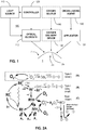
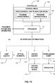
- FIG. 1 illustrates an example treatment system 100 for generating cross-linking of collagen in a cornea 2 of an eye 1.
- the treatment system 100 includes an applicator 132 for applying a cross-linking agent 130 to the cornea 2.
- the applicator 132 may be an eye dropper, syringe, or the like that applies the photosensitizer 130 as drops to the cornea 2.
- the cross-linking agent 130 may be provided in a formulation that allows the cross-linking agent 130 to pass through the corneal epithelium 2a and to underlying regions in the corneal stroma 2b.
- the corneal epithelium 2a may be removed or otherwise incised to allow the cross-linking agent 130 to be applied more directly to the underlying tissue.
- the treatment system 100 includes an illumination system with a light source 110 and optical elements 112 for directing light to the cornea 2.
- the light causes photoactivation of the cross-linking agent 130 to generate cross-linking activity in the cornea 2.
- the cross-linking agent may include riboflavin and the photoactivating light may be ultraviolet A (UVA) (e.g., 365 nm) light.
- UVA ultraviolet A
- the photoactivating light may have another wavelength, such as a visible wavelength (e.g., 452 nm).
- corneal cross-linking improves corneal strength by creating chemical bonds within the corneal tissue according to a system of photochemical kinetic reactions.
- riboflavin and the photoactivating light are applied to stabilize and/or strengthen corneal tissue to address diseases such as keratoconus or post-LASIK ectasia.
- the treatment system 100 includes one or more controllers 120 that control aspects of the system 100, including the light source 110 and/or the optical elements 112.
- the cornea 2 can be more broadly treated with the cross-linking agent 130 (e.g., with an eye dropper, syringe, etc.), and the photoactivating light from the light source 110 can be selectively directed to regions of the treated cornea 2 according to a particular pattern.
- the optical elements 112 may include one or more mirrors or lenses for directing and focusing the photoactivating light emitted by the light source 110 to a particular pattern on the cornea 2.
- the optical elements 112 may further include filters for partially blocking wavelengths of light emitted by the light source 110 and for selecting particular wavelengths of light to be directed to the cornea 2 for activating the cross-linking agent 130.
- the optical elements 112 may include one or more beam splitters for dividing a beam of light emitted by the light source 110, and may include one or more heat sinks for absorbing light emitted by the light source 110.
- the optical elements 112 may also accurately and precisely focus the photo-activating light to particular focal planes within the cornea 2, e.g., at a particular depths in the underlying region 2b where cross-linking activity is desired.
- the one or more controllers 120 may be used to control the operation of the light source 110 and/or the optical elements 112 to precisely deliver the photoactivating light according to any combination of: wavelength, bandwidth, intensity, power, location, depth of penetration, and/or duration of treatment (the duration of the exposure cycle, the dark cycle, and the ratio of the exposure cycle to the dark cycle duration).
- the parameters for photoactivation of the cross-linking agent 130 can be adjusted, for example, to reduce the amount of time required to achieve the desired cross-linking. In an example implementation, the time can be reduced from minutes to seconds. While some configurations may apply the photoactivating light at an irradiance of 5 mW/cm 2 , larger irradiance of the photoactivating light, e.g., multiples of 5 mW/cm 2 , can be applied to reduce the time required to achieve the desired cross-linking.
- the total dose of energy absorbed in the cornea 2 can be described as an effective dose, which is an amount of energy absorbed through an area of the corneal epithelium 2a.
- the effective dose for a region of the corneal surface 2A can be, for example, 5 J/cm 2 , or as high as 20 J/cm 2 or 30 J/cm 2 .
- the effective dose described can be delivered from a single application of energy, or from repeated applications of energy.
- the optical elements 112 of the treatment system 100 may include a digital micro-mirror device (DMD) to modulate the application of photoactivating light spatially and temporally.
- DMD digital micro-mirror device
- the photoactivating light from the light source 110 is projected in a precise spatial pattern that is created by microscopically small mirrors laid out in a matrix on a semiconductor chip. Each mirror represents one or more pixels in the pattern of projected light.
- the control of the DMD according to topography may employ several different spatial and temporal irradiance and dose profiles. These spatial and temporal dose profiles may be created using continuous wave illumination but may also be modulated via pulsed illumination by pulsing the illumination source under varying frequency and duty cycle regimes as described above.
- the DMD can modulate different frequencies and duty cycles on a pixel by pixel basis to give ultimate flexibility using continuous wave illumination.
- both pulsed illumination and modulated DMD frequency and duty cycle combinations may be combined.
- This allows for specific amounts of spatially determined corneal cross-linking.
- This spatially determined cross-linking may be combined with dosimetry, interferometry, optical coherence tomography (OCT), corneal topography, etc., for pre-treatment planning and/or real-time monitoring and modulation of corneal cross-linking during treatment.
- pre-clinical patient information may be combined with finite element biomechanical computer modeling to create patient specific pre-treatment plans.
- embodiments may also employ aspects of multiphoton excitation microscopy.
- the treatment system 100 may deliver multiple photons of longer wavelengths, i.e., lower energy, that combine to initiate the cross-linking.
- longer wavelengths are scattered within the cornea 2 to a lesser degree than shorter wavelengths, which allows longer wavelengths of light to penetrate the cornea 2 more efficiently than shorter wavelength light. Shielding effects of incident irradiation at deeper depths within the cornea are also reduced over conventional short wavelength illumination since the absorption of the light by the photosensitizer is much less at the longer wavelengths. This allows for enhanced control over depth specific cross-linking.
- two photons may be employed, where each photon carries approximately half the energy necessary to excite the molecules in the cross-linking agent 130 to generate the photochemical kinetic reactions described further below.
- a cross-linking agent molecule simultaneously absorbs both photons, it absorbs enough energy to release reactive radicals in the corneal tissue.
- Embodiments may also utilize lower energy photons such that a cross-linking agent molecule must simultaneously absorb, for example, three, four, or five, photons to release a reactive radical.
- the probability of the near-simultaneous absorption of multiple photons is low, so a high flux of excitation photons may be required, and the high flux may be delivered through a femtosecond laser.
- the cross-linking agent 130 is riboflavin and the photoactivating light is UVA light
- the irradiance and the dose both affect the amount and the rate of cross-linking.
- the UVA light may be applied continuously (continuous wave (CW)) or as pulsed light, and this selection has an effect on the amount, the rate, and the extent of cross-linking.
- the duration of the exposure cycle, the dark cycle, and the ratio of the exposure cycle to the dark cycle duration have an effect on the resulting corneal stiffening.
- Pulsed light illumination can be used to create greater or lesser stiffening of corneal tissue than may be achieved with continuous wave illumination for the same amount or dose of energy delivered.
- Light pulses of suitable length and frequency may be used to achieve more optimal chemical amplification.
- the on/off duty cycle may be between approximately 1000/1 to approximately 1/1000; the irradiance may be between approximately 1 mW/cm 2 to approximately 1000 mW/cm 2 average irradiance, and the pulse rate may be between approximately 0.01 HZ to approximately 1000 Hz or between approximately 1000 Hz to approximately 100,000 Hz.
- the treatment system 100 may generate pulsed light by employing a DMD, electronically turning the light source 110 on and off, and/or using a mechanical or optoelectronic (e.g., Pockels cells) shutter or mechanical chopper or rotating aperture. Because of the pixel specific modulation capabilities of the DMD and the subsequent stiffness impartment based on the modulated frequency, duty cycle, irradiance and dose delivered to the cornea, complex biomechanical stiffness patterns may be imparted to the cornea to allow for various amounts of refractive correction.
- a mechanical or optoelectronic e.g., Pockels cells
- refractive corrections may involve combinations of myopia, hyperopia, astigmatism, irregular astigmatism, presbyopia and complex corneal refractive surface corrections because of ophthalmic conditions such as keratoconus, pellucid marginal disease, post-lasik ectasia, and other conditions of corneal biomechanical alteration/degeneration, etc.
- a specific advantage of the DMD system and method is that it allows for randomized asynchronous pulsed topographic patterning, creating a non-periodic and uniformly appearing illumination which eliminates the possibility for triggering photosensitive epileptic seizures or flicker vertigo for pulsed frequencies between 2 Hz and 84 Hz.
- example embodiments may employ stepwise on/off pulsed light functions, it is understood that other functions for applying light to the cornea may be employed to achieve similar effects.
- light may be applied to the cornea according to a sinusoidal function, sawtooth function, or other complex functions or curves, or any combination of functions or curves.
- the function may be substantially stepwise where there may be more gradual transitions between on/off values.
- irradiance does not have to decrease down to a value of zero during the off cycle, and may be above zero during the off cycle. Desired effects may be achieved by applying light to the cornea according to a curve varying irradiance between two or more values.
- oxygen also affects the amount of corneal stiffening.
- O 2 content is very low compared to the atmosphere.
- the rate of cross-linking in the cornea is related to the concentration of O 2 when it is irradiated with photoactivating light. Therefore, it may be advantageous to increase or decrease the concentration of O 2 actively during irradiation to control the rate of cross-linking until a desired amount of cross-linking is achieved.
- Oxygen may be applied during the cross-linking treatments in a number of different ways. One approach involves supersaturating the riboflavin with O 2.
- the treatment system 100 also includes an oxygen source 140 and an oxygen delivery device 142 that optionally delivers oxygen at a selected concentration to the cornea 2.
- Example systems and methods for applying oxygen during cross-linking treatments are described, for example, in U.S. Patent No. 8,574,277, filed October 21, 2010 and titled “Eye Therapy," U.S. Patent Application Publication No. 2013/0060187, filed October 31, 2012 and titled “Systems and Methods for Corneal Cross-Linking with Pulsed Light,”.
- the structure of the cornea includes five layers. From the outer surface of the eye inward, these are: (1) epithelium, (2) Bowman's layer, (3) stroma, (4) Descemet's membrane, and (5) endothelium.
- the stroma is treated with riboflavin, a photosensitizer, and ultraviolet (UV) light is delivered to the cornea to activate the riboflavin in the stroma.
- UV light ultraviolet
- riboflavin undergoes a reaction with oxygen in which reactive oxygen species and other radicals are produced. These reactive oxygen species and other radicals further interact with the collagen fibrils to induce covalent bonds that bind together amino acids of the collagen fibrils, thereby cross-linking the fibrils.
- the photo-oxidative induction of collagen cross-linking enhances the biomechanical strength of the stroma, and can provide therapeutic benefits for certain ophthalmic conditions, such as keratoconus, or generate refractive changes to correct myopia, hyperopia and/or astigmatism.
- the epithelium functions to regulate nutrients, including oxygen, that are admitted into the stromal tissue from the tear film. This regulation is carried out via the epithelium's physiological "pumps" that are driven by osmotic pressure across the epithelium due to differential concentrations of barrier-permeable solutes on either side of the epithelium.
- the epithelium's physiological "pumps” that are driven by osmotic pressure across the epithelium due to differential concentrations of barrier-permeable solutes on either side of the epithelium.
- certain nutrients in the tear film that become depleted within the stroma can permeate the epithelium via osmotic pressure to resupply the stroma.
- oxygen and some other small molecule nutrients can reach the stroma according to this mechanism, certain photosensitizers cannot pass through the epithelium.
- Riboflavin for example, is a relatively large, hydrophilic molecule that cannot penetrate the tight junctions of the epithelium.
- the epithelium slows the amount of riboflavin that can penetrate the stroma.
- a variety of approaches have been employed to overcome low riboflavin diffusivity and deliver sufficient concentrations of riboflavin to the stroma for performing corneal cross-linking treatments.
- the epithelium is removed (epithelium debridement) before a riboflavin solution is applied directly to the stroma.
- the approach is associated with patient discomfort, risks of infection, and other possible complications.
- riboflavin may be provided in a formulation that allows the cross-linking agent to pass through the epithelium.
- Such formulations are described, for example, in U.S Patent Application Publication No. 2010/0286156, filed on May 6, 2009 and titled “Collyrium for the Treatment of Conical Cornea with Cross-Linking Trans-Epithelial Technique, and in U.S. Patent Application Publication No. 2013/0267528, filed on January 4, 2013 and titled "Trans-Epithelial Osmotic Collyrium for the Treatment of Keratoconus,".
- some riboflavin formulations include ionic agents, such as benzalkonium chloride (BAC), with a specific osmolarity of sodium chloride (NaCl).
- BAC benzalkonium chloride
- NaCl sodium chloride
- Another solution and/or mechanical forces may be applied to enhance the permeability of the epithelium and allow the riboflavin to pass more easily through the epithelium.
- approaches for enhancing or otherwise controlling the delivery of a cross-linking agent to the underlying regions of the cornea are described, for example, in U.S. Patent Application Publication No. 2011/0288466, filed April 13, 2011 and titled “Systems and Methods for Activating Cross-Linking in an Eye," and U.S. Patent Application Publication No. 2012/0289886, filed May 18, 2012 and titled “Controlled Application of Cross-Linking Agent,".
- riboflavin formulations enhance the permeability of the epithelium sufficiently to allow relatively large hydrophilic riboflavin molecules (or Flavin mononucleotide (FMN), or riboflavin-5'-phosphate, molecules) to pass through the epithelium without debridement, but the permeability is not enhanced to a point where the epithelium becomes damaged.
- riboflavin formulations employ a non-ionic agent that is chosen using the Hydrophile-Lipophile Balance (HLB) system.
- HLB Hydrophile-Lipophile Balance
- the HLB of a permeability enhancer indicates the balance of hydrophilic and lipophilic groups in the molecular structure of the enhancer.
- Permeability enhancers (or emulsifiers) for the epithelium include a molecule which has both hydrophilic and lipophilic groups. Molecules with HLB number below 9 are considered lipophilic and those above 11 as hydrophilic. Molecules with HLB number between 9 and 11 are intermediate.
- aspects of the present disclosure employ non-ionic agents that have a hydrophilic/lipophilic balance to achieve optimized diffusivity through the epithelium and the stroma.
- non-ionic agents are also less corrosive and damaging to the epithelium than ionic agents, such as BAC.
- the HLB range for more effective permeability enhancers has been experimentally determined by the inventors to be between approximately 12.6 and approximately 14.6.
- a class of permeability enhancers includes various forms of polyethylene glycol (PEG) with different aliphatic chain lengths.
- PEG polyethylene glycol
- some riboflavin formulations include specific concentrations of Polidocanol (Polyoxyethylene (9) lauryl ether), which has a HLB number of approximately 13.6.
- E weight percentage oxyethylene content.
- the HLB range for enhancers that achieve more effective permeability may vary according to different aspects of the formulation.
- the HLB range for more optimal enhancers may vary according to the photosensitizer employed in the formulation. For instance, more optimal permeability might be achieved for other photosensitizers, such as Rose Bengal, by employing enhancers in a HLB range that is different from that for riboflavin (e.g., HLB of approximately 12.6 to approximately 14.6).
- the formulation may include other additives that may affect the HLB range for more optimal enhancers.
- riboflavin formulations may also include iron ions, such as Fe(II).
- Additives that may be included in photosensitizer formulations are described, for example, in U.S. Patent Application Publication No. 2014/0343480, filed May 19, 2014 and titled “Systems, Methods, and Compositions for Cross-linking," and U.S. Provisional Patent Application No. 62/086,572, filed December 2, 2014 and titled “Systems, Methods, and Compositions for Cross-linking,".
- Other additives for instance, include copper, manganese, chromium, vanadium, aluminum, cobalt, mercury, cadmium, nickel, arsenic, 2,3-butanedione, and folic acid.
- permeability enhancers may be combined to achieve a specific HLB that achieves more effective permeability for the epithelium. These may be calculated by taking the percentage of each enhancer, multiplying it by its HLB number, and then summing the results. For instance, in a formulation including 30% enhancer A with a HLB number of approximately 14, 50% enhancer B with a HLB number of approximately 6, and 20% enhancer C with a HLB number of approximately 14, the estimated HLB number can be calculated as:
- two or more enhancers may be combined to achieve a very specific HLB number, where a single enhancer may provide less optimal permeability. Additionally, combining different enhancers might offer other desirable properties of the final formulation with regard to solubility, viscosity, stability or some other desirable attribute.
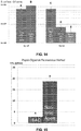
- FIG. 14 illustrates a graph of diffusivity values for the formulations in this study.
- Column A represents the application of a saline solution of 0.1% riboflavin to porcine eyes without epithelia (epi-off).
- Column B represents the application of a 0.22% riboflavin solution with BAC to porcine eyes epi-off.
- Column C represents the application of a 0.22% riboflavin solution with a non-ionic agent to porcine eyes with epithelia (epi-on).
- porcine eyes shipped overnight on ice from an abattoir (SiouxPreme, Sioux City, IA) were cleaned and soaked for 20 minutes in an incubator set at 37°C with a 0.22% riboflavin solution with BAC or a 0.22% riboflavin solution with a non-ionic agent.
- the corneas had epithelia of approximately 100 ⁇ m. The epithelia of the corneas were removed after the respective soaks and prior to pan-corneal irradiation with UVA light.
- the treatment protocol employed applying pulsed UVA light (1 second on; 1 second off) at an irradiation of 30 mW/cm 2 and for a dose of 7.2 J/cm 2 , while the corneas were exposed to 100% concentration of oxygen gas. 200 ⁇ m corneal flaps were cut using a femtosecond laser. The extent of the cross-linking in the corneas was evaluated on the basis of fluorimetric analysis (excitation wave 365 nm, emission wave 450 nm) after collagen solubilization with papain.
- FIG. 15 illustrates the fluorescence values for the formulations in this study, indicating the extent of cross-linking activity.
- Column A represents the fluorescence of the corneas treated with the 0.22% riboflavin solution with BAC.
- Column B represents the fluorescence of the corneas treated with the 0.22% riboflavin solution with the non-ionic agent.
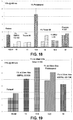
- porcine eyes were treated according to the parameters indicated in FIG. 16 .
- porcine eyes were soaked epi-off with 0.1% riboflavin solution for 20 minutes and then irradiated with continuous wave UVA light with an irradiance of 3 mW/cm 2 for a dose of 5.4 J/cm 2 while exposed to ambient air.
- porcine eyes were soaked epi-off with 0.22% riboflavin solution for 20 minutes and then irradiated with continuous wave UVA light with an irradiance of 30 mW/cm 2 for a dose of 7.2 J/cm 2 while exposed to ambient air.
- porcine eyes were soaked epi-on with 0.22% riboflavin solution with BAC for 20 minutes and then irradiated with continuous wave UVA light with an irradiance of 30 mW/cm 2 for a dose of 7.2 J/cm 2 while exposed to ambient air.
- Porcine eyes were soaked epi-on with 0.22% riboflavin solution with a non-ionic agent for 20 minutes and then irradiated with continuous wave UVA light with an irradiance of 30 mW/cm 2 for a dose of 7.2 J/cm 2 while exposed to ambient air.
- Porcine eyes were soaked epi-on with 0.22% riboflavin solution with BAC for 20 minutes and then irradiated with pulsed UVA light with an irradiance of 30 mW/cm 2 for a dose of 7.2 J/cm 2 while exposed to 100% oxygen.
- Porcine eyes were soaked epi-on with 0.22% riboflavin solution with the non-ionic agent for 20 minutes and then irradiated with pulsed UVA light with an irradiance of 30 mW/cm 2 for a dose of 7.2 J/cm 2 while exposed to 100% oxygen.
- FIG. 17 shows the fluorescence values for the formulations in this study, indicating the extent of cross-linking activity.
- Columns A-F in FIG. 17 represent the results corresponding to the experimental parameters provided in respective rows A-F in FIG. 16 .
- Columns C and D indicate that the cross-linking with the 0.22% riboflavin solution with BAC or the 0.22% riboflavin solution with the non-ionic agent was oxygen-limited.
- Columns E and F indicate that the cross-linking with the 0.22% riboflavin solution with BAC is riboflavin limited when compared to the cross-linking with the 0.22% riboflavin solution with the non-ionic agent.
- Columns E and F indicate that absorption by riboflavin in the saturated epithelium is not a significant factor when oxygen is applied.
- the diffusivity of riboflavin and the initial stromal concentration of riboflavin affects the extent of cross-linking activity.
- the results from the formulations including the non-ionic agent indicate that hydrophilic-lipophilic properties are a factor, allowing riboflavin to penetrate the epithelium and diffuse into the corneal hydrophilic stroma in quantities and duration appropriate for a clinical application.
- Oxygen is a factor in efficient trans-epithelial (epi on) cross-linking.
- the results of the study show that the application of oxygen with the non-ionic agent provides cross-linking efficiencies similar to standard epi-off cross-linking.
- some riboflavin formulations include specific concentrations of Polidocanol to enhance permeability of the corneal epithelium.
- concentrations of Polidocanol do not cause damage to the epithelium.
- Such riboflavin solutions may also include additives such as Fe(II).
- Polidocanol and optionally additives can be employed in combination with other cross-linking techniques as described above to enhance delivery of riboflavin through the epithelium and achieve the desired amount of cross-linking activity.
- the riboflavin formulations with Polidocanol and optional additives can be applied with oxygen.
- the riboflavin solutions can be employed with different approaches for delivering photoactivating illumination (pulsed illumination, illumination of different patterns, etc.).
- BAC benzalkonium chloride
- Pig eyes shipped overnight on ice from an abattoir (SiouxPreme, Sioux City, IA) were rinsed in saline.
- the eyes with intact epithelium were soaked with one of the test solutions below for 20 minutes in an incubator set at 37°C by using a rubber ring to hold the solution on top.
- the epitheliums of eyes in Group A were removed with a dull blade after the eyes were soaked in one of the solutions and irradiated pan-corneally on air with a top hat beam (3% root mean square) for 4 minutes with 365-nm light source (UV LED NCSU033B[T]; Nichia Co., Tokushima, Japan) at a chosen irradiance of 30 mW/cm 2 which was measured with a power sensor (model PD-300-UV; Ophir, Inc., Jerusalem, Israel) at the corneal surface. Corneal flaps (approximately 200 ⁇ m thick) were excised from the eyes with aid of an Intralase femtosecond laser (Abbot Medical Optics, Santa Ana, CA).
- the average thickness of the corneal flaps was calculated as a difference between the measurements before and after the excision from the eyes with an ultrasonic Pachymeter (DGH Technology, Exton, PA).
- the flaps were washed with distilled water and dried in a vacuum until the weight change became less than 10% (Rotary vane vacuum pump RV3 A652-01-903, BOC Edwards, West Wales, UK).
- Each flap was digested for 2.5 h at 65°C with 2.5 units/ml of papain (from Papaya latex, Sigma) in 1 ml of papain buffer [BBBS (pH 7.0-7.2), 2 mM L-cysteine and 2 mM EDTA].
- Papain digests were diluted 0.5 times with lxBBBS and fluorescence of the solutions was measured with excitation of 360 nm in a QM-40 Spectrofluorometer (Photon Technology Int., London, Ontario, Canada). The fluorescence of the papain buffer was taken into account by measuring fluorescence in the absence of tissue and subtracting this value from the fluorescence of the samples.
- the epitheliums of eyes in Group B were not removed after soaking in one of the solutions and the surfaces were briefly rinsed with a saline buffer before irradiation. The epitheliums were removed after the irradiation. Conditions used for the irradiation and the following treatment of the eyes were the same as for Group A.
- the epitheliums of eyes in Group C were not removed after soaking in one of the solutions and the surfaces were briefly rinsed with a saline buffer before irradiation.
- the eyes were placed in a beaker with an oxygen stream for 2 minutes in the incubation chamber prior to irradiation.
- Corneas were pan-corneally irradiated with irradiance of 30 mW/cm 2 , pulsed 1 sec on: 1 sec off for a total time of 8 min (7.2 J).
- the eyes were exposed to oxygen during all time of the treatment.
- the epithelium were removed from the cornea after the irradiation with a dull blade.
- Corneal flaps (approximately 200 ⁇ m thick) were excised from the eyes with aid of Intralase femtosecond laser and the following treatment of the flaps was the same as for the Groups A and B.
- the epitheliums of eyes in Group E were removed after soaking in one of the solutions for 20 min. The eyes then were placed in a beaker with oxygen stream for 2 minutes in the incubation chamber prior to irradiation. Corneas were pan-corneally irradiated with irradiance of 30 mW/cm 2 , pulsed 1 sec on: 1 sec off for total time of 8 min (7.2 J). The eyes were exposed to oxygen during all time of the treatment. Corneal flaps (approximately 200 ⁇ m thick) were excised from the eyes with aid of Intralase femtosecond laser and the following treatment of the flaps was the same as for the Groups A and B.
- FIGS. 18-26 illustrate the cross-linking activity induced in Groups A-E by various riboflavin solutions.
- the cross-linking activity was measured as a ratio of fluorescence for the treated sample (F) to fluorescence for an untreated control (Fo), where emissions were recorded at a wavelength of 450 nm.
- F treated sample
- Fo untreated control
- Such measurement of cross-linking activity is described, for example, in U.S. Patent No. 9,020,580, filed June 4, 2012 and titled "Systems and Methods for Monitoring Time Based Photo Active Agent Delivery or Photo Active Marker Presence,".
- FIGS. 18 and 19 illustrate relative fluorescence for cross-linked corneal flaps treated with different surfactants in 0.22% riboflavin solution containing saline (VIBEX XTRATM) applied topically to pig eyes with intact epithelium for 20 min, after which the epithelium were then removed and the eyes were irradiated with 30 mW/cm 2 for 4 min.
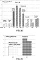
- VIBEX XTRATM riboflavin solution containing saline
- FIG. 20 illustrates relative fluorescence for cross-linked corneal flaps after using 1% solutions of different surfactants in 0.22% riboflavin solution containing saline (VIBEX XTRATM). These results are presented in relation to the fluorescence from corneal flaps treated with only 0.25% riboflavin solution containing BAC (PARACELTM) in the same procedural conditions.
- FIGS. 3-5 also show the HLB numbers for the surfactants, e.g., Polidocanol has an HLB number of 13.6.
- FIG. 21 illustrates relative fluorescence for cross-linked corneal flaps treated with solution (a2) which does not include BAC relative to solution (a1) which includes BAC.
- FIG. 22 illustrates relative fluorescence of cross-linked corneal flaps treated with solutions (a3), (a4), and (a5) which include different concentrations of Polidocanol.
- FIG. 23 illustrates relative fluorescence for cross-linked corneal flaps treated with solutions (b2) and (b3) which include 1% and 5% concentrations of Polidocanol respectively.
- FIG. 24 illustrates relative fluorescence of cross-linked corneal flaps treated with solutions (c2) and (c3) which include 1% and 3% concentrations of Polidocanol respectively. These results are presented relative to corneal flaps treated with solution (c1) which includes BAC.
- FIG. 25 for Group D and FIG. 11 for Group E illustrate relative fluorescence of cross-linked flaps treated with solutions (d2) and (d4) which include 1% and 3% concentrations of Polidocanol respectively and with solutions (d3) and (d5) which include 2.5 mM iron(II) as well as 1% and 3% concentrations of Polidocanol respectively.
- solutions (d2) and (d4) which include 1% and 3% concentrations of Polidocanol respectively
- solutions (d3) and (d5) which include 2.5 mM iron(II) as well as 1% and 3% concentrations of Polidocanol respectively.
- Polidocanol as a non-ionic surfactant that is more effective than many other surfactants for enhancing permeability and generating cross-linking activity.
- BAC in riboflavin solutions may help riboflavin to pass through the epithelium
- Polidocanol is far more effective and efficient than BAC in enhancing permeability in the epithelium and generating cross-linking activity.
- non-ionic agents such as Polidocanol, are less corrosive and damaging to the epithelium than BAC.
- permeability enhancers may be combined to achieve a specific HLB that achieves more optimal permeability for the epithelium.
- Intact epithelium were soaked for 20 min using one of the following solutions:
- the epitheliums of the eyes were removed and the eyes were irradiated with 30 mW/cm 2 for 4 min continuously on air. Corneal flaps with thickness of 200 ⁇ m were cut and the papain digestion and fluorescence analysis was conducted as previously described above.
- FIG. 27 illustrates relative fluorescence of the cross-linked flaps treated with one of two different surfactants or a combination of the two surfactants.
- the cross-linking activity was measured as a ratio of fluorescence for the respective treated sample (F) to fluorescence for a sample treated with solution (e1) (Fparacel), where emissions were recorded at a wavelength of 450 nm.
- the surfactant IGEPAL CO-630 has a HLB number of 13 and the surfactant IGEPAL CO-720 has a HLB number of 14, the 1:1 mixture has a HLB number of 13.5.
- the mixture of the surfactants facilitates riboflavin permeation through the corneal epithelium more effectively than the surfactants employed individually.
- riboflavin and Polidocanol as a permeability enhancer for corneal cross-linking treatments
- photosensitizers and/or other permeability enhancers e.g., non-ionic surfactant with an appropriate HLB number
- other types of treatment such as antimicrobial photodynamic therapy, where enhanced or controlled delivery of a photosensitizer through an epithelium may be advantageous.
- Some microbes, such as fungi have dormant phases, while other microbes, such as Acanthamoeba, can create cystic cell membrane barriers.
- additives that enhance permeability can increase penetration and uptake of photosensitizer by microbes/pathogens and enhance the antimicrobial effect of the photosensitizer.
- photosensitizer formulations employing a non-ionic permeability enhancer may be particularly effective for penetrating cysts, ulcers, etc. and treating microbes/pathogens.
- Other aspects of antimicrobial photodynamic therapy are described in U.S. Patent Application No. 15/137,748, filed April 25, 2016 and titled "Systems and Methods for Photoactivating a Photosensitizer Applied to an Eye,".
- Rf represents riboflavin in the ground state.
- Rf* 1 represents riboflavin in the excited singlet state.
- Rf * 3 represents riboflavin in a triplet excited state.
- Rf •- is the reduced radical anion form of riboflavin.
- RfH • is the radical form of riboflavin.
- RfH 2 is the reduced form of riboflavin.
- DH is the substrate.
- DH •+ is the intermediate radical cation.
- D • is the radical.
- D ox is the oxidized form of the substrate.
- Riboflavin is excited into its triplet excited state Rf * 3 as shown in reactions (r1) to (r3). From the triplet excited state Rf * 3 , the riboflavin reacts further, generally according to Type I or Type II mechanisms.
- the substrate reacts with the excited state riboflavin to generate radicals or radical ions, respectively, by hydrogen atoms or electron transfer.
- the excited state riboflavin reacts with oxygen to form singlet molecular oxygen. The singlet molecular oxygen then acts on tissue to produce additional cross-linked bonds.
- Oxygen concentration in the cornea is modulated by UVA irradiance and temperature and quickly decreases at the beginning of UVA exposure.
- Utilizing pulsed light of a specific duty cycle, frequency, and irradiance, input from both Type I and Type II photochemical kinetic mechanisms can be employed to achieve a greater amount of photochemical efficiency.
- utilizing pulsed light allows regulating the rate of reactions involving riboflavin. The rate of reactions may either be increased or decreased, as needed, by regulating, one of the parameters such as the irradiance, the dose, the on/off duty cycle, riboflavin concentration, soak time, and others.
- additional ingredients that affect the reaction and cross-linking rates may be added to the cornea.
- oxygen concentrations start to increase (replenish). Excess oxygen may be detrimental in the corneal cross-linking process because oxygen is able to inhibit free radical photopolymerization reactions by interacting with radical species to form chain-terminating peroxide molecules.
- the pulse rate, irradiance, dose, and other parameters can be adjusted to achieve a more optimal oxygen regeneration rate. Calculating and adjusting the oxygen regeneration rate is another example of adjusting the reaction parameters to achieve a desired amount of corneal stiffening.
- Oxygen content may be depleted throughout the cornea, by various chemical reactions, except for the very thin corneal layer where oxygen diffusion is able to keep up with the kinetics of the reactions. This diffusion-controlled zone will gradually move deeper into the cornea as the reaction ability of the substrate to uptake oxygen decreases.
- Riboflavin is reduced (deactivated) reversibly or irreversibly and/or photo-degraded to a greater extent as irradiance increases.
- Photon optimization can be achieved by allowing reduced riboflavin to return to ground state riboflavin in Type I reactions.
- the rate of return of reduced riboflavin to ground state in Type I reactions is determined by a number of factors. These factors include, but are not limited to, on/off duty cycle of pulsed light treatment, pulse rate frequency, irradiance, and dose.
- the riboflavin concentration, soak time, and addition of other agents, including oxidizers affect the rate of oxygen uptake.
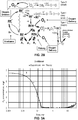
- FIG. 2A illustrates a diagram for the photochemical kinetic reactions provided in reactions (r1) through (r26) above.
- the diagram summarizes photochemical transformations of riboflavin (Rf) under UVA photoactivating light and its interactions with various donors (DH) via electron transfer.
- cross-linking activity occurs: (A) through the presence of singlet oxygen in reactions (r6) through (r8) (Type II mechanism); (B) without using oxygen in reactions (r4) and (r17) (Type I mechanism); and (C) through the presence of peroxide (H 2 O 2 ), superoxide (O 2 - ), and hydroxyl radicals ( ⁇ OH) in reactions (r13) through (r17).
- the present inventors have also determined that the cross-linking activity is generated to a greater degree from reactions involving peroxide, superoxide, and hydroxyl radicals.
- Cross-linking activity is generated to a lesser degree from reactions involving singlet oxygen and from non-oxygen reactions.
- Some models based on the reactions (r1)-(r26) may account for the level of cross-linking activity generated by the respective reactions. For instance, where singlet oxygen plays a smaller role in generating cross-linking activity, models may be simplified by treating the cross-linking activity resulting from singlet oxygen as a constant.
- excess oxygen may be detrimental in corneal cross-linking process.
- FIG. 2A when the system becomes photon-limited and oxygen-abundant, cross-links can be broken from further reactions involving superoxide, peroxide, and hydroxyl radicals. Indeed, in some cases, excess oxygen may result in net destruction of cross-links versus generation of cross-links.
- the parameters may include, but is not limited to: the concentration(s) and/or soak times of the applied cross-linking agent; the dose(s), wavelength(s), irradiance(s), duration(s), and/or on/off duty cycle(s) of the photoactivating light; the oxygenation conditions in the tissue; and/or presence of additional agents and solutions.
- a model based on the reactions (rl)-(rl9) has been validated by at least six different methods of evaluating cross-linking activity:
- corneas were soaked with riboflavin for 20 minutes and exposed to UVA photoactivating light in ambient air at an irradiance of 3mW/cm 2 for 7.5 minutes (dose of 1.35 J/cm 2 ), 15 minutes (dose of 2.70 J/cm 2 ), 30 minutes (dose of 5.4 J/cm 2 ), 45 minutes (dose of 8.10 J/cm 2 ), 120 minutes (dose of 21.6 J/cm 2 ), and 150 minutes (dose of 27.0 J/cm 2 ). Extensiometry measurements were taken for 200 ⁇ m flaps of the corneas. FIG.
- FIG. 31 illustrates a graph of cross-link profiles (cross-link concentration as a function of corneal depth) calculated by the model for each cross-linking treatment.
- FIG. 32 illustrates a correlation between the extensiometry measurements and the values calculated by the model (area under the curve for 200 ⁇ m). In general, there is a good correlation between the biomechanics determined in the extensiometry experiments and the values calculated by the model. It can also be seen from FIG. 31 that cross-linking can saturate when particular treatment parameters (e.g., longer irradiation times) are employed.
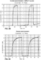
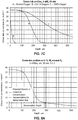
- FIG. 3A illustrates a graph of data showing the correlation between the theoretical values based on the model and experimental data for corneas exposed to continuous wave UVA photoactivating light at an irradiance of 3mW/cm 2 .
- FIGS. 3B-C illustrate graphs of data showing the correlation between model values and experimental data for corneas exposed to long term pulses and short term pulses, respectively, at an irradiance of 3 mW/cm 2 .
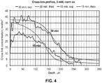
- FIG. 4 illustrates a graph of data showing the correlation between model and experimental data for corneas exposed for 15 minutes and 30 minutes.
- the third party experimental data was published in Dongyul Chai et al. "Quantitative Assessment of UVA-riboflavin Corneal Cross-Linking Using Nonlinear Optical Microscopy.” Investigative Ophthalmology & Visual Science. June 2011, Vol. 52, No. 7, pp. 4231 -4238 .
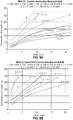
- FIG. 5A illustrates a graph of data showing the correlation of model values and experimental data for corneal flaps (taken from 0 to approximately 100 ⁇ m deep) exposed to combinations of riboflavin concentrations (0.1%, 0.25%, and 0.5%) and 5.4 J/cm 2 doses of UVA photoactivating light at irradiances of 3 mW/cm 2 and 30 mW/cm 2 for 3 minutes and 30 minutes.
- FIG. 5A illustrates a graph of data showing the correlation of model values and experimental data for corneal flaps (taken from 0 to approximately 100 ⁇ m deep) exposed to combinations of riboflavin concentrations (0.1%, 0.25%, and 0.5%) and 5.4 J/cm 2 doses of UVA photoactivating light at irradiances of 3 mW/cm 2 and 30 mW/cm 2 for 3 minutes and 30 minutes.
- 5B illustrates a graph of data showing the correlation of model values and experimental data for corneal flaps (taken from approximately 100 ⁇ m to approximately 200 ⁇ m deep) exposed to combinations of riboflavin concentrations (0.1%, 0.25%, and 0.5%) and 5.4 J/cm 2 doses of UVA photoactivating light at irradiances of 3 mW/cm 2 and 30 mW/cm 2 for 3 minutes and 30 minutes.
- 5C illustrates a graph of data showing the correlation of model values and experimental data for corneal flaps treated with a concentration of riboflavin and exposed to full oxygen concentration and 5.4 J/cm 2 and 7.2 J/cm 2 doses of continuous wave UVA photoactivating light at irradiances of 3 mW/cm 2 , 10 mW/cm 2 , 15 mW/cm 2 , 30 mW/cm 2 , 45 mW/cm 2 , 60 mW/cm 2 , and 100 mW/cm 2 .
- FIG. 5D illustrates a graph of data showing the correlation of model values and experimental data for corneal flaps (taken from approximately 0 ⁇ m to approximately 200 ⁇ m deep) treated with a concentration of 0.1% riboflavin and exposed to air or full oxygen concentration and a 5.4 J/cm 2 doses of continuous wave UVA photoactivating light at irradiances of 3 mW/cm 2 , 10 mW/cm 2 , 15 mW/cm 2 , 30 mW/cm 2 , 45 mW/cm 2 , 60 mW/cm 2 , and 100 mW/cm 2 .
- the values at irradiance 3 mW/cm 2 under 100% oxygen shows the effect of quenching Rf 3 ⁇ by oxygen.
- FIG. 28A shows Brillouin modulus values measured at anterior, central, and posterior sections of corneas experimentally soaked in riboflavin for various durations and irradiated with UV light for various durations.
- FIG. 28B illustrates the correlation between the experimentally measured values and values calculated with the model for various treatments.
- Treatment A in FIG. 28B corresponds to a soak time of 30 minutes and irradiation of 5 minutes.
- Treatment B corresponds to a soak time of 30 minutes and irradiation of 15 minutes.
- Treatment C corresponds to a soak time of 30 minutes and irradiation of 30 minutes.
- Treatment D corresponds to a soak time of 30 minutes and irradiation of 5 minutes.
- Treatment E corresponds to a soak time of 5 minutes and irradiation of 30 minutes.
- Treatment E corresponds to a soak time of 30 minutes and irradiation of 30 minutes.
- corneal stromal demarcation lines were evaluated for treated corneas.
- a method for these experiments involves slit-lamp examination (slit projection and Scheimpflug camera). See Theo Seiler and Farhad Hafezi, "Corneal Cross-Linking-Induced Stromal Demarcation Line," Cornea, Oct. 2006; 25:1057-59 .
- Another method involves corneal optical coherence tomography (OCT). See Luigi Fontana, Antonello Moramarco, "Esperienze personali con CXL accelerato,” UOC Oculistica ASMN-IRCCS Reggio Emilia. Roma, 20 settieri 2014 .
- a further method involves confocal microscopy. See C. Mazzotta et al., "Treatment of Progressive Keratoconus by of Corneal Collagen In Vivo Confocal Microscopy in Humans," Cornea, vol. 26, no. 4, May 2007 .
- Corneal stromal healing involves the deposition of new collagen, which produces haze and scattering. Slit-lamp examination and OCT can detect this hyper-reflectivity and possibly a significant change in the spatial order factor (change in birefringence, i.e., index variation). Corneal stromal demarcation lines indicate the threshold at which the healing response occurs. This conclusion is corroborated by confocal microscopy. The demarcation lines can also be seen as a transition zone between cross-linked anterior corneal stroma and untreated posterior corneal stroma.
- the six evaluations described above show a strong correlation between the experimental data and the calculations generated by a model based on the photochemical kinetic reactions identified above.
- the model is extremely effective and accurate in predicting the results of riboflavin cross-linking treatments applied according to various combinations of parameters. Accordingly, using such a model, systems and methods can more efficiently and predictably achieve a desired profile of cross-linking activity throughout the cornea.
- the model allows the systems and methods to identify a more optimal combination of parameters for cross-linking treatment. Therefore, the model can be used to determine the set up for different aspects of cross-linking treatment systems as described above.
- aspects of the system of reactions can be affected by different parameters. For instance, the irradiance at which photoactivating light is delivered to the system affects the photons available in the system to generate Rf 3 ⁇ for subsequent reactions. Additionally, delivering greater oxygen into the system drives the oxygen-based reactions. Meanwhile, pulsing the photoactivating light affects the ability of the reduced riboflavin to return to ground state riboflavin by allowing additional time for oxygen diffusion. Of course, other parameters can be varied to control the system of reactions.
- a model based on the photochemical kinetic reactions (rl)-(r26) can generate cross-link profiles for treatments using different protocols as shown in FIGS. 7A-C .
- each protocol determines the dose of the photoactivating UVA light, the irradiance for the UVA photoactivating light, the treatment time, and the concentration of oxygen delivered to the corneal surface.
- the cornea has been treated with a formulation including 0.1% concentration riboflavin.
- FIG. 7A illustrates cross-link profiles for treatments that deliver a dose of 7.2 J/cm 2 of UVA light under normal (ambient) oxygen according to different irradiances and different treatment times.
- FIG. 7B illustrates cross-link profiles for treatments that employ different irradiances of continuous or modulated (pulsed) UVA light and different treatment times under normal or 100% oxygen concentration.
- FIG. 7C illustrates cross-link profiles for treatments that deliver an irradiance of 3 mW of UVA light for 30 minutes with different oxygen conditions (normal, 100%, or 0.01x) at the corneal surface.
- the cross-link profiles in FIGS. 7A-C provide the cross-link concentration as a function of corneal depth.
- the three-dimensional distribution of cross-links in the cornea as indicated by each cross-link profile depends on the combination of different treatment parameters. Protocols employing different sets of treatment parameters can be provided as input into the model and the model can output the resulting three-dimensional distribution of cross-links in the cornea. Accordingly, the model can be used to select treatment parameters to achieve the desired distribution of cross-links in the cornea.
- FIG. 6A illustrates a graph of data showing the correlation of model values and experimental data for the depths of corneal stromal demarcation lines for the protocols described in FIG. 6B .
- corneal stromal demarcation lines indicate the transition zone between cross-linked anterior corneal stroma and untreated posterior corneal stroma.
- cross-link profiles generated by the model can be evaluated to determine the depth at which the demarcation line may appear at a cross-link concentration of approximately 5 mol/m 3 .
- the demarcation line may be understood as the threshold at which a healing response occurs in response to the distribution of cross-links as well as the effect of reactive oxygen species on the corneal tissue.
- the cornea has been treated with a formulation including 0.1% concentration riboflavin.
- FIG. 8A illustrates a cross-link profile for a treatment that delivers a dose of 5.4 J/cm 2 of photoactivating UVA light under normal oxygen according to an irradiance of 3 mW/cm 2 and a treatment time of 30 minutes.
- FIG. 8A shows that a cross-link concentration of approximately 5 mol/m 3 (demarcation line) occurs at a depth of approximately 290 ⁇ m in the resulting cross-link profile.
- FIG. 8B illustrates cross-link profiles for treatments that deliver different doses of photoactivating UVA light according to different irradiances and different treatment times under normal oxygen.
- FIG. 8C illustrates cross-link profiles for treatments that deliver different doses of photoactivating UVA light according to different irradiances and different treatment times under normal or 100% oxygen concentration.
- FIGS. 8B-C shows that the depths for the demarcation line vary with the different cross-link profiles generated by the different sets of treatment parameters.
- the depths of the demarcation line indicated by the different cross-link profiles may be employed to select treatment parameters. For instance, treatment parameters may be selected to ensure that the cross-links do not occur at a depth where undesired damage may result to the endothelium. This analysis allows the treatment system to accommodate different corneal thicknesses, particularly thin corneas.
- FIGS. 9A-B illustrate graphs of demarcation depth (cross-link concentration of approximately 5 mol/m 3 ) as a function of dose of UVA photoactivating light.
- the determination of the demarcation depths are based on cross-link profiles generated by the model for treatments using different protocols.
- the cornea has been treated with a formulation including 0.1% concentration riboflavin.
- FIG. 9A illustrates graphs for treatments that deliver continuous or pulsed UVA photoactivating light according to different irradiances under normal oxygen.
- FIG. 9B illustrates graphs for treatments that deliver continuous or pulsed UVA photoactivating light according to different irradiances under a greater concentration of oxygen.
- FIG. 10 illustrates the cross-link profiles for treatments employing different protocols as generated by the model.
- FIG. 10 also shows a demarcation line that corresponds to biomechanical stiffness threshold at a cross-link concentration of 10 mol/m 3 .
- the demarcation line intersects the cross-link profiles at varying depths (biomechanical stiffness depth) based on the different treatment parameters of the protocols.
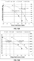
- FIG. 11 illustrates the measurement of maximum keratometry (K max ) (diopters) at three, six, and twelve months relative to a baseline for corneas that were experimentally treated according to the protocols employed for FIG. 10 .
- K max maximum keratometry
- FIGS. 12A-B illustrate the correlation between the experimental data of FIG. 11 and the cross-link profiles generated for FIG. 10 by the model.
- FIG. 12A plots the experimental change of K max for months six and twelve corresponding to the respective protocol.
- FIG. 12A also shows a quadratic fit of the plotted data for each month six and twelve. The quadratic fit is consistent with the quadratic nature of shear forces (in the x-y plane) resulting from a force placed on a disk (along the z-axis) according to thin shell theory.
- FIG. 12B plots the experimental change of K max for months six and twelve corresponding to the respective protocol.
- FIG. 12B also shows a linear fit of the plotted data for each month six and twelve.
- the quadratic fit for the two curves in FIG. 12A are substantially similar.
- the linear fit for the two curves in FIG. 12B are substantially similar.
- the correlations shown in FIGS. 12A-B indicate that there is a predictable biomechanical/healing response over time for a given set of treatment parameters.
- the model, as well as thin shell analysis one can predictably determine refractive change according to the radius and depth of the disk corresponding to the myopic correction.
- the distribution of cross-links effects refractive change.
- the model can be employed to determine this refractive change.
- FIG. 29 illustrates concentration of cross-link concentration as well as a function of corneal depth with a demarcation depth of approximately 360 ⁇ m.
- FIG. 29 also shows a line of demarcation analysis using a second derivative and threshold algorithm. The second derivative can also be calculated as shown. At the illustrated demarcation depth, the second derivative has a threshold derivative of 180 mM/mm 2 .
- FIGS. 35-36 illustrate graphs of cross-link profiles for treatments employing different protocols, as generated by the model.
- the graphs also show a potential threshold cross-link concentration of approximately 4.4 mM/m 3 for the demarcation line.
- the demarcation depth is taken at a shift of approximately 125 ⁇ m from the intersection of the cross-link profile curve and the potential threshold cross-link concentration.
- the demarcation line occurs at a depth of approximately 350 ⁇ m.
- FIG. 30 illustrates a graph of data showing the correlation of model values and experimental data for the depths of corneal stromal demarcation lines for protocols described in these fourteen separate studies. The measurements are taken from the epithelial surface. The standard deviation is approximately 20 ⁇ m.
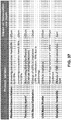
- TABLE 1 shows further shows data for determination of demarcation line depth for different treatments.
- the reported standard deviations reveal variability in the depth of the demarcation line for nominally equivalent clinical protocols. Such variability may be the result of aspects of measurement error, clinical technique, protocol, the riboflavin formulation, and/or the equipment employed. An analysis of the reported variability was performed with the model of photochemical kinetic reactions.
- FIG. 37 illustrates that the demarcation line depth may be affected by aspects of the riboflavin concentration, the use of thickening agent, irradiation (UVA) device calibration, irradiation (UVA) beam profile, and/or geographic factors.
- UVA irradiation
- UVA irradiation
- the line of demarcation is predicted accurately the model of photochemical kinetic reactions.
- the model of photochemical kinetic reactions may be used to treat corneas, particularly thinner corneas, more safely.
- changes in clinical protocol may result in variability in the depth of the line of demarcation line and potentially clinical outcomes, suggesting the importance of precision in cross-linking treatment methodology. Consistency in protocol, technique and equipment can enhance the predictability of the clinical outcomes.
- FIG. 13 illustrates the example system 100 employing a model based on the photochemical kinetic reactions (rl)-(r26) identified above to determine an amount of cross-linking that results from treatment parameters and/or other related information.
- the controller 120 includes a processor 122 and computer-readable storage media 124.
- the storage media 124 stores program instructions for determining an amount of cross-linking when the photoactivating light from the light source 110 is delivered to a selected region of a cornea treated with a cross-linking agent.
- a photochemical kinetic model 126 based on the reactions (rl)-(r26) may include a first set of program instructions A for determining cross-linking resulting from reactions involving reactive oxygen species (ROS) including combinations of peroxides, superoxides, hydroxyl radicals, and/or singlet oxygen and a second set of program instructions B for determining cross-linking from reactions not involving oxygen.
- the controller 120 receives input relating to treatment parameters and/or other related information.
- the controller 120 can then execute the program instructions A and B to output information relating to three-dimensional cross-link distribution(s) for the selected region of the cornea based on the input.
- the three-dimensional cross-link distribution(s) may then be employed to determine how to control aspects of the light source 110, the optical elements 112, the cross-linking agent 130, the applicator 132, the oxygen source 140, and/or oxygen delivery device 142 in order to achieve a desired treatment in selected region of the cornea.
- the system 100 shown in FIG. 13 and this process can be used for treatment of more than one selected region of the same cornea.
- the three-dimensional cross-link distribution(s) may be evaluated to calculate a threshold depth corresponding to a healing response due to the cross-links and an effect of the reactive-oxygen species in the selected region of the cornea. Additionally or alternatively, the three-dimensional cross-link distribution(s) may be evaluated to calculate a biomechanical tissue stiffness threshold depth corresponding to a biomechanical tissue response in the selected region of the cornea.
- the information on the depth of the healing response and/or the biomechanical tissue stiffness in the cornea can be employed to determine how to control aspects of the light source 110, the optical elements 112, the cross-linking agent 130, the applicator 132, the oxygen source 140, and/or oxygen delivery device 142. Certain healing response and/or biomechanical tissue stiffness may be desired or not desired at certain depths of the cornea.
- the photochemical kinetic model allows particular aspects of the photochemical process to be controlled or otherwise influenced to produce desired cross-linking activity.
- different additives such as iron, may be employed to affect mechanisms at different points of the photochemical process as shown in FIG. 33 .
- FIG. 34 shows the effect of the various additives in FIG. 33 on cross-linking activity.
- FIG. 38 illustrates the example system 100 employing the photochemical kinetic model 126 to determine treatment parameters for achieving desired biomechanical changes in the cornea, e.g., a refractive correction.
- the controller 120 includes the processor 122 and the computer-readable storage media 124.
- the storage media 124 stores program instructions 125 for determining what treatment parameters may be employed to achieve desired biomechanical changes.
- the program instructions 125 are based on the photochemical kinetic model 126 which employ the reactions (rl)-(r26) to determine cross-linking resulting from (i) reactions involving reactive oxygen species (ROS) including combinations of peroxides, superoxides, hydroxyl radicals, and/or singlet oxygen and (ii) reactions not involving oxygen.
- ROS reactive oxygen species
- parameters for cross-linking treatment may include: the concentration(s) and/or soak times of the applied cross-linking agent; the dose(s), wavelength(s), irradiance(s), duration(s), on/off duty cycle(s), and/or other illumination parameters for the photoactivating light; the oxygenation conditions in the tissue; and/or presence of additional agents and solutions.
- the resulting distribution of cross-links determined from the photochemical kinetic model 126 can be correlated to a particular biomechanical change in the cornea.
- FIGS. 12A-B show, for instance, the correlation between the distribution of cross-links and refractive change.
- the controller 120 receives an input 12 relating to the initial biomechanical state of the cornea and an input 14 indicating a desired biomechanical change for the cornea, e.g., for refractive correction.
- the initial biomechanical state for instance, can be determined according to approaches described in U.S. Patent Application Publication No. 2012/0215155 referenced above.
- the input 12 may be provided by a measurement system communicatively coupled to the controller 120. It is understood that the initial biomechanical state may reflect the state of the cornea prior to any treatment or during a treatment.
- the inputs 12, 14 may be expressed in terms of corneal topography (i.e., shape), corneal strength (i.e., stiffness), and/or corneal thickness.
- corneal topography i.e., shape
- corneal strength i.e., stiffness
- corneal thickness i.e., corneal thickness
- the desired biomechanical change for refractive correction may be determined from a correction specified (by a practitioner) in diopters, e.g., "a 1.5 diopter correction.”
- a desired biomechanical change in the cornea can be correlated to a particular distribution of cross-links as determined by the photochemical kinetic model 126.
- the controller 120 can execute the program instructions 125 to determine the particular distribution of cross-links 16 that can generate the desired biomechanical change specified by the input 14 in a cornea having the initial biomechanical state specified by the input 12. After determining the distribution of cross-links 16 for the desired biomechanical change, the controller 120 can prescribe a set of treatment parameters for achieving the specified distribution of cross-links.
- cross-links 16 might be achieved in many cases by more than one set of treatment parameters. For instance, depending on the photochemical kinetic reactions, similar distributions of cross-links may be achieved by applying: (i) a lower dose of photoactivating light for a longer amount of time, or (ii) a higher dose of photoactivating light for a shorter amount of time. Therefore, more than one set of treatment parameters 18 for achieving the distribution of cross-links 16 may be identified.
- a practitioner can optimize the treatment for certain preferred parameters, such as treatment time or dose of photoactivating light. For instance, the practitioner may optimize the treatment parameters to achieve shorter treatment times. For this preference, the controller 120 may prescribe a set of illumination parameters that provide a larger dose of photoactivating light that yields the distribution of cross-links 16 over shorter illumination durations. Conversely, the practitioner may optimize the treatment parameters to employ smaller doses of photoactivating light. For this second preference, the controller 120 may prescribe a set of illumination parameters that provide a smaller dose of photoactivating light that yields the distribution of cross-links 16 over longer illumination durations.
- the controller 120 may identify any of the different combinations 18 of values for a set of treatment parameters A, B, C, D, E, etc., as described above.
- the practitioner can set preferences for one or more of these treatment parameters. For instance, the practitioner may initially set a preferred value or range of preferred values for parameter A.
- the controller 120 can specify combinations of values for the remaining parameters B, C, D, E, etc., that meet the preference for parameter A while achieving the distribution of cross-links 16.
- the practitioner may make selections for the values of the parameters B, C, D, and/or E, etc., based on further preferences to arrive at an optimized set of treatment parameters 18a.
- the process of optimizing the treatment parameters may be iterative as the values for the treatment parameters are incrementally tuned to meet preferences having varying priorities.
- the practitioner may manage the optimization process through a series of selections and other inputs via a user interface (not shown) coupled to the controller 120.
- the inputs 12, 14 may also be provided through such a user interface.
- the final set of treatment parameters 18a can then be employed to determine how to control aspects of the light source 110, the optical elements 112, the cross-linking agent 130, the applicator 132, the oxygen source 140, oxygen delivery device 142, etc., in order to achieve a desired treatment in selected region of the cornea.
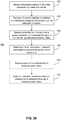
- FIG. 39 illustrates an example method 200 for employing a model of photochemical kinetic reactions (rl)-(r26) to determine treatment parameters for achieving desired biomechanical changes.
- step 202 information relating to the initial biomechanical state of a cornea is received.
- step 204 information relating to a desired biomechanical change for the cornea, e.g., for refractive correction, is received.
- step 206 a distribution of cross-links is determined to achieve the desired biomechanical change in a cornea having the initial biomechanical state.
- one or more sets of treatment parameters are determined to achieve the distribution of cross-links.
- one or more preferences for treatment parameters may be received in step 210, and the treatment parameters may be optimized in step 212 based on the one or more preferences to determine a final set of treatment parameters that can be implemented in a treatment system (e.g., the example system 100) to achieve the distribution of cross-links.
- a treatment system e.g., the example system 100
- controllers may be implemented as a combination of hardware and software elements.
- the hardware aspects may include combinations of operatively coupled hardware components including microprocessors, logical circuitry, communication/networking ports, digital filters, memory, or logical circuitry.
- the controller may be adapted to perform operations specified by a computer-executable code, which may be stored on a computer readable medium.
- the controller may be a programmable processing device, such as an external conventional computer or an on-board field programmable gate array (FPGA) or digital signal processor (DSP), that executes software, or stored instructions.
- FPGA field programmable gate array
- DSP digital signal processor
- physical processors and/or machines employed by embodiments of the present disclosure for any processing or evaluation may include one or more networked or non-networked general purpose computer systems, microprocessors, field programmable gate arrays (FPGA's), digital signal processors (DSP's), micro-controllers, and the like, programmed according to the teachings of the example embodiments of the present disclosure, as is appreciated by those skilled in the computer and software arts.
- the physical processors and/or machines may be externally networked with the image capture device(s), or may be integrated to reside within the image capture device.
- Appropriate software can be readily prepared by programmers of ordinary skill based on the teachings of the example embodiments, as is appreciated by those skilled in the software art.
- the devices and subsystems of the example embodiments can be implemented by the preparation of application-specific integrated circuits or by interconnecting an appropriate network of conventional component circuits, as is appreciated by those skilled in the electrical art(s).
- the example embodiments are not limited to any specific combination of hardware circuitry and/or software.
- the example embodiments of the present disclosure may include software for controlling the devices and subsystems of the example embodiments, for driving the devices and subsystems of the example embodiments, for enabling the devices and subsystems of the example embodiments to interact with a human user, and the like.
- software can include, but is not limited to, device drivers, firmware, operating systems, development tools, applications software, and the like.
- Such computer readable media further can include the computer program product of an embodiment of the present disclosure for performing all or a portion (if processing is distributed) of the processing performed in implementations.
- Computer code devices of the example embodiments of the present disclosure can include any suitable interpretable or executable code mechanism, including but not limited to scripts, interpretable programs, dynamic link libraries (DLLs), Java classes and applets, complete executable programs, and the like. Moreover, parts of the processing of the example embodiments of the present disclosure can be distributed for better performance, reliability, cost, and the like.
- interpretable or executable code mechanism including but not limited to scripts, interpretable programs, dynamic link libraries (DLLs), Java classes and applets, complete executable programs, and the like.
- Computer-readable media may include, for example, a floppy disk, a flexible disk, hard disk, magnetic tape, any other suitable magnetic medium, a CD-ROM, CDRW, DVD, any other suitable optical medium, punch cards, paper tape, optical mark sheets, any other suitable physical medium with patterns of holes or other optically recognizable indicia, a RAM, a PROM, an EPROM, a FLASH-EPROM, any other suitable memory chip or cartridge, a carrier wave or any other suitable medium from which a computer can read.
- a floppy disk a flexible disk, hard disk, magnetic tape, any other suitable magnetic medium, a CD-ROM, CDRW, DVD, any other suitable optical medium, punch cards, paper tape, optical mark sheets, any other suitable physical medium with patterns of holes or other optically recognizable indicia, a RAM, a PROM, an EPROM, a FLASH-EPROM, any other suitable memory chip or cartridge, a carrier wave or any other suitable medium from which a computer can read.
Landscapes
- Health & Medical Sciences (AREA)
- Life Sciences & Earth Sciences (AREA)
- Veterinary Medicine (AREA)
- General Health & Medical Sciences (AREA)
- Animal Behavior & Ethology (AREA)
- Public Health (AREA)
- Chemical & Material Sciences (AREA)
- Medicinal Chemistry (AREA)
- Pharmacology & Pharmacy (AREA)
- Engineering & Computer Science (AREA)
- Nuclear Medicine, Radiotherapy & Molecular Imaging (AREA)
- Epidemiology (AREA)
- Ophthalmology & Optometry (AREA)
- Biomedical Technology (AREA)
- Chemical Kinetics & Catalysis (AREA)
- General Chemical & Material Sciences (AREA)
- Organic Chemistry (AREA)
- Bioinformatics & Cheminformatics (AREA)
- Vascular Medicine (AREA)
- Heart & Thoracic Surgery (AREA)
- Inorganic Chemistry (AREA)
- Biochemistry (AREA)
- Molecular Biology (AREA)
- Radiology & Medical Imaging (AREA)
- Pathology (AREA)
- Biophysics (AREA)
- Physics & Mathematics (AREA)
- Surgery (AREA)
- Optics & Photonics (AREA)
- Electromagnetism (AREA)
- Communicable Diseases (AREA)
- Oncology (AREA)
- Tropical Medicine & Parasitology (AREA)
- Oil, Petroleum & Natural Gas (AREA)
- Pharmaceuticals Containing Other Organic And Inorganic Compounds (AREA)
- Medicinal Preparation (AREA)
- Medicines That Contain Protein Lipid Enzymes And Other Medicines (AREA)
Claims (4)
- Formulierung für eine Augenbehandlung, umfassend:einen Photosensibilisator, umfassend Riboflavin; undeine durchlässigkeitsverbessernde Zusammensetzung, umfassend eines oder mehrere aus IGEPAL CO-630, Polidocanol oder IGEPAL CO-720, wobei die durchlässigkeitsverbessernde Zusammensetzung ausgelegt ist, um eine Durchlässigkeit eines Bereichs des Auges für den Photosensibilisator zu erhöhen.
- Formulierung nach Anspruch 1, weiters umfassend zumindest ein Additiv, das aus der aus Eisen, Kupfer, Mangan, Chrom, Vanadium, Aluminium, Kobalt, Quecksilber, Cadmium, Nickel, Arsen, 2,3-Butandion und Folsäure bestehenden Gruppe ausgewählt ist.
- Formulierung nach Anspruch 3, wobei das zumindest eine Additiv Eisen(ll) umfasst.
- Formulierung nach Anspruch 1, wobei der Bereich des Auges Hornhautepithel ist.
Applications Claiming Priority (5)
| Application Number | Priority Date | Filing Date | Title |
|---|---|---|---|
| US201562195144P | 2015-07-21 | 2015-07-21 | |
| US201562255452P | 2015-11-14 | 2015-11-14 | |
| US201562262919P | 2015-12-04 | 2015-12-04 | |
| US201562263598P | 2015-12-04 | 2015-12-04 | |
| PCT/US2016/043359 WO2017015471A1 (en) | 2015-07-21 | 2016-07-21 | Systems and methods for treaments of an eye with a photosensitizer |
Publications (3)
| Publication Number | Publication Date |
|---|---|
| EP3324973A1 EP3324973A1 (de) | 2018-05-30 |
| EP3324973A4 EP3324973A4 (de) | 2019-05-15 |
| EP3324973B1 true EP3324973B1 (de) | 2020-06-03 |
Family
ID=57834629
Family Applications (1)
| Application Number | Title | Priority Date | Filing Date |
|---|---|---|---|
| EP16828542.7A Active EP3324973B1 (de) | 2015-07-21 | 2016-07-21 | Augenbehandlung mit einem photosensibilisator |
Country Status (6)
| Country | Link |
|---|---|
| US (3) | US11207410B2 (de) |
| EP (1) | EP3324973B1 (de) |
| JP (1) | JP6933377B2 (de) |
| KR (1) | KR102706856B1 (de) |
| CN (2) | CN116832158A (de) |
| WO (1) | WO2017015471A1 (de) |
Families Citing this family (19)
| Publication number | Priority date | Publication date | Assignee | Title |
|---|---|---|---|---|
| US9622911B2 (en) | 2010-09-30 | 2017-04-18 | Cxl Ophthalmics, Llc | Ophthalmic treatment device, system, and method of use |
| ES2978927T3 (es) | 2012-03-29 | 2024-09-23 | Epion Therapeutics Inc | Soluciones de tratamiento ocular, dispositivos de administración y métodos de aumento de la administración |
| US9566301B2 (en) | 2012-03-29 | 2017-02-14 | Cxl Ophthalmics, Llc | Compositions and methods for treating or preventing diseases associated with oxidative stress |
| US10258809B2 (en) * | 2015-04-24 | 2019-04-16 | Avedro, Inc. | Systems and methods for photoactivating a photosensitizer applied to an eye |
| CN116832158A (zh) * | 2015-07-21 | 2023-10-03 | 艾维德洛公司 | 用光敏剂治疗眼睛的系统和方法 |
| CA3001139A1 (en) | 2015-10-23 | 2017-04-27 | The Trustees Of Columbia University In The City Of New York | Laser induced collagen crosslinking in tissue |
| EP3442481B1 (de) | 2016-04-13 | 2023-06-28 | Avedro, Inc. | Systeme zur abgabe von arzneimitteln an ein auge |
| US11497403B2 (en) | 2016-06-10 | 2022-11-15 | The Trustees Of Columbia University In The City Of New York | Devices, methods, and systems for detection of collagen tissue features |
| EP3496681A4 (de) | 2016-08-08 | 2020-07-22 | Avedro, Inc. | Systeme und verfahren zur vernetzung von behandlungen trockener augen |
| US20180236077A1 (en) * | 2017-02-21 | 2018-08-23 | Avedro, Inc. | Formulations for eye treatments |
| US9939381B1 (en) * | 2017-04-07 | 2018-04-10 | Vidrio Technologies, Llc | Automated scanning path planner with path calibration for high frame rate multi photon laser scanning microscope with wide field of view |
| WO2019084450A1 (en) * | 2017-10-26 | 2019-05-02 | The Regents Of The University Of California | SOLAR CELL BIOCIDE |
| US11666481B1 (en) | 2017-12-01 | 2023-06-06 | The Trustees Of Columbia University In The City Of New York | Diagnosis and treatment of collagen-containing tissues |
| US10932864B2 (en) | 2018-11-28 | 2021-03-02 | Rxsight, Inc. | Tracking-based illumination control system |
| US11013593B2 (en) | 2018-12-02 | 2021-05-25 | Rxsight, Inc. | Light adjustable lens tracking system and method |
| WO2020198421A1 (en) * | 2019-03-28 | 2020-10-01 | TECLens, LLC | Corneal crosslinking with catalyst distribution control |
| CN114848817A (zh) * | 2019-05-17 | 2022-08-05 | 北京厚燊维康科技有限责任公司 | 二酮类化合物在光动力治疗或诊断中的用途 |
| US20220193439A1 (en) * | 2020-12-17 | 2022-06-23 | Desmond Christopher Adler | Treatments for eye infection |
| CZ2022336A3 (cs) * | 2022-08-12 | 2024-02-21 | GEMINI oÄŤnĂ klinika a.s. | Zařízení pro aplikaci UV záření k síťování kolagenu rohovky |
Family Cites Families (335)
| Publication number | Priority date | Publication date | Assignee | Title |
|---|---|---|---|---|
| US3169459A (en) | 1963-03-11 | 1965-02-16 | Michael A Friedberg | Method of determining surface contours of an eye |
| US4034750A (en) | 1975-04-22 | 1977-07-12 | Maurice Seiderman | Electrochemically linking collagen fibrils to animal tissue |
| US4161013A (en) | 1977-05-23 | 1979-07-10 | Massachusetts Institute Of Technology | Electromechanochemical device |
| US4326529A (en) | 1978-05-26 | 1982-04-27 | The United States Of America As Represented By The United States Department Of Energy | Corneal-shaping electrode |
| US4805616A (en) | 1980-12-08 | 1989-02-21 | Pao David S C | Bipolar probes for ophthalmic surgery and methods of performing anterior capsulotomy |
| US4381007A (en) | 1981-04-30 | 1983-04-26 | The United States Of America As Represented By The United States Department Of Energy | Multipolar corneal-shaping electrode with flexible removable skirt |
| US4712543A (en) | 1982-01-20 | 1987-12-15 | Baron Neville A | Process for recurving the cornea of an eye |
| US4665913A (en) | 1983-11-17 | 1987-05-19 | Lri L.P. | Method for ophthalmological surgery |
| US5484432A (en) | 1985-09-27 | 1996-01-16 | Laser Biotech, Inc. | Collagen treatment apparatus |
| US4764007A (en) | 1986-02-24 | 1988-08-16 | The United States Of America As Represented By The Secretary Of The Air Force | Glare susceptibility tester |
| GB8606821D0 (en) | 1986-03-19 | 1986-04-23 | Pa Consulting Services | Corneal reprofiling |
| US5019074A (en) | 1987-03-09 | 1991-05-28 | Summit Technology, Inc. | Laser reprofiling system employing an erodable mask |
| US4891043A (en) | 1987-05-28 | 1990-01-02 | Board Of Trustees Of The University Of Illinois | System for selective release of liposome encapsulated material via laser radiation |
| US5171318A (en) | 1987-11-09 | 1992-12-15 | Chiron Ophthalmics, Inc. | Treated corneal prosthetic device |
| US4969912A (en) | 1988-02-18 | 1990-11-13 | Kelman Charles D | Human collagen processing and autoimplant use |
| US4881543A (en) | 1988-06-28 | 1989-11-21 | Massachusetts Institute Of Technology | Combined microwave heating and surface cooling of the cornea |
| US5098426A (en) | 1989-02-06 | 1992-03-24 | Phoenix Laser Systems, Inc. | Method and apparatus for precision laser surgery |
| US5152759A (en) | 1989-06-07 | 1992-10-06 | University Of Miami, School Of Medicine, Dept. Of Ophthalmology | Noncontact laser microsurgical apparatus |
| US5103005A (en) | 1989-07-21 | 1992-04-07 | Coors Biotech, Inc. | Method for recovery of riboflavin |
| US5016615A (en) | 1990-02-20 | 1991-05-21 | Riverside Research Institute | Local application of medication with ultrasound |
| US5490849A (en) | 1990-07-13 | 1996-02-13 | Smith; Robert F. | Uniform-radiation caustic surface for photoablation |
| US5779696A (en) | 1990-07-23 | 1998-07-14 | Sunrise Technologies International, Inc. | Method and apparatus for performing corneal reshaping to correct ocular refractive errors |
| US6342053B1 (en) | 1990-07-23 | 2002-01-29 | Laser Biotech, Inc. | Apparatus for cornea reshaping |
| US5219895A (en) | 1991-01-29 | 1993-06-15 | Autogenesis Technologies, Inc. | Collagen-based adhesives and sealants and methods of preparation and use thereof |
| US6325792B1 (en) | 1991-11-06 | 2001-12-04 | Casimir A. Swinger | Ophthalmic surgical laser and method |
| US5171254A (en) | 1991-11-19 | 1992-12-15 | Sher Neal A | Eye fixation device |
| AU3781193A (en) | 1992-02-27 | 1993-09-13 | Phoenix Laser Systems, Inc. | Automated laser workstation for high precision surgical and industrial interventions |
| RU2086215C1 (ru) | 1992-03-17 | 1997-08-10 | Александр Иосифович Симановский | Способ определения истинного внутриглазного давления, коэффициента легкости оттока и минутного объема продуцирования камерной влаги |
| US6450641B2 (en) | 1992-06-02 | 2002-09-17 | Lasersight Technologies, Inc. | Method of corneal analysis using a checkered placido apparatus |
| JP2965101B2 (ja) | 1992-07-31 | 1999-10-18 | 株式会社ニデック | 眼科装置 |
| CA2141577A1 (en) | 1992-08-03 | 1994-02-17 | David R. Hennings | Method and apparatus for exposing a human eye to a controlled pattern of radiation spots |
| IL103290A (en) | 1992-09-25 | 1996-06-18 | Ben Nun Joshua | Ophthalmologic examination and/or treatment apparatus |
| US5608472A (en) | 1992-11-06 | 1997-03-04 | Research Development Foundation | Eye imaging system |
| US5891131A (en) | 1993-02-01 | 1999-04-06 | Arizona Board Of Regents | Method and apparatus for automated simulation and design of corneal refractive procedures |
| AU6559394A (en) | 1993-04-15 | 1994-11-08 | Board Of Regents, The University Of Texas System | Raman spectrometer |
| US5461212A (en) | 1993-06-04 | 1995-10-24 | Summit Technology, Inc. | Astigmatic laser ablation of surfaces |
| AU716040B2 (en) | 1993-06-24 | 2000-02-17 | Bausch & Lomb Incorporated | Ophthalmic pachymeter and method of making ophthalmic determinations |
| US5634921A (en) | 1993-08-23 | 1997-06-03 | Hood; Larry | Method and apparatus for modifications of visual acuity by thermal means |
| US20020002369A1 (en) | 1993-08-23 | 2002-01-03 | Hood Larry L. | Method and apparatus for modifying visual acuity by moving a focal point of energy within a cornea |
| US20020042612A1 (en) | 1997-10-27 | 2002-04-11 | Hood Larry L. | Method and apparatus for modifications of visual acuity by thermal means |
| CA2183103A1 (en) | 1994-02-09 | 1995-08-17 | Thomas A. Silvestrini | An electrosurgical procedure recurving the cornea |
| JP3263275B2 (ja) | 1994-04-05 | 2002-03-04 | ザ リージェンツ オブ ザ ユニバーシティ オブ カリフォルニア | 生体組織のレーザー処理のための装置並びに火焔状斑点母斑のレーザー処理装置 |
| US6293938B1 (en) | 1994-04-08 | 2001-09-25 | Summit Technology, Inc. | Photo-refractive keratectomy |
| US20050187599A1 (en) | 1994-05-06 | 2005-08-25 | Hugh Sharkey | Method and apparatus for controlled contraction of soft tissue |
| WO1996005884A1 (en) | 1994-08-22 | 1996-02-29 | Iomed, Inc. | Iontophoretic delivery device with integral hydrating means |
| RU2098057C1 (ru) | 1994-10-20 | 1997-12-10 | Ольга Александровна Киселева | Способ лечения отслойки сетчатки, осложненной субретинальным швартообразованием |
| US5624437A (en) | 1995-03-28 | 1997-04-29 | Freeman; Jerre M. | High resolution, high speed, programmable laser beam modulating apparatus for microsurgery |
| HU213450B (en) | 1995-04-26 | 1997-06-30 | Ladanyi Jozsef | Gel contains gelatin and process for producing it |
| US5910110A (en) | 1995-06-07 | 1999-06-08 | Mentor Ophthalmics, Inc. | Controlling pressure in the eye during surgery |
| US6520956B1 (en) | 1995-11-06 | 2003-02-18 | David Huang | Apparatus and method for performing laser thermal keratoplasty with minimized regression |
| US6033396A (en) | 1995-11-06 | 2000-03-07 | Huang; David | Apparatus and method for performing laser thermal keratoplasty with minimized regression |
| US6441047B2 (en) | 1995-11-17 | 2002-08-27 | Alcon Manufacturing Ltd.. | Combination therapy for treating glaucoma |
| US7452358B2 (en) | 1996-01-05 | 2008-11-18 | Thermage, Inc. | RF electrode assembly for handpiece |
| US7141049B2 (en) | 1999-03-09 | 2006-11-28 | Thermage, Inc. | Handpiece for treatment of tissue |
| US20070244496A1 (en) | 1996-02-07 | 2007-10-18 | Hellenkamp Johann F | Automatic surgical device and control assembly for cutting a cornea |
| DE19614823A1 (de) | 1996-04-15 | 1997-10-16 | Mann Gerhard Chem Pharm Fab | Ophthalmische Zusammensetzung mit verlängerter Verweilzeit am Auge |
| US6726684B1 (en) | 1996-07-16 | 2004-04-27 | Arthrocare Corporation | Methods for electrosurgical spine surgery |
| US20060149343A1 (en) | 1996-12-02 | 2006-07-06 | Palomar Medical Technologies, Inc. | Cooling system for a photocosmetic device |
| RU2127099C1 (ru) | 1997-04-07 | 1999-03-10 | Борзенок Сергей Анатольевич | Глазные капли "неокератоник" |
| RU2127100C1 (ru) | 1997-04-17 | 1999-03-10 | Борзенок Сергей Анатольевич | Глазные капли "пиротоник" |
| US6302876B1 (en) | 1997-05-27 | 2001-10-16 | Visx Corporation | Systems and methods for imaging corneal profiles |
| RU2121825C1 (ru) | 1997-06-24 | 1998-11-20 | Межотраслевой научно-технический комплекс "Микрохирургия глаза" | Глазные капли "интертоник" |
| US6104959A (en) | 1997-07-31 | 2000-08-15 | Microwave Medical Corp. | Method and apparatus for treating subcutaneous histological features |
| US6007499A (en) | 1997-10-31 | 1999-12-28 | University Of Washington | Method and apparatus for medical procedures using high-intensity focused ultrasound |
| RU2130762C1 (ru) | 1997-12-10 | 1999-05-27 | Федоров Святослав Николаевич | Устройство для офтальмохирургических операций |
| US20030135122A1 (en) | 1997-12-12 | 2003-07-17 | Spectrx, Inc. | Multi-modal optical tissue diagnostic system |
| US20080027328A1 (en) | 1997-12-29 | 2008-01-31 | Julia Therapeutics, Llc | Multi-focal treatment of skin with acoustic energy |
| US5885275A (en) | 1998-01-15 | 1999-03-23 | Diomed, Inc. | Medical spacing guide |
| US6161544A (en) | 1998-01-28 | 2000-12-19 | Keratoform, Inc. | Methods for accelerated orthokeratology |
| US20020013577A1 (en) | 1998-03-06 | 2002-01-31 | Frey Rudolph W. | Laser beam delivery and eye tracking system |
| BR9908692A (pt) | 1998-03-09 | 2001-12-04 | Ista Pharmaceuticals Inc | Uso de agentes de enrijecimento corneal emortocerotologia enzimática |
| US6188500B1 (en) | 1998-04-03 | 2001-02-13 | Psc Scanning, Inc. | Method for generating multiple scan lines in a thin scanner |
| US6398753B2 (en) | 1998-04-03 | 2002-06-04 | Mcdaniel David H. | Ultrasound enhancement of percutaneous drug absorption |
| AU3970799A (en) | 1998-05-04 | 1999-11-23 | Board Of Regents | Combined fluorescence and reflectance spectroscopy |
| US6099521A (en) | 1998-05-26 | 2000-08-08 | Shadduck; John H. | Semiconductor contact lens cooling system and technique for light-mediated eye therapies |
| US6162210A (en) | 1998-08-06 | 2000-12-19 | Shadduck; John H. | Laser mediated treatments for presbyopia and hyperopia |
| ATE222074T1 (de) | 1998-08-19 | 2002-08-15 | Autonomous Technologies Corp | Gerät und verfahren zur messung von fehlsichtigkeiten eines menschlichen auges |
| US6101411A (en) | 1998-09-24 | 2000-08-08 | Newsome; David A. | Dilation enhancer |
| US6218360B1 (en) | 1998-11-19 | 2001-04-17 | The Schepens Eye Research Institute | Collagen based biomaterials and methods of preparation and use |
| US6402739B1 (en) | 1998-12-08 | 2002-06-11 | Y-Beam Technologies, Inc. | Energy application with cooling |
| US6617963B1 (en) | 1999-02-26 | 2003-09-09 | Sri International | Event-recording devices with identification codes |
| ATE298536T1 (de) | 1999-03-09 | 2005-07-15 | Thermage Inc | Vorichtung zur behandlung von gewebe |
| JP4250721B2 (ja) | 1999-03-15 | 2009-04-08 | カール ツアイス メディテック アクチエンゲゼルシャフト | 眼検査用立体顕微鏡における照明装置及びその方法 |
| AU5274000A (en) | 1999-06-04 | 2000-12-28 | Sunrise Technologies International, Inc. | Prevention of regression in refractive keratoplasty |
| US6280436B1 (en) | 1999-08-10 | 2001-08-28 | Memphis Eye & Cataract Associates Ambulatory Surgery Center | Eye tracking and positioning system for a refractive laser system |
| JP2002058695A (ja) | 1999-09-03 | 2002-02-26 | Carl Zeiss Jena Gmbh | 眼の照射方法及び装置 |
| US6946440B1 (en) | 1999-09-15 | 2005-09-20 | Dewoolfson Bruce H | Composition for stabilizing corneal tissue during or after orthokeratology lens wear |
| JP2003511183A (ja) | 1999-10-21 | 2003-03-25 | テクノラス ゲーエムベーハー オフタルモロギッシェ システム | 個別化された角膜プロファイル |
| US6319273B1 (en) | 1999-12-16 | 2001-11-20 | Light Sciences Corporation | Illuminating device for treating eye disease |
| US6673067B1 (en) | 2000-01-31 | 2004-01-06 | Gholam A. Peyman | System and method for thermally and chemically treating cells at sites of interest in the body to impede cell proliferation |
| US8215314B2 (en) | 2000-02-11 | 2012-07-10 | The General Hospital Corporation | Photochemical tissue bonding |
| CA2399414A1 (en) | 2000-02-11 | 2001-08-16 | The General Hospital Corporation | Photochemical tissue bonding |
| US7073510B2 (en) | 2000-02-11 | 2006-07-11 | The General Hospital Corporation | Photochemical tissue bonding |
| US20020006394A1 (en) | 2000-02-11 | 2002-01-17 | Redmond Robert W. | Photochemical tissue bonding |
| US6394999B1 (en) | 2000-03-13 | 2002-05-28 | Memphis Eye & Cataract Associates Ambulatory Surgery Center | Laser eye surgery system using wavefront sensor analysis to control digital micromirror device (DMD) mirror patterns |
| US6949093B1 (en) | 2000-03-21 | 2005-09-27 | Minu, L.L.C. | Adjustable universal implant blank for modifying corneal curvature and methods of modifying corneal curvature therewith |
| US7867186B2 (en) | 2002-04-08 | 2011-01-11 | Glaukos Corporation | Devices and methods for treatment of ocular disorders |
| JP2001299941A (ja) | 2000-04-27 | 2001-10-30 | Hamamatsu Photonics Kk | レーザ治療装置 |
| US6494878B1 (en) | 2000-05-12 | 2002-12-17 | Ceramoptec Industries, Inc. | System and method for accurate optical treatment of an eye's fundus |
| US6668190B2 (en) | 2000-06-16 | 2003-12-23 | Wayne State University | Method and apparatus for activating molecules to stimulate neurological tissue |
| EP1295602A1 (de) | 2000-06-29 | 2003-03-26 | Rohto Pharmaceutical Co., Ltd. | Sauerstoffhaltige ophthalmische verbindung |
| CA2423354C (en) | 2000-09-20 | 2009-12-15 | Lee Shahinian Jr. | Self-preserved nasal, inhalable, and topical ophthalmic preparations and medications |
| ES2325681T3 (es) | 2000-10-13 | 2009-09-14 | The Johns Hopkins University | Activador de plasminogeno para prevenir turbidez corneal y subepitelial despues de cirugia de correccion de la vision con laser. |
| US20020099363A1 (en) | 2001-01-23 | 2002-07-25 | Woodward Benjamin W. | Radiation treatment system and method of using same |
| US7753943B2 (en) | 2001-02-06 | 2010-07-13 | Qlt Inc. | Reduced fluence rate PDT |
| US7044945B2 (en) | 2001-03-30 | 2006-05-16 | Sand Bruce J | Prevention of regression in thermal ciliary muscle tendinoplasty |
| US6766042B2 (en) | 2001-04-26 | 2004-07-20 | Memphis Eye & Contact Associates | System to automatically detect eye corneal striae |
| US6918904B1 (en) | 2001-11-07 | 2005-07-19 | Minu, L.L.C. | Method of reshaping the cornea by controlled thermal delivery |
| US20030208190A1 (en) | 2001-07-20 | 2003-11-06 | Cynthia Roberts | Methods and instruments for refractive ophthalmic surgery |
| US20030030908A1 (en) | 2001-08-13 | 2003-02-13 | Yeou-Yen Cheng | Virtually imaged phased array (VIPA) with machined radiation window boundary |
| US9681984B2 (en) | 2001-11-07 | 2017-06-20 | Gholam A. Peyman | Method of altering the refractive properties of an eye |
| US20070142828A1 (en) | 2001-11-07 | 2007-06-21 | Minu, Llc | Method and system for altering the refractive properties of the eye |
| US9681942B2 (en) | 2001-11-07 | 2017-06-20 | Gholam A. Peyman | Method for prevention of rejection and sever encapsulation of a supportive or functioning implant |
| US20050149006A1 (en) | 2001-11-07 | 2005-07-07 | Peyman Gholam A. | Device and method for reshaping the cornea |
| US20070088415A1 (en) | 2001-11-07 | 2007-04-19 | Minu Llc | Method of treating the eye using controlled heat delivery |
| US9370446B2 (en) | 2001-11-07 | 2016-06-21 | Gholam A. Peyman | Method of altering the refractive properties of an eye |
| US20050177149A1 (en) | 2001-11-07 | 2005-08-11 | Peyman Gholam A. | Method and apparatus for reshaping the cornea by controlled thermal delivery |
| US9155652B2 (en) | 2001-11-07 | 2015-10-13 | Gholam A. Peyman | Method for laser correction of refractive errors of an eye with a thin cornea |
| US9814567B2 (en) | 2001-11-07 | 2017-11-14 | Gholam A. Peyman | Method of altering the refractive properties of an eye |
| US20070135805A1 (en) | 2001-11-07 | 2007-06-14 | Minu Telesystems Llc | Device and method for reshaping the cornea |
| EP1467760A2 (de) | 2002-01-23 | 2004-10-20 | Light Sciences Corporation | Systeme und verfahren zur photodynamischen therapie |
| US7130835B2 (en) | 2002-03-28 | 2006-10-31 | Bausch & Lomb Incorporated | System and method for predictive ophthalmic correction |
| US7336989B2 (en) | 2002-04-03 | 2008-02-26 | The Regents Of The University Of California | System and method for quantitative or qualitative measurement of exogenous substances in tissue and other materials using laser-induced fluorescence spectroscopy |
| EP1358839B1 (de) | 2002-04-05 | 2005-02-23 | SIS AG Surgical Instrument Systems | Vorrichtung und Verfahren zur Bestimmung von geometrischen Messwerten eines Auges |
| US7455858B2 (en) | 2002-05-16 | 2008-11-25 | Qlt Inc. | Compositions and methods for delivery of photosensitive drugs |
| US6758564B2 (en) | 2002-06-14 | 2004-07-06 | Physical Sciences, Inc. | Line-scan laser ophthalmoscope |
| DE10323422B4 (de) | 2002-08-23 | 2022-05-05 | Carl Zeiss Meditec Ag | Vorrichtung und Verfahren zur Messung eines optischen Durchbruchs in einem Gewebe |
| ITTO20021007A1 (it) | 2002-11-19 | 2004-05-20 | Franco Bartoli | Apparecchiatura laser ad eccimeri e metodo di pilotaggio |
| EP1575435B1 (de) | 2002-12-09 | 2010-03-10 | The Trustees of Dartmouth College | Elektrisch-induziertes thermokeratoplastisches system |
| US7377917B2 (en) | 2002-12-09 | 2008-05-27 | The Trustees Of Dartmouth College | Feedback control of thermokeratoplasty treatments |
| US20060177430A1 (en) | 2002-12-20 | 2006-08-10 | Chakshu Research Inc | Treatment of ocular disorders with ophthalmic formulations containing methylsulfonylmethane as a transport enhancer |
| FR2849383B1 (fr) | 2002-12-26 | 2005-09-30 | Jean Noel Thorel | Composition trophique en milieu aqueux, et ses applications, notamment en ophtalmologie |
| US7004902B2 (en) | 2003-03-21 | 2006-02-28 | Reichert, Inc. | Method and apparatus for measuring biomechanical characteristics of corneal tissue |
| US20110098790A1 (en) | 2009-10-26 | 2011-04-28 | Albert Daxer | Methods for treating corneal disease |
| US7223275B2 (en) | 2003-05-27 | 2007-05-29 | Yichieh Shiuey | System for cutting the cornea of an eye |
| TWI329022B (en) | 2003-08-22 | 2010-08-21 | Kose Corp | Singlet oxygen quenchers and compositions comprising same |
| JP4152836B2 (ja) | 2003-08-22 | 2008-09-17 | 富士通株式会社 | 分波機能を備えた光部品および波長分散補償器 |
| US20050096515A1 (en) | 2003-10-23 | 2005-05-05 | Geng Z. J. | Three-dimensional surface image guided adaptive therapy system |
| DE502004009305D1 (de) | 2004-02-03 | 2009-05-20 | Iroc Ag | Ophtalmologische Vorrichtung |
| WO2005102303A2 (en) | 2004-04-21 | 2005-11-03 | Advanced Ocular Systems Limited | Antiprostaglandins for the treatment of ocular pathologies |
| DE102004021754A1 (de) | 2004-04-30 | 2005-11-24 | Reinhardt Thyzel | Vorrichtung zum Entfernen von Epithelzellen aus einem Linsenkapselsack eines menschlichen oder tierischen Auges |
| EP1755584A4 (de) | 2004-05-07 | 2007-10-31 | Univ California | Behandlung von myopie |
| US8571648B2 (en) | 2004-05-07 | 2013-10-29 | Aesthera | Apparatus and method to apply substances to tissue |
| ITMI20041625A1 (it) | 2004-08-06 | 2004-11-06 | Roberto Pinelli | Apparato per la correzione della presbiopia |
| EP1989997A1 (de) | 2004-08-24 | 2008-11-12 | The General Hospital Corporation | Verfahren, Vorrichtung und Software-Anordnung zur Messung einer mechanischen Belastung und elastischer Eigenschaften einer Probe |
| US20060135957A1 (en) | 2004-12-21 | 2006-06-22 | Dorin Panescu | Method and apparatus to align a probe with a cornea |
| US7871378B1 (en) | 2004-12-22 | 2011-01-18 | Achevé Technology, Inc. | Device and method to measure corneal biomechanical properties and its application to intraocular pressure measurement |
| US8394084B2 (en) | 2005-01-10 | 2013-03-12 | Optimedica Corporation | Apparatus for patterned plasma-mediated laser trephination of the lens capsule and three dimensional phaco-segmentation |
| US20060276777A1 (en) | 2005-04-08 | 2006-12-07 | Coroneo Minas T | Corneal shaping |
| US20060287662A1 (en) | 2005-05-26 | 2006-12-21 | Ntk Enterprises, Inc. | Device, system, and method for epithelium protection during cornea reshaping |
| WO2007001926A2 (en) | 2005-06-24 | 2007-01-04 | Hyperbranch Medical Technology, Inc. | Low-swelling hydrogel sealants for wound repair |
| US20070027509A1 (en) | 2005-07-29 | 2007-02-01 | Eisenberg Elliot S | Automated panretinal laser photocoagulation |
| US7292347B2 (en) | 2005-08-01 | 2007-11-06 | Mitutoyo Corporation | Dual laser high precision interferometer |
| US8388668B2 (en) | 2005-08-05 | 2013-03-05 | Gholam A. Peyman | Methods to regulate polarization of excitable cells |
| US20070048340A1 (en) | 2005-08-31 | 2007-03-01 | Searete Llc, A Limited Liability Corporation Of The State Of Delaware | Multi step patterning of a skin surface |
| US20070055227A1 (en) | 2005-09-08 | 2007-03-08 | Refractec, Inc. | Probe used for an ocular procedure |
| US20070074722A1 (en) | 2005-09-21 | 2007-04-05 | Kurve Technology, Inc. | Medicament delivery control, monitoring, and reporting system and method |
| US8053449B2 (en) | 2005-10-05 | 2011-11-08 | Cell Viable Corporation | Method for inhibiting AGE complex formation |
| EP1779891A1 (de) | 2005-10-28 | 2007-05-02 | Abdula Kurkayev | Verfahren zum Aktivieren eines Photosensibilisators |
| JP5498699B2 (ja) | 2005-10-31 | 2014-05-21 | シーアールエス・アンド・アソシエイツ | 対象物の変形特性を測定する装置 |
| DE102005056958A1 (de) | 2005-11-29 | 2007-06-06 | Rowiak Gmbh | Verfahren und Vorrichtung zum Bearbeiten eines Werkstücks |
| US20070203547A1 (en) | 2005-12-15 | 2007-08-30 | Costello Benedict J | Medical device identification |
| US20090171305A1 (en) | 2006-01-05 | 2009-07-02 | El Hage Sami G | Combination therapy for long-lasting ckr |
| US20080177220A1 (en) | 2006-01-06 | 2008-07-24 | The Curators Of The University Of Missouri | Ultrasound-Mediated Transcleral Drug Delivery |
| US20070203478A1 (en) | 2006-02-21 | 2007-08-30 | Herekar Satish V | Method and system for elasto-modulation of ocular tissue |
| US8182471B2 (en) | 2006-03-17 | 2012-05-22 | Amo Manufacturing Usa, Llc. | Intrastromal refractive correction systems and methods |
| RU2309713C1 (ru) | 2006-03-17 | 2007-11-10 | ЗАО "Екатеринбургский центр МНТК "Микрохирургия глаза" | Способ лечения начальных стадий кератоконуса путем эксимерлазерной хирургии |
| US20090130176A1 (en) | 2006-03-30 | 2009-05-21 | Leila Bossy-Nobs | Intraocular lens with drug delivery system attached thereto |
| WO2007120457A2 (en) | 2006-04-13 | 2007-10-25 | Euclid Systems Corporation | Composition and method for stabilizing corneal tissue after refractive surgery |
| US8624721B2 (en) | 2006-04-17 | 2014-01-07 | Warsaw Orthopedic, Inc. | Method and apparatus for embedding a transmitter into a tool, and a system for monitoring the tool |
| WO2007122615A2 (en) | 2006-04-20 | 2007-11-01 | Xceed Imaging Ltd. | All optical system and method for providing extended depth of focus of imaging |
| WO2007127172A2 (en) | 2006-04-27 | 2007-11-08 | The Trustees Of Columbia University In The City Of New York | Layered bio-adhesive compositions and uses thereof |
| JP2009535464A (ja) | 2006-05-03 | 2009-10-01 | ビジョン シーアールシー リミティド | 生物系ポリシロキサン |
| EP1854438B1 (de) | 2006-05-09 | 2018-07-04 | IROC Services AG | Ophthalmologische Vorrichtung zur Vorbeugung einer Myopie |
| WO2007139927A1 (en) | 2006-05-26 | 2007-12-06 | The Cleveland Clinic Foundation | Method for measuring biomechanical properties in an eye |
| WO2007143111A2 (en) | 2006-06-01 | 2007-12-13 | University Of Southern California | Method and apparatus to guide laser corneal surgery with optical measurement |
| US20070291277A1 (en) | 2006-06-20 | 2007-12-20 | Everett Matthew J | Spectral domain optical coherence tomography system |
| DE102006030219A1 (de) | 2006-06-30 | 2008-01-03 | Iroc Ag | Bestrahlungssystem für medizinische Anwendungen |
| JP3940161B1 (ja) | 2006-07-03 | 2007-07-04 | 朝日インテック株式会社 | 医療用ガイドワイヤ、医療用ガイドワイヤとマイクロカテーテルとの組立体、および医療用ガイドワイヤとバルーンカテーテルとガイディングカテーテルとの組立体 |
| ES2820837T3 (es) * | 2006-07-10 | 2021-04-22 | Esbatech Alcon Biomed Res Unit | Anticuerpos scFv que pasan las capas epitelial y/o endotelial |
| US20080015660A1 (en) | 2006-07-13 | 2008-01-17 | Priavision, Inc. | Method And Apparatus For Photo-Chemical Oculoplasty/Keratoplasty |
| US10729585B2 (en) | 2006-07-19 | 2020-08-04 | Alcon Inc. | Computer program for ophthalmological surgery |
| US20080063627A1 (en) | 2006-09-12 | 2008-03-13 | Surmodics, Inc. | Tissue graft materials containing biocompatible agent and methods of making and using same |
| US7496174B2 (en) | 2006-10-16 | 2009-02-24 | Oraya Therapeutics, Inc. | Portable orthovoltage radiotherapy |
| US8414911B2 (en) | 2006-10-24 | 2013-04-09 | The Regents Of The University Of California | Photochemical therapy to affect mechanical and/or chemical properties of body tissue |
| WO2008070185A2 (en) | 2006-12-06 | 2008-06-12 | The Trustees Of Columbia University In The City Of New York | Method of stabilizing human eye tissue by reaction with nitrite and related agents such as nitro compounds |
| US9125856B1 (en) | 2006-12-06 | 2015-09-08 | The Trustees Of Columbia University In The City Of New York | Method of stabilizing human eye tissue |
| WO2008070848A2 (en) | 2006-12-07 | 2008-06-12 | Priavision, Inc. | Method and material for in situ corneal structural augmentation |
| US20080147021A1 (en) * | 2006-12-15 | 2008-06-19 | Jani Dharmendra M | Drug delivery devices |
| WO2008095075A1 (en) | 2007-02-01 | 2008-08-07 | Ls Biopath, Inc. | Optical system for identification and characterization of abnormal tissue and cells |
| US8580789B2 (en) | 2007-04-02 | 2013-11-12 | The Cleveland Clinic Foundation | Treating glaucoma |
| WO2008137637A2 (en) | 2007-05-04 | 2008-11-13 | The General Hospital Corporation | Methods, arrangements and systems for obtaining information associated with a sample using brillouin microscopy |
| WO2008150291A1 (en) | 2007-06-05 | 2008-12-11 | Minu Llc | Device and method for reshaping the cornea |
| US8992516B2 (en) | 2007-07-19 | 2015-03-31 | Avedro, Inc. | Eye therapy system |
| US8202272B2 (en) | 2007-07-19 | 2012-06-19 | Avedro, Inc. | Eye therapy system |
| AU2008289552B2 (en) | 2007-08-16 | 2014-10-09 | The Schepens Eye Research Institute, Inc. | Therapeutic compositions for treatment of inflammation of ocular and adnexal tissues |
| US8603081B2 (en) | 2007-08-23 | 2013-12-10 | Ntk Enterprises, Inc. | System and method for defining and controlling LTK and other surgical eye procedures to produce little or no stromal collagen shrinkage |
| WO2009042159A1 (en) | 2007-09-24 | 2009-04-02 | The Johns Hopkins University | Wearable photoactivator for ocular therapeutic applications and use thereof |
| WO2010065026A2 (en) | 2007-10-03 | 2010-06-10 | The General Hospital Corporation | Photochemical tissue bonding |
| AU2008310956B2 (en) * | 2007-10-08 | 2014-08-07 | Aurinia Pharmaceuticals Inc. | Ophthalmic compositions comprising calcineurin inhibitors or mTOR inhibitors |
| DE102007052103A1 (de) | 2007-10-31 | 2009-05-07 | Medizinisches Laserzentrum Lübeck GmbH | Vorrichtung zur schonenden Lasertherapie der Retina |
| JP2011503232A (ja) | 2007-11-20 | 2011-01-27 | ザ ブリガム アンド ウィメンズ ホスピタル インコーポレイテッド | 免疫応答の調節 |
| WO2009069123A2 (en) | 2007-11-26 | 2009-06-04 | Yissum Research Development Company Of The Hebrew University Of Jerusalem | Compositions comprising fibrous polypeptides and polysaccharides |
| US8545487B2 (en) | 2007-12-05 | 2013-10-01 | Avedro Inc. | Eye therapy system |
| US20090149923A1 (en) | 2007-12-07 | 2009-06-11 | 21X Corporation Dba Priavision, Inc. | Method for equi-dosed time fractionated pulsed uva irradiation of collagen/riboflavin mixtures for ocular structural augmentation |
| US20100057060A1 (en) | 2007-12-07 | 2010-03-04 | Seros Medical, Llc | In Situ UV/Riboflavin Ocular Treatment System |
| US8348935B2 (en) | 2008-01-23 | 2013-01-08 | Avedro, Inc. | System and method for reshaping an eye feature |
| CN101969900A (zh) | 2008-03-14 | 2011-02-09 | 欧几里得系统公司 | 紫外照射用于治疗角膜薄弱障碍 |
| WO2009145842A2 (en) | 2008-04-04 | 2009-12-03 | Forsight Labs, Llc | Therapeutic device for pain management and vision |
| WO2009146151A2 (en) | 2008-04-04 | 2009-12-03 | Forsight Labs, Llc | Corneal onlay devices and methods |
| US9125735B2 (en) | 2008-04-04 | 2015-09-08 | Forsight Labs, Llc | Method of correcting vision using corneal onlays |
| US8346518B2 (en) | 2008-04-23 | 2013-01-01 | The Cleveland Clinic Foundation | Method for modeling biomechanical properties of an eye |
| US7898656B2 (en) | 2008-04-30 | 2011-03-01 | The General Hospital Corporation | Apparatus and method for cross axis parallel spectroscopy |
| US20090275929A1 (en) | 2008-04-30 | 2009-11-05 | Amo Development, Llc | System and method for controlling measurement in an eye during ophthalmic procedure |
| WO2010011119A1 (es) | 2008-07-22 | 2010-01-28 | Choza Remero Andres Abelino | Solución oftálmica para engrosar córneas |
| WO2010015255A1 (en) | 2008-08-08 | 2010-02-11 | Glostrup Hospital | System and method for treatment of lens related disorders |
| RU2391078C2 (ru) | 2008-08-14 | 2010-06-10 | Сергей Игоревич Анисимов | Способ лечения кератоконуса воздействием ультрафиолетового излучения и устройство для его осуществления (варианты) |
| IT1393402B1 (it) | 2008-08-28 | 2012-04-20 | Sooft Italia Spa | Uso di enhancer eventualmente con riboflavina, nonche' relative composizioni oftalmiche per cross-linking corneale del cheratocono o di altre patologie ectasiche corneali |
| DE102008046834A1 (de) | 2008-09-11 | 2010-03-18 | Iroc Ag | Steuerprogramm zum Steuern elektromagnetischer Strahlung für eine Quervernetzung von Augengewebe |
| US20100082018A1 (en) | 2008-09-26 | 2010-04-01 | Daryus Panthakey | Method and system for reshaping the cornea |
| US8366689B2 (en) | 2008-09-30 | 2013-02-05 | Avedro, Inc. | Method for making structural changes in corneal fibrils |
| EP2355739A4 (de) * | 2008-11-11 | 2014-03-12 | Avedro Inc | Augentherapiesystem |
| US8226232B2 (en) | 2008-12-17 | 2012-07-24 | Technion Research And Development Foundation, Ltd. | System and method for fast retinal imaging |
| WO2010074940A1 (en) | 2008-12-23 | 2010-07-01 | Alcon Research, Ltd. | Compositions and nutritional supplements for improving ocular health and reducing ocular inflammatory response |
| CN104306103B (zh) | 2009-01-23 | 2017-04-19 | 马缇医疗股份有限公司 | 一种或多种药剂的持续释放递送 |
| US8496651B2 (en) | 2009-01-27 | 2013-07-30 | Technolas Perfect Vision Gmbh | System and method for refractive surgery with augmentation by intrastromal corrective procedure |
| WO2010093908A2 (en) | 2009-02-12 | 2010-08-19 | University Of Rochester | Aberration control by corneal collagen crosslinking combined with beam-shaping technique |
| US9232889B2 (en) | 2009-02-12 | 2016-01-12 | Alcon Research, Ltd. | Method and apparatus for ocular surface imaging |
| US7883210B2 (en) | 2009-04-24 | 2011-02-08 | Filar Paul A | Apparatus for photographing the anterior segment and retina of the eye through the use of a camera attachment |
| US20100286156A1 (en) * | 2009-05-06 | 2010-11-11 | Roberto Pinelli | Collyrium For The Treatment Of Conical Cornea With Cross-Linking Trans-Epithelial Technique |
| EP2253321A1 (de) | 2009-05-06 | 2010-11-24 | Roberto Pinelli | Collyrium zur Behandlung von Cornea conica mit transepithelialer Vernetzungstechnik |
| AU2010259184B2 (en) | 2009-06-09 | 2015-08-13 | Aurinia Pharmaceuticals Inc. | Topical drug delivery systems for ophthalmic use |
| US20100318017A1 (en) | 2009-06-11 | 2010-12-16 | Lewis Scott E | Method of improving vision using contact lenses and corneal collagen crosslinking |
| WO2011012557A1 (en) * | 2009-07-27 | 2011-02-03 | Sanseverino, Renato | Ophthalmic solution for protecting internal structures of the eyeball against uv-a rays or for the treatment of keratoconus with a trans-epithelial cross-linking technique |
| WO2011019940A2 (en) | 2009-08-12 | 2011-02-17 | Seros Medical, Llc | Deuterated water and riboflavin solution for extending singlet oxygen lifetimes in treatment of ocular tissue and method for use |
| US8710099B2 (en) | 2009-09-10 | 2014-04-29 | New York University | Treatment of cancer using the sodium salt of a benzoic acid derivative |
| US8518028B2 (en) | 2009-09-30 | 2013-08-27 | Abbott Medical Optics Inc. | Methods for enhancing accommodation of a natural lens of an eye |
| US9278026B2 (en) | 2009-09-30 | 2016-03-08 | Abbott Medical Optics Inc. | Capsular membrane treatments to increase accommodative amplitude |
| WO2011038485A1 (en) | 2009-09-30 | 2011-04-07 | Ottawa Hospital Research Institute | Crosslinked hydrogels and related method of preparation |
| EP2485765A2 (de) | 2009-10-09 | 2012-08-15 | Board Of Regents The University Of Texas System | Fotokinetisches verfahren zur vorrichtung zur freisetzung eines augenarzneimittels |
| US20130245536A1 (en) | 2009-10-21 | 2013-09-19 | Avedro, Inc. | Systems and methods for corneal cross-linking with pulsed light |
| US8574277B2 (en) | 2009-10-21 | 2013-11-05 | Avedro Inc. | Eye therapy |
| AU2010310484A1 (en) | 2009-10-23 | 2012-05-10 | Ivantis, Inc. | Ocular implant system and method |
| AU2010327321A1 (en) | 2009-12-02 | 2012-07-12 | The University Of Sydney | Diagnostic and therapeutic methods for corneal ectasia following refractive surgery, keratoconus or pellucid degeneration |
| AU2011210536A1 (en) | 2010-02-01 | 2012-09-13 | Conversion Energy Enterprises Inc. | Methods and devices for crosslinking of corneal collagen and for treatment of disorders of the eye |
| US20120283621A1 (en) | 2010-03-19 | 2012-11-08 | Avedro, Inc. | Sterilizing application of cross-linking agent |
| US20120215155A1 (en) | 2010-03-19 | 2012-08-23 | Avedro Inc. | Controlled cross-linking initiation and corneal topography feedback systems for directing cross-linking |
| WO2011116306A2 (en) | 2010-03-19 | 2011-09-22 | Avedro, Inc. | Systems and methods for applying and monitoring eye therapy |
| WO2011130356A2 (en) | 2010-04-13 | 2011-10-20 | Avedro, Inc. | Systems and methods for activating cross-linking in an eye |
| RU2428152C1 (ru) | 2010-04-19 | 2011-09-10 | Сергей Васильевич Безик | Способ мобилизации интраокулярной линзы для удаления вторичной катаракты с использованием бимануальной автоматизированной системы аспирации-ирригации при наличии фимоза переднего капсулорексиса |
| EP2907490A1 (de) | 2010-04-21 | 2015-08-19 | IROC Innocross AG | Vorrichtung für die vernetzung von okulärem gewebe mit elektromagnetischer strahlung |
| US8945101B2 (en) | 2010-04-30 | 2015-02-03 | Seros Medical, Llc | Method and apparatus for treatment of ocular tissue using combine modalities |
| DE102010020194B4 (de) | 2010-05-07 | 2022-09-08 | Carl Zeiss Meditec Ag | Vorrichtung zur Stabilisierung der Augenhornhaut |
| KG1376C1 (en) | 2010-05-31 | 2011-08-30 | M A Medvedev | Method for keratoconus treatment |
| IT1400982B1 (it) | 2010-07-05 | 2013-07-05 | Pinelli | Collirio osmotico trans-epiteliale per la cura del cheratocono. |
| CN103189105A (zh) * | 2010-07-21 | 2013-07-03 | P·傅贾尔斯 | 治疗慢性创伤的光活性维生素纳米颗粒 |
| US20120065572A1 (en) | 2010-08-09 | 2012-03-15 | Lewis Scott E | Corneal crosslinking methodology using custom etched contact lenses |
| US9622911B2 (en) | 2010-09-30 | 2017-04-18 | Cxl Ophthalmics, Llc | Ophthalmic treatment device, system, and method of use |
| US20120083772A1 (en) | 2010-09-30 | 2012-04-05 | Curveright Llc | Corneal treatment system and method |
| US8834916B2 (en) | 2010-10-11 | 2014-09-16 | Leonard A. Newman | Apparatus and method for removing epithelium from the cornea |
| ES2590127T3 (es) * | 2011-01-12 | 2016-11-18 | Sooft Italia S.P.A. | Administración corneal de agentes de entrecruzamiento por iontoforesis para el tratamiento de queratocono y composiciones oftálmicas relacionadas |
| EA022306B1 (ru) | 2011-01-12 | 2015-12-30 | Соофт Италия Спа | Устройство и способ подачи рибофлавина в роговицу путем ионофореза для лечения кератоконуса |
| US9005099B2 (en) | 2011-02-15 | 2015-04-14 | Seros Medical, Llc | Method and apparatus for the delivery of photochemical (cross-linking) treatment to scleral tissue |
| WO2012127330A1 (en) | 2011-03-23 | 2012-09-27 | Saks Asher Abraham | An ophthalmological device and method for the treatment of corneal diseases |
| WO2012135073A2 (en) | 2011-03-25 | 2012-10-04 | Board Of Trustees Of Michigan State University | Adaptive laser system for ophthalmic use |
| US8111394B1 (en) | 2011-04-01 | 2012-02-07 | IsoSpec Technologies, LP | Raman spectrometer for monitoring traces of dissolved organic and inorganic substances |
| RU2456971C1 (ru) | 2011-04-19 | 2012-07-27 | Федеральное государственное учреждение "Межотраслевой научно-технический комплекс "Микрохирургия глаза" имени академика С.Н. Федорова Федерального агентства по высокотехнологичной медицинской помощи" | Способ лечения прогрессирующего кератоконуса |
| JP6072003B2 (ja) | 2011-04-29 | 2017-02-01 | アフェジ, ファルアドHafezi, Farhad | 角膜疾患を治療および/または予防するための装置 |
| US20120302862A1 (en) | 2011-04-29 | 2012-11-29 | The General Hospital Corporation | Methods and arrangements for obtaining information and providing analysis for biological tissues |
| US20120310141A1 (en) | 2011-05-06 | 2012-12-06 | Kornfield Julia A | Light delivery device and related compositions, methods and systems |
| WO2012158991A2 (en) | 2011-05-18 | 2012-11-22 | Avedro, Inc. | Controlled application of cross-linking agent |
| US20140066835A1 (en) | 2011-05-24 | 2014-03-06 | Avedro, Inc. | Systems and methods for corneal cross-linking with pulsed light |
| US9044308B2 (en) | 2011-05-24 | 2015-06-02 | Avedro, Inc. | Systems and methods for reshaping an eye feature |
| US10813791B2 (en) | 2011-06-02 | 2020-10-27 | University Of Rochester | Method for modifying the refractive index of ocular tissues and applications thereof |
| EP2713849B1 (de) | 2011-06-02 | 2017-02-15 | Avedro, Inc. | Systeme zur überwachung einer zeitgesteuerten lichtwirkstofffreisetzung oder einer wirkungsmarkerpräsenz |
| WO2012174453A2 (en) | 2011-06-15 | 2012-12-20 | Sybotics, Llc | Systems and methods for binocular iris imaging |
| US9095414B2 (en) | 2011-06-24 | 2015-08-04 | The Regents Of The University Of California | Nonlinear optical photodynamic therapy (NLO-PDT) of the cornea |
| US20120330291A1 (en) | 2011-06-24 | 2012-12-27 | The Regents Of The University Of California | Nonlinear optical photodynamic therapy (nlo-pdt) of the cornea |
| DE102011052002B4 (de) | 2011-07-20 | 2013-04-11 | Telesto GmbH | Lasertherapiesystem mit UVA- und IR-Laser-Licht zur gerichteten Erzeugung einer dermalen Kollagen-Matrix |
| PE20141183A1 (es) | 2011-08-23 | 2014-10-05 | Yeda Res & Dev | Sensibilizantes de (bacterioclorofila) para el tratamiento de enfermedades y trastornos oculares |
| EP2760397A4 (de) | 2011-10-02 | 2015-06-24 | Avedro Inc | System und verfahren zur anwendung und überwachung einer augentherapie |
| ITRM20110560A1 (it) | 2011-10-25 | 2013-04-26 | Sooft Italia Spa | Migliorata composizione di cross-linking per il trattamento del cheratocono mediante iontoforesi |
| US20130110091A1 (en) | 2011-10-26 | 2013-05-02 | Ntk Enterprises, Inc. | Apparatus and method for performing surgical eye procedures including ltk and cxl procedures |
| AU2012349819A1 (en) | 2011-12-07 | 2014-06-26 | Yagna Limited | Compositions for photodynamic therapy chemically modified to increase epithelia penetration and cellular bioavailability |
| ES2658304T3 (es) | 2011-12-30 | 2018-03-09 | Wavelight Gmbh | Un dispositivo integrado para oftalmolagía |
| WO2013148713A1 (en) | 2012-03-28 | 2013-10-03 | Cxl Ophthalmics, Llc | Ocular treatment system and method using red and gold phototherapy |
| ES2978927T3 (es) | 2012-03-29 | 2024-09-23 | Epion Therapeutics Inc | Soluciones de tratamiento ocular, dispositivos de administración y métodos de aumento de la administración |
| US9555111B2 (en) | 2012-03-29 | 2017-01-31 | Cxl Ophthalmics, Llc | Ocular cross-linking system and method for sealing corneal wounds |
| US9566301B2 (en) | 2012-03-29 | 2017-02-14 | Cxl Ophthalmics, Llc | Compositions and methods for treating or preventing diseases associated with oxidative stress |
| JP5989523B2 (ja) | 2012-05-01 | 2016-09-07 | 株式会社トプコン | 眼科装置 |
| US20130310728A1 (en) | 2012-05-16 | 2013-11-21 | Theo Seiler | Device for dissecting an eye for the introduction of photosensitizer and method of refractive surgery |
| EP2872081B2 (de) | 2012-07-16 | 2025-07-16 | Avedro, Inc. | Systeme zur hornhautvernetzung mit gepulstem licht |
| ES2798111T3 (es) | 2012-10-17 | 2020-12-09 | Albert Daxer | Dispositivo para la irradiación del ojo |
| US20140113009A1 (en) | 2012-10-24 | 2014-04-24 | Avedro, Inc. | Controlled application of cross-linking agent |
| WO2014071408A1 (en) | 2012-11-05 | 2014-05-08 | Avedro, Inc. | Systems and methods for reshaping an eye feature |
| US20140276361A1 (en) | 2012-11-20 | 2014-09-18 | Avedro, Inc. | Systems and methods for treating glaucoma |
| WO2014081875A1 (en) | 2012-11-20 | 2014-05-30 | David Muller | Systems and methods for treating glaucoma |
| EP2745819B1 (de) | 2012-12-18 | 2017-08-09 | Telesto GmbH | Lasertherapiesystem zur Behandlung einer Kollagenstruktur und variköser Blutgefäße in einem Auge |
| EP2745820B1 (de) | 2012-12-19 | 2015-12-09 | Telesto GmbH | Lichttherapiesystem zur nichtinvasiven Korrektur des refraktiven Systems des Auges |
| AU2013375414B2 (en) | 2013-01-28 | 2016-11-03 | Alcon Inc. | Apparatus for corneal crosslinking |
| EP3441049B1 (de) | 2013-03-14 | 2025-08-20 | Epion Therapeutics, Inc. | Augenbehandlungsvorrichtung |
| US10092393B2 (en) | 2013-03-14 | 2018-10-09 | Allotex, Inc. | Corneal implant systems and methods |
| WO2014145684A1 (en) | 2013-03-15 | 2014-09-18 | Avedro, Inc. | Systems and methods for treating glaucoma |
| WO2014145666A2 (en) | 2013-03-15 | 2014-09-18 | Avedro, Inc. | Treatments of extracellular matrices of the eye |
| ITRM20130248A1 (it) | 2013-04-24 | 2014-10-25 | Medivis S R L | Formulazioni di riboflavina per il cross-linking transepiteliale. |
| US20140320819A1 (en) | 2013-04-30 | 2014-10-30 | Avedro, Inc | Systems and methods for delivering light in eye treatments |
| EP3858356A3 (de) * | 2013-05-19 | 2021-12-01 | Avedro, Inc. | Vernetzungssysteme, -verfahren und -zusammensetzungen |
| US9498114B2 (en) | 2013-06-18 | 2016-11-22 | Avedro, Inc. | Systems and methods for determining biomechanical properties of the eye for applying treatment |
| DE102013211854B4 (de) | 2013-06-21 | 2017-01-05 | Universität Rostock | Verfahren und Vorrichtung zur Bestimmung einer spektralen Änderung von gestreutem Licht |
| EP3013290B1 (de) | 2013-06-25 | 2017-09-27 | Teclens, LLC | Vorrichtung für eine augen-lichttherapie |
| KR101836458B1 (ko) | 2013-10-09 | 2018-03-09 | 웨이브라이트 게엠베하 | 감광제의 주입을 위해 눈을 절개하기 위한 장치 |
| KR101862364B1 (ko) | 2013-10-30 | 2018-05-29 | 웨이브라이트 게엠베하 | 교차결합술 제어 |
| WO2015130944A1 (en) | 2014-02-28 | 2015-09-03 | Massachusetts Eye & Ear Infirmary | Methods for cross-linking corneal collagen with verteporfin for the treatment of disorders of the eye |
| WO2015138794A1 (en) | 2014-03-12 | 2015-09-17 | The Trustees Of Columbia University In The City Of New York | Cosmetic preservatives as therapeutic corneoscleral tissue cross-linking agents |
| US20150305933A1 (en) | 2014-04-23 | 2015-10-29 | Carl Zeiss Meditec Ag | Integrated device system and method for noninvasive corneal refractive corrections |
| EP3406229B1 (de) | 2014-05-12 | 2025-08-20 | Epion Therapeutics, Inc. | Ophthalmische behandlungslösungsabgabevorrichtungen und abgabeverstärkungsverfahren |
| US9427355B1 (en) | 2014-05-12 | 2016-08-30 | Gholam A. Peyman | Corneal transplantation with a cross-linked cornea |
| EP3403693B1 (de) | 2014-06-27 | 2020-03-25 | Teclens, LLC | Akustische echtzeitdosimetrie für hornhautkollagenvernetzung |
| DE102014012675A1 (de) | 2014-08-26 | 2016-03-03 | Wavelight Gmbh | Vernetzung von Augengewebe |
| EP3212140B1 (de) | 2014-10-27 | 2021-12-01 | Avedro, Inc. | Systeme zur vernetzung von behandlungen trockener augen |
| WO2016077747A1 (en) | 2014-11-13 | 2016-05-19 | Avedro, Inc. | Multipass virtually imaged phased array etalon |
| DE102014016990B3 (de) | 2014-11-18 | 2016-02-11 | Wavelight Gmbh | Düseneinheit zur Vernetzung von Augengewebe |
| US11071643B2 (en) | 2014-11-28 | 2021-07-27 | Visionisti Oy | Ocular therapeutics tool |
| EP3226871B1 (de) | 2014-12-02 | 2022-07-13 | Avedro, INC. | Systeme, verfahren und zusammensetzungen für behandlungen zur vernetzung eines auges |
| WO2016106217A1 (en) | 2014-12-22 | 2016-06-30 | TECLens, LLC | Corneal crosslinking with oxygenation |
| DE102015102298A1 (de) | 2015-02-18 | 2016-08-18 | Carl Zeiss Meditec Ag | Ophthalmologische Zusammensetzung mit wenigstens einer molekularen Schalterverbindung |
| SG11201708114SA (en) | 2015-03-05 | 2017-11-29 | Auckland Uniservices Ltd | Ophthalmic compositions and methods of use therefor |
| ES2686472T3 (es) | 2015-03-10 | 2018-10-18 | Consejo Superior De Investigaciones Científicas | Acoplamiento inducido fotoquímicamente de implantes intraoculares |
| US10258809B2 (en) * | 2015-04-24 | 2019-04-16 | Avedro, Inc. | Systems and methods for photoactivating a photosensitizer applied to an eye |
| US10688196B2 (en) | 2015-04-29 | 2020-06-23 | Sooft Italia Spa | Cross-linking agents of collagen fibers for the use in the treatment of corneal ectasia |
| US9895467B2 (en) | 2015-05-14 | 2018-02-20 | California Institute Of Technology | Light adjustable intraocular lenses using upconverting nanoparticles and near infrared (NIR) light |
| WO2016191342A1 (en) | 2015-05-22 | 2016-12-01 | Avedro, Inc. | Systems and methods for monitoring cross-linking activity for corneal treatments |
| JP6921761B2 (ja) | 2015-06-12 | 2021-08-18 | ザ ジェネラル ホスピタル コーポレイション | 屈折異常矯正用の角膜フィラー |
| CN116832158A (zh) * | 2015-07-21 | 2023-10-03 | 艾维德洛公司 | 用光敏剂治疗眼睛的系统和方法 |
| EP3442481B1 (de) * | 2016-04-13 | 2023-06-28 | Avedro, Inc. | Systeme zur abgabe von arzneimitteln an ein auge |
-
2016
- 2016-07-21 CN CN202310862543.3A patent/CN116832158A/zh active Pending
- 2016-07-21 WO PCT/US2016/043359 patent/WO2017015471A1/en not_active Ceased
- 2016-07-21 KR KR1020187004895A patent/KR102706856B1/ko active Active
- 2016-07-21 JP JP2018502795A patent/JP6933377B2/ja active Active
- 2016-07-21 CN CN201680054742.XA patent/CN108025011A/zh active Pending
- 2016-07-21 US US15/216,344 patent/US11207410B2/en active Active
- 2016-07-21 EP EP16828542.7A patent/EP3324973B1/de active Active
-
2021
- 2021-10-25 US US17/509,238 patent/US12214039B2/en active Active
-
2025
- 2025-01-17 US US19/030,447 patent/US20250161452A1/en active Pending
Non-Patent Citations (1)
| Title |
|---|
| None * |
Also Published As
| Publication number | Publication date |
|---|---|
| CN108025011A (zh) | 2018-05-11 |
| US20220152202A1 (en) | 2022-05-19 |
| US11207410B2 (en) | 2021-12-28 |
| US20250161452A1 (en) | 2025-05-22 |
| JP6933377B2 (ja) | 2021-09-08 |
| KR20180030892A (ko) | 2018-03-26 |
| WO2017015471A1 (en) | 2017-01-26 |
| KR102706856B1 (ko) | 2024-09-19 |
| JP2018526341A (ja) | 2018-09-13 |
| EP3324973A4 (de) | 2019-05-15 |
| CN116832158A (zh) | 2023-10-03 |
| EP3324973A1 (de) | 2018-05-30 |
| US20170021021A1 (en) | 2017-01-26 |
| US12214039B2 (en) | 2025-02-04 |
Similar Documents
| Publication | Publication Date | Title |
|---|---|---|
| US12214039B2 (en) | Systems and methods for treatments of an eye with a photosensitizer | |
| US20260000539A1 (en) | Systems and methods for cross-linking treatments of an eye | |
| EP3442481B1 (de) | Systeme zur abgabe von arzneimitteln an ein auge | |
| US10028657B2 (en) | Systems and methods for monitoring cross-linking activity for corneal treatments | |
| US10646372B2 (en) | Systems and methods for treating an eye with a mask device | |
| US20140066835A1 (en) | Systems and methods for corneal cross-linking with pulsed light | |
| EP3585393A1 (de) | Formulierungen für augenbehandlungen |
Legal Events
| Date | Code | Title | Description |
|---|---|---|---|
| STAA | Information on the status of an ep patent application or granted ep patent |
Free format text: STATUS: THE INTERNATIONAL PUBLICATION HAS BEEN MADE |
|
| PUAI | Public reference made under article 153(3) epc to a published international application that has entered the european phase |
Free format text: ORIGINAL CODE: 0009012 |
|
| STAA | Information on the status of an ep patent application or granted ep patent |
Free format text: STATUS: REQUEST FOR EXAMINATION WAS MADE |
|
| 17P | Request for examination filed |
Effective date: 20180221 |
|
| AK | Designated contracting states |
Kind code of ref document: A1 Designated state(s): AL AT BE BG CH CY CZ DE DK EE ES FI FR GB GR HR HU IE IS IT LI LT LU LV MC MK MT NL NO PL PT RO RS SE SI SK SM TR |
|
| AX | Request for extension of the european patent |
Extension state: BA ME |
|
| RIN1 | Information on inventor provided before grant (corrected) |
Inventor name: FRIEDMAN, MARC Inventor name: KAMAEV, PAVEL |
|
| DAV | Request for validation of the european patent (deleted) | ||
| DAX | Request for extension of the european patent (deleted) | ||
| A4 | Supplementary search report drawn up and despatched |
Effective date: 20190417 |
|
| RIC1 | Information provided on ipc code assigned before grant |
Ipc: A61F 9/007 20060101ALI20190411BHEP Ipc: A61N 5/06 20060101AFI20190411BHEP Ipc: A61K 41/00 20060101ALN20190411BHEP |
|
| REG | Reference to a national code |
Ref country code: DE Ref legal event code: R079 Ref document number: 602016037656 Country of ref document: DE Free format text: PREVIOUS MAIN CLASS: A61K0031525000 Ipc: A61N0005060000 |
|
| GRAP | Despatch of communication of intention to grant a patent |
Free format text: ORIGINAL CODE: EPIDOSNIGR1 |
|
| STAA | Information on the status of an ep patent application or granted ep patent |
Free format text: STATUS: GRANT OF PATENT IS INTENDED |
|
| RIC1 | Information provided on ipc code assigned before grant |
Ipc: A61F 9/007 20060101ALI20191204BHEP Ipc: A61N 5/06 20060101AFI20191204BHEP Ipc: A61K 41/00 20060101ALN20191204BHEP |
|
| INTG | Intention to grant announced |
Effective date: 20191220 |
|
| GRAS | Grant fee paid |
Free format text: ORIGINAL CODE: EPIDOSNIGR3 |
|
| GRAA | (expected) grant |
Free format text: ORIGINAL CODE: 0009210 |
|
| STAA | Information on the status of an ep patent application or granted ep patent |
Free format text: STATUS: THE PATENT HAS BEEN GRANTED |
|
| AK | Designated contracting states |
Kind code of ref document: B1 Designated state(s): AL AT BE BG CH CY CZ DE DK EE ES FI FR GB GR HR HU IE IS IT LI LT LU LV MC MK MT NL NO PL PT RO RS SE SI SK SM TR |
|
| REG | Reference to a national code |
Ref country code: GB Ref legal event code: FG4D |
|
| REG | Reference to a national code |
Ref country code: CH Ref legal event code: EP Ref country code: AT Ref legal event code: REF Ref document number: 1276376 Country of ref document: AT Kind code of ref document: T Effective date: 20200615 |
|
| REG | Reference to a national code |
Ref country code: DE Ref legal event code: R096 Ref document number: 602016037656 Country of ref document: DE |
|
| REG | Reference to a national code |
Ref country code: LT Ref legal event code: MG4D |
|
| PG25 | Lapsed in a contracting state [announced via postgrant information from national office to epo] |
Ref country code: SE Free format text: LAPSE BECAUSE OF FAILURE TO SUBMIT A TRANSLATION OF THE DESCRIPTION OR TO PAY THE FEE WITHIN THE PRESCRIBED TIME-LIMIT Effective date: 20200603 Ref country code: LT Free format text: LAPSE BECAUSE OF FAILURE TO SUBMIT A TRANSLATION OF THE DESCRIPTION OR TO PAY THE FEE WITHIN THE PRESCRIBED TIME-LIMIT Effective date: 20200603 Ref country code: FI Free format text: LAPSE BECAUSE OF FAILURE TO SUBMIT A TRANSLATION OF THE DESCRIPTION OR TO PAY THE FEE WITHIN THE PRESCRIBED TIME-LIMIT Effective date: 20200603 Ref country code: GR Free format text: LAPSE BECAUSE OF FAILURE TO SUBMIT A TRANSLATION OF THE DESCRIPTION OR TO PAY THE FEE WITHIN THE PRESCRIBED TIME-LIMIT Effective date: 20200904 Ref country code: NO Free format text: LAPSE BECAUSE OF FAILURE TO SUBMIT A TRANSLATION OF THE DESCRIPTION OR TO PAY THE FEE WITHIN THE PRESCRIBED TIME-LIMIT Effective date: 20200903 |
|
| REG | Reference to a national code |
Ref country code: NL Ref legal event code: MP Effective date: 20200603 |
|
| PG25 | Lapsed in a contracting state [announced via postgrant information from national office to epo] |
Ref country code: LV Free format text: LAPSE BECAUSE OF FAILURE TO SUBMIT A TRANSLATION OF THE DESCRIPTION OR TO PAY THE FEE WITHIN THE PRESCRIBED TIME-LIMIT Effective date: 20200603 Ref country code: HR Free format text: LAPSE BECAUSE OF FAILURE TO SUBMIT A TRANSLATION OF THE DESCRIPTION OR TO PAY THE FEE WITHIN THE PRESCRIBED TIME-LIMIT Effective date: 20200603 Ref country code: RS Free format text: LAPSE BECAUSE OF FAILURE TO SUBMIT A TRANSLATION OF THE DESCRIPTION OR TO PAY THE FEE WITHIN THE PRESCRIBED TIME-LIMIT Effective date: 20200603 Ref country code: BG Free format text: LAPSE BECAUSE OF FAILURE TO SUBMIT A TRANSLATION OF THE DESCRIPTION OR TO PAY THE FEE WITHIN THE PRESCRIBED TIME-LIMIT Effective date: 20200903 |
|
| REG | Reference to a national code |
Ref country code: AT Ref legal event code: MK05 Ref document number: 1276376 Country of ref document: AT Kind code of ref document: T Effective date: 20200603 |
|
| PG25 | Lapsed in a contracting state [announced via postgrant information from national office to epo] |
Ref country code: NL Free format text: LAPSE BECAUSE OF FAILURE TO SUBMIT A TRANSLATION OF THE DESCRIPTION OR TO PAY THE FEE WITHIN THE PRESCRIBED TIME-LIMIT Effective date: 20200603 Ref country code: AL Free format text: LAPSE BECAUSE OF FAILURE TO SUBMIT A TRANSLATION OF THE DESCRIPTION OR TO PAY THE FEE WITHIN THE PRESCRIBED TIME-LIMIT Effective date: 20200603 |
|
| PG25 | Lapsed in a contracting state [announced via postgrant information from national office to epo] |
Ref country code: RO Free format text: LAPSE BECAUSE OF FAILURE TO SUBMIT A TRANSLATION OF THE DESCRIPTION OR TO PAY THE FEE WITHIN THE PRESCRIBED TIME-LIMIT Effective date: 20200603 Ref country code: PT Free format text: LAPSE BECAUSE OF FAILURE TO SUBMIT A TRANSLATION OF THE DESCRIPTION OR TO PAY THE FEE WITHIN THE PRESCRIBED TIME-LIMIT Effective date: 20201006 Ref country code: CZ Free format text: LAPSE BECAUSE OF FAILURE TO SUBMIT A TRANSLATION OF THE DESCRIPTION OR TO PAY THE FEE WITHIN THE PRESCRIBED TIME-LIMIT Effective date: 20200603 Ref country code: ES Free format text: LAPSE BECAUSE OF FAILURE TO SUBMIT A TRANSLATION OF THE DESCRIPTION OR TO PAY THE FEE WITHIN THE PRESCRIBED TIME-LIMIT Effective date: 20200603 Ref country code: SM Free format text: LAPSE BECAUSE OF FAILURE TO SUBMIT A TRANSLATION OF THE DESCRIPTION OR TO PAY THE FEE WITHIN THE PRESCRIBED TIME-LIMIT Effective date: 20200603 Ref country code: AT Free format text: LAPSE BECAUSE OF FAILURE TO SUBMIT A TRANSLATION OF THE DESCRIPTION OR TO PAY THE FEE WITHIN THE PRESCRIBED TIME-LIMIT Effective date: 20200603 Ref country code: EE Free format text: LAPSE BECAUSE OF FAILURE TO SUBMIT A TRANSLATION OF THE DESCRIPTION OR TO PAY THE FEE WITHIN THE PRESCRIBED TIME-LIMIT Effective date: 20200603 Ref country code: IT Free format text: LAPSE BECAUSE OF FAILURE TO SUBMIT A TRANSLATION OF THE DESCRIPTION OR TO PAY THE FEE WITHIN THE PRESCRIBED TIME-LIMIT Effective date: 20200603 |
|
| PG25 | Lapsed in a contracting state [announced via postgrant information from national office to epo] |
Ref country code: PL Free format text: LAPSE BECAUSE OF FAILURE TO SUBMIT A TRANSLATION OF THE DESCRIPTION OR TO PAY THE FEE WITHIN THE PRESCRIBED TIME-LIMIT Effective date: 20200603 Ref country code: SK Free format text: LAPSE BECAUSE OF FAILURE TO SUBMIT A TRANSLATION OF THE DESCRIPTION OR TO PAY THE FEE WITHIN THE PRESCRIBED TIME-LIMIT Effective date: 20200603 Ref country code: IS Free format text: LAPSE BECAUSE OF FAILURE TO SUBMIT A TRANSLATION OF THE DESCRIPTION OR TO PAY THE FEE WITHIN THE PRESCRIBED TIME-LIMIT Effective date: 20201003 |
|
| REG | Reference to a national code |
Ref country code: CH Ref legal event code: PL |
|
| REG | Reference to a national code |
Ref country code: DE Ref legal event code: R097 Ref document number: 602016037656 Country of ref document: DE |
|
| PG25 | Lapsed in a contracting state [announced via postgrant information from national office to epo] |
Ref country code: MC Free format text: LAPSE BECAUSE OF FAILURE TO SUBMIT A TRANSLATION OF THE DESCRIPTION OR TO PAY THE FEE WITHIN THE PRESCRIBED TIME-LIMIT Effective date: 20200603 |
|
| PLBE | No opposition filed within time limit |
Free format text: ORIGINAL CODE: 0009261 |
|
| STAA | Information on the status of an ep patent application or granted ep patent |
Free format text: STATUS: NO OPPOSITION FILED WITHIN TIME LIMIT |
|
| REG | Reference to a national code |
Ref country code: BE Ref legal event code: MM Effective date: 20200731 |
|
| PG25 | Lapsed in a contracting state [announced via postgrant information from national office to epo] |
Ref country code: LI Free format text: LAPSE BECAUSE OF NON-PAYMENT OF DUE FEES Effective date: 20200731 Ref country code: LU Free format text: LAPSE BECAUSE OF NON-PAYMENT OF DUE FEES Effective date: 20200721 Ref country code: DK Free format text: LAPSE BECAUSE OF FAILURE TO SUBMIT A TRANSLATION OF THE DESCRIPTION OR TO PAY THE FEE WITHIN THE PRESCRIBED TIME-LIMIT Effective date: 20200603 Ref country code: CH Free format text: LAPSE BECAUSE OF NON-PAYMENT OF DUE FEES Effective date: 20200731 |
|
| 26N | No opposition filed |
Effective date: 20210304 |
|
| PG25 | Lapsed in a contracting state [announced via postgrant information from national office to epo] |
Ref country code: BE Free format text: LAPSE BECAUSE OF NON-PAYMENT OF DUE FEES Effective date: 20200731 Ref country code: SI Free format text: LAPSE BECAUSE OF FAILURE TO SUBMIT A TRANSLATION OF THE DESCRIPTION OR TO PAY THE FEE WITHIN THE PRESCRIBED TIME-LIMIT Effective date: 20200603 |
|
| PG25 | Lapsed in a contracting state [announced via postgrant information from national office to epo] |
Ref country code: IE Free format text: LAPSE BECAUSE OF NON-PAYMENT OF DUE FEES Effective date: 20200721 |
|
| PG25 | Lapsed in a contracting state [announced via postgrant information from national office to epo] |
Ref country code: TR Free format text: LAPSE BECAUSE OF FAILURE TO SUBMIT A TRANSLATION OF THE DESCRIPTION OR TO PAY THE FEE WITHIN THE PRESCRIBED TIME-LIMIT Effective date: 20200603 Ref country code: MT Free format text: LAPSE BECAUSE OF FAILURE TO SUBMIT A TRANSLATION OF THE DESCRIPTION OR TO PAY THE FEE WITHIN THE PRESCRIBED TIME-LIMIT Effective date: 20200603 Ref country code: CY Free format text: LAPSE BECAUSE OF FAILURE TO SUBMIT A TRANSLATION OF THE DESCRIPTION OR TO PAY THE FEE WITHIN THE PRESCRIBED TIME-LIMIT Effective date: 20200603 |
|
| PG25 | Lapsed in a contracting state [announced via postgrant information from national office to epo] |
Ref country code: MK Free format text: LAPSE BECAUSE OF FAILURE TO SUBMIT A TRANSLATION OF THE DESCRIPTION OR TO PAY THE FEE WITHIN THE PRESCRIBED TIME-LIMIT Effective date: 20200603 |
|
| P01 | Opt-out of the competence of the unified patent court (upc) registered |
Effective date: 20230524 |
|
| PGFP | Annual fee paid to national office [announced via postgrant information from national office to epo] |
Ref country code: DE Payment date: 20250730 Year of fee payment: 10 |
|
| PGFP | Annual fee paid to national office [announced via postgrant information from national office to epo] |
Ref country code: GB Payment date: 20250730 Year of fee payment: 10 |
|
| PGFP | Annual fee paid to national office [announced via postgrant information from national office to epo] |
Ref country code: FR Payment date: 20250724 Year of fee payment: 10 |